' Published September 29
|
|
|
- Gary Bond
- 5 years ago
- Views:
Transcription
1 Vol. 7: 1-12, 1989 l DISEASES OF AQUATIC ORGANISMS Dis. aquat. Org. ' Published September 29 Nodular and epicellular coccidiosis in the intestine of cyprinid fishes Kalman Molnar Veterinary Medical Research Institute. Hungarian Academy of Sciences. PO Box 18. H-1581 Budapest, Hungary ABSTRACT In the intestine of 10 (n = 18) cyprinid fish species collected from natural waters of Hungary, the author demonstrated oocysts which were shed unsporulated In tapwater, oocysts sporulated within 24 to 48 h In 6 of the fish species, oocysts of 2 sizes were observed Histological examnations proved that, of the morphologically similar oocysts larger ones developed in nodules in size of a pinhead in certain segments of the gut Smaller oocysts were formed in the epicellular layer of epithel~al cells and were distributed evently in the intestine Both nodular and epicellular type coccidioses are characterized by a l-yr developmental cycle Infection is demonstrable only in the spring Merogonic stages were found in March and gamogonic stages In Aprll By May the infection practically disappeared Oocysts collected from different fish speclrs varied sllghtly in morphology Therefore, w~thout knowing host specificity, only the nodular and epicrllular coccidia found in the most intensively infected fish species Bl~cca bloerkna are descr~bed as new species, Gouss~a balatonlca and Gouss~a pannon~ca INTRODUCTION Until quite recently, limited information was available on fish coccidla. However, the number of coccidian species demonstrated in fishes has increased considerably In the last few years and several have been described outslde Europe e.g. from America (Upton et al. 1984), Africa (Obikezie 1986) and Australia (Molnar & Rohde 1988a). New species of coccidia have also. been described from manne fishes (Lom & Dykova 1982, Daoudi et al. 1987, Molnar & Rohde ). Oocysts of the majority of species described undergo sporulation within their hosts - however, data on species in which unsporulated oocysts are released are scarce (Leger & Bory 1932, Hoffman 1965, Molnar & Rohde 1988a). The majority of oocidian gut species are located diffusely in the intestinal epithelium. Only Goussia subepitheh~~fis and a Goussia sp. described from tench (Molnar 1982) are known to develop in nodules in a circumscribed area of the epithelium. For a long time, G. subepithelialis was regarded as a species developing in the subepithelium (Schaperclaus 1943, Pellerdy & Molnar 1968). However, Marincek (1973) proved that this parasite developed in the gut epithelium. Furthermore, Molnar (1984) has demonstrated that oocysts of this parasite are driven into the subepithelium by a secondary host reaction. Eimeria anguillae (Leger & Hollande 1922) and Goussia pigra (Leger & Bory 1932) are the best known fish coccidians which develop in an epicellular location. However, Molnar (1986), Landsberg & Paperna (1987), Jastrzebslu et al. (1988), Kent et al. (1988), and Molnar & Rohde (1988a) have recently reported additional epicellular coccidia. The aim of this paper is to demonstrate that cyprinid species are frequently infected by relatively wellknown parasites which cause diffuse coccidiosis and also by less known nodular or epicellular coccidia. The latter two leave the fish unsporulated and their oocysts appear in the early spring months as a result of a yearly developmental cycle. Of the oocysts very similar in morphology and location, those collected from the white bream (Blicca bjoerkna) have been described as new species: Goussia balatonica and G. pannonica. MATERIALS AND METHODS These studies were conducted between April 1986 and June In 1986, coccidian infection was surveyed throughout the year. In 1987 and 1988, systematic studies were performed only in the spring months. However, samples were occasionally taken at other times of the year. Over a 3-yr period, a total of 249 fish O Intei--Research/Printed in F. R. Germany
2
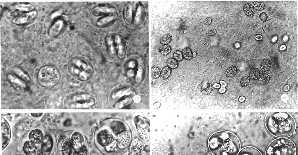
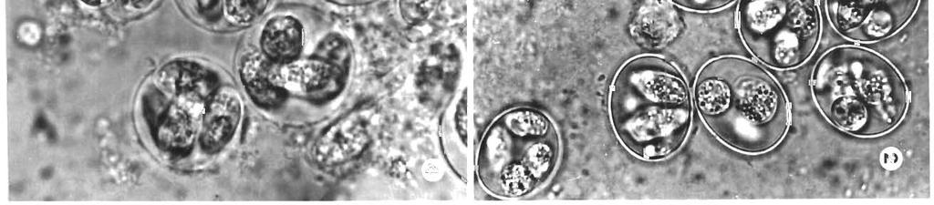
3 belong~ng to 18 cyprln~d specles were examlned for the plesence of nodular occ~diosls The major~ty of f~shes examined came from Lake Balaton and the R~ver Danube Hungary although a smaller number were taken from the creeks Tap10 and Kemence (tr~butaries of R~vers Tlsza and Ipoly, ~espect~vely) Fish wt re caught w~th a seine net and transported alive to the laborato~y where th(,i were processed wlthin 4 to 6 d F~sh kept in the labordtory w~thout feedlng for at least 2 d and whose intestinal contents had been excreted, proved the most su~table fo~ rel~able d~agnos~s of COCCId~al ~nfect~on The gut of these flsh was sl~t open long~tudinally and examlned under a microscope The mucus and sciaplngs taken fiom d~ffe~ent segments of the gut were studled wlth a l~ght lnlcroscope Oocysts were easiest to demonstrate In the glassy mucus llning the gut but could also be found In the intest~nal scraplngs In posltive and doubtful cases smaller pleces of unaffected intest~nal segments or of nodules seen on the ~ntestlnal wall were flxed In 10. buffered formalln or Bouln s solut~on In some cases the whole gut was wound up and f~xed In ~ts ent~rety Samples wele embedded In paraffin, 3 to 4 btm thlck sect~ons cut and stalned wlth haematoxylin and eosln (H& E) Unsporulated oocysts found In nodules from the gut wall and in mucus were placed In Petr~ d~shes or on a watch-glass and allowed to sporulate 111 tap water changed several t~mes for 24 to 48 h To pievent bacter~al growth, a loopful of penlc~llin or streptomyc~n was added to the water D~mens~ons of the new specles of Goussia wele determined from measurements of 50 oocysts and ale given as mean followed by iange (pm) RESULTS Prevalence In early April 1986, nodules containing gamogonic stages of a Goussia sp. were found In the gut of a bleak Alburnus alburnus caught in Lake Balaton. Further studies consistently revealed the presence of nodules containing similar unspoi-ulated oocysts in the gut of other fish specles, i~rimarily white bream Blicca bjoerkna. In mucus taken from the gut of wh~te bream, 2 types of unsporulated oocysts (Fig. 1) were deinonstrable in high numbers: a larger (17-19 X pm) oocyst and a smaller one ( X ~~m). In tap water, oocysts sporulated withln 48 h and Increased slightly in size. Oocysts scraped from nodules always represented the larger species (Fig. 2). Later, histological examination revealed that the coccidium having smaller oocysts represented a Goussia sp. which developed In large numbers in an epicellular locat~on on the surface of the intestinal epithelium (Fig. 3). Infect~on was demonstrable exclusively in the early spring and disappeared almost completely by the end of April. From McI)' onward, occas~onal examination of the fish revealed only the presence of the species causing so-called carpelli-type diffuse cocc~diosis (G. carpelli, G. cyprinorum). Oocysts shed unsporulated, developing in nodules or In the epicellular location, were found in the gut of 10 f~sh species (Table 1). In 6 cyprinids both the nodular and epicellular types occurred; in 2 species, only the nodular type was found, while in another 2 the site of oocyst development could not be precisely located. In all f~sh species, infect~on typically occurred in the early spring. At that time (of fish species examined in sufficient numbers) infect~on was most prevalent in Blicca Ojoerkna (75 and ca 50 '10 In Abramis brama and Rutllus rutilus. The earliest lnfect~on was demonstrated in flsh caught in March, Immediately after the ice had thawed. At that time, merogonic stages were still present in species of Blicca,.41burnus and Leuciscus and gamogonic stages had already appeared. The intensity of infection was highest In April. By mid-apr~l oocysts had been formed in nodules and were expelled into the intestinal lumen unsporulated. The development of specles with smaller oocysts in an epicellular location took place about 2 wk later. The merogonic stages of this species were demonstrable at the beginning of April and oocyst shedding was completed by the end of the month. In the malor~ty of cases, Infection was highly intensive. No appreciable difference was noted between fish caught in Lake Balaton and in the Danube, In terms of the prevalence and intensity of lnfect~on. Despite the high intensity of coccidial infection, there was no evidence of pathogenicity. Fish transported to the laboratory intact survived the observation period without showing slgns of d~sease. Dying fish did not weigh less nor were they more severely infected than apparently healthy fish. Despite intensive infect~on occasionally Involving as much as 70 ",) of the Intestine, there was no evidence of necrosis of gut epithelium, nor were inflammatory cells observed. The pathologic effect was limited to the destruction of infected epithelia1 cells. Mucus released Figs 1 to 4 Goussia spp. Unsporuldted G. balatonlca (larger) and G pannonlca (smaller) oocysts In the ~ntestinal mucus; (X 500) Flq G balatonica Oocysts after 24 h sporulation; ( X 1300). G pannonlca Oocysts after 48 h sporulation; ( X 1300). Qi(. G subep~thelialis. Oocysts after 24 h sporulation, ( X 1300)
4 Table 1 Nodular and epicellular coccidia from cyprinids. n: nodular; e: epicellular; U: unknown: B: Balaton; D: Danube ; F: titrrn-ponds; K: Creek Kemence; T. Creek T;lp16 Fish species No. Fish shedding nodular and No. fish free from No. fish infected with Source tested epicellular oocysts intestinal coccidia G. carpelli-ty pe coccidia Infection No. fish type Mar Apr May Total Mar Apr May Total Mar Apr May Total Blicca bjoerkna Abralnis bran~a RLI~I~LIS rutilus Scardinlus erylhrophthalrn 11s Lr~rcrsc~ls cephalus L. I~IIS Albl~rnus alburnus Pelecus cultratus Aspius aspi~rs Rhode~rsericeus Cyprinus carpio Cbrassius auratus gibelio Gobio gobio Gobio albipinnetus Bar-bus barbus Ch(~ndr~~l0f71a nasus Vlrn ba virn ba Phoxlnus phoxirl us
5 (8961 erssno3 & 1'6-8'8 9 S'P-P S'ZZ-S'II a S[-C1 as da snruleyl ydo.rqlilta snrurp~e~s X 'ds erssnog n n S1-C1 as da snleyda2 sn~sr~na-] XI 'ds emnog S 2 6-5'8 9 Z'P-P S'ZI-S'11 a &l-01 S1-&I as da snlrln~ snlrlng 1[[,q,ds e!ssnog Z'Z-Z S'S-S 8'11-S'OL a PL-21 as da erue~q srruelqv [~,q'ds e~ssnog (1.2) (~'6) (P)!8'11! (Z'ZI!!?L) S'Z-Z SOLVS8 9 S' P-& &[-L1 3 PT P1 as da euyraolq e ~~rlg e~ruouued erssnog as JeuloW 9 Apl?nad a U ordre~ snur~da3 s!le!laylrdaqns (t,z)!$'i L! (L.S) (S.ZI) C-S'Z S Cl-21 a Z.81-8'S1 8'61-2'81 as U ~surl e ~ u r ~ ~861 leu~olr\~.ds erssnog n n a S'Z1-S'11 L[-S1 as UI snjeuurdrqle orqog [A.ds erssnog LU U U a PZ-1Z J U snu~nqle srtu~nqlv i\.ds erssno3-9 S'S-S 101 a SL-PL 6l-81 as LU snq.leq snq.reg AI.ds efssno PI-Z1 a $1-PL 61-L1 as U snleyda~ sn~sr~na7 111 'ds erssnog (61) n S'61-S'8 1 I U snlrlnl snlrjng 11 ds erssnog &'Z ZI 9 S'S ~1 a L1-P1 EZ-12 as LI eurelq srrue~qv 1 ds erssnog (&,2) (211 (8'5)!z'CI) (L[) (~'81) S - S'ZI-01 9 L-S L - a 61-S1 ZZ-L[ as U euy.raofq ess!rg e~~uojeleq erssno3 - - A\ 7 S M 7 S /M 1 S alrozolod~ lshsoiods ]sasoo uo~leso~ lsoei al!seled painseam IOU :uru!pale[n~odsun :n:pun01 :r Ipadeqs-eueueq.q '[esrldg[a.a '[eprosd~l[a 11oqS :as lqlpr~ :M :ql6ual :? ladeqs :S.slsoy jualajj!p wolj palsa[[os sadai (U) lesosnur pue (a) relnl(as!da pue (U) ie[npou jo sluauralnseapq.dds erssno3.z a[qel


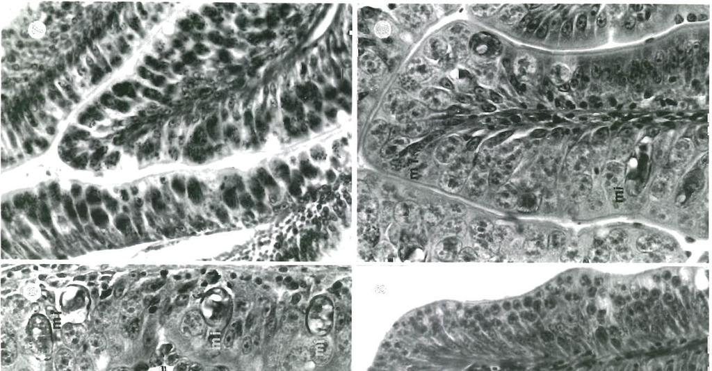
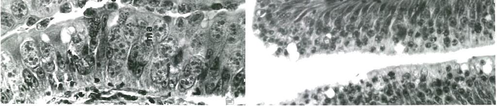
6 6 Dis. aquat. Org molnari and G. schulmani. Smaller oocysts differed from the former in size. As, of coccidia demonstrated in this study, only those found in common carp were identified as G. subepithelialis (Fig. 4), and since it cannot be decided whether the species found in the other hosts represent 1 or more species, only the 2 most prevalent and most intensively studied coccidia parasitizing Blicca bjoerkna are described as the new species G. balatonica and G. pannonica. Species descriptions Fig. 5. Goussia balatonica. Oocyst from them covered the gut in a tubelike fashion and contained masses of oocysts. Both modular and epicellular coccidiosis were demonstrable in the fish species examined. In some cases, coccidia, both nodular and epicellular in location, occurred in a given segment of the gut (Fig. 6). Morphological differences between oocysts released from nodules and those shed from the surface of the Intestinal epithelium within a given fish species were negligible. Differences in size were, however, significant. No distinct morphological differences were demonstrable between nodular and epicellular oocysts derived from various cyprinid fishes. There were, however, smaller differences in size (Table 2). The size of the fish had no influence on that of the oocyst. Oocysts derived from nodules represented the larger type typical of Goussia subepithelialis, G. pigra, G. Goussia balatonica n. sp. is found in the white bream Blicca bjoerkna, of Lake Balaton and the River Danube, w11r1 t: it furnls nodilles 1 to 1.5 mm diam. in the mucos;: of the intestine. Short ellipsoidal oocysts (Fig. 5), 18.7 (17-22) pm long and 17 (15-19) pm wlde have a thin, smooth and colourless wall and lack oocyst residuum, polar granules and micropyle. The unsporulated oocysts that leave the fish are (Fig. l) compact, elliptical and measure 17.9 X 12.9 (17-19 x 12-13)!.km. Oocysts kept in tap water at 22 "C completed sporulation within 48 h. Merogonic and gamogonic stages were studied histologically. Sporocysts are elongated ellipsoids, 13.2 (12-17) kim long and 5.8 (5-7) pm wide, and are generally arranged longitudinally in oocysts. The sporocyst wall is thin and composed of 2 equal valves joined by a suture. Each sporocyst contains 2 bananashaped sporozoites which possess large refractile nuclei. Sporozoites are 12 ( ) pm long and 2.3 (2-2.5) kkm wide. Their residuum is finely granulated and fills the sporocyst after 24 h sporulation, becoming ellipsoidal (ca 8 X 4 pm) and consisting of scattered particles after 48 h of sporulation in tap water. In histological preparations the earliest developmental stages of Goussia balatonica were meronts which developed in a given segment of the foregut, and extended into ca 3 to 5 intestinal folds (Fig. 7). In th.at segment of the gut almost all epithelia1 cells were infected and each contained a meront which was located in the cytoplasm apical to the nucleus. ~Meronts measured 17 X 8 X 8 tim and contain.ed 32 bananashaped merozoites. Figs. 6 to 9. Goussia spp. (H & E). Fig. Bficca bjoerkna. Intestinal segment exhibiting mixed infection by nodular stages of G. balatonica and epicellu.lar developmental stages of G. pannonica. mi: microgamont; ma: macrogamont, e: epicellular stages; (X 500). Fiq. 7. Goussia balatonica. Meronts rnfecting gut epithelium In single nodule. Almost all epithelia1 cells are infected by meronts containing 32 merozoites; ( X 300). Fig G. balatonica. Round trophozoites 3 to 8 pm diam. developing, probably, into gamonts are located in the epithelia1 cell cytoplasm in a given segment of the gut; (X 200). Fig G. balatonica. Dark staining microgamonts (mi) located among macrogamonts in affected part of gut epithelium. Young microyamonts contain dot-shaped while the more developed ones have comma-shaped microgametes; (X 500)
7 I\,lalnriT; Coccidiosis in cyprinid fishes 7
8 8 Dis. aquat. Org. 7: Gamogonic stages were also limited to a given segment of the gut; besides the foregut, they were also observed in the midgut and hindgut. Round trophozoites 3 to 8 pm diam. (Fig. 8) and gamonts were located in the epithelial cell cytoplasm apical to the nucleus and pushed the nucleus to the basement membrane. Macrogamonts developing in the cell (Fig. 9) were mostly elliptical and X pm. A central nucleus and some basophilic protein granules were seen in their foamy cytoplasm. Young microgamonts (Fig. 9) stained much darker. During development, the abundant chromatin of microgametes first appeared as small dots, then as elongated 'commas' within the elliptical microgamonts which measured X pm. Other species resembling Goussia balatonica differ from it in the following respects: G'. subepitheiiaiis has less elongated oocysts (Fig. 4); G. pigra has more elongated sporocysts; G. schulmani has a coarsely granulated sporocyst residuum; and G. molnari has sporozoits with reflexed ends. Besides these characteristics G. subepithelialis, G. schulmani, and G. molnari have endogenous sporogony. Goussia pannonica n. sp., found in white bream, Blicca bjoerlcna, of Lake Balaton and the River Danube, infects the epicellular layer of epithelial cells, mainly in the first half of the gut. Ellipsoidal oocysts (Fig. 10), 15 (14-16) km long and 12.2 (11-14) brm wide, have a thin. smooth and colourless wall. Oocyst residuum, polar granule and micropyle are absent. The oocysts leave the fish unsporulated, are (Fig. 1) compact, ellipsoidal and measure 12.9 X 8.6 ( X ) LLm When kept in tap water at 22 C complete sporulation occurs within 48 h. Merogonic and gamogonic stages were also studied histologically. Sporocysts are elongated ellipsoidal, 11.8 (11-13) pm long and 4 (3-4.5) LLrn wide, are generally longitudinally arranged in the oocyst, have thin walls and are composed of 2 equal valves. Each sporocyst contains 2 banana-shaped sporozoites with large refractile nuclei 9.5 ( ) pm long and 2.1 (2-2.5) pm wide. The residuum is finely granulated and fills the sporocyst after 24 h sporuictiio~i su~ll iiiai after 48 :-L, sporscy-sts are ellipsoidal, ca 8 X 4 btm; and after 72 h consists of scattered particles. Developmental stages of Goussia pannonica were demonstrable in all segments of the gut and were always located epicellularly (intracellularly but extracytoplasmally) (Fig. 11). The earliest stages were round meronts 5.5 to 6 +m diam., containing 8 merozoites, which were usually demonstrable among developing gamonts (Fig. 12). Mature macrogamonts and microgamonts (Fig. 11) were ellipsoidal and X 8-9 pm in size. In the pale cytoplasm of the macrogamonts, besides the nucleus, a few dark-staining protein granules were seen. Depending on their stage of maturity, microgamonts contained dot- or comma-shaped microgametes rich in chromatin. This species resembles Goussia balatonica but differs from it in its smaller size, its specific epicellular location in the gut and the number of merozoites in meronts. DISCUSSION 10 pm Fig. 10. Coussia pannonica. Oocyst (these are smaller and more elongated than these of C. balatonica, Fig. 5) The results indicate that, besides Goussia subepithelialis infection of common carp (Schaperclaus 1943, Pellerdy & Molnar 1968, Manncek 1973) and coccldiosis reported by Molnar (1982) from tench, nodular coccidiosis frequently occurs in other cyprinid fishes. These studies also prove that in these fishes the nodular form is often accompanied by epicellular, diffuse coccidiosis. Both types of coccidiosis are characterized by a strictly seasonal, annual developmental cycle. Under the climatic conditions prevailing in Hungary merogony occurs until March and gamogony takes place in April. Oocysts leave the fish unsporulated before the end of April. Infection is not demonstrable in other periods of the year Rapid regeneration of the gut epithelium is also typical, as a result, even intensive infections disappear without a trace. The fact that nodular and epicellular coccidiosis of
. Fig. 11. Developmental stages, covering surface of intestinal epithelial cells in large numbers; (X 100). Fig. 12.")


 who suggested that Eimeria vanasj, a species described by them, had 2 forms differing in size and location.")
9 Molnar: Coccidiosis in cyprinid fishes 9 Figs. 11 and 12. Goussiapannonica (H & E). Fig. 11. Developmental stages, covering surface of intestinal epithelial cells in large numbers; (X 100). Fig. 12. Meronts (arrows) developing epicellularly in intestinal epithelial cells; (X 800) cyprinids have so far largely escaped the attention of specialists, can be explained by several factors. These parasites can be demonstrated only in the early spring months when the collection of host fish is difficult. Specialists dealing with f~sh coccidia do not routinely perform histological examinations. While looking for sporulated oocysts they may overlook unsporulated oocysts which can easily be mlstaken for food organisms. On the basis of significant differences in oocyst size and location, and despite the morphological resemblance, nodular coccidiosis and epicellular coccidiosis observed in a given cyprinid species are unquestionably caused by 2 distinct species. In spite of this fact, consistent simultaneous occurrence of the 2 types might suggest that the nodular and epicellular forms are 2 different manifestations caused by the same species but altered by the host response. This conclusion was drawn by Landsberg & Paperna (1987) who suggested that Eimeria vanasj, a species described by them, had 2 forms differing in size and location. This s'upposition is contradicted in the present study, by the observation that oocysts of intermediate size do not occur even if both types are pregent simultaneo.usly, and that oocysts always fall into 2 distinct categories. That the 2 species are identical is practically excluded by the fact that the gamogonic stages of Goussia balatonica develop from a meront containing 32 merozoites, whereas in the meronts of G. pannonica only 8 merozoites are formed. The developn~ental cycle of Goussia balatonica closely resembles that reported for G. subepithelialis. In C. balatonica the late stage of merogony, presumably corresponding to the 3rd merogonic generation, occurs in early spring, similar to the cycle of G. subepithelialis as reported by Marincek (1973). This stage is followed by gamogony lasting for, at most, 2 mo. According to Marincek (1973), infection of common carp with G. subepithelialis takes place in summer, and within ca 2 mo, 2 merogonic generations develop in the gut epithelium. However, a hibernation period is required for the 3rd merogonic stage. This fact would explain the annual developmental cycle. The present study gives no information on the mode of infection by Goussia balatonica and G. pannonica. Marincek (1973) successfully infected common carp with G. subepithelialis directly. Similarly, Paterson & Desser (1982) could produce infection in fathead min-
10 10 Dis. aquat. Org. 7: 1-12, 1989 now by feeding oocysts of G, iroquoina. On the other hand, to produce infection with Calyptospora [Eimeria) funduli in killifish. Fournie & Overstreet (1983) needed intermediate hosts. While surveying coccidial infections in the present study a basic question emerged: what kind of host specificity do these morphologically similar coccidia, characterized by very thin oocyst and sporocyst walls and a sporocyst composed of 2 valves, have? Are there, within the nodular and epicellular types of coccidiosis, morphologically similar but strictly specific Goussia species, each of which can colonize a single host, or are all cyprinid fishes infected by the same, morphologically variable species? Considering the assumed specificity of coccidia and the phylogenetic distance of the hosts, most probably both nodular and epicellular coccidia are able to establish themselves in only a few closely related species, similarly to the spleen-parasitic Goussia mechnikowi, which can parasitize only Gobio spp., or to the intestinal parasite G. sinensis, which exclusively parasitizes Hypophthalmichthys spp. Assuming a similar specificity, it is not probable that G. balatonica is identical to G. subepithelialis reported from the genus Cyprinus, or to G. schulmani reported from the genus Leuciscus. At the same time, it cannot be ruled out that Goussia I1 and Goussia 111, species demonstrated in the closely related Rutilus rutilus and Leu- ciscus cephalus, respectively, should be classified as G. schulmani. Desplte the fact that, apart from Goussia subepithelialis and nodular coccidia reported from tench by Molnar (1982), the literature does not contain factual data on the occurrence of nodular coccldiosis, it is highly probable that species listed in Table 3 were described from unrecognized cases of nodular coccidiosis. Besides the morphological resemblance, this was suggested by Hoffman (1965) and Jastrzebski (1984), both of whom observed oocyst shedding in the early spring months. To determine whether Goussia balatonica represents a distinct species, it should be compared to G. subepithelialis of common carp as an archetype. There are only shght ditferences between the oocysts of these 2 parasite species (Figs. 3 and 4). In the mechanism of oocyst excretion, however, important differences are demonstrable. Oocysts of G. balatonica leave the fish unsporulated in April (similarly to that reported by Molnar 1982 for coccidiosis of tench). On the other hand, oocysts of G. subepithelialis reach the deep layers of the epithelium and leave the fish in the sporulated state in May. This opinion, generally accepted in the literature, is somewhat contradicted by the observation of Marincek (1973), i.e. that some oocysts of G. subepithelialis also leave the fish unsporulated in April, suggesting that differences in the oocyst excretion Table 3. Goussia spp. G. balatonica- and G. pannonica-type coccidia reported from cyprinid fishes. Shapes: (r) round; (e) elliptical; (0) ovoid Coccidian species Host Oocyst Sporocyst Shape Length Width Shape Length Width Eimeria (Goussia) subepithelialis Moroff & Fiebiger Marincek (1973) Common carp Schaperclaus (1943) Pellerdy & Molnar (1968) Common carp Eim eria pigra Leger & Bory, 1932 Rudd Eimeria schulmani Kulemi.na, 1969 Orfe Eimeria sch ulmani Kulemina, 1969 Jastrzebski (1984) Chub Eimeria molnan Jastrzebski, 1984 Gudgeou Eimeria carassiusaurati Romero-Rodriguez, 1978 Goldfish Eimena a urati Hoffman, 1965 Goldfish Eimeria carassii Yakimoff & Gouseff, 1935 Cruclan carp Eimeria nicollei Yakimoff & Gouseff, 1935 Crucian carp o e -
11 Molnar: Cocc~d~osis in cyprinid fishes 11 mechanism reflect the characteristics of the host reac- e.g. development with or without an intermediate host tion rather than the properties of coccidia. Species or development by a yearly cycle or continuously, could important in respect of differentiation, e.g. G. schul- also serve as a basis for taxonomic classification. main, G. molnari, and G. carassiusaurati, also excrete sporulated oocysts, and only the oocysts of G. aurati and G. pigra are known to leave the fish unsporulated. Until quite recently, Eimeria anyuillae infection of eel and Goussia pigra infection of rudd were the LITERATURE CITED exclusive examples of epicellular coccidiosis. Recent ~ ~ ~ F. ~ (1987). d i coccidies, et coccidioses de poissons data (Molnar 1986, Daoudi et al. 1987, Landsberg & mediterraneens: systematique, ultrastructure et biologie. Paperna 1987, Kent et al. 1988, Molnar & Rohde 1988a) Doctoral thesis. Montpellier Daoudi, indicate that this type of coccidiosis occurs frequently F.. Radujkovic. B., Marques, A., Bouix. G. (1987). Nouvelles especes de Coccidies (Apicomplexa, and in different fish species. Important data can be Eimeriidae) des genres Eimeria Schneider. 1875, et found in the book of Kocylowski & Myaczynski (1963) Epieimeria Dykova et Lom, 1981, parasites de poissons who, in connection with G, pigra infection of rudd, marins de la baie de Kotor (~u~oslavie). Bull. M&. natn. mention that similar eimerians of epicellular location Hist. nat. Paris (4" Ser.) A 9: Dykova, I., Lom, J. (1981). Fish coccidia: critical notes on life are frequently demonstrable from the gut of other cycles, classification and pathogenicity. J. Fish Dis. 4: fishes, too. Though light-microscopic examination alone does not have probative value, by analoqy - it is Fournie, J. W., Overstreet, R. M. (1983). True intermediate beyond doubt that the 'epicellular' developmental hosts for Eimeria funduli (~~icom~lexa) from estuarine fishes. J. Protozool. 30: stages of G. pannonica n. sp, occupy a similar intracel- Hoffman, G. L. (1965). Eimeria aurati n. sp. (Protozoa: lular but extracytoplasmal position as was proved Eimeriidae) from goldfish (Carassius auratus) in North ultrastructurally by Molnar & Baska (1986) for America. J. Protozool. 12: Jastrzebski, M. (1984). Coccidiofauna of cultured and feral d The species Goussia pigra occupies a peculiar place fishes in fish farms. Wiad. parazyt. 30: Jastrzebski, M., Pastuszko, J., Kurska, E.. Badowska, M. in this respect. This parasite was described by Leger & (1988). Kokcydia ciernika - Gasterosteus aculeatus (L.). Bory (1932) from Scardinius erytl~ropl~thalmus. At the Wiad. parazyt. 34: (in Polish) same time, its oocyst measurements are closer to those Kent, M. L., ~durnie, J. W., Snodqrass, R. E., Elston, R. A. of nodular coccidia. Though I have studied only the (1988). Goussla girellae n. sp. (Apicomplexa- Elmeriorina) in the opaleye, smaller, sporulated oocyst forms from mdd, I assume Girella nigricans. J. Protozool. 35: Kocylowski, B., Myaczynski, T (1963). Fish diseases. Publ. that Leger & Bory (1932) must have observed a mixed House Mezogazdasagi, Budapest (in Hungarian) infection usual in cyprinids, and they must have Labbe, A. (1896). Recherches zoologiques, cytologiques et described the species on the basis of oocysts derived biologiques sur les Coccidies. Archs exp. gen. 4: from the nodule and histologically demonstrated only Landsberg, J. H., Paperna, I. (1987). Intestinal infections by the epicellular forms. Eimeria (S. l) vanasi n. sp. (Eimeriidae, Apicomplexa, Pro- Numerous attempts have been made at classifying tozoa) in cichlid fish. Ann. Parasitol. Hum. Comp. 62: the genus Ein~erja Schneider, 1875, a genus comprising an extremely large number of species. Among the Leger, M., Bory, T (1932). Ei~neria pigra n. sp nouvelle Coccidie juxtaepitheliale, parasite du Gardon rouge. C. r eimerians of fishes, on the basis of oocyst morphology, hebd. Seanc. Acad. Sci., Paris 194: Labbe (1896) created the genera Goussia and Crystal- Lom, J., Dykova, I (1982). Some marine fish coccidia of the lospora, whereas Overstreet et al. (1984) established genera Eimeria Schneider, Epieimeria Dykova et Lom and the genus Calyptospora. On the basis of intracellular Goussia Labbe. J. Fish Dis. 5: location Dykova & Lom (1981) suggested the genus Marincek. M. (1973). Developpement d'eimeria subepithelialis (Sporozoa, Coccidia) - parasite de la carpe. Epieimeria and Daoudi (1987) recommended the gen- Acta protozool. 12: era Nucleoeimeria and Nucleogoussia. The species Molnar, K. (1982). Nodular coccidiosis in the gut of the tench, G. balatonica and G. pannonica, described in this work, Tinca tinca L. J. Fish Dis. 5: are typical members of the genus Goussia as shown by Molnbr, K. (1984). Some pecularities of Oocyst rejection of fish coccidia. Symp. Biol. Hung. 23: their sporocysts composed of 2 valves joined by a Molnar, K. (1986). Occurrence of two new Goussla species in suture. However, by its location within the cell the inteshne of sterlet (Acipenser ruthenus). Acta vet. G. pannonica could be assigned to the genus hung. 34: Epieimeria, or to the still non-existent genus Epigous- Molnar. K., Baska, F. (1986). Light and electron microscopic sia. In my opinion, location within the cell cannot be studies on Epieimeria anguillae (Leger et Hollande, 1922), a coccidium parasitizing the European eel, Anguilla accepted as a generic character, as the location of most anguilla L. J. Fish Dis. 9: the presently is Molnar, K., Rohde, K. (1988a). New coccidians from freshfrom this principle, such still unelucidated factors as water fishes of Australia. J. Fish Dis. 11:
12
DISEASES OF AQUATIC ORGANISMS Dis. aquat. Org.
 Vol. 10: 121-125. 1991 DISEASES OF AQUATIC ORGANISMS Dis. aquat. Org. Published April 4 Ultrastructural observations on sporozoite stages of piscine Coccidia: Goussia carpelli and G. subepithelialis from
Fine structure of Eimeria (S. l.) vanasi merogony stages in the intestinal mucosa of cichlid fishes
 vanasi merogony stages in the intestinal mucosa of cichlid fishes") Vol. 10: 195-201. 1991 DISEASES OF AQUATIC ORGANISMS Dis. aquat. Org. Published May 8 Fine structure of Eimeria (S. l.) vanasi merogony stages in the intestinal mucosa of cichlid fishes Ilan Paperna Department
Vol. 10: 195-201. 1991 DISEASES OF AQUATIC ORGANISMS Dis. aquat. Org. Published May 8 Fine structure of Eimeria (S. l.) vanasi merogony stages in the intestinal mucosa of cichlid fishes Ilan Paperna Department
Phylum:Apicomplexa Class:Sporozoa
 Phylum:Apicomplexa Class:Sporozoa The most characteristic features of sporozoa are 1-unique appearance of most protozoa makes it possible for knowledge able person to identifiy them to level of genus and
Oocyst formation in the coccidian parasite Goussia carpelli
 Vol. 10: 203-209, 1991 DISEASES OF AQUATIC ORGANISMS Dis. aquat. Org. Published May 8 Oocyst formation in the coccidian parasite Goussia carpelli 'Institute of Parasitology, Czech Academy of Sciences,
EFFICACY OF SOME ANTICOCCIDIAL DRUGS FOR TREATING COCCIDIAL ENTERITIS OF THE COMMON CARP CAUSED BY GOUSSIA CARPELLI (APICOMPLEXA: EIMERIIDAE)
") Acta Veterinaria Hungarica 55 (1), pp. 67 76 (2007) DOI: 10.1556/AVet.55.2007.1.7 EFFICACY OF SOME ANTICOCCIDIAL DRUGS FOR TREATING COCCIDIAL ENTERITIS OF THE COMMON CARP CAUSED BY GOUSSIA CARPELLI (APICOMPLEXA:
Acta Veterinaria Hungarica 55 (1), pp. 67 76 (2007) DOI: 10.1556/AVet.55.2007.1.7 EFFICACY OF SOME ANTICOCCIDIAL DRUGS FOR TREATING COCCIDIAL ENTERITIS OF THE COMMON CARP CAUSED BY GOUSSIA CARPELLI (APICOMPLEXA:
"Comments on the nature and methods of collection of fish coccidia. " - Molnár, K. - Parasit. Hung. _
 Parasit. Hung 10. 1977. Comments on the Nature and Methods of Collection of Fish Coccidia Dr. Kálmán MOLNÁR Research Institute for Veterinary Science, Hungarian Academy of Sciences, Budapest "Comments
Key words: Coccidia, Choleoeimeria rochalimai, fine structure, gall bladder epithelium, Hemidactylus mabouia, Brazil
 FOLIA PARASITOLOGICA 47: 91-96, 2000 Ultrastructural study of meronts and gamonts of Choleoeimeria rochalimai (Apicomplexa: Eimeriidae) developing in the gall bladder of the gecko Hemidactylus mabouia
Ahead of print online version
 Folia Parasitologica 60 [3]: 232 236, 2013 ISSN 0015-5683 (print), ISSN 1803-6465 (online) Institute of Parasitology, Biology Centre ASCR http://folia.paru.cas.cz/ A new species of Choleoeimeria (Apicomplexa:
Joerg Kinne, Mansoor Ali*, Ulrich Wernery, and J. P. Dubey
 J. Parasitol., 88(3), 2002, pp. 548 552 American Society of Parasitologists 2002 CLINICAL LARGE INTESTINAL COCCIDIOSIS IN CAMELS (CAMELUS DROMEDARIUS) IN THE UNITED ARAB EMIRATES: DESCRIPTION OF LESIONS,
Protozoa. Apicomplexa Sarcomastigophora Ciliophora. Gregarinea Coccidia Piroplasma
 Protozoa Apicomplexa Sarcomastigophora Ciliophora Gregarinea Coccidia Piroplasma Coccidia characterized by thick-walled oocysts excreted in feces In Humans Cryptosporidium Isospora Cyclospora Sarcocystis
Light and electron microscopic study of the pathology and merogony of Goussia gadi (Apicomplexa: Coccidia) in the swimbladder wall
 in the swimbladder wall") Vol. 17: 113-125.1993 DISEASES OF AQUATIC ORGANISMS Dis. aquat. Org., Published November 18 I Light and electron microscopic study of the pathology and merogony of Goussia gadi (Apicomplexa: Coccidia)
Vol. 17: 113-125.1993 DISEASES OF AQUATIC ORGANISMS Dis. aquat. Org., Published November 18 I Light and electron microscopic study of the pathology and merogony of Goussia gadi (Apicomplexa: Coccidia)
HISTOPATHOLOGY. Introduction:
 Introduction: HISTOPATHOLOGY Goats and sheep are the major domestic animal species in India. Much of the economy of the country has been depend upon the domestication of these animals. Especially economy
Extra-intestinal localization of Goussia sp. (Apicomplexa) oocysts in Rana dalmatina (Anura: Ranidae), and the fate of infection after metamorphosis
 oocysts in Rana dalmatina (Anura: Ranidae), and the fate of infection after metamorphosis") DISEASES OF AQUATIC ORGANISMS Vol. 70: 237 241, 2006 Published June 23 Dis Aquat Org Extra-intestinal localization of Goussia sp. (Apicomplexa) oocysts in Rana dalmatina (Anura: Ranidae), and the fate
DISEASES OF AQUATIC ORGANISMS Vol. 70: 237 241, 2006 Published June 23 Dis Aquat Org Extra-intestinal localization of Goussia sp. (Apicomplexa) oocysts in Rana dalmatina (Anura: Ranidae), and the fate
The Fine Structure of the Endogenous Stages of Isospora hemidactyli Carini, 1936 in the Gecko Hemidactylus mabouia from North Brazil
 Mem Inst Oswaldo Cruz, Rio de Janeiro, Vol. 95(1): 43-47, Jan./Feb. 2000 The Fine Structure of the Endogenous Stages of Isospora hemidactyli Carini, 1936 in the Gecko Hemidactylus mabouia from North Brazil
A NEW SPECIES OF GENUS EIMERIA (APICOMPLEXA: EUCOCCIDIORIDA) FROM GOAT.
 FROM GOAT.") A NEW SPECIES OF GENUS EIMERIA (APICOMPLEXA: EUCOCCIDIORIDA) FROM GOAT. B.V. More 1, H.A.Kamble. 2 S.V. Nikam 3, 1 Department of Zoology, Ramkrishna Paramhansa Mahavidyalaya, Osmanabad. (M.S.) India. 2
A NEW SPECIES OF GENUS EIMERIA (APICOMPLEXA: EUCOCCIDIORIDA) FROM GOAT. B.V. More 1, H.A.Kamble. 2 S.V. Nikam 3, 1 Department of Zoology, Ramkrishna Paramhansa Mahavidyalaya, Osmanabad. (M.S.) India. 2
cyst&' appeared to be of two kinds-one smaller and Smnith "is inclined to regard these epithelial cell parasites as
 COCCIDIA IN SUBEPITHELIAL INFECTIONS OF THE INTESTINES OF BIRDS PHILIP B. HADLEY From the Agricultural Experiment Station of the Rhode Island State College' Received for publication, July 10, 1916 In an
Taxonomy of North American Fish Eimeriidae
 11 NOAA Technical Report NMFS 11 Taxonomy of North American Fish Eimeriidae Steve J. Upton, David W. Reduker, William L. Current, and Donald W. Duszynski August 1984 U.S. DEPARTMENT OF COMMERCE National
Coccidia. Nimit Morakote, Ph.D.
 Coccidia Nimit Morakote, Ph.D. 1 Learning objectives After class, students will be able to: Describe morphology, life cycle, signs and symptoms, prevention and control, laboratory diagnosis and treatment
Sam R. Telford, Jr The Florida Museum of Natural History, University of Florida, Gainesville, Fl32611, USA
 Systematic Parasitology 23: 203-208, 1992. 0 1992 Kluwer Academic Publishers. Printed in the Netherlands. An eimeriid species (Apicomplexa: Eimeriidae) that parasitises the gallbladder and bile-duct of
Goussia cruciata (Thelohan, 1892) a hepatic coccidian parasite of the horse mackerel Trachurus trachurus (Linnaeus, 1758) from the
 a hepatic coccidian parasite of the horse mackerel Trachurus trachurus (Linnaeus, 1758) from the") Bull. Eur. Ass. Fish Pathol., 2(6) 2, 219 Goussia cruciata (Thelohan, 1892) a hepatic coccidian parasite of the horse mackerel Trachurus trachurus (Linnaeus, 1758) from the Mediterranean coasts of northern
Bull. Eur. Ass. Fish Pathol., 2(6) 2, 219 Goussia cruciata (Thelohan, 1892) a hepatic coccidian parasite of the horse mackerel Trachurus trachurus (Linnaeus, 1758) from the Mediterranean coasts of northern
Protozoan Parasites of Veterinary importance 2017
 Protozoan Parasites of Veterinary importance 2017 VPM-122 Laboratory 4 Spencer J. Greenwood PhD, DVM Dept. of Biomedical Sciences Room 2332N AVC North Annex sgreenwood@upei.ca Office phone # 566-6002 To
Zadar County Rural Development Agency, Zadar, Croatia. Fish Farm IHOR PARK, Jastrebarsko, Croatia
 . Veterinarski Arhiv 87 (1), 77-86, 2017 New data on Eimeria dicentrarchi (Apicomplexa: Eimeriidae), a common parasite of farmed European sea bass (Dicentrarchus labrax) from the mid-eastern Adriatic Emil
PLASMODIUM MODULE 39.1 INTRODUCTION OBJECTIVES 39.2 MALARIAL PARASITE. Notes
 Plasmodium MODULE 39 PLASMODIUM 39.1 INTRODUCTION Malaria is characterized by intermittent fever associated with chills and rigors in the patient. There may be enlargement of the liver and spleen in the
Diagnosis, treatment and control: dealing with coccidiosis in cattle
 Vet Times The website for the veterinary profession https://www.vettimes.co.uk Diagnosis, treatment and control: dealing with coccidiosis in cattle Author : Adam Martin Categories : Vets Date : January
Observations on Eimeria species of Dasyprocta leporina (Linnaeus, 1758) (Rodentia: Dasyproctidae) from the state of Pará, North Brazil
 (Rodentia: Dasyproctidae) from the state of Pará, North Brazil") Mem Inst Oswaldo Cruz, Rio de Janeiro, Vol. 99: 000-000, 2004 1 Observations on Eimeria species of Dasyprocta leporina (Linnaeus, 1758) (Rodentia: Dasyproctidae) from the state of Pará, North Brazil Ralph
Mem Inst Oswaldo Cruz, Rio de Janeiro, Vol. 99: 000-000, 2004 1 Observations on Eimeria species of Dasyprocta leporina (Linnaeus, 1758) (Rodentia: Dasyproctidae) from the state of Pará, North Brazil Ralph
DISEASES OF AQUATIC ORGANISMS Dis. aquat. Org.
 Vol. 7: 149-153, 1989 DISEASES OF AQUATIC ORGANISMS Dis. aquat. Org. Published October 26 Developmental cycle of chelonian haemogregarines in leeches with extra-intestinal multiple sporozoite oocysts and
Cr 2 O 7. Key words: coccidia - apicomplexa - Eimeria - peacock - Pavo cristatus - Egypt. Materials and Methods
 Mem Inst Oswaldo Cruz, Rio de Janeiro, Vol. 105(8): 965-969, December 2010 965 Eimeria pavoaegyptica sp. nov. (Apicomplexa: Eimeriidae) in faeces of Indian peacocks, Pavo cristatus Linnaeus, 1758 (Galliformes:
Apicomplexa of Intestinal Pathology
 LECTURES #4, #5 & #6: APICOMPLEXA 1 Apicomplexa of Intestinal Pathology Cryptosporidium, Eimeria, Cystoisospora General Characteristics of Apicomplexa A. Morphology by stage Zoite o Tear-shaped (cylindrical
Apicomplexans Apicomplexa Intro
 Apicomplexans Apicomplexa Intro Cryptosporidium Apicomplexan Select Characteristics Gliding motility Apical Complex organelle for invasion of host cell Life cycle alternates b/w sexual and asexual phases
Revajová, Viera, Loószová, Adrian. The Journal of Protozoology Resea Citation RightsNational Research Center for Prot
 ' ' Morphological study of partridge Title development in the foreign host - (Gallus gallus) Revajová, Viera, Loószová, Adrian Author(s) Maria, Zibrín, Martin, Herich, Ro Mikulas The Journal of Protozoology
Ultrastructure of Endogenous Stages of Eimeria ninakohlyakimovae Yakimoff & Rastegaieff, 1930 Emend. Levine, 1961 in Experimentally Infected Goat
 Mem Inst Oswaldo Cruz, Rio de Janeiro, Vol. 92(4): 533-538, Jul./Aug. 1997 Ultrastructure of Endogenous Stages of Eimeria ninakohlyakimovae Yakimoff & Rastegaieff, 1930 Emend. Levine, 1961 in Experimentally
COCCIDIOSIS OF SANDHILL CRANES (GRUS CANADENSIS) WINTERING IN NEW MEXICO
 WINTERING IN NEW MEXICO") journal of Wtldltfe hemes, 22(1). 1986. pp 25-35 0 Wildlife Disease Association 1986 COCCIDIOSIS OF SANDHILL CRANES (GRUS CANADENSIS) WINTERING IN NEW MEXICO Brent B. Parker and Donald W. Duszynski Department
journal of Wtldltfe hemes, 22(1). 1986. pp 25-35 0 Wildlife Disease Association 1986 COCCIDIOSIS OF SANDHILL CRANES (GRUS CANADENSIS) WINTERING IN NEW MEXICO Brent B. Parker and Donald W. Duszynski Department
The specimens of Ameiva ameiva (Linn) were
 were") Article available at http://www.parasite-journal.org or http://dx.doi.org/10.1051/parasite/1999064359 FINE STRUCTURE OF THE EPICYTOPLASMIC EIMERID COCCIDIUM ACROEIMERIA PINTOI LAINSON & PAPERNA, 1999,
Article available at http://www.parasite-journal.org or http://dx.doi.org/10.1051/parasite/1999064359 FINE STRUCTURE OF THE EPICYTOPLASMIC EIMERID COCCIDIUM ACROEIMERIA PINTOI LAINSON & PAPERNA, 1999,
of Nebraska - Lincoln
 University of Nebraska - Lincoln DigitalCommons@University of Nebraska - Lincoln Faculty Publications from the Harold W. Manter Laboratory of Parasitology Parasitology, Harold W. Manter Laboratory of 2006
American Journal of Animal and Veterinary Sciences. Introduction. Original Research Paper
 American Journal of Animal and Veterinary Sciences Original Research Paper Eimeria Legionensis and Eimeria kofoidi (Apicomplexa: Eimeriidae) Infection and Associated Lesions in Naturally Infected Red-Legged
STUDY OF EIMERIA INTRICATA IN GOAT AND SHEEP FROM BEED DISTRICT, MAHARASHTRA STATE INDIA
 STUDY OF EIMERIA INTRICATA IN GOAT AND SHEEP FROM BEED DISTRICT, MAHARASHTRA STATE INDIA More B.V., Kamble H.A. and Nikam S.V. 1 Department of Zoology, Ramkrishna Paramhansa Mahavidyalaya, Osmanabad. (M.S.),
LABORATORY. The Protozoa. At the Bench
 LABORATORY Laboratory 8, Page 1 8 The Protozoa Introduction: The protozoa are unicellular animals that are classified on the basis of the organelles used for locomotion (flagella, pseudopodia, cilia or
Parasitology Amoebas. Sarcodina. Mastigophora
 Parasitology Amoebas Sarcodina Entamoeba hisolytica (histo = tissue, lytica = lyse or break) (pathogenic form) o Trophozoite is the feeding form o Life Cycle: personfeces cyst with 4 nuclei with thicker
Article available at or
 Article available at http://www.parasite-journal.org or http://dx.doi.org/10.1051/parasite/1998051017 PSEUDOKLOSSIA SEMILUNA N. SP. (APICOMPLEXA: AGGREGATIDAE): A COCCIDIAN PARASITE OF THE KIDNEY OF BLUE
DISEASES OF AQUATIC ORGANISMS Vol. 62: , 2004 Published November 23 Dis Aquat Org
 DISEASES OF AQUATIC ORGANISMS Vol. 62: 133 145, 2004 Published November 23 Dis Aquat Org Cryptosporidium scophthalmi n. sp. (Apicomplexa: Cryptosporidiidae) from cultured turbot Scophthalmus maximus. Light
Ahead of print online version
 Folia Parasitologica 61 [3]: 195 200, 2014 ISSN 0015-5683 (print), ISSN 1803-6465 (online) doi: 10.14411/fp.2014.018 Institute of Parasitology, Biology Centre ASCR http://folia.paru.cas.cz/ Four new species
Myxosporeans and myxosporidiosis of allogynogenetic gibel carp (Carassius auratus gibelio Bloch) in China
 in China") Myxosporeans and myxosporidiosis of allogynogenetic gibel carp (Carassius auratus gibelio Bloch) in China Zhang Jinyong zhangjy@ihb.ac.cn Laboratory of Fish Diseases; Institute of Hydrobiology (IHB), Chinese
Myxosporeans and myxosporidiosis of allogynogenetic gibel carp (Carassius auratus gibelio Bloch) in China Zhang Jinyong zhangjy@ihb.ac.cn Laboratory of Fish Diseases; Institute of Hydrobiology (IHB), Chinese
Coccidiosis in macropods and other species
 Coccidiosis in macropods and other species Author: Derek Spielman Wildlife Assistance and Information Foundation; Sydney School of Veterinary Science, the University of Sydney Abstract This presentation
Isospora arabica n. sp. (Apicomplexa: Eimeriidae) from the Ocellated Skink, Chalcides ocellatus (Lacertilia: Scincidae)
 from the Ocellated Skink, Chalcides ocellatus (Lacertilia: Scincidae)") J.K.A.U.: Sci., vol. 5, pp. 65-70 (1413A,H./1993 A.D. Isospora arabica n. sp. (Apicomplexa: Eimeriidae) from the Ocellated Skink, Chalcides ocellatus (Lacertilia: Scincidae).from Saudi Arabia MIKKY A.
J.K.A.U.: Sci., vol. 5, pp. 65-70 (1413A,H./1993 A.D. Isospora arabica n. sp. (Apicomplexa: Eimeriidae) from the Ocellated Skink, Chalcides ocellatus (Lacertilia: Scincidae).from Saudi Arabia MIKKY A.
Hepatic Coccidiosis of the Domestic Rabbit Oryctolagus cuniculus domesticus L. in Saudi Arabia
 World Journal of Zoology 3 (1): -35, 2008 ISSN 1817-98 IDOSI Publications, 2008 Hepatic Coccidiosis of the Domestic Rabbit Oryctolagus cuniculus domesticus L. in Saudi Arabia Ebtesam M. Al-Mathal Department
Hamed Mohamed Fayed; Mohamed Abd-Allah Shazly and Sayed Abd El-Monem
 Life cycle of Eimeria rousetti sp. nov. (Alveolata: Apicomplexa: Eimeriidae) infecting the frugivorous bat, Rousettus aegyptiacus Geoffroy, 1810 (Mammalia: Chiroptera: Pteropodidae) in Egypt. Hamed Mohamed
Malaria parasites of rodents of the Congo (Brazzaville) :
 :") Annales de Parasitologie (Paris), 1976, t. 51, n 6, pp. 637 à 646 Malaria parasites of rodents of the Congo (Brazzaville) : Plasmodium cbabaudi adami subsp. nov. and Plasmodium vinckei lentum Landau, Michel,
Annales de Parasitologie (Paris), 1976, t. 51, n 6, pp. 637 à 646 Malaria parasites of rodents of the Congo (Brazzaville) : Plasmodium cbabaudi adami subsp. nov. and Plasmodium vinckei lentum Landau, Michel,
EVALUATION OF THE EFFICACY OF CYCOSTAT 66G AGAINST COCCIDIOSIS IN FATTENING RABBITS UNDER CONTROLLED FIELD CONDITIONS.
 EVALUATION OF THE EFFICACY OF CYCOSTAT 66G AGAINST COCCIDIOSIS IN FATTENING RABBITS UNDER CONTROLLED FIELD CONDITIONS. PIERRE COUDERT INRA, BASE, 37380 Nouzilly coudert@tours.inra.fr ABSTRACT This study
AVIAN COCCIDIOSIS. One of the most potentially destructive diseases in domestic poultry production. Most costly of all poultry diseases.
 AVIAN COCCIDIOSIS One of the most potentially destructive diseases in domestic poultry production. Most costly of all poultry diseases. Strictly a gut infection in chickens and turkeys. All avian species
Ëtude ultrastructurale de la mérogonie de Schellackia cf. agamae (Lankesterellidae, Apicomplexa) chez le Lézard Agama stellio.
 chez le Lézard Agama stellio.") Masson, Paris, 1987 Ann. Parasitol. Hum. Comp. 1987, 62, n 5, pp. 380-386. ULTRASTRUCTURAL STUDIES ON THE MEROGONY OF SCHELLACKIA CF. AGAMAE (LANKESTERELLIDAE, APICOMPLEXA) FROM THE STARRED LIZARD AGAMA
Masson, Paris, 1987 Ann. Parasitol. Hum. Comp. 1987, 62, n 5, pp. 380-386. ULTRASTRUCTURAL STUDIES ON THE MEROGONY OF SCHELLACKIA CF. AGAMAE (LANKESTERELLIDAE, APICOMPLEXA) FROM THE STARRED LIZARD AGAMA
1) Most common, infectious, pathogenic animal (zoonotic) parasite of humans; estimated that 13% of humans are infected
 Most common, infectious, pathogenic animal (zoonotic) parasite of humans; estimated that 13% of humans are infected") XX Phylum Apicomplexa (Chapter 8) 2005 A. Characteristics 1. All are parasitic 2. APICAL COMPLEX a. Group of organelles used to invade host cells b. Visible only with electron microscopy Picture Slide
XX Phylum Apicomplexa (Chapter 8) 2005 A. Characteristics 1. All are parasitic 2. APICAL COMPLEX a. Group of organelles used to invade host cells b. Visible only with electron microscopy Picture Slide
A Lymphosarcoma in an Atlantic Salmon (Salmo salar)
") A Lymphosarcoma in an Atlantic Salmon (Salmo salar) Authors: Paul R. Bowser, Marilyn J. Wolfe, and Timothy Wallbridge Source: Journal of Wildlife Diseases, 23(4) : 698-701 Published By: Wildlife Disease
A Lymphosarcoma in an Atlantic Salmon (Salmo salar) Authors: Paul R. Bowser, Marilyn J. Wolfe, and Timothy Wallbridge Source: Journal of Wildlife Diseases, 23(4) : 698-701 Published By: Wildlife Disease
Infecting Anopheles stephensi With Rodent Malaria Parasites Alida Coppi & Photini Sinnis
 Infecting Anopheles stephensi With Rodent Malaria Parasites Alida Coppi & Photini Sinnis A. Reagents: 1. DMEM or RPMI DMEM (4.5g/L glucose) RPMI 1640 Cellgro #MT-10-017-CM Cellgro #MT-10-040-CM 2. Giemsa
Isospora ticoticoi n. sp. (Apicomplexa: Eimeriidae) from the Rufouscollared Sparrow Zonotrichia capensis in South America
 from the Rufouscollared Sparrow Zonotrichia capensis in South America") Acta Protozoologica Acta Protozool. (2009) 48: 345 349 Isospora ticoticoi n. sp. (Apicomplexa: Eimeriidae) from the Rufouscollared Sparrow Zonotrichia capensis in South America Lianna M. C. BALTHAZAR 1,
Acta Protozoologica Acta Protozool. (2009) 48: 345 349 Isospora ticoticoi n. sp. (Apicomplexa: Eimeriidae) from the Rufouscollared Sparrow Zonotrichia capensis in South America Lianna M. C. BALTHAZAR 1,
PATHOLOGICAL CHANGES AND LOCAL DEFENSE REACTION OCCURRING IN SPONTANEOUS HEPATIC COCCIDIOSIS IN RABBITS (Oryctolagus cuniculus)
") W O R L D R A B B I T SCIENCE World Rabbit Sci. 2007, 15: 23-28 WRSA, UPV, 2003 PATHOLOGICAL CHANGES AND LOCAL DEFENSE REACTION OCCURRING IN SPONTANEOUS HEPATIC COCCIDIOSIS IN RABBITS (Oryctolagus cuniculus)
W O R L D R A B B I T SCIENCE World Rabbit Sci. 2007, 15: 23-28 WRSA, UPV, 2003 PATHOLOGICAL CHANGES AND LOCAL DEFENSE REACTION OCCURRING IN SPONTANEOUS HEPATIC COCCIDIOSIS IN RABBITS (Oryctolagus cuniculus)
A Study of Coccidiosis in Livestock in the Island of Dominica. Joshua Santelises. Study Abroad Texas A&M University. Dr.
 A Study of Coccidiosis in Livestock in the Island of Dominica Joshua Santelises Study Abroad 2012 Texas A&M University Dr. Thomas Lacher Dr. Jim Woolley Abstract The following experiment was done to investigate
Ectoparasites Myobia musculi Radfordia affinis Radfordia ensifera
 Ectoparasites Fleas, ticks, and lice are uncommon in modern laboratory facilities, but may be seen on wild or feral rodents. Most ectoparasite infestations seen in rats and mice used for research are various
Myxosporeans and myxosporidiosis of common carp and gibel carp in China
 Myxosporeans and myxosporidiosis of common carp and gibel carp in China Zhang Jinyong, Liu Xinhua, Xi Bingwen, Kálmán Molnár zhangjy@ihb.ac.cn Hungary 2015 June.3 Laboratory of Fish Diseases; Institute
CENTRAL VETERINARY LABORATORY, MAFF
 CENTRAL VETERINARY LABORATORY, MAFF Trial to evaluate the efficacy of Stalosan F disinfectant against coccidial oocysts o CENTRAL VETERINARY LABORATORY, MAFF REPORT TO CONTRACT, MANAGER PERIOD OF INVESTIGATION
STUDIES ON BOVINE COCCIDIA [APICOMPLEXIA: EIMERIIDAE] IN PARTS OF PLATEAU STATE, NIGERIA
![STUDIES ON BOVINE COCCIDIA [APICOMPLEXIA: EIMERIIDAE] IN PARTS OF PLATEAU STATE, NIGERIA](/thumbs/77/76476594.jpg "STUDIES ON BOVINE COCCIDIA [APICOMPLEXIA: EIMERIIDAE] IN PARTS OF PLATEAU STATE, NIGERIA") i STUDIES ON BOVINE COCCIDIA [APICOMPLEXIA: EIMERIIDAE] IN PARTS OF PLATEAU STATE, NIGERIA ABISOLA TITILAYO OLUWADARE PGNS/UJ/7090/91 B.SC. [HONS] APPLIED BIOLOGY M.SC. APPLIED ENTOMOLOGY AND PARASITOLOGY
i STUDIES ON BOVINE COCCIDIA [APICOMPLEXIA: EIMERIIDAE] IN PARTS OF PLATEAU STATE, NIGERIA ABISOLA TITILAYO OLUWADARE PGNS/UJ/7090/91 B.SC. [HONS] APPLIED BIOLOGY M.SC. APPLIED ENTOMOLOGY AND PARASITOLOGY
Redescription of Genera of Family Eimeriidae Minchin, 1903
 Review Redescription of Genera of Family Eimeriidae Minchin, 1903 Tirth R. Ghimire Strathclyde Institute of Pharmacy and Biomedical Sciences, University of Strathclyde, Glasgow, Scotland. Correspondence:
ZOOTAXA. Coccidia (Apicomplexa: Eimeriidae) of amphibians of the world DONALD W. DUSZYNSKI, MATTHEW G. BOLEK & STEVE J. UPTON
 of amphibians of the world DONALD W. DUSZYNSKI, MATTHEW G. BOLEK & STEVE J. UPTON") ZOOTAXA 1667 Coccidia (Apicomplexa: Eimeriidae) of amphibians of the world DONALD W. DUSZYNSKI, MATTHEW G. BOLEK & STEVE J. UPTON Magnolia Press Auckland, New Zealand Donald W. Duszynski, Matthew G. Bolek
ZOOTAXA 1667 Coccidia (Apicomplexa: Eimeriidae) of amphibians of the world DONALD W. DUSZYNSKI, MATTHEW G. BOLEK & STEVE J. UPTON Magnolia Press Auckland, New Zealand Donald W. Duszynski, Matthew G. Bolek
Biology of Isospora spp. from Humans, Nonhuman Primates, and Domestic Animals
 CLINICAL MICROBIOLOGY REVIEWS, Jan. 1997, p. 19 34 Vol. 10, No. 1 0893-8512/97/$04.00 0 Copyright 1997, American Society for Microbiology Biology of Isospora spp. from Humans, Nonhuman Primates, and Domestic
Introduction. Syst Parasitol (2014) 89:83 89 DOI /s
 89:83 89 DOI /s") Syst Parasitol (2014) 89:83 89 DOI 10.1007/s10-014-9510-7 Coccidial dispersion across New World marsupials: Klossiella tejerai Scorza, Torrealba & Dagert, 1957 (Apicomplexa: Adeleorina) from the Brazilian
Syst Parasitol (2014) 89:83 89 DOI 10.1007/s10-014-9510-7 Coccidial dispersion across New World marsupials: Klossiella tejerai Scorza, Torrealba & Dagert, 1957 (Apicomplexa: Adeleorina) from the Brazilian
Malaria. This sheet is from both sections recording and includes all slides and diagrams.
 Malaria This sheet is from both sections recording and includes all slides and diagrams. Malaria is caused by protozoa family called plasmodium (Genus) mainly affect blood system specially RBCs and each
Fact sheet. Caryospora cheloniae is a coccidian in the Phylum Apicomplexa, Family Eimeriidae.
 D isseminated coccidiosis ( C a r y o s p o r a c heloniae ) in g r een turtles Fact sheet Introductory statement Coccidiosis in Chelonia mydas was originally described from an epizootic amongst mariculture-reared
Detection of Gastrointestinal Helminthic and Protozoan Infections in Diarrhoeic Goats
 International Journal of Current Microbiology and Applied Sciences ISSN: 2319-7706 Volume 6 Number 4 (2017) pp. 801-805 Journal homepage: http://www.ijcmas.com Original Research Article https://doi.org/10.20546/ijcmas.2017.604.100
REPRODUCTION OF THE CYCLE OF COCCIDIA EIMERIA ACERVULINA (TYZZER, 1929) IN CELL CULTURES OF CHICKEN KIDNEYS
 IN CELL CULTURES OF CHICKEN KIDNEYS") REPRODUCTION OF THE CYCLE OF COCCIDIA EIMERIA ACERVULINA (TYZZER, 1929) IN CELL CULTURES OF CHICKEN KIDNEYS Muriel NaciriBontemps To cite this version: Muriel NaciriBontemps. REPRODUCTION OF THE CYCLE
REPRODUCTION OF THE CYCLE OF COCCIDIA EIMERIA ACERVULINA (TYZZER, 1929) IN CELL CULTURES OF CHICKEN KIDNEYS Muriel NaciriBontemps To cite this version: Muriel NaciriBontemps. REPRODUCTION OF THE CYCLE
Biology of toxoplasmosis
 1 Biology of toxoplasmosis E. Petersen 1 and J. P. Dubey 2 1 Statens Seruminstitut, Copenhagen, Denmark 2 U.S. Department of Agriculture, Beltsville, USA History Toxoplasma gondii is a coccidium, with
Anat. Labor. of Prof. H. SETO, Tohoku University, On the Sensory Terminations Formed along the Ductus
 Anat. Labor. of Prof. H. SETO, Tohoku University, Sendai. On the Sensory Terminations Formed along the Ductus Pancreaticus in Cat. The existence of PACINIan bodies in the pancreas of mammals, especially
Cryptosporidium: Cryptosporidium: Director, UK Cryptosporidium Reference Unit the global challenge in monit toring
 Cryptosporidium: still cryptic after all these years Dr Rachel Chalmers Cryptosporidium: Director, UK Cryptosporidium Reference Unit the global challenge in monit toring urtesy of the Francis A. Countway
STUDY OF EIMERIA NINAKOHYLAKIMOVAE IN GOAT AND SHEEP FROM BEED, MAHARASHTRA STATE, INDIA.
 STUDY OF EIMERIA NINAKOHYLAKIMOVAE IN GOAT AND SHEEP FROM BEED, MAHARASHTRA STATE, INDIA. *More B.V., **Kamble H.A. and ***Nikam S.V. 1 Department of Zoology, Ramkrishna Paramhansa Mahavidyalaya, Osmanabad.
Eimeria idmii sp. n. (Apicomplexa: Eimeriidae) from the Arabian Mountain Gazelle, Gazella gazella, in Saudi Arabia
 from the Arabian Mountain Gazelle, Gazella gazella, in Saudi Arabia") J. Helminthol. Soc. Wash. 59(1), 1992, pp. 120-124 Eimeria idmii sp. n. (Apicomplexa: Eimeriidae) from the Arabian Mountain Gazelle, Gazella gazella, in Saudi Arabia O. B. MOHAMMED1 AND H. S. HUSSEIN2'3
J. Helminthol. Soc. Wash. 59(1), 1992, pp. 120-124 Eimeria idmii sp. n. (Apicomplexa: Eimeriidae) from the Arabian Mountain Gazelle, Gazella gazella, in Saudi Arabia O. B. MOHAMMED1 AND H. S. HUSSEIN2'3
PREVALENCE AND PATHOLOGY OF RABBIT COCCIDIOSIS IN NAIROBI COUNTY, KENYA.
 PREVALENCE AND PATHOLOGY OF RABBIT COCCIDIOSIS IN NAIROBI COUNTY, KENYA. A research project submitted in partial fulfillment for the award of the degree of Bachelor of Veterinary Medicine, UON. Investigator:
BIO Parasitology Spring 2009
 BIO 475 - Parasitology Spring 2009 Stephen M. Shuster Northern Arizona University http://www4.nau.edu/isopod Lecture 10 Malaria-Life Cycle a. Micro and macrogametocytes in mosquito stomach. b. Ookinete
AARJMD VOLUME 1 ISSUE 19 (MARCH 2014) ISSN : A Peer Reviewed International Journal of Asian Academic Research Associates AARJMD
 ISSN : A Peer Reviewed International Journal of Asian Academic Research Associates AARJMD") A Peer Reviewed International Journal of Asian Academic Research Associates AARJMD ASIAN ACADEMIC RESEARCH JOURNAL OF MULTIDISCIPLINARY PERCENTAGE PREVALENCE OF EIMERIAN SPECIES IN AWASSI SHEEP IN NORTHERN
A Peer Reviewed International Journal of Asian Academic Research Associates AARJMD ASIAN ACADEMIC RESEARCH JOURNAL OF MULTIDISCIPLINARY PERCENTAGE PREVALENCE OF EIMERIAN SPECIES IN AWASSI SHEEP IN NORTHERN
Coccidiosis in Lambs. Dr Fiona Lovatt. Flock Health Ltd. RCVS Recognised Specialist in Sheep Health & Production
 Coccidiosis in Lambs Dr Fiona Lovatt RCVS Recognised Specialist in Sheep Health & Production Flock Health Ltd What is coccidiosis? Fifteen different types of coccidia may affect sheep in UK but only two
The life cycle of Haemogregarina bigemina (Adeleina: Haemogregarinidae) in South African hosts
 in South African hosts") FOLIA PARASITOLOGICA 48: 169-177, 2001 The life cycle of Haemogregarina bigemina (Adeleina: Haemogregarinidae) in South African hosts Angela J. Davies 1 and Nico J. Smit 2 1 School of Life Sciences, Faculty
FOLIA PARASITOLOGICA 48: 169-177, 2001 The life cycle of Haemogregarina bigemina (Adeleina: Haemogregarinidae) in South African hosts Angela J. Davies 1 and Nico J. Smit 2 1 School of Life Sciences, Faculty
Giardia and Apicomplexa. G. A. Lozano UNBC
 Giardia and Apicomplexa G. A. Lozano UNBC NINE Protozoan diseases/parasites Ciliphora, Ichthyophthirius, Ick Sarcomastigophora, Giardia, giardiasis Apicomplexa: Eimeria, Toxoplasma, Sarcocystis, Cryptosporidium.
A comparison of placental tissue in the skinks Eulamprus tympanum and E. quoyii. Yates, Lauren A.
 A comparison of placental tissue in the skinks Eulamprus tympanum and E. quoyii Yates, Lauren A. Abstract: The species Eulamprus tympanum and Eulamprus quoyii are viviparous skinks that are said to have
BLOOD PARASITES MORPHOTYPES OF ROCK LIZARDS OF ARMENIA
 PROCEEDINGS OF THE YEREVAN STATE UNIVERSITY C h e m i s t r y a n d B i o l o g y 2015, 2, p. 45 49 B i o l o g y BLOOD PARASITES MORPHOTYPES OF ROCK LIZARDS OF ARMENIA T. K. HARUTYUNYAN, F. D. DANIELYAN,
Eimeria rheemi sp. n. (Apicomplexa: Eimeriidae) from the Arabian Sand Gazelle, Gazella subgutturosa marica (Artiodactyla: Bovidae) in Saudi Arabia
 from the Arabian Sand Gazelle, Gazella subgutturosa marica (Artiodactyla: Bovidae) in Saudi Arabia") J. Helminthol. Soc. Wash. 59(2), 1992, 190-194 Eimeria rheemi sp. n. (Apicomplexa: Eimeriidae) from the Arabian Sand Gazelle, Gazella subgutturosa marica (Artiodactyla: Bovidae) in Saudi Arabia H. S. HUSSEINl
J. Helminthol. Soc. Wash. 59(2), 1992, 190-194 Eimeria rheemi sp. n. (Apicomplexa: Eimeriidae) from the Arabian Sand Gazelle, Gazella subgutturosa marica (Artiodactyla: Bovidae) in Saudi Arabia H. S. HUSSEINl
Sarcocystis heydorni, n. sp. (Apicomplexa: Protozoa) with cattle (Bos taurus) and human
 with cattle (Bos taurus) and human") 1 Sarcocystis heydorni, n. sp. (Apicomplexa: Protozoa) with cattle (Bos taurus) and human (Homo sapiens) cycle Jitender P. Dubey 1, Erna van Wilpe 2, Rafael Calero-Bernal 1, Shiv Kumar Verma 1, Ronald
1 Sarcocystis heydorni, n. sp. (Apicomplexa: Protozoa) with cattle (Bos taurus) and human (Homo sapiens) cycle Jitender P. Dubey 1, Erna van Wilpe 2, Rafael Calero-Bernal 1, Shiv Kumar Verma 1, Ronald
ANTICOCCIDIALS USED FOR THE THERAPY OF COCCIDIOSIS IN CHICKENS, TURKEYS AND GEESE
 ANTICOCCIDIALS USED FOR THE THERAPY OF COCCIDIOSIS IN CHICKENS, TURKEYS AND GEESE Guideline Title Anticoccidials used for the Therapy of Coccidiosis i n Chickens, Turkey and Geese Legislative Basis Directive
Relationship between Coccidiosis Infection and Hematological Profile, Body Weight and Famacha Scores in Dorper Sheep
 Relationship between Coccidiosis Infection and Hematological Profile, Body Weight and Famacha Scores in Dorper Sheep Nurzaty Ewani, A.H., Ariff 1 *, O.M., Sani 2, R.A. and Rasedee 3, A. 1 Department of
A COCCIDIAN IN HAEMOGAMASID MITES; POSSIBLE VECTORS OF ELLEIPSISOMA THOMSONI FRANCA, 1912
 Masson, Paris, 1987. Ann. Parasitol. Hum. Comp., 1987, 62, n 2, pp. 107-116. A COCCIDIAN IN HAEMOGAMASID MITES; POSSIBLE VECTORS OF ELLEIPSISOMA THOMSONI FRANCA, 1912 H. A. MOHAMED, D. H. MOLYNEUX, K.
Proteocephalus filicollis (Rud. 1810) in the Netherlands
 in the Netherlands") Proteocephalus filicollis (Rud. 1810) in the Netherlands by J.J. Willemse AND A.L.M. Veltman Zoological Laboratory, University of Amsterdam INTRODUCTION in another glass dish containing about 50 specimens
Proteocephalus filicollis (Rud. 1810) in the Netherlands by J.J. Willemse AND A.L.M. Veltman Zoological Laboratory, University of Amsterdam INTRODUCTION in another glass dish containing about 50 specimens
BORKHANUDDIN Hafiz, CECH Gábor, OSTOROS Györgyi, MOLNÁR Kálmán, SZÉKELY Csaba
 BORKHANUDDIN Hafiz, CECH Gábor, OSTOROS Györgyi, MOLNÁR Kálmán, SZÉKELY Csaba Veterinary Medical Research Institute of the Hungarian Academy of Sciences, 1143. Budapest. Hungária krt. 21. Introduction
MANAGEMENT OF SEVERE HEPATIC COCCIDIOSIS IN DOMESTIC RABBITS
 MANAGEMENT OF SEVERE HEPATIC COCCIDIOSIS IN DOMESTIC RABBITS B. Bibin Becha* and S.S. Devi Avian Disease Diagnostic Laboratory, Manjadi, P.O., Thiruvalla, Kerala 689 105 Received : 28.11.2013 Accepted
Eimeria (Capra hircus)
") (Capra hircus) 07 Ocular micrometer E. E. christensis E. arloingi E.kochari E.jolchijevi E.hirci E. coprovina ninakohlykimovae ( Norton, 96 ) ( Pellerdy, 974 ) (Chartier,992) (Lima,99) Coccidiosis 0 2
(Capra hircus) 07 Ocular micrometer E. E. christensis E. arloingi E.kochari E.jolchijevi E.hirci E. coprovina ninakohlykimovae ( Norton, 96 ) ( Pellerdy, 974 ) (Chartier,992) (Lima,99) Coccidiosis 0 2
Copyright is owned by the Author of the thesis. Permission is given for a copy to be downloaded by an individual for the purpose of research and
 Copyright is owned by the Author of the thesis. Permission is given for a copy to be downloaded by an individual for the purpose of research and private study only. The thesis may not be reproduced elsewhere
In recent years, there has been increasing
 Pakistan J. Zool., vol. 45(5), pp. 1329-1333, 2013 Prevalence of Coccidia (Eimeria spp.) Infection in Domestic Rabbits, Oryctolagus cuniculus, in Riyadh, Saudi Arabia Abdel-Azeem S. Abdel-Baki 1, 2 * and
Diurus, Pascoe. sp. 1). declivity of the elytra, but distinguished. Length (the rostrum and tails 26 included) mm. Deep. exception
. declivity of the elytra, but distinguished. Length (the rostrum and tails 26 included) mm. Deep. exception") 210 DIURUS ERYTIIROPUS. NOTE XXVI. Three new species of the Brenthid genus Diurus, Pascoe DESCRIBED BY C. Ritsema+Cz. 1. Diurus erythropus, n. sp. 1). Allied to D. furcillatus Gylh. ²) by the short head,
210 DIURUS ERYTIIROPUS. NOTE XXVI. Three new species of the Brenthid genus Diurus, Pascoe DESCRIBED BY C. Ritsema+Cz. 1. Diurus erythropus, n. sp. 1). Allied to D. furcillatus Gylh. ²) by the short head,
Prevention of experimentally induced whirling disease in rainbow trout Oncorhynchus mykiss by Fumagillin
 Vol. 10: 109-113, 1991 DISEASES OF AQUATIC ORGANISMS Dis. aquat. Org. Published April 4 Prevention of experimentally induced whirling disease in rainbow trout Oncorhynchus mykiss by Fumagillin M. El-Matbouli,
General introduction
 Spirometra mansoni General introduction Distributed worldwide, mainly in southeast Asia. Larval infection of S. mansoni may cause serious clinical disease ---Sparganosis Morphology Adult worm measures
Morphological characterization of Cryptosporidium parvum life-cycle stages in an in vitro model system
 Morphological characterization of Cryptosporidium parvum life-cycle stages in an in vitro model system 13 H. BOROWSKI 1,R.C.A.THOMPSON 1 *, T. ARMSTRONG 1 and P. L. CLODE 2 1 WHO Collaborating Centre for
Coccidiosis Of '~~ Po SEp
 July, 1924 Circular No. 288 UNIVERSITY OF ILLINOIS AGRICULTURAL COLLEGE AND EXPERIMENT STATION URBANA, ILLINOIS Coccidiosis Of '~~ Po SEp ROBERT GRAHAM and E. A. TUN!IH try A brief statement of the cause
Key words: Plasmodium, Kentropyx calcarata, Brazil, merogony, gametocytes, ultrastructure
 FOLIA PARASITOLOGICA 49: 2-8, 2002 Fine structure of erythrocytic stages of a Plasmodium tropiduri-like malaria parasite found in the lizard Kentropyx calcarata (Teiidae) from north Brazil Ilan Paperna
A NEW TYPE OF BRYOZOAN GIZZARD, WITH REMARKS ON THE GENUS BUSKIA.
 A NEW TYPE OF BRYOZOAN GIZZARD, WITH REMARKS ON THE GENUS BUSKIA. RAYMOND C. OSBURN AND RUTH M. VETH Department of Zoology and Entomology, Ohio State University A certain few of the Ctenostome Bryozoa
BEAK AND FEATHER DYSTROPHY IN WILD SULPHUR-CRESTED COCKATOOS (CACATUA GALERITA)
") BEAK AND FEATHER DYSTROPHY IN WILD SULPHUR-CRESTED COCKATOOS (CACATUA GALERITA) Author(s): Steven McOrist, Douglas G. Black, David A. Pass, Peter C. Scott, and John Marshall Source: Journal of Wildlife
BEAK AND FEATHER DYSTROPHY IN WILD SULPHUR-CRESTED COCKATOOS (CACATUA GALERITA) Author(s): Steven McOrist, Douglas G. Black, David A. Pass, Peter C. Scott, and John Marshall Source: Journal of Wildlife