Hamed Mohamed Fayed; Mohamed Abd-Allah Shazly and Sayed Abd El-Monem
|
|
|
- Leon Jayson Reed
- 5 years ago
- Views:
Transcription
1 Life cycle of Eimeria rousetti sp. nov. (Alveolata: Apicomplexa: Eimeriidae) infecting the frugivorous bat, Rousettus aegyptiacus Geoffroy, 1810 (Mammalia: Chiroptera: Pteropodidae) in Egypt. Hamed Mohamed Fayed; Mohamed Abd-Allah Shazly and Sayed Abd El-Monem Zoology Department, Faculty of Science, Cairo University, Egypt Abstract: Developmental stages of the life cycle of Eimeria rousetti sp. nov. were described for the first time from the frugivorous bat, Rousettus aegyptiacus in Egypt. The infection rate was 32%. Oocysts were collected and identified from naturally infected bats. They were subspherical to ovoid in shape; measured x µm and limited by a smooth colourless double-layered wall; no micropyle but a polar granule was observed. Events of sporulation were described and sporulation time was found to be hrs. at 28±3 o C. An oocyst residuum was also observed. The sporocyst measured x 6.62 µm with sporocyst residuum; Stieda and substieda bodies were also observed. Experimental inoculation of sporulated oocysts was carried out and the developmental endogenous stages (merogony and gamogony) were followed up and described. The prepatent period was 4 days, while the patent period was days. Merogony took place in the lamina propria and epithelial cells of the middle third of the small intestine of the experimentally infected bats at hrs. p.i. Only one generation of meronts was observed. Early uninuclear meronts were seen hrs. p.i. and measured 4.40 x 3.69 µm, while the mature meronts measured 9.44 x 7.10 µm and contained 6-15 fully- differentiated merozoites. Gamogony occurred at hrs. p.i. and took place at the same site. The microgamonts measured 8.60 x 6.62 µm and contained 7-18 small nuclei. At the same time, macrogamonts measured 9.12 x 8.22 µm, while mature macrogametes measured x 9.68 µm and contained 2 types of wall-forming bodies (types I&II). At hrs. p.i., newly-formed zygotes or young oocysts were observed in the epithelial cells of the experimentally infected bats. In the present study, fusion of the wallforming bodies (types I & II) to produce the bilayered wall of the oocyst could be observed at the periphery. [Hamed Mohamed Fayed; Mohamed Abd-Allah Shazly and Sayed Abd El-Monem. Life cycle of Eimeria rousetti sp. nov. (Alveolata: Apicomplexa: Eimeriidae) infecting the frugivorous bat, Rousettus aegyptiacus Geoffroy, 1810 (Mammalia: Chiroptera: Pteropodidae) in Egypt. A light microscopic study. Journal of American Science 2011; 7(12): ]. (ISSN: ).. Keyword: Eimeria rousetti, life cycle, Oocysts. Subspherical, merogony and gamogony. These parameters include the dimensions and morphology of the oocysts, host and site specificity, prepatent and patent periods, the morphology and location of endogenous stages within the host cells and the pathogenicity of the parasite (Scholtyseck, 1979; Fayed, 1997, 2003; Duszynski, 2002; Shazly et al., 2005; McAllister and Upton, 2009; Fritzler et al., 2011; Hofstatter and Guaraldo, 2011; McAllister et al. 2011). Chiroptera as one of the most successful orders of mammals in term of their number of species (there are nearly 1,240 species of bats around the world, making up about one quarter of all mammal species); their high powered flight nocturnal activities (night fliers) and their using sonar echos to find their way around attract our attention ((Altringham, 1996). To the best of our knowledge, the study of eimerian parasites infecting Egyptian bats is very rare, scarce and fragmented (Cerná and Rysavý, 1976; Ka-oud et al., 1989). Nevertheless, most of these studies were restricted only to the prevalence of infection and the description of the oocyst morphology. Therefore, the present study aims to investigate and discuss the characteristic features of the life cycle stages of 1. Introduction Members of family Eimeriidae are alveolate apicomplexan parasites belonging to the eucoccidian suborder Eimeriina (Levine, 1988). They have an obligatory monoxenous as well as heteroxenous life cycle (Pellérdy, 1974; Mehlhorn, 2001). These parasitic coccidians are the cause of coccidiosis (eimeriosis), an economically significant disease in poultry, domestic and wild animals and considered as major causes of significant morbidity and mortality in livestock and wildlife (Pellérdy, 1974; Chapman et al., 2002; Chapman, 2003; Gres et al., 2003; Bashtar et al., 2003; Taylor et al., 2007; Al-Mathal, 2008; Kim et al., 2010; Lee et al., 2011; Saratsis et al., 2011). This group of coccidians has now assumed a medical as well as veterinary importance (Long, 1990; Slapeta et al., 2001, 2003; Adriano et al., 2003; Bashtar et al., 2003; Shazly et al., 2005; Kim et al., 2010; Fritzler et al., 2011). The genus Eimeria, with more than 1800 species described to date, is the largest apicomplexan genus and may be the most specious genus of all animal genera (Slapeta et al., 2003). For the identification of an Eimeria species, many parameters are considered. 292
2 then stained with haematoxylin and eosin. The stained sections were examined and photographed using a Zeiss research photomicroscope. 3. Results 1. Rate of natural infection: Sixteen out of 50 (32%) frugivorous bats Rousettus aegyptiacus collected from Giza, Beni-Suef and Fayoum provinces in Egypt were found to be naturally infected with oocysts of the present eimerian type. 2. Exogenous stages: The collected oocysts from the naturally infected bats were found to be subspherical to ovoid in shape (Figs. 1-6) and measured µm in length and µm in width (n=50), with a mean of x µm. The length to width ratio was with a mean of 1.27; no micropyle but a polar granule was observed (Fig. 6). The investigated oocysts were limited by a smooth colourless doublelayered wall; the outer layer was light and thin, while the inner one was dark and thick (Figs. 1-6). In freshly shedded non-sporulated oocysts, a fullyformed spherical sporont (zygote) occupied the entire volume of the oocyst. Its cytoplasm was granulated with large granules mostly around the periphery and smaller ones in the interior (Figs. 1-3). The nucleus could be seen in the freshly passed oocysts (Figs. 2 & 3). Oocysts were allowed to sporulate at 28±3 o C. At the beginning of the sporulation process, it was observed that the sporont began to condensate forming a spherical mass either shifted towards one pole of the oocyst or towards its center leaving a clear space between the sporont and the oocyst wall and then the sporont evaginated (Figs. 1-3). The cytoplasm then divided into 2, 3 and finally 4 sporoblasts with the appearance of an oocyst residuum (Figs. 4 & 5). Later on, the sporoblasts began to elongate with the formation of sporocysts and differentiation of the sporozoites (Figs. 5 & 6). At the end of sporulation, each sporulated oocyst contained 4 ovoid sporocysts, each with 2 bananashaped sporozoites and sporocyst residuum (Figs. 5&6). These sporocysts were observed to be tapered at one end that bears Stieda and substieda bodies (Figs. 5&6). The sporocyst measured µm in length and µm in width (n=50), with a mean of x 6.62 µm. The length to width ratio was with an average of The sporocyst residuum appeared in the form of small and large granules throughout the entire sporocyst (Fig. 6). Sporulation time of oocysts was found to be hrs at 28±3 o C. 3. Endogenous stages: The time that elapsed from the beginning of the experimental inoculation till the first appearance of Eimeria rousetti sp. nov. infecting the frugivorous bat, Rousettus aegyptiacus in Egypt by light microscopy. 2. Materials and Methods Experimental animals used in the present study were the frugivorous bats, Rousettus aegyptiacus Geoffroy, 1810 (Mammalia: Chiroptera: Megachiroptera: Pteropodidae). The forelimbs of bats are webbed and developed as wings, making them the only mammals naturally capable of true and sustained flight (Altringham, 1996). Fifty bats were trapped and collected alive with the aid of mist nets from Giza, Beni-Suef and Fayoum provinces in Egypt and then transported to the laboratory at Zoology Department, Faculty of Science, Cairo University where they were killed and dissected individually. The intestinal tract, caecum, rectum and colon of the bats were slit lengthwise and their contents were collected separately and examined for the incidence of Coccidia. At intervals of 24 hrs, microscopical examination of the collected faeces was done by the flotation technique (Long et al., 1976). Oocysts collected were allowed to sporulate in 2.5 % potassium dichromate solution at 28±3 o C to inhibit bacterial growth. The events of sporulation were observedat intervals of 12 hrs. by microscopical examination. The morphology of sporulated and nonsporulated oocysts was studied and photographed using a Zeiss research photomicroscope. To study the different merogonic and gamogonic stages, 20 coccidia-free bats were experimentally inoculated orally with approximately 1x10 5 viable sporulated oocysts of the present parasite Eimeria rousetti (pure strain) previously collected and identified from the naturally infected bats. The oocysts were excysted before inoculation by the method of Kowalik and Zahner (1999). Two experimentally inoculated bats were sacrificed at intervals of 12, 24, 36, 48, 60, 72, 84, 96 and 120 hrs. p.i. Tissues of small intestine, liver, rectum, caecum and duodenum were fixed immediately in 3% glutaraldehyde in 0.1 M Sodium cacodylate buffer (ph ) for at least 4 hours at 4 o C. Two control bats were kept under the same conditions as infected ones and the faeces were usually examined. The faeces of the control bats were usually coccidia-free, which indicated that the precautions taken were adequate. Processing for light microscopy was done by the usual technique of dehydration in ascending series of ethyl alcohol, clearing in xylene and finally embedding in paraplast at 63 o C. Sections of about 3-5 µm thickness were prepared using a rotary microtome. After deparaffination, the sections were hydrated in a descending series of ethyl alcohol and 293
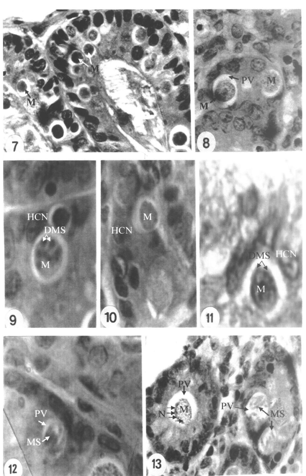
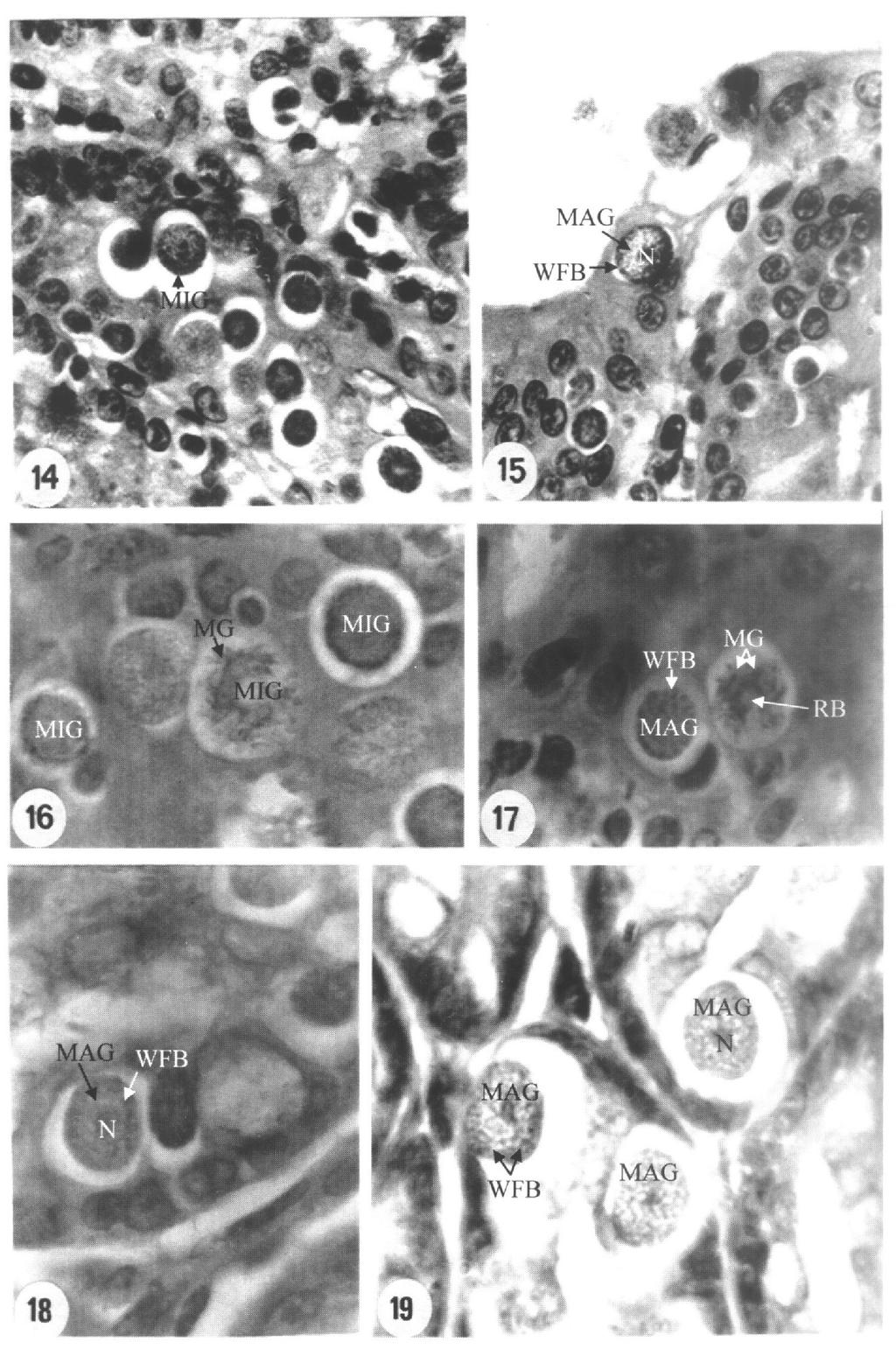
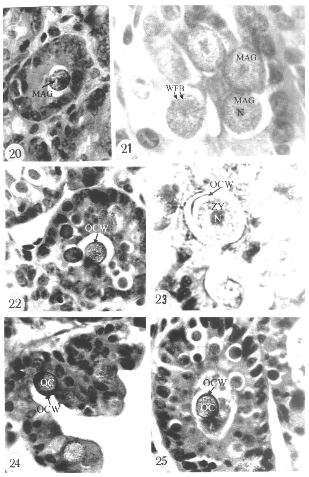
3 produced merozoites invaded new epithelial cells of the middle third of the small intestine of the experimentally inoculated bats and were differentiated into gamonts. They were spherical in shape, each with a clear centric nucleus, located within a small parasitophorous vacuole, measured x µm (n=50), with a mean of 6.2 x 6.3 µm (Figs. 14&15) and were observed at 65 hrs. p.i.. From hrs. p.i., some of these gamonts became microgamonts while others led to the formation of macrogamonts. The microgamonts were characterized by the presence of a large number of small nuclei (7-18) either randomly spread in the cytoplasm (Figs. 16&17) or peripherally arranged (Fig. 16) and measured x µm (n=50), with a mean of 8.60 x 6.62 µm. After a process of microgametogenesis, the microgametes were developed (Fig. 17). In the present study, free and slender microgametes were obvious and present in numbers that made their light photography feasible. Their number ranged in each microgamont from 8 to 24, measured x µm (n=50), with a mean of 3.12 x 2.24 µm and were observed at about hrs. p.i. (Figs. 16&17). Meanwhile, macrogamonts observed at hrs. p.i. were spherical in shape, each with a large centric nucleus, situated within a large parasitophorous vacuole and measured x µm (n=50), with a mean of 9.12 x 8.22 µm (Fig. 15). The development of each macrogamont resulted in the formation of a single macrogamete. This type of development did not include any nuclear division. Each macrogamete contained a large central nucleus and two types of wall-forming bodies (types I & II) which arranged in the peripheral cytoplasm (Figs. 15, ). These macrogametes were observed at hrs. p.i. and measured x µm (n=50), with a mean of x 9.68 µm (Figs ). At hrs. p.i., newly-formed zygotes or young oocysts were observed in the epithelial cells of the experimentally infected bats. The majority of them appeared subspherical to oval in the shape, each with a large centric nucleus, situated in a clear and large parasitophorous vacuole and measured x µm (n=50), with a mean of x µm (Figs ). In the present investigation, the fusion of the wall-forming bodies (types I & II) to produce the bilayered wall of the oocyst (outer layer and inner one) could be observed at the periphery (Figs ). 4. Discussion 1. Rate of natural infection: Duszynski et al. (1988) and Duszynski (1997) recorded that the chiropteran bats were showing a oocysts in the faeces (prepatent period) was 4 days, while the entire duration of oocyst existence or production in the faeces (patent period) was days. a- Merogony: The merogonic stages of the eimerian species under investigation started as its free sporozoites within the intestine of the bats invade the mucosal lining of the epithelial cells. In the present study, merogony took place in the lamina propria and the epithelial cells of the middle third of the small intestine of the experimentally infected bats at hrs. p.i. and only one generation was observed. None of the merogonic stages were seen either in the liver, spleen, kidney or other parts of the intestine. Light microscopic study showed that after the invasion of the sporozoites to the epithelial cells, they became rounded or ovoid and grew in size to form developing meronts (Figs. 7&8). Meanwhile, a parasitophorous vacuole appeared enclosing the parasite and grew in size during the growth of the meronts (Figs. 7 & 8). In the present investigation, it was difficult to detect sporozoites penetrating the intestinal epithelial cells. The early uninuclear meronts were spherical or subspherical, seen hrs. post inoculation and measured x µm (n=50), with a mean of 4.40 x 3.69 µm (Fig. 7). As nuclear division progressed, these meronts increased in size, became mature and measured x µm (n=50), with a mean of 9.44 x 7.10 µm. These mature meronts contained 6-15 nuclei when observed at hrs. p.i. (Figs. 9-11). Later on, nuclei of each meront were arranged at the periphery directly under its border (Fig. 13). Each peripheral nucleus represented a site of a developing merozoite (Figs. 9-11). The developing merozoites were protruded and gradually budded off from the mother meront cytoplasm by ectomerogonous manner (Figs. 9-11). After 60 hrs. p.i. mature meronts with 6-15 fullydifferentiated merozoites were observed within a clear parasitophorous vacuole (Figs. 12&13). The free merozoites were banana-shaped and measured x µm (n=50), with a mean of 3.71 x 2.82 µm (Figs. 12&13). No residual body could be seen in this type of meronts (Figs. 12&13). The host cells were greatly hypertrophied due to the development of the parasite. The host cell nucleus was elongated and flattened at the border facing the parasitophorous vacuole. More than one host cell nucleus were observed at the vacuole edge (Figs. 8-13). b- Gamogony: As in other Eimeria species, the asexual phase of the life cycle (merogony) was followed by the sexual one (gamogony). In the present investigation, gamogony was detected at hrs. p.i. The 294
4 recorded trilayerd oocyst wall, composed of an inner thin, colourless one and two outer layers which are thicker, yellowish-brown, prominently striated and in close apposition, in Eimeria molossi infecting the bat, Molossus ater and unilayered oocyst wall, composed of a single thin colourless layer, in Eimeria peltocephali infecting the fresh-water turtle, Peltocephalus dumerilianus in Brazil. In spite of, Lainson and Naiff (2000) reported bilayered oocyst wall in Eimeria bragancaensis infecting the Brazilian bat, Peropteryx macrotis, however, they recorded that the outer layer of the oocyst wall is frequently lost. Meanwhile, McAllister and Upton (1989) and Upton et al. (1992a) recorded that oocysts of Eimeria species infecting some reptiles were surrounded by a single-layered wall. Moreover, 4 and 5 membranes were observed in the oocyst wall of E. maxima (Elwasila, 1984) and E. perforans (Scholtyseck et al., 1971) respectively. In addition, 9 membranes were reported in the oocyst wall of Isospora canaria (Speer and Duszynski, 1975). The absence of the micropyle, recorded in the present study, was in agreement with some available eimerian species infecting the bats (Wheat, 1975; Duszynski and Barkley, 1985; Duszynski et al., 1988, 1999 a&b; Lainson and Naiff, 1998, 2000; Alyousif, 1999 a&b; Alyousif et al., 1999; Duszynski, 2002; McAllister and Upton, 2009; McAllister et al. 2011); the rats (Abdel-Ghaffar et al., 1991; Duszynski et al., 1992; Shazly et al., 1997; Ahmed et al., 1999; Slapeta et al., 2001) and reptiles (Upton et al., 1992 a&b; Abdel-Gawad et al., 1995; Fayed, 1997, 2003; Slapeta et al., 2003; Snow et al., 2011). On the other hand, the presence of the micropyle was reported in other eimerian species (Barker et al., 1979; Scott and Duszynski, 1997; Fayed, 1997 in the second type of oocysts; Bashtar et al., 2003; Alyousif et al., 2010). The presence of the polar granule in the eimerian oocysts in the present study showed similarity with other eimerian oocysts infecting the bats (Wheat, 1975; Duszynski and Barkley, 1985; Duszynski et al., 1988, 1999 a&b; Scott and Duszynski, 1997; Lainson and Naiff, 1998, 2000; Alyousif, 1999 a&b; Alyousif et al., 1999; McAllister and Upton, 2009; McAllister et al. 2011). On the other hand, the absence of the polar granule was also recorded in some other eimerian oocysts (Sakran et al., 1994; Fayed, 1997, 2003; Shazly et al., 1997; Snow et al., 2011). The present investigation showed the presence of an oocyst residuum in the sporulated oocysts which was in agreement with some Eimeria species (Barker et al., 1979; Duszynski and Barkley, 1985; Duszynski et al., 1988, 1999 a&b; Abdel-Ghaffar et al., 1991; Fayed, 1997, 2003; Shazly et al., 1997; Alyousif, 1999 a&b; Alyousif et al., 1999; Bashtar et al., 2003; Hofstatter and Guaraldo, 2011; Couch et al., 2011). lower percentage of natural infection (3% and 3.57% respectively) with Eimeria species and explained their hypothesis due to the high flying nocturnal activities and different geographical distributions of these flying mammals. Similarly, Scott and Duszynski (1997) recorded 5% and 11% natural rates of infection for two new Eimeria spp. infecting the bats Myotis spp in New Mexico, California and Bolivia, USA. At the same time, Duszynski and Barkley (1985); Seville and Gruver (2004) and McAllister et al. (2011) reported 12%; 6.6% and 10% natural rates of infection respectively in bats, Tomopeas ravus; Myotis sp. and Perimyotis subflvus infecting with new Eimeria spp. On the other hand, the rate of natural infection of bats with Eimeria spp. may exceed 25% as recorded by Alyousif et al. (1999) [25%]; the present study [32%]; Duszynski et al. (1999b) [50%]; McAllister and Upton (2009) [63.6%]. 2. Exogenous stages: The oocysts are the most easily accessible stages of any coccidium and in some cases many Eimeria species are known only from the morphological and characteristic features of their oocysts (Gottschalk, 1974; Cerná, 1976; Abdel-Ghaffar et al., 1991; Bashtar et al., 1992b, 2003; Fayed, 1997, 2003; Shazly et al., 1997; Duszynski et al a&b; Slapeta et al., 2001,2003; Duszynski, 2002; Adriano et al., 2003; Couch et al., 2003; Al-Ghamdy et al., 2005; McAllister and Upton, 2009; McAllister et al. 2011; Hofstatter and Guaraldo, 2011). The presence or absence of a polar cap or micropyle; shape of the sporocysts; presence or absence of residual bodies, polar granules, Stieda and substieda bodies represent very important criteria for detection of eimerian species (Marinkelle, 1968; Wheat, 1975; Cerná and Rysavý, 1976; Barker et al., 1979; Abdel-Ghaffar et al., 1986a; Mehlhorn, 1988, 2001; Fayed, 1997, 2003; Shazly et al., 1997; Lainson and Naiff, 1998, 2000; Alyousif, 1999 a&b; Zhao et al., 2001; Bashtar et al., 2003; Seville and Gruver, 2004; McAllister and Upton, 2009; Fritzler et al., 2011; McAllister et al. 2011; Hofstatter and Guaraldo, 2011). In the present study, only one type of oocysts was recorded. These oocysts were surrounded by a bilayered oocyst wall. Similar results were obtained in other eimerian species of bats as E. macyi (Wheat, 1975); E. wombati and E. arundeli (Barker et al., 1979); E. tomopea (Duszynski and Barkley, 1985); E. tadarida (Duszynski et al., 1988); 2 new Eimeria spp. from the bats Myotis spp. (Scott and Duszynski, 1997); E. chiropteri (Alyousif, 1999a); E. kuhliensis (Alyousif, 1999b); E. pipistrellus (Alyousif et al., 1999); E. dowleri and E. sealanderi (McAllister and Upton, 2009) and finally E. heidti (McAllister et al., 2011). On the other hand, Lainson and Naiff (1998) 295
5 al., 1986b, 1990; Koura et al., 2001; Slapeta et al., 2001; Fayed, 2003). On the other hand, three asexual generations (Clarkson, 1959; Fernando, 1974; McDonal and Rose, 1987; Shazly et al., 1997; Ahmed et al., 1999) and four asexual generations (Ernst and Chobotar, 1978; Pakandl et al., 1996) were reported in some other Eimeria species. Moreover, five asexual generations were also observed in other Eimeria species (Cheissin, 1940; Ruff et al., 1980; Pakandl and Coudert, 1999; Shazly et al., 2005). It is recorded in the present parasite that its uninucleated meronts were spherical or subspherical and this result runs in agreement with that reported for other Eimeria species (Abdel-Ghaffar et al., 1986b, 1990; El-Toukhy, 1994; El-Toukhy et al., 1997; Fayed, 1997, 2003; Shazly et al., 1997, 2005; Ahmed et al., 1999). The shape and position of the multinucleated meronts of the present parasite were similar to other Eimeria species but different in the measurements (Cheissin, 1940; Pellérdy, 1974; Abdel-Ghaffar et al., 1986b, 1990, 1991; Ahmed et al., 1992, 1999; Lainson and Naiff, 1998, 2000; Fayed, 1997, 2003; Shazly et al., 1997, 2005). The yielded number of merozoites per each mature meront may be a species-specific character of Eimeria species (Levine and Ivens, 1990; Ahmed et al., 1992, 1999). In the present study, mature meronts with 6-15 fully- differentiated merozoites were observed within a clear parasitophorous vacuole. Similar results were recorded by Abdel-Ghaffar et al. (1986b, 1990, 1991); Ahmed et al. (1992, 1999); Fayed (1997, 2003); El-Toukhy et al. (1997) and Shazly et al. (1997, 2005). These merozoites reported herein were developed through an ectomerogonous manner. Ectomerogony is the most common recorded mechanism of merozoite formation among Coccidia (Scholtyseck, 1973; Ahmed et al., 1992, 1999; Mehlhorn, 2001; Slapeta et al., 2001). However, Hammond (1973) reported an endomerogonous type of merozoite formation among some other eimerian species. Furthermore, Entzeroth et al. (1998) described the structure and function of the parasitophorous vacuole in Eimeria species and claimed that the parasitophorous vacuole membrane was manipulated by the parasite and functioned later in the developmental cycle as a molecular sieve, allowing the exchange of metabolites between parasite and host cell. b- Gamogony: The sexual phase of the life cycle is of great significance because it probably plays an important role in the host specificity of Coccidia (Scholtyseck et al., 1971, 1977). The sexual phase in any eimerian infection occurs after a species-specific number of asexual merogonic generations (Mehlhorn, 1988). In However, such oocyst residuum is lacking in sporulated oocysts of other Eimeria spp. (Wheat, 1975; Scott and Duszynski, 1997; El-Toukhy et al., 1997; Lainson and Naiff, 1998, 2000; Slapeta et al., 2001; Adriano et al., 2003; McAllister and Upton, 2009; Alyousif et al., 2010; Fritzler et al., 2011; McAllister et al. 2011; Snow et al., 2011; Hofstatter and Kawazoe, 2011). Furthermore, the measurements and characteristic features of eimerian sporocysts can be used as criteria to differentiate between Eimeria species (Mehlhorn, 1988, 2001; Daszak and Ball, 2001; Lainson, 2002; Duszynski, 2002; Adriano et al., 2003; Bashtar et al., 2003; Alyousif et al., 2010; Fritzler et al., 2011; Hofstatter and Kawazoe, 2011). In the present study, sporocysts of Eimeria rousetti measured x 6.62 µm, contained Stieda and substieda bodies at the pointed end and a sporocyst residuum. These results are in agreement with that reported in some other Eimeria species (Barker et al., 1979; Duszynski and Barkley, 1985; Duszynski et al., 1988, 1999 a&b; Fayed, 1997, 2003; Scott and Duszynski, 1997; Shazly et al., 1997; Lainson and Naiff, 1998, 2000; Alyousif, 1999 a&b; Alyousif et al., 1999, 2010; Slapeta et al., 2001; Adriano et al., 2003; Bashtar et al., 2003; McAllister and Upton, 2009; Fritzler et al., 2011; McAllister et al. 2011; Hofstatter and Guaraldo, 2011; Couch et al., 2011; Hofstatter and Kawazoe, 2011). 3. Endogenous stages: Most studies carried out on Eimeria parasites mainly recorded the exogenous stages only, but many authors gave a great attention to the endogenous stages as well (Ernst and Chobotar, 1978; Abdel- Ghaffar et al., 1986b, 1990; Ahmed et al., 1992, 1999; El-Toukhy et al., 1997; Fayed et al., 1996 a&b; Fayed, 1997, 2003; Shazly et al., 1997, 2005; Lainson and Naiff, 1998, 2000; Paperna and Lainson, 1999; Shazly, 2002; Al-Ghamdy et al., 2005). a- Merogony: In the present study, merogony took place in the lamina propria and the epithelial cells of the middle third of the small intestine of the experimentally infected bats at hrs. p.i.. Similar observations were reported for some other Eimeria species (Cheissin, 1940; Pellérdy, 1974; Abdel-Ghaffar et al., 1986b, 1990, 1991; Ahmed et al., 1992, 1999; Pakandl et al., 1996; Fayed, 1997, 2003; Shazly et al., 2005). Also, merogony occurred only in one generation during the present study and this result runs in agreement with that reported for E. obtusi (Fayed, 1997). However, the exact number of asexual merogonic generations among Eimeria is not fixed (Clarkson, 1958, 1959; Mehlhorn, 1988). Two asexual merogonic generations were recorded for some Eimeria species (Rutherford, 1943; Clarkson, 1958; Pellérdy, 1974; Long, 1982; Abdel-Ghaffar et 296
6 oocysts were formed in the intestinal epithelial cells and fusion of the wall-forming bodies (types I & II) to produce the bilayered oocyst wall could be observed at the periphery. Species diagnosis: Eimeria rousetti sp. nov. Type-host: Frugivorous bats Rousettus aegyptiacus. Type-locality: Giza, Beni-Suef and Fayoum provinces in Egypt. Type-specimens (hapantotypes): Oocysts in 10% formalin, slides and phototypes are deposited in the parasitological collection of Zoology Department, Faculty of Science, Cairo University, Egypt. Site of infection: The lamina propria and the epithelial cells of the middle third of the small intestine of the experimentally inoculated bats. Prevalence of natural infection: 32%. Sporulation time: hrs at 28±3 o C. Prepatent period: 4 days. Patent period: days. Number of asexual merogonic generations: One generation Etymology: The specific name of the present parasite is derived from the name of the host genus Rousettus. Abbreviations used in figures: DMS Developing merozoites HCN Host cell nucleus IL Inner layer of oocyst wall M Meront MAG Macrogamont / Macrogamete MG Microgamete MIG Microgamont MS Merozoite /s N Nucleus OC Oocyst OCR Oocyst residuum OCW Oocyst wall OL Outer layer of oocyst wall PG Polar granule PV Parasitophorous vacuole RB Residual body S Sporont SbT Substieda body SP Sporoblast SPC Sporocyst SPCR Sporocyst residuum SPR Sporozoite ST Stieda body WFB Wall-forming bodies ZY Zygote the present study, gamogony was detected at hrs. p.i. The produced merozoites invaded new epithelial cells of the middle third of the small intestine of the experimentally inoculated bats and were differentiated into spherical gamonts. Similar results were recorded in gamonts of most Eimeria species (Ernst and Chobotar, 1978; Abdel-Ghaffar et al. 1986b, 1990; Bashtar, 1991; Ahmed et al. 1992, 1999; Fayed et al., 1996 a&b; Al-Hoot, 1997; El- Toukhy et al.,1997; Fayed, 1997, 2003; Shazly, 2002; Al-Ghamdy et al., 2005). Microgamonts in the present study were characterized by the presence of a large number of small nuclei either randomly spread in the cytoplasm or peripherally arranged, producing a large number of microgametes. Similar observations were also reported in many other Eimeria species (Abdel- Ghaffar et al. 1986b, 1990; Bashtar, 1991; Ahmed et al. 1992, 1999; Fayed et al., 1996a; Al-Hoot, 1997; El-Toukhy et al.,1997; Fayed, 1997, 2003; Shazly, 2002; Al-Ghamdy et al., 2005). Using light microscopy in the present study made no way to count and/or to describe the characteristics of the microgametes. Meanwhile, macrogamonts in the present study were characterized by their large size, accumulation of many food materials in their cytoplasm and their distinct nucleus. After growth, the two types of wallforming bodies (types I & II) developed and arranged in their peripheral cytoplasm forming macrogametes. The same results were obtained in most Eimeria species (Scholtyseck, 1979; Abdel-Ghaffar et al. 1986b, 1990; Mehlhorn, 1988; Ahmed et al. 1992, 1999; Bashtar et al., 1992a; Fayed et al., 1996b; Al- Hoot, 1997; El-Toukhy et al.,1997; Fayed, 1997, 2003; Al-Ghamdy et al., 2005). Both types of wallforming bodies were named after their fate or function rather than the order of their appearance. Thus, the WFB I, which appeared after the WFB II, giving rise to the outer layer of the future oocyst wall and the WFB II form the inner layer (Scholtyseck et al., 1966; Mehlhorn, 1972, 2001; Speer et al., 1973; Bashtar et al., 1992a; Fayed et al., 1996b; Al-Hoot, 1997; Al-Ghamdy et al., 2005). Although the events of fertilization are still not completely clarified in many recorded species of Eimeria, detailed studies regarding the changes associating fertilization and oocyst wall formation of Eimeria species were recorded (Scholtyseck, 1973; Scholtyseck et al., 1971; Mehlhorn, 1972, 2001; Abdel-Ghaffar et al. 1986b, 1990; Bashtar et al., 1992a; Fayed et al., 1996b; Al-Ghamdy et al., 2005). The present study showed that after fertilization, zygotes or young 297

7 298
8 299
9 300
10 301
11 Figure Explanations: All photos (X: 1600) Figs. (1-6): Exogenous stages of Eimeria rousetti sp. nov. infecting the Egyptian frugivorous bats Rousettus aegyptiacus. Fig.(1): Fresh non-sporulated oocyst (OC), the sporont (S) occupies the entire volume of the oocyst; no micropyle or polar granules were observed. Fig.(2): Non-sporulated oocyst; the sporont (S) began to condensate towards one pole of the oocyst. Note the presence of distinct nucleus (N) and oocyst wall (OCW). Fig.(3): Non-sporulated oocyst (OC), evagination of the sporont (S) appeared. Note the presence of distinct nucleus (N) and oocyst wall (OCW). Fig.(4): Oocyst (OC) with three sporoblasts (Sp). Note the bilayered oocyst wall (OCW) formed of an outer layer (OL) and an inner one (IL). Fig.(5 Sporulated oocyst (OC) with 4 sporocysts (SPC), each with two sporozoites (SPR). Note the presence of oocyst residuum (OCR);the stieda (ST) and substieda (Sbt) bodies. Fig.(6): Sporulated oocyst (OC) with 4 fully sporulated sporocysts (SPC), each with two sporozoites (SPR). Note the presence of polar granule (PG) and sporosyst residuum (SPCR). Figs.(7-13): Light photomicrographs showing the merogonic stages of Eimeria rousetti sp.nov. experimentally infecting the Egyptian frugivorous bats Rousettus aegyptiacus. They occurred in lamina propria and the epithelial cells of the middle third of the small intestine at hrs P.I. All specimens are stained with haematoxylin and eosin (X 1800). Fig.(7): Early uninuclear meront (M) were seen hrs p.i. inside a clear parasitophorous vacuole (PV). Figs.(8): Developing meronts (M) within a clear parasitophorous vacuole (PV). Figs.(9): Multinucleated meront (M) showing the beginning of the formation of the developing merozoites (DMS). Figs.(10): Multinucleated meront (M). Figs.(11): Multinucleated meront (M) showing invagination of the developing merozoites (DMS). Figs.(12): Mature meront with fully formed merozoites (MS) in the (L.S.) within PV; each merozoite is bananashaped. Figs.(13): Meronts in different merogonic stages; one is multinucleated (M) with peripherally arranged nuclei (N) and the other meronts are mature with fully formed merozoites (MS). Each meront appeared within a parasitophorous vacuole (PV). Figs.(14-21): Light photomicrographs showing the different gamogonic stages of Eimeria rousetti sp.nov. in the epithelial cells of the middle third of the small intestine of the experimentally infected bats. Seen from hrs p.i. Figs.(14): Young gamonts were observed, each in spherical shape with central nucleus (N) and located in a big PV. One of these gamonts is the young microgamont (MIG). Figs.(15): Developing macrogamont (MAG) with peripherally arranged wall-forming bodies (WFB). Figs.(16): Microgamonts in different develop-mental stages. A microgamont (MIG) with a number of developing microgametes (MG) and situated in a parasitophorous vacuole (PV). Other micrgamonts are immature and with peripherally arranged nuclei. Figs.(17): A microgamont (MIG) with developing microgametes (MG) and residual body (RB). A macrogamont is also showed with a number of wall-forming bodies (WFB). Figs.(18): A macrogamete (MAG) with peripherally arranged wall-forming bodies (WFB) and a central nucleus (N). Figs.(19 & 21): Macrogametes (MAG) with wall-forming bodies (WFB) and a central nucleus (N). Figs.(20): A macrogamete (MAG) with peripherally arranged wall-forming bodies (WFB). Figs. (22-25): Newly-formed zygotes (ZY) or young oocysts (OC) were observed in the epithelial cells of the middle third of the small intestine of the experimentally infected bats. They are spherical or oval in shape and situated with a clear parasitophorous vacuole (PV). Note that the fusion of the wall-forming bodies of types I &II (WFBI &II) to form the bilayered oocyst wall (OCW). senegalensis aegyptiaca. 2. Endogenous stages. Bulletin of Fac. of Sci., Cairo Univ. 1986b; 54: Abdel-Ghaffar FA, Bashtar AR, El-Toukhy A. Endogenous stages of Eimeria arvicanthi infecting the field rat, Arvicanthus niloticus niloticus from Egypt. Journal of Egyptian Society for Parasitology 1990; 20(1): Abdel-Ghaffar FA, Bashtar AR, Mustafa AM, El-Toukhy A. Eimeria arvicanthi Van den Berghe & Chardome, 1956 and Eimeria mehlhorni nov. sp. infecting the field rat, References Abdel-Gawad M, Abdel-Aziz A, El-Toukhy A. Two new species of Eimeria infecting the skink, Chalcides ocellatus in Egypt. Journal of Union Arab Biologists, Cairo 1995; 3(A): Abdel-Ghaffar FA, Bashtar AR, Ahmed AK. Studies on Eimeria species infecting the Egyptian wild dove, Streptopelia senegalensis aegyptiaca.1. Exogenous stages. Bulletin of Fac. of Sci., Cairo Univ. 1986a; 54: Abdel-Ghaffar FA, Bashtar AR, Ahmed AK. Studies on Eimeria species infecting the Egyptian wild dove, Streptopelia 302
12 Cerná Z, Rysavý B. Notes on the distribution of Eimeria dukei Lavier, 1921 in African bats. Folia Parasitologica 1976; 23(3): 286. Chapman HD. Origins of coccidiosis research in the fowl--the first fifty years. Avian Disease 2003; 47(1):1-20. Chapman HD, Cherry TE, Danforth HD, Richards G, Shirley MW, Williams RB. Sustainable coccidiosis control in poultry production: the role of live vaccines. International Journal for Parasitology 2002; 32(5): Cheissin EM. Coccidiosis of rabbits III. The developmental cycle of Eimeria magna. Uch. Zap. Len. God. Ped. Institute of Gertsena 1940; 30: Clarkson MJ. Life history and pathogenicity of Eimeria adenoeides (Moore and Brown, 1951) in the turkey poult. Parasitology 1958; 48: Clarkson MJ. The life history and pathogenicity of Eimeria meleagrimitis (Tyzzer, 1929) in the turkey poult. Parasitology 1959; 49: Couch L, Laakkonen J, Goodman S, Duszynski DW.Two new eimerians (Apicomplexa) from insectivorous mammals in Madagascar. Journal of Parasitology 2011; 97(2): Couch L, Uni S, Duszynski DW. Eimeria species from serows (Capricornis spp.) in Japan with descriptions of two new species. Journal of Parasitology 2003; 89(3): Daszak P, Ball SJ. A description of two new species of Coccidia (Apicomplexa: Eimeriidae) from African reptiles with nomenclatural corrections for two Caryospora and Eimeria species from snakes. Folia Parasitologica 2001; 48(1): 1-6. Duszynski DW. Coccidia from bats (Chiroptera) of the world: A new Eimeria species in Pipistrellus javanicus from Japanese Journal of Parasitology 1997; 83(2): Duszynski DW. Coccidia (Apicomplexa: Eimeriidae) of the Mammalian order Chiroptera. Special Publications of the Museum of Southwestern Biology, No. 5. First Impressions, Inc., Albuquerque, NM., 2002; pp Duszynski DW, Barkley LJ. Eimeria from bats of the world: A new species in Tomopeas ravus from Peru. Journal of Parasitology 1985; 71(2): Duszynski DW, Patrick MJ, Couch L, Upton SJ. Eimerians in harvest mice, Reithrodontomys spp. from Mexico, California and New Mexico, and phenotypic plasticity in oocysts of Eimeria arizonensis. Journal of Parasitology 1992; 39: Duszynski DW, Reduker DW, Parker BB. Eimeria from bats of the world. II. A new species in Tadarida femorosacca from Sonora, Mexico. Journal of Parasitology 1988; 74(2): Duszynski DW, Scott DT, Aragon J, Leach A, Perry T. Six new Eimeria species from vespertilionid bats of North America. Journal of Parasitology 1999a; 85(3): Duszynski DW, Scott DT, Zhao X. Eimeria from bats of Bolivia: Two new species from vespertilionid bats. Journal of Parasitology, 1999b; 85(3): El-Toukhy A. Eimeria stenodactyli n. sp. from the Egyptian gecko, Stenodactylus elegans (Lacertilia: Geckonidae). Journal of Egyptian German Society 1994; 15(D): El-Toukhy A., Galal M, Radwaan R. Light and electron microscopic studies of Eimeria lineri infecting the Mediterranean gecko, Hemidactylus turcicus (Sauria: Geckonidae) in Egypt. Journal of Egyptian German Society 1997; 23(D): Elwasila M. Fine structure of the process of oocyst wall formation of Eimeria maxima (Apicomplexa: Eimeriina) Acta. Veterinaria Hungarica 1984; 32: Entzeroth R, Mattig FR, Werner-Meier R. Structure and function of the parasitophorous vacuole in Eimeria species. International Journal for Parasitology 1998; 28(7): Arvicanthus niloticus niloticus in Egypt. Archiv Fur Protistenkunde 1991; 140: Adriano EA, Thyssen PJ, Cordeiro NS. A New Species of Eimeria from the Eared Dove Zenaida auriculata (Aves: Columbidae) in Brazil. Acta Protozoologica 2003; 42: Ahmed AK, Bashtar AR, Shazly MA Abdel-Ghaffar F. Ectomerogony and fine structural changes associated with merozoite formation in Eimeria falciformis and Eimeria adenoeides (Protozoa, Eimeriidae). Proceeding of Zoological Society A.R.E. 1992; 23(2): Ahmed AK, Shazly MA Saleh AR. Transmission of an Eimeria sp. to another host of a different genus from the same family (Muridae): The question of host-specificity. Egyptian Journal of Zoology 1999; 32: Al-Ghamdy AO, Shazly MA, Al-Rasheid KAS, Mubarak M, Bashtar AR. Light and electron microscopy of Eimeria magna Pérard, 1925 infecting the house rabbit, Oryctolagus cuniculus from Saudi Arabia. II. Gamogony and oocyst wall formation. Saudi. Journal of Biological Science, Riydh, Saudi Arabia. 2005; 12(2): Al-Hoot AA. Ultrastructural study of micro- and macrogametogenesis of Eimeria mehlhorni infecting the field rat Arvicanthis niloticus niloticus in Egypt. Journal of Egyptian German Society 1997; 23(D): Al-Mathal EL. Hepatic Coccidiosis of the domestic rabbit Oryctolagus cuniculus domesticus in Saudi Arabia. World Journal of Zoology 2008; 3: Altringham J D. Bats: Biology and Behavior, 1996; New York: Oxford University Press. Alyousif MS. Eimeria chiropteri n. sp. (Apicomplexa: Eimeriidae) from Saudi Arabian bat Pipistrellus kuhlii (Chiroptera: Vespertilionidae Journal of Egyptian Society for Parasitology 1999a; 29(1): Alyousif MS. Eimeria kuhliensis sp. n. (Apicomplexa: Eimeriidae) from pipistrelle bat, Pipistrellus kuhlii. Acta Protozoologica 1999b; 38: Alyousif MS., Al-Dakhil M, Al-Shawa YR. Eimeria pipistrellus n. sp. from Pipistrellus kuhlii (Chiroptera: Vespertilionidae) in Saudi Arabia. Korean Journal of Parasitology 1999; 37(1): 1-4. Alyousif MS, Alfaleh FA, Al-Shawa YR. Eimeria falconensis sp. n. (Apicomplexa: Eimeriidae) from the lanner falcon, Falco biarmicus (Falconiformes: Falconidae) in Saudi Arabia. Journal of Egyptian Society for Parasitology 2010; 40(2): Barker IK, Munday BL, Presidente PJ. Coccidia of wombats: correction of host-parasite relationships. Eimeria wombat (Gilruth and Bull, 1912) Comb. Nov. and Eimeria ursine (Supperer, 1957) from the hairy-nosed wombat and Eimeria arundeli sp. n. from the common wombat. Journal of Parasitology 1979; 65(3): Bashtar AR. Ultrastructural study of microgametogenesis in Eimeria arvicanthi Journal of Egyptian German Society 1991; 4: Bashtar AR, Ahmed AK, Shazly MA, Abdel-Aziz A. Fine structural studies on macrogamogony of Eimeria adenoeides (Sporozoa, Eimeriidae) infecting turkeys in Egypt and oocyst formation. Journal of Egyptian German Society 1992a; 8(B): Bashtar AR, Ahmed AK, Shazly MA, Soliman GN, El-Samanudy F. Studies on Eimeria species naturally infecting Gerbillus pyramidium. (1) Exogenous stages. Journal of Egyptian Society for Parasitology 1992b; 22(3): Bashtar AR, Al-Rasheid KAS, Mubarak M, Al-Ghamdy AA. Coccidiosis in rabbits from Saudi Arabia. 1. Exogenous stages of different Eimeria spp. Journal of Egyptian German Society 2003; 42(D): Cerná Z. Eimeria vejsovi sp. n. from the bat Nyctalus noctula. Folia Parasitologica (Praha), 1976; 23(2):
13 n. sp., from the bat, Molossus ater. Memórias do Instituto Oswaldo Cruz, Rio de Janeiro1998; 93(1): Lainson R, Naiff RD. On Eimeria bragancaensis n. sp. (Apicomplexa: Eimeriidae) and tissue-cysts of an unidentified protozoan in the bat, Peropteryx macrotis (Chiroptera: Emballonuridae) from Amazonian Brazil. Parasite 2000; 7(2): Lee SH, Lillehoj HS, Jang SI, Woo Lee K, Seon Park M, Bravo D, Lillehoj EP. Cinnamaldehyde enhances in vitro parameters of immunity and reduces in vivo infection against avian coccidiosis. British Journal of Nutrition 2011; 18:1-8. Levine ND. The protozoan phylum Apicomplexa. 1988; CRC Press. Inc., Boca Raton, Florida, p Levine ND, Ivens V. The Coccidian Parasites of Rodents. 1990; CRC Press, Inc. Boca Raton, Florida. Long PL. The biology of the Coccidia. University Park Press, Baltimore, USA. Long, P.L. (1990): Coccidiosis of Man and Domestic Animals. 1982; CRC Press, Boca Raton, Florida, USA. Long PL, Millard BJ, Joyner LP, Norton CC. A guide to laboratory techniques used in the study and diagnosis of avian Coccidiosis. Folia Vet. Lat., 1976; 6: Marinkelle CJ. Eimeria eumopos n. sp. from a Colombian bat Eumops trumbulli. Journal of Protozoology 1968; 15(1): McAllister CT, Upton SJ. Coccidian parasites (Apicomplexa: Eimeriidae) of Nerodia sp. (Serpentes: Colubridae), with a description of a new species of Eimeria. Journal of Protozoology 1989; 36(3): McAllister CT, Upton SJ. Two new species of Eimeria (Apicomplexa: Eimeriidae) from Eastern red bats, Lasiurus borealis (Chiroptera: Vespertilionidae), in Arkansas and North Carolina. Journal of Parasitology 2009; 95(4): McAllister CT, Burt S, Seville RS, Robison HW. A new species of Eimeria (Apicomplexa: Eimeriidae) from The Eastern pipistrelle, Perimyotis subflavus (Chiroptera: Vespertilionidae), in Arkansas. Journal of Parasitology 2011; (Epub ahead of print). McDonal V, Rose ME. Eimeria tenella and E. necatrix: A third schizogony is an obligatory part of the development cycle. J. Parasitol., 1987; 73(3): Mehlhorn H. Elektrnenmikroscopisch untersuchungen an Entwicklungasstadien von Eimeria maxima (Sporozoa, Coccidia). 1. Diefeinstruktur der Makrogamenten. Zeitschrift Fur Parasitenkunde1972; 39: Mehlhorn H. Morphology in Parasitology in Focus: Facts and trends. Heinz Mehlhorn (Ed.) 1 st edition, 1988; Spring- Verlag Berlin, Heidelberg, New York, London, Paris, Tokyo. Mehlhorn H. Encyclopedic Reference of Parasitology. 2nd Ed., 2001; Springer, Berlin, Germany. Pakandl M, Coudert P. Life cycle of Eimeria vejdovskyi Pakandl, 1988: electron microscope study. Parasitology Research 1999; 85(10): Pakandl M, Eid Ahmed N, Licois D, Coudert P. Eimeria magna Perard, 1925: Study of the endogenous development of parental and precocious strains. Veterinary Parasitology 1996; 65(3-4): Paperna I, Lainson R. The ultrastructure of some endogenous stages of the coccidian Eimeria boveroi Carini & Pinto, 1926 in the gut epithelial cells of the gecko Hemidactylus mabouia from Brazil. Parasite 1999; 6(3): Pellérdy L. Coccidia and Coccidiosis. 2nd Edn., 1974; Verlag Paul Parey, Berlin & Hamburg, Germany. Ruff MD, Doran DJ, Augustine PC. Eimeria meleagrimitis Tyzzer in turkeys. The life cycle and effects of inoculation, size and time on severity of infection and intestinal distribution. Journal of Protozoology 1980; 27(2): Ernst JV, Chobotar B.The endogenous stages of Eimeria utahensis (Protozoa: Eimeriidae) in the Kangaroo rat Dipodomys ordii. Journal of Parasitology 1978; 64(1): Fayed HM. Life cycle of Eimeria obtusi sp. nov. (Apicomplexa: Coccidia: Eimeriidae) infecting the Egyptian snake Telescopus dhara obtusus Reuss. Egyptian Journal of Zoology 1997; 29: Fayed HM. Life cycle of Eimeria varani sp. nov. (Apicomplexa: Coccidia: Eimeriidae) infecting the desert monitor Varanus griseus (Boulenger, 1885) (Squamata: Lacertilia: Varanidae) in Egypt: A light microscopic study. Egyptian. Journal of Zoology 2003; 41: Fayed HM, Ahmed AK, Shazly MA, Kassem HH. Fine structural characteristics of microgametogenesis of Eimeria sp. infecting fat sand rats Psammomys obesus obesus Cretzschmar, 1828 in Egypt. Journal of Egyptian German Society 1996a; 20(D): Fayed HM, Ahmed AK, Shazly MA, Kassem HH. Fine structural characteristics of macrogametogenesis, fertilization and oocyst wall formation of Eimeria sp. infecting fat sand rats Psammomys obesus obesus Cretzschmar, 1828 in Egypt. Journal of Union Arab Biologists, Cairo 1996b; 6(A): Fernando MA. Fine structure of the schizonts and merozoites of Eimeria acervulina in the chicken. Journal of Parasitology 1974; 60(1): Fritzler JM, Craig T, Elgayar A, Plummer C, Wilson RS, Peterson M, Zhu G. Eimeria attwateri N. Sp. (Apicomplexa: Eimeriidae) from endangered Attwater's Prairie-Chickens (Tympanuchus cupido attwateri) in Texas, USA. Journal of Parasitology 2011; [Epub ahead of print]. Gottschalk C. New species of Coccidia from the bat (Nyctalus noctula). Angewandte Parasitologie 1974; 15(1): 3-5. Gres V, Voza T, Chabaud A, Landau I. Coccidiosis of the wild rabbit (Oryctolagus cuniculus) in France. Parasite 2003; 10(1): Hammond DM. Life cycles and development of Eimeria; Isospora; Toxoplasma and related genera. In Hammond, D.M. and Long, P.L. (edst.), 1973; University Park Press, Baltimore, pp Hofstatter PG, Guaraldo AM. A new eimerian species (Apicomplexa: Eimeriidae) from the blue-fronted Amazon parrot Amazona aestiva L. (Aves: Psittacidae) in Brazilian Journal of Parasitology 2011; Epub ahead of print. Hofstatter PG, Kawazoe U. Two new Eimeria species (Apicomplexa: Eimeriidae) from the Yellow-Crowned Amazon Amazona Ochrocephala (Aves: Psittacidae) in Brazil. Journal of Parasitology 2011; Epub ahead of print. Ka-oud H, Nassar AM, Abdel-Gawad AMH, Eisa ME. Studies on Eimeria of bats from Egypt. Journal of Veterinary Medical Association 1989; 49 (1-2): Kim DY, Reilly TJ, Schommer SK, Spagnoli ST. Rabbit tularemia and hepatic coccidiosis in wild rabbit. Emerg. Infectious Disease Journal 2010; 16(12): Koura EA, Abdel-Ghaffar FA, Ismail HM. Life cycle of Eimeria sp. (Eimeriidae: Apicomplexa) infecting the domestic duck (Anas boschas domesticus) in Egypt. A light microscopic study. Journal of Egyptian German Society 2001; 35(D): Kowalik S, Zahner H. Eimeria separate: method for excystation of sporozoites. Parasitology Research 1999; 85(6): Lainson R. Intestinal Coccidia (Apicomplexa: Eimeriidae) of Brazilian Lizards. Eimeria carmelinoi n.sp., from Kentropyx calcarata and Acroeimeria paraensis n.sp. from Cnemidophorus lemniscatus lemniscatus (Lacertilia: Teiidae Memórias do Instituto Oswaldo Cruz, Rio de Janeiro 2002; 97(2): Lainson R, Naiff RD. Eimeria peltocephali n. sp., (Apicomplexa: Eimeriidae) from the freshwater turtle Peltocephalus dumerilianus (Chelonia: Pelomusidae) and Eimeria molossi 304
14 cycles. Saudi. Journal of Biological Science 2005; Riydh, Saudi Arabia. 12(1): Šlapeta JR, Modrý D, Ashe J, Koudela B. Description of Eimeria arabukosokokensis sp. n. (Apicomplexa: Eimeriidae) from Telescopus semiannulatus (Serpentes: Colubridae) with notes on eimerian coccidia from snakes of Eastern Kenya. Folia Parasitologica 2003; 50 (1): Šlapeta JR; Modry D, Votypka J, Jirku MJ, Obornik M, Lukes J, Koudela B. Eimeria telekii n.sp. (Apicomplexa: Coccidia) from Lemniscomys striatus (Rodentia: Muridae): morphology, pathology and phylogeny. Parasitology 2001; 122: Snow KR, Peter D, Ball S, Streicker D. A New species of Eimeria (Apicomplexa: Eimeriidae) from the Western Hognose snake, Heterodon Nasicus (Serpentes: Xenodontidae) from Texas. Journal of Parasitology 2011; (Epub ahead of print). Speer CA, Duszynski DW. Fine structure of the oocyst walls of Isospora serini and Isospora canaria and excystation of Isospora serini from the canary Serinus canaries. Journal of Protozoology 1975; 22: Speer CA, Hammond DM, Youssef NN, Danforth HD. Fine structural aspects of macrogametogenesis in Eimeria magna. Journal of Protozoology 1973; 20: Taylor MA, Coop RL, Wall RL. Veterinary parasitology, 3rd edition., 2007; Blackwell Publishing Company pp, Upton S J, McAllister CT, Brillhart D B, Duszynski DW, Wash CW. Cross transmission studies with Eimeria arizonensislike oocysts (Apicomplexa) in New World rodents of the genera Baiomys, Neotoma, Onychomys, Peromyscus, and Reithrodontomys (Muridae). Journal of Parasitology 1992a; 78: Upton SJ, McAllister CT, Trauth SE, Bibb DK. Description of 2 new species of Coccidia (Apicomplexa, Eimeriorina) from Flat-Headed Snakes, Tantilla gracilis (Serpentes, Colubridae) and reclassification of misnomer species within the genera Isospora and Sarcocystis from snakes. Transactions of the American Microscopical Society1992b; 111: Wheat BE. Eimeria macyi sp. n. (Protozoa: Eimeriidae) from the eastern pipistrelle, Pipistrellus subflavus, from Alabama. Journal of Parasitology 1975; 61(5): Zhao X, Duszynski DW, Loker ES. Phylogenetic position of Eimeria antrozoi, a bat coccidium (Apicomplexa: Eimeriidae) and its relationship to morphologically similar Eimeria spp. from bats and rodents based on nuclear 18S and plastid 23S rdna sequences. Journal of Parasitology 2001; 87(5): Rutherford RL. The life cycle of four intestinal coccidian of the domestic rabbit. Journal of Parasitology 1943; 29: Sakran Th, Fayed HM, El-Toukhy A, Abdel-Gawad M. Studies on Eimeria ghaffari sp. nov. and Isospora acanthodactyli sp. nov. naturally infecting the Egyptian lizard Acanthodactylus boskianus (1): Exogenous stages. Journal of Egyptian German Society 1994; 15(D): Saratsis A, Joachim A, Alexandros S, Sotiraki S. Lamb coccidiosis dynamics in different dairy production systems. Veterinary Parasitology 2011; Apr 21(Epub ahead of print). Scholtyseck E.Ultrastructure, p In D. M. Hammond and P. L. Long (ed.), The Coccidia: Eimeria, Isospora, Toxoplasma, and related genera., 1973; University Park Press, Baltimore, USA. Scholtyseck E. Fine structure of Parasitic Protozoa: An Atlas of Micrographs, Drawings and Diagrams., 1979; Springer- Verlag, Berlin, Heidelberg, New York. Scholtyseck E, Chobotar B, Senaud J, Ernst JV. Fine structure of microgametogenesis of Eimeria ferrisi in Mus musculus. Zeitschrift Fur Parasitenkunde 1977; 51: Scholtyseck E, Hammond DM, Ernst JV. Fine structure of the macrogametes of Eimeria perforans, E. stiedai, E. bovis and E. auburnensis. Journal of Parasitology 1966; 52: Scholtyseck E, Mehlhorn H, Hammond A. Fine structure of macrogametes and oocysts of Coccidia and related organisms. Zeitschrift Fur Parasitenkunde 1971; 37: Scott DT, Duszynski DW. Eimeria from bats of the world: two new species from Myotis spp. (Chiroptera: Vespertilionidae). Journal of Parasitology 1997; 83(3): Seville RS, Gruver J. Species of Eimeria (Apicomplexa: Eimeriidae) from bats (Chiroptera: Vespertilionidae) in central Wyoming. Journal of Parasitology 2004; 90(2): Shazly MA. The ultrastructural study of microgametogenesis of Eimeria scinci Phisalix 1923 (Apicomplexa: Eimeriidae) infecting the sandfish lizard, Scincus mitranus Anderson 1871 in Saudi Arabia. Journal of Egyptian Society for Parasitology 2002; 32(3): Shazly MA, Ahmed AK, Fayed HM, Kassem HH. Exogenous stages and merogony of Eimeria sp. infecting the fat-sand rat Psammomys obesus obesus Cretzschmar, 1828 in Egypt: Light and electron microscope studies. Journal of Egyptian German Society 1997; 23(D): Shazly MA, Mubarak M, Al-Rasheid KAS, Al-Ghamdy AO, Bashtar AR. Light and electron microscopy of Eimeria magna Pérard, 1925 infecting the house rabbit, Oryctolagus cuniculus from Saudi Arabia I. Asexual developmental 11/21/
Ahead of print online version
 Folia Parasitologica 60 [3]: 232 236, 2013 ISSN 0015-5683 (print), ISSN 1803-6465 (online) Institute of Parasitology, Biology Centre ASCR http://folia.paru.cas.cz/ A new species of Choleoeimeria (Apicomplexa:
Ultrastructure of Endogenous Stages of Eimeria ninakohlyakimovae Yakimoff & Rastegaieff, 1930 Emend. Levine, 1961 in Experimentally Infected Goat
 Mem Inst Oswaldo Cruz, Rio de Janeiro, Vol. 92(4): 533-538, Jul./Aug. 1997 Ultrastructure of Endogenous Stages of Eimeria ninakohlyakimovae Yakimoff & Rastegaieff, 1930 Emend. Levine, 1961 in Experimentally
The Fine Structure of the Endogenous Stages of Isospora hemidactyli Carini, 1936 in the Gecko Hemidactylus mabouia from North Brazil
 Mem Inst Oswaldo Cruz, Rio de Janeiro, Vol. 95(1): 43-47, Jan./Feb. 2000 The Fine Structure of the Endogenous Stages of Isospora hemidactyli Carini, 1936 in the Gecko Hemidactylus mabouia from North Brazil
In recent years, there has been increasing
 Pakistan J. Zool., vol. 45(5), pp. 1329-1333, 2013 Prevalence of Coccidia (Eimeria spp.) Infection in Domestic Rabbits, Oryctolagus cuniculus, in Riyadh, Saudi Arabia Abdel-Azeem S. Abdel-Baki 1, 2 * and
Observations on Eimeria species of Dasyprocta leporina (Linnaeus, 1758) (Rodentia: Dasyproctidae) from the state of Pará, North Brazil
 (Rodentia: Dasyproctidae) from the state of Pará, North Brazil") Mem Inst Oswaldo Cruz, Rio de Janeiro, Vol. 99: 000-000, 2004 1 Observations on Eimeria species of Dasyprocta leporina (Linnaeus, 1758) (Rodentia: Dasyproctidae) from the state of Pará, North Brazil Ralph
Mem Inst Oswaldo Cruz, Rio de Janeiro, Vol. 99: 000-000, 2004 1 Observations on Eimeria species of Dasyprocta leporina (Linnaeus, 1758) (Rodentia: Dasyproctidae) from the state of Pará, North Brazil Ralph
Key words: Coccidia, Choleoeimeria rochalimai, fine structure, gall bladder epithelium, Hemidactylus mabouia, Brazil
 FOLIA PARASITOLOGICA 47: 91-96, 2000 Ultrastructural study of meronts and gamonts of Choleoeimeria rochalimai (Apicomplexa: Eimeriidae) developing in the gall bladder of the gecko Hemidactylus mabouia
Phylum:Apicomplexa Class:Sporozoa
 Phylum:Apicomplexa Class:Sporozoa The most characteristic features of sporozoa are 1-unique appearance of most protozoa makes it possible for knowledge able person to identifiy them to level of genus and
 956 8 748557 0 0 6946 00869759 0 5676694 amira_el_far@hotmail.com ٢٨ ٣٢ ) 990 984 979 979 0 984 8 99 996 6 0 00 0 4 5 6 994 99 004 999 997 96 96 95 - 4 00 4 40 00 4 5 40 6 6 40 7 40 000 5 LIST OF PUBLICATIONS
Joerg Kinne, Mansoor Ali*, Ulrich Wernery, and J. P. Dubey
 J. Parasitol., 88(3), 2002, pp. 548 552 American Society of Parasitologists 2002 CLINICAL LARGE INTESTINAL COCCIDIOSIS IN CAMELS (CAMELUS DROMEDARIUS) IN THE UNITED ARAB EMIRATES: DESCRIPTION OF LESIONS,
CURRICULUM VITAE (Prof.Dr. Amira Kamal Ahmed)
") CURRICULUM VITAE (Prof.Dr. Amira Kamal Ahmed) Personal Particulars : Name : Amira Kamal Ahmed Hasanien Date of Birth : Nov, 18 th 1956. Place Of Birth : Cairo, Egypt. Nationality Marital Status : Egyptian
CURRICULUM VITAE (Prof.Dr. Amira Kamal Ahmed) Personal Particulars : Name : Amira Kamal Ahmed Hasanien Date of Birth : Nov, 18 th 1956. Place Of Birth : Cairo, Egypt. Nationality Marital Status : Egyptian
Protozoa. Apicomplexa Sarcomastigophora Ciliophora. Gregarinea Coccidia Piroplasma
 Protozoa Apicomplexa Sarcomastigophora Ciliophora Gregarinea Coccidia Piroplasma Coccidia characterized by thick-walled oocysts excreted in feces In Humans Cryptosporidium Isospora Cyclospora Sarcocystis
Cr 2 O 7. Key words: coccidia - apicomplexa - Eimeria - peacock - Pavo cristatus - Egypt. Materials and Methods
 Mem Inst Oswaldo Cruz, Rio de Janeiro, Vol. 105(8): 965-969, December 2010 965 Eimeria pavoaegyptica sp. nov. (Apicomplexa: Eimeriidae) in faeces of Indian peacocks, Pavo cristatus Linnaeus, 1758 (Galliformes:
Revajová, Viera, Loószová, Adrian. The Journal of Protozoology Resea Citation RightsNational Research Center for Prot
 ' ' Morphological study of partridge Title development in the foreign host - (Gallus gallus) Revajová, Viera, Loószová, Adrian Author(s) Maria, Zibrín, Martin, Herich, Ro Mikulas The Journal of Protozoology
HISTOPATHOLOGY. Introduction:
 Introduction: HISTOPATHOLOGY Goats and sheep are the major domestic animal species in India. Much of the economy of the country has been depend upon the domestication of these animals. Especially economy
Protozoan Parasites of Veterinary importance 2017
 Protozoan Parasites of Veterinary importance 2017 VPM-122 Laboratory 4 Spencer J. Greenwood PhD, DVM Dept. of Biomedical Sciences Room 2332N AVC North Annex sgreenwood@upei.ca Office phone # 566-6002 To
Session Pathology and Hygiene
 PROCEEDINGS OF THE 11 th WORLD RABBIT CONGRESS Qingdao (China) - June 15-18, 2016 ISSN 2308-1910 Session Pathology and Hygiene Li Y., Wang Y., Tao G., Cui Y., Suo X., Liu X. PROPHYLACTIC AND THERAPEUTIC
A NEW SPECIES OF GENUS EIMERIA (APICOMPLEXA: EUCOCCIDIORIDA) FROM GOAT.
 FROM GOAT.") A NEW SPECIES OF GENUS EIMERIA (APICOMPLEXA: EUCOCCIDIORIDA) FROM GOAT. B.V. More 1, H.A.Kamble. 2 S.V. Nikam 3, 1 Department of Zoology, Ramkrishna Paramhansa Mahavidyalaya, Osmanabad. (M.S.) India. 2
A NEW SPECIES OF GENUS EIMERIA (APICOMPLEXA: EUCOCCIDIORIDA) FROM GOAT. B.V. More 1, H.A.Kamble. 2 S.V. Nikam 3, 1 Department of Zoology, Ramkrishna Paramhansa Mahavidyalaya, Osmanabad. (M.S.) India. 2
Diagnosis, treatment and control: dealing with coccidiosis in cattle
 Vet Times The website for the veterinary profession https://www.vettimes.co.uk Diagnosis, treatment and control: dealing with coccidiosis in cattle Author : Adam Martin Categories : Vets Date : January
Sam R. Telford, Jr The Florida Museum of Natural History, University of Florida, Gainesville, Fl32611, USA
 Systematic Parasitology 23: 203-208, 1992. 0 1992 Kluwer Academic Publishers. Printed in the Netherlands. An eimeriid species (Apicomplexa: Eimeriidae) that parasitises the gallbladder and bile-duct of
The specimens of Ameiva ameiva (Linn) were
 were") Article available at http://www.parasite-journal.org or http://dx.doi.org/10.1051/parasite/1999064359 FINE STRUCTURE OF THE EPICYTOPLASMIC EIMERID COCCIDIUM ACROEIMERIA PINTOI LAINSON & PAPERNA, 1999,
Article available at http://www.parasite-journal.org or http://dx.doi.org/10.1051/parasite/1999064359 FINE STRUCTURE OF THE EPICYTOPLASMIC EIMERID COCCIDIUM ACROEIMERIA PINTOI LAINSON & PAPERNA, 1999,
Hepatic Coccidiosis of the Domestic Rabbit Oryctolagus cuniculus domesticus L. in Saudi Arabia
 World Journal of Zoology 3 (1): -35, 2008 ISSN 1817-98 IDOSI Publications, 2008 Hepatic Coccidiosis of the Domestic Rabbit Oryctolagus cuniculus domesticus L. in Saudi Arabia Ebtesam M. Al-Mathal Department
Ahead of print online version
 Folia Parasitologica 61 [3]: 195 200, 2014 ISSN 0015-5683 (print), ISSN 1803-6465 (online) doi: 10.14411/fp.2014.018 Institute of Parasitology, Biology Centre ASCR http://folia.paru.cas.cz/ Four new species
Apicomplexa of Intestinal Pathology
 LECTURES #4, #5 & #6: APICOMPLEXA 1 Apicomplexa of Intestinal Pathology Cryptosporidium, Eimeria, Cystoisospora General Characteristics of Apicomplexa A. Morphology by stage Zoite o Tear-shaped (cylindrical
Coccidia. Nimit Morakote, Ph.D.
 Coccidia Nimit Morakote, Ph.D. 1 Learning objectives After class, students will be able to: Describe morphology, life cycle, signs and symptoms, prevention and control, laboratory diagnosis and treatment
Apicomplexans Apicomplexa Intro
 Apicomplexans Apicomplexa Intro Cryptosporidium Apicomplexan Select Characteristics Gliding motility Apical Complex organelle for invasion of host cell Life cycle alternates b/w sexual and asexual phases
IN-VIVO EVALUATION OF ANTI-COCCIDIAL EFFICACY OF SALINOMYCIN AND AMPROLIUM IN COMMERCIAL CHICKEN
 IN-VIVO EVALUATION OF ANTI-COCCIDIAL EFFICACY OF SALINOMYCIN AND AMPROLIUM IN COMMERCIAL CHICKEN R. Selvarani*, M. Raman and S. Gomathinayagam Department of Veterinary Parasitology Madras Veterinary College,
PLASMODIUM MODULE 39.1 INTRODUCTION OBJECTIVES 39.2 MALARIAL PARASITE. Notes
 Plasmodium MODULE 39 PLASMODIUM 39.1 INTRODUCTION Malaria is characterized by intermittent fever associated with chills and rigors in the patient. There may be enlargement of the liver and spleen in the
Ëtude ultrastructurale de la mérogonie de Schellackia cf. agamae (Lankesterellidae, Apicomplexa) chez le Lézard Agama stellio.
 chez le Lézard Agama stellio.") Masson, Paris, 1987 Ann. Parasitol. Hum. Comp. 1987, 62, n 5, pp. 380-386. ULTRASTRUCTURAL STUDIES ON THE MEROGONY OF SCHELLACKIA CF. AGAMAE (LANKESTERELLIDAE, APICOMPLEXA) FROM THE STARRED LIZARD AGAMA
Masson, Paris, 1987 Ann. Parasitol. Hum. Comp. 1987, 62, n 5, pp. 380-386. ULTRASTRUCTURAL STUDIES ON THE MEROGONY OF SCHELLACKIA CF. AGAMAE (LANKESTERELLIDAE, APICOMPLEXA) FROM THE STARRED LIZARD AGAMA
Introduction. Syst Parasitol (2014) 89:83 89 DOI /s
 89:83 89 DOI /s") Syst Parasitol (2014) 89:83 89 DOI 10.1007/s10-014-9510-7 Coccidial dispersion across New World marsupials: Klossiella tejerai Scorza, Torrealba & Dagert, 1957 (Apicomplexa: Adeleorina) from the Brazilian
Syst Parasitol (2014) 89:83 89 DOI 10.1007/s10-014-9510-7 Coccidial dispersion across New World marsupials: Klossiella tejerai Scorza, Torrealba & Dagert, 1957 (Apicomplexa: Adeleorina) from the Brazilian
Key words: Plasmodium, Kentropyx calcarata, Brazil, merogony, gametocytes, ultrastructure
 FOLIA PARASITOLOGICA 49: 2-8, 2002 Fine structure of erythrocytic stages of a Plasmodium tropiduri-like malaria parasite found in the lizard Kentropyx calcarata (Teiidae) from north Brazil Ilan Paperna
Isospora arabica n. sp. (Apicomplexa: Eimeriidae) from the Ocellated Skink, Chalcides ocellatus (Lacertilia: Scincidae)
 from the Ocellated Skink, Chalcides ocellatus (Lacertilia: Scincidae)") J.K.A.U.: Sci., vol. 5, pp. 65-70 (1413A,H./1993 A.D. Isospora arabica n. sp. (Apicomplexa: Eimeriidae) from the Ocellated Skink, Chalcides ocellatus (Lacertilia: Scincidae).from Saudi Arabia MIKKY A.
J.K.A.U.: Sci., vol. 5, pp. 65-70 (1413A,H./1993 A.D. Isospora arabica n. sp. (Apicomplexa: Eimeriidae) from the Ocellated Skink, Chalcides ocellatus (Lacertilia: Scincidae).from Saudi Arabia MIKKY A.
Sarcocystis heydorni, n. sp. (Apicomplexa: Protozoa) with cattle (Bos taurus) and human
 with cattle (Bos taurus) and human") 1 Sarcocystis heydorni, n. sp. (Apicomplexa: Protozoa) with cattle (Bos taurus) and human (Homo sapiens) cycle Jitender P. Dubey 1, Erna van Wilpe 2, Rafael Calero-Bernal 1, Shiv Kumar Verma 1, Ronald
1 Sarcocystis heydorni, n. sp. (Apicomplexa: Protozoa) with cattle (Bos taurus) and human (Homo sapiens) cycle Jitender P. Dubey 1, Erna van Wilpe 2, Rafael Calero-Bernal 1, Shiv Kumar Verma 1, Ronald
ANTICOCCIDIALS USED FOR THE THERAPY OF COCCIDIOSIS IN CHICKENS, TURKEYS AND GEESE
 ANTICOCCIDIALS USED FOR THE THERAPY OF COCCIDIOSIS IN CHICKENS, TURKEYS AND GEESE Guideline Title Anticoccidials used for the Therapy of Coccidiosis i n Chickens, Turkey and Geese Legislative Basis Directive
Coccidiosis in macropods and other species
 Coccidiosis in macropods and other species Author: Derek Spielman Wildlife Assistance and Information Foundation; Sydney School of Veterinary Science, the University of Sydney Abstract This presentation
A CYTOLOGICAL STUDY OF THE SPOROZOITES OF EIMERIA CAVIAE, A COCCIDIAN PARASITE OF THE DOMESTIC GUINEA PIG, CAVIA PORCELLUS
 A CYTOLOGICAL STUDY OF THE SPOROZOITES OF EIMERIA CAVIAE, A COCCIDIAN PARASITE OF THE DOMESTIC GUINEA PIG, CAVIA PORCELLUS An abstract of a Thesis by C. Bruce Moore December 1976 Drake University Advisor:
1) Most common, infectious, pathogenic animal (zoonotic) parasite of humans; estimated that 13% of humans are infected
 Most common, infectious, pathogenic animal (zoonotic) parasite of humans; estimated that 13% of humans are infected") XX Phylum Apicomplexa (Chapter 8) 2005 A. Characteristics 1. All are parasitic 2. APICAL COMPLEX a. Group of organelles used to invade host cells b. Visible only with electron microscopy Picture Slide
XX Phylum Apicomplexa (Chapter 8) 2005 A. Characteristics 1. All are parasitic 2. APICAL COMPLEX a. Group of organelles used to invade host cells b. Visible only with electron microscopy Picture Slide
LABORATORY. The Protozoa. At the Bench
 LABORATORY Laboratory 8, Page 1 8 The Protozoa Introduction: The protozoa are unicellular animals that are classified on the basis of the organelles used for locomotion (flagella, pseudopodia, cilia or
A Study of Coccidiosis in Livestock in the Island of Dominica. Joshua Santelises. Study Abroad Texas A&M University. Dr.
 A Study of Coccidiosis in Livestock in the Island of Dominica Joshua Santelises Study Abroad 2012 Texas A&M University Dr. Thomas Lacher Dr. Jim Woolley Abstract The following experiment was done to investigate
Eimeria (Capra hircus)
") (Capra hircus) 07 Ocular micrometer E. E. christensis E. arloingi E.kochari E.jolchijevi E.hirci E. coprovina ninakohlykimovae ( Norton, 96 ) ( Pellerdy, 974 ) (Chartier,992) (Lima,99) Coccidiosis 0 2
(Capra hircus) 07 Ocular micrometer E. E. christensis E. arloingi E.kochari E.jolchijevi E.hirci E. coprovina ninakohlykimovae ( Norton, 96 ) ( Pellerdy, 974 ) (Chartier,992) (Lima,99) Coccidiosis 0 2
STUDY OF EIMERIA INTRICATA IN GOAT AND SHEEP FROM BEED DISTRICT, MAHARASHTRA STATE INDIA
 STUDY OF EIMERIA INTRICATA IN GOAT AND SHEEP FROM BEED DISTRICT, MAHARASHTRA STATE INDIA More B.V., Kamble H.A. and Nikam S.V. 1 Department of Zoology, Ramkrishna Paramhansa Mahavidyalaya, Osmanabad. (M.S.),
BLOOD PARASITES MORPHOTYPES OF ROCK LIZARDS OF ARMENIA
 PROCEEDINGS OF THE YEREVAN STATE UNIVERSITY C h e m i s t r y a n d B i o l o g y 2015, 2, p. 45 49 B i o l o g y BLOOD PARASITES MORPHOTYPES OF ROCK LIZARDS OF ARMENIA T. K. HARUTYUNYAN, F. D. DANIELYAN,
The comparative analysis of infection pattern and oocyst output in
 Veterinary World, EISSN: 2231-916 Available at www.veterinaryworld.org/vol.7/july-21/18.pdf RESEARCH ARTICLE Open Access The comparative analysis of infection pattern and oocyst output in Eimeria tenella,
School of Life Sciences, Kingston University, Kingston-upon-Thames, Surrey KTI 2EE, UK;
 Folia Parasitologica 56[4]: 233 241, 2009 ISSN 0015-5683 (print), ISSN 1803-6465 (online) Institute of Parasitology, Biology Centre ASCR http://www.paru.cas.cz/folia/ Six new species of coccidia (Apicomplexa:
Isospora ticoticoi n. sp. (Apicomplexa: Eimeriidae) from the Rufouscollared Sparrow Zonotrichia capensis in South America
 from the Rufouscollared Sparrow Zonotrichia capensis in South America") Acta Protozoologica Acta Protozool. (2009) 48: 345 349 Isospora ticoticoi n. sp. (Apicomplexa: Eimeriidae) from the Rufouscollared Sparrow Zonotrichia capensis in South America Lianna M. C. BALTHAZAR 1,
Acta Protozoologica Acta Protozool. (2009) 48: 345 349 Isospora ticoticoi n. sp. (Apicomplexa: Eimeriidae) from the Rufouscollared Sparrow Zonotrichia capensis in South America Lianna M. C. BALTHAZAR 1,
cyst&' appeared to be of two kinds-one smaller and Smnith "is inclined to regard these epithelial cell parasites as
 COCCIDIA IN SUBEPITHELIAL INFECTIONS OF THE INTESTINES OF BIRDS PHILIP B. HADLEY From the Agricultural Experiment Station of the Rhode Island State College' Received for publication, July 10, 1916 In an
of Nebraska - Lincoln
 University of Nebraska - Lincoln DigitalCommons@University of Nebraska - Lincoln Faculty Publications from the Harold W. Manter Laboratory of Parasitology Parasitology, Harold W. Manter Laboratory of 2006
Fact sheet. All animals, particularly herbivores, appear to be natural hosts for coccidian species with a high degree of host specificity observed.
 Coccidia in k angaroos Fact sheet Introductory statement Coccidians are protozoan parasites which infect the intestinal tract of many animals. Within kangaroos, coccidia infections can lead to clinical
Biology of Isospora spp. from Humans, Nonhuman Primates, and Domestic Animals
 CLINICAL MICROBIOLOGY REVIEWS, Jan. 1997, p. 19 34 Vol. 10, No. 1 0893-8512/97/$04.00 0 Copyright 1997, American Society for Microbiology Biology of Isospora spp. from Humans, Nonhuman Primates, and Domestic
MURDOCH RESEARCH REPOSITORY
 MURDOCH RESEARCH REPOSITORY This is the author s final version of the work, as accepted for publication following peer review but without the publisher s layout or pagination. The definitive version is
PREVALENCE AND PATHOLOGY OF RABBIT COCCIDIOSIS IN NAIROBI COUNTY, KENYA.
 PREVALENCE AND PATHOLOGY OF RABBIT COCCIDIOSIS IN NAIROBI COUNTY, KENYA. A research project submitted in partial fulfillment for the award of the degree of Bachelor of Veterinary Medicine, UON. Investigator:
Parasitology Amoebas. Sarcodina. Mastigophora
 Parasitology Amoebas Sarcodina Entamoeba hisolytica (histo = tissue, lytica = lyse or break) (pathogenic form) o Trophozoite is the feeding form o Life Cycle: personfeces cyst with 4 nuclei with thicker
Avian coccidiosis, a disease of major economic
 This is an Open Access article distributed under the terms of the Creative Commons Attribution License (http://creativecommons.org/licenses/by/2.0), which permits unrestricted use, distribution, and reproduction
AVIAN COCCIDIOSIS. One of the most potentially destructive diseases in domestic poultry production. Most costly of all poultry diseases.
 AVIAN COCCIDIOSIS One of the most potentially destructive diseases in domestic poultry production. Most costly of all poultry diseases. Strictly a gut infection in chickens and turkeys. All avian species
Study of Eimeria brunetti (Levine 1942) in Broiler Chicken from Aurangabad District of Maharashtra State India
 in Broiler Chicken from Aurangabad District of Maharashtra State India") International Journal of Applied Science-Research and Review www.ijas.org.uk Review Article Study of Eimeria brunetti (Levine 1942) in Broiler Chicken from Aurangabad District of Maharashtra State India
International Journal of Applied Science-Research and Review www.ijas.org.uk Review Article Study of Eimeria brunetti (Levine 1942) in Broiler Chicken from Aurangabad District of Maharashtra State India
Giardia and Apicomplexa. G. A. Lozano UNBC
 Giardia and Apicomplexa G. A. Lozano UNBC NINE Protozoan diseases/parasites Ciliphora, Ichthyophthirius, Ick Sarcomastigophora, Giardia, giardiasis Apicomplexa: Eimeria, Toxoplasma, Sarcocystis, Cryptosporidium.
Systemic Apicomplexans. Toxoplasma
 Systemic Apicomplexans Toxoplasma Protozoan Groups Historically, protozoa have been grouped by mode of motility. Flagellates Hemoflagellates Trypanosoma cruzi Leishmania infantum Mucoflagellates Tritrichomonas
Prevalence of Endoparasites in Peacocks (Pavo cristatus) Prevalenţa endoparazitozelor la Păuni (Pavo cristatus)
 Prevalenţa endoparazitozelor la Păuni (Pavo cristatus)") Prevalence of Endoparasites in Peacocks (Pavo cristatus) Prevalenţa endoparazitozelor la Păuni (Pavo cristatus) Adriana TITILINCU, Viorica MIRCEAN, BEJAN A., Anamaria IOVU, Roxana UNGUREANU, COZMA V. University
Prevalence of Endoparasites in Peacocks (Pavo cristatus) Prevalenţa endoparazitozelor la Păuni (Pavo cristatus) Adriana TITILINCU, Viorica MIRCEAN, BEJAN A., Anamaria IOVU, Roxana UNGUREANU, COZMA V. University
Eimeria idmii sp. n. (Apicomplexa: Eimeriidae) from the Arabian Mountain Gazelle, Gazella gazella, in Saudi Arabia
 from the Arabian Mountain Gazelle, Gazella gazella, in Saudi Arabia") J. Helminthol. Soc. Wash. 59(1), 1992, pp. 120-124 Eimeria idmii sp. n. (Apicomplexa: Eimeriidae) from the Arabian Mountain Gazelle, Gazella gazella, in Saudi Arabia O. B. MOHAMMED1 AND H. S. HUSSEIN2'3
J. Helminthol. Soc. Wash. 59(1), 1992, pp. 120-124 Eimeria idmii sp. n. (Apicomplexa: Eimeriidae) from the Arabian Mountain Gazelle, Gazella gazella, in Saudi Arabia O. B. MOHAMMED1 AND H. S. HUSSEIN2'3
American Journal of Animal and Veterinary Sciences. Introduction. Original Research Paper
 American Journal of Animal and Veterinary Sciences Original Research Paper Eimeria Legionensis and Eimeria kofoidi (Apicomplexa: Eimeriidae) Infection and Associated Lesions in Naturally Infected Red-Legged
Discovery of the life cycle of Sarcocystis lacertae Babudieri, 1932 (Apicomplexa: Sarcocystidae), with a species redescription
, with a species redescription") FOLIA PARASITOLOGICA 46: 257-262, 1999 Discovery of the life cycle of Sarcocystis lacertae Babudieri, 1932 (Apicomplexa: Sarcocystidae), with a species redescription Jiří Volf 1, David Modrý 1,2, Břetislav
FOLIA PARASITOLOGICA 46: 257-262, 1999 Discovery of the life cycle of Sarcocystis lacertae Babudieri, 1932 (Apicomplexa: Sarcocystidae), with a species redescription Jiří Volf 1, David Modrý 1,2, Břetislav
ON TWO NEW HAEMOGREGARINES (PROTOZOA: APICOMPLEXA) FROM COLUBRID AND ELAPIDAE SNAKES IN EGYPT. ~~~l.a!ji~ ~ ~ c_l.:al\4- ~ ~ J. ~~~~u.
 FROM COLUBRID AND ELAPIDAE SNAKES IN EGYPT. ~~~l.a!ji~ ~ ~ c_l.:al\4- ~ ~ J. ~~~~u.") Qatar Univ. Sci. J. (1996), 16(1): 127-139 ON TWO NEW HAEMOGREGARINES (PROTOZOA: APICOMPLEXA) FROM COLUBRID AND ELAPIDAE SNAKES IN EGYPT By MOHAMMAD FATHY A. SAOUD, 1 2 NADIA F. RAMDAN, 1 SHADIA H. MOHAMMED
Qatar Univ. Sci. J. (1996), 16(1): 127-139 ON TWO NEW HAEMOGREGARINES (PROTOZOA: APICOMPLEXA) FROM COLUBRID AND ELAPIDAE SNAKES IN EGYPT By MOHAMMAD FATHY A. SAOUD, 1 2 NADIA F. RAMDAN, 1 SHADIA H. MOHAMMED
COCCIDIOSIS OF SANDHILL CRANES (GRUS CANADENSIS) WINTERING IN NEW MEXICO
 WINTERING IN NEW MEXICO") journal of Wtldltfe hemes, 22(1). 1986. pp 25-35 0 Wildlife Disease Association 1986 COCCIDIOSIS OF SANDHILL CRANES (GRUS CANADENSIS) WINTERING IN NEW MEXICO Brent B. Parker and Donald W. Duszynski Department
journal of Wtldltfe hemes, 22(1). 1986. pp 25-35 0 Wildlife Disease Association 1986 COCCIDIOSIS OF SANDHILL CRANES (GRUS CANADENSIS) WINTERING IN NEW MEXICO Brent B. Parker and Donald W. Duszynski Department
Development of the Intestinal Villi Associated
 Development of the Intestinal Villi Associated with the Increased Epithelial Cell Mitosis in Chickens Koh-en YAMAUCHI, Eiji NAKAMURA and Yutaka ISSHIKI Laboratory of Animal Science, Faculty of Agriculture,
REPRODUCTION OF THE CYCLE OF COCCIDIA EIMERIA ACERVULINA (TYZZER, 1929) IN CELL CULTURES OF CHICKEN KIDNEYS
 IN CELL CULTURES OF CHICKEN KIDNEYS") REPRODUCTION OF THE CYCLE OF COCCIDIA EIMERIA ACERVULINA (TYZZER, 1929) IN CELL CULTURES OF CHICKEN KIDNEYS Muriel NaciriBontemps To cite this version: Muriel NaciriBontemps. REPRODUCTION OF THE CYCLE
REPRODUCTION OF THE CYCLE OF COCCIDIA EIMERIA ACERVULINA (TYZZER, 1929) IN CELL CULTURES OF CHICKEN KIDNEYS Muriel NaciriBontemps To cite this version: Muriel NaciriBontemps. REPRODUCTION OF THE CYCLE
Fauna of Coccidian Parasites of Cattles in Nakhchivan Autonomous Republic of Azerbaijan
 EUROPEAN ACADEMIC RESEARCH Vol. II, Issue 2/ May 2014 ISSN 2286-4822 www.euacademic.org Impact Factor: 3.1 (UIF) DRJI Value: 5.9 (B+) Fauna of Coccidian Parasites of Cattles in Nakhchivan ISMAIL MAMMADOV
The specific characters of the Eimeria, with special reference to the Coccidia of the fowl
 Avian Pathology ISSN: 0307-9457 (Print) 1465-3338 (Online) Journal homepage: http://www.tandfonline.com/loi/cavp20 The specific characters of the Eimeria, with special reference to the Coccidia of the
MANAGEMENT OF SEVERE HEPATIC COCCIDIOSIS IN DOMESTIC RABBITS
 MANAGEMENT OF SEVERE HEPATIC COCCIDIOSIS IN DOMESTIC RABBITS B. Bibin Becha* and S.S. Devi Avian Disease Diagnostic Laboratory, Manjadi, P.O., Thiruvalla, Kerala 689 105 Received : 28.11.2013 Accepted
Fine structure of Eimeria (S. l.) vanasi merogony stages in the intestinal mucosa of cichlid fishes
 vanasi merogony stages in the intestinal mucosa of cichlid fishes") Vol. 10: 195-201. 1991 DISEASES OF AQUATIC ORGANISMS Dis. aquat. Org. Published May 8 Fine structure of Eimeria (S. l.) vanasi merogony stages in the intestinal mucosa of cichlid fishes Ilan Paperna Department
Vol. 10: 195-201. 1991 DISEASES OF AQUATIC ORGANISMS Dis. aquat. Org. Published May 8 Fine structure of Eimeria (S. l.) vanasi merogony stages in the intestinal mucosa of cichlid fishes Ilan Paperna Department
Effect of Sodium Hypochlorite on the Oocyst Wall of Eimeria tenella as Shown by Electron Microscopy1
 32 PROCEEDINGS OF THE HELMINTHOLOGICAL SOCIETY This alteration appeared similar to that observed by light microscopy (Figs. 5, 6). Literature Cited Dixon, K. E. 1966. The physiology of excystment of the
Coccidiosis of Cattle
 Utah State University DigitalCommons@USU USU Faculty Honor Lectures Lectures 5-1-1964 Coccidiosis of Cattle Datus M. Hammod Utah State University Follow this and additional works at: https://digitalcommons.usu.edu/honor_lectures
for presence of cryptosporidia by microscopy using aniline-carbol-methyl violet staining, and Cryptosporidium
 doi: http://folia.paru.cas.cz Research Article Cryptosporidium testudinis sp. n., Cryptosporidium ducismarci Traversa, 2010 and Cryptosporidium tortoise genotype III (Apicomplexa: Cryptosporidiidae) in
Eimeria rheemi sp. n. (Apicomplexa: Eimeriidae) from the Arabian Sand Gazelle, Gazella subgutturosa marica (Artiodactyla: Bovidae) in Saudi Arabia
 from the Arabian Sand Gazelle, Gazella subgutturosa marica (Artiodactyla: Bovidae) in Saudi Arabia") J. Helminthol. Soc. Wash. 59(2), 1992, 190-194 Eimeria rheemi sp. n. (Apicomplexa: Eimeriidae) from the Arabian Sand Gazelle, Gazella subgutturosa marica (Artiodactyla: Bovidae) in Saudi Arabia H. S. HUSSEINl
J. Helminthol. Soc. Wash. 59(2), 1992, 190-194 Eimeria rheemi sp. n. (Apicomplexa: Eimeriidae) from the Arabian Sand Gazelle, Gazella subgutturosa marica (Artiodactyla: Bovidae) in Saudi Arabia H. S. HUSSEINl
DISEASES OF AQUATIC ORGANISMS Dis. aquat. Org.
 Vol. 7: 149-153, 1989 DISEASES OF AQUATIC ORGANISMS Dis. aquat. Org. Published October 26 Developmental cycle of chelonian haemogregarines in leeches with extra-intestinal multiple sporozoite oocysts and
Efficacies of fenbendazole and albendazole in the treatment of commercial turkeys artificially infected with Ascaridia dissimilis
 Efficacies of fenbendazole and albendazole in the treatment of commercial turkeys artificially infected with Ascaridia dissimilis Jessica Perkins, Thomas Yazwinski, Chris Tucker Abstract The goal of this
STUDIES ON THE HOST RANGE OF EIMERIA CHINCHILLAE DE VOS & VAN DER WESTHUIZEN, 1968
 Onderstepoort ]. vet. Res. 37 (1), 2936 (1970) STUDIES ON THE HOST RANGE OF EIMERIA CHINCHILLAE DE VOS & VAN DER WESTHUIZEN, 1968 A. J. DE VOS, Veterinary Research Institute, Onderstepoort ABSTRACT A.].
A:Malaria (Plasmodium species) Plasmodium falciparum causes malignant tertian malaria P. malariae: causes Quartan malaria P. vivax: causes benign
 Plasmodium falciparum causes malignant tertian malaria P. malariae: causes Quartan malaria P. vivax: causes benign") A:Malaria (Plasmodium species) Plasmodium falciparum causes malignant tertian malaria P. malariae: causes Quartan malaria P. vivax: causes benign tertian malaria P. ovale: causes benign tertian malaria
A:Malaria (Plasmodium species) Plasmodium falciparum causes malignant tertian malaria P. malariae: causes Quartan malaria P. vivax: causes benign tertian malaria P. ovale: causes benign tertian malaria
BIO Parasitology Spring 2009
 BIO 475 - Parasitology Spring 2009 Stephen M. Shuster Northern Arizona University http://www4.nau.edu/isopod Lecture 10 Malaria-Life Cycle a. Micro and macrogametocytes in mosquito stomach. b. Ookinete
Ectoparasites Myobia musculi Radfordia affinis Radfordia ensifera
 Ectoparasites Fleas, ticks, and lice are uncommon in modern laboratory facilities, but may be seen on wild or feral rodents. Most ectoparasite infestations seen in rats and mice used for research are various
Biology of toxoplasmosis
 1 Biology of toxoplasmosis E. Petersen 1 and J. P. Dubey 2 1 Statens Seruminstitut, Copenhagen, Denmark 2 U.S. Department of Agriculture, Beltsville, USA History Toxoplasma gondii is a coccidium, with
Extra-intestinal localization of Goussia sp. (Apicomplexa) oocysts in Rana dalmatina (Anura: Ranidae), and the fate of infection after metamorphosis
 oocysts in Rana dalmatina (Anura: Ranidae), and the fate of infection after metamorphosis") DISEASES OF AQUATIC ORGANISMS Vol. 70: 237 241, 2006 Published June 23 Dis Aquat Org Extra-intestinal localization of Goussia sp. (Apicomplexa) oocysts in Rana dalmatina (Anura: Ranidae), and the fate
DISEASES OF AQUATIC ORGANISMS Vol. 70: 237 241, 2006 Published June 23 Dis Aquat Org Extra-intestinal localization of Goussia sp. (Apicomplexa) oocysts in Rana dalmatina (Anura: Ranidae), and the fate
TECHNICAL MONOGRAPH No. 5 ZOOLOGICAL SURVEY OF INDIA 19J6. Z 'OO OGICAL SURVEY OF NOlA
 TECHNICAL MONOGRAPH No. 5 I ZOOLOGICAL SURVEY OF INDIA 19J6 Z 'OO OGICAL SURVEY OF NOlA ZOOLOGICAL SURVEY OF INDIA TECHNICAL MONOGRAPH No. 5 COCCIDIA r\nd COCCIDIOSIS OF POUL 1~RY AND FARl\1 ANI!v1ALS
Protozoan Parasites: Lecture 20 Apicomplexans II Coccidia Part II & Cryptosporidium Pages 28-36
 Protozoan Parasites: Lecture 20 Apicomplexans II Coccidia Part II & Cryptosporidium Pages 28-36 Coccidia: Life cycle & treatment/control effectiveness? Asexual stages Sexual stages Prophylactic drugs Current
EVALUATION OF THE EFFICACY OF CYCOSTAT 66G AGAINST COCCIDIOSIS IN FATTENING RABBITS UNDER CONTROLLED FIELD CONDITIONS.
 EVALUATION OF THE EFFICACY OF CYCOSTAT 66G AGAINST COCCIDIOSIS IN FATTENING RABBITS UNDER CONTROLLED FIELD CONDITIONS. PIERRE COUDERT INRA, BASE, 37380 Nouzilly coudert@tours.inra.fr ABSTRACT This study
A Scanning Electron Microscopic Study of Eggshell Surface Topography of Leidynema portentosae and L. appendiculatum (Nematoda: Oxyuroidea)
") The Ohio State University Knowledge Bank kb.osu.edu Ohio Journal of Science (Ohio Academy of Science) Ohio Journal of Science: Volume 88, Issue 5 (December, 1988) 1988-12 A Scanning Electron Microscopic
The Ohio State University Knowledge Bank kb.osu.edu Ohio Journal of Science (Ohio Academy of Science) Ohio Journal of Science: Volume 88, Issue 5 (December, 1988) 1988-12 A Scanning Electron Microscopic
Anti-protozoan study of a medicinal herb, Bidens pilosa
 1 2017 JITMM Anti-protozoan study of a medicinal herb, Bidens pilosa Meng-Ting Yang, Tien-Fen Kuo, Yueh-Chen Wu, Cicero L.T. Chang and Wen-Chin Yang Taiwan International Graduate Program Molecular and
FINAL REPORT OF RABBIT PROJECTS
 FINAL REPORT OF RABBIT PROJECTS 1- Title of the projects: 1) The first: Production of purebred and crossbred parents of rabbits to be distributed to the small breeders in the middle and east of Delta.
Ahead of print online version
 Folia Parasitologica 59 [1]: 27 31, 2012 ISSN 0015-5683 (print), ISSN 1803-6465 (online) Institute of Parasitology, Biology Centre ASCR http://folia.paru.cas.cz/ Redescription of Eimeria dorcadis Mantovani,
Epidemiology and economic benefits of treating rabbits coccidiosis in small farms from West Pomerania province, Poland
 Annals of Parasitology 2014, 60(4), 247 251 Copyright 2014 Polish Parasitological Society Original papers Epidemiology and economic benefits of treating rabbits coccidiosis in small farms from West Pomerania
STUDIES ON BOVINE COCCIDIA [APICOMPLEXIA: EIMERIIDAE] IN PARTS OF PLATEAU STATE, NIGERIA
![STUDIES ON BOVINE COCCIDIA [APICOMPLEXIA: EIMERIIDAE] IN PARTS OF PLATEAU STATE, NIGERIA](/thumbs/77/76476594.jpg "STUDIES ON BOVINE COCCIDIA [APICOMPLEXIA: EIMERIIDAE] IN PARTS OF PLATEAU STATE, NIGERIA") i STUDIES ON BOVINE COCCIDIA [APICOMPLEXIA: EIMERIIDAE] IN PARTS OF PLATEAU STATE, NIGERIA ABISOLA TITILAYO OLUWADARE PGNS/UJ/7090/91 B.SC. [HONS] APPLIED BIOLOGY M.SC. APPLIED ENTOMOLOGY AND PARASITOLOGY
i STUDIES ON BOVINE COCCIDIA [APICOMPLEXIA: EIMERIIDAE] IN PARTS OF PLATEAU STATE, NIGERIA ABISOLA TITILAYO OLUWADARE PGNS/UJ/7090/91 B.SC. [HONS] APPLIED BIOLOGY M.SC. APPLIED ENTOMOLOGY AND PARASITOLOGY
AARJMD VOLUME 1 ISSUE 19 (MARCH 2014) ISSN : A Peer Reviewed International Journal of Asian Academic Research Associates AARJMD
 ISSN : A Peer Reviewed International Journal of Asian Academic Research Associates AARJMD") A Peer Reviewed International Journal of Asian Academic Research Associates AARJMD ASIAN ACADEMIC RESEARCH JOURNAL OF MULTIDISCIPLINARY PERCENTAGE PREVALENCE OF EIMERIAN SPECIES IN AWASSI SHEEP IN NORTHERN
A Peer Reviewed International Journal of Asian Academic Research Associates AARJMD ASIAN ACADEMIC RESEARCH JOURNAL OF MULTIDISCIPLINARY PERCENTAGE PREVALENCE OF EIMERIAN SPECIES IN AWASSI SHEEP IN NORTHERN
STUDY OF EIMERIA NINAKOHYLAKIMOVAE IN GOAT AND SHEEP FROM BEED, MAHARASHTRA STATE, INDIA.
 STUDY OF EIMERIA NINAKOHYLAKIMOVAE IN GOAT AND SHEEP FROM BEED, MAHARASHTRA STATE, INDIA. *More B.V., **Kamble H.A. and ***Nikam S.V. 1 Department of Zoology, Ramkrishna Paramhansa Mahavidyalaya, Osmanabad.
Investigating Enteric Coccidiosis in the Black-footed (Mustela nigripes) and Domestic Ferret (Mustela putorius furo)
 and Domestic Ferret (Mustela putorius furo)") Investigating Enteric Coccidiosis in the Black-footed (Mustela nigripes) and Domestic Ferret (Mustela putorius furo) by Adriana R. Pastor A Thesis presented to The University of Guelph In partial fulfilment
Investigating Enteric Coccidiosis in the Black-footed (Mustela nigripes) and Domestic Ferret (Mustela putorius furo) by Adriana R. Pastor A Thesis presented to The University of Guelph In partial fulfilment
An adult, melanistic male of Dahl's whip snake,
 Article available at http://www.parasite-journal.org or http://dx.doi.org/10.1051/parasite/2000073201 SARCOCYSTIS STENODACTYLICOLUBRIS N. SP., A NEW SARCOSPORIDIAN COCCIDIUM WITH A SNAKE-GECKO HETEROXENOUS
of Nebraska - Lincoln
 University of Nebraska - Lincoln DigitalCommons@University of Nebraska - Lincoln Faculty Publications from the Harold W. Manter Laboratory of Parasitology Parasitology, Harold W. Manter Laboratory of 6-1985
Malaria parasites of rodents of the Congo (Brazzaville) :
 :") Annales de Parasitologie (Paris), 1976, t. 51, n 6, pp. 637 à 646 Malaria parasites of rodents of the Congo (Brazzaville) : Plasmodium cbabaudi adami subsp. nov. and Plasmodium vinckei lentum Landau, Michel,
Annales de Parasitologie (Paris), 1976, t. 51, n 6, pp. 637 à 646 Malaria parasites of rodents of the Congo (Brazzaville) : Plasmodium cbabaudi adami subsp. nov. and Plasmodium vinckei lentum Landau, Michel,
9 Parasitology 9 EXERCISE EQA. Objectives EXERCISE
 0696T_c09_81-90.qxd 07/01/2004 23:19 Page 81 EXERCISE 9 Parasitology Exercise Pre-Test Attempt to answer the following questions before starting this exercise. They will serve as a guide to important concepts.
Cryptosporidium spp. Oocysts
 Sampling and Source Tracking of Cryptosporidium spp. Oocysts June 28, 2005 Kristen L. Jellison, Ph.D. Department of Civil & Environmental Engineering Lehigh University Bethlehem, Pennsylvania Ultimate
A Lymphosarcoma in an Atlantic Salmon (Salmo salar)
") A Lymphosarcoma in an Atlantic Salmon (Salmo salar) Authors: Paul R. Bowser, Marilyn J. Wolfe, and Timothy Wallbridge Source: Journal of Wildlife Diseases, 23(4) : 698-701 Published By: Wildlife Disease
A Lymphosarcoma in an Atlantic Salmon (Salmo salar) Authors: Paul R. Bowser, Marilyn J. Wolfe, and Timothy Wallbridge Source: Journal of Wildlife Diseases, 23(4) : 698-701 Published By: Wildlife Disease
Hyalomma impeltatum (Acari: Ixodidae) as a potential vector of malignant theileriosis in sheep in Saudi Arabia
 as a potential vector of malignant theileriosis in sheep in Saudi Arabia") Veterinary Parasitology 99 (2001) 305 309 Hyalomma impeltatum (Acari: Ixodidae) as a potential vector of malignant theileriosis in sheep in Saudi Arabia O.M.E. El-Azazy a,, T.M. El-Metenawy b, H.Y. Wassef
Veterinary Parasitology 99 (2001) 305 309 Hyalomma impeltatum (Acari: Ixodidae) as a potential vector of malignant theileriosis in sheep in Saudi Arabia O.M.E. El-Azazy a,, T.M. El-Metenawy b, H.Y. Wassef
Dermatitis in a dog associated with an unidentified Toxoplasma gondii-like parasite
 Veterinary Parasitology 116 (2003) 51 59 Short communication Dermatitis in a dog associated with an unidentified Toxoplasma gondii-like parasite J.P. Dubey a,, A.L. Pimenta b, L.C.S. Abboud b, R.R. Ravasani