Oocyst formation in the coccidian parasite Goussia carpelli
|
|
|
- Bruce Williams
- 6 years ago
- Views:
Transcription
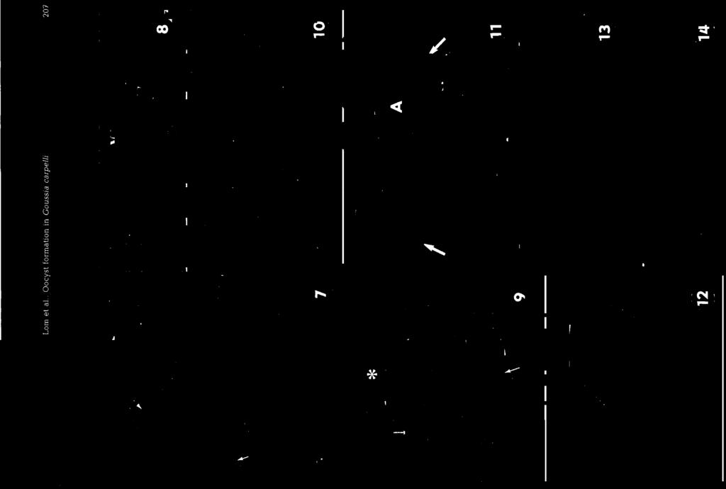
1 Vol. 10: , 1991 DISEASES OF AQUATIC ORGANISMS Dis. aquat. Org. Published May 8 Oocyst formation in the coccidian parasite Goussia carpelli 'Institute of Parasitology, Czech Academy of Sciences, BraniSovska ceske Budgjovice, Czechoslovakia '~ish Diseases Research Unit, School of Veterinary Medicine. Bischofsholer Damm 15, W-3000 Hannover 1, Germany ABSTRACT. The ultrastructural features of sporogony of Goussia carpelli (Leger & Stankovich) are described from the intestine of laboratory-infected carp. Zygotes were encased by a bilayered membrane (about 45 nm thick), which could be taken for the outer cell boundary of the oocyst. No indications were found that the parasitophorous vacuole membrane of the host cell contributed to oocyst wall formation. The formation of 4 sporoblasts from the sporont appeared to occur simultaneously. Initially, the sporoblasts were enveloped by a unit membrane and later were covered by a fine bilayered envelope which then transformed into a 120 nm thick sporocyst wall. The sporocyst wall formed a bivalved shell; the valves were joined by a continuous suture visible in the scanning electron microscope as a slightly prominent longitudinal fold. The sporozoites started to develop at opposite sides of the residual body within the sporoblasts, and in more advanced stages 2 sporozoites lying side by side could be observed. The sporozoites were morphologically similar to sporozoites of other Coccidia. During sporulation, the host cell lost its integrity, detenorated and turned to an almost structureless mass whlch appeared as the 'yellow bodies' known from light microscopy. INTRODUCTION Goussia (syn. Eimeria) carpem (Leger & Stankovich, 1921) Dykova & Lom, 1983 is a coccidian parasite of intestinal epithelia1 cells of common carp. In hatchery populations, mainly juvenile fish have been found to be lnfected (Ivasik & Kulakovskaya 1959, Zaika & Kheisin 1959, Kocylowski et al. 1976), and information on biology and morphological features of developmental stages of G. carpelli has been given by Leger & Stankovitch (1921), Kent & Hedrick (1985) and Steinhagen et al. (1989), while notes on the ultrastucture of oocysts and sporocysts have been reported by Lom & Dykova (1982a). In the present communication ultrastructural observations on sporogonic developmental stages of Goussja carpelli from laboratory-infected carp and from carp with spontaneous infections are presented, and the formation of 'yellow bodies' is discussed. allowing them to feed on Tubifex tubifex infected with G. carpelli. On Days 6 to 10 post-infection (PI) (at 20 "C), fish were dissected and the first 5 mm of the intestine was removed and fixed for electron microscopy at 4 OC overnight with 2.5 % glutaraldehyde in 0.1 M sodium cacodylate buffer at ph 7.2, postfixed with 1 % Os04 in the same buffer and then stained en bloc with uranyl acetate. After dehydration in a series of graded ethanol, the tissue was embedded in Spurr's resin. Additional samples from spontaneously infected carp fingerlings were fixed for 90 min at 4 "C in 0.1 M cacodylate-buffered 2 % OsO, and embedded in Epon- Araldite mixture. For scanning electron microscopy, samples of isolated sporocysts were fixed in 2 % osmic acid buffered with 0.1 M sodium cacodylate, criticalpoint dried and examined in a Tesla 400B scanning microscope. Ultrathin sections were stained with uranyl acetate and lead citrate and studied in Zeiss EM 10A, Jeol loob and Phillips 420 electron microscopes. MATERIAL AND METHODS RESULTS Common carp Cyprinus carpio raised from eggs in Macrogametes and sporulating oocysts were the laboratory were infected with Goussia carpelli at observed in cells of the intestinal epithelium and the the age of 3 to 4 n~o by fecal contamination or by lamina propria 6 to 10 d PI. The process of fertilization O Inter-Research/Printed in Germany
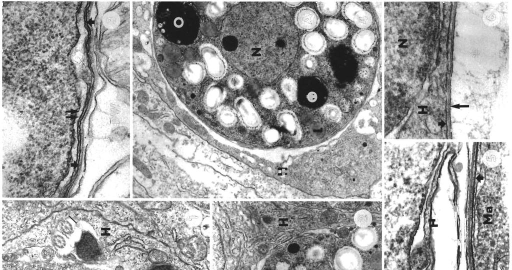
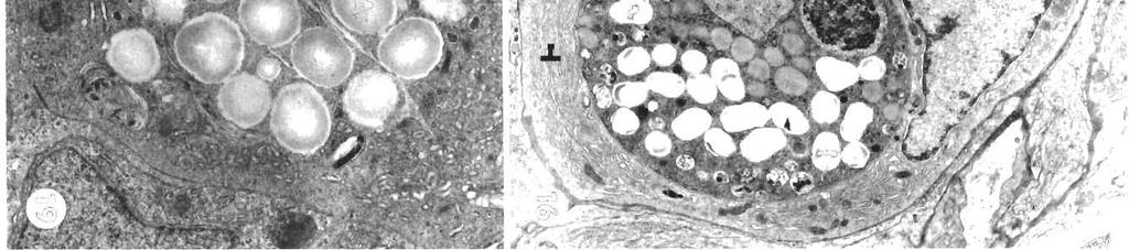
2 P- 204 Dis. aquat. Org. itself could not be observed. However, microgametes were observed in the cytoplasm of the host cell harbouring the macrogamete (Fig. 1). The cytoplasm of macrogametes with a central nucleus contained numerous amylopectin granules and prominent osmiophilic material full of small lucent vesicles. In what appeared to be zygotes (Fig. 2), these aggregates became condensed, more uniformly opaque and later became reduced in size and less opaque. The parasitophorous vacuole was reduced to a narrow space. In the zygote, the unit membrane at the cell surface (Fig. 2) transformed into or was replaced by a bilayered membrane, about 45 nm thick (Figs. 3 & 4). Beneath this membrane, stretches of opaque membrane started to appear which were contiguous with, or underlain by, cisternae of endoplasmic reticulum (Fig. 3). Later, these stretches fused into what could be taken for the outer cell boundary of the young oocyst; the narrow gap (about 70 to 260 nm) between this boundary and the bilayered membrane appeared to be filled with an amorphous substance. There was no indication that host cell membranes could participate in formation of oocyst boundary. In some instances (or at a certain stage of development?) the bilayered membrane was covered by another loosely undulating membrane (Fig. 5). In the course of later development, the bilayered membrane, which is the actual oocyst wall, became closely apposed to the host cell membrane, i.e. to the parasitophorous vacuole membrane. The host cell mostly formed a narrow band around the parasite (Figs. 4 & 6). Sometimes, however, the space between the oocyst wall and the membrane of the parasitophorous vacuole was filled with a dense mass of various vesicles derived from the host cell (Fig. 7), or just by an amorphous substance (Fig. 8). Before the onset of sporulation, the sporont was covered by a simple cell membrane (Fig. 8). Formation of the 4 sporoblasts appeared to occur simultaneously (Fig. 7). After the division was completed, amylopectin granules, osmiophilic inclusions now of homogeneous appearance, endoplasmic reticulum and free ribosomes could be seen in the sporoblasts. The sporoblasts appeared enveloped by a cover formed by a unit membrane (Fig.?), which started to form at the moment of cleaving of the sporont. Eventually, the sporoblasts each have their own membranous envelope; the space inside it may be filled with a foamy substance. Additional membranes delimit empty areas around the sporoblasts (Fig. g), while the rest of the oocyst space is filled with a similar foamy material (Figs. 10 & 11). These membranes may still persist after the firm oocyst walls have been formed (Fig. 14). Next, the sporoblasts were covered by a fine bilayered envelope (Fig. 11), which transforms (Figs. 13 & 14) into the thick sporocyst wall. The sporocyst wall consisted of a thick (about 63 nm) layer transversally striated in intervals of 12 nm; this layer grew wider along the sutural line. The striated layer is covered with a rather thin (15 nm) (Fig. 15) lucent layer subtending on opaque, coarsely granular coat. Isolated sporocysts observed in the scanning electron microscope have a smooth wall with a slightly prominent longitudinal fold representing the suture, the line of dehiscence of the 2 sporocyst shell valves. No membranous structures are attached (Fig. 12). The sporoblast forms 2 sporozoites that split off from the remaining sporoblast material, which becomes a sporocyst residuum. Cross sections of early stages show 2 developing sporozoites at opposite sides of the sporoblast. In sections of more advanced stages, 2 sporozoites lying side by side are arranged head to tail. The sporozoites exhibit an apical complex with 2 polar rings, conoid, micronemes, rhoptries, dense bodies, refractile bodies, nucleus and amylopectin inclusions. While the oocyst sporulated, the host cell, revealing at first almost normal structure (Fig. 16), gradually lost its integrity, and all its constituents deteriorated (Fig. 17). The cytoplasm condensed, turning eventually into an almost structureless opaque mass. Mature oocysts were then expelled together with the host-cell remains, which appear as the yellow bodies well known from light microscopy. There may be one to several oocysts within a yellow body (Fig. 18), which can originate from one or several host cells fused together. In most infections, especially the heavier ones, one can observe in the cells of the host epithelium aggregations of amylopectin granules, usually together with Figs. 1 to 6. Oocyst formation of Goussia carpelli infecting Cyprinus carpio. hg. Microgametes (arrow points to their nuclei) in the host-cell cytoplasm (H) next to a macrogamete X a A zygote enveloped by a simple cell membrane. x Flq A zygote enveloped by a bilayered membrane (long arrow) underlain by a continuous opaque layer (short arrow) subtended by a cisterna of endoplasmic reticulum (double arrow). X Flg. A zygote in process of membrane formation, within a host cell forming a narrow band around the parasite, X Fig. Bilayered oocyst wall (long arrow) underlain by tbe opaque layer (short arrow) and covered by a sllghtly wavy double membrane [double arrow). The oocyst wall faces the host cell in form of a thln, vacuolated envelope. X Fig. Oocyst wall (long arrow) closely apposed to the parasitophorous vacuole membrane (short arrow). Asterisk designates the free space in the oocyst. X Abbreviations used in figures. A: amylopectin granules; H: host cell; L: lipid inclus~on; Ma: macrogamete; N: nucleus; 0: osmiophil inclusion; PV. parasitophorous vacuole; PW: parasitophorous vacuole wall; S: sporoblast; W: oocyst wall
3
; in some cases, nothing but a group of amylopectin granules is left.")

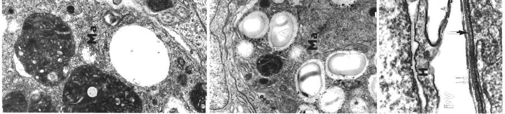
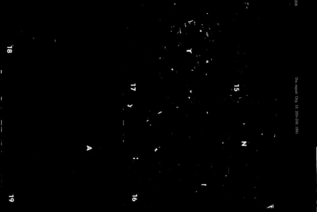
4 206 Dis. aquat. Org. 10: , 1991 islands of deteriorated cytoplasm. These groups of granules may occur alone or associated with groups of oocysts, always intracellularly. Usually they lie within remains of a structure representing obviously a deteriorated zygote (Fig. 19); in some cases, nothing but a group of amylopectin granules is left. DISCUSSION Oocyst wall formation in Goussia carpelli seems to be relatively simple. A bilayered envelope is formed at the surface of the zygote. After a new sporont outer-shell membrane has been formed, evidently with participation of the subtending endoplasmic reticulum, the oiiayered enveiope is detached and forms the actual oocyst wall. This simplicity may be misleading, since actual formation of the bilayered structure could not be observed, and since there may be some variation in the structures - the uneven membrane connection, the bilayered structure, and 'filamentous material' similar to that of the parasitophorous vacuole observed by Paterson & Desser (1981) in G. iroquoina were only occasionally observed. None of the previous papers on oocyst wall formation in picsine coccidia could exactly describe their origin. That is probably why some investigators (e.g. Hawkins et al. 1983, Morrison & Poynton 1989) claimed that the outer layer of the oocyst wall originated from the host membrane of the parasitophorous vacuole. If it is true that the typically thin oocyst wall of piscine coccidia has a mixed origin from host and parasite cell membranes, such a unique feature should also be reflected in the taxonomy of these parasites. However, our observations on G. carpelli [and other species examined by the present authors (G. gasterostei; unpubl.)] could not prove such a construction of the oocyst wall. The proximity of the bilayered membrane and the parasitophorous membrane wall which was observed in some cases might perhaps even in G. carpelli produce the false impression of an oocyst wall of mixed origin. In other piscine coccidia, 1 or 2 envelopes were seen to enclose the zygotes; this is the case for Goussia iroquoina (Paterson & Desser 1984) and Eimeria laureleus (Desser & Li 1984), with 3 envelopes in Goussia zarnowskii (Jastrzebski & Komorowski 1990). In G. cichlidarum, there are 2 membranes covering the zygote (Paperna & Landsberg 1985, Paperna et al. 1986), which precede the appearance of a soft but thick oocyst wall. The dark bodies in the zygotes of Goussia carpelli are reminiscent of inclusions which arise in G. iroquoina from the confluence of dense, membrane-bound vesicles (Paterson & Desser 1981). In this species, the oocyst wall is also underlain by endoplasmic reticulum cisternae, probably giving rise to sporont membrane as in G. carpelli and G. zarnowskii (Jastrzebsh & Komorowski 1990). Thus the pattern of zygote development in piscine coccidia - at least in several species - has similar features. One of these features is also the membranous cover of the sporoblasts, observed in sectioned Goussia carpelli oocysts in the process of sporulation. Such membranous envelopes were observed in other species (G. caseosa; Lom & Dykova 1982b). In G. carpelfi they are obviously impermanent, since they were not observed in sectioned sporulated oocysts. Also, the scanning electron microscope did not reveal any of them on the surface of the sporocysts, and no membranous girdles were associated with the sporocyst suture. However, the sporocyst of G. degustii does bear such a membranous girdle along the line of dehiscence of its shell valves. Similar sutural veils were found to persist on G, sinensis sporocysts. The possible significance of the membranous covers in G. carpelli, if any, has to be elucidated. Our observations unequivocally prove that the 'yellow bodies' harbouring oocysts shed from the epithelium are nothing but remains of deteriorated host cells, in which a process of lipofuscin formation took place as a result of deterioration of host-cell cytoplasm. Previous assumptions that they were cavities filled by tissue fluids or blood (Schaperclaus 1954) or degenerated host cell and coagulated tissue fluid (Molnar 1984) were not absolutely correct. Our interpretation is closer to that of Kent & Hedrick (1985), who saw the origin of yellow bodies in degenerated cell membranes and also suggested the presence of lipofuscin. Figs. 7 to 14. Goussia carpel11 infecting Cyprinus carpio. Fig. 7. Simultaneous formation of 4 sporoblasts by deep surface invaginations, they are enveloped by a b. pellicle (arrow) The parasitophorous vacuole is replete with a mass of moderately opaque vesicles. X Fig. 8. In some cases, the parasitophorous vacuole 1s filled wth an amorphous substance; the young oocyst is covered by a simple membrane. x Fig. A membranous pellicle of the sporoblasts envelops a foamy substance (arrow); asterisk marks the empty space surrounded by another thin membrane X Fig. 10. Enlarged central part of Fig 9; membranous septa around the cell, membrane-bound sporoblasts and the foamy substance (asterisk); X Fig. 11. More advanced sporoblasts with the initial membranous envelopes (long arrows) still present; short arrows point to the spore shell in formation. X Fig. 12. Scanning electron microscope micrograph of a mature sporocyst. X Fig. 13. Sporoblasts with a still thin, bilayered shell covered ~ t flocculent h dense material and with cell membranes (arrows) still in close proximity. X Fig. 14: Advanced formation of sporocyst shells, with the in~tial sporoblast envelopes still visible (arrow) between the 2 shells. X

5

6
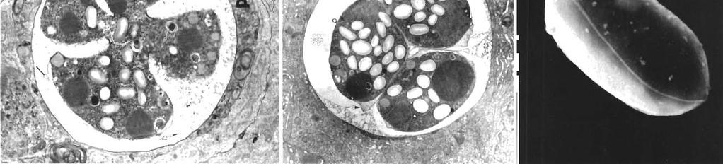
7 Lom et a1 Oocyst formation in Goussla carpelli 209 Figs. 15 to 19. Goussia carpel11 infecting Cyprinus carpio Fig 15. Wall of mature sporocyst Inner layer exhlbits periodic transversal striation. X Fiqs. 16 to 18. Formation of yellow bodies. Fig. 16. Sporulating oocyst incorporated in a host cell with extended endoplasmic reticulum cisternae (arrow) X Fig. 17. Oocyst with sporoblasts in a deteriorated host cell. X Fiq. 18. Several oocysts in deteriorated host cells expelled together, forming large aggregates (yellow bodies). Light micrograph, fresh preparation, X 1300 Fiq 19. Abortlve development of oocysts: amylopectin granules and remains of a deteriorated zygote in a host cell X 6700 Our observations indicate that large yellow bodies pathology of Gouss~a carpelli (Leger and Stankovitch) in containing more than 1 or 2 oocysts can anse by fusion goldfish Carassius auratus (L~nnaeus) Fish Path. 20: of 2 or more apposed parasitized cells. Yellow bodies Kocylowski, B., Zelazny, J., Antychowicz, J. (1976). Incidence are a common phenomenon in many epithelium-infect- of carr, coccidlosis and its control Bull. vet Inst. Pulawv ing Goussja and Eimerja species with smaller oocysts 20: (e.g. Goussia luciae, Ejmena ivanae, E. lajrdi). Species Leger, L., Stankovich, S. (1921). L'enterite coccidienne des with larger and/or fragile oocysts, such as Goussia alevins de carpe. Trav. Lab. Pisclcult. Univ. Grenoble sube~ithelialis from common carp1 shed their oocysts Lam, J., Dykov6, 1, (1982a). Goussia - structure of without yellow bodies, or only released sporocysts are sporocysts and 'yellow bodies'. J. Protozool. 29: 508 discharged. (Abstract 139) Findings on deteriorated zygotes of Goussia carpellj, Lom, J., Dykova. I. (1982b). Some marine fish coccidia of the genera Eimena Schneider, Epieimena Dykova & Lom and in which further development was aborted, put in seri- Goussia Labbe. J. Fish. Dis. 5: Ous doubt an assumption of Basks & On Lom.. J.., Dvkov6. >. I... LhotBkov6 S Kudoa lunata n SD. \, the expulsion of a part of the zygote material from (Myxozoa, Myxosporea) and notes on the nature of muscusporulatinq oocysts. This material - appeannq - as lar 'cysts' of the genus Kudoa Arch. Protistenk A. bodes, distinguished only by or consisting only of Molnar, K. (1984). Some peculiarities of oocyst rejection of fish amylopectin granules - can be identified as the Coccidia. Syn~posia Biol. Hung remains of aborted ooc~sts. It is by no means a Mornson. C M.. Povnton, S L (1989). A new s~ecies of phenomenon associated with normal zygote develop- Goussia (~~icomp'lexa, ~occidia) in ;he kidney kbules of ment. Abortive development is a phenomenon well cod, Gadus morhua L J Fish Dis Papernat I., Landsberg, J. H. (1985). Ultrastructure of oogony known in other fish protozoan parasites, such as myxand sporogony In Goussia cichlldarum Landsberg and osporidans (Lom et al. 1983). Acknowledgements. Part of this work was camed out using the equipment of the electron microscope worlung group of the Botany Department, School of Vetennary Medicine, Hannover. Supported by DFG grant # STE 420/1 to D.S. LITERATURE CITED Baska, F., Molnar, K. (1989). Ultrastrucutral observations on different developmental stages of Goussia sinensls (Chen, 1955), a parasite of the silver carp (Hypophthalmichthys molibix Valenciennes, 1844). Acta vet. hung. 37: Desser, S. S., Li, L. (1984). Ultrastructural observations on the sexual stages and oocyst formation in Eimeria laureleus (Protozoa, Coccidia) of perch, Perca flavescens, from Lake Sasajewun, Ontano. Z. ParasitKde 70: Hawkins, W. S., Foumie, J. W., Overstreet, R. M. (1983). Organization of sporulated oocysts of Eimena fundtdi in the gulf Wfish, Fundulus grands. J. Parasit. 69: Ivasik, V., Kulakovskaya, 0. P. (1959). On carp coccidosis. Zool. Zh. Ukr. 38: (in Russian) Jastrzebsh, M., Komorowski, Z. (1990). Light and electron microscopic studies in Goussia zarnowsh (Jastrzebski, 1982): an intestinal coccidium parasitizing the threespined stickleback. J. Fish Dis. 13: 1-24 Kent, M. L., Hedrick, R. P. (1985). The biology and associated Paperna, 1985, a coccidian parasite in the swimbladder of cichlld fish. Protistologica 21: Paperna, I., Landsberg, J. H., Femstein, N. (1986). Ultrastructure of the macrogamont of Goussia cichlidarum Landsberg and Paperna, 1985, a coccidian parasite in the swmbladder of cichlid fish. Annls Parasit. hum. conlp. 61: Paterson, W. B., Desser, S. S. (1981). Ultrastructure of macrogametogenesis, macrogametes and young oocysts of Eimeria lroquolna Molnar and Fernando, 1974 in experimentally infected fathead minnows (Pimephalespromelas, Cyprinidae). J. Parasit Paterson, W B., Desser, S. S. (1984). Ultrastructural observations on fertilization and sporulation in Goussla iroquoina (Molnar and Fernando, 1974) in experimentally infected fathead minnows (Pimephales promelas, Cyprinidae). J. Parasit. 70: Schaperclaus, W (1954). Fischkrankheiten, 3rd edn. Akademie Verlag, Berlin Steinhagen, D., Kortlng, W., van Muiswinkel, W. B. (1989). Morphology and biology of Goussia carpell1 (Protozoa: Apicomplexa) from the intestine of experimentally infected common carp Cyprinus carpio. Dis. aquat. Org. 6: Zaika, V E., Kheisin, E. M. (1959). Carp coccidiosis on the Valdai fish farm Bull. State Scient. Res Inst. Lake and River Fishenes, 49. Parasites of freshwater fish and the biological basis of their control, Leningrad (Translated from Russian by Israel Prog. Scient. Transl., 2nd edn., 1965: p ) Editorial responsibd~ty: Managing Editor Manuscript first received: January 16, 1991 Rerrlsed version accepted: March 25, 1991
DISEASES OF AQUATIC ORGANISMS Dis. aquat. Org.
 Vol. 10: 121-125. 1991 DISEASES OF AQUATIC ORGANISMS Dis. aquat. Org. Published April 4 Ultrastructural observations on sporozoite stages of piscine Coccidia: Goussia carpelli and G. subepithelialis from
Key words: Coccidia, Choleoeimeria rochalimai, fine structure, gall bladder epithelium, Hemidactylus mabouia, Brazil
 FOLIA PARASITOLOGICA 47: 91-96, 2000 Ultrastructural study of meronts and gamonts of Choleoeimeria rochalimai (Apicomplexa: Eimeriidae) developing in the gall bladder of the gecko Hemidactylus mabouia
Light and electron microscopic study of the pathology and merogony of Goussia gadi (Apicomplexa: Coccidia) in the swimbladder wall
 in the swimbladder wall") Vol. 17: 113-125.1993 DISEASES OF AQUATIC ORGANISMS Dis. aquat. Org., Published November 18 I Light and electron microscopic study of the pathology and merogony of Goussia gadi (Apicomplexa: Coccidia)
Vol. 17: 113-125.1993 DISEASES OF AQUATIC ORGANISMS Dis. aquat. Org., Published November 18 I Light and electron microscopic study of the pathology and merogony of Goussia gadi (Apicomplexa: Coccidia)
Phylum:Apicomplexa Class:Sporozoa
 Phylum:Apicomplexa Class:Sporozoa The most characteristic features of sporozoa are 1-unique appearance of most protozoa makes it possible for knowledge able person to identifiy them to level of genus and
EFFICACY OF SOME ANTICOCCIDIAL DRUGS FOR TREATING COCCIDIAL ENTERITIS OF THE COMMON CARP CAUSED BY GOUSSIA CARPELLI (APICOMPLEXA: EIMERIIDAE)
") Acta Veterinaria Hungarica 55 (1), pp. 67 76 (2007) DOI: 10.1556/AVet.55.2007.1.7 EFFICACY OF SOME ANTICOCCIDIAL DRUGS FOR TREATING COCCIDIAL ENTERITIS OF THE COMMON CARP CAUSED BY GOUSSIA CARPELLI (APICOMPLEXA:
Acta Veterinaria Hungarica 55 (1), pp. 67 76 (2007) DOI: 10.1556/AVet.55.2007.1.7 EFFICACY OF SOME ANTICOCCIDIAL DRUGS FOR TREATING COCCIDIAL ENTERITIS OF THE COMMON CARP CAUSED BY GOUSSIA CARPELLI (APICOMPLEXA:
Fine structure of Eimeria (S. l.) vanasi merogony stages in the intestinal mucosa of cichlid fishes
 vanasi merogony stages in the intestinal mucosa of cichlid fishes") Vol. 10: 195-201. 1991 DISEASES OF AQUATIC ORGANISMS Dis. aquat. Org. Published May 8 Fine structure of Eimeria (S. l.) vanasi merogony stages in the intestinal mucosa of cichlid fishes Ilan Paperna Department
Vol. 10: 195-201. 1991 DISEASES OF AQUATIC ORGANISMS Dis. aquat. Org. Published May 8 Fine structure of Eimeria (S. l.) vanasi merogony stages in the intestinal mucosa of cichlid fishes Ilan Paperna Department
The Fine Structure of the Endogenous Stages of Isospora hemidactyli Carini, 1936 in the Gecko Hemidactylus mabouia from North Brazil
 Mem Inst Oswaldo Cruz, Rio de Janeiro, Vol. 95(1): 43-47, Jan./Feb. 2000 The Fine Structure of the Endogenous Stages of Isospora hemidactyli Carini, 1936 in the Gecko Hemidactylus mabouia from North Brazil
Ultrastructure of Endogenous Stages of Eimeria ninakohlyakimovae Yakimoff & Rastegaieff, 1930 Emend. Levine, 1961 in Experimentally Infected Goat
 Mem Inst Oswaldo Cruz, Rio de Janeiro, Vol. 92(4): 533-538, Jul./Aug. 1997 Ultrastructure of Endogenous Stages of Eimeria ninakohlyakimovae Yakimoff & Rastegaieff, 1930 Emend. Levine, 1961 in Experimentally
Protozoan Parasites of Veterinary importance 2017
 Protozoan Parasites of Veterinary importance 2017 VPM-122 Laboratory 4 Spencer J. Greenwood PhD, DVM Dept. of Biomedical Sciences Room 2332N AVC North Annex sgreenwood@upei.ca Office phone # 566-6002 To
Key words: Plasmodium, Kentropyx calcarata, Brazil, merogony, gametocytes, ultrastructure
 FOLIA PARASITOLOGICA 49: 2-8, 2002 Fine structure of erythrocytic stages of a Plasmodium tropiduri-like malaria parasite found in the lizard Kentropyx calcarata (Teiidae) from north Brazil Ilan Paperna
Revajová, Viera, Loószová, Adrian. The Journal of Protozoology Resea Citation RightsNational Research Center for Prot
 ' ' Morphological study of partridge Title development in the foreign host - (Gallus gallus) Revajová, Viera, Loószová, Adrian Author(s) Maria, Zibrín, Martin, Herich, Ro Mikulas The Journal of Protozoology
The specimens of Ameiva ameiva (Linn) were
 were") Article available at http://www.parasite-journal.org or http://dx.doi.org/10.1051/parasite/1999064359 FINE STRUCTURE OF THE EPICYTOPLASMIC EIMERID COCCIDIUM ACROEIMERIA PINTOI LAINSON & PAPERNA, 1999,
Article available at http://www.parasite-journal.org or http://dx.doi.org/10.1051/parasite/1999064359 FINE STRUCTURE OF THE EPICYTOPLASMIC EIMERID COCCIDIUM ACROEIMERIA PINTOI LAINSON & PAPERNA, 1999,
HISTOPATHOLOGY. Introduction:
 Introduction: HISTOPATHOLOGY Goats and sheep are the major domestic animal species in India. Much of the economy of the country has been depend upon the domestication of these animals. Especially economy
Extra-intestinal localization of Goussia sp. (Apicomplexa) oocysts in Rana dalmatina (Anura: Ranidae), and the fate of infection after metamorphosis
 oocysts in Rana dalmatina (Anura: Ranidae), and the fate of infection after metamorphosis") DISEASES OF AQUATIC ORGANISMS Vol. 70: 237 241, 2006 Published June 23 Dis Aquat Org Extra-intestinal localization of Goussia sp. (Apicomplexa) oocysts in Rana dalmatina (Anura: Ranidae), and the fate
DISEASES OF AQUATIC ORGANISMS Vol. 70: 237 241, 2006 Published June 23 Dis Aquat Org Extra-intestinal localization of Goussia sp. (Apicomplexa) oocysts in Rana dalmatina (Anura: Ranidae), and the fate
cyst&' appeared to be of two kinds-one smaller and Smnith "is inclined to regard these epithelial cell parasites as
 COCCIDIA IN SUBEPITHELIAL INFECTIONS OF THE INTESTINES OF BIRDS PHILIP B. HADLEY From the Agricultural Experiment Station of the Rhode Island State College' Received for publication, July 10, 1916 In an
Biology of toxoplasmosis
 1 Biology of toxoplasmosis E. Petersen 1 and J. P. Dubey 2 1 Statens Seruminstitut, Copenhagen, Denmark 2 U.S. Department of Agriculture, Beltsville, USA History Toxoplasma gondii is a coccidium, with
"Comments on the nature and methods of collection of fish coccidia. " - Molnár, K. - Parasit. Hung. _
 Parasit. Hung 10. 1977. Comments on the Nature and Methods of Collection of Fish Coccidia Dr. Kálmán MOLNÁR Research Institute for Veterinary Science, Hungarian Academy of Sciences, Budapest "Comments
Ëtude ultrastructurale de la mérogonie de Schellackia cf. agamae (Lankesterellidae, Apicomplexa) chez le Lézard Agama stellio.
 chez le Lézard Agama stellio.") Masson, Paris, 1987 Ann. Parasitol. Hum. Comp. 1987, 62, n 5, pp. 380-386. ULTRASTRUCTURAL STUDIES ON THE MEROGONY OF SCHELLACKIA CF. AGAMAE (LANKESTERELLIDAE, APICOMPLEXA) FROM THE STARRED LIZARD AGAMA
Masson, Paris, 1987 Ann. Parasitol. Hum. Comp. 1987, 62, n 5, pp. 380-386. ULTRASTRUCTURAL STUDIES ON THE MEROGONY OF SCHELLACKIA CF. AGAMAE (LANKESTERELLIDAE, APICOMPLEXA) FROM THE STARRED LIZARD AGAMA
Sarcocystis heydorni, n. sp. (Apicomplexa: Protozoa) with cattle (Bos taurus) and human
 with cattle (Bos taurus) and human") 1 Sarcocystis heydorni, n. sp. (Apicomplexa: Protozoa) with cattle (Bos taurus) and human (Homo sapiens) cycle Jitender P. Dubey 1, Erna van Wilpe 2, Rafael Calero-Bernal 1, Shiv Kumar Verma 1, Ronald
1 Sarcocystis heydorni, n. sp. (Apicomplexa: Protozoa) with cattle (Bos taurus) and human (Homo sapiens) cycle Jitender P. Dubey 1, Erna van Wilpe 2, Rafael Calero-Bernal 1, Shiv Kumar Verma 1, Ronald
Apicomplexans Apicomplexa Intro
 Apicomplexans Apicomplexa Intro Cryptosporidium Apicomplexan Select Characteristics Gliding motility Apical Complex organelle for invasion of host cell Life cycle alternates b/w sexual and asexual phases
Article available at or
 Article available at http://www.parasite-journal.org or http://dx.doi.org/10.1051/parasite/1998051017 PSEUDOKLOSSIA SEMILUNA N. SP. (APICOMPLEXA: AGGREGATIDAE): A COCCIDIAN PARASITE OF THE KIDNEY OF BLUE
Protozoa. Apicomplexa Sarcomastigophora Ciliophora. Gregarinea Coccidia Piroplasma
 Protozoa Apicomplexa Sarcomastigophora Ciliophora Gregarinea Coccidia Piroplasma Coccidia characterized by thick-walled oocysts excreted in feces In Humans Cryptosporidium Isospora Cyclospora Sarcocystis
DISEASES OF AQUATIC ORGANISMS Dis. aquat. Org.
 Vol. 7: 149-153, 1989 DISEASES OF AQUATIC ORGANISMS Dis. aquat. Org. Published October 26 Developmental cycle of chelonian haemogregarines in leeches with extra-intestinal multiple sporozoite oocysts and
' Published September 29
 Vol. 7: 1-12, 1989 l DISEASES OF AQUATIC ORGANISMS Dis. aquat. Org. ' Published September 29 Nodular and epicellular coccidiosis in the intestine of cyprinid fishes Kalman Molnar Veterinary Medical Research
PLASMODIUM MODULE 39.1 INTRODUCTION OBJECTIVES 39.2 MALARIAL PARASITE. Notes
 Plasmodium MODULE 39 PLASMODIUM 39.1 INTRODUCTION Malaria is characterized by intermittent fever associated with chills and rigors in the patient. There may be enlargement of the liver and spleen in the
Coccidia. Nimit Morakote, Ph.D.
 Coccidia Nimit Morakote, Ph.D. 1 Learning objectives After class, students will be able to: Describe morphology, life cycle, signs and symptoms, prevention and control, laboratory diagnosis and treatment
Title. CitationJapanese Journal of Veterinary Research, 24(1-2): 37. Issue Date DOI. Doc URL. Type. File Information
: 37. Issue Date DOI. Doc URL. Type. File Information") Title DISTRIBUTION OF LYMPHATIC TISSUES IN DUCK CAECA Author(s)KITAMURA, Hirokazu; SUGIMURA, Makoto; HASHIMOTO, Yos CitationJapanese Journal of Veterinary Research, 24(1-2): 37 Issue Date 1976-05 DOI 10.14943/jjvr.24.1-2.37
Title DISTRIBUTION OF LYMPHATIC TISSUES IN DUCK CAECA Author(s)KITAMURA, Hirokazu; SUGIMURA, Makoto; HASHIMOTO, Yos CitationJapanese Journal of Veterinary Research, 24(1-2): 37 Issue Date 1976-05 DOI 10.14943/jjvr.24.1-2.37
Effect of Sodium Hypochlorite on the Oocyst Wall of Eimeria tenella as Shown by Electron Microscopy1
 32 PROCEEDINGS OF THE HELMINTHOLOGICAL SOCIETY This alteration appeared similar to that observed by light microscopy (Figs. 5, 6). Literature Cited Dixon, K. E. 1966. The physiology of excystment of the
A NEW SPECIES OF GENUS EIMERIA (APICOMPLEXA: EUCOCCIDIORIDA) FROM GOAT.
 FROM GOAT.") A NEW SPECIES OF GENUS EIMERIA (APICOMPLEXA: EUCOCCIDIORIDA) FROM GOAT. B.V. More 1, H.A.Kamble. 2 S.V. Nikam 3, 1 Department of Zoology, Ramkrishna Paramhansa Mahavidyalaya, Osmanabad. (M.S.) India. 2
A NEW SPECIES OF GENUS EIMERIA (APICOMPLEXA: EUCOCCIDIORIDA) FROM GOAT. B.V. More 1, H.A.Kamble. 2 S.V. Nikam 3, 1 Department of Zoology, Ramkrishna Paramhansa Mahavidyalaya, Osmanabad. (M.S.) India. 2
Diagnosis, treatment and control: dealing with coccidiosis in cattle
 Vet Times The website for the veterinary profession https://www.vettimes.co.uk Diagnosis, treatment and control: dealing with coccidiosis in cattle Author : Adam Martin Categories : Vets Date : January
A Scanning Electron Microscopic Study of Eggshell Surface Topography of Leidynema portentosae and L. appendiculatum (Nematoda: Oxyuroidea)
") The Ohio State University Knowledge Bank kb.osu.edu Ohio Journal of Science (Ohio Academy of Science) Ohio Journal of Science: Volume 88, Issue 5 (December, 1988) 1988-12 A Scanning Electron Microscopic
The Ohio State University Knowledge Bank kb.osu.edu Ohio Journal of Science (Ohio Academy of Science) Ohio Journal of Science: Volume 88, Issue 5 (December, 1988) 1988-12 A Scanning Electron Microscopic
Studied tortoises, Testudo graeca, were collected from
 Article available at http://www.parasite-journal.org or http://dx.doi.org/10.1051/parasite/2006134267 HEMOLIVIA MAURITANICA (HAEMOGREGARINIDAE: APICOMPLEXA) INFECTION IN THE TORTOISE TESTUDO GRAECA IN
Joerg Kinne, Mansoor Ali*, Ulrich Wernery, and J. P. Dubey
 J. Parasitol., 88(3), 2002, pp. 548 552 American Society of Parasitologists 2002 CLINICAL LARGE INTESTINAL COCCIDIOSIS IN CAMELS (CAMELUS DROMEDARIUS) IN THE UNITED ARAB EMIRATES: DESCRIPTION OF LESIONS,
Myxosporeans and myxosporidiosis of allogynogenetic gibel carp (Carassius auratus gibelio Bloch) in China
 in China") Myxosporeans and myxosporidiosis of allogynogenetic gibel carp (Carassius auratus gibelio Bloch) in China Zhang Jinyong zhangjy@ihb.ac.cn Laboratory of Fish Diseases; Institute of Hydrobiology (IHB), Chinese
Myxosporeans and myxosporidiosis of allogynogenetic gibel carp (Carassius auratus gibelio Bloch) in China Zhang Jinyong zhangjy@ihb.ac.cn Laboratory of Fish Diseases; Institute of Hydrobiology (IHB), Chinese
Myxosporeans and myxosporidiosis of common carp and gibel carp in China
 Myxosporeans and myxosporidiosis of common carp and gibel carp in China Zhang Jinyong, Liu Xinhua, Xi Bingwen, Kálmán Molnár zhangjy@ihb.ac.cn Hungary 2015 June.3 Laboratory of Fish Diseases; Institute
Parasitenkunde. (Odocoileus virginianus ) Ultrastructure of Sarcocystis sp. from the Muscle of a White-Tailed Deer
 Ultrastructure of Sarcocystis sp. from the Muscle of a White-Tailed Deer") Z Parasitenkd (1982) 68 : 33-38 Zeitschrift for Parasitenkunde Parasitology Research 9 Springer-Verlag 1982 Ultrastructure of Sarcocystis sp. from the Muscle of a White-Tailed Deer (Odocoileus virginianus
Z Parasitenkd (1982) 68 : 33-38 Zeitschrift for Parasitenkunde Parasitology Research 9 Springer-Verlag 1982 Ultrastructure of Sarcocystis sp. from the Muscle of a White-Tailed Deer (Odocoileus virginianus
COCCIDIOSIS OF SANDHILL CRANES (GRUS CANADENSIS) WINTERING IN NEW MEXICO
 WINTERING IN NEW MEXICO") journal of Wtldltfe hemes, 22(1). 1986. pp 25-35 0 Wildlife Disease Association 1986 COCCIDIOSIS OF SANDHILL CRANES (GRUS CANADENSIS) WINTERING IN NEW MEXICO Brent B. Parker and Donald W. Duszynski Department
journal of Wtldltfe hemes, 22(1). 1986. pp 25-35 0 Wildlife Disease Association 1986 COCCIDIOSIS OF SANDHILL CRANES (GRUS CANADENSIS) WINTERING IN NEW MEXICO Brent B. Parker and Donald W. Duszynski Department
DISEASES OF AQUATIC ORGANISMS Vol. 62: , 2004 Published November 23 Dis Aquat Org
 DISEASES OF AQUATIC ORGANISMS Vol. 62: 133 145, 2004 Published November 23 Dis Aquat Org Cryptosporidium scophthalmi n. sp. (Apicomplexa: Cryptosporidiidae) from cultured turbot Scophthalmus maximus. Light
Redescription of Sarcocystis fusiformis sarcocysts from the water buffalo (Bubalus bubalis)
") Redescription of Sarcocystis fusiformis sarcocysts from the water buffalo (Bubalus bubalis) 1 J. P. DUBEY 1 *, M. HILALI 2,E.VANWILPE 3,S.K.VERMA 1,R.CALERO-BERNAL 1 and A. ABDEL-WAHAB 2 1 U. S. Department
Redescription of Sarcocystis fusiformis sarcocysts from the water buffalo (Bubalus bubalis) 1 J. P. DUBEY 1 *, M. HILALI 2,E.VANWILPE 3,S.K.VERMA 1,R.CALERO-BERNAL 1 and A. ABDEL-WAHAB 2 1 U. S. Department
Taxonomy of North American Fish Eimeriidae
 11 NOAA Technical Report NMFS 11 Taxonomy of North American Fish Eimeriidae Steve J. Upton, David W. Reduker, William L. Current, and Donald W. Duszynski August 1984 U.S. DEPARTMENT OF COMMERCE National
Ahead of print online version
 Folia Parasitologica 60 [3]: 232 236, 2013 ISSN 0015-5683 (print), ISSN 1803-6465 (online) Institute of Parasitology, Biology Centre ASCR http://folia.paru.cas.cz/ A new species of Choleoeimeria (Apicomplexa:
LABORATORY. The Protozoa. At the Bench
 LABORATORY Laboratory 8, Page 1 8 The Protozoa Introduction: The protozoa are unicellular animals that are classified on the basis of the organelles used for locomotion (flagella, pseudopodia, cilia or
Article available at or
 Article available at http://www.parasite-journal.org or http://dx.doi.org/10.1051/parasite/1996034341 DESCRIPTION AND ULTRASTRUCTURE OF LANKESTERELLA SPECIES INFECTING FROGS IN KENYA PAPERNA I.* & OGARA
BEAK AND FEATHER DYSTROPHY IN WILD SULPHUR-CRESTED COCKATOOS (CACATUA GALERITA)
") BEAK AND FEATHER DYSTROPHY IN WILD SULPHUR-CRESTED COCKATOOS (CACATUA GALERITA) Author(s): Steven McOrist, Douglas G. Black, David A. Pass, Peter C. Scott, and John Marshall Source: Journal of Wildlife
BEAK AND FEATHER DYSTROPHY IN WILD SULPHUR-CRESTED COCKATOOS (CACATUA GALERITA) Author(s): Steven McOrist, Douglas G. Black, David A. Pass, Peter C. Scott, and John Marshall Source: Journal of Wildlife
Observations on Eimeria species of Dasyprocta leporina (Linnaeus, 1758) (Rodentia: Dasyproctidae) from the state of Pará, North Brazil
 (Rodentia: Dasyproctidae) from the state of Pará, North Brazil") Mem Inst Oswaldo Cruz, Rio de Janeiro, Vol. 99: 000-000, 2004 1 Observations on Eimeria species of Dasyprocta leporina (Linnaeus, 1758) (Rodentia: Dasyproctidae) from the state of Pará, North Brazil Ralph
Mem Inst Oswaldo Cruz, Rio de Janeiro, Vol. 99: 000-000, 2004 1 Observations on Eimeria species of Dasyprocta leporina (Linnaeus, 1758) (Rodentia: Dasyproctidae) from the state of Pará, North Brazil Ralph
Parasitology Amoebas. Sarcodina. Mastigophora
 Parasitology Amoebas Sarcodina Entamoeba hisolytica (histo = tissue, lytica = lyse or break) (pathogenic form) o Trophozoite is the feeding form o Life Cycle: personfeces cyst with 4 nuclei with thicker
Prevention of experimentally induced whirling disease in rainbow trout Oncorhynchus mykiss by Fumagillin
 Vol. 10: 109-113, 1991 DISEASES OF AQUATIC ORGANISMS Dis. aquat. Org. Published April 4 Prevention of experimentally induced whirling disease in rainbow trout Oncorhynchus mykiss by Fumagillin M. El-Matbouli,
Criconemoides similis 1 G. W. BIRD ~
 Somatic Musculature of Trichodorus porosus and Criconemoides similis 1 G. W. BIRD ~ Abstract: The somatic musculature of Trichodorus porosus is transversely striated, and that of Criconemoides similis
A:Malaria (Plasmodium species) Plasmodium falciparum causes malignant tertian malaria P. malariae: causes Quartan malaria P. vivax: causes benign
 Plasmodium falciparum causes malignant tertian malaria P. malariae: causes Quartan malaria P. vivax: causes benign") A:Malaria (Plasmodium species) Plasmodium falciparum causes malignant tertian malaria P. malariae: causes Quartan malaria P. vivax: causes benign tertian malaria P. ovale: causes benign tertian malaria
A:Malaria (Plasmodium species) Plasmodium falciparum causes malignant tertian malaria P. malariae: causes Quartan malaria P. vivax: causes benign tertian malaria P. ovale: causes benign tertian malaria
Mesosomes are a definite event in antibiotic-treated Staphylococcus aureus ATCC 25923
 Tropical Biomedicine 24(1): 105 109 (2007) Mesosomes are a definite event in antibiotic-treated Staphylococcus aureus ATCC 25923 Santhana Raj, L. 1*, Hing, H.L. 2, Baharudin Omar 2, Teh Hamidah, Z. 1,
SUPPLEMENTARY INFORMATION
 doi:10.1038/nature11046 Supplementary Figure 1: Images of PB-positive cells in the subepidermal region (a-i) Representative images of PB positive cells in the subepidermis of the upper beak of the pigeon.
AN ULTRASTRUCTURAL STUDY OF THE DEVELOPMENT OF BABESIA. E. F. BLOUIN and LYNN VAN RENSBURG, Veterinary Research Institute, Onderstepoort OliO
 OnderstepoortJ. vet. Res., 55, 93-100(1988) AN ULTRASTRUCTURAL STUDY OF THE DEVELOPMENT OF BABESIA OCCULTANS INTHESALIVARYGLANDSOF ADULT HYALOMMA MARGINATUM RUFIPES E. F. BLOUIN and LYNN VAN RENSBURG,
Coccidiosis in macropods and other species
 Coccidiosis in macropods and other species Author: Derek Spielman Wildlife Assistance and Information Foundation; Sydney School of Veterinary Science, the University of Sydney Abstract This presentation
Both Rod and Cone Disc Shedding ore Related to Light Onset in the Cat
 Both Rod and Cone Disc Shedding ore Related to Light Onset in the Cat Sreven K. Fisher,* Bruce A. Pfeffer.f and Don H. Anderson* Nineteen domestic cats were entrained to a 12-hr light/12-hr dark lighting
Biology of Isospora spp. from Humans, Nonhuman Primates, and Domestic Animals
 CLINICAL MICROBIOLOGY REVIEWS, Jan. 1997, p. 19 34 Vol. 10, No. 1 0893-8512/97/$04.00 0 Copyright 1997, American Society for Microbiology Biology of Isospora spp. from Humans, Nonhuman Primates, and Domestic
1) Most common, infectious, pathogenic animal (zoonotic) parasite of humans; estimated that 13% of humans are infected
 Most common, infectious, pathogenic animal (zoonotic) parasite of humans; estimated that 13% of humans are infected") XX Phylum Apicomplexa (Chapter 8) 2005 A. Characteristics 1. All are parasitic 2. APICAL COMPLEX a. Group of organelles used to invade host cells b. Visible only with electron microscopy Picture Slide
XX Phylum Apicomplexa (Chapter 8) 2005 A. Characteristics 1. All are parasitic 2. APICAL COMPLEX a. Group of organelles used to invade host cells b. Visible only with electron microscopy Picture Slide
SCANNING electron - microscopy has
 Characteristics of the Absorptive Surface of the Small Intestine of the Chicken from 1 Day to 14 Weeks of Age 1 R. C. BAYER, C. B. CHAWAN, F. H. BIRD AND S. D. MUSGRAVE Department of Animal and Veterinary
STUDY OF EIMERIA INTRICATA IN GOAT AND SHEEP FROM BEED DISTRICT, MAHARASHTRA STATE INDIA
 STUDY OF EIMERIA INTRICATA IN GOAT AND SHEEP FROM BEED DISTRICT, MAHARASHTRA STATE INDIA More B.V., Kamble H.A. and Nikam S.V. 1 Department of Zoology, Ramkrishna Paramhansa Mahavidyalaya, Osmanabad. (M.S.),
A comparison of placental tissue in the skinks Eulamprus tympanum and E. quoyii. Yates, Lauren A.
 A comparison of placental tissue in the skinks Eulamprus tympanum and E. quoyii Yates, Lauren A. Abstract: The species Eulamprus tympanum and Eulamprus quoyii are viviparous skinks that are said to have
A Study of Coccidiosis in Livestock in the Island of Dominica. Joshua Santelises. Study Abroad Texas A&M University. Dr.
 A Study of Coccidiosis in Livestock in the Island of Dominica Joshua Santelises Study Abroad 2012 Texas A&M University Dr. Thomas Lacher Dr. Jim Woolley Abstract The following experiment was done to investigate
Lacerta viridis. Functional anatomy of the lungs of the green lizard, (Accepted 18 February 1977)
") J. Anat. (1978), 125, 2, pp. 421-431 421 With 9 figures Printed in Great Britain Functional anatomy of the lungs of the green lizard, Lacerta viridis C. MEBAN Department of Anatomy, The Queen's University
J. Anat. (1978), 125, 2, pp. 421-431 421 With 9 figures Printed in Great Britain Functional anatomy of the lungs of the green lizard, Lacerta viridis C. MEBAN Department of Anatomy, The Queen's University
DRAFT TANZANIA STANDARD
 Hatching eggs Specification DRAFT TANZANIA STANDARD TANZANIA BUREAU OF STANDARDS 1 Hatching eggs Specification TBS/AFDC 22 (5271) P3 0 FOREWORD This Tanzania standard was developed due to rapid increase
A Lymphosarcoma in an Atlantic Salmon (Salmo salar)
") A Lymphosarcoma in an Atlantic Salmon (Salmo salar) Authors: Paul R. Bowser, Marilyn J. Wolfe, and Timothy Wallbridge Source: Journal of Wildlife Diseases, 23(4) : 698-701 Published By: Wildlife Disease
A Lymphosarcoma in an Atlantic Salmon (Salmo salar) Authors: Paul R. Bowser, Marilyn J. Wolfe, and Timothy Wallbridge Source: Journal of Wildlife Diseases, 23(4) : 698-701 Published By: Wildlife Disease
Seasonal Variations of yeso sika Deer Skin and its Vegetable Tanned Leather
 Seasonal Variations of yeso sika Deer Skin and its Vegetable Tanned Leather Shigeharu Fukunaga, Akihiko Yoshie, Ikuo Yamakawa, Fumio Nakamura Laboratory of Animal By-product Science, Graduate School of
Zadar County Rural Development Agency, Zadar, Croatia. Fish Farm IHOR PARK, Jastrebarsko, Croatia
 . Veterinarski Arhiv 87 (1), 77-86, 2017 New data on Eimeria dicentrarchi (Apicomplexa: Eimeriidae), a common parasite of farmed European sea bass (Dicentrarchus labrax) from the mid-eastern Adriatic Emil
Sam R. Telford, Jr The Florida Museum of Natural History, University of Florida, Gainesville, Fl32611, USA
 Systematic Parasitology 23: 203-208, 1992. 0 1992 Kluwer Academic Publishers. Printed in the Netherlands. An eimeriid species (Apicomplexa: Eimeriidae) that parasitises the gallbladder and bile-duct of
Infecting Anopheles stephensi With Rodent Malaria Parasites Alida Coppi & Photini Sinnis
 Infecting Anopheles stephensi With Rodent Malaria Parasites Alida Coppi & Photini Sinnis A. Reagents: 1. DMEM or RPMI DMEM (4.5g/L glucose) RPMI 1640 Cellgro #MT-10-017-CM Cellgro #MT-10-040-CM 2. Giemsa
Fact sheet. All animals, particularly herbivores, appear to be natural hosts for coccidian species with a high degree of host specificity observed.
 Coccidia in k angaroos Fact sheet Introductory statement Coccidians are protozoan parasites which infect the intestinal tract of many animals. Within kangaroos, coccidia infections can lead to clinical
Article available at or
 Article available at http://www.parasite-journal.org or http://dx.doi.org/10.1051/parasite/1995023307 LIGHT AND ELECTRON MICROSCOPE STUDY OF A LANKESTERELLA PETITI N. SP., (APICOMPLEXA : LANKESTERELLIDAE)
Systemic Apicomplexans. Toxoplasma
 Systemic Apicomplexans Toxoplasma Protozoan Groups Historically, protozoa have been grouped by mode of motility. Flagellates Hemoflagellates Trypanosoma cruzi Leishmania infantum Mucoflagellates Tritrichomonas
HYDATID CYST DISEASE
 HYDATID CYST DISEASE Hydatid disease, also called hydatidosis or echinococcosis, is a cystforming disease resulting from an infection with the metacestode, or larval form, of parasitic dog tapeworms from
Apicomplexa of Intestinal Pathology
 LECTURES #4, #5 & #6: APICOMPLEXA 1 Apicomplexa of Intestinal Pathology Cryptosporidium, Eimeria, Cystoisospora General Characteristics of Apicomplexa A. Morphology by stage Zoite o Tear-shaped (cylindrical
This is the smallest tapeworm that can affect human being but it s not really proper human tapeworm (the human is not the primary host).
.") Echinococcus Granulosus Small Tapeworm (1 cm), Cestode. This is the smallest tapeworm that can affect human being but it s not really proper human tapeworm (the human is not the primary host). The primary
Echinococcus Granulosus Small Tapeworm (1 cm), Cestode. This is the smallest tapeworm that can affect human being but it s not really proper human tapeworm (the human is not the primary host). The primary
Light, Scanning and Transmission Electron Microscopical Study on the Oviduct of the Ostrich (Struthio
 Light, Scanning and Transmission Electron Microscopical Study on the Oviduct of the Ostrich (Struthio camelus) A.S.Saber*, S.A.M.Emara*, O.M.M.AboSaeda** * Faculty of Veterinary Medicine, Sadat City Branch,
Sleepy lizards Tiliqua rugosa Gray (Scincidae)
") Article available at http://www.parasite-journal.org or http://dx.doi.org/10.1051/parasite/1997044359 THE TICK-TRANSMITTED HAEMOGREGARINID OF THE AUSTRALIAN SLEEPY LIZARD TILIQUA RUGOSA TO THE GENUS HEMOLIVIA
Article available at http://www.parasite-journal.org or http://dx.doi.org/10.1051/parasite/1997044359 THE TICK-TRANSMITTED HAEMOGREGARINID OF THE AUSTRALIAN SLEEPY LIZARD TILIQUA RUGOSA TO THE GENUS HEMOLIVIA
Hamed Mohamed Fayed; Mohamed Abd-Allah Shazly and Sayed Abd El-Monem
 Life cycle of Eimeria rousetti sp. nov. (Alveolata: Apicomplexa: Eimeriidae) infecting the frugivorous bat, Rousettus aegyptiacus Geoffroy, 1810 (Mammalia: Chiroptera: Pteropodidae) in Egypt. Hamed Mohamed
TARENTANNULARI INFECTING THE GECKO TARENTOLA ANNULARIS. Department of Zoology, Faculty of Science, University of Ain Shams, Cairo, Egypt - - -
 Qatar Univ. Sci. J. (1995), 15 (2) : 379-387 THE ULTRASTRUCTURE OF SOME STAGES OF HAEMOGREGARINA TARENTANNULARI INFECTING THE GECKO TARENTOLA ANNULARIS BY Nadia F. Ramadan, Shadia H. Mohammed and Samia
Progressive Retinal Atrophy in the Abyssinian Cat
 Progressive Retinal Atrophy in the Abyssinian Cat Electron Microscopy Kristina Narfstr6m*t and Sven Erik Nilsson* Seven adult Abyssinian cats at different stages of a recessively inherited retinal degenerative
Ultrastructure of Sarcocystis bertrami sarcocysts from a naturally infected donkey (Equus
 Ultrastructure of Sarcocystis bertrami sarcocysts from a naturally infected donkey (Equus asinus) from Egypt J. P. DUBEY 1,*, E. VAN WILPE 2, S. K. VERMA 1, M. HILALI 3, 1 U. S. Department of Agriculture,
Toxoplasmosis in Atlantic Bottle-Nosed Dolphins
 Journal of Wildlife Diseases, 26(3), 1990, pp. 377-382 Toxoplasmosis in Atlantic Bottle-Nosed Dolphins (Tursiops truncatus) W. Inskeep II, C. H. Gardiner, R. K. Harris, J. P. Dubey,2 and R. T. Goldston,3
A CYTOLOGICAL STUDY OF THE SPOROZOITES OF EIMERIA CAVIAE, A COCCIDIAN PARASITE OF THE DOMESTIC GUINEA PIG, CAVIA PORCELLUS
 A CYTOLOGICAL STUDY OF THE SPOROZOITES OF EIMERIA CAVIAE, A COCCIDIAN PARASITE OF THE DOMESTIC GUINEA PIG, CAVIA PORCELLUS An abstract of a Thesis by C. Bruce Moore December 1976 Drake University Advisor:
BIO Parasitology Spring 2009
 BIO 475 - Parasitology Spring 2009 Stephen M. Shuster Northern Arizona University http://www4.nau.edu/isopod Lecture 10 Malaria-Life Cycle a. Micro and macrogametocytes in mosquito stomach. b. Ookinete
Cr 2 O 7. Key words: coccidia - apicomplexa - Eimeria - peacock - Pavo cristatus - Egypt. Materials and Methods
 Mem Inst Oswaldo Cruz, Rio de Janeiro, Vol. 105(8): 965-969, December 2010 965 Eimeria pavoaegyptica sp. nov. (Apicomplexa: Eimeriidae) in faeces of Indian peacocks, Pavo cristatus Linnaeus, 1758 (Galliformes:
Electron Microscopic Observations on Ciliated Epithelium of Tracheal Organ Cultures Infected with Bordetella bronchiseptica
 Microbiol. Immunol. Vol. 33 (2), 111-121, 1989 Electron Microscopic Observations on Ciliated Epithelium of Tracheal Organ Cultures Infected with Bordetella bronchiseptica Kachiko SEKIYA,*,1 Yutaka FUTAESAKU,2
Goussia cruciata (Thelohan, 1892) a hepatic coccidian parasite of the horse mackerel Trachurus trachurus (Linnaeus, 1758) from the
 a hepatic coccidian parasite of the horse mackerel Trachurus trachurus (Linnaeus, 1758) from the") Bull. Eur. Ass. Fish Pathol., 2(6) 2, 219 Goussia cruciata (Thelohan, 1892) a hepatic coccidian parasite of the horse mackerel Trachurus trachurus (Linnaeus, 1758) from the Mediterranean coasts of northern
Bull. Eur. Ass. Fish Pathol., 2(6) 2, 219 Goussia cruciata (Thelohan, 1892) a hepatic coccidian parasite of the horse mackerel Trachurus trachurus (Linnaeus, 1758) from the Mediterranean coasts of northern
Intestinal amoebiasis in Heckel discus Symphysodon discus - a case report
 Bull. Eur. Ass. Fish Pathol., 29(1) 2009, 28 Intestinal amoebiasis in Heckel discus Symphysodon discus - a case report L. Guz 1 * and K. Szczepaniak 2 1 Sub-department of Fish Diseases and Biology, 2 Sub-department
Title. Author(s)YAMASHITA, Jiro; OHBAYASHI, Masashi; KONNO, Seiji. CitationJapanese Journal of Veterinary Research, 4(3): Issue Date
YAMASHITA, Jiro; OHBAYASHI, Masashi; KONNO, Seiji. CitationJapanese Journal of Veterinary Research, 4(3): Issue Date") Title STUDIES ON ECHINOCOCCOSIS : III. ON EXPERIMENTAL INF DEVELOPMENT OF ECHINOCOCCUS GRANULOSUS (BATSCH, 1786 Author(s)YAMASHITA, Jiro; OHBAYASHI, Masashi; KONNO, Seiji CitationJapanese Journal of Veterinary
Title STUDIES ON ECHINOCOCCOSIS : III. ON EXPERIMENTAL INF DEVELOPMENT OF ECHINOCOCCUS GRANULOSUS (BATSCH, 1786 Author(s)YAMASHITA, Jiro; OHBAYASHI, Masashi; KONNO, Seiji CitationJapanese Journal of Veterinary
HISTOLOGY OF MAMMARY GLAND DURING LACTATING AND NON-LACTATING PHASES OF MADRAS RED SHEEP WITH SPECIAL REFERENCE TO INVOLUTION
 International Journal of Science, Environment and Technology, Vol. 5, No 3, 2016, 991 996 ISSN 2278-3687 (O) 2277-663X (P) HISTOLOGY OF MAMMARY GLAND DURING LACTATING AND NON-LACTATING PHASES OF MADRAS
Zoology Department, College of Science, King Saud University, Riyadh, Saudi Arabia 3
 Mem Inst Oswaldo Cruz, Rio de Janeiro, Vol. 106(5): 557-561, August 2011 557 Myxidium volitans sp. nov., a parasite of the gallbladder of the fish, Dactylopterus volitans (Teleostei: Triglidae) from the
Malaria in the Mosquito Dr. Peter Billingsley
 Malaria in the Mosquito Senior Director Quality Systems and Entomology Research Sanaria Inc. Rockville MD. 1 Malaria: one of the world s foremost killers Every year 1 million children die of malaria 250
Exotic Hematology Lab Leigh-Ann Horne, LVT, CWR Wildlife Center of Virginia
 Exotic Hematology Lab Leigh-Ann Horne, LVT, CWR Wildlife Center of Virginia lhorne@wildlifecenter.org Anne Lynch, LVT Cedarcrest Animal Clinic amllvt9@gmail.com Introduction While the general set-up for
Ultrastructural and molecular identification of Sarcocystis tenella (Protozoa, Apicomplexa) in naturally infected Korean native goats
 in naturally infected Korean native goats") Original Paper Veterinarni Medicina, 61, 2016 (7): 374 381 Ultrastructural and molecular identification of Sarcocystis tenella (Protozoa, Apicomplexa) in naturally infected Korean native goats E.J. Hong
Original Paper Veterinarni Medicina, 61, 2016 (7): 374 381 Ultrastructural and molecular identification of Sarcocystis tenella (Protozoa, Apicomplexa) in naturally infected Korean native goats E.J. Hong
THE MICROSCOPE PATHOGEN IDENTIFICATION
 CONTENTS 5 ABOUT THE AUTHOR 5 ACKNOWLEDGEMENTS 6 OVERVIEW 6 What is the Purpose of this Book? 6 What are the Limitations of Light Microscopy as a Diagnostic Tool? 7 When Should I Contact a Veterinarian?
of Nebraska - Lincoln
 University of Nebraska - Lincoln DigitalCommons@University of Nebraska - Lincoln Faculty Publications from the Harold W. Manter Laboratory of Parasitology Parasitology, Harold W. Manter Laboratory of 2006
Malaria parasites of rodents of the Congo (Brazzaville) :
 :") Annales de Parasitologie (Paris), 1976, t. 51, n 6, pp. 637 à 646 Malaria parasites of rodents of the Congo (Brazzaville) : Plasmodium cbabaudi adami subsp. nov. and Plasmodium vinckei lentum Landau, Michel,
Annales de Parasitologie (Paris), 1976, t. 51, n 6, pp. 637 à 646 Malaria parasites of rodents of the Congo (Brazzaville) : Plasmodium cbabaudi adami subsp. nov. and Plasmodium vinckei lentum Landau, Michel,
BLOOD PARASITES MORPHOTYPES OF ROCK LIZARDS OF ARMENIA
 PROCEEDINGS OF THE YEREVAN STATE UNIVERSITY C h e m i s t r y a n d B i o l o g y 2015, 2, p. 45 49 B i o l o g y BLOOD PARASITES MORPHOTYPES OF ROCK LIZARDS OF ARMENIA T. K. HARUTYUNYAN, F. D. DANIELYAN,
THE GENUS TEMNOGAMETUM.
 THE GENUS TEMNOGAMETUM. EDGAR NELSON TRANSEAU, Ohio State University. The genus Temnogametum was established by W. and G. S. West in 1897 to include those species of the Zygnemaceae with vegetative cells
Ectoparasites Myobia musculi Radfordia affinis Radfordia ensifera
 Ectoparasites Fleas, ticks, and lice are uncommon in modern laboratory facilities, but may be seen on wild or feral rodents. Most ectoparasite infestations seen in rats and mice used for research are various
ANTICOCCIDIALS USED FOR THE THERAPY OF COCCIDIOSIS IN CHICKENS, TURKEYS AND GEESE
 ANTICOCCIDIALS USED FOR THE THERAPY OF COCCIDIOSIS IN CHICKENS, TURKEYS AND GEESE Guideline Title Anticoccidials used for the Therapy of Coccidiosis i n Chickens, Turkey and Geese Legislative Basis Directive
Fish Farms. DATCP Fish Health 4/21/2009. Myron Kebus, MS, DVM. State Aquaculture Veterinary Epidemiologist
 Fish Farms Myron Kebus, MS, DVM State Aquaculture Veterinary Epidemiologist DATCP Fish Health National model for fish health programs Requirements: Import permits Health certificates Record-keeping Reportable