STUDIES ON BOVINE COCCIDIA [APICOMPLEXIA: EIMERIIDAE] IN PARTS OF PLATEAU STATE, NIGERIA
|
|
|
- Kerrie Austin
- 6 years ago
- Views:
Transcription
1 i STUDIES ON BOVINE COCCIDIA [APICOMPLEXIA: EIMERIIDAE] IN PARTS OF PLATEAU STATE, NIGERIA ABISOLA TITILAYO OLUWADARE PGNS/UJ/7090/91 B.SC. [HONS] APPLIED BIOLOGY M.SC. APPLIED ENTOMOLOGY AND PARASITOLOGY 2004
2 ii CERTIFICATION This is to certify that the research work of this thesis by ABISOLA TITILAYO OLUWADARE [PGNS/7090/91] were carried out under my supervision Professor J.A. Ajayi Date Professor of Zoology Supervisor Dr Molta Date Head of Department
3 iii ACKNOWLEDGMENT I acknowledge with profound gratitude to my supervisor, Professor J.A. Ajayi for the criticisms, suggestions and advice which were very useful to the course of this study. I also acknowledge the support given to me by the following: Dr. T.O Ojobe, Dr. G.H Asenge and Dr. I. Ikeh for technical assistance, Mr. E.H Okpala from National Vetrinary Research Institute Vom and Miss A. Okeke from West Afican Milk Company Vom. My acknowledgment also goes to my caring husband for his support and encouragement, my loving children Adebanjo, Oluwafunmilayo and Opeyemi. Finally, my friends, Remi, Gloria, and Arinola for their encouragement.
4 iv DECLARATION I HEREBY DECLARE THAT THIS THESIS IS A RECORD OF MY ORIGINAL WORK AND TO THE BEST OF MY KNOWLEDGE, NO PART OF IT HAS BEEN PRESENTED TO ANY INSTITUITION FOR THE AWARD OF ANY HIGHER DEGREE. A.T. OLUWADARE (MRS).
5 v DEDICATION To the Almighty God AND To my Husband Adetunji Oluwadare
6 vi TABLE OF CONTENTS Title Page Certification Page Acknowledgment Declaration Dedication Table of Contents List of Tables List of Figures List of Plates Abstract i ii iii iv v vi x xii xiii xiv CHAPTER ONE INTRODUCTION AND LITERATURE REVIEW 1.1 Introduction Literature Review Life cycle of Eimeria Morphology of Bovine Oocysts Pathogenesis of Bovine Coccidia Factors affecting the Pathogenicity of Bovine Coccidia Clinical signs of Bovine Coccidiosis Survival and viability of Oocysts in External Environment Diagnosis Control of Bovine Coccidiosis Aims and objectives 41 CHAPTER TWO
7 vii MATERIALS AND METHODS 2.1 Description of study Area Collection of faecal samples Examination of faecal samples from Environmental Media Microscopic Examination of Faecal samples Process and examining faeal materials from Environmental Media Sporulation of Emeria species Oocysts Measurement of Eimeria Oocysts Chemical analysis Statistical analysis 65 CHAPTER THREE RESULTS 3.1 Overall of Eimeria Species of Eimeria encountered in different prefectures in parts of Plateau State Frequency of Eimeria species in 5g faecal samples of 2,500 cattle in parts of Plateau State Overall frequency Distribution of Negative single and mixed Infections of Eimeria species in 2,500 cattle in Plateau State Prevalence of Eimeria species in different Age groups of 2,500 cattle in parts of Plateau State Ages at which Oocysts of different Eimeria species were first detected in faecal samples of 52 calves Eimeria species in 2,500 cattle of both sexes in parts of Plateau State Prevalence of Eimeria species in 1,518 female cattle in parts of Plateau State. 89
8 viii Prevalence of Eimeria species in 982 male cattle in parts of Plateau State Prevalence of Eimeria species in different breeds of cattle in Plateau State Relationship between prevalence of Eimeria species in cattle and the system of husbandry in parts of Plateau State Prevalence of Eimeria species in 2,500 cattle of Dairy and Beef in parts of Plateau State Species and sporulation rates of Eimeria Oocysts contaminating Environmental media feeds and udders of cattle Seasonal prevalence of Bovine coccidiosis in cattle in parts of Plateau State Seasonal prevalence of Eimeria in both sexes of 2,500 cattle in parts of Plateau State Seasonal Prevalence of Eimeria of Cattle in Parts Plateau State, Nigeria Seasonal Prevalence of Eimeria in Both Sexes of 2,500Cattle in Parts of Plateau State, Nigeria Average sporulation percentage under different temperature for Eimeria Oocysts of 2,500 cattle in Plateau State Morphological features of Bovine Eimeria oocysts encountered Amino Acid Profile and Concentration of Sporulated Bovine Eimeria oocysts 137 CHAPTER FOUR
9 DISCUSSION 140 Contribution to knowledge 159 Summary of Results 161 REFERENCES 166 ix
10 LIST OF TABLES 1. Overall Prevalence of Eimeria Species of Eimeria encountered in different prefectures in parts of Plateau State Frequency of Eimeria species in 5g faecal samples of 2,500 cattle in parts of Plateau State Overall frequency Distribution of Negative single and mixed Infections of Eimeria species in 2,500 cattle in Plateau State Prevalence of Eimeria species in different Age groups of 2,500 cattle in parts of Plateau State Ages at which Oocysts of different Eimeria species were first detected in faecal samples of 52 calves Eimeria species in 2,500 cattle of both sexes in parts of Plateau State Prevalence of Eimeria species in 1,518 female cattle in parts of Plateau State Prevalence of Eimeria species in 982 male cattle in parts of Plateau State Prevalence of Eimeria species in different breeds of cattle in Plateau State Relationship between prevalence of Eimeria species in cattle and the system of husbandry in parts of Plateau State Prevalence of Eimeria species in 2,500 cattle of Dairy and Beef in parts of Plateau State. 102 x 13. Species and sporulation rates of Eimeria Oocysts contaminating Environmental media feeds and udders of cattle. 104
11 xi 14. Seasonal prevalence of Bovine coccidiosis in cattle in parts of Plateau State Seasonal prevalence of Eimeria in both sexes of 2,500 cattle in parts of Plateau State Average sporulation percentage under Different Temperature for Eimeria Oocysts of 2,500 cattle in Plateau State Morphological features of Bovine Eimeria oocysts encountered Amino Acid Profile and Concentration of Sporulated Bovine Eimeria Oocysts 138
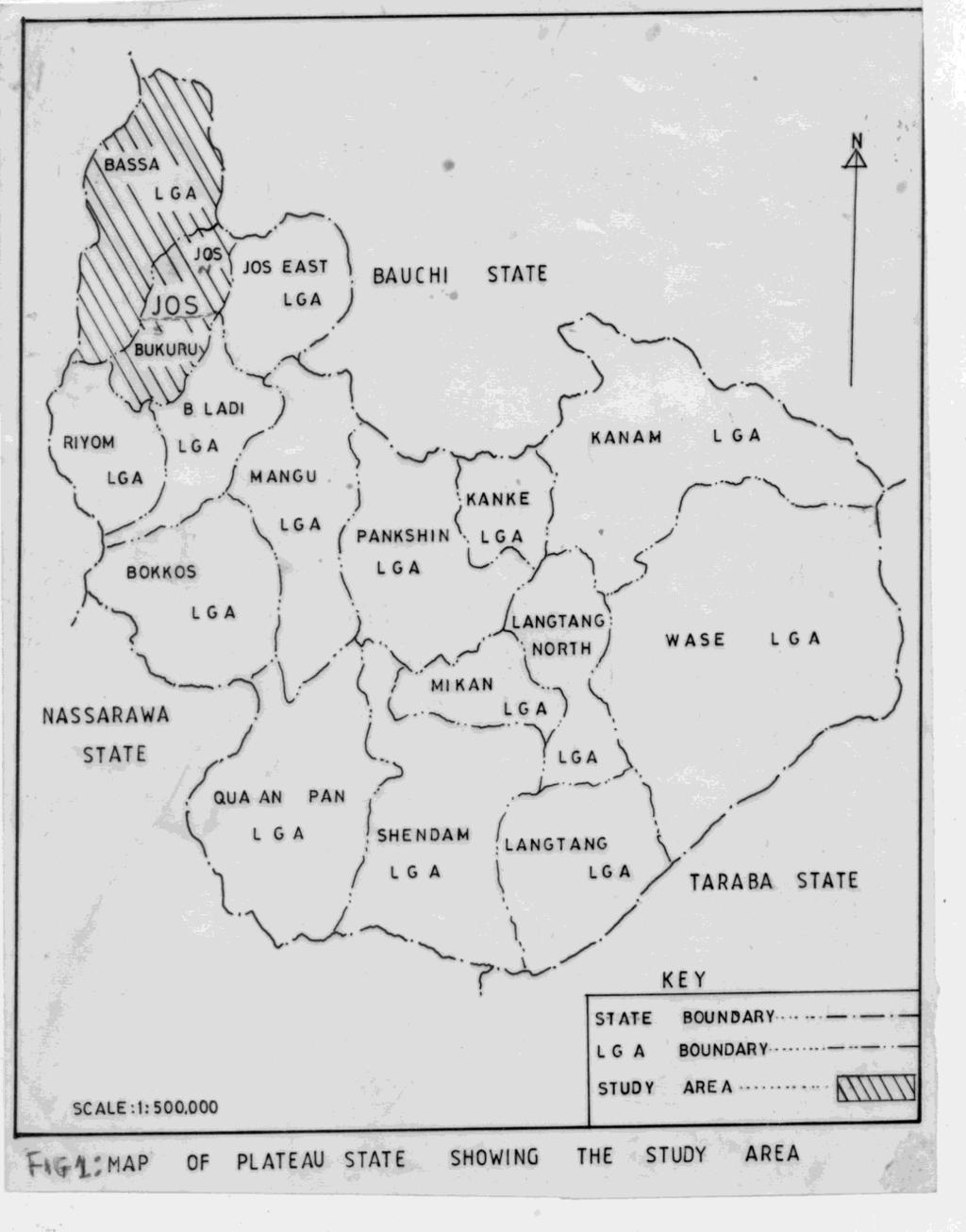
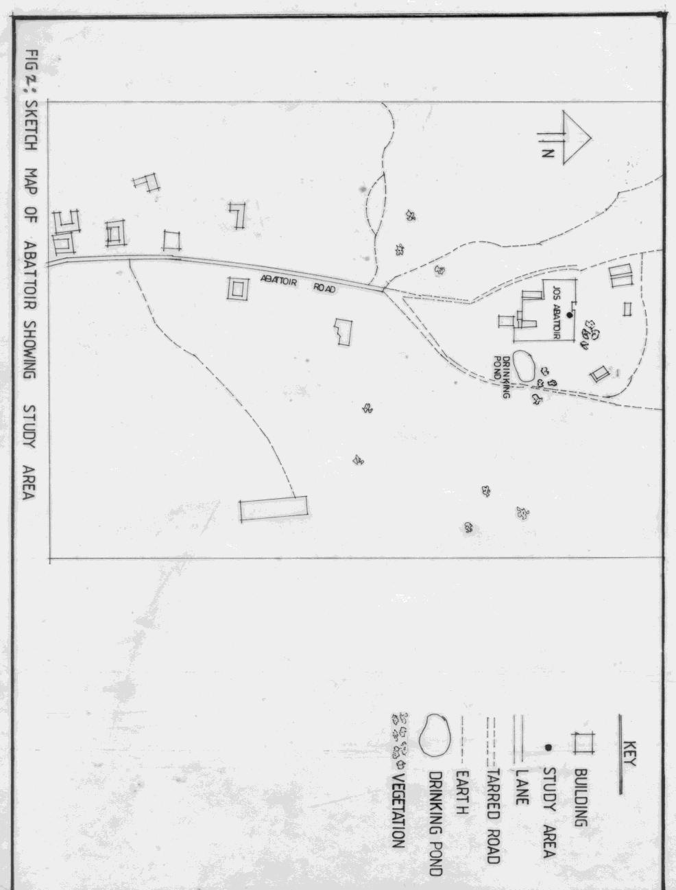
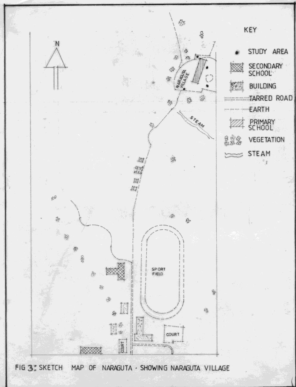
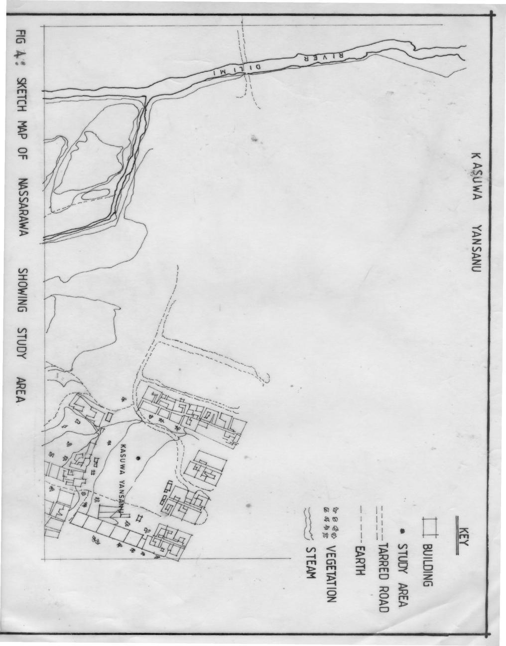
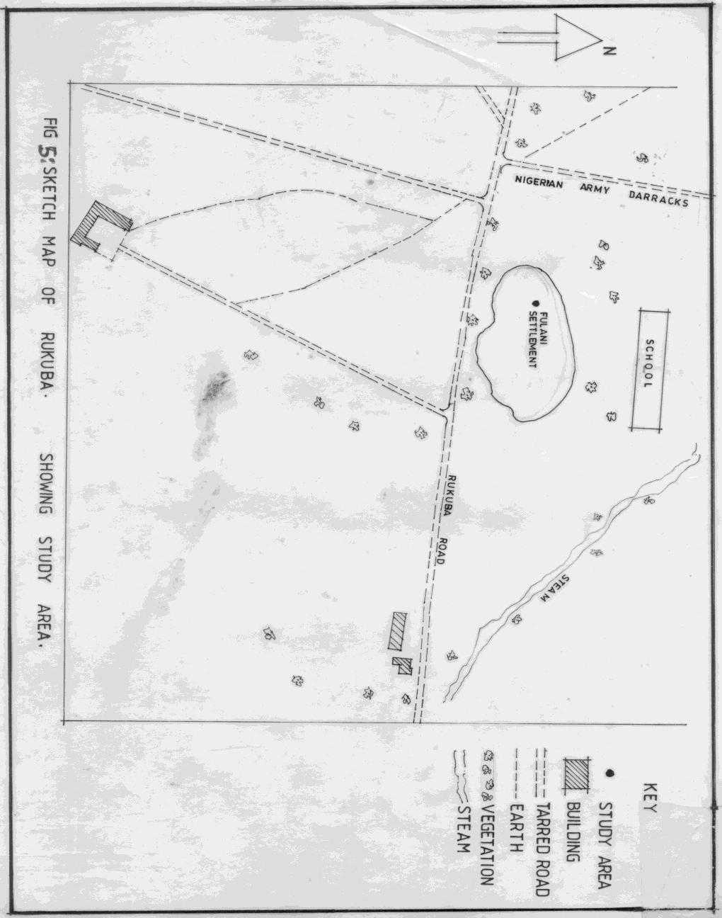
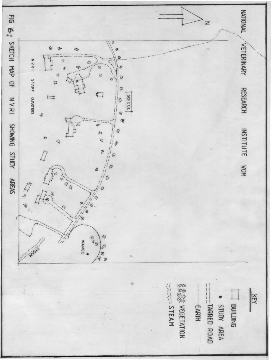
12 xii LIST OF FIGURES FIGURE 1: Map of Plateau State showing the Study area 44 FIGURE 2: Sketch Map of Abattoir in Jos North Local Government Area showing study area. 52 FIGURE 3: Sketch Map of Naragura in Jos North Local Government Area showing Naraguta Village 53 FIGURE 4: Sketch Map of Nassarawa Gwon in Jos North Local Government Area showing study area 54 FIGURE 5 Sketch Map of Rukuba in Bassa Local Government Area showing study area. 55 FIGURE 6: Sketch Map of NVRI Vom in Jos South Local Government area showing study areas. 56
13 LIST OF PLATES i. Unsporulated and sporulated E.alabAMensis 125 ii. Unsporulated and sporulated E.auburnensis 126 iii. Unsporulated and sporulated E.bovis 127 iv. Unsporulated and sporulated E.brassiliensis 128 v. Unsporulated and sporulated E.bukidnonensis 129 vi. Unsporulated and sporulated E.canadensis 130 vii. Unsporulated and sporulated E.cylinderica 131 viii. Unsporulated and sporulated E.ellipsoidalis 132 ix. Unsporulated and sporulated E.pelita 133 x. Unsporulated and sporulated E.subspherica 134 xi. Unsporulated and sporulated E.wyomingensis 135 xii. Unsporulated and sporulated E.zurnii 136 xiii
14 xiv ABSTRACT A parasitological examination of 2,500 faecal samples of cattle was carried out in Plateau State, Nigeria, to determine prevalence, seasonal distribution and sources of various species of Eimeria in both sexes, age groups, breeds and husbandry systems. Effects of temperatures on sporulation of Eimeria oocysts and chemical analysis of the Eimeria oocysts were also determined. Out of 2500 samples examined, 1567 i.e 62.68% were positive for twelve species of Eimeria. E. alabamensis occurred in 220 [8.80%], E. auburnensis 806 [32.24%], E. bovis 914 [36.56%], E.brassiliensis 240 [9.60%], E. bukidnonensis 580 [23.20%], E. canadensis 236 [9.44%] E. cylindrica 556 [22.24%], E. ellipsoidalis 722 [28.88%], E. pellita 279 [11.16%]. E subsphserical 295 [11.80%], E. wyomingensis 445 [17.80%] and E. zurnii 1043 [41.72%]. E.zurnii 54.88% was predominant in the Jos Abattoir. E. cylindrica 81.02% in Naraguta, E. auburnensis 76.08% in Nassarawa Gwom, E. zurnii, 97.52% in Rukuba, E. bovis 29.36% in Vom and E. ellipsoidalis 94.62% in Zallaki. Mixed infections of 2-12 species were found in 1457 [58.28%] faecal samples with 178 [7.12%] positive for two parasites; 324 [12.96%], for three parasites; 401[16.04%], for four parasites; 280 [11.20%], for five parasites; 160 [6.40], for six parasites; 72 [2.88%], for seven parasites; 11 [0.44%], for eight parasites; 10 [0.40%], for nine parasites; 8 [0.32%], were positive for ten parasites; 8 [0.32%], for eleven parasites; 5 [0.20%], or twelve parasites. Only 110 [4.40%] harboured single infections The most infected age group was months old with infection rate of 79.30%. Other age groups were 0-4 months 43.75%;
15 xv 5-9 months 65.90%; months 72.33%; months 61.11%; and 25 months and above 43.20%. There was statistically significant difference in the rate of infection between age groups months and 25 months and above, between months and 0-4 months [P<0.1] The calves first shed Eimeria oocysts between 14 and 84 days of their lives. The parasites were more prevalent in bulls than in cows, but there was no statistically significant difference in the rate of infection between bulls and cows. [P>0.05]. N'dama breed carried the lowest infection [44.36%] while the Muturu carried the highest infection rate of 83.76%. There was a statistically significant difference in the rate of infection between N'dama and Muturu breeds [P<0.05]. Cattle grazed on pastures were more infected than those raised either on mud floor pens or on concrete floor pens. The prevalence rates were 70.70%, 62.48% and 56.73% respectively. The cow beef cattle had the highest rate of infection of 69.15% while the dairy cattle had 55.06% infection rate. There was a statistically significant difference in the rate of infection between the beef cattle and dairy cattle [P<0.05]. The major source of infection was the pastures, having contamination rate of 40.77%. Others were beddings 32.38%; feeds 28.05% and udders 29.17%. There was a statistically significant difference in the rate of contamination between pastures and feeds [P<0.01] and between pastures and udders [P<0.01].Infection was higher during the dry season 68.35%, than during the wet season, 59.31%. There was no statistically significant difference in the infection rate between dry and wet season [P>0.05]. Low temperature inhibited sporulation of Eimeria oocysts while high temperatures increased sporulation. The morphological features of Eimeria oocyst types encountered varied in sizes, shapes and inclusions. Twelve Amino acids were found in the sporulated bovine Eimeria oocysts in this study, 9 essentials and 4 non-essentials amino acids.
16 xvi
17 1 CHAPTER ONE INTRODUCTION AND LITERATURE REVIEW 1.1 INTRODUCTION The term coccidiosis applies to the disease condition caused by infection with one or more of the many species of coccidia. Coccidia are a group of parasitic protozoans, which invade certain cells of the intestinal epithelium, caeca, often in other organs like liver and kidney. Each species of coccidia produces different host parasite interaction; as such there are many kinds and degrees of coccidiosis. The disease only occurs if an animal is subjected to heavy infection or its resistance is lowered. It is important at the outset to differentiate between infection and disease. The presence of infection does not invariably lead to the development of clinical signs of disease. Low level of challenge can actually be beneficial by stimulating protective immune responses in the host (Catchpole et al 1993). The disease is cosmopolitan, occurring in practically all kinds of vertebrates including fowls, cattle, pigs, sheep, goats, cats and dogs (Ajayi 1973). The problem of identification of species is simplified by the fact that parasites are host specific, each
18 2 species occurs in a single or in a limited group of related hosts. Of many described genera of coccidia the genus Eimeria contains the species of major economic importance in cattle. Internal structure of the sporulated occysts is the characteristic used to distinguish this genus from several others that may occasionally be encountered. The oocysts of all genera, when freshly passed, consist of a thickened outer wall and rounded mass nucleated protoplasm. Not until sporulation has occurred do the distinguishing characteristics become apparent. In the genus Eimeria four sporocysts develop, each containing two banana-shaped sporozoites. This is distinguished from another genus Isospora containing many species from wild birds. Isospora contains two sporocysts each containing four sporozoites. The causative agent of bovine coccidiosis is of the genus Eimeria of the family Eimeriidae. Bovine coccidiosis is an infectious disease of cattle which produces bloody scours or red diarrhoea, dysentery, weight loss and death. The disease is said to be more severe during dry hot and sunny conditions usually considered unsuitable for oocyst development (Marquadt 1960, Marquadt et al 1960, Parker and Jones 1987, Wilber et
19 3 al 1994, Ahmed et al (1992). Bovine coccidiosis has long been of interest because of its economic importance. In U.S.A for example annual estimate loss has been put at 200 million dollars (About 28 billion Naira) as of 1993 (Taylor and Catchpole 1994). The disease causes a lot of economic loss in livestock production industries all over the world (Foreyt, Parish and Foreyt 1981). The disease occurs mostly in young animals, but occasionally it occurs in calves over 6 months of age or even in adults (Joyner et al 1996, Macmerinam and Elliot 1995, Svensson et al 1993). The most serious losses are seen in dairy herds where large numbers of calves are kept. Older cattle are carriers, and they continue to pass oocysts in the faeces (Waggoner et al 1994 and Holliman 2000). There are thirteen known species of bovine Eimeria. These are E.alabamensis Christensen 1941, E.auburnensis, Christensen and Porter 1939, E. bovis, Fiebiger 1912, E.brassiliensis Torres and Ramos 1939, E.bukidnonensis Tubangui 1931, E.canadensis Bruce 1921, E.cyclindrica Wilson, 1931, E.ellipsoidalis Becker and Fye 1929, E.illinoisensis Levine and Ivens 1967, E.pellita Supperer 1951,
20 4 E.subspherica Christensen 1941, E.wyomingensis Huizinga and Winger 1942 and E.zurnii Rivolta 1878, Martin Calves become infected by ingesting sporulated oocysts along with their feed or water. The severity of disease depends upon the number of oocysts they ingest. If few oocysts are ingested, no symptoms will be noticed, but if a large number is ingested, severe disease and even death may occur. However, subclinical infection may cause retardation of growth and add greatly to financial loss. Crowding and lack of sanitation greatly increase the disease hazard. Successive passage of coccidia from one animal to another often builds up infection to a pathogenic level. Long and Millard (1979) showed that animals under nutritional stress are liable to respond adversely to coccidial infection. There is a remarkable degree of specificity both in regards to the host and in part of the host in which development takes place. Coccidia that invade the small intestine generally produce less pathogenic effects (Gregory 1990). This is because in ruminants the small intestine is very long, providing a large number of host cells and allowing the potential for enormous parasite replication with minimal damage. If absorption is
21 5 impaired, the large intestine is capable of compensating to some extent. In the large intestine, the rate of cellular turnover is much less and there is no compensatory effect from other regions of the gut. Coccidia that invade the large intestine are thus more likely to cause pathological changes, particularly if large numbers of sporulated oocysts are ingested over a short period of time. Those coccidia that produce small endogenous stages and develop in epithelial cells of the villi produce minimal lesions. In contrast coccidia that infect stem cells in the crypt of epithelium cause extensive lesions, preventing replacement of damaged epithelium. Coccidia with large endogenous stages can evoke either localized or diffused lesions because of the large numbers of parasites produced. In cattle E.bovis and E.zurnii are considered the main pathogens (Ernst and Benz 1986). These species affect the large intestine of the host because they have large endogenous stages; therefore they can kill stem cells, thus impairing cellular repairs (Radostits and Rocksdale 1980, Fox 1985, Taylor and Catchpole 1994). Effects of coccidial infection may be exacerbated if different species that affect different parts of the gut are present. Similarly concurrent
22 6 infections with other disease-producing agents such as helminths, bacteria and viruses may affect the severity of disease. Pavorvirus infection is thought to aggravate coccidiosis in calves (Durham et al 1985). Calves infected with intestinal nematode along with coccidia had higher intensity of coccidiosis (Davis et al 1959 Agnihotri 1994 and Wariuru et al 2000). All ages of cattle are susceptible to disease. During the first few months of life, the majority will probably have been infected and may not show signs of disease. Those that reach adulthood are highly resistant to pathogenic effects of the parasites, but they may continue to harbour small numbers of parasites throughout their lives. Occasionally acute coccidiosis occurs in adult animals with impaired cellular immunity or in those, which have been subjected to stress. In the young, the colostrum provides passive immunity during the first few weeks of life. Thereafter, animals acquire resistance to coccidia as a result of active immunity. An animal's resistance to coccidia infection can be reduced by adverse conditions such as dietary changes, prolonged travel, extremes of temperature and weather conditions (Gregory 1990,
23 7 Hasbullah et al 1990 and Waruiru et al 2000). According to nutritional status; mineral and vitamin deficiencies can influence resistance to infection. Suckling animals who benefit from colostral intake may forage less, and hence pick-up fewer oocysts from pasture. Wellnourished animals may simply be able to fight off infection more readily. Mineral deficiencies can affect the immune response of infection. 1.2 LITERATURE REVIEW Lee and Armour (1959) carried out a survey of the coccidial oocysts in Nigerian cattle, and 11 species were identified. These included E.alabamensis Christensen 1941, E.auburnensis, Christensen and Porter 1939, E. bovis, Fiebiger 1912, E.brassiliensis Torres and Ramos 1939, E.bukidnonensis Tunbagui 1931, E.canadensis Bruce 1921, E.cyclindrica Wilson, 1931, E.ellipsoidalis Becker and Fye 1929, E.subspherica Christensen 1941, E.wyomingensis Huizinga and Winger 1942 and E.zurnii Rivolta 1879, Martin Hasche and Todd (1959) reported that out of 355 cattle examined in Wisconsin, 26% had E.ellipsoidalis, whereas 20% were infected with
24 8 E.cylindrica, similarly 6% were infected with E.brasilliensis, whereas 42% and 3% were positive of E.bukidnonensis and E.canadensis respectively. Fitzgerald (1962) conducted field studies on the incidence of coccidia in young Hereford calves during grazing and feeding seasons in Alabama. He encountered seven species of Eimeria and observed that E.zurnii was the causative agent of winter coccidiosis. Majaro and Dipeolu (1981) reported on the survey of coccidiosis in trade cattle, sheep and goats in Nigeria. Faecal examination showed that nine species of Eimeria were identified, and E.bovis and E.zurnii were the predominant species. In another investigation, Kasim and Al-shawa (1985) studied the prevalence of Eimeria in the faeces of cattle in Saudi Arabia and observed that out of the 205 cattle studied, 34.1% of the individual samples were positive, and mixed infection of 2 to 4 species were found in 15.7% of the specimens. E.zurnii and E.bovis were predominant. Ernst et al (1987) determined the prevalence of coccidial oocysts from crossbreed beef calves in Bahia grass in coastal plain of Georgia. Out of 534 faecal samples collected, 86.0% contained one or more species of coccidian oocysts; 13 species were found, and E.bovis
25 9 was the predominant species. Parker and Jones (1987) examined the faecal samples of unweaned calves from birth to eight months of age and discovered that most calves shed at least nine species of Eimeria by 3-4 months old. Oda and Nishida (1990) carried out a field survey on the prevalence of bovine coccidia in prefectures of Japan in autumn and found that out of 1,015 faecal samples examined, 59.0% contained coccidial oocysts. The prevalence was highest in the animals between 6 and 11 months of age. Hasbullah et al (1990), examined faecal samples from 2,019 cattle in Japan for a period of 12 months and 19.3% were found to be positive, and E.bovis was the most dominant species. Svensson et al (1993), found out that Eimeria oocysts were shed in calves during their first 3 weeks after turn out to pasture. The number of Eimeria oocysts per gramme and the dry matter content of 449 faecal samples taken from 54 calves in 8 herds in South West Sweden were determined during the last 2 weeks before and first 3 weeks after the animals were turned out to pasture. E.alabamensis, accounted for most of the increase but small numbers of the oocysts of E.bovis, E.auburnensis, E.cylinderica,
26 10 E.pellita, E.zurnii, E.bukiduonensis, E.ellipsoidalis and E.subspherica were found. Mundim et al (1994) examined faecal samples from calves in Uberlanda in Brazil, and out of 55 faecal samples examined 47.0% had Eimeria oocysts in their faeces. Nine species were found; E.bovis occurred in 36%, E.zurnni in 34.0% and E.ellipsoidallis, 31.5%. Penzhorn, Knapp and Speer (1994) collected faecal samples from free ranging American cattle in Montana. The following results were obtained: E.bovis 97.0% E.canadensis 32.0%, E.brassillensis 3.2% and E.auburnensis 9.7%. Arslan and Tuzer (1998), in Turkey, examined 768 faecal samples of cattle, and the species encountered and their prevalence rates were, E. alabamensis 4.98%, E. auburnensis 27.0% E. bovis 34.0%, E. canadensis 12.0%, E. cylindrica 7.9%, E. ellipsoidalis 14.0% and E. zurnii 26.0%. Hooshmand, Svensson and Uggla (1994) infected Swedish calves with E.alabamensis and found the prepatent period to be 6 to 8 days. The clinical signs were mostly diarrhoea, loss of appetite and depression. Growth rates were significantly reduced for 18 days after inoculation, but 71 days after, they did gain back their weights.
27 11 Protein available to the ruminant for nutritional needs is supplied by microbial and dietary sources (Wilkerson et al 1993). Microbial protein supply to the deodenum can meet between 50 and 90% of metabolisable protein requirement of beef cattle (Parker 2000). Because of the large quantity of protein provided by microorganisms, analyses on rumen microbial protein were carried out by Goedeken et al (1990), Wilkerson et al (1993), Rohr and Lebzein (1991) and Klemesrud et al (2000) and 10 to 13 types of amino acid were discovered. Pattilo and Becker (1995) found that protein and polysaccharides were present in the sporocyst wall of some Eimeria oocysts during cytochemical investigation. Loser and Gornnert (1965) reported that Eimeria oocysts wall contains o- dioxy-phenol-protein; consequently the oocyst wall is very resistant to various physical and chemical factors. There are 20 types of amino acids, (11 essentials and 9 nonessentials), and their functions are as follow as described by Harper (1971):
28 Alanine. This is an important source of energy for muscle. It is the primary source of sugar metabolism. It boosts immune system by producing antibodies. Lack of alanine leads to hypoglycemia, muscle breakdown and fatigue Aspartic acid. This is interconvertible with asparagines and therefore shares the same functions. They are both involved in immune system function by enhancing immunoglobulin production and antibody formation. They both help the liver by aiding the removal of ammonia. Lack of aspartic acid and asparagine causes calcium and magnesium deficiencies Cysteine and cystine. Both are interconvertible; they are protective against radiation. They are protective against radiation. They are essential in growth, maintenance and the repair of skin. Lack of cysteine and cystine causes chemical sensitivity and food allergy Glutamic Acid.
29 13 This increases energy, accelerates wound healing and ulcer healing. It plays major role in DNA synthesis. Lack of glutamic acid causes ulcer Glutamine. This involves in muscle strength and endurance. It is essential for helping to maintain normal and blood sugar levels. It is also essential to gastrointestinal function by providing energy to small intestine. Lack of glutamine causes chronic fatigue syndrome Glycine. This forms part of the structure of hemoglobin. It involves in glucacon production and assists in glycogen metabolism. Lack of glycine produces anaemia, viral infection, hypoglycemia and chronic fatigue syndrome Proline. The main functions are that it is a critical component of cartilage and hence health of joints, tendons, and ligaments. It is involved in keeping heart muscle strong. It works in
30 14 conjuction with vitamin C in keeping skin and joint healthy. Lack of proline causes chronic liver diseases Serine. It is used in maintaining blood sugar levels. It boosts immune system by assisting in production of antibodies and immunoglobulins. Lack of serine causes total body gamma and neutron irradiation and hypoglycemia Arginine. This is essential for normal immune system activity and wound healing. It assists with regeneration of damaged liver. It is the most potent amino acid in releasing insulin. Lack of arginine causes immune deficiency syndrome Histidine. This is found in high concentration in haemoglobin. It is useful in treating aneamia due to relationship to haemoglobin. It assists in maintaining proper blood ph. Lack of histidine causes aneamia Isoleucine.
31 15 This is involved in muscle strength, endurance and muscle stamina. It is involved in formation of heamoglobins. Lack of isoleucine causes obesity and acute hunger Leucine. It has all the properties of isoleucine. It is a potent stimulator of insulin and it helps in bone and skin healing. Lack of leucine causes depression, acute hunger, and vitamin B deficiency in pernicious aneamia Lysine. It inhibits viral growth and it helps form collagen, connective tissue present in bones, ligaments tendons and joints. It assists in the absorption of calcium. Lack of lysine causes anaemia, weight and hair loss and irritability Methionine. This assists in breakdown of fats. It helps to reduce blood cholesterol levels. It assists in the removal of toxic wastes from the liver. It helps prevent disorder of hair, skin and nails. Lack of Methionine causes obesity.
32 Phenylalanine. This helps in major part of collagen formation. It is a powerful antidepressant. It enhances mood clarity of thought, concentration and memory, and it suppresses appetite. Lack of phenylalanine causes depression, obesity and cancer Threonine. This is required in formation of collagen. It helps prevent fatty deposits in the liver. It aids in production of antibodies. Lack of threonine causes depression, muscle spasticity and epilepsy Tryptophan. This stimulates growth hormones, lowers risk of arterial spasms and effective in some forms of depression. Lack of tryptophan causes depression insomnia and chronic fatigue syndrome Tyrosine. It increases energy and it improves mental clarity and concentration. It is an effective antidepressant. Lack of tyrosine causes depression and chronic fatigue syndrome.
33 Valine. The main functions of valine are that it is involved in muscle strength, endurance and muscle stamina. Lack of valine causes hunger, obesity and neurological deficit. 1.3 LIFE CYCLE OF BOVINE EIMERIA The life cycle of a typical bovine coccidium involves only one host, and the development is in 3 phases viz: schizogony, gametogony and sporogony. Oocysts, after leaving the cow by way of faeces, sporulate under favourable conditions to become the infective stage in 2-7 days. The infective, sporulated oocyst containing 4 sporocysts, each of which contains 2 sporozoites, enters the host passively by ingestion. The animal ingests the oocyst along with food and or water from external environment. Infection from the ingested oocysts occurs whenever excystation takes place in the lower part of the small intestine and also in the caecum within 24 hours after introduction of oocysts (Hammond, McCowin and Shupe 1954, Nyberg and Hammond 1965). Excystation releases the contained sporozoites. Two separate stimuli are necessary for excystation (Nyberg et al 1967). The first is provided by carbon
34 18 dioxide and the second by trypsin, and bile carbon dioxide is said to stimulate the activation or production of an enzyme or an enzymic rate limiting of the micropyle. The second stage of excystation is ph dependent, and it effects the escape of the sporozoites. Bile facilitates the entry of trypsin through the altered micropyle which then digests the sporocystic plug, permitting escape of the mobile sporozoites. Doran (1906) considers that the sporozoites may also secrete enzymes that attack the plug. The liberated sporozoites emerge through the narrow opening of the micropyle where steida body formerly was, and remains within the oocyst wall. The sporocyst residuum is usually destroyed during this process. Liberated sporozoites penetrate quickly within seconds into intestinal epithelium of the host where further development takes place (Hammond, Anderson and Miner 1973). They are engulfed by macrophages and are carried by them through lamina propria of the villi to reach the epithelium in the depths of the glands of Leiberkuhn. The next stage is the asexual reproduction or schizogony Schizogony. This process is initiated when sporozoites enter the epithelial cell and
35 19 become rounded up. Development in bovine coccidia occurs within the nucleus. The rounded up sporozoite becomes trophozoite, and within a few days the nucleus of the trophozoite divides by schizogony (mutiple fission) to become a schizont. The nucleus in schizont is considered to be of the mitotic type (Pellerdy 1974). Initially the cytoplasm is undivided, but later the daughter nuclei are each surrounded by a clear zone of cytoplasm and eventually a number of elongate fusiform organisms are produced. These are called the first generation merozoites. The numbers of merozoites that are formed in the first generation schizont vary according to species. In E.bovis, more than 100,000 first generation merozoites are released, and they then enter other epithelial cells in the area and continue the cycle of asexual development. In some species this results in second generation schizonts. In some other species the second generation schizont is larger than the first whereas in some it is smaller. The number of merozoites produced also varies according to the species e.g. the second generation schizonts of E.bovis are extremely small, about microns and form not more than merozites
36 20 (Hammond et al 1973). The second generation merozoites may proceed to a third or more generations of asexual reproduction or they may differentiate into sexual or gametogenous forms Gametogony. The sexual process in coccidiosis is broken down into 2 periods viz: - the gametogenesis period during which formation of male and female gametes occurs and the fertilization period. The factors responsible for the initiation of gametogenous cycle are not fully understood. Although generally considered to be genetically determined, host responses may play a role through phenotypic determination in terminating schizogony. The gametogenesis in Eimeria is characterized by the fact that one comparatively large macrogamete always develops from one macrogamont but a multitude of small, motile flagellated microgametes are formed from one microgamont. During macrogametogenesis there is an accumulation of nutritive substances. In the young macrogamate small granules are initially found in the vicinity of the nucleus; later these enlarge and become scattered over the cytoplasm, the larger granules being found on the periphery of the cell. These are plastic
37 21 granules or wall forming granules that form the wall of oocyst following fertilization of the macrogamate. The process of macrogametogenesis can be divided into two periods. The first is the period of gametocyte growth and is accompanied by a reproduction of nuclei. Growth of gametocyte and division of the nucleus lead to the formation of a polynucleus microgametocyte with a characteristic peripheral distribution of nuclei. The second period is one of differentiation and formation of microgametes. During this period the nuclei assume a coma-shape and accumulate on the periphery of the cell leaving a residual mass of cytoplasm in the cell. Rupture of microgamont liberates the microgametes which fertilize the macrogametes. Fertilization by microgametes may occur at any point on the surface of the macrogamate. A zygote is formed, and the oocyst wall is laid down around it. When the cyst wall is complete, the oocyst is extended from the tissue and passed to the exterior Sporogony. With few exceptions, sporulation does not occur until the oocyst is shed to the exterior of the body. Initially the zygote almost fills the oocyst
38 22 cavity, but within a few hours outside the host, the protoplasm contracts from the wall of the oocyst to form a sporont and leaves a clear space between it and the wall. The sporont divides into four sporoblasts and remaining cytoplasm being left as an oocystic residual body. The sporoblasts are initially more or less spherical, but later elongate into ovoid or ellipsoid bodies which then become sporocysts by laying down a wall of refractile material around each sporoblast. The protoplasm inside each sporocyst further divides to form two sporozoites. Protoplasm remaining from the division is left as a sporocystic residual body. The time required for sporulation to the infective stage is a specific feature of each species of coccidium and is used as a characteristic in identification. Oxygen and adequate moisture are necessary for the sporulation and at constant temperature. Temperature also has been an important influence on sporulation. The optimum temperature for sporulation is about 30 o C. In general sporulated oocysts are more resistant to desiccation and cold, and may survive for up to two weeks at temperature of - 12 o C to 20 o C. Unsporulated forms are killed in 96
39 hours at these temperatures MORPHOLOGY OF BOVINE EIMERIA OOCYSTS The oocysts of the following Eimeria species of cattle have been described. Species differentiation is based on the following characteristics Eimeria alabamensis, Christiensen The oocysts range between 13 and 24 in length by 11 to 36 in diameter. The shape is typically pyriform with variations. There is no micropyle and the wall is thin, delicate, homogenous, transparent and slightly thinner at the narrow end. The oocysts appear colourless and crystalline under low magnification, but under oil immersion the wall has a grayish - lavender to pale brownish yellow tint, fading to light yellow at the narrow end. Sporulation time is between 96 hours and 120 hours. Sporulated oocysts contained elongated tapered sporocysts, each having two indistinct sporozoites. A polar granule is presumably absent Eimeria auburnesis, Christensen and Porter The oocysts are typically elongate, ovoidal, varying between tapered
40 24 and sub-ellipsoidal in form. A micropyle is present and appears as a gap in the wall at the tapered end covered by a thin black line considered possibly represents an operculum. The walls are smooth but varying in structure from the transparent type to a rare semitransparent heavily mamillated type. Sporulation time is between 48 and 72 hours. The oocysts range from 32 to 45 long by wide Eimeria bovis, Christensen The oocysts are ovoidal blunted across the narrow end. The oocysts range from 23 to 34 long and 17 to 23 wide. The oocyst wall is smooth homogenous, transparent, greenish brown in colour. Micropyle is present, appears at a higher area of the wall. Sporulation time is 48 hours - 72 hours at room temperature. The wall is composed of a heavy inner layer and a very thin transparent outer layer. An oocyst residuum and polar granule are present. Sporocyst residuum is present Eimeria brasiliensis, Torres and Ramor The oocysts are ellipsoidal, about long and 22-29
41 25 wide. The oocyst wall is smooth and yellowish-green in colour. The wall is composed of a single layer and is covered by micropylar cap. An oocyst polar granule is present. Oocyst residuum is absent but sporocyst residuum is present. Sporulation time is about 6 days - 7 days Eimeria bukidnonensis, Tubangui The oocysts are pear-shaped to oval. They are yellowish brown to dark brown in colour. They are 31 to 44 long and wide. The oocyst wall is about 2 to 4 thick, except at the micropylar end where it is thin. It is composed of 2 layers; the outer layer is thick and the inner layer has a tough membrane. Lee (1954) described the wall as radially striated. The oocyst wall is speckled and rather rough. The micropyle is conspicuous about wide. An oocyst residuum and polar granule are absent. The sporocysts are elongated about by A steida body is possibly present. Definite sporocyst residual material is absent. Sporulation time is 4 days -7 days Eimeria canadensis, Bruce 1921.
42 26 The oocyst shape is typically ellipsoidal, varying from nearly cylinderical to stoutly ellipsoidal. They are about by The oocyst wall is smooth, transparent slightly yellowish-brown in colour. The micropyle is an inconspicuous gap in the wall at one end, appearing covered with a thin dark reflection line. An oocyst residuum and polar granules are absent. Sporulation time is 72 hours Eimeria cylindrica, Wilson The oocysts measure long by wide. The shape is typically cylindrical but may vary from ellipsoidal to narrow cylindrical. The oocyst wall is thin, colourless, smooth, homogenous and transparent. The micropyle is imperceptible. Oocyst residuum and polar granules are absent. A sporocyst residuum is present, but there is no sporocyst steida body. Sporulation time is about 48 hours Eimeria ellipsoidalis, Becker and Frye Oocysts are from long by wide. The shape is predominantly ellipsoidal with some spherical to subspherical forms. Oocyst wall is thin homogenous and transparent. An oocyst residuum and polar granule are absent. A sporocyst residuum is present. No micropyle present.
43 27 Sporulation time is between 48 hours and 72 hours Eimeria pellita, Supperer The oocysts are ovoid with a flattered small end about by The oocyst wall is relatively thick and dark-brown. The surface of the oocyst bears numerous small uniformly distributed protuberances in the form of small blunt points that give the wall velvety appearances. There is a micropyle at the small end. An oocyst polar granule and residuum are absent. The sporocysts are elongated without steida body. A sporocyst residuum is present, usually compact. The sporozoites lie lengthwise in sporocysts with 2 refractile globules. It is possible that E.pellita is a synonym of E.bukidnonensis. However it differs from it in the velvety appearance described for its oocyst wall. The oocyst wall of E.bukidnonensis has been described as speckled. Other differences are that sporocyst residuum is prominent in E.pellita and absent in E.bukidnonensis. The sporocysts of E. bukidnonensis are somewhat pointed at one end while those of E.pellita are not pointed. Sporulation time is between 10 days and 12 days E.subspherica, Christiensen 1941.
44 28 The oocysts are the smallest to of all the bovine Eimeria species. They are about 9-11 long by 8-12 wide. They are ellipsoidal to subspherical in shape. The oocyst wall is uniformly thin, smooth, homogenous and transparent, with no visible mircopyle. The oocyst appears colourless to a faint yellow tint and fragile. Oocyst residuum and polar granules are absent. The sporocysts are pale, spindle-shape, without a sporocyst residuum. Sporulation time is 4 days - 5 days Eimeria wyomingensis, Huizinga and Winger The oocysts are similar to those of E.bukidnonensis but the oocysts are smaller. The oocysts are ovoidal in shape, measuring by The oocyst wall is thick with a yellowish-brown to greenish-brown colour, and slightly speckled. The micropyle is conspicuous. Sporulation time is 5 days - 7 days Eimeria zurnii, Rivolta 1878, Martin The oocysts are spherical, subspherical to ellipsoidal, measuring by The oocysts appear colourless and the wall is thin, homogenous and transparent. There is no visible micropyle. Oocyst polar granule and residuum are absent. Sporocyst residuum is absent. Sporulation
45 29 time is 3 days at 20 o C, 9 days - 10 days at 12 o C, 23 hours - 24 hours at 30 o C o C. (Marquardt et al 1960). 1.5 PATHOGENESIS OF BOVINE COCCIDIA Coccidia are obligate, intracellular parasites whose development within the cytoplasm of epithelial cells results in the death of each cell which is parasitized. The effect on the host depends on the magnitude of the initial infective dose and the number of cells invaded at the onset by the sporozoites. The effect also depends on the spread of infection during schizogony, which is affected to a great extent by immunity acquired by the host. As increasing numbers of organisms enter sexual phase (gametogenesis) infection of new cells by merozoites diminishes and the disease gradually abates. When many cells of the intestinal epithelium are parasitized at one time the denuded mucosa may bleed greatly and intense inflammation involves the lamina propria and sometimes the sub mucosa. As large numbers of epithelial cells are destroyed the remaining epithelium is eliminated to replace that which was host. This eventually causes hyperplasia of the intestinal epithelium, which is cast into long papillary ponds as replacement of the epithelial cells exceeds
46 30 their loss. Coccidia gametogenesis is most numerous in that the lesion exhibiting this hyperplasia. This is in contrast to the erosive haemonhyic stage which organisms in various stages of schizogony are most common. Different species of bovine coccidia manifest a tendency to localize in different parts of intenstine. E.zurnii and E.bovis occur mainly in the caecum, colon and last part of the ileum, whereas, E.alabamensis is an intranuclear parasite occurring within the nuclei of the epithelial cells at the tips of villi. Coccidia life history is self-limiting, merozoites and gametocytes are the pathogenic stages causing rupture of the cells they invade with consequent exfoliation of the epithelial lining of the intestine. Oocysts count is often very low when the disease is at its peak since the oocysts have not yet been formed. Exfoliation of the epithelium mucosa causes diarrhoea and in severe cases hemorrhages into the intestinal lumen and the resulting hemorrhagic anaemia may be fatal, leading to death of the animals. If the animal survives this stage of the life cycle of the coccidia terminates without further damage. Prepatent period ranges from one to four weeks depending on the dose of
47 31 oocysts, length of intestine, length of oocysts exposure to external environment and the age of the host. Under practical conditions constant reinfection occurs and waves of pathogenic stages succeed each other (Blood and Henderson 1974) Considering pathogenesis on individual basis E.zurnii is the most pathogenic, while E.bovis is rated second. In Europe E.zurnii is the most frequent cause of bovine coccidiosis. The acute disease is characterized by haemorrhagic diarrhoea, and the condition may become so intense that the faeces are frank blood. Teresmus is marked; there is anaemia, weakness and emaciation. In severe infections death may occur as early as 7 days after the onset of clinical signs. At post mortem the major lesions occur in the large intestine, though general catarrhal enteritis may be present in both the small and large intestine. In severe cases the caecum and colon may be filled with semifluid hemorrhagic material or even frank blood with fibrinous clot. The epithelium may slough away leaving large denuded areas, which are infiltrated with lymphocytes and leukocytes. In less acute cases the mucous membrane is roughened and spotted with petechial hemorrhages. Smears from the mucosa show very
48 32 large number of developmental stages of oocysts. In E.bovis infection, the later stages of development of the first stage schizont cause distortion of villi and disruption in the case with the second schizont. It is the gametocytic stages, which cause the greatest pathogenic effect. Hammond (1964) estimated that if the full potential of E.bovis were to be realised, 1,000 oocysts could result in the destruction of 24 billion intestinal cells. In experimental infection, Hammond et al (1944) found that an infective dose of 125,000 oocysts or more caused marked signs of illness with diarrhoea occurring on the 18th day when faeces were streaked with blood. A calf given 125,000 oocysts became moribund, and with higher doses animal became moribund or died within 3-4 weeks of infection. In severe infections majority of the crypts of the large intestine and sometimes of the terminal part of the small intestine are destroyed, the epithelial layer denuded and the lumen of the intestine filled with blood. The mucosa is necrotic and sloughed and this damage may extend to the sub mucosa, the wall of the intestine is congested and edematus, thickened with petechial or diffuse hemorrhages. Large number of
49 33 gametocytes and oocysts are visible microscopically. The pathogenicity of E.alabamensis is low under field conditions and is generally considered to be unimportant in clinical bovine coccidiosis. Disease may be produced if large numbers of oocysts are given to young calves. Davis et al (1955) found that 140 million oocysts produced yellowish green diarrhoea, admixed with blood in 14-month-old animals. The small intestine showed hyperaemia, destruction of the epithelium, a leukocytic infiltration and oedema. The effect of E.auburnensis is also low under field conditions. Profused green diarrhoea was produced in a 2-week-old calf given 8,000 oocysts by Christensen and Porter (1939). Clinical signs appearing between the 9th and 15th day of infection. Davis and Bowman (1952) reported the passages of blood and mucus with straining, in artificial infections with large numbers of oocysts and in natural outbreak. E. ellipsoidalis is also thought to be of minor pathogenic significance, (Holliman 2000). Boughton (1945) reported that E. ellipsoidalis might cause diarrhoea in young calves 3 months of age, and Hammond et al (1963), showed that a varying degree of immunity developed after infection with 50,000 to 1
50 34 million oocysts. E. subspherica is not known to be pathogenic, while the endogenous developmental cycles of other bovine Eimeria species are unknown and no pathogenesis is ascribed to them Factors Affecting the Pathogenicity of Coccidia Host - Parasite Relationship. A successful parasite is one that infects every available host causing minimal damage. This ultimate, harmonious co-existence is however easily upset by factors affecting either host or parasite leading to clinical signs of disease in the host. Site of Development. The coccidia that invade the small intestine generally produce less pathogenic effect (Gregory 1990). This is because the calves have long small intestine providing a large number of host cells and allowing the potential for enormous parasite replication with minimal damage. If absorption is impaired, the large intestine is capable of compensating to some extent. In the large intestine the rate of cellular turnover is much less and there is no compensatory effect from other region of the gut. Coccidia that invade the large intestine are thus more likely to cause
51 35 changes particularly if large numbers of oocysts are ingested over a short period of time. Those coccidia that produce small endogenous stages and develop in epithelial cells of the villi produce minimal lesions. In contrast coccidia that infect stem cells in the crypt epithelium may produce more extensive lesions by preventing replacement of damaged epithelium. Coccidia with large endogenous stages can evoke either localised or diffuse lesions because of the large number of parasites produced. In bovine Eimeria, E.bovis and E.zurnii are considered the main pathogens (Erst and Benz 1986). They affect large intestine and have large endogenous stages and can kill the stem cells. The effect of coccidial infection may also be exacerbated if different species that affect different parts of the gut are present. Similarly concurrent infections with other disease - producing agents such as helminths, bacteria and viruses may affect the severity of disease. Pavovirus infection is thought to aggravate coccidiosis in calves (Durham et al 1985) Effect of Age on Susceptibility To Infection All ages of calves are susceptible to infection but younger ones are more
52 36 susceptible to disease. During the first few months of life, the majority will probably have been infected and may or may not show signs of disease. Those that reach adulthood are highly resistant to the pathogenic effects of the parasite but may continue to harbour small numbers of parasites throughout their lives. Occasionally acute coccidiosis occurs in adult animals with impaired cellular immunity or in those, which have been subjected to stress. Svensson et al (1993) discovered that calves started shedding Eimeria oocysts as early as 2 weeks. However in the young calves colostrum provides passive immunity during the first few weeks of life; thereafter they acquired resistance to coccidial infection as a result of active immunity. An animal resistance to coccidial infection can be lowered by adverse conditions such as dietary changes, prolonged travel, extremes of temperature, weather conditions, changes in environment or severe concurrent infection (Gregory 1990). Nutritional status, mineral and vitamin deficiencies can influence resistance to infection. Suckling calves as well as those benefiting from colostral intake may forage less and hence pick up fewer oocysts from pasture. Well nourished calves may simply be able to fight off infection
53 37 more readily. Many deficiencies can affect the immense response to infection. 1.6 CLINICAL SIGNS OF BOVINE COCCIDIOSIS The clinical symptoms caused by various coccidia are similar in all animals. Mild fever may occur in the early stage, but in most clinical cases the temperature is normal or subnormal. The first sign of illness is usually the sudden onset of severe diarrhoea with foul smelling fluid faeces containing mucus and blood. The blood may appear as a dark, tarry staining of the faeces or as streaks or charts of fresh red blood. Anaemia is variable depending on the amount of blood loss. It may be extreme with ash-white pallor of the mucosa. There is weakness, staggering and dyspnoea. Severe dehydration, emaciation and complete anorexia are common. The period of the disease is usually 5-6 days and survivors undergo a convalescent period of some weeks and regain condition slowly. In mild cases, poor growth and anaemia only result. Nervous signs like convulsions could be observed in calves and other cattle during outbreak.
54 SURVIVAL AND VIABILITY OF OOCYSTS IN EXTERNAL ENVIROMENT. The survival of oocysts in external environment depends on 3 basic factors, namely, temperature, humidity and free access to oxygen. The possibility of oocysts development on the soil and their ultimate viability will not be identical in all soils or at all times of the year. This has been shown in a series of investigations conducted in different countries (Horton - Smith 1947, Kogan 1956 and Krylov 1960). Wilber et al (1994) compared oocysts survival in hosts from random rich and random poor soils and discovered that elevated random content of soil may have adverse effect on sporulation of coccidia while it still was intracellular within host. Sporulation of oocysts in the external environment can only occur in certain temperature ranges, in most cases at a temperature between 18 o C and 20 o C, Ajayi (1976) found out that temperature of 5 o C and 10 o C inhibited sporulation of ovine Eimeria oocysts, but when the same oocysts were subsequently incubated at 25 o C they sporulated.
55 39 Lack of oxygen will also impede the sporulation of oocysts. If oocysts are placed in ordinary water the bacteria inside the water will compete with oocysts for oxygen and sporulation may not take place. Marquardt et al (1960) showed that E.zurnii sporulated when oxygen deficit was only 10% or less when deficit was increased and in complete absence of oxygen no development took place. Lack of humidity had also been shown to cause wrinkling of oocyst wall due to loss of water. Marquardt (1960) showed that at 20% relative humidity, 12% of oocysts formed sporozoites and the rest died. But at 75% humidity, about 51% of the oocysts sporulated. Graat et al (1994) reported that temperature at 21 o C and 35% relative humidity had no significant influence on ocyst sporulation. Chemicals also have been shown to have effects on sporulation of oocysts. Some chemicals are known to penetrate though the cell walls of oocysts, causing cessation of development. Thus, Marquardt et al (1960) showed that 80% of oocysts of E.bovis placed in a solution of mercuric chloride died only 3% sporulated. Also, oocysts recovered from coxytrol treated rabbits failed to sporulate well, most of them being destroyed or
56 40 distorted morphologically (Ajayi and Anthony, 1983). 1.8 DIAGNOSIS Bovine coccidiosis can be diagnosed from combination of history, signs and gross lesions at necropsy and microscopic examination of scrapings of the intestinal mucosa and of faeces. Diarrhoea or dysentery accompanied by inappetence is suggestive of coccidiosis in calves. Secondary pneumonia is often present. Microscopic examination is necessary to determine whether the lesions are due to coccidia or to some other agent. However, diagnosis will be missed if one relies only on finding oocysts in the faeces. There may be none there at all in the acute stage of E.zurnii coccidiosis. Similarly the mere presence of oocysts in the faeces is not proof that coccidiosis is present. To be sure of a diagnosis scraping should be made from the affected intestinal mucosa and examined under the microscope. It is not enough to look for oocysts but schizonts, merozoites and young gametes should be recognised. 1.9 CONTROL OF BOVINE COCCIDIOSIS The more oocysts ingested at early susceptible age, the worse the disease will be. There are several methods one can use to control bovine
57 41 coccidiosis Management. Calves kept indoors on damp bedding are particularly at risk. Also those on contaminated heavily stocked pastures particularly in cold weather. Therefore reduction of stocking density and raising of food and water troughs to avoid contamination will help to reduce the risk (Catchpole 1989). Young calves should be kept off heavily contaminated pastures when they are most susceptible. Good feeding of dams prior to parturition and creep feeding of their progeny will also help to boost resistance to coccidiosis. Individual housing also helps in preventing coccidial infection (Pavlasek et al 1984) Chemotherapy. If control by management is impracticable or ineffective, medication can be considered. Decoquinate can be used for prevention of coccidiosis in calves (Fitzgerald and Mansfield 1969). Waggoner, et al (1994) used lasalocid acid and decoquinate against coccidiosis in calves and found that there was little advantage in weight gain or performance when calves with subclinal coccidiosis were medicated with the anticoccidial agents.
58 42 Macmerinan and Elliot (1995) concluded that mixing lasalocid in milk replacer or fresh milk is an effective method of protecting young calves against early infection with coccidia. Sulplumethazine was found to reduce the severity of experimental E.zurnii or mixed species infection given at the rate of 7.25g per 4.5kg body weight followed by 3.6g daily for 3 days (Davis and Bowman 1954). Sulphadimidine was recommended for three days and had been found not sufficient to control the infection, but administration of the drug for 12 days or more resulting in trascient absence of oocyst with excretion recommencing two or three weeks later. Hammond (1964) indicated that the use of Amprolium used for chicken coccidia would control E.bovis coccidiosis in calves when given in milk for 3 weeks. However, it is not effective when given as a single dose 13 days after infection.
59 43 Monensin has been used, although it has never been licensed as an anticoccidial for bovine coccidiosis (Stockdale 1981). It is marketed as a growth promoter for cattle in which it has side effect of reducing oocyst output dramatically. It has also been shown to give good results for chemotherapy of coccidiosis (Calhoun et al 1969, Gregory et al 1983). Elisade et al (1993) used salinomycin and found that it was effective in the control of coccidiosis in cattle Immunological Contro.l Control of coccidiosis in chickens, using either controlled doses of live unattenuated coccidia or precocious strains has been extensively researched (Shirley 1992). In ruminant like cattle poor protection was obtained with continuous low-level inoculations of E.bovis (Fitzgerald 1967). In contrast, a severe single heavy challenge with the parasite induced protective immunity for six months or longer (Senger et al 1959, Fitzgerald 1967). Colostral transfer of antibodies may provide protection to calves infected with E.bovis (Fiege et al 1992). The immunity to coccidia that develops is thought to be species specific although there is evidence that
60 44 some cross protection between species may occur. A humoral response to E.bovis develops in calves between 10days and 22 days after innoculation of oocysts (Andersen et al 1965). Attempt to transfer passive immunity by injection of antiserum has not been successful (Fitzgerald 1964). Evidence for a cell mediated immune (CMI) response to E.bovis was reported by Klesius et al (1977) and it has been suggested that CMI is probably more important in resistance against infection than humoral immunity (Hughes et al 1989). It is unlikely that live virulent vaccines could be used in cattle. Control of coccidial infections has been reported in calves given irradiated occysts of E.bovis (Fitzgerald 1968); E.bovis and E.auburnensis (Ziegler 1982) and E.zurnii (Mielke, Rudnik and Hiepe 1993). Development of an optimum immunisation regimen for cattle is therefore a possibility but not without practical problems. Ultimately the administration of recombinant antigens may provide a more satisfactory means of vaccination (Taylor and Catchpole 1994).
61 AIMS The aims of this study are: - (1) To determine the prevalence of bovine coccidia species, in relation to age groups, exes, breeds and husbandry of cattle systems and the seasonal distribution of the Eimeria species encountered in the cattle. (2) To determine the species of Eimeria oocysts contaminating the different environmental media and prefectures (leading to coccidial infections for cattle) in parts of Plateau State, Nigeria. (3) To ascertain at what ages newly born calves shed different coccidial oocysts in their faeces. (4) To determine the effects of different ranges of temperatures on the sporulation of bovine Eimeria oocysts. (5) To measure and describe the morphologies of Eimeria species encountered, in cattle in parts of Plateau State, in Nigeria. (6) To determine the Amino acid profile of the oocysts of Eimeria encountered in parts of Plateau State, Nigeria.
62 46 CHAPTER TWO MATERIALS AND METHODS 2.1 DESCRIPTION OF STUDY AREA. The study area comprises Jos North Local Government Council (JNLGC), Jos South Local Government Council (JSLGC) and Bassa Local Government Council (BLGC) of Plateau state ( Fig 1). Plateau State of Nigeria as described in Plateau State diary (2000) derives its name from the geographical landscape that predominates in this part of the country which is often referred to as Jos Plateau. The plateau highlands stand at an average height of 400 metres above sea level with peaks rising over 1,289m above sea level. Jos Plateau, covers nearly 26,899 square kilometers, possesses the most conspicuous features in the federation. It is a slightly undulating highland rising from steep encampment from the riverine plains of river Benue and descending towards Bauchi. Located in the middle belt zone of the country, it lies between longitude 8 o 22'E and latitudes 8 o 22'N and 10 o 24'N. The northern part of the state is rocky and the area contains within its infrastructure, chains of hills
63 47 and many captivating rock formations. Its picturesque landscape ranges from bare rocks, artificial hillrocks and deep gorges from years of tin mining wastes in Jos. The state shares common boundaries with four of the 36 states of the federation. To the South West is Nasarawa State, while to the North West and North East are Kaduna and Bauchi States, and to the South East is Taraba State. The state provides a hydrographical head for many rivers in the Northern areas, several of which are fast flowing and in the process developed to waterfalls. Parts of the Plateau highland zone are Jos North, Jos South and Bassa Local Government Council Areas, where this work was carried out. Though situated in the tropical zone, the climate of the state is nearest equivalent of temperate climate. There are two seasons, the dry season and the wet season. The dry season is usually from October to April and the wet is from May to September. On account of the altitude and moderate rainfall, the climate on the Plateau is pleasant. The weather conditions are controlled by moist Southwesterly winds during the cold months. The average temperature is about 18 o C. The mean annual rainfall is about 0.127mm. The relative humidity is seldom uncomfortably high
64 and 48
65 49
66 50 during the dry season low values are recorded. December is the coldest month while March and April are the hottest months. The vegetation is closest to Guinea Savannah, and it is characterised by scattered trees, shrubs and grass savannah. Jos Plateau provides the finest grazing for animals in Northern Nigeria. There are food pasture, adequate water supplies and a comparative freedom from disease. The vegetation and climate are very suitable for livestock production mainly poultry, piggery, dairy and cattle rearing. This gives good investment potentials. Naraguta Village in Jos North Local Government Area is surrounded by hills (Naraguta hills). It was observed that the Anaguta tribes were mostly the Villagers living among the hills and farm in the fertile basin formed by the Delimi River. It is about 6km Northwest of Yelwa with a population of over
67 51 21,000. Apart from mining and farming the Hausas living in Naraguta are Craftmen, tanning leather. The vegetation is mainly grassland. Nassarawa Gwom also in Jos North Local Government Area lies within the Basin of Delimi River, a tributary of the Shari River System, which flows to the North East and drains into Lake Chad. On account of altitude, Nassarawa Gwom experiences cooler temperatures and in some cases higher rainfall figures as in the surrounding town. Rukuba Village in Bassa Local Government (BLG) about 15km west of Jos. It is a rural setting lacking social infrastructures and amenities, which Jos enjoys. Most of the houses are thatched-roof huts, grouped together and enclosed by a mud wall to demarcate one family compound from another. The Village is situated on a hill overlooking the Rukuba Cantonment. The vegetation is mainly dense shrubs and grass with trees. Vom in Jos South Local Government Area is located in the grassland of Plateau at about 1300m altitudes, which depicts cool weather. It has rainfall of about 0.145mm annually. It is a large area of excellent wellwatered pasture with ample farming land. Animal husbandry is more than arable farming in this area. Kuru River provides water for irrigation
68 52 in this area. Zallaki Village in Bassa Local Government Area is situated between Saminaka in Kaduna State and Bauchi in Bauchi State. It is hilly in the South but the North of the district belongs to Kaduna region. Terrace farming is done around isolated villages. There is a considerable export of firewood to Jos from this place and not much of cattle rearing is done here except at the company, Brewery Agro Research Company, in the Village. 2.2 COLLECTION OF FAECES FROM CATTLE Faecal samples were collected directly from the recta of 2,500 cattle of different age groups, breeds, both sexes and different husbandry systems. The samples were collected from cattle located in the areas shown in Figures 2-6. Collections were made early in the morning between 6.am and 9.am before the animals were set out for grazing. The samples were collected randomly. Feacal samples were also collected from 52 calves between the ages 1day and 84 days in order to determine ages at which oocysts of Eimeria species were first discharged by them. Samples collected were put seperately in
69 53 prelabelled containers carrying the information sated abovel. The samples were taken to the laboratory for microscopic determination of species of Eimeria contained in them on the same day. Whenever the samples could not be examined on the same day they were kept in the refrigerator at 5 o C and examined on the following day. Samples were collected for 2 years from January 1992 to December In 1992, a total of 1,125 faecal samples were collected, comprising 675 from cows and 450 from bulls. In 1993, a total of 1,375 faecal samples were collected comprising 843 from cows and 532 from bulls. As a result, faecal samples were collected from the same numbers of cattle distributed as follows: Vom. (a) National Veterinary Research Institute (NVRI), Four hundred and eighty-nine (489) faecal samples of cattle were collected, comprising 64 of Adamawa breed (beef), 21 of Wadara breed (beef), 105 of Holstein Frezian breed (dairy), 103 of N'dama breed (beef), 60 of Red Bororo breed (beef), 24 of Sokoto Gudali breed (beef) and 112 of White Fulani breed (beef). The system of husbandry was concrete floor.
70 54 (b) West African Milk Company (WAMCO): This is Located in Vom. Two hundred and ninety-one (291) faecal samples of cattle were collected comprising of Holstein Frezian breed and mainly used for dairy. The system of husbandry was also concrete floor Zallaki village. Breweries Agro Research Company (BARC): Two hundred and fifteen (215) faecal samples were collected from Holstein Frezian cattle comprising 135 from dairy cattle and 80 from beef cattle. The system of husbandry was concrete floor Abattoir. Four hundred and sixty-one (461) faecal samples of cattle were collected comprising 91 of Adamawa breed (69 beef and 22 dairy), 44 of Wadara breed (beef),39 of Holstein Frezian breed (beef), 43 of Muturu breed (beef), 87 of N'dama breed (61 beef and 26 dairy), 48 of Red Bororo breed (33 beef and 15 dairy), 52 of Sokoto Gudali breed (21 beef and 31 dairy) and 57 of White Fulani breed (38 dairy and 19 beef). The system of husbandry was free range Naraguta.
71 55 Three hundred and thirty-two (332) faecal samples of cattle were collected, comprising of 42 of Adamawa breed (29 beef and 13 dairy), 33 of Wadara breed (beef), 28 of Muturu breed (beef), 106 of N'dama breed (65 beef and 41 dairy), 70 of Red Bororo breed (36 beef and 34 dairy), 45 of Sokoto Gudali breed (12 beef and 33 dairy), and 8 of White Fulani breed (beef). The system of husbandry was mud pen Nasarawa Gwom. Five hundred and ten (510) faecal sample of cattle were collected, comprising of 105 of Adamawa breed (43 beef and 62 dairy), 33 of Wadara breed (beef), 25 of Holstein Frezian breed (beef), 26 of Muturu breed (beef), 101 of N'dama breed (51 beef and 50 dairy), 45 of Red Bororo breed (30 beef and 15 dairy), 104 of Sokoto Gudali breed (69 beef and 35 dairy) and 71 of White Fulani breed (29 beef and 42 dairy). The systems of husbandry were free range (423) and mud pen (87) Rukuba. Two hundred and two (202) faecal samples of cattle were collected, comprising 26 of Adamawa breed (10 beef and 16 dairy), 30 of Wadara breed (beef), 20 of Muturu breed (18 beef and 2 dairy), 46 of Red Bororo
72 56 breed (43 beef and 3 dairy), 41 of Sokoto Gudali breed (21 beef and 20 dairy) and 39 of white fulani breed (2 beef and 19 dairy). The system of husbandry was mud pen.
73 57 BREED OF CATTLE LOCATIONS WHERE FAECAL SAMPLES WERE COLLECTED Abbatoir Naraguta Nasarawa Gwom Rukuba Vom Adamawa Holstein Frezian Muturu Ndama Red Bororo Sokoto Gudali Wadara White Fulani Total
74 58
75 59
76 60
77 61
78 62
79 COLLECTION OF FAECAL SAMPLES FROM ENVIROMENTAL MEDIAOne hundred and fifty (150) samples each of pasture (grasses), animal feeds and bedding, and each weighing one kilogram (1kg), were randomly taken from various locations mentioned above. Each sample was put into a prelabelled 10litre beaker and taken to the laboratory for microscopic examination for Eimeria species contained in it. Also udders and teats of each of the 350 randomly selected cattle from the study areas were washed with clean tap water into a largemouth bottle, allowed to settle for 30 minutes and supernatant discarded. A 5g portion of each sediment was examined as described under 2:5 below. 2.4 MICROSCOPIC EXAMINATIONS OF THE FAECAL SAMPLES A 5g portion of each of the 2,500 faecal samples collected from 2.2 above was weighed out using a Metler balance and put in a 50ml gl beaker. Twenty-two (22ml) of water was added, mixed thoroughly and
80 64 poured into a 100ml glass beaker through a strainer with 16 meshes per 2.5cm. The 50ml glass beaker was rinsed with 8ml of water and the total fluid poured into four (4) 15ml conical tip centrifuge tubes. After centrifugation at 1,500rpm for 5 minutes, the supernatant was decanted and a sugar solution (specific gravity 1.25) added to the sediment, until the tube was about half full. The content of each test tube was thoroughly mixed with a wooden applicator stick. With the aid of a medicine dropper, more sugar solution was added until a convex meniscus was formed on top of the tube. A 22mm 2 glass coverslip was placed on top of each tube and left for 30 minutes. Then, each glass coverslip was briskly lifted up and placed on a clean glass slide, not allowing formation of air bubbles. The entire area under each coverslip was examined under a swift binocular microscope at x40. The types of Eimeria encountered were counted and recorded separately. The total number of oocysts observed was recorded under each species and the overall total which gave the oocyst load per 5g of faecal sample was the sum of all oocyst types of the identified species in the faecal sample. Oocyst types were identified by references to the works of Lee and Armour (1959), Joyner et al (1966),
81 65 Norton et al (1974), Courtney et al (1976) and Taylor and Catchpole (1994). 2.5 PROCESSING AND EXAMINING FAECAL MATERIALS FROM ENVIROMENTAL MEDIA Each sample collected in 2.3 above was analysed as described by MAFF (1977) and Ajayi et al (1997). Tap water was added into the 1kg sample in a prelabelled container, covered and shaken vigorously in order to wash the faeces attached to each medium specimen. Each solution of faecal material was then mixed thoroughly and allowed to settle for 30 minutes. The clear supernatant was decanted together with any unwanted debris. Each sediment was thoroughly mixed and 5g of the mixture in each container was analysed for Eimeria oocysts. Each sample was analysed on the day of collection in order to avoid additional sporulation of oocysts. The sporulation rates of the species of Eimeria in each sample were determined according to the methods of Ajayi (1976). Oocysts were differentiated into species by reference to the works of Lee and Armour (1959), Joyner et al (1966), Courtney et al (1976), Norton et al (1974) and Taylor and Catchpole (1994).
82 SPORULATION OF EIMERIA OOCYSTS. This was carried out according to the method of Ajayi (1976). A solution of 2.5% potassium dichromate was added to each faecal sample, which contained most of the Eimeria species in a beaker, mixed thoroughly with a wooden applicator and poured into a Petridish. Each Petridish was left on the bench in the laboratory to allow sporulation. There after, every 24hours, the culture of oocysts was mixed thoroughly and with the aid of medicine dropper, a drop of the culture was placed on a glass slide, covered with a glass coverslip and examined under the microscope to determine when sporulation occurred. When sporulation of oocysts was completed after 14 days, the Petridish containing oocysts was covered up and stored in a refrigerator at 5 o C until needed. A total of 5 Petridishes of sporulated oocysts were stored. 2.7 MEASUREMENT OF EIMERIA OOCYSTS The dimensions of the sporulated Eimeria species in the Petridish in 2:6 above were measured using a swift (binocular) microscope equipped with a
83 67 calibrated eyepiece micrometer. To measure absolute lengths, the length of the oocyst was measured from the micropilar cap (if any) down to the other end of the oocysts. The width of the oocyst was measured from one side of the outer layer of the oocyst wall across the other side of the outer layer. One hundred sporulated oocysts of each species were measured and average measurement of different oocysts was taken. Detailed appearance by observation of typical oocysts was written. All the inclusions of a sporulated oocyst were measured individually. Oocysts were identified again by consulting identification keys of Lee and Armour (1959), Norton et al (1974), Courtney et al (1976) and Taylor and Catchpole (1994). 2.8 CHEMICAL ANALYSIS OF SPORULATED EIMERIA OOCYSTS. Sporulated bovine Eimeria oocysts were collected from stored culture using floatation technique described in section 2.4. The coverslip containing sporulated oocysts was washed with distilled water into a Petri-dish. The Petri-dish was placed under a dissecting microscope and with the aid of medicine dropper the oocysts were carefully removed and
84 68 placed in a weighed glass Petri-dish. An amount of 1g of the oocysts was collected and was used for the analysis after drying in an oven at 40 C using Heidolph evaporator (Model WI). The sample was defatted into extraction thimble with chloroform and methanol mixture using soxlet extraction apparatus as described by AOAC (1980). About 200mg of fat-free sample was weighed in a Metler digital balance (AE100) into a glass ampoule. 7ml of 6N hydrochloric acid was added and passing nitrogen gas through the solution expelled oxygen. This was to avoid the possibility of oxidation of cystine and methionine (Benitez, 1980). The glass ampoule was then sealed with bunsen burner flame. The ampoule and the contents were hydrolysed in an oven at 105 C for 22 hours. Immediately after cooling it was filtered through non-absorbent cotton wool. The filterate was dried in an oven at 40 C. The amino acids in the flask were diluted with 5ml of sodium citrate buffer (ph 2.0). 5 microlitre of the sample was loaded into a catridge of Technicon Sequential multi Sample Amino Acid Analyzer (TSM). 60ml of ninhydin and 48ml hydrazine sulphate reagents were combined on the manifold of TSM to form hydridantin.
85 69 A segment reagent stream flow under the analyzer column to join with the eluting buffers. The stream carrying the amino acid reagent mixture went through heating bath where development of the colour reaction product occurred. The absorbance was proportional to the concentration of each amino acid and was measured by the colorimeter inside the machine. The net height (NH) of each peak from the chromatograph was measured. The half-height (NH/2) of each peak on the chart was measured (width at NH/2). The area of each peak was obtained by multiplying the height with the width at half height. Since internal standard was used, the norleucine equivalent (NE) individual amino acids in the mixturer was calculated using the formular below. Norleucine Standard (Nestd)= Area of Norleucineor (NH/W) nleu Area of each amino acid or (NHxW (AAA)) W (nleu) = width of Norleucine peak Where: W (nleu) =Width of peak Norleucine (WAA) = width of each amino acid peak NH = Net height of each amino acid peak
86 amino acid. 70 Reference to the NE above, a constant Sstd was calculated for each Sstd = Nestd x Mol weight x umaa std. Where: Nestd = Norleucine equivalent in the standard. Mol. Weight = Molecular Weight of the amino acid. umaa std = Micromole of standard (0.025). The amount of each amino acid present in the sample was calculated in g/16n using the formular. Concentration (g/16gn)=nh x Width at NH/2 x Sstd x c Where: C = dilution x16 Sample weight (g) x 10 %N x volume loaded. NH = Net height of Norleucine W (nleu) =Width of height of Norleucine. %N = Percentage Nitrogen Sample. Volume loaded Basic Column = 5ul Acid/Neutral Column =5ul
87 71 Dilution = x5 2.9 STATISTICAL ANALYSIS Data collected on prevalence and numbers of Eimeria oocysts in faecal samples of cattle and from beddings, pastures and udders were statistically analysed using Student t-test. The test statistic was applied at 0.01 and 0.05 levels of significance to test whether or not there were significant differences in the following: (I) Between the overall prevalence of Eimeria species in different areas studied. (ii) (iii) (iv) (v) Between Eimeria species and in different age groups of cattle. Between the prevalence of Eimeria and in both sexes of cattle. Between prevalence of Eimeria species and in different breeds of cattle. Between prevalence of Eimeria species in cattle and in the systems of husbandry. (vi) Between prevalence of Eimeria species in dairy and in beef cattle. (vii) Between species and sporulation rates of Eimeria oocysts contaminating
88 72 environmental medial, and (viii) Between seasonal prevalence of Eimeria species in cattle and in sexes.
89 73 CHAPTER THREE RESULTS 3.1 OVERALL PREVALENCE OF EIMERIA. The overall prevalence of Eimeria species in the cattle is shown in Table 1.Out of 2,500 cattle whose feacal samples were examined, 1,567 (62.68%) were found to be positive for 12 species of Eimeria. The most prevalent species were E.zurnii, which occurred in 1043 (41.72%), E.bovis which occurred in 914 (36.56%) and E.auburnensis, which occurred in 806 (32.24%). These are known to be the most pathogenic species of all bovine coccidia. The prevalence rates of the others were E.ellipsoidalis in 422 (28.88%), E.bukidnonensis in 580 (23.20%), E.cylinderica in 556 (22.24%), E.wyomingensis in 445 (17.80%), E.subspherica in 295 (11.80%), E.pellita in 279 (11.16%), E.brassiliensis in 240 (9.60%), E.canadensis in 236 (9.44%) and E.alabamensis in 220 (8.80%). The ranges and the averages of the population of the oocysts of the species in 5g faecal samples are also shown in the Table. The highest average oocyst population was that of E.auburnensis, which was 56,900 while the lowest oocyst population was that of E.pellita which was 28,200. The sporulated and unsporulated oocysts of the 12 species are shown in plates I - XII.
90 74 TABLE 1 Prevalence of Eimeria species in 2,500 cattle in Parts of Plateau State, Nigeria. Species of Eimeria Number Positive Prevalence (%) Oocyst Population in 5g faecal sample Average (x10³) Range (x10³) E alabamensis E. auburnesis E. bovis E brassilliensis E.bukidnonensis E. canadensis E. cylindrical E. ellipsoidallis E. pellita E. subspherica E wyomingensis E. zurnii
91 SPECIES OF EIMERIA ENCOUNTERED IN DIFFERENT PREFECTURES IN PARTS OF PLATEAU STATE, NIGERIA. Table 2 shows the prevalence of Eimeria species in different areas in Plateau State. The predominant species in Jos abattoir in Jos South Local Government Area was E. zurnii, which occurred, in 253 (54.88%) of 461 bovine faecal samples examined. The predominant species in Naraguta of Jos North Local Government Area, was E. cylindrica which occurred in 269(81.02%), of 332 bovine faecal samples examined there, while the predominant species in Nasarawa Gwom, also in Jos North Local Government Area was E. auburnensis which occurred in 321 (62.94%) of 510 faecal samples examined. In Rukuba Village of Bassa Local Government Area (BLGA), the most commonly encountered species was E. zurnii, which occurred in 197 (97.52%) of 202 faecal samples examined. The predominant species in Vom of Jos South Local Government Area was E. bovis, which occurred, in 229 (29.36%) of 780 faecal samples examined. In Zallaki Village also in Bassa Local Government Area the predominant species was E. ellipsoidallis, which occurred, in 192 (89.30%) of 215
92 76 faecal samples examined. Rukuba had the highest prevalence of bovine Eimeria oocyst infection which occurred in 178 (88.12%) out of 202 feacal samples examined. Vom had the lowest prevalence which occurred in 401 (51.41%) out of 780 feacal samples of cattle examined. Others were; Abattoir, 292 (63.34%) out of 461 feacal samples examined, Naraguta 253 (76.21%) out of 332 feacal samples examined, Nassarawa Gwom 296 (58.04%) out of 510 feacal samples examined and Zallaki 147 (68.37%) out of 215 feacal samples examined. There was statistically significant difference in the rate of infection between Rukuba and Vom areas and also Naraguta and Vom (P>0.05). There was also statistically significant difference in the rate of infection between Rukuba and Zallaki village (P>0.05). There was no statistically significance difference in the rates of infection between one Local Government Area and another. (P>0.05,df =22,t=0.154).
93 77 TABLE 2 Prevalence of Eimeria species in cattle in Different Prefectures in Parts of Plateau State, Nigeria. Prefectures Species of Eimeria Abbatoir Naraguta Nassarawa. Rukuba Vom Zallaki Gwom n=461 n=332 n=510 n=202 n=780 n=215 Numbers of samples positive and prevalence rate in population E. alabamensis 18(3.90) 31(9.34) 67(13.14) 50(24.75) 14(1.79) 40(18.60) E. auburnesis 200(43.38) 9(2.71) *321(62.94) 151(74.75) 86(11.03) 155(72.09) E. bovis 82(17.79) 120(36.14) 214(41.96) 14(6.93) *229(29.36) 155(72.09) E. brassilliensis 47(10.20) 65(19.58) 38(7.45) 41(20.30) 24(3.08)) 25(11.63) E. bukidnonensis 5(1.08) 178(53.61) 132(25.88) 67(33.17) 50(6.41) 58(26.98) E. canadensis 45(9.76) 80(24.10) 29(5.69) 41(20.30) 16(2.05) 25(11.63) E. cylindrica 52(11.28) *269(81.02) 70(13.73) 79(39.11) 51(6.54) 35(16.28) E. ellipsoidalis 52(11.28) 78(23.49) 155(30.39) 161(79.70) 79(10.13) *192(89.30) E. pellita 32(6.94) 7(2.11) 61(11.96) 98(49.51) 36(4.62) 50(23.26) E. subspherica 40(8.68) 53(15.96) 66(12.94) 102(50.50) 25(3.21) 9(4.19) E. wyomingensis 55(11.93) 107(32.23) 95(18.63) 58(28.71) 53(6.79) 27(12.56) E. zumii *253(54.88) 207(62.35) 136(26.67) *197(97.52) 175(22.44) 75(34.88)
94 78 *Most Predominant 3.3 FREQUENCY OF EIMERIA IN 5g FAECAL SAMPLES OF 2,500 CATTLE IN PARTS OF PLATEAU STATE, NIGERIA. Table 3 shows the frequency of Eimeria species in 2,500 faecal samples of cattle with mixed infections of 2-12 different species encountered in 1,457 (58.28%) of the faecal samples examined. The minimum number of species identified from any individual animal was 1 and the maximum was 12. The commonest combinations were 3 and 4 species which occurred in 324 (12.96%) and 401 (16.04%) samples respectively.
95 79 TABLE 3 Frequency of Eimeria species in 5g faecal samples of 2,500 cattle in Parts of Plateau State, Nigeria. Number of Species of Eimeria Number of samples positive Frequency (%)
96 OVERALL FREQUENCY DISTRIBUTION OF EIMERIA SPECIES IN 2,500 CATTLE IN PLATEAU STATE, NIGERIA. The overall frequency distribution of different combinations of infections in 2,500 cattle is shown in Table 4. There were 12 different combinations of 2 infections totaling 178 (7.12%), 25 different combination of 3 infections totalling 324 (12.96%) and 19 different combinations of 4 infections totaling 401 (16.04%). There were 12 different combinations of 5 infections totaling 280 (11.20%), 9 different combinations of 6 infections totaling 160 (6.40%), 5 different combinations of 7 infections totaling 72(2.88%), 4 different combinations of 8 infections totaling 11 (0.44%) and 2 different combinations of 9 infections totaling 10 (0.40%). For 10, 11 and 12 infection there was 1 combination totaling 8(0.37%), 8(0.32%) and 5(0.20%) respectively.
97 TABLE 4 81 Frequency Distribution of Negative, Single and Mixed Infections of Eimeria Oocysts in 2,500 cattle in Parts of Plateau State, Nigeria. Combination of Infections and Parasites Frequency of Occurrence Number Total % I No. Infection I I Single Infections E.alabamensis 3 E.auburnesis 12 E.bovis 16 E.brassilliensis 6 E.bukidnonensis E.canadensis 5 E.cylindrica 9
98 82 E.ellipsoidallis 12 E.pellita 3 E.subspherica 10 E.wyomingensis 6 E.zurnii 23 Mixed Infections Frequency of occurrence A Two parasites Number Total E. aub + E.bov 8
99 83 E. aub + E.bukid 16 E. aub + E.ellips 21 E. bov + E.cylind 21 E. bov + E.canad 16 E. bov + E.bukid 21 E. bov + E.ellips E. bukid + E.cylind 14 E. bukid + E.zurn 18 E. cylind + E.subs 8 E. wyom + E.aub 13 E. wyom + E.zurn 10
100 84 Parasites Frequency of Occurrence No Total % I V Three parasites E. alab + E.aub +.bukid 8 E. alab + E.aub + E.bov 4 E. alab + E.bukid +E.bov 8 E.alab + E.bukid + ellips 8 E. alab + E.clind +E.zurn 5 E. alab + E.bukid E.zurn E.aub+E.bov+E.pell 3 21
101 85 E. aub + E.bov + E.subs 18 E. aub + E.bov + E.zurn E. aub+ E.cylind+E.pell 10 E.bov +E.brassil+E. cylind 14 E. bov + E.brassill + E.zurn 5 E. bov + E.canad + E.wyom 18 E. bov + E.canad + E.zurn 16 E. aub + E.cylind + E.wyom 9 E. bov + E.wyom + E.zurn 17 E. bov + E.cylind + E.ellips 6 E. bov + E.ellips + E.aurb 25 E. bov + E.ellips + E.zurn 4 E.bov + E.pell + E.zurn 9
102 86 E. cylind + E.ellips + E.subs 3 E. cylind + E.ellips + E.zurn 18 E. ellips + E.bukid + E.wyom 10 E. ellips + E.subs + E.zurn 18 E. ellips + E.wyom + E.zurn 8 Frequency of Occurrence No Total % V Four parasites E. alab + E.bov + E.ellips + E.zurn 27 E. alab + E.aurb + E.bov + E.wyom 18 E. alab + E.aurb + E.bov + E.zurn 25 E. alab + E.bov + E.wyom + E.zurn 4 E. aub + E.bov + E.brassil + E.zurn 19 E. aub + E.bov + E.cylind + E.wyom 27
103 87 E. aub + E.bov + E.subs + E.zurn E. aub + E.brassil + E.cylind + E.zurn 38 E. aub + E.canad + E.ellips + E.wyom 28 E. aub + E.cylind + E.ellips + E.wyom 18 E. aub + E.ellips + E.wyom + E.zurn 25 E. bov + E.brassil + E.ellips + E.zurn 25 E. bov + E.brassil + E.pell + E.zurn 9 E. bov + E.bukid + E.ellips + E.zurn 15 E. bov + E.bukid + E.pell + E.zurn 4 E. cylind + E.ellips + E.pell + E.zurn 13 E. cylind + E.ellips + E.subs + E.zurn 30 E. ellips + E.subs + E.pell + E.wyom 13 E. cylind + E.subs + E.wyom + E.zurn 28
104 88 V Five parasites I E. alab + E.bov + E.bukid + E.Ellips + E.zurn 28 E. aub + E.bov + E.bukid + E.cylind + E.zurn 35 E. aub + E.bov + E.bukid + E.subs + E.zurn 38 E. aub + E.cylind + E.ellips + E.wyom + E.zurn E. aub + E.bukid + E.canad + E.cylind + E.subs 20 E. bov + E.cylind + E.ellips + E.pell + E.aurb 16 E. bov + E.cylind + E.ellips + E.wyom + E.zurn 15 E. brassill + E.cylind + E.aub + E.wyom + E.zurn 17 E. bukid + E.canad + E.aub + E.wyom + E.zurn 21 E. bukid + E.cylind + E.aub + E.wyom + E.zurn 21 E. cylind + E.ellips + E.pell + E.aub + E.zurn 19 E. ellips + E.pell + E.aub + E.wyom + E.zurn 15
105 89 Frequency Of Occurrence No Total % VII Six Parasites E. aurb + E.bukid + E.ellips + E.bov + E.subs + E.zurn 28 E. aurb + E.bukid + E.ellips + E.cylind + E.subs +E.bov 11 E. brassil + E.bov + E.canad + E.ellips + E.alab + E.zurn 25 E. aurb + E.alab + E.bov + E.brassil + E.ellips + E.zurn E. bov + E.canad + E.cylind + E.aurb + E.ellips + E.zurn 15 E. bov + E.bukid + E.brassil + E.ellips + E.sub + E.zurn 13 E. bov + E.bukid + E.cylind + E.ellips + E.pell + E.zurn 24 E. bov + E.aurb + E.cylind + E.ellips + E.pell + E.zurn 27 E.bov + E.ellips + E.aub + E.bukid + E.pell + E.zurn 5 VIII Seven Parasites
106 90 E. aurb + E.bov + E.brassil + E.bukid + E.canad + E.ellips + E.zurn 25 E. aub+ E.bov + E.bukid + E.cylind + E.pell + E.ellips + E.zurn E. bov + E.canad + E.bukid + E.ellips + E.pell + E.aub + E.zurn 12 E. bov + E.bukid + E.ellips + E.alab + E.pell + E.wyom + E.zurn 2 E.bov + E.bukid + E.ellips + E.subs + E.pell + E.wyom + E.zurn 2 IX Eight Parasites E.alab + E.aurb + E.bov + E.bukid + E.canad +E.ellips + E.pell + E.zurn 1 E. alab + E.aurb + E.bov + E.bukid + E.canad +E.ellips + E.brassil + E.zurn 3 E. alab + E.aurb + E.bov + E.bukid + E.canad +E.ellips + E.wyom + E.zurn E. bov + E.bukid + E.canad + E.cylind + E.ellips +E.subs + E.pell + E.zurn 3 Nine Parasites X E. alab + E.aurb + E.bov + E.brassil + E.bukid +E.ellips + E.subs + E.pell +E.zurn E.alab + E.aurb + E.bov + E.brassil + E.bukid +E.ellips + E.wyom + E.pell +E.zurn XI Ten Parasites
107 91 E.alab + E.aurb + E.bov + E.brassil + E.bukid +E.canad + E.ellips + E.wyom + E.pell +E.zurn XII Eleven Parasites E.alab+E.aurb+E.bov+E.brassil+E.bukid+E.canad+E.ellips+E.wyom+E.cylind+E. pell+e.zurn XIII Twelve Parasites All parasites PREVALENCE OF EIMERIA SPECIES IN DIFFERENT AGE GROUPS OF CATTLE IN PARTS OF PLATEAU STATE, NIGERIA. The prevalence of Eimeria species in different age groups of cattle is recorded in Table 5. The most infected age group of cattle was the months old, of which 360 (78.28%) of the 454 faecal samples examined were positive. The least infected age group of cattle was the 25 months
108 92 old and above, of which 289 (41.34%) of the 669 faecal samples examined were positive for Eimeria oocysts. The others were 0-4 months in 35 (43.75%) out of 80 faecal samples examined, 5-9 months in 114 (65.90%) out of 175 faecal samples examined, months in 415 (72.32%) out of 571 faecal samples examined, months in 356 (61.11%) out of 553 faecal samles examined. There was statistically significant difference in the rates of infection between calves that were 0-4 months and those that were months, (P<0.01 df =22,t=-6.186). Also there was statistically significant difference in the rates of infection between cattle in the age groups of months and those of 25 months and above (P<0.01 df=22, t=5.301).
109 xciii TABLE 5 `Overall Prevalence of Eimeria species in different age groups of 2,500 cattle in Parts of Plateau State, Nigeria. Age of Total Total E.alab E.aurb E.bovis E.brassil E.bukid E.canad E.cylind E.ellips E.pell E.subs E.wyom E.zurn cattle No. No (month) Exam. +ve No % No No No No No No No No No No No +ve +ve % +ve % +ve % +ve % +ve % +ve % +ve % +v % +ve % +ve % +ve % e
110 xciv & above Total 2,500 1,
111 AGES AT WHICH OOCYSTS OF DIFFERENT EIMERIA SPECIES WERE FIRST DETECTED IN FAECAL SAMPLES OF 52 CALVES IN PARTS OF PARTS OF PLATEAU STATE. The ages of cattle at which oocysts of different Eimeria species were first detected in the faecal sample of 52 calves were recorded in Table 6. It was observed that the age at which the calves first shed oocysts was between 14 days and 84 days of age, when 9 different species of Eimeria were identified during the first shedding of oocysts. The first to be shed was E. alabamensis on day 14, and thereafter, E. ellipsoidallis on day 15, E.zurnii on day 16, E. auburnensis, E. bovis and E. cylindrical on day 26, E. brassilliensis and E. bukidnonensis on day 28, E. subspherica on day 30, E. pellita on day 38 E. wyomingensis on day 61 and E. canadensis on day 84. The average oocyst population in 5g of faecal samples is also recorded also in Table 6. The lowest average oocyst population was 13,200 fore. pellita, and the highest average oocyst population was 35,000 for E. subspherica. TABLE 6
112 96 Ages at which Oocysts of different Eimeria species were first detected in faecal samples of 52 calves in Parts of Plateau State. Species of Eimeria Age of Calves (days) No. of Calves Range Mean Mean No.of Oocyst per 5g of faeces x10³ E. alabamensis E. auburnensis E. bovis E. brassilliensis E. bukidnonensis E. canadensis E. cylindrical E. ellipsoidalis E. pellita E. subspherica E. wyomingensis E.zurnii
113 EIMERIA SPECIES IN 2,500 CATTLE IN SEXES ON PARTS OF PLATEAU STATE, NIGERIA. Table 7 shows the prevalence of Eimeria species in both sexes of 2,500 cattle. Out of 1,518 faecal samples of cows examined, 902 (59.42%) were positive, and out of 982 faecal samples of bulls examined 665 (67.72%) were positive. There was no statistically significant difference in the rates of infection between male and female cattle. (P>0.05, df=22, t=13.125).
114 Table 7 Prevalence of Eimeria species in relation to sex of cattlea. 98 Sex of Cattle Number Examined Positive Number Prevalence (%) Female 1, Male Total 2,500 1,
115 xcix 3.8 PREVALENCE OF EIMERIA SPECIES IN 1518 COWS IN PARTS OF PLATEAU STATE, NIGERIA. Out of the 1518 cows whose faecal samples were examined 902 (59.42%) were found to be positive for Eimeria oocysts. However 123(8.10%) were found to be positive for E. alabamensis, 441(29.05%) were found to be positive for E.auburnensis, 543(35.77%) were found to be positive for E. bovis, 135(8.89%) were founnd to be positive for E. brassiliensis, 346(22.79%) were found to be positive for E. bukidnonensis, 137(9.03%) were found to be positive for E. canadensis, 329(21.67%) were found to be positive or E. cylindrica, 428(28.19%) were found to be positive for E. ellipsoidalis, 165(10.87%) were found to be positive for E. pellita, 174 (11.46%) were found to be positive for E. subspherica, 256(16.86%) were found to be positive for E. wyomingensis, and 621(40.91%) were found to be positive for E. zurnii. Infection was encountered in all age groups of the cows with infection ranging between 41.00% and 78.70%. The parasites were more common in the cows (heifers) months old, with 200 (78.74%) of them carrying heavier infection than in any other age group. There was statistically significant difference in the rates of infection between heifers of the age group months old and those 0-4 months old (P< 0.05) and also between age group months old and those 25 months old and above (P, 0.05).
116 c TABLE 8 Prevalence of Eimeria species in 1,518 Cows in Parts of Plateau State, Nigeria. Age of cattle Total Total E.alab E.aub E.bovis E.brassil E.bukid E.canad E.cylind E.ellips E.pell E.subs (month) No. No Exam. +ve (% ) No No No No % No No No No No No +ve % +ve % +ve % +ve +ve % +ve % +ve % +ve % +ve % +ve (44.2) (65.5) )78.7) (70.5) (59.5) & above (41.0) Total 1, (59.4) KEY. E.alab = E.alabamensis E.aurb = E.aurbunesis E.bovis = E.bovis E.brassil = E.brassilliensis E.bukid = E.bukidnonensis E.canad = E.canadensis E.cylind = E.cylindrica E.ellips = E.ellipsoidallis E.pell = E.pellita E.sub = E.subspherica E.wyom = E.wyomingensis E.zurn = E.zurnii
117 ci 3.9 PREVALENCE OF EIMERIA SPEICIES IN 982 BULLS IN PARTS OF PLATEAU STATE, NIGERIA Out of the 982 bulls whose faecal samples were examined 665 (67.72%) were found to be positive for Eimeria oocysts. However 97 (9.88%) were found to be positive for E. alabamensis, 365 (37.17%) were found to be positive for E. auburnensis, 371 (37.78%) were found to be positive for E. bovis, 104 (10.59%) were found to be positive for E. brassiliensis, 234(23.83%) were found to be positive for E. bukidnonensis, 99(10.08%) were found to be positive for E. canadensis, 227 (23.12%) were found to be positive for E. cylindrica, 294(29.94%) were found to be positive for E. ellipsoidalis, 114(11.61%) were found to be positive for E. pellita, 121 (12.32%) were found to be positive for E. subspherica, 189 (19.25%) were found to be positive for E. wyomingensis, and 85 (8.66%) were found to be positive for E. zurnii. Bulls in all age groups carried cocidial infections ranging from, 42.90% to 80.00%. The parasites were more commonly encountered in bulls of month olds than in any other age group. There was a significant difference in the rates of infection between the months bulls and 0-4 months and also between month olds and 25 month olds and above. (P<0.05).
118 cii s TABLE 9 Prevalence of Eimeria species in 982 bulls cattle in Parts of Plateau State, Nigeria. Age of cattle Total Total E.alab E.aurb E.bovis E.brassil E.bukid E.canad E.cylind E.ellips E.pell E.subs (month) No. No Exam. +ve (%) No No No N No No No No No % No +ve % +ve % +ve % o % +ve % +ve % +ve % +ve % +ve +ve + v e (42.9) (66.7) (80.0) (74.9) (70.5) 25 & above (48.1) Total (67.7)
119 PREVALENCE OF EIMERIA SPECIES IN DIFFERENT BREEDS OF CATTLE IN PLATEAU STATE. Table 10 shows the prevalence of Eimeria species in different breeds of cattle. The most infected breed of cattle was the Muturu. Out of the 117 faecal samples of the Muturu breed of cattle examined 98, (83.76%) were infected. The least infected breed of cattle was the N'dama. Out of the 408 faecal samples of N'dama breed of cattle examined, 181 (44.36%) were infected. The other infected breeds were Adamawa, in which infection was found in 250 (76.22%), out of their 328 faecal samples examined; Wadara in 112 (69.57%), out of their 161 faecal samples examined; Holstein Frezian 336 (49.78%), out of their 675 faecal samples examined; Red Bororo in 183 (70.93%), out of their 258 faecal samples examined; Sokoto Gudali in 200 (75.19%), out of their 266 faecal samples examined; and White Fulani in 208 (72.47%), out of their 287 faecal samples examined. There was a statistically significant difference in the rate of infection between the breeds of N'dama and Muturu (P<0.05). There was also a statistically significant difference in the rate of infection between the
120 104 breeds of Adamawa and Muturu; between Wadara and Holstein Frezian; between Holstein Frezian and Muturu, and between Muturu; and N dama (P=<0.05, df=22, t=2.156, 2.558, , and 2.305) respectively.
121 105 Table 10 Distribution of Eimeria species in different breeds of cattle in Plateau State, Nigeria Breed of Number Number Prevalence Cattle Examined Positive (%) Adamawa Wadara Holstein Frezian Muturu N'dama Red Bororo Sokoto Gudali
122 106 White fulani Total 2,500 1, RELATIONSHIP BETWEEN THE OCCURRENCE OF EIMERIA SPECIES IN CATTLE AND THE SYSTEMS OF HUSBANDRY IN PARTS OF PLATEAU STATE, NIGERIA. In Table 11 the relationship between the prevalence of Eimeria species in cattle and the system of husbandry is recorded. The parasites infected a greater population of cattle that were grazed on pastures (free range), compared with those raised in either mud floor pens or concrete floor pens. Out of 809 faecal samples of cattle on free range examined, 572 (70.70%) were infected. Out of 621 faecal samples of cattle raised in mud pen floor examined, 388 (62.48%) were infected. From cattle raised on concrete floor, out of 1,070 faecal samples examined, 607 (56.73%) were infected. However the levels of parasitism between cattle raised on
123 107 concrete floor pens and free range was statistically significantly different (P<0.05) and also between cattle raised on concrete floor pens and mud floor pens (P< 0.05). The intensity of contamination by oocysts in different husbandry systems followed a similar trend with the highest oocyst counts 156,410.oocysts per 5g faecal matters produced by cattle grazed under free-range system. The predominant Eimeria species found in free range cattle were E. bovis, E. cylindrica, E. subspherica and E. zurnii. From mud pens, E. bovis, E. subspherica and E. zurnii were commonly encountered. From concrete floor pens E. alabamensis, E. bovis and E. zurnii were encountered.
124
125 101 Table 11 Relationship between Prevalence of Eimeria species in cattle and the systems of husbandry in Plateau State, Nigeria Free Range Mud Pen Concrete floor Number Examined ,070 Number Infected Prevalence (%) Average Number of oocysts/5g faeces 156, , ,300 Predominant Eimeria species E.bovis E.bovis E.alabamensis E.cylindrica E.subspherica E.bovis E.subspherica E.zurnii E.zurnii E.zurnii
126 EIMERIA SPECIES IN 2,500 DAIRY AND BEEF CATTLE IN PARTS OF PLATEAU STATE NIGERIA. Table 12 shows the prevalence of Eimeria species in 2,500 dairy and beef cattle. Out of the 1048 dairy cattle whose faecal samples were examined 577 (55.06%) were found to be positive. Out of the 1452 beef cattle whose faecal samples were examined, 990(68.18%) were found to be positive. However, 325 (69.15%) of the 470 cows beef whose faecal samples were examined, were found to be positive, while 665 (67.72%) of the 982 bulls whose feacal samples were examined were found to be positive for Eimeria oocysts. The least infected cattle were the dairy cattle (55.06%) while the highest infected cattle were the beef cows with (67.72%) infection rate. There was no statistically significant difference in the rates of infection between the dairy and the beef cattle. (P<0.05,t=1.796 df=22)
127 103 TABLE 12 Prevalence of Eimeria species in Dairy and Beef Cattle in Parts of Plateau State, Nigeria. ub E.bovis E.brassil E.bukid E.canad E.cylind E.ellips E.pell E.subs E.wyom E.zurn No No No No No No No No No No % +ve % +ve % +ve % +ve % +ve % +ve % +ve % +ve % +ve % +ve %
128 SPECIES AND SPORULATION RATES OF EIMERIA OOCYSTS CONTAMINATING ENVIRONMENTAL MEDIA AND UDDERS OF CATTLE. The major source of infection was from the pastured as shown in Table 13. The average contamination rate was 40.77%. The lowest source of contamination was from udders with contamination rate of 29.22%. The beddings and feeds had contamination rates of 31.06% and 38.06% respectively. The rate of oocyst sporulation was highest in pastures (33.67%) and lowest on udders (10.75%). The predominant species of Eimeria from the four media was E. zurnii, with the highest contamination rate of 82.0% and sporulation rate of 78.0% in pastures. There was no statistically significant difference in the rate of contamination between pastures and feed, but there was a highly statistically significant difference in the contamination rate between feeds
129 105 and udders P<0.01, df=22 t= Also there was a statistically significant difference in contamination rate between beddings and udders P<0.01 df=22, t=
130 106 TABLE 13 Species of Eimeria PASTURES (150) No. and Contam. BEDDINGS (150) FEED S (150) No. and Contam UDDERS (300 No. and Contam Sporulat No. and Contam Sporu Rate (%) ion lation Rate (%) (%) Sporulation Rate (%) Rate (%) (%) (%) E. alabamensis 36 (24.00) (16.67) (37.33) (36.00) E. auburnesis 96 (64.00) (50.67) (25.33) (35.33) E. bovis 102(68.00) (50.67) (40.67) (33.33) E. brassilliensis 32 (21.33) (15.33) (16.67) E. bukidnonensis 77 (51.33) (34.00) (26.00) (35.00) 70 (23.33) E. Canadensis 19 (12.67) (12.00) (18.67) (34.33) E. cylindrical 41 (27.33) (32.00) (18.67) 14 98(32.67) E. ellipsoidalis 92 (61.33) (40.00) (27.33) (44.00) E. pellita 45 (30.00) (16.00) (10.67) 11 28(9.33) E. subspherica 43 (28.67) (25.33) (30.67) 25 43(14.33) E. wyomingensis 28 (18.67) (18.00) (39.33) (19.33) E. zurnii 123(82.00) (62.00) (45.33) (33.67) Average (40.77) (31.06) (28.06) (29.22)
131 107 Species and Sporulation Rates of Eimeria oocysts contaminating Environmental Media, Feeds and Udders of Cattle SEASONAL PREVALENCE OF EIMERIA OF CATTLE IN PARTS OF PLATEAU STATE, NIGERIA. Table 14 shows the overall seasonal prevalence of Eimeria in cattle. Out of the 932 faecal samples examined during the dry season (November - March) 637 (68.35%) were positive, while out of the 1568 faecal samples examined during the wet season (April - October) 930 (59.31%) were positive. The rate of infection was higher during the dry season than during the wet season. During the dry season the month of March had the highest rate of infection (77.95%) as well as the highest rate of sporulation (46%). During the wet season the month of April had the highest rate of infection and sporulation (72.38% and 38%) respectively. There was no significant difference in the rate of infection between each month
132 108 during the dry season and also during the wet season. Also there was no statistically significant difference in the rate of infection between dry season and wet season. (P>0.05) df=10, t=0.335.
133 109 TABLE 14 Seasonal Prevalence of Eimeria in Cattle in Parts Plateau State Season Number Number Infective Oocyst Sporulation Examined Infected Rate (%) Rate (%) Dry Season (Months) November December January February March Total X=31.80 Wet Season (Months) April May June July August September October
134 110 Total X= SEASONAL PREVALENCE OF EIMERIA IN CATTLE IN PARTS OF PLATEAU STATE. Table 15 shows seasonal prevalence of Eimeria in both sexes of cattle. Out of the 360 faecal samples of the bulls examined during dry season 281 (78.1%) were positive and out of the 572 faecal samples of the cows examined, 356 (62.24%) were positive. During the wet season, out of the 622 faecal samples from bulls examined 384 (61.7%) were positive, and out of the 946 faecal samples of the cows examined, 546 (57.72%) were positive. The highest rate of infection during the dry season was in the month of March for both bulls and cows. The lowest was in the month of November. The highest rate of infection during the wet season was in the month of April for both bulls and cows and the lowest was in the month of July.
135 111 There was no significant difference in the rate of infection between the bulls and the cows during the wet and dry season (P>0.05).
136 112 TABLE 15 Seasonal Prevalence of Eimeria in both sexes of 2,500 cattle in Parts of Plateau State. Season Number Number Prevalence 1992/93 Dry Season Examined Bulls Cows Infected Bulls Cows (%) Bulls Cows (Months) November December January February March Total Wet Season (months) April May June
137 113 July August September October Total AVERAGE SPORULATION RATES OF BOVINE EIMERIA OOCYSTS UNDER DIFFERENT TEMPERATURES IN THE LABORATORY. The average sporulation rates of Eimeria oocysts under different temperatures in the laboratory are recorded in Table 16. The control temperature was the average room temperature (28.8 o C). The trend of development was that the higher the temperature, the faster and higher are the rates of sporulation. The low temperatures of 5 o C and 10 o C inhibited the sporulation of most oocysts but did not kill them. At 20 o C and room
138 114 temperatures the rate of sporulation was slow until 7 th to 9 th day. At 25 o C and 30 o C,sporulation was from 15% to 94%, except for E. pellita that started sporulating on the 3 rd day at the same temperature. At higher temperatures of 35 o C and 40 o C about 60% of the oocysts seemed to sporulate quickly but later degenerated.
139 135 TABLE 16 Average Sporulation Rates (%) of Eimeria Oocysts Under Different Temperatures. Day Temperatures After incubatio n 5 o C 10 o C 15 o C 20 o C RMT o C 25 o C 30 o C 35 o C 40 o C E.alabamensis E.auburnensis E.bovis
140 y er ubation Temperatures 5 o C 10 o C 15 o C 20 o C RMT o C 25 o C 30 o C 35 o C 40 o C E.bukidnonensis E.canadensis
141 E.cylindrica Day Temperatures 5 o C 10 o C 15 o C 20 o C RMT o C 25 o C 30 o C 35 o C 40 o C E.ellipsoidallis E.pellita
142 E.subspherica E.wyomingensis Day Temperatures 5 o C 10 o C 15 o C 20 o C RMT o C 25 o C 30 o C 35 o C 40 o C E.zurnii
143 MORPHOLOGICAL FEATURES OF BOVINE EIMERIA OOCYSTS ENCOUNTERED IN PLATEAU STATE, NIGERIA
144 140 Table 17 shows the morphological features of Eimeria oocysts types varied from one species to the other. 50 oocysts were measured for each species E.alabamensis: The oocysts (Plate 1) were pear shaped and pyriform. The dimensions were by (mean, 19.5 by 14.3 ). The oocyst wall composed of two thin transparent layers. The colour was light brownish yellow outside and dark brown inside. The average thickness of the cell wall was 0.5. There was no micropyle present. Oocyst residuum was absent. The Sporocysts were elongate. The average dimensions were 8.2 by 3.8. Sporocysts residuum was absent. The sporozoites were elongate with one end narrower than the other and lying head to tail in the sporocysts. One to two globules were present in the sporozoite. The length of sporulation was 4 days to 5 days E.auburnensis. The oocysts (Plate 2) were ovoidal and elongate in shape. The dimensions were 39.2 to 43.7 by 24.5 to 28.0 (mean, 38.0 by 24.7 ). The oocyst wall was composed of double thin layers. The outside wall was very smooth and thicker
145 141 than the inner wall. The colour of the oocyst wall was light brown. The average thickness of the wall was The presence of micropyle was visible, and the average length was 4.3. Micropylar cap was absent. There was no oocyst residuum. The sporocysts were elongate and the average dimension was 18.4 by Sporocysts residuum was present and visible. The sporozoites were elongate with one end narrower than the other, lying head to tail inside the sporocysts. Two globules were visually present in each sporozoite. The length of sporulation was 2-3 days E.brassiliensis. The oocysts (Plate 3) were elliposidal in shape. The dimensions were 31.0 to 39.5 by 21.2 to 28.0 (mean, 34.1 by 25.6 ). The oocyst wall was composed of two layers. The inner layer is thicker than the outer layer. The colour of the oocyst was yellowish-brown. The average thickness of the cell wall was Presence of micropylar cap was visible, measuring 8.5 to 10 wide by 2.2 to 3.5 high, (mean, 8.8 by 2.7 ). The micropylar cap was centrally placed at the micropylar end of the oocyst. The micropyle was also visible, measuring an average length of
146 There was a portion of polar granule in the areas of micropyle. There was appearance of brightly refractile residual body situated behind the micropyle. Oocyst residuum was absent. The Sporocysts were elongate and the average dimension was 18.3 by 7.5. There was absence of sporocyst residuum. The sporozoites were elongate with one end narrower than the other, lying head to tail inside the sporocysts. One to two globules were visible in each sporozoite. The length of sporulation was 6-7 days E.bovis. The oocysts (Plate 4) were ovoid and blunted at the narrow end. The dimensions were 25.0 to 32.0 by 18.0 to 22.4 (mean, 28.5 by The oocyst wall was composed of smooth double layers. The inner layer was thicker than the outer layer. The average thickness of the cell wall was 1.1. The colour of the cell wall was yellowish green. The micropyle appeared as a gap in the cell wall at the narrow end. The average dimension of the micropyle was 3.8. There was no micropylar cap, and oocyst residuum was absent. The sporocysts were elongate and the average measurement was
147 by 6.8. Sporocyst residuum was present. The sporozoites were elongate with one end narrower than the other, lying head to tail inside the sporocysts. One globule was visible inside each sporozoite. The length of sporulation was 2-3 days E.bukidnonensis. The oocysts, (Plate 5) were pear-shaped and pyriform. The dimensions of the oocysts were 36.1 to 49.2 by 26.0 to 37.2 (mean 43.8 by The oocysts were composed of double thick cell walls. The cell walls were dark and radically striated. The average thickness of the cell wall was 2.8. The oocyst colour was yellowish-brown. There was presence of a micropyle. The micropylar end was flattened. The average length of the micropyle was 2.8. There was no micropylar cap, and oocyst residuum was absent. The sporocysts were elongate and the mean dimensions were 20.1 by Sporocyst residuum was absent. The sporozoites were elongate with one end narrower than the other, lying head to tail inside the sporocysts. One big globule was visible in each sporozoite. The sporulation time was 5-7 days.
148 E.canadensis. The oocysts (Plate 6) were ellipsoidal in shape, having dimensions of 29.1 to 36.2 by 21.0 to 26.2 (mean, 32.5 by 22.4 ). The oocysts were composed of two thick cell walls that were yellowish in colour. The average thickness of the cell wall was 0.6. There was presence of a micropyle having an average length of 4.2. There was no oocyst micorpylar cap. Oocyst residuum was absent. The sporocysts were elongate and the average measurement was 18.3 by 7.6. Sporocyst resiuduum was absent. The sporozoites were elongate with one end narrower than the other, lying head to tail inside the sporocyst. Two to three globules were visible inside each sporozoite. The length of sporulation was 3-5 days E.cylindrica. The oocysts (Plate 7) were cylindrical in shape. The dimensions of the oocysts were 17.0 to 29.8 by 12.4 to 16.5 (mean, 28.0 by The oocysts were composed of double thin walls. The oocyst walls were colourless. The average thickness of the cell wall was 0.8. There was no micropyle. Oocyst residuum was absent. The sporocysts were elongate, and the average measurement was 11.4
149 145 by 7.1. Sporocyst residuum was absent. The sporozoites were elongate, with one end narrower than the other, lying head to tail inside the sporocyst. One small globule was visible inside each sporozoite. The length of sporulation was 2-3 days E. ellipsoidalis. The oocysts (Plate 8) were predominantly ellipsoidal in shape. The dimensions of the oocysts were 13.2 to 25.0 by 10.7 to 18.1 (mean, 20.1 by 14.5 ). The oocysts composed of double walls; the inner wall was thicker than the outer wall. The average thickness of the cell wall was 1.3, and the colour was yellow. Micropyle was not visible. There was no oocyst residuum. The sporocysts were elongate with average dimension of 11.2 by 6.8. The sporocyst residuum was visibly present. The sporozoites were elongate with one end narrower than the other, lying head to tail inside the sporocysts. One small globule was visible in each sporozoite. The length of sporulation was 2-3 days E.pellita. The oocysts (Plate 9) were egg shaped. The dimensions were 38.9 to 48.5 by 26.8 to 37.4 (mean, 39.5 by
150 ). The oocysts had thick double walls with the outer wall thicker than the inner wall. The colour of the oocyst wall was dark brown with velvet appearance. The average thickness of the cell wall was 2.2. Micropyle was visible like an operculum at the narrow end of the oocyst. The micropylar end was flattened and the average length was 4.5. There was no micropylar cap. There was no oocyst residuum. The sporocysts were elongate with average dimensions of 23.7 by Sporocyst residuum was absent. The sporozoites were elongate, with one end narrower than the other, lying head to tail inside the sporocyst. One large globule was seen inside each sporozoite. The length of sporulation was days E. subspherica. The small oocysts (Plate 10) of this species were subspherical in shape. The dimensions were 9.0 to 13.0 by 8.80 to 12.0 (mean, 11.5 by The oocysts composed of thin double cell walls that were transparent. The average thickness of the cell wall was 0.7 and the colour under oil immersion was light yellow. There was no micropyle, and oocyst residuum was absent.
151 147 The sporocysts were elongated and difficult to measure. The average dimension was 8.1 by 3.6. There was no sporocyst residuum. The sporozoites were difficult to differentiate from the sporocyst, and globules were not visible. The average length of sporulation was 4-5days E.wyomingensis. The oocysts (Plate 11) were broadlyovoidal and pyriform in shape. The dimensions were 35.8 to 46.6 by 26.6 to 31.5 (mean 39.8 by 28.4 ). The oocysts were composed of two thick cell walls, the inner wall thicker than the outer wall. The average thickness of the cell wall was 2.5. The colour of the cell wall was yellowish brown. There was a micropyle with an average length of 2.8. There was no oocyst residuum. The Sporocysts were elongate with average dimension of 20.1 by There was no sporocyst residuum. The sporozoites were elongate with one end narrower than the other, lying head to tail inside the sporocysts. One globule was visible inside each sporozoite. The average length of sporulation was 3-5 days E.zurnii.The oocysts (Plate 12) were spherical in shape. The
152 148 dimensions were 14.9 to 19.8 by 13.4 to 17.3 (mean 17.6 by 15.7 ). The oocysts were composed of two thin cell walls that were transparent and colourless. There was no micropyle. The average thickness of the cell wall was 0.6. There was no oocyst residuum. The sporocysts were elongate with an average dimension of 12.1 by 10.2.There was no sporocyst residuum. The sporozoites were elongate with one end narrower than other, lying head to tail inside the sporocysts. One tiny globule was visible inside each sporozoite. The average length of sporulation was 2-3 days. Figures 1 to 12 show the microphotograghs and diagrammatic representations of the sporulated and unsporulated oocysts of the twelve species of Eimeria encountered in this study. TABLE 17 Morphological features of Bovine Eimeria oocysts encountered in Plateau State, Nigeria.
153 149 Species of Shape Oocyst Oocyst Micropyle Micropylar Oocyst Spor Eimeria and Wall and Dimension and cap Residuum Shap Colour length (µ) dimension Dimens (µ) E.alabamensis Pear The wall is L Not Present Not seen Not seen Elongate shaped, single. Pyriform, Thin and B x3.8 light and transparent. brownish Thinner at X.19.5 x 14.3 yellow the narrow end (0.5) E.auburnesis Ovoidal in It has L Present Not seen Not seen Elongate shape and double colourless wall. B µ 18.4x10 Smooth and thin X.38.0 x 24.7 (0.7) E.brassilliensis Ellipsoidal Smooth L Present Present Not seen Elongate and yellow in and a 6.4µ colour single layer B x7.5 (1.9) X E.bovis Ovoid and Smooth, L Present Not seen Not seen Elongate blunted at inner layer 3.8µ narrow end. is thick and B x6.8 Greenish outer layer brown in is thin X.28.5 x 20.6 colour (1.1) E.bukidnonensis Pear Double L Present Not seen Not seen Elongate shaped, thick and Pyriform and dark walls B µ 20.1x13 yellowish (2.8) brown X
154 150
155 151 Shape and Colour Oocyst Wall Oocyst Dimension length & width(µ) Micropyle and dimension Micropylar cap Oocyst Residuum Sporocyst Shape and dimension (µ) Sporocyst Residuum Ellipsoidal in shape Single thin wall L Present Not seen Not seen Elongate Not seen and yellowish in colour (0.6) B µ 20.1x13.8 X32.5 x Cylindrical in shape Single thin wall L Not seen Not seen Not seen Elongate Not seen and colourless (0.8) B x7.1 X.28.0 x 13.8 Ellipsoidal and Thick and L Not seen Not seen Not seen Elongate Present yellowish in colour transparent (1.3) B x6.8 X.20.1 x 14.5 Egg shape, dark Very thick L Present Not seen Not seen Elongate Not seen brown with velvet (2.2) appearance B µ 23.7x15.9 X x 27.9 Subspherical in shape, Thick and L Not seen Not seen Not seen Elongate Not seen light yellow in colour transparent (0.7) B x3.6 X.11.5 x 10.6 Ovoidal pyriform in Thick L Present Not seen Not seen Elongate Not seen shape and yellowish (2.5) brown in colour B µ 20.1x12.3 X.39.8 x 28.4
156 152 Spherical colourless Thick and L Not seen Not seen Not seen Elongate Not seen transparent (0.6) B x10.2 X.17.6 x 15.7
157 153 X1000 PLATE1; PHOTOMICROGRAPHOFUNSPORULATED ANDSPORULATEDE.alabamensis DIAGRAMATIC REPRESENTATION OF UNSPORULATED AND SPORULATED E.alabamensis


158 154 X1000 PLATE 2; PHOTOMICROGRAPH OF UNSPORULATED AND SPORULATED E.auburnensis DIAGRAMATIC REPRESENTATION OF SPORULATED AND UNSPORULATED E.auburnensis
159 155 X1000 PLATE 3:PHOTOMICROGRAPH OF SPORULATED AND UNSPORULATED E.bo vis DIAGRAMATIC REPRESENTATION OF UNSPORULATED AND SPORULATED E.bovis.
160 156 X1000 PLATE 4; PHOTOMICROGRAGH OF UNSPORULATED AND SPORULATED E.brassiliensis DIAGRAMATIC REPRESENTATION OF UNSPORULATED AND SPORULATED E.brassiliensis

161 157 x1000 PHOTOMICROGRAPH OF UNSPORULATED AND SPORULATED E.BUKIDNONENSIS DIAGRAMATIC REPRESENTATION OF UNSPORULATED AND SPORULATED
162 158 X1000 PLATE 6; PHOTOMICROGRAPH OF UNSPORULATED AND SPORULATED E.canadensis. DIAGARAMATIC REPRESENTATION OF UNSPORULATED AND SPORULATED ie. Canadensis.
163 159 X1000 PLATE 7 PHOTOMICROGRAPH OF UNSPORULATED AND SPORULATED E.cylindrica. DIAGRAMATIC REPRISENTATION OF UNSPORULATED AND SPORULATED IE.cylindrica.
164 160 X1ooo PLATE 8; PHOTOMICROGRAPH OF UNSPORULATED AND SPORULATED E.ellipsoidalis. DIAGRAMATIC REPRESENTATION OF UNSPORULATED AND SPORULATED E.ellipsoidalis

165 161 X1000 PLATE 9; PHOTOMICROGRAPH OF UNSPORULATED AND SPORULATED E.pellita DIAGRAMATIC REPRESENTATION OF UNSPORULATED AND SPORULATED E.pellita.
166 162 X1000 PLATE 10 ;PHOTOMICROGRAPH OF UNSPORULATED AND SPORULATED E.subspherica. DIAGRAMATIC REPRESENTATION OF UNSPORULATED AND SPORULATED E.subspherica
167 163 X1000 PLATE 11;PHOTOMICROGRAPH OF UNSPORULATED AND SPORULATED E.wyomingensis DIAGRAMATIC REPRESENTATION OF UNSPORULATED AND SPORULATED E.wyomingensis.

168 164 X1000 PLATE 12;PHOTOMICROGRAPH OF UNSPORULATED AND SPORULATED E.zurnii DIAGRAMATIC REPRESENTATION OF UNSPORULATED AND SPORULATED E.zurnii
Diagnosis, treatment and control: dealing with coccidiosis in cattle
 Vet Times The website for the veterinary profession https://www.vettimes.co.uk Diagnosis, treatment and control: dealing with coccidiosis in cattle Author : Adam Martin Categories : Vets Date : January
Phylum:Apicomplexa Class:Sporozoa
 Phylum:Apicomplexa Class:Sporozoa The most characteristic features of sporozoa are 1-unique appearance of most protozoa makes it possible for knowledge able person to identifiy them to level of genus and
Coccidiosis in macropods and other species
 Coccidiosis in macropods and other species Author: Derek Spielman Wildlife Assistance and Information Foundation; Sydney School of Veterinary Science, the University of Sydney Abstract This presentation
Protozoa. Apicomplexa Sarcomastigophora Ciliophora. Gregarinea Coccidia Piroplasma
 Protozoa Apicomplexa Sarcomastigophora Ciliophora Gregarinea Coccidia Piroplasma Coccidia characterized by thick-walled oocysts excreted in feces In Humans Cryptosporidium Isospora Cyclospora Sarcocystis
HISTOPATHOLOGY. Introduction:
 Introduction: HISTOPATHOLOGY Goats and sheep are the major domestic animal species in India. Much of the economy of the country has been depend upon the domestication of these animals. Especially economy
Diagnosis and classification of Eimeria species in cattle in Mosul
 () ( ) (%,) E.zuernii (%,) E.subspherica : %, E.ellipsoidalis (%,) E.bukidnonensis (%,) E.canadensis (%) E.alabamensis (%,) E.bovis %, (%,) E.cylindrica (%,). %, %, %, Abstract Diagnosis and classification
Joerg Kinne, Mansoor Ali*, Ulrich Wernery, and J. P. Dubey
 J. Parasitol., 88(3), 2002, pp. 548 552 American Society of Parasitologists 2002 CLINICAL LARGE INTESTINAL COCCIDIOSIS IN CAMELS (CAMELUS DROMEDARIUS) IN THE UNITED ARAB EMIRATES: DESCRIPTION OF LESIONS,
Apicomplexans Apicomplexa Intro
 Apicomplexans Apicomplexa Intro Cryptosporidium Apicomplexan Select Characteristics Gliding motility Apical Complex organelle for invasion of host cell Life cycle alternates b/w sexual and asexual phases
Coccidiosis of Cattle
 Utah State University DigitalCommons@USU USU Faculty Honor Lectures Lectures 5-1-1964 Coccidiosis of Cattle Datus M. Hammod Utah State University Follow this and additional works at: https://digitalcommons.usu.edu/honor_lectures
Coccidia. Nimit Morakote, Ph.D.
 Coccidia Nimit Morakote, Ph.D. 1 Learning objectives After class, students will be able to: Describe morphology, life cycle, signs and symptoms, prevention and control, laboratory diagnosis and treatment
PLASMODIUM MODULE 39.1 INTRODUCTION OBJECTIVES 39.2 MALARIAL PARASITE. Notes
 Plasmodium MODULE 39 PLASMODIUM 39.1 INTRODUCTION Malaria is characterized by intermittent fever associated with chills and rigors in the patient. There may be enlargement of the liver and spleen in the
LABORATORY. The Protozoa. At the Bench
 LABORATORY Laboratory 8, Page 1 8 The Protozoa Introduction: The protozoa are unicellular animals that are classified on the basis of the organelles used for locomotion (flagella, pseudopodia, cilia or
Apicomplexa of Intestinal Pathology
 LECTURES #4, #5 & #6: APICOMPLEXA 1 Apicomplexa of Intestinal Pathology Cryptosporidium, Eimeria, Cystoisospora General Characteristics of Apicomplexa A. Morphology by stage Zoite o Tear-shaped (cylindrical
Coccidiosis in Lambs. Dr Fiona Lovatt. Flock Health Ltd. RCVS Recognised Specialist in Sheep Health & Production
 Coccidiosis in Lambs Dr Fiona Lovatt RCVS Recognised Specialist in Sheep Health & Production Flock Health Ltd What is coccidiosis? Fifteen different types of coccidia may affect sheep in UK but only two
Copyright is owned by the Author of the thesis. Permission is given for a copy to be downloaded by an individual for the purpose of research and
 Copyright is owned by the Author of the thesis. Permission is given for a copy to be downloaded by an individual for the purpose of research and private study only. The thesis may not be reproduced elsewhere
cyst&' appeared to be of two kinds-one smaller and Smnith "is inclined to regard these epithelial cell parasites as
 COCCIDIA IN SUBEPITHELIAL INFECTIONS OF THE INTESTINES OF BIRDS PHILIP B. HADLEY From the Agricultural Experiment Station of the Rhode Island State College' Received for publication, July 10, 1916 In an
Managing pre-calving dairy cows: nutrition, housing and parasites
 Vet Times The website for the veterinary profession https://www.vettimes.co.uk Managing pre-calving dairy cows: nutrition, housing and parasites Author : Lee-Anne Oliver Categories : Farm animal, Vets
A:Malaria (Plasmodium species) Plasmodium falciparum causes malignant tertian malaria P. malariae: causes Quartan malaria P. vivax: causes benign
 Plasmodium falciparum causes malignant tertian malaria P. malariae: causes Quartan malaria P. vivax: causes benign") A:Malaria (Plasmodium species) Plasmodium falciparum causes malignant tertian malaria P. malariae: causes Quartan malaria P. vivax: causes benign tertian malaria P. ovale: causes benign tertian malaria
A:Malaria (Plasmodium species) Plasmodium falciparum causes malignant tertian malaria P. malariae: causes Quartan malaria P. vivax: causes benign tertian malaria P. ovale: causes benign tertian malaria
Protozoan Parasites of Veterinary importance 2017
 Protozoan Parasites of Veterinary importance 2017 VPM-122 Laboratory 4 Spencer J. Greenwood PhD, DVM Dept. of Biomedical Sciences Room 2332N AVC North Annex sgreenwood@upei.ca Office phone # 566-6002 To
AVIAN COCCIDIOSIS. One of the most potentially destructive diseases in domestic poultry production. Most costly of all poultry diseases.
 AVIAN COCCIDIOSIS One of the most potentially destructive diseases in domestic poultry production. Most costly of all poultry diseases. Strictly a gut infection in chickens and turkeys. All avian species
Systemic Apicomplexans. Toxoplasma
 Systemic Apicomplexans Toxoplasma Protozoan Groups Historically, protozoa have been grouped by mode of motility. Flagellates Hemoflagellates Trypanosoma cruzi Leishmania infantum Mucoflagellates Tritrichomonas
Parasitology Amoebas. Sarcodina. Mastigophora
 Parasitology Amoebas Sarcodina Entamoeba hisolytica (histo = tissue, lytica = lyse or break) (pathogenic form) o Trophozoite is the feeding form o Life Cycle: personfeces cyst with 4 nuclei with thicker
Observations on Eimeria species of Dasyprocta leporina (Linnaeus, 1758) (Rodentia: Dasyproctidae) from the state of Pará, North Brazil
 (Rodentia: Dasyproctidae) from the state of Pará, North Brazil") Mem Inst Oswaldo Cruz, Rio de Janeiro, Vol. 99: 000-000, 2004 1 Observations on Eimeria species of Dasyprocta leporina (Linnaeus, 1758) (Rodentia: Dasyproctidae) from the state of Pará, North Brazil Ralph
Mem Inst Oswaldo Cruz, Rio de Janeiro, Vol. 99: 000-000, 2004 1 Observations on Eimeria species of Dasyprocta leporina (Linnaeus, 1758) (Rodentia: Dasyproctidae) from the state of Pará, North Brazil Ralph
Lactation. Macroscopic Anatomy of the Mammary Gland. Anatomy AS 1124
 Lactation AS 1124 Macroscopic Anatomy of the Mammary Gland Species differences in numbers and locations of glands inguinal - caudal to the abdomen, between the hind legs (cow, mare, ewe) abdominal - along
X-DISEASE TH. OF CATTLE I AYllG{ - OIS. ~I RCU lar 656 Extension Service in Agriculture and Home Economics INCE
 X-DISEASE TH S INCE OF CATTLE I AYllG{ - OIS By C. C. MORRILL and R. P. LINK UIIJH8SITY OF lluuoij Coll ege of Veterinary Medicine and Agricultural Experiment Station 1941 A DISEASE OF CATTLE which is
COCCIDIOSIS INCIDENCE, CONTROL AND EPIDEMIOLOGY PUT IN FOCUS
 Vet Times The website for the veterinary profession https://www.vettimes.co.uk COCCIDIOSIS INCIDENCE, CONTROL AND EPIDEMIOLOGY PUT IN FOCUS Author : Hany M Elsheikha Categories : Vets Date : December 28,
Factors Affecting Breast Meat Yield in Turkeys
 Management Article The premier supplier of turkey breeding stock worldwide CP01 Version 2 Factors Affecting Breast Meat Yield in Turkeys Aviagen Turkeys Ltd Introduction Breast meat, in the majority of
A NEW SPECIES OF GENUS EIMERIA (APICOMPLEXA: EUCOCCIDIORIDA) FROM GOAT.
 FROM GOAT.") A NEW SPECIES OF GENUS EIMERIA (APICOMPLEXA: EUCOCCIDIORIDA) FROM GOAT. B.V. More 1, H.A.Kamble. 2 S.V. Nikam 3, 1 Department of Zoology, Ramkrishna Paramhansa Mahavidyalaya, Osmanabad. (M.S.) India. 2
A NEW SPECIES OF GENUS EIMERIA (APICOMPLEXA: EUCOCCIDIORIDA) FROM GOAT. B.V. More 1, H.A.Kamble. 2 S.V. Nikam 3, 1 Department of Zoology, Ramkrishna Paramhansa Mahavidyalaya, Osmanabad. (M.S.) India. 2
Giardia and Apicomplexa. G. A. Lozano UNBC
 Giardia and Apicomplexa G. A. Lozano UNBC NINE Protozoan diseases/parasites Ciliphora, Ichthyophthirius, Ick Sarcomastigophora, Giardia, giardiasis Apicomplexa: Eimeria, Toxoplasma, Sarcocystis, Cryptosporidium.
Johne's disease infectious diarrhea of cattle
 Louisiana State University LSU Digital Commons LSU Agricultural Experiment Station Reports LSU AgCenter 1927 Johne's disease infectious diarrhea of cattle Harry Morris Follow this and additional works
Reedy Fork Dairy Farm Parasitology Report Fall 2016
 Reedy Fork Dairy Farm Parasitology Report Fall 2016 By Catherine Si and Nick De Castro History: Reedy Fork Dairy Farm, certified organic since 2007, has a herd of 200 cattle, which are mostly Jersey/ Holstein
Assessment Schedule 2017 Subject: Agricultural and Horticultural Science: Demonstrate knowledge of livestock management practices (90921)
") NCEA Level 1 Agricultural and Horticultural Science (90921) 2017 page 1 of 6 Assessment Schedule 2017 Subject: Agricultural and Horticultural Science: Demonstrate knowledge of livestock management practices
NCEA Level 1 Agricultural and Horticultural Science (90921) 2017 page 1 of 6 Assessment Schedule 2017 Subject: Agricultural and Horticultural Science: Demonstrate knowledge of livestock management practices
4.11 Major diseases in sheep
 49 4.11 Major diseases in sheep There are many types of pneumonia in sheep such as parasitic, aspiration, viral and bacterial. Sheep of all ages are affected. Stress factors influence the severity of pneumonia.
STUDY OF EIMERIA INTRICATA IN GOAT AND SHEEP FROM BEED DISTRICT, MAHARASHTRA STATE INDIA
 STUDY OF EIMERIA INTRICATA IN GOAT AND SHEEP FROM BEED DISTRICT, MAHARASHTRA STATE INDIA More B.V., Kamble H.A. and Nikam S.V. 1 Department of Zoology, Ramkrishna Paramhansa Mahavidyalaya, Osmanabad. (M.S.),
NMR HERDWISE JOHNE S SCREENING PROGRAMME
 NMR HERDWISE JOHNE S SCREENING PROGRAMME INFORMATION PACK www.nmr.co.uk NML HerdWise Johne s Screening Programme Contents 1. Introduction 2. What is Johne s Disease? 3. How is Johne s Disease transmitted?
Feeding the Commercial Egg-Type Replacement Pullet 1
 PS48 Feeding the Commercial Egg-Type Replacement Pullet 1 Richard D. Miles and Jacqueline P. Jacob 2 TODAY'S PULLET Advances in genetic selection make today's pullets quite different from those of only
Health plans and fluke and worm control
 Health plans and fluke and worm control Tim Geraghty SAC Consulting is a division of Scotland s Rural College Leading the way in Agriculture and Rural Research, Education and Consulting Farm Profit Sheep
SUMMARY OF PRODUCT CHARACTERISTICS
 SUMMARY OF PRODUCT CHARACTERISTICS 1. NAME OF THE VETERINARY MEDICINAL PRODUCT AMPROLINE 400 mg/ml solution for use in drinking water for chickens and turkeys 2. QUALITATIVE AND QUANTITATIVE COMPOSITION
Malaria. This sheet is from both sections recording and includes all slides and diagrams.
 Malaria This sheet is from both sections recording and includes all slides and diagrams. Malaria is caused by protozoa family called plasmodium (Genus) mainly affect blood system specially RBCs and each
Understanding Epidemics Section 3: Malaria & Modelling
 Understanding Epidemics Section 3: Malaria & Modelling PART B: Biology Contents: Vector and parasite Biology of the malaria parasite Biology of the anopheles mosquito life cycle Vector and parasite Malaria
Johne s Disease. for Goat Owners
 Johne s Disease Q&A for Goat Owners The National Johne s Education Initiative recognizes Dr. Elisabeth Patton and Dr. Gretchen May with the Wisconsin Department of Agriculture, Trade and Consumer Protection
Epidemiology of Eimeria Infections in Calves in Addis Ababa and Debre Zeit Dairy Farms, Ethiopia
 Epidemiology of Eimeria Infections in Calves in Addis Ababa and Debre Zeit Dairy Farms, Ethiopia Rahmeto Abebe, DVM, MSc 1 Abebe Wossene, DVM, MSc 2 Bersissa Kumsa, DVM, MSc 3 1 Department of Biomedical
Johne s Disease Control
 Johne s Disease Control D. Owen Rae DVM, MPVM College of Veterinary Medicine UF/IFAS Gainesville, FL Introduction Johne s disease is caused by the bacteria Mycobacterium avium paratuberculosis (MAP). The
Cryptosporidiosis in Cattle
 Cryptosporidiosis in Cattle The Moredun Foundation News Sheet Vol. 6, No. 1, February 2014 Beth Wells BSc, PhD Sarah Thomson BSc, MRes Moredun Research Institute Key points Cryptosporidiosis is the disease
Metacam 1.5 mg/ml oral suspension for dogs
 Metacam 1.5 mg/ml oral suspension for dogs Species:Dogs Therapeutic indication:pharmaceuticals: Neurological preparations: Analgesics, Other NSAIDs, Locomotor (including navicular and osteoarthritis) Active
SUMMARY OF PRODUCT CHARACTERISTICS
 SUMMARY OF PRODUCT CHARACTERISTICS Revised: March 2015 1. NAME OF THE VETERINARY MEDICINAL PRODUCT Tolracol 50 mg/ml oral suspension for pigs, cattle and sheep 2. QUALITATIVE AND QUANTITATIVE COMPOSITION
Phase B 5 Questions Correct answers are worth 10 points each.
 2004 Senior Dairy Quiz Bowl Questions Round 05 Phase B 5 Questions Correct answers are worth 10 points each. Only the team being asked the questions is to be in the room. Each team will be asked these
Hepatic Coccidiosis of the Domestic Rabbit Oryctolagus cuniculus domesticus L. in Saudi Arabia
 World Journal of Zoology 3 (1): -35, 2008 ISSN 1817-98 IDOSI Publications, 2008 Hepatic Coccidiosis of the Domestic Rabbit Oryctolagus cuniculus domesticus L. in Saudi Arabia Ebtesam M. Al-Mathal Department
Tautopathic Treatment. Systemic infection or localized infections
 PRODUCT CODE AN071 Herbal Antibiotic for All Species 5 Pages Last Updated: 11-07-18 All species and ages (and humans) Tautopathic Treatment By taking Doxycycline 30C or 200C in a homeopathic form, this
Farm Newsletter - May 2017
 Farm Newsletter - May 2017 ***MEETING DATE*** 7th June 2017, The Bell Inn, Chittlehampton - 7:30PM Reducing Antibiotic Usage on Farm As mentioned in last months newsletter we will be hosting a meeting
Eukaryotic Organisms
 Eukaryotic Organisms A Pictoral Guide of Supportive Illustrations to accompany Select Topics on Eukaryotic Oranisms Bacteria (Not Shown) Agent of Disease Reservoir Vector By Noel Ways Favorable Environmental
"Our aim is to improve the health and productivity of livestock through evidence based collaborative research, knowledge and experience"
 "Our aim is to improve the health and productivity of livestock through evidence based collaborative research, knowledge and experience" LIVESTOCK DOCS Forward to friends and family. If not currently recieving
Protozoan Parasites: Lecture 20 Apicomplexans II Coccidia Part II & Cryptosporidium Pages 28-36
 Protozoan Parasites: Lecture 20 Apicomplexans II Coccidia Part II & Cryptosporidium Pages 28-36 Coccidia: Life cycle & treatment/control effectiveness? Asexual stages Sexual stages Prophylactic drugs Current
Lecture 6: Fungi, antibiotics and bacterial infections. Outline Eukaryotes and Prokaryotes Viruses Bacteria Antibiotics Antibiotic resistance
 Lecture 6: Fungi, antibiotics and bacterial infections Outline Eukaryotes and Prokaryotes Viruses Bacteria Antibiotics Antibiotic resistance Lecture 1 2 3 Lecture Outline Section 4 Willow and aspirin Opium
NEONATAL CALF DIARRHEA
 NEONATAL CALF DIARRHEA E.J. Bicknell 1 and T.H. Noon 2 Neonatal calf diarrhea (NCD), also known as calf scours, is a common disease affecting the newborn calf. The most critical period is in the first
REEDY FORK DAIRY FARM
 History REEDY FORK DAIRY FARM The Reedy Fork Farm is set on 600 acres and houses both a feed mill and an organic dairy operation. The feed mill was started in 2007 when the dairy transitioned to organic,
Blood protozoan: Plasmodium
 Blood protozoan: Plasmodium Dr. Hala Al Daghistani The causative agent of including Plasmodium vivax P. falciparum P. malariae P. ovale. malaria in humans: four species are associated The Plasmodium spp.
Ahead of print online version
 Folia Parasitologica 60 [3]: 232 236, 2013 ISSN 0015-5683 (print), ISSN 1803-6465 (online) Institute of Parasitology, Biology Centre ASCR http://folia.paru.cas.cz/ A new species of Choleoeimeria (Apicomplexa:
EBA Series FOOTHILL ABORTION UPDATE: PART I: THE TICK
 EBA Series FOOTHILL ABORTION UPDATE: PART I: THE TICK Foothill abortion in cattle, also known as Epizootic Bovine Abortion (EBA), is a condition well known to beef producers who have experienced losses
AARJMD VOLUME 1 ISSUE 19 (MARCH 2014) ISSN : A Peer Reviewed International Journal of Asian Academic Research Associates AARJMD
 ISSN : A Peer Reviewed International Journal of Asian Academic Research Associates AARJMD") A Peer Reviewed International Journal of Asian Academic Research Associates AARJMD ASIAN ACADEMIC RESEARCH JOURNAL OF MULTIDISCIPLINARY PERCENTAGE PREVALENCE OF EIMERIAN SPECIES IN AWASSI SHEEP IN NORTHERN
A Peer Reviewed International Journal of Asian Academic Research Associates AARJMD ASIAN ACADEMIC RESEARCH JOURNAL OF MULTIDISCIPLINARY PERCENTAGE PREVALENCE OF EIMERIAN SPECIES IN AWASSI SHEEP IN NORTHERN
Blood protozoan: Plasmodium
 Blood protozoan: Plasmodium The causative agent of including Plasmodium vivax P. falciparum P. malariae P. ovale. malaria in humans:four species are associated The Plasmodium spp. life cycle can be divided
BIO Parasitology Spring 2009
 BIO 475 - Parasitology Spring 2009 Stephen M. Shuster Northern Arizona University http://www4.nau.edu/isopod Lecture 10 Malaria-Life Cycle a. Micro and macrogametocytes in mosquito stomach. b. Ookinete
A STUDY OF FACTORS ASSOCIATED WITH THE PREVALENCE OF COCCIDIA INFECTION IN CATTLE AND ITS SPATIAL EPIDEMIOLOGY IN BUSIA,
 A STUDY OF FACTORS ASSOCIATED WITH THE PREVALENCE OF COCCIDIA INFECTION IN CATTLE AND ITS SPATIAL EPIDEMIOLOGY IN BUSIA, BUNGOMA AND SIAYA COUNTIES, KENYA. A thesis submitted in partial fulfillment of
SUMMARY OF PRODUCT CHARACTERISTICS. 1. NAME OF THE VETERINARY MEDICINAL PRODUCT Emdocam 20 mg/ml solution for injection for cattle, pigs and horses
 SUMMARY OF PRODUCT CHARACTERISTICS 1. NAME OF THE VETERINARY MEDICINAL PRODUCT Emdocam 20 mg/ml solution for injection for cattle, pigs and horses 2. QUALITATIVE AND QUANTITATIVE COMPOSITION One ml contains:
My cat has kidney problems and food hypersensitivity what do I do now?
 TROVET Renal (Venison), complete, easily digestible, hypoallergenic dietary food for adult cats with an impaired kidney function My cat has kidney problems and food hypersensitivity what do I do now? reliable
Training Module No 8
 Training Module No 8 Theory 1. Pneumonia 2. Coccidiosis 3. Orf 4. Notifiable diseases Property of Abafuyi Media Pneumonia treat with tetracycline Live animal: The symptoms include fever, lack of appetite,
A-l. Students shall examine the circulatory and respiratory systems of animals.
 Animal Science A-l. Students shall examine the circulatory and respiratory systems of animals. 1. Discuss the pathway of blood through the heart and circulatory system. 2. Describe and compare the functions
Farm Newsletter - February 2017
 Farm Newsletter - February 2017 Lung Worm in Cattle The disease is caused by the worm Dictyocaulus viviparus. Adult worms live in the animal s lungs where they produce eggs which hatch quickly. The first
Mastitis and colostrum management update. Dr Kiro Petrovski University of Adelaide, Roseworthy
 Mastitis and colostrum management update Dr Kiro Petrovski University of Adelaide, Roseworthy Biography Started working with dairy cows at age of 11 First independent calving First cow surgery at 16 Work
A. Effect upon human culture 1. Control of malaria has contributed to world=s population explosion 2. Africans brought to U.S.
 VI. Malaria A. Effect upon human culture 1. Control of malaria has contributed to world=s population explosion 2. Africans brought to U.S. because they were resistant to malaria & other diseases 3. Many
DOG & CAT CARE & NUTRITION KNOWLEDGE AND RESPECT DOG AND CAT FIRST
 DOG & CAT CARE & NUTRITION KNOWLEDGE AND RESPECT DOG AND CAT FIRST Factors which determine palatability: SMELL 10 million Olfactory receptors (millions) Smell is dominant Factors which determine palatability:
A Study of Coccidiosis in Livestock in the Island of Dominica. Joshua Santelises. Study Abroad Texas A&M University. Dr.
 A Study of Coccidiosis in Livestock in the Island of Dominica Joshua Santelises Study Abroad 2012 Texas A&M University Dr. Thomas Lacher Dr. Jim Woolley Abstract The following experiment was done to investigate
Abortions and causes of death in newborn sheep and goats
 Abortions and causes of death in newborn sheep and goats Debrah Mohale What is abortion? Abortion is the result of a disturbance in the functioning of the afterbirth (placenta). This causes the premature
Arizona State Laws Affected by H.R. 4879
 Arizona State Laws Affected by H.R. 4879 I. Food a. Food Safety i. Date Label Laws 1. These laws require and regulate sell-by date labels on food items. They are intended to promote both food quality and
Testing Phylogenetic Hypotheses with Molecular Data 1
 Testing Phylogenetic Hypotheses with Molecular Data 1 How does an evolutionary biologist quantify the timing and pathways for diversification (speciation)? If we observe diversification today, the processes
Colorado State Laws Affected by H.R. 4879
 Colorado State Laws Affected by H.R. 4879 I. Food a. Food Safety i. Date Label Laws 1. These laws require and regulate sell-by date labels on food items. They are intended to promote both food quality
SPCA CERTIFIED. Table 1. Animal Health Response Plan. Calf mortality pre-weaning exceeds 5 % per calving season
 SPCA CERTIFIED Herd Health Planning for Beef Cattle The following Tables 1 & 2 are provided as examples of minimum response and plans and are not exhaustive. Consider additional information, conditions
Worm control in backyard poultry
 Vet Times The website for the veterinary profession https://www.vettimes.co.uk Worm control in backyard poultry Author : Alastair Johnston Categories : Vets Date : November 28, 2011 Alastair Johnston examines
INTERNAL PARASITES OF SHEEP AND GOATS
 7 INTERNAL PARASITES OF SHEEP AND GOATS These diseases are known to occur in Afghanistan. 1. Definition Parasitism and gastrointestinal nematode parasitism in particular, is arguably the most serious constraint
MANAGEMENT OF SEVERE HEPATIC COCCIDIOSIS IN DOMESTIC RABBITS
 MANAGEMENT OF SEVERE HEPATIC COCCIDIOSIS IN DOMESTIC RABBITS B. Bibin Becha* and S.S. Devi Avian Disease Diagnostic Laboratory, Manjadi, P.O., Thiruvalla, Kerala 689 105 Received : 28.11.2013 Accepted
Johnston County 4-H Heifer Project Guide
 Johnston County 4-H Heifer Project Guide Adapted by Dan Wells from: Introduction to 4-H Beef Heifer Project, compiled by James B. Neel, Professor and Leader, Extension Animal Science, Agricultural Extension
American Journal of Animal and Veterinary Sciences. Introduction. Original Research Paper
 American Journal of Animal and Veterinary Sciences Original Research Paper Eimeria Legionensis and Eimeria kofoidi (Apicomplexa: Eimeriidae) Infection and Associated Lesions in Naturally Infected Red-Legged
Development of the Intestinal Villi Associated
 Development of the Intestinal Villi Associated with the Increased Epithelial Cell Mitosis in Chickens Koh-en YAMAUCHI, Eiji NAKAMURA and Yutaka ISSHIKI Laboratory of Animal Science, Faculty of Agriculture,
Coccidiosis and cryptosporidiosis: control and management options
 Vet Times The website for the veterinary profession https://www.vettimes.co.uk Coccidiosis and cryptosporidiosis: control and management options Author : Sara Pedersen Categories : Farm animal, Vets Date
Revajová, Viera, Loószová, Adrian. The Journal of Protozoology Resea Citation RightsNational Research Center for Prot
 ' ' Morphological study of partridge Title development in the foreign host - (Gallus gallus) Revajová, Viera, Loószová, Adrian Author(s) Maria, Zibrín, Martin, Herich, Ro Mikulas The Journal of Protozoology
Correlation of. Animal Science Biology & Technology, 3/E, by Dr. Robert Mikesell/ MeeCee Baker, 2011, ISBN 10: ; ISBN 13:
 Correlation of Animal Science Biology & Technology, 3/E, by Dr. Robert Mikesell/ MeeCee Baker, 2011, ISBN 10: 1435486374; ISBN 13: 9781435486379 to Indiana s Agricultural Education Curriculum Standards
A CYTOLOGICAL STUDY OF THE SPOROZOITES OF EIMERIA CAVIAE, A COCCIDIAN PARASITE OF THE DOMESTIC GUINEA PIG, CAVIA PORCELLUS
 A CYTOLOGICAL STUDY OF THE SPOROZOITES OF EIMERIA CAVIAE, A COCCIDIAN PARASITE OF THE DOMESTIC GUINEA PIG, CAVIA PORCELLUS An abstract of a Thesis by C. Bruce Moore December 1976 Drake University Advisor:
PREVALENCE AND PATHOLOGY OF RABBIT COCCIDIOSIS IN NAIROBI COUNTY, KENYA.
 PREVALENCE AND PATHOLOGY OF RABBIT COCCIDIOSIS IN NAIROBI COUNTY, KENYA. A research project submitted in partial fulfillment for the award of the degree of Bachelor of Veterinary Medicine, UON. Investigator:
Fact sheet. All animals, particularly herbivores, appear to be natural hosts for coccidian species with a high degree of host specificity observed.
 Coccidia in k angaroos Fact sheet Introductory statement Coccidians are protozoan parasites which infect the intestinal tract of many animals. Within kangaroos, coccidia infections can lead to clinical
Vertebrates. Vertebrate Characteristics. 444 Chapter 14
 4 Vertebrates Key Concept All vertebrates have a backbone, which supports other specialized body structures and functions. What You Will Learn Vertebrates have an endoskeleton that provides support and
HUSK, LUNGWORMS AND CATTLE
 Vet Times The website for the veterinary profession https://www.vettimes.co.uk HUSK, LUNGWORMS AND CATTLE Author : Alastair Hayton Categories : Vets Date : July 20, 2009 Alastair Hayton discusses how best
WHEN you first look at a dairy cow, what do
 External and Internal Parts of Dairy Cattle WHEN you first look at a dairy cow, what do you see? Most likely you see an animal weighing more than 1,000 pounds, with a head, tail, and four legs. But if
HYDATID CYST DISEASE
 HYDATID CYST DISEASE Hydatid disease, also called hydatidosis or echinococcosis, is a cystforming disease resulting from an infection with the metacestode, or larval form, of parasitic dog tapeworms from
Attorneys for Plaintiffs Hells Canyon Preservation Council and The Wilderness Society UNITED STATES DISTRICT COURT FOR THE DISTRICT OF IDAHO
 Lauren M. Rule (ISB # 6863 ADVOCATES FOR THE WEST PO Box 1612 Boise ID 83701 (208 342-7024 lrule@advocateswest.org Attorney for Plaintiff Western Watersheds Project Jennifer R. Schemm (OSB #97008 602 O
Summary of Product Characteristics
 Summary of Product Characteristics 1 NAME OF THE VETERINARY MEDICINAL PRODUCT Melosolute 20 mg/ml solution for injection for cattle, pigs and horses. 2 QUALITATIVE AND QUANTITATIVE COMPOSITION One ml contains:
Copper-Storage Liver Disease Basics
 Copper-Storage Liver Disease Basics OVERVIEW Abnormal accumulation of copper in the liver, causing sudden (acute) inflammation of the liver (hepatitis) or long-term (chronic) hepatitis and eventually progressive
Introduction to Chemotherapeutic Agents. Munir Gharaibeh MD, PhD, MHPE School of Medicine, The university of Jordan November 2018
 Introduction to Chemotherapeutic Agents Munir Gharaibeh MD, PhD, MHPE School of Medicine, The university of Jordan November 2018 Antimicrobial Agents Substances that kill bacteria without harming the host.
Myth #1 - "Feeding my dog raw meat will make him aggressive!"
 There are many, many myths about raw dog food, both with and without bones. Myth #1 - "Feeding my dog raw meat will make him aggressive!" Fact: There is NO causative relationship between eating raw meat
Emerging Bovine Health Issues. February 2019 MREC-Minneapolis Brandon Treichler, DVM
 Emerging Bovine Health Issues February 2019 MREC-Minneapolis Brandon Treichler, DVM Bovine Tuberculosis Bovine Leukemia Virus- BLV Annual economic losses to the US dairy industry are estimated to be $285