UNIVERSITY OF OKLAHOMA GRADUATE COLLEGE THE EVOLUTION OF VERTEBRAL PNEUMATICITY IN THE SAUROPODA A THESIS SUBMITTED TO THE GRADUATE FACULTY
|
|
|
- Britney Heath
- 6 years ago
- Views:
Transcription
1 UNIVERSITY OF OKLAHOMA GRADUATE COLLEGE THE EVOLUTION OF VERTEBRAL PNEUMATICITY IN THE SAUROPODA A THESIS SUBMITTED TO THE GRADUATE FACULTY in partial fulfillment of the requirements for the degree of MASTER OF SCIENCE By MATHEW JOHN WEDEL Norman, Oklahoma 2001
2 THE EVOLUTION OF VERTEBRAL PNEUMATICITY IN THE SAUROPODA A THESIS APPROVED FOR THE DEPARTMENT OF ZOOLOGY BY
3 Copyright by MATHEW JOHN WEDEL 2001 All Rights Reserved
4 ACKNOWLEDGMENTS I owe a debt of extreme gratitude to my mentor and advisor, Dr. Richard L. Cifelli, for his patient and expert guidance over the last nine years. From a high school mentorship through my undergraduate and graduate research, he has been a constant source of ideas, advice, and encouragement. He has given me almost unlimited freedom to pursue my own interests at my own pace, and he saw me through the long years when it seemed that nothing would ever come of my work. He has gone far beyond the call of duty, and is my mentor in the fullest sense of the word. Drs. Nicholas Czaplewski and Laurie Vitt also served on my thesis committee and provided much useful advice on writing, illustrating, and publishing. I am very grateful for their time and efforts on my behalf. I met Dr. R. Kent Sanders while doing radiologic work at the University of Oklahoma Health Sciences Center in Oklahoma City. That chance encounter spawned a productive collaboration and valued friendship that have contributed much to my thought and work. Through his unceasing commitment to clarity and accuracy in scientific discourse, Kent forced me to refine my own ideas well beyond the level of precision I would have achieved on my own. As my coinvestigator in the radiologic study, he oversaw the computed tomography (CT) and magnetic resonance imaging (MRI) work and was immensely helpful in interpreting the radiologic data, for which I am fundamentally indebted. As my fellow graduate students under Dr. Cifelli, Pat Goldberg, Cindy Gordon, Julian Hilliard, Randall Nydam, and Kent Smith provided invaluable support and iv
5 encouragement. I especially thank Julian Hilliard for many engaging discussions that helped me to refine my ideas. Critical specimens were made available through the efforts of Oklahoma Museum of Natural History field crews and volunteers. They are the unsung heroes of paleontology, and their labor is gratefully acknowledged. I thank the staff of the University Hospital Department of Radiology for their cooperation, especially B.G. Eaton for access to CT and MRI facilities, and Thea Clayborn, Kenneth Day, and Susan Gebur for performing the scans. Ostrich specimens were generously provided by Southwest Ostrich Processing of Noble, Oklahoma. I thank David Berman, Michael Brett-Surman, Brooks Britt, Dan Chure, Jim Diffily, Janet Gillette, Wann Langston, Jr., Paul Sereno, Ken Stadtman, and Dale Winkler for access to specimens in their care. Many thanks also to Matt Bonnan, Dan Brinkman, Brooks Britt, Dan Chure, Brian Curtice, Kristy Curry-Rogers, Kyle Davies, Wann Langston, Jr., Nick Longrich, Des Maxwell, Jack McIntosh, Pat O Connor, Tom Rich, Paul Sereno, Kent Stevens, Virginia Tidwell, and Jeff Wilson for providing literature and photographs, and to Brooks Britt, Dan Chure, Elizabeth Gomani, and Ray Wilhite for access to unpublished data. Funding for the various research and conference trips that led to the creation of this thesis was provided by grants from the University of Oklahoma Undergraduate Research Opportunities Program, Graduate College, Graduate Student Senate, and Department of Zoology, and by NSF and NGS grants to Dr. Cifelli. Dr. James N. Thompson of the OU Zoology Department was especially helpful in securing sources of v
6 funding, and his support is gratefully acknowledged. Translations of critical papers were made by Will Downs, Nancie Ecker, and Virginia Tidwell, and obtained courtesy of the Polyglot Paleontologist website ( A translation of Janensch (1947) was made by Gerhard Maier, whose effort is gratefully acknowledged. As a final note, I have previously published some preliminary results of this work in two manuscripts coauthored by Drs. Cifelli and Sanders (Wedel et al., 2000a, b). These manuscripts were submitted with the knowledge and approval of my thesis committee. Although Dr. Cifelli and Sanders made fundamental contributions to those works and to my ideas in general, the functional and evolutionary inferences drawn from analysis of sauropod vertebrae, which form the backbone of this thesis, are mine alone, along with any errors that appear herein. Where the material presented herein reflects the substantial input of Drs. Cifelli and Sanders, I cite it as Wedel et al. (2000b). This manuscript is formatted for the Journal of Vertebrate Paleontology, following the revised guidelines provided in the March, 2001 issue (Volume 21, Number 1). vi
7 A NOTE ABOUT THIS VERSION OF THE THESIS This version of my master s thesis is slightly different from the version that I filed with the University of Oklahoma Graduate College in At the recommendation of Kevin Padian, I moved the section Identification of Problematic Elements from Survey of Vertebral Pneumaticity in the Sauropoda to Materials and Methods. In the filed version, the figure captions were grouped together before the figures. In this version, I have placed each figure adjacent to its caption, so that if the thesis is printed front-andback each figure and its caption will share a two-page spread. Finally, one or two extra pages (including this one) have been inserted to make the pagination work out for frontand-back printing. The scientific content of the thesis is not changed at all. In the summer of 2001 I cut the thesis in half and submitted the halves to the Journal of Vertebrate Paleontology and to Paleobiology. The two papers were fortuitously printed within months of each other in The JVP paper is very little changed from the parts of the thesis from which it was drawn. The Paleobiology paper is substantially different. In particular, I didn t hit on the idea of pneumatic hiatuses until 2002, when the Paleobiology manuscript was going through its second round of peer review, so there is nothing about pneumatic hiatuses in the thesis. Thank you for your interest. Matt Wedel May 26, 2007 vii
8 TABLE OF CONTENTS Title Page i Signature Page ii Copyright Page iii Acknowledgements iv A Note About This Version of the Thesis vii Table of Contents viii List of Tables ix List of Illustrations x Abstract xii Introduction Materials and Methods Vertebral Terminology Included Taxa and Sample Size Identificaion of Problematic Elements Radiographic Techniques Institutional Abbreviations Postcranial Skeletal Pneumaticity in Extant Taxa The Lung-Air Sac System of Birds Skeletal Pneumatization in Birds Recognizing Skeletal Pneumaticity in Fossil Taxa Survey of Vertebral Pneumaticity in the Sauropoda Historical Context for Discussion Description Discussion Defining Pneumatic Morphologies Trends Within Sauropoda Evolution of Postcranial Pneumaticity Within Ornithodira Paleobiological Implications: Air Sacs and Metabolism Conclusions Literature Cited Figures Tables viii
9 LIST OF TABLES Table 1. Abbreviations used herein Table 2. Included taxa and their phylogenetic positions Table 3. Specimens included in the CT study Table 4. Definitions of pneumatic features Table 5. Definitions of morphologic categories ix
10 LIST OF ILLUSTRATIONS Figure 1. Vertebral laminae and fossae Figure 2. Examples of pneumatic features Figure 3. Avian air sac system - gross morphology Figure 4. Avian air sac system - CT of ostrich neck Figure 5. Avian air sac system - vertebral diverticula and pattern of pneumatization Figure 6. Apatosaurus and Camarasaurus compared Figure 7. The basal sauropods Vulcanodon and Barapasaurus Figure 8. Apatosaurus - CTs of cervical vertebra Figure 9. Apatosaurus - CTs of developing coels at edge of camera Figure 10. Diplodocus - dorsal, sacral, and caudal vertebrae Figure 11. Diplodocus - CTs of cervical vertebra of an adult Figure 12. Diplodocus - CTs of cervical vertebra of a juvenile Figure 13. Haplocanthosaurus - CTs of cervical vertebra Figure 14. Haplocanthosaurus - CTs of dorsal vertebra Figure 15. Camarasaurus - CTs of cervical vertebra Figure 16. Tendaguria - cervical and dorsal vertebrae Figure 17. Camarasaurus and Tendaguria compared Figure 18. Brachiosaurus - cervical vertebrae Figure 19. Brachiosaurus - dorsal vertebrae Figure 20. Brachiosaurus - CTs of cervical vertebra Figure 21. Sauroposeidon - CTs of cervical vertebra x
11 Figure 22. Cervical vertebrae of an unnamed brachiosaurid from Croatia Figure 23. Camerate vertebrae of various titanosauriforms Figure 24. Alamosaurus - dorsal vertebra Figure 25. Saltasaurus - dorsal vertebra Figure 26. Generations of cameral divisions in procamerate and camerate sauropods Figure 27. Pneumatic features of juvenile sauropods Figure 28. Evolution of vertebral pneumaticity, following Upchurch (1998) Figure 29. Evolution of vertebral pneumaticity, following Wilson and Sereno (1998) Figure 30. Ornithodiran phylogeny Figure 31. Hypothetical air sac system in a sauropod xi
12 ABSTRACT The vertebrae of sauropod dinosaurs are characterized by complex architecture involving laminae, fossae, and internal chambers of various shapes and sizes. These structures are interpreted as osteological correlates of an intricate system of air sacs and pneumatic diverticula similar to that of birds. In primitive sauropods, including Jobaria and Haplocanthosaurus, pneumatic features are limited to fossae. Although these fossae are morphologically simple, lacking the elaborate subdivision of pneumatic chambers observed in more derived taxa, the absence of similar fossae in the axial skeletons of ornithischians suggests that they are pneumatic in origin and not simply adaptations for mass reduction. A well-developed system of vertebral laminae was already present in primitive sauropods and also supports the interpretation of certain vertebral characters as products of pneumatization. Camerae and camellae are internalized pneumatic chambers independently acquired in neosauropods and some Chinese forms. The polycamerate and camellate vertebrae of higher neosauropods are characterized by internal pneumatic chambers of considerable complexity. The independent acquisition of these derived morphologies in Mamenchisaurus, advanced diplodocids, and most titanosauriforms is strongly correlated with increasing size and neck length. The presacral vertebrae of primitive sauropods were probably pneumatized by diverticula of cervical air sacs similar to those of birds. Although pneumatic characters in sauropods are most extensive and complex in presacral vertebrae, the sacrum was also pneumatized in most neosauropods. Pneumatization of the proximal caudal vertebrae was achieved independently in diplodocids and titanosaurids. In birds, the synsacrum is xii
13 pneumatized via abdominal air sacs which function primarily in lung ventilation. The presence of pneumatized sacral and caudal vertebrae in neosauropods indicates that abdominal air sacs may have been present in at least some sauropods. Postcranial pneumaticity in sauropods may have facilitated the evolution of extremely long necks in some sauropod lineages; thoracoabdominal air sacs would have overcome respiratory dead space, and the pneumatization of the axial skeleton would have reduced mass. The hypothesis that sauropods had thoracoabdominal air sacs is also supported by certain aspects of their paleobiology, especially the observed rapid growth rates. xiii
14 xiv
15 INTRODUCTION That sauropod vertebrae were pneumatic was recognized before the scientific community had any clear idea of just what a sauropod was; at least one early specimen was thought to pertain to a giant ally of the pterosaurs (Seeley, 1870). When Cope and Marsh described the first relatively complete sauropod specimens from the American West in the 1870s, they also noted that the vertebrae had well-developed pneumatic features (Cope, 1877; Marsh, 1877). Despite this promising early start, the possibility that sauropod vertebrae might have been pneumatic was largely ignored during the following century. Although the complex system of vertebral laminae was widely used as a systematic tool, later authors tended to acknowledge the weight-saving features of sauropod vertebrae without discussing the possibility of vertebral pneumaticity (e.g., Osborn, 1899; Hatcher, 1903a; Gilmore, 1925). The infrequent acknowledgment that sauropod vertebrae were probably pneumatic (Janensch, 1947; Romer, 1966) did little to alter the prevailing view of sauropods as swamp-bound sluggards; the vertebral air sac system was presumed to be an adaptation for maintaining buoyancy (see discussion in Coombs, 1975). Even after sauropods were recognized as fully terrestrial, giraffe-like herbivores (Bakker, 1971; Coombs, 1975), their complexly excavated vertebrae and elaborate spinal laminae continued to be viewed as anatomical curiosities, weight-saving features of undeniable taxonomic value but little significance otherwise. In his survey of postcranial pneumaticity in the Archosauria, Britt (1993) described pneumatic features in the vertebrae of five sauropod genera, and noted that derived sauropods tend to have more complex pneumatic morphologies than do primitive 1
16 taxa. At the time his work was the most detailed analysis ever performed of vertebral pneumaticity in sauropods. However, Britt acknowledged that much work remained to be done; in particular, he suggested that the distribution of specific pneumatic features within Sauropoda be determined and compared with hypotheses of sauropod phylogenetic relationships. That determination and comparison is precisely what I have attempted herein. Some preliminary results from this study appeared in Wedel et al. (2000a, b). My purpose here is to greatly expand the number of genera discussed, to describe the pneumatic morphology of each genus in more detail, and to discuss the evolution and implications of vertebral pneumaticity in sauropods in a more comprehensive fashion. MATERIALS AND METHODS Ver tebr al Ter minology Janensch (1929, 1950) provided a comprehensive nomenclature for the laminae and cavities of sauropod presacral vertebrae. Wilson (1999) revised Janensch's nomenclature for laminae and provided a system of four-letter abbreviations to standardize discussion of these features. I follow that terminology when discussing the external laminae. There has been no similar attempt sto standardize the nomenclature of the fossae that are bounded by the major laminae of saurischian vertebrae, and the creation of such a system is beyond the scope of this work. Gilmore (1936) named several of the more important fossae, including two that are of particular interest in the current study. One 2
17 fossa is bounded by the prezygodiapophyseal, spinoprezygapophyseal, spinopostzygapophyseal, and postzygodiapophyseal laminae. This fossa occurs in the cervical and dorsal vertebrae of almost all sauropods and in the anterior caudals of diplodocids (see Wilson, 1999:fig. 4). In dorsal and sacral vertebrae, this cavity is divided by the spinodiapophyseal lamina into suprapre- and suprapostzygapophyseal cavities of Gilmore (1936). However, the spinodiapophyseal lamina is absent from cervical vertebrae, and the suprapre- and suprapostzygapophyseal cavities form a single broad fossa. This fossa is referred to herein as the supraprezygapophyseal cavity. The other fossa of interest is that bounded antero-dorsally by the postzygodiapophyseal and postcentrodiapophyseal laminae, and referred to as the infrapostzygapophyseal cavity by Gilmore (1936). Although alternate terminology may be applied to these cavities in the future, they can be recognized in cervical, dorsal, and caudal vertebrae if the bounding laminae are present. They are thus serially homologous throughout the vertebral column, meeting one of Wilson s (1999) criteria for appropriate vertebral nomenclature. A stylized cervical vertebra illustrating some of the more important terms used herein is shown in Figure 1. Britt (1993, 1997) provided the most comprehensive survey of postcranial pneumaticity in the Archosauria to date, and proposed terminology for discussing vertebral pneumatic spaces. Lateral excavations of saurischian vertebrae had previously been referred to as pleurocoels. The term pleurocoel was never rigorously defined, and was applied indiscriminately to a variety of pneumatic features including fossae, foramina, and camerae. Britt classified external pneumatic features as fossae or foramina, 3
18 and proposed the terms camerae and camellae to describe internal pneumatic spaces (Fig. 2). Wedel et al. (2000b) proposed empirically-based definitions of fossae, camerae, and camellae based on geometry, size, septal thickness, degree of enclosure, and pattern of branching. These definitions and the rationale behind them are discussed below under Defining Pneumatic Morphologies. When discussing vertebral proportions Upchurch (1998) used the term elongation index (EI), defined as the length of the centrum divided by the width of the cotyle. Although they did not suggest a term for the proportion, Wilson and Sereno (1998) used centrum length divided by the height of the cotyle as a character in their analysis. I prefer the latter definition of this proportion, because the height of the cotyle is directly related to the range of motion of the intervertebral joint in the dorsoventral plane. For the purposes of the following discussion, I therefore redefine the EI of Upchurch (1998) as the anteroposterior length of the centrum divided by the midline height of the cotyle, following Wedel et al. (2000b). The arthrology and myology of sauropod vertebrae are also relevant to the following discussion, in that the origins and insertions of various muscles limit the extent of external air sacs. In addition, pneumatic features of sauropod vertebrae have occasionally been explained as muscle attachment points (see Bonaparte et al., 2000), and this point must be addressed in any discussion of vertebral pneumaticity. The extant phylogenetic bracket for sauropods consists of Crocodylia and Aves. Birds and sauropods share an elongated neck and pneumatized presacral vertebrae, which makes birds the most suitable models for interpreting the cervical series of sauropods and 4
19 making inferences about soft-tissue anatomy. Zweers et al. (1987) provided a comprehensive nomenclature for the cervical musculature of birds, which I follow herein. The abbreviations used in the figures are provided in Table 1. Included Taxa and Sample Size The taxa included in this study fall into three broad categories: those that I was able to image using computed tomography (CT), those that I personally examined but was not able to scan, and those for which observations or interpretations are based solely on available literature. The entry for each genus includes the methods of investigation available to me, with citations of key references. A list of the included taxa in their approximate phylogenetic position is provided in Table 2. The following description and discussion hinge on those taxa for which vertebrae were available for CT scanning. This includes specimens from the BYU, CM, MAL, OMNH, and TMM collections, which are listed in Table 3. Two factors limited the sample size available for radiographic investigation. First, the logistics of transporting sauropod bones limited the number of institutions from which I could borrow specimens, as well as the size and number of specimens that could be borrowed. Second, the CT scanner used in this study has an aperture of 48 cm, limiting the radiographic study to specimens that did not exceed 48 cm in at least two dimensions. Although the following descriptions include cervical, dorsal, and caudal vertebrae, the radiographic study focused primarily on cervicals. Sauropod cervical vertebrae tend to be long, low, and roughly cylindrical, whereas dorsals tend to be both tall and wide because of their large neural 5
20 spines and transverse processes. Thus for an individual of any given size, a cervical vertebra was more likely to fit through the CT scanner. Furthermore, Britt (1993) observed that in any given taxon or individual, pneumatic morphology tends to be the most complex in the posterior cervicals. This observation is supported by the results of this study. For some taxa, such as the brachiosaurids, even a single cervical was too large to be accommodated by the scanner in one pass, and such vertebrae had to be imaged in two or more scans. Many vertebrae with broken neural spines and diapophyses were included in the radiographic study because the loss of these peripheral elements made these specimens small enough to fit through the scanner. The proper identification of such incomplete specimens is discussed below. Although the number of vertebrae that could be scanned for any given taxon was rather small, the CT study still included enough specimens that describing them all would be impractical. In the descriptions below, I have focused on one or more vertebrae from each taxon that illustrate the relevant morphologies. In most cases, the illustrations derived from the CT scans do not include raw data. Where possible, matrix has been removed from the internal cavities, either digitally during imaging, or manually using Adobe Photoshop version 5.5. Identification of Problematic Elements A significant portion of the specimens described below are from the vertebrate paleontology collection of the OMNH. Between 1935 and 1942, WPA crews working under J. Willis Stovall collected and prepared a vast amount of sauropod material from 6
21 Morrison Formation exposures in the Oklahoma panhandle. Most of the workers had only limited training and experience, and preparation techniques were necessarily primitive (Czaplewski et al., 1994). Perhaps as a result of incautious preparation, many of the vertebrae are missing the neural spines and cervical ribs. Accurate referral of these incomplete specimens is problematic, and a few are so incomplete as to preclude identification (see Table 3). However, vertebrae of Apatosaurus and Camarasaurus can be differentiated based on the orientation of the diapophyseal laminae (Fig. 6). Where possible, more complete and diagnostic material in other collections was also scanned to confirm the identification of the problematic elements. Several of the OMNH specimens collected during the Stovall era were repaired with metal rods and wires of various sizes, which were plastered into the internal structure. The metalwork is radio-opaque and shows up clearly in the CT scans, but the resulting artifacts are small and generally do not prohibit identification of internal structures. Radiographic Techniques The radiographic techniques discussed herein were performed at the University Hospital and Veterans Hospital, both on the University of Oklahoma Health Sciences Center campus in Oklahoma City. CT scans of sauropod vertebrae were performed using a General Electric 9800 Highlight Advantage 4th generation CT scanner. Scout images were obtained in lateral or dorsal projection with a technique setting of 120 kvp (kilovolt peak) and 40 ma (milliamperes). Most axial images were produced at 120 kvp and 120 7
22 ma, although the size and density of the largest specimens required the maximum technique setting of 140 kvp at 170 ma. Data were reconstructed in bone algorithm using a Star Tech, Inc., One Sun CPU computed tomography array imaging processor and the GE Advantage version 1.0 imaging software package. Magnetic resonance imaging (MRI) scans of Struthio were performed on a 1.5 Telsa General Electric Signa magnet to produce spin-echo T1 weighted images. Institutional Abbreviations BMNH, The Natural History Museum, London, UK; BYU, Brigham Young University, Earth Sciences Museum, Provo, Utah; CM, Carnegie Museum of Natural History, Pittsburgh, Pennsylvania; FWMSH, Fort Worth Museum of Science and History, Fort Worth, Texas; HM, Humbolt Museum, Berlin, Germany; MAL, Malawi Department of Antiquities, Lilongwe, Malawi; MN, Museu Nacional/Universidade Federal do Rio de Janeiro, Rio de Janeiro, Brazil; MNN, Musée National du Niger, Niamey, Republic of Niger; MWC, Museum of Western Colorado, Grand Junction, Colorado; OMNH, Oklahoma Museum of Natural History, Norman, Oklahoma; PVL, Paleontología de Vertebrados de la Fundación Miguel Lillo, Argentina; TMM, Texas Memorial Museum, Austin, Texas; UMNH, Utah Museum of Natural History, Salt Lake City, Utah; USNM, National Museum of Natural History, Smithsonian Institution, Washington, D.C.; WL, Wann Langston, Jr., Texas Memorial Museum, Austin, Texas. 8
23 POSTCRANIAL SKELETAL PNEUMATICITY IN EXTANT TAXA Pneumatization of the postcranial skeleton in various ornithodiran groups, including sauropods, is just one aspect of the more general phenomenon of skeletal pneumatization. The phenomenon of skeletal pneumatization, which includes paranasal, paratympanic, and pulmonary pneumatic spaces, is unique to archosaurs and advanced synapsids (Witmer, 1997). In addition, diverticula of the pulmonary system that do not invade the skeleton are present in representative taxa from most major lineages of tetrapods, and are used in intraspecific or interspecific communication. These diverticula usually arise from the buccal cavity or trachea and are used to inflate specialized structures that are used in display or phonation, and are present in certain species of frogs (Duellman and Trueb, 1986), snakes (Young, 1992), birds (King, 1966; Fowler, 1991), and primates (Janensch, 1947). Such tracheal diverticula do not invade any bones except the hyoid, which is pneumatized in certain species of primates (Janensch, 1947). Although paranasal and paratympanic pneumatization of the cranium is certainly relevant to skeletal pneumatization in general, the distribution and functions of cranial pneumatization have been thoroughly reviewed elsewhere (see Witmer, 1997 and references therein). Diverticula of paranasal and paratympanic air spaces may extend down the neck in some species of birds, but these diverticula are subcutaneous or intermuscular and do not pneumatize the postcranial skeleton (King, 1966). Extremely rare examples of cervical pneumatization have been reported in humans, but these are pathological cases related to occipito-atlantal fusion (Sadler et al., 1996). Among extant taxa, extensive pneumatization of the postcranial skeleton occurs only in birds. A survey 9
24 of the distribution and morphology of postcranial pneumaticity in birds is fundamental to any discussion of postcranial pneumaticity in dinosaurs. The Lung-Air Sac System of Birds All birds have an extensive air sac system in the thorax and abdomen (Fig. 3). In contrast to the tracheal diverticula mentioned above, the thoracoabdominal air sacs of birds arise directly from the bronchi within the lungs (Duncker, 1971, 1972). There are typically nine thoracoabdominal air sacs, including one interclavicular air sac and paired cervical, anterior thoracic, posterior thoracic, and abdominal air sacs (Duncker, 1974), although this number is reduced in certain taxa by anteroposterior and lateral fusion of adjacent air sacs. The air sacs are present throughout the body cavity and enclose the viscera like a nut-shell (Wetherbee, 1951). The primary function of the avian pulmonary air sac system is lung ventilation. The air sac system allows ventilation and gas exchange to be decoupled physically; the relatively inflexible lungs are ventilated by changes in air sac volume. The air sacs system is divided into two functional complexes, an anterior complex consisting of the cervical, interclavicular, and anterior thoracic air sacs, and a posterior complex consisting of the posterior thoracic and abdominal air sacs (Duncker, 1971, 1974). Avian respiration is complex but now quite well understood (see Brackerbury, 1971; Bouverot and Dejours, 1971; Duncker, 1971, 1972, 1974; and Scheid et al. 1972), and merits only a brief description here. Inhalation is accomplished by expanding the air sacs, which draws air through the parabronchi of the lungs and into the air sacs. During 10
25 exhalation, the air sacs are compressed and air also flows through the parabronchi. Airflow through the parabronchi is unidirectional during both inspiration and expiration. This unidirectional flow allows cross-current gas exchange between the air capillaries of the parabronchi and the capillaries of the circulatory system. The constant airflow through the lungs and cross-current gas exchange allow birds to have much higher oxygen extraction than mammals (Bernstein, 1976). This greatly increased oxygen extraction allows birds access to physical regimes denied to other vertebrates. For example, geese regularly migrate over the Himalayas at altitudes exceeding 8850 m, whereas human climbers at similar altitudes cannot survive long without artificially supplied oxygen. Outside the lungs, gas exchange in the air sacs is negligible. Most extrapulmonary gas exchange occurs in the posterior thoracic sacs and accounts for less than five percent of the total (Magnussen et al., 1976). In addition to their ventilatory function, air sacs overcome respiratory dead space in the trachea, which can be quite long in some species (Müller, 1907; Duncker, 1972). The air sacs are also important in thermoregulation. Birds dump exogenous heat through the air sac system through evaporation (Bernstein, 1976). Indeed, in the absence of significant evaporation through the skin, evaporative cooling in the air sac system is the only way for large subtropical birds to maintain a stable body temperature below high ambient temperatures (Schmidt-Nielsen et al., 1969). The complex architecture of lungair sac system allows the lungs to be excluded from airflow during thermoregulatory panting to avoid respiratory alkalosis (Schmidt-Nielsen et al., 1969; Fowler, 1991). 11
26 Skeletal Pneumatization in Bir ds The postcranial skeleton is pneumatized by diverticula of cervical, interclavicular, and abdominal air sacs (Müller, 1907; Hogg, 1984b; Bezuidenhout et al., 1999). Diverticula of the cervical air sacs pneumatize the cervical and anterior thoracic vertebrae. The posterior thoracic vertebrae, synsacrum, and hindlimb are pneumatized by diverticula of the abdominal air sacs. The interclavicular air sac pneumatizes the sternum, sternal ribs, coracoid, clavicle, scapula, and forelimb. The anterior and posterior thoracic air sacs lack diverticula (Müller, 1907; Bezuidenhout et al., 1999) and are excluded from the vertebral column by horizontal and oblique septa within the body cavity (Duncker, 1974), and consequently do not pneumatize any bones. The above list contains those bones known to be pneumatized in at least some bird species, but the extent of diverticula and hence pneumatization is quite variable in different lineages. For example, in diving birds such as the loon, there is no pneumatization of the postcranial skeleton whatsoever (Gier, 1952). Although the paired thoracoabdominal air sacs are simple bags with no internal divisions, pneumatic diverticula consist of narrow pneumatic tubes. A single diverticulum may consist of several small pneumatic tubes separated by thin membranes of epithelial tissue, similar to camellae but lacking the surrounding bone structure (Fig. 4). A similar morphology is seen in the unpaired interclavicular air sac where it attaches to the sternum (Duncker, 1971). The diverticula pass intermuscularly following blood vessels and nerves, and enter bones at existing nutrient and nervous foramina (Bremer, 1940a, b; Rigdon, 1957; Duncker, 1971). The resulting pneumatic foramina occur at 12
27 areas of low stress, and may shift position ontogenetically as the loading of a particular bone changes (Bremer, 1940b; Witmer, 1997). In addition, the pneumatic foramina may retain their original functions as inlets for blood vessels and nerves, so a given foramen may host circulatory, nervous, and pneumatic components. The pneumatic diverticula cannot actually invade bones, they can only occupy space that has already been evacuated. Air pressure is unimportant in bone pneumatization, as demonstrated by the fact that diverticula may form embryonically prior to aeration of cavities (Witmer, 1997). Rather, an osteoclastic resorptive front resorbs the inner layer of periosteum, which is replaced by mesenchymal tissue (van Limborgh, 1970). This mesenchymal tissue is in turn displaced by the growing air sac (Bremer, 1940b). The osteoclastic resorption of bone ahead of the advancing air sac creates changes in bone histology similar to the effects of osteitis fibrosa cystica in mammals, and is evidently mediated hormonally (Bremer, 1940b). Once the air sac has penetrated the cortical bone, erythropoietic bone marrow is replaced by pneumatic air spaces (Schepelmann, 1990). This replacement of bone marrow by air sacs means that marrow is restricted to bones that have not been pneumatized or which are only partially pneumatized (King and Kelly, 1956; Schepelmann, 1990). As an air sac advances, it bypasses and envelops obstructions, which may be resorbed later or preserved as a system of trabeculae (Bremer, 1940b). The morphology of a pneumatized bone is partly a result of the competing mandates of pneumatic epithelium and developing bone. The pneumatic epithelium advances opportunistically 13
28 and tends to pneumatize bones in an all-or-nothing fashion (Hogg, 1984b). At the same time, bone grows partly in reaction to biomechanical stress. This competition between bone and air sacs tends to produce structures that appear mechanically optimal (Witmer, 1997). Furthermore, the interaction between bone and air sac is dynamic, and morphology may change in response to mechanical stress or injury, even late in life (Sadler et al., 1996; Witmer, 1997). As mentioned above, the extent of pneumatic diverticula and thus pneumatization varies widely in different lineages of birds. In addition to these broad phylogenetic variations, within a single genus there is often significant variation at the individual, population, and species level (King, 1966; Hogg, 1984a, b). In Gallus, there is also some evidence for sex-related variation: the skeletons of males tend to be more completely pneumatized than those of females (King and Kelly, 1956; Hogg, 1984a). Furthermore, pneumatization of the postcranial skeleton occurs independently on either side via diverticula of the paired cervical and abdominal air sacs, which may account for high degrees of asymmetry observed within an individual (Hogg, 1984a). Diverticula of the cervical air sacs pneumatize the cervical and anterior thoracic vertebrae, although the initial point of pneumatization may vary. In Gallus, the cervical vertebrae are pneumatized mid-series. Diverticula pneumatize C5-C9 before spreading to the rest of the cervical series and the anterior thoracic vertebrae (Hogg, 1984a). In the closely related Meleagris, the anterior thoracics are apparently pneumatized first, and diverticula spread to the cervical series later in ontogeny (Cover, 1953; Rigdon, 1957). The primary diverticulum is the diverticulum intertransversalis, which follows the 14
29 brachial plexus and vertebral artery to advance through the transverse foramina (Fig. 5; see Müller, 1907 and Duncker, 1971). From these major diverticula on either side of the vertebral column, smaller diverticula contact the neural spine and enter the neural canal to form the canalis supramedullaris (Müller, 1907). Air sacs unite when they come into contact, forming a continuous supramedullary canal that extends along most or all of the spinal column (Cover, 1953). Before this invasion, the neural canal was completely filled by the spinal cord (Shapiro, 1992). The supramedullary canal may form parallel airways that vary in number. For example, there are two in Columba (Müller, 1907) and three in Struthio (Fig. 4). The posterior thoracic vertebrae, synsacrum, pelvis and hindlimb are pneumatized by diverticula of the paired abdominal air sacs (Müller, 1907; Hogg, 1984a, b; Bezuidenhout et al., 1999). In Struthio, the posterior thoracics and synsacrum are pneumatized independently by extensions of the diverticulum perirenalia (Bezuidenhout et al., 1999). In Gallus, variable pneumatization along the synsacrum also indicates more than one diverticular invasion (see Hogg, 1984a:fig. 4). The thoracic vertebrae are pneumatized by diverticula of two different air sacs. Anterior thoracic vertebrae are pneumatized directly from the cervical air sac (Meleagris) or by transverse and supramedullary diverticula advancing posteriorly from the cervical series (Gallus, Struthio). Posterior thoracic vertebrae are pneumatized by the diverticulum perirenalia or by vertebral diverticula advancing anteriorly from the synsacrum. If the cervical and abdominal diverticula meet, they may anastomose to form a continuous airway extending the entire length of the vertebral column (Cover, 1953). 15
30 Because of this dual pneumatization of the thoracic series from two different directions, the middle thoracics are occasionally incompletely pneumatized or not pneumatized at all (King and Kelly, 1956; Hogg, 1984a). The internal structure of pneumatic bones varies depending on location and gross morphology. The diaphyses of the long bones tend to be filled by a single large chamber that is crossed by a variable number of thin, strut-like trabeculae (McGowan, 1991). The vertebrae, sternum, pelvis, and ends of the long bones are completely filled with a highly interconnected network of small camellae (see Figs. 4 and 5). Except the middle thoracics of some species, the vertebrae tend to pneumatize in an all-or-nothing fashion: camellae fill the entire internal volume of the centrum, condyles, cotyles, neural spine, zygapophyses, diapophyses, parapophyses, and cervical ribs. A striking example of the completeness of vertebral pneumatization is illustrated by Tompsett (1957:pl. 3). In addition to their primary ventilatory function, the air sacs and their diverticula function in buoyancy, phonation and display, mass reduction, and thermoregulation (Witmer, 1997). These non-ventilatory functions of the air sacs are obviously exaptations of a primarily pulmonary system. Skeletal pneumatization, which results in significant mass reduction, is one of these exaptive aspects of the air sac system. However, given the apparent precision of hormonal control in the pneumatization of bone, its evolutionary origins remain mysterious (Bremer, 1940b; Witmer, 1997). RECOGNIZING SKELETAL PNEUMATICITY IN FOSSIL TAXA Soft tissues are only rarely preserved with fossil remains, and the delicate 16
31 structure of pneumatic epithelium makes it highly unlikely that a preserved pulmonary system will ever be found for any fossil taxon. Therefore, recognition of skeletal pneumaticity involves a certain level of inference. The degree of inference involved can be determined using the Extant Phylogenetic Bracket (EPB) method described by Witmer (1997). The EPB of all ornithodirans, including sauropods, consists of Crocodylia and Aves. Crocodylians, both extinct and extant, lack postcranial skeletal pneumaticity of any kind (Britt, 1993). Therefore postcranial skeletal pneumaticity cannot be assumed to be primitive for Archosauria, and its recognition in any fossil archosaur involves a level II inference sensu Witmer (1997). In the absence of convincing phylogenetic support, recognition of postcranial pneumaticity in fossil archosaurs must be based on compelling morphological evidence. Causal association of hard and soft tissues is the key to the approach in that it allows the soft-tissue attributes to be tested for congruence across both extinct and extant taxa by using the osteological correlates as proxies for the soft tissues (Witmer, 1997:7). In his survey of postcranial skeletal pneumaticity in Archosauria, Britt (1993) listed five osteological correlates of pneumaticity: large foramina, fossae with crenulate texture, bones with thin outer walls, smooth or crenulate tracks, and internal chambers with foramina. These features are all present in the pneumatized bones of extant birds, and constitute the compelling morphological evidence by which potentially pneumatic features of fossil taxa may be evaluated. 17
32 SURVEY OF VERTEBRAL PNEUMATICITY IN THE SAUROPODA Histor ical Context for Discussion Early Work Seeley (1870) was the first to recognize certain features of sauropod vertebrae as osteological correlates of a pneumatic air sac system. Seeley referred some large vertebrae from the Wealden to a pterodactyl on the basis of their pneumatic characters. At the time, sauropods were very poorly known and pneumatic vertebrae were only known for pterodactyls and birds, so the referral was entirely appropriate given existing knowledge. Owen (1875) later demonstrated that the vertebrae belonged to a sauropod. Cope (1877) and Marsh (1877) recognized that the vertebrae of the sauropods Camarasaurus and Apatosaurus were pneumatic, respectively. Cope, in particular, considered the interpretation of the Camarasaurus vertebrae as pneumatic to be so obvious that he did not bother to defend it (Britt, 1993). Longman, 1933 In his description of Austrosaurus, Longman noted the presence of both pneumatic fossae and camellae in the dorsal vertebrae. By comparison to the relatively few sauropods known at the time, he also established that sauropod vertebrae fell into two general categories, an open-chambered phanerocamerate type and a cancellous cryptocamarillan type (Longman, 1933:141). These terms are equivalent to the modern terms camerate and camellate, respectively, and Longman s description and diagnosis is surprisingly cogent and accurate given how little he had to work with. Longman did not specifically discuss these internal structures as pneumatic in origin, and in fact he questioned whether the internal camellae were connected to each other or to the outside at all. However, he stressed the current and future utility of vertebral internal 18
33 structure as a phylogenetic tool, saying, the intramural complex of the vertebral centra exhibits diagnostic characters to which greater attention should be given (Longman, 1933:141). Although Longman s work was generally overlooked in later studies of vertebral pneumaticity (e.g. Janensch, 1947; Britt, 1993), his work in diagnosing vertebral internal structures and using them in phylogenetic comparisons was pioneering and deserves to be acknowledged. Janensch, 1947 Although initially skeptical of the interpretation of sauropod vertebrae as pneumatic, Janensch became convinced of this in the course of his own study. Janensch cited three lines of evidence supporting the pneumatic interpretation: the presence of pleurocentral cavities, including fossae, camerae, and camellae, in the centra; the complex of fossae and laminae that comprise the neural arches; and the presence of fossae, foramina, and internal chambers in the ribs of certain taxa. Although he mentioned at least ten genera of sauropods in his discussion, Janensch did not attempt to use pneumatic vertebral characters to assess sauropod phylogeny. Rather, his aims were to convince the scientific community that the pneumatic interpretation of the vertebrae of sauropods and other saurischians was wellfounded, and to discuss the physiological implications of skeletal pneumaticity. Janensch felt that the function of vertebral pneumaticity in sauropods was to maintain buoyancy, in accordance with their presumed aquatic habits. Janensch s work is important because of his wide-ranging survey of skeletal pneumaticity, reliance on comparative anatomy for recognition of pneumatic features, and inquiry into the physiological functions of postcranial pneumaticity. 19
34 Britt 1993, 1997 In his survey of postcranial pneumaticity in Archosauria, Britt provided the most comprehensive analysis and discussion of the subject to date. Using dissections of extant ratites as a starting point, Britt identified five osteological correlates of pneumaticity that form the foundation for any attempt to identify pneumatic bones in fossil taxa (see above). Equally pioneering was Britt s use of CT scans to image and identify internal chambers in pneumatic bones. Perhaps the most fundamental of Britt s contributions was the creation of a specific and empirically-derived nomenclature for pneumatic characters. Prior to Britt s work, external features such as fossae and foramina were lumped together with internal features such as camerae and camellae under the allpurpose heading of pleurocoels. Pleurocoels are present in the vertebrae of all sauropods and theropods, so stating that a particular taxon has pleurocoels is a plesiomorphic description rather than an apomorphic diagnosis. Britt described the pneumatic features of five sauropod genera: Barapasaurus, Haplocanthosaurus, Camarasaurus, Euhelopus, and Diplodocus. He noted that the pneumatic features of basal forms such as Barapasaurus and Haplocanthosaurus lacked the complexity observed in more derived taxa, but did not attempt to map pneumatic characters of sauropods onto a systematic framework, in part because sauropod phylogenetics were so poorly understood at the time. The intervening years have seen great advances in both sauropod systematics and CT technology, facilitating the present study. However, Britt s work remains the foundation and guidebook for current and future investigations of postcranial pneumaticity in fossil taxa. Wilson, 1999 Using the work of Janensch (1929, 1950) as a starting point, 20
35 Wilson proposed a comprehensive, landmark-based terminology for sauropod vertebral laminae, thus ending more than a century of nomenclatorial confusion. In addition, Wilson discussed two functional interpretations of vertebral laminae, as structural adaptations for resisting biomechanical stress and as osseous septa of pneumatic diverticula. Because the appearance of vertebral laminae in Saurischia and its outgroups predates the evolution of large size and long necks in some saurischian lineages, Wilson favored an interpretation of laminae as primarily pneumatic in origin, with a secondary structural function. The evolutionary implications of this interpretation are discussed below. Descr iption The following description and discussion includes both those taxa which I was able to examine personally and those which were unavailable and had to be studied on the basis of available literature. No attempt has been made to describe the pneumatic features of all known sauropods, or to track down every published description of pneumatic morphology in sauropods. Rather, I have focused on the taxa that occupy key phylogenetic positions and are thus the most useful for determining the probable distribution and evolution of pneumatic characters in sauropod phylogeny. In addition, the laminar structure of sauropod vertebrae has been extensively described elsewhere (Wilson, 1999; Bonaparte, 1999), so the following description and discussion focus on vertebral internal structures (camerae and camellae) and their external correlates (fossae and foramina). 21
36 In the following description, the taxa studied are listed by genus in approximate phylogenetic order (see Table 2). This order is based primarily on Wilson and Sereno (1998), with supplemental information drawn from Salgado et al. (1997), Upchurch (1998), Sereno et al. (1999), and Wedel et al. (2000b). Where there is disagreement over the phylogenetic position of a particular genus (e.g. Haplocanthosaurus, Euhelopus), I follow Wilson and Sereno (1998). I do this to maintain consistency throughout the manuscript; the implications of both the phylogenies of Upchurch (1998) and Wilson and Sereno (1998) are outlined in the Discussion (see below). Saur opoda Taxon: Vulcanodon. Key references: Cooper (1984). Age: Early Jurassic,?Hettangian. Phylogenetic position: All of the most recent phylogenetic analyses posit Vulcanodon as the most primitive sauropod (Salgado et al., 1997; Upchurch, 1998; Wilson and Sereno, 1998). Description: The posterior half of a cervical vertebra, QG-1406, is the sole presacral vertebra yet recovered for Vulcanodon. The vertebra is strongly waisted at its mid-point by deep fossae that penetrate to a median septum (Fig. 7). Similar but shallower fossae are also present in the proximal caudals. Cooper (1984) proposed that these fossae might represent precursors of the fossae and camerae of more advanced sauropods. The 22
37 possibility that these fossae were pneumatic is discussed below. Taxon: Isanosaurus. Key references: Buffetaut et al. (2000). Age: Late Triassic, late Norian or Rhaetian. Phylogenetic position: The phylogenetic position of Isanosaurus has not been empirically tested. Buffetaut et al. (2000) described some features of Isanosaurus as comparable to those of basal sauropods such as Vulcanodon, Gongxianosaurus, Barapasaurus, and Shunosaurus, although others characters are more primitive than those of Barapasaurus. They referred Isanosaurus to Sauropoda incertae sedis. For the purposes of this discussion, Isanosaurus is regarded as a basal sauropod not more derived than Barapasaurus. Description: The single available individual of Isanosaurus is a juvenile with unfused neural arches. Cervical and dorsal vertebrae have concave fossae on their lateral faces, which are neither as deep nor as complex as those of more advanced sauropods. In addition, simple laminae are present on a dorsal neural spine. These features are not present in the vertebrae of prosauropods, and support the referral of Isanosaurus to the Sauropoda. However, the juvenile status of the type material leaves open the possibility that adult individuals may have had more complex pneumatic morphologies, because large, simple fossae are a hallmark of most juvenile sauropods (see discussion below). Eusaur opoda 23
38 Taxon: Barapasaurus. Key references: Jain et al. (1979), Britt (1993). Age: Early Jurassic. Phylogenetic position: Recent phylogenetic analyses recognize Barapasaurus as a basal eusauropod (Salgado et al., 1997; Wilson and Sereno, 1998), or as the sister group to Eusauropoda (Upchurch, 1998). Description: The presacral vertebrae of Barapasaurus bear fossae on the lateral faces of the centra (Fig. 7). Some of these fossae are deeper than others, but at no point do the fossae on opposite sides approach each other closely enough to produce what might be termed a median septum. In addition, several of the dorsal vertebrae have hollow neural spines, the chambers of which communicate directly with the neural canal. Externally, laminae are present in the presacral vertebrae but the laminar system is less complex than that of more derived sauropods (Jain et al., 1979; Wilson, 1999). Taxon: Mamenchisaurus. Key references: Young and Zhao (1972), Russell and Zheng (1993). Age: Late Jurassic. Phylogenetic position: Phylogenetic analyses posit Mamenchisaurus as the sister taxon or close relative of Omeisaurus, which is a eusauropod more derived than Barapasaurus but less derived than basal neosauropods (Russell and Zheng, 1993; Upchurch, 1998; Wilson and Sereno, 1998). 24
39 Description: Young and Zhao (1972) described the vertebrae of Mamenchisaurus hochuanensis as follows. Two small, elliptical fossae are present on the lateral faces of the centra in the cervical series. The tops of the cervical neural spines are partially fenestrated. Internally, the cervical vertebrae are composed of elaborate, honeycombed laminae (trabeculae of Britt, 1993). The laminae of dorsal vertebrae are not well developed and the dorsal centra bear small fossae. Fossae are absent from the sacrum. Anterior caudal vertebrae are laterally concave, but lack distinct pleurocoels. Russell and Zheng (1993:2089) provided the following description of the fourth cervical vertebra of Mamenchisaurus sinocanadorum. Cortical bone varies between 2 and 3 mm in thickness. The right lateral wall of the centrum is pierced by an anteriorly directed channel approximately 12 mm in diameter; the left wall is unbroken in the same region. There are no pleurocoel-like lateral cavities. Fractures indicate that the interior of the centrum is at least partly composed of small (13-15 mm in diameter), closely packed, longitudinal pneumatic tubes. These structures are very similar to, but about half as large as the honeycomb of elongate sinuses (diploe) in the back of elephant skulls. Taxon: Jobaria. Specimens studied: MNN TIG4, TIG5, and TIG6. Technique: External examination. Key references: Sereno et al. (1999), Sanders et al. (2000). Age: Early Cretaceous, Hauterivian-Barremian. Phylogenetic position: Sereno et al. (1999) posited Jobaria as the sister group to Neosauropoda. 25
40 Description: Pneumatic fossae are present in the centra of every postatlantal cervical vertebra (the atlas was not recovered). These fossae are usually extensive, occupying most of the lateral face of the centrum, and are subdivided into anterior and posterior cavities. The middle portion of the centrum is reduced to a narrow median septum. The condyles of some vertebrae are excavated by anterior extensions of the lateral fossae. Cervical vertebrae also have fossae in the supraprezygapophyseal cavities that are sharply lipped at their dorsal margins and occasionally subdivided by accessory laminae. In the dorsal series, pneumatic fossae are only present in the centra of anterior vertebrae. Sharply-lipped laminae are present in the neural spines of dorsal and sacral vertebrae. Neosaur opoda Diplodocidae Taxon: Apatosaurus. Specimens studied: CM 87, 555 Df 3, 3390, and 11339; OMNH 01094, 01174, 01210, 01219, 01245, 01340, 01380, 01420, and Technique: External examination, CT. Key references: Marsh (1877), Gilmore (1936). Age: Late Jurassic, Kimmeridgian-Tithonian. Phylogenetic position: Apatosaurus is a crown-group diplodocid closely related to Diplodocus (Upchurch, 1998; Wilson and Sereno, 1998). 26
41 Description: The vertebrae of Apatosaurus are camerate, but they are more complex than the camerate vertebrae of less derived taxa. The lateral camerae branch within the centrum and give rise to successive generations of smaller camerae, usually with a bifurcating pattern of division. The small tertiary and quaternary camerae produced by these bifurcations fill the condyles, encircle the cotyles, and are variably present in the center of the centrum (Fig. 8). Apatosaurus is unique among the taxa described herein in that the arrangement of the camerae in the condyles and cotyles is roughly radially symmetrical. The vertebrae of very immature specimens are characterized by large lateral fossae similar to those of Pleurocoelus (see discussion below). During ontogeny, these fossae develop into camerae. In some specimens, preservation is fine enough to record tiny (<1 mm) coels in the bone near the developing camera (Fig. 9). Comparisons with birds suggest that these coels represent osteoclastic resorption in proximity to the advancing pneumatic epithelium. Taxon: Diplodocus. Specimens studied: BYU 12613, CM 33984, and OMNH Technique: External examination, CT. Key references: Osborn (1899), Hatcher (1901), Britt (1993). Age: Late Jurassic, Kimmeridgian-Tithonian. Phylogenetic position: Diplodocus is a crown-group diplodocid closely related to Apatosaurus (Upchurch, 1998; Wilson and Sereno, 1998) 27
42 Description: Like those of Apatosaurus, the vertebrae of Diplodocus are characterized by a camerate internal structure that exceeds the complexity seen in less derived taxa. Pneumatic features of Diplodocus are extensive; laminae and pneumatic foramina extend well into the caudal series (Fig. 10). Internally, the vertebrae of Diplodocus are, if anything, even more complex than those of Apatosaurus (Fig. 11). Division of the lateral camerae produces several generations of smaller chambers, but these tertiary and quaternary camerae are irregularly arrayed and lack the roughly radial symmetry of Apatosaurus. A few comparatively large camerae are present near the cotyle, but these do not form a radially-arranged ring as in Apatosaurus. There is also considerable elaboration of the external fossae and foramina by numerous accessory laminae. The median septum is rarely regular or symmetrical, regardless of ontogenetic stage (Fig. 12). Vertebrae of juveniles are less complex than those of adults, but still lack the regular bilateral or radial development of pneumatic chambers seen in Apatosaurus. It may be worth noting that the general lack of symmetry in the vertebrae of Diplodocus is more similar to the irregular development of camellae in Titanosauriformes than is the predictable development seen in Apatosaurus. These differing morphologies appear to represent different morphogenetic strategies in the two closely related diplodocids, but the functional significance associated with the difference is unknown at this time. Macronaria Taxon: Haplocanthosaurus. 28
43 Specimens studied: CM and 572. Technique: External examination, CT. Key references: Hatcher (1903a), Britt (1993). Age: Late Jurassic, Kimmeridgian-Tithonian. Phylogenetic position: Haplocanthosaurus is either the sister taxon to Neosauropoda (Upchurch, 1998), or a basal neosauropod united with the camarasaur-brachiosaurtitanosaur group in the clade Macronaria (Wilson and Sereno, 1998). Description: Pneumatic features in Haplocanthosaurus are limited to fossae. In cervical vertebrae, the lateral fossae penetrate to the median septum, but are not enclosed by ostial margins (Fig. 13). Extensions of each lateral fossa penetrate the condyles, but these extensions are not separated from the fossae by any sort of bottleneck that would constitute a foramen, as are the secondary and later camerae of the diplodocids described above. The dorsal vertebrae also bear large, simple fossae (Fig. 14). These fossae occur in the same location on the centrum as the foramina of truly camerate vertebrae, but do not open into any larger chambers; they are essentially deep depressions. Taxon: Camarasaurus. Specimens studied: CM and 36039; OMNH 01109, 01252, and Technique: External examination, CT. Key references: Cope (1877), Britt (1993). Age: Late Jurassic, Kimmeridgian-Tithonian. Phylogenetic position: Camarasaurus is a basal neosauropod more closely allied to the 29
44 brachiosaurids and titanosaurids than to the diplodocids (Salgado et al., 1997; Upchurch, 1998; Wilson and Sereno, 1998). Description: Camarasaurus is the prototypical camerate sauropod; large camerae are one of the hallmark characters of the genus. The large lateral camerae do give rise to secondary and even tertiary camerae in the condyles and variably along the median septum, but these later generations of camerae are neither as small or as numerous as those of the diplodocids (Fig. 15). Because the internal structure is relatively simple, the vertebrae of juveniles resemble smaller versions of the adult form with fewer generations of camerae. Taxon: Tendaguria. Key references: Bonaparte et al. (2000). Age: Late Jurassic, Kimmeridgian-Tithonian. Phylogenetic position: Bonaparte et al. (2000) compared Tendaguria to wide selection of sauropod taxa, but did not suggest an alliance with any one group. They noted that the dorsal vertebrae are superficially similar to those of certain titanosaurids, and that the cervical vertebra is more similar to Camarasaurus than to any other sauropod. For the purposes of this discussion, Tendaguria will be considered a neosauropod related to Camarasaurus, but readers should be aware that this referral is based more out of convenience than any other factor. Description: The dorsal vertebrae of Tendaguria have large pneumatic fossae or foramina in the lateral faces of the centra. These features were described as pleurocoelous 30
45 cavities by Bonaparte et al. (2000), hence the uncertainty over whether they are fossae or foramina. The neural arch laminae delimit several deep fossae; particularly noteworthy are those along the undersides of the transverse processes (Fig. 16). A cervical vertebra referred to Tendaguria has complex fossae on the lateral faces of the centrum and in the supraprezygapophyseal and infrapostzygapophyseal cavities. Bonaparte et al. (2000) mentioned that these fossae are deep and penetrate to a median septum, but did not mention whether or not any camerae or camellae were present in the internal structure. The vertebra is unusual in that the fossae in the infrapostzygapophyseal cavity are so extensive. Fossae occur variably in this cavity in other sauropods (Fig. 17), but those of Tendaguria are larger, deeper, more numerous, and more complex than those of other sauropods. Bonaparte et al. (2000) speculated that the fossae on the lateral faces of the centrum may have been apneumatic and served for muscle attachment. This hypothesis is discussed below. Titanosaur ifor mes Br achiosaur idae Taxon: Brachiosaurus. Specimens studied: BYU and Technique: External examination, CT. 31
46 Key references: Janensch (1947, 1950). Age: Late Jurassic, Kimmeridgian-Tithonian. Phylogenetic position: Brachiosaurus is the most basal titanosauriform (Salgado et al., 1997; Wilson and Sereno, 1998). Description: Externally, both cervical and dorsal vertebrae of Brachiosaurus bear large foramina on the lateral faces of the centra (Figs. 18, 19). These foramina open into camerae that occupy most of the centrum and that penetrate to a narrow median septum. Although the camerae are large they do not occupy the entirety of the internal structure; the condyles, cotyles, and zygapophyses are filled with camellae (Fig. 20). In addition, camellae are also occasionally present along the median septum and in the neural spine. These camellae are larger and simpler than those of Sauroposeidon or more derived titanosaurians, but they can be distinguished from small camerae on the basis of their thin walls, irregular occurrence, and lack of branching pattern. Taxon: Sauroposeidon. Specimens studied: OMNH Technique: External examination, CT. Key references: Wedel et al. (2000a, b). Age: Early Cretaceous, Aptian-Albian. Phylogenetic position: Sauroposeidon is linked to Brachiosaurus by at least three synapomorphies, including mid-cervical vertebrae with an EI greater than 4, cervical ribs that equal or exceed three centrum-lengths, and a transition in neural spine height and 32
47 form between C6 and C7 (Wedel et al., 2000b). Description: The lateral faces of the centra and neural spines are occupied by large pneumatic fossae that penetrate to a narrow median septum (Fig. 21). These fossae are larger, deeper, and more elaborate than those of basal sauropods. The absence of enveloping margins on the bounding laminae gives each pneumatic fossa a bowl-like profile. This morphology is especially pronounced in the neural spines, where each pneumatic fossa grows progressively deeper towards its center. In the two anterior vertebrae, C5 and C6, these fossae actually penetrate the median septum of the neural spine to produce a perforation. The edge of each perforation consists of a finished bone surface. This morphology, coupled with the similar placement of the perforation in both vertebrae, suggests that it is an actual morphological character and not an artifact of preparation. Pneumatic fossae also occupy the lateral faces of the centra, extending from near the condyles to the very rim of the cotyles. Although the borders of these fossae are not sharply lipped like the pneumatic foramina of other sauropod taxa, the boundaries are easily recongnizable on the basis of the crenulate, remodeled texture typical of pneumatized bone (Britt, 1993). The central pneumatic fossae are deepest just posterior to the diapophyses, at which point they are subdivided into a complex network of accessory laminae and small, sharply-lipped foramina. Other elaborations of the pneumatic fossae occur along major laminae and around the condyles; these accessory fossae and foramina are very similar to the 'Aussenkaverne' described by Janensch for Brachiosaurus (see Janensch, 1950:fig. 5). 33
48 As revealed by CT, the vertebrae of Sauroposeidon are fully camellate. Because the specimen is so large and dense, a large portion of each cross-sectional slice is obscured by x-ray beam hardening artifacts, which show up as dark, radially-arranged streaks (Fig. 21). In regions not obscured by this artifact, the internal structure of the vertebrae is composed entirely of small pneumatic camellae. The bony septa dividing these camellae are extremely thin; throughout the centrum and neural spine, the bone ranges in thickness from less than 1 mm to approximately 3 mm. The only place in the entire specimen that the bone is thicker than 3 mm is in the cervical ribs, which are solid. Taxon: Unnamed taxon, Croatia. Key references: Dalla Vecchia (1998, 1999). Age: Early Cretaceous, Hauterivian-Barremian. Phylogenetic position: The assemblage described by Dalla Vecchia (1998, 1999) is almost certainly polyspecific: it contains elements referable to the Diplodocoidea and Titanosauriformes. This discussion will focus on only two elements, WN-V1 and MPCM-V2, which may or may not pertain to the same taxon. WN-V1 is very similar to the cervical vertebrae of Brachiosaurus and shares with Sauroposeidon an EI greater than 5. I therefore follow Dalla Vecchia (1998) in tentatively referring WN-V1 to the Brachiosauridae. MPCM-V2 is similar to the cervical vertebrae of Chondrosteosaurus gigas (see Owen, 1876), which has been referred to the Camarasauridae (McIntosh, 1990). However, Chondrosteosaurus is probably a titanosauriform (Dalla Vecchia, 1998), and the camellate internal structure of both Chondrosteosaurus and MPCM-V2 34
49 would seem to preclude a close relationship with Camarasaurus. Dalla Vecchia (1998) considered it unlikely that MPCM-V2 pertained to a brachiosaurid, because the centrum is relatively short and flat. The posterior dorsals of Brachiosaurus are also rather short (see Janensch, 1950), and the dorsoventral flatness of MPCM-V2 may be an artifact of its generally poor preservation. In general, I find MPCM-V2 more similar to the vertebrae of brachiosaurids than to those of any other group, and so for the purposes of the present discussion I lump WN-V1 and MPCM-V2 under the same heading and consider them to pertain to a brachiosaurid. Description: WN-V1 is an anterior or middle cervical vertebra from a long-necked sauropod (Fig. 22). The fully fused neural spine indicates that the individual was mature, and its dimensions (centrum length of 350 mm) suggest a rather small animal, especially compared to Brachiosaurus and Sauroposeidon. The lateral surfaces of the centrum bear numerous small fossae and foramina that are smaller than those of Brachiosaurus. The cortical bone is missing from much of the right side of the vertebra, exposing an internal structure that consists of numerous small, antero-posteriorly oriented tubes. These camellae are smaller, more numerous, and more extensive than the camellae in the anterior centrum of Brachiosaurus. An internal pneumatic chamber is also evident inside a broken diapophysis. MPCM-V2 consists of a centrum from a posterior cervical vertebra (Fig. 22). The neural arch and spine are missing, and the cortical bone is missing from the condyle and anterior centrum. The lateral faces of the centrum each bear three large, deeply invaginated fossae. These fossae do not penetrate to a narrow median septum as in the 35
50 cervical and dorsal vertebrae of Brachiosaurus. Rather, the interior of the centrum is entirely composed of a honeycomb-like complex of camellae. As in WN-V1, many of these camellae are antero-posteriorly elongated. Titanosaur ifor mes incer tae sedis Taxon: Pleurocoelus. Specimens studied: USNM 4946, 4968, 5640, 5641, 5675, 5678, 5705, and Technique: External examination. Key references: Marsh (1888), Lull (1911a, b), Salgado and Calvo (1997). Age: Early Cretaceous, Aptian. Phylogenetic position: Salgado and Calvo (1997) considered Pleurocoelus a titanosauriform close to the origin of Titanosauria. However, they uncritically assumed that all of the material referred to Pleurocoelus from the Arundel Clay, Cedar Mountain Formation, and Trinity Group pertains to a single, monophyletic genus. The referral of any material outside of the Arundel to Pleurocoelus is questionable (Tidwell et al., 1999; Wedel et al., 2000b). For the purposes of this discussion I accept the referral by Salgado and Calvo (1997) of the Arundel Pleurocoelus material to the Titanosauriformes, based on the morphology of referred appendicular elements. Description: The type and referred vertebrae of Pleurocoelus from the Arundel Clay all pertain to juvenile individuals too young to have undergone neurocentral fusion, and the neural spine is unknown except in very distal caudals. The cervical, dorsal, and sacral 36
51 vertebrae bear large lateral fossae that penetrate to a narrow median septum (Fig. 23). However, in the absence of adult material it is impossible to determine whether the lack of internalized pneumatic chambers is of phylogenetic or merely ontogenetic significance (see discussion below). Taxon: Unnamed taxon, Jones Ranch, Twin Mountains Formation. Specimens studied: FWMSH A (see below). Technique: External examination. Key references: Winkler et al. (1997), Gomani et al. (1999). Age: Early Cretaceous, Aptian-Albian. Phylogenetic position: Preliminary analysis of the Jones Ranch sauropod indicates that it lies within Titanosauriformes, but outside Somphospondyli (Gomani et al., 1999). According to Gomani et al. (1999), it is unclear whether the Jones Ranch sauropod is more closely related to Brachiosauridae or Somphospondyli. Certain features of the taxon suggest a closer alliance to Somphospondyli than to Brachiosauridae (see below). Description: The Jones Ranch sauropod has well developed laminae in the dorsal vertebrae, and apparently lacks camellae. The development of certain laminae suggests a closer alliance with basal titanosaurians than with brachiosaurids. Gomani et al. (1999) also describe the neural arch laminae of cervical vertebrae as being well-developed, unlike those of Somphospondyli. A single cervical vertebra of a sauropod from the Jones Ranch quarry is on display at the Fort Worth Museum of Science and History, and is designated FWMSH A for the 37
52 purposes of this discussion, following Wedel et al. (2000b). This vertebra closely resembles cervical vertebrae of Euhelopus and the unnamed titanosaurid from Peirópolis, Brazil (Fig. 23; see Wiman, 1929, and Powell, 1987, respectively). Unlike the cervical vertebrae from those taxa, it has large foramina on the lateral faces of the centrum. These foramina are more similar to those of camerate taxa than those of camellate taxa, which is consistent with the lack of camellae described by Gomani et al. (1999). However, the cervical neural arch laminae are at least as reduced as those of Euhelopus and the Peirópolis titanosaurid, unlike the condition described by Gomani et al. (1999). If all of the sauropod material from the Jones Ranch quarry belongs to one species, that sauropod has an unusual combination of primitive characters (i.e. lack of camellae) and derived ones (reduced lamination on cervical neural arches). Although a more thorough analysis will have to await the publication of a full description of the material, the characters available at present suggest that the Jones Ranch sauropod is more closely allied to Somphospondyli than to Brachiosauridae. The implications of this are discussed below. Somphospondyli Taxon: Euhelopus. Key references: Wiman (1929), Britt (1993), Wilson and Sereno (1998). Age: Late Jurassic or Early Cretaceous (see discussion in Wilson and Sereno, 1998). Phylogenetic position: The phylogenetic position of Euhelopus is currently debated. Upchurch (1995, 1998) considered Euhelopus part of a monophyletic and endemic 38
53 radiation of Chinese sauropods, the Euhelopodidae, and closely related to the basal eusauropods Shunosaurus, Omeisaurus, and Mamenchisaurus. However, Wilson and Sereno (1998) cited 34 characters tying Euhelopus more closely to Titanosauria than to Omeisaurus, and considered Euhelopus the sister group to Titanosauria. Description: The presacral vertebrae of Euhelopus are completely camellate. In addition, the laminae of cervical and anterior dorsal vertebrae are poorly developed compared to those of other sauropods. Both of these characters are synapomorphic for Somphospondyli and unite Euhelopus with the Titanosauria. Titanosaur ia Taxon: Gondwanatitan. Key references: Kellner and Azevedo (1999). Age: Late Cretaceous, Santonian-Maastrichtian. Phylogenetic position: Kellner and Azevedo (1999) describe Gondwanatitan as a member of the Titanosauridae, being more derived than the primitive titanosaurians Andesaurus and Malawisaurus and less derived than the Saltasaurinae. Description: Although the material currently available is not complete enough for a rigorous assessment, a partial cervical vertebra has a few, relatively large pneumatic chambers (Fig. 23). The thick cortical bone and presence of a distinct median septum suggest that these chambers are camerae rather than camellae. 39
54 Taxon: Alamosaurus. Specimens studied: TMM and WL 362. Technique: External examination. Key references: Gilmore (1922, 1946). Age: Late Cretaceous, Campanian-Maastrichtian (Sullivan and Lucas, 2000). Phylogenetic position: Alamosaurus is a titanosaurid closely allied with the Mongolian Opisthocoelicaudia and the South American Saltasaurinae (Salgado et al., 1997; Upchurch, 1998; Wilson and Sereno, 1998). Description: TMM consists of the neural spine of a dorsal vertebra. The distal end of the neural spine is broken away, revealing an internal structure that is entirely composed of camellae (Fig. 24). The laminar structure of the neural spine is poorly developed, and the neural spine resembles a partially inflated balloon in overall appearance. A partial cervical vertebra, WL 362, was also examined, but not figured because of its extremely poor preservation. Across most of the centrum the outer cortical bone is entirely missing, and the extremely dense matrix filling the internal camellae is exposed. The matrix casts of the small and irregular camellae, thus exposed, resemble petrified shag carpet. Taxon: Saltasaurus. Key references: Powell (1986, 1992), Sanz et al. (1999). Age: Late Cretaceous, Campanian-Maastrichtian. Phylogenetic position: Saltasaurus is generally regarded to be the most derived 40
55 titanosaurid yet discovered (Salgado et al., 1997; Upchurch, 1998; Wilson and Sereno, 1998). Description: The presacral, sacral, and proximal caudal vertebrae of Saltasaurus are fully camellate (Fig. 25). Fully camellate caudal vertebrae are autapomorphic for Saltasaurinae (Saltasaurus and Neuquensaurus). Furthermore, Sanz et al. (1999) mention that the ilium has a cancellous internal structure. This cancellous internal structure is also used to describe the camellate vertebrae, and no distinction is made between the cancellous internal structure of the vertebrae and the ilium. It therefore seems reasonable to assume that Sanz et al. (1999) are actually reporting the presence of camellae in the ilium of Saltasaurus. If this is accurate, it is of tremendous importance, because it would represent the only recorded instance of appendicular pneumatization in a sauropod. However, the possibility of ilial camellae was not mentioned by Powell (1992) in his monographic description of the osteology of Saltasaurus. In addition, Sanz et al. (1999) did not discuss the cancellous internal structure as pneumatic, and it is therefore difficult to determine at second hand whether they meant the apneumatic medullary bone typical for most vertebrates or the pneumatic camellate bone typical of advanced sauropods and birds. Taxon: Unnamed taxon, Dalton Wells, Cedar Mountain Formation. Specimens studied: BYU 7510/9443, 9458, 9460, 11302, and (but see below). Technique: External examination. Key references: Britt and Stadtman (1996, 1997), Britt et al. (1997, 1998). 41
56 Age: Early Cretaceous,?Barremian-Aptian. Phylogenetic position: Although the Dalton Wells taxon has not been included in a phylogenetic analysis, it is characterized by several titanosaurid synapomorphies (Britt et al., 1998). Description: The Dalton Wells quarry has yielded elements pertaining to a camarasaurid, a titanosaurid, and possibly a brachiosaurid (B. Britt, personal communication). The titanosaurid is unusual in that it is fully camerate and apparently lacks camellae. When I examined the material, then housed in the MWC collections, vertebrae from both the camarasaurid and the titanosaurid were shelved together and lacked labels other than the specimen number. Therefore, I am certain at least some of the specimens listed above pertain to the camarasaurid. However, the fact that I cannot determine from my notes and photos alone which vertebrae pertain to the titanosaurid only highlights how similar the Dalton Wells titanosaurid is to camarasaurids in general. The fully camerate condition of the Dalton Wells taxon is so far unique among described titanosaurids. DISCUSSION Defining Pneumatic Morphologies Pneumatic features, or features that may be interpreted as pneumatic, are present in the presacral vertebrae of all sauropods. In most sauropods, these vertebrae are hollowed by internal pneumatic chambers of various sizes. Britt (1993, 1997) proposed the terms camerae and camellae to describe large and small chambers, respectively. These terms were defined based on relative size, but in some cases the sizes of the 42
57 chambers may overlap, so the difference between small camerae and large camellae is vague. In addition, deep pneumatic fossae may be confused with camerae. Therefore, Wedel et al. (2000b) proposed that fossae, camerae, and camellae be differentiated on the basis of discrete criteria (Table 4). Pneumatic fossae are lateral excavations that are broad in contour and are not enclosed by ostial margins to form a foramen. Camerae are pneumatic chambers enclosed by ostial margins that constitute a foramen. Although there is some overlap between partially enclosed fossae and large pneumatic foramina, pneumatic foramina are generally less than half the diameter of the camerae they enclose. Camerae are rounded and smoothly contoured cavities ranging in size from 5 mm to more than 150 mm. They are separated by septa ranging in thickness from 2 to 10 mm. They have recognizable branching patterns with interconnecting pneumatic foramina and usually communicate with the lateral foramina. Camellae are pneumatic cavities that range in size from 2 to 20 mm in diameter. The walls are generally angular, with no identifiable branching pattern, and range in thickness from 3 mm to less than 1 mm. The numerous small cavities and angular walls produce a honeycombed pattern in camellate vertebrae. Small camerae and large camellae can be differentiated on the basis of shape, septal thickness, and presence or absence of an identifiable branching pattern. Wedel et al. (2000b) proposed these definitions to provide a more accurate, empirically based nomenclature for describing pneumatic internal structure. They considered that some cavities described by Britt (1993) as camellae are actually small camerae. For example, Britt described small cavities in the condyles and cotyles of 43
58 Diplodocus vertebrae as camellae, but their rounded margins, relatively thick septa, and bifurcating pattern of division suggest that they are small camerae rather than camellae (Wedel et al., 2000b). Pneumatic characters are highly variable, both within species and serially within individuals (Britt, 1993; Curtice, 1998). In sauropod taxa I examined, internal pneumatic features are most complex in posterior cervical vertebrae. Some character combinations I describe may not exist throughout the presacral series. In general, dorsals and caudals tend to be more camerate than cervicals within a given individual or species. This serial variation in character states should be borne in mind when coding data for cladistic analyses. Sauropod vertebrae can be classified into five general types based on the presence and distribution of their pneumatic fossae, camerae, and camellae (Table 5). In addition to the established terms camerate and camellate (Britt, 1993, 1997), Wedel et al. (2000b) proposed the new terms acamerate, procamerate, and semicamellate, which are defined below. They also recognized particularly derived subcategories within camerate and camellate morphologies, which were termed polycamerate and somphospondylous, respectively. The evolutionary derivation of these categories is not yet fully worked out, and although some of these categories may eventually prove too detailed or cumbersome for common usage, they are provided here in order to describe the observed range of morphologies as precisely as possible. The most primitive condition observed in sauropods is acamerate morphology, in which pneumatic fossae are present but do not significantly invade the centrum. This 44
59 morphology is found in basal sauropods such as Isanosaurus and basal eusauropods such as Barapasaurus and Shunosaurus. In these taxa, pneumatic fossae are present as simple depressions on the lateral faces of the vertebral centra (Britt, 1993; Wilson and Sereno, 1998), and the centra are not deeply excavated by pneumatic chambers. In procamerate vertebrae, pneumatic cavities penetrate to the median septum, producing two large lateral fossae that occupy most of the centrum. This internal structure is exhibited by Haplocanthosaurus, which is either a derived eusauropod (Upchurch, 1998), or a primitive neosauropod (Wilson and Sereno, 1998). In this genus, large pneumatic chambers are present in the condyles but not in the cotyles, and are separated by thick bony septa. These condylar chambers are formed by anterior extensions of the lateral fossae. The typical camerate condition is embodied in Camarasaurus. Lateral pneumatic foramina open into large camerae that occupy most of the centrum. In addition, the large lateral camerae bifurcate to produce successive generations of smaller camerae that penetrate into the condyle (Fig. 26). These secondary and tertiary camerae may also be present inside the cotyle and parapophyses and along the neural canal. Apatosaurus and Diplodocus exhibit a more complex form of camerate morphology. Large camerae are present in the median portion of the centrum, although they may be subdivided by numerous accessory laminae (see Hatcher, 1901:fig. 7). The successive bifurcations of the lateral camerae often proceed to the fourth generation, so that the condyles and cotyles are pneumatized by a large number of small camerae that are often arranged radially (Fig. 26; cf. Hatcher, 1901:pl. 7). Wedel et al. (2000b) 45
60 proposed the term polycamerate to describe this morphology, in which the bifurcation of the pneumatic diverticula exceeds the number of generations seen in more primitive taxa such as Camarasaurus, to produce numerous small camerae that more extensively pneumatize the centrum. In Brachiosaurus, the condyles, cotyles, and zygapophyses are filled with pneumatic camellae (Fig. 20). In addition, camellae are also occasionally present along the median septum and in the neural spine. However, the majority of the centrum is taken up by large lateral camerae, in both cervical vertebrae (Fig. 18) and dorsal vertebrae (Fig. 19). Thus the vertebrae of Brachiosaurus exhibit a semicamellate morphology, wherein camellae are present but do not entirely fill the internal structure. The acme of vertebral pneumatization is the camellate condition, in which the centra and neural spines are entirely filled with numerous, small, irregularly arrayed pneumatic camellae (Fig. 2). As revealed by CT, the vertebrae of Sauroposeidon are fully camellate; the internal structure of the vertebrae is composed entirely of small pneumatic camellae. The lateral pneumatic fossae penetrate to the median septum, but are not enclosed by bone to produce internalized chambers, as are the lateral camerae of Brachiosaurus. If Sauroposeidon evolved from a Brachiosaurus-like ancestor, the lateral pneumatic fossae were probably derived by reducing the ostial margins of the camerae to externalize the lateral air sacs. Wilson and Sereno (1998) proposed the term Somphospondyli ( spongy vertebrae ) to describe the group [Euhelopus + Titanosauria]. Wilson and Sereno (1998) noted that, in addition to being completely filled with pneumatic camellae, somphospondylous vertebrae are characterized by reduced neural arch lamination. This 46
61 often gives the neural spines an inflated appearance. Whereas camellate internal structure as defined by Britt (1993, 1997) has precedence over somphospondyli (used as a condition rather than a taxonomic group), Wedel et al. (2000b) recommended that the latter term be retained and used to designate only those camellate vertebrae that have the 'inflated' neural spines and reduced lamination characteristic of Euhelopus and the Titanosauria. Tr ends Within Saur opoda Ontogenetic Considerations It is axiomatic that animals change as they grow, but ontogenetic variation poses a special problem in any analysis of vertebral pneumaticity. The interaction between the bone and air sac is dynamic and may be actively remodeled even late in life. Therefore, attempting to predict adult morphology from a juvenile specimen, or vice versa, is an exercise in futility unless the ontogenetic changes in a particular feature are well understood for closely related taxa. This problem is of considerable import in studies of sauropod evolution, because several key taxa are known only from juvenile material (e.g., Isanosaurus, Lapparentosaurus, Phuwiangosaurus; see Upchurch, 1998). The Pleurocoelus problem is a prime example of the hazards of attempting to draw phylogenetically significant information from juvenile material. The type species of the genus Pleurocoelus, P. nanus, is based on juvenile remains. The type vertebrae, which may not belong to a single individual (Lull, 1911b), are from an animal or animals too young to have undergone neurocentral fusion except in 47
62 the posterior caudal vertebrae. The sacral and presacral centra are distinctive only in the large size of their pneumatic fossae; in fact, this is the sole diagnostic characteristic of the taxon. Perhaps because of this lack of diagnostic characters, Astrodon/Pleurocoelus has been used a taxonomic dustbin for a variety of juvenile material, including vertebrae from young ornithischians (Galton, 1981). Juvenile sauropod vertebrae from the Upper Jurassic Morrison Formation were referred by Marsh (1896) and Hatcher (1903b) to Pleurocoelus. McIntosh (1981) considered these vertebrae to belong to Camarasaurus, and judged the enlarged pneumatic fossae to be a juvenile character. Carpenter and McIntosh (1994) described vertebrae from juvenile Apatosaurus and Camarasaurus in the CM and OMNH collections. These vertebrae do not differ significantly from the type material of Pleurocoelus on either proportional or morphological grounds (personal observation). Blows (1995) described dorsal vertebrae of a juvenile brachiosaurid from the Isle of Wight that also appear to be indistinguishable from the type material of Pleurocoelus. The same is true for cervical and dorsal vertebrae from juvenile individuals of Phuwiangosaurus described by Martin (1994). Phuwiangosaurus is probably a basal titanosauriform more closely allied to Titanosauria than to Brachiosauridae (Upchurch, 1998). These similarities between the vertebrae of juvenile sauropods in at least four nonoverlapping suprageneric taxa (Diplodocidae, Camarasauridae, Brachiosauridae, and Titanosauroidea) suggest that referrals made on the basis of cavernous juvenile vertebrae alone are unfounded (Fig. 27). Given that vertebrae of juvenile Apatosaurus and Camarasaurus are characterized 48
63 by the same large, simple fossae as those of Pleurocoelus, there is clearly no barrier to the ontogenetic derivation of camerate and even polycamerate morphologies from procamerate precursors. Indeed, the vertebrae of the youngest Apatosaurus individuals in this study are acamerate, and must have lacked any pneumatic features at an even earlier ontogenetic stage. This is obvious, but important, because if camerae can be derived from fossae ontogenetically then they can also be derived from fossae phylogenetically. Jain et al. (1979) maintained that the fossae in the vertebrae of Barapasaurus could not have been evolutionary precursors to the camerae of more derived forms because the two morphologies indicated different strategies for lightening the centrum. However, given that fossae may grade into camerae in an individual, either ontogenetically or serially (Britt, 1993), it is clear that fossae and camerae are not fundamentally different, but merely two points in a morphological continuum. Of greater import is the question of whether or not the fossae of primitive sauropods were, in fact, pneumatic; this will be dealt with in the next section. The regular branching pattern of camerae in camerate and polycamerate forms suggests a clear ontogenetic pathway. First, the pneumatic diverticula would enter the vertebra on either side and replace most of the bony tissue of the centrum, producing the large, simple fossae seen in juveniles of Apatosaurus and Camarasaurus. As the vertebra grew from either end, an increasing thickness of bone would build up in front of, behind, and between the lateral fossae, which probably remained static for a certain amount of time. At some point the pneumatic epithelium would start expanding again, bifurcating and pushing its way into the new bone that had formed around it. If this cycle repeated 49
64 several times, the result would be the regularly branching, polycamerate morphology observed in diplodocids (see Fig. 26). The simpler camerate morphology of Camarasaurus suggests a smaller number of growth cycles, and hence fewer generations of camerae. This leapfrogging of bone and air sac could also explain the growth of the ostial margins that enclose the camerae; they are all that is left of the solid bone that once lay above and below the primitive fossae. Alternately, the ostial margins may have grown out from the centrum to enclose the growing air sac. Certainly the interaction of bone and air sac must have been more complex than the simple model suggested here; how else to account for the derivation of a variable number of foramina from a single, juvenile fossa? Unfortunately, the ontogeny of camerate morphologies may be difficult to test empirically, because all extant avians are camellate. The ontogenetic development of camellate morphologies in sauropods is also mysterious, but for the opposite reason. Whereas extant camellate forms (i.e., birds) abound, juveniles of camellate sauropod taxa are rare. The few juvenile titanosaurids that have been found are in museums outside the U.S., and I have not had the opportunity to examine them. In birds, camellae develop very early in ontogeny and the vertebrae increase in size by several factors after pneumatization. However, it is not clear whether the camellae grow together with the vertebrae or whether the entire system is continuously remodeled throughout ontogeny. The dynamic potential of the bone/air sac interaction certainly makes the latter a viable possibility. Of particular interest will be the eventual elucidation of the ontogeny of semicamellate forms such as Brachiosaurus. Do 50
65 the camellae initially form as branches of the lateral camerae? Does the complex system of camellae develop before pneumatization takes place? The answers to these questions will have to await the discovery of a fairly complete growth series of a semicamellate taxon, but they promise to clear up much of the uncertainty regarding the ontogenetic and phylogenetic derivation of camellate morphologies in general. Finally, separating ontogenetic noise from phylogenetic signal will be one of the key tasks for any future study of postcranial pneumaticity. Recognizing Skeletal Pneumaticity Revisited: Were Pneumatic Fossae Actually Pneumatic? The pneumatic features of primitive sauropods are limited to fossae. These fossae range from shallow depressions, as in Barapasaurus (Jain et al., 1979), to deep excavations that penetrate to a narrow medium septum, as in Haplocanthosaurus (Britt, 1993). Although these fossae are not as obviously pneumatic as the internalized chambers of more advanced sauropods, they were nonetheless pneumatic. If the fossae were simply apneumatic adaptations for mass reduction, we would expect to see similar structures in the largest prosauropods, hadrosaurs, and proboscideans, all of which had apneumatic postcranial skeletons and equaled or exceeded primitive sauropods in size. The absence of such fossae in these non-sauropod taxa is evidence that their appearance in sauropods is related to more than simple mass reduction. In addition, even the most basal sauropods have vertebral laminae (Wilson and Sereno, 1998), which probably evolved initially to partition pneumatic diverticula (Wilson, 1999). Finally, in more advanced taxa fossae may grade into camerae either ontogenetically or serially, indicating that the difference between fossae and camerae is 51
66 one of degree (Britt, 1993). Two possibilities exist. One is that the fossae of primitive sauropods constitute a novel adaptation for mass reduction, which failed to evolve in equally large taxa outside Sauropoda, and which are totally unrelated to the more obviously pneumatic features of primitive sauropod vertebrae, such as the neural spine laminae and supramedullary chambers in Barapasaurus. Alternatively, the fossae of primitive sauropods are osteological correlates of a system of pneumatic diverticula, which was probably present in basal forms, based on the occurrence of laminae and supramedullary chambers. Based on the available evidence, the latter hypothesis is clearly better supported. Even the more complex and invasive fossae of neosauropods have not been universally regarded as pneumatic in origin. Bonaparte et al. (2000) speculated that the fossae of Tendaguria may have been muscle attachment sites rather than pneumatic features, but do not cite any evidence to support such an interpretation. Indeed, the muscular hypothesis is a poor alternative to the pneumatic hypothesis. No muscles attach to the equivalent position in the vertebrae of birds (Zweers et al., 1987), and the lateral faces of the centrum are largely occupied by the cervical diverticula (see Figs. 4 and 5). On the other hand, several muscle attachment sites in sauropod vertebrae can be homologized with those of birds (Wedel et al., 2000b), and these attachment points are usually marked by reactive bone growth rather than pitting. Pitting is more typically associated with excessive strain or trauma at the interface between bone and tendon or bone and ligament (see Iscan and Kennedy, 1989, and references therein). Even when pitting does occur, it does not penetrate to the center of a bone except in cases of severe 52
67 infection. In short, no known or inferred muscle originated or inserted at the location of the fossae, and if one had, it would have left different osteological traces. Using the criteria of Britt (1993) and Witmer (1997), the fossae are best explained as osteological correlates of pneumaticity. Evolution of Pneumatic Morphologies Within Sauropoda From the outset, one of the goals of this project was to determine how many times various pneumatic characters evolved in sauropod evolution, and to compare the distribution of pneumatic morphologies with currently accepted hypotheses of sauropod relationships. The two most comprehensive treatments of sauropod systematics in recent years are the cladistic analyses presented by Upchurch (1998) and Wilson and Sereno (1998). The two studies agree on most points, positing Vulcanodon, Barapasaurus, Omeisaurus, the diplodocids, Camarasaurus, and Brachiosauridae as successively closer outgroups to Titanosauria. However, the positions of certain taxa such as Haplocanthosaurus and Euhelopus vary between the two studies. Because the positions of these taxa influence the inferred evolution of pneumatic characters within Sauropoda, I will compare the results of my survey of vertebral pneumaticity with both phylogenies. Upchurch (1998) placed Haplocanthosaurus outside Neosauropoda, and grouped Euhelopus with an endemic radiation of Chinese sauropods including Shunosaurus, Omeisaurus, and Mamenchisaurus. This distribution of taxa requires two independent acquisitions of camerae, once in the Chinese sauropods and once in Neosauropoda (Fig. 28). In fact, camerae are synapomorphic for Neosauropoda if this phylogeny is accurate. Camellae also evolved independently a minimum of two times, once in the Chinese 53
68 sauropods and at least once in Titanosauriformes. However, at least some titanosauriforms appear to have lacked camellate internal structure. The Jones Ranch sauropod from the Early Cretaceous of Texas is a titanosauriform that lacks camellae (Gomani et al., 1999). Gondwanatitan faustoi (Kellner and Azevedo, 1999) is a recently described titanosaur from the Late Cretaceous of Brazil. Although the material currently available is not complete enough for a rigorous assessment, a partial cervical vertebra has large pneumatic chambers with thick septations (Fig. 23). Thus Gondwanatitan may be a camerate titanosaur. In addition, a sauropod from the Dalton Wells Quarry, Utah, is characterized by several titanosaurid apomorphies (Britt and Stadtman, 1996, 1997; Britt et al., 1997, 1998). Vertebrae from this sauropod have large lateral camerae (Britt et al., 1997) and lack camellae (B. Britt, pers. comm.), demonstrating that at least some titanosaurids had camerate vertebrae. On the other hand, all known brachiosaurids have camellae, as do the crowngroup titanosaurids such as Alamosaurus and Saltasaurus. Camellae may be synapomorphic for Titanosauriformes, and the camerate taxa listed above may represent numerous reversals. Alternatively, camellae may have evolved independently in Brachiosauridae and within Titanosauridae, and the more basal titanosauriforms may have been primitively camerate. Until one of these hypotheses is supported by a more detailed analysis, it is best to regard the basal titanosauriforms as equivocal with respect to the evolution of camellae. The results of Wilson and Sereno (1998) differ from those of Upchurch mainly in the alternative placement of Haplocanthosaurus and Euhelopus (Fig. 29). Wilson and 54
69 Sereno (1998) consider Haplocanthosaurus to be a basal Macronarian, a neosauropod more closely allied to Titanosauriformes than to Diplodocoidea. The procamerate morphology of Haplocanthosaurus suggests that camerae are not synapomorphic for Neosauropoda, and evolved independently in the Chinese forms, Diplodocoidea, and Macronaria. Wilson and Sereno (1998) also group Euhelopus with Titanosauria. That the camellate Euhelopus is bracketed by camerate taxa such as the Jones Ranch and Dalton Wells forms supports the earlier assertion that the evolution of camellae within Titanosauriformes was complex, and that the condition in basal titanosauriforms is best regarded as equivocal for the present. Regardless of which phylogeny is preferred, it is clear that camerae and camellae each evolved more than once in sauropods. The Chinese sauropods, derived diplodocoids, brachiosaurids, and some titanosaurians were all relatively long-necked (see Powell, 1986, 1987; Wilson and Sereno, 1998). The presence of complex polycamerate and camellate internal structures in these taxa is thus strongly correlated with neck elongation. Although it has not yet been tested, it is likely that honeycombed polycamerate and camellate structures are biomechanically more efficient than I-beam camerate structures, and that acquisition of the more complex morphologies facilitated the evolution of the spectacularly long necks observed in certain lineages. Distribution of Vertebral Pneumaticity and its Implications The pattern of vertebral pneumatization in sauropod evolution is similar to that seen during avian ontogeny, and allows us to speculate about the nature and extent of the thoracoabdominal air sac system. In primitive sauropods such as Jobaria, pneumatic fossae occur only in the 55
70 cervical and anterior thoracic vertebrae (Sereno et al., 1999). In most neosauropods, the posterior thoracic and sacral vertebrae are also pneumatized. Derived diplodocoids and titanosaurians independently acquired pneumatized caudal vertebrae (Britt, 1993; Sanz et al., 1999). This caudad progression of vertebral pneumaticity in sauropod phylogeny is mirrored in avian ontogeny. In extant birds, the cervical and anterior thoracic vertebrae are pneumatized first, via diverticula from the cervical air sacs (Hogg, 1984b; Bezuidenhout et al., 1999). Diverticula of the abdominal air sacs pneumatize the posterior thoracic vertebrae and synsacrum later in ontogeny. It therefore appears likely that cervical air sacs were present in all sauropods, and abdominal air sacs were probably also present in most neosauropods. A similar caudad progression of pneumatized vertebrae, and hence air sacs, also occurred in the evolution of theropods (Britt, 1993). Evolution of Postcr anial Pneumaticity Within Or nithodir a Sauropods are not the only fossil archosaurs with pneumatic postcranial skeletons. Postcranial pneumaticity is also present in pterosaurs and theropods (Fig. 30), but lacking in ornithischians and, probably, prosauropods (see discussions in Britt, 1993, 1997). This distribution of postcranial pneumaticity is puzzling. The same morphologies and trends observed in sauropods are also present in theropods and pterosaurs (Britt, 1993), and it seems unlikely that the same complex system would have evolved more than once, let alone three times. On the other hand, it appears equally unlikely that an adaptation as potentially useful as skeletal pneumatization would be lost in successful, diverse lineages. Inferences drawn from vertebral characters and from studies of birds may shed some 56
71 light on the situation. In his analysis of vertebral laminae in Saurischia, Wilson (1999) discussed two functional interpretations of vertebral laminae, as structural adaptations for resisting biomechanical stress and as bony partitions of vertebral air sacs. He also noted that the two functions are not mutually exclusive; in fact, alignment of bony components to resist biomechanical stress appears to be a fundamental property of the bone/air sac interaction (Witmer, 1997). Because the appearance of vertebral laminae in Saurischia and its outgroups predates the evolution of large size and long necks in some saurischian lineages, Wilson favored an interpretation of laminae as primarily pneumatic in origin, with a secondary structural function. One consequence of this interpretation is that the origin of vertebral pneumaticity must have occurred at about the same time as the origin of Saurischia. But where did the vertebral diverticula come from? Although the thoracoabdominal air sacs of extinct taxa cannot be observed directly, their presence can be inferred from osteological correlates and by comparative studies with birds (Fig. 31). The postcrania of birds are pneumatized by diverticula of the thoracoabdominal air sacs, not by the airsacs themselves. The thoracoabdominal air sacs must be present before the skeleton can be pneumatized. Furthermore, a complete and functional system of thoracoabdominal air sacs can be present without pneumatizing the skeleton, as in the loon (Gier, 1952). These observations of extant taxa have important implications for fossil forms. First, it is clear that the evolution of thoracoabdominal air sacs must predate the first appearance in the fossil record of a taxon with pneumatic postcranial bones. Second, 57
72 because thoracoabdominal air sacs had to originate before the evolution of skeletal pneumaticity, they must have initially evolved for some purpose other than pneumatizing the skeleton. This other purpose was probably not mass reduction. Thoracoabdominal air sacs alone merely displace soft tissues outward; mass reduction is achieved by the diverticula invading the skeleton and actively replacing tissue, which could only have happened later. Nor is it likely that the ancestral thoracoabdominal air sac system evolved primarily for thermoregulatory purposes. The thermoregulatory advantages conveyed on birds by their air sac systems are directly tied to the ventilatory function of the air sacs. Without adequate ventilation, the primitive air sacs would have been of dubious thermoregulatory value. It therefore appears most likely that air sacs initially evolved to fulfill the same purpose they serve in modern birds: to ventilate the lungs. Between the simple septate lungs of extant reptiles and the fully evolved air sac system of modern birds, there must have existed an entire spectrum of intermediates. Although the air sac systems of basal ornithodirans would not have been as complex or efficient as those of modern birds, there is no logical reason why they could not have become so in the course of the dinosaurian radiation. And obviously, in time, they did. All of this still leaves open the question of why pneumatization of the postcranial skeleton apparently never occurred in Ornithischia. The invasion of bone by pneumatic epithelium is essentially opportunistic (Witmer, 1997). Although pneumatic diverticula may radically remodel both the exterior and interior of an affected bone, this remodeling cannot occur if the diverticula never come into contact with the bone. The anterior and posterior thoracic air sacs of birds do not pneumatize any bones because they are 58
73 excluded from the bony skeleton by the horizontal and oblique septa (Duncker, 1974; also see Müller, 1907, and Bezuidenhout et al., 1999). Furthermore, for all of the potential advantages it conveys, skeletal pneumaticity is still an exaptation of a pre-existing system; in an adaptive sense, lineages that lack skeletal pneumaticity don t know what they re missing. It is possible that at some intermediate stage along the line from septate lung to lung/air sac system, the viscera of primitive ornithischians became arranged in such a way that the primeval air sacs were excluded from the skeleton, and so postcranial pneumaticity never developed. It is pointless to dwell on the advantages that ornithischians lost by never developing postcranial pneumaticity, because that development would have hinged on the incidental contact of bone and air sac and could not have been anticipated or sought by natural selection. Paleobiological Implications: Air Sacs and Metabolism The possibility that dinosaurs might have had metabolic rates equivalent to modern endotherms, or at least intermediate between those of endotherms and ectothermic reptiles, was widely debated in the late 1970's (see Thomas and Olson, 1980, and references therein). However, even after it became fashionable to envision most nonavian dinosaurs as endotherms or intermediates, sauropods were still viewed as being essentially reptilian in metabolic strategy, or warm-blooded only by virtue of inertial homeothermy (Dodson, 1990; see discussion in Paul, 1998). The tremendous heat buildup in such enormous animals was frequently cited as evidence that sauropods could not possibly have been endothermic (e.g., Spotila et al., 1991). 59
74 That sauropods had a highly pneumatic vertebral column and probably a thoracoabdominal air sac system rarely figured in these discussions. The existence of such air sac systems is supported not only by osteological correlates, but also by the timing of acquisition of pneumatic vertebral characters in sauropod evolution. Acknowledging the probable existence of thoracoabdominal air sac systems in sauropods profoundly affects much of what has been proposed regarding their physiology. If sauropods and theropods had thoracoabdominal air sac systems similar to those of extant birds, we might expect to see some evidence that their metabolic rates were elevated above the basal reptilian condition. Although it is still hotly debated, considerable evidence indicates that theropods had, if not endothermic, then at least intermediate metabolic rates (Farlow and Brett-Surman, 1997, and references therein). Sauropods, on the other hand, have traditionally been viewed as gigantotherms, whose sheer size made elevated metabolic rates unnecessary or impossible (Dodson, 1990; Spotila et al., 1991). However, discoveries within the past two years suggest that it is time to rethink sauropod metabolism. Recent studies of bone histology of North American and African sauropods indicate that they reached sexual maturity in 8-12 years and attained full adult size in about two decades (Curry, 1999; Sander, 2000). These sustained rapid growth rates approach those observed in modern birds. The apparent rapid growth of sauropods cannot be explained by inertial homeothermy, which would not have conferred significant metabolic advantages on hatchlings and juveniles. Generally favorable Mesozoic climates are also an insufficient causal explanation, because extant tropical ectotherms 60
75 have much lower growth rates than those inferred for sauropods. The traditional view of sauropods as ectotherms rests on three assumptions: that the caloric intake of sauropods was insufficient to support endothermy (Weaver, 1983), that their respiratory systems were likewise inadequate (Hengst and Rigby, 1994), and that the endogenous heat loads associated with endothermy were incompatible with sauropod gigantism (Spotila et al., 1991). None of these assumptions is well supported by available evidence. Extant elephants and giraffes have to spend the majority of their waking hours gathering food (Weaver, 1983). If sauropods gathered food at the same rate as an elephant or giraffe, as has been alleged, they could not have gathered enough food to sustain endothermy. However, the food-gathering apparatus of most sauropods was much larger, in an absolute sense, than that of an elephant or giraffe (Paul, 1998). More obviously, most sauropods did not perform significant oral processing (Christiansen, 2000; Upchurch and Barrett, 2000), which would greatly increase the rate at which they could gather food compared to large extant mammals. The assertion that the respiratory systems of sauropods were inadequate to sustain endothermy is based on the assumption that their lungs were essentially identical to those of modern crocodilians (Hengst and Rigby, 1994). No morphological evidence has been cited to support this assumption. Rather, the morphological and phylogenetic development of vertebral pneumaticity in sauropods suggests that their respiratory systems were more similar to those of birds than to those of crocodiles. Diaphragmatically-driven respiratory systems have been postulated for some theropods 61
76 (Ruben et al., 1999), but the piston has no muscular attachment to the pubis (Hutchinson, 2000). Furthermore, a hepatic-piston diaphragm would exclude the lungs or air sacs from the posterior thoracic vertebrae, and the pneumatization of these vertebrae in many theropods and sauropods is evidence against a hepatic piston in saurischians (Christiansen and Bonde, 2000). Even if sauropods had diaphragmatically driven respiratory systems, such systems are also present in mammals and are clearly no barrier to endothermy. It has also been argued that the respiratory dead spaces associated with the long necks of sauropods would have prohibited elevated metabolic rates (Gale, 1997, 1998). However, the studies in question explicitly assumed that the respiratory systems of sauropods could be approximated by scaling up monitor lizards to dinosaurian proportions. Using the monitor lizard model, Gale concluded that sauropods either had pharyngeal slits at the base of their necks (1997), or used between 50 and 100 percent of their metabolic energy for lung ventilation (1998), neither of which seems possible, let alone likely. The air sac systems of sauropods may not have been as complex as those of extant birds, but the preponderance of osteological evidence suggests that sauropods were closer to the bird end of the morphological spectrum than to the monitor end. In birds, the air sacs are sufficient to overcome respiratory dead space. The presence of similar air sacs in sauropods, based on morphological evidence presented herein, provides a far more plausible explanation as to how they were able to breathe through their anomalously long necks. Spotila et al. (1991) modeled the physiology of Apatosaurus and concluded that 62
77 sauropods could not have had elevated metabolic rates because they couldn t dump heat fast enough to prevent lethally high body temperatures. It was explicitly assumed in that study that Apatosaurus had the respiratory system of an 18-ton sea turtle. Once again, osteological evidence suggests that birds are much more suitable as extant analogues for the respiratory physiology of sauropods. As described above, birds dump heat by evaporation in their air sacs, and this form of thermoregulatory cooling is more efficient than that of mammals (Schmidt-Nielsen et al., 1969). This is probably because the air sacs of birds lie between the skeletal muscles and the viscera and can therefore cool the body core directly, whereas mammals must rely on evaporation from more peripheral sites. Future studies of sauropod thermal physiology should at least acknowledge the possibility of efficient, avian-style thermoregulation. Complicating the picture is the fact that most published estimates of sauropod digestive, respiratory, and thermal physiology (e.g. Daniels and Pratt, 1992; Paladino et al., 1997) have assumed body masses that greatly exceed those obtained from rigorous volumetric estimates (Paul, 1997; Henderson, 1999). The presence of vertebral and thoracoabdominal air sacs in sauropods would have greatly increased the volume of air inside the body and further reduced body mass (Wedel et al., 2000b). In summation, the traditional arguments for ectothermy in sauropods are largely based on flawed assumptions and inappropriate choices of extant analogs, and are not supported by morphological evidence. More seriously, they fail to explain the observed rapid growth rates in sauropods, which constitute the best available evidence that sauropods were either endothermic or at least intermediate in metabolic strategy. 63
78 Elevated metabolic rates in sauropods were probably facilitated by thoracoabdominal air sac systems. Rather than being an aberrant feature solely related to mass reduction, the postcranial pneumaticity of sauropods may be the key to understanding their continued success throughout the Mesozoic. CONCLUSIONS The complex external and internal features of sauropod vertebrae are best explained as osteological correlates of skeletal pneumatization. Extant birds are the most appropriate models for understanding the ontogenetic and phylogenetic development of postcranial pneumaticity in sauropods. The evolution of vertebral pneumaticity in sauropods was complex, and most features evolved several times. The evolution of extensively subdivided internal structures in mamenchisaurs, diplodocids, brachiosaurids, and some titanosaurians is correlated with increasing body size and neck length, and suggests that these complex morphologies were mechanically more efficient than the fossae and simple camerae of less derived taxa. The evolutionary pattern of pneumatization along the axial column in sauropods suggests the presence of both cervical and abdominal air sacs, although the latter were either absent in basal sauropods or failed to pneumatize any bones. Although it may have been less complex and extensive than that of birds, a thoracoabdominal air sac system was definitely present in sauropods. The irregular distribution of postcranial pneumaticity within Ornithodira suggests that the evolution of air sacs within the group was complex. The fact that some groups lacked postcranial pneumaticity is best viewed 64
79 in light of the exaptive and opportunistic nature of skeletal pneumatization in general. Ornithodirans, saurischians, and sauropods are all characterized by having longer necks than their immediate outgroups (Gauthier, 1986; Sereno, 1991; Wilson and Sereno, 1998). The continuing trend toward neck elongation in these nested clades may have been related to the progressive evolution of postcranial pneumaticity in the same groups. Air sac systems would have facilitated the evolution of progressively longer necks, first by overcoming tracheal dead space, and later by pneumatizing the axial skeleton, thereby reducing mass. This timing is dictated by the fact that skeletal pneumatization is exaptive; air sacs can be present without pneumatizing the skeleton, but skeletal pneumaticity cannot be present without air sacs. It therefore seems likely that the air sac systems of ornithodirans evolved primarily for lung ventilation, and this adaptation may have been one of the keys to the success of the group. The potential benefits of a thoracoabdominal air sac system include mass reduction, thermoregulation, and most importantly, efficient lung ventilation. The hypothesis that sauropods had such air sac systems has great explanatory value and reconciles previously inexplicable features of sauropod paleobiology, for example, the observed rapid growth rates. Taken together, the postcranial pneumaticity and rapid growth rates of sauropods point to a synthetic view of these animals as being more active and bird-like than previously recognized. 65
80 LITERATURE CITED Bakker, R.T Ecology of the brontosaurs. Nature 229: Bernstein, M.H Ventilation and respiratory evaporation in the flying crow, Corvus ossifragus. Respiration Physiology 26: Bezuidenhout, A.J., H.B. Groenewald, and J.T. Soley An anatomical study of the respiratory air sacs in ostriches. Onderstepoort Journal of Veterinary Research 66: Blows, W.T The Early Cretaceous brachiosaurid dinosaurs Ornithopsis and Eucamerotus from the Isle of Wight, England. Palaeontology 38(1): Bonaparte, J.F Evolución de las vertebras presacras en Sauropodomorpha. Ameghiniana 36(2): Bonaparte, J.F., W.-D. Heinrich, and R. Wild Review of Janenschia Wild, with the description of a new sauropod from the Tendaguru beds of Tanzania and a discussion on the systematic value of procoelous caudal vertebrae in the Sauropoda. Palaeontographica A 256: Bouverot, P., and P. Dejours Pathway of respired gas in the air sacs-lung apparatus of fowl and ducks. Respiration Physiology 13: Brackenbury, J.H Airflow dynamics in the avian lung as determined by direct and indirect methods. Respiration Physiology 13: Bremer, J.L. 1940a. The pneumatization of the head of the common fowl. Journal of Morphology 67: Bremer, J.L. 1940b. The pneumatization of the humerus in the common fowl and the associated activity of theelin. Anatomical Record 77: Britt, B.B Pneumatic postcranial bones in dinosaurs and other archosaurs. Ph.D. dissertation, University of Calgary, Calgary, 383 pp. Britt, B.B Postcranial pneumaticity; pp in P.J. Currie and K. Padian (eds.), The Encyclopedia of Dinosaurs. Academic Press, San Diego. Britt, B.B., R.D. Scheetz, J.S. McIntosh, and K.L. Stadtman Osteological characters of an Early Cretaceous titanosaurid sauropod dinosaur from the Cedar Mountain Formation of Utah. Journal of Vertebrate Paleontology 18(3):29A. 66
81 Britt, B.B., and K.L. Stadtman The Early Cretaceous Dalton Wells dinosaur fauna and the earliest North American titanosaurid sauropod. Journal of Vertebrate Paleontology 16(3):24A. Britt, B.B., and K.L. Stadtman Dalton Wells Quarry; pp in P.J. Currie and K. Padian (eds.), The Encyclopedia of Dinosaurs. Academic Press, San Diego. Britt, B.B., K.L. Stadtman, R.D. Scheetz, and J.S. McIntosh Camarasaurid and titanosaurid sauropods from the Early Cretaceous Dalton Wells Quarry (Cedar Mountain Formation), Utah. Journal of Vertebrate Paleontology 17(3):34A. Buffetaut, E., V. Suteethorn, G. Cuny, H. Tong, J. Le Loeuff, S. Khansuba, and S. Jongautchariyakal The earliest known sauropod dinosaur. Nature 407: Carpenter, K., and J.S. McIntosh Upper Jurassic sauropod babies from the Morrison; pp in K. Carpenter, K.F. Hirsch, and J.R. Horner (eds), Dinosaur Eggs and Babies. Cambridge University Press, Cambridge. Christiansen, P Feeding mechanisms of the sauropod dinosaurs Brachiosaurus, Camarasaurus, Diplodocus, and Dicraeosaurus. Historical Biology 14: Christiansen, P., and N. Bonde Axial and appendicular pneumaticity in Archaeopteryx. Proceedings of the Royal Society of London, Series B 267: Coombs, W.P., Jr Sauropod habits and habitats. Palaeogeography, Palaeoclimatology, Palaeoecology 17:1-33. Cooper, M.R A reassessment of Vulcanodon karibaensis Raath (Dinosauria: Saurischia) and the origin of the Sauropoda. Palaontographica Africana 25: Cope, E.D On a gigantic saurian from the Dakota Epoch of Colorado. Palaeontological Bulletin 25:5-10. Cover, M.S Gross and microscopic anatomy of the respiratory system of the turkey. III. The air sacs. American Journal of Veterinary Research 14: Curry, K.A Ontogenetic histology of Apatosaurus (Dinosauria: Sauropoda): new insights on growth rates and longevity. Journal of Vertebrate Paleontology 67
82 19(4): Curtice, B.D Sauropod vertebral variation and its phylogenetic impact. Journal of Vertebrate Paleontology 18(3):37A. Czaplewski, N.J., R.L. Cifelli, and W. Langston, Jr Catalog of type and figured fossil vertebrates, Oklahoma Museum of Natural History. Oklahoma Geological Survey Special Publication 94-1:1-35. Dalla Vecchia, F.M Remains of Sauropoda (Reptilia, Saurischia) in the Lower Cretaceous (Upper Hauterivian/Lower Barremian) limestones of SW Istria (Croatia). Geologia Croatica 51(2): Dalla Vecchia, F.M Atlas of the sauropod bones from the Upper Hauterivian - Lower Barremian of Bale/Valle (SW Istria, Croatia). Natura Nacosta 18:6-41. Daniels, C.B., and J. Pratt Breathing in long-necked dinosaurs; did the sauropods have bird lungs? Comparative Biochemistry and Physiology 101A(1): DeCourten, F.L New data on Early Cretaceous dinosaurs from the Long Walk Quarry and tracksite, Emery County, Utah; pp in T.C. Chidsey, Jr. (ed.), Geology of East-Central Utah. Utah Geological Association Publication 19. Utah Geological Association, Salt Lake City, Utah. Dodson, P Sauropod paleoecology; pp in D.B. Weishampel, P. Dodson, and H. Osmolska (eds.), The Dinosauria. University of California Press, Berkeley. Duellman, W.E., and L. Trueb Biology of Amphibians. McGraw-Hill Book Company, New York, 670 pp. Duncker, H.-R The lung air sac system of birds. Advances in Anatomy, Embryology, and Cell Biology 45(6): Duncker, H.-R Structure of avian lungs. Respiration Physiology 14: Duncker, H.-R Structure of the avian respiratory tract. Respiration Physiology 22:1-19. Farlow, J.O., and M.K. Brett-Surman (eds.) The Complete Dinosaur. Indiana University Press, Indianapolis, 752 pp. Fowler, M.E Comparative clinical anatomy of ratites. Journal of Zoo and 68
83 Wildlife Medicine 22(2): Gale, H.H Breathing through a long neck: sauropod lung ventilation. Journal of Vertebrate Paleontology 17(3):48A. Gale, H.H Lung ventilation costs of short-necked dinosaurs. Journal of Vertebrate Paleontology 18(3):44A. Galton, P A juvenile stegosaurian dinosaur, Astrodon pusillus, from the Upper Jurassic of Portugal, with comments on Upper Jurassic and Lower Cretaceous biogeography. Journal of Vertebrate Paleontology 1: Gauthier, J.A Saurischian monophyly and the origin of birds. California Academy of Sciences, Memoir 8:1-55. Gier, H.T The air sacs of the loon. The Auk 69: Gilmore, C.W A new sauropod from the Ojo Alamo Formation of New Mexico. Smithsonian Miscellaneous Collections 72(14):1-9. Gilmore, C.W A nearly complete articulated skeleton of Camarasaurus, a saurischian dinosaur from the Dinosaur National Monument, Utah. Memoirs of the Carnegie Museum 10: Gilmore, C.W Osteology of Apatosaurus with special reference to specimens in the Carnegie Museum. Memoirs of the Carnegie Museum 11: Gilmore, C.W Reptilian fauna of the North Horn Formation of central Utah. United States Geological Survey Professional Paper 210-C: Gomani, E.M., L.L. Jacobs, and D.A. Winkler Comparison of the African titanosaurian, Malawisaurus, with a North American Early Cretaceous sauropod; pp in Y. Tomida, T.H. Rich, and P. Vickers-Rich (eds.), Proceedings of the Second Gondwanan Dinosaur Symposium, Tokyo National Science Museum Monograph 15. Tokyo National Science Museum, Tokyo. Gower, D.J Possible postcranial pneumaticity in the last common ancestor of birds and crocodilians: evidence from Erythrosuchus and other Mesozoic archosaurs. Naturwissenschaften 88(3): Hatcher, J.B Diplodocus (Marsh): its osteology, taxonomy, and probable habits, with a restoration of the skeleton. Memoirs of the Carnegie Museum 1:
84 Hatcher, J.B. 1903a. Osteology of Haplocanthosaurus, with a description of a new species, and remarks on the probable habits of the Sauropoda, and the age and origin of Atlantosaurus beds. Memoirs of the Carnegie Museum 2:1-72. Hatcher, J.B. 1903b. Discovery of remains of Astrodon (Pleurocoelus) in the Atlantosaurus Beds of Wyoming. Annals of the Carnegie Museum 2:9-14. Henderson, D.M Estimating the masses and centers of mass of extinct animals by 3-D mathematical slicing. Paleobiology 25(1): Hengst, R., and J.K. Rigby, Jr Apatosaurus as a means of understanding dinosaur respiration; pp in G.D. Rosenberg and D.L. Wolberg (eds.), DinoFest, The Paleontological Society Special Publication 7. University of Tennessee Press, Knoxville. Hogg, D.A. 1984a. The distribution of pneumatisation in the skeleton of the adult domestic fowl. Journal of Anatomy 138(4): Hogg, D.A. 1984b. The development of pneumatisation in the postcranial skeleton of the domestic fowl. Journal of Anatomy 139(1): Hutchinson, J The evolution of pelvic osteology and soft tissues on the line to extant birds (Neornithes). Zoological Journal of the Linnean Society 131(2): Iscan, M.Y., and K.A.R. Kennedy (eds.) Reconstruction of Life From the Skeleton. Wiley Liss, New York, 315 pp. Jain, S.L., T.S. Kutty, T.K. Roy-Chowdhury, and S. Chatterjee Some characteristics of Barapasaurus tagorei, a sauropod dinosaur from the Lower Jurassic of Deccan, India. Proceedings of the IV International Gondwana Symposium, Calcutta 1: Janensch, W Material und formengehalt der sauropoden in der ausbeute der Tendaguru-expedition. Palaeontographica, Supplement 7, 2(1):1-34. Janensch, W Pneumatizitat bei Wirbeln von Sauropoden und anderen Saurischien. Palaeontographica, Supplement 7, 3(1):1-25. Janensch, W Die Wirbelsaule von Brachiosaurus brancai. Palaeontographica, Supplement 7, 3(2): Kellner, A.W.A., and S.A.K. Azevedo A new sauropod dinosaur (Titanosauria) 70
85 from the Late Cretaceous of Brazil; pp in Y. Tomida, T.H. Rich, and P. Vickers-Rich (eds.), Proceedings of the Second Gondwanan Dinosaur Symposium, Tokyo National Science Museum Monograph 15. Tokyo National Science Museum, Tokyo. King, A.S Structural and functional aspects of the avian lungs and air sacs. International Review of General and Experimental Zoology 2: King, A.S., and D.F. Kelly The aerated bones of Gallus domesticus: The fifth thoracic vertebra and sternal ribs. British Veterinary Journal 112: Longman, H.A A new dinosaur from the Queensland Cretaceous. Memoirs of the Queensland Museum 10(3): Lull, R.S. 1911a. The Reptilia of the Arundel Formation. Lower Cretaceous Volume, Maryland Geological Survey, Lull, R.S. 1911b. Systematic paleontology of the Lower Cretaceous deposits of Maryland: Vertebrata. Lower Cretaceous Volume, Maryland Geological Survey, Magnussen, H., H. Willmer, and P. Scheid Gas exchange in air sacs: Contribution to respiratory gas exchange in ducks. Respiration Physiology 26: Marsh, O.C Notice of new dinosaurian reptiles from the Jurassic Formation. American Journal of Science 14(3): Marsh, O.C Notice of a new genus of Sauropoda and other new dinosaurs from the Potomac Formation. American Journal of Science 35: Marsh, O.C The dinosaurs of North America. U.S. Geological Survey, Sixteenth Annual Report 1: Martin, V Baby sauropods from the Sao Khua Formation (Lower Cretaceous) in northeastern Thailand. GAIA 10: McGowan, C Dinosaurs, Spitfires, and Sea Dragons. Harvard University Press, Cambrigde. 365 pp. McIntosh, J.S Annotated catalogue of the dinosaurs (Reptilia: Archosauria) in the collections of the Carnegie Museum of Natural History. Bulletin of the Carnegie Museum of Natural History 18:1-67. McIntosh, J.S Sauropoda; pp in D.B. Weishampel, P. Dodson, and H. 71
86 Osmolska (eds.), The Dinosauria. University of California Press, Berkeley. Müller, B The air-sacs of the pigeon. Smithsonian Miscellaneous Collections 50: Osborn, H.F A skeleton of Diplodocus. Memoirs of the American Museum of Natural History 1: Osborn, H.F., and C.C. Mook Camarasaurus, Amphicoelias, and other sauropods of Cope. Memoirs of the American Museum of Natural History 3: Owen, R Monograph of the Mesozoic Reptilia, Part 2: Bothriospondylus magnus. Palaeontographical Society Monograph 29: Owen, R Monograph of the fossil Reptilia of the Wealden and Purbeck Formations. Supplement 7. Crocodilia (Poikilopleuron), Dinosauria (Chondrosteosaurus). Palaeontographical Society Monograph 30:1-7. Paladino, F.V., J.R. Spotila, and P. Dodson A blueprint for giants: modeling the physiology of large dinosaurs; pp in J.O. Farlow and M.K. Brett-Surman (eds.), The Complete Dinosaur. Indiana University Press, Bloomington. Paul, G.S Dinosaur models: the good, the bad, and using them to estimate the mass of dinosaurs; pp in D.L. Wolberg, E. Stump, and G.D. Rosenberg (eds.), Dinofest International: Proceedings of a Symposium Sponsored by Arizona State University. Academy of Natural Sciences, Philadelphia. Paul, G.S Terramegathermy and Cope s Rule in the land of titans. Modern Geology 23: Powell, J.E Revisión de los Titanosauridos de America del Sur. Ph.D. dissertation, Universidad Nacional de Tucumán, Argentina, 493 pp. Powell, J.E Morfología del esqueleto axial de los dinosaurios titanosauridos (Saurischia, Sauropoda) del Estado de Minas Gerais, Brasil. Anais do X Congreso Brasiliero de Paleontologia, Powell, J.E Osteología de Saltasaurus loricatus (Sauropoda - Titanosauridae) del Cretácico Superior del noroeste Argentino; pp in J.L. Sanz and A.D. Buscalioni (eds.), Los Dinosaurios y Su Entorno Biotico: Actas del Segundo Curso de Paleontología en Cuenca. Instituto Juan de Valdes, Cuenca, Argentina. Rigdon, R.H., T.M. Ferguson, G.L. Feldman, and J.R. Couch Air sacs in the 72
87 turkey. Poultry Science 37: Romer, A.S Vertebrate Paleontology, Third Edition. University of Chicago Press, Chicago, 491 pp. Ruben, J.A., C. Dal Sasso, N.R. Geist, W.J. Hillenius, T.D. Jones, and M. Signore Pulmonary function and metabolic physiology of theropod dinosaurs. Science 283: Russell, D.A., and Z. Zheng A large mamenchisaurid from the Junggar Basin, Xinjiang, People's Republic of China. Canadian Journal of Earth Science 30: Sadler, D.J., G.J. Doyle, K. Hall, and P.J. Crawford Craniocervical bone pneumatisation. Neuroradiology 38: Salgado, L., and J.O. Calvo Evolution of the titanosaurid sauropods II: The cranial evidence. Ameghiniana 34(1): Salgado, L., R.A. Coria, and J.O. Calvo Evolution of the titanosaurid sauropods I: Phylogenetic analysis based on the postcranial evidence. Ameghiniana 34(1):3-32. Sander, P.M Longbone histology of the Tendaguru sauropods: implications for growth and biology. Paleobiology 26(3): Sanders, R.K., M.J. Wedel, P.C. Sereno, and G. Staab A restoration of the craniocervical system in Jobaria. Journal of Vertebrate Paleontology 20(3):67A. Sanz, J.L., J.E. Powell, J. LeLoeuff, R. Martinez, and X. Pereda Superbiola Sauropod remains from the Upper Cretaceous of Laño (northcentral Spain). Titanosaur phylogenetic relationships. Estudios del Museo de Ciencias Naturales de Alava 14 (Numero Especial 1): Scheid, P., H. Slama, and J. Piiper Mechanisms of unidirectional flow in parabronchi of avian lungs. Respiration Physiology 14: Schepelmann, K Erythropoietic bone marrow in the pigeon: Development of its distribution and volume during growth and pneumatization of bones. Journal of Morphology 203: Schmidt-Nielsen, K., J. Kanwisher, R.C. Lasiewski, J.E. Cohn, and W.L. Bretz Temperature regulation and respiration in the ostrich. The Condor 71:
88 Seeley, H.G On Ornithopsis, a gigantic animal of the pterodactyle kind from the Wealden. Annals of the Magazine of Natural History, Series 4, 5: Sereno, P.C Basal archosaurs: Phylogenetic relationships and functional implications. Society of Vertebrate Paleontology Memoir 2:1-53. Sereno, P.C The evolution of dinosaurs. Science 284: Sereno, P.C., A.L. Beck, D.B. Dutheil, H.C.E. Larsson, G.H. Lyon, B. Moussa, R.W. Sadleir, C.A. Sidor, D.J. Varricchio, G.P. Wilson, and J.A. Wilson Cretaceous sauropods and the uneven rate of skeletal evolution among dinosaurs. Science 286: Shapiro, F Vertebral development of the chick embryo during days 3-19 of incubation. Journal of Morphology 213: Spotila, J.R., M.P. O Connor, P. Dodson, and F.V. Paladino Hot and cold running dinosaurs: Body size, metabolism and migration. Modern Geology 16: Sullivan, R.M., and S.G. Lucas Alamosaurus (Dinosauria: Sauropoda) from the Late Campanian of New Mexico and its significance. Journal of Vertebrate Paleontology 20(2): Thomas, R.D.K., and E.C. Olson (eds.) A Cold Look at the Warm Blooded Dinosaurs. Westview Press, Boulder, Colorado, 514 pp. Tidwell, V., K. Carpenter, and W. Brooks New sauropod from the Lower Cretaceous of Utah, USA. Oryctos 2: Tompsett, D.H Casts of the pulmonary system of birds. Ibis 99: Upchurch, P The evolutionary history of sauropod dinosaurs. Philosophical Transactions of the Royal Society of London, series B 349: Upchurch, P The phylogenetic relationships of sauropod dinosaurs. Zoological Journal of the Linnean Society 124: Upchurch, P., and P.M. Barrett The evolution of sauropod feeding mechanisms; pp in H.-D. Sues (ed.), Evolution of Herbivory in Terrestrial Vertebrates: Perspectives from the Fossil Record. Cambridge University Press, Cambridge. 74
89 van Limborgh, J A new view on the control of morphogenesis of the skull. Acta Morphologica Neerlando-Scandinavica 8: Weaver, J.C The improbable endotherm: the energetics of the sauropod dinosaur Brachiosaurus. Paleobiology 9(2): Wedel, M.J., R.L. Cifelli, and R.K. Sanders. 2000a. Sauroposeidon proteles, a new sauropod from the Early Cretaceous of Oklahoma. Journal of Vertebrate Paleontology 20(1): Wedel, M.J., Cifelli, R.L., and Sanders, R.K. 2000b. Osteology, paleobiology, and relationships of the sauropod dinosaur Sauroposeidon. Acta Palaeontologica Polonica 45(4): Wetherbee, D.K Air-sacs in the English sparrow. The Auk 68: Wilson, J.A A nomenclature for vertebral laminae in sauropods and other saurischian dinosaurs. Journal of Vertebrate Paleonotology 19(4): Wilson, J.A., and P.C. Sereno Early evolution and higher-level phylogeny of sauropod dinosaurs. Society of Vertebrate Paleontology Memoir 5:1-68. Wiman, C Die Kriede-Dinosaurier aus Shantung. Palaeontologia Sinica 6, Winkler, D.A., LL. Jacobs, and P.A. Murry Jones Ranch: An Early Cretaceous sauropod bone-bed in Texas. Journal of Vertebrate Paleontology 17(3):85A. Witmer, L.M The evolution of the antorbital cavity of archosaurs: A study in softtissue reconstruction in the fossil record with an analysis of the function of pneumaticity. Society of Vertebrate Paleontology Memoir 3:1-73. Young, B.A Tracheal diverticula in snakes: Possible functions and evolution. Journal of Zoology 227: Young, C.C., and X.-J. Zhao Mamenchisaurus hochuanensis sp. nov. Institute of Vertebrate Paleontology and Paleoanthropology Monographs, A, 8:1-30. Zweers, G.A., J.C. Vanden Berge, and R. Koppendraier Avian cranio-cervical systems Part I: Anatomy of the cervical column in the chicken (Gallus gallus L.). Acta Morphologica Neerlando-Scandinavica 25:
90 FIGURE 1. Vertebral nomenclature. A stylized cervical vertebra of a sauropod in (A) dorsal, (B) lateral, and (C) ventral views, illustrating the terminology used herein. Anterior is to the right. After Wedel et al. (2000b). 76
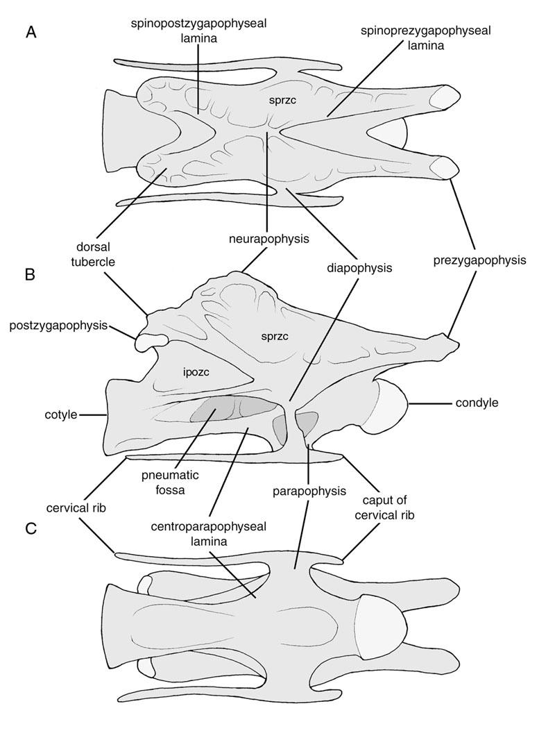
91 77
92 FIGURE 2. Axial sections of sauropod vertebrae showing pneumatic features. A. Haplocanthosaurus priscus (CM 897-7). B. Camarasaurus sp. (OMNH 01313). C. Saltasaurus loricatus (PVL , redrawn from Powell, 1986). After Wedel et al. (2000b). 78
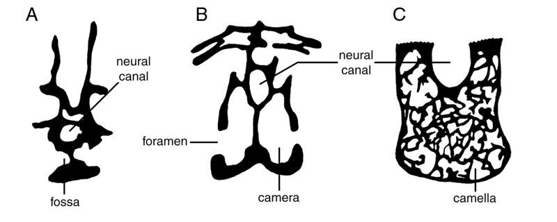
93 79
94 FIGURE 3. Air sacs and axial pneumatization in an extant avian. The body of bird in left lateral view, showing the cervical (C), interclavicular (I), anterior thoracic (AT), posterior thoracic (PT), and abdominal (AB) air sacs. The hatched area shows the volume change during exhalation. The cervical and anterior thoracic vertebrae are pneumatized by diverticula of the cervical air sacs. The posterior thoracic vertebrae and synsacrum are pneumatized via the abdominal air sacs. Diverticula of the abdominal air sacs usually invade the vertebral column at several points. Diverticula often unite when they come into contact, producing a system of continuous vertebral airways extending from the third cervical vertebra to the end of the synsacrum. Modified from Duncker (1971:fig. 8). 80
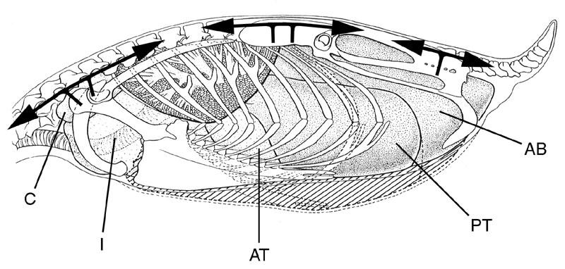
95 81
96 FIGURE 4. CT sections through the neck of an ostrich. The neck section was sealed with surgical gloves and cannulated with an air tube to re-inflate the pneumatic diverticula. Two axial sections are shown. In these images, air is black, bone is white, and soft tissues are grey. A. Note the essentially camellate nature of the external diverticula, which form aggregates of narrow tubes rather than large, simple sacs. B. The supramedullary airway can be seen to consist of three diverticula separated by thin membranes. C. Also apparent in this view are the cervical ribs, which appear ventrolateral to the centrum on either side. Scale bars are in cm. 82
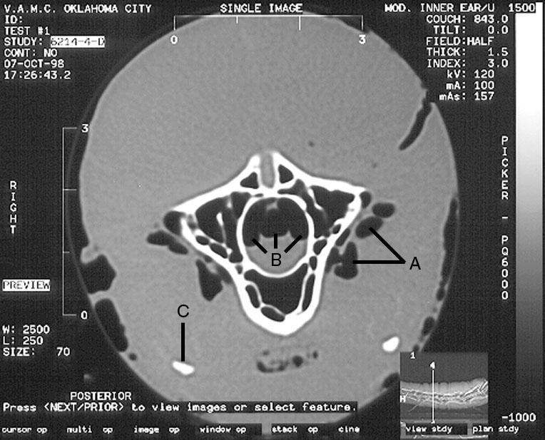
97 83
98 FIGURE 5. Pneumatization of the cervical series in the ostrich. The vertebrae are shown in midsagittal section (A), right lateral view (B), and horizontal section (C). Anterior is to the right. White arrows show the development of pneumatic diverticula and camellae, which are shown in black. The pattern of pneumatization is as follows. 1. Cervical diverticula advance through the transverse foramina. 2. Where the diverticula contact the vertebra, the exterior surface of the bone is remodeled. 3. Accessory diverticula enter the bone through existing nutrient foramina, and spread throughout the bone to produce a system of irregular, interconnected camellae. Eventually, even the neural spine and cervical ribs will be filled with pneumatic camellae. 4. Other branches of the main diverticula enter the neural canal at the intervertebral foramina, producing the supramedullary airways. 5. The neural spine may be pneumatized directly from the supramedullary airways. 6. The supramedullary airways evulse at the intervertebral joint to produce the anterior dorsal diverticula. 84
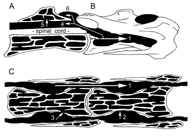
99 85
100 FIGURE 6. Cervical vertebrae of Apatosaurus and Camarasaurus differentiated. A. In Apatosaurus, the pre- and postzygapophyses are situated more or less directly above the condyle and cotyle, respectively. The intersection of the four diapophyseal laminae is therefore roughly symmetrical. Modified from Gilmore (1936:pl. 24). B. In Camarasaurus, the zygapophyses are located well forward of their respective centrum ends, and as a result the intersection of the diapophyseal laminae is anteriorly canted and lacks anter-posterior symmetry. Modified from Osborn and Mook (1921:pl. 67). 86
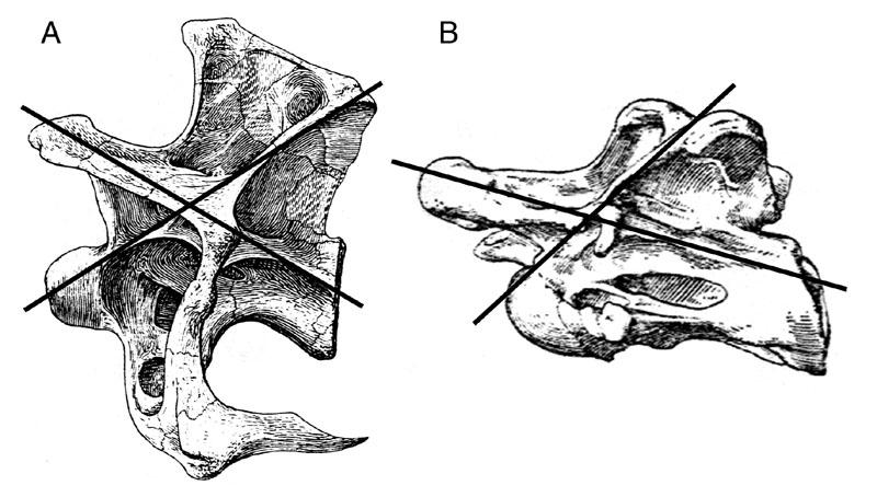
101 87
102 FIGURE 7. Vertebrae of basal sauropods. A. A section through the single available cervical of Vulcanodon. The centrum is strongly waisted by large fossae on either side. Modified from Cooper (1984:fig. 15). B.-F. Dorsal vertebrae of Barapasaurus, modified from Jain et al. (1979:pls. 101 and 102). B. A mid-dorsal vertebra in left lateral view. C.-D. Axial sections through mid-dorsal vertebrae, showing the extent of the lateral fossae. E.-F. Sagittal sections through mid-dorsal vertebrae, showing pneumatic chambers extending into the interior of the neural spine from the neural canal. Scale bars: A, 5 cm; B, 10 cm. 88
103 89
104 FIGURE 8. CT sections through a cervical vertebra of Apatosaurus, OMNH A. Specimen in left lateral view showing the location of CT sections. Two metal pins were used to repair the specimen and can be seen in this image. B. A section through the condyle (A, position 1) showing large, radially arranged camerae. C. A section through the mid-centrum (A, position 2) showing irregular and opportunistic development of camerae within the centrum. D. A section through the cotyle (A, position 3) showing small camerae arranged radially around the cotyle rim. Scale bar is 10 cm. 90
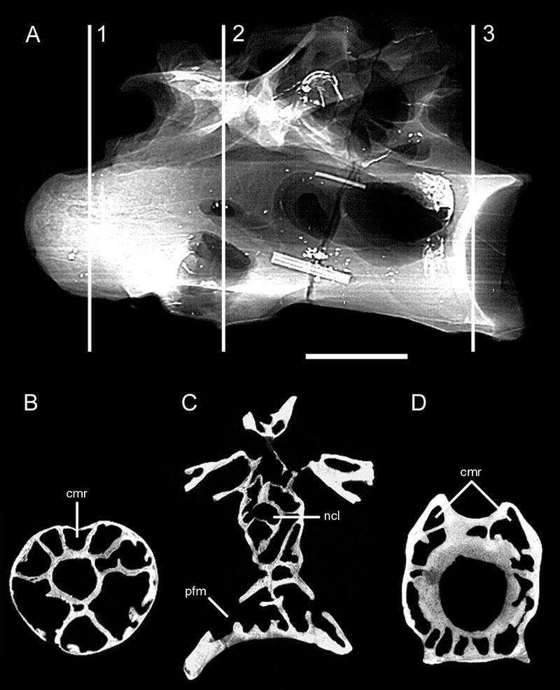
105 91
106 FIGURE 9. Digital model of juvenile vertebrae of Apatosaurus. CM 3390 includes two articulated cervical vertebrae from a juvenile animal. These vertebrae were scanned with overlapping slices, and the data were used to construct a digital model of the vertebrae in three dimensions. This digital model could then be sectioned along any axis. A. The digital model in left lateral view. B. The model in ventral view. C. An oblique slice through the centrum of the posterior vertebra (shown as a white line in B) reveals coels developing in the bone ahead of the growing camera. Also apparent in this view is the unfused neurocentral suture. Scale bar is in cm. 92
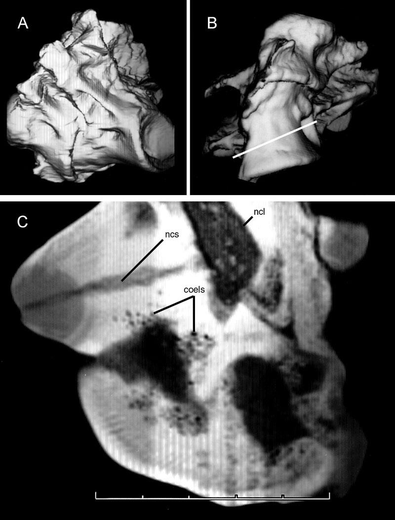
107 93
108 FIGURE 10. Dorsal, sacral, and caudal vertebrae of Diplodocus. All vertebrae are shown in left lateral view. A. Dorsal vertebrae and sacrum with ilium. Note the complex system of neural spine laminae. B. Caudal vertebrae Pneumatic foramina are present on caudals Modified from Osborn (1899:figs. 7 and 13). Scale bars are 50 cm. 94
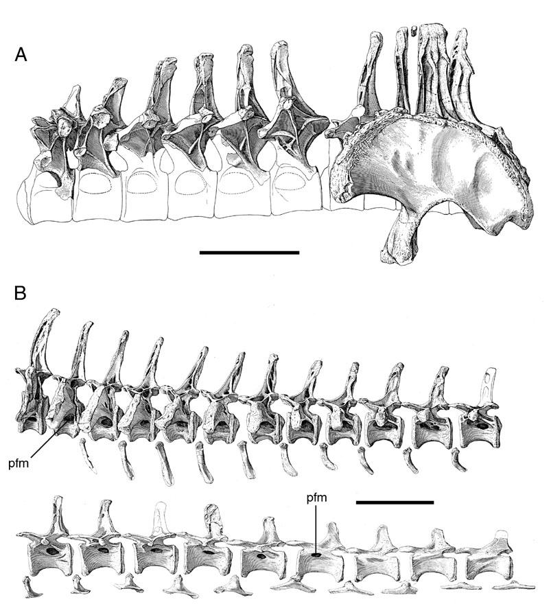
109 95
110 FIGURE 11. CT sections through a cervical vertebra of Diplodocus, BYU A. Specimen in left lateral view showing the location of CT sections. B.-C. Sections through the condyle (A, position 1) and anterior centrum (A, position 2), showing numerous small camerae in the centrum. D. A section through the posterior centrum (A, position 3). Note the irregular, asymmetrical shape of the median septum and the number of large camerae on either side. E. A section through the cotyle (A, position 4). Only a handful of large camerae are present. The regular, radially symmetrical camerae that circle the cotyle in Apatosaurus are not present in Diplodocus. Scale bar is 10 cm. 96
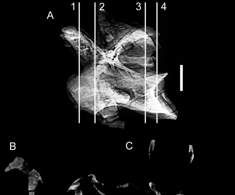
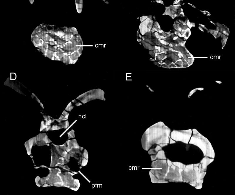
111 97
112 FIGURE 12. CT sections through a cervical vertebra from a juvenile individual of Diplodocus, CM The specimen is somewhat distorted diagonally by dorsolateral compression. A. Specimen in left lateral view showing the location of CT sections. B.- C. Sections through the condyle (A, position 1) and anterior centrum (A, position 2), showing the early development of a few relatively large camerae. D. A section through the middle of the centrum (A, position 3) showing large lateral fossae and a camera between the paired median septa. Scale bar is 5 cm. 98
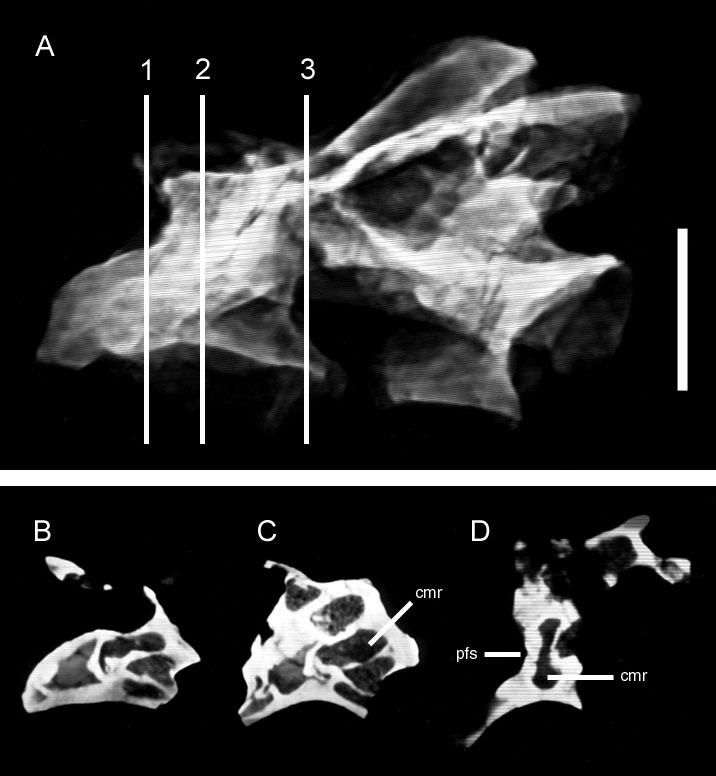
113 99
114 FIGURE 13. CT sections through a cervical vertebra of Haplocanthosaurus, CM A. Line drawing by R.K. Sanders showing the location of CT sections. B. A section through the condyle (A, position 1). The paired chambers are anterior extensions of the lateral fossae. C. A section through the middle of the centrum (A, position 2) showing large lateral fossae, which penetrate to a thin median septum. Scale bar is 10 cm. 100
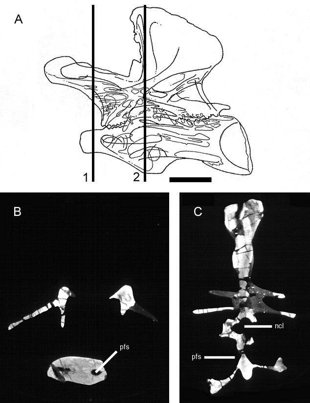
115 101
116 FIGURE 14. CT sections through a dorsal vertebra of Haplocanthosaurus, CM 572. The vertebra is complete, but only the centrum is shown here. A. The centrum in left lateral view showing the location of CT sections. B. Horizontal section through the midcentrum (A, position 1) showing the large lateral fossae. C. Horizontal section through the mid-centrum just below the rim of the lateral fossae (A, position 2). The centrum is solid at this point, and the lack of internalized chambers indicates that the lateral chambers are in fact fossae, rather than camerae. Scale bar is 5 cm. 102
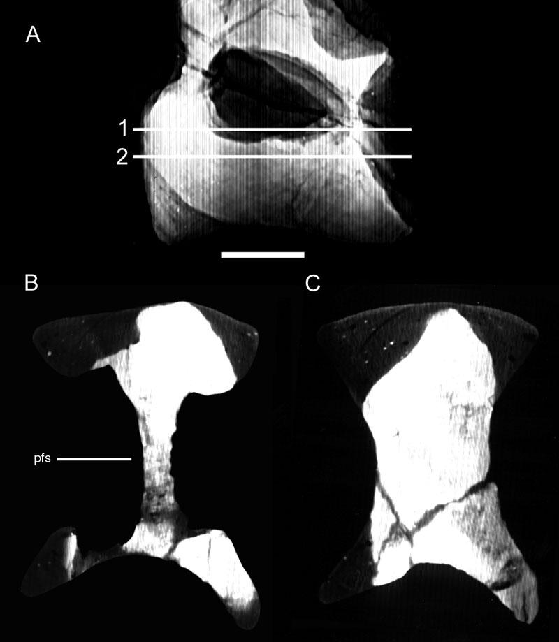
117 103
118 FIGURE 15. CT sections through a cervical vertebra of Camarasaurus, OMNH A. Specimen in left lateral view showing the location of CT sections. Small wires used in repairing the specimen can be seen in the condyle, cotyle, neural spine, and parapophysis. B. A section through the condyle (A, position 1). The camerae here are fewer and less complex than those in Apatosaurus and Diplodocus. C. A section through the mid-centrum (A, position 2) showing the relatively simple internal structure, composed mainly of large camerae. D. A section near the cotyle (A, position 3). The large lateral camerae extend posteriorly to the anterior side of the cotyle, but the smaller camerae seen in Apatosaurus and Diplodocus are absent. Scale bar is 10 cm. 104
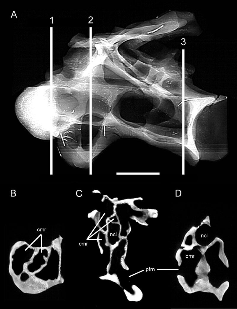
119 105
120 FIGURE 16. Vertebrae of Tendaguria. A. The single available cervical shown in left lateral view. In addition to the pneumatic foramina on the sides of the centrum, pneumatic fossae or foramina are also present in the supraprezyapophyseal and infrapostzygapophyseal cavities. B. A dorsal shown in right antero-lateral view. Note the fossae along the underside of the transverse processes. Modified from Bonaparte et al. (2000:fig. 17 and pl. 8). Scale bars are 10 cm. 106
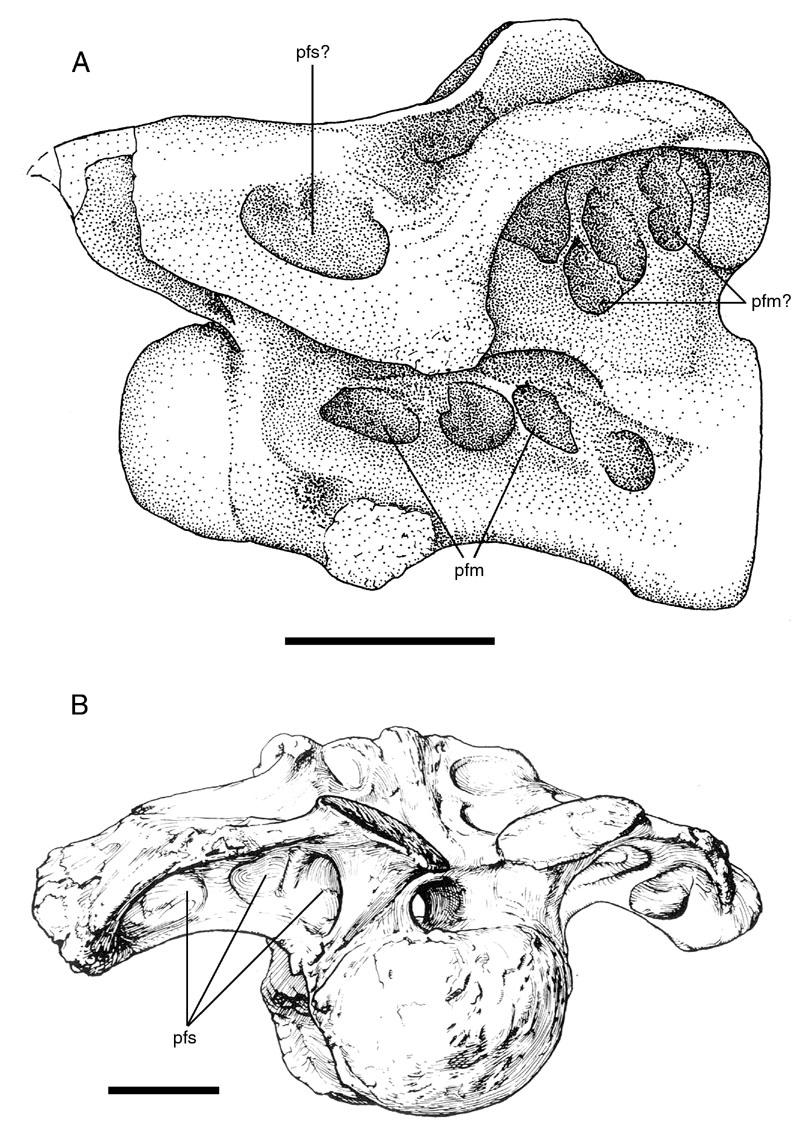
121 107
122 FIGURE 17. Cervical vertebrae of Camarasaurus and Tendaguria compared. Both vertebrae are shown in right lateral view. A. Camarasaurus. B. Tendaguria. Pneumatic fossae and foramina are shown in black. Deep, invasive pneumatic features are present in the supraprezygapophyseal and infrapostzygapophyseal cavities in these taxa, some diplodocids, and brachiosaurids (cf. Hatcher, 1901:pl. 7; Janensch, 1950:fig. 5, and Dalla Vecchia, 1998:fig. 2). Modified from Bonaparte et al. (2000:fig. 18). 108
123 109
124 FIGURE 18. Cervical vertebrae of Brachiosaurus. A. Atlas. B. Eighth cervical vertebra. In addition to the large foramina on the centrum, note the small, irregular fossae within the supraprezygapophyseal and infrapostzygapophyseal cavities. C. A sagittal section through the condyle of a cervical vertebra. D. An axial section through another condyle. Note the thin walls and irregular layout of the camellae. Modified from Janensch (1950:figs. 12, 43, 70, and 71). Scale bars: A, 10 cm; B, 20 cm. 110
125 111
126 FIGURE 19. Dorsal vertebrae of Brachiosaurus. A. Fourth dorsal vertebra in posterior view. B. The same vertebra in lateral view. C. Sagittal section through the anterior portion of a dorsal vertebra. D. Axial section through the middle of a dorsal vertebra. As in the cervical vertebrae, most of the centrum is occupied by large camerae, and camellae are restricted to the ends of the centrum. Modified from Janensch (1950:figs. 53, 54, 67, and 72). Scale bar is 20 cm. 112
127 113
128 FIGURE 20. CT sections through a cervical vertebra of Brachiosaurus, BYU The specimen is somewhat distorted diagonally, causing the distortion or collapse of some internal cavities. A. Specimen in left lateral view showing the location of CT sections. B. Section through condyle and prezygapophyses (A, position 1). Many camellae in the condyle are collapsed, but several remain relatively undistorted. C. Section through the centrum posterior to the neurapophysis (A, position 2). Although camellae are present in the neural spine and at the base of the median septum, the most prominent pneumatic cavities are the large, thick-walled camerae. The major laminae are connected to the median septum by thin strips of bone. D. Section through the cotyle (A, position 3) showing camellae surrounding the cotyle. Modified from Wedel et al. (2000b:fig. 12). Scale bar is 20 cm. 114
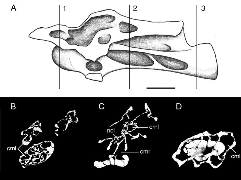
129 115
130 FIGURE 21. CT sections through a cervical vertebra of Sauroposeidon, OMNH A. Specimen in right lateral view showing the location of CT sections. B. Section through diapophysis (A, position 1) showing hypertrophied pneumatic fossae in the centrum and neural spine. C. Section through anterior centrum (A, position 2) showing camellae, which completely fill the internal structure. D. Section through postzygapophysis (A, position 3) showing camellae. The upper left quadrant of the image is obscured by x-ray beam hardening artifact, caused by the size and density of the specimen. Modified from Wedel et al. (2000b:fig. 12). Scale bar is 20 cm. 116
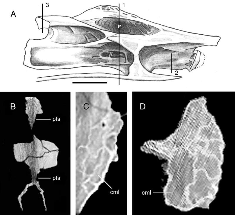
131 117
132 FIGURE 22. Cervical vertebrae of an unnamed brachiosaurid from Croatia. A. WN-V1, an anterior or middle cervical vertebra, shown in right lateral view. B. MPCM-V2, a posterior cervical vertebra, shown in antero-dorsal view. Note the complex honeycomb of camellae in the anterior part of each vertebra. Modified from Dalla Vecchia (1999:figs. 2 and 19). Scale bars are in cm. 118
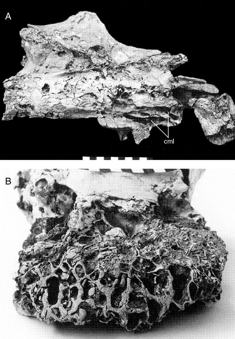
133 119
134 FIGURE 23. Cervical vertebrae of various titanosauriforms. A. USNM 5678, holotype cervical vertebra of Pleurocoelus. The presence of large lateral fossae is presumed to be a juvenile character. After Lull (1911b:pl. 15). B. FWMSH A, a cervical vertebra of the Jones Ranch sauropod on display in the Fort Worth Museum of Science and History. C. MN 4111-V, a partial cervical vertebra of Gondwanatitan. Note the relatively thick cortical bone and the presence of a median septum separating the few, relatively large cavities. Compare to the camellate vertebrae of Sauroposeidon (see Fig. 22) and Saltasaurus (see Fig. 26). Redrawn from Kellner and Azevedo (1999:fig. 5). Scale bars: A, 2 cm; B, 20 cm; C, 5 cm. 120
135 121
136 FIGURE 24. TMM , a dorsal neural spine of Alamosaurus. A. Anterior view. B. Dorsal view. The top of the neural spine is broken, revealing the camellate internal structure. Also note the reduced neural arch laminae and overall inflated appearance. Scale bar is 10 cm. 122
137 123
138 FIGURE 25. PVL , a dorsal vertebra of Saltasaurus. A. Posterior view. B. Lateral view, showing the location of cross sections. C. Horizontal section through centrum (B, position 1). D. Vertical section through centrum (B, position 2). E. Horizontal section through neural spine (B, position 3). Redrawn from Powell (1986:pls. 28 and 30). Scale bar is 5 cm. 124
139 125
140 FIGURE 26. Generations of cameral divisions in camerate sauropods. Pneumatic features arising at each generation are shown in white, those of previous generations are shown in light grey. A. Haplocanthosaurus priscus (CM 879-7), illustrating the procamerate condition, in which all pneumatic fossae are exposed to the surface. B. Camarasaurus sp. (OMNH 01313), illustrating the camerate condition, with three generations of camerae. C. Apatosaurus sp. (OMNH 01380), illustrating the polycamerate condition, with four generations of camerae and an increased number of camerae at each generation. After Wedel et al. (2000b:fig. 11). 126
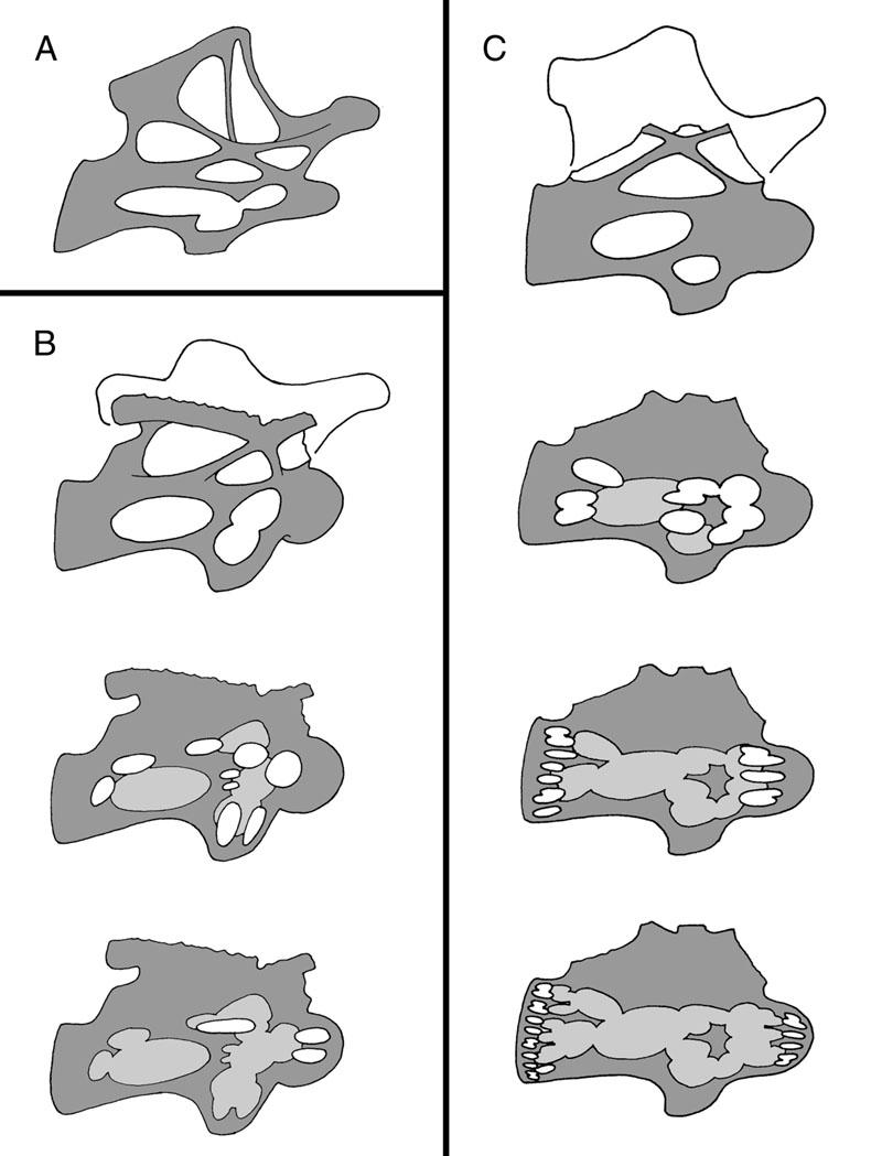
141 127
Postcranial Skeletal Pneumaticity in Sauropods and Its Implications for Mass Estimates
 SEVEN Postcranial Skeletal Pneumaticity in Sauropods and Its Implications for Mass Estimates Mathew J. Wedel O ne of the signal features of sauropods, and one of the cornerstones of our fascination with
Mathew John Wedel. B.S. (University of Oklahoma) A dissertation submitted in partial satisfaction of the. requirements for the degree of
 A dissertation submitted in partial satisfaction of the. requirements for the degree of") Postcranial Pneumaticity in Dinosaurs and the Origin of the Avian Lung by Mathew John Wedel B.S. (University of Oklahoma) 1997 A dissertation submitted in partial satisfaction of the requirements for the
Postcranial Pneumaticity in Dinosaurs and the Origin of the Avian Lung by Mathew John Wedel B.S. (University of Oklahoma) 1997 A dissertation submitted in partial satisfaction of the requirements for the
Anatomy. Name Section. The Vertebrate Skeleton
 Name Section Anatomy The Vertebrate Skeleton Vertebrate paleontologists get most of their knowledge about past organisms from skeletal remains. Skeletons are useful for gleaning information about an organism
WHAT PNEUMATICITY TELLS US ABOUT PROSAUROPODS, AND VICE VERSA
 [Special Papers in Palaeontology 77, 2007, pp. 207 222] WHAT PNEUMATICITY TELLS US ABOUT PROSAUROPODS, AND VICE VERSA by MATHEW WEDEL University of California Museum of Paleontology and Department of Integrative
Title: Phylogenetic Methods and Vertebrate Phylogeny
 Title: Phylogenetic Methods and Vertebrate Phylogeny Central Question: How can evolutionary relationships be determined objectively? Sub-questions: 1. What affect does the selection of the outgroup have
These small issues are easily addressed by small changes in wording, and should in no way delay publication of this first- rate paper.
 Reviewers' comments: Reviewer #1 (Remarks to the Author): This paper reports on a highly significant discovery and associated analysis that are likely to be of broad interest to the scientific community.
Animal Form and Function. Amphibians. United by several distinguishing apomorphies within the Vertebrata
 Animal Form and Function Kight Amphibians Class Amphibia (amphibia = living a double life) United by several distinguishing apomorphies within the Vertebrata 1. Skin Thought Question: For whom are integumentary
Geo 302D: Age of Dinosaurs. LAB 7: Dinosaur diversity- Saurischians
 Geo 302D: Age of Dinosaurs LAB 7: Dinosaur diversity- Saurischians Last lab you were presented with a review of major ornithischian clades. You also were presented with some of the kinds of plants that
Caudal Pneumaticity and Pneumatic Hiatuses in the Sauropod Dinosaurs Giraffatitan and Apatosaurus
 Caudal Pneumaticity and Pneumatic Hiatuses in the Sauropod Dinosaurs Giraffatitan and Apatosaurus Mathew J. Wedel 1 *, Michael P. Taylor 2 * 1 College of Osteopathic Medicine of the Pacific and College
Considerations of the neural laminae of sauropod dinosaurs and their morphofunctional meaning *
 Considerations of the neural laminae of sauropod dinosaurs and their morphofunctional meaning * Leonardo SALGADO, Rodolfo A. GARCÍA, & Juan D. DAZA Translated by Michael D. D Emic & Ariel Schepers; edited
Origin and Evolution of Birds. Read: Chapters 1-3 in Gill but limited review of systematics
 Origin and Evolution of Birds Read: Chapters 1-3 in Gill but limited review of systematics Review of Taxonomy Kingdom: Animalia Phylum: Chordata Subphylum: Vertebrata Class: Aves Characteristics: wings,
NEW INFORMATION ON A JUVENILE SAUROPOD SPECIMEN FROM THE MORRISON FORMATION AND THE REASSESSMENT OF ITS SYSTEMATIC POSITION
 [Palaeontology, Vol. 55, Part 3, 2012, pp. 567 582] NEW INFORMATION ON A JUVENILE SAUROPOD SPECIMEN FROM THE MORRISON FORMATION AND THE REASSESSMENT OF ITS SYSTEMATIC POSITION by JOSÉ L. CARBALLIDO 1,
THE LIGAMENT SYSTEM IN THE NECK OF RHEA AMERICANA AND ITS IMPLICATION FOR THE BIFURCATED NEURAL SPINES OF SAUROPOD DINOSAURS
 Journal of Vertebrate Paleontology 24(1):165 172, March 2004 2004 by the Society of Vertebrate Paleontology THE LIGAMENT SYSTEM IN THE NECK OF RHEA AMERICANA AND ITS IMPLICATION FOR THE BIFURCATED NEURAL
LABORATORY EXERCISE 6: CLADISTICS I
 Biology 4415/5415 Evolution LABORATORY EXERCISE 6: CLADISTICS I Take a group of organisms. Let s use five: a lungfish, a frog, a crocodile, a flamingo, and a human. How to reconstruct their relationships?
Tetrapod Similarites The Origins of Birds
 Tetrapod Similarites The Origins of Birds Birds Reptiles Mammals Integument Feathers, scales Scales Hair Digestive Horny bill Teeth Teeth Skeletal Fusion of bones Some fusion Some fusion Reduction in number
Diapsida. BIO2135 Animal Form and Function. Page 1. Diapsida (Reptilia, Sauropsida) Amniote eggs. Amniote egg. Temporal fenestra.
 Amniote eggs. Amniote egg. Temporal fenestra.") Diapsida (Reptilia, Sauropsida) Vertebrate phylogeny Mixini Chondrichthyes Sarcopterygii Mammalia Pteromyzontida Actinopterygii Amphibia Reptilia! 1! Amniota (autapomorphies) Costal ventilation Amniote
Diapsida (Reptilia, Sauropsida) Vertebrate phylogeny Mixini Chondrichthyes Sarcopterygii Mammalia Pteromyzontida Actinopterygii Amphibia Reptilia! 1! Amniota (autapomorphies) Costal ventilation Amniote
SAUROPOD DINOSAURS FROM THE EARLY CRETACEOUS OF MALAWI, AFRICA. Elizabeth M. Gomani
 Palaeontologia Electronica http://palaeo-electronica.org SAUROPOD DINOSAURS FROM THE EARLY CRETACEOUS OF MALAWI, AFRICA Elizabeth M. Gomani ABSTRACT At least two titanosaurian sauropod taxa have been discovered
Diapsida. BIO2135 Animal Form and Function. Page 1. Diapsida (Reptilia, Sauropsida) Amniote egg. Membranes. Vertebrate phylogeny
 Amniote egg. Membranes. Vertebrate phylogeny") Diapsida (Reptilia, Sauropsida) 1 Vertebrate phylogeny Mixini Chondrichthyes Sarcopterygii Mammalia Pteromyzontida Actinopterygii Amphibia Reptilia!! Amniota (autapomorphies) Costal ventilation Amniote
Diapsida (Reptilia, Sauropsida) 1 Vertebrate phylogeny Mixini Chondrichthyes Sarcopterygii Mammalia Pteromyzontida Actinopterygii Amphibia Reptilia!! Amniota (autapomorphies) Costal ventilation Amniote
'Rain' of dead birds on central NJ lawns explained; Federal culling program killed up to 5,000 Associated Press, January 27, 2009
 'Rain' of dead birds on central NJ lawns explained; Federal culling program killed up to 5,000 Associated Press, January 27, 2009 Study May Give Hope That Ivory-billed Woodpeckers Still Around Science
Phylogeny Reconstruction
 Phylogeny Reconstruction Trees, Methods and Characters Reading: Gregory, 2008. Understanding Evolutionary Trees (Polly, 2006) Lab tomorrow Meet in Geology GY522 Bring computers if you have them (they will
Vertebrates. Vertebrate Characteristics. 444 Chapter 14
 4 Vertebrates Key Concept All vertebrates have a backbone, which supports other specialized body structures and functions. What You Will Learn Vertebrates have an endoskeleton that provides support and
Biology Slide 1 of 50
 Biology 1 of 50 2 of 50 What Is a Reptile? What are the characteristics of reptiles? 3 of 50 What Is a Reptile? What Is a Reptile? A reptile is a vertebrate that has dry, scaly skin, lungs, and terrestrial
Name Class Date. After you read this section, you should be able to answer these questions:
 CHAPTER 14 4 Vertebrates SECTION Introduction to Animals BEFORE YOU READ After you read this section, you should be able to answer these questions: How are vertebrates different from invertebrates? How
Frog Dissection Information Manuel
 Frog Dissection Information Manuel Anatomical Terms: Used to explain directions and orientation of a organism Directions or Positions: Anterior (cranial)- toward the head Posterior (caudal)- towards the
Haplocanthosaurus (Saurischia: Sauropoda) from the lower Morrison Formation (Upper Jurassic) near Snowmass, Colorado
 from the lower Morrison Formation (Upper Jurassic) near Snowmass, Colorado") Volumina Jurassica, 2014, XII (2): 197 210 DOI: 10.5604/17313708.1130144 Haplocanthosaurus (Saurischia: Sauropoda) from the lower Morrison Formation (Upper Jurassic) near Snowmass, Colorado John R. FosteR
Volumina Jurassica, 2014, XII (2): 197 210 DOI: 10.5604/17313708.1130144 Haplocanthosaurus (Saurischia: Sauropoda) from the lower Morrison Formation (Upper Jurassic) near Snowmass, Colorado John R. FosteR
A New Sauropod Dinosaur From the Early Cretaceous of Oklahoma. Mathew J. Wedel. Oklahoma Museum of Natural History. and. Department of Zoology
 ..., ' A New Sauropod Dinosaur From the Early Cretaceous of Oklahoma by Mathew. Wedel Oklahoma Museum of Natural History " and Department of Zoology University of Oklahoma 1335 Asp Avenue Norman, OK 73019-0606
LABORATORY EXERCISE 7: CLADISTICS I
 Biology 4415/5415 Evolution LABORATORY EXERCISE 7: CLADISTICS I Take a group of organisms. Let s use five: a lungfish, a frog, a crocodile, a flamingo, and a human. How to reconstruct their relationships?
A Nomenclature for Vertebral Fossae in Sauropods and Other Saurischian Dinosaurs
 A Nomenclature for Vertebral Fossae in Sauropods and Other Saurischian Dinosaurs Jeffrey A. Wilson*, Michael D. D Emic, Takehito Ikejiri, Emile M. Moacdieh, John A. Whitlock Museum of Paleontology and
1 Describe the anatomy and function of the turtle shell. 2 Describe respiration in turtles. How does the shell affect respiration?
 GVZ 2017 Practice Questions Set 1 Test 3 1 Describe the anatomy and function of the turtle shell. 2 Describe respiration in turtles. How does the shell affect respiration? 3 According to the most recent
A new species of sauropod, Mamenchisaurus anyuensis sp. nov.
 A new species of sauropod, Mamenchisaurus anyuensis sp. nov. by Xinlu He, Suihua Yang, Kaiji Cai, Kui Li, and Zongwen Liu Chengdu University of Technology Papers on Geosciences Contributed to the 30th
IS THERE AN OPTION FOR A PNEUMATIC STABILIZATION OF SAUROPOD NECKS? AN EXPERIMENTAL AND ANATOMICAL APPROACH. Daniela Schwarz-Wings and Eberhard Frey
 Palaeontologia Electronica http://palaeo-electronica.org IS THERE AN OPTION FOR A PNEUMATIC STABILIZATION OF SAUROPOD NECKS? AN EXPERIMENTAL AND ANATOMICAL APPROACH Daniela Schwarz-Wings and Eberhard Frey
Course # Course Name Credits
 Curriculum Outline: Course # Course Name Credits Term 1 Courses VET 100 Introduction to Veterinary Technology 3 ENG 105 English Composition 3 MATH 120 Technical Mathematics 3 VET 130 Animal Biology/ Anatomy
Origin and Evolution of Birds. Read: Chapters 1-3 in Gill but limited review of systematics
 Origin and Evolution of Birds Read: Chapters 1-3 in Gill but limited review of systematics Review of Taxonomy Kingdom: Animalia Phylum: Chordata Subphylum: Vertebrata Class: Aves Characteristics: wings,
A NEW TITANOSAURIFORM SAUROPOD (DINOSAURIA: SAURISCHIA) FROM THE EARLY CRETACEOUS OF CENTRAL TEXAS AND ITS PHYLOGENETIC RELATIONSHIPS. Peter J.
 FROM THE EARLY CRETACEOUS OF CENTRAL TEXAS AND ITS PHYLOGENETIC RELATIONSHIPS. Peter J.") Palaeontologia Electronica http://palaeo-electronica.org A NEW TITANOSAURIFORM SAUROPOD (DINOSAURIA: SAURISCHIA) FROM THE EARLY CRETACEOUS OF CENTRAL TEXAS AND ITS PHYLOGENETIC RELATIONSHIPS Peter J. Rose
Palaeontologia Electronica http://palaeo-electronica.org A NEW TITANOSAURIFORM SAUROPOD (DINOSAURIA: SAURISCHIA) FROM THE EARLY CRETACEOUS OF CENTRAL TEXAS AND ITS PHYLOGENETIC RELATIONSHIPS Peter J. Rose
Diversity of Animals
 Classifying Animals Diversity of Animals Animals can be classified and grouped based on similarities in their characteristics. Animals make up one of the major biological groups of classification. All
Exceptional fossil preservation demonstrates a new mode of axial skeleton elongation in early ray-finned fishes
 Supplementary Information Exceptional fossil preservation demonstrates a new mode of axial skeleton elongation in early ray-finned fishes Erin E. Maxwell, Heinz Furrer, Marcelo R. Sánchez-Villagra Supplementary
Biology 340 Comparative Embryology Lecture 12 Dr. Stuart Sumida. Evo-Devo Revisited. Development of the Tetrapod Limb
 Biology 340 Comparative Embryology Lecture 12 Dr. Stuart Sumida Evo-Devo Revisited Development of the Tetrapod Limb Limbs whether fins or arms/legs for only in particular regions or LIMB FIELDS. Primitively
Australian and New Zealand College of Veterinary Scientists. Membership Examination. Veterinary Radiology (Small Animal) Paper 1
 Paper 1") Australian and New Zealand College of Veterinary Scientists Membership Examination June 2016 Veterinary Radiology (Small Animal) Paper 1 Perusal time: Fifteen (15) minutes Time allowed: Two (2) hours after
Australian and New Zealand College of Veterinary Scientists Membership Examination June 2016 Veterinary Radiology (Small Animal) Paper 1 Perusal time: Fifteen (15) minutes Time allowed: Two (2) hours after
A new basal sauropodiform dinosaur from the Lower Jurassic of Yunnan Province, China
 SUPPLEMENTARY INFORMATION A new basal sauropodiform dinosaur from the Lower Jurassic of Yunnan Province, China Ya-Ming Wang 1, Hai-Lu You 2,3 *, Tao Wang 4 1 School of Earth Sciences and Resources, China
VERTEBRATE READING. Fishes
 VERTEBRATE READING Fishes The first vertebrates to become a widespread, predominant life form on earth were fishes. Prior to this, only invertebrates, such as mollusks, worms and squid-like animals, would
BREATHING WHICH IS NOT RESPIRATION
 BREATHING WHICH IS NOT RESPIRATION Breathing vs. Respiration All animals respire. A lot of people think respiration means breathing- this is not true! Breathing is the physical process of inhaling oxygen
1. Examine the specimens of sponges on the lab table. Which of these are true sponges? Explain your answers.
 Station #1 - Porifera 1. Examine the specimens of sponges on the lab table. Which of these are true sponges? Explain your answers. 2. Sponges are said to have an internal special skeleton. Examine the
The Effect of Intervertebral Cartilage on Neutral Posture and Range of Motion in the Necks of Sauropod Dinosaurs
 The Effect of Intervertebral Cartilage on Neutral Posture and Range of Motion in the Necks of Sauropod Dinosaurs Michael P. Taylor 1 *, Mathew J. Wedel 2 1 Department of Earth Sciences, University of Bristol,
New Carnivorous Dinosaurs from the Upper Cretaceous of Mongolia
 1955 Doklady, Academy of Sciences USSR 104 (5):779-783 New Carnivorous Dinosaurs from the Upper Cretaceous of Mongolia E. A. Maleev (translated by F. J. Alcock) The present article is a summary containing
2 nd Term Final. Revision Sheet. Students Name: Grade: 11 A/B. Subject: Biology. Teacher Signature. Page 1 of 11
 2 nd Term Final Revision Sheet Students Name: Grade: 11 A/B Subject: Biology Teacher Signature Page 1 of 11 Nour Al Maref International School Riyadh, Saudi Arabia Biology Worksheet (2 nd Term) Chapter-26
d a Name Vertebrate Evolution - Exam 2 1. (12) Fill in the blanks
 Fill in the blanks") Vertebrate Evolution - Exam 2 1. (12) Fill in the blanks 100 points Name f e c d a Identify the structures (for c and e, identify the entire structure, not the individual elements. b a. b. c. d. e. f.
Vertebrate Evolution - Exam 2 1. (12) Fill in the blanks 100 points Name f e c d a Identify the structures (for c and e, identify the entire structure, not the individual elements. b a. b. c. d. e. f.
8/19/2013. Topic 5: The Origin of Amniotes. What are some stem Amniotes? What are some stem Amniotes? The Amniotic Egg. What is an Amniote?
 Topic 5: The Origin of Amniotes Where do amniotes fall out on the vertebrate phylogeny? What are some stem Amniotes? What is an Amniote? What changes were involved with the transition to dry habitats?
Fish 2/26/13. Chordates 2. Sharks and Rays (about 470 species) Sharks etc Bony fish. Tetrapods. Osteichthans Lobe fins and lungfish
 Sharks etc Bony fish. Tetrapods. Osteichthans Lobe fins and lungfish") Chordates 2 Sharks etc Bony fish Osteichthans Lobe fins and lungfish Tetrapods ns Reptiles Birds Feb 27, 2013 Chordates ANCESTRAL DEUTEROSTOME Notochord Common ancestor of chordates Head Vertebral column
Chordates 2 Sharks etc Bony fish Osteichthans Lobe fins and lungfish Tetrapods ns Reptiles Birds Feb 27, 2013 Chordates ANCESTRAL DEUTEROSTOME Notochord Common ancestor of chordates Head Vertebral column
Question Set 1: Animal EVOLUTIONARY BIODIVERSITY
 Biology 162 LAB EXAM 2, AM Version Thursday 24 April 2003 page 1 Question Set 1: Animal EVOLUTIONARY BIODIVERSITY (a). We have mentioned several times in class that the concepts of Developed and Evolved
SOAR Research Proposal Summer How do sand boas capture prey they can t see?
 SOAR Research Proposal Summer 2016 How do sand boas capture prey they can t see? Faculty Mentor: Dr. Frances Irish, Assistant Professor of Biological Sciences Project start date and duration: May 31, 2016
Cladistics (reading and making of cladograms)
") Cladistics (reading and making of cladograms) Definitions Systematics The branch of biological sciences concerned with classifying organisms Taxon (pl: taxa) Any unit of biological diversity (eg. Animalia,
Cladistics (reading and making of cladograms) Definitions Systematics The branch of biological sciences concerned with classifying organisms Taxon (pl: taxa) Any unit of biological diversity (eg. Animalia,
Video Assignments. Microraptor PBS The Four-winged Dinosaur Mark Davis SUNY Cortland Library Online
 Video Assignments Microraptor PBS The Four-winged Dinosaur Mark Davis SUNY Cortland Library Online Radiolab Apocalyptical http://www.youtube.com/watch?v=k52vd4wbdlw&feature=youtu.be Minute 13 through minute
muscles (enhancing biting strength). Possible states: none, one, or two.
. Possible states: none, one, or two.") Reconstructing Evolutionary Relationships S-1 Practice Exercise: Phylogeny of Terrestrial Vertebrates In this example we will construct a phylogenetic hypothesis of the relationships between seven taxa
Reconstructing Evolutionary Relationships S-1 Practice Exercise: Phylogeny of Terrestrial Vertebrates In this example we will construct a phylogenetic hypothesis of the relationships between seven taxa
From Slime to Scales: Evolution of Reptiles. Review: Disadvantages of Being an Amphibian
 From Slime to Scales: Evolution of Reptiles Review: Disadvantages of Being an Amphibian Gelatinous eggs of amphibians cannot survive out of water, so amphibians are limited in terms of the environments
Biology. Slide 1of 50. End Show. Copyright Pearson Prentice Hall
 Biology 1of 50 2of 50 Phylogeny of Chordates Nonvertebrate chordates Jawless fishes Sharks & their relatives Bony fishes Reptiles Amphibians Birds Mammals Invertebrate ancestor 3of 50 A vertebrate dry,
From Reptiles to Aves
 First Vertebrates From Reptiles to Aves Evolutions of Fish to Amphibians Evolution of Amphibians to Reptiles Evolution of Reptiles to Dinosaurs to Birds Common Ancestor of Birds and Reptiles: Thecodonts
Abstract RESEARCH ARTICLE
 RESEARCH ARTICLE Vertebral Pneumaticity in the Ornithomimosaur Archaeornithomimus (Dinosauria: Theropoda) Revealed by Computed Tomography Imaging and Reappraisal of Axial Pneumaticity in Ornithomimosauria
Introduction to Cladistic Analysis
 3.0 Copyright 2008 by Department of Integrative Biology, University of California-Berkeley Introduction to Cladistic Analysis tunicate lamprey Cladoselache trout lungfish frog four jaws swimbladder or
Animal Diversity III: Mollusca and Deuterostomes
 Animal Diversity III: Mollusca and Deuterostomes Objectives: Be able to identify specimens from the main groups of Mollusca and Echinodermata. Be able to distinguish between the bilateral symmetry on a
Mechanism of a Crocodile s Circulatory System
 Mechanism of a Crocodile s Circulatory System Figure 1. A crocodile diving at Botswana (Nachoum, A. 2017) Ever wonder in one of those animal documentaries we watch in television, wherein a crocodile glides
What is a dinosaur? Reading Practice
 Reading Practice What is a dinosaur? A. Although the name dinosaur is derived from the Greek for "terrible lizard", dinosaurs were not, in fact, lizards at all. Like lizards, dinosaurs are included in
Do the traits of organisms provide evidence for evolution?
 PhyloStrat Tutorial Do the traits of organisms provide evidence for evolution? Consider two hypotheses about where Earth s organisms came from. The first hypothesis is from John Ray, an influential British
Animal Diversity wrap-up Lecture 9 Winter 2014
 Animal Diversity wrap-up Lecture 9 Winter 2014 1 Animal phylogeny based on morphology & development Fig. 32.10 2 Animal phylogeny based on molecular data Fig. 32.11 New Clades 3 Lophotrochozoa Lophophore:
NOTES ON THE EVOLUTION OF VERTEBRAE IN THE SAUROPODOMORPHA. José F. BONAPARTE *
 NOTES ON THE EVOLUTION OF VERTEBRAE IN THE SAUROPODOMORPHA by José F. BONAPARTE * Museo Argentino de Ciencias Naturales Consejo Nacional de Investigaciones Científicas y Técnicas Avenida Angel Gallardo
Characteristics of a Reptile. Vertebrate animals Lungs Scaly skin Amniotic egg
 Reptiles Characteristics of a Reptile Vertebrate animals Lungs Scaly skin Amniotic egg Characteristics of Reptiles Adaptations to life on land More efficient lungs and a better circulator system were develope
CAT DISSECTION A LABORATORY GUIDE
 8546d_fm_i-iv 6/26/02 3:51 PM Page 3 mac62 mac62:1253_ge: CAT DISSECTION A LABORATORY GUIDE CONNIE ALLEN VALERIE HARPER Edison Community College John Wiley & Sons, Inc. 8546d_fm_i-iv 6/26/02 12:17 PM Page
THE SKELETON RECONSTRUCTION OF BRACHIOSAURUS BRANCAI
 THE SKELETON RECONSTRUCTION OF BRACHIOSAURUS BRANCAI BY W. JANENSCH WITH PLATES VI VIII PALAEONTOGRAPHICA 1950, Supplement VII, Reihe I, Teil III, 97 103. TRANSLATED BY GERHARD MAIER JUNE 2007 97 A reconstruction
Supplementary Figure 1 Cartilaginous stages in non-avian amniotes. (a) Drawing of early ankle development of Alligator mississippiensis, as reported
 Drawing of early ankle development of Alligator mississippiensis, as reported") Supplementary Figure 1 Cartilaginous stages in non-avian amniotes. (a) Drawing of early ankle development of Alligator mississippiensis, as reported by a previous study 1. The intermedium is formed at
Supplementary Figure 1 Cartilaginous stages in non-avian amniotes. (a) Drawing of early ankle development of Alligator mississippiensis, as reported by a previous study 1. The intermedium is formed at
TRACHEMYS SCULPTA. A nearly complete articulated carapace and plastron of an Emjdd A NEAKLY COMPLETE SHELL OF THE EXTINCT TURTLE,
 A NEAKLY COMPLETE SHELL OF THE EXTINCT TURTLE, TRACHEMYS SCULPTA By Charles W. Gilmore Curator of Vertebrate Paleontology, United States National Museum INTRODUCTION A nearly complete articulated carapace
BIOL241 & BIOL242 Links to Useful www Sites 2012
 Links to Study and Review Sites in Anatomy and Physiology Histology - Study of Tissue Types Histology Practice Exam at Gateway CC http://www.gwc.maricopa.edu/class/bio201/histoprc/prac1q.htm Histology
Modern taxonomy. Building family trees 10/10/2011. Knowing a lot about lots of creatures. Tom Hartman. Systematics includes: 1.
 Modern taxonomy Building family trees Tom Hartman www.tuatara9.co.uk Classification has moved away from the simple grouping of organisms according to their similarities (phenetics) and has become the study
Comparative Zoology Portfolio Project Assignment
 Comparative Zoology Portfolio Project Assignment Using your knowledge from the in class activities, your notes, you Integrated Science text, or the internet, you will look at the major trends in the evolution
University of Canberra. This thesis is available in print format from the University of Canberra Library.
 University of Canberra This thesis is available in print format from the University of Canberra Library. If you are the author of this thesis and wish to have the whole thesis loaded here, please contact
Stuart S. Sumida Biology 342. (Simplified)Phylogeny of Archosauria
Phylogeny of Archosauria") Stuart S. Sumida Biology 342 (Simplified)Phylogeny of Archosauria Remember, we re studying AMNIOTES. Defined by: EMBRYOLOGICAL FEATURES: amnion, chorion, allantois, yolk sac. ANATOMICAL FEATURES: lack
Stuart S. Sumida Biology 342 (Simplified)Phylogeny of Archosauria Remember, we re studying AMNIOTES. Defined by: EMBRYOLOGICAL FEATURES: amnion, chorion, allantois, yolk sac. ANATOMICAL FEATURES: lack
Vertebrates. Vertebrates are animals that have a backbone and an endoskeleton.
 Vertebrates Vertebrates are animals that have a backbone and an endoskeleton. The backbone replaces the notochord and contains bones called vertebrae. An endoskeleton is an internal skeleton that protects
Field Trip: Harvard Museum of Natural History (HMNH)
") Field Trip: Harvard Museum of Natural History (HMNH) Objectives To observe the diversity of animals. To compare and contrast the various adaptations, body plans, etc. of the animals found at the HMNH.
Field Trip: Harvard Museum of Natural History (HMNH) Objectives To observe the diversity of animals. To compare and contrast the various adaptations, body plans, etc. of the animals found at the HMNH.
REPORT FROM A BOU-FUNDED PROJECT
 Pneumatisation and internal architecture of the Southern Cassowary Casuarius casuarius casque: a microct study CHARLOTTE A. BRASSEY 1*, THOMAS O MAHONEY 2 1 School of Science and the Environment, Manchester
Europe s largest dinosaur? A giant brachiosaurid cervical vertebra from the Wessex Formation (Early Cretaceous) of southern England
 of southern England") Cretaceous Research 25 (2004) 787 795 www.elsevier.com/locate/cretres Europe s largest dinosaur? A giant brachiosaurid cervical vertebra from the Wessex Formation (Early Cretaceous) of southern England
Cretaceous Research 25 (2004) 787 795 www.elsevier.com/locate/cretres Europe s largest dinosaur? A giant brachiosaurid cervical vertebra from the Wessex Formation (Early Cretaceous) of southern England
Introduction to phylogenetic trees and tree-thinking Copyright 2005, D. A. Baum (Free use for non-commercial educational pruposes)
") Introduction to phylogenetic trees and tree-thinking Copyright 2005, D. A. Baum (Free use for non-commercial educational pruposes) Phylogenetics is the study of the relationships of organisms to each other.
Introduction to phylogenetic trees and tree-thinking Copyright 2005, D. A. Baum (Free use for non-commercial educational pruposes) Phylogenetics is the study of the relationships of organisms to each other.
Comments on the Beauceron Standard By M. Maurice Hermel (Translated by C. Batson)
") Comments on the Beauceron Standard By M. Maurice Hermel (Translated by C. Batson) The following are comments written by M. Hermel for the FCI Standard #44 published on 10/25/06. They were approved by the
Comments on the Beauceron Standard By M. Maurice Hermel (Translated by C. Batson) The following are comments written by M. Hermel for the FCI Standard #44 published on 10/25/06. They were approved by the
Postilla PEABODY MUSEUM OF NATURAL HISTORY YALE UNIVERSITY NEW HAVEN, CONNECTICUT, U.S.A.
 Postilla PEABODY MUSEUM OF NATURAL HISTORY YALE UNIVERSITY NEW HAVEN, CONNECTICUT, U.S.A. Number 117 18 March 1968 A 7DIAPSID (REPTILIA) PARIETAL FROM THE LOWER PERMIAN OF OKLAHOMA ROBERT L. CARROLL REDPATH
FURTHER STUDIES ON TWO SKELETONS OF THE BLACK RIGHT WHALE IN THE NORTH PACIFIC
 FURTHER STUDIES ON TWO SKELETONS OF THE BLACK RIGHT WHALE IN THE NORTH PACIFIC HIDEO OMURA, MASAHARU NISHIWAKI* AND TOSHIO KASUYA* ABSTRACT Two skeletons of the black right whale were studied, supplementing
Phylogeny of Animalia (overview)
") The Diversity of Animals 2 Chapter 23 Phylogeny of Animalia (overview) Key features of Chordates Phylum Chordata (the Chordates) includes both invertebrates and vertebrates that share (at some point in
The Diversity of Animals 2 Chapter 23 Phylogeny of Animalia (overview) Key features of Chordates Phylum Chordata (the Chordates) includes both invertebrates and vertebrates that share (at some point in
Geo 302D: Age of Dinosaurs LAB 4: Systematics Part 1
 Geo 302D: Age of Dinosaurs LAB 4: Systematics Part 1 Systematics is the comparative study of biological diversity with the intent of determining the relationships between organisms. Humankind has always
Most amphibians begin life as aquatic organisms and then live on land as adults.
 Section 3: Most amphibians begin life as aquatic organisms and then live on land as adults. K What I Know W What I Want to Find Out L What I Learned Essential Questions What were the kinds of adaptations
NEW SAUROPOD FROM THE LOWER CRETACEOUS OF UTAH, USA
 ORYCTOS, Vol. 2 : 21-37, Décembre 1999 NEW SAUROPOD FROM THE LOWER CRETACEOUS OF UTAH, USA Virginia TIDWELL, Kenneth CARPENTER and William BROOKS Department of Earth and Space Sciences, Denver Museum of
Sec KEY CONCEPT Reptiles, birds, and mammals are amniotes.
 Thu 4/27 Learning Target Class Activities *attached below (scroll down)* Website: my.hrw.com Username: bio678 Password:a4s5s Activities Students will describe the evolutionary significance of amniotic
Are the dinosauromorph femora from the Upper Triassic of Hayden Quarry (New Mexico) three stages in a growth series of a single taxon?
 three stages in a growth series of a single taxon?") Anais da Academia Brasileira de Ciências (2017) 89(2): 835-839 (Annals of the Brazilian Academy of Sciences) Printed version ISSN 0001-3765 / Online version ISSN 1678-2690 http://dx.doi.org/10.1590/0001-3765201720160583
Anais da Academia Brasileira de Ciências (2017) 89(2): 835-839 (Annals of the Brazilian Academy of Sciences) Printed version ISSN 0001-3765 / Online version ISSN 1678-2690 http://dx.doi.org/10.1590/0001-3765201720160583
What are taxonomy, classification, and systematics?
 Topic 2: Comparative Method o Taxonomy, classification, systematics o Importance of phylogenies o A closer look at systematics o Some key concepts o Parts of a cladogram o Groups and characters o Homology
Copyright is owned by the Author of the thesis. Permission is given for a copy to be downloaded by an individual for the purpose of research and
 Copyright is owned by the Author of the thesis. Permission is given for a copy to be downloaded by an individual for the purpose of research and private study only. The thesis may not be reproduced elsewhere
Species: Panthera pardus Genus: Panthera Family: Felidae Order: Carnivora Class: Mammalia Phylum: Chordata
 CHAPTER 6: PHYLOGENY AND THE TREE OF LIFE AP Biology 3 PHYLOGENY AND SYSTEMATICS Phylogeny - evolutionary history of a species or group of related species Systematics - analytical approach to understanding
Shedding Light on the Dinosaur-Bird Connection
 Shedding Light on the Dinosaur-Bird Connection This text is provided courtesy of the American Museum of Natural History. When people think of dinosaurs, two types generally come to mind: the huge herbivores
Modern Evolutionary Classification. Lesson Overview. Lesson Overview Modern Evolutionary Classification
 Lesson Overview 18.2 Modern Evolutionary Classification THINK ABOUT IT Darwin s ideas about a tree of life suggested a new way to classify organisms not just based on similarities and differences, but
Reptilian Requirements Created by the North Carolina Aquarium at Fort Fisher Education Section
 Essential Question: North Carolina Aquariums Education Section Reptilian Requirements Created by the North Carolina Aquarium at Fort Fisher Education Section What physical and behavioral adaptations do
Evidence for Avian Intrathoracic Air Sacs in a New Predatory Dinosaur from Argentina
 Evidence for Avian Intrathoracic Air Sacs in a New Predatory Dinosaur from Argentina Paul C. Sereno 1 *, Ricardo N. Martinez 2, Jeffrey A. Wilson 3, David J. Varricchio 4, Oscar A. Alcober 2, Hans C. E.
The Origin of Birds. Technical name for birds is Aves, and avian means of or concerning birds.
 The Origin of Birds Technical name for birds is Aves, and avian means of or concerning birds. Birds have many unusual synapomorphies among modern animals: [ Synapomorphies (shared derived characters),
.56 m. (22 in.). COMPSOGNATHOID DINOSAUR FROM THE. Medicine Bow, Wyoming, by the American Museum Expedition
. COMPSOGNATHOID DINOSAUR FROM THE. Medicine Bow, Wyoming, by the American Museum Expedition") Article XII.-ORNITHOLESTES HERMANNI, A NEW COMPSOGNATHOID DINOSAUR FROM THE UPPER JURASSIC. By HENRY FAIRFIELD OSBORN. The type skeleton (Amer. Mus. Coll. No. 6I9) of this remarkable animal was discovered
Article XII.-ORNITHOLESTES HERMANNI, A NEW COMPSOGNATHOID DINOSAUR FROM THE UPPER JURASSIC. By HENRY FAIRFIELD OSBORN. The type skeleton (Amer. Mus. Coll. No. 6I9) of this remarkable animal was discovered
May 10, SWBAT analyze and evaluate the scientific evidence provided by the fossil record.
 May 10, 2017 Aims: SWBAT analyze and evaluate the scientific evidence provided by the fossil record. Agenda 1. Do Now 2. Class Notes 3. Guided Practice 4. Independent Practice 5. Practicing our AIMS: E.3-Examining
Topic 13: Energetics & Performance. How are gas exchange, circulation & metabolism inter-related?
 Topic 3: Energetics & Performance How are gas exchange, circulation & metabolism interrelated? How is it done in air and water? What organs are involved in each case? How does ventilation differ among
290 SHUFELDT, Remains of Hesperornis.
 290 SHUFELDT, Remains of Hesperornis. [ Auk [July THE FOSSIL REMAINS OF A SPECIES OF HESPERORNIS FOUND IN MONTANA. BY R. W. SHUFELD% M.D. Plate XI7III. ExR,¾ in November, 1914, Mr. Charles W. Gihnore,