Postcranial Skeletal Pneumaticity in Sauropods and Its Implications for Mass Estimates
|
|
|
- Griselda Black
- 6 years ago
- Views:
Transcription
1 SEVEN Postcranial Skeletal Pneumaticity in Sauropods and Its Implications for Mass Estimates Mathew J. Wedel O ne of the signal features of sauropods, and one of the cornerstones of our fascination with them, is their apparent efficiency of design. The presacral neural spines of all sauropods have a complex of bony ridges or plates known as vertebral laminae (fig. 7.1; abbreviations used in the figures are listed below). In addition, the vertebral centra of most sauropods bear deep fossae or have large foramina that open into internal chambers. The laminae and cavities of sauropod vertebrae are often considered to be adaptations for mass reduction (Osborn 1899; Hatcher 1901; Gilmore 1925) and have been important in studies of sauropod evolution (McIntosh 1990; Wilson 1999). The possibility that these structures were pneumatic that they contained or partitioned air-filled diverticula of the lungs or air sacs has been recognized for over a century (Seeley 1870; Janensch 1947). However, pneumaticity in sauropods has received little attention until recently (Britt 1997; Wilson 1999; Wedel 2003a, 2003b). My goal here is to review previous work on pneumaticity in sauropods, discuss some outstanding problems, and outline possible directions for future studies. To that end, the chapter is organized around three questions. What criteria do we use to infer pneumaticity in sauropod fossils? What characteristics of pneumatic bones have been (or could be) described? and How can we apply data on skeletal pneumaticity to paleobiological problems, such as estimating the masses of sauropods? Before attempting to answer these questions, it will be useful to review skeletal pneumaticity in living vertebrates. Institutional abbreviations: BYU, Earth Sciences Museum, Brigham Young University, Provo, Utah; CM, Carnegie Museum, Pittsburgh, Pennsylvania; DGM, Museo de la Divisão de Geologia y Mineralogia, Rio de Janeiro, Brazil; DMNS, Denver Museum of Nature and Science, Denver, Colorado; OMNH, Oklahoma Museum of Natural History, Norman, Oklahoma; USNM, National Museum of Natural History, Smithsonian Institution, Washington, DC. 201
2 FIGURE 7.1. Pneumatic features in dorsal vertebrae of Barapasaurus (A D), Camarasaurus (E G), Diplodocus (H J), and Saltasaurus (K N). Anterior is to the left; different elements are not to scale. A, A posterior dorsal vertebra of Barapasaurus. The opening of the neural cavity is under the transverse process. B, A midsagittal section through a middorsal vertebra of Barapasaurus showing the neural cavity above the neural canal. C, A transverse section through the posterior dorsal shown in A (position 1). In this vertebra, the neural cavities on either side are separated by a narrow median septum and do not communicate with the neural canal. The centrum bears large, shallow fossae. D, A transverse section through the middorsal shown in B. The neural cavity opens to either side beneath the transverse processes. No bony structures separate the neural cavity from the neural canal. The fossae on the centrum are smaller and deeper than in the previous example. (A D redrawn from Jain et al. 1979:pl. 101, 102.) E, An anterior dorsal vertebra of Camarasaurus. F, A transverse section through the centrum (E, position 1) showing the large camerae that occupy most of the volume of the centrum. G, a horizontal section (E, position 2). (E G redrawn from Ostrom and McIntosh 1966:pl. 24.) H, A posterior dorsal vertebra of Diplodocus. (Modified from Gilmore 1932:fig. 2.) I, Transverse sections through the neural spines of other Diplodocus dorsals (similar to H, position 1). The neural spine has no body or central corpus of bone for most of its length. Instead it is composed of intersecting bony laminae. This form of construction is typical for the presacral neural spines of most sauropods outside the clade Somphospondyli. (Modified from Osborn 1899:fig. 4.) J, A horizontal section through a generalized Diplodocus dorsal (similar to H, position 2). This diagram is based on several broken elements and is not intended to represent a specific specimen. The large camerae in the midcentrum connect to several smaller chambers at either end. K, A transverse section through the top of the neural spine of an anterior dorsal vertebra of Saltasaurus (L, position 1). Compare the internal pneumatic chambers in the neural spine of Saltasaurus with the external fossae in the neural spine of Diplodocus shown in J. L, An anterior dorsal vertebra of Saltasaurus. M, A transverse section through the centrum (L, position 2). N, A horizontal section (L, position 3). In most members of the clade Somphospondyli the neural spines and centra are filled with small camellae. (K N modified from Powell 1992:fig. 16.) Anatomical abbreviations: al, accessory lamina; cit, canalis intertransversarius; cml, camella; cmr, camera; dsv, diverticulum supervertebrale; for, foramen; fos, fossa; lam, lamina; nad, neural arch diverticulum; naf, neural arch fossa; ncl, neural canal; ncs, neurocentral suture; ncv, neural cavity; nsf, neural spine fossa; pcdl, posterior centrodiapophyseal lamina; podl, postzygodiapophyseal lamina; prdl, prezygodiapophyseal lamina; spol, spinopostzygapophyseal lamina; sprl, spinoprezygapophyseal lamina; vk, ventral keel. SKELETAL PNEUMATICITY IN EXTANT TAXA Pneumatization of the postcranial skeleton in various ornithodiran groups, including sauropods, is just one instance of the more 202 POSTCRANIAL SKELETAL PNEUMATICITY IN SAUROPODS
3 general phenomenon of skeletal pneumatization. Skeletal pneumatization, which includes paranasal, paratympanic, and pulmonary pneumatic spaces, is unique to archosaurs and advanced synapsids (Witmer 1997, 1999). However, diverticula (epithelium-lined outgrowths) of the pharynx or trachea are present in representative taxa from most major lineages of tetrapods, including frogs (Duellman and Trueb 1986), snakes (Young 1991, 1992), birds (King 1966; McClelland 1989a), and primates (Janensch 1947). Pharyngeal and tracheal diverticula are often used to inflate specialized structures used in phonation or visual display. These diverticula do not invade any bones except the hyoid, which is pneumatized by tracheal diverticula in the howler monkey Alouatta (Janensch 1947; Mycetes of his usage). Diverticula of paranasal and paratympanic air spaces extend down the neck in some species of birds, but these diverticula are subcutaneous or intermuscular and do not pneumatize the postcranial skeleton (King 1966). Extremely rare examples of cervical pneumatization have been reported in humans, but these are pathological cases related to occipitoatlantal fusion (Sadler et al. 1996). Among extant taxa, only birds have extensive postcranial skeletal pneumaticity (PSP). Extant birds have relatively small, inflexible lungs and an extensive system of air sacs in the thorax and abdomen. The air sacs are flexible and devoid of parenchymal tissue, and their primary function is to ventilate the lungs (King 1966; Duncker 1971; McClelland 1989b). In most birds, the air sacs also give rise to a network of diverticula. Diverticula pass into the viscera, between muscles, and under the skin in various taxa (Richardson 1939; King 1966; Duncker 1971). If a diverticulum comes into contact with a bone, the bone may become pneumatized. Bremer (1940) described the pneumatization of the humerus in the chicken (Gallus) as follows. The diverticulum enters the bone because osteoclasts break down the bony tissue ahead of it. The bony tissue immediately adjacent to the diverticulum is replaced by mesenchymal tissue, which degenerates or is resorbed and is in turn replaced by the growing diverticulum. As the diverticulum bores through the cortical bone it produces a pneumatic foramen, which must remain open for pneumatization to proceed normally (Ojala 1957). Once the bone has been penetrated, branches of the diverticulum spread through the marrow cavity by replacing bony trabeculae. The marrow is reduced to small islands of tissue surrounded by the diverticulum. As these islands of marrow degenerate, the branches of the diverticulum anastomose and form a single, epithelium-lined air cavity that occupies most of the internal volume of the bone. The trabecular structure of the bone is greatly reduced, and the inner layers of the cortex are resorbed. Witmer (1990) pointed out that a pneumatic foramen does not have to be located on the pneumatic bone in question; the intraosseous diverticulum may have spread across a suture from an adjacent pneumatic bone. He called this extramural pneumatization and contrasted it with intramural pneumatization, in which a diverticulum directly invades a bone and produces a pneumatic foramen. Although Witmer (1990) was concerned with cranial pneumatization, extramural pneumatization also occurs in the postcranial skeleton, for example, between fused vertebrae in the chicken (King 1957; Hogg 1984a). The term air sac has been used by some authors for any reservoir of air in an animal that is lined by epithelium and devoid of parenchymal tissue (e.g., Brattstrom 1959; Cranford et al. 1996). The same term is often used in the ornithological literature to refer specifically to the pulmonary air sacs of birds (e.g., Müller 1907). In this paper, the term air sac is restricted to indicate the pulmonary air sacs of birds. All other epithelium-lined air reservoirs, including those that develop from the lungs and air sacs, are called diverticula. Another important difference is between a pneumatic diverticulum, which is a soft-tissue structure, and the bony recess that it may occupy (Witmer 1999). In many cases, the bony recess is produced by the POSTCRANIAL SKELETAL PNEUMATICITY IN SAUROPODS 203
4 diverticulum through the process of pneumatization. This causal relationship allows us to infer the presence of diverticula from certain kinds of bony recesses. The study of skeletal pneumaticity in fossil taxa is founded on such inferences. WHAT CRITERIA DO WE USE TO INFER PNEUMATICITY IN FOSSILS? How do we recognize skeletal pneumaticity? More specifically, what are the osteological correlates (sensu Witmer 1995, 1997) of pneumatic diverticula, such that the presence of the latter can be inferred from the former? Several authors, including Hunter (1774) and Müller (1907), list differences between pneumatic and apneumatic bones. These authors focused on recognizing pneumaticity in extant birds and thus referred to attributes that tend not to fossilize, such as vascularity, oil content, and color. Britt (1993, 1997) provided the most comprehensive list of pneumatic features identifiable in fossil bones: internal chambers with foramina, fossae with crenulate texture, smooth or crenulate tracks (grooves), bones with thin outer walls, and large foramina. INTERNAL CHAMBERS WITH FORAMINA The most obvious osteological correlate of pneumaticity is the presence of foramina that lead to large internal chambers. Large chambers, often called pleurocoels, are present in the presacral vertebrae of most sauropods. They may also be present in the sacral and caudal vertebrae, as in Apatosaurus and Diplodocus (see Ostrom and McIntosh 1966:pl. 30 and Osborn 1899:fig. 13, respectively). In extant birds, such chambers are invariably associated with pneumatic diverticula (Britt 1993). The presence of similar chambers in the bones of sauropods, theropods, and pterosaurs has been accepted by most authors as prima facie evidence of pneumaticity (Seeley 1870; Cope 1877; Marsh 1877; Janensch 1947; Romer 1966; Britt 1993, 1997; O Connor 2002). As far as I am aware, no substantive alternative hypotheses have been advanced; as Janensch (1947:10: translated from the German by G. Maier) said, There is no basis to consider the pleurocentral cavities in sauropod vertebrae as different from similar structures in the vertebrae of birds. In short, no soft tissues other than pneumatic diverticula are known to produce large foramina that lead to internal chambers, and these chambers constitute unequivocal evidence of pneumaticity. One of the primary differences among the pneumatic vertebrae of different sauropod taxa is the subdivision of the internal chambers. Some taxa, such as Camarasaurus, have only a few large chambers, whereas others, such as Saltasaurus, have many small chambers (fig. 7.1). Vertebrae with many small chambers have been characterized as complex (Britt 1993; Wedel 2003b), in contrast to simple vertebrae with few chambers. The concept of biological complexity has several potential meanings (McShea 1996). In this paper, complexity refers only to the level of internal subdivision of pneumatic bones; complex bones have more chambers than simple ones. This is nonhierarchical object complexity in the terminology of McShea (1996). EXTRAMURAL PNEUMATIZATION The only obvious opportunities for extramural pneumatization in the postcranial skeletons of sauropods are between fused sacral and caudal vertebrae and between the sacrum and the ilium. Sacral vertebrae of baby sauropods have deep fossae (Wedel et al. 2000:fig. 14), and at least in Apatosaurus, a complex of internal chambers is present before the sacral vertebrae fuse (Ostrom and McIntosh 1966:pl. 30). The co-ossified blocks of caudal vertebrae in Diplodocus often include centra with large pneumatic foramina (Gilmore 1932:fig. 3). It is possible that co-ossified centra without foramina could be pneumatized by intraosseous diverticula of adjacent pneumatic vertebrae, although this has not been demonstrated. Sanz et al. (1999) reported that cancellous tissue is present in the presacral vertebrae, ribs, and ilium of Epachthosaurus and Saltasaurus. The 204 POSTCRANIAL SKELETAL PNEUMATICITY IN SAUROPODS
5 presacral vertebrae of Saltasaurus are pneumatic and have a camellate internal structure (fig. 7.1K N), and pneumatic ribs are known in several titanosaurs (Wilson and Sereno 1998). Further, spongiosa (sensu Francillon-Vieillot et al. 1990) are present in apneumatic vertebrae of many possibly all sauropods (see Application to a Paleobiological Problem: Mass Estimates, below), so cancellous bone is not limited to titanosaurs. For these reasons, it seems that the cancellous tissue of Sanz et al. (1990) is synonymous with camellate pneumatic bone. If so, then the ilia of some titanosaurs may have been pneumatic. Two possible routes for pneumatization of the ilium are by diverticula of abdominal air sacs and by extramural pneumatization from the sacrum. However, the possibility of ilial pneumatization must remain speculative until better evidence for it is presented. NEURAL CAVITIES In many sauropods, the neural spines of the dorsal vertebrae contain large chambers. These chambers communicate with the outside by way of large foramina beneath the diapophyses. Upchurch and Martin (2003) called such chambers neural cavities and discussed their occurrence in Cetiosaurus, Barapasaurus, and Patagosaurus. According to Upchurch and Martin (2003:218), In Barapasaurus and Patagosaurus, the neural cavity is linked to the external surface of the arch by a lateral foramen which lies immediately below the base of the transverse process, just in front of the posterior centrodiapophyseal lamina [pcdl] (see fig. 7.1A). In some dorsal vertebrae of Barapasaurus, the neural canal is open dorsally and communicates with the neural cavity (Jain et al. 1979). Upchurch and Martin (2003) mentioned that similar cavities are present in some neosauropods, and Bonaparte (1986:fig. 19.7) illustrated neural cavities in Camarasaurus and Diplodocus. Jain et al. (1979) and Upchurch and Martin (2003) also described a second morphology (in Barapasaurus and Cetiosaurus, respectively), in which the neural cavity is divided into two halves by a median septum and does not communicate with the neural canal (fig. 7.1C). Neural cavities are interpreted as pneumatic for the same reason that the more familiar cavities in vertebral centra are: they are large internal chambers connected to the outside through prominent foramina (Britt 1993). PNEUMATIC RIBS The dorsal ribs of some sauropods have large foramina that lead to internal chambers. The best-known examples of costal pneumaticity in sauropods are the pneumatic ribs of Brachiosaurus (Riggs 1904; Janensch 1950). Pneumatic dorsal ribs are also present in Euhelopus and some titanosaurs (Wilson and Sereno 1998). Gilmore (1936) described a foramen that leads to an internal cavity in a dorsal rib of Apatosaurus, and pneumatic dorsal ribs have also been reported in the diplodocid Supersaurus (Lovelace et al. 2003). Pneumatic dorsal ribs have not been found in Haplocanthosaurus, Camarasaurus, or any basal diplodocoids, so the character evidently evolved independently in diplodocids and titanosauriforms. Pneumatic ribs are part of a growing list of pneumatic characters that evolved in parallel in diplodocids and titanosauriforms, along with complex vertebral chambers and pneumatic caudal vertebrae (see below). FOSSAE AND LAMINAE PNEUMATIC FOSSAE Fossae are ubiquitous in sauropod vertebrae and are often the sole evidence of pneumaticity. For example, basal sauropods such as Barapasaurus have shallow fossae on the presacral centra and neural spines but lack the large internal chambers typical of later sauropods (fig. 7.1). Are these fossae pneumatic? The naive assumption that all fossae are pneumatic will surely lead to the overestimation of pneumaticity. On the other hand, to deny that any fossae are pneumatic unless they contain foramina that lead to large internal chambers is equally false. We need criteria to distinguish pneumatic fossae from nonpneumatic fossae. POSTCRANIAL SKELETAL PNEUMATICITY IN SAUROPODS 205

6 FIGURE 7.2. A cervical vertebra of Brachiosaurus and a hypothetical reconstruction of the pneumatic diverticula. A, BYU 12866, a midcervical vertebra of Brachiosaurus, in left lateral view. The neural spine fossae are bounded on all sides by the four laminae that connect the pre- and postzygapophyses to the neurapophysis and diapophysis. Some of the neural spine fossae contain large, sharp-lipped foramina. B, Possible appearance of the pneumatic diverticula, shown in black. We can be fairly certain that pneumatic diverticula occupied the fossae on the neural arch, neural spine, and centrum, but the connections between various diverticula and their order of appearance during ontogeny remain speculative. Here the diverticula have been restored based on those of birds, with the canalis intertransversarius running alongside the centrum and the diverticulum supervertebrale occupying the neural spine fossae (see Müller [1907:figs. 3 5, 7, 11, 12] for the appearance of these diverticula in the pigeon). Any connections between the canalis intertransversarius and the diverticulum supervertebrale probably passed intermuscularly, because the laminae bounding the neural spine fossae are uninterrupted by tracks or grooves. C, A transverse section through the midcentrum (A, position 1) traced from a CT image (Wedel et al. 2000:fig. 12C) and corrected for distortion. The volume of air filling the fossae and camellae in the neural arch and spine is unknown, but it may have equaled or exceeded the volume of air in the centrum. Lamina terminology after Wilson (1999). Scale bar equals 20 cm. The best case for a pneumatic fossa is a fossa that contains pneumatic foramina within its boundaries. The Brachiosaurus vertebra shown in figure 7.2 has large, sharply lipped pneumatic foramina in most of the fossae on the lateral sides of the centrum and neural spine (see also Janensch 1950; Wilson 1999). Similar foramina-within-fossae are present in the vertebrae of many other neosauropods, including Diplodocus (Hatcher 1901:pl. 3, 7), Tendaguria (Bonaparte et al. 2000:fig. 17, pl. 8), and Sauroposeidon (Wedel et al. 2000:fig. 8b). The inference that these fossae are pneumatic relies on the presence of unequivocally pneumatic features within the fossae. The inferred presence of pneumaticity is less supported in the case of blind fossae that contain no foramina, such as the large fossae on the dorsal centra of Barapasaurus (fig. 7.1). Wilson (1999) proposed that subfossae, or fossae-within-fossae, might further support the inference of pneumaticity. These well defined, smooth-walled depressions are present in many sauropods and seem to be analogous to the more pronounced coels [ foramina] that characterize Brachiosaurus. Like the coels, these depressions may have housed smaller pneumatic diverticuli [sic] in life (Wilson 1999:651). This hypothesis is supported by the complex morphology of some pneumatic diverticula in birds. In the ostrich, the large diverticula that lay alongside the cervical vertebrae consist of bundles of smaller diverticula (Wedel 2003a:fig. 2). It seems reasonable to expect that when such a bundle comes into contact with a bone, the aggregate would produce a fossa, within which each diverticulum would produce a subfossa. This hypothesis can and should be tested in future computed tomography (CT) studies. Gower (2001:121) argued that the multipartite fossae and deep multi-chambered concavities in the dorsal vertebrae of Erythrosuchus 206 POSTCRANIAL SKELETAL PNEUMATICITY IN SAUROPODS
7 were more consistent with pneumaticity than with muscular or vascular structures (but see O Connor 2002). Britt (1993) proposed that crenulate texture of the external bone is evidence that some fossae are pneumatic. In Sauroposeidon the difference in texture between the pneumatic fossae and the adjacent bone is striking, and this allows the boundaries of the fossae to be precisely plotted (Wedel et al. 2000:fig. 7). However, there is little doubt that the fossae of Sauroposeidon are pneumatic, because they contain pneumatic foramina. The inference that a blind fossa is pneumatic based on texture alone is less certain. Blind fossae can also contain muscles or adipose tissue (O Connor 2002). It is not known if these three kinds of fossae can be reliably distinguished on the basis of bone texture. Until this is tested, inferring pneumaticity on the basis of bone texture alone may not be warranted. For the time being, I know of no test that can definitively determine whether a blind fossa housed a pneumatic diverticulum or some other soft tissue. Pneumatic diverticula often induce bone resorption when they come into contact with the skeleton, and it is possible that external pneumatic features might be recognized by some distinctive aspect of cortical bone histology. I do not suggest that this must be the case, but it is worth investigating. To determine if a fossa is pneumatic or not, it is worthwhile to consider other potentially pneumatic features on or in the same bone. Consider the fossa bounded by the podl, prdl, spol, and sprl in Haplocanthosaurus (fig. 7.3). At least in the cervical vertebrae, these fossae do not contain any pneumatic foramina or subfossae, they do not lead to any obvious pneumatic tracks, and the bone texture is smooth rather than crenulate (pers. obs.). In other words, nothing about the fossae themselves indicates that they were pneumatic (as opposed to containing adipose deposits or other soft tissues). However, the centra of the same vertebrae contain deep, sharp-lipped cavities that penetrate to a narrow median septum. By the criteria discussed herein, the cavities in the centra are unequivocally pneumatic. Their presence demonstrates that pneumatic diverticula were in close contact with all of the preserved cervical vertebrae. Because we already know that pneumatic diverticula contacted the cervical vertebrae, it seems safe to infer that the neural spine fossae are pneumatic in origin. At least, the inference of pneumaticity is better founded than it would be based on the neural spine fossae alone. (As an aside, the nomenclature for vertebral laminae has been thoroughly reviewed and standardized [Wilson 1999], but no standard nomenclature for vertebral fossae exists. It is tempting to propose such a nomenclature, if only to avoid circumlocutions like that used above [ the fossae bounded by the podl, prdl, spol, and sprl ]. However, a separate nomenclature for fossae is unnecessary and could be misleading. Hatcher [1901] named several fossae, such as the infraprezygapophyseal cavity, using the same spatial orientation terms that were commonly used for naming laminae [e.g., Osborn 1899]. Such a position-based nomenclature for fossae shares all of the faults of the old orientation-based systems for naming laminae [ for further discussion see Wilson 1999]. Laminae should be defined by the structures they connect [Wilson 1999]. Similarly, I think that fossae should be defined by the laminae that bound them. To list all of the bounding laminae when referring to a fossa may be awkward, but it is also precise.) VERTEBRAL LAMINAE, HOMOLOGY, AND THE ORIGINS OF POSTCRANIAL SKELETAL PNEUMATICITY It is tempting to assume that the fossae of basal sauropods are pneumatic because they are homologous to the unequivocally pneumatic features of later sauropods. For example, in Brachiosaurus the fossa bounded by the podl, prdl, spol, and sprl is clearly pneumatic because it contains pneumatic foramina (fig. 7.2). Does this mean that the equivalent fossa in Barapasaurus is also pneumatic? After all, phylogenetic analysis indicates that the bounding laminae are homologous in Barapasaurus and Brachiosaurus (Wilson 1999, 2002). The answer seems to be POSTCRANIAL SKELETAL PNEUMATICITY IN SAUROPODS 207
8 FIGURE 7.3. Pneumatic features in a cervical vertebra of Haplocanthosaurus. A, A posterior cervical of Haplocanthosaurus in right lateral view (CM 879-7; this specimen was erroneously referred to as CM 572 in Upchurch [1998:fig. 8], and as CM in Wedel et al. [2000:fig. 2], Wedel [2003a:fig. 3], and Wedel [2003b:fig. 1]). (Modified from Hatcher (1903:pl. 2.) B E, Cross sections traced from CT slices. B, Section at A, position 1. C, Section at A, position 2. The opening of the neural canal and the absence of the neurocentral suture on one side are due to a break in the specimen. D, Section at A, position 3. E, Section at A, position 4. The neurocentral sutures are unfused over most of their length, indicating that this animal was not fully mature. Scale bar equals 5 cm. that the fossae may be homologous, but that is no guarantee that they were produced by the same morphogenetic process. Ontogenetic pathways are themselves subject to evolutionary change. As Hall (1999:347) stated, A limb built upon one set of rules does not lose its homology with limbs built upon different rules. Conversely, homology does not necessarily indicate identical morphogenetic pathways. The shallow fossae of basal sauropods may have contained deposits of fat such as those identified in birds by O Connor (2001). It is possible that such adipose deposits were replaced by pneumatic diverticula later in sauropod evolution. In that case, the laminae that bound the fossae would have remained the same, but the tissue that filled the fossae would have changed. The same replacement may also have occurred during ontogeny. If we order archosaur vertebrae in terms of putatively pneumatic features, the resulting arrangement has no obvious gaps and is roughly congruent with current phylogenies (i.e., Sereno 1991; Wilson 2002). At one end of the spectrum are vertebrae that lack laminae, such as those of extant crocodilians. Very shallow depressions may be present on the neural spines or centra, but these depressions are not bounded by an obvious lip and do not contain subfossae or large foramina. The next grade 208 POSTCRANIAL SKELETAL PNEUMATICITY IN SAUROPODS
9 of vertebral construction is represented by Marasuchus, which has low ridges below some of the presacral diapophyses (Sereno and Arcucci 1994); these ridges may represent rudimentary laminae (Wilson 1999). At the next level, a series of diapophyseal and zygapophyseal laminae is primitive for Saurischia (Wilson 1999). These laminae are present in Herrerasaurus and prosauropods (Sereno and Novas 1994; Bonaparte 1986), but the fossae they enclose are blind, lack subfossae, and have no obvious textural differences from the adjacent bone (Wedel, pers. obs.). Vertebral centra of these taxa lack fossae. Shallow fossae are present on the centra of early sauropods such as Isanosaurus, Shunosaurus, and Barapasaurus, and neural chambers may be present in the arch and spine (Jain et al. 1979; Zhang 1988; Buffetaut et al. 2000). In Jobaria and Haplocanthosaurus the central fossae are bounded by a sharp lip and penetrate to a median septum (Sereno et al. 1999; Wedel 2003b, pers. obs.). Finally, most neosauropods have prominent pneumatic foramina that open into chambers that ramify within the centrum, and the fossae of the neural arches and spines contain subfossae or pneumatic foramina. It is not clear where pneumaticity first appears in the preceding series. At one end of the scale are the vertebrae of crocodiles, which are known to be apneumatic. At the other end are the vertebrae of neosauropods, the pneumatic features of which are virtually identical to those of birds (Janensch 1947). In between, the inference of pneumaticity receives more support as we approach Neosauropoda, but the break point between apneumatic and pneumatic morphologies is debatable. The primitive saurischian complex of laminae first appears in small dinosaurs and seems to be structural overkill if pneumatic diverticula were absent (Wilson 1999). An apneumatic interpretation of these laminae requires that a large number of structures that are clearly related to pneumatization in later forms be primitively present for other reasons, and leaves us (at least for now) without a satisfying hypothesis to explain the origin of vertebral laminae. The blind fossae of early saurischians are, at best, equivocal evidence of pneumaticity. However, any explanation that pushes the origin of PSP forward in time will accumulate a corresponding number of ad hoc hypotheses to explain the early appearance of laminae and fossae. For these reasons, I favor Wilson s (1999) hypothesis that laminae are pneumatic in origin and that the appearance of laminae marks the appearance of PSP, although as Wilson (1999:651) pointed out, more work is needed. Gower (2001) posited widespread pneumaticity in Archosauria based on vertebral fossae. If he is right, PSP originated before the divergence between crocodile- and birdline archosaurs and was present in virtually all of the noncrocodilian taxa in the series discussed above. O Connor (2002) questioned the reliability of blind fossae as indicators of pneumaticity, but he did not present evidence to falsify Gower s hypothesis. Indeed, hypotheses of pneumaticity are difficult to falsify; although it is often easy to demonstrate that a bone has been pneumatized, it is difficult to demonstrate that it has not (Hogg 1980). For now, the possibility that the fossae described by Gower are pneumatic cannot be ruled out, but neither can less radical alternative hypotheses. OTHER OSTEOLOGICAL CORRELATES OF PNEUMATICITY Pneumatic tracks, thin outer walls, and large foramina are not likely to be falsely interpreted as pneumatic features in sauropods. External tracks are only rarely identified in sauropods. Wedel et al. (2000:fig. 7) illustrated a pneumatic track in Sauroposeidon, but the track was not the basis for the pneumatic interpretation; rather, the track was identified as pneumatic because it led away from a deep, sharply lipped pneumatic fossa. Many sauropod vertebrae have thin outer walls, especially those of the aforementioned Sauroposeidon (fig. 7.4). However, the thin outer walls of sauropod vertebrae invariably bound large internal chambers that are clearly pneumatic, POSTCRANIAL SKELETAL PNEUMATICITY IN SAUROPODS 209
 and photographs of the broken end.")
10 FIGURE 7.4. Internal structure of a cervical vertebra of Sauroposeidon, OMNH A, The posterior two-thirds of C5 and the condyle and prezygapophysis of C6 in right lateral view. The field crew cut though C6 to divide the specimen into manageable pieces. B, Cross section of C6 at the level of the break, traced from a CT image (A, position 1) and photographs of the broken end. The left side of the specimen was facing up in the field and the bone on that side is badly weathered. Over most of the broken surface the internal structure is obscured by plaster or too damaged to trace, but it is cleanly exposed in the ramus of the right prezygapophysis (outlined). C, The internal structure of the prezygapophyseal ramus, traced from a photograph. The arrows indicate the thickness of the bone at several points, as measured with a pair of digital calipers. The camellae are filled with sandstone. so, again, the inference of pneumaticity does not rest on the equivocal feature. Finally, there is the question of foramina that are not pneumatic, such as nutrient or nervous foramina. Britt et al. (1998) proposed that pneumatic foramina could be distinguished from nutrient foramina on the basis of relative size, with pneumatic foramina typically being about an order of magnitude larger, relative to the length of the centrum. The two kinds of foramina could also be distinguished based on the internal structure of the vertebrae. Pneumatic vertebrae typically lack trabecular bone (Bremer 1940; Schepelmann 1990) and have compact bone in their outer walls and in the septa between pneumatic cavities (Reid 1996). The presence of trabecular bone inside a vertebra is evidence that it is either apneumatic or, at least, incompletely pneumatized (King 1957). Distinguishing pneumatic foramina from nutrient foramina is a potential problem in studies of birds and other small theropods, but most sauropods are simply so large that pneumatic and nutrient foramina are unlikely to be confused. Even juvenile sauropods tend to have large pneumatic fossae rather than small foramina (see Wedel et al. 2000:fig. 14). DESCRIPTION OF PNEUMATIC ELEMENTS At least four aspects of skeletal pneumaticity can be described: the external traces of pneumaticity (discussed above), the internal complexity of an element, the ratio of bone to airspace within an element, and the distribution of pneumatic features along the vertebral column. INTERNAL COMPLEXITY OF PNEUMATIC BONES This variable has received the most attention in previous studies and is only briefly reviewed here. Longman (1933) recognized that sauropod 210 POSTCRANIAL SKELETAL PNEUMATICITY IN SAUROPODS
11 TABLE 7.1 Classification of Sauropod Vertebrae into Morphologic Categories Based on Pneumatic Characters Acamerate Procamerate Camerate Polycamerate Semicamellate Camellate Somphospondylous Pneumatic characters limited to fossae; fossae do not significantly invade the centrum. Deep fossae penetrate to median septum but are not enclosed by ostial margins. Large, enclosed camerae with regular branching pattern; cameral generations usually limited to 3. Large, enclosed camerae with regular branching pattern; cameral generations usually 3 or more, with increased number of branches at each generation. Camellae present but limited in extent; large camerae may also be present. Internal structure entirely composed of camellae; neural arch laminae not reduced. Large external fossae may also be present. Internal structure entirely composed of camellae; neural arch laminae reduced; neural spine has an inflated appearance. NOTE: After Wedel et al. (2000:table 3). vertebrae with internal chambers fall into two broad types, those with a few large chambers and those with many small chambers. Longman called the first type phanerocamerate and the second cryptocamerillan (although he did not explicitly discuss them as products of skeletal pneumatization). Britt (1993, 1997) independently made the same observation and used the terms camerate and camellate to describe largechambered and small-chambered vertebrae, respectively. Wedel et al. (2000) expanded this terminology to include categories for vertebrae with fossae only and vertebrae with combinations of large and small chambers (table 7.1). Wedel et al. (2000) and Wedel (2003b) also discussed the phylogenetic distribution of different internal structure types. In general, the vertebrae of early diverging sauropods such as Shunosaurus and Barapasaurus have external fossae but lack internal chambers. Camerae are present in the vertebrae of diplodocids, Camarasaurus, and Brachiosaurus. Presacral vertebrae of Brachiosaurus also have camellae in the condyles and cotyles, and camellae are variably present in the neural spine and apophyses. The vertebrae of Sauroposeidon and most titanosaurs lack camerae and are entirely filled with camellae, although some titanosaurs may have vertebral camerae. From published descriptions (Young and Zhao 1972; Russell and Zheng 1994), the vertebrae of Mamenchisaurus appear to be camellate. From the foregoing, it might appear that the internal structures of sauropod vertebrae, their evolution, and their phylogenetic distribution are all well understood. In fact, vertebral internal structure is only known for a small minority of sauropods. Even in those taxa for which the internal structure is known, this knowledge is usually limited to a handful of vertebrae or even a single element, which severely limits our ability to assess serial, ontogenetic, and population-level variation. Despite these limitations, three broad generalizations can be made. First, the vertebrae of very young sauropods tend to have a simple I-beam shape in cross section, with large lateral fossae separated by a median septum (Wedel 2003b). This is true even for taxa in which the vertebrae of adults are highly subdivided, such as Apatosaurus. In these taxa the internal complexity of the vertebrae increased during ontogeny. The second generalization is that complex internal structures evolved independently in Mamenchisaurus and diplodocids and one or more times in Titanosauriformes (Wedel 2003b). This suggests POSTCRANIAL SKELETAL PNEUMATICITY IN SAUROPODS 211
12 a general evolutionary trend toward increasing complexity of vertebral internal structure in sauropods, albeit one that took different forms in different lineages (i.e., polycamerate vertebrae in Diplodocidae and somphospondylous vertebrae in Somphospondyli) and that may have been subject to reversals (i.e, camerate vertebrae in some titanosaurs [see Wedel 2003b]). Finally, the largest and longest- necked sauropods, such as Mamenchisaurus, the diplodocines, brachiosaurids, Euhelopus, and titanosaurs such as Argentinosaurus and the unnamed taxon represented by DGM Serie A, all have polycamerate, semicamellate, or fully camellate internal structures. I have previously stated that the complex internal structures were correlated with increasing size and neck length (Wedel 2003a, 2003b). This may or may not be true; I have not performed any phylogenetic tests of character correlation. Nevertheless, the presence of complex internal structures in the vertebrae of the largest and longest-necked sauropods suggests that size, neck length, and internal structure are related. VOLUME OF AIR WITHIN A PNEUMATIC BONE The aspect of skeletal pneumaticity that has probably received the least attention to date is the ratio of bone tissue to empty space inside a pneumatic bone. Although many authors have commented on the weight-saving design of sauropod vertebrae (Osborn 1899; Hatcher 1901; Gilmore 1925), no one has quantified just how much mass was saved. The savings in mass could have important paleobiological implications, for example, in determining how much mass to subtract from volumetric mass estimates. Currey and Alexander (1985) and Cubo and Casinos (2000) reported relevant data on the long bones of birds, which are tubular and may be filled with marrow or air. In both studies, the variable of interest was K, the inner diameter of the element divided by its outer diameter. Both studies found mean values of K between 0.77 and 0.80 for pneumatic bones. The mean for marrow-filled bird bones is 0.65 (Cubo and Casinos 2000), and the mean for terrestrial mammals is 0.53 (calculated from Currey and Alexander 1985:table 1). The K value is a parameter of tubular bones; it is meaningless when applied to bones with more complex shapes or internal structures, such as sauropod vertebrae. I propose the airspace proportion (ASP), or the proportion of the volume of a bone or the area of a bone section that is occupied by air spaces, as a variable that can be applied to both tubular and nontubular bones. One problem is that measuring the volumes of objects is difficult and often imprecise. It is usually easier to measure the relevant surface areas of a cross section, but any one cross section may not be representative of the entire bone. For example, the long bones of birds and mammals are usually tubular at midshaft, but the epiphyses mostly consist of marrow-filled trabecular bone or pneumatic camellate bone. Nevertheless, it may be easier to take the mean of several cross sections as an approximation of volume than to directly measure volume, especially in the case of large, fragile, matrix-filled sauropod vertebrae. For the avian long bones described above, data were only presented for a single cross section located at midshaft. Therefore, the ASP values I am about to discuss may not be representative of the entire bones, but they probably approximate the volumes (total and air) of the diaphyses. For tubular bones, ASP may be determined by squaring K (if r is the inner diameter and R the outer, then K is r/r, ASP is πr 2 /πr 2 or simply r 2 /R 2, and ASP K 2 ). For the K of pneumatic bones, Currey and Alexander (1985) report lower and upper bounds of 0.69 and 0.86, and I calculate a mean of 0.80 from the data presented in their table 1. Using a larger sample size, Cubo and Casinos (2000) found a slightly lower mean K of The equivalent values of ASP are 0.48 and 0.74, with a mean of 0.64, or 0.59 for the mean of Cubo and Casinos (2000). This means that, on average, the diaphysis of a pneumatic avian long bone is 59% 64% air, by volume. 212 POSTCRANIAL SKELETAL PNEUMATICITY IN SAUROPODS
 A section is traced from a photograph, CT image, or published illustration; in this case, a transverse section of a Barosaurus africanus cervical vertebra from Janensch (1947:fig. 3).")
13 FIGURE 7.5. How to determine the airspace proportion (ASP) of a bone section. (A) A section is traced from a photograph, CT image, or published illustration; in this case, a transverse section of a Barosaurus africanus cervical vertebra from Janensch (1947:fig. 3). (B) Imaging software is used to fill the bone, airspace, and background with different colors. The number of pixels of each color can then be counted using Image J (or any program with a pixel count function) and used to compute the ASP. In this case, bone is black and air is white, so the ASP is (white pixels)/(black pixels + white pixels). How do these numbers compare with the ASPs of sauropod vertebrae? To find out, I measured the area occupied by bone and the total area for several cross sections of sauropod vertebrae (see fig. 7.5 for an example). I obtained the cross-sectional images from CT scans, published cross sections, and photographs of broken or cut vertebrae. For image analysis I used Image J, a free program available online from the National Institutes of Health (Rasband 2003). Some results are presented in table 7.2 (this research is in progress and I will present more complete results elsewhere). The results should be approached with caution: I have only analyzed a few vertebrae from a handful of taxa, and only one or a few cross sections for each bone, so the results may not be representative of either the vertebrae, the regions of the vertebral column, or the taxa to which they belong. The sample is strongly biased toward cervical vertebrae simply because cervicals are roughly cylindrical and fit through CT scanners better than dorsal or sacral vertebrae. Despite these caveats, some regularities emerge. First, ASP values range from 0.32 to 0.89, with a mean of Even though the data may not be truly representative, it seems reasonable to conclude that most sauropod vertebrae contained at least 50% air, by volume, and probably somewhat more. This assumes that the cavities in sauropod vertebrae were entirely filled with air and the amount of soft tissue was negligible. Chandra Pal and Bharadwaj (1971) found that the air spaces in pneumatic bird bones are lined with simple squamous epithelium, so the assumption is probably valid. The ASP values presented here for sauropod vertebrae are similar to the range and mean found for pneumatic long bones of birds (or at least their diaphyses). Second, although only a handful of measurements are available for each taxon, it is already clear that ASP can vary widely from slice to slice within a single vertebra and probably also between vertebrae of different regions of the skeleton and between individuals of the same species. As we collect more data we may find more predictable relationships, for example, between the ASP values of cervical and dorsal vertebrae or between certain taxa. The system may also be so variable that such relationships will be impossible to detect, if they even exist. Rampant variation seems to be the rule for skeletal pneumaticity in general (e.g., King 1957; Cranford et al. 1996; Weiglein 1999), and it would be surprising if ASP were not also highly variable. POSTCRANIAL SKELETAL PNEUMATICITY IN SAUROPODS 213
14 TABLE 7.2 The Airspace Proportion (ASP) of Transverse Sections through Vertebrae of Sauropods and Other Saurischians REGION ASP SOURCE Apatosaurus Cervical condyle 0.69 Wedel (2003b:fig. 6b) Cervical midcentrum 0.52 Wedel (2003b:fig. 6c) Cervical cotyle 0.32 Wedel (2003b:fig. 6d) Barosaurus Cervical midcentrum 0.56 Janensch (1947:fig. 8) Cervical, near cotyle 0.77 Janensch (1947:fig. 3) Caudal midcentrum 0.47 Janensch (1947:fig. 9) Brachiosaurus Cervical condyle 0.73 Janensch (1950:fig. 70) Cervical midcentrum 0.67 Wedel et al. (2000:fig. 12c) Cervical cotyle 0.39 Wedel et al. (2000:fig.12d) Dorsal midcentrum 0.59 Janensch (1947:fig. 2) Camarasaurus Cervical condyle 0.49 Wedel (2003b:fig. 9b) Cervical midcentrum 0.52 Wedel (2003b:fig. 9c) Cervical, near cotyle 0.50 Wedel (2003b:fig. 9d) Dorsal midcentrum 0.63 Ostrom and McIntosh (1966:pl. 23) Dorsal midcentrum 0.58 Ostrom and McIntosh (1966:pl. 24) Dorsal midcentrum 0.71 Ostrom and McIntosh (1966:pl. 25) Phuwiangosaurus Cervical midcentrum 0.55 Martin (1994:fig. 2) Pleurocoelus Cervical midcentrum 0.55 Lull (1911:pl. 15) Saltasaurus Dorsal midcentrum 0.55 Powell (1992:fig. 16) Sauroposeidon Cervical prezygapophyseal 0.89 Fig. 7.4 ramus Cervical midcentrum 0.74 Wedel et al. (2000:fig. 12g) Cervical postzygapophysis 0.75 Wedel et al. (2000:fig. 12h) Theropoda Cervical prezygapophysis 0.48 Janensch (1947:fig. 16) Dorsal midcentrum 0.50 Janensch (1947:fig. 15) NOTE: Only values for published sections are presented. Much more work will be required to determine norms for different taxa and different regions of the vertebral column, and the values presented here may not be representative of either. Nevertheless, these values suggest that pneumatic sauropod vertebrae were often 50% 60% air, by volume. The mean of these 22 measurements is Third, the lowest ASP values 0.32 in Apatosaurus and 0.39 in Brachiosaurus are for slices through the cotyle, or bony cup, at the posterior end of the centrum. Here the cortical bone is doubled back on itself to form the cup, and the wall of the cotyle itself is at an angle to the slice and appears wider in cross section. The cotyle is surrounded by pneumatic chambers in both Apatosaurus and Brachiosaurus, but these become smaller and eventually disappear toward the end of the vertebra. For these reasons, the cotyle is expected to have a lower ASP than the rest of the vertebra. Fourth, Sauroposeidon has the highest values of ASP, up to a remarkable The values for Sauroposeidon are even higher than those for the closely related Brachiosaurus, and the ranges for the two taxa do not overlap (although they may come to when a larger sample is considered). A very high ASP is probably an autapomorphy of Sauroposeidon and may have evolved to help lighten its extremely long ( 12-m) neck. Finally, ASP appears to be independent of the internal complexity of the vertebrae. The Saltasaurus vertebra is the most highly subdivided of 214 POSTCRANIAL SKELETAL PNEUMATICITY IN SAUROPODS
15 the sample. The I-beam-like vertebrae of the juvenile Pleurocoelus and Phuwiangosaurus are the least subdivided; the other taxa fall somewhere in the middle. Nevertheless, most values in the table 7.2, including those for Saltasaurus, Pleurocoelus, and Phuwiangosaurus, fall between 0.50 and The means for all taxa other than Sauroposeidon also fall within the same range, so there is no apparent trend that relates ASP to internal complexity. Cast in evolutionary terms, this indicates that the evolution of complex internal structures from simple ones involved a redistribution rather than a reduction of bony tissue within the vertebrae. The ASP values of the juvenile Pleurocoelus and Phuwiangosaurus imply that a similar redistribution was involved in the ontogenetic derivation of complex chambers from juvenile fossae. The results presented here are preliminary, and the available data are better suited for suggesting hypotheses than for testing them. Much work remains to be done, both in gathering comparative data from extant forms and in exploring the implications of pneumaticity for sauropod biomechanics. DISTRIBUTION OF PNEUMATICITY ALONG THE VERTEBRAL COLUMN The two previous sections dealt with the characteristics of a single pneumatic bone. We must also consider the location of pneumatic features in the skeleton, because these features constrain the minimum extent of the diverticular system. For example, in the USNM skeleton of Diplodocus, pneumatic foramina are present on every vertebra between the axis and the nineteenth caudal ([Gilmore 1932; foramina are only present on caudals 1 18 in the skeleton of Diplodocus described by Osborn [1899] and on caudals 1 16 in the mounted DMNS skeleton [Wedel, pers. obs.]). This means that in life the pneumatic diverticula reached at least as far anteriorly as the axis and as far posteriorly as caudal vertebra 19 (fig. 7.6). The diverticular system may have been more extensive and simply failed to pneumatize any more bones, but it could not have been any less extensive. In mapping the distribution of pneumaticity along the vertebral column, it is important to consider where on the vertebrae the pneumatic features are located. In the co-ossified block of Diplodocus caudal vertebrae illustrated by Gilmore (1932:fig. 3), the centra of caudals bear large pneumatic foramina, but the neural spines lack laminae and do not appear to have been pneumatic. This is in contrast to the presacral, sacral, and anterior caudal vertebrae, which have heavily sculpted neural spines with deep fossae and scattered foramina (see Osborn 1899:figs. 7, 13). In the opposite condition, the neural spines bear laminae and fossae and may have been pneumatic, but the centra lack pneumatic features. Examples include the middle and posterior dorsal vertebrae of Jobaria (see Sereno et al. 1999:fig. 3). Sauropod vertebrae can therefore exist in one of four states: (1) both centrum and neural spine pneumatic, as in the presacral vertebrae of most neosauropods; (2) centrum pneumatic but neural spine apneumatic, as in the middle caudals of Diplodocus; (3) neural spine pneumatic but centrum apneumatic, as in the posterior dorsals of Jobaria (assuming that the laminate neural spines are pneumatic); or (4) no signs of pneumaticity in the centrum or neural spine, as in the distal caudals of most sauropods. Pneumatization of the centrum typically results in large internal cavities with prominent foramina, so the inference of pneumaticity is well supported in conditions 1 and 2. In condition 3 the situation may be less clear. In derived neosauropods such as Brachiosaurus and the diplodocids, the neural spine fossae often bear small subfossae and foramina, which indicate that these fossae are pneumatic (see Janensch 1950; Curtice and Stadtman 2001). In more basal sauropods such as Haplocanthosaurus, the neural spine fossae are often blind and lack the heavily sculpted texture seen in later forms. The neural spines of these basal sauropods may have been pneumatic, but the inference is less well founded. The earliest sauropodomorph with distinctly emarginated pneumatic fossae is Thecodontosaurus caducus (Yates 2003). In POSTCRANIAL SKELETAL PNEUMATICITY IN SAUROPODS 215

16 FIGURE 7.6. Hypothetical conformation of the respiratory system of a diplodocid sauropod. The left forelimb, pectoral girdle, and ribs have been removed for clarity. The lung is shown in dark gray, air sacs are light gray, and pneumatic diverticula are black. Only some of the elements shown in this illustration can be determined with certainty: the minimum length of the trachea, the presence of at least some air sacs, and the minimum extent of the pneumatic diverticula. The rest of the respiratory system has been restored based on that of birds, but this remains speculative. The skeleton is modified from Norman (1985:83). T. caducus, pneumatic fossae are only present on the middle cervical vertebrae. This means that the fossae must have been produced by diverticula of cervical air sacs similar to those of birds (as opposed to diverticula of the lungs proper). A similar pattern of pneumatization in Coelophysis indicates that cervical air sacs were present in both sauropodomorphs and theropods by the Norian (Late Triassic), and cervical air sacs are probably primitive for saurischians (Wedel 2004). In general, more derived sauropods tended to pneumatize more of the vertebral column. Except for the atlas, which is always apneumatic, pneumatic chambers (or prominent fossae) are present in the cervical vertebrae of Shunosaurus; in the cervical and anterior dorsal vertebrae of Jobaria; in all of the presacral vertebrae of Cetiosaurus; in the presacral and sacral vertebrae of most neosauropods; and in the presacral, sacral, and caudal vertebrae of diplodocids and saltasaurids (Wedel 2003a, 2003b, pers. obs.). This caudad progression of vertebral pneumaticity also occurred in the evolution of theropods (Britt 1993) and occurs ontogenetically in extant birds (Cover 1953; Hogg 1984b). At a gross level, the system is both homoplastic and recapitulatory. In extant birds, diverticula of the cervical air sacs do not extend farther posteriorly than the anterior thoracic vertebrae. If the diverticula of sauropods followed the same pattern of development as those of birds, then the presence of pneumatic sacral vertebrae in most neosauropods indicates the presence of abdominal air sacs (Wedel et al. 2000). There are no strong reasons to doubt that neosauropods had abdominal air sacs. However, the future discovery of a sauropod with a pneumatic hiatus a gap in the pneumatization of the dorsal vertebrae would unequivocally demonstrate the presence of abdominal air sacs and their diverticula (Wedel 2003a). APPLICATION TO A PALEOBIOLOGICAL PROBLEM: MASS ESTIMATES The implications of PSP for sauropod paleobiology are only beginning to be explored. In particular, skeletal pneumaticity may be an important factor in future studies of the biomechanics and respiratory physiology of sauropods. The most obvious implication of extensive PSP in sauropods is that they may have weighed less than is commonly thought. In this section, the problem of estimating the masses of sauropods is used as an example of how information about PSP may be applied to a paleobiological question. Two distinct questions proceed from the observation that most sauropod skeletons were highly pneumatic. The first is purely 216 POSTCRANIAL SKELETAL PNEUMATICITY IN SAUROPODS
17 methodological: (How) Should we take pneumaticity into account in estimating the masses of sauropods? The second question is paleobiological: If we find that pneumaticity significantly lightened sauropods, how does that affect our understanding of sauropods as living animals? If pneumaticity did not significantly lighten sauropods, then the second question is moot, so I consider the methodological question first. METHODS The masses of dinosaurs are generally estimated using allometric equations based on limb bone dimensions (Russell et al. 1980; Anderson et al. 1985) or volumetric measurements using physical or computer models (Colbert 1962; Paul 1988, 1997; Henderson 1999). If allometric equations are used, then pneumaticity need not be taken into account; the limb bones are assumed to have been as circumferentially robust as they needed to be to support the animal s mass, regardless of how the body was constituted. If an animal with a pneumatic skeleton was lighter than it would have been otherwise, this should already be reflected in its limb bone morphology, and no correction is necessary. On the other hand, if volumetric measurements are used, then it is possible to take skeletal pneumaticity into account and failure to do so may result in mass estimates that are too high. Volumetric mass estimation is performed in three steps (Alexander 1989). First, the volume of a scale model of the organism is measured. Next, the volume of the model is multiplied by the scale factor to obtain the volume of the organism in life. Finally, the volume of the organism is multiplied by the estimated density to obtain its mass. The presence of air in the respiratory system and pneumatic diverticula can be accounted for in the first two steps, by reducing the estimated volume of model or the organism, or in the third step, by adjusting the density used in the mass calculation. Both methods have been used in published mass estimates of dinosaurs. Alexander (1989) used plastic models in his volumetric study, and he drilled holes to represent the lungs before estimating the center of mass of each model and the proportion of mass supported by the foreand hindlimbs (see Alexander 1989:figs. 4.6, 5.3). Curiously, he does not seem to have drilled the holes before performing his mass estimates; at least, the holes are only mentioned in conjunction with the center of mass and limb support studies. Henderson (1999) included lung spaces in his digital models for mass estimation purposes and, later, included air sacs and diverticula in a buoyancy study (Henderson 2004). Paul (1988, 1997) used the alternative method of adjusting the density values for the mass calculations. He assigned a specific gravity (SG) of 0.9 to the trunk to account for lungs and air sacs, and an SG of 0.6 to the neck to account for pneumatization of the vertebrae. Before attempting to estimate the volume of air in a sauropod, it is important to recognize that the air was distributed among four separate regions: (1) the trachea, (2) the core respiratory system of lungs and, possibly, pulmonary air sacs, (3) the extraskeletal (i.e., visceral, intermuscular, and subcutaneous) diverticula, and (4) the pneumatic bones. These divisions are important for two reasons. First, the volumes of each region are differently constrained by skeletal remains. The volume of air in the skeleton can be estimated with a high degree of confidence because the sizes of the airspaces can be measured from fossils. In contrast, the volume of the trachea is not constrained by skeletal remains and must be estimated by comparison to extant taxa. The lung/air sac system and extraskeletal diverticula are only partly constrained by the skeleton (see below). This leads to the second point, which is that estimates of all four regions can be made independently, so that skeletal pneumaticity can be taken into account regardless of conformation (birdlike, crocodile-like, etc.) and volume of the core respiratory system. AN EXAMPLE USING DIPLODOCUS Consider the volume of air present inside a living Diplodocus. Practically all available mass POSTCRANIAL SKELETAL PNEUMATICITY IN SAUROPODS 217
18 TABLE 7.3 The Volume of Air in Diplodocus AIR MASS VOLUME (L) SAVINGS (KG) Trachea Lungs and air sacs 1,500 1,500 Extraskeletal diverticula?? Pneumatic vertebrae Centra Cervicals Dorsals Sacrals Caudals Subtotal for centra Neural spines Cervicals Dorsals Sacrals Caudals Subtotal for spines Subtotal for vertebrae 970 1,455 Total 2,574 3,059 NOTE: See the text for methods of estimation. The total volume for vertebrae is 1,615. estimates for Diplodocus (Colbert 1962; Alexander 1985; Paul 1997; Henderson 1999) are based on CM 84, the nearly complete skeleton described by Hatcher (1901). Uncorrected volumetric mass estimates i.e., those that do not include lungs, air sacs, or diverticula for this individual range from 11,700 kg (Colbert 1962; as modified by Alexander 1989:table 2.2) to 18,500 kg (Alexander 1985). Paul (1997) calculated a mass of 11,400 kg using the corrected SGs cited above, and Henderson (1999) estimated 14,912 kg, or 13,421 kg after deducting 10% to represent the lungs. For the purposes of this example, the volume of the animal is assumed to have been 15,000 liters. The estimated volumes of various air reservoirs and their effects on body mass are listed in table 7.3. Estimating the volume of air in the vertebral centra is the most straightforward. I used published measurements of centrum length and diameter from Hatcher (1901) and Gilmore (1932) and treated the centra as cylinders. The caudal series of CM 84 is incomplete, so I substituted the measurements for USNM 1065 from Gilmore (1932); comparison of the measurements of the elements common to both skeletons indicates that the two animals were roughly the same size. I multiplied the volumes obtained by 0.60, the mean ASP of the sauropod vertebrae listed in table 7.2, to obtain the total volume of air in the centra. The volume of air in the neural spines is harder to calculate. The neural spines are complex shapes and are not easily approximated with simple geometric models. Furthermore, the fossae on the neural arches and spines only partially enclosed the diverticula that occupied them. Did the diverticula completely fill the space between adjacent laminae, did they bulge outward into the surrounding tissues, or did surrounding tissues bulge inward? In the complete absence of in vivo measurements of diverticulum volume in birds, it is impossible to say. Based on the size of the neural spine relative to the centrum in most sauropods (see fig. 7.2), it seems reasonable to assume that in the cervical vertebrae, at least as much air was present in the arch and spine as in the centrum, if not more. In the high-spined dorsal and sacral vertebrae (see fig. 7.1), the volume of air in the neural arch and spine may have been twice that in the centrum. Finally, proximal caudal vertebrae have large neural spines but the size of the spines decreases rapidly in successive vertebrae. On average, the caudal neural spines of Diplodocus may have contained only half as much air as their associated centra. These estimates are admittedly rough, but they are probably conservative and so they will suffice for this example. As they developed, the intraosseous diverticula replaced bony tissue, and the density of that tissue must be taken into account in estimating how much mass was saved by pneumatization of the skeleton. In apneumatic sauropod vertebrae the internal structure is filled with cancellous bone and presumably supported red (erythropoeitic) bone marrow (fig. 7.7). Distal 218 POSTCRANIAL SKELETAL PNEUMATICITY IN SAUROPODS

19 FIGURE 7.7. Internal structure of OMNH 27794, a partial distal caudal vertebra of a titanosauriform. The internal structure is composed of apneumatic cancellous bone, and no medullary cavity is present. Scale bar equals 1 cm. caudal vertebrae of the theropod Ceratosaurus have a large central chamber or centrocoel (Madsen and Welles 2000:fig. 6). This cavity lacks large foramina that would connect it to the outside, so it cannot be pneumatic in origin. The medullary cavities of apneumatic avian and mammalian long bones are filled with adipose tissue that acts as lightweight packing material (Currey and Alexander 1985), and the same may have been true of the centrocoels in Ceratosaurus caudals. The presence of a similar marrow cavity in sauropod vertebrae prior to pneumatization cannot be ruled out, but to my knowledge no such cavities have been reported. In birds, the intraosseous diverticula erode the inner surfaces of the cortical bone in addition to replacing the cancellous bone (Bremer 1940), so pneumatic bones tend to have thinner walls than apneumatic bones (Currey and Alexander 1985; Cubo and Casinos 2000). The tissues that may have been replaced by intraosseous diverticula have SGs that range from 0.9 for some fats and oils to 3.2 for apatite (Schmidt-Nielsen 1983:451, table 11.5). For this example, I estimated that the tissue replaced by the intraosseous diverticula had an average SG of 1.5 (calculated from data presented in Cubo and Casinos 2000), so air cavities that total 970 liters replace 1,455 kg of tissue. The extraskeletal diverticula, trachea, lungs, and air sacs did not replace bony tissue in the body. They are assumed to replace soft tissues (density of 1 g/cm 3 ) in the solid model. Extraskeletal diverticula include visceral, intermuscular, and subcutaneous diverticula. None of these leave traces that are likely to be fossilized. The bony skeleton places only two constraints on the extraskeletal diverticula. First, as previously discussed, the distribution of pneumatic bones in the skeleton limits the minimum extent of the diverticular system. Thus, we can infer that the vertebral diverticula in Diplodocus must have extended from the axis to the nineteenth caudal vertebra (at least in USNM 1065), but the course and diameter of the diverticula are unknown. The second constraint imposed by the skeleton is that the canalis intertransversarius, if it existed, could not have been larger than the transverse foramina where it passed through them, although it may have been smaller or increased in diameter on either side. I am unaware of any studies in which the in vivo volume of the avian diverticular system is measured. This information vacuum prevents me from including a volume estimate for the diverticular system in table 7.3. To estimate the volume of the trachea, I used the allometric equations presented by Hinds and Calder (1971) for birds. The length equation, L 16.77M 0.394, where L is the length of the trachea (cm) and M is the mass of the animal (kg), yielded a predicted tracheal length of 6.8 m for a 12-ton animal. The cervical series of Diplodocus CM 84 is 6.7 m long and the trachea may have been somewhat longer, and I judged the correspondence between the neck length and the predicted tracheal length to be close enough to justify using the equations, especially for the coarse level of detail needed in this example. The volume equation, V 3.724M 1.090, yields a volume of 104 liters. Finally, the volume of the lungs and air sacs must be taken into account. The lungs and air sacs are only constrained by the skeleton in that they must fit inside the ribcage and share space with the viscera. Based on measurements from caimans and large ungulates, Alexander (1989) subtracted 8% from the volume of each of his POSTCRANIAL SKELETAL PNEUMATICITY IN SAUROPODS 219
20 models to account for lungs. Data presented by King (1966:table 3) indicate that the lungs and air sacs of birds may occupy 10% 20% of the volume of the body. Hazlehurst and Rayner (1992) found an average SG of 0.73 in a sample of 25 birds from 12 unspecified species. On this basis, they concluded that the lungs and air sacs occupy about a quarter of the volume of the body in birds. However, some of the air in their birds probably resided in extraskeletal diverticula or pneumatic bones, so the volume of the lungs and air sacs may have been somewhat lower. In the interests of erring conservatively, I put the volume of the lungs and air sacs at 10% of the body volume. The results of these calculations are necessarily tentative. The lungs and air sacs were probably not much smaller than estimated here, but they may have been much larger; the trachea could not have been much shorter but may have been much longer, or it may have been of a different or an irregular diameter (see McClelland [1989a] for tracheal convolutions and bulbous expansions in birds); the neural spines may have contained much more or somewhat less air; the ASP of Diplodocus vertebrae may be higher or lower; and the tissue replaced by the intraosseous diverticula may have been more or less dense. The extraskeletal diverticula have not been accounted for at all, although they were certainly extensive in linear terms and were probably voluminous as well. Uncertainties aside, it seems likely that the vertebrae contained a large volume of air, possibly 1,000 liters or more if the very tall neural spines are taken into account. This air mainly replaced dense bony tissue, so skeletal pneumatization may have lightened the animal by up to 10% and that does not include the extraskeletal diverticula or pulmonary air sacs. In the example presented here, the volume of air in the body of Diplodocus is calculated to have replaced about 3,000 kg of tissue that would have been present if the animal were solid. If the total volume of the body was 15,000 liters and the density of the remaining tissue was 1 g/cm 3, the body mass would have been about 12 metric tons and the SG of the entire body would have been 0.8. This is lower than the SGs of squamates and crocodilians ( ) found by Colbert (1962), higher than the SGs of birds (0.73) found by Hazlehurst and Rayner (1992), and about the same as the SGs ( ) used by Henderson (2004) in his study of sauropod buoyancy. Note that the amount of mass saved by skeletal pneumatization is independent of the estimated volume of the body, but the proportion of mass saved is not. Thus if we start with Alexander s (1985) 18,500-liter estimate for the body volume of Diplodocus, the mass saved is still 1,455 kg, but this is only 8% of the solid mass, not 10% as in the previous example. It could be argued that adjusting the estimated mass of a sauropod by a mere 8% 10% is pointless. The mass of the living animal may have periodically fluctuated by that amount or more, depending on the amount of fat it carried and how much food it held in its gut (Paul 1997). Further, the proposed correction is tiny compared to the range of mass estimates produced by different studies, from 11,700 kg (Paul 1997) to 18,500 kg (Alexander 1985). However, there are several reasons for taking into account the mass saved by skeletal pneumatization. The first is that estimating the mass of extinct animals is fraught with uncertainty, but we should account for as many sources of error as possible, and PSP is a particularly large source of error if it is not considered. Also, the range of mass estimates for certain taxa may be very wide, but 8% 10% of the body mass is still a sizable fraction when applied to any one estimate. The entire neck and head account for about the same percentage of mass in volumetric studies (Alexander 1989; Paul 1997), so failing to account for PSP may be as gross an error as omitting the neck and head from the volumetric model. These are the purely methodological reasons for considering the effect of PSP on body mass. There is also the paleobiological consideration, which is that the living animal was 8% 10% lighter because of PSP than it would have been without. Mass reduction of this 220 POSTCRANIAL SKELETAL PNEUMATICITY IN SAUROPODS
21 magnitude almost certainly carries a selective advantage (Currey and Alexander 1985), and this may explain the presence of extensive PSP in many sauropods. An alternative possibility is that sauropod skeletons weighed as much as they would have in the absence of PSP but that pneumatization allowed the elements to be larger and stronger for the same mass. This hypothesis was first articulated by Hunter (1774) to explain skeletal pneumatization in birds. It is supported by the observation that the skeletons of birds are not significantly lighter than the skeletons of comparably sized mammals (Prange et al. 1979). If this hypothesis is correct, pneumatic elements should be noticeably larger and more voluminous than nonpneumatic elements. The transitions from pneumatic to apneumatic regions of the vertebral column in Jobaria (Sereno et al. 1999:fig. 3) and Diplodocus (Osborn 1899:fig. 13; Gilmore 1932:fig. 3, pl. 6) are not marked by obvious changes in size or form of the vertebrae. This supports the hypotheses that pneumatic vertebrae were lighter than apneumatic vertebrae and that PSP really did lighten sauropod skeletons. PALEOBIOLOGICAL IMPLICATIONS The importance of PSP for sauropod paleobiology is still largely unexplored. To date, Henderson s (2004) study of sauropod buoyancy is the only investigation of the biomechanical effects of PSP. Henderson included pneumatic diverticula in and around the vertebrae in his computer models of sauropods, and found that floating sauropods were both highly buoyant and highly unstable. Pneumaticity may also be important in future studies of neck support in sauropods. Alexander (1985, 1989) calculated that a large elastin ligament would be better suited than muscles to holding up the neck of Diplodocus. His calculations were based on a volumetric estimate of 1,340 liters (and, thus, 1,340 kg) for the neck and head. Using the values in table 7.3, one fifth of that volume, or 268 liters, was occupied by airspaces. If Paul (1997) and Henderson (2004) are correct, the SG of the neck may have been as low as 0.6, which would bring the mass of the neck down to about 800 kg (the same result could be obtained by applying the air volumes in table 7.3 to a more slender neck model than that used by Alexander). As the mass of the neck goes down, so to does the perceived need for a large nuchal ligament, the existence of which is controversial (see Wedel et al. 2000; Dodson and Harris 2001; Tsuihiji 2004). Recognition of skeletal pneumaticity in sauropods may also affect physiological calculations. For example, most published studies of thermal conductance in dinosaurs (e.g., Spotila et al. 1973, 1991) have modeled dinosaur bodies using solid cylinders. Air is a better insulator than conductor, but moving bodies of air may cool adjacent tissues by convection or evaporation. The pneumatic diverticula of birds tend to be blind-ended tubes except where they anastomose (Cover 1953), and most are poorly vascularized (Duncker 1971), so there appears to be little potential for evaporative cooling. On the other hand, thermal panting is an important homeostatic mechanism for controlling body temperature in birds and depends on evaporation from nasal, buccopharyngeal, and upper tracheal regions (Lasiewski 1972; Menaum and Richards 1975). At the very least, the inclusion of tracheae, lungs and pneumatic diverticula in thermal conductance models would decrease the effective radius of some of the constituent cylinders. What effect, if any, this would have on the results of thermal conductance studies is unknown, which is precisely the point: it has not been tested. PROBLEMS AND PROSPECTS FOR FURTHER RESEARCH Despite a long history of study, research on PSP is, in many ways, still in its infancy. Anyone who doubts the accuracy of this statement is directed to Hunter (1774). In the first published study of PSP, Hunter developed two of the major functional hypotheses entertained today: pneumaticity may lighten the skeleton, or it POSTCRANIAL SKELETAL PNEUMATICITY IN SAUROPODS 221
22 may strengthen the skeleton by allowing bones of larger diameter for the same mass as marrowfilled bones (see Witmer [1997], for a historical perspective on these and other hypotheses). Although many later authors have documented the presence and extent of PSP in certain birds (e.g., Crisp 1857; King 1957), most have focused on one or a few species (but see O Connor 2004), some have produced conflicting accounts (reviewed by King 1957), and few have attempted to test functional hypotheses (but see Warncke and Stork 1977; Currey and Alexander 1985; Cubo and Casinos 2000; O Connor 2004). Evolutionary patterns of PSP in birds are difficult to discern because few species have been studied (King, 1966), usually with little or no phylogenetic context (O Connor 2002, 2004). Limits of knowledge of PSP in extant vertebrates necessarily limit what can be inferred from the fossil record. For example, disagreements between various published accounts of the development of pneumatization in birds frustrate attempts to infer the ontogenetic development of PSP in sauropods (Wedel 2003a). Another problem for studies of PSP in fossil organisms is small sample sizes. As mentioned above, few taxa have been intensively studied and the importance of serial, ontogenetic, and intraspecific variation is difficult to assess. Sample sizes are mainly limited by the inherent attributes of the fossils: fossilized bones are rare, at least compared to the bones of extant vertebrates; they may be crushed or distorted; and they are often too large, too heavy, or too fragile to be easily manipulated. Even if these difficulties are overcome, most of the pneumatic morphology is still inaccessible, locked inside the bones. SOURCES OF DATA Information on the internal structure of fossil bones comes from three sources: CT studies, cut sections of bones, and broken bones. Although CT studies of fossils are becoming more common, access to scanners is very limited and can be prohibitively expensive. Large fossils, such as sauropod vertebrae, cause logistical problems. Most medical CT scanners have apertures 50 cm or less in diameter, and many sauropod vertebrae are simply too big to fit through the scanners. Furthermore, medical scanners are not designed to image large, dense objects like sauropod bones. The relatively lowenergy X rays employed by medical scanners may fail to penetrate large bones, and this can produce artifacts in the resulting images (Wedel et al. 2000). Industrial CT scanners can image denser materials, but the rotating platforms used in many industrial scanners are too small to accept most sauropod vertebrae. For the near-future, CT will likely remain a tool of great promise but limited application. Cut sections of bones can yield valuable information about pneumatic internal structures. The cuts may be made in the field to break aggregates of bones into manageable pieces, as in the cut Sauroposeidon vertebra shown in fig Less commonly, bones may be deliberately cut to expose their cross sections or internal structures, such as the cut specimens illustrated by Janensch (1947:fig. 5) and Martill and Naish (2001:pl. 32). Cutting into specimens is invasive and potentially dangerous to both researchers and fossils. Although cut specimens will continue to appear from time to time, they are unlikely to become a major source of data. In contrast, broken bones are ubiquitous. The delicate structure of pneumatic bones, even large sauropod vertebrae, may make them more prone to breakage than apneumatic bones. For these reasons broken bones are an important resource in studies of PSP and could be exploited more in the future. Published illustrations of broken sauropod vertebrae are numerous; notable examples include Cope (1878:fig. 5), Hatcher (1901:pl. 7), Longman (1933:pl. 16, fig. 3), and Dalla Vecchia (1999:figs. 2, 19). A beautiful example from outside Sauropoda is the broken transverse process of Tyrannosaurus illustrated by Brochu (2003:fig. 75). DIRECTIONS FOR FUTURE RESEARCH Four attributes of pneumatic bones are listed above under Description of Pneumatic Elements : 222 POSTCRANIAL SKELETAL PNEUMATICITY IN SAUROPODS
23 (1) external pneumatic features, (2) internal structure, (3) ASP, and (4) distribution of pneumaticity in the skeleton. Only the second attribute has been systematically surveyed in sauropods (Wedel 2003b), although aspects of the first are treated by Wilson (1999). Knowledge of the fourth is mainly limited to the observation that diplodocines and saltasaurines have pneumatic caudal vertebrae and other sauropods do not (Wedel 2003b). All existing data on the ASPs of sauropod vertebrae are presented in table 7.2. Not only do all four attributes need further study, but the levels of serial, ontogenetic, and intraspecific variation should be assessed whenever possible. Similar data on PSP in pterosaurs, nonavian theropods, and birds are needed to test phylogenetic and functional hypotheses. The pneumatic diverticula of birds are morphologically and morphogenetically intermediate between the core respiratory system of lungs and air sacs and the pneumatic bones. Understanding the development, evolution, and possible functions of diverticula is therefore crucial for interpreting patterns of PSP in fossil vertebrates. Müller (1907), Richardson (1939), Cover (1953), King (1966), Duncker (1971) and a few others described the form and extent of the diverticular network in the few birds for which they are known, but information on many bird species is lacking or has been inadequately documented (King 1966). The ontogenetic development of the diverticula is very poorly understood; most of what we think we know is based on inferences derived from patterns of skeletal pneumatization (Hogg 1984a; McClelland 1989b). Such inferences tell us nothing about the development of the many visceral, intermuscular, and subcutaneous diverticula that do not contact the skeleton or pneumatize any bones. These diverticula could not have evolved to pneumatize the skeleton. Most diverticula that pneumatize the skeleton must grow out from the core respiratory system before they reach their target bones, so they probably also evolved for reasons other than skeletal pneumatization (Wedel 2003a). Those reasons are unknown, in part because the physiological functions or exaptive effects (sensu Gould and Vrba 1982) of diverticula remain obscure. Three important physiological questions that could be answered with existing methods are: (1) What volume of air is contained in the diverticula in life? (2) What is the rate of diffusion of air into and out of blindended diverticula? and (3) In cases where diverticula of different air sacs anastomose, is air actively circulated through the resulting loops? Finally, more work is needed on the origins of PSP; if nothing else, Gower s (2001) unconventional hypothesis has drawn attention to this need. Potential projects include histological and biomechanical studies to assess the structure and functions of vertebral laminae (Wilson 1999). In addition, criteria for distinguishing the osteological traces of adipose deposits, muscles, vascular structures, and pneumatic diverticula are badly needed for the interpretation of potentially pneumatic features in fossil bones. This problem is the subject of ongoing research by O Connor (1999, 2001, 2002). CONCLUSIONS The best evidence for pneumaticity in a fossil element is the presence of large foramina that lead to internal chambers. Based on this criterion, pneumatic diverticula were present in the vertebrae of most sauropods and in the ribs of some. Vertebral laminae and fossae were clearly associated with pneumatic diverticula in most eusauropods, but it is not clear whether this was the case in more basal forms. Measurements of vertebral cross sections indicate that, on average, pneumatic sauropod vertebrae were 50% 60% air, by volume. Taking skeletal pneumaticity into account may reduce mass estimates of sauropods by up to 10%. Although the functional and physiological implications of pneumaticity in sauropods and other archosaurs remain largely unexplored, most of the outstanding problems appear tractable, and there is great potential for progress in future studies of pneumaticity. POSTCRANIAL SKELETAL PNEUMATICITY IN SAUROPODS 223
24 ACKNOWLEDGMENTS This work is dedicated to Jack McIntosh, the dean of sauropod workers, whose generosity and enthusiasm continually inspire me. I am grateful to my former and current advisors, Richard Cifelli, Bill Clemens, and Kevin Padian, for their encouragement and sound guidance over many years. This work was completed as part of a doctoral dissertation in the Department of Integrative Biology, University of California, Berkeley. In addition to my advisors, I thank the other members of my dissertation committee, F. Clark Howell, David Wake, and Marvalee Wake, for sound advice and enlightening discussions. Portions of this work are based on a CT study conducted in collaboration with R. Kent Sanders, without whose help I would be nowhere. I thank the staff of the University of Oklahoma Hospital, Department of Radiology, for their cooperation, expecially B. G. Eaton for access to CT facilities and Thea Clayborn, Kenneth Day, and Susan Gebur for performance of the scans. I thank David Berman, Michael Brett-Surman, Brooks Britt, Sandra Chapman, Jim Diffily, Janet Gillette, Wann Langston, Jr., Paul Sereno, Derek Siveter, Ken Stadtman, Dale Winkler, and the staff of the Leicester City Museum for access to specimens in their care. Translations of critical papers were made by Will Downs, Nancie Ecker, and Virginia Tidwell and obtained courtesy of the Polyglot Paleontologist Web site ( anatomicalsci/paleo). A translation of Janensch (1947) was made by Gerhard Maier, whose effort is gratefully acknowledged. I thank Pat O Connor for many inspiring conversations and for gracious access to unpublished data and papers in press. Lauryn Benedict, Rebecca Doubledee, Andrew Lee, Anne Peattie, and Michael P. Taylor read early drafts of this chapter and made many helpful suggestions. I am especially grateful to Pat O Connor and an anonymous reviewer for thoughtful comments that greatly improved the manuscript. Funding was provided by grants from the University of Oklahoma Graduate College, Graduate Student Senate, and Department of Zoology and the University of California Museum of Paleontology and Department of Integrative Biology. This is University of California Museum of Paleontology contribution no LITERATURE CITED Alexander, R. McN Mechanics of posture and gait of some large dinosaurs. Zool. J. Linn. Soc. 83: Alexander, R. McN Dynamics of Dinosaurs and Other Extinct Giants. Columbia University Press, New York. 167 pp. Anderson, J. F., Hall-Martin, A., and Russell, D. A Long-bone circumference and weight in mammals, birds and dinosaurs. J. Zool. 207: Bonaparte, J. F The early radiation and phylogenetic relationships of the Jurassic sauropod dinosaurs, based on vertebral anatomy. In: Padian, K. (ed.). The Beginning of the Age of Dinosaurs. Cambridge University Press. Cambridge. Pp Bonaparte, J. F., Heinrich, W. -D., and Wild, R Review of Janenschia Wild, with the description of a new sauropod from the Tendaguru beds of Tanzania and a discussion on the systematic value of procoelous caudal vertebrae in the Sauropoda. Palaeontographica A 256: Brattstrom, B. H The functions of the air sac in snakes. Herpetologica 15: Bremer, J. L The pneumatization of the humerus in the common fowl and the associated activity of theelin. Anat. Rec. 77: Britt, B. B Pneumatic Postcranial Bones in Dinosaurs and Other Archosaurs. Ph.D. dissertation. University of Calgary, Calgary. 383 pp. Britt, B. B Postcranial pneumaticity. In: Currie, P. J., and Padian, K. (eds.). The Encyclopedia of Dinosaurs. Academic Press, San Diego, CA. Pp Britt, B. B. Makovicky, P. J., Gauthier, J., and Bonde, N Postcranial pneumatization in Archaeopteryx. Nature 395: Brochu, C. A Osteology of Tyrannosaurus rex: insights from a nearly complete skeleton and highresolution computed tomographic analysis of the skull. Soc. Vertebr. Paleontol. Mem. 7: Buffetaut, E., Suteethorn, V., Cuny, G., Tong, H., Le Loeuff, J., Khansuba, S., and Jongautchariyakal, S The earliest known sauropod dinosaur. Nature 407: POSTCRANIAL SKELETAL PNEUMATICITY IN SAUROPODS
25 Chandra Pal and Bharadwaj, M. B Histological and certain histochemical studies on the respiratory system of chicken. II. Trachea, syrinx, brochi and lungs. Indian J. Anim. Sci. 41: Colbert, E. H The weights of dinosaurs. Am. Mus. Novitates 2076: Cope, E. D On a gigantic saurian from the Dakota Epoch of Colorado. Palaeontol. Bull. 25: Cope, E. D On the saurians recently discovered in the Dakota beds of Colorado. Am. Nat. 12: Cover, M. S Gross and microscopic anatomy of the respiratory system of the turkey. III. The air sacs. Am. J. Vet. Res. 14: Cranford, T. W., Amundin, M., and Norris, K. S., Functional morphology and homology in the odontocete nasal complex: implications for sound generation. J. Morphol. 228: Crisp, E On the presence or absence of air in the bones of birds. Proc. Zool. Soc. London 1857: Cubo, J., and Casinos, A Incidence and mechanical significance of pneumatization in the long bones of birds. Zool. J. Linn. Soc. 130: Currey, J. D., and Alexander, R. McN The thickness of the walls of tubular bones. J. Zool. 206: Curtice, B., and Stadtman, K The demise of Dystylosaurus edwini and a revision of Supersaurus vivianae. Mesa Southwest Mus. Bull. 8: Dalla Vecchia, F. M Atlas of the sauropod bones from the Upper Hauterivian Lower Barremian of Bale/Valle (SW Istria, Croatia). Natura Nacosta 18: Dodson, P., and Harris, J. D Necks of sauropod dinosaurs: Support for a nuchal ligament? J. Morphol. 248: 224. Duellman, W. E., and Trueb, L Biology of Amphibians. McGraw Hill, New York. 670 pp. Duncker, H. -R The lung air sac system of birds. Adv. Anat., Embryol. Cell Biol. 45: Francillon-Vieillot, H., de Buffrénil, V., Castanet, J., Géraudie, J., Meunier, F. J., Sire, Y., Zylberberg, L., and de Ricqlés, A Microstructure and mineralization of vertebrate skeletal tissues. In: Carter, J. G. (ed.). Skeletal Biomineralization: Patterns, Processes and Evolutionary Trends. Vol. 1. Van Nostrand Reinhold, New York. Pp Gilmore, C. W A nearly complete articulated skeleton of Camarasaurus, a saurischian dinosaur from the Dinosaur National Monument, Utah. Mem. Carnegie Mus. 10: Gilmore, C. W On a newly mounted skeleton of Diplodocus in the United States National Museum. Proc. U. S. Natl. Mus. 81: Gould, S. J., and Vrba, E. S Exaptation A missing term in the science of form. Paleobiology 8: Gower, D. J Possible postcranial pneumaticity in the last common ancestor of birds and crocodilians: evidence from Erythrosuchus and other Mesozoic archosaurs. Naturwissenschaften 88: Hall, B. K Evolutionary Developmental Biology. 2nd ed. Kluwer Academic, Norwell, MA. 491 pp. Hatcher, J. B Diplodocus (Marsh): its osteology, taxonomy, and probable habits, with a restoration of the skeleton. Mem. Carnegie Mus. 1: Hatcher, J. B Osteology of Haplocanthosaurus, with a description of a new species, and remarks on the probable habits of the Sauropoda, and the age and origin of Atlantosaurus beds. Mem. Carnegie Mus. 2: Hazlehurst, G. A., and Rayner, J. M. V Flight characteristics of Triassic and Jurassic Pterosauria: an appraisal based on wing shape. Paleobiology 18: Henderson, D. M Estimating the masses and centers of mass of extinct animals by 3-D mathematical slicing. Paleobiology 25: Henderson, D. M Tipsy punters: sauropod dinosaur pneumaticity, buoyancy and aquatic habits. Proc. Biol. Sci. 271 (Suppl.): S180 S183. Hinds, D. S., and Calder, W. A Tracheal dead space in the respiration of birds. Evolution 25: Hogg, D. A A comparative evaluation of methods for identification of pneumatization in the avian skeleton. Ibis 122: Hogg, D. A. 1984a. The distribution of pneumatisation in the skeleton of the adult domestic fowl. J. Anat. 138: Hogg, D. A. 1984b. The development of pneumatisation in the postcranial skeleton of the domestic fowl. J. Anat. 139: Hunter, J An account of certain receptacles of air, in birds, which communicate with the lungs, and are lodged both among the fleshy parts and in the hollow bones of those animals. Philos. Trans. Roy. Soc. London 64: Jain, S. L., Kutty, T. S., Roy-Chowdhury, T. K., and Chatterjee, S Some characteristics of Barapasaurus tagorei, a sauropod dinosaur from the Lower Jurassic of Deccan, India. Proceedings of the IV International Gondwana Symposium, Calcutta, Vol. 1. Pp Janensch, W Pneumatizitat bei Wirbeln von Sauropoden und anderen Saurischien. Palaeontographica (Suppl. 7) 3: Janensch, W Die Wirbelsaule von Brachiosaurus brancai. Palaeontographica (Suppl. 7) 3: King, A. S The aerated bones of Gallus domesticus. Acta Anat. 31: POSTCRANIAL SKELETAL PNEUMATICITY IN SAUROPODS 225
26 King, A. S Structural and functional aspects of the avian lungs and air sacs. Int. Rev. Gen. Exp. Zool. 2: Lasiewski, R. C Respiratory function in birds. In: Farmer, D. S., and King, J. R. (eds.). Avian Biology. Vol. II. Academic Press, New York. Pp Longman, H. A A new dinosaur from the Queensland Cretaceous. Mem. Queensland Mus. 10: Lovelace, D., Wahl, W. R., and Hartman, S. A Evidence for costal pneumaticity in a diplodocid dinosaur (Supersaurus vivianae). J. Vertebr. Paleontol. 23: 73A. Lull, R. S Systematic paleontology of the Lower Cretaceous deposits of Maryland: Vertebrata. Lower Cretaceous Volume, Maryland Geological Survey. Pp Madsen, J. H., and Welles, S. P Ceratosaurus (Dinosauria, Theropoda): a revised osteology. Utah Geol. Surv. Misc. Publ. 00-2: Marsh, O. C Notice of new dinosaurian reptiles from the Jurassic Formation. Am. J. Sci. 14: Martill, D. M., and Naish, D Dinosaurs of the Isle of Wight. Palaeontological Association, London. 433 pp. Martin, V Baby sauropods from the Sao Khua Formation (Lower Cretaceous) in northeastern Thailand. GAIA 10: McClelland, J. 1989a. Larynx and trachea. In King, A. S., and McClelland, J. (eds.). Form and Function in Birds. Vol. 4. Academic Press, London. Pp McClelland, J. 1989b. Anatomy of the lungs and air sacs. In King, A. S., and McClelland, J. (eds.). Form and Function in Birds. Vol. 4. Academic Press, London. Pp McIntosh, J. S Sauropoda. In Weishampel, D. B., Dodson, P., and Osmolska, H. (eds.). The Dinosauria. University of California Press, Berkeley, CA. Pp McShea, D. W Metazoan complexity: Is there a trend? Evolution 50: Menaum, B., and Richards, S. A Observations on the sites of respiratory evaporation in the fowl during thermal panting. Resp. Physiol. 25: Müller, B The air-sacs of the pigeon. Smithson. Misc. Collect. 50: Norman, D The Illustrated Encyclopedia of Dinosaurs. Crescent Books, New York. 208 Pp. O Connor, P. M Postcranial pneumatic features and the interpretation of respiratory anatomy from skeletal specimens. J. Vertebr. Paleontol. 19: 67A. O Connor, P. M Soft-tissue influences on archosaurian vertebrae: interpreting pneumatic and vascular features. J. Vertebr. Paleontol. 21: 84A. O Connor, P. M Pulmonary pneumaticity in non-dinosaurian archosaurs with comments on Erythrosuchus and distal forelimb pneumaticity in pterosaurs. J. Vertebr. Paleontol. 22: 93A. O Connor, P. M Pulmonary pneumaticity in the postcranial skeleton of extant Aves: a case study examining Anseriformes. J. Morphol. 261: Ojala, L Pneumatization of the bone and environmental factors: experimental studies on chick humerus. Acta Oto-Laryngol. Suppl. 133: Osborn, H. F A skeleton of Diplodocus. Mem. Am. Mus. Nat. Hist. 1: Ostrom, J. H., and McIntosh, J. S Marsh s Dinosaurs: The Collections from Como Bluff. Yale University Press, New Haven, CT. 388 pp. Paul, G. S The brachiosaur giants of the Morrison and Tendaguru with a description of a new subgenus, Giraffatitan, and a comparison of the world s largest dinosaurs. Hunteria 2(3): Paul, G. S Dinosaur models: the good, the bad, and using them to estimate the mass of dinosaurs. Dinofest Int. Proc. 1997: Powell, J. E Osteología de Saltasaurus loricatus (Sauropoda Titanosauridae) del Cretácico Superior del noroeste Argentino. In: Sanz, J. L., and Buscalioni, A. D. (eds.). Los Dinosaurios y Su Entorno Biotico: Actas del Segundo Curso de Paleontología en Cuenca. Instituto Juan de Valdes, Cuenca, Argentina Pp Prange, H. D., Anderson, J. F., and Rahn, H Scaling of skeletal mass to body mass in birds and mammals. Am. Nat. 113: Rasband, W Image J. National Institutes of Health, Bethesda, MD ( Reid, R. E. H Bone histology of the Cleveland- Lloyd dinosaurs and of dinosaurs in general Part 1: Introduction: introduction to bony tissues. Brigham Young Univ. Geol. Stud. 41: Richardson, F Functional aspects of the pneumatic system of the California brown pelican. Condor 41: Riggs, E. S Structure and relationships of the opisthocoelian dinosaurs. Part II: The Brachiosauridae. Field Columbian Mus. Publ. Geol. 2: Romer, A. S Vertebrate Paleontology. 3rd ed. University of Chicago Press, Chicago, IL. 491 pp. Russell, D. A., and Zheng, Z A large mamenchisaurid from the Junggar Basin, Xinjiang, People s Republic of China. Can. J. Earth Sci. 30: POSTCRANIAL SKELETAL PNEUMATICITY IN SAUROPODS
27 Russell, D. A., Beland, P., and McIntosh, J. S Paleoecology of the dinosaurs of Tendaguru (Tanzania). Mem. Soc. Geol. France. 59: Sadler, D. J., Doyle, G. J., Hall, K., and Crawford, P. J Craniocervical bone pneumatisation. Neuroradiology 38: Sanz, J. L., Powell, J. E., LeLoeuff, J., Martinez, R., and Pereda Superbiola, X Sauropod remains from the Upper Cretaceous of Laño (northcentral Spain). Titanosaur phylogenetic relationships. Estud. Mus. Cie. Nat. Alava 14(Numero Esp. 1): Schepelmann, K Erythropoietic bone marrow in the pigeon: development of its distribution and volume during growth and pneumatization of bones. J. Morphol. 203: Schmidt-Nielsen, K Animal Physiology: Adaptation and Environment. Cambridge University Press, Cambridge. 619 pp. Seeley, H. G On Ornithopsis, a gigantic animal of the pterodactyle kind from the Wealden. Ann. Mag. Nat. Hist. (Ser. 4) 5: Sereno, P. C Basal archosaurs: phylogenetic relationships and functional implications. Soc. Vertebr. Paleontol. Mem. 2: Sereno, P. C., and Arcucci, A. B Dinosaurian precursors from the Middle Triassic of Argentina: Marasuchus lilloensis, gen. nov. J. Vertebr. Paleontol. 14: Sereno, P. C., and Novas, F. E The skull and neck of the basal theropod Herrerasaurus ischigualastensis. J. Vertebr. Paleontol. 13: Sereno, P. C., Beck, A. L., Dutheil, D. B., Larsson, H. C. E., Lyon, G. H., Moussa, B., Sadleir, R. W., Sidor, C. A., Varricchio, D. J., Wilson, G. P., and Wilson, J. A Cretaceous sauropods and the uneven rate of skeletal evolution among dinosaurs. Science 286: Spotila, J. R., Lommen, P. W., Bakken, G. S., and Gates, D. M A mathematical model for body temperatures of large reptiles: implications for dinosaur ecology. Am. Nat. 107: Spotila, J. R., O Connor, P. M., Dodson, P., and Paladino, F. V Hot and cold running dinosaurs: body size, metabolism and migration. Modern Geol. 16: Tsuihiji, T The ligament system in the neck of Rhea americana and its implication for the bifurcated neural spines of sauropod dinosaurs. J. Vertebr. Paleontol. 24: Upchurch, P The phylogenetic relationships of sauropod dinosaurs. Zool. J. Linn. Soc. 124: Upchurch, P., and Martin, J The anatomy and taxonomy of Cetiosaurus (Saurischia, Sauropoda) from the Middle Jurassic of England. J. Vertebr. Paleontol. 23: Warncke, G., and Stork, H. -J Biostatische und thermoregulatorische Funktion der Sandwich- Strukturen in der Schädeldecke der Vögel. Zool. Anzeiger 199: Wedel, M. J. 2003a. Vertebral pneumaticity, air sacs, and the physiology of sauropod dinosaurs. Paleobiology 29: Wedel, M. J. 2003b. The evolution of vertebral pneumaticity in sauropod dinosaurs. J. Vertebr. Paleontol. 23: Wedel, M. J The origin of postcranial skeletal pneumaticity in dinosaurs. Proceedings of the 19th International Congress of Zoology, Beijing, China Zoological Society. Pp Wedel, M. J. Cifelli, R. L., and Sanders, R. K Osteology, paleobiology, and relationships of the sauropod dinosaur Sauroposeidon. Acta Palaeontol. Polonica 45: Weiglein, A. H Development of the paranasal sinuses in humans. In: Koppe, T., Nagai, H., and Alt, K. W., (eds.). The Paranasal Sinuses of Higher Primates. Quintessence, Chicago, IL. Pp Wilson, J. A A nomenclature for vertebral laminae in sauropods and other saurischian dinosaurs. J. Vertebr. Paleonotol. 19: Wilson, J. A Sauropod dinosaur phylogeny: critique and cladistic analysis. Zool. J. Linn. Soc. 136: Wilson, J. A., and Sereno, P. C Early evolution and higher-level phylogeny of sauropod dinosaurs. Soc. Vertebr. Paleontol. Mem. 5: Witmer, L. M The craniofacial air sac system of Mesozoic birds (Aves). Zool. J. Linn. Soc. 100: Witmer, L. M The extant phylogenetic bracket and the importance of reconstructing soft tissues in fossils. In: Thomason, J. J., (ed.). Functional Morphology in Vertebrate Paleontology. Cambridge University Press, Cambridge. Pp Witmer, L. M The evolution of the antorbital cavity of archosaurs: a study in soft-tissue reconstruction in the fossil record with an analysis of the function of pneumaticity. Soc. Vertebr. Paleontol. Mem. 3: Witmer, L. M The phylogenetic history of paranasal air sinuses. In: Koppe, T., Koppe, Nagai, H., and Alt, K. W. (eds.). The Paranasal Sinuses of Higher Primates. Quintessence, Chicago, IL. Pp Yates, A. M A new species of the primitive dinosaur Thecodontosaurus (Saurischia: Sauropodomorpha) and its implications for the POSTCRANIAL SKELETAL PNEUMATICITY IN SAUROPODS 227
28 systematics of basal dinosaurs. J. Syst. Palaeontol. 1: Young, B. A Morphological basis of growling in the king cobra, Ophiophagus hannah. J. Exp. Zool. 260: Young, B. A Tracheal diverticula in snakes: possible functions and evolution. J. Zool. 227: Young, C. C., and Zhao, X. -J [Mamenchisaurus hochuanensis, sp. nov.] Inst. Vertebr. Paleontol. Paleoanthropol. Monogr. A 8: (In Chinese.) Zhang, Y [The Middle Jurassic dinosaur fauna from Dashanpu, Zigong, Sichuan.] J. Chengdu Coll. Geol. 3: (In Chinese.) 228 POSTCRANIAL SKELETAL PNEUMATICITY IN SAUROPODS
29 ERRATUM to Wedel (2005) Table 7.3, The volume of air in Diplodocus Dear reader, In my chapter in the UC Press book, The Sauropods: Evolution and Paleobiology, Table 7.3 was formatted incorrectly. The leftmost column heading was deleted, so all of the values were transposed to incorrect columns, except for some of the totals, which now make no sense. This is as much my fault as anyone else s: the error happened before the proofs were set, and I codified it into a mistake by failing to catch it. Here is the corrected table. I am sorry for the inconvenience. Thank you for your interest in my work. All the best, Matt Wedel TABLE 7.3. The volume of air in Diplodocus. See the text for methods of estimation. Total Air Mass System Volume (L) Volume (L) Savings (kg) Trachea Lungs and air sacs Extraskeletal diverticula?? Pneumatic vertebrae Centra Cervicals Dorsals Sacrals Caudals Subtotal for centra Neural spines Cervicals Dorsals Sacrals Caudals Subtotal for spines Subtotal for vertebrae Total volume of air spaces 2574 Total mass replaced by air spaces 3059 Reference: Wedel, M.J Postcranial skeletal pneumaticity in sauropods and its implications for mass estimates; pp in Wilson, J.A., and Curry-Rogers, K. (eds.), The Sauropods: Evolution and Paleobiology. University of California Press, Berkeley.

30

31
32
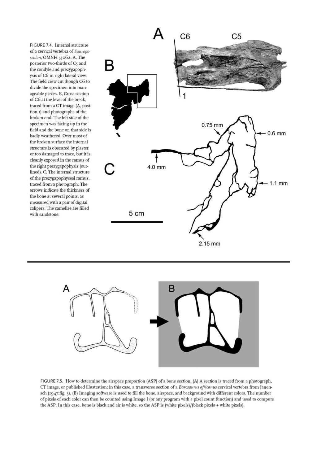
33
Mathew John Wedel. B.S. (University of Oklahoma) A dissertation submitted in partial satisfaction of the. requirements for the degree of
 A dissertation submitted in partial satisfaction of the. requirements for the degree of") Postcranial Pneumaticity in Dinosaurs and the Origin of the Avian Lung by Mathew John Wedel B.S. (University of Oklahoma) 1997 A dissertation submitted in partial satisfaction of the requirements for the
Postcranial Pneumaticity in Dinosaurs and the Origin of the Avian Lung by Mathew John Wedel B.S. (University of Oklahoma) 1997 A dissertation submitted in partial satisfaction of the requirements for the
WHAT PNEUMATICITY TELLS US ABOUT PROSAUROPODS, AND VICE VERSA
 [Special Papers in Palaeontology 77, 2007, pp. 207 222] WHAT PNEUMATICITY TELLS US ABOUT PROSAUROPODS, AND VICE VERSA by MATHEW WEDEL University of California Museum of Paleontology and Department of Integrative
UNIVERSITY OF OKLAHOMA GRADUATE COLLEGE THE EVOLUTION OF VERTEBRAL PNEUMATICITY IN THE SAUROPODA A THESIS SUBMITTED TO THE GRADUATE FACULTY
 UNIVERSITY OF OKLAHOMA GRADUATE COLLEGE THE EVOLUTION OF VERTEBRAL PNEUMATICITY IN THE SAUROPODA A THESIS SUBMITTED TO THE GRADUATE FACULTY in partial fulfillment of the requirements for the degree of
NEW INFORMATION ON A JUVENILE SAUROPOD SPECIMEN FROM THE MORRISON FORMATION AND THE REASSESSMENT OF ITS SYSTEMATIC POSITION
 [Palaeontology, Vol. 55, Part 3, 2012, pp. 567 582] NEW INFORMATION ON A JUVENILE SAUROPOD SPECIMEN FROM THE MORRISON FORMATION AND THE REASSESSMENT OF ITS SYSTEMATIC POSITION by JOSÉ L. CARBALLIDO 1,
Considerations of the neural laminae of sauropod dinosaurs and their morphofunctional meaning *
 Considerations of the neural laminae of sauropod dinosaurs and their morphofunctional meaning * Leonardo SALGADO, Rodolfo A. GARCÍA, & Juan D. DAZA Translated by Michael D. D Emic & Ariel Schepers; edited
Anatomy. Name Section. The Vertebrate Skeleton
 Name Section Anatomy The Vertebrate Skeleton Vertebrate paleontologists get most of their knowledge about past organisms from skeletal remains. Skeletons are useful for gleaning information about an organism
Caudal Pneumaticity and Pneumatic Hiatuses in the Sauropod Dinosaurs Giraffatitan and Apatosaurus
 Caudal Pneumaticity and Pneumatic Hiatuses in the Sauropod Dinosaurs Giraffatitan and Apatosaurus Mathew J. Wedel 1 *, Michael P. Taylor 2 * 1 College of Osteopathic Medicine of the Pacific and College
Abstract RESEARCH ARTICLE
 RESEARCH ARTICLE Vertebral Pneumaticity in the Ornithomimosaur Archaeornithomimus (Dinosauria: Theropoda) Revealed by Computed Tomography Imaging and Reappraisal of Axial Pneumaticity in Ornithomimosauria
Reassessment of the Evidence for Postcranial Skeletal Pneumaticity in Triassic Archosaurs, and the Early Evolution of the Avian Respiratory System
 Reassessment of the Evidence for Postcranial Skeletal Pneumaticity in Triassic Archosaurs, and the Early Evolution of the Avian Respiratory System Richard J. Butler 1,2 *, Paul M. Barrett 2, David J. Gower
Title: Phylogenetic Methods and Vertebrate Phylogeny
 Title: Phylogenetic Methods and Vertebrate Phylogeny Central Question: How can evolutionary relationships be determined objectively? Sub-questions: 1. What affect does the selection of the outgroup have
6BT, UK b Museum für Naturkunde, Invalidenstrasse 43, 10115, Berlin, Germany
 This article was downloaded by: [University College London] On: 02 August 2012, At: 03:36 Publisher: Taylor & Francis Informa Ltd Registered in England and Wales Registered Number: 1072954 Registered office:
A Nomenclature for Vertebral Fossae in Sauropods and Other Saurischian Dinosaurs
 A Nomenclature for Vertebral Fossae in Sauropods and Other Saurischian Dinosaurs Jeffrey A. Wilson*, Michael D. D Emic, Takehito Ikejiri, Emile M. Moacdieh, John A. Whitlock Museum of Paleontology and
A NEW TITANOSAURIFORM SAUROPOD (DINOSAURIA: SAURISCHIA) FROM THE EARLY CRETACEOUS OF CENTRAL TEXAS AND ITS PHYLOGENETIC RELATIONSHIPS. Peter J.
 FROM THE EARLY CRETACEOUS OF CENTRAL TEXAS AND ITS PHYLOGENETIC RELATIONSHIPS. Peter J.") Palaeontologia Electronica http://palaeo-electronica.org A NEW TITANOSAURIFORM SAUROPOD (DINOSAURIA: SAURISCHIA) FROM THE EARLY CRETACEOUS OF CENTRAL TEXAS AND ITS PHYLOGENETIC RELATIONSHIPS Peter J. Rose
Palaeontologia Electronica http://palaeo-electronica.org A NEW TITANOSAURIFORM SAUROPOD (DINOSAURIA: SAURISCHIA) FROM THE EARLY CRETACEOUS OF CENTRAL TEXAS AND ITS PHYLOGENETIC RELATIONSHIPS Peter J. Rose
Geo 302D: Age of Dinosaurs. LAB 7: Dinosaur diversity- Saurischians
 Geo 302D: Age of Dinosaurs LAB 7: Dinosaur diversity- Saurischians Last lab you were presented with a review of major ornithischian clades. You also were presented with some of the kinds of plants that
A new basal sauropodiform dinosaur from the Lower Jurassic of Yunnan Province, China
 SUPPLEMENTARY INFORMATION A new basal sauropodiform dinosaur from the Lower Jurassic of Yunnan Province, China Ya-Ming Wang 1, Hai-Lu You 2,3 *, Tao Wang 4 1 School of Earth Sciences and Resources, China
Nomenclature of Vertebral Laminae in Lizards, with Comments on Ontogenetic and Serial Variation in Lacertini (Squamata, Lacertidae)
") RESEARCH ARTICLE Nomenclature of Vertebral Laminae in Lizards, with Comments on Ontogenetic and Serial Variation in Lacertini (Squamata, Lacertidae) Emanuel Tschopp 1,2,3 * 1 Dipartimento di Scienze della
RESEARCH ARTICLE Nomenclature of Vertebral Laminae in Lizards, with Comments on Ontogenetic and Serial Variation in Lacertini (Squamata, Lacertidae) Emanuel Tschopp 1,2,3 * 1 Dipartimento di Scienze della
These small issues are easily addressed by small changes in wording, and should in no way delay publication of this first- rate paper.
 Reviewers' comments: Reviewer #1 (Remarks to the Author): This paper reports on a highly significant discovery and associated analysis that are likely to be of broad interest to the scientific community.
Europe s largest dinosaur? A giant brachiosaurid cervical vertebra from the Wessex Formation (Early Cretaceous) of southern England
 of southern England") Cretaceous Research 25 (2004) 787 795 www.elsevier.com/locate/cretres Europe s largest dinosaur? A giant brachiosaurid cervical vertebra from the Wessex Formation (Early Cretaceous) of southern England
Cretaceous Research 25 (2004) 787 795 www.elsevier.com/locate/cretres Europe s largest dinosaur? A giant brachiosaurid cervical vertebra from the Wessex Formation (Early Cretaceous) of southern England
.56 m. (22 in.). COMPSOGNATHOID DINOSAUR FROM THE. Medicine Bow, Wyoming, by the American Museum Expedition
. COMPSOGNATHOID DINOSAUR FROM THE. Medicine Bow, Wyoming, by the American Museum Expedition") Article XII.-ORNITHOLESTES HERMANNI, A NEW COMPSOGNATHOID DINOSAUR FROM THE UPPER JURASSIC. By HENRY FAIRFIELD OSBORN. The type skeleton (Amer. Mus. Coll. No. 6I9) of this remarkable animal was discovered
Article XII.-ORNITHOLESTES HERMANNI, A NEW COMPSOGNATHOID DINOSAUR FROM THE UPPER JURASSIC. By HENRY FAIRFIELD OSBORN. The type skeleton (Amer. Mus. Coll. No. 6I9) of this remarkable animal was discovered
muscles (enhancing biting strength). Possible states: none, one, or two.
. Possible states: none, one, or two.") Reconstructing Evolutionary Relationships S-1 Practice Exercise: Phylogeny of Terrestrial Vertebrates In this example we will construct a phylogenetic hypothesis of the relationships between seven taxa
Reconstructing Evolutionary Relationships S-1 Practice Exercise: Phylogeny of Terrestrial Vertebrates In this example we will construct a phylogenetic hypothesis of the relationships between seven taxa
LABORATORY EXERCISE 6: CLADISTICS I
 Biology 4415/5415 Evolution LABORATORY EXERCISE 6: CLADISTICS I Take a group of organisms. Let s use five: a lungfish, a frog, a crocodile, a flamingo, and a human. How to reconstruct their relationships?
SAUROPOD DINOSAURS FROM THE EARLY CRETACEOUS OF MALAWI, AFRICA. Elizabeth M. Gomani
 Palaeontologia Electronica http://palaeo-electronica.org SAUROPOD DINOSAURS FROM THE EARLY CRETACEOUS OF MALAWI, AFRICA Elizabeth M. Gomani ABSTRACT At least two titanosaurian sauropod taxa have been discovered
FIELDIANA GEOLOGY NEW SALAMANDERS OF THE FAMILY SIRENIDAE FROM THE CRETACEOUS OF NORTH AMERICA
 FIELDIANA GEOLOGY Published by CHICAGO NATURAL HISTORY MUSEUM Volume 10 Sbftember 22, 1968 No. 88 NEW SALAMANDERS OF THE FAMILY SIRENIDAE FROM THE CRETACEOUS OF NORTH AMERICA Coleman J. Coin AND Walter
A new species of sauropod, Mamenchisaurus anyuensis sp. nov.
 A new species of sauropod, Mamenchisaurus anyuensis sp. nov. by Xinlu He, Suihua Yang, Kaiji Cai, Kui Li, and Zongwen Liu Chengdu University of Technology Papers on Geosciences Contributed to the 30th
TRACHEMYS SCULPTA. A nearly complete articulated carapace and plastron of an Emjdd A NEAKLY COMPLETE SHELL OF THE EXTINCT TURTLE,
 A NEAKLY COMPLETE SHELL OF THE EXTINCT TURTLE, TRACHEMYS SCULPTA By Charles W. Gilmore Curator of Vertebrate Paleontology, United States National Museum INTRODUCTION A nearly complete articulated carapace
LABORATORY EXERCISE 7: CLADISTICS I
 Biology 4415/5415 Evolution LABORATORY EXERCISE 7: CLADISTICS I Take a group of organisms. Let s use five: a lungfish, a frog, a crocodile, a flamingo, and a human. How to reconstruct their relationships?
ABSTRACT. Candice M. Stefanic and Sterling J. Nesbitt
 The axial skeleton of Poposaurus langstoni (Pseudosuchia: Poposauroidea) and its implications for accessory intervertebral articulation evolution in pseudosuchian archosaurs Candice M. Stefanic and Sterling
IS THERE AN OPTION FOR A PNEUMATIC STABILIZATION OF SAUROPOD NECKS? AN EXPERIMENTAL AND ANATOMICAL APPROACH. Daniela Schwarz-Wings and Eberhard Frey
 Palaeontologia Electronica http://palaeo-electronica.org IS THERE AN OPTION FOR A PNEUMATIC STABILIZATION OF SAUROPOD NECKS? AN EXPERIMENTAL AND ANATOMICAL APPROACH Daniela Schwarz-Wings and Eberhard Frey
Video Assignments. Microraptor PBS The Four-winged Dinosaur Mark Davis SUNY Cortland Library Online
 Video Assignments Microraptor PBS The Four-winged Dinosaur Mark Davis SUNY Cortland Library Online Radiolab Apocalyptical http://www.youtube.com/watch?v=k52vd4wbdlw&feature=youtu.be Minute 13 through minute
A New Sauropod Dinosaur From the Early Cretaceous of Oklahoma. Mathew J. Wedel. Oklahoma Museum of Natural History. and. Department of Zoology
 ..., ' A New Sauropod Dinosaur From the Early Cretaceous of Oklahoma by Mathew. Wedel Oklahoma Museum of Natural History " and Department of Zoology University of Oklahoma 1335 Asp Avenue Norman, OK 73019-0606
Exceptional fossil preservation demonstrates a new mode of axial skeleton elongation in early ray-finned fishes
 Supplementary Information Exceptional fossil preservation demonstrates a new mode of axial skeleton elongation in early ray-finned fishes Erin E. Maxwell, Heinz Furrer, Marcelo R. Sánchez-Villagra Supplementary
Introduction to Cladistic Analysis
 3.0 Copyright 2008 by Department of Integrative Biology, University of California-Berkeley Introduction to Cladistic Analysis tunicate lamprey Cladoselache trout lungfish frog four jaws swimbladder or
Introduction to phylogenetic trees and tree-thinking Copyright 2005, D. A. Baum (Free use for non-commercial educational pruposes)
") Introduction to phylogenetic trees and tree-thinking Copyright 2005, D. A. Baum (Free use for non-commercial educational pruposes) Phylogenetics is the study of the relationships of organisms to each other.
Introduction to phylogenetic trees and tree-thinking Copyright 2005, D. A. Baum (Free use for non-commercial educational pruposes) Phylogenetics is the study of the relationships of organisms to each other.
Feruglio, Fontana 140, Trelew, Argentina Version of record first published: 25 Mar 2013.
 This article was downloaded by: [American Museum of Natural History] On: 25 March 2013, At: 05:07 Publisher: Taylor & Francis Informa Ltd Registered in England and Wales Registered Number: 1072954 Registered
Haplocanthosaurus (Saurischia: Sauropoda) from the lower Morrison Formation (Upper Jurassic) near Snowmass, Colorado
 from the lower Morrison Formation (Upper Jurassic) near Snowmass, Colorado") Volumina Jurassica, 2014, XII (2): 197 210 DOI: 10.5604/17313708.1130144 Haplocanthosaurus (Saurischia: Sauropoda) from the lower Morrison Formation (Upper Jurassic) near Snowmass, Colorado John R. FosteR
Volumina Jurassica, 2014, XII (2): 197 210 DOI: 10.5604/17313708.1130144 Haplocanthosaurus (Saurischia: Sauropoda) from the lower Morrison Formation (Upper Jurassic) near Snowmass, Colorado John R. FosteR
Supplementary Figure 1 Cartilaginous stages in non-avian amniotes. (a) Drawing of early ankle development of Alligator mississippiensis, as reported
 Drawing of early ankle development of Alligator mississippiensis, as reported") Supplementary Figure 1 Cartilaginous stages in non-avian amniotes. (a) Drawing of early ankle development of Alligator mississippiensis, as reported by a previous study 1. The intermedium is formed at
Supplementary Figure 1 Cartilaginous stages in non-avian amniotes. (a) Drawing of early ankle development of Alligator mississippiensis, as reported by a previous study 1. The intermedium is formed at
NOTES ON THE EVOLUTION OF VERTEBRAE IN THE SAUROPODOMORPHA. José F. BONAPARTE *
 NOTES ON THE EVOLUTION OF VERTEBRAE IN THE SAUROPODOMORPHA by José F. BONAPARTE * Museo Argentino de Ciencias Naturales Consejo Nacional de Investigaciones Científicas y Técnicas Avenida Angel Gallardo
THE LIGAMENT SYSTEM IN THE NECK OF RHEA AMERICANA AND ITS IMPLICATION FOR THE BIFURCATED NEURAL SPINES OF SAUROPOD DINOSAURS
 Journal of Vertebrate Paleontology 24(1):165 172, March 2004 2004 by the Society of Vertebrate Paleontology THE LIGAMENT SYSTEM IN THE NECK OF RHEA AMERICANA AND ITS IMPLICATION FOR THE BIFURCATED NEURAL
Animal Form and Function. Amphibians. United by several distinguishing apomorphies within the Vertebrata
 Animal Form and Function Kight Amphibians Class Amphibia (amphibia = living a double life) United by several distinguishing apomorphies within the Vertebrata 1. Skin Thought Question: For whom are integumentary
Cretaceous Research 34 (2012) 220e232. Contents lists available at SciVerse ScienceDirect. Cretaceous Research
 220e232. Contents lists available at SciVerse ScienceDirect. Cretaceous Research") Cretaceous Research 34 (2012) 220e232 Contents lists available at SciVerse ScienceDirect Cretaceous Research journal homepage: www.elsevier.com/locate/cretres The southernmost records of Rebbachisauridae
Cretaceous Research 34 (2012) 220e232 Contents lists available at SciVerse ScienceDirect Cretaceous Research journal homepage: www.elsevier.com/locate/cretres The southernmost records of Rebbachisauridae
THE SKELETON RECONSTRUCTION OF BRACHIOSAURUS BRANCAI
 THE SKELETON RECONSTRUCTION OF BRACHIOSAURUS BRANCAI BY W. JANENSCH WITH PLATES VI VIII PALAEONTOGRAPHICA 1950, Supplement VII, Reihe I, Teil III, 97 103. TRANSLATED BY GERHARD MAIER JUNE 2007 97 A reconstruction
FURTHER STUDIES ON TWO SKELETONS OF THE BLACK RIGHT WHALE IN THE NORTH PACIFIC
 FURTHER STUDIES ON TWO SKELETONS OF THE BLACK RIGHT WHALE IN THE NORTH PACIFIC HIDEO OMURA, MASAHARU NISHIWAKI* AND TOSHIO KASUYA* ABSTRACT Two skeletons of the black right whale were studied, supplementing
Overview of Sauropod Phylogeny and Evolution
 One Overview of Sauropod Phylogeny and Evolution Jeffrey A. Wilson SAUROPOD STUDIES FROM OWEN TO THE PRESENT This year marks the one hundred sixty-fourth anniversary of Richard Owen s (1841) description
d a Name Vertebrate Evolution - Exam 2 1. (12) Fill in the blanks
 Fill in the blanks") Vertebrate Evolution - Exam 2 1. (12) Fill in the blanks 100 points Name f e c d a Identify the structures (for c and e, identify the entire structure, not the individual elements. b a. b. c. d. e. f.
Vertebrate Evolution - Exam 2 1. (12) Fill in the blanks 100 points Name f e c d a Identify the structures (for c and e, identify the entire structure, not the individual elements. b a. b. c. d. e. f.
290 SHUFELDT, Remains of Hesperornis.
 290 SHUFELDT, Remains of Hesperornis. [ Auk [July THE FOSSIL REMAINS OF A SPECIES OF HESPERORNIS FOUND IN MONTANA. BY R. W. SHUFELD% M.D. Plate XI7III. ExR,¾ in November, 1914, Mr. Charles W. Gihnore,
Biology 340 Comparative Embryology Lecture 12 Dr. Stuart Sumida. Evo-Devo Revisited. Development of the Tetrapod Limb
 Biology 340 Comparative Embryology Lecture 12 Dr. Stuart Sumida Evo-Devo Revisited Development of the Tetrapod Limb Limbs whether fins or arms/legs for only in particular regions or LIMB FIELDS. Primitively
Diapsida. BIO2135 Animal Form and Function. Page 1. Diapsida (Reptilia, Sauropsida) Amniote eggs. Amniote egg. Temporal fenestra.
 Amniote eggs. Amniote egg. Temporal fenestra.") Diapsida (Reptilia, Sauropsida) Vertebrate phylogeny Mixini Chondrichthyes Sarcopterygii Mammalia Pteromyzontida Actinopterygii Amphibia Reptilia! 1! Amniota (autapomorphies) Costal ventilation Amniote
Diapsida (Reptilia, Sauropsida) Vertebrate phylogeny Mixini Chondrichthyes Sarcopterygii Mammalia Pteromyzontida Actinopterygii Amphibia Reptilia! 1! Amniota (autapomorphies) Costal ventilation Amniote
Diapsida. BIO2135 Animal Form and Function. Page 1. Diapsida (Reptilia, Sauropsida) Amniote egg. Membranes. Vertebrate phylogeny
 Amniote egg. Membranes. Vertebrate phylogeny") Diapsida (Reptilia, Sauropsida) 1 Vertebrate phylogeny Mixini Chondrichthyes Sarcopterygii Mammalia Pteromyzontida Actinopterygii Amphibia Reptilia!! Amniota (autapomorphies) Costal ventilation Amniote
Diapsida (Reptilia, Sauropsida) 1 Vertebrate phylogeny Mixini Chondrichthyes Sarcopterygii Mammalia Pteromyzontida Actinopterygii Amphibia Reptilia!! Amniota (autapomorphies) Costal ventilation Amniote
Geo 302D: Age of Dinosaurs LAB 4: Systematics Part 1
 Geo 302D: Age of Dinosaurs LAB 4: Systematics Part 1 Systematics is the comparative study of biological diversity with the intent of determining the relationships between organisms. Humankind has always
Why sauropods had long necks; and why giraffes have short necks
 Why sauropods had long necks; and why giraffes have short necks The necks of the sauropod dinosaurs reached 15 m in length: six times longer than that of the world record giraffe and five times longer
Evidence for Avian Intrathoracic Air Sacs in a New Predatory Dinosaur from Argentina
 Evidence for Avian Intrathoracic Air Sacs in a New Predatory Dinosaur from Argentina Paul C. Sereno 1 *, Ricardo N. Martinez 2, Jeffrey A. Wilson 3, David J. Varricchio 4, Oscar A. Alcober 2, Hans C. E.
Bio 1B Lecture Outline (please print and bring along) Fall, 2006
 Fall, 2006") Bio 1B Lecture Outline (please print and bring along) Fall, 2006 B.D. Mishler, Dept. of Integrative Biology 2-6810, bmishler@berkeley.edu Evolution lecture #4 -- Phylogenetic Analysis (Cladistics) -- Oct.
Bio 1B Lecture Outline (please print and bring along) Fall, 2006 B.D. Mishler, Dept. of Integrative Biology 2-6810, bmishler@berkeley.edu Evolution lecture #4 -- Phylogenetic Analysis (Cladistics) -- Oct.
( M amenchisaurus youngi Pi, Ouyang et Ye, 1996)
") 39 4 2001 10 V ERTEBRATA PALASIATICA pp. 266 271 fig. 1,pl. I ( 643013), ( M amenchisaurus hochuanensis),,, Q915. 864 1995 12 31 (ZDM0126) ( M amenchisau rus hochuanensis Young et Chao, 1972),,, ZDM0126
39 4 2001 10 V ERTEBRATA PALASIATICA pp. 266 271 fig. 1,pl. I ( 643013), ( M amenchisaurus hochuanensis),,, Q915. 864 1995 12 31 (ZDM0126) ( M amenchisau rus hochuanensis Young et Chao, 1972),,, ZDM0126
Phylogeny Reconstruction
 Phylogeny Reconstruction Trees, Methods and Characters Reading: Gregory, 2008. Understanding Evolutionary Trees (Polly, 2006) Lab tomorrow Meet in Geology GY522 Bring computers if you have them (they will
Comparative Zoology Portfolio Project Assignment
 Comparative Zoology Portfolio Project Assignment Using your knowledge from the in class activities, your notes, you Integrated Science text, or the internet, you will look at the major trends in the evolution
Tetrapod Similarites The Origins of Birds
 Tetrapod Similarites The Origins of Birds Birds Reptiles Mammals Integument Feathers, scales Scales Hair Digestive Horny bill Teeth Teeth Skeletal Fusion of bones Some fusion Some fusion Reduction in number
8/19/2013. Topic 5: The Origin of Amniotes. What are some stem Amniotes? What are some stem Amniotes? The Amniotic Egg. What is an Amniote?
 Topic 5: The Origin of Amniotes Where do amniotes fall out on the vertebrate phylogeny? What are some stem Amniotes? What is an Amniote? What changes were involved with the transition to dry habitats?
The Effect of Intervertebral Cartilage on Neutral Posture and Range of Motion in the Necks of Sauropod Dinosaurs
 The Effect of Intervertebral Cartilage on Neutral Posture and Range of Motion in the Necks of Sauropod Dinosaurs Michael P. Taylor 1 *, Mathew J. Wedel 2 1 Department of Earth Sciences, University of Bristol,
Species: Panthera pardus Genus: Panthera Family: Felidae Order: Carnivora Class: Mammalia Phylum: Chordata
 CHAPTER 6: PHYLOGENY AND THE TREE OF LIFE AP Biology 3 PHYLOGENY AND SYSTEMATICS Phylogeny - evolutionary history of a species or group of related species Systematics - analytical approach to understanding
Shedding Light on the Dinosaur-Bird Connection
 Shedding Light on the Dinosaur-Bird Connection This text is provided courtesy of the American Museum of Natural History. When people think of dinosaurs, two types generally come to mind: the huge herbivores
Fish 2/26/13. Chordates 2. Sharks and Rays (about 470 species) Sharks etc Bony fish. Tetrapods. Osteichthans Lobe fins and lungfish
 Sharks etc Bony fish. Tetrapods. Osteichthans Lobe fins and lungfish") Chordates 2 Sharks etc Bony fish Osteichthans Lobe fins and lungfish Tetrapods ns Reptiles Birds Feb 27, 2013 Chordates ANCESTRAL DEUTEROSTOME Notochord Common ancestor of chordates Head Vertebral column
Chordates 2 Sharks etc Bony fish Osteichthans Lobe fins and lungfish Tetrapods ns Reptiles Birds Feb 27, 2013 Chordates ANCESTRAL DEUTEROSTOME Notochord Common ancestor of chordates Head Vertebral column
Journal of Systematic Palaeontology. ISSN: (Print) (Online) Journal homepage:
 (Online) Journal homepage:") Journal of Systematic Palaeontology ISSN: 1477-2019 (Print) 1478-0941 (Online) Journal homepage: http://www.tandfonline.com/loi/tjsp20 An articulated cervical series of Alamosaurus sanjuanensis Gilmore,
Journal of Systematic Palaeontology ISSN: 1477-2019 (Print) 1478-0941 (Online) Journal homepage: http://www.tandfonline.com/loi/tjsp20 An articulated cervical series of Alamosaurus sanjuanensis Gilmore,
ON SOME REPTILIAN REMAINS FROM THE DINOSAUR BEDS OF NYASALAND. By S. H. HAUGHTON, D.Sc., F.G.S.
 ( 67 ) ON SOME REPTILIAN REMAINS FROM THE DINOSAUR BEDS OF NYASALAND. By S. H. HAUGHTON, D.Sc., F.G.S. (Published by permission of the Hon. the Minister for Mines and Industries.) (With Plates II-V and
Osteology of the dorsal vertebrae of the giant titanosaurian sauropod dinosaur Dreadnoughtus schrani from the Late Cretaceous of Argentina
 Rowan University Rowan Digital Works School of Earth & Environment Faculty Scholarship School of Earth & Environment 1-1-2017 Osteology of the dorsal vertebrae of the giant titanosaurian sauropod dinosaur
Origin and Evolution of Birds. Read: Chapters 1-3 in Gill but limited review of systematics
 Origin and Evolution of Birds Read: Chapters 1-3 in Gill but limited review of systematics Review of Taxonomy Kingdom: Animalia Phylum: Chordata Subphylum: Vertebrata Class: Aves Characteristics: wings,
1/9/2013. Divisions of the Skeleton: Topic 8: Appendicular Skeleton. Appendicular Components. Appendicular Components
 /9/203 Topic 8: Appendicular Skeleton Divisions of the Skeleton: Cranial Postcranial What makes up the appendicular skeleton? What is the pattern of serial homology of the limbs? Tetrapod front limb morphology
Comments on the Beauceron Standard By M. Maurice Hermel (Translated by C. Batson)
") Comments on the Beauceron Standard By M. Maurice Hermel (Translated by C. Batson) The following are comments written by M. Hermel for the FCI Standard #44 published on 10/25/06. They were approved by the
Comments on the Beauceron Standard By M. Maurice Hermel (Translated by C. Batson) The following are comments written by M. Hermel for the FCI Standard #44 published on 10/25/06. They were approved by the
Erycine Boids from the Early Oligocene of the South Dakota Badlands
 Georgia Journal of Science Volume 67 No. 2 Scholarly Contributions from the Membership and Others Article 6 2009 Erycine Boids from the Early Oligocene of the South Dakota Badlands Dennis Parmley J. Alan
Animal Diversity III: Mollusca and Deuterostomes
 Animal Diversity III: Mollusca and Deuterostomes Objectives: Be able to identify specimens from the main groups of Mollusca and Echinodermata. Be able to distinguish between the bilateral symmetry on a
UNIT III A. Descent with Modification(Ch19) B. Phylogeny (Ch20) C. Evolution of Populations (Ch21) D. Origin of Species or Speciation (Ch22)
 B. Phylogeny (Ch20) C. Evolution of Populations (Ch21) D. Origin of Species or Speciation (Ch22)") UNIT III A. Descent with Modification(Ch9) B. Phylogeny (Ch2) C. Evolution of Populations (Ch2) D. Origin of Species or Speciation (Ch22) Classification in broad term simply means putting things in classes
UNIT III A. Descent with Modification(Ch9) B. Phylogeny (Ch2) C. Evolution of Populations (Ch2) D. Origin of Species or Speciation (Ch22) Classification in broad term simply means putting things in classes
Field Trip: Harvard Museum of Natural History (HMNH)
") Field Trip: Harvard Museum of Natural History (HMNH) Objectives To observe the diversity of animals. To compare and contrast the various adaptations, body plans, etc. of the animals found at the HMNH.
Field Trip: Harvard Museum of Natural History (HMNH) Objectives To observe the diversity of animals. To compare and contrast the various adaptations, body plans, etc. of the animals found at the HMNH.
A new Middle Jurassic sauropod subfamily (Klamelisaurinae subfam. nov.) from Xinjiang Autonomous Region, China
 from Xinjiang Autonomous Region, China") A new Middle Jurassic sauropod subfamily (Klamelisaurinae subfam. nov.) from Xinjiang Autonomous Region, China by Xijing Zhao Institute of Vertebrate Paleontology and Paleoanthropology, Academia Sinica
A new Middle Jurassic sauropod subfamily (Klamelisaurinae subfam. nov.) from Xinjiang Autonomous Region, China by Xijing Zhao Institute of Vertebrate Paleontology and Paleoanthropology, Academia Sinica
INQUIRY & INVESTIGATION
 INQUIRY & INVESTIGTION Phylogenies & Tree-Thinking D VID. UM SUSN OFFNER character a trait or feature that varies among a set of taxa (e.g., hair color) character-state a variant of a character that occurs
From Slime to Scales: Evolution of Reptiles. Review: Disadvantages of Being an Amphibian
 From Slime to Scales: Evolution of Reptiles Review: Disadvantages of Being an Amphibian Gelatinous eggs of amphibians cannot survive out of water, so amphibians are limited in terms of the environments
Skeletal Morphogenesis of the Vertebral Column of the Miniature Hylid Frog Acris crepitans, With Comments on Anomalies
 JOURNAL OF MORPHOLOGY 270:52 69 (2009) Skeletal Morphogenesis of the Vertebral Column of the Miniature Hylid Frog Acris crepitans, With Comments on Anomalies L. Analía Pugener* and Anne M. Maglia Department
A Fossil Snake (Elaphe vulpina) From A Pliocene Ash Bed In Nebraska
 From A Pliocene Ash Bed In Nebraska") University of Nebraska - Lincoln DigitalCommons@University of Nebraska - Lincoln Transactions of the Nebraska Academy of Sciences and Affiliated Societies Nebraska Academy of Sciences 198 A Fossil Snake
University of Nebraska - Lincoln DigitalCommons@University of Nebraska - Lincoln Transactions of the Nebraska Academy of Sciences and Affiliated Societies Nebraska Academy of Sciences 198 A Fossil Snake
A New Titanosaurian Sauropod from the Hekou Group (Lower Cretaceous) of the Lanzhou-Minhe Basin, Gansu Province, China
 of the Lanzhou-Minhe Basin, Gansu Province, China") A New Titanosaurian Sauropod from the Hekou Group (Lower Cretaceous) of the Lanzhou-Minhe Basin, Gansu Province, China Li-Guo Li 1,2 *, Da-Qing Li 3, Hai-Lu You 4, Peter Dodson 2 1 School of Earth Sciences
A New Titanosaurian Sauropod from the Hekou Group (Lower Cretaceous) of the Lanzhou-Minhe Basin, Gansu Province, China Li-Guo Li 1,2 *, Da-Qing Li 3, Hai-Lu You 4, Peter Dodson 2 1 School of Earth Sciences
17.2 Classification Based on Evolutionary Relationships Organization of all that speciation!
 Organization of all that speciation! Patterns of evolution.. Taxonomy gets an over haul! Using more than morphology! 3 domains, 6 kingdoms KEY CONCEPT Modern classification is based on evolutionary relationships.
LOWER CRETACEOUS OF SOUTH DAKOTA.
 A NEW DINOSAUR, STP^GOSAURUS MARSHl, FROM THE LOWER CRETACEOUS OF SOUTH DAKOTA. By Frederic A. Lucas, Curator, Divisioii of Coiiipnrative Anatomy, in charge, of Section of Vertebrate Fossils. The name
A Study of Carasaurus' (Dinosaura: Sauropodomorph) Torso and its Biomechanical Implications
 Torso and its Biomechanical Implications") University of New Orleans ScholarWorks@UNO University of New Orleans Theses and Dissertations Dissertations and Theses 5-22-2006 A Study of Carasaurus' (Dinosaura: Sauropodomorph) Torso and its Biomechanical
University of New Orleans ScholarWorks@UNO University of New Orleans Theses and Dissertations Dissertations and Theses 5-22-2006 A Study of Carasaurus' (Dinosaura: Sauropodomorph) Torso and its Biomechanical
VERTEBRATE READING. Fishes
 VERTEBRATE READING Fishes The first vertebrates to become a widespread, predominant life form on earth were fishes. Prior to this, only invertebrates, such as mollusks, worms and squid-like animals, would
Sauropoda from the Kelameili Region of the Junggar Basin, Xinjiang Autonomous Region
 Sauropoda from the Kelameili Region of the Junggar Basin, Xinjiang Autonomous Region Zhiming Dong (Institute of Vertebrate Paleontology and Paleoanthropology, Academia Sinica) Vertebrata PalAsiatica Volume
THE ANATOMY AND TAXONOMY OF CETIOSAURUS (SAURISCHIA, SAUROPODA) FROM THE MIDDLE JURASSIC OF ENGLAND
 FROM THE MIDDLE JURASSIC OF ENGLAND") Journal of Vertebrate Paleontology 23(1):208 231, March 2003 2003 by the Society of Vertebrate Paleontology THE ANATOMY AND TAXONOMY OF CETIOSAURUS (SAURISCHIA, SAUROPODA) FROM THE MIDDLE JURASSIC OF ENGLAND
Journal of Vertebrate Paleontology 23(1):208 231, March 2003 2003 by the Society of Vertebrate Paleontology THE ANATOMY AND TAXONOMY OF CETIOSAURUS (SAURISCHIA, SAUROPODA) FROM THE MIDDLE JURASSIC OF ENGLAND
Interpreting Evolutionary Trees Honors Integrated Science 4 Name Per.
 Interpreting Evolutionary Trees Honors Integrated Science 4 Name Per. Introduction Imagine a single diagram representing the evolutionary relationships between everything that has ever lived. If life evolved
Pneumaticity and soft tissue reconstructions in the neck of diplodocid and dicraeosaurid sauropods
 Pneumaticity and soft tissue reconstructions in the neck of diplodocid and dicraeosaurid sauropods DANIELA SCHWARZ, EBERHARD FREY, and CHRISTIAN A. MEYER Schwarz, D., Frey, E., and Meyer, C.A. 2007. Pneumaticity
Osteology of the Late Jurassic Portuguese sauropod dinosaur Lusotitan atalaiensis (Macronaria) and the evolutionary history of basal titanosauriforms
 and the evolutionary history of basal titanosauriforms") bs_bs_banner Zoological Journal of the Linnean Society, 2013, 168, 98 206. With 30 figures Osteology of the Late Jurassic Portuguese sauropod dinosaur Lusotitan atalaiensis (Macronaria) and the evolutionary
bs_bs_banner Zoological Journal of the Linnean Society, 2013, 168, 98 206. With 30 figures Osteology of the Late Jurassic Portuguese sauropod dinosaur Lusotitan atalaiensis (Macronaria) and the evolutionary
Name: GEOL 104 Dinosaurs: A Natural History Video Assignment. DUE: Wed. Oct. 20
 GEOL 104 Dinosaurs: A Natural History Video Assignment DUE: Wed. Oct. 20 Documentaries represent one of the main media by which scientific information reaches the general public. For this assignment, you
Biology 1B Evolution Lecture 11 (March 19, 2010), Insights from the Fossil Record and Evo-Devo
, Insights from the Fossil Record and Evo-Devo") Biology 1B Evolution Lecture 11 (March 19, 2010), Insights from the Fossil Record and Evo-Devo Extinction Important points on extinction rates: Background rate of extinctions per million species per year:
Biology 1B Evolution Lecture 11 (March 19, 2010), Insights from the Fossil Record and Evo-Devo Extinction Important points on extinction rates: Background rate of extinctions per million species per year:
CLADISTICS Student Packet SUMMARY Phylogeny Phylogenetic trees/cladograms
 CLADISTICS Student Packet SUMMARY PHYLOGENETIC TREES AND CLADOGRAMS ARE MODELS OF EVOLUTIONARY HISTORY THAT CAN BE TESTED Phylogeny is the history of descent of organisms from their common ancestor. Phylogenetic
Recommended Resources: The following resources may be useful in teaching this
 Unit B: Anatomy and Physiology of Poultry Lesson1: Internal Anatomy of Poultry Student Learning Objectives: Instruction in this lesson should result in students achieving the following objectives: 1. Identify
Dinosaur Safari Junior: A Walk in Jurassic Park
 Dinosaur Safari Junior: A Walk in Jurassic Park Introduction The rules used are a simplified variant of the Saurian Safari rules developed by Chris Peers and published by HLBS publishing 2002. This is
Article. Universidade de Brasília - Faculdade UnB Planaltina, Brasília-DF, , Brazil. 2
 Zootaxa 3085: 1 33 (2011) www.mapress.com/zootaxa/ Copyright 2011 Magnolia Press Article ISSN 1175-5326 (print edition) ZOOTAXA ISSN 1175-5334 (online edition) A new sauropod (Macronaria, Titanosauria)
Do the traits of organisms provide evidence for evolution?
 PhyloStrat Tutorial Do the traits of organisms provide evidence for evolution? Consider two hypotheses about where Earth s organisms came from. The first hypothesis is from John Ray, an influential British
AMERICAN MUSEUM NOVITATES
 AMERICAN MUSEUM NOVITATES Number 3700 October 22, 2010 The Illusory Evidence for Asian Brachiosauridae: New Material of Erketu ellisoni and a Phylogenetic Reappraisal of Basal Titanosauriformes DANIEL
A NEW ANKYLOSAUR FROM THE UPPER CRETACEOUS OF MONGOLIA E.A. Maleev Doklady Akademii Nauk, SSSR 87:
 translated by Dr. Tamara and F. Jeletzky, 1956 A NEW ANKYLOSAUR FROM THE UPPER CRETACEOUS OF MONGOLIA E.A. Maleev 1952. Doklady Akademii Nauk, SSSR 87:273-276 Armored dinosaurs make a considerable part
A definite prosauropod dinosaur from the Lower Elliot Formation (Norian: Upper Triassic) of South Africa
 of South Africa") A definite prosauropod dinosaur from the Lower Elliot Formation (Norian: Upper Triassic) of South Africa Adam M. Yates Bernard Price Institute for Palaeontological Research, School of Geosciences, University
A definite prosauropod dinosaur from the Lower Elliot Formation (Norian: Upper Triassic) of South Africa Adam M. Yates Bernard Price Institute for Palaeontological Research, School of Geosciences, University
d. Wrist bones. Pacific salmon life cycle. Atlantic salmon (different genus) can spawn more than once.
 can spawn more than once.") Lecture III.5b Answers to HW 1. (2 pts). Tiktaalik bridges the gap between fish and tetrapods by virtue of possessing which of the following? a. Humerus. b. Radius. c. Ulna. d. Wrist bones. 2. (2 pts)
Lecture III.5b Answers to HW 1. (2 pts). Tiktaalik bridges the gap between fish and tetrapods by virtue of possessing which of the following? a. Humerus. b. Radius. c. Ulna. d. Wrist bones. 2. (2 pts)
Vol. XIV, No. 1, March, The Larva and Pupa of Brontispa namorikia Maulik (Coleoptera: Chrysomelidae: Hispinae) By S.
 By S.") Vol. XIV, No. 1, March, 1950 167 The Larva and Pupa of Brontispa namorikia Maulik (Coleoptera: Chrysomelidae: Hispinae) By S. MAULIK BRITISH MUSEUM (NATURAL HISTORY) (Presented by Mr. Van Zwaluwenburg
Vol. XIV, No. 1, March, 1950 167 The Larva and Pupa of Brontispa namorikia Maulik (Coleoptera: Chrysomelidae: Hispinae) By S. MAULIK BRITISH MUSEUM (NATURAL HISTORY) (Presented by Mr. Van Zwaluwenburg
Williston, and as there are many fairly good specimens in the American
 56.81.7D :14.71.5 Article VII.- SOME POINTS IN THE STRUCTURE OF THE DIADECTID SKULL. BY R. BROOM. The skull of Diadectes has been described by Cope, Case, v. Huene, and Williston, and as there are many
Modern Evolutionary Classification. Lesson Overview. Lesson Overview Modern Evolutionary Classification
 Lesson Overview 18.2 Modern Evolutionary Classification THINK ABOUT IT Darwin s ideas about a tree of life suggested a new way to classify organisms not just based on similarities and differences, but