An investigation of the evolution of the anguimorph lizard venom system
|
|
|
- Stanley Fox
- 5 years ago
- Views:
Transcription
1 An investigation of the evolution of the anguimorph lizard venom system Ivan Koludarov BSc (Biology); MSc (Biology) A thesis submitted for the degree of Doctor of Philosophy at The University of Queensland in 2016 School of Biological Sciences
2 2 Abstract Over the course of the last two decades significant advances were made in our understanding of the evolution of venom in squamate reptiles. Several studies looked at nuclear genes from various lizards and snakes and confirmed the hypothesis put forth in E. Kochva s article Phylogeny of the oral glands in reptiles as related to the origin and evolution of snakes that anguimorph lizards form part of a monophyletic clade with all the snakes. This confirmation confounded decades of morphology based taxonomy, as did evidence of the inclusion of the Iguania lizard lineage into that group. The new group received the name Toxicofera, emphasizing the role of oral toxins in the evolution of the lineage. Despite being supported by genetic, anatomical and proteomic data, the Toxicofera hypothesis remains controversial. This strengthens the necessity for thorough investigation of toxicoferan reptile venom evolution. The oral secretions of anguimorph lizards demand particular attention not only because they are understudied, but also for the reason that Anguimorpha contains species with the considerable morphological diversity of venom glands from incipient to advanced and almost snake-like. Therefore the primary goal of this thesis is to address the lack of knowledge on anguimorph lizard oral secretions, in particular that of varanoid lizards which include various monitor species (genus Varanus) as well as Heloderma (gila monster) and Lanthanotus (Borneo earless monitor). This has been achieved through the implementation of several proteomic techniques (in particular, gel electrophoresis) as well as transcriptomic analysis and bioactivity testings. Chapter One reviews all previously published information on anguimorph venom evolution, highlighting the data in support of the Toxicofera hypothesis. Chapter Two presents new data on Heloderma lizard venoms and the striking similarities between the venom profiles of specimens from different localities as well as between species. The data presented and discussed in Chapter Three represents the core finding of this study: the surprising complexity and diversity of varanid lizard oral secretions. Taken together, this study present a compelling argument in favour of functional diversity and differential complexity of the venom of anguimorph lizards and discusses the evolutionary forces that helped generate this diversity.
3 Declaration by author 3 This thesis is composed of my original work, and contains no material previously published or written by another person except where due reference has been made in the text. I have clearly stated the contribution by others to jointly-authored works that I have included in my thesis. I have clearly stated the contribution of others to my thesis as a whole, including statistical assistance, survey design, data analysis, significant technical procedures, professional editorial advice, and any other original research work used or reported in my thesis. The content of my thesis is the result of work I have carried out since the commencement of my research higher degree candidature and does not include a substantial part of work that has been submitted to qualify for the award of any other degree or diploma in any university or other tertiary institution. I have clearly stated which parts of my thesis, if any, have been submitted to qualify for another award. I acknowledge that an electronic copy of my thesis must be lodged with the University Library and, subject to the policy and procedures of The University of Queensland, the thesis be made available for research and study in accordance with the Copyright Act 1968 unless a period of embargo has been approved by the Dean of the Graduate School. I acknowledge that copyright of all material contained in my thesis resides with the copyright holder(s) of that material. Where appropriate I have obtained copyright permission from the copyright holder to reproduce material in this thesis.
4 Publications during candidature 4 Peer-reviewed papers published during candidacy: 1. Debono, J., Cochran, C., Kuruppu, S., Nouwens, A., Rajapakse, N. W., Kawasaki, M., Wood, K., Dobson, J., Baumann, K., Jouiaei, M., Jackson, T. N. W., Koludarov, I., Low, D., Ali, S. A., Barnes, A. & Fry, B. G. (2016). Canopy venom: proteomic comparison among new world arboreal pit-viper venoms. Toxins, 8(7), Koludarov, I., Jackson, T. N., Sunagar, K., Nouwens, A., Hendrikx, I., & Fry, B. G. (2014). Fossilized venom: the unusually conserved venom profiles of Heloderma species (beaded lizards and gila monsters). Toxins, 6(12), Jouiaei, M., Casewell, N. R., Yanagihara, A. A., Nouwens, A., Cribb, B. W., Whitehead, D., Jackson, T. N., Ali, S. A., Wagstaff, S. C., Koludarov, I., & Alewood, P. (2015). Firing the sting: chemically induced discharge of cnidae reveals novel proteins and peptides from box jellyfish (Chironex fleckeri) venom. Toxins, 7(3), Jesupret, C., Baumann, K., Jackson, T. N., Ali, S. A., Yang, D. C., Greisman, L., Kern, L., Steuten, J., Jouiaei, M., Casewell, N. R., Undheim, E. A., Koludarov, I., Debono, J., Low, D. H. W., Rossi, S., Panagides, N., Winter, K., Ignatovich, V., Summerhayesh, R., Jones, A., Nouwens, A., Dunstank, N., Hodgson, W. C., Winkeld, K. D., Monagleh, P., & Fry, B. G. (2014). Vintage venoms: proteomic and pharmacological stability of snake venoms stored for up to eight decades. Journal of proteomics, 105, Sunagar, K., Undheim, E. A., Scheib, H., Gren, E. C., Cochran, C., Person, C. E., Koludarov, I., Kelln, W., Hayes, W. K., King, G. F., Fry, B. G., & Antunes, A. (2014). Intraspecific venom variation in the medically significant Southern Pacific Rattlesnake (Crotalus oreganus helleri): biodiscovery, clinical and evolutionary implications. Journal of proteomics, 99, Book chapters contributed to during candidacy: 1. Fry, B. G., Koludarov, I., Jackson, T. N., Holford, M., Terrat, Y., Casewell, N. R., Undheim, E. A. B., Vetter, I., Ali, S. A., Low, D. H. W., & Sunagar, K. (2015). Seeing the
5 5 Woods for the Trees: Understanding Venom Evolution as a Guide for Biodiscovery. in Venoms to Drugs: Venom as a Source for the Development of Human Therapeutics, Royal Society of Chemistry. 2. Bénard-Valle, M., Neri-Castro, E. E., Fry, B. G., Boyer, L., Cochran, C., Alam, M., Jackson, T. N. W., Paniagua, D., Olvera-Rodríguez, F., Koludarov, I., Sunagar, K. & Alagón, A. (2015). Chapter 3 Antivenom research and development. In Venomous Reptiles: Evolution, Pathophysiology and Biodiscovery. Fry BG editor. Oxford University Conferences attended during candidacy: 1. Koludarov I. and Fry B. G., Taming the dragon: the biodiscovery potential of lizards venom. Venom to Drugs Publications included in this thesis Chapter 2 of thesis: Koludarov, I., Jackson, T. N., Sunagar, K., Nouwens, A., Hendrikx, I., & Fry, B. G. (2014). Fossilized venom: the unusually conserved venom profiles of Heloderma species (beaded lizards and gila monsters). Toxins, 6(12), Contributor Statement of contribution Author IK (Candidate) Designed experiments (70%) Conducted experiments (50%) Wrote and edited the paper (40%) Author TNJ Conducted experiments (30%) Wrote and edited the paper (30%) Author KS Conducted experiments (20%) Author AN Assisted with mass spectrometry experiments Author IH Collected venoms Author BGF Designed experiments (30%) Wrote and edited paper (30%) Formatted paper for submission Collected venoms
6 Contributions by others to the thesis 6 Chapter 1: TNJ co-wrote the manuscript (10%); BGF co-wrote the manuscript (10%). Chapter 3: Bianca op den Brouw assisted with lab work; Nadya Panagides assisted with lab work; TNJ assisted with lab work and co-wrote the manuscript (30%); James Dobson assisted with lab work; Amanda Nowens assisted with mass spectrometry experiments; Peter Josh assisted with mass spectrometry experiments; Mary-Louise Roy Manchadi assisted with rat-ileum organ bath testings; Chip Cochran collected the venoms, BGF collected the venoms, helped designed the experiments and co-wrote the manuscript (20%). Statement of parts of the thesis submitted to qualify for the award of another degree None.
7 Acknowledgements 7 Academic support of various kinds was provided by Timothy Jackson, Glenn King, Irina Vetter, Nicholas Casewell, Vittoria Cipriani, Jonathan Goldenberg, James Dobson, Bianca op den Brouw, Nadya Panagides, Amanda Nowens, Peter Josh and Bryan Fry thank you for all the support. No parent, pet or partner helped in any way. Funding was provided by University of Queensland International Scholarship.
8 8 Keywords venom, evolution, system, varanid lizards, anguimorpha, reptile, toxin, toxicofera, philosophy, proteomics, pharmacology Australian and New Zealand Standard Research Classifications (ANZSRC) ANZSRC code: Proteomics and Intermolecular Interactions (excl. Medical Proteomics), 40% ANZSRC code: History and Philosophy of Science (incl. Non-historical Philosophy of Science), 10% ANZSRC code: Biological Adaptation, 50% Fields of Research (FoR) Classification FoR code: 0601, Biochemistry and Cell Biology, 40% FoR code: 2202 History and Philosophy of Specific Fields, 10% FoR code: 0603 Evolutionary Biology, 50%
9 Table of contents 9 TABLE OF CONTENTS... 9 INTRODUCTION CHAPTER 1. LIZARDS: MORE THAN VENOMOUS ABSTRACT TOXICOFERA ANGUIMORPHA TOXINS CONCLUDING REMARKS REFERENCES CHAPTER 2: FOSSILIZED VENOM: THE UNUSUALLY CONSERVED VENOM PROFILES OF HELODERMA SPECIES (BEADED LIZARDS AND GILA MONSTERS) ABSTRACT INTRODUCTION RESULTS AND DISCUSSION MATERIALS AND METHODS CONCLUSION REFERENCES CHAPTER 3. TAMING THE DRAGON: INVESTIGATING EVOLUTIONARY FORCES THAT SHAPE THE COMPLEXITY OF MONITOR LIZARDS VENOM ABSTRACT INTRODUCTION MATERIALS AND METHODS RESULTS AND DISCUSSION CONCLUSION REFERENCES CONCLUSIONS
10 Introduction 10 After being neglected for a long period of time the field of lizard venomics has made significant progress in the 21 century. It is now an established fact that the oral glands of an array of Anguimorpha lizards (amongst others containing monitor lizards and beaded lizards) produce a complex mix of toxins, including toxins of families previously known only from snake venom. Together with Iguania and Serpentes, Anguimorpha form the monophyletic clade Toxicofera, the common ancestor of which possessed oral glands that acted as the substrate for evolution of all known venom systems in reptiles. Several core (putatively ancestral) toxicoferan reptile toxin groups have been revealed, with homologs recovered from oral glands all across the clade. Anguimorpha is a diverse group of lizards that contains several major lineages, of which helodermatid and varanid lizards are of special interest for modern venomics, as they provide an example, parallel to that of snakes, of the evolution of complex oral secretions and thus allow for evolutionary hypotheses to be formed based on differences and similarities in their venom systems. Of all the anguimorph lizards, complex oral glands are found only amongst beaded lizards (Heloderma), and monitors (Varanus and Lanthanotus), with other species having relatively simple glands. This complexity evolved independently in the two lineages of venomous lizard. In both cases oral glands are covered by a thick membrane and have a structured lumen. Chapter 1 discusses the Anguimorpha venom system in detail and reviews all the relevant information published prior to this thesis. The protein classes known or suspected to be present in anguimorpha lizard venoms are discussed with sufficient detail to provide the reader with the background information that subsequent chapters are built upon. Chapter 2 presents a published proteomic study of Heloderma venom. Though helodermatid lizards have previously received significantly more attention than any other anguimorph lizard, including drug development research, our understanding of their venom is far from complete. The role of the venom in their evolutionary history and the extent to which they deploy it in their feeding or defense remains enigmatic. In our study we aimed
11 11 to breach some of this gap and conjectured evolutionary forces that may have shaped the extant Heloderma venom system. On the other hand, venom in varanid lizards remains largely unstudied and some researchers remain skeptical as to whether varanid oral secretions possess any significant toxic activity despite the growing amount of evidence suggesting otherwise. Our study of this unique venom system is summarised in Chapter 3 that concludes the thesis. All extant monitor lizards other than Lanthanotus borneensis belong to the Varanus genus. Their recent radiation in Australia resulted in great diversity of body size, ranging from 20 cm for dwarf monitor (V. brevicauda) to 3 m for Komodo Dragon, making the genus the most variable in body size of all extant vertebrate animals (except snakes). This diversity is even greater once the recently extinct 7m Varanus (Megalania) prisca is taken into consideration (Ast, 2001; Pianka and King, 2004). This diversity of form corresponds to diversity in habitat and diet: terrestrial monitors are large, while rock-dwelling lizards are small, with aquatic and arboreal species being somewhere in-between and morphologically specialised in their own ways. To date no study has looked into the relationship between habitat, diet and venom composition in varanid lizards. Such a study will immensely contribute to our understanding of evolution of venom in reptiles as well as helping us to build a bigger picture of the evolution of venom in the animal kingdom. Apart from tracing an evolutionary history, the study of monitor lizard oral secretions can lead to a drug discovery, since some of the toxins are exclusive to this lineage. A similar approach led to the discovery of exendin, found to be a peptide agonist of the glucagonlike peptide (GLP) receptor that promotes insulin secretion. It has been clinically used to treat type 2 diabetes and to enhance plasma insulin secretion. Implementing an array of proteomic and transcriptomic methods, the present thesis aims to significantly increase the available data on venom complexity and content in Anguimorpha as well as to shed light on the evolutionary forces that shape them.
12 Chapter 1. Lizards: more than venomous. A review of past studies on Anguimorpha venom system. 12
13 Lizards: more than venomous. A review of past studies on Anguimorpha venom system. 13 Abstract Venom research in reptiles is mostly focused on medically important front-fanged snakes that significantly contribute to animal-facilitated mortalities in humans. This is one of the reasons why venomous organisms that do not present an immediate threat to human health are severely understudied. However, improving our understanding of the role venom plays in non-medically important species could not only dramatically improve our understanding of venom and toxin evolution but may also lead to advances in drug design. This review puts together several decades worth of research on Anguimorph lizard venom systems in an attempt to demonstrate the sophistication and complexity of lizard venoms. At the same time it aims to show that the extent of current knowledge on the topic is only the tip of the iceberg, and future research is needed to fully uncover the properties and functions of lizard toxins, with all indicators pointing to the possibility of the high medicinal impact such research can foster. Toxicofera Until the beginning of the 21 st century members of the genus Heloderma, most importantly the iconic gila monster, were considered to be the only venomous lizards. However, the recent expansion of scientific knowledge on the evolution of venom in reptiles as well as on the nature of oral glands in helodermatid lizards and their relatives revealed that reality is considerably more complicated. Recent studies demonstrated that all members of the lizard clade Anguimorpha possess oral glands homologous to the venom glands of the front-fanged snakes (Vidal and Hedges, 2005; 2009; Fry et al., 2006; 2009b; 2010b). In addition, these studies demonstrated that all venomous lizards and all snakes share a common ancestor that may have possessed an incipient venom system, and that any modern day snakes lacking venom do so as the result of secondary loss. This discovery was built upon two higher-level genetic studies aimed to solve phylogenetic conflicts within Squamata (Vidal and Hedges, 2004; Townsend et al., 2004). These studies suggested that previous morphology-based phylogenetic relationships are incorrect in positionining Iguania as a sister group to the rest of squamates.
14 14 Investigations shortly thereafter resolved aspects of the higher order relationships, finding snakes formed a clade with the two lizard lineages Anguimorpha and Iguania, and that the common ancestor of the clade lived some 170 million years ago. Based on the clade s principle synapomoprhy, toxin-secreting oral glands, this ancestor likely had oral secretory apparatus that acted as the basis for the subsequent evolution of venom systems in the Anguimorpha and Serpentes (Fry et al., 2006; Vidal and Hedges, 2005). The name Toxicofera was given in order to reflect the role venom played in the evolution of the clade. Subsequent studies utilising a growing number of sampled taxa further corroborated the monophyletic origin of the Toxicofera and attempted to clarify relationships within it (e.g., Pyron et al., 2013, Reeder et al., 2015). The study by Reeder et al. compared molecular, morphological and fossil data and suggested that the perceived discordance between squamate phylogenies based on morphological and genetic data is largely due to inability of the former to discern between the multiple origins of limblesness in reptiles. If appropriately corrected, the morphological data is in fact concordant with the results drawn from molecular data. The study also placed Mosasauria and Polyglyphanodontia in Toxicofera as sister groups to Serpents (Reeder et al., 2015). A recent study (Hsiang et al., 2015) produced trees that were skewed by the inclusion of genetics, morphology and fossils (with the last two not always being present for a particular extant lineage). Hsiang et al. based their preferred tree on combined molecular and phenotypic data, but with many relationships constrained to match the phenotype-based tree, which was weakly supported. Since addressing it is beyond the scope of this chapter please refer to Streicher and Wiens for detailed discussion (Streicher and Weins, 2016). Thus recent reviews that relied on the Hsiang et al trees to reconstruct the evolutionary history of the organisms and also the associated venom systems inherit the problems of the original study (Mackessy et al., 2016; Sweet, 2016). Both Mackessy and Sweet also relied heavily upon Hargreaves tissue expression paper (Hargreaves et al., 2014) for their interpretation. For the exact differences between the traditional and Toxicofera phylogenetic approaches we recommend looking into dedicated studies (Streicher and Weins, 2016; Mackessy et al., 2016; Reeder et al., 2015; Jackson et al., 2016; Zheng and Weins, 2016; Vidal and Hedges, 2009; Townsend et al., 2004). One of the major implications of the Toxicofera grouping in respect to the present study is that the presence of any venomous lizards outside of this clade is unlikely (though obviously not impossible) since the oral secretory apparatus that gave rise to all the known types of venom systems in reptiles is hypothesised to have evolved once within the clade.
15 15 Such a consideration does not, however, deplete the scope of possibilities considering that fossil evidence suggests that venom also evolved within the Sphenodontidae family during the Jurassic period (Reynoso, 2005). This putatively venomous squamate belongs to a family of which the only current extant representatives are the non-venomous New Zealand tuataras. However, in the absence of evidence for any other extant venomous lineage, the Toxicofera grouping also implies that any biodiscovery search for venomderived components with a potential for therapeutic use should be focused on the unstudied members of the clade as they represent an untapped natural resource. Evolution of the venom system in reptiles The proposed ancestor of the Toxicofera clade had relatively simple serous dental glands in both upper and lower jaws (Fry et al., 2006), with the protein-secreting region being enlarged and distinct from the smaller mucus-secreting parts. These glands produced active substances that became the substrate for the evolution of all known reptilian toxins. For detailed discussion on types and evolution of reptile venom glands see Endless forms most beautiful: the evolution of ophidian oral glands, including the venom system, and the use of appropriate terminology for homologous structures (Jackson et al., 2016). Iguania split off while this system was still developing and in most iguanian species the glands remain in the plesiomorphic state. The apparent lack of specialisation or refinement of the venom system within the Iguania is likely due to the fact that most species are insectivorous or herbivorous. However, toxin genes continue to be expressed in the venom glands of these lizard. It is also worth mentioning that bearded dragons and frilled dragons possess considerably larger glands than insectivorous and herbivorous species (Fry et al., 2006, 2013). In contrast with iguanian lizards, venom became of greater importance within both the snakes and anguimorph lizards via differential evolutionary trajectories. Despite sharing a common origin, both groups have developed unique venom systems, presumably in accordance with differences in the evolutionary history and ecological role of venom between the two. In snakes the glands on the upper jaw became extensively diversified, while the lower jaw glands are atrophied or missing entirely in most species (Fry et al., 2006). However, some species of snakes do have active mandibular toxin-secreting glands (Fry, 2015).
16 16 On the other hand, the evolution of oral secretory apparatuses in anguimorph lizards resulted in extensive diversification of the glands on the lower jaw, with a correspondent regression of the maxillary glands, which are absent in most of the modern species studied (Fry et al., 2010b). The only anguimorph lizard currently known to retain maxillary glands in addition to mandibular glands is Pseudopus apodus (European legless lizard) (Fry et al., 2010b). It has to be noted that both within snakes and anguimorph lizards there are differences in the location and types of secretory epithelia, as well as in the number and physical orientation of the gland compartments and the degree of encapsulation of the glands (Fry et al., 2006, 2010b), which strengthens the idea that venom system in reptiles exist in a continuum of forms and functions (Jackson et al., 2016). Unlike the relatively simple venom glands of other anguimorph lizards, Heloderma and Varanus venom glands have independently evolved into complex organs with segregated protein and mucous secreting regions (Fry et al., 2010a,b). In both cases the glands are encapsulated by a thick membrane and have a reduced number of compartments, which are fused to increase storage space of the highly structured lumen (Russel and Bogert, 1981; Fry et al., 2006, 2010a,b). The homology of lizard and snake venom glands was further corroborated by the data on expression of shared toxin genes. Several types of proteins were found to be expressed in the venom glands of both snakes and lizards: AVIT, CRiSP, kallikrein, nerve growth factor (Fry et al., 2006), hyaluronidase (Fry et al., 2010b) and kunitz peptides (Koludarov et al., 2012). The complexity of varanid lizard oral gland transcriptomes is comparable to those of other reptile venom glands, containing a range of potentially active components: natriuretic peptides, type III PLA2, CRiSP and kallikrein (Fry et al., 2010b). Phospholipase A2 purified from varanid lizard venom causes platelet aggregation (Fry et al., 2006) similar to the activity of PLA2 from Heloderma (Huang and Chiang, 1994). Intravenous injections of crude Varanus varius mandibular secretion to anaesthetized rats rapidly produced a sharp drop in blood pressure and specific analyses with precontracted rat aortic rings demonstrated relaxation of aortic smooth muscle, consistent with presence of natriuretic peptides in the venom and consistent with the testing of pure natriuretic peptides (Fry et al., 2006; 2009; 2010). Testing of other toxin types revealed a plethora of unique activities (Fry et al. 2010).
17 17 These data suggest that an ecological role of toxic oral secretions in lizards is not restricted to members of the Heloderma genus and might in fact be one of the evolutionary forces that shaped the Anguimorpha clade in a similar (though not as dramatic) way as in snakes. Anguimorpha The Anguimorpha clade has a Laurasian origin and currently includes slightly over 200 extant species (Pianka and King, 2004; Vidal and Hedges, 2009) separated into two lineages each characterised by its geographical distribution. Paleoanguimorpha inhabit the Old World while Neoanguimorpha primarily inhabit the New World. The Neoanguimorpha includes Anguidae, Anniellidae, Diploglossidae, Helodermatidae and Xenosauridae; while Paleoanguimorpha includes Shinisauridae, Lanthanotidae and Varanidae (Vidal and Hedges, 2009). Heloderma There are five extant species of helodermatid lizards: H alvarezi, H. charlesbogerti, H. exasperatum, H. horridum (gila monster) and H. suspectum (beaded lizard) (Reiserer et al., 2013). All of which are native to the south-western regions of the North American continent. Helodermatid lizards are large (up to 550 mm for gila monster), relatively slowmoving, largely nocturnal lizards. They are highly specialised for the rocky, semiarid habitat with scrub forestry that all of the species occupy. This is reflected in the extremely low-levels of morphological variation between the species. These lizards are also characterised by having a very low metabolic rate and showing a preference for low body temperatures, spending most of the year at temperatures lower than 25 C. Helodermatid lizards are primarily nest feeders, raiding the nests of birds and mammals, but will readily kill and consume adult birds and rodents if they also happen to be in the nest (Pianka and King, 2004). Species that belong to the Heloderma genus have been recognised as venomous for more than a century, which is reflected in their Hispanic name el scorpio (Russel and Bogert, 1981). Early accounts suggested the toxicity of gila monster venom to rattle snakes: A lizard placed in a cage with a rattler shows no fear, and seems to recognize its superiority, while the snake, from the first, considers the monster a foe, and usually glides to the farthest corner of the cage. The lizard follows menacingly, and slowly moves up to the rattler until in a striking position. Then with a quick swing it imprisons the rattler's body in
18 18 its jaws and characteristically hangs on. The snake thrashes about, but seldom attempts to strike its deadly opponent. Three to five minutes are sufficient to quiet the average rattler and bring death, and then only does the victor loosen its grip on the snake's body. If the lizard battles an unusually large rattler, four and a half feet or over death may not result but for days the snake will appear very sick and almost lifeless (Arrington, 1930). The teeth of helodermatid lizards are deeply grooved in a manner convergent with those of some advanced snakes, in which grooved, venom-delivering teeth have independently evolved on multiple occasions and display extensive variation (Russel and Bogert, 1981; Fry et al., 2008), This is also convergent with various other extant and extinct venomous lineages including archosauriforms (Mitchell et al., 2010), conodonts (Szaniawski, 2009), sphenodons (Reyonso, 2005), insectivorous mammals such as shrews and solenodons (Cuenca-Bescos and Rofes, 2007; Dufton, 1992; Ligabue-Braun et al., 2012; Rofes and Cuenca-Bescos, 2009) and bird-like dinosaurs hypothesised to specialise in preying upon early birds (Gong et al., 2010). Helodermatid lizard venom glands are very large (Russel and Bogert, 1981). A recent MRI study showed that paired glands have six compartments with each compartment having its own duct that terminates at the base of the grooved teeth (Fry et al., 2010a). The glands are located in the anterior half of the lower jaw just beneath the skin, separated from the latter by the connective tissue. The glands can be noted by the swelling underlying the lower jaw. Each duct terminates on the outer side of certain teeth of the lower jaw and the venom is carried into the groove solely by capillary action. It can be further mixed with the saliva and thus be carried into the grooves of the upper teeth as well (Russel and Bogert, 1981). Most of human envenomation by Heloderma involve lizards biting and holding with their strong jaws, sometimes for hours. Envenomation can result in complex symptoms such as extreme nausea, fever, myocardial infarction, tachycardia, hypotension and inhibition of blood coagulation as well as pain, acute local swelling and faintness (Preston, 1989; Strimple et al., 1997; Cantrell, 2003). No antivenom is required, and standard treatment is effective though severe pain may last for up to 12 hours (Hooker et al., 1994) and lack of response by the victim to non-steroidal anti-inflammatory analgesics might complicate the situation (Ariano-Sánchez 2008). Studies in mice found that subcutaneous injection of minimal lethal dose or similar amounts results in massive haemorrhages in the lungs, on the liver and intestinal organs,
19 19 on the epicardium, and especially in the various layers of the eyeball wall. Death occurred within 4-5 hours via pronounced asphyxia. In animals that were injected with higher doses, death occurred within minutes without any observable haemorrhagic effect (Styblova and Karnalik, 1967). Extensive studies of helodermatid venom have revealed a great diversity of components. Kallikrein-like activity of the venom was first observed by Mebs (1968, 1969a, 1969b) and subsequently attributed to the kallikrein-like toxins helodermatine (Alagon et al., 1986) and gilatoxin (Utaisincharoen et al., 1993) that also possess fibrinolytic and haemorrhagic activity (Utaisincharoen et al., 1993; Datta and Tu, 1997; Nikai et al., 1988). Other potent components recovered include phospholipases A2 type III (Dehaye et al., 1984), helothermine (Mochca-Morales et al., 1990), helokinestatins (Kwok et al., 2008), helofensis with neurotoxic activity (Komori and Nikai, 1988) and exendin peptides (Parker et al., 1984). Of these, exendin-4, isolated from Heloderma suspectum venom was found to be a peptide agonist of the glucagon-like peptide (GLP) receptor that promotes insulin secretion. It has been clinically used to treat type 2 diabetes and to enhance plasma insulin secretion (Drucker and Nauck, 2006). Varanus All extant varanid lizards are members of the genus Varanus. These lizards are unique in their body size diversity, with a range from 23 cm (V. brevicauda) to over 3 m (V. komodoensis). The extinct species V. priscus (Megalania) is estimated to have reached between 6 and 9 m in total length, making the Varanus genus the most variable in body size of all extant vertebrate genera, with the sole exclusion of snakes (Sweet and Pianka, 2007). Members of the genus have successfully colonised arboreal, terrestrial, rocky and aquatic habitats. Habitat preference is tightly linked with body size; body shape scales allometrically and otherwise is a subject of little to no change between the species (Openshaw and Keogh 2014, Clemente et al., 2013). Rock-dwelling varanids are usually small, whilst terrestrial and arboreal monitors are large and intermediate respectively (Collar et al., 2011). Of the more than 60 species worldwide, 5 occur in Africa and 7 are distributed across mainland southern Asia. 30 species, grouped into two distinct clades, occur on islands in Southeast Asia (four of those species are also present on the mainland). Species richness reaches its peak in Australia, with 29 named species (5 shared with New Guinea) belonging to the single Indo-Australian clade (Pianka and King, 2004; Ast, 2001). The
20 20 smaller Australian species like the members of the Odatria clade represent unique Australian derivations, as do the giants including the extinct V. prisca and the extant V. komodoensis. Most of the non-australian varanids are large bulky lizards reaching more than 1.3 m in total length as adults. While the species diversity of large lizards does not change across Wallace s line, the amount of small varanid lizard species increases sharply to the east of the line. This has been linked with the fact that Wallace s line is a natural boundary for all small placental carnivores, such as cats, mustelids and viverrids. Apparently only large varanid species can coexist with placental carnivorous species, as they have big clutches and rapidly grow out of the vulnerable size range. Small varanid lizards always remain within the prey size range for carnivorous placental mammals that implement ambush hunting, unlike the Australian dasyurid marsupial carnivores that hunt by foraging (Sweet and Pianka, 2007). Consequently, of all the varanids lizards those in Australia have the most extreme variation in size and niche occupation. While all varanid lizards will predate upon literally anything they can overpower, each species has a unique range of prey items, with some being primarily insectivorous (V. gilleni), and some feeding on larger animals such as birds and mammals (V. giganteus) (Pianka and King, 2004). Similar to helodermatid lizards, monitors have large oral glands, homologous to those of front-fanged snakes. They also are divided into six compartments, even though the compartment structures are distinct from those of the helodermatids, and in varanids ducts terminate between successive bladelike teeth (Fry et al., 2010b). The helodermatid and varanid lineages thus represent independent derivations from the anguid lizard state, in which the protein and mucus secreting regions are divided and the protein secreting gland encapsulated to form a tube. The plesiomorphic anguimorph lizard state as seen in anguid lizards is characterised by the protein and mucus secreting cells being heterogenous, and presence of dorsal/ventral divisions within the gland lobules, with each lobule terminating between successive teeth along the jaw-line. The previous lack of sufficient knowledge of the role of venom in the Anguimorpha clade contributed to some misinterpretations of the feeding ecology of varanid lizards, in particular those of the iconic Varanus komodoensis (Komodo Dragon or ora). This species evolved in Australia around 4 million years ago but now can be found only on several Indonesian islands, mainly Komodo, Rinca, Flores, Gili Motang, and Gili Dasami (Hocknull
21 21 et al., 2009). The islands megafauna became extinct around 12,000 years ago, and for 5,000 years V. komodoensis fed only on relatively small prey until the introduction, by humans, of pigs from Sulawesi. In recent history deer and water buffalo were also introduced on the islands, and now oras occupy a novel ecosystem, with their potential prey consisting of feral pigs, deer and buffalo. This situation has led to a unique ecological position for the Komodo Dragon, enabling it to function as an entire vertebrate predator guild, by going through rapid ontogenetic niche shifts from preying on relatively small arthropods as a juvenile to big ungulate mammals as an adult (Purwandana et al., 2016). V. komodoensis have light-weight skulls whose biting force is relatively weak compared to the mass of the animal (D Amore et al., 2011; Moreno et al., 2008; Fry et al 2009). Instead of bite force, V. komodoensis utilise their large, serrated teeth as the primary weapon, inflicting deep parallel wounds in a bite and slice form of a prey capture. Mechanical damage dealt to a prey may alone result in rapid death from blood loss, especially if major arteries are affected. While previously the long-lasting effects of ora s bites were largely attributed to pathogenic bacteria this has since been suggested to be misleading as no pathogenic oral flora was recovered from V. komodoensis saliva or gingiva that would set it apart from any other carnivorous animal (Goldstein et al., 2013). While the effect might be attributed to the pathonogenic bacteria getting into the wound after the bite itself, that only seems plausible in case of attacked water buffalo that seek shelter in stagnant pools of water. Other prey animals do not display similar behaviour and their bite wounds do not exhibit the effects of pronounced sepsis like those of water buffalo. The overall clinical picture, including prolonged blood loss, suggests toxin action (Auffenberg, 1981), which is concordant with recent studies revealing that V. komodoensis venom contains anti-coagulant toxins that increase blood loss and other toxins that induce hypotension and shock (Fry et al., 2006, 2009, 2010b). Though many bites by monitor lizards have been reported by the lay press, including a lethal attack of Komodo dragon on a 5-year old boy (Fry et al., 2010a), only a few cases of Varanus bites were published in peer-reviewed journals. A case of lethal bite attributed to V. bengalensis reports that a 55-year old female victim experienced severe local pain, blood loss, as well as nausea, diaphoresis, dizziness, and breathlessness (Vikrant and Verma, 2014). Though bleeding time was considered normal, the whole blood clotting time was prolonged. Patient was treated with injections of antihistaminic, hydrocortisone and
22 22 tetanus toxoid. 72h after the bite, patient succumbed to a sudden cardiac arrest. Post mortem kidney biopsy revealed that morphological features are consistent with acute tubular injury associated with pigment nephropathy. From the clinical picture and morphological features seen on kidney biopsy, authors concluded that this varanid lizard venom is capable of producing intravascular haemolysis, coagulopathy, and rhabdomyolysis. These pathogenic effects were responsible for causing the acute kidney injury. It was suggested that the direct nephrotoxicity of the lizard venom might have played a direct role, as it is known for other biological nephrotoxins (Strimple et al., 1997; Cantrell, 2003; Ariano-Sanchez, 2008; Piacentine et al., 1986; Preston 1989). The pathogenic mechanisms observed in the case were found very similar to those seen with snakebite-related acute kidney injury (Sitprija, 2006) and other authors have doubted the attribution to V. bengalensis, instead suggesting that the victim was bitten by a Russel s viper (White and Weinstein, 2015). According to White and Weinstein, the vernacular name for V. bengalensis is goh (a contraction of gohera or ghorpad ) and is quite similar to ghonas and gunas, local names for D. russelii. It has to be noted, that though initial effects such as prolonged blood loss, nausea and pain are consistent with the action of toxins recovered from varanid venom (Fry et al 2009), it remains to be elucidated whether varanid venom can cause acute kidney injury. In contrast, Ducey et al. report a much less dangerous bite by a juvenile Komodo dragon that resulted in a faintness, prolonged bleeding and transient hypotension (Ducey et al., 2016). At 8 months postinjury, the victim still had tingling and numbness in the distal aspect of her right fifth finger where the tooth had been retained after the bite. Though authors admitted that there might be multiple reasons for the main hypotensive effect of the bite, they concluded that it was likely due to a vasovagal reaction an autonomic nervous system response unspecific to the trigger despite the effects being consistent with laboratory studies of Komodo dragon venom (Fry et al 2009). To date a number of toxin types have been recovered from varanid lizard oral secretions, most of them sharing type similarity with those recovered from helodermatid lizard venoms, reinforcing their shared evolutionary origin. Different forms of kallikrein toxins, CRiSP, natriuretic peptide, AVIT and phospholipases A2 reveal the ongoing evolution of varanid lizard venom systems, though few components have been functionally characterized (Fry et al., 2010b, 2013; Fry, 2015).
23 Other anguimorphs 23 Most anguid lizards possess oral glands homologous to those of Heloderma and Varanus, though usually their glands are much less developed. Anguid lizard glands are usually of the mixed type, with a serous portion occupying the bottom of the gland and a mucous part above it, leading to the duct, which opens at the base of a tooth (Fry et al., 2010b). Of all the anguimorpha species studied to date, only Pseudopus apodus has retained the ancestral condition of having both maxillary and mandibular glands which have unstructured central lumens. In contrast, Lanthanotus borneensis the closest extant relative of Varanidae possesses serous protein-secreting glands with a well-structured lumen, encapsulated in a thick membrane in a similar manner to Varanus glands and thus also convergent with helodermatid lizard glands (Fry et al., 2010b). Though most of the anguimorph lizards are medically unimportant, they still secrete a number of plesiotypic toxicoferan toxin types. CRiSP and kallikrein are present in the venom gland transcriptomes of Pseudopus apodus, Gherronotus infernalis and Celestus warreni. The latter two species also share similar forms of helokinestatin and natriuretic peptides. Apart from that the three species have different toxin profiles: P. apodus expresses lectin, G. infernalis hyaluronidase, and C. warreni PLA2. C. warreni expresses a potential novel toxin type of its own celestoxin, entirely unrelated to other Toxicofera or Anguimorpha toxins (Fry et al., 2010b). In our recent study of the venom gland trnascriptome of the small arboreal lizard Abronia graminea (Koludarov et al., 2012), we showed that members of Anguidae sometimes have robust and serous mandibular glands traits usually considered reflective of a gland in active use. A wide array of recovered transcripts were shown to be homologous to those of helodermatid lizards, some of which are evidently under rapid diversification in particular helokinestatin peptides. Interestingly, natriuretic peptides that are encoded in tandem with helokinestatins were shown to be largely under the influence of negative selection with 27% of the sequence being under constraint. We were also able to sequence kunitz peptides for the first time from a lizard toxin-secreting gland, suggesting a potential ancestral role of this toxin in the Toxicofera clade. Toxins Venom proteins are likely the result of toxin recruitment events (Fry, 2004), whereby genes encoding normal body proteins are duplicated and the new copy preferentially
24 24 expressed in the venom glands (Fry, 2005). Toxins are often exquisitely targeted and typically characterised by specific scaffold highly cross-linked with disulphide bonds that allow for greater proteolytic resistance than that of typical body proteins. The ancestral toxicoferan reptile is hypothesised to possess a core set of toxin genes, with additional genes added to the venom gland arsenal at different points in the evolutionary history of the clade (Fry et al., 2006, 2009, 2010a,b, 2013; Koludarov et al., 2012). Such toxins as AVIT, kallikreins, CRiSPs, NGF and hyaluronidase are present in different lineages of Toxicofera and their sequences have high similarity, with phylogenetic analyses recovering monophyletic origin of toxin forms (Fry et al., 2005 and 2010b; Koludarov et al., 2012). Additional toxins identified as present exclusively in lizard venoms are: helofensin (lethal toxin isoforms), exendin, B-type natriuretic peptides, and type III phospholipase A 2 (Fry et al., 2006; 2009; 2010b). In addition, helokinestatin peptides are unique proline-rich de novo derivations within the precursor region of the natriuretic peptides. Post-translational cleavage liberates the helokinestatin peptides to act as independent toxins. Currently they are thought as unique to Heloderma and anguid lizards, for varanid natriuretic peptides studied to date lack these derivatives (Fry et al., 2010a,b; Koludarov et al., 2012). The full list of toxins currently known from anguimorph lizards is presented in Table 1. Table 1 Toxins found in anguimorph lizards oral secretion Protein type/toxin Toxin action class Exendin Induces hypotension via relaxation of cardiac smooth muscle. Kallikrein CRiSP (cysteine rich secretory protein) Increase of vascular permeability and production of hypotension in addition to stimulation of inflammation Paralysis of peripheral smooth muscle and induction of hypothermia through blockage of various channels including ryanodine and L-type calcium channels B-type Natriuretic peptide/helokinestatin Natriuretic peptides produce hypotension through the relaxation of aortic smooth muscle. The helokinestatin peptides are antagonists of
25 precursor bradykinin at the B2 bradykinin receptor. 25 Phospholipase A 2 type III Block of platelet aggregation Nucleosides Potentially hypotension, paralysis, necrosis Vespryn Induces hypolocomotion and hyperalgesia. Hyaluronidase Enhancement of tissue permeability Kunitz peptide Protease inhibtion Goannatyrotoxin Induces hypertension followed by hypotension Helofensin Lethal toxin which possesses an inhibitory effect on direct electrical stimulation of the isolated hemi-diaphragm. Celestoxin Induces hypotension C-type lectin Platelet aggregation mediated by galactose binding. AVIT Toxic activity currently uncharacterised. Cholecystoxin Toxic activity currently uncharacterised. Veficolin Toxic activity currently uncharacterised. NGF Toxic activity currently uncharacterised. VEGF Toxic activity currently uncharacterised. Hepatocyte factor growth Toxic activity currently uncharacterised. Traditionally, reptile venom research has been focused on clinically important species. As a result, our understanding of the evolution of venom systems in most toxicoferan reptiles is limited. Previous studies of venomous but harmless to human lizard species showed that a wide range of venom components were present in the venom-gland transcriptomes
26 26 of such lizards (Fry et al., 2006, 2010b; Koludarov et al., 2012). Consistent with use in prey capture, some components showed an evidence of rapid evolution. Helokinestatin peptides in particular accumulated residues that underwent episodic diversifying selection. In contrast, natriuretic peptides have evolved under evolutionary constraints despite being encoded in tandem with helokinestatins by the same gene precursor. Exendins Exendins are secretin hormone-like peptides that were isolated from the venom of helodermatid lizards. Exendins-1 and -2 are vasoactive intestinal peptide (VIP)-like, both in sequence and function, while exendins-3 and -4 are glucagon-like peptide-1 (GLP-1)-like (Irwin 2012). Previously names helospectin and helodermin were used for exendin-1 and exendin-2 respectively. Occurrence of different exendin forms seems to differ between Heloderma species: exendin-2 and exendin-4 is apparently restricted to H. suspectum venom (Vandermeers et al., 1987; Eng et al., 1990). There is also a concentration difference between the forms: in H. horridum venom exendin-3 is 5 times more abundant in molar concentration than exendin-1 (Eng et al., 1990). Helodermatid adenylate cyclase activating peptide (PACAP), VIP and GLP show high sequence similarity to exendin peptides (Vandermeers et al., 1987). There is evidence that exendin-4 is present in Heloderma plasma and therefore has a potential role as a metabolic hormone. However, it has been shown that its levels increase only when the feeding strategy involves bitting (Christel and Denardo 2006). Though the exact role exendin plays in plasma is yet to be uncovered, it is highly plausible that its occurrence in the blood is a byproduct of its primary function in subjugation of a prey. Given that it is one of the dominant components of the venom, it is not unlikely that it enters the stomach along with the meal and then is filtered into the blood due to its relatively small size. Of course it cannot be ruled out that it has a metabolic function, given its known ability to increase the circulation of insulin. Apart from occurring in helodermatid lizards venom, exendin-like peptides (in particular similar to exendin-1) were identified in high concentration in the thyroid C cells and in the noradrenaline containing cells of the adrenal medulla in many mammals (Sundler et al., 1988; Grunditz et al., 1989; Bjartell et al., 1989; Grundemar and Högestätt, 1990; Uddman et al. 1999).
27 Structure 27 Exendin-1 occurs as two isoforms in the venom of Heloderma suspectum. One isoform (dubbed helospectin-i) is a 38-residue peptide and another (helospectin II) is a 37-residue peptide identical to first one except that it lacks serine in 38 position. Exendin-2 is a 35-residue peptide. Both forms of exendin-1 as well as exendin-2 have high similarity with VIP with 15 of the first 28 residues being identical (Hoshino et al., 1984; Parker et al., 1984; Grundemar and Högestätt, 1990) and significant similarity with human pituitary adenylate cyclase activating peptide (PACAP) with exendin-2 being 42% identical to it (Vandermeers et al., 1987). The precursor transcript of an exendin-2 isoform was shown to have high sequence similarity to the precursors of exendins-3 and -4. The signal peptide differed only in 3 residues from that of exendin-3 (Fry et al., 2012) Exendin-3 is a 39-residue peptide with an amide at the C-end. It has greater similarity with glucagon and human glugaonlike peptide-1 (48% and 50% respectively) than with exendin-1 and -2 (32% and 26% respectively) (Eng et al., 1990). Exendin-4 is a 39-residue peptide. Its cdna is similar to exendin-2 both in size and structural organisation (in particular, the respective positions of the peptide and N-terminal extension peptide), suggestive of their evolution from a common ancestral gene through gene duplication event (Vandermeers et al., 1987; Eng et al., 1992). It was shown to have a high stability to degradation mediated by DPPIV and ectopeptidases (Montrose- Rafizadeh et al., 1997). NMR-study revealed a well-converged helix over residues in both GLP-1 and exendin-4 in the conditions mimicking that just prior to binding to the receptor. Exendin-4 was shown to display a greater intrinsic propensity toward the formation of a monomeric helix than GLP-1, which can be attributed to the absence in exendin-4 of the helix-interrupting glycine residue at position 16 of GLP-1 (Neidigh et al., 2001). This allows for the helix to go much further toward the N-terminus in exendin-4 that, along with other structural differences between GLP-1 and exendin-4, may reduce the entropic cost of binding and be responsible for the higher potency of exendin-4 in some organisms. Function Crude venom of Heloderma suspectum was shown to be a potent pancreatic secretagogue (Eng et al., 1990). Exendin-1 and -2 were found to have immunological and
28 28 biological properties similar to mammalian VIP while exendins-3 and -4, in contrast, have biological functions most similar to mammalian GLP-1 (Irwin, 2012). Both exendin-1 and -2 mimic VIP action in reducing blood pressure in the rat, however exendin-2 was shown to be less effective than VIP in the low dose-range, as well as VIP having longer duration that either of the peptides. In accordance with the vasodepressor effect, both exendin-1 and -2 were shown to relax precontracted femoral arteries, with exendin-1 being less potent than VIP or exendin-2. The maximum relaxation induced by the either of the three was of similar magnitude and further addition of any of the other peptides did not produce a further relaxation (Grundemar and Högestätt, 1990). Exendin-2 is at least partially responsible for the tachycardia seen in human envenomations (Naruse et al., 1986; Konturek et al., 1989). Similar to VIP, intraarterial infusion of exendin-2 causes a dose dependent increase in femoral blood flow and intravenous injection produces systemic hypotension and tachycardia in the dog models, though exendin-2 produced more long-lasting increase in canine femoral blood flow than VIP (Naruse et al., 1986; Konturek et al., 1989). Exendin-4 inhibits glucagon secretion (Silvestre et al., 2003), stimulates insulin synthesis (Alarcon et al., 2006) and secretion (Silvestre et al., 2003; Parkes et al., 2001), protects against b-cell apoptosis (Kawasaki et al., 2010; Wang et al., 2010) and promotes b-cell proliferation (Kwon et al., 2009). On an organismal level it was shown to promote satiety, reduce food intake, fat deposition and body weight both in normal animals and experimental models (Scott and Moran, 2007; Williams et al., 2009; Primeaux et al., 2010; Washington et al., 2010). Exendin-4 was shown to stimulate secretion of insulin in the presence of elevated blood glucose concentrations, but not during hypoglycemic periods, which led to its pharmacological use in the treatment of type 2 diabetes (Neidigh et al., 2001). Treatment with exendin-4 increases cell proliferation and neuroblast differentiation in the dentate gyrus, potentially through promoting structural plasticity (Li et al., 2010). The very first studied effects of exendin-1 and -2 was their ability to activate adenylate cyclase in plasma membranes of the rat pancreas (Vandermeers et al., 1984), but it is only exendin-2 that also induces amylase secretion (Konturek et al., 1989). Exendin-3 was shown to be similar in action to exendin-2, inducing the increase of cellular adenylate cyclase and releasing amylase in guinea pig pancreatic acini (Vandermeers et al., 1987).
29 29 Though exendin-3 amylase releasing activity was less potent that that of exenind-1, exendin-2 or VIP (Raufman et al., 1991). On the guinea pig pancreatic acini models exenndin-3 was shown to interact with VIP receptors at high concentration and with specific exendin receptor in lower concentrations. The former action is responsible for its amylase releasing activity through increase of cellular camp, while the latter activity results only in increase of acinar camp without stimulating amylase release (Raufman et al., 1991; Uddman et al. 1999). Exendin-4 was likewise shown to stimulate adenylate cyclase production with a potency and efficacy equal to or greater than that of GLP-1 and binding with a similar (in humans) and higher (w7.5 fold in rats) affinity to the mammalian GLP-1 receptor (Eng et al., 1990; Edwards et al., 2001; Egan et al., 2002; Mann et al., 2010). The adenylate cyclase activating effect is attributed to N-terminal regions as was shown for exendins-3 and -4, while middle and C-terminal regions influence binding affinity to the receptor (Eng 1992). N-terminal regions of exendin-3 and -4 are different by two amino acids, which were shown to be responsible for the lack of exendin-4 ability to interact with VIP receptors. This might be the reason why exendin-3 causes the biphasic increase in adenylate cyclase, while exendin-4 action is monophasic (Eng et al., 1992). The function of the N-terminal region is more than that though as an exendin-4 version truncated beyond the second N-terminal amino acid acts as a potent antagonist, rather than agonist, of the GLP-1 receptor (Eng 1990; Montrose-Rafizadeh et al., 1997). C-terminal sequence of 9 amino acids in exendin-4, absent in GLP-1 structure, is responsible for its high affinity to GLP-1 receptor in rats, its removal was shown to reduce the potency of binding and addition of the sequence to GLP-1 resulted in increase of potency (Doyle et al., 2003). However this sequence does not contribute to the binding of exendin-4 to human GLP-1 receptor (Runge et al., 2007), which might explain the aforementioned differential affinity to the receptor between exendin-1 and GLP-1 in rat in human models. Evolution The existence of exendin-like peptides in mammals, especially in the adrenal medulla (Bjartell et al., 1989; Grundemar and Högestätt, 1990), suggests a potential plesiotypic role of exendins in cardiovascular regulation.
30 30 Despite the earlier speculation of exendin-2 and -4 being evolutionary precursors to the mammalian members of glucagon-like peptide family (PACAP, VIP and GLP-1) based on similarity in activity between those groups, it was revealed that they are not evolutionary closely related (Vandermeers et al., 1987; Pohl and Wank, 1998). The finding is also corroborated by the fact that exendins expression in Heloderma seems to be limited to venom glands only (Vandermeers et al., 1987). Differences in biological activity within exendins, in particular similarity between VIP and exendins-1 and -2 and between GLP-1 and exendins-3 and -4 led to uncertainty weather exendins are derived versions of VIP or GLP-1 or even a result of convergent evolution. Genetic studies ruled out the last possibility and showed that exendins are in fact closely related to each other and the signal and pro-peptide sequences have greater similarity between themselves then to the corresponding regions of any other secretin-like hormone precursor (Irwin, 2012). Since the glucagon and VIP genes are situated on different chromosomes in various vertebrates, it is very likely that the genomic separation between the two occurred in the early evolution of the common vertebrate ancestor and therefore the interchromosomal shuffling of exons was the unlikely reason for the differences between the exendins. Two most likely scenarios of exendins evolution were proposed with one involving duplication of a glucagon gene followed by an additional duplication event and convergent evolution of exendins-1 and -2 sequences to the VIP sequence, and another involving duplication of the VIP gene with a subsequent duplication event and convergence of the exendin-3 and -4 sequences with that of GLP-1. A glucagon-like gene identified in anole lizard suggested that a first scenario is more likely than the second. Not only the gene sequence was most similar to proglucadon-derived sequences of other vertebrates as well as exendin sequences from Heloderma, it also had intron that was homologous to those of the proglucagon and GLP genes. The altogether position of introns and exons, including flanking splice donor and acceptor sequences was found similar to that of other proglucagon genes (Irwin, 2012). Comparison of the exendin sequence of helodermatid lizard with that of anole lizard, zebra finch, turkey and Xenopus suggested that exendin genes exist in a conserved neighborhood, strongly indicating that they are orthologous. Phylogenetic trees built with just the exendin precursor sequences showed that there is an extremely long ancestral
31 31 lineage leading to the Heloderma exendins. Phylogenies generated with just the N- terminal signal peptides produced identical topologies for all the different exendins used, however the length (amount of sequence change) leading to Heloderma was much shorter. Exendin sequences showed the length (amount of sequence change) of the ancestral lineage leading to Heloderma exendins becoming exaggerated, and disturbed the monophyletic groupping of reptilian and avian sequences. All of that indicated a more rapid evolution of exendin sequence in Heloderma lineage as compared to other vertebrate lineages. At the same time the rate of evolution of pro-peptide and signalpeptide regions stayed the same across all studied lineages (Fry et al., 2012; Irwin, 2012). Reconstruction of the ancestral sequence of the Heloderma exendins further corroborated proglucagon-like origin scenario. When compared to the extant exendins, it was found to be most similar to GLP-like exendins-3 and -4 with 5 and 7 substitutions respectively, while showing 15 and 17 replacements with exendins-1 and -2 (Irwin, 2012). These findings suggest that Heloderma exendins diversified by gene duplication after Heloderma lineage diverged from other reptiles. As the peptides were adapting to a new role as toxins, their sequence rapidly evolved a phenomenon well associated with the recruitment of body protein for the use as a toxin. It was shown that VIP hormones are more potent vasodilators than endogenous glucagon hormones (Ezawa et al. 2006) making it likely for the selection to favor the evolution of a toxin that mimics VIP action then for the VIP-like peptide to evolve into a less potent glucagon-like form. These suggests that while exendin-3 and -4 retained the ancestral structure of proglucagon-derived family, exendin-1 and -2 convergently evolved the structure similar to that of VIP, resulting in similar bioactive properties and a long-lasting confusion of their origin (Fry et al 2010b). Medicinal use of exendin-4 The discovery of GLP-1 like action of exendin-4 rapidly led to numerous studies on its potential anti-diabetic effects. They show that continuous administration of exendin-4 in diabetic mice, diabetic and obese-diabetic rats, diabetic rhesus monkeys result in increases in concentration of circulating insulin accompanied by reductions in blood glucose and glycosylated haemoglobin (Greig et al., 1999; Young et al., 1999; Gedulin et al., 2005). Exendin-4 was shown to stimulate b-cell neogenesis in some type 2 diabetes
32 32 animal models, while stimulating the replication and differentiation of islet cells in others (Xu et al., 1999; Tourrel et al., 2002). In humans, exendin-4 reduced fasting and postprandial blood glucose in healthy volunteers and improved b-cell sensitivity to glucose and reduced blood glucose in type 2 diabetes patients (Edwards et al., 2001; Egan et al., 2003; Kolterman et al., 2003; Buse et al., 2004, 2009; DeFronzo et al., 2005; Moretto et al., 2008; Preumont et al., 2010; Bunck et al., 2010). Despite its ability to cause antibody formation similar to any other subcutaneously injected peptide, no safety and efficacy concerns were shown in exendine-naive patients. Study on anti-exendine antibody formation and the incidence of immune-related and hypersensitivity reactions after exendine re-exposure found no increase in treatment-emergent adverse events (Faludi et al., 2009). Synthetic exendin-4 was released as a type 2 diabetes treatment under the market name Byetta (Furman, 2012). Kallikrein Kallikrein-scaffold forms of S1 peptidase family are broadly represented in reptile venoms with 250 toxin forms, a majority of which were recovered from viperid snakes. Toxicoferan venom kallikreins (TV-kallikreins) mostly affect blood pressure and coagulation through their proteolytic activity, most importantly liberating kinins from plasma kininogen or depleting plasma of fibrinogen by cleaving it to facilitate blood loss. Only half of toxicoferan venom kallikreins have been functionally characterized. A number of kallikreins were recovered from Anguimorpha lizards with two major examples being helodermatine and gilatoxin, both isolated from helodermatid lizards venom. TV-kallikreins have a potential for medicinal use due to their ability to alter hemostasis with one instance of snake kallikrein developed into an anticoagulant under the commercial name Arvin (Fry et al., 2009a; Fry, 2015) Structure Toxicoferan venom kallikreins have evolved from glandular/tissue kallikreins and are similar to them in structure and most of their functions. They are single-chain proteins with a sequence length of usually about 230 amino acids, preceded by 6 residues of activation peptide and 18 residues of signal peptide. Proteolytic domain contains Ser/Gly189 or
33 33 Asp189 (α-chymotrypsin numbering) and is located between two antiparallel six-stranded β-barrels. All toxicoferan venom kallikreins have 5 or 6 cystines stabilizing their structure. Viperid and some elapid forms of toxin kallikreins possess C-terminal 7 amino acid extension with an additional cysteine residue, while anguimorph lizards kallikreins lack the extension and instead have a cysteine-carrying insertion of 6-9 amino acids in-between positions Other structural variations occur mainly in surface loops. TV-kallikreins are usually heavily glycosylated and that can significantly increase their predicted Mw of kda. The isoelectric point is usually between 5 and 7 or 8 and 9, but can be as low as 4 (Fry, 2015). Gilatoxin isolated from the venoms of Helodermatid lizards is a monomeric acidic kallikrein of Mw in the range of kda and an isoelectric point of (Hendon and Tu 1981; Nikai et al., 1988). The complete sequence includes 246 residues. Gilatoxin is highly glycosylated, containing approximately 8 mole of monosaccharide per mole of toxin, but appears to lack O-glycosylation sites. Sequence alignments of gilatoxin with batroxobin, crotalase, thrombin and trypsin revealed its considerable similarity to those enzymes, particularly in the catalytic site. The highest sequence identity (40%) was with batroxobin (Utaisincharoen et al., 1993). Previously, gilatoxin was reported to have an additional 7 residues extension (including Asp-96, in the basic specificity pocket of thrombin), which was thought to contribute to the unusual substrate specificity of the toxin (Utaisincharoen et al., 1993). Recent studies demonstrated that sequence used in 1993 analysis is not authentic and all other gilatoxins lack this extension (Fry et al., 2010b). Gilatoxin from Heloderma horridum venom was shown to have Mw of 31 kda and isloelectric point of 3.9. It was found to be similar to porcine pancreatic kallikrein with homologous sequences in N-termial region (Nikai et al., 1988). Similar to other TV-kallikreins, varanid forms show a conservation of functional residues and cysteine spacing. However two forms from V. acanthurus and V. scalaris were shown to evolve new instance of cysteine absent in the ancestral condition. At the same time kallikrein toxins recovered from Celestus warreni and Gherronotus infernalis show not only a newly derived cysteine not present in other sequences but also the loss of the last ancestral cysteine (Fry et al., 2010b).
34 Function 34 Toxicoferan venom kallikreins perform a range of functions in the bite and largely contribute to the profound, rapidly developing swelling characteristic of viperid snakes and anguimoprh lizards bites. Similar to tissue kallikreins they evolved from, TV-kallikreins main activity lies in processing of low-mw kininogens into kallidin (Lys-bradykinin). This leads to inflammation, smooth muscle contraction, lowered blood pressure and increased vascular permeability. Other major activities of TV-kallikreins include angiotensinolytic activity, resulting in hypotension and fibrinolytic activity, resulting in unstable clots, depletion of fibrinogen and clotting inhibition (Fry et al., 2015). Kinin-generating activity of the Heloderma suspectum venom was discovered by Mebs (Mebs, 1969a) and was later attributed to helodermatine (Alagon et al., 1986). Helodermatine was shown to cleave synthetic peptide substrates with similar kinetics to serum kallikrein, realeasing 75% of the amount of kinin from human high-mw-kininogens similar to human plasma kallikrein. This might be indicative of another function of helodermatine in venom, potentially converting toxins precursors into active forms (Alagon et al., 1986). Since several tissue kallikreins are known to process insulin and glucagon, it is not unlikely that helodermatine or gilatoxin play a role in processing of exendins in Heloderma venom. Helodermatine was found to produce hypotensive effect in rabbits and is responsible for hypefibrinolytic state at the site of the Heloderma bite (Alagon et al., 1986). Similarly, gilatoxin lowered blood pressure in rats and induced contraction of isolated rat uterus smooth muscle. In accordance with plesiotypic functions for TV-kallikreins, gilatoxin was shown to release bradykinin from kininogen. It catalyzed the hydrolysis of various substrates for trypsin and thrombin and degraded both angiotensin I and II by cleaving the dipeptide Asp-Arg from the N-terminus. It was shown to have higher substrate specificity than trypsin and being very similar to plasma kallikrein. Gilatoxin degrades fibrinogen without clot formation (Utaisincharoen et al., 1993), which should lead to further clotting inhibition. Toxicity of anguimorph kallikreins is thought to be moderate. For instance, gilatoxin s toxicity toward Swiss-Webster mice is 2.5 pg/g body weight (IV LD50) (Utaisincharoen et al., 1993).
35 35 Structure-function mechanics of TV-kallikreins are poorly understood. However the variation between Gly/Ser189 and Asp189 in catalytic site is thought to correspond to difference in substrate specificity (Fry et al., 2015). CRiSP Cysteine rich secretory proteins (CRiSPs) are part of CAP (cysteine-rich secretory proteins, antigen 5, and pathogenesis-related 1 proteins) superfamily and act as inhibitors of a number of ion channels. Reptilian venoms are rich in CRiSPs, in particular venoms of non-front-fanged snakes and anguimorph lizards, but the toxic role of these proteins remains enigmatic. It is most likely that they disrupt homeostasis through the blockage of cyclic nucleotide-gated and voltage-gated ion channels, as well as inhibition of smoothmuscle contraction. A number of CRiSPs were recovered from Anguimorpha and Iguania lizards oral secretion (Fry et al., 2009a; Fry, 2015). Structure CRiSPs are single chain proteins with a sequence of approximately 230 amino acid residues and Mw of kda. All CRiSPs have universally conserved 16 cysteine residues that form 8 cystines responsible for the great stability of the structure (Lyns, 2007). CRiSP molecule has two distinct domains separated by a hinge region: 21 kda structurally conservative CAP (PK-1) at the N-terminus and 6 kda CRD (cysteine rich domain) at the C-terminus, with the latter containing 10 out of 16 cysteine residues. Even though CRiSPs lack transmembrane domain, they can associate with membrane components through interactions with membrane proteins (Fry et al., 2015) Helothermine, isolated from Heloderma horridum (Mochca-Morales, Martin and Possani, 1990), has a Mw of 25 kda and consists of 223 amino acids, its structure stabilized by eight disulfide bridges. It has significant similarity with CRiSPs from human (49,6%) and mouse (46.7%) testes, as well as CRiSPs found in mouse salivary glands and mouse and rat epididymis (Morrissette et al., 1995). While most details of CRiSPs function remain to be elucidated, it is hypothesized that their distinct two-domain structure allow for the dual function, with CRD being responsible for the ion-channel binding activity. Varanid CRiSPs were shown to have the loop I doublet (KR) that is thought to be an essential part of the blockage of cyclic-nucleotide-gated calcium channels. Helothermine
36 36 as well as most varanid CRiSPs also have the loop I motif (EXXF) that is thought to facilitate blockage of the L-type Ca channels and contribute to the inhibition of smooth muscle contraction (Fry et al., 2006). Anguimorph CRiSPs were shown to have variation in structural and functional domains, including in the ion channel-binding region (CRD). Similar to kallikreins, their cysteine pattern remains highly conserved, with only Celestus warreni and Varanus indicus so far revealed to possess newly evolved cysteines (Fry et al., 2006). Function Organismal effects of CRiSP toxins are diverse. Injections of the purified helothermine produced lethargy, rear limb paralysis, hypothermia, and death in rodents (Mochca- Morales et al., 1990). Helothermine was found to produce a strong and constant inhibition of ryanodine binding to cardiac and skeletal sarcoplasmic reticulum with a more pronounced effect on skeletal receptor. It was shown that helothermine interacts with ryanodine receptors in a very selective manner, most likely lowering the Ca sensitivity of the channel through selectively decreasing the number of release sites (Morrissette et al., 1995). Helothermine was also shown to block K+ and Ca2+ currents in rat cerebellar cells (Nobile et al., 1994, 1996). While the physiological targets of helothermine are unknown, its toxic action in rodents likely includes either assistance by other venom components (such as PLA2) or cleavage of the whole protein into a smaller fragment in order to facilitate penetration of the muscle cells to produce a direct block of ryanodine receptor (Morrissette et al., 1995). Evolution Toxicoferan venom CRiSPs form two distinct lineages with a high likelihood of separate events leading to their recruitment. Snakes CRiSPs are more diverse and have more sites under the influence of positive selection than their lizards counterparts (Sunagar et al., 2012). Natriuretic/helokinestatin peptides Natriuretic peptides are a vast group of molecules performing diverse physiological roles. They have been independently recruited as toxins in mammalian (platypus) and reptilian venoms. Anguimorpha lizards venom natriuretic peptides belong to a group B of
37 37 natriuretic peptides and are thus called LV-BNP. There are three target guanylyl cyclase receptors associated with the activity of natriuretic peptides. GC-A and GC-B are the primary targets of LV-BNPs, they trigger secondary pathways of intracellular guanylyl cyclase. GC-C is a secondary target mediating the activity through G-protein dependent pathways (Fry et al., 2009a; Fry, 2015). Helokinestatins are a family of proline-rich peptides originally isolated from H. suspectum, they were later found to be present in venoms of other members of Anguimorpha family Gherronotus infernalis and Celestus warreni (Fry et al., 2010b). Helokinestatins act as inhibitors of bradykinin action on smooth muscle (Kwok et al., 2008) and represent one of the apotypic lineages of evolution of toxic natriuretic peptides in reptiles, another being snake venom BPPs (Ma et al., 2011). Structure All natriuretic peptides are expressed as a long precursor containing signal peptide, propeptide and natriuretic peptide domain that is post-translationally cleaved to liberate individual NP. All natriuretic peptides have 17-amino-acid disulfide loop, all lizard natriuretic peptides have a C-terminal tail, that sets them apart from snake NP (Fry, 2015). Helokinestatins are usually small peptides with Mw of 1 to 1.5 kda. To date seven isoforms have been identified in the Heloderma venoms with their nomenclature reflecting their position on the precursor. All helokinestatins are rich in prolyl residues, which impose certain rigidity and spatial order on their structure and makes them resistant to non-specific proteolysis (Fry et al., 2010a; Zhang et al., 2010; Ma et al 2011). All helokinestatins possess a Pro-Arg motif at C-terminus as opposed to Ile/Val-Pro-Pro motif of snake venom BPPs. All helokinestatins are encoded as tandem repeats within the natriuretic peptide precursor (Fry et al., 2010a,b, 2012; Zhang et al., 2010). Helokinestatins have a highly conserved structure among anguimorph lizards, which is suggestive of their fundamental function in the venoms of this clade (Ma et al., 2011). Between the species of Heloderma several helokinestatins are fully-conserved in primary structure while others are different. Differences between the species occur also in the length of the precursor. One precursor sequence was identified to have an open-reading frame of 196 amino acids in H. horridum and 178 residues in H. suspectum. The difference is due to the insertion of an additional domain of 18 residues coding helokinestatin-3 molecule. In H. horridum precursor
38 38 sequence encodes one copy of helokinestatin-1, one copy of helokinestatin-2, two tandem copies of helokinestatin-3 (that set it apart from H. suspectum version), a single copy of helokinestatin-6 and a single copy of C-type natriuretic peptide (Ma et al., 2011). Function Helokinestatins antagonize the actions of bradykinin on mammalian smooth muscle (Kwok et al., 2008) unlike the bradykinin-potentiating peptides (BBPs) isolated from venoms of certain snakes. Synthetic replicates of helokinestatins 1 3 and 5 were all found to antagonize the relaxation effect observed following bradykinin application to a rat arterial smooth muscle preparation in a manner that suggested a non-competitive mode of action. This effect was also more pronounced than that recorded for guinea pig ileum smooth muscle (Zhang et al., 2010). However, unlike all other members of the family, Helokinestatin-7H induces a dose-dependent contraction of rat arterial smooth muscle (Ma et al., 2012). Due to the tandem-repeat nature of the evolution of this peptide family, most forms are just structurally slightly different versions of each other. At the same time, even single substitution can have a dramatic effect on the peptide s bioactivity. A helokinestatin-2 isoform from H. horridum has a single phenylalanine substitution which can be attributed to the differential potency of both forms (with and without substitution) in attenuating bradykinin responses on arterial smooth muscle, with the substitution causing an almost 50% reduction in activity. Another example is helokinestatin-6 that has an unusual C- terminal sequence of EPR that results in it being the least potent of helokinetsatins in bradykinin inhibition (Ma et al., 2011). This feature is further elucidated by helokinestatin-7 forms that differ in having Arg/Thr at position 6 and Lys/Glu at position 7. These changes increase the net negative charge of the molecule with helokinestatin-7h having -1 and helokinestatin-7s having -4, at the same time most other helokinestatins exhibit a net positive charge of +2 which, along with the higher presence of prolyl residues, might explain the lower potency of helokinestatin-7 forms as bradykinin antagonists since their physiochemical properties are closer to that of the appropriate ligand (Ma et al., 2012). To date no varanid helokinestatin forms were recovered, and varanid natriuretic peptides were shown to have retention of the residues necessary for natriuretic activity (Fry et al., 2006), which might be indicative of low importance of this toxins in varanid lizards venom.
39 39 Natriuretic peptide from G. infernalis was shown to have hypotensive effect, with aspartate at ring-position 7 and isoleucine at ring-position 9 being important for this activity. While aspartate 7 replacement was affecting it more than isoleucine 9, both were shown to be important for full activity (Fry et al., 2010b). Evolution Snake venom BPPs were shown to encode a single copy of a peptide exhibiting a closer structural similarity with helokinestatins, in addition having similar propeptide convertase site cleavage specificity properties otherwise outlining the differences between helokinestatins and BPPs (Zhang et al., 2010). However, phylogenetic studies show that lizard venom natriuretic peptides are the result of a recruitment event independent to that of the snake venom natriuretic peptides. In phylogenetic studies, lizard venom natriuretic peptides are nested within the BNP clade, while snake forms are nested within the CNP clade two different types of natriuretic body peptides (Fry et al., 2010b). In this line of evidence helokinestatins are likely to occur early in the Neoanguimorpha clade, given their wide present in the oral secretion of relatively-distantly related neoaguimorph lizard lineages. This is further corroborated by the absence of novel helokinestatin domains within the natriuretic precursor gene of varanid lizards (Fry et al. 2006, 2009b) and the differential rate of evolution between the scaffold of natriuretic peptides and helokinestatins in Abronia graminea (Koludarov et al., 2012). Medicinal potential of helokinestatins Helokinestatins were found to act synergistically with some anticancer drugs and evoke prostaglandin production (Kwok et al., 2008), which might lead to the development of a medicinal drug based on helokinestatin. Phospholipase A2 type III Phospholipases A2 are low-molecular weight enzymes that catalyze the hydrolysis of ester bond in glycerophospholipids. Being one of the largest groups of lipid-hydrolyzing enzymes, phospholipases have been convergently recruited into toxin arsenals of cephalopods, cnidarians, insects, arachnids, reptiles and other lineages. There are three classes of PLA2s, with groups I and II finding their way into snakes venom and group III present in the venom of anguimorphs (Fry et al., 2009a; Fry, 2015).
40 40 Initially phospholipases A2 type III were thought to be present only in the venom of helodermatid lizards (Dehaye et al., 1984; Sosa et al., 1986), however subsequent studies revealed their presence in a broader spectrum of anguimorph lizards oral secretion (Fry et al., 2006; Fry et al., 2009b; Fry et al., 2010b; Koludarov et al., 2012), thus suggesting its plesiotypic role in Anguimorpha clade. Structure Anguimorph lizards secretory anionic phospholipases A2 belong to unique class III of this protein family, with the snake phosopholipases compromising class I and mammalian pancreatic ones as well as some snakes belonging to class II. They usually have a sequence of around amino acids with Mw around kda. Their cysteine pattern (usually 10 half-cystine residues), short hydrophopic N-terminus and C-terminal region sequence making them closely related to bee venom phospholipase A2 than to any other (Gomez et al., 1989; Vandermeers et al., 1991; Huang and Chiang, 1994). Within Heloderma alone several forms of PLA2s were identified with up to 15% difference in sequence that results in differential bioactivity. The N-terminal part, including the lipidbinding area remains invariant. Active site and C-terminus also remain quite conserved with differences between the forms mostly occurring in the region separating the two (Vandermeers et al., 1991). Function PLA2s from H. horridum were shown to exclusively inhibit thromboxane-induced platelet aggregation of human platelet-rich plasma probably by virtue of their phospholipase activity on plasma phospholipids, converting them into lysophospholipids, which then interfere with the coupling of TXA, receptor and its signalling transduction system. However they had little effect on collagen- and ADP-induced platelet aggregation (Huang and Chiang, 1994).E Evolution Similarities between honeybee PLA2s and that of Heloderma lizards and the differences in PLA2 structure between anguimorph lizards and other toxicoferans hint at the possibility of ancient gene duplication event occurring in a provertebrate ancestor. One branch then putatively led to bee and lizard forms with 4 and 5 disulfide bonds and another after experiencing additional duplication event within the reptiles led to type I and II PLA2s with
41 41 7 and 6 disulfide bonds respectively (Gomez et al., 1989). Though it might be that Heloderma PLA2 type III evolved from ancestral reptilian PLA2 of uncertain type, the striking similarity between PLA2 from Heloderma venom and arthropodian venom type III PLA2s all of which form monophyletic clade (Ghomashchi et al., 1998; Hariprasad et al., 2009) and the wide-range study showing that at least two PLA2 genes existed in the vertebrate line before divergence of reptiles and mammals (Davidson and Dennis, 1990) point towards the model of PLA2 type III evolution proposed by Gomez. Nucleosides and nucleoside-releasing enzymes Nucleosides are a known component of toxicoferan venoms, corresponding to as much as 8.7% of the dry weight in some cases (Aird, 2005). Nucleosides were found to be present in venoms of Heloderma horridum and Heloderma suspectum though in smaller amounts than in many elapid or viperid venoms (Aird, 2008). Both exogenous venom nucleosides and endogenous nucleosides released from prey tissue have a complex function in reptilian venom, simultaneously immobilizing the prey via both hypotension and paralysis, and contributing to prey digestion via apoptosis and fast necrotic cell death (Aird, 2002). Since they have similar pharmacological roles in all vertebrates they are useful against all types of vertebrate prey. Many non-toxic venom components may exert a toxic role via meditating release of endogeneous nucleosides in the prey. Structure Aird (2008) reported presence of adenosine, cytidine, guanosine, hypoxanthine, inosine, and uridine in venoms of both Heloderma horridum and Heloderma suspectum. Fry et al. (2010b) reported ribonuclease sequences from G. infernalis that lack two ancestral cysteines as opposed to body form. Two of those isoforms had a frameshift mutation in the C-terminal region, which resulted in aformention lack of ancestral cysteines and presence of two new ones. Function Helodermatid nucleosides are likely to act in concert with kallikreins, exendins and helothermine in inducing hypotension in prey. At the same time no significant nucleosidereleasing activity was found in Heloderma venoms (Aird 2008).
42 Other toxins 42 A number of other toxins and potentially toxic enzymes were identified in anguimorph lizards venom glands or oral secretion. Serotonin was reported as a minor component in Heloderma venoms, but its toxic role remains unclear (Strimple et al., 1997). Likewise the functions of vespryn (Fry et al., 2010a), NGF, VEGF, hepatocyte growth factor, peroxiredoxin, semaphoring 3E and other minor components discovered recently in Anguimorph venom (Sanggaard et al., 2015) remain to be elucidated. Since most of those proteins were recovered indirectly, they are yet to be properly charachterised. A lethal toxin-1, a protein of 28 kda was shown to have inhibitory effect on direct electrical stimulation of the isolated mouse hemidiaphragm (Komori et al., 1988). Later study recovered the entire precursor gene of lethal toxin-1 orthologs, showing it to be constructed of beta-defensin domain repeats, therefore naming the toxin group helofensins (Fry et al., 2012). Helofensins evolved from an ancestral beta-defensin precursor through three domain duplication events, making it a third known independent recruitment of beta-defensin as a toxin with the other two being crotamine and platypus toxin. AVIT peptides are a plesiotypic group of toxicoferan toxins with a significant similarity with bioactive components from firebellied toads (genus Bombina) defensive skin secretions. They are potent agonists of mammalian prokineticin receptors and their effect mimics that of an endogenous prokineticin overdose leading to a rapid constriction of intestinal smooth muscles. AVIT have been isolated from snakes venom and several members of Varanus genus (Fry et al., 2010b). Fry et al. (2010b) report three new peptides with their toxin function inferred from their structure and location: cholecystoxin from V. varius, celestoxin from C. warreni, and goannatyrotoxin from V. glauerti. Cholecystoxin was found to be homologous to normal body cholecystokinin, while goannotyrotoxin homologous to YY peptides. Goannotyrotoxin had a potent hypertension activity followed by a prolonged hypotension. Celestoxin was found to be hypotensive and did not show any similarity with any known protein type. The same study recovered lectin from Psedopus apodus and lizard veficolin whose sequences formed a monophyletic group with snake forms, thus suggesting its plesiotypic toxicoferan role (Fry et al., 2010b).
43 43 Recent studies revealed presence of kunitz peptides in oral secretion of Anguimorpha venom (Koludarov et al., 2012; Fry et al., 2013). Kunitz peptides are present in a variety of snake venoms and are known to function as protease inhibitors and channel-blocking neurotoxins. Most reptilian venom kunitz peptides only known only through their primary sequence, with groups like textilins from Pseudonaja textilis being a stark exception (Fry, 2015). Their role in Anguimorpha oral secretion as well as their evolutionary history within the Toxicofera remains to be elucidated. A presence of another core toxicoferan toxin hyaluronidase was first inferred from the bioactivity of Heloderma venom (Tu and Hendon, 1983), with further study recovering hyaluronidase sequences from both H. suspectum and G. infernalis and showing their homology with snake venom hyaluronidases (Fry et al., 2010b). As can be inferred from their name, hyaluronidases catalyze hydrolysis of hyaluronic acid, a major component of interstitial barriers, thus increasing tissue permeability to allow for a more efficient spreading of other toxins. These enzymes are widely present in animal venoms with little sequence diversity across the taxa (Fry et al., 2009a; Fry, 2015) Concluding remarks For a long time venom research has had its focus fixed upon medically important species, resulting in a subtle anthropocentric bias in purely biological endeavours such as the application of the function concept and the study of evolutionary trends. The tides are changing now, with an ever-increasing amount of evidence outlining the role venom systems have played in the diversification of some reptilian lineages. If we put lizard venom research in perspective and take into consideration the fact that it is only four or five decades old, then we should view any criticism based on scarce evidence of functional role as a criticism of the insufficient number of studies. The aim of this review was partially to facilitate the emergence of new studies in the area of functional characterization of lizard venoms by supplying an outline of the current knowledge on the topic. It has to be noted that the role venom plays in the evolutionary and life history of anguimorph lizards is likely drastically different to that which it plays in front-fanged or even non-front-fanged snakes. In many venomous serpents venom is the only way to subdue a prey, while lizards usually have other ways of securing a meal, whether through the large blade-like teeth of varanid lizards or the powerful jaws of gila monsters.
44 44 Nevertheless, the reality of the presence of myriad toxins in the oral secretions of the Anguimorpha is inescapable, as is the homology of those molecules to their snake counterparts and their toxic activities for those few that have been tested. It might be that venom plays largely a defensive role in the ecological realities of anguimorph lizards, it might be that it gives them a slight edge, marginally increasing their predatory success rates in nature a slight effect may be enough to favour the selection of a functional trait. It is also likely that not all proteins secreted by venom glands have toxic role, perhaps acting as antimicrobial agents or digestive enzymes. However, a surplus of evidence is telling us that anguimorph lizards possess a unique and complicated oral secretory apparatus that produces myriad molecules with diverse exogenous effects. Such an array requires resources for its production, suggesting these molecules have been playing and continue to play an important role in the evolutionary reality of anguimorph lizards. References Aird, S. D. "Ophidian Envenomation Strategies and the Role of Purines." Toxicon 40, no. 4 (2002): Aird, S. D. "Taxonomic Distribution and Quantitative Analysis of Free Purine and Pyrimidine Nucleosides in Snake Venoms." Comp Biochem Physiol B Biochem Mol Biol 140, no. 1 (2005): Aird, S. D. "Nucleoside Composition of Heloderma Venoms." Comp Biochem Physiol B Biochem Mol Biol 150, no. 2 (2008): Alagon, A., L. D. Possani, J. Smart and W. D. Schleuning. "Helodermatine, a Kallikrein- Like, Hypotensive Enzyme from the Venom of Heloderma Horridum Horridum (Mexican Beaded Lizard)." J Exp Med 164, no. 6 (1986): Alarcon, C., B. Wicksteed and C. J. Rhodes. "Exendin 4 Controls Insulin Production in Rat Islet Beta Cells Predominantly by Potentiation of Glucose-Stimulated Proinsulin Biosynthesis at the Translational Level." Diabetologia 49, no. 12 (2006): Ariano-Sanchez, D. "Envenomation by a Wild Guatemalan Beaded Lizard Heloderma Horridum Charlesbogerti." Clin Toxicol (Phila) 46, no. 9 (2008): Arrington, ON. "Notes on the Two Poisonous Lizards with Special Reference to Heloderma Suspectum." Bull. Antivenin Inst. Am 4, (1930): Ast, J. C. "Mitochondrial DNA Evidence and Evolution in Varanoidea (Squamata)."
45 Cladistics 17, no. 3 (2001): Auffenberg, W. The Behavioral Ecology of the Komodo Monitor University Press of Florida (1981). Bjartell, A., P. Persson, A. Absood, F. Sundler and R. Hakanson. "Helodermin-Like Peptides in Noradrenaline Cells of Adrenal Medulla." Regul Pept 26, no. 1 (1989): Bunck, M. C., A. Corner, B. Eliasson, R. J. Heine, R. M. Shaginian, Y. Wu, P. Yan, U. Smith, H. Yki-Jarvinen, M. Diamant and M. R. Taskinen. "One-Year Treatment with Exenatide Vs. Insulin Glargine: Effects on Postprandial Glycemia, Lipid Profiles, and Oxidative Stress." Atherosclerosis 212, no. 1 (2010): Buse, J. B., R. R. Henry, J. Han, D. D. Kim, M. S. Fineman, A. D. Baron and Group Exenatide-113 Clinical Study. "Effects of Exenatide (Exendin-4) on Glycemic Control over 30 Weeks in Sulfonylurea-Treated Patients with Type 2 Diabetes." Diabetes Care 27, no. 11 (2004): Buse, J. B., J. Rosenstock, G. Sesti, W. E. Schmidt, E. Montanya, J. H. Brett, M. Zychma, L. Blonde and Lead- Study Group. "Liraglutide Once a Day Versus Exenatide Twice a Day for Type 2 Diabetes: A 26-Week Randomised, Parallel-Group, Multinational, Open-Label Trial (Lead-6)." Lancet 374, no (2009): Cantrell, F. L. "Envenomation by the Mexican Beaded Lizard: A Case Report." J Toxicol Clin Toxicol 41, no. 3 (2003): Chen, T., H. Kwok, C. Ivanyi and C. Shaw. "Isolation and Cloning of Exendin Precursor Cdnas from Single Samples of Venom from the Mexican Beaded Lizard (Heloderma Horridum) and the Gila Monster (Heloderma Suspectum)." Toxicon 47, no. 3 (2006): Clemente, C. J., P. C. Withers and G. Thompson. "Optimal Body Size with Respect to Maximal Speed for the Yellow-Spotted Monitor Lizard (Varanus panoptes; Varanidae)." Physiol Biochem Zool 85, no. 3 (2012): Collar, D. C., J. A. Schulte, 2nd and J. B. Losos. "Evolution of Extreme Body Size Disparity in Monitor Lizards (Varanus)." Evolution 65, no. 9 (2011): Cuenca-Bescós, Gloria and Juan Rofes. "First Evidence of Poisonous Shrews with an Envenomation Apparatus." Naturwissenschaften 94, no. 2 (2007): D'Amore, D. C., K. Moreno, C. R. McHenry and S. Wroe. "The Effects of Biting and Pulling on the Forces Generated During Feeding in the Komodo Dragon (Varanus komodoensis)."
46 PLoS One 6, no. 10 (2011): e Datta, G. and A. T. Tu. "Structure and Other Chemical Characterizations of Gila Toxin, a Lethal Toxin from Lizard Venom." J Pept Res 50, no. 6 (1997): Davidson, F. F. and Dennis E. A. Evolutionary relationships and implications for the regulation of phospholipase A2 from snake venom to human secreted forms. J Mol Evol 31.3 (1990): DeFronzo, R. A., R. E. Ratner, J. Han, D. D. Kim, M. S. Fineman and A. D. Baron. "Effects of Exenatide (Exendin-4) on Glycemic Control and Weight over 30 Weeks in Metformin- Treated Patients with Type 2 Diabetes." Diabetes Care 28, no. 5 (2005): Dehaye, J. P., J. Winand, P. Michel, P. Poloczek, C. Damien, M. C. Vandermeers-Piret, A. Vandermeers and J. Christophe. "Phospholipase A2 Activity of Pancreatic Secretory Factor, a New Secretagogue Isolated from the Venom of Heloderma Suspectum." FEBS Lett 172, no. 2 (1984): Doyle, M. E., M. J. Theodorakis, H. W. Holloway, M. Bernier, N. H. Greig and J. M. Egan. "The Importance of the Nine-Amino Acid C-Terminal Sequence of Exendin-4 for Binding to the Glp-1 Receptor and for Biological Activity." Regul Pept 114, no. 2-3 (2003): Drucker, D. J. and M. A. Nauck. "The Incretin System: Glucagon-Like Peptide-1 Receptor Agonists and Dipeptidyl Peptidase-4 Inhibitors in Type 2 Diabetes." Lancet 368, no (2006): Ducey, S. D., J. S. Cooper and M. C. Wadman. "Bitten by a Dragon." Wilderness Environ Med 27, no. 2 (2016): Dufton, Mark J. "Venomous Mammals." Pharmacol Ther 53, no. 2 (1992): Edwards, C. M., S. A. Stanley, R. Davis, A. E. Brynes, G. S. Frost, L. J. Seal, M. A. Ghatei and S. R. Bloom. "Exendin-4 Reduces Fasting and Postprandial Glucose and Decreases Energy Intake in Healthy Volunteers." Am J Physiol Endocrinol Metab 281, no. 1 (2001): E Egan, J. M., A. R. Clocquet and D. Elahi. "The Insulinotropic Effect of Acute Exendin-4 Administered to Humans: Comparison of Nondiabetic State to Type 2 Diabetes." J Clin Endocrinol Metab 87, no. 3 (2002): Egan, J. M., G. S. Meneilly and D. Elahi. "Effects of 1-Mo Bolus Subcutaneous Administration of Exendin-4 in Type 2 Diabetes." Am J Physiol Endocrinol Metab 284, no. 6 (2003): E
47 Eng, J. "Exendin Peptides." Mt Sinai J Med 59, no. 2 (1992): Eng, J., P. C. Andrews, W. A. Kleinman, L. Singh and J. P. Raufman. "Purification and Structure of Exendin-3, a New Pancreatic Secretagogue Isolated from Heloderma Horridum Venom." J Biol Chem 265, no. 33 (1990): Eng, J., W. A. Kleinman, L. Singh, G. Singh and J. P. Raufman. "Isolation and Characterization of Exendin-4, an Exendin-3 Analogue, from Heloderma Suspectum Venom. Further Evidence for an Exendin Receptor on Dispersed Acini from Guinea Pig Pancreas." J Biol Chem 267, no. 11 (1992): Ezawa, K., O. Ota S, N. Saitou and Smbe Tri-National Young Investigators. "Proceedings of the Smbe Tri-National Young Investigators' Workshop Genome-Wide Search of Gene Conversions in Duplicated Genes of Mouse and Rat." Mol Biol Evol 23, no. 5 (2006): Faludi, P., R. Brodows, J. Burger, T. Ivanyi and D. K. Braun. "The Effect of Exenatide Re- Exposure on Safety and Efficacy." Peptides 30, no. 9 (2009): Fernandez, Jorge Hernandez, Goran Neshich and Antonio Carlos M Camargo. "Using Bradykinin-Potentiating Peptide Structures to Develop New Antihypertensive Drugs." Genet Mol Res 3, no. 4 (2004): Ferreira, S. H., D. C. Bartelt and L. J. Greene. "Isolation of Bradykinin-Potentiating Peptides from Bothrops Jararaca Venom." Biochemistry 9, no. 13 (1970): Forstner, M. R., S. K. Davis and E. Arevalo. "Support for the Hypothesis of Anguimorph Ancestry for the Suborder Serpentes from Phylogenetic Analysis of Mitochondrial DNA Sequences." Mol Phylogenet Evol 4, no. 1 (1995): Fry, B. G. Venomous Reptiles and Their Toxins: Evolution, Pathophysiology and Biodiscovery Oxford University Press (2015). Fry, B. G. "From Genome to "Venome": Molecular Origin and Evolution of the Snake Venom Proteome Inferred from Phylogenetic Analysis of Toxin Sequences and Related Body Proteins." Genome Res 15, no. 3 (2005): Fry, B. G., N. R. Casewell, W. Wuster, N. Vidal, B. Young and T. N. Jackson. "The Structural and Functional Diversification of the Toxicofera Reptile Venom System." Toxicon 60, no. 4 (2012): Fry, B. G., K. Roelants, D. E. Champagne, H. Scheib, J. D. Tyndall, G. F. King, T. J. Nevalainen, J. A. Norman, R. J. Lewis, R. S. Norton, C. Renjifo and R. C. de la Vega. "The
48 48 Toxicogenomic Multiverse: Convergent Recruitment of Proteins into Animal Venoms." Annu Rev Genomics Hum Genet 10, (2009a): Fry, B. G., K. Roelants, K. Winter, W. C. Hodgson, L. Griesman, H. F. Kwok, D. Scanlon, J. Karas, C. Shaw, L. Wong and J. A. Norman. "Novel Venom Proteins Produced by Differential Domain-Expression Strategies in Beaded Lizards and Gila Monsters (Genus Heloderma)." Mol Biol Evol 27, no. 2 (2010a): Fry, B. G., H. Scheib, L. van der Weerd, B. Young, J. McNaughtan, S. F. Ramjan, N. Vidal, R. E. Poelmann and J. A. Norman. "Evolution of an Arsenal: Structural and Functional Diversification of the Venom System in the Advanced Snakes (Caenophidia)." Mol Cell Proteomics 7, no. 2 (2008): Fry, B. G., E. A. Undheim, S. A. Ali, T. N. Jackson, J. Debono, H. Scheib, T. Ruder, D. Morgenstern, L. Cadwallader, D. Whitehead, R. Nabuurs, L. van der Weerd, N. Vidal, K. Roelants, I. Hendrikx, S. P. Gonzalez, I. Koludarov, A. Jones, G. F. King, A. Antunes and K. Sunagar. "Squeezers and Leaf-Cutters: Differential Diversification and Degeneration of the Venom System in Toxicoferan Reptiles." Mol Cell Proteomics 12, no. 7 (2013): Fry, B. G., N. Vidal, J. A. Norman, F. J. Vonk, H. Scheib, S. F. Ramjan, S. Kuruppu, K. Fung, S. B. Hedges, M. K. Richardson, W. C. Hodgson, V. Ignjatovic, R. Summerhayes and E. Kochva. "Early Evolution of the Venom System in Lizards and Snakes." Nature 439, no (2006): Fry, B. G., K. Winter, J. A. Norman, K. Roelants, R. J. Nabuurs, M. J. van Osch, W. M. Teeuwisse, L. van der Weerd, J. E. McNaughtan, H. F. Kwok, H. Scheib, L. Greisman, E. Kochva, L. J. Miller, F. Gao, J. Karas, D. Scanlon, F. Lin, S. Kuruppu, C. Shaw, L. Wong and W. C. Hodgson. "Functional and Structural Diversification of the Anguimorpha Lizard Venom System." Mol Cell Proteomics 9, no. 11 (2010b): Fry, B. G., S. Wroe, W. Teeuwisse, M. J. van Osch, K. Moreno, J. Ingle, C. McHenry, T. Ferrara, P. Clausen, H. Scheib, K. L. Winter, L. Greisman, K. Roelants, L. van der Weerd, C. J. Clemente, E. Giannakis, W. C. Hodgson, S. Luz, P. Martelli, K. Krishnasamy, E. Kochva, H. F. Kwok, D. Scanlon, J. Karas, D. M. Citron, E. J. Goldstein, J. E. McNaughtan and J. A. Norman. "A Central Role for Venom in Predation by Varanus Komodoensis (Komodo Dragon) and the Extinct Giant Varanus (Megalania) Priscus." Proc Natl Acad Sci U S A 106, no. 22 (2009b): Fry, B. G. and W. Wuster. "Assembling an Arsenal: Origin and Evolution of the Snake
49 49 Venom Proteome Inferred from Phylogenetic Analysis of Toxin Sequences." Mol Biol Evol 21, no. 5 (2004): Furman, B. L. "The Development of Byetta (Exenatide) from the Venom of the Gila Monster as an Anti-Diabetic Agent." Toxicon 59, no. 4 (2012): Gallwitz, B., M. Bohmer, T. Segiet, A. Molle, K. Milek, B. Becker, K. Helsberg, H. Petto, N. Peters and O. Bachmann. "Exenatide Twice Daily Versus Premixed Insulin Aspart 70/30 in Metformin-Treated Patients with Type 2 Diabetes: A Randomized 26-Week Study on Glycemic Control and Hypoglycemia." Diabetes Care 34, no. 3 (2011): Gedulin, B. R., S. E. Nikoulina, P. A. Smith, G. Gedulin, L. L. Nielsen, A. D. Baron, D. G. Parkes and A. A. Young. "Exenatide (Exendin-4) Improves Insulin Sensitivity and {Beta}- Cell Mass in Insulin-Resistant Obese Fa/Fa Zucker Rats Independent of Glycemia and Body Weight." Endocrinology 146, no. 4 (2005): Ghomashchi, F., Y. Lin, M. S. Hixon, B. Z. Yu, R. Annand, M. K. Jain, & M. H. Gelb. Interfacial recognition by bee venom phospholipase A2: insights into nonelectrostatic molecular determinants by charge reversal mutagenesis. Biochemistry, 37(19), (1998): Goldstein, E. J. C., K. L. Tyrrell, D. M. Citron, C. R. Cox, I. M. Recchio, B. Okimoto, J. Bryja and B. G. Fry. "Anaerobic and Aerobic Bacteriology of the Saliva and Gingiva from 16 Captive Komodo Dragons (Varanus Komodoensis): New Implications for the Bacteria as Venom Model." J Zoo Wildl Med 44, no. 2 (2013): Gomez, F., A. Vandermeers, M. Vandermeers-Piret, R. Herzog, J. Rathe, M. Stievenart, J. Winand and J. Christophe. "Purification and Characterization of Five Variants of Phospholipase A2 and Complete Primary Structure of the Main Phospholipase A2 Variant in Heloderma Suspectum (Gila Monster) Venom." Eur J Biochem 186, no. 1,2 (1989): Gong, E., L. D. Martin, D. A. Burnham and A. R. Falk. "The Birdlike Raptor Sinornithosaurus Was Venomous." Proc Natl Acad Sci U S A 107, no. 2 (2010): Greig, NH, HW Holloway, KA De Ore, D Jani, Y Wang, J Zhou, MJ Garant and JM Egan. "Once Daily Injection of Exendin-4 to Diabetic Mice Achieves Long-Term Beneficial Effects on Blood Glucose Concentrations." Diabetologia 42, no. 1 (1999): Grundemar, L. and E. D. Högestätt. "Vascular Effects of Helodermin, Helospectin I and Helospectin Ii: A Comparison with Vasoactive Intestinal Peptide (Vip)." Br J Pharmacol 99,
50 no. 3 (1990): Grunditz, T., P. Persson, R. Håkanson, A. Absood, G. Böttcher, C. Rerup and F. Sundler. "Helodermin-Like Peptides in Thyroid C Cells: Stimulation of Thyroid Hormone Secretion and Suppression of Calcium Incorporation into Bone." Proc Natl Acad Sci U S A 86, no. 4 (1989): Hargreaves, A. D., M. T. Swain, D. W. Logan and J. F. Mulley. "Testing the Toxicofera: Comparative Transcriptomics Casts Doubt on the Single, Early Evolution of the Reptile Venom System." Toxicon 92, (2014): Hariprasad, G., K. Saravanan, S. B. Singh, U. Das, S. Sharma, P. Kaur, and A. Srinivasan. "Group III PLA2 from the scorpion, Mesobuthus tamulus: cloning and recombinant expression in E. coli." Electron J Biotechnol 12.3 (2009): 6-7. Hendon, R. A. and A. T. Tu. "Biochemical Characterization of the Lizard Toxin Gilatoxin." Biochemistry 20, no. 12 (1981): Hocknull, S. A., P. J. Piper, G. D. van den Bergh, R. A. Due, M. J. Morwood and I. Kurniawan. "Dragon's Paradise Lost: Palaeobiogeography, Evolution and Extinction of the Largest-Ever Terrestrial Lizards (Varanidae)." PLoS One 4, no. 9 (2009): e7241. Hooker, K. R., E. M. Caravati and S. C. Hartsell. "Gila Monster Envenomation." Ann Emerg Med 24, no. 4 (1994): Hoshino, M., C. Yanaihara, Y. M. Hong, S. Kishida, Y. Katsumaru, A. Vandermeers, M. C. Vandermeers-Piret, P. Robberecht, J. Christophe and N. Yanaihara. "Primary Structure of Helodermin, a Vip-Secretin-Like Peptide Isolated from Gila Monster Venom." FEBS Lett 178, no. 2 (1984): Hsiang, A. Y., D. J. Field, T. H. Webster, A. D. Behlke, M. B. Davis, R. A. Racicot and J. A. Gauthier. "The Origin of Snakes: Revealing the Ecology, Behavior, and Evolutionary History of Early Snakes Using Genomics, Phenomics, and the Fossil Record." BMC Evol Biol 15, (2015): 87. Huang, T. F. and H. S. Chiang. "Effect on Human Platelet Aggregation of Phospholipase A2 Purified from Heloderma Horridum (Beaded Lizard) Venom." Biochim Biophys Acta 1211, no. 1 (1994): Irwin, D. M. "Origin and Convergent Evolution of Exendin Genes." Gen Comp Endocrinol 175, no. 1 (2012): Jackson, T. N., B. Young, G. Underwood, C. J. McCarthy, E. Kochva, N. Vidal, B. G. Fry
51 51 and F. J. Vonk. "Endless forms most beautiful: The evolution of ophidian oral glands, including the venom system, and the use of appropriate terminology for homologous structures." Zoomorphology (2016): Kanjanabuch, T. and V. Sitprija. "Snakebite Nephrotoxicity in Asia." Semin Nephrol 28, no. 4 (2008): Kawasaki, Y., S. Harashima, M. Sasaki, E. Mukai, Y. Nakamura, N. Harada, K. Toyoda, A. Hamasaki, S. Yamane, C. Yamada, Y. Yamada, Y. Seino and N. Inagaki. "Exendin-4 Protects Pancreatic Beta Cells from the Cytotoxic Effect of Rapamycin by Inhibiting Jnk and P38 Phosphorylation." Horm Metab Res 42, no. 5 (2010): Kolterman, O. G., J. B. Buse, M. S. Fineman, E. Gaines, S. Heintz, T. A. Bicsak, K. Taylor, D. Kim, M. Aisporna and Y. Wang. "Synthetic Exendin-4 (Exenatide) Significantly Reduces Postprandial and Fasting Plasma Glucose in Subjects with Type 2 Diabetes." J Clin Endocrinol Metab 88, no. 7 (2003): Koludarov, I., K. Sunagar, E. A. B. Undheim, T. N. W. Jackson, T. Ruder, D. Whitehead, A. C. Saucedo, G. R. Mora, A. C. Alagon, B. G. Fry and G. F. King. "Structural and Molecular Diversification of the Anguimorpha Lizard Mandibular Venom Gland System in the Arboreal Species Abronia Graminea." J Mol Evol 75, no. 5-6 (2012): Komori, Y., T. Nikai and H. Sugihara. "Purification and Characterization of a Lethal Toxin from the Venom of Heloderma horridum horridum." Biochem Biophys Res Commun 154, no. 2 (1988): Konturek, S. J., N. Yanaihara, W. Pawlik, J. Jaworek and K. Szewczyk. "Comparison of Helodermin, Vip and Phi in Pancreatic Secretion and Blood Flow in Dogs." Regul Pept 24, no. 2 (1989): Kwok, H. F., T. Chen, M. O Rourke, C. Ivanyi, D. Hirst and C. Shaw. "Helokinestatin: A New Bradykinin B 2 Receptor Antagonist Decapeptide from Lizard Venom." Peptides 29, no. 1 (2008): Kwon, D. Y., Y. S. Kim, I. S. Ahn, D. S. Kim, S. Kang, S. M. Hong and S. Park. "Exendin-4 Potentiates Insulinotropic Action Partly Via Increasing Beta-Cell Proliferation and Neogenesis and Decreasing Apoptosis in Association with the Attenuation of Endoplasmic Reticulum Stress in Islets of Diabetic Rats." J Pharmacol Sci 111, no. 4 (2009): Li, H., C. H. Lee, K. Y. Yoo, J. H. Choi, O. K. Park, B. C. Yan, K. Byun, B. Lee, I. K. Hwang and M. H. Won. "Chronic Treatment of Exendin-4 Affects Cell Proliferation and Neuroblast
52 52 Differentiation in the Adult Mouse Hippocampal Dentate Gyrus." Neurosci Lett 486, no. 1 (2010): Ligabue-Braun, R., H. Verli and C. R. Carlini. "Venomous Mammals: A Review." Toxicon 59, no. 7 (2012): Lyns, R. Biochimie: Presses Universitaire de Crepuscule, Ma, C., H. Wang, Y. Wu, M. Zhou, G. Lowe, L. Wang, Y. Zhang, T. Chen and C. Shaw. "Helokinestatin-7 Peptides from the Venoms of Heloderma Lizards." Peptides 35, no. 2 (2012): Ma, C., M. Yang, M. Zhou, Y. Wu, L. Wang, T. Chen, A. Ding and C. Shaw. "The Natriuretic Peptide/Helokinestatin Precursor from Mexican Beaded Lizard (Heloderma Horridum) Venom: Amino Acid Sequence Deduced from Cloned Cdna and Identification of Two Novel Encoded Helokinestatins." Peptides 32, no. 6 (2011): Mackessy, S. P. and A. J. Saviola. "Understanding Biological Roles of Venoms among the Caenophidia: The Importance of Rear-Fanged Snakes." Integr Comp Biol, 56(5) (2016): Mann, R. J., N. E. Nasr, J. K. Sinfield, E. Paci and D. Donnelly. "The Major Determinant of Exendin-4/Glucagon-Like Peptide 1 Differential Affinity at the Rat Glucagon-Like Peptide 1 Receptor N-Terminal Domain Is a Hydrogen Bond from Ser-32 of Exendin-4." Br J Pharmacol 160, no. 8 (2010): Mebs, D. "[Purification and Properties of a Kinin-Liberating Enzyme from the Venom of the Scaly Lizard Heloderma suspectum]." Naunyn-Schmiedebergs Archiv fur Pharmakologie 264, no. 3 (1968a): Mebs, D. "Some Studies on the Biochemistry of the Venom Gland of Heloderma horridum." Toxicon 5, no. 3 (1968b): Mebs, D. "[Isolation and Properties of a Kallikrein from the Venom of the Gila Monster, Heloderma suspectum]." Hoppe Seylers Z Physiol Chem 350, no. 7 (1969): Mitchell, J. S., A. B. Heckert and H.-D. Sues. "Grooves to Tubes: Evolution of the Venom Delivery System in a Late Triassic Reptile." Naturwissenschaften 97, no. 12 (2010): Mochca-Morales, J., B. M. Martin and L. D. Possani. "Isolation and Characterization of Helothermine, a Novel Toxin from Heloderma horridum horridum (Mexican Beaded Lizard) Venom." Toxicon 28, no. 3 (1990):
53 53 Montrose-Rafizadeh, C., H. Yang, B. D. Rodgers, A. Beday, L. A. Pritchette and J. Eng. "High Potency Antagonists of the Pancreatic Glucagon-Like Peptide-1 Receptor." J Biol Chem 272, no. 34 (1997): Moreno, K., S. Wroe, P. Clausen, C. McHenry, D. C. D Amore, E. J. Rayfield and E. Cunningham. "Cranial Performance in the Komodo Dragon (Varanus Komodoensis) as Revealed by High-Resolution 3-D Finite Element Analysis." J Anat 212, no. 6 (2008): Moretto, T. J., D. R. Milton, T. D. Ridge, L. A. MacConell, T. Okerson, A. M. Wolka and R. G. Brodows. "Efficacy and Tolerability of Exenatide Monotherapy over 24 Weeks in Antidiabetic Drug Naive Patients with Type 2 Diabetes: A Randomized, Double-Blind, Placebo-Controlled, Parallel-Group Study." Clin Ther 30, no. 8 (2008): Morrissette, J, J Krätzschmar, Bernard Haendler, Roque El-Hayek, Javier Mochca- Morales, Brian M Martin, Jitandrakumar R Patel, Richard L Moss, Wolf-Dieter Schleuning and Richard Coronado. "Primary Structure and Properties of Helothermine, a Peptide Toxin That Blocks Ryanodine Receptors." Biophys J 68, no. 6 (1995): Naruse, S, A Yasui, S. Kishida, M. Kadowaki, M. Hoshino, T. Ozaki, P. Robberecht, J. Christophe, C. Yanaihara and N. Yanaihara. "Helodermin Has a Vip-Like Effect Upon Canine Blood Flow." Peptides 7, (1986): Neidigh, J. W., R. M. Fesinmeyer, K. S. Prickett and N. H. Andersen. "Exendin-4 and Glucagon-Like-Peptide-1: Nmr Structural Comparisons in the Solution and Micelle- Associated States." Biochemistry 40, no. 44 (2001): Nget-Hong, T., and P. Gnanajothy. "Comparative Study of the Enzymatic, Hemorrhagic, Procoagulant and Anticoagulant Activities of Some Animal Venoms." Comp Biochem Physiol C Comp Pharmacol 103, no. 2 (1992): Nielsen, L. L., A. A. Young and D. G. Parkes. "Pharmacology of Exenatide (Synthetic Exendin-4): A Potential Therapeutic for Improved Glycemic Control of Type 2 Diabetes." Regul Pept 117, no. 2 (2004): Nikai, T., K. Imai, Y. Komori and H. Sugihara. "Isolation and Characterization of Arginine Ester Hydrolase from Heloderma Horridum (Beaded Lizard) Venom." Int J Biochem 24, no. 3 (1992): Nikai, T., K. Imai, H. Sugihara and A. T. Tu. "Isolation and Characterization of Horridum Toxin with Arginine Ester Hydrolase Activity from Heloderma Horridum (Beaded Lizard)
54 Venom." Arch Biochem Biophys 264, no. 1 (1988): Nobile, M., V. Magnelli, L. Lagostena, J. Mochca-Morales, L. D. Possani and G. Prestipino. "The Toxin Helothermine Affects Potassium Currents in Newborn Rat Cerebellar Granule Cells." J Membr Biol 139, no. 1 (1994): Nobile, M., F. Noceti, G. Prestipino and L. D. Possani. "Helothermine, a Lizard Venom Toxin, Inhibits Calcium Current in Cerebellar Granules." Exp Brain Res 110, no. 1 (1996): Openshaw, G. H. and J. S. Keogh. "Head Shape Evolution in Monitor Lizards (Varanus): Interactions between Extreme Size Disparity, Phylogeny and Ecology." J Evol Biol 27, no. 2 (2014): Parker, D. S., J. P. Raufman, T. L. O'Donohue, M. Bledsoe, H. Yoshida and J. J. Pisano. "Amino Acid Sequences of Helospectins, New Members of the Glucagon Superfamily, Found in Gila Monster Venom." J Biol Chem 259, no. 19 (1984): Parkes, D. G., R. Pittner, C. Jodka, P. Smith and A. Young. "Insulinotropic Actions of Exendin-4 and Glucagon-Like Peptide-1 in Vivo and in Vitro." Metabolism 50, no. 5 (2001): Piacentine, J., S. C. Curry and P. J. Ryan. "Life-Threatening Anaphylaxis Following Gila Monster Bite." Ann Emerg Med 15, no. 8 (1986): Pianka, E. R., D. King and R. A. King. Varanoid Lizards of the World Indiana University Press, (2004). Pohl M., Wank S. A. Molecular cloning of the helodermin and exendin-4 cdnas in the lizard. Relationship to vasoactive intestinal polypeptide/pituitary adenylate cyclase activating polypeptide and glucagon-like peptide 1 and evidence against the existence of mammalian homologues. J Biol Chem (1998): Preston, C. A. "Hypotension, Myocardial Infarction, and Coagulopathy Following Gila Monster Bite." J Emerg Med 7, no. 1 (1989): Preumont, V., M.-P. Hermans, S. Brichard and M. Buysschaert. "Six-Month Exenatide Improves Homa Hyperbolic Product in Type 2 Diabetic Patients Mostly by Enhancing Beta- Cell Function Rather Than Insulin Sensitivity." Diabetes Metab 36, no. 4 (2010): Primeaux, S. D, M. J. Barnes, H. D. Braymer and G. A. Bray. "Sensitivity to the Satiating Effects of Exendin 4 Is Decreased in Obesity-Prone Osborne Mendel Rats Compared to Obesity-Resistant S5b/Pl Rats." Int J Obes 34, no. 9 (2010):
55 55 Purwandana, D., A. Ariefiandy, M. J. Imansyah, A. Seno, Cl. Ciofi, M. Letnic and T. S. Jessop. "Ecological Allometries and Niche Use Dynamics across Komodo Dragon Ontogeny." Sci Nat 103, no. 3-4 (2016): Pyron, R. A., F. T. Burbrink and J. J. Wiens. "A Phylogeny and Revised Classification of Squamata, Including 4161 Species of Lizards and Snakes." BMC Evol Biol 13, (2013): 93. Raufman, J. P., L. Singh, G. Singh and J. Eng. "Truncated Glucagon-Like Peptide-1 Interacts with Exendin Receptors on Dispersed Acini from Guinea Pig Pancreas. Identification of a Mammalian Analogue of the Reptilian Peptide Exendin-4." J Biol Chem 267, no. 30 (1992): Raufman, J. P. "Bioactive Peptides from Lizard Venoms." Regul Pept 61, no. 1 (1996): Raufman, J. P., L. Singh and J. Eng. "Exendin-3, a Novel Peptide from Heloderma Horridum Venom, Interacts with Vasoactive Intestinal Peptide Receptors and a Newly Described Receptor on Dispersed Acini from Guinea Pig Pancreas. Description of Exendin-3(9-39) Amide, a Specific Exendin Receptor Antagonist." J Biol Chem 266, no. 5 (1991): Reeder, T. W., T. M. Townsend, D. G. Mulcahy, B. P. Noonan, P. L. Wood, Jr., J. W. Sites, Jr. and J. J. Wiens. "Integrated Analyses Resolve Conflicts over Squamate Reptile Phylogeny and Reveal Unexpected Placements for Fossil Taxa." PLoS One 10, no. 3 (2015): e Reiserer, R. S., G. W. Schuett and D. D. Beck. "Taxonomic Reassessment and Conservation Status of the Beaded Lizard, Heloderma horridum. Amphib Reptile Conserv, no. 7(1) (2013): Reynoso, V.-H. "Possible Evidence of a Venom Apparatus in a Middle Jurassic Sphenodontian from the Huizachal Red Beds of Tamaulipas, México." J Vert Paleontol, 25, no. 3 (2005): Robles, G. I. and D. Singh-Franco. "A Review of Exenatide as Adjunctive Therapy in Patients with Type 2 Diabetes." Drug Des Devel Ther 3, no. 9 (2009): Runge, S., S. Schimmer, J. Oschmann, C. B. Schiødt, S. M. Knudsen, C. B. Jeppesen, K. Madsen, J. Lau, H. Thøgersen and R. Rudolph. "Differential Structural Properties of Glp-1 and Exendin-4 Determine Their Relative Affinity for the Glp-1 Receptor N-Terminal Extracellular Domain." Biochemistry 46, no. 19 (2007):
56 Rofes, J. and G. Cuenca-Bescós. "First Record of Beremendia Fissidens (Mammalia, Soricidae) in the Pleistocene of the Iberian Peninsula, with a Review of the Biostratigraphy, Biogeography and Palaeoecology of the Species." Comptes Rendus Palevol 8, no. 1 (2009): Russell, F. E. and C. M. Bogert. "Gila Monster: Its Biology, Venom and Bite--a Review." Toxicon 19, no. 3 (1981): Scott, K. A and T. H. Moran. "The Glp-1 Agonist Exendin-4 Reduces Food Intake in Nonhuman Primates through Changes in Meal Size." Am J Physiol Regul Integr Comp Physiol 293, no. 3 (2007): R983-R987. Silvestre, R. A., J. Rodrıǵuez-Gallardo, E. M. Egido and J. Marco. "Interrelationship among Insulin, Glucagon and Somatostatin Secretory Responses to Exendin-4 in the Perfused Rat Pancreas." Eur J Pharmacol 469, no. 1 (2003): Sitprija, V. "Snakebite Nephropathy." Nephrology (Carlton) 11, no. 5 (2006): Sosa, B. P., A. C. Alagon, B. M. Martin and L. D. Possani. "Biochemical Characterization of the Phospholipase A2 Purified from the Venom of the Mexican Beaded Lizard (Heloderma Horridum Horridum Wiegmann)." Biochemistry 25, no. 10 (1986): Streicher, J. W., and J. J. Wiens. "Phylogenomic analyses reveal novel relationships among snake families." Mol Phylogenet Evol 100 (2016): Strimple, P. D., A. J. Tomassoni, E. J. Otten and D. Bahner. "Report on Envenomation by a Gila Monster (Heloderma Suspectum) with a Discussion of Venom Apparatus, Clinical Findings, and Treatment." Wilderness Environ Med 8, no. 2 (1997): Styblova, Z. and F. Kornalik. "Enzymatic Properties of Heloderma Suspectum Venom." Toxicon 5, no. 2 (1967): Sunagar, K., W. E. Johnson, S. J. O'Brien, V. Vasconcelos and A. Antunes. "Evolution of Crisps Associated with Toxicoferan-Reptilian Venom and Mammalian Reproduction." Mol Biol Evol 29, no. 7 (2012): Sundler, F., J. Christophe, P. Robberecht, N. Yanaihara, C. Yanaihara, T. Grunditz and R. Håkanson. "Is Helodermin Produced by Medullary Thyroid Carcinoma Cells and Normal C- Cells? Immunocytochemical Evidence." Regul Pept 20, no. 1 (1988): Sweet, S. S. "Chasing Flamingos: Toxicofera and the Misinterpretation of Venom in Varanid Lizards." (2016). Sweet, S. S. and E. R. Pianka. "Monitors, Mammals, and Wallace s Line." Mertensiella 16,
57 (2007): Szaniawski, H. "The Earliest Known Venomous Animals Recognized among Conodonts." Acta Palaeontologica Polonica 54, no. 4 (2009): Thompson, G. G. and P. C. Withers. "Standard and Maximal Metabolic Rates of Goannas (Squamata:Varanidae)." Physiol Zool 70, no. 3 (1997): Tourrel, C., D. Bailbe, M. Lacorne, M. J. Meile, M. Kergoat and B. Portha. "Persistent Improvement of Type 2 Diabetes in the Goto-Kakizaki Rat Model by Expansion of the Beta-Cell Mass During the Prediabetic Period with Glucagon-Like Peptide-1 or Exendin-4." Diabetes 51, no. 5 (2002): Townsend, T., A. Larson, E. Louis and J. R. Macey. "Molecular Phylogenetics of Squamata: The Position of Snakes, Amphisbaenians, and Dibamids, and the Root of the Squamate Tree." Syst Biol 53, no. 5 (2004): Tu, A. T. and R. R. Hendon. "Characterization of Lizard Venom Hyaluronidase and Evidence for Its Action as a Spreading Factor." Comp Biochem Physiol B Comp Biochem 76, no. 2 (1983): Tu, A. T. and D. S. Murdock. "Protein Nature and Some Enzymatic Properties of the Lizard Heloderma Suspectum Suspectum (Gila Monster) Venom." Comp Biochem Physiol 22, no. 2 (1967): Uddman, R., P. J. Goadsby, I. Jansen-Olesen and L. Edvinsson. "Helospectin-Like Peptides: Immunochemical Localization and Effects on Isolated Cerebral Arteries and on Local Cerebral Blood Flow in the Cat." J Cereb Blood Flow Metab 19, no. 1 (1999): Utaisincharoen, P., S. P. Mackessy, R. A. Miller and A. T. Tu. "Complete Primary Structure and Biochemical Properties of Gilatoxin, a Serine Protease with Kallikrein-Like and Angiotensin-Degrading Activities." J Biol Chem 268, no. 29 (1993): Vandermeers, A., M. C. Vandermeers-Piret, P. Robberecht, M. Waelbroeck, J. P. Dehaye, J. Winand and J. Christophe. "Purification of a Novel Pancreatic Secretory Factor (Psf) and a Novel Peptide with Vip- and Secretin-Like Properties (Helodermin) from Gila Monster Venom." FEBS Lett 166, no. 2 (1984): Vandermeers, A., P. Gourlet, M. C. Vandermeers-Piret, A. Cauvin, P. De Neef, J. Rathe, M. Svoboda, P. Robberecht and J. Christophe. "Chemical, Immunological and Biological Properties of Peptides Like Vasoactive-Intestinal-Peptide and Peptide-Histidine- Isoleucinamide Extracted from the Venom of Two Lizards (Heloderma Horridum and
58 Heloderma Suspectum)." Eur J Biochem 164, no. 2 (1987): Vandermeers, A., M.-C. Vandermeers-Piret, L. Vigneron, J. Rathe, M. Stievenart and J. Christophe. "Differences in Primary Structure among Five Phospholipases A2 from Heloderma Suspectum." European journal of biochemistry 196, no. 3 (1991): Vidal, N. and S. B. Hedges. "Molecular Evidence for a Terrestrial Origin of Snakes." Proc Biol Sci 271 Suppl 4, (2004): S Vidal, N. and S. B. Hedges. "The Phylogeny of Squamate Reptiles (Lizards, Snakes, and Amphisbaenians) Inferred from Nine Nuclear Protein-Coding Genes." C R Biol 328, no (2005): Vidal, N. and S. B. Hedges. "The Molecular Evolutionary Tree of Lizards, Snakes, and Amphisbaenians." C R Biol 332, no. 2-3 (2009): Vikrant, S. and B. S. Verma. "Monitor Lizard Bite-Induced Acute Kidney Injury--a Case Report." Ren Fail 36, no. 3 (2014): Wang, L.-X., Y.-P. Wang, Z. Chen, X.-Y. Liu, X.-H. Liu, L. Liu, W.-J. Chen and L.-B. Liu. "Exendin-4 Protects Murine Pancreatic Β-Cells from Dexamethasone-Induced Apoptosis through Pka and Pi-3k Signaling." Diabetes Res Clin Pract 90, no. 3 (2010): Washington, M. C., S. J. Raboin, W. Thompson, C. J. Larsen and A. I. Sayegh. "Exenatide Reduces Food Intake and Activates the Enteric Nervous System of the Gastrointestinal Tract and the Dorsal Vagal Complex of the Hindbrain in the Rat by a Glp-1 Receptor." Brain Res 1344, (2010): Williams, D. L., D. G. Baskin and M. W. Schwartz. "Evidence That Intestinal Glucagon-Like Peptide-1 Plays a Physiological Role in Satiety." Endocrinology 150, no. 4 (2009): White, J. and S. A. Weinstein. "Reply to Vikrant and Verma About Monitor Lizard Envenoming." Renal failure 37, no. 4 (2015): Xu, G., D. A. Stoffers, J. F. Habener and S. Bonner-Weir. "Exendin-4 Stimulates Both Beta-Cell Replication and Neogenesis, Resulting in Increased Beta-Cell Mass and Improved Glucose Tolerance in Diabetic Rats." Diabetes 48, no. 12 (1999): Young, A. A., B. R. Gedulin, S. Bhavsar, N. Bodkin, C. Jodka, B. Hansen and M. Denaro. "Glucose-Lowering and Insulin-Sensitizing Actions of Exendin-4: Studies in Obese Diabetic (Ob/Ob, Db/Db) Mice, Diabetic Fatty Zucker Rats, and Diabetic Rhesus Monkeys (Macaca Mulatta)." Diabetes 48, no. 5 (1999):
59 59 Zhang, Y., L. Wang, M. Zhou, Z. Zhou, X. Chen, T. Chen, H. Kwok, C. Ivanyi and C. Shaw. "The Structure of Helokinestatin-5 and Its Biosynthetic Precursor from Gila Monster (Heloderma Suspectum) Venom: Evidence for Helokinestatin Antagonism of Bradykinin- Induced Relaxation of Rat Tail Artery Smooth Muscle." Peptides 31, no. 8 (2010): Zheng, Y., and J. J. Wiens. "Combining phylogenomic and supermatrix approaches, and a time-calibrated phylogeny for squamate reptiles (lizards and snakes) based on 52 genes and 4162 species." Mol Phylogenet Evol 94 (2016):
60 Chapter 2: Fossilized venom: the unusually conserved venom profiles of Heloderma species (beaded lizards and gila monsters) 60 (published article: Koludarov, I., Jackson, T. N., Sunagar, K., Nouwens, A., Hendrikx, I., & Fry, B. G. (2014). Fossilized venom: the unusually conserved venom profiles of Heloderma species (beaded lizards and gila monsters). Toxins, 6(12), )
61 Fossilized venom: the unusually conserved venom profiles of Heloderma species (beaded lizards and gila monsters) 61 Abstract Research into snake venoms has revealed extensive variation at all taxonomic levels. Lizard venoms, however, have received scant research attention in general, and no studies of intraclade variation in lizard venom composition have been attempted to date. Despite their iconic status and proven usefulness in drug design and discovery, highly venomous helodermatid lizards (gila monsters and beaded lizards) have remained neglected by toxinological research. Proteomic comparisons of venoms of three helodermatid lizards in this study has unravelled an unusual similarity in venomcomposition, despite the long evolutionary time (~30 million years) separating H. suspectum from the other 2 species included in this study (H. exasperatum and H. horridum). Moreover, several genes encoding the major helodermatid toxins appeared to be extremely well-conserved under the influence of negative selection (but with these results regarded as preliminary due to the scarcity of available sequences). While the feeding ecologies of all species of helodermatid lizard are broadly similar, there are significant morphological differences between species, which impact upon relative niche occupation. Introduction There are five extant species of helodermatid lizards: Heloderma alvarezi, H. charlesbogerti, H. exasperatum, H. horridum and H. suspectum (Douglas et al., 2010; Reiserer et al., 2013). H. suspectum last shared a common ancestor with the other extant species approximately 30 million years ago. H. exasperatum and H. horridum, the other two species included in this study, last shared a common ancestor approximately 4 million years ago. All are native to the south-western part of the North American continent and inhabit rocky, semiarid and scrubland habitats. Such an absence of appreciable intraclade diversity in the ecology is reflected in the extremely overall morphological similarity of the species. These lizards are also characterised by having a very low metabolic rate and are known to exhibit a preference for low-body temperatures, spending most of the year at temperatures lower than 25 C (Pianka and King, 2004). Helodermatid lizards raid the nests of birds and rodents but may also predate upon adult rodents (Beck, 2005; Fry personal observations).
62 62 The genus Heloderma has been recognised as venomous for more than a century. The teeth are deeply grooved and the glands are very large. Most cases of human envenomation involve lizards biting and holding with their strong jaws, sometimes for hours. Envenomations by helodermatid lizards may be clinically complex, with symptoms including extreme pain, acute local swelling, nausea, fever, faintness, myocardial infarction, tachycardia, hypotension, and inhibition of blood coagulation (Bogert and del Campo, 1956; Bouabboud and Kardassakis, 1988; Cantrell, 2003; Hooker and Caravati, 1994; Miller, 1995; Strimple et al., 1997). Studies of helodermatid lizard venom have identified several components (Table 1). Of these, exendin-4, isolated from Heloderma suspectum venom, is a peptide agonist of the glucagon-like peptide (GLP) receptor that promotes insulin secretion. It has been clinically used to treat type 2 diabetes and to enhance plasma insulin secretion (Drucker and Nauck, 2006).
63 Table 1 Heloderma venom peptides/proteins which have been proteomically characterised (based on previous studies) 63 Protein type/toxin Toxic action class Possible disruption of signal transduction, possible disruption of neuronal cells homeostasis CRiSP (cysteine rich Paralysis of peripheral secretory protein) smooth muscle and induction of hypothermia through blockage of various channels including ryanodine and L-type calcium channels. Exendin Induces hypotension via relaxation of cardiac smooth muscle. Helofensin Lethal toxin that inhibits direct electrical stimulation of the isolated hemi-diaphragm. Hepatocyte growth Toxin action unknown factor-like protein Uniprot accession #(s) by similarity Q91055 C6EVG1, C6EVG2, P04203, P04204, P20394, P26349 C6EVG6, D2X5W3, D2X5W4, Q7LZ31 predicted Hyaluronidase Increase of membrane gi permeability Kallikrein Increase of vascular P43685, C6EVG4, permeability, production of C6EVG5 hypotension, stimulation of inflammation in addition to cleavage of fibrinogen. B-type Natriuretic Natriuretic peptides C6EVG7, D7FB56, peptide/helokinestatin produce hypotension through the D7FB57, E8ZCG5 precursor relaxation of aortic smooth muscle. The helokinestatin peptides are antagonists of bradykinin at the B2 bradykinin receptor. Neuroendocrine Potentially involved in predicted convertase 1 activation of exendins Peroxiredoxin-4 Toxin action unknown predicted Phospholipase A2 Inhibition of platelet C6EVG9, C6EVH0 (Type III) aggregation via the epinephrine-
64 64 induced pathway Semaphorin-3E Toxin action unknown predicted Vascular Potentially increases based on similarity Endothelium Growth Factor vascular permeability Venom Nerve Growth Factor Toxin action unknown based on similarity Variation in venom profiles has been extensively documented between snake species of the same genus (Angulo et al., 2008; Calvete et al., 2007; Fry et al., 2005, 2002, 2003; Gutierrez et al., 2008; Lomonte et al., 2008; Mackessy, 2010; Salazar et al., 2009; Sanz et al., 2008, 2006; Tashima et al., 2008; Wagstaff et al., 2009) and between individuals of the same species, with intraspecific differences found among different geographic localities (Fr et al., 2002; Boldrini-Franca et al., 2010; Castro et al., 2013; Daltry et al., 1996; Forstner et al., 1997; French et al., 2004; Sunagar et al., 2014), and between juveniles and adults (Daltry et al., 1996; Calvete et al., 2009; Lopez-Lozano et al., 2002; Mackessy, 1988). Such taxonomic, geographic and ontogenetic variation has been linked to strong natural selection in response to differing prey species (Fry et al., 2003a,b,c, 2008; Daltry et al., 1996; Sunagar et al., 2012, 2014; Brust et al., 2013; Casewell et al., 2013; Gibbs and Mackessy, 2009; Pawlak et al., 2009). Traditionally, reptile venom research has focused mostly on clinically important snake species. As a result, our understanding of the evolution of helodermatid lizard venom is limited. In this study, we compare the venom proteomes of H. exasperatum, H. horridum and H. suspectum in order to gauge the extent of the diversification in venom composition that has occurred over 30 million years since these species last shared a common ancestor (Douglas et al., 2010).

65 Results and Discussion 65 Figure 1: Reduced and non-reduced tris-tricine 1D-gel comparison of H. exasperatum, H. horridum and H. suspectum. Shotgun sequencing recovered toxin types previously known from the Heloderma venom proteome: CRiSP, exendin, kallikrein, helokinestatin and Type III phospholipase A2. In addition, this analytical technique recovered types previously known only from transcriptome studies: hyaluronidase, natriuretic peptide and nerve growth factor. One-dimensional gel electrophoresis (1D-GE) utilizing the tris-tricine method indicated a gross overall similarity between the three venoms (Figure 1). For each species there was a notable difference between non-reduced (NR) and reduced (R) samples. Most notably, a 100 kda band was present in the non-reduced lanes but absent from the reduced lanes. Conversely, the reduced lanes exhibited a dark band at 50 kda not present in the nonreduced, indicating that the 100 kda band was a disulfide-linked dimer.
66 66 Figure 2: 2D-gel examination of H. exasperatum venom. 2D gels (2D-GE) confirmed the striking similarity of the three venoms (Figures 2-4). While the venoms are broadly similar in overall protein composition, it is clear that there are significant differences in relative expression levels. This was most apparent in the PLA 2 region. While these variations may point towards differential evolution, they may be also the result of intergel variation or simply arbitrary variation in venom gland content between individuals at the time of milking. More extensive comparative sampling is required to elucidate individual variation versus species level variations. Regardless, the overall protein composition was vastly more conserved than has been noted even for closely related species of snake (c.f. Ali et al., 2014). 2D-GE revealed in all venoms previously unknown venom components which were identified as bactericidal/permeability-increasing (BPI)-fold (all species) and semaphorin proteins by searching the LC-MS/MS results against our previously constructed H. suspectum venom gland cdna library (Fry et al., 2010a,b). While semaphorin was identified in H. suspectum only, it must be noted that light spots located in similar regions of the H. exasperatum and H. horridum gels, for which mass spectrometry analysis was unable to provide an identity, suggest that this component is likely present in the other Heloderma venoms. mrna sequences are given in supplemental file 1 and genbank accession numbers are KP (BPI-fold) and KP (semaphorin). Phylogenetic

67 67 analysis for both protein types identified Anolis genome sequences as the nearest known relative (Figures 5 and 6). The fact that both of these were identified as being transcribed by the venom gland indicates that they are indeed secreted by this gland and are not mucus contaminants. Therefore, they may play an as yet unidentified role in envenomation. Figure 3: 2D-gel examination of H. horridum venom. The precise role of venom in the ecology of helodermatid lizards remains unknown. Beck (Beck, 1990) considered it a paradox that helodermatid lizards hold on with bulldog tenacity when biting in apparent defence, thus increasing the lizard s chance of injury or death. But this assertion merely reflects a common fallacy of evolutionary thinking that the individual lizard is the unit of selection. As selection takes place at the level of the gene, the death of the individual lizard does not preclude strong selection for bulldog tenacity in defensive bites, as presumably this tactic maximises the unpleasantness of the encounter for the aggressor, thus ensuring it avoids such encounters in future. However the overall pharmacological profile of the venom includes components with lethal neurological effects or other severe physiological targeting, actions not consistent with a purely defensive role for the venom as defensive toxins are typically pain inducing (Casewell et al., 2013). Rather such lethal effects point towards the venoms having at least some role in predation.
 to synonymous (ds) ratio] in this study revealed a greater influence of negative selection on the major toxin-encoding genes of Heloderma")
68 68 Figure 4: 2D-gel examination of H. suspectum venom. Very few sequences are available and most are from H. suspectum and thus selection analyses must therefore be regarded as extremely preliminary. Regardless, pairwiseestimation of omega [non-synonymous (dn) to synonymous (ds) ratio] in this study revealed a greater influence of negative selection on the major toxin-encoding genes of Heloderma lineage, even for comparisons between H. horridum and H. suspectum sequences. Although for the in of intraspecific comparisons, dn/ds values lower than 1 can mean both weak negative selection or very strong positive selection, we chose to interpret these results as a sign of negative selection, since it is consistent with the proteomic data. However we acknowledge the possibility that future studies with larger sampling size might prove our decision wrong. The absence of diversity in venom-composition and toxinencoding genes within the sister Heloderma lineages suggest that the venoms are not evolving under the diversifying selection pressure characteristic of the predator-prey chemical arms races in which venomous organisms utilising their venom for prey subjugation find themselves (Sunagar et al., 2014; Brust et al., 2013; Casewell et al., 2013; Sunagar et al., 2012). However, as noted above, there are obvious variations in relative expression levels within toxin classes and thus these relative expression levels may be a novel form of diversification.
69 69 While the venom has actions consistent with predatory use, a defensive venom role is also supported by the aposematic colouration of helodermatid lizards, the fact that these lizards are slow moving and vulnerable above ground (the osteoderms in their skin are another line of defence against would-be predators), and the fact that the lizards often feed on defenceless prey such as eggs and nestlings (Pianka and King, 2004). That being said, the results of the present study do not refute the hypothesis that helodermatid lizard venom may be used (at least partially) for predation, as all species occupy similar ecological niches and therefore feed on similar prey items. It must be emphasised that some toxins have actions consistent with predatory effects including lethal effects upon blood pressure, coagulation and neurological function. More in-depth analyses of the venom gland transcriptomes of helodermatid lizards to mine enough nucleotide sequences for evolutionary selection analyses may shed light in this regard. Table 2. Relative toxin molecular evolutionary rates. Toxin Type Sequence pairs Estimates Kallikrein EU (H. suspectum) vs. HM (H. horridum) dn: 0.180; ds: 0.225; dn/ds: 0.80 EU (H. suspectum) vs. HM (H. horridum) dn: 0.242; ds: 0.450; dn/ds: 0.53 EU (H. suspectum) vs. EU (H. suspectum) dn: 0.081; ds: 0.173; dn/ds: 0.47 Average dn: 0.167; ds: 0.282; dn/ds: 0.60 CRiSP EU (H. suspectum) vs. U (H. horridum) dn: 0.011; ds: 0.022; dn/ds: 0.49 Helofensin GQ (H. suspectum) vs. EU (H. suspectum) dn: 0.030; ds: 0.036; dn/ds: 0.84
70 70 GQ (H. suspectum) vs. EU (H. suspectum) dn: 0.052; ds: 0.065; dn/ds: 0.80 GQ (H. suspectum) vs. GQ (H. suspectum) dn: 0.020; ds: 0.027; dn/ds: 0.74 Average dn: 0.034; ds: 0.042; dn/ds: 0.80
71 71 Figure 5: Phylogenetic reconstruction of A) BPI-fold and B) semaphorin proteins. Previously known sequences are referred to by their uniprot accession codes while Heloderma suspectum sequences obtained in this study are referred to by their genbank codes.
 and Anolis caronlinensis genome (G1KSB1).")
72 72 Figure 6: Sequence alignment of A) the BPI-fold proteins from Heloderma suspectum venom (KP224275) and Anolis caronlinensis genome (R4GBN8) and B) the semaphorin proteins from Heloderma suspectum venom (KP224276) and Anolis caronlinensis genome (G1KSB1). Materials and Methods Venom collection Venoms were obtained from captive bred adult male specimens of Heloderma exasperatum (Rio Fuerte, Mexico founder stock), Heloderma horridum (Colima, Mexico founder stock) and Heloderma suspectum (Phoenix, Arizona founder stock). Shotgun sequencing In order to identify low molecular weight peptides that do not resolve well on 1D or 2D gels, shotgun sequencing was used. 3 µg of crude venom sample was dissolved in 50 µl of 100 mm ammonium carbonate to reduce and alkylate cysteine bonds with subsequent addition of 50 µl of 2% iodoethanol/0.5% triethylphosphine in acetonitrile. The sample was
73 73 afterwards resuspended in 20 µl of 40 mm ammonium bicarbonate, before overnight incubation (at 37 C) with 750 ng of sequencing grade trypsin (Sigma-Aldrich). To stop digestion 1 µl of concentrated formic acid was added to each of the samples. Samples were lyophilised then resuspended in 20 µl of 5% ACN/0.5% FA, put into MS vials and subjected to LC MS/MS analysis. One-dimensional gel electrophoresis In order to compare venom proteomes between species, 1D gradient gels were run under both reducing and non-reducing conditions using the manufacturer (BioRad) protocol. Gels were prepared as follows: 0.05 ml Milli-Q H2O, 2.5 ml 30% acrylamide mix, 1.5 ml 1.0 M Tris-HCl, ph 8.45, 0.48 glycerol, 20 µl 10% APS, 2 µl TEMED (spreading gel); 0.76 ml Milli-Q H 2 O, ml 30% acrylamide mix, 0.76 ml 1.0 M Tris-HCl, ph 8.45, 15 µl 10% APS, 2 µl TEMED (spacer gel); 1.56 ml Milli-Q H 2 O, 0.34 ml 30% acrylamide mix, 0.63 ml 1.0 M Tris-HCl, ph 8.45, 15 µl 10% APS, 2 µl TEMED (stacking gel). Spreading gel was cast first. After it set the spacer gel was slowly layered atop it, and after the spacer gel set the stacking gel was layered atop it. Running buffers were: 0.2 M Tris-HCl, ph 8.9 (anode buffer); 0.1 M Tris-tricine-HCl ph 8,45. The gels were run at 100 V for three hours at room temperature. 30 µg of venom was reconstituted in Tricine loading buffer (Bio-Rad) with 10 mm DTT added to provide reduce conditions. Gels were stained overnight with colloidal Coomassie brilliant blue G250 (34% methanol, 3% phosphoric acid, 170 g/l ammonium sulphate, 1 g/l Coomassie blue G250). After the staining was complete, gels were destained using MilliQ water. Two-dimensional gel electrophoresis In order to further investigate the proteomics variation, particularly that of isoelectric variation, 2D gels were run. 0.3 mg of venom sample were solubilized in 125 µl of rehydration buffer (8 M urea, 100 mm DTT, 4% CHAPS, and 0.5% ampholytes (Biolytes ph 3 10, Bio-Rad Lab)) with 0.01% bromophenol blue. The sample was mixed with shaking and centrifuged for 5 min at 4 C, 14, 000 rpm. This was done to remove any insoluble material. The supernatant was loaded onto IEF strips (Bio-Rad ReadyStrip, nonlinear ph 3 10, 7 cm IPG) and left overnight for passive rehydration. Protein focusing was achieved via PROTEAN i12 IEF CELL (Bio-Rad Lab). The IEF running conditions were as follows: 100 V for 1 h, 500 V for 1 h, 1,000 V for 1 h and 8,000 V until 98,400 V/h. Actual current in the final step of the run varied in accordance to resistance. To each strip a constant current of 50 µa was applied. After the run IPG strips were incubated for 10 min
74 74 in a reducing equilibration buffer (50 mm Tris HCl, ph 8.8, 6 M urea, 2% SDS, 30% glycerol, 2% DTT) to reduce cysteine bonds. To alkylate reduced bonds IPG strips were further incubated for 20 min in an alkylating equilibration buffer (50 mm Tris HCl, ph 8.8, 6 M urea, 2% SDS, 30% glycerol, 2.5% iodoacetamide). After rinse with SDS-PAGE running buffer, IPG strips were positioned on top of 12% polyacrylamide gels (Protean-II Plus, cm, Bio-Rad Lab) using 0.5% agarose. Gels were run with a current of 10 ma/per gel for 20 min followed by 20 ma/per gel for the rest of the run until the bromophenol dye front was within 0.5 cm of the base of the gel. After the run, gels were briefly washed with water and stained with 0.2% colloidal Coomassie brilliant blue G250 overnight. Water was used to remove the excess of the dye after staining was complete. Visible spots were subsequently picked from gels and digested overnight at 37 C with the use of sequencing grade trypsin (Sigma-Aldrich). Afterwards gel spots were washed with MiliQ water, destained (40 mm NH 4 CO 3 /50% acetonitrile (ACN)) and dehydrated (100% ACN); rehydration occurs in 10 µl of 20 µg/ml TPCK trypsin with subsequent incubation at 37 C overnight. To elute peptides following solutions were used per each spot: 20 µl of 1% formic acid (FA), followed by 20 µl of 5% ACN/0.5% FA. Collected peptides were put into MS vials and subjected to LC MS/MS analysis. LC MS/MS In order to identify the toxin types present, digested gel spots and digested whole venom (shotgun) samples were processed using an Agilent Zorbax stable bond C18 column (2.1 mm by 100 mm, 1.8 µm particle size, 300 Å pore size) at a flow rate of 400 µl per minute and a gradient of 1 40% solvent B (90% acetonitrile, 0.1% formic acid) in 0.1% formic acid over 15 minutes or 4 minutes for shotgun samples and 2D-gel spots respectively on a Shimadzu Nexera UHPLC coupled with an AB SCIEX 5600 Triple TOF mass spectrometer. MS2 spectra are acquired at a rate of 20 scans per second with a cycle time of 2.3 seconds and optimised for high resolution. Precursor ions were selected between 80 and 1800 m/z with a charge state of 2 5 and of an intensity of at least 120 counts per second with a precursor selection window of 1.5 Da. The isotopes within 2 Da were excluded for MS2. MS2 spectra were searched against known translated transcriptome libraries or UniProt database with Proteinpilot v4.0 (ABSciex) using a thorough identification search, specifying iodoacetamide as an alkylation method, trypsin digestion and allowing for biological and chemical modifications (ethanolyl C or deamidated N in particular) and amino acid substitutions, including artefacts induced by the preparation or
75 75 analysis processes. This was done to maximize the identification of protein sequences. Spectra were inspected manually to eliminate false positives. Phylogenetic analysis and alignment We performed phylogenetic analyses in such a way so as to allow reconstruction of the molecular evolutionary history of each toxin type for which transcripts were bioinformatically recovered. We identified toxin sequences by comparison of the translated DNA sequences with previously characterised toxins using a BLAST search of the UniProtKB protein database. Molecular phylogenetic analyses of toxin transcripts were conducted using the translated amino acid sequences. In each dataset we included comparative sequences from other venomous reptiles and physiological gene homologs previously identified from non-venom gland transcriptomes to suit as outgroup sequences. All sequences obtained in this study are referred to by their Genbank accession numbers ( and sequences from previous studies are referred to by their UniProtKB accession numbers ( For sequence alignment we used CLC Mainbench. In sequence alignments, the leader sequence is shown in lowercase and cysteines are highlighted in black. > and < indicate incomplete N/5 or C/3 ends, respectively and * is used to indicate the end of a sequence. To analyse datasets we used Bayesian inference implemented on MrBayes, version using lset rates=invgamma with prset aamodelpr=mixed, which enables the program to optimize between nine different amino acid substitution matrices. The analysis was performed by running a minimum of 107 generations in four chains, and saving every 100th tree. The log-likelihood score of each saved tree was plotted against the number of generations to establish the point at which the log likelihood scores reached their asymptote, and the posterior probabilities for clades established by constructing a majority-rule consensus tree for all trees generated after completion of the burn-in phase. Pairwise-estimation of dn/ds Pairwise-estimates of dn/ds were obtained for Heloderma Kallikreins, CRiSPs and lethal toxins using the Codeml program of PAML package.
76 Conclusion 76 Due to the limited amount of previously available proteomic data, these results significantly contribute to our understanding of helodermatid lizard venoms. Despite the H. suspectum having been separated from all other extant species for 30 million years, the venoms have a significant overall level of similarity in regards to protein/peptide types present but with variable expression within these conserved classes. This suggesting that their venoms experienced a diversifying selection pressure different from that often governs the evolution of venom in other squamate reptiles such as snakes which display significant differences in the types of proteins expressed, even at low taxonomical levels. This variation pattern is likely a consequence of the fact that all helodermatid lizards intrinsically occupy the same ecological niche and thus feed upon the similar prey items. However, the discovery of novel components represents an exciting opportunity for biodiscovery and reinforces the basic premise that poorly investigated venomous lineages represent untapped resources of molecules with potential for utilization in drug design and development. References Ali, S. A., T. N. Jackson, N. R. Casewell, D. H. Low, S. Rossi, K. Baumann, B. Fathinia, J. Visser, A. Nouwens, I. Hendrikx, A. Jones, E. Undheim and B. G. Fry. "Extreme Venom Variation in Middle Eastern Vipers: A Proteomics Comparison of Eristicophis Macmahonii, Pseudocerastes Fieldi and Pseudocerastes Persicus." J Proteomics 116, (2015): Angulo, Y., J. Escolano, B. Lomonte, J. M. Gutierrez, L. Sanz and J. J. Calvete. "Snake Venomics of Central American Pitvipers: Clues for Rationalizing the Distinct Envenomation Profiles of Atropoides Nummifer and Atropoides Picadoi." J Proteome Res 7, no. 2 (2008): Beck, D. D. "Ecology and Behavior of the Gila Monster in Southwestern Utah." J Herpetol, (1990): Beck, D. D. Biology of Gila Monsters and Beaded Lizards. Vol. 9: Univ of California Press, (2005). Bogert, C. M. "The Gila monster and its allies: the relationships, habits, and behavior of the lizards of the family Helodermatidae. Bull Am Mus Nat Hist no. 109 (1956): Boldrini-Franca, J., C. Correa-Netto, M. M. Silva, R. S. Rodrigues, P. De La Torre, A. Perez, A. M. Soares, R. B. Zingali, R. A. Nogueira, V. M. Rodrigues, L. Sanz and J. J.
77 77 Calvete. "Snake Venomics and Antivenomics of Crotalus Durissus Subspecies from Brazil: Assessment of Geographic Variation and Its Implication on Snakebite Management." J Proteomics 73, no. 9 (2010): Bou-Abboud, C. F. and D. G. Kardassakis. "Acute Myocardial Infarction Following a Gila Monster (Heloderma Suspectum Cinctum) Bite." West J Med 148, no. 5 (1988): 577. Brust, A., K. Sunagar, E. A. Undheim, I. Vetter, D. C. Yang, N. R. Casewell, T. N. Jackson, I. Koludarov, P. F. Alewood, W. C. Hodgson, R. J. Lewis, G. F. King, A. Antunes, I. Hendrikx and B. G. Fry. "Differential Evolution and Neofunctionalization of Snake Venom Metalloprotease Domains." Mol Cell Proteomics 12, no. 3 (2013): Calvete, J. J., E. Fasoli, L. Sanz, E. Boschetti and P. G. Righetti. "Exploring the Venom Proteome of the Western Diamondback Rattlesnake, Crotalus Atrox, Via Snake Venomics and Combinatorial Peptide Ligand Library Approaches." J Proteome Res 8, no. 6 (2009): Calvete, J. J., P. Juárez and L. Sanz. "Snake Venomics. Strategy and Applications." J Mass Spectrom 42, no. 11 (2007): Cantrell, F. L. "Envenomation by the Mexican Beaded Lizard: A Case Report." J Toxicol Clin Toxicol 41, no. 3 (2003): Casewell, N. R., W. Wuster, F. J. Vonk, R. A. Harrison and B. G. Fry. "Complex Cocktails: The Evolutionary Novelty of Venoms." Trends Ecol Evol 28, no. 4 (2013): Castro, E. N., B. Lomonte, M. del Carmen Gutierrez, A. Alagon and J. M. Gutierrez. "Intraspecies Variation in the Venom of the Rattlesnake Crotalus Simus from Mexico: Different Expression of Crotoxin Results in Highly Variable Toxicity in the Venoms of Three Subspecies." J Proteomics 87, (2013): Daltry, J. C., W. Wuster and R. S. Thorpe. "Diet and Snake Venom Evolution." Nature 379, no (1996): Douglas, M. E., M. R. Douglas, G. W. Schuett, D. D. Beck and B. K. Sullivan. "Conservation Phylogenetics of Helodermatid Lizards Using Multiple Molecular Markers and a Supertree Approach." Mol Phylogenet Evol 55, no. 1 (2010): Drucker, D. J. and M. A. Nauck. "The Incretin System: Glucagon-Like Peptide-1 Receptor Agonists and Dipeptidyl Peptidase-4 Inhibitors in Type 2 Diabetes." Lancet 368, no (2006): Forstner, M. R. J., R. A. Hilsenbeck and J. F. Scudday. "Geographic Variation in Whole
78 78 Venom Profiles from the Mottled Rock Rattlesnake (Crotalus Lepidus Lepidus) in Texas." J Herpetol, (1997): French, W. J., W. K. Hayes, S. P. Bush, M. D. Cardwell, J. O. Bader and E. D. Rael. "Mojave Toxin in Venom of Crotalus Helleri (Southern Pacific Rattlesnake): Molecular and Geographic Characterization." Toxicon 44, no. 7 (2004): Fry, B. G., J. C. Wickramaratna, W. C. Hodgson, P. F. Alewood, R. M. Kini, H. Ho and W. Wuster. "Electrospray Liquid Chromatography/Mass Spectrometry Fingerprinting of Acanthophis (Death Adder) Venoms: Taxonomic and Toxinological Implications." Rapid Commun Mass Spectrom 16, no. 6 (2002): Fry, B. G., W. Wuster, R. M. Kini, V. Brusic, A. Khan, D. Venkataraman and A. P. Rooney. "Molecular Evolution and Phylogeny of Elapid Snake Venom Three-Finger Toxins." J Mol Evol 57, no. 1 (2003): Fry, B. G., W. Wuster, S. F. R. Ramjan, T. Jackson, P. Martelli and R. M. Kini. "Analysis of Colubroidea Snake Venoms by Liquid Chromatography with Mass Spectrometry: Evolutionary and Toxinological Implications." Rapid Commun Mass Spectrom 17, no. 18 (2003): Fry, B. G., H. Scheib, L. van der Weerd, B. Young, J. McNaughtan, S. F. Ramjan, N. Vidal, R. E. Poelmann and J. A. Norman. "Evolution of an Arsenal: Structural and Functional Diversification of the Venom System in the Advanced Snakes (Caenophidia)." Mol Cell Proteomics 7, no. 2 (2008): Fry, B. G., K. Roelants, K. Winter, W. C. Hodgson, L. Griesman, H. F. Kwok, D. Scanlon, J. Karas, C. Shaw, L. Wong and J. A. Norman. "Novel Venom Proteins Produced by Differential Domain-Expression Strategies in Beaded Lizards and Gila Monsters (Genus Heloderma)." Mol Biol Evol 27, no. 2 (2010): Fry, B. G., K. Winter, J. A. Norman, K. Roelants, R. J. Nabuurs, M. J. van Osch, W. M. Teeuwisse, L. van der Weerd, J. E. McNaughtan, H. F. Kwok, H. Scheib, L. Greisman, E. Kochva, L. J. Miller, F. Gao, J. Karas, D. Scanlon, F. Lin, S. Kuruppu, C. Shaw, L. Wong and W. C. Hodgson. "Functional and Structural Diversification of the Anguimorpha Lizard Venom System." Mol Cell Proteomics 9, no. 11 (2010): Gibbs, H. L. and S. P. Mackessy. "Functional Basis of a Molecular Adaptation: Prey- Specific Toxic Effects of Venom from Sistrurus Rattlesnakes." Toxicon 53, no. 6 (2009):
79 79 Gutierrez, J. M., L. Sanz, J. Escolano, J. Fernandez, B. Lomonte, Y. Angulo, A. Rucavado, D. A. Warrell and J. J. Calvete. "Snake Venomics of the Lesser Antillean Pit Vipers Bothrops Caribbaeus and Bothrops Lanceolatus: Correlation with Toxicological Activities and Immunoreactivity of a Heterologous Antivenom." J Proteome Res 7, no. 10 (2008): Hooker, K. R., E. M. Caravati and S. C. Hartsell. "Gila Monster Envenomation." Ann Emerg Med 24, no. 4 (1994): Lomonte, B., J. Escolano, J. Fernandez, L. Sanz, Y. Angulo, J. M. Gutierrez and J. J. Calvete. "Snake Venomics and Antivenomics of the Arboreal Neotropical Pitvipers Bothriechis Lateralis and Bothriechis Schlegelii." J Proteome Res 7, no. 6 (2008): Lopez-Lozano, J. L., M. V. de Sousa, C. A. Ricart, C. Chavez-Olortegui, E. Flores Sanchez, E. G. Muniz, P. F. Buhrnheim and L. Morhy. "Ontogenetic Variation of Metalloproteinases and Plasma Coagulant Activity in Venoms of Wild Bothrops Atrox Specimens from Amazonian Rain Forest." Toxicon 40, no. 7 (2002): Mackessy, S. P. "Venom Ontogeny in the Pacific Rattlesnakes Crotalus Viridis Helleri and C. V. Oreganus." Copeia, (1988): Mackessy, S. P. "Evolutionary Trends in Venom Composition in the Western Rattlesnakes (Crotalus Viridis Sensu Lato): Toxicity Vs. Tenderizers." Toxicon 55, no. 8 (2010): Miller, M. F. "Gila Monster Envenomation." Ann Emerg Med 25, no. 5 (1995): 720. Pawlak, J., S. P. Mackessy, N. M. Sixberry, E. A. Stura, M. H. Le Du, R. Menez, C. S. Foo, A. Menez, S. Nirthanan and R. M. Kini. "Irditoxin, a Novel Covalently Linked Heterodimeric Three-Finger Toxin with High Taxon-Specific Neurotoxicity." FASEB J 23, no. 2 (2009): Pianka, E. R., D. K. and R. A. King. Varanoid Lizards of the World. Bloomington, Indiana University Press, (2004). Reiserer, R. S., G. W. Schuett and D. D. Beck. "Taxonomic Reassessment and Conservation Status of the Beaded Lizard, Heloderma Horridum (Squamata: Helodermatidae)." Amphib. Reptile Conserv 7, (2013): Salazar, A. M., B. Guerrero, B. Cantu, E. Cantu, A. Rodriguez-Acosta, J. C. Perez, J. A. Galan, A. Tao and E. E. Sanchez. "Venom Variation in Hemostasis of the Southern Pacific Rattlesnake (Crotalus Oreganus Helleri): Isolation of Hellerase." Comp Biochem Physiol C
80 Toxicol Pharmacol 149, no. 3 (2009): Sanz, L., J. Escolano, M. Ferretti, M. J. Biscoglio, E. Rivera, E. J. Crescenti, Y. Angulo, B. Lomonte, J. M. Gutierrez and J. J. Calvete. "Snake Venomics of the South and Central American Bushmasters. Comparison of the Toxin Composition of Lachesis Muta Gathered from Proteomic Versus Transcriptomic Analysis." J Proteomics 71, no. 1 (2008): Sanz, L., H. L. Gibbs, S. P. Mackessy and J. J. Calvete. "Venom Proteomes of Closely Related Sistrurus Rattlesnakes with Divergent Diets." J Proteome Res 5, no. 9 (2006): Strimple, P. D., A. J. Tomassoni, E. J. Otten and D. Bahner. "Report on Envenomation by a Gila Monster (Heloderma Suspectum) with a Discussion of Venom Apparatus, Clinical Findings, and Treatment." Wilderness Environ Med 8, no. 2 (1997): Sunagar, K., W. E. Johnson, S. J. O'Brien, V. Vasconcelos and A. Antunes. "Evolution of Crisps Associated with Toxicoferan-Reptilian Venom and Mammalian Reproduction." Mol Biol Evol 29, no. 7 (2012): Sunagar, K., E. A. Undheim, H. Scheib, E. C. Gren, C. Cochran, C. E. Person, I. Koludarov, W. Kelln, W. K. Hayes, G. F. King, A. Antunes and B. G. Fry. "Intraspecific Venom Variation in the Medically Significant Southern Pacific Rattlesnake (Crotalus Oreganus Helleri): Biodiscovery, Clinical and Evolutionary Implications." J Proteomics 99, (2014): Tashima, A. K., L. Sanz, A. C. Camargo, S. M. Serrano and J. J. Calvete. "Snake Venomics of the Brazilian Pitvipers Bothrops Cotiara and Bothrops Fonsecai. Identification of Taxonomy Markers." J Proteomics 71, no. 4 (2008): Wagstaff, S. C., L. Sanz, P. Juarez, R. A. Harrison and J. J. Calvete. "Combined Snake Venomics and Venom Gland Transcriptomic Analysis of the Ocellated Carpet Viper, Echis Ocellatus." J Proteomics 71, no. 6 (2009):
81 Chapter 3. Taming the dragon: investigating evolutionary forces that shape the complexity of monitor lizards venom 81
82 Taming the dragon: investigating evolutionary forces that shape the complexity of monitor lizards venom 82 Abstract For almost a century the intriguing consequences of the Komodo dragon s bite have demanded a thorough scientific investigation. Several hypotheses have been proposed and a mounting pile of evidence indicates that the oral glands of varanid lizards secrete various toxins homologous to those of venomous snakes. Here we present a first broad scale investigation into the composition of varanid lizard venom, and compare proteomic and bioactivity data with published reports of monitor lizard ecology. Our results not only reveal the remarkable complexity of varanid lizard venom, but are also indicative of a tripartite relationship between their venom content, size and habitat. Altogether, our findings portray Varanus oral secretions as a multifunctional tool, likely to play an active role in either defence, predation or digestion, or perhaps a combination of these. Introduction Monitors, varanid lizards or goannas are mostly carnivorous squamate reptiles belonging to the genus Varanus. Their body size ranges from 23 cm for the adult V. brevicauda to over 3 m for V. komodoensis and they occupy diverse habitats throughout Africa, Asia and Australia, with the latter having the highest number of varanid species, apparently due to the lack of indigenous placental predators (Sweet and Pianka, 2007; Pianka and King, 2004). Varanid lizards belong to the lizard clade Anguimorpha that also contains anguid lizards and helodermatids of which the gila monster is the most well-known species (Pyron et al., 2013, Vidal et al., 2012; Reeder et al., 2015). The closest extant relative of Varanus is the Borneo earless monitor Lanthanotus borneensis, also included in this study. Despite their unique appearance as giant terrestrial reptiles and stories that blow their notoriety out of proportion (Auffenberg, 1981), for a long time only scant scientific information was available on the basic ecology, feeding behaviour, anatomy and physiology of varanid lizards. Only relatively recently have varanid lizards started to get attention they deserve.
83 83 In the 21 st century studies on monitor lizard diets (Guarino, 2001), foraging behaviour (Guarino, 2002), shape variation (Collar et al., 2011; Openshaw and Keogh, 2014), bite force (D'Amore et al., 2011; Fry et al 2009), movement strategies and others began to form the basis of scientific understanding of Varanus. However, these studies were largely biased towards easily accessible species that spend most of their adult life on the ground, leaving out cryptic arboreal species. At the same time, studies demonstrating the uniqueness of varanid oral glands began to appear. Morphological studies by Elazar Kochva revealed similarities between Heloderma and Varanus glands and put them on continuum with anguid lizard glands, naming them glands of Gabe (Kochva, 1978). Further studies revealed that the oral glands of the Komodo dragon are of sophisticated structure with separated protein and mucus parts, structured lumen and a thick membranous cover (Fry et al., 2010a,b). Morphological as well as molecular evolutionary studies suggested that these glands are homologous not only to Heloderma venom glands, but also to the venom glands of frontfanged snakes (Vidal and Hedges, 2005; Fry et al., 2006, 2009b,c 2010a). At the same time it was shown that within the Anguimorpha complex oral glands are relatively uncommon. Only beaded lizards (Heloderma) and monitors (Varanus and Lanthanotus) have independently evolved complex oral secretory apparatus, with others mostly having simple-structured glands. On the other hand anecdotal data on complications following Komodo dragon bites triggered scientific interest and was at first explained via the potential of pathogenic bacteria unique to the lizards oral flora. The origin of this idea dates back to folk myths, however Auffenberg is often quoted as the originator of it. In his monumental 1981 study he reports the presence of Staphylococcus sp., Providencia sp., Proteus morgani and Proteus mirabilis in mucoid samples from the external gum surface of the upper jaw of two freshly captured oras (the local name for Komodo dragon). In the same study, the specimen from the San Diego Zoo possessed none of these bacteria, allowing for the conclusion that oras may depend on frequent reinfestation from carrion to replenish their weaponised bacteria. Though Auffenberg admits that Proteus-dominated infection could be responsible for the consequences of some of the recorded bites and could potentially play an adaptive role in Komodo dragon ecology, he himself concludes that the infectious feature of an ora bite is a folk myth (Auffenberg, 1981).
84 84 It was not until very recently, however, that the idea was definitively discarded, since Komodo dragon oral flora turned out to be not at all dissimilar from that of any other carnivorous animal (Goldstein et al., 2013). By that time, multiple phylogenetic studies had reassessed morphology-based classifications of squamate reptiles and positioned anguimorph lizards in a clade with Iguania and Serpents (Vidal and Hedges 2005, 2009; Fry et al., 2006). The clade was given the name Toxicofera to reflect the presence of toxin-secreting oral glands as one of the defining synapomorphies of the group. Genetic studies further corroborated close evolutionary relationships between snakes and anguimorph lizards, showing that Anguimorpha oral glands express proteins homologous to toxins found in the venom of front-fanged snakes (Fry et al., 2010b). Such toxin groups as kallikrein, CRiSP, natriuretic peptides and phospholipase A2 type III have been recovered from varanid lizard venom gland transcriptomes (Fry et al., 2006, 2010b), some of which might be responsible for the hypotensive effect of the crude venom intravenous injections in rats (Fry et al., 2006). Recent reports of human envenomations by monitor lizards seem to be inconclusive as to whether their bite possesses any threat to human health other than pure mechanical damage. Vikrant and Verma report a lethal bite by Varanus bengalensis that induced local pain, blood loss, as well as nausea, diaphoresis, dizziness, and breathlessness in victim and eventually led to an acute kidney injury and cardiac arrest (Vikrant and Verma, 2014); however, the actual culprit responsible for this bite has been questioned by clinical toxinologists (Weinstein and White, 2015). In contrast, Ducey et al. report a bite by a juvenile Komodo Dragon that led to faintness, prolonged bleeding and transient hypotension that the authors attributed to a vasovagal reaction (Ducey et al., 2016). Eventually, the only lasting effect of the bite was numbness in the finger where the monitor s tooth had been retained after the bite. Obviously, these two incidents cannot be properly compared due to the species differences as well as age of the lizards. Anecdotally, a great many varanid lizard bites to biologists, zookeepers and amateur reptile enthusiasts have resulted in little that could be attributed to the action of toxins; however, some bite victims do report burning sensations and inflammation disproportionate the the mechanical damage inflicted (Sweet 2016). When compared with much better studied gila monster bites, one clear aspect of difference becomes apparent: a helodermatid lizard will stay attached, continuously
85 85 chewing in more venom into the bite site, while a monitor lizard is unlikely to hold onto something that is not a food item. This obviously should lead to difference in amount of oral fluids administered to the victim. Interestingly, while feeding on large prey items, varanid lizards seem to have a tendency to shake it violently, prompting subjugation (Loop, 1974). Given that both varanid and helodermatid venom glands are the most developed within the Anquimorpha, and that a number of studies have revealed remarkable complexity and medical potential of Heloderma venom (Ma et al., 2011; Furman, 2012; Irwin, 2012), it became a point of interest to study the venom system of varanid lizards in detail. The evolution of a complex venom system is likely only possible under certain contingent circumstances i.e., when both environmental conditions select for it and a species overall evolutionary trajectory facilitates it. For example, in Iguania the incipient venom glands never developed any significant complexity probably due to the mainly insectivorous/herbivorous nature of these lizards. In addition, in at least some cases when animal develops a method of subduing prey that renders the venom system excessive, or switches to defenceless prey, the system degrades as seen in numerous snake species (Fry et al., 2012). The cost of venom production is presumably high enough to justify the presence of active secretory and delivery apparatuses only when it is contributing to a functional role in the life of the organism (Morgenstern and King, 2013). The fact that varanid lizards possess highly developed oral glands suggests that those glands in one way or another play an important role in their life. They may have functions different to that of aiding in subjugation of prey: they may help in digestion, providing specific enzymes, or aid in maintaining oral health by secreting antimicrobial agents. And even if they are indeed toxin-secreting glands, those toxins might not be necessarily for prey capture, they may be equally important in defence against predators (Arbuckle, 2009). It goes without saying that no varanid lizard would rely entirely on its venom (if it indeed is venomous) for subduing its prey, and the role of this hypothetical venom would be remarkably different to that which it plays in the lives of most venomous snakes. The remarkable speed at which monitors charge at food items, their sharp teeth and claws all seem to guarantee a successful meal. However, no predator will have a 100% success rate in hunting, so a system that can increase this rate even by a margin will be selected for as long as its benefits outweigh its costs.
86 86 All monitor lizards share the same morphological features; their body shape scales allometrically with only minor exceptions that we mention below (Collar et al., 2011; Openshaw and Keogh, 2014; Thompson et al., 2009). Given how plastic the body plan generally is in reptiles and how easily it is shaped by changes in habitat and diet, it does not seem to be a coincidence that all varanid lizards rely on more or less the same strategy for securing a meal the strategy appears to work remarkably well. The strategy in question seems to be eat everything you can grab. That might be the reason why varanid lizards tend to occupy any carnivorous niche the conditions allow for and only cold climate and carnivorous placental mammals can hinder their progress. Some monitors function as an entire predator guild, undergoing an ontogenetic niche shift from being strictly arboreal in juvenile stage all the way to strictly terrestrial when they are adults (Purwandana et al., 2016). At the same time, monitor lizards also scavenge a lot from carrion to even fruit in some species (Pianka and King, 2004). Both of these strategies benefit from monitor s stamina and chemosensory apparatus as well as bite and tear build. That means that habitat will affect monitor s diet in two ways directly, by providing prey items of specific type and indirectly, by putting constraints on size. Given all of that, if oral secretions are evolutionarily important for varanid lizards we would expect to see some tripartite relationship between size, habitat and oral secretion composition. A previous study (Fry et al., 2010b) revealed the presence of kallikrein transcripts in venom gland transcriptome of varanid lizards. Toxicoferan venom kallikreins are known for inducing fibrinogen depletion in the prey organism and thus prompting prolonged bleeding (see Chapter 1 for more information on toxic functions of kallikreins). It is not unforeseeable that, if given a chance, a predator would benefit from inducing blood loss or altering blood pressure in its prey, for this will increase the chance of successful subjugation by weakening the prey. However, in the case of some monitor lizards, in particular the big species like the lace monitor (Varanus varius) or Komodo dragon, this type of toxic action might be beneficial even if a prey manages to escape the intial attack but succumbs to blood loss in the aftermath. Big monitors are known for relying on scavenging to the point that for some of them carrion can be considered a staple food item (Guarino, 2001), and in most areas these species inhabit, they are one of the most if not the most prominent scavengers (Auffenberg, 1981; Pascoe et al., 2011; Pianka and King, 2004). At the same time they are known to have wide foraging areas (Guarino, 2002) and
87 87 to be capable of tracking food items (especially carrion) from miles away due to their extremely refined chemosensory apparatus (Auffenberg, 1981; Pianka and King, 2004; Guarino, 2002). Given all of that and the tendency to bite everything that looks like a prey item, a scenario in which a victim escapes but subsequently succumbs to blood loss will be beneficial for the monitor lizard. Of course, all of that does not mean that aiding in scavenging is the primary role of venom in big varanid lizards, but it helps to illustrate the point that even slow-acting venom will still have a positive impact on the monitor s wellbeing, playing into both of the feeding strategies. In order to be selected for and maintained, this hypothetical venom system would not have to be a nuclear arsenal aimed to kill the victim on instant, it would only have to give the lizards hunting techniques a certain edge (Jackson and Fry, 2016). In the present study we used a number of proteomic techniques to reveal the complexity and content of oral secretion from representative monitor species (Table 1). In addition we screened for a key bioactivities that might be reflective of the role these secretions play in the dietary ecology of monitor lizards.
88 88 Table 1. Species studied. Code Species Clade Habitat Diet Total Length ACA Varanus acanthurus Odatria Rocky areas throughout northern Australia, except eastern Queensland. Invertebrates like grasshoppers and beetles, also takes skinks and geckoes 60 to 78 cm BAR Varanus baritji Odatria Rocky outcrops and stony hills with wild vegetation in the extreme parts of Northern Territory from the Adelaide River district east to near Borroloola. The species is poorly studied, but is closely related to V. acanthurus and is likely to be similar in feeding behaviour 60 to 72 cm. GIG Varanus giganteus gouldii Arid inland from western Queensland to the coastal areas of Western Australia. Feeds on anything it can overpower: reptiles, birds and mammals 150 to 250 cm. GIL Varanus gilleni Odatria Dried parts of inland Australia, mostly in Acacia, Casuarina and Eucapyptus woodlands. Wide range of prey items: arthropods, lizards, bird eggs and small mammals 34 to 40 cm. GRI Varanus griseus African Sand dunes, clay steppes, riverbeds and other mostly arid and semiarid areas from Rio De Oro throughout the Sahara desert, to Egypt and northern Sudan, Arabian peninsula, central Asian deserts, Pakistan and northwestern India Mainly rodents, snakes and agamid lizards. Large arthropods are hunted intensively only when vertebrate prey is scarce. Egs and chicks are frequently eaten. 120 to 150 cm. JOB Varanus jobiensis Indo-Asian B Mixed alluvium and hill forests of New Guinea. Terrestrial and arboreal, seeks refuge by climbing trees. Tarantulas, insects, frogs and reptile eggs. Most prey items are insects. Can also eat fish or shrimps. up to 120 cm.
89 89 KOM Varanus komodoensis varius Monsoon forests, grasslands, savannas and mangrove forests of Komodo, Rinca, Gili Dasami, Flores and other islands in Indonesia. Hatchlings are arboreal, juveniles and subadults are both arboreal and terrestrial. Adults are strictly terrestrial. Juveniles are active predators, adults ambush their prey. Diet includes insects, bird and sea turtle eggs, carrion and small to large vertebrates: lizards, snakes, rodents, monkeys, wild boars, deer and water buffalo. up to 300 cm. MEL Varanus melinus Indo-Asian B Sawmps and tropical forests of Sula islands Insects, birds's eggs, tree frogs, potentially prawns and fish cm MER Varanus mertensi gouldii Tropical northern Australia near water, from Kimberly to Cape York Peninsula. Water invertebrates, fish, frogs, turtle eggs, lizards and small mammals 110 to 130 cm. MIT Varanus mitchelli Odatria Northern Australia, from Kimberley region to extreme north-western Queensland in habitats with permanent or semipermanent water. Fish, frogs, invertebrates, crabs, reptile and their eggs, small mammals and birds 60 to 96 cm. PRA Varanus prasinus Indo-Asian B Mostly highly arboreal. Monsoon, rain and palm forests, mangrove swamps and coca plantations of mainland New Guinea. Mostly insects. Occasionally spiders and rodents. up to 85 cm RUB Varanus panoptes rubidus gouldii Coastal and inland Western Australia in a variety of habitats with the preference for sandy or stony soils Preys on virtually anything it can overpower: insects, small mammals, reptiles, frogs, eggs 120 to 160 cm. SAL Varanus salvadorii varius Delta estuaries of big rivers in southern New Guinea. Arboreal and terrestrial. Little known. Belived to eat deer, pigs (hauled into canopy and eaten there), insects, lizards, birds, mammals. No precise information. up to 250 cm.
90 90 SCA Varanus scalaris Odatria Mostly woodland, as well as subhumid and humid rainforest habitats from Kimberley region to the far north-eastern coastal Queensland. Invertebrates, small skinks, geckoes, frogs, bird eggs and fledglings 55 to 60 cm. TRI Varanus tristis Odatria Wide range of habitats all over Australia except some parts of south and south east. Lizards, invertebrates, birds and their eggs 60 to 80 cm. VAR Varanus varius varius Forested areas from the north-eastern coast of Queensland through coastal and inland New South Wales to Victoria and South Australia Preys on everything it can overpower 150 to 200 cm. Materials and Methods Milking Venoms and tissues were collected by A/Prof Fry. Specimens were milked into sterile jars. In order to remove mucous, all samples were filtered through 0.2 micron syringe filters prior to lyophilisation. 1D gel electrophoresis In order to establish the proteomics variation, 1D gradient gels were run under both reducing and non-reducing conditions using the manufacturer (BioRad) protocol. Gels were prepared as follows: 0.05 ml Milli-Q H2O, 2,5 ml 30% acrylamide mix, 1.5 ml 1.0 M Tris-HCl, ph 8.45, glycerol, 20 µl 10% APS, 2 µl TEMED (spreading gel); ml Milli-Q H2O, ml 30% acrylamide mix, ml 1.0 M Tris-HCl, ph 8.45, 15 µl 10% APS, 2 µl TEMED (spacer gel); ml Milli-Q H2O, ml 30% acrylamide mix, ml 1.0 M Tris-HCl, ph 8.45, 15 µl 10% APS, 2 µl TEMED (stacking gel). Spreading gel was cast first. After it was set the spacer gel was slowly layered atop of it, and after spacer gel was set the stacking gel was layered atop of it. Running buffers were:
91 M Tris-HCl, ph 8,9 (anode buffer); 0.1 M Tris-tricine-HCl ph 8,45. The gels were run at 100 V for three hours at room temperature. 30 µg of venom was reconstituted in Tricine loading buffer (Bio-Rad) with 10 mm DTT added to provide reduce conditions. Gels were stained overnight with colloidal Coomassie brilliant blue G250 (34% methanol, 3% phosphoric acid, 170 g/l ammonium sulphate, 1 g/l Coomassie blue G250). After the staining was complete, water was used to remove excess of the dye. 2D gel electrophoresis In order to further investigate the proteomics variation, particularly that of isoelectric variation, 2D gels will be run using protocols previously optimised in the Fry lab (Ali et al., 2013, Low et al., 2013). 0.3 mg (7 cm gels) and 2 mg (17 cm gel) of venom sample were solubilized in 125 µl (7 cm gels) and 1 ml (17 cm gels) of rehydration buffer (8 M urea, 100 mm DTT, 4% CHAPS, and 0.5% ampholytes (Biolytes ph 3 10, Bio-Rad Lab)) with 0.01% bromophenol blue. The sample was mixed with shaking and centrifuged for 5 min at 4 C, rpm. This was done to remove any insoluble material. The supernatant was loaded onto IEF strips (Bio-Rad ReadyStrip, non-linear ph 3 10, 7 cm and 17 cm IPG) and left overnight for passive rehydration. Protein focusing was achieved via PROTEAN i12 IEF CELL (Bio-Rad Lab). The IEF running conditions were as follows: 100 V for 1 h, 500 V for 1 h, 1000 V for 1 h and 8000 V until 98,400 V/h. Actual current in the final step of the run varied in accordance to resistance. To each strip a constant current of 50 µa was applied. After the run IPG strips were incubated for 10 min in a reducing equilibration buffer (50 mm Tris HCl, ph 8.8, 6 M urea, 2% SDS, 30% glycerol, 2% DTT) to reduce cysteine bonds. To alkylate reduced bonds IPG strips were further incubated for 20 min in an alkylating equilibration buffer (50 mm Tris HCl, ph 8.8, 6 M urea, 2% SDS, 30% glycerol, 2.5% iodoacetamide). After rinse with SDS-PAGE running buffer, IPG strips were positioned on top of 12% polyacrylamide gels (Protean-II Plus, cm, Bio-Rad Lab) using 0.5% agarose. Gels were run at 4 C with a current of 10 ma/per gel for 20 min followed by 20 ma/per gel for the rest of the run until the bromophenol dye front was within 0.5 cm of the base of the gel. After the run, gels were briefly washed with water and stained with 0.2% colloidal Coomassie brilliant blue G250 overnight. Water was used to remove the excess of the dye after staining was complete.
92 92 Visible spots were subsequently picked from gels and digested overnight at 37 C with the use of sequencing grade trypsin (Sigma-Aldrich). Afterwards gel spots were washed with MiliQ water, destained (40 mm NH4CO3/50% acetonitrile (ACN)) and dehydrated (100% ACN); rehydration occurs in 10 µl of 20 µg/ml TPCK trypsin with subsequent incubation at 37 C overnight. To elute peptides following solutions were used per each spot: 20 µl of 1% formic acid (FA), followed by 20 µl of 5% ACN/0.5% FA. Collected peptides were put into MS vials and subjected to LC MS/MS analysis. Shotgun sequencing In order to identify low molecular weight peptides that do not resolve well on 1D or 2D gels, shotgun sequencing was used. 3 µg of crude venom sample was dissolved in 50 µl of 100 mm ammonium carbonate to reduce and alkylate cysteine bonds with subsequent addition of 50 µl of 2% iodoethanol/0.5% triethylphosphine in acetonitrile. The sample was afterwards resuspended in 20 µl of 40 mm ammonium bicarbonate, before overnight incubation (at 37 C) with 750 ng of sequencing grade trypsin (Sigma-Aldrich). To stop digestion 1 µl of concentrated formic acid was added to each of the samples. Samples were lyophilised then resuspended in 20 µl of 5% ACN/0.5% FA, put into MS vials and subjected to LC MS/MS analysis. LC MS/MS In order to identify the toxin types present, digested gel spots and digested whole venom (shotgun) samples were processed using an Agilent Zorbax stable bond C18 column (2.1 mm by 100 mm, 1.8 µm particle size, 300 Å pore size) at a flow rate of 400 µl per minute and a gradient of 1 40% solvent B (90% acetonitrile, 0.1% formic acid) in 0.1% formic acid over 15 minutes or 4 minutes for shotgun samples and 2D-gel spots respectively on a Shimadzu Nexera UHPLC coupled with an AB SCIEX 5600 Triple TOF mass spectrometer. MS2 spectra are acquired at a rate of 20 scans per second with a cycle time of 2.3 seconds and optimised for high resolution. Precursor ions were selected between 80 and 1800 m/z with a charge state of 2 5 and of an intensity of at least 120 counts per second with a precursor selection window of 1.5 Da. The isotopes within 2 Da were excluded for MS2. MS2 spectra were searched against known translated transcriptome libraries or UniProt database with Proteinpilot v4.0 (ABSciex) using a thorough identification search, specifying iodoacetamide as an alkylation method, trypsin digestion
93 93 and allowing for biological and chemical modifications (ethanolyl C or deamidated N in particular) and amino acid substitutions, including artefacts induced by the preparation or analysis processes. This was done to maximize the identification of protein sequences. Spectra were inspected manually to eliminate false positives. Bioactivity studies Phsopholipase A2 activity We assessed the continuous phospholipase A2 (PLA2) activity of the venoms using a fluorescence substrate assay (EnzChek Phospholipase A2 Assay Kit, ThermoFisher Scientific). A working stock solution of freeze dried venom was reconstituted in a buffer containing 50% MilliQ/50% glycerol (99.9%, Sigma) at a 1:1 ratio to preserve enzymatic activity and reduce enzyme degradation with the final venom concentration of 0.1 mg/ml, and then stored at -20 C. Venom solution (0.1 µg in dry venom weight) was brought up to 12.5 µl in 1X PLA 2 reaction buffer (250 mm Tris-HCL, 500 mm NaCl, 5 mm CaCl 2, ph 8.9) and plated out in triplicates on a 384 well plate. Triplicates were measured by adding 12.5 µl quenched 1mM EnzChek Phospholipase A2 substrate per well (total volume 25µL/well) over 100 cycles at an excitation of 485 nm and emission of 520 nm, using a Fluoroskan Ascent (ThermoFisher Scientific). The negative control consisted of PLA 2 reaction buffer and substrate only. Matrix mettaloprotease and kallikrein activity A working stock solution of freeze dried venom was reconstituted in a buffer containing 50% MilliQ/50% glycerol (>99.9%, Sigma) at a 1:1 ratio to preserve enzymatic activity and reduce enzyme degradation with the final venom concentration of 0.1 mg/ml, and then stored at -20 C. Venom solutions (1 µg in dry venom weight) were plated in triplicates on a 384 well plate and measured by adding 90µL quenched fluorescent substrate per well (total volume 100µL/well; 10µL/5mL enzyme buffer mm NaCl and 50 mm Tri-HCl ph 7.3, Fluorogenic Peptide Substrate, R & D systems, Cat#ES001 & ES011, Minneapolis, Minnesota). Fluorescence was monitored (Cat#ES001 excitation at 320 nm and emission at 405 nm; Cat#ES011 excitation at 390 nm and emission at 460 nm) over 400 min or until activity has ceased. Rat ileum organ bath testings The rat ileum muscle preparations were isolated from adult male rats. The rats were killed by CO2 asphyxiation. The isolated preparations were individually mounted in 15 ml parallel
94 94 organ baths containing a Krebs solution with the following constituents (mm): NaCl, 118.4; KCl, 4.7; MgSO4, 1.2; KH2PO4, 1.2; CaCl2, 2.5; NaHCO3, 25 and glucose, 11.1). The Krebs solution was continuously bubbled with carbogen (95% O2 and 5% CO2) to maintain a ph between at a temperature of C. A resting tension between 1 and 3g was found to be the optimal starting baseline. Stimulation was performed with 200 μg/ml of crude venom; Milli-Q H2O (170 μl) was used as a control. The venom was left in the organ bath with the preparation for approximately 30 minutes or until the twitch response was completely abolished. Results and Discussion 1D PAGE results (fig. 1-3) revealed differential complexity of oral secretion within the genus. Compared to the published results on Heloderma venom, which shows little variation across the genus (cf: Chapter 2), this data is suggestive of the differential role oral secretion plays in different varanid species and that it is evolving under selection pressure. 2D PAGE results (fig. 4-8) further elucidated the clear differences in venom complexity across the genus. V. mertensi (fig. 5) has the most streamlined profile, with essentially a single acidic protein group of approximately 40 kda, whereas the most complex profile recovered is that of V. varius (fig. 7) with various protein groups dispersed all across molecular weight range and pi gradient. All other species profiles form a continuum of complexity between the two. The 40 kda acidic protein group is present in all specimens with the sole exception of V. griseus (fig. 7) which exhibits a 2D profile with only a few lowmolecular weight protein groups. Several profiles exhibit a specific pattern usually indicative of different isoforms of the same protein (most likely due to difference in glycosylation), and this is especially notable in the V. salvadorii profile (fig. 7). MS-analysis of excised gel spots revealed that while different, all venoms but that of V. griseus are mainly composed of kallikrein and CRiSP proteins the approximately 40 kda spot in the acidic region. This finding was further corroborated by crude venom MS that portrayed varanid oral secretion as largely based on kallikrein, CRiSP, lysosomal acid lipase, phospholipase A2 and natriuretic peptides, with individual species and phylogenetic groups having their unique components (fig. 9). Altogether more than 20 different protein classes were recovered in oral secretion of studied species, all of which have the potential to play a functional role in defence or feeding (including predation and scavenging).


95 95 CRiSP toxins are reported to have various channel-blocking activities resulting in smooth muscle paralysis and hypothermia. Toxicoferan venom kallikreins increase vascular permeability and reduce blood pressure as well as stimulating inflammation and blood loss through fibrinogenolysis. Toxic natriuretic peptides also induce hypotension, while PLA2 block platelet aggregation (Fry, 2015). On the other hand, chitinase and chitotriosidase have a well-defined function of cleaving chitin, which might potentially play a role in digestion of arthropods as well as provide defence against chitin-coated pathogens such as Plasmodium or Cryptococcus (Hamid et al., 2013). Figure 1. 1D PAGE reduced results. MER V. mertensi, GIG V. giganteus, RUB V. panoptes rubidus, MIT V. mitcheli, SCA V. scalaris, TRI V. tristis, GIL V. gilleni, ACA V. acanthurus, BAR V. baritji. LAL Lysosomal acid lipase, KLK kallikrein.


. Figure 3. 1D PAGE non-reduced results.")

96 96 Figure 2. 1D PAGE reduced results. GRI V. griseus, PRA V. prasinus, JOB V. jobiensis, MEL V. melinus, SAL V. salvadorii, VAR V. varius, HS H. suspectum, LAN L. borneensis. LAL Lysosomal acid lipase, KLK kallikrein.(notice that positioning of exendin in HS might be an artefact, for it is a small peptide, though it is not unknown for the proteins to have modifications that would increase its MW almost tenfold). Figure 3. 1D PAGE non-reduced results. MER V. mertensi, GIG V. giganteus, RUB V. panoptes rubidus, MIT V. mitcheli, SCA V. scalaris, TRI V. tristis, GIL V. gilleni, ACA V. acanthurus, BAR V. baritji. LAL Lysosomal acid lipase, KLK kallikrein.


97 97 V. acanthurus V. gileni Figure 4. 2D PAGE results. C CRiSP, L Lysosomal acid lipase, K kallikrein, HGF Hepatocyte growth factor, A AVIT, Ch Chitotriosidase


98 98 V. mertensi V. giganteus Figure 5. 2D PAGE results. C CRiSP, L Lysosomal acid lipase, K kallikrein, HGF Hepatocyte growth factor, A AVIT, Ch Chitotriosidase


99 99 V. p. rubidus V. mitchelli V. scalaris V. tristis Figure 6. 2D PAGE results.


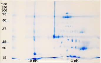
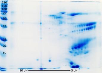
100 V. griseus V. prasinus 100 V. jobiensis V. varius V salvadorii Figure 7. 2D PAGE results.

101 101 V. varius Figure 8. 17cm 2D PAGE results. C CRiSP, L Lysosomal acid lipase, K kallikrein, HGF Hepatocyte growth factor, A AVIT, Ch Chitotriosidase, PLA2 phsopholipase A2 type III


102 Figure 9. All components of varanoid oral secretion recovered in this study. 102
103 103 Bioactivity studies further strengthened the differences between the oral secretion profiles, with V. varius having the highest of all phospholipase A2 activity rate almost three times higher than that of V. melinus and V. scalaris and an order of magnitude higher than the rest, most of which had no significant activity (fig. 10). No phospholipase A2 activity was recovered for V. mitchelli. Ability to cleave kallikrein substrate was equally variable between the species: V. mitchelli activity rate was more than twice than that of V. scalaris, V. panoptes rubidus, V. melinus and V. prasinus and almost ten-fold that of other species. V. gilleni and V. griseus had the least potency in that respect amongst all the species studied. The observed differences in activity rates, especially with respect to kallikrein, might be the consequence not only of the structural differences between the proteins of different species, but also a result of differential component complexity, as is clearly the case with V. mitchelli whose 2D profile is largely composed of different kallikrein isoforms. At the same time, differences in activity rate might be related to the dramatic size difference between the studied species almost seven-fold between the smallest (V. gilleni) and biggest (V. varius) in SVL (snout to vent length). The prey-predator size ratio is drastically different for those species, and venom replenishment costs may also vary considerably. Experiments of the effect of V. varius venom on rat ileum contractility showed a pronounced effect of increased contractility induced by the administration of the crude venom (fig. 11). This may be attributable to the presence of AVIT and natriuretic peptides, as recovered by the MS analysis.

104 ,9 0,8 0,7 0,6 0,5 0,4 PLA2 KLK 0,3 0,2 0,1 0 MIT SCA TRI ACA GIL RUB GIG MER SAL VAR MEL JOB PRA GRI Figure 10. Normalised results of bioactivity testings. On the Y-axis is the initial activity rate normalised within the study group, with the highest rate being assign to 1 and the others calculated as a fraction of it. MIT V. mitcheli, SCA V. scalaris, TRI V. tristis, ACA V. acanthurus, GIL V. gilleni, RUB V. panoptes rubidus, GIG V. giganteus, MER V. mertensi, SAL V. salvadorii, VAR V. varius, MEL V. melinus, JOB V. jobiensis, PRA V. prasinus, GRI V. griseus. Figure 11. Change in rat ileum smooth muscle contractility after administration of crude V. varius venom.on the Y-axis is the pulling force in relative units; on the X-axis is time.
 with SVL")
105 105 Figure 12. Phylogenetic relationships between the studied species (based on Ast, 2001) with SVL meassurments (based on Pianka and King, 2004) and main findings of the present study.
OPEN WIDE: DECODING THE SECRETS OF VENOM
 Ms. Foglia Period Date The New York Times April 5, 2005 OPEN WIDE: DECODING THE SECRETS OF VENOM The inland taipan, a nine-foot-long Australian snake, is not the sort of creature most people would want
CLADISTICS Student Packet SUMMARY Phylogeny Phylogenetic trees/cladograms
 CLADISTICS Student Packet SUMMARY PHYLOGENETIC TREES AND CLADOGRAMS ARE MODELS OF EVOLUTIONARY HISTORY THAT CAN BE TESTED Phylogeny is the history of descent of organisms from their common ancestor. Phylogenetic
The Venom System and Envenomation
 CHAPTER 3 The Venom System and Envenomation T he most widely recognized feature of the Helodermatidae family is that its members are venomous. Misunderstanding and confusion about this trait have accompanied
5 Dangerous Venom Types Thailand Snakes. Thailand Snake Venom Types:
 5 Dangerous Venom Types Thailand Snakes Snakes in Thailand have different types of venom that affect you in different ways if you are bitten and venom is inside your bloodstream (envenomation). Here are
(D) fertilization of eggs immediately after egg laying
 fertilization of eggs immediately after egg laying") Name: ACROSS DOWN 24. The amniote egg (A) requires a moist environment for egg laying (B) lacks protective structures for the embryo (C) has membranes enclosing the developing embryo (D) evolved from the
Name: ACROSS DOWN 24. The amniote egg (A) requires a moist environment for egg laying (B) lacks protective structures for the embryo (C) has membranes enclosing the developing embryo (D) evolved from the
posterior probabilities Values below branches: Maximum Likelihood bootstrap values.
 Supplementary Figure 1: Squamate molecular phylogeny. Values above branches: Bayesian posterior probabilities Values below branches: Maximum Likelihood bootstrap values. Supplementary Figure 2: Bayesian
Dynamic evolution of venom proteins in squamate reptiles. Nicholas R. Casewell, Gavin A. Huttley and Wolfgang Wüster
 Dynamic evolution of venom proteins in squamate reptiles Nicholas R. Casewell, Gavin A. Huttley and Wolfgang Wüster Supplementary Information Supplementary Figure S1. Phylogeny of the Toxicofera and evolution
Lecture 11 Wednesday, September 19, 2012
 Lecture 11 Wednesday, September 19, 2012 Phylogenetic tree (phylogeny) Darwin and classification: In the Origin, Darwin said that descent from a common ancestral species could explain why the Linnaean
Class Reptilia Testudines Squamata Crocodilia Sphenodontia
 Class Reptilia Testudines (around 300 species Tortoises and Turtles) Squamata (around 7,900 species Snakes, Lizards and amphisbaenids) Crocodilia (around 23 species Alligators, Crocodiles, Caimans and
Evolution as Fact. The figure below shows transitional fossils in the whale lineage.
 Evolution as Fact Evolution is a fact. Organisms descend from others with modification. Phylogeny, the lineage of ancestors and descendants, is the scientific term to Darwin's phrase "descent with modification."
Characteristics of a Reptile. Vertebrate animals Lungs Scaly skin Amniotic egg
 Reptiles Characteristics of a Reptile Vertebrate animals Lungs Scaly skin Amniotic egg Characteristics of Reptiles Adaptations to life on land More efficient lungs and a better circulator system were develope
Red Eared Slider Secrets. Although Most Red-Eared Sliders Can Live Up to Years, Most WILL NOT Survive Two Years!
 Although Most Red-Eared Sliders Can Live Up to 45-60 Years, Most WILL NOT Survive Two Years! Chris Johnson 2014 2 Red Eared Slider Secrets Although Most Red-Eared Sliders Can Live Up to 45-60 Years, Most
1. Department of Zoology, University of Oxford, South Parks Road, Oxford, OX1 3PS,
 1 2 Title: A Critique of the Toxicoferan Hypothesis 3 4 5 Authors: Adam D Hargreaves 1, Abigail S Tucker 2 and John F Mulley 3 6 7 8 9 10 11 12 1. Department of Zoology, University of Oxford, South Parks
Supporting Information
 Supporting Information Fry et al. 10.1073/pnas.0810883106 Fig. S1. SELDI-TOF MS comparison of Varanus komodoensis (Komodo Dragon) and V. varius (Lace Monitor) venoms using different arrays and wash buffers:
muscles (enhancing biting strength). Possible states: none, one, or two.
. Possible states: none, one, or two.") Reconstructing Evolutionary Relationships S-1 Practice Exercise: Phylogeny of Terrestrial Vertebrates In this example we will construct a phylogenetic hypothesis of the relationships between seven taxa
Reconstructing Evolutionary Relationships S-1 Practice Exercise: Phylogeny of Terrestrial Vertebrates In this example we will construct a phylogenetic hypothesis of the relationships between seven taxa
THE KOMODO DRAGON. endangered species L ARCHE PHOTOGRAPHIQUE CHARACTERISTICS. Animal Phylum. Kingdom
 L ARCHE PHOTOGRAPHIQUE ACTIONS FOR BIODIVERSITY CHARACTERISTICS It looks like a dragon from legend. Moreover, the Komodo dragon is the biggest and heaviest lizard in the world, and it is also known as
Testing Phylogenetic Hypotheses with Molecular Data 1
 Testing Phylogenetic Hypotheses with Molecular Data 1 How does an evolutionary biologist quantify the timing and pathways for diversification (speciation)? If we observe diversification today, the processes
Modern Evolutionary Classification. Lesson Overview. Lesson Overview Modern Evolutionary Classification
 Lesson Overview 18.2 Modern Evolutionary Classification THINK ABOUT IT Darwin s ideas about a tree of life suggested a new way to classify organisms not just based on similarities and differences, but
What causes heartworm disease?
 Heartworm Disease: What causes heartworm disease? Heartworm disease (dirofilariasis) is a serious and potentially fatal disease in dogs and cats. It is caused by a blood-borne parasite called Dirofilaria
Komodo Dragons: Giant. Komodo Dragons: Giant Reptiles A Reading A Z Level Q Leveled Book Word Count: 966 LEVELED BOOK Q
 Komodo Dragons: Giant Reptiles A Reading A Z Level Q Leveled Book Word Count: 966 Connections Writing Write an adventure story featuring a Komodo dragon. Include facts about Komodo dragons in your story.
Taxonomy. Chapter 20. Evolutionary Development Diagram. I. Evolution 2/24/11. Kingdom - Animalia Phylum - Chordata Class Reptilia.
 Taxonomy Chapter 20 Reptiles Kingdom - Animalia Phylum - Chordata Class Reptilia Order Testudines - turtles Order Crocodylia - crocodiles, alligators Order Sphenodontida - tuataras Order Squamata - snakes
Traveling Treasures 2016 The Power of Poison
 Traveling Treasures 2016 The Power of Poison Snake and Butterfly case Timber rattlesnake (Crotalus horridus) Light morph Like other snakes in the family Viperidae, timber rattlers are pit vipers. This
«The Very Thick Line Between Raising Concerns And Denialism Choose your cutlery carefully: what you eat with changes how food tastes»
 1 of 6 7/23/13 9:41 AM Science Sushi «The Very Thick Line Between Raising Concerns And Denialism Choose your cutlery carefully: what you eat with changes how food tastes» Here Be Dragons: The Mythic Bite
Evolution. Evolution is change in organisms over time. Evolution does not have a goal; it is often shaped by natural selection (see below).
.") Evolution Evolution is change in organisms over time. Evolution does not have a goal; it is often shaped by natural selection (see below). Species an interbreeding population of organisms that can produce
Evolution Evolution is change in organisms over time. Evolution does not have a goal; it is often shaped by natural selection (see below). Species an interbreeding population of organisms that can produce
Bio 1B Lecture Outline (please print and bring along) Fall, 2006
 Fall, 2006") Bio 1B Lecture Outline (please print and bring along) Fall, 2006 B.D. Mishler, Dept. of Integrative Biology 2-6810, bmishler@berkeley.edu Evolution lecture #4 -- Phylogenetic Analysis (Cladistics) -- Oct.
Bio 1B Lecture Outline (please print and bring along) Fall, 2006 B.D. Mishler, Dept. of Integrative Biology 2-6810, bmishler@berkeley.edu Evolution lecture #4 -- Phylogenetic Analysis (Cladistics) -- Oct.
What is the evidence for evolution?
 What is the evidence for evolution? 1. Geographic Distribution 2. Fossil Evidence & Transitional Species 3. Comparative Anatomy 1. Homologous Structures 2. Analogous Structures 3. Vestigial Structures
Differences between Reptiles and Mammals. Reptiles. Mammals. No milk. Milk. Small brain case Jaw contains more than one bone Simple teeth
 Differences between Reptiles and Mammals Reptiles No milk Mammals Milk The Advantage of Being a Furball: Diversification of Mammals Small brain case Jaw contains more than one bone Simple teeth One ear
NAME: DATE: SECTION:
 NAME: DATE: SECTION: MCAS PREP PACKET EVOLUTION AND BIODIVERSITY 1. Which of the following observations best supports the conclusion that dolphins and sharks do not have a recent common ancestor? A. Dolphins
rodent species in Australia to the fecal odor of various predators. Rattus fuscipes (bush
 Sample paper critique #2 The article by Hayes, Nahrung and Wilson 1 investigates the response of three rodent species in Australia to the fecal odor of various predators. Rattus fuscipes (bush rat), Uromys
8/19/2013. Topic 5: The Origin of Amniotes. What are some stem Amniotes? What are some stem Amniotes? The Amniotic Egg. What is an Amniote?
 Topic 5: The Origin of Amniotes Where do amniotes fall out on the vertebrate phylogeny? What are some stem Amniotes? What is an Amniote? What changes were involved with the transition to dry habitats?
Biology. Slide 1of 50. End Show. Copyright Pearson Prentice Hall
 Biology 1of 50 2of 50 Phylogeny of Chordates Nonvertebrate chordates Jawless fishes Sharks & their relatives Bony fishes Reptiles Amphibians Birds Mammals Invertebrate ancestor 3of 50 A vertebrate dry,
Fishes, Amphibians, Reptiles
 Fishes, Amphibians, Reptiles Section 1: What is a Vertebrate? Characteristics of CHORDATES Most are Vertebrates (have a spinal cord) Some point in life cycle all chordates have: Notochord Nerve cord that
Biology Slide 1 of 50
 Biology 1 of 50 2 of 50 What Is a Reptile? What are the characteristics of reptiles? 3 of 50 What Is a Reptile? What Is a Reptile? A reptile is a vertebrate that has dry, scaly skin, lungs, and terrestrial
Title: Phylogenetic Methods and Vertebrate Phylogeny
 Title: Phylogenetic Methods and Vertebrate Phylogeny Central Question: How can evolutionary relationships be determined objectively? Sub-questions: 1. What affect does the selection of the outgroup have
WildlifeCampus Advanced Snakes & Reptiles 1. Vipers and Adders
 Advanced Snakes & Reptiles 1 Module # 4 Component # 9 Viperidae - Hinged Front Fang Snakes This Family is divided into two sub-families. These are Old World and Modern / New World Adders. The predominant
Geo 302D: Age of Dinosaurs LAB 4: Systematics Part 1
 Geo 302D: Age of Dinosaurs LAB 4: Systematics Part 1 Systematics is the comparative study of biological diversity with the intent of determining the relationships between organisms. Humankind has always
Unit 19.3: Amphibians
 Unit 19.3: Amphibians Lesson Objectives Describe structure and function in amphibians. Outline the reproduction and development of amphibians. Identify the three living amphibian orders. Describe how amphibians
LABORATORY #10 -- BIOL 111 Taxonomy, Phylogeny & Diversity
 LABORATORY #10 -- BIOL 111 Taxonomy, Phylogeny & Diversity Scientific Names ( Taxonomy ) Most organisms have familiar names, such as the red maple or the brown-headed cowbird. However, these familiar names
Ch 1.2 Determining How Species Are Related.notebook February 06, 2018
 Name 3 "Big Ideas" from our last notebook lecture: * * * 1 WDYR? Of the following organisms, which is the closest relative of the "Snowy Owl" (Bubo scandiacus)? a) barn owl (Tyto alba) b) saw whet owl
VERTEBRATE READING. Fishes
 VERTEBRATE READING Fishes The first vertebrates to become a widespread, predominant life form on earth were fishes. Prior to this, only invertebrates, such as mollusks, worms and squid-like animals, would
Reptile Round Up. An Educator s Guide to the Program
 Reptile Round Up An Educator s Guide to the Program GRADES: K-3 PROGRAM DESCRIPTION: This guide provided by the Oklahoma Aquarium explores reptiles and their unique characteristics. The Reptile Round Up
Do the traits of organisms provide evidence for evolution?
 PhyloStrat Tutorial Do the traits of organisms provide evidence for evolution? Consider two hypotheses about where Earth s organisms came from. The first hypothesis is from John Ray, an influential British
Reptilian Requirements Created by the North Carolina Aquarium at Fort Fisher Education Section
 Essential Question: North Carolina Aquariums Education Section Reptilian Requirements Created by the North Carolina Aquarium at Fort Fisher Education Section What physical and behavioral adaptations do
Materials and Methods: Anti-snake venom activities of Asparagus racernosus
 Sunil Prashar. et al.: Asian Journal of Pharmacology and Toxicology, 04(16), 2016,Ol-08. RESEARCH ARTICLE Received on: 201 1212016 Published on:29/ 12120 16 Corresponding Author Sunil Prashar, Department
Stuart S. Sumida Biology 342. Simplified Phylogeny of Squamate Reptiles
 Stuart S. Sumida Biology 342 Simplified Phylogeny of Squamate Reptiles Amphibia Amniota Seymouriamorpha Diadectomorpha Synapsida Parareptilia Captorhinidae Diapsida Archosauromorpha Reptilia Amniota Amphibia
LABORATORY EXERCISE 6: CLADISTICS I
 Biology 4415/5415 Evolution LABORATORY EXERCISE 6: CLADISTICS I Take a group of organisms. Let s use five: a lungfish, a frog, a crocodile, a flamingo, and a human. How to reconstruct their relationships?
Malayan Pit Viper Venomous Very Dangerous
 Malayan Pit Viper Venomous Very Dangerous Adult Malayan Pit Viper in situ, found in a culvert in Krabi, Thailand. [Page Updated: 4 April 2018] Calloselasma rhodostoma (Malayan Pit Viper, Malaysian Pit
Evolution of Birds. Summary:
 Oregon State Standards OR Science 7.1, 7.2, 7.3, 7.3S.1, 7.3S.2 8.1, 8.2, 8.2L.1, 8.3, 8.3S.1, 8.3S.2 H.1, H.2, H.2L.4, H.2L.5, H.3, H.3S.1, H.3S.2, H.3S.3 Summary: Students create phylogenetic trees to
Interpreting Evolutionary Trees Honors Integrated Science 4 Name Per.
 Interpreting Evolutionary Trees Honors Integrated Science 4 Name Per. Introduction Imagine a single diagram representing the evolutionary relationships between everything that has ever lived. If life evolved
Start of new generation of NSAIDs?
 Vet Times The website for the veterinary profession https://www.vettimes.co.uk Start of new generation of NSAIDs? Author : Peter Lees Categories : Vets Date : May 16, 2011 Peter Lees discusses development
A. Body Temperature Control Form and Function in Mammals
 Taxonomy Chapter 22 Kingdom Animalia Phylum Chordata Class Mammalia Mammals Characteristics Evolution of Mammals Have hair and First appear in the mammary glands Breathe air, 4chambered heart, endotherms
1 Describe the anatomy and function of the turtle shell. 2 Describe respiration in turtles. How does the shell affect respiration?
 GVZ 2017 Practice Questions Set 1 Test 3 1 Describe the anatomy and function of the turtle shell. 2 Describe respiration in turtles. How does the shell affect respiration? 3 According to the most recent
REPTILES. Scientific Classification of Reptiles To creep. Kingdom: Animalia Phylum: Chordata Subphylum: Vertebrata Class: Reptilia
 Scientific Classification of Reptiles To creep Kingdom: Animalia Phylum: Chordata Subphylum: Vertebrata Class: Reptilia REPTILES tetrapods - 4 legs adapted for land, hip/girdle Amniotes - animals whose
08 alberts part2 7/23/03 9:10 AM Page 95 PART TWO. Behavior and Ecology
 08 alberts part2 7/23/03 9:10 AM Page 95 PART TWO Behavior and Ecology 08 alberts part2 7/23/03 9:10 AM Page 96 08 alberts part2 7/23/03 9:10 AM Page 97 Introduction Emília P. Martins Iguanas have long
All about snakes. What are snakes? Are snakes just lizards without legs? If you want to know more
 Novak.lisa@gmail.com Day 83 12/29/2017 All about snakes What are snakes? Are snakes just lizards without legs? If you want to know more keep reading to find out the answers to the question. The purpose
Vertebrates. Vertebrates are animals that have a backbone and an endoskeleton.
 Vertebrates Vertebrates are animals that have a backbone and an endoskeleton. The backbone replaces the notochord and contains bones called vertebrae. An endoskeleton is an internal skeleton that protects
DO NOT ATTEMPT TO CAPTURE OR HANDLE SNAKES
 Advanced Snakes & Reptiles 1 Module # 4 Component # 1 Capturing and Handling This is not a snake Capture or Handling course. This course in no way encourages, teaches, trains, supports, persuades or promotes
The Ecology of Lyme Disease 1
 The Ecology of Lyme Disease 1 What is Lyme disease? Lyme disease begins when a tick bite injects Lyme disease bacteria into a person's blood. Early symptoms of Lyme disease usually include a bull's-eye
Biodiversity and Distributions. Lecture 2: Biodiversity. The process of natural selection
 Lecture 2: Biodiversity What is biological diversity? Natural selection Adaptive radiations and convergent evolution Biogeography Biodiversity and Distributions Types of biological diversity: Genetic diversity
Biology 1B Evolution Lecture 11 (March 19, 2010), Insights from the Fossil Record and Evo-Devo
, Insights from the Fossil Record and Evo-Devo") Biology 1B Evolution Lecture 11 (March 19, 2010), Insights from the Fossil Record and Evo-Devo Extinction Important points on extinction rates: Background rate of extinctions per million species per year:
Biology 1B Evolution Lecture 11 (March 19, 2010), Insights from the Fossil Record and Evo-Devo Extinction Important points on extinction rates: Background rate of extinctions per million species per year:
Video Assignments. Microraptor PBS The Four-winged Dinosaur Mark Davis SUNY Cortland Library Online
 Video Assignments Microraptor PBS The Four-winged Dinosaur Mark Davis SUNY Cortland Library Online Radiolab Apocalyptical http://www.youtube.com/watch?v=k52vd4wbdlw&feature=youtu.be Minute 13 through minute
HOW XTC IMPROVED MINOXIDIL PENETRATION - 5 WAYS!
 HOW XTC IMPROVED MINOXIDIL PENETRATION - 5 WAYS! What Hinders Minoxidil from Working Well 1. Sebum from sebaceous gland blocks the hair follicle. 2. Minoxidil therefore, cannot penetrate through the sebum
Northern Copperhead Updated: April 8, 2018
 Interpretation Guide Northern Copperhead Updated: April 8, 2018 Status Danger Threats Population Distribution Habitat Diet Size Longevity Social Family Units Reproduction Our Animals Scientific Name Least
May 10, SWBAT analyze and evaluate the scientific evidence provided by the fossil record.
 May 10, 2017 Aims: SWBAT analyze and evaluate the scientific evidence provided by the fossil record. Agenda 1. Do Now 2. Class Notes 3. Guided Practice 4. Independent Practice 5. Practicing our AIMS: E.3-Examining
UT HEALTH EMERGENCY MEDICINE & TRAUMA GUIDELINES
 UT HEALTH EMERGENCY MEDICINE & TRAUMA GUIDELINES TITLE: Snake bites ORIGINAL DATE: 07/2003 SUPERCEDES: 07/2013 LAST REVIEW DATE: 06/2017 Purpose Statement: To provide guidance on the evaluation and management
Mental stim ulation it s not just for dogs!! By Danielle Middleton- Beck BSc hons, PGDip CABC
 Milo, Congo African Grey by Elaine Henley Mental stim ulation it s not just for dogs!! By Danielle Middleton- Beck BSc hons, PGDip CABC Dexter, Green Iguana by Danielle Middleton-Beck Exotic pets include
Super Toxic Thailand Sea Snakes
 Super Toxic Thailand Sea Snakes Laticauda colubrina. Also known as colubrine sea krait or yellow-lipped sea krait. 2012 Elias Levy at Flickr.com. THAILAND SEA SNAKES CRUCIAL INFORMATION Thailand is surrounded,
CANINE HEARTWORM DISEASE
 ! CANINE HEARTWORM DISEASE What causes heartworm disease? Heartworm disease (dirofilariasis) is a serious and potentially fatal disease in dogs. It is caused by a blood-borne parasite called Dirofilaria
6. The lifetime Darwinian fitness of one organism is greater than that of another organism if: A. it lives longer than the other B. it is able to outc
 1. The money in the kingdom of Florin consists of bills with the value written on the front, and pictures of members of the royal family on the back. To test the hypothesis that all of the Florinese $5
GUIDELINES ON CHOOSING THE CORRECT ERADICATION TECHNIQUE
 GUIDELINES ON CHOOSING THE CORRECT ERADICATION TECHNIQUE PURPOSE... 2 1. RODENTS... 2 1.1 METHOD PROS AND CONS... 3 1.1. COMPARISON BETWEEN BROUDIFACOUM AND DIPHACINONE... 4 1.2. DISCUSSION ON OTHER POSSIBLE
Darwin and the Family Tree of Animals
 Darwin and the Family Tree of Animals Note: These links do not work. Use the links within the outline to access the images in the popup windows. This text is the same as the scrolling text in the popup
Talks generally last minutes and take place in one of our classrooms.
 Key Stage 1 & Key Stage 2 REPTILES General points about this talk: Talks generally last 30-40 minutes and take place in one of our classrooms. Talks are generally lead by the keepers on this section so
Reproduction in Seed Plants (pp )
") Structure and Function of Plants Reading/Notetaking Guide Reproduction in Seed Plants (pp. 388 397) This section gives examples of the group of seed plants known as gymnosperms and angiosperms and describes
Structure and Function of Plants Reading/Notetaking Guide Reproduction in Seed Plants (pp. 388 397) This section gives examples of the group of seed plants known as gymnosperms and angiosperms and describes
LABORATORY EXERCISE 7: CLADISTICS I
 Biology 4415/5415 Evolution LABORATORY EXERCISE 7: CLADISTICS I Take a group of organisms. Let s use five: a lungfish, a frog, a crocodile, a flamingo, and a human. How to reconstruct their relationships?
UNIT III A. Descent with Modification(Ch19) B. Phylogeny (Ch20) C. Evolution of Populations (Ch21) D. Origin of Species or Speciation (Ch22)
 B. Phylogeny (Ch20) C. Evolution of Populations (Ch21) D. Origin of Species or Speciation (Ch22)") UNIT III A. Descent with Modification(Ch9) B. Phylogeny (Ch2) C. Evolution of Populations (Ch2) D. Origin of Species or Speciation (Ch22) Classification in broad term simply means putting things in classes
UNIT III A. Descent with Modification(Ch9) B. Phylogeny (Ch2) C. Evolution of Populations (Ch2) D. Origin of Species or Speciation (Ch22) Classification in broad term simply means putting things in classes
Squamates of Connecticut
 Squamates of Connecticut Reptilia Turtles are sisters to crocodiles and birds Yeah, birds are reptiles, haven t you watched Jurassic Park yet? Lizards and snakes are part of one clade called the squamates
Species: Panthera pardus Genus: Panthera Family: Felidae Order: Carnivora Class: Mammalia Phylum: Chordata
 CHAPTER 6: PHYLOGENY AND THE TREE OF LIFE AP Biology 3 PHYLOGENY AND SYSTEMATICS Phylogeny - evolutionary history of a species or group of related species Systematics - analytical approach to understanding
Introduction to phylogenetic trees and tree-thinking Copyright 2005, D. A. Baum (Free use for non-commercial educational pruposes)
") Introduction to phylogenetic trees and tree-thinking Copyright 2005, D. A. Baum (Free use for non-commercial educational pruposes) Phylogenetics is the study of the relationships of organisms to each other.
Introduction to phylogenetic trees and tree-thinking Copyright 2005, D. A. Baum (Free use for non-commercial educational pruposes) Phylogenetics is the study of the relationships of organisms to each other.
Animal Diversity wrap-up Lecture 9 Winter 2014
 Animal Diversity wrap-up Lecture 9 Winter 2014 1 Animal phylogeny based on morphology & development Fig. 32.10 2 Animal phylogeny based on molecular data Fig. 32.11 New Clades 3 Lophotrochozoa Lophophore:
d. Wrist bones. Pacific salmon life cycle. Atlantic salmon (different genus) can spawn more than once.
 can spawn more than once.") Lecture III.5b Answers to HW 1. (2 pts). Tiktaalik bridges the gap between fish and tetrapods by virtue of possessing which of the following? a. Humerus. b. Radius. c. Ulna. d. Wrist bones. 2. (2 pts)
Lecture III.5b Answers to HW 1. (2 pts). Tiktaalik bridges the gap between fish and tetrapods by virtue of possessing which of the following? a. Humerus. b. Radius. c. Ulna. d. Wrist bones. 2. (2 pts)
Introduction to Medically Important Spiders
 WildlifeCampus Medically Important Spiders (1) Module # 1 Component # 1 Preface Introduction to Medically Important Spiders For this online course, we ve put together a host of interesting and useful facts
Professor David J Mellor Professor Kevin J Stafford Co-Directors
 Professor David J Mellor Professor Kevin J Stafford Co-Directors Collaborating Centre for Animal Welfare Science and Bioethical Analysis: Founding Partner http://animalwelfare.massey.ac.nz Evolving Veterinary
Cover Page. The handle holds various files of this Leiden University dissertation.
 Cover Page The handle http://hdl.handle.net/1887/19952 holds various files of this Leiden University dissertation. Author: Vonk, Freek Jacobus Title: Snake evolution and prospecting of snake venom Date:
Biodiversity and Extinction. Lecture 9
 Biodiversity and Extinction Lecture 9 This lecture will help you understand: The scope of Earth s biodiversity Levels and patterns of biodiversity Mass extinction vs background extinction Attributes of
Cobras By Guy Belleranti
 Name: The cobras of Africa and Asia are among the most famous, and most intimidating, snakes in the world. have thin bodies and short, wide heads. When a cobra hunts or senses danger, it raises its head
Sec KEY CONCEPT Reptiles, birds, and mammals are amniotes.
 Thu 4/27 Learning Target Class Activities *attached below (scroll down)* Website: my.hrw.com Username: bio678 Password:a4s5s Activities Students will describe the evolutionary significance of amniotic
Non-Fiction. Reptile Edition. Close Reading PASSAGEs. Common Core Aligned. 1 st, 2 nd and 3 rd Grade Michelle Arold
 1 st, 2 nd and 3 rd Grade Non-Fiction Close Reading PASSAGEs Common Core Aligned Reptile Edition THANK YOU for downloading! Thank you for downloading! In this packet I have included 4 non-fiction close
Introduction to Herpetology
 Introduction to Herpetology Lesson Aims Discuss the nature and scope of reptiles. Identify credible resources, and begin to develop networking with organisations and individuals involved with the study
CHAPTER 26. Animal Evolution The Vertebrates
 CHAPTER 26 Animal Evolution The Vertebrates Impacts, Issues: Interpreting and Misinterpreting the Past No one was around to witness the transitions in the history of life Fossils allow us glimpses into
Ch 34: Vertebrate Objective Questions & Diagrams
 Ch 34: Vertebrate Objective Questions & Diagrams Invertebrate Chordates and the Origin of Vertebrates 1. Distinguish between the two subgroups of deuterostomes. 2. Describe the four unique characteristics
Banded Krait Venomous Deadly
 Banded Krait Venomous Deadly Yellow and black Banded Krait (Bungarus fasciatus) venomous and deadly. Copyright Tom Charlton. These are yellow and black kraits here in Thailand. In some other part of the
WHO/SEARO GUIDELINES FOR THE CLINICAL MANAGEMENT OF SNAKE BITES IN THE SOUTHEAST ASIAN REGION
 WHO/SEARO GUIDELINES FOR THE CLINICAL MANAGEMENT OF SNAKE BITES IN THE SOUTHEAST ASIAN REGION Written and edited by David A Warrell with contributions by an international panel of experts World Health
2 nd Term Final. Revision Sheet. Students Name: Grade: 11 A/B. Subject: Biology. Teacher Signature. Page 1 of 11
 2 nd Term Final Revision Sheet Students Name: Grade: 11 A/B Subject: Biology Teacher Signature Page 1 of 11 Nour Al Maref International School Riyadh, Saudi Arabia Biology Worksheet (2 nd Term) Chapter-26
The Most Venomous Dangerous Deadly Poisonous Snakes?
 The Most Venomous Dangerous Deadly Poisonous Snakes? Venomous and somewhat dangerous, but not deadly or poisonous, Trimeresurus venustus the beautiful pit viper, aka, the brown-spotted pit viper. This
6/20/2018. A Public Benefit Corporation. A First-in-Class Snakebite Antidote. Discovery to Accelerated Development
 A Public Benefit Corporation A First-in-Class Snakebite Antidote Discovery to Accelerated Development 1) Introduction - 5 min 2) Introduction of Ophiex 15 min 3) Ophiex's snake research 60 min 4) Introduction
INQUIRY & INVESTIGATION
 INQUIRY & INVESTIGTION Phylogenies & Tree-Thinking D VID. UM SUSN OFFNER character a trait or feature that varies among a set of taxa (e.g., hair color) character-state a variant of a character that occurs
Non-Snake Reptile Bites141
 Non-Snake Reptile Bites141 Stephen C. Hartsell and Troy E. Madsen KEY POINTS Alligator and crocodile bites may inflict significant internal injury and should be managed as major trauma. Gila monster bites
BREATHING WHICH IS NOT RESPIRATION
 BREATHING WHICH IS NOT RESPIRATION Breathing vs. Respiration All animals respire. A lot of people think respiration means breathing- this is not true! Breathing is the physical process of inhaling oxygen
Page # Diversity of Arthropoda Crustacea Morphology. Diversity of Arthropoda. Diversity of Arthropoda. Diversity of Arthropoda. Arthropods, from last
 Arthropods, from last time Crustacea are the dominant marine arthropods Crustacea are the dominant marine arthropods any terrestrial crustaceans? Should we call them shellfish? sowbugs 2 3 Crustacea Morphology
Mammals. Introduction (page 821) Evolution of Mammals (page 821) Form and Function in Mammals (pages ) Chapter 32.
 Evolution of Mammals (page 821) Form and Function in Mammals (pages ) Chapter 32.") Chapter 32 Mammals Section 32 1 Introduction to the Mammals (pages 821 827) This section describes the characteristics common to all mammals, as well as how mammals carry out life functions. It also briefly
Chapter 32 Mammals Section 32 1 Introduction to the Mammals (pages 821 827) This section describes the characteristics common to all mammals, as well as how mammals carry out life functions. It also briefly