A CYTOLOGICAL STUDY OF THE SPOROZOITES OF EIMERIA CAVIAE, A COCCIDIAN PARASITE OF THE DOMESTIC GUINEA PIG, CAVIA PORCELLUS
|
|
|
- Esmond Mathews
- 5 years ago
- Views:
Transcription

1 A CYTOLOGICAL STUDY OF THE SPOROZOITES OF EIMERIA CAVIAE, A COCCIDIAN PARASITE OF THE DOMESTIC GUINEA PIG, CAVIA PORCELLUS An abstract of a Thesis by C. Bruce Moore December 1976 Drake University Advisor: Richard S. Wacha The problem. The present study was undertaken in order to provide information concerning the morphological and cytological characteristics of the sporozoites of Eimeria caviae, Sheather (1924), a coccidian parasite of guinea pigs. Observations concerning the prepatent and patent periods, the symptomatology and pathology, and the morphological features of the oocysts of this species are also reported. Procedure. Oocysts of Eimeria caviae, isolated from naturally infected guinea pigs, were excysted in vitro and the sporozoites studied in fresh and stained preparations. Fresh sporozoites were studied in the medium used for excystation, at room temperature, with the aid of a Zeiss RA and/or Zeiss photomicroscope equipped with achromatic objectives and bright field and phase contrast condensors. Smears for permanent preparations were made by smearing No. 1 cover slips with Meyer's adhesive and freshly excysted sporozoites. Sporozoites stained with the Giemsa method were fixed in methanol and those stained with Harris' hematoxylin and eosin were fixed in Zenker's fluid. Sporozoites stained with the periodic acid-schiff (PAS) and the Feulgen methods were fixed in Bouin's fixative. Findings. Freshly excysted sporozoites were motile and exhibited flexion, probing, gliding and rotational movements. The sporozoites were curved in shape with the anterior end tapering to a point and the posterior end being more rounded and blunt. All sporozoites had one anterior and one posterior refractile body with a nucleus between these bodies. Staining reactions revealed refractile bodies, numerous granules distributed within the cytoplasm and a nucleus containing peripherally distributed chromatin; no nucleolus was observed. Sporulated oocysts were ellipsoidal and brownishyellow in color. An oocyst residium was not observed. A cone-shaped Stieda body and membrane-bound sporocyst residium were present. The oocyst wall was smooth and consisted of two layers. The prepatent period of Eimeria caviae was found to range from 11 to 13 days~ the patent period ranged from 7 to 13 days. Symptoms of infection in guinea pigs experimentally infected with E. caviae were loss of appetite, constipation,

2 diarrhea, ruffled hair, and a "hunched" posture. Apathetic behavior was also a constant symptom. Conclusion. The cytological observations of the sporozoite of Eimeria caviae in the present study agree closely with those observed previously for this and other species. The prepatent and patent periods, symptomatology and pathology, and morphological features of the oocysts also agree with that information previously reported for this species. Recommendations. While information concerning the morphology of the sporozoites of Eimeria caviae is presented in the present study, many important problems concerning their mechanism of invasion of host cells, their excystation process, and the role they play in the immune response remain to be solved.
3 A CYTOLOGICAL STUDY OF THE SPOROZOITES OF EIMERIA CAVIAE, A COCCIDIAN PARASITE OF THE DOMESTIC GUINEA PIG, CAVIA PORCELLUS A Thesis Presented to The School of Graduate Studies Drake University In Partial Fulfillment of the Requirements for the Degree Master of Arts by C. Bruce Moore December 1976
4 A CYTOLOGICAL STUDY OF THE SPOROZOITES OF EIMERIA CAVIAE, A COCCIDIAN PARASITE OF THE DOMESTIC GUINEA PIG, CAVIA PORCELLUS by C. Bruce Moore Approved by Committee: Dr. Richard S. Wacha Chairman Dr. Rodney A. Rogers Dr. William Teppert Dr. Earle L. Canfield Dean of the School of Graduate Studies 11
5 TABLE OF CONTENTS INTRODUCTION.. REVIEW OF LITERATURE MATERIALS AND METHODS.. RESULTS. DISCUSSION CONCLUSIONS LITERATURE CITED PAGE
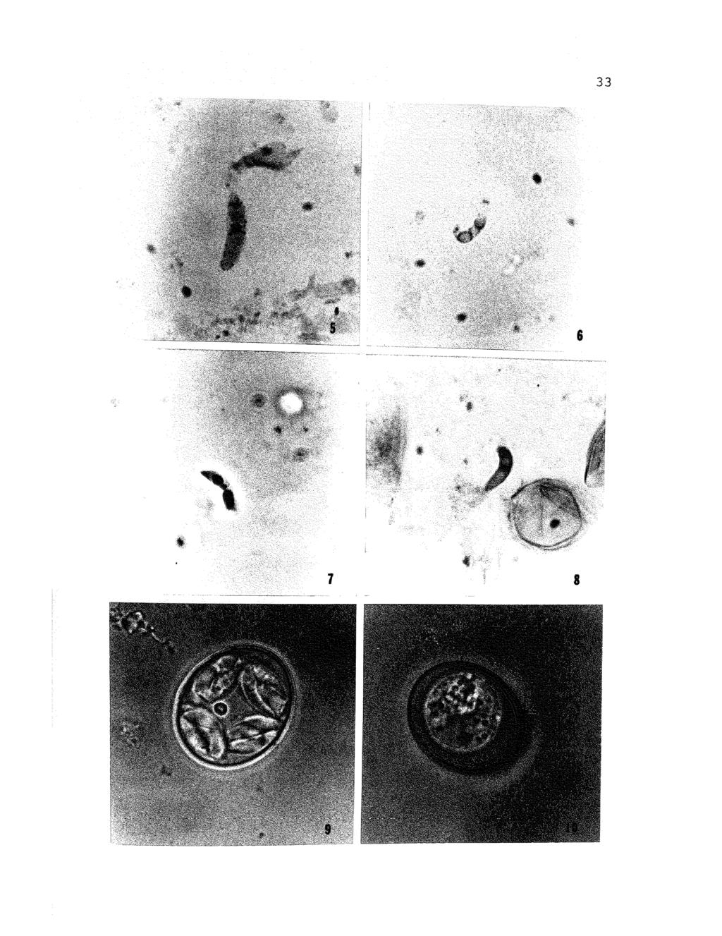
6 LIST OF TABLES Table 1. Prepatent and patent periods of Eimeria caviae. Page 17 LIST OF FIGURES Figure Page 1. Line diagram of living sporozoite Line diagram of sporozoite fixed in methanol and stained with method of Giemsa Line diagram of sporozoite fixed in Bouin's fluid and stained with the periodic acid- Schiff method Line diagrams showing variation in distribution of chromatin in nuclei of sporozoites stained with method of Feulgen Photograph showing distribution of granules in sporozoite stained with method of Giemsa Photograph showing distribution of granules in sporozoite stained with periodic acid- Schiff method Photograph showing clumps of chromatin in nucleus of sporozoite stained with method of Feulgen Photograph showing anterior and posterior refractile bodies in sporozoite stained with Hematoxylin and Eosin Photograph of sporulated oocyst. 10. Photograph of nonsporulated oocyst II. Photograph of intact sporocyst. 35
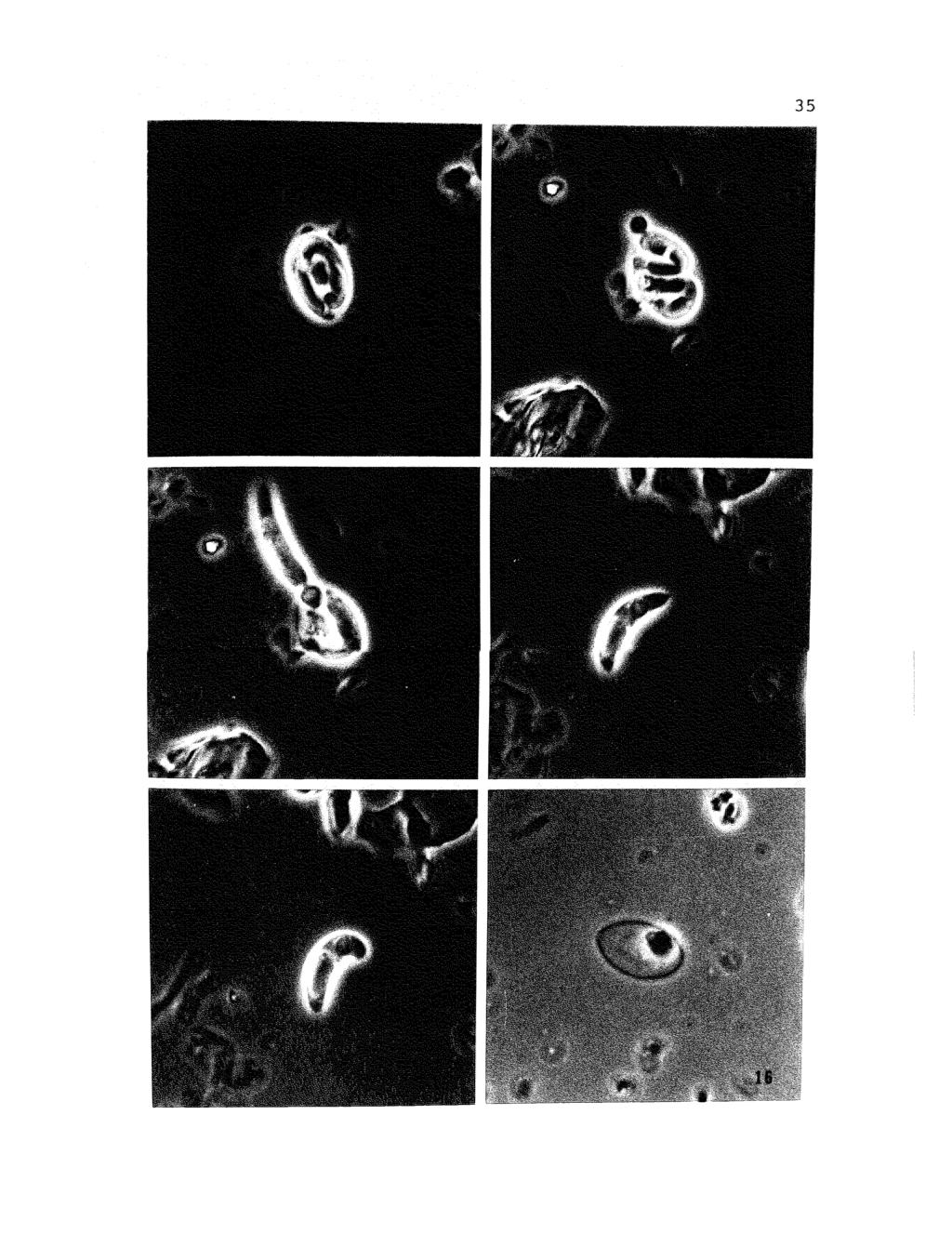
7 v Figure Page 12. Photograph showing partial emergence of sporozoite from sporocyst during excystation Photograph showing nearly complete emergence of sporozoite from sporocyst during excystation Photograph of living sporozoite in extended position Photograph of living sporozoite in flexed position Photograph showing empty sporocyst containing sporocyst residium. 35
8 INTRODUCTION The present study provides information concerning the cytological characteristics of the sporozoit.es of Eimeria caviae, Sheather (1924), a coccidian parasite of the domestic guinea pig, Cavia porcellus. The sporozoite is important in that it is the stage in the coccidian life cycle which first invades the cells of the host, initiating infection. Also, it is often useful for specific diagnosis. It is hoped that this study will contribute to our understanding of the sporozoite stage of Eimeria caviae and perhaps of the coccidia in general. Eimeria caviae was chosen for this study because of the apparent lack of detailed information available about this species, and because of the ease with which guinea pigs could be obtained and reared in the laboratory. REVIEW OF LITERATURE Labbe (1899) was apparently the first to report coccidian oocysts in the intestinal contents of guinea pigs. He believed that the oocysts which he observed represented a variety of a coccidian species which had been previously reported from rabbits. He did not, however, discuss the parasite in detail. Strada and Traina (1900) described a coccidium from guinea pigs in Italy, which they called Coccodium oviforme, and reported that infections with this
9 2 coccidium were associated with diarrhea, emaciation and death. There was no discussion of sporozoites presented. Bugge and Heinke (1921) found 73% of lao guinea pigs from Germany to harbor coccidia and regarded the infection as common and widely distributed. They also noted that sporulation of ~. caviae began in 2 to 3 days and was completed in about 5 to 8 days in boric acid or thymol at room temperature. These authors also reported the occurrence of diarrhea in guinea pigs infected with this parasite and a marked intestinal inflammation resulting from the infection. with reference to the sporozoite stage, Bugge and Heinke (1921) stated only that two, sickle-shaped, slightly curved sporozoites appeared within the sporocysts. However, the characteristics of these sporozoites were not discussed in detail. They also reported the developmental states of E. caviae to occur only in the upper portions of the colon. Sheather (1924) described the life cycle of a coccidium which he isolated from a guinea pig purchased for experimental purposes. He identified this coccidium as a species of Eimeria, to which he gave the name Eimeria caviae. He stated that endogenous development appeared to take place only in the epithelium of the colon, especially in the upper portions with the frequency of endogenous development decreasing in the lower portions. Using 5% potassium dichromate solution at room temperature, he found that oocysts of E. caviae from different guinea pigs required
10 3 different times to complete sporulation, the range being from 5 to 8 days. Sheather (1924) found oocysts of E. caviae in the feces of infected guinea pigs as early as 7 days after inoculation and as late as 13 days after inoculation. When the infection was severe, almost every cell of the mucosa of the colon appeared to contain stages of the parasite. While discussing the schizonts, microgametocytes and macrogametocytes in some detail, Sheather (1924) mentioned sporozoites only as they related to the process of sporulation. Andrews (1930) reported that oocysts from guinea pigs all excysted in the small intestine, but failed to discuss sporozoites. Henry (1932) using the method of Tyzzer (1929) noted that sporulation of E. caviae oocysts began within the twenty-four hour period following the onset of incubation in 2% potassium dichromate. Sporulation was complete in 2 to 3 days. Henry (1932) also noted that oocysts of E. caviae sporulated quite readily in normal salt solution. She also noted the prepatent period of E. caviae in guinea pigs previously uninfected with this species to be 11 to 12 days. Henry (1932) stated that in all but one of the experimentally infected animals studied, diarrhea was a constant symptom. She also noted that after the first indications of diarrhea were present, an occasional formed stool may be passed and that on the day following the appearance of marked diarrhea, only diarrheic stools occurred
11 4 with an increase in the quantity of fecal material being discharged. Diarrhea usually continued until the death of the guinea pig, but occasionally toward the end of the life of the animal, a string of very small pellets was passed. Constipation was a common symptom, and this symptom was most often noticed on the eleventh or twelfth day, but rarely later. At about the time of the occurrence of diarrhea, guinea pigs became visibly ill, remaining very quiet with hair ruffled and with the body contracted in such a manner that the posterior portion of the animal was noticeably drawn under the rest of the body. A very marked loss of appetite was observed by Henry in all infected animals. In discussing the pathology of this species, Henry (1932) found lesions of coccidiosis along with numerous oocysts of Eimeria caviae in the large intestine of 24 guinea pigs that apparently died of coccidiosis. In all cases, the lungs, liver and spleen were normal in appearance and no changes were noticed in the small intestine or in the stomach, although a few isolated lesions of coccidiosis were seen in the small intestine. In the colon, endogenous stages were found in the epithelial cells lining the crypts of Lieberkuhn. In many places, the entire mucosa had been destroyed, leaving large spaces containing some cellular debris and oocysts. Ruptured capillaries, with resulting hemorrhage, were observed in several cases. Henry's only mention of the sporozoites of E. caviae was the
12 5 possible role they might play in the cellular response of the host to infection, and that during sporulation, each of the four sporoblasts were gradually transformed into sporocysts containing two sporozoites and some rather granular residual material. Lapage (1937) found E. caviae in two laboratory groups of guinea pigs. In one population of 24 guinea pigs, 40% were infected while in a second population of 57 guinea pigs, 91% were infected. Lapage discusses variations in different phases of the life history of Eimeria caviae as well as the pathology of the parasite. He noted that this parasite produced little host tissue reaction and in general affected the host surprisingly little. The only mention of sporozoites by Lapage was that they did form in 60-70% of the oocysts which were allowed to incubate for 11 days at laboratory temperatures. Although these earlier studies provided information relative to the life cycle, pathology and prevalence of E. caviae, detailed information concerning the cytology and general structure of the sporozoites of this organism were lacking. Levine and Ivens (1965), in their recent monographic review of the coccidian parasites of rodents, made no reference to the cytological characteristics of the sporozoites of this species, nor did Kheysin (1972) in his comprehensive review of the biology of the coccidia. However, some information about the cytological and structural aspects of the sporozoites of other Eimeria species as
13 inferred from histochemical and light microscopy studies is available. Smetana (1933) noted that sporozoites of Eimeria stiedae from the rabbit were crescent-shaped and pointed at the anterior end, and that the middle and posterior portions 6 contained large, homogeneous, known as refractile bodies. fluid-like droplets, now Numerous small refractile granules were also seen in various locations throughout the sporozoites. Using brightfied and phase-contrast microscopy, Nyberg and Hammond (1965) described the living sporozoites of 4 species of Eimeria: E. bovis, E. auburnensis, E. ellipsoidalis and E. zurnii. The excysted sporozoites of E. bovis were found to agree with a description given earlier by Hammond, Bowman, Davis and Simms (1946) except that the sporozoites were slightly longer (12 to 15V as compared with 13.5 to ). Also, Nyberg and Hammond (1965) observed a projecting tip at the anterior end of the sporozoites of E. bovis which had not been previously reported. The nucleus appeared vesicular with a small, somewhat eccentric nucleolus. Hammond, Chobotar and Ernst (1968), who studied the cytological aspects of the sporozoites of E. bovis, E. auburnensis and an Eimeria species from the Ord Kangaroo Rat noted that the refractile bodies in sporozoites of E. bovis stained deeply with iron hematoxylin and bromphenol-blue but were pink in hematoxylin and eosin and in Giemsa preparations. The refractile bodies also appeared
14 7 orange w~th the modified periodic acid-schiff-analine orange (PAS-AD) method and reacted negatively to the Feulgen and protargol methods. In preparations stained with Sudan black B, the refractile bodies stained a light gray. In Feulgen preparations of sporozoites, a positive reaction was seen only at the periphery of the nucleus, showing a marginal layer of chromatin with inconspicuous clumps which occurred at irregular intervals along this layer. The nuclei in Giemsa preparations were usually obscured by numerous surrounding granules. prepared with the periodic acid-schiff (PAS) In specimens method, many PAS-positive granules were observed. After 1 hour of diastase digestion, the reaction was negative, indicating that these granules were glycogen (Hammond et al., 1968). In the same study by Hammond et al., (1968), the sporozoites of ~. auburnensis differed in some respects from those of E. bovis. The anterior portion of the body was more changeable in shape, undergoing greater elongation and retraction. Nyberg and Hammond (1965) found that the sporozoites of E. auburnensis were larger than those of E. bovis but like E. bovis contained 2 refractile bodies. Hammond et al. (1968) structure resembling a found an additional small spherical refractile body located at the extreme posterior end of the sporozoites of ~. auburnensis. The reactions of the sporozoites of E. auburnensis with the various staining methods did not differ appreciably from
15 8 those of E. bovis (Hammond et al., 1968). Nyberg and Hammond (1965) reported that free sporozoites of E. ellipsoidalis resembled those of E. bovis but the anterior end was more smoothly tapered, with the body remaining narrow for a greater distance from the anterior end than in the sporozoites of E. bovis. In Giemsa preparations and in the majority of free, actively moving sporozoites (Nyberg and Hammond, 1965), two refractile bodies of similar size were present, one near the posterior end and one near the anterior end. These bodies were smaller in proportion to the size of the sporozoite than in E. auburnensis. The nucleus was located centrally between the two refractile bodies, and there was an area of small granules immediately surrounding the nucleus. Sporozoites of E. zurnii had a relatively large and prominent nucleus and a single relatively small refractile body (Nyberg and Hammond, 1965). In contrast, the sporozoites of E. bovis, E. auburnensis and E. ellipsoidalis had a relatively small nucleus and two more or less prominent refractile bodies. In the Eimeria species isolated from the Ord Kangaroo Rat (Hammond et al., 1968) the sporozoites resembled those of E. bovis in general appearance, but had a much larger posterior refractile body. Also, this species had much less glycogen than E. bovis and E. auburnensis. In PAS preparations, some specimens showed no positively reacting granules; others had only I to 5 of these which
16 9 were located between the refractile bodies. The nuclei of this species were similar in morphology to those of E. bovis and E. auburnensis in that a nucleolus was observed in each and the chromatin was located peripherally; some nuclei appeared as clumps. The sporozoites of E. acervulina were also reported to have nuclei of this kind (Pattillo and Becker, 1955). A nipple-like projection at the anterior extremity was also observed in the species from the Ord Kangaroo Rat (Hammond et al., 1968). While the present work and that of the authors cited herein was conducted with the light microscope, much excellent microscopy has been carried out on the ultrastructure of Eimeria species (see Hammond, 1973, for a review). MATERIALS AND METHODS The original inoculum used in the present study was obtained from 2 naturally infected guinea pigs housed in the pet department of a large department store in Des Moines, Iowa. Three hundred sixteen fecal samples from guinea pigs in pet stores in the Iowa cities of Winterset, Newton, Bettendorf, Davenport, Cedar Rapids and Des Moines were examined before these 2 infected animals were found. Droppings from each guinea pig sampled were collected either from a pan placed beneath each cage or directly from the bedding. The droppings from each cage were placed in separate baby food jars, each containing approximately 30 ml
17 10 a 2% aqueous solution of potassium dichromate. These samples were placed in an ice chest and transferred to the laboratory. In some instances the potassium dichromate solution was added to the sample jars after the samples were returned to the laboratory. In the laboratory, each sample was recorded as to location and date of collection. All samples were comminuted with a wooden tongue depressor while still in the sampling jars. A 2 to 3 mi. aliquot of the mixed sample was then transferred to a 15 ml conical centrifuge tube into which 5 to 7 mls of Sheather's solution was stirred. The entire mixture was then centrifuged at 2,000 rpm for 7 minutes, allowing the oocysts to float to the surface of the Sheather's solution. After centifugation, material from the surface of the Sheather's solution was transferred with the aid of a flamed inoculating loop to a microscope slide, covered with a #1 cover slip which had been ringed with petroleum jelly, and observed under the light microscope for the presence of oocysts. Those samples found to be negative were discardedj those 2 found to be positive were allowed to sporulate and were used as the initial source of oocysts. To facilitate sporulation, oocysts from the comminuted samples which were positive were concentrated by sedimentation in water for 24 hours, the supernatent was removed, and then the sediment was resuspended in a 2.5% aqueous solution of potassium dichromate. The resuspended samples were layered (5 mm deep) in loosely-covered, glass
18 11 Petri dishes and the oocysts allowed to sporulate at room temperature (18-22 C) for 15 days. After sporulation was completed, the oocysts were again concentrated by sedimentation, the supernatent poured off, and the sediment resuspended in fresh potassium dichromate. This suspension was then distributed in thin layers (2 to 3 rom deep) in each of several inverted plastic Petri dish lids into which equal amounts of Sheather's solution was mixed. Petri dish bottoms were carefully floated on this mixture, allowing the oocysts to rise and adhere to the underside of the Petri dish bottoms. The bottoms were carefully lifted away from the surface and the adhering oocysts washed into a clear beaker with jets of water from a wash bottle. The oocyst suspension was then washed by repeated sedimentation in water to remove any residual sugar and potassium dichromate. This suspension was then resuspended in buffered Ringer's solution and stored in the refrigerator at 5 C as a stock suspension for use as inoculum. In order to determine the prepatent and patent periods, and in order to obtain a larger quantity of oocysts for inoculum, 6 guinea pigs obtained from Diamond Laboratories in Des Moines, Iowa, were inoculated with sporulated oocysts of Eimeria caviae obtained from the two naturally infected guinea pigs described above. Six days prior to inoculation, fecal samples from these 6 guinea pigs were examined daily to determine whether or not oocysts were
19 12 present. When all 6 were determined as being negative for coccidiosis, 3 were inoculated with 3,000 sporulated oocysts of Eimeria caviae and 3 with 24,000 sporulated oocysts. Oocysts were introduced, per os, with a 1 cc syringe equipped with a flexible polyethylene cannula. These guinea pigs, which ranged from two months to two years in age, appeared strong, sleek and healthy. They were housed in separate wire cages having wire bottoms, and were given fresh food, water and bedding daily. consisted of commercially prepared wood chips. Bedding Each animal was given one cup of nonmedicated, nonsterile, Purina Laboratory Animal Chow and generous amounts of lettuce daily. Fecal pellets collected daily from each inoculated animal were placed in 33 ml of a 2% aqueous solution of potassium dichromate. The samples were examined for the presence of oocysts as previously mentioned. Oocysts from positive samples were cleaned, concentrated, and sporulated as described previously, and stored at 5 C in Ringer's solution until needed for further study. For excystation purposes, these stock suspensions of oocysts from experimentally infected guinea pigs were further cleaned by adding portions of the stock suspensions to 5 to 7 mls of 33% Sheather's solution and by centrifuging the mixture at 2,000 rpm for 5 minutes. After centrifugation, the heavier debris was distributed throughout the Sheather's solution in the lower portion of the tube while
20 13 the oocysts were suspended at the Sheather's-Ringer's interface. The Ringer's supernatant was carefully withdrawn by a Pasteur pipette and discarded. The layer of cleaned oocysts was removed carefully with a Pasteur pipette and transferred to 5 mls of distilled water in a clean 15 ml conical centrifuge tube. This suspension was washed repeatedly (2,000 rpm for 5 minutes) to remove any residual Sheather's solution. During the final rinse, the cleaned oocysts were concentrated in a small pellet at the bottom of the tube. Oocysts from this pellet were transferred to a tissue grinder containing a small amount of Ringer's solution and broken mechanically by grinding for 6 minutes. The entire contents of the tissue grinder were then transferred to each of two 15 ml conical centrifuge tubes. The tissue grinder was rinsed into these centrifuge tubes with about 1.0 ml Ringer's solution. The sporocyst-oocyst mixture was then centrifuged at 1800 rpm for 5 minutes and allowed to stand for 10 minutes (Doran and Parr, 1962) The supernatant was carefully decanted, 2 mls of excystation medium were added, and the tubes were agitated. These were then incubated in a water bath at 39 C for 2 hours. At the end of the 2 hour period, the sporocyst-sporozoite suspension was washed in Ringer's solution by repeated centrifugation (2,500 rpm for 5 minutes), the supernatant was removed, and the sporozoites in the pellet were ready for study.
21 The excystation medium, mentioned above, was similar 14 to that used by Doran and Farr (1962). In the present study, 1 ml of fresh guinea pig bile,.25% trypsin and 10 ml of mammalian Ringer's solution was used. Fresh bile, obtained from the gall bladder of killed guinea pigs, ~as transferred directly to the Ringer's-trypsin mixture and stirred with a magnetic stirrer for 5 minutes. Excess bile was injected into sterile serum vials and frozen for future use. In guinea pigs maintained on a normal diet, only about 0.2 to 0.5 ml of bile was present in the gall bladder. In order to increase bile production, guinea pigs were ma~ntained on a low fat diet of lettuce only, which increased bile production to as much as 1.8 to 4.5 mls. Those guinea pigs used for bile production were not kept in the laboratory, but were obtained directly from DiamondLaboratories as bile supply dictated. Free sporozoites, obtained as indicated above, were studied in fresh and stained preparations. Fresh sporozoites were studied in the medium used for excystation, at room temperature, with the aid of a Zeiss RA and/or Ze~ss photomicroscope equipped with achromatic objectives and bright field and phase contrast condensors. A.ll measurements were made with the aid of an ocular micrometer while using bright field illumination and are herein expressed in microns unless otherwise indicated; range measurements are placed in parentheses following the means. Drawings are
22 15 made to scale from photographs and from direct observation. Smears for permanent preparations were made by smearing a No.1, 22 rom square, cover slip with Meyer's adhesive. The cover slip was placed adhesive-side up on a glass slide with the edge of the cover slip projecting slightly beyond the slide so that it could easily be grasped with forceps. The adhesive was allowed to dry to a slightly "sticky" film, (about 5 minutes, depending on humidity). with a Pasteur pipette, 2 to 3 drops of sporozoite suspension were placed in the center of the cover slip, after which it spread over the entire surface. The liquid was then allowed to evaporate slightly, bringing the sporozoites into contact with the adhesive. The liquid was not allowed to completely dry (the edges were dry, but the center still remained moist). This evaporating process takes from 5 to 15 minutes and must be carefully watched lest the preparation become destroyed by overdrying. The cover slips were then carefully placed, specimen-side up, in a Petri dish filled with the appropriate fixative. After fixation, the cover slips were removed, stained and mounted. Sporozoites stained with the Giemsa method were fixed in methanol. Sporozoites fixed in Zenker's fluid were stained with Harris' hematoxylin and eosin. stained with the periodic acid-schiff (PAS) Sporozoites method and the Feulgen method were fixed in Bouin's fixative. To determine whether PAS-positive granules contained glycogen, control
23 16 smears were treated with 1% diastase in 0.8% saline solution at 37 C for 1 hour. All permanent preparations were mounted in a synthetic mounting medium, and were photographed, drawn, and measured as indicated above. RESULTS Sporulation and Prepatent and Patent Periods In the present study, 70% of the oocysts completed sporulation in 9 to 11 days at room temperature (19 to 22 c). Oocysts of Eimeria caviae were first seen in the feces of experimentally infected guinea pigs on the eleventh day after inoculation. Oocysts were never detected later than 25 days post-inoculation. Oocyst production was heaviest on day 14 post-inoculation. As indicated in Table 1, the prepatent period averaged 12 (11 to 13) days and the patent period 10 (7 to 13) days. Clinical Signs Guinea pig No.1, inoculated with 3,000 sporulated oocysts, first showed signs of constipation 17 days postinoculation. Loss of appetite was also noted at this time but no diarrhea was observed. Constipation continued through day 21, at which time appetite and stools returned to normal. Guinea pig No.2, having been inoculated with 3,000 sporulated oocysts, showed the first signs of constipation
24 17 Table 1. Prepatent and Patent Periods of Eimeria caviae (in Days) Guinea pig No Prepatent Period Patent Period
25 18 13 days post-inoculation; constipation continued through day 24. Sixteen days post-inoculation, the animal became visibly ill, grooming ceased, and the hair appeared ruffled and in general disarray. Control guinea pigs constantly groomed themselves and did not show this "unkempt" appearance. Loss of appetite was noticed 18 days postinoculation. Twenty days post-inoculation, normal grooming behavior returned, and the animal appeared, in general, more mobile. Constipation was first observed 14 days postinoculation in guinea pig No.3, which had been inoculated with 3,000 sporulated oocysts, and continued until 17 days post-inoculation at which time normal stools were passed. Loss of appetite occurred one day after the appearance of constipation. Apathetic behavior was first noticed on day 17, 3 days after the appearance of constipation. Stools were normal on day 18 and no oocysts were present. One day later, the guinea pig appeared to be recovering from its apathetic condition and appeared to return to normal by day 28. Guinea pigs 4, 5 and 6 were inoculated with 24,000 sporulated oocysts each. Guinea pig 4 showed mild diarrhea 13 days after inoculation. About this time, this guinea pig confined itself to the corner of the cage and assumed a "hunched" position in which the hair appeared to ruffle. Control guinea pigs moved constantly and seldom assumed a
26 "hunched" position. By day 15, the guinea pig became 19 anorexic and seldom moved. Stools and appetite returned to normal on day 18, at which time the animal's general behavior began to improve. Guinea pig 5 showed signs of constipation 12 days after inoculation. Food and water intake decreased progressively from day 13 to day 15. On day 13, normal colored stools consisting of 4 to 7 pellets connected together in 6 cm lengths were noticed and by day IS, severe diarrhea occurred. Although severe, the diarrhea lasted only 2 days and the animal returned to normal by day 18. Stools having 4 to 7 connected pellets were also evident in guinea pig 6, 15 days after inoculation. Unlike animal No.5, these stools became diarrheic within about 12 hours, and lasted 3 days. A "hunched" posture was noticed in this guinea pig also. Between day 19 and day 28, stools of connected pellets were again passed. The animal never seemed to fully return to normal in that stools became inconsistent and eating habits sporadic. The animal died two weeks later. No oocysts were observed in fecal samples taken from the animal's cage the day it died. All other experimental animals continued to live. Six control guinea pigs observed during the studies of the experimental animals showed none of the symptoms present in infected guinea pigs. Control animals were always active and their appetites seemingly unsatiated as
27 20 they constantly nibbled either lettuce or pellets. Coat appearance also served as an excellent comparison between control and experimental animals as controls had slick, wellgroomed coats while the experimental animals had coats which generally appeared "unkempt". Excystation of Sporozoites Excystation appeared to begin with a swelling of the Stieda body which protruded more than normal, then became indistinct and suddenly disappeared (Figures 11 and 12). Sporozoites were seen to move within the sporocysts before any noticeable change in the Stieda body occurred. In E. utahensis, it was noted that the Stieda body suddenly popped out (Hammond, Ernst and Chobotar, 1970). However, the present author's data can only note that the Stieda body suddenly disappeared; whether it popped out, was shot out, exploded out, etc., would only be mere conjecture and unsupported by accurate observations. As soon as the Stieda body became detached, one of the two sporozoites contained in the sporocyst inserted its pointed end into the opening created by the loss of the Stieda body and began to emerge (Figure 12). The earliest escape of sporozoites was observed 10 to 15 minutes after beginning of incubation and 90% had excysted after 1 hour. The conoid region appeared to be that portion of the sporozoite which emerged first (Figure 12); the rest of the sporozoite followed (Figure 13).
28 The body of the sporozoite was constricted to about threefourths of its normal diameter as it pes s ed through the 21 small opening in the sporocyst wall. When the first sporozoite completed its process of emergence, which lasted about 20 to 30 seconds, the second sporozoite began to emerge in a sporozoite. manner characteristic of that of the first The freed sporozoites had the characteristic shape and appearance of Eimeria caviae sporozoites (Figures 14 and 15). With both sporozoites having emerged, an empty sporocyst with sporocyst residium remained (Figure 16). Observation of Living Sporozoites With bright field illumination, living sporozoites appeared banana-shaped with the anterior end tapering to a point and the posterior end appearing rounded and blunt (Figure 1). Both ends appeared more opaque than the central one-third of the sporozoite, which appeared translucent. Fifty extended sporozoites averaged 12.9 (8.1 to 18.9) in length by 2.6 (1.6 to 2.7) in width in the region of the anterior refractile body and 2.8 (2.1 to 3.7) in width in the region of the posterior refractile body (Figures 1 and 14). Fifty flexed sporozoites averaged 10.3 (8.1 to 15.6) in length (Figure 15). In extended specimens, posterior refractile bodies averaged 5.5 (2.7 to 8.1) in length by 2.9 (2.1 to 3.7) in width; these were located an average of 1.2 (1.6 to 3.7) from the posterior end. Fifty
29 22 anterior refractile bodies averaged 2.6 (1.6 to 4.8) in length by 2.6 (1.6 to 3.7) in width. Anterior refractile bodies were located about 2.8 (1.6 to 4.8) from the anterior end in extended specimens (Figure 1). The nucleus was located about 1.0 to 1.5 from the anterior end in extended sporozoites (Figures 1 and 7). Several granules of varying sizes were seen throughout the sporozoites with the greatest concentration observed in the middle and posterior regions. No vacuoles of any kind were observed. Freshly excysted sporozoites were motile and exhibited flexing, rotating, probing, and gliding movements similar to those observed in Eimeria bovis by Hammond et al. (196B). Flexion occurred about every 2 minutes and involved the slow bending of the anterior one-third of the sporozoite toward the concave side of the body. Flexion continued until the anterior one-third of the body was directed posteriorly, changing the shape of the sporozoite from that of a crescent to that of a J-shaped configuration. Extension was rapid and often followed by gliding. Gliding movements occurred periodically, and apparently randomly, with no set time interval between glides. Typically, the sporozoites were observed to glide for about 2 seconds, covering a distance ranging from 4 to 230 microns. Gliding always began from the extended position, never from the flexed. On a few occasions, rotation of the sporozoite was observed in which the sporozoites glided forward in
30 23 spiral fashion. The direction of rotation, whether clockwise or counterclockwise, was not determined because of the rapidity of movement. Probing in which the anterior end of the sporozoite moved laterally in all directions while the posterior end remained fixed, was also observed. Living sporozoites remained active for about 5 hours in excystation medium at room temperature (18 to 22 C). After 4 additional hours at refrigerator temperature (SOC), some sporozoites still exhibited gliding, flexing and probing movements, but at a much slower rate than normal. After about 5 hours of refrigeration, sporozoites began to undergo degeneration in which 1 to 6 globule or blisterlike protrusions appeared on the cell surface. The size and number of these protrusions increased with time of degeneration until the sporozoite seemed to disintegrate. At about the time the globule-like protrusions appeared, the cytoplasm became more granular. This too increased with the passage of time. Observation of Fixed and Stained Sporozoites Twenty-five sporozoites fixed in methanol and stained with Giemsa's stain averaged 12.7 (7.9 to 8.1) in length by 2.4 (1.4 to 2.8) in width in the region of the anterior refractile body, and 2.6 (1.6 to 2.7) in the region of the posterior refractile body. measured in these specimens. Refractile bodies were not In the area surrounding the
31 nucleus, 10 to 15 dark red granules ranging from 0.1 to in diameter were observed (Figures 2 and 5). The nucleus itself was obscured by these red granules which surrounded it. The distribution of these granules was more concentrated anterior to the nucleus but several granules were present posterior to the nucleus. At the anterior end of the sporozoite, in front of the anterior refractile body, 5 to 12 dark red granules ranging from 0.1 to 0.5 in diameter were also present (Figures 2 and 5) and at the posterior end of the sporozoite, behind the posterior refractile body, 3 to 6 dark red granules ranging from 0.1 to 0.5 in diameter were observed; these latter granules were distributed between the posterior refractile body and the outer membrane of the sporozoite. The cytoplasm in the middle region of the sporozoites stained light blue and appeared homogeneous, while the anterior and posterior ends stained darker blue, appearing homogeneous. The refractile bodies stained very light pink. In several Giemsa stained preparations, as well as in some living preparations, a small nipple-like structure was observed at the anterior end of the sporozoite (Figure 14) and may represent the conoid. In specimens fixed in Bouin's fluid and stained with the periodic acid-schiff (PAS) procedure, numerous small PAS-positive granules were observed (Figures 3 and 6). After 1 hour of diastase digestion with control slides, corresponding granules were not observed, indicating that
32 25 the granules contained glycogen (Hammond et al., 1968) or amylopectin (Ryley, 1969). Numerous PAS-positive granules were concentrated in the middle of the sporozoite totally obscuring the nucleus. A single layer of PAS-positive granules was concentrated around both the anterior and posterior refractile bodies. Both the granules in the center of the sporozoite, in the region of the nucleus, and those surrounding the refractile bodies averaged about 0.3 (0.1 to 0.6) in diameter. Twenty-five sporozoites, stained by the PAS method, averaged 12.2 (7.6 to 16.4) in length by 2.2 (1.4 to 3.. 0) in width in the region of the anterior refractile body and 2.7 (2.0 to 3.6) in width in the region of the posterior refractile body. Although unstained, the refractile bodies were clearly outlined by the PAS-positive granules (Figures 3 and 6). The nucleus was not observed in these preparations. Specimens fixed in Bouin's and Zenker's fixative and stained by the method of Feulgen and counter-stained with fast green, showed a thin peripheral layer of chromatin having 3 to 5 chromatin clumps lying at irregular intervals along this layer (Figures 4 and 7). No nucleolus was observed. The chromatin was distributed in an area of about 1. 0 to 1.5 in diameter. In Feulgen preparations, 25 posterior refractile bodies averaged 5.3 (2.5 to 7.9) in length by 2.8 (2.1 to 3.6) in width. Twenty-five anterior refractile bodies averaged 2.5 (1.6 to 4.7) in length and
33 (1.6 to 3.7) in width. A large percentage of the stained specimens gave consistently smaller measurements than living sporozoites, indicating that the prepared specimens may have undergone shrinkage. In specimens fixed in Zenker's fixative and stained with hematoxylin and eosin (Figure 8), the nucleus stained basophilic and appeared as a well-defined, solid, vesicular ring of chromatin; no nucleolus was observed. Both the anterior and posterior refractile bodies stained lightly eosinophilic. The cytoplasm stained light pink and was relatively homogeneous. Twenty-five sporozoites stained with hematoxylin and eosin averaged 11.6 (9.0 to 13.5) in length. In the region of the anterior refractile body, sporozoites averaged 1.6 (1.0 to 2.5) in width and in the region of the posterior refractile body averaged 1.8 (0.8 to 2.7) in width. Twenty-five flexed sporozoites, stained with hematoxylin and eosin, averaged 6.8 (5.0 to 8.1) in length. In the 25 extended sporozoites, the posterior refractile bodies averaged 4.1 (2.1 to 5.4) in length by 1.8 (1.0 to 2.5) in width and were located an average of 1.2 (0.0 to 3.2) from the posterior end of the sporozoite. The anterior refractile bodies in these same extended specimens, averaged 2.3 (1.5 to 4.3) in length by 1.6 (1.0 to 2.5) in width and were located an average of 2.4 (1.0 to 4.0) from the anterior end of the sporozoite. The nucleus was located approximately 5.2 from the anterior end of the
34 sporozoite. 27 Observation of Oocysts Oocysts were broadly ellipsoid in shape with some being almost spherical. Twenty-five unsporulated oocysts averaged 22.6 (19.9 to 25.9) in length by 20.8 (17.2 to 24.3) in width (Figure 10). The sporoblast was highly granular and was present as a spherical mass which had contracted from the oocyst wall (Figure 10). Fifty sporulated oocysts averaged 21.4 (18.5 to 24.0) in length by 18.7 (17.5 to 24.0) in width (Figure 9) and had a length/width ratio of about 1.1 (1.0 to 1.1). In intact sporulated oocysts, the oocyst wall was about 1.0 thick and appeared as two layers. The outer surface of the oocyst wall was smooth. Micropyle and oocyst residium were not observed; however, 1 to 3 refractile granules resembling polar granules were present in the oocyst fluid (Figure 9). These granules ranged from about 1.0 to 3.0 in length by 1.0 to 1.5 in width and appeared as spherical, bilobed, elliptical or sickle-shaped bodies. Fifty sporocysts averaged 13.1 (12.5 to 15.0) in length by 7.2 (6.5 to 8.0) in width (Figure 9). A cone-shaped Stieda body averaging about 3.0 in length by 1.0 in width was present at one end of the sporocyst. No substieda body was observed. A membrane-bounded sporocyst residium averaging 2.7 in diameter and containing numerous granules averaging about 0.5 in diameter was present within the sporocysts (Figure 16).
35 28 1 Figure 1. Line diagram of li~ing sporozoite of Eim~ria caviae as observed under bri field microscopy. Note nuc ear ion midway between anterior and posterior refractile bodies, X 12,200.
36 29 Figure 2. Line diagram of sporozoite of Eimeria cavipe fixed in methanol and stained with method of Giemsa. Note distribution of dark granules, X 7,780.
37 30 Figure 3. Line diagram of sporozoite of Eimeria caviae fixed in Bouin's fluid and stained with the periodic acid-schiff (PAS) method. Note distribution of PAS-positive granules, X 10,130.
38 31 o o o 4 Figure 4. Line diagrams showing variations in the distribution of chromatin in the nuclei of sporozoites of Eimeria caviae stained with the method Fuelgen, X 10,000.
39 32
40 Figures 5-8. Photomicrographs (brightfield) showing sporozoites and oocysts of Eimeria caviae. 5. Giemsa preparation, showing distribution of granules, X 1, Periodic acid-schiff (PAS) preparation showing distribution of PAS-positive granules, X 1, Feu1gen preparation showing clumps of chromatin in nucleus, X 2, Hematoxylin and eosin preparation showing anterior and posterior refractile bodies, X 2, Sporulated oocyst of Eimeria caviae, X 1, Unsporulated oocyst of Eimeria caviae. Note granular sporoblast, X 1,600.
41 33
42 34
43 Figures Photomicrographs (phase contrast] showing excystation of sporozoites from sporocyst of Eimeria caviae. 11. Sporocyst containing two sporozoites and with Stieda body intact, X 1, Sporocyst with sporozoite beginning to emerge, X 1, Nearly complete emergence of sporozoite from sporocyst. Note constriction of posterior refractile body, X 1, Newly emerged sporozoite in extended position, X 1, Newly emerged sporozoite in flexed position, X 1, Empty sporocyst containing sporocyst residium, X 1,500.
44 35
45 36 DISCUSSION Sporulation and Prepatent and Patent Periods In the present study, 70% of the oocysts completed sporulation in 9-11 days at room temperature ( C). These data agree most closely with those observed by Lapage (1937) who also found that 60-70% of the oocysts completed sporulation in about 9-11 days. Bugge and Heinke (1921), Sheather (1924) and Henry (1932) showed much shorter sporulation times, e.g., 5-8 days, 5-8 days and 1-3 days respectivelyi temperature may have been a variable in these latter cases. The prepatent period of days observed in the present study most closely agrees with the days observed by Henry (1932). Sheather (1924) recorded a prepatent period of 7-13 days in E. caviae. Lapage (1937) while presenting no data of his own, agrees with the day prepatent period noted by Henry (1932). Clinical Signs The present study suggests that a dose of 3,000 sporulated oocysts of Eimeria caviae per guinea pig produces little, if any, symptoms. Twenty-four thousand sporulated oocysts per guinea pig, however, seemed to produce slight diarrhea, constipation, crouching and loss of appetite, but did not cause death. The clinical signs which were observed in the present study seem to agree with those of Henry
46 37 (1932) but in general seemed less severe. The reason for this may be that Henry inoculated a larger number of oocysts than inoculated in the present study, although Henry did not indicate the number of oocysts inoculated. The most common symptoms of light infection with Eimeria caviae appeared to be mild constipation associated with some irregular stools. Excystation of Sporozoites In the present study, it was found that for successful excystation, fresh guinea pig bile was far superior to fresh chicken or mouse bile, as it was also to commercially prepared ox bile. Lotz and Leek (1960) were the first to recognize the importance of using bile in combination with pancreatic enzyme preparations in the in vitro excystation of sheep coccidia. Pratt (1937) failed to obtain excystation of sporozoites from E. tenella oocysts with bile and Itagaki (1954) was unable to obtain excystation from!. tenella sporocysts with sodium taurocholate, but neither investigator used bile or bile salts in combination with enzymes. Doran and Farr (1962) in their study of excystation of oocysts of E. acervulina noted that undiluted chicken bile alone did not excyst or activate sporozoites within free sporocysts. However, a mixture of 2.5% trypsin and undiluted chicken bile added to a suspension of sporocysts gave 85-90% excystation after 1 hour. Jackson
47 (1962) found that sodium taurocholate and "Tween had an action similar to that of bile in promoting excystation of 38 intact oocysts of sheep coccidia. Also, Doran and Farr (1962) found that various surface active agents such as t'tergitol" and IMicroso1v" could be substituted for bile in excystation of free sporocysts of avian coccidia, but the sporozoites died quickly after emerging. Hibbert, Hammond and Simmons (1969), in discussing the effects of ph, buffers, bile and bile acids on excystation, noted that all of the bile acids used in their study gave as good or better results than those of bile. The swelling and disappearance of the Stieda body during excystation in the present study closely agrees with the observations of other researchers. In E. acervulina (Doran and Farr, 1962), it was noted that the inner portion of the Stieda body became swollen or enlarged before any movement of the sporozoites could be seen. The inner margin of the Stieda body then became less distinct and the body disappeared; at this time the sporozoites began emerging. Swelling and disappearance of the Stieda body was also observed in E. tenella (Nyberg, Bauer, and Knapp, 1968). The mechanism involved in the removal of the Stieda body is still somewhat unclear. However, Doran and Farr (1962) stated that bile salts probably alter the protein or lipoprotein surface of the Stieda body in such a way that it is then readily acted upon by pancreatic enzymes.
48 39 Observations of Sporozoites The morphological characteristics of the sporozoites of Eimeria.. cavlae wer e s anu "1ar t 0 t hose of various Eimeria species previously studied by others (Sheather, 1924: Smetana, 1933; Lapage, 1937; Nyberg and Hammond, 1965: and Hammond et al., 19(8). The most prominent structures observed in the sporozoites of Eimeria caviae were the anterior and posterior refractile bodies. Mazia, Brewer and Alfert (1953) treated sporozoites of an Eimeria species with mercuric bromphenol blue, and demonstrated that the refractile bodies had a great affinity for the stain which indicated their proteinaceous nature. In the present study, refractile bodies were not tested for their proteinaceous properties. Hammond et al. (1968) demonstrated the proteinaceous nature of refractile bodies in Eimeria bovis sporozoites, as did Fayer and Hammond (1969) who studied this species in tissue culture. The nuclei of the sporozoites of E. caviae were similar in appearance to those species studied by Hammond et al. (1968). The nucleus appeared vesicular and was located between the anterior and posterior refractile bodies. One difference between the nuclei of E. caviae in the present study and those of E. caviae as studied by Lapage (1937) was the presence of a nucleolus in the sporozoites studied by Lapage and an absence of a nucleolus
49 40 in the present study. Nyberg and Hammond (1965) noted the presence of a nucleolus in sporozoites of E. bovis as did Roberts, Speer and Hammond (1971) for E. larimenensis from the ground squirrel Hammond et al (1968) also noted the presence of a nucleolus in sporozoites of ~. bovis, E. auburnensis and an Eimeria species from the Ord Kangaroo Rat. Nyberg and Hammond (1965) did not mention a nucleolus in sporozoites of E. zurnii. Sampson and Hammong (1972) in studying the fine structure of E. alabamensis, noted that sporozoites fixed 5 minutes after inoculation into Madin Darby bovine kidney cells had no nucleolus, whereas specimens fixed 24 hours later had an enlarged nucleus with a nucleolus. Speer and Hammond (1969) also noted a nucleolus in ~. callospermophili from the ground squirrel. Scholtyseck (in Hammond, 1973) is of the opinion that in general, nucleoli are usually present in sporozoites of the genus Eimeria. No satisfactory explanation can be given as to why a nucleolus was not observed in the present study. A characteristic feature of the nuclei of E. caviae was their peripheral layer of chromatin having 3 to 5 chromatin clumps distributed at irregular intervals along this layer. In this respect, the distribution of chromatin is similar to that observed in sporozoites and merozoites of other species of Eimeria. A peripheral layer of chromatin was observed in merozoites of E. caviae by Lapage (1937). Patillo and Becker (1955) reported chromatin distribution in
Phylum:Apicomplexa Class:Sporozoa
 Phylum:Apicomplexa Class:Sporozoa The most characteristic features of sporozoa are 1-unique appearance of most protozoa makes it possible for knowledge able person to identifiy them to level of genus and
HISTOPATHOLOGY. Introduction:
 Introduction: HISTOPATHOLOGY Goats and sheep are the major domestic animal species in India. Much of the economy of the country has been depend upon the domestication of these animals. Especially economy
cyst&' appeared to be of two kinds-one smaller and Smnith "is inclined to regard these epithelial cell parasites as
 COCCIDIA IN SUBEPITHELIAL INFECTIONS OF THE INTESTINES OF BIRDS PHILIP B. HADLEY From the Agricultural Experiment Station of the Rhode Island State College' Received for publication, July 10, 1916 In an
Protozoan Parasites of Veterinary importance 2017
 Protozoan Parasites of Veterinary importance 2017 VPM-122 Laboratory 4 Spencer J. Greenwood PhD, DVM Dept. of Biomedical Sciences Room 2332N AVC North Annex sgreenwood@upei.ca Office phone # 566-6002 To
Protozoa. Apicomplexa Sarcomastigophora Ciliophora. Gregarinea Coccidia Piroplasma
 Protozoa Apicomplexa Sarcomastigophora Ciliophora Gregarinea Coccidia Piroplasma Coccidia characterized by thick-walled oocysts excreted in feces In Humans Cryptosporidium Isospora Cyclospora Sarcocystis
Coccidiosis of Cattle
 Utah State University DigitalCommons@USU USU Faculty Honor Lectures Lectures 5-1-1964 Coccidiosis of Cattle Datus M. Hammod Utah State University Follow this and additional works at: https://digitalcommons.usu.edu/honor_lectures
Joerg Kinne, Mansoor Ali*, Ulrich Wernery, and J. P. Dubey
 J. Parasitol., 88(3), 2002, pp. 548 552 American Society of Parasitologists 2002 CLINICAL LARGE INTESTINAL COCCIDIOSIS IN CAMELS (CAMELUS DROMEDARIUS) IN THE UNITED ARAB EMIRATES: DESCRIPTION OF LESIONS,
Follow this and additional works at: Part of the Medicine and Health Sciences Commons
 University of Tennessee, Knoxville Trace: Tennessee Research and Creative Exchange University of Tennessee Honors Thesis Projects University of Tennessee Honors Program 5-2010 Techniques for determining
PLASMODIUM MODULE 39.1 INTRODUCTION OBJECTIVES 39.2 MALARIAL PARASITE. Notes
 Plasmodium MODULE 39 PLASMODIUM 39.1 INTRODUCTION Malaria is characterized by intermittent fever associated with chills and rigors in the patient. There may be enlargement of the liver and spleen in the
Observations on Eimeria species of Dasyprocta leporina (Linnaeus, 1758) (Rodentia: Dasyproctidae) from the state of Pará, North Brazil
 (Rodentia: Dasyproctidae) from the state of Pará, North Brazil") Mem Inst Oswaldo Cruz, Rio de Janeiro, Vol. 99: 000-000, 2004 1 Observations on Eimeria species of Dasyprocta leporina (Linnaeus, 1758) (Rodentia: Dasyproctidae) from the state of Pará, North Brazil Ralph
Mem Inst Oswaldo Cruz, Rio de Janeiro, Vol. 99: 000-000, 2004 1 Observations on Eimeria species of Dasyprocta leporina (Linnaeus, 1758) (Rodentia: Dasyproctidae) from the state of Pará, North Brazil Ralph
Protocol for fabrication of microcompartments for long-term culture and imaging of small C. elegans larvae. Henrik Bringmann, March 2011.
 Protocol for fabrication of microcompartments for long-term culture and imaging of small C. elegans larvae Henrik Bringmann, March 2011. 1 Step-by-Step Protocol Step1 : Preparing a humidity dish (see illustration
DLS Sample Preparation Guide
 DLS Sample Preparation Guide The Leica TCS SP8 DLS is an innovative concept to integrate the Light Sheet Microscopy technology into the confocal microscope. Due to its unique optical architecture samples
LABORATORY. The Protozoa. At the Bench
 LABORATORY Laboratory 8, Page 1 8 The Protozoa Introduction: The protozoa are unicellular animals that are classified on the basis of the organelles used for locomotion (flagella, pseudopodia, cilia or
Infecting Anopheles stephensi With Rodent Malaria Parasites Alida Coppi & Photini Sinnis
 Infecting Anopheles stephensi With Rodent Malaria Parasites Alida Coppi & Photini Sinnis A. Reagents: 1. DMEM or RPMI DMEM (4.5g/L glucose) RPMI 1640 Cellgro #MT-10-017-CM Cellgro #MT-10-040-CM 2. Giemsa
Sam R. Telford, Jr The Florida Museum of Natural History, University of Florida, Gainesville, Fl32611, USA
 Systematic Parasitology 23: 203-208, 1992. 0 1992 Kluwer Academic Publishers. Printed in the Netherlands. An eimeriid species (Apicomplexa: Eimeriidae) that parasitises the gallbladder and bile-duct of
A:Malaria (Plasmodium species) Plasmodium falciparum causes malignant tertian malaria P. malariae: causes Quartan malaria P. vivax: causes benign
 Plasmodium falciparum causes malignant tertian malaria P. malariae: causes Quartan malaria P. vivax: causes benign") A:Malaria (Plasmodium species) Plasmodium falciparum causes malignant tertian malaria P. malariae: causes Quartan malaria P. vivax: causes benign tertian malaria P. ovale: causes benign tertian malaria
A:Malaria (Plasmodium species) Plasmodium falciparum causes malignant tertian malaria P. malariae: causes Quartan malaria P. vivax: causes benign tertian malaria P. ovale: causes benign tertian malaria
Effect of Sodium Hypochlorite on the Oocyst Wall of Eimeria tenella as Shown by Electron Microscopy1
 32 PROCEEDINGS OF THE HELMINTHOLOGICAL SOCIETY This alteration appeared similar to that observed by light microscopy (Figs. 5, 6). Literature Cited Dixon, K. E. 1966. The physiology of excystment of the
A NEW SPECIES OF GENUS EIMERIA (APICOMPLEXA: EUCOCCIDIORIDA) FROM GOAT.
 FROM GOAT.") A NEW SPECIES OF GENUS EIMERIA (APICOMPLEXA: EUCOCCIDIORIDA) FROM GOAT. B.V. More 1, H.A.Kamble. 2 S.V. Nikam 3, 1 Department of Zoology, Ramkrishna Paramhansa Mahavidyalaya, Osmanabad. (M.S.) India. 2
A NEW SPECIES OF GENUS EIMERIA (APICOMPLEXA: EUCOCCIDIORIDA) FROM GOAT. B.V. More 1, H.A.Kamble. 2 S.V. Nikam 3, 1 Department of Zoology, Ramkrishna Paramhansa Mahavidyalaya, Osmanabad. (M.S.) India. 2
Ectoparasites Myobia musculi Radfordia affinis Radfordia ensifera
 Ectoparasites Fleas, ticks, and lice are uncommon in modern laboratory facilities, but may be seen on wild or feral rodents. Most ectoparasite infestations seen in rats and mice used for research are various
Gliding Motility Assay for P. berghei Sporozoites
 Gliding Motility Assay for P. berghei Sporozoites Important Notes: 1. For all dilutions (including antibodies and sporozoites), always make slightly more than needed. For instance, if you need 200 µl sporozoites
CENTRAL VETERINARY LABORATORY, MAFF
 CENTRAL VETERINARY LABORATORY, MAFF Trial to evaluate the efficacy of Stalosan F disinfectant against coccidial oocysts o CENTRAL VETERINARY LABORATORY, MAFF REPORT TO CONTRACT, MANAGER PERIOD OF INVESTIGATION
A Study of Coccidiosis in Livestock in the Island of Dominica. Joshua Santelises. Study Abroad Texas A&M University. Dr.
 A Study of Coccidiosis in Livestock in the Island of Dominica Joshua Santelises Study Abroad 2012 Texas A&M University Dr. Thomas Lacher Dr. Jim Woolley Abstract The following experiment was done to investigate
AVIAN COCCIDIOSIS. One of the most potentially destructive diseases in domestic poultry production. Most costly of all poultry diseases.
 AVIAN COCCIDIOSIS One of the most potentially destructive diseases in domestic poultry production. Most costly of all poultry diseases. Strictly a gut infection in chickens and turkeys. All avian species
Detection of Mastitis
 Detection of Mastitis Changes in milk composition Changes in milk composition Physical examination Signs of inflammation Empty udder Differences in firmness Unbalanced quarters Taste Test 60% of salty
ECONOMICS OF DEWORMING BEEF CATTLE & HERD MONITORING WITH FECAL EGG COUNTS
 Animal Health Fact Sheet ECONOMICS OF DEWORMING BEEF CATTLE & HERD MONITORING WITH FECAL EGG COUNTS Clell V. Bagley, DVM, Extension Veterinarian Utah State University, Logan UT 84322-5600 July 1997 AH/Beef/12
Exotic Hematology Lab Leigh-Ann Horne, LVT, CWR Wildlife Center of Virginia
 Exotic Hematology Lab Leigh-Ann Horne, LVT, CWR Wildlife Center of Virginia lhorne@wildlifecenter.org Anne Lynch, LVT Cedarcrest Animal Clinic amllvt9@gmail.com Introduction While the general set-up for
Most clients are well aware that puppies
 D i a g n o s t i c s P A R A S I T O L O G Y Michael W. Dryden, DVM, MS, PhD, & Patricia A. Payne, DVM, PhD Kansas State University Fecal Examination Techniques Intestinal parasites are both a real and
A Lymphosarcoma in an Atlantic Salmon (Salmo salar)
") A Lymphosarcoma in an Atlantic Salmon (Salmo salar) Authors: Paul R. Bowser, Marilyn J. Wolfe, and Timothy Wallbridge Source: Journal of Wildlife Diseases, 23(4) : 698-701 Published By: Wildlife Disease
A Lymphosarcoma in an Atlantic Salmon (Salmo salar) Authors: Paul R. Bowser, Marilyn J. Wolfe, and Timothy Wallbridge Source: Journal of Wildlife Diseases, 23(4) : 698-701 Published By: Wildlife Disease
HYDATID CYST DISEASE
 HYDATID CYST DISEASE Hydatid disease, also called hydatidosis or echinococcosis, is a cystforming disease resulting from an infection with the metacestode, or larval form, of parasitic dog tapeworms from
Ultrastructure of Endogenous Stages of Eimeria ninakohlyakimovae Yakimoff & Rastegaieff, 1930 Emend. Levine, 1961 in Experimentally Infected Goat
 Mem Inst Oswaldo Cruz, Rio de Janeiro, Vol. 92(4): 533-538, Jul./Aug. 1997 Ultrastructure of Endogenous Stages of Eimeria ninakohlyakimovae Yakimoff & Rastegaieff, 1930 Emend. Levine, 1961 in Experimentally
AugerMatic & TruPan. Feeding system for rearing and growing turkeys
 AugerMatic & TruPan Feeding system for rearing and growing turkeys AUGERMATIC with the flexible TRU PAN feed p A feeding system for turkeys must meet very high standards depending on production method
Diagnosis, treatment and control: dealing with coccidiosis in cattle
 Vet Times The website for the veterinary profession https://www.vettimes.co.uk Diagnosis, treatment and control: dealing with coccidiosis in cattle Author : Adam Martin Categories : Vets Date : January
An introduction to ear cytology in small animal patients
 Vet Times The website for the veterinary profession https://www.vettimes.co.uk An introduction to ear cytology in small animal patients Author : Ariane Neuber Categories : RVNs Date : November 1, 2009
Coccidiosis in macropods and other species
 Coccidiosis in macropods and other species Author: Derek Spielman Wildlife Assistance and Information Foundation; Sydney School of Veterinary Science, the University of Sydney Abstract This presentation
Understanding Epidemics Section 3: Malaria & Modelling
 Understanding Epidemics Section 3: Malaria & Modelling PART B: Biology Contents: Vector and parasite Biology of the malaria parasite Biology of the anopheles mosquito life cycle Vector and parasite Malaria
STUDY OF EIMERIA INTRICATA IN GOAT AND SHEEP FROM BEED DISTRICT, MAHARASHTRA STATE INDIA
 STUDY OF EIMERIA INTRICATA IN GOAT AND SHEEP FROM BEED DISTRICT, MAHARASHTRA STATE INDIA More B.V., Kamble H.A. and Nikam S.V. 1 Department of Zoology, Ramkrishna Paramhansa Mahavidyalaya, Osmanabad. (M.S.),
Coccidia. Nimit Morakote, Ph.D.
 Coccidia Nimit Morakote, Ph.D. 1 Learning objectives After class, students will be able to: Describe morphology, life cycle, signs and symptoms, prevention and control, laboratory diagnosis and treatment
Key words: Coccidia, Choleoeimeria rochalimai, fine structure, gall bladder epithelium, Hemidactylus mabouia, Brazil
 FOLIA PARASITOLOGICA 47: 91-96, 2000 Ultrastructural study of meronts and gamonts of Choleoeimeria rochalimai (Apicomplexa: Eimeriidae) developing in the gall bladder of the gecko Hemidactylus mabouia
Hepatic Coccidiosis of the Domestic Rabbit Oryctolagus cuniculus domesticus L. in Saudi Arabia
 World Journal of Zoology 3 (1): -35, 2008 ISSN 1817-98 IDOSI Publications, 2008 Hepatic Coccidiosis of the Domestic Rabbit Oryctolagus cuniculus domesticus L. in Saudi Arabia Ebtesam M. Al-Mathal Department
The effects of diet upon pupal development and cocoon formation by the cat flea (Siphonaptera: Pulicidae)
") June, 2002 Journal of Vector Ecology 39 The effects of diet upon pupal development and cocoon formation by the cat flea (Siphonaptera: Pulicidae) W. Lawrence and L. D. Foil Department of Entomology, Louisiana
June, 2002 Journal of Vector Ecology 39 The effects of diet upon pupal development and cocoon formation by the cat flea (Siphonaptera: Pulicidae) W. Lawrence and L. D. Foil Department of Entomology, Louisiana
INTERNAL PARASITES OF SHEEP AND GOATS
 7 INTERNAL PARASITES OF SHEEP AND GOATS These diseases are known to occur in Afghanistan. 1. Definition Parasitism and gastrointestinal nematode parasitism in particular, is arguably the most serious constraint
Johne's disease infectious diarrhea of cattle
 Louisiana State University LSU Digital Commons LSU Agricultural Experiment Station Reports LSU AgCenter 1927 Johne's disease infectious diarrhea of cattle Harry Morris Follow this and additional works
Ultra-Fast Analysis of Contaminant Residue from Propolis by LC/MS/MS Using SPE
 Ultra-Fast Analysis of Contaminant Residue from Propolis by LC/MS/MS Using SPE Matthew Trass, Philip J. Koerner and Jeff Layne Phenomenex, Inc., 411 Madrid Ave.,Torrance, CA 90501 USA PO88780811_L_2 Introduction
A cage for the ferret
 Laboratory Animals (1987) 21, 43-47 43 W. SCHARMANN & D. WOLFF Bundesgesundheitsamt, Postfach, D-lOOO Berlin 33, Federal Republic of Germany Summary A cage for ferrets is described that consists of a plastic
Microbiology: Practical Competence
 Microbiology: Practical Competence Introduction Infectious diseases in animals are caused by the invasion of tissues by bacteria, especially the epithelium, by microorganisms. This invasion have many effects
X-DISEASE TH. OF CATTLE I AYllG{ - OIS. ~I RCU lar 656 Extension Service in Agriculture and Home Economics INCE
 X-DISEASE TH S INCE OF CATTLE I AYllG{ - OIS By C. C. MORRILL and R. P. LINK UIIJH8SITY OF lluuoij Coll ege of Veterinary Medicine and Agricultural Experiment Station 1941 A DISEASE OF CATTLE which is
AMOEBA BY S. F. CHIANG
 VOL. 11, 1925 PA TH(OLOGY: S. F. CHIANG.THE RAT AS A POSSIBLE C-ARRIER OF THE- D YEWTERY AMOEBA BY S. F. CHIANG DEPARTMENT OF COMPARATIVE PATHOLOGY, HARVARD MEDICAL ScHOcL, BOSTON Communicated April 6,
Parasites of Small Mammals in Grand Teton National Park: Babesia and Hepatozoon
 University of Wyoming National Park Service Research Center Annual Report Volume 19 19th Annual Report, 1995 Article 13 1-1-1995 Parasites of Small Mammals in Grand Teton National Park: Babesia and Hepatozoon
loopfull is removed from each dilution and transferred to capable of killing the test organism in 10 minutes but not GERMICIDAL SUBSTANCES
 A NEW METHOD FOR THE EVALUATION OF GERMICIDAL SUBSTANCES A. J. SALLE, W. A. McOMIE AND I. L. SHECHMEISTER Department of Bacteriology, University of California, Berkeley, California Received for publication
PULLORUM DISEASE OF CHICKS
 PULLORUM DISEASE OF CHICKS (Bacillary White Diarrhea) By ROBERT GRAHAM Young chicks fatally affected with pullorum disease UNIVERSITY OF ILLINOIS COLLEGE OF AGRICULTURE : AGRICULTURAL EXPERIMENT STATION
Phylum Platyhelminthes Flatworms
 Phylum Platyhelminthes Flatworms The Acoelomates The acoelomates are animals that lack a coelom. Acoelomates lack a body cavity, and instead the space between the body wall and the digestive tract is filled
4-H Small Animals. Birds Gerbils Hamsters Mice Reptiles
 4-H Small Animals Birds Gerbils Hamsters Mice Reptiles 4-H Small Animal Project Introduction Raising and caring for small animal pets can be lots of fun and also educational. There are many different kinds
(From the Department of Pathology a~ut Ontology, University of Kansas Medical Center, Kansas City, Kansas 66103)
") THE TOXOPLASMA GONDII OOCYST FROM CAT FECES* BY J. P. DUBEY, P~.D., NANCY L. MILLER, A~rD J. K. FRENKEL, M.D. (From the Department of Pathology a~ut Ontology, University of Kansas Medical Center, Kansas
THE TOXOPLASMA GONDII OOCYST FROM CAT FECES* BY J. P. DUBEY, P~.D., NANCY L. MILLER, A~rD J. K. FRENKEL, M.D. (From the Department of Pathology a~ut Ontology, University of Kansas Medical Center, Kansas
Apicomplexa of Intestinal Pathology
 LECTURES #4, #5 & #6: APICOMPLEXA 1 Apicomplexa of Intestinal Pathology Cryptosporidium, Eimeria, Cystoisospora General Characteristics of Apicomplexa A. Morphology by stage Zoite o Tear-shaped (cylindrical
Progression of Signs. Lethargy. Coughing
 1 Emergency Pet Care Info for Hannah Members Dear Hannah Member, Below is information 1 on how to respond to several common after-hours pet symptoms. If this information is not sufficient, then call our
Gye and Cramer (1919) found that the ionizable salts of calcium injected together with the washed spores of Cl. tetani or of certain
 found that the ionizable salts of calcium injected together with the washed spores of Cl. tetani or of certain") STUDIES ON TETANUS TOXOID III. ANTITOXIC RESPONSE IN GUINEA PIGS IMMUNIZED WITH TETANUS ALUM-PRECIPITATED TOXOID FOLLOWED BY TET- ANUS SPORES F. G. JONES AND W. A. JAMIESON Lilly Research Laboratories,
STUDIES ON TETANUS TOXOID III. ANTITOXIC RESPONSE IN GUINEA PIGS IMMUNIZED WITH TETANUS ALUM-PRECIPITATED TOXOID FOLLOWED BY TET- ANUS SPORES F. G. JONES AND W. A. JAMIESON Lilly Research Laboratories,
Apicomplexans Apicomplexa Intro
 Apicomplexans Apicomplexa Intro Cryptosporidium Apicomplexan Select Characteristics Gliding motility Apical Complex organelle for invasion of host cell Life cycle alternates b/w sexual and asexual phases
Controlling "Worms" In Poultry
 Controlling "Worms" In Poultry or E. M. Dickinson W. E. Babcock Agricultural Experiment Station Oregon State College Corvallis Circular of Information 522 February 1953 Controlling "Worms" in Poultry E.
Systemic Apicomplexans. Toxoplasma
 Systemic Apicomplexans Toxoplasma Protozoan Groups Historically, protozoa have been grouped by mode of motility. Flagellates Hemoflagellates Trypanosoma cruzi Leishmania infantum Mucoflagellates Tritrichomonas
BIO Parasitology Spring 2009
 BIO 475 - Parasitology Spring 2009 Stephen M. Shuster Northern Arizona University http://www4.nau.edu/isopod Lecture 10 Malaria-Life Cycle a. Micro and macrogametocytes in mosquito stomach. b. Ookinete
We Check Your Pets For Internal Parasites
 We Check Your Pets For Internal Parasites Why have a fecal exam done twice yearly? Hookworm egg, whipworm egg, roundworm egg Question: Vets typically want to a microscopic exam of a stool sample from our
Revajová, Viera, Loószová, Adrian. The Journal of Protozoology Resea Citation RightsNational Research Center for Prot
 ' ' Morphological study of partridge Title development in the foreign host - (Gallus gallus) Revajová, Viera, Loószová, Adrian Author(s) Maria, Zibrín, Martin, Herich, Ro Mikulas The Journal of Protozoology
Washington State University Institutional Animal Care and Use Committee
 1 Standard Operating Procedure #9 Title: Minor Medical Treatment of Rodents Washington State University Institutional Animal Care and Use Committee Purpose: Currently, the Office of the Campus Veterinarian
ELECTROPHORETIC ANALYSIS OF SERUM PROTEINS OF BIRDS AND MAMMALS
 ELECTROPHORETIC ANALYSIS OF SERUM PROTEINS OF BIRDS AND MAMMALS Emanuel G. E. HELAL 1, Samir A. M. ZAHKOUK 1, Hamdy A. MEKKAWY 2 1 Zoology Department, Faculty of Science, Al-Azhar University for Girls,
STUDIES ON THE HOST RANGE OF EIMERIA CHINCHILLAE DE VOS & VAN DER WESTHUIZEN, 1968
 Onderstepoort ]. vet. Res. 37 (1), 2936 (1970) STUDIES ON THE HOST RANGE OF EIMERIA CHINCHILLAE DE VOS & VAN DER WESTHUIZEN, 1968 A. J. DE VOS, Veterinary Research Institute, Onderstepoort ABSTRACT A.].
The comparative analysis of infection pattern and oocyst output in
 Veterinary World, EISSN: 2231-916 Available at www.veterinaryworld.org/vol.7/july-21/18.pdf RESEARCH ARTICLE Open Access The comparative analysis of infection pattern and oocyst output in Eimeria tenella,
Selection and Evaluation
 Selection and Evaluation Lesson 2: Selection and Evaluation Selecting high quality poultry is a skill that is important to egg and meat production. By evaluating and selecting the most productive birds,
Name Class Date. After you read this section, you should be able to answer these questions:
 CHAPTER 14 4 Vertebrates SECTION Introduction to Animals BEFORE YOU READ After you read this section, you should be able to answer these questions: How are vertebrates different from invertebrates? How
Small Mammal (SMAM) Buddy Buddy Center Volunteer Training Manual
 Buddy Buddy Center Volunteer Training Manual") Small Mammal (SMAM) Buddy Buddy Center Volunteer Training Manual Thank you for choosing to be a Small Mammal (SMAM) Buddy! This manual will help you understand the basics of volunteering with SMAMs at
Small Mammal (SMAM) Buddy Buddy Center Volunteer Training Manual Thank you for choosing to be a Small Mammal (SMAM) Buddy! This manual will help you understand the basics of volunteering with SMAMs at
Biology of Isospora spp. from Humans, Nonhuman Primates, and Domestic Animals
 CLINICAL MICROBIOLOGY REVIEWS, Jan. 1997, p. 19 34 Vol. 10, No. 1 0893-8512/97/$04.00 0 Copyright 1997, American Society for Microbiology Biology of Isospora spp. from Humans, Nonhuman Primates, and Domestic
Prescription Label. Patient Name: Species: Drug Name & Strength: Directions (amount to give how often & for how long):
:") Prescription Label Patient Name: Species: Drug Name & Strength: Directions (amount to give how often & for how long): Prescribing Veterinarian's Name & Contact Information: Refills: [Content to be provided
Prescription Label Patient Name: Species: Drug Name & Strength: Directions (amount to give how often & for how long): Prescribing Veterinarian's Name & Contact Information: Refills: [Content to be provided
Frog Dissection Information Manuel
 Frog Dissection Information Manuel Anatomical Terms: Used to explain directions and orientation of a organism Directions or Positions: Anterior (cranial)- toward the head Posterior (caudal)- towards the
Training Module No 2
 Training Module No 2 Theory 1. Heartwater 2. 5 point check for internal parasites 3. Checking for signs of anaemia 4. Roundworm 5. Taking temperature and weighing your goat Property of Abafuyi Media Training
Parasitology Amoebas. Sarcodina. Mastigophora
 Parasitology Amoebas Sarcodina Entamoeba hisolytica (histo = tissue, lytica = lyse or break) (pathogenic form) o Trophozoite is the feeding form o Life Cycle: personfeces cyst with 4 nuclei with thicker
A Scanning Electron Microscopic Study of Eggshell Surface Topography of Leidynema portentosae and L. appendiculatum (Nematoda: Oxyuroidea)
") The Ohio State University Knowledge Bank kb.osu.edu Ohio Journal of Science (Ohio Academy of Science) Ohio Journal of Science: Volume 88, Issue 5 (December, 1988) 1988-12 A Scanning Electron Microscopic
The Ohio State University Knowledge Bank kb.osu.edu Ohio Journal of Science (Ohio Academy of Science) Ohio Journal of Science: Volume 88, Issue 5 (December, 1988) 1988-12 A Scanning Electron Microscopic
AUGERMATIC. The feeding system for successful poultry growing
 AUGERMATIC The feeding system for successful poultry growing AUGERMATIC this unive A feeding system for poulty growing must meet very high standards depending on feed distribution (ad libitum or controlled
Development of the Intestinal Villi Associated
 Development of the Intestinal Villi Associated with the Increased Epithelial Cell Mitosis in Chickens Koh-en YAMAUCHI, Eiji NAKAMURA and Yutaka ISSHIKI Laboratory of Animal Science, Faculty of Agriculture,
ON THE TRANSPLANTABILITY OF THE LARVA OF TEh'IA CRASSICOLLIS AND THE PROBABLE R~LE OF THE LIVER IN CYSTICERCUS DISEASE OF RATS
 ON THE TRANSPLANTABILITY OF THE LARVA OF TEh'IA CRASSICOLLIS AND THE PROBABLE R~LE OF THE LIVER IN CYSTICERCUS DISEASE OF RATS E'. 1). BULLOCI< AND M. 1%. CURTIS (Prom Coltil?lhin Uiiroersity, Iiistitute
1211 Grand Avenue West Des Moines, IA
 1211 Grand Avenue West Des Moines, IA 50265 515-222-0009 www.furryfriendsrefuge.org Email: adoptions@furryfriendsrefuge.org 1 Guinea Pig Care What do I need? Cage Igloo or other hiding place Cage Bedding
Ahead of print online version
 Folia Parasitologica 60 [3]: 232 236, 2013 ISSN 0015-5683 (print), ISSN 1803-6465 (online) Institute of Parasitology, Biology Centre ASCR http://folia.paru.cas.cz/ A new species of Choleoeimeria (Apicomplexa:
Recommended Resources: The following resources may be useful in teaching this
 Unit B: Anatomy and Physiology of Poultry Lesson1: Internal Anatomy of Poultry Student Learning Objectives: Instruction in this lesson should result in students achieving the following objectives: 1. Identify
Selecting Foundation and Replacement Goats
 Selecting Foundation and Replacement Goats G. L. M. Chappell Terry K. Hutchens Department of Animal Sciences College of Agriculture University of Kentucky The selection of goats to begin a flock or add
EXPERIMENT. Antibiotic Sensitivity-Kirby Bauer Diffusion Test
 EXPERIMENT Antibiotic Sensitivity-Kirby Bauer Diffusion Test Author Name Version 42-0238-00-02 Review the safety materials and wear goggles when working with chemicals. Read the entire exercise before
ANTICOCCIDIALS USED FOR THE THERAPY OF COCCIDIOSIS IN CHICKENS, TURKEYS AND GEESE
 ANTICOCCIDIALS USED FOR THE THERAPY OF COCCIDIOSIS IN CHICKENS, TURKEYS AND GEESE Guideline Title Anticoccidials used for the Therapy of Coccidiosis i n Chickens, Turkey and Geese Legislative Basis Directive
Field necropsy techniques in mammal and poultry
 Field necropsy techniques in mammal and poultry Kidsadagon Pringproa, DVM, MS, PhD Department of Veterinary Biosciences and Veterinary Public Health Faculty of Veterinary Medicine Chiang Mai University
Anti-protozoan study of a medicinal herb, Bidens pilosa
 1 2017 JITMM Anti-protozoan study of a medicinal herb, Bidens pilosa Meng-Ting Yang, Tien-Fen Kuo, Yueh-Chen Wu, Cicero L.T. Chang and Wen-Chin Yang Taiwan International Graduate Program Molecular and
STUDIES ON BOVINE COCCIDIA [APICOMPLEXIA: EIMERIIDAE] IN PARTS OF PLATEAU STATE, NIGERIA
![STUDIES ON BOVINE COCCIDIA [APICOMPLEXIA: EIMERIIDAE] IN PARTS OF PLATEAU STATE, NIGERIA](/thumbs/77/76476594.jpg "STUDIES ON BOVINE COCCIDIA [APICOMPLEXIA: EIMERIIDAE] IN PARTS OF PLATEAU STATE, NIGERIA") i STUDIES ON BOVINE COCCIDIA [APICOMPLEXIA: EIMERIIDAE] IN PARTS OF PLATEAU STATE, NIGERIA ABISOLA TITILAYO OLUWADARE PGNS/UJ/7090/91 B.SC. [HONS] APPLIED BIOLOGY M.SC. APPLIED ENTOMOLOGY AND PARASITOLOGY
i STUDIES ON BOVINE COCCIDIA [APICOMPLEXIA: EIMERIIDAE] IN PARTS OF PLATEAU STATE, NIGERIA ABISOLA TITILAYO OLUWADARE PGNS/UJ/7090/91 B.SC. [HONS] APPLIED BIOLOGY M.SC. APPLIED ENTOMOLOGY AND PARASITOLOGY
Biology *P40125RA0116* P40125RA. Unit: 4BI0 Paper: 2B. Edexcel International GCSE. Tuesday 10 January 2012 Afternoon Time: 1 hour.
 Write your name here Surname Other names Edexcel International GCSE Biology Unit: 4BI0 Paper: 2B Centre Number Candidate Number Tuesday 10 January 2012 Afternoon Time: 1 hour You must have: Calculator.
THE MICROSCOPE PATHOGEN IDENTIFICATION
 CONTENTS 5 ABOUT THE AUTHOR 5 ACKNOWLEDGEMENTS 6 OVERVIEW 6 What is the Purpose of this Book? 6 What are the Limitations of Light Microscopy as a Diagnostic Tool? 7 When Should I Contact a Veterinarian?
Malaria. This sheet is from both sections recording and includes all slides and diagrams.
 Malaria This sheet is from both sections recording and includes all slides and diagrams. Malaria is caused by protozoa family called plasmodium (Genus) mainly affect blood system specially RBCs and each
EXOTIC CLINICAL PATHOLOGY
 Brittney Exarhos, LVT, RVT Toledo Zoo and Aquarium 2700 Broadway St. Toledo OH 43609 EXOTIC CLINICAL PATHOLOGY Veterinary technicians in a zoo setting often spend a lot of time in the lab. They must have
Vertebrates. Vertebrate Characteristics. 444 Chapter 14
 4 Vertebrates Key Concept All vertebrates have a backbone, which supports other specialized body structures and functions. What You Will Learn Vertebrates have an endoskeleton that provides support and
Mastitis in Dairy. Cattle. Oregon State System of Higher Education Agricultural Experiment Station Oregon State College JOHN 0.
 STATION CIRCULAR 163 Mastitis in Dairy Cattle JOHN 0. SCHNAUTZ Oregon State System of Higher Education Agricultural Experiment Station Oregon State College Figure 1. Mastitis milk showing Streptococcus
Temperature Gradient in the Egg-Laying Activities of the Queen Bee
 The Ohio State University Knowledge Bank kb.osu.edu Ohio Journal of Science (Ohio Academy of Science) Ohio Journal of Science: Volume 30, Issue 6 (November, 1930) 1930-11 Temperature Gradient in the Egg-Laying
Controlling Bacterial Growth
 Pre- Lab Discussion: Controlling Bacterial Growth Most bacteria (and other microorganisms) are harmless. In fact, many bacteria are beneficial. Cheesemaking, decay, and soil building are a few of the important
Pro-Pet Puppy lbs lbs Big Red Premium Blend. A new product that
 Pet Food Pet Food Price these quality pet foods at your local Farmers Co-op today. Pro-Pet Dog Food For years, Co-op has been offering quality pet foods under various brands. In 1996, Pro-Pet was formed
Coccidiosis Of '~~ Po SEp
 July, 1924 Circular No. 288 UNIVERSITY OF ILLINOIS AGRICULTURAL COLLEGE AND EXPERIMENT STATION URBANA, ILLINOIS Coccidiosis Of '~~ Po SEp ROBERT GRAHAM and E. A. TUN!IH try A brief statement of the cause
HISTOPHYSIOLOGICAL STUDIES ON THE HYPOPHYSIO- MAMMARY AXIS IN SHEEP (Ovis aries) - MAMMOTROPHS
 - MAMMOTROPHS") International Journal of Science, Environment and Technology, Vol. 5, No 3, 2016, 912 917 ISSN 2278-3687 (O) 2277-663X (P) HISTOPHYSIOLOGICAL STUDIES ON THE HYPOPHYSIO- MAMMARY AXIS IN SHEEP (Ovis aries)
International Journal of Science, Environment and Technology, Vol. 5, No 3, 2016, 912 917 ISSN 2278-3687 (O) 2277-663X (P) HISTOPHYSIOLOGICAL STUDIES ON THE HYPOPHYSIO- MAMMARY AXIS IN SHEEP (Ovis aries)
Burn Infection & Laboratory Diagnosis
 Burn Infection & Laboratory Diagnosis Introduction Burns are one the most common forms of trauma. 2 million fires each years 1.2 million people with burn injuries 100000 hospitalization 5000 patients die
ECHINOCOCCOSIS. By Dr. Ameer kadhim Hussein. M.B.Ch.B. FICMS (Community Medicine).
.") ECHINOCOCCOSIS By Dr. Ameer kadhim Hussein. M.B.Ch.B. FICMS (Community Medicine). INTRODUCTION Species under genus Echinococcus are small tapeworms of carnivores with larval stages known as hydatids proliferating
ECHINOCOCCOSIS By Dr. Ameer kadhim Hussein. M.B.Ch.B. FICMS (Community Medicine). INTRODUCTION Species under genus Echinococcus are small tapeworms of carnivores with larval stages known as hydatids proliferating