Ostracoda (Myodocopina) from Bahamian Blue Holes
|
|
|
- Nathaniel Dorsey
- 5 years ago
- Views:
Transcription


1 * Ostracoda (Myodocopina) from Bahamian Blue Holes LOUIS S. KORNICKER, THOMAS M. ILIFFE, and ELIZABETH HARRISONNELSON I SMITHSONIAN CONTRIBUTIONS TO ZOOLOGY NUMBER 616


2 SERIES PUBLICATIONS OF THE SMITHSONIAN INSTITUTION Emphasis upon publication as a means of "diffusing knowledge" was expressed by the first Secretary of the Smithsonian. In his formal plan for the Institution, Joseph Henry outlined a program that included the following statement: "It is proposed to publish a series of reports, giving an account of the new discoveries in science, and of the changes made from year to year in all branches of knowledge." This theme of basic research has been adhered to through the years by thousands of titles issued in series publications under the Smithsonian imprint, commencing with Smithsonian Contributions to Knowledge in 1848 and continuing with the following active series: Smithsonian Contributions to Anthropology Smithsonian Contributions to Botany Smithsonian Contributions to the Earth Sciences Smithsonian Contributions to the Marine Sciences Smithsonian Contributions to Paleobiology Smithsonian Contributions to Zoology Smithsonian Folklife Studies Smithsonian Studies in Air and Space Smithsonian Studies in History and Technology In these series, the Institution publishes small papers and fullscale monographs that report the research and collections of its various museums and bureaux or of professional colleagues in the world of science and scholarship. The publications are distributed by mailing lists to libraries, universities, and similar institutions throughout the world. Papers or monographs submitted for series publication are received by the Smithsonian Institution Press, subject to its own review for format and style, only through departments of the various Smithsonian museums or bureaux, where the manuscripts are given substantive review. Press requirements for manuscript and art preparation are outlined on the inside back cover. Lawrence M. Small Secretary Smithsonian Institution

3 S M I T H S O N I A N C O N T R I B U T I O N S T O Z O O L O G Y N U M B E R Ostracoda (Myodocopa) from Bahamian Blue Holes Louis S. Kornicker, Thomas M. lliffe, and Elizabeth HarrisonNelson Smithsonian Institution Press Washington, D.C. 2002
4 ABSTRACT Kornicker, Louis S., Thomas M. Iliffe, and Elizabeth HarrisonNelson. Ostracoda (Myodocopa) from Bahamian Blue Holes. Smithsonian Contributions to Zoology, number 616, 99 pages, 69 figures, 8 tables, Three troglobitic myodocopid ostracodes (two previously described and one new) in the Order Halocyprida are reported from anchialine waters in inland blue holes on Grand Bahama Island and Andros Island. The adult male and female Deeveya bransoni Komicker and Palmer, 1987, is described and illustrated, a key is presented to species of Deeveya, and the sexual dimorphism and ontogeny of the genus is discussed. Ten species of myodocopid ostracodes (seven previously described and three new) in the Order Myodocopida are reported from eight oceanic blue holes in the vicinity of Exuma Cays and Andros Island. The sarsiellid genus Junctichela Kornicker and Caraion, 1978, is reported from the Bahamas for the first time, and the new species J. pax is interpreted to be endemic to Crab Cay Cravasse, Exuma Cays. Descriptions of some myodocopids include brief notes on gut contents (including nematode, amphipod, worm). No anchialine halocyprid ostracodes have been collected in oceanic blue holes, nor have they been reported from shallow open waters of the Bahamas, from which 28 species of Myodocopida have been reported. Nine species of Myodocopida, which previously had been reported either from the shallow open water of the Bahamas, or the Atlantic shelf of North America, were collected also in the oceanic blue holes. The Simpson Index of faunal resemblance between species of Myodocopida occupying the open ocean and oceanic blue holes is 67, which suggests a close relationship. OFFICIAL PUBLICATION DATE is handstamped in a limited number of initial copies and is recorded in the Institution's annual report, Annals of the Smithsonian Institution. SERIES COVER DESIGN: The coral Montastrea cavernosa (Linnaeus). Library of Congress CataloginginPublication Data Komicker, Louis S., 1919 Ostracoda (Myodocopa) from Bahamian blue holes / Louis S. Kornicker, Thomas M. Iliffe, and Elizabeth HarrisonNelson. p. cm. (Smithsonian contributions to zoology; no. 616) Includes bibliographical references (p.) 1. Myodocopida Bahamas. 2. Halocyprida Bahamas. I. Iliffe, Thomas M. II. HarrisonNelson, Elizabeth. III. Title. IV. Series. QL1.S54 no. 616 [QL444.O85] 59Os595.3'3de The paper used in this publication meets the minimum requirements of the American National Standard for Permanence of Paper for Printed Library Materials Z

5 Contents Page Introduction 1 Terminology 1 Disposition of Specimens 1 Abbreviations 1 Acknowledgments 2 Description of Collecting Localities 2 Inland Blue Holes 2 Oceanic Blue Holes 6 Distribution 10 Superorder MYODOCOPA Sars, Order HALOCYPRIDA Dana, Suborder HALOCYPRIDINA Dana, Superfamily HALOCYPRIDOIDEA Dana, Family HALOCYPRIDIDAE Dana, Subfamily DEEVEYINAE Kornicker and Iliffe, Deeveya Kornicker and Iliffe, Key to Species of Deeveya in Group A 12 Key to Species of Deeveya in Group B 12 Deeveya bransoni Kornicker and Palmer, Deeveya exleyi Kornicker and Iliffe, Spelaeoecia Angel and Iliffe, Spelaeoecia styx Kornicker, Spelaeoecia parkeri, new species 26 Order MYODOCOPIDA Sars, Suborder MYODOCOPINA Sars, Superfamily CYPRIDINOIDEA Baird, Family CYPRIDINIDAE Baird, Subfamily CYPRIDININAE Baird, Skogsbergia Kornicker, Skogsbergia lerneri (Kornicker, 1958) 34 Superfamily SARSIELLOIDEA Brady and Norman, Family PHILOMEDIDAE Muller, Subfamily PSEUDOPHILOMEDINAE Kornicker, Harbansus Kornicker, Harbansus paucichelatus (Kornicker, 1958) 35 Family SARSIELLIDAE Brady and Norman, Subfamily SARSIELLINAE Brady and Norman, Eusarsiella Cohen and Kornicker, Eusarsiella ryanae Kornicker and Iliffe, Eusarsiella merx, new species 48 Eusarsiella warneri, new species 59 Eusarsiella species x 63 Eurypylus Brady, Eurypylus hapax Kornicker and Iliffe, Junctichela Kornicker and Caraion, Junctichela pax, new species 67 in


6 IV SMITHSONIAN CONTRIBUTIONS TO ZOOLOGY Family RUTIDERMATIDAE Brady and Norman, Rutiderma Brady and Norman, Rutiderma darbyi Kornicker, Superfamily CYLINDROLEBERIDOIDEA Muller, Family CYLINDROLEBERIDIDAE Muller, Subfamily CYLINDROLEBERIDINAE Muller, Synasterope Kornicker, Synasterope browni Kornicker and Iliffe, Subfamily CYCLASTEROPINAE Poulsen, Tribe TETRALEBERIDINI Kornicker, Amboleberis Kornicker, Amboleberis americana (Muller, 1890) 95 Appendix 1: Station Data with Specimens Examined 96 Appendix 2: Number of filaments on c, f, and gbristles of the first antenna of males and females of selected species of Cylindroleberidinae, and carapace length of adult males 97 Literature Cited 98


7 Ostracoda (Myodocopina) from Bahamian Blue Holes Louis S. Kornicker, Thomas M. Iliffe, and Elizabeth HarrisonNelson Introduction Over the last decade and a half, investigations of anchialine caves by biologically trained cave divers have resulted in the discovery of a significant number of new stygobitic halocyprid ostracodes. These discoveries include ostracode species from caves in the Bahamas, Bermuda, Canary Islands, Jamaica, Mexico, Galapagos, and Australia. The species are typically encountered only in the deep cave interior and are considered to be troglobites. Among the notable troglobitic halocyprids are the genera Danielopolina, Deeveya, and Spelaeoecia. Danielopolina is the most widely dispersed genus, with nine species inhabiting caves in the following locations: the Bahamas (2 species); Canary Islands (2 species); Cuba (1 species); Galapagos Islands (1 species); Jamaica (1 species); Yucatan Peninsula, Mexico (1 species); and Western Australia (1 species). An additional species inhabits the deep sea in the mid Atlantic. Spelaeoecia is intermediate in its distribution, with seven species inhabiting caves in the following locations: Bermuda (1 species); the Bahamas (4 species); Cuba (1 species); and the Yucatan Peninsula, Mexico (1 species). Deeveya has the most limited distribution of the three genera and is known only from caves in the Bahamas (6 species), and the geographically and geologically related Caicos Islands (1 species). Deeveya spp. are found in caves on three shallow water platforms: the Little Bahama Bank, the Great Bahama Bank, and Louis S, Kornicker and Elizabeth HarrisonNelson, Department of Systematic Biology, Division of Invertebrate Zoology, National Museum of Natural History, Smithsonian Institution, Washington, D.C., Thomas M. Iliffe, Department of Marine Biology, Texas A&M University at Galveston, P.O. Box 1675, Galveston. Texas Review Chairman: Klaus Ruetzler, Department of Invertebrate Zoology, National Museum of Natural History, Smithsonian Institution, Washington, DC, Reviewer: Anne Cohen, Bodega Marine Laboratory, University of California, Davis, P.O. Box 247, Bodega Bay, California, the Caicos Bank. These platforms are separated from one another by 5 km deep submarine canyons. This suggests that even for Deeveya significant dispersal abilities exist. The Bahamas, with 12 species of troglobitic halocyprid ostracodes, has the greatest diversity and could be considered to be the center of dispersal for these genera. Within the Bahamas, the Great Bahama Bank, including Andros, Eleuthera, Exumas, and Long Island, has a total of six species: three species of Deeveya, two species ofspelaeoecia, and one species of Danielopolina. The Little Bahamas Bank, including Grand Bahama and Abaco, has five species: three species of Deeveya and two species of Spelaeoecia. The Caicos Islands is the only other location in the Bahamas with troglobitic ostracodes, namely one species of Deeveya. All species have so far been found only on a single platform. Based on the limited data available, it would seem that larger platforms such as the Great Bahama and Little Bahama Banks have a more diverse fauna than smaller, more isolated islands. This paper reports on the ostracode fauna of both inland and oceanic blue holes from Grand Bahama and Andros Islands. TERMINOLOGY. The term "open ocean" refers to marine areas not restricted to caves or blue holes. "Inland blue holes" are circular, often deep, waterfilled shafts with entrances on land that bell out beneath the surface into a wide underwater cavern. "Oceanic blue holes" are openings on the sea floor to extensive, strongly tidal, submerged cave systems. For more detailed descriptions of Blue Holes, see Kornicker and Iliffe, 2000:10. Some oceanic blue holes connect to inland cave systems (Kakuk, pers. comm., 1998). DISPOSITION OF SPECIMENS. All specimens have been deposited in the collections of the former United States National Museum (USNM), now the National Museum of Natural History, Smithsonian Institution, and have been assigned USNM numbers. ABBREVIATIONS. In the figures, Arabic numbers designate limbs 17, as well as individual joints of each limb (the location of the numeral indicating whether a limb or joint is


8 SMITHSONIAN CONTRIBUTIONS TO ZOOLOGY present); the number 5 also is used to designate the sensory bristle of the 5th joint of the 1st antenna. Roman numerals indicate the endites. Arrows indicate the anterior. All measurments are in millimeters unless otherwise noted. The following abbreviations are used in the illustrations and legends. am an Bo CO ex dv end ep es ex fu gl im iv 1 II IP l.v. lv md mo mx nabs precx prot r r.v. ul up central adductor muscle attachments antenna Bellonci organ Copulatory organ coxale dorsal view endopodite epipodite esophagus exopodite furca gland inner margin of infold inside view left lower lip lamellar prolongation of selvage left valve lateral view mandible mouth maxilla not all bristles shown precoxale protopodite right right valve upper lip unpaired process ACKNOWLEDGMENTS. Biological collections from caves on Andros and Grand Bahama Islands were carried out during a filming trip sponsored by NDR Northern German Public TV and the Rob Palmer Blue Holes Foundation. We thank Stephanie Schwabe, Dan Malone, and Rob Parker for assistance with cave diving collections. Logistical assistance was provided by the crew of the Ocean Explorer and by the film crew led by Gerhard Stueting. Penciled camera lucida taxonomic illustrations drawn by Kornicker were inked by Jack Schroeder, Schroeder Associates. Graphs and rendered shaded drawings of carapaces were prepared by Molly Ryan, Smithsonian Institution. Appendage illustrations of Eusarsiella merx were inked by Celia Stamerra, volunteer, Smithsonian Institution. We thank LeeAnn Hayek for statistical calculations concerning correlation coefficients. This research was supported by grants from the Caribbean Marine Research Center (CMRC) of the National Oceanic and Atmospheric Administration (NOAA) and from the National Science Foundation (NSF # ). We thank Anne C. Cohen for thorough critique of the manuscript, and Jack Korytowski, Smithsonian Institution Press, for final editing and preparing the manuscript for publication. This paper is dedicated to the memory of Blue Hole cave diving pioneers Rob Palmer and Rob Parker. Description of Collecting Localities Bahamian ostracodes reported on in this study were collected from both inland and oceanic blue holes in the vicinity of Andros Island (Map 1), Grand Bahama Island (Map 2), and Exuma Cays (see Kornicker and Iliffe, 2000, map 4). INLAND BLUE HOLES Mermaid's Lair is located about 100 m from the south shore of Grand Bahama Island at 'N and 'W (Map 2). It is the seawardmost segment of the highly extensive Owl's Hole cave system. The entrance to Mermaid's Lair consists of a mangrove lined, shallow pool containing tannic stained water (Figure 1). A hole under a rock ledge on the north side of the pool opens into a large submerged cavern about 25 m long by 8 m wide and 6 m high. Water clarity improves considerably within the cavern. In the back of the breakdownfloored cavern, a wide, horizontal passage with numerous stalagmitic columns extends to the northwest at 22 m depth. This passage is generally 6 to 8 m wide and 1 to 2.5 m high. Reddish brown sediment containing wind blown dust from North Africa covers the cave floor in this area. Water column profiles were obtained by a diver carrying a Hydrolab diagonally from the surface pool to the bottom in 22 m depth (Figure 2). Three distinct water masses are separated by two haloclines within the cavern zone. The first halocline, situated between 1.5 m and 6 m depth, is associated with the boundary between tanniccolored pool water and the much clearer, brackish cavern water. In this zone, salinity changes from 3.2 g/1 at the surface pool to 10.4 g/1 in the cavern. The second halocline occurs in 16 to 19 m depths at the bottom of the cavern where 10.4 g/1 cavern water borders 35.0 g/1 cave water. Temperature decreases from 26.1 C in the surface pool to about 23.7 C in the fully marine cave waters. Dissolved oxygen increases from 1.0 mg/1 in the pool to 2.5 mg/1 in the cavern, but then drops sharply to 0.15 mg/1 in the cave waters. The ph values drop from 8.44 in the pool to 8.39 in the cavern and 8.28 in the cave waters. Distinct ph minima are associated with the two haloclines. At the upper halocline, a ph minimum of 8.31 occurs at 3 m depth, whereas at the deeper halocline, a ph minimum of 8.21 is at 17.5 m depth. Ostracodes were collected by divers using a 93 um mesh plankton net, suction bottle, and vials in 1822 m depths. Thermosbaenaceans, cirolanid isopods, and copepods were collected from the same locations. Stargate Blue Hole (Figure 3; Map 1) is located about 500 m inland from the coast of South Andros Island on the west side


9 NUMBER #LConch Sound / RatCay New Providence 25 Andros Island 24 \ Stargate 24 v Four Shark 78 km MAP I. Location of caves discussed herein in vicinity of Andros Island, Great Bahama Bank.


10 SMITHSONIAN CONTRIBUTIONS TO ZOOLOGY Grand Bahama Island km MAP 2. Location of Mermaid's Lair, Grand Bahama Island, Little Bahama Bank. Water Depth (m) Water table J Bedrock Breakdown and sediment Speleothems FIGURE 1. Mermaid's Lair, Grand Bahama Island, profile view of entrance and cave section. Two haloclines are present in the cave, one at 3.S m and another at 17.5 m depths.
 1 1.5 2 25 FIGURE 2.")

11 Salinity (g/l) Temperature (deg. C) ph (units) Dissolved Oxygen (mg/l) FIGURE 2. Water column profiles of salinity, temperature, ph, and dissolved oxygen from Mermaid's Lair, Grand Bahama Island, Bahamas, on 5 Aug Measurements were made with Hydrolab Recorder Quality Multiprobe Logger, which was programmed to take data once every two seconds.
, profile (bottom), and cross sectional (right) views (after Palmer, 1997:90). of The Bluff Village.")

12 SMITHSONIAN CONTRIBUTIONS TO ZOOLOGY m 0m Key I Bedrock LJ Open pool I Breakdown 1 * * * * ' ' * ' ' ' 40 m. M 1 80 m FIGURE 3. Stargate Blue Hole, South Andros Island, Bahamas: Plan (top), profile (bottom), and cross sectional (right) views (after Palmer, 1997:90). of The Bluff Village. It is part of a major northsouth slump fracture zone paralleling the underwater escarpment that separates the Great Bahama Bank from the Tongue of the Ocean, a deep oceanic trench. This slump fracture extends for tens of kilometers and was formed as a result of glacioeustatic sea level changes and gravitational stresses along the edge of the limestone banks (Palmer, 1968a, 1968b). The entrance to this cave is a partially roofedover cavern with a vertical drop of 6 m to water level. The restricted nature of the entrance limits organic input, therefore the surface water is relatively clear. Underwater, a shaft drops vertically to depths in excess of 80 m, and riftlike passages extend north and south. To the north, a 10 m wide passage with the roof at 20 m depth extends for 107 m to a breakdown choke. To the south, a similar passage runs for 100 m to another choke, passible on the right hand side at 37 m to reach an extremely loose boulder chamber that chokes again after a further 30 m. Water column profiles were obtained by lowering a Hydrolab Recorder Water Quality Multiprobe Logger from the surface to 80 m depth (Figure 4). Surface waters in the entrance pool were found to have a salinity of 3.4 g/1. A halocline between 2227 m depth marked the transition to 37 g/1. The general temperature trend in the water column involved a decrease from 27.4 C at the surface to 25.0 C at 80 m. However, secondary temperature maxima occurred above and below the halocline at 10.2 m (25.82 C) and 30.2 m (26.12 C). The ph decreased from 8.7 at the surface to 8.18 at 80 m. A secondary ph minimum of 8.21 occurred at 2224 m, whereas a secondary maximum of 8.55 was found at 30 m. Dissolved oxygen decreased from 5.67 mg/1 at the surface to 0 at 80 m depth. A secondary oxygen maximum of 2.96 mg/1 occurred at 31 m depth. Two water layers, each several meters thick and characterized by reduced water clarity and abundance of copepods, are present in the cave at 1620 m and at 43 m depths. Below the shallower layer, water clarity increases considerably. Wall rock changes in color from light brown outside to gray within the layer. Spelothems (cave formations) are present at all depths, and a thin layer of fine, brown sediment covers breakdown blocks on the floor of the cave. Ostracodes were collected by divers, either with a plankton net tow or individually in vials, from the water column in 3336 m depth of the North Passage and 3339 m depth of the South Passage. Copepods, thermosbaenaceans, archeannelids, and polychaetes were collected from the same locations within the cave. OCEANIC BLUE HOLES Angelfish Blue Hole is located on the southwest side of Stocking Island, Exuma Cays, in the same bay containing Mystery Cave (see Kornicker and Iliffe, 2000:10, map 4). For a description of the cave, see Kornicker and Iliffe, 2000:12, map 4. Crab Cay Crevasse is a submarine blue hole located in the central section of the bay between Great Exuma Island and Crab Cay, Exuma Cays. For a description of the crevasse, see Kornicker and Iliffe, 2000:20, map 4. Sugar Cay Blue Hole is located about 50 m offshore from the small island of Sugar Cay, near Barraterre, Great Exuma Island, Exuma Cays. For a description of the cave, see Kornicker and Iliffe, 2000:11, map 4.
 2 3 4 FIGURE 4.")

13 Temperature (deg. C) Dissolved Oxygen (mg/l) FIGURE 4. Water column profiles of salinity, temperature, ph, and dissolved oxygen from Stargate Blue Hole, South Andros Island, Bahamas. Measurements were made on 20 Aug 1997 with Hydrolab Recorder Water Quality Multiprobe Logger, which was programmed to take data once every two seconds.


14 SMITHSONIAN CONTRIBUTIONS TO ZOOLOGY Sea Level N Om 50m Key I Bedrock I100m I I Unexplored passage m FIGURE 5. Profile view of Four Shark Cave, Bahamas (after Palmer, 1997:162). Mystery Cave is located on the southwest side of Stocking Island, Exuma Cays, in an almost totally enclosed bay. For a description of the cave, see Kornicker and Iliffe, 2000:10, map 4. Master Harbour Cave is located in Master Harbour, about 4 km southeast of George Town on the northeast coast of Great Exuma Island, Exuma Cays. For a description of the cave, see Kornicker and Iliffe, 2000:10, map 4. Conch Sound Blue Hole is located near the northeastern tip of Andros Island. For a description of the cave, see Kornicker and Iliffe, 2000:11. Four Shark Cave (Map 1) is an offshore oceanic blue hole located along the fault line near Grassy Creek Cays, South Andros. The entrance is a coral rimmed basin about 10 m deep that opens into a 70 m long, 20 m wide chamber known as Kalik Cavern that extends to 60 m depth (Figure 5). At the far end of the cavern, a narrow crack at 40 m depth descends into a continuation of the fissure with depths from 50 to 105 m or greater. In the entrance zone, the bottom consists of sand and fine white silt. Strong tidal currents exchange reef water with that from the interior of the cave. Due to the presence of strong tidal currents, numerous species of filter feeding invertebrates are characteristically found inside the cave's entrance. Encrusting sponges and hydroids are present on the walls at the entrance and also around the crack at rear of the cavern where current velocitiy increases because of the constricted nature of the passage. Primarily openwater algae, corals, echinoderms, polychaetes, mollusks, bryozoans, ascidians, and crustaceans also are found in or near blue hole entrances (Trott and Warner, 1986). Water column profiles were obtained in the rear of the cavern using a Hydrolab Recorder (Figure 6). A diver descended with the Hydrolab from a dome in the ceiling at 9 m depth down to 40 m depth. At this point, the Hydrolab was lowered on a line to the cavern floor at 56 m. Salinity increased gradually from 35.9 g/1 at the top of the dome to 37.4 at 56 m. The general temperature trend in the water column involved a decrease from 27.6 C at 9 m to 26.0 C at 56 m. The ph decreased from 9.2 at 9 m to 8.6 at 56 m. Dissolved oxygen decreased from 2.5 mg/1 at 9 m to 1.25 at 56 m depth. Thus, deeper waters in the cavern are relatively higher in salinity, although lower in temperature, ph, and dissolved oxygen. In order to observe changes in water entering the cave over the course of a tidal cycle, a Hydrolab was suspended in the water column 15 m inside the cave and was left to record data at one minute intervals for 12 hours (8 PM to 8 AM). Openocean water entering the cave on a rising tide was compared with water flowing out of the cave at the end of the tidal cycle. Ocean water was relatively warmer (31.0 C vs C), had lower salinity (35.8 g/1 vs g/1), lower ph (9.3 vs. 8.9), and higher dissolved oxygen (4.1 mg/1 vs. 1.0 mg/1). The characteristics of the water exiting the cave at the end of the cycle most closely matched those of water from 14 to 20 m depths within the cave water column. This indicates that even in the cavern zone, cave water is not completely flushed during a typical tidal cycle. Deeper water from the bottom of the cavern and from sections of the fissure beyond the restriction must consequently have a long residence time within the cave. Ostracodes were collected by scuba divers at 2733 m depth in the back section of the cavern. Silt on ledges was fanned by hand and a plankton net was swept through the suspended sediment, thus sampling both the silt and the water column. Cumaceans, copepods, larval shrimp, and nebaliaceans also were collected from the same locations within the cave. Divers exploring the fissure at 70 m depth reported observing numerous apparently troglobitic crustaceans within the cave water column, but, unfortunately, specimens were not collected (Dan Malone, pers. coram., 1998). The junior author (Iliffe) thinks that Malone's observations of troglobites in the deep waters of ocean blue holes (Malone, pers. coram., 1998) may be significant in that they support hydrological data, suggesting that the deep waters are substantially different physically from sections of the cave nearer to the entrance. If this is true, then our knowledge of the fauna of oceanic blue holes is
 27 28 29 30 86 87 Dissolved Oxygen (mg/l) 18 2.2 2.6 60 FIGURE 6.")

15 Salinity (g/l) Temperature (deg. C) Dissolved Oxygen (mg/l) FIGURE 6. Water column profiles of salinity, temperature, ph, and dissolved oxygen from Four Shark Cave, South Andros Island, Bahamas. Measurements were made on 14 Aug 1997 with Hydrolab Recorder Water Quality Multiprobe Logger, which was programmed to take data every two seconds.
 at which specimens were collected, = no specimens collected.")

16 10 SMITHSONIAN CONTRIBUTIONS TO ZOOLOGY TABLE 1. Comparison of Myodocopa in the Bahamas. (OO=Open Ocean, OBH=Oceanic Blue Holes, IBH=Inland Blue Holes, numbers=depths (in m) at which specimens were collected, = no specimens collected.) Taxa Order HALOCYPRlDA Family HALOCYPRIDIDAE Deeveya bransoni Deeveya exleyi Deeveya hirpex Deeveya j Mae Deeveya medix Deeveya spiralis Deeveya styrax Spelaeoecia barri Spelaeoecia capax Spelaeoecia sagax Spelaeoecia styx Family THAUMATOCYPRIDIDAE Danielopolina bahamensis Danielopolina exuma Order MYODOCOPIDA Family CYPRIDINIDAE Jimmorinia gamma Jimmorinia gunnari Skogsbergia lerneri Vargula exuma Family PHILOMEDIDAE Harbansus paucichelatus Pseudophilomedes ferulana Zeugophilomedes multichelata Family SARSIELLIDAE Chelicopia arostrata Eurypylus eagari Eurypylus hapax Eusarsiella capillaris Eusarsiella costata Eusarsiella gigacantha Eusarsiella merx Eusarsiella punctata Eusarsiella ryanae Eusarsiella truncana Eusarsiella warneri Eusarsiella species x Junctichela pax Family RUTIDERMATIDAE Alternochelata polychelata Rutiderma darbyi Rutiderma dinochelatum Rutiderma schroederi Family CYLINDROLEBERIDIDAE Actinoseta chelisparsa Amboleberis americana Asteropella monambon Diasterope procax Parasterope extrachelata Parasterope muelleri Synasterope brown i Synasterope setisparsa Total no. species OO shallow shallow _ shallow shaliow OBH IBH ? ? _ 1 _ 13 considerably biased by our diving limitations. Additional studies should provide clarification on this point. Rat Cay Blue Hole is located close to Rat Cay, Andros Island, in water 12 m deep about 1 km offshore. For description of hole, see Warner and Moore (1984:33). Distribution The distribution of species in the Bahamas are listed in Table 1, and the faunal resemblances of species and genera, as indicated by the Simpson Index (Kornicker, 1975:31, 1992:4; Kornicker and Thomassin, 1998:6) as calculated from species and genera, are presented in Table 2. The Simpson Indices (S.I.) suggest that the myodocopid populations from the shallow openocean and the oceanic blue holes are fairly closely related (species S.I.=67; genus S.I.=90), whereas the myodocopid populations of both the shallow openocean and the oceanic blue holes are unrelated to that of inland blue holes (species and genus S.I.=0). Some oceanic blue holes, such as Mystery Cave, have been found to connect to inland cave systems. Brian Kakuk (pers. comm., 1998) reported observing remipedes in the far interior of Mystery Cave, an oceanic blue hole, and Dan Malone (pers. comm., 1998) reported seeing numerous crustaceans having a troglobitic appearance in the fissure at 70 m depth in the oceanic blue hole known as Four Shark Cave. The presence of remipedes should be diagnostic of the habitat in which other troglobites, including ostracodes, would be found. Diving explorations and penetrations into oceanic blue holes reported herein were severely limited by depth and time. The areas that are most accessible to us, near the entrances, also are the areas where strong tidal currents flush out the cave waters on a diurnal cycle. Farther and deeper into the caves, troglobitic species may inhabit cave waters with much longer residence time. Three ostracode species {Eusarsiella merx, E. warneri, and Junctichela pax) that were collected in the oceanic blue holes have not been reported from the open ocean and possibly could be endemic to oceanic blue holes. Junctichela is a genus previously known only from the eastern Atlantic and Indian Ocean. A relatively large number of specimens (18) of J. pax were collected in Crab Cay Crevasse at a depth of 35 m about 100 m inside the cave. All growth stages of that species are in the collection, suggesting that the species may be a permanent resident in TABLE 2. Simpson Indices of faunal resemblances between Bahamian species from the open ocean, oceanic blue holes, and inland blue holes based on species (top diagonal) and genera (bottom diagonal). Localities Open ocean Oceanic blue holes Inland blue holes Open ocean Oceanic blue holes Inland blue holes


17 NUMBER the cave, and we think that it may be endemic to the cave. However, four other ostracode species collected with J. pax also have been collected in the open ocean, suggesting that the water in this part of the cave is similar to that of the open ocean. The two other ostracode species collected only in the oceanic caves were few in number and are probably not endemic. Superorder MYODOCOPA Sars, 1866 Order HALOCYPRIDA Dana, 1853 Suborder HALOCYPRIDINA Dana, 1853 Superfamily HALOCYPRIDOIDEA Dana, 1853 Family HALOCYPRIDIDAE Dana, 1853 Subfamily DEEVEYINAE Kornicker and Ilifle, 1985 Deeveya Kornicker and Ilifle, 1985 TYPE SPECIES. Deeveya spiralis Kornicker and Iliffe, 1985:476, figs COMPOSITION AND DISTRIBUTION. The genus includes seven species (plus one left in open nomenclature) from inland blue holes in the West Indies (Table 3). SEXUAL DIMORPHISM AND ONTOGENY. Kornicker et al. (1990:37) concluded that the presence of a welldeveloped genital tube adjacent to 2 small bristles on the holotype of D. bransoni indicated that the specimen was an adult female. The study of additional specimens of the species in the present collection revealed that the genitalia also are developed in the Al female. Fortunately, Al and adult males of D. bransoni, whose developmental stage is easily identified on the basis of the development of the copulatory organ, also are in the present TABLE 3. Distribution of species of Deeveya in the West Indies. Turks and Caicos Islands, Caicos Islands Providencial Island (The Hole) D. spiralis Kornicker and Iliffe, 1985 Bahama Islands Little Bahama Bank Abaco Island (Dan's Cave) D. styrax Kornicker, 1990 D. hirpex Kornicker, 1990 Grand Bahama Island, Sweeting's Cay (Sagittarius Cave) D. styrax Kornicker, 1990 D. medix Kornicker, 1990 Great Bahama Bank Eleuthera Island (Hatchet Bay Cave) D.jillae Kornicker and Iliffe, 1989a South Andros Island (The Bluff: Evelyn Green's and Stargate Blue Holes) D. bransoni Komicker and Palmer, 1987 Great Guana Cay, Exuma Cays (Oven Rock Cave) D. exleyi Kornicker and Iliffe, 1998 San Salvador Island (Dixon Hill Lighthouse Cave) Deeveya sp. (Komicker and Barr, 1997:2) collection. Because carapace lengths of Al and adult females are similar to those of equivalent male stages, it was possibly to identify the stage of development of Al and adult females by their lengths. Previous collections of species of Deeveya consist mostly of females. Adult males are known for only three (D. styrax, D. medix, D. bransoni) of the seven known species; a juvenile male is known from one additional species {D.jillae). Determination of the stage of development of females of those species lacking males must be based only on the morphology of the females. The collections of D. bransoni were studied to find clues for identifying the stage of development of females of other species if males are absent. No definitive clues were found to distinguish Al from adult females of D. bransoni. The number of claws (7) on the furca is the same for both Al and adult stages; the A2 instar has only 6 claws and a triangular process (incipient 7th claw) following the last claw. The presence of unextruded eggs may indicate an adult female, but this is not known with certainty. If both Al and adult females are in a collection, a detailed comparison of the development of the genitalia and the number of bristles on the mandible, maxilla, and 5th and 6th limbs may enable their identification (see Table 5 for D. bransoni). If instars earlier than the Al stage are in a collection, the number of furcal claws, combined with carapace lengths, may be useful in identifying the stage of later instars, providing the species has 7 claws on Al and adult instars, 6 claws on A2 instars, 5 claws on A3 instars, etc. Because of the difficulty in separating Al and adult females of D. bransoni, prior descriptions of species of Deeveya were reexamined to reestimate the stage of development of specimens reported (Table 4). The age of estimated stages in the table is based in part on comparisons with the ontogeny of D. bransoni (Table 5). This reestimation of the stage of development resulted in changes in prior estimates of D. bransoni, D. jillae, and D. hirpex. DISCRIMINATION OF SPECIES OF Deeveya The carapaces of species of Deeveya have walled polygons. Disks that appear bright in transmitted light are present at the intersections of the polygonal walls. The diameters of the disks relative to the breadth of the polygons are useful in discriminating species. Species may be divided roughly into two groups based on whether the diameters of the disks are smaller (Group A) or greater (Group B) than the width of the polygon walls (Kornicker et al., 1990,fig. 29). The disks may be slightly larger towards the edges of valves; comparisons between species should be made on discs near the middle of valves. Group A (small disks) Deeveya spiralis Deeveya bransoni Deeveya styrax Group B (large disks) Deeveya jillae Deeveya hirpex Deeveya medix Deeveya exleyi
 Taxa D. bransoni D. exieyi D. hirpex D.jillae D. medix D. spiralis D.")

18 12 SMITHSONIAN CONTRIBUTIONS TO ZOOLOGY TABLE 4. Inventory of reestimated growth stages of specimens of species of Deeveya reported in the literature and herein. (F=female, M=male, =absent, +=present.) Taxa D. bransoni D. exieyi D. hirpex D.jillae D. medix D. spiralis D. styrax Growth stages A5 A4 A3 A2F A2M AIF A1M Adult F Adult M The following keys to species of Deeveya attempt to differentiate species. When the intraspecific variability of each species becomes better known, however, it is possible that some species will be combined. Although carapace length is used in the keys, it must be used with caution because of the difficulty in separating Al and adult females. Key to Species of Deeveya in Group A 1. Adult carapace shorter than 2.5 mm D. bransoni Adult carapace longer than 2.5 mm 2 2. Anterior of carapace with unbranched bristles; width of distal end of 3rd joint of 1 st antenna about VA length of dorsal margin of joint; terminal joint of 5th limb with 4 bristles D. styrax Anterior of carapace with bifurcate bristles; width of distal end of 3rd joint of 1st antenna more than Vz length of dorsal margin of joint; terminal joint of 5th limb with 5 bristles D. spiralis Key to Species of Deeveya in Group B 1. Adult carapace longer than 2.3 mm D. hirpex Adult carapace shorter than 2.2 mm 2 2. Estimated length of adult carapace less than 1.50 mm D.jillae Length of adult carapace more than 1.50 mm 3 3. Width of distal end of 3rd joint of 1st antenna 38% length of dorsal margin of joint; 1st endopodial joint of mandible with 4 medial bristles D. medix Width of distal end of 3rd joint of 1st antenna 26% length of dorsal margin of joint; 1st endopodial joint of mandible with 6 medial bristles D. exieyi Deeveya bransoni Kornicker and Palmer, 1987 FIGURES 712,13a* Deeveya bransoni Kornicker and Palmer, 1987:610623, figs. 15. Kornickcr et al., 1990:37, figs. 22c, 23d,e, 29b, 30. HOLOTYPE. USNM , Al female on slide and in alcohol. TYPE LOCALITY. Evelyn Green's Blue Hole, South Andros Island. REMARKS. Kornicker and Palmer (1987:611) described an Al female from Evelyn Green's Blue Hole and an A2 female from Stargate Blue Hole. New material (adult male, adult females, Al males and females, and A2 female) described herein is from Stargate Blue Hole. Kornicker et al. (1990:37) incorrectly reinterpreted the Kornicker and Palmer material to be an adult female rather than an Al female (USNM ), and an Al female rather than an A2 female (USNM ). MATERIAL. Stargate Blue Hole, South Andros Island. Sta 97026: USNM , A2 female; USNM , adult female; USNM , adult female; USNM , Al male. Sta 97028: USNM , Al female; USNM , Al male; USNM , adult male (many appendages lost). Sta 97029: USNM , Al female; USNM , adult female. DISTRIBUTION. Known only from Evelyn Green's Blue Hole and from Stargate Blue Hole, South Andros Island, Bahamas.

19 NUMBER FIGURE 7. Deeveya bransoni Kornicker and Palmer, 1987, USNM , adult male: a. complete specimen from left side showing representative reticulations, length 2.04 mm; b, posterodorsal corner right valve, ov; c, adductor muscle attachments of right valve and representative reticulations, anterior toward right, ov; d, reticulations of right valve near central adductor attachments; e, opened and flattened specimen showing location of some appendages, anterior toward left, nabs;/ right 1st antenna protruding from carapace, anterior toward right, Iv; g, endopodite of left 2nd antenna and part of 1 st joint of left 1 st antenna, anterior toward left, lv; h. part of right mandible, lv; i, left 5th limb, nabs, lv.

20 14 SMITHSONIAN CONTRIBUTIONS TO ZOOLOGY FIGURE 8. Deeveya bramoni Kornicker and Palmer, 1987, USNM , adult male: a,b, parts of left 6th limb viewed while attached to body, lv; c, part of right 6th limb, lv; d, right 7th limb, lv; e, right furcal lamella, lv;/ anterodorsal part of body viewed from left side, anterior to left; g, copulatory organ viewed from right side, anterior toward right; h,i, details from g.
. Single bristles similar to those of Al female holotype (Figure 1b).")
: USNM 194537, 2.04,1.51.")
21 NUMBER 6)6 15 DESCRIPTION OF ADULT MALE (Figures 79). Carapace shape similar to that of Al female holotype and adult female (Figure la). Gland on posterodorsal corner of right valve. Ornamentation: Polygons with small disk at intersections of walls and small boss within polygons (Figure led). Single bristles similar to those of Al female holotype (Figure 1b). Anteroventral margin with serrations. Central Adductor Muscle Scars (Figure 7c): With about 12 indistinct ovoid scars similar to those of Al female holotype. Carapace Size (length, height in mm): USNM , 2.04,1.51. First Antenna (Figures If, 8/): Similar to that of Al female holotype, except gbristle shorter than bbristle, but variability of latter unknown. Second Antenna (Figure le,g): Endopodite similar to that of Al female holotype. Prodopodite and exopodite not examined in detail, but in general similar to that of Al female holotype. Mandible: In general, similar to that of Al female holotype (Figure le,h, all bristes not shown); however, limb not examined in detail. Basale with 4 proximal bristles, same number as on adult female and Al female described herein (Al female described by Kornicker and Palmer (1987:613, fig. 2a,e) had only 3, but one probably broken off). Maxilla: Not examined. Fifth Limb (Figure 7/): Epipodite, basale, endopodite, and exopodite bristles similar to those of Al female holotype, except left limb of USNM with 4 long bristles and no short bristle in dorsal group of epipodite. Bristles of proximal protopodite not examined and not shown in Figure 6i. (Note: identity of parts of limb from Boxshall (1998, fig. 13.6b), but they differ from original description of species.) Sixth Limb (Figure 8ac): First endopodial joint with 6 bristles on or near ventral margin (Al female with only 4); limb otherwise similar to that of Al female holotype. (Note: identity of parts of limb based on Cohen et al. (1998), and differ from original description of species.) Seventh Limb (Figure $d), Bellonci Organ (Figure 8/), and Posterior of Body: Similar to those of A1 female holotype. Furca (Figures 8e, 9e, \0a,b,d,e): Apron of USNM separated from furca by 2 lobes (tubular gut appears to terminate in middle lobe, which may be faeces); muscles in apron connect both with sclerites near posterior part of body and with furca (Figure 9e). Lips: Not examined. Copulatory Organ (Figures le, Sgi, 9ad): On left side of body. Posterior branch with blunt spinous tip. Anterior branch with flat sclerotized tip bearing upcurved sclerotized spine near midlength of anterior margin of tip and numerous small teeth along posterior margin and terminal end of tip (Figure Si). Anterior margin of tip with stout proximal downcurved spine. Tapered tubular process at tip extending from internal tube running length of anterior branch (Figure $h). Proximal part of copulatory organ located at left side of apron. Posterior Sclerites (Figure 9e): Indistinct sclerites present on each side of posterior of body. Muscles connect sclerites with apron. Gut Content: Unrecognizable ambercolored organic particles. REMARKS. Except for the 1st endopodial joint of the 6th limb having 6 rather than 4 bristles on or near the ventral margin, the examined appendages of the adult male are similar to those of the Al female. Male with copulatory organ. DESCRIPTION OF ADULT FEMALE (Figures 10,11, \2a,b). Carapace shape and ornamentation (Figure 10a) similar to those of Al female holotype. Single bristles scattered on valve surface and along margins. Glands: Glandular opening and adjacent setal bristle on tip of posterodorsal tubercle of right valve. Central Adductor Muscle Attachments (Figure 1 Ob): About 10 indistinct attachment scars anterior to valve midlength; 3 or 4 indistinct attachment scars anterior and ventral to larger cluster. Carapace Size (length, height in mm): USNM , 2.10, 1.58; USNM , 2.00, 1.48; USNM , 1.99, Average length 2.03, average height First Antenna (Figures \0c,d, lie): Dorsal bristle of 4th joint reaching 8th joint; gbristle of 8th joint slightly shorter than bbristle of 7th joint. Width of distal end of 3rd joint of USNM (measured perpendicular to ventral margin) 30% length of dorsal margin. Limb otherwise similar to that of Al female holotype. Second Antenna (Figure lie): Same as that of Al female holotype. Mandible (Figure \0eg): Coxale similar to that of Al female described by Kornicker and Palmer (1987:613, fig. 2b,c, f). Basale with 4 proximal bristles, 2 entwined lateral bristles that cross each other 5 times, 7 additional bristles, and terminal teeth similar to those of Al female described by Kornicker and Palmer (1987:613, fig. 2e). Endopodite: 1st joint with 8 bristles (1 dorsal terminal bristle, 1 distal ventral bristle, 5 distal medial bristles, 1 lateral bristle near midlength); 3rd joint with 7 bristles. Not all spines shown on bristles. Maxilla (Figure I Oh): Endite I with 2 proximal and about 10 terminal bristles; endite II with 2 proximal and about 11 terminal bristles; endite HI with 1 proximal and about 6 terminal bristles. Coxale with stout, hirsute, terminal, dorsal bristle. Basale with 1 slender ventral bristle and 1 slender terminal bristle at midwidth. Endopodite: 1st joint with about 11 long bristles; 2nd joint with 2 stout claws and 6 slender bristles. Fifth Limb (Figure 10/): Epipodite with 15 long bristles: 4 in dorsal group, 6 in middle group, and 5 in ventral group. Protopodite with 4 endites: endite I with 3 bristles; endite II with 1 proximal medial bristle and 4 ventral bristles; endite HI with 8 ventral bristles; endite IV with 10 bristles (2 proximal medial, 2 clawlike ventral, and 6 slender ventral). Basale with 12 bristles on or near ventral margin. Exopodite represented by 2 terminal dorsal bristles on basale (1 very long). Endopodite: 1st
: Epipodial bristles similar to those of Al female holotype. Precoxale and coxale with total of 8 bristles. Basale with 7 bristles (1 lateral, 6 medial or ventral).")
; 3rd joint with 4 bristles (2 stout clawlike, 1 long ventral, 1 short medial).")
22 16 SMITHSONIAN CONTRIBUTIONS TO ZOOLOGY joint with 5 bristles (4 ventral, 1 dorsal); 2nd joint with 2 stout pectinate clawlike bristles and 2 slender ringed bristles. Not all spines shown on bristles. Sixth Limb (Figures lla, \2a,b): Epipodial bristles similar to those of Al female holotype. Precoxale and coxale with total of 8 bristles. Basale with 7 bristles (1 lateral, 6 medial or ventral). Exopodite with 4 long bristles. Endopodite 3jointed: 1st joint with 6 ventral bristles; 2nd joint with 3 bristles (2 ventral, 1 dorsal); 3rd joint with 4 bristles (2 stout clawlike, 1 long ventral, 1 short medial). Not all spines shown on bristles. Seventh Limb (Figure 12a,b), Bellonci Organ (Figure 1 lc), Lips (Figure lie), and Apron: Similar to those of A1 female holotype. Furca (Figure 1 \b): Claw 4 about same length or slightly longer than claw 5; otherwise furca similar to that of Al female holotype. Genitalia (Figures 1 la 1, 12a,b): Small brown disc (with terminal triangular process) at tip of genital internal duct and adjacent to 3 small bristles (1 pair, 1 single). Sclerotized U shaped internal structure ventral to bristles. Posterior of Body (Figure 10a): Evenly rounded, unsegmented. Eggs: USNM with about 15 small, unextruded eggs, each with central nucleus. USNM with many small eggs with nucleus and 1 or 2 larger eggs without nucleus (Figure \2b). Gut Content: Unrecognizable ambercolored organic particles. DESCRIPTION OF Al MALE (Figure 12c/). Carapace similar to that of Al female holotype (Figure lie). Carapace Size (length, height in mm): USNM , 1.62, 1.14; USNM , 1.64, Average length 1.63, average height First Antenna, Second Antenna: Similar to those of Al female holotype. Mandible: Basale similar to that of adult female described herein. Endopodite: 1st joint with 1 dorsal, 1 ventral, and 4 medial bristles; 2nd and 3rd joints similar to those of adult female. Maxilla: Coxale with stout, spinous, dorsal bristle. Endopodite: 2nd joint with 2 stout claws and 5 slender bristles; anterior margin with long hairs. Fifth Limb (Figure 12a*): Epipodite similar to that of adult female (ventral bristle of ventral group slenderer and about % length of others). Endite I with 3 ventral bristles; endite II with 1 proximal and 4 ventral bristles; endite HI with about 8 bristles; endite IV with about 10 bristles (2 clawlike ventral). Basale with 9 bristles. Exopodite represented by 2 bristles. Endopodite: 1st joint with 4 bristles (1 dorsal, 3 ventral); 2nd joint similar to that of adult female. Sixth Limb: Exopodite similar to that of adult female. Endopodite: 1 st joint with 4 ventral bristles; 2nd and 3rd joints similar to those of adult female. Seventh Limb, Furca, Bellonci Organ, Lips (Figure 12e), and Posterior of Body: Similar to those of Al female holotype. FIGURE 9 (right). Deeveya bransoni Kornicker and Palmer, 1987, USNM , adult male: a, appendages protruding from ventral edge of carapace viewed from left side, anterior toward left, nabs; b, posterior of body from left side showing location of some appendages, anterior toward left, nabs; c, copulatory organ from left side, anterior toward left, not all muscles shown; d, appendages protruding from ventral edge of carapace viewed from right side, anterior toward right, nabs; e, posteroventral part of body, anterior to right. Apron (Figure 12g): Similar to that of adult male described herein. Copulatory Organ (Figure 12/): Anterior branch with subterminal fingerlike process proximal to flatspined process. Narrower posterior branch with rounded tip with 6 minute spines. Gut Content: With unidentifiable ambercolored organic particles. SUPPLEMENTARY DESCRIPTION OF Al FEMALE (Figure 13ag). Carapace (Figure 13a,b) similar to that of Al female holotype. Carapace Size (length, height in mm): USNM , 1.62, 1.21; USNM , 1.55, [USNM , holotype, 1.68, 1.27.] Average length 1.62, average height First Antenna: Width of distal end of 3rd joint of USNM (measured perpendicular to ventral margin) 36% to 37% length of dorsal margin. Limb similar to that of Al female holotype. Second Antenna: Same as that of A 1 female holotype. Mandible: Basale with 4 proximal bristles (Al female holotype has only 3 but one probably had broken off) (Figure 12a). Endopodite: joints 2 and 3 similar to those of Al female holotype. Remaining parts of limb not examined in detail. Maxilla: Not examined in detail. Fifth Limb: Epipodite and endopodite similar to those of Al female holotype. Remaining parts of limb not examined in detail. Sixth Limb: Epipodite, exopodite, and endopodite similar to those of Al female holotype. Remainder of limb not examined in detail. Seventh Limb (Figure 13/ bristles not shown), Furca (Figure 13e,g), Bellonci Organ, Upper Lip, Apron (Figure 13g), and Posterior of Body (Figure 13g): Similar to those of Al female holotype. Genitalia (Figure 13/): Left side with 2 bristles near indistinct genital tube. Gut Content: Unrecognizable ambercolored particulate matter. SUPPLEMENTARY DESCRIPTION OF A2 FEMALE (Figure \3hk). Carapace similar to that of adult female except dorsal margin slightly more convex (Figure 13/J). Carapace Size (length, height in mm): USNM , 1.20, [USNM , paratype, 1.21, 0.94.] Average length 1.21, average height First Antenna: Dorsal bristle of 4th joint almost reaching 8th joint. Width of distal end of 3rd joint of USNM (measured perpendicular to ventral margin) 36% to 40% length

23 NUMBER fecal peuet?

24 18 SMITHSONIAN CONTRIBUTIONS TO ZOOLOGY FIGURE 10. Deeveya bransoni Kornicker and Palmer, 1987, USNM , adult female: a, complete specimen from left side, length 2.10 mm; b, central and mandibular muscle attachments left valve, anterior to right, iv; c. 3rd joint of right 1 st antenna, mv; d, 3rd joint of left 1 st antenna, lv; e, basale, left mandible, anterior toward left, lv;/g, part of right mandible, nabs, mv; h, maxilla, not all endite bristles shown; i, left 5th limb, lv.

25 NUMBER FIGURE 11. Deeveya bransoni Kornicker and Palmer, 1987, USNM , adult female: a. left 6th limb, lv; b. furcal lamellae from left side, anterior to left; c. anterior of body showing location of some appendages, anterior to left, dv; d, genital area from left side of body, anterior toward left; e, anteroventral part of body from left side, anterior toward left.

.")
26 20 SMITHSONIAN CONTRIBUTIONS TO ZOOLOGY FIGURE 12. Deeveya bransoni Kornicker and Palmer, 1987, USNM , adult female: a, part of posterior of body from left side, anterior toward left, bristles of 7th limb not shown. USNM , adult female: b, part of posterior of body from left side, anterior toward left. USNM , A 1 male: c, complete specimen from right side, length 1.62 mm. USNM , A 1 male: d, distal left 5th limb, nabs, lv; e, anterior of body and lips, anterior toward top, w;/ copulatory organ from left side, anterior to left. FIGURE 13 (right). Deeveya bransoni Kornicker and Palmer, 1987, USNM , A 1 female: a, complete specimen from right side, length 1.62 mm; b. detail of central adductor muscle attachments from a; c, 3rd joint left 1 st antenna, anterior toward left, lv; d, part of left mandible, anterior toward left, lv; e, part of right furcal lamella, lv;/ part of posterior of body from left side, anterior toward left, nabs. USNM , A 1 female instar: g, posterior of body protruding from shell viewed from left side, anterior toward left. USNM , A 2 female: h. complete specimen from left side, length 1.20 mm; i. 3rd joint of right 1st antenna, lv;y, part of left mandible (not flattened undercover slip), nabs, lv; k, left 5th limb (protopodite and endite bristles approximate), lv. Deeveya exleyi Kornicker and Iliffe, 1998, holotype, USNM , adult female: /. part of posterior of body from left side, anterior toward left; m, clear pores near adductor muscle attachments of right valve, anterior toward right, ov; n, clear pores of valve fragment (same magnification as m), iv.

27 NUMBER k
.")
28 22 SMITHSONIAN CONTRIBUTIONS TO ZOOLOGY of dorsal margin (Figure 13/). Limb otherwise similar to that of adult female. Second Antenna: Exopodite: bristle of 1 st exopodial joint of right limb aberrant (twice length of joints 28 and subterminal on joint). Limb otherwise similar to that of Al female holotype, but bristles of 9th exopodial joint not clearly seen. Mandible (Figure 13/): Tips of coxale and basale endites not examined. Basale with 4 proximal bristles (2 long stout plumose, 1 short stout, 1 long slender reaching tip of endopodite). Basale endite: posterior margin with 2 short distal bristles; lateral side with 6 bristles (2 long entwined crossing each other 2 or 3 times), and 1 short stout tooth just proximal to tip of endite. Endopodite: 1st joint with 1 terminal anterior bristle, 1 distal ventral bristle, and 2 distal medial bristles; 2nd joint with 4 bristles; 3rd joint with 7 bristles. Maxilla: Coxale with stout, plumose, dorsal bristle. Basale with 2 slender bristles (1 at midwidth, 1 ventral). Endopodite: 1st joint with 3 bristles near anterior margin and 3 bristles at posterodorsal corner; 2nd joint with 2 stout claws and about 3 slender bristles. Fifth Limb (Figure 13 ): Epipodite: dorsal group with 4 long bristles; middle group with 6 long bristles; ventral group with 4 long bristles. Protopodite with about 20 bristles. Exopodite represented by 2 bristles. Basale with total of 8 bristles. Endopodite: 1st joint with 3 bristles (2 ventral, 1 dorsal); 2nd joint with 4 bristles (2 clawlike). Sixth Limb: Epipodite: dorsal group with 7 bristles (dorsal bristle short); middle group with 5 long bristles (stump of 6th bristle may be present); ventral group with 5 long bristles. Endopodite: 1st joint with 2 ventral bristles; 2nd joint with 2 bristles (1 ventral, 1 dorsal); 3rd joint with 4 bristles (2 clawlike). Exopodite with 4 long bristles. Seventh Limb: Similar to that of adult female. Furca: Each lamella with 6 claws followed by small triangular process (incipient 7th claw). Otherwise similar to that of Al female holotype. Bellonci Organ, Apron, and Posterior of Body: Similar to those of adult female. Genitalia: None visible. Gut Content: Unidentifiable ambercolored particulate organic matter. ONTOGENETIC DEVELOPMENT. The earliest instar in the collections is an A 2 instar. Genitalia are absent on the specimen, so the sex is uncertain but is assumed herein to be a female because A2 males probably have vestigal copulatory appendages, based on the large size of the appendages on the A 1 male. The collections contain A 1 male and female instars, adult females, and one adult male. Unfortunately, most appendages of the adult male were lost before they could be studied in detail; only the 7th limb, copulatory organ, and furca are on hand, also the shell. Camera lucida drawings of the lost appendages presented herein are based on preliminary drawings made while the appendages were attached to the body. The last two developmental stages of species of Deeveya TABLE 5. Number of bristles on some appendages of different growth stages of Deeveya bransoni. (F=female, M=male, na=not applicable, nd=no data, =absent, +=present.) Character Carapace average length (mm) Mandible, endopodite 1 st joint 2nd joint 3rd joint Maxilla, endopodite 1st joint 2nd joint Sth limb, exopodite 1st joint 2nd joint 6th limb, endopodite 1st joint 2nd joint 3rd joint Furcal claws Genitalia Female bristles Number of specimens A2F AIF Growth stages A1M na 2 Adult F Adult M 2.04 bransoni may be identified by the morphology of the copulatory organ of the male. The organ of the adult male is welldeveloped, with a complex process at the tip of the large anterior branch (Figure 8/), whereas the organ of the Al male is less welldeveloped, with a less complex tip on the anterior branch (Figure 12/). The sizes of the carapaces of Al and adult males of D. bransoni in the present collection differ by a factor of By comparing the relative sizes of Al and adult females, which are similar to those of Al and adult males, respectively, the two stages of females may be identified with reasonable certainty. Three adult females and three Al females of D. bransoni are known. The 3 adults have 3 bristles (1 paired, 1 single) in the vicinity of the genital area (Figure 1 \d), compared to 2 single bristles on the Al females (Figure 13/). The adults also have a tapered process at the tip of the genital tube (Figures 1 Id, Yla.b), which was not observed on the A 1 females (Figure 13f). These differences suggest that a detailed comparison of the morphology of the genital area may be useful in identifying the last two stages of a female. The genital areas in the present study were examined at x300 (15x ocular, 20x objective) using complete specimens immersed in a drop of glycerine in a dished slide; the valves were either opened or removed. Under those conditions the genital area is visible but requires careful examination to see details, and more than one specimen should be examined. The presence of only 2 bristles and the absence of a terminal tapered process at the tip of the genital tube of the holotype of D. bransoni (Kornicker et al., 1990, fig. 23e), support a conclusion that the holotype is an Al female. nd nd nd nd nd 4? na 1


29 NUMBER Two of the three adult females of D. bransoni in the present collection have unextruded eggs (Figure Mb). The three known Al females of D. bransoni are without eggs. The number of specimens studied is insufficient to be certain that all Al females are without eggs. Kornicker and Iliffe (1989:40) reported the presence of small unextruded eggs in the Al female (instar VI) of Euconchoecia bifurcata pax Kornicker and Iliffe, 1989a, so the possibility of Al females of Deeveya having eggs is real, although large eggs should be found only in the adult. Extruded eggs have not been reported within the carapace of members of Deeveya; presumbably, like most halocyprids, eggs are not brooded within the carapace. Although the data are few, males and females appear to be similar in size. Except for the genitalia, the morphologies of appendages also are similar. The number of bristles on some distal joints of the mandible, maxilla, and 5th and 6th limbs increases slightly in the last three stages and may be useful in identifying the stage (Table 5). Deeveya exleyi Kornicker and Iliffe, 1998 FIGURE 13/AI Deeveya exleyi Kornicker and Iliffe, 1998:5361, figs MATERIAL Holotype, USNM , adult female. DISTRIBUTION. Oven Rock Cave, Great Guana Cay, Exuma Cays, Great Bahama Bank. SUPPLEMENTARY DESCRIPTION OF ADULT FEMALE GENITA LIA (Figure 13/). Reexamination of the holotype revealed 2 small bristles adjacent to the genitalia. (Kornicker and Iliffe, 1998:61, fig. 42ac, had described and illustrated only 1 bristle.) Spelaeoecia Angel and Iliffe, 1987 Spelaeoecia Angel and Iliffe, 1987:545,figs. 2«. TYPE SPECIES. Spelaeoecia bermudensis Angel and Iliffe, 1987:545. COMPOSITION AND DISTRIBUTION. The genus includes nine species from anchialine caves in Bermuda, the Bahama Islands, Jamaica, Cuba, and Mexico (Komicker and Iliffe, 2000). The Bahaman species include S. styx, S. capax, S. sagax Kornicker, 1990 (in Kornicker et al., 1990), S. barri (Kornicker and Ban, 1997), and S. parkeri, new species, herein. Those five species have been collected only in the Bahamas. REMARKS CONCERNING ORNAMENTAL STRUCTURES ON OUTER SURFACE OF CARAPACE. Surface lineations or reticulations on the outer surface of Spelaeoecia carapaces, which are visible when viewed in water or alcohol, disappear after being immersed in glycerine for a few weeks and are no longer visible under a light microscope. Surface ridges are part of the epicuticle of halocyprids (Bate and Sheppard, 1982, pi. 9). Spelaeoecia styx Kornicker, 1990 FIGURES 14,15 Spelaeoecia styx Kornicker in Kornicker et al., 1990:6, figs. 28. Komicker and Iliffe, 1998:26, figs HOLOTYPE. USNM , undissected adult male in alcohol. TYPE LOCALITY. El Dorado Cave, South Andros Island, Great Bahama Bank. DISTRIBUTION. South Andros Island: El Dorado Cave, Stargate Blue Hole. Exuma Cays: Norman's Pond Cave, Oven Rock Cave. MATERIAL. Stargate Blue Hole, South Andros Island: Sta 97026: USNM , adult male on slide and in alcohol; USNM A,B, 2 adult females in alcohol; USNM C,D, 2 adult males in alcohol; USNM E, 1 Al?female in alcohol; USNM F, 1 A2 instar in alcohol. Sta 97029: USNM A, 1 adult male in alcohol; USNM B,C, 2 adult females in alcohol. SUPPLEMENTARY DESCRIPTION OF ADULT MALE (Figure 14). Carapace similar to that of holotype (Figure 14o). Carapace Size (length, height in mm): USNM , 1.03, 0.56; USNM A, 1.04, 0.58; USNM C, 1.04,0.60; USNM D, 1.03,0.58. First Antenna (Figure \4b,c), Second Antenna (Figure I4d,e), Mandible, and Maxilla (Figure 14/): Similar to those of holotype. Fifth Limb (Figure 14/): Protopodite with elongate lateral glandular process projecting outward between maxilla and 6th limb. Sixth Limb (Figure 14/), Seventh Limb: Similar to those of holotype. Furca (Figure 14g): Each lamella with 7 claws. Copulatory Organ (Figure I4h,i): Anterior branch complex, with proximal flat process with teeth along distal tip and distal toothed process; processes between flat process and distal toothed process difficult to interpret (Figure 14/). SUPPLEMENTAL DESCRIPTION OF ADULT FEMALE. Carapace Size (length, height in mm): Sta 97026: USNM A, 1.13, 0.57; USNM B, 1.05,0.56. Sta 97029: USNM B, 1.03,0.56; USNM C, 1.04,0.52. SUPPLEMENTAL DESCRIPTION OF Al INSTAR. Carapace Size (length, height in mm): Sta 97026, USNM E (?female), 0.84,0.50. Furca: Each lamella with 7 claws. SUPPLEMENTAL DESCRIPTION OF A2 INSTAR. Carapace Size (length, height in mm): Sta 97026, 0.73, USNM F,0.41. Furca: Each lamella with 6 claws followed by small triangular process (incipient claw). REMARKS CONCERNING CARAPACE SURFACE. The carapace of an adult male of 5. styx that had been collected on Exuma Cay (Kornicker and Iliffe, 1998) and had distinct epicuticle reticulations prior to being immersed in glycerine was


30 24 SMITHSONIAN CONTRIBUTIONS TO ZOOLOGY FIGURE 14. Spelaeoecia styx Komicker, 1990, USNM , adult male: a, carapace from right side, length 1.03 mm; b.c, proximal part of right and left 1st antenna, respectively, mv; J,e, distal part of endopodite of left and right 2nd antenna, respectively, mv;/ left posterior appendages as seen through shell, nabs, anterior to left, Iv; g, left furcal lamella, Iv; h,i, distal parts of posterior and anterior branch of copulatory organ, respectively, anterior to right. examined with a Scanning Electron Microscope; most reticulations were no longer clearly visible, but a few remained. The carapace of a specimen of S. styx that had been collected in Andros Island and then immersed in glycerine, which had resulted in the reticulations being no longer visible, also was examined with a Scanning Electron Microscope, and a few reticulations were found to be present. The reticulations from both localities seem sufficiently similar to indicate a close relationship between the two populations. The specimens were treated by freezing point evaporation prior to micrography. An adult female (USNM ) and an A1 male (USNM ) from Exuma Cays, whose reticulations had disappeared after immersion in glycerin, were placed in dyes (Chlorozol Black and Hematoxylin, respectively), but these failed to bring out surface ridges. Fresh material from Andros Island should be examined to be certain that the carapace reticulations

, ad, USNM 194261, adult female from Norman's Pond Cave, Norman's Pond Cay, Exuma Cays, Bahamas (reported in Kornicker")
.")
31 NUMBER FIGURE IS. Spelaeoecia styx Kornicker, 1990, SEM micrographs of carapaces whose surface reticulations were no longer visible when viewed with a light microscope after specimens were immersed in glycerin for several days or weeks (carapaces treated by freezingpoint evaporation and then coated prior to micrography), ad, USNM , adult female from Norman's Pond Cave, Norman's Pond Cay, Exuma Cays, Bahamas (reported in Kornicker and Iliffe, 1998:26, 33), carapace length 1.02 mm: a.b. outside and inside views, respectively, of opened carapace; c. detail of anterior end, from a; d, detail of surface reticulations (location on valve indicated by arrow in c). e, USNM 194S46C, adult male fromstargate Blue Hole, South Andros Island, Bahamas, carapace length 1.04 mm, distorted posterior part of left valve, ventral edge toward bottom, ov.
 Location South Andros Island Eldorado Cave Stargate Blue Hole Exuma Cays Norman's Pond Cave Oven Rock Cave Adult male Adult female Length (mm) N Length (mm) 0.98 1.031.04 1.041.11 0.95 14 2 0.82 1.")
, the 2nd joint of the 1st antennae of Andros Island specimens have a shorter dorsal bristle (Figure \4b,c) than the specimens from Exuma Cays,")
32 26 SMITHSONIAN CONTRIBUTIONS TO ZOOLOGY TABLE 6. Comparison of carapace lengths of adult males and females of Spelaeoecia styx from South Andros Island and Exuma Cays. (N=number of specimens.) Location South Andros Island Eldorado Cave Stargate Blue Hole Exuma Cays Norman's Pond Cave Oven Rock Cave Adult male Adult female Length (mm) N Length (mm) are similar to the carapace reticulations of the population from Exuma Cays. COMPARISONS. As discussed in Kornicker and Iliffe (1998:37), the 2nd joint of the 1st antennae of Andros Island specimens have a shorter dorsal bristle (Figure \4b,c) than the specimens from Exuma Cays, suggesting that the Andros Island and Exuma Cays populations may not be conspecific. Although the specimens are too few for statistical certainty, the carapace lengths of specimens from the four localities appear to vary (Table 6). Spelaeoecia parkeri, new species FIGURES 1621 ETYMOLOGY. The species is named in honor of Rob Parker, diving pioneer. HOLOTYPE. USNM , female (?adult) from Sta TYPE LOCALITY. Mermaid's Lair, Grand Bahama Island. PARATYPE. USNM , juvenile instar from Sta DISTRIBUTION. Mermaid's Lair, Grand Bahama Island, depth 1822m. DESCRIPTION OF FEMALE (?adult) (Figures 1620). Carapace uncalcified, flexible, elongate; dorsal margin straight, ventral margin slightly rounded; anterior incisur dorsal to midheight (Figure 16a). Anterior outer part of rostrum broadly overreaching edge of valve and with rounded tip (Figure 16e) (rostrum of right valve of holotype twisted and appearing falsely as having pointed tip (Figure 16a/)). Posterodorsal corner of each valve with obtuse angle (Figure \6cf,h,i)\ left valve with minute process that could be glandular process (Figure 16c); right valve with glandular openings indicated by minutely digitate edge anterior to 2 depressions (Figure \6c,h,i). Posterior (Figure \6c,f,h) and ventral edges of valves with glandular ducts. Ornamentation (Figure I6b,g): Surface of valves with striations and crossstriations that become invisible after being immersed in glycerin for a few days. Infold (Figure \6c,d,/,h): Broad infold along anterior, ventral, and posterior margins; posterior list intersects valve edge near posteroventral corner; a 2nd list closer to inner margin of 10 4 infold extends from near midheight of posterior end of valve to anterior of valve ventral to inner end of incisur. Central Adductor Muscle Attachments: Not observed. Carapace Size (length, height in mm): USNM , 1.53,0.88. First Antenna (Figure 1 la.b): 1 st joint with minute distal spines. 2nd joint with distal medial spines and long, bare, dorsal bristle. 3rd joint bare, about twice length of 4th joint. 4th joint with 2 welldefined bristles (1 ventral, 1 dorsal). 5th joint with long ventral filament. 7th joint with spinous abristle and long b and cbristles. 8th joint with d, e, f, and gbristles. Second Antenna: Protopodite bare. Endopodite 3jointed (Figure \ld,k): 1st joint elongate with slender a and bbristles; 2nd joint with small ringed cbristle, long stout f and gbristles, and indistinct minute lateral bristle near base of jbristle; 3rd joint with filamentous h, i, and jbristles. Exopodite with 9 joints (Figure 17c): 1st joint divided into long proximal and short distal parts, with long terminal bristle with proximal ventral spines and natatory hairs; distal part of 1st joint with thinner sclerotized surface sheath than that of proximal part (Figure 17c); bristle of 2nd joint with ventral spines and natatory hairs; bristles of joints 3 to 8 with natatory hairs but no spines; 9th joint with 3 bristles. Mandible (Figures \lek, 18e): Coxale endite with proximal and distal sets of teeth separated by gap (Figure \%e,h): proximal set comprising 4 broad cusps plus posterior triangular tooth; surface between cusps and just proximal to cusps with slender spines; 1 indistinct bristle on corner just anterior to anterior cusp and another just posterior to posterior cusp; 3 (possibly only 2) spinous bristles adjacent to triangular tooth; distal set of teeth comprising 2 flat teeth, each with 7 or 8 cusps; 1 stout, curved, toothlike process proximal to flat teeth (usual bristle adjacent to process on other species not observed on limb examined, probably present but obscured). Basale (Figure 17/,/): distal edge (Figure 17/) with 5 triangular cusps and 1 smaller posterior cusp; lateral surface near distal edge with sharp tooth near midwidth; lateral surface distal to midlength with 2 minute and 4 longer bristles, none entwined (Figure 17/); anterior margin with long bristle distal to midlength; posterior margin hirsute, with 2 short distal bristles (proximal with pointed tip, distal tubular); proximal end of basale with 3 transparent plumose bristles; lateral surface near insertion of endopodite with long bare bristle; medial side near insertion of endopodite with small bristle. Endopodite (Figure Mf.g.j.k): 1st joint with 3 bare distal bristles (1 long dorsal, 1 short near ventral margin, 1 long medial); 2nd joint widening distally, with 3 terminal dorsal bristles (1 clawlike with ventral spines, 1 medial bare, 1 lateral bare) and 1 long, terminal, bare, ventral bristle; 3rd joint with 2 small medial pustules near dorsal margin (Figure 1 lf,g), 2 long, stout, clawlike, spinous bristles, 5 short bare ringed bristles forming medial row along terminal edge, and 1 longer, ringed, spinous bristle on terminal lateral edge; anterior margin and medial surface of joint hirsute.

33 NUMBER FIGURE 16. Spelaeoecia parkeri, new species, holotype, USNM , female ('.'adult): a, flattened carapace, anterior to left, ov, length 1.S3 mm; b, representative lineations on valves, ov; c, posterior ends of left and right valves from left side; d, posterodorsal ends of flattened left and right valves, iv, anterior towards bottom; e,f. anterior and posterior of left valve, respectively, iv; g, h. anterior (rostrum twisted) and posterior of right valve, respectively, iv; i, posterodorsal corner of right valve, iv.


34 28 SMITHSONIAN CONTRIBUTIONS TO ZOOLOGY FIGURE 17. Spelaeoecia parkeri, new species, holotype, USNM , female (?adult): a.b, right 1st antenna, mv; c, exopodite right 2nd antenna, vv; d, endopodite and parts of protopodite and exopodite right 2nd antenna, w; e, coxale left mandible, lv;/ part of endopodite left mandible, lv; g, detail showing pustules of 3rd endopodial joint left mandible, from/ h, distal end of coxale left mandible; /. distal end of basale left mandible, lv;/, lateral view of right mandible (drawn while appendage attached to body), nabs, lv; k, distal end of endopodite right 2nd antenna, mv.
, nabs,")
35 NUMBER FIGURE 18. Spelaeoecia parkeri, new species, holotype, USNM , female (?adult): a, maxilla; bd, endites I, II, and III of maxilla shown in a, respectively; e. ventral view of anterior of body and lateral view of some appendages of right side, nabs;/ epipodite right Sth limb, anterior towards left, mv; g, left Sth limb (viewed while attached to body), nabs, mv; h, left 7th limb, mv.

: a, left 5th limb, mv; b.")
36 30 SMITHSONIAN CONTRIBUTIONS TO ZOOLOGY FIGURE \9. Spelaeoeciaparkeri. new species, holotype, USNM , female (?adult): a, left 5th limb, mv; b. right 5th limb, lv; c, epipodite right 6th limb, mv; d, left 6th limb (viewed while attached to body), mv.
: a, right furcal lamella, lv; b, left furcal lamella, mv; c, ventral view of anterior of body and proximal parts of 1st antenna, anterior towards top; d, anterior of body from right side,")
37 NUMBER FIGURE 20. Spelaeoecia parkeri, new species, holotype, USNM , female ('.'adult): a, right furcal lamella, lv; b, left furcal lamella, mv; c, ventral view of anterior of body and proximal parts of 1st antenna, anterior towards top; d, anterior of body from right side, anterior towards right; e, oblique view of anteroventral part of body from left side;/ anteroventral part of body from left side, lower lip not shown.

38 32 SMITHSONIAN CONTRIBUTIONS TO ZOOLOGY FIGURE 21. Spelaeoecia parkeri, new species, paratype, USNM , juvenile instar: a, complete carapace from right side, length 0.52 mm; b, posterior ends of left and right valves, anterior towards left; c, distal end 1st antenna, nabs; d. exopodite right 2nd antenna, mv; e, endopodite right 2nd antenna, mv;/ distal end of basale right mandible, mv; g, distal end of coxale endite right mandible (partly obscured on slide), lv; h, endopodite right mandible, nabs, mv; i, 2nd endopodial joint maxilla, mv;/ Bellonci organ; k, left lamella of furca as seen through shell.
. Coxale with few long hairs and stout, plumose, dorsal bristle.")

39 NUMBER Maxilla (Figure \%ae): Endite I with 2 proximal and 11 terminal bristles (4 tubular); endite II with 2 proximal and about 7 terminal bristles (3 tubular); endite III with 1 long, spinous, proximal bristle and 5 terminal bristles (2 tubular). Coxale with few long hairs and stout, plumose, dorsal bristle. Basale with 1 proximal, stout, spinous, ventral bristle and 2 distal ventral bristles (1 minute). Endopodite: 1st joint with 8 to 11 bristles; 2nd joint with anterior hairs, 2 stout clawlike bristles, and 5 slender ringed bristles. Fifth Limb (Figures \%eg, \9a,b): Epipodite with plumose bristles forming 3 groups (ventral group with 5 bristles, middle group with 6 bristles, dorsal group with 5 bristles (4 long, 1 short dorsal) (Figure 18/). Proximal prodopodite with 4 ventral endites: endite I with medial spines and 3 bristles closer to ventral margin; endite II with 1 medial bristle and 3 bristles closer to ventral margin (longest bristle with long spines) (Figure 19a) (endite II displaced dorsally in mounted right limb (Figure \9b)); endite III with medial spines and 9 bristles, 1 clawlike (Figure 19a,b); endite IV with 7 bristles (2 clawlike) and with distal medial lobe with slender, tubular, terminal bristle. Basale: ventral margin divided into broad proximal part and more slender distal parts; proximal part with 3 slender ventral bristles, 1 long, plumose, lateral bristle near midwidth, and 1 medial bristle with short spines near midwidth; distal part with 3 distal bristles near ventral margin and 1 long, lateral, plumose bristle near midwidth or closer to dorsal margin. Exopodite represented by 3 distal dorsal bristles (longest bare, others spinous). Endopodite: 1st joint with distal dorsal bristle and 4 distal ventral bristles. 2nd joint with 2 stout clawlike bristles, 1 slender, ringed, bare, ventral bristle, and 1 minute, spinelike, medial bristle. Bristles appearing to be tubular ringed (except on 2nd endopodial joint). Sixth Limb (Figure \9c,d): Epipodite with plumose bristles forming 3 groups (ventral group with 5 bristles, middle group with 6 bristles, dorsal group with 7 bristles (1 short dorsal)) (Figure 19c). Proximal protopodite separated from basale by suture and divided by indistinct suture into 2 parts interpreted to be precoxale and coxale, both with long medial hairs: precoxale with 5 bristles (2 with long spines); coxale with 5 bristles (3 with long spines). Basale with long proximal medial hairs near ventral margin, and with 7 plumose bristles (6 on or near ventral margin, 1 distal lateral near dorsal margin). Exopodite well developed, with 5 long bristles (3 longest plumose, others bare). Endopodite 3jointed: 1st joint with 3 ventral bristles; 2nd joint with 3 bristles (2 ventral, 1 dorsal); 3rd joint with 3 long, bare, terminal bristles (middle bristle stouter than others and somewhat clawlike, other bristles slender, ringed), and 1 indistinct, minute, medial, spinelike bristle (Figure \9d). Seventh Limb (Figure \%h): Elongate, with 3 terminal bristles (1 longer than others). Only 2 bristles on right 7th limb of holotype (1 probably broken off). Furca (Figure 20a,b): Each lamella with 4 claws (teeth observed along posterior edges of claws 3 and 4) followed by 3 ringed bristles (anterior of these very long, almost twice length of claw 1); minute glandular process between claws 1 and 2 (closer to claw 2); fairly long bifurcate bristle just posterior to furca. Bellonci Organ: Not observed. (An elongate bifurcate organ present on juvenile described below.) Lips (Figures 18e, 20c/): Anterior of body dorsal to upper lip with 2 rounded processes dorsal to 2 smaller triangular processes; outer surface of upper lip with rows of small processes; distal edge of upper lip spinous. Lower lip with triangular process on each side of mouth. Cylindrical esophagous with bulbous end where it connects with anterior end of stomach (Figure 20d). Genitalia: Absent. (Close observation did not reveal either processes or bristles. A sclerotized tube appears to exit in vicinity of apron anterior to furca, but its function is unknown.) Gut Content: Posterior of gut with abundant slender clusters of filaments somewhat similar to sperm clusters illustrated by Kornicker et al. (1990,fig. 6). REMARKS. The holotype is interpreted to be a female because of the morphology of the endopodite of the 2nd antenna. The apparent lack of genitalia bristles suggests that the specimen is a late instar and not adult, but additional collections are required to document this. DESCRIPTION OF EARLY INSTAR (Figure 21). Carapace short, with small, indistinct, glandular process at posterodorsal corner of right valve (Figure 2\a,b). Carapace Size (length, height in mm): USNM , 0.52, Sixth and Seventh Limbs: Apparently absent. Remaining appendages present but with fewer bristles than on holotype. Bellonci Organ (Figure 21/): Bifurcate, with tapered tips. REMARKS. The partly dissected body of USNM mounted on a slide is too obscured for complete descriptions of appendages (Figure 21cA:). COMPARISONS. The long bristles on the furca of the holotype of S. parkeri are not present on previously described species of Spelaeoecia. Order MYODOCOPIDA Sars, 1866 Suborder MYODOCOPINA Sars, 1866 Superfamily CYPRIDINOIDEA Baird, 1850 Family CYPRIDINIDAE Baird, 1850 Subfamily CYPRIDIMNAE Baird, 1850 Skogsbergia Kornicker, 1974 TYPE SPECIES. Skogsbergia minuta Poulsen, 1962 (subsequent designation, Kornicker, 1974). COMPOSITION AND DISTRIBUTION. See Kornicker and Iliffe, 2000:30.

 FIGURE 22 Cypridina squamosa. Tressler, 1949:335, fig.")
40 34 SMITHSONIAN CONTRIBUTIONS TO ZOOLOGY FIGURE 22. Skogsbergia lerneri, new species, USNM , instar I: a, complete specimen from left side, length 0.68 mm; b, distal part right 1st antenna, lv; c, main tooth first exopodial joint left 5th limb, mv; d, left 6th limb, lv; e, posterior of body from right side showing right lamella of furca, fused girdle and Ysclerite, and muscle ends (dashed). USNM , instar I:/ complete specimen from right side, length 0.60 mm; g, right furcal lamella, lv. USNM , adult male: h, complete specimen from left side, length 1.59 mm. Skogsbergia lerneri (Kornicker, 1958) FIGURE 22 Cypridina squamosa. Tressler, 1949:335, fig. 21 [in part, not Cypridinia squamosa Miiller]. Astempe elliptica Philippi. Tressler, 1949:338 [in part, only USNM 88860]. Cypridina squamosa lerneri Kornicker, 1958:229, figs. 47:la,b, 48ad, 49ae. Skogsbergia lerneri (Kornicker). Kornicker, 1974:4; 1984b: 14, figs. 57 Cohen, 1983:235, figs. 210). Kornicker and lliffe, 2000:30, fig. 14. MATERIAL. Crab Cay Crevasse, Crab Cay, Exuma Cays, Sta 95010, depth 35 m: USNM , undissected adult female in alcohol; USNM , undissected juvenile in alcohol. Master Harbour Cave, Great Exuma Island, Exuma Cays, Bahamas, Sta 96031, depth 1215 m: USNM , partly dissected adult male in alcohol; USNM , undissected instar I in alcohol. Four Sharks Blue Hole, South Andros Island, Sta 97023, depth 2733 m: USNM , 1 instar I in alcohol. DISTRIBUTION. This species, described originally from Bimini, Bahamas, is widespread in Bahamas, West Indies, Belize, Gulf of Mexico, and Atlantic continental shelf off Florida, at depths of 1130 m (Kornicker and lliffe, 2000:30). During this study it was collected in Bahaman oceanic blue holes at Crab Cay Crevasse, Exuma Cays; Master Harbour Cave, Great Exuma Island; Four Sharks Blue Hole, South Andros Island; and outside Angelfish Cave, Stocking Island, Exuma Cays, Great Bahama Bank. SUPPLEMENTARY DESCRIPTION OF INSTAR I (Figure 22a). Carapace Length (length, height in mm) (Figure 22a): USNM ,0.68,0.43; USNM , 0.60,0.38.
: Without bristles. Seventh Limb: Absent. Furca (Figure 22eg): Each lamella with 5 claws: claw 1 articulated, remaining claws fused to lamella.")
. Additional morphological information about the 5th limb and Ysclerite are supplied herein. SUPPLEMENTARY DESCRIPTION OF ADULT MALE (Figure 22h).")
41 NUMBER First Antenna (Figure 22b): Sensory bristle of 5th joint and b to gbristles of 7th and 8th joints without filaments. Fifth Limb (Figure 22c): Main tooth with 1 cuspate tooth. Sixth Limb (Figure 22d): Without bristles. Seventh Limb: Absent. Furca (Figure 22eg): Each lamella with 5 claws: claw 1 articulated, remaining claws fused to lamella. YSclerite (Figure 22e): Ysclerite fused proximally to short girdle, and without distal ventral branch. Heart (Figure 22/): Well developed. REMARKS. Instar I was described previously by Cohen (1983:246). Additional morphological information about the 5th limb and Ysclerite are supplied herein. SUPPLEMENTARY DESCRIPTION OF ADULT MALE (Figure 22h). Carapace Length (length, height in mm): USNM , 1.59,0.93. SUPPLEMENTARY DESCRIPTION OF ADULT FEMALE. Carapace Length (length, height in mm): USNM , Superfamily SARSIELLOIDEA Brady and Norman, 1896 Family PHILOMEDIDAE Miiller, 1906 Subfamily PSEUDOPHILOMEDINAE Kornicker, 1967 Harbamus Kornicker, 1978 TYPE SPECIES. Harbamus bradmyersi Kornicker, COMPOSITION AND DISTRIBUTION. See Kornicker (1978: 13; 1984a:59). Harbamus paucichelatus (Kornicker, 1958) FIGURES 2331 Philomedes paucichelata Kornicker, 1958:233, figs. 46: 4a,b; 54AE, 55AC, 87B,E,H. Euphilomedes paucichelata. Kornicker, 1970:36. Harbansus paucichelatus. Kornicker, 1978:16, figs. 59, pis. 1, 2; 1984a:59, figs Kornicker and Iliffe, 2000:15. HOLOTYPE. USNM , dried specimen in poor condition. MATERIAL. Sugar Cay Blue Hole, Sugar Cay, Exuma Cays, Bahamas, Sta 95091, depth 22 m: USNM , partly dissected adult female in alcohol; USNM , ovigerous female in alcohol; USNM , adult female in alcohol. Conch Sound Blue Hole, Andros Island, Bahamas, Sta 96040, depth 31 m: USNM , dissected instar I on slide and in alcohol; USNM , undissected instar I in alcohol; USNM , dissected instar III male on slide and in alcohol; USNM , undissected instar III male in alcohol; USNM A, ovigerous female with 4 eggs in alcohol; USNM B, adult female with 5 or 6 unextruded eggs in alcohol; USNM , partly dissected instar IV? male in alcohol; USNM , instar IV female on slide and in alcohol; USNM , instar III male in alcohol (shell torn and not measured); USNM , instar III female (lost); USNM , undissected instar IV female in alcohol. DISTRIBUTION. This species, described originally from Bimini, Bahamas, is widespread in the western Atlantic from North Carolina to Belize. Known depth range 15 cm to 135 m (Kornicker, 1884:59). Collected herein in Sugar Cay Blue Hole, Sugar Cay, Exuma Cays, and Conch Sound Blue Hole, Andros Island, Great Bahama Bank. SUPPLEMENTARY DESCRIPTION OF ADULT FEMALE (Figure 23a~h). Specimens with single rib near rostrum (Figure 23a,b,e,f); 2 lateral ridges indistinct and not always visible. Bristles on outer surface and along outer edge single, bare. Central Adductor Muscle Attachments (Figure 23d): Consisting of about 9 oval scars dorsal to several elongate scars. Carapace Size (length, height in mm): Exuma Cays: USNM , 1.03,0.56; USNM ,1.06,0.60; USNM , 1.01, Andros Island: USNM A, 1.06, 0.59; USNM B, 1.08,0.61. Second Antenna (Figure 23g): Pivot sclerite long, straight. Number of Eggs (Figure 23a): USNM with 3 eggs in marsupium. Length of 2 eggs (mm): 0.24, USNM A, 4 eggs in marsupium. Length of 1 egg 0.28 mm. REMARKS. The adult females from Exuma Cays ( mm) and Andros Island ( mm) are longer than those reported from Bimini ( mm) by Kornicker (1958:234), but a specimen as long as 1.20 mm was reported from off Florida by Kornicker (1984a:65). As previously mentioned in Kornicker (1984a:59), the considerable variability in specimens referred to this species suggests a species complex. DESCRIPTION OF INSTAR I (sex unknown) (Figures 23//, 24, 25ag). Carapace similar in shape to that of adult female (Figure 23/). Ornamentation (Figure 23/,*): Rostrum with 35 triangular nodes along anterior edge and lateral rib at midheight. Infold: Rostral infold with 3 bristles (Figure 23Jfc). Infold of caudal process with 1 setose bristle on posterior edge of list (Figure 23/). Anteroventral infold with several ribs parallel to outer edge of valve (Figure 23k). Selvage: Narrow lamellar prolongation without marginal fringe observed at inner angle of incisur (Figure 23 ) and along posterior edge of valve. Central Adductor Muscle Attachments (Figure 23/): Partly obscured, but of similar type to that of adult. Carapace Size (length, height in mm): USNM , 0.56, 0.30; USNM , 0.53, First Antenna (Figure 24a): 1st and 2nd joints bare. 3rd joint with 2 bristles (1 ventral terminal, 1 dorsal subterminal). 4th joint with terminal dorsal and ventral spines forming rows. Sensory bristle of 5th joint long, bare, except for papilla at tip. 6th joint short, fused to 5th joint, with short medial bristle. 7th joint: abristle about twice length of bristle of 6th joint, with indistinct short marginal spines; medial bbristle about three

42 36 SMITHSONIAN CONTRIBUTIONS TO ZOOLOGY FIGURE 23. Harbansus paucichelatus (Kornicker, 1958), USNM , adult female: a, complete specimen from right side, length 1.06 mm; b. detail from a; c, caudal process right valve, ov; d, adductor muscle attachment right valve, anterior toward right, ov. USNM , adult female: e, anterior of left valve (valve length 1.03 mm), ov;/ anterior of left valve showing internal bristles, ov; g, anterior of body (Bellonci organ broken off and out of place), nabs, dv; h, posterior of body including left furcal lamella from left side. USNM , instar I (sex unknown): i, complete specimen from right side, length 0.56 mm;/ detail from /; k, anterior of right valve, iv; /. caudal process right valve, iv.
, USNM 194565, instar I (sex unknown): a, right 1st antenna, part of right 2nd antenna, outline of right lateral eye, from right side; b.")
43 NUMBER FIGURE 24. Harbansus paucichelatus (Komicker, 1958), USNM , instar I (sex unknown): a, right 1st antenna, part of right 2nd antenna, outline of right lateral eye, from right side; b. endopodite right 2nd antenna, Iv; c, left lateral eye, distal part of displaced Bellonci organ (dashed), and part of left 2nd antenna, nabs, lv; d, protopodite and endopodite left 2nd antenna, mv; e. right mandible, mv;/ left maxilla, mv; g. distal end endopodite right maxilla, Iv; h. left maxilla and 5th limb viewed while attached to body, Iv; i, dorsal part of epipodite 5th limb, anterior toward left;y'/, part of right 5th limb, nabs, av; m,n, part of left Sth limb, pv.
.")
; joints 29 with rows of")
44 38 SMITHSONIAN CONTRIBUTIONS TO ZOOLOGY fifths length of cbristle, bare; cbristle long, bare. 8th joint: d and ebristles long, bare, with blunt tips; f and gbristles long, bare. Second Antenna: Protodopodite bare. Endopodite 2jointed: 1st joint short, bare, not shown; 2nd joint elongate, with anterior bristle and small terminal spine (Figure 24ad). Exopodite 9jointed: bristle of 2nd joint just reaching 8th joint, with few slender spines; bristles of joints 3 and 4 reaching past 9th joint, with long proximal segments, bare; bristles of joints 58 long, with long proximal segments, few stout proximal spines, slender distal spines, and indistinct distal natatory hairs; 9th joint with 2 bare bristles (1 medium, 1 short); joints 29 with rows of minute spines. Mandible (Figure 24e): Coxale endite bifurcate, spinous. Basale: ventral margin with 2 short proximal bristles and 1 longer bristle near midlength; dorsal margin with 1 bristle near midlength and 2 subterminal. Exopodite well developed, with 2 terminal bristles. 1 st endopodial joint with 2 spinous ventral bristles; 2nd endopodial joint with distal ventral spines, 1 short subterminal ventral bristle, and 4 dorsal bristles near midlength; 3rd endopodial joint with 2 stout claws and 2 slender bristles (1 ventral, 1 dorsal). Maxilla (Figure 24fh): Endite I with 6 bristles; endites II and III each with 4 bristles. Coxale with stout dorsal bristle and fringe of dorsal hairs. Exopodite with 3 bristles (2 long, 1 short). Basale with 3 ventral bristles. 1st endopodial joint with 1 alphabristle and 1 betabristle; 2nd endopodial joint with 5 bristles. Fifth Limb (Figures 24hn, 25eg): Epipodite with 25 spinous bristles (Figure 24/ (nabs)). Endite I with spinous bristle; endite II with 3 spinous bristles and 1 triangular process; endite III with 4 spinous bristles and 1 triangular process (Figure 24k). Exopodite: 1st joint with short proximal bristle on inner edge and single triangular terminal main tooth (Figure 24j,k); 2nd joint with distal margin of large tooth almost straight, forming right angle with outer edge, inner edge of tooth concave with prominent node near middle and on anterior side, and proximal bristle on inner edge; posterior side with proximal bristle near midwidth and short distal bristle (Figure 24m); 3rd joint with outer lobe with 2 bristles and without inner lobe (Figure 24n); 4th plus 5th joints fused with 2 spinous bristles (Figure 24n). Sixth Limb (Figure 2Sa,b,e,f): With 3 spinous nodes and without bristles. Seventh Limb (Figure 25c): Minute, bare, thumblike. Furca (Figure 25c,d): Each lamella with 3 claws: claw 1 separated from lamella by suture; claw 2 either separated from lamella by suture, or fused to lamella; claw 3 small, fused to lamella; all claws dentate. Medial spines present between and following claws and at base of claw 1. Bellonci Organ (Figure 24c): Elongate. Eyes: Lateral eye with 4 divided yellowish ommatidia (Figure 24a, c). Medial eye lost. Upper Lip (Figure 25e): With minute anterior processes. Genitalia: Absent. YSclerite: Absent. Girdle (Figure 25c,g): Present. DESCRIPTION OF INSTAR III MALE (Figures 25hj, 26, 27, 28ag). Carapace similar in shape to that of adult female (Figure 25hj). Ornamentation: Lateral surface with 2 ribs (Figure 25A). Anterior margin of rostrum with about 8 processes. Surface bristles single (Figure 25i,j). In/old: Rostral infold with 4 bristles; 2 bristles present at inner end of incisur (Figure 26a). Infold of caudal process with 4 setose bristles along posterior edge of list (Figures 25/, 26c). Anteroventral infold with parallel ridges and small bristle near incisur (Figure 26a). Central Adductor Muscle Attachments: Not examined in detail, but of usual type and location (Figure 25h). Carapace Size (length, height in mm): USNM , 0.77, 0.41; USNM ,0.76, First Antenna (Figure 26d,e): 1 st joint bare. 2nd joint with dorsal bristle with few long spines. 3rd joint short, with medial spines and 3 bristles (1 ventral, 2 dorsal). 4th joint with 2 bristles (1 ventral, 1 dorsal). Sensory bristle of 5th joint with 2 minute proximal filaments and 2 longer distal marginal filaments. 6th joint with short medial bristle. 7th joint: abristle about twice length of bristle of 6th joint, with short marginal spines; medial bbristle with fairly long filament near midlength and 2 terminal papillae; cbristle slightly shorter than sensory bristle of 5th joint, with 2 minute proximal filaments and 3 longer distal marginal filaments. 8th joint: d and ebristles slightly longer than sensory bristle of 5th joint, bare with blunt tips; fbristle long, with 2 short proximal filaments and 2 longer distal marginal filaments; gbristle long, with short filament near midlength and 2 longer, distal, marginal filaments. Second Antenna: Protopodite bare; pivot sclerite slightly curved (Figure 26/): Endopodite 3jointed (2nd and 3rd joints fused) (Figure 26f,g): 1st joint short, with 2 short bristles; 2nd joint elongate, with 2 marginal bristles; 3rd joint elongate, with minute subterminal spine and short broad terminal process. Exopodite 9jointed (Figure 26/): 1st joint with small medial spine on distal margin; bristle of 2nd joint reaching 8th or 9th joints, with 8 stout ventral spines followed by short slender spines, but without natatory hairs; bristles of joints 18 with stout ventral spines and distal natatory hairs; 9th joint with small medial process and 2 spinous bristles without natatory hairs (Figure 26h); joints 28 with spines along distal margins. Mandible (Figure 26j,k): Coxale endite bifurcate, spinous. Basale: medial and lateral sides with rows of spines; medial surface with 2 short proximal bristles and 1 short bristle at midlength, all near ventral margin; ventral margin with long distal bristle; lateral surface with spinous bristle near midlength; dorsal margin with 3 bristles (1 near midlength, 2 terminal). Exopodite about onefourth length of 1 st endopodial joint, hirsute, with 2 terminal bristles. 1st endopodial joint with 3 ven
, nabs, vv; g, posterior of body from right side, nabs.")
45 NUMBER FIGURE 25. Harbansus paucichelatus (Kornicker, 1958), USNM , instar 1 (sex unknown): a.b. left (Iv) and right (mv) 6th limbs, respectively; c, posterior of body from left side including left furcal lamella; d. right furcal lamella, lv; <?,/ anterior part of body (anterior toward top), nabs, vv; g, posterior of body from right side, nabs. USNM , instar III male: h, complete specimen from right side, length 0.77 mm; i,j, anterior and posterior of complete specimen shown in h.


46 40 SMITHSONIAN CONTRIBUTIONS TO ZOOLOGY FIGURE 26. Harbansus paucichelatus (Kornicker, 1958), USNM , instar III male: a.b, anterior and posterior of right valve, respectively, iv; c, caudal process left valve, iv; d, right 1 st antenna, lv; e, tip right 1 st antenna, nabs, mv;/ right 2nd antenna, nabs, mv; g, detail from/' h, distal part of exopodite right 2nd antenna, nabs, mv; /, right lateral eye and part of right 2nd antenna viewed through shell, anterior toward right, lv;/jt, right mandible viewed while attached to body, nabs, lv.
, USNM 194567, instar HI male: a, left maxilla, nabs, lv; b, endites")
, av;")
47 NUMBER FIGURE 27. Harbansus paucichelatus (Komicker, 1958), USNM , instar HI male: a, left maxilla, nabs, lv; b, endites left maxilla, lv; c. 2nd endopodial joint right maxilla, mv; d, part of right 5th limb (teeth of exopodial joint 1 not shown), av; e, part of right 5th limb, nabs, av;/ part of left 5th limb, nabs, av; g, 2nd exopodial joint left 5th limb, av; h, exopodial joints 35 left 5th limb viewed through shell, av.
, USNM 194567, instar III male: a, part of left 6th limb viewed through shell, nabs, Iv; b.")

48 42 SMITHSONIAN CONTRIBUTIONS TO ZOOLOGY FIGURE 28. Harbansus paucichelalus (Komicker, 1958), USNM , instar III male: a, part of left 6th limb viewed through shell, nabs, Iv; b. part of posterior of body from left side viewed while attached to body, nabs; c, part of anterior of body viewed from right side while attached to body, lv; d, part of body, vv; e, right furcal lamella, lv;/ part of anterior of body, anterior toward bottom, dv; g, posterior of body from left side. USNM , instar III female: h. complete specimen from right side, length 0.73 mm; i, detail from h;j, detail of appendages shown as dashed outlines in h.

49 NUMBER FIGURE 29. Harbansus paucichelatus (Kornicker, 1958), USNM , instar IV female: a, complete specimen from right side, length 0.90 mm. USNM , instar IV female: b, complete specimen from right side, length 0.86 mm; c, detail from b; d, left 7th limb viewed through shell. USNM , instar IV? male; e, complete specimen from right side, length 0.91 mm;/g, details from e. tral bristles (1 short bare, 2 long with long proximal spines). 2nd endopodial joint with 2 pairs of short distal bristles on or near ventral margin and 5 bristles near midlength on or near dorsal margin. 3rd endopodial joint with 3 claws (dorsal claw short) and 2 ventral bristles. Maxilla (Figure 21ac): Endites I and II each with 5 terminal bristles; endite III with 7 bristles (1 proximal and 6 terminal). Precoxale and coxale with fringe of dorsal hairs; coxale with long spinous dorsal bristle. Basale with 3 long distal bristles. Exopodite short, with 3 bristles (2 long, 1 short). 1st en

50 44 a SMITHSONIAN CONTRIBUTIONS TO ZOOLOGY FIGURE 30. Harbansus pauckhelatus (Komicker, 1958), USNM , instar IV? male: a, central adductor muscles protruding from left side of body, anterior to left; b, part of right side of body viewed through shell; c, central adductor muscles protruding from right side of body, part of epipodite 5th limb, and outline of 7th limb; d.e, parts of right 1st antenna, nabs, Iv;/ part of right 2nd antenna, lv; g, endopodite left 2nd antenna, mv; h, exopodite right 2nd antenna, nabs, Iv; /, part of epipodial appendage right 5th limb, nabs, and right 7th limb, lv;/ part of anterodorsal section of body from right side, Iv. dopodial joint with 1 alphabristle and 2 betabristles. 2nd endopodial joint with 2 abristles and 7 terminal claws and bristles. Fifth Limb (Figures 21dh, 286): Epipodite with 33 spinous bristles. Endite I with 1 bristle; endite II with 5 bristles; endite III with about 9 bristles (Figure 21 d). 1st exopodial joint
. (Instar IV male datum point could be instar III.")
 (Figure 27/); short bristle present proximal to smooth tooth (Figure 27/).")
51 NUMBER i A FEMALES MALES 0.6 r SEX UNKNOWN ADULT 1 A 0.5 r A m A rv A AA? 0.4 X II 0.3 I Length (mm) FIGURE 31. Lengthheight distribution of carapaces of Harbansus paucichelatus (Komicker, 1958). (Instar IV male datum point could be instar III.) with anterior bristle near middle of distal margin (Figure 21df); a small bristle present near outer corner of 1st joint (Figure 27d,e); main tooth with 3 constituent teeth (proximal of these smooth, others with 2 or 3 lobes) (Figure 27/); short bristle present proximal to smooth tooth (Figure 27/). 2nd exopodial joint with large squarish tooth with node on inner edge distal to 2 bristles (Figure 21g). Inner lobe of 3rd exopodial joint with 2 bristles, outer lobe with 3. Fused 4th and 5th exopodial joints with 5 bristles (Figure 27h). Sixth Limb: With short epipodial bristle. End joint not prolonged posteriorly and with 7 bristles (Figure 28a). Endite bristles not counted, but limb, in general, similar to that of adult female. Seventh Limb (Figures 25h, 286): Elongate, without bristles. Furca (Figure 28e): Similar to that of adult female. Bellonci Organ (Figure 2Sc,d): Elongate, weakly segmented in proximal part. Eyes: Medial eye unpigmented (Figure 28c). Lateral eye well defined, with 5 ommatidia (Figures 25h, 26/, 28df). Upper Lip (Figure 28c): In general, similar to that of adult female, but glands not observed. YSclerite and Girdle (Figure 28g): Similar to those of adult female. Genitalia: Absent. DESCRIPTION OF INSTAR III FEMALE (Figure 2%h~j). Carapace similar in shape to that of adult female (Figure 2%h,i). Carapace Size (Length, height in mm): USNM , 0.73,0.44. Seventh Limb (Figure 28/): Elongate bare. Remaining Appendages: Not examined in detail, but, in general, simiar to those of adult female. SUPPLEMENTARY DESCRIPTION OF INSTAR IV FEMALE (Figure 29a~d). Carapace similar in shape to that of adult female (Figure 29ac). Carapace Size (length, height in mm): USNM , 0.86, 0.47; USNM ,0.90,0.52. First Antenna: 4th joint with 3 bristles (2 ventral, 1 dorsal). Seventh Limb (Figure 29b,d): Well developed, segmented, with 4 terminal and 2 proximal bristles, each tapering distally (juvenile character). Terminus with opposing teeth and pegs. Remaining Appendages: Not examined in detail, but, in general, similar to those of adult female. DESCRIPTION OF INSTAR IV? MALE (Figures 29eg, 30). Carapace similar in shape to that of adult female (Figure 29eg). Carapace Size (length, height in mm): USNM , 0.91, 0.53.
: Similar to that of instar HI male, except 2nd endopodial joint of 2nd antenna of right limb with 3 instead of 2 bristles (left limb with only 2).")
 Seventh Limb (Figure 30b,c,i): Elongate bare, similar to that of instar III male. Remaining Appendages (Figure 30b,c, i,j): Similar to those of instar III male, but not examined in detail.")
52 46 Central Adductor Muscle Scars: As shown in Figure 30oc. First Antenna (Figure 30d,e,j): Differs from that of instar III male in having 2 ventral bristles instead of 1 on the 4th joint. Second Antenna (Figure 30b,fh): Similar to that of instar HI male, except 2nd endopodial joint of 2nd antenna of right limb with 3 instead of 2 bristles (left limb with only 2). (Note: The differences between the endopodite of the 2nd antennae of the instar III and IV females are fewer than expected, suggesting that the endopodite of the instar IV is underdeveloped.) Seventh Limb (Figure 30b,c,i): Elongate bare, similar to that of instar III male. Remaining Appendages (Figure 30b,c, i,j): Similar to those of instar III male, but not examined in detail. NUMBER OF GROWTH STAGES. The present collections contained instars I, III, IV, and adult females (Figure 31). Based on their size and appendage morphology it is tentatively concluded that the species has four juvenile growth stages. Two other species in the Pseodophilomedinae have been interpreted to have four juvenile growth stages: Pseudophilomedes kylix Kornicker and Iliffe, 1989b:26, and Tetragonodon rex Kornicker and HarrisonNelson, 1999:3. REMARKS. USNM has some morphological characters suggesting that it is an instar IV male and others suggesting that it is an instar III male. Characters suggesting the former are (1) the greater size of its carapace relative to those of the two instar III males in the collection (USNM , ), and (2) the presence of two ventral bristles rather than one on the 4th joint of the 1st antenna. Characters suggesting the latter are (1) the bare 7th limb, (2) the similarity in size and morphology of the endopodite of the 2nd antennae of USNM (Figure 30g) and USNM (Figure 26/) (instar III males), and (3) the similarity in size of both the 1st antenna and protopod and exopod of the 2nd antennae of USNM (Figure 30e,f) and USNM (Figure 26e,f). Mainly because of the carapace size, the senior author is inclined to believe that USNM is an instar IV in which some appendages are not fully developed, but the difference in carapace size could be the result of variability, or the specimen could be an instar III. In the carapace lengthheight graph (Figure 31), USNM is shown as an instar IV. Family SARSIELLIDAE Brady and Norman, 1896 Subfamily SARSIELLINAE Brady and Norman, 1896 ONTOGENY. First instars have been described for the following species of the Sarsiellinae: Spinacopia sandersi Kornicker, 1969:232 (and below); Sarsiella japonica Hiruta, 1977:44; Sarsiella misakiensis Hiruta, 1978:272; Eusarsiella ryanae Kornicker and Iliffe, 2000; E. merx, new species; and Junctichela pax, new species. SMITHSONIAN CONTRIBUTIONS TO ZOOLOGY Kornicker (1969:29) described the maxilla of instar I of Spinacopia sandersi as follows: "Well developed, similar in general morphology to adult female." Hiruta (1977:58) observed that the maxilla of instar I of both Sarsiella japonica and Sarsiella misakiensis has two abristles and three terminal bristles on the 2nd endopodial joint and pointed out that the maxillae of those species differed from those of S. sandersi described by Kornicker (1969:29). The senior author reexamined herein the maxilla of instar I of S. sandersi (USNM ) and observed that, like S. japonica and S. misakiensis, it has two abristles and three terminal bristles on the 2nd endopodial joint. Apparently, the absence of a cbristle and the presence of three instead of five end bristles on the 2nd endopodial joint are characteristic of the first instars of the Sarsiellinae. Eusarsiella Cohen and Kornicker, 1975 TYPE SPECIES. Sarsiella capsula Norman, COMPOSITION AND DISTRIBUTION. See Kornicker and Iliffe, 2000:30. Eusarsiella ryanae Kornicker and Iliffe, 2000 FIGURE 32 Eusarsiella ryanae Kornicker and Iliffe, 2000:3045, figs. 1424, 25a. HOLOTYPE. USNM , ovigerous female. MATERIAL. Four Shark Cave, South Andros Island, Sta 97023: USNM , 2 ovigerous females in alcohol; USNM , 1 partly dissected instar IV male in alcohol; USNM , 8 instar I in alcohol. DISTRIBUTION. Exuma Sound, depth 67 m (Kornicker and Iliffe, 2000). Collected herein in Four Shark Cave, South Andros Island, depth 2733 m. SUPPLEMENTARY DESCRIPTION OF ADULT FEMALE. Carapace Size (length, height in mm): USNM A, length with caudal process 1.00, length without caudal process 0.82, height 0.73; USNM B, length with caudal process 0.95, length without caudal process 0.79, height Eggs: USNM A with 1 extruded egg with length of 0.26 mm; USNM B with 2 extruded eggs with lengths of 0.26 mm and 0.23 mm. Attached Organisms: USNM A with rotaloid Foraminifera attached to right valve near ventral margin at midlength. DESCRIPTION OF INSTAR IV MALE (Figure 32). Carapace similar in shape to that of instar HI male (Figure 32a). Ornamentation (Figure 30a,b): Surface with fossae formed by rings of minute pointed spines. Two weakly developed lateral horizontal ridges formed by spines similar in position to those of adult female. Long and short bristles along valve edge similar to those of adult female. Tip of caudal process with 4 bristles (long slender bristle on either side of 2

53 NUMBER g FIGURE 32. Eusarsiella ryanae Kornicker and Iliffe, 2000, USNM , instar IV male: a, complete specimen from left side, length 0.84 mm; b, caudal process right valve, iv; c, part of anterodorsal section of body viewed from left side; d, endopodite left 2nd antenna, mv; e, left maxilla viewed while attached to body, nabs, lv; / left 7th limb viewed attached to body; g, posterior of body from left side. short bristles, the latter on minute pedestals) (Figure 326). Gellike substance observed filling space between short bristles along valve edge. Infold (Figure 326): Anterior infold with small bristle near midheight. Infold of caudal process with vertical row of 6 or 7 setose bristles and few smaller undivided bristles near inner margin of infold; posterior infold with 2 setal bristles near valve midheight. Carapace Size (length, height in mm): USNM , 0.84, First Antenna: Similar to that of adult female, but filaments on bristles of 7th and 8th joints not counted. Second Antenna (Figure Z2c,d): Protopodite bare. Endopodite 2jointed: 1st joint with 2 anterior ringed proximal bristles; tip of 2nd joint with 2 short terminal bristles. Exopodite similar to that of adult female. Mandible, Maxilla (Figure 32e), Fifth and Sixth Limbs (Figure 32g): Not examined in detail but similar to those of adult female. Endite III of maxilla with 6 bristles. Seventh Limb (Figure 32/): Limb long and segmented, with 4 terminal bristles, 2 on each side, each with 3 to 5 bells; bristles with slight taper distally (juvenile character). Terminal combs absent. Furca, Bellonci Organ (Figure 32c), Eyes (Figure 32c), Pos
 described instars I to III of E.")
54 48 terior of Body (Figure 32g), and^i'sclerite (Figure 32g): Similar to those of adult female. Copulatory Organ (Figure 32g): Bare lobe on each side of body posterior to 5th and 6th limbs. Gut Content: With dark brown unidentified fragments. ONTOGENY. Kornicker and Iliffe (2000:37^*2) described instars I to III of E. ryanae but had no instar IV in collection; therefore, an instar IV male in the present collection is described above. The calculated growth factors of total carapace length between successive stages are as follows: I II, 1.19; II III, 1.18; IIIIV (male), 1.25; IV (male)adult female, 1.16; IV (male)adult male, 0.96 (negative growth). The average growth factor for carapace length, including all instars and adult females, is The morphology of the endopodite of the 2nd antenna of the instar IV male and adult male are similar. The 7th limb is bare on instar III but has 4 terminal bristles on instar IV. The bristles of the 7th limb differ from those of the adult male in being slightly tapered and in having some bristles with fewer terminal bells. The copulatory organ appears for the first time in the instar IV male as a bare lobe on each side of the body (Figure 32g). Eusarsiella merx, new species FIGURES ETYMOLOGY. From the Latin merx (commodities, goods, wares). HOLOTYPE. USNM , dissected adult male on slide and in alcohol. TYPE LOCALITY. Sta 95091, Sugar Cay Blue Hole, Sugar Cay, Andros Island. PARATYPES. Sugar Cay Blue Hole, Sta 95091: USNM , dissected instar IV female on slide and in alcohol; USNM , partly dissected instar III male in alcohol; USNM , partly dissected instar II (sex unknown) in alcohol; USNM , dissected instar I (sex unknown) on slide. DISTRIBUTION. Sugar Cay Blue Hole, Sugar Cay, Andros Island, at depth of 22 m. DESCRIPTION OF ADULT MALE (Figures 3336). Anterodorsal and posterodorsal margins of carapace oblique (Figures 33, 34ac); dorsal margin linear in hinge area; ventral margin slightly scalloped; caudal process with only slight projection. Gellike coating absent. Ornamentation: Carapace with upper and lower ribs; upper rib with projecting process at posterior end (Figures 33, 34a). Oblique ridge setback from anteroventral margin and parallel to it (Figure 346). Bristles sparsely distributed on lateral surface, more abundant along margins (Figures 33, 34b,c). Long bristles forming row along posterior edge of small caudal process (Figure 34c, e). Infold (Figure 34o»: Anterior infold with small bristle at shell midheight; posterior infold with 4 bristles within caudal SMITHSONIAN CONTRIBUTIONS TO ZOOLOGY FIGURE 33. Eusarsiella merx, new species, holotype, adult male, USNM , length 0.85 mm. process, and 2 setose bristles near midheight; additional bristles along inner margin of posteroventral and posterior infold. Central Adductor Muscle Attachments (Figure 34a): Comprising about 13 oval attachments. Carapace Size (length, height in mm): USNM , 0.85, First Antenna (Figures 34fk, 35a): 1 st joint bare. 2nd joint with spinous dorsal bristle at midlength. 3rd joint fused with 4th; 3rd joint with long dorsal bristle and no ventral bristle; 4th joint with 3 bristles (1 dorsal, 2 ventral). 5th joint wedged ventrally between 4th and 6th joints; sensory bristle with short proximal part with abundant thin filaments (filaments not shown; tip of bristle broken off, remaining part without filaments). 6th joint long with short medial terminal bristle. 7th joint with short slender ringed abristle, bare bbristle about same length as combined joints 68, and long cbristle with 3 marginal filaments. 8th joint with long bare dbristle, shorter bare ebristle, and long f and gbristles with 3 or 4 marginal filaments. Second Antenna (Figures 35ac, 36g): Sclerite attached to posterior edge of protopodite straight (Figure 36g). Protopodite bare. Endopodite 2jointed (Figure 35a,b): joint 1 with medial and anterior spines and 1 ringed anterior bristle; 2nd joint with 2 or 3 terminal ringed bristles. Exopodite: 1st joint with small terminal medial bristle adjacent to triangular process; bristle of 2nd joint with about 15 slender ventral spines and distal natatory hairs; bristles of joints 38 with natatory hairs; 9th joint with 2 bristles (1 long ventral with slender, proximal, hairlike spines and distal natatory hairs, 1 short bare (Figure 35c)); joints 27 with row of short terminal spines. Mandible (Figure 35dh): Coxale endite represented by small spine (Figure 35/). Basale: ventral margin with short bristle near midlength; medial side near ventral margin with 4 bristles (3 proximal forming cluster) (Figure 35/); dorsal mar

55 NUMBER FIGURE 34. Eusarsiella merx, new species, holotype, USNM , adult male: a, complete specimen from right side, length 0.85 mm; b.c, details from a (bristles on infold of caudal process dashed in c); d.e. anterior and posterior of right valve, respectively, iv;/ part of left 1st antenna, lv; g, distal end left 1st antenna, nabs, lv; h, distal end left 1st antenna, nabs, mv; i, tip left 1st antenna, nabs, lv;/ tip right 1st antenna nabs, mv; it. tip right 1st antenna, nabs, lv.

56 50 SMITHSONIAN CONTRIBUTIONS TO ZOOLOGY FIGURE 35. Eusarsiella merx, new species, holotype, USNM , adult male: a, parts of right 1st and 2nd antennae in place on body viewed from right side, nabs, lv; b, part of left 2nd antenna, mv; c, distal part of exopodite left 2nd antenna, lv; d, left mandible in place on body viewed from left side, nabs, lv; e, tip left mandible, mv;/ part of right mandible, mv; g,h. part of right mandible, lv.

57 NUMBER FIGURE 36. Eusarsiella merx. new species, holotype, USNM , adult male: a, maxilla, nabs; b, distal end 5th limb, nabs; c, left 6th limb, not all hairs shown, lv; d, right 7th limb; e, left furcal lamella, lv;/ Bellonci organ folded over medial eye on covered slide; g, anterodorsal part of body viewed from right side, anterior to right; h. lobes of left (lv) and right (mv) copulatory organs, respectively, anterior to left, nabs; i. detail from h.

58 52 o o o SMITHSONIAN CONTRIBUTIONS TO ZOOLOGY FIGURE 37. Eusarsiella merx, new species, paratype, USNM , instar IV female: a. complete specimen from left side, length 0.84 mm; b, caudal process right valve, iv; c, some central adductor muscles attachments left valve, anterior to left, ov; d, left 1st antenna, Iv; e, part of right 2nd antenna, mv;/ right lateral eye and part of right 2nd antenna, Iv; g, left mandible, mv; h, detail from g; i, left maxilla, nabs, Iv.

. Maxilla (Figure 36a): Extremely reduced with weakly developed bristles. Fifth Limb (Figure 36b): Single endite with 1 short bristle.")
59 NUMBER 616 gin with 2 subterminal bristles. 1 st endopodial joint: ventral margin with 2 subterminal bristles; medial surface and dorsal margin with spines forming rows. 2nd endopodial joint: ventral margin with terminal bristle; dorsal margin with short subterminal bristle with small medial spine near base. 3rd endopodial joint (Figure 35d,e,h): medial side with 2 minute, indistinct, subterminal, spinelike bristles; distal margin with small ventral bristle and long, bare, terminal claw. Exopodite small indistinct hirsute (Figure 35d,g). Maxilla (Figure 36a): Extremely reduced with weakly developed bristles. Fifth Limb (Figure 36b): Single endite with 1 short bristle. Exopodite minute, joints fused, with about 9 indistinct bristles. Sixth Limb (Figure 36c): Single endite with 3 bristles (2 short, 1 slightly longer hirsute). End joint hirsute with 11 or 12 bristles (6 or 7 broad hirsute bristles along anteroventral margin, 2 hirsute broad posterior bristles, and 3 medial bristles with long proximal and short distal spines). Seventh Limb (Figure 36c/): Small, peglike. Furca (Figure 36e): Each lamella with 5 pointed claws; claw 1 fused to lamella, claws 25 separated from lamella by suture; claws followed by paired spines and single spine; left lamella with indistinct spine (not shown) between claws 4 and 5; all claws with teeth along posterior margin, some larger than others; right lamella anterior to left by width of base of claw 1. Bellonci Organ (Figure 36f,g): Elongate, with rounded tip. Eyes: Lateral eye with about 12 ommatidia (2 ommatidia near ventral margin larger than others) (Figure 36g). Medial eye about same size as lateral eye, with brown pigment (Figure 36/). Copulatory Organ (Figure 36/?,/): Each limb with about 3 lobes; terminal lobe with footlike sclerotized hook with 3 or 4 small bristles at inner edge of base; sclerotized medial process present at base of hook; middle lobe with 2 indistinct bristles; small proximal posterior lobe bare. YSclerite: Typical for family. DESCRIPTION OF INSTAR IV FEMALE (Figures 37,38). Carapace with evenly rounded anterior and dorsal margins and oblique posterior margin (Figure 37a). Caudal process small and forming rightangle. Gellike coating absent. Ornamentation: Carapace with distinct crescentic lateral ridge near posterodorsal corner (Figure 37a). Surface with scattered bristles more abundant along margins (Figure 37a). Outer edge of caudal process with row of 8 or 9 bristles (Figure 31b). Several rows of minute lateral spines present just within valve edge. Infold: Infold of caudal process with 4 small bristles (Figure 31b); additional smaller bristles along inner margin of infold of caudal process; 1 or 2 longer bristles along posterior infold ventral to 2 setal bristles located just ventral to valve midheight. Central Adductor Muscle Attachments (Figure 37c): Consisting of 11 or 12 large ovoid attachments. Carapace Size (length, height in mm): USNM , 0.84,0.74. First Antenna (Figure 31 d): 1 st joint bare. 2nd joint with subterminal dorsal bristle. 3rd joint short, fused to 4th, with long dorsal bristle, but without ventral bristle. 4th joint long with 3 bristles (1 dorsal, 2 ventral). Sensory bristle of 5th joint with 2 minute filaments (1 proximal, 1 terminal). Medial bristle of 6th joint short. 7th joint: abristle about twice length of bristle of 6th joint; bbristle almost twice length of abristle; cbristle with minute terminal filament, about same length as sensory bristle of 5th joint. 8th joint: dbristle bare, same length as c bristle; ebristle bare, slightly longer than bbristle; f and g bristles about same length as cbristle, each with minute terminal filament. Second Antenna (Figure 31e,f): Protopodite bare. Endopodite 1jointed with proximal anterior bristle and small terminal node (terminal node could be interpreted to be 2nd joint). Exopodite: 1st joint with minute, terminal, medial bristle; bristle of 2nd joint about twothirds length of bristle of 3rd joint, with slender, proximal, ventral spines and distal natatory hairs; bristles of joints 38 with proximal ventral spines and distal natatory hairs; 9th joint with 2 bristles (ventral bristle long, with proximal ventral spines and distal natatory hairs; dorsal bristle short bare); joints 35 with row of minute spines along distal edge. Mandible (Figure 31g,h): Coxale: endite consisting of minute indistinct spine; ventral margin with slender spines. Basale: medial side near ventral margin with 4 small bristles; lateral side near ventral margin with 2 small bristles; dorsal margin with 2 small subterminal bristles. No exopodite present. 1st endopodial joint: dorsal margin with minute spines forming terminal row; ventral margin with stout terminal claw with minute medial bristle near its base; medial side of joint with about 32 spines (distal spines longer). 2nd endopodial joint with stout, bare, ventral claw and small, terminal, dorsal bristle. 3rd endopodial joint with stout, bare, terminal claw, 2 minute ventral bristles, and 1 minute dorsal bristle. Maxilla (Figure 37/): Coxale with short anterior bristle. Endite I with 5 bristles; endite II with 4 bristles; endite HI with 5 or 6 bristles. Basale with short bristle near exopodite and short transparent flap. Exopodite with 3 bristles (1 longer than others). Endopodite: 1st joint with spinous alpha and betabristles with distal rings; 2nd joint with 2 lateral abristles, 1 medial cbristle, and 5 stout pectinate terminal bristles. Fifth Limb (Figure 38a): Epipodial appendage with 29 bristles. Single endite with 1 short bristle. Exopodite hirsute: 1st joint with 2 bare bristles; 2nd joint with 3 bristles; joints 35 with 4 bristles (1 minute). Sixth Limb (Figure 3Sb,c): Endite I with 3 short bristles. End joint with 9 or 10 bristles (with short marginal spines) followed by 2 stout hirsute bristles; posterior margin hirsute. Seventh Limb (Figure 38a*,/?): Proximal group with 2 tapered bristles, 1 on each side, each with 2 bells; terminal group 53
 and right (mv) 6th limbs, respectively; d, right 7th limb, lv; e, right furcal lamella, lv;/ left")

60 54 SMITHSONIAN CONTRIBUTIONS TO ZOOLOGY FIGURE 38. Eusarsiella merx. new species, paratype, USNM , instar IV female: a, left 5th limb, nabs, lv; b,c, left (Iv) and right (mv) 6th limbs, respectively; d, right 7th limb, lv; e, right furcal lamella, lv;/ left furcal lamella, mv; g, medial eye and Bellonci organ from right side; h. part of posterior of body, anterior toward right. with 4 tapered bristles, 2 on each side, each with 3 or 4 bells; terminus with opposing combs, each with 2 or 3 minute teeth. Furca: Right lamella with 5 claws followed by space and small spine (Figure 38e); left lamella with 6 claws with spines between claws 5 and 6 and following claw 6 (Figure 38/); claw 1 continuous with lamella, others separated by suture; anterior margin of right lamella with 3 minute spines; right lamella anterior to left by width of base of claw 1; lamellae without hairs. Bellonci Organ (Figure 38g): Elongate, with rounded tip. Eyes: Lateral eye small, with brown pigment and 5 oi tidia (Figure 37/). Medial eye more than twice size of li eye, with brown pigment (Figure 38g). Genitalia: Absent. YSclerite (Figure 38/i): Typical for family. DESCRIPTION OF INSTAR III MALE (Figure 39). Carapace shape and ornamentation similar to that of Instar IV female (Figure 39a). Gellike coating absent. Infold: Infold of caudal process with 3 or 4 bristles; additional bristles along inner margin of infold (Figure 39c); 2 setal

61 NUMBER FIGURE 39. Eusarsiella merx, new species, paratype, USNM , instar HI male: a, complete specimen from left side, length 0.69 mm; b.c, anterior and posterior of left valve, respectively, iv; d, part of left 1 st antenna, nabs, mv; e, part of anterodorsal part of body from right side, anterior toward lower right, Iv; /g, endopodites of left and right 2nd antenna, respectively, mv; h, part of right mandible, Iv; i, part of posterior of body viewed from right side (muscle attachments protruding from body), anterior toward right; j. posteroventral part of body from right side, nabs, anterior toward right; k, right furcal lamella, Iv; /, right lateral eye, medial eye, and Bellonci organ, anterior toward right; m, worm (dashed) in gut viewed from right side, anterior toward lower right.
: Consisting of about 15 oval attachments. Carapace Size (length, height in mm): USNM 194559, 0.69, 0.54.")
. Bristles of joints 58 similar to those of instar IV female, but not studied in detail. Second Antenna: Protopodite bare.")
62 56 SMITHSONIAN CONTRIBUTIONS TO ZOOLOGY bristles near midheight. Anterior infold with small bristle ventral to midheight (Figure 39b). Central Adductor Muscle Attachments (Figure 39a,/): Consisting of about 15 oval attachments. Carapace Size (length, height in mm): USNM , 0.69, First Antenna (Figure 39d,e): 1 st joint bare. 2nd joint with subterminal dorsal bristle. 3rd joint fused to 4th, with 1 long dorsal bristle, no ventral bristle. 4th joint with 2 bristles (1 ventral, 1 dorsal). Bristles of joints 58 similar to those of instar IV female, but not studied in detail. Second Antenna: Protopodite bare. Endopodite 2jointed: 1st joint with 1 short proximal anterior bristle; 2nd joint small with 1 or 2 terminal bristles (Figure 39eg). Exopodite similar to that of instar IV female. Mandible (Figure 39/J): Similar to that of instar IV female. Maxilla, Fifth Limb (Figure 39/), and Sixth Limb (Figure 39/,y): Not studied in detail but, in general, similar to that of instar IV female. Seventh Limb (Figure 39/): Short, bare, thumblike. Furca (Figure 39k): Each lamella with 5 claws; claw 1 fused to lamella, claws 15 separated from lamella by suture; claws with teeth along posterior edge; left lamella with small spine following claw 5; right lamella anterior to left by threefourths width of base of claw 1. Bellonci Organ (Figure 39/): Similar to that of instar IV female. Eyes (Figure 39a,e,h): Medial eye with brown pigment. Lateral eye about onehalf size of medial eye, with 5 ommatidia. YSclerite: Typical for family. Genitalia: Absent. Gut Content (Figure 39m): Large segmented organism near midlength of gut. Remarks: USNM is interpreted to be a male because of its having a short 7th limb. DESCRIPTION OF INSTAR II (sex unknown) (Figure 40ah). Carapace shape and ornamentation similar to that of instar IV female (Figure 40a). Infold and Central Adductor Muscle Attachments (Figure 40a): Similar to those of instar III male. Carapace Size (length, height in mm): USNM , 0.59,0.46. First Antenna (Figure 40/»): Differs from that of instar III male in lacking a ventral bristle on 4th joint. Bristles of joints 7 and 8 not examined in detail. Second Antenna (Figure 40c): Protopodite bare. Endopodite 2jointed: 1st joint with single proximal anterior bristle; 2nd joint short, bare. Exopodite similar to that of instar III male. Mandible, Maxilla, and Fifth Limb: Not examined in detail but, in general, similar to those of instar III male. Sixth Limb (Figure 40a 1, e): With single bristle adjacent to hirsute node. Seventh Limb (Figure 40/): Small, bare, thumblike. Furca (Figure 40g): Similar to that of instar III male. Bellonci Organ (Figure 40/J) and Eyes (Figure 40a,h): Similar to those of instar III male. Genitalia: Absent. YSclerite: Typical for family. DESCRIPTION OF INSTAR I (sex unknown) (Figures 40iq, 41). Carapace similar in shape to that of instar II (Figure 40/). Gellike coating absent. Infold: Infold fairly well developed, with visible inner margin. Anterior infold with small bristle near midheight (Figure 40/). Infold of caudal process with several bristles along inner margin of infold, but none observed closer to distal end of caudal process (Figure 40k). Usual 2 posterior setal bristles not observed, possibly lost during dissection. Central Adductor Muscle Attachments (Figure 40/): Consisting of about 13 ovoid attachments. Carapace Size (length, height in mm): USNM , 0.50,0.39. First Antenna (Figure 40/,m): 1 st, 2nd, and 4th joints without bristles. 3rd joint with 1 long dorsal bristle. 4th joint with spines forming row in distal dorsal corner. Distal joints fused. Sensory bristle of 5th joint bare. 5th and 6th joints fused. 6th joint with short medial bristle. 7th joint: abristle short; bbristle about 3 times length of abristle; cbristle about same length as sensory bristle of 5th joint, bare. 8th joint: all bristles bare; d, f, and gbristles long, ebristle short, about onethird length ofebristle. Second Antenna (Figure 40nq): Protopodite bare. Endopodite 1jointed; left limb of USNM with unsclerotized pointed tip; right limb with rounded tip. Exopodite 9 jointed: 1st joint with minute terminal medial bristle; bristles of 2nd joint with ventral spines and few long distal hairs; bristles of joints 38 with ventral spines and distal natatory hairs; 9th joint with 2 bristles (dorsal bristle short); joints 37 with spines forming row along distal edges. Mandible (Figure 41a): Coxale: endite consisting of small medial spine; ventral margin with few indistinct hairs. Basale: medial side near ventral margin with 4 small bristles; lateral side without bristles near ventral margin; dorsal margin with 2 small subterminal bristles. 1st endopodial joint: dorsal margin with few spines forming terminal row; ventral margin with stout terminal claw with minute, indistinct, medial bristle near its base; medial side with distal spines. 2nd endopodial joint with stout, bare, ventral claw and small, terminal, dorsal bristle. 3rd endopodial joint with stout, bare, terminal claw, 2 minute ventral bristles, and 1 minute dorsal bristle. Maxilla (Figure 4\bd): Coxale with transparent dorsal fringe, but without dorsal bristle. Endite I with 5 bristles; endites II and III each with 4 bristles. Basale with short bristle near exopodite. Exopodite with 3 bristles. Endopodite: 1st joint
 and left (mv) 6th limbs, respectively;/ right 7th limb, Iv; g, right lamella of furca, Iv; h, left lateral eye, medial eye, and Bellonci organ.")
63 NUMBER FIGURE 40. Eusarsiella merx. new species, paratype, USNM , instar II (sex unknown): a. complete specimen from right side, length 0.59 mm; b, part of left 1 st antenna, nabs, Iv; c, right 2nd antenna, nabs, mv; d.e. right (Iv) and left (mv) 6th limbs, respectively;/ right 7th limb, Iv; g, right lamella of furca, Iv; h, left lateral eye, medial eye, and Bellonci organ. USNM , instar I (sex unknown): i, complete specimen from left side, length 0.50 mm;/*, anterior and posterior of right valve, respectively, mv; /, left lateral eye and left 1st antenna viewed through shell, nabs, Iv; m, part of left 1st antenna, Iv; n, part of left 2nd antenna viewed through shell, Iv; o, detail from n; p, part right 2nd antenna, mv; q, part of exopodite right 2nd antenna, mv.
: a, left mandible, lv; b, left maxilla, nabs, Iv; c, right maxilla, nabs, mv; J, endites right maxilla, mv; e, part of 5th")
64 58 SMITHSONIAN CONTRIBUTIONS TO ZOOLOGY FIGURE 41. Eusarsiella merx, new species, paratype, USNM , instar I (sex unknown): a, left mandible, lv; b, left maxilla, nabs, Iv; c, right maxilla, nabs, mv; J, endites right maxilla, mv; e, part of 5th limb, nabs;/ left 6th limb (lv); g, 6th limb; h, left 7th limb; i. left furcal lamella, lv;y, medial eye and Bellonci organ from left side; k, left lateral eye, lv; /, posterior of body from left side.
: Epipodite with 23 spinous bristles. Single endite with 1 short bristle.")
65 NUMBER 616 with spinous alpha and betabristles; 2nd joint with 2 lateral a bristles, no medial cbristle, and 3 stout pectinate terminal bristles. Fifth Limb (Figures 40/, 41e): Epipodite with 23 spinous bristles. Single endite with 1 short bristle. Exopodite hirsute: 1st joint with 2 bare bristles; 2nd joint with 3 bristles; joints 35 with 4 bristles (1 minute). Sixth Limb (Figures 40/, 41/g): Hirsute flap without bristles. Seventh Limb (Figure 41/i): Represented by small knob. Furca (Figure 41/,/): Each lamella with 4 claws fused to lamella followed by stout spines (one of the spines could be interpreted to be an incipient 5th claw); all claws with teeth along posterior edges. Bellonci Organ (Figure 4If): Elongate, with tapered tip. Eyes: Medial eye with brown pigment (Figure 41/). Lateral eye about same size as medial eye, with 5 ommatidia and brown pigment (Figures 40/, 4U). YSclerite (Figure 41/): Anterior branches weakly developed. Genitalia: Absent. Posterior of Body (Figure 41/): Row of spines at posterodorsal corner just dorsal to end of girdle. Gut Content: Unrecognizable brownish particles in posterior part of gut. ONTOGENY. Development of appendages of E. merx during ontogeny is normal for the family (Hiruta, 1977:44; 1978:262). The presence of three stout terminal claws on the 2nd endopodial joint of the maxilla of instar I is useful in identifying the first instar of species of the Sarsiellidae; later instars have 5 stout claws. The calculated growth factors of total carapace length between successive stages are as follows: I II, 1.18; II III (male), 1.17; III (male)iv (female), 1.22 ; IV (female)adult (male), The calculated average growth factor of carapace length between the few juvenile instars in the collection is This suggests that the adult female would have a length of about 1 mm, which is longer than the adult male length of 0.85 mm. COMPARISONS. The 1st antenna of E. merx differs from that of E. ryanae in lacking a ventral bristle on the 3rd joint. Relatively few species of the genus lack a ventral bristle on the 3rd joint (Kornicker, 1986a, table 4). The basale of the mandible of E. merx is without a dorsal bristle distal to midlength that is present on E. absens (Kornicker, 1981c:3). Eusarsiella warneri, new species FIGURES 4245 Sarsiella "carinata " (Kornicker). Warner and Moore, 1984:32. ETYMOLOGY. The species is named in honor of Dr. George F. Warner, The University of Reading, who collected the adult male described herein. HOLOTYPE. USNM , adult male on slide and in alcohol. FIGURE 42. Eusarsiella warneri, new species, holotype, USNM , adult male, length 1.15 mm. TYPE LOCALITY. Rat Cay blue hole, Andros Island, Great Bahama Bank. "The bluehole ostracodes were caught in the mouth of the hole in a plankton net during the "blow" phase of the tidal current which flows strongly (up to 50 cm/sec) out of the hole with the falling tide. They are not in the "suck" phase samples taken in the inflowing current, and the inference is that they live within the caves and were dislodged from benthic habitats by the outgoing current." (Warner, in litt., 1983; also see Warner and Moore, 1984:31,33,34). PARATYPES. None. DISTRIBUTION. Collected only at type locality. DESCRIPTION OF ADULT MALE (Figures 4245). Carapace elongate with rostrum extending well past anterior edge of valve and with projecting posteroventral caudal process (Figures 42, 43a, b). Surface with 2 lateral ribs, each followed by spinous protuberance. Anteroventral margin and part of ventral margin ofrightvalve of USNM with distinct scalloping (Figure 43a,e); scallops poorly developed or absent on left valve. Ornamentation: Surface with shallow round fossae and small spines between fossae (Figure 43c). Long bristles present (Figure 43b f); long bristle at tip of caudal process with its base inside minute Ushaped depression (Figure 43g). Gellike transparent substance visible filling space between bristles on ribs and valve edges. Infold: Anterior infold with small bristle well below rostrum (observed only on left valve). Infold of caudal process with 4 or 5 bristles forming vertical row near inner margin of infold (Figure 43g). Posterior infold with 2 setal bristles (Figure 43g). Selvage: Lamellar prolongation of selvage extending past tip of caudal process; narrow anterior and ventral lamellar prolongation with smooth outer edge. Central Adductor Muscle Attachments (Figure 43a,d): Comprising about 13 ovoid attachments.

66 60 SMITHSONIAN CONTRIBUTIONS TO ZOOLOGY FIGURE 43. Eusarsiella warneri, new species, holotype, USNM , adult male: a, complete specimen from right side, length 1.15 mm; b.c, details from a, anterior towardright;d, central adductor muscle attachments left valve, anterior toward left, ov; e, anteroventral margin right valve, iv;/g, rostrum and caudal process right valve, iv. Carapace Size (length (with caudal process), height in mm): dorsal bristle with long hairs. 3rd joint fused to 4th, with dorsal USNM : separated left valve, 1.19,0.74; separated right bristle with long hairs and without ventral bristle. 4th joint with valve, 1.15,0.76. dorsal bristle with short spines and without ventral bristles. 5th First Antenna (Figure 44a,b): 1st joint bare. 2nd joint with joint wedged ventrally between 4th and 6th joints, with basal

67 NUMBER FIGURE 44. Eusarsiella warneri, new species, holotype, USNM , adult male: a, left 1st antenna, Iv; b, tip right lst antenna, mv; c.d, endopodites left and right 2nd antenna, respectively, mv; e. left mandible, mv;/ part of right mandible, lv.

68 62 SMITHSONIAN CONTRIBUTIONS TO ZOOLOGY FIGURE 45. Eusarsiella warneri. new species, holotype, USNM , adult male: a,b. maxillae; c. 5th limb, nabs; d, right 6th limb, mv; e, 7th limb;/ posteroventral part of body from right side; g, lateral eye; h, complete specimen from left side showing nematode inside shell.

 (Figure 44b). Second Antenna: Protopodite bare. Endopodite 2jointed (Figure 44c.")
69 NUMBER cup with abundant filaments and single stout bristle with 4 short filaments. 6th joint long, with short slender medial bristle on distal margin. 7th joint: abristle bare, about twice length of bristle of 6th joint; medial bbristle long, bare; cbristle long, with 4 marginal filaments. 8th joint: d and ebristles well developed, bare (dbristle broken on both limbs of USNM ); fbristle long, with 3 marginal filaments; gbristle long, with 4 marginal filaments. Small medial node present near base of bristle of 6th joint (possibly on 7th joint) (Figure 44b). Second Antenna: Protopodite bare. Endopodite 2jointed (Figure 44c.d): joint 1 with 2 ringed anterior bristles; joint 2 very small, with 2 short bristles. Exopodite: 1st joint with minute, medial, terminal bristle; bristle of 2nd joint long, with 1315 proximal ventral spines and natatory hairs (distal ventral and both proximal and distal dorsal); bristles of joints 35 with proximal ventral spines and natatory hairs similar to those on bristle of 2nd joint; bristles of joints 68 with natatory hairs; 9th joint with 2 bristles (1 long with natatory hairs, 1 small dorsal, either bare or with few short spines); joints 28 with rows of spines along distal edge. Mandible (Figure 44e/): Coxale endite represented by small spine (Figure 44e). Basale: ventral margin with 2 proximal bristles; medial side near ventral margin with 4 bristles (3 proximal, I closer to midlength); dorsal margin with distal bristle with base on small node and 2 subterminal bristles with bases on larger node. Exopodite represented by small bristle (Figure 44/). 1st endopodial joint: ventral margin with 3 subterminal bristles; dorsal margin and medial surface with spines (Figure 44e). 2nd endopodial joint: dorsal margin with 1 short bristle near midlength; ventral margin with short subterminal bristle. 3rd endopodial joint: medial side with short terminal bristle near ventral margin; dorsal edge with small bristle; distal edge with ringed bristle lateral to the small medial bristle and 1 stout claw with proximal medial teeth. Maxilla (Figure 45a,b): Limb reduced. Endites with total of about 12 bristles (Figure 45a). Coxale with dorsal bristle. Exopodite with 3 slender bristles (longest ringed hirsute) (Figure 45a,b). Fused endopodial joints 1 and 2 with total of about 8 bristles. Fifth Limb (Figure 45c): Single endite with 1 small bristle. Exopodite: 1 st joint with 2 long bristles; remaining joints not well defined; joint 2 with 3 long bristles; joints 35 with 6 bristles. Epipodite with 31 hirsute bristles. Sixth Limb (Figure 45d): Single endite with 3 small bristles. End joint rounded, with 1517 spinous bristles forming 2 rows followed by space, and then 2 hirsute bristles. Seventh Limb (Figure 45<>): Limbs well developed, each with 4 terminal bristles (2 on each side), each with 4 or 5 bells. Tip of limb without teeth. Furca (Figure 45/): Each lamella with 5 claws; claw 1 fused to lamella, claws 25 separated from lamella by suture; claws 14 with teeth along posterior edge; left lamella with spines following claw 5. Right lamella anterior to left lamella by width of base of claw 1. Bellonci Organ and Lips: Obscured on mounted specimen. Eyes: Medial eye obscured on mounted specimen. Lateral eye with slight pigmentation and 5 ambercolored ommatidia. Copulatory Organ (Figure 45/): Typical for genus. Posterior of Body (Figure 45/): Evenly rounded, bare. YSclerite (Figure 45/): Typical for genus. Ectozoa (Figure 45h): Left valve of USNM with segmented nematode inside anterodorsal corner. COMPARISONS (Males). The new species, E. warneri, differs from E. ryanae Komicker and Iliffe, 2000, in the absence of ventral bristles on the 4th joint of the 1st antenna, in having a short endopodite of the 2nd antenna, and a mandible with 3 dorsal bristles on the basale and an exopodite represented by a small bristle. The carapace of E. warneri differs from that of E. species x (herein), E. donabbotti Cohen, 1989, and E. ryanae Kornicker and Iliffe, 2000, in not having a row of spinous pustules between the lower rib and ventral margin of the valves, the appendages differ in not having 2 ventral bristles on the 4th joint of the 1st antenna, and in having a small bristle instead of a brushlike process representing the exopodite of the mandible. Eusarsiella species x Sarsiella carinata Kornicker, 1958:247, figs. 47:5a,b, 73AG, 74AF, 75AD, 88J.N.R, 89E. Eusarsiella carinata. Poulscn, 1965:83. Eusarsiella "carinata " (Komicker, 1958). Kornicker, 1986a:49, fig. 19. Not Sarsiella carinata Scott, 1905:368, pi. 1: figs. 1, 2; pi. 2: figs. 40,41. MATERIAL. USNM (No. 686X2 in Kornicker, 1958:258), adult female illustrated by Kornicker (1958, fig. 47:5a,b). The specimen is dry, on a cardboard slide, and the carapace is in fragments. Other specimens illustrated by Kornicker (1958) are lost. DISTRIBUTION. Shallow water in vicinity of Bimini, Great Bahama Bank. Known depth range 120 m (Kornicker, 1986a:49). REMARKS. Specimens from the vicinity of Bimini, Great Bahama Bank, were misidentified as Sarsiella carinata Scott, 1905:368, by Kornicker (1958:247). The mistake was recognized by Kornicker (1986a:49), who referred to the Bimini specimens as Eusarsiella "carinata" (Kornicker, 1958). The present authors consider it expedient to refer the Bimini species herein to Eusarsiella sp. x. DIAGNOSIS (adult male). Carapace elongate, with welldeveloped rostrum and caudal process, 2 lateral ribs fringed with short hairs terminate in protuberances posteriorly. A row of 6 hirsute nodes present between lower rib and ventral margin of valve; ventral margin of valve with 6 spinous processes; rostrum with small process (Kornicker, 1958, fig. 88J,N). First antenna without dorsal bristle on 2nd joint and with 2 ventral bristles on 4th joint (Kornicker, 1958, fig. 74B). Endopodite of 2nd antenna with fused 1 st and 2nd joints, with 2 anterior bristles on 1st joint and 2 on 2nd joint (Komicker, 1958, fig. 74C) (presence of fused 2nd joint interpreted herein). Exopodite of mandible brushlike. Seventh limb well developed, with 2 lat
. COMPARISONS (adult males). The Eusarsiella sp. x differs from 5.")

70 64 SMITHSONIAN CONTRIBUTIONS TO ZOOLOGY eral and 4 terminal bristles and bare tip. Claws 1 and 2 of furca with a long tooth separating rows of smaller teeth along almost full length of claws (Kornicker, 1958,fig. 74F). COMPARISONS (adult males). The Eusarsiella sp. x differs from 5. carinata Scott in having a row of 7 rather than 2 spinous pustules between the lower lateral rib and ventral margin of valve and in not having a dorsal bristle on the 2nd joint of the 1st antenna. The exopodite of the mandible of Eusarsiella sp. x differs from that of E. warneri in being brushlike rather than a small bristle. The endopodite of the 2nd antenna of the adult male Eusarsiella sp. x differs from that of E. donabbotti in having 4 rather than 2 bristles. The carapace of Eusarsiella ryanae Komicker and Iliffe, 2000, differs from that of Eusarsiella sp. x in not having a row of pustules between the lower lateral rib and ventral margin of valve; the endopodite of the 2nd antenna differs in having an elongate 2nd joint. Adult males are unknown for Eusarsiella cornuta Poulsen, 1965:105, E. pillipolicus Darby, 1965:35, and E. paniculata Kornicker, 1986:58; all should be compared with the adult male E. sp. x when they are collected. Eurypylus Brady, 1869 TYPE SPECIES. Eurypylus petrosus Brady, 1869:141, monotypy. COMPOSITION AND DISTRIBUTION. Widespread; known depth range shallow water to 142 m (Kornicker and Iliffe, 2000:45). Eurypylus hapax Kornicker and Iliffe, 2000 FIGURES 46,47 Eurypylus hapax Kornicker and Iliffe, 2000:45, figs HOLOTYPE. USNM , instar IV female in alcohol. TYPE LOCALITY. Sta 95002, Exuma Sound, Bahamas, depth 142 m (Kornicker and Iliffe, 2000). MATERIAL. Crab Cay Crevasse, Sta 95010: USNM A, instar IV female with shell opened, in alcohol; USNM B, partly dissected instar IV female in alcohol; USNM , dissected instar IV male on slide and in alcohol. DISTRIBUTION. Crab Cay, Exuma Cays, and Exuma Sound, Bahamas, depth 35 m. SUPPLEMENTARY DESCRIPTION OF INSTAR IV FEMALE. Carapace Size (length, height in mm): USNM A, 0.96, 0.75; USNM B, 0.93, Fifth Limb: Epipodite with 33 hirsute bristles. Gut Content: Brown nonrecognizable paniculate matter. REMARKS. The two instar IV females from Crab Cay Crevasse (length mm) are larger than the single specimen from Exuma Sound (length 0.87 mm) described by Kornicker and Iliffe (2000:45). This is interpreted to be the result of intraspecific variability. The females from Crab Cay Crevasse are interpreted to be instar IV stages because of the tapered bristles on the 7th limbs and the lack of genitalia. DESCRIPTION OF INSTAR IV MALE (Figures 46,47). Carapace similar in shape and ornamentation to that of instar IV female (Figure 46a). Carapace Size (length, height in mm): USNM , 0.88,0.69. First Antenna (Figures 46b,c, 41k): 1 st joint bare. 2nd joint with dorsal bristle. 3rd joint fused to 4th joint; 3rd joint with long dorsal bristle and no ventral bristle; 4th joint with 3 bristles (2 ventral, 1 dorsal). 5th and 6th joints fused; sensory bristle of 5th joint long, bare; medial bristle of 6th joint short. 7th joint: a bristle about twice length of bristle of 6th joint; bbristle about twice length of abristle; cbristle about same length as sensory bristle of 5th joint, with minute proximal filament. 8th joint: lateral d and ebristles long, bare; fbristle about same length as dbristle, bare; gbristle longer than fbristle, bare. Second Antenna (Figures 46df, 47k): Protopodite bare. Endopodite 3jointed: 1st joint with short, proximal, anterior bristle; 2nd joint elongate, with 2 small ventral bristles near midlength; 3rd joint elongate, with 2 small terminal bristles. Exopodite: 1st joint with small medial bristle; bristle of 2nd joint with ventral spines and few distal natatory hairs; bristles of joints 37 with proximal ventral spines and distal natatory hairs; bristle of joint 8 with few proximal dorsal spines and distal natatory hairs; 9th joint with 2 bristles (ventral bristle with few distal natatory hairs, dorsal bristle short bare); joints 2 and 3 (possibly others) with few spines along distal edges. Mandible (Figures 46g,/, 41k): Coxale endite consisting of short stout spine in proximal ventral corner; ventral margin of coxale with hairs and slender spines. Basale: ventral margin with 4 small medial bristles (1 larger than others) and 2 small lateral bristles; dorsal margin with 3 small bristles (1 distal to midlength, 2 subterminal). Exopodite absent. 1st endopodial joint with medial, ventral, and distal dorsal spines, as well as lateral spines in distal lateral corner, and stout ventral terminal claw with proximal ventral and dorsal spines. 2nd endopodial joint with stout ventral claw and small dorsal bristle; 3rd endopodial joint with stout terminal claw and 2 minute bristles (1 ventral, 1 dorsal). Maxilla (Figure 41ac): Endite I with 2 lobes with medial spines: proximal lobe with 1 unringed terminal bristle; distal lobe with 5 bristles (2 ringed, 3 clawlike and pectinate) (Figure 47c). Endite II with 4 bristles (2 ringed, 2 clawlike pectinate) (Figure 47c). Endite III with 5 bristles (1 clawlike pectinate) (Figure 47a,c). Coxale with short dorsal bristle (Figure 47a). Precoxale or coxale with long dorsal hairs. Exopodite with 3 bristles (1 long, 2 short). 1st endopodial joint with few distal spines on anterior margin and pectinate alpha and betabristles. 2nd endopodial joint with 2 abristles, 1 cbristle, and 5 pectinate end bristles (middle bristle shortest, anterior bristle longest). Fifth Limb (Figure 41 d): Epipodite with 33 hirsute bristles. Single endite with short bristle. 1st endopodial joint with 2 bris

71 NUMBER FIGURE 46. Eurypylus hapax, Komicker and Iliffe, 2000, USNM , instar IV male: a, complete specimen from right side, length 0.88 mm; b,c, right 1st antenna, Iv; d, endopodite left 2nd antenna, lv; e, right lateral eye and part of right 2nd antenna, lv;/ part of right 2nd antenna, mv; gi, left mandible, lv.

72 66 SMITHSONIAN CONTRIBUTIONS TO ZOOLOGY


73 NUMBER FIGURE 47 (left). Eurypylus hapax Kornicker and Iliffe, 2000, USNM , instar IV male: a,rightmaxilla, nabs, lv; b, part ofrightmaxilla, nabs, mv; c, endites I III right maxilla, mv; d, right 5th limb, nabs, mv; e, left 6th limb, lv;/ part of posterior of body from right side, anterior toward right; g, posterior of body from right side, anterior toward right; h. right lateral eye, medial eye, and Bellonci organ; /, outline of upper lip, anterior toward right;/ copulatory organ from left side of body, anterior to left; *, anterior view of body showing location of some appendages, nabs, ventral end to right. ties; 2nd endopodial joint with 3 bristles; joints 35 fused, with total of 6 bristles. Exopodial joints 25 hirsute. Sixth Limb (Figure Ale): Single endite with 3 short bristles. End joint with 12 spinous bristles followed by short space and 2 stout hirsute bristles. Seventh Limb (Figure 47/): Short, thumblike, indistinct. Furca (Figure 47g): Each lamella with 5 claws decreasing in length along lamella; claws 1 and 2 nonarticulated, claws 35 articulated. All claws with teeth along posterior margin; few teeth on claws 1 and 2 longer than others; teeth of claws 5 and 6 indistinct. Few spines present on lamellae following claws. Right lamella anterior to left by width of base of claw 1. Bellonci Organ (Figure 41h): Elongate, broadening near rounded tip. Eyes (Figures 46e, 47/i): Medial eye bare, with line of brown pigment near midheight. Lateral eye smaller than medial eye, with 9 ommatidia (5 or 6 divided, 3 or 4 undivided (undivided ommatidia appear medial to divided ommatidia)). Upper Lip (Figure 41i,k): Projecting anteriorly, bare. Posterior of Body (Figure 41 g): With few spines at posterodorsal corner dorsal to posterior end of girdle. Genitalia (Figure 47/): Consisting of 2 subequal lobes, each with several small bristles at tip. YSclerite (Figure 47g): With ventral branch typical for family. Gut Content: Brown unrecognizable particulate matter. REMARKS CONCERNING ADULT MALE. The morphology of the endopodite of the 2nd antenna and of the 7th limb of the instar IV male indicates that the adult male will have a 3jointed reflexed endopodite on the 2nd antenna and a bare 7th limb. Junctichela Kornicker and Caraion, 1978 TYPE SPECIES. Junctichela margalefi Kornicker and Caraion, COMPOSITION AND DISTRIBUTION. Including the new species described herein from Crab Cay Crevasse, Crab Cay, Bahamas, the genus contains a species from the continental shelf west of Mauritania, depth 2540 m, a species from Madagascar, depth 9 m, and two species from the vicinity of Sri Lanka. The latter two are incompletely known and may not belong to the genus (Kornicker and Thomassin, 1998:97). Junctichela pax, new species FIGURES 4858 ETYMOLOGY. From the Latin pax (peace). HOLOTYPE. USNM , adult female on slide and in alcohol. TYPE LOCALITY. Sta 95010, Crab Cay Crevasse, Crab Cay, Bahamas, depth 35 m. PARATYPES. Crab Cay Cravasse, Sta 95010: USNM , 4 adult females; USNM , adult male; USNM , USNM , 2 instar I, sex unknown; USNM , USNM , USNM , USNM , 4 instar II, sex unknown; USNM , USNM AC, 4 instar III, sex unknown; USNM , instar IV, carapace without body, sex unknown; USNM , instar IV female. DESCRIPTION OF ADULT FEMALE (Figures 48,49). Carapace oval in lateral view, with short caudal process and no rostrum (Figure 49a). Ornamentation: Surface with distinct, fairly closely spaced ovoid nodes appearing as 2 concentric circles when shell viewed laterally (Figure 486); in side view, nodes along edge of caudal process and elsewhere appear mushroomlike, with terminal disk on narrow stem (Figure 48d). Inner concentric circle of each node viewed perpendicularly appears to be outline of inner edge of hollow node. Areas of indistinct minute pustules present between nodes (Figure 486). Relatively few widely separated single bristles present on valve surface and along edges. Surface with gellike coating visible along valve edge between nodes. Infold: Anterior infold with minute bristle (Figure 48c). Infold of caudal process with curved ridge with about 10 short bristles, as well as narrow transparent lamellar prolongation along posterior concave edge of ridge (Figure 48d). Posterior infold with 2 setal bristles just dorsal to caudal process (Figure 48c/). Ventral infold with several small, widely separated bristles. Numerous pore canals visible along edge of valve when viewed from inside (Figure 4%d); canals extending from line of concrescence to shell edge. Selvage: Bare narrow lamellar prolongation present along free margin of valves. Central Adductor Muscle Attachments: Partly obscured on USNM , consisting of many ovoid attachments. Carapace Size (length, height in mm): USNM , 1.04,0.72. USNM ,4 specimens: 1.04,0.72; 1.01,0.72; 0.99, 0.69; 0.98, Average: length 1.01, height N=5. First Antenna (Figure 48eg): 1st joint bare. 2nd joint with few, small, dorsal spines. 3rd and 4th joints fused; 3rd joint with long, spinous, dorsal bristle and short bare ventral bristle; 4th joint elongate, with 3 bristles (2 ventral, 1 dorsal). Sensory bristle of 5th joint with minute proximal filament. 6th joint minute, fused to 5th, with short medial bristle. 7th joint: abristle spinous; bbristle short, bare (Figure 48/g); cbristle with

74 68 SMITHSONIAN CONTRIBUTIONS TO ZOOLOGY FIGURE 48. Junctichela pax, new species, holotype, USNM , adult female: a, complete specimen from right side showing representative nodes, anterior to right, length 1.04 mm; b, nodes (concentric circles) and pustules (dots) of anteroventral part of left valve, anterior toward left, iv; c.d, anterior and posterior of left valve, respectively, anterior toward right, iv; ej. right 1st antenna, lv; g, tip left 1st antenna, nabs, mv; h, part of right 2nd antenna, Iv; i, detail of endopodite shown in h;j, part of exopodite right 2nd antenna, mv.

75 NUMBER FIGURE 49. Junctichela pax. new species, holotype, USNM , adult female: a, left mandible, mv; b, right maxilla, nabs, lv; c, endites I and II right maxilla, lv; d, endite III left maxilla, lv; e. right 5th limb, lv;/ right 6th limb, lv; g, right 7th limb, lv; h, posteroventral part of body from right side, anterior toward right, lv; /, right lateral eye, medial eye, and Bellonci organ from right side;/ upper lip from right side, anterior toward right; k, two eggs in marsupium containing closely packed spheres. minute proximal spine. 8th joint: d and ebristles filamentlike, long, bare; fbristle with fairly long proximal filament; g bristle with small proximal filament. Second Antenna (Figure 4Shj): Protopodite bare. Endopodite 1jointed, with proximal anterior bristle and terminal node (node could be interpreted to be fused 2nd joint). Ex
 (small")
. Mandible (Figure 49a): Coxale endite represented by small spine; ventral margin of coxale with spines with bases on lateral side.")
76 70 SMITHSONIAN CONTRIBUTIONS TO ZOOLOGY opodite: 1st joint with minute recurved tubular medial bristle on distal margin; bristle of 2nd joint with 14 stout, widely separated ventral proximal spines and distal natatory hairs; bristles of joints 38 with proximal ventral spines and distal natatory hairs; small 9th joint with fairly long bristle with natatory hairs and small bristle (dorsal) (small bristle absent on right limb of USNM ,?aberration). 4th joint about onehalf length of 3rd joint on right limb only of USNM (aberration) (Figure 48/). Pivot sclerite slightly curved (Figure 48A). Mandible (Figure 49a): Coxale endite represented by small spine; ventral margin of coxale with spines with bases on lateral side. Basale: ventral margin and medial side near ventral margin with 4 small bristles (3 proximal, 1 distal); lateral side near ventral margin with 2 small bristles near midlength; dorsal margin with small subterminal bristle. Exopodite absent. Endopodite: 1st joint with distal medial spines and spines along distal dorsal corner, and distal bare ventral claw; minute ventral pustule just proximal to claw; 2nd joint with small subterminal spinelike dorsal bristle and stout ventral claw; 3rd joint with 2 small ventral bristles (medial shorter bristle indistinct) and 1 minute indistinct dorsal bristle at base of stout terminal claw. Maxilla (Figure 49bd): Endite I with 6 bristles (Figure 49c); endite II with 4 bristles; endite III with 4 bristles (2 anterior bristles on projection) (Figure 49d). Coxale with short dorsal bristle. Basale with slender bristle near base of exopodite. Exopodite with 2 bristles. 1 st endopodial joint with stout pectinate alpha and betabristles. 2nd endopodial joint with 2 a bristles, 1 cbristle, and 5 pectinate end bristles. Fifth Limb (Figure 49e): Single endite with short bristle. Exopodite: 1st joint with 2 bristles; joints 25 fused, hirsute, with total of 6 bristles. Epipodite with 33 bristles. Sixth Limb (Figure 49/): Single endite with 3 bristles. End joint with 10 spinous ventral bristles followed by 2 plumose bristles. Seventh Limb (Figure 49g): Each limb with 8 bristles (2 in proximal group, 1 on each side, and 6 in terminal group, 3 on each side); each bristle with 36 bells and without marginal spines. Terminus with opposing combs with few small teeth. Furca (Figure 49/i): Each lamella with 5 claws; claws 13 nonarticulated, with teeth along posterior edge and spines along anterior edge; claws 4 and 5 articulated, bare; anterior margin of lamellae with spines; claw 1 of right lamella anterior to that of left by width of claw 1 at base. Bellonci Organ (Figure 49/): Elongate, with uneven edges and squarish tip. Eyes (Figure 49/): Lateral eye much smaller than medial eye, with 5 ommatidia. Medial eye bare, with light brown pigment. Upper Lip (Figure 49/): Projecting slightly anteriorly, with sclerotized ventral edge. Genitalia (Figure 49h): With round spermatophore attached to each side. Brushlike Organ: Not observed with certainty. FIGURE 50. Junctichela pax, new species, paratype, USNM , adult male, length 0.91 mm. VSclerite (Figure 49h): Typical for subfamily. Eggs (Figure 49Jt): USNM with 2 elongate eggs filled with minute globules; length and width of eggs in mm: 0.28,0.18; 0.28,0.17. Gut Content: Light brown unrecognizable paniculate matter. DESCRIPTION OF ADULT MALE (Figures 5052, 53ah). Carapace with rostrum, shallow incisure, and short caudal process (Figure 50). Ornamentation (Figures 50, 51ac): Similar to that of adult female. Surface with gellike coating between nodes. Infold: Anterior infold with minute bristle (Figure 51c/). Infold of caudal process with indistinct straight or slightly convex ridge with 3 or 4 small bristles along posterior edge (Figure 51 e,f). Posterior infold with 2 setal bristles just dorsal to caudal process. Ventral infold with several small, widely separated bristles. Numerous pore canals visible along edge of valve when viewed from inside; canals on caudal process extending from line of concrescence to valve edge (Figure 51e). Line of concrescence closer to inner margin of infold of caudal process on male than on female (Figure 5 \e,f). Selvage: Lamellar prolongation of selvage without marginal spines. Central Adductor Muscle Attachments (Figure 51 c): Consisting of 9 ovoid attachments. Carapace Size (length, height in mm): USNM , 0.91,0.61. First Antenna (Figures 5 \g,h, 52a): 1 st and 2nd joints bare. 3rd and 4th joints fused; 3rd joint with long dorsal bristle and short ventral bristle; 4th joint with 3 bristles (2 ventral, 1 dorsal). 5th joint wedged ventrally between 4th and 6th joints; sensory bristle of 5th joint with stout proximal flaring process bearing abundant filaments and long, stout, main bristle with 3 distal short marginal filaments. 6th joint long, with short medial bristle. 7th joint: abristle spinous; bbristle short bare; cbristle
 and nodes of left valve, anterior toward right, iv; d.e. anterior and posterior left valve, respectively, iv;/ posterior right valve, iv; g.")
77 NUMBER FIGURE 51. Junctichela pax, new species, paratype, USNM , adult male: a. complete specimen from right side showing right lateral eye and representative nodes, length 0.91 mm; b. detail of anteroventral margin from a; c, central adductor muscles (stippled) and nodes of left valve, anterior toward right, iv; d.e. anterior and posterior left valve, respectively, iv;/ posterior right valve, iv; g.h, left 1st antenna, Iv; i, part of anterior of body from right side, anterior toward right; i, medial eye and Bellonci organ from left side.

78 72 SMITHSONIAN CONTRIBUTIONS TO ZOOLOGY FIGURE 52. Junctichela pax. new species, paratype, USNM , adult male: a, part of anterodorsal of body from left side showing location of appendages, anterior toward left, nabs; b, part of right 2nd antenna, mv; c, right mandible, lv; d, basale right mandible, mv; e, part of left mandible, mv;/ left maxilla viewed attached to body, anterior toward left, nabs, Iv; g, part of left maxilla, anterior toward left, nabs, Iv.
: Protopodite bare.")

79 NUMBER 616 reaching just past main bristle of 5th joint, with 3 fairly long distal filaments. 8th joint: d and ebristles long, bare; fbristle long, with 3 distal filaments; gbristle long, with 4 distal filaments. Second Antenna (Figure 52a,b): Protopodite bare. Endopodite 2jointed: 1st joint with short proximal anterior bristle; 2nd joint small cylindrical, with 2 terminal bristles (Figure 52b). Exopodite: 1st joint with minute recurved medial bristle; bristle of 2nd joint with 10 proximal ventral spines and distal natatory hairs; bristles of joints 38 with proximal ventral spines, proximal dorsal hairs, and distal natatory hairs; 9th joint with fairly long bristle with natatory hairs and small bristle (dorsal). Pivot sclerite slightly curved. Mandible (Figure 52ce): Coxale endite represented by small spine (Figure 52e). Ventral margin of coxale without spines. Basale: Ventral margin with 2 bristles near midlength; dorsal margin with long distal bristle; medial surface with 4 short bristles (3 proximal, 1 near midlength); sclerites (stippled) present in vicinity of proximal bristles (Figure 52d,e). Exopodite well developed, translucent, hirsute; tip of exopodite tends to bend medially. 1 st endopodial joint: medial side with proximal hairs and row of spines along distal dorsal corner; ventral margin with 2 terminal bristles. 2nd endopodial joint: ventral margin with 1 short terminal bristle; dorsal margin with long bristle near midlength. 3rd endopodial joint with terminal curved claw and 2 short bristles (1 ventral, 1 dorsal). Maxilla (Figure 52fg): Reduced, with mostly indistinct bristles. Exopodite with 2 distinct bristles. Bristles of illustrated maxilla approximate. Fifth Limb (Figure 53a,b): Reduced. Single endite with 1 small bristle. Exopodite fused, hirsute, with about 6 bristles. Sixth Limb (Figure 53c): Single endite with 3 small bristles. End joint with 12 spinous and plumose bristles. Seventh Limb (Figure 53d): Well developed. Each limb with 4 terminal bristles, 2 on each side; each bristle with 47 bells. Terminus without comb teeth. Furca (Figure 53eg): Each lamella with 5 or 6 toothed claws; claws 13 nonarticulated; claws 46 articulated; USNM with 5 claws on left lamella and 6 on right. Bellonci Organ (Figure 51/,/): Elongate, with broad rounded tip. Eyes (Figures 5\a,i,j, 52a): Lateral eye about same size as medial eye, with 5 ommatidia and light brown pigment between ommatidia. Medial eye bare, with light brown pigment. Upper Lip (Figure 511): Broadly rounded. Genitalia (Figure 53g,h): Elongate lobes with hooklike process at tip of one lobe. V'Sclerite (Figure 53g): Typical for subfamily. Gut Content: Light brown unrecognizable paniculate matter. DESCRIPTION OF INSTAR I (sex unknown) (Figures 53//, 54, 55ae). Carapace similar in shape and ornamentation to that of adult female (Figures 53/, 54a,b). Gellike substance between surface pustules. Infold (Figure 54d,e): Indistinct. One setal bristle dorsal to caudal process (Figure 54e). Usual small bristle on anterior infold not seen with certainty. Caudal infold may have 1 minute bristle (indicated by minute circle in Figure 54e). Selvage: Similar to that of adult female. Central Adductor Muscle Attachments (Figures 53/, 54a,c): Consisting of about 10 ovoid attachments. Carapace Size (length, width in mm): USNM , 0.48, 0.37; USNM , 0.51, Average: length 0.50, height N=2. First Antenna (Figure 54fg): 1 st joint bare. 2nd joint with few dorsal spines. 3rd and 4th joints fused; 3rd joint with 2 bristles (1 dorsal, 1 ventral (small)); 4th joint with terminal dorsal spines. 5th and 6th joints fused. Sensory bristle of 5th joint long, with minute proximal filament. 6th joint with small medial bristle (Figure 54g). 7th joint: abristle about 4 times length of bristle of 6th joint; medial bbristle short, about same length as bristle of 6th joint; cbristle long, bare. 8th joint: d and ebristles long, bare; fbristle fairly long, bare; gbristle long, bare. Second Antenna (Figures 53/, 54fh,i): Protopodite bare. Endopodite single jointed, with minute terminal node with or without minute spine or bristle at tip (Figures 53/, 54/J,/). Exopodite with 9 joints: 1st joint with minute terminal medial bristle at midwidth; bristle of 2nd joint reaching past 9th joint, with about 15 ventral spines; bristles of joints 38 with ventral spines and distal natatory hairs; 9th joint with 2 bristles (ventral bristle fairly long and with natatory hairs, dorsal bristle minute); joints 2 or 3 to 8 with spines forming terminal row. Mandible (Figure 546; not all bristles shown): Coxale endite represented by stout medial spine near ventral margin. Basale: medial side with 3 short bristles near ventral margin (2 proximal, 1 distal); dorsal margin with 1 small subterminal bristle. 1st endopodial joint: ventral margin with stout terminal claw with row of short proximal spines along dorsal margin; medial side with numerous spines, including row along distal margin; dorsal margin with row of terminal spines. 2nd endopodial joint with stout ventral claw; dorsal margin of joint with small subterminal bristle. 3rd endopodial joint with stout terminal claw and 2 small bristles (1 ventral, 1 dorsal). Exopodite absent. Maxilla (Figure 54/,/t): Endites I and II with total of about 8 bristles. Endite III with 4 bristles (2 anterior bristles on projection) (Figure 54/). Coxale with short dorsal bristle. Basale with slender bristle near base of exopodite. Exopodite with 2 bristles. 1st endopodial joint with 2 stout pectinate alpha and betabristles. 2nd endopodial joint with 2 abristles, no cbristle, and 3 pectinate end bristles. Fifth Limb (Figures 54*. 55a,c): Single endite with short bristle. Exopodite: 1st joint with 1 bristle; joints 25 fused, with total of 6 bristles. Epipodite with 27 hirsute bristles. Sixth Limb (Figures 546,*, 556,c): Hirsute, without bristles. Seventh Limb (Figures 54*, 55c,d): Short, thumblike. Furca (Figures 53/, 546, 55c): Each lamella with 2 long claws and 1 short blunt spinous claw followed by 3 sets of spines (3rd claw less developed on USNM ). Long claws with slender teeth along posterior margin and spines along anterior margin; anterior of lamellae with spines. 73

80 74 SMITHSONIAN CONTRIBUTIONS TO ZOOLOGY FIGURE 53. Junctichela pax, new species, paratype, USNM , adult male: a, part of 5th limb, nabs; b, part of left 5th limb, nabs, mv; c, left 6th limb, bristles approximate; d, 7th limb; ef, left andrightfurcal lamellae, respectively, Iv; g, posterior of body from right side, anterior toward right, h, copulatory organ from left side, anterior to left. USNM , instar I (sex unknown), paratype: i, complete specimen from left side, anterior toward left, length 0.48 mm;/ part of left 2nd antenna, Iv; k, anteroventral part of body from left side, anterior toward left; /. nematode from inside gut of ostracod, head of nematode toward top (see i for location in carapace).

81 NUMBER k FIGURE 54. Junctichela pax, new species, paratype, USNM , instar I (sex unknown): a.b, complete specimen from left and right sides, respectively, length 0.51 mm; c, central adductor muscle attachments of right valve, ov; d.e, anterior and posterior of left valve, respectively, iv;/ anterodorsal part of body showing location of appendages, anterior toward right, nabs; g. tip left 1st antenna, nabs, mv; h, part of right 2nd antenna, mv; i, part of left 2nd antenna, mv;/ right maxilla, nabs, lv; k, ventral part of body from right side showing location of some appendages, anterior toward right, nabs.
: a, left 5th limb, nabs, lv; b, 6th limb, anterior toright;c, posterior of body from right side showing location of")
, paratype:/ complete specimen from right side, length 0.")
82 76 SMITHSONIAN CONTRIBUTIONS TO ZOOLOGY end FIGURE 55. Junctichela pax, new species, paratype, USNM , instar I (sex unknown): a, left 5th limb, nabs, lv; b, 6th limb, anterior toright;c, posterior of body from right side showing location of appendages, anterior towardright,nabs; d, left 7th limb; e, anterior of body from right side, anterior towardright. USNM , instar II (sex unknown), paratype:/ complete specimen from right side, length 0.59 mm; g, anterior of right valve, iv; h, right 1st antenna, nabs, lv; ij, part ofrightand left 2nd antennae, respectively, Iv; k, left mandible, lv. Bellonci Organ (Figure 55e): Elongate, with rounded tip. Eyes (Figures 53/, 54/ 55e): Lateral eye with 5 ommatidia. Medial eye larger than lateral eye, with light brown pigment. Upper Lip (Figure 55): Obscured. Genitalia: Absent. Brushlike Organ: Not observed. YSclerite (Figure 55c): Without ventral branch. Girdle indistinct, appears not to be fused to Ysclerite. Gut Content: USNM with freeliving nematode in gut in addition to light brown particles (Figure 53/); USNM with only unidentified light brown particles. (Presence of nematode in gut indicates that 1st instar is carnivorous. Nematode is an epsilonematid in Order Chromadorida, according to Duane Hope (pers. coram., 1998).) DESCRIPTION OF INSTAR II (sex unknown) (Figures 55fk, 56). Carapace similar in shape and ornamentation to that of adult female (Figure 56/). Gellike substance coating valves. Infold (Figure 55g): Inner margin of infold better defined than that of instar I; anterior infold with 1 small bristle; infold
: a, right maxilla, nabs, mv; b, left maxilla, nabs, Iv; c, part of right maxilla, mv; d, endites I and II left maxilla, Iv; e.")
83 NUMBER FIGURE 56. Junctichela pax. new species, paratype, USNM , instar II (sex unknown): a, right maxilla, nabs, mv; b, left maxilla, nabs, Iv; c, part of right maxilla, mv; d, endites I and II left maxilla, Iv; e.f, endite III of left (Iv) and right (mv) maxillae, respectively; g, left 5th and 7th limbs viewed while attached to body, Iv; h, 6th limb; i, posterior of body from right side showing location of some limbs and right lateral eye;/ left lateral eye, medial eye, and Bellonci organ from left side, anterior toward left; k. lateral eyes, medial eye, and Bellonci organ, dv; /, right 6th limb and right furcal lamella, Iv, anterior toward right; m, amphipod (dashed) in gut, anterior of ostracod toward left.
: USNM 194591, 0.59, 0.42; USNM 194592, 0.63, 0.47; USNM 194593, 0.60, 0.")

84 78 SMITHSONIAN CONTRIBUTIONS TO ZOOLOGY of caudal process with 1 small bristle; 2 setal bristles present dorsal to caudal process. Selvage: Similar to that of adult female. Central Adductor Muscle Attachments: Consisting of closely spaced ovoid attachments. Carapace Size (length, height in mm): USNM , 0.59, 0.42; USNM , 0.63, 0.47; USNM , 0.60, 0.43; USNM , 0.63, Average: length 0.61, height N=4. First Antenna (Figure 55h; not all bristles shown): Joints 13 similar to those of Instar I. Joint 4 with dorsal bristle. Sensory bristle of 5th joint with 2 minute marginal filaments and 1 minute terminal filament. 6th joint with small medial bristle. 7th joint: abristle about 4 times length of bristle of 6th joint; medial bbristle short, about same length as bristle of 6th joint; cbristle long, with 2 or 3 minute marginal filaments and 1 minute terminal filament. 8th joint: d and ebristles long, bare; fbristle shorter than d and ebristles, with minute terminal filament; gbristle long, with 2 minute marginal filaments and minute terminal filament. Second Antenna (Figure 55/,/): Protopodite bare. Endopodite single jointed, with small terminal node and proximal anterior bristle. Exopodite: 1st joint with minute terminal medial bristle; bristle of 2nd joint reaching well past 9th joint, with 13 small ventral spines; bristles of joints 38 with ventral spines and distal natatory hairs; 9th joint with 2 bristles (ventral bristle fairly long, with slender, proximal, ventral spines and distal natatory hairs; dorsal bristle minute); distal margins of joints bare. Mandible (Figure 55k): Coxale endite with medial spine near ventral margin. Basale with 5 small bristles near ventral margin and 1 small sub terminal bristle on dorsal margin. 1st endopodial joint with stout ventral claw; medial side with numerous spines, including row along distal margin; dorsal margin of joint with row of terminal spines. 2nd endopodial joint with stout ventral claw; dorsal margin of joint with small subterminal bristle. 3rd endopodial joint with stout terminal claw and 2 small bristles (1 ventral, 1 dorsal). Exopodite absent. Maxilla (Figure 56a/): Endite I with 6 bristles; endite II with 4 bristles; endite HI with 4 bristles (2 anterior bristles on projection). Coxale with short dorsal bristle. Basale with slender bristle near base of exopodite. 1 st endopodial joint with 2 stout pectinate alpha and betabristles. 2nd endopodial joint with 2 abristles, 1 cbristle, and 5 pectinate end bristles. Fifth Limb (Figure 56g,/): Single endite with short bristle. Exopodite: 1st joint with 2 bristles; joints 25 fused, with total of 6 bristles. Epipodite with 27 bristles. Sixth Limb (Figure 56h,i,l): Hirsute, with 1 bristle. Seventh Limb (Figure 56g,i): Longer than limb of instar I, bare, with rounded tip. Furca (Figure 56/,/): Each limb with 3 stout nonarticulated claws followed by 2 small articulated claws. Bellonci Organ (Figure 56/.A:): Elongate, with rounded tip. Eyes (Figure 56j,k): Similar to those of instar I. FIGURE 57 (right). Junctichelapax. new species, paratype, USNM B, instar III (sex unknown): a.b, central adductor muscle attachments of right and left valves, respectively, ov. Instar III (sex unknown), paratype, USNM : c, complete specimenfromright side, length 0.69 mm; d, adjacent carapace pores or fossae, iv; e, central adductor muscle attachments right valve, o\;/.g, anterior and posterior ofright valve, iv; h, left 1st antenna, nabs, lv; i", part of left 2nd antenna, lv;/ right lateral eye and part of right 2nd antenna, lv; k, right mandible, mv; /, detail from k; m, right maxilla, nabs, lv; n. endites left maxilla, mv, anterior toward right, mv; o, right 5th limb, nabs, lv; p. right 7th limb and part of right 5th limb, anterior toward right, nabs, lv; q, right furcal lamella, lv; r. right lateral eye, medial eye, and Bellonci organ, anterior toward bottom. Upper Lip: Projecting slightly. Genitalia: Absent. Brushlike Organ: Not observed. Posterior of Body (Figure 56/): Posterodorsal margin of body with spines dorsal to posterior end of girdle. YSclerite (Figure 56/): With ventral branch. Girdle not fused to Ysclerite. Gut Content: USNM , , and with unidentified, light brown particles. USNM with whole amphipod and unidentified, light brown particles (Figure 56m). DESCRIPTION OF INSTAR III (sex unknown) (Figures 57, 58ac). Carapace similar in shape to that of instar II (Figure 57c). Gellike substance coating valves. Ornamentation (Figure 57*/): Similar to that of adult female. Viewed under oil immersion (xloo objective) small spines pointing inwards visible within pores (Figure 57d). Infold: Anterior infold with minute bristle (Figure 57/). Infold of caudal process with several minute bristles, but without curved ridge present on adult female (Figure 51g). Posterior infold with 2 setal bristles. Central Adductor Muscle Attachments (Figure 57a,b,e): Consisting of about 15 closely spaced ovoid attachments. Carapace Size (length, height in mm): USNM , 0.69, 0.49; USNM A, 0.72, 0.50; USNM B, 0.71, 0.51; USNM C, 0.71, Average: length 0.71, height N=4. First Antenna (Figure 51 h; not all bristles shown): 1 st joint bare. 2nd joint with few dorsal spines. 3rd and 4th joints fused; 3rd joint with 2 bristles (dorsal long, ventral minute); 4th joint with 2 bristles (1 dorsal, 1 ventral). Sensory bristle of 5th joint with 2 short marginal filaments. 6th joint fused to 5th, with small medial bristle. 7th joint: abristle about 4 times length of bristle of 6th joint; medial bbristle short, about same length as bristle of 6th joint; cbristle long, with 2 small marginal filaments. 8th joint: d and ebristles long, bare; fbristle long, with proximal small marginal filament; gbristle long, with 3 marginal filaments (proximal filament long). Second Antenna (Figure 57/,/): Protopodite bare. Endopodite 1 jointed, with proximal anterior bristle and terminal node. Exopodite similar to that of instar III. Mandible (Figure 51k,l): Coxale endite represented by small spine; ventral margin of coxale with spines with bases on lateral side. Basale: ventral margin and medial side near ventral

85 NUMBER a y

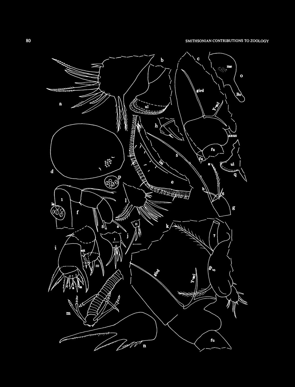
86 80 SMITHSONIAN CONTRIBUTIONS TO ZOOLOGY
: a, 6th limb; b, upper lip, anterior toward left; c, posterior of body from right side, anterior toward right.")

87 NUMBER FIGURE 58 (left). Junctichela pax, new species, paratype, USNM , instar III (sex unknown): a, 6th limb; b, upper lip, anterior toward left; c, posterior of body from right side, anterior toward right. USNM , instar IV female, paratype: d, complete specimen from left side, length 0.87 mm; e, posterior of left valve, iv;/ right lateral eye and right 1st antenna, nabs, lv; g, distal left 1 st antenna, nabs, lv; h, endopodite left 2nd antenna, anterior toward right, mv; i. right maxilla, nabs, rav;j, endites I and II right maxilla, anterior toward left, mv; k, posterior of body from right side showing location of some appendages, anterior toward right; /, 6th limb; m, 7th limb; n, left furcal lamella, lv; o, medial eye and Bellonci organ from right side; p, left lateral eye, anterior toward right; q, upper lip from left side, anterior to left. margin with 3 small bristles (2 proximal medial, 1 distal ventral); lateral side with 2 small bristles near midlength (Figure 57/); dorsal margin with small subterminal bristle. Exopodite absent. Endopodite: 1st joint with distal medial spines, spines along distal dorsal corner, and distal ventral claw with minute proximal spines along dorsal margin; small ventral bristle proximal to claw. 2nd joint with small subterminal dorsal bristle and stout ventral claw. 3rd joint with 2 small bristles (1 ventral, 1 dorsal). Maxilla (Figure 51m,n): Similar to that of adult female. Fifth Limb (Figure 51o,p): Epipodite with about 30 bristles. Endite and exopodite similar to those of adult female. Sixth Limb (Figure 58a): Single endite with 1 or 2 bristles. End joint with 9 spinous bristles followed by 2 plumose bristles. Seventh Limb (Figure 57p): Elongate, bare. Furca (Figure 57g): Each lamella with 5 claws; claws 13 nonarticulated, with teeth along posterior edge and spines along anterior edge; claws 4 and 5 small, articulated, bare. Right lamella anterior to left by width of base of claw 1. Bellonci Organ (Figure 57r): Elongate, with broadly rounded tip. Eyes (Figure 57/»: Lateral eye smaller than medial eye, with 5 ommatidia. Medial eye bare, with light brown pigment. Upper Lip (Figure 586): Similar to that of adult female. Genitalia: Absent. Brushlike Organ: Absent. YSclerite (Figure 58c): Similar to that of adult female. Posterior of Body (Figure 58c): With few spines dorsal to posterior end of girdle. Gut Content: USNM and USNM AC with unidentified, light brown particles. DESCRIPTION OF INSTAR IV FEMALE (Figure 5&dq). Carapace similar in shape and ornamentation to that of instar III (Figure 5Sd). Gellike substance coating valves. Infold (Figure 58e): Anterior infold with minute bristle. Infold of caudal process with indistinct line with about 4 short bristles (indistinct line equivalent to concave ridge on adult female); 2 or 3 minute bristles between indistinct line and posterior edge of caudal process. Posterior infold with 2 setal bristles. Selvage: Bare narrow lamellar prolongation present along free margin of valves. Central Adductor Muscle Attachments: Closely spaced ovoid attachments. TABLE 7. Average shell dimensions and calculated growth factors for female Junctichela pax. (*=sex not determined.) Growth stage adult IV III* II* I* Average Average length (mm) Growth factor Average height (mm) Growth factor Number of specimens Carapace Size (length, height in mm): USNM , 0.87, (USNM , carapace without body, sex unknown: right valve, 0.83, 0.59; left valve, 0.84, 0.63 (dimensions not used in average length and height in Table 7).) First Antenna (Figure 5%fg; not all bristles shown): Joints 14 and 6 similar to those of adult female. Sensory bristle of 5th joint with fairly long proximal filament, minute filament near midlength, and terminal spine. 7th joint: a and bbristles similar to those of adult female; cbristle with fairly long proximal filament and minute filament near midlength. 8th joint: d and ebristles similar those of adult female; fbristle with fairly long proximal filament, minute filament near midlength, and terminal spine; gbristle with fairly long proximal filament, minute distal filament, and terminal spine. Second Antenna (Figure 58A): Protopodite similar to that of adult female. Endopodite: left limb only with row of minute spines proximal to terminal node, otherwise endopodite similar to that of adult female. Exopodite: 1st joint with recurved tubular medial bristle on distal margin; bristle of 2nd joint with about 17 proximal ventral spines and distal natatory hairs; bristles of joints 38 with proximal ventral spines and distal natatory hairs; 9th joint with 2 bristles (ventral bristle with proximal ventral spines and distal natatory hairs, dorsal bristle small). Mandible and Maxilla (Figure 58/j): Similar to those of adult female. Fifth Limb (Figure 58*): Epipodite with 31 bristles, otherwise limb similar to that of adult female. Sixth Limb (Figure 58/): Similar to that of adult female. Seventh Limb (Figure 58k,m): Each limb with 6 tapered bristles (2 in proximal group, 1 on each side, and 4 in terminal group, 2 on each side); each bristle with 3 or 4 bells and without marginal spines. Terminus with opposing combs with few small teeth. Furca (Figure 58/t,n): Except for spine following claw 5, furca similar to that of adult female. Bellonci Organ (Figure 58o), Eyes (Figure 5%fo,p\ and Upper Lip (Figure 58^): Similar to those of adult female
. COMPARISONS. The new species J.")

88 82 SMITHSONIAN CONTRIBUTIONS TO ZOOLOGY Genitalia: Absent. Brushlike Organ: Not observed. YSclerite (Figure 58/r): Similar to that of adult female. Gut Content: Unidentified, light brown particles. REMARKS. USNM is interpreted to be female because of its 7th limbs having 2 proximal bristles (these are absent on adult male). COMPARISONS. The new species J. pax differs from Junchtichela margalefi Kornicker and Caraion, 1978, and Junctichela lex Kornicker and Thomassin, 1998, in the female having one instead of two proximal bristles on the endopodite of the 2nd antenna and four instead of two proximal bristles on the 7th limb. The endopodite of the 2nd antenna of the adult female of J. margalefi bears a long terminal bristle that is absent on J. pax. The endopodite of the 2nd antenna of the adult male J. lex has three joints compared to two on J. pax. ONTOGENY. In general, the development of J. pax is similar to that of known species of the Sarsiellinae. The calculated growth factors for instars and adults averaged 1.19 for carapace length and 1.18 for carapace height (Table 7). Family RUTIDERMATIDAE Brady and Norman, 1896 TYPE SPECIES. Rutiderma compressa Brady and Norman, 1896:673. DISTRIBUTION. Widespread between latitudes of about 34 N and 50 S and depths of 1260 m (Kornicker, 1983:25). Rutiderma Brady and Norman, 1896 Rutiderma darbyi Kornicker, 1983 FIGURE 59 Rutiderma dinochelatum. Darby, 1965:28, 56, 57, pi. 13: figs. 19; pi. 14: figs. 17 [not Rutidermata dinochelata Kornicker, 1958]. Rutiderma darbyi Kornicker. Bradford, 1980:141, fig. 2 [deliberate nomen nudum]. Rutiderma darbyi Kornicker, 1983:27, 1114, 17, 25, 3647, 62, 85, figs. 1824; pi. I. Grabe, Gibson, and Watkins, 1995:698, table 2. HOLOTYPE USNM , ovigerous female. TYPE LOCALITY. Georgia continental shelf, depth 26 m. MATERIAL. Crab Cay Cravasse, Sta 95010: USNM , partly dissected instar IV male in alcohol. FIGURE 59 Rutiderma darbyi Kornicker, 1983, USNM , instar IV male: a. complete specimen from right side, length 0.94 mm; b, rostrum right valve, Iv; c, caudal process right valve, iv; d, caudal process left valve, iv; e, endopodite right 2nd antenna, mv;/ posterior of body from right side.
, and")
89 NUMBER DISTRIBUTION. Atlantic continental shelf from North Carolina to Florida, Gulf of Mexico continental shelf off Florida, and Bahama Islands in vicinity of San Salvador Island, Andros Island (Kornicker, 1983:38), and in Crab Cay Cravasse, Exuma Islands (herein). Known depth range intertidal to 168 m (Kornicker, 1983:38). DESCRIPTION OF INSTAR IV MALE. Carapace similar in shape to that of adult female (Figure 59a). Ornamentation (Figure 59a,b): Surface with small round separated fossae. Infold: Infold of caudal process of left valve with serrations along anterior edge of pocket (Figure 59d); serrations and pocket absent on right valve (Figure 59c). Carapace Size (length, height in mm): USNM , 0.94,0.70. Second Antenna: Endopodite elongate, 3jointed (Figure 59e): 1st joint with 3 short bristles; 2nd joint with 1 or 2 short bristles; 3rd joint with 1 or 2 terminal bristles. Seventh Limb: Well developed, with 4 proximal bristles (2 on each side), each with 2 bells, and 4 terminal bristles (2 on each side), each with 4 bells; all bristles tapered (juvenile character). Tip with opposing combs with few small teeth. Furca (Figure 59/): Each lamella with 4 stout claws followed by 2 or 3 secondary claws. USNM with 2 secondary claws on left limb and 3 on right. Copulatory Organ (Figure 59/): Consisting of 2 lobes. Superfamily CYLINDROLEBERIDOIDEA Miiller, 1906 Family CYLINDROLEBERIDIDAE Miiller, 1906 Subfamily CYLINDROLEBERIDINAE Mfiller,1906 SEXUAL DIMORPHISM OF THE FIRST ANTENNAE OF THE CY LINDROLEBERIDINAE. The c and fbristles of the 1st antennae on known species of the Cylindroleberidinae are generally much longer in the adult male than in the adult female and juveniles, and they have many more marginal filaments (Appendix 2). In the female the c and fbristles are shorter than the combined length of the joints of the limb, whereas in the male the bristles may be twice the length of the combined joints and as much as one and onehalf times the length of the carapace. The c and fbristles of juveniles of both sexes resemble those of the adult female, but they may have fewer filaments. The adult male of many described species is unkown. The lengths of the c and fbristles of adult males are not presented in most descriptions of the species, but the number of sensory filaments on the bristles often are stated. According to Parker (1998: 105), "halophores [filaments] may be bimodal chemosensory/ mechanosensory sensilla," and are irridescent (Parker, 1995:349). The filaments are probably important in sensing females, and if irridescent, may attract females. The bristles on the 1st antenna of the adult male Synasterope browni described herein differ from most members of the subfamily in being short and having few filaments (Table 8, Appendix 2). That difference prompted a brief survey of the literature concerning the morphometrics of some of the bristles of TABLE 8. Lengths (in mm) of shell and c and fbristles of male 1st antenna of selected species of Cylindroleberidinae. Species Homasterope curia Parasterope nana Prionotoleberis norvegica Synasterope browni Synasterope oculata Synasterope oculata Shell length Bristle length c f Source Skogsberg (1920:501) Poulsen (1965:396) Skogsberg (1920:532) Herein Skogsberg (1920:523) Poulsen (1965:413) the 7th and 8th joints of the 1 st antenna of the Cylindroleberidinae (Appendix 2). Of the 44 adult males listed in Appendix 2, only four have relatively short c and fbristles with relatively few marginal filaments. The short bristles are designated "S" in Appendix 2. The c and fbristles on at least three of those males do not appear to be significantly different than the bristles on the adult female of the same species. The males having short c and f bristles have normal gbristles, and females of those species have normal c, f, and gbristles (Appendix 2). The length and number of filaments on the c and fbristles on all specimens are clearly related to each other, e.g., a long c bristle with many filaments is always accompanied by a long f bristle with many filaments. The number of filaments on c and fbristles varies directly with shell length (Figure 60). For cbristles, the Pearson correlation coefficient is 0.615, which is significant at the level P<0.01. For fbristles, the Pearson correlation coefficient is 0.617, which is significant at the level of P<0.01. Measurements have been made of the lengths of the c and f bristles of the males of only six species (Table 8). The few data suggest that lengths of the c and fbristles vary directly with shell length. CORRECTIONS. Kornicker (1975:439) described the fbristle of the 8th joint of the 1 st antenna of the male Parasterope prolixa Kornicker, 1975, as having 8 filaments and illustrated the right 1st antenna of USNM with an fbristle shorter than the gbristle. The senior author reexamined the slide bearing the illustrated 1 st antenna and observed the fbristle to be folded and bearing about 14 filaments. It is about twothirds the length of the cbristle. Kornicker (1976:17), in the description of the f and gbristles of the 1st antenna of a male Parasterope beta, incorrectly transposed them. It should have been f bristle with 29 filaments and gbristle with 8 filaments. The f and gbristles (both broken) also were incorrectly transposed in an adult male of Parasterope kappa (Kornicker, 1976:22). Synasterope Kornicker, 1975 TYPE SPECIES. Synasterope implumis Poulsen, 1965, by subsequent designation (Kornicker, 1975:44). COMPOSITION AND DISTRIBUTION. See Kornicker and Iliffe (2000:72).

.")
90 84 SMITHSONIAN CONTRIBUTIONS TO ZOOLOGY c bristle 3 2 C « f bristle * Number of Filaments FIGURE 60. Relationship between the number of filaments on c and fbristles of the first antennae and the carapace lengths of adult males of selected species of the Cylindroleberidinae (from Appendix 2). (Specimens with number of filaments on c and fbristles followed by plus sign in Appendix 2 not plotted on graphs.)

.")
91 NUMBER 616 Synasterope browni Kornicker and IlifTe, 2000 FIGURES 6168 Synasterope browni Kornicker and IlifTe, 2000:7277, figs HOLOTYPE. USNM , adult female on slide and in alcohol. TYPE LOCALITY. Exuma Sound, depth 96 m. MATERIAL. Four Shark Cave, Sta 97023: USNM , adult male on slide and in alcohol. Mystery Cave, Sta 96030: USNM , adult male on slide and in alcohol; USNM , Al male on slide and in alcohol; USNM , instar I in alcohol. DISTRIBUTION. Exuma Sound, depth 96 m; collected herein in Four Shark Cave, South Andros Island, depth 2733 m. Mystery Cave, Stocking Island, Exuma Cays, depth 50 m. DESCRIPTION OF ADULT MALE (Figures 6165, 66a,b). Carapace oval with posterodorsal margin slightly more oblique than posteroventral margin (Figures 61a, 64/); vertical row of few hairs near posterior end. Infold: Obscure on slides, but appearing to have relatively few bristles on rostrum and along anteroventral margin (Figure 61/>); flaplike posterior bristles present but number obscured (Figure 61c). No processes observed between posteroventral list and valve edge. Carapace Size (length, height in mm): USNM , 0.66, 0.42; USNM ,0.69,0.42. First Antenna (Figures 6\d,e, 65a,b): 1st joint with few lateral hairs in distal dorsal corner. 2nd joint with spinous dorsal bristle and without lateral bristle. 3rd and 4th joints fused, triangular; 3rd joint with small bristle on short ventral margin and 6 dorsal bristles (2 single bristles with long spines, 2 paired bristles (lateral with short or long spines, medial with long spines) and 2 paired bristles (lateral with short spines, medial with long spines) on long dorsal margin. 4th joint with terminal, spinous, dorsal bristle and 2 ventral lateral bristles. 5th joint fused to 6th joint and forming small ventral wedge between 4th and 6th joints; bristle of 5th joint unusually stout and with well over 100 filaments (not all shown). 6th joint long, with medial bristle with short spines. 7th joint: abristle clawlike, concave dorsally, with proximal dorsal teeth; bbristle with 3 long dorsal filaments; cbristle about same length as dorsal margin of joints 28, with 6 marginal filaments. 8th joint: dbristle represented by minute peg (Figure 61e); bare ebristle about twothirds length of cbristle; fbristle bent dorsally, about twothirds length of cbristle, with 5 marginal filaments; gbristle shorter than cbristle, with 5 marginal filaments. Second Antenna (Figure 61/g): Protopodite with small distomedial bristle. Endopodite 3jointed: 1st joint short bare; 2nd joint elongate with 3 short bare dorsal bristles near midlength; 3rd joint long, reflexed, with long, bare, proximal filament and pointed tip with few small teeth on inner side. Exopodite: 2nd joint only slightly longer than 3rd joint; bristle of 2nd joint reaching 7th joint, with slender ventral spines; bristles of joints 3 and 4 with slender proximal ventral spines and distal natatory hairs; bristles of joints 58 with natatory hairs; 9th joint minute, about onehalf width of 8th joint, with 2 bristles (ventral bristle about onehalf length of bristle of 8th joint and same length as combined joints 29; dorsal bristle shorter); joints 28 with few small spines at distal dorsal corner, some joints with minute spines along distal margins, stout basal spines absent. Mandible (Figures 62, 65e): Coxale endite (Figure 62d,e): ventral branch with 3 or 4 oblique rows of spines and tip with 2 or 3 slender spines; dorsal branch with small main spine; tip obscure; usual bristle near base of ventral branch absent. Basale endite with 3 spinous end bristles, glandular peg, 2 dwarf bristles (proximal about threequarters length of distal), and 2 trianid bristles with 2 or 3 pairs of spines proximal to terminal pair (terminal pair well developed) (Figure 62b,e). Basale: ventral margin with Ushaped boss near midlength; dorsal margin with 2 long terminal bristles. Exopodite about onehalf length of dorsal margin of 1st endopodial joint, with hirsute tip and 2 short subterminal bristles (Figure 62a). 1st endopodial joint with 3 long bristles (2 with long spines near midlength and short proximal and distal spines, 1 with short spines). 2nd endopodial joint (Figure 62a,g): ventral margin with 3 terminal bristles with short marginal spines; dorsal margin with a, b, c, d, f, and gbristles (base of cbristle slightly stouter than bases of b and dbristles), and 1 short bristle proximal to abristle; medial surface of joint with 3 cleaning bristles (with short spines) in oblique row near base of cbristle. 3rd endopodial joint with fairly straight claw with few ventral and dorsal spines, and 5 bristles (not all annulations shown on bristles). Maxilla (Figure 63ad): Epipodite obscured. Endite I with 3 bristles (2 long, 1 short); endite II with 3 long bristles (Figure 63a). Basale: medial surface with proximal bristle near dorsal margin and 1 small distal bristle near ventral margin (seen only on left limb of USNM ); lateral surface with short proximal bristle near midwidth; ventral margin with long, spinous, terminal bristle. 1st endopodial joint with short alphabristle and long betabristle. 2nd endopodial joint with long terminal bristle reaching past tip of betabristle. Fifth Limb (Figure 63e): Comb: lateral side with stout spinous exopodial bristle reaching past end of comb; 1 short slender bristle ventral to base of exopodial bristle; 1 pair of bristles and 1 slender bristle closer to ventral margin. Sixth Limb (Figure 63/g): Small medial bristle near proximal anterior corner. Anterior margin with slender bristle at upper endite and longer spinous bristle at lower endite. Skirt: anterior end with 3 ventral bristles; lateral flap with slender hirsute anterior bristle; ventral margin with 7 or 8 bristles posterior to midlength. Seventh Limb (Figures 63h,i, 65c): Proximal group with 4 or 5 bristles (1 or 2 on one side, 3 on other side), each with 2 or 3 bells; distal group with 4 or 5 bristles (2 on one side, 2 or 3 on other side, each with 2 or 3 bells). Terminus with opposing 85

92 86 SMITHSONIAN CONTRIBUTIONS TO ZOOLOGY

: Elongate, broad near middle, with rounded tip. Eyes: Medial eye bare (Figure (Ah).")
93 NUMBER 616 FIGURE 61 (left). Synasterope browni Kornicker and Iliffe, 2000, USNM , adult male: a, complete specimen from right side, length 0.66 mm; b,c, anterior and posterior of right valve, respectively, iv; d, right 1 st antenna, nabs, mv; e, distal left 1st antenna, Iv;/ distal left 2nd antenna, mv; g, part of right 2nd antenna, Iv. combs with small teeth. Furca (Figures 63y, 64a,f,g, 65c/): Each lamella with 8 claws; posterior 3 claws slender, bristlelike, but not ringed (1 oriented backward). Claws 17 with teeth along posterior edge; claw 8 with hairs along anterior edge. Bellonci Organ (Figure (Ah): Elongate, broad near middle, with rounded tip. Eyes: Medial eye bare (Figure (Ah). Lateral eye with black pigment and about 15 ommatidia (Figures 61a, 63/fc, 64f,h, 65a,d). Lips (Figure 65d,e): Typical for genus, without spines. Copulatory Organ: Extremely long, about onehalf length of shell (Figures 61a, 64/), coalesced proximally, divided distally into 2 lobes, each with bifurcate lobate tips; lobes with indistinct bristles near tip (Figures (Ad,e, 66a,b). Posterior of Body (Figures 63/, 64ac,g, 65c,/): Posterodorsal comer slightly lobate, bare; posterior margin bare. Gills (Figures 64a, 65c,/): Narrow, number uncertain (2 shown). YSclerite (Figures 63/, 64a, 65c,/ 66a): Without ventral branch. DESCRIPTION OF Al MALE (Figures 66cg, 67a/). Carapace similar in shape to that of adult female (Figure 66c). Infold (Figure 66a 1, e) and Selvage: Similar to that of adult female. Carapace Size (length, height in mm): USNM , 0.72, First Antenna (Figure 66/g): 1 st joint with lateral hairs in distal dorsal corner and distal medial hairs near midheight. 2nd joint with spinous dorsal bristle and without lateral bristle. 3rd and 4th joints fused; 3rd joint with small bristle on ventral margin and 6 dorsal bristles (2 single bristles with long spines, and 2 pairs of bristles either bare or with indistinct short spines). 4th joint with dorsal bristle with short spines and bare ventral bristle. 5th joint with weak sutures separating joint from 4th and 6th joints; sensory bristle with short proximal filament and 6 long terminal filaments. 6th joint long, with long medial bristle. 7th joint: abristle clawlike, with concave dorsal margin and proximal dorsal teeth; bbristle with 3 long dorsal filaments; cbristle with 4 filaments at midlength and 1 subterminal. 8th joint: dbristle represented by minute papilla (not shown on illustration); bare ebristle about onehalf length of c bristle; fbristle bent dorsally, about twothirds length of cbristle, with 4 marginal filaments; gbristle shorter than cbristle, with 5 marginal filaments. Illustrated left limb of USNM shows filaments of sensory bristle of adult male within. Second Antenna: Protopodite and endopodite similar to those of adult male. Endopodite 3jointed (Figure 66h): 1st joint bare; 2nd joint elongate with 2 short distal bristles; 3rd joint elongate with long, bare, proximal filament and pointed tip. Illustrated detail of tip of endopodite of USNM shows pointed tip with few small teeth of endopodite of adult male within. Mandible: Coxale endite broken off. Remainder of limb similar to that of adult male. Maxilla (Figure 67a, b): Epipodite triangular. Endite I with 3 bristles (2 long, 1 short); endite II with 3 long bristles. Basale with short proximal medial bristle near dorsal margin or at midwidth; ventral margin with short proximal bristle, none or 1 short indistinct distal bristle, and long terminal bristle; proximal lateral bristle obscured. Endopodite: 1st joint with short alpha and long betabristle; 2nd joint with terminal bristle longer than betabristle of 1st joint. Fifth Limb: Similar to that of adult male. Sixth Limb: In general, similar to that of adult male. Seventh Limb: Proximal group with 5 tapered bristles (2 on one side, 3 on other), each with 1 to 3 bells; distal group with 5 slightly tapered bristles (2 on one side, 3 on other), each with 1 or 2 bells. Terminus with opposing combs with few small teeth. Furca (Figure 61c,d,f): Similar to that of adult male. Bellonci Organ: Not observed. Eyes: Medial eye not observed. Lateral eye with black pigment and obscured ambercolored ommatidia (Figure 66c). Lips (Figure 67e): Similar to those of adult male. Copulatory Organ (Figure 67/): Large with 2 lobes at anterior tip, without visible internal sclerites. Posterior of Body (Figure did): Posterodorsal corner slightly lobate and with spines; posterior ventral to posterior end of girdle hirsute. Gills: Narrow. YSclerite (Figure 67d): Similar to that of adult male; anterior tip with small hooklike ventral sclerite. DESCRIPTION OF INSTAR I (sex unknown) (Figure 61go). Carapace less elongate than that adult female (Figure 67g); dorsal edge of caudal process extending onto lateral surface of valve (Figure 67/z). Infold and Selvage: Not examined. Carapace Size (length, height in mm): USNM , 0.44,0.28. First Antenna (Figure 67/): 1st, 2nd, and 4th joints bare. 3rd joint with long, spinous, dorsal bristle and small, bare, ventral bristle. Sensory bristle of 5th joint without filaments. 6th joint with medial bristle. 7th joint: abristle clawlike, b and c bristles bare. 8th joint: dbristle absent; ebristle about same length as cbristle, bare, distal part filamentous, indistinct; f and gbristles bare. Second Antenna (Figure 67/, A:): Protopodite with small distal medial bristle. Endopodite weakly 3jointed, with long terminal filament. Exopodite 9jointed: bristle of 2nd joint reaching 7th joint, with slender ventral spines; bristles of joints 38 with distal natatory hairs (some bristles with proximal ventral spines); 9th joint with 2 bristles (dorsal bristle short, bare, ven 87

94 88 SMITHSONIAN CONTRIBUTIONS TO ZOOLOGY f V' FIGURE 62. Synasterope browni Komicker and Iliffe, 2000, USNM , adult male, right mandible, lv: a, distal part; b, basale; c, basal and endopodite, nabs; d, coxale endite. Left mandible, mv: e, proximal coxale and ventral basale;/ distal basale and endopodite (exopodite not shown); g, dorsal bristles 1st endopodial joint.
 and endopodite; d, left maxilla, lv; e, distal right Sth limb (twisted), vv;/g, right and left 6th limbs (partly fragmented),")
95 NUMBER FIGURE 63. Synasterope browni Kornicker and Iliffe, 2000, USNM , adult male, right maxilla, Iv: a. limb without epipodite, nabs; b, proximal basale; c, distal basale (nabs) and endopodite; d, left maxilla, lv; e, distal right Sth limb (twisted), vv;/g, right and left 6th limbs (partly fragmented), respectively, mv; h.i, left and right 7th limbs;/ posterior of body from right side (gills not shown), anterior toward top; k, left lateral eye.

96 90 SMITHSONIAN CONTRIBUTIONS TO ZOOLOGY a FIGURE (A. Synasterope browni Komicker and Iliffe, 2000, USNM , adult male: a, posterior of body from right side, anterior toward right; b.c. parts of posterior of body from left side, anterior toward left; d, tip of copulatory organ, anterior toward left; e, copulatory organ, anterior toward right. USNM , adult male:/ complete specimen from right side, ov, length 0.69 mm; g. part of posterior of body from right side, anterior toward right; h. right lateral eye (stippled), medial eye, and Bellonci organ, anterior toward lower right.

97 NUMBER FIGURE 65. Synasterope browni Komicker and Iliffe, 2000, USNM , adult male: a, right lateral eye and right 1st antenna, nabs, Iv; b, right 1st antenna projecting from carapace viewed from right side, anterior toward right, nabs, lv; c, posterior of body from right side showing location of gills and some appendages (furcal claws not shown), anterior toward right; d,e, dorsal view of anterior of body showing lips, anterior to bottom;/ posterior of body projecting from ventral part of carapace viewed from right side, anterior toward right.

98 92 SMITHSONIAN CONTRIBUTIONS TO ZOOLOGY FIGURE 66. Synasterope browni Kornicker and Iliffe, 2000, USNM , adult male: a, part of posterior of body showing copulatory organ, anterior toright;b, tip of copulatory organ from left side, anterior toward left. USNM , A 1 male (appendages of adult male visible within appendages of A 1 male): c, complete specimen from right side, length 0.72 mm; d,e, anterior and posterior of left valve, iv;/g, parts of left 1st antenna (internal dashed lines represent filaments of bristle of Sth limb of adult male), mv; h, endopodite right 2nd antenna, lv.

99 NUMBER FIGURE 67. Synasterope browrti Kornicker and Hiffe, 2000, USNM , Al male: a, left maxilla, nabs, mv; b, proximal part of right maxilla, mv; c, right furcal lamella, lv; d, posterior of body from right side, anterior toward right; e, upper lip, anterior toward bottom, dv;/ posteroventral part of body from left side, anterior to left. USNM , instar I (sex unknown): g. complete specimen from left side, length 0.44 mm; h, posterior of right valve, ov; i, left 1st antenna, lv;/ right lateral eye and right 2nd antenna, nabs, lv; k, joints 79 of exopodite left 2nd antenna, nabs, lv; /, comb left 5th limb, mv; m.n, right (lv) and left (mv) 6th limbs; o. medial eye and Bellonci organ.
: a, left mandible, tnv; b, right maxilla, nabs, mv; c, posterior of body from left side, anterior toward left, nabs;")
; joints 37 with row of minute terminal spines; joints 68 each with indistinct, small, basal spine; 9th joint with small lateral spine (Figure 61k).")
100 94 SMITHSONIAN CONTRIBUTIONS TO ZOOLOGY FIGURE 68. Synasterope browni Kornicker and Iliffe, 2000, USNM , instar I (sex unknown): a, left mandible, tnv; b, right maxilla, nabs, mv; c, posterior of body from left side, anterior toward left, nabs; d, lips from left side, anterior toward left; e, posterior of body from right side, anterior toward right;/ posterior of body from left side showing absence of 7th limb, anterior toward left. tral bristle longer and with natatory hairs); joints 37 with row of minute terminal spines; joints 68 each with indistinct, small, basal spine; 9th joint with small lateral spine (Figure 61k). (Basal spines not observed on exopodites of adults.) Mandible (Figure 680): Coxale endite broken off. Basale: endite with 1 trianid bristles with 3 pairs of spines proximal to terminal pair, 2 dwarf bristles, 1 glandular peg, and 2 spinous endbristles; dorsal margin with 2 long terminal bristles. Exopodite almost as long as dorsal margin of 1st endopodial joint, with 2 small terminal bristles. 1st endopodial joint with 2 long ventral bristles (one with long spines); 2nd endopodial joint: dorsal margin with 2 stout bristles (possibly c and dbristles); lateral side near dorsal margin with long lateral bristle (possibly fbristle) between those 2 stout bristles; medial side near dorsal margin with 2 short cleaning bristles adjacent to proximal stout c?bristle, and 1 long bristle (possibly gbristle) just distal to distal stout d?bristle. 3rd endopodial joint with stout clawlike bristle and 4 slender ringed bristles.

: With stout bristle on posterior tip of skirt, but no endite or ventral bristles. Seventh Limb: Absent.")
101 NUMBER Maxilla (Figure 686): Epipodite obscured. Endites I and II each with 2 long bristles. Basale with short medial bristle near midlength and midheight and long, ventral, terminal bristle. 1st endopodial joint with short alphabristle and long betabristle. 2nd endopodial joint with long terminal bristle extending past betabristle. Fifth Limb (Figures 67/, 68c): Well developed but with few ventral bristles on comb (Figure 67/). Sixth Limb (Figures 67m,n, 68c): With stout bristle on posterior tip of skirt, but no endite or ventral bristles. Seventh Limb: Absent. Furca (Figure 68c/): Each lamella with 3 claws: claw 1 separated from lamella by suture, remaining claws fused to lamella. Claws 1 and 2 with stout teeth along posterior edge (distal teeth longer); claw 3 with hairs or slender spines along posterior edge; claw 1 with distal hairs along anterior edge. Right lamella anterior to left by width of base of claw 1. Bellonci Organ (Figure 67o): Elongate, with rounded tip. Eyes: Lateral eye with black pigment and small obscured ommatidia (at least 10) (Figure 67gj). Medial eye present (Figure 67o). Lips (Figure 6Sd): Similar to those of Al male. Copulatory Organ: Absent. Posterior of Body (Figure 68c,e,/): Posterodorsal corner with stout spines (not shown on Figure 68/). Gills (Figure 68e): Seven narrow gills on each side of body. Attachment of gills typical for genus. YSclerite (Figure 68c): Fused to girdle. REMARKS. The lengths of the two adult males described herein from South Andros Island and Stocking Island are 0.66 mm and 0.69 mm, respectively. Both are shorter than the length (0.80 mm) of the adult female from Exuma Sound and the length (0.72 mm) of the Al male from Stocking Island. The two adult males and the Al male in the present location are from blue holes and caves, whereas the adult female is from Exuma Sound. The males and females are interpreted to be conspecific herein, but further study of males and females from the same locality is warrented. The sensory bristle of the Al male bears 1 short proximal filament and 6 terminal filaments. The adult female holotype is without a short proximal filament. The presence of a short proximal filament on the sensory bristle of the Al male is interpreted herein to be a juvenile character state. Subfamily CYCLASTEROPINAE Poulsen, 1965 Tribe TETRALEBERIDINI Kornicker, 1981 Amboleberis Kornicker, 1981 TYPE SPECIES. Asterope americana Miiller, COMPOSITION AND DISTRIBUTION. Three species are referred to this genus: A. americana (Miiller, 1890), A. antyx Kor FlGURE 69. Amboleberis americana (Muller, 1890), USNM , instar I (sex unknown), complete specimenfromright side, carapace length 0.76 mm. nicker, 1981, and A. cubensis (Lalana and Kornicker, 1997). The genus is widespread in world oceans; depth ranges from surface to 53.5 m (Komicker, 1981:167). Amboleberis americana (Muller, 1890) FIGURE 69 See Kornicker (1981b: 168) for synonomy. HOLOTYPE. Present locality of specimen, if extant, unknown (Kornicker, 1981:168). MATERIAL. Rat Cay Blue Hole, Andros Island, 9 specimens. Crab Cay Crevasse, Exuma Cays, USNM , 1 specimen. DISTRIBUTION. East Atlantic coast: South Carolina to Brazil. Bahamas: Rat Cay Blue Hole and Crab Cay Cravasse (herein). East Pacific: Costa Rica and Gulf of Panama. Gulf of Mexico: Florida and Texas. Depth range: surface to 53.5 m. SUPPLEMENTARY DESCRIPTION OF INSTAR I (sex unknown). Carapace ovoid with vertical ridge posterior to incisur (Figure 69). Carapace Size (length, height in mm): USNM , 0.76,0.58. Furca (Figure 69): Each lamella with 4 claws: claw 1 with suture at base, claws 24 without sutures. YSclerite and Girdle (Figure 69): Fused.


102 Appendix 1 Station Data With Specimens Examined (in chronological order) Inland Blue Holes Sta 97021, 5 Aug 1997, Mermaid's Lair, Grand Bahama Island; collected with plankton net from water column in 1822 m depths. Spelaeoecia parkeri: 1 specimen. Sta 97022, 6 Aug 1997, Mermaid's Lair, Grand Bahama Island; collected with individual vials and 93 mm mesh plankton net from halocline and below in 1822 m depths. Spelaeoecia parkeri: 1 specimen. Sta 97026,24 Aug 1997, Stargate Blue Hole, South Andros Island; collected with plankton net, vials, and suction bottle from water column in 3336 m depths of North Passage. Deeveya bransoni: 4 specimens. Spelaeoecia styx: 7 specimens. Sta 97028, 24 Aug 1997, Stargate Blue Hole (same collecting data as given above for Sta 97026). Deeveya bransoni: 3 specimens. Sta 97029,26 Aug 1997, Stargate Blue Hole, South Andros Island; collected with plankton net and vials from water column in 3339 m depths of South Passage. Deeveya bransoni: 2 specimens. Spelaeoecia styx: 3 specimens. Oceanic Blue Holes Sta 95008, 18 May 1995, Angelfish Cave, Stocking Island, Exuma Cays, Bahamas; salinity 35 ppt; collected with suction bottle from outside the cave in 9 m water depth on a sandy ledge overlooking the cave entrance. Cylindroleberidinae, genus and species indeterminate: 1 early instar. Sta 95010, 18 May 1995, Crab Cay Crevasse, Crab Cay, Exuma Cays, Bahamas; salinity 35 ppt; collected with plankton net from coarse sediment at 35 m depth about 100 m inside cave. Skogsbergia lerneri: 2 specimens. Eurypylus hapax: 3 specimens. Junctichela pax: 17 specimens. Rutiderma darbyi: 1 specimen. Amboleberis americana: 1 specimen. Sta 95091, 5 Aug 1995, Sugar Cay Blue Hole, Sugar Cay, Exuma Cays, Bahamas; collected with plankton net from a silt mound on the wall of the cave passage in 31 m depth (Kornicker and Iliffe, 2000:94). Harbansus paucichelatus: 3 specimens. Eusarsiella merx: 5 specimens. Eusarsiella spp. indet.: 2 instar IV (USNM , ); 3 juveniles (USNM ). Sta 96030, 9 Sep 1996, Mystery Cave, Stocking Island, Exuma Cays, Bahamas; collected with plankton net from sandy bottom and walls in 50 m depth (Kornicker and Iliffe, 2000:94). Synasterope browni: 3 specimens. Sta 96031, 9 Sep 1996, Master Harbour Cave, Great Exuma Island, Exuma Cays, Bahamas; collected with plankton net from surface of a silt mound in 1215 m depth. Skogsbergia lerneri: 2 specimens. Cylindroleberidinae (genus and species indeterminate): 1 instar I. Sta 96040, 6 Apr 1996, Conch Sound Blue Hole, Andros Island, Bahamas; collected with plankton net from surface of silt mound in 22 m depth (Kornicker and Iliffe, 2000:94). Harbansus paucichelatus: 11 specimens. Eusarsiella sp. indet.: 1 instar I. Sta 97023, month and day unknown, 1997, Four Shark Cave, South Andros Island; collected by divers with a plankton net tow from the water column and silt on ledges at 2733 m depth in the back section of the cavern. Skogsbergia lerneri: 1 specimen. Eusarsiella ryanae: 11 specimens. Synasterope browni: 1 specimen. Rat Cay Blue Hole, Andros Island, 1981, 1982; collected with plankton net about 1.5 m from walls of the cave in about 5 m depth while current flowed out of hole. Species marked by * are from Warner and Moore (1984, table 1) (those species were identified by Kornicker (1983, in litt.) and were returned to The Natural History Museum, London. Skogsbergia lerneri*: 2 specimens. Eusarsiella warneri: 1 specimen. Amboleberis americana*: 9 specimens. Actinoseta chelisparsa*: 1 specimen. Asteropella monambon*: 1 specimen. 96

103 Appendix 2 (Number of filaments on c, f, and gbristles of the first antennae of males and females of selected species of Cylindroleberidinae, and carapace length of adult males. (L=carapace length of adult male in mm; nd=no data; S= bristle relatively short; females unknown; +=distal part of bristle missing and number of missing additional filaments unknown; number of filaments may or may not include tip of bristle.) Species Archasterope efficax Cylindroleberis bacescui Cylindroleberis kliei Cylindroleberis nodulifera Diasterope bisetosa Diasterope grisea Empoulsenia weddellensis Heptonema latex Heptonema latum Homasterope curta Homasterope maccaini Homasterope trebax Parasterope beta Parasterope corrugata Parasterope extrachelata Parasterope hulingsi Parasterope kappa Parasterope muelleri Parasterope muelleri Parasterope nana Parasterope pollex Parasterope prolixa Parasterope styx Parasterope whatleyi Parasterope zeta Postasterope abaco Postasterope messingi Prionotoleberis norvegica Prionotoleberis salomani Skogsbergiella macrothrix Skogsbergiella plocus Synasterope bensoni Synasterope browni Synasterope calix Synasterope cushmani Synasterope dimorpha Synasterope hirpex Synasterope implumis Synasterope mystax Synasterope oculata Synasterope oculata Synasterope phalanx Synasterope quadrisetosa Xandarasterope trux L (mm) Males No. filaments c f g S S 6S S S ? 33 4S 5S S nd nd nd nd 10 6 nd Females No. filaments c f g nd nd nd Source Kornicker and Poore, 1996:143 Kornicker and Caraion, 1974:29 Kornicker, 1976:2 Poulsen, 1965:391 Poulsen, 1965:356 Kornicker, 1975:391 Kornicker, 1975:516 Kornicker, 1992:172 Kornicker, 1986b:54 Skogsberg, 1920:498 Kornicker, 1975:529 Kornicker and Poore, 1996:120 Kornicker, 1976:11 Poulsen, 1965:391 Kornicker, 1986:28 Kornicker, 1986b:40 Kornicker, 1976:18 Poulsen, 1965:370 Kornicker, 1986b:20 Poulsen, 1965:396 Kornicker, 1986b:32 Kornicker, 1975:439 Kornicker, 1989:90 Kornicker and Poore, 1996:115 Kornicker, 1986b:47 Kornicker, 1986b: 105 Kornicker, 1986b: 101 Skogsberg, 1920:527 Kornicker, 1986b:89 Kornicker, 1975:481 Kornicker, 1975:489 Kornicker and Caraion, 1974:23 Kornicker and Iliffe, 2000:72; herein Kornicker, 1992:194 Kornicker, 1974:11 Kornicker, 1975:445 Kornicker, 1989b: 105 Poulsen, 1965:421 Kornicker, 1975:461 Poulsen, 1965:413 Skogsberg, 1920:523 Kornicker, 1989a: 102 Poulsen, 1965:410 Kornicker and Poore, 1996:131 97


104 Literature Cited Angel, M.V., and T.M. Iliffe Spelaeocia bermudensis New Genus, New Species, a Halocyprid Ostracod from Marine Caves in Bermuda. Journal of Crustacean Biology. 7:541553, figures 17, tables 13. Baird, W The Natural History of the British Entomostraca. 364 pages, 36 plates. London. [Printed for the Ray Society.] Bate, R.H., and L.M. Sheppard The Shell Structure of Halocypris inflata Dana, In R.H. Bate, E. Robinson, and L.M. Sheppard, editors, Fossil and Recent Ostracods. pages 2550, plates 110. Chichester: Ellis Horwood, Ltd. Boxshall, Geoffrey A Comparative Limb Morphology in Major Crustacean Groups: The Coxabasis Joint in Postmandibular Limbs. In R.A. Fortey and R.H. Thomas, editors, Arthropod Relationshps. Systematics Association, special volume, series 55: London, England: Chapman and Hall. Bradford, Janet M First Record of a Copepod Parasitizing the Ostracod Family Rutidermatidae (Myodocopina: Cypridinacea): Sphaeronella spinosa n. sp. (Copepoda: Choniostomatidae.) Proceedings of the Biological Society of Washington, 93(1): Brady, G.S Entomostraces. In A.G.L. de Folin and L. Perier, Les Fonds de la Men l(supplement): Brady, G.S., and A.M. Norman A Monograph of the Marine and Fresh Water Ostracoda of the North Atlantic and of Northwestern Europe. The Scientific Transactions of the Royal Dublin Society, series 2, 5:621784, plates Cohen, Anne C Rearing and Postembryonic Development of the Myodocopid Ostracode Skogsbergia lerneri from Coral Reefs of Belize and the Bahamas. Journal of Crustacean Biology, 3(2):235256, figures 110, tables Eusarsiella donabbotti. New Ostracode Species (Sarsiellidae) from the Belize Barrier Reef. Bulletin of Marine Science, 45(2):304315, figures 17. Cohen, Anne C, and Louis S. Kornicker Taxonomic Indexes to Ostracoda (Suborder Myodocopina) in Skogsberg (1920) and Poulsen (1962, 1965). Smithsonian Contributions to Zoology, 204: 29 pages. Cohen, Anne C, Joel W. Martin, and Louis S. Kornicker Homology of Holocene Ostracode Biramous Appendages with Those of Other Crustaceans: The Protopod, Epipod, Exopod and Endopod. Lethaia. 31(3):251265, figures 15. Dana, J.D Tribe III: Cyproidea = Ostracoda. In United States Exploring Expedition during the Years 1838,.1839, , 1842, under the Command of Charles Wilkes. U.S.N., with Atlas of 96 plates, 13(CrustaceaX2): , plates 90, 91. Philadelphia: C. Sherman. Darby, D.G Ecology and Taxonomy of Ostracoda in the Vicinity of Sapelo Island, Georgia. In R. V. Kesling, editor, Four Reports of Ostracod Investigations, 2:177, 11 figures 33 plates. Ann Arbor: University of Michigan. Grabe, Stephen A., Carl C. Gibson, and Rhonda J. Watkins Ecological Relationships of Myodocopid Ostracods in the Vicinity of Marco Island, Florida. Bulletin of Marine Science, 56(2): , figures 13. Hiruta, Shinlchi A New Species of the Genus Sarsiella Norman from Hokkaido, with Reference to the Larval Stages (Ostracoda: Myodocopina). Journal of the Faculty of Science, Hokkaido University, series 6 (Zoology), 21(l):4460, figures 112, plate Redescription of Sarsiella misakiensis Kajiyama from Kokkaido, with Reference to the Larval Stages (Ostracoda: Myodocopina). Journal of the Faculty of Science. Hokkaido University. 6 (Zoology), 21(2):262278, figures 115. Kornicker, Louis S Ecology and Taxonomy of Recent Marine Ostracodes in the Bimini Area, Great Bahama Bank. Publications of the Institute of Marine Science (The University of Texas), 5:194300, 89 figures, 1 map, 4 tables The Myodocopid Ostracod Familes Philomedidae and Pseudophilomedidae (New Family). Proceedings of the United States National Museum. 120(3580): 135, 12 figures, 1 plate, 2 tables Morphology, Ontogeny, and Intraspecific Variation of Spinacopia. a New Genus of Myodocopid Ostracod (Sarsiellidae). Smithsonian Contributions to Zoology. 8: 50 pages, 26 figures, 7 tables, 6 plates Ostracoda (Myodocopina) from the PeruChile Trench and the Antarctic Ocean. Smithsonian Contributions to Zoology, 32: 42 pages, 25 figures, 7 tables Ostracoda (Myodocopina) of Cape Cod Bay, Massachusetts. Smithsonian Contributions to Zoology, 173: 20 pages, 11 figures Antarctic Ostracoda (Myodocopina), parts I and 2. Smithsonian Contributions to Zoology. 163: 720 pages, 432 figures, 9 plates, 21 tables Myodocopid Ostracoda from Southern Africa. Smithsonian Contributions to Zoology. 214: 39 pages, 24 figures Harbansus, a New Genus of Marine Ostracoda, and a Revision of the Philomedidae (Myodocopina). Smithsonian Contributions to Zoology, 260: 75 pages, 37 figures, 16 plates, 2 tables. 1981a. A New Bathyal Myodocopine Ostracode from New Zealand and a Key to Developmental Stages of Sarsiellidae. New Zealand Journal of Marine and Freshwater Research, 5:385390, figures b. Revision, Distribution, Ecology, and Ontogeny of the Ostracode Subfamily Cyclasteropinae (Myodocopina: Cylindroleberididae). Smithsonian Contributions to Zoology, 319: 548 pages, 174 figures, 185 plates, 23 tables. 1981c. Benthic Marine Cypridinoidea from Bermuda (Ostracoda). Smithsonian Contributions to Zoology, 331: 15 pages, 10 figures Rutidermatidae of the Continental Shelf of Southeastern North America and the Gulf of Mexico (Ostracoda: Myodocopina). Smithsonian Contributions to Zoology, 371: 89 pages, 51 figures, 3 plates, liable. 1984a. Philomedidae of the Continental Shelf of Eastern North America and the Northern Gulf of Mexico (Ostracoda: Myodocopina). Smithsonian Contributions to Zoology, 393: 78 pages, 45 figures, 3 maps, 1 table. 1984b. Cypridinidae of the Continental Shelves of Southeastern North America, the Northern Gulf of Mexico, and the West Indies (Ostracoda: Myodocopina), Smithsonian Contributions to Zoology, 401: 37 pages, 17 figures, 2 maps, 1 table. 1986a. Sarsiellidae of the Western Atlantic and Northern Gulf of Mexico, and Revision of the Sarsiellinae (Ostracoda: Myodocopina). Smithsonian Contributions to Zoology, 415: 217 pages, 113 figures, 34 plates, 7 tables. 1986b. Cylindroleberididae of the Western North Atlantic and the Northern Gulf of Mexico, and Zoogeography of the Myodocopina (Ostra 98
.")

105 NUMBER coda). Smithsonian Contributions to Zoology, 425: 139 pages, 63 figures, 6 tables. 1989a. The Adult Male of the Troglobitic Ostracode Spelaeocia bermudensis Angel and Iliffe, 1989, from an Anchialine Cave in Bermuda (Crustacea: Ostracoda: Halocypridoidea). Proceedings of the Biological Society of Washington, 102(2):313323, figures b. Bathyal and Abyssal Myodocopid Ostracoda of the Bay of Biscay and Vicinity. Smithsonian Contributions to Zoology, 467: 134 pages, 73 figures, 7 tables Myodocopid Ostracoda of the Benthedi Expedition, 1977, to the N E Mozambique Channel, Indian Ocean. Smithsonian Contributions to Zoology, 531: 243 pages, 132 figures, 20 tables. Kornicker, Louis S., and Douglas J. Barr Anchialine Ostracoda (Halocyprididae) from San Salvador, Bahamas. Smithsonian Contributions to Zoology, 588: 20 pages, 11 figures, 3 tables. Kornicker, Louis S., and Francisca Elena Caraion West African Myodocopid Ostracoda (Cylindroleberididae). Smithsonian Contributions to Zoology, 179: 78 pages, 43 figures, 1 table West African Myodocopid Ostracoda (Sarsiellidae, Rutidermatidae). Smithsonian Contributions to Zoology, 250: 110 pages, 59 figures, 33 plates, I table. Kornicker, Louis S., and Elizabeth HarrisonNelson Eumeli Expeditions, Part 1: Tetragonodon rex, New Species, and General Reproductive Biology of the Myodocopina. Smithsonian Contributions to Zoology, 602: 55 pages, 25 figures, 11 tables. Kornicker, Louis S., and Thomas M. Iliffe Deeveyinae, a New Subfamily of Ostracoda (Halocyprididae) from a Marine Cave on the Turks and Caicos Islands. Proceedings of the Biological Society of Washington, 98(2): a. New Ostracoda (Halocyprida: Thaumatocyprididae and Halocyprididae) from Anchialine Caves in the Bahamas, Palau, and Mexico. Smithsonian Contributions to Zoology, 470: 47 pages, 22 figures, 8 tables. 1989b. Ostracoda (Myodocopina, Cladocopina, Halocypridina) from Anchialine Caves in Bermuda. Smithsonian Contributions to Zoology, 478, 88 pages, 49 figures, 22 tables Myodocopid Ostracoda (Halocypridina, Cladocopina) from Anchialine Caves in the Bahamas, Canary Islands, and Mexico. Smithsonian Contributions to Zoology, 599: 93 pages, 62 figures, 2 maps, 9 tables Myodocopid Ostracoda from Exuma Sound, Bahamas, and from Marine Caves and Blue Holes in the Bahamas, Bermuda, and Mexico. Smithsonian Contributions to Zoology, 606: 98 pages, 56 figures, 6 maps, 5 tables. Kornicker, Louis S., and R.J. Palmer Deeveya bransoni, a New Species of Troglobitic Halocyprid Ostracode from Anchialine Caves on South Andros Island, Bahamas (Crustacea: Ostracoda). Proceedings of the Biological Society of Washington, 100(3): Kornicker, Louis S., and Gary C.B. Poore Ostracoda (Myodocopina) of the SE Australian Continental Slope, Part 3. Smithsonian Contributions to Zoology, 573: 186 pages, 102 figures, 17 tables. Kornicker, Louis S., and Bernard A. Thomassin Ostracoda (Myodocopina) of the Tulear Reef Complex, S W Madagascar. Smithsonian Contributions to Zoology, 595: 134 pages, 86 figures, 2 tables. Komicker, Louis S., Jill Yager, and Dennis Williams Ostracoda (Halocyprididae) from Anchialine Caves in the Bahamas. Smithsonian Contributions to Zoology, 495: 51 pages, 30 figures, 4 tables. Lalana, Rogelio, and Louis S. Kornicker Amboleberis cubensis. a New Species of Myodocopine Ostracode from the Vicinity of Cuba (Crustacea: Ostracoda: Cylindroleberididae). Proceedings of the Biological Society of Washington, 110(1): , figures 16. Miiller, G.W Neue Cypridiniden. Zoologische Jahrbucher, 5:211252, plates Die Ostracoden der SibogaExpedition. In Uitkomsten op Zoologisch, Botanisch, Oceanographischen on Geologische Gebeid versameld in Nederlandsch OostIndie, , 30: 40 pages, 9 plates. Leiden: E.J. Brill. Norman, A.M Shetland Final Dredging Report, Part II: On the Crustacea, Tunicata, Polyzoa, Echinodermata, Actinozoa, Hydrozoa, and Porifera. In Report of the ThirtyEighth Meeting of the British Association for the Advancement of Science, pages Palmer, Rob 1986a. The Blue Holes of South Andros, Bahamas. Cave Science, 13(1):3 6, figures b. Hydrology and Speleogenesis beneath Andros Island. Cave Science. 13(1):712, figures Deep into Blue Holes. 188 pages, 22 figures, 13 maps, 24 color plates. Nassau, Bahamas: Media Publishing, Ltd. Parker, Andrew R Discovery of Functional Iridescence and Its Coevolution with Eyes in the Phylogeny of Ostracoda (Crustacea). Proceedings of the Royal Society of London, Biological Sciences, 262(1365): Exoskeleton, Distribution, and Movement of the Flexible Setules on the Myodocopine (Ostracoda: Myodocopa) First Antenna. Journal of Crustacean Biology, 18(1 ): Poulsen, Erik M OstracodaMyodocopa, 1: CypridiniformesCypridinidae. Dana Report, 57:1414, 181 figures. Copenhagen: Carlsberg Foundation OstracodaMyodocopa, 1: CypridiniformesRutidermatidae, Sarsiellidae and Asteropidae. Dana Report, 65:1484, 156 figures. Copenhagen: Carlsberg Foundation. Sars, G.O ("1865"). Oversigt af Norges marine Ostracoder. Forhandlinger i VidenskabsSelskabet I Christiania, 8:1130. [Preprint, 1865.] Scott, Andrew Report on the Ostracoda Collected by Professor Herdmann at Ceylon in Supplementary Reports, Ceylon Pearl Fisheries, 22:365384, figures 1,2. London. Skogsberg, T Studies on Marine Ostracods, I: Cyprdinids, Halocyprids, and Polycopids. Zoologiska Bidragfran Uppsala, supplement 1:1784. Tressler, W.L Marine Ostracoda from Tortugas, Florida. Journal of the Washington Academy of Science, 39(10):335343,25 figures. Trott, R.J., and G.F. Warner The Biota in the Marine Blue Holes of Andros Island. Cave Science. 13(1):1319, 6 figures, 3 tables. Warner, G.F., and C.A.M. Moore Ecological Studies in the Marine Blue Holes of Andros Island, Bahamas. Cave Science, ll(l):3044, tables 18, figures 1, 2, plates 19.
106


, with 1 1 /4\" margins, submitted as ribbon copy (not carbon or xerox), in")
107 REQUIREMENTS FOR SMITHSONIAN SERIES PUBLICATION Manuscripts intended for series publication receive substantive review (conducted by their originating Smithsonian museums or offices) and are submitted to the Smithsonian Institution Press with Form SI36, which must show the approval of the appropriate authority designated by the sponsoring organizational unit. Requests for special treatment use of color, foldouts, casebound covers, etc. require, on the same form, the added approval of the sponsoring authority. Review of manuscripts and art by the Press for requirements of series format and style, completeness and clarity of copy, and arrangement of all material, as outlined below, will govern, within the judgment of the Press, acceptance or rejection of manuscripts and art. Copy must be prepared on typewriter or word processor, doublespaced, on one side of standard white bond paper (not erasable), with 1 1 /4" margins, submitted as ribbon copy (not carbon or xerox), in loose sheets (not stapled or bound), and accompanied by original art. Minimum acceptable length is 30 pages. Front matter (preceding the text) should include: title page with only title and author and no other information; abstract page with author, title, series, etc., following the established format; table of contents with indents reflecting the hierarchy of heads in the paper; also, foreword and/or preface, if appropriate. First page of text should carry the title and author at the top of the page; second page should have only the author's name and professional mailing address, to be used as an unnumbered footnote on the first page of printed text. Center heads of whatever level should be typed with initial caps of major words, with extra space above and below the head, but no other preparation (such as all caps or underline, except for the underline necessary for generic and specific epithets). Runin paragraph heads should use period/dashes or colons as necessary. Tabulations within text (lists of data, often in parallel columns) can be typed on the text page where they occur, but they should not contain rules or numbered table captions. Formal tables (numbered, with captions, boxheads, stubs, rules) should be submitted as carefully typed, doublespaced copy separate from the text; they will be typeset unless otherwise requested. If cameracopy use is anticipated, do not draw rules on manuscript copy. Taxonomic keys in natural history papers should use the alignedcouplet form for zoology and may use the multilevel indent form for botany. If cross referencing is required between key and text, do not include page references within the key, but number the keyedout taxa, using the same numbers with their corresponding heads in the text. Synonymy in zoology must use the short form (taxon, author, yearpage), with full reference at the end of the paper under "Literature Cited." For botany, the long form (taxon, author, abbreviated journal or book title, volume, page, year, with no reference in "Literature Cited") is optional. Textreference system (author, yearpage used within the text, with full citation in "Literature Cited" at the end of the text) must be used in place of bibliographic footnotes in all Contributions Series and is strongly recommended in the Studies Series: "(Jones, 1910:122)" or "...Jones (1910:122)." If bibliographic footnotes are required, use the short form (author, brief title, page) with the full citation in the bibliography. Footnotes, when few in number, whether annotative or bibliographic, should be typed on separate sheets and inserted immediately after the text pages on which the references occur. Extensive notes must be gathered together and placed at the end of the text in a notes section. Bibliography, depending upon use, is termed "Literature Cited," "References," or "Bibliography." Spell out titles of books, articles, journals, and monographic series. For book and article titles use sentencestyle capitalization according to the rules of the language employed (exception: capitalize all major words in English). For journal and series titles, capitalize the initial word and all subsequent words except articles, conjunctions, and prepositions. Transliterate languages that use a nonroman alphabet according to the Library of Congress system. Underline (for italics) titles of journals and series and titles of books that are not part of a series. Use the parentheses/colon system for volume (number):pagination: "10(2):59." For alignment and arrangement of elements, follow the format of recent publications in the series for which the manuscript is intended. Guidelines for preparing bibliography may be secured from Series Section, SI Press. Legends for illustrations must be submitted at the end of the manuscript, with as many legends typed, doublespaced, to a page as convenient. Illustrations must be submitted as original art (not copies) accompanying, but separate from, the manuscript. Guidelines for preparing art may be secured from the Series Section, SI Press. All types of illustrations (photographs, line drawings, maps, etc.) may be intermixed throughout the printed text. They should be termed Figures and should be numbered consecutively as they will appear in the monograph. If several illustrations are treated as components of a single composite figure, they should be designated by lowercase italic letters on the illustration; also, in the legend and in text references the italic letters (underlined in copy) should be used: "Figure 9b." Illustrations that are intended to follow the printed text may be termed Plates, and any components should be similarly lettered and referenced: "Plate 9b." Keys to any symbols within an illustation should appear on the art rather than in the legend. Some points of style: Do not use periods after such abbreviations as "mm, ft, USNM, NNE." Spell out numbers "one" through "nine" in expository text, but use digits in all other cases if possible. Use of the metric system of measurement is preferable; where use of the English system is unavoidable, supply metric equivalents in parentheses. Use the decimal system for precise measurements and relationships, common fractions for approximations. Use day/month/ year sequence for dates: "9 April 1976." For months in tabular listings or data sections, use threeletter abbreviations with no periods: "Jan, Mar, Jun," etc. Omit space between initials of a personal name: "J.B. Jones." Arrange and paginate sequentially every sheet of manuscript in the following order: (1) title page, (2) abstract, (3) contents, (4) foreword and/or preface, (5) text, (6) appendices, (7) notes section, (8) glossary, (9) bibliography, (10) legends, (11) tables. Index copy may be submitted at page proof stage, but plans for an index should be indicated when the manuscript is submitted.

108 i
Ostracoda (Myodocopina) of the SE Australian Continental Slope, Part 2
 of the SE Australian Continental Slope, Part 2") Ostracoda (Myodocopina) of the SE Australian Continental Slope, Part 2 LOUIS S. KORNICKER m i SMITHSONIAN CONTRIBUTIONS TO ZOOLOGY NUMBER 562 SERIES PUBLICATIONS OF THE SMITHSONIAN INSTITUTION Emphasis
Ostracoda (Myodocopina) of the SE Australian Continental Slope, Part 2 LOUIS S. KORNICKER m i SMITHSONIAN CONTRIBUTIONS TO ZOOLOGY NUMBER 562 SERIES PUBLICATIONS OF THE SMITHSONIAN INSTITUTION Emphasis
Ostracoda (Myodocopina) from Shallow Waters of the Northern Territory and Queensland, Australia
 from Shallow Waters of the Northern Territory and Queensland, Australia") * Ostracoda (Myodocopina) from Shallow Waters of the Northern Territory and Queensland, Australia LOUTS S. KORNICKER m I SMITHSONIAN CONTRIUTIONS TO ZOOLOGY NUMER 578 SERIES PULICATIONS OF THE SMITHSONIAN
* Ostracoda (Myodocopina) from Shallow Waters of the Northern Territory and Queensland, Australia LOUTS S. KORNICKER m I SMITHSONIAN CONTRIUTIONS TO ZOOLOGY NUMER 578 SERIES PULICATIONS OF THE SMITHSONIAN
Ostracoda (Myodocopina) of the Hawaiian Islands 1
 of the Hawaiian Islands 1") Ostracoda (Myodocopina) of the Hawaiian Islands 1 Louis S. Kornicker, 2,4 Elizabeth Harrison-Nelson, 2 and S. L. Coles 3 Abstract: Ostracoda (Myodocopina) from four of the Hawaiian Islands (Kaua i, Moloka
Ostracoda (Myodocopina) of the Hawaiian Islands 1 Louis S. Kornicker, 2,4 Elizabeth Harrison-Nelson, 2 and S. L. Coles 3 Abstract: Ostracoda (Myodocopina) from four of the Hawaiian Islands (Kaua i, Moloka
Nealella, a New Genus of Myodocopid Ostracoda (Sarsiellidae: Dantyinae)
") Nealella, a New Genus of Myodocopid Ostracoda (Sarsiellidae: Dantyinae) LOUIS S. KORNICKER and FRANCISCA ELENA CARAION SMITHSONIAN CONTRIBUTIONS TO ZOOLOGY NUMBER 309 SERIES PUBLICATIONS OF THE SMITHSONIAN
Nealella, a New Genus of Myodocopid Ostracoda (Sarsiellidae: Dantyinae) LOUIS S. KORNICKER and FRANCISCA ELENA CARAION SMITHSONIAN CONTRIBUTIONS TO ZOOLOGY NUMBER 309 SERIES PUBLICATIONS OF THE SMITHSONIAN
Ostracoda (Myodocopina, Cladocopina, Halocypridina) Mainly from Anchialine Caves in Bermuda
 Mainly from Anchialine Caves in Bermuda") Ostracoda (Myodocopina, Cladocopina, Halocypridina) Mainly from Anchialine Caves in Bermuda LOUIS S. KORNICKER and THOMAS M. ILIFFE SMITHSONIAN CONTRIBUTIONS TO ZOOLOGY NUMBER 475 SERIES PUBLICATIONS OF
Ostracoda (Myodocopina, Cladocopina, Halocypridina) Mainly from Anchialine Caves in Bermuda LOUIS S. KORNICKER and THOMAS M. ILIFFE SMITHSONIAN CONTRIBUTIONS TO ZOOLOGY NUMBER 475 SERIES PUBLICATIONS OF
Ostracoda (Myodocopina from the Peru-Chile Trench and the Antarctic Ocean
 LOUIS S. KORNICK Ostracoda (Myodocopina from the Peru-Chile Trench and the Antarctic Ocean SMITHSONIAN CONTRIBUTIONS TO ZOOLOGY 1969 NUMBER 32 SERIAL PUBLICATIONS OF THE SMITHSONIAN INSTITUTION The emphasis
Cypridinidae of the Continental Shelves of Southeastern North America, the Northern Gulf of Mexico, and the West Indies (Ostracoda: Myodocopina)
") Cypridinidae of the Continental Shelves of Southeastern North America, the Northern Gulf of Mexico, and the West Indies (Ostracoda: Myodocopina) LOUIS S. fcornicker SMITHSONIAN CONTRIBUTIONS TO ZOOLOGY
Cypridinidae of the Continental Shelves of Southeastern North America, the Northern Gulf of Mexico, and the West Indies (Ostracoda: Myodocopina) LOUIS S. fcornicker SMITHSONIAN CONTRIBUTIONS TO ZOOLOGY
Ostracoda (Myodocopina) of Tulear Reef Complex, SW Madagascar
 of Tulear Reef Complex, SW Madagascar") Ostracoda (Myodocopina) of Tulear Reef Complex, SW Madagascar LOUIS S. KORNICKER and BERNARD A. THOMASSIN SMITHSONIAN CONTRIBUTIONS TO ZOOLOGY NUMBER 595 SERIES PUBLICATIONS OF THE SMITHSONIAN INSTITUTION
Ostracoda (Myodocopina) of Tulear Reef Complex, SW Madagascar LOUIS S. KORNICKER and BERNARD A. THOMASSIN SMITHSONIAN CONTRIBUTIONS TO ZOOLOGY NUMBER 595 SERIES PUBLICATIONS OF THE SMITHSONIAN INSTITUTION
Myodocopid Ostracoda (Halocypridina, Cladocopina) from Anchialine Caves in the Bahamas, Canary Islands, and Mexico
 from Anchialine Caves in the Bahamas, Canary Islands, and Mexico") Myodocopid Ostracoda (Halocypridina, Cladocopina) from nchialine Caves in the Bahamas, Canary Islands, and Mexico LOUIS S. KORNICKER and THOMS^ ILIFFE I SMITHSONIN CONTRIBUTIONS TO ZOOLOGY NUMBER 599 SERIES
Myodocopid Ostracoda (Halocypridina, Cladocopina) from nchialine Caves in the Bahamas, Canary Islands, and Mexico LOUIS S. KORNICKER and THOMS^ ILIFFE I SMITHSONIN CONTRIBUTIONS TO ZOOLOGY NUMBER 599 SERIES
NATIONAL BIORESOURCE DEVELOPMENT BOARD Dept. of Biotechnology Government of India, New Delhi
 NATIONAL BIORESOURCE DEVELOPMENT BOARD Dept. of Biotechnology Government of India, New Delhi For office use: MARINE BIORESOURCES FORMS DATA ENTRY: Form- 1(general ) (please answer only relevant fields;add
Beaufortia. (Rathke) ZOOLOGICAL MUSEUM - AMSTERDAM. July. Three new commensal Ostracods from Limnoria lignorum
 ZOOLOGICAL MUSEUM - AMSTERDAM. July. Three new commensal Ostracods from Limnoria lignorum") Beaufortia SERIES OF MISCELLANEOUS PUBLICATIONS ZOOLOGICAL MUSEUM - AMSTERDAM No. 34 Volume 4 July 30, 1953 Three new commensal Ostracods from Limnoria lignorum (Rathke) by A.P.C. de Vos (Zoological Museum,
Beaufortia SERIES OF MISCELLANEOUS PUBLICATIONS ZOOLOGICAL MUSEUM - AMSTERDAM No. 34 Volume 4 July 30, 1953 Three new commensal Ostracods from Limnoria lignorum (Rathke) by A.P.C. de Vos (Zoological Museum,
PARAKRITHELLA PSEUDADONTA (HANAI, 1 THE INLAND SEA, JAPAN (OSTRACODA)
") Title PARAKRITHELLA PSEUDADONTA (HANAI, 1 THE INLAND SEA, JAPAN (OSTRACODA) Author(s) Okubo, Ichiro Citation PUBLICATIONS OF THE SETO MARINE BIO LABORATORY (1976), 23(1-2): 99-104 Issue Date 1976-07-31
Title PARAKRITHELLA PSEUDADONTA (HANAI, 1 THE INLAND SEA, JAPAN (OSTRACODA) Author(s) Okubo, Ichiro Citation PUBLICATIONS OF THE SETO MARINE BIO LABORATORY (1976), 23(1-2): 99-104 Issue Date 1976-07-31
Museum. Proceedings. National. the United States SMITHSONIAN INSTITUTION WASHINGTON, D.C. AND PSEUDOPHILOMEDIDAE (NEW FAMILY)
") Proceedings of the United States National Museum SMITHSONIAN INSTITUTION WASHINGTON, D.C. Volume 121 1967 Number 3580 THE MYODOCOPID OSTRACOD FAMILIES PHILOMEDIDAE AND PSEUDOPHILOMEDIDAE (NEW FAMILY) By
Proceedings of the United States National Museum SMITHSONIAN INSTITUTION WASHINGTON, D.C. Volume 121 1967 Number 3580 THE MYODOCOPID OSTRACOD FAMILIES PHILOMEDIDAE AND PSEUDOPHILOMEDIDAE (NEW FAMILY) By
Phylogeny, Ontogeny, and Morphology of Living and Fossil Thaumatocypridacea (Myodocopa: Ostracoda)
") Phylogeny, Ontogeny, and Morphology of Living and Fossil Thaumatocypridacea (Myodocopa: Ostracoda) LOUIS S. Kt)RNICKER and I. G. SOHN SMITHSONIAN CONTRIBUTIONS TO ZOOLOGY NUMBER 19 SERIAL PUBLICATIONS
Phylogeny, Ontogeny, and Morphology of Living and Fossil Thaumatocypridacea (Myodocopa: Ostracoda) LOUIS S. Kt)RNICKER and I. G. SOHN SMITHSONIAN CONTRIBUTIONS TO ZOOLOGY NUMBER 19 SERIAL PUBLICATIONS
Harbansus, a New Genus of Marine Ostracoda, and a Revision of the Philomedidae (Myodocopina
 Harbansus, a New Genus of Marine Ostracoda, and a Revision of the Philomedidae (Myodocopina LOUIS S. KORNIGKER SMITHSONIAN CONTRIBUTIONS TO ZOOLOGY NUMBER 260 SERIES PUBLICATIONS OF THE SMITHSONIAN INSTITUTION
NAUSHONIA PAN AMEN SIS, NEW SPECIES (DECAPODA: THALASSINIDEA: LAOMEDIIDAE) FROM THE PACIFIC COAST OF PANAMA, WITH NOTES ON THE GENUS
 FROM THE PACIFIC COAST OF PANAMA, WITH NOTES ON THE GENUS") 5 October 1982 PROC. BIOL. SOC. WASH. 95(3), 1982, pp. 478-483 NAUSHONIA PAN AMEN SIS, NEW SPECIES (DECAPODA: THALASSINIDEA: LAOMEDIIDAE) FROM THE PACIFIC COAST OF PANAMA, WITH NOTES ON THE GENUS Joel
5 October 1982 PROC. BIOL. SOC. WASH. 95(3), 1982, pp. 478-483 NAUSHONIA PAN AMEN SIS, NEW SPECIES (DECAPODA: THALASSINIDEA: LAOMEDIIDAE) FROM THE PACIFIC COAST OF PANAMA, WITH NOTES ON THE GENUS Joel
A NEW AUSTROSQUILLA (STOMATOPODA) FROM THE
 FROM THE") A NEW AUSTROSQUILLA (STOMATOPODA) FROM THE MARQUESAS ISLANDS BY ALAIN MICHEL Centre O.R.S.T.O.M., Noumea, New Caledonia and RAYMOND B. MANNING Smithsonian Institution, Washington, U.S.A. The At s,tstrosqzlilla
A NEW AUSTROSQUILLA (STOMATOPODA) FROM THE MARQUESAS ISLANDS BY ALAIN MICHEL Centre O.R.S.T.O.M., Noumea, New Caledonia and RAYMOND B. MANNING Smithsonian Institution, Washington, U.S.A. The At s,tstrosqzlilla
National Museum ^^gt^
 Proceedings of the United States National Museum ^^gt^ SMITHSONIAN INSTITUTION WASHINGTON, D.C. Volume 123 1967 Number 3613 Two New Crustaceans: The Parasitic Copepod Sphaeronellopsis monothrix (Choniostomatidae)
Vol. XIV, No. 1, March, The Larva and Pupa of Brontispa namorikia Maulik (Coleoptera: Chrysomelidae: Hispinae) By S.
 By S.") Vol. XIV, No. 1, March, 1950 167 The Larva and Pupa of Brontispa namorikia Maulik (Coleoptera: Chrysomelidae: Hispinae) By S. MAULIK BRITISH MUSEUM (NATURAL HISTORY) (Presented by Mr. Van Zwaluwenburg
Vol. XIV, No. 1, March, 1950 167 The Larva and Pupa of Brontispa namorikia Maulik (Coleoptera: Chrysomelidae: Hispinae) By S. MAULIK BRITISH MUSEUM (NATURAL HISTORY) (Presented by Mr. Van Zwaluwenburg
TWO NEW SPECIES OF WATER MITES FROM OHIO 1-2
 TWO NEW SPECIES OF WATER MITES FROM OHIO 1-2 DAVID R. COOK Wayne State University, Detroit, Michigan ABSTRACT Two new species of Hydracarina, Tiphys weaveri (Acarina: Pionidae) and Axonopsis ohioensis
New Records of Cladocera (Crustacea) for Trinidad, West Indies
 for Trinidad, West Indies") New Records of Cladocera (Crustacea) for Trinidad, West Indies Azad Mohammed Mohammed, A. 2004. A New Records of Cladocera (Crustacea) for Trinidad, West Indies. Living World, Journal of The Trinidad and
New Records of Cladocera (Crustacea) for Trinidad, West Indies Azad Mohammed Mohammed, A. 2004. A New Records of Cladocera (Crustacea) for Trinidad, West Indies. Living World, Journal of The Trinidad and
BREVIORA LEUCOLEPIDOPA SUNDA GEN. NOV., SP. NOV. (DECAPODA: ALBUNEIDAE), A NEW INDO-PACIFIC SAND CRAB. Ian E. Efford 1
, A NEW INDO-PACIFIC SAND CRAB. Ian E. Efford 1") ac lc BREVIORA CAMBRIDGE, MASS. 30 APRIL, 1969 NUMBER 318 LEUCOLEPIDOPA SUNDA GEN. NOV., SP. NOV. (DECAPODA: ALBUNEIDAE), A NEW INDO-PACIFIC SAND CRAB Ian E. Efford 1 ABSTRACT. Leucolepidopa gen. nov.
ac lc BREVIORA CAMBRIDGE, MASS. 30 APRIL, 1969 NUMBER 318 LEUCOLEPIDOPA SUNDA GEN. NOV., SP. NOV. (DECAPODA: ALBUNEIDAE), A NEW INDO-PACIFIC SAND CRAB Ian E. Efford 1 ABSTRACT. Leucolepidopa gen. nov.
STELLICOMES PAMBANENSIS, A NEW CYCLOPOID COPEPOD PARASITIC ON STARFISH
 /. Mar. biol. Ass. ndia, 964, 6 (): 89-93 STELLCOMES PAMBANENSS, A NEW CYCLOPOD COPEPOD PARASTC ON STARFSH By C. A. PADMANABHA RAO* Central Marine Fisheries Research nstitute, Mandapam Camp THE siphonostomatous
TWO NEW PINE-FEEDING SPECIES OF COLEOTECHNITES ( GELECHIIDAE )
") Journal of the Lepidopterists' Society 32(2), 1978, 118-122 TWO NEW PINE-FEEDING SPECIES OF COLEOTECHNITES ( GELECHIIDAE ) RONALD W. HODGES l AND ROBERT E. STEVENS2 ABSTRACT. Two new species of moths,
Journal of the Lepidopterists' Society 32(2), 1978, 118-122 TWO NEW PINE-FEEDING SPECIES OF COLEOTECHNITES ( GELECHIIDAE ) RONALD W. HODGES l AND ROBERT E. STEVENS2 ABSTRACT. Two new species of moths,
NEW CAVE PSEUDOSCORPIONS OF THE GENUS APOCHTHONIUS (ARACHNIDA: CHELONETHIDA) 1
 1") NEW CAVE PSEUDOSCORPIONS OF THE GENUS APOCHTHONIUS (ARACHNIDA: CHELONETHIDA) 1 WILLIAM B. MUCHMORE 2 Department of Biology, University of Rochester, Rochester, N. Y. ABSTRACT Six new cavernicolous species
NEW CAVE PSEUDOSCORPIONS OF THE GENUS APOCHTHONIUS (ARACHNIDA: CHELONETHIDA) 1 WILLIAM B. MUCHMORE 2 Department of Biology, University of Rochester, Rochester, N. Y. ABSTRACT Six new cavernicolous species
A DESCRIPTION OF THE LABORATORY-REARED FIRST AND SECOND ZOEAE OF PORTUNUS X At IT US it (STIMPSON) (BRACHYURA, DECAPODA)
 (BRACHYURA, DECAPODA)") REPRINT FROM Calif. Fish and Game, 60(2) : 74-78. 1974. A DESCRIPTION OF THE LABORATORY-REARED FIRST AND SECOND ZOEAE OF PORTUNUS X At IT US it (STIMPSON) (BRACHYURA, DECAPODA) J. R. RAYMOND ALLY & r*
REPRINT FROM Calif. Fish and Game, 60(2) : 74-78. 1974. A DESCRIPTION OF THE LABORATORY-REARED FIRST AND SECOND ZOEAE OF PORTUNUS X At IT US it (STIMPSON) (BRACHYURA, DECAPODA) J. R. RAYMOND ALLY & r*
urn:lsid:zoobank.org:author:85df11c9-6eca-42e5-b55d-face65c5d1d3 urn:lsid:zoobank.org:author:60ea2167-a d-9295-b6a8ca0c701b
 ZooKeys 294: 75 91 (2013) doi: 10.3897/zookeys.294.4846 www.zookeys.org Description and scanning electron microscopic observation of a new species... 75 Research article A peer-reviewed open-access journal
Reprinted from: CRUSTACEANA, Vol. 32, Part 2, 1977 LEIDEN E. J. BRILL
 Reprinted from: CRUSTACEANA, Vol. 32, Part 2, 1977 LEIDEN E. J. BRILL NOTES AND NEWS 207 ALPHE0PS1S SHEARMII (ALCOCK & ANDERSON): A NEW COMBINATION WITH A REDESCRIPTION OF THE HOLOTYPE (DECAPODA, ALPHEIDAE)
The family Gnaphosidae is a large family
 Pakistan J. Zool., vol. 36(4), pp. 307-312, 2004. New Species of Zelotus Spider (Araneae: Gnaphosidae) from Pakistan ABIDA BUTT AND M.A. BEG Department of Zoology, University of Agriculture, Faisalabad,
SERIES OF MISCELLANEOUS PUBLICATIONS. Limnoria. be borne in mind, members of two monospecific
 Beaufortia SERIES OF MISCELLANEOUS PUBLICATIONS ZOOLOGICAL MUSEUM - AMSTERDAM No. 55 Volume 5 November 3, 1956 On commensal Ostracoda from the wood-infesting isopod Limnoria by A.P.C. de Vos and J.H. Stock
DESCRIPTIONS OF THREE NEW SPECIES OF PETALOCEPHALA STÅL, 1853 FROM CHINA (HEMIPTERA: CICADELLIDAE: LEDRINAE) Yu-Jian Li* and Zi-Zhong Li**
 Yu-Jian Li* and Zi-Zhong Li**") 499 DESCRIPTIONS OF THREE NEW SPECIES OF PETALOCEPHALA STÅL, 1853 FROM CHINA (HEMIPTERA: CICADELLIDAE: LEDRINAE) Yu-Jian Li* and Zi-Zhong Li** * Institute of Entomology, Guizhou University, Guiyang, Guizhou
499 DESCRIPTIONS OF THREE NEW SPECIES OF PETALOCEPHALA STÅL, 1853 FROM CHINA (HEMIPTERA: CICADELLIDAE: LEDRINAE) Yu-Jian Li* and Zi-Zhong Li** * Institute of Entomology, Guizhou University, Guiyang, Guizhou
TitleA NEW PORCELLANID CRAB FROM.
 TitleA NEW PORCELLANID CRAB FROM MIDDLE Author(s) Miyake, Sadayoshi Citation PUBLICATIONS OF THE SETO MARINE BIO LABORATORY (1957), 6(1): 75-78 Issue Date 1957-06-30 URL http://hdl.handle.net/2433/174572
Echinoderms are marine animals with spiny endoskeletons, water-vascular systems, and tube feet; they have radial symmetry as adults.
 Section 1: Echinoderms are marine animals with spiny endoskeletons, water-vascular systems, and tube feet; they have radial symmetry as adults. K What I Know W What I Want to Find Out L What I Learned
(CRUSTACEA: ISOPODA: ONISCIDEA)
") 31 October 1990 Memoirs of the Museum of Victoria 51: 93-97 (1990) ISSN 0814-1827 https://doi.org/10.24199/j.mmv.1990.51.06 TYLOS BILOBUS SP. NOV., A SECOND AUSTRALIAN SPECIES OF TYLIDAE (CRUSTACEA: ISOPODA:
31 October 1990 Memoirs of the Museum of Victoria 51: 93-97 (1990) ISSN 0814-1827 https://doi.org/10.24199/j.mmv.1990.51.06 TYLOS BILOBUS SP. NOV., A SECOND AUSTRALIAN SPECIES OF TYLIDAE (CRUSTACEA: ISOPODA:
SUBFAMILY THYMOPINAE Holthuis, 1974
 click for previous page 29 Remarks : The taxonomy of the species is not clear. It is possible that 2 forms may have to be distinguished: A. sublevis Wood-Mason, 1891 (with a synonym A. opipara Burukovsky
Oribatid Mites of the Family Otocepheidae from Tian-mu Mountain in China (Acari: Oribatida)1'
1'") Acta arachnol,, 42 (1): 1-6, August 30, 1993 Oribatid Mites of the Family Otocepheidae from Tian-mu Mountain in China (Acari: Oribatida)1' Jun-ichi AoKI2' and Sheng-hao Hu3' Abstract Dolicheremaeus wangi
Acta arachnol,, 42 (1): 1-6, August 30, 1993 Oribatid Mites of the Family Otocepheidae from Tian-mu Mountain in China (Acari: Oribatida)1' Jun-ichi AoKI2' and Sheng-hao Hu3' Abstract Dolicheremaeus wangi
YALE PEABODY MUSEUM OF NATURAL HISTORY A NEW CAVERNICOLOUS PSEUDOSCORPION BELONGING TO THE GENUS MICROCREAGR1S WILLIAM B. MUCHMORE
 YALE PEABODY MUSEUM OF NATURAL HISTORY Number 70 November 5, 1962 New Haven, Conn. A NEW CAVERNICOLOUS PSEUDOSCORPION BELONGING TO THE GENUS MICROCREAGR1S WILLIAM B. MUCHMORE UNIVERSITY OF ROCHESTER, ROCHESTER,
Chapter 11: Echinoderms. Spiny-skinned Invertebrates
 Chapter 11: Echinoderms Spiny-skinned Invertebrates Echinoderms Kingdom: Animalia Phylum: Echinodermata ( spiny skin ) Invertebrates Radial symmetry No body segmentation Includes: sea stars, sea urchin,
Sergio, A NEW GENUS OF GHOST SHRIMP FROM THE AMERICAS (CRUSTACEA: DECAPODA: CALLIANASSIDAE)
") NAUPLIUS, Rio Grande, 1: 39-43, 1991!* ^ Sergio, A NEW GENUS OF GHOST SHRIMP FROM THE AMERICAS (CRUSTACEA: DECAPODA: CALLIANASSIDAE) R. B. MANNING & R. LEMAITRE Department of Invertebrate Zoology National
NAUPLIUS, Rio Grande, 1: 39-43, 1991!* ^ Sergio, A NEW GENUS OF GHOST SHRIMP FROM THE AMERICAS (CRUSTACEA: DECAPODA: CALLIANASSIDAE) R. B. MANNING & R. LEMAITRE Department of Invertebrate Zoology National
UPOGEBIA LINCOLNI SP. NOV. (DECAPODA, THALASSINIDEA, UPOGEBIIDAE) FROM JAVA, INDONESIA
 FROM JAVA, INDONESIA") NOTES AND NEWS UPOGEBIA LINCOLNI SP. NOV. (DECAPODA, THALASSINIDEA, UPOGEBIIDAE) FROM JAVA, INDONESIA BY NGUYEN NGOC-HO i) Faculty of Science, University of Saigon, Vietnam Among material recently collected
NOTES AND NEWS UPOGEBIA LINCOLNI SP. NOV. (DECAPODA, THALASSINIDEA, UPOGEBIIDAE) FROM JAVA, INDONESIA BY NGUYEN NGOC-HO i) Faculty of Science, University of Saigon, Vietnam Among material recently collected
Review Inverts 4/17/15. What Invertebrates have we learned about so far? Porifera. Cnidaria. Ctenophora. Molluscs
 Review Inverts What Invertebrates have we learned about so far? Porifera sponges Cnidaria jellyfishes, sea anemones, coral Ctenophora comb jellies Molluscs snails, bivalves, octopuses, squid, cuglefish
A DESCRIPTION OF CALLIANASSA MARTENSI MIERS, 1884 (DECAPODA, THALASSINIDEA) AND ITS OCCURRENCE IN THE NORTHERN ARABIAN SEA
 AND ITS OCCURRENCE IN THE NORTHERN ARABIAN SEA") Crustaceana 26 (3), 1974- E. J. BiiU, Leide A DESCRIPTION OF CALLIANASSA MARTENSI MIERS, 1884 (DECAPODA, THALASSINIDEA) AND ITS OCCURRENCE IN THE NORTHERN ARABIAN SEA BY NASIMA M. TIRMIZI Invertebrate
Crustaceana 26 (3), 1974- E. J. BiiU, Leide A DESCRIPTION OF CALLIANASSA MARTENSI MIERS, 1884 (DECAPODA, THALASSINIDEA) AND ITS OCCURRENCE IN THE NORTHERN ARABIAN SEA BY NASIMA M. TIRMIZI Invertebrate
MUNIDOPSIS ALBATROSSAB, A NEW SPECIES OF DEEP-SEA GALATHEIDAE (DECAPODA, ANOMURA) FROM THE EASTERN PACIFIC OCEAN
 FROM THE EASTERN PACIFIC OCEAN") MUNIDOPSIS ALBATROSSAB, A NEW SPECIES OF DEEP-SEA GALATHEIDAE (DECAPODA, ANOMURA) FROM THE EASTERN PACIFIC OCEAN BY WILLIS E. PEQUEGNAT and LINDA H. PEQUEGNAT Department of Oceanography, Texas A & M University,
MUNIDOPSIS ALBATROSSAB, A NEW SPECIES OF DEEP-SEA GALATHEIDAE (DECAPODA, ANOMURA) FROM THE EASTERN PACIFIC OCEAN BY WILLIS E. PEQUEGNAT and LINDA H. PEQUEGNAT Department of Oceanography, Texas A & M University,
A REDESCRIPTION OF THE HOLOTYPE OF CALLIANASSA MUCRONATA STRAHL, 1861 (DECAPODA, THALASSINIDEA)
") Crustaceana 52 (1) 1977, E. J. Brill, Leiden A REDESCRIPTION OF THE HOLOTYPE OF CALLIANASSA MUCRONATA STRAHL, 1861 (DECAPODA, THALASSINIDEA) BY NASIMA M. TIRMIZI Department of Zoology, University of Karachi,
Crustaceana 52 (1) 1977, E. J. Brill, Leiden A REDESCRIPTION OF THE HOLOTYPE OF CALLIANASSA MUCRONATA STRAHL, 1861 (DECAPODA, THALASSINIDEA) BY NASIMA M. TIRMIZI Department of Zoology, University of Karachi,
JOURNAL OF. RONALD W. HODGES Systematic Entomology Laboratory, USDA, % U.S. National Museum of Natural History, MRC 168, Washington, D.C.
 JOURNAL OF THE LEPIDOPTERISTS' Volume 39 1985 SOCIETY Number 3 Journal of the Lepidopterists' Society 39(3), 1985, 151-155 A NEW SPECIES OF TlLDENIA FROM ILLINOIS (GELECHIIDAE) RONALD W. HODGES Systematic
A new species of Antinia PASCOE from Burma (Coleoptera: Curculionidae: Entiminae)
") Genus Vol. 14 (3): 413-418 Wroc³aw, 15 X 2003 A new species of Antinia PASCOE from Burma (Coleoptera: Curculionidae: Entiminae) JAROS AW KANIA Zoological Institute, University of Wroc³aw, Sienkiewicza
Genus Vol. 14 (3): 413-418 Wroc³aw, 15 X 2003 A new species of Antinia PASCOE from Burma (Coleoptera: Curculionidae: Entiminae) JAROS AW KANIA Zoological Institute, University of Wroc³aw, Sienkiewicza
Frog Dissection Information Manuel
 Frog Dissection Information Manuel Anatomical Terms: Used to explain directions and orientation of a organism Directions or Positions: Anterior (cranial)- toward the head Posterior (caudal)- towards the
THE LARVAL STAGES OF PLEURONCODES PLANIPES STIMPSON (CRUSTACEA, DECAPODA, GALATHEIDAE)
") THE LARVAL STAGES OF PLEURONCODES PLANIPES STIMPSON (CRUSTACEA, DECAPODA, GALATHEIDAE) CARL M. BOYD Scripps Institution of Oceanography, 1 University of California, La Jolla, California The zoeal larval
THE LARVAL STAGES OF PLEURONCODES PLANIPES STIMPSON (CRUSTACEA, DECAPODA, GALATHEIDAE) CARL M. BOYD Scripps Institution of Oceanography, 1 University of California, La Jolla, California The zoeal larval
A NEW SPECIES OF A USTROLIBINIA FROM THE SOUTH CHINA SEA AND INDONESIA (CRUSTACEA: BRACHYURA: MAJIDAE)
") 69 C O a g r ^ j^a RAFFLES BULLETIN OF ZOOLOGY 1992 40(1): 69-73 A NEW SPECIES OF A USTROLIBINIA FROM THE SOUTH CHINA SEA AND INDONESIA (CRUSTACEA: BRACHYURA: MAJIDAE) H P Waener SMITHSONIAN INSTITUTE
69 C O a g r ^ j^a RAFFLES BULLETIN OF ZOOLOGY 1992 40(1): 69-73 A NEW SPECIES OF A USTROLIBINIA FROM THE SOUTH CHINA SEA AND INDONESIA (CRUSTACEA: BRACHYURA: MAJIDAE) H P Waener SMITHSONIAN INSTITUTE
ANOPSILANA LINGUA, A NEW FRESHWATER TROGLOBITIC ISOPOD FROM THE PALAU ISLANDS (FLABELLIFERA: CIROLANIDAE)
") 19 May 1987 PROC. BIOL. SOC. WASH. 100(2), 1987, pp. 347-352 ANOPSILANA LINGUA, A NEW FRESHWATER TROGLOBITIC ISOPOD FROM THE PALAU ISLANDS (FLABELLIFERA: CIROLANIDAE) Thomas E. Bowman and Thomas M. Iliffe
19 May 1987 PROC. BIOL. SOC. WASH. 100(2), 1987, pp. 347-352 ANOPSILANA LINGUA, A NEW FRESHWATER TROGLOBITIC ISOPOD FROM THE PALAU ISLANDS (FLABELLIFERA: CIROLANIDAE) Thomas E. Bowman and Thomas M. Iliffe
complex in cusp pattern. (3) The bones of the coyote skull are thinner, crests sharper and the
 The bones of the coyote skull are thinner, crests sharper and the") DISTINCTIONS BETWEEN THE SKULLS OF S AND DOGS Grover S. Krantz Archaeological sites in the United States frequently yield the bones of coyotes and domestic dogs. These two canines are very similar both
DISTINCTIONS BETWEEN THE SKULLS OF S AND DOGS Grover S. Krantz Archaeological sites in the United States frequently yield the bones of coyotes and domestic dogs. These two canines are very similar both
New Species of Black Coral (Cnidaria: Antipatharia) from the Northern Gulf of Mexico
 from the Northern Gulf of Mexico") Northeast Gulf Science Volume 12 Number 2 Number 2 Article 2 10-1992 New Species of Black Coral (Cnidaria: Antipatharia) from the Northern Gulf of Mexico Dennis M. Opresko Oak Ridge National Laboratory
Northeast Gulf Science Volume 12 Number 2 Number 2 Article 2 10-1992 New Species of Black Coral (Cnidaria: Antipatharia) from the Northern Gulf of Mexico Dennis M. Opresko Oak Ridge National Laboratory
Pairing Behavior in Thick-Clawed Porcelain Crabs
 Pairing Behavior in Thick-Clawed Porcelain Crabs Ben Perry Oregon Institute of Marine Biology, Charleston, Oregon 97420-0605, USA. ~ntroduction The Thick-Clawed Porcelain Crab, Pachycheles rudis, is an
Exceptional fossil preservation demonstrates a new mode of axial skeleton elongation in early ray-finned fishes
 Supplementary Information Exceptional fossil preservation demonstrates a new mode of axial skeleton elongation in early ray-finned fishes Erin E. Maxwell, Heinz Furrer, Marcelo R. Sánchez-Villagra Supplementary
Central Marine Fisheries Research Institute, Mandapam Camp
 w«r n Mar. biol. Ass. India, 1961, 3 (1 & 2): 92-95 ON A NEW GENUS OF PORCELLANIDAE (CRUSTACEA-ANOMURA) * By C. SANKARANKUTTY Central Marine Fisheries Research Institute, Mandapam Camp The specimen described
Systematic Studies of the Plankton Organisms Occurring in Iwayama Bay, Palao VI. On Brachyuran Larvae from the Palao Islands (South Sea Islands)
") n Systematic Studies of the Plankton Organisms Occurring in Iwayama Bay, Palao VI. On Brachyuran Larvae from the Palao Islands (South Sea Islands) By Hiroaki AIKAWA irv If v i V t. «. Crustacea From the
n Systematic Studies of the Plankton Organisms Occurring in Iwayama Bay, Palao VI. On Brachyuran Larvae from the Palao Islands (South Sea Islands) By Hiroaki AIKAWA irv If v i V t. «. Crustacea From the
A new species of the genus Phytocoris (Heteroptera: Miridae) from the United Arab Emirates
 from the United Arab Emirates") ACTA ENTOMOLOGICA MUSEI NATIONALIS PRAGAE Published 6.xi.2006 Volume 46, pp. 15-19 ISSN 0374-1036 A new species of the genus Phytocoris (Heteroptera: Miridae) from the United Arab Emirates Rauno E. LINNAVUORI
ACTA ENTOMOLOGICA MUSEI NATIONALIS PRAGAE Published 6.xi.2006 Volume 46, pp. 15-19 ISSN 0374-1036 A new species of the genus Phytocoris (Heteroptera: Miridae) from the United Arab Emirates Rauno E. LINNAVUORI
Teacher Workbooks. Language Arts Series Internet Reading Comprehension Oceans Theme, Vol. 1
 Teacher Workbooks Language Arts Series Internet Reading Comprehension Oceans Theme, Vol. 1 Copyright 2003 Teachnology Publishing Company A Division of Teachnology, Inc. For additional information, visit
35. DATA REPORT: CRETACEOUS OSTRACODES FROM HOLES 865A AND 866A (MID-PACIFIC MOUNTAINS) 1. Renée Damotte 2
 1. Renée Damotte 2") Winterer, E.L., Sager, W.W., Firth, J.V., and Sinton, J.M. (Eds.), 1995 Proceedings of the Ocean Drilling Program, Scientific Results, Vol. 143 35. DATA REPORT: CRETACEOUS OSTRACODES FROM HOLES 865A AND
Winterer, E.L., Sager, W.W., Firth, J.V., and Sinton, J.M. (Eds.), 1995 Proceedings of the Ocean Drilling Program, Scientific Results, Vol. 143 35. DATA REPORT: CRETACEOUS OSTRACODES FROM HOLES 865A AND
Eelgrass in Narragansett Bay: A Case Study
 Eelgrass in Narragansett Bay: A Case Study An activity developed by Kristin Van Wagner, Education Coordinator for the Narragansett Bay Research Reserve based on Prudence Island, Rhode Island (www.nbnerr.org)
THE LARVA OF ROTHIUM SONORENSIS MOORE & LEGNER. BY IAN MOORE Department of Entomology, University of California, Riverside, California 92521
 THE LARVA OF ROTHIUM SONORENSIS MOORE & LEGNER WITH A KEY TO THE KNOWN LARVAE OF THE GENERA OF THE MARINE BOLITOCHARINI (COLEOPTERA STAPHYLINIDAE) BY IAN MOORE Department of Entomology, University of California,
Echinodermata. Phylum Echinodermata. Derived from the Greek meaning Spiny Skinned. Ancient animal group that evolved over 600 ma
 Echinodermata Phylum Echinodermata Derived from the Greek meaning Spiny Skinned Ancient animal group that evolved over 600 ma Six classes living today Sea stars (Asteroidea) Sea urchins (Echinoidea) Sea
A brief report on the 2016/17 monitoring of marine turtles on the São Sebastião peninsula, Mozambique
 A brief report on the 2016/17 monitoring of marine turtles on the São Sebastião peninsula, Mozambique 23 June 2017 Executive summary The Sanctuary successfully concluded its 8 th year of marine turtle
"Have you heard about the Iguanidae? Well, let s just keep it in the family "
 "Have you heard about the Iguanidae? Well, let s just keep it in the family " DAVID W. BLAIR Iguana iguana is just one of several spectacular members of the lizard family Iguanidae, a grouping that currently
Phylum Echinodermata. Biology 11
 Phylum Echinodermata Biology 11 General characteristics Spiny Radial symmetry Water vascular system Endoskeleton Endoskeleton Hard, spiny, or bumpy endoskeleton covered with a thin epidermis. Endoskeleton
TRACHEMYS SCULPTA. A nearly complete articulated carapace and plastron of an Emjdd A NEAKLY COMPLETE SHELL OF THE EXTINCT TURTLE,
 A NEAKLY COMPLETE SHELL OF THE EXTINCT TURTLE, TRACHEMYS SCULPTA By Charles W. Gilmore Curator of Vertebrate Paleontology, United States National Museum INTRODUCTION A nearly complete articulated carapace
First Record of the Mysids, Genus Erythrops (Crustacea: Mysida: Mysidae) from Korea
 from Korea") Anim. Syst. Evol. Divers. Vol. 28, No. 2: 97-104, April 2012 http://dx.doi.org/10.5635/ased.2012.28.2.097 First Record of the Mysids, Genus Erythrops (Crustacea: Mysida: Mysidae) from Korea Mijin Kim 1,
Anim. Syst. Evol. Divers. Vol. 28, No. 2: 97-104, April 2012 http://dx.doi.org/10.5635/ased.2012.28.2.097 First Record of the Mysids, Genus Erythrops (Crustacea: Mysida: Mysidae) from Korea Mijin Kim 1,
Three new species of Microctenochira SPAETH from Brazil and Panama (Coleoptera: Chrysomelidae: Cassidinae)
") Genus Vol. 10 (1): 109-116 Wroc³aw, 31 III 1999 Three new species of Microctenochira SPAETH from Brazil and Panama (Coleoptera: Chrysomelidae: Cassidinae) JOLANTA ŒWIÊTOJAÑSKA and LECH BOROWIEC Zoological
Genus Vol. 10 (1): 109-116 Wroc³aw, 31 III 1999 Three new species of Microctenochira SPAETH from Brazil and Panama (Coleoptera: Chrysomelidae: Cassidinae) JOLANTA ŒWIÊTOJAÑSKA and LECH BOROWIEC Zoological
Fischthal and Kuntz (1964) reported the
 reported the") Zoological Studies 41(3): 283-287 (2002) Meristocotyle provitellaria sp. nov. (Digenea: Meristocotylidae) from Varanus salvator in China Wei Liu 1, Qing-Kui Li 2, Hsiu-Hui Shih 3 and Zhao-Zhi Qiu 1, *
Zoological Studies 41(3): 283-287 (2002) Meristocotyle provitellaria sp. nov. (Digenea: Meristocotylidae) from Varanus salvator in China Wei Liu 1, Qing-Kui Li 2, Hsiu-Hui Shih 3 and Zhao-Zhi Qiu 1, *
 THE PELAGIC COPEPODS OF THE IZU REG TitleMIDDLE JAPAN SYSTEMATIC ACCOUNT II. PARACALANIDAE AND PSEUDOCALANIDAE- Author(s) Tanaka, Otohiko Citation PUBLICATIONS OF THE SETO MARINE BIO LABORATORY (1956),
Nematoda. Round worms Feeding and Parasitism
 Nematoda Round worms Feeding and Parasitism Nematoda Have pseudocoelom Live in many environments Parasitic Important decomposers Covered with cuticle Trichinella spiralis see fig 18.8B Nematode Diets and
Molecular phylogeny of interstitial Polycopidae ostracods (Crustacea) and descriptions of a new genus and four new species
 and descriptions of a new genus and four new species") bs_bs_banner Zoological Journal of the Linnean Society, 2014, 172, 282 317. With 31 figures Molecular phylogeny of interstitial Polycopidae ostracods (Crustacea) and descriptions of a new genus and four
bs_bs_banner Zoological Journal of the Linnean Society, 2014, 172, 282 317. With 31 figures Molecular phylogeny of interstitial Polycopidae ostracods (Crustacea) and descriptions of a new genus and four
HUGH AVERY FREEMAN 1605 Lewis Drive. Garland. Texas 75041
 Journal of the Lepidopterists' Society 45(4). 1991.291-295 A NEW SPECIES OF AMBLYSCIRTES FROM MEXICO (HESPER lid AE) HUGH AVERY FREEMAN 1605 Lewis Drive. Garland. Texas 75041 ABSTRACT. Amblyscirtes brocki
VARIATION IN MONIEZIA EXPANSA RUDOLPHI
 VARIATION IN MONIEZIA EXPANSA RUDOLPHI STEPHEN R. WILLIAMS, Miami University, Oxford, Ohio In making a number of preparations of proglottids for class study at the stage when sex organs are mature and
Morphologic study of dog flea species by scanning electron microscopy
 Scientia Parasitologica, 2006, 3-4, 77-81 Morphologic study of dog flea species by scanning electron microscopy NAGY Ágnes 1, L. BARBU TUDORAN 2, V. COZMA 1 1 University of Agricultural Sciences and Veterinary
Who Really Owns the Beach? The Competition Between Sea Turtles and the Coast Renee C. Cohen
 Who Really Owns the Beach? The Competition Between Sea Turtles and the Coast Renee C. Cohen Some Common Questions Microsoft Word Document This is an outline of the speaker s notes in Word What are some
Echinoderms. Copyright 2011 LessonSnips
 Echinoderms The ocean is home to different creatures from animals that are found on land and the phylum of echinoderms is a prime example. The phylum Echinodermata is a scientific classification of simple
Marine Reptiles. Four types of marine reptiles exist today: 1. Sea Turtles 2. Sea Snakes 3. Marine Iguana 4. Saltwater Crocodile
 Marine Reptiles Four types of marine reptiles exist today: 1. Sea Turtles 2. Sea Snakes 3. Marine Iguana 4. Saltwater Crocodile Sea Turtles All species of sea turtles are threatened or endangered Endangered
LUTEOCARCINUS SORDIDUS, NEW GENUS AND SPECIES, FROM MANGROVE SWAMPS IN PENINSULAR MALAYSIA (CRUSTACEA: DECAPODA: BRACHYURA: PILUMNIDAE: RHIZOPINAE)
") Los Ar-:::- :, Ciluornia AUG 0 3 1990 )3007 PROC. BIOL. SOC. WASH. 103(1), 1990, pp. 95-99 LUTEOCARCINUS SORDIDUS, NEW GENUS AND SPECIES, FROM MANGROVE SWAMPS IN PENINSULAR MALAYSIA (CRUSTACEA: DECAPODA:
Los Ar-:::- :, Ciluornia AUG 0 3 1990 )3007 PROC. BIOL. SOC. WASH. 103(1), 1990, pp. 95-99 LUTEOCARCINUS SORDIDUS, NEW GENUS AND SPECIES, FROM MANGROVE SWAMPS IN PENINSULAR MALAYSIA (CRUSTACEA: DECAPODA:
Thomas E. BOWMAN * Sty~iomysis major, new species (Figures 1-30)
") In!. J. Speleol. 8 (1976). pp. 365-373. Stygiomysis major, a New Troglobitic Mysid from Jamaica, and Extension of the Range of S. holthuisi to Puerto Rico (Crustacea: Mysidacea: Stygiomysidae) by Thomas
In!. J. Speleol. 8 (1976). pp. 365-373. Stygiomysis major, a New Troglobitic Mysid from Jamaica, and Extension of the Range of S. holthuisi to Puerto Rico (Crustacea: Mysidacea: Stygiomysidae) by Thomas
A New Species of the Genus Asemonea (Araneae: Salticidae) from Japan
 from Japan") Acta arachnol., 45 (2): 113-117, December 30, 1996 A New Species of the Genus Asemonea (Araneae: Salticidae) from Japan Hiroyoshi IKEDA1 Abstract A new salticid spider species, Asemonea tanikawai sp. nov.
Acta arachnol., 45 (2): 113-117, December 30, 1996 A New Species of the Genus Asemonea (Araneae: Salticidae) from Japan Hiroyoshi IKEDA1 Abstract A new salticid spider species, Asemonea tanikawai sp. nov.
Name Class Date. After you read this section, you should be able to answer these questions:
 CHAPTER 14 2 The Animal Kingdom SECTION Introduction to Animals BEFORE YOU READ After you read this section, you should be able to answer these questions: What is diversity? What are vertebrates? What
ZOOLOGISCHE MEDEDELINGEN
 ZOOLOGISCHE MEDEDELINGEN UITGEGEVEN DOOR HET RIJKSMUSEUM VAN NATUURLIJKE HISTORIE TE LEIDEN (MINISTERIE VAN WELZIJN, VOLKSGEZONDHEID EN CULTUUR) Deel 58 no. 19 16 november 1984 ISSN 0024-0672 CANTHARELLUS
posterior part of the second segment may show a few white hairs
 April, 1911.] New Species of Diptera of the Genus Erax. 307 NEW SPECIES OF DIPTERA OF THE GENUS ERAX. JAMES S. HINE. The various species of Asilinae known by the generic name Erax have been considered
CHARACTERISTIC COMPARISON. Green Turtle - Chelonia mydas
 5 CHARACTERISTIC COMPARISON Green Turtle - Chelonia mydas Green turtles average 1.2m to 1.4m in length, are between 120kg to 180kg in weight at full maturity and found in tropical and sub-tropical seas
A Comparison of morphological differences between Gymnophthalmus spp. in Dominica, West Indies
 209 A Comparison of morphological differences between Gymnophthalmus spp. in Dominica, West Indies Marie Perez June 2015 Texas A&M University Dr. Thomas Lacher and Dr. Jim Woolley Department of Wildlife
290 SHUFELDT, Remains of Hesperornis.
 290 SHUFELDT, Remains of Hesperornis. [ Auk [July THE FOSSIL REMAINS OF A SPECIES OF HESPERORNIS FOUND IN MONTANA. BY R. W. SHUFELD% M.D. Plate XI7III. ExR,¾ in November, 1914, Mr. Charles W. Gihnore,
By H. G. JOHNSTON, Ames, Iowa.
 Dec., 19930 Bulletin of the Brooklyn Entomological Society 295 FOUR NEW SPECIES OF MIRIDAE FROM TEXAS (HEMIPTERA).* By H. G. JOHNSTON, Ames, Iowa. Phytocoris conspicuus n. sp. This species is readily distinguished
Phylum Mollusca (mollis, soft)
") Phylum Mollusca Phylum Mollusca (mollis, soft) Body usually an anterior head, ventral foot and a dorsal visceral mass. Covered by a fleshy outgrowth of the body wall called a mantle. Shell if present is
Phylum Mollusca Phylum Mollusca (mollis, soft) Body usually an anterior head, ventral foot and a dorsal visceral mass. Covered by a fleshy outgrowth of the body wall called a mantle. Shell if present is
INSTITUTE FOR STRATEGIC BIOSPHERIC STUDIES CONFERENCE CENTER HUNTSVILLE, TEXAS
 INSTITUTE FOR STRATEGIC BIOSPHERIC STUDIES CONFERENCE CENTER HUNTSVILLE, TEXAS Mantis/Arboreal Ant Species September 2 nd 2017 TABLE OF CONTENTS 1.0 INTRODUCTION... 3 2.0 COLLECTING... 4 3.0 MANTIS AND
Postilla PEABODY MUSEUM OF NATURAL HISTORY YALE UNIVERSITY NEW HAVEN, CONNECTICUT, U.S.A.
 Postilla PEABODY MUSEUM OF NATURAL HISTORY YALE UNIVERSITY NEW HAVEN, CONNECTICUT, U.S.A. Number 117 18 March 1968 A 7DIAPSID (REPTILIA) PARIETAL FROM THE LOWER PERMIAN OF OKLAHOMA ROBERT L. CARROLL REDPATH
MARINE INSECTS OF THE TOKARA ISLAND MARINE MIDGES (DIPTERA, CHIRONOMIDA. Author(s) Tokunaga, Masaaki; Komyo, Etsuko.
 Tokunaga, Masaaki; Komyo, Etsuko.") Title MARINE INSECTS OF THE TOKARA ISLAND MARINE MIDGES (DIPTERA, CHIRONOMIDA Author(s) Tokunaga, Masaaki; Komyo, Etsuko Citation PUBLICATIONS OF THE SETO MARINE BIO LABORATORY (1955), 4(2-3): 363-366
Title MARINE INSECTS OF THE TOKARA ISLAND MARINE MIDGES (DIPTERA, CHIRONOMIDA Author(s) Tokunaga, Masaaki; Komyo, Etsuko Citation PUBLICATIONS OF THE SETO MARINE BIO LABORATORY (1955), 4(2-3): 363-366
A NEW PLIOCENE FOSSIL CRAB OF THE GENUS (Trichopeltarion) FROM NEW ZEALAND
 FROM NEW ZEALAND") De/i & I f f n 8 t 0 * of Orustac^ A NEW PLIOCENE FOSSIL CRAB OF THE GENUS (Trichopeltarion) FROM NEW ZEALAND by R. K. DELL Dominion Museum, Wellington, New Zealand ABSTRACT A new Pliocene species of Trichopeltarion
De/i & I f f n 8 t 0 * of Orustac^ A NEW PLIOCENE FOSSIL CRAB OF THE GENUS (Trichopeltarion) FROM NEW ZEALAND by R. K. DELL Dominion Museum, Wellington, New Zealand ABSTRACT A new Pliocene species of Trichopeltarion
Lytta costata Lec., 1854, monobasic.
 30 Psyche [March-June REVISION OF THE GENUS PLEUROPOMPHA LECONTE (COLEOP., MELOIDzE) BY F. G. WERNER Biological Laboratories, Harvard University Genus Pleuropompha LeConte LeConte, J. L., 1862, Smiths.
MARINE INSECTS OF THE TOKARA ISLAND MARINE CRANEFLIES (DIPTERA, TIPULID.
 Title MARINE INSECTS OF THE TOKARA ISLAND MARINE CRANEFLIES (DIPTERA, TIPULID Author(s) Nobuchi, Akira Citation PUBLICATIONS OF THE SETO MARINE BIO LABORATORY (1955), 4(2-3): 359-362 Issue Date 1955-05-30
1. Examine the specimens of sponges on the lab table. Which of these are true sponges? Explain your answers.
 Station #1 - Porifera 1. Examine the specimens of sponges on the lab table. Which of these are true sponges? Explain your answers. 2. Sponges are said to have an internal special skeleton. Examine the
26-3 Cnidarians Slide 2 of 47
 2 of 47 What Is a Cnidarian? What is a cnidarian? 3 of 47 What Is a Cnidarian? What Is a Cnidarian? Cnidarians are soft-bodied, carnivorous animals that have stinging tentacles arranged in circles around
FOUR NEW PHILIPPINE SPECIES OF FRESH-WATER SHRIMPS OF THE GENUS CARIDINA
 Philippine Journal of Science, vol. 70, Bo. k December, 1939 D Ui Q FOUR NEW PHILIPPINE SPECIES OF FRESH-WATER SHRIMPS OF THE GENUS CARIDINA By GUILLERMO J. BLANCO Of the Division of Fisheries, Department
Hermit Crab Species, Size, and Shell Type Distribution on Hurricane Island, Maine. By Rachel Hennessy
 Hermit Crab Species, Size, and Shell Type Distribution on Hurricane Island, Maine. By Rachel Hennessy Two species of hermit crab live in the intertidal zones surrounding Hurricane Island. Pagurus acadianus