Ostracoda (Myodocopina) from Shallow Waters of the Northern Territory and Queensland, Australia
|
|
|
- Martha Ellis
- 5 years ago
- Views:
Transcription


1 * Ostracoda (Myodocopina) from Shallow Waters of the Northern Territory and Queensland, Australia LOUTS S. KORNICKER m I SMITHSONIAN CONTRIUTIONS TO ZOOLOGY NUMER 578


2 SERIES PULICATIONS OF THE SMITHSONIAN INSTITUTION Emphasis upon publication as a means of "diffusing knowledge" was expressed by the first Secretary of the Smithsonian. In his formal plan for the institution, Joseph Henry outlined a program that included the following statement: "It is proposed to publish a series of reports, giving an account of the new discoveries in science, and of the changes made from year to year in all branches of knowledge." This theme of basic research has been adhered to through the years by thousands of titles issued in series publications under the Smithsonian imprint, commencing with Smithsonian Contributions to Knowledge in 1848 and continuing with the following active series: Smithsonian Contributions to Anthropology Smithsonian Contributions to otany Smithsonian Contributions to the Earth Sciences Smithsonian Contributions to the Marine Sciences Smithsonian Contributions to Paleobiology Smithsonian Contributions to Zoology Smithsonian Folklife Studies Smithsonian Studies in Air and Space Smithsonian Studies in History and Technology In these series, the Institution publishes small papers and full-scale monographs that report the research and collections of its various museums and bureaux or of professional colleagues in the world of science and scholarship. The publications are distributed by mailing lists to libraries, universities, and similar institutions throughout the world. Papers or monographs submitted for series publication are received by the Smithsonian Institution Press, subject to its own review for format and style, only through departments of the various Smithsonian museums or bureaux, where the manuscripts are given substantive review. Press requirements for manuscript and art preparation are outlined on the inside back cover. I. Michael Heyman Secretary Smithsonian Institution
 from Shallow Waters")

3 S M I T H S O N I A N C O N T R I U T I O N S T O Z O O L O G Y N U M E R Ostracoda (Myodocopina) from Shallow Waters of the Northern Territory and Queensland, Australia Louis S. Kornicker SMITHSONIAN INSTITUTION PRESS Washington, D.C. 1996

4 A STRACT Komicker, Louis S. Ostracoda (Myodocopina) from Shallow Waters of the Northern Territory and Queensland, Australia. Smithsonian Contributions to Zoology, number 578, 97 pages, 64 figures, 4 tables, Seventeen new species in eight genera in five families (Cypridinidae, Philomedidae, Rutidermatidae, Sarsiellidae, and Cylindroleberididae) of myodocopid Ostracoda collected at shallow depths in the vicinities of Darwin, Northern Territory, and Weipa (Gulf of Carpentaria) and the Great arrier Reef (Lizard Island Group, Davies Reef, Calliope River, and Auckland Creek), Queensland, are described and illustrated. A supplementary description is presented of Sheina orri Harding, 1966, from the vicinity of Heron Island, Great arrier Reef. Choniostomatid copepod parasites were found in one philomedid and one sarsiellid species. OFFICIAL PULICATION DATE is handstamped in a limited number of initial copies and is recorded in the Institution's annual report, Smithsonian Year. SERIES COVER DESIGN: The coral Montastrea cavernosa (Linnaeus). Library of Congress Cataloging-in-Publication Data Komicker, Louis S., Ostracoda (Myodocopina) from shallow waters of the Northern Territory and Queensland. Australia / Louis S. Komicker. p. cm. (Smithsonian contributions to zoology ; no. 578) Includes bibliographical references. 1. Ostracoda Australia Northern Territory. 2. Ostracoda Australia Queensland. I. Title. II Series QL1.S54 no. 578 [QL444.08] 591 s dc20 [595.3'3] The paper used in this publication meets the minimum requirements of the American National Standard for Permanence of Paper for Printed Library Materials Z

5 Contents Page Introduction 1 Disposition of Specimens 2 Abbreviations 2 Acknowledgments 2 Suborder MYODOCOPINA Sars, CYPRIDINIDAE aird, CYPRIDININAE aird, Cypridinodes rady, Cypridinodes rumex, new species 3 Cypridinodes pix, new species 10 Sheina Harding, Sheina orri Harding, PHILOMEDIDAE Miiller, PHILOMEDINAE Miiller, Scleroconcha Skogsberg, Scleroconcha pix, new species 18 RUTIDERMATIDAE rady and Norman, RUTIDERMATINAE rady and Norman, Rutiderma rady and Norman, Rutiderma dux, new species 22 Rutiderma sagax, new species 28 Rutiderma tryx, new species 31 SARSIELLIDAE rady and Norman, SARSIELLINAE rady and Norman, Sarsiella Norman, Sarsiella varix, new species 38 Sarsiella pugnax, new species 41 Eusarsiella Cohen and Kornicker, Eusarsiella vernix, new species 44 Eusarsiella saengeri, new species 48 Eusarsiella tryx, new species 51 Eusarsiella phrix, new species 54 Eurypylus rady, Eurypylus rex, new species 57 Eurypylus darwinensis, new species 60 CYLINDROLEERIDIDAE Muller, ASTEROPTERONINAE Kornicker, Asteropterygion Kornicker, Asteropterygion climax, new species 65 CYCLASTEROPINAE Poulsen, Tetraleberis Kornicker, Tetraleberis pix, new species 79 Tetraleberis triplex, new species 89 Appendix 1: Station Data with Specimens Identified 93 Appendix 2: Type Specimens in Australian Museums 95 Literature Cited 96 in
6
.")
 now are known from the vicinity of Lizard Island (Kornicker and Caraion, 1980; Kornicker, 1982,")
7 Ostracoda (Myodocopina) from Shallow Waters of the Northern Territory and Queensland, Australia Louis S. Kornicker Introduction This work reports on Ostracoda in the suborder Myodocopina collected in shallow waters of Northern Territory and Queensland, Australia (Table 1; Appendix 1). The Myodocopina in those areas except Lizard Island, at the northern end of the Great arrier Reef, largely are unknown. Including three new species described herein, 15 species (including one left in open nomenclature) now are known from the vicinity of Lizard Island (Kornicker and Caraion, 1980; Kornicker, 1982, 1983; Hall, 1985, 1987). One of the Lizard Island species described herein also was collected from Davies Reef in the central part of the Great arrier Reef (additional species on hand from Davies Reef are not reported upon herein). Eight new species and one species left in open nomenclature collected in Calliope River and Auckland Creek, near the southern end of the Great arrier Reef, probably comprise most species of Myodocopina inhabiting that area because they are from extensive samples collected from 55 sites over several years by personnel of the Queensland Electricity Generating oard. The small number of species there (nine) is to be expected because of fluctuating salinities (20-35 /oo) encountered in the river and creek. Three new species described herein from the vicinity of Weipa, Gulf of Carpentaria, are the only specimens received from the North-Eastern Regional Laboratory, Queensland, but no doubt additional collections from the substrate will reveal more species. Four new species from the vicinity of Darwin, Northern Territory, were selected for description from a fairly large coastal collection made by J.L. arnard. One of the Louis S. Kornicker, Department of Invertebrate Zoology, National Museum of Natural History, Smithsonian Institution, Washington, D.C TALE 1. Distribution of new species described herein (1= Darwin; 2 = Weipa, Gulf of Carpentaria; 3 = Lizard Island, Great arrier Reef; 4 = Palfrey Island (Lizard Island Group); 5 = Davies Reef, Central Great arrier Reef; 6 = Calliope River and Auckland Creek (near southern end of Great arrier Reef)). Localities arranged clockwise along coast Number of specimens listed. Species CYPRIDINIDAE Cypridinodes pix Cypridinodes rumex PHILOMEDIDAE Scleroconcha pix RUTIDERMATIDAE Rutiderma dux Rutiderma sagax Rutiderma tryx SARSIELLIDAE Eurypylus darwinensis Eurypylus rex Eusarsiella phrix Eusarsiella saengeri Eusarsiella tryx Eusarsiella vernix Sarsiella pugnax Sarsiella varix CYLINDROLEERIDIDAE Asteropterygion climax Tetraleberis pix Tetraleberis triplex Localities and number of specimens Darwin species {Rutiderma dux) also was collected on the Great arrier Reef. A supplementary description is presented of Sheina orri Harding, 1966, from a paratype that had been collected near Heron Island, Great arrier Reef. - - _
, namely Queensland Museum (QM), South risbane, Queensland, and The Australian Museum (AM), Sydney South, New South")

8 SMITHSONIAN CONTRIUTIONS TO ZOOLOGY DISPOSITION OF SPECIMENS. Holotypes and some paratypes have been deposited at Australian Museums (Appendix 2), namely Queensland Museum (QM), South risbane, Queensland, and The Australian Museum (AM), Sydney South, New South Wales. Some paratypes have been deposited at the National Museum of Natural History, Smithsonian Institution; these have been assigned USNM numbers. AREVIATIONS. In the figures, Arabic numerals indicate individual joints of each limb, and Roman numerals I-IV indicate the endites. The lettering used for bristles follows Skogsberg (1920:188) and, for the rutidermatid mandible, Kornicker (1985:2). Arrows on illustrations of the carapace indicate anterior of valve. The following additional abbreviations are used in illustrations: am ant ap av bas o CO ex end epip esop ex fu gird go hit im iv le 11 IP lv me mnd mo mv mx ov PP prot pv sens t ul Y-scl central adductor muscle attachments antenna anterior process anterior view basale ellonci organ copulatory organ coxale endopodite epipodite esophagus exopodite furca girdle genital organ heart inner margin of infold inner view lateral eye lower lip lamellar prolongation of selvage lateral view medial eye mandible mouth medial view maxilla outer view posterior process on body protopodite posterior view sensory bristle of 5th joint of 1st antenna testis upper lip. Y-sclerite ACKNOWLEDGMENTS. I wish to thank the following for the opportunity to study specimens described herein: J.L. arnard (deceased), Smithsonian Institution; J. Carleton, Australian Institute of Marine Science, Queensland, Australia; A.C. Cohen, Los Angeles County Museum of Natural History, Los Angeles, California; P.C. Rothlisberg, Division of Fisheries and Oceanography, North-Eastern Regional Laboratory, CSIRO, Queensland, Australia; P. Saenger, Queensland Electricity Generating oard, Queensland, Australia; P.N. Slattery, Moss Marine Laboratory, Moss Island, California; J.K. Lowry and S.J. Keable, Division of Invertebrate Zoology, The Australian Museum, New South Wales, Australia. I am grateful to several people who assisted in the preparation of this paper: Molly Kelly Ryan (Figure 19) and Carolyn Gast rendered shaded drawings of the carapace; Marsha Leader, Melanie Coburn, and Patricia Condit, volunteer artists, and Jack Schroeder (Jack Schroeder Associates) inked the illustrations from my camera lucida drawings; Elizabeth Harrison-Nelson, Smithsonian Institution, helped in preparation of figures, prepared the "Literature Cited," and helped in many other tasks. I also thank Cheryl Roesel, Smithsonian Institution Press, for editing and preparing the manuscript. Suborder MYODOCOPINA Sars, 1866 CYPRIDINIDAE aird, 1850 COMPOSITION. The Cypridinidae includes two subfamilies: Cypridininae aird, 1850, and Azygocypridininae Kornicker, oth are present in the vicinity of Australia. DISTRIUTION. Global with depth range of intertidal to abyssal. CYPRIDININAE aird, 1850 Cypridinodes rady, 1902 TYPE SPECIES. Cypridinodes favus rady, 1902:187, by monotypy. COMPOSITION. Including two new species described herein, this genus has 14 species: C. acuminata Skogsberg, 1920, C. asymmetrica (Miiller, 1906), C. bairdii (rady, 1866), C. concentrica Kornicker, 1979, C. dorsocurvata (Graf, 1931), C. favus rady, 1902, C. galatheae Poulsen, 1962, C. inermis Poulsen, 1962, C. minuta Poulsen, 1962, C. pix, new species, C. plax Kornicker, 1991, C. reticulata Poulsen 1962, C. rumex, new species, and C. wyvillethomsoni (rady, 1880). DISTRIUTION. Indo-West Pacific region. The northernmost extent of its range is the South China Sea and the southernmost is the Tasman Sea (Kornicker, 1991:17). Five species are known from the vicinity of Australia: C. acuminata, C. asymmetrica, C. pix, C. rumex, and C. wyvillethomsoni. Also, Poore et al. (1975:31) reported Cypridinodes sp. in Port Phillip ay, Melbourne, Australia. Two species, C. concentrica and C. reticulata, are known from the vicinity of New Zealand (Kornicker, 1979, table 1).
 divided species of Cypridinodes into Group A and Group (the two new species described herein are in Group ) based on many character differences, including the")
) very short glandular processes, and the edge \"appears in side view as an evenly rounded, serrate margin\" (Poulsen, 1962:296).")
9 NUMER 578 DISCUSSION OF THE UPPER LIP. Poulsen (1962:279, 280, table 20) divided species of Cypridinodes into Group A and Group (the two new species described herein are in Group ) based on many character differences, including the anterior unpaired part of the upper lip. The anterior part of the upper lip in Group A species has many (25 in C. concentrica (Komicker, 1979, pis. 12, 13)) very short glandular processes, and the edge "appears in side view as an evenly rounded, serrate margin" (Poulsen, 1962:296). In Group species, the edge of the anterior part of the upper lip has fewer and longer glandular processes that appear in side view as a row of three to six single processes followed by a double process that is usually slightly larger and wider than the single processes (Figure 3h). Actually, the processes appearing to be single have a second process adjacent to them that is not visible in side view; the two processes are much closer to each other than the following double process that in side view is clearly seen to be two widely separated processes (Komicker, 1991:23, fig. 22j,k). ecause of the difficulty in determining the exact number of processes on the anterior part of the upper lip of Group species, it is expedient to use, when comparing lips of different species, the number of rows of processes visible when a lip is viewed from the side, rather than the total number of processes. The number of rows (including the "double process" as one row) of processes in Group species is as follows: C. asymmetrica (reported by Monod, 1932:8, specimens probably misidentified) and C. plax have four rows, Cypridinodes asymmetrica, C. galatheae, C. minuta, C. dorsocurvata, and C. pix have five rows, and C. rumex has seven rows. The number of rows of processes in C. bairdii is not known. REMARKS CONCERNING C. asymmetrica (Muller, 1906). The presence of four rows of glandular processes instead of five on the upper lip of a specimen referred to C. asymmetrica by Monod (1932, fig. 10:1) suggests that the specimen is not C. asymmetrica. The presence of seven or eight claws on each lamella of the furca instead of five claws suggests that specimens (possibly not all) referred to C. asymmetrica by Poulsen (1962:297) are not C. asymmetrica. Also, the lack of small suckers on filaments of the b- and c-bristles of the 1st antenna of the adult male described by Poulsen (1962:297) is very unusual and, if not an aberrancy, certainly indicates that the specimen is not conspecific with C. asymmetrica. Poulsen (1962:297) listed one juvenile of C. asymmetrica from the vicinity of Australia at 37 05'S, lso'we (depth m), but in view of the uncertainty as to whether Poulsen's specimens are conspecific with those described by Muller (1906:14) the presence of that species in Australian waters needs confirmation. (The lack of the usual wide spaces between claw three and claws two and four suggests that the third claw on the right furcal lamella illustrated by Poulsen (1962, fig. 10c) may actually be a claw on the left lamella mistakenly drawn on the right lamella; however, it will be necessary to reexamine the specimen to verify this.) Cypridinodes rumex, new species FIGURES 1-4 ETYMOLOGY. From the Latin rumex (dock, sorrel). HOLOTYPE. Undissected ovigerous female in alcohol, AM P TYPE LOCALITY. Gulf of Carpentaria, in vicinity of Weipa, Queensland, Australia; sampling depth 10 m; 16 Nov 1981, time PARATYPES. Type locality: USNM , dissected ovigerous female on slide and in alcohol; USNM , dissected adult male on slide and in alcohol; USNM A., 2 undissected ovigerous females in alcohol; USNM C, undissected adult male in alcohol; USNM D, 6 undissected specimens (adult females and juveniles) in alcohol. DISTRIUTION. Known only from type locality in Gulf of Carpentaria. DESCRIPTION OF ADULT MALE (Figures 1-3). Carapace with convex ventral and dorsal margins (Figure la); anterior of rostrum straight with pointed tip (Figure la-c). Projecting caudal process at midheight with dorsal slope continuous with posterior end of valve dorsal to process (Figure la,/). Rostrum without the lateral rib projecting past valve edge present on many other species of genus. Right valve with lunate process ventral to incisur and with 19 undivided bristles forming row on inner surface just within outer edge (Figure \e). Left valve without lunate process but with IS undivided bristles along edge of anteroventral corner (Figure la"). Ornamentation: Surface of valves with indistinct scallops, sparsely distributed short bristles, and small pits, some bearing a minute bilobed process (Figure \b). Infold: Rostral infold with 1 proximal divided bristle, 12 or 13 divided bristles paralleling valve edge, 1 shorter bristle at midwidth of ventral edge, and 2 closely spaced bristles at inner end of incisur (Figure lc). Anteroventral infold just ventral to incisur with 2 short bristles near inner end of incisur and with 1 short bristle near inner margin of infold (Figure lc). Anteroventral infold and anterior part of ventral infold with bristles (not all shown in Figure \d,e); narrow list (crenulate) present only on posterior 2 /3 of left valve. Infold of caudal process forming pocket, and anterior ridge bearing digitate processes (digitations not shown on processes in Figure 1/) along posterior edge and 5-8 small bristles (left valve with 4 of the bristles near ventral end of ridge; right valve with only 1 (Figure 1/)) near bases of digitate processes; on left valve, ventral end of ridge forms low knob, and 5 small bristles present between knob and ventral edge of valve; posterior edge of caudal process with 5 or 6 minute pustules and many short, straight pore canals (Figure 1/). (Komicker (1991:18) interpreted the inner side of the lunate process of the right valve of Cypridinodes to be part of the infold. ecause bristles on the inner side of the process on the right valve of C. rumex are equivalent to a row of bristles along the anteroventral comer of


10 SMITHSONIAN CONTRIUTIONS TO ZOOLOGY a FIGURE 1. Cypridinodes rumex, new species, adult male, paratype, USNM : a, complete specimen (left valve projects past right valve along dorsal margin), length 2.36 mm; b, anterior left valve, ov; c, anterior left valve, iv; d, anteroventral corner left valve, iv; e, anteroventral cornerright valve, iv;/, caudal process right valve, iv; g, central adductor attachments left valve, ov.
 Selvage: Lamellar prolongation with smooth outer edge present along anterior margin of rostrum. Prolongation along ventral edge of incisur broad, narrowly striate, and with minute surface pustules.")
11 NUMER 578 the left valve that are definitely not on the infold, I now believe that the inner side of the lunate process is part of the shell rather than part of the infold.) Selvage: Lamellar prolongation with smooth outer edge present along anterior margin of rostrum. Prolongation along ventral edge of incisur broad, narrowly striate, and with minute surface pustules. Lamellar prolongation along anteroventral margin of valve with smooth outer edge except medial to lunate process of right valve; lamellar prolongation along ventral margin of valve minutely serrate and with fine lines perpendicular to edge. Posterior end of caudal process without lamellar prolongation. Lamellar prolongation medial to lunate process of right valve narrower than prolongation anterior and posterior to process and with minutely serrate edge (Figure le). Prolongation along anteroventral corner of left valve narrower than prolongation anterior and posterior to corner. Central Adductor Muscle Attachments (Figure lg): Comprising about 23 ovoid attachments. Carapace Size (length (L), height (H), in mm): USNM , L = 2.36, H» USNM C, L = 2.24, H=1.71. First Antenna (Figure 2a-d): 1st joint with indistinct distal lateral spines and few medial hairs near dorsal margin. 2nd joint with abundant medial spines. 3rd joint with oblique distal margin, medial spines near ventral margin, and 2 spinous bristles (1 ventral, 1 dorsal) near midlength. 4th joint with spines along ventral and dorsal margins, 1 spinous terminal dorsal bristle, and 1 spinous subterminal ventral bristle. 5th joint with few dorsal spines and few lateral rows of small spines near dorsal margin; sensory bristle about same length as joints 3-8, with 10 long filaments (10th filament some distance from proximal 9) followed by 2 shorter and more slender filaments and bifurcate tip (Figure 2 c). 6th joint with dorsal spines (not shown) and short spinous medial bristle (with tubular tip) just dorsal to midwidth. 7th joint: a-bristle spinous, slightly longer than bristle of 6th joint, with tubular tip; b-bristle about same length as sensory bristle of 5th joint, with stout, basally broad proximal filament (having round transparent sucker with indistinct, short spines along edge), followed by 2 long filaments (each with minute process proximal to row of 6 or 7 small round suckers), then 2 short slender filaments (each with terminal papilla) close to base of distal filament; tip of bristle not bifurcate and with terminal papilla; c-bristle twice length of b-bristle, with stout, basally broad proximal filament (having round transparent sucker with indistinct, short spines along edge), followed by long filament with minute process proximal to row of 7 small round suckers (Figure 2d), then short slender filament adjacent to a 2nd long filament with minute process proximal to row of 7 small round suckers, then 7 long slender marginal filaments, each with terminal papilla; tip of bristle not bifurcate and with terminal papilla. 8th joint: d- and e-bristles slightly shorter than b-bristle, bare with blunt tips; f-bristle same length as c-bristle, with 9 slender filaments (with 1-4 marginal teeth; fewer teeth on distal filaments; some filaments with few slender hair-like spines) and bifurcate tip; g-bristle similar to f-bristle. (Filaments and spines not shown on bristles in Figure la.) Second Antenna: Protopodite with short distal medial bristle (Figure 2e). Endopodite 3-jointed (Figure 2e): 1st joint with 4 proximal bristles (1 long with indistinct marginal spines, 3 short bare) and 1 long spinous distal bristle; 2nd joint bare; 3rd joint with long terminal filament. Exopodite: 1st joint with hairs along ventral and dorsal margins; bristle of 2nd joint with 11 stout ventral spines and 7 slender dorsal spines (Figure 2g); bristles of joints 3-8 with natatory hairs, no spines; 9th joint with 4 bristles (1 short dorsal, 1 medium, 2 long) with natatory hairs; joints 2-8 with stout basal spines increasing in length on distal joints (spine of 8th joint about 1 l U times length of 9th joint) (Figure 2/); 9th joint with lateral spine about same length as joint; joints 2-8 with lateral row of minute spines along distal edges; joint 2 also with row of short ventral spines at midlength. Mandible (Figure 2h,i): Coxale endite spinous, with 2 stout spines at tip with small peg between them and with small bristle at base. asale: ventral margin with 2 small a-bristles (longer bristle with short marginal spines), 1 small b-bristle with long marginal hairs, 2 c-bristles (longer bristle with short marginal spines), and 2 d-bristles (longer bristle with wreaths of long spines); dorsal margin with 1 bristle at distal 3 A joint length and 2 terminal bristles, all with short marginal spines; medial surface with rows of spines in dorsal half. Exopodite about same length as dorsal margin of 1st endopodial joint, hirsute, with minute terminal spine and 2 bristles at midlength (proximal bristle longer, with short spines proximally and distally and longer spines at midlength; shorter bristle bare). 1st endopodial joint with 4 ventral bristles (1 minute medial tubular bare, 1 short medial with short spines, 2 long spinous). 2nd endopodial joint narrows very slightly at about 2 /3 joint length (in vicinity of proximal ventral bristle); ventral margin with 2 single-ringed bristles (with tubular tips) and paired terminal bristles (medial bristle unringed, sclerotized, broader, slightly longer, straight; lateral bristle ringed and with tubular tip); dorsal margin with 7 long spinous ringed bristles, 13 or 14 short spinous unringed bristles (spines on 4 or 5 bristles stouter than on others), and 1 or 2 ringed short distal bristles with short spines (not all dorsal bristles or their spines illustrated). 3rd endopodial joint with short dorsal part bearing short bristle medial to long claw (both bare), and longer ventral part bearing 2 stout claws (medial claw about x h longer than lateral claw; lateral claw weakly pectinate) and 3 ringed bristles (2 long, 1 minute) with tubular tips (longest bristle with broad base bearing minute ventral spines, others bare) (Figure 21). Maxilla: Endite I with 8 spinous and pectinate bristles; endite II with 6 spinous and pectinate bristles; endite III hirsute, with 1 proximal ringed plumose bristle and 5 terminal spinous and pectinate bristles. Precoxale with dorsal hairs. Coxale with long dorsal bristle with indistinct short spines (Figure 2j,l). asale with short ventral bristle with long proximal hairs. Exopodite elongate, about 'A length of 1st endopodial joint, with 3 bristles (1 short proximal, 2 long terminal) (Figure 2j,l).
, lv; b, right 1st antenna, mv; c, detail of sensory bristle of 5th joint in")
, lv; k, alpha- and beta-bristles of 1st endopodial joint and a-bristles of 2nd endopodial joint, left maxilla, lv; /, right")
12 SMITHSONIAN CONTRIUTIONS TO ZOOLOGY sens FIGURE 2. Cypridinodes rumex, new species, adult male, paratype, USNM : a, left 1st antenna (filaments of bristles not shown), lv; b, right 1st antenna, mv; c, detail of sensory bristle of 5th joint in b; d, detail of proximal part of c-bristle in b; e, distal protopodite and endopodite left 2nd antenna, mv;/, distal exopodite left 2nd antenna, lv; g, bristle 2nd exopodial joint left 2nd antenna, lv; h,i, right mandible, mv; j, left maxilla (not all bristles shown), lv; k, alpha- and beta-bristles of 1st endopodial joint and a-bristles of 2nd endopodial joint, left maxilla, lv; /, right maxilla (not all bristles shown), mv; m, b- and c-bristles 2nd endopodial joint right maxilla, mv; n, d-bristles 2nd endopodial joint right maxilla, mv; o, tip of outer alpha-bristle of 1st endopodial joint right maxilla, mv.
 and 3 ringed beta-bristles (inner bristle short bare, 2 outer bristles pectinate) (Figure 2 k).")
 (Figure 2m), and 3 stout pectinate d-bristles (posterior bristle ringed, others unringed) (Figure 2n). (Rings of bristles not always shown in illustrations.")
13 NUMER 578 Endopodite: 1st joint with 2 ringed alpha-bristles (1 short with long proximal spines, 1 long bare except for long hairs near hooked tip and with (Figure 2o) or without (Figure 2 k) minute tooth proximal to hooked tip) and 3 ringed beta-bristles (inner bristle short bare, 2 outer bristles pectinate) (Figure 2 k). 2nd joint with 4 ringed a-bristles (3rd bristle pectinate, unringed proximally, others bare) (Figure 2k), 3 ringed pectinate b-bristles (Figure 2m), 3 ringed c-bristles (inner bristle short bare, others longer pectinate) (Figure 2m), and 3 stout pectinate d-bristles (posterior bristle ringed, others unringed) (Figure 2n). (Rings of bristles not always shown in illustrations.) Fifth Limb: Epipodite with 53 bristles. Endite I with 6 or 7 bristles with long spines; endite II with 5 or 6 bristles with long spines; endite III with 6 bristles with long spines. Protopodite with short anterior tooth (Figure 3a). Exopodite: anterior side of 1st joint with row of 3 bristles (2 stout with long proximal and short distal spines, 1 short slender with long proximal hairs (bifurcate bristle in Figure 3a aberrant)) and with 1 bristle (with long proximal hairs) close to protopodial tooth (Figure 3a); main tooth comprising proximal smooth peg and 6 cuspate teeth (Figure 3b); bristle with long proximal hairs present proximal to smooth peg. 2nd joint with posterior c-bristle with few long proximal and many short distal spines, anterior d-bristle with many long proximal hairs, and total of 12 ringed pectinate a- and b-bristles (4 a-bristles, 4 b'-bristles, and 4 b"-bristles (2 b"-bristles with stout rounded teeth in middle part)). 3rd joint: inner lobe with 1 short ringed proximal bristle with few short spines and 2 long ringed terminal bristles (bare or with few indistinct spines); outer lobe hirsute, with 2 ringed bristles with many short spines; 4th and 5th joints fused, hirsute, with total of 5 spinous ringed bristles. (Rings not shown on all bristles of illustrated limb.) Sixth Limb (Figure 3c): Hirsute with 4 epipodial bristles. Endite I with 3 bristles (2 short medial, 1 long terminal); endite II with 5 bristles (2 short medial, 2 long, and 1 minute terminal); endites III and IV each with 1 hirsute medial bristle and 2 long spinous terminal bristles with minute bristle between them. End joint posteriorly extended, with anteroventral bristles (11 or 12 with bases on medial side or on margin, 3 or 4 short with bases on lateral side), 1 posteroventral bristle with base on medial side near posterior end, and 2 hirsute posterior bristles. Seventh Limb: Terminal segment with 4 bristles on ventral margin, each with 3-5 bells, and 3 bristles proximal to comb teeth, 1 on one side, 2 on the other, each with 3-5 bells. Proximal bristles comprise 6 or 7 on ventral side and 8 on dorsal side, each with 3 bells. Total number of bristles on each limb 21 or 22. Comb with about 7 long teeth and 4 shorter teeth on each side. Stout jaw with several stout teeth opposite comb (Figure 3a*). Furca (Figure 3e): Each lamella with 5 claws, claw 2 nonarticulated (2 specimens examined). Claw 1 with 10 stout medial teeth in distal half; all claws with row of teeth along posterior edge, some teeth slightly stouter than others; claws 2-4 with medial row of teeth and indistinct distal spines along anterior edge. Right lamella anterior to left lamella by width of base of claw 1. (Teeth and spines of claws not shown.) ellonci Organ (Figure 3g): Short with triangular tip. Eyes: Medial eye with small area of brown pigment (Figure 3g). Lateral eye larger than medial eye, with black pigment and about 17 amber-colored ommatidia (Figure 3f,g). Upper Lip (Figure 3h,i): Anterior unpaired part of lip with 7 rows of glandular processes (6 short dorsal pairs, each process close to adjacent process and with single terminal opening; ventral pair longer, tusk-like, and with 4 small openings at tip (only 2 visible in lateral view)). Paired posterior part of lip with elongate pointed tusks without glandular processes; each tusk with few long anterior hairs, lateral row of long stout hairs, posterior row of slender short hairs, diaphanous process at tip, and posterior serrate process with 4 teeth and straight or slightly concave posterior edge proximal to teeth; hirsute rounded lobe present between serrate process and mouth. Genitalia (Figure 3e,j): Well-developed lobes (some with bristles) on each side of body anterior to furca. Posterior of ody (Figure 3k): are with undulating surface dorsal to end of girdle. Y-Sclerite (Figure 3/): Dorsal and ventral branches forming right angle. DESCRIPTION OF ADULT FEMALE (Figure 4). Carapace not studied in detail but, in general, similar to that of the adult male (Figure Aa-c). Carapace Size (length (L), height (H), in mm): AM P45374 (holotype), L = 2.61, H = USNM (measurements made on complete carapace with body removed), L = 2.64, H = USNM A., 2 ovigerous females: L = 2.66, H = 1.91; L = 2.68, H = First Antenna: Joints 1-6 similar to those of adult male except for ventral bristle of 4th joint being about same length as 5th joint. 7th joint: a-bristle with short marginal spines and tubular tip; b-bristle about 3 A length of 5th joint sensory bristle, with 5 short filaments (3 proximal with few marginal teeth) and nonbifurcate tip; c-bristle about 2*/2 times length of b-bristle, with 9 marginal filaments (some with few marginal teeth) and bifurcate tip. 8th joint: d- and e-bristles about same length as b-bristle, bare with blunt tips; f-bristle about same length as c-bristle, with 9 marginal filaments (most with few marginal teeth) and bifurcate tip; g-bristle about same length as c-bristle, with 10 marginal filaments (most with few marginal teeth) and bifurcate tip. Second Antenna: Protopodite and endopodite similar to those of adult male. Exopodite: bristle of 2nd joint with 9 or 10 stout ventral spines and 5-7 slender dorsal spines; 1st exopodial joint with fewer hairs along ventral margin than on adult male; exopodite otherwise similar to that of adult male. Mandible and Fifth Limb (Figure 4a*): Similar to those of adult male. (Spines not shown on all bristles in Figure 4a 1.) Maxilla: Similar to that of adult male except longest alpha-bristle without subterminal tooth on both limbs of USNM


14 SMITHSONIAN CONTRIUTIONS TO ZOOLOGY FIGURE 3. Cypridinodes rumex, new species, adult male, paratype, USNM : a, part distal right 5th limb, av; b. part distal left Sth limb, pv; c, left 6th limb, mv; d, jaw opposite comb of 7th limb; e, right furcal lamella and right copulatory organ, lv; /, part protopodite left 2nd antenna and left lateral eye (stippled area with black pigment); g, right lateral eye, medial eye (stippled area with brown pigment), and ellonci organ; h.i, upper lip from left and right sides respectively; j, right copulatory organ, anterior to right, lv; k, outline of posterior of body from right side (stippling indicates posterior end of girdle); /, right Y-sclerite.


15 NUMER 578 FIGURE 4. Cypridinodes rumex, new species, ovigerous female, paratype, USNM : a, complete specimen from right side (only 2 of 28 eggs shown), length 2.64 mm; b,c, anterior and anteroventral margin of right valve, respectively, ov; d, part distal left 5th limb, av; e. jaw and a terminal bristle of 7th limb;/, right furcal lamella (teeth of claws not shown); g, left lateral eye, medial eye, and ellonci organ; h,rightlateral eye (stippling indicates black pigment); i,j, upper lip from left and right sides, respectively; it, left Y-sclerite. Sixth Limb: Epipodite with 4 bristles. Endites similar to those of adult male except for endite II of left limb of USNM having 3 medial bristles. End joint with 14 anteroventral bristles (10 with bases on medial side or on margin, 4 short with bases on lateral side), 1 posteroventral bristle with base on medial side near posterior margin, and 2 hirsute posterior bristles; shape of end joint similar to that of adult male. Seventh Limb: Terminal segment with 4 bristles on ventral margin, each with 3-5 bells, and 3 bristles proximal to teeth, 1 on one side, 2 on the other, each with 3-5 bells. Proximal bristles comprise 7-9 on ventral side and 9-12 on dorsal side, each with 3 bells. Total number of bristles on each limb
 Eyes: Medial eye similar to that of adult male (Figure 4g).")
16 10 SMITHSONIAN CONTRIUTIONS TO ZOOLOGY Terminal comb and jaw similar to those of adult male (Figure 4e). Furca (Figure 4/) and ellonci Organ (Figure 4g): Similar to those of adult male. (The furcae of 7 females were examined, all had 5 claws and had claw 2 nonarticulated.) Eyes: Medial eye similar to that of adult male (Figure 4g). Lateral eye slightly smaller than that of adult male (length about 85%), with black pigment and about 18 ommatidia (Figure 4g,h). Upper Lip (Figure 41,/): Lip similar to that of adult male except for left tusk of USNM having only 3 teeth on serrate process. Genitalia: USNM with oval spermatophore present on each side of body anterior to furca. Posterior of ody: Part dorsal to girdle more evenly rounded than on adult male. Y-Sclerite (Figure 4k): Similar to that of adult male. Number of Eggs: USNM with 28 eggs in marsupium; length of typical egg mm (2 eggs shown in Figure 4a). AM P45374 (holotype) with 9 eggs in marsupium. COMPARISONS. The seven rows of processes on the anterior part of the upper lip of C. rumex separates it from other species in Group for which the upper lip is known (see "Discussion of the Upper Lip," above). The furca of C. rumex bears five claws (nine specimens examined) on each lamella compared to six for C. bairdii. The carapace of C. rumex differs from those of C. favus and C. wyvillethomsoni in not having lateral protuberances. The carapace of C. rumex differs from those of C. wyvillethomsoni, C. reticulata, C. concentrica, C. acuminata, C. inermis, and C. minuta in having a lunate anteroventral process on the right valve. The furca of C. rumex differs from those of C. favus, C. asymmetrica, and probably C. bairdii (based on drawing of furca of C. bairdii by rady (1866, pi. 62: fig. 7g)) in having a nonarticulated second claw. Cypridinodes pix, new species FIGURES 5-8 ETYMOLOGY. From the Latin pix (pitch). HOLOTYPE. Undissected ovigerous female in alcohol, AM P TYPE LOCALITY. Gulf of Carpentaria, in vicinity of Weipa, Queensland, Australia; sampling depth 10 m; 16 Nov 1981, time PARATYPES Type locality: USNM , adult male on slide and in alcohol; USNM , ovigerous female on slide and in alcohol; AM P45373, 2 undissected adult females in alcohol plus 1 specimen in alcohol with 1 valve separated and parts of 1st antennae missing. DISTRIUTION. Known only from the type locality in Gulf of Carpentaria. DESCRIPTION OF ADULT MALE (Figures 5-8a-j). Carapace with convex ventral and dorsal margins (Figure 5); FIGURE 5. Cypridinodes pix, new species, adult male, paratype, USNM , length 2.21 mm. anterior margin of rostrum fairly straight with pointed tip (Figures 5, 6a-c). Projecting caudal process at midheight and with dorsal slope continuous with posterior end of valve dorsal to process (Figure 5). Rostrum with low broad lateral process projecting past valve edge very slightly or not at all (Figure 6a,b). Low but distinct rib paralleling ventral edge of valve (edge of rib sharply defined ventrally but gradually blends into valve surface dorsally) (Figure 5); a second, less well-defined rib lies just within dorsal edge of valve (Figure 5). Right valve with lunate process ventral to incisur (Figures 5, 6e), with 28 undivided bristles forming row on inner surface just within outer edge of process and with 3 bristles on valve edge posterior to lunate process (Figure 6e). Left valve without lunate process but with 22 undivided bristles along edge of anteroventral corner (Figure 6d). Ornamentation: Surface of valves with distinct rounded fossae (Figures 5, 6a) and small reticulations (some shown in Figure 6a); reticulations, especially those on posterior part of valve, with rounded posterior edge and poorly defined anterior edge giving valve surface a scalloped appearance. Infold: Rostral infold with 1 proximal bristle, row of 8-12 divided bristles paralleling valve edge and separated by space from row of 8 closely spaced bristles near tip of rostrum, and 2 closely spaced bristles at inner end of incisur (Figure 6c). Anteroventral infold just ventral to incisur with 2 short bristles near inner end of incisur (Figure 6c,d). Anteroventral infold and anterior part of ventral infold with 50 bristles (not all shown in Figure 6c-e); narrow, well-developed list on anteroventral infold of both valves, but list (crenulate) on ventral infold of only left valve. Infold of caudal process forming pocket, and anterior ridge bearing about 30 fairly smooth processes and 2 small bristles along posterior edge (Figure 6/); on left valve ventral edge of ridge forms low knob,


17 NUMER a FIGURE 6. Cypridinodes pix, new species, adult male, paratype, USNM : a,b. anterior of right and left valves, respectively, ov; c, left valve, iv; d,e, anteroventral corner of left and right valves, respectively, iv; /, caudal process right valve, iv; g, right 1st antenna (not all terminal bristles shown), mv; h. detail of c-bristle in g.

18 12 SMITHSONIAN CONTRIUTIONS TO ZOOLOGY and row of 5 small bristles present between knob and ventral edge of valve. Selvage: Lamellar prolongation with smooth outer edge present along anterior margin of rostrum. Prolongation along ventral edge of incisur broad, narrowly striate. Lamellar prolongation along anteroventral margin with outer edge smooth or, at most, minutely serrate except medial to lunate process of right valve and anteroventral comer of left valve; lamellar prolongation along ventral margin of valve smooth or minutely serrate; lamellar prolongation of right valve just posterior to lunate process divided into proximal and distal parts by suture near midwidth. Lamellar prolongation medial to lunate process of right valve narrower and with serrations along outer edge (length of serrations about one-half width of prolongation) (Figure 6e); prolongation along anteroventral margin of left valve with similar serrations. Carapace Size (length (L), height (H), in mm): USNM , L = 2.21, H= First Antenna (Figure 6g,h): 1st joint with few indistinct distal lateral spines and medial hairs near dorsal margin. 2nd joint with abundant medial spines and distal lateral spines. 3rd joint longer on lateral side, with oblique distal margin, medial spines near ventral margin, few proximal spines near dorsal margin, and 2 spinous bristles (1 ventral subterminal, 1 dorsal proximal to midlength). 4th joint with medial spines and spines along ventral and dorsal margins, 1 spinous terminal ventral bristle, and 1 spinous subterminal dorsal bristle. 5th joint with few dorsal spines; sensory bristle longer than joints 3-8, with 9 long filaments (9th filament some distance from proximal 8 and slightly narrower) followed by 2 shorter and more slender filaments and bifurcate tip. 6th joint with few dorsal spines and short spinous medial bristle (with tubular tip) near dorsal margin. 7th joint: a-bristle spinous, slightly longer than bristle of 6th joint, with tubular tip; b-bristle about same length as sensory bristle of 5th joint, with stout, basally broad proximal filament (having round transparent sucker with indistinct spines along outer edge), followed by 2 long filaments, each with minute process proximal to 7 or 8 small round suckers, then by 2 short slender filaments (1 about x /2 length of other) just distal to base of distal filament; tip of bristle not bifurcate; c-bristle twice length of b-bristle, with stout proximal filament similar to that of b-bristle, followed by long filament with minute process proximal to row of 7 small round suckers, then short slender filament adjacent to 2nd long filament with minute process proximal to row of 7 small round suckers, and then 5 long slender marginal filaments (some with few marginal teeth) with terminal papilla (tip of bristle missing) (Figure 6h). 8th joint: d- and e-bristles slightly shorter than b-bristle, bare with blunt tips; f-bristle same length as c-bristle, with 9 slender filaments (with 1-3 proximal dorsal marginal teeth and few slender ventral hairs opposite teeth) and bifurcate tip; g-bristle similar to f-bristle. Second Antenna: Protopodite with short distal medial bristle (Figure la). Endopodite 3-jointed (Figure la): 1st joint with 4 proximal bristles (1 long with indistinct marginal spines, 3 short bare) and 1 long spinous distal bristle; 2nd joint bare; 3rd joint with long terminal filament. Exopodite: 1st joint with hairs or spines along ventral and dorsal margins; bristle of 2nd joint with 13 stout ventral spines and 5 slender dorsal spines (Figure lb); bristles of joints 3-8 with natatory hairs, no spines; 9th joint with 4 bristles (1 short (dorsal), 1 medium, 2 long) with natatory hairs; joints 2-8 with stout basal spines increasing in length on distal joints (spine of 8th joint about 1 x li times length of 9th joint); 9th joint with lateral spine about same length as joint; joints 2-8 with lateral row of minute spines along distal edges; joint 2 also with row of short ventral spines at midlength. Mandible: Coxale endite spinous, with 2 stout spines at tip with small peg between them and with small bristle at base. asale: ventral margin with 2 small a-bristles (longer bristle with short marginal spines), 1 small b-bristle, 2 c-bristles (longer bristle with short marginal spines), and 2 d-bristles (longer bristle with wreaths of long spines, other bristle with short spines); dorsal margin with 1 bristle at distal 2 /3 joint length and 2 terminal bristles, all with short marginal spines; dorsal half of medial surface with numerous rows of spines. Exopodite about x h longer than dorsal margin of 1st endopodial joint, hirsute, with few minute terminal spines and 2 bristles at distal 2 /3 joint length (proximal bristle longer, with short proximal and distal spines and longer spines at midlength; shorter bristle bare). 1st endopodial joint with 4 ventral bristles (1 minute medial tubular bare, 1 short medial with short spines, 2 long with short and long spines). 2nd endopodial joint narrows at about 2 /3 length; ventral margin with 2 or 3 (3 aberrant) single-ringed bristles with tubular tips and paired terminal bristles (medial bristle unringed, sclerotized, broader, slightly longer, with curved tip; lateral bristle ringed, with minute tubular tip); dorsal margin with 7 long spinous ringed bristles, short spinous unringed bristles (spines on 3 or 4 short bristles stouter than on others), and 2 short ringed distal bristles with short spines (not all dorsal bristles or their spines shown; Figure lc,d). 3rd endopodial joint with short dorsal part bearing short bristle medial to long claw (both bare) and with longer ventral part bearing 2 stout claws (medial claw about x /i longer than lateral claw and strongly pectinate in proximal x /r, lateral claw pectinate at midlength) and 3 ringed ventral bristles (2 long (with few minute ventral spines at proximal end; longest bristle with slightly broader basal part) and 1 minute) with tubular tips (Figure Id). (Rings of bristles not always shown in illustrations.) Maxilla (Figure le): Endite I with 9 spinous and pectinate bristles; endite II hirsute, with 7 spinous and pectinate bristles; endite III hirsute, with 1 proximal ringed plumose bristle and 5 spinous and pectinate terminal bristles. Precoxale with dorsal hairs. Coxale with long dorsal bristle (broken on illustrated limb). asale with short bare ventral bristle. Exopodite about X IA length of 1st endopodial joint, with inner edge joining 1st endopodial joint and with 3 bare bristles (1 short proximal, 2

, Iv;/, part distal right 5th limb (not all bristles shown), av; g, part distal left 5th limb (not all bristles shown), pv; h, right 6th limb,")
19 NUMER FIGURE 7. Cypridinodes pix, new species, adult male, paratype, USNM : a, distal protopodite and endopodite left 2nd antenna, mv; b, bristle of 2nd joint exopodite right 2nd antenna, mv; c, 2nd endopodial joint left mandible (not all dorsal bristles shown), mv; d, 2nd (not all dorsal bristles shown) and 3rd endopodial joints right mandible, mv; e, left maxilla (not all bristles shown), Iv;/, part distal right 5th limb (not all bristles shown), av; g, part distal left 5th limb (not all bristles shown), pv; h, right 6th limb, mv; i, left furcal lamella and left copulatory organ; j, copulatory organ under cover slip (bristles not shown); k. right copulatory organ drawn without cover slip.

20 14 SMITHSONIAN CONTRIUTIONS TO ZOOLOGY long terminal). Endopodite: 1st joint with 2 ringed alphabristles (1 short with long proximal spines, 1 long, bare except for hairs near hooked tip and minute subterminal spine on inner edge, with sclerotized unringed dorsal edge (see detail in illustration)) and 3 ringed beta-bristles (inner bristle short bare with tubular tip, 2 outer bristles long pectinate). 2nd endopodial joint with 4 ringed a-bristles (3rd bristle pectinate, others bare), 2 ringed pectinate b-bristles (an additional bristle may be missing from limb studied), 3 ringed c-bristles (inner bristle short bare, others stout pectinate), and 3 stout pectinate d-bristles (posterior bristle ringed, others unringed). Fifth Limb: Endite I with 7 bristles with long spines; endites II and III each with 5 or 6 bristles either with long spines or pectinate. Protopodite with short anterior tooth (Figure If). Exopodite: anterior side with row of 3 bristles (2 stout with few long spines at midlength and short spines distal to midlength, 1 short slender with long proximal hairs) and with 1 bristle with long proximal hairs close to protopodial tooth (bristle absent from illustrated limb); main tooth comprising peg with serrate margin and 6 cuspate teeth (Figure lg)\ bristle with long proximal hairs and short distal spines present proximal to peg. 2nd joint with posterior c-bristle with short spines, anterior d-bristle with many long proximal hairs, and total of 13 ringed pectinate a- and b-bristles (4 a-bristles, 5 b'-bristles, and 4 b"-bristles (2 b"-bristles with very stout rounded teeth in middle part)). 3rd joint: inner lobe with 1 short ringed bare proximal bristle and 2 long ringed terminal bristles with short spines; outer lobe hirsute, with 2 ringed bristles with short spines; 4th and 5th joints fused, hirsute, with total of 5 spinous bristles (not shown). (Rings not shown on all bristles of illustrated limbs.) Sixth Limb (Figure 7/i): Hirsute with 3 or 4 epipodial bristles. Endite I with 3 or 4 bristles (2 or 3 short medial, 1 long terminal); endite II with 5 bristles (2 short medial, 2 long, and 1 minute terminal); endites III and IV each with 1 hirsute medial bristle and 2 long spinous terminal bristles with minute bristle between them. End joint posteriorly extended, with 22 or 23 bristles (2 broad plumose posterior, 4 or 5 short lateral, 4 medial at midheight close to bases of 2 posterior bristles, and 12 short and long with bases either medial or on edge). Seventh Limb: Terminal segment with 4 bristles on ventral margin, each with 3-5 bells, and 3 bristles proximal to comb, 1 on one side, 2 on the other, each with 3-5 bells. Proximal bristles comprise 4 on ventral side and 6 or 7 on dorsal side, each with 3 bells. Total number of bristles on each limb 17 or 18. Comb with about 7 long teeth and 3 shorter teeth on each side. Stout jaw opposite comb with about 11 stout teeth. Furca (Figure 7i): Each lamella with 6 claws (4 anterior claws missing from both lamellae of USNM , so it is not possible to ascertain with certainty whether claw 2 is disarticulated as on female furca described herein, but it probably is based on males and females of other species). Left lamella narrower than right lamella. ellonci Organ (Figure %b): Short (illustrated organ probably distorted). Eyes: Medial eye with amber-colored areas but without brown pigment (Figure &b). Lateral eye larger than medial eye, with black pigment and about 17 ommatidia (Figure 8 a). Upper Lip (Figure Sc-g): Anterior unpaired part of lip with 5 rows of glandular processes (4 short dorsal pairs, each process close to adjacent process, ventral pair longer and more widely separated). Paired posterior part of lip with elongate pointed tusks without glandular processes; each tusk with few long anterior hairs, lateral row of long stout hairs, posterior row of slender short hairs, diaphanous process at tip, and posterior serrate process with 5 or 6 teeth and rounded posterior margin; hirsute rounded lobe present between serrate process and mouth. Genitalia (Figure li-k): Well-developed lobes (some with bristles) on each side of body anterior to furca. Anterior of ody (Figure 8/i): Muscles attached just ventral to base of 1 st antenna indicate presence of low anterior process. Posterior of ody (Figure 8 i): are with 6 well-developed "segments" dorsal to end of girdle. Y-Sclerite (Figure Sf): Dorsal and ventral branches forming acute angle. DESCRIPTION OF ADULT FEMALE (Figure Sk-r). Carapace similar in shape to that of adult male. Lunate process of right valve with 25 undivided bristles and 4 similar bristles on valve edge posterior to process. Left valve without lunate process but with 24 bristles along anteroventral corner. Ornamentation: Similar to that of adult male. Infold: Rostral infold with 1 proximal bristle, row of 11 bristles paralleling valve edge and separated by space from row of 5 closely spaced bristles near tip of rostrum, 1 bristle at midwidth of ventral edge, and 2 closely spaced bristles at inner end of incisur. Anteroventral infold just ventral to incisur with 2 short bristles near inner end of incisur. Anteroventral infold and anterior part of ventral infold with 60 bristles including minute divided bristles. Anterior ridge of caudal process with processes and 4 small bristles. Narrow list along anteroventral and ventral margins similar to that of adult male. Selvage: Similar to that of adult male. Carapace Size (length (L), height (H), in mm): AM P45367 (holotype), L = 2.30, H=1.68. USNM , L = 2.27, H=1.70. AM P45373, 2 specimens: L = 2.25, H = 1.66; L = 2.34, H= First Antenna: 1st joint with few short indistinct distal spines near dorsal margin. 2nd joint spinous. 3rd joint with ventral and medial spines and 2 spinous bristles (1 ventral at midlength, 1 dorsal at proximal 1 /A). 4th joint with few dorsal spines and 2 spinous bristles (1 ventral subterminal, 1 dorsal terminal). 5th joint with few dorsal spines; sensory bristle about same length as joints 3-8, with 10 long filaments (10th filament some distance from proximal 9 and slightly narrower) followed by 2 shorter and more slender filaments and bifurcate tip. 6th joint with short medial bristle with few spines and minute tubular tip. 7th joint: a-bristle spinous, slightly longer than bristle of 6th joint, with minute tubular tip; b-bristle about
; b, medial eye (stippling indicates")
; d, upper lip from right side; e, proximal posterior serrations of left tusk of upper lip drawn from left side, lv;/, ventral view of upper lip (only proximal posterior")
21 NUMER FIGURE 8. Cypridinodes pix, new species, adult male, paratype, USNM : a, part protopodite right 2nd antenna and right lateral eye (stippling indicates black pigment); b, medial eye (stippling indicates area colored light amber) and ellonci organ; c, upper lip from right side but showing medial side of left tusk (right tusk not shown) (2 posterior glandular processes of left side of unpaired anterior part visible); d, upper lip from right side; e, proximal posterior serrations of left tusk of upper lip drawn from left side, lv;/, ventral view of upper lip (only proximal posterior serrations of tusks shown) and mouth; g, anterior view of unpaired anterior part of upper lip (drawn without cover slip); h, anterior of body from right side in vicinity of 1st antenna; i, posterior of body from right side; j, right Y-sclerite. Ovigerous female, paratype, USNM , length 2.27 mm: *, right maxilla (only bristle of coxale shown), mv; /, proximal peg and bristle of main tooth of 1st exopodial joint of left 5th limb, av; m, jaw of tip of 7th limb; n, furca from left side (left lamella striated; not all claws shown); o, right furcal lamella (teeth of claws not shown); p, outline of right lateral eye, medial eye, and ellonci organ; q, right lateral eye (stippling indicates black pigment); r, upper lip from right side.

22 16 3 /4 length of sensory bristle of 5th joint, with 5 marginal filaments and undivided tip; c-bristle almost 3 times length of b-bristle, with 9 marginal filaments (some with few marginal teeth) and bifurcate tip. 8th joint: d- and e-bristles about same length as b-bristle, bare with blunt tips; f-bristle about 2'/2 times length of b-bristle, with 9 marginal filaments (some with few marginal teeth) and bifurcate tip; g-bristle slightly shorter than c-bristle, with 10 marginal filaments (some with few marginal teeth) and bifurcate tip. Second Antenna: ristle of 2nd exopodial joint with stout ventral spines and 3-7 slender ventral spines; limb otherwise similar to that of adult male. Mandible: 2nd endopodial joint: ventral margin with 2 single and 2 terminal paired bristles similar to those of adult male; dorsal margin with 7 long spinous ringed bristles, 10 or 11 short spinous unringed bristles (spines on 4 bristles stouter than others), and 2 or 3 short or medium ringed bristles with short spines. 3rd endopodial joint: longest ventral bristle with few minute ventral spines on slightly broader basal part. Limb otherwise similar to that of adult male. Maxilla: Similar to that of adult male (only dorsal bristle of coxale shown in Figure 8& (this bristle broken on maxilla of illustrated adult male)). Fifth Limb: Epipodite with 54 bristles. Endites I III with 7, 5, and 6 bristles, respectively. 1st exopodial joint with 4 anterior bristles (row of 3 plus 1 close to protopodial tooth); main tooth with only 1 marginal serration on proximal peg (Figure 8/), otherwise similar to that of adult male. Exopodial joints 2-5 similar to those of adult male. Sixth Limb: Hirsute, with 5 epipodial bristles. Endite I with 1-3 short hirsute medial bristles and 1 long spinous terminal bristle; endite II with 1 or 2 short hirsute medial bristles and 1 minute and 2 long spinous terminal bristles (minute bristle medial and between terminal bristles); endites III and IV each with 1 hirsute medial bristle and 2 long spinous terminal bristles with minute bristle between them. End joint similar in shape to that of adult male, with bristles (2 broad plumose posterior, 6 or 7 short or medium lateral, 5 medial at midheight close to bases of 2 posterior bristles, and 13 or 14 long and short with bases either medial or on edge). Seventh Limb: ristles of terminal segment similar to those of adult male. Proximal bristles comprise 5 or 6 on ventral side and 8 or 9 on dorsal side, each with 3 bells. Total number of bristles on each limb 21. Comb similar to that of adult male. Jaw opposite comb with about 12 teeth (Figure 8 m). Furca (Figure %n,o): Each lamella with 6 slender claws (4 specimens examined), claw 2 disarticulated, claws decrease in length and width posteriorly along lamella. Claw 1 with teeth forming medial (distal 10 teeth stouter) and lateral (teeth in distal half stouter and of similar size) rows; claws 2 and 3 with teeth of similar size forming medial and lateral rows; claws 4 and 5 with teeth along posterior edge (some teeth larger than others); claw 6 either bare or with few minute teeth along posterior edge. Right lamella anterior to left lamella by width of base of claw 1 and with few minute anterior spines adjacent to claw 1; left lamella narrower than right lamella (Figure Sn) SMITHSONIAN CONTRIUTIONS TO ZOOLOGY (difference in width greater than on adult male). (Teeth of claws not shown in illustrations.) ellonci Organ (Figure 8/?): Short, broadening distally, with rounded tip. Eyes: Medial eye with amber-colored area but without brown pigment (Figure 8p). Lateral eye larger than medial eye, with black pigment and about 18 ommatidia (Figure 8p,q). Upper Lip (Figure 8r): Lip similar to that of adult male except for posterior edge of serrate process dorsal to teeth being straighter than on adult male. Genitalia: Rounded spermatophore on each side of body anterior to base of furca. Posterior of ody: are, evenly rounded or slightly undulate dorsal to end of girdle. Y-Sclerite: Dorsal and ventral branches forming right angle. Number of Eggs: Specimens in collection contained large fragments of calcium carbonate inside the carapace that were removed by treatment with nitric acid. During cleaning process, eggs escaped from valves of ovigerous females so that exact count was not possible. AM P45367 (holotype) with 5 eggs remaining in carapace; length of typical egg 0.32 mm. COMPARISONS. The new species, C. pix, differs from C. asymmetrica in having the second furcal claw disarticulated, and it differs from C. rumex in having numerous distinct fossae on the outer surface of the carapace, in having five rather than seven rows of processes on the unpaired anterior part of the upper lip, and in having six rather than five claws on each lamella of the furca. The carapace of C. pix differs from that of C. wyvillethomsoni in not having a large process at midlength and midheight and differs from that of C. inermis in having a lunate process on the anteroventral margin of the right valve. The upper lip of C. pix differs from that of C. plax in having five rather than four rows of processes on the unpaired anterior part of the upper lip. Sheina Harding, 1966 TYPE SPECIES. Sheina orri Harding, 1966, by monotypy. COMPOSITION. Known only from the type species. DISTRIUTION. Known only from the gills of fishes captured in the vicinity of Heron Island, Queensland, Australia. REMARKS. I am taking this opportunity to present illustrations of the mandible and the maxilla of a male paratype that were omitted from a previous paper in which the type species was redescribed (Kornicker, 1986b:639). Sheina orri Harding, 1966 FIGURE 9 Sheina orri Harding, 1966:371, figs Kornicker, 1986b:639, figs. 1, 2. HOLOTYPE. Natural History Museum, London (MNH), number , adult male in alcohol. TYPE LOCALITY. Heron Island, Queensland, Australia, from the gills of either Taeniura lymna, a ray, or Hemiscyllium oscellatus, a shark.


23 NUMER FIGURE 9. Sheina orri Harding, 1966, paratype, USNM : a, right mandible, mv; b,c,rightmaxilla, mv; d, distal 1st endopodial joint left maxilla, lv; e, 2nd endopodial joint left maxilla, lv. MATERIAL. USNM , adult male paratype on 2 slides and in alcohol. SUPPLEMENTAL DESCRIPTION OF ADULT MALE. Mandible (Figure 9a): Small coxale endite with stout terminal spine and with or without 2 or 3 small additional spines near tip. Dorsal margin of basale with 1 bristle at distal 3 A of joint and
).")
). PHILOMEDIDAE Miiller, 1906 COMPOSITION.")
24 18 SMITHSONIAN CONTRIUTIONS TO ZOOLOGY with 2 terminal bristles; distal bristle of exopodite l /2 to 2 /3 length of proximal bristle. Dorsal margin of 2nd endopodial joint with 9 bristles on proximal X /A. Maxilla: Coxale with plumose dorsal bristle (Figure 9b (not all endite bristles shown)). asale with 2 bristles near ventral margin. 1st endopodial joint with 2 alpha- and 2 beta-bristles and 2-pronged cutting tooth (Figure 9d). 2nd endopodial joint with 3 stout curved claws and 8-10 bristles (Figure 9c,e (spines of most bristles not shown)). PHILOMEDIDAE Miiller, 1906 COMPOSITION. The Philomedidae includes two subfamilies: Philomedinae Miiller, 1906, and Pseudophilomedinae Komicker, oth are present in the vicinity of Australia. DISTRIUTION. Cosmopolitan. Known depth range is intertidal to 4303 m (Komicker, 1975, table 4). PHILOMEDINAE Miiller, 1906 Scleroconcha Skogsberg, 1920 TYPE SPECIES. Philomedes (Scleroconcha) appelloefi Skogsberg, COMPOSITION. This genus has 15 species including a new species described herein. The genus has not been recorded previously from the vicinity of Australia. DISTRIUTION. Cosmopolitan at shelf and slope depths. Known depth range is intertidal to 1226 m (Komicker, 1988a: 16; 1988b:56O). Scleroconcha pix, new species FIGURES ETYMOLOGY. From the Latin pix (pitch). HOLOTYPE. Partly dissected adult female in alcohol, QM W TYPE LOCALITY. Calliope River and Auckland Creek area, near Gladstone, Queensland, Australia. PARATYPES Type locality: USNM A, ovigerous female on slide and in alcohol; USNM , ovigerous female with female choniostomatid in marsupium; USNM , 3 ovigerous females; USNM , 9 adult females including 1 with parasite eggs and 1 or 2 with ostracode eggs; USNM , ovigerous female with female choniostomatid in marsupium. DISTRIUTION. Known only from type locality. DESCRIPTION OF ADULT FEMALE (Figures 10-12). Carapace oval in lateral view with prominent rostrum and small projecting caudal process (Figure 10). Ornamentation (Figure 10): Lateral surface with a narrow rib just within ventral and dorsal margins and with 2 narrow lateral ribs: upper rib terminating anteriorly in small process on rostrum extending past valve edge (Figure 10); lower rib terminating anteriorly at short vertical ridge ventral to incisur; both ribs terminating posteriorly on caudal process. Central adductor muscle attachments intersect lower lateral rib anterior to valve midlength. Surface of valve with abundant shallow fossae (filled with debris in specimens examined). Undivided bristles, some with broad bases, sparsely distributed over valve surface, more numerous along anterior and ventral margins; short bristles along outer edge of caudal process and with bases on outer surface of valve (Figure 1 \b). Minute papillae covering valve surface between fossae, visible along ventral edge of valve viewed from inside. Infold: Rostral infold with 10 bristles (Figure lla); 2 indistinct flat unringed bristles at inner end of incisur, 1 small bristle present ventral to inner end of incisur; anteroventral infold with about 12 ridges and with 5 bristles forming row parallel to valve edge; infold along middle of ventral margin bare; posterior end of ventral infold and posterior infold in vicinity of caudal process with numerous small bristles; infold of caudal process with narrow flap bearing 4 or 5 bristles with well-defined sockets and several less well-defined bristles without well-defined sockets (Figure 112?); 1 small bristle present between flap and valve edge. Selvage: road lamellar prolongation with marginal fringe present along anterodorsal, anterior, and ventral margins of valve; lamellar prolongation narrower and with minute fringe along posterior edge of valve; prolongation absent in vicinity of row of short bristles along outer edge of caudal process. Selvage divided at inner edge of incisur (prolongation along dorsal edge of incisur overlaps prolongation along ventral margin). Carapace Size (length (L), height (H), in mm): QM W20740 (holotype), L=1.66, H= USNM A, L = 1.64, H = USNM , L = 1.46, H = USNM , 3 ovigerous females: L=1.62, H=1.10; L = 1.64, FIGURE 10. Scleroconcha pix, new species, adult female, holotype, QM W20740, length 1.66 mm.

, lv;/, tip right 1st antenna (not all terminal bristles shown); g,")
25 NUMER FIGURE 11. Scleroconcha pix, new species, adult female, holotype, QM W20740: ajb, rostrum and caudal process, respectively, right valve, iv; c, right furcal lamella (teeth of claws not shown); d, right Y-sclerite. Ovigerous female, paratype, USNM 1S7968A, length 1.64 mm: e, left 1st antenna (not all terminal bristles shown), lv;/, tip right 1st antenna (not all terminal bristles shown); g, distal protopodite and endopodite left 2nd antenna, mv; h, left 6th limb, mv; i, left furcal lamella (teeth of claws not shown).

, av;/, right 5th limb, pv; g, tip 7th limb (not all bristles shown); h,")
26 20 SMITHSONIAN CONTRIUTIONS TO ZOOLOGY FIGURE 12. Scleroconcha pix, new species, ovigerous female, paratype, USNM A: a,b, proximal and distal parts, respectively, right mandible, mv; c, left maxilla (exopodite and not all bristles shown), mv; d, right maxilla (not all bristles shown), lv; e, left 5th limb (not all bristles shown), av;/, right 5th limb, pv; g, tip 7th limb (not all bristles shown); h, right furcal lamella; i, anterior of body from right side; j, right lateral eye.
 and with 3 bristles (1")
27 NUMER H=1.08; L=1.63, H=1.09. USNM , L=1.68, H= First Antenna (Figure lie,/): 1st joint with rows of indistinct spines along dorsal margin. 2nd joint spinous (dorsal margin with long proximal spines; ventral margin with short spines; lateral surface with row of short spines along distal edge near dorsal margin) and with 3 bristles (1 ventral, 1 dorsal, 1 lateral). Short 3rd joint with 3 bristles (1 ventral, 2 dorsal). 4th joint spinous and with 5 bristles (3 ventral, 2 dorsal). 5th and 6th joints fused, with row of short lateral spines along distal edge; sensory bristle of 5th joint with 7 short proximal filaments, a cluster of 3 subterminal filaments, and bifurcate tip; 6th joint with medial bristle with long proximal spines. 7th joint: a-bristle same length as bristle of 6th joint, with proximal wreaths of long spines and distal short spines; b-bristle almost twice length of a-bristle, with 2 short proximal filaments, 2 short subterminal paired filaments, and bifurcate tip; c-bristle slightly longer than bristle of 5th joint, with 7 short proximal filaments, a cluster of 3 short subterminal filaments, and bifurcate tip. 8th joint: d- and e-bristles same length as bristle of 5th joint, bare with blunt tips; f-bristle about same length as c -bristle, with 7 short proximal filaments, a cluster of 3 distal filaments, and bifurcate tip; g-bristle same length as c-bristle, with 6 short proximal filaments, a cluster of 3 distal filaments, and bifurcate tip. Second Antenna: Protopodite bare (Figure 1 \g). Endopodite 2-jointed (Figure llg): 1st joint short, with 5 proximal bristles and 1 distal bristle; 2nd joint only slightly longer than 1st, with 1 long spinous proximal bristle and 1 short bare terminal bristle. Exopodite: 1st joint with minute terminal medial tubular bristle; bristles of joints 2-4 about 2 /3 length of bristle of 5th joint, bare; bristles of joints 5-8 unbroken, with natatory hairs; 9th joint with 7 unbroken bristles (3 long, 1 medium, 2 short, all with natatory hairs, 1 very short with few short spines); joints 2-8 with slender spines along distal edges; joints 2 and 3 with 2 minute basal spines; joints 4-8 with single longer basal spine increasing in size on distal joints (spine of 8th joint x li to 2 /3 length of ventral margin of joint 9). Mandible (Figure \2a,b): Coxale with long and short medial spines; endite bifurcate, with long proximal spines and few short distal teeth; minute bristle near base of endite. asale: dorsal margin with 3 long spinous bristles (1 at distal 3 A, 2 terminal); medial surface hirsute, with 5 or 6 bristles (3 or 4 pectinate unringed; 2 ringed, with long proximal and short distal spines) in proximal ventral corner and 1 ringed spinous bristle close to middle of joint; ventral margin with 8 spinous bristles. Exopodite hirsute distally, length about 2 /3 length of dorsal margin on 1st endopodial joint, with 2 bristles bearing wreaths of long spines (distal outer bristle about 2 /3 length of inner bristle). 1st endopodial joint with 4 ventral bristles bearing wreaths of long spines; medial surface with rows of short spines. 2nd endopodial joint: ventral margin with ringed bristles forming 2 distal groups (3 bristles in each group); dorsal margin with 10 bristles near midlength, some with bases on medial and lateral sides of joint; medial surface and proximal ventral and dorsal margins spinous. 3rd endopodial joint with 3 claws with indistinct proximal ventral teeth (dorsal claw about 2 /3 length of medial ventral claw; medial ventral claw about 3 /4 length of lateral ventral claw), and 4 ringed bristles. Maxilla (Figure \2c,d): Endite I with 10 or 11 spinous and pectinate bristles; endites II and III narrow, with terminal bristles; endite III also with 1 proximal lateral bristle. Coxale with medial hairs along dorsal surface and plumose dorsal bristle. asale with 3 bristles along distal margin (dorsal bristle short). Exopodite with 3 spinous bristles. 1st endopodial joint with rows of short spines and long hairs, 1 spinous alphabristle, and 5 beta-bristles (bare or with short spines). End joint with 3 ringed a-bristles, 2 b-bristles (1 ringed and bearing small spines, 1 stout unringed claw-like, with few small teeth), 2 small ringed spinous c-bristles, and 3 d-bristles (2 stout unringed claw-like with few indistinct teeth, 1 long ringed and posterior). Fifth Limb (Figure \2e,f): Endite I with about 5 spinous and pectinate bristles; endite II with about 7 spinous and pectinate bristles; endite III with about 9 spinous and pectinate bristles. 1st exopodial joint: anterior margin with 2 spinous bristles along distal edge at midwidth, and 1 bristle on small lobe near outer edge (Figure \2e); main tooth with 4 cusps (3 pectinate cusps and 1 proximal smooth pointed cusp) (Figure \2e,f); distal cusp with pointed cusp at base (Figure \2e,f); 1 spinous ringed bristle proximal to main tooth. 2nd exopodial joint: large tooth with 2 small adjacent teeth (proximal tooth pointed, distal tooth rounded) along inner margin (Figure 12/); distal outer corner of large tooth with minute bristle; long proximal posterior c-bristle bare; 3 posterior bristles in row, with outer bristle short and separated by space from middle bristle; inner bristle about 3 A length of middle bristle, all bare (Figure 12/). 3rd endopodial joint: inner lobe with 3 bristles; outer lobe with 2 bristles. 4th and 5th joints fused, hirsute, with total of 6 spinous bristles (Figure 12/). Sixth Limb (Figure 11 h): Endite I with 3 spinous bristles; endite II with 1 proximal and 3 terminal spinous bristles; endite III with 1 proximal, 1 subterminal, and 7 or 8 terminal spinous bristles; endite IV with 1 subterminal and 6 terminal spinous bristles. End joint with total of 22 spinous and plumose bristles. Four epipodial bristles with long proximal hairs. Seventh Limb: Each limb with 10 bristles, 4 in proximal group, 2 on each side, each with 3 or 4 bells (mostly 3); 6 in terminal group, 3 on each side, each with 2-7 bells; all bristles with marginal spines. Terminus with comb of 4 small indistinct teeth and without peg opposite comb (Figure 12 g). Furca (Figures lie,/, 12/t): Each lamella with 10 or 11 claws, all articulated. Claw 3 about x h length of 4th claw and much thinner (Figure 1 \c,i) (small claw absent on right lamella of USNM A, aberrant? (Figure 12/»)); claws 4 to 10 or 11 decreasing in length and width posteriorly along lamella; all claws with teeth along posterior margin; claw 1 with medial

28 22 SMITHSONIAN CONTRIUTIONS TO ZOOLOGY row of teeth stouter near claw midlength. Right lamella with short medial rows of hairs near anterior edge, anterior to left lamella by width of base of claw 1; hairs present medially at bases of some claws. (Teeth and hairs not shown.) ellonci Organ (Figure 12/): Elongate with about 17 sutures; minute spines along many sutures (spines became visible under oil immersion (x 15 ocular, x 100 objective)); tip pointed with minute terminal spine. Eyes: Medial eye bare with brown pigment (Figure 12/). Lateral eye minute, indistinct, and with 2 small ommatidia (Figure 12/). Upper Lip (Figure 12/): Normal for genus. Anterior of ody (Figure 12/): Small rounded process between upper lip and medial eye. Posterior of ody: are. Y-Sclerite (Figure 1 la*): Normal for genus. Number of Eggs: USNM A with 12 eggs in marsupium and with smaller unextruded eggs; length of 1 extruded egg 0.21 mm. USNM with 3 eggs in marsupium (a female choniostomatid also present); length of 1 egg including transparent sheath 0.25 mm, without transparent sheath 0.19 mm. USNM with 6 eggs in marsupium (a female choniostomatid also present); length of 1 welldeveloped egg 0.21 mm. Parasites: USNM with female choniostomatid and 3 ostracode eggs in marsupium. USNM , marsupium of a torn adult female S. pix with pear-shaped egg case (choniostomatid?) with about 50 eggs (length of 1 egg 0.09 mm); egg case with short stem (attachment?) at narrow end (narrow end folded over); egg case length 0.54 mm (would be longer (about 0.80 mm) if folded part included), maximum width 0.36 mm. USNM with female choniostomatid and 6 ostracode eggs in marsupium. COMPARISONS. Scleroconcha pix differs from previously described species of the genus in the small size of the third furcal claw, which is about one-third the length of the fourth claw, compared to the third and fourth claws being about the same length in other species. RUTIDERMATIDAE rady and Norman, 1896 COMPOSITION. The Rutidermatidae includes two subfamilies: Rutidermatinae rady and Norman, 1896, and Metaschismatinae Kornicker, oth subfamilies have been collected in the vicinity of Australia. RUTIDERMATINAE rady and Norman, 1896 COMPOSITION. This subfamily includes three genera: Rutiderma rady and Norman, 1896, Alternochelata Kornicker, 1958, and Scleraner Kornicker, All have been reported in the vicinity of Australia. DISTRIUTION. Widespread on continental shelves, except in the Arctic and Antarctic, and sparse on continental slopes. Known depth range is intertidal to 1834 m. Rutiderma rady and Norman, 1896 TYPE SPECIES. Rutiderma compressa rady and Norman, COMPOSITION. This genus has 28 species, including three new species described herein. DISTRIUTION. Widespread between the latitudes 45 N and 53 S, intertidal to 317 m, but questionably reported from 1834 m (Cohen and Kornicker, 1987:3). Poore et al. (1975:31, 60, 61) reported, but did not describe or illustrate, Rutiderma sp. among the fauna of Port Phillip ay, Victoria, collected in bottom samples at depths of m. Whether or not their species is conspecific with either of the two new species described below must await restudy of the Port Phillip ay specimens. Rutiderma dux, new species FIGURES ETYMOLOGY. From the Latin dux (leader). HOLOTYPE. Undissected adult female in alcohol with large unextruded eggs, AM P TYPE LOCALITY. Darwin, Australia, sta JL Darwin 302 and 305 combined (sta 302: Channel Island, 20 Aug 1982, mud; sta 305 (same as sta 304): East Point, 22 Aug 1982), both samples from intertidal washings of algae and substrate. PARATYPES. Darwin, Australia, sta 302 and sta 305: USNM , ovigerous female on slide and in alcohol; USNM , 21 specimens in alcohol. Sta 304: USNM , partly dissected A-l male in alcohol. Palfrey Island, sta 5 transect: USNM , adult male on slide and in alcohol; AM P45376, undissected adult male in alcohol. Palfrey Island, sta 8 transect: USNM , 1 undissected adult male in alcohol. Lizard Island, plankton tow, 2000 hours: USNM , 1 undissected juvenile specimen in alcohol. Lizard Island, sta AC-LI-6: USNM , adult female in alcohol; USNM , ovigerous female in alcohol. DISTRIUTION. Channel Island and/or East Point, Darwin, Australia, and Lizard Island, Great arrier Reef, Australia. DESCRIPTION OF ADULT FEMALE (Figures 13, 14a-n). Carapace elongate with straight dorsal margin forming a 45 angle with straight posterodorsal margin (Figure 13a); anteroventral margin broadly convex. Rostrum slightly overhanging incisur. Caudal process triangular and projecting posteriorly. Ornamentation: Lateral alar process on each valve with concave posterior edge (Figure 13a); ribs delimiting dorsal and ventral margins of alar process well defined in posterior 3 A of valve and poorly defined in anterior 'A; dorsal rib of alar process with poorly defined anterior end reaching ventral end of rostrum; ventral rib of alar process with poorly defined anterior end forming Y; upper branch of Y intersecting dorsal rib just posterior to incisur, and lower branch intersecting anteroventral corner of valve; 3 poorly defined radial riblets intersecting ventral edge of valve along anterior half; a 3rd rib
; b, upper posterior corner of alar process left")
, mv; g, distal protopodite and endopodite right 2nd antenna, mv; h, left mandible (not all terminal bristles shown), mv; i, tip left mandible,")
29 NUMER a FIGURE 13. Ruiiderma dux, new species, ovigerous female, paratype, USNM : a, complete specimen (1 of 3 eggs shown); b, upper posterior corner of alar process left valve, ov; c, anterior left valve (not all bristles shown), iv; d, caudal process left valve, iv; e, left 1st antenna, mv;/, distal left 1st antenna (not all bristles shown), mv; g, distal protopodite and endopodite right 2nd antenna, mv; h, left mandible (not all terminal bristles shown), mv; i, tip left mandible, Iv. Adult female, holotype, AM P4S375, length 1.11 mm: j, right lateral eye (viewed through shell) and central adductor muscles right valve.


30 24 SMITHSONIAN CONTRIUTIONS TO ZOOLOGY with poorly defined anterior end terminating on rostrum present just within dorsal margin of valve. Valve surface ventral to lower rib of alar process slopes gradually to ventral edge of valve except near posterior end where surface drops off fairly sharply to flat surface of caudal process. Left valve extending past right valve along posterior margin and middle part of dorsal margin. Straight posterodorsal margin of each valve with a small triangular process at midlength and another at ventral end. Anterior edge of rostrum, anterodorsal margin, and ventral margin of valve distinctly scalloped. Surface of valve with numerous well-defined fossae and abundant minute punctae between fossae (Figure \3a,b); surface of ribs with rectangular platelets bearing abundant minute punctae (punctae not shown in Figure 13b). Anterior and ventral margins of valves with numerous bristles, some with broad bases (not all shown); bristles sparse on lateral surface of valves; a small bristle curving dorsally present at tip of caudal process (Figure \3a,d). Infold: Rostral infold with 6 or 7 bristles forming row parallel to valve edge (not all bristles shown in Figure 13c); inner end of incisur with 2 bristles near edge. Anteroventral infold with row of 7-9 bristles and with about 7 narrow ridges. Infold of caudal process of left valve more complex than that of right, both with pocket having ridge along anterior edge: ridge of left valve with skirt bearing numerous flat pointed spines along posterior edge (ventral 'A with 6 curved spines, middle part with 5 straight spines, dorsal x h complex with small spines along ventral '/2 and stouter spines along dorsal l /2) (Figure \3d); ridge of right valve without spines; ridge of both valves with 2 medial bristles; right valve with 2 small bristles just dorsal to dorsal end of ridge and 1 bristle dorsal to caudal process; left valve with 1 bristle dorsal to caudal process; both valves with row of 9 bristles along ventral infold just anterior to caudal process. Selvage: Selvage divided at inner end of incisur. road lamellar prolongation, with long terminal hairs, along ventral and anterior margin of rostrum; hairs shorter on prolongation along anterodorsal margin of valve. Prolongation along anteroventral and ventral x h of valve with long hairs. Prolongation along posterior 2 /3 of valve with smooth margin; prolongation narrow along ventral margin of caudal process and extends to tip of process; a narrow prolongation with smooth outer edge may be present along posterior margin of valve but not resolved with certainty. Central Adductor Muscle Attachments (Figure 13;): Consisting of about 18 ovoid attachments. Carapace Size (length (L), height (H), in mm): AM P45375 (holotype), L= 1.11, H = USNM , ovigerous female, L= 1.13, H = USNM , L= 1.18, H = USNM , adult female, L = 1.11, H = USNM , L= 1.06, H = USNM , L = 1.11, H = First Antenna (Figure 13e,f): 1 st joint bare. 2nd joint with proximal dorsal spines, short lateral spines along dorsal half of distal margin, and 2 bristles (1 dorsal, 1 lateral). 3rd and 4th joints fused; 3rd joint with 3 short spinous bristles (1 ventral, 2 dorsal); 4th joint with 1 short terminal dorsal bristle and 2 ventral bristles (1 short, 1 long). Sensory bristle of long 5th joint with 1 short proximal filament and minute terminal papilla. 6th joint minute, fused to 5th joint, with short spinous medial bristle. 7th joint: a-bristle about same size as bristle of 6th joint, with short spines; b-bristle about x /2 length of a-bristle, with broader proximal half and minute terminal papilla (Figure 13/); c-bristle slightly shorter than bristle of 5th joint, with minute terminal papilla. 8th joint: d- and e-bristles same length as c-bristle, bare with blunt tips; f-bristle shorter than c-bristle, with 1 minute short proximal filament, 1 minute subterminal spine, and minute terminal papilla; g-bristle about same length as c-bristle, with 1 fairly long proximal filament (tip drawn out to slender point) and minute terminal papilla (proximal part of g-bristle obscured and may bear additional filaments). Second Antenna: Protopodite bare (Figure 13g). Endopodite 1-jointed, with 3 or 4 short anterior proximal bristles (Figure \3g). Exopodite: 1st joint with minute tubular terminal medial bristle; bristle of 2nd joint reaching past 9th joint, with minute ventral spines near midlength, minute beading (round teeth) distal to spines, and several longer beads just proximal to hooked tip; bristles of joints 3-5 similar to bristle of 2nd joint but longer and without ventral spines; bristles of joints 6-8 long with natatory hairs, without ventral beads or hooked tip; 9th joint with 5 or 6 bristles (3 long with natatory hairs, 1 fairly short (about same length as joints 5-9 combined) with few natatory hairs, and 1 or 2 minute bare). Mandible (Figure \3h,f): Coxale endite bifurcate, with long proximal hairs and few small distal teeth (Figure 13/J). asale: dorsal margin with 1 short bristle near midlength and 2 bristles (1 long, 1 short) at distal 1 lv, medial surface near ventral margin with 8 bristles (3 unringed pectinate and 2 ringed at proximal comer, 2 ringed paired near midlength, 1 small ringed proximal to paired bristles). Exopodite absent. 1st endopodial joint with 2 small ventral bristles; medial surface with abundant spines. 2nd endopodial joint: dorsal margin with 3 short proximal bristles; ventral margin with 1 small distal a-bristle; medial surface with abundant spines and small terminal b-bristle on small sclerotized base; terminal c-bristle claw-like with small dorsal teeth distal to small proximal tooth-like peg and with short tip (with teeth along inner edge) bent at right angle to stem of claw; lateral surface with 2 short spinous ringed terminal d-bristles (shown in Figure 13/ but not in Figure 13/J). 3rd endopodial joint with 3 slender ringed a-bristles, long terminal b-bristle with minutely serrate ventral margin, and stout claw-like c-bristle with minutely serrate ventral margin. Maxilla (Figure 14 a): Endite I with 3 pectinate claws and 3 ringed bristles; endite II with 2 pectinate claws and 3 ringed bristles; endite III with 1 proximal bristle, 3 terminal pectinate claws, and 3 terminal ringed bristles. Precoxale and coxale with

 and ellonci organ; /, ellonci organ; m.")
31 NUMER FIGURE 14. Rutiderma dux, new species, ovigerous female, paratype, USNM : a, right maxilla, Iv; 6, endites I and II, left 5th limb, pv; c, endite III right 5th limb, av; d,e, 1st and 2nd exopodial joints, respectively, left 5th limb, pv; /, 3rd-5th exopodial joints left 5th limb, pv; g, right 6th limb, lv; h, 7th limb; i, detail of tip of 7th limb in h; j, posterior of body from left side; k, medial eye (stippling indicates brown pigment) and ellonci organ; /, ellonci organ; m.n, anterior of body from right side. Adult male, paratype, USNM : o, posterior of body from left side; p. heart and outline of body from right side.

32 26 dorsal hairs; coxale with bare dorsal bristle. asale with 2 terminal bristles (1 dorsal, 1 near exopodite). Exopodite with 2 bristles (1 long, 1 short). 1st endopodial joint with 1 spinous alpha-bristle and 1 spinous beta-bristle. 2nd endopodial joint with 2 stout pectinate claws and 4 ringed spinous bristles. Fifth Limb: Endite I with 3 bristles (Figure 146); endite II with 5 bristles (Figure 146); endite III obscured but with at least 5 bristles (Figure 14 c). Tooth of 1st exopodial joint with 3 prongs with marginal cusps and probably a 4th proximal cusp without prongs (not clearly resolved) (Figure 14rf); 2 ringed bristles present, 1 on each end, proximal to prongs. 2nd exopodial joint large flat with 3 prongs along inner edge (Figure 14*); inner edge of distal prong with minute distal cusp and slightly uneven; next 2 prongs with 2 cusps near tip; inner edge of joint proximal to prongs obscured and usual bristle not visible; posterior side of tooth with 2 ringed bristles near proximal prong (Figure \4e). Inner lobe of 3rd exopodial joint with 3 ringed bristles, outer lobe with 2 bristles (Figure 14/). 4th and 5th joints fused, with total of 5 ringed bristles (Figure 14/). (Rings and spines not shown on all bristles.) Sixth Limb (Figure 14g): With 2 epipodial bristles with long hairs. Endite I with 3 bristles (1 long, 2 short); endite II with 2 bristles (1 long, 1 short); endite III with 2 long bristles; endite IV with 2 or 3 long bristles. End joint with 3 spinous anterior bristles on long projection, followed by 4 or 5 stouter bristles (2 anterior with long proximal hairs and short spines near tip; 2 or 3 posterior plumose to tip); a row of hairs present near posterior edge of end joint just proximal to posterior plumose bristle; one side with long hairs just proximal to most bristles of end joint. (All bristles ringed but rings not shown.) (Examined specimen with detritus coating broad posterior bristles of end joint.) Seventh Limb (Figure \Ah,i): Proximal group with 3 or 4 bristles, 2 on one side, 1 or 2 on the other, each with 3 or 4 bells and marginal spines; terminal group with 6 bristles, 3 on each side, each with 2-5 bells and marginal spines. Terminus with opposing combs, each comb with 2 or 3 indistinct alate teeth. Furca (Figure 14/): Each lamella with 4 primary claws followed by 2 secondary claws; primary claws with base at ventral edge of lamella, and secondary claws with bases slightly proximal to ventral edge. Claw 1 with lateral row of long and short teeth along posterior edge and with smaller long and short teeth in medial row; teeth of claw 1 much longer than those of claws 2-4; secondary claws with long proximal spine-like tooth on posterior edge followed by closely spaced shorter spine-like teeth; claws 1 to 3 or 4 with blunt tips (worn) on some specimens and with narrowly rounded or pointed tips on others. Anterior edge of right lamella with row of distal spines; both lamellae with long hairs following last claw; row of medial spines present near bases of claws 1 and 2; right lamella anterior to left lamella by slightly more than width of base of claw 1. (Teeth and spines not shown.) ellonci Organ (Figure 14*,/): Long with broad striated part near midlength and with pointed tip; weak suture at proximal end of broad part. Eyes: Medial eye with brown pigment (Figure \4k,n). SMITHSONIAN CONTRIUTIONS TO ZOOLOGY Lateral eye small with 4 amber-colored ommatidia (Figure 13/). Upper Lip (Figure 14/n): Simple, rounded. Genitalia (Figure 14/): Small amber-colored oval on each side of body anterior to furca. Posterior of ody (Figure 14;"): Part at midheight hirsute. Y-Sclerite (Figure 14/): Typical for family. Number of Eggs: USNM with 3 eggs in marsupium and with unextruded eggs; length of 1 extruded egg 0.21 mm (outline of 1 extruded egg shown in Figure 13a). USNM , 1 specimen with 3 eggs in marsupium. USNM with 4 eggs in marsupium; length of 1 egg 0.30 mm. DESCRIPTION OF ADULT MALE (Figures 14 o,p, 15). Carapace more elongate than that of adult female (Figure 15 a). Ornamentation: Lateral alar process (with concave posterior edge) in posterior half of valve (Figure 15 a). Surface with small round fossae (representative fossae shown in Figure \5b); bristles mainly along anterior and ventral margins (Figure \5b). Infold: Rostral infold with 8 bristles forming row parallel to valve edge (Figure 15b) (anteroventral bristles not counted). Infold of caudal process similar to that of female, with spines on left valve (Figure 15c) and none on right valve. Selvage: Similar to that of adult female (dashed line in Figure 15 a represents distal edge of lamellar prolongation). Carapace Size (length (L), height (H), in mm): USNM , L = 1.05, H = AM P45376, L = 1.07, H = USNM , L = 1.06, H = First Antenna (Figure 15a>,/): 1st joint bare. 2nd joint spinous, with 2 bristles (1 dorsal, 1 lateral). 3rd joint not fused to 4th, with 3 bristles (1 ventral, 2 dorsal). 4th joint with 4 bristles (3 ventral, 1 dorsal). 5th joint small, wedged ventrally between 4th and 6th joints; sensory bristle with stout proximal part and with numerous long filaments along distal obtuse edge (last filament very small); tip of bristle with minute papilla. 6th joint long, with spinous medial bristle near dorsal margin. 7th joint: a-bristle spinous, about same length as bristle of 6th joint; b-bristle about 3 times length of a-bristle, thinner and with closer rings in distal third, with 2 small filaments near midlength, and with terminal papilla; c-bristle very long, more than twice the length of joints 2-8 combined, with numerous short filaments with terminal papillae. 8th joint: d- and e-bristles slightly longer than b-bristle, bare with blunt tips; f-bristle similar to c-bristle; g-bristle about same length as d-bristle, with terminal papilla and short proximal filament with 2 spines at tip. Second Antenna: Protopodite bare (Figure 15e,f). Endopodite 3-jointed (Figure 15/): 1st joint short with 5 bristles (4 proximal, 1 distal); 2nd joint elongate with 2 bristles near midlength; 3rd joint elongate, reflexed, with 1 short proximal bristle and 2 minute subterminal bristles. Exopodite: 1st joint with distal spines along ventral margin and 1 minute medial terminal tubular bristle; short 2nd joint with ventral bristle (with minute subterminal spine on bristle of left limb, and numerous ventral spines on bristle of right limb) reaching 4th joint; 3rd joint about same length as 1st; bristles of joints 3-8 with natatory hairs, no spines; 9th joint with 1 minute bare
, mv; e, anterior of")
, medial eye (stippling indicates dark brown pigment), and ellonci organ; /, anterior")
33 NUMER FIGURE 15. Rutiderma dux, new species, adult male, paratype, USNM : a, complete specimen (attached protistan stippled), length 1.05 mm; b, anterior right valve, ov; c, caudal process left valve, iv; d, left 1st antenna (long filaments of sensory bristle of 5th joint not shown), mv; e, anterior of body from left side (not all bristles of 1st antenna shown);/, distal protopodite and endopodite left 2nd antenna, mv; g, right mandible, lv; h, 5th limb; i, right 6th limb, mv; j, tip 7th limb; k, left lateral eye (stippling indicates black pigment), medial eye (stippling indicates dark brown pigment), and ellonci organ; /, anterior of body from right side.


34 28 SMITHSONIAN CONTRIUTIONS TO ZOOLOGY FIGURE 16. Rutiderma dux, new species. A 1 male, paratype, USNM , length 0.99 mm. dorsal bristle and 4 long bristles with natatory hairs; joints 3-8 with short row of small spines along distal edges. Mandible (Figure 15g,/): Coxale endite consisting of 2 minute unringed weakly developed spines. asale: dorsal margin with 3 ringed bristles; medial surface obscured on specimen examined but spinous and with 4 or 5 proximal bristles. Exopodite finger-like, hirsute. 1st endopodial joint with medial spines and 2 ventral bristles. 2nd endopodial joint: medial surface spinous; ventral margin with 6 distal bristles (2 a-bristles, 1 b-bristle, 1 c-bristle, and 2 d-bristles); dorsal margin with 3 proximal bristles. 3rd endopodial joint with medial spines, 3 small a-bristles, 1 b-bristle, and 1 claw-like c -bristle. Maxilla: Minute and appearing similar to that of R. sagax, described herein. Fifth Limb: Reduced, difficult to resolve accurately, bristles and joints as shown in Figure \5h. Sixth Limb (Figure 15i): With 2 epipodial bristles with long hairs. Endite I with 3 bristles (1 long, 2 short); endite II with 1 or 2 bristles; endite III with 2 long bristles; endite IV with 3 bristles (2 long, 1 short). End joint with 3 anterior bristles (posterior 2 plumose) on well-defined projection, followed by 4 stouter plumose bristles (anterior 2 thinner and with smaller hairs in vicinity of tip); posterior edge with long hairs proximal to plumose bristles; long medial and lateral hairs proximal to bristles of end joint. Seventh Limb: Proximal group with 4 bristles, 2 on each side, each with 2 or 3 bells; terminal group with 4 bristles, 2 on each side, each with 4 bells; bristles with marginal spines. Terminus with small opposing combs, each with 2 spinous teeth (only 1 of these shown in Figure 15/). Furca (Figure 14o): Similar to that of adult female except for teeth of claw 1 being more uniform. ellonci Organ (Figure \5k,l): Similar to that of adult female except with spines at tip. Eyes: Medial eye bare and with dark brown pigment (Figure \5k). Lateral eye well developed, slightly larger than medial eye, with many ommatidia (number could not be determined because of black pigment) (Figure \5a,e,k). Upper Lip: Not clearly observed but probably simple, rounded. Genitalia (Figure 14o): Each copulatory limb elongate, with 2 small terminal lobes with few bristles and small process. Posterior of ody (Figure 14p): Part at midheight spinous; dorsal margin anterior to dorsal end of girdle uneven. Y-Sclerite (Figure 14 o): Similar to that of female. Heart (Figure 14p): Well developed. DESCRIPTION OF A-l MALE (Figure 16). Carapace similar to that of adult female. Carapace Size (length (L), height (H), in mm): USNM , L = 0.99, H = COMPARISONS. Rutiderma dux is close to R. normani Poulsen, 1965:22, and they could be conspecific. The female sixth limbs of the two species differ in that the ventral margin of the end joint of R. normani is straight (Poulsen, 1965, fig. 4j), whereas the anterior three bristles on the end joint of/?, dux are on a long projection (Figure 14g). Also, Poulsen (1965:26, 28) described both the female and male furcae of/?, normani as having three main claws and three secondary claws, whereas the female and male furcae of/?, dux have four main claws and two secondary claws. Rutiderma sagax, new species FIGURES 17,18 ETYMOLOGY. From the Latin sagax (wise). HOLOTYPE. Adult male on slide and in alcohol, AM P TYPE LOCALITY. Darwin, Australia, sta JL Darwin 302 and 305 combined (sta 302: Channel Island, 20 Aug 1982, mud; sta 305 (same as sta 304): East Point, 22 Aug 1982), both samples from intertidal washings of algae and substrate. PARATYPES. None. DISTRIUTION. Collected only at type locality. DESCRIPTION OF ADULT MALE (Figures 17, 18). Carapace elongate with shallow incisure and projecting triangular caudal process (Figure 17a). Ornamentation (Figure 17a): Short alar lateral process with slightly convex posterior edge at posterior end of each valve (Figure 17a). Poorly defined radial riblets along anterior and ventral margins of valve. Surface of valve with numerous small fossae. Anterior and ventral margins of valves and lateral surfaces with sparse bristles (not all bristles shown). Infold: Rostral infold with 4 or 5 bristles forming row parallel to valve edge (4 of the bristles shown in Figure Mb); anteroventral infold with row of 4-6 bristles and without parallel ridges. Infold of caudal process of both valves with

, distal protopodite and endopodite right 2nd antenna, mv; k, left")
35 NUMER FIGURE 17. Rutiderma sagax, new species, adult male, holotype, AM P45365: a, complete specimen, length 1.05 mm; b,c. rostrum and caudal process, respectively, right valve, iv; d, caudal process left valve, iv; e,f, central adductor muscle attachments of right and left valves, respectively, ov; g, right 1st antenna showing c- and f-bristles; h, right 1st antenna, lv; i, proximal part of sensory bristle right 1st antenna, lv;), distal protopodite and endopodite right 2nd antenna, mv; k, left mandible, mv.
; b, 5th limb; c, left 6th limb, lv; d, right 6th limb, mv; e, tip 7th lirnb;/,g, left and right")

36 30 SMITHSONIAN CONTRIUTIONS TO ZOOLOGY FIGURE 18. Rutiderma sagax, new species, adult male, holotype, AM P4536S: a, maxilla (not all bristles shown); b, 5th limb; c, left 6th limb, lv; d, right 6th limb, mv; e, tip 7th lirnb;/,g, left and right furcal lamellae, Iv; h, lateral eye; /', outline of left lateral eye, medial eye, and ellonci organ; j, posterior of body from left side; k, anterior of body from left side; /, posterodorsal corner of body from right side. pocket having narrow lip along anterior edge (Figure \lc,d)\ lip of each valve with 5 bristles; 1 bristle on infold just dorsal to caudal process and 5 or 6 bristles at inner margin of ventral infold anterior to caudal process. Central Adductor Muscle Attachments (Figure Xle.f): Comprising 19 or 20 ovoid attachments. Carapace Size (length (L), height (H), in mm): AM P45365 (holotype), L = 1.05, H = First Antenna (Figures \lg-i, 18*): 1st joint bare. 2nd joint spinous, with 2 bristles (1 dorsal, 1 lateral). 3rd joint short, with 3 bristles (1 ventral, 2 dorsal). 4th joint spinous, with 4 bristles (3 ventral, 1 dorsal). 5th joint small, wedged ventrally between 4th and 6th joints; sensory bristle with stout proximal part with obtuse end bearing numerous long thin filaments (not all shown) and 1 short filament just distal to stout part (tip of bristle broken off) (Figure Xlh.i). Long 6th joint with small medial bristle near dorsal margin. 7th joint: a-bristle about same length as bristle of 6th joint, with short spines; b-bristle stout, longer than a-bristle, with 2 marginal filaments (tip of bristle missing); c-bristle very long (about twice length of

: short 1st joint with 5 small anterior bristles (4 proximal, I slightly distal); 2nd joint elongate with 2 bristles near midlength; 3rd joint reflexed, elongate,")
37 NUMER combined lengths of joints 2-8) with numerous short filaments (Figure 17#). 8th joint: d- and e-bristles about 3 times length of a-bristle, longer than b-bristle, bare with blunt tips; f-bristle similar to c-bristle; g-bristle about same length as d-bristle, with 2 short proximal filaments and terminal papilla. (Filaments not shown on all bristles.) Second Antenna: Protopodite bare (Figure 17/). Endopodite 3-jointed (Figure 177): short 1st joint with 5 small anterior bristles (4 proximal, I slightly distal); 2nd joint elongate with 2 bristles near midlength; 3rd joint reflexed, elongate, with 1 short proximal bristle and 2 minute subterminal bristles. Exopodite: 1st joint with distal spines along ventral margin and 1 minute medial terminal tubular bristle; short 2nd joint with bare ventral bristle reaching 4th joint; 3rd joint about same length as 1st; bristles of joints 3-8 with natatory hairs, no spines; 9th joint with 1 minute bare dorsal bristle and 4 long bristles with natatory hairs. Mandible (Figures 17it, 18 it): Coxale endite consisting of 3 minute unringed weakly developed spines. asale: dorsal margin with 3 ringed bristles; medial surface spinous and with 5 or 6 bristles near ventral margin (4 or 5 ringed, 1 unringed). Exopodite finger-like, hirsute. 1st endopodial joint with medial spines and 2 terminal ventral bristles. 2nd endopodial joint: medial surface spinous; dorsal margin with 4 proximal bristles; ventral margin with 5 distal bristles (1 a-bristle, 1 b-bristle, 1 c-bristle, 2 d-bristles). 3rd endopodial joint with 3 small a-bristles, 1 b-bristle, and 1 claw-like c -bristle. Maxilla (Figure 18a): Minute, with most bristles weakly developed (not all bristles shown). Fifth Limb (Figure 18 ft): Reduced; endite I with 3 small ringed bristles; endite II with 4 small ringed bristles; endite III with 5 ringed and 2 unringed bristles. 1st exopodial joint with 5 bristles (1 long and 2 short terminal unringed, 1 long proximal ringed, and 1 short subterminal ringed). 2nd exopodial joint with 4 bristles (1 long proximal and 2 short terminal unringed, 1 long proximal ringed). 3rd exopodial joint: outer lobe with 2 stout ringed bristles; inner lobe with 3 bristles (2 ringed and 1 unringed). 4th and Sth exopodial joints with 5 ringed bristles (1 proximal, 4 terminal). (Exopodial joints not well defined and assignment of bristles to a particular joint tentative.) Sixth Limb (Figure 18c,d): With 2 short epipodial bristles. Endite I with 3 short bristles; endite II with 2 bristles (1 long, 1 short); endite III with 2 bristles; endite IV with 3 bristles. End joint with fairly straight ventral margin and with 4 anterior bristles with long proximal and short distal spines followed by 2 plumose bristles. All bristles ringed (not shown). Seventh Limb: Proximal group with 4 short bristles, 2 on each side, each with 3 bells; terminal group with 4 bristles, 2 on each side, each with 4 or 5 bells; some bristles with indistinct marginal spines. Terminus with opposing combs, each with 1 alate tooth (Figure 18e). Furca (Figure \%f,g,j): Each lamella with 3 primary claws followed by 4 secondary claws; primary claws with bases at ventral edge of lamella, and secondary claws with bases slightly proximal to ventral edge. Claw 1 with medial and lateral row of slender fairly equilength teeth; claws 2 and 3 with single row of teeth smaller than those of claw 1; secondary claws 4-7 with long proximal posterior spine followed by shorter spines, spines also along anterior edge; lamellae with numerous long hairs medial and posterior to secondary claws; primary claws with few long proximal medial hairs. Right lamella anterior to left lamella by width of base of claw 1. ellonci Organ (Figure 18/): Elongate with widened part distal to midlength and with pointed tip. Eyes: Medial eye bare and with brown pigment (Figure 18/). Lateral eye well developed and slightly larger than medial eye, with brown pigment and 20 amber-colored divided ommatidia (Figures 17a, \%h,i). Upper Lip: Not clearly observed but probably simple, rounded (part of ventral outline shown dashed in Figure 18 it). Genitalia (Figure 18/): Very short with 2 small terminal lobes without bristles. Posterior of ody (Figure 1 %j,l): Part at midheight hirsute; dorsal corner with small thumb-like process with spines. Y-Sclerite (Figure 18;): Typical for family. REMARKS. The male is referred to Rutiderma rather than Scleraner because of the morphology of the sensory bristle of the 5th joint of the 1st antenna (Kornicker, 1994). COMPARISONS. This species differs from previously described species of the genus in having seven furcal claws (three primary, four secondary) (it is possible that the fourth secondary claw is the result of intraspecific variability, but in other species the number of claws is fairly constant). The carapace of R. sagax is readily separated from that of R. dux because of the absence of flat pointed spines on the infold of the caudal process of the left valve. In addition to having fewer claws, the furca of/?, dux differs from that of/?, sagax in having four rather than three primary claws. Rutiderma tryx, new species FIGURES ETYMOLOGY. From the Greek tryx (new wine). HOLOTYPE. Undissected ovigerous female in alcohol, QM W TYPE LOCALITY. Lizard Island, Australia, sta AC-LI-2, west of Lizard Island Research Station, off Casuarina each; meters off shore of sandy beach; depth of water about 1.5 m; substrate silty sand, some substrate very thin on flat rock lying between coral heads in patch reef. Collected with net on bottom. PARATYPES. Type locality: USNM , ovigerous female on slide and in alcohol; USNM , undissected ovigerous female in alcohol. DISTRIUTION. Collected only at type locality.
; anteroventral margin broadly convex; slightly projecting")
; posteroventral edge of process forming 45 angle, and anterior end of process edge terminating at about 73 valve")

38 32 SMITHSONIAN CONTRIUTIONS TO ZOOLOGY DESCRIPTION OF ADULT FEMALE (Figures 19-21). Carapace oval in lateral view, with slightly convex dorsal margin forming a 45 angle with straight posterodorsal margin (Figures 19, 20a); anteroventral margin broadly convex; slightly projecting rostrum without overhang; caudal process triangular and projecting posteriorly. Ornamentation: Alar process on each valve with convex posterior edge (Figures 19, 20a); posteroventral edge of process forming 45 angle, and anterior end of process edge terminating at about 73 valve length measured from posterior end of valve; posteroventral edge of process well above surface of valve ventral to it except at anterior end; dorsal edge of process a narrow, poorly defined horizontal rib projecting only slightly above valve surface and terminating anteriorly at about 2 /3 valve length measured from posterior end of valve; a similar horizontal rib extending from ventral edge of convex posterior end of alar process and terminating anteriorly near ventral margin of central adductor muscle attachments; rib projects only slightly above valve surface; area between upper and lower ribs slightly concave on preserved specimens. Surface of valve ventral to ventral rib of alar process with low undulations perpendicular to ventral valve edge (not shown). Anterior and ventral margins of valve with numerous bristles, some with broad bases; bristles sparse on lateral surface of valves (not all shown in Figure 19). Surface of valves with small round fossae with minute pustules around edge and at bottom (representative fossae shown in upper left of Figure 20 a). Anterior and ventral edges of valves scalloped (Figures 19, 20a). Infold: Rostral infold with 6 bristles forming row parallel to valve edge (Figure 206); anteroventral infold with 1 small bristle (Figure 20b) followed by row of 5-7 longer bristles and 7 narrow ridges proximal to bristles. Infold of caudal process of both valves with pocket having ridge along anterior edge bearing 3 minute bristles (Figure 20c,d); each valve with row of 10 small bristles along ventral infold anterior to caudal process and posterior to valve midlength (5 shown in Figure 20c). Selvage: Selvage with slight indentation at inner end of rostrum but not clearly divided. road lamellar prolongation along anterior margin of rostrum with long hairs along edge; prolongation along anterodorsal margin of valve with smooth edge; prolongation along anteroventral and ventral x \i of valve with marginal hairs (longer near rostrum); prolongation along posterior x li of valve with smooth margin. Left valve: prolongation along margin of caudal process fairly wide with smooth outer edge; prolongation between dorsal end of caudal process and midheight of posterior edge of valve with marginal hairs; prolongation absent dorsal to midheight of posterior valve edge. Right valve with prolongation absent along dorsal edge of caudal process and posterior edge of valve. FIGURE 19. Rutiderma tryx, new species, ovigerous female, paratype, USNM , length 1.10 mm.

; g,h, endopodite of right and left 2nd antennae, respectively, iv; i, joints 8 and 9 of exopodite left 2nd antenna, Iv; j, right furcal lamella; k, anterior of")
39 NUMER o ^O - e FIGURE 20. Rutiderma tryx, new species, ovigerous female, holotype, QM W20741: a, complete specimen showing 2 eggs, length 1.13 mm. Ovigerous female, paratype, USNM : b,c, rostrum and caudal process, respectively, of right valve, iv; d. caudal process left valve, iv; e, central adductor muscle attachments left valve, iv; /, central adductor muscle attachments projecting from left side of body after shell removed (some muscles may be missing); g,h, endopodite of right and left 2nd antennae, respectively, iv; i, joints 8 and 9 of exopodite left 2nd antenna, Iv; j, right furcal lamella; k, anterior of body from right side; /, medial eye (stippling indicates brown pigment) and ellonci organ; m, lateral eye.
: 1st joint bare.")
40 34 SMITHSONIAN CONTRIUTIONS TO ZOOLOGY Central Adductor Muscle Attachments (Figure 2Oe,f): Comprising 16 oval attachments. Carapace Size (length (L), height (H), in mm): QM W20741 (holotype), L=1.13, H = USNM , L=1.10, H = USNM , L= 1.15, H = First Antenna (Figures 20 k, 21a): 1st joint bare. 2nd joint with row of indistinct lateral spines along distal margin, few proximal spines on dorsal margin, and 1 spinous dorsal bristle near midlength. 3rd and 4th joints fused; 3rd joint with 3 bristles (1 ventral, 2 dorsal); 4th joint with 1 short dorsal bristle and 2 ventral bristles (1 very long, 1 shorter). Sensory bristle of 5th joint broken on illustrated limb, with 2 short filaments on stump; joint with row of lateral spines near base of bristle. 6th joint minute, fused to 5th, with short ringed medial bristle. 7th joint: a-bristle spinous, longer than bristle of 6th joint; b-bristle obscured, but shorter than a-bristle. 8th joint: c-bristle long, with 2 short proximal filaments and terminal papilla; d- and e-bristles slightly shorter than c-bristle, bare with blunt tips; f-bristle about 2 /3 length of c-bristle, with terminal papilla; g-bristle stout, distal part broken off on illustrated specimen (tip rounded so breakage probably took place when specimen was alive), stump with 2 short proximal filaments. Minute lateral papilla or pore present on either 7th or 8th joint proximal to bases of c- and d-bristles. Left limb of USNM broken off near proximal part of 2nd joint; rounded broken edge indicates breakage took place when specimen was alive. Second Antenna: Prodopodite bare. Endopodite 1-jointed, with 4 short anterior bristles and, at posterior ventral edge, a small process bearing a minute spine-like bristle (Figure 20g,h). Exopodite: 1st joint with minute tubular curved medial bristle; bristle of 2nd joint reaching well past 9th joint, with beaded ventral edge on distal half and with several larger beads just proximal to hooked tip; bristles of joints 3-5 longer than bristle of 2nd joint but otherwise similar, bristles of joints 6-8 long with natatory hairs, without ventral beading and hooked tip; 9th joint with 6 bristles (3 long and 1 medium with natatory hairs, 1 small and 1 minute bare (both dorsal)) (Figure 20/). Mandible (Figure 21 b): Coxale endite bifurcate, with distal spines and teeth. asale: dorsal margin with 3 distal bristles (1 short proximal bare, 2 distal paired (1 long spinous, 1 short bare)); ventral margin with 4 short proximal bristles (2 ringed, 2 unringed pectinate) and 1 short ringed bristle just proximal to midlength. Exopodite absent. 1st endopodial joint with 2 small ventral bristles; medial surface with abundant spines. 2nd endopodial joint: dorsal margin with 3 short bare ringed proximal bristles; ventral margin with 1 small distal a-bristle; medial surface with abundant spines and small terminal b-bristle on small sclerotized base; terminal c-bristle claw-like, with small dorsal teeth distal to small proximal tooth-like peg, and with short tip (with teeth along inner edge) bent at right angle to stem of claw; lateral surface with 2 short spinous ringed terminal d-bristles. 3rd endopodial joint with 3 slender ringed a-bristles, long terminal b-bristle with minutely serrate ventral margin, and stout claw-like c-bristle with minutely serrate ventral margin and slight bulge on outer margin of curved terminal part just proximal to tip. Maxilla (Figure 21c-/): Endite I with 2 pectinate claws and 3 ringed bristles (Figure 21c); endite II with 2 or 3 pectinate claws and 2 ringed bristles (Figure 2Id"); endite III with 1 proximal ringed bristle, 2 or 3 terminal pectinate claws, and 2 terminal ringed bristles (Figure 21*). Precoxale and coxale with dorsal hairs. asale with 3 terminal bristles (1 dorsal, 1 near exopodite, 1 medial) (Figure 21/). Exopodite with 2 bristles (1 long, 1 short) (Figure 21/). 1 st endopodial joint with 1 spinous alpha-bristle and 1 spinous beta-bristle. 2nd endopodial joint with 2 stout pectinate claws and 5 ringed bristles. Fifth Limb: Endites I and II each with 4 bristles, and endite III with 6 bristles (Figure 2\g,h). Tooth of 1st exopodial joint with 3 prongs with marginal cusps and with 4th prong bare; a low sclerotized cusp present on anterior side of tooth near base of anterior prong; 2 ringed bristles present (1 on each side proximal to prongs) (Figure 21/). 2nd exopodial joint large flat with 3 smooth prongs along inner edge (Figure 21 h); posterior side of tooth with 2 ringed bristles near proximal prong; edge proximal to proximal prong with long bristle. Inner lobe of 3rd exopodial tooth with 3 ringed bristles; outer lobe absent but 2 bristles in place of lobe (Figure 21 h). 4th and 5th joints fused, with total of 4 ringed bristles. Sixth Limb (Figure 21/): With 2 epipodial bristles. Endite I with 3 short bristles; endite II with 2 long bristles; endite HI with 3 bristles (2 long, 1 short); endite IV with 3 long bristles. Distal margin of end joint evenly rounded, with 6 bristles (4 anterior bristles with either short spines or long hairs at midlength and with short spines distal to midlength; 2 posterior bristles plumose to tip; all bristles with suture at base). One side of 4th endite and end joint with rows of long hairs, other side without hairs. (Rings not shown on bristles.) Seventh Limb (Figure 2\k,l): Proximal group with 4 bristles, 2 on each side, each with 3 (rarely 2) bells and marginal spines; terminal group with 6 bristles, 3 on each side, each with 2-5 bells and marginal spines. Terminus with opposing combs, 1 with about 5 indistinct teeth, the other with 3 teeth. Furca (Figures 20/, 2\m): Each lamella with 3 primary claws followed by 3 secondary claws; primary claws with bases at ventral edge of lamella, and secondary claws with bases slightly proximal to ventral edge. Claw 1 with row of many slender teeth and few stout teeth along posterior edge, few small teeth in medial row along posterior edge, and few slender spines along anterior edge; claws 2 and 3 with slender teeth along posterior edge; claws 1 and 2 with row of medial spines near bases; teeth of secondary claws obscured. Anterior edge of right lamella with long hairs near base of claw 1; both lamellae with long hairs following last claw; row of medial spines on both lamellae near base of claw 1. Right lamella anterior to left lamella by slightly more than width of claw 1. ellonci Organ (Figure 20*,/): Long with weak suture near midlength, broadest just distal to midlength, then tapering


41 NUMER m FIGURE 21. Rutiderma tryx, ovigerous female, paratype, USNM : a, right 1st antenna, lv; b, right mandible, mv; c,d, endites I and II, respectively, right maxilla, lv; e, endite III left maxilla, lv;/, right maxilla (not all bristles shown), lv; g, endites left 5th limb, av; h. right 5th limb (endites II and III and 1st exopodial joint not shown), pv; /, 1st exopodial joint left 5th limb, av; j, 6th limb; k, 7th limb; /, detail of tip of 7th limb in k; m, posterior of body from right side.
.")
42 36 SMITHSONIAN CONTRIUTIONS TO ZOOLOGY to narrowly rounded tip bearing minute triangular-shaped spine. Eyes: Lateral eye small, with 3 or 4 divided amber-colored ommatidia and 2 or 3 amber-colored spheres (Figure 20 k,m). Medial eye with area of light brown pigment and 2 short anterior filaments (Figure 20k,l). Upper Lip (Figure 20*): Simple, rounded. Genitalia (Figure 21m): Small amber-colored oval on each side of body anterior to furca. Posterior of ody (Figure 21m): Dense cluster of very stout hairs near midheight, and short slender hairs dorsal to cluster. Y-Sclerite (Figure 21m): Without ventral branch. Gut Content: USNM with almost complete harpacticoid copepod (exoskeleton only) in gut (copepod identified by T.E. owman, Smithsonian Institution). Number of Eggs: QM W20741 (holotype, Figure 20a) and USNM each with 2 eggs in marsupium. USNM with 2 embryos in marsupium (valves of embryos separated at free margin and with some appendages protruding); length (L) and height (H) of embryos, in mm: L = 0.37, H = 0.34; L = 0.40, H = Cohen (1983:244) observed that a thin covering enclosing the entire embryo of Skogsbergia lerneri Kornicker, 1958, was discarded just after the embryo was released from the marsupium of the female. Apparently, in R. tryx the covering is split prior to the embryo leaving the marsupium. No covering was observed on the outer side of the valves of the embryos, but a thin film was attached to the ventral edge of the body of one of the embryos. COMPARISONS. The carapace of R. tryx differs from that of R. sagax (only the male known) in having an alar process with a convex rather than a concave posterior edge. The furca of R. tryx bears six claws on each lamella compared to seven on R. sagax. The anterior ridge of the infold of the left valve caudal process of R. dux bears numerous flat pointed spines that are absent on R. tryx (spines usually are visible through shell). The furca of R. dux bears four primary and two secondary claws on each lamella compared to three primary and three secondary claws on the furca of R. tryx. SARSIELLIDAE rady and Norman, 1896 COMPOSITION. The Sarsiellidae includes two subfamilies: Sarsiellinae rady and Norman, 1896, and Dantyinae Kornicker and Cohen, oth subfamilies have been collected in the vicinity of Australia. DISTRIUTION. The known latitudinal range of this family is between 63 N and 72 S, and the known depth range is intertidal to 4758 m (Kornicker and Caraion, 1980:2). SARSIELLINAE rady and Norman, 1896 COMPOSITION. This subfamily includes about 15 genera. DISTRIUTION. Same as for Sarsiellidae. DISCUSSION OF ADULT MALE. Adult males are less abundant than are adult females. Within some genera as presently construed, Eusarsiella for example, interspecific differences in appendage morphology may differ more between males than between females. Some of those differences in the second antennae, mandibles, and seventh limbs of known adult males are listed in Table 2. The differences could be useful in subdividing genera, but to do so would make uncertain the referral of many species in which the adult male is unknown. The mandibles of adult males of the Sarsiellidae and Rutidermatidae resemble each other more closely than do TALE 2. Comparison of the morphology of the 2nd antenna, mandible, and 7th limb of selected adult male Sarsiellinae (A = exopodite of mandible absent; = bristle on ventral margin of 1st or 2nd endopodial joint of mandible; C = claw on ventral margin of 1 st or 2nd endopodial joint of mandible; L = exopodite of mandible well developed; N = endopodite of 2nd antenna not prehensile; nd = no data; P = endopodite of 2nd antenna prehensile; R = 7th limb reduced, bare; S = exopodite of mandible small; W = 7th limb well developed, with bristles). Mandible Species Endopodite Exopodite 2nd Antenna 1st joint 2nd joint Endopodite 7th limb Eusarsiella absens (Kornicker. 1981a) radiicosta (Darby, 1965) ozotothrix (Kornicker and owen, 1976) gettlesoni Kornicker, 1986a cressyi Kornicker, 1986a ocula (Kornicker and Caraion, 1978) spinosa (Kornicker and Wise, 1962) rugosa (Poulsen, 1965) S A L L L L L L N N N N N N N N R R R R R R R R
 dornellasae (Kornicker and Caraion, 1978) neapolis (Kornicker, 1974)")
 zostericola (Cushman, 1906) spadix Kornicker, in press C C C C C C C C C L L L L L L L A L L A A L N N N N N N N P P P P P P R R R R W W W R R R R R R Sarsiella")
43 NUMER Table 2. Continued. Mandible Species Endopodite Exopodite 2nd Antenna 1st joint 2nd joint Endopodite 7th limb gomoiui (Kornicker and Caraion, 1978) dornellasae (Kornicker and Caraion, 1978) neapolis (Kornicker, 1974) qfricana (Kornicker and Caraion, 1978) styx Kornicker and Iliffe, 1989 chessi Kornicker, 1991 "carinata" Kornicker, 1958 disparalis (Darby, 1965) costata (Kornicker, 1958) lunata (Kornicker, 1975) texana (Kornicker and Wise, 1962) zostericola (Cushman, 1906) spadix Kornicker, in press C C C C C C C C C L L L L L L L A L L A A L N N N N N N N P P P P P P R R R R W W W R R R R R R Sarsiella verae (Poulsen, 1965) longicornis (Poulsen, 1965) armata (Poulsen, 1965) japonica Hiruta, 1977 multispinosa (Poulsen, 1965) parvispinosa (Poulsen, 1965) maculata (Poulsen, 1965) misakiensis Kajiyama, 1912 capsula Norman, 1869 fadeevi Chavtur, 1983 L L L L L L L L L L P P P P P P P P P P R R R R R R R R R nd Ancohenia robusta (rady, 1880) hawaiiensis (Kornicker, 1976) C C C C L L P P W W Anscottiella vertex Kornicker, 1991 crispata (Scott, 1905) C A A N N R R Eurypylus chavtur i Kornicker, 1992 darwinensis Kornicker, n.sp. C L A P N W R Neomuelleriella mayottensis Kornicker, 1992 hispida (rady, 1898) L S N P W R Cymbicopia hanseni (rady, 1898) brevicosta Kornicker, 1975 A S P P W W Spinacopia mastix Kornicker, 1975 sandersi Kornicker, 1969 L L P P W W Tetrasarsiella, in press olax Kornicker, in press C A P R Junctichela margalefi Kornicker and Caraion, 1978 A N R Chelicopia lorica Hall, 1985 L P W

 includes 20 species, including the two new species described herein. DISTRIUTION.")
44 38 SMITHSONIAN CONTRIUTIONS TO ZOOLOGY mandibles of females of those families. Mandibles of males and females of the Cypridinidae, Philomedidae, and Cylindroleberididae have relatively little sexual dimorphism and resemble male sarsiellid and rutidermatid mandibles more closely than mandibles of females of those families. This suggests that the male sarsiellid and rutidermatid mandibles have a more primitive morphology than the female sarsiellid and rutidermatid mandibles. Sarsiella Norman, 1869 TYPE SPECIES. Sarsiella capsula Norman, COMPOSITION. Sarsiella sensu Kornicker (1986a:27) includes 20 species, including the two new species described herein. DISTRIUTION. Mediterranean Sea; eastern Atlantic off the west coast of Africa; in the vicinity of Thailand, Vietnam, Japan, and Australia. Sarsiella varix, new species FIGURES ETYMOLOGY. From the Latin varix (dilated, twisted vein). HOLOTYPE. Undissected ovigerous female in alcohol, QM W TYPE LOCALITY. Calliope River and Auckland Creek area, near Gladstone, Queensland, Australia. PARATYPES. Type locality: USNM A, ovigerous female on slide and in alcohol; USNM C, 57 undissected adult and ovigerous females in alcohol; USNM FIGURE 22. Sarsiella varix, new species, ovigerous female, paratype, USNM A, length 1.52 mm ID, 4 undissected specimens (2 adult females, 2 ovigerous females) in alcohol. DISTRIUTION. Collected only at type locality. DESCRIPTION OF ADULT FEMALE (Figures 22-24). Carapace oval in lateral view, with long slender posteroventral caudal process (Figure 22). Ornamentation: Carapace with dense covering of pointed bristles covered by gel-like coating (Figure 22); longer bristles (most with swelling in proximal l /4 to l /i) fairly numerous along valve margins, sparser elsewhere, and extend well past gel-like coating of shorter bristles (Figures 22, 2ia,b). Small round fossae without bristles abundant. Narrow ridge (with dense covering of fairly long pointed bristles covered by gel-like coating) present just within posterior 2 /3 of dorsal margin and terminating at small triangular process at midheight of posterior margin; a concentric ridge (with dense covering of fairly long pointed bristles covered by gel-like coating) present some distance from valve edge and with upper and lower triangular processes at posterior end (ridge connecting processes not as well developed as elsewhere); 2 lateral ridges with bristles and coating like concentric ridge extend posteriorly from anterior part of concentric ridge (upper ridge about '/2 length of lower ridge, which reaches midlength of valve just past location of central adductor muscle attachments; each ridge parallel to adjacent part of concentric ridge). Infold: Anterior infold with small bristle near midheight (Figure 23a). Infold of caudal process of USNM A with 7 bristles (right valve 6 proximal, 1 distal (Figure 23fe); left valve 5 proximal, 2 distal). Posterior infold with 2 setal bristles just dorsal to caudal process. Inner margin of infold with several minute bristles in vicinity of caudal process and setal bristles (Figure 23b). Carapace Size (length (L), height (H), in mm): QM W20747 (holotype), L=1.42, H=1.35 (including caudal process), H=1.17 (excluding caudal process). USNM A, L=1.52, H=1.45 (including caudal process), H=1.21 (excluding caudal process). USNM D, 4 specimens: L=1.41, H=1.28 (including caudal process), H = 1.14 (excluding caudal process); L = 1.45, H= 1.31 (including caudal process), H = 1.17 (excluding caudal process); L=1.39, H=1.32 (including caudal process), H=1.10 (excluding caudal process); L=1.36, H=1.28 (including caudal process), H = 1.14 (excluding caudal process). First Antenna (Figure 23c): 1st joint bare. 2nd joint with dorsal spines and 1 dorsal bristle. 3rd and 4th joints fused; 3rd joint with 2 bristles (1 ventral, 1 dorsal); 4th joint with 4 bristles (3 ventral, 1 dorsal). 5th joint fused to 6th; sensory bristle of 5th joint with 1 minute proximal filament, 1 filament near midlength, 1 subterminal filament, and 1 terminal papilla; 6th joint with short spinous medial bristle near dorsal margin. 7th joint: a-bristle about 4 times length of bristle of 6th joint, bare; b-bristle longer than a-bristle, bare with broad base; c-bristle about same length as sensory bristle of 5th joint, with minute

, mv; d, distal protopodite and endopodite")
45 NUMER FIGURE 23. Sarsiella varix, new species, ovigerous female, paratype, USNM A: a,b, anterior edge and caudal process, respectively, right valve, iv; c, right 1st antenna (not all terminal bristles shown), mv; d, distal protopodite and endopodite left 2nd antenna, mv; e, left mandible, mv; /, 5th limb; g, 7th limb.
, 1 minute distal")
.")
46 40 SMITHSONIAN CONTRIUTIONS TO ZOOLOGY distal filament and terminal papilla. 8th joint: d- and e-bristles slightly shorter than c-bristle, bare with blunt tips; f-bristle about same length as d-bristle, with 3 minute proximal filaments (1 single, 2 paired), 1 minute distal filament, and terminal papilla; g-bristle same length as c-bristle, with minute terminal papilla. Second Antenna: Protopodite bare (Figure 23d). Endopodite 1-jointed with 2 small proximal anterior bristles (Figure 23d). Exopodite: 1st joint with small terminal recurved tubular medial bristle; bristles of joints 2-8 with natatory hairs, no spines; 9th joint with 2 bristles (1 short dorsal, 1 long ventral) with natatory hairs; joints 2-7 with row of spines along distal edges. Mandible (Figure 23e): Coxale endite represented by small medial spine (with few minute marginal spines) near ventral margin; ventral margin of coxale with slender spines. asale: ventral margin with 6 bristles (2 unringed lateral; 3 ringed and 1 unringed medial); dorsal margin with 2 minute spine-like subterminal bristles. Exopodite not observed (absent or, if small, possibly obscured on observed limbs, which are mounted with lateral side down). 1st endopodial joint with stout ventral claw with narrow diaphanous flange along proximal half of dorsal edge; numerous spines on medial surface of joint, and few terminal spines on dorsal margin; 2nd endopodial joint with stout ventral claw with narrow diaphanous flange along proximal x li to 2 /3 of both margins; dorsal margin of joint with small terminal spine-like bristle; 3rd endopodial joint with stout terminal claw with 3 minute spine-like bristles (2 ventral, 1 dorsal). FIGURE 24. Sarsiella varix. new species, ovigerous female, paratype, USNM A: a.b, endites I and II right maxilla, mv; c, left maxilla, lv; d, right 6th limb, lv; e,f, left and right furcal lamellae, respectively, lv; g, left lateral eye (stippling indicates dark brown pigment), medial eye (stippling indicates brown pigment), and proximal part of ellonci organ; h. upper lip from right side, anterior to right; i, genital organ;;, right Y-sclerite.
.")
47 NUMER Maxilla: Endites I (Figure 24a) and III (Figure 24c) each with 6 bristles; endite II with about 4 bristles (Figure 24b). Coxale with short dorsal bristle (Figure 24 c). asale with short lateral bristle near exopodite and with diaphanous lateral flap with base near endites. Exopodite with 2 bristles (1 about l /2 length of the other). 1st endopodial joint with distal anterior spines and pectinate alpha- and beta-bristles. 2nd endopodial joint with 2 spinous a-bristles, 1 shorter c-bristle, and 5 pectinate terminal bristles (middle bristle shorter, anterior bristle ringed distally). Fifth Limb (Figure 23/): Single endite with 1 short bristle. Exopodite: 1st joint with 2 bristles; joints 2-5 partly fused (interpretation: 2nd joint with 2 bristles; 3rd joint with long bristle on inner lobe and short unringed bristle on outer lobe; fused 4th and 5th joints forming small mound, with 3 bristles (2 long, 1 short)). Sixth Limb (Figure 24d): Single endite with 3 small bristles. End joint with 11 or 12 spinous anterior bristles separated by space from 2 stout plumose posterior bristles. Lateral surface of end joint with long hairs at posterior end; medial surface with long hairs near posterior end and rows of short spines elsewhere; posterior edge with long hairs. Seventh Limb (Figure 23g): Proximal group with 2 bristles, 1 on each side, each with 4 or 5 bells; terminal group with 6 bristles, 3 on each side, each with 3-7 bells; bristles without marginal spines. Terminal segment without teeth. Furca (Figure 24e,f): Each lamella with 5 claws; claw 1 nonarticulated; claws 1-4 with long and short teeth along posterior edge. Right lamella proximal to claw 1 with medial row of long spines; anterior edge of lamella and edge following claw 5 with few minute spines. ellonci Organ (Figure 24g): Elongate, with indistinct proximal segments. Distal part broken off of USNM A. Eyes: Lateral eye small with 4 amber-colored ommatidia and dark brown pigment between ommatidia (Figure 24g). Medial eye larger than lateral eye but about same length, with brown pigment (Figure 24g). Upper Lip (Figure 24h): Evenly rounded. Genitalia (Figure 24/): Oval on each side of body anterior to furca. Posterior of ody: Evenly rounded, bare. Y-Sclerite: Right sclerite of USNM A with bifurcate ventral branch (Figure 24J), but left sclerite typical for subfamily. Number of Eggs: USNM A with 18 eggs in marsupium and with smaller unextruded eggs; length of 1 extruded egg 0.17 mm, length of 1 unextruded egg 0.05 mm. COMPARISONS. The distribution of ribs on the carapace differentiates this species from those previously described. The caudal process of 5. varix is longer than that of 5. oryx Kornicker, in press. FIGURE 25. Sarsiella pugnax, new species, ovigerous female, holotype, AM P45369, length 1.36 mm. Sarsiella pugnax, new species FIGURES ETYMOLOGY. From the Latin pugnax (combative, contentious). HOLOTYPE. Ovigerous female on 2 slides and in alcohol, AM P TYPE LOCALITY. Darwin, Australia, sta JL Darwin 304, East Point, 22 Aug 1982, intertidal washings of algae and substrate. PARATYPES. None. DISTRIUTION. Collected only at type locality. DESCRIPTION OF ADULT FEMALE (Figures 25-27). Carapace similar in shape to that of S. varix, except for slightly shorter and stouter caudal process (Figure 25). Ornamentation (Figures 25, 26a-c): Similar to that of S. varix, except posterior end of lower lateral ridge intersects concentric ridge at about 2 /3 valve length (Figure 25). Dense covering of small pointed bristles covered by gel-like coating. Infold: Anterior infold with small bristle near midheight (Figure 26 a). Infold of caudal process with 5-10 bristles (right valve with proximal row of 8 bristles and 2 distal bristles (Figure 26 b); left valve with proximal row of 3 bristles and 2 distal bristles (Figure 26 c)). Posterior infold with 2 setal bristles just dorsal to caudal process. Inner margin of infold with 5 or 6 minute bristles in vicinity of caudal process and setal bristles. Carapace Size (length (L), height (H), in mm): AM P45369 (holotype), L=1.36, H=1.14 (including caudal process).

: 1st joint bare.")
48 42 SMITHSONIAN CONTRIUTIONS TO ZOOLOGY a FIGURE 26. Sarsiella pugnax, new species, ovigerous female, holotype, AM P45369: a,b, anterior edge and caudal process, respectively, right valve, iv; c, caudal process left valve, iv; d, right 1st antenna (not all terminal bristles shown), mv; e, distal protopodite and endopodite left 2nd antenna, mv; /, left mandible, mv; g, right maxilla, Iv; h, 5th limb; i, left 6th limb, mv. First Antenna (Figure 26d): 1st joint bare. 2nd joint with joint with 2 bristles (1 ventral, 1 dorsal); 4th joint with 4 bristles dorsal spines and 1 dorsal bristle. 3rd joint fused to 4th; 3rd (3 ventral, 1 dorsal). 5th joint fused to 6th; sensory bristle of 5th

 with 1 minute proximal filament, 1 filament near midlength (spine-like), 1 subterminal filament (spine-like), and 1 terminal papilla; 6th joint with short bare medial")
49 NUMER FIGURE 27. Sarsiella pugnax, new species, ovigerous female, holotype, AM P45369: a, tip 7th limb; b, right lamella of furca and posterior claw of left lamella showing spine-like filaments following claw; c, right lateral eye, medial eye, and ellonci organ; d, medial eye and ellonci organ; e, posterior of body from right side; /, left Y-sclerite and genital organ; g, upper lip. joint (only proximal part shown) with 1 minute proximal filament, 1 filament near midlength (spine-like), 1 subterminal filament (spine-like), and 1 terminal papilla; 6th joint with short bare medial bristle near dorsal margin. 7th joint: a-bristle about 5 times length of bristle of 6th joint, bare; b-bristle longer than a-bristle, with broad base (tip missing on illustrated limb); c-bristle not shown, about same length as sensory bristle of 5th joint, with 1 minute proximal filament, 1 minute distal filament (spine-like), and terminal papilla. 8th joint (bristles not shown): d- and e-bristles slightly shorter than c-bristle, bare with blunt tips; f-bristle slightly shorter than e-bristle, with minute proximal filament and terminal papilla; g-bristle about same length as c-bristle, with 2 minute proximal filaments, 1 minute subterminal filament (spine-like), and terminal papilla. Second Antenna: Protopodite bare. Endopodite 1-jointed with 2 small proximal anterior bristles (Figure 26 e). Exopodite: 1st joint with small terminal slightly curved tubular medial bristle; bristles of joints 2-8 with natatory hairs, no spines; 9th joint with 2 bristles (1 short dorsal, 1 long ventral) with natatory hairs; joints 2-7 with row of spines along distal edges. Mandible (Figure 26/): Coxale endite represented by medial spine (with 1 marginal spine) near ventral margin; ventral margin of coxale with numerous slender spines. asale: ventral margin with 6 bristles (2 unringed lateral; 3 ringed and I unringed medial); dorsal margin with 2 small spine-like subterminal bristles. Exopodite minute (dashed in illustrated limb). 1st endopodial joint with stout ventral claw with narrow diaphanous flange (not shown) along proximal 3 A of dorsal edge and with indistinct minute spines along proximal l /i of dorsal margin; numerous spines on medial surface of joint, and few terminal spines on dorsal margin; 2nd endopodial joint with stout ventral claw with narrow diaphanous flange (not shown) along proximal 3 A of both margins; dorsal margin of joint with small terminal spine-like bristle; 3rd endopodial joint with stout terminal claw and 2 or 3 minute spine-like bristles (1 or 2 ventral, 1 dorsal). Maxilla (Figure 26g): Endite I with about 5 bristles; endite II with about 4 bristles; endite III with 6 bristles. Precoxale and coxale with dorsal fringe of long hairs; coxale with short spinous dorsal bristle. asale with short lateral bristle near exopodite and with diaphanous lateral flap with base in vicinity of endites. Exopodite with 2 unequal bristles. 1st endopodial
.")

50 44 SMITHSONIAN CONTRIUTIONS TO ZOOLOGY joint with distal anterior spines and pectinate alpha- and beta-bristles. 2nd endopodial joint with 2 a-bristles, 1 shorter c-bristle, and 5 pectinate terminal bristles (middle bristle shorter, anterior bristle ringed distally). Fifth Limb (Figure 26/i): Single endite with 1 short bristle. Exopodite: 1st joint with 2 bristles; joints 2-5 partly fused (interpretation: 2nd joint with 2 bristles; 3rd joint with long bristle on inner lobe and short unringed bristle on outer lobe; fused 4th and 5th joints forming small mound, with 3 bristles (2 long, 1 short)). Sixth Limb (Figure 26/): Single endite with 2 small medial bristles and 1 longer terminal bristle. End joint with 10 or 11 spinous anterior bristles separated by space from 2 stout plumose posterior bristles (2 of the anterior bristles (with bases on lateral edge) with bare unringed spear-like tips). Medial surface with long hairs; lateral surface with long hairs near posterior edge. Seventh Limb: Proximal group with 2 bristles, 1 on each side, each with 5 or 6 bells; terminal group with 6 bristles, 3 on each side, each with 2-6 bells; all bristles without marginal spines. Terminal segment without teeth (Figure 27a). Furca (Figure 27b): Left lamella with 5 claws; right lamella with 6 (aberrant); claw 1 nonarticulated; claws 1-3 with long and short teeth along posterior edge; right lamella proximal to claw 1 with medial row of long spines; anterior edge of lamella and edge following last claw with few minute spines; left lamella of illustrated limb with long slender spine-like "filaments" on inner edge of lamella following last claw (Figure 27b) (possibly foreign). ellonci Organ (Figure 27c,d): Elongate with about 7 proximal segments and broader distal part with rounded tip. Eyes: Lateral eye small with 4 amber-colored ommatidia and without brown pigment between ommatidia (possibly pigment faded during storage) (Figure 27c). Medial eye about same size as lateral eye, with unusual bifurcate process at dorsal end, without brown pigment (Figure 27c,d). Upper Lip (Figure 27 g): Simple. Genitalia (Figure 27/): rown oval on each side of body anterior to furca. Posterior of ody (Figure 27e): Evenly rounded, bare. Y-Sclerite (Figure 27e): Typical for subfamily. Number of Eggs: AM P45369 (holotype) with 11 eggs; length of 1 egg 0.27 mm. COMPARISON. The distribution of ribs on the carapace differentiates this species from those previously described. The caudal process of S. pugnax is shorter than that of S. varix. The medial eye of 5. pugnax is unusual in having a small bifurcate process at the dorsal end. Eusarsiella Cohen and Kornicker, 1975 TYPE SPECIES. Sarsiella tumida Scott, 1905, by subsequent designation in Cohen and Kornicker (1975, table 1). COMPOSITION. This genus includes about 67 species. FIGURE 28. Eusarsiella vernix, new species, adult female, paratype, USNM C, length 1.36 mm. DISTRIUTION. Cosmopolitan between the latitudes of about 63 N and 37 S. Known depth range is intertidal to 1120 m (Kornicker, 1994:129) Eusarsiella vernix, new species FIGURES ETYMOLOGY. From the New Latin vernix (varnish). HOLOTYPE. Undissected adult female in alcohol and with choniostomatid parasites within carapace, QM W TYPE LOCALITY. Calliope River and Auckland Creek area, near Gladstone, Queensland, Australia. PARATYPES Type Locality: USNM A, ovigerous female on slide and in alcohol; USNM , 2 ovigerous females plus 1 adult female with large unextruded eggs, all undissected and in alcohol; USNM C, adult female in alcohol (with choniostomatid parasites); USNM , 1 adult female with choniostomatid parasites plus 1 adult female, both undissected and in alcohol. DISTRIUTION. Known only from type locality. DESCRIPTION OF ADULT FEMALE (Figures 28-30). Carapace oval in lateral view, with small projecting posteroventral caudal process (Figure 28). Ornamentation: Outer surface with abundant bristles with 2 to 6 slender branches (Figure 29a,b; not shown in Figure 28) (divided part of bristles fragile, and branches break off easily leaving short stout undivided stump). Long and short undivided bristles also numerous, especially along margins, many with short broad section either proximally or near midlength

, lv; e,")
51 NUMER FIGURE 29. Eusarsiella vernix, new species, ovigerous female, paratype, USNM A, length 1.43 mm: a, bristles at anterodorsal corner right valve, ov; b,c, posterior and anterior, respectively, left valve, iv; d, right 1st antenna (not all terminal bristles shown), lv; e, distal protopodite and endopodite left 2nd antenna, mv; /, right mandible, mv; g, 5th limb.

 and ellonci organ; h, right lateral eye; i, anterior of body from right side.")
52 46 SMITHSONIAN CONTRIUTIONS TO ZOOLOGY FIGURE 30. Eusarsiella vernix, new species, ovigerous female, paratype, USNM A: a,b, endites I and II, and endite III, respectively, left maxilla, mv; c, right maxilla (endite bristles not shown), lv; d, right 6th limb, mv; <?, tip 7th limb; /, left furcal lamella, mv; g, medial eye (stippling indicates brown pigment) and ellonci organ; h, right lateral eye; i, anterior of body from right side. (Figures 28, 29a-c). Surface without ribs or gel-like coating. Infold: Anterior infold with small bristle ventral to midheight (Figure 29c). Infold of caudal process with row of 4 or 5 bristles along straight indistinct lip (Figure 29b); caudal
. Carapace Size (length (L), height (H) including caudal process, in mm): QM W20746 (holotype), L=1.")
: 1st joint bare. 2nd joint with dorsal bristle with long hair-like spines.")
53 NUMER process distal to lip with few minute pores; several small bristles along inner margin of infold in vicinity of caudal process and dorsal to it. Posterior infold with 2 setal bristles considerable distance from caudal process (Figure 29b). Carapace Size (length (L), height (H) including caudal process, in mm): QM W20746 (holotype), L=1.34, H=1.29. USNM A, L=1.43, H=1.27. USNM , ovigerous female, L=1.27, H=1.30. USNM C, L= 1.36, H= USNM , specimen with choniostomatid copepods, L=1.37, H = 1.32; adult female without eggs, L = 1.31, H = First Antenna (Figure 29a"): 1st joint bare. 2nd joint with dorsal bristle with long hair-like spines. 3rd joint short, fused to 4th joint, with 2 bristles (1 long dorsal, 1 short ventral); 4th joint long, with ventral, dorsal, and medial spines and 3 bristles (1 dorsal, 2 ventral). 5th joint with few dorsal spines and with long terminal ventral sensory bristle with minute terminal papilla (only proximal part of bristle shown). 6th joint small, fused to 5th joint, with short medial bristle near dorsal margin. 7th joint: a-bristle 4 times length of bristle of 6th joint, with few short marginal spines; b-bristle about l'/2 times length of a-bristle, with minute terminal papilla; c-bristle about same length as bristle of 5th joint, with short proximal filament and minute terminal papilla. 8th joint: d- and e-bristles shorter than c-bristle, bare with blunt tips; f-bristle very slightly shorter than c-bristle, with minute terminal papilla; g-bristle same length as c-bristle, with short proximal filament and minute terminal papilla. (Not all bristles of joints 7 and 8 shown.) Second Antenna: Protopodite bare (Figure 29e). Endopodite 1-jointed with 2 small proximal anterior bristles (Figure 29e). Exopodite: 1st joint with small terminal recurved tubular bristle; bristles of joints 2-8 with natatory hairs, no spines; 9th joint with 2 bristles with natatory hairs (1 short dorsal, 1 long ventral); joints 2-8 with row of minute terminal spines in distal dorsal corner. Mandible (Figure 29/): Coxale endite represented by stout pectinate spine with few rings; ventral margin of coxale with long slender spines. asale: ventral margin with 6 small bristles (2 lateral, 4 medial); dorsal margin with 1 minute bristle near midlength and 2 subterminal bristles (1 minute, 1 longer). Exopodite, if present, not visible on 2 limbs examined (limbs mounted with medial side up). 1st endopodial joint long slender with numerous spines on medial surface, ventral margin, and distal dorsal margin and with 2 stout bare ventral claws (present on 8 specimens examined); proximal claw x li length of distal claw. 2nd endopodial joint with stout ventral claw and small dorsal bristle. 3rd endopodial joint with stout terminal claw with 3 small bristles near base (2 ventral, 1 dorsal). Maxilla: Coxale with dorsal fringe of long hairs and with short dorsal bristle with small marginal spines (Figure 30c). Endite I with 6 bristles (Figure 30 a); endite II with 4 bristles (Figure 30a); endite III with 6 bristles (Figure 30ft). asale with short bristle (with small marginal spines) near exopodite (Figure 30c). Exopodite with 3 bristles (1 long, 2 short) (Figure 30c). Endopodite (Figure 30c): 1st joint with spines along anterior edge and spinous alpha-bristle ringed in distal l /2 and spinous beta-bristle ringed in distal l /r, 2nd joint with 2 slender a-bristles, 1 slender c-bristle, and 5 pectinate end bristles (middle bristle shorter, anterior bristle ringed). Fifth Limb (Figure 29g): Single endite with 1 short ringed bristle with few short spines. Exopodite: 1st joint with 3 spinous ringed bristles; joints 2-5 partly fused, with total of 10 ringed bristles (interpretation: 2nd joint with 4 bristles (1 short proximal bare; 3 long terminal with short spines); joints 3-5 fused and forming distinct mound; 3rd joint with 1 short bristle (bare or with indistinct minute spines) on outer lobe and 3 bristles on inner lobe (on limb opposite that shown in Figure 29g, the 2 longer bristles of inner lobe fused proximally); fused 4th and 5th joints with total of 2 spinous bristles). (All bristles ringed; rings not shown on most bristles.) Sixth Limb (Figure 30a*): Single endite with 2 or 3 small proximal medial bristles (with short spines) and 1 longer terminal bristle. End joint with 14 or 15 anterior bristles (with short spines) separated by wide space from 2 stout plumose posterior bristles on elongate posterior projection. Medial side of anterior part of end joint with rows of short spines, and same side of posterior part with long hairs; posterior margin of limb with long distal hairs. (All bristles ringed; rings not shown on most bristles.) Seventh Limb: Proximal group with 4 bristles, 2 on each side, each with 3 or 4 bells; terminal group with 6 bristles, 3 on each side, each with 2-5 bells; all bristles without marginal spines. Terminal segment with opposing combs, each with about 8 indistinct teeth (Figure 30e). Furca (Figure 30/): Each lamella with 5 claws; claw 1 nonarticulated; posterior edges of claws with both large and small teeth; each lamella with long hair-like spines following claw 5. ellonci Organ (Figure 30g): Short, cylindrical, with broadly rounded tip. Eyes: Medial eye bare with brown pigment (Figure 30g). Lateral eye smaller than medial eye, with 5 ommatidia and small amount of brown pigment between them (Figure 30/i). Upper Lip (Figure 30i): Two adjacent lobes with long slender spines along posterior edges. Genitalia: Oval ring on each side anterior to furca. Posterior of ody: Evenly rounded, bare. Y-Sclerite: Typical for subfamily. Number of Eggs: USNM A with 27 eggs in marsupium and with smaller unextruded eggs; length of 1 extruded egg 0.24 mm, length of 1 unextruded egg about 0.14 mm. Parasites: USNM C with choniostomatids within carapace (1 female, 1 male, 4 male or female exuvia or pupa, 1 copepodite, 5 egg sacs). QM W20746 (holotype) with choniostomatids within carapace (1 female plus 2 egg sacs). USNM with choniostomatids within carapace (1
 COMPARISONS.")


54 48 SMITHSONIAN CONTRIUTIONS TO ZOOLOGY female plus 4 egg sacs). (QM W20746 and USNM examined through shell, and resolution not sufficiently clear to rule out presence of minute adult male choniostomatids.) COMPARISONS. Eusarsiella vernix differs from all previously described members of the subfamily in having two ventral claws on the first endopodial joint of the female mandible. The carapace differs from other known species of the genus in having numerous branching bristles on the outer surface. Eusarsiella saengeri, new species FIGURES ETYMOLOGY. Named for P. Saenger, Scientific Services ranch, Queensland Electricity Generating oard, risbane, Australia, from whom I received the specimens. HOLOTYPE. Undissected ovigerous female in alcohol, QM W TYPE LOCALITY. Calliope River and Auckland Creek area near Gladstone, Queensland, Australia. PARATYPES. Type locality: USNM A, adult female on slide and in alcohol; USNM A, 1 undissected adult FIGURE 31. Eusarsiella saengeri, new species, adult female, paratype, USNM A, length 1.84 mm. Right valve: top, dorsal view, bottom, lateral view. female in alcohol; USNM , partly dissected ovigerous female in alcohol. DISTRIUTION. Collected only at type locality. DESCRIPTION OF ADULT FEMALE (Figures 31-33). Carapace oval in lateral view, with long slender posteroventral caudal process (Figure 31). Ornamentation: Anterior, ventral, and dorsal margins scalloped; scallops larger along dorsal and posterodorsal margins; alate rib extending from anterior x h of valve at midheight to posterodorsal corner where it extends past valve edge (Figure 31); rib with slight curvature in vicinity of central adductor muscle attachments (Figure 31); posterior edge of valve between alate rib and caudal process with small triangular protuberance. Anterior and ventral margins with numerous slender bristles with bases along inner side of valve edge, some bristles broad at base or near midlength (Figure 32a,d); bristles extremely sparse elsewhere on valve surface. Valves without gel-like coating. Infold: Anterior infold with minute bristle near valve midheight (Figure 32 a). Infold of caudal process with 6-11 small spine-like bristles (Figure 32b-d); possible additional minute bristles near inner edge of infold in vicinity of caudal process but area indistinct on examined specimen; posterior infold with 2 setal bristles dorsal to caudal process (Figure 32b). Carapace Size (length (L) with alate rib, height (H) with caudal process and dorsal scallops, in mm): QM W20744 (holotype), L=1.77 (=1.64 without alate rib), H = 1.57 (=1.51 without caudal process and with dorsal scallops), (= 1.27 without caudal process or dorsal scallops). USNM A, separated left and right valves, L = 1.84, H = USNM A, L=1.82, H=1.52. USNM , L=1.89, H = Range of length including alate rib mm. First Antenna (Figure 32 e): 1st joint bare. 2nd joint with dorsal bristle with short spines. 3rd joint fused to 4th; 3rd joint with 2 bristles (1 ventral, 1 dorsal) with short spines; 4th joint with 3 bristles (1 dorsal with short spines, 2 ventral bare). Ventral bristle of 5th joint long with minute terminal papilla (only proximal part of bristle shown). 6th joint fused to 5th, with short medial bristle near dorsal margin, bare except for slender subterminal hair. 7th joint: a-bristle slightly longer than bristle of 6th joint, bare except for long subterminal hair, b-bristle 3 times length of a-bristle, with minute terminal papilla; c -bristle same length as bristle of 5th joint, with 2 minute proximal filaments and minute terminal papilla (not shown). 8th joint (bristles not shown): d- and e-bristles very slightly shorter than c-bristle, bare with blunt tips; f-bristle same length as e-bristle, with minute terminal papilla; g-bristle slightly longer than e-bristle and slightly shorter than c-bristle, with 2 minute proximal filaments and minute terminal papilla. All bristles ringed (rings not shown). Second Antenna: Protopodite bare (Figure 32/). Endopodite 1-jointed, with 2 proximal ringed anterior bristles (with

, Iv;/, distal protopodite and endopodite left 2nd antenna, mv; g,h, right and left furcal")
55 NUMER a FIGURE 32. Eusarsiella saengeri, new species, adult female, paratype, USNM A: a,b; antenoredge and caudal process, respectively, right valve, iv; c, detailfromb; d, caudal process left valve, iv; e, left 1st antenna (not all terminal bristles shown), Iv;/, distal protopodite and endopodite left 2nd antenna, mv; g,h, right and left furcal lamellae, respectively, Iv; i, posterior of body from right side; j, upper lip and mouth from right side.
, mv; b-d, endites I III,")

56 50 SMITHSONIAN CONTRIUTIONS TO ZOOLOGY k x -- FIGURE 33. Eusarsiella saengeri, new species, adult female, paratype, USNM A: a, right mandible (coxale endite not shown), mv; b-d, endites I III, respectively, right maxilla, mv; e, left maxilla (endite bristles not shown), lv;/, 5th limb; g, right 6th limb, lv; h, left lateral eye; i, medial eye and ellonci organ from left side. Adult female, paratype. USNM A: j, right lateral eye, medial eye, and ellonci organ. Ovigerous female, paratype, USNM : k, left lateral eye, medial eye, and ellonci organ.

: Coxale endite represented by small spine (not shown); coxale with long slender ventral spines (not shown).")
57 NUMER few minute spines) and minute spine-like terminal bristle (Figure 32/). Exopodite: 1st joint with minute, terminal, medial, very slightly curved, tubular bristle; bristles of joints 2-8 with natatory hairs, no spines; 9th joint with 2 bristles (1 short dorsal, 1 long ventral) with natatory hairs; joints 2 to 6 or 7 with few minute spines along distal edges. Mandible (Figure 33a): Coxale endite represented by small spine (not shown); coxale with long slender ventral spines (not shown). asale: ventral margin with 6 small bristles (4 medial, 2 lateral); dorsal margin with 1 small spinous bristle just distal to midlength and 2 subterminal bristles (medial bristle unusually long). Exopodite absent. 1st endopodial joint with spines on medial surface and distally on ventral and dorsal margins and with stout ventral claw with narrow transparent flange (not shown) along proximal 2 /3 of dorsal margin (small part of flange near base of claw divided into row of minute spines). 2nd endopodial joint with small dorsal bristle and stout ventral claw with narrow transparent flange along proximal 3 /4 of ventral and dorsal margins. 3rd endopodial joint with stout terminal claw (without flanges) and 3 minute bristles (2 ventral, 1 dorsal). Maxilla: Endites I and III each with 6 bristles (Figure 33b,d); endite II with 4 bristles (Figure 33c). Coxale with short dorsal bristle and dorsal fringe of long hairs (Figure 33e). asale with short bristle near base of exopodite (Figure 33e). Exopodite with 3 bristles (1 long, 2 short). Endopodite: 1st joint with numerous fairly stout spines along anterior margin and spinous and pectinate alpha- and beta-bristles; 2nd joint with 2 small a-bristles, 1 small c-bristle, and 5 stout pectinate end bristles (middle bristle shorter; anterior bristle ringed distally). Fifth Limb (Figure 33/): Epipodite with 36 hirsute bristles. Single endite with short bristle. Exopodite: 1st joint with 3 bristles; joints 2-5 fused, with total of 9 spinous bristles (interpretation: 2nd joint with 4 bristles (3 long terminal, 1 minute proximal); 3rd joint with 2 bristles (1 minute) on inner lobe and 1 on outer lobe; fused 4th and 5th joints with total of 2 bristles). Sixth Limb (Figure 33g): Single endite with 2 or 3 bristles (1 or 2 short medial, 1 longer terminal). End joint with 15 spinous ringed bristles (rings not shown) followed by space and 2 broad plumose posterior bristles; medial side with rows of short spines on anterior part and long hairs on posterior part. Seventh Limb: Proximal group with 13 bristles, 6 on one side, 7 on the other, each with 2 or 3 bells (mostly 3); terminal group with 6 bristles, 3 on each side, each with 2-5 bells; all bristles without marginal spines. Terminus with opposing combs. Furca: Left lamella of USNM A with 5 claws; claw 1 nonarticulated; claws decrease in length posteriorly along lamella (Figure 32 h). Right lamella probably aberrant, with 6 claws and with 3rd claw more slender than 4th (Figure 32g). Each claw with long and short teeth along posterior edges; edge of lamella following claws with several spines. ellonci Organ: Short, cone-like (Figure 33j,k). Organ of USNM A possibly aberrant, with long tapering proximal part (Figure 33/). Eyes: Medial eye with anterior area of brown pigment (Figure 33i-k). Lateral eye small, with 4 or 5 ommatidia and slight amount of brown pigment (Figure 33h,j,k). Upper Lip (Figure 32y): Single lobe with few hairs at posteroventral comer. Genitalia: Not observed. Posterior of ody (Figure 321): Evenly rounded, bare. Y-Sclerite (Figure 32/): Typical for subfamily except more elongate. Number of Eggs: QM W20744 (holotype) with about 10 eggs in marsupium (observed through shell); length of 1 egg mm. USNM with 11 eggs in marsupium and with smaller unextruded eggs; length of 1 extruded egg mm. USNM A with small unextruded eggs (Figure 32/). Gut Content: USNM A with 4 harpacticoid copepods (identified by T.E. owman, Smithsonian Institution) and 1 nematode (identified by D. Hope, Smithsonian Institution) in gut. COMPARISONS. The carapace of E. saengeri resembles that of E. serrata (Hall, 1987) in that both have an alate process, but the marginal serrations along the edges of the valves appear more prominent in E. saengeri than in E. serrata; also, E. serrata has only seven proximal bristles rather than 13 on the seventh limb, and the exopodite of the maxilla has two bristles rather than three. Eusarsiella tryx, new species FIGURES 34, 35 ETYMOLOGY. From the Greek tryx (new wine). HOLOTYPE. Undissected ovigerous female in alcohol, QM W TYPE LOCALITY. Calliope River and Auckland Creek area, near Gladstone, Queensland, Australia. PARATYPES. Type locality: USNM , ovigerous female on slide and in alcohol; USNM , undissected ovigerous female in alcohol; USNM C, undissected females plus juveniles in alcohol. DISTRIUTION. Collected only at type locality. DESCRIPTION OF ADULT FEMALE (Figures 34, 35). Carapace oval in lateral view, with long slender posteroventral caudal process (Figure 34). Ornamentation: Dorsal margin with 1 large triangular process near midlength and 3 smaller processes anterior to it (Figure 34); alate rib extending from near valve midlength and midheight (in vicinity of central adductor muscle attachments) to posterodorsal corner, where it extends past valve edge (Figure 34). Surface of both valves and rib with abundant minute papillae (not shown); edges of valves and processes
.")

: d- and e-bristles very slightly shorter than c-bristle, bare with blunt tips; f-bristle very slightly shorter than c-bristle, with minute terminal papilla; g-bristle")
58 52 SMITHSONIAN CONTRIUTIONS TO ZOOLOGY with 2 bristles (1 small ventral, 1 dorsal); 4th joint with 3 bristles (1 dorsal, 2 ventral). Ventral bristle of 5th joint long, with minute proximal filament and minute terminal papilla (only proximal part of bristle shown). 6th joint fused to 5th joint, with short medial bristle near dorsal margin, bristle bare except for slender subterminal hair. 7th joint: a-bristle about twice length of bristle of 6th joint, bare except for slender subterminal hair; b-bristle almost 3 times length of a-bristle, with minute terminal papilla; c-bristle same length as bristle of 5th joint, with 1 minute proximal filament and minute terminal papilla (bristle not shown). 8th joint (bristles not shown): d- and e-bristles very slightly shorter than c-bristle, bare with blunt tips; f-bristle very slightly shorter than c-bristle, with minute terminal papilla; g-bristle same length as c-bristle, with minute terminal papilla. FIGURE 34. Eusarsiella tryx, ovigerous female, paratype, USNM , length including caudal process 1.48 mm. Right valve: top, dorsal view, bottom, lateral view. with minute broad triangular papillae (not shown) and small U-shaped scales (2 scales shown near ventral edge in Figure 35b). Anterior and ventral margins with numerous slender bristles with bases along inner side of valve edge, some bristles broad at base or near midlength (Figure 35 a); about 12 bristles along posterior edge of valves (5 shown in Figure 35ft); bristles extremely sparse elsewhere on valve surface. Valves without gel-like coating. Infold: Anterior infold with minute bristle near valve midheight (Figure 35 a). Infold of caudal process with 7 small spine-like bristles (Figure 35ft); few minute bristles along inner edge of infold in vicinity of caudal process; posterior infold with 2 setal bristles dorsal to caudal process (Figure 35ft). Carapace Size (length (L) including caudal process, height (H), in mm): QM W20745 (holotype), L= 1.29, H= USNM , L=1.48, H=1.32; width including alate ribs = USNM , L = 1.22, H = First Antenna (Figure 35c,n): 1 st joint bare. 2nd joint with proximal row of spines on dorsal margin and with dorsal bristle with few indistinct minute spines. 3rd joint fused to 4th joint, Second Antenna: Protopodite bare (Figure 350*). Endopodite 1-jointed, with 2 proximal ringed anterior bristles and minute spine-like terminal bristle (Figure 35d). Exopodite: 1st joint with terminal medial minute curved tubular bristle; bristles of joints 2-8 with natatory hairs, no spines; 9th joint with 2 bristles (1 short dorsal, 1 long ventral) with natatory hairs; joints 3-7 with 1-4 minute medial spines along distal edges near dorsal margin. Mandible (Figure 35e): Coxale endite represented by small bifurcate spine; coxale with slender ventral spines. asale: ventral margin with 6 small bristles (4 medial, 2 lateral); dorsal margin with 1 short bristle distal to midlength and 2 ringed subterminal bristles. Exopodite absent. 1st endopodial joint with spines on medial surface and terminal dorsal margin and with stout ventral claw with narrow transparent flange (not shown) along proximal 2 /3 of dorsal margin (small part of flange near base divided into row of few indistinct spines) and few minute spines near base of ventral margin. 2nd endopodial joint with small dorsal bristle and stout ventral claw with narrow transparent flange (not shown) along proximal x li to 2 /3 of ventral and dorsal margins. 3rd endopodial joint with stout terminal claw (without flanges) and 3 minute bristles (2 ventral, 1 dorsal). Maxilla (Figure 35/): Endites I and HI each with 6 bristles (not all bristles shown); endite II (not shown) with about 4 bristles. Usual dorsal bristle of coxale not observed (probably broken off or obscured). asale with short dorsal bristle near exopodite. Exopodite with 3 bristles (1 long, 2 short). Endopodite: 1st joint with spines along anterior margin and spinous and pectinate alpha- and beta-bristles (bristles broken on illustrated limb); 2nd joint with 2 small a-bristles, 1 small c-bristle, and 5 stout pectinate end bristles (middle bristle shorter, anterior bristle ringed distally; posterior bristle broken off of limb illustrated). Fifth Limb (Figure 35g): Single endite with short bristle. Exopodite: 1st joint with 2 bristles; joints 2-5 fused, with total of 9 bristles (interpretation: 2nd joint with 3 bristles; 3rd joint with 3 bristles (1 minute) on inner lobe and 1 bristle on outer lobe; fused 4th and 5th joints with total of 2 bristles).
 medial eye (stippling indicates brown pigment), and ellonci organ; d,")
 and ellonci")
59 NUMER FIGURE 35. Eusarsiella tryx, ovigerous female, paratype, USNM , length including caudal process 1.48 mm: a, anterior edge right valve, iv; b, posterior left valve, iv; c, left 1st antenna, mv, (not all terminal bristles shown) medial eye (stippling indicates brown pigment), and ellonci organ; d, distal protopodite and endopodite left 2nd antenna, mv; e, left mandible, mv;/, left maxilla (not all endite bristles shown), Iv; g, 5th limb; h. 6th limb; i, combs at tip of 7th limb; j, right lamella of furca, and posterior claw of left lamella showing spines posterior to claw; k, medial eye (stippling indicates brown pigment) and ellonci organ; /,?lateral eye; m, left Y-sclerite. Ovigerous female, paratype, USNM , length including caudal process 1.22 mm: n, 1st joint of left 1st antenna, medial eye (stippling indicates brown pigment), and ellonci organ.
; terminal group with 6 bristles, 3 on each side, each with 3-5 bells (mostly 5); all bristles without")
60 54 SMITHSONIAN CONTRIUTIONS TO ZOOLOGY Sixth Limb (Figure 35/i): Single endite with 2 short bristles. End joint with spinous bristles followed by space and 2 broad plumose posterior bristles. Seventh Limb: Proximal group with 4 bristles, 2 on each side, each with 3 or 4 bells (mostly 3); terminal group with 6 bristles, 3 on each side, each with 3-5 bells (mostly 5); all bristles without marginal spines. Terminal segment with opposing combs, each with about 9 indistinct teeth (Figure 35/). Furca (Figure 35y): Each lamella with 5 claws; claw 1 nonarticulated; all claws with teeth along posterior edge; those of claws 2 and 3 unusually long, greater than width of claw at base. Right lamella anterior to left lamella by width of base of claw 1; right lamella with spines along anterior edge; left lamella with spines along edge following claw 5. ellonci Organ: Short (length less than width of base of 1st joint of 1st antenna), cylindrical, with broadly rounded tip (Figure 35c,k,n). Tip bulbous on USNM (Figure 35n). Eyes: Medial eye bare, with brown pigment (Figure 35c,k,n). Lateral eye absent, but a triangular process (with minute unpigmented round cell near tip) (Figure 35/) in place where lateral eye is usually present in other species of the genus may represent a sight organ. Upper Lip: Not seen with certainty (may be attached to mounted left mandible of USNM ). Genitalia: Not observed. Posterior of ody: Evenly rounded, bare. Y-Sclerite (Figure 35m): Typical for subfamily except for curvature of proximal part. Number of Eggs: QM W20745 (holotype) with 9 eggs in marsupium. USNM with 10 eggs in marsupium and with large unextruded eggs; length of 1 extruded egg (appendages visible) mm, length of 1 unextruded egg mm. USNM with 6 eggs in marsupium and with large unextruded eggs; length of 1 extruded egg mm. COMPARISONS. The carapace of E. tryx resembles that of E. saengeri in that both have a stout alar rib. The carapace of E. tryx (length mm) is smaller than that of E. saengeri (length mm) and does not have strongly scalloped anterior and ventral margins. The seventh limb of E. saengeri bears 13 bristles in the proximal group compared to four for E. tryx, and the ellonci Organ is cone-like rather than cylindrical. The teeth along the posterior edges of the furcal claws are well developed in both E. saengeri and E. tryx, but the teeth are longer in the latter. Also, the lateral eye is better developed in E. saengeri than in E. tryx. The carapace of E. tryx is smaller than that of E. serrata (Hall, 1987) (the length of the latter is mm), the seventh limb has fewer proximal bristles (four compared to seven), and the exopodite of the maxilla has three instead of two bristles. Also, the lateral eye has six ommatidia in E. serrata but is either absent or minute in E. tryx. Eusarsiella phrix, new species FIGURES 36,37 ETYMOLOGY. From the Greek phrix (a ruffling of a smooth surface). HOLOTYPE. Instar IV (A-l) female on slide and in alcohol, QM W (Specimen with appendages of adult visible inside its appendages.) TYPE LOCALITY. Calliope River and Auckland Creek area, near Gladstone, Queensland, Australia. PARATYPES. None. DISTRIUTION. Collected only at type locality. DESCRIPTION OF INSTAR IV FEMALE (Figures 36, 37). Carapace oval in lateral view, with long slender posteroventral caudal process (Figure 36a-c). Ornamentation (Figure 36a-c): Thin flange extends past valve edge along margins except on caudal process where ends of ventral and posterior flanges terminate on outer surface near midlength of process (dashed line just within outer margin of valve in Figure 36 a represents valve edge). Flange along dorsal margin with large triangular process just posterior to midlength and 3 or 4 smaller processes anterior to it; flange along posterior margin with triangular process at midheight. Alate rib extends from valve near midlength and midheight (in vicinity of central adductor muscle attachments) to posterodorsal corner where it reaches past valve edge (Figure 36 a). A second alate rib, broadest in vicinity of central adductor muscle attachments, continues as narrow indistinct rib almost to anterior valve margin (Figure 36 a); surface of posterior parts of both alate processes with minute U-shaped scales (Figure 36 a,b); ribs of left and right valves differ only slightly in shape. Inner side of flange along anterior and ventral margins bears numerous long bristles (Figure 36 c), some bristles with stout proximal part. Tip of caudal process with slender bristle at both ventral and dorsal corners (Figure 36 c); outer surface of valves smooth, with very few bristles, and without gel-like coating. Infold: Anterior infold with minute bristle near valve midheight. Infold of caudal process with 5 small spine-like bristles (Figure 36 c); posterior infold with 2 setal bristles dorsal to caudal process (Figure 36 c); 1 or 2 minute bristles present just dorsal to caudal process and ventral to setal bristles; inner margin of infold poorly defined. Carapace Size (length (L), height (H), in mm): QM W20743 (holotype), right valve with body removed: L = 1.95 including caudal process, H = 1.05 including flanges. First Antenna (Figure 36<i): 1st joint bare. 2nd joint with dorsal bristle with indistinct spines. 3rd joint fused to 4th, with 2 bristles (1 ventral small bare, 1 dorsal longer with few indistinct spines); 4th joint with 3 bristles (1 dorsal with spines, 2 ventral bare). Ventral bristle of 5th joint long with minute proximal filament and minute terminal papilla (only proximal part of bristle shown). 6th joint fused to 5th joint, with short medial bristle near dorsal margin. 7th joint: a-bristle about same length as bristle of 6th joint, with slender subterminal


61 NUMER FIGURE 36. Eusarsiella phrix, new species, A-l female, holotype, QM W20743, length including caudal process 1.95 mm: a,b, lateral view of complete specimen, and dorsal view showing only left valve, respectively; c, posterior right valve, iv; d, left 1st antenna (not all terminal bristles shown), mv; e, distal protopodite and endopodite, right 2nd antenna, mv;/, 5th limb. hair, b-bristle about 3 times length of a-bristle, with minute terminal papilla; c-bristle same length as bristle of 5th joint, with minute proximal filament and minute terminal papilla (only proximal part of bristle shown). 8th joint (bristles not
, mv; b, right maxilla (not all endite bristles shown), lv; c, 6th limb;")
, and ellonci organ; h, 1st joint of 1st antenna, medial eye (stippling indicates brown area), and ellonci organ; i,")
62 56 SMITHSONIAN CONTRIUTIONS TO ZOOLOGY FIGURE 37. Eusarsiella phrix, new species, A-l female, holotype, QM W20743, a, left mandible (coxale endite broken off), mv; b, right maxilla (not all endite bristles shown), lv; c, 6th limb; d, 7th limb; e, right furcal lamella, lv; /, right lateral eye, medial eye (stippling indicates brown area), and ellonci organ; g, 1st joint of right 1st antenna,?left lateral eye, medial eye (dashed circle outlines brown area), and ellonci organ; h, 1st joint of 1st antenna, medial eye (stippling indicates brown area), and ellonci organ; i, 1st joint of left 1st antenna, medial eye (stippling indicates brown area), and ellonci organ; j, anterior of body from right side; k, left Y-sclerite.
.")
63 NUMER shown): d- and e-bristles very slightly shorter than c-bristle, bare with blunt tips; f-bristle about same length as c-bristle, with minute terminal papilla; g-bristle same length as c-bristle, with minute terminal papilla. Second Antenna: Protopodite bare (Figure 36e). Endopodite 1-jointed, with 1 or 2 proximal ringed anterior bristles and minute spine-like terminal bristle (Figure 36e). Exopodite: 1st joint with minute terminal medial curved tubular bristle; bristles of joints 2-8 with natatory hairs, no spines; 9th joint with 2 bristles (1 short dorsal, 1 long ventral) with natatory hairs; joints 2-8 with up to 10 indistinct medial spines along distal edges. Mandible (Figure 37a): Part of coxale containing endite broken off both limbs of holotype; coxale with slender ventral spines. asale: ventral margin with 5 or 6 small bristles (2 or 3 proximal and 1 distal medial, 2 distal lateral); dorsal margin with 1 short bristle distal to middle and 2 short ringed subterminal bristles. Exopodite absent. 1st endopodial joint with spines (on medial surface, ventral margin, and terminally on dorsal margin), stout ventral claw with short row of minute spines proximally on dorsal margin, and minute medial bristle near base of claw. 2nd endopodial joint with small dorsal bristle and stout ventral claw. 3rd endopodial joint with stout terminal claw and 3 minute bristles (2 ventral, 1 dorsal). Maxilla (Figure 376): Endites I and II (endite II bristles not shown) each with 6 bristles; endite III with 4 bristles (not shown). Coxale with short dorsal bristle on left limb but none on right limb (Figure 31b) and with fringe of dorsal hairs. asale without bristles. Exopodite with 3 bristles (1 long, 2 short). Endopodite: 1st joint with spines along anterior margin and spinous and pectinate alpha- and beta-bristles; 2nd joint with 2 small a-bristles, 1 small c-bristle, and 5 stout pectinate end bristles (middle bristle shorter, anterior bristle ringed distally). Fifth Limb (Figure 36/): Epipodite with about 34 bristles. Single endite with short bristle. Exopodite: 1st joint with 3 bristles; joints 2-5 fused, with total of 8 or 9 bristles (interpretation: 2nd joint with 3 bristles; 3rd joint with 3 or 4 bristles (2 long plus none or plus 1 minute on inner lobe, and 1 on outer lobe); fused 4th and 5th joints with total of 2 bristles). Sixth Limb (Figure 37c): Single endite with 2 bristles (1 short medial, 1 longer terminal). End joint with 14 spinous ringed bristles followed by space and 2 broad plumose posterior bristles. Seventh Limb (Figure 37d): Proximal group with 11 tapered bristles (juvenile character), 5 on one side, 6 on the other, each with single bell; terminal group with 4 tapered bristles, 2 on each side, each with 2 bells; all bristles without marginal spines. Tip with small opposing combs, each with few minute teeth. Furca (Figure Tie): Each lamella with 5 claws; claw 1 nonarticulated; claws 1-4 with teeth along posterior edge, some teeth longer than others, but none longer than width of claw base (some of long teeth shown in Figure 37e); left lamella with few spines following last claw; right lamella anterior to left lamella by width of base of claw 1. ellonci Organ (Figure 37/-/): Tapering to narrow tip (similar to that of Eusarsiella saengeri). Eyes: Medial eye with brown area (Figure 37/-/). Lateral eye: left side of holotype with triangular process with single oval near tip (similar to process of E. tryx) (Figure 31g), but right side with indistinct eye with light brown pigmented area containing a few minute cells (eye somewhat similar to those of E. saengeri but less distinct) (Figure 37/). Upper Lip (Figure 37/): Single bare lobe. Genitalia: Absent. Posterior of ody: Evenly rounded, bare. Y-Sclerite (Figure 31k): Similar to that of E. tryx. COMPARISONS. The carapace of E. phrix differs from those of E. tryx and E. saengeri in having an alate process at midlength and midheight in addition to the posterodorsal alate process present on all three species. The alate processes on E. phrix resemble those of Eusarsiella disparalis (Darby, 1965) and E. dispar Komicker, 1986a, collected in the Atlantic Ocean and Gulf of Mexico in the vicinity of North America, but the first antenna of E. phrix differs from those species in having a ventral bristle on the third joint. Eurypylus rady, 1869 TYPE SPECIES. Eurypylus petrosus rady, 1869:141, by monotypy. COMPOSITION. This genus has eight species including three (two described herein) from Australia. DISTRIUTION. Continental shelf off Georgia, North America; mangrove area in vicinity of Tanzania, East Africa; Cape Verde Islands off West Africa; shallow water in vicinity of Singapore; in vicinity of Mayotte and Glorioso Islands, Indian Ocean; Lizard Island lagoon, Australia; and Darwin, sta 304, and Calliope River and Auckland Creek area, Australia, herein. DISCUSSION. Species of Eurypylus may be divided into two groups based on the distribution of claws on the furca: (1) the E. petrosus group in which articulated secondary claws follow two nonarticulated primary claws; and (2) the E. rousei group in which articulated primary claws follow two nonarticulated primary claws (Table 3). Eurypylus rex, new species FIGURES 38,39 ETYMOLOGY. From the Latin rex (king). HOLOTYPE. Ovigerous female on slide and in alcohol, QM W TYPE LOCALITY. Calliope River and Auckland Creek area, near Gladstone, Queensland, Australia. PARATYPES. None. DISTRIUTION. Known only from type locality.

.")
: Surface with many long stout bristles tapering to slender tip; anterior and ventral margins with closely spaced long slender bristles, most with slightly broader")
64 58 SMITHSONIAN CONTRIUTIONS TO ZOOLOGY TALE 3. Distribution of primary and secondary claws on caudal furca of females of species of Eurypylus (P = primary; S = secondary; - = no claw). Claws 1 and 2 nonarticulated, other claws articulated. Species Eurypylus petrosus Group Eurypylus concentricus Eurypylus petrosus (juvenile) Eurypylus setifera Eurypylus darwinensis Eurypylus rousei Group Eurypylus chavturi Eurypylus pulcher Eurypylus rousei Eurypylus rex Claws P P S S S P P S S - - P P S S S S S S P P S S S S - - p p p p p p p p p p DESCRIPTION OF ADULT FEMALE (Figures 38, 39). Carapace oval in lateral view, without rostrum or projecting caudal process (Figures 38, 39a,b). Ornamentation (Figures 38, 39a): Surface with many long stout bristles tapering to slender tip; anterior and ventral margins with closely spaced long slender bristles, most with slightly broader place at >/3 to x li length. Surface without gel-like coat. (ristles are fragile and it is possible that many bristles have broken off of specimen illustrated (Figure 38).) Infold: Anterior infold with minute bristle near valve midheight. Posterior infold with 2 setal bristles (Figure 39ft). Right valve: posteroventral comer with row of 6 small bristles (Figure 39ft); inner edge of infold in vicinity of posteroventral comer with 3 minute bristles. Left valve: inner edge of valve at posteroventral corner straight and set inward from overreaching shell (Figure 39a); bristles of infold obscured. Carapace Size (length (L), height (H), in mm): QM W20742 (holotype, disarticulated valves): left, L = 0.88, H = 0.73; right, L = 0.87, H = First Antenna (Figure 39c): 1st joint bare. 2nd joint with indistinct spines proximal to dorsal bristle. 3rd and 4th joints fused; 3rd joint with 2 bristles (1 ventral, 1 dorsal); 4th joint with 2 bristles (1 long ventral, 1 short dorsal). Ventral bristle of long 5th joint with small proximal filament. 6th joint fused to FIGURE 38. Eurypylus rex, new species, ovigerous female, holotype, QM W20742, length 0.88 i

, 7th limb; k, left furcal lamella, Iv; /.")
65 NUMER FIGURE 39. Eurypylus rex, new species, ovigerous female, holotype, QM W20742: a, posteroventral comer left valve, iv; b, posteroventral corner right valve, iv; c, right 1st antenna, lateral view; d, distal protopodite, endopodite, and part of 1st exopodial joint, second antenna, iv; e, right mandible, mv;/. endites of maxilla; g, left maxilla, mv; h, 5th limb; i, 6th limb;), 7th limb; k, left furcal lamella, Iv; /. anterior of body from left side showing upper lip, medial eye (stippling indicates brown pigment), and ellonci organ. 5th joint, with small medial bristle near dorsal margin. 7th joint: a-bristle about 3 times length of bristle of 6th joint; b-bristle longer than a-bristle; c-bristle about same length as bristle of 5th joint, with 2 small marginal filaments. 8th joint:
. Endopodite 1-jointed, with 1 short proximal anterior bristle (Figure 39d).")
; joints 2-7 with medial fan of distinct spines near distal edge. Mandible (Figure 39<?")
66 60 SMITHSONIAN CONTRIUTIONS TO ZOOLOGY d- and e-bristles slightly shorter than c-bristle, bare with blunt tips; f-bristle about same length as e-bristle, with small proximal filament; g-bristle about same length as c-bristle. Second Antenna: Protopodite bare (Figure 39d). Endopodite 1-jointed, with 1 short proximal anterior bristle (Figure 39d). Exopodite: 1st joint with minute slightly curved terminal tubular bristle; bristles of joints 2-8 long, without spines or natatory hairs; 9th joint with 2 bare bristles (dorsal bristle very short, about combined length of joints 7 and 8; ventral bristle long); joints 2-7 with medial fan of distinct spines near distal edge. Mandible (Figure 39<?): Coxale endite represented by stout spine with few minute proximal marginal spines; ventral margin of coxale with long hair-like spines. asale: ventral margin with 6 small bristles; dorsal margin with subterminal spine-like bristle. Exopodite absent. 1st endopodial joint with few distal medial spines, terminal dorsal spines, 1 small spine-like bristle at proximal end of base of stout ventral claw, and lateral row of 6 minute spines along distal edge near dorsal margin (not shown). 2nd endopodial joint with stout ventral claw and 1 small spine-like dorsal bristle at midlength. 3rd endopodial joint with stout terminal claw and 1 small spine-like ventral bristle. Maxilla: Endite I with 5 bristles; endite II with 4 bristles; endite III with 6 bristles (Figure 39/). Coxale with short dorsal bristle and dorsal fringe of long hairs (Figure 39g). asale with short bristle near base of exopodite. Exopodite with 2 slender bristles (Figure 39g). Endopodite: 1st joint with spinous alpha-and beta-bristles; 2nd joint with 2 a-bristles (posterior bristle long), 1 c-bristle, and 5 end bristles (middle bristle shorter, anterior bristle pectinate proximally and ringed distally). Fifth Limb (Figure 39h): Single endite with small bristle. Exopodial joints 1-5 fused, with total of 7 bristles (interpretation: 1st and 2nd joints each with 2 bristles; joints 3-5 with 3 bristles (1 small)). Sixth Limb (Figure 39/): Endite I with 3 small bristles. End joint with anterior projection with 4 spinous bristles (projection could be interpreted to be endite II) followed by 4 or 5 bristles with short spines and then by 2 posterior plumose bristles. Seventh Limb (Figure 39y): Proximal group with 2 bristles, 1 on each side, each with 3 or 4 bells. Terminal group with 6 bristles, 3 on each side, each with 3-7 bells; bristles without marginal spines. Terminal segment with comb with 6 or 7 teeth (2 long teeth at midwidth and 2 short teeth on one side, 2 or 3 on the other) opposite 1 or 2 small pegs (partly obscured on holotype). Furca (Figure 39k): Each lamella with 5 claws decreasing in length posteriorly; claws 1 and 2 nonarticulated; each claw with posterior teeth of similar length; claws 1 and 2 with numerous spines along anterior edges; claw 3 with few long hairs along anterior edge; left lamella with few ventral spines following claw 5; right lamella with medial row of hairs just proximal to base of claw 1 and with medial rows of shorter hairs just posterior to anterior edge. ellonci Organ (Figure 39/): Elongate, bare, with 3 or 4 weakly developed sutures near midlength; broad terminal segment with minute process at tip. Eyes: Medial eye bare, with brown pigment (Figure 39/). Lateral eyes absent. Upper Lip (Figure 39/): Rounded. Genitalia: Round disk with sclerotized rim on each side of body anterior to furca. Posterior of ody: Evenly rounded, bare. Y-Sclerite: Typical for subfamily. Number of Eggs: QM W20742 (holotype) with 1 egg in marsupium and 4 unextruded eggs; length of extruded egg 0.19 mm, length of 1 unextruded egg 0.13 mm. (Egg in marsupium amber color and with transparent sheath but without visible appendages. It cannot be stated with certainty that it is an ostracode egg.) REMARKS. The absence of natatory hairs on the exopodial bristles of the second antennae indicates that the female is either incapable of swimming or is at most a poor swimmer. The absence of hairs is not the result of their being bitten or rubbed off. COMPARISONS. In a key to species of Eurypylus in Kornicker (1992:160), the present species keys out to E. pulcher Hall (1985:500) known from Lizard Island, Australia. The carapace of that species is strongly reticulate and is without the long stout bristles present on E. rex. Eurypylus rex is the only known species of the genus with the adult female not having natatory hairs on the exopodial bristles of the second antenna. Eurypylus darwinensis, new species FIGURES ETYMOLOGY. Named for Darwin, Australia. HOLOTYPE. Ovigerous female on slide and in alcohol, AM P TYPE LOCALITY. Darwin, Australia, sta JL Darwin 304, East Point, 22 Aug 1982, intertidal washings of algae and substrate. PARATYPES. Type locality: AM P45368, adult male on slide and in alcohol. DISTRIUTION. Known only from type locality. DESCRIPTION OF ADULT FEMALE (Figures 40, 41). Carapace oval in lateral view, with small caudal process (Figure 40). Ornamentation (Figures 40, 41a): Surface rugose with U-shaped ridge just within dorsal, anterior, and ventral margins; dorsal arm of ridge terminates posteriorly in small knob-like process; a second U-shaped ridge well within outer ridge terminates posteriorly in poorly defined short vertical ridge; a short ridge present between posterior ends of the 2
.")
67 NUMER 578 FIGURE 40. Eurypylus darwinensis, new species, ovigerous female, holotype, AM P45366 length 0.80 mm. U-shaped ridges terminates posteriorly in stout knob-like process extending past posterodorsal edge of valve. Surface of valves with abundant shallow fossae and minute papillae (latter not shown). Anterodorsal, anterior, and ventral margins of valves with numerous long bristles with bases (many bases broad) on inner side of margin; outside edge of caudal process with 8-10 long bristles with broad bases; bristles on lateral surface of valves sparse. Surface without gel-like coating. Infold: Anterior infold with 1 small bristle near midheight of valve. Infold of caudal process with 5-7 small bristles (Figure 41a); posterior infold with 2 setal bristles near valve midheight (Figure 41a); inner margin of infold with 3 or 4 minute bristles in vicinity of caudal process. Carapace Size (length (L), height (H), in mm): AM P45366 (holotype), L = 0.80, H = First Antenna (Figure 4lb,s): 1st joint bare. 2nd joint with few indistinct dorsal spines and 1 spinous dorsal bristle. 3rd joint fused to 4th joint; 3rd joint with 2 bristles (1 ventral, 1 dorsal); 4th joint with few distal dorsal spines and 3 bristles (2 ventral, 1 dorsal). 5th and 6th joints fused; dorsal margin of 5th joint with numerous indistinct minute spines (these could be foreign but are present on both limbs of holotype); ventral bristle of 5th joint with 1 indistinct minute filament at proximal V3, minute terminal papilla, andringsin distal x h; 6th joint with small medial bristle near dorsal margin. 7th joint: a-bristle about twice length of bristle of 6th joint; b-bristle about same length as a-bristle, with minute terminal papilla; c-bristle same length as bristle of 5th joint, with 2 minute filaments in proximal V3, minute terminal papilla, and rings in distal x /3. 8th joint: d- and e-bristles about same length as c-bristle or very slightly shorter, bare with blunt tips; f-bristle shorter than c-bristle, with minute proximal filament, minute terminal papilla, and rings in distal V2; g-bristle same length as c-bristle, with 3 minute proximal filaments, minute terminal papilla, and rings in distal 73. (Terminal papillae not shown on bristles of 7th and 8th joints.) Second Antenna: Protopodite bare; dorsal margin with concavity near midlength (only holotype examined) (Figure 4\e,r). Endopodite 1-jointed with 1 small proximal anterior bristle (Figure 41e) and no medial spines. Exopodite: 1st joint with small recurved terminal medial bristle; bristle of 2nd joint long, with 8-17 slender ventral spines in proximal 2 /3 and natatory hairs in distal l /y, bristle of joint 3 with 0-7 proximal ventral spines and natatory hairs; bristles of joints 4-8 with natatory hairs, no spines; 9th joint with 2 bristles (1 very short dorsal bare, 1 long ventral with natatory hairs); usual row of minute spines along distal edges of joints absent Mandible (Figure 41c,d): Coxale endite represented by small proximal ventral spine; coxale with short row of ventral spines. asale: ventral margin with 6 bristles (4 medial, 2 lateral); dorsal margin with 1 minute distal spine-like bristle. Exopodite represented by minute indistinct process set inward from dorsal margin of basale (Figure 41a"). 1st endopodial joint: distal medial surface with about 10 small spines forming single crescent-like row, and few small indistinct spines along distal edge near dorsal margin; ventral margin with stout ventral claw. 2nd endopodial joint with stout ventral claw. 3rd endopodial joint with minute ventral spine-like bristle and stout terminal claw (a minute dorsal spine-like bristle also may be present but not seen with certainty). Maxilla: Endite I with 6 bristles (Figure 41/); endite II with 4 bristles (Figure 41g); endite III with 5 bristles (Figure 4\h). Coxale with short dorsal bristle with short spines (Figure 41/). asale with short bristle near base of exopodite (Figure 41/) and diaphanous thumb-like flap in vicinity of endite III (Figure 4lh). Exopodite with 3 bristles (1 long, 2 short) (Figure 41/). 1st endopodial joint with few indistinct distal spines on anterior margin and pectinate alpha- and beta-bristles. 2nd endopodial joint with 2 a-bristles, 1 shorter c-bristle, and 5 pectinate end bristles. Fifth Limb (Figure 41/): Epipodite with 31 bristles. Single endite with 1 short bristle. Exopodite: 1st joint with 2 bristles. Joints 2-5 fused, with 6 bristles (interpretation: 2nd joint with 2 bristles; 3rd joint with 1 long bristle on inner lobe and 1 short bristle on outer lobe; fused 4th and 5th joints with total of 2 bristles). Sixth Limb (Figure 4U): With 2 closely spaced endites (endite I with 1 short bristle, and endite II with 1 short medial bristle and 1 longer terminal bristle (both endites also could be interpreted as a single endite with 3 bristles)). End joint with 10 anterior bristles with short marginal spines (all bristles ringed, but rings not shown) closely followed by 2 broad plumose bristles; posterior margin and medial surface near posterior margin with abundant long hairs. 61

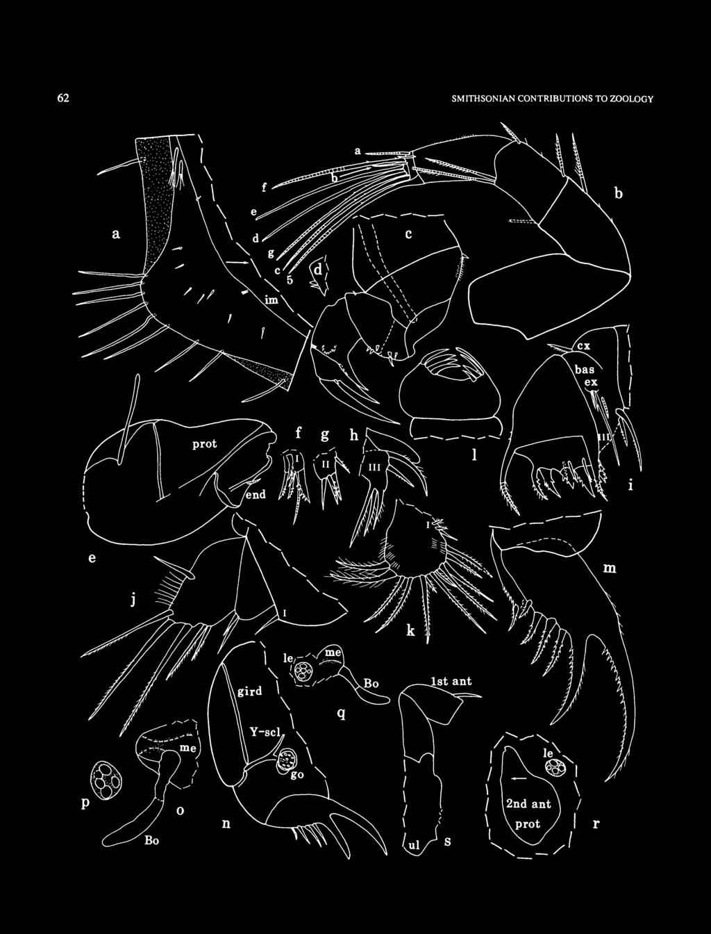
68 62 SMITHSONIAN CONTRIUTIONS TO ZOOLOGY

69 NUMER FIGURE 41 (opposite page). Eurypylus darwinensis, new species, ovigerous female, holotype, AM P45366: a, posterior left valve, iv; b, right 1st antenna, mv; c, right mandible, mv; d, exopodite on basale of left mandible (dorsal edge towards top), lv; e, protopodite (with straight sclerite connected at posterodorsal corner) and endopodite of left 2nd antenna, mv; f,g, endites I and II, respectively, right maxilla; h, endite III and diaphanous dorsal process, left maxilla; i, left maxilla (not all endite bristles shown), lv;y, 5th limb; k, 6th limb; /, combs of tip of 7th limb; m, right lamella of furca, and posterior claw of left lamella showing spines posterior to claw; n, posterior of body from right side; o, medial eye (stippling indicates brown pigment) and ellonci organ; p, lateral eye (stippling indicates brown pigment); q, right lateral eye, medial eye, and ellonci organ (stippling in eyes indicates brown pigment); r, left lateral eye (stippling indicates brown pigment) and endopodite of left 2nd antenna in place on body; 5, anterior of body from right side. Seventh Limb: Proximal group with 2 bristles, 1 on each side, each with 3 bells. Terminal group with 6 bristles, 3 on each side, with 2 and 5 bells. All bristles without marginal spines. Terminal segment with comb of about 8 teeth opposite comb of about 7 shorter teeth (exact number of teeth difficult to resolve) (Figure 41/). Furca (Figure 41 m,n): Each lamella with 6 claws; claws 1 and 2 primary and nonarticulated; claws 3-6 secondary and articulated. Posterior edges of claws 1 and 2 with stout proximal teeth and small distal teeth; anterior edges with slender distal spines. Claws 3-6 with small teeth along proximal parts of anterior and posterior edges and with smaller teeth distally along posterior edges. Right lamella with few spines along anterior edge; left lamella with numerous spines following last claw. Right lamella anterior to left lamella by width of base of claw 1. ellonci Organ (Figure 4\o,q): Elongate, bare, with indistinct suture near midlength; proximal segment with thicker exoskeleton; distal segment broader and with rounded tip. Eyes: Medial eye bare, with brown pigment (Figure 41 o,q). Lateral eye easily visible, with 5 amber-colored ommatidia and brown pigment between ommatidia (Figure 41p-r). Upper Lip (Figure 41s): Rounded. Genitalia (Figure 4\n): Round disk with sclerotizedrimon each side of body anterior to furca. Posterior of ody (Figure 41 n): are, with part posterior to Y-sclerite projecting outward (posteriorly) from part ventral to Y-sclerite. Y'Sclerite (Figure 4ln): Typical for subfamily. Number of Eggs: AM P45366 (holotype) with 4 eggs in marsupium and with smaller unextruded eggs; length of 1 extruded egg mm. DESCRIPTION OF ADULT MALE (Figures 42,43). Carapace differs from that of female in being more elongate and in having a slightly projecting rostrum (Figure 42). Ornamentation (Figures 42, 43a-c): Surface rugose but with ridges less well defined than those of female; knob-like process in posterodorsal corner of valve. Abundant surface papillae longer and more pointed than those of female (papillae FIGURE 42. Eurypylus darwinensis, new species, adult male, paratype, AM P45368, length 0.76 mm. shown in Figure 43b,c). In general, type and distribution of surface bristles similar to that of female (Figure 43a,c). Infold: Anterior infold with minute bristle ventral to rostrum (Figure 43a). Infold of caudal process with 2 minute bristles (Figure 43b,c)- Posterior infold with 2 setal bristles dorsal to caudal process (Figure 43b,c). Inner margin of infold with 1 or 2 minute bristles ventral to caudal process (Figure 43c). Carapace Size (length (L), height (H), in mm): AM P45368, L = 0.76, H = First Antenna: Lost during dissection but of usual type, with abundant slender filaments on sensory bristle of 5th joint. Second Antenna: Protopodite bare (Figure 43a*); anterior half of dorsal margin straight or slightly concave (Figure 43n). Endopodite similar to that of female except with 2 proximal anterior bristles rather than 1 and with 2 medial rows of long hairs (Figure 43d). Exopodite: minute medial terminal bristle of 1 st joint straight, not curved as on female; bristle of 2nd joint with about 10 proximal ventral spines followed by natatory hairs; bristles of joints 3-8 with natatory hairs, no spines; 9th joint with 2 bristles (dorsal bristle short bare; ventral bristle long with natatory hairs); joints without usual row of minute spines along distal edges. Mandible (Figure 43e,f): Coxale endite represented by minute spine near ventral margin; coxale without ventral spines. asale: ventral margin with 6 small bristles (4 medial, 2 along ventral edge or slightly lateral); dorsal margin with 1 distal bristle (bristle missing on limb illustrated but socket present; length of bristle unknown). Exopodite absent. 1st endopodial joint: medial surface with numerous rows of minute spines; dorsal margin with row of minute terminal spines; ventral margin with 2 minute distal spines and 2 terminal bristles (1 small bare ringed and with base medial, 1 long stout

, mv; m, posterior of body from left side; n, dorsal part of anterior of body from left side (stippling")
70 64 SMITHSONIAN CONTRIUTIONS TO ZOOLOGY FIGURE 43. Eurypylus darwinensis, new species, adult male, paratype, AM P45368: a,b, anterior and posterior of left valve, respectively, iv; c, posterior right valve, iv; d, distal protopodite and endopodite left 2nd antenna, mv; e, right mandible, mv;/, detail of tip of e; g, left maxilla (exopodite not shown), lv; h, exopodite right maxilla, lv; i, 5th limb; j, endites I and II right 6th limb, mv; k, endite left 6th limb, mv; /, left 6th limb (endite not shown), mv; m, posterior of body from left side; n, dorsal part of anterior of body from left side (stippling in medial eye indicates brown pigment) (note sclerite connected to 2nd antenna); o, medial eye (stippling indicates brown pigment) and ellonci organ.

 Maxilla (Figure 43g,h): Limb reduced.")
71 NUMER ringed in distal 73 and with minute proximal spines perpendicular to ventral edge). 2nd endopodial joint: medial surface with 2 rows of spines; dorsal margin at midlength with long slender bare ringed bristle; ventral margin with stout bare unringed terminal claw. 3rd endopodial joint with stout bare terminal claw and 3 bristles (1 dorsal minute indistinct, 2 ventral (1 small bare, other longer ringed and with minute spines perpendicular to ventral edge)). (Above description based on right limb; left limb missing from specimen, possibly lost during dissection.) Maxilla (Figure 43g,h): Limb reduced. Endites with weakly formed bristles; bristles difficult to resolve (not all shown). Coxale with well-developed, indistinctly ringed dorsal bristle with few marginal spines. Exopodite with 3 welldeveloped bristles (1 long spinous, 2 short bare) (Figure 43/J). Endopodite: 1st joint with weakly developed alpha- and beta-bristles; 2nd joint with 8 weakly developed bristles. Fifth Limb (Figure 43/): Single endite with 1 short ringed bristle. Exopodite: 1st joint with 2 ringed bristles. Joints 2-5 fused, with 8 bristles (interpretation: 2nd joint with 3 bristles; 3rd joint with 1 bristle on inner lobe and 1 on outer lobe; fused 4th and 5th joints with total of 3 bristles). ristle on inner lobe of 3rd joint and most bristles of 4th and 5th joints shorter than those of female. Sixth Limb (Figure 43./-/): Left limb with single endite with 2 small bristles (Figure 43Jfc); right limb with 2 endites (endite I with 1 small bristle, and endite II with 2 small bristles (Figure 43./)). End joint with 2 very long plumose anterior bristles, 4 bristles with short spines, and 6 or 7 plumose bristles (endite broken off in Figure 43/). Seventh Limb (Figure 43m): Small, bare, thumb-like. Furca (Figure 43m): Similar to that of adult female except with 2 instead of 4 secondary claws; left lamella with many spines following last claw. ellonci Organ (Figure 43n,o): Elongate with indistinct suture at midlength (a few weak sutures may be in distal half but not clearly resolved). Eyes: Medial eye bare, with brown pigment (Figure 43n,o). Lateral eye about same size as medial eye, with 10 amber-colored ommatidia and brown pigment between them (Figure 43/i). Genitalia (Figure 43m): Lobes (1 with hook-like tip) on each side of body anterior to furca. Upper Lip, Posterior of ody (Figure 43m), and Y-Sclerite (Figure 43m): Similar to those of female. REMARKS. The presence of only four claws on each lamella of the furca of the male compared to six on the female is unusual and suggests that the male and female may not be conspecific. Resolution of this may have to await the study of additional collections. COMPARISONS. Female: The carapace of E. darwinensis is without the stout surface bristles present on E. rex. The carapace of E. darwinensis resembles that of E. pulcher Hall, 1985, from Lizard Island, Australia. Eurypylus darwinensis differs from E. pulcher mainly in having one rather than two bristles on the endopodite of the second antenna, two rather than three ventral bristles on the fourth joint of the first antenna, and especially in that the third furcal claw is not a primary claw; also, E. darwinensis has six rather than five furcal claws. Eurypylus darwinensis differs from E. setifera (Poulsen, 1965) in having fewer furcal claws (six rather than seven or eight) and one or two instead of three endites on the sixth limb. CYLINDROLEERIDIDAE Muller, 1906 COMPOSITION. The Cylindroleberididae includes three subfamilies: Cylindroleberidinae, Cyclasteropinae, and Asteropteroninae. All have been reported from the vicinity of Australia (Komicker, 1981b:18; 1994), but only members of the last two subfamilies are considered herein. DISTRIUTION. Cosmopolitan at depths of m. ASTEROPTERONINAE Kornicker, 1981 Asteropterygion Kornicker, 1981 TYPE SPECIES. Asteropterygion thomassini Kornicker, 1981b. COMPOSITION. Three species of this genus are known from the vicinity of Australia: Asteropterygion liguriae (Granata, 1915), A. magnum (Poulsen, 1965), and A. climax, a new species described herein. DISTRIUTION. Cosmopolitan between the latitudes 34 N and 41 S. Known depth range is m, and questionably to 1100 m. Asteropterygion climax, new species FIGURES ETYMOLOGY. From the Latin climax (a succession of ideas progressing in force to a culmination). HOLOTYPE. Partly dissected A-l male in alcohol, AM P TYPE LOCALITY. Gulf of Carpentaria, in vicinity of Weipa, Queensland, Australia; depth 10 m; 16 Nov 1981, time PARATYPES. Type locality: USNM , dissected A-l male on 2 slides (right valve coated for SEM) and in alcohol; USNM , dissected adult male on 3 slides (left valve coated for SEM) and in alcohol; USNM ,3 undissected juveniles in alcohol. DISTRIUTION. Known only from type locality in Gulf of Carpentaria. DESCRIPTION OF A-l MALE (Figures 44-51). Carapace rugose, with well-developed rostrum and incisur (Figures 44, 45); caudal process not projecting past valve edge. Valve

72 66 SMITHSONIAN CONTRIUTIONS TO ZOOLOGY overlaps edge on all margins except posterior edge of caudal process; overlap broadest on rostrum and at anteroventral corner. Ornamentation: Surface with stout node on rostrum, 2 stout nodes near ventral margin, 1 stout node at midheight near posterior end, 1 stout node near posterodorsal margin, and 1 or 2 nodes at midlength dorsal to midheight (Figures 44, 45). Surface with rounded fossae (Figures 44,45,46a-f,h,i, 41a,b)\ fossae on nodes smaller than fossae elsewhere (Figure 46h). Scanning electron microscope (SEM) micrographs reveal minute digitate papillae inside fossae (Figure 47b,c,f,g) and minute reticulations between fossae (Figure 41a-d). Lateral surface of valves with sparsely distributed bristles (Figures 46/, 41a-d). Edge of valve with numerous closely spaced bristles (Figure 46f,g,i) (in lateral view bases of bristles hidden by valve overlap). Infold: Infold of rostrum with abundant bristles. Narrow list extending from anteroventral infold to dorsal end of caudal process; lamellar prolongation observed along anteroventral part of list; list along ventral margin crenulate; long simple bristles present along anteroventral and ventral list (Figure 48 a); list anterior to caudal process straight and with short and long bristles (some of latter with parallel sides). A beaded ridge just inside valve edge extends along anteroventral and ventral infold becoming closer to edge in posterior half of ventral margin (Figure 48 a); ridge absent along caudal process. Anteroventral infold with many short bristles between list and beaded ridge (Figure 48 a). Infold of caudal process with row of about 30 bristles just within and parallel to valve edge. Hingement: Posterior end of hingement with large tooth on left valve and large socket on right valve (Figure 46b). Carapace Size (length (L), height (H), in mm): AM P45370 (holotype), L = 4.2, H = 3.0. USNM , L = 4.5, H = 3.4. First Antenna (Figure 4Sb,c): 1st joint with ventral hairs. 2nd joint: ventral margin with long proximal and short distal hairs; dorsal margin with short proximal hairs and 5 spinous bristles; lateral surface with 2 spinous bristles near midlength; medial surface with hairs along distal margin and near ventral margin. 3rd joint trapezoidal; dorsal margin with 5 spinous bristles (1 proximal, 4 terminal); ventral margin with 1 short bristle. 4th joint: dorsal margin with 1 long spinous terminal bristle; ventral margin with 4 spinous bristles (1 long, 3 very short). 5th joint: dorsal margin and distal lateral surface with rows of hairs; sensory bristle with 8 short proximal filaments and 5 long terminal filaments (distal pair fused proximally). 6th joint short, with spinous medial bristle about 3 A length of 5th joint. 7th joint: a-bristle slender, claw-like; b-bristle about twice length of a-bristle, with 9 short filaments; c-bristle slightly longer than b-bristle, with about 15 marginal filaments. 8th joint: d- and e-bristles about '/3 longer than a-bristle, bare with blunt tips; f-bristle bent dorsally, about same length as b-bristle and with about 21 filaments; g-bristle about same length as c-bristle, with about 12 marginal filaments. FIGURE 44. Asteropterygion climax, new species, A-l male, paratype, USNM , length 4.5 mm. Second Antenna: Protopodite ovoid, with few short distal spines along dorsal margin, long hairs along ventral margin (Figure 4Sd,e), and with 1 short distal medial bristle (Figure 48 e). Endopodite 3-jointed (Figure 48 e): 1st joint elongate with 7 small slender bristles (5 at proximal end, 2 slightly distal to them); 2nd joint more than twice length of 1st joint, with 3-5 small distal lateral bristles; 3rd joint about 2 /3 length of 2nd joint, with long proximal filament and narrow rounded tip either bare or with minute bristle. Exopodite: 1st joint with minute straight medial bristle on distal margin (not shown in Figure 48 d); bristle of 2nd joint very long, with natatory hairs and proximal ventral spines; bristles of joints 3-8 with natatory hairs; 9th joint with 5 bristles, all with natatory hairs (3 stout bristles also may have few minute ventral spines); joints 3-8 with basal spines increasing in size on distal joints (spine of 8th joint about 3 A length of 9th joint); 9th joint with lateral spine about same size as basal spine of 8th joint; joints 2-8 with minute spines along distal margin and few rows of indistinct spines proximal to edge; dorsal margin of 2nd joint 2 3 A times length of dorsal margin of 3rd joint. Mandible: Coxale endite of USNM with small bristle near base of ventral branch of left limb (Figure 49b) but not on right limb (Figure 49a); ventral branch with spines forming about 7 oblique rows and tip with 3 minute teeth (Figure 49a); ventral margin of dorsal branch with 2 minute teeth followed by 4 larger triangular nodes, 1 small spine close to distal node, 2 longer slender spines, and small main spine (Figure 49a); tip of branch with short spine; dorsal margin of branch with few minute distal serrations and long subterminal bristle with short marginal hairs and longer spines near base; margin of branch between tip and main spine and between tip
: tip with 7 end-type bristles; ventral margin with 7 triaenid bristles (terminal pair of spines only slightly longer than subterminal pair); 3 dwarf bristles near dorsal")
: ventral margin with 9 triaenid bristles, 1 long subterminal bristle with long spines, and 6 or 7 minute bare bristles with bases either medial or lateral; dorsal margin with 6 or")
 (Figure 49d).")
73 NUMER and subterminal dorsal bristle with minute spines. asale endite (Figure 49c): tip with 7 end-type bristles; ventral margin with 7 triaenid bristles (terminal pair of spines only slightly longer than subterminal pair); 3 dwarf bristles near dorsal margin (distal bristle about twice length of others). asale (Figure 49d): ventral margin with 9 triaenid bristles, 1 long subterminal bristle with long spines, and 6 or 7 minute bare bristles with bases either medial or lateral; dorsal margin with 6 or 7 slender bristles (bare or with short spines) in middle part and 2 long stout terminal bristles with long spines; medial surface with long hairs. Exopodite almost reaching distal margin of 1st endopodial joint, hirsute, with 2 distal ventral bristles (proximal bristle about twice length of distal bristle) (Figure 49d). 1st endopodial joint with 8 ventral bristles (2 long stout proximal, 5 shorter distal, and 1 minute both proximal and medial) (Figure 49d). 2nd endopodial joint (Figure 49e): ventral margin with 7 spinous bristles; dorsal margin and medial surface near dorsal margin with abundant long and short bristles (not all shown in Figure 49e). 3rd endopodial joint with 6 bristles (3 long stout, 3 slender (1 long lateral, 2 short ventral) (Figure 49/)). Maxilla (Figure 48/): Epipodite long slender with hairs at pointed tip. Endite I with 6 or 7 stout spinous bristles, 1 or 2 shorter slender bristles, and 1 or 2 minute bare medial bristles; endite II with 3-5 stout spinous bristles; 7 or 8 short bristles present near bases of bristles of endite II. asale: dorsal margin with 4 proximal bristles with terminal hairs and 6 or 7 distal bristles (1 very long) oriented dorsally (some with bases on medial surface); ventral margin with row of 15 short spinous bristles, 1 long spinous distal bristle, and 1 long terminal bristle; medial side near ventral margin with 3 or 4 distal bristles (1 long spinous, 2 or 3 short bare); lateral side with 1 short proximal bristle near ventral margin (dashed in Figure 48/). Exopodite minute, with 3 bristles (1 long, 2 short). Endopodite: 1st joint with long hairs, 1 short bare alpha-bristle, and 1 long spinous beta-bristle; 2nd joint with 6 terminal bristles. Fifth Limb (Figure 49g): Comb short, with strongly arched dorsal margin with 9-11 small proximal bristles; dorsal margin hirsute except for short span anterior to midlength. Exopodial bristles comprise 2 long spinous bristles, 3 small bare bristles between bases of long bristles, and 4 short bristles (2 longer bristles spinous, 2 shorter bristles bare) closer to ventral margin; 2 bristles at anteroventral corner with bases short distance from comb edge; bristles along ventral margin of comb of 3 different lengths. Sixth Limb (Figure 49h): With 4 small epipodial bristles. Anterior margin with distinct suture separating trunk from skirt. Medial surface in anterodorsal corner of trunk with about 20 stout spines and with rows of slender spines ventral to them. Anterior margin of trunk with 3 rows of bristles: inner row with 6 bare bristles followed by 10 spinous bristles; middle row with 14 spinous bristles; outer row with 24 spinous bristles with bases on edge of trunk (not all bristles shown). Anterior margin of skirt ventral to suture with 8 or 9 slender spinous bristles. Anterior comer and ventral margin of skirt with 21 long bristles (with long spines) along edge, 10 shorter bristles (with long spines) with bases medial and set back slightly from ventral edge, 10 shorter bristles (with short spines) with bases medial and set back slightly farther from ventral edge, and about 30 minute bristles (either bare or with minute spines) with bases medial and set back slightly farther from ventral edge (not all bristles shown); 7 additional small bristles along ventral margin posterior to last long spinous ventral bristle; posterior tip of limb skirt translucent, separated slightly from rest of skirt by small indentation at ventral edge, and with 3 terminal weakly developed plumose bristles; tip and posterior part of skirt and trunk with abundant long hairs. Lateral flap at anterior comer of skirt obscured but with several slender plumose bristles (not shown). An internal sclerite at trunk midwidth bends at proximal end to intersect anterodorsal comer of trunk. Seventh Limb: USNM : Each limb with cylindrical bristles per side to make up actual total of bristles per limb; short bristles with 2 or 3 bells, longer bristles FIGURE 45. Asteropterygion climax, new species, A-l male, paratype, USNM , length 4.5 mm, stereoscopic SEM micrograph pair of right valve.

; h, posterodorsal corner")
74 68 SMITHSONIAN CONTRIUTIONS TO ZOOLOGY FIGURE 46. Asteropterygion climax, new species, A-l male, paratype, USNM , outside SEM views of right valve: a-d, anterior, posterior, dorsal, and ventral views, respectively; e, lateral view of incisur, anterior of valve to bottom of picture;/, detail from midheight of a; g, detail from lower right of/(arrow); h, posterodorsal corner right valve, lv; /', detail of edge from midheight of d (see arrow).

; b, detail from Figure 46>i (see 3rd arrow from top); c, detail")
;/ detail from bottom of fossa in")
75 NUMER FIGURE 47. Astewpterygion climax, new species, A-l male, paratype, USNM outside SEM views of right valve: a, detail from Figure 46/i (see upper arrow); b, detail from Figure 46>i (see 3rd arrow from top); c, detail from b (see arrow); d, detail from Figure 46/i (see 2nd arrow from top); e, detail from Figure 46> (see arrow);/ detail from bottom of fossa in Figure 467i (see bottom arrow); g, detail from c (see arrow).

; h, one")
76 70 SMITHSONIAN CONTRIUTIONS TO ZOOLOGY FIGURE 48. Asteropterygion climax, new species, A-l male, paratype, USNM : a, anteroventral margin left valve, iv; b,c, left 1st antenna, lv; d, protopodite and 1st exopodial joint right 2nd antenna, lv; e, distal protopodite and endopodite right 2nd antenna, mv;/ left maxilla; g, tip of 7th limb (only teeth of 1 side shown); h, one of two copulatory organs on specimen (drawn under cover slip).

, mv;/, 3rd endopodial joint left")
77 NUMER FIGURE 49. Asteropterygion climax, new species, A-l male, paratype, USNM : a, coxale endite right mandible, mv; b, proximal part of coxale endite left mandible, mv; c, basale endite left mandible, mv; d, part left mandible, mv; e, endopodite left mandible (not all bristles shown), mv;/, 3rd endopodial joint left mandible, mv; g, comb of left 5th limb, lv; h, right 6th limb (spines not shown on all bristles), mv.

 (only 1 comb shown in Figure 48 #).")
78 72 SMITHSONIAN CONTRIUTIONS TO ZOOLOGY FIGURE 50. Asteropterygion climax, new species, A-l male, paratype, USNM : a, right furcal lamella, lv; b, medial eye and ellonci organ; c, posterior end of protopodite of right 2nd antenna, and right lateral eye; d, upper and lower lips and location of mouth; e, posterodorsal corner of body showing posterior process;/, right Y-sclerite. with up to 7 bells; bristles without marginal spines. Terminus with opposing combs, each with teeth (5 or 6 teeth at midwidth of comb shorter than adjacent teeth, and teeth decrease in length at each end of comb) (only 1 comb shown in Figure 48 #). Furca (Figure 50a): Each lamella with 9 claws decreasing in length and width posteriorly along lamella; claws 1 to 4 or 5 with posterior teeth stouter along distal half; posterior 3 or 4 claws appearing weak and flexible; all claws with hairs along anterior margins. Each lamella with medial hairs at bases of claws and following claws; anterior edge of lamella with small spines; right lamella anterior to left lamella by width of base of claw 1. ellonci Organ (Figure 506): With indistinct suture at about l U length; broad part distal to midlength with indistinct striations; tip nipple-like.
: Upper lip comprising 2 hirsute lobes, one on each side of low saddle; spines absent. Lower lip a hirsute flap on each side of mouth.")
: Hirsute, with very long hairs near posterior end of girdle; posterodorsal process bent on USNM 194279 but with 2 lobes: posterior lobe with long hairs, anterior lobe bare. Gills well developed.")

79 NUMER 578 Eyes: Medial eye large, bare, with area of brown pigment (Figure 5Qb). Lateral eye with numerous ommatidia (about 50) and brown pigment (Figure 50c). Lips (Figure 50d): Upper lip comprising 2 hirsute lobes, one on each side of low saddle; spines absent. Lower lip a hirsute flap on each side of mouth. Genitalia (Figure 48 h): Comprising minute lobes with few bristles. Posterior of ody (Figure 50<?): Hirsute, with very long hairs near posterior end of girdle; posterodorsal process bent on USNM but with 2 lobes: posterior lobe with long hairs, anterior lobe bare. Gills well developed. \-Sclerite (Figure 50/): Without ventral branch. Protistans: Some appendages and other parts of body with numerous internal ciliate-like protistans (those in 6th limb shown in Figure 51). FIGURE 52. Asteropterygion climax, new species, adult male, paratype, USNM , length 5.0 mm. 73 FIGURE 51. Asteropterygion climax, new species, A-l male, paratype, USNM ,6th limb containing ciliate-like protistans: a, complete limb; b, detail of protistans in a (note cilia indicated by arrow). Photographs by Robert Higgins, Smithsonian Institution. DESCRIPTION OF ADULT MALE (Figures 52-57). Carapace elongate with well-developed incisur and rostrum (Figures 52, 53). Valve overlaps edge along anterior, ventral, and posterior margins; overlap broadest on rostrum and anteroventral corner. Ornamentation: Surface with small node at midheight of rostrum, elongate ridge along ventral margin of rostrum extending onto valve posterior to rostrum, discontinuous rib along anterior 2 /3 of ventral margin, 3 processes along posterior margin, and 2 small nodes just anterior to upper and middle posterior processes (Figures 52, 53). Surface with rounded fossae (Figures 52, 53, 54a, 55 a-c); fossae on nodes generally smaller than elsewhere (Figure 55b). SEM micrographs reveal minute digitate papillae and clusters of papillae inside of fossae (Figures 54 a,c, 55e) and show minute reticulations between fossae (Figures 54a, 55 c,d); edges of reticulations with minute papillae (Figures 54b, 55 d). Lateral surface of valves with sparsely distributed bristles (Figures 54a,b, 55 c-/); some bristles emerge from open pores (Figure 54a,b), others from closed pores with radial processes around base of bristle (Figure 55 c-/). Edges of valves with numerous closely spaced bristles (in lateral view bases of bristles hidden by valve overlap) (Figures 55 a, 56a,b). Posterior end of valve with row of long hairs (Figure 55b); anterior end of valve with long hairs and bristles on rostrum and ventral to incisur (Figures 55 a, 56a). Infold: Infold of rostrum with abundant bristles (not all shown in Figure 56 a). List extending from anterior x h of valve length to dorsal end of caudal process; list anterior to caudal process broad and with short and long bristles similar to those of A-l male (Figure 56b). Crenulate ridge extends from about anterior 73 of valve to ventral end of caudal process. Infold of caudal process with about 45 long and short bristles just within valve edge (Figure 566). Hingement: Similar to that of A-l male (socket of right valve shown in Figure 56 b).
, height, (H), in mm): USNM 194280, L = 5.0, H = 3.5. First Antenna (Figure 56c,d): 1st joint with distal ventral hairs. 2nd joint spinous with 5 spinous dorsal bristles.")
 ventral). 5th joint with long stout sensory bristle with abundant thin filaments (not shown).")

80 74 SMITHSONIAN CONTRIUTIONS TO ZOOLOGY FIGURE 53. Asteropterygion climax, new species, adult male, paratype. USNM , length 5.0 mm, stereoscopic SEM micrograph pair of left valve. Carapace Size (length (L), height, (H), in mm): USNM , L = 5.0, H = 3.5. First Antenna (Figure 56c,d): 1st joint with distal ventral hairs. 2nd joint spinous with 5 spinous dorsal bristles. 3rd joint weakly separated from 4th joint, with 1 short ventral bristle and 5 dorsal bristles (1 proximal, 4 terminal). 4th joint with 4 or 5 spinous terminal bristles (1 dorsal, 3 or 4 (1 minute) ventral). 5th joint with long stout sensory bristle with abundant thin filaments (not shown). 6th joint with spinous medial bristle with base almost on dorsal margin. 7th joint: a-bristle slender claw-like with distal rings; b-bristle about 3 times length of a-bristle, with about 13 short marginal filaments; c-bristle extremely long (tip broken off but with 40 marginal filaments on remaining part). 8th joint: d- and e-bristles about twice length of a-bristle, bare with blunt tips; f-bristle about same length as b-bristle, with about 15 short marginal filaments; g-bristle similar to c-bristle. Second Antenna: Prodopodite without spines or hairs, with short distal medial bristle. Endopodite 3-jointed (Figure 56 e): 1st joint elongate with 7 or 8 bristles (4 or 5 proximal, 3 at midlength); 2nd joint elongate with about 30 bristles (only 10 shown in Figure 56 e, but 30 shown in Figure 56/); 3rd joint elongate, recurved, with 1 long slender proximal filament and FIGURE 54. Asteropterygion climax, new species, adult male, paratype, USNM , outside SEM views of left valve: a, detail from upper right in Figure 55b (see arrow, 556); b, detail of base of lower bristle in a (see lower arrow); c, detail of cluster at bottom of fossa in a (see upper arrow).


; e, detail of upper bristle")
81 NUMER FIGURE 55. Asteropterygion climax, new species, adult male, paratype, USNM , outside SEM views of left valve: a,b, lateral views of anterior and posterior, respectively; c, detail from anteroventral corner in a (see arrow); d, detail of base of lower bristle in c (see lower arrow); e, detail of upper bristle in c (see upper arrow); /, detail of base of bristle in d.
 bristles with natatory hairs; joints 4-8")
82 76 SMITHSONIAN CONTRIUTIONS TO ZOOLOGY pointed tip with about 15 minute ridges (not all ridges shown). Exopodite: 1st joint with short distal spines along ventral margin; bristles of joints 2-8 long, with natatory hairs, no spines; 9th joint with 5(1 short) bristles with natatory hairs; joints 4-8 with basal spines increasing in length on distal joints; basal spine of 8th joint about same length as 9th joint; 9th joint with lateral spine about 2 /3 length of basal spine of 8th joint; joints 2-8 with long hairs on distal dorsal corners. Mandible: Coxale endite similar to that of A-l male, but both limbs with small bristle near base of ventral branch (Figure 51a,b). asale endite with total of 11 triaenid and end-type bristles (bristles not clearly differentiated, both with small marginal spines (spines not shown in Figure 57c), 3 dwarf bristles (distal bristle twice length of 2 proximal bristles), and glandular peg (Figure 57c). asale (Figure 51d): ventral margin with 4 minute bristles, 9 short bristles (most with closely spaced distal spines), and 1 long distal bristle with long spines; dorsal margin with 7 slender bristles and 2 long stout terminal bristles. Exopodite and 1st and 3rd endopodial joints similar to those of A-l male (Figure 51d). 2nd endopodial joint: ventral margin with 10 bristles; dorsal margin and medial surface near dorsal margin similar to that of A-l male but with more medial bristles near dorsal margin (about 100 medial bristles in 10 rows). Maxilla: oth limbs partly fragmented. Endite I with 6 stout bristles, 1 short slender bristle, and 1 minute medial bristle; endite II with 5 stout bristles; 9 short bristles present near bases of bristles on endite II. asale: dorsal margin with 4 proximal bristles and 5 distal bristles (2 short proximal, then 1 very long and 2 fairly long); medial side with vertical row of 4 small bristles near distal dorsal bristles; ventral margin fragmented (remaining part with 10 small bristles and 1 distal long medial bristle). Exopodite minute with 2 bristles. Endopodite: 1st joint with 1 short alpha-bristle and 1 long beta-bristle; 2nd joint with 5 terminal bristles. Fifth Limb (Figure 51e): Comb with hook-like dorsal process; dorsal margin with 5 small bristles proximal to tip of hooked process and with hairs at distal end just dorsal to marginal bristles; 2 small lateral bristles present at midlength just ventral to dorsal margin. Exopodial bristles comprise 2 long spinous bristles (posterior bristle shorter and more slender, broken off on comb of right limb but shown as dashed bristle based on proximal part of bristle present on left comb), 3 short bare bristles between bases of long bristles, and 5 short bristles (2 longer spinous, 3 shorter bare) closer to ventral margin; 2 small bristles at anteroventral corner with bases short distance from comb edge; bristles along ventral edge similar to those of A-l male. Sixth Limb: With 5 epipodial bristles. Medial surface in anterodorsal corner of trunk with spines (uncounted) similar to those of A 1 male. Posterior tip of skirt with 4 plumose bristles; remaining bristles (uncounted) of skirt similar to those of A-l male. Seventh Limb: Each limb with cylindrical bristles per side to make up actual total of bristles per limb; short bristles with 2-4 bells, longer bristles with up to 7 bells (rarely 8); bristles without marginal spines. Terminus with opposing combs, each with 27 or 28 spinous teeth (5 or 6 teeth at midwidth of comb shorter than adjacent bristles, and teeth decrease in length at each end of comb) (only 1 comb shown in Figure 56g and marginal spines of teeth not shown). Furca (Figure 51f,g): Each lamella with 9 or 10 claws decreasing in length and width posteriorly along lamella. Limb otherwise similar to that of A-l male. ellonci Organ, Eyes (Figure 57h), Lips, and Y- Sclerite: Similar to those of A-l male. Genitalia (Figure 57/): Elongate lobes with few minute bristles, anterior to furca. Posterior of ody (Figure 57/): Similar to that of A-l male except anterior lobe of posterodorsal process narrower and longer than posterior lobe. COMPARISONS. The new species resembles Asteropterygion hirsutum (Poulsen, 1965), which is based on a unique juvenile male (probably A-l male) from the Malayan Archipelago. They differ mainly in that the a-bristle of the first antenna of the A-l male A. hirsutum tapers to a point (bristle-like) whereas that of the A-l male A. climax curves dorsally and is claw-like. The two species have characters not found in other members of the genus, such as having more primary furcal claws and having medial spines in the proximal anterior corner of the trunk of the sixth limb. The two species are very closely related and could be conspecific. The presence of small copulatory organs on the juvenile male A. hirsutum described by Poulsen (1965:187) and the juvenile male A. climax described herein, as well as the bristles of the seventh limbs being cylindrical rather than tapering distally, indicate that both are at the A-l stage. ased on the description of the A-l male by Poulsen (1965:187), and the A-l male A. climax described herein, additional differences between A-l males of the two species are as follows: Carapace length (mm) Number of bristles between list and valve edge on infold of caudal process Number of bristles on 2nd joint of endopodite of 2nd antenna Number of long exopodial bristles on comb of 5th limb A. hirsutum A. climax The shape of the carapace of the adult male A. climax resembles that of Aster ope lichenoides rady, 1902, known from a unique adult male (from the southern part of the South China Sea) that I herewith refer to Asteropterygion lichenoides. rady (1902:181) described the furca of A. lichenoides as having five slender claws, many fewer than A. climax. The adult male A. climax differs from previously described adult males of the genus in having extremely long c- and g-bristles on the first antenna and in having a hook-shaped dorsal process on the comb of the fifth limb. The elongate carapace of the adult male A. climax differs in shape from those of known adult males of other species with the exception of A. lichenoides

), iv; c,d. right 1st antenna, lv; e.")
83 NUMER FIGURE 56. Astewpterygion climax, new species, adult male, USNM : a, part anteriorright valve, iv; b, ventral half of posterior margin (posteroventral corner not shown; stippled posterior protuberance at upper two-thirds of illustration is tip of lateral process at midheight of valve (see Figure 52)), iv; c,d. right 1st antenna, lv; e.f, endopodite right 2nd antenna (all bristles of 2nd joint shown in/but not in e), iv; g, tip of 7th limb (teeth shown on only one side).

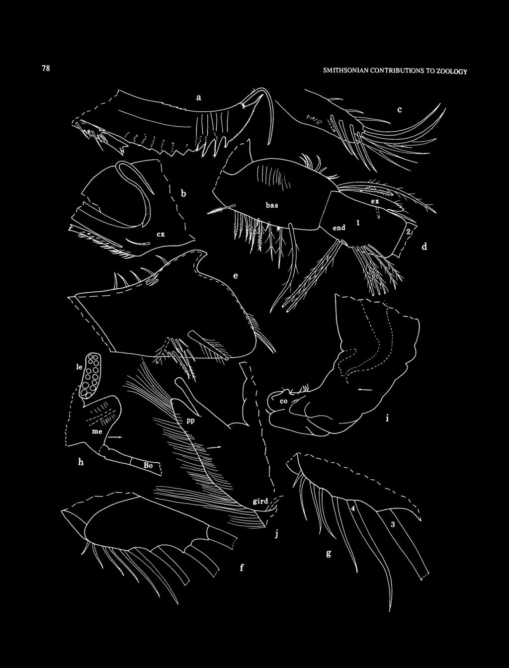
84 78 SMITHSONIAN CONTRIUTIONS TO ZOOLOGY
, mv; c, basale")

85 NUMER FIGURE 57 (opposite page). Asleropterygion climax, new species, adult male, USNM : a, distal part coxale enditerightmandible, mv; b, proximal part coxale endite left mandible (stippling indicates sclerite), mv; c, basale endite right mandible, mv; d, pan left mandible, mv; e, comb of right 5th limb (proximal long bristle broken off but shown as dashed bristle based on bristle of comb of left limb), lv;/,g, right furcal lamella, lv; h,rightlateral eye, medial eye, proximal part of ellonci organ; i, left copulatory organ, lv; j, posterior process at posterodorsal corner of body drawn from right side. CYCLASTEROPINAE Poulsen, 1965 Tetraleberis Kornicker, 1981 TYPE SPECIES. Aster ope brevis Muller, 1890:239. COMPOSITION. Three species of this genus are known from the vicinity of Australia, namely, T. brevis (Muller, 1890:239) and two new species described herein: T. pix and T. triplex. The genus now contains five species plus one left in open nomenclature (Kornicker, 1975:134). DISTRIUTION. Tetraleberis brevis: Southwest of Japan and Coral Sea (NE of Australia); T. similis: Java Sea; T. pix: Calliope River and Auckland Creek near Gladstone, Queensland, Australia; T. triplex: Lizard Island and Davies Reef, Great arrier Reef, Australia; T. tanzania, T. maddocksae, and Tetraleberis sp. 1: Indian Ocean. Known depth range is 3-65 m (Kornicker, 1981b: 134). REMARKS CONCERNING T. brevis (Muller, 1890). This species, the type species of the genus, presents a problem. Komicker (1981b: 135) described a female (adult or A-l) syntype of T. brevis having a maxilla with many more dorsal and ventral bristles on the basale than were present on the maxilla illustrated by Muller (1890, pi. 27: fig. 12). Possible interpretations are that either (1) two different species are in the type series; (2) the number of bristles on the basale of the maxilla varies considerably within the species; (3) one of the described specimens has an aberrant maxilla; (4) poor observation; or (5) the maxilla illustrated by Muller is from an early juvenile. Until additional study clarifies the problem, I think it expedient to accept the fifth possibility and to assume that the specimen described by Komicker (1981b: 135) better represents 7! brevis. Although I believe that the lack of a bristle between claws three and four of the caudal furca illustrated by Muller (1890, pi. 27: fig. 10) is probably the result of poor observation, it is possible that the absence is also a juvenile character. CORRECTION. Kornicker (1981b: 153) described the lateral eye of T. maddocksae as having 24 ommatidia. The numerals apparently are transposed because the eye has 42 ommatidia (Kornicker, 1981b,fig. 52h). Tetraleberis pix, new species FIGURES 58-62a-m ETYMOLOGY. From the Latin pix (pitch). HOLOTYPE. Adult male on slide and in alcohol, QM W TYPE LOCALITY. Calliope River and Auckland Creek area, near Gladstone, Queensland, Australia. PARATYPES. Type locality: USNM A, ovigerous female on slide and in alcohol; USNM , 4 undissected adult females in alcohol; USNM C, undissected adult male in alcohol; USNM D, 32 undissected adult females and juveniles in alcohol. DISTRIUTION. Known only from type locality. DESCRIPTION OF ADULT MALE (Figures 58-60). Carapace oval in lateral view with evenly rounded posterior, incisur at midheight (Figure 58a). Ornamentation: Anterior margin without scalloped peripheral ridge; surface without fossae or ridges but with numerous minute pores indicated by short pore canals. Anteroventral quarter of shell surface with abundant thin hair-like bristles (Figure 58ft) (these not reported previously in Cyclasteropinae); usual vertical row of bristles present near posterior end (Figure 58 a); long bristles with bases on inner side of valve abundant along anterior, ventral, and posterior margins. Not all bristles shown in Figure 58 b. Infold: Typical for subfamily. Vestment: Long spines present on vestment just proximal to inner margin of anterodorsal infold. Central Adductor Muscle Attachments (Figure 58 c): Typical for subfamily. Carapace Size (length (L), height (H), in mm): QM W20739 (holotype), L = 4.0, H = 2.9; H = 72.5% of length. USNM C, L = 3.9, H = 2.6; H = 65% of length. First Antenna (Figure 58a»: 1st joint with row of long medial hairs just within ventral margin. 2nd joint: ventral margin with long distal hairs; dorsal margin with long proximal hairs and 5 spinous ringed bristles; lateral side with row of 4 unringed distal bristles with few short distal marginal spines; medial side with abundant hairs. 3rd joint: short ventral side with minute bristle (Figure 58 e) (bristle missing from left limb of holotype); long dorsal side with 10 spinous ringed bristles. 4th joint: ventral margin with 4 long spinous terminal ringed bristles (1 additional minute bristle present on left limb of holotype, but could be foreign growth (Figure 58 d)); dorsal margin with 1 long spinous ringed bristle. 5th joint: fused to 6th joint on lateral side except for short suture at dorsal edge; dorsal margin with small triangular process near midlength; stout ventral sensory bristle with abundant thin marginal filaments and about 9 stouter terminal filaments (filaments not shown). 6th joint: fused to 7th joint on medial side, with spinous ventral bristle with base on medial side. 7th joint: a-bristle claw-like, bare; b-bristle almost twice as long as stem of sensory bristle of

86 80 SMITHSONIAN CONTRIUTIONS TO ZOOLOGY 5th joint, with about 15 short marginal filaments; c-bristle not bent backwards, about 6 times length of b-bristle, with numerous short thin indistinct marginal filaments. 8th joint: d- and e-bristles about twice length of a-bristle, bare with blunt tips, both bristles with bases on small sclerotized process with minute nodes (detail in Figure 58d); f-bristle similar to c-bristle; g-bristle about same length as b-bristle, with 12 closely spaced fairly long proximal filaments followed by 10 shorter and more widely spaced filaments. Some proximal filaments of c- and f-bristles slightly broader and more strongly ringed near midlength, but no marginal flanges observed. Second Antenna: Protopodite bare except for small distomedial bristle. Endopodite 3-jointed (Figure 58/,g): 1st joint with 4 or 5 short bristles at midlength; 2nd joint with a cluster of about 9 short slender ringed bristles at distal 3 A; 3rd joint reflexed, with broad triangular proximal part bearing long filament and with slender distal part with short subterminal bristle and numerous ridges. Exopodite (Figure 58 h,i): 1st joint with minute straight terminal medial bristle; 2nd joint long, about same length as joints 3-8 combined; joints 2-8 with basal spines (spine of 2nd joint small, but spines of other joints about same length; spine of 8th joint about 2 /3 length of 9th joint), long slender terminal dorsal spines, and small indistinct spines along distal edges; 9th joint of holotype with small lateral spine on left limb (Figure 58/) but with none on right limb (Figure 58 hi); bristles of joints 2-8 and 5 bristles of joint 9 with natatory hairs, no spines. Mandible: Coxale endite (Figure 59a-d) with long slender ringed bristle proximal to base of ventral branch; ventral branch with 5 oblique rows of spines and truncate tip with 2 small ventral teeth, medial row of 7 or 8 minute spines (Figure 59d), and lateral row of about 13 minute spines (ventral spine slightly stouter) (Figure 59c); dorsal margin of dorsal branch with few serrations; ventral margin of dorsal branch with 3 short triangular teeth followed by tooth with curved distal margin with small rounded teeth, and then 2 recurved teeth with minute spines along proximal edge; edge between distal recurved tooth and tip of branch with minute spines; tip of branch with long bristle with indistinct hairs. asale endite with 6 or 7 long terminal bristles and about 10 bristles proximal to tip (some bristles with distal spines, but no triaenid type) (most spines not shown in Figures 59e, 60a). asale: ventral margin with about 15 short- or medium-length bristles proximal to 2 long stout spinous bristles (Figure 60 a); dorsal margin with 10 short bristles proximal to 2 long stout terminal bristles; medial side with long hairs in proximal half at midheight. Exopodite hirsute, about same length as dorsal margin of 1st endopodial joint (triangular tip folded back on both limbs of holotype), with proximal bristle about twice length of distal bristle. 1st endopodial joint with 8 medial ventral bristles (2 longest bristles with long spines). 2nd endopodial joint: ventral margin with 2 groups of bristles: subterminal group with 5 bristles (shortest and innermost bristle bare, others with short spines), and terminal group with 2 long stout bristles with short spines; dorsal margin of joint with numerous long and medium bristles; medial side near dorsal margin with abundant short bristles. 3rd endopodial joint with 3 stout claw-like bristles, 1 long lateral bristle with few indistinct short marginal hairs, and 2 ventral medial bristles (longer bristle with short spines, other bare). Maxilla (Figure 59/,g): Epipodite long and slender. Endite I with 1 minute proximal bristle and 3 long terminal bristles with long subterminal spines (Figure 59/); endite II with 1 short bristle and 2 long terminal bristles with long subterminal spines; endite III with 3 long bristles with spines along entire margin; 3 or 4 short slender bristles distal to bases of bristles of endite III. asale (Figure 59#): dorsal margin spinous, with 1 proximal plumose bristle (with base on medial side) and 2 distal bristles; medial side with 5 distal bristles (ventral bristle very long, others short); ventral margin with 13 or 14 short bristles, 2 long distal bristles, and 1 long terminal bristle (with base lateral); lateral side with 1 short proximal bristle at midheight (not shown). Exopodite consisting of poorly defined lobe with 3 slender bristles (longest bristle about l /2 length of 1st endopodial joint). Endopodite (Figure 59g): 1st joint spinous, with short alpha-bristle at proximal '/3 and long terminal beta-bristle with few small indistinct spines; 2nd joint with 6 bristles (3 long, 3 short, some with few small indistinct spines). (Spines and rings not shown on all bristles illustrated.) Fifth Limb (Figure 59/i): Dorsal margin of comb with 4 minute ringed bristles proximal to low process with sclerotized anterior tooth on medial side (upper detail in Figure 59A); anterodorsal margin hirsute and sloping to base of 1st anteroventral bristle (2 anterior bristles shown in illustrated comb). Stout spinous exopodial bristle almost reaching end of comb; 8 short lateral bristles (some with spines) near base of stout bristle (lower detail in Figure 59h), and 1 short spinous proximal bristle near ventral margin. Sixth Limb (Figure 60b): With 4 small bare epipodial bristles (a few hairs present on posterior edge of limb proximal to bristles). Anterior margin of limb with 2 well-defined endite sutures; lower endite suture with fairly long bristle just dorsal to it; bristles along anterior margin of limb dorsal to upper endite suture form 3 rows with spinous bristles in inner medial row, 23 more slender spinous bristles in outer medial row, and 22 smaller bristles along edge. Same 3 rows also present between upper and lower endite sutures, with 3 or 4 bristles in inner medial row, 3 bristles in outer medial row (not counting long bristle at suture of lower endite), and 5-7 bristles along edge. Single row of bristles along edge between lower endite and anterior tip of skirt. Posterior end of skirt with 2 fairly broad plumose unringed bristles; ventral margin of skirt with about 75 spinous bristles. Summary: about anterior bristles, 77 bristles on ventral margin and posterior corner of skirt, and 4 epipodial bristles; total of bristles. Not all bristles, hairs, or spines shown in illustrated limb. Seventh Limb: Each limb with 77 bristles, 38 on one side, 39 on the other, each bristle with up to 6 bells; most
, ov; d, distal left 1st antenna (not all")
, mv;/, endopodite left 2nd antenna, mv; g, distal part endopoditeright2nd antenna, mv; h, distal part exopodite right 2nd antenna (bristles of joints 7 and 8 not shown), lv; i, basal")
87 NUMER FIGURE 58. Tetraleberis pix, new species, adult male, holotype, QM W20739: a. complete specimen, length 4.0 mm; b, hair-like surface bristles of anteroventral part left valve, ov; c, central adductor muscle attachments left valve (drawn after body removed), ov; d, distal left 1st antenna (not all terminal bristles shown; filaments of sensory bristle of 5th joint not shown), lv; e, distal right 1st antenna (not all terminal bristles shown; filaments of sensory bristle of 5th joint not shown), mv;/, endopodite left 2nd antenna, mv; g, distal part endopoditeright2nd antenna, mv; h, distal part exopodite right 2nd antenna (bristles of joints 7 and 8 not shown), lv; i, basal spine of joint 8 and lateral spine of joint 9 of exopodite left 2nd antenna, lv.

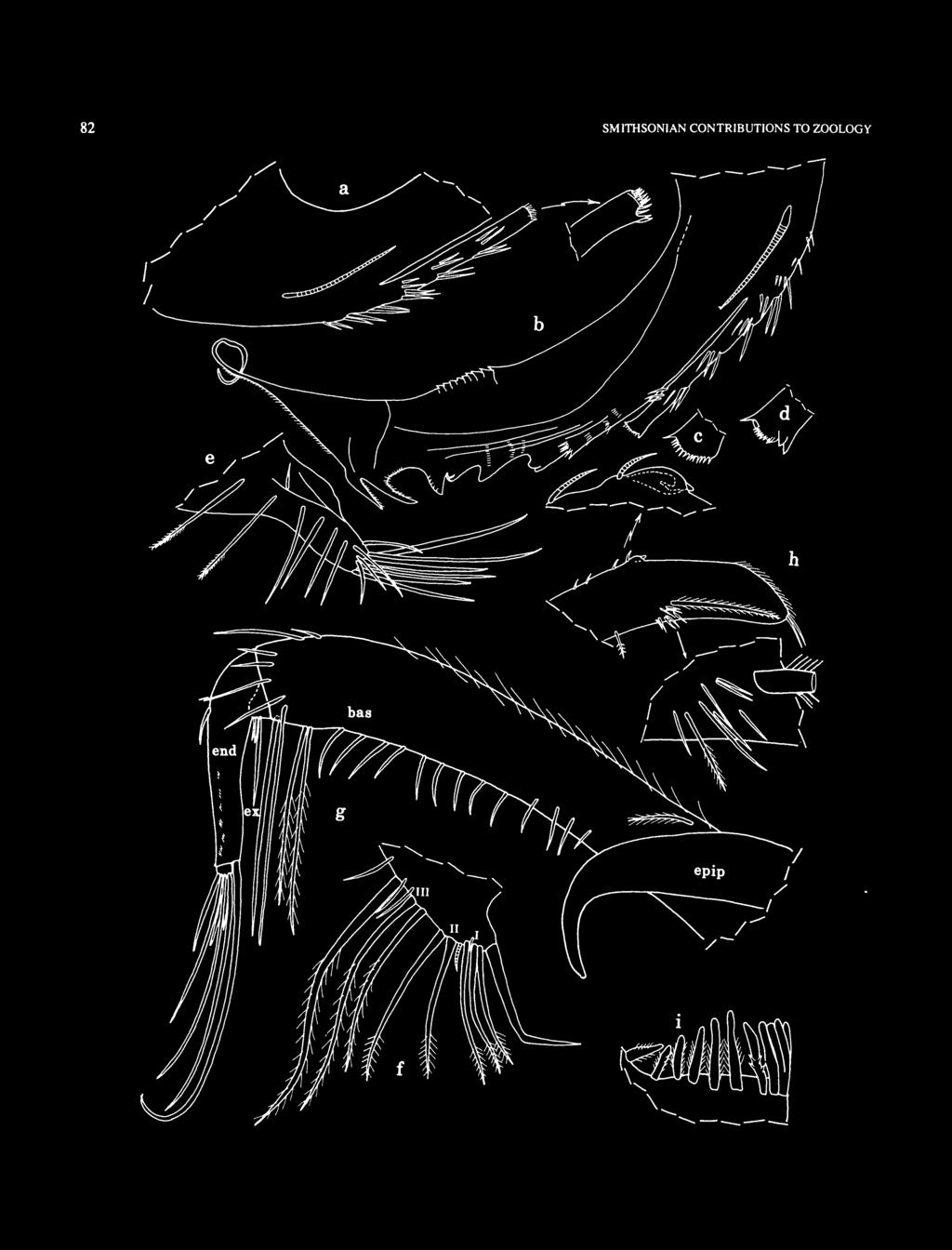
88 82 SMITHSONIAN CONTRIUTIONS TO ZOOLOGY

, mv; h, comb ofright5th limb, lv; i, part of comb at tip of 7th limb.")
89 NUMER FIGURE 59 (opposite page). Tetraleberis pix, new species, adult male, holotype, QM W20739: a, ventral branch coxale endite right mandible, mv; b, coxale endite left mandible, mv; c,d, lateral row of 13 minute spines and medial row of 7 minute spines, respectively, along terminal end of ventral branch of coxale endite shown in b; e, basale endite right mandible (spines of most bristles not shown), mv; /, endites right maxilla, mv; g, right maxilla (endites shown in/), mv; h, comb ofright5th limb, lv; i, part of comb at tip of 7th limb. bristle-bearing segments with 2 bristles, 1 on each side, very few with 2 bristles on one side. Terminus with opposing combs, each with about 20 teeth of various types (not all shown in Figure 59/). Furca (Figure 60c): Each lamella with 4 stout main claws, 1 slender bristle between claws 3 and 4 (closer to claw 4), and 8 or 9 additional slender bristles following claw 4; all claws with slender pointed tips, teeth of equal length forming medial and lateral rows along concave margin (some adjacent teeth with minute tooth between them), and hairs along convex margin. ristles ringed and with spines along margins except near pointed tip; space between claw 4 and following bristle wider onrightlamella than on left. Lamellae with medial spines at bases of main claws and along edge following bristles. Right lamella anterior to left lamella by width of base of claw 1 and with few hairs along anterior edge proximal to claw 1. (Not all spines, hairs, or teeth shown.) ellonci Organ (Figure 60e): Elongate, bare, with suture proximal to widened middle part, and tapering to rounded tip. Eyes: Medial eye bare, without dark pigment (Figure 60a 1 ). Lateral eye larger than medial eye, without dark pigment on preserved specimen, with 47 ommatidia (Figure 60/). Upper Lip (Figure 60g): Each lobe with long hairs (some anterior hairs slightly thicker, spine-like). Saddle between lobes also with some hairs spine-like. Hirsute lateral flap of lower lip present on each side of mouth. Genitalia (Figure 60/J): Copulatory organ comprising small lobes on each side of body anterior to furca. Posterior of ody (Figure 60/»): Hirsute. Gills: Well developed, typical for subfamily. Y-Sclerite (Figure 60A): Convex with dorsal socket near midlength. DESCRIPTION OF ADULT FEMALE (Figures 61, 62a-m). In lateral view, carapace smaller and more rounded than that of adult male (Figure 61a). Ornamentation: Anteroventral part of valve with few long bristles (not as many as in adult male, not shown), and posterior without vertical row of bristles as in male. Surface otherwise similar to that of adult male. Infold: Infold of rostrum with about 20 bristles between list and edge of incisur, and row of about 60 bristles on and dorsal to list, including row of small bristles near anterodorsal edge of valve (bristles not shown in Figure 61 b). In general, infold appears similar to that of adult male and typical of subfamily, but not all bristles counted. Vestment: Vestment in anterodorsal corner just proximal to inner margin of infold with numerous slender hair-like spines. Central Adductor Muscle Attachments: Similar to those of adult male. Carapace Size (length (L), height (H), in mm): USNM A, L = 3.5, H = 3.0; H = 85.7% of length. USNM D, 4 specimens: L = 3.9, H = 3.2, H = 82% of length; L = 4.1, H = 3.2, H = 78% of length; L = 3.9, H = 3.2, H = 82% of length; L = 3.8, H = 3.0, H = 79% of length. First Antenna: 1st joint with hairs along ventral margin; medial side with few hairs near midheight and with row of 8 small distal spines near ventral margin (Figure 61c); lateral side with group of about 12 long hair-like spines in distal dorsal comer and with row of about 10 short spines dorsal to midheight and parallel to dorsal margin. 2nd joint (Figure 61c,e): ventral margin with long hairs; dorsal margin with few long proximal hairs and 4 or 5 spinous ringed bristles; medial side with few long hairs near dorsal margin; lateral side with long hairs in distal ventral comer and with row of 5 distal bristles (Figure t\e). 3rd joint: short ventral margin with minute bristle (Figure 61/) (bristle missing from right limb of USNM A); long dorsal margin with 14 spinous ringed bristles and, between bases of bristles, long sclerites perpendicular to dorsal margin of joint (Figure 61/). 4th joint (bristles not shown in Figure 61/g): ventral margin with 5 ringed terminal bristles (4 short bare (about x li length of 5th joint) and 1 very long spinous); dorsal margin with 1 long spinous terminal bristle. 5th joint: sensory bristle with 4 or 5 short proximal filaments followed by longer slender filament and 9 long stout terminal filaments (Figure 61/J); dorsal margin with stout process at midlength (Figure 6\d,g); medial and lateral sides with scattered long hairs near dorsal margin distal to process. 6th joint with long spinous medial ringed bristle shorter than bristle of 5th joint (Figure 6\d). 7th joint (Figure 6\d,g): a-bristle claw-like; b-bristle reaching tip of bristle of 5th joint, with 8 marginal filaments each with terminal papilla; c -bristle about '/3 longer than b-bristle, with 13 marginal filaments. 8th joint (Figure 6ld,g (d- and e-bristles not shown)): d- and e-bristles reaching tip of bristle of 5th limb, bare with blunt tips, and both on small pedestal with minute lateral process; f-bristle bent dorsally, slightly longer than b-bristle, with 12 marginal filaments each with terminal papilla; g-bristle about same length as c-bristle, with 12 marginal filaments each with terminal papilla. Tips of stems of b-, c-, f-, and g-bristles each with minute papilla. Second Antenna: Protopodite with long hairs near ventral margin and on medial surface near ventral margin and with 1 short bare distal medial bristle. Endopodite 3-jointed (Figure 62a): 1st joint divided into 2 parts, with 1 small bristle on proximal part and 5 or 6 small bristles on distal part; 2nd joint elongate, bare; 3rd joint short with long terminal filament. Exopodite: 1st joint with minute straight terminal medial bristle; bristle of 2nd joint with closely spaced slender spines; bristles of joints 3-8 with natatory hairs and stout proximal
\ joints 2-8 with row of minute spines along distal edges and basal spines; basal spine of 2nd joint about x h length of joint; basal spines of joints 3-8 of similar length and longer")
 with slender ringed bristle proximal to base of ventral branch; ventral branch with 5 oblique rows of spines and truncate tip with 2 ventral teeth, FIGURE 60.")
90 84 SMITHSONIAN CONTRIUTIONS TO ZOOLOGY ventral spines (Figure 62 c); 9th joint with 5 bristles with natatory hairs (2 longest bristles (ventral) also with stout proximal spines) and 2 lateral spines (Figure 62 b)\ joints 2-8 with row of minute spines along distal edges and basal spines; basal spine of 2nd joint about x h length of joint; basal spines of joints 3-8 of similar length and longer than 9th joint (Figure 62 b). Mandible: Coxale endite (Figure 62d) with slender ringed bristle proximal to base of ventral branch; ventral branch with 5 oblique rows of spines and truncate tip with 2 ventral teeth, FIGURE 60. Tetraleberis pix, new species, adult male, holotype, QM W20739: a, endite and proximal ventral edge of basale left mandible, mv; b, left 6th limb, mv; c, copulatory organ and right furcal lamella (not all teeth and spines shown on claws), lv; d, medial eye; e, ellonci organ;/ left lateral eye from left side; g, upper and lower lips from right side; h, posterior of body from left side.


91 NUMER FIGURE 61. Tetraleberis pix, new species, ovigerous female, paratype, USNM A: a, complete specimen, length 3.5 mm; b, anterior left valve (bristles not shown), iv; c4. proximal and distal ends, respectively, right 1st antenna (bristles of 4th joint not shown, and not all terminal bristles shown), mv; e-g, left 1st antenna (bristles of 4th and 6th joints not shown), Iv; h, sensory bristle of 5th joint right 1st antenna, mv.

.")
92 86 medial row of about 16 minute spines (not all shown), and lateral row of similar spines (not shown); ventral margin of dorsal branch with 3 short triangular teeth followed by slightly longer tooth with curved distal edge with minute rounded teeth, and then 2 recurved teeth with spines along proximal edge and at tip; edge between distal recurved tooth and tip of branch with minute spines; tip with long terminal bristle with minute indistinct hairs; dorsal margin of branch with few minute serrations. asale endite with 7 terminal bristles, 13 triaenid bristles, and 4 dwarf bristles (Figure 62e). asale: ventral margin with 10 triaenid bristles (with 9-12 pairs of spines proximal to terminal pair, bristles similar to those of basale endite) followed by 2 long unequal bristles with long marginal spines; dorsal margin with 8 short or medium bare bristles followed by 1 longer bare bristle, 2 or 3 short bare bristles, and 2 very long terminal bristles with short spines; medial surface with many long hairs. Exopodite similar to that of adult male. 1st endopodial joint with 1 minute and 7 long ventral bristles (2 longest bristles with long spines, others either with short spines or bare). 2nd endopodial joint: ventral margin with bristles in 2 groups: subterminal group with 3 or 4 bristles with short spines, terminal group with 2 long stout bristles with short spines; dorsal margin of joint with numerous long and medium bristles; medial side near dorsal margin with abundant short bristles. 3rd endopodial joint with 3 stout bare claw-like bristles, 1 long lateral bristle with short spines, and 2 ventral medial bristles with short spines. Maxilla: Epipodite, endites, exopodite (Figure 62/), and endopodite similar to those of adult male; 7 short bare medial bristles near bases of bristles of endite III (not shown). asale (Figure 62/): dorsal margin spinous, with 3 short proximal bristles (anterior plumose) with bases on medial side, and 3 distal bristles (1 longer than others and with few hairs); ventral margin with 19 or 20 short bristles, 2 long distal bristles, and 1 long terminal bristle (with base lateral); medial side with 6 distal bristles (ventral bristle very long, others short). Fifth Limb (Figure 62g,h): Dorsal margin of comb with 5 small proximal bristles; lateral side with 1 stout spinous bristle and 8 small bristles near its base. Sixth Limb: Shape similar to that of adult male limb; with 5 small epipodial bristles. Anterior margin with fairly long bristle just dorsal to suture marking lower endite. Anterior margin dorsal to upper endite with 19 bristles in inner medial row, 18 bristles in outer medial row, and 25 smaller bristles along edge. Anterior margin between endites with 3 bristles in inner medial row, 3 bristles in outer medial row (not counting long bristle at lower suture), and 7 bristles along edge. About 20 slender bristles form single row just medial to edge between lower endite suture and anterior tip of skirt. Posterior end of skirt with 4 fairly broad plumose unringed bristles; ventral margin of skirt with about 90 spinous bristles. Summary: about 96 anterior bristles, 94 bristles on ventral margin and posterior comer of skirt, and 5 epipodial bristles; total of about 195 bristles. SMITHSONIAN CONTRIUTIONS TO ZOOLOGY Seventh Limb: Each limb with about 100 bristles, each bristle with up to 7 bells. Terminus with opposing combs similar to those of adult male, but teeth not counted. Furca (Figure 62/): Except for 4th main claw of left lamella, tips of main claws rounded (probably worn). Right lamella of USNM A with 8 bristles following 4th main claw on right lamella and 7 on left, furca otherwise similar to that of adult male. ellonci Organ (Figure 62j), Upper Lip (Figure 62/;, Posterior of ody (Figure 62m), Gills, and Y-Sclerite: Similar to those of adult male. Eyes: Medial eye similar to that of adult male (Figure 62 j). Lateral eye about 3 A length of eye of adult male, without dark pigment on preserved specimen, and with 41 ommatidia (Figure 62k). Genitalia: Not observed. Number of Eggs: USNM A with 73 eggs in marsupium; length of typical egg 0.39 mm. (Number of eggs highest reported for family.) COMPARISONS. Poulsen (1965:250) referred a female collected in the Coral Sea northeast of Australia to Cycloleberis brevis (Muller, 1890) (= T. brevis (Muller, 1890)). It is not clear whether it is an adult female or a juvenile because on p. 250 Poulsen described it as having embryos, but on p. 260 he stated that it is probably a juvenile (Kornicker, 1981b:142). Some morphological characters of T. pix and of the specimen Poulsen referred to T. brevis are compared in Table 4. The specimen is not considered to be conspecific with T. pix herein mainly because of differences in the coxale endite of the mandible (the tip of the ventral branch has three short teeth in T. brevis (Poulsen, 1965:247, fig. 83g) and has two teeth and numerous spines in T. pix; also, the bristle on the dorsal branch is set back from the tip on T. brevis and is terminal on T. pix (Figure 62d)), in the number of ventral bristles on the basale of the female maxilla (22 or 23 for T. pix compared to three (aberrant?) for T. brevis), and in the number of bristles on the female 6th limb (e.g., 94 ventral bristles on T. pix compared to 28 or 29 bristles on T. brevis). The maxilla of the specimen of T. brevis illustrated by Muller (1890, pi. 27: fig. 12) has 10 ventral bristles on the basale compared to 22 or 23 bristles for T. pix, and for this reason they are probably not conspecific. Komicker (1981b: 135) examined syntypes of T. brevis and described one FIGURE 62 (opposite page). Tetraleberis pix, new species, ovigerous female, paratype, USNM A: a, endopodite left 2nd antenna, mv; b. joints 7-9 exopodite left 2nd antenna (bristles of joints 7 and 8 not shown), lv; c, proximal part of bristle of 3rd joint of exopodite left 2nd antenna (ventral edge to bottom), lv; d, coxale endite right mandible, mv; e, basale endite right mandible, mv;/, basale and exopodite left maxilla, mv; g, comb of left 5th limb, lv; h, detail from g;i, left lamella of furca, lv; j, medial eye and ellonci organ; *, left lateral eye from left side; /, upper and lower lips from right side; m, posterior of body from left side. Tetraleberis maddocksae Kornicker, 1981, ovigerous female, paratype, USNM : n, tip of ventral branch of coxale endite of left mandible, mv.


93 NUMER
 from the Coral Sea identified by Poulsen (1965:245), and T. tanzania Kornicker, 1981b (adult or A-l). Data on T.")
. (- = absent, + = present; prox = proximal, dist = distal; nd = no data.) Character T. pix T. brevis T.")
94 88 SMITHSONIAN CONTRIUTIONS TO ZOOLOGY TALE 4. Comparison of the morphology of selected characters of females of Tetraleberis pix, USNM A, T. brevis (specimen may be juvenile) from the Coral Sea identified by Poulsen (1965:245), and T. tanzania Kornicker, 1981b (adult or A-l). Data on T. brevis from Poulsen (1965:241, table 14; 245) and Kornicker (1981b: 140), the latter in parentheses (the maxilla, 6th, and 7th limbs were not available to Kornicker and are probably not extant); data on T. tanzania from Kornicker (1981b: 153). (- = absent, + = present; prox = proximal, dist = distal; nd = no data.) Character T. pix T. brevis T. tanzania Carapace length (mm) Medial bristles on rostrum ristles of 1st antenna Lateral on 2nd joint Dorsal on 2nd joint Dorsal on 3rd joint Fifth joint bristle filaments, prox:dist ristles of 2nd antenna Endopodite, 1st joint Exopodite, 9th joint ristles of mandible asale endite asale ventral margin asale dorsal margin Endopodite, 1st joint ristles of maxilla asale dorsal, prox:dist asale, ventral Endopodite, 2nd joint Exopodite ristles of 5th limb Comb, dorsal margin Comb, short exopodial ristles of 6th limb Epipodite Skirt, ventral margin are space on skirt Furca, bristles : : (3) :7(4:8) 6 3(4) : (6) nd : :7 18 6* * Kornicker (1981b:156) reported 5 bristles but a reexamination of USNM , the female holotype, revealed 6 bristles (2 long, 2 medium, 2 short). that has many more bristles on the maxilla than are present on the specimen Miiller described. The maxilla of the specimen of T. brevis described by Kornicker (1981b: 136, fig. 47a) has proximal bristles on the dorsal margin of the basale compared to three bristles for T. pix, therefore, the specimens are not likely to be conspecific. ristles of joints three to eight of the exopodite of the second antenna of female T. pix bear stout proximal ventral spines, whereas bristles of female T. similis are without spines, and bristles of T. maddocksae have slender spines. The dorsal margin of the basale of the maxilla of female T. pix has three proximal and three distal bristles compared to six proximal and seven distal bristles for T. tanzania Kornicker, 1981b (Table 4), 24 proximal and eight distal bristles for T. similis (rady, 1902), and 13 proximal and six distal bristles for T. maddocksae Kornicker, 1981b. Tetraleberis pix and T. tanzania are very close; the greatest difference observed between them is in the endopodite of the male second antenna: T. tanzania has four bristles on the second joint, and on the third joint the slender reflexed part has its base proximally on a triangular process (Kornicker, 1981, fig. 54c); whereas, T. pix has nine bristles on the second joint, and on the third joint the slender reflexed part has its base terminally on a triangular process (Figure 58/). The tip of the ventral branch of the coxale endite of the mandible of T. maddocksae (Figure 62 n) is similar to that of T. pix (Figures 59c,d, 62d); the coxale endites of T. tanzania and T. similis are unknown (the endites often break off the mandible and remain in the mouth when the mandible is removed from the body, but they usually can be removed from the mouth for study). The anterior of the proximal bristles on the dorsal margin of the basale of the maxillae of both the male and female T. pix are plumose. A plumose bristle has not been reported previously in that position in other species of Tetraleberis; however, a reexamination of the maxillae of a male paratype (USNM
 of T. maddocksae (male unknown).")
 in reference to the 2nd joint of the 1st antenna of the female having 3 dorsal bristles. HOLOTYPE. Undissected adult female in alcohol, AM P39205. TYPE LOCALITY.")
95 NUMER ) and of the female holotype (USNM ) of T. tanzania revealed that the anterior bristle of the female bears hairs; hairs also were observed on that bristle on the female holotype (USNM ) of T. maddocksae (male unknown). This suggests that the anterior bristle in other species of the genus may have hairs that have been overlooked (the hairs are quite thin). Tetraleberis triplex, new species FIGURES 63,64 ETYMOLOGY. From the Latin triplex (threefold) in reference to the 2nd joint of the 1st antenna of the female having 3 dorsal bristles. HOLOTYPE. Undissected adult female in alcohol, AM P TYPE LOCALITY. Lizard Island, Australia, patch reef at northern entrance to lue Lagoon, sta QLD 300, depth 3 or 4 m, 1 Feb 1989, baited cone trap. PARATYPES Davies Reef: USNM , ovigerous female on slide and in alcohol. Lizard Island, lue Lagoon, sample 5: USNM , dissected adult female in alcohol. Lizard Island, patch reef, sta QLD 299: AM P39204, partly dissected adult female in alcohol, deposited in The Australian Museum. DISTRIUTION. lue Lagoon, Lizard Island, Australia (QLD-299, QLD-300, 1989, and sample 5, 1987). Davies Reef (lagoon floor), Central Great arrier Reef, Australia. DESCRIPTION OF ADULT FEMALE (Figures 63, 64). Carapace shape, ornamentation, infold, and vestment similar to those characters of T. pix, except with fewer hair-like bristles (not shown) in anteroventral quarter of shell outer surface (Figure 63a). Central Adductor Muscle Attachments (Figure 63b): Typical for subfamily. Carapace Size (length (L), height (H), in mm): AM P39205 (holotype), L = 3.3, H = 2.8. USNM , L = 3.04, H = USNM , L = 3.2, H = 2.5. AM P39204, L = 3.2, H = 2.5. First Antenna (Figure 63c-e): 1st joint: ventral margin with few hairs; medial side with 12 spines and long hairs forming row parallel and close to ventral margin, and distal hairs at midheight and in dorsal corner (Figure 63d); lateral side with numerous long distal hairs in dorsal corner (Figure 63c). 2nd joint: ventral margin with long hairs; medial side with long hairs near ventral margin and proximally near dorsal margin (Figure 63d); dorsal margin with 3 spinous bristles; lateral side with few long distal hairs in ventral corner and row of 4 slender distal bristles (Figure 63c). 3rd joint: short ventral margin with small bare ringed bristle (Figure 63c); long dorsal margin with 12 spinous bristles and with long sclerites perpendicular to dorsal margin between bases of bristles (outlines of sclerites dashed in Figure 63c); lateral side with 2 hairs near base of 3rd dorsal bristle. 4th joint: ventral margin with 5 ringed terminal bristles (4 short bare, about x li length of 5th joint, and 1 very long spinous); dorsal margin with 1 long spinous terminal bristle; medial side with few indistinct distal hairs in dorsal corner. 5th joint: sensory bristle with 6 or 7 short proximal filaments and 9 long stout terminal filaments (Figure 63e); dorsal margin with stout triangular process at midlength; medial side bare or with few long distal hairs in dorsal comer, lateral side bare. 6th joint with long spinous medial ringed bristle shorter than bristle of 5th joint (not shown). 7th joint: a-bristle claw-like; b-bristle not shown, reaching tip of terminal filaments of bristle of 5th joint, with 10 marginal filaments each with terminal papilla; c-bristle not shown, about l /3 longer than b-bristle, with 14 marginal filaments; minute lateral pore present proximal to base of c-bristle. 8th joint: d- and e-bristles same length as b-bristle, bare with blunt tips, and both on small pedestal with minute lateral pore; f-bristle bent dorsally, longer than b-bristle, with 11 or 12 marginal filaments; g-bristle not shown, same length as c-bristle, with about 13 marginal filaments. Second Antenna: Protopodite and exopodite (Figure 63g) similar to those of female T. pix. Endopodite 3-jointed (Figure 63/): 1st joint divided into 2 parts with 2 small bristles on proximal part and 6 small bristles on distal part; 2nd joint elongate, bare; 3rd joint short with long terminal filament. Mandible: Coxale endite similar to that of female T. pix (Figure 63h). asale endite with 7 terminal bristles, about 13 triaenid bristles, 6 dwarf bristles, and small round glandular process. asale: ventral margin with 10 triaenid bristles, followed by 3 long bristles with long spines, and 1 shorter bristle with short spines; dorsal margin with 5 or 6 short or medium bare bristles followed by 2 longer bare bristles, 2 or 3 short bare bristles, and 2 very long terminal bristles with few short spines; medial surface with many long hairs and 2 or 3 minute bristles at midlength near ventral margin. Exopodite similar to that of female T. pix. 1st endopodial joint with 1 minute bristle and 7 long ventral bristles (2 longest bristles with long spines, others either with short spines or bare). 2nd endopodial joint: ventral margin with bristles in 2 groups: subterminal group with 3 bristles and terminal group with 2 long stout bristles; dorsal margin of joint with numerous long and medium bristles; medial side near dorsal margin with abundant short bristles. ristles of 3rd endopodial joint similar to those of female T. pix. Maxilla (Figure 64a): Epipodite long and slender. Endites I and II similar to those of T. pix; endite III with 4 unequal bristles with spines along entire margin (distal bristle longer than other bristles of endites); 5 or 6 short slender bristles distal to bases of bristles of endite III. asale: dorsal margin spinous, with 14 proximal bristles with bases on medial side (distal bristle plumose, others bare) and 5 or 6 distal slender bare bristles (distal bristle longest); medial side with 5 or 6 distal bare bristles (ventral bristle very long, others short); ventral margin with 16 or 17 short bristles with indistinct spines, 2 long

, lv; d, proximal part right 1 st antenna, mv; e, sensory bristle of Sth joint right 1st antenna, mv;/, endopodite left 2nd antenna, mv; g,")
96 90 SMITHSONIAN CONTRIUTIONS TO ZOOLOGY FIGURE 63. Tetraleberis triplex, new species, ovigerous female, paratype, USNM : a. complete specimen showing 5 of 33 eggs, length 3.04 nun; b, central adductor muscle attachments of left valve (drawn with body removed), ov; c, left 1 st antenna (not all terminal bristles shown), lv; d, proximal part right 1 st antenna, mv; e, sensory bristle of Sth joint right 1st antenna, mv;/, endopodite left 2nd antenna, mv; g, joints 7-9 of exopodite right 2nd antenna (bristles of joints 7 and 9 not shown), lv; h, coxale endite left mandible, lv.


97 NUMER FIGURE 64. Tetraleberis triplex, new species, ovigerous female, paratype, USNM : a, right maxilla, mv; b, comb of right 5th limb, lv; c, left 6th limb (not all bristles shown), mv; d, left furcal lamella and claw 1 of right lamella; e. medial eye and ellonci organ;/, right lateral eye from right side; g, upper lip from left side; h, anterior of body, av.
 (see detail in Figure 64a).")
: Dorsal margin with 5 small proximal bristles; lateral side with 1 stout spinous bristle, 8 small bristles near its base, and 1 small spinous proximal bristle near ventral")
98 92 SMITHSONIAN CONTRIUTIONS TO ZOOLOGY spinous distal bristles, and 1 very long spinous terminal bristle (with base lateral); lateral side with 1 short proximal bristle at midheight (not shown). Exopodite consisting of poorly defined lobe with 3 slender bristles (longest about 2 /3 length of 1st endopodial joint) (see detail in Figure 64a). Endopodite: 1st joint without spines, with 1 short bare alpha-bristle and 1 long spinous beta-bristle; 2nd joint with 6 spinous bristles (3 long, 3 short or medium) (bristles not shown). Fifth Limb (Figure 64b): Dorsal margin with 5 small proximal bristles; lateral side with 1 stout spinous bristle, 8 small bristles near its base, and 1 small spinous proximal bristle near ventral edge. Sixth Limb (Figure 64c; not all bristles shown): With 4 or 5 small epipodial bristles. Anterior margin with fairly long bristle just dorsal to suture marking lower endite. Anterior margin dorsal to upper endite with 14 bristles in inner medial row, 22 bristles in outer medial row, and 24 smaller bristles along edge. Anterior margin between endites with 6 bristles in inner medial row, 1 bristle in outer medial row (not counting longer bristle at lower suture), and 7 bristles along edge. About 17 slender bristles form single row just medial to edge between lower endite suture and anterior tip of skirt. Posterior end of skirt with 4 fairly broad plumose unringed bristles; ventral margin of skirt with about 95 spinous bristles. Summary: about 92 anterior bristles, 99 bristles on ventral margin and posterior comer of skirt, and 4 epipodial bristles; total of about 195 bristles. Seventh Limb: Each limb with about 85 bristles, each with up to 8 bells. Terminus with opposing combs similar to those of T. pix, but teeth not counted. Furca (Figure 64c/): Each lamella with 4 stout main claws, 1 slender bristle between claws 3 and 4 (closer to claw 4), and 7 additional bristles; space between claw 4 and following bristle wider on right lamella than on left; main claws with rounded tips; teeth and spines of main claws similar to those of T. pix; bristles ringed (not shown) and with spines along margins except near pointed tip; right lamella anterior to left lamella by width of claw 1. ellonci Organ (Figure 64e), Upper Lip (Figure 64g,h), Posterior of ody, Gills, and Y-Sclerite: Similar to those of T. pix. Eyes: Medial eye bare, without dark pigment (Figure 64e). Lateral eye about same size as medial eye, without dark pigment and with about 37 ommatidia (Figure 64/). Genitalia: Not observed. Number of Eggs: USNM with 33 eggs in marsupium; length of typical egg 0.41 mm (position of 5 eggs within carapace shown in Figure 63a). COMPARISONS. The new species T. triplex differs from previously described species in the genus in having three rather than four to six bristles on the dorsal margin of the second joint of the female first antenna (three specimens examined). The maxilla of T. triplex has 14 proximal bristles on the dorsal margin of the basale compared to three on the female T. pix.
. Collected by P.")

99 Appendix 1 Station Data with Specimens Identified CALLIOPE RIVER and AUCKLAND CREEK, QUEENSLAND, AUSTRALIA Vicinity of Port Curtis and Gladstone, sampling depths 1-8 m at low water; salinity range generally /oo, but for short periods after heavy rain, short-term salinity reduction to 2O /oo can occur (for additional data on collecting area see Saenger et al. ( 1980). Collected by P. Saenger, Queensland Electricity Generating oard, risbane, Queensland, Australia, Scleroconcha pix: QM W20740, holotype, adult female; USNM A, ovigerous female; USNM , ovigerous female with female choniostomatid in marsupium; USNM , 3 ovigerous females; USNM , 9 adult females including 1 with parasite eggs and 1 or 2 with ostracode eggs; USNM , ovigerous female with female choniostomatid in marsupium. Sarsiella varix: QM W20747, holotype, ovigerous female; USNM A, ovigerous female; USNM C, 57 adult and ovigerous females; USNM 15797ID, 2 adult females and 2 ovigerous females. Eusarsiella phrix: QM W20743, holotype, instar IV female. Eusarsiella saengeri: QM W20744, holotype, ovigerous female; USNM A, adult female; USNM A, adult female; USNM , ovigerous female. Eusarsiella tryx: QM W20745, holotype, ovigerous female; USNM , ovigerous female; USNM , ovigerous female; USNM C, females and juveniles. Eusarsiella vernix: QM W20746, holotype, adult female with choniostomatid parasites; USNM A, ovigerous female; USNM , 2 ovigerous females and 1 adult female; USNM C, adult female with choniostomatid parasites; USNM , adult female with choniostomatid parasites and 1 adult female. Eurypylus rex: QM W20742, holotype, ovigerous female. Asteropterygion species indeterminate: USNM , 1 juvenile (not mentioned in text). Tetraleberis pix: QM W20739, holotype, adult male; USNM A, ovigerous female; USNM , 4 adult females; USNM C, adult male; USNM D, 32 adult females and juveniles. DARWIN, NORTHERN TERRITORY, AUSTRALIA Intertidal washings of algae and substrate, from stations JL Darwin 302, 304, and 305; some samples combined. Sta 302: Channel Island, 20 Aug Sta 304 and sta 305: East Point, 22 Aug Collected by J.L. arnard, Smithsonian Institution. Sta 302 and sta 305 Rutiderma dux: AM P45375, holotype, adult female; USNM , ovigerous female; USNM , 21 specimens. Rutiderma sagax: AM P45365, holotype, adult male. Sta 304 Rutiderma dux: USNM , A-l male. Sarsiella pugnax: AM P45369, holotype, ovigerous female. Eurypylus darwinensis: AM P45366, holotype, ovigerous female; AM P45368, adult male. DAVIES REEF, CENTRAL GREAT ARRIER REEF, QUEENSLAND, AUSTRALIA Collected at lagoon floor by John Carleton, Australian Institute of Marine Science, Cape Ferguson, Townsville M.C., Queensland, Australia, in 1989 or Tetraleberis triplex: USNM , ovigerous female. GULF of CARPENTARIA, QUEENSLAND, AUSTRALIA Coastal area in vicinity of Weipa, sampling depth 10 m. Collected by Peter C. Rothlisberg, Division of Fisheries and Oceanography, North-Eastern Regional Laboratory, CSIRO, Cleveland, Queensland, Australia, in Cypridinodes rumex: AM P45374, holotype, ovigerous female; USNM , ovigerous female; USNM , adult male; USNM A., 2 ovigerous 93

100 94 SMITHSONIAN CONTRIUTIONS TO ZOOLOGY females; USNM C, adult male; USNM D, 6 adult females and juveniles. Cypridinodes pix: AM P45367, holotype, ovigerous female; USNM , adult male; USNM , ovigerous female; AM P45373, 2 adult females and 1 specimen. Asteropterygion climax: AM P45370, holotype, A-l male; USNM , A-l male; USNM , adult male; USNM , 3 juveniles. LIZARD ISLAND, GREAT ARRIER REEF, QUEENSLAND, AUSTRALIA Plankton tow, lagoon, 2000 hours. Collected by Peter N. Slattery, Moss Marine Laboratory, Moss Island, California, 24 Sep Rutiderma dux: USNM , 1 juvenile. West of Lizard Island Research Station, off Casuarina each, m off shore of sandy beach, about 1.5 m deep, silty sand, Collected by Anne C. Cohen, Smithsonian Institution, 27 May Sta AC-LI-2 Rutiderma tryx: QM W20741, holotype, ovigerous female; USNM , ovigerous female; USNM , ovigerous female. Lagoon south of Lizard Island, between Lizard Island and Palfrey Island, off sand beach with 1 palm tree, about m from shore. In Thalassia bed (not dense) in very silty sand, about 2 m deep, Collected by Anne C. Cohen, Smithsonian Institution, 28 May Sta AC-LI-6 Rutiderma dux: USNM , 1 adult female; USNM , 1 ovigerous female. lue Lagoon, central part, sample 5. Collected by J.K. Lowry, Division of Invertebrate Zoology, The Australian Museum, Sydney South, NSW, Australia, in Tetraleberis triplex: USNM , adult female. Patch reef at northern entrance to lue Lagoon. aited traps (cones with small hole at top) on sandy lagoon floor having patches of blue-green algae. Traps set along 100 m transect beginning at 5 ft depth and continuing in a southeasterly direction (120 ) to 11 ft depth. Traps set , retrieved , 31 Jan Feb Collected by J.K. Lowry and S.J. Keable, Division of Invertebrate Zoology, Australian Museum, Sydney South, NSW, Australia. Sta QLD-299, trap 9/10 Tetraleberis triplex: AM P39204, adult female. Sta QLD-300, trap 10/10 Tetraleberis triplex: AM P39205, holotype, adult female. PALFREY ISLAND, LIZARD ISLAND GROUP, GREAT ARRIER REEF, QUEENSLAND, AUSTRALIA Transect across sand flat from shore to reef; stations 1-3 fairly close together and above mean datum; stations 4-8 extending 150 m across sand flat; station 8 at far edge of flat adjacent to a coral head at edge of coral reef; stations 4-8 exposed probably only at extreme low tides, twice a year. Sand fine to medium fine, with small ripple marks but not exposed to much wave action; tube-dwelling polychaetes and other more-or-less sedentary animals fairly abundant; 5 large cores (each m 2 ) taken from each of 8 stations (ostracodes present in only 5 outer stations); cores sieved through 500 jim mesh; specimens from each station consolidated. Collected by Peter N. Slattery, Moss Marine Laboratory, Moss Island, California, during Sep-Oct Sta 5 Rutiderma dux: USNM , adult male; AM P45376, adult male. Sta 8 Rutiderma dux: USNM , adult male.
 paratype, Lizard Island, sta QLD-299, AM P39204 (1 adult female) CYPRIDINIDAE Cypridinodes pix holotype, Gulf of Carpentaria, AM P45367 (1")
 paratype, Palfrey Island, sta 5, AM P45376 (1 adult male) Rutiderma sagax holotype, Darwin, sta 302 and 305, AM P45365 (1 adult male)")
101 Appendix 2 Type Specimens in Australian Museums The Australian Museum CYLINDROLEERIDIDAE Asteropterygion climax holotype, Gulf of Carpentaria, AM P45370 (1 A-l male) Tetraleberis triplex holotype, Lizard Island, sta QLD-300, AM P39205 (1 adult female) paratype, Lizard Island, sta QLD-299, AM P39204 (1 adult female) CYPRIDINIDAE Cypridinodes pix holotype, Gulf of Carpentaria, AM P45367 (1 ovigerous female) paratypes, Gulf of Carpentaria, AM P45373 (2 adult females and 1 specimen) Cypridinodes rumex holotype, Gulf of Carpentaria, AM P45374 (1 ovigerous female) RUTIDERMATIDAE Rutiderma dux holotype, Darwin, sta 302 and 305, AM P45375 (1 adult female) paratype, Palfrey Island, sta 5, AM P45376 (1 adult male) Rutiderma sagax holotype, Darwin, sta 302 and 305, AM P45365 (1 adult male) SARSIELLIDAE Eurypylus darwinensis holotype, Darwin, sta 304, AM P45366 (1 ovigerous female) paratype, Darwin, sta 304, AM P45368 (1 adult male) Sarsiella pugnax holotype, Darwin, sta 304, AM P45369 (1 ovigerous female) Queensland Museum CYLINDROLEERIDIDAE Tetraleberis pix holotype, Calliope River area, QM W20739 (1 adult male) PHILOMEDIDAE Scleroconcha pix holotype, Calliope River area, QM W20740 (1 adult female) RUTIDERMATIDAE Rutiderma tryx holotype, Lizard Island, sta AC-LI-2, QM W20741 (1 ovigerous female) SARSIELLIDAE Eurypylus rex holotype, Calliope River area, QM W20742 (1 ovigerous female) Eusarsiella phrix holotype, Calliope River area, QM W20743 (1 instar IV female) Eusarsiella saengeri holotype, Calliope River area, QM W20744 (1 ovigerous female) Eusarsiella tryx holotype, Calliope River area, QM W20745 (1 ovigerous female) Eusarsiella vernix holotype, Calliope River area, QM W20746 (1 adult female) Sarsiella varix holotype, Calliope River area, QM W20747 (1 ovigerous female) 95

, 1(3): 1-184. 1898. On New or Imperfectly-known Species of Ostracoda, Chiefly from New Zealand.")
102 Literature Cited aird, W The Natural History of the ritish Entomostraca. 364 pages, 36 plates. London: Printed for the Ray Society. rady, G.S On New or Imperfectly-known Species of Marine Ostracoda Transactions of the Zoological Society of London, 5: , plates [Preprint 1865.] Entomostraces. In A.G.L. de Folin and L. PeYier, Les fonds de la Mer, 1 (supplement): Report on the Ostracoda Dredged by H.M.S. Challenger during the Years In Report on the Scientific Results of the Voyage ofhms. Challenger (Zoology), 1(3): On New or Imperfectly-known Species of Ostracoda, Chiefly from New Zealand. Transactions of the Royal Society of Edinburgh, 35(2)14: ,9 figures, 4 plates On New or Imperfectly-known Ostracoda, Chiefly from a Collection in the Zoological Museum, Copenhagen. Transactions of the Zoological Society of London, 16(4): , plates rady, G.S., and A.M. Norman A Monograph of the Marine and Fresh Water Ostracoda of the North Atlantic and of Northwestern Europe. The Scientific Transactions of the Royal Dublin Society, series 2, 5: , plates Chavtur, V.G [New Species of Ostracods (Sarsiellidae) from the South of Vietnam.] Proceedings, Institute of Marine iology, Far East Science Center of the USSR Academy of Sciences (Vladivostok), pages ,5 figures. [In Russian.] Cohen, Anne C Rearing and Postembryonic Development of the Myodocopid Ostracode Skogsbergia lerneri from Coral Reefs of elize and the ahamas. Journal of Crustacean iology, 3(2): Cohen, Anne C, and Louis S. Kornicker Taxonomic Indexes to Ostracoda (Suborder Myodocopina) in Skogsberg (1920) and Poulsen (1962,1965). Smithsonian Contributions to Zoology, 204: 29 pages Catalog of the Rutidermatidae (Crustacea: Ostracoda). Smithsonian Contributions to Zoology, 449: 11 pages. Cushman, J.A Marine Ostracoda of Vineyard Sound and Adjacent Waters. oston Society of Natural History, Proceedings, 32(10): , plates Darby, D.G Ecology and Taxonomy of Ostracoda in the Vicinity of Sapelo Island, Georgia. In R.V. Kesling. editor. Four Reports of Ostracod Investigations, 2:1-77, 11 figures, 33 plates. Ann Arbor: University of Michigan. Graf.H Die Cypridinidae des Roten Meeres. Akademie der Wissenschaften in Wien, Mathematisch-Naturwessenchafliche Klasse; Denkschriften, 102: Granata,L Nuove specie di Ostracodi. ullettino delta Societa Entomologica Italiana, 46: Hall. S. Jane Four New Species of Myodocopine Ostracodes (Sarsiellidae) from Lizard Island, North Queensland. Journal of Crustacean iology, 5(3):5OO-522. figures New Species of Sarsiella and Anscottiella (Ostracoda: Myodocopina) from Lizard Island, North Queensland. Journal of Crustacean iology, 7(4): , figures Harding, J.P Myodocopan Ostracods from the Gills and Nostrils of Fishes. In Harold arnes, editor. Some Contemporary Studies in Marine Science, pages , 20 figures. London: George Allen and Unwin, Ltd. Hiruta, Shinlchi A New Species of the Genus Sarsiella Norman from Hokkaido, with Reference to the Larval Stages (Ostracoda: Myodocopina). Journal of the Faculty of Science, Hokkaido University, series VI (Zoology), 21(l): Kajiyama, E [The Ostracoda of Misaki, Part 2.] Dobutsugakuzasshi, 24: , plate 9. [In Japanese.] Kornicker, L.S Ecology and Taxonomy of Recent Marine Ostracodes in the imini Area, Great ahama ank. Publications of the Institute of Marine Science (The University of Texas). 5: figures, 1 map, 4 tables The Myodocopid Ostracod Familes Philomedidae and Pseudophilomedidae (New Family). Proceedings of the United States National Museum, 120:1-35,12 figures, 1 plate, 2 tables Morphology, Ontogeny, and Intraspecific Variation of Spinacopia, a New Genus of Myodocopid Ostracod (Sarsiellidae). Smithsonian Contributions to Zoology, 8: 50 pages, 26 figures, 6 plates, 7 tables Ostracoda (Myodocopina) from the Peru-Chile Trench and the Antarctic Ocean. Smithsonian Contributions to Zoology, 32: 42 pages, 25 figures, 7 tables Revision of the Cypridinacea of the Gulf of Naples (Ostracoda). Smithsonian Contributions to Zoology, 178: 64 pages, 26 figures, 2 tables Antarctic Ostracoda (Myodocopina), Parts 1 and 2. Smithsonian Contributions to Zoology, 163: 720 pages, 432 figures, 9 plates, 21 tables enthic Marine Cypridinacea from Hawaii (Ostracoda). Smithsonian Contributions to Zoology, 231: 24 pages, 19 figures, 1 table The Marine Fauna of New Zealand: enthic Ostracoda (Suborder Myodocopina). New Zealand Oceanographic Institute Memoir, 82: 58 pages, 13 figures, 28 plates, 3 tables. 1981a. enthic Marine Cypridinoidea from ermuda (Ostracoda). Smithsonian Contributions to Zoology, 331: 15 pages, 10 figures. 1981b. Revision, Distribution, Ecology, and Ontogeny of the Ostracode Subfamily Cyclasteropinae (Myodocopina: Cylindroleberididae). Smithsonian Contributions to Zoology, 319: 548 pages, 174 figures, 185 plates, 23 tables Alternochelata lizardensis, a New Species of Myodocopine Ostracode from the Great arrier Reef of Australia (Rutidermatidae). Proceedings of the iological Society of Washington, 95(4): ,6 figures Harbansus slatteryi, a New Species of Myodocopine Ostracode from the Great arrier Reef of Australia (Philomedidae). Proceedings of the iological Society of Washington, 96(1): , 4 figures Sexual Dimorphism, Ontogeny, and Functional Morphology of Rutiderma hartmanni Poulsen, 1965 (Crustacea: Ostracoda). Smithsonian Contributions to Zoology, 408: 28 pages, 20 figures. 96


103 NUMER a. Sarsiellidae of the Western Atlantic and Northern Gulf of Mexico, and Revision of the Sarsiellinae (Ostracoda: Myodocopina). Smithsonian Contributions to Zoology, 415: 217 pages, 113 figures, 34 plates, 7 tables. 1986b. Redescription of Sheina orri Harding, 1966, a Myodocopid Ostracode Collected on Fishes off Queensland, Australia. Proceedings of the iological Society of Washington, 99(4): , figures 1, a. Myodocopid Ostracoda of the eaufort Sea, Arctic Ocean. Smithsonian Contributions to Zoology, 450: 40 pages, 19 figures, 3 tables. 1988b. Myodocopine Ostracoda of the Alaskan Continental Shelf. Proceedings of the iological Society of Washington, 10(3): ,7 figures Myodocopid Ostracoda of Enewetak and ikini Atolls. Smithsonian Contributions to Zoology, 505: 140 pages, 71 figures Myodocopid Ostracoda of the enthe'di Expedition, 1977, to the NE Mozambique Channel, Indian Ocean. Smithsonian Contributions to Zoology, 531: 243 pages, 109 figures, 4 tables Ostracoda (Myodocopina) of the SE Australian Continental Slope, Part 1. Smithsonian Contributions to Zoology, 553: 200 pages, 111 figures. In press. Five New Eulittoral Sarsiellidae from Western Australia. Mitteilungen aus dem Hamburgischen Zoologischen Museum und Institut. Kornicker, Louis S., and Marcia owen Sarsiella ozotothrix, a New Species of Marine Ostracoda (Myodocopina) from the Atlantic and Gulf Coasts of North America. Proceedings of the iological Society of Washington, 88(46): , figures 1-3. Kornicker, Louis S., and Francisca Elena Caraion West African Myodocopid Ostracoda (Sarsiellidae, Rutiderrnatidae). Smithsonian Contributions to Zoology, 250: 110 pages, 59 figures, 33 plates, 1 table Nealella, a New Genus of Myodocopid Ostracoda (Sarsiellidae: Dantyinae). Smithsonian Contributions to Zoology, 309: 27 pages, 16 figures, 7 plates. Kornicker, Louis S., and Anne C. Cohen Dantyinae, a New Subfamily of Ostracoda (Myodocopina: Sarsiellidae). Proceedings of the iological Society of Washington, 91(2): ,5 figures, 7 plates. Kornicker, Louis S., and Thomas M. Iliffe Ostracoda (Myodocopina, Cladocopina, Halocypridina) from Anchialine Caves in ermuda. Smithsonian Contributions to Zoology, 475: 88 pages, 49 figures, 22 tables. Kornicker, Louis S., and C. Wise Sarsiella (Ostracoda) in Texas ays and Lagoons. Crustaceana, 4(l): Monod, Th Wissenschaftliche Mitteilungen, Uber drei indopazifische Cypridiniden und zwei in Ostracoden lebende Krebstiere. Zoologischer Anzeiger,9HM2):\-%. Miiller, G.W Neue Cypridiniden. Zoologische Jahrbucher. 5: , plates Die Ostracoden der Siboga-Expedition. In Uitkomsten op Zoologisch, otanisch, Oceanographischen on Geologische Gebeid versameld in Nederlandsch Oost-Indie, , 30: 40 pages, 9 plates. Leiden: E.J. rill. Norman, A.M Shetland Final Dredging Report, Part 11: On the Crustacea, Tunicata, Polyzoa, Echinodermata, Actinozoa, Hydrozoa, and Porifera. In Report of the Thirty-Eighth Meeting of the ritish Association for the Advancement of Science, pages Poore, G.C.., S.F. Rainer, R.. Spies, and E. Ward The Zoobenthos Program in Port Phillip ay, Fisheries Wildlife Paper Number 7, Victoria, Australia, pages Victoria: Fisheries and Wildlife Division. Poulsen, Erik M Ostracoda-Myodocopa, 1: Cypridiniformes-Cypridinidae. Dana Report, 57:1-414,181 figures. Copenhagen: Carlsberg Foundation Ostracoda-Myodocopa, 1: Cypridiniformes-Rutidermatidae, Sarsiellidae and Asteropidae. Dana Report, 65:1-484, 156 figures. Copenhagen: Carlsberg Foundation. Saenger, P., W. Stephenson, and J. Moverley The Estuarine Macrobenthos of the Calliope River and Auckland Creek, Queensland. Memoir Queensland Museum, 20(l): Sars, G.O Oversigt af Norges marine Ostracoder. Forhandlinger i Videnskabs- Selskabet i Christiania, 8: [Preprint, 1865.] Scott, Thomas Observations on Crustacea Collected during the Hydrographic Cruises, In Report on Fishery and Hydrographical Investigations in the North Sea and Adjacent Waters. Conducted for the Fishery oard for Scotland pages London: Darling and Son, Ltd. Skogsberg, T Studies on Marine Ostracods, I: Cypridinids, Halocyprids, and Polycopids. Zoologiska idragfrdn Uppsala, supplement 1: U.S. GOVERNMENT PRINTING OFFICE: /20004
104
105
106


, with 1 1 /4\" margins, submitted as ribbon copy (not carbon or xerox), in")
107 REQUIREMENTS FOR SMITHSONIAN SERIES PULICATION Manuscripts intended for series publication receive substantive review (conducted by their originating Smithsonian museums or offices) and are submitted to the Smithsonian Institution Press with Form SI-36, which must show the approval of the appropriate authority designated by the sponsoring organizational unit. Requests for special treatment use of color, foldouts, case-bound covers, etc. require, on the same form, the added approval of the sponsoring authority. Review of manuscripts and art by the Press for requirements of series format and style, completeness and clarity of copy, and arrangement of all material, as outlined below, will govern, within the judgment of the Press, acceptance or rejection of manuscripts and art. Copy must be prepared on typewriter or word processor, double-spaced, on one side of standard white bond paper (not erasable), with 1 1 /4" margins, submitted as ribbon copy (not carbon or xerox), in loose sheets (not stapled or bound), and accompanied by original art. Minimum acceptable length is 30 pages. Front matter (preceding the text) should include: title page with only title and author and no other information; abstract page with author, title, series, etc., following the established format; table of contents with indents reflecting the hierarchy of heads in the paper; also, foreword and/or preface, if appropriate. First page of text should carry the title and author at the top of the page; second page should have only the author's name and professional mailing address, to be used as an unnumbered footnote on the first page of printed text. Center heads of whatever level should be typed with initial caps of major words, with extra space above and below the head, but no other preparation (such as all caps or underline, except for the underline necessary for generic and specific epithets). Run-in paragraph heads should use period/dashes or colons as necessary. Tabulations within text (lists of data, often in parallel columns) can be typed on the text page where they occur, but they should not contain rules or numbered table captions. Formal tables (numbered, with captions, boxheads, stubs, rules) should be submitted as carefully typed, double-spaced copy separate from the text; they will be typeset unless otherwise requested. If camera-copy use is anticipated, do not draw rules on manuscript copy. Taxonomic keys in natural history papers should use the aligned-couplet form for zoology and may use the multi-level indent form for botany. If cross referencing is required between key and text, do not include page references within the key, but number the keyed-out taxa, using the same numbers with their corresponding heads in the text. Synonymy in zoology must use the short form (taxon, author, yearpage), with full reference at the end of the paper under "Literature Cited." For botany, the long form (taxon, author, abbreviated journal or book title, volume, page, year, with no reference in "Literature Cited") is optional. Text-reference system (author, yearpage used within the text, with full citation in "Literature Cited" at the end of the text) must be used in place of bibliographic footnotes in all Contributions Series and is strongly recommended in the Studies Series: "(Jones, 1910:122)" or "...Jones (1910:122)." If bibliographic footnotes are required, use the short form (author, brief title, page) with the full citation in the bibliography. Footnotes, when few in number, whether annotative or bibliographic, should be typed on separate sheets and inserted immediately after the text pages on which the references occur. Extensive notes must be gathered together and placed at the end of the text in a notes section. ibliography, depending upon use, is termed "Literature Cited," "References," or "ibliography." Spell out titles of books, articles, journals, and monographic series. For book and article titles use sentence-style capitalization according to the rules of the language employed (exception: capitalize all major words in English). For journal and series titles, capitalize the initial word and all subsequent words except articles, conjunctions, and prepositions. Transliterate languages that use a non-roman alphabet according to the Library of Congress system. Underline (for italics) titles of journals and series and titles of books that are not part of a series. Use the parentheses/colon system for volume (number):pagination: "10(2):5-9." For alignment and arrangement of elements, follow the format of recent publications in the series for which the manuscript is intended. Guidelines for preparing bibliography may be secured from Series Section, SI Press. Legends for illustrations must be submitted at the end of the manuscript, with as many legends typed, double-spaced, to a page as convenient. Illustrations must be submitted as original art (not copies) accompanying, but separate from, the manuscript. Guidelines for preparing art may be secured from the Series Section, SI Press. All types of illustrations (photographs, line drawings, maps, etc.) may be intermixed throughout the printed text. They should be termed Figures and should be numbered consecutively as they will appear in the monograph. If several illustrations are treated as components of a single composite figure, they should be designated by lowercase italic letters on the illustration; also, in the legend and in text references the italic letters (underlined in copy) should be used: "Figure 9b." Illustrations that are intended to follow the printed text may be termed Plates, and any components should be similarly lettered and referenced: "Plate 9b." Keys to any symbols within an illustation should appear on the art rather than in the legend. Some points of style: Do not use periods after such abbreviations as "mm, ft, USNM, NNE." Spell out numbers "one" through "nine" in expository text, but use digits in all other cases if possible. Use of the metric system of measurement is preferable; where use of the English system is unavoidable, supply metric equivalents in parentheses. Use the decimal system for precise measurements and relationships, common fractions for approximations. Use day/month/ year sequence for dates: "9 April 1976." For months in tabular listings or data sections, use three-letter abbreviations with no periods: "Jan, Mar, Jun," etc. Omit space between initials of a personal name: "J.. Jones." Arrange and paginate sequentially every sheet of manuscript in the following order: (1) title page, (2) abstract, (3) contents, (4) foreword and/or preface, (5) text, (6) appendices, (7) notes section, (8) glossary, (9) bibliography, (10) legends, (11) tables. Index copy may be submitted at page proof stage, but plans for an index should be indicated when the manuscript is submitted.

108 i
Ostracoda (Myodocopina) of the SE Australian Continental Slope, Part 2
 of the SE Australian Continental Slope, Part 2") Ostracoda (Myodocopina) of the SE Australian Continental Slope, Part 2 LOUIS S. KORNICKER m i SMITHSONIAN CONTRIBUTIONS TO ZOOLOGY NUMBER 562 SERIES PUBLICATIONS OF THE SMITHSONIAN INSTITUTION Emphasis
Ostracoda (Myodocopina) of the SE Australian Continental Slope, Part 2 LOUIS S. KORNICKER m i SMITHSONIAN CONTRIBUTIONS TO ZOOLOGY NUMBER 562 SERIES PUBLICATIONS OF THE SMITHSONIAN INSTITUTION Emphasis
Ostracoda (Myodocopina) of the Hawaiian Islands 1
 of the Hawaiian Islands 1") Ostracoda (Myodocopina) of the Hawaiian Islands 1 Louis S. Kornicker, 2,4 Elizabeth Harrison-Nelson, 2 and S. L. Coles 3 Abstract: Ostracoda (Myodocopina) from four of the Hawaiian Islands (Kaua i, Moloka
Ostracoda (Myodocopina) of the Hawaiian Islands 1 Louis S. Kornicker, 2,4 Elizabeth Harrison-Nelson, 2 and S. L. Coles 3 Abstract: Ostracoda (Myodocopina) from four of the Hawaiian Islands (Kaua i, Moloka
Nealella, a New Genus of Myodocopid Ostracoda (Sarsiellidae: Dantyinae)
") Nealella, a New Genus of Myodocopid Ostracoda (Sarsiellidae: Dantyinae) LOUIS S. KORNICKER and FRANCISCA ELENA CARAION SMITHSONIAN CONTRIBUTIONS TO ZOOLOGY NUMBER 309 SERIES PUBLICATIONS OF THE SMITHSONIAN
Nealella, a New Genus of Myodocopid Ostracoda (Sarsiellidae: Dantyinae) LOUIS S. KORNICKER and FRANCISCA ELENA CARAION SMITHSONIAN CONTRIBUTIONS TO ZOOLOGY NUMBER 309 SERIES PUBLICATIONS OF THE SMITHSONIAN
Cypridinidae of the Continental Shelves of Southeastern North America, the Northern Gulf of Mexico, and the West Indies (Ostracoda: Myodocopina)
") Cypridinidae of the Continental Shelves of Southeastern North America, the Northern Gulf of Mexico, and the West Indies (Ostracoda: Myodocopina) LOUIS S. fcornicker SMITHSONIAN CONTRIBUTIONS TO ZOOLOGY
Cypridinidae of the Continental Shelves of Southeastern North America, the Northern Gulf of Mexico, and the West Indies (Ostracoda: Myodocopina) LOUIS S. fcornicker SMITHSONIAN CONTRIBUTIONS TO ZOOLOGY
Ostracoda (Myodocopina from the Peru-Chile Trench and the Antarctic Ocean
 LOUIS S. KORNICK Ostracoda (Myodocopina from the Peru-Chile Trench and the Antarctic Ocean SMITHSONIAN CONTRIBUTIONS TO ZOOLOGY 1969 NUMBER 32 SERIAL PUBLICATIONS OF THE SMITHSONIAN INSTITUTION The emphasis
Ostracoda (Myodocopina) of Tulear Reef Complex, SW Madagascar
 of Tulear Reef Complex, SW Madagascar") Ostracoda (Myodocopina) of Tulear Reef Complex, SW Madagascar LOUIS S. KORNICKER and BERNARD A. THOMASSIN SMITHSONIAN CONTRIBUTIONS TO ZOOLOGY NUMBER 595 SERIES PUBLICATIONS OF THE SMITHSONIAN INSTITUTION
Ostracoda (Myodocopina) of Tulear Reef Complex, SW Madagascar LOUIS S. KORNICKER and BERNARD A. THOMASSIN SMITHSONIAN CONTRIBUTIONS TO ZOOLOGY NUMBER 595 SERIES PUBLICATIONS OF THE SMITHSONIAN INSTITUTION
Museum. Proceedings. National. the United States SMITHSONIAN INSTITUTION WASHINGTON, D.C. AND PSEUDOPHILOMEDIDAE (NEW FAMILY)
") Proceedings of the United States National Museum SMITHSONIAN INSTITUTION WASHINGTON, D.C. Volume 121 1967 Number 3580 THE MYODOCOPID OSTRACOD FAMILIES PHILOMEDIDAE AND PSEUDOPHILOMEDIDAE (NEW FAMILY) By
Proceedings of the United States National Museum SMITHSONIAN INSTITUTION WASHINGTON, D.C. Volume 121 1967 Number 3580 THE MYODOCOPID OSTRACOD FAMILIES PHILOMEDIDAE AND PSEUDOPHILOMEDIDAE (NEW FAMILY) By
A NEW AUSTROSQUILLA (STOMATOPODA) FROM THE
 FROM THE") A NEW AUSTROSQUILLA (STOMATOPODA) FROM THE MARQUESAS ISLANDS BY ALAIN MICHEL Centre O.R.S.T.O.M., Noumea, New Caledonia and RAYMOND B. MANNING Smithsonian Institution, Washington, U.S.A. The At s,tstrosqzlilla
A NEW AUSTROSQUILLA (STOMATOPODA) FROM THE MARQUESAS ISLANDS BY ALAIN MICHEL Centre O.R.S.T.O.M., Noumea, New Caledonia and RAYMOND B. MANNING Smithsonian Institution, Washington, U.S.A. The At s,tstrosqzlilla
BREVIORA LEUCOLEPIDOPA SUNDA GEN. NOV., SP. NOV. (DECAPODA: ALBUNEIDAE), A NEW INDO-PACIFIC SAND CRAB. Ian E. Efford 1
, A NEW INDO-PACIFIC SAND CRAB. Ian E. Efford 1") ac lc BREVIORA CAMBRIDGE, MASS. 30 APRIL, 1969 NUMBER 318 LEUCOLEPIDOPA SUNDA GEN. NOV., SP. NOV. (DECAPODA: ALBUNEIDAE), A NEW INDO-PACIFIC SAND CRAB Ian E. Efford 1 ABSTRACT. Leucolepidopa gen. nov.
ac lc BREVIORA CAMBRIDGE, MASS. 30 APRIL, 1969 NUMBER 318 LEUCOLEPIDOPA SUNDA GEN. NOV., SP. NOV. (DECAPODA: ALBUNEIDAE), A NEW INDO-PACIFIC SAND CRAB Ian E. Efford 1 ABSTRACT. Leucolepidopa gen. nov.
UPOGEBIA LINCOLNI SP. NOV. (DECAPODA, THALASSINIDEA, UPOGEBIIDAE) FROM JAVA, INDONESIA
 FROM JAVA, INDONESIA") NOTES AND NEWS UPOGEBIA LINCOLNI SP. NOV. (DECAPODA, THALASSINIDEA, UPOGEBIIDAE) FROM JAVA, INDONESIA BY NGUYEN NGOC-HO i) Faculty of Science, University of Saigon, Vietnam Among material recently collected
NOTES AND NEWS UPOGEBIA LINCOLNI SP. NOV. (DECAPODA, THALASSINIDEA, UPOGEBIIDAE) FROM JAVA, INDONESIA BY NGUYEN NGOC-HO i) Faculty of Science, University of Saigon, Vietnam Among material recently collected
PARAKRITHELLA PSEUDADONTA (HANAI, 1 THE INLAND SEA, JAPAN (OSTRACODA)
") Title PARAKRITHELLA PSEUDADONTA (HANAI, 1 THE INLAND SEA, JAPAN (OSTRACODA) Author(s) Okubo, Ichiro Citation PUBLICATIONS OF THE SETO MARINE BIO LABORATORY (1976), 23(1-2): 99-104 Issue Date 1976-07-31
Title PARAKRITHELLA PSEUDADONTA (HANAI, 1 THE INLAND SEA, JAPAN (OSTRACODA) Author(s) Okubo, Ichiro Citation PUBLICATIONS OF THE SETO MARINE BIO LABORATORY (1976), 23(1-2): 99-104 Issue Date 1976-07-31
NAUSHONIA PAN AMEN SIS, NEW SPECIES (DECAPODA: THALASSINIDEA: LAOMEDIIDAE) FROM THE PACIFIC COAST OF PANAMA, WITH NOTES ON THE GENUS
 FROM THE PACIFIC COAST OF PANAMA, WITH NOTES ON THE GENUS") 5 October 1982 PROC. BIOL. SOC. WASH. 95(3), 1982, pp. 478-483 NAUSHONIA PAN AMEN SIS, NEW SPECIES (DECAPODA: THALASSINIDEA: LAOMEDIIDAE) FROM THE PACIFIC COAST OF PANAMA, WITH NOTES ON THE GENUS Joel
5 October 1982 PROC. BIOL. SOC. WASH. 95(3), 1982, pp. 478-483 NAUSHONIA PAN AMEN SIS, NEW SPECIES (DECAPODA: THALASSINIDEA: LAOMEDIIDAE) FROM THE PACIFIC COAST OF PANAMA, WITH NOTES ON THE GENUS Joel
Vol. XIV, No. 1, March, The Larva and Pupa of Brontispa namorikia Maulik (Coleoptera: Chrysomelidae: Hispinae) By S.
 By S.") Vol. XIV, No. 1, March, 1950 167 The Larva and Pupa of Brontispa namorikia Maulik (Coleoptera: Chrysomelidae: Hispinae) By S. MAULIK BRITISH MUSEUM (NATURAL HISTORY) (Presented by Mr. Van Zwaluwenburg
Vol. XIV, No. 1, March, 1950 167 The Larva and Pupa of Brontispa namorikia Maulik (Coleoptera: Chrysomelidae: Hispinae) By S. MAULIK BRITISH MUSEUM (NATURAL HISTORY) (Presented by Mr. Van Zwaluwenburg
Harbansus, a New Genus of Marine Ostracoda, and a Revision of the Philomedidae (Myodocopina
 Harbansus, a New Genus of Marine Ostracoda, and a Revision of the Philomedidae (Myodocopina LOUIS S. KORNIGKER SMITHSONIAN CONTRIBUTIONS TO ZOOLOGY NUMBER 260 SERIES PUBLICATIONS OF THE SMITHSONIAN INSTITUTION
Beaufortia. (Rathke) ZOOLOGICAL MUSEUM - AMSTERDAM. July. Three new commensal Ostracods from Limnoria lignorum
 ZOOLOGICAL MUSEUM - AMSTERDAM. July. Three new commensal Ostracods from Limnoria lignorum") Beaufortia SERIES OF MISCELLANEOUS PUBLICATIONS ZOOLOGICAL MUSEUM - AMSTERDAM No. 34 Volume 4 July 30, 1953 Three new commensal Ostracods from Limnoria lignorum (Rathke) by A.P.C. de Vos (Zoological Museum,
Beaufortia SERIES OF MISCELLANEOUS PUBLICATIONS ZOOLOGICAL MUSEUM - AMSTERDAM No. 34 Volume 4 July 30, 1953 Three new commensal Ostracods from Limnoria lignorum (Rathke) by A.P.C. de Vos (Zoological Museum,
STELLICOMES PAMBANENSIS, A NEW CYCLOPOID COPEPOD PARASITIC ON STARFISH
 /. Mar. biol. Ass. ndia, 964, 6 (): 89-93 STELLCOMES PAMBANENSS, A NEW CYCLOPOD COPEPOD PARASTC ON STARFSH By C. A. PADMANABHA RAO* Central Marine Fisheries Research nstitute, Mandapam Camp THE siphonostomatous
Reprinted from: CRUSTACEANA, Vol. 32, Part 2, 1977 LEIDEN E. J. BRILL
 Reprinted from: CRUSTACEANA, Vol. 32, Part 2, 1977 LEIDEN E. J. BRILL NOTES AND NEWS 207 ALPHE0PS1S SHEARMII (ALCOCK & ANDERSON): A NEW COMBINATION WITH A REDESCRIPTION OF THE HOLOTYPE (DECAPODA, ALPHEIDAE)
A DESCRIPTION OF CALLIANASSA MARTENSI MIERS, 1884 (DECAPODA, THALASSINIDEA) AND ITS OCCURRENCE IN THE NORTHERN ARABIAN SEA
 AND ITS OCCURRENCE IN THE NORTHERN ARABIAN SEA") Crustaceana 26 (3), 1974- E. J. BiiU, Leide A DESCRIPTION OF CALLIANASSA MARTENSI MIERS, 1884 (DECAPODA, THALASSINIDEA) AND ITS OCCURRENCE IN THE NORTHERN ARABIAN SEA BY NASIMA M. TIRMIZI Invertebrate
Crustaceana 26 (3), 1974- E. J. BiiU, Leide A DESCRIPTION OF CALLIANASSA MARTENSI MIERS, 1884 (DECAPODA, THALASSINIDEA) AND ITS OCCURRENCE IN THE NORTHERN ARABIAN SEA BY NASIMA M. TIRMIZI Invertebrate
(CRUSTACEA: ISOPODA: ONISCIDEA)
") 31 October 1990 Memoirs of the Museum of Victoria 51: 93-97 (1990) ISSN 0814-1827 https://doi.org/10.24199/j.mmv.1990.51.06 TYLOS BILOBUS SP. NOV., A SECOND AUSTRALIAN SPECIES OF TYLIDAE (CRUSTACEA: ISOPODA:
31 October 1990 Memoirs of the Museum of Victoria 51: 93-97 (1990) ISSN 0814-1827 https://doi.org/10.24199/j.mmv.1990.51.06 TYLOS BILOBUS SP. NOV., A SECOND AUSTRALIAN SPECIES OF TYLIDAE (CRUSTACEA: ISOPODA:
National Museum ^^gt^
 Proceedings of the United States National Museum ^^gt^ SMITHSONIAN INSTITUTION WASHINGTON, D.C. Volume 123 1967 Number 3613 Two New Crustaceans: The Parasitic Copepod Sphaeronellopsis monothrix (Choniostomatidae)
MUNIDOPSIS ALBATROSSAB, A NEW SPECIES OF DEEP-SEA GALATHEIDAE (DECAPODA, ANOMURA) FROM THE EASTERN PACIFIC OCEAN
 FROM THE EASTERN PACIFIC OCEAN") MUNIDOPSIS ALBATROSSAB, A NEW SPECIES OF DEEP-SEA GALATHEIDAE (DECAPODA, ANOMURA) FROM THE EASTERN PACIFIC OCEAN BY WILLIS E. PEQUEGNAT and LINDA H. PEQUEGNAT Department of Oceanography, Texas A & M University,
MUNIDOPSIS ALBATROSSAB, A NEW SPECIES OF DEEP-SEA GALATHEIDAE (DECAPODA, ANOMURA) FROM THE EASTERN PACIFIC OCEAN BY WILLIS E. PEQUEGNAT and LINDA H. PEQUEGNAT Department of Oceanography, Texas A & M University,
A DESCRIPTION OF THE LABORATORY-REARED FIRST AND SECOND ZOEAE OF PORTUNUS X At IT US it (STIMPSON) (BRACHYURA, DECAPODA)
 (BRACHYURA, DECAPODA)") REPRINT FROM Calif. Fish and Game, 60(2) : 74-78. 1974. A DESCRIPTION OF THE LABORATORY-REARED FIRST AND SECOND ZOEAE OF PORTUNUS X At IT US it (STIMPSON) (BRACHYURA, DECAPODA) J. R. RAYMOND ALLY & r*
REPRINT FROM Calif. Fish and Game, 60(2) : 74-78. 1974. A DESCRIPTION OF THE LABORATORY-REARED FIRST AND SECOND ZOEAE OF PORTUNUS X At IT US it (STIMPSON) (BRACHYURA, DECAPODA) J. R. RAYMOND ALLY & r*
Ostracoda (Myodocopina, Cladocopina, Halocypridina) Mainly from Anchialine Caves in Bermuda
 Mainly from Anchialine Caves in Bermuda") Ostracoda (Myodocopina, Cladocopina, Halocypridina) Mainly from Anchialine Caves in Bermuda LOUIS S. KORNICKER and THOMAS M. ILIFFE SMITHSONIAN CONTRIBUTIONS TO ZOOLOGY NUMBER 475 SERIES PUBLICATIONS OF
Ostracoda (Myodocopina, Cladocopina, Halocypridina) Mainly from Anchialine Caves in Bermuda LOUIS S. KORNICKER and THOMAS M. ILIFFE SMITHSONIAN CONTRIBUTIONS TO ZOOLOGY NUMBER 475 SERIES PUBLICATIONS OF
TWO NEW SPECIES OF ACUTIGEBIA (CRUSTACEA: DECAPODA: GEBIIDEA: UPOGEBIIDAE) FROM THE SOUTH CHINA SEA
 FROM THE SOUTH CHINA SEA") THE RAFFLES BULLETIN OF ZOOLOGY 2013 61(2): 571 577 Date of Publication: 30 Aug.2013 National University of Singapore TWO NEW SPECIES OF ACUTIGEBIA (CRUSTACEA: DECAPODA: GEBIIDEA: UPOGEBIIDAE) FROM THE
THE RAFFLES BULLETIN OF ZOOLOGY 2013 61(2): 571 577 Date of Publication: 30 Aug.2013 National University of Singapore TWO NEW SPECIES OF ACUTIGEBIA (CRUSTACEA: DECAPODA: GEBIIDEA: UPOGEBIIDAE) FROM THE
A new species of Antinia PASCOE from Burma (Coleoptera: Curculionidae: Entiminae)
") Genus Vol. 14 (3): 413-418 Wroc³aw, 15 X 2003 A new species of Antinia PASCOE from Burma (Coleoptera: Curculionidae: Entiminae) JAROS AW KANIA Zoological Institute, University of Wroc³aw, Sienkiewicza
Genus Vol. 14 (3): 413-418 Wroc³aw, 15 X 2003 A new species of Antinia PASCOE from Burma (Coleoptera: Curculionidae: Entiminae) JAROS AW KANIA Zoological Institute, University of Wroc³aw, Sienkiewicza
A NEW SPECIES OF A USTROLIBINIA FROM THE SOUTH CHINA SEA AND INDONESIA (CRUSTACEA: BRACHYURA: MAJIDAE)
") 69 C O a g r ^ j^a RAFFLES BULLETIN OF ZOOLOGY 1992 40(1): 69-73 A NEW SPECIES OF A USTROLIBINIA FROM THE SOUTH CHINA SEA AND INDONESIA (CRUSTACEA: BRACHYURA: MAJIDAE) H P Waener SMITHSONIAN INSTITUTE
69 C O a g r ^ j^a RAFFLES BULLETIN OF ZOOLOGY 1992 40(1): 69-73 A NEW SPECIES OF A USTROLIBINIA FROM THE SOUTH CHINA SEA AND INDONESIA (CRUSTACEA: BRACHYURA: MAJIDAE) H P Waener SMITHSONIAN INSTITUTE
A SECOND SPECIES OF BR ESI LI A, B. PLUMIFERA SP. NOV., NEW TO THE AUSTRALIAN FAUNA PRirTAPFA IIRRARV (CRUSTACEA: DECAPODA: BRESILIIDAl^...g^.
 The Beagle, Records of the Northern Territory Museum of Arts and Sciences, 1990 7(2): 1-8 0 (3 0 3 4 A SECOND SPECIES OF BR ESI LI A, B. PLUMIFERA SP. NOV., NEW TO THE AUSTRALIAN FAUNA PRirTAPFA IIRRARV
The family Gnaphosidae is a large family
 Pakistan J. Zool., vol. 36(4), pp. 307-312, 2004. New Species of Zelotus Spider (Araneae: Gnaphosidae) from Pakistan ABIDA BUTT AND M.A. BEG Department of Zoology, University of Agriculture, Faisalabad,
TWO NEW SPECIES OF WATER MITES FROM OHIO 1-2
 TWO NEW SPECIES OF WATER MITES FROM OHIO 1-2 DAVID R. COOK Wayne State University, Detroit, Michigan ABSTRACT Two new species of Hydracarina, Tiphys weaveri (Acarina: Pionidae) and Axonopsis ohioensis
Ostracoda (Myodocopina) from Bahamian Blue Holes
 from Bahamian Blue Holes") * Ostracoda (Myodocopina) from Bahamian Blue Holes LOUIS S. KORNICKER, THOMAS M. ILIFFE, and ELIZABETH HARRISONNELSON I SMITHSONIAN CONTRIBUTIONS TO ZOOLOGY NUMBER 616 SERIES PUBLICATIONS OF THE SMITHSONIAN
* Ostracoda (Myodocopina) from Bahamian Blue Holes LOUIS S. KORNICKER, THOMAS M. ILIFFE, and ELIZABETH HARRISONNELSON I SMITHSONIAN CONTRIBUTIONS TO ZOOLOGY NUMBER 616 SERIES PUBLICATIONS OF THE SMITHSONIAN
TitleA NEW PORCELLANID CRAB FROM.
 TitleA NEW PORCELLANID CRAB FROM MIDDLE Author(s) Miyake, Sadayoshi Citation PUBLICATIONS OF THE SETO MARINE BIO LABORATORY (1957), 6(1): 75-78 Issue Date 1957-06-30 URL http://hdl.handle.net/2433/174572
Systematic Studies of the Plankton Organisms Occurring in Iwayama Bay, Palao VI. On Brachyuran Larvae from the Palao Islands (South Sea Islands)
") n Systematic Studies of the Plankton Organisms Occurring in Iwayama Bay, Palao VI. On Brachyuran Larvae from the Palao Islands (South Sea Islands) By Hiroaki AIKAWA irv If v i V t. «. Crustacea From the
n Systematic Studies of the Plankton Organisms Occurring in Iwayama Bay, Palao VI. On Brachyuran Larvae from the Palao Islands (South Sea Islands) By Hiroaki AIKAWA irv If v i V t. «. Crustacea From the
A REDESCRIPTION OF THE HOLOTYPE OF CALLIANASSA MUCRONATA STRAHL, 1861 (DECAPODA, THALASSINIDEA)
") Crustaceana 52 (1) 1977, E. J. Brill, Leiden A REDESCRIPTION OF THE HOLOTYPE OF CALLIANASSA MUCRONATA STRAHL, 1861 (DECAPODA, THALASSINIDEA) BY NASIMA M. TIRMIZI Department of Zoology, University of Karachi,
Crustaceana 52 (1) 1977, E. J. Brill, Leiden A REDESCRIPTION OF THE HOLOTYPE OF CALLIANASSA MUCRONATA STRAHL, 1861 (DECAPODA, THALASSINIDEA) BY NASIMA M. TIRMIZI Department of Zoology, University of Karachi,
TWO NEW SPECIES AND ONE NEW RECORD OF PHYLLADIORHYNCHUS BABA FROM THE INDIAN OCEAN» (DECAPODA, GALATHEIDAE)
") Crustaceana 39 (3) 1980, E, J. Brill, Leiden TWO NEW SPECIES AND ONE NEW RECORD OF PHYLLADIORHYNCHUS BABA FROM THE INDIAN OCEAN» (DECAPODA, GALATHEIDAE) BY NASIMA M, TIRMIZI and WAQUAR JAVED Invertebrate
Crustaceana 39 (3) 1980, E, J. Brill, Leiden TWO NEW SPECIES AND ONE NEW RECORD OF PHYLLADIORHYNCHUS BABA FROM THE INDIAN OCEAN» (DECAPODA, GALATHEIDAE) BY NASIMA M, TIRMIZI and WAQUAR JAVED Invertebrate
NATIONAL BIORESOURCE DEVELOPMENT BOARD Dept. of Biotechnology Government of India, New Delhi
 NATIONAL BIORESOURCE DEVELOPMENT BOARD Dept. of Biotechnology Government of India, New Delhi For office use: MARINE BIORESOURCES FORMS DATA ENTRY: Form- 1(general ) (please answer only relevant fields;add
Sergio, A NEW GENUS OF GHOST SHRIMP FROM THE AMERICAS (CRUSTACEA: DECAPODA: CALLIANASSIDAE)
") NAUPLIUS, Rio Grande, 1: 39-43, 1991!* ^ Sergio, A NEW GENUS OF GHOST SHRIMP FROM THE AMERICAS (CRUSTACEA: DECAPODA: CALLIANASSIDAE) R. B. MANNING & R. LEMAITRE Department of Invertebrate Zoology National
NAUPLIUS, Rio Grande, 1: 39-43, 1991!* ^ Sergio, A NEW GENUS OF GHOST SHRIMP FROM THE AMERICAS (CRUSTACEA: DECAPODA: CALLIANASSIDAE) R. B. MANNING & R. LEMAITRE Department of Invertebrate Zoology National
MARINE INSECTS OF THE TOKARA ISLAND MARINE CRANEFLIES (DIPTERA, TIPULID.
 Title MARINE INSECTS OF THE TOKARA ISLAND MARINE CRANEFLIES (DIPTERA, TIPULID Author(s) Nobuchi, Akira Citation PUBLICATIONS OF THE SETO MARINE BIO LABORATORY (1955), 4(2-3): 359-362 Issue Date 1955-05-30
SUBFAMILY THYMOPINAE Holthuis, 1974
 click for previous page 29 Remarks : The taxonomy of the species is not clear. It is possible that 2 forms may have to be distinguished: A. sublevis Wood-Mason, 1891 (with a synonym A. opipara Burukovsky
RIJKSMUSEUM VAN NATUURLIJKE HISTORIE TE LEIDEN
 103 cnusi SMIITi RSXUE ZOOLOGISCHE MEDEDELINGEN UITGEGEVEN DOOR HET RIJKSMUSEUM VAN NATUURLIJKE HISTORIE TE LEIDEN (MINISTERIE VAN WELZIJN, VOLKSGEZONDHEID EN CULTUUR) Deel 60 no. 7 2 april 1986 ISSN 0024-0672
THE LARVA OF ROTHIUM SONORENSIS MOORE & LEGNER. BY IAN MOORE Department of Entomology, University of California, Riverside, California 92521
 THE LARVA OF ROTHIUM SONORENSIS MOORE & LEGNER WITH A KEY TO THE KNOWN LARVAE OF THE GENERA OF THE MARINE BOLITOCHARINI (COLEOPTERA STAPHYLINIDAE) BY IAN MOORE Department of Entomology, University of California,
Monograph. urn:lsid:zoobank.org:pub:7fb59949-fd45-4f28-9b48-b6752c67f3d5 ZOOTAXA. New Zealand Ceratocumatidae and Nannastacidae (Crustacea: Cumacea)
") Zootaxa 3524: 1 124 (2012) www.mapress.com/zootaxa/ Copyright 2012 Magnolia Press Monograph ISSN 1175-5326 (print edition) ZOOTAXA ISSN 1175-5334 (online edition) urn:lsid:zoobank.org:pub:7fb59949-fd45-4f28-9b48-b6752c67f3d5
Zootaxa 3524: 1 124 (2012) www.mapress.com/zootaxa/ Copyright 2012 Magnolia Press Monograph ISSN 1175-5326 (print edition) ZOOTAXA ISSN 1175-5334 (online edition) urn:lsid:zoobank.org:pub:7fb59949-fd45-4f28-9b48-b6752c67f3d5
P X ^ V N s e \ 0 BEAUFORTIA INSTITUTE OF TAXONOMIC ZOOLOGY (ZOOLOGICAL MUSEUM) UNIVERSITY OF AMSTERDAM. Vol. 41, no. 10 October 22, 1990
 UNIVERSITY OF AMSTERDAM. Vol. 41, no. 10 October 22, 1990") P X ^ V N s e \ 0 BEAUFORTIA CRUSTACEA LIBRARY INSTITUTE OF TAXONOMIC ZOOLOGY (ZOOLOGICAL MUSEUM) UNIVERSITY OF AMSTERDAM Vol. 41, no. 10 October 22, 1990 BITIAS STOCKI, A NEW GENUS AND NEW SPECIES OF
P X ^ V N s e \ 0 BEAUFORTIA CRUSTACEA LIBRARY INSTITUTE OF TAXONOMIC ZOOLOGY (ZOOLOGICAL MUSEUM) UNIVERSITY OF AMSTERDAM Vol. 41, no. 10 October 22, 1990 BITIAS STOCKI, A NEW GENUS AND NEW SPECIES OF
urn:lsid:zoobank.org:author:85df11c9-6eca-42e5-b55d-face65c5d1d3 urn:lsid:zoobank.org:author:60ea2167-a d-9295-b6a8ca0c701b
 ZooKeys 294: 75 91 (2013) doi: 10.3897/zookeys.294.4846 www.zookeys.org Description and scanning electron microscopic observation of a new species... 75 Research article A peer-reviewed open-access journal
New Species of Black Coral (Cnidaria: Antipatharia) from the Northern Gulf of Mexico
 from the Northern Gulf of Mexico") Northeast Gulf Science Volume 12 Number 2 Number 2 Article 2 10-1992 New Species of Black Coral (Cnidaria: Antipatharia) from the Northern Gulf of Mexico Dennis M. Opresko Oak Ridge National Laboratory
Northeast Gulf Science Volume 12 Number 2 Number 2 Article 2 10-1992 New Species of Black Coral (Cnidaria: Antipatharia) from the Northern Gulf of Mexico Dennis M. Opresko Oak Ridge National Laboratory
YALE PEABODY MUSEUM OF NATURAL HISTORY A NEW CAVERNICOLOUS PSEUDOSCORPION BELONGING TO THE GENUS MICROCREAGR1S WILLIAM B. MUCHMORE
 YALE PEABODY MUSEUM OF NATURAL HISTORY Number 70 November 5, 1962 New Haven, Conn. A NEW CAVERNICOLOUS PSEUDOSCORPION BELONGING TO THE GENUS MICROCREAGR1S WILLIAM B. MUCHMORE UNIVERSITY OF ROCHESTER, ROCHESTER,
Chelomalpheus koreanus, a new genus and species of snapping shrimp from Korea (Crustacea: Decapoda: Alpheidae)
") MS 6 April 1998 PROCEEDINGS OF THE BIOLOGICAL SOCIETY OF WASHINGTON 111(1): 140-145. 1998. Chelomalpheus koreanus, a new genus and species of snapping shrimp from Korea (Crustacea: Decapoda: Alpheidae)
MS 6 April 1998 PROCEEDINGS OF THE BIOLOGICAL SOCIETY OF WASHINGTON 111(1): 140-145. 1998. Chelomalpheus koreanus, a new genus and species of snapping shrimp from Korea (Crustacea: Decapoda: Alpheidae)
FABIA TELLINAE, A NEW SPECIES OF COMMENSAL CRAB (DECAPODA, PINNOTHERIDAE) FROM THE NORTHEASTERN GULF OF MEXICO
 FROM THE NORTHEASTERN GULF OF MEXICO") Zobk s. / CRUSTACKANA, Vol. 25, l':irt i, 1073 FABIA TELLINAE, A NEW SPECIES OF COMMENSAL CRAB (DECAPODA, PINNOTHERIDAE) FROM THE NORTHEASTERN GULF OF MEXICO BY STEPHEN P. COBB Marine Research Laboratory,
Zobk s. / CRUSTACKANA, Vol. 25, l':irt i, 1073 FABIA TELLINAE, A NEW SPECIES OF COMMENSAL CRAB (DECAPODA, PINNOTHERIDAE) FROM THE NORTHEASTERN GULF OF MEXICO BY STEPHEN P. COBB Marine Research Laboratory,
LUTEOCARCINUS SORDIDUS, NEW GENUS AND SPECIES, FROM MANGROVE SWAMPS IN PENINSULAR MALAYSIA (CRUSTACEA: DECAPODA: BRACHYURA: PILUMNIDAE: RHIZOPINAE)
") Los Ar-:::- :, Ciluornia AUG 0 3 1990 )3007 PROC. BIOL. SOC. WASH. 103(1), 1990, pp. 95-99 LUTEOCARCINUS SORDIDUS, NEW GENUS AND SPECIES, FROM MANGROVE SWAMPS IN PENINSULAR MALAYSIA (CRUSTACEA: DECAPODA:
Los Ar-:::- :, Ciluornia AUG 0 3 1990 )3007 PROC. BIOL. SOC. WASH. 103(1), 1990, pp. 95-99 LUTEOCARCINUS SORDIDUS, NEW GENUS AND SPECIES, FROM MANGROVE SWAMPS IN PENINSULAR MALAYSIA (CRUSTACEA: DECAPODA:
NEW SPECIES OF CALLIANASSA (DECAPODA, THALASSINIDEA) FROM THE WESTERN ATLANTIC1)
 FROM THE WESTERN ATLANTIC1)") NEW SPECIES OF CALLIANASSA (DECAPODA, THALASSINIDEA) FROM THE WESTERN BY ATLANTIC1) THOMAS A. BIFFAR School of Marine and Atmospheric Sciences, University of Miami, Miami, Florida, 33149, U.S.A. In the
NEW SPECIES OF CALLIANASSA (DECAPODA, THALASSINIDEA) FROM THE WESTERN BY ATLANTIC1) THOMAS A. BIFFAR School of Marine and Atmospheric Sciences, University of Miami, Miami, Florida, 33149, U.S.A. In the
TWO NEW PINE-FEEDING SPECIES OF COLEOTECHNITES ( GELECHIIDAE )
") Journal of the Lepidopterists' Society 32(2), 1978, 118-122 TWO NEW PINE-FEEDING SPECIES OF COLEOTECHNITES ( GELECHIIDAE ) RONALD W. HODGES l AND ROBERT E. STEVENS2 ABSTRACT. Two new species of moths,
Journal of the Lepidopterists' Society 32(2), 1978, 118-122 TWO NEW PINE-FEEDING SPECIES OF COLEOTECHNITES ( GELECHIIDAE ) RONALD W. HODGES l AND ROBERT E. STEVENS2 ABSTRACT. Two new species of moths,
Three new hyporheic water mite species from Australia (Acari: Hydrachnidia)
") Subterranean Biology 10: 37-42, Three 2012 new (2013) hyporheic water mite species from Australia (Acari: Hydrachnidia) doi: 10.3897/subtbiol.10.2988 37 Three new hyporheic water mite species from Australia
Subterranean Biology 10: 37-42, Three 2012 new (2013) hyporheic water mite species from Australia (Acari: Hydrachnidia) doi: 10.3897/subtbiol.10.2988 37 Three new hyporheic water mite species from Australia
MARINE INSECTS OF THE TOKARA ISLAND MARINE MIDGES (DIPTERA, CHIRONOMIDA. Author(s) Tokunaga, Masaaki; Komyo, Etsuko.
 Tokunaga, Masaaki; Komyo, Etsuko.") Title MARINE INSECTS OF THE TOKARA ISLAND MARINE MIDGES (DIPTERA, CHIRONOMIDA Author(s) Tokunaga, Masaaki; Komyo, Etsuko Citation PUBLICATIONS OF THE SETO MARINE BIO LABORATORY (1955), 4(2-3): 363-366
Title MARINE INSECTS OF THE TOKARA ISLAND MARINE MIDGES (DIPTERA, CHIRONOMIDA Author(s) Tokunaga, Masaaki; Komyo, Etsuko Citation PUBLICATIONS OF THE SETO MARINE BIO LABORATORY (1955), 4(2-3): 363-366
THE LARVAL STAGES OF PLEURONCODES PLANIPES STIMPSON (CRUSTACEA, DECAPODA, GALATHEIDAE)
") THE LARVAL STAGES OF PLEURONCODES PLANIPES STIMPSON (CRUSTACEA, DECAPODA, GALATHEIDAE) CARL M. BOYD Scripps Institution of Oceanography, 1 University of California, La Jolla, California The zoeal larval
THE LARVAL STAGES OF PLEURONCODES PLANIPES STIMPSON (CRUSTACEA, DECAPODA, GALATHEIDAE) CARL M. BOYD Scripps Institution of Oceanography, 1 University of California, La Jolla, California The zoeal larval
Phylogeny, Ontogeny, and Morphology of Living and Fossil Thaumatocypridacea (Myodocopa: Ostracoda)
") Phylogeny, Ontogeny, and Morphology of Living and Fossil Thaumatocypridacea (Myodocopa: Ostracoda) LOUIS S. Kt)RNICKER and I. G. SOHN SMITHSONIAN CONTRIBUTIONS TO ZOOLOGY NUMBER 19 SERIAL PUBLICATIONS
Phylogeny, Ontogeny, and Morphology of Living and Fossil Thaumatocypridacea (Myodocopa: Ostracoda) LOUIS S. Kt)RNICKER and I. G. SOHN SMITHSONIAN CONTRIBUTIONS TO ZOOLOGY NUMBER 19 SERIAL PUBLICATIONS
NOTE XXXVIII. Three new species of the genus Helota DESCRIBED BY. C. Ritsema+Cz. is very. friend René Oberthür who received. Biet.
 Subshining; HELOTA MARIAE. 249 NOTE XXXVIII. Three new species of the genus Helota DESCRIBED BY C. Ritsema+Cz. The first of these species is very interesting as it belongs to the same section as the recently
ONLINE APPENDIX 1. Morphological phylogenetic characters scored in this paper. See Poe (2004) for
 for") ONLINE APPENDIX Morphological phylogenetic characters scored in this paper. See Poe () for detailed character descriptions, citations, and justifications for states. Note that codes are changed from a
ONLINE APPENDIX Morphological phylogenetic characters scored in this paper. See Poe () for detailed character descriptions, citations, and justifications for states. Note that codes are changed from a
A NEW SALTICID SPIDER FROM VICTORIA By R. A. Dunn
 Dunn, R. A. 1947. A new salticid spider from Victoria. Memoirs of the National Museum of Victoria 15: 82 85. All text not included in the original document is highlighted in red. Mem. Nat. Mus. Vict.,
Cinetorhynchus manningi, a new shrimp (Crustacea: Decapoda: Caridea: Rhynchocinetidae) from the western Atlantic
 from the western Atlantic") 23 December 1996 PROCEEDINGS OF THE BIOLOGICAL SOCIETY OF WASHINGTON 109(4):725-730. 1996 Cinetorhynchus manningi, a new shrimp (Crustacea: Decapoda: Caridea: Rhynchocinetidae) from the western Atlantic
23 December 1996 PROCEEDINGS OF THE BIOLOGICAL SOCIETY OF WASHINGTON 109(4):725-730. 1996 Cinetorhynchus manningi, a new shrimp (Crustacea: Decapoda: Caridea: Rhynchocinetidae) from the western Atlantic
TWO NEW SPECIES OF IXAMATUS SIMON FROM EASTERN AUSTRALIA (NEM1SIIDAE, MYGALOMORPHAE, ARANEAE ) Robert J. Raven
 Robert J. Raven") Raven, R. J. 1985. Two new species of Ixamatus Simon from eastern Australia (Nemesiidae, Mygalomorphae, Araneae). J. Arachnol., 13 :285-290. TWO NEW SPECIES OF IXAMATUS SIMON FROM EASTERN AUSTRALIA (NEM1SIIDAE,
Raven, R. J. 1985. Two new species of Ixamatus Simon from eastern Australia (Nemesiidae, Mygalomorphae, Araneae). J. Arachnol., 13 :285-290. TWO NEW SPECIES OF IXAMATUS SIMON FROM EASTERN AUSTRALIA (NEM1SIIDAE,
The Larvre of the Plymouth Galatheidre. I. Munida banfjica, Galathea strigosa and Galathea dispersa.
 [ 175 ] The Larvre of the Plymouth Galatheidre. I. Munida banfjica, Galathea strigosa and Galathea dispersa. By Marie V. Lebour, D.Se., Naturalist at the Plymouth Laboratory. With 1 Text-Figure and Plates
Oribatid Mites of the Family Otocepheidae from Tian-mu Mountain in China (Acari: Oribatida)1'
1'") Acta arachnol,, 42 (1): 1-6, August 30, 1993 Oribatid Mites of the Family Otocepheidae from Tian-mu Mountain in China (Acari: Oribatida)1' Jun-ichi AoKI2' and Sheng-hao Hu3' Abstract Dolicheremaeus wangi
Acta arachnol,, 42 (1): 1-6, August 30, 1993 Oribatid Mites of the Family Otocepheidae from Tian-mu Mountain in China (Acari: Oribatida)1' Jun-ichi AoKI2' and Sheng-hao Hu3' Abstract Dolicheremaeus wangi
Central Marine Fisheries Research Institute, Mandapam Camp
 w«r n Mar. biol. Ass. India, 1961, 3 (1 & 2): 92-95 ON A NEW GENUS OF PORCELLANIDAE (CRUSTACEA-ANOMURA) * By C. SANKARANKUTTY Central Marine Fisheries Research Institute, Mandapam Camp The specimen described
First Record of the Mysids, Genus Erythrops (Crustacea: Mysida: Mysidae) from Korea
 from Korea") Anim. Syst. Evol. Divers. Vol. 28, No. 2: 97-104, April 2012 http://dx.doi.org/10.5635/ased.2012.28.2.097 First Record of the Mysids, Genus Erythrops (Crustacea: Mysida: Mysidae) from Korea Mijin Kim 1,
Anim. Syst. Evol. Divers. Vol. 28, No. 2: 97-104, April 2012 http://dx.doi.org/10.5635/ased.2012.28.2.097 First Record of the Mysids, Genus Erythrops (Crustacea: Mysida: Mysidae) from Korea Mijin Kim 1,
Fischthal and Kuntz (1964) reported the
 reported the") Zoological Studies 41(3): 283-287 (2002) Meristocotyle provitellaria sp. nov. (Digenea: Meristocotylidae) from Varanus salvator in China Wei Liu 1, Qing-Kui Li 2, Hsiu-Hui Shih 3 and Zhao-Zhi Qiu 1, *
Zoological Studies 41(3): 283-287 (2002) Meristocotyle provitellaria sp. nov. (Digenea: Meristocotylidae) from Varanus salvator in China Wei Liu 1, Qing-Kui Li 2, Hsiu-Hui Shih 3 and Zhao-Zhi Qiu 1, *
ZOOLOGISCHE MEDEDELINGEN
 "f ~- >D noitnwz, tito ZOOLOGISCHE MEDEDELINGEN UITGEGEVEN DOOR HET RIJKSMUSEUM VAN NATUURLIJKE HISTORIE TE LEIDEN (MINISTERIE VAN CULTUUR, RECREATIE EN MAATSCHAPPELIJK WERK) Deel 48 no. 25 25 maart 1975
Three new species of Microctenochira SPAETH from Brazil and Panama (Coleoptera: Chrysomelidae: Cassidinae)
") Genus Vol. 10 (1): 109-116 Wroc³aw, 31 III 1999 Three new species of Microctenochira SPAETH from Brazil and Panama (Coleoptera: Chrysomelidae: Cassidinae) JOLANTA ŒWIÊTOJAÑSKA and LECH BOROWIEC Zoological
Genus Vol. 10 (1): 109-116 Wroc³aw, 31 III 1999 Three new species of Microctenochira SPAETH from Brazil and Panama (Coleoptera: Chrysomelidae: Cassidinae) JOLANTA ŒWIÊTOJAÑSKA and LECH BOROWIEC Zoological
SERIES OF MISCELLANEOUS PUBLICATIONS. Limnoria. be borne in mind, members of two monospecific
 Beaufortia SERIES OF MISCELLANEOUS PUBLICATIONS ZOOLOGICAL MUSEUM - AMSTERDAM No. 55 Volume 5 November 3, 1956 On commensal Ostracoda from the wood-infesting isopod Limnoria by A.P.C. de Vos and J.H. Stock
Leiurus nasheri sp. nov. from Yemen (Scorpiones, Buthidae)
") Acta Soc. Zool. Bohem. 71: 137 141, 2007 ISSN 1211-376X Leiurus nasheri sp. nov. from Yemen (Scorpiones, Buthidae) František KOVAŘÍK P. O. Box 27, CZ 145 01 Praha 45, Czech Republic Received June 15, 2007;
Acta Soc. Zool. Bohem. 71: 137 141, 2007 ISSN 1211-376X Leiurus nasheri sp. nov. from Yemen (Scorpiones, Buthidae) František KOVAŘÍK P. O. Box 27, CZ 145 01 Praha 45, Czech Republic Received June 15, 2007;
Memoirs of the Museum of Victoria 51: (1990) ISSN
 ISSN") 00023H Memoirs of the Museum of Victoria 51: 121-130 (1990) ISSN 0814-1827 LEONTOCARIS AMPLECT1PES SP. NOV. (HIPPOLYTIDAE), A NEW DEEP-WATER SHRIMP FROM SOUTHERN AUSTRALIA A.J. BRUCE Division of Natural
00023H Memoirs of the Museum of Victoria 51: 121-130 (1990) ISSN 0814-1827 LEONTOCARIS AMPLECT1PES SP. NOV. (HIPPOLYTIDAE), A NEW DEEP-WATER SHRIMP FROM SOUTHERN AUSTRALIA A.J. BRUCE Division of Natural
DESCRIPTION OF BYTHOCARIDES MENSHUTKINAE GEN. NOV., SP. NOV. (DECAPODA, HIPPOLYTIDAE)
") DESCRIPTION OF BYTHOCARIDES MENSHUTKINAE GEN. NOV., SP. NOV. (DECAPODA, HIPPOLYTIDAE) BY V. I. SOKOLOV 1 ) All-Russian Research Institute of Fisheries and Oceanography (VNIRO), V. Krasnoselskaya 17 A,
DESCRIPTION OF BYTHOCARIDES MENSHUTKINAE GEN. NOV., SP. NOV. (DECAPODA, HIPPOLYTIDAE) BY V. I. SOKOLOV 1 ) All-Russian Research Institute of Fisheries and Oceanography (VNIRO), V. Krasnoselskaya 17 A,
JOURNAL OF. RONALD W. HODGES Systematic Entomology Laboratory, USDA, % U.S. National Museum of Natural History, MRC 168, Washington, D.C.
 JOURNAL OF THE LEPIDOPTERISTS' Volume 39 1985 SOCIETY Number 3 Journal of the Lepidopterists' Society 39(3), 1985, 151-155 A NEW SPECIES OF TlLDENIA FROM ILLINOIS (GELECHIIDAE) RONALD W. HODGES Systematic
Scorpionyssus heterometrus gen. n., sp. n. (Acari, Laelapidae) parasitic on a scorpion from Sri Lanka
 parasitic on a scorpion from Sri Lanka") Entomol. Mitt. zool. Mus. Hamburg Bd. 9 (1988) Nr. 132 Scorpionyssus heterometrus gen. n., sp. n. (Acari, Laelapidae) parasitic on a scorpion from Sri Lanka Alex Fain and Gisela Rack (With 18 figures)
Entomol. Mitt. zool. Mus. Hamburg Bd. 9 (1988) Nr. 132 Scorpionyssus heterometrus gen. n., sp. n. (Acari, Laelapidae) parasitic on a scorpion from Sri Lanka Alex Fain and Gisela Rack (With 18 figures)
NEW CAVE PSEUDOSCORPIONS OF THE GENUS APOCHTHONIUS (ARACHNIDA: CHELONETHIDA) 1
 1") NEW CAVE PSEUDOSCORPIONS OF THE GENUS APOCHTHONIUS (ARACHNIDA: CHELONETHIDA) 1 WILLIAM B. MUCHMORE 2 Department of Biology, University of Rochester, Rochester, N. Y. ABSTRACT Six new cavernicolous species
NEW CAVE PSEUDOSCORPIONS OF THE GENUS APOCHTHONIUS (ARACHNIDA: CHELONETHIDA) 1 WILLIAM B. MUCHMORE 2 Department of Biology, University of Rochester, Rochester, N. Y. ABSTRACT Six new cavernicolous species
DESCRIPTIONS OF THREE NEW SPECIES OF PETALOCEPHALA STÅL, 1853 FROM CHINA (HEMIPTERA: CICADELLIDAE: LEDRINAE) Yu-Jian Li* and Zi-Zhong Li**
 Yu-Jian Li* and Zi-Zhong Li**") 499 DESCRIPTIONS OF THREE NEW SPECIES OF PETALOCEPHALA STÅL, 1853 FROM CHINA (HEMIPTERA: CICADELLIDAE: LEDRINAE) Yu-Jian Li* and Zi-Zhong Li** * Institute of Entomology, Guizhou University, Guiyang, Guizhou
499 DESCRIPTIONS OF THREE NEW SPECIES OF PETALOCEPHALA STÅL, 1853 FROM CHINA (HEMIPTERA: CICADELLIDAE: LEDRINAE) Yu-Jian Li* and Zi-Zhong Li** * Institute of Entomology, Guizhou University, Guiyang, Guizhou
Exceptional fossil preservation demonstrates a new mode of axial skeleton elongation in early ray-finned fishes
 Supplementary Information Exceptional fossil preservation demonstrates a new mode of axial skeleton elongation in early ray-finned fishes Erin E. Maxwell, Heinz Furrer, Marcelo R. Sánchez-Villagra Supplementary
EASTERN PACIFIC 1 FOUR NEW PORCELLAIN CRABS FROM THE
 ^ FOUR NEW PORCELLAIN CRABS FROM THE EASTERN PACIFIC 1 By JANET HAIG Allan Hancock Foundation, University of Southern California In the course of studies on west American Porcellanidae, the writer has
Two of the species were found to be new, and are described below, Paratypes, 6cr cr and 6, same data; in the Museum o.
 TWO NEW AMERICAN ARADIDAE HEM IPTERA-HETEROPTERA BY NICHOLAS A. KORMILEV By the. kind offices of Dr. John F. Lawrence, Museum of Comparative Zoology, Cambridge, Mass., I have had the opportunity to study
Ostracoda (Crustacea} from Lizard Island, northern Great Barrier Reef, Australia. II. The family Paradoxostomatidae Brady & Norman, 1889
 HELGOLANDER MEERESUNTERSUCHUNGEN Helgolander Meeresunters. 45, 143-163 {1991) Ostracoda (Crustacea} from Lizard Island, northern Great Barrier Reef, Australia. II. The family Paradoxostomatidae Brady &
ACADEMY OF SCIENCES CALIFORNIA. Occasional Papers SAN FRANCISCO. August 24, 1928 OF THE. Published by the Academy
 Occasional Papers OF THE CALIFORNIA ACADEMY OF SCIENCES XV PRINTED FROM THE JOHN W. HENDRIE PUBLICATION ENDOWMENT SAN FRANCISCO Published by the Academy August 24, 1928 COMMITTEE ON PUBLICATION George
DISCOVERY OF GENUS PLATOLENES (COLEOP TERA : TENEBRIONIDAE) FROM INDIA WITH DESCRIPTION OF TWO NEW SPECIES G. N. SABA
 FROM INDIA WITH DESCRIPTION OF TWO NEW SPECIES G. N. SABA") Rec. zool. Surv. India, 85(3) : 433-437,1988 DISCOVERY OF GENUS PLATOLENES (COLEOP TERA : TENEBRIONIDAE) FROM INDIA WITH DESCRIPTION OF TWO NEW SPECIES By G. N. SABA Zoological Survey of India M-Block,
Rec. zool. Surv. India, 85(3) : 433-437,1988 DISCOVERY OF GENUS PLATOLENES (COLEOP TERA : TENEBRIONIDAE) FROM INDIA WITH DESCRIPTION OF TWO NEW SPECIES By G. N. SABA Zoological Survey of India M-Block,
A New Commensal Shrimp, Spongicola japonica, n. sp.
 CARDED *t»t ANNOTATIONES ZOOLOGICAE JAPONENSES Volume 21, No. 2 June 1942 Published by the Zoological Society of Japan A New Commensal Shrimp, Spongicola japonica, n. sp. ITUO KUBO Imperial Fisheclies
FOUR NEW PHILIPPINE SPECIES OF FRESH-WATER SHRIMPS OF THE GENUS CARIDINA
 Philippine Journal of Science, vol. 70, Bo. k December, 1939 D Ui Q FOUR NEW PHILIPPINE SPECIES OF FRESH-WATER SHRIMPS OF THE GENUS CARIDINA By GUILLERMO J. BLANCO Of the Division of Fisheries, Department
A new species of the genus Phytocoris (Heteroptera: Miridae) from the United Arab Emirates
 from the United Arab Emirates") ACTA ENTOMOLOGICA MUSEI NATIONALIS PRAGAE Published 6.xi.2006 Volume 46, pp. 15-19 ISSN 0374-1036 A new species of the genus Phytocoris (Heteroptera: Miridae) from the United Arab Emirates Rauno E. LINNAVUORI
ACTA ENTOMOLOGICA MUSEI NATIONALIS PRAGAE Published 6.xi.2006 Volume 46, pp. 15-19 ISSN 0374-1036 A new species of the genus Phytocoris (Heteroptera: Miridae) from the United Arab Emirates Rauno E. LINNAVUORI
AUSTRALIAN MUSEUM SCIENTIFIC PUBLICATIONS
 AUSTRALIAN MUSEUM SCIENTIFIC PUBLICATIONS Riek, E. F., 1964. Merostomoidea (Arthropoda, Trilobitomorpha) from the Australian Middle Triassic. Records of the Australian Museum 26(13): 327 332, plate 35.
KEY TO HAIRY-EYED CRANEFLIES: PEDICIIDAE by ALAN STUBBS 1994 Revised by John Kramer 2016
 KEY TO HAIRY-EYED CRANEFLIES: PEDICIIDAE by ALAN STUBBS 1994 Revised by John Kramer 2016 Among craneflies the Pediciidae are unique in having pubescent eyes but a good light and magnification are needed
Diurus, Pascoe. sp. 1). declivity of the elytra, but distinguished. Length (the rostrum and tails 26 included) mm. Deep. exception
. declivity of the elytra, but distinguished. Length (the rostrum and tails 26 included) mm. Deep. exception") 210 DIURUS ERYTIIROPUS. NOTE XXVI. Three new species of the Brenthid genus Diurus, Pascoe DESCRIBED BY C. Ritsema+Cz. 1. Diurus erythropus, n. sp. 1). Allied to D. furcillatus Gylh. ²) by the short head,
210 DIURUS ERYTIIROPUS. NOTE XXVI. Three new species of the Brenthid genus Diurus, Pascoe DESCRIBED BY C. Ritsema+Cz. 1. Diurus erythropus, n. sp. 1). Allied to D. furcillatus Gylh. ²) by the short head,
LAELAPTID MITES FROM THE NEW GUINEA BANDICOOT, PERORYCTES RAFFRAYANUS RAFFRAYANUS 1
 Pacific Insects 5 (1) : 281-286 April 30, 1963 LAELAPTID MITES FROM THE NEW GUINEA BANDICOOT, PERORYCTES RAFFRAYANUS RAFFRAYANUS 1 By Nixon Wilson and R. W. Strandtmann B. P. BISHOP MUSEUM, HONOLULU, HAWAII
(Crustacea, Decapoda, Palaemonidae) 1 ).
 1 ).") 185 Senck. biol. 45 2 185 192 Frankfurt am Main, 15. 5. 1964 A new species of the genus Periclimenes from Bermuda (Crustacea, Decapoda, Palaemonidae) 1 ). By L. B. HOLTHUIS, Rijksmuseum van Natuurlijke
185 Senck. biol. 45 2 185 192 Frankfurt am Main, 15. 5. 1964 A new species of the genus Periclimenes from Bermuda (Crustacea, Decapoda, Palaemonidae) 1 ). By L. B. HOLTHUIS, Rijksmuseum van Natuurlijke
 THE PELAGIC COPEPODS OF THE IZU REG TitleMIDDLE JAPAN SYSTEMATIC ACCOUNT II. PARACALANIDAE AND PSEUDOCALANIDAE- Author(s) Tanaka, Otohiko Citation PUBLICATIONS OF THE SETO MARINE BIO LABORATORY (1956),
Myodocopid Ostracoda (Halocypridina, Cladocopina) from Anchialine Caves in the Bahamas, Canary Islands, and Mexico
 from Anchialine Caves in the Bahamas, Canary Islands, and Mexico") Myodocopid Ostracoda (Halocypridina, Cladocopina) from nchialine Caves in the Bahamas, Canary Islands, and Mexico LOUIS S. KORNICKER and THOMS^ ILIFFE I SMITHSONIN CONTRIBUTIONS TO ZOOLOGY NUMBER 599 SERIES
Myodocopid Ostracoda (Halocypridina, Cladocopina) from nchialine Caves in the Bahamas, Canary Islands, and Mexico LOUIS S. KORNICKER and THOMS^ ILIFFE I SMITHSONIN CONTRIBUTIONS TO ZOOLOGY NUMBER 599 SERIES
ADDITIONAL NOTES ON ARGULUS TRILINEATUS (WILSON)
") ADDITIONAL NOTES ON ARGULUS TRILINEATUS (WILSON) O. LLOYD MEEHEAN, Junior Aquatic Biologist, U. S. Bureau of Fisheries The female of this species was described by Wilson (1904) from specimens collected
ADDITIONAL NOTES ON ARGULUS TRILINEATUS (WILSON) O. LLOYD MEEHEAN, Junior Aquatic Biologist, U. S. Bureau of Fisheries The female of this species was described by Wilson (1904) from specimens collected
A New Crab-shaped Anomuran Living Commensally with a Gigantic Sea-anemone [Neopetrolisthes ohshimai gen. et sp. nov) Sadayoshi MiYAKE
 Sadayoshi MiYAKE") MA^^Ke, /^7i A New Crab-shaped Anomuran Living Commensally with a Gigantic Sea-anemone [Neopetrolisthes ohshimai gen. et sp. nov) Sadayoshi MiYAKE Zoological Laboratory, Kyushu Imperial University, Fukuoka
MA^^Ke, /^7i A New Crab-shaped Anomuran Living Commensally with a Gigantic Sea-anemone [Neopetrolisthes ohshimai gen. et sp. nov) Sadayoshi MiYAKE Zoological Laboratory, Kyushu Imperial University, Fukuoka
Morphologic study of dog flea species by scanning electron microscopy
 Scientia Parasitologica, 2006, 3-4, 77-81 Morphologic study of dog flea species by scanning electron microscopy NAGY Ágnes 1, L. BARBU TUDORAN 2, V. COZMA 1 1 University of Agricultural Sciences and Veterinary
Z. Kabata V9R 5K6. Canada, Abstract. Zusammenfassung. nov. (Copepoda: Hatschekiidae) werden beschrieben und. quently, I must. which I propose. nov.
 werden beschrieben und. quently, I must. which I propose. nov.") Beaufortia INSTITUTE OF TAXONOMIC ZOOLOGY (ZOOLOGICAL MUSEUM) UNIVERSITY OF AMSTERDAM Vol. 41, no. 19 October 22, 1990 Prohatschekia stocki sp. nov.(copepoda: Hatschekiidae) a parasite of an Australian
Beaufortia INSTITUTE OF TAXONOMIC ZOOLOGY (ZOOLOGICAL MUSEUM) UNIVERSITY OF AMSTERDAM Vol. 41, no. 19 October 22, 1990 Prohatschekia stocki sp. nov.(copepoda: Hatschekiidae) a parasite of an Australian
CONODERINAE (ELATERIDAE) OF BUXA TIGER RESERVE, WEST BENGAL, INDIA. Sutirtha Sarkar*, Sumana Saha** and Dinendra Raychaudhuri*
 OF BUXA TIGER RESERVE, WEST BENGAL, INDIA. Sutirtha Sarkar*, Sumana Saha** and Dinendra Raychaudhuri*") 328 CONODERINAE (ELATERIDAE) OF BUXA TIGER RESERVE, WEST BENGAL, INDIA Sutirtha Sarkar*, Sumana Saha** and Dinendra Raychaudhuri* *Entomology Laboratory, Department of Zoology, University of Calcutta,
328 CONODERINAE (ELATERIDAE) OF BUXA TIGER RESERVE, WEST BENGAL, INDIA Sutirtha Sarkar*, Sumana Saha** and Dinendra Raychaudhuri* *Entomology Laboratory, Department of Zoology, University of Calcutta,
AUSTRALIAN MUSEUM SCIENTIFIC PUBLICATIONS
 AUSTRALIAN MUSEUM SCIENTIFIC PUBLICATIONS McCulloch, Allan R., 1908. A new genus and species of turtle, from North Australia. Records of the Australian Museum 7(2): 126 128, plates xxvi xxvii. [11 September
Pseudamophilus davidi sp. n. from Thailand. (Coleoptera: Elmidae)
") Linzer biol. Beitr. 24/1 359-365 17.7.1992 Pseudamophilus davidi sp. n. from Thailand (Coleoptera: Elmidae) J. KODADA Abstract: Pseudamophilus davidi sp. n. from Thailand is described. Line drawings of
Linzer biol. Beitr. 24/1 359-365 17.7.1992 Pseudamophilus davidi sp. n. from Thailand (Coleoptera: Elmidae) J. KODADA Abstract: Pseudamophilus davidi sp. n. from Thailand is described. Line drawings of
By H. G. JOHNSTON, Ames, Iowa.
 Dec., 19930 Bulletin of the Brooklyn Entomological Society 295 FOUR NEW SPECIES OF MIRIDAE FROM TEXAS (HEMIPTERA).* By H. G. JOHNSTON, Ames, Iowa. Phytocoris conspicuus n. sp. This species is readily distinguished
SOME NEW AMERICAN PYCNODONT FISHES.
 SOME NEW AMERICAN PYCNODONT FISHES. By James Williams Gidley, Assistant Curator of Fossil Mammals, United States National Museum. In the United States National Museum are several specimens representing
Attagivora, a new genus o f feather mite
 Entomol. Mitt. zool. Mus. Hamburg Bd. 10 (1992) Nr. 146 Attagivora, a new genus o f feather mite subfam ily Avenzoariinae (Analgoidea: Avenzoariidae) from seedsnipes o f the genus Attagis (Charadriiformes: