Ostracoda (Myodocopina, Cladocopina, Halocypridina) Mainly from Anchialine Caves in Bermuda
|
|
|
- Jewel Cannon
- 6 years ago
- Views:
Transcription
")


1 Ostracoda (Myodocopina, Cladocopina, Halocypridina) Mainly from Anchialine Caves in Bermuda LOUIS S. KORNICKER and THOMAS M. ILIFFE SMITHSONIAN CONTRIBUTIONS TO ZOOLOGY NUMBER 475


2 SERIES PUBLICATIONS OF THE SMITHSONIAN INSTITUTION Emphasis upon publication as a means of "diffusing knowledge" was expressed by the first Secretary of the Smithsonian. In his formal plan for the Institution, Joseph Henry outlined a program that included the following statement: "It is proposed to publish a series of reports, giving an account of the new discoveries in science, and of the changes made from year to year in all branches of knowledge." This theme of basic research has been adhered to through the years by thousands of titles issued in series publications under the Smithsonian imprint, commencing with Smithsonian Contributions to Knowledge in 1848 and continuing with the following active series: Smithsonian Contributions to Anthropology Smithsonian Contributions to Astrophysics Smithsonian Contributions to Botany Smithsonian Contributions to the Earth Sciences Smithsonian Contributions to the Marine Sciences Smithsonian Contributions to Paleobiology Smithsonian Contributions to Zoology Smithsonian Folklife Studies Smithsonian Studies in Air and Space Smithsonian Studies in History and Technology In these series, the Institution publishes small papers and full-scale monographs that report the research and collections of its various museums and bureaux or of professional colleagues in the world of science and scholarship. The publications are distributed by mailing lists to libraries, universities, and similar institutions throughout the world. Papers or monographs submitted for series publication are received by the Smithsonian Institution Press, subject to its own review for format and style, only through departments of the various Smithsonian museums or bureaux, where the manuscripts are given substantive review. Press requirements for manuscript and art preparation are outlined on the inside back cover. Robert McC. Adams Secretary Smithsonian Institution
 Mainly from Anchialine Caves in Bermuda Louis S. Kornicker and Thomas M.")
3 S M I T H S O N I A N C O N T R I B U T I O N S T O Z O O L O G Y N U M B E R Ostracoda (Myodocopina, Cladocopina, Halocypridina) Mainly from Anchialine Caves in Bermuda Louis S. Kornicker and Thomas M. Iliffe SMITHSONIAN INSTITUTION PRESS Washington, D.C. 1989

4 ABSTRACT Komicker, Louis S., and Thomas M. Iliffe. Ostracoda (Myodocopina, Cladocopina, Halocypridina) Mainly from Anchialine Caves in Bermuda. Smithsonian Contributions to Zoology, number 47S, 88 pages, 49 figures, 22 tables, Ostracoda in the suborders Myodocopina (4 species in 4 genera: Parasterope muellen (Skogsberg, 1920), Rutidermata sterrerikorvicksx, 1981, Pseudophilomedes jfcy/ix,new species, and Sarsiella styx.new species), Cladocopina (4 species in 3 genera: Metapolycope duplex, new species, Micropolycope eurax, new species, M. styx, new species, and Polycopissa anax, new species), and Halocypridina (Spelaeoecia bermudensis Angel and Iliffe, 1987) from anchialine caves in the Bermuda Islands are described and illustrated. Two species of Cladocopina and the single species of Halocypridina are interpreted as troglobitic, 1 species of Cladocopina and 2 of Myodocopina are interpreted as troglophilic, and 1 species of Cladocopina and 2 of Myodocopina are interpreted as accidental cave inhabitants. Accidental inhabitants, few in number, are mostly in caves having strong to moderate currents close to the coast; troglophilic species are fairly abundant in caves with moderate currents close to the coast, and also in caves having weak currents farther from the coast; troglobites inhabit isolated caves without currents usually farther from the coast A new locality (Jane's Cave) is reported for the troglobitic halocyprid ostracode Spelaeoecia bermudensis Angel and Iliffe, It is concluded that Pseudophilomedes kylix has 4 juvenile instars, Metapolycope duplex has 6, and Polycopissa anax has 5. The first instar of Cladocopina has S appendages, the same number as on the adult (first and second antennae, mandible, maxilla, and fifth limb). The male is described for the first time for the myodocopid Eusarsiella absens (Komicker, 1981), collected from Harrington Sound, Bermuda. Specimens from the continental shelves of North America (Atlantic and Gulf of Mexico) that had previously been referred to Pseudophilomedes ferulanus Komicker, 1958, are described as a new species, Pseudophilomedes darbyi. OFFICIAL PUBLICATION DATE is handstamped in a limited number of initial copies and is recorded in the Institution's annual report, Smithsonian Year. SERIES COVER DESIGN: The coral Monastrea cavernosa (Linnaeus). Library of Congress Cataloging-in-Publication Data Komicker, Louis S., Ostracoda (Myodocopina, Cladocopina, Halocypridina) mainlyfromanchialine caves in Bermuda. (Smithsonian contributions to zoology ; no. 475) Bibliography: p. 1. Ostracoda Bermuda Islands Classification. 2. Crustacea Classification. 3. Crustacea Bermuda Islands Classification. I. Iliffe, Thomas M. II. Title. III. Series. QL1.S54 no s [QL444.08] [595.3' ]

5 Contents Page Introduction 1 Acknowledgments 1 Samples 1 Descriptions of Caves 2 Distribution of Ostracodes 5 Discussion 6 Classification 6 Superorder MYODOCOPA Sars, Order MYODOCOPIDA Sars, Suborder MYODOCOPINA Sars, Superfamily SARSIELLOIDEA Brady and Norman, Family PHILOMEDIDAE Muller, Subfamily PSEUDOPHILOMEDINAE Kornicker, Pseudophilomedes Muller, Pseudophilomedes darbyi Kornicker, new species 9 Pseudophilomedes kylix, new species 11 Family SARSIELLIDAE Brady and Norman, Subfamily SARSIELUNAE Brady and Norman, Eusarsiella Cohen and Kornicker, Eusarsiella absens (Kornicker, 1981) 29 Eusarsiella styx, new species 32 Family RUTIDERMATIDAE Brady and Norman, Rutiderma Brady and Norman, Rutiderma sterreri Kornicker, Superfamily CYUNDROLEBERIDOIDEA Muller, Family CYUNDROLEBERIDIDAE Muller, Subfamily CYLINDROLEBERIDINAE Muller, Parasterope Kornicker, Parasterope muelleri (Skogsberg, 1920) 45 Order HALOCYPRIDA Dana, Suborder HALOCYPRIDINA Dana, Superfamily HALOCYPRIDOIDEA Dana, Family HALOCYPRIDIDAE Dana, Subfamily DEEVEYINAE Kornicker and Iliffe, Spelaeoecia Angel and Iliffe, Spelaeoecia bermudensis Angel and Iliffe, Suborder CLADOCOPINA Sars, Superfamily POLYCOPOIDEA Sars, Family POLYCOPIDAE Sars, Key to the Subfamilies of Polycopidae 48 Subfamily POLYCOPSISINAE Chavtur, Key to the Genera of Polycopsisinae 50 Metapolycope Kornicker and van Morkhoven, Metapolycope duplex, new species 50 Subfamily POLYCOPINAE Sars, Key to the Genera of Polycopinae 68 iii

6 iv SMITHSONIAN CONTRIBUTIONS TO ZOOLOGY Micropolycope Chavtur, Micropolycope eurax, new species 69 Micropolycope styx, new species 71 Polycopissa Chavtur, Polycopissa anax, new species 74 Appendix: Station Data with Specimens Collected 83 Literature Cited 86


7 Ostracoda (Myodocopina, Cladocopina, Halocypridina) Mainly from Anchialine Caves in Bermuda Louis S. Kornicker and Thomas M. Iliffe Introduction The Bermuda Islands have numerous anchialine caves supporting a diverse marine fauna (Sket, 1979; Bacescu, 1980; Karaman, 1980a, 1980b; Yeatman, 1980; Sket and Iliffe, 1980; Hart and Manning, 1981, 1986; Sterrer and Iliffe, 1982; Bowman and Iliffe, 1983, 1985; Manning and Hart, 1984; Barnard and Clark, 1985; Bartsch and Iliffe, 1985; Fosshagen and Iliffe, 1985; Gutu and Iliffe, 1985, Bacescu and Iliffe, 1986, Boxshall and Iliffe, 1986; Erseus, 1986; Hill et al, 1986; Small et al., 1986; Stock, Holsinger, et al., 1986; Stock, Sket, and Iliffe, 1987; WSgele and Brandt, 1985). Ostracoda from the caves in the order Podocopa (32 species) have been studied recently by Maddocks and Iliffe (1986), and in the suborder Halocypridina (1 species) by Angel and Iliffe (1987). In the present study ostracodes in the suborder Cladocopina (4 species, all new, in 3 genera) and in the suborder Myodocopina (4 species, 2 new, in 4 genera) are described, and new records are given for Halocypridina. In addition, the male is described for the first time for the myodocopid Eusarsiella absens Kornicker, 1981, from a sample collected in Harrington Sound, Bermuda; also, specimens from the continental shelves of North America (Atlantic and Gulf of Mexico) that had been referred previously to Pseudophilomedes ferulanus Kornicker, 1958, by many authors are described as a new species, P. darbyi. The authors of new cave species are Kornicker and Iliffe; the author of P. darbyi, which was not collected in Bermudan caves, is Kornicker. ACKNOWLEDGMENTS. Collection of specimens from Bermudan caves was supported by National Science Foundation grant BSR to Thomas M. Iliffe, who was assisted by Louis S. Kornicker, Department of Invertebrate Zoology, National Museum of Natural History, Smithsonian Institution, Washington, DC Thomas M. Iliffe, Bermuda Biological Station for Research, Ferry Reach GE 01, Bermuda. Paul Hobbs, Robert Power, and Mary van Soeren (all of the Bermuda Biological Station for Research, Inc.). We thank Jan H. Stock (University of Amsterdam, Netherlands) for a specimen from Cathedral Cave. We also thank the following people for their help: V.G. Chavtur (USSR Academy of Sciences, Vladivostok) for reviewing for errors an English translation from Russian by Dr. Ervin Otvos (Gulf Coast Marine Laboratory) of his key to genera of the Polycopidae; Jack Schroeder (Schroeder Prints, Inc., Crisfield, Maryland) for inking the illustrations; Thomas E. Bowman (Smithsonian Institution, S.I.), John W. Neale (University of Hull, England), and I.G. Sohn (U.S. Geological Survey) for reviewing the manuscript; Elizabeth Harrison-Nelson (S.I.) for general assistance; and Joan Horn (S.I.) for final editing and preparation of the manuscript for publication. This paper is Contribution Number 1137 of the Bermuda Biological Station for Research. Samples With the exception of a sample collected by Jan H. Stock, the samples were collected by the junior author. Most of the samples were sent initially to Rosalie Maddocks who separated the various ostracode taxa and with Iliffe reported upon them (Maddocks and Iliffe, 1986). The Myodocopa were identified by Maddocks as Cladocopina (as Polycope spp) and Myodocopina. Those specimens were then forwarded to the senior author. The present publication is based mainly on those specimens, as well as additional samples collected by the junior author, some of which belong to the suborder Halocypridina. The counted numbers of specimens from the same samples of Cladocopina and Myodocopina reported herein and in Maddocks and Iliffe (1986:30-33, 73) differ. Therefore, the numbers reported in each study are listed in Table 1 (which excludes samples not listed in Maddocks and Iliffe (1986:31)).
 and herein. Cave and collecting date Bee Pit Cave.")

8 SMITHSONIAN CONTRIBUTIONS TO ZOOLOGY TABLE 1. Comparison of numbers of specimens of Cladooopina and Myodocopina in Bermudan Caves reported from same samples by Maddocks and Iliffe 0986:31-33) and herein. Cave and collecting date Bee Pit Cave. 23 Jan 1984 Cathedral Cave, IS Feb 1984 Cherry Pit Cave, 23 Jun 1982 Cherry Pit Cave, 12 Jan 1984 Christie's Cave, 24 Nov 1982 Cripplegate Cave, 21 Oct 1981 Deep Blue Pool**, 23 Mar 1982 Deep Blue Cave, 20 Feb 1984 Emerald Sink, 16 Nov 1983 Fern Sink, 25 Feb 1982 Fern Sink, 24 Jul 1984 Green Bay Cave, 18 Nov 1981 Long Rock Sink. 8 Jul 1984 Myrtle Bank Cave, 7 Feb 1982 Palm Cave, 20 Jan 1982 Palm Cave. 13 and 16 Mar 1982 Roadside Cave, 27 Aug 1982 Roadside Cave, 12 Nov 1982 Sailors Choice Cave, 6 Jul 1982 Straw Market Cave, 12 Jan 1984 Tucker's Town Cave, 8 Sep 1982 From algal-covered wall From sand bottom Tucker's Town Cave, 16 Mar 1984 Walsingham Cave, 18 Feb 1982 Walsingham Cave, 13 Jul 1984 Walsingham Sink Cave, 7 Feb 1982 Walsingham Sink Cave, 13 Aug 1982 Cladocopina (1986*/herein) 1/1 0/0 14/12 22/8 3/4 0/0 3/5 2/2 2/0 1/1 7/4 6/5 1/0 0/0 27/11 10/17 20/20 12/11 12/6 1/1 7/8 27/0 5/6 15/8 4/3 0/0 3/2 Myodocopina (1986*/herein) 0/0 3/3 56/57 19/18 1/0 1/1 1/1 5/5 0/0 0/0 1/0 0/1 0/0 1/1 0/0 8/8 0A) 0/0 0/0 3/3 0/0 0/0 0/0 4/4 0/1 1A> 0/0 * Data from Maddocks and Iliffe (1986:31-33). ** Listed as Deep Blue Cave in Maddocks and Iliffe (1986) on p. 31 but not on p. 73. Deep Blue Pool considered entrance pool of Deep Blue Cave herein. Almost all samples were collected with a fine-meshed hand net drawn through the substrate or water column at various places and depths within the caves. The particular method of collecting each sample is given in the appendix. The cave numbering system used by Maddocks and Iliffe (1986:tab. 1, fig. 1) is used herein on species distribution maps (Figures 3, 17, 25, 28, 41, 45): 1, Bee Pit Cave; 2, Castle Grotto; 3, Cathedral Cave; 4, Cherry Pit Cave; 5, Christie's Cave; 6, Cripplegate Cave; 7, Deep Blue Cave; 8, Emerald Sink Cave; 9, Fern Sink Cave; 10, Green Bay Cave; 11, Grenadier Pool; 12, Little River Cave; 13, Long Rock Sink; 14, Myrtle Bank Cave; 15, Palm Cave; 16, Prospero's Cave; 17, Roadside Cave; 18, Sailor's Choice Cave; 19, Small Fish Pond Cave; 20, Straw Market Cave; 21, Tucker's Town Cave; 22, Walsingham Cave; 23, Walsingham Sink Cave; 24, Wonderland Cave. Additional caves not covered in Maddocks and Iliffe are: 25, Crystal Cave, 26, Jane's Cave, and 27, Red Bay Cave. No Myodocopa were found in samples from caves 2, 11, 12, 16, 19 (herein and Maddocks and Iliffe, 1986:31, 32). Maddocks and Iliffe (1986:31) had reported 2 Cladocopina in (8) Emerald Sink Cave, but Kornicker could find no specimens in the vial; the cladocopids are probably lost. Maddocks and Iliffe (1986:32) also reported 1 Cladocopina in (13) Long Rock Sink, but only a podocopid and some debris were found in the vial when studied by the senior author. Additional collecting in both caves is warranted. Descriptions of Caves The locations of the caves are shown in species distribution maps herein (Figures 3, 17, 25, 28, 41,45). The depths of the cave waters range from 2-24 m. The surface waters are generally brackish with a salinity range of /oo. The salinities increase rapidly with depth and, where myodocopids were collected, are probably not lower than about 20 %o. The substrates within the caves are quite variable (silt, soil, sand, gravel, rock). Some characteristics of the caves are presented in Tables 2 and 3. Unfortunately, salinity and temperature measurements were not taken at each site when collections were made. Where these data are lacking, salinities and temperatures from the nearest pool are given. For caves containing more than 1 pool, it is possible for salinities and temperatures to vary. For each pool salinities and temperatures were taken at the same time at surface and 1-m depths. Those Bermuda caves from which Halocypridina, Cladocopina, and Myodocopina were collected can be grouped primarily into four systems based on geographical setting and/or proven or inferred interconnections. The Palm Cave System is a complex series of underwater chambers and collapse entrances. Cripplegate Cave, a tidal spring on Harrington Sound, is the primary coastal entrance to the Palm System. Myrtle Bank is the closest entrance to Cripplegate and thus has moderate to strong tidal currents passing through it. Progressing farther inland through the underwater cave and away from Harrington Sound, the Palm, Sailor's Choice, and Straw Market entrances are reached in that order. With increasing distance from Harrington Sound, the velocity of water currents in the cave declines together with the abundance of encrusting sponges, hydroids, and bryozoans. Cherry Pit Cave, located not far from Straw Market Cave, has yet to be shown to be connected to the Palm System. The 30 m long collapse entrance of Cherry Pit Cave provides access to an underwater silt-floored chamber with moderate currents. Emerald Sink Cave is situated south of the Palm Cave System and may or may not be part of it. A large horseshoe-shaped collapse at Emerald Sink has left 3 shaded, silt-floored pools that have considerable currents in their deeper parts. No specimens are reported from Emerald Sink Cave in the present study although 2 Cladocopina were reported by Maddocks and Iliffe (1986:31). The Walsingham Cave System is another complex system located to the north of and possibly hydrologically connected with the Palm Cave System. The main route of water flow
 and salinities (%o) taken at surface and 1 m between 28 Oct 1981 and 10 Nov 1981; nd = no data; questionmark indicates")
 Salinity (surface: 1 m) Temperature (surface: 1 m) Water current Light level Substrate PALM CAVE SYSTEM Cripplegate Myrtle Bank Palm Sailor's Choice Straw Market Cherry Pit(?")
9 NUMBER 475 TABLE 2. Selected chemical and physical characters of water in caves (temperatures ( C) and salinities (%o) taken at surface and 1 m between 28 Oct 1981 and 10 Nov 1981; nd = no data; questionmark indicates doubtful association with cave system). Cave Maximum depth (m) Salinity (surface: 1 m) Temperature (surface: 1 m) Water current Light level Substrate PALM CAVE SYSTEM Cripplegate Myrtle Bank Palm Sailor's Choice Straw Market Cherry Pit(?) WALSINGHAM CAVE SYSTEM Walsingham Deep Blue Fern Sink Crystal Wonderland Bee Pit Walsingham Sink Roadside(?) SHELLY BAY AREA CAVES Green Bay Red Bay TUCKER'S TOWN AREA CAVES Christie's Jane's Tucker's Town OTHER CAVES Cathedral :nd 32.2: : : : : : : : : : : : : : :nd 6.9: : : : :nd 23.3: : : : : : : : : : : : : : :nd 18.2: : : :21.9 strong moderatestrong weakmoderate weakmoderate weak weakmoderate weakmoderate weak none none none none none none none-strong strong none none none none shaded dark dark dark dark dark dark shaded dark dark dark dim dark dark dark dark dim dark dim dark rock, gravel silt silt silt silt silt, sand silt silt, rock silt, rock rock rock soil soil, silt rock silt, rock silt rock rock sand, detritus soil, silt though the Walsingham System is apparently from Castle Harbor through Walsingham and Deep Blue caves and then possibly into the back sections of the Palm Cave System. Walsingham Cave consists of a pool under the edge of a cliff, which extends back through underwater fissures and past several air-filled chambers to connect with Deep Blue Cave. The shaded pool of Deep Blue Cave is situated at the base of a cliff and extends down into one of the largest submerged chambers in Bermuda. The presence of marine algae including abundant Caulerpa and crustose corallines in the open pool of Deep Blue reflects a different environment from the lightless underwater caves. An arm of the Walsingham System that includes Fern Sink, Crystal, and Wonderland caves is much more isolated from contact with the sea and shows no evident water currents. Fern Sink Cave contains a pool in total darkness, which inclines steeply down a breakdown slope to the main level of the cave at 18 m depth. Crystal Cave is a commercially operated tourist cave containing a cave lake crossed by a pontoon bridge (see Bowman and Iliffe, 1985:69 for a description of the cave). Large underwater stalactites and stalagmites, deposited during low sea stands in the Pleistocene TABLE 3. Salinity, temperature, and dissolved oxygen of water in selected Bermudan Caves from which Myodocopa were collected (nd = no data). Cave, water depth, date Palm Cave, 20 m, 16 Mar 1982 Deep Blue Cave, 16 m, 5 Dec 1981 Crystal, 20 m, 25 Mar 1982 Wonderland 2.5 m, 24 Nov m,3Sepl987 Green Bay, 18 m, 22 Mar 1982 Tucker's Town, 21 m, 26 Nov 1981 Salinity (%o) Temperature ( C) nd Dissolved oxygen (ml/1) nd when the caves were dry, are present at all depths in this as well as other caves of the Walsingham System. A clear pool in total darkness in Wonderland Cave reaches a depth of 16 m before ascending to an isolated air-filled room (see Bowman and Iliffe, 1983:293 for description of the cave). Two other caves, Bee Pit and Walsingham Sink caves, located in the immediate area of the submerged passages connecting

10 SMITHSONIAN CONTRIBUTIONS TO ZOOLOGY Walsingham and Deep Blue caves are probably part of the system. Bee Pit Cave contains a small, shallow soil-floored pool situated just inside the entrance in dim light. A deep pool in Walsingham Sink Cave slopes steeply down a soil and rock slope to 18 m depth. Roadside Cave is more remote from the Walsingham System and questionably part of it (see Boxshall and Iliffe, 1986:55-56 for a description of the cave). The Shelly Bay area cave system is in a peninsula separating the almost totally enclosed Harrington Sound from the North Lagoon. Caves in this area are extensive and show transverse development across the peninsula. Considerable tidal currents pass through some sections of these caves and provide a significant contribution to the tidal exchange in Harrington Sound. The totally underwater Green Bay Cave, the longest cave in Bermuda, is more than 2 km in length (see Bowman and Iliffe, 1985:69-72 for map and description of cave). The degree of isolation from the sea varies considerably in different sections of this cave, giving rise to a highly diverse fauna. Red Bay Cave is entered through a tidal spring on the coast of Harrington Sound. The initial section of the cave is a low bedding plane that enlarges after about 40 m to a complex of passages 5 m or more in diameter. Tucker's Town area caves are located at the southwestern corner of Castle Harbor. All are isolated caves with no water currents noted and typically containing only troglobitic taxa. Diving investigations of these cave pools have yet to find any interconnected cave systems. Christie's Cave contains a clear pool just inside a collapse cave entrance. Although situated within 30 m of Castle Harbor, the cave shows no evidence of any direct connections with open water. Jane's Cave, located on the edge of the golf course of the Castle Harbor Hotel, is a large collapse chamber containing 3 anchialine pools (see Boxshall and Iliffe, 1986:61, for description of cave). Tucker's Town Cave, on the Tucker's Town peninsula, contains a steep entrance shaft reaching an isolated pool with a large underwater chamber (see Hart and Manning, 1981:441 for description of cave). Two other caves do not fit within the groups listed above. Cathedral Cave, along with a number of other caves, is located on the grounds of the Grotto Bay Hotel. The pool within the cave is currently used by guests of the hotel as an 'indoor' swimming pool during inclement weather. Long Rock Sink is an open, sediment-filled collapse pool on a small islet off the east end of Bermuda. No specimens were reported from this cave in the present study but 1 Cladocopina was reported by Maddocks and Iliffe (1986:32). Descriptions of additional caves having Podocopa and Platycopa but no Myodocopa are given in Maddocks and Iliffe (1986:27-30). A number of caves in Bermuda, especially those in the Palm and Shelly Bay area cave systems, serve as important tidal conduits for Harrington Sound, an almost totally enclosed inshore body of water. Approximately 50% of the tidal volume of Harrington Sound is exchanged through caves. Other caves, such as parts of the Walsingham Cave System, also are important vectors for tidal exchange. Current velocity in any given cave is affected by the complex branching nature of many Bermuda caves with both continuous and dead-end passages, the alternation between large underwater chambers and tight restrictions, the directness of the cave connection to the sea, and the location of the cave between bodies of water with differing tidal periodicities. In general, the strongest currents are observed in tidal springs, entrances along the coastline where water floods in or out depending upon the state of the tide. The degree of biological encrustation by sponges, hydroids, bryozoans, and mollusks serves as an excellent bioindicator of tidal currents. Walls and ceilings of cave entrances near the coast with strong currents are typically completely covered with encrusting organisms. Cave passages with moderate and weak currents have progressively less bioencrustation, while rocks in passages with no noticeable current are completely bare. Only qualitative estimates of current velocity have so far been made. Strong currents are those in which a diver would have to exert extra effort to move against them, i.e., about 1 knot or greater. Moderate currents, about less than 1 knot, can easily be directly observed but do not impede swimming progress. Weak currents are only observed in restrictions of the cave passage or where suspended silt particles slowly move with the current. Although, owing to their tidal nature, water currents must exist in all Bermuda's anchialine caves, those inland caves most isolated from the sea and not on direct routes of tidal exchange, lack any apparent visible evidence of currents. The currents within caves vary from no visible flow to strong flow, but generally are fairly constant at any particular collecting site. The current may differ among different sites within a cave; for example, in Green Bay Cave. The current flows observed at each collecting site are presented in the appendix, Station Data with Specimens Collected; they differ somewhat from those listed by Maddocks and Illife (1986, table 1). Although such observations are subjective, the junior author believes those presented herein to be more accurate. The current flow appears to be a function of relationship of the cave pool or passage with the sea. A strong current indicates a close connection, whereas absence of flow indicates a distant relationship. Observed currents at collecting sites in the caves are as follows. No currents Bee Pit Cathedral Christie's Crystal Fern Sink Green Bay (Dungeon area) Jane's Roadside Tucker's Town Walsingham Sink Wonderland Weak currents Deep Blue Palm (entrance slope) Sailor's Choice Straw Market Moderate currents Cherry Pit Green Bay (past rat trap) Myrtle Bank Palm (Pumpkin Passage and Palm Cave room) Walsingham Strong currents Cripplegate Red Bay
 Distribution of Ostracodes The distribution of ostracodes in the cave systems of Bermuda is given in Table 4.")

11 NUMBER 475 (Three samples were collected in Cherry Pit Cave. The current was moderate during 1 collection and weak to moderate during 2 collections; it is expedient to treat all as moderate, herein.) Distribution of Ostracodes The distribution of ostracodes in the cave systems of Bermuda is given in Table 4. The troglobite Spelaeoecia bermudensis was collected in the Walsingham Cave System, the Shelly Bay area caves, and in the Tucker's Town area caves, but not in the Palm Cave System. A second species, Polycopissa anax, interpreted to be troglobitic herein, was collected in the Walsingham Cave System and the Tucker's Town area caves, but not in the Palm Cave System and Shelly Bay area caves. A third species, Micropolycope styx, interpreted to be troglobitic was represented in the collection by only 1 specimen collected in a Tucker's Town area cave. The 3 species interpreted herein as troglophilic, Metapolycope duplex, Eusarsiella styx, and Pseudophilomedes kylix, were collected in the Palm Cave System, the Walsingham Cave System, and the Shelly Bay area caves, but not in the Tucker's Town area caves. The range of selected characteristics of the caves in which Myodocopa were collected is given in Table 5. Specimens were collected from a broad depth range within each cave. Because each collecting effort generally encompassed several meters depth, the precise depths at which the specimens were captured is not known. Surface salinities are usually considerably lower than those at 1 m (Table 2). It is not known for certain if ostracodes were netted in the low salinity surface water. Myodocopa in open water generally live in waters having a salinity of more than about 20 %c but whether they are capable of living at lower salinities in anchialine caves has not been determined. All the Myodocopa species collected in the present study are capable of swimming as well as burrowing, and specimens were collected in both the water column and bottom substrate. The diversity of substrates encountered in the caves suggests that the type of substrate is not limiting (Table 5). The occurrence of ostracodes at the sites within caves arranged according to current flow is shown in Table 6. Each "" represents the occurrence of a species at a site, regardless of the number of times that species may have been encountered there. TABLE 4. Species occurrence of Halocypridina, Qadocopina, and Myodocopina in Bennudan caves arranged according to cave system (beginning at entrance) (questionmark indicates doubtful association with cave system). Cave Halocypridina Cladocopina Myodocopina 5. bermudensis M. duplex P. anax M.styx M. eurax P. kylix E. styx R. slerreri P. muelleri PALM CAVE SYSTEM Cripplegate Myrtle Bank Palm Sailor's Choice Straw Market Cherry Pit WALSINGHAM CAVE SYSTEM Walsingham Deep Blue Fern Sink Crystal Wonderland Bee Pit Walsingham Sink Roadside(?) SHELLY BAY AREA CAVES Green Bay Red Bay 1* 4* 3* 1* 22* 17* TUCKER'S TOWN AREA CAVES Christie's Jane's Tucker's Town OTHER CAVE Cathedral 4 13 *Data from Angel and Iliffe (1987).
.")

12 SMITHSONIAN CONTRIBUTIONS TO ZOOLOGY TABLE 5. Range of salinities at 1 m, substrates, and maximum depths of water in Bermudan caves inhabited by species of Cladocopina and Myodocopina (measurements not obuined at time of collections). Species Salinities Substrate type Maximum depths (m) Capture depths (m) CLADOCOPINA Metapolycope duplex Micropolycope eurax Micropolycope styx Polycopissa anax MYODOCOPINA Eusarsiella styx Parasterope muelleri Pseudophilomedes kylix Rutiderma sterreri about 36 rock, soil, silt rock, silt silt, sand, detritus rock, soil, silt rock, silt rock, silt rock, soil, silt rock, gravel Collections upon which the present study of Myodocopina and Cladocopina is based were made during the period , and nine caves were sampled more than once (see appendix). This provided the opportunity to compare populations within the several caves from which more than one collection had been made. The data for five caves is presented in Table 7. The samples are not quantitative and may not have been collected from the same location within a cave, so that comparisons must be considered to be approximations. The data suggest that at least some species have long-standing populations within the caves. Of the 10 caves from which Myodocopina were collected, 6 had 1 species, 3 had 2 species, 1 had 3 species, but none had all 4 cave species (Table 8). Of the 13 caves having Cladocopina, 8 had 1 species, 5 had 2 species, and none all 4 species (Table 9). Discussion Two species encountered in the caves, Parasterope muelleri and Rutiderma sterreri, represented by a total of 5 specimens TABLE 6. Occurrence of species of Halocypridina, Cladocopina, and Myodocopina in Bermudan caves arranged according to water movement ( = a single cave pool at which the species was collected). Species Spelaeoecia bermudensis Polycopissa anax Micropolycope styx Metapolycope duplex Pseudophilomedes kylix Micropolycope eurax Parasterope muelleri Eusarsiella styx Rutiderma sterreri No current xxxxxxxxx xxxxxx Weak current xxxx Moderate current xxxx Strong current in 4 caves at sites having weak to strong currents, also have been collected in open water outside the caves (Kornicker, 1981b:5, 8; Maddocks and Kornicker, 1986:282, 283) and are interpreted herein to be accidental inhabitants of the caves. Three species, Spelaeoecia bermudensis, Polycopissa anax, and Micropolycope styx, were collected only in sections of isolated caves having no observable currents and are interpreted herein to be troglobites. Pseudophilomedes kylix has eyes commonly present on shallow water species of the genus and is unlikely to be troglobitic. It was present at sites with moderate currents in Cherry Pit Cave in both 1982 and 1987, and in Walsingham Cave in both 1982 and 1986, but in small numbers, and it was collected in an isolated pool in Cathedral Cave. The data suggest that the species could be troglophilic, having populations both in the caves as well as outside, although as yet not collected in open water. Metapolycope duplex is widespread in the caves at 2 sites having no observable current, at 4 sites with weak currents, and at 4 sites with moderate currents. The populations in Cherry Pit Cave and Walsingham Cave appear long lasting, and it seems likely that the species is troglophilic, and possibly troglobitic. Eusarsiella styx has eyes similar to those of shallow water species of the genus and it is unlikely that it is a troglobite. It was collected at sites having weak and moderate currents, and appears to have a long lasting population in Cherry Pit Cave. We tentatively conclude that it is troglophilic, although as yet not collected in open water. Only 7 specimens of Micropolycope eurax were collected, and in only 3 sites in 2 caves having weak or moderate currents; we think it likely that it is an accidental occupant of the caves, but it could be troglophilic. Classification Kornicker (1975:81) and Kornicker and Sohn (1976:4) observed that the position of the furca relative to the anus is a character useful for dividing Recent Ostracoda into 2 groups: Group 1, the superorder Myodocopa (which includes the
 12 Jan 1984 (3-12 m) 22 Mar 1987 (3-10 m) Eusarsiella styx Metapolycope duplex Pseudophilomedes kylix Rutiderma sterreri")
 12 Nov 1982 (4-8 m) Polycopissa anax 20 11 TUCKER'S TOWN CAVE 8 Sep 1982 (0-2 m) 16 Mar 1984 (16-18 m) Micropolycope styx Polycopissa anax 0 8 1 5 WALSINGHAM CAVE 18 Feb 1982 (6-8 m) 14 Jun 1982")
13 NUMBER 475 TABLE 7. Number of specimens of Cladocopina and Myodocopina collected from five representative Bermudan caves on different dates and depths. Cave and species Collecting date and depth CHERRY PIT CAVE 23 Jun 1982 (5 m) 12 Jan 1984 (3-12 m) 22 Mar 1987 (3-10 m) Eusarsiella styx Metapolycope duplex Pseudophilomedes kylix Rutiderma sterreri O O FERN SINK CAVE 25 Feb 1982 (18 m) 24 Jul 1984 (18 m) Metapolycope duplex Polycopissa anax ROADSIDE CAVE 27 Aug 1982 (0-1.5 m) 12 Nov 1982 (4-8 m) Polycopissa anax TUCKER'S TOWN CAVE 8 Sep 1982 (0-2 m) 16 Mar 1984 (16-18 m) Micropolycope styx Polycopissa anax WALSINGHAM CAVE 18 Feb 1982 (6-8 m) 14 Jun 1982 (5 m) 13 Jul Oct 1986 (6 m) (3-5 m) Eusarsiella styx Metapolycope duplex Pseudophilomedes kylix suborders Myodocopina, Halocypridina, and Cladocopina) and Group 2, the orders Podocopida and Platycopida. Studies of Myodocopina (Kornicker, 1969a, 1981b; Hiruta, 1977, 1979a, 1979b; Cohen, 1983) and Halocypridina (Kornicker and Iliffe, in press) have shown that the 1st instars of those taxa have at least 5 appendages. The 1st instars of Podocopida have only 3 TABLE 8. Occurrence of Myodocopina in Bermudan caves. P. kylix E.styx P. muelleri R. sterreri Cathedral Cave Green Bay Cave Myrtle Bank Cave Walsingham Cave Walsingham Cave Cherry Pit Cave Cherry Pit Cave Straw Market Cave Red Bay Cave Deep Blue Cave Palm Cave Cherry Pit Cave Deep Blue Cave Palm Cave Cripplegate Cave Tucker's Cave TABLE 9. Occurrence of Cladocopina in Bermudan caves. M.styx P. anax M. duplex Town Tucker's Town Cave Bee Pit Cave Christie's Cave Roadside Cave Fern Sink Cave Walsingham Sink Cave Fem Sink Cave Walsingham Sink Cave Walsingham Cave Cherry Pit Cave Green Bay Cave Palm Cave Straw Market Cave Deep Blue Cave Sailor's Choice Cave M. eurax Deep Blue Cave Sailor's Choice Cave

14 8 SMITHSONIAN CONTRIBUTIONS TO ZOOLOGY appendages (Kesling, 1951:94). It is apparent that in the Myodocopina and Halocypridina the 4th and Sth appendages develop within the egg. Therefore, we consider 5 or more appendages on 1st instars to be synapomorphic. In the present study the 1st instars of 2 Qadocopina (Metapolycope duplex and Polycopissa anax) were observed to have 5 appendages. This supports the inclusion of the suborders Mydocopina, Halocypridina, and Qadocopina in the superorder Myodocopa. The number of appendages on the 1st instars of Platycopida is unknown, so that the possibilty of its having more than the 3 appendages cannot be excluded. The 1st instars have been described for about 28 species of myodocopid ostracodes (Table 20), and the 1st instars of 2 species of Azygocypridina are partly known (Hiruta, 1983:667). In the classification proposed by Kornicker and Sohn 0976:6) the suborders Halocypridina and Qadocopina form the order Halocyprida, which together with the order Myodocopida form the superorder Myodocopa. The Halocypridina and Qadocopina are related by having 2 synapomorphies: (1) absence of a medial eye (present in most Myodocopina), and (2) presence of an unpaired copulatory organ (paired in Myodocopina) (Kornicker and Sohn, 1976:5). Kozur (1985:2, 7) recognized the superorder Myodocopomorphes Kozur, 1972, comprising the orders Cladocopida and Myodocopida. In that classification the Cladocopida comprise the suborders Qadocopina and Thaumatocypridina; and the Myodocopida comprise the suborders Myodocopina and Halocypridina. The carapace of thaumatocyprids resembles those of cladocopids more than those of typical halocyprids, which generally are thin-walled and have well-developed rostra. However, the shell of the halocyprid genus Deeveya Kornicker and Iliffe, 1985, at least superficially, resembles those of thaumatocyprids. Except for the furca, which is typical of halocyprids, Deeveya has appendages and a copulatory organ similar to those of thaumatocyprids. These characteristics seem to bridge thaumatocyprids and halocyprids. Also, the halocyprid genus Spelaeoecia Angel and Iliffe, 1987, has appendages almost identical to those of Deeveya, but a carapace with a rostrum typical of other halocyprids. Therefore, we conclude that the Thaumatocypridoidea should be referred to the Halocypridina, not to the Qadocopina. We follow herein the classification of Kornicker and Sohn (1976:6). Because the Qadocopina and Halocypridina are related by only 2 synapomorphies, a more conservative classification might be to consider Cladocopida, Halocyprida, and Myodocopida of equal rank in the superorder Myodocopa. This classification is similar to that used by Kornicker (1975:84), Hartmann (1975:667), and Maddocks (1982:228, table 1), except for their ranking the 3 orders as suborders; however, no taxonomic action on this consideration is made herein. Superorder MYODOCOPA Sars, 1866 COMPOSITION. The Myodocopa comprise 2 orders: Myodocopida Sars, 1866, and Halocyprida Dana, Order MYODOCOPIDA Sars, 1866 COMPOSITION. The Myodocopida comprise the suborder Myodocopina Sars, Suborder MYODOCOPINA Sars, 1866 COMPOSITION. This suborder comprises 5 families of which 4 (Philomedidae, Sarsiellidae, Rutidermatidae, and Cylindroleberididae) are known from Bermuda. The family Philomedidae is reported in Bermuda for the first time herein. The absence of members of the Cypridinidae is probably a matter of insufficient collecting. Superfamily SARSIELLOIDEA Brady and Norman, 1896 COMPOSITION. The Sarsielloidea comprise 3 families: Sarsiellidae Brady and Norman, 1896, Rutidermatidae Brady and Norman, 1896, and Philomedidae Miiller, Members of all families are represented in Bermudan caves. Family PHILOMEDIDAE Miiller, 1906 The Philomedidae comprise 2 subfamilies: Philomedinae Muller (1906:12) and Pseudophilomedinae Kornicker (1967:5). Neither subfamily has been reported previously from Bermuda, and only the Pseudophilomedinae was represented in the collections. Subfamily PSEUDOPHILOMEDINAE Kornicker, 1967 Five genera in this subfamily have been reported from the western Atlantic (Kornicker, 1984:32), but only the genus Pseudophilomedes was in the present collections. Pseudophilomedes Muller, 1893 TYPE SPECIES. Pseudophilomedesfoveolatus Muller, DISCUSSION. Kornicker (1958:235) described P.ferulanus from the Great Bahama Bank, and later (1984:34) presented a short supplementary description of the holotype. Several investigators (Darby, 1965:26; Kornicker, 1967:8; 1969b: 119; 1977:792; 1984:33; Bowen et al., 1979, fig. 3) incorrectly referred specimens collected on the Atlantic and Gulf of Mexico continental shelves off North America to P.ferulanus. A new species, Pseudophilomedes darbyi, is proposed for those specimens, and its diagnosis is presented herein. No specimens of P. darbyi were collected in the present study, but proposal of the new species is necessary because of a closely related new species described herein from Bermudan caves. The new species, P. kylix, from Bermudan caves, is close to P. ferulanus from the Bahamas. The latter is known from only a few specimens and should be studied further when more
, nor are they on the dried carapace of the holotype (USNM 113287). Specimens from the continental shelves of North America, which have been referred to P.")
15 NUMBER 475 specimens become available. The carapaces of the adult male and female P. kylix bear posterior nodes not shown on photographs of the holotype of P. ferulanus (Kornicker, 1958, fig. 46:2a,b), nor are they on the dried carapace of the holotype (USNM ). Specimens from the continental shelves of North America, which have been referred to P. ferulanus, as detailed above, are not conspecific with either P. ferulanus or P. kylix. It is less certain that P. ferulanus and P. kylix are not conspecific, but it is expedient to keep them separate at this time. DISTRIBUTION. Members of Pseudophilomedes have been reported from the Gulf of Naples (Miiller, 1894:212), the Great Bahama Bank (Kornicker, 1958:212), the Atlantic continental shelf from New Jersey to Florida (Darby, 1965:26; Kornicker, 1967:8, 1984:33), the continental shelf of the Gulf of Mexico off Florida, Louisiana, and Texas (Kornicker, 1984:33), and off the western coast of Africa (Spanish Sahara, Mauritania) (Kornicker and Caraion, 1977:47). The genus has not been reported previously either from Bermuda or from caves. In the open sea the known depth range is m (Kornicker, 1984:33). In Bcrmudan caves specimens were collected at depths of 0-21 m. Pseudophilomedes darbyi Kornicker, new species FIGURES Pseudophilomedes ferulana. Darby, 1965:26, fig. 10, pis. 11,12. PseudophUomedes ferulanus. Kornicker, 1967:8, figs. 1-6, pi. 1; 1969b:119, figs. 3,4, pi. 2; 1977:792; 1984:33. figs. 14d-k, 15,16. Bowen et al., fig. 3. Not PseudophUomedes ferulana Kornicker, 1958:235, figs. 46:la,b, 2a,b; 56A-D. Not Pseudophilomedes ferulanus. Kornicker, 1984:34, fig. 14a-c. ETYMOLOGY. Named for David G. Darby. HOLOTYPE. USNM , undissected ovigerous female in alcohol. FIGURE 1. Outline of carapace of adult female Pseudophilomedes kylix, new species, USNM , paratype, length 1.13 mm, within outline of adult female carapace of P. darbyi, new species, USNM , paratype, length 1.66 mm, both drawn at same magnification (x 4 objective, x 15 ocular). 1,la TYPE LOCALITY. Beaufort Shelf Transect, off Beaufort, North Carolina, BST 245N, sta VIII, 30 Nov 1965,34 23'00"N, 75 55'42"W; 120 m depth. MATERIAL. USNM (holotype), undissected ovigerous female in alcohol; this specimen is 1 of 2 identified as P. ferulanus and previously assigned USNM by Kornicker, 1984:2). USNM , 1 juvenile from same sample as holotype. USNM , 3 adult females + 3 ovigerous females, from North Carolina Continental shelf, 130 m (Kornicker, 1984:2). DISTRIBUTION. Atlantic continental shelf between New Jersey (38 44'08"N) and Florida (27 20'40"N), and Gulf of Mexico continental shelf off Florida, Louisiana, and east Texas. Known depth range m (Kornicker, 1984:33). DIAGNOSIS. Carapace oval in lateral view with well developed rostrum and caudal process (Figure 1); ventral margin of rostrum forming right-angle with valve margin ventral to rostrum. Surface of carapace with shallow fossae but without nodes or ridges. Size: Females: length mm. Male: length 1.47 mm. Second Antenna: Medial surface and anterior margin of protopodite with clusters of slender spines. Endopodite with 2 short proximal anterior bristles and 1 long spinous terminal bristle. Exopodial bristles of juveniles as well as adults with natatory hairs. Maxilla: End joint with long stout process with distal annulations and rough surface of small pustules. Fifth Limb: 1 st exopodial joint with 2 teeth: proximal tooth with 3 prongs, distal tooth with 2 prongs. 2nd exopodial joint: inner margin of long saber-like tooth with 3 teeth having 3, 3, and 1 prongs; posterior side of joint with 2 stout hirsute bristles (Figure 2a). Seventh Limb: Absent on male. Female limb with 6 (rarely 4) terminal bristles, and 6-8 (rarely 4) proximal bristles. Furca: Each lamella with 8 claws followed by small process fused to lamella; 3rd claw thinner than 4th but about same length; claw 5 thinner than claw 6 on some but not all specimens (not on holotype). COMPARISONS. The appendages of P. ferulanus, P. ambon, P. kylix, and P. darbyi are quite similar, so only those species are compared here. The carapace of P. darbyi is without the verticle posterior ridge present on P. ambon, and without the posterior nodes present on P. kylix. The medial surface and anterior edge of the protopodite of the 2nd antenna of P. darbyi (also P. ambon) are spinous, but are bare on P. ferulanus and P. kylix. The 2 stout posterior bristles on the 2nd exopodial joint of the female 5th limb of P. darbyi (also P. ambon) are hirsute, but are bare on P. ferulanus and P. kylix (Figure 2). The claws of the furca of some specimens of P. darbyi are stouter than those of the other species; also, unlike other species, the 5th claw of some specimens is thinner than the 6th (not on the holotype). The carapace of P. darbyi (also P. ambon) is larger than those of P. kylix and P. ferulanus (Table 10) (Figure 1). (Kornicker (1984, fig. 14) in the legend to an


16 10 SMITHSONIAN CONTRIBUTIONS TO ZOOLOGY FIGURE 2. Posterior bristles of 2nd exopodial joint of female 5th limb: a, Pseudophilomedes darbyi, new species, USNM , paratype, left limb; b, P. kylix, new species, USNM , paratype, right limb; c, P. ambon Kornicker, USNM , paratype, right limb; d, P.ferulanus Komicker, USNM , holotype, left limb. (All limbs drawn at same magnification: x 100 oil immersion objective, x 15 ocular.)
 was measured herein: length 1.11 mm, height 0.70 mm.). REMARKS. Some specimens referred to P.")
. HOLOTYPE.")
17 NUMBER illustration gave the length of the holotype of P. ferulanus as 1.5 mm; it should have been 1.15 mm as previously given in Kornicker (1958:236). The dry right valve of the holotype (USNM113287) was measured herein: length 1.11 mm, height 0.70 mm.). REMARKS. Some specimens referred to P. darbyi have stouter furcal claws than on others, and on those the 6th claw is stouter than the 5th. This suggests that 2 species might be included in P. darbyi; the differences are attributed herein to intraspecific variability. Pseudophilomedes kylix, new species FIGURES 1,2b, 3-13 ETYMOLOGY. From the Greek kylix (cup). HOLOTYPE. USNM ,1 undissected adult female in alcohol. TYPE LOCALITY. Bermuda: Cathedral Cave, 6-8 m, MATERIAL. Cathedral Cave: 15 Feb 1984: USNM , 1 adult male in alcohol; USNM , 1 instar III male on slide and in alcohol; USNM , 1 instar IV male on slide and in alcohol. 2 Oct 1984: USNM , 1 undissected adult female in alcohol (holotype). Cherry Pit Cave: 23 Jun 1982: USNM , 1 adult male on slide and in alcohol; USNM ,1 adult female on slide and in alcohol; USNM ,1 adult male in alcohol. 22 Mar 1987: USNM , 1 adult female in alcohol; USNM , 1 instar I (sex unknown) on slide; USNM , 1 instar III female in alcohol; USNM , 1 instar IV male in alcohol. Green Bay Cave: 18 Nov 1981: USNM , 1 instar I (sex unknown) in alcohol. Myrtle Bank Cave: 7 Feb 1982: USNM , 1 instar III female in alcohol. Walsingham Cave: 18 Feb 1982: USNM ,1 instar IV male in alcohol. 22 Oct 1986: USNM ,1 adult male in alcohol. DISTRIBUTION (Figure 3). Bermuda: Cathedral Cave, 15 Feb Cherry Pit Cave, 23 Jun 1982,22 Mar Green Bay Cave, 18 Nov Myrtle Bank Cave, 7 Feb Walsingham Cave, 18 Feb 1982,22 Oct REMARKS. The lateral eyes of P. kylix are well developed indicating that the species is not troglobitic. Because juveniles as well as adults are in the caves, it is tentatively concluded that the species is troglophilic, and probably lives also outside the caves, although it has thus far been collected only in the caves. DESCRIPTION OF ADULT FEMALE (Figures 1, 2b, 4-6). Carapace elongate (Figure 4a,b); dorsal margin evenly rounded, but depressed straight posterodorsal hingeline generally visible through shell; valve greatest height at anterior end of hinge; slightly concave posterior margin with projecting caudal process; anterodorsal and ventral margins convex; ventral edge of rostrum forming obtuse angle with anterior margin of valve ventral to rostrum (Figure 4a-d). Each valve with narrow, vertical, depressed sulcus with dorsal end just posterior to anterior end of hinge (Figure 4b). Ornamentation (Figure 4a,bf): Carapace with small round fossae (only few fossae near ventral margin shown on illustrated carapace); under high resolution (20 objective) and in transmitted light, fossae either ovoid or rectangular and bordered by minute spines or papillae (Figure 4/); long and short undivided bristles widely scattered on lateral surface and N Castle Harbor FIGURE 3. Distribution of caves from which Pseudophilomedes kylix, new species, was collected (3 = Cathedral Cave; 4 = Cherry Pit Cave; 7 = Deep Blue Cave; 10 = Green Bay Cave; 14 = Myrtle Bank Cave; 22 = Walsingham Cave).

; c, d, inside view of rostrum of left valve (fringed lamellar prolongation of selvage shown in d); e, inside view of caudal process of right")
18 12 SMITHSONIAN CONTRIBUTIONS TO ZOOLOGY FIGURE 4. Pseudophilomedes kylix, new species, adult female: a, USNM , holotype, length 1.08 mm, complete specimen from left side, anterior to left. USNM , paratype, length 1.13 mm: b, lateral view of complete specimen, anterior to left (circles at midheight represent location of central adductor muscle attachments; those at midlength near ventral margin represent minute pits of shell surface); c, d, inside view of rostrum of left valve (fringed lamellar prolongation of selvage shown in d); e, inside view of caudal process of right valve./, USNM , paratype, length 1.06 mm, bristle and reticulations on decalcified left valve viewed with transmitted light and x 20 objective.
; b, distal part")
; h, medial eye")
19 NUMBER FIGURE 5. Pseudophilomedes kylix, new species, USNM , paratype, adult female, length 1.06 mm: a, right 1st antenna, medial view (d- and e-bristles of 8th joint not shown; limb drawn while not under cover slip); b, distal part of protopodite and endopodite of left 2nd antenna, medial view; c, left mandible, medial view (not under cover slip); d, right maxilla, lateral view; e, left 6th limb, lateral view;/, 7th limb; g, left lamella of furca and claws 1 and 2 of right lamella (lined); h, medial eye and Bellonci organ; i, left lateral eye.
. Infold: Rostral infold with 4 long bristles (Figure 4c,d); infold of caudal process with 6 plumose bristles forming row near upper edge (Figure Ae).")

20 14 SMITHSONIAN CONTRIBUTIONS TO ZOOLOGY more abundant along ventral and anterior margins (not all shown on illustrated carapace); valves with several protuberances (each bearing long bristle) in posterior part of valve (Figure 4a,b). Infold: Rostral infold with 4 long bristles (Figure 4c,d); infold of caudal process with 6 plumose bristles forming row near upper edge (Figure Ae). Posterior infold in vicinity of caudal process with small bristles along inner margin (Figure Ae)\ anteroventral infold with single bristle ventral to incisur (Figure Ad). Selvage: Fairly broad unsegmented lamellar prolongation with fringe of long hairs present along anterodorsal, anterior, anteroventral, ventral, and posterior margins but not observed along posterior edge of caudal process; prolongation undivided at incisur (Figure Ad). Central Adductor Muscle Attachments (Figure Ab): Comprising about 13 minute oval attachments just anterior and ventral to valve middle. Carapace Size: USNM , length 1.06 mm, height 0.68 mm; USNM , length 1.13 mm, height 0.73 mm; USNM , holotype, length 1.08 mm, height 0.65 mm. First Antenna (Figure 5a): 1 st joint bare. 2nd joint with few dorsal spines, lateral spines forming row along distal margin, and 1 dorsal bristle with few long spines. 3rd joint short, with spines forming short rows on dorsal and ventral margins and on medial and lateral distal surfaces near margins, and 2 bristles (1 dorsal with long spines, 1 ventral, bare). 4th joint with spines on dorsal and ventral margins and on distal medial surface, and 3 bristles (1 dorsal, 2 ventral, all with long spines). 5th joint long; sensory bristle about same length as combined lengths of joints 2-8, with 2 short proximal filaments and 2 minute terminal filaments. 6th joint fused to 5th, with medial bristle (about half length of joint 5 and with short spines) near dorsal margin. 7th joint: a-bristle about 3 times length of bristle of 6th joint, with short marginal spines; b-bristle slightly longer than a-bristle, with minute distal marginal filament and minute filament at tip; c-bristle about same length as d- and e-bristles, with 4 short filaments (3 proximal, 1 distal) and 2 minute filaments at tip. 8th joint: d- and e-bristles bare, with blunt tips reaching just past tip of sensory bristle of 5th joint (not shown in Figure 5a); f-bristle slightly shorter than d- and e-bristles, with 4 short marginal filaments (2 proximal, 2 distal) and 2 minute filaments at tip; g-bristle about same length as d- and e-bristles, with 3 short filaments (2 proximal, 1 distal) and 2 minute filaments at tip. (Ventral bristles and spines of joints 2-5 appear medially on illustrated right limb drawn without cover slip, and medial bristle of 6th joint appears closer to dorsal margin than when limb compressed under cover slip.) Second Antenna (Figure 5b): Protopodite bare. Endopodite single jointed (or could be interpreted to have small incipient 2nd joint), with short proximal anterior bristle (on both limbs of 2 specimens examined), and terminal bristle with long proximal and short distal spines. Exopodite: 1st joint with small tube-formed medial bristle on terminal margin; bristle of 2nd joint with ventral spines (becoming more slender distally along bristle) followed by natatory hairs; bristles of joints 3-8 with few proximal dorsal hairs, ventral spines becoming more slender distally along bristle, and distal natatory hairs; 9th joint with 3 bristles: middle and ventral bristles (ventral longer) with ventral spines (becoming more slender distally along bristle) followed by natatory hairs, and slender dorsal spines just proximal to natatory hairs; dorsal bristle short, with short slender spines. Distal margins of joints 2-8 with short slender spines forming row. Mandible (Figure 5c): Coxale endite elongate, bifurcate; both terminal prongs with stout spines, also few spines proximal to prongs; proximal prong annulate distally; dorsal margin of coxale in vicinity of endite with bulge sclerotized along edge. Basale: dorsal margin with distal spines and 3 bristles (1 distal to midlength, 2 terminal, all with few long proximal spines); ventral margin with 2 bristles (1 at midlength short, with short marginal spines, 1 distal long, with long spines at midlength and short hairs distally); medial surface with 2 short bristles (with short marginal spines), and long spines forming rows. Exopodite reaching midlength of 1st endopodial joint, with terminal bristle reaching just past 1st endopodial joint and subterminal bristle reaching midlength of 2nd endopodial joint, both with short marginal spines. 1st endopodial joint with medial spines forming rows, and 3 ventral bristles (longest bristle with long spines at midlength and short spines distally; shorter bristles with short marginal spines). 2nd endopodial joint medial surface with spines forming rows; ventral margin with 3 short ringed bristles forming 2 distal groups (1 in proximal group, 2 in terminal group; lateral bristle in terminal group with base set inward from ventral edge of joint); dorsal margin with 4 bristles (proximal with few short spines, 3 distal with long proximal and short distal spines). End joint with 6 claws and bristles: 3 forming medial row (ventral bristle short, unringed, claw-like with ventral spines and knife-like curving tip; middle bristle slightly shorter, slender, ringed, with short spines; dorsal bristle short, tapering to pointed tip, but not claw-like, unringed, with short dorsal spines); 3 forming terminal row (ventral bristle short, ringed, with short ventral spines; middle bristle long, ringed, with short ventral spines; dorsal bristle long, unringed, claw-like, with ventral spines and knife-like curving tip). (Medial bristles of basale of illustrated limb drawn without cover slip are closer to ventral margin when limb compressed under cover slip). Maxilla (Figure 5d): Precoxale: dorsal margin with long hairs forming fringe; endite I with 3 pectinate claws and 2 bristles (proximal ringed; distal longer and partly ringed, both bristles with short marginal spines). Coxale: dorsal margin and lateral surface with long hairs; dorsal margin with ringed plumose bristle; endite II with proximal hairs, 1 bare ringed bristle, 2 terminal pectinate claws, and 2 subterminal ringed bristles, both with short marginal spines; endite III represented by 2 short ringed bristles with bases directly on endite II (not on small lobe). Basale with dorsal hairs and 3 ringed spinous bristles (2 ventral, 1 dorsal about half length of 2 ventral).
; b, distal tooth of 1st exopodial joint, anterior view; c, proximal tooth")
, left lateral eye, medial eye and Bellonci organ; /, posterior of body showing left geniul organ (stippled), left Y-sderite, egg (dashed circle),")
21 NUMBER FIGURE 6. Pseudophilomedes kylix, new species, USNM , paratype, adult female, length 1.06 mm: a-d, left 5th limb: a, posterior view (drawn while not under cover slip); b, distal tooth of 1st exopodial joint, anterior view; c, proximal tooth of 1st exopodial joint, anterior view; d, part of 2nd exopodial joint, anterior view, e, lateral view of head region showing left 1 st antenna (bristles omitted), left lateral eye, medial eye and Bellonci organ; /, posterior of body showing left geniul organ (stippled), left Y-sderite, egg (dashed circle), anus, and posterior 2 claws of left furcal lamella.
; end joint with stout terminal process ringed")
22 16 SMITHSONIAN CONTRIBUTIONS TO ZOOLOGY Exopodite comprising 3 ringed spinous bristles (1 less than half length of others). Endopodite: 1 st joint with hairs on distal margin, and 2 ringed bristles (1 slender at midlength, 1 stout, terminal, with short marginal spines); end joint with stout terminal process ringed distally, and 3 slender ringed spinous bristles. Fifth Limb (Figure 6a-d): Epipodite with 40 plumose bristles. Single endite with 2 short bristles. 1st exopodial joint with 2 teeth: proximal tooth with 3 prongs and 3 bristles (Figure 6c); distal tooth with 2 prongs and 1 proximal bristle (Figure 6b). 2nd exopodial joint: inner margin of long saber-like tooth with 3 proximal teeth with 3, 3, and 1 prongs (Figure 6d); posterior side with 3 bristles (proximal longer, inner of 2 distal bristles minute, indistinct). 3rd exopodial joint with 2 small bristles. 4th and 5th exopodial joints fused, hirsute along outer side, with 4 bristles (2 short, 2 longer, inner longer bristle with stout proximal spines). Sixth Limb (Figure 5e): Endite I with 2 or 3 short, hirsute, medial, proximal bristles; endite II with 1 long terminal bristle (with long proximal hairs and distal spine-like hairs) and 1 short subterminal bristle (with long proximal hairs and short distal spines); endite III with 4 or 5 terminal bristles (2 or 3 long with long distal spines except near tip, 2 with short spines); endite IV broader than endite III, with 5 terminal bristles (2 long with long distal spines except near tip, 3 shorter, with short marginal spines). End joint with 7 bristles along margin (anterior 5 with long proximal and short distal spines, posterior 2 hirsute to tip). Epipodite represented by short bristle. Medial surface of limb hirsute; lateral surface of endites III and IV and distal part of end joint hirsute (hairs not shown on illustrated limb). Seventh Limb (Figure 5/): Proximal group with 4 bristles (2 on each side) with distal spines and 3 or 4 bells; terminal group with 6 bristles (3 on each side) with distal spines and 2-5 bells. Terminus with opposing combs, each with 4-6 teeth (2 specimens examined). Furca (Figures 5g, 6/): Right lamella slightly anterior to left; each lamella with 7-9 claws; claw 3 more slender but about same length as claw 4; claw 5 about same width as claw 6, or slightly broader. (USNM with 8 claws on right lamella and 9 on left; USNM with 9 claws on each lamella; USNM with 7 claws on left lamella and 8 on right). Bellonci Organ (Figures 5h, 6e): Elongate, with about 6 segments in proximal half and minute distal spines, and 2 minute spines at Up. Eyes: Medial eye tapering distally, with concentration of reddish brown pigment near middle when viewed in transmitted light (Figures 5h, 6e). Lateral eye small, with 3 ommatidia and brownish amber pigment (Figure 5i, 6e). Genitalia (Figure 6/): Small oval ring on each side of body. Y-Sclerite (Figure 6/): With dorsal and ventral branch. Eggs: USNM with 4 large unextruded eggs, USNM with 5 large unextruded eggs (Figure 6/). Gut Content: Brown unrecognizable mass. DESCRIPTION OF ADULT MALE (Figures 7, 8). Carapace shape similar to that of female except posterior more acuminate dorsally (some specimens more than others; Figure la,b). Ornamentation (Figure la): Posterior without posterior protuberances; shallow oval fossae on surface indistinct and not observed on all specimens. Some bristles differing from those of female in being divided into 2 or rarely 3 branches (not all bristles shown on illustrated carapace). Infold: Bristles similar in number and position to those of female. Selvage: Similar to that of female. Central Adductor Muscle Attachments (Figure la): Similar type as those of female (only 9 observed on illustrated specimen). Carapace Size: USNM , length 0.95 mm, height 0.61 mm; USNM , length 0.93 mm, height 0.57 mm; USNM , length 0.95 mm, height 0.62 mm. First Antenna (Figure 1c, $c,d): 1st joint bare. 2nd joint with few lateral spines along distal margin, and 1 dorsal bristle with few short spines. 3rd joint short, with 2 bristles (1 dorsal, 1 ventral), both with few short marginal spines. 4th joint with 1 dorsal bristle with long proximal and short distal spines. 5th joint small, wedged ventrally between 4th and 6th joints; sensory bristle with broad base with backward pointing proximal end, and with abundant sensory filaments; stem of sensory bristle with 2 long proximal filaments and 2 short distal filaments and bifurcate tip. 6th joint long, with few distal medial spines and 1 medial bristle (with short spines) near dorsal margin. 7th joint: a-bristle almost twice length of bristle of 6th joint, with short marginal spines; b-bristle with tip not reaching that of sensory bristle, with 2 marginal filaments and bifurcate tip; c-bristle about 3 times length of b-bristle, with 10 marginal filaments and bifurcate tip (some filaments with marginal spines). 8th joint: d- and e-bristles almost twice length of b-bristle, bare with blunt tips; f-bristle similar to c-bristle, with about 12 marginal filaments, some with small marginal spines; g-bristle about same length as d-bristle, with 5 marginal filaments and bifurcate tip. (Each filament bears spine at tip.) Second Antenna: Protopodite bare (Figure &d). Endopodite 3-jointed (Figure Id): 1st joint short with 2 anterior bristles; 2nd joint elongate with 1 spinous ventral bristle; 3rd joint elongate, reflexed, with 2 spinous bristles near recurved tip; tip with few ridges. Exopodite: 1st joint elongate with small tube-formed medial bristle on terminal margin; joints 2-9 short, decreasing in length distally along limb; bristles of joints 2-8 long with natatory hairs; 9th joint with 4 bristles (2 long with natatory hairs, 1 medium and 1 short, both with slender spines; joints 2-8 with few spines along distal margins. Mandible (Figure le-g): Coxale endite comprising small bifurcate process (Figure le). Basale: dorsal margin with 3 long bristles (1 near midlength broken off on illustrated limb, 2 terminal); ventral margin with 1 proximal bristle (with few short distal spines) and 1 distal bristle (with long spines near midlength and few short spines distally); medial side with long hairs forming rows and 2 short bristles. Exopodite about half


23 NUMBER FIGURE 1. Pseudophilomedes kylix, new species, paratype, adult male: a, USNM , length 0.95 mm, complete specimen from right side, anterior to right (dashed line represents hinge, dashed circle indicates lateral eye, small circles at midlength represent some central adductor muscle anachments). b, USNM , length 0.93 mm, lateral outline of valve, anterior to left USNM , length 0.95 mm: c, right 1st antenna, medial view; d, endopodite of right 2nd antenna, medial view; e, left mandible, medial view;/, detail of bristles of end joint, from e; g, basale, exopodite and 1 st endopodial joint (bristles shown only on exopodite) of right mandible, medial view; h, left maxilla, lateral view (endite III not shown); ij, endites II and III of left maxilla, lateral view; k, right 5th limb, posterior view; /, left 6th limb, medial view.
, posterior spines, and location of central adductor muscle attachments")
, Bellonci organ, and outline of anterior process; d, anterior of body showing lst and 2nd joints of lst antenna, protopodite of right 2nd antenna, right lateral eye (stippled),")
24 18 SMITHSONIAN CONTRIBUTIONS TO ZOOLOGY FIGURE 8. Pseudophilomedes kylix, new species, USNM , paratype, adult male, length 0.95 mm: a, posterior of body showing right lamella of furca, right copulatory limb, Y-sclerite and other sclerites (lined), posterior spines, and location of central adductor muscle attachments (stippled circle); b, medial eye and Bellonci organ, ventral side to bottom (elements drawn not under cover slip); c, anterior of body showing joints 1 and 2 of right 1 st antenna, outline of right lateral eye (stippled), Bellonci organ, and outline of anterior process; d, anterior of body showing lst and 2nd joints of lst antenna, protopodite of right 2nd antenna, right lateral eye (stippled), sclerite (lined) and encysted nematode above lateral eye; e, posterior of body showing left lamella of furca (claws not drawn), left copulatory organ, Y-sclerite and other sclerites (lined), posterior spines, and encysted nematode; /, right and left copulatory organ, lateral view (organ drawn not under cover slip, not all bristles shown, right organ at top); g, tip of left copulatory organ, medial view; h, tip of right copulatory organ, lateral view.
 with short marginal spines, 1 long (lateral) with long spines at midlength and short spines distally).")
25 NUMBER length of dorsal margin of 1st endopodial joint, hirsute terminally, with 2 long terminal bristles (outer reaching past midlength of inner) (Figure Ig). 1st endopodial joint with 3 ventral bristles (1 short and 1 long (medial) with short marginal spines, 1 long (lateral) with long spines at midlength and short spines distally). 2nd endopodial joint: ventral margin with spines and bristles forming 2 groups (1 in proximal group, 2 in distal group); dorsal margin with 4 bristles (1 proximal, 3 at midlength); medial surface with spines forming rows. End joint with 3 claws (medial claw about half length of longest claw on illustrated limb but shorter on opposite limb of same specimen, dorsal claw minute, longest claw with ventral teeth; Figure If). Maxilla (Figure lh-j): Limb reduced, precoxale, coxale, and basale with fringe of long hairs along dorsal margin. Endite 1 comprising elongate lobe with 2 thumb-like unringed bristles and 2 pointed bristles; endite 11 comprising elongate lobe with 2 thumb-like bristles and 3 pointed bristles (1 proximal, 2 terminal; Figure 7i); endite III forming small lobe at base of endite II, with 2 bristles (Figure 7/). Coxale with 1 stout spinous bristle near dorsal margin. Basale with 3 distal bristles (1 dorsal, 2 ventral). Exopodite comprising 3 bristles (2 long, 1 shorter) with base on 1st exopodial joint close to distal margin of basale. Endopodite: 1st joint with hairs and 2 bristles (alpha and beta); 2nd joint with stout, unringed, terminal bristle and 2 short slender ringed bristles. Fifth Limb (Figure Ik): Limb reduced. Epipodite with 38 plumose bristles. Single endite with 2 small bristles. 1st exopodial joint with 2 indistinct lobes: proximal lobe with 2 ringed bristles; distal lobe with 2 bristles (1 ringed, 1 unringed). 2nd exopodial joint with 2 ringed proximal bristles on posterior side (1 close to 1st joint, bare; 1 at joint midwidth, hirsute proximally), 1 small unringed tooth-like bristle on inner edge, and 2 terminal bristles (1 ringed, hirsute, 1 minute). 3rd exopodial joint with 2 ringed bristles on outer lobe. Fused 4th and 5th exopodial joints hirsute along inner and outer edges, with 4 ringed bristles. (Segmentation interpreted to conform with that of adult female limb; this interpretation differs from that of Kornicker (1984:38, 45, 53) for the male 5th limb of other species mainly in placing on the 2nd exopodial joint a bristle he interpreted to be on the inner lobe of the 3rd joint.) Sixth Limb (Figure 71): Size similar to that of female. Endite I with 2 short medial proximal bristles, bare or with few hairs; endite II with 1 terminal bristle (with long spines becoming more slender near tip of bristle) and 1 short medial subterminal bristle with few short marginal spines; endite III with 1 short and 2 long terminal bristles (with long hairs except near tip where hairs shorter) and 2 short medial subterminal bristles with short marginal spines; endite IV with 2 long terminal bristles (with long hairs except near tip where hairs shorter) and 3 short medial subterminal bristles with short marginal spines. End joint with 7 or 8 marginal bristles (anterior bristle plumose, following 1 or 2 bristles with short marginal spines, following 3 bristles with long spines followed by short spines; posterior 2 bristles stouter than others, plumose. Epipodite represented by short bristle. Limb hirsute in same manner as that of female (hairs not shown on illustrated limb). Seventh Limb: Absent. Furca (Figure $a,e): Right lamella slightly anterior to left; each lamella with 9 claws; claw 3 more slender but about same length as claw 4; remaining claws decreasing in length and width posteriorly along lamella; all claws with teeth along posterior margin (proximal teeth stouter, teeth not shown on illustrated limb); minute prong with 5 terminal spines on lamella following last claw; prong followed by spines forming row (detail in Figure 8a); anterior margin of lamellae with few spines. Claw 1 differs from that of female in not having large medial tooth at midlength. Bellonci Organ (Figure Sb.c): 3 or 4 indistinct sutures proximal to midlength; distal half broad with spines more numerous at tip than elsewhere; tip also with 2 minute processes. Eyes: Medial eye without pigment (Figure Sb). Lateral eye well developed and with black pigment; several ommatidia visible along edge, others obscure because of pigment (Figure Sc,d). Genitalia (Figure Se-h): Complex copulatory organs with sclerotized hooked tip and several bristle-bearing lobes. Posterior of Body (Figure Se): Posterodorsal corner with small spines. Y-Sclerite (Figure Se): With dorsal and ventral branch. Parasites: USNM with several coiled juvenile nematodes scattered throughout body (Figure 8d,e). DESCRIPTION OF INSTAR I (sex unknown) (Figures 9, 10). Carapace similar in shape to that of adult female (Figures 9a, loh). Ornamentation: Without posterior processes present on carapaces of adult male and female. Surface bristles undivided as on adult female (Figure 9a). Infold: Rostral infold with 3 bristles on right valve and 4 on left (Figure 9b); single bristle on anteroventral infold just ventral to incisur (not shown on illustration); caudal infold with 1 or 2 plumose bristles, 2 single bristles near inner margin, and 2 small single bristles at midwidth of infold near ventral margin of valve. Selvage: Similar to that on adult. Carapace Size: USNM , length 0.54 mm, height 0.36 mm; USNM , length 0.57 mm, height 0.38 mm. First Antenna (Figure 9c): 1st joint bare. 2nd joint with spines forming lateral row along distal margin; medial side with few spines forming discontinuous row along distal margin and distal cluster of spines near dorsal margin. 3rd joint short, with 2 bristles (1 ventral, 1 dorsal). 4th joint with distal spines forming row on dorsal margin extending on to lateral and medial sides. Sensory bristle of long 5th joint with terminal spine, no marginal filaments. 6th joint fused to 5th, with short medial bristle (with marginal spines) near dorsal margin. 7th joint: a-bristle about twice length of bristle of 6th joint, with several indistinct marginal spines; b-bristle about twice length

; A,rightlateral eye, medial eye and Bellonci organ, anterior to right (drawn")
26 20 SMITHSONIAN CONTRIBUTIONS TO ZOOLOGY FIGURE 9. Pseudophilomedes kylix, new species, USNM , paratype, instar I, length 0.57 mm: a, complete specimen from left side, anterior to left; b, inside view of anterior of left valve showing bristles of rostral infold; c, right 1st antenna, lateral view ; d, protopodite and endopodite of right 2nd antenna, medial view (sclerites stippled); e, right mandible, lateral view;/ left mandible, medial view; g, right maxilla (not all bristles shown); A,rightlateral eye, medial eye and Bellonci organ, anterior to right (drawn not under cover slip).
; b, detail from a; c, left 5th limb, anterior view (limb drawn not under")

27 NUMBER FIGURE 10. Pseudophilomedes kylix, new species, USNM , paratype, instar I, length 0.57 mm: a, Right 5th limb, posterior view (limb drawn not under cover slip); b, detail from a; c, left 5th limb, anterior view (limb drawn not under cover slip); d, detail from c (only 1 of 3 bristles of 3-pronged tooth shown); «, 6th limb;/,;, lateral views of right and left furcal lamellae. USNM , paratype, length 0.54 mm: A, ventral view of specimen (left valve and some appendages and bristles not shown) (mnd = mandible, mx = maxilla); i, left lateral eye (dorsal view through shell).

28 22 SMITHSONIAN CONTRIBUTIONS TO ZOOLOGY of a-bristle, with spine at tip; c-bristle slightly longer than sensory bristle, with spine at tip. 8th joint d- and e-bristles about same length as sensory bristle, sides parallel except at broadly pointed tips; f-bristle about x h longer than b-bristle, with spine at tip; g-bristle about same length as sensory bristle, with spine at tip. (Terminal spine of sensory bristle and b-, c-, f-, and g-bristles could be interpreted to be a minute filament.) Second Antenna: Protopodite bare (Figures 9d, 10A). Endopodite with single joint bearing bare subterminal bristle (Figure 9d). Exopodite similar to that of adult male except 9th joint with only 2 bristles (dorsal bristle small with slender spines or hairs; ventral bristle with stout proximal ventral spines becoming more slender distally along bristle and followed by natatory hairs, dorsal margin with slender spines just proximal to natatory hairs). Mandible (Figures 9e,f, loh): Coxale endite similar to that of adult female. Basale: dorsal margin with 3 bristles (1 distal to midlength, 2 terminal, all bare or with few short spines); medial side with 1 or 2 short bristles (with short marginal spines) and spines forming rows. Exopodite similar to that of adult female. 1st endopodial joint with medial spines forming rows and 1 or 2 ventral bristles. 2nd endopodial joint: medial and lateral surfaces with spines forming rows; ventral margin with short ringed terminal bristle; dorsal margin with 3 bristles at midlength, all bare or with few short marginal spines. End joint with 2 claws (medial claw about half length of lateral claw, both with ventral teeth) and 2 ringed bristles. Maxilla (Figures 9g, 10/t): Endites and exopodite similar to those of adult female. Precoxale and coxale with fringe of dorsal hairs. Coxale with plumose dorsal bristle. Basale with 2 ventral bristles. 1st endopodial joint with slender ringed bristle (with short marginal hairs) at midlength. End joint similar to that of adult female. Fifth Limb (Figure \0a-d,h): Epipodite with 26 plumose bristles. Single endite with 1 short bristle. 1st exopodial joint with single tooth with 3 prongs and 3 bristles. 2nd exopodial joint: inner margin of long saber-like tooth with 13 or 14 teeth, each with single prong; anterior side of joint with stout tooth adjacent to 4th marginal tooth (Figure \0c,d)', posterior side of joint with with flat plate with small terminal tooth and 2 bristles (this plate absent on adult female) (Figure loa,b); small proximal bristle on inner margin of plate; on outer edge plate appearing to be fused with posterior surface of joint just distal to base of 4th exopodial joint (dashed curve beneath 4th exopodial joint in Figure 10b,d); on inner edge plate appearing to be fused with posterior surface of joint in vicinity of small proximal bristle (Figure 106). 3rd joint with 2 bristles on outer lobe. 4th and 5th joints fused, hirsute along edges, with 2 terminal bristles (inner bristle with stout proximal spines; Figure 106). Sixth Limb (Figure \0e,h): Elongate without bristles or well-defined endites. 3 anterior lobes interpreted herein to be endites II, III, and IV: endite II with 7 spines, endite III with 10 spines, endite IV with 7 spines. End joint with spines forming 5 clusters: anterior cluster with about 12 spines slightly more slender than endite spines; following 4 clusters with numerous hair-like spines (only few shown on illustrated limb). 5 long and 2 short spines forming row proximal to endite III. Seventh Limb: Absent. Furca (Figure \0f-h): Claws 1 and 2 stout, separated from lamella by suture; 3 processes fused to lamella follow claw 2; each process with terminal spines forming row (spines decrease in stoutness posteriorly); 2 short ridges bearing spines (spines decreasing in stoutness posteriorly) follow last fused process; spines forming row just proximal to edge of lamella follow spinous ridges. Claw 1 with stout medial tooth at midlength; claws 1 and 2 with teeth along posterior margin (proximal teeth stouter); lateral spines forming row proximal to claws 1 and 2 and anterior fused process; tuft of long lateral hairs near posteroventral corner of lamella; additional medial hairs along ventral margin of lamella; anterior margin of lamella with few spines forming rows. Bellonci Organ (Figure 9h): Similar to that of adult female, with 8 or 9 fairly well-defined sutures in proximal half; tip with 2 stout spines and several hairs. Eyes: Medial eye without pigment (Figure 9h). Lateral eye with light amber pigment: USNM with 6 ommatidia (Figure 9h), USNM with 3 ommatidia (Figure 10/). Posterior of Body: Lateral surface just dorsal to furcal lamellae with minute spines forming clusters (Figure log); additional longer spines forming rows in posterodorsal corner. Y-Sclerite: Not observed (may not be formed). DESCRIPTION OF INSTAR III MALE (Figure 11). Carapace similar in shape to that of adult female (Figure 1 la). Ornamentation: Surface of carapace with shallow oval fossae with rim of minute spines or papillae. Long undivided bristles scattered over surface; bristles rarely with 2 branches. Infold: Rostral infold with 4 bristles; 1 small bristle on anteroventral infold just ventral to incisur, caudal process with 4 plumose bristles forming row. Selvage: Similar to that of adult. Carapace Size: USNM , length 0.79 mm, height 0.51 mm. First Antenna (Figure 116): Except for 4th joint having only 1 ventral bristle, joints 1-4 similar to those of adult female. Sensory bristle of 5th joint with 1 small proximal filament and 2 minute filaments at tip. Medial bristle of 6th joint short with small marginal spines. 7th joint: a-bristle about 4 times length of bristle of 6th joint, with short marginal spines; b-bristle about l /i longer than a-bristle, with minute filament just distal to midlength, and with terminal spine; c-bristle slightly longer than sensory bristle, with minute proximal filament and 1 near tip, tip with 2 minute filaments. 8th joint: d- and e-bristles about same length as sensory bristle, bare with parallel sides and blunt tips; f-bristle slightly shorter than sensory bristle, with 3 short filaments distal to midlength and 2 minute filaments at tip; g-bristle slightly longer than f-bristle, with 1 short proximal filament, 1 near tip, and 2 minute filaments at tip.
, right mandible (mnd.")
, and right 5th limb (not all bristles shown); c, distal end of protopodite and endopodite of right 2nd antenna, medial view; d, right 5th limb, posterior view (limb drawn not under cover slip); e,")
29 NUMBER FIGURE 11. Pseudophilomedes kylix, new species, USNM , paratype, male instar III, length 0.79 mm: a, complete specimen from left side, anterior to left; b, body of specimen from right side showing in place the right lateral eye, right 1st antenna, adductor muscle scars (m.s.), right mandible (mnd.), right maxilla (mx.), and right 5th limb (not all bristles shown); c, distal end of protopodite and endopodite of right 2nd antenna, medial view; d, right 5th limb, posterior view (limb drawn not under cover slip); e, left 6th limb, lateral view;/, posterior of body showing right lamella of furca, Y-sclerite and other sclerites, and posterior spines; g, furcal claws 1-5 of left lamella and claw 1 of right lamella; h, medial eye and Bellonci organ, ventral margin to bottom; ij, anterior and posterior views of upper lip showing position of coxale endite of left mandible (esoph. = esophagus); k, foreign growth attached to inside of carapace.
; b, distal part of protopodite and endopodite of right 2nd antenna, medial view; c, posterior of body showing")
30 24 SMITHSONIAN CONTRIBUTIONS TO ZOOLOGY FIGURE 12. Pseudophilomedes kylix, new species, USNM , paratype, female instar III, length 0.85 mm: a, complete specimen from left side, anterior to left (not all bristles shown); b, distal part of protopodite and endopodite of right 2nd antenna, medial view; c, posterior of body showing right 7th limb, right lamella of furca, Y-sclerite, and sclerite of girdle; d, lateral eye. USNM , paratype, length 0.77 mm: e, inside view of left valve;/, outside view of central adductor attachments of right valve, anterior to right Second Antenna (Figure lie): Protopodite bare. Endopodite 3-jointed: 1st joint with short anterior bristle; long 2nd joint with long proximal bristle; short 3rd joint with short terminal bristle. Exopodite similar to that of adult female, except left limb of USNM with only 2 bristles on 9th joint, right limb with 3. Mandible (Figure lib): Except for proximal bristle of dorsal margin of 2nd endopodial joint being about half length of distal bristles, limb similar to that of adult female. (Only main terminal claw shown on illustrated limb.) Maxilla (Figure lib): Limb of same type as on adult female. (Not all bristles shown on illustrated limb.) Fifth Limb (Figure 1 lb,d): Similar to that of adult female. Sixth Limb (Figure 1 le): Endite I with 1 short bristle; endite 2 with 2 terminal bristles (1 long, 1 short); endite III with 5 bristles (2 long, 3 short); endite IV with 4 terminal bristles (3 long, 1 short). End joint with 5 bristles (2 posterior bristles plumose, others with long spines at midlength, then short spines). Limb with medial and lateral hairs and spines (not shown on illustrated limb). Spinosity of bristles as shown in illustration. Seventh Limb: Absent. Furca (Figure llf.g): Each lamella with 7 claws; claw 3 narrower and shorter than claw 4; claw 4 much broader than claw 5; claw 1 with large medial tooth at midlength and about 6 small proximal teeth ; all claws with teeth along posterior margins (not all shown on illustration); claws 1 and 2 with distal hairs along anterior margin; spines at base of claw 1 and following claws; tuft of long lateral hairs near posteroventral comer of lamella. Bellonci Organ (Figure ll/i), Posterior of Body (Figure 1 If): Similar to those of adult male. Eyes: Medial eye without pigment (Figure 1 l/i). Lateral eye fairly large, with 2 ommatidia and several amber cells (ommatidia?) (Figure 116). Lips (Figure 11/J): Spines forming row ventral and dorsal to mouth; ventral spines stouter.
; b, inside view of rostrum of left valve; c, inside view of posterior of left valve; d, distal part of protopodite")
; /, posterior of body showing right lamella of furca, Y-sclerite and other sclerites (only 1 furcal tooth shown); g, medial eye and")
31 NUMBER FIGURE 13. Pseudophilomedes kylix, new species, USNM , paratype, male instar IV, length 0.91 mm: a, complete specimen from left side, anterior to left (valves open slightly); b, inside view of rostrum of left valve; c, inside view of posterior of left valve; d, distal part of protopodite and endopodite of right 2nd antenna, medial view (dashed lines represent endopodite of adult visible within limb of instar IV); e, right 5th limb, posterior view (lined pattern represents 5th limb of adult visible within 5th limb of instar IV, not all bristles shown); /, posterior of body showing right lamella of furca, Y-sclerite and other sclerites (only 1 furcal tooth shown); g, medial eye and Bellonci organ, ventral margin to bottom); h, right lateral eye; i, right and left copulatory organs, anterior view (drawn not under cover slip).
. DESCRIPTION OF INSTAR III FEMALE (Figure 12). Carapace similar in shape to that of adult female (Figure I2a,e).")
32 26 SMITHSONIAN CONTRIBUTIONS TO ZOOLOGY Genitalia: None observed. Y-Sclerite (Figure 1 If): With ventral and dorsal branch. Foreign Growth: Inside of carapace with numerous unidentified filaments (Figure 1 Ik). DESCRIPTION OF INSTAR III FEMALE (Figure 12). Carapace similar in shape to that of adult female (Figure I2a,e). Ornamentation: Similar to that of adult female, only undivided bristles on shell. Infold (Figure 12e): Rostral infold with 4 bristles; anteroventral infold with 1 small bristle ventral to incisur. Bristles of caudal process obscure on specimens examined, only 1 shown in illustration, probably more present. Central Adductor Muscle Attachments (Figure 12/): Comprising about 14 ovoid scars. Carapace Size: USNM193387, length 0.85 mm, height 0.53 mm; USNM , length 0.77 mm, height 0.52 mm. First Antenna, Mandible, Maxilla, 5th and 6th Limbs: Not examined in detail but appearing similar to those of male instar III. Second Antenna: Endopodite with 1 short proximal anterior bristle and small terminal protuberance with long bristle (Figure \2b). Protopodite and exopodite similar to those of male instar III. Seventh Limb (Figure 12c): Elongate without bristles. Furca (Figure 12c): Similar to that of male instar III, with 7 claws. Bellonci Organ: Similar to that of male instar III. Eyes: Medial eye unpigmented. Lateral eye with 3 ommatidia and light brown pigment (Figure Yld). Genitalia: None observed. Posterior of Body and Y-Sclerite (Figure 12c): Similar to those of adult female. DESCRIPTION OF INSTAR IV MALE (Figure 13). Carapace similar in shape to that of adult female (Figure 13a). Ornamentation (Figure 13a) and Selvage: Similar to those of adult female, bristles undivided. Infold: Rostral infold with 4 bristles (Figure I3b)\ infold of caudal process with 4 plumose bristles (Figure 13c). Carapace Size: USNM , length 0.99 mm, height 0.54 mm; USNM , length 0.85 mm, height 0.58 mm; USNM , length 0.91 mm, height 0.60 mm. First Antenna: Joints 1-6 with same number of bristles as on adult female. Bristles of joints 7 and 8 not examined in detail but appearing similar to those of adult female. (First antenna of USNM with adult 1st antenna bearing filamentous sensory bristle visible inside.) Second Antenna: Endopodite 3-jointed: short 1st joint with 1 small anterior bristle; elongate 2nd joint with 2 bristles (1 proximal, 1 shorter at midlength); 3rd joint elongate with 2 short terminal bristles (Figure 13d; illustrated endopodite with endopodite of adult male shown inside as dashed outline). Protopodite bare. Exopodite similar to that of adult male, with 4 bristles on 9th joint. Mandible, Maxilla, Fifth Limb (Figure 13e): Similar to those of adult female. (The 5th limb of the adult male is shown within the illustrated 5th limb.) Sixth Limb: One limb of USNM with similar number of endites and bristles as limb of adult female; other limb with 3rd and 4th endites fused (aberrant); fused endites with total of 8 bristles. Seventh Limb: Absent. Furca (Figure 13/): Similar to that of adult female: USNM , ,193394, all with 8 claws on each lamella. Bellonci Organ (Figure 13g), Posterior of Body, and Y-Sclerite (Figure 13/): Similar to those of adult female. Eyes: Medial eye unpigmented (Figure \3g). Lateral eye large, with black pigment and 7 visible ommatidia (others masked by pigment) (Figure \3h). Genitalia (Figure 130: Represented by small lobes. COMPARISONS. Pseudophilomedes kylix differs from P. ferulanus in having posterior protuberances on the adult carapace (female only). P. kylix differs from both P. darbyi and P. ambon in not having medial spines on the protopodite of the 2nd antenna, and in not having long hairs or spines on the 2 long posterior bristles of the 2nd exopodial joint of the female 5th limb (Figure 2). The endopodites of the 2nd antenna of the 2 adult females of P. kylix that were examined had 1 proximal anterior bristle, compared to 2 on other species, but the variability in number of bristles is unknown. The furcal claws of P. darbyi are mostly stouter than those of P. kylix, and unlike the claws of P. kylix the 5th claw tends to be narrower than the 6th, but not on all specimens. P. darbyi is without the posterior protuberances on the carapace of P. kylix, and P. ambon bears a vertical ridge in place of the protuberances. Both P. darbyi and P. ambon are larger than P. kylix (Table 10). NUMBER OF GROWTH STAGES. Instar I is represented in the collection by 2 specimens of undetermined sex. Identification of the instar is based on the morphology of the 1st antenna (absence of bristles on the 2nd and 4th joints; absence of marginal filaments on the sensory bristle and b-, c-, f-, and g-bristles), and absence of bristles on the 6th limb. Instar II is absent from the collection. Instar III is represented by 1 female and 2 males. Instar IV is represented by 3 males. Adults are represented by 3 females and 3 males. The male of the species is without a 7th limb so it cannot be used to estimate male stage of development The following 3 combined characters were used in identifying the female instar III (Hiruta, 1983:673): (1) absence of bristles on the female 7th limb; (2) presence of a bristle on the ventral margin of the 4th joint of the 2nd antenna; and (3) presence of many bristles on the 6th limb. The juvenile male USNM is considered an instar III because it is similar to the female instar III in carapace size, distribution of bristles on the 1st antenna, and number of claws on the caudal furca. There can be no doubt that the juvenile males referred to instar IV are A-1 males because appendages of the adult male are visible within appendages of USNM According to this analysis, P. kylix has 4 juvenile instars. Prior studies of the Philomedidae in which the number of instars has been estimated were all in the subfamily Philomedinae, in

 Great Bahama Bank Continental shelf (Atlantic and Gulf of Mexico) Continental shelf (Gulf of Mexico) Growth stage I II III IV V (adult) Characters present nd Sixth limb anlage nd")
33 NUMBER TABLE 10. Localities and carapace lengths (mm) of adults of Pseudophilomedes kylix, P.ferulanus, P. darbyi, and P. ambon. Species Female Male Locality TABLE 11. Order of appearance of appendages of Pseudophilomedes kylix (Characters present = 1 st and 2nd antennae, mandible, maxilla, 5th limb, furca, Bellonci organ; x = present; 0 = absent; anlage = appendage present but without bristles; nd = no data). P. kylix P. ferulanus P. darbyi P. ambon no data Bermuda (caves) Great Bahama Bank Continental shelf (Atlantic and Gulf of Mexico) Continental shelf (Gulf of Mexico) Growth stage I II III IV V (adult) Characters present nd Sixth limb anlage nd Seventh limb male female 0 nd nd anlage nd TABLE 12. Number of bristles on joints of first antenna of Pseudophilomedes kylix (d = dorsal; v = ventral; nd = no data). Growth stage & sex 1st joint 2nd joint (d) 3rd joint (v/d) 4th joint (v/d) 5th joint 6th joint 7th joint 8th joint I II III IV V (adult) female male 0 nd nd /1 nd 1/1 1/1 1/1 1/1 0 nd 1/1 2/1 2/1 0/1 1* nd nd * nd * nd 'Bristles without marginal filaments. TABLE 13. Carapace lengths and distribution of claws on each lamella of furca of Pseudophilomedes kylix ( = claw separated from lamella by suture; C = claw separated from lamella and narrower than following claw; x = claw fused to lamella). Growth stage & sex Carapace length (mm) Distribution of claws I (sex?) II III male female rvmale V (adult) female no data for this stage C C C C C c male c 'Each number represents the position of a claw counting from distal end of lamella, ^f 3 adult females examined, 3 lamellae have 9 claws, 2 have 8, and 1 has 7.

34 28 SMITHSONIAN CONTRIBUTIONS TO ZOOLOGY which 5 species in 3 genera (Philomedes, Euphilomedes, and Scleroconcha) were determined to have 5 juvenile instars (Hiruta, 1983:675). Because the number of instars in the Pseudophilomedinae is known for only P. kylix, it would be premature to conclude that all members of the subfamily have fewer instars than do the Philomedinae. ONTOGENY AND SEUAL DIMORPHISM. Except for having fewer plumose bristles on the infold of the caudal process, the carapaces of juveniles are similar to that of the adult female. Some bristles on the carapace of the adult male have 2 (rarely 3) branches, whereas, bristles of the adult female are unbranched. A few bristles with 2 branches were observed on male instar III (USNM ) but only single bristles were observed on other instars. The adult male is smaller than the adult female. It is only slightly larger than the A-l male, and the lengths at each stage may overlap. The order of appearance of appendages of P. kylix is similar to that found in previous studies of the Myodocopina (Hiruta, 1980) (Table 11). First Antenna (Table 12): The number of bristles on each joint is given in Table 12. Significant changes during ontogeny in the number of bristles on joints 2 and 4 and in the number of filaments on selected bristles are as follows. On instar I no bristles are present on joints 2 and 4, and except for a small terminal filament or spine, no marginal filaments are present on either the sensory bristle of the 5th joint or the b-, c-bristles of the 7th joint and f- and g-bristles of the 8th joint. Instar II is not represented in the collection. A dorsal bristle is present on the 2nd and 4th joints of Instar III as well as of the following instar. One ventral bristle is present on the 4th joint of instar III, 2 on the 4th joint of both instar IV and the adult female, and none on the adult male. Instar III and following instars have marginal filaments on the sensory bristle of the 5th joint, as well as on the b-, c-, f-, and g-bristles of the 7th and 8th joints. Second Antenna: The protopodite is bare on all instars. The endopodite of instar I has a single joint bearing a subterminal bristle. The endopodites of both instar III and the adult female have a short proximal anterior bristle and a small terminal protuberance with a long bristle. The endopodite of the instar III male has a short anterior bristle on the 1st joint, a long 2nd joint with a long proximal bristle, and a short 3rd joint with a short terminal bristle. The endopodite of the instar IV male differs in having an additional short bristle on the 2nd joint, and a longer 3rd joint with 2 terminal bristles. The endopodite of the adult male has 2 short anterior bristles on the 1st joint, only 1 bristle on the long 2nd joint, and a recurved 3rd joint with 2 short subterminal bristles and terminal ridges. The exopodite of instars differs mainly in the number of bristles on the 9th joint instar II with 2, instar III with 2 or 3, instar IV male and adult male with 4, adult female with 3. Mandible: The mandible of instar I has fewer bristles on the basale, 1st and 2nd endopodial joints, and only 2 claws and 2 bristles on the end joint. The mandibles of instars III and IV are similar to that of the adult female. The adult male mandible differs mainly in having a much smaller coxale endite. Maxilla: The maxilla of juveniles is similar to that of the adult female. That of adult male is reduced, and the endite bristles as well as the main bristle of the 2nd endopodial joint is more weakly developed. Fifth Limb: The limb of instar I is different from those of later instars: the distal bilobed tooth of the 1st exopodial joint present on later instars is absent, the inner edge of the long saber-like tooth of the 2nd exopodial joint has 13 or 14 teeth (not 7 as on later instars), the anterior side of the joint bears a stout tooth adjacent to the 4th marginal tooth, and the posterior side of the joint has a flat plate with a small terminal tooth. The plate is absent on later instars, and unless the tooth of the anterior side is later modified and becomes one of the teeth along the inner edge, it too is not present on later instars. The limb is fully developed in instar III. The limb of the adult male is reduced and without large teeth on the 1st and 2nd exopodial joints. Sixth Limb: The limb on instar I is a flat plate with the endites and end joint represented by spinous lobes. (On instar II, missing in the present collection, the limb on other Myodocopina generally bears a single anterior bristle.) The limb is well developed with many bristles in female instar III. The 6th limb of the adult male and female are fairly similar. Seventh Limb: Absent on juvenile and adult males. None was present on the 2 specimens of instar I in the collection. Their sex is unknown and, if they are males, a 7th limb would not be expected; however, because the 7th limb is almost always absent on instar I females of Myodocopina, it would not be expected on instars I of P. kylix even should one or both of the specimens on hand be females. The 7th limb of the female instar III is elongate, without bristles; whereas, the adult female has many cylindrical bristles (no female instar IV in collection). Furca (Table 13): On instar I claws 1 and 2 are stout and separated from the lamella by a suture. Following claw 2 are 3 spinous processes (1st much longer than others), which may be anlagers of claws that develop in later instars. The furca of instar III bears 7 claws on each lamella, each with suture at base. The male instar IV has 8 claws on each lamella, and the adult male 9. The adult female has 7-9 claws on each lamella. Lateral Eyes: Of the 2 specimens of instar I studied, USNM has 5 ommatidia (more than the 3 ommatidia observed in the adult female), suggesting that it might be a male; but no differences were observed in the endopodites of the 2nd antennae of that specimen and USNM , which has only 3 ommatidia. It is possible, however, that the endopodites of the male and female instar I are identical. The lateral eyes of male instars III and IV, as well as those of the adult, all have more ommatidia than that of female instar III and adult (instar IV female not in collection).

. A new species (E. styx) of the genus was collected in caves in this study. Eusarsiella Cohen and Kornicker, 1975 TYPE SPECIES.")
35 NUMBER Family SARSIELLIDAE Brady and Norman, 1896 This family comprises 2 subfamilies, Sarsiellinae Brady and Norman, 1896, and Dantyinae Kornicker and Cohen, Only the Sarsiellinae have been reported in Bermuda. Subfamily SARSIELLINAE Brady and Norman, 1896 Four genera in this subfamily have been reported from the western Atlantic (Kornicker, 1986a), but of these only the genus Eusarsiella has been reported from Bermuda (Kornicker, 1981:2). A new species (E. styx) of the genus was collected in caves in this study. Eusarsiella Cohen and Kornicker, 1975 TYPE SPECIES. Sarsiella tumida Scott, 1905, by subsequent designation in Cohen and Kornicker (1975, table 1). COMPOSITION. Only 1 species has been reported previously from Bermuda, Eusarsiella absens (Kornicker, 1981b). A new species, E. styx, from Bermudan caves is described herein. REMARKS. The original description of E. absens was based on 2 adult females and 1 juvenile from Castle Harbor. No specimens were collected in caves in the present collection but specimens that had been collected in open water in Harrington Sound, including adult males, are reported upon herein. CORRECTION. In the 'Key to the Species of Eusarsiella 1 in Kornicker (1986a:46) couplet 28 should be as follows. 28. First antenna without ventral bristle on 3rd joint First antenna with ventral bristle on 3rd joint 30 Eusarsiella absens (Kornicker, 1981) FIGURES Sarsiella absens Kornicker, 1981a:2, figs. 1, 2. Maddocks and Kornicker, 1986:283, pl 90. Eusarsiella absens. Kornicker, 1986a:46 [key], 54, fig. 25, tables 1,4. HOLOTYPE. USNM , adult female. TYPE LOCALITY. Castle Harbor, Bermuda. MATERIAL. Harrington Sound, collected : USNM , 1 adult male; USNM , 3 adult males; USNM , 15 ovigerous females, 4 adult females without eggs, 5 juveniles. DISTRIBUTION. Bermuda: Castle Harbor (Kornicker, 1981a:2), Harrington Sound. DESCRIPTION OF ADULT MALE (Figures 14-16). Carapace elongate with distinct rostrum in lateral view and shallow incisur, caudal process forming right-angle (Figure 14). Ornamentation (Figure 14, \5b): Carapace with 2 horizontal ribs: ventral rib broad over central adductor muscle attachment area, becoming narrow posteriorly, and terminating near caudal process (Figure 14); dorsal horizontal rib FIGURE 14. Eusarsiella absens Kornicker, USNM , adult male, length 1.04 mm, complete specimen, anterior to right. terminating at posterodorsal corner of valve and joining indistinct vertical rib just anterior to valve midlength; ventral end of vertical rib joining anterior end of ventral rib. Long bristles, some with broad basal part, abundant along anterior and ventral margins and along edge of caudal process (Figure 15b), but very sparse on lateral surface. No gelatinous coating on valves. Valves weakly calcified; USNM with oval plate-like concretions within shell. Infold: Anterior infold with 1 minute bristle just ventral to valve midheight (Figure 15a); midwidth of infold of caudal process with 6 small bristles forming row (Figure 15b); 9 small bristles forming row along inner edge of infold anterior to caudal process and posterior valve edge; 2 setal bristles on infold dorsal to caudal process. Selvage: Broad lamellar prolongation without marginal fringe present along anterior, ventral, and posterior margins of each valve. Carapace Size: USNM , disarticulated valves: left valve, length 1.04 mm, height 0.71 mm; right valve, length 1.02 mm, height 0.68 mm. USNM ,3 specimens: length 1.09 mm, height 0.77 mm; length 1.06 mm, height 0.76 mm; length 0.% mm, height 0.65 mm. (Slightly smaller than 3 females described in Kornicker (1981a:3), which range in length from 1.17 to 1.22 mm.) First Antenna (Figure 15c): 1st joint bare. 2nd joint with dorsal bristle with long proximal marginal spines. 3rd joint fused to 4th joint, with long dorsal bristle and no ventral bristle. 4th joint with short dorsal bristle and 2 long, slender, ventral bristles. 5th joint wedged ventrally between 4th and 6th joints; stem of sensory bristle with 4 distal marginal filaments and bifurcate tip. 6th joint long, with small medial bristle (length about equal to width of joint near dorsal margin. 7th joint: a-bristle bare, slightly shorter than length of 6th joint; b-bristle about twice length of a-bristle, with distal marginal filament and bifurcate tip; c-bristle long, with 5 marginal filaments and bifurcate tip. 8th joint: d- and e-bristles long, bare, with blunt tips; f-bristle long, with 4 distal marginal filaments and


36 30 SMITHSONIAN CONTRIBUTIONS TO ZOOLOGY FIGURE 15. Eusarsiella absens Kornicker, USNM , adult male, length 1.04 mm: a, inside view of anterior of right valve; b, inside view of posterior of left valve; c, left 1 st antenna, medial view; d, distal part of protopodite and endopodite of left 2nd antenna, medial view; e, left mandible, lateral view; /, coxale, basale, exopodite, and proximal part of 1st endopodial joint of right mandible, medial view; g, endopodial joints 1-3 of right mandible, medial view.
; b, 5th limb (bristles difficult to resolve and drawing may be inaccurate); c, posterior of body showing left 7th limb, few bristles of epipodite of left 5th Umb, 4 central adductor")
37 NUMBER \ FIGURE 16. Eusarsiella absens Komicker, USNM , adult male, length 1.04 mm: a. maxilla (exop. = exopodite); b, 5th limb (bristles difficult to resolve and drawing may be inaccurate); c, posterior of body showing left 7th limb, few bristles of epipodite of left 5th Umb, 4 central adductor muscle attachments (circles) (ms. = adductor muscle scars), and 2 internal muscles; d, left 6th limb, lateral view; e, left lamella of furca; /, anterior of body showing right lateral eye, 1st joint of right 1st antenna, medial eye and Bellonci organ, convex anterior process ventral to 1st antenna, upper lip (u.lip) and mouth area (dashed lines indicate anterior end of esophagus leading from mouth); g, left lateral eye; h, lateral view of left copulatory organ and left seminal vesicle (dashed oval).

38 32 SMITHSONIAN CONTRIBUTIONS TO ZOOLOGY bifurcate tip; g-bristle long, with 5 short distal filaments and bifurcate tip. Second Antenna (Figure I5d): Protopodite bare. Endopodite 2-jointed: 1st joint with medial spines forming rows, and 1 short, proximal, anterior bristle; 2nd joint short, with 2 terminal bristles. Exopodite: 1st joint with small, terminal, medial bristle forming right angle; bristles of joints 2-8 long, with ventral spines proximal to middle (spines on bristles of joints 2-4 stouter than on bristles of joints 5-8) and distal natatory hairs; small 9th joint with 2 bristles (dorsal bristle short; ventral bristle long with proximal ventral spines and distal natatory hairs). Mandible (Figure I5e-g): Coxale: endite comprising a small spine (Figure 15/); lateral side near distal ventral corner with 4 minute spine-like processes (Figure 15e) (not previously reported on Sarsiellidae). Basale: dorsal margin with 3 distal bristles; ventral margin with 2 short bristles near midlength; medial surface with 4 bristles (3 forming group near proximal ventral corner, 1 closer to midlength; tips of bristles knife-like). Exopodite short, hirsute (Figure 15/). 1st endopodial joint: medial surface with long proximal hairs and shorter distal spines forming rows; ventral margin with 2 short bristles; dorsal margin with distal spines forming rows. 2nd endopodial joint: ventral margin with long terminal bristle; dorsal margin with small spines, and short bristle distal to midlengh; medial and lateral surfaces with spines forming rows. End joint with stout claw-like bristle (with proximal medial spines forming row), 1 short ventral bristle, and 2 minute medial bristles near base of claw-like bristle (Figure \5g). Maxilla (Figure 16a): Extremely reduced. Coxale with short dorsal bristle. Exopodite with 2 ringed bristles. Endites with weakly developed bristles, some hirsute. Basale with short dorsal bristle and long ventral bristle. No suture observed separating 1st and 2nd endopodial joints. (Bristles of illustrated limb indistinct, endites and not all bristles shown.) Fifth Limb (Figure 16b): Epipodite of illustrated limb with 27 bristles but several may be missing. Single endite with 1 short bristle. Exopodite reduced: 1st joint with 2 bristles. 2nd-5th joints not well defined, with total of 7 bristles. Sixth Limb (Figure \6d): Single endite with 3 bristles (2 minute, 1 longer). End joint with bristles forming 2 rows (medial row with 4 bristles with long proximal and short distal spines; lateral row with 7-9 plumose bristles) closely followed by 2 plumose bristles; limb hirsute. Seventh Limb (Figure 16c): Minute, bare. Furca (Figure 16c): Furca with 5 claws (claw 1 fused to lamella) followed by several minute spines. Bellonci Organ (Figure 16/): Elongate, with weak suture near middle and rounded tip. Eyes (Figure 16/,g): Medial eye with dark amber pigment. Lateral eye with dark amber pigment and 9-10 ommatidia. Upper Lip (Figure 16/): In lateral view not projecting anteriorly, with crenulations anterior to mouth. Genitalia (Figure \6h): Elongate copulatory organ on each side of body terminating in sclerotized hook; lobes proximal to hook with small bristles. Anterior of Body (Figure 16/): Broadly rounded with projecting convex process at midlength. Y-Sclerite: Similar to that of female. Eusarsiella styx, new species FIGURES ETYMOLOGY. From the Latin and Greek Styx, name of the river in the nether world. HOLOTYPE. USNM193369,1 ovigerous female in alcohol. TYPE LOCALITY. Bermuda: Cherry Pit Cave. MATERIAL. Cherry Pit Cave: 23 Jun 1982: USNM , 1 ovigerous female on slide and in alcohol; USNM , 1 ovigerous female in alcohol (holotype); USNM , 1 adult female in alcohol; USNM , 5 adult females (2 ovigerous) in alcohol; USNM , 46 juveniles in alcohol. 12 Jan 1984: USNM , 4 adult females and 14 juveniles in alcohol. 22 Mar 1987: USNM A, 70 adult females and juveniles in alcohol; USNM B, 1 instar III female; USNM , 1 adult male on slide and in alcohol. Deep Blue Cave: 20 Feb 1984: USNM : 1 adult female and 4 juveniles in alcohol. Palm Cave: 13 and 16 Mar 1982: USNM , 1 ovigerous female and 5 juveniles in alcohol. Red Bay Cave: 23 Oct 1986: USNM , 1 adult female in alcohol. Straw Market Cave: 12 Jan 1984: USNM , 3 juveniles. Walsingham Cave: 18 Feb 1982: USNM A.B.C, 1 instar I, 1 female instar II, 1 male instar IV, in alcohol, 14 Jun 1982: USNM , 15 females and juveniles in alcohol. 13 Jul 1984: USNM , 1 juvenile. DISTRIBUTION (Figure 17). Bermuda: Cherry Pit Cave, 23 Jun 1982,12 Jan 1984,22 Mar 1987; Deep Blue Cave, 20 Feb 1984; Palm Cave, 13 and 16 Mar 1982; Red Bay Cave, 23 Oct 1986; Straw Market Cave, 12 Jan 1984; Walsingham Cave, 14 Jun 1982, 13 Jul DESCRIPTION OF ADULT FEMALE (Figures 18-20). Carapace oval in lateral view with triangular and projecting caudal process (Figure 18). Surface coated by gelatinous film. Ornamentation (Figure 18): Surface with wide U-shaped rib opening to posterior, with each branch terminating in small knob-like process at each end, and with smaller knob-like processes distributed along rib; 3 knob-like processes forming oblique row near posterior edge of valve; surface with abundant shallow fossae and minute indistinct spines. Long bristles sparsely distributed over valve surface, more abundant along anterior and ventral margins and along caudal process (not shown on illustration). Infold: Anterior infold with minute bristle near midheight of valve. Infold of caudal process with 4-6 bristles near middle, and 1 smaller bristle near inner edge (Figure \9a,b). Posterior infold with 2 setal bristles dorsal to caudal process (Figure 19a). Carapace Size (length includes caudal process): USNM


39 NUMBER N Castle Harbor FIGURE 17. Distribution of caves from which Eusarsiella styx, new species, was collected (4 = Cherry Pit Cave; 7 = Deep Blue Cave; 15 = Palm Cave; 20 = Straw Market Cave; 22 = Walsingham Cave; 27 = Red Bay Cave) (separated left valve), length 1.15 mm, height 0.94 mm; USNM (holotype), length 1.20 mm, height 0.97 mm; USNM , length 1.14 mm, height 0.87 mm; USNM FIGURE 18. Eusarsiella styx, new species, USNM , paratype, adult female, length 1.15 mm, complete specimen, anterior to right , 2 specimens, length 1.17 mm, height 0.99 mm, length 1.23 mm, height 1.01 mm. First Antenna (Figures 19/, 20a): 1st joint bare. 2nd joint with few distal dorsal spines and 1 dorsal bristle with few spines. 3rd and 4th joints fused: 3rd joint with 2 bristles (1 dorsal and 1 ventral; 4th joint with 2 or 3 terminal bristles (1 dorsal, 1 or 2 ventral; base of dorsal bristle stouter than that of ventral bristle) with indistinct marginal spines. 5th joint long; sensory bristle with 1 minute distal filament on dorsal margin. 6th joint fused to 5th, with small medial bristle with indistinct spines. 7th joint: a-bristle more than twice length of bristle of 6th joint; b-bristle longer than a-bristle, bare; c-bristle about same length as sensory bristle of 5th joint, with minute distal filament on dorsal margin. 8th joint: d- and e-brisiles long, bare, with blunt tips; f-bristle slightly shorter than d-bristle, with 1 minute, proximal, dorsal filament; g-bristle long, with 3 minute dorsal filaments. Tips of sensory bristle of 5th joint and b-, c-, f-, and g-bristles with minute triangular process. Second Antenna: Protopodite bare (Figure 20b). Endopodite with 2 small proximal bristles and terminal protuberance (protuberance could be considered small 2nd joint) (Figure 20c). Exopodite: 1st joint with small, straight, tubular bristle; bristles of joints 2-8 long with ventral spines and natatory hairs; small 9th joint with 2 bristles (dorsal bristle short with

40 34 SMITHSONIAN CONTRIBUTIONS TO ZOOLOGY FIGURE 19. Eusarsiella styx, new species, USNM , paratype, adult female, length 1.15 mm: a, inside view of posterior of left valve; b, inside view of caudal process of right valve; c, left mandible, lateral view; d, coxale, basale, and 1st endopodial joint of right mandible, medial view; e, left maxilla, lateral view (limb drawn not under cover slip, not all bristles shown);/, endites I-III of right maxilla, medial view (not all bristles shown); g,h, endites I and II of right maxilla; i, anterior of body showing medial eye and Bellonci organ, left lateral eye, proximal part of 1 st joint of left 1 st antenna, anterior process, upper lip, esophagus leading from mouth (dashed) (stippling in lateral eye indicates pigment, elsewhere sclerotization).

 and other sderites (stippled), h, USNM 193369, holotype, adult female, complete 7th limb, i, USNM 193370,")
41 NUMBER FIGURE 20. Eusarsiella styx, new species. USNM , paratype, adult female, length 1.15 mm: a, left lateral eye and left 1st antenna, lateral view; b, protopodite and endopodite of left 2nd antenna, lateral view; c, endopodite of right 2nd antenna, medial view; d, left genital organ (stippled circle) and left 6th limb (all marginal spines not shown); e, left 5th limb, lateral view (only 3 of 33 epipodial bristles shown);/, 7th limb; g, posterior of body showing left lamella of furca, Y-sclerite Oined) and other sderites (stippled), h, USNM , holotype, adult female, complete 7th limb, i, USNM , paratype, adult female, length 1.14 mm, right lateral eye, medial eye and Bellonci organ.
: Coxale: endite represented by small spine; ventral margin with straight hairs or spines forming row (bases on lateral side of joint).")
 at midwidth, spines forming row along dorsal x h of distal margin, and minute spine-like bristle")
42 36 SMITHSONIAN CONTRIBUTIONS TO ZOOLOGY few hairs; ventral bristle long, with few proximal ventral spines and distal natatory hairs. Mandible (Figure 19c,d): Coxale: endite represented by small spine; ventral margin with straight hairs or spines forming row (bases on lateral side of joint). Basale: ventral margin with 6 small bristles (4 medial, 2 minute, lateral); dorsal margin with 1 small, spine-like, subterminal bristle. Exopodite absent. 1st endopodial joint: medial surface with spines forming 2 groupings (spines of distal group stouter) at midwidth, spines forming row along dorsal x h of distal margin, and minute spine-like bristle near base of stout ventral claw; dorsal margin with stout spines forming terminal row; ventral margin with stout terminal claw with minute dorsal spines along proximal edge. 2nd endopodial joint with minute, spine-like, terminal, dorsal bristle, and stout ventral claw (claw broader than claw of 1st joint). 3rd endopodial joint with 2 minute ventral bristles at base of stout terminal claw (claw without suture at base). Manila (Figure \9e-h): Endites Mil with 6, 5, and 5 bristles, some claw-like, pectinate (Figure \9f-h). Coxale with 1 short dorsal bristle and long hairs along margin proximal to bristle. Basale with 1 slender bristle near exopodite. Exopodite with 2 fairly long bristles. 1st endopodial joint with short hairs along outer edge proximal to alpha-bristle; alpha- and beta-bristles pectinate distally. 2nd endopodial joint with 2 fairly long a-bristles, shorter c-bristle, and usual 5 stout, pectinate, terminal bristles (only outer bristle pectinate to tip; middle bristle shorter). Fifth Limb (Figure 20e): Epipodite with 33 plumose bristles. Single endite with small bristle. 1st exopodial joint with 2 bare bristles; 2nd joint with 3 spinous bristles; joints 3-5 fused, hirsute: 3rd joint with 4 spinous bristles (3 inner, 1 outer); 4th + 5th joints with 2 spinous bristles. Sixth Limb (Figure 20d): Single endite with 3 or 4 bristles (1 marginal, short, 2 medial, short, 1 medial (when present), minute). End joint with 10 or 11 bristles (with short marginal spines) forming 2 rows, followed by 2 stouter plumose bristles; no broad space between spinous and plumose bristles; posterior margin with long hairs forming 3 clusters. Seventh Limb (Figure 20/,/t): Proximal group with 1 or 2 bristles (1 on each side) with 4 or 5 bells; terminal group with 6 bristles (3 on each side; 2 short with 3 or 4 bells, 4 longer with 6 or 7 bells). Terminus with opposing combs, each with 3 or 4 teeth. Furca (Figure 20g): Each lamella with 5 claws; only claw 1 fused to lamella; claw 1 with 5 or 6 stout teeth separated by many minute teeth; claw 2 with fewer stout teeth separated by minute teeth; claws 3 and 4 with 2 small teeth near base followed by minute teeth; claw 5 bare. Left lamella with several minute teeth and clusters of long hairs following claws, right lamella with only 1 tooth and no hairs. Inner side of lamellae near claws 1, 4, and 5 with very long hairs. Bellonci Organ: Elongate, slightly narrower near medial eye, tip square (Figure 20/) or rounded (Figure 19/). Eyes (Figures 19/, 200: Medial eye light amber with horizontal brown line at midheight (internal structure not shown in illustration). Lateral eye much smaller than medial eye, with 5 light amber ommatidia near middle of eye and along periphery of light brownish matrix. Upper Lip (Figure 19/): Elongate with paired sclerotized anterior processes with squarish tips. Anterior Process (Figure 19/): Single triangular process about midway between medial eye and upper lip. Genitalia (Figure 20$: Small oval proximal and anterior to furca and on each side of body. Y-Sclerite (Figure 20g): Typical for genus. Eggs: USNM with 2 in marsupium; USNM with 1 egg in marsupium; USNM with 1 large unextruded egg; USNM , 2 specimens with 1 and 3 eggs in marsupium. Gut Content: Posterior part of gut of USNM with food ball comprising unrecognizable mass, but with no animal fragments usually found in gut of members of genus. DESCRIPTION OF ADULT MALE (Figures 21-23). Carapace more elongate than that of adult female, with rostrum, incisur, and backward oriented caudal process (Figure 21). Surface coated with gelatinous film reaching tips of short spine-like bristles. Ornamentation (Figures 21,22b): In general similar to that of female except ribs more continuous; rib at posterior part of valve less well developed but traceable by high density of short spine-like bristles on rib. Shallow fossae, spines and bristles similar to those of adult female (not shown on illustrated carapace); some medium length bristles with terminal thread (Figure 22b); bristle at posterodorsal corner with striations at base and longer than bristles elsewhere (Figure 22b). (Surface texture not shown in Figure 21.) Infold: Anteroventral infold with small bristle ventral to rostrum. Infold of caudal process with 4 bristles (Figure 22a); indistinct minute bristles along inner edge of infold in vicinity FIGURE 21. Eusarsiella styx, new species, USNM , paratype, adult male, length 1.10, complete specimen, anterior to right (surface texture not shown).
; c, anterior of body showing")
43 NUMBER FIGURE 22. Eusarsiella styx, new species, USNM , paratype, adult male, length 1.10 mm: a, inside view of posterior of right valve; b, inside view of posterodorsal corner of right valve showing 3 bristles (stippling indicates outer surface of shell); c, anterior of body showing left lateral eye, left 1st antenna, prodopodite and endopodite of left 2nd antenna, and left mandible; d.e, medial views of endopodites of right and left 2nd antennae; /, left mandible, medial view; g, posterior of body showing left lamella of furca, left copulatory organ and seminal vesicle (stippled), and Y-sclerite.


44 38 SMITHSONIAN CONTRIBUTIONS TO ZOOLOGY \J FIGURE 23. Eusarsiella styx, new species, USNM , paratype, adult male, length 1.10 mm: a, maxilla (exop. = exopodite, endites II and III not shown); b, 5th limb (only 2 epipodial bristles shown); c, right 6th limb, medial view; d, complete left 7th limb, lateral view; e, anterior view of head region showing lateral eyes, medial eye and Bellonci organ, ventral side to bottom.
: 1st joint bare. 2nd joint with long dorsal bristle.")
45 NUMBER of caudal process; posterior infold with 2 setal bristles dorsal to caudal process. Selvage: Broad lamellar prolongation with smooth edge; prolongation continuous along incisur. Carapace Size: USNM , length 1.10 mm, height 0.74 mm. First Antenna (Figure 22c): 1st joint bare. 2nd joint with long dorsal bristle. 3rd joint with short dorsal bristle and minute slender ventral spines (spines not shown in Figure 22c). 4th joint with short dorsal terminal bristle (with short marginal spines) and 2 long slender ventral bristles. 5th joint wedged ventrally between 4th and 6th joints; stem of sensory bristle with 4 short distal filaments and bifurcate tip; base of bristle flaring and with abundant thin filaments. 6th joint long, with small medial bristle (length about equal to width of joint) near dorsal margin. 7th joint: a-bristle bare, about same length as length of 6th joint; b-bristle bare, slightly longer than a-bristle; c-bristle long, with 4 short marginal filaments. 8th joint: d- and e-bristles long, bare, with blunt tips; f-bristle about same length as d-bristle, with 3 short distal filaments; g-bristle about same length as f-bristle, with 5 short distal filaments. Second Antenna (Figure 22c-e): Protopodite bare. Endopodite 2-jointed (could be interpreted to be weakly 3-jointed with short 3rd joint fused to 2nd): 1st joint with 2 short proximal anterior bristles; 2nd joint elongate with 2 or 3 short terminal bristles. Exopodite: 1st joint with small bristle bent to form right angle; bristles of joints 2-8 long with ventral spines proximal to midlength and distal natatory hairs; small 9th joint with 2 bristles (dorsal bristle small; ventral bristle long with ventral spines and distal natatory hairs). Mandible (Figure 22c f): Coxale endite comprising small spine. Basale: dorsal margin with 2 distal bristles; ventral margin with 2 proximal bristles; medial surface with 4 bristles (3 forming group in proximal ventral corner; 1 slightly distal to proximal group and near midheight of joint; tips of bristles knife-like and slightly curved). Exopodite well developed, about 73 length of dorsal margin of 1st endopodial joint, bifurcate distally with short outer branch; both branches hirsute; inner tip longer, folding over medial surface of joint (Figure 22/). 1st endopodial joint medial surface hirsute and also with short distal spines forming rows; ventral margin with 3 bristles (1 very short); dorsal margin with minute spines forming terminal row. 2nd endopodial joint with 1 terminal ventral bristle and 1 dorsal bristle at joint midlength. End joint with stout bare terminal claw-like bristle, 1 minute dorsal terminal bristle, and 2 ventral bristles (medial bristle minute). Maxilla (Figure 23a): Extremely reduced. Coxale with 1 short dorsal bristle. Exopodite with 2 long ringed bristles. Endites with weakly developed bristles, some hirsute. Surface of endopodite crinkled, and boundary separating 1st and 2nd joints not observed. (Not all bristles shown in illustrated limb.) Fifth Limb (Figure 23b): Epipodite with 32 plumose bristles. Single endite with 1 short bristle. Exopodite reduced: 1st joint with 2 bristles. 2nd-5th joints not well defined, with total of 8 bristles. Sixth Limb (Figure 23c): About same size as that of female. Single endite with 3 small bare bristles. End joint with 11 spinous bristles forming 2 rows (medial row with 6 bristles with long proximal and short distal spines; lateral row with 5 bristles with only long spines) followed by 2 plumose bristles; no broad space between spinous and plumose bristles; limb hirsute. Seventh Limb (Figure 23d): Well developed with about 56 segments; 9 distal segments broader, separated by more distinct sutures, and divided into quarters; terminus with 4 long bristles (2 on each side), each with 7 bells and without marginal spines. Furca (Figure 22g): Furca similar to that of adult female but without long hairs at base of claws and on lamella following claws. Bellonci Organ (Figure 23e): Elongate, narrow proximally, with rounded tip. Eyes: Medial eye with black pigment (Figure 23e). Lateral eye with black pigment and 7 or 8 ommatidia (Figure 22c, 23e). (Not all ommatidia shown in lateral eye of Figure 22c.) Genitalia (Figure 22g): Elongate copulatory organ on each side of body terminating in long sclerotized hook just distal to sclerotized prong; proximal lobes with small bristles. Posterior of Body and Y-Sclerite (Figure 22g): Similar to those of adult female. Gut Content: Dark brown elliptical food ball in posterior part of gut of USNM with unrecognizable particles and some light brown transparent spheres of unknown origin. DESCRIPTION OF INSTAR I (sex unknown) (Figure 24a-e). Carapace with projecting caudal process (Figure 24a). Ornamentation (Figure 2Ae): Surface smooth without processes. Long bristles scattered over valve surface and along margins. Small spines widely distributed. Surface coated by gelatinous film. Carapace Size: USNM A, length 0.55 mm, height 0.41 mm. First Antenna (Figure 24b): 1st and 2nd joints bare. 3rd joint fused to 4th, with 2 fairly long bristles (1 ventral, 1 dorsal); 4th joint without bristles. Sensory bristle of long 5th joint without filaments. 6th joint minute, fused to 5th with small medial bristle near dorsal margin. 7th joint: a-bristle bare, b-bristle longer than a-bristle; c-bristle almost as long as sensory bristle, without filaments. 8th joint: d-, and e-bristles bare, shorter than c-bristle; f-, and g-bristles long, without filaments. Second Antenna (Figure 24c): Protopodite bare. Endopodite bare, with single joint with small terminal protuberance. Exopodite similar to that of adult female, with 2 bristles on 9th joint. Mandible, Maxilla, Fifth Limb: Well developed, but not examined in detail; similar type as that of adult female. Sixth Limb (Figure 24a): Small, with marginal hairs but no bristles. Seventh Limb: Absent. Furca (Figure 24e): 1st claw fused to lamella, with stout proximal teeth on posterior margin; 2nd claw separated from

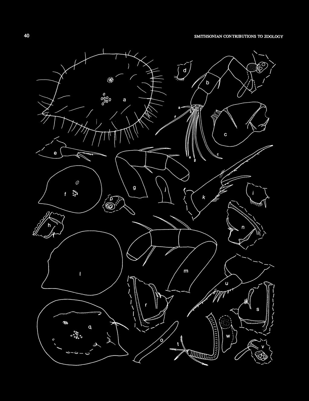
46 40 SMITHSONIAN CONTRIBUTIONS TO ZOOLOGY \ \
: Well developed.")
47 NUMBER lamella by suture, with proximal teeth along posterior margin; 3rd claw a stout pointed process fused to lamella and with distal spines along anterior and posterior margins at tip; several minute teeth along lamella following claws; claw 1 of right lamella slightly anterior to claw 1 of left lamella. Bellonci Organ and Eyes (Figure 24b): Well developed. Posterior of Body: Similar to that of adult female. Y-Sclerite: Present, but ventral branch may be absent. DESCRIPTION OF FEMALE INSTAR II (Figure 24/-*). Carapace similar in shape to that of instar I, but with better defined posterodorsal corner (Figure 24/). Ornamentation: Surface smooth without protuberances. Bristles similar to those of instar I. Infold: Posterior infold with 1 or 2 setose bristle dorsal to caudal process. Infold of caudal process with minute bristles along inner edge but none observed in middle part. Carapace Size: USNM B, length 0.65 mm, height 0.48 mm. First Antenna (Figure 24g): 1st joint bare. 2nd joint with dorsal bristle. 3rd joint fused to 4th, with 2 bristles (1 ventral, 1 dorsal); 4th joint with 1 terminal dorsal bristle. Bristles of joints 5-8 of similar type as those of adult female; but filaments not observed at magnification observed (with x 40 objective and x 15 ocular, and without cover slip) but minute filaments could be present. (Bristles of joints 5-8 not shown on illustrated limb.) Second Antenna (Figure 24/i): Protopodite bare. Endopodite with single joint with ventral protuberance and either no bristles or 1 proximal anterior bristle. Exopodite similar to that of adult female, with 2 bristles on 9th joint Mandible, Maxilla, Fifth Limb: Not examined in detail but of similar type as that of adult female. Sixth Limb (Figure 24/): With 1 anterior bristle. Seventh Limb (Figure 24/): Short and without bristles. Furca (Figure 24*): Claw 1 fused to lamella, claws 2-5 separated from lamella by suture. In general, furca similar to FIGURE 24. Eusarsiella styx, new species. USNM A, paratype, instar I, length 0.55 mm: a, complete specimen showing location of right lateral eye (stippled) and central adductor muscle attachments, anterior to right; b, head region from left side showing left lateral eye, medial eye and Bellonci organ, and left 1st antenna; c, protopodite and endopodite of left 2nd antenna, medial view; d, left 6th limb, lateral view; e, right lamella of furca. USNM B, paratype, instar II, length 0.65 mm: /, complete specimen from right side showing location of right lateral eye (stippled) and adductor muscle attachments, anterior to right; g, right 1st antenna, lateral view, bristles of joints 5-8 not shown; h, distal part of protopodite and endopodite ofright2nd antenna, medial view; i,right6th limb, lateral view;y, 7th limb; k,right lamella of furca. USNM B, paratype, female instar III, length 0.91 mm: /, outline of complete specimen from right side, anterior to right; m, left 1st antenna, lateral view, bristles of joints 5-8 not shown; n, distal part of protopodite and endopodite of right 2nd antenna, medial view; o, right 7th limb; p, left lateral eye, medial eye and Bellonci organ. USNM C, paratype, male instar IV, length 0.98 mm: q, complete specimen from left side, anterior to left; rj, proximal part of protopodites and endopodites of left and right 2nd antennae, medial view; t, 7th limb; u, left lamella of furca; v, left lateral eye, medial eye and Bellonci organ; w, left copulatory organ, anterior to left that of adult female in having long medial hairs at base of claw 1, and long hairs on lamella following claws. Bellonci Organ, Posterior of Body, Y-Sclerite: Similar to those of adult female. Eyes: Medial eye with brown pigment forming horizontal zone near midheight. Lateral eye with brown pigment and 4 or 5 ommatidia. Remarks: The small size of the protuberance on the endopodite of the 2nd antenna and the long hairs on the furcal lamella posterior to claws indicate that the specimen studied (USNM B) is female. DESCRIPTION OF FEMALE INSTAR III (Figure 24/-p). Carapace similar in outline to that of instar II; USNM B strongly calcified (Figure 240- Ornamentation: Surface with ribs and processes of similar type as those on adult female. Carapace Size: USNM B, length 0.91 mm, height 0.67 mm. First Antenna (Figure 24m): 1st joint bare. 2nd joint with 1 dorsal bristle. 3rd joint fused to 4th; 3rd joint with 2 bristles (1 ventral, 1 dorsal); 4th joint with 2 bristles (1 ventral, 1 dorsal). Bristles of joints 5-8 not examined in detail (and not shown in illustrated limb), but of type similar to that of adult female. Second Antenna (Figure 24/i): Protopodite bare. Endopodite with 1 joint with terminal protuberance and 2 proximal anterior bristles. Exopodite similar to that of adult female. Mandible, Maxilla, Fifth and Sixth Limbs: Not examined in detail but of similar type to those of adult female. Sixth limb with many bristles. Seventh Limb (Figure 24o): Elongate without bristles. Furca: S imilar to that of adult female. Long hairs following claws of left lamella but not on right lamella. Bellonci Organ (Figure 24p): Elongate with rounded tip. Eyes (Figure 24p): Medial eye with black pigment. Lateral eye with black pigment and 2 ommatidia. Posterior of Body, Y-Sclerite: Similar to those of adult female. Remarks: The long hairs following the last claw on the left lamella of the furca suggests that USNM B is a juvenile female. The endopodite of the 2nd antenna also suggests a female, but not with certainty. DESCRIPTION OF MALE INSTAR IV (Figure 24q-w). Carapace shape similar to that of female instar III (Figure 24q). Ornamentation (Figure 24^): With ridges and protuberances of type in adult female; bristles and spines similar to those of adult female. Infold: Infold of caudal process with 4 bristles near middle; posterior infold with 2 setose bristles dorsal to caudal process; anterior infold with 1 small bristle at inner edge ventral to valve midheight Carapace Size: USNM C, length 0.98 mm, height 0.77 mm. First Antenna: 1 st joint bare. 2nd joint with 1 dorsal bristle.
: Protopodite bare.")
48 42 SMITHSONIAN CONTRIBUTIONS TO ZOOLOGY 3rd and 4th joints fused; 3rd joint with 2 bristles (1 dorsal, 1 ventral); 4th joint with 3 bristles (1 dorsal, 2 ventral). Bristles of joints 6-8 not examined in detail but of type similar to that of adult female. Second Antenna (Figure 24r,j): Protopodite bare. Endopodite 2 jointed: 1st joint with 2 proximal anterior bristles; 2nd joint tapering distally, with 1 small terminal bristle. Mandible, Maxilla, Fifth Limb, Sixth Limb: Not examined in detail but of type similar to that of adult female. Seventh Limb (Figure 24*): Elongate with 4 terminal bristles with 4 or 5 bells; bristles tapering distally (juvenile character). Furca (Figure 24M): Similar to that of adult male. Differs from that of adult female in not having long hairs on lamella following last claw. Bellonci Organ (Figure 24v): Elongate with rounded tip. Eyes (Figure 24v): Medial eye with zone of black pigment at midheight and anteriorly. Lateral eye with area of black pigment with 4 ommatidia and anterior amber rim with 2 indistinct ommatidia. Genitalia (Figure 24w): Seminal vesicle visible as ovoid amber mass; copulatory organ elongate, without sclerotized processes at tip. Posterior of Body, Y-Sclerite: Similar to those of adult female. Gut Content: The gut of most instars and adults dissected contained unrecognizable brown particles. Remarks: The similarity of the endopodite of the 2nd antenna to that of the adult male identifies the specimen studied as a juvenile male. The presence of bristles on the 7th limb indicates it to be the A-1 instar, which is interpreted to be instar IV. COMPARISONS. The carapace of E. styx differs from that of E. absens in having a narrow projecting caudal process; the 1st antenna differs in having a ventral bristle on the 3rd joint. The male E. styx differs from the male of E. absens in having a well-developed 7th limb. The female E. styx differs from the female E. comma Poulsen (1965:105), in being larger, having more bristles on the infold of the caudal process, and in having a small terminal protuberance on the endopodite of the 2nd antenna. Only 4 species of Eusarsiella having a male with a well developed 7th limb bearing bristles have been described previously (Kornicker, 1986a, table 3). NUMBER OF GROWTH STAGES. Hiruta (1983:676) listed 6 species of the Sarsicllinae of which the number of instars has been determined. All have 4 juvenile stages, the same number determined herein for E. styx. The number of growth stages is known now for 4 species of Eusarsiella, 2 species of Spinacopia, and 1 species of Junctichela. Apparently, 4 juvenile growth stages is uniform within the Sarsiellinae. Appearance and development of limbs of E. styx is similar to those of other Sarsiellinae: Instar I is without bristles either on the 4th joint of the 1st antenna or on the 6th limb, and is without a 7th limb; instar II has 1 dorsal bristle on the 4th joint of the 1st antenna, 1 bristle on the 6th limb, and a short 7th limb without bristles; instar III has 1 ventral and 1 dorsal bristle on the 4th joint of the 1st antenna, many bristles on the 6th limb, and a long bare 7th limb; instar IV has tapering bristles on the 7th limb. ONTOGENY AND SEUAL DIMORPHISM. The carapaces of instars I and II are without ribs and processes, which appear first on instar III. The length of the adult male (1.10 mm) is less than that of the adult female ( mm). First Antenna: On instar I no bristles are present on joints 2 and 4, and no marginal filaments are present on either the sensory bristle of the 5th joint or the b-, c-, f-, and g-bristles of the 7th and 8th joints. On instar II a dorsal bristle is present on the 2nd joint and a dorsal bristle is present on the 4th joint; no filaments were observed on the sensory bristle of the 5th joint or on bristles of the 7th and 8th joints, but the bristles were not examined under sufficiently high magnification to be certain minute filaments are not present. On instar IV the 4th joint has 2 ventral bristles, the same number as on the adult (1 adult female had only 1 ventral bristle (Figure 20a)). The 5th joint and its sensory bristle of the adult male and female exhibit usual sexual dimorphism. Second Antenna: Protopodite and exopodite of juveniles and adults similar except for dimensions. Endopodite: instar I (sex unknown) bare, with single joint with small terminal protuberance; female instar II similar to that of instar I except for having 1 proximal anterior bristle on 1 limb of examined specimen; female instar III similar to that of female instar II except for having 2 proximal anterior bristles; male instar IV 2-jointed, with 2 proximal anterior bristles on 1st joint and 1 small terminal bristle on tapering 2nd joint; adult female similar to that of female instar III; adult male similar to that of male instar IV except for having elongate 2nd joint with 2 or 3 small terminal bristles. Mandible: Well developed on all instars, but mandibles of juvenile instars not examined in detail; they are of similar type to that of adult female. Mandible of adults dimorphic: male mandible differs from that of female in having a smaller coxale endite, a well-developed exopodite, and differences in bristles and claws of the exopodite. Maxilla and Fifth Limb: Well developed on all juvenile instars (not examined in detail but of similar type to those of adult female). Maxillae and fifth limbs of adult male differ from limbs of adult female in being considerably reduced, as well as in number and type of bristles. Sixth Limb: Instar I with marginal hairs but no bristles; instar II with 1 anterior bristle; instars III, IV, and adults well developed with many bristles. Seventh Limb: Absent on instar I (sex unknown); short bare on instar II female; elongate bare on female instar III; elongate with 4 strongly tapering terminal bristles on male instar IV; limb of adult female elongate with 1 or 2 cylindrical proximal bristles, 6 cylindrical terminal bristles, and opposing terminal combs; limb of adult male with 4 cylindrical terminal bristles and without terminal combs. Furca: Instar I with 1st claw fused to lamella, 2nd claw


49 NUMBER separated from lamella by suture, and 3rd claw represented by stout, spinous, pointed process fused to lamella. Instars II-IV and adults similar in having 5 claws with only claw 1 fused to lamella. Eyes: Lateral eye of adult female with 5 ommatidea and light brown pigment; lateral eye of adult male with 7 or 8 ommatidea and black pigment. Medial eye of adult male with black pigment, of adult female with small amount of brown pigment. Differences in pigmentation between eyes of the adult male and adult females could be due to the females being preserved for a longer period of time than the male. Family RUTIDERMATIDAE Brady and Norman, 1896 COMPOSITION. This family comprises 3 genera of which only 1, Rutiderma, has been reported from Bermuda (Kornicker, 1983:70). Rutiderma Brady and Norman, 1896 TYPE SPECIES. Rutiderma compressa Brady and Norman (1896:673). Only 1 species has been reported from Bermuda, R. sterreri Kornicker (198la:5), from open water. Two specimens of this species were collected in caves in this study. Rutiderma sterreri Kornicker, 1981 FIGURES 25,26 Rutiderma sterreri Kornicker, 1981a:5, figs. 3-5; 1983:70, figs Maddocks and Komicker, 1986:282, fig. 90 [part]. HOLOTYPE. USNM , ovigerous female on slide and in alcohol. TYPE LOCALITY. Station , Harrington Sound, Bermuda. MATERIAL. Bermuda: Cherry Pit Cave, 22 Mar 1987: USNM , 1 adult female in alcohol. Cripplegate Cave, 21 Oct 1981: USNM , 1 adult male in alcohol. Harrington Sound, : USNM , 3 adult males plus 4 adult females in alcohol. DISTRIBUTION. Bermuda: Cherry Pit Cave, 22 Mar 1987; Cripplegate Cave, 21 Oct 1981; Harrington Sound (open water), Previously reported from Hamilton Harbor, Spanish Point, Castle Harbor, and Harrington Sound (Kornicker, 198 la: 1,2). SUPPLEMENTARY DESCRIPTION OF ADULT MALE, USNM (Figure 26). Carapace elongate with strong lateral ribs terminating posteriorly in connecting vertical rib with concavity at midheight and weaker ribs along dorsal and ventral margins (Figure 26a). Surface with small fossae (Figure 26a). Central adductor muscle attachments comprising about N Castle Harbor FIGURE 25. Distribution of caves from which Rutiderma sterreri Komicker (open circles) and Parasterope muelleri Skogsberg (solid circles) were collected (4 = Cherry Pit Cave; 6 = Cripplegate Cave; 7 = Deep Blue Cave; 15 = Palm Cave).
 and central adductor muscle atuchments (small cluster of circles beneath lateral eye), and few")
, protopodite, endopodite, and part of 1st exopodial joint of left 2nd antenna, and location of central adductor muscle attachments (dashed cicle); e, posterior of body showing")
50 44 SMITHSONIAN CONTRIBUTIONS TO ZOOLOGY FIGURE 26. Rutiderma sterreri Kornicker, USNM , adult male, length 1.00 mm: a, complete specimen from left side showing location of left lateral eye (stippled) and central adductor muscle atuchments (small cluster of circles beneath lateral eye), and few representative surface pits; b, detail from a of central adductor muscle attachments; c, protopodite and 1st endopodial joint of left 2nd antenna, sclerites stippled; d, anterior of body showing left lateral eye (suppled), protopodite, endopodite, and part of 1st exopodial joint of left 2nd antenna, and location of central adductor muscle attachments (dashed cicle); e, posterior of body showing left lamella of furca, left copulatory organ, left Y-sclerite (lined) and other sclerites, and posterior hairs; /, anterior part of body showing location of heart, left lateral eye (stippled), medial eye, and Bellonci organ, joints 1 and 2 of right 1st antenna, broad anterior process ventral to 1st antenna, and upper lip (u.lip).
: Major sclerites of the protopodite viewed medially are stippled in illustration (only 1st joint of endopodite shown in illustration)(figure 26c).")
51 NUMBER ovoid scars forming group and 1 additional scar dorsal to group (Figure 26b). Carapace Size: Length 1.00 mm, height 0.58 mm. Second Antenna (Figure 26c,d): Major sclerites of the protopodite viewed medially are stippled in illustration (only 1st joint of endopodite shown in illustration)(figure 26c). Posterior margin of protopodite bears notch near upper end to acommodate lateral eye, and shallow indentation at midheight to accomodate central adductor muscles (similar morphology was noted by Kornicker (1985:24) on R. hartmanni Poulsen, 1965). Furca, Copulatory Organ, Posterior of Body with Sclerites: Illustrated in Figure 26«. Bellonci Organ, Lateral Eye, Upper Lip, Anterior of Body, Heart: Illustrated in Figure 26/. USNM , 1 early instar in alcohol. Palm Cave, 13 and 16 Mar 1982: USNM A.B, 1 adult female and 1 adult male, respectively, both in alcohol. DISTRIBUTION. Deep Blue Pool, 23 Mar Palm Cave, 13 and 16 Mar Previously reported from Castle Harbor, Hamilton Harbor, Smith's Sound, and off St. George, Bermuda (Kornicker, 1981a:8). Species widespread in north Atlantic Ocean (Kornicker and Caraion, 1974:7). DISCUSSION. The body was removed from the shell of the single specimen collected in Deep Blue Cave and examined in a drop of glycerine. The absence of bristles on an elongate Superfamily CYLINDROLEBERIDOIDEA Mtiller, 1906 COMPOSITION. The Cylindroleberidoidea comprise the family Cylindroleberididae Miiller, Family CYLINDROLEBERIDIDAE Miiller, 1906 COMPOSITION. This family comprises 3 subfamilies of which only 1, the Cylindroleberidinae, is represented in collections from Bermuda. Subfamily CYLINDROLEBERIDINAE Miiller, 1906 Two species in 2 genera in this subfamily have been reported previously from Bermuda (Kornicker, 1981a:2): Parasterope muelleri (Skogsberg, 1920:483) and Bruuniella species A (Kornicker, 1981a: 12), but none have been reported previously from caves. Parasterope Kornicker, 1975 TYPE SPECIES. Asterope Mullen Skogsberg (1920:483); subsequent designation by Kornicker (1975:401). Three specimens of P. muelleri were collected in Bermudan caves. Parasterope muelleri (Skogsberg, 1920) FIGURE 27 Asterope Mullen Skogsberg, 1920:483. Parasterope muelleri (Skogsberg). Kornicker, 1986b:20 [synonomy]. Maddocks and Kornicker, 1986:284. fig. 90 [part]. LECTOTYPE. Adult female on slides in Swedish State Museum, Stockholm. TYPE LOCALITY. English Channel, off Salcombe, coast of England. MATERIAL. Bermuda: Deep Blue Pool, 23 Mar 1982: FIGURE 27. Parasterope muelleri Skogsberg: a, USNM A, adult female, length 1.07 mm, outline of complete carapace from right side showing location of right lateral eye, anterior to right; b, USNM B, adult male, length 1.19 mm, outline of complete specimen from right side showing location of right lateral eye (stippled), location of central adductor muscle attachments (dashed circle) and hairs near posterior forming vertical row; c, USNM , instar II or III, length 0.65 mm, outline of complete specimen from left side showing location of left lateral eye and central adductor muscle attachments.
 indicate that it is an early instar (instar II or III). An early instar cannot be referred to a genus or species with certainty, but I have referred it to P.")
52 46 SMITHSONIAN CONTRIBUTIONS TO ZOOLOGY 7th limb and a 6th limb with 2 indistinct endite bristles and 2 bristles on the end joint (1 indistinct anteroventral bristle, and 1 ventral bristle at midlength) indicate that it is an early instar (instar II or III). An early instar cannot be referred to a genus or species with certainty, but I have referred it to P. muelleri because it resembles that species (Figure 27c). The presence of 2 endite bristles and an anteroventral bristle on the end joint of the 6th limb could not be determined with certainty on the undissected specimen. Carapace Sizes: Of 3 specimens: USNM , early instar (Figure 27c), length 0.69 mm, height 0.45 mm; USNM A, adult female (Figure 27a), length 1.07 mm, height 0.71 mm; USNM B, adult male (Figure 21b), length 1.19 mm, height 0.74 mm. Order HALOCYPRIDA Dana, 1853 COMPOSITION. The Halocyprida comprise the suborders Halocypridina Dana, 1853, and Cladocopina Sars, Suborder HALOCYPRIDINA Dana, 1853 COMPOSITION. The Halocypridina comprise the superfamilies Thaumatocypridoidea Miiller, 1906, and Halocypridoidea Dana, The Thaumatocypridoidea have been collected in anchialine caves of the Bahamas and the Yucatan Peninsula (Kornicker and Iliffe, in press), Canary Islands (Hartmann, 1985), Cuba (Danielopol, 1972) but not in Bermuda. Superfamily HALOCYPRIDOIDEA Dana, 1853 Composition. The superfamily comprises the family Halocyprididae Dana, Family HALOCYPRIDIDAE Dana, 1853 COMPOSITION. The family comprises the subfamilies Archiconchoecinae Poulsen, 1969, Euconchoecinae Poulsen, 1969, Halocypridinae Dana, 1853, Conchoecinae Claus, 1891, and Deeveyinae Kornicker and Iliffe, One species of Euconchoecinae has been reported from an anchialine cave in Palau (Kornicker and Iliffe, in press), but none were collected in Bermudan caves. Subfamily DEEVEYINAE Kornicker and Iliffe, 1985 COMPOSITION. The subfamily comprises the genera Deeveya Kornicker and Iliffe, 1985, and Spelaeoecia Angel and Iliffe, Two species of Deeveya have been collected from anchialine caves in the Bahamas (Kornicker and Palmer, 1987:610) and in the Turks and Caicos Islands (Kornicker and Iliffe, 1985:476) but not in Bermuda. Spelaeoecia Angel and Iliffe, 1987 TYPE SPECIES. Spelaeoecia bermudensis Angel and Iliffe (1987:545). COMPOSITION. The genus is monotypic. Spelaeoecia bermudensis Angel and Iliffe, 1987 FIGURES 28a, 29 Spelaeoecia bermudensis Angel and Diffc, 1987:545,figs HOLOTYPE. USNM , adult female on 5 slides. TYPE LOCALITY. Green Bay Cave, Bermuda. MATERIAL. Bermuda: Green Bay Cave, 29 Aug 1982, USNM (holotype), adult female on 5 slides; Jane's Cave: 27 Mar 1985: USNM A, 1 A-l male, left 2nd antenna on slide, remaining appendages and carapace in alcohol; USNM B,C, 2 adult females in alcohol. 22 Jul 1985, USNM , 1 juvenile in alcohol. 29 Jul 1986: USNM A, 1 A-l male in alcohol; USNM B.C, 2 adult females in alcohol. Roadside Cave: 13 Mar 1985: USNM , 1 A-3 instar in alcohol. 7 Aug 1985: USNM , 1 specimen in alcohol. DISTRIBUTION (Figure 28a). Bermuda: Jane's Cave (new locality for species). Christie's Cave, Crystal Cave, Fern Sink Cave, Green Bay Cave, Roadside Cave, Tucker's Town Cave, Walsingham Sink Cave, Wonderland Cave. SUPPLEMENTARY DESCRIPTION (Figure 29). Surface of carapace with oblique lineations (Figure 29b,c) (indistinct on some specimens and not observed on others). Infold (Figure 29a,m): Broad infold except along hinge, narrower just posterior to inner end of incisur, narrow list at about midwidth of infold extending from anterior near valve midheight to midlength or farther; a 2nd list forming sclerotized bar just posterior to posterior hinge juncture, then extending ventrally as narrow ridge intersecting posterior valve edge at midheight. Selvage (Figure 29m): Narrow bare lamellar prolongation extending from point at ventral end of incisur to posterodorsal corner. Carapace Locking Structures (Figure 29m): Each valve with 1 or 2 narrow sclerotized ridges between posterior juncture of hinge and posterodorsal corner of valve that may serve as locking structures when valves are closed. Central Adductor Muscle Attachments (Figure 29/): Consisting of about 9 individual ovoid scars. Carapace Size: Adult females: USNM B, length 1.57 mm, height 0.85 mm; USNM C, length 1.54 mm, height 0.86 mm. A-l males: USNM A, length 1.25 mm, height 0.62 mm; USNM A, length 1.27 mm, height 0.67 mm. First Antenna: Without bristle on 6th joint 7th joint with a-,b-, and c-bristles. 8th joint with d-,e-,f-, and g-bristles. Limb of A-l male (Figure 29n) similar to that of adult female (Figure 29g). Holotype differs from description of Angel and Iliffe
 Spelaeoecia bermudensis Angel and Iliffe and (fc) Metapolycope duplex, new species, were collected (4 = Cherry Pit Cave; 5 = Christie's Cave; 7 = Deep Blue Cave;")
53 NUMBER N Castle Harbor Harrington Sound N Castle Harbor FIGURE 28. Distribution of caves from which (a) Spelaeoecia bermudensis Angel and Iliffe and (fc) Metapolycope duplex, new species, were collected (4 = Cherry Pit Cave; 5 = Christie's Cave; 7 = Deep Blue Cave; 9 = Fern Sink Cave; 10 = Green Bay Cave; 15 = Palm Cave; 17 = Roadside Cave; 18 = Sailor's Choice Cave; 20 = Straw Market Cave; 21 = Tucker's Town Cave; 22 = Walsingham Cave; 23 = Walsingham Sink Cave; 24 = Wonderland Cave; 25 = Crystal Cave; 26 = Jane's Cave).
.")
; bristles of joints 1 and 2 with natatory")
54 48 SMITHSONIAN CONTRIBUTIONS TO ZOOLOGY 0987:546, fig. 3a) in having no bristle on 6th joint and 3 on 7th joint (1 dorsal, 2 ventral). Second Antenna: Endopodite of adult female limb with minute medial bristle near base of j-bristle and minute lateral peg near f-bristle (Figure 29e). Endopodite of A-1 male with additional minute lateral c-bristle, and dorsal margin of 2nd joint convex (Figure 29p). Exopodite: 1st joint partly divided into long proximal and short distal segments, with long terminal bristle reaching well past 9th joint (Figure 29d,o); bristles of joints 1 and 2 with natatory hairs, and spines facing convex margin of exopodite; 9th joint of exopodite with 4 bristles (at least 3 with spines facing concave margin of exopodite). (Angel and Iliffe (1987: 546) describe the exopodite as having a short ventral bristle on the 1st joint, and that bristle is shown in their figure 3B. Although the legend of that illustration does not state that it is based on the holotype, each of the mounted 2nd antennae of the holotype does have the tip of an endopodial bristle in the same place as the "exopodial" bristle shown in their figure 3B, and that endopodial bristle was most likely misinterpreted by Angel and Iliffe to be an exopodial bristle.) Furca: Each lamella of the furca of the A-1 male (Figure 29jjc,q) and adult female with claw 2 represented by stump and 7 additional claws with minute spines along posterior margins. The 'unpaired' bristle is bifid and bears marginal spines. Bellonci Organ: In lateral view tapering distally and bifurcating at 2 h length; tip of each branch broadly rounded (Figure 29gJ). Organ of USNM A aberrant in being relatively short, not bifurcate, and having spine at tip (Figure 29n). Genitalia: A-1 male (USNM A) with long, broad, anterior process without terminal structures, and tapering rod-shaped organ with short pointed tip (Figure 29/). The anterior process either obscured or missing on one A-1 male (USNM A), but rod-shaped organ well developed and with minute spine just proximal to pointed tip (Figure 29q,r). Adult female with small process bearing a terminal spine just anterior to base of left 7th limb in vicinity of receptaculum seminis, which was not visible (Figure 29h,i). (Miiller (1894, pi. 1: fig. 9) illustrated the body of a female Conchoecia magna showing the location of the receptaculum seminis of that species at about the same location as the spined process of S. bermudensis.) Gut Content: A-1 male, USNM A, with large ovoid pellet emerging from anus: dark brown ventral part of pellet (that part extruded first) about x h length and 3 A width of dorsal FIGURE 29. Spelaeocia bermudensis Angel and Iliffe, USNM , holotype, adult female, length 1.58 mm: a, left valve from outside showing 3 central adductor muscle attachment scars, inner margin of infold, list, and valve edge inside of rostrum (all shown as dashed lines); b,c, surface Hneations on anteroventral and posterior parts of valve shown in a (arrow in b points toward anterior of valve); d, exopodite of left 2nd antenna, lateral view (only bristle of 1st joint shown); e, endopodite of left 2nd antenna, lateral view./, USNM B, adult female, length 1.S7 mm, outside view of central adductor muscle attachment scars of left valve. USNM C, adult female, length 1.S4 mm: g, Bellonci organ and left 1st antenna (bristles of 6th-8th joints not shown; h, lateral view of left side of body as seen through shell showing spined process in vicinity of recepticulum seminis, epipodial appendage of 6th limb (bristles not shown), 7th limb, and apron anterior to furca. i, USNM , adult female, length 1.44 mm, left side of body showing spined process, epipodial appendage of 6th limb (bristles not shown) and 7th limb. USNM A, A-1 male, length 1.27 mm: /, posterior of body from left side showing copulatory organ, epipodial appendage of 6th limb (bristles not shown), 7th limb, apron, and left lamella of furca; k, right lamella of furca; /, Bellonci organ. USNM A, A-1 male, length 1.25 mm: m, inside view of valves; n, Bellonci organ (aberrant) and left 2nd antenna, lateral view; o, exopodite of left 2nd antenna, medial view (only bristles of 1 st and 2nd joints shown); p, endopodite of left 2nd antenna, medial view; q, posterior of body from left side showing rod-shaped copulatory organ, 5th and 6th limbs (not all bristles shown) and left lamella of furca; r, detail of copulatory organ shown in q. part, and comprises minute unidentified particles; large dorsal part of pellet with pearly sheen in reflected light and comprises abundant, broken, hollow, transparent, colorless tubes of unknown origin. REMARKS. An adult male (USNM ) in a sample from Wonderland Cave collected by Jan H. Stock was encountered too late for including its description herein and will be described elsewhere (Kornicker, in press). Suborder CLADOCOPINA Sars, 1866 REMARKS. The long geological history of the Cladocopina was discussed recently by Neale (1983: ). COMPOSITION. The Cladocopina contains the superfamily Polycopoidea Sars, Superfamily POLYCOPOIDEA Sars, 1866 COMPOSITION. The Polycopoidea contains the Poylcopidae Sars, Family POLYCOPIDAE Sars, 1866 COMPOSITION. The Polycopidae comprise the Polycopinae Sars, 1866, and Polycopsisinae Chavtur, Key to the Subfamilies of Polycopidae (from Chavtur, 1983:92, emended) 1st antenna with 5 or 6 segments delimited by sutures 1st antenna with 3 or 4 segments delimited by sutures POLYCOPSISINAE POLYCOPINAE


55 NUMBER

 individual scars; 1st antenna with 6 segments delimited by sutures Polycopsis Metapolycope Kornicker and van Morkhoven, 1976 TYPE SPECIES.")
56 50 SMITHSONIAN CONTRIBUTIONS TO ZOOLOGY Subfamily POLYCOPSISINAE Chavtur, 1983 COMPOSITION. The Polycopsisinae comprise the genera Metapolycope Kornicker and van Morkhoven, 1976, and Polycopsis Miiller, Only Metapolycope is represented in Bermudan caves. Key to the Genera of Polycopsisinae (adapted from Chavtur, 1983:93) Central muscle scars comprising 9 or more individual scars; 1st antenna with 5 segments delimited by sutures Metapolycope Central muscle scars comprising 3 (always?) individual scars; 1st antenna with 6 segments delimited by sutures Polycopsis Metapolycope Kornicker and van Morkhoven, 1976 TYPE SPECIES. Metapolycope hartmanni Kornicker and van Morkhoven, COMPOSITION. The genus comprises 5 species and 1 in open nomenclature: M. hartmanni Kornicker and van Morkhoven, 1976, M. microthrix Kornicker and van Morkhoven, 1976, Metapolycope species (Kornicker and van Morkhoven, 1976; known only from shell), M. kornickeri Chavtur, 1979, M. echinata Chavtur, 1981b, and M. duplex new species. TERMINOLOGY OF FIRST ANTENNA. The 1st antenna is interpreted herein to have 8 joints (Figure 30b-d), not 5 as given in the diagnosis of the genus by Kornicker and van Morkhoven 0976:2). The 2nd and 3rd joints are fused except in some adult males of M. duplex. The 3rd and 4th joints are fused except in M. duplex. The 7th and 8th joints are small and although each is fairly well defined on M. duplex, they could be interpreted to be a single joint (7th). With the present interpretation the 1st antenna of Metapolycope bears the same number of joints as the Myodocopina and, also, the genus Deeveya of the Halocypridina. Bristles with suckers (Metapolycope) or spatulate bristles (Polycopissa) of adult males are interpreted to be on the 6th joint. Metapolycope duplex, new species FIGURES Polycop* spp. Maddocks and Diffe, 1986:73 [part]. ETYMOLOGY. From the Latin duplex in reference to the 2 anterior processes on the anterior margin of each lamella of the furca. HOLOTYPE. USNM , 1 adult male. TYPE LOCALITY. Bermuda: Palm Cave, 13 and 16 Mar MATERIAL. Cherry Pit Cave: 23 Jun 1982: USNM ,1 instar IV; USNM ,1 instar VI female; USNM , 1 adult male; USNM A3, 2 adult females; USNM C, 1 instar V female; USNM , 1 adult male, 2 adult females, 2 instar VI, 1 valve. 12 Jan 1984: USNM , 1 instar VI male; USNM , 1 adult female, 1 adult male, 2 instar VI, 3 valves. 22 Mar 1987: USNM , 1 instar III; USNM , 32 specimens. Deep Blue Cave: 20 Feb 1984: USNM , 2 specimens (valves only). Fern Sink Cave: 25 Feb 1982: USNM , 1 adult or late instar. Green Bay Cave: 18 Nov 1981: USNM , 4 specimens (adults and late instars), 1 specimen (valves only). Palm Cave: 20 Jan 1982: USNM , 1 instar I; USNM , 1 instar V female; USNM , 1 adult male; USNM , 1 adult female; USNM A, 1 instar VI female; USNM B, 1 adult female; USNM C, 4 adult females, 1 valve. 13 and 16 Mar 1982: USNM , 1 adult male (holotype); USNM , 1 instar V female; USNM , 1 adult male; USNM , 1 instar VI male; USNM A, 1 adult female; USNM B, 5 adult females; USNM , 1 adult female, USNM , 1 female, 5 juveniles. 9 Feb 1985: USNM , 1 adult female; USNM , 1 decalcified valve. Sailor's Choice Cave: 6 Jul 1982: USNM , 4 specimens (adults and late instars). Straw Market Cave: 12 Jan 1984: USNM , 1 adult. Walsingham Cave: 18 Feb 1982: USNM , 7 specimens (adults and late instars); USNM , 1 instar I. 14 Jun 1982: USNM , 1 adult male, 3 single valves. 13 Jul 1984: USNM ,1 instar IV; USNM ,2 specimens (valves only). Walsingham Sink Cave: 13 Aug 1982: USNM , 1 valve. DISTRIBUTION (Figure 286). Bermuda: Cherry Pit Cave, 23 Jun 1982, 12 Jan 1984, 22 Mar Deep Blue Cave, 20 Feb Fern Sink, 25 Feb Green Bay Cave, 18 Nov Palm Cave, 20 Jan 1982, 9 Feb 1982, Mar Sailor's Choice Cave, 6 Jul Straw Market Cave, 12 Jan Walsingham Cave, 18 Feb 1982, 14 Jun 1982, 13 Jul Walsingham Sink Cave, 13 Aug 1982, 13 Jul DESCRIPTION OF ADULT FEMALE (Figures 30, 31). Carapace oval in lateral view (Figure 30a). Shell translucent, edges and most carapaces light amber color. Ornamentation (Figure 30a): Surface smooth. Anteroventral margin serrate with triangular processes (processes near middle broader). Central Adductor Muscle Attachments (Figure 30a): Com-
; c, left 1st antenna, medial view; d,")
57 NUMBER FIGURE 30. Metapolycope duplex, new species, USNM , paratype, adult female, length 0.54 mm: a, lateral view of complete specimen, anterior to right; b, lateral view of left 1st antenna and prodopodite of left 2nd antenna (not all bristles shown); c, left 1st antenna, medial view; d, right 1st antenna, lateral view; e, left 2nd antenna, medial view. Mandible: /, coxale endite of left limb, lateral view; g,h, right limb and detail of coxale endite, lateral view, i, maxilla; j, right lamella of furca and posterior of body; *, posterior of body from right side with unpaired process and 7th claw of right furcal lamella at lower edge; /, posterior of body from left side showing right lamella of furca, unpaired process, and 4 unextnided eggs; m, upper Up, anterior to right; n, food pellet in gut (drawn with x 100 oil immersion objective).
; c, medial bristles of")
. USNM 193330, paratype, adult female: i, posteroventral view of complete specimen with valves open showing 3 unextruded eggs (stippled) (diameter of smallest egg 0.")
58 52 SMITHSONIAN CONTRIBUTIONS TO ZOOLOGY \ FIGURE 31. Metapolycope duplex, new species. USNM , paratype, adult female, length 0.54 mm: a-e, left 5th limb, medial views: a, complete limb except for proximal epipodial bristles; b, limb showing internal lenticular-shaped cells (not all bristles shown); c, medial bristles of exopodite; d, terminal bristles of exopodite; e, endopodite; f,g, lateral view of complete right 5th limb and detail from / (not all bristles shown); h, dorsal view of head region showing Bellonci organ (2 bristles). USNM , paratype, adult female: i, posteroventral view of complete specimen with valves open showing 3 unextruded eggs (stippled) (diameter of smallest egg 0.07 mm, of largest egg 0.14 mm), unpaired process, and 2 furcal claws at lower edge (parts of other visible limbs not shown). USNM , paratype, adult female, length 0.53 mm: j, lateral view of right valve showing central adductor muscles as seen through shell, anterior to right; k, nematode (stippled) with body of animal as seen through right valve, anterior of valve to right (dashed line represents location of hinge line). USNM A, paratype, adult female, length 0.45 mm: /, posterior of body showing 5 unextruded eggs (stippled), unpaired process of furca at lower right

59 NUMBER prising central ovoid scar, 7 radial wedge-shaped scars forming half of a rosette with convex edge of rosette oriented posteroventrally, 1 ovoid scar anterior to central scar, 2 narrow scars dorsal to central scar, and 5 small scars between wedge-shaped scars and central scar. An additional ovoid scar present posterior to central adductor muscle cluster and separated from cluster by distance about equal to maximum dimension of cluster. (Attachments not always clearly visible through valve so that variability of scar in illustrations presented herein may be the result of poor visibility. Also, changes may occur during preservation causing separations in individual muscles making a single muscle appear to be 2 muscles; the 5 small scars indicated at the inner end of the wedge-shaped scars could be the result of division of the wedge-shaped muscles in the alcohol preservative and not actual divisions of the scars in the shell; the muscle of USNM (Figure 31/) probably best represents the central adductor muscle attachments because it was clearly visible on that specimen.) Carapace Size: USNM , length 0.54 mm, height 0.48 mm; USNM , length 0.53 mm, height 0.45 mm; USNM A.B, 2 specimens, both with same dimensions, length 0.54 mm, height 0.47 mm; USNM B, length 0.49 mm, height 0.44 mm. First Antenna (Figure 30b-d): 1st joint spinous, with long dorsal bristle with long proximal spines; except near dorsal margin, suture separating 1st and 2nd joints weakly developed or absent. 2nd and 3rd joints fused; each joint spinous, and with dorsal process with terminal bristle (tip of process of 3rd joint extended and with narrow neck at base of extension); 3rd joint with subterminal ventral bristle, and with well-developed suture separating it from 4th joint on lateral side only (suture on medial side either poorly developed or absent). 4th joint with spines along dorsal margin, and well-developed suture separating it from 5th joint 5th joint short, bare. 6th joint with 3 bristles (1 dorsal, 2 ventral (longer of ventral bristles lateral)); dorsal margin of joint sinuate. 7th joint short, partly embedded in 6th joint, with 2 long terminal ventral bristles (both ringed proximally and with widely separated marginal spines on ringed part; ventral of the 2 bristles shorter, but both longer than stem). 8th joint short with 3 bristles (2 about twice length of stem, 1 about same length as stem, all ringed proximally and with widely separated marginal spines on ringed part). Second Antenna (Figure 30b,e,): Protopodite with spines forming dorsal row at midlength. Exopodite with 9 joints: 1st joint arcuate with spines forming rows along convex margin; joints 1-8 each with long bristle ringed along proximal 3 A (ringed part bearing natatory hairs appearing more numerous on bristles of joints 1-4); 9th joint with 2 bristles (1 short, bare, other shorter than bristles of joints 1-8 and with few short proximal hairs). Endopodite 3-jointed: 1st joint elongate with 1 terminal dorsal bristle having few marginal hairs; 2nd joint short with 1 short terminal dorsal bristle, and 5 terminal ventral bristles with rings along proximal half (1 bristle about length of endopodite, others about twice the length; marginal hairs observed on ringed part of 2 long bristles); 3rd joint short with 3 long bristles having rings along proximal 3 A (dorsal bristle shorter than others; proximal marginal hairs observed on 1 of the longer bristles). Mandible (Figure 30f-h): Coxale endite with stout anterior tooth followed by short spinous bristle, 2 slender teeth with smaller tooth between them, then 5 or 6 smaller slender teeth and about 7 minute distal posterior teeth. Basale with 4 spinous bristles on or near ventral margin, 1 distal spinous dorsal bristle, distal dorsal spines, and few small lateral spines near middle. Exopodite well developed with 2 spinous distal bristles. Endopodite 2-jointed: 1st joint with 3 or 4 bristles on or near ventral margin, 1 long dorsal at midlength (Figure 30g; USNM without bristle), 2 longer distal dorsal bristles (with long proximal spines), and long spines medially and along ventral margin; end joint with 2 long bristles (ventral of these with long proximal marginal spines, dorsal bristle stouter, somewhat claw-like, with long proximal and shorter distal spines). Maxilla (Figure 300: Precoxale and coxale well defined: precoxale endite of specimen studied fragmented; coxale with 1 distal hirsute lateral bristle near ventral margin, 3 long spinous bristles on ventral margin (1 proximal, 2 distal), and 2 endites, each with 3 spinous bristles, Basale: ventral margin with 2 spinous distal bristles; dorsal margin with proximal hump with internal muscle extending into exopodite; medial surface with long hairs near ventral margin and short indistinct proximal hairs near dorsal margin. Exopodite weakly divided into short proximal and longer distal parts, with 8 terminal bristles (3 with marginal hairs). Endopodite 3-jointed: 1st joint with 2 long, spinous, distal, ventral bristles, and long medial spines near ventral margin; 2nd joint with 4 terminal ventral bristles (2 long, 2 short; ventral long bristle with long marginal spines), and 1 spinous dorsal bristle; small 3rd joint with 4 bristles (2 with long marginal spines). Fifth Limb (Figure 3la-g): Epipodite with 16 hirsute bristles; and with minute spines along proximal 3 A of edge. Coxale hirsute with about 9 short spinous bristles on or near dorsal margin. Basale with distal suture not reaching outer margin, and with about 9 bristles. Endopodite short, possibly 2-jointed, with indistinct hairs and 4 bristles: longest and next-to-longest bristles with long slender marginal spines on broad proximal part and shorter stouter spines on distal narrower part (Figure 31c). Exopodite with 4 hirsute bristles forming medial proximal row (Figure 31c), and 4 spinous terminal bristles (1 short, 3 long) (Figure 3Id)- Lenticularshaped cells (sporozoans?) present within coxale and basale near medial surface of USNM (Figure 31b). Furca (Figures 30/-/, 3li): Each lamella with 7 claws decreasing in length proximally; each claw with long spine-like teeth along posterior margin and indistinct distal hair-like spines along anterior margin; posterior teeth longer on proximal 2 claws, especially on last claw. Small process with triangular terminal tooth present between all claws except proximal 2; each process closer to proximal claw. Apex of

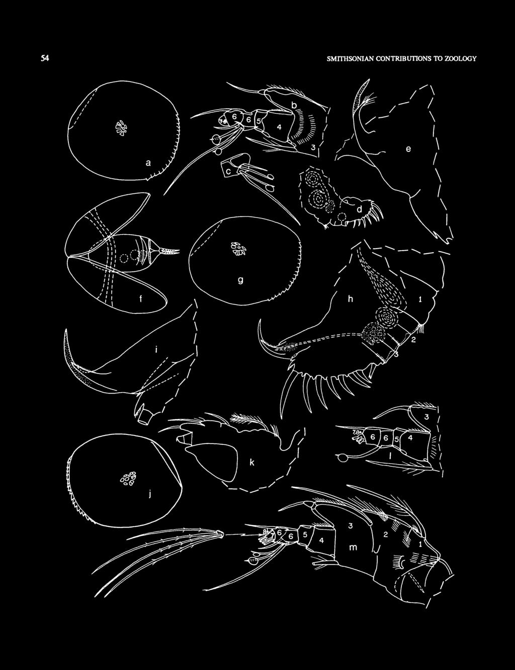
60 54 SMITHSONIAN CONTRIBUTIONS TO ZOOLOGY
; c, joint 6 of left 1 st antenna showing")

61 NUMBER FIGURE 32(,left). Metapolycope duplex, new species. USNM , holotype, adult male, length 0.47 mm: a, lateral view of complete specimen, anterior to right; b, joints 3-8 of right 1st antenna, medial view (bristles of fused joints 7 and 8 not shown); c, joint 6 of left 1 st antenna showing ventral bristles, medial view; d, posterior of body from right side showing unpaired furcal process, right lamella of furca, copulatory organ, and internal genitalia; e, genital organ from left side, part of furca at bottom edge. USNM , paratype, adult male, length 0.50 mm: /, ventral view of complete specimen with valves open, showing internal genitalia, 2 furcal claws, and localition of central adductor muscles, additional visible parts of appendages not shown. USNM , paratype, adult male, length 0.46 mm: g, lateral view of complete specimen from right side (valves slightly open), anterior to right; h, posterior of body from left side showing genital organ and internal genitalia, and left lamella of furca; i, detail from h. USNM , paratype, adult male, length 0.49 mm:/, inside view of right valve, anterior to left (infold not shown); k, left view of body showing Bellonci organ, left 1st antenna (not all bristles shown) and protopodite of left 2nd antenna; /, joints 3-8 of right 1st antenna, only 1 of 2 bristles of 8th joint shown (limb aberrant in having only 1 ventral bristle on 6th joint); m, left 1 st antenna, lateral view. FIGURE 33. Metapolycope duplex, new species, USNM , paratype, adult male, length 0.49 mm: a, endopodite of right 2nd antenna, medial view; b, tip of endopodite of left 2nd antenna, lateral view; c, endopodite of left mandible, medial view; d, maxilla and bristles of basale (exopodite and endopodite not shown). Left 5th limb, lateral views: e, complete limb showing only bristles of coxale, basale, and stumps of 3 bristles of epipodite (epip.);/, exopodite (exop.); g, endopodite (end.); h, epipodite, distal end at bottom, i, posterior of body from left side showing copulatory organ, left furcal lamella, and unpaired furcal process; /, upper lip, lateral view.
.")
62 56 SMITHSONIAN CONTRIBUTIONS TO ZOOLOGY each lamella with 2 processes (distal process longer) bearing ventral spines. Single unpaired process on body proximal to proximal claw (process with digitate tip and about 3 A length of proximal claw). Outer surface of lamellae proximal to claws with small broad spines (spines smaller posterior to proximal claw); inner surface with short slender spines. Right lamella not anterior to left. Bellonci Organ (Figures 306, 31 h): Comprising 2 plumose bristles. Cluster of long hairs on surface of head posterior to bristles. Upper Lip (Figure 30m): Helmet-shaped with glandular openings in anteroventral corner. Posterior of Body (Figure 30/-/): With 3 main segments; proximal segment with long hairs forming short distal row, segments 2 and 3 with shorter less distinct hairs forming distal row; 2nd and 3rd segment each divided by secondary suture at midheight; in lateral view sclerotized edge of posterior margin forming broad boss at midheight of segments 2 and 3 (dotted line in Figure 30k); posterodorsal margin proximal to segment 1 with short spines forming row. Eggs: Location of eggs is shown in Figure 31/,/. Gut Content (Figure 30n): Gut of USNM with light amber food ball with darker amber and brown ovoid particles (with internal structures) that may be of plant origin. Parasites (Figure 3U): USNM with nematode within dorsal part of body. USNM with cells within 5th limb that may be internal sporozoans (Figure 3lb). DESCRIPTION OF ADUU MALE (Figures 32, 33). Carapace similar to that of adult female except smaller (Figure 32a,f,g,j). Ornamentation (Figure 32a,g,j"): Surface smooth. Anteroventral margin serrate with triangular processes (USNM with 18 on right valve and 22 on left). Central Adductor Muscle Attachments (Figure 32a,g,j)\ Similar to those of adult female. Carapace Size: USNM , length 0.49 mm, height 0.40 mm; USNM , holotype, length 0.47 mm, height 0.45 mm; USNM , length 0.46 mm, height 0.42 mm; USNM , length 0.50 mm, height 0.42 mm. First Antenna (Figure 32b,cjt-m): 1st joint spinous with long dorsal bristle; 1st and 2nd joints fused except near dorsal margin. 2nd and 3rd joints separated by suture on some limbs but not on others (illustrated left limb of USNM with 2nd and 3rd joints separated by suture (Figure 32m), but joints of right limb fused); each joint spinous and with dorsal process with terminal bristle. 3rd joint with subterminal ventral bristle; suture separating 3rd and 4th joints well developed only on lateral side; dorsal process of 3rd joint differing from that of female in having small joint at tip proximal to bristle (this joint fused to process on female). 4th joint with dorsal spines; sutures separating 4th and 5th joints well developed on both sides. 5th joint short bare. 6th joint with 1 short dorsal bristle and 4 ventral bristles: 1 long lateral, 1 short medial bare, 2 short medial with subterminal sucker (suckers tranparent and difficult to see except under oil immersion objective, and even then indistinct); joint with transverse medial suture near midlength in vicinity of the 4 bristles; dorsal margin of joint sinuate. 7th joint small with 2 long proximal bristles near ventral margin. 8th joint minute with 2 terminal bristles (1 long and 1 about 2 h length of longer bristle). Bristles of 7th and 8th joints ringed proximally and with widely separated short spines on ringed part. Right limb of USNM aberrant (Figure 320 in having only 1 ventral bristle on 6th joint and only 1 bristle on 8th joint Second Antenna (Figures 32k, 33a,b): Protopodite and exopodite similar to those of female. Endopodite 3-jointed: 1st joint with short spines along dorsal margin and 2 distal bristles; 2nd joint with 3 dorsal bristles (1 long, and 2 short, diaphanous), and total of 6 ventral and terminal bristles with proximal rings (one of these with few long proximal spines, others bare or with few short proximal spines); 3rd joint with sclerotized hook-shaped clasping organ (with rounded tip and minute distal ridges on concave edge) and 3 bristles (ringed proximally and with few widely spaced marginal spines on ringed part). Mandible (Figure 33c): Similar to that of adult female. Maxilla (Figure 33d): Precoxale and coxale well defined. Endite of precoxale with 9 spinous bristles. Coxale with 1 hirsute, lateral, terminal bristle near ventral margin, 3 long spinous bristles on ventral margin (1 proximal, 2 distal) and 2 endites, each with 3 spinous bristles. Basale with surface spines and 2 or 3 terminal ventral spinous bristles. Exopodite undivided, with 8 terminal bristles. Endopodite similar to that of adult female. Fifth Limb (Figure 33e-h): Epipodite with 16 spinous bristles (proximal 6 bristles more closely spaced than distal bristles and on convex projection (Figure 33h); edge of epipodite along posterior 3 A with minute spines). Coxale hirsute with 10 short spinous bristles on or near dorsal margin (Figure 33e). Basale with about 8 bristles (Figure 33e). Endopodite short with 3 bristles (longest and shortest with long slender spines on broad proximal part and shorter stouter spines on distal narrower part) (Figure 33g). Exopodite with 4 hirsute medial bristles forming proximal row and 4 spinous terminal bristles (1 short, 3 long) (Figure 33/). Right limb of USNM aberrant in having an endopodite with only 2 bristles and an exopodite without medial bristles forming proximal row (not illustrated). (No lenticular-shaped cells present within coxale as in coxale of female USNM : Figure 31 b.) Furca (Figures 32d-f,h,i\ 33/), Bellonci Organ (Figure 32*), Upper Lip (Figure 33./), Posterior of Body (Figure 32d,h,i): Similar to that of adult female. Genitalia (Figures 32d,e,h,i; 33/): Consisting of sclerotized hook-like process tapering to pointed tip; left side of process near base with indistinct lateral process with short spines on USNM (Figure 32/t,/) and longer spines on USNM (Figures 32e). Mobility of process indicated by its different positions shown in illustrations. DESCRIPTION OF INSTAR I (sex unknown)(figure 34). Carapace more evenly rounded than that of adult (Figure 34a). Ornamentation (Figure 34a): Surface smooth. Anteroven-

63 NUMBER FIGURE 34. Metapolycope duplex, new species, USNM , paratype, insurl, length 0.25 mm: a, complete specimen from left size, anterior to left; b, Bellonci organ and left 1st antenna, lateral view; c, left 2nd antenna, lateral view; d, left mandible, lateral view (only proximal part of exopodite shown); e,f, right mandible and its coxale, lateral views; g, right maxilla, medial view; h, left 5th limb, medial view; ij, right 5th limb and epipodite (distal end at bottom), lateral view; k, posterior of body from left showing left lamella of furca and unpaired furcal process.
: Comprising central ovoid scar and 6 additional scars forming rosette with convex outline oriented posteroventrally.")
64 58 SMITHSONIAN CONTRIBUTIONS TO ZOOLOGY tral margin serrate with 4 triangular processes. Central Adductor Muscle Attachments (Figure 34a): Comprising central ovoid scar and 6 additional scars forming rosette with convex outline oriented posteroventrally. Carapace Size: USNM , length and height 0.25 mm; USNM , length 0.27 mm, height 0.24 mm. First Antenna (Figure 346): Joints 1-4 fused except at sclerotized ventral and dorsal edges. 1st joint with long dorsal hairs and shorter hairs forming distal lateral row near dorsal margin. 2nd joint with dorsal process wih marginal spines and terminal bristle. 3rd joint with dorsal process bearing terminal spines. 5th joint short, bare, with well-developed sutures separating it from 4th and 6th joints. 6th joint with 1 ventral bristle. 7th and 8th joints fairly well defined; 7th joint with long ventral bristle with few widely separated marginal spines; 8th joint with 2 bristles with widely separated marginal spines (longer bristle about same length as bristle of 7th joint, shorter bristle about x h length of longer bristle). Second Antenna (Figure 34c): Protopodite similar to that of adult female. Exopodite: similar to that of adult female except for absence of bristle on 1st joint and fusing of 8th and 9th joints; sparse slender marginal spines on bristles of joints 7 and 8. Endopodite 3-jointed: 1st joint elongate, bare; 2nd joint short, with 1 short terminal dorsal bristle, and 1 long terminal ventral bristle; 3rd joint short with 3 long terminal bristles. Mandible (Figure 34d-J): Coxale endite similar to that of adult female (Figure 34/). Basale with 3 spinous ventral bristles and no dorsal bristle. Exopodite well developed, with 2 spinous terminal bristles. Endopodite 2-jointed: 1st joint with 1 spinous ventral bristle and 1 long, spinous, distal, dorsal bristle; end joint with 2 long spinous bristles (dorsal bristle somewhat claw-jike). Maxilla (Figure 34g): Precoxale and coxale well defined; precoxale endite with 5 spinous bristles (1 long, 4 short); coxale with 1 long, terminal, lateral bristle near ventral margin, 2 long ventral bristles (1 proximal, 1 distal), and 2 endites, each with 2 short spinous bristles. Basale with 2 terminal ventral bristles; dorsal margin with proximal hump. Exopodite with weak diagonal proximal suture and 4 terminal bristles. Endopodite 3-jointed, but 1st and 2nd joints fused; 1st joint bare; 2nd joint with 2 bristles (1 ventral, 1 dorsal); 3rd joint with 3 terminal bristles. Fifth Limb (Figure 34A-/): Epipodite with 9 hirsute bristles; edge without minute spines. Coxale with 3 or 4 small spinous bristles on or near dorsal margin. Basale with 3 spinous bristles. Endopodite short with 2 spinous terminal bristles. Exopodite with 3 spinous bristles. Furca (Figure 34*): Each lamella with 2 stout claws with spine-like teeth along posterior margin; Small process with terminal spines between claws and following claw 2; anterior of each lamella with distal process bearing spines. Single unpaired process on body following claws. Outer surface of lamellae with minute spines forming row proximal to claw 1. Bellonci Organ (Figure 346): Comprising 2 plumose bristles. Long hairs forming cluster on surface posterior to base of bristles. Upper Lip: Helmet-shape. Posterior of Body (Figure 34*): With 3 main segments with stepped posterior edge; segment 1 with short secondary suture followed by long secondary suture bearing long hairs, then shorter suture at stepped margin; segment 2 with 1 long secondary suture followed by short secondary suture, then short suture (with fairly long hairs) at stepped margin; segment 3 with short secondary suture followed by long suture (with fairly long marginal hairs) at stepped margin. DESCRIPTION OF INSTAR III (sex unknown)(figure 35). Carapace similar in shape to that of adult female (Figure 35a). Ornamentation (Figure 35a): Surface smooth. Anteroventral margin serrate with 12 triangular processes. Central Adductor Muscle Attachments: Similar to those of adult female. Carapace Size: USNM , length 0.35 mm, height 0.32 mm. First Antenna (Figure 356): Differs from instar I in having 2 ventral bristles on 6th joint; long bristle of 7th joint with few widely separated marginal spines; shorter bristle of 8th joint about 3 A length of longer bare bristle, with few widely separated marginal spines. Second Antenna (Figure 356): Protopodite without spines. Exopodite differs from that of instar I in having bristle on 1st joint (bristle reaching just past 9th joint) and in having FIGURE 35. Metapolycope duplex, new species, USNM , paratype, instar III, length 0.35 mm: a, complete specimen from left side, anterior to left; b, anterior of body showing Bellonci organ, right 1st antenna, and protopodite and endopodite ofright2nd antenna; c, posterior of body showingrightlamella of furca, unpaired furcal process, and posterior segmentation.

65 NUMBER well-defined small 9th joint; bristles of 7th and 8th joints and longer bristle of 9th joint with few widely separated slender ventral spines on proximal half; indistinct long natatory hairs observed only on bristles of joints 3 and 4. Endopodite differs from that of instar I in having 2 long ventral bristles on 2nd joint in addition to short dorsal bristle. Mandible: Coxale endite similar to that of adult female. Basale differs from that of adult female in having only 3 ventral bristles. Exopodite similar to that of adult female. Endopodite differs from that of adult female in having 1st joint with 1 ventral bristle (at joint midlength) and 1 dorsal bristle (distal). Maxilla: Precoxale and coxale well defined; precoxale with 5 spinous bristles; coxale with 8 or 9 bristles. Basale with 1 or 2 terminal ventral bristle; dorsal margin with proximal hump. Exopodite undivided, with 4 terminal bristles. Endopodite 3-jointed, but suture between 1st and 2nd joints weak: 1st joint with 1 long terminal ventral bristle with long marginal spines; 2nd joint with 2 terminal bristles (1 ventral, 1 dorsal); end joint with 3 bristles. Fifth Limb: Epipodite with 11 hirsute bristles; edge with indistinct spines. Coxale with 7 short bristles near dorsal margin. Basale with 3 spinous bristles. Endopodite with 3 spinous bristles. Exopodite with 3 spinous bristles. Furca (Figure 35c): Each lamella with 4 claws with spine-like teeth along posterior margins (spines of claw 4 broader but shorter than those on claws 1-3; relative length and breadth of spines not accurately shown in illustration); claw 4 without basal suture; small spinous process between claws 1 and 2; anterior of each lamella with distal process bearing spines; single unpaired process on body following claws. Bellonci Organ (Figure 35b), Upper Lip, Posterior of Body (Figure 35c): Similar to those of adult female. DESCRIPTION OF INSTAR IV (sex unknown) (Figure 36). Carapace similar in shape to that of adult female (Figure 36a,b). Ornamentation (Figure 36a,b): Surface smooth. Anteroventral margin serrate with 13 or 14 triangular processes. Central Adductor Muscle Attachments (Figure 36a): Similar type to those of adult female. Carapace Size: USNM , length 0.39 mm, height 0.33 mm; USNM , length 0.39 mm, height 0.34 mm. First Antenna (Figure 36c4): Differs from that of adult female in having shorter ventral bristle on 3rd joint (tip of bristle reaching midlength of 5th joint), shorter dorsal bristle on 6th joint (tip reaching distal end of 7th joint), only 1 long ventral bristle on 7th joint, and only 2 long bristles on 8th joint. 5th joint with few short spines forming distal lateral row near ventral margin. Second Antenna (Figure 36c,d): Protopodite and exopodite similar to those of adult female. Endopodite: 1st joint bare; 2nd joint with short dorsal bristle and 3 long, terminal, ventral and medial bristles; 3rd joint with 3 long terminal bristles. Mandible (Figure 36c): Coxale endite similar to that of adult female. Basale with 4 spinous bristles (1 dorsal, 3 ventral). Exopodite similar to that of adult female. Endopodite: 1st joint with 3 bristles on or near ventral margin and 1 distal, long, spinous, dorsal bristle; 2nd joint with 2 bristles similar to those of adult female. Maxilla (Figure 36c): Precoxale with 7 spinous bristles (6 short, 1 long). Coxale: ventral margin with 2 or 3 long ventral bristles (1 proximal, 1 or 2 distal); lateral side with 0 or 1 distal bristle; proximal and distal endites each with 3 short spinous bristles. Basale with 2 spinous bristles on or near ventral margin and proximal dorsal hump as on adult female. Exopodite elongate, unsegmented, with 6 long terminal bristles. Endopodite: 1st joint with 1 long spinous ventral bristle; 2nd joint with 2 terminal, spinous, ventral bristles and 1 spinous dorsal bristle; 3rd joint with 4 terminal bristles. FIGURE 36. Metapolycope duplex, new species, USNM , paratype, instar IV, length 0.39 mm: a, complete specimen from right side showing central adductor muscle attachment scars, anterior to right. USNM , paratype, instar IV, length 0.39 mm: b, separated right valve from outside, anterior to right, dashed circle represents central adductor muscle attachment area; c, body of animal viewed from right side (not all bristles and joints shown, and not under cover slip) showing Bellonci organ (B.O.), central adductor muscle scars (ad.m), mandible (mnd.), and maxilla (mx.); d, anterior of body showing Bellonci organ, right 1st antenna, and protopodite and endopodite of right 2nd antenna; e, right lamella of furca and unpaired furcal process; /, daws 3-5 of right lamella of furca showing internally claws 3-6 of right lamella of next instar.
. Exopodite with 5 bristles.")
66 60 SMITHSONIAN CONTRIBUTIONS TO ZOOLOGY Fifth Limb (Figure 36c): Epipodite with 13 hirsute bristles. Coxale hirsute with 9 short spinous bristles on or near dorsal margin. Basale hirsute, with 6 bristles. Endopodite with 4 bristles (longest with long slender spines on broad proximal part and shorter stouter spines on narrower distal part). Exopodite with 5 bristles. Furca (Figure 36c,e,f): Each lamella with 5 claws bearing posterior spine-like teeth; claw 5 fused to lamella; small process between claws 1 and 2 and 2 and 3, none between claws 3 and 4, and 4 and S; each process closer to following claw. Anterior of each lamella with 2 distal spinous processes (proximal process very small). Single unpaired process on body following last claw. Outer surface of lamella proximal to claws 2-4 with minute spines. Bellonci Organ (Figure 36c,d), Upper Lip, Posterior of Body (Figure 36c): Similar to those of adult female. Ecdysis: USNM with appendages of instar V visible within. 6 claws present on each lamella of the furca of instar V. As illustrated in Figure 36/ the 6th claw is added posteriorly. DESCRIPTION OF INSTAR V FEMALE (Figure 37a-/). Carapace similar in shape to that of adult female (Figure 31a,d). Ornamentation: Surface smooth. Anteroventral margin serrate with 15 or 16 triangular processes (Figures 31a,d). Central Adductor Muscle Attachments (Figure 31a,d): Similar type to those of adult female. Carapace Size: USNM , length 0.44 mm, height 0.39 mm; USNM , length 0.42 mm, height 0.35 mm; USNM C, length 0.42 mm, height 0.37 mm. First Antenna (Figure 31b): Similar to that of adult female. Second Antenna (Figure 376): Protopodite and exopodite similar to those of adult female. Endopodite 3-jointed: dorsal bristle of 1st joint short (length about half to full width of joint); endopodite otherwise similar to that of adult female. Mandible (Figure 37ft): Coxale endite and basale similar to those of adult female. Endopodite: 1st joint with 2 or 3 spinous ventral bristles and 1 long, spinous, distal, dorsal bristle; 2nd joint similar to that of adult female. Maxilla (Figure 376): Precoxale and coxale well defined; precoxale endite with 7 or 8 bristles; coxale with 1 long, hirsute, terminal, lateral bristle near ventral margin, 3 long spinous ventral bristles (1 proximal, 2 terminal), and 2 endites (proximal endite with 2 short spinous bristles, distal endite with 3). Basale with surface spines and 2 distal ventral bristles. Exopodite with 7 terminal bristles. Endopodite: 1st joint with 2 spinous ventral bristles; 2nd joint with 1 spinous dorsal bristle and 3 ventral bristles (1 short, 2 long); 3rd joint with 4 bristles (2 with long marginal spines). Fifth Limb (Figure 37b): Epipodite with 15 spinous bristles; proximal margin of epipodite with minute spines. Coxale with 10 short spinous bristles on or near dorsal margin. Basale with about 9 bristles. Endopodite short, with 4 or 5 bristles (longest with long slender spines on broad proximal part and shorter stout spines on distal narrower part; next-to-longest bristle with long slender spines on broad proximal part). Exopodite with 3 hirsute bristles forming medial proximal row, and 4 spinous terminal bristles. Lenticular-shaped cells present within coxale and basale of adult female absent. Furca (Figure 31b,c,f): Differs from that of adult female in having only 6 claws, a weakly developed suture at base of 5th claw, no suture at base of 6th claw, and a process between only claws 1 and 2, 2 and 3, and 3 and 4, none between claws 4 and 5 and 5 and 6. Bellonci Organ (Figure 31b,g,h): Similar to that of female. Illustrated organ of USNM aberrant in having only 1 bristle. Upper Lip (Figure 37/,/): Similar to that of adult female. Posterior of Body (Figure 31b,c): USNM : 3 main segments similar to those of adult female; secondary sutures not developed; thickening of sclerotized posterior margin to form boss at midheight as on adult female absent USNM : Similar to that of adult female except for segment 1 being much broader and having well-developed secondary suture; also posterior edge of segments stepped. Epizoa: USNM with disk-like protistan with spines along 1 edge attached to head near base of Bellonci organ (Figure 31h). DESCRIPTION OF INSTAR V MALE (Figures 31k-m). Carapace similar in shape to that of adult male (Figure 31k). Ornamentation: Surface smooth. Anteroventral margin serrate with 17 triangular processes (Figure 31k). Central Adductor Muscle Attachments (Figure 37*): Similar type to those of adult Carapace Size: USNM , length 0.43 mm, height 0.37 mm. First Antenna (Figure 37/): Joints 1-5 similar to those of instar VI male. 6th joint with 1 short dorsal bristle and 3 ventral bristles (shorter 2 very slightly broader in proximal unringed part than in normal uniformally tapering bristles). 7th joint with 2 long ventral bristles. 8th joint with 3 long bristles. Second Antenna: Protopodite and exopodite similar to those of adult male. Endopodite: 1st joint bare; 2nd and 3rd joints similar to those of adult female. Mandible: Coxale endite and basale similar to that of adult male. Endopodite: 1st joint with 3 spinous ventral bristles and 1 long, spinous, distal, dorsal bristle; 2nd joint similar to that of adult male. Maxilla: Precoxale and coxale well defined; precoxale endite with 6 bristles; coxale with 1 long, hirsute, terminal, lateral bristle near ventral margin, 3 long spinous ventral bristles (1 proximal, 2 terminal), and 2 endites, each with 3 short spinous bristles. Basale with surface spines and 2 distal ventral bristles. Exopodite with 8 terminal bristles. Endopodite: 1st joint with 2 spinous ventral bristles; 2nd joint with 1 spinous dorsal bristle and 3 ventral bristles (1 short slender, 2 long stout); 3rd joint with 4 bristles. Fifth Limb: Epipodite fragmented. Coxale with about 9 short spinous bristles on or near dorsal margin. Basale with about 9 bristles. Endopodite short, with 4 bristles (longest with


67 NUMBER long slender spines on broad proximal part and shorter stout spines on distal narrower part; next-to-longest with long slender spines on broad proximal part). Exopodite with 3 hirsute bristles forming medial row and 3 spinous terminal bristles. Furca (Figure 37m): Each lamella with 6 claws, a weakly developed suture at base of 5th claw, and no suture at base of 6th claw; a process between claws 1 and 2,2 and 3, and 3 and 4; no process between either claws 4 and 5 or 5 and 6; unpaired process following claws. FIGURE 37. Metapolycope duplex, new species, USNM , paratype, instar V female, length 0.42 mm: a, complete specimen from left side, anterior to left; b, body of specimen within right valve (not all bristles or joints of appendages shown) showing mandible (mnd.), central adductor muscle scars (m.s.), and maxilla (mx.); c, posterior of body showing left lamella of furca and unpaired bristle. USNM , paratype, instar V female, length 0.44 mm: d, complete specimen from right side, anterior to right (outer serrate anteroventral margin represents edge of left valve); e, outside view of muscle attachments of left valve, anterior to left; /, posterior of body from right side showing posterior segmentation, right lamella of furca, unpaired furcal process, and 4 unidentified internal disks; g, right side of dorsal part of body bearing single bristle of Bellonci organ (1 bristle instead of usual 2 comprising Bellonci organ probably aberrant); h, detail from g showing attached oval Tprotistan; i, dorsal view of anterior of body with 1st and 2nd antennae removed showing outline of upper lip (mnd. = mandible); j, anterior side of body from right side showing upper Up, anterior to left. USNM , paratype, instar V male, length 0.43 mm: k, complete specimen from left side, anterior to left; /, joints 2-8 of left 1 st antenna, medial view (dorsal bristle of 6th joint and bristles of joints 7 and 8 not shown); m, dorsal view of posterior of body flattened under cover sup (unpaired furcal process, claws 5 and 6 of right lamella (top) and left lamella (bottom) are on right end of illustration; sclerotized areas indicated by stippling).
. Carapace similar to that of adult female (Figure 38a). Ornamentation (Figure 38a): Surface smooth.")
.")
68 62 SMITHSONIAN CONTRIBUTIONS TO ZOOLOGY Bellonci Organ, Upper Lip, Posterior of Body (Figure 37m): Similar to those of adult male. Genitalia: None observed. DESCRIPTION OF INSTAR VI FEMALE (Figure 38a-c). Carapace similar to that of adult female (Figure 38a). Ornamentation (Figure 38a): Surface smooth. Anteroventral margin serrate with 18 triangular processes. Central Adductor Muscle Attachments (Figure 38a): Similar to those of adult female. Carapace Size: USNM A, length 0.48 mm, height 0.40 mm; USNM , length 0.46 mm, height 0.40 mm. First Antenna: Similar to that of adult female (with same number of bristles). Second Antenna: Protopodite and exopodite similar to that of adult female. Endopodite: 1st joint elongate, with no or 1 dorsal bristle; 2nd and 3rd joints similar to those of adult female. Mandible: Similar to that of adult female. FIGURE 38. Metapolycope duplex, new species, USNM A, paratype, instar VI female, length 0.48 mm: a, complete specimen from left side, anterior to left; b, posterior of body from left side showing unpaired furcal process, left lamella of furca, location of anus, and unidentified internal organs. USNM , paratype, instar VI female, length 0.46 mm: c, posterior of body from right side showing posterior segmentation (suppling indicates sclerotized margin, which thickens at midheight of segments), unpaired furcal process, claws 5-7 of right lamella of furca, locations of central adductor muscles (ad.m.), andrightsth limb. USNM , paratype, instar VI male, length 0.4S mm: d, left valve from inside showing location of central adductor muscle attachments, and posterodorsal muscles extending from shell, anterior to right (narrow infold not shown); e, Bellonci organ (marginal hairs not shown) and right 1st antenna, medial view (bristles of joints 7 and 8 not shown);/, detail from e showing joints 2-8 of 1st antenna (bristles of joinu 7 and 8 not shown); g, lateral view of claws 3-7 of left lamella of furca, anterior to left.
: Epipodite with 15 or 16 hirsute bristles; entire edge with minute indistinct spines.")
: Similar to that of adult female except for not having a small process between claws 5 and 6 in addition to none between claws 6 and 7.")
69 NUMBER Maxilla: Precoxale endite with 8 spinous bristles. Basale with 3 distal, spinous, ventral bristles. Endopodite: 1st joint with 2 or 3 spinous ventral bristles. Coxale, 2nd and 3rd endopodial joints, and exopodite similar to those of adult female. Fifth Limb (Figure 38c): Epipodite with 15 or 16 hirsute bristles; entire edge with minute indistinct spines. Coxale of USNM A with 3 bristles (aberrant) on left limb and 11 on right. Basale with 8 bristles. Exopodite with 4 hirsute bristles forming medial row, and 5 terminal bristles (2 short, 3 long). Endopodite similar to that of adult female. Furca (Figure 3Sb,c): Similar to that of adult female except for not having a small process between claws 5 and 6 in addition to none between claws 6 and 7. Bellonci Organ, Upper Lip: Similar to those of adult female. Posterior of Body (Figure 38c): Having 3 main segments similar to those of adult female. USNM having dorsal to 1st segment 2 short sutures suggesting additional segments, but these are without thickening of sclerotized edge at midheight as on main segments. DESCRIPTION OF INSTAR VI MALE (Figure 3$d-g). Carapace similar in shape to that of adult male (Figure 3M). Ornamentation (Figure 3Sd): Surface smooth. Anteroventral margin serrate with 17 triangular processes. Central Adductor Muscle Attachments (Figure 3M): Similar type to those of adult male. Carapace Size: USNM , length 0.45 mm, height 0.41 mm. First Antenna (Figure 3%e,f): Joints 1-4 similar to those of adult male except for tip of dorsal process of 3rd joint without separate joint. Small 5th joint with few indistinct, minute, medial spines along distal edge. 6th joint with 1 short dorsal bristle and 4 ventral bristles: 1 long slender lateral, 3 short medial, all spatulate with bristle-like tip (inner spatulate bristle minute). 7th with 2 long bristles. 8th joint with 3 long bristles (adult male with only 2 bristles on 8th joint). Second Antenna: Protopodite and exopodite similar to those of adult male. Endopodite 3-jointed: 1st joint elongate with 2 short dorsal bristles (1 with length about equal to width of joint, 1 (lateral) about half width of joint); 2nd joint short with 1 long, slender, dorsal bristle and 6 long ventral and terminal bristles; 3rd joint short with 3 long terminal bristles. Mandible: Similar to that of adult male. Basale with 4 bristles on or near ventral margin. Maxilla: Precoxale and coxale well defined. Endite of precoxale with 8 bristles. Coxale with 1 distal lateral bristle, 2 long distal bristles on ventral margin, and 2 endites (proximal endite with 4 bristles; distal endite with 3 bristles). Basale with 3 distal, spinous, ventral bristles. Exopodite undivided with 8 terminal bristles. Endopodite similar to that of adult male. Fifth Limb: Epipodite with 16 hirsute bristles. Coxale with 10 short spinous bristles on or near dorsal margin. Basale with 8 bristles. Endopodite short, with 4 bristles similar to those of adult male. Exopodite with 3 hirsute bristles forming medial proximal row and 4 spinous terminal bristles. Furca (Figure 38g): Each lamella with 7 claws as on adult furca. Differs in absence of process between claws 5 and 6 as well as between claws 6 and 7. 7th claw without suture at base. Anterior of each lamella with 2 spinous distal processess as on adult. Single unpaired process following last claw. Bellonci Organ (Figure 38e), Upper Lip, Posterior of Body: Similar to those of adult male. Genitalia: None observed on USNM when dissected specimen was examined without cover slip with 20 objective; when specimen was placed under cover slip the genital area was obscured). COMPARISONS. The furca of M. duplex differs from those of previously described species of the genus in having 2 distal anterior processes instead of 1. The 1st antenna differs in having a well-defined lateral suture separating the 3rd and 4th joints. The mandible differs in having 2 instead of 3 dorsal bristles on the 1st endopodial joint The carapace is smaller than in previously described species. REPRODUCTION. The unpaired male genitalia (including internal organs and external penis) are located on the left side of the body (Figure 32fh). The copulatory appendage, which includes the penis, comprises an elongate sclerotized branch and a diaphanous hirsute shorter branch (Figure 32h,i). The point of termination of an internal tube (presumably through which sperm is passed) could not be ascertained (Figure 32h). No copulatory organ was observed in the instar VI male. The adult female bears 1-5 eggs forming a single row within the posterior part of the body with egg centers at about body centerline (Figure 310- Eggs increase in size distally within the body (Figure 310, but in USNM , which has 3 eggs, the distal 2 eggs are of similar size. Apparently, in M. duplex, eggs are formed within the ovary either individually or in pairs with a maximum of about 5 eggs being in the ovary at one time. Because eggs deposited first are considerably larger than those deposited last, some time must elapse between their initial deposition. Probably, the instar I (metanauplius) leaves the ovary either individually or in pairs, and then some time must elapse to allow the remaining unextraded eggs to develop before their extrusion. No eggs observed in females in the collection had visibly developed into embryos. The method of egg production of M. duplex differs from that of Polycope orbicularis Sars, 1866, according to a female illustrated by Sars (1922, pi. 14), which contains at least 11 eggs of similar size. ONTOGENY AND SEUAL DIMORPHISM. All 5 appendages are present on instar I, including the 5th limb which bears many brisdes. The collection comprises both male and female instars V, VI, and adults, and 3 instars (I, III, IV) whose sex could not be determined; instar II is absent from the collection. The rate of addition of bristles to appendages in each succeeding instar is shown in Tables The adult male is generally smaller than the adult female, but no difference in size between sexes was observed for earlier instars (Figure 39).
 0.45 0.50 0.55 FIGURE 39. Metapolycope duplex, new species, carapace length/height graph (-2 = 2 specimens).")
.")
70 64 SMITHSONIAN CONTRIBUTIONS TO ZOOLOGY Female Male Sex Unknown -O - - O O O O-2 VI \ Adult E ~ III IV D D V O \ \ A-3 \ O s. o \ A-2 \ A-l 0.30 V \ II \ \ \ A-4 \ \ \ 0.25 \ A- \ \ D \ A Length (mm) FIGURE 39. Metapolycope duplex, new species, carapace length/height graph (-2 = 2 specimens). First Antenna (Table 14): The sex may be determined on instar V by the number of ventral bristles on the 6th joint 3 on the male and only 2 on the female (2 of the bristles of the male are very slightly broader in the proximal half than in normal uniformly tapering bristles). On the 6th joint of instar VI the male has 4 ventral bristles (3 of which are spatulate and have slender bristle-like tips (Figure 40a)) and the female has 2. The adult male also has 4 ventral bristles on the 6th joint (2 on the female) but 2 of them have a subterminal sucker (Figure 406). The adult male antenna differs from both those of the adult female and male instar VI in having only 2 instead of 3 bristles on the 8th joint. Second Antenna (Table 15): The exopodite of instar I differs from later instars in the collection in not having a bristle on the 1st joint. The protopodite and exopodite differ only in size for instars III to adult, and are similar in both sexes. The endopodite of the male instar VI differs from that of the female instar VI in having 2 instead of either 0 or 1 bristle on the 1st joint, and 6 instead of 5 ventral bristles on the 2nd joinl The endopodite of the adult male differs from that of the adult female in having 9 instead of 6 bristles on the 2nd joint and in having a sclerotized hook-like clasping organ on the 3rd joint (Figure 33a,b). Mandible (Table 16), Maxilla (Table 17), Fifth Limb (Table 18), Bellonci Organ, Upper Lip, Posterior of Body: No sexual dimorphism. The number of bristles on the mandible, maxilla, and 5th limb increases in later instars. Furca (Table 19): No sexual dimorphism. The number of stout furcal claws on each lamella increases from 2 on instar I to 7 on instar VI and the adult, with 1 stout claw added on each succeeding instar (instar II is not represented in the collection but probably bears 3 stout claws and a triangular process (node) between claws 1 and 2). The adult differs from instar VI in having an additional triangular process (between
 3rd joint FIGURE 40.")
 II III (sex?) IV (sex?) V female male VI female male VII (adult) female male 0 1/1 3 no data for this stage 0 2/1 3 0 3/1 3 1 5/1 3 0 5/1 3 0-1 5/1 3 2 6/1 3 1 2 5/1 6/3 3 3+hook TABLE 14.")
71 NUMBER TABLE IS. Ontogenetic development of number of bristles of endopodite of second antenna of Metapolycope duplex, new species (v = ventral; d = dorsal). Growth stage & sex 1st joint 2nd joint (v/d) 3rd joint FIGURE 40. Metapolycope duplex, new species, ontogenetic development of sensory bristles on 6th joint of 1st antennae of males: a, USNM , paratype, instar VI male; b, USNM , holotype, adult male. I (sex?) II III (sex?) IV (sex?) V female male VI female male VII (adult) female male 0 1/1 3 no data for this stage 0 2/ / / / / / /1 6/3 3 3+hook TABLE 14. Ontogenetic development of number of bristles of first antenna of Metapolycope duplex, new species (v = ventral; d = dorsal; nd = no data). TABLE 16. Ontogenetic development of number of bristles of mandible of Metapolycope duplex, new species (v = ventral; d = dorsal). Growth stage & sex 1st joint 2nd joint 3rd joint (v/d) 6th joint (v/d) 7th joint 8th joint Growth stage Basale (v/d) Endopodite joints 1st (v/d) 2nd Exopodite I (sex?) II III (sex?) IV (sex?) V female male VI female male VII (adult) female male /0 1/0 no data for this stage 0/0 2/0 1/1 2/1 1/1 2/1 1/1 3/1 1/1 2/1 1/1 4/1 1/1 1/1 2/1 4/ I II III IV V VI VII (adult) 3/0 3/1 3/1 4/1 4/1 4/1 1/1 2 no data for this stage 1/1 2 3/ / / / TABLE 17. Ontogenetic development of number of bristles of maxilla of Metapolycope duplex, new species (v = ventral; d = dorsal). Growth Precoxale Coxale Basale Endopodial joints Exopodite stage 1st 2nd (v/d) 3rd I II III IV V VI VII (adult) no data for this stage /1 1/1 2/1 3/1 4/1 4/
 Epipodite 9 11 13 15 15-16 16 Coxale Basale Endopodite 3-i 3 2 no data for this stage 7 3 3 9 6 4 9-10 9 4-5 10-11 8 4 9-10 8-9 3-4 Exopodite 3 3 5 6-7 7-9 8")
72 66 SMITHSONIAN CONTRIBUTIONS TO ZOOLOGY TABLE 18. Ontogenetic development of number of bristles of fifth limb of Metapolycope duplex, new species. Growth stage I II III rv V VI VII (adult) Epipodite Coxale Basale Endopodite 3-i 3 2 no data for this stage Exopodite claws S and 6). In the sequence between instars III and the adult a triangular process is added between claws on each succeeding instar. Male Copulatory Organ: This was not observed on the instar VI male. On the adult male it comprises 2 branches, one of which is diaphanous and hirsute (Figure 32t). RECOGNITION OF THE FIRST INSTAR OF CLADOCOPINA. No Cladocopa have been reported with eggs or embryos within the shell. Therefore, it is likely that the eggs are deposited directly on the substrate. However, the time of retention within the shell may be so short that the chance of collecting a specimen with eggs or embryos inside the shell may be minimal. Within the Myodocopa (Myodocopina, Halocypridina, Cladocopina) additional claws are added to the furca with each succeeding molt In the present collection of M. duplex the youngest specimens have 2 claws on the furca. Hartmann (1955:246, 1968:536) also reported a juvenile Gadocopa with only 2 claws. Mailer (1894:185) and Skogsberg (1920:95) observed that the first free-living stage of cypridinids have 2 stout claws followed by weak spine-like claws. Claus (1893:286) and Skogsberg (1920:95) observed the same for the first stage of halocyprids. If it also holds for cladocopids, juvenile cladocopids with fewer than 2 furcal claws do not exist, and the first instar has at least 2 furcal claws. Therefore, it seems reasonable to accept that specimens with only 2 claws are first instars. Therefore, the A-6 cladocopid discussed by Hartmann (1968:536) as well as the A-6 specimens of M. duplex are assumed herein to be first instars (instar I). The numbers of furcal claws reported on first instars of some Myodocopida are listed in Table 20. NUMBER OF GROWTH STAGES (Figure 39). It is assumed herein that the youngest stage of M. duplex in the collection, which bears 2 claws on the furca, is instar I. It is believed that instar II, which would be expected to have 3 furcal claws is missing from the collection. Instars III, IV, V, VI, and the adult are in the collection, and have 4, 5, 6, 7, and 7, claws, respectively. Thus, it is concluded that M. duplex has a total of 7 instars of which 6 are juvenile stages. Only the A-l and adult stages are known for M. hartmanni, and their furcae have 6 and 7 claws, respectively. If instar I of that species has 2 claws and a claw is added at each stage, then M. hartmanni would have only 6 growth stages of which 5 would be juvenile stages. The number of furcal claws of adult cladocopids ranges from 4to7 (Hartmann, 1955:208). Polycope japonica Hiruta, 1983, has on each lamella of the furca 4 stout claws followed by 2 spines [small claws] (Hiruta, 1983:8). If instar I of cladocopids has 2 claws on the furca, and an additional claw is added at each succeeding growth stage from instar I to instar VI, and an additional claw may, or may not, be added in the adult, then the number of growth stages could vary from about 3 to 7. Data on juvenile stages is known from only a few species, all having 7 furcal claws on the adult (Hartmann, 1955:245, 1968:536; Kornicker and van Morkhoven, 1976:16; and herein), and it seems safe to conclude that species with 7 furcal claws have either 6 or 7 growth stages (based on assumption that instar I has 2 claws and a claw is added at growth stages II to V). Following the description of the new species Polycopissa anax (herein), it is estimated that that species, which has 6 furcal claws plus a bristle on each lamella of the adult, has a total of 6 growth stages. Conclusions or estimates concerning the number of growth stages for species with fewer than 6 furcal claws in the adult must await additional studies of juveniles of those species. SHELL SIZE AND WATER DEPTH. The length of M. duplex is less than other known species of the genus collected at greater depths in the open sea, possibly indicating that M. duplex is restricted to caves (Table 21), but additional comparisons of other ostracode species with their relatives in the open sea are needed before generalizations are acceptable. Subfamily POLYCOPINAE Sars, 1866 COMPOSITION. The Polycopinae comprise 14 living genera. Two genera were collected in Bermudan caves: Micropolycope Chavtur, 1979, and Polycopissa Chavtur, TABLE 19. Ontogenetic development of furca of Metapolycope duplex, new species ("Length" refers to the carapace; = furca claw; x = node; nd = no data). Growth stage & sex I (sex?) II (nd) III (sex?) TV (sex?) V female male VI female male VII (adult) female male Length (mm) (nd) 'Beginning with distalmost claw on each lamella. Order of claws and nodes 1 xx x (predicted) x xx xxx xxx xxxx xxxx xxxxx xxxxx


73 NUMBER TABLE 20. Patterns of furcal claws on the first instars of species of Myodocopa for which this character is known. Species daws Source CYPRIDINIDAE Doloria pectinata Gigantocypris agassizi Gigantocypris muelleri Gigantocypris danae Macrocypridina castanea Skogsbergia lerneri Vargula hilgendorfii Myodocopina 2 stout + 4 weak 2 stout + 6 slender 2 stout + 5 or 6 flat, thin 2 stout + 5 juvenile type 2 stout claws + 3 flat spines 5 4 or 5 Skogsberg, 1920:14. fig. 37 Poulsen, 1962:45 Poulsen, 1962:72 Poulsen, 1962:99 Poulsen, 1962:130 Cohen, fig. 9a Hiruta, 1980:149 PHILOMEDIDAB Euphilomedes nipponica Philomedes brenda Pseudophilomedes kylix 2+1 claw-like process 2 stout + 2 weak 2 stout + 3 processes Hiruta, 1980:157 Skogsberg, 1920, fig. 69:21 Herein SARSIELUDAE Eusarsiella japonica Eusarsiella misakiensis Eusarsiella styx Spinacopia sandersi protuberances prong Hiruta, 1977:54 Hiruta, 1978:274 Herein Kornicker, 1969a:29 CYUNDROLEBERIDIDAE Amboleberis americana Asteropella monambon Aster opteron fuscum Bathyleberis yamadai Cycloleberis christei Leuroleberis sharpei Parasterope muelleri Pleromeniscus intesi 1 Tetraleberis species stout + 3 small 3 4 or 5 + hairy process 3 3 or bristle 3or4 4 Kornicker, 1981b: 179 Kornicker, 1981b:229 Hiruta, 1979a:23 Hiruta, 1979b: 111, Kornicker, 1981b:89 Kornicker, 1981b:lll Poulsen, 1965:377 Kornicker and Caraion, 1974:65, fig. 34m; Kornicker, 1981b:334 Kornicker, 1981b:166 HALOCYPRIDIDAE Euconchoecia bifurcate pax 2 +process Halocypridina Kornicker and Diffe, in press THAUMATOCYPRIDIDAE Thaumatoconcka radiata 2 + small process Kornicker and Sohn, fig. 34b POLYCOPIDAE Metapolycope duplex Polycopissa anax Polycope sp processes processes 2+1 process Cladocopina Herein Herein Hartmann, 1955:246 'Komicker and Caraion (1974:59) listed this specimen as "instar II?". Because of the absence of filamenu on the sensory bristle of the 5th joint and on the b-and c-bristles of the 7th joint, and e-, f-, and g-bristles of the 8th joinu, it is interpreted herein as instar I.


74 68 SMITHSONIAN CONTRIBUTIONS TO ZOOLOGY TABLE 21. Female carapace lengths (mm) of known species of Metapolycope and depths of water (m) where collected. Species Carapace length Locality Water depth Source hi. duplex M. kornickeri M. hartmanni hi. microthrix M. echinata Bermuda caves Bering Sea South Atlantic South Atlantic Kuril-Kamchatka Trench shallow Herein Chavtur, 1979 Komicker and van Morkhoven, 1976 Komicker and van Morkhoven, 1976 Chavtur, 1981b REMARKS. In the classification of Polycopinae proposed by Chavtur (1981a, 1983), which is used herein, the morphology of the exopodite of the mandible is critical. On an exopodite interpreted to have 2 joints the 2nd joint is probably a modified bristle, and a decision as to whether an exopodite is 2-jointed with 1 bristle, or 1-jointed with 2 bristles, can be subjective. The choice plays a major role in deciding to which genus a species is referred. Key to the Genera of Polycopinae (adapted from Chavtur, 1981a:60, 1983:95) 1. Dorsal margin of 2nd joint of 1st antenna with large process.. Parapolycopissa Dorsal margin of 2nd joint of 1st antenna without large process 2 2. Exopodite of maxilla shorter than basale; furcal claws short and broad; copulatory organ flagellate Parapolycope Exopodite of maxilla longer than basale; furcal claws slender; copulatory organ not flagellate 3 3. Exopodite of mandible with 2 joints with or without bristles 4 Exopodite of mandible represented by bristle, or by 1 joint with 1-3 bristles Distal joint of mandibular exopodite reduced in form of tubercle. Hexapolycope Distal joint of mandibular exopodite well developed, tip either narrow or funnel-shaped 5 5. Carapace with rostrum; endopodite of male 2nd antenna with dorsal process... Pontopolycope Carapace without rostrum (anterodorsal tooth may be in place of rostrum); endopodite of male 2nd antenna with or without dorsal process 6 6. Bellonci organ represented by 2 bristles 7 Bellonci organ represented by 1 bristle (which may be divided distally into 2 bristles with common base) 9 7. Carapace length less than 0.4 mm; basale of mandible with 3 ventral bristles, distal joint of exopodite stout and conical Micropolycope Carapace length more than 0.4 mm; basale of mandible with 4 ventral and 1 distolateral bristles, distal joint of exopodite not stout and conical 8 8. Exopodite of mandible without bristle; endopodite of male 2nd antenna with hook-shaped process Archypolycope Exopodite of mandible with 1 bristle; endopodite of male 2nd antenna without hook-shaped process Polycope 9. Bellonci organ not distally divided Eupolycope Bellonci organ distally divided Bellonci organ divided at midlength; endopodite of male 2nd antenna with hook-shaped process and dorsal process Polycopetta Bellonci organ divided near its base, endopodite of male 2nd antenna without hook-shaped process and dorsal process Pseudopolycope

75 NUMBER Exopodite of mandible without single joint but represented by 1 bristle J*olycopiella Exopodite of mandible with single joint with 1-3 bristles Exopodite of mandible with 1 bristle Orthopolycope Exopodite of mandible with 2 or 3 bristles Exopodite of mandible with 2 bristles Polycopissa Exopodite of mandible with 3 bristles Polycopinna Micropolycope Chavtur, 1979 TYPE SPECIES. Polycope angulata Chavtur, 1977, by monotypy. COMPOSITION. The genus comprises 8 species plus 1 in open nomenclature: M. angulata (Chavtur, 1977), M. paramushiri (Chavtur, 1977), M. crassidentata (Chavtur, 1977), M. triangularis Chavtur, 1983, M. rnultidentata Chavtur, 1983, M. aliena Chavtur, 1984, Micropolycope sp. (Chavtur, 1983), M. eurax, new species, and M. styx, new species. Chavtur (1981a:56) stated that probably Polycope obtusa Hartmann (1954a:96), and P. duplidentata Hartmann (1959:204), should be referred to Micropolycope; we have no opinion on P. obtusa, and concur on P. duplidentata, but are taking no taxonomic action herein. TERMINOLOGY OF APPENDAGES. The 1st antennae of species of Micropolycope have only 2 sutures forming 3 segments (1st and 3rd segments short, middle segment very long). In the description of M. eurax the middle segment is interpreted to be homologous to segments 2-5 of Metapolycope duplex, and the end segment of M. eurax is interpreted to be homologous to segments 6-8 of M. duplex. In the maxillae of Polycope setigera Skogsberg (1920, fig. 153:6) and M. duplex (herein), a muscle extends from the exopodite to the proximal dorsal corner of the basale. That muscle is used to locate the proximal end of the basale of M. eurax (herein); however, in order to conform with the interpretation of Chavtur (1981a:56), the proximal end of the basale is assumed to be closer to the exopodite in the key to the Polycopinae presented herein. Thus, the exopodite of Micropolycope is longer than the basale when using the key. The coxale endite may be located on the proximal half of the medial side of the basale of M. eurax, but this could not be determined with certainty. Chavtur (1981a:57) interpreted what is described herein as a stout hirsute bristle on the mandibular exopodite to be a conical 2nd joint This bristle is referred to as the 2nd joint in the key to the genera of Polycopinae presented herein, in order for the key to be more easily related to the work of Chavtur. Micropolycope eurax, new species FIGURES Polycope spp. Maddocks and Diffc, 1986:73 (part]. ETYMOLOGY. From the Greek eurax (on one side, sideways). HOLOTYPE. USNM , 1 adult female, complete specimen in alcohol. TYPE LOCALITY. Bermuda: Deep Blue Cave, entrance pool, 23 Mar MATERIAL. Deep Blue Cave (entrance pool), 23 Mar 1982: USNM , 1 adult female (holotype), complete specimen in alcohol; USNM , 1 dissected adult female on slide with cover slip; USNM , adult dissected female on slide with cover slip; USNM , 1 adult female with left shell and left 1st antenna removed, mounted on 2 slides with cover slips. Sailor's Choice Cave, 6 Jul 1982: USNM ,1 adult female in alcohol + 1 late instar in alcohol (length 0.14 mm, height 0.12 mm). DISTRIBUTION (Figure 41). Bermuda: Deep Blue Cave (entrance pool), 23 Mar Sailor's Choice Cave, 6 Jul DESCRIPTION OF ADULT FEMALE (Figures 42, 43). Shell translucent, colorless. Carapace oval in lateral view (Figure 42a, 43a), but slight angle formed at posteroventral end of straight hinge-line, and very slight indentation along anterior margin (Figure 43c). Ornamentation: Surface smooth without visible ornamentation. (Although shell margins are smooth a serrate lamellar prolongation of the selvage along the anteroventral margin could be misinterpreted to be part of the shell proper). Selvage (Figure 43a-c): Each valve with broad lamellar prolongation along margins except in vicinity of hinge; prolongation fringed and distinctly segmented along anteroventral margin, fringed only along posteroventral and posterior margins. Central Adductor Muscle Attachments (Figure 42a): Comprising 6 or 7 small scars forming oval. Carapace Size: USNM , , , : length 0.19 mm, height 0.18 mm; USNM , length 0.18 mm, height 0.16 mm. First Antenna (Figures Alb.f, 43d-f): 1st joint separated from 2nd by oblique suture on medial side only; joint bare or with few terminal spines on dorsal margin. Joints 2-5 fused: inferred 2nd joint with dorsal spines and terminal dorsal bristle on distinct projection; inferred 3rd joint with dorsal and medial spines; inferred 4th joint bare; inferred 5th joint separated from 4th by indentation of dorsal margin, with several ventral hairs. Joints 6-8 fused, separated from 5th joint by distinct suture, with 5 terminal bristles. Second Antenna (Figures 42b, 43d,g): Protopodite bare. Exopodite with 8 joints: joints 1-7 each with long bristle; joint 8 with 2 bristles (1 small, 1 shorter than bristles of other joints).
, were collected (7 = Deep Blue Cave; 18 = Sailor's Choice Cave; 21 = TUcker's Town Cave).")
76 70 SMITHSONIAN CONTRIBUTIONS TO ZOOLOGY N Castle Harbor Harrington Sound FIGURE 41. Distribution of caves from which Micropolycope eurax, new species (solid circle), and M. styx, new species (open circle), were collected (7 = Deep Blue Cave; 18 = Sailor's Choice Cave; 21 = TUcker's Town Cave). Endopodite 3-jointed: 1st joint elongate, bare; 2nd joint with small dorsal bristle and 5 long ventral bristles; small 3rd joint with 4 bristles (1 shorter than others). (Exopodite could be interpreted as having 9 joints, with 8th joint having bristle shorter than that on joints 1-7, and 9th joint with only 1 small bristle.) Mandible (Figures 42b, 43h): Coxale endite well developed, tip obscure. Basale with 3 hirsute ventral bristles. Exopodite pear-shaped with 2 terminal bristles (1 stout, 1 slender). Endopodite: 1st joint with 2 long distal dorsal bristles and 2 shorter hirsute ventral bristles; end joint with with 2 spinous bristles (dorsal somewhat claw-like, with angular bends). Maxilla (Figures 42c, 43d,i): Precoxale and coxale endites obscure on whole mount and bristles broken off on dissected specimens (some bristles missing on illustrated limbs). Muscles extending from proximal dorsal hump of basale to exopodite (Figures 42c, 430- Basale: proximal margin indicated by base of muscle attaching to dorsal hump and extending into exopodite; distal margin indicated by slight indentation on ventral margin coinciding with location of a stout bristle (this bristle broken off on all dissected specimens but observed on 1 limb of whole mount); a suture present proximal to exopodite on only medial side of limb. Endopodite: 1st and 2nd joints fused: 1st joint with distal ventral bristle; 2nd joint with 2 terminal ventral bristles (stouter of these with 4 or 5 long proximal dorsal hairs, about 6 stout, distal, widely spaced, ventral spines, and numerous, slender, distal, dorsal spines), and 1 slender dorsal bristle; end joint with 3 long bristles. Exopodite with distal dorsal hairs and 6 long terminal bristles. Fifth Limb (Figures Aid, 43d): Epipodite with about 8 bristles (bristles very indistinct). Basale hirsute distally but no bristles observed. Exopodite with 4 terminal bristles. Endopodite with 1 stout bristle with marginal hairs. Furca (Figures 42e,g, 43d): Each lamella with anterior terminal process bearing marginal spines and 7 claws with slender teeth along posterior margins; triangular process between all claws except claws 6 and 7. Unpaired process following lamellae. Bellonci Organ (Figures 42b, 43d): 2 stout hirsute bristles. Posterior of Body (Figures 42e,g, 43d): Weakly segmented; proximal segment with long spines forming row. Eggs: Holotype and paratypes with small closely packed globules forming poorly defined eggs in posterior of body. COMPARISONS. The appendages of M. eurax resemble closely those of M. angulata, but the shell of that species has surface sculpture not observed on M. eurax. The central

; c, left maxilla, lateral view (exop. = exopodite, end.")
77 NUMBER FIGURE 42. Micropolycope eurax, new species. USNM , holotype, adult female, length 0.19 mm: a, complete specimen from left side showing centnd adductor muscle attachments and outline of appendages within shell. USNM , paratype, adult female, length 0.19 mm: b, anterior of body showing 2 bristles of Bellonci organ, left 1st antenna (only bristle of 2nd joint shown), protopodite of left 2nd antenna, and exopodite of left mandible (mnd.); c, left maxilla, lateral view (exop. = exopodite, end. = endopodite); d, left 5th limb, lateral view; e, posterior of body showing left furcal lamella, unpaired furcal process, and posterior segmentation. USNM , paratype, adult female, length 0.19 mm:/, right 1st antenna, medial view; g, posterior of body showing left lamella of furca, unpaired furcal process, and posterior segmentation. adductor muscle attachment scars of M. eurax comprise 6 or 7 discrete scars in an oval arrangement; whereas, those of M. angulata comprise 3 closely located patches. The basale of the 5th limb of M. angulata bears 3 bristles not observed on M. eurax. Micropolycope styx, new species FIGURES 41,44 Polycope spp. Maddocks and Iliffe, 1986:73 [part]. ETYMOLOGY. From the Latin and Greek Styx (river in the nether world). HOLOTYPE. USNM ,1 adult female on slide. TYPE LOCALITY. Bermuda: Tucker's Town Cave, m, 16 Mar MATERIAL. Tucker's Town Cave, 16 Mar 1984: USNM , 1 adult female on slide (holotype). DISTRIBUTION (Figure 41). Bermuda: collected only in Tucker's Town Cave (type locality), 16 Mar DESCRIPTION OF ADULT FEMALE (Figure 44). Carapace globose with slight but distinct anteroventral concavity (Figure 44a-c). Ornamentation (Figure 44d-f): Surface with indistinct reticulations (Figure AAd.f), and minute, sparsely distributed, indistinct spines; edge of valve viewed under high magnification (x 100 objective, x 15 ocular) minutely crenulate (Figure 44/). Selvage (Figure 44c): Each valve with broad lamellar prolongation along margins except in vicinity of hinge; prolongation along anteroventral and ventral valve margins with indistinct fringe; inner surface of anterior, anteroventral,
; b, detail of anteroventral selvage, from a; c, inside view of anterior")
, and location of appendages")
78 72 SMITHSONIAN CONTRIBUTIONS TO ZOOLOGY FIGURE «&. MicTopofycope eurax, new species, USNM , paratype, adult female, length 0.19 mm: a, inside view of left valve under cover slip (dashed circle represents location of central adductor muscle attachments); b, detail of anteroventral selvage, from a; c, inside view of anterior of right valve under cover slip (dashed circle represents location of central adductor muscle attachments); d, outline of body showing Bellonci organ (B.O.). maxilla (mx.), and location of appendages and central adductor muscles (m.s.); e, right 1st antenna, lateral view, internal muscles (dashed lines) (only bristle of 2nd joint shown);/, left 1st antenna showing joints 1-8, lateral view, internal muscles (dashed lines); g, right 2nd antenna, lateral view (exopodite not shown); h, right mandible, medial view; i, maxilla (exop. = exopodite; end. = endopodite). and ventral prolongation with triangular spikes (could not determine whether these are folds in selvage or part of valve edge). Central Adductor Muscle Attachments (Figure 44a): Unclear, possible 4 minute scars forming oval. Carapace Size: Holotype, length mm, height mm. First Antenna (Figure 44g): 1st joint separated from 2nd by oblique suture, with few indistinct distal dorsal spines. Joints 2-5 fused: inferred 2nd joint with short, terminal, dorsal bristle; inferred 3rd joint with indistinct dorsal spines; inferred 4th and 5th joints bare. Joints 6-8 fused, with 5 terminal bristles. Second Antenna (Figure 44h,i): Protopodite bare. Exopodite with 8 joints: joints 1-8 each with long bristle; joint 8 with 2 bristles. Endopodite 3-jointed: 1st joint elongate, bare; 2nd joint triangular, projecting ventrally, bristles obscure (at least 3 apparent); 3rd joint small, bristles obscure (at least 3 apparent). Mandible (Figure 44/): Coxale endite, basale, and exopodite obscure. Endopodite: 1st joint with 2 distal dorsal bristles and 1 (possibly 2) ventral bristles; end joint with 2 bristles (dorsal bristle somewhat claw-like). Maxilla (Figure 44Jfc): Obscure, not all bristles shown on illustrated limb. Fifth Limb: Obscure. Furca (Figure 44/): Each lamella with small anterior terminal process and 7 claws; small triangular process between all claws except claws 6 and 7. Unpaired process following lamellae.
; b, same as a showing dashed hinge line and few spines and reticulations on shell")

79 NUMBER FIGURE 44. Micropolycope styx, new species, USNM , holotype, adult female, length mm: a, complete specimen from right side showing location of central adductor muscle attachments (dashed circle); b, same as a showing dashed hinge line and few spines and reticulations on shell surface; c, ventral view of complete specimen showing location of central adductor muscles (dashed), anterior of specimen to right; d, inside view of right valve under glass showing thin surface reticulations as seen through shell, some surface spines along valve edge, and dashed dorsal hinge line, anterior of valve to left, outline of valve distorted; e, inside view of anterior and anteroventral part of right valve under cover slip showing inner edge of selvage, inner row of surface spines as seen through shell, middle row of spines on finely crenulate valve edge (spines indistinct and may actually be folds in lamellar prolongation of selvage), and fringe along outer edge of lamellar prolongation of selvage (outer edge of shell finely crenulate);/, inside view of posteroventral corner of left valve showing thin surface reticulations as seen through shell and crenulations along valve edge; g, 1st antenna; h, endopodite of 2nd antenna; t, exopodite of 2nd antenna; j, mandible; k, 5th limb (not all bristles shown); /, posterior of body showing left lamella of furca and unpaired furcal process.

80 74 SMITHSONIAN CONTRIBUTIONS TO ZOOLOGY Bellonci Organ: Not seen. Posterior of Body: Weakly segmented. REMARKS. Only 1 specimen of this species is in the collection. Because of its small size, the valves were separated and mounted with the inside upward on a slide. One of the 1st and 2nd antennae, one mandible and the Bellonci organ were lost; remaining appendages are inside the valves, and could not be seen as clearly as they would have been if mounted individually. The generic assignment is tenuous, but the species can be reassigned if necessary, when the Bellonci organ and undescribed appendages become known. It has been referred to Micropolycope herein because of its small size and the morphology of the 1st antenna. The presence of 7 claws on the furcal lamella indicates that the specimen is an adult. No eggs were present, but the absence of male characters indicates that the specimen is a female. COMPARISONS. Micropolycope styx is smaller than previously described living Polycopidae. In addition to its small size, M. styx differs from other species reported from Bermudan Caves herein in having reticulations on the surface of the carapace. Polycopissa Chavtur, 1979 TYPE SPECIES. Polycopissa japonica Chavtur (1979:97), by monotypy. COMPOSITION. The genus comprises 2 species: P. japonica Chavtur, 1979, and Polycopissa anax, new species. Chavtur (1981a:57) stated that possibly Polycope limbata Hartmann (1954a:87), P. levis Hartmann (1954b:176), and P. siewingli Hartmann (1958:229), also belong here; we concur but take no taxonomic action here. EMENDED DIAGNOSIS. Carapace with 2 or more small anterior teeth, and 3-8 central adductor muscle attachment scars. 1st antenna with 3 or 4 joints separated by sutures; 1st and 2nd joints with dorsal spines or hairs; penultimate joint of adult male with 2 spatulate bristles (always?). Endopodite of male 2nd antenna with sclerotized elongate lateral process on 1st joint and short sclerotized dorsal process on 2nd joint (always?). Mandible with 3 or 4 ventral bristles on basale; exopodite with single joint bearing 2 terminal spinous bristles, anterior bristle longer and better defined. Maxilla with exopodite reaching past distal end of basale. Furca with 6 or 7 slender claws (each lamella of P. anax with single bristle following last (6th) claw); 1-3 unpaired bristles on body just proximal to furca ; triangular process between adjacent claws. Bellonci organ comprising 2 hirsute bristles. Male copulatory appendage without flagellum. Polycopissa anax, new species FIGURES Poiycope spp. Maddocks and Diffe, 1986:73 [part]. ETYMOLOGY. From the Greek anax (lord, master, king). HOLOTYPE. USNM , 1 adult male in vial (not illustrated). TYPE LOCALITY. Bermuda: Roadside Cave, m, 27 Aug MATERIAL. Bee Pit Cave, 23 Jan 1984: USNM , 1 adult male on slide. Christie's Cave, 24 Nov 1982: USNM A, 1 instar V female in vial; USNM B, 2 adult females in vial; USNM C, 1 instar V female on slide. Fern Sink Cave, 24 Jul 1984: USNM A, 1 adult female on slide; USNM B, 1 adult male in vial; USNM C, 2 adult females in vial. Roadside Cave, 27 Aug 1982: USNM , 19 specimens in vial; USNM , 1 adult male in vial (holotype); 12 Nov 1982: USNM , 1 adult male on slide; USNM , 1 adult male on slide; USNM A.B, 2 instar I on slides; USNM A.B, 2 instar III on slides; USNM , 1 instar II on slide; USNM A, 1 early instar on slide (length 0.18 mm, height 0.12 mm); USNM B, 3 early instars in vial. Tucker's Town Cave, 8 Sep 1982: USNM A, 1 instar V female on slide, USNM B, 6 specimens in vial (2 adult males, 2 adult females, 1 instar I, 1 instar II); USNM C, 1 adult male in vial; 16 Mar 1984: USNM , 3 females and 2 males in vial. Walsingham Sink Cave, 13 Aug 1982: USNM , 1 adult female on slide. DISTRIBUTION (Figure 45). Bermuda: Bee Pit Cave: 23 Jan 1984; Christie's Cave: 24 Nov 1982; Fern Sink Cave: 24 Jul 1984; Roadside Cave: 27 Aug 1982 (type locality), 12 Nov 1982; Tucker's Town Cave: 8 Sep 1982, 16 Mar 1984; Walsingham Sink Cave: 13 Aug DIAGNOSIS. Carapace with 7 or 8 small wedge-shaped central adductor muscle attachments forming oval. 1st antenna with 3 sutures forming 4 distinct segments without dorsal processes; penultimate joint of adult male with 2 spatulate bristles. Male endopodite of 2nd antenna: 1st joint with lateral elongate sclerotized process with recurved tip; 2nd joint with short sclerotized dorsal process. Mandible: basale with 3 ventral bristles; exopodite with 2 terminal bristles (posterior bristle indistinct). Maxilla with exopodite longer than basale. Furca: each lamella with 6 claws followed by slender bristle; 2 or 3 unpaired bristles following lamellae; a triangular process between adjacent claws; each claw with narrow transparent vellum along posterior edge. Bellonci organ comprising 2 short stout hirsute bristles. DESCRIPTION OF ADULT MALE (Figures 46-48/). Elongate, smoothly rounded, shell translucent, colorless; anteroventral margin slightly less rounded and with 2 minute triangular processes (Figures 46, 48a; processes not shown in Figure 46a). Ornamentation: Surface smooth without visible ornamentation at low magnification but in anteroventral quarter of valves faint ridges and punctae visible in transmitted light with x 50 objective (Figure 48a). Anteroventral margin with 2 minute triangular processes (Figure 46d,j). About 7 long hairs present along anterodorsal margin, extremely sparse elsewhere (Figure 48a). Glands composed of closely packed globules,

81 NUMBER N Castle Harbor FIGURE 45. Distribution of caves from which Polycopissa anax, new species, was collected (1 = Bee Pit Cave; 5 = Christie's Cave; 9 = Fern Sink Cave; 17 = Roadside Cave; 21 = Tucker's Town Cave; 23 = Walsingham Sink Cave). appearing slightly yellow in preserved valves when viewed with transmitted light, forming discrete patches between vestment and shell, and more abundant along margins than near valve middle (Figure 46b-d). External pores present just within valve margins (Figure 46b-d, 48a) leading to glands through tube having bulbous section near pore; similar pores sparsely distributed over external surface of valves (Figure 48a); the marginal pore just ventral to ventral of the 2 triangular processes on anteroventral margin of valves of USNM with extruded substance emanating from it (Figure 46a4-j), extruded substance not present on valves of USNM (Figure 46a). Infold (Figure 466-/): Infold broader along anterior margin (Figure 46b,e); right valve with well-defined list extending from midheight of anterior margin to midheight of posterior margin (Figure 460,0*); list of left valve less extensive and defined (Figure 46/). Selvage (Figure 46c,d): Broad lamellar prolongation along anterior, ventral, and posteroventral margins, with slender marginal hairs along anteroventral, ventral, and posteroventral margins, but longest along anteroventral margin (Figure 46d); margin of prolongation smooth dorsal to lower of the 2 anteroventral triangular processes (Figure 46d); edge of prolongation appearing slightly sclerotized between the 2 anteroventral triangular processes. Vestment: Spines present on anterior, posterior, and dorsal areas (Figure 46c). Central Adductor Muscle Attachments (Figures 46a, 47a): Comprising 7 or 8 wedge-shaped scars forming oval. Muscle consisting of 3 compact strands where passing through body and then subdividing into 7 or 8 strands just before shell. Carapace Size: USNM , , length 0.25 mm, height 0.21 mm; USNM , length 0.25 mm, height 0.20 mm; USNM (holotype), length 0.26 mm, height 0.20 mm; USNM B, 2 specimens, length 0.25 mm, height 0.20 mm, length 0.26 mm, height 0.21 mm; USNM B, 0.25 mm, height 0.21 mm. First Antenna (Figures 47a-c, 486): 1st joint with dorsal spines and well-defined distal suture. Joints 2-5 fused: inferred 2nd joint with short dorsal spines and terminal dorsal bristle on distinct projection; joints 3-5 with short dorsal spines, medial spines distal to midlength (3rd joint?) and short ventral bristle on inferred 5th joint 6th joint elongate (62 percent length of joints 2-5), with well-defined proximal and distal sutures, 1 short dorsal bristle, and 4 medial bristles near ventral margin (2 middle bristles with spatulate tips (Figures 41b, 4Sb)). Joints 7 and 8 fused with 5 bristles (2 long, 1 medium, 2 short; long and medium bristles with indistinct rings proximally). Second Antenna (Figure 41a,d,e): Protopodite triangular,
; b, inside view")
, c, inside view of posterior of right valve showing inner margin of infold, narrow list on infold, 1 glandular mass between vestment and shell, a pore canal leading")
82 76 SMITHSONIAN CONTRIBUTIONS TO ZOOLOGY FIGURE 46. Polycopissa max, new species, USNM , paratype, adult male, length 0.25 mm: a, complete specimen from left side showing central adductor muscle attachments and dashed hinge line, anterior to left (2 minute triangular anteroventral processes not shown); b, inside view of right valve (anterior to left) showing inner margin of selvage, glandular masses between vestment and shell (cells within glands shown in only 1 gland), pore canals (dashed lines) (note extrusion emanating from anteroventral pore), c, inside view of posterior of right valve showing inner margin of infold, narrow list on infold, 1 glandular mass between vestment and shell, a pore canal leading from mass to outer surface of shell, 2 long hairs on outer surface of valve, and spines on inner surface of vestment; d, detail of anterior of right valve shown in b showing inner margin of infold and narrow list, glandular mass and pore canals, and long hairs on valve surface; e, inside view of left valve, anterior to right;/, detail from e showing anteroventral margin.

; /, left mandible, lateral view; g, right mandible, lateral view; h, left maxilla, medial view, only endite bristles of precoxale and coxale shown; i, right maxilla, lateral view; j, left")
83 NUMBER FIGURE 47. Polycopissa anax, new species, USNM , paratype, adult male, length 0.25 mm: a, specimen without valves showing location of appendages; b, right 1st antenna, medial view (bristles of joints 7 and 8 not shown, dashed line indicates inner edge of sclerotized exoskeleton); c, fused 7th and 8th joints of right 1st antenna, medial view; d, endopodite of right 2nd antenna, lateral view; e, joints 1-3 of endopodite of left 2nd antenna, medial view (bristles not shown); /, left mandible, lateral view; g, right mandible, lateral view; h, left maxilla, medial view, only endite bristles of precoxale and coxale shown; i, right maxilla, lateral view; j, left 5th limb, medial view; k, posterior of body showing right lamella of furca, 2 unpaired furcal bristles, posterior segmentation, and outline of compact copulatoiy process on left side of body.
, anterior of valve to left; b,")

84 78 SMITHSONIAN CONTRIBUTIONS TO ZOOLOGY FIGURE 48. Polycopissa anax, new species. USNM , paratype, adult male, length 0.2S mm: a, complete specimen from left side showing hairs, faint ridges and punctae on outer surface, location of central adductor muscle attachments (dashed circle), anterior of valve to left; b, spatulate bristles of 6th joint of right 1 st antenna, medial view; c, furcal lamellae from left side (right lamella lined), claws 1-6, slender posterior bristle, and 2 unpaired furcal bristles; d, Bellonci organ, e, USNM , paratype, adult male, length 0.25 mm, posterior of body showing left lamella of furca, 3 unpaired furcal bristles, copulatory organ, and posterior segmentation. /, USNM B, paratype, adult male, length 0.25 mm, posterodorsal comer of left valve showing attached protistans. USNM , paratype, adult female, length 0.26 mm: g, complete specimen from right side (because valves slightly open, view of right side slightly oblique; dashed line indicates hinge line); h, Bellonci organ, left 1st antenna (only bristle of 2nd joint shown), and left 2nd antenna (no bristles shown); i, right 1st antenna, medial view; /", endopodite of left 2nd antenna, lateral view; k, posterior of body showing left lamella of furca and 2 unpaired furcal bristles (dashed line with circles indicates unidentified internal organs).


85 NUMBER with few hairs near midlength of dorsal margin. Exopodite with 9 joints: joints 1-8 with long bristle with natatory hairs; joint 9 with 3 bristles (1 medium, 2 short). Endopodite 3-jointed: 1st joint with terminal, lateral, club-like sclerotized process with recurved tip; 2nd joint with 1 indistinct short ventral bristle, a short sclerotized dorsal process, and 5 terminal bristles (3 long, 2 medium); 3rd joint with 4 terminal bristles (3 long, 1 medium). Mandible (Figure Alaf.g): Coxale endite well developed, tip obscure. Basale with 3 ventral bristles and 1 dorsal bristle at midlength. Exopodite broadening distally, with 2 terminal bristles (anterior bristle long with long marginal hairs; posterior bristle indistinct, about half length of anterior bristle, with extremely thin marginal hairs difficult to resolve with light microscope. Endopodite: 1st joint with 2 long distal dorsal bristles, and 3 shorter hirsute ventral bristles; end joint with 2 bristles. Maxilla (Figure Ala,h,i)\ Precoxale endite with proximal ventral papillae, 4 or 5 slender bristles and 1 short, stout, claw-like bristle. Coxale with dorsal proximal hump forming attachment for internal muscle extending to basale just proximal to base of exopodite; with 2 endites (proximal endite with 3 bristles; distal endite with 5 bristles). Basale: proximal margin indicated by base of muscle attaching to proximal end of convex dorsal margin and extending into exopodite; distal margin indicated by slight indentations in dorsal margin at base of exopodite and ventral margin at base of endopodite; sutures separating basale from coxale and 1st endopodial joint clearly visible only on medial side; ventral margin with 2 bristles (1 very long). Endopodite: 1st and 2nd joint separated by well-defined suture; 1st joint with 1 distal ventral bristle; 2nd joint with 2 dorsal bristles (longer bristle with few long proximal hairs), and 3 long ventral bristles (marginal hairs not observed); end joint with 3 long stout bristles (dorsal of these with long proximal hairs). Exopodite slightly shorter than endopodite, with distal dorsal hairs and 8 terminal bristles. Fifth Limb (Figure 47aj): Epipodite with 11 bristles (proximal 3 bristles missing on illustrated limb). Coxale with 2 short, distal, medial bristles, and 1 short, terminal, anterior bristle. Basale with 2 distal posterior bristles, 1 distal anterior bristle, and hirsute anterior margin. Short exopodite with 6 terminal bristles. Endopodite with single small joint with 2 bristles (anterior bristle broad, short, hirsute; posterior bristle slender, about twice length of anterior bristle). Furca (Figures Alajc, 48e): Each lamella with anterior terminal process and 6 claws (with slender teeth and narrow velum along posterior margins) followed by 1 slender bare bristle; a triangular process present between adjacent claws (process between claws 5 and 6 very small). 2 or 3 slender, bare, unpaired, closely spaced bristles proximal to lamellae. Posterior edges of anterior terminal process and triangular processes between claws with well-defined S-shaped line extending proximally into lamellae (line extending from small process between claws 5 and 6 almost straight); medial surface of lamellae with short spines forming rows. Bellonci Organ (Figures Ala, AM): 2 stout hirsute bristles. Posterior of Body (Figures 47a,*, 48e): With 3 welldefined segments; proximal segment with long spines forming row; distal segments with small spines. Copulatory Organ (Figures Alk, 48e): On left side of furca, compact Epizoa: USNM B with 2 funnel-shaped protistans on posterodorsal margin of left valve (Figure 48/) and 1 on right. DESCRIPTION OF ADULT FEMALE (Figure ASg-k). Carapace, ornamentation, and central adductor muscle attachments similar to those of adult male (Figure ASg). Carapace Size: USNM , length 0.26 mm, height 0.21 mm; USNM B, 2 specimens, length 0.28 mm, height 0.22 mm; length 0.27 mm, height 0.23 mm; USNM , 3 specimens, length 0.25 mm, height 0.22 mm, length 0.26 mm, height 0.22 mm, length 0.28 mm, height 0.23 mm; USNM C, length 0.29 mm, height 0.25 mm. First Antenna (Figure 48/»,i): Joints 1-5 similar to those of adult male. 6th joint shorter than that of male (length 43 percent length of joints 2-5), with 1 short dorsal bristle and 2 medial bristles near ventral margin (inner bristle very small, no spatulate bristles). Bristles of joints 7 and 8 similar to those of adult male. Second Antenna (Figure A%hJ): Protopodite and exopodite similar to those of adult male. Endopodite 3-jointed: 1st joint elongate with 1 short lateral bristle; 2nd joint broadening distally, with 1 slender dorsal bristle and 5 terminal ventral bristles (3 medial, 2 lateral (1 slender, 1 stout)); small 3rd joint with 4 bristles. Mandible: Similar to that of adult male. Maxilla: Exopodite with 9 bristles. Limb otherwise similar to that of adult female. Fifth Limb: Basale with 3 distal posterior bristles. Limb otherwise similar to that of adult male. Bellonci Organ (Figure ASh), Furca and Posterior of Body (Figure 48*): Similar to that of adult male. DESCRIPTION OF INSTAR I (Figure 49a-e).-Carapace similar in shape to that of adult, uncalcified. Central Adductor Muscle Attachments: Indistinct but comprising about 5 wedge-shaped scars forming oval. Carapace Size: USNM A.B, 2 specimens, length 0.15 mm, height 0.12 mm; length 0.15 mm, height 0.90 mm; USNM B, length 0.14 mm, height 0.12 mm. First Antenna (Figure 49a,c): Similar to adult in having sutures between joints 1 and 2,5 and 6, and 6 and 7; joints 2-5 and 7 and 8 fused. Joint 1 with dorsal spines; joint 2 with long dorsal bristle reaching 6th joint; joints 2-5 with dorsal spines and medial spines distal to midlength; joint 6 with 1 short ventral bristle; fused joints 7 and 8 with 3 bristles (2 long stout, 1 medium slender). Second Antenna (Figure A9a,d): Protopodite similar to that of adult. Exopodite with 8 joints: joints 1-7 with long bristle with natatory hairs; 8th joint with 2 bristles (1 medium, 1 small). Endopodite 3-jointed: 1st joint bare; fused 2nd and 3rd
; b, tip of left 5th limb (indistinct).")
. USNM 193356A, paratype, instar III, length 0.")
86 80 SMITHSONIAN CONTRIBUTIONS TO ZOOLOGY y FIGURE 49. Polycopissa anax, new species, USNM B, paratype, instar I, length 0.15 mm: a, body showing locations of some appendages (Bellonci organ and furca not shown, mx = maxilla, mnd. = mandible); b, tip of left 5th limb (indistinct). USNM A, paratype, instar I, length 0.15 mm: c, left 1st antenna, lateral view; d, endopodite of left 2nd antenna, lateral view; e, left lamella of furca and 2 unpaired furcal bristles. /, USNM , paratype, instar II, length 0.17 mm, body showing location of appendages (all bristles and joints not shown). USNM A, paratype, instar III, length 0.17 mm: g, right 1st antenna, lateral view; h, posterior of body showing left lamella of furca, 2 unpaired furcal bristles, and posterior segmenution; i, Bellonci organ. j, USNM C, paratype, instar V, length 0.23 mm, posterior of body showing right lamella of furca, 3 unpaired furcal bristles, and posterior segmentation.
: Developed, exopodite and endopodite with bristles, but epipodial bristles not observed (possibly not resolved).")
87 NUMBER joints with 1 short dorsal bristle (on 2nd joint) and 6 distal bristles. Mandible and Maxilla (Figure 49a): Well developed. Fifth Limb (Figure 49a,b): Developed, exopodite and endopodite with bristles, but epipodial bristles not observed (possibly not resolved). Furca (Figure 49e): Each lamella with anterior terminal process and 2 claws; a triangular process between claws 1 and 2 and following claw 2. 2 slender unpaired bristles following lamellae; without S-shaped line extending proximally into lamella from posterior edges of anterior terminal process and triangular processes. Bellonci Organ, Posterior of Body: Similar to those of adult. DESCRIPTION OF INSTAR II (Figure 49/). Carapace similar to that of adult male. Carapace Size: USNM , length 0.17 mm, height 0.15 mm; USNM B, length 0.16 mm, height 0.14 mm. First Antenna (Figure 49/): Similar to that of instar I. Second Antenna (Figure 49/): Protopodite similar to that of instar I. Exopodite with 8 joints: joints 1-7 with stout natatory bristle; joint 8 with 2 bristles (1 short, 1 medium). Endopodite similar to that of instar I. Maxilla (Figure 49/): Well developed. Fifth Limb (Figure 49/): Well developed; epipodite with at least 5 bristles. Furca (Figure 49/): Each lamella with anterior terminal process and 3 claws; a triangular process between claws 1 and 2, and following claw 3; none between claws 2 and 3; without S-shaped line extending proximally into lamella from posterior edges of anterior terminal process and triangular processes; without medial spines forming rows on lamellae; 2 unpaired bristles following lamellae. Bellonci Organ and Posterior of Body (Figure 49/): Similar to those of instar I. DESCRIPTION OF INSTAR III (Figure 49^-0.-Carapace similar to that of adult, uncalcified. Carapace Size: USNM A3, length 0.17 mm, height 0.12 mm, length 0.20 mm, height 0.17 mm. First Antenna (Figure 49g): Similar to that of instar I. Second Antenna: Protopodite similar to that of instar I. Exopodite with 9 joints; joints 1-8 with 1 long bristle; 9th joint minute, with 2 bristles (1 medium, 1 short). Endopodite: 1st joint bare; 2nd and 3rd joints fused; with 1 short dorsal bristle (on 2nd joint) and 7 terminal bristles. Mandible, Maxilla, Fifth Limb: Well developed. Furca (Figure 49h): Each lamella with anterior terminal process and 4 claws; a triangular process between claws 1 and 2, and 2 and 3, and following claw 4; none between claws 3 and 4; without S-shaped line extending proximally into lamellae from posterior edges of anterior terminal process and triangular processes; without medial spines forming rows on lamellae; 2 unpaired bristles following lamellae. Bellonci Organ (Figure 49z), Posterior of Body (Figure 49/i): Similar to those of adult. DESCRIPTION OF INSTAR V FEMALE (Figure 49/).-Carapace similar to that of adult Carapace Size: USNM A, 1 specimen, length 0.23 mm, height 0.19 mm; USNM C, 1 specimen, length 0.23 mm, height 0.19 mm; USNM A, length 0.25 mm, height 0.20 mm. First and Second Antennae: Similar to those of adult female. Mandible, Maxilla, Fifth Limb: Not examined in detail but similar to those of adult female. Furca (Figure 49/): Differs from that of adult male and female in not having a small process between claws 5 and 6, in having small process in place of bristle following claw 6, and in not having S-shaped line extending proximally into lamella from posterior edges of anterior terminal process and triangular process; lamellae without medial spines forming rows; 3 unpaired, closely spaced bristles following lamellae. Bellonci Organ, Posterior of Body (Figure 49/): Similar to those of adult. FOOD. The guts of many specimens contain unidentifiable brown particles. ONTOGENY AND SEUAL DIMORPHISM. It is estimated that the youngest instar in the collection is Instar I, because of having only 2 furcal claws (see "Recognition of the First Instar of Cladocopina," p. 66, herein). An additonal furcal claw is added in Instar II (total 3), and another in instar III (total 4). Instar IV is absent from the collection but it is predicted to have 5 claws (Table 22). Instar V has 6 claws, the same number as the adult. Juveniles have a triangular process following the last claw; in the adult the process is replaced by a bristle. A very low triangular process between claws 5 and 6 of the adult furca is absent on Instar V (A-l instar). An S-shaped well-defined line on the adult furca extending proximally into the lamellae from the posterior edges of the anterior terminal triangular process (present on all instars) and the triangular processes between claws is absent on juveniles. The species has a total of 6 instars of which 5 are juveniles. This is 1 less than the number of instars of Metapolycope duplex described herein. TABLE 22. Ontogenetic development of furca of Polycopissa anax, new species ("Length" refers to carapace; = furcal claw; x = node; B = bristle; nd = no data). Growth stage & sex I (sex?) II (sex?) Ill(sex?) IV (nd) V female VI (adult) female male Length (mm) nd 0.23-O Order of claws, nodes, and bristles 1 xx xx xxx xxxx (predicted) xxxxx xxxxxb xxxxxb 'Beginning with distalmost claw on each lamella.
.")
88 82 SMITHSONIAN CONTRIBUTIONS TO ZOOLOGY Instar I has all five appendages; exopodial and endopodial bristles are present on the Sth limb (epipodial bristles were not seen on the epipodite, but these thin bristles are difficult to resolve even on later instars so they could be present). The 1st antenna of the adult female has 2 medial bristles near the ventral margin of the 6th joint, whereas that of the adult male has 4 of which the middle 2 have spatulate tips (probably used for grasping female). Instars V (A-l) in the collection have 2 simple bristles on the 6th joint similar to those of the adult female, and because of this they were interpreted to be females. A-l males of Metapolycope hartmanni (Kornicker and van Morkhoven, 1976, fig. 14a) and of M. duplex, herein, have modified medial bristles on the 6th joint of the 1st antenna. The endopodite of the 2nd antenna of the adult male has a sclerotized club-like process with recurved tip on the 1st joint, and a short sclerotized dorsal process on the 2nd joint, both absent on the adult female. The adult male has a bulky copulatory organ proximally of the left lamella of the furca. COMPARISONS. Chavtur (1981a:57) recognized with certainty only 1 species in the genus Polycopissa: P. japonica Chavtur (1979:97), known only from the female. The carapace of P. japonica bears only 3 central adductor muscle attachments compared to 7 or 8 on P. anax. On the 1st antenna of P. japonica the combined length of the joints following the 2nd suture is much smaller relative to the length of the 2nd joint than it is on P. anax. The basale of the mandible bears 4 ventral bristles on P. japonica and only 3 on P. anax. Each lamella of the furca bears 7 claws on P. japonica and 6 claws plus 1 bristle on P. anax. Spatulate bristles on the penultimate joint of the male 1st antenna have not previously been reported in the Polycopidae.
. Polycopissa anax: 1 adult male, USNM 193352. Cathedral Cave, Hamilton Parish.")
89 Appendix Station Data with Specimens Collected (Includes occurrences of Spelaeoecia bermudensis reported in Angel and Diffe (1987, table 2); caves listed in alphabetical order) Bee Pit Cave, Hamilton Parish, 23 Jan 1984, fine-mesh dip net from underwater soil slope in 0-1 m water, no currents evident (isolated pool). Polycopissa anax: 1 adult male, USNM Cathedral Cave, Hamilton Parish. All stations with no currents evident (isolated pool). 15 Feb 1984, fine-mesh hand net from bottom rocks and silt in 0-4 m. Pseudophilomedes kylix: 1 adult male, USNM ; 1 instar III male, USNM ; 1 instar IV male, USNM Oct 1984, 6-8 m. Collected by Jan H. Stock. Pseudophilomedes kylix: 1 adult female, USNM (holotype). Cherry Pit Cave, Hamilton Parish. 23 Jun 1982, fine-mesh hand net from bottom rocks and gravel at 5 m; moderate currents. Metapolycope duplex: 1 instar IV, USNM ; 1 instar VI female, USNM ; 1 adult male, USNM ; 2 adult females, USNM A.B; 1 instar V female, USNM C; 1 adult male, 2 adult females, 2 instar VI, 1 valve, USNM Pseudophilomedes kylix: 1 adult male, USNM ; 1 adult female, USNM ; 1 adult male, USNM Eusarsiella styx: 1 ovigerous female, USNM ; 1 ovigerous female, USNM (holotype); 1 adult female, USNM ; 5 adult females, USNM ; 46 juveniles, USNM Jan 1984, fine-mesh hand net from bottom silt and gravel at 3-12 m; weak to moderate currents. Metapolycope duplex: 1 instar VI male, USNM ; 1 adult male, 1 adult female, 2 instar VI, 3 valves, USNM Eusarsiella styx: 4 adult females and 14 juveniles, USNM Mar 1987, fine-mesh hand and plankton nets from bottom silts and gravel at 3-10 m; weak to moderate current. Metapolycope duplex: 32 specimens including 2 empty carapaces, USNM ; 1 instar III, USNM Pseudophilomedes kylix: 1 adult female, USNM ; 1 instar I, USNM ; 1 instar III female, USNM 1933%; 1 instar IV male, USNM Eusarsiella styx: 70 adult females and juveniles, USNM A; 1 instar HI female, USNM B; 1 adult male, USNM Rutiderma sterreri: 1 adult female, USNM Christie's Cave, St. George's Parish. All stations with no currents evident (isolated pool). 1 Sep 1982, dip net at m. Spelaeoecia bermudensis: 1 A-3 instar (data from Angel and Iliffe (1987, table 2). 24 Nov 1982, fine-mesh hand net from bottom rocks and silt in 2-8 m water. Polycopissa anax: 1 instar V female, USNM A; 2 adult females, USNM B; 1 instar V female, USNM C. Spelaeoecia bermudensis:? specimens (data from Angel and Iliffe (1987, table 2); original notes of Iliffe show 40 specimens collected). Cripplegate Cave, Hamilton Parish, 21 Oct 1981, 30 cm diameter plankton net in tidal currents flowing out of coastal cave entrance. Rutiderma sterreri: 1 adult male, USNM Crystal Cave, Hamilton Parish. All stations with no current evident (isolated pool). 30 Mar 1982, suction bottle from water column in main pool at depth of m. Spelaeoecia bermudensis: 1 adult, 1 A-l instar, suction bottle at m (data from Angel and Iliffe, 1987, table 2). 16 Oct 1982, hand-towed plankton net through main pool at depth of 2-4 m. Spelaeoecia bermudensis: 1 A-l instar (data from Angel and Iliffe, 1987, table 2). Deep Blue Cave, Hamilton Parish. All stations with weak current. 23 Mar 1982, specimens sorted from algae growing in open entrance pool at 1-2 m depth. Micropolycope eurax: 1 adult female, USNM (holotype); 2 adult dissected females, USNM and ; 1 adult female, USNM
, USNM 193340. Eusarsiella styx: 1 adult female and 4 juveniles, USNM 193383. Fern Sink Cave, Hamilton Parish.")
90 84 SMITHSONIAN CONTRIBUTIONS TO ZOOLOGY Parasterope muelleri: 1 early instar, USNM Feb 1984, fine-mesh dip net from water column and soil slope at 0-1 m in open pool. Metapolycope duplex: 2 specimens (valves only), USNM Eusarsiella styx: 1 adult female and 4 juveniles, USNM Fern Sink Cave, Hamilton Parish. All stations with no currents visible (isolated pool). 25 Feb 1982, fine-mesh net from fine bottom silt past base of underwater entrance slope at 18 m. Metapolycope duplex: 1 adult or late instar, USNM Jul 1984, fine-mesh hand net from surface of ceiling and water column in 18 m water. Polycopissa anax: 1 adult female, USNM A; 1 adult male, USNM B; 2 adult females, USNM C. 24 Jul 1984, short handled net from water column and surface of silty sediment at base of entrance slope at depth of 18 m. Spelaeoecia bermudensis, 1 adult (data from Angel and Iliffe, 1987, table 2). Green Bay Cave, Hamilton Parish. 18 Nov 1981, fine-mesh hand net from fine bottom silt in Green Bay Passage just past the 'Rat Trap' at 14 m; moderate currents. Metapolycope duplex: 4 specimens (adults and late instars), 1 specimen (valves only), USNM Pseudophilomedes kylix: 1 instar I, USNM Aug 1982, suction bottle from water column in Dungeon area past Air Room in Green Bay Passage at depth of m; no currents evident (isolated pool). Spelaeoecia bermudensis: 10 adults, 3 A-l instars (data from Angel and Iliffe, 1987, table 2). 12 Jul 1984, by hand in vials from water column in Dungeon Area past Air Room in Green Bay Passage with SCUBA at depth of 18 m; no currents evident (isolated pool). Spelaeoecia bermudensis: 2 A-l instars (data from Angel and Iliffe, 1987, table 2). Jane's Cave, Hamilton Parish. All stations with no current evident (isolated pool). 27 Mar 1985, short-handed net from water column and rocky floor in pool at back northwest corner of cave at depth of 8-12 m. (Numerous ostracodes observed in water column throughout pool.) Spelaeoecia bermudensis: 3 specimens, USNM A,B,C. 22 Jul 1985 (same collecting data as 27 Mar 1985). Spelaeoecia bermudensis: 1 juvenile, USNM Jul 1986, short-handled net from water column and peat-like sediments in pool at back northwest corner of cave at depth of 3-12 m. Spelaeoecia bermudensis: 1 A-l male, USNM A; 2 adult females, USNM B.C. Myrtle Bank Cave, Hamilton Parish, 7 Feb 1982, fine-mesh hand net from bottom silt in the "Pumpkin Passage" between the Palm and Myrtle Bank entrances at depth of 21 m; moderate currents. [This station is same as Palm Cave, 9 Feb 1985, but station reached by entering Myrtle Bank rather than Palm Cave.] Pseudophilomedes kylix: 1 instar III female, USNM Palm Cave, Hamilton Parish. 20 Jan 1982, fine-mesh hand net from bottom silt near base of entrance slope at 9 m; weak currents. Metapolycope duplex: 1 instar I, USNM ; 1 instar V female, USNM ; 1 adult male, USNM ; 1 adult female with parasitic nematode within body dorsal to adductor muscles, USNM ; 1 instar VI female, USNM A; 1 adult female, USNM B, 4 adult females, 1 valve, USNM C. 13 and 16 Mar 1982, fine-mesh net from bottom silt in underwater Palm Cave Room at 16 m; moderate currents. Metapolycope duplex: 1 adult male, USNM (holotype); 1 instar V female, USNM ; 1 adult male, USNM ; 1 instar VI male, USNM ; 1 adult female, USNM A; 5 adult females, USNM B; 1 adult female, USNM ; 1 female, 5 juveniles, USNM Eusarsiella styx: 1 ovigerous female and 5 juveniles, USNM Parasterope muelleri: 1 adult male and 1 adult female, USNM A.B. 9 Feb 1985, short-handled net from rock walls in the 'Pumpkin Passage' between the Palm and Myrtle Bank entrances at depth of 21 m; moderate currents. Metapolycope duplex: 1 adult female, USNM ; 1 decalcified valve, USNM Red Bay Cave, Hamilton Parish, 23 Oct 1986, collected from hydroids and other organisms growing on guideline in 9-14 m first 150 m of cave from entrance (guideline had been in place for about 2 years); strong currents. Eusarsiella styx: 1 adult female, USNM Roadside Cave, Hamilton Parish. All station with no current evident (isolated pool). 27 Aug 1982, fine-mesh dip net from water column and bare rock walls in m water. Polycopissa anax: 19 specimens, USNM ; 1 adult male, USNM (holotype). 12 Nov 1982, fine-mesh net from bottom rocks at 4-8 m water. Polycopissa anax: 1 adult male, USNM ; 1 adult male, USNM ; 2 instar I, USNM A.B; 2 instar III, USNM A.B; 1 instar II, USNM ; 1 early instar, USNM A; 3 early instars, USNM B. 12 Nov 1982, suction bottle at 4-8 m. Spelaeoecia bermudensis: 2 adults, 9 A-l instars, 1 A-2 instar (data from Angel and Iliffe, 1987, table 2). 28 Aug 1984, dip net at 3-8 m. Spelaeoecia bermudensis: 2 A-l instars, 1 A-3 instar (data

91 NUMBER from Angel and Iliffe, 1987, table 2). 27 Sep 1984, plankton net at 2-8 m. Spelaeoecia bermudensis: 1 adult, 5 A-l instars, 1 A-4 instar (data from Angel and Iliffe, 1987, table 2). 13 Mar 1985, short-handled net from water column and rocky substrate at depth of 7 m. Spelaeoecia bermudensis: 1 A-3 instar, USNM Aug 1985, short-handled net from water column and rocky substrate at depth of m. Spelaeoecia bermudensis: 1 specimen, USNM Sailor's Choice Cave, Hamilton Parish, 6 Jul 1982, fine-mesh dip net (M. eurax from undercut sloping walls of entrance pool at 0-1 m; M. duplex from bottom rocks at 3-5 m; weak currents.) Metapolycope duplex: 4 specimens (adults and late instars), USNM Micropolycope eurax: 1 adult female plus 1 late instar, USNM Straw Market Cave, Hamilton Parish, 12 Jan 1984, fine-mesh hand net from gravelly sediment where currents come out of breakdown in m water depths; weak currents. Metapolycope duplex: 1 adult, USNM Eusarsiella styx: 3 juveniles, USNM Tucker's Town Cave, St. George's Parish. All stations with no current evident (isolated pool). 8 Sep 1982, fine-mesh hand dip net from the sparsely algal-covered rear wall of entrance slope in 0-2 m depth. Polycopissa anax: 1 instar V female, USNM A; 6 specimens (2 adult males, 2 adult females, 1 instar I, 1 instar II), USNM B; 1 adult male, USNM C. 4 Dec 1982, suction bottle from base of entrance slope and main chamber at depth of m. Spelaeoecia bermudensis: 4 adults, 2 A-l instars, 1 A-l instar (data from Angel and Iliffe, 1987, table 2). 16 Mar 1984, fine-mesh net from sand and rotting wooden planks at base of the entrance slope in m water. Micropolycope styx: 1 adult female, USNM (holotype). Polycopissa anax: 3 females and 2 males, USNM Walsingham Cave, Hamilton Parish. All stations with moderate currents. 18 Feb 1982, collected with suction bottle and fine-mesh hand net from bottom silt and walls of rear part of main entrance pool at 6-8 m. Metapolycope duplex: 7 specimens (adults and late instars), USNM , 1 instar I, USNM Pseudophilomedes kylix: 1 instar IV male, USNM Eusarsiella styx: 1 instar I female, 1 instar II, 1 male instar IV, USNM A,B,C. 14 Jun 1982, fine-mesh hand net from rear part of entrance pool, at 5 m depth. Metapolycope duplex: 1 adult male, 3 single valves, USNM Eusarsiella styx: 15 females and juveniles, USNM Jul 1984, fine-mesh hand net from surface of silty sediments in main entrance room at water depth of 6 m. Metapolycope duplex: 1 instar IV, USNM ; 2 specimens (valves only), USNM Eusarsiella styx: 1 juvenile, USNM Oct 1986, short-handled net from under ledges near 'Round Room' at depth of 3-5 m. Pseudophilomedes kylix: 1 adult male, USNM Walsingham Sink Cave, Hamilton Parish. All stations with no current evident (isolated pool). 7 Feb 1982, pumping water from 5 m depth through plankton net. Spelaeoecia bermudensis: 1 A-l instar (data from Angel and Iliffe, 1987, table 2). 13 Aug 1982, fine-mesh dip net from water column, walls and ledges at m. Metapolycope duplex: 1 valve, USNM Polycopissa anax: 1 adult female, USNM Wonderland Cave. All stations with no current evident (isolated pool). 19 May 1982, suction bottle in water column of main pool at depth of m. Spelaeoecia bermudensis: 2 adults (carapaces only) (data from Angel and Iliffe, 1987, table 2). 20 Oct 1982, collected with plastic bottle trap baited with fish and left for 1 week in main pool at depth of 3 m. Spelaeoecia bermudensis: 1 adult (data from Angel and Iliffe, 1987, table 2).
:541-553, figures 1-7, tables 1-3. B&cescu, Mihai 1980. Apseudes bermudeus n. sp. from Caves around the Bermuda Islands.")
:93-104, figures 1,2. Barnard, J.L., and Janice Clark 1985. A New Sea-cave Amphipod from Bermuda (Dulichiidae).")
92 Literature Cited Angel, Martin V., and Thomas M. Diffe Spelaeoecia bermudensis, New Genus, New Species, a Halocyprid Ostracod from Marine Caves in Bermuda. Journal of Crustacean Biology, 7(3): , figures 1-7, tables 1-3. B&cescu, Mihai Apseudes bermudeus n. sp. from Caves around the Bermuda Islands. Acta Adriatica, 21(2):401-4O7, figures A-N, table 1. B&cescu, Mihai, and Thomas M. Diffe Bermudamysis g.n., Platyops g.n. and Other Mysids from Bermudian Caves. Stygologia, 2(l/2):93-104, figures 1,2. Barnard, J.L., and Janice Clark A New Sea-cave Amphipod from Bermuda (Dulichiidae). Proceedings of the Biological Society of Washington, 98(4): , figures 1-3. Bartsch, llse, and Thomas M. Diffe The Halacarid Fauna (Halacaridae, Acari) of Bermuda's Caves. Stygologia, l(3):3oo-321. Bowen, Marcia A., Peter O. Smyth, Donald F. Boesch, and Jacques van Montfrans Comparative Biogeography of Benthic Macrocnistaceans of the Middle Atlantic (U.S.A.) Continental Shelf. Bulletin of the Biological Society of Washington, 3: , figures 1-14, tables 1-5. Bowman, Thomas E., and Thomas M. Diffe Bermudalana aruboides, a New Genus and Species of Troglobitic Isopoda (Cirolanidae) from Marine Caves on Bermuda. Proceedings of the Biological Society of Washington, 96(2): , figures Mictocaris halope, a New Unusual Peracaridan Crustacean from Marine Caves on Bermuda. Journal of Crustacean Biology, 5(l):58-73, figures Boxshall, Geoffrey A., and Thomas M. Diffe New Cave-dwelling Misophrioids (Crustacea: Copepoda) from Bermuda. Sarsia, 71:55-64, figures 1-6. Brady, G.S., and Alfred M. Norman A Monograph of the Marine and Freshwater Ostracoda of the North Atlantic and of Northwestern Europe. Scientific Transactions of the Royal Dublin Society, series 2,5: , plates Chavtur, V.G Ostracods of the Family Potycopidae (Cladocopa) of the Kuril Islands. Moscow: Izdatel'stvo "Nauka." [Title in English, text in Russian; translated for the Smithsonian Institution and the National Science Foundation, Washington, D.C. by ESDUCK Cairo, 1981.] [New Data on Ostracodes of the Polycopidae Family (Ostracoda- Cladooopa) for Eastern Seas.] In Investigations of Pelagic and Bottom Organisms from the Far-Eastern Seas. Transactions of the Institute of Marine Biology, Far East Science Center, Academy of Sciences of the USSJi. (Vladivostok), 15:91-105, figures 1-8. [In Russian; translated by Dr. Ervin G. Otvos.] 1981a. [On the Systematic Position of the Modern Ostracoda in the Family Polycopidae (Ostracoda, Cladocopinae).] Trudy Institute Okeanologiia, 115: [In Russian; translated by Dr. Ervin G. Otvos.] 1981b. [New Species of Deep-Sca Ostracoda (Polycopidae) from Kurilo- Kamchatka Trench.] Trudy Institute Okeanologiia, 115:62-75, figures [In Russian; translated by Dr. Ervin G. Otvos.] Ostrakody Myodocopina, Cladocopina umerennykh i kholodnykh vod Severnogo polusharila. [Ostracods (Myodocopina, Cladocopina) of Temperate and Cold Waters of the Northern Hemisphere]. 132 pages. Vladivostok: Academy of Sciences of the USSR, Far-Eastern Science Center, Institute of Marine Biology. [Translated by Dr. Ervin G. Otvos.] Ostracods of the Family Polycopidae (Cladocopina) of the Far East Seas and Adjacent Pacific Regions. Zoohgicheskiy Zhurnal 63(4): , figures [Title and abstract in English, text in Russian; translated by Dr. Mark Grygier.] Claus, Carl Die Gattungen und Arten der Mediterranen und Atlantischen Halocypriden nebst Bemerkungen iiber die Organisation de selben. Arbeiten asu dent Zoologischen Institute (Vienna and Trieste), 9: Die Postembryonale Entwickelung der Halocypriden. Anzeiger der Kaiserlichen Akademie der Wissenschaften (Mathematisch- Naturwissenschaftliche Classe), 30: Cohen, Anne C Rearing and Postembryonic Development of the Myodocopid Ostracode Skogsbergia lerneri from Coral Reefs of Belize and the Bahamas. Journal of Crustacean Biology, 3(2): ,10 figures. Cohen, Anne C, and Louis S. Komicker Taxonomic Indexes to Ostracoda (Suborder Myodocopina) in Skogsberg (1920) and Poulsen (1962,1965). Smithsonian Contributions to Zoology, 204:1-29,2 tables. Dana, J.D Tribe III: Cyproidea = Ostracoda. In Crustacea, of United States Exploring Expedition during the Years 1838, 1839, 1840, 1841, 1842, under the Command of Charles Wilkes, USM., with Atlas of 96 plates, 14(2): , plates 90, 91. Philadelphia: C. Sherman. Danielopol, D.L. 197Z Sur la presence de Thaumatocypris orghidani n.sp. (Ostxacoda- Myodocopida) dans une Grotto de Cuba. Academie des Sciences Comptes Rendus (Paris), 274: , figures A-D. Darby, D.G Ecology and Taxonomy of Ostracoda in the Vicinity of Sapelo Island, Georgia. In R.V. Kesling, editor. Four Reports of Ostracod Investigations, 2:1-77,11 figures, 33 plates. Arm Arbor: University of Michigan. Erseus, Christer A New Species of Phallodrilus (Oligochaeta, Tubificidae) from a limestone Cave on Bermuda. Sarsia, 71:7-9, figure 1. Fosshagen, Audun, and Thomas M. Diffe Two New Genera of Calanoida and a New Order of Copepoda, Platycopioida, from Marine Caves on Bermuda. Sarsia, 70: , figures 1-8. Gutu, Modest, and Thomas M. Diffe The Redescription of Apseudes (?) propinquus Richardson, 1902 (Crustacea, Tanaidacea) from Bermuda Caves. Travaux du Museum d'histoire Nature lie "Grigore Antipa," 27:55-62, figures 1-3. Hart, C.W. Jr., and Raymond B. Manning The Cavernicolous Caridean Shrimps of Bermuda (Alpheidae, Hippolytidae, and Atyidae). Journal of Crustacean Biology, l(3): figures Two New Shrimps (Procarididae and Agostocarididae, New Family) from Marine Caves of the Western North Atlantic Journal of Crustacean Biology, 6(3):408^16, 47 figures. 86
 aus Brasilien. Zoologischer Araeiger, 153(7/8): 175-182, figures 1-22. 1955. Zur Moiphologie der Polycopiden.")
: 187-241, plates 1-48. 1968.")
93 NUMBER Hartmann, Gerd 1954a. Neue Polycopidae (Ostracoda: Cladocopa) von europaischen Kusten. Kieler Meeresforschungen, 10(l):84-99, plates b. Zwei neue Polycope-Anm (Ostracoda, Cladocopa) aus Brasilien. Zoologischer Araeiger, 153(7/8): , figures Zur Moiphologie der Polycopiden. Zeitschriftjur Wissenschaftliche Zoologie, 158(2-4): , figures Ostracoden vod der franzosischen Mittelmeerkiiste. Kieler Meeresforschungen, 14(2): , plates Zur Kenntnis der lotischen Lebensbereiche der pazifischen Kiiste von El Salvador unter besondere Beriicksichtigung seiner Ostracodenfauna. Kieler Meeresforschungen, 15(2): , plates Ostracoda. In H.G. Bronns, editor, Klassen und Ordnungen des Tierreich, 5(l)(book 2, pt 4. no. 3): , figures , tables Ostracoda. In H.G. Bronns, editor, Klassen und Ordnungen des Tierreich, 5(l)(book 2, pt 4, no. 4): figures , tables Danielopolina wilkensi n. sp. (Halocyprida, Thaumatocyprididae), ein Neuer Ostracode aus einem Marinen Lava-Tunnel auf Lanzarote (Kanarische Inseln). Mitteilung aus dem Hamburgischen Zoologischer Museum und Institut, 82: , figures 1-8. Hill, Bruce P., Eugene B. Small, and Thomas M. Diffe Euplotes iliffei n.sp.: A New Species of Euplotes (Ciliophora, Hypotrichida) from the Marine Caves of Bermuda. Journal of the Washington Academy of Sciences, 76(4): , figures 1-7. Hiruta, Shinlchi A New Species of the Genus Sarsiella Norman from Hokkaido, with Reference to the Larval Stages (Ostracoda: Myodocopina). Journal of the Faculty of Science, Hokkaido University, series 6 (Zoology), 21(l):44-60,12 figures, plate Redescription of Sarsiella misakunsis Kajiyama from Hokkaido, with Reference to the Larval Stages (Ostracoda: Myodocopina). Journal of the Faculty of Science, Hokkaido University, series 6 (Zoology), 21(2): ,15 figures. 1979a. Redescription of Asteropteron fuse urn (G.W. Miiller) from Amakusa, Kyushu, with Reference to the Larval Stages (Ostracoda: Myodocopina). Proceedings of the Japanese Society of Systematic Zoology, 17:15-30, figures b. A New Species of the Genus Bathyleberis Kornicker from Hokkaido, with Reference to the Larval Stages (Ostracoda: Myodocopina). Journal of the Faculty of Science, Hokkaido University, series 6 (Zoology), 22(1)^9-121, figures 1-17, table Morphology of the Larval Stages of Vargula hilgendorfu (G.W. Miiller) and Euphilomedes nipponica Hiruta from Japan (Ostracoda: Myodocopina). Journal of the Hokkaido University of Education, 30(2): , figures Post-Embryonic Development of Myodocopid Ostracoda. In Rosalie F. Maddocks, editor. Applications of Ostracoda, pages , figures 1-5, table 1. University Park, Houston: Department of Geosciences, University of Houston. Karaman, G.S. 1980a. Cocoharpinia iliffei, New Genus and Species from Bermuda, with Remarks to Other Genera and Species (Fam. Phoxocephalidae). Studia Marina (Kotor), 9-10: , figures b. Revision of Genus Idundella Sars with Description of New Species /. sketi, n. sp. (Fam. liljeborgiidae) Acta Adriatica, 21(2): , figures 1-4. Kesling, Robert V The Morphology of Ostracod Molt Stages. Illinois Biological Monographs, 21(1-3): 324 pages, 95 plates. Kornicker, Louis S Ecology and Taxonomy of Recent Marine Ostracodes in the Bimini Area, Great Bahama Bank. Publications of the Institute of Marine Science, University of Texas, 5: , 89 figures The Myodocopid Ostracod Families Philomedidae and Pseudophilomedidae (New Family). Proceedings of the United States National Museum, 121(3580):l figures, 1 plate. 1969a. Morphology, Ontogeny, and Intraspecific Variation of Spinacopia, a New Genus of Myodocopid Ostracod (Sarsiellidae). Smithsonian Contributions to Zoology, 8:1-50,26 figures, 6 platrs, 7 tables. 1969b. Relationship between the Free and Attached Margins of the Myodocopid Ostracod Shell. In The Taxonomy, Morphology, and Ecology of Recent Ostracoda, pages , figures 1-12, plates 1-7. Edinburg: Oliver and Boyd Antarctic Ostracoda (Myodocopina). Smithsonian Contributions to Zoology, 163:1-720,422 figures, 9 plates Myodocopid Ostracoda of the Indian River Complex, Florida. Proceedings of the Biological Society of Washington, 90(4): , 4 figures. 1981a. Benthic Marine Cypridinoidea from Bermuda (Ostracoda). Smithsonian Contributions to Zoology, 331:1-15,10 figures. 1981b. Revision, Distribution, Ecology, and Ontogeny of the Ostracode Subfamily Cyclasteropinae (Myodocopina: CylindroJeberididae). Smithsonian Contributions to Zoology. 319:1-548,174 figures, 185 plates, 24 tables Rutidenmatidae of the Continental Shelf of Southeastern North America and the Gulf of Mexico (Ostracoda: Myodocopina). Smithsonian Contributions to Zoology, 371:1-89, 51 figures, 3 plates Philomedidae of the Continental Shelf of Eastern North America and the Northern Gulf of Mexico (Ostracoda: Myodocopina). Smithsonian Contributions to Zoology, 393:1-78, 45 figures, 3 maps, 1 table Sexual Dimorphism, Ontogeny, and Functional Morphology of Rutiderma hartmanni Poulsen, 1965 (Crustacea: Ostracoda). Smithsonian Contributions to Zoology, 408:1-28,20 figures. 1986a. Sarsiellidae of the Western Atlantic and Northern Gulf of Mexico, and Revision of the Sarsiellinae (Ostracoda: Myodocopina). Smithsonian Contributions to Zoology, 415:1-217, 113 figures, 34 plates, 7 tables. 1986b. Cylindroleberididae of the Western North Atlantic and Northern Gulf of Mexico, and Zoogeography of the Myodocopina (Ostracoda). Smithsonian Contributions to Zoology, 425:1-139,63 figures, 6 tables. In press. The Adult Male of the Troglobite Ostracode Spelaeoecia bermudensis Angel and Hiffe, 1987, from an Anchialine Cave in Bermuda (Crustacea: Ostracoda: Halocypridoidea). Proceedings of the Biological Society of Washington. Kornicker, Louis S., and Francisca Elena Caraion West African Myodocopid Ostracods (Cylindroleberididae). Smithsonian Contributions to Zoology, 179:1-78,43 figures, 2 tables West African Myodocopid Ostracoda (Cypridinidae, Philomedidae). Smithsonian Contributions to Zoology, 241:1-100, 52 figures, 28 plates, 1 table. Kornicker, Louis S., and Anne C. Cohen Dantyinae, a New Subfamily of Ostracoda (Myodocopina: Sarsiellidae). Proceedings of the Biological Society of Washington, 91(2): , figures 1-5, plates 1-7, table 1. Kornicker, Louis S., and Thomas M. Diffe Deeveyinae, a New Subfamily of Ostracoda (Halocyprididae) from a Marine Cave on the Turks and Caicos Islands. Proceedings of the Biological Society of Washington, 98(2): , figures In press. New Ostracoda (Halocypida: Thaumatocyprididae and Halocyprididae) from Anchialine Caves in the Bahamas, Palau, and Mexico. Smithsonian Contributions to Zoology, 470. Kornicker, Louis S., and Robert J. Palmer Deeveya bransoni, a New Species of Troglobitic Halocyprid Ostracode from Anchialine Caves on South Andres Island, Bahamas (Crustacea: Ostracoda). Proceedings of the Biological Society of Washington, 100(3): , figures 1-6, table 1.

, Mittel- und Oberperm des Biikk-Gebirges (N-Ungarn).")
94 SMITHSONIAN CONTRIBUTIONS TO ZOOLOGY Komicker, Louis S., and I.G. Sohn Phytogeny, Ontogeny, and Morphology of Living and Fossil Thaumatocypridacea (Myodocopa: Ostracoda). Smithsonian Contributions to Zoology, 219:1-124,93 figures, 14 tables. Komicker, Louis S., and F.P.C.M. van Morkhoven Metapolycope, a New Genus of Bathyal Ostracoda from the Atlantic (Suborder Cladocopina). Smithsonian Contributions to Zoology, 225:1-29,24 figures. Kozur, H Neue Ostracoden-Arten aus dem Oberen Miaelkarbon (Hoheres Moskovian), Mittel- und Oberperm des Biikk-Gebirges (N-Ungarn). Geologisch-Pal&ontologische Mitteilungen Innsbruck, supplement 2,145 pages, 22 unnumbered illustrations. Maddocks, Rosalie F Chapter 5, Part 4: Ostracoda. In D.E. Bliss, editor, The Biology of Crustacea, 1: New York: Academic Press, Inc. Maddocks, Rosalie P., and Thomas M. Hiffe Podocopid Ostracoda of Berrnudian Caves. Stygologia, 2(l/2):26-76, figures 1-18, tables 1-2. Maddocks, Rosalie P., and Louis S. Komicker Class Ostracoda (Mussel Shrimps). In Wolfgang Sterrer, editor, Marine Fauna and Flora of Bermuda, pages , plates 90, 91. New York: John Wiley and Sons. Manning, Raymond B., and C.W. Hart The Status of the Hippolytid Shrimp Genera Barbouria and Ligur (Crustacea: Decapoda): A Reevaluation. Proceedings of the Biological Society of Washington, 97(3): , figures 1-6. Muller, G.W Uber Lebensweise und Entwickelungsgeschichte der Ostracoden. Sitzungsberichte der Koniglich Preussischen Akademie der Wissenschaftenzu Berlin, 1893(23): Die Ostracoden des Golfes von Neapel und der angrenzenden Meeres-Abschmitte. Fauna und Flora des Golfes von Neapel, 21:404 pages, 40 plates Die Ostracoden der Siboga-Expedition. In Siboga-Expeditie, Monographic 30: 40 pages. 9 plates. Neale. John W Geological History of the Cladocopina. In Rosalie F. Maddocks, editor, Applications of Ostracoda, pages , figures 1-5. University of Houston: Department of Geosciences. [Proceedings of the Eighth International Symposium on Ostracoda July 26-29, 1982.] Poulsen, Eric M Ostracoda-Myodocopa, 1: Cypridiniformes-Cypridinidae. Dana Report 57:1-414, 181 figures. Copenhagen: Carlsberg Foundation Ostracoda-Myodocopa, 2: Cypridiformes-Rutidermatidae, Sarsiellidae and Asteropidae. Dana Report, 57:1-484, 156 figures. Copenhagen: Carlsberg Foundation Ostracoda-Myodocopa,3A:Halocypriformes-Thaumatocypridaeand it U.S. GOVERNMENT PRINTING OFFICE: /60035 Halocypridae. Dana Report, 75:1-100, figures 1-40, tables Copenhagen: Carlsberg Foundation. Sars, G.O Oversight af Norges marine Ostracoder. Forhandlingeri i Vidensbabsselskabet i Chrisliania, 8: [Preprint published 1865.] An Account of the Crustacea of Norway with Short Descriptions and Figures of All the Species. Ostracoda, 9(l-2):l-277, plates Scott, Thomas Observations on Crustacea Collected During the Hydrographic Cruises, In Report on Fishery and Hydrographical Investigations in the North Sea and Adjacent Waters, Conducted for the Fishery Board for Scotland , pages London: Darling and Son, Ltd. Sket, Boris Atlantasellus cavernicolus, n. gen. n. sp. (Isopoda Asellota, Atlantasellidae n. fam.) from Bermuda. Bioloski Vestnik (Ljubljana, Yugoslavia), 27(2): Sket, Boris, and Thomas M. JJiffe Cave Fauna of Bermuda. Internationale Revue der gesamten Hydrobiologie (Berlin), 65(6): , figure 1. tables 1-3. Skogsberg, Tage Studies on Marine Ostracods, 1: Cypridinids, Halocyprids and Polycopids. Zoologiska Bidragfran Uppsala, supplement, 1:1-784, 153 figures. Small, Eugene B., John Heisler, James Sniezak, and Thomas M. Uiffe Glauconema bermudense n. sp. (Scuticociliatida, Oligohymenophorea), a Troglobitic Ciliophoran from Berrnudian Marine Caves. Stygologia, 2(1/2): , figures 1-8, table 1. Sterrer, Wolfgang, and Thomas M. Diffe Mesonerilla prospera, a New Archiannelid from Marine Caves in Bermuda. Proceedings of the Biological Society of Washington, 95(3): , figure 1. Stock, Jan H., John R. Holsinger, Boris Sket, and Thomas M. Uiffe Two New Species of Pseudoniphargus (Amphipoda), in Berrnudian Groundwaters. Zoologica Scripta, 15(3): , figures 1-8. Stock, Jan H., Boris Sket, and Thomas M. Uiffe Two New Amphipod Crustaceans from Anchihaline Caves in Bermuda. Crustaceana, 53(l):54-66, figures Wagele, Johann Wolfgang, and Angelika Brandt New West Atlantic Localities for the Stygobiont Paranthurid Curassanthura (Crustacea, Isopoda, Anthuridea) with Description of C. bermudensis n. sp. Bijdragen tot de Dierkunde. 55(2): , 4 figures. Yeatman, H.C Miostephos leamingtonensis, a New Species of Copepod from Bermuda. Journal of the Tennessee Academy of Sciences, 55(1 ):20-21, figure 1.


95 REQUIREMENTS FOR SMITHSONIAN SERIES PUBLICATION Manuscripts intended for series publication receive substantive review (conducted by their originating Smithsonian museums or offices) and are submitted to the Smithsonian Institution Press with Form SI-36, which must show the approval of the appropriate authority designated by the sponsoring organizational unit. Requests for special treatment use of color, foldouts, case-bound covers, etc. require, on the same form, the added approval of the sponsoring authority. Review of manuscripts and art by the Press for requirements of series format and style, completeness and clarity of copy, and arrangement of all material, as outlined below, will govern, within the judgment of the Press, acceptance or rejection of manuscripts and art. Copy must be prepared on typewriter or word processor, double-spaced, on one side of standard white bond paper (not erasable), with 1 VA" margins, submitted as ribbon copy (not carbon or xerox), in loose sheets (not stapled or bound), and accompanied by original art. Minimum acceptable length is 30 pages. Front matter (preceding the text) should include: title page with only title and author and no other information, abstract page with author, title, series, etc., following the established format; table of contents with indents reflecting the hierarchy of heads in the paper; also, foreword and/or preface, if appropriate. First page of text should carry the title and author at the top of the page; second page should have only the author's name and professional mailing address, to be used as an unnumbered footnote on the first page of printed text. Center heads of whatever level should be typed with initial caps of major words, with extra space above and below the head, but no other preparation (such as all caps or underline, except for the underline necessary for generic and specific epithets). Run-in paragraph heads should use period/dashes or colons as necessary. Tabulations within text (lists of data, often in parallel columns) can be typed on the text page where they occur, but they should not contain rules or numbered table captions. Formal tables (numbered, with captions, boxheads, stubs, rules) should be submitted as carefully typed, double-spaced copy separate from the text; they will be typeset unless otherwise requested. If camera-copy use is anticipated, do not draw rules on manuscript copy. Taxonomic keys in natural history papers should use the aligned-couplet form for zoology and may use the multi-level indent form for botany. If cross referencing is required between key and text, do not include page references within the key, but number the keyed-out taxa, using the same numbers with their corresponding heads in the text. Synonymy in zoology must use the short form (taxon, author, year: page), with full reference at the end of the paper under "Literature Cited." For botany, the long form (taxon, author, abbreviated journal or book title, volume, page, year, with no reference in "Literature Cited") is optional. Text-reference system (author, year:page used within the text, with full citation in "Literature Cited" at the end of the text) must be used in place of bibliographic footnotes in all Contributions Series and is strongly recommended in the Studies Series "(Jones. 1910:122)" or "...Jones (1910:122)." If bibliographic footnotes are required, use the short form (author, brief title, page) with the full citation in the bibliography. Footnotes, when few in number, whether annotative or bibliographic, should be typed on separate sheets and inserted immediately after the text pages on which the references occur. Extensive notes must be gathered together and placed at the end of the text in a notes section. Bibliography, depending upon use, is termed "Literature Cited," "References," or "Bibliography." Spell out titles of books, articles, journals, and monographic series. For book and article titles use sentence-style capitalization according to the rules of the language employed (exception: capitalize all major words in English). For journal and series titles, capitalize the initial word and all subsequent words except articles, conjunctions, and prepositions. Transliterate languages that use a non-roman alphabet according to the Library of Congress system. Underline (for italics) titles of journals and series and titles of books that are not part of a series. Use the parentheses/colon system for volume (number): pagination: "10(2):5-9." For alignment and arrangement of elements, follow the format of recent publications in the series for which the manuscript is intended. Guidelines for preparing bibliography may be secured from Series Section, SI Press. Legends for illustrations must be submitted at the end of the manuscript, with as many legends typed, double-spaced, to a page as convenient. Illustrations must be submitted as original art (not copies) accompanying, but separate from, the manuscript. Guidelines for preparing art may be secured from Series Section, SI Press. All types of illustrations (photographs, line drawings, maps, etc.) may be intermixed throughout the printed text. They should be termed Figures and should be numbered consecutively as they will appear in the monograph. If several illustrations are treated as components of a single composite figure, they should be designated by lowercase italic letters on the illustration; also, in the legend and in text references the italic letters (underlined in copy) should be used: "Figure 9b." Illustrations that are intended to follow the printed text may be termed Plates, and any components should be similarly lettered and referenced: "Plate 9b." Keys to any symbols within an illustration should appear on the art rather than in the legend. Some points of style: Do not use periods after such abbreviations as "mm, ft. USNM, NNE." Spell out numbers "one' through nine" in expository text, but use digits in all other cases if possible. Use of the metric system of measurement is preferable; where use of the English system is unavoidable, supply metric equivalents in parentheses. Use the decimal system for precise measurements and relationships, common fractions for approximations. Use day/month/year sequence for dates: "9 April 1976." For months in tabular listings or data sections, use three-letter abbreviations with no periods: "Jan, Mar, Jun," etc. Omit space between initials of a personal name: "J.B Jones." Arrange and paginate sequentially every sheet of manuscript in the following order: (1) title page. (2) abstract, (3) contents, (4) foreword and/or preface, (5) text, (6) appendixes, (7) notes section, (8) glossary, (9) bibliography (10) legends, (11) tables. Index copy may be submitted at page proof stage, but plans for an index should be indicated when manuscript is submitted.


96 i
Ostracoda (Myodocopina) of the SE Australian Continental Slope, Part 2
 of the SE Australian Continental Slope, Part 2") Ostracoda (Myodocopina) of the SE Australian Continental Slope, Part 2 LOUIS S. KORNICKER m i SMITHSONIAN CONTRIBUTIONS TO ZOOLOGY NUMBER 562 SERIES PUBLICATIONS OF THE SMITHSONIAN INSTITUTION Emphasis
Ostracoda (Myodocopina) of the SE Australian Continental Slope, Part 2 LOUIS S. KORNICKER m i SMITHSONIAN CONTRIBUTIONS TO ZOOLOGY NUMBER 562 SERIES PUBLICATIONS OF THE SMITHSONIAN INSTITUTION Emphasis
Nealella, a New Genus of Myodocopid Ostracoda (Sarsiellidae: Dantyinae)
") Nealella, a New Genus of Myodocopid Ostracoda (Sarsiellidae: Dantyinae) LOUIS S. KORNICKER and FRANCISCA ELENA CARAION SMITHSONIAN CONTRIBUTIONS TO ZOOLOGY NUMBER 309 SERIES PUBLICATIONS OF THE SMITHSONIAN
Nealella, a New Genus of Myodocopid Ostracoda (Sarsiellidae: Dantyinae) LOUIS S. KORNICKER and FRANCISCA ELENA CARAION SMITHSONIAN CONTRIBUTIONS TO ZOOLOGY NUMBER 309 SERIES PUBLICATIONS OF THE SMITHSONIAN
Ostracoda (Myodocopina) from Shallow Waters of the Northern Territory and Queensland, Australia
 from Shallow Waters of the Northern Territory and Queensland, Australia") * Ostracoda (Myodocopina) from Shallow Waters of the Northern Territory and Queensland, Australia LOUTS S. KORNICKER m I SMITHSONIAN CONTRIUTIONS TO ZOOLOGY NUMER 578 SERIES PULICATIONS OF THE SMITHSONIAN
* Ostracoda (Myodocopina) from Shallow Waters of the Northern Territory and Queensland, Australia LOUTS S. KORNICKER m I SMITHSONIAN CONTRIUTIONS TO ZOOLOGY NUMER 578 SERIES PULICATIONS OF THE SMITHSONIAN
Ostracoda (Myodocopina) of the Hawaiian Islands 1
 of the Hawaiian Islands 1") Ostracoda (Myodocopina) of the Hawaiian Islands 1 Louis S. Kornicker, 2,4 Elizabeth Harrison-Nelson, 2 and S. L. Coles 3 Abstract: Ostracoda (Myodocopina) from four of the Hawaiian Islands (Kaua i, Moloka
Ostracoda (Myodocopina) of the Hawaiian Islands 1 Louis S. Kornicker, 2,4 Elizabeth Harrison-Nelson, 2 and S. L. Coles 3 Abstract: Ostracoda (Myodocopina) from four of the Hawaiian Islands (Kaua i, Moloka
Ostracoda (Myodocopina) from Bahamian Blue Holes
 from Bahamian Blue Holes") * Ostracoda (Myodocopina) from Bahamian Blue Holes LOUIS S. KORNICKER, THOMAS M. ILIFFE, and ELIZABETH HARRISONNELSON I SMITHSONIAN CONTRIBUTIONS TO ZOOLOGY NUMBER 616 SERIES PUBLICATIONS OF THE SMITHSONIAN
* Ostracoda (Myodocopina) from Bahamian Blue Holes LOUIS S. KORNICKER, THOMAS M. ILIFFE, and ELIZABETH HARRISONNELSON I SMITHSONIAN CONTRIBUTIONS TO ZOOLOGY NUMBER 616 SERIES PUBLICATIONS OF THE SMITHSONIAN
Ostracoda (Myodocopina from the Peru-Chile Trench and the Antarctic Ocean
 LOUIS S. KORNICK Ostracoda (Myodocopina from the Peru-Chile Trench and the Antarctic Ocean SMITHSONIAN CONTRIBUTIONS TO ZOOLOGY 1969 NUMBER 32 SERIAL PUBLICATIONS OF THE SMITHSONIAN INSTITUTION The emphasis
Cypridinidae of the Continental Shelves of Southeastern North America, the Northern Gulf of Mexico, and the West Indies (Ostracoda: Myodocopina)
") Cypridinidae of the Continental Shelves of Southeastern North America, the Northern Gulf of Mexico, and the West Indies (Ostracoda: Myodocopina) LOUIS S. fcornicker SMITHSONIAN CONTRIBUTIONS TO ZOOLOGY
Cypridinidae of the Continental Shelves of Southeastern North America, the Northern Gulf of Mexico, and the West Indies (Ostracoda: Myodocopina) LOUIS S. fcornicker SMITHSONIAN CONTRIBUTIONS TO ZOOLOGY
Phylogeny, Ontogeny, and Morphology of Living and Fossil Thaumatocypridacea (Myodocopa: Ostracoda)
") Phylogeny, Ontogeny, and Morphology of Living and Fossil Thaumatocypridacea (Myodocopa: Ostracoda) LOUIS S. Kt)RNICKER and I. G. SOHN SMITHSONIAN CONTRIBUTIONS TO ZOOLOGY NUMBER 19 SERIAL PUBLICATIONS
Phylogeny, Ontogeny, and Morphology of Living and Fossil Thaumatocypridacea (Myodocopa: Ostracoda) LOUIS S. Kt)RNICKER and I. G. SOHN SMITHSONIAN CONTRIBUTIONS TO ZOOLOGY NUMBER 19 SERIAL PUBLICATIONS
NATIONAL BIORESOURCE DEVELOPMENT BOARD Dept. of Biotechnology Government of India, New Delhi
 NATIONAL BIORESOURCE DEVELOPMENT BOARD Dept. of Biotechnology Government of India, New Delhi For office use: MARINE BIORESOURCES FORMS DATA ENTRY: Form- 1(general ) (please answer only relevant fields;add
Museum. Proceedings. National. the United States SMITHSONIAN INSTITUTION WASHINGTON, D.C. AND PSEUDOPHILOMEDIDAE (NEW FAMILY)
") Proceedings of the United States National Museum SMITHSONIAN INSTITUTION WASHINGTON, D.C. Volume 121 1967 Number 3580 THE MYODOCOPID OSTRACOD FAMILIES PHILOMEDIDAE AND PSEUDOPHILOMEDIDAE (NEW FAMILY) By
Proceedings of the United States National Museum SMITHSONIAN INSTITUTION WASHINGTON, D.C. Volume 121 1967 Number 3580 THE MYODOCOPID OSTRACOD FAMILIES PHILOMEDIDAE AND PSEUDOPHILOMEDIDAE (NEW FAMILY) By
Ostracoda (Myodocopina) of Tulear Reef Complex, SW Madagascar
 of Tulear Reef Complex, SW Madagascar") Ostracoda (Myodocopina) of Tulear Reef Complex, SW Madagascar LOUIS S. KORNICKER and BERNARD A. THOMASSIN SMITHSONIAN CONTRIBUTIONS TO ZOOLOGY NUMBER 595 SERIES PUBLICATIONS OF THE SMITHSONIAN INSTITUTION
Ostracoda (Myodocopina) of Tulear Reef Complex, SW Madagascar LOUIS S. KORNICKER and BERNARD A. THOMASSIN SMITHSONIAN CONTRIBUTIONS TO ZOOLOGY NUMBER 595 SERIES PUBLICATIONS OF THE SMITHSONIAN INSTITUTION
Beaufortia. (Rathke) ZOOLOGICAL MUSEUM - AMSTERDAM. July. Three new commensal Ostracods from Limnoria lignorum
 ZOOLOGICAL MUSEUM - AMSTERDAM. July. Three new commensal Ostracods from Limnoria lignorum") Beaufortia SERIES OF MISCELLANEOUS PUBLICATIONS ZOOLOGICAL MUSEUM - AMSTERDAM No. 34 Volume 4 July 30, 1953 Three new commensal Ostracods from Limnoria lignorum (Rathke) by A.P.C. de Vos (Zoological Museum,
Beaufortia SERIES OF MISCELLANEOUS PUBLICATIONS ZOOLOGICAL MUSEUM - AMSTERDAM No. 34 Volume 4 July 30, 1953 Three new commensal Ostracods from Limnoria lignorum (Rathke) by A.P.C. de Vos (Zoological Museum,
Harbansus, a New Genus of Marine Ostracoda, and a Revision of the Philomedidae (Myodocopina
 Harbansus, a New Genus of Marine Ostracoda, and a Revision of the Philomedidae (Myodocopina LOUIS S. KORNIGKER SMITHSONIAN CONTRIBUTIONS TO ZOOLOGY NUMBER 260 SERIES PUBLICATIONS OF THE SMITHSONIAN INSTITUTION
UPOGEBIA LINCOLNI SP. NOV. (DECAPODA, THALASSINIDEA, UPOGEBIIDAE) FROM JAVA, INDONESIA
 FROM JAVA, INDONESIA") NOTES AND NEWS UPOGEBIA LINCOLNI SP. NOV. (DECAPODA, THALASSINIDEA, UPOGEBIIDAE) FROM JAVA, INDONESIA BY NGUYEN NGOC-HO i) Faculty of Science, University of Saigon, Vietnam Among material recently collected
NOTES AND NEWS UPOGEBIA LINCOLNI SP. NOV. (DECAPODA, THALASSINIDEA, UPOGEBIIDAE) FROM JAVA, INDONESIA BY NGUYEN NGOC-HO i) Faculty of Science, University of Saigon, Vietnam Among material recently collected
National Museum ^^gt^
 Proceedings of the United States National Museum ^^gt^ SMITHSONIAN INSTITUTION WASHINGTON, D.C. Volume 123 1967 Number 3613 Two New Crustaceans: The Parasitic Copepod Sphaeronellopsis monothrix (Choniostomatidae)
SERIES OF MISCELLANEOUS PUBLICATIONS. Limnoria. be borne in mind, members of two monospecific
 Beaufortia SERIES OF MISCELLANEOUS PUBLICATIONS ZOOLOGICAL MUSEUM - AMSTERDAM No. 55 Volume 5 November 3, 1956 On commensal Ostracoda from the wood-infesting isopod Limnoria by A.P.C. de Vos and J.H. Stock
NAUSHONIA PAN AMEN SIS, NEW SPECIES (DECAPODA: THALASSINIDEA: LAOMEDIIDAE) FROM THE PACIFIC COAST OF PANAMA, WITH NOTES ON THE GENUS
 FROM THE PACIFIC COAST OF PANAMA, WITH NOTES ON THE GENUS") 5 October 1982 PROC. BIOL. SOC. WASH. 95(3), 1982, pp. 478-483 NAUSHONIA PAN AMEN SIS, NEW SPECIES (DECAPODA: THALASSINIDEA: LAOMEDIIDAE) FROM THE PACIFIC COAST OF PANAMA, WITH NOTES ON THE GENUS Joel
5 October 1982 PROC. BIOL. SOC. WASH. 95(3), 1982, pp. 478-483 NAUSHONIA PAN AMEN SIS, NEW SPECIES (DECAPODA: THALASSINIDEA: LAOMEDIIDAE) FROM THE PACIFIC COAST OF PANAMA, WITH NOTES ON THE GENUS Joel
urn:lsid:zoobank.org:author:85df11c9-6eca-42e5-b55d-face65c5d1d3 urn:lsid:zoobank.org:author:60ea2167-a d-9295-b6a8ca0c701b
 ZooKeys 294: 75 91 (2013) doi: 10.3897/zookeys.294.4846 www.zookeys.org Description and scanning electron microscopic observation of a new species... 75 Research article A peer-reviewed open-access journal
A NEW AUSTROSQUILLA (STOMATOPODA) FROM THE
 FROM THE") A NEW AUSTROSQUILLA (STOMATOPODA) FROM THE MARQUESAS ISLANDS BY ALAIN MICHEL Centre O.R.S.T.O.M., Noumea, New Caledonia and RAYMOND B. MANNING Smithsonian Institution, Washington, U.S.A. The At s,tstrosqzlilla
A NEW AUSTROSQUILLA (STOMATOPODA) FROM THE MARQUESAS ISLANDS BY ALAIN MICHEL Centre O.R.S.T.O.M., Noumea, New Caledonia and RAYMOND B. MANNING Smithsonian Institution, Washington, U.S.A. The At s,tstrosqzlilla
Myodocopid Ostracoda (Halocypridina, Cladocopina) from Anchialine Caves in the Bahamas, Canary Islands, and Mexico
 from Anchialine Caves in the Bahamas, Canary Islands, and Mexico") Myodocopid Ostracoda (Halocypridina, Cladocopina) from nchialine Caves in the Bahamas, Canary Islands, and Mexico LOUIS S. KORNICKER and THOMS^ ILIFFE I SMITHSONIN CONTRIBUTIONS TO ZOOLOGY NUMBER 599 SERIES
Myodocopid Ostracoda (Halocypridina, Cladocopina) from nchialine Caves in the Bahamas, Canary Islands, and Mexico LOUIS S. KORNICKER and THOMS^ ILIFFE I SMITHSONIN CONTRIBUTIONS TO ZOOLOGY NUMBER 599 SERIES
PARAKRITHELLA PSEUDADONTA (HANAI, 1 THE INLAND SEA, JAPAN (OSTRACODA)
") Title PARAKRITHELLA PSEUDADONTA (HANAI, 1 THE INLAND SEA, JAPAN (OSTRACODA) Author(s) Okubo, Ichiro Citation PUBLICATIONS OF THE SETO MARINE BIO LABORATORY (1976), 23(1-2): 99-104 Issue Date 1976-07-31
Title PARAKRITHELLA PSEUDADONTA (HANAI, 1 THE INLAND SEA, JAPAN (OSTRACODA) Author(s) Okubo, Ichiro Citation PUBLICATIONS OF THE SETO MARINE BIO LABORATORY (1976), 23(1-2): 99-104 Issue Date 1976-07-31
TWO NEW SPECIES OF WATER MITES FROM OHIO 1-2
 TWO NEW SPECIES OF WATER MITES FROM OHIO 1-2 DAVID R. COOK Wayne State University, Detroit, Michigan ABSTRACT Two new species of Hydracarina, Tiphys weaveri (Acarina: Pionidae) and Axonopsis ohioensis
Vol. XIV, No. 1, March, The Larva and Pupa of Brontispa namorikia Maulik (Coleoptera: Chrysomelidae: Hispinae) By S.
 By S.") Vol. XIV, No. 1, March, 1950 167 The Larva and Pupa of Brontispa namorikia Maulik (Coleoptera: Chrysomelidae: Hispinae) By S. MAULIK BRITISH MUSEUM (NATURAL HISTORY) (Presented by Mr. Van Zwaluwenburg
Vol. XIV, No. 1, March, 1950 167 The Larva and Pupa of Brontispa namorikia Maulik (Coleoptera: Chrysomelidae: Hispinae) By S. MAULIK BRITISH MUSEUM (NATURAL HISTORY) (Presented by Mr. Van Zwaluwenburg
TitleA NEW PORCELLANID CRAB FROM.
 TitleA NEW PORCELLANID CRAB FROM MIDDLE Author(s) Miyake, Sadayoshi Citation PUBLICATIONS OF THE SETO MARINE BIO LABORATORY (1957), 6(1): 75-78 Issue Date 1957-06-30 URL http://hdl.handle.net/2433/174572
Sergio, A NEW GENUS OF GHOST SHRIMP FROM THE AMERICAS (CRUSTACEA: DECAPODA: CALLIANASSIDAE)
") NAUPLIUS, Rio Grande, 1: 39-43, 1991!* ^ Sergio, A NEW GENUS OF GHOST SHRIMP FROM THE AMERICAS (CRUSTACEA: DECAPODA: CALLIANASSIDAE) R. B. MANNING & R. LEMAITRE Department of Invertebrate Zoology National
NAUPLIUS, Rio Grande, 1: 39-43, 1991!* ^ Sergio, A NEW GENUS OF GHOST SHRIMP FROM THE AMERICAS (CRUSTACEA: DECAPODA: CALLIANASSIDAE) R. B. MANNING & R. LEMAITRE Department of Invertebrate Zoology National
A DESCRIPTION OF CALLIANASSA MARTENSI MIERS, 1884 (DECAPODA, THALASSINIDEA) AND ITS OCCURRENCE IN THE NORTHERN ARABIAN SEA
 AND ITS OCCURRENCE IN THE NORTHERN ARABIAN SEA") Crustaceana 26 (3), 1974- E. J. BiiU, Leide A DESCRIPTION OF CALLIANASSA MARTENSI MIERS, 1884 (DECAPODA, THALASSINIDEA) AND ITS OCCURRENCE IN THE NORTHERN ARABIAN SEA BY NASIMA M. TIRMIZI Invertebrate
Crustaceana 26 (3), 1974- E. J. BiiU, Leide A DESCRIPTION OF CALLIANASSA MARTENSI MIERS, 1884 (DECAPODA, THALASSINIDEA) AND ITS OCCURRENCE IN THE NORTHERN ARABIAN SEA BY NASIMA M. TIRMIZI Invertebrate
New Species of Black Coral (Cnidaria: Antipatharia) from the Northern Gulf of Mexico
 from the Northern Gulf of Mexico") Northeast Gulf Science Volume 12 Number 2 Number 2 Article 2 10-1992 New Species of Black Coral (Cnidaria: Antipatharia) from the Northern Gulf of Mexico Dennis M. Opresko Oak Ridge National Laboratory
Northeast Gulf Science Volume 12 Number 2 Number 2 Article 2 10-1992 New Species of Black Coral (Cnidaria: Antipatharia) from the Northern Gulf of Mexico Dennis M. Opresko Oak Ridge National Laboratory
Reprinted from: CRUSTACEANA, Vol. 32, Part 2, 1977 LEIDEN E. J. BRILL
 Reprinted from: CRUSTACEANA, Vol. 32, Part 2, 1977 LEIDEN E. J. BRILL NOTES AND NEWS 207 ALPHE0PS1S SHEARMII (ALCOCK & ANDERSON): A NEW COMBINATION WITH A REDESCRIPTION OF THE HOLOTYPE (DECAPODA, ALPHEIDAE)
STELLICOMES PAMBANENSIS, A NEW CYCLOPOID COPEPOD PARASITIC ON STARFISH
 /. Mar. biol. Ass. ndia, 964, 6 (): 89-93 STELLCOMES PAMBANENSS, A NEW CYCLOPOD COPEPOD PARASTC ON STARFSH By C. A. PADMANABHA RAO* Central Marine Fisheries Research nstitute, Mandapam Camp THE siphonostomatous
BREVIORA LEUCOLEPIDOPA SUNDA GEN. NOV., SP. NOV. (DECAPODA: ALBUNEIDAE), A NEW INDO-PACIFIC SAND CRAB. Ian E. Efford 1
, A NEW INDO-PACIFIC SAND CRAB. Ian E. Efford 1") ac lc BREVIORA CAMBRIDGE, MASS. 30 APRIL, 1969 NUMBER 318 LEUCOLEPIDOPA SUNDA GEN. NOV., SP. NOV. (DECAPODA: ALBUNEIDAE), A NEW INDO-PACIFIC SAND CRAB Ian E. Efford 1 ABSTRACT. Leucolepidopa gen. nov.
ac lc BREVIORA CAMBRIDGE, MASS. 30 APRIL, 1969 NUMBER 318 LEUCOLEPIDOPA SUNDA GEN. NOV., SP. NOV. (DECAPODA: ALBUNEIDAE), A NEW INDO-PACIFIC SAND CRAB Ian E. Efford 1 ABSTRACT. Leucolepidopa gen. nov.
A DESCRIPTION OF THE LABORATORY-REARED FIRST AND SECOND ZOEAE OF PORTUNUS X At IT US it (STIMPSON) (BRACHYURA, DECAPODA)
 (BRACHYURA, DECAPODA)") REPRINT FROM Calif. Fish and Game, 60(2) : 74-78. 1974. A DESCRIPTION OF THE LABORATORY-REARED FIRST AND SECOND ZOEAE OF PORTUNUS X At IT US it (STIMPSON) (BRACHYURA, DECAPODA) J. R. RAYMOND ALLY & r*
REPRINT FROM Calif. Fish and Game, 60(2) : 74-78. 1974. A DESCRIPTION OF THE LABORATORY-REARED FIRST AND SECOND ZOEAE OF PORTUNUS X At IT US it (STIMPSON) (BRACHYURA, DECAPODA) J. R. RAYMOND ALLY & r*
TRACHEMYS SCULPTA. A nearly complete articulated carapace and plastron of an Emjdd A NEAKLY COMPLETE SHELL OF THE EXTINCT TURTLE,
 A NEAKLY COMPLETE SHELL OF THE EXTINCT TURTLE, TRACHEMYS SCULPTA By Charles W. Gilmore Curator of Vertebrate Paleontology, United States National Museum INTRODUCTION A nearly complete articulated carapace
FABIA TELLINAE, A NEW SPECIES OF COMMENSAL CRAB (DECAPODA, PINNOTHERIDAE) FROM THE NORTHEASTERN GULF OF MEXICO
 FROM THE NORTHEASTERN GULF OF MEXICO") Zobk s. / CRUSTACKANA, Vol. 25, l':irt i, 1073 FABIA TELLINAE, A NEW SPECIES OF COMMENSAL CRAB (DECAPODA, PINNOTHERIDAE) FROM THE NORTHEASTERN GULF OF MEXICO BY STEPHEN P. COBB Marine Research Laboratory,
Zobk s. / CRUSTACKANA, Vol. 25, l':irt i, 1073 FABIA TELLINAE, A NEW SPECIES OF COMMENSAL CRAB (DECAPODA, PINNOTHERIDAE) FROM THE NORTHEASTERN GULF OF MEXICO BY STEPHEN P. COBB Marine Research Laboratory,
35. DATA REPORT: CRETACEOUS OSTRACODES FROM HOLES 865A AND 866A (MID-PACIFIC MOUNTAINS) 1. Renée Damotte 2
 1. Renée Damotte 2") Winterer, E.L., Sager, W.W., Firth, J.V., and Sinton, J.M. (Eds.), 1995 Proceedings of the Ocean Drilling Program, Scientific Results, Vol. 143 35. DATA REPORT: CRETACEOUS OSTRACODES FROM HOLES 865A AND
Winterer, E.L., Sager, W.W., Firth, J.V., and Sinton, J.M. (Eds.), 1995 Proceedings of the Ocean Drilling Program, Scientific Results, Vol. 143 35. DATA REPORT: CRETACEOUS OSTRACODES FROM HOLES 865A AND
SEASONAL CHANGES IN A POPULATION OF DESERT HARVESTMEN, TRACHYRHINUS MARMORATUS (ARACHNIDA: OPILIONES), FROM WESTERN TEXAS
, FROM WESTERN TEXAS") Reprinted from PSYCHE, Vol 99, No. 23, 1992 SEASONAL CHANGES IN A POPULATION OF DESERT HARVESTMEN, TRACHYRHINUS MARMORATUS (ARACHNIDA: OPILIONES), FROM WESTERN TEXAS BY WILLIAM P. MACKAY l, CHE'REE AND
Reprinted from PSYCHE, Vol 99, No. 23, 1992 SEASONAL CHANGES IN A POPULATION OF DESERT HARVESTMEN, TRACHYRHINUS MARMORATUS (ARACHNIDA: OPILIONES), FROM WESTERN TEXAS BY WILLIAM P. MACKAY l, CHE'REE AND
Reprintedfrom: CRUSTACEANA 72,7 1999
 Reprintedfrom: CRUSTACEANA 72,7 1999 Brill - P.O. Box 9000-2300 PA Leiden The Netherlands NOTES AND NEWS ROSTRAL VARIATION IN PALAEMON CONCINNUS DANA, 1852 (DECAPODA, PALAEMONIDAE) ') S. DE GRAVE^) Department
click for previous page SEA TURTLES
 click for previous page SEA TURTLES FAO Sheets Fishing Area 51 TECHNICAL TERMS AND PRINCIPAL MEASUREMENTS USED head width (Straight-line distances) head prefrontal precentral carapace central (or neural)
NEGLECTUS. NOTE V. Synonymical Remarks. about Palaemon neglectus nov. nom. and. Palaemon reunionnensis Hoffm. Dr. J.G. de Man. Plate
 PALAEMON NEGLECTUS. 201 NOTE V. Synonymical Remarks about Palaemon neglectus nov. nom. and Palaemon reunionnensis Hoffm. BY Dr. J.G. de Man Plate 15. Palaemon (Eupalaemon) neglectus, nov. nom. (Plate 15,
The family Gnaphosidae is a large family
 Pakistan J. Zool., vol. 36(4), pp. 307-312, 2004. New Species of Zelotus Spider (Araneae: Gnaphosidae) from Pakistan ABIDA BUTT AND M.A. BEG Department of Zoology, University of Agriculture, Faisalabad,
Figure 1. Numerical Distribution of Named Animal Taxa.
 Arthropod Review Sheet The Phylum Arthropoda is the largest and most diverse of all animal phyla (Fig 1). More than three quarters of the animals on earth are arthropods, and most of these are insects.
(CRUSTACEA: ISOPODA: ONISCIDEA)
") 31 October 1990 Memoirs of the Museum of Victoria 51: 93-97 (1990) ISSN 0814-1827 https://doi.org/10.24199/j.mmv.1990.51.06 TYLOS BILOBUS SP. NOV., A SECOND AUSTRALIAN SPECIES OF TYLIDAE (CRUSTACEA: ISOPODA:
31 October 1990 Memoirs of the Museum of Victoria 51: 93-97 (1990) ISSN 0814-1827 https://doi.org/10.24199/j.mmv.1990.51.06 TYLOS BILOBUS SP. NOV., A SECOND AUSTRALIAN SPECIES OF TYLIDAE (CRUSTACEA: ISOPODA:
First Record of the Mysids, Genus Erythrops (Crustacea: Mysida: Mysidae) from Korea
 from Korea") Anim. Syst. Evol. Divers. Vol. 28, No. 2: 97-104, April 2012 http://dx.doi.org/10.5635/ased.2012.28.2.097 First Record of the Mysids, Genus Erythrops (Crustacea: Mysida: Mysidae) from Korea Mijin Kim 1,
Anim. Syst. Evol. Divers. Vol. 28, No. 2: 97-104, April 2012 http://dx.doi.org/10.5635/ased.2012.28.2.097 First Record of the Mysids, Genus Erythrops (Crustacea: Mysida: Mysidae) from Korea Mijin Kim 1,
Central Marine Fisheries Research Institute, Mandapam Camp
 w«r n Mar. biol. Ass. India, 1961, 3 (1 & 2): 92-95 ON A NEW GENUS OF PORCELLANIDAE (CRUSTACEA-ANOMURA) * By C. SANKARANKUTTY Central Marine Fisheries Research Institute, Mandapam Camp The specimen described
A NEW SPECIES OF A USTROLIBINIA FROM THE SOUTH CHINA SEA AND INDONESIA (CRUSTACEA: BRACHYURA: MAJIDAE)
") 69 C O a g r ^ j^a RAFFLES BULLETIN OF ZOOLOGY 1992 40(1): 69-73 A NEW SPECIES OF A USTROLIBINIA FROM THE SOUTH CHINA SEA AND INDONESIA (CRUSTACEA: BRACHYURA: MAJIDAE) H P Waener SMITHSONIAN INSTITUTE
69 C O a g r ^ j^a RAFFLES BULLETIN OF ZOOLOGY 1992 40(1): 69-73 A NEW SPECIES OF A USTROLIBINIA FROM THE SOUTH CHINA SEA AND INDONESIA (CRUSTACEA: BRACHYURA: MAJIDAE) H P Waener SMITHSONIAN INSTITUTE
Diurus, Pascoe. sp. 1). declivity of the elytra, but distinguished. Length (the rostrum and tails 26 included) mm. Deep. exception
. declivity of the elytra, but distinguished. Length (the rostrum and tails 26 included) mm. Deep. exception") 210 DIURUS ERYTIIROPUS. NOTE XXVI. Three new species of the Brenthid genus Diurus, Pascoe DESCRIBED BY C. Ritsema+Cz. 1. Diurus erythropus, n. sp. 1). Allied to D. furcillatus Gylh. ²) by the short head,
210 DIURUS ERYTIIROPUS. NOTE XXVI. Three new species of the Brenthid genus Diurus, Pascoe DESCRIBED BY C. Ritsema+Cz. 1. Diurus erythropus, n. sp. 1). Allied to D. furcillatus Gylh. ²) by the short head,
Where Animals and Plants Are Found
 Section 8: Physical Systems Where Animals and Plants Are Found About Animals and Plants What I Need to Know Vocabulary ecosystem food chain food web marine prairie Many animals live on Earth. Many plants
A new species of Antinia PASCOE from Burma (Coleoptera: Curculionidae: Entiminae)
") Genus Vol. 14 (3): 413-418 Wroc³aw, 15 X 2003 A new species of Antinia PASCOE from Burma (Coleoptera: Curculionidae: Entiminae) JAROS AW KANIA Zoological Institute, University of Wroc³aw, Sienkiewicza
Genus Vol. 14 (3): 413-418 Wroc³aw, 15 X 2003 A new species of Antinia PASCOE from Burma (Coleoptera: Curculionidae: Entiminae) JAROS AW KANIA Zoological Institute, University of Wroc³aw, Sienkiewicza
TWO NEW PINE-FEEDING SPECIES OF COLEOTECHNITES ( GELECHIIDAE )
") Journal of the Lepidopterists' Society 32(2), 1978, 118-122 TWO NEW PINE-FEEDING SPECIES OF COLEOTECHNITES ( GELECHIIDAE ) RONALD W. HODGES l AND ROBERT E. STEVENS2 ABSTRACT. Two new species of moths,
Journal of the Lepidopterists' Society 32(2), 1978, 118-122 TWO NEW PINE-FEEDING SPECIES OF COLEOTECHNITES ( GELECHIIDAE ) RONALD W. HODGES l AND ROBERT E. STEVENS2 ABSTRACT. Two new species of moths,
MUNIDOPSIS ALBATROSSAB, A NEW SPECIES OF DEEP-SEA GALATHEIDAE (DECAPODA, ANOMURA) FROM THE EASTERN PACIFIC OCEAN
 FROM THE EASTERN PACIFIC OCEAN") MUNIDOPSIS ALBATROSSAB, A NEW SPECIES OF DEEP-SEA GALATHEIDAE (DECAPODA, ANOMURA) FROM THE EASTERN PACIFIC OCEAN BY WILLIS E. PEQUEGNAT and LINDA H. PEQUEGNAT Department of Oceanography, Texas A & M University,
MUNIDOPSIS ALBATROSSAB, A NEW SPECIES OF DEEP-SEA GALATHEIDAE (DECAPODA, ANOMURA) FROM THE EASTERN PACIFIC OCEAN BY WILLIS E. PEQUEGNAT and LINDA H. PEQUEGNAT Department of Oceanography, Texas A & M University,
A NEW TYPE OF BRYOZOAN GIZZARD, WITH REMARKS ON THE GENUS BUSKIA.
 A NEW TYPE OF BRYOZOAN GIZZARD, WITH REMARKS ON THE GENUS BUSKIA. RAYMOND C. OSBURN AND RUTH M. VETH Department of Zoology and Entomology, Ohio State University A certain few of the Ctenostome Bryozoa
P X ^ V N s e \ 0 BEAUFORTIA INSTITUTE OF TAXONOMIC ZOOLOGY (ZOOLOGICAL MUSEUM) UNIVERSITY OF AMSTERDAM. Vol. 41, no. 10 October 22, 1990
 UNIVERSITY OF AMSTERDAM. Vol. 41, no. 10 October 22, 1990") P X ^ V N s e \ 0 BEAUFORTIA CRUSTACEA LIBRARY INSTITUTE OF TAXONOMIC ZOOLOGY (ZOOLOGICAL MUSEUM) UNIVERSITY OF AMSTERDAM Vol. 41, no. 10 October 22, 1990 BITIAS STOCKI, A NEW GENUS AND NEW SPECIES OF
P X ^ V N s e \ 0 BEAUFORTIA CRUSTACEA LIBRARY INSTITUTE OF TAXONOMIC ZOOLOGY (ZOOLOGICAL MUSEUM) UNIVERSITY OF AMSTERDAM Vol. 41, no. 10 October 22, 1990 BITIAS STOCKI, A NEW GENUS AND NEW SPECIES OF
YALE PEABODY MUSEUM OF NATURAL HISTORY A NEW CAVERNICOLOUS PSEUDOSCORPION BELONGING TO THE GENUS MICROCREAGR1S WILLIAM B. MUCHMORE
 YALE PEABODY MUSEUM OF NATURAL HISTORY Number 70 November 5, 1962 New Haven, Conn. A NEW CAVERNICOLOUS PSEUDOSCORPION BELONGING TO THE GENUS MICROCREAGR1S WILLIAM B. MUCHMORE UNIVERSITY OF ROCHESTER, ROCHESTER,
Who Really Owns the Beach? The Competition Between Sea Turtles and the Coast Renee C. Cohen
 Who Really Owns the Beach? The Competition Between Sea Turtles and the Coast Renee C. Cohen Some Common Questions Microsoft Word Document This is an outline of the speaker s notes in Word What are some
Genus HETEROTANAIS, G. O. Sars. Ser. 7, Vol. xvii., January 1906.
 » From the ANNALS AND MAGAZINE OF NATURAL IIISTOBY, Ser. 7, Vol. xvii., January 1906. J. new Heterotanais and a new Ettrydice, Genera of Isopoda, By Canon A. M. NORMAN, M.A., D.C.L., LL.D., F.R.S., &c.
TWO NEW SPECIES AND ONE NEW RECORD OF PHYLLADIORHYNCHUS BABA FROM THE INDIAN OCEAN» (DECAPODA, GALATHEIDAE)
") Crustaceana 39 (3) 1980, E, J. Brill, Leiden TWO NEW SPECIES AND ONE NEW RECORD OF PHYLLADIORHYNCHUS BABA FROM THE INDIAN OCEAN» (DECAPODA, GALATHEIDAE) BY NASIMA M, TIRMIZI and WAQUAR JAVED Invertebrate
Crustaceana 39 (3) 1980, E, J. Brill, Leiden TWO NEW SPECIES AND ONE NEW RECORD OF PHYLLADIORHYNCHUS BABA FROM THE INDIAN OCEAN» (DECAPODA, GALATHEIDAE) BY NASIMA M, TIRMIZI and WAQUAR JAVED Invertebrate
RELATIVE ABUNDANCE OF JUVENILE SMALL TOOTH SAWFISH
 Final Report RELATIVE ABUNDANCE OF JUVENILE SMALL TOOTH SAWFISH Beau G. Yeiser and Tonya Wiley* Center for Shark Research Mote Marine Laboratory 1600 Ken Thompson Parkway Sarasota, FL 34236 NOAA purchase
INSTITUTE FOR STRATEGIC BIOSPHERIC STUDIES CONFERENCE CENTER HUNTSVILLE, TEXAS
 INSTITUTE FOR STRATEGIC BIOSPHERIC STUDIES CONFERENCE CENTER HUNTSVILLE, TEXAS Mantis/Arboreal Ant Species September 2 nd 2017 TABLE OF CONTENTS 1.0 INTRODUCTION... 3 2.0 COLLECTING... 4 3.0 MANTIS AND
Florida Fish and Wildlife Conservation Commission Fish and Wildlife Research Institute Guidelines for Marine Turtle Permit Holders
 Florida Fish and Wildlife Conservation Commission Fish and Wildlife Research Institute Guidelines for Marine Turtle Permit Holders Nesting Beach Surveys TOPIC: CRAWL IDENTIFICATION GLOSSARY OF TERMS: Crawl
ACADEMY OF SCIENCES CALIFORNIA. Occasional Papers SAN FRANCISCO. August 24, 1928 OF THE. Published by the Academy
 Occasional Papers OF THE CALIFORNIA ACADEMY OF SCIENCES XV PRINTED FROM THE JOHN W. HENDRIE PUBLICATION ENDOWMENT SAN FRANCISCO Published by the Academy August 24, 1928 COMMITTEE ON PUBLICATION George
A REDESCRIPTION OF THE HOLOTYPE OF CALLIANASSA MUCRONATA STRAHL, 1861 (DECAPODA, THALASSINIDEA)
") Crustaceana 52 (1) 1977, E. J. Brill, Leiden A REDESCRIPTION OF THE HOLOTYPE OF CALLIANASSA MUCRONATA STRAHL, 1861 (DECAPODA, THALASSINIDEA) BY NASIMA M. TIRMIZI Department of Zoology, University of Karachi,
Crustaceana 52 (1) 1977, E. J. Brill, Leiden A REDESCRIPTION OF THE HOLOTYPE OF CALLIANASSA MUCRONATA STRAHL, 1861 (DECAPODA, THALASSINIDEA) BY NASIMA M. TIRMIZI Department of Zoology, University of Karachi,
NEW CAVE PSEUDOSCORPIONS OF THE GENUS APOCHTHONIUS (ARACHNIDA: CHELONETHIDA) 1
 1") NEW CAVE PSEUDOSCORPIONS OF THE GENUS APOCHTHONIUS (ARACHNIDA: CHELONETHIDA) 1 WILLIAM B. MUCHMORE 2 Department of Biology, University of Rochester, Rochester, N. Y. ABSTRACT Six new cavernicolous species
NEW CAVE PSEUDOSCORPIONS OF THE GENUS APOCHTHONIUS (ARACHNIDA: CHELONETHIDA) 1 WILLIAM B. MUCHMORE 2 Department of Biology, University of Rochester, Rochester, N. Y. ABSTRACT Six new cavernicolous species
Echinoderms are marine animals with spiny endoskeletons, water-vascular systems, and tube feet; they have radial symmetry as adults.
 Section 1: Echinoderms are marine animals with spiny endoskeletons, water-vascular systems, and tube feet; they have radial symmetry as adults. K What I Know W What I Want to Find Out L What I Learned
ZOOLOGISCHE MEDEDELINGEN
 "f ~- >D noitnwz, tito ZOOLOGISCHE MEDEDELINGEN UITGEGEVEN DOOR HET RIJKSMUSEUM VAN NATUURLIJKE HISTORIE TE LEIDEN (MINISTERIE VAN CULTUUR, RECREATIE EN MAATSCHAPPELIJK WERK) Deel 48 no. 25 25 maart 1975
SUBFAMILY THYMOPINAE Holthuis, 1974
 click for previous page 29 Remarks : The taxonomy of the species is not clear. It is possible that 2 forms may have to be distinguished: A. sublevis Wood-Mason, 1891 (with a synonym A. opipara Burukovsky
DESCRIPTIONS OF THREE NEW SPECIES OF PETALOCEPHALA STÅL, 1853 FROM CHINA (HEMIPTERA: CICADELLIDAE: LEDRINAE) Yu-Jian Li* and Zi-Zhong Li**
 Yu-Jian Li* and Zi-Zhong Li**") 499 DESCRIPTIONS OF THREE NEW SPECIES OF PETALOCEPHALA STÅL, 1853 FROM CHINA (HEMIPTERA: CICADELLIDAE: LEDRINAE) Yu-Jian Li* and Zi-Zhong Li** * Institute of Entomology, Guizhou University, Guiyang, Guizhou
499 DESCRIPTIONS OF THREE NEW SPECIES OF PETALOCEPHALA STÅL, 1853 FROM CHINA (HEMIPTERA: CICADELLIDAE: LEDRINAE) Yu-Jian Li* and Zi-Zhong Li** * Institute of Entomology, Guizhou University, Guiyang, Guizhou
Response to SERO sea turtle density analysis from 2007 aerial surveys of the eastern Gulf of Mexico: June 9, 2009
 Response to SERO sea turtle density analysis from 27 aerial surveys of the eastern Gulf of Mexico: June 9, 29 Lance P. Garrison Protected Species and Biodiversity Division Southeast Fisheries Science Center
ZOOLOGISCHE MEDEDELINGEN
 ZOOLOGISCHE MEDEDELINGEN UITGEGEVEN DOOR HET RIJKSMUSEUM VAN NATUURLIJKE HISTORIE TE LEIDEN (MINISTERIE VAN WELZIJN, VOLKSGEZONDHEID EN CULTUUR) Deel 58 no. 19 16 november 1984 ISSN 0024-0672 CANTHARELLUS
THE LARVA OF ROTHIUM SONORENSIS MOORE & LEGNER. BY IAN MOORE Department of Entomology, University of California, Riverside, California 92521
 THE LARVA OF ROTHIUM SONORENSIS MOORE & LEGNER WITH A KEY TO THE KNOWN LARVAE OF THE GENERA OF THE MARINE BOLITOCHARINI (COLEOPTERA STAPHYLINIDAE) BY IAN MOORE Department of Entomology, University of California,
TWO NEW SPECIES OF ACUTIGEBIA (CRUSTACEA: DECAPODA: GEBIIDEA: UPOGEBIIDAE) FROM THE SOUTH CHINA SEA
 FROM THE SOUTH CHINA SEA") THE RAFFLES BULLETIN OF ZOOLOGY 2013 61(2): 571 577 Date of Publication: 30 Aug.2013 National University of Singapore TWO NEW SPECIES OF ACUTIGEBIA (CRUSTACEA: DECAPODA: GEBIIDEA: UPOGEBIIDAE) FROM THE
THE RAFFLES BULLETIN OF ZOOLOGY 2013 61(2): 571 577 Date of Publication: 30 Aug.2013 National University of Singapore TWO NEW SPECIES OF ACUTIGEBIA (CRUSTACEA: DECAPODA: GEBIIDEA: UPOGEBIIDAE) FROM THE
LUTEOCARCINUS SORDIDUS, NEW GENUS AND SPECIES, FROM MANGROVE SWAMPS IN PENINSULAR MALAYSIA (CRUSTACEA: DECAPODA: BRACHYURA: PILUMNIDAE: RHIZOPINAE)
") Los Ar-:::- :, Ciluornia AUG 0 3 1990 )3007 PROC. BIOL. SOC. WASH. 103(1), 1990, pp. 95-99 LUTEOCARCINUS SORDIDUS, NEW GENUS AND SPECIES, FROM MANGROVE SWAMPS IN PENINSULAR MALAYSIA (CRUSTACEA: DECAPODA:
Los Ar-:::- :, Ciluornia AUG 0 3 1990 )3007 PROC. BIOL. SOC. WASH. 103(1), 1990, pp. 95-99 LUTEOCARCINUS SORDIDUS, NEW GENUS AND SPECIES, FROM MANGROVE SWAMPS IN PENINSULAR MALAYSIA (CRUSTACEA: DECAPODA:
A NEW SALTICID SPIDER FROM VICTORIA By R. A. Dunn
 Dunn, R. A. 1947. A new salticid spider from Victoria. Memoirs of the National Museum of Victoria 15: 82 85. All text not included in the original document is highlighted in red. Mem. Nat. Mus. Vict.,
Chelomalpheus koreanus, a new genus and species of snapping shrimp from Korea (Crustacea: Decapoda: Alpheidae)
") MS 6 April 1998 PROCEEDINGS OF THE BIOLOGICAL SOCIETY OF WASHINGTON 111(1): 140-145. 1998. Chelomalpheus koreanus, a new genus and species of snapping shrimp from Korea (Crustacea: Decapoda: Alpheidae)
MS 6 April 1998 PROCEEDINGS OF THE BIOLOGICAL SOCIETY OF WASHINGTON 111(1): 140-145. 1998. Chelomalpheus koreanus, a new genus and species of snapping shrimp from Korea (Crustacea: Decapoda: Alpheidae)
Thomas E. BOWMAN * Sty~iomysis major, new species (Figures 1-30)
") In!. J. Speleol. 8 (1976). pp. 365-373. Stygiomysis major, a New Troglobitic Mysid from Jamaica, and Extension of the Range of S. holthuisi to Puerto Rico (Crustacea: Mysidacea: Stygiomysidae) by Thomas
In!. J. Speleol. 8 (1976). pp. 365-373. Stygiomysis major, a New Troglobitic Mysid from Jamaica, and Extension of the Range of S. holthuisi to Puerto Rico (Crustacea: Mysidacea: Stygiomysidae) by Thomas
Ostracoda (Crustacea} from Lizard Island, northern Great Barrier Reef, Australia. II. The family Paradoxostomatidae Brady & Norman, 1889
 HELGOLANDER MEERESUNTERSUCHUNGEN Helgolander Meeresunters. 45, 143-163 {1991) Ostracoda (Crustacea} from Lizard Island, northern Great Barrier Reef, Australia. II. The family Paradoxostomatidae Brady &
Water vole survey on Laughton Level via Mill Farm
 Water vole survey on Laughton Level via Mill Farm Grid reference: TQ 4911 Mill Farm, Ripe, East Sussex November 2008 Hetty Wakeford Ecologist Sussex Ecology Introduction The Ecologist undertook a water
A NEW SHRIMP OF THE GENUS LYSMATA (DECAPODA, HIPPOLYTIDAE) FROM THE WESTERN ATLANTIC
 FROM THE WESTERN ATLANTIC") A NEW SHRIMP OF THE GENUS LYSMATA (DECAPODA, HIPPOLYTIDAE) FROM THE WESTERN ATLANTIC FENNER A. CHACE, JR. Department of Invertebrate Zoology, Smithsonian Institution, Washington, D.C. 20560, U.S.A. Reprinted
A NEW SHRIMP OF THE GENUS LYSMATA (DECAPODA, HIPPOLYTIDAE) FROM THE WESTERN ATLANTIC FENNER A. CHACE, JR. Department of Invertebrate Zoology, Smithsonian Institution, Washington, D.C. 20560, U.S.A. Reprinted
The Nautilus. Melbourne, Fla., etc.,american Malacologists, inc., etc.
 http://www.biodiversitylibrary.org/ The Nautilus. Melbourne, Fla., etc.,american Malacologists, inc., etc. http://www.biodiversitylibrary.org/bibliography/6170 v. 84 (1970-1971): http://www.biodiversitylibrary.org/item/34854
ANOPSILANA LINGUA, A NEW FRESHWATER TROGLOBITIC ISOPOD FROM THE PALAU ISLANDS (FLABELLIFERA: CIROLANIDAE)
") 19 May 1987 PROC. BIOL. SOC. WASH. 100(2), 1987, pp. 347-352 ANOPSILANA LINGUA, A NEW FRESHWATER TROGLOBITIC ISOPOD FROM THE PALAU ISLANDS (FLABELLIFERA: CIROLANIDAE) Thomas E. Bowman and Thomas M. Iliffe
19 May 1987 PROC. BIOL. SOC. WASH. 100(2), 1987, pp. 347-352 ANOPSILANA LINGUA, A NEW FRESHWATER TROGLOBITIC ISOPOD FROM THE PALAU ISLANDS (FLABELLIFERA: CIROLANIDAE) Thomas E. Bowman and Thomas M. Iliffe
1995 Activities Summary
 Marine Turtle Tagging Program Tagging Data for Nesting Turtles and Netted & Released Turtles 199 Activities Summary Submitted to: NMFS - Miami Lab Cooperative Marine Turtle Tagging Program 7 Virginia Beach
Molecular phylogeny of interstitial Polycopidae ostracods (Crustacea) and descriptions of a new genus and four new species
 and descriptions of a new genus and four new species") bs_bs_banner Zoological Journal of the Linnean Society, 2014, 172, 282 317. With 31 figures Molecular phylogeny of interstitial Polycopidae ostracods (Crustacea) and descriptions of a new genus and four
bs_bs_banner Zoological Journal of the Linnean Society, 2014, 172, 282 317. With 31 figures Molecular phylogeny of interstitial Polycopidae ostracods (Crustacea) and descriptions of a new genus and four
New Records of Cladocera (Crustacea) for Trinidad, West Indies
 for Trinidad, West Indies") New Records of Cladocera (Crustacea) for Trinidad, West Indies Azad Mohammed Mohammed, A. 2004. A New Records of Cladocera (Crustacea) for Trinidad, West Indies. Living World, Journal of The Trinidad and
New Records of Cladocera (Crustacea) for Trinidad, West Indies Azad Mohammed Mohammed, A. 2004. A New Records of Cladocera (Crustacea) for Trinidad, West Indies. Living World, Journal of The Trinidad and
posterior part of the second segment may show a few white hairs
 April, 1911.] New Species of Diptera of the Genus Erax. 307 NEW SPECIES OF DIPTERA OF THE GENUS ERAX. JAMES S. HINE. The various species of Asilinae known by the generic name Erax have been considered
NOTES A NEW ACHNIAN PARATEAUA KERALENSIS GEN. ET SP. NOV. FROM THE SOUTHWEST COAST OF INDIA ABSTRACT
 NOTES A NEW ACHNIAN PARATEAUA KERALENSIS GEN. ET SP. NOV. FROM THE SOUTHWEST COAST OF INDIA ABSTRACT In the benthos samples of' R.V. Conch' from the Kerala Coast at a depth of 150 m, occurred specimens
Rabbits and hares (Lagomorpha)
") Rabbits and hares (Lagomorpha) Rabbits and hares are part of a small order of mammals called lagomorphs. They are herbivores (feeding only on vegetation) with enlarged front teeth (anterior incisors) which
Rabbits and hares (Lagomorpha) Rabbits and hares are part of a small order of mammals called lagomorphs. They are herbivores (feeding only on vegetation) with enlarged front teeth (anterior incisors) which
SUPPLEMENTARY ONLINE MATERIAL FOR. Nirina O. Ratsimbaholison, Ryan N. Felice, and Patrick M. O connor
 http://app.pan.pl/som/app61-ratsimbaholison_etal_som.pdf SUPPLEMENTARY ONLINE MATERIAL FOR Nirina O. Ratsimbaholison, Ryan N. Felice, and Patrick M. O connor Ontogenetic changes in the craniomandibular
complex in cusp pattern. (3) The bones of the coyote skull are thinner, crests sharper and the
 The bones of the coyote skull are thinner, crests sharper and the") DISTINCTIONS BETWEEN THE SKULLS OF S AND DOGS Grover S. Krantz Archaeological sites in the United States frequently yield the bones of coyotes and domestic dogs. These two canines are very similar both
DISTINCTIONS BETWEEN THE SKULLS OF S AND DOGS Grover S. Krantz Archaeological sites in the United States frequently yield the bones of coyotes and domestic dogs. These two canines are very similar both
DESERT TORTOISE SIGN RECOGNITION INITIAL REQUIREMENTS DESERT TORTOISE SIGN RECOGNITION. Find Sign in the Open INITIAL REQUIREMENTS.
 100 90 80 70 60 50 40 30 20 10 0 140 120 100 80 60 40 20 0 0-1.4 1.5-2.9 3-4.4 4.5-5.9 6-7.4 7.5-8.9 9-10.4 10.5-11.9 12-13.4 13.5-14.9 15-16.4 16.5-18 PERPENDICULAR DISTANCE 0-1.4 1.5-2.9 3-4.4 4.5-5.9
You may use the information and images contained in this document for non-commercial, personal, or educational purposes only, provided that you (1)
") You may use the information and images contained in this document for non-commercial, personal, or educational purposes only, provided that you (1) do not modify such information and (2) include proper
You may use the information and images contained in this document for non-commercial, personal, or educational purposes only, provided that you (1) do not modify such information and (2) include proper
Teacher Workbooks. Language Arts Series Internet Reading Comprehension Oceans Theme, Vol. 1
 Teacher Workbooks Language Arts Series Internet Reading Comprehension Oceans Theme, Vol. 1 Copyright 2003 Teachnology Publishing Company A Division of Teachnology, Inc. For additional information, visit
Population Study of Canada Geese of Jackson Hole
 National Park Service Research Center Annual Report Volume 4 4th Annual Report, 1980 Article 15 1-1-1980 Population Study of Canada Geese of Jackson Hole Gary Radke David Krementz Kenneth L. Diem Follow
Monograph. urn:lsid:zoobank.org:pub:7fb59949-fd45-4f28-9b48-b6752c67f3d5 ZOOTAXA. New Zealand Ceratocumatidae and Nannastacidae (Crustacea: Cumacea)
") Zootaxa 3524: 1 124 (2012) www.mapress.com/zootaxa/ Copyright 2012 Magnolia Press Monograph ISSN 1175-5326 (print edition) ZOOTAXA ISSN 1175-5334 (online edition) urn:lsid:zoobank.org:pub:7fb59949-fd45-4f28-9b48-b6752c67f3d5
Zootaxa 3524: 1 124 (2012) www.mapress.com/zootaxa/ Copyright 2012 Magnolia Press Monograph ISSN 1175-5326 (print edition) ZOOTAXA ISSN 1175-5334 (online edition) urn:lsid:zoobank.org:pub:7fb59949-fd45-4f28-9b48-b6752c67f3d5
JOURNAL OF. RONALD W. HODGES Systematic Entomology Laboratory, USDA, % U.S. National Museum of Natural History, MRC 168, Washington, D.C.
 JOURNAL OF THE LEPIDOPTERISTS' Volume 39 1985 SOCIETY Number 3 Journal of the Lepidopterists' Society 39(3), 1985, 151-155 A NEW SPECIES OF TlLDENIA FROM ILLINOIS (GELECHIIDAE) RONALD W. HODGES Systematic
DESCRIPTION OF BYTHOCARIDES MENSHUTKINAE GEN. NOV., SP. NOV. (DECAPODA, HIPPOLYTIDAE)
") DESCRIPTION OF BYTHOCARIDES MENSHUTKINAE GEN. NOV., SP. NOV. (DECAPODA, HIPPOLYTIDAE) BY V. I. SOKOLOV 1 ) All-Russian Research Institute of Fisheries and Oceanography (VNIRO), V. Krasnoselskaya 17 A,
DESCRIPTION OF BYTHOCARIDES MENSHUTKINAE GEN. NOV., SP. NOV. (DECAPODA, HIPPOLYTIDAE) BY V. I. SOKOLOV 1 ) All-Russian Research Institute of Fisheries and Oceanography (VNIRO), V. Krasnoselskaya 17 A,
290 SHUFELDT, Remains of Hesperornis.
 290 SHUFELDT, Remains of Hesperornis. [ Auk [July THE FOSSIL REMAINS OF A SPECIES OF HESPERORNIS FOUND IN MONTANA. BY R. W. SHUFELD% M.D. Plate XI7III. ExR,¾ in November, 1914, Mr. Charles W. Gihnore,
GNARALOO TURTLE CONSERVATION PROGRAM 2011/12 GNARALOO CAPE FARQUHAR ROOKERY REPORT ON SECOND RECONNAISSANCE SURVEY (21 23 JANUARY 2012)
") GNARALOO TURTLE CONSERVATION PROGRAM 2011/12 GNARALOO CAPE FARQUHAR ROOKERY REPORT ON SECOND RECONNAISSANCE SURVEY (21 23 JANUARY 2012) By Karen Hattingh, Kimmie Riskas, Robert Edman and Fiona Morgan 1.
GNARALOO TURTLE CONSERVATION PROGRAM 2011/12 GNARALOO CAPE FARQUHAR ROOKERY REPORT ON SECOND RECONNAISSANCE SURVEY (21 23 JANUARY 2012) By Karen Hattingh, Kimmie Riskas, Robert Edman and Fiona Morgan 1.
An Evaluation of Environmental Windows on Dredging Projects in Florida, USA
 An Evaluation of Environmental Windows on Dredging Projects in Florida, USA Terri Jordan-Sellers and Aubree Hershorin, Ph.D. Coastal Section, Environmental Branch Florida Shore and Beach Technical Meeting
MARINE INSECTS OF THE TOKARA ISLAND MARINE MIDGES (DIPTERA, CHIRONOMIDA. Author(s) Tokunaga, Masaaki; Komyo, Etsuko.
 Tokunaga, Masaaki; Komyo, Etsuko.") Title MARINE INSECTS OF THE TOKARA ISLAND MARINE MIDGES (DIPTERA, CHIRONOMIDA Author(s) Tokunaga, Masaaki; Komyo, Etsuko Citation PUBLICATIONS OF THE SETO MARINE BIO LABORATORY (1955), 4(2-3): 363-366
Title MARINE INSECTS OF THE TOKARA ISLAND MARINE MIDGES (DIPTERA, CHIRONOMIDA Author(s) Tokunaga, Masaaki; Komyo, Etsuko Citation PUBLICATIONS OF THE SETO MARINE BIO LABORATORY (1955), 4(2-3): 363-366
NATIONAL BIORESOURCE DEVELOPMENT BOARD Dept. of Biotechnology Government of India, New Delhi
 NATIONAL BIORESOURCE DEVELOPMENT BOARD Dept. of Biotechnology Government of India, New Delhi MARINE BIORESOURCES FORMS DATA ENTRY: Form- 1(general ) (please answer only relevant fields;add additional fields
RECORDS. of the INDIAN MUSEUM. Vol. XLV, Part IV, pp Preliminary Descriptions of Two New Species of Palaemon from Bengal
 WJWn 's co^ii. Autbcr'a Cop/ RECORDS of the INDIAN MUSEUM Vol. XLV, Part IV, pp. 329-331 Preliminary Descriptions of Two New Species of Palaemon from Bengal By Krishna Kant Tiwari CALCUTTA: DECEMBER, 1947
A NEW PLIOCENE FOSSIL CRAB OF THE GENUS (Trichopeltarion) FROM NEW ZEALAND
 FROM NEW ZEALAND") De/i & I f f n 8 t 0 * of Orustac^ A NEW PLIOCENE FOSSIL CRAB OF THE GENUS (Trichopeltarion) FROM NEW ZEALAND by R. K. DELL Dominion Museum, Wellington, New Zealand ABSTRACT A new Pliocene species of Trichopeltarion
De/i & I f f n 8 t 0 * of Orustac^ A NEW PLIOCENE FOSSIL CRAB OF THE GENUS (Trichopeltarion) FROM NEW ZEALAND by R. K. DELL Dominion Museum, Wellington, New Zealand ABSTRACT A new Pliocene species of Trichopeltarion