Study on Clinicopathological changes and Therapeutic management of Canine Babesiosis
|
|
|
- Daniela Rogers
- 5 years ago
- Views:
Transcription
 DEPARTMENT OF VETERINARY MEDICINE COLLEGE OF VETERINARY SCIENCE AND ANIMAL HUSBANDRY ANAND AGRICULTURAL UNIVERSITY ANAND 388 001")
1 Study on Clinicopathological changes and Therapeutic management of Canine Babesiosis BY BILWAL AVINASHKUMAR KAMJIBHAI B. V. Sc. & A. H. (Reg. No ) DEPARTMENT OF VETERINARY MEDICINE COLLEGE OF VETERINARY SCIENCE AND ANIMAL HUSBANDRY ANAND AGRICULTURAL UNIVERSITY ANAND (GUJARAT) 2016
2 Study on Clinicopathological changes and Therapeutic management of Canine Babesiosis A THESIS SUBMITTED TO THE ANAND AGRICULTURAL UNIVERSITY IN THE PARTIAL FULFILMENT OF THE REQUIREMENTS FOR THE AWARD OF THE DEGREE OF Master of Veterinary Science IN VETERINARY CLINICAL MEDICINE, ETHICS AND JURISPRUDENCE BY BILWAL AVINASHKUMAR KAMJIBHAI B. V. Sc. & A. H. (Reg. No ) DEPARTMENT OF VETERINARY MEDICINE COLLEGE OF VETERINARY SCIENCE AND ANIMAL HUSBANDRY ANAND AGRICULTURAL UNIVERSITY ANAND

3 Dedicated to My Beloved family, Friends & My Advisor
4 Dr. G. C. Mandali Professor Department of Veterinary Medicine College of Veterinary Science and Animal Husbandry Anand Agricultural University Anand , Gujarat This is to certify that the thesis entitled Study on Clinicopathological changes and Therapeutic management of Canine Babesiosis submitted by BILWAL AVINASHKUMAR KAMJIBHAI (Reg. No ) in partial fulfillment of the requirements for the award of the degree of MASTER OF VETERINARY SCIENCE in the subject of VETERINARY CLINICAL MEDICINE, ETHICS & JURISPRUDENCE of the Anand Agricultural University is a record of bonafide research work carried out by his under my guidance and supervision and the thesis has not previously formed the basis for the award of any degree, diploma or other similar title. CERTIFICATE Place: Anand Date: / / 2016 Dr. G. C. Mandali Major Advisor
5 CERTIFICATE This is to certify that I have no objection for supplying to any scientist only one copy of any part of this thesis at a time through reprographic process, if necessary, for rendering reference service in a library or documentation center. Place: Anand Date: / / 2016 (Bilwal Avinashkumar K.) Dr. G. C. Mandali Major Advisor
6 DECLARATION This is to certify that the whole of research work reported in the thesis in partial fulfillment of the requirements for the award of degree of Master of Veterinary Science in the subject of Veterinary Clinical Medicine, Ethics and Jurisprudence is the result of investigation done by undersigned under the direct guidance and supervision of Dr. G. C. Mandali, Professor, Department of Veterinary Medicine, College of Veterinary Science and Animal Husbandry, AAU, Anand and no part of research work has been submitted to for any other degree so far. Place: Anand Date: / / 2016 (Bilwal Avinashkumar K.) Dr. G.C. Mandali Major Advisor Professor Department of Veterinary Medicine, College of Veterinary Science & Animal Husbandry Anand Agricultural University, Anand
7 Abstract
8 Study on Clinicopathological changes and Therapeutic Management of Canine Babesiosis Name of student Avinash K. Bilwal Major advisor Dr. G.C. Mandali Name of student :Tandel Falgunibahen Bhagubhai Major advisor : Dr. R. G. Jani DEPARTMENT OF VETERINARY MEDICINE COLLEGE OF VETERINARY SCIENCE AND ANIMAL HUSBANDRY ANAND AGRICULTURAL UNIVERSITY ANAND (GUJARAT) ABSTRACT The development of dog is obscure as evolution of man himself and its presence a pet has been increasing gradually as a companion the world over. The importance of dogs in the society is well established. The dog s role as a definitive host for a number of zoonotic parasites has been widely studied and recognized as being a significant public health problem worldwide. In the present study, a total of 118 domesticated dogs were examined clinically. Out of these 118 dogs, 70 were presented with tick infestation. Total 79 dogs were screened for babesiosis by demonstrating presence of intra-erythrocytic piroplasm of Babesia spp. after staining with Giemsa staining solution. Forty two blood samples were subjected to analyze hematological parameters using autohaematoanalyzer. Forty two serum samples were subjected to analyze serum biochemical parameters using autoanalyzer. Overall prevalence of tick infestation was per cent. Breed-wise prevalence of tick infestation was highest (18.57%) in Labrador retriever followed by Doberman
9 pinscher and Non-descript (17.14% each), Pomeranian (14.29%), German shepherd (12.85%), Pug (04.28%), Boxer; Golden retriever; Great Dane; Saint bernard (02.86% each) and Coker spaniel; Lhasa Apso; Shih Tzu (01.43% each) breeds of dog. Overall sex-wise prevalence was recorded higher in females (55.71%) as compared to males (44.29%). Overall age-wise prevalence was recorded highest (52.86%, n=37) in dogs with 2 years of age followed by dogs 1 year of age (30%, n=21) and dogs between 1 to 2 years of age (17.14%, n=12). Overall hygienic condition-wise prevalence of tick infestation was recorded highest in dogs having poor hygienic condition (45.71%, n=32) followed by fair (31.43%, n=22) and good hygienic condition (22.86%, n=16). Overall housing pattern-wise prevalence of tick infestation was recorded highest in dogs kept in a Pakka house with access to open areas (52.86%, n=37) followed by dogs kept in Kachcha house and field (25.71%, n=18) and Pakka house (21.43%, n=15). Overall prevalence of babesiosis based on blood smear examination was recorded as per cent. Overall Species-wise prevalence of B. gibsoni (66.67%) was recorded higher compared to B. canis (33.33%). Breed-wise prevalence of babesiosis was recorded highest (37.5%) in Labrador retriever breed of dog followed by German shepherd (20.83%), Non-descript (20.83%), Pomeranian (8.33%), Pug (8.33%) and Golden retriever (4.18%). Sex-wise prevalence of babesiosis was recorded higher (58.33%) in females than males (41.67%). Age-wise prevalence of babesiosis was recorded highest (50.00%) among dogs 1 year of age followed by dogs 2 years of age (41.67%) and dogs between 1 to 2 years of age (27.28%). Hygienic condition-wise prevalence of babesiosis was recorded highest (54.17%) in dogs having a fair hygienic condition
10 followed by Poor (29.16%) and Good (16.67%). Housing pattern-wise prevalence of babesiosis was recorded highest (45.84%) in dogs kept in Pakka house with access to open areas followed by dogs kept in Kachacha house and field (33.33%) and dogs kept in Pakka house (20.83%). The mean values of rectal temperature, heart rate, and respiration rate in dogs with babesiosis increased significantly (P < 0.01) than healthy dogs. Among various clinical signs in dogs naturally positive for babesiosis, the clinical findings varies with species. In B. canis positive cases, General signs (55.41%), Gastrointestinal sings (22.98%), Cardiac signs (6.76%), Nervous signs (5.40%), Urological signs (5.40%) and Respiratory signs (4.05%) recorded. While in case of B. gibsoni positive cases, General signs (50.82%), Gastrointestinal sings (13.94%), Cardiac signs (13.11%), Nervous signs (13.11%), Respiratory signs (4.92%) and Urological signs (4.10%) recorded. Seventy nine dogs were examined for presence of intra-erythrocytic piroplasm of Babesia spp. in peripheral smears after staining with Giemsa staining solution. Paired pear shape intra-erythrocytic piroplasm of B.canis were observed in 8 (33.33%) whereas 16 were (66.67%) pleomorphic, single, intra-erythrocytic piroplasm of B. gibsoni. The mean values of Hb, TEC, PCV and lymphocytes decreased significantly (P<0.01) in dogs with babesiosis than healthy dogs. Levels of Platelet count decreased significantly (P<0.05) in dogs with babesiosis than healthy dogs. Among DLC, levels of neutrophils, eosinophils increased significantly (P<0.01) in dogs with babesiosis than
11 healthy dogs. Among various serum biochemical parameters, the levels of ALP, ALT, AST, total bilirubin, BUN and globulin increased significantly (P<0.01) in dogs with babesiosis than healthy dogs. A total of 24 naturally infected cases of babesiosis were subjected to therapeutic management. In dogs treated with diminazine aceturate and supportive drugs (Group-B), mean values of heart rate decreased significantly (P<0.05) from pre-treatment. Among haematological parameters, levels of Hb, TEC, PCV, platelet count, MCH and MCHC increased and levels of TLC as well as among DLC, levels of neutrophils, lymphocytes, monocytes and eosinophils decreased significantly (P<0.01) in comparision to pre-treatment. Among serum biochemical parameters, levels of ALP, ALT, AST, BUN, creatinine and globulin decreased and total protein increased significantly (P<0.01) in comparision to pre-treatment. In dogs treated with oxytetracycline and supportive drugs (Group-C), mean values of heart rate decreased significantly (P<0.05) from pre-treatments. Among haematological parameters, levels of Hb, PCV, lymphocytes, MCV, MCH and MCHC increased and TLC and monocytes decreased significantly (P<0.01) in comparision to pre-treatment. Among serum biochemical parameters, levels of ALT and AST, BUN, creatinine and globulin decreased and total protein increased significantly (P<0.01) in comparision to pre-treatment. In dogs treated with clindamycin and diminazine aceturate and supportive drugs (Group-D), mean values of heart rate decreased significantly (P<0.05) from pre-
12 treatment. Among haematological parameters, levels of Hb, TEC, PCV, platelet count, MCV and MCH increased and levels of TLC, eosinophils decreased significantly (P<0.01) in comparision to pre-treatment. Among serum biochemical parameters, levels of ALP, ALT, AST, total bilirubin, BUN, creatinine and globulin decreased and total protein and albumin increased significantly (P<0.01) in comparision to pre-treatment. After 15 days of completion of treatment, significant increase in haematological and improvement in serum biochemical values nearing the normal values as well as uneventfully recovery were observed in three groups. However, on comaparision of between groups conclude that Group-B and Group-D had shown improvement in return to normal temperature, loss of parasitemia and overall health status of dog.
13 Acknowledgement
14 Acknowledgement I take this opportunity to extend my deep sense of gratitude and words of appreciation towards those, who helped me during the pursuit of my present study. I express deepest gratitude and appreciation to my Major Guide, Dr. G. C. Mandali, Professor, Department of Medicine, College of Veterinary Science & Animal Husbandry, Anand for his invaluable, judicious and constantly inspiring guidance with constructive criticism, persistent encouragement, active persuasion, diligent efforts and caring attitude throughout the course of this study. It was indeed a valuable opportunity for me to pursue my M.V.Sc. study under a patient and brilliant man like him. If at all, an efflorescence element is witnessed in my work the entire credit goes to his blessing. I wish to express my sincere gratitude and thanks to Dr. P. V. Parikh, Minor Guide, Professor and Head, Department of Veterinary surgery and Radiology. Words won t be enough to express acknowledgement for the members of the advisory committee Dr. B. P. Joshi, Professor & Head, Department of Veterinary Pathology; Dr. D. M. Patel, Professor & Head, Teaching Veterinary Clinical Complex, Dr. S. K. Raval, Professor, Department of Veterinary Medicine, College of Veterinary Science & animal husbandry, A.A.U., Anand, who provided suggestion and helped to place bricks one by one for the work and played a very important role of guardians during my studies. I take this opportunity in expressing my heartfelt thanks to Dr. D. S. Nauriyal, Professor and Head, Department of Medicine, College of Veterinary Science & Animal Husbandry, Anand for providing all the necessary facilities to undertake the research and indispensable suggestions and counsel during the study and research period. I am profoundly thankful to the Principal and Dean Dr. A. M. Thaker, College of Veterinary Science and Animal Husbandry, Anand, for his generous attitude in providing necessary facilities and an opportunity to pursue my higher studies from such an esteemed institute of Gujarat state. I acknowledge to Dr. D. M. Patel, Professor and Head, Teaching Veterinary Clinical Complex, for allowing me to do my research work and Dr. Aditya Shah, as well as Dr. Neha Rao, for their help. I am sincerely gratified to Managing trusty, Nandini Veterinary hospital, Surat, for providing me ever willing help and providing infrastructural facilities for successful completion of my research work. I am sincerely thankful to Dr. J. P. Varshney for help as
15 well as guidance to undertake my work. I highly thankful to Dr. V. V. Deshmukh sir for their guidance during blood smear examination and Dr. P. S. Chaudhary sir for his moral guidance and help. I thank all those who helped directly or indirectly in completion of this work. I am sincerely thankful to Dr. J. J. Hasnani, Professor and Head, Department of Veterinary Parasitology, College of Veterinary Science & animal husbandry, A.A.U., Anand, for his moral guidance and help in my research work. I wish to express my sincere gratitude and thanks to my dear best friends ever Dr.(s) Falguni Tandel, Akshatha G. M., Harsh Goswami, Parimal Mashiyava, Pravin Chauhan and Saurabh Raval for standing with me under all the circumstances, boosting up my moral everlastingly and supporting me during the course of my study. I feel lucky to have friends like them because best friends are another soul of the body i.e. another itself. I am sincerely thankful to Dr.(s) Sanjay Parmar, Sawan Rathwa, Amrita Vasava and Devsee Borkhatariya for helping me doing the hematology and blood biochemistry of my collected samples for research. I am also thankful to my departmental colleagues, Dr. (s) Chirag Bhadesiya, Prasann Vasava, Kamlesh Prajapati, Alpesh Rathva, Nishtha Mahida, Pratik Chavelikar, Dhaval Patel and Bansari Shah for their timely help, support and wholehearted cooperation during the entire course of study. On the way to completion, the friends who have shared the moments of laughter and sorrow can never be forgotten. I thank my friends Dr.(s), Deepak Mer, Ayub Sama, Kishor Gohil, Kanak Gameti, Nirav Amin, Rahul Vaniya, Maulik Patel, Hetal Karetha, and Radha Padher, for their unreserved help. Moreover I am highly thankful to library staff for their co-operation throughout the course of study. I am highly thankful to departmental staff Babubhai R. Parmar, Piyush Valand, Jayeshbhai & Mahendrabhai for their kind co-operation throughout the course of study. I am also thankful to Amitbhai and Himaniben, PGTC office to help and guidance me at various stage. My vocabulary utterly fails in expressing my accolade to my respected parents who brought me to this stage. I deeply express my sincere thanks to my Mother (Lilaben), Father (Kamjibhai) and Brothers (Abhishek & Hardik), whose continuous inspiration, encouragement and affection, boosted up my moral everlastingly. Thanks giving is not merely a customary words but I realize that it is of immense significance too.
16 Last but not the least; I thank all those who helped directly or indirectly in completion of this study. My essence thanks to the Almighty God who made everything possible. Grateful and indebted (Avinash K. Bilwal)
17 Table No. 3.1 LIST OF TABLES Title Number of dogs examined and various types of samples collected Page No Methods of serum biochemical analysis Treatment protocol for babesiosis positive dogs Overall prevalence of tick infestation in dogs Breed-wise prevalence of tick infestation in dogs Sex-wise prevalence of tick infestation in dogs Age-wise prevalence of tick infestation in dogs Hygienic condition -wise prevalence of tick infestation in dogs Housing pattern-wise prevalence of tick infestation in dogs Overall prevalence of babesiosis in dogs Species-wise prevalence of babesiosis in dogs Breed-wise prevalence of babesiosis in dogs Sex-wise prevalence of babesiosis in dogs Age-wise prevalence of babesiosis in dogs 83
18 Table No. LIST OF TABLES Title Page No Hygienic condition-wise prevalence of babesiosis in dogs Housing pattern-wise prevalence of tick infestation in dogs Clinical variants (Mean±SE values) in healthy and babesiosis positive dogs Clinical signs in dogs with babesiosis Haematological parameters (Mean±SE) in healthy and babesiosis positive dogs Serum biochemical parameters (Mean±SE) in healthy and babesiosis positive dogs Post-treatment clinical variants (Mean±SE values) in various groups of babesiosis positive dogs Post-treatment haematological parameters (Mean±SE) in Group-B Post-treatment haematological parameters (Mean±SE) in Group-C Post-treatment haematological parameters (Mean±SE) in Group-D Post-treatment comparision of haematological parameters (Mean±SE) between healthy and different treatment groups
19 Table No LIST OF TABLES Title Post-treatment serum biochemical parameters (Mean±SE) in Group-B Post-treatment serum biochemical parameters (Mean±SE) in Group-C Post-treatment serum biochemical parameters (Mean±SE) in Group-D Post-treatment comparision of serum biochemical parameters (Mean±SE) between healthy and treatment groups Page No
20 Figure No. LIST OF FIGURES Title Page No. 01 Overall prevalence of tick infestation in dogs Breed-wise prevalence of tick infestation in dogs Sex-wise prevalence of tick infestation in dogs Age-wise prevalence of tick infestation in dogs Hygienic condition-wise prevalence of tick infestation in dogs Housing pattern-wise prevalence of tick infestation Overall prevalence of babesiosis in dogs Species-wise prevalence in dogs Breed-wise prevalence of babesiosis in dogs Sex-wise prevalence of babesiosis in dogs Age-wise prevalence of babesiosis in dogs Hygienic condition-wise prevalence of babesiosis in dogs Housing pattern-wise prevalence of babesiosis in dogs Mean±SE values of Rectal temperature of (ºF) of Healthy & Babesiosis positive dogs Mean±SE values of Heart rate (beats/min) of Healthy & Babesiosis positive dogs Mean±SE values of Respiration rate (breaths/min) of Healthy & Babesiosis positive dogs
21 Figure No. 17 LIST OF FIGURES Title Mean±SE values of CRT (seconds) of Healthy & Babesiosis positive dogs Page No Systemic manifestations in B. canis positive dogs Systemic manifestations in B. gibsoni positive dogs Mean±SE values of Hb (g/dl) of Healthy & Babesiosis positive dogs Mean±SE values of TEC ( 10 6 /μl) of Healthy & Babesiosis positive dogs Mean±SE values of Platelet count ( 10 5 /μl) of Healthy & Babesiosis positive dogs Mean±SE values of PCV (%) of Healthy & Babesiosis positive dogs Mean±SE values of TLC ( 10³/μl) of Healthy & Babesiosis positive dogs Mean±SE values of DLC (%) of Healthy & Babesiosis positive dogs Mean±SE values of MCV (fl) of Healthy & Babesiosis positive dogs Mean±SE values of MCH (pg) of Healthy & Babesiosis positive dogs Mean±SE values of MCHC (g/dl) of Healthy & Babesiosis positive dogs Mean±SE values of ALP (IU/L) of Healthy & Babesiosis positive dogs
22 Figure No LIST OF FIGURES Title Mean±SE values of ALT (IU/L) of Healthy & Babesiosis positive dogs Mean±SE values of AST (IU/L) of Healthy & Babesiosis positive dogs Mean±SE values of Total bilirubin (mg/dl) of Healthy & Babesiosis positive dogs Mean±SE values of BUN (mg/dl) of Healthy & Babesiosis positive dogs Mean±SE values of Creatinine (mg/dl) of Healthy & Babesiosis positive dogs Mean±SE values of Protein (mg/dl) of Healthy & Babesiosis positive dogs Page No
23 Plate No. LIST OF PLATES Title After Page No. 01 Sources of samples Various breeds of dog examined 54 03(i) Clinical examination of dogs 56 03(ii) Clinical examination of dogs Blood smear preparation & Examination Haematological & Biochemical analysis 62 06(i) Clinical signs in dogs with babesiosis 92 06(ii) Clinical signs in dogs with babesiosis 93 06(iii) Clinical signs in dogs with babesiosis 94 06(iv) Clinical signs in dogs with babesiosis 95 06(v) Clinical signs in dogs with babesiosis 96 06(vi) Clinical signs in dogs with babesiosis 97 07(i) Blood smear examination (ii) Blood smear examination Therapeutic management of clinical cases 118
24 ABBREVIATIONS ARDS ALT A:G ALP ANOVA BUN BW CVBD : CRD cmm DLC ELISA = et al etc e.g. F fl g/dl Acute respiratory distress syndrome Alanine aminotransferase Albumin globulin ratio Alkaline phosphatase Analysis of variance Aspartate aminotransferase At the rate Blood urea nitrogen Body weight Canine vector-borne diseases Colon Completely randomized design Cubic millimeter Differential leukocyte count Enzyme linked immunosorbent assay Equal to Et alibi Etcetera Exampli gratia Degree fahrenheit Femtoliter Gram per deci liter
25 g/l g or gms > Hb i.e. IFAT Inj. IU i.m. i.v. Gram per liter Gram(s) Greater than Haemoglobin Id est Indirect fluorescent antibody test Injection International unit Intramuscularly Intravenously < Less than MCH Mean corpuscular haemoglobin MCHC Mean corpuscular haemoglobin concentration MCV µg/ L mcg/ml µm mg mg/kg b.wt ml mm MODS viz. PCV Mean corpuscular volume Microgram per liter Microgram per milliliter Micrometer Milligram Milligram per kilogram body weight Milliliter Millimeter Multi-organ dysfunction syndrome Namely or as follows Packed cell volume
26 PO Per oral % Percent pg PLT ± PCR RT RBC Spp. SE SC SIRD tsf TBD TEC TLC K 3 EDTA U WBC Picogram Platelet Plus or minus Polymerase chain reaction Rectal temperature Red blood corpuscle Species Standard error Subcutaneously Systemic inflammatory respiratory disease Teaspoonful Tick borne diseases Total erythrocyte count Total leucocyte count Tripotassium ethylenediamine tetraacetic acid Unit White blood corpuscles
27 C O N T E N T S CHAPTER NO. T I T L E PAGE NO. 1 INTRODUCTION REVIEW OF LITERATURE MATERIALS AND METHODS RESULTS AND DISCUSSION SUMMARY AND CONCLUSIONS REFERENCES I-XIII
28 Introduction
29 Introduction Chapter 1 Introduction Dogs are member of the order Carnivora, a group of mammals originated in the tertiary era, about 55 million years ago (Pachauri, 1999). Dogs were probably the first tame animals. The domestic dogs (Canis lupus familiaris) is a subspecies of the gray wolf (Canis lupus), a member belonging to class Mammalia, order Carnivora (family Canidae, genus Canis and species lupus, subspecies familiaris). The term Domestic dog is generally used for both domestic and feral varieties of dogs (Linnaeus, 1758). Dogs perform many roles for people, such as hunting, herding, pulling loads, protection, assisting police and military, companionship and more recently aiding handicapped individuals. The impact on human society has given them the nickname Man s best friend in the Western world. Among various purposes, keeping of dogs as pets, companionship and protection against invaders are mostly observed in India. India's dog population is estimated 25 million and divided into four categories as follows: pets (restricted and supervised), family dogs (partially restricted, wholly dependent), community dogs (unrestricted, partially dependent) and feral dogs (unrestricted, independent) (Menezes, 2008). Approximately 80% of the population fall into the latter three categories leaving over 5 million dogs within the 'pet' category (Sudarshan et al., 2006). 1
30 Introduction Increasing interest has been observed among people to keep dogs as companions in both, rural as well as urban areas. To provide better types of protective medical aids, an understanding of basics of infectious and non-infectious diseases is integral to practice efficiency and such knowledge enables pet professionals to provide optimal health care which contributes to healthy life and longevity. Canine vector borne diseases India has a wide range of climatic zones, from montane (cold, wet alpine) and semi-arid regions to the wet tropics, which make it suitable for a diverse range of vectors and pathogens of medical and veterinary importance, whose transmission and geographical distribution are closely linked to regional temperature, rainfall and humidity (Patz et al., 2005). Knowledge of parasitic diseases of companion animals in India remains incomplete, particularly outside the subcontinent those are often conducive for the transmission of enteric and vector-borne parasitic infections. Tick-borne haemoparasites are one of the most important vector-borne infections of dogs. They are numerous and are caused by several etiological agents such as bacterial, protozoan and rickettsial organisms (Bruno, 2011). Among them, Tick-borne haemopathogens such as Babesia, Ehrlichia, Anaplasma, Borrelia and Hepatozoon are of major health concern to dogs and some of which are of zoonotic significance (Okubanjo, 2013). Canine piroplasmosis is caused by several tick-borne Babesia and Theileria protozoal haemoparasites (termed piroplasms) and represents a pathological condition with significant veterinary medical importance in many parts of the world (Boozer et al., 2003). Mainly depending on the piroplasm species or subspecies, the effects of 2
31 Introduction infection in dogs may range from a subclinical condition to severe and even fatal illness. Other factors, such as host age and immunity together with concomitant infections or diseases also play a role in the potentially variable pathogenicity of the disease (Irwin, 2009). Canine Babesiosis Canine babesiosis is an important world-wide disease caused by intraerythrocytic protozoan parasites of the genus Babesia including B. canis and B. gibsoni which are the two predominant species that cause canine babesiosis and strains of these organisms are found worldwide. Traditionally differentiation of the species was performed on the basis of their size within parasitized erythrocytes: The larger organisms, classified as large Babesia (2 x 5 µm), occurring single or paired within the cells and the small Babesia (1 x 3 µm) and usually appearing singly as round or oval forms in parasitized cells. Based on genetic data, vector specificity and variation of pathogenicity, large Babesia are subdivided into three species, namely, B. canis transmitted by Dermacentor reticulatus (in Europe), B. vogeli transmitted by Rhipicephalus sanguineus (in tropical and subtropical regions), and B. rossi transmitted by Haemaphysalis elliptica (in South Africa). Small Babesia includes Babesia gibsoni (Asian genotype), B. conradae (previously Californian genotype) and B. microti like species (also referred to as the Spanish isolate or Theileria annae) (Kjemtrup et al., 2006). In India both B. canis and B. gibsoni are prevalent. Both trans-tadial and trans-ovarial transmission can occur, and ticks are believed to remain infective for several generations. Babesia spp. can also be 3
32 Introduction transmitted by blood transfusion. Strong circumstantial evidence exist that B. gibsoni is transmitted by dog bite. The protozoans Babesia spp. are generally transmitted by ticks and reach the blood stream while the ticks are feeding. Once inside the host, the parasite attaches to an erythrocyte, is engulfed via endocytosis, matures and then starts asexual reproduction, producing merozoites. Infected erythrocytes eventually rupture and released merozoites invade other erythrocytes. Hemolytic anaemia is the result of direct erythrocyte injury caused by the parasites and also by immune-mediated mechanisms. In addition, most of dogs with babesiosis have thrombocytopenia. Puppies are generally more susceptible to babesiosis and are at greatest risk of serious illness and death. Low blood oxygen (hypoxaemia) as a result of the anaemia contributes to morbidity and the most pathogenic strains cause multi organ dysfunction syndrome (MODS) and systemic inflammatory respiratory disease (SIRD). The incubation period of canine babesiosis varies from days for B. canis and days for B. gibsoni. The clinical presentation of babesiosis ranges widely from peracute, chronic or even subclinical to widespread organ failure and death. Canine babesiosis can be classified clinically into an uncomplicated and a complicated form. Mostly of dogs develop the uncomplicated form of babesiosis resulting in intra and extravascular haemolysis leading to hemolytic anaemia, thrombocytopenia and varying degrees of anorexia, weakness, fever, splenomegaly, icterus and pigmenturia (Irwin, 2010). Complicated babesiosis may lead to kidney failure, hepatopathy, respiratory distress, myocardial lesions and central nervous signs. 4
33 Introduction The most common haematological abnormalities found in canine babesiosis are anaemia and thrombocytopenia. The anaemia is initially mild, normocytic and normochromic, then becomes macrocytic, hypochromic and regenerative as the disease progresses. In a recent review, it was stated that moderate to severe thrombocytopenia is common in canine babesiosis, independent of the subspecies involved (Lobetti, 2003). Leukocyte abnormalities are inconsistently observed but may include leukocytosis, leucopenia, neutrophilia or neutropenia, lymphocytosis, and eosinophilia (Vercammen et al., 1997). Biochemical profile abnormalities includes increase in serum activity of aspartate aminotransferase (AST), alanine aminotransferase (ALT), alkaline phosphatase (ALP), hyperbilirubinemia, hypoalbuminemia, electrolyte and acid base abnormalities (mostly hypokalemia, hypercloremia and metabolic acidosis) (Page s et al., 1990; Bourdeau and Guelfi, 1995 and Vercammen et al., 1997). As far as the diagnosis of canine babesiosis is concerned, direct microscopic examination of the stained blood smear is the most commonly used method as it is conclusive, feasible and cost effective diagnostic method but not necessarily detects parasites in dogs with unapparent or chronic infections since the level of parasitemia is very low (Cacci o et al., 2002). As moreover, the serological methods, indirect fluorescent antibody test (IFAT) and enzyme linked immunosorbent assay (ELISA) for B. gibsoni parasites are considered to be highly sensitive but only moderately specific because of antigenic cross-reactions to B. canis and normal dog erythrocytes. In this regard, recent advances in molecular biology techniques like polymerase chain reaction (PCR) have made it possible to detect and identify piroplasms with greater sensitivity and 5
34 Introduction specificity. Regarding Indian scenario, there are sporadic reports of canine babesiosis based on conventional diagnostic methods (Sunder et al., 2004; Kumar et al., 2009; Singh et al., 2011 and Singh et al., 2012). The clinical presentation of babesiosis may be complicated by co-infection with other tick-borne pathogens. Most commonly, co-infection with Ehrlichia canis occurs as both pathogens share Rhipicephalus sanguineus as a tick vector. The primary goals are to eliminate the parasite and reverse the life-threatening anaemia (Schoeman, 2009). The treatment for canine babesiosis depends primarily upon the species of piroplasm causing the clinical disease. Diminazene aceturate (3-6 mg/kg single deep intramuscularly or subcutaneously given once or repeated in two weeks) has been used for many years to treat babesiosis in dogs but it is unable to eliminate B. gibsoni infection. However, diminazene aceturate is toxic to kidney, brain and liver (Farwell et al., 1982) it has been reported to reduce parasitaemia. The drug of choice for the large piroplasms (Babesia canis) is Imidocarb dipropionate (5.0 to 6.6 mg/kg intramuscularly given once then repeated in two to three weeks), which is effective against Babesia canis. Imidocarb also has some effect against Ehrlichia canis. Thus, it may be useful for co-infections. Imidocarb is also effective in treatment of B. gibsoni but, its high cost market and systemic side effects even at therapeutic doses limits its use in field practice. Thus, there remains a need for an effective therapeutic agent for the treatment of canine babesiosis caused by B. gibsoni. Newer drugs like anti-malarial Atovaquone in combination with Azithromycin which has shown promise for the treatment of B. gibsoni. Other drugs reportedly 6
35 Introduction effective against both B. canis and B. gibsoni include phenamidine isethionate, pentamidine isethionate. Trypan blue (1% solution) is used in Southern Africa to treat complicated infection with B. rossi. Clindamycin (25 mg/kg) in B. gibsoni infection leads to reduction in parasitaemia levels, clinical symptoms as well as morphological changes indicating degeneration of parasite was noted (Walunsari et al., 2003). Markedly anaemic animals or those with any of the above mentioned complications require a variety of supportive treatments depending on the severity of the case and range from single to multiple blood transfusions, aggressive colloidal and crystalloid fluid support with added electrolytes such as potassium chloride, corticosteroid drug administration for the concomitant immune-mediated red blood cell destruction and diuretics in cases with acute renal failure. In many cases, treatment does not completely eliminate the infection. The host immune response may clear the infection after months to years, so animals become long term chronic carriers of the organism and potential reservoirs of infection. Subclinically infected animals may have reoccurrence of disease with stress, immunosuppressive therapy or concurrent disease. Vaccination against Babesia infection has been researched for long time and vaccines are registered in some European countries. Cross-immunity experiments have shown that the antigenic differences between the subspecies or even strains of the B. canis complex have important implications on the development of a vaccine as there is no complete cross-protection between the single pathogens. As a consequence, the degree of protection may vary in vaccinated dogs. Further studies are required to elucidate this situation. In every case, the prevention of canine babesiosis includes the prevention of tick bites. 7
36 Introduction However, with one of the fastest growing economies in the world, India's increasingly affluent middle class is becoming increasingly accustomed to Western culture. This has resulted in changing attitudes towards companion animal ownership with higher expectations and demands being placed on veterinary practitioners and the companion animal industry for improved knowledge of veterinary diseases and products for treatment and control (Woodford, 2004 and Irwin and Traub, 2006). Despite these, information available to veterinarians concerning the prevalence, epidemiology, diagnosis and management of canine vector-borne diseases (CVBD) and those of zoonotic concern is scarce (Irwin and Traub, 2006). Routine diagnostic procedures are not helpful for diagnosis of sub-clinical canine babesiosis and the true status of canine babesiosis is still not clear barring few reports (Abd Rani et al.,2011 and Laha et al., 2013) employing the PCR based assays. Most of the studies on canine babesiosis in India have been limited to small studies, dealing mainly with prevalence of local population except for few ones dealing with some epidemiological aspects and drug trials. The present work has been planned with following objectives. OBJECTIVES: 1. To study epidemiology of babesiosis in canines. 2. To study clinicopathological changes in clinical cases of babesiosis in canines. 3. To evaluate therapeutic response in clinical cases of babesiosis in canines. 8
37 Review of Literature
38 Review of Literature Chapter 2 Review of Literature The development of dog is obscure as evolution of man himself and its presence a pet has been increasing gradually as a companion the world over. The importance of dogs in the society is well established. The dog s role as a definitive host for a number of zoonotic parasites has been widely studied and recognized as being a significant public health problem worldwide. Despite the benefits of dogs to our society, there are well documented health hazard associated with them with a diverse range of infections, including parasitic, bacterial, fungal and viral diseases (Robertson and Thompson, 2002). The present work on Study on Clinicopathological changes and Therapeutic management of Canine Babesiosis covered clinical signs, haematological changes, biochemical changes and therapeutic management of canine babesiosis. The available scientific literature pertinent to the topics covered in this study has been briefly reviewed under the following heads and sub-heads. 9
39 Review of Literature 2.1 Prevalence: Prevalence of tick infestation Among ectoparasites, ticks are the most important group of arthropod vectors for several important zoonoses and found worldwide. Concerns regarding potential impacts of climate change and the increased movement of people and companion animals between countries on the distribution of ectoparasites highlight the need for an accurate understanding of existing prevalence patterns Overall prevalence of tick infestation: Mumcuoglu et al. (1993) recorded 34.00% prevalence of tick infestation in dogs by Rhipicephalis sanguineus species of ticks. Bashir et al. (2009) conducted an epidemiological survey for babesiosis and recorded predominant vector for disease was found to be Rhipicephalus species. Abd Rani et al. (2011) examined 525 dogs for prevalence of tick infestation. Amongst them, 278 (53%) dogs were having a tick infestation. A total of 832 ticks was collected and identified, the genus Rhipicephalus spp. was found to be the most common tick present on dog. Chaudhary (2012) screened dogs to identify the vector species and the species of genus Babesia harbored by the ticks by PCR-RFLP in and around Lahore. The most frequent tick genera found was Rhipicephalus from 491 (97.2%) out of 507 dogs. Out of the total ticks (507) studied 401 (82%) Rhipicephalis sanguineus ticks harbored Babesia 10
40 Review of Literature gibsoni, 10 (71 %) Demacentor reticulates contained Babesia canis vogeli, while Haemaphysalis leachi contained no Babesia in their bodies. Murtaz ul-hasan et al. (2012) conducted a study to identify and record prevalence of tick infestation in dogs in the Rawalpindi region of Punjab, Pakistan. They examined a total of 525 dogs between June and September, Of those, 60 (11.42%) dogs were infested with ticks. They observed two species of ticks, viz, Rhipicephalis sanguineus and Hyalomma anatolicum anatolicum with overall prevalence rate of 98.33% (59/60) and 01.67% (01/60), respectively. Konto et al. (2014) reported the prevalence of ticks on dogs in Maidugiri, North- Estern Nigeria from December 2009 to November Out of collected ticks from 400 stray dogs, four genera of ticks on the infested dogs were identified in which genus Boophilus was predominant with a prevalence of 88.0%, Rhipicephalus 10.8%, Hyalomma 0.9% and Amblyomma 0.3%. Lee et al. (2015) screened 52 dogs for vector borne diseases in Mauritius. Total collected 178 ticks were collected from these dogs. The majority (175/178) of ticks were identified as Rhipicephalus sanguineus and three were Amblyomma variegatum. Rene Martellet et al. (2015) collected 635 ticks from dogs and identified using morphological keys. Study revealed 574 ticks belonged to the R. sanguineus group and 33, 27 and 1 to the species D. reticulatus, Ixodes ricinus and Pholeoixodes hexagonus respectively indicating R. sanguineus was the main ticks species removed from dogs in southern France. 11
41 Review of Literature Breed-wise prevalence of tick infestation Little information in literature is available on impact of breeds of dog on prevalence of tick infestation. Bashir et al. (2009) recorded that crossbreds were more prone to the infection (10.9%) than purebreds. Smith et al. (2011) stated that Gundog, Terriers and Postoral breed group of dogs are more likely to carry ticks. They also stated that dog breeds with shorter hair were less likely to have ticks Sex- wise prevalence of tick infestation Little information in literature is available on impact of breeds of sex of dog on prevalence of tick infestation. Dantas-Torres (2010) stated that it is uncertain whether tick infestation has a gender-related susceptibility or a matter of exposition. Konto et al. (2014) stated that sex appeared to have a less significant influence (P > 0.05) on the prevalence of ticks among the dog population as females were more infested than the males from 400 dogs Age- wise prevalence of tick infestation Little information in literature is available on impact of breeds of age of dog on prevalence of tick infestation. 12
42 Review of Literature Dantas-Torres et al. (2009) and Tinoco-Gracia (2009) stated that tick burden is heavier on young dogs in comparison to older ones. Smith et al. (2011) stated that age is not having a significant effect on the likelihood of a dog carrying a tick. Konto et al. (2014) conducted a study on 400 dogs infested with ticks. Out of 400, dogs within 6-12 months of age were more infested (98.6%) compared to those of 1-6 months and months age which had 97.7% and 78.0% respectively Hygienic condition-wise prevalence of tick infestation: Little information in literatures is available on impact of hygienic condition for dog on prevalence of tick infestation. Fischer et al. (1994) stated that strict hygienic precaution must be taken to prevent the spreading to diseases in dogs. Shoorijeh et al. (2008) stated that tick infestation of dogs were depends on hygienic condition, contact with stray dogs and their owners Housing pattern-wise prevalence of tick infestation: Little information in literatures is available on impact of housing patterns for dog on prevalence of tick infestation. 13
43 Review of Literature Papazahariadou et al. (2003) recorded higher prevalence of tick infestation in dogs living outdoors, in rural areas and in close proximity to farm animals than dogs living indoors. Dantas-Torres et al. (2009) and Abd Rani et al. (2011) recorded most intense tick infestation in dogs from urban areas compared to dogs from rural areas. Wu et al. (2009) stated that dogs living indoors or with limited outdoor activities (less than one walk a week) were less likely to be infested by ticks Prevalence of Babesiosis Varshney and Dey (1998) reported the prevalence of babesiosis 0.66% in referral canines at Bareilly in a small scale study concluding B. gibsoni was more prevalent (83.33%) than that by B. canis (16.67%). Saud and hazarika (2000) reported 21.68% overall prevalence of canine babesiosis in Guwahati, Assam, India during Assam, India. Saud et al. (2000) recorded 21.67% prevalence of babesiosis in dogs in Guwahati, Bastos et al. (2004) carried out retrospective study of clinical cases of babesiosis in dogs examined at the Veterinary Hospital (Universidade Federal de Minas Gerais) from March 1998 to September Out of 194, 145 were confirmed to be infected in which 61 dogs were infected with B. canis (42%). 14
44 Review of Literature Matsuu et al. (2004) screened 141 dogs for the incidence of Babesia gibsoni infection in Aomori Prefecture, northeastern Japan by using of PCR assay and recorded 29.8% (42/141) prevalence of B. gibsoni infection. Miyama et al. (2005) carried out an epidemiological survey of dogs suspected of having B. gibsoni infection using the Polymerase Chain Reaction (PCR) and Enzyme- Linked Immunosorbent Assay (ELISA). Thirty-five of 115 such dogs (30.4%) were found positive by PCR and/or ELISA. Samradhni et al. (2005) recorded % prevalence of babesiosis from suspected 377 dogs for haemoprotista infections. Ahmed et al. (2007) reported overall prevalence of canine babesiosis in Lahore, Pakistan during a period from January 2004 to December Overall 12.49% and 13.97% of these diseases for two years i.e respectively and overall 81.5% and 18.5% were for Babesia canis and Babesia gibsoni respectively for two years. Ungar et al. (2007) screened 7243 case of dogs referred to Salvador and Metropolitan Region, Bahia, from September 1991 to February 2005 for Babesia spp infection. Blood smear examination revealed overall prevalence was 33.95% for canine babesiosis. Bashir et al. (2009) recorded 2.62% prevalence from 6204 blood samples of dogs in Lahore over a 12 month period from January to December
45 Review of Literature Kumar et al. (2009) screened 4190 peripheral blood smears to find out prevalence of haemoprotozoans in canines at Madras Veterinary College Teaching Hospital. The peripheral blood smear from ear tip was collected from dogs exhibiting all or one of the following clinical signs - progressive anaemia, haemoglobunuria, icterus, tick infestation, pyrexia or enlarged lymph nodes. On blood smear examination 485 (11.6%) blood smears were found to be positive out of 4190 sample. Amongst positive cases, the majority of haemoprotozoans identified were Babesia gibsoni (84.9%) and Babesia canis (3.9%). Godara et al. (2010) investigated prevalence of parasitic infections in dogs maintained by urban society in the semiarid Jaipur (Rajasthan) from September 2007 to August Out of 61, overall prevalence of haemoprotozoan infections was 16.39% and Babesia spp. was 13.1%. India. Patton (1910) reported first case of Babesia gibsoni in hounds and jackals in Selvaraj et al. (2010) screened 4896 samples of dogs for the blood parasites. Among them, 426 (8.7%) cases were found to be positive for Babesia infection in which 398 (69%) cases were of Babesia gibsoni and (31%) were due to Babesia canis. Abd Rani et al. (2011) screened 525 blood smears for survey of canine tick-borne diseases (TBD) in India by microscopic and PCR from blood samples of dogs obtained from Delhi (n=162), Mumbai (n=162), Sikkim (n-101) and Ladakh (n=101) recorded 16
46 Review of Literature prevalence of babesiosis as Babesia vogeli (5.5%) and Babesia gibsoni (0.2%). Concurrent infection with more than one TBD pathogen occurred in 39% of cases. Nalubamba et al. (2011) conducted a study on epidemiology of canine babesiosis in Lusaka, Zambia over a period of 18 months and recorded 30.18% prevalence of Babesia canis infection. Out of 1196 dogs, 361 dogs were positive large-sized Babesia canis infection by microscopic examination of stained blood smears. Wadhwa et al. (2011) recorded 1.15% incidence of canine babesiosis among 1570 cases brought to College of Veterinary clinics, Palampur, Himachal Pradesh. Chaudhary (2012) carried out epidemiological survey of canine babesiosis for 12 month period in Lahore. Out of 6204 dogs, 2.62% were found positive for babesiosis. Singh et al. (2012a) recorded occurrence of haemoprotozoan infection in 634 dogs for haemoprotozoan parasites presented to Small Animal Clinics, GADVASU, Ludhiana, India during the year Examination of Giemsa stained peripheral blood smears exhibited 10.21% (47/460) haemoprotozoan comprizing of B. gibsoni (8.26%) and B. canis (0.65%). Singh et al. (2012b) screened 532 blood samples for prevalence of canine babesiosis at Small Animal Clinics, GADVASU, Ludhiana, (Punjab) during a period of one year (January, 2011 to December, 2011). Overall prevalence was 5.82% (31/532) comprising of B. gibsoni (5.45%) and B. canis infections (0.37%) by microscopic examination of Giemsa stained peripheral blood smears. 17
47 Review of Literature Bhattacharjee et al. (2013) reported the prevalence of haemoparasites in dogs brought to Teaching Veterinary Clinical Complex of the College of Veterinary Science, Assam Agricultural University, Khanapara, Guwahati, Assam and North-East India during January 2009 to December Out of 2104, 424 dogs considered suspicious for having haemoparasitic infection on the basis of case history (depression, inappetance, lethargy, fever, abnormal coloration of stool and urine and history of tick attachment). Overall prevalence was 57.31% in the hospital population comprising pet (58.03%) and working (54.54%) dogs and 63.64% in stray dog population. Amongst them, most predominat haemoprotozoan species was Babesia gibsoni (47.16%) and Babesia canis (1.41%) by blood smear examination. Razi Jalali et al. (2013) carried out the epidemiological survey of Babesia infection in 400 dogs referred to Veterinary Teaching Hospital of Shahid Chamran University of Ahvaz and 5 villages around Ahvaz from November 2008 to March 2010 reporting overall 3.75% prevalence of B. canis. Shrivastava and Shukla (2013) screened total 1500 dogs for prevalence of canine babesiosis during August 2010 to January 2011 in and around Jabalpur city. The overall prevalence of haemoprotozoa was 10.67% and that of Babesia spp. 6.93% (104 cases). Bhattacharjee et al. (2014) reported the seroprevalence of vector borne parasites in naturally exposed dogs of Assam, India by IFAT. Seropositivity against B. gibsoni and B. canis was found comparatively higher in hospital dogs (84.0% and 22.0%) than the street dogs (73.3% and 10%). 18
48 Review of Literature Konto et al. (2014) reported 12 % prevalence of B.canis in Maidugiri, North- Estern Nigeria from December 2009 to November Singh et al. (2014) screened 214 dogs in and around Ludhaina, Punjab (India) suspected for canine babesiosis. Overall prevalence was 7.47% (16/214) by Microscopic examination of stained thin blood smear for canine babesiosis encompassing 0.93% (2/214) of large Babesia and 6.54% (14/214) of Babesia gibsoni. Shrivastava et al. (2014) screened total 1680 cases to study the epidemiological pattern of prevalent haemoprotozoa of dogs in Jabalpur (MP). The overall prevalence of haemoprotozoa during November 2012 to October 2013 was 10.60% and prevalence of Babesia spp. was 10.48%. Das et al. (2015) screened 226 numbers of dogs in and around Kolkata during November 2012 to July 2013 for babesiosis. Out of 226, 72 animals (31.86%) were found clinically and cytologically positive comprising of 68 and 4 were found infected with small and large form of Babesia spp., respectively. Gabrielli et al. (2015) recorded 21.5% prevalence of canine babesiosis in Serbia during year Jadhav et al. (2015) studied the epidemiology of canine babesiosis in Gujarat and that was 15.81%. Kumar et al. (2015a) screened total 204 canine blood samples for prevalence of canine Babesiosis with history of fever (104 F to 105 F), were collected and examined at 19
49 Review of Literature Regional Disease Diagnostic Laboratory, Jalandhar (Punjab) during a period of one year (April 2013 to March 2014). Examination of blood smears revealed 8.33% (17/204) of canines were positive for canine babesiosis comprising of B. gibsoni 7.84% (16/204) and B. canis 0.49% (1/204) Kumar et al. (2015c) screened 432 blood samples collected from dogs presented at veterinary polyclinic Jalandhar, Punjab during April 2014 to December Blood smears examination revealed that 6.01% (26/432) of canines were positive for canine babesiosis. Higher prevalence of B. gibsoni (5.45%) was recorded as compared to B. canis infection (0.23%). Nalubamba et al. (2015) carried out retrospective and prospective analysis of clinical records of dogs diagnosed with Babesia infections for the years 2000 to 2013 from practices in Lusaka, Zambia. Records of 363 dogs with confirmed Babesia infections, in which highest proportion were mongrels (32.2%). Rene Martellet et al. (2015) screened 140 dogs for babesiosis in southern France from 2010 to 2012 by PCR amplification reporting prevalence of B. vogeli and B. canis were 13.6% and 12.9% respectively Breed-wise prevalence of babesiosis Ungar et al. (2007) reported higher frequencies of infection were detected in Akita Inu 48.61%, Pitbull 46.91%, Rottweiler 42.23%, Cocker Spaniel 41.93%, not defined breed 41.60% and Boxer 40.47% breeds. 20
50 Review of Literature Bashir et al. (2009) recorded that crossbreds were more prone to the infection (10.9%) than purebreds. Selvaraj et al. (2010) recorded of prevalence of babesiosis in different breeds of dogs in which non-descript dogs were found to be most commonly affected with an incidence of 20% followed by spitz 12%. purebreds. Chaudhary (2012) recorded crossbreds were more prone to the infection than Shrivastava et al. (2014) reported 10.60% haemoprotozoa out of 1680 dogs in Jabalpur (Madhya Pradesh). During the study, the maximum prevalence was noticed in German Shepherd breed, i.e., 15.47% followed by Samoyed, Pug, Non-descript and Spitz dogs in which prevalence was found to be 15.25%, 12.50%, 10.94% and 10.13%, respectively. There was significant variation (P < 0.05) in the prevalence of babesiosis in various breeds. Bastos et al. (2004) recorded higher frequencies of infection were detected in German sheepdogs (16.6%), poodles (13.3%), and rottweilers (11.6%), demonstrating that the disease occurs in dogs of different sizes. Jadhav et al. (2015) recorded higher incidence of infection in Labradors and Pomeranians breeds of dogs. 21
51 Review of Literature Sex-wise prevalence of babesiosis Bashir et al. (2009) recorded 2.62% prevalence from 6204 blood samples of dogs in which male dogs were more prone to disease than female dogs (3.39 vs. 1.32%). Chaudhary (2012) reported that male dogs were more prone to disease than female dogs (3.39 vs. 1.32%). Singh et al. (2012b) recorded prevalence of babesiosis was comparatively higher in females (6.22%) as compared to males (5.57%). Shrivastava and Shukla (2013) reported 6.93% cases of canine babesiosis, out of 1500 dogs in and around Jabalpur city. Amongst 104 cases positive for Babesia spp., 36.54% were male and 63.46% female, indicating higher percentage of female among affected animals. Konto et al. (2014) recorded female dogs were more infected 32 (66.7%) with babesiosis than the male dogs 16 (33.3%). Shrivastava et al. (2014) reported 10.60% haemoprotozoa in Jabalpur (Madhya Pradesh). Amongst them, sex wise prevalence study revealed 14.77% prevalence in females as compared with 7.38% in males. Significant difference (P < 0.05) was noticed in the prevalence of both sexes and relative risk analysis revealed that female dogs have two times higher risk of babesiosis as compared to male dogs. Das et al. (2015) recorded higher incidence of babesiosis in female dogs (53.33%) than male dogs (46.68%). 22
52 Review of Literature Kumar et al. (2015c) reported prevalence of 6.01% (n=26) of 432 cases of which, prevalence was comparatively higher in females (6.42%) than male dogs Age-wise prevalence of babesiosis Ungar et al. (2007) reported the frequencies of Babesia spp. infected dogs by age groups were high for those under twelve months old (42.87%) followed by twelve to forty eight months old dogs (34,63%) and over forty eight month old dogs (34.38%). Bashir et al. (2009) recorded 2.62% prevalence from 6204 blood samples of dogs in which incidence of disease was higher in younger dogs (6.9%) than older age groups. Selvaraj et al. (2010) reported that Babesia gibsoni was more prevalent in canine pediatric population 58% (less than three months) of Chennai. Nalubamba et al. (2011) mentioned that dogs younger than 1 year were more likely to be Babesia positive followed by those between 2 and 5 years old. Chaudhary (2012) recorded higher incidence of babesiosis in younger dogs than older age groups. Singh et al. (2012b) recorded that prevalence of B. gibsoni in all age groups but B. canis was recorded only from the dogs above 1 year of age. Shrivastava and Shukla (2013) reported 6.93% case of canine babesiosis from total 1500 dogs in and around Jabalpur city. Among the positive cases, 15.38% were 23
53 Review of Literature under 1 year age, 20.19% of 1-3 year age and 64.42% of more than 3 years age. This observation reveal infection is more occur in adult than young ones. Konto et al. (2014) conducted a study on 400 dogs. Out of 48 dogs, the ages of 1-6 months had the highest B. canis infection rate of 58.3% while 6-12 and months had 12.5% and 29.2%, respectively. Shrivastava et al. (2014) screened 1680 dogs among that prevalence of haemoprotozoa was 10.60%. The age wise prevalence of babesiosis revealed highest prevalence (13.27%) in the 1-3 years age group, followed by 12.94% prevalence in dogs of 5-7 years age and 12.92% prevalence in 7-9 years age group. However, lower prevalence was reported in the dogs of <1 year age (7.32%). The age wise prevalence showed significant variation (P < 0.05) among the groups. Kumar et al. (2015c) reported prevalence of 6.01% (n=26) of 432 cases of which incidence of disease was higher in >1 year (20/26) than <1 year (6/26) age groups Diagnosis Clinical signs Abdullahi et al. (1990) reported clinical findings in dogs with natural cases of Babesia canis infection in Nigeria. They reported that acute form of the disease was most common. Consistent clinical signs of the acute form included abnormal appetite, lethargy, fever, anaemia, generalised lymphadenopathy, splenomegaly, emaciation and icterus. Anaemia was of the regenerative type in all cases while neutrophilic leukocytosis was mainly observed in hyperacute cases. 24
54 Review of Literature Varshney et al. (2003) studied 100 clinical cases of canine babesiosis due to Babesia gibsoni infection. Cases of babesiosis were scattered throughout the year but 60% were observed from March to June (summer). The disease was in acute form in pups and in chronic form in adults. The disease was in acute form in pups and in chronic form in adults. Pyrexia was inconsistent and haemoglobinuria, icterus and ataxia were of rare occurrence. Sonography revealed both hepato and splenomegaly. Bastos et al. (2004) reported clinical sigs associated with 61 dogs infected with Babesia canis. The most frequent clinical signs were fever, apathy, anorexia, weight lost, dehydration, abdominal pain, and kidney sensitivity to palpation. Furlanelloa et al. (2005) reported 23 cases of canine babesiosis. The main clinical signs were dehydration (100%), apathy (74%), anorexia or decrease appetite (70%) and fever (68%). The anaemia was present in 74% of the dogs and thrombocytopenia in all dogs. Ahmed et al. (2007) reported clinical signs of canine babesiosis and were characterized by high temperature (from 103oF- 105oF), anaemia, off feed, dehydration, pain on palpation at abdomen, labored breathing. Acute infection were characterised by pyrexia, weakness, mucous membrane pallor, depression, lymphadenopathy, splenomegaly and general malaise. Dantas-Torres (2008) mentioned that diagnosis of canine babesiosis is usually based on the presence of suggestive clinical signs (e.g., apathy, fever, anorexia, weight loss, pale mucous membranes, and jaundice) and patient history. 25
55 Review of Literature Maele et al. (2008) reported one case of cerebral form of babesiosis in 10-year old male Akita Inu referred to the Faculty of Veterinary Medicine with a history of seizures last from 3 days, weakness and anorexia. The clinical signs were elevated temperature (39.8 C), cardiac and respiratory auscultation was normal (heart rate 90 beats/min, respiratory rate 30 breaths/min), Mucous membranes were pale pink and capillary refill time was 2 s. Dehydration was estimated clinically at 5%. Results from a neurological examination were normal, except for dullness. An enlarged spleen was palpated. Urinalysis abnormalities included pyuria, haemoglobinuria, and bilirubinuria. Urine specific gravity was high compatible with clinical dehydration Varshney et al. (2008) studied 102 naturally occurring cases of babesiosis, caused by B. gibsoni in dogs at Nandini Veterinary Hospital, Surat The most common clinical signs noted were anorexia, vomiting, subnormal, normal or increased basal body temperature; emaciation/weight loss; melena; constipation/ diarrhoea; tachycardia arrhythmia: tachypnoea/ dyspnoea; haemorrhages (epistaxis); pale/ congested/yellow mucus membranes; nervous deficit; ascites / edema; circling / paresis / ataxia / dullness / depression; increased salivation; splenomegaly and/or hepatomegaly to death) Pyrexia was inconsistent, haemoglobinuria was not seen in any case and jaundice was observed in 5 cases only. Cardoso et al. (2010) studied 38 dogs having a B. canis infection and recorded clinical signs were lethargy (n = 24; 63%), red urine (n = 19; 50%), hyperthermia (n = 18; 47%), anorexia (n = 17; 45%), pale mucous membranes (n = 17; 45%), hypothermia 26
56 Review of Literature (n = 9; 24%), yellow mucous membranes (n = 5; 13%), vomiting (n = 4; 11%), abdominal pain (n = 3; 8%), ataxia (n = 2; 5%), uterine discharge (n = 2; 5%), cough (n = 1; 3%), gingival petechiae (n = 1; 3%) and ocular discharge (n = 1, 3%). Selvaraj et al. (2010) observed that the cases of canine babesiosis having a clinical sign ranged from peracute to subclinical and chronic forms. Acute forms were characterized by fever, lethargy, hemolytic anaemia, lymphadenopathy, splenomegaly and coagulopathy was found to be the major presenting sign as well as the complicating factor in many cases. Wadhwa et al. (2011) confirmed 18 cases of babesiosis in dogs by blood smear examination. Clinical examination revealed elevated rectal temperature (103.90±0.62 F), tachycardia (119.66±13.86/min) and polypnoea (47.83±4.49 /min).the most common clinical symptoms observed were inappetence to anorexia, lethargy, recurrent fever, pale mucus membrane and emesis. Haemoglobinurea was in 3 B. canis positive cases. Bilateral hind limbs oedema, ascites, jaundice and nervous signs were also seen in few cases. Additionally, constipation, diarrhoea, melena, salivation, nasal discharge, coughing and ocular discharge were concurrent signs. Yadav et al. (2011) reported one case Babesia gibsoni infection in 16 months old Labrador bitch presented to Teaching Veterinary Clinical Services Complex, Apollo College of Veterinary Medicine, Jaipur with a history of anorexia, dullness, weight loss, epistaxis, vomition, constipation, distended abdomen and dribbling urination. Clinical signs were yellowish discoloration of all visible mucus membranes including skin, slight 27
57 Review of Literature dyspnea, rectal temperature F, pulse rate 68/minute, respiratory rate 28/minute and heavy tick infestation. Ultrasonographic examination revealed hypoechoic images in liver with enlargement of liver. Janus et al. (2012) reported one case of cerebral form of babesiosis in dachund pup with a history of inappettance, occasional convulsions and debility and clinical signs were pale and icteric mucous membrane, rectal temperature was subnormal and Blood smear examination revealed characteristic pyriform shaped Babesia canis organism in pairs in more than 50 percent of the erythrocytes. Andoni et al. (2013) studied the clinicopathological findings in twenty nine cases of dogs naturally infected with Babesia canis and recorded main clinical signs were dehydration (89.65%), apathy (58.62%), fever (55.1%), icterus (27.5%), petechiae (10.3%), abdominal pain (41%), anaemia in (79%) and anorexia or decreased appetite (70%). Daste et al. (2013) reported one case of babesiosis in 5 year old Scottish terrier was referred to the emergency department of the Ecole Nationale Veterinaire de Toulouse with clinical signs of dysponea and signs suggestive of central neurological disease. Thoracic radiographs showed a diffuse and heavy interstitial/alveolar lung pattern. Cerebral babesiosis and ARDS were confirmed at necropsy. Major pathological findings included erythrocyte aggregation in the lungs, liver, and brain. Tresamol et al. (2013) reported the cerebral babesiosis due to Babesia gibsoni in a four year old male boxer dog with was a history of depression, weakness, ataxia, 28
58 Review of Literature occasional seizures and anorexia. On clinical examination, the animal was anemic with pale mucous membranes and was dehydrated. Clinical data were within normal range except for a rise in body temperature (104ºF). Gintaras et al. (2014) stated that haemolysis of erythrocytes often cause azotemia and renal failure. Less common babesiosis is accompanied by liver failure. Reddy et al. (2014) reported clinical findings in 6 cases of dogs with large Babesia infection. Clinical examination revealed presence of ticks over the body (6/6), dullness (6/6), variations in the appetite (6/6), rise in rectal temperature (6/6), tachycardia (6/6), tachypnoea (5/6), dyspnoea (5/6), congested mucus membranes with sunken eye balls (5/6), lymphadenopathy (5/6), pallor (3/6), haemoglobinuria (3/6), tensed abdomen (2/6), yellowish mucus membranes (2/6), yellowish discoloration of abdomen (2/6), vomitions (2/6), diarrhoea (2/6), edema at the legs (1/6) and constipation (1/6). Laboratory urinalysis was done in all the dogs. Abnormal findings were noticed in three dogs included haemoglobinuria, proteinuria, bilirubinuria, and increased amount of urobilinogen in the urine. Microscopic evaluation of the sediment revealed red blood cells as well as white blood cells, tubular epithelial cells and crystal formation. Sarma et al. (2014) reported ultrasonographic changes in 10 dogs having babesiosis. Out of ten, hypo echogenicity of liver was observed in 30 % out of which two dogs had gall bladder distension (66.66 %). Thirty percentage of the dogs showed ascites followed by each 20 % of the cases had splenomegaly and hepato-splenomegaly. 29
59 Review of Literature Sivajothi et al. (2014) reported two different cases of dogs having infection of B. canis. One uncomplicated case was having a clinical signs were Pyrexia (104.4ºF), Tachycardia (122/min), congested mucus membranes, dullness. In complicated case was having clinical signs were pyrexia (103.8ºF), slight yellowish pale mucus membranes, Tachycardia (132/min) and Tachypnoea (56/min) along with distress, bilateral lymphadenopathy with tensed and slight yellowish discoloration of abdomen also had decreased urine output, with passage of reddish colour urine along with constipation and vomitions. Yogeshpriya et al. (2014) reported one case of babesiosis in three year old male Labrador dog. On clinical examination animal had temperature of 103.8ºF and icteric conjunctival and oral mucous membrane. Davitkov et al. (2015) screened 60 dogs with clinical findings compatible with babesiosis in Serbia. Infection was diagnosed by microscopic examination and confirmed by PCR. The main clinical signs were apathy, anorexia, fever, brown/red discoloration of urine, pale mucous membranes, icterus, splenomegaly and vomiting. Jadhav et al. (2015) carried out a study on canine babesiosis and recorded that maximum number of cases had clinical signs attributable to general state (92.22%) followed by gastrointestinal system (84.81 %), cardiovascular system (60%), nervous system (32.59%) and respiratory system (12.96%). The most common clinical symptoms were fever, anorexia, anaemia, presence of ticks, vomition, diarrhoea, dullness and depression. Chronic form of the disease (48.52%) was more common than the acute 30
60 Review of Literature (27.78%) and sub-clinical form (23.70%). Mortality was positively correlated with cardiac, respiratory and nervous involvement. Cardiac involvement either as conduction abnormalities or cardiomyopathy was found to be relatively common in B. gibsoni infection with electrocardiographic changes in 54.44% of the total cases. Joice et al. (2015) reported different clinical sings in babesiosis in dogs presented in Teaching Veterinary Clinical Complex, College of Veterinary Science & Animal Husbandry, Junagadh Agricultural University, Junagadh for period of one year and recorded clinical signs of affected animals were anorexia (46%) shivering (23 %), hemorrhagic gastro enteritis (23%). salivation (23%), inappetance (15.3%), jaundice (0%) inability to bark (7%), blood in urine (7%), panting (7%), chronic wound (7%) and seizures (7%). Kumar et al. (2015b) studied one case of three years old male German shepherd dog weighing 30 kg was presented to Veterinary Polyclinic Jalandhar with the history of voiding blood mixed urine, vomiting and icterus with clinical signs were temperature of ºF and icteric conjunctival and oral mucous membrane. Based upon the blood smear and clinic haematological findings this case was diagnosed as babesiosis. Nalubamba et al. (2015) reported the most common clinical sigs in Babesia infection were fever, pallor, lymphadenopathy, anorexia, depression/lethargy, and weight loss. 31
61 Review of Literature Haematological parameters Saud et al. (2000) studied haematological changes in 31 affected dogs and recorded statistically significant (P<0.01) decrease in haemoglobin content, packed cell volume and erythrocyte count which could be attributed to massive red cell destruction and depressed erythropoiesis. Varshney et al. (2003) reported haematological findings in 100 clinical cases of canine babesiosis due to Babesia gibsoni and recorded wide range of variations in haemoglobin ( g/dl), haematocrit (6-36 %), total erythrocyte ( /μl) and total leukocyte counts ( /μl). Bastos et al. (2004) reported haematological findings in 61 dogs infected with Babesia canis. Haematology revealed 64.3% dogs showed anaemia, 19.6% had significant decrease in the PCV (<15%), 62.5% had eosinopenia, 64.3% had an increase of neutrophils and 89.3% presented anisocitosis. Furlanelloa et al. (2005) studied 23 cases of canine babesiosis for haematological changes which revealed anaemia was normocytic and normochromic in all cases, erythrocyte regeneration in three dogs, hemolytic anaemia in 70 % dogs and 30% had non-hemolytic anaemia. Sixty-nine percent of dogs showed leucopenia and 74% neutropenia and leucocytosis due to mature neutrophilia and lymphocytosis. Samradhni et al. (2005) studied naturally infected dogs with babesiosis which showed statistically significant reduction in haemoglobin, packed cell volume, total 32
62 Review of Literature erythrocyte cunt, thrombocytes, lymphocytes and monocytes with increase in total leukocyte count and neutrophils. Niwetpathomwat et al. (2006) studied clinicopathological findings in 127 dogs naturally infected with Babesia spp. presented to department of the Small Animal Teaching Hospital during the period January 2001 December 2003 and recorded hypocytic hypochromic anaemia and thrombocytopenia in most of dogs. The total and differential leukocyte counts were not specific. Ahmed et al. (2007) reported the laboratory findings in case of canine babesiosis were anaemia, thrombocytopenia, hypoalbumiemia and billirubinurea. Initially, the anaemia was normocytic, regenerative anaemia with reticulocytes. Zygner et al. (2007) reported haematological changes in 248 dogs infected with large Babesia. The most common disorders in affected dogs were thrombocytopenia and anisocytosis. Low erythrocytes values were in 26.2% dogs and low haematocrit in 31.4% dogs, low haemoglobin in 29% of dogs, an increase MCHC 21% of dogs. 60.5% of dogs presented anisocytosis, 25% poikilocytosis, 23.8% polychromasia, 19.7% hypochromia and 4.4% erythroblastosis. Thrombocytopenia was detected in 99.5% of dogs. 36.3% of dogs had neutropenia and 21.8% presented a left shift, 14.9% had the lymphocytosis and 7.2% lymphopenia. Maele et al. (2008) reported one case of cerebral form of babesiosis in 10-year old male Akita Inu referred to the Faculty of Veterinary Medicine. Haematology reveals anaemia, leukopenia and thrombocytopenia. 33
63 Review of Literature Selvaraj et al. (2010) studied haematological abnormalities in babesiosis and recorded decreased values of haemoglobin (8.75 ± 0.15 g/ dl) and packed cell volume (22.6 ±0.18 %), increased TLC (20.2 ± 1.12 x10³/cmm). Neutrophils were significantly increased (78%) and lymphocytes reduced (18%). Zvorc et al. (2010) evaluated the haematological changes in 30 dogs naturally infected with large Babesia and recorded erythrocyte number was significantly decreased and as the consequence of erythrocyte decrease, haematocrit was also significantly low. Platelet number and haematocrit were significantly decreased. The most common abnormality in the investigated parameters was thrombocytopenia, which was observed in all cases. Karunakaran et al. (2011) reported haematological changes in Babesia gibsoni infection in 7 year old german shephered dog. Haematological analysis revealed haemoglobin value of 3 g %, PCV 20% and total RBC count 1.1 x10 6 /cmm. Shah et al. (2011) studied four dogs infected with Babesia presented to Department of Veterinary clinical services complex, GADVASU, Ludhiana for haematological changes and recorded normocytic normochromic anaemia and thrombocytopenia. The total and differential leukocyte counts were not specific. Wadhwa et al. (2011) reported haematological findings in 18 dogs positive for babesiosis. Haematology revealed significant decrease in Hb ± 0.30g/dl and Haematocrit 20.80±0.30% values and non-significant increase in TLC ± 34
64 Review of Literature 2.11x10³/ml, Neutrophils ± 6.72%, Lymphocytes ± 4.58%, Monocytes 2.66 ± 1.20% and Eosinophils 2.33 ± 1.33% values in infected dogs. Yadav et al. (2011) reported haematological and biochemical changes in one case of Babesia gibsoni infection. Haematological findings were Hb 9g/dl, PCV 26%, TEC 5.83x10 6 /mm 3, MCV 44.59fl, MCH 15.44pg, MCHC 34.62%, TLC 11.75x10 3 /mm 3, DLC (%): N-72, L 27, M 1, E - 0, B - 0. Andoni et al. (2012) reported haematological changes in six dogs which were positive for B. canis on blood smear examination. Haematology revealed hypocytic hypochromic anaemia and 20% of the cases had a packed cell volume (PCV) less of 24%, thrombocytopenia in all cases and platelets counts was lower than 50x10 3 cell/µl indicating Babesia infection in dogs caused anaemia and thrombocytopenia. Bhojne et al. (2013) reported haematological changes in two cases of babesiosis in dogs revealed haemoglobin was 4.2 gm% and 6.8 gm%. Gintaras et al. (2014) screened 300 dogs at the Veterinary Academy of Lithuanian University of Health Sciences (LUHS) and Dr. L. Kriauceliunas Small Animal Clinic for babesiosis during Among 300 dogs, 186 dogs were having haematological abnormalities and 114 dogs were with normal haematological findings which shows that babesiosis can be characterized by marked thrombocytopenia, neutrophilic leukogram profile change to lymphocytic-plasmocytic and monocytosis. Also there was often a tendency to anaemia and leukopenia. 35
65 Review of Literature Reddy et al. (2014) reported haematological findings in six cases of babesiosis in dogs and recorded statistically significant reduction in RBC, Hb concentration, PCV percentage and platelet count were recorded among infected dogs. Significant decrease in WBC count and neutropenia along with lymphocytosis and monocytosis was noted. Shrivastava et al. (2014) reported 18 dogs which are positive for babesiosis on blood smear examination. Haematology reveal TEC, Hb and PCV were significantly lower, indicating anaemia in affected animals. The TLC increased significantly with significant increase in neutrophil and reduction in lymphocyte in affected animals. Profound thrombocytopenia also observed in affected animals. Sivajothi et al. (2014) recorded haematological changes in one uncomplicated case of B. canis infection. Haemotology revealed anaemia (8.8 g/dl), TEC (4.9x10 6 /cumm), leukocytosis (9600/cumm) with neutrophilia (6912/cumm), lymphocytosis (2496/cumm) and eosinophilia (192/cumm). Sivajothi et al. (2014) recorded haematological changes in one complicated case of B. canis infection. Haemotology revealed leucocytosis (10640/cumm), Neutrophils (7236/cumm), Lymphocytes (3192/cumm) and Eosinophils (212/cumm) and decreased platelet count of (82,000/μl). Vishnurahav et al. (2014) studied haematological changes in six dogs which were positive for Babesia gibsoni on blood smear examination. Haematology revealed TEC, Hb and PCV were reduced. There were lymphocytosis and Thrombocytopenia. 36
66 Review of Literature Yogeshpriya et al. (2014) reported one case of babesiosis. Haemogram revealed leukocytosis (22,000/ cumm) with neutrophilia. Haemoglobin and volume of packed cells were 5% and 16%, respectively. Davitkov et al. (2015) reported 60 cases of Babesia infection in dogs in Serbia. The main clinicopathological findings were slight to severe thrombocytopenia and a mild to very severe normocytic normochromic anaemia. Kumar et al. (2015b) studied one case of babesiosis in three years old male German shepherd. Haemogram revealed leukocytosis (22,000/cumm) with neutrophilia. Haemoglobin and volume of packed cells were 5.5 g% and 18%, respectively. Nalubamba et al. (2015) reported laboratory findings in 363 cases of Babesia infection in dogs. The most consistent haematological abnormalities were anaemia (96.4%), nucleated erythrocytes (42.2%), and hypochromasia (34.7%) cases Serological parameters Saud and hazarika (2000) studied biochemical changes induced by canine babesiosis and author recorded significant increases in blood glucose, blood urea nitrogen, conjugated bilirubin, serum alkaline phosphatase, serum glutamic pyruvic transaminase and serum glutamic oxaloacetic transaminase and a significant decrease in total serum iron levels. Varshney et al. (2003) reported serological findings in 100 clinical cases of canine babesiosis due to Babesia gibsoni and recorded serum alkaline phosphatase 37
67 Review of Literature (28 to 400 U/L), ALT (40 to 460 U/L) and AST (50 to 520 U/L) activity values also varied from case to case. Scally et al. (2004) carried out retrospective screening of approximately 7000 recorded cases at screened at the Section of Clinical Pathology, Department of Companion Animal Clinical Studies, Faculty of Veterinary Science, University of Pretoria to know urea and creatinine values are reliable indicators of azotaemia in canine babesiosis or not. They conclude that serum urea and serum creatinine do not behave in predictable manner over a range of azotaemia in canine babesiosis, as they do in nonbabesiosis patients. They therefore may not reflect the presence of azotaemia and possibly renal disease accurately in some babesiosis patients. Zygner et al. (2007) reported biochemical abnormalities observed in serum of 202 dogs infected with large Babesia in Warsaw (Poland) and recorded elevated activity of ALT, AST and ALP was detected accordingly in: 64.9, 92.6 and 31.7% of dogs. Elevated creatinine concentration and BUN were detected accordingly in 30.7 and 62.4% of dogs. Decrease of TP, albumin, BUN, and hypoglycemia was detected accordingly in: 19.8, 32.7, 1.5 and 18.3% of dogs concluding increase of activity of transaminases and ALP, elevated creatinine concentration, hypoalbuminemia and hypoglycemia. These abnormalities resulted from hepatopathy, renal failure and fasting. Shah et al. (2011) reported biochemical changes in 4 dogs positive for Babesia infection presented to the Department of Veterinary Clinical Services Complex, GADVASU, Ludhiana from August 2008 to April 2009 and recorded values of Blood 38
68 Review of Literature Urea Nitrogen (BUN), creatinine, total protein, albumin, bilirubin, Aspartate amino Transferase (AST), Alanine amino Transferase (ALT), Alkaline Phosphatase (ALP) were varied for each dog. Wadhwa et al. (2011) studied biochemical changes in 18 dogs positive for babesiosis there was a significant changes in Total protein 7.93±0.81 g/dl, AST 80.33±8.76 U/l, ALT 86.75±7.86 U/l values and non-significant changes in Blood urea nitrogen 13.30± 3.20mg/dl and creatinine 2.20±0.90mg/dl in infected dogs. Yadav et al. (2011) reported biochemical changes in one case of Babesia gibsoni infection. Serological finding were TP 4.8 gm/dl, Total bilirubin mg/dl, AST IU/L and ALT IU/L. Andoni et al. (2013) studied serological changes in 29 cases of B. canis infection in dogs and recorded mean ± SD values of Alanine amino Transferase (ALT), Asparate amino Transferase (AST), Bilirubine were ± 40.51, ± 13.14, 0.75 ± 0.3 respectively. Azotemia was present in 24.1% dogs and Urea was elevated with normal creatinine in eight dogs 27.5% cases. Reddy et al. (2014) reported serological changes in six cases of babesiosis in dogs and recorded reduction in total protein, serum albumin, glucose levels. Increased BUN, creatinine, SGPT levels were noticed. Shrivastava et al. (2014) observed 18 dogs which are positive for babesiosis on blood smear examination. Biochemical analysis revealed level of ALT, ALP, total direct 39
69 Review of Literature and indirect bilirubin were significantly higher in affected animal. These changes may be attributed to the hemolysis and cellular damage to the hepatic cells. The blood glucose, BUN and Creatinine level was decrease significantly in affected animal. Sivajothi et al. (2014) recorded serum biochemical changes in one complicated case of B. canis infection. Serum biochemical parameters revealed decreased total protein (6.0 g/dl), serum albumin (2.2g/dl). Increased BUN (28 mg/dl), creatinine (1.8 mg/dl), SGPT (224 IU/l) levels and urine analysis revealed positive hays test and presence of RBC in the sediment of urine. Vishnurahav et al. (2014) recoded serological changes in six dogs which were positive for Babesia gibsoni on blood smear examination which were hypoproteinemia, hyperglobulinemia and increase in total and direct bilirubin Diagnosis using different tests Diagnosis by peripheral blood smear examination Ahmed et al. (2007) used the Diff-Quik and Giemsa stain for the diagnosis of Babesia spp. infection. Stained smear demonstrate 2.4 mm X 5.0 mm, pyriform shaped, intra-erythrocytic parasite which were usually paired (B. canis) or 1.0 mm X 3.2 mm pleomorphic, single to multiple, intra-erythrocytic organism (B. gibsoni). Maele et al. (2008) confirmed one case of B. canis infection by blood smear examination having a large (2.4 µm to 5.0 µm), pyriformed shaped organisms within erythrocyte. 40
70 Review of Literature Cardoso et al. (2010) diagnosed 41 cases of dogs having a B. canis infection by microscopic examination of peripheral blood smear evaluation which showed intraerythrocytic piroplasms morphologically compatible with B. canis (3-5 μm long and mainly occurring in pairs or single ring shapes). Ilie et al. (2010) screened 103 dogs suspicious of canine babesiosis in Banet area. Out of 103, 16 (15.5%) were positive for Babesia spp. infections and in which 11 cases (68.8%) large Babesia species by microscopic examination appear blue colored, pear shaped, present in the center of red cells were identified and in 5 cases small Babesia species rod or coci shaped, small and blue colored were found. Andoni et al. (2013) confirmed 29 cases of B. canis infection by microscopic examination having a pear shaped inside infected erythrocytes with Wright-Giemsa stain. Aysul et al. (2013) screened two dogs suspected for canine babesiosis having a clinical sings of pyrexia, weakness, mucous membrane pallor, and depression living in Aydın, Turkey, in February, Microscopic examination of Giemsa-stained peripheral blood smear reveal small (1 3 μm in diameter, ring, oval, or comma shaped) piroplasms suggestive of Babesia gibsoni which were confirmed by PCR. Bhojne et al. (2013) diagnosed two cases of babesiosis in dogs by peripheral blood smears from ear tip stained with Leishman stain which revealed small, single, signet ring shaped trophozoites of the parasite in more than 40 per cent of erythrocytes confirmed as babesiosis. 41
71 Review of Literature Razi Jalali et al. (2013) screened 400 dogs referred to Veterinary Teaching Hospital of Shahid Chamran University of Ahvaz and nearby four villages which were confirmed positive for intraerythrocyte piroplasms in binary quaternary form of B. canis bases on blood smear examination with Giemsa stain. Bhattacharjee et al. (2014) prepared thin blood smears from blood samples of dogs and stained with Giemsa for microscopic examination for haemoparasites. Konto et al. (2014) confirmed 48 cases of dogs of in Maidugiri, North- Estern Nigeria as a positive for babesiosis based on blood smear stained with Giemsa stain. Sivajothi et al. (2014) confirmed two cases of B. canis infection by examined stained blood smears having typical intra-erythrocytic paired pear-shaped piroplasms. Das et al. (2015) examined thin blood smears of 226 dogs using Giemsa stain resulting in 72 positive cases. Cytological studies revealed four large sized 2.4 x 5 mm bodies of Babesia spp. within the red blood cells and of the positive cases 68 numbers were small approximately mm in size pleomorphic form of Babesia spp. without pyriform shape present in the center having signet, rod, or cocci shape and a single organism per RBC was common but multiple forms were also observed. Kumar et al. (2015a) screened total 204 canine blood samples for prevalence of Canine Babesiosis with history of fever by examination of blood smears. To make a thin blood film, a drop of blood was placed on a clean glass slide drawn into a smear, air dried, fixed in methanol, stained with Giemsa strain and examined under light 42
72 Review of Literature microscope by using the oil immersion objective (100X). In positive case, intraerythrocytic forms (piroplasms) were observed. Blood smear revealed 8.33% (17/204) of canines were positive for canine babesiosis comprising of B. gibsoni 7.84% and B. canis 0.49% Therapeutic management of Babesiosis in dogs Drugs used to treat babesiosis Diminazine aceturate Varshney et al. (2003) reported diminazine 3.5 mg/kg b.wt. intramuscularly on 2 consecutive days or 7.5 mg/kg b. wt. as a single dose was found clinically effective. Vial and Gorenflot (2006) suggested that single intramuscular injection of diminazene aceturate at a dose of 5 mg/kg is effective in canine babesiosis. Lin and Huang (2010) reported that Doxycycline-Enrofloxacin-Metronidazole combination with diminazene diaceturate was found superior than only Doxycycline- Enrofloxacin-Metronidazole combination to treat naturally occurring canine babesiosis caused by Babesia gibsoni though no significant overall efficacy was seen in management efficacy between the two protocols. Selvaraj et al. (2010) recoded success rate of 94% in babesiosis by diminazene dose rate of 5 mg/kg body weight. 43
73 Review of Literature Wadhwa et al. (2011) treated 18 cases of babesiosis in dogs with two doses of diminazine 5 mg/kg b wt i/m at 48 hr interval, fluid therapy (dextrose 40 ml/kg b wt daily till oral consumption began), Inj. imferon (iron dextran 50 mg/ml) 2 ml i/m on alternate days (3-4 injections), liver tonic (Inj. 1 ml i/m on alternate day for 3-4 occasion) and syp. Polybion 1-2 tsf b.i.d orally for one week. After treatment recovery was achieved in 5-7 days. Janus et al. (2012) treated one case of cerebral form of babesiosis due to B. canis with Berenil (Diminazene aceturate) 0.25 ml intramuscularly, Inj. dextrose 25% solution 25 ml intravenously and Inj. imferon 1 ml intramuscularly. The condition of the animal improved by the third day and was cured by the 10 th day. Torbika et al. (2013) stated that treatment with diminazine aceturate at the dose rate of 3.5 mg/kg BW in case of Babesia canis infection resulted into significantly regression of clinical symptoms like high body temperature within 24 hours, hemoglobinuria, haematological values (Hb, RBC, PCV) were gain normal within 7 days. Reddy et al. (2014) treated six cases of dogs infected with Babesia spp. Dogs were treated with inj. diminazene aceturate (Berenil RTU, 5 mg/kg body single dose IM, Inj. Nurobion 2 ml IM, Inj. 0.5 mg/kg body weight IM. Symptomatic therapy along with fluids was given based on requirement of the individual case. 44
74 Review of Literature Sivajothi et al. (2014) reported that diminazine aceturate at dose rate of 5 mg/kg BW was found effective in uncomplicated cases of canine babesiosis than complicated ones. Yogeshpriya et al. (2014) treated one case of babesiosis in three year old male Labrador dog weighing 30kg was presented to University Veterinary Hospital, Mannuthy with the history of voiding blood mixed urine, vomiting and icterus. The case was treated with diaminazine aceturate 1.5 ml i.m. and hematinics. Dog showed good response and uneventful recovery. Joice et al. (2015) effectively managed Babesia canis infection with Inj. diminazene aceturate at dose of 3.5 mg/kg body weight. Koster et al. (2015) stated that B. canis infection can successfully treated with diminazene aceturate (3.5 mg/kg subcutaneously or intramuscularly) Oxytetracycline Karunakaran et al. (2011) treated one case of babesiosis in dog infected with Babesia gibsoni. The dog was treated with 20 mg/kg body weight intravenously daily for three days followed by 5 mg/kg orally for seven days, dimenazine 5 mg/kg I/M, whole blood 10 ml/kg intravenously followed by oral haematinics. Dexamethasone 2ml was given intravenously prior to transfusion to avoid any transfusion reaction. There was a reduction in number of RBC s infected with protozoan parasite and Hb and PCV values were in normal range. 45
75 Review of Literature Karunakaran et al. (2011) stated that long acting 20mg/kg bodyweight is reported to reduce the severity of the disease if treatment begins early for its successful treatment. Yadav et al. (2011) treated one case of Babesia gibsoni infection in one dog. Dog was treated with treated with single shot of Inj. diminazine 3.5 mg/kg body weight (i/m). Inj. 30 mg/kg body weight and dextrose normal saline i/v along with the Inj. Tribivet were administered for a week. Antiemetics mg/kg body weight i/v) and H2 blocker 1.0 mg/kg body weight i/m) were given once a day till vomiting ceased. Adjuvant therapy with Syrup 1 tsf bid orally and three shots of Inj. Imferon 2 ml i/v on alternate day. On 5th day the ear tip smear of the animal was found negative for any haemoprotzoan infection and after 15 days the condition of animal was improved. Bhojne et al. (2013) treated two cases of canine babesiosis fluid therapy, Inj. 20mg/kg BW slow intravenously for 5 days and then maintained on Tab. 5 mg/kg BW orally for 7 days. The dogs were also maintained on oral Haematinics (Syp. Bestiron 1tsp BID) and a single dose of dimenazine 10 mg/kg BW i/m. Antipyretic drug analgin was administered on first 3 days. After a week one dog showed maximum improvement where as other one collapsed in spite of all efforts. Peripheral blood smear was negative for haemoprotozoan infection. Haemoglobin percentage was also improved (7.4 gm %). 46
76 Review of Literature Ananda (2015) suggested that 20 mg/kg b.wt. i/v for three days along with supportive treatment is found effective. Joice et al. (2015) effectively managed Babesia canis infection with Inj. Oxytetracycline at dose of 22 mg/kg body weight. Joice et al. (2015) clinically managed mild infection of Babesia gibsoni Inj. Oxytetracycline at dose of 22 mg/kg body weight. Kumar et al. (2015b) treated one case of canine babesiosis with single shot of diaminazene 3.5 mg/kg body weight intramuscularly. Injection 10 mg/kg body weight and Dextrose Normal Saline Intravenous along with three shots of Injection Imferon 2 ml Intranvenous on alternate day to regenerate the liver function and restore the blood haemoglobin level, respectively. The dog showed good response and uneventful recovery. Tandel et al. (2016) treated one case of babesiosis with concurrent ehrichiosis in one year old no descript dog. Peripheral blood smear was found to be positive for both Ehrlichia canis and Babesia canis. The dog was treated with 22 mg/kg with normal saline, 300 mg and Conciplex (multivitamin) for 5 days followed by Tab. 10 mg/kg BW for 15 days, Syp. 2 tsf for 2 weeks and Syp. Heam tsf for 1 month. The dog recovered uneventhfully after 15 days. 47
77 Review of Literature Vibin et al. (2016) treated one case of babesiosis with concurrent ehrichiosis in one year old dog. Peripheral blood smear was found to be positive for both Ehrlichia canis and Babesia canis. The dog was treated with Normal saline, analgin, pantaprazole, ondensetron for three days and diminazine 4 mg/kg i/m on first day. After three days patient showed improvement and was advised to continue with Tab. mg/kg OD and tab. 10 mg/kg OD for 21 days with haematinics and gastric protectants. After two weeks, patient had an uneventful Clinamycine + Diminazine aceturate Wulansari et al. (2003) reported the efficacy of clindamycin in experimentally infected with Babesia gibsoni in five beagle dogs. Dogs were administered clindamycin at 25 mg/ kg body weight, per os, q 12 hr from 7 days to 21 days post-infection (PI). On the acute stage of infection, clindamycin treatment resolved anaemia and other clinical findings. There were no significant differences between treated and untreated dogs either in parasitaemia levels or Babesial IgG antibody levels. However, morphological changes that indicated degeneration in the majority of parasites were observed. The antibody levels of treated dogs were significantly higher than those of untreated dogs. These results suggested that clindamycin might not eliminate rapidly parasites from peripheral blood, but damage parasites, which might stimulate efficiently humoral and cellular immunity against Babesia infection, and result in an improvement of clinical conditions. 48
78 Review of Literature Suzuki et al. (2007) reported that the effectiveness of combination therapy using clindamycin, metronidazole and doxycycline against Bebesia gibsoni Infection. The combination therapy successfully eliminated B. gibsoni in peripheral blood in 3 of 4 dogs however the remaining dog showed obvious uncontrolled relapse after a temporary recovery. Varshney et al. (2008) treated 10 cases of Babesia gibsoni infection in dogs with 25 mg/kg i.v. twice daily till dehydration, blood pressure and arrhythmias became normal followed by a single injection of diminazene 5.0 mg/kg im once) Selvaraj et al. (2010) successfully treated cases of babesiosis in non-responsive diminazene aceturate by dose rate of 25 mg/kg body weight orally for every 12 hours for 14 days. Jadav et al. (2011) stated that 25 mg/kg orally every 12 hours for 1-3 weeks can be effective for the treatment of babesiosis in dogs. 49
79 Materials and Methods
80 Materials and Methods Chapter 3 Materials and Methods The present study entitled Study on Clinicopathological changes and Therapeutic Management of Canine Babesiosis was undertaken for the period of six to eight months. The proposed work was conducted at Department of Veterinary Medicine in collaboration with Teaching Veterinary Clinical Complex (TVCC), Department of Veterinary Physiology & Biochemistry at College of Veterinary Science and Animal Husbandry, Anand Agricultural University, Anand and with Nandini Veterinary Hospital, Surat. The clinical examination and sampling were done from the dogs presented at the TVCC as well as from Nandini Veterinary Hospital, Surat, where the dogs are routinely brought for different ailments from around whole Gujarat. 3.1 Location and Source During Period of proposed work, a total 118 domesticated dogs were examined clinically. Seventy nine peripheral smears were examined for presence of intraerythrocytic piroplasms after staining with Giemsa staining solution. Forty two samples were subjected to analyze hematological parameters using autohaematoanalyzer. Forty two serum samples were subjected to analyze serological parameters using auto-analyzer. A total of 24 naturally infected cases of babesiosis were subjected to therapeutic management. 50
81 Materials and Methods The details of epidemiological study of dogs as well as number of various types of samples collected from these dogs are presented in Table-3.1 & Plate Activities of the Survey Programme Surveillance Programme was undertaken at TVCC and Nandini Hospital. The information thus obtained along with observations made on the patients were recorded in a proforma drawn out for this purpose (Appendix-I) which includes, 1. Epidemiological surveillance 2. Clinical examination 3. Laboratory examination 4. Statistical analysis Table-3.1: Number of dogs examined and various types of samples collected Sr. Place No. of dogs Peripheral Blood Serum No. examined blood samples samples smears 1 TVCC, Veterinary college, Anand 2 Nandini Veterinary Hospital, Surat Total
 Teaching Veterinary")

82 Plate-01: Sources of samples (i) Teaching Veterinary Clinical Complex, Anand (ii) Nandini Veterinary Hospital, Surat 52
83 Materials and Methods Epidemiological surveillance In the proposed work, information on epidemiological parameters such as breed, sex, age, hygienic condition, housing pattern, vaccination details, deworming etc. were collected and recorded in proforma (Plate-02) Breed: The prevalence of tick infestation and babesiosis were recorded and compared according to different breeds of dogs Sex: Each animal was grouped on the basis of sex, i.e., Male and Female. Correlation of prevalence of tick infestation and babesiosis with sex of animals was studied Age: All animals were grouped as per their age into following groups and prevalence of tick infestation and babesiosis were correlated with age. 1. Age 1 year 2. Age between 1 to 2 years 3. Age 2 years Hygiene: Each and every dog was examined closely in relation to their hygienic condition to know the impact of hygiene on disease and classified into three groups as follows and prevalence of tick infestation and babesiosis were correlated with housing patterns. 1. Good 2. Fair 53


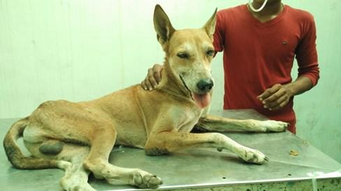


84 Plate-02: Various breeds of dog examined Saint bernard Lhasa apso Doberman Bull dog Street dog Non-descript Beagle Shih Tzu German shephered 54
85 Materials and Methods 3. Poor Housing pattern: Types of housing pattern for dogs were classified into three groups as follows and prevalence of tick infestation and babesiosis were correlated with housing patterns. 1. Kachcha house and field 2. Pakka house 3. Pakka house with access to open areas Clinical examination: The dogs were subjected to a complete clinical examination. This include history taking from the owner, general clinical examination including respiratory rate, pulse rate, rectal temperature, mucous membrane color, palpation of peripheral lymphnodes, examination of coat for presence of ticks etc. (Plate-03 (i) & (ii)) and data were recorded in proforma (Appendix-I) Laboratory examination: Examination of Giemsa stained peripheral blood smears for presence of intraerythrocytic piroplasm of Babesia spp. for microscopic examination; analysis of haematological parameters from obtained blood samples and analysis of serological parameters were carried out in laboratory at Department of Veterinary Medicine, Department of Veterinary Parasitology; Department of Veterinary Biochemistry, College of Veterinary Science & Aniamal Husbandry, Anand Agricultural University, Anand and Pathology laboratory of Nandini Veterinary Hospital, Surat. 55


86 Plate-03(i): Clinical examination of dogs A. Recording of rectal temperature B. Cojuctival mucus membranes C. Oral mucosa (Pale) 56


87 Plate-03(ii): Clinical examination of dogs D. Recording of CRT E. Ear examination F. Inguinal region examination 57
88 Materials and Methods Blood and serum samples from healthy dogs as a control and suspected dogs were collected for different laboratory examination and haematobiochemical changes using standard kits. 5 ml of blood was withdrawn from cephalic vein out of which 2 ml was collected in a sterile plastic K 3 EDTA vaccutainer for haematological analysis and 3 ml in a sterile plain plastic vaccutainer which was centrifuged at rpm for 5 minutes to separate serum and were stored for further investigation Blood smear examination: A total 79 peripheral smears were examined for the presence of intra-erythrocytic piroplasm of Babesia spp. (Plate-04) Preparation of smear: Thin blood smears from tip of ear as well as blood samples obtained from peripheral veins were prepared as per standard methods described by Kelly (1979) Staining with Giemsa stain: Method: 1. The Giemsa staining technique was performed by fixation of thin blood smears with methyl alcohol for 30 seconds. 2. The dilute stain was applied for 45 minutes. 3. Blood smears were then rinsed with distilled water and air dried Examination of stained smears: The blood smears were examined under 100x oil emersion fields (OIFs), Results were interpreted as babesiosis when piroplasm Babesia or developing stages were found in at least one cell of erythrocytes. 58


89 Plate-04: Blood smear preparation & Examination A. Blood smear staining B. Microscopic examination 59
90 Materials and Methods Haematological study: Haematological analysis of samples were performed from whole blood to estimate following haematological parameters by autohaematoanalyzer (Analytical, Hema 2062, Analytical technologies limited, Vadodara, India) (Plate-05). Parameters Unit Haemoglobin (Hb) - g/dl Total Erythrocyte Count (TEC) / μl Total Leukocyte Count (TLC), /μl Packed Cell Volume (PCV) - % Differential Leukocyte Count (DLC) Neutrophils - % Lymphocytes - % Monocytes - % Eosinophils - % Basophils - % Platelet count /μl Mean Corpuscle Volume (MCV), - Fl Mean Corpuscle Hemoglobin (MCH) - Pg Mean Corpuscle Hemoglobin Concentration - g/dl (MCHC) 60
91 Materials and Methods Serology: Biochemical parameters like Alkaline phosphatase (ALP), Alanine amino transferase (ALT), Aspartate amino transferase (AST), Serum creatinine, Total Plasma Protein (TPP), Albumin, Globulin, Albumin:Globulin ratio, Total bilirubin were evaluated by using an autobiochemical analyzer (Mindray, BS-120 chemistry analyser) using standard kits manufactured by coral clinical systems (Plate-05). Blood Urea Nitrogen (BUN) was estimated manually using standard kit mentioned above. Table 3.2: Methods of serum biochemical analysis Parameter (Unit) Alkaline phosphatase (ALP) (IU/L) Alanine amino transferase (ALT) (IU/L) Aspartate amino transferase (AST) (IU/L) Total bilirubin (mg/dl) Blood Urea Nitrogen (mg/dl) Serum creatinine (mg/dl) Total plasma protein (g/dl) Albumin (g/dl) Method pnpp Kinetic Mod. IFCC Mod. IFCC Mod. Jendrassik & Grog s DAM Mod. Zaffe s Kinetic Biuret method BCG method Therapeutic management of positive cases: Twenty four positive cases of canine babesiosis were selected for therapeutic management with three drugs as well as supportive therapy. Dogs naturally infected with babesiosis were grouped irrespective of species in three groups (i.e., Group B, C & D) shown in Table


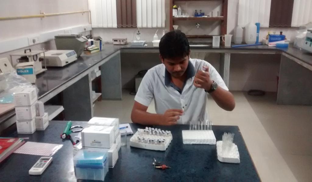
92 Plate-05: Haematological & Biochemical analysis A. B. C. 62
93 Materials and Methods Table 3.3 Treatment protocol for babesiosis positive dogs Sr. No. Particulars Dose Healthy Control Group A (n=8) Treatment group Group Group Group B C D (n=8) (n=8) (n=8) 1 Diminazine Aceturate 3.5 mg/kg by i.m. route Oxytetracyline 22 mg/kg by i.v. route for 3 days Clindamycin + Diminazine Aceturate 25 mg/kg by PO b.i.d. for 14 days 3.5 mg/kg by i.m. on 14 th day Group A: The dogs in this group did not receive any treatment and considered as control. Group B: The affected dogs in this group were given treated with Diminazine 3.5 mg/kg BW, intramuscularly on first day after diagnosis and sample collection. Group C: The affected dogs in this group were given treatment with 22 mg/kg BW, intravenously for three days after diagnosis and sample collection. 63
94 Materials and Methods Group D: The affected dogs in this group were given treatment with 25 mg/kg BW b.i.d., orally for 14 days followed by Diminazine 3.5 mg/kg BW, intramuscularly on 14 th day after diagnosis and sample collection Statistical analysis: The data obtained on clinical variants, hematological and serological parameters were subjected to statistical analysis of above data included Students t test, two samples assuming equal variances and t-test: Paired two samples for means and completely randomized design (CRD) on IBM SPSS Statistics as per the method described by Snedecor and Cochran (1990). One-way-analysis of variance (ANOVA) was used to compare the efficacy of three different therapeutic agents. Variables with P<0.05 are considered as statistically significant variables with P>0.05 were considered as statistically non-significant. 64
95 Materials and Methods ANNEXURE I Study on Clinicopathological changes and Therapeutic Management of Canine Babesiosis A. General Information: 1) Name of Owner: 2) Address: 3) Contact No.: B. Details of Dog: 1) Age: 2) Sex: 3) Breed: 4) Identification Mark: 5) Hygiene: Good/Fair/Poor C. Anamnesis: 1) Urination: Normal/Dribbling/Stranguria/Dysuria/Polyuria/Blood mixed/anuria 2) Deworming: Time: Agent: 3) Vaccination: Time: Agent: 4) Present complaint: Duration: 5) Any other complications, if noticed: D. Clinical Examination: 1) Apperance: Normal/Thin/Emaciated/Fatty/Others: 2) Behavior: Normal/Apathy/Drowsy/Excited/Dull/Depressed/Others: 3) Hair coat: Good/Fair/Poor 4) Presence of ectoparasite: Yes/No: 5) Type of skin lesion, if present: Alopacia/Papule/Macule/Pustule/Urticaria/Erythrema/Others: 6) Rectal Temperature ( F): 7) Mucosal membranes: Normal/Pale/congested/Cyanotic/Icteric 8) Respiratory Rate: / minute 9) Heart Rate: / minute 10) Pulse Rate: / minute (Tachycardia/Bradycardia) Other observations, if any: 65
96 Materials and Methods E. Laboratory Examination: 1) Sample ID: Whole Blood sample: Serum sample: 2) Haematological parameters: Sr. Parameter No. 1 Hb (g/dl) 2 TEC ( 10 6 / μl) 3 PCV (%) 4 MCV (fl) 5 MCH (g/dl) 6 MCHC (pg) 7 TLC ( 10 3 / μl) 8 DLC (%) Neutrophils (%) Lymphocytes (%) Monocytes (%) Eosinophils (%) Basophils (%) 9 Platelet count ( 10 5 / μl) 3) Serum biochemical parameters: Sr. Parameter No. 1 ALP (IU/L) 2 ALT (IU/L) 3 AST (IU/L) 4 Total Bilirubin (mg/dl) 5 BUN (mg/dl) 6 Creatinine (mg/dl) 7 Total protein (g/dl) 8 Albumin (g/dl) 9 Globulin (g/dl) 10 A:G (g/dl) Value Value 4) Blood smear examination: F. Treatment: G. Recommendations/ Advice:- 66
97 Results & Discuusion
98 Results & Discussion Chapter 4 Results & Discussion Dogs are impeded by several infectious disease conditions such as bacterial, fungal, viral, endo and ectoparasites, haemoprotozoan diseases and various noninfectious disorders like vitamin and mineral deficiencies. Dog-bite, spinal cord injuries, fractures and many more contribute variable morbidities and mortalities. Several epidemiological, environmental and managerial factors precipitate these conditions. Once considered a rare disease, canine babesiosis is now diagnosed more frequently in endemic areas owing to increased transport of the dogs. Clinical and haematological abnormalities are often non-specific in babesiosis; therefore a definitive diagnosis may be difficult to make. The diagnosis of babesiosis can be achieved by a combination of clinical variants, haematological parameters and serum biochemical parameters as well as detection of intra-erythrocytic piroplasm in peripheral blood smears. Keeping in view of the above facts, the epidemiology, diagnosis and therapeutic management of babesiosis in dogs were studied and the derived information is presented in this chapter. 4.1 Prevalence: In the present study, a total of 118 domesticated dogs were examined clinically. Out of these 118 dogs, 70 (59.32%) were presented with tick infestation. A Total 79 dogs were screened for babesiosis by demonstrating presence of intra-erythrocytic piroplasm of Babesia spp. after staining with Giemsa staining solution was carried out; out of which 24 (30.38%) were positive for babesiosis. Prevalence of tick infestation 67
99 Results & Discussion and babesiosis and its correlation with various epidemiological parameters such as breed, sex, age, hygienic condition and housing pattern is presented Prevalence of tick infestation in dogs: During a period of present study, a total 118 dogs were examined which were presented at TVCC as well as Nandini Veterinary Hospital (Table-3.1). Prevalence of tick infestation in dogs and its correlation with epidemiological parameters such as breed, sex, age, hygienic condition and housing pattern is presented Overall prevalence of tick infestation in dogs: Out of 118 dogs clinically examined during the period of proposed work, 70 dogs were harboring tick infestation and thus, overall prevalence of tick infestation was per cent (Table-4.1, Figure-01). Table-4.1 Overall prevalence of tick infestation in dogs No. of dogs Percentage Total examined Tick infested Overall prevalence of tick infestation was recorded higher compared to 52.71% reported by Dagnone et al. (2003), % by Smith et al. (2011), Murtaz ul-hasan et al. (2012) and 58.11% by Bhadesiya and Modi (2015) while it is lower than 80.00% observed by Nithikathkul et al. (2005), 58.80% by Dantas-Torres et al. (2009) and 96% by Konto et al. (2015). Despite extensive work to elucidate patterns of tick abundance of dogs has been systematically evaluated worldwide. The data on tick infestation in dogs and the vector role in India is scanty. In past, work was conducted mostly in domestic animals other 68
100 Results & Discussion than dogs (Kikani, 1988; Kumar et al., 2002; Islam et al., 2006 and Ghosh et al., 2007). Few investigations have been made into the presence of tick infestation among the dog population. This study provides a basis for epidemiological studies and also exploration of the vector role of ticks Breed-wise prevalence of tick infestation in dogs: Breed-wise prevalence of tick infestation in dogs is shown in Table-4.2 and Figure-02. Table-4.2 Breed-wise prevalence of tick infestation in dogs Sr. No. Breed Overall (N=70) Per cent 1 Labrador retriever Doberman pinscher Non-descript Pomeranian German shepherd Pug Boxer Golden retriever Great Dane Saint Bernard Coker spaniel Lhasa Apso Shih Tzu Total

101 Results & Discussion 70
102 Results & Discussion Among various breeds of dogs, prevalence of tick infestation was reported highest (18.57%) in Labrador retriever followed by Doberman pinscher and Nondescript (17.14% each), Pomeranian (14.29%), German shepherd (12.85%), Pug (04.28%), Boxer; Golden retriever; Great Dane; Saint bernard (02.86% each) and Coker spaniel; Lhasa Apso; Shih Tzu (01.43% each) breeds of dog Sex-wise prevalence of tick infestation in dogs: Sex-wise prevalence of tick infestation in dogs is shown in Table-4.3 and Figure-03. Out of 70 dogs with tick infestation, overall sex-wise prevalence was recorded higher in females (55.71%) as compared to males (44.29%). Dantas-Torres (2010) stated that it is uncertain whether tick infestation has a gender-related susceptibility or a matter of exposition while Konto et al. (2014) stated that sex appeared to have a less significant influence (P>0.05) on the prevalence of ticks among the dog population. Table-4.3: Sex-wise prevalence of tick infestation in dogs Sr. No. Sex Overall (N=70) Per cent 1 Males Females Total In the present study, prevalence of tick infestation was recorded higher in females than males while Silveira et al. (2009) recoded significantly higher (58.29%) prevalence of tick infestation in male dogs than females (41.71%). 71
103 Results & Discussion Age-wise prevalence of tick infestation in dogs: Age wise prevalence of tick infestation in dogs is shown in Table-4.4 and Figure-04. Table-4.4: Age-wise prevalence of tick infestation in dogs Sr. Overall Age group No. (N=70) Per cent 1 1 year years years Total Among 70 dogs with tick infestation, overall age-wise prevalence was recorded highest (52.86%, n=37) in dogs with 2 years of age followed by dogs 1 year of age (30%, n=21) and dogs between 1 to 2 years of age (17.14%, n=12) Hygienic condition-wise prevalence of tick infestation in dogs: Hygienic condition-wise prevalence of tick infestation in dogs is shown in Table-4.5 and Figure-05. Table-4.5: Hygienic condition-wise prevalence of tick infestation in dogs Sr. Hygienic Overall No. Condition (N=70) Per cent 1 Good Fair Poor Total Among 70 dogs with tick infestation, overall hygienic condition-wise prevalence of tick infestation was recoded highest in dogs having poor hygienic 72

104 Results & Discussion 73
105 Results & Discussion condition (45.71%, n=32) followed by fair (31.43%, n=22) and good hygienic condition (22.86%, n=16). Shoorijeh et al. (2008) stated that tick infestation of dogs were depended on hygienic condition, contact with stray dogs and their owners Housing pattern-wise prevalence of tick infestation in dogs: Housing pattern-wise prevalence of tick infestation in dogs is shown in Table- 4.6 and Figure-06. Among 70 dogs with tick infestation, overall housing pattern-wise prevalence of tick infestation was recoded highest in dogs kept in a Pakka house with access to open areas (52.86%, n=37) followed by dogs kept in Kachcha house and field (25.71%, n=18) and Pakka house (21.43%, n=15). Table-4.6: Housing pattern-wise prevalence of tick infestation in dogs Sr. No. Housing pattern Overall (N=70) Per cent 1 Kachcha house and field Pakka house Pakka house with access to open areas Total Wu et al. (2009) stated that dogs living indoors or with limited outdoor activities (less than one walk a week) were less likely to be infested by ticks Prevalence of babesiosis: In proposed work, a total 118 dogs were examined clinically and a total 79 peripheral blood smears were made for presence of intra-erythrocytic piroplasm 74

106 Results & Discussion 75
107 Results & Discussion Babesia spp. after staining with Giemsa staining solution, 42 whole blood samples and 42 serum samples were collected. Prevalence of babesiosis and its correlation with epidemiological parameters such as breed, sex, age, hygienic condition and housing pattern is presented Overall prevalence of babesiosis Based on peripheral blood smear examination: Seventy nine dogs were examined for presence of inra-erythrocytic piroplasam Babesia spp. after staining with Giemsa staining solution; out of which, 24 were positive for presence of Babesia spp. Thus, overall prevalence of babesiosis based on blood smear examination was recorded as per cent (Table-4.7, Figure-07). Table-4.7 Overall prevalence of babesiosis in dogs No. of dogs Percentage Total examined Positive In the present study, overall prevalence of 30.38% was recorded, which is higher than recorded 0.66% by Varshney and Dey (1998), 21.68% by Saud and hazarika (2000), 21.67% by Saud et al. (2000), 29.8% by Matsuu et al. (2004), 13.97% by Ahmed et al. (2007), 33.95% by Ungar et al. (2007), 2.62% by Bashir et al. (2009), 11.6% by Kumar et al. (2009), 16.39% by Godara et al. (2010), 8.7% by Selvaraj et al. (2010), 8.6% (Delhi), 7.4% (Mumbai), 3% (Sikkim) by Abd Rani et al. (2011), 30.18% by Nalubamba et al. (2011), 1.15% by Wadhwa et al. (2011), 2.62% by Chaudhary (2012), 8.91% by Singh et al. (2012a), 5.82% by Singh et al. (2012b), 12% by Konto et al. (2014), 7.47% by Singh et al. (2014), 10.48% by Shrivastava et al. (2014), 21.5% 76
108 Results & Discussion by Gabrielli et al. (2015), 15.81% by Jadhav et al. (2015), 8.33% by Kumar et al. (2015a), 6.01% by Kumar et al. (2015c) and 13.6% by Rene Martellet et al. (2015). Overall prevalence in this study was lower than 42% reported by Bastos et al. (2004), 30.4% by Miyama et al. (2005), % by Samradhni et al. (2005), 48.57% by Bhattacharjee et al. (2013), 3.75% by Razi Jalali et al. (2013), % by Bhattacharjee et al. (2014), 31.86% by Das et al. (2015) and 32.2% by Nalubamba et al. (2015). Out of 24 positive cases, 8 (33.33%) cases were of Babesia canis and 16 were (66.67%) of Babesia gibsoni (Table-4.8, Figure-08). Overall prevalence of B. gibsoni was recorded higher compared to B. canis. Table-4.8: Species-wise prevalence of babesiosis in dogs Sr. Overall Species Per cent No. (N=24) 1 Babesia canis Babesia gibsoni Total In the present study, overall prevalence of Babesia gibsoni was 66.67% and Babesia canis 33.33%. Similar higher prevalence of B. gibsoni than B. canis were recorded by Kumar et al. (2009), Selvaraj et al. (2010), Singh et al. (2012a), Bhattacharjee et al. (2013), Singh et al. (2014), Kumar et al. (2015a), Kumar et al. (2015c) and Rene Martellet et al. (2015) while Ahmed et al. (2007) and Abd Rani et al. (2011) recorded higher prevalence of B. canis than B. gibsoni among positive babesiosis cases respectively. Occasional appearance of B. canis in the peripheral blood of dogs was also mentioned by Irwin (2009). 77

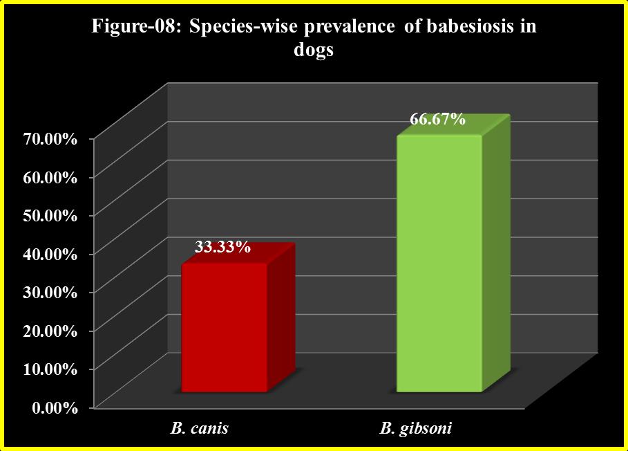
109 Results & Discussion 78
110 Results & Discussion This observation also agreed with Schalm et al. (1975) who reported demonstration of the parasite was a fortunate occurrence in 1 out of 5 blood examinations done in a Babesia canis positive dog during a month followed. Due to technical constraints, B. canis could not be detected on microscopic examination, which requires better detection methods (Bhattacharjee, 2011). Difficulty in detection of B. canis was also mentioned by Soulsby (1982) and treatment of which in endemic areas required to be mostly based on exhibited clinical symptoms. The difference in the prevalence could be due to geographical region studied, presence and abundance of vector, season when this study was carried out and diagnostic method used to detect positive cases of babesiosis. Hence, it could be stated that canine babesiosis has been reported from places all over the globe. Giving due considerations to various places in India, prevalence of babesiosis in dogs has been reported from Bareilly, Chennai, Delhi, Guwahati, Palampur, Ludhiana, Jabalpur, Jalandhar, Jaipur, Kolkata, Ladakh, Mumbai and Nagpur. At various places other than India, prevalence of babesiosis in dogs has been reported from Brazil (Bastos et al., 2004; Ungar et al. (2007), Japan (Matsuu et al., 2004; Miyama et al., 2005), Pakistan (Ahmed et al., 2007; Bashir et al., 2009 and Chaudhary, 2012), Zambia (Nalubamba et al., 2011 and Nalubamba et al., 2015), Iran (Razi Jalali et al., 2013), Nigeria (Konto et al., 2014), Serbia (Gabrielli et al., 2015) and France (Rene Martellet et al., 2015). Once considered a rare disease, canine babesiosis is now diagnosed more frequently in endemic areas owing to increased transport of dogs by dog owners Breed-wise prevalence of babesiosis in dogs: Breed-wise prevalence of babesiosis in dogs is shown in Table-4.9 and Figure
111 Results & Discussion Breed-wise prevalence of babesiosis amongst 24 dogs naturally infected with babesiosis was recorded highest (37.5%) in Labrador retriever breed of dog, followed by German shepherd (20.83%), Non-descript (20.83%), Pomeranian (8.33%), Pug (8.33%) and Golden retriever (4.18%). Table-4.9 Breed-wise prevalence of babesiosis in dogs Species Sr. Babesia Babesia Breed No. canis gibsoni Total Percent 1 Labrador retriever German shepherd Non-descript Pomeranian Pug Golden retriever Total Higher breed-wise in Labrador retriever was in correlation with Singh et al. (2014) and Jadav et al. (2015). Of 18 babesiosis positive cases reported by Wadhwa et al. (2011), breed wise prevalence included Pomeranian (22.22%), German shepherd and Labrador retriever (16.67% each), Doberman (11.11%), Saint Barnard and Dalmatian (5.56% each) and Non-descript (22.22%) breeds of dog. Higher breed-wise prevalence in crossbred dogs was recorded by Bashir et al. (2009), Selvaraj et al. (2010), Chaudhary (2012) and Razi Jalali et al. (2013). However, Shrivastava et al. (2014) recorded maximum prevalence was in German Shepherd breed (15.47%), Samoyed (15.25%), Pug (12.50%), Non-descript (10.94%) and Spitz (10.13%) breeds of dog. 80
112 Results & Discussion Amongst B. canis positive cases, higher prevalence was recorded in Labrador retriever (N=4; 50%) followed by German shepherd (N=3; 37.5%) and Non-descript (N=1; 12.5%) breeds of dog. Whereas among B. gibsoni positive cases, higher prevalence was recorded in Labrador retriever (N=5; 31.25%) followed by Nondescript (N=4; 25%), German shepherd; Pomeranian; Pug (N=2, 11.11%) each and Golden retriever (N=1; 6.25%) breeds of dogs. Prevalence of antibodies to B. canis was significantly higher among German Shepherds suggesting a genetic predisposition of German shepherd dogs to chronic babesiosis (carrier status) with long-term maintenance of their seropositivity (Sandor et al., 2006). It was difficult to gauge the actual breed wise prevalence from the present study. Breed preference of owners is also different in different areas and hence the variation in the prevalence rate of babesiosis in different breeds might be due to variation in the population of the breed in area and different attitudes of the owners in care and management of their pets. Thus, the over representation of Labrador retrievers in this study could be due to the fact that number of Labrador retrievers may be more in the population or may be the result of lower numbers of dogs belonging to different breeds screened Sex-wise prevalence of babesiosis in dogs: Sex-wise prevalence of babesiosis is shown in Table-4.10 and Figure-10. Table-4.10 Sex-wise prevalence of babesiosis in dogs Species Sr. Babesia Babesia Sex Total Per cent No. canis gibsoni 1 Males Females Total
113 Results & Discussion 82
114 Results & Discussion Sex-wise prevalence of babesiosis amongst 24 dogs positive for babesiosis was recorded higher (58.33%) in females than males (41.67%). Similar higher sex-wise prevalence of babesiosis in females was recorded by Razi Jalali et al. (2013), Shrivastava & Shukla (2013), Konto et al. (2014) and Das et al. (2015). However, Miyama et al. (2005), Kumar et al. (2009), Ilie et al. (2010) and Wadhwa et al. (2011) recorded higher prevalence of babesiosis in males. Shrivastava et al. (2014) recorded 14.77% prevalence in females as compared with 7.38% in males and significant difference (P < 0.05) was noticed in the prevalence of both sexes. In present study, higher prevalence of both B. canis and B. gibsoni was recorded in females than males which were in correlation with Singh et al. (2012b) and Kumar et al. (2015c) Age-wise prevalence of babesiosis in dogs: Age-wise prevalence of babesiosis in dogs is shown in Table-4.11 and Figure- 11. Table-4.11 Age-wise prevalence of babesiosis in dogs Sr. Species No. Age Babesia Babesia Total Per cent canis gibsoni 1 1 year years years Total Age-wise prevalence of babesiosis amongst 24 dogs positive for babesiosis was recorded highest (50.00%) among dogs 1 year of age followed by dogs 2 years of age (41.67%) and dogs between 1 to 2 years of age (27.28%). 83
115 Results & Discussion In present study, higher age-wise prevalence of babesiosis was recorded in young dogs 1 year of age. Similar age-wise prevalence of babesiosis were recorded by Samradhni et al. (2005), Unger et al. (2007), Selvaraj et al. (2010), Nalubamba et al. (2011), Chaudhary (2012), Razi Jalali et al. (2013), Konto et al. (2014), Singh et al. (2014) and Kumar et al. (2015c). Out of total B. canis positive cases, highest prevalence was recoded in dogs of 2 years (N=4) of age followed by dogs of 1 year (N=3) of age and dogs between 1-2 years (N=1) of age. In present study, highest prevalence of B. gibsoni was recoded in dogs of 1year (N=12) followed by dogs of 2 years (N=10) of age and dogs between 1-2 years (N=2) of age. Singh et al. (2012b) recorded higher prevalence of B. gibsoni in dogs of < 1 year of age. Higher prevalence of babesiosis in younger dogs may be due to the underdeveloped immune system in young dogs as compared to adults (Tizard, 2004) Hygienic condition-wise prevalence of babesiosis in dogs: Hygienic condition-wise prevalence of babesiosis is shown in Table-4.12 and Figure-12. Table-4.12 Hygienic condition-wise prevalence of babesiosis in dogs Sr. Hygienic Overall Per cent No. Condition (N=24) 1 Good Fair Poor Total
116 Results & Discussion 85
117 Results & Discussion Hygienic condition-wise prevalence of babesiosis amongst 24 dogs positive for babesiosis was recorded highest (54.17%) in dogs having a fair hygienic condition followed by Poor (29.16%) and Good (16.67%). There is no information available in literature on impact of hygienic condition provided to dogs on the occurrence of babesiosis Housing pattern-wise prevalence of babesiosis in dogs: Housing pattern-wise prevalence of babesiosis in dogs is shown in Table-4.13 and Figure-13. Table-4.13: Housing pattern-wise prevalence of babesiosis in dogs Sr. No. Housing pattern Total Per cent 1 Kachcha house and field Pakka house Pakka house with access to open areas Total Housing pattern-wise prevalence of babesiosis amongst 24 dogs positive for babesiosis was recorded higher (45.84%) in dogs kept in Pakka house with access to open areas followed by dogs kept in Kachacha house and field (33.33%) and dogs kept in Pakka house (20.83%). There is no information available in literature on impact of hosing pattern provided to dogs on occurrence of babesiosis. Higher prevalence of babesiosis has been reported in dogs kept in Pakka house with access to open areas compare to others. Under these conditions, there is a high risk of contact between dogs and vector ticks which remains unnoticed when dog is allowed to rest in Pakka house. 86
118 Results & Discussion 87
119 Results & Discussion 4.2. Diagnosis: For confirmation and positive identification of babesiosis cases inclusion of clinical examination, haematological parameters, serum biochemical parameters and examination of Giemsa stained blood smears were employed Clinical examination: Clinical examination of 08 healthy dogs and 24 babesiosis positive cases was recorded and compared. The statistical analysis of clinical variants expressed by and recorded from healthy as well as babesiosis positive dogs are presented in Table-4.14; Figure- 14,15,16 & 17. Sr. No. Table-4.14: Clinical variants (Mean±SE values) in healthy and babesiosis positive dogs Parameter Healthy dogs Babesiosis positive dogs 1 Rectal temperature ( F) ± ±00.02** 2 Heart rate (beats/min) ± ±01.73** 3 Respiration rate (breaths/min) 28.50± ±03.75** 4 CRT (seconds) 01.25± ±00.14 ** (P<0.01) The mean values of rectal temperature ( F), heart rate (beats/min), respiration rate (breaths/min) and CRT (seconds) in healthy dogs were ±00.20 F, ± beats/min, ± breaths/min and ± seconds, respectively. The mean values of rectal temperature, heart rate, respiration rate and CRT in babesiosis positive dogs were ± F, ± beats/min, ± breaths/min and ± seconds, respectively. 88
120 Results & Discussion 89

121 Results & Discussion 90
122 Results & Discussion The mean values of rectal temperature, heart rate, and respiration rate in dogs with babesiosis increased significantly (P<0.01) than healthy dogs. No significant changes were observed for CRT in dogs with babesiosis than healthy dogs. Similar findings of increase rectal temperature in dogs with babesiosis were documented by Abdullahi et al. (1990), Saud et al. (2000), Gupta et al. (2002), Bastos et al. (2004), Farlanello et al. (2005), Varshney et al. (2008), Cardoso et al. (2010), Yadav et al. (2011), Andoni et al. (2013), Reddy et al. (2014), Sivajothi et al. (2014), Yogeshpriya et al. (2014), Koster et al. (2015) and Nalubamba et al. (2015). Clinical signs in healthy and in babesiosis positive dogs are presented in Table-4.15 and Plate-06(i, ii, iii, iv, v & vi). Among various clinical signs in dogs naturally positive for babesiosis, the clinical findings varies with species. In B. canis positive cases, there were General signs (55.41%), Gastrointestinal sings (22.98%), Cardiac signs (6.76%), Nervous signs (5.40%), Urological signs (5.40%) and Respiratory signs (4.05%). While in case of B. gibsoni positive cases, General signs (50.82%), Gastrointestinal sings (13.94%), Cardiac signs (13.11%), Nervous signs (13.11%), Respiratory signs (4.92%) and Urological signs (4.10%). In B. canis positive dogs (Table-4.15, Figure-18) general signs were recoded 55.41%, in which 8 (100%) dogs were having tick infestation, weakness or emaciation or apathy, dehydration and pale mucus membrane were observed in 06 (75.00%), recumbancy and lymphadenopathy in 05 (62.50%), petechiae or epistaxis or haemorrhage and congested mucus membrane in 02 (25.00%) and edema was evident in 01 (12.50%) dogs. Similar findings in B. canis positive dogs were also observed by Abdullahi et al. (1990), Varshney et al. (2003), Bastos et al. (2004), Farlanello et al. (2005), Cardoso et al. (2010), Wadhwa et al. (2011), Janus et al. (2012), Andoni et al. 91

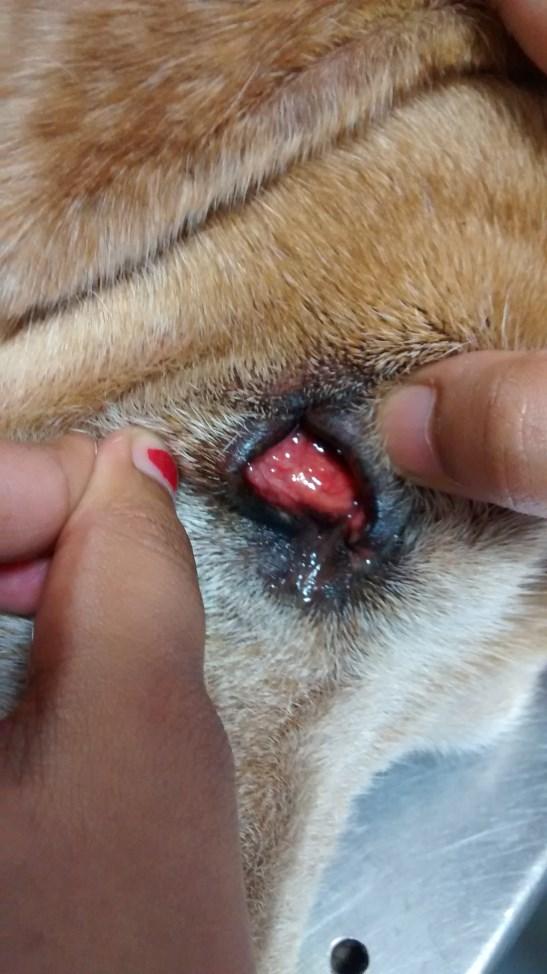
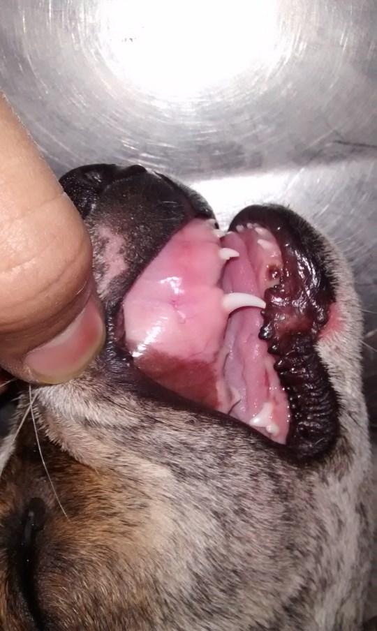

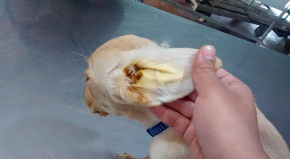
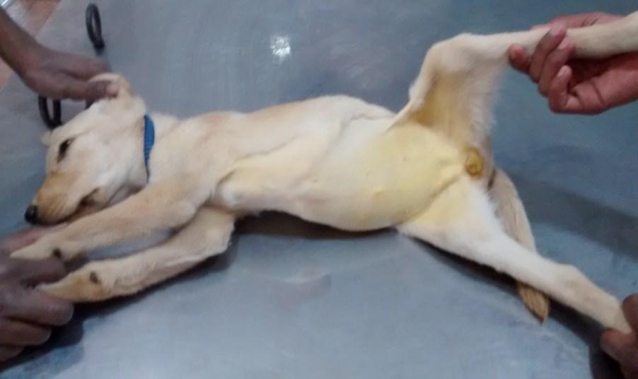
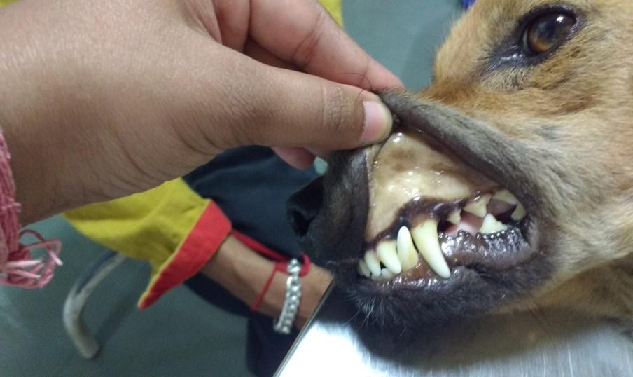

123 Plate-06(i): Clinical signs in dogs with babesiosis A. Pale mucus membranes B. Congested mucus membranes C. Icteric changes 92


124 Plate-06(ii): Clinical signs in dogs with babesiosis A. Weakness B. Ematiation & Dehydration C. Tick infestation 93


125 Plate-06(iii): Clinical signs in dogs with babesiosis D. Epistaxis E. Limb edema F. Oozing of blood from limb 94


126 Plate-06(iv): Clinical signs in dogs with babesiosis G. Severe diarrhoea H. Vomiting I. Recumbancy 95


127 Plate-06(v): Clinical signs in dogs with babesiosis J. Nasal discharge K. Comatose L. Hemoglobinuria 96


128 Plate-06(vi): Clinical signs in dogs with babesiosis M. Petechiation N. Ascites O. Hemoptysis 97
129 Results & Discussion (2013), Reddy et al. (2014), Sivajothi et al. (2014), Yogeshpriya et al. (2014) and Nalubamba et al. (2015). Table-4.15: Clinical signs in dogs with babesiosis B. canis (%) (N=8) B. gibsoni (%) (N=16) General signs Weakness/emaciation/apathy 6 (75.00) 11 (68.75) Ticks on body 8 (100) 15 (93.15) Recumbancy/prostration 5 (62.50) 7 (43.75) Dehydration 6 (75.00) 9 (56.25) Edema 1 (12.50) 0 (00.00) Lymphadenopathy 5 (62.50) 0 (00.00) Petechiae/epistaxis/haemorrhage 2 (25.00) 3 (18.75) Pale mucus membrane 6 (75.00) 9 (56.25) Congested 2 (25.00) 4 (25.00) Icteric 0 (00.00) 4 (25.00) Gastrointestinal signs Anorexia 7 (87.50) 8 (50.00) Vomiting/nausea 3 (37.50) 4 (25.00) Diarrhoea 3 (37.50) 2 (12.50) Ascites 1 (12.50) 0 (00.00) Melena 1 (12.50) 0 (00.00) Salivation 2 (25.00) 3 (18.75) Nervous signs Dullness/depression 3 (37.50) 11 (68.75) Ataxia/paresis/epilepsy 1 (12.50) 5 (31.25) Cardiac signs Weak pulse 1 (12.50) 6 (37.50) Arrhythmia 4 (50.00) 10 (62.50) Respiratory sign Nasal discharge/coughing 2 (25.00) 3 (18.75) Dysponea 1 (12.50) 3 (18.75) Urological sign Dribbling of urine 1 (12.50) 3 (18.75) Hemaglobinuria 3 (37.50) 2 (12.50) A gastrointestinal signs such as anorexia was observed in 07 (87.50%), vomiting or nausea and diarrhoea in 03 (37.50%), salivation 02 (25.00%) and ascites 98
130 Results & Discussion and melena was recorded in 01 (12.50%) dogs. Similarly, different clinical findings in B. canis positive dogs were observed by Abdullahi et al. (1990), Varshney et al. (2003), Bastos et al. (2004), Farlanello et al. (2005), Cardoso et al. (2010), Reddy et al. (2014), Sivajothi et al. (2014) and Yogeshpriya et al. (2014). Nervous sings such as dullness or depression in 03 (37.50%) and ataxia or paresis or epilepsy was in 01 (12.50%) dogs. Similarly, different clinical findings in B. canis positive dogs were also observed by Varshney et al. (2003), Janus et al. (2012) and Yogeshpriya et al. (2014). Cardiac signs such as arrhythmia was recorded in 04 (50.00%) and weak pulse in 01 (12.50%) dogs. Such different clinical findings in B. canis positive dogs were also observed by Cardoso et al. (2010). Respiratory signs such as nasal discharge or coughing was evident in 02 (25.00%) and dysponoea in 01 (12.50%) dogs. Similar different clinical findings in B. canis positive dogs were also observed by Ahmed et al. (2007) and Cardoso et al. (2010). Urological signs such as hemoglobinuria was recorded in 03 (37.50%) and dribbling of urine in 01 (12.50%) dogs. Similarly, different clinical findings in B. canis positive dogs were also observed by Varshney et al. (2003), Cardoso et al. (2010), Jadav et al. (2011), Reddy et al. (2014). In B. gibsoni positive dogs (Table-4.11, Figure-19) general signs were recoded 50.82%, in which 15 (93.15%) dogs were having tick infestation, weakness or emaciation or apathy in 11 (68.75), dehydration and pale mucus membrane in 09 (56.25%), recumbancy in 07 (43.75%), congested and icteric mucus membrane in 04(25.00%) and petechiae or epistaxis or haemorrhage in 03 (18.75%) dogs. Similar 99

131 Results & Discussion 100
132 Results & Discussion different clinical findings in B. gibsoni positive dogs were also observed by Varshney et al. (2008), Karunakaran et al. (2011), Yadav et al. (2011), Tresamol et al. (2013) and Kumar et al. (2015b), A gastrointestinal signs such as anorexia was observed in 08 (50.00%), vomiting or nausea in 04 (25.00%), salivation 03 (18.75%) and diarrhoea in 2 (12.50%) dogs. Similar different clinical findings in B. gibsoni positive dogs were also observed by Varshney et al. (2008), Yadav et al. (2011) and Kumar et al. (2015b). Nervous sings such as dullness or depression in 11 (68.75%) and ataxia or paresis or epilepsy was in 05 (31.25%) dogs. Similar different clinical findings in B. gibsoni positive dogs were also observed by Varshney et al. (2008), Yadav et al. (2011) and Tresamol et al. (2013). Cardiac signs such as arrhythmia was recorded in 10 (62.50%) and weak pulse in 06 (37.50%) dogs. Similarly, different clinical findings in B. gibsoni positive dogs were also observed by Varshney et al. (2008). Respiratory signs such as nasal discharge or coughing and dysponoea were evident in 03 (18.75%) dogs. Similar different clinical findings in B. gibsoni positive dogs were also observed by Varshney et al. (2008) and Yadav et al. (2011) Urological signs such as dribbling of urine was recorded in 03 (18.75%) and hemoglobinuria in 02 (12.50%) dogs. Similarly, different clinical findings in B. gibsoni positive dogs were also observed by Varshney et al. (2008) and Yadav et al. (2011) Laboratory examination: Diagnosis based on blood smear examination: Seventy nine dogs were examined for presence of intra-erythrocytic piroplasm of Babesia spp. in peripheral smears after staining with Giemsa staining solution (Plate-7(i) & (ii)). 101


133 Plate-07(i): Blood smear examination A. Paired pear shape d B. canis B. Ring shaped B. canis 102


134 Plate-07(ii): Blood smear examination A. Oval, comma shaped B. gibsoni B. Oval, comma shaped B. gibsoni 103
135 Results & Discussion Out of 79 dogs, 24 were positive for presence of Babesia spp. Among them 08 were positive for B. canis and 16 were of B. gibsoni. Thus, overall prevalence of babesiosis based on blood smear examination was recorded as per cent. Out of 24 positive cases, 8 (33.33%) positive cases were paired pear shape intra-erythrocytic piroplasm of B. canis and 16 were (66.67%) pleomorphic, single to multiple, intra-erythrocytic piroplasm of B. gibsoni (Table-4.8, Figure-08). Peripheral blood smear examination for diagnosis of babesiosis has been documented by many authors (Bastos et al. (2004), Furlanello et al. (2005), Ahmed et al. (2007), Zynger et al. (2007), Bashir et al. (2009), Kumar et al. (2009), Ilie et al. (2010), Karunakaran et al. (2011), Wadhwa et al. (2011), Andoni et al. (2012), Janus et al. (2012), Andoni et al. (2013), Aysul et al. (2013), Bhojne et al. (2013), Razi Jalali et al. (2013), Reddy et al. (2014), Das et al. (2015) and Kumar et al. (2015a). Overall prevalence recorded in the present study using peripheral blood smear examination was higher compare to 0.66% reported by Varshney and Dey (1998), 13.97% by Ahmed et al. (2007), 8.7% by Selvaraj et al. (2010), 1.15% by Wadhwa et al. (2011), 8.91% by Singh et al. (2012a), 10.48% by Shrivastava et al. (2014) and 15.81% by Jadhav et al. (2015) while it was lower as compared to 42% reported by Bastos et al. (2004), % by Samradhni et al. (2005), Bhattacharjee et al. (2013) and 31.86% by Das et al. (2015) Haematological parameters: The mean values of hematological parameters of healthy and babesiosis positive dogs are presented in Table The mean values of Hb (g/dl) (Figure-20), TEC ( 10 6 /μl) (Figure-21), Platelet count ( 10 3 /μl) (Figure-22) and PCV (%) (Figure-23) in healthy dogs were ± g/dl, ± /μl, ± /μl and ± %, 104
136 Results & Discussion respectively. Mean values of Hb, TEC, Platelet count and PCV in babesiosis positive dogs were ± g/dl, ± /μl, ± /μl and ± 02.21%, respectively. Table-4.16: Haematological parameters (Mean±SE) in healthy and babesiosis positive dogs Babesiosis positive Healthy dogs Sr. No. Parameter Group-I (n=24) (n=8) (Pre-treatment) 1 Hb (g/dl) ± ± 00.69** 2 TEC ( 10 6 /μl) ± ± 00.36** 3 PCV (%) ± ± 02.21** 4 TLC ( 10 3 /μl) 12.30± ± Neutrophils (%) ± ± ** 6 Lymphocytes (%) ± ± 02.13** 7 Monocytes (%) ± ± Eosinophils (%) ± ± 00.67** 9 Basophils (%) ± ± Platelet count ( 10 3 /μl) ± ±11.06* 11 MCV (fl) ± ± MCH (pg) ± ± MCHC (g/dl) ± ± ** (P < 0.01) * (P < 0.05) Levels of Hb, TEC and PCV decreased significantly (P<0.01) in dogs with babesiosis than healthy dogs. Levels of Platelet count decreased significantly (P<0.05) in dogs with babesiosis than healthy dogs. 105

137 Results & Discussion 106
138 Results & Discussion 107
139 Results & Discussion Decrease in Hb levels were in agreement with reports of Furlanello et al. (2005), Niwetpathomwat et al. (2006), Shah et al. (2011), Wadhwa et al. (2011), Andoni et al. (2012), Andoni et al. (2013), Reddy et al. (2014) and Nalubamba et al. (2015). Decreased TEC levels were in agreement with Niwetpathomwat et al. (2006), Shah et al. (2011), Andoni et al. (2012), Andoni et al. (2013), Reddy et al. (2014) and Nalubamba et al. (2015). Pre-treatment decreased Hb and TEC levels could be due to epistaxis, petechial hemorrhages and intra-vascular hemolysis or due to severe anemia. Decreased platelet counts (i.e., thrombocytopenia) in babesiosis were in agreement with Furlanello et al. (2005), Niwetpathomwat et al. (2006), Shah et al. (2011) Wadhwa et al. (2011), Andoni et al. (2012), Andoni et al. (2013), Reddy et al. (2014), Vishnurahav et al. (2014) and Nalubamba et al. (2015). The pathogenesis of thrombocytopenia during the infection may be due to platelet sequestration in the spleen or immune mediated platelet destruction and development of disseminated intravascular coagulation (Boozer and Macintire, 2003). Elevated body temperature could have contributory effect on thrombocytopenia. (Oglesbee, 1999). Decreased PCV levels in dogs with babesiosis were in agreement with Furlanello et al. (2005), Niwetpathomwat et al. (2006), Shah et al. (2011) Wadhwa et al. (2011), Andoni et al. (2012), Andoni et al. (2013), Reddy et al. (2014) and Nalubamba et al. (2015). The mean values of TLC/WBCs ( 10 3 /μl) (Figure-24) count in healthy dogs and in babesiosis positive dogs were 12.30± /μl and ± /μl, respectively. The difference between TLC/WBCs count in dogs with babesiosis and 108
140 Results & Discussion healthy dogs was statistically non-significant. However, Wadhwa et al. (2011) and Reddy et al. (2014) reported presence of significant leucopenia in dogs with babesiosis. The mean values of Differential Leucocyte Count (DLC) (Figure-25) including neutrophils (%), lymphocytes (%), monocytes (%), eosinophils (%) and basophils (%) in healthy dogs were ± 01.60%, ± 00.76%, ± 00.54%, ± 00.33% and ± 00.00, respectively. The mean values of DLC including neutrophils (%), lymphocytes (%), monocytes (%), eosinophils (%) and basophils (%) in babesiosis positive dogs were ± 01.92%, ± 02.13%, ± 00.45%, ± 00.67% and ± 00.00%, respectively. Among DLC, levels of neutrophils increased significantly (P<0.01) in dogs with babesiosis than healthy dogs. This finding was in agreement with Shah et al. (2011), and Vishnurahav et al. (2014). However, Reddy et al. (2014) reported decreased significantly in dogs with babesiosis than healthy dogs. Levels of lymphocytes decreased significantly (P<0.01) in dogs with babesiosis than healthy dogs. This finding was in agreement with Andoni et al. (2012). However, Shah et al. (2011) and Reddy et al. (2014) reported significant increase in lymphocytes count in dogs with babesiosis. Levels of eosinophils increased significantly (P<0.01) in dogs with babesiosis than healthy dogs. The differences between monocytes and basophils in dogs with babesiosis than healthy dogs were statistically non-significant. However, Reddy et al. (2014) reported significant monocytes changes in dogs with babesiosis. Among various RBC indices, the mean values of MCV (fl) (Figure-26), MCH (pg) (Figure-27) and MCHC (g/dl) (Figure-28) in healthy dogs were ± fl, 109

141 Results & Discussion 110

142 Results & Discussion 111
143 Results & Discussion ± pg and ± g/dl, respectively. The mean values of MCV, MCH and MCHC in babesiosis positive dogs were ± fl, ± pg and ± g/dl, respectively. The difference between values of MCV, MCH and MCHC in babesiosis positive dogs and healthy dogs were statistically non-significant Serum biochemical parameters: The mean values of serum biochemical parameters of healthy and babesiosis positive dogs are presented in Table Table-4.17: Serum biochemical parameters (Mean±SE) in healthy and babesiosis positive dogs Babesiosis positive Healthy dogs Sr. Group-I Parameter (n=24) No. (n=8) (Pre-treatment) 1 ALP (IU/L) ± ± 27.36** 2 ALT (IU/L) ± ± 06.94** 3 AST (IU/L) ± ± 09.89** 4 Total bilirubin (mg/dl) ± ± 00.06** 5 BUN (mg/dl) ± ± 02.29** 6 Creatinine (mg/dl) ± ± Total protein (g/dl) ± ± Albumin (g/dl) ± ± 00.15** 9 Globulin (g/dl) ± ± 00.26** 10 A:G (g/dl) ± ± 00.06** ** (P < 0.01) The mean values of ALP (IU/L) (Figure-29), ALT/SGPT (IU/L) (Figure-30), AST (IU/L) (Figure-31) and total bilirubin (mg/dl) (Figure-32) in healthy dogs were ± IU/L, ± IU/L, ± IU/L and ± mg/dl, 112
144 Results & Discussion 113
145 Results & Discussion respectively. The mean values of ALP, ALT, AST and total bilirubin in babesiosis positive dogs were ± IU/L, ± IU/L, ± IU/L and ± mg/dl, respectively. Among various serum biochemical parameters, levels of ALP increased significantly (P<0.01) in dogs with babesiosis than healthy dogs. This finding was in agreement with Furlanello et al. (2005) and Shah et al. (2011). Levels of ALT increased significantly (P<0.01) in dogs with babesiosis than healthy dogs. These findings were in agreement with Furlanello et al. (2005), Wadhwa et al. (2011) and Reddy et al. (2014). Levels of AST increased significantly (P<0.01) in dogs with babesiosis than healthy dogs. This finding was in agreement with Furlanello et al. (2005) and Wadhwa et al. (2011). Levels of total bilirubin increased significantly (P<0.01) in dogs with babesiosis than healthy dogs. This finding was in agreement with Furlanello et al. (2005) and Shah et al. (2011). Increase in level of ALP was may be due to damage or abnormal function of biliary system (Crnogaj et al., 2010). Increased activities of AST and ALT were might be due to escape of these enzymes from the damaged hepatic parenchymal cells with necrosis or altered membrane permeability indicating hepatic dysfunction. (Wadhwa et al., 2011). Hyperbilirubinaemia was due to resulted from both intravascular and extravascular haemolysis (Irwin and Hutchinson, 1991). The mean values of BUN (mg/dl) (Figure-33) and creatinine (mg/dl) (Figure- 34) in healthy dogs were ± mg/dl and ± mg/dl, respectively. The mean values of BUN and creatinine in dogs with babesiosis were ± mg/dl and ± mg/dl, respectively. 114
146 Results & Discussion Levels of BUN increased significantly (P<0.01) in dogs with babesiosis than healthy dogs. The difference between values of creatinine in babesiosis positive dogs and healthy dogs were statistically non-significant. This finding was in agreement with Wadhwa et al. (2011). However, Andoni et al. (2013), Reddy et al. (2014) and Vishnurahav et al. (2014) reported significant increase in creatinine in babesiosis positive dogs and healthy dogs. However, Reddy et al. (2014) recorded significant increase in level of BUN in babesiosis positive dogs and healthy dogs. The mean values of Total protein (g/dl) (Figure-35), Albumin (g/dl) (Figure- 35), Globulin (g/dl) (Figure-35) and A:G ratio (g/dl) (Figure-35) in healthy dogs were ± 00.37g/dl, ± g/dl, ± 00.07g/dl and ± g/dl, respectively. The mean values of total protein, Albumin, globulin and A:G ratio in babesiosis positive dogs were ± g/dl, ± 00.15g/dl, ± g/dl and ± g/dl, respectively. Levels of albumin and A:G ratio decreased significantly (P<0.01) in dogs with babesiosis than healthy dogs. Levels of globulin increased significantly (P<0.01) in dogs with babesiosis than healthy dogs. The difference between values of total protein in babesiosis positive dogs and healthy dogs were statistically non-significant. This finding was in agreement with Reddy et al. (2014). 4.3: Therapeutic management of cases positive for babesiosis: In the present study, 24 cases (n=24) those were diagnosed to have babesiosis were treated with three different drugs (Table-3.3, Plate-08). 115
147 BUN (mg/dl) Total bilirubin Figure-32: Mean±SE values of Total bilirubin (mg/dl) of Healthy & Babesiosis positive dogs ± ± Healthy Babesiosis positive dogs Figure-33: Mean±SE values of BUN (mg/dl) of Healthy & Babesiosis positive dogs ± ± Healthy Babesiosis positive dogs 116

 03.57 ± 02.15 02.15 ± 00.15 02.16 ± 00.07 03.18 ± 00.26 01.53 ± 00.")
148 mg/dl Creatinine (mg/dl) Figure-34: Mean±SE values of Creatinine (mg/dl) of Healthy & Babesiosis positive dogs ± Healthy ± Babesiosis positive dogs Figure-35: Mean±SE values of Protein (mg/dl) of Healthy & Babesiosis positive dogs ± ± Total protein (g/dl) ± ± ± ± ± ± Albumin (g/dl) Globulin (g/dl) A:G (g/dl) Healthy Babesiosis positive dogs 117
 Teaching")
 Nandini Veterinary Hospital,")
149 Plate-08: Therapeutic management of clinical cases (i) Teaching Veterinary Clinical Complex, Anand (ii) Nandini Veterinary Hospital, Surat 118
Case Report Peritoneal Effusion in a Dog due to Babesia gibsoni Infection
 Case Reports in Veterinary Medicine, Article ID 807141, 4 pages http://dx.doi.org/10.1155/2014/807141 Case Report Peritoneal Effusion in a Dog due to Babesia gibsoni Infection Suresh Gonde, 1 Sushma Chhabra,
Pathogenesis of E. canis
 Tick-born disease Rhipicephalus sanguineus brown dog tick Rickettsia Ehrlichia canis Ehrlichia platys Anaplasma platys Pathogenesis of E. canis Incubation period: 8 20 days Mononuclear cells Liver, spleen,
Canine Anaplasmosis Anaplasma phagocytophilum Anaplasma platys
 Canine Anaplasmosis Anaplasma phagocytophilum Anaplasma platys It takes just hours for an infected tick to transmit Anaplasma organisms to a dog. What is canine anaplasmosis? Canine anaplasmosis is a disease
InternationalJournalofAgricultural
 www.ijasvm.com IJASVM InternationalJournalofAgricultural SciencesandVeterinaryMedicine ISSN:2320-3730 Vol.5,No.1,February2017 E-Mail:editorijasvm@gmail.com oreditor@ijasvm.comm@gmail.com Int. J. Agric.Sc
Clinicopathological findings in dogs naturally infected dogs with Babesia
 Albanian j. agric. sci. 2013;12 (2): 185-189 Agricultural University of Tirana RESEARCH ARTICLE Correspondence: egon Andoni, Affiliation; Agricultural University of Tirana Email: egon-andoni@yahoo.com
Blood protozoan: Plasmodium
 Blood protozoan: Plasmodium Dr. Hala Al Daghistani The causative agent of including Plasmodium vivax P. falciparum P. malariae P. ovale. malaria in humans: four species are associated The Plasmodium spp.
Blood protozoan: Plasmodium
 Blood protozoan: Plasmodium The causative agent of including Plasmodium vivax P. falciparum P. malariae P. ovale. malaria in humans:four species are associated The Plasmodium spp. life cycle can be divided
MURDOCH RESEARCH REPOSITORY
 MURDOCH RESEARCH REPOSITORY http://researchrepository.murdoch.edu.au/20636/ Irwin, P.J. (2007) Blood, bull terriers and babesiosis: a review of canine babesiosis. In: 32nd Annual World Small Animal Veterinary
SUMMARY Of the PhD thesis entitled RESEARCH ON THE EPIDEMIOLOGY, DIAGNOSIS AND CONTROL OF CANINE BABESIOSIS IN WESTERN ROMANIA
 This thesis contains: Summaries (Romanian, English, French) Extended general part 55 pages; Extended own research part 137 pages; Tables: 11; Figures full color: 111; References: 303 references. SUMMARY
INCIDENCE OF CANINE BABESIOSIS IN AND AROUND KOLKATA, WEST BENGAL, INDIA
 Exploratory Animal and Medical Research, Vol.5, Issue 1, June, 2015 Explor Anim Med Res, Vol.5, Issue - 1, 2015, p. 102-107 Research Article INCIDENCE OF CANINE BABESIOSIS IN AND AROUND KOLKATA, WEST BENGAL,
Australian and New Zealand College of Veterinary Scientists. Membership Examination. Medicine of Cats Paper 1
 Australian and New Zealand College of Veterinary Scientists Membership Examination June 2016 Medicine of Cats Paper 1 Perusal time: Fifteen (15) minutes Time allowed: Two (2) hours after perusal Answer
The Epidemiological Survey and Analysis of Canine Babesiosis in Xi an City, China
 Animal and Veterinary Sciences 2018; 6(3): 43-50 http://www.sciencepublishinggroup.com/j/avs doi: 10.11648/j.avs.20180603.12 ISSN: 2328-5842 (Print); ISSN: 2328-5850 (Online) Report The Epidemiological
An Overview of Canine Babesiosis
 Page 1 of 6 C. Wyatt Cleveland, DVM; David S. Peterson, DVM, PhD; and Kenneth S. Latimer, DVM, PhD Class of 2002 (Cleveland), Department of Medical Microbiology and Parasitology (Peterson), and Department
Adopting a dog from Spain comes with some risks of which you should be aware.
 LHB Galgo Rescue Information for your Vet Adopting a dog from Spain comes with some risks of which you should be aware. Nearly all Spanish shelters test for Babesia, Ehrlichia, Leishmania and heartworm
Ehrlichiosis, Anaplasmosis and other Vector Borne Diseases You May Not Be Thinking About Richard E Goldstein Cornell University Ithaca NY
 Ehrlichiosis, Anaplasmosis and other Vector Borne Diseases You May Not Be Thinking About Richard E Goldstein Cornell University Ithaca NY Canine Monocytic Ehrlichiosis Ehrlichia canis The common etiologic
Australian and New Zealand College of Veterinary Scientists. Membership Examination. Veterinary Anaesthesia and Critical Care Paper 1
 Australian and New Zealand College of Veterinary Scientists Membership Examination June 2015 Veterinary Anaesthesia and Critical Care Paper 1 Perusal time: Fifteen (15) minutes Time allowed: Two (2) hours
AUSTRALIAN AND NEW ZEALAND COLLEGE OF VETERINARY SCIENTISTS. Sample Exam Questions. Veterinary Practice (Small Animal)
") AUSTRALIAN AND NEW ZEALAND COLLEGE OF VETERINARY SCIENTISTS Sample Exam Questions Veterinary Practice (Small Animal) Written Examination (Component 1) Written Paper 1 (two hours): Principles of Veterinary
AUSTRALIAN AND NEW ZEALAND COLLEGE OF VETERINARY SCIENTISTS Sample Exam Questions Veterinary Practice (Small Animal) Written Examination (Component 1) Written Paper 1 (two hours): Principles of Veterinary
Journal home page:
 Journal home page: http://www.journalijiar.com INTERNATIONAL JOURNAL OF INNOVATIVE AND APPLIED RESEARCH RESEARCH ARTICLE A First Case Report of Feline Infectious Peritonitis in a Domestic Cat in Pakistan
DIAGNOSIS AND MANAGEMENT OF CHOLECYSTITIS IN DOGS
 Int. J. Agric.Sc & Vet.Med. 2014 K Satish Kumar and D Srikala, 2014 Research Paper ISSN 2320-3730 www.ijasvm.com Vol. 2, No. 3, August 2014 2014 www.ijasvm.com. All Rights Reserved DIAGNOSIS AND MANAGEMENT
Ehrlichia and Anaplasma: What Do We Need to Know in NY State Richard E Goldstein DVM DACVIM DECVIM-CA The Animal Medical Center New York, NY
 Ehrlichia and Anaplasma: What Do We Need to Know in NY State Richard E Goldstein DVM DACVIM DECVIM-CA The Animal Medical Center New York, NY Learning Objectives The attendees will be familiar with the
Australian and New Zealand College of Veterinary Scientists. Membership Examination. Medicine of Horses Paper 1
 Australian and New Zealand College of Veterinary Scientists Membership Examination June 2015 Medicine of Horses Paper 1 Perusal time: Fifteen (15) minutes Time allowed: Two (2) hours after perusal Answer
Prevalence of Babesia canis and Hepatozoon canis in Zaria, Nigeria
 RESEARCH ARTICLE Sokoto Journal of Veterinary Sciences (P-ISSN 1595-093X/ E-ISSN 2315-6201) Okubanjo et al./sokoto Journal of Veterinary Sciences (2013) 11(2): 15-20. http://dx.doi.org/10.4314/sokjvs.v11i2.3
both are fatal diseases. In babesiosis blood comes out with the urine and hence it is also known as Red water disease. Theileria vaccines are not
 1.1 INTRODUCTION Animal husbandry plays an important role in Indian agriculture. Indians by large are vegetarian and as such the only source of animal protein is milk and milk products. With the increasing
Double-Blind, Placebo-Controlled, Randomized Study of Dipyrone as a Treatment for Pyrexia in Horses
 Double-Blind, Placebo-Controlled, Randomized Study of Dipyrone as a Treatment for Pyrexia in Horses Emily Sundman, DVM Ming Yin, PhD Tianhua Hu, PhD Melinda Poole, DVM Disclosures Sundman, Yin, Hu, and
How to talk to clients about heartworm disease
 Client Communication How to talk to clients about heartworm disease Detecting heartworm infection early generally allows for a faster and more effective response to treatment. Answers to pet owners most
Topics. Ticks on dogs in North America. Ticks and tick-borne diseases: emerging problems? Andrew S. Peregrine
 Ticks and tick-borne diseases: emerging problems? Andrew S. Peregrine E-mail: aperegri@ovc.uoguelph.ca Topics Ticks on dogs in Ontario and the pathogens they transmit? Should dogs be routinely screened
1. Babesia bigemina. 2. Anaplasma marginale. 3. Theileria orientalis. 4. Trypanosoma evansi. Vector: Rhipicephalus (Boophilus) microplus.
 microplus.") 1. Babesia bigemina. Vector: Rhipicephalus (Boophilus) microplus. 2. Anaplasma marginale. Vector: Rhipicephalus (Boophilus) microplus. 3. Theileria orientalis. Vector: Rhipicephalus (Boophilus) microplus.
1. Babesia bigemina. Vector: Rhipicephalus (Boophilus) microplus. 2. Anaplasma marginale. Vector: Rhipicephalus (Boophilus) microplus. 3. Theileria orientalis. Vector: Rhipicephalus (Boophilus) microplus.
Review on status of babesiosis in humans and animals in Iran
 Review on status of babesiosis in humans and animals in Iran Mousa Tavassoli, Sepideh Rajabi Department of Pathobiology, Faculty of Veterinary Medicine, Urmia University, Urmia, Iran Babesiosis is a zoonotic
Anesthesia Check-off Form
 Anesthesia Check-off Form 5231 SW 91st Drive Gainesville, FL 32608 (352) 377-6003 The doctors and staff at Haile Plantation Animal Clinic would like to offer the most advanced medical care and services
Tick-borne Disease Testing in Shelters What Does that Blue Dot Really Mean?
 Tick-borne Disease Testing in Shelters What Does that Blue Dot Really Mean? 2017 ASPCA. All Rights Reserved. Your Presenter Stephanie Janeczko, DVM, MS, DABVP, CAWA Senior Director of Shelter Medical Programs
Outline 4/25/2009. Cytauxzoonosis: A tick-transmitted parasite of domestic and wild cats in the southeastern U.S. What is Cytauxzoonosis?
 Cytauxzoonosis: A tick-transmitted parasite of domestic and wild cats in the southeastern U.S. Michelle Rosen Center for Wildlife Health Department of Forestry, Wildlife, & Fisheries What is Cytauxzoonosis?
International Journal of Science, Environment and Technology, Vol. 6, No 6, 2017,
 International Journal of Science, Environment and Technology, Vol. 6, No 6, 2017, 3362 3366 ISSN 2278-3687 (O) 2277-663X (P) CONCURRENT HAEMOPROTOZOAN AND ENDOPARASITIC INFECTION IN GOATS *Subramanian
sanguineus, in a population of
 BVA Student Travel Grant Final Report Prevalence of the Brown Dog tick, Rhipicephalus sanguineus, in a population of dogs in Zanzibar, and its role as a vector of canine tickborne disease. Bethan Warner
Suggested vector-borne disease screening guidelines
 Suggested vector-borne disease screening guidelines SNAP Dx Test Screen your dog every year with the SNAP Dx Test to detect exposure to pathogens that cause heartworm disease, ehrlichiosis, Lyme disease
Australian and New Zealand College of Veterinary Scientists. Membership Examination. Veterinary Pathology Paper 1
 Australian and New Zealand College of Veterinary Scientists Membership Examination June 2018 Veterinary Pathology Paper 1 Perusal time: Fifteen (15) minutes Time allowed: Two (2) hours after perusal Answer
Tick-Borne Disease Diagnosis: Moving from 3Dx to 4Dx AND it s MUCH more than Blue Dots! indications implications
 Tick-Borne Disease Diagnosis: Moving from 3Dx to 4Dx Richard B. Ford, DVM, MS Professor of Medicine Diplomate ACVIM and (Hon) ACVPM North Carolina State University Raleigh, NC In just the past 3 to 5 years,
Australian and New Zealand College of Veterinary Scientists. Membership Examination. Veterinary Emergency and Critical Care Paper 1
 Australian and New Zealand College of Veterinary Scientists Membership Examination June 2014 Veterinary Emergency and Critical Care Paper 1 Perusal time: Fifteen (15) minutes Time allowed: Two (2) hours
EHRLICHIOSIS IN DOGS IMPORTANCE OF TESTING FOR CONTRIBUTING AUTHORS CASE 1: SWIGGLES INTRODUCTION WITH PERSISTENT LYMPHOCYTOSIS
 THE IMPORTANCE OF TESTING FOR EHRLICHIOSIS IN DOGS WITH PERSISTENT LYMPHOCYTOSIS Contributing Authors: Mary Anna Thrall, DVM, MS, DACVP Diana Scorpio, DVM, MS, DACLAM Ross University School of Veterinary
Fact sheet. A u s t r a l i a n w ildlife. Introductory statement. Aetiology. Natural hosts. World distribution. Occurrences in Australia
 P iroplasms ( B abesia s p p. a n d T h e ileria s p p. ) in A u s t r a l i a n w ildlife Fact sheet Introductory statement Babesia spp. and Theileria spp. are protozoan haemoparasites which invade the
FACULTY OF VETERINARY MEDICINE
 FACULTY OF VETERINARY MEDICINE DEPARTMENT OF VETERINARY PARASITOLOGY AND ENTOMOLOGY M.Sc. AND Ph.D. DEGREE PROGRAMMES The postgraduate programmes of the Department of Veterinary Parasitology and Entomology
Copyright is owned by the Author of the thesis. Permission is given for a copy to be downloaded by an individual for the purpose of research and
 Copyright is owned by the Author of the thesis. Permission is given for a copy to be downloaded by an individual for the purpose of research and private study only. The thesis may not be reproduced elsewhere
Canine babesiosis is an emerging hemoprotozoan
 J Vet Intern Med 2010;24:127 131 Babesiosis Caused by a Large Babesia Species in 7 Immunocompromised Dogs L.E. Sikorski, A.J. Birkenheuer, M.K. Holowaychuk, A.L. McCleary-Wheeler, J.M. Davis, and M.P.
Acute Hemorrhagic Diarrhea Syndrome (AHDS) A Cause of Bloody Feces in Dogs
 A Cause of Bloody Feces in Dogs") Acute Hemorrhagic Diarrhea Syndrome (AHDS) A Cause of Bloody Feces in Dogs No dog parent wants to clean up diarrhea. Cleaning up bloody diarrhea is even more unpleasant. Unfortunately, the development
Acute Hemorrhagic Diarrhea Syndrome (AHDS) A Cause of Bloody Feces in Dogs No dog parent wants to clean up diarrhea. Cleaning up bloody diarrhea is even more unpleasant. Unfortunately, the development
The Essentials of Ticks and Tick-borne Diseases
 The Essentials of Ticks and Tick-borne Diseases Presenter: Bobbi S. Pritt, M.D., M.Sc. Director, Clinical Parasitology Laboratory Co-Director, Vector-borne Diseases Laboratory Services Vice Chair of Education
Copper-Storage Liver Disease Basics
 Copper-Storage Liver Disease Basics OVERVIEW Abnormal accumulation of copper in the liver, causing sudden (acute) inflammation of the liver (hepatitis) or long-term (chronic) hepatitis and eventually progressive
Hematology and serum biochemistry of captive gharial (Gavialis gangeticus) in India
 in India") Veterinary World, EISSN: 2231-0916 Available at www.veterinaryworld.org/vol.7/october-2014/7.pdf RESEARCH ARTICLE Open Access Hematology and serum biochemistry of captive gharial (Gavialis gangeticus)
Veterinary World, EISSN: 2231-0916 Available at www.veterinaryworld.org/vol.7/october-2014/7.pdf RESEARCH ARTICLE Open Access Hematology and serum biochemistry of captive gharial (Gavialis gangeticus)
Ticks and tick-borne diseases
 Occupational Diseases Ticks and tick-borne diseases Ticks Ticks are small, blood sucking arthropods related to spiders, mites and scorpions. Ticks are only about one to two millimetres long before they
Parasitology Division, National Veterinary Research Institute, PMB 01 Vom Plateau State, Nigeria * Association
 !" #$%$ &'()*+# Parasitology Division, National Veterinary Research Institute, PMB 0 Vom Plateau State, Nigeria * shapumani@yahoo.com +23470355775 + Association of parasitic infection of dogs with packed
A2-year-old neutered. Diagnosing FHM in anemic patients
 Diagnosing FHM in anemic patients Feline hemotrophic mycoplasmosis can be a difficult disease to pinpoint, but there are ways to make a successful diagnosis. By Jennifer Jellison, DVM Contributing Author
Supplementary figure 1: Questionnaire used to establish the constitution of the patient (relevant characteristics of the whole patient leading to the
 Supplementary figure 1: Questionnaire used to establish the constitution of the patient (relevant characteristics of the whole patient leading to the current clinical signs). 1 Study into Hyperthyroid
Hematobiochemical changes in ehrlichiosis in dogs of Anand region, Gujarat
 Veterinary World, EISSN: 2231-0916 Available at www.veterinaryworld.org/vol.8/june-2015/4.pdf RESEARCH ARTICLE Open Access Hematobiochemical changes in in dogs of Anand region, Gujarat C. M. Bhadesiya
UNDERSTANDING THE TRANSMISSION OF TICK-BORNE PATHOGENS WITH PUBLIC HEALTH IMPLICATIONS
 UNDERSTANDING THE TRANSMISSION OF TICK-BORNE PATHOGENS WITH PUBLIC HEALTH IMPLICATIONS A. Rick Alleman, DVM, PhD, DABVP, DACVP Lighthouse Veterinary Consultants, LLC Gainesville, FL Tick-transmitted pathogens
Annual Screening for Vector-borne Disease. The SNAP 4Dx Plus Test Clinical Reference Guide
 Annual Screening for Vector-borne Disease The SNAP Dx Plus Test Clinical Reference Guide Every dog, every year For healthier pets and so much more. The benefits of vector-borne disease screening go far
PARASITOLOGICAL EXAMINATIONS CATALOGUE OF SERVICES AND PRICE LIST
 INSTITUTE OF PARASITOLOGY Biomedical Research Center Seltersberg Justus Liebig University Giessen Schubertstrasse 81 35392 Giessen Germany Office: +49 (0) 641 99 38461 Fax: +49 (0) 641 99 38469 Coprological
Saturday Clinics. Every Saturday morning we hold an open clinic for the local community.
 Saturday Clinics Every Saturday morning we hold an open clinic for the local community. We see a variety of animals ranging from livestock, dogs, cats and even rabbits and monkeys! The majority of our
Blood Cell Characteristics and Some Hematological Values of American Pit-bull Terriers in Thailand
 World Applied Sciences Journal 2 (3): 158-162, 2007 ISSN 1818-4952 IDOSI Publications, 2007 Blood Cell Characteristics and Some Hematological Values of American Pit-bull Terriers in Thailand W. Aengwanich,
Title. Author(s)Shiranaga, Nobuyuki; Inokuma, Hisashi. CitationJapanese Journal of Veterinary Research, 66(3): 221- Issue Date DOI.
Shiranaga, Nobuyuki; Inokuma, Hisashi. CitationJapanese Journal of Veterinary Research, 66(3): 221- Issue Date DOI.") Title Effects of low-dose diminazene aceturate injection followed by clindamycin administration for treating Author(s)Shiranaga, Nobuyuki; Inokuma, Hisashi CitationJapanese Journal of Veterinary Research,
Title Effects of low-dose diminazene aceturate injection followed by clindamycin administration for treating Author(s)Shiranaga, Nobuyuki; Inokuma, Hisashi CitationJapanese Journal of Veterinary Research,
Correspondence should be addressed to M. Veeraselvam;
 Veterinary Medicine International, Article ID 341047, 5 pages http://dx.doi.org/10.1155/2014/341047 Research Article Chemical Immobilization of Sloth Bears (Melursus ursinus) with Ketamine Hydrochloride
Therapeutic apheresis in veterinary
 Therapeutic apheresis in veterinary 1 I.P.Pavlov First St.-Petersburg State Medical University, Saint-Petersburg, Russia. Voinov V.A. A. By types of animals on the basis of anatomical and physiological
Chapter 1 COPYRIGHTED MATERIAL. Introduction to Veterinary Pathology. What is pathology? Who does pathology?
 What is pathology? Who does pathology? Chapter 1 Introduction to Veterinary Pathology Anatomic pathology Clinical pathology Microbiology Parasitology Immunology Toxicology Veterinary forensic pathology
LANTANA CAMERA ASSOCIATED HEPATIC AND RENAL TOXICITY IN BULLOCKS A CASE REPORT
 LANTANA CAMERA ASSOCIATED HEPATIC AND RENAL TOXICITY IN BULLOCKS A CASE REPORT DR. B. ANIL KUMAR ASSISTANT PROFESSOR COLLEGE OF VETERINARY SCIENCE KORUTLA-505 326 KARIMNAGAR DISTRICT, TELANGANA. Overview
Haemato-Biochemical Changes in Canine Demodicosis
 International Journal of Current Microbiology and Applied Sciences ISSN: 2319-7706 Volume 7 Number 11 (2018) Journal homepage: http://www.ijcmas.com Original Research Article https://doi.org/10.20546/ijcmas.2018.711.338
ISPUB.COM. E Amuta, B Atu, R Houmsou, J Ayashar INTRODUCTION
 ISPUB.COM The Internet Journal of Parasitic Diseases Volume 4 Number 1 Prevalence of Rhipicephalus sanguineus infestation and Babesia canis infection in dogs with respect to breed type and degree of freedom
Update on diagnosis of feline infectious peritonitis (FIP)
") Update on diagnosis of feline infectious peritonitis (FIP) Séverine Tasker RCVS Specialist in Feline Medicine The Feline Centre Langford Veterinary Services University of Bristol http://www.felinecentre.co.uk/
Update on diagnosis of feline infectious peritonitis (FIP) Séverine Tasker RCVS Specialist in Feline Medicine The Feline Centre Langford Veterinary Services University of Bristol http://www.felinecentre.co.uk/
What s Your Diagnosis? By Sohaila Jafarian, Class of 2018
 Signalment: Greeley, 3 yo MC DSH Presenting Complaint: ADR History: What s Your Diagnosis? By Sohaila Jafarian, Class of 2018 Patient is an indoor/outdoor cat. Previously healthy and up to date on vaccines
Copyright is owned by the Author of the thesis. Permission is given for a copy to be downloaded by an individual for the purpose of research and
 Copyright is owned by the Author of the thesis. Permission is given for a copy to be downloaded by an individual for the purpose of research and private study only. The thesis may not be reproduced elsewhere
Caution: Federal law restricts this drug to use by or on the order of a licensed veterinarian.
 BOEHRINGER INGELHEIM VETMEDICA, INC. USA Product Label http://www.vetdepot.com 2621 NORTH BELT HIGHWAY, ST. JOSEPH, MO, 64506 2002 Telephone: 800 325 9167 Fax: 816 236 2717 Email: www.bi vetmedica.com
PLASMODIUM MODULE 39.1 INTRODUCTION OBJECTIVES 39.2 MALARIAL PARASITE. Notes
 Plasmodium MODULE 39 PLASMODIUM 39.1 INTRODUCTION Malaria is characterized by intermittent fever associated with chills and rigors in the patient. There may be enlargement of the liver and spleen in the
MEDICAL MANAGEMENT OF THEILERIOSIS WITH ABOMASAL IMPACTION IN A CALF Prasanth CR 1 and Ajithkumar S 2
 International Journal of Science, Environment and Technology, Vol. 5, No 6, 2016, 3838 3842 ISSN 2278-3687 (O) 2277-663X (P) MEDICAL MANAGEMENT OF THEILERIOSIS WITH ABOMASAL IMPACTION IN A CALF Prasanth
Canine vector-borne diseases prevalence and prevention
 Vet Times The website for the veterinary profession https://www.vettimes.co.uk Canine vector-borne diseases prevalence and prevention Author : SIMON TAPPIN Categories : Vets Date : March 3, 2014 SIMON
A study of hematological changes in sheep naturally infected with Anaplasma spp. and Theileria ovis: Molecular diagnosis
 Iranian Journal of Veterinary Medicine A study of hematological changes in sheep naturally infected with Anaplasma spp. and Theileria ovis: Molecular diagnosis Khaki, Z. 1, Jalali, S.M. 2*, Kazemi, B.
Medical Notes. Wellness Care. Bulger Animal Hospital Newsletter. Volume 2, Issue 1 Winter 2008
 InTown Veterinary Group Bulger Animal Hospital Newsletter Volume 2, Issue 1 Winter 2008 InTown Veterinary Group is dedicated to providing clients with an unparalleled range of veterinary care options.
Tackle Ticks: Babesiosis
 Vet Times The website for the veterinary profession https://www.vettimes.co.uk Tackle Ticks: Babesiosis Author : Simon Tappin Categories : Canine, Companion animal, Feline, General, Practical, RVNs, Species,
Review article CANINE BABESIOSIS: WHERE DO WE STAND? BILIĆ Petra 1, KULEŠ Josipa 2, BARIĆ RAFAJ Renata 3 1, 2* , MRLJAK Vladimir INTRODUCTION
 Review article CANINE BABESIOSIS: WHERE DO WE STAND? Acta Veterinaria-Beograd 2018, 68 (2), 127-160 UDK: 636.7.09:616.993.19 DOI: 10.2478/acve-2018-0011 BILIĆ Petra 1, KULEŠ Josipa 2, BARIĆ RAFAJ Renata
UDC: : PECULIARITIES OF DOG BABESIOSIS DISTRIBUTION IN KYIV CITY
 Vestnik zoologii, 51(6): 493 498, 2017 DOI 10.1515/vzoo-2017-0059 Ecology UDC: 636.709:616.99 PECULIARITIES OF DOG BABESIOSIS DISTRIBUTION IN KYIV CITY O. V. Semenko 1, M. V. Galat 1, O. V. Shcherbak 2,
SEROPREVALENCE TO CATTLE BABESIA SPP. INFECTION IN NORTHERN SAMAR ABSTRACT
 SEROPREVALENCE TO CATTLE BABESIA SPP. INFECTION IN NORTHERN SAMAR A. Amit College of Ve terina ry Me dicine, U niversi ty of East ern P hi lii ppi nes Cata rman, Nort hern Sam ar ABSTRACT Babesiosis is
ANIMAL RABIES IN NEPAL AND RACCOON RABIES IN ALBANY COUNTY, NEW YORK
 ANIMAL RABIES IN NEPAL AND RACCOON RABIES IN ALBANY COUNTY, NEW YORK SHANKAR YADAV MPH Report/Capstone Project Presentation 07/19/2012 CHAPTER 1: FIELD EXPERIENCE AT KANSAS STATE UNIVERSITY RABIES LABORATORY
Septic cats are not small septic dogs
 Vet Times The website for the veterinary profession https://www.vettimes.co.uk Septic cats are not small septic dogs Author : ROGER WILKINSON Categories : Vets Date : September 29, 2008 ROGER WILKINSON
Fungal Disease. What is a fungus?
 Fungal Disease What is a fungus? A fungus is a living organism. It goes through a complicated life cycle and is able to spread in the environment by producing large numbers of spores that are easily dispersed
Screening for vector-borne disease. SNAP 4Dx Plus Test clinical reference guide
 Screening for vector-borne disease SNAP 4Dx Plus Test clinical reference guide Every dog, every year The Companion Animal Parasite Council (CAPC) Guidelines recommend annual comprehensive screening for
New Insights into the Treatment of Leishmaniasis
 New Insights into the Treatment of Leishmaniasis Eric Zini Snow meeting, 14 March 2009 Few drugs available for dogs Initially developed to treat human leishmaniasis, later adopted in dogs None eradicates
Outlines. Introduction Prevalence Resistance Clinical presentation Diagnosis Management Prevention Case presentation Achievements
 Amal Meas Al-Anizi, PharmD Candidate KSU, Infectious Disease Rotation 2014 Outlines Introduction Prevalence Resistance Clinical presentation Diagnosis Management Prevention Case presentation Achievements
Hyalomma impeltatum (Acari: Ixodidae) as a potential vector of malignant theileriosis in sheep in Saudi Arabia
 as a potential vector of malignant theileriosis in sheep in Saudi Arabia") Veterinary Parasitology 99 (2001) 305 309 Hyalomma impeltatum (Acari: Ixodidae) as a potential vector of malignant theileriosis in sheep in Saudi Arabia O.M.E. El-Azazy a,, T.M. El-Metenawy b, H.Y. Wassef
Veterinary Parasitology 99 (2001) 305 309 Hyalomma impeltatum (Acari: Ixodidae) as a potential vector of malignant theileriosis in sheep in Saudi Arabia O.M.E. El-Azazy a,, T.M. El-Metenawy b, H.Y. Wassef
Veterinary Parasitology
 Veterinary Parasitology 186 (2012) 159 164 Contents lists available at SciVerse ScienceDirect Veterinary Parasitology jo u rn al hom epa ge : www.elsevier.com/locate/vetpar The therapeutic efficacy of
PERSISTENT EXCESSIVE THROMBOCYTHAEMIA IN A CAT
 PERSISTENT EXCESSIVE THROMBOCYTHAEMIA IN A CAT E. Hooijberg 1, M. Pichler 2, E. Leidinger 1. 1 InVitro Labor, Vienna, Austria. 2 Tierklinik Meidling, Vienna, Austria. Signalment: 7 month-old male neutered
Salmonella Dublin: Clinical Challenges and Control
 Salmonella Dublin: Clinical Challenges and Control Simon Peek BVSc, MRCVS PhD, DACVIM, University of Wisconsin-Madison School of Veterinary Medicine Advancing animal and human health with science and compassion
Ylva Sjöström 1) and Anna Lennquist 2)
 and Anna Lennquist 2)") Ylva Sjöström 1) and Anna Lennquist 2) 1) VMD, Swedish specialist in diseases of dogs and cats, Blue Star Animal Hospital, Gjutjärnsgatan 4, SE-417 07 Gothenburg, Sweden 2) PhD in Zoophysiology, Dept.
Ylva Sjöström 1) and Anna Lennquist 2) 1) VMD, Swedish specialist in diseases of dogs and cats, Blue Star Animal Hospital, Gjutjärnsgatan 4, SE-417 07 Gothenburg, Sweden 2) PhD in Zoophysiology, Dept.
VETERINARY SCIENCE CURRICULUM. Unit 1: Safety and Sanitation
 Chariho Regional School District - Science Curriculum September, 2016 VETERINARY SCIENCE CURRICULUM Unit 1: Safety and Sanitation Students will gain an understanding of the types of hazards common in veterinary
INFLUENCE OF ANAEMIA ON AZOTAEMIA IN DOGS INFECTED WITH BABESIA CANIS IN POLAND
 Bull Vet Inst Pulawy 53, 663-668, 2009 INFLUENCE OF ANAEMIA ON AZOTAEMIA IN DOGS INFECTED WITH BABESIA CANIS IN POLAND WOJCIECH ZYGNER AND HALINA WĘDRYCHOWICZ 1, 2 1 Division of Parasitology and Parasitic
The Friends of Nachusa Grasslands 2016 Scientific Research Project Grant Report Due June 30, 2017
 The Friends of Nachusa Grasslands 2016 Scientific Research Project Grant Report Due June 30, 2017 Name: Laura Adamovicz Address: 2001 S Lincoln Ave, Urbana, IL 61802 Phone: 217-333-8056 2016 grant amount:
COLLEGE OF VETERINARY MEDICINE
 Title: A randomized, masked, placebo controlled field study to determine efficacy and safety of Paccal Vet in dogs with non resectable (or unresected) mammary carcinoma of stage III-V 1. Why is the study
PREVALENCE OF CANINE BACTERIAL DERMATITIS IN WEST BENGAL
 PREVALENCE OF CANINE BACTERIAL DERMATITIS IN WEST BENGAL Chandan Lodh and Surojit Das Department of Veterinary Medicine, Ethics and Jurisprudence. Faculty of Veterinary and Animal Sciences. West Bengal
PCR detection of Leptospira in. stray cat and
 PCR detection of Leptospira in 1 Department of Pathology, School of Veterinary Medicine, Islamic Azad University, Shahrekord Branch, Shahrekord, Iran 2 Department of Microbiology, School of Veterinary
Research Article Canine Babesiosis in Northwestern India: Molecular Detection and Assessment of Risk Factors
 BioMed Volume 2014, Article ID 741785, 5 pages http://dx.doi.org/10.1155/2014/741785 Research Article Canine Babesiosis in Northwestern India: Molecular Detection and Assessment of Risk Factors Amritpal
Color: Black/Tan NO GROWTH ON SOLID MEDIA IN 48 HRS. NO GROWTH ON SOLID MEDIA IN 24 HRS.
 11/10/2015 L RD Microbiology results from Antech Diagnostics FINAL RPT 11/12/2015 PRELIM 1 11/11/2015 Ascn: IRBE51114588 Profile: Urine MIC Culture RE: 3099 SOURCE Urine RE: 3196 - (Not Stated) NO GROWTH
Drd. OBADĂ MIHAI DORU. PhD THESIS ABSTRACT
 UNIVERSITY OF AGRICULTURAL SCIENCES AND VETERINARY MEDICINE ION IONESCU DE LA BRAD IAŞI FACULTY OF VETERINARY MEDICINE SPECIALIZATION MICROBIOLOGY- IMUNOLOGY Drd. OBADĂ MIHAI DORU PhD THESIS ABSTRACT RESEARCHES
Hematological and serum biochemical findings in clinical cases of cattle naturally infected with lumpy skin disease
 Original Article Hematological and serum biochemical findings in clinical cases of cattle naturally infected with lumpy skin disease Sameeh M Abutarbush 1,2 1 Department of Veterinary Clinical Sciences,
Ticks and Tick-borne Diseases: More than just Lyme
 Ticks and Tick-borne Diseases: More than just Lyme http://www.scalibor-usa.com/tick-identifier/ Katherine Sayler and A. Rick Alleman Important Emerging Pathogens Increase in disease prevalence in pets
HAEMATOLOGICAL PROFILE IN HALLIKAR CATTLE AT THE NATIVE TRACT
 International Journal of Science, Environment and Technology, Vol. 4, No 5, 2015, 1316 1321 ISSN 2278-3687 (O) 2277-663X (P) HAEMATOLOGICAL PROFILE IN HALLIKAR CATTLE AT THE NATIVE TRACT Lankesh, P., Narayana
International Journal of Science, Environment and Technology, Vol. 7, No 3, 2018,
 International Journal of Science, Environment and Technology, Vol. 7, No 3, 2018, 872 876 ISSN 2278-3687 (O) 2277-663X (P) SURGICAL MANAGEMENT OF INTESTINAL OBSTRUCTION DUE TO BRISTLES OF SWEEP BROOM IN