Paleocene mammalian fauna from the Nanxiong Basin, Guangdong Province
|
|
|
- Ashley Chapman
- 6 years ago
- Views:
Transcription
1 Paleocene mammalian fauna from the Nanxiong Basin, Guangdong Province Minchen Chow, Yuping Zhang, Banyue Wang, Suyin Ding Paleontologica Sinica New Series C Whole Number 153, Volume Translated by Will Downs Bilby Research Center Northern Arizona University June, 2000
2 Synopsis of contents This volume is divided into three sections: (1) General discussion of the Paleocene stratigraphy of the Nanxiong Basin, the mammalian fossil localities, their stratigraphic position, and a discussion of the faunal nature and geochronology. (2) Description of specimens with their phylogenetic relationships and a documentation of Bemalambda nanhsiungensis, the thirteenth species in the genus (among ten other genera, seven families, and four orders). (3) A comparative cranial and post cranial study of Bemalambda (within the order Pantodonta and family Bemalambdidae). Lower Tertiary sediments in the Nanxiong Basin include the (?) Eocene Danxia Fm. * and Paleocene Luofozhai Fm. (formerly Lofochai) which is further subdivided into the lower Shanghu Mem. and upper Nongshan Mem. Specimens described in this paper were derived from the Shanghu Mem. and thus comprise the Shanghu Fauna, representing the oldest Tertiary mammalian fauna in Asia. * The authors erroneously romanize nomenclature as Danya Fm. and Nonshan Mem. in their extensive English summary. (wd) i
3 Preface Upper Cretaceous, Lower Tertiary, and younger sediments in the Nanxiong Basin were first recognized in 1962, and as stated by the authors of this text, have only been described recently in several short reports. It is only now possible to publish upon the most significant data contained within these sediments which are represented by the Paleocene mammals. This text consists of detailed research conducted by Minchen Chow and his colleagues on the Paleocene mammal fauna from Nanxiong, Guangdong Province. The following aspects of their work are worthy of great appreciation: (1) As stated by the authors, formerly, the only Paleocene mammal fauna recognized in China was from the Turpan Basin in Xinjiang Autonomous Region. The Nanxiong Fauna is not only taxonomically more diverse and predates those localities in Xinjiang, but moreover, the specimens are more complete, as witnessed by the pantodont genus Bemalambda, which is represented by numerous species, several of which are represented by complete skeletons. Additional taxa are also relatively complete, including the anagalid Linnania lofoenisis, which is extremely important in understanding the Anagalida. Although some taxa are rather fragmentary, they still constitute significant faunal members and lend credence to the legitimacy of the Nanxiong Paleocene Mammalian Fauna which supplements the globally rare Paleocene mammalian assemblage. (2) The authors herein conduct detailed descriptions and in depth discussion while summarizing previous knowledge and proposing solutions for current research problems. Aspirations for the future include more discoveries of specimens in addition to the comprehension of the Paleocene of China with its related faunas. (3) Geological literature regarding the Nanxiong Basin is noted as early as 1927 (Young, 1934, pp ), although at that time several aspects of the basin s geochronology had yet to be understood due to the absence of paleontological data. In this text Chow and his colleagues describe four orders of mammals containing seven families, ten genera and thirteen species which, to date, comprise the most abundant Paleocene fauna in China and provides a legitimate and reliable basis for stratigraphic subdivision. Since the establishment of the People s Republic of China, and particularly since the unprecedented great proletarian Cultural Revolution, research in vertebrate paleontology and paleoanthropology have genuinely surged forward. It is not sufficient that we remain content with results accumulated to date, but believe it necessary to focus on the future and the numerous problems contained therein. Aspirations are rather formidable, and thus it is necessary to utilize a continuous effort to promote our scientific endeavors along Mao Zedong s revolutionary line, which unceasingly urges to go on discovering, inventing, creating, and advancing. C.C. Young October 5, 1973 Beijing ii
4 Part I General Discussion Foreword The initiation of the Tertiary, the Paleocene, is an extremely significant phase in the evolution of the Mammalia, although specimens representing the various stages within this epoch are rather rare, and represented globally predominantly in western North America with secondary occurrences in France, Germany, Argentina, Brazil, and Mongolia. The first discovery of Paleocene mammals in China occurred in 1959 in the Turpan Basin of Xinjiang Autonomous region with a fauna that correlates precisely with the Mongolian Gashato Fauna. The assemblage documented in this volume is derived from Nanxiong Co., Guangdong Province, and to date, with the exception of the North American faunas, constitutes one of the oldest Tertiary mammalian faunas in the world. After the discovery of this paleontological data at Nanxiong, several short stratigraphic papers were published assigning nomenclature to some of the specimens. However, due to a variety of reasons, completion of research upon all the Nanxiong specimens has been delayed until now, while a portion of the taxonomic nomenclature has been cited in several publications. For this reason, a previous paper (Chow et al., 1973) was published summarizing the Nanxiong taxa with their associated characters. These taxa are described in more detail in this volume. A vast majority of the mammal specimens from Nanxiong are fragmentary; available pertinent literature and comparative specimens are scarce; and there is a lack of experience conducting research upon such archaic mammals, rendering the conclusions regarding the taxonomy and phylogenetic relationships of several taxa in this collection preliminary despite a detailed comparison to related taxa. Better preserved specimens found in the future will confirm or ammend the research conducted here and will lead to the revision of several conclusions. The best preserved specimens in the collection belong to the pantodont genus Bemalambda. These specimens represent a new family and genus with three species represented by complete skeletons and many complete skulls. To date, Paleocene mammal skeletons are relatively rare globally and consequently, emphasis is placed upon detailed description and discussion. Specimens that are too fragmentary are not named but merely described. Because some specimens are poorly represented or fragmentary and the current availability of illustration equipment is lacking, several illustrations and photographic plates are less than ideal and must await later supplementary endeavors. This volume is a preliminary analysis and discussion of the faunal characteristics and phylogeny of the fossil mammals from Nanxiong. Specimens were collected primarily during two field seasons by the Institute of Vertebrate Paleontology and Paleoanthropology (IVPP). Additional specimens were collected by colleagues from the Hubei Provincial Institute of Geology (formerly the South Central Institute of Geology) and were transferred to IVPP for study. The authors hereby express their gratitude. Synopsis of the biostratigraphy of the Nanxiong region in northern Guangdong Province. The Cretaceous and Tertiary Red Bed System is relatively widespread within South China. However, a long-standing controversy exists regarding the age and stratigraphic subdivision of several of these rock units, as a majority of workers have generally assigned the
5 2 lower Red Beds to the Cretaceous and upper beds to the Tertiary with an ambiguous Cretaceous- Tertiary boundary. Reconnaissance of sediments within the Nanxiong Basin of Guangdong was initiated as early as the end of the previous century, but it was not until later that more detailed and systematic work was conducted by the Chinese geologists such as Jinglan Feng, (?)Zhisheng Zhu, and Guoda Chen. After the establishment of the People s Republic, and between 1959 and 1962, more detailed surveys were conducted by geologists Xu Ren, Zuoming Li, Jiaguang Rao and others, in addition to the Nanxiong Co. Geological Corps of the Guangdong Bureau of Geology, and the Guangdong Provincial Geological Survey. The conclusion of this work resulted in abundant data regarding the subdivision and correlation of sediments in northern Guangdong, although precise recognition of the age of the Nanxiong Group was still vague due to the absence of diagnostic macrofossils, particularly fossil vertebrates. After 1960 great advances were made regarding the age, subdivision, and correlation of the Guangdong Red Beds, not only were preliminary subdivisions of Cretaceous and Lower Tertiary recognized, but Late Cretaceous dinosaurs and Paleocene mammals were discovered in the extensive region of southeast China. In 1961 colleagues from the Guangdong Provincial Bureau of Geology conducted field work at Shixing and Nanxiong counties, where they made the first discovery of fossil vertebrates including a damaged turtle and several fragmentary dinosaur limb bones. After being studied by C.C. Young and Minchen Chow (Young and Chow, 1962), the higher sediments, producing the turtle Anosterira lingnanica Young and Chow, were recognized as Early Tertiary, and based upon the characteristics of the specimen, perhaps Paleocene. The dinosaur phalanges were diagnosed as belonging to a coelurosaur (?) and thus the age of Late Cretaceous to earliest Tertiary was assigned to the lower sediments containing them. Continuing along this line, in the winter of 1962 a contingent from IVPP including Yuping Zhang, Yongsheng Tong, and Cunyi Wang, conducted field work in the Shixing region where they discovered a large quantity of dinosaur eggs, turtles, and mammals. Based upon their preliminary taxonomic diagnoses and stratigraphic relationships, Zhang et al. subdivided the upper sediments of what was then regarded the Dengtayan System (Nanxiong Group) and named the Luofozhai Fm. with an age of Paleocene, while retaining the lower sediments of the Nanxiong Group as Cretaceous (Zhang and Tong, 1963). The following year, Jiajian Zheng et al. from IVPP made additional collections and measured stratigraphic sections which were published much later (Zheng et al, 1973). Reptile specimens resulting from the 1962 and 1963 field seasons were published in Young (1963, 1965) and Ye (1966), respectfully. Recently, geological investigations have continued, including work conducted by the Third Division of the 706th Geological Corps of Datang Commune, Nanxiong, which discovered theropod dinosaur teeth, turtles, and fossil mammals in the vicinity of Datang. Synopsis of the stratigraphy in the Nanxiong Basin The Nanxiong Basin borders the southern flanks of the Nanling mountain range as a narrow northeast-southwest trending extensional basin. The basin is approximately 80 km long and exceeds 18 km in breadth with the Zhenshui River traversing westward along its midline. Basin topography consists of red sedimentary hills exceeding 50 meters in height. At the northern margin of the basin these sediments represent the Early Tertiary Danxia Fm. where they form the characteristic Danxia topography. The Mesozoic and Cenozoic red beds in the Nanxiong Basin directly overly older granites and may be subdivided into four stratigraphic units according to lithologic character and paleontological complexes:
6
7 4 3. Danxia Fm. 2. Luofozhai Fm.: Tertiary Cretaceous 1.Nanxiong Fm. Nongshan Mem. Shanghu Mem Late Cretaceous Nanxiong Fm.: Unconformably overlying Mesozoic granites, Nanxiong Fm. sediments span approximately 1,300 to 2,900 meters of terrestrial clastics extending throughout the entire basin. Sediments are best exposed in the regions of Zhutian, Mabu, Huangkeng, and Wujing. Thickness increases from southwest to northeast and has a general dip to the northwest. At the basin margins the angle of dip increases greatly but toward the center of the basin gradually decreases to between From the Upper Cretaceous Nanxiong Fm. * bones, and teeth, of dinosaurs (Carnosauria, Nodosauria, Hadrosauria, etc.) and lacertilian reptiles in addition to eggshells have been discovered. Lithologically the Nanxiong Fm. consists mainly of dark brown sandy marls, marls, and marly shales with intercalating thin beds of green sandstone and sandy conglomerates. The highest Cretaceous bone-bearing bed is only about one meter below the lowest mammal-bearing Paleocene and no discernible structural or lithological lines of demarcations are observed between it and the overlying Paleocene. Luofozhai Fm.: When Zhang and Tong (1963) first made their discoveries of fossil mammals in the Nanxiong Basin, they recognized the sediments in the upper Nanxiong Group (Dengtayan System) which underly the Danxia Fm. as an independent formation and erected the nomenclature Luofozhai Fm. representing Paleocene sedimentation. This unit may attain 800 m in thickness and is further subdivided into two members, the upper of which produces fossil turtles and the lower being the source of mammals. Because of the indistinct contact relationships between these two members, they were not provided with formation status. This subdivision was recognized as the most parsimonious and applicable from the perspective of the paleontological data. At that time, Early Tertiary paleontological data in South China was neither abundant or adequate for the foundation of stratigraphic subdivision. Later, Zheng et al. (1973) recognized two members in the Luofozhai Fm. based upon field work conducted in 1964, and as mammals were still unknown from the upper member, did not assign a stratigraphic age for it, although they hypothesized that the sediments were Late Paleocene or even younger. They recognized the lower member as either middle Paleocene or late middle Paleocene. Currently, Early Tertiary subdivision at Nanxiong is relatively detailed and stable,. However, the fossil evidence from the upper member is distinctly deficient while the lower member clearly documents middle Paleocene mammals. Furthermore, type localities in the vicinity of the village of Luofozhai do not represent the lower member of the formation but only the upper member, which creates certain difficulties for current and future endeavors, as the nomenclature is * This paragraph adapted from the English summary. (wd)

8 5 already used extensively in the literature, and is recognized as the regional standard for middle Paleocene sediments and faunas. Therefore, in order to resolve this problem adequately, this text regards the Lower Tertiary Luofozhai Fm. in the Nanxiong Basin and the faunal nature contained therein with the following criteria below (reference is also applied from data recovered recently from several other South China provinces including Hunan, Jiangxi, and Anwei). (1) The designation, Luofozhai Fm. should be retained as the stratigraphic unit representing the basal Tertiary sediments of the Nanxiong Basin that underlie the Danxia Fm. (2) The Luofozhai Fm. is subdivided into upper and lower members. The lower member is here named Shanghu Member and the upper member is recognized as the Nongshan Member. The best exposures producing Shanghu Mem. localities lie in the vicinity of Hukou Commune in Nanxiong Co. However, the nomenclature Hukou is preoccupied as the Permian Hukou Coal System, and thus the nomenclature Shanghu, derived from the village of Shanghudong, is hereby applied. The age of the Shanghu Mem. is recognized as middle Paleocene. Furthermore, the Shanghu Fauna is hereby recognized as the middle Paleocene local fauna in northern Guangdong Province. (3) The upper Nongshan Mem. is named for a small hilltop by the name of Nongshanding that lies west of Hukou Commune and north of the village of Luofozhai. There is still no diagnostic data to determine the age of this member but inference from the geologic conditions in several South China provinces suggests that it is probably not younger than Late Paleocene.
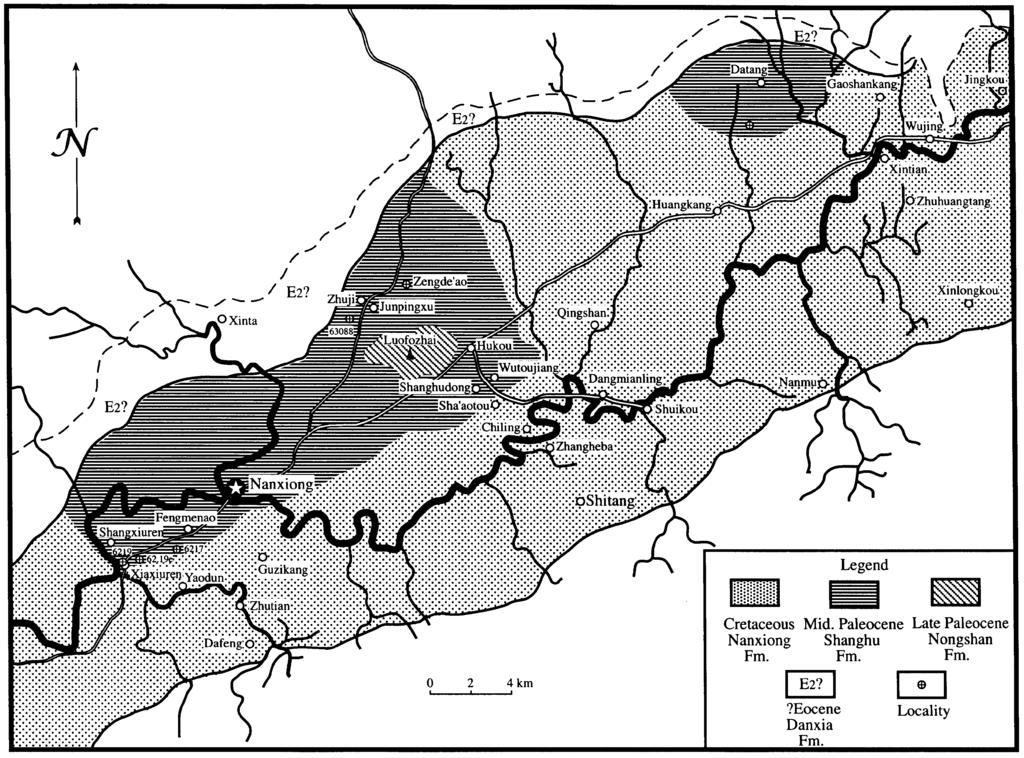
9 6 Synopsis of lithologic characters is as follows: (1) The basal Shanghu Mem. is predominantly exposed in the regions of Canren, Hukou, and Shanghudong where it overlies the Nanxiong Formation disconformably or paraconformably. Exposures are also present in the vicinity of Datang. Thickness exceeds 600 m of tabular beds that dip northwest at 7-12, but in the vicinity of Zhuji Commune the dip becomes southeast, reflecting part of a broad and gentle syncline. Lithologic character consists predominantly of brown-red and purple-red mudstones, argillaceous sandstones, and sandy mudstones interbedded with thinly laminated sandy conglomerates with a base consisting of an approximately two to five meter thick gray-white, and gray-red sandy conglomerate. Abundant fresh-water gastropods and fossil vertebrates are present including mammals, turtles, crocodiles, and other reptiles. (2) The Upper Paleocene (?) Nongshan Mem. lies conformably on the Shanghu Mem. and is predominantly exposed in the center of the basin in the vicinity of Luofozhai Village and Nongshanding with an approximate thickness of 200 meters. Beds are tabular with a broad and gentle synclinal dip of 5-8 and are composed of a set of thinly laminated gray-green and light purple-red mudstones, siltstones, sandstones, and fine conglomerates. Paleontological data includes a large quantity of turtle, crocodiles, and coprolites. (3) The Eocene (?) Danxia Fm. overlies the Luofozhai Fm. with an unconformable contact and consists of massive sandy conglomerates with a brown weathered surface, but fresh exposures are gray-yellow or purple-brown. Conglomerates are poorly sorted, poorly rounded, and poorly bedded with a complex composition consisting predominantly of quartz clasts but also containing granite, slate, phyllites, and sandstones. Clast size varies from several millimeters to greater than ten centimeters. Exposures are between m thick and increase in thickness from northeast to southwest. Beds are extremely contorted and faulted and after weathering compose a characteristic landform designated Danxia topography by geomorphologists. Localities and stratigraphic characteristics Exposures of the Shanghu Mem. of the Luofozhai Fm. occur in the central portion of the basin. Mammalian fossil localities are predominantly distributed around Hukou Commune, northwest of Nanxiong Co. seat and in the Xiuren vicinity south west of Nanxiong (Fig. 1) as are enumerated below: (1) Xiuren: 6217, (2) Shanghudong, Hukou: 6232, 6233, 63081, (3) Wutoujiang, Hukou: 63086, 63082, 63083, 63083, (4) Zhuji: (5) Zengde ao vicinity: Collected by the Hubei Institute of Geology (6) Tatangyoushan vicinity: Collected by the Third Division of the 706th Geological Corps of the Guangdong Bureau of Geology. (7)?Phenacodontidae indet., locality unknown. The Nanxiong localities are relatively widespread. In order to clarify stratigraphic positions and relationships, units in the Shanghu Mem. are recognized as follows:
10 7 (1) Basal section: Fossil localities at the base of the Shanghu Mem. are located predominantly in purple-red mudstones but the basal stratigraphic unit is a white sandy conglomerate not easily distinguished from the underlying Cretaceous sandstones, although at several localities there appears to be a vague angular unconformity between the two units. The greatest thickness of these exposures is found in the vicinity southeast of Xiuren. At the village of Shanghudong the unit attains 281 m, and is equivalent to units 1-IV in cross-section 4, as measured by Zheng et al. (1973). This region documents localities 6217, 6219, and which produce Bemalambda pachyoesteus, Yuodon protoselenoides, Palasiodon siurensis, Dissacusium shanghoensis, and a small quantity of B. nanhsiungensis. Table 1. Correlation of principle fossil mammals, localities, and stratigraphic sections in the Nanxiong Basin. (2) Medial section: This is the most fossiliferous unit consisting predominantly of intertounguing brown-red and purple-red mudstones bearing calcareous concretions and small green marl lenses. The sandy conglomeratic base of this section is conformable with the underlying strata. The greatest thickness is 320 m and these strata appear to correlate with units V- VIII of Zheng et al. (1973) section 4 and units I-VII of their section 5. Exposures lie north of Shanghudong and Wutoujiang where the following localities are documented: 63086, 63082, 63083, 63084, 63081, and These localities produce abundant specimens of Bemalambda
11 8 nanhsiungensis, and a small number of B. pachyoesteus, Dissacus feiganensis, Hukoutherium ambigum, and Linnania lofoensis specimens. (3) Upper section: The poorly fossiliferous sediments consist of 234 m of purple-red mudstones containing gray-green patches. The base is gray-white sandy mudstone interbedded with sandy conglomerates that are conformable with the medial section. This section appears equivalent to units VIII-X in section 5 of Zheng et al. (1973). Exposures lie east of Hukou with only locality producing Bemalambda nanhsiungensis. (4) Capping section: The uppermost fossiliferous sediments of the Shanghu Mem. are variegated light purple-red mudstones but lighter than the underlying sediments of the Shanghu Mem. Contact relationship with the upper section and total thickness are unobservable as the basal portion is obscured by loess. Exposures are principally located in the vicinity of Zhuji Commune and Zengde ao where locality and localities of the Hubei Institute of Geology produce Bemalambda crassa and Linnania lofoensis. Characteristics and chronology of the mammalian faunas from the Shanghu Member of the Luofozhai Fm. Fossil mammals from the lower Shanghu Mem. of the Luofozhai Fm. in the Nanxiong Basin are derived from four stratigraphic units: the lower, middle upper, and capping units, with the two lowest units being the most fossiliferous and containing the same taxa. Although the superpositional relationship between the two lower units is distinct, the assemblages represented in them are consistent. The stratigraphically higher upper sedimentary unit has a thickness exceeding 200 m and produces only the single species Bemalambda nanhsiungensis, which is also recorded from the two underlying assemblages. Consequently, data recovered from the upper section to date suggests it should also be regarded as the same fauna as the lower and middle units. The capping sediments of the Shanghu Mem. produce only two taxa, Bemalambda crassa and the tillodont Lofochaius brachyodon, both of which are absent from the underlying units. The bemalambdid species is distinctly more derived than the underlying taxa and consequently, the capping unit is recognized as representing a distinctly younger faunal zone. Currently, though, data is insufficient and taxa from the capping unit are still regarded in general as members of the Shanghu Fauna. But in the discussion of the age and correlation of the fauna, only the taxa from the lower three units are utilized. Those from the capping unit are regarded only in a referred status. Nanxiong specimens are rather abundant with the vast majority representing the genus Bemalambda in low species diversity. However, those taxa present represent new species and genera which are relatively derived such that contemporaneous or nearly contemporaneous faunas from North America and other regions display extremely few directly comparable taxa. In recent years, numerous generally contemporaneous or slightly younger specimens have been recovered in other regions of South China including Qianshan and Xuancheng Counties, Anwei Province; Dayu Co., Jiangxi Province; and Chaling Co., Hunan Province, but these specimens are predominantly undescribed. Recorded below are taxa from the various units of the Shanghu Mem. and unpublished referred taxa from other regions. Recorded here are 13 species within 10 genera: Anagalida Anagalidae Linnania lofoensis Chow, et al., 1973 Tillodontia Esthonychidae Lofochaius brachyodus Chow et al., 1973 Condylarthra
12 9 Mesonychidae Dissacus feiganensis Chow et al., 1973 Dissacusium shanghoensis (Chow et al.), 1973 Hukoutherium ambigum Chow et. al., 1973 Hyopsodontidae Yuodon protoselenoides Chow et al., 1973 Palasiodon siurensis Chow et al., 1973 Periptychidae?Ectoconus sp.?phenacodontidae, gen. et sp. indet. Pantodonta Bemalambdidae Chow et al., 1973 Bemalambda nanhsiungensis Chow et al., 1973 B. pachyoesteus Chow et al., 1973 B. crassa Chow et al., 1973 B. sp. The 13 species listed above belong to seven families within four orders. The ordder Anagalida is characteristic of Asia, while the other three orders are represented in the Paleocene or Eocene of North America and Europe. The family Anagalidae has for many years been recorded only from the Oligocene of China. Most recently, Szalay and McKenna (1971) described fragmentary specimens representing two species of this family from the Late Paleocene Gashato Fauna of Mongolia, but their relationship to the Shanghu genus Linnania is unclear. The dentition of Linnania is complete and represents a brachydont plesiomorphic form with a conspicuous tribosphenic talonid basin. This primitive form displays an archaic morphology for the family and hence represents an earlier age. The family Mesonychidae is represented by three species. Specimens of Hukoutherium are extremely fragmentary and although its phylogenetic relationship is not completely clear, it displays very few apomorphies and may be basically recognized as a relatively primitive mesonychid. It is worth noting that Dissacus, a primitive and archaic mesonychid, represents the longest recorded genus in the family as it extends from the Middle Paleocene to Late Eocene in North America. Upper molars of the Nanxiong species are not only particularly transversely broadened but are also morphologically distinct from the American genus. Although available data is limited, comparison to other taxa from the northern hemisphere suggest that there is insufficient justification to exclude the Nanxiong specimens from the currently known genera. Thus, D. feiganensis recorded as being relatively close to D. navajovius in size and morphology and is consequently retained in the same genus. The Hyopsodontidae is represented by the two new genera Yuodon and Palasiodon which are similar to but clearly distinct from their North American middle Paleocene counterparts Protoselene and Promioclaenus (P. aquilonius). The Esthonychidae is represented by Lofochaius brachyodus, the most primitive tillodont known to date. This taxon is much more primitive in morphology and lower in stratigraphic position than Esthonyx from the Early Eocene (perhaps including terminal Paleocene) from North America and Europe. This is also further evidence supporting the discovery of this family in Chaling Co., Hunan. Furthermore, tillodonts that are extremely similar to this genus have been recovered from sediments bearing Bemalambda nanhsiungensis in Qianshan Co., Anwei, and appear slightly more primitive than the specimens from the Shanghu capping sediments, indicating a younger age for Lofochaius.
13 10 Table 2. Distribution of middle Paleocene mammal sites in the Shanghu Member of the Luofozhai Fm.* Xiuren Shanghudong, Hukou Wujtoujiang, Hukou Zhuji Zengde ao Linania lofoensis X Lofochaius brachyodus X Dissacus feiganensis X Dissacussium shanghoensis X Hukoutherium ambigum X?Phenacodontidae indet. Yuodon protoselenoides X Palasiodon siurenensis X? Ectoconus sp. X Bemalambda nanhsiungensis X X X X X B. pachyoesteus X X X X X B. crassus X Loc. indet. X *Localities are arranged stratigraphically from bottom to top.
14 11 The new family Bemalambdidae represents the most abundant order in the Luofozhai Fm. Unexpectedly absent from the Nanxiong Paleocene deposits are the prolific Late Paleocene Asian families Archaelambdidae, the common Early Eocene Coryphodontidae, and the North American nearly stratigraphically equivalent Pantolambdidae as well as other North American families such as the Barylambidae and Titanoideidae. The bemalambids are extremely derived, with characters distinct from all other known pantodonts. From the perspective of a general evolutionary grade, the pantodonts show a conspicuous trend from small to large size with the premolar morphology becoming more complex. In this respect Bemalambda is generally consistent with the developmental phase of Pantolambda bathmodon from the middle Paleocene of North America, only the P2 root shows vestiges of bifurcation indicating that it is slightly more primitive than the latter. Compared to P. cavirictus, Bemalambda is much smaller and has a more simplified premolar morphology. Consequently, it is believed that Bemalambda and P. bathmodon are nearly equivalent in evolutionary grade and age and that both represent index fossils for the middle Paleocene of their respective regions. A fragmentary mandible containing a small number of damaged teeth is questionably assigned to the Phenacodontidae based upon mandibular morphology and size. Preliminary conclusions regarding the relationships of the Nanxiong Fauna to other regions, and age of the Shanghu Fauna are provided below: (1) Several genera are shared with, similar to, or are at the same level of systematic development as taxa in Europe, North America, and Mongolia. These include Dissacus,?Ectoconus, Yuodon ( Protoselene ), and Palasiodon ( Promioclaenus ) and indicate that the age of the fossil complexes are constrained by the upper and lower ranges of the middle Paleocene. (2) With the exception of Dissacus, there are no Late Paleocene taxa at Nanxiong that are shared with the Asian Late Paleocene Gashato Fauna. However, the Shanghu Fauna vaguely approaches the Gashato fauna but with major discrepancies as the Gashato Fauna is distinctly older. Although the Gashato equates to the European Thanetian and North American Tiffanian stages, the upper boundary of the Shanghu Mem. cannot exceed the North American Torrejonian Stage. (3) With reference to the ages of several recently discovered local faunas in South China and particularly assemblages from several stratigraphic units in Anwei, the age of the Shanghu Fauna (or at least the three lowest units) may slightly predate the North American Torrejonian or more closely approach the Dragonian. The capping unit of the Shanghu Mem. producing Bemalambda crassa and Lofochaius brachyodon is probably closer to the Torrejonian Stage. Thus the Nanxiong Fauna is provisionally erected as an independent middle Paleocene cronofauna. Other characteristics of the fauna such as paleobiogeographic relationships and paleoecological inferences may only be addressed in a preliminary fashion as current data is insufficient for more substantial conclusions. (1) The general complexion of the Nanxiong fauna is distinctly endemic, as it contains taxa unknown from other equivalent age localities, including members of the Anagalidae, Esthonychidae, and Bemalambdidae. (2) Several families are represented in equivalent age faunas in North American, such as the mesonychids and hyopsodontids, although phenacodontid and periptychid data is extremely rare, with specimens only provisionally assigned to these families.
15 12 It is questionable whether the shared taxa and plesiomorphic nature permit significant comparison with North American and European faunas. North American and European Late Paleocene and Early Eocene faunas are comparable by their distinct primitive faunal complexion and appear to share the main fundamental taxa with Nanxiong as well as with the entire East Asian Paleocene Fauna. This should be related to Asian paleogeographic conditions around the middle Paleocene, although more precise elaboration cannot be addressed here due to the broad scope of this discussion. (3) To date the Luofozhai Fm. has not produced North Asian Late Paleocene taxa such as the Multituberculata, Dinocerata, Notoungulata, Archaelambdidae, or other anagalid families such as the Pseudictopidae and Eurymylidae. This is predominantly due to the limitied collection that currently represents the fauna although other reasons are undoubtedly due to ecological or paleobiogeographical considerations. (4) Habitat characteristics expressed through the faunal elements suggest the majority of the Shanghu fauna appears to be adapted to riparion, paludal, or other habitats proximal to aquatic systems. The assemblage in general appears to lack typical plain or forest dwelling inhabitants such as primates, insectivores, multituberculates, or carnivores, although obviously Linnania may be regarded as a forest taxon. Faunal characteristics, taphonomy, and depositional characters suggest the Lofozhai sediments are predominantly central basin quiet water lacustrine or riparion sediments which differ distinctly from the faunal and fluvial characters expressed in the Late Paleocene sediments of Xinjiang and Mongolia. (5) Analysis of Bemalambda dentition, cranial, and postcranial elements suggest that this family represents a primitive pantodont that shares numerous cranial and dental characters with Deltatheridium, and possibly expresses a closer relationship between the two genera. It is possible that the Pantodonta had its genesis in Asia at the terminal Cretaceous or basal Paleocene. Conclusions The mammalian fossil complex from the Nanxiong Basin of northern Guangdong Province represents a new middle Paleocene assemblage named the Shanghu Fauna, or the Shanghu Local Fauna, which represents the oldest Cenozoic mammal fauna known in the Old World to date. The Lower Tertiary of Nanxiong includes the upper Danxia Fm. and the lower Lofozhai Fm. Precise dating of the Danxia Fm. is unclear but it is generally recognized as Eocene. The Luofozhai Fm. is subdivided into the upper Nongshan Mem. and the lower Shanghu Mem. with the Paleocene fauna produced completely from the Shanghu Mem. Fossil data from the Shanghu Mem. are derived from four stratigraphic units: the lower, middle, upper, and capping units, with the majority of data derived from the lower and middle units and few specimens recovered from the upper unit. These three sedimentary packages are recognized as a single middle Paleocene chronounit while the capping sediments represent an independent later stratigraphic unit. The age of the entire Nanxiong fauna distinctly predates faunas from the Turpan Basin, Xinjiang, and the Gashato Fauna in Mongolia. The age of the Nanxiong Fauna lower boundary is constrained possibly to the early middle Paleocene. The age of the stratigraphically higher Nongshan Mem. may represent Late Paleocene or perhaps Early Eocene. Four orders, seven families, ten genera, and thirteen species are represented in the Shanghu Fauna. Its most distinctive characters lie in its endemic autapomorphies such as the presence of the Anagalidae, Esthonichydae, and Bemalambdidae, which are unknown from other Paleocene sediments. From another perspective, there are several genera that are extremely close to those
16 13 from the middle Paleocene of North America, to the extent that they may be congeneric, suggesting an affinity between the two continents during this period. A majority of the Nanxiong faunal elements are adapted to riparion or paludal to lacustrine habitats with a near absence of typical plain or forest elements. The family Bemalambdidae represents the most primitive member of the Pantodonta that had its genesis in the terminal Cretaceous or Early Paleocene from a primitive Asian therian resembling Deltatheridium. Part II Specimen descriptions and phylogenetics Anagalida Anagalidae Linnania Chow, Chang, Wang and Ting, 1973 Linnania lofoensis Chow, Chang, Wang, and Ting, 1973 (Plate I, Figs. 1,2; Plate II, Fig. 1; Text figs. 1-5) Linnania lofoensis Chow et al., 1973; Vertebrata PalAsiatica 11(1), 31. Type: A relatively complete skull and mandible (IVPP #V4234). Fm. Stratigraphic position: Middle Paleocene middle portion of Shanghu Mem., Lofozhai Genus and species diagnosis: Cranial and dentition morphology basically resembles the anagalid Anagale gobiensis, being relatively primitive and slightly small. Skull is relatively short, narrow, low and flat. Temporo-orbital fossa is small, ramus is slender and weak, upper dental formula is? , and lower dental formula is? Canine is small, weak, and simple in morphology and the first and second premolars have bifurcated roots. Protocone on P3 to M3 is nearly crescentic, P4 paracone is well developed, metacone is small, metaconule is undeveloped or incipient, dentition is unilaterally hypsodont, enamel extends into the dental trough. Upper molar paracone and metacone are isolated and conical. Pre- and postprotocristae extend to the external sides of the paracone and metacone to contact the anterior and posterior cingula respectively. On the mandible, the protoconids of the third and fourth premolars are bifid. Degree of p4 molarization is higher than the P4. Lower molar paraconid is just slightly lower than the metaconid, and trigonid is extremely high on p4-m3, being twice the height of the talonid. Trigonid cusps are in a v-shaped alignment with a deep trigonid basin. The p3-m3 protoconid is rather elongated and hypoconid gradually reduces in size posteriorly along the sequence. Description: At the time of its recovery, the specimen s mandible was in tight occlusion with the skull. The skull is relatively completely preserved in outline but due to compressional distortion it is fractured, such that individual cranial elements are missing and its precise morphology is unclear. The skull is relatively short, narrow and low. Anterior to the P3 the skull is even more narrow due to compressional distortion and the rostral end has been shattered such that it does not preserve the incisors. Laterally, the skull has suffered some displacement with the left side shifted 1.7 mm anteriorly. The dorsal surface of the skull is extremely flat with the nasals lying nearly on the same plane as the parietals. The frontals are very slightly inflated and lack sagittal crests. The temporo-orbital fossa is confluent with the temporal fossa, and lacks a medial
17 14 boundary line to compose a relatively small temporo-orbital fossa with its anterior end situated dorsal to the M1. The zygomatic arch is completely broken with no vestiges remaining. The palate is relatively flat with the dental sequences rather widely spaced. The P4-M3 sequences are parallel but from P3-C the dentitions gradually attenuate with palatal breadth at P4 being 18 mm but at the P2 it is 11 mm. The sphenoid is relatively broad and the basioccipital region is rather deeply concave. The dorsal surface of the skull is rectangular, relatively thin, flat, and precipitous. The occipital crest is flat, straight and slightly dorsally projected. Dentition on the right side is complete with the exception of the P1. Each tooth in the sequence is in tight association with its preceding and posterior counterparts (less than a millimeter separates the P2 from P3). On the left side, the P3-M2 are preserved, the P3 is incomplete, and the M3 only preserves its paracone. Table 3. Cranial measurements of Linnania lofoensis (mm). Facial length (anterior temporo-orbital fossa anterior canine) Cranium length (anterior temporo-orbital fossa occiput) Lateral diameter of temporo-orbital fossa 11.4 Dorsal diameter of temporo-orbital fossa 15.3 Length of skull (occiput - anterior canine) 47.4 Height of skull (at M2) 15.7 Figure 2. Ventral view of V4234, Linnania lofoensis Chow et al., 1973 (X3).
18 15 The canine is small, weak, as long as the P2, and has damaged lingual enamel. Four upper premolars are present. The P1 preserves two roots, the posterior of which is slightly larger. The P2 also maintains two roots, the posterior root being large with a thin and flat labial side, convex lingual side, and slightly inflated posterior portion. Less than a millimeter separates this tooth from the P3. Both the left P1 and P2 are missing. On the right side the P3 is newly erupted with three roots present, the protocone is low, thinly crescentic, and supported by the lingual root. The paracone is extremely high with a relatively precipitous anterior face. Compressional distortion has shifted the cusp posterior to the paracone (ectometacone) onto the posterior tooth distorting the triangular morphology of the P4. This distortion is also probably the cause of its anteriorly obtuse triangular morphology. The P4 on the right side is deciduous, damaged, and only recognizable as subsquare in outline with a remnant morphology very similar to the M1. On the left side the P4 has been displaced due to compressional distortion with the lophes anteriorly oblique and its outline subsquare. The protocone is well developed and thinly crescentic, the paracone is extremely high, twice the height of the protocone, in direct opposition to the protocone, and is slightly damaged anterolabially. The metacone is extremely low, the cingulum is weak, and the preprotocrista extends directly to the anterior paracone where it becomes confluent with the cingulum. On the right side all molars are present but the M3 metacone is missing while on the left side the M1 and M2 are present but the M3 preserves only its paracone. Crowns of the M1 are shattered such that only the outline and lingual morphology are recognizable. The crown is circular with a well developed protocone, paracone, and metacone of equivalent size; cingulum is extremely well developed; and parastyle and metastyle are absent. Figure 3. Right mandible with p1-m2 (V4234) of Linnania lofoensis Chow et al., Upper. Occlusal view; Lower. Lateral view (X3) Figure 4. Left mandible with m2-m3 (V4234) of Linnania lofoensis Chow et al., Upper. Lateral view; Lower. Occlusal view (X3) The M2 is square, larger than the M1, paracone and metacone are isolated with the paracone slightly larger; cingula are well developed labially, anteriorly, and posteriorly with the posterior cingulum extending around to the anterolingual side of the protocone. Pre- and post protocristae both extend to the exterior sides of the paracone and metacone to contact the cingula. Parastyle and metastyle are weak.
19 16 Table 4. Comparison of anagalid dental measurements (mm). Linnania lofoensis Anagale gobiensis Anagalopsis kansuensis Khashanagale zofiae P1 L P1 W 1.60 P2 L P2 W 2.92 P3 L 3.25 P3 W 3.30 P4 L 3.26 P4 W 3.32 M1 L 3.30 M1 W 3.50 M2 L 3.42 M2 W 3.60 M3 L 3.18 M3 W 3.55 P1-M3 L ~27.5 P2-M3 L p1 L 3.12 p1 W 2.78 p2 L 3.18 P2W 2.88 p3 L 3.20 p3 W 2.95 L p4 Trig. W Tal. W 2.95 ~ L m1 Trig. W Tal. W L m2 Trig. W ? Tal. W L m3 Trig. W Tal. W m1-m3 L ~ ~7.2 p1-m3 L ~ p4-m1 L p2-m3 L ~ ~26-27 Addendum:?Kashanagale sp. nov. (Szalay and McKenna, 1971) m3 Trig. W: 1.15, Tal. W:0.9 The M3 is newly erupted, situated lower and is slightly smaller than the M2, and morphologically resembles the M1 and M2. The anterior margin is very slightly posteriorly oblique and as such the tooth is not square in outline. Pre- and postcingula are reduced and do not extend to the lingual side. Pre- and postprotocristae resemble those on the M2, and a parastyle is more conspicuous than on the M2. A character shared between the P4-M3 is that the lingual side is higher than the labial side.
20 17 Right lower dentition preserved includes the trigonid of the m2 to the p1 and half the root of the canine. On the left side only a piece of dentary bearing an m2, m3 and roots of the m1 are present. The lower canine appears to have been small on the basis of half a root present. Lower molars are not tightly associated as small diastemae lie between the teeth. The p1-p3 are all incomplete, p1 outline is vague, p2 is broken lingually and is thin and flat labially. The p3 has been shattered anteriorly and posteriorly, but the protoconid is completely preserved with a labial side that is rather thin and flat, the lingual side is convex, and there is a small inflation both anteriorly and posteriorly representing an incipient paraconid and metaconid, developing the tendency toward a well developed trigonid. Posterior to the protoconid is a small, extremely narrow, semicircular talonid. The lower fourth deciduous premolar is extremely worn (Pl. II, Figs. 1a,1c,1d) and morphologically resembles the m1. A complete p4 is erupting posterior to it (Pl. II, Fig. 1b). The right side preserves only the m1 and m2 trigonids, while the left side preserves a relatively complete m2 and m3. The dentition is not tightly associated as there are small diastemae between the teeth. Lower molar trigonids are nearly twice the height of talonids. The protoconid is high and long, the metaconid is slightly lower than the protoconid, and the paraconid lies anterior to and lower than the metaconid, with an extremely deep v-shaped groove lying between the three principle cusps. The talonid is small, nearly half the length of the trigonid, with a high entoconid and hypoconid and hypoconulid of equivalent height. All three cusps encompass a narrow basin. The cristid obliqua extends slightly labially to the midpoint of the trigonid. From a lingual perspective the entire tooth resembles a trident. The m1 is rectangular with a short trigonid, metaconid higher than protoconid, and paraconid much higher than on the p4, being nearly the height of the metaconid. Both paraconid and the metaconid are aligned nearly opposite to the protoconid. Between the paraconid and metaconid there is still a relatively deep, flat, and expanded nearly u-shaped groove that provides an elongation to the trigonid. The talonid is slightly broader than the trigonid, with the hypoconid the largest cusp, hypoconulid the smallest cusp, and the entoconid as the tallest among the three cusps which encompass a broad and spacious basin. The reduced height of the protoconid and hypoconid is due to occlusal wear. The cristid obliqua extends slightly labially to the midpoint of the trigonid Table 5. Cranial measurement comparison of three anagalid species (mm). Linnania lofoensis Anagale gobiensis Anagalopsis kansuensis Supratemporo-orbital fossaal width Palatal width at p Foramen magnum-canine length The m2 resembles the m1 in morphology although trigonid and talonid lengths are equivalent and the trigonid is more narrowly rectangular. The m3 resembles both anterior lower molars but with an extremely high hypoconid that is situated slightly lower than the protoconid and a talonid that is rather elongated. A shared character between the p4-m3 is the labial enamel extending further than lingual enamel such that labial crown height exceeds the lingual crown height.
21 18 Figure 5. Cranial comparison between Linnania lofoensis, Anagale gobiensis, and Anagalopsis kansuensis. Upper. Linnania; Middle. Anagale; Lower. Anagalopsis. Comparison and discussion: The Nanxiong specimen is comparable to the anagalid taxa Anagale gobiensis, Anagalopsis kansuensis and Kashanagale zofiae. It skull length approaches but is slightly shorter and much narrower than Anagale, and compared to A. kansuensis it is even shorter, as displayed in Table 5. In addition to the narrow and short cranial outline, the size of the temporo-orbital fossa is conspicuously short at 11.4 mm whereas on A. gobiensis it is 14.6 and on A. kansuensis it is 33 mm. Dorsal cranial morphology is also distinct as the Nanxiong specimen is relatively flat but A.
22 19 gobiensis has a gradual increase in height from the nasals to the parietal and A. kansuensis is particularly high and inflated in the frontoparietal region. Dentally, Linnania shares fundamental characters with Anagale, Anagalopsis, and Kashanagale, including nearly square upper molars, compact dentition, equivalent degree of hypsodonty, rectangular lower molars, and nearly equivalently lengthened trigonid and talonid. However, compared to Kashanagale zofiae, the latter s lower molar trigonids are not rectangular but are nearly triangular in occlusal outline; the protoconid is more anteroposteriorly compressed or columnar in form; the metaconid is more anteroposteriorly elongated or nearly equivalent to the protoconid; and the paraconid is low, widely separated from the metaconid, and not as tightly associated as on Linnania. Consequently, both cusps lie distantly from the protoconid and provide the trigonid with its triangular morphology.?k. sp. nov. is represented only by a portion of a mandible with a deep ramus, and m3 talonid that is approximately half the size of Linnania. Anagale gobiensis has a relatively large dentition, the P2 has three roots, and the P4 is more molariform in maintaining four cusps with the appearance of a hypocone and metaconule. The Linnania P2 maintains only two roots and its P4 lacks a hypocone and metaconule. Furthermore, the A. gobiensis M3 is distinctly smaller than the M2. Anagalopsis kansuensis is quite different in its large morphologically distinct dentition. Its P1 is single rooted, the P4 is square and highly molariform, the M3 is reduced and distinctly smaller than the M2, p4 is more molariform than P4, and its trigonid is short and broad, whereas the Linnania P1 is double rooted, the P4 is subsquare, the M3 is not extremely reduced, being only slightly smaller than the M2, and the p4 is submolariform with a long trigonid. The preceding descriptions indicate clearly that Linnania is distinct from the other three anagalid genera. The type species of Anagale, which is representative for the family, is from the Oligocene Ulan Gochu Fm. of the northern Sharamurun region of Ulanqab Meng, Inner Mongolia. The initial description by Zengsheng Xin (1931) erected a new insectivore family that approached the Tupaiidae, a phylogentic interpretation that was accepted and utilized for a long time. But following placement of the tupaiidae within the Primates, the family Anagalidae became an excluded extinct member of the order. In 1951 Bohlin described the Oligocene Anagalopsis from the Huihuibu (Huihuipu) region of Gansu. He considered Anagalopsis extremely close to Anagale and believed that the family lay in an uncertain phylogenetic position, unassociated with either the tupaiids or primates. Later, McKenna (1963) reevaluated the type and supplementary specimens for Anagale, after which he advanced and supported the conclusions of Bholin (1951). Recently, Szalay and McKenna (1971) reviewed the Gashato Fauna from Mongolia, described the new genus Khashanagale, and elevated the family to the order status of Anigalida, recognizing within the order the four families Anagalidae, Zalambdalestidae, Pseudictopidae, and Eurymylidae. This order is considered as endemic to the Cretaceous to Early Tertiary of Asia. Although these families share conspicuous synapomorphies (Szalay and Mckenna, 1971, p. 301), their phylogenetic relationship is still suspect pending the acquisition of more data, particularly with regard to the Eurymylidae. This recent phylogeny however is generally more parsimonious than those of the past. Linnania is distinctly phylogenetically close to the genus Anagale and undoubtedly represents the most primitive genus in the order known to date. In cranial morphology it is relatively short, small, narrow, low, and flat with a small temporo-orbital fossa. The stratigraphically younger Anagale has a broader skull and an enlarged temporo-orbital fossa. The precise chronology of Anagalopsis in unclear but it is also distinctly younger with an elongated and broadened skull and a more enlarged temporo-orbital fossa.
23 20 The relatively primitive dental sequence of Linnania is reflected in its sub-square P4 lacking a hypocone while the Anagale P4 is highly molariform by being square, possessing four cusps, one of which is the hypocone, and having a rather projected metaconule. Another significant distinction of Linnania is the third upper molar which is not distinctly reduced but which is reduced on Anagale. Anagalopsis is more dentally derived than both Linnania and Anagale with both upper and lower fourth premolars highly molariform and an M3 that is distinctly reduced. Worth noting is the double rooted condition of the P1 and P2 on Linnania, whereas in Anagale both upper and lower first premolars are double rooted, but the P2 has three roots, and in Anagalopsis the P1 is single rooted and P2 is double rooted, which probably represents the plesiomorphic condition. In general dental morphology Anagale and Anagalopsis are more derived than Linnania. The age of Khashanagle approaches that of Linnania but because the specimen is too fragmentary, adequate comparisons are difficult. The lower molars display the m3 as distinctly more reduced than the m2, which resembles Anagale and Anagalopsis but differs from Linnania. Another distinction also worth noting is the relatively well developed paraconid on Linnania which is nearly equivalent in size to the metaconid. The plates illustrating Kashanagale indicate an undeveloped paraconid lower than the metaconid, but this may be the result of occlusal wear. However from the perspective of the developmental trends within the genera it appears the paraconid tends to become reduced. The anagalids are evidently very abundant and represented by diverse closely related forms in East Asia during the middle and Late Paleocene, as documented by the rich data collected in Anhui Province which is now under investigation. Tillodontia Esthonychidae Lofochaius brachyodus Chow, Chang, Wang, and Ting 1973 (Plate II, Figs. 3-5; Plate III, Figs. 1-3) Lofochaius brachyodus Chow et al., 1973; Vertebrata PalAsiatica 11(1), Type: A poorly preserved skull that is relatively complete in outline bearing left P4-M3 with damaged crowns and right P2, M2 and M3 (V4239). Locality and stratigraphic position: Middle Paleocene capping sediments of the Shanghu Mem., Lofozhai Fm. Yadoao (perhaps mistaken for Zengde ao), Nanxiong Co., Guangdong Province. Genus and species diagnosis: A relatively small species with dental morphology precisely that of the Tillodontia but with a size nearly half that of Esthonyx bisulcatus. Skull is narrow and long with a primitive rostrum that is short and small. Upper dental formula is 3? 1 4 3, P1 and P2 are double rooted, and brachydont molars are relatively broad. Apices of paracone and metacone approach the midline of the molar, labial wall is particularly gently sloping, and parastyle and metastyle are well developed with the parastyle particularly large, being nearly the size of the paracone. Protocone is larger than the principle labial cusps and rises precipitously; anterior and posterior cingula are well developed with the posterior cingulum particularly conspicuous, although it does not extend to the lingual portion of the protocone but comprises a
24 21 hypocone shelf posterior to the protocone. Metastyle on the third molar has become lost causing the diminishment of the posterior half of the tooth. Conules are well developed. Figure 6. Ventral view of Lofochaius brachyodus Chow et al., 1973 (V4239) X2. Description: Weathering and compressional distortion have damaged the skull. The right side is slightly askew and sutures are indistinct, prohibiting the determination of individual cranial elements. In outline the skull is narrow and long with a rostrum that rapidly constricts anterior to the P3 where it becomes primitively short and narrow. The nasals and maxillae are fragmented but coalesced by matrix and lack any distinguishable sutures. Frontals are narrow and slightly inflated; a supraorbital process is slightly projected on the right side but is not preserved on the left side; the parietal is also slightly projected; and sagittal crest is relatively well developed to compose a narrow ridge. Orbit and temporal fossae are confluent with an anterior margin located anterodorsal to the M1. On the right side only an extremely small piece of the zygomatic arch is preserved but on the left side only the middle portion of the arch is missing, with the remaining arch as being relatively robust, flat, and straight. The suborbital foramen is located dorsal to the P3. The palate is relatively flat with relatively close dental batteries. From the M3 to the P3 the dentition gradually converges but anterior to the P3 the tooth rows become parallel. Palatal width at the M3 is 12.2 mm and at the P3 is 7.9 mm. The occipital plane is relatively high with several depressions upon it, the occipital crest is semicircular and projects posteriorly; and occipital condyles are relatively small. Table 6. Cranial measurements of Lofochaius brachyodus (mm). Estimated cranial length 72.0 Facial length (anterior orbit to anterior P1) 20.5 Cranium length (anterior orbit to occiput) 47.8 Orbit length 23.7 Dorsal orbital breadth 19.2 Cranial height (at M2) 24.2
25 22 On the left side the M2 and M3 are completely preserved but other cheek teeth only preserve roots or crown outline, and canine and incisors are absent. Remnant dentition and alveolae indicate the presence of four premolars and three molars in compact association. Molars are brachydont and slightly higher lingually. Among the four upper premolars, only the P4 is distinguishable in outline. The P1 is represented by two equivalently sized linearly aligned roots. The P2 is also only represented by two roots, the anterior of which is relatively small and situated close to the labial plane while the posterior root is twice the size of the anterior root and is located more lingually. On the right side a portion of the lingual wall of the P2 principle cusp is retained suggesting the cusp is semicircular in occlusal view. The P3 has three roots, on the left side there is a well developed lingual cusp which is crescentic lingually, relatively precipitous labially. A posterior cingulum is present. The P4 outline, remnant portion of protocone, and metaloph indicate its high degree of molarization. It is equivalent in size to the M1 with a well developed robust and bluntly conical protocone that is crescentic lingually. The metaloph is preserved and a posterior cingulum is well developed which ascends from the metaloph lingually to compose a low hypocone shelf or balustrade, although it does not extend as far as the lingual protocone. Three upper molars with very distinct cusps are preserved on the left side with the exception of the M1 which is damaged and represented as a transverse rectangle in outline. On the right side only the M2 and M3 outlines are represented. The M2 is also a transverse rectangle, slightly larger than the M1, and the broadest tooth in the sequence. The protocone is well developed, relatively long and broad, and relatively precipitous lingually and labially. From occlusal perspective there is a small groove at its apical midpoint causing it to be antero-posteriorly bifid, however, this groove is extremely shallow and may be the result of fracturing as the anterior half of the lingual protocone is damaged. Paracone and metacone are blunt, their lingual side is roundly convex, labial side is thin and flat, or crescentic, their apice approaches the tooth s midline, and the labial wall is particularly gently sloping. Preparacrista and postmetacrista are well developed to contact the parastyle and metastyle. The parastyle is particularly well developed with a size approaching the paracone. Anterior and posterior cingula are also well developed with the posterior cingulum being more conspicuous but its terminus does not reach the lingual side of the protocone but does ascend to compose a low hypocone shelf. The trigon basin separates the protocone from the paracone and metacone. The enamel is damaged on the protoloph and metaloph but the protoconule and metaconule are still visible vestigially. Table 7. Dental measurements of Lofochaius brachyodus (mm). M3 M2 M1 P4 Labial Length Medial Lingual Width P1-M3 length 29.5 The M3 is morphologically similar to but slightly smaller than the M2 and has been fractured at its mid-section due to compressional distortion, resulting in the lingual side being anteriorly askew. The protocone is relatively blunt and not as elongated as on the M2. A metaconule is extremely well developed but the protoconule is damaged and thus indistinct. The metastyle has become lost causing the posterior half of the tooth to be contracted. Anterior and posterior cingula are well developed and a hypocone shelf is extremely conspicuous.
26 23 Comparison and discussion: The cranial and dental characters described above indicate Lofochaius to be an extremely primitive ungulate that shares a number of plesiomorphies with the Esthonichidae, such as the slender and long skull which rapidly narrows anterior to the P3 and a relatively well developed sagittal crest. These characters are also distinct on stratigraphically younger genera such as Tillodon, which is larger and more derived. Lofochaius possesses several principle characters that are shared with Esthonyx, the most primitive genus in the family, including brachydonty, upper molars higher lingually, relatively high degree of P4 molarization, well developed protocone, pronounced parastyle and metastyle, well developed anterior and posterior cingula on molars extending to the lingual protocone, and the loss of the M3 metastyle. Particularly shared with E. aucutidens is the extremely well developed posterior cingulum on P4-M3 which ascends to compose a hypocone shelf, and the gently sloping labial wall of the paracone and metacone which are present as crescentic cusps. Particularly shared with E. bisulcatus is the double rooted P2 and moderately acute tooth cusps. However the most conspicuous character is the much smaller size of Lofochaius compared to the other species in the family, being approximately half the size of E. bisulcatus which is currently the smallest species known. Lofochaius is dentally distinct from Esthonyx in numerous characters, the most notable of which is the presence of four premolars, whereas the dental formula for the family is three. Moreover, Lofochaius has an extremely compact dentition that lacks small or large diastemae; but other members of the family possess a diastema anterior to the third premolar, particularly on younger taxa where this feature becomes elongated. Additionally, Lofochaius lacks a hypocone. Only a hypocone shelf is present that is composed of the posterior cingulum. This also differs from other members in the family. Current data is extremely inadequate, with the absence of a mandible, lower cheek teeth, and particularly upper incisors and canines. However, from perspective of the data at hand, the Nanxiong specimen is a primitive ungulate that appears to represent the most primitive species of the Tillodontia. The Nanxiong specimen shares several characters with adapid primates such as the relatively narrow and long skull, well developed sagittal crests, the presence of four premolars and three molars in compact association, and particularly the characters of transversely broadened upper molars and a well developed parastyle. However, observation in detail notes conspicuous distinctions in the adapids, as the primates have a broader skull with a laterally projected zygomatic arch, relatively elevated frontals and parietals, upper molars are not higher lingually, and anterior and posterior cingula completely encompass the protocone. Consequently, Lofochaius is recognized as a primitive ungulate that only shares plesiomorphies with several taxa of primates. Formerly, a number of workers placed the tillodonts within the Carnivora (Cope 1876; Schlosser, 1923; Abel, 1914; Winge, 1923). But the Lofochaius specimen, although limited, indicates extreme distinctions from this order. Prior to the discovery of the Nanxiong specimen the most primitive tillodont was recognized as Esthonyx, about which there were numerous controversies regarding its origin, with some workers believing it arose from the family Oxyclaenidae (now included in the Arctocyonidae) (Gregory, 1910; Gasin, 1953), while other workers regarded the genesis of the tillodonts as unclear or from the Insectivora (Simpson, 1945). Van Valen (1963) proposed that the tillodonts arose from the Paleocene arctocyonid Claenodon procyonoides and therefore should be regarded as a suborder of the Condylarthra. He moreover conducted a detailed comparison of the dentition and postcranial characters of Esthonyx spatularius from the Late Paleocene Clark Fork Basin (AMNH and 16065) and a C. procyonoides collected from the Paleocene of the San Juan Basin (AMNH 16554). The Esthonyx specimens preserve only a single M2 that Van Valen believed resembled C. procyonoides in morphology, moderate cusp height, labially placed paracone and metacone, and a posterior cingulum that does
27 24 not extend to the lingual side. However, there are also conspicuous distinctions such as the high degree of C. procyonoides P4 molarization and well developed hypocone. On another C. procyonoides specimen the posterior cingulum reaches the lingual side. Despite these distinctions, Van Valen believed that from an evolutionary perspective, later species of Esthonyx also gradually embody these characters and thus its descendence from C. procyonoides is evident. The Nanxiong specimen had yet to be studied at the time of Van Valen s publication and at that time Esthonyx was regarded as the most primitive tillodont genus, a status which is currently replaced by the Lofochaius, and which should be contemporaneous with C. procyonoides. Based upon the dental characters of the three molars, Esthonyx and Lofochaius are hereby considered to have a closer affinity than to the arctocyonid mentioned above and it would therefore be inappropriate to consider the Tillodontia a suborder of the Condylarthra. The retention of the Tillodontia as an independent order is also supported by Rose (1972). Sloan and Van Valen (1965) described the new arctocyonid Protungulatum donnae from the Cretaceous of Montana which, from its description and illustrations, shares several features with Lofochaius such as its transversely expanded M2 which constitutes the largest molar in the sequence, a well developed parastyle and metastyle, loss of the M3 metastyle and well developed anterior and posterior cingula. However, the Montana specimen is generally smaller with an M2 length of 4 mm and width of 6 mm and hence the shared characters are regarded symplesiomorphic for the most primitive ungulates. The description above suggests the Nanxiong specimen approaches the Condylarthra and may be descended from a primitive arctocyonid, but this cannot be confirmed with the limited data at hand. Some specimens that are definitely related to Lofochaius have been found at Chaling, Hunan. They possess nearly all the essential features necessary to fill the gaps between Lofochaius and Esthonyx. These specimens are more advanced but approach the older taxon. They are from a higher stratigraphic horizon (probably Late Paleocene) and bear chisel-like incisors typical of tillodonts. Therefore the tillodont affinity of Lofochaius appears to be well founded. The Lofochaius skull was collected in 1964 by the Hubei Academy of Geology (formerly the Southcentral Academy of Geology) and was mailed to IVPP for diagnosis. Also contained within the package were three small limb bones including a proximal humerus (V4239.3), distal femur (V4239.1), and a proximal tibia (V4239.2). A description of these elements is not conducted due to their fragmentary nature, although it appears, from a perspective of their preservation, that they probably represent post crania of Lofochaius brachyodus. Condylarthra Mesonychidae Dissacus feiganensis Chow Chang, Wang, and Ting, 1973 (Plate IV, Fig. 1,2,4)?Dissacus feiganensis Chow et al., Vertebrata PalAsiatica 11(1), p. 32. Type: A damaged anterior portion of skull with left P3-M2 and right P4-M3 (V4229). Paratype: An extremely fragmentary anterior portion of skull (V4230). Locality and stratigraphic position: Middle Paleocene, middle portion of Hukou Mem., Lofozhai Fm., Hukou Commune, Nanxiong Co. Guangdong Province.
28 25 Diagnosis: Dental formula of 3? that approaches in size but is slightly smaller than Dissacus navajovius. Upper molars vary in breadth with M3 slightly smaller than M2 and protocone is well developed as an isolated lingual cusp that has a relatively precipitous lingual wall. A conspicuous longitudinal groove lies between the protocone and the two principle labial cusps, paracone and metacone are distinctly smaller and in tight association, with low vertical crests extending from the anterior paracone and the posterior metacone. The lingual cusp on the P4 and P3 is relatively well developed. A constriction is present on the posterior nasals at the P1. Figure 7. Type (V4229) of Dissacus feiganensis Chow et al., Left. dorsal view; Right. Ventral view Description and comparison: Two pieces of skull are preserved, among which V4229 is relatively well preserved with frontals, a large portion of nasals, maxillae, premaxillae, a portion of zygomatic arch, and anterior sagittal crests. The frontals have suffered slight compressional distortion, are relatively short, have a well developed frontal ridge, and the frontal parietal suture composes a relatively well developed sagittal crest. Additionally, a supraorbital process is extremely conspicuous. Nasals are slender and long with an extremely conspicuous longitudinal groove which broadens slightly anteriorly as it runs along the suture line. Nasals are constricted at their midpoint and become inflated posteriorly where they insert directly into the midpoint of the frontals. The contact of the premaxilla with the nasal process forms a relatively long slender line that extends distantly between the maxilla and nasal. The maxilla is relatively high, zygomatic arch is relatively straight and robust, the lacrimal extends into the facial region, anterior orbit runs anterior to the M2, and the suborbital foramen is positioned low and dorsal to the P3. On the palate, the maxillary fossae are relatively large and anterior foramena lie posteriorly, or at the posterior M2. Palatal breadth at the M2 is 39.2 mm and at the P4 is 32.3 mm. Specimen V4230 has been shattered and heavily compressionally distorted, preserving only the anterior half of the skull. No dorsal characters may be distinguished but an incomplete left and right M2 indicate that it is conspecific with the former specimen. D. navajovius is a third larger than the Chinese specimens and is also incomplete but as in D. feiganensis has an anterior orbit that runs anterodorsal to the M2 and displays a similar placement of the suborbital foramen which is dorsal to the P3. The zygomatic arch on this genus is extremely straight and long indicating a particularly elongated cranium, and although the Nanxiong specimen only preserves a portion of the arch, it too appears by be relatively linear. The jugal of
29 26 D. feiganensis also resembles other members of the family by lacking an anteroventral branch and only possessing the dorsal branch which composes a relatively low ventral orbital margin. The Nanxiong dentition is extremely restricted and very poorly preserved limiting the comprehension of its morphology. On the type the dentition has been completely worn or is only represented by tooth roots, although its dental formula may be recognized as 3? Incisors are not preserved; the canine appeares to have been relatively robust based upon its alveolus; the P1 is extremely close to the posterior canine with a single root and conical cusp; and the P2 is also single rooted with a 2.7 mm diastema between it and the P1. The outline of the P3 is unclear although it is similar in size to D. navajovius and alveolae indicate that the relatively well developed lingual principle cusp is supported by an independent root,. The P4 is preserved on both the left and right sides and although the crown is worn, its outline indicates a length greater than breadth, the presence of a well developed lingual cusp nearly the same size as the labial cusps, and a well developed anterior cingulum. Figure 8. Dissacus feiganensis Chow et al., 1973 (V4230) X1, magnification X2. Although the upper molars are all preserved, they have all suffered heavy occlusal wear. Their most significant characters lie in the excessive transverse breadth of the dentition, the base of the protocone is anteroposteriorly compressed, anterior and posterior margins of the teeth are nearly parallel, and the M3 is smaller than the M2. On specimen V4230 the M2 is relatively well preserved with three well developed principle cusps. The protocone is a small isolated conical cusp with a precipitous lingual wall, the metacone is relatively small, or slightly smaller than the paracone, and a low anterior crest projects off the paracone. The M1 resembles the M2 but is slightly smaller. The M3 is extremely poorly preserved but from occlusal outline is it determined that it is distinctly shorter and narrower than the M2 with an unprojected posterolabial angle. The reduced M3 is a character that is difficult to compare to other Asian mesonychid genera as currently there are no upper third molars available and other comparative specimens are younger taxa that are twice as large or larger. Furthermore, on the younger taxa the molars are quite distinct from D. feiganensis, being either longer than broad, or equivalent in dimension to compose an equilateral triangle, contrary to the Nanxiong specimens in which molar breadth greatly exceeds its length.
30 27 Table 8. Comparison of femoral measurements between Dissacus feiganensis and Mesonyx obtusidens (cm). Dissacus feiganensis Mesonyx obtusidens Length Distal breadth Proximal breadth There are currently seven species of Dissacus recognized in North America, although the majority of these are known only from lower dentitions. D. navajovius and D. saurognathus preserve complete upper dentitions with a dental formula resembling D. feiganensis, and their M3 is reduced, although their morphology is distinct in that their length and breadth are nearly equivalent (particularly the M2) to compose an equilateral triangle, while the outline of D. feiganensis is an isosceles triangle. Moreover, the North American species have relatively well developed cingula which are absent (or only restricted to the labial side) on the Chinese species. Table 9 illustrates the dental measurement discrepancies between the North American and Chinese species. Table 9. Comparison of dental measurements between three species of Dissacus (mm) D. feiganensis D. navajovius D. saurognathus Left P1-M2 length Left P1-P4 length Left M1-M2 length Right P4-M2 length P1 LxW 4.8x x 5.7 P2 LxW 5.5x x7.5 P3 LxW 7.2x x x9.8 P4 LxW x x x12.3 M1 LxW 8.0x x x16.3 M2 LxW 9.0x x x16.9 M3 LxW 7.4x x x12.7 P1 L/W ratio P2 L/W ratio P3 L/W ratio P4 L/W ratio M1 L/W ratio M2 L/W ratio M3 L/W ratio Comparisons conducted above indicate that D. feiganensis is particularly distinct from its North American counterparts and that it very possibly may represent a new genus. However the paucity of current data from Nanxiong and particularly the absence of a lower dentition prohibits the erection of a higher rank. Recently, several complete lower dentitions have been recovered from equivalent age strata in Qianshan Co., Anwei Province, which may provide supplemental or revised descriptions for this species. A left femur (V4231) is also represented in the Nanxiong collection that is identified as representing the Mesonychidae. It is completely preserved with a relatively narrow, high, and convex anterior surface; posterior surface is relatively flat and straight; and a lesser trochanter is

31 28 well developed, situated relatively high, or at a point approximately one-fifth from the proximal end. The third trochanter is also well developed and located approximately one-third down the shaft from the proximal end. Proximally, the femur is relatively broad with an anteriorly projected elliptical head, the greater trochanter sits just slightly higher than the head, and the trochanteric fossa is extremely deep. The distal end is relatively narrow and rather anteroposteriorly excavated; the lateral condyle is slightly large, intercondylar fossa is relatively shallow, troclea is narrow, and the medial condyle is distinctly more inflated than the lateral condyle. Table 8 indicates that although there is a wide discrepancy between D. feiganensis and Mesonyx, as the latter is nearly twice the length of the former, their morphology is extremely similar in the rather broad proximal end, the greater trochanter lying on nearly the same plane as the femoral head, the high placement of the lesser trochanter being only one-fifth from the proximal end, and the third trochanter being one-third down the shaft. However, there are several distinctions distally as Mesonyx is proportionally twice the breadth with a broader troclea. The presence of the genus in the Gashato Fm. of Mongolia has recently been reported by Szalay and McKenna (1971), but no corresponding parts are available for comparison. Dissacusium gen. nov. Dissacusium shanghoensis (Chow, Chang, Wang, and Ting) (Plate IV. Fig. 3)?Dissacus shanghoensis Chow et al., 1973, Vertebrata PalAsiatica 11(1), p. 32. Type: A completely preserved right M2 (or M1) (V4232) Locality and stratigraphic position: Middle Paleocene lower Shanghu Mem. of the Lofozhai Fm. from Shanghudong, Hukou Commune, Nanxiong Co., Guangdong. Genus and species diagnosis: A mesonychid species approximately twice the size of Dissacus feiganensis, tribosphenic with breadth greatly exceeding width, relatively robust protocone, relatively low metacone, and well developed labial cingulum. Description and comparison: Only an upper molar referable to an M1 or M2 represents this species. The enamel has been damaged around the protocone, although its remnant morphology indicates it is robust, conical, and much larger than the paracone and metacone with a rather precipitous lingual wall. Cingula at the external angles are well developed and extend from the paracone and metacone to encircle the labial wall. Both paracone and metacone are conical and in close association. Consequently, there is formed a single oblique wear facet composed of paracone and metacone. Extending from both the anterior paracone and posterior metacone from their apices to their base is a low relatively linear crest. Tooth length is 10.5 mm and width is 16.6 mm. Figure 9. Upper molar (M1?) of Dissacusium shanghoensis Chow et al., 1973 (V4232).
32 29 Measurements and morphology of the Shanghudong specimen distinguish it from Dissacus feiganensis. Although both share the characters of being tribosphenic with a breadth that exceeds length, the Shanghudong specimen has a more well developed cingulum, is larger, and has a smaller breadth to length ratio of 0.63 which is less than the M1 ratio or average of the M2 ratios of D. feiganensis. Compared to other species of Dissacus the Shanghudong specimen is also extremely distinct in its length breadth ratio, being nearly half that of D. navajovius. Hukoutherium ambigum Chow, Chang, Wang, and Ting, 1973 (Plate V, Figures 1 and 2) Hukoutherium ambigum Chow et al., Vertebrata PalAsiatica 11(1), pp Type: A damaged mandible (V4233). Locality and stratigraphic position: Middle Paleocene, middle Shanghu Mem. of the Lofozhai Fm., Nanxiong Co., Guangdong. Genus and species diagnosis: A moderate sized mesonychid (?) with a mandibular dental formula of Canine is robust, trenchant, and posteriorly curved. Mandibular condyle is situated low or nearly on the same plane as the dentition, lower premolars are single circularly blunted cusps, lower molar talonids are extremely well developed and rectangular in occlusal view, and paraconid is small. A well developed lingual cingulum is present and a diastema is absent. Description: The specimen has suffered compressional distortion and as such is fragmentary, although the left mandible is slightly better preserved with three incisors, the basal portion of a canine, three premolars, and the majority of an m3. The coronoid process is relatively broad, being 65.3 mm at its base, but its apex has been broken. Mandibular condyle is situated low, or at the same level as the dentition, and is relatively small, being 25.4 mm in transverse diameter and 17.9 in vertical diameter. The angular process is missing. The anterior end of the coronoid process initiates at the m1 as a relatively oblique lateral ridge at a 135 angle to the ramus. At the m2-m3 it increases in thickness causing an expansion of the ramus. The masseterric fossa is relatively shallow although a relatively deep sulcus is present posteroventrally on the anterior margin which may be due to taphonomic conditions rather than reflecting its true morphology. Only a vestigial remnant of the mental foramen is present anteroventral to the p2. The right mandible is fragmentary, preserving only two incisors, a nearly complete canine, portions of two premolars, and portions of two molars. Coronoid process, angular process, and condyle are missing. Incisors are well preserved with three in tight association on the left side while the right side preserves the two posterior incisors the third of which is represented only by its root. The i1 is the smallest in the series with a rounded anterior wall and constricted posteriorly. The left i2 is relatively large and is nearly square in cross-section with a longitudinal groove lying medially and which is rather conspicuous posteriorly. The right i2 is relatively narrow and is morphologically vague, including its longitudinal groove, due to compressional distortion. The i3 is slightly larger than its preceding tooth with a root that is triangular in cross-section. The right canine is relatively complete, trenchant, elliptical, posteriorly curved, but has a broken apex. Laminations or corrugations are indistinct due to damaged enamel.
33 30 The p1 is preserved only on the left side as a single cusp and root and is much smaller than the posterior premolars. Its labial side is slightly rounded, lingual side is relatively flat, and there is a conspicuous 5.2 mm diastema between it and the canine. The p2 is indistinct in outline due to the loss of its posterior portion. The tooth is relatively thin and flat, labial side is slightly rounded, and its principle cusp is posteriorly oblique. The p3 is distinctly divided into a principle and secondary cusp which may be due to compressional distortion. Nevertheless, the crown is conspicuously posteriorly oblique, the principle cusp is relatively high, the secondary cusp is low, distinct, and positioned labially. Table 10. Mandibular measurements of Hukoutherium ambigum Chow et al., 1973 (V4233) (mm). Mandible length Ramus height 31.5 Ramus thickness at m Canine-m3 length Length of p1-p Length of m1-m Left i1 LxW 3.6x4.9 Left i2 LxW 5.0x6.9 Left i3 LxW 7.4x7.8 Right i2 LxW 5.0x6.8 Right 13 LxW 5.9x Remnant canine height 32.9 Basal canine anteroposterior length 21.2 Basal canine breadth 12.5 p1 LxW 6.4x4.2 p2 LxW 8.9x4.5 p3 LxW 10.4x5.3 p4 LxW 18.0x8.6 m1 LxW 16.2x9.0 m2 LxW 20.4x11.6 m3 LxW 18.85x10.1 The p4 on the right ramus is represented by the paraconid and anterior half of the protoconid as the posterior portion of the tooth is damaged. It is slightly molariform, broader than the p3 with a higher crown, the labial side is very slightly rounded, but lingual side is damaged. The paraconid is a small, well developed cusp and the entire tooth crown is posteriorly oblique. The left ramus only preserves the p4 root. Three molars are present but are all incomplete. All the trigonids appear to be rounded and blunt with no indication of carnassial blades, unlike the condition of hyaenodontids. The m1 is only represented by tooth roots in the left ramus. The m2 is also only represented by tooth roots on the left side but on the right side preserves a trigonid. The tooth s lingual side has been shattered, labial side is relatively rounded, its apex displays a very slight wear facet, and anteriorly there lies a very low and undeveloped cusp that may represent the paraconid. A labial cingulum is absent. The left m3 is relatively complete with only the anterolabial side of the trigonid damaged, while on the right side only the labial side of the trigonid is preserved. Thus, a composite m3 may be described as nearly rectangular in occlusal view, the lingual trigonid is slightly flat, there is no distinct cingulum, the talonid is square, hypoconid is well developed, and the labial side of the
34 31 tooth is rounded resembling the m2 but not as pronounced as the latter. A paraconid is indistinct but the protoconid is extremely well developed. Comparison: The Hukou mandible very closely approaches the hyaenodontid genus Apternodon with characters including the dentition lying parallel to the base of the ramus and the low position of the mandibular condyle. However, distinctions lie in the morphology of the dentition, as the premolar principle cusp is posteriorly oblique, an incipient but distinct talonid occurs on the p3, a paraconid is present on the p4, and lower molar paraconids are small and don t compose a carnassial notch. On hyaenodontid premolars, principle cusps are not posteriorly oblique, p3 lacks a talonid, and paraconid is absent on p4 but rather conspicuous on the molars. The Hukou specimen s p4 is not conspicuously high whereas on the Hyaenodontidae the p4 is the highest of the premolars to the point of exceeding the height of the m1. The hyaenodontid canine is nearly circular in cross-section, whereas on the Hukou specimen it is elliptical. These dental characters much more closely resemble the Mesonychidae. The Hukou specimen also displays an m3 that is more reduced than the m2 and lacks a carnassial notch, whereas on the Hyaenodontidae the m3 is the largest molar in the sequence with a distinct carnassial notch. The Hukou specimen shares characters with Dissacus saurognathus including the relatively compressed and elongated molar morphology and the distinctly reduced m3. But D. saurognathus has a relatively straight and precipitous canine that doesn t curve posteriorly until its apex, while on the Hukou specimen this curvature initiates at the midpoint of the tooth with a larger degree of curvature. D. saurognathus has an extremely large diastema between the p1 and p2 while the p4 and m1 are nearly equivalent in size. On the Hukou specimen there is no diastema between the p1
35 32 and p2 which are instead in tight association and the p4, from the perspective of its roots, appears to have been slightly larger than the m1. These characters clearly distinguish the Hukou specimen from the genus Dissacus in addition to other genera of the mesonychines. In general, lower third molars of Asian mesonychines are not present, only two incisors are represented, and the taxa are much larger. The Hukou specimen is also distinguished from Pachyaena which has larger 25 mm long molars, in addition to a larger paraconid This species is the largest mammal known in the fauna at present. Hyopsodontidae Hyopsodontinae Yuodon protoselenoides Chow, Chang, Wang and Ting, 1973 (Plate VI, Figure 1) Yuodon protoselenoides Chow et al., 1973; Vertebrata PalAsiatica 11(1), p. 33. Type: A right mandible with p2-m3 (V4236) Locality and stratigraphic position: Middle Paleocene lower portion of the Shanghu Mem. Lofozhai Fm. from Shanghudong, Hukou Commune, Nanxiong Co., Guangdong. Genus and species diagnosis: Mandible and cheek teeth narrow, gracile, and long. Lower premolars (p2-p4) all display lophate talonids which gradually increase in size posteriorly. The p4 is not highly molarized, possess an incipient trigonid and displays a small projection at the position of the entoconid. Lower molar paraconids are relatively well developed, situated near the midpoint of the tooth, and are not in alignment with the metaconid. Talonids are basin-shaped with a relatively well developed hypoconid. The m3 talonid is elongated. Description: The specimen is represented only by a ramus with p2-m3, p1 root, and canine root. The ramus is laterally compressed with a relatively shallow masseteric fossa, mental foramena lie anterior to the p2, and posteroventral to the p3. The canine root is elliptical in cross-section and larger than the p1 root, with merely a 1 mm diastema between the two. Four premolars are represented with p2-p4 morphology generally equivalent, alignment is relatively compact, lingually the teeth are relatively flat, and labially they are rather rounded. Figure 12. Occlusal view of Yuodon protoselenoides Chow et al., 1973 (V4236) with p2-m3 (x2) The P1 is single rooted and subcircular. The p2 is damaged anteriorly, double rooted, labially rounded, principle cusp is high, and posteriorly there is a small crest representing the talonid. The p3 is larger than the p2 with a rather conspicuous anterior cusp and a lingual crest that extends posteriorly from the principle cusp to the posterior cusp. Consequently, the anterior
36 33 portion of the tooth basically composes a relatively long and narrow trigonid posterior to which is a talonid that is composed of a high crest that is larger than on the p2. The p4 is larger than the p3, morphologically similar, but with a more conspicuous and larger trigonid. The metaconid is basically discernable but distant from the paraconid. A small projection is present posterolingually suggesting the presence of an entoconid. Damaged enamel prevents a more detailed description of the morphology. Table 11. Dental measurements of Yuodon protoselenoides (mm). p1 root p2 p3 p4 m1 m2 m3 Length Trigonid width Talonid width All three molars are present although there is some damage to the enamel and dentine. The lingual sides are relatively flat while the labial sides are rounded. The m1 is rectangular with a broad triangular trigonid, metaconid is relatively well developed or equivalent in size to the protoconid, and the paraconid is also rather conspicuous, not in alignment with the metaconid, and positioned mediolingually. The talonid basin is nearly the length of the trigonid, has a well developed hypoconid and entoconid, but a hypoconulid is unobservable due to damaged enamel. Cristid obliqua and metalophid are well developed. The m2 resembles the m1 in morphology with the three trigonid cusps encircling a shallow trigon basin. The talonid is damaged lingually, a hypoconid is conspicuous, and a well developed cristid obliqua extends to the midpoint of the trigonid. The m3 trigonid resembles that on the m2 with a high metaconid. The talonid is elongated to approximately one-third longer than the trigonid. The hypoconid is relatively well developed, cristid obliqua and metalophid are also well developed, but the condition of the entoconid is unobservable due to dammaged lingual enamel. Comparison and discussion: Dental characters of Yuodon are fundamentally consistent with genera in the subfamily Hyopsodontinae and may be compared more closely to the Middle Paleocene North American Protoselene. Nine genera are contained within this subfamily with characters including a P4 that is moderate in size, uninflated, and slightly molarized; a p4 that is generally broadened with a weak metaconid and a talonid basin; lower molar paraconid medially situated and not in contact with the metaconid, conspicuous entoconid, a completely enclosed talonid basin, and m3 that is very slightly or basically not reduced. These characters are also consistent with Yuodon. Yuodon approaches Protoselene opisthacus in size, molar morphology, and with a thin, long, and gracile mandible. Additional shared characters include long double rooted p2-p4, well developed p4 talonid, trigonid cusps similar in morphology, well developed hypoconid and entoconid, and absence of molar cingula. However, distinctions between the Nanxiong specimen and North American forms are extremely notable as Yuodon does not maintain an inflated p3 or p4 but possess relatively well developed talonids, and paraconid and metaconid basically compose trigonids, whereas Protoselene opisthacus displays several inflations on the p3 and p4, p3 is not fully developed, p4 has only a well developed protoconid (anterior principle cusp) and a single posterior heel. On Yuodon lower molars the paraconid is lower than the metaconid and is posteromediolingually situated, while the hypoconulid is indistinct on the m1 and m2. But on the North American form the paraconid is situated precisely medially and a hypoconulid is distinct on the m1 and m2. Consequently, Yuodon has a higher molariform p4 and a more lingually placed paraconid on the molars indicating that it is a more derived genus than Protoselene.
37 34 P.?novissimus (Gazin, 1956) is a Late Paleocene North American form that is not completely diagnostic. Its molars are relatively elongated and paraconid is medially and very slightly lingually situated which approach Yuodon. As other characters are indistinct, further comparisons cannot be conducted. Mioclaeninae Palasiodon siurenensis (Chow, Chang, Wang, and Ting) (Plate VI, Figure 2) Promiocleanus siurenensis Chow et al., Vertebrata PalAsiatica 11(1), p. 33. Type: A damaged right mandible with p4-m3 (V4235). Locality and stratigraphic position: Middle Paleocene Lower Shanghu Mem., Lofozhai Fm. from Xiuren, Nanxiong Co., Guangdong. Etymology: Siurenensis, derived from the village of Xiuren near the locality. Genus and species diagnosis: Size and basic morphology of cheek teeth particularly resembles the mioclaenid Promioclaenus aquilonius with a relatively short and small p4 that lacks a distinct metaconid and entoconid, m2 as the largest tooth in the sequence with breadth slightly larger than length, m3 slightly reduced, trigonid higher than talonid, protoconid and metaconid equivalent in size, and paraconid slightly reduced, positioned lingually, and in alignment with the metaconid. A distinct open valley on the talonid basin isolates the metaconid from the well developed entoconid, hypoconid is high, hypoconulid is medially situated and not in contact with the entoconid. Figure 13. Right mandible with p4-m3 (V4235) of Palasiodon siurenensis (Chow et al., 1973) x3. Description: Only a poorly preserved mandible represents this taxon. Canine and first three premolars are represented only by roots, p4-m2 are relatively well preserved, particularly the m2, while the m3 only preserves a portion of its trigonid. The p4-m2 length is 11.9 mm. Dental formula is 3? with an extremely compact dentition that lacks any diastemae. The canine root is extremely small and columnar. The p1 is represented by an extremely small single root closely situated to the canine root. The p2 is double rooted, and the p3 appears to have a relatively large posterior portion based upon the remnant outline of the root. The relatively short and broad outline of the p4 is quite distinct. The trigonid is particularly demarcated from the talonid with a remnant outline indicating it is extremely long, and with a lingually situated protoconid. There appears to be a projection at the anterior terminus of the paralophid, or the anterior slope of the protoconid, which may represent the paraconid, but the presence of this cusp is vague due to breakage of the enamel. Posterolingual to the paralophid is a relatively distinct metaconid. Together these three features compose an oblique trigonid. The talonid is much narrower than the trigonid and lingually there is still remnants of enamel. A distinct entoconid provides the tooth it with a high degree of molarization.
38 35 The outline of the m1 is extremely distinct with a trigonid and talonid nearly of equivalent length and breadth. The talonid basin is well preserved with a relatively worn hypoconid and entoconid but the hypoconulid is higher due to the lack of wear. The entoconid is quite distinctly isolated from the hypoconulid. The well preserved m2 is larger than the m1 with its breadth exceeding its length. The trigonid is very slightly compressed, enamel on the crown has been slightly damaged, the labial side is rounded, lingual side is flattened, trigonid is both higher and broader than talonid, protoconid and metaconid are nearly equivalent in size, and paraconid is slightly reduced and lingually situated. The talonid basin has a relatively well developed hypoconid that is situated near the hypoconulid. Cristid obliqua is short, extends obliquely to the midpoint of the trigonid, postmetaconid sulcus is relatively deep, and there is a conspicuous open valley separating the entoconid from the metaconid. Only a remnant of the m3 trigonid is present preserving the metaconid and paraconid. The m3 trigonid is narrower than that on the m2 and is closer in size to the m1. Table 12. Dental measurements of Palasiodon siurenensis (mm). p4 m1 m2 m3 Length Trigonid width Talonid width Comparison and discussion: Although the specimen representing this taxon is relatively restricted, it is diagnosed as a hyopsodontid particularly on the basis of its elongated and slightly inflated p4, slightly reduced m3, and medially situated paraconid in contact with the metaconid. These characters particularly resemble Promioclaenus aquilonius. On a number of species of Promioclaenus the paraconid has either been reduced or become lost, but on P. aquilonius the paraconid is still quite distinct, positioned medially, and is in alignment with but lies slightly lower than the metaconid. This condition is identical in Palasiodon. Furthermore, the talonid basin of Palasiodon has an extremely well developed crescentic hypoconid, and the entoconid is quite conspicuous, being separated from the metaconid by a narrow open valley, which are characters also shared with P. aquilonius. But the most notable shared characters lie in the morphology of the p4 with P. aquilonius displaying a more derived p4 morphology for the genus as other species display only a single p4 principle cusp anteriorly and a small undeveloped talonid posteriorly. P. aquilonius also maintains a relatively complete trigonid with a distinct and low metaconid posterolingual to the protoconid, the occasional presence of a paraconid, and a relatively distinct entoconid. These characters are all consistent with the p4 of Palasiodon. Characters distinguishing P. aquilonius from Palasiodon include the near equivalent height of trigonid and talonid and m1 talonid is broader than the trigonid. Palasiodon differs in its entoconid being distinctly isolated from the hypoconid whereas on P. aquilonius the two cusps are extremely close. Palasiodon s relatively high degree of p4 molarization approaches the mioclaenid Litalestes although Palasiodon differs in its well developed entoconid not contacting the hypoconulid. Furthermore, Litalesetes does not have a reduced m3 which on Palasiodon is slightly reduced. Palasiodon dental outline resembles Ellipsodon by being nearly rectangular in shape, but the latter s molars are longer than broad while the m2 of Palasiodon is broader than long. Additionally, the entoconid on Ellipsodon is not well developed. Compared to two other hyopsodont subfamilies, the Hyopsodontinae and Ampheliscinae, Palasiodon differs completely from taxa in the latter subfamily which has only two equivalent
39 36 sized, rounded cusps in juxtaposition on the trigonid and the p4 maintains only a single principle cusp with the talonid represented by a single loph. Palasiodon shares several characters with some taxa of hyopsodontines such as a presence of a metaconid on the p4 and a distinct and extremely high entoconid, but principle characters of Palasiodon more closely approach the Mioclaeninea. Periptychidae?Ectoconus sp. (Plate II, Figure 2) Specimen: A relatively complete sacrum (V4226). Locality and stratigraphic position: Middle Paleocene middle Shanghu Mem. of Lofozhai Fm. from north of Wutoujiang, Hukou Commune, Nanxiong Co., Guangdong. Description: Sacral outline is a posteriorly attenuating trapezoid composed of four vertebra. Damage has been sustained to the left anterior end, anterior prezygopophyses, and dorsal spines. Diapophyseal fusion composes a thin and flat plate although neural spines are unfused. The dorsal sacral foramena are large, ventral spines are inconspicuous, centra are particularly laterally compressed, very slightly concave, and ventral sacral foramena are slightly small. The posterior sacral surface is slightly longer than the anterior surface. The Nanxiong sacral count and morphology resembles Ectoconus majusculus although as the data is restricted it cannot be compared in more detail.? Phenacodontidae gen. et sp. nov. (Plate VI, Figure 3) Specimen: A fragmentary right mandible (V4237) with a complete p2 and roots of?i3, canine and p1-m2. Locality and stratigraphic position: Middle Paleocene Shanghu Mem. of Lofozhai Fm. from Hukou Commune, Nanxiong Co., Guangdong. Figure 14. Left mandible with p2 of?phenacodontidae gen. et sp. nov. (V4237) x1, p2 x2. Description: The mandible is relatively long and robust with a symphysis that extends obliquely to the anterior p2. A mental foramen is present ventral to the p1. The dentition is rather transversely compressed and extremely compact (with the exception of a 2 mm diastema between
40 37 the canine and the p1). Dental formula is? 1 4?3. The i3(?) root is elliptical, the root of the canine indicates it was relatively small, and the p1 is small and single rooted. The p2 is the only completely preserved tooth in the sequence, is larger than the p1, double rooted, maintains thick enamel, small folds, and tuberosities on the lateral walls. Furthermore, it is not substantially molarized as a trigonid is not completely formed, the protoconid is relatively large, and the metaconid is small and only barely visible lingual to the protoconid. A paraconid is not evident. The talonid is barely recognizable or incipient, is extremely low, anteroposteriorly short, and transversely broadened. There is a small cusp at the approximate location for a hypoconid. Cingula are extremely well developed on the lingual and labial side of the talonid in addition to the lingual paraconid, but from the lingual talonid directly to the metaconid and at the labial protoconid, the cingulum is rather weak. The dentition gradually increases in size from the p1-m1, at the m2 it increases abruptly and greatly in size with relatively robust roots. An extremely thin enamel layer is present labially between the roots of the m1 and m2 and anteriorly and posteriorly on the roots into the alveolae. The depth of mandible at p3-4 is mm and at m1 is mm. Table 13. Dental measurements of?phenacodontidae gen. et sp. nov. c-m2 roots p1 roots p2 p3 roots p4 roots m1 roots m2 roots L L W L W L W L W L W L W ~ ~7.15 Comparison and discussion: This specimen is unlike any taxon in the Nanxiong Fauna and clearly represents a new genus and species. Mandible morphology and dentition alignment, particularly the anterior compaction of dentition and canine size, most closely resembles hyopsodontid and phenacodontid condylarths although it is larger than other contemporaneous hyopsodontids and approaches several phenacodontids in both size and morphology of p2. Preservation however prohibits more detailed description and consequently it is assigned only to the familial level until more complete specimens are recovered in the future. Part III Systematics and skeletal morphology of the Bamalambdidae. Family diagnosis: As for Genus Specimens and systematics Pantodonta Bemalambdidae Chow et al. Bemalamda Chow et al. Genus diagnosis: Skull is robust, being broad and short. Cranium is primitively small, oral region is relatively small, and facial region is extremely low, or much lower than other families in the order. Sagittal crest is extremely well developed and bifurcates with a distinct angle posterior to the facial region. Nasals are posteriorly transversely expanded and anteriorly in contact the premaxillae. A supraorbital process is conspicuous and the frontal is separated from the temporal by the parietal and sphenoid. Postglenoid process is rather compressed dorsoventrally. Posttympanic process and mastoid process are shorter than the postglenoid process, paramastoid process is extremely small, occipital condyles are relatively small, and the occipital surface is posteriorly oblique. Palatal foramina are located at P4 or M1, while basitubercle and postglenoid processes lie upon the same plane. Both a postglenoid foramen and pterygoid canal are present.
41 38 Osseus bulla is not observable. Mandible is short, robust, rather precipitous anteriorly, constricts distinctly posterior to the m3, and ventral margin of the ramus is sinuous. The anterolabial margin is convex while the coronoid process is straight and anteroposteriorly expanded. The mandibular condyle constitutes a transverse axle that is situated relatively low, or at the level of the dentition. The angular process is large, principally ventrally directed, and slightly posteriorly extended, but does not exceed the posterior margin of the condyle. Dental formula is / Dentition is brachydont with incisors as small semicircular cones. The first incisor is smallest and the second incisor is largest in the series. Upper canine is trenchant with anterior and posterior carinae. Transverse axes of the cheek teeth are slightly posteriorly oblique to the axis of the dentition. P1 is single rooted and in a derived location, being slightly posterolingual to the canine. Molars are tribosphenic, particularly broad and short with a relatively deeply concave lingual margin. M1 and M2 metacone and paracone are weak and extremely closely set with an extremely small labially convex loph between them that composes a weak W-shaped ectoloph. The paracone and metacone are lingually positioned, being lingual to the midline of the tooth, and sit lower than the protocone, parastyle, and metastyle. The parastyle is larger than the metastyle and more labially projected. Hypocone and conules are absent. The M3 metastyle is reduced and the metacone has migrated labially to compose an asymmetrical V-shaped loph with the paracone. The lower canine is a large triangular cone. The p1 is single rooted, p3 and p4 talonids consist of longitudinal crests, p3-m3 paraconids are extremely reduced; the metaconid is extremely well developed and on the p4 is equivalent in size to the protoconid, while on the molars it is more robust than the protoconid. The trigonid is much higher and slightly broader than the talonid with a rather narrow internal angle. The posterior sulcus is more gently open and broadened than the anterior sulcus, metastylid is absent, and entoconid is present. As opposed to the m1 and m2, the m3 talonid is longer than the trigonid with a relatively large hypoconulid that is not posteriorly projected to compose a hypolophid. Seven cervical vertebrae are present with relatively well developed neural spines. There are approximately 17 thoracic vertebrae, eight lumbar vertebrae with well developed dorsal spines and diapophyses; three fused sacrals with diapophyses fused into a single plate, and the caudal series is long with the anterior nine vertebrae displaying zygopophyses for functional mobility. Scapula is fan-shaped with an open and spacious scapular notch and a robust spine. Clavicle is large and robust with a sinuous curvature. Humeral shaft is flat and straight, deltoid tubercle is located approximately two-fifths from the proximal end, a proximomedial condylar foramen is present, and the dorsal crest on the lateral condyle is extremely well developed and very high, expanding laterally at the proximal end. Radius and ulna are not fused, radial shaft is straight, and relatively flattened. Occasional longitudinal grooves are present at the apex of the olecranon process of the ulna. Forelimb is semi-plantigrade and relatively gracile, carpals are flat, and centrale and radiale are fused but completely unreduced. Metacarpals are relatively long, thin, and uncurved. The third phalanx is saddle-shaped. Illiac crest is relatively narrow. Femoral shaft is flat and straight, capitular fossa is relatively shallow, third trochanter is located one third down the shaft, and greater trochanter lies on either the same plane as the femoral head or is slightly higher. Tibia and fibula are not fused, distal tibia does not articulate with the astragalus, and the fibula is flat and straight with greatly inflated termini. Manus and pes are hoofed, hind limb is semi-plantigrade and rather dexterous in construction, metatarsals are flattened, and calcaneum possesses a distinct head, neck, and calcaneal foramen. The external cuneiform does not articulate with the calcaneum or the fourth metatarsal, metatarsals are flat and straight, and the third metatarsal articulates with the cuboid. Species: Bemalambda nanhsiungensis
42 39 Bemalambda nanhsiungensis Chow, Chang, Wang, and Ting, 1973 (Plates IX and XII; Plate IXXX, Figures 1-3; Plates XVI-XIX; Plate XX, Figure 1; Plate XXVII) Bemalambda nanhsiungensis Chow et al., 1973, Vertebrata PalAsiatica 11(1), p. 34. Type: A relatively complete skull lacking incisors, P1, and P2 (V4116). Hypodigm: Six relatively complete skulls (V4114, V4117, V4118, V4119, V4120, V4121), two preorbital portions of skulls (V4124, V4125), right maxilla with P3-M3 (V4219), left maxilla with P3-M2 (V4126), left maxilla with M2 and M3 (V4127), left maxilla with M1 and M2 (V ), right P4 and M1 (V4223.1), upper molar (V4223.2), right P2 (V4149), two left upper canines (V4151, V4152), two right upper canines (V4153, V4155), two relatively complete mandibles (V4130, V4131), eight right mandibles (V4132, V4134, V4136, V4138, V4140, V4141, V4147, V4156), four left mandibles (V4133, V4135, V4137, V4158), 18 fragmentary mandibles (V4139, V4142, V4143, V4144.8, V4144.1, V4144.9, V , V , V , V , V4145, V4146, V4217, V4220, V4221, V4222, V4225, V4227), left I2 (V4148), left P1 (V4150), Left M3 (V4154), and right P2 (V4223.3). A partial skeleton includes posterior dorsal, lumbar, and sacral vertebrae, left ilium, femur, and ribs (V4157), atlas (V4159), axis (V4160), fifth cervical vertebra (V4161), five thoracic vertebrae (V4162.1, V4162.2, V4162.3, V4163.1, V4163.2), last dorsal to the eighth lumbar vertebra (V ), rib (V ) four lumbar vertebrae (V4164.1, V4164.2, V4164.3, V4165), three caudal vertebrae (V4144.8, V4164.4, V4228), one complete right scapula (V4169), six fragmentary scapulae (V4144.2, V4164.5, V4172.1, V4172.2, V4173.1, V4173.2), seven right humeri (V4164.6, V4174, V4175, V4177, V4178, V4179, V4180), three left humeri (V4181, V4182, V4185), two right radii (V4164.7, V4187), two left radii (V4188, V4189), a fragmentary radius (V4164.8), two left ulnae (V4164.9, V4166), four right ulnae (V , V4171, V4176, V4190), two fragmentary ulnae (V4191, V4192), three right ilia (V4170, V4193, V4194), two left ilia (V4195, V4196), a fragmentary ilium (V4144.3), a right femur (V ), three left femora (V , V4199, V4200), five fragmentary femora (V4144.6, V4144.7, V4201, V4202, V4203), three patellae (V , V , V4204), two right tibiae ( V4206, V4207), two left tibiae (V , V4205), eight fragmentary tibiae (V , V , V , V , V , V4209, V4210, V4211), proximal end of tibia and fibula (V4208), several left hindlimb phalanges (V4215), right astragalus (V4212) right calcaneum (V4214), left astragalus, V4213), and right astragalus and calcaneum (V ). The aforementioned specimens have been subjected to compressional distortion, and lie in varying degrees of breakage and deformity, which is particularly noticeable in the skull, humerus, femur, tibia, and astragalus. Deformation occurs in two distinct modes: compressed and broadened or thinly elongated, although morphological characters remain consistent. Species diagnosis: Size approaches Pantolambda bathmodon. Body is relatively slender, skull is relatively short with the facial region approximately two-thirds the length of skull. Dorsal nasals are relatively flat and straight, lie slightly lower than the nasal-maxillary suture, anterior end is transversely broadened, medial portion is constricted, the nasal-maxillary suture composes a medially concave arc, and nasal process of the premaxilla extends approximately to the midpoint of the nasals. Sagittal crest is relatively long, high, and bluntly convex; zygomatic arch is relatively thin and from lateral perspective lies horizontally; mandibular corpus is relatively short with a posterior margin that lies approximately at the p1; mandibular fusion occurs extremely late; and anterolateral convex margin of the ramus is relatively well developed.
43 40 Table 14. Localities and stratigraphic positions of Bemalambda specimens. Locality Stratigraphic position Specimen numbers 63083: Approximately 1 km north of Wutoujiang Middle Shanghu Mem. 4144,4118,4126,4136,4137, 4138,4139,4140,4141,4142, 4143,4145,4147,4151,4153, 4155,4156,4158,4166,4169, 4171,4175,4176,4177,4180, 4181,4182,4188,4193,4195, 4200,4201,4205,4206,4209, 4212,4214,4217,4228, : Approximately 200 m north of locality 63083, north of Wutoujiang Middle Shanghu Mem. 4116,4117,4119,4120,4121, 4124,4127,4130,4131,4132, 4133,4134,4135,4146,4148, 4149,4150,4154,4157,4160, 4161, , , ,4165,4170, ,4172.2,4174,4178, 4179,4185,4187,4189,4190, 4191,4194,4196,4199,4210, : Approximately 1.5 km northeast of Wutoujiang Upper Shanghu Mem : One-half km southeast of Shanghudong Lower Shanghu Mem , , : 2 km south of Hukou Middle Shanghu Mem. 4152, Locality indet ,4173.2, , 4203,4204,4207,4208,4215, 4219,4222,4227,4225,4220, 4221 P2 is single rooted with a v-shaped ectoloph, a distinctly concave lateral wall, and maintains a small single principle cusp. M1 and M2 are relatively broad and short, paracone is larger than metacone, the v-shaped loph of the protocone is relatively acute, and lingual cingulum is relatively weak. Lateral carinae are present anteriorly and posteriorly on the p1, the p2 is single rooted with an indistinct trigonid, p3 is relatively narrow and long with a rather openly spacious v- shaped trigonid and a talonid present as a longitudinal crest. The p4 talonid is weak and crescentic. Postcrania are comparatively longer, slender, and lightly built. The atlas is anteroposteriorly compressed, axis neural spine is shaped as an anterior facing right triangle, and lumbar diapophyses extend anteriorly and are dorsoventrally flattened. Glenoid fossa on the scapula is relatively long and narrow, humerus is relatively short, deltoid tubercle projects laterally, and humeral head is relatively flat. Ulna shaft is rather gracile and transversely compressed, olecranon process is rather transversely compressed with a sinuous notch, and there is a rather conspicuous groove at the apex of the olecranon. Radius shaft is anteroposteriorly compressed. Illiac notch is an obtuse triangle, iliac tubercle is rather weak, and the iliac crest is relatively thin. Proximal and distal ends of the femur are rather flattened and the third trochanter lies approximately one-third from the proximal end. Calcaneal neck is long with a convexly rounded head, there is an acute ridge between the tibia and fibula articular facets, and the sustentacular tali facet is concave. The astragalus is relatively slender with a relatively low sustentaculum tali that has a laterally extended and flattened facet for articulation with the cuboid, the medial astragalus is concave, while
44 41 laterally it maintains a distinct ridge that extends from the calcanear articular surface to the distal end. Bemalambda pachyoesteus Chow, Chang, Wang, and Ting, 1973 (Plate VII and VIII; Plate XIII, Figure 4; Plate XIV and XV; Plate XX, Figures 2-7, and Plates XXI-XXVI) Bemalambda pachyoesteus Chow et al., 1973; Vertebrata PalAsiatica 11(1) p. 34. Type: Obliquely compressed skull and mandible. Left zygomatic arch is broken (V4122). Hypodigm: A relatively complete skull and mandible (V4167); three crushed skulls and mandibles (V4123.1, V4128, V4129); a skeleton with skull, mandible, cervical, dorsal, sacral, and caudal vertebrae, scapula, clavicle, ilium, forelimb, hindlimb, and ribs (V4115); four damaged mandibles (V , V4128.1, V4218.2, V ); a canine (V ); atlas (V4123.5); axis (V4123.7); two lumbar vertebrae (V , V4218.3); two sacral vertebrae (V4123.4, V ), a caudal vertebra (V , V4218.4); two incomplete left scapulae (V4168, V4218.5); two left humeri (V4184.1, V4183); a right humerus (V4186); five damaged humeri (V , V , V4218.7, V4218.8, V4218.9); left radius with carpals (V4216); two damaged ulnae (V , V4218.6); a left ilium (V4123.6); a right ilium (V4198); seven damaged ilia (V V ); two femora (V4123.2, V4123.3); nine damaged femora (V4144.4, V4144.5, V4184.2, V V ); two patellae (V4123.8, V ); right calcaneum (V ); left calcaneum (V ); a damaged astragalus (V4123.9); and distal elements that may belong to this species including 10 metacarpals, metatarsals, and phalanges (V4224). Table 15 Bemalambda localities and their stratirgraphic positions. Locality Stratigraphic position Specimen number 62.19e: 1 km east of Xiuren Lower Shanghu Mem. 4122,4167, , k : 1.5 km southwest of Hukou Middle Shanghu Mem. 4115,4184.1,4184.2, a: 4 km southwest of Xiuren Lower Shanghu Mem : Approximately 800 m northeast of Wutoujiang Middle Shanghu Mem. 4128,4168, :.5 km southeast of Shanghudong Lower Shanghu Mem , Locality indet. 4129,4186,4198,4224 Species diagnosis: Body size is similar to but more robust than B. nanhsiungensis with a longer facial region that is approximately four-fifths the length of the skull. Dorsal nasals are transversely convexly rounded, constricted at their midpoint, and unexpanded anteriorly. Nasalmaxillary suture is straight, nasal process of the premaxilla only penetrates the anterior third of the nasals, sagittal crest is short and low, and zygomatic arch is robust and sinuously curved such that in lateral perspective it is anteriorly low and ascends posteriorly. Mandibular corpus is relatively long with a posterior margin that attains the p2, symphyseal fusion is early, ramus is robust, the anterolateral convex margin is relatively weak, and masseteric fossa is deep. Upper incisors are semicircular cones with lateral carinae, P1 is conical, P2 is subcircularly conical with a rounded labial wall, displays anterior and posterior carinae, and is double rooted.
45 42 M1 and M2 are relatively long and narrower, paracone and metacone are nearly equivalent in size, protocone crest is rather gently rounded, and lingual cingulum is relatively well developed. Lower incisors are rather bluntly convex and lack conspicuous lingual or labial carinae, p1 is a compressed cone with a single root, p2 is double rooted with a relatively distinct trigonid, and p3 is broad and short with paraconid and metaconid in tight association causing the trigonid to be a U- shaped crest. Postcrania are much more robust than the preceding species. The atlas is anteroposteriorly elongated and lumbar diapophyses are greatly thickened dorsoventrally. The scapular glenoid fossa is relatively broad and short, humerus is relatively long, deltoid tubercle does not noticeabley project laterally, and humeral head is relatively bluntly convex. Ulnar shaft is nearly circular, olecranon process with its sinuous notch are extremely transversely expanded, and olecranon apex is shallowly grooved. Radial shaft is nearly circular, ilium is triangular in cross-section, iliac tubercle is robust, and iliac crest is relatively thick. Calcanear neck is relatively short, ridge between the tibia and fibula articular surfaces is rather gently rounded, and articular surface for the sustentaculum tali is convex. The sustentaculum tali is positioned rather high on the astragalus with an articular surface that does not project laterally. The distal articular surface for the cuboid is concave and the lateral side of the astragalus does not possess a distinct ridge. Bemalambda crassa Chow, Chang, Wang, and Ting, 1973 (Plate XIII, Figure 5) Bemalambda crassa Chow et al., 1973; Vertebrata PalAsiatica 11(1), p. 34. Type: Right maxilla with M1-M3. Locality and stratigraphic position: Middle Paleocene top of the Shanghu Mem., Lofozhai Fm. from northeast of Nanlaoxucun hamlet, Juji Village, Nanxiong Co., Guangdong Province. Species diagnosis: A relatively large and robust taxon with molars a little broader and shorter, or approximately 1.5 times the breadth of B. nanhsiungensis. Paracone and metacone are relatively well developed, being rather large and high, paracone is larger than metacone, both cusps are distinctly separated, and parastyle is particularly well developed, being relatively thick, large, and conspicuously laterally projected. Metastyle is relatively weak and very slightly laterally projected. M2 labial margin does not compose a V-shaped embayment but is an S-shaped curvature. M3 metastyle is completely reduced, parastyle is extremely well developed and constitutes nearly the entire labial margin causing the tooth to be oval in outline. Cingula are relatively well developed. M1-M3 length is 35 mm. Table 16. Bemalambda crassa dental measurements (mm). M1 M2 M3 L W L/WX100 L W L/WX100 L W L/WX Bemalambda sp. Material: Posterior halves of a right (V4144.8) and left (V4144.9) mandible. Locality and stratigraphic position: Middle Paleocene middle portion of the Shanghu Mem., Luofozhai Fm., one-half km from Shanghudong, Nanxiong Co., Guangdong.
46 43 Description and comparison: Ramus distinctly constricts posterior to the m3, anterior margin of coronoid rotates laterally to compose a 120 angle between it and the dentition. Masseteric fossa is extremely deep with a conspicuous anterior oblique ridge; mandibular condyle is positioned low, or on the same plane as the dentition, and extends mediolaterally as a transverse axle slightly oblique to the longitudinal axis of the mandible. Angular process is well developed, distinctly ventrally directed and slightly posteriorly extended. Pterygoid fossa is distinctly bifurcated by a ridge. Figures 15 and 16. Dorsal and right lateral lateral view of Bemalambda nanhsiungensis Chow et al., 1973 (V4116) X1/2. These characters correspond to the genus Bemalambda only the specimens are smaller, particularly V Ramus is comparatively lower, medial longitudinal ridge of the ascending ramus is weaker, does not contact the condyle, and disappears ventral to the mandibular foramen. The alveolae of the m3 suggests it is much smaller than any known on Bemalambda, and hence may represent a new species. Discussion Four species are contained within the genus Bemalambda: B. nanhsiungensis, B. pachyoesteus, B. crassa, and B. sp. indet. but it is currently difficult to conduct a discussion on their phylogenetic relationships due to absence of pertinent data, although the following observations may be recorded: B. nanhsiungensis and B. pachyoesteus are similar in size but the P2 of B. pachyoesteus is more primitive, as it lacks a V-shaped ectoloph and is instead represented by a single subcircular cone with a rounded labial wall. The upper and lower second premolars of B. pachyoesteus are also double rooted and p2 and p3 have more distinctly defined
47 44 trigonids than on B. nanhsiungensis. It is possible that these two species represent distinct early lineages although chronology and stratigraphic relationships between the two are still slightly vague. It is possible that B. pachyoesteus is contemporaneous with, or slightly older than, B. nanhsiungensis. B. crassa is more derived by being distinctly larger and more robust than the other two species although morphologically it approaches B. nanhsiungensis in having its molar proportions and paracone being larger than metacone. It is also possible that it is derived from stratigraphically higher sediments. Bemalambdidae skeletal morphology Skull: (Plates IX-XI, XIV, and XV, Figures 1 and 2; Text figures 15 and 16) The skull of Bemalambda is short, broad, low, has an extremely small cranium, and in outline is nearly rhomboid. The facial region is short and broad with an elongated cranium. The facial region of B. nanhsiungensis is two-thirds the length of the skull, but on B. pachyeosteus this proportion increases to four-fifths. The nasals are relatively long and on B. nanhsiungensis have a relatively flat dorsal surface that lies slightly lower than the nasomaxillary suture. Anteriorly, and posteriorly the nasals are transversely broadened while medially they are constricted approximately one-third or one-quarter the breadth of the face. The nasomaxillary suture is medially crescentic. On B. pachyoesteus the dorsal nasals are transversely convex, do not broaden anteriorly, and contact the maxilla with a relatively straight suture line. The B. nanhsiungensis frontal is relatively short, anteriorly rather flat, and posteriorly gradually ascends to compose an anteriorly oblique plane. A supraorbital process is conspicuous. On the majority of B. nanhsiungensis specimens there are relatively short frontal ridges that abruptly contact the sagittal crest. The sagittal crest is relatively narrow, long, and only most posteriorly broadens to form an acute anteriorly directed triangle. A distinct longitudinal groove lies upon the crest. On a juvenile specimen of B. pachyoesteus (V4115) the frontal ridges are relatively short, but on V4122 they are rather long and make a gradual contact with the sagittal crest. The zygomatic arch is rather narrow, extends crescentically laterally, is slightly narrower anteriorly than posteriorly, and attains its greatest breadth as its posterior base. The facial and rostral regions are extremely low while the cranium is long and high. Anterior to the supraorbital process the dorsal facial region is rather low, flat, and lies upon the same plane as the supraorbital process. On B. nanhsiungensis the sagittal crest is well developed to compose a high convexly rounded ridge that diverges with a distinct angle in the facial region, but on B. crassa, the sagittal crest is relatively low and diverges to form a rather large and spacious angle in the facial region. The occipital surface is posteriorly oblique to the occipital crest and extends posterior to the occipital condyles. External nares are situated anteriorly, the anterior premaxilla extends beyond the anterior nasals, and the nasal process of the premaxilla is long and in contact with the nasals. On B. nanhsiungensis the nasal process of the premaxilla extends into the mid-portion of the nasals, but on B. pachyoesteus it merely extends to approximately the anterior third of the nasals. The maxilla is long with a relatively short, low, and sinuous nasomaxillary suture that is the result of the premaxilla penetrating the maxilla and nasals. The infraorbital foramen is large and lies beneath the ventral margin of the orbit, approximately dorsal to the P4. The orbit is small, composed of the frontal, lacrimal, and jugal, with the maxilla nearly completely excluded. The anterior margin of the orbit lies dorsal to the M1 or M2 with a notch and lacrimal tubercle. The lacrimal extends into the facial region where, within the orbit, it is perforated by foramina of varying sizes and quantity. Although in the majority of specimens there are two foramina, on V4120 there is a single foramen on each side, and on V4119 and V4114 the left side possesses a single foramen but the right side possesses two foramina. A ridge lies ventral to the lacrimal foramina isolating them from the maxillary recess. Within the recess lie two foramina, the dorsal of which is relatively large and represents the maxillary
48 45 foramen, and the posteroventral which is small and represents the sphenopalatine foramen. Ventral to the orbit in the region of the maxilla is a triangularly shaped plate with its maximum breadth posteriorly. Upon it posteromedially lies a posterior palatal foramen. The temporal fossa is extremely large, with its posterior two-thirds being bounded by the parietal. Numerous grooves, ridges, and nutrient foramina lie upon the parietal within the temporal fossa. Anteroventral to the parietal are the combined frontal, sphenoid, and temporal, but the frontal does not contact the temporal as it is separated by the parietal and sphenoid. The anterior zygomatic arch initiates dorsal to the P4 as a rounded inflation that separates the infraorbital foramen from the orbit. The arch itself is composed of the jugal process of themaxilla, jugal process of the temporal, and the jugal, among which the jugal process of the maxilla is relatively large with its posterior end lying approximately at the posterior margin of the M3. Anteriorly, the jugal is bifurcated into two branches, the ventral branch is short while the dorsal branch is relatively long and extends to contact the dorsal maxilla and lacrimal notch. Posteriorly, the jugal remains thin. The jugal process of the temporal becomes relatively thick and enlarged, particularly dorsoventrally, and reaches its maximum thickness anterior to the glenoid fossa anterior to which it gradually thins. The zygomatic arch on B. nanhsiungensis is rather thin, particularly at the horizontally extended jugal. On B. pachyoesteus the arch is relatively short and robust, composing an anteriorly low and posteriorly high sinuous curvature. The postglenoid process is relatively large, anteroposteriorly elongated, and rather compressed dorsoventrally. The glenoid fossa is longer than the postglenoid process and composes a dorsally curved surface that includes the anterior side of the postglenoid process. There is a relatively large vacuity between the postglenoid process and the posttympanic process. The posttympanic process and the mastoid process are fused to compose a ventrally extended element that is more robust than the postglenoid process. A relatively small mastoid foramen is located posterolaterally on the mastoid process. The paramastoid process is small and is penetrated by the occipitotemporal suture line. Occipital condyles are extended principally posteriorly but also very slightly ventrally, and lie more ventral than the mastoid processes, being on the same plane as the postglenoid processes. The pterygoid is laterally concave with an anterodorsally oblique pterygoid canal located at its posterior base. There is an extremely large elliptical anterior foramen of the pterygoid canal anterior to which is a smaller orbital foramen, and still further anterior there is yet a smaller optical foramen. These three foramina are aligned nearly linearly along an oblique anterodorsal direction. From a ventral perspective the anterior premaxilla is rounded with a small anterior incisive notch. The palatal fissure is divided into two vacuities by the premaxillary and palatine process and are located upon a line drawn between the third incisors. The palate is relatively broad and slightly concave with a medial longitudinal groove that gradually disappears posteriorly. The palatine sulcus runs parallel to this longitudinal groove and posteriorly becomes confluent with the large anterior palatine foramen, which is located medial to the p4 or m1. The palatine is long and linguoid shaped with an anterior margin that constitutes the boundary for the anterior palatine foramen. Upon it lies accessory palatal foramina and grooves. The ventral boundary of the posterior internal nares (nasopharyngeal meatus) is a relatively thick and rugose transverse ridge. A conspicuous notch lies between this ridge and the posterior maxillary tubercle. The two pterygoids terminate by extending nearly parallel posteriorly and then open and expand laterally. The basitubercle lies nearly in line with the postglenoid process. The foramen ovale is large, elliptical, and is situated posterior to the foramen of the posterior pterygoid canal, to which it is connected by a groove. The basioccipital is anteriorly narrow and posteriorly broad with a distinct vertical ridge anteromedially that is concave on each side and becomes rather flattened at its posterior end.


49 46 Figure 17. Ventral view of Bemalambda nanhsiungensis Chow et al., 1973 (V4116) X1/2. Occipital condyles are relatively small and nearly unified ventrally, with only a shallow groove separating them. The intercondylar notch is relatively openly spacious. The hypoglossal foramen lies approximately 5-10 mm anterior to the occipital condyle, and is inconsistent in size and quantity. On the majority of specimens there is only one but on V4120 three small foramina are present on the right side while the left side displays only one; and on V4116 and V4119 the left sides maintain two foramina. The glenoid fossae is oblique to the longitudinal axis of the skull but the postglenoid process is nearly perpendicular to this axis. A postglenoid foramen is distinct and confluent with the medial side of the postglenoid process by a canal. No vestige of an osseous bulla is present, a distinct stylomastoid foramen is located posterior to the postglenoid foramen, and the tympanic and mastoid processes are fused medially. An extremely small Figure 18. Occipital view of Bemalambda nanhsiungensis Chow et al., 1973 (V4116) X1/2. anteriorly directed foramen is present which is convergent with the facial foramen by a groove. The petrosal is relatively well preserved with a slightly blunt convex promontorium that possesses a posterolateral groove for the stapedial artery. The fenestra ovalis is extremely small, nearly circular, and located on the lateral promontorium medial to the groove for the facial nerve. The fenestra rotunda is a small and oval foramen located extremely close and posteromedial to the fenestra ovale. The foramen for the anterior lacerum is relatively small, the foramen for the medial lacerum is large and elongated, and the foramen for the posterior lacerum is large, circular, and isolated from its medial counterpart by a thin osseous baton. These three foramina are collectively located anterolateral to the hypoglossal foramen. In posterior perspective, the skull of B. nanhsiungensis is nearly semicircular and dorsoventrally concave. The occipital crest is nearly crescentic, relatively coarse and thick at its mid-region, and rather thin at its extremities, which extend anteroventrolaterally to contact the temporal ridge at the dorsal mastoid process. Ventral to the occipital crest are two large and coarsened surfaces to facilitate ventral musculature and ventral to these are conspicuous crescentic lineations (linea semicircularis). Laterally, the occipital crests are distinct, concave, and ornamented with numerous longitudinal grooves and ridges. The foramen magnum is large,
50 47 Figure 19. Cranial comparison of five pantodont genera X1/5. 1. Bemalambda nanhsiungensis Chow et al., Pantolambda bathmodon Cope from Matthew, 1937, p. 173, Fig Titanoides primaevus Gidley, from Patterson 1939, p. 355, Fig Barylambda fameri (Patterson), from Patterson, 1935, p. 147, Fig Coryphodon Owen, from Simons, 1960, p. 9, Fig. 1A.
51 48 elliptical, rather spaciously broadened dorsal to the occipital condyles, and there are a pair of large inflations on the medial occipit dorsal to the foramen magnum. Comparisons: The skull of Bemalambda resembles the Pantolambdidae in size and with characters including the relatively short and broad rostrum; skull becoming constricted, narrow, and long posterior to the orbit; well developed sagittal and occipital crests; semicircular occiput; occipital condyles small compared to the size of the cranium; nasals long and posteriorly transversely broadened; nasal process of the premaxilla in contact with the nasals; lacrimal rather abruptly extended into the facial region; lacrimal foramen within the orbit; and zygomatic arch as large, broad, and relatively thick. However, Bemalambda s rostrum is situated much lower than the Pantolambdidae, sagittal crest is more pronounced, premaxilla is lower and longer, and it possesses a supraorbital process and pterygoid canal. Bemalambda resembles the Barylambdidae with a relatively short and broad rostrum, semicircular occiput, and supraorbital process. However, there are conspicuous distinctions as the oral region of Bemalambda is much lower than the Barylambdidae and nasal process of the premaxilla is rather well developed and in contact with the nasals, whereas on the Barylambdidae the premaxilla does not contact the nasals. The Bemalambda maxilla is relatively low with a relatively short nasomaxillary suture, parietal is relatively narrow, and sagittal crest is strong, but on the Barylambdidae the parietal is relatively broad and flat. The occipital condyles on Bemalamda are rather small in proportion to its skull and are primarily posteriorly projected, whereas on the Barylambdidae the condyles are primarily ventrally projected. The occipital surface of Bemalambda is posteroventrally oblique with an occipital crest that extends posterior to the occipital condyles, but on the Barylambdidae the occiput is nearly vertical with occipital condyles that extend further posterior than the occipital crest. A single anterior palatal foramen on Bemalamda is located in the vicinity of the P4 or M1 but on the latter family is dispersed in the vicinity of the M2. On Bemalambda the posttympanic and mastoid processes are fused and a mastoid foramen is present, but on the Barylambdidae a posttympanic process and mastoid foramen are absent. Bemalamda is quite distinct from the Titanoideidae with a much lower facial and rostral region, a somewhat stronger sagittal crest, and a longer nasal process of the premaxilla in contact with the nasals, whereas on the Titanoideidae the nasal process of the premaxilla is slender and does not extend to the nasals. The zygomatic arch of Bemalambda is robust, a supraorbital process is present, and the anterior palatal foramen lies at the P4 or M1, while on the titanoideidae this foramen lies approximately at the M2. Furthermore, the hypoglossal foramen on Bemalambda is further anterior. Bemalambda is extremely distinct from the Coryphodontidae in its much smaller size. The premaxilla of Coryphodon is robust but its nasal process is small and on a majority of specimens does not contact the nasals. Its dorsal cranium is relatively broad and flat with a sagittal crest that is narrow anteriorly and becomes extremely broad posteriorly. Bemalambda has a supraorbital process and expanded posterior nasals, while on Coryphodon the nasals are slender and unexpanded posteriorly. The posttympanic and postglenoid processes on Bemalambda are extended much more ventrally than on Coryphodon. The maxilla of Coryphodon constricts conspicuously posterior to the canine in contrast to Bemalambda and a paramastoid process is absent. Compared to the Archaeolambidae, both maintain anterior palatal and infraorbital foramina generally in the same locations although Bemalambda s palate is broader with the distance between dentition approximately twice the breadth of the teeth whereas on the Archaeolambdidae this distance is approximately equivalent to the breadth of the teeth. The posterior palatal margin on Bemalambda lies at the M3 while on the latter it is at the M2.

52 49 Mandible: (Plate XII, Figures 2,3,5, and 6; Plate XIII, Figure 2; Plate XV, Figure 3; and Text figure 20). The mandible of Bemalambda is robust. On B. nanhsiungensis, the corpus is robust and short with a nearly perpendicular anterior wall and its posterior margin lies approximately at the p1, but on B. pachyoesteus the posterior margin extends to the p2. On V4130 the anteroventral corpus is very slightly anteriorly projected. Mandibles either lack mental foramina completely or possesses two to three foramina. A vast majority of B. nanhsiungensis specimens have an unfused symphysis suggesting that its fusion occurred extremely late during its ontogeny, or at very least postdating the complete eruption of permanent dentition and occurring only after relatively deep occlusal wear. On B. pachyoesteus complete mandibles indicate a symphyseal fusion relatively earlier. Figure 20. Lateral view of Bemalambda nanhsiungensis Chow et al., 1973 right mandible (V4132), x 1/2. Left and right mandibles are nearly parallel but the rami are not strictly perpendicular as lateraly surfaces are slightly anteriorly oblique, and anteroventral ends are particularly thickened to form laterally projected margins. On B. nanhsiungensis this anterolateral margin is well developed such that from dorsal perspective they appear as a processes but on B. pachyoesteus this feature is weaker. The quantity and locations of the mental foramina are inconsistent, with the majority of specimens maintaining two foramina: one located approximately ventral to the lower canine and one located approximately ventral to the p3. There are also specimens with three or four foramina although the most posteror foramen never surpasses the p4. The ramus is deepest at the m1 or anterior to the m2, posterior to which it gradually reduces, and posteroventral to the m3 distinctly constricts. Therefore, from lateral perspective, the ventral margin of the ramus appears as an anteriorly convex and posteriorly concave sinuous line. The coronoid process is relatively straight, high, and anteroposteriorly expanded with a thickened anterior margin that is laterally rotated to compose an approximate angle with the dental trough. The mandibular condyle constitutes a low transverse axle located nearly along the same plane as the dentition. This transverse axle is perpendicular, or very slightly posterolaterally oblique, to the axis of the ramus. The articular surface of the condyle is transversely linear and anteroposteriorly rounded to compose a semicirular articular surface. The mandibular column or neck is short and shallow. The masseteric fossa is large and ascends to the dorsal coronoid process with its anterior boundary as the anterior margin of the coronoid process. The masseteric fossa of B. pachyoesteus is deeper than on B. nanhsiungensis. On the medial surface of the ascending ramus there is a ridge that extends from the medial side of the condyle directly ventral to the m3, ventral to which is a distinct groove. A large mandibular foramen is located ventral to this medial ridge, approximately at the posteroventral margin of the coronoid process or slightly anterior. The angular process is relatively large, nearly rectangular, and directed slightly posteroventrally, but does not exceed the posterior margin of the mandibular condyle. Its posteroventral margin is relatively straight, thick, coarsened, and rotates medially. The pterygoid fossa is large and bordered with a distinct anterior ridge. Within the fossa is another ridge that bifurcates it into two fossae, the ventral of which is relatively large, smooth, and glossy,
53 50 Figure 21. Comparison of pantodont mandibles (X1/5). 1. Bemalambda nanhsiungensis Chow et al., Archaeolambda planicanina Flerow (from Flerow, 1952; p. 51, Plate I, Fig. 5). 3. Pantolambda bathmodon Cope (from Matthew 1937; p. 173, Fig. 38.). 4. Titanoides primaevus Gidley (from Patterson 1939; p. 355, Fig. 100). 5. Barylambda faberi (Patterson) (from Patterson 1935; p. 147, Fig. 1). 6. Coryphodon Owen (from Simons 1960; p. 9, Fig. 1A). while the dorsal fossa lying between the ridge and the mandibular neck is relatively small and deep with a coarsened surficial texture. Comparisons: (Fig. 21) Bemalambda mandible characters that differ from all other pantodont families include the rather expanded coronoid process and the ventrally situated mandibular condyle. The rectangular angular process that is primarily ventrally extended and does not exceed the posterior margin of the condyle is a character shared with Coryphodon but distinct from other members of the Order. The mandible compares to the Pantolambdidae in size, with a rather robust ramus and the presence of a convex anterolateral margin. It is distinct from the

54 51 Barylambdidae in a number of characters as it is rather robust and short with a nearly perpendicular anterior corpus, whereas the Barylambdidae mandible is rather gracile, long and obliquely inclined with the posterior symphysis extending to the p3, an anterolateral convex margin is absent, and the ventral margin of the ramus is rather linear, as opposed to the sinuous ventral curvature on Bemalambda. Several characters are shared with the Titanoideidae as both have a robust and perpendicular corpus, a relatively deep anterior ramus, and a conspicuous constriction posterior to the m3. There are extremely conspicuous distinctions between Bemalambda and the Coryphodontidae, for with the exception of the morphology of the angular process, Bemalamdbda is distinct in its robust corpus with a nearly precipitous anterior wall, the presence of a convex anterolateral margin, and the distinct constriction of the mandible posterior to the m3 creating a sinuous ventral curvature. Bemalambda differs from the Archaeolambdidae in its rather robust corpus, presence of a convex anterolateral margin, and sinuous ventral margin. On the Archaeolambdidae the ramus increases in depth anteroposteriorly and there is only an extremely small constriction posterior to the m3. Figure 22. Left upper canine (V4125) of Bemalambda nanhsiungensis Chow et al., 1973 (X1). Left. Lateral view; Center. Cross-sectional view; Right. Medial view. Dentition: (Plate IX, Plate X, Fig. 1; Plates XII, XIII, and Plate XV, Fig. 3) Among the B. nanhsiungensis specimens only V4118 preserves the incisors (I1 and I2 on the right side and I2 and I3 on the left side) although a majority of these teeth have been subjected to occlusal wear prohibiting a precise description of their characters. Dentition is thinly enameled and has a formula of / Upper incisors are in crescentic alignment and are directed very slightly anteriorly. The I1 is the smallest and I2 the largest in the series. Diastemae are present between the left and right I1 and between the I3 and canine. On B. pachyoesteus the incisors are subconical with lateral carinae. Figure 23. Right P2 of Bemalambda nanhsiungenisis Chow et al., 1973 (V4124) X2. 1. Posterior view; 2. Apical view; 3. Labial view. The upper canine is extremely prominent, long, trenchant, extends slightly anteriorly and is laterally compressed. Carinae are present anteriorly and posteriorly which extend from the apex to the base of the crown, the posterior of which is relatively acute. Anterior carina disappears after occlusal wear such that the surface becomes relatively flat, smooth, and lingually oblique. At this stage of wear the canine is triangular in cross-section (Text Fig. 22). Labially, it is convexly rounded with abundant corrugations, while the lingual surface is relatively flat or very slightly convex with corrugations toward its apex. On B. nanhsiungensis specimen V4117 the corrugations are relatively weak with the lingual side rather flat and glossy.

55 52 The upper dentition occurs as nearly parallel compact dental batteries that are in closer proximity anteriorly. Transverse axis of the molars are either perpendicular or very slightly posteromedially oblique to the longitudinal axis of the dentition. Protocones are all situated on the transverse axes of the teeth. Upper premolars are not molarized. The P1 is situated slightly posteromedial to the canine and on B. nanhsiungensis is located more anteriorly allowing the P2 to be in direct contact with the canine. On B. pachyoesteus the P1 is more posteriorly located and the P2 is not in contact with the canine. On B. nanhsiungensis the P1 is not preserved but the alveolus indicates it to be single rooted. On B. pachyoesteus the P1 is also single rooted with a sub-conical crown. The P2 on B. nanhsiungensis (Fig. 23) is nearly a trilaminar cone that is distinctly labially concave and relatively flat and straight anteriorly and posteriorly with its breadth slightly exceeding its length. The para-metacone (generally regarded as the ectocone) is connected to the parastyle and metastyle to compose a gently expanded V-shaped ectoloph with its breadth just slightly smaller than the breadth of the tooth. The anterior wing of this loph (paraloph) is slightly longer than the posterior wing (metaloph). The parastyle is more lingually projected and sits lower than the metastyle. A protocone is extremely undeveloped and is present only as a small protrusion in the vicinity of the lingual root. Labially, the tooth is distinctly concave with a weak longitudinal ridge. A labial cingulum is distinct but anterior and posterior cingula are weak. The tooth is single rooted with longitudinal grooves on all three faces and expresses an incipient division into a three rooted morphology. The P2 of B. pachyoesteus (Fig. 24) has a length greater than breadth, and a crown as a subconical single cusp with a convexly rounded labial wall and anterior and posterior carinae. Two roots are present. The P3 is particularly broad, short, and configured as an isosceles triangle. The anterior wall is equivalent to, or very slightly broader than, the posterior wall, both of which are rather planar. Labially, the tooth wall is extremely excavated with a V-shaped wall. The ectocone constitutes a V- shaped loph which penetrates the tooth over one-half its breadth, the anterior wing of the ectoloph is equivalent to or slightly longer than the posterior wing, its labial wall is distinctly concave and relatively straight, and its lingual wall is precipitous and convexly rounded. The parastyle is equivalent to or very slightly longer than the metastyle but sits slightly lower than the latter. The protocone is slightly lower than the ectoloph as a V-shaped loph with two asymmetrical wings, the anterior of which is high, short, and extends anterolabially to the base of the anterior wall of the ectoloph, while the posterior wing is relatively low and long, extending directly to the posterior region of the Figure 24. Occlusal view Bemalambda pachyoesteus left P2 (V4128) X2. metastyle. The labial wall of the protocone is slightly concave and posteriorly oblique while the lingual wall is precipitous. A labial cingulum is rather pronounced, a lingual cingulum is weak or absent, and an anterior cingulum is strong, extending from the labial end of the preprotocrista directly anterior to the parastyle. The P4 basically resembles the morphology of the P3 but is larger. With the exception of B. nanhsiungensis specimen V4120 which lacks any cingula, the vast majority of specimens display an extremely strong lingual cingulum that that runs continuously anteroposteriorly. Molars are anteroposteriorly compressed, extremely transversely broadened, and compose a broad and short triangle with a low labial wall. The M1 is a transversely broadened isosceles triangle with a very slightly convex anterior wall, a slightly concave posterior wall, and a labial wall that is deeply excavated to compose a V-shaped configuration. A weak W-shaped ectoloph is composed of a relatively low paracone and metacone that are barely separated by an extremely short loph, the labial wall is extremely small or is not labially convex, and lingual wall is slightly labially embayed. The parastyle is higher, longer, and projects more labially than the metastyle. The ectoloph is excavated beyond the midline of the tooth with a concave labial wall that is more

56 53 gently curved than on the premolars. The protocone is much higher than the labial cusps and displays a narrower preprotocrista-postprotocrista angle than on the premolars, with both cristae of equivalent lengths and extending respectively to the bases of the paracone and metacone. A labial cingulum is relatively well developed, extending continuously to the base of the lingual protocone. Hypocone, protoconule, and metaconule are absent. The M2 resembles the M1 in morphology but is larger with a more conspicuous W-shaped ectoloph. The paracone and metacone are distinctly separated but linked by a slightly labially convex loph. Parastyle is more labially projected than metastyle and the two wings of the ectoloph are distinctly asymmetrical with the anterior wing longer. In outline, the crown is an obtuse triangle. The M1 and M2 of B. nanhsiungensis are rather broad and short with a paracone slightly larger than the metacone, have relatively acute pre- and postprotocristae, rather weak lingual and anterior cingula, and posterior cingulum is inconspicuous (Fig. 25). The M1 and M2 of B. pachyoesteus are relatively narrow and long, paracone and metacone are equivalent in size, pre- and postparacrestae are rather gently rounded, and the lingual cingulum is relatively well developed. Figure 25. Occlusal view of Bemalambda nanhsiungensis right canine to M3 (V4124) X2. The M1 and M2 of B. crassa are large, proportionally broader, and shorter than B. nanhsiungensis with a breadth 1.5 times that of the older species. Paracone and metacone are rather well developed and distinctly separated with the paracone larger than the metacone. Lingual cingulum is extremely strong, anterior cingulum is strong, and posterior cingulum is distinct, extending from the labial end of the postparacrista to the metastyle. On the M2, the parastyle is particularly well developed and distinctly labially projected, while the metastyle is small, low, and barely projects labially, such that the labial margin is an anteriorly convex and posteriorly concave sinuous line (Fig. 26). In outline, the M3 on B. nanhsiungensis and B. pachyoesteus is an obtuse triangle, more transversely broadened than the M2, paracone and parastyle are enlarged, metastyle is reduced, and the metacone is reduced and migrated labially. The ectoloph is an asymmetrical V that surpasses the midline of the tooth with its anterior wing longer than its posterior wing. The protocone is more well developed than on the M1 or M2 with more gently rounded and asymmetrical pre- and postparacristae. The preparacrista is rather high and short, extending to the base of the paracone, while the postparacrista is rather low and long, extending directly to the posterolabial angle. Paraconule and metaconule are absent, concave labial margin is inconspicuous, labial cingulum is

57 54 well developed, and lingual cingulum is extremely strong, continuously surrounding all three sides of the protocone. On B. nanhsiungensis, the lingual cingulum of the M3 posterior to the protocone has a large range of variation. On V4121 it is relatively weak, while on specimens V4119, V4117, and V4120 it is particularly broadened to nearly form a small cusp with a posterior groove isolating it from the rest of the tooth s margin. On the right M3 of V4114, there appears to be a vague secondary cingulum underlying the principle cingulum, while on the left M3 the medial portion of the posterior cingulum possesses an additional cingular loph that extends labially to contact the labial end of the postparacrista. Figure 26. Occlusal view Bemalambda crassa right M1-M3 (V4197) X2. On B. pachyoesteus the posterior M3 is poorly preserved prohibiting a detailed description. The M3 of B. crassa is extremely transversely expanded to provide it with an oval outline. The parastyle is high, robust, and constitutes nearly the entire labial margin, the metastyle is completely reduced, and the protocone is a V-shaped loph with a relatively high and short preprotocrista that extends to the base of the paracone. The postprotocrista is rather low, long, and extends to the base of the metacone. The lingual cingulum is extremely well developed and anterior cingulum is relatively conspicuous. Lower incisors are upright and in tight crescentic alignment with the i1 being the smallest and i2 the largest in the series. Among B. nanhsiungensis specimens, only V4140 preserves the i2 and i3, but an additional isolated specimen of a left i2 is also represented. The i1 is poorly preserved. The i2 and i3 are semiconical with distinct lateral carinae that maintain a small cusp at each of their bases and are in alignment with the principle cusp of the tooth causing it to be trifoliate. The incisors are labially convex. On the lingual side there is a sharp medial longitudinal ridge bifurcating the surface into a concave medial and lateral side with the medial side slightly broader than its counterpart. B. pachyoesteus does not display conspicuous lateral carinae or a medial longitudinal ridge and its lingual surface is rather convexly rounded. The lower canine is smaller than its upper counterpart and is principally upright with a slight posterolabial inclination. B. nanhsiungensis specimen V4130 is distinctly posterolabially inclined. Upon occlusion the lower canine shears against the anterior upper canine. The crown is triangularly conical with convexly rounded lingual and labial surfaces, the labial of which is more pronounced. Lingual and labial faces maintain numerous corrugations, but the posterior surface is smooth, glossy, and concave, and on unworn specimens displays a medial longitudinal ridge which disappears after occlusal wear. Three distinct carinae delineate the surfaces with the anterior carina becoming ventrally bifurcated. A diastema between the i3 and canine is absent and between the p1 is either extremely small or absent.


58 55 Premolars are not molariform and are in tight alignment. The p1 is single rooted and on B. nanhsiungensis is laterally compressed with distinct anterior and posterior carinae. The principle cusp is laterally compressed with an anterior and posterior crest that are connected by an anterior and posterolabial ridge. No conules are present, the tooth is labially convex, lingually concave, and medially there is a slightly convexly rounded longitudinal ridge. The p1 of B. pachyoesteus is an anteroposteriorly elongated and compressed cone. The p2 of B. nanhsiungensis resembles the p1, being single rooted and laterally compressed, but the principle cusp is more well developed, being high and acute with oblique anterior and posterior carinae that possess a small cusp at their termini. These three cusps are in direct alignment with the anterior cusp higher than the posterior cusp. At the basal region of a lingual longitudinal ridge, which divides the lingual side into two facets, there is a small indistinct conule which may represent an incipient metaconule (Fig. 27). The p2 of B. pachyoesteus is double rooted with a relatively well defined trigonid, metaconid is higher and larger than paraconid, and talonid is a simplified longitudinal crest (Fig. 28. Left). Figure 27. Occlusal view of Bemalambda nanhsiungensis juvenile mandibular dentition (c1,p1,p2,dp3,dp4,m1,m2) (V4147) X2. The p3 is double rooted with a distinct high and broad trigonid. The talonid consists of a simplified longitudinal crest extending from the posterior wall of the protoconid. On B. nanhsiungensis, the p3 is relatively narrow and long with a rather weak paraconid but extremely well developed protoconid and metaconid. The paralophid and protolophid connect to form an asymmetrical V-shaped crest. The paralophid is rather low and obliquely long, the protolophid is rather high and short, and between them lies a sulcus that sits just slightly lower than the protoconid and metaconid. The anterior valley is spacious and rather precipitous (Fig. 29). On B. pachyoesteus the p3 is relatively broad and short with a closely associated paraconid and metaconid; the protolophid and paralophid are connected to form a more gently rounded crest; the anterior valley is not openly spacious as on B. nanhsiungensis; and the metaconid is not particularly enlarged (Fig. 28 Right). Figure 28. Occlusal view of Bemalambda pachyoesteus premolars. 1. Right p2 (V4167); 2. Left p3 (V4122) X2. The p4 of B. nanhsiungensis resembles the p3 but the trigonid is more well developed. The paraconid is extremely reduced, while the protoconid and metaconid are more pronounced, nearly equivalent in height, and distinctly lingually extended. There is an extreme distinction between the heights of the trigonid and talonid with the talonid composed as a weak crescentic loph. Posterior to the longitudinal crest is a transverse crest that is lingually extended. A p4 of B. pachyoesteus is poorly preserved, prohibiting a comparison. The m1 of Bemalambda has an extremely well developed trigonid that is represented by a V-shaped loph, a reduced paraconid, and an extremely well developed protoconid and metaconid, the latter of which is larger and more robust. The paralophid is short, low, and extends obliquely ventrolabially. The protolophid is very high, slightly depressed at its midpoint and there is a notch that divides the loph into two segments. The angle between the paralophid and protolophid is narrower than on the p3 or p4. The anterior valley is relatively narrow and precipitous, metastylid

59 56 is absent, and the talonid is shorter, narrower, and sits much lower than the trigonid with a V- shaped crest composing a much larger angle than the trigonid. The cristid obliqua extends to the posterior base of the protolophid one-third from the lingual side. A hypolophid is crescentic, the hypoconid is larger than the hypoconulid, and the entoconid consists of a transverse loph that extends lingually with a slightly anteriorly curved terminus that leaves the gently spacious posterior valley open. Lingual and labial cingula are extremely weak or absent. Figure 29. Bemalambda nanhsiungensis adult mandibular dentition. Left. Occlusal view of left p3-m3 (V4158); Right. Posterior view of m3 (V4158) X2. The m2 resembles the m1 but is larger. The trigonid is extremely well developed, paraconid is more reduced than on the m1, and paralophid is lower, more oblique, and positioned closer to the protolophid to compose an even more distinct asymmetrical angle. The metaconid is larger than the protoconid, entoconid and hypoconulid are rather well developed, there is a shallow groove between the hypoconid and hypoconulid on the lateral wall of the hypolophid, and the posterior valley is rather spaciously open. The m3 is larger than the m2 with an even more accentuated trigonid, the metaconid is much larger than the protoconid, and the difference in height between trigonid and talonid is even more pronounced. The hypolophid between the hypoconid and hypoconulid has become narrow and a groove on the labial wall is rather distinct. The hypoconulid has become enlarged and the talonid is elongated but is neither particularly posteriorly projected nor trifoliate in form. An entoconid is extremely distinct. B. nanhsiungensis specimen V4147 preserves the third and fourth deciduous premolars that are smaller than their associated permanent dentition (Fig. 27). The dp3 is simple in morphology, more complex than the p2, but more simplified than the p3. The paralophid and protolophid are weak with a relatively broad angle between them. The protoconid is extremely high, paraconid and metaconid are extremely low, and the anterior valley is shallow but precipitously bounded. The talonid consists of a weak oblique crest. The dp4 is molariform with three distinct cusps, the metaconid is the most well developed cusp, and paraconid is the smallest cusp. Paralophid and protolophid are asymmetrical with the protolophid being higher. The anterior valley is lingually rather deeply excavated and there is a transverse groove that separates the base of the protoconid from the metaconid. The talonid resembles those on the molars, being composed of a V-shaped crest; the cristid obliqua extends from the hypoconid to the posterior wall of the protolophid; and the hypolophid is crescentic. The posterior valley is spaciously open. A weak lingual cingulum is present and a weak labial cingulum is present between the base of the metaconid and protoconid.
60 57 Comparisons The dentition of Bemalambda is relatively derived and readily distinguished from all other known pantodont families. Principle characters include the first incisor as smallest in the series while second incisor is largest; upper canine is trenchant, predominantly straight, extremely long and large, or larger and more robust than the lower canine, which is triangularly conical and upright; P1 is posterolingual to the upper canine; upper molars are extremely broad and short with M1 and M2 as broad and short triangles with a weak W-shaped ectoloph; M3 ectoloph is an asymmetrical V; molars lack a protoconule or metaconule; m1 and m2 trigonids are much higher than the talonids; paraconid is reduced; and metaconid is thicker and larger than protoconid. However, the P2 crown of B. nanhsiungensis resembles Pantolambda bathmodon which is distinct from all other pantodont genera, but the former is distinguished by being single rooted. The B. pachyoesteus P2 is relatively derived by having two roots and a subconical crown with a rounded labial wall. Bemalambda and the Pantolambdidae, both share relatively similar premolar morphology and their first upper and lower premolars are single rooted. Bemalambda, however, is distinct with its conically semicircular lower incisors with lateral carinae, close proximity of paracone and metacone, ectoloph extends beyond the midline of the crown, M1 and M2 labial margin is rather deeply excavated, p3 talonid is a longitudinal crest, p4-m3 paralophid and protolophid are spaciously open, a metastylid is absent, m3 talonid is not particularly elongated, and its posterior end does not project acutely. In addition to the aforementioned distinctions, Bemalambda differs from the Barylamdidae in the transverse axes of the cheek teeth being posteriorly oblique to the longitudinal axis of the dentition, whereas on the Barylambdidae the P2-P4 are anteriorly oblique. On Bemalamda, the paracone and metacone are positioned lingually and in close proximity, there is a relatively deeply excavated labial margin, the parastyle is distinctly larger than metastyle, and is more laterally projected. On the Barylambdidae, the parastyle and metastyle are nearly equivalent in size. On Bemalambda a diastema is absent posterior to the p1 and the p3 talonid is a longitudinal loph, but on the Barylambdidae the p3 talonid is a crescentic loph. On Bemalambda the trigonids are wider than the talonids with trigonid angles smaller than talonid angles and a metastylid is absent. On the Barylambdidae, the m1 and m2 trigonid is narrower than the talonid, the angles of the lophes are nearly equivalent, and a metastylid is present. Characters shared between the two dentitions include the spaciously open paralophid-protolophid angle, presence of an entoconid, first upper and lower premolars single rooted, p2-p4 lack basal cingula, are not rectangular, and enlarge posteriorly; m1 and m2 trigonid is longer than talonid, and m3 talonid is rather gently rounded and not particularly acutely projected posteriorly. Bemalambda and the Titanoideidae both share relatively similar mandibular dentition including the presence of lateral carinae on lower incisors, p3 talonid as a longitudinal loph, m1 and m2 trigonid longer and broader than talonid, the presence of an entoconid, and absence of a metastylid. But their upper dentitions are extremely distinct as the Bemalambda P1 is single rooted as opposed to all the genera in the Titanoideidae which have a double rooted P1, Bemalambda P3 and P4 protocones lie on the transverse axis of the tooth as a V-shaped loph, but on the Titanoideidae the protocones lie anterior to the transverse axis as a single anterior wing, with a posterior wing absent but a particularly well developed and enlarged posterior cingulum present. The ectoloph of the P3-M3 on Bemalambda extends distinctly lingually beyond the midline of the tooth, the M1 and M2 labial margin is rather deeply excavated, paracone and metacone are in close proximity, and parastyle is more well developed than metastyle, being projected further labially. On the Titanoideidae the parastyle and metastyle are nearly equivalent in size and project to the same degree labially. Additionally, Bemalambda p3 and p4 are nearly triangular while on the
61 58 Titanoideidea they are subrectangular. Bemalambda trigonid angles are smaller than their talonid angles but on the Titanoideidae are nearly equivalent. Bemalambda dental characters are extremely distinct from the Coryphodontidae as the former has a triangular P3 and P4 while on the latter these teeth are nearly oval with parastyle and metastyle in close proximity. Upper molars are quite distinct as Bemalambda lacks a hypocone, but the coryphodonts have an M2 and M3 that occasionally display a hypocone. Bemalambda p3 and p4 have a low paraconid with a distinctly well developed protoconid and metaconid. The Coryphodontidae have a relatively low paraconid and metaconid, protoconid is the highest cusp, and protolophid and paralophid descend abruptly from the protocone to the lingual base of the crown. The Bemalambda paraconid is quite distinct from that on a coryphodont as the latter has a paraconid, paralophid, and cristid obliqua that is greatly reduced to the point of being absent; the m1 and m2 trigonids are rarely higher than the talonids, their lengths are nearly equivalent, and the talonid is occasionally broader than the trigonid. Bemalambda shares many characters with the Archaeolambdidae including a progressive posterior enlargement of the cheek teeth, labial margin deeply excavated, ectoloph invading past the midline of the tooth, P1 as single rooted, lower dentition in tight alignment and lacking diastemae, and m1 and m2 trigonid being wider, longer, and higher than the talonid. Bemalambda is distinct with a P3 and P4 protocone as a V-shaped loph and a metaconule is absent. Archaeolambdid P2- P4 protocones are not V-shaped, lack a posterior wing, and a metaconule is present. The lingual apex of Bemalambda cheek teeth is posterior to the transverse axis of the teeth, while the parastyle is larger than the metastyle and protects more labially. Archaeolambid cheek teeth are anteriorly oblique, metastyle is larger than the parastyle, paracone and metacone are higher than the protocone, and positioned more lingually than on Bemalambda. Bemalambda possesses an entoconid, and the posterior embayment of the m3 talonid is not lingually enclosed as on the Archaeolambdidae. The hypoconulid is relatively large, projects slightly posteriorly, and is separated from the hypoconid by a groove, whereas on the archaeolambdids the m3 talonid is relatively small and crescentically configured. Cervical vertebrae: (Plate XX, Figure 3) The atlas is oval in outline, anteroposteriorly compressed, and transversely expanded. The articular surface for the cranium is elliptical, deeply concave, and the dorsal articular fossa is a broad transversely expanded trough with right angle sides. Postzygopophyses are very slightly concave and slightly semicircular. The dorsal neural arch is slightly convex with a small medial dorsal tubercle, and ventrally it is concave. An intervertebral foramen lies anterolaterally on the arch posterior of which is a groove that extends to the zygopophyseal foramen. The dorsal surface of the ventral arch is concave, but the ventral surface is convex, and a distinct ventral tubercle is absent. An odontoid fossae is indistinct, the atlas wings (alae atlantis) possess anterior and posterior grooves, are short, narrow, not greatly thickened, and dorsolaterally warped, while at their base is a relatively large diapophyseal foramen which penetrates the wing. A foramen of the wing (foramen alare) is absent. On B. nanhsiungensis the dorsal and ventral atlas arches are more anteroposteriorly compressed than on B. pachyoesteus. The axis (Plate XVI, Figure 3) has a relatively large odontoid process with a nearly circular and slightly convex facet for articulation with the atlas. Ventrally the centrum has a distinct spine with two oblique crests that extend from the posterior odontoid process to the bases of the diapophyses. Postzygopophyses are nearly triangular with convex articular surfaces. Diapophyses are triangular and extend posterolaterally. The neural spine is extremely robust and is a right triangle in outline with a perpendicular posterior margin. Third through seventh cervical vertebrae Cv3-Cv7 (Plate XVI, Figure 3) are preserved only on B. pachyoesteus specimen V4115, and have been damaged, prohibiting a complete description. On B. nanhsiungensis only an incomplete fifth cervical is preserved (V4161) that has a short

62 59 approximately nine mm long centrum, a well developed ventral spine, and relatively large prezygopophyses with slightly concave circular articular facets. Diapophyses are thinly plateshaped and distinctly divided into two branches, the lower of which is relatively small with a large diapophyseal foramen. On Cv6, diapophyses are also divided into two branches with the ventral being broad and a neural spine is well developed. The seventh cervical lacks a diapophyseal foramen but maintains a well developed neural spine. Figure 30. Left. Lateral view of Bemalambda pachyoesteus thoracic vertebrae (V4115, X1/2). Center. Lateral view of Bemalambda nanhsiungensis thoracic and lumbar vertebrae (V4115, X1/2). Right. Lateral view of Bemalambda nanhsiungensis lumbar vertebrae (V4157,X1/2). (Par.-parapophysis; Di.-diapophysis; L3-Third lumbar) Comparison: Bemalambda cervicals compare to the Pantolambdidae in length (P. bathmodon Cv6-10 mm, Bemalambda Cv5-9 mm), and morphology with Cv6 resembling that on Caenolambda, being distinctly divided into two branches, the lower of which is broad. Cv7 resembles P. bathmodon in its lack of a diapophyseal foramen. Bemalambda cervicals are distinct in the morphology of their axis spine which is a right triangle with a straight posterior margin, whereas on the Pantolambdidae the posterior margin is posteriorly oblique. Compared to the Barylambdidae, both maintain relatively short cervical centra, but Bemalambda differs with its anteriorly extended atlas diapophyses, which, on the Barylambdidae, are posteriorly directed. The axis resembles Haplolambda in morphology but is distinct from Barylambda fabri. The Bemalambda cervicals are shorter than those on the Titanoideideae. Compared to the
A new species of sauropod, Mamenchisaurus anyuensis sp. nov.
 A new species of sauropod, Mamenchisaurus anyuensis sp. nov. by Xinlu He, Suihua Yang, Kaiji Cai, Kui Li, and Zongwen Liu Chengdu University of Technology Papers on Geosciences Contributed to the 30th
Fig. 5. (A) Scaling of brain vault size (width measured at the level of anterior squamosal/parietal suture) relative to skull size (measured at the
 Scaling of brain vault size (width measured at the level of anterior squamosal/parietal suture) relative to skull size (measured at the") Fig. 5. (A) Scaling of brain vault size (width measured at the level of anterior squamosal/parietal suture) relative to skull size (measured at the distance between the left versus right temporomandibular
Fig. 5. (A) Scaling of brain vault size (width measured at the level of anterior squamosal/parietal suture) relative to skull size (measured at the distance between the left versus right temporomandibular
A New Pterosaur from the Middle Jurassic of Dashanpu, Zigong, Sichuan
 A New Pterosaur from the Middle Jurassic of Dashanpu, Zigong, Sichuan by Xinlu He (Chengdu College of Geology) Daihuan Yang (Chungking Natural History Museum, Sichuan Province) Chunkang Su (Zigong Historical
Mammalogy Lab 1: Skull, Teeth, and Terms
 Mammalogy Lab 1: Skull, Teeth, and Terms Be able to: Goals of today s lab Locate all structures listed on handout Define all terms on handout what they are or what they look like Give examples of mammals
PEABODY MUSEUM OF NATURAL HISTORY, YALE UNIVERSITY NEW HAVEN, CONNECTICUT, U.S.A. A NEW OREODONT FROM THE CABBAGE PATCH LOCAL FAUNA, WESTERN MONTANA
 Postilla PEABODY MUSEUM OF NATURAL HISTORY YALE UNIVERSITY NEW HAVEN, CONNECTICUT, U.S.A. Number 85 September 21, 1964 A NEW OREODONT FROM THE CABBAGE PATCH LOCAL FAUNA, WESTERN MONTANA STANLEY J. RIEL
A R T I C L E S STRATIGRAPHIC DISTRIBUTION OF VERTEBRATE FOSSIL FOOTPRINTS COMPARED WITH BODY FOSSILS
 A R T I C L E S STRATIGRAPHIC DISTRIBUTION OF VERTEBRATE FOSSIL FOOTPRINTS COMPARED WITH BODY FOSSILS Leonard Brand & James Florence Department of Biology Loma Linda University WHAT THIS ARTICLE IS ABOUT
On the Discovery of the earliest fossil bird in China (Sinosauropteryx gen. nov.) and the origin of birds
 and the origin of birds") On the Discovery of the earliest fossil bird in China (Sinosauropteryx gen. nov.) and the origin of birds by Qiang Ji and Shu an Ji Chinese Geological Museum, Beijing Chinese Geology Volume 233 1996 pp.
On the Discovery of the earliest fossil bird in China (Sinosauropteryx gen. nov.) and the origin of birds by Qiang Ji and Shu an Ji Chinese Geological Museum, Beijing Chinese Geology Volume 233 1996 pp.
A new carnosaur from Yongchuan County, Sichuan Province
 A new carnosaur from Yongchuan County, Sichuan Province by Dong Zhiming Institute of Vertebrate Palaeontology and Palaeoanthropology, Academia Sinica Zhang Yihong, Li Xuanmin, and Zhou Shiwu Chongqing
Title: Phylogenetic Methods and Vertebrate Phylogeny
 Title: Phylogenetic Methods and Vertebrate Phylogeny Central Question: How can evolutionary relationships be determined objectively? Sub-questions: 1. What affect does the selection of the outgroup have
Lower Cretaceous Kwanmon Group, Northern Kyushu
 Bull. Kitakyushu Mus. Nat. Hist., 11: 87-90. March 30, 1992 A New Genus and Species of Carnivorous Dinosaur from the Lower Cretaceous Kwanmon Group, Northern Kyushu Yoshihiko Okazaki Kitakyushu Museum
SOME LITTLE-KNOWN FOSSIL LIZARDS FROM THE
 PROCEEDINGS OF THE UNITED STATES NATIONAL MUSEUM issued SWsK \ {^^m ^V ^^ SMITHSONIAN INSTITUTION U. S. NATIONAL MUSEUM Vol. 91 Washington : 1941 No. 3124 SOME LITTLE-KNOWN FOSSIL LIZARDS FROM THE OLIGOCENE
Exceptional fossil preservation demonstrates a new mode of axial skeleton elongation in early ray-finned fishes
 Supplementary Information Exceptional fossil preservation demonstrates a new mode of axial skeleton elongation in early ray-finned fishes Erin E. Maxwell, Heinz Furrer, Marcelo R. Sánchez-Villagra Supplementary
Chapter 2 Mammalian Origins. Fig. 2-2 Temporal Openings in the Amniotes
 Chapter 2 Mammalian Origins Fig. 2-2 Temporal Openings in the Amniotes 1 Synapsida 1. monophyletic group 2. Single temporal opening below postorbital and squamosal 3. Dominant terrestrial vertebrate group
Sample Questions: EXAMINATION I Form A Mammalogy -EEOB 625. Name Composite of previous Examinations
 Sample Questions: EXAMINATION I Form A Mammalogy -EEOB 625 Name Composite of previous Examinations Part I. Define or describe only 5 of the following 6 words - 15 points (3 each). If you define all 6,
Skulls & Evolution. 14,000 ya cro-magnon. 300,000 ya Homo sapiens. 2 Ma Homo habilis A. boisei A. robustus A. africanus
 Skulls & Evolution Purpose To illustrate trends in the evolution of humans. To demonstrate what you can learn from bones & fossils. To show the adaptations of various mammals to different habitats and
v:ii-ixi, 'i':;iisimvi'\>!i-:: "^ A%'''''-'^-''S.''v.--..V^'E^'-'-^"-t''gi L I E) R.ARY OF THE VERSITY U N I or ILLINOIS REMO
 R.ARY OF THE VERSITY U N I or ILLINOIS REMO") "^ A%'''''-'^-''S.''v.--..V^'E^'-'-^"-t''gi v:ii-ixi, 'i':;iisimvi'\>!i-:: L I E) R.ARY OF THE U N I VERSITY or ILLINOIS REMO Natural History Survey Librarv GEOLOGICAL SERIES OF FIELD MUSEUM OF NATURAL
"^ A%'''''-'^-''S.''v.--..V^'E^'-'-^"-t''gi v:ii-ixi, 'i':;iisimvi'\>!i-:: L I E) R.ARY OF THE U N I VERSITY or ILLINOIS REMO Natural History Survey Librarv GEOLOGICAL SERIES OF FIELD MUSEUM OF NATURAL
New Carnivorous Dinosaurs from the Upper Cretaceous of Mongolia
 1955 Doklady, Academy of Sciences USSR 104 (5):779-783 New Carnivorous Dinosaurs from the Upper Cretaceous of Mongolia E. A. Maleev (translated by F. J. Alcock) The present article is a summary containing
THE GORGONOPSIAN GENUS, HIPPOSAURUS, AND THE FAMILY ICTIDORHINIDAE * Dr. L.D. Boonstra. Paleontologist, South African Museum, Cape Town
 THE GORGONOPSIAN GENUS, HIPPOSAURUS, AND THE FAMILY ICTIDORHINIDAE * by Dr. L.D. Boonstra Paleontologist, South African Museum, Cape Town In 1928 I dug up the complete skeleton of a smallish gorgonopsian
(Lagomorpha, Mammalia) ABSTRACT. those of the lagomorph Mimolagus rodens. These postcranial remains were actually found
 ABSTRACT. those of the lagomorph Mimolagus rodens. These postcranial remains were actually found") AMERICAN MUSEUM ~NT iit woo L- L V 4o ) OFFICE OF THE DIRECTOR RECEIVE1D FEB Z z1985 PUBLISHED BY THE AMERICAN MUSEUM OF NATURAL HISTORY CENTRAL PARK WEST AT 79TH STREET, NEW YORK, N.Y. 10024 Number 2806,
AMERICAN MUSEUM ~NT iit woo L- L V 4o ) OFFICE OF THE DIRECTOR RECEIVE1D FEB Z z1985 PUBLISHED BY THE AMERICAN MUSEUM OF NATURAL HISTORY CENTRAL PARK WEST AT 79TH STREET, NEW YORK, N.Y. 10024 Number 2806,
complex in cusp pattern. (3) The bones of the coyote skull are thinner, crests sharper and the
 The bones of the coyote skull are thinner, crests sharper and the") DISTINCTIONS BETWEEN THE SKULLS OF S AND DOGS Grover S. Krantz Archaeological sites in the United States frequently yield the bones of coyotes and domestic dogs. These two canines are very similar both
DISTINCTIONS BETWEEN THE SKULLS OF S AND DOGS Grover S. Krantz Archaeological sites in the United States frequently yield the bones of coyotes and domestic dogs. These two canines are very similar both
YALE PEABODY MUSEUM JOSEPH T. GREGORY AND THEODORE DOWNS INTRODUCTION
 YALE PEABODY MUSEUM OF NATURAL HISTORY Number 8 May 10, 1951 New Haven, Conn. BASSARISCUS IN MIOCENE FAUNAS AND "POTAMOTHERIUM LYCOPOTAMICUM COPE" JOSEPH T. GREGORY AND THEODORE DOWNS INTRODUCTION Cope
A new species of Hsisosuchus (Mesoeucrocodylia) from Dashanpu, Zigong Municipality, Sichuan Province
 from Dashanpu, Zigong Municipality, Sichuan Province") A new species of Hsisosuchus (Mesoeucrocodylia) from Dashanpu, Zigong Municipality, Sichuan Province Yuhui Gao (Zigong Dinosaur Museum) Vertebrata PalAsiatica Volume 39, No. 3 July, 2001 pp. 177-184 Translated
A new species of Hsisosuchus (Mesoeucrocodylia) from Dashanpu, Zigong Municipality, Sichuan Province Yuhui Gao (Zigong Dinosaur Museum) Vertebrata PalAsiatica Volume 39, No. 3 July, 2001 pp. 177-184 Translated
A new sauropod from Dashanpu, Zigong Co. Sichuan Province (Abrosaurus dongpoensis gen. et sp. nov.)
") A new sauropod from Dashanpu, Zigong Co. Sichuan Province (Abrosaurus dongpoensis gen. et sp. nov.) by Ouyang Hui Zigong Dinosaur Museum Newsletter Number 2 1989 pp. 10-14 Translated By Will Downs Bilby
A new sauropod from Dashanpu, Zigong Co. Sichuan Province (Abrosaurus dongpoensis gen. et sp. nov.) by Ouyang Hui Zigong Dinosaur Museum Newsletter Number 2 1989 pp. 10-14 Translated By Will Downs Bilby
The Discovery of a Tritylodont from the Xinjiang Autonomous Region
 The Discovery of a Tritylodont from the Xinjiang Autonomous Region Ailing Sun and Guihai Cui (Institute of Vertebrate Paleontology, Paleoanthropology, Academia Sinica) Vertebrata PalAsiatica Volume XXVII,
Anatomy. Name Section. The Vertebrate Skeleton
 Name Section Anatomy The Vertebrate Skeleton Vertebrate paleontologists get most of their knowledge about past organisms from skeletal remains. Skeletons are useful for gleaning information about an organism
( M amenchisaurus youngi Pi, Ouyang et Ye, 1996)
") 39 4 2001 10 V ERTEBRATA PALASIATICA pp. 266 271 fig. 1,pl. I ( 643013), ( M amenchisaurus hochuanensis),,, Q915. 864 1995 12 31 (ZDM0126) ( M amenchisau rus hochuanensis Young et Chao, 1972),,, ZDM0126
39 4 2001 10 V ERTEBRATA PALASIATICA pp. 266 271 fig. 1,pl. I ( 643013), ( M amenchisaurus hochuanensis),,, Q915. 864 1995 12 31 (ZDM0126) ( M amenchisau rus hochuanensis Young et Chao, 1972),,, ZDM0126
Williston, and as there are many fairly good specimens in the American
 56.81.7D :14.71.5 Article VII.- SOME POINTS IN THE STRUCTURE OF THE DIADECTID SKULL. BY R. BROOM. The skull of Diadectes has been described by Cope, Case, v. Huene, and Williston, and as there are many
Postilla PEABODY MUSEUM OF NATURAL HISTORY YALE UNIVERSITY NEW HAVEN, CONNECTICUT, U.S.A.
 Postilla PEABODY MUSEUM OF NATURAL HISTORY YALE UNIVERSITY NEW HAVEN, CONNECTICUT, U.S.A. Number 117 18 March 1968 A 7DIAPSID (REPTILIA) PARIETAL FROM THE LOWER PERMIAN OF OKLAHOMA ROBERT L. CARROLL REDPATH
AMERICAN MUSEUM NOVITATES Published by
 AMERICAN MUSEUM NOVITATES Published by Number 782 THE AmzRICAN MUSEUM OF NATURAL HISTORY Feb. 20, 1935 New York City 56.81, 7 G (68) A NOTE ON THE CYNODONT, GLOCHINODONTOIDES GRACILIS HAUGHTON BY LIEUWE
Chapter 8. New Material of Osbornodon from the Early Hemingfordian of Nebraska and Florida
 Chapter 8 New Material of Osbornodon from the Early Hemingfordian of Nebraska and Florida XIAOMING WANG 1 ABSTRACT New cranial and dental material from the early Hemingfordian of Florida and Nebraska permits
Juehuaornis gen. nov.
 34 1 2015 3 GLOBAL GEOLOGY Vol. 34 No. 1 Mar. 2015 1004 5589 2015 01 0007 05 Juehuaornis gen. nov. 1 1 1 2 1. 110034 2. 110034 70% Juehuaornis zhangi gen. et sp. nov Q915. 4 A doi 10. 3969 /j. issn. 1004-5589.
FIELDIANA GEOLOGY NEW SALAMANDERS OF THE FAMILY SIRENIDAE FROM THE CRETACEOUS OF NORTH AMERICA
 FIELDIANA GEOLOGY Published by CHICAGO NATURAL HISTORY MUSEUM Volume 10 Sbftember 22, 1968 No. 88 NEW SALAMANDERS OF THE FAMILY SIRENIDAE FROM THE CRETACEOUS OF NORTH AMERICA Coleman J. Coin AND Walter
VERTEBRATA PALASIATICA
 1) 42 2 2004 4 VERTEBRATA PALASIATICA pp. 171 176 fig. 1 1 1,2 1,3 (1 710069) (2 710075) (3 710062) :,, : Q915. 864 : A :1000-3118(2004) 02-0171 - 06 1, 1999, Coni2 codontosaurus qinlingensis sp. nov.
A NEW GENUS AND SPECIES OF AMERICAN THEROMORPHA
 A NEW GENUS AND SPECIES OF AMERICAN THEROMORPHA MYCTEROSAURUS LONGICEPS S. W. WILLISTON University of Chicago The past summer, Mr. Herman Douthitt, of the University of Chicago paleontological expedition,
TRACHEMYS SCULPTA. A nearly complete articulated carapace and plastron of an Emjdd A NEAKLY COMPLETE SHELL OF THE EXTINCT TURTLE,
 A NEAKLY COMPLETE SHELL OF THE EXTINCT TURTLE, TRACHEMYS SCULPTA By Charles W. Gilmore Curator of Vertebrate Paleontology, United States National Museum INTRODUCTION A nearly complete articulated carapace
HONR219D Due 3/29/16 Homework VI
 Part 1: Yet More Vertebrate Anatomy!!! HONR219D Due 3/29/16 Homework VI Part 1 builds on homework V by examining the skull in even greater detail. We start with the some of the important bones (thankfully
Differences between Reptiles and Mammals. Reptiles. Mammals. No milk. Milk. Small brain case Jaw contains more than one bone Simple teeth
 Differences between Reptiles and Mammals Reptiles No milk Mammals Milk The Advantage of Being a Furball: Diversification of Mammals Small brain case Jaw contains more than one bone Simple teeth One ear
SUPPLEMENTARY INFORMATION
 doi:10.1038/nature10291 Table of Contents Part A. Systematic Paleontology of Juramaia sinensis gen. et. sp. nov. & additional photos Part B. Body mass estimate of Juramaia sinensis (holotype) Part C. Geological
 Oct. 2017 ACTA GEOLOGICA SINICA (English Edition) Vol. 91 No. 5 1529 http://www.geojournals.cn/dzxben/ch/index.aspx of Yumenerpeton and that of all the other bystrowianids. On the other hand, the primitive
DESCRIPTIONS OF THREE NEW SPECIES OF PETALOCEPHALA STÅL, 1853 FROM CHINA (HEMIPTERA: CICADELLIDAE: LEDRINAE) Yu-Jian Li* and Zi-Zhong Li**
 Yu-Jian Li* and Zi-Zhong Li**") 499 DESCRIPTIONS OF THREE NEW SPECIES OF PETALOCEPHALA STÅL, 1853 FROM CHINA (HEMIPTERA: CICADELLIDAE: LEDRINAE) Yu-Jian Li* and Zi-Zhong Li** * Institute of Entomology, Guizhou University, Guiyang, Guizhou
499 DESCRIPTIONS OF THREE NEW SPECIES OF PETALOCEPHALA STÅL, 1853 FROM CHINA (HEMIPTERA: CICADELLIDAE: LEDRINAE) Yu-Jian Li* and Zi-Zhong Li** * Institute of Entomology, Guizhou University, Guiyang, Guizhou
Description of Malacomys verschureni, a new Murid-species from Central Africa
 (Rev. ZooI. afr., 91, no 3) (A paru Ie 30 septembre 1977). Description of Malacomys verschureni, a new Murid-species from Central Africa (Mammalia - Muridae) By W.N. VERHEYEN ANDE. VAN DER STRAETEN * (Antwerpen)
SUPPLEMENTARY ONLINE MATERIAL FOR. Nirina O. Ratsimbaholison, Ryan N. Felice, and Patrick M. O connor
 http://app.pan.pl/som/app61-ratsimbaholison_etal_som.pdf SUPPLEMENTARY ONLINE MATERIAL FOR Nirina O. Ratsimbaholison, Ryan N. Felice, and Patrick M. O connor Ontogenetic changes in the craniomandibular
290 SHUFELDT, Remains of Hesperornis.
 290 SHUFELDT, Remains of Hesperornis. [ Auk [July THE FOSSIL REMAINS OF A SPECIES OF HESPERORNIS FOUND IN MONTANA. BY R. W. SHUFELD% M.D. Plate XI7III. ExR,¾ in November, 1914, Mr. Charles W. Gihnore,
CENE RUMINANTS OF THE GENERA OVIBOS AND
 DESCRIPTIONS OF TWO NEW SPECIES OF PLEISTO- CENE RUMINANTS OF THE GENERA OVIBOS AND BOOTHERIUM, WITH NOTES ON THE LATTER GENUS. By James Williams Gidley, Of the United States National Museum. Two interesting
Erycine Boids from the Early Oligocene of the South Dakota Badlands
 Georgia Journal of Science Volume 67 No. 2 Scholarly Contributions from the Membership and Others Article 6 2009 Erycine Boids from the Early Oligocene of the South Dakota Badlands Dennis Parmley J. Alan
FURTHER STUDIES ON TWO SKELETONS OF THE BLACK RIGHT WHALE IN THE NORTH PACIFIC
 FURTHER STUDIES ON TWO SKELETONS OF THE BLACK RIGHT WHALE IN THE NORTH PACIFIC HIDEO OMURA, MASAHARU NISHIWAKI* AND TOSHIO KASUYA* ABSTRACT Two skeletons of the black right whale were studied, supplementing
ONLINE APPENDIX 1. Morphological phylogenetic characters scored in this paper. See Poe (2004) for
 for") ONLINE APPENDIX Morphological phylogenetic characters scored in this paper. See Poe () for detailed character descriptions, citations, and justifications for states. Note that codes are changed from a
ONLINE APPENDIX Morphological phylogenetic characters scored in this paper. See Poe () for detailed character descriptions, citations, and justifications for states. Note that codes are changed from a
A new species of Confuciusornis from Lower Cretaceous of Jianchang Liaoning China
 29 2 2010 6 GLOBAL GEOLOGY Vol. 29 No. 2 Jun. 2010 1004-5589 2010 02-0183 - 05 1 2 2 2 1. 110004 2. 110034 Confuciusornis jianchangensis sp. nov. 蹠 V 蹠 Q915. 865 A doi 10. 3969 /j. issn. 1004-5589. 2010.
P. huntii and Nanodelphys minutus, suggest that P. huntii belongs in the genus Nanodelphys.
 TERTIARY GEOLOGY AND PALEONTOLOGY OF NORTHEASTERN COLORADO 41 Peratherium nr. P. fugax ( COPE) Referred specimen.-vista member: Univ. Colorado Mus. 19877; right jaw with M3-M4; sec. 8, T. 11 N., R. 53
SOME NEW AMERICAN PYCNODONT FISHES.
 SOME NEW AMERICAN PYCNODONT FISHES. By James Williams Gidley, Assistant Curator of Fossil Mammals, United States National Museum. In the United States National Museum are several specimens representing
Giant croc with T. rex teeth roamed Madagascar
 Giant croc with T. rex teeth roamed Madagascar www.scimex.org/newsfeed/giant-croc-with-t.-rex-teeth-used-to-roam-in-madagascar Embargoed until: Publicly released: PeerJ A fossil of the largest and oldest
ZOOLOGISCHE MEDEDELINGEN
 MINISTERIE VAN ONDERWIJS, KUNSTEN EN WETENSCHAPPEN ZOOLOGISCHE MEDEDELINGEN UITGEGEVEN DOOR HET RIJKSMUSEUM VAN NATUURLIJKE HISTORIE TE LEIDEN DEEL XXXVII, No. 10 10 juli 1961 THE FOSSIL HIPPOPOTAMUS FROM
A skull without mandihle, from the Hunterian Collection (no.
 4 MR. G. A. BOULENGER ON CHELONIAN REMAINS. [Jan. 6, 2. On some Chelonian Remains preserved in the Museum of the Eojal College of Surgeons. By G. A. Boulenger. [Eeceived December 8, 1890.] In the course
Origin and Evolution of Birds. Read: Chapters 1-3 in Gill but limited review of systematics
 Origin and Evolution of Birds Read: Chapters 1-3 in Gill but limited review of systematics Review of Taxonomy Kingdom: Animalia Phylum: Chordata Subphylum: Vertebrata Class: Aves Characteristics: wings,
Family Tupaiidae: tree shrews (5 genera) Genus to know: Tupaia Diurnal frugivores or insectivores, live in forests in Southeastern Asia
 Genus to know: Tupaia Diurnal frugivores or insectivores, live in forests in Southeastern Asia") Family Tupaiidae: tree shrews (5 genera) Genus to know: Tupaia Diurnal frugivores or insectivores, live in forests in Southeastern Asia Diagnosis: Looks like a squirrel with elongated snout, dilambodont
Family Tupaiidae: tree shrews (5 genera) Genus to know: Tupaia Diurnal frugivores or insectivores, live in forests in Southeastern Asia Diagnosis: Looks like a squirrel with elongated snout, dilambodont
Introduction to phylogenetic trees and tree-thinking Copyright 2005, D. A. Baum (Free use for non-commercial educational pruposes)
") Introduction to phylogenetic trees and tree-thinking Copyright 2005, D. A. Baum (Free use for non-commercial educational pruposes) Phylogenetics is the study of the relationships of organisms to each other.
Introduction to phylogenetic trees and tree-thinking Copyright 2005, D. A. Baum (Free use for non-commercial educational pruposes) Phylogenetics is the study of the relationships of organisms to each other.
THE SKULLS OF ARAEOSCELIS AND CASEA, PERMIAN REPTILES
 THE SKULLS OF REOSCELIS ND CSE, PERMIN REPTILES University of Chicago There are few Permian reptiles of greater interest at the present time than the peculiar one I briefly described in this journal' three
d a Name Vertebrate Evolution - Exam 2 1. (12) Fill in the blanks
 Fill in the blanks") Vertebrate Evolution - Exam 2 1. (12) Fill in the blanks 100 points Name f e c d a Identify the structures (for c and e, identify the entire structure, not the individual elements. b a. b. c. d. e. f.
Vertebrate Evolution - Exam 2 1. (12) Fill in the blanks 100 points Name f e c d a Identify the structures (for c and e, identify the entire structure, not the individual elements. b a. b. c. d. e. f.
Vol. XIV, No. 1, March, The Larva and Pupa of Brontispa namorikia Maulik (Coleoptera: Chrysomelidae: Hispinae) By S.
 By S.") Vol. XIV, No. 1, March, 1950 167 The Larva and Pupa of Brontispa namorikia Maulik (Coleoptera: Chrysomelidae: Hispinae) By S. MAULIK BRITISH MUSEUM (NATURAL HISTORY) (Presented by Mr. Van Zwaluwenburg
Vol. XIV, No. 1, March, 1950 167 The Larva and Pupa of Brontispa namorikia Maulik (Coleoptera: Chrysomelidae: Hispinae) By S. MAULIK BRITISH MUSEUM (NATURAL HISTORY) (Presented by Mr. Van Zwaluwenburg
J/ieuican JfiLsllm. The Genus Proterix (Insectivora, Erinaceidae) of the Upper Oligocene of North America BY CONSTANCE ELAINE GAWNE1 INTRODUCTION
 of the Upper Oligocene of North America BY CONSTANCE ELAINE GAWNE1 INTRODUCTION") A J/ieuican JfiLsllm PUBLISHED BY THE AMERICAN MUSEUM OF NATURAL HISTORY CENTRAL PARK WEST AT 79TH STREET, NEW YORK, N. Y. I0024 NUMBER 2 3 I 5 FEBRUARY 28, I968 The Genus Proterix (Insectivora, Erinaceidae)
A J/ieuican JfiLsllm PUBLISHED BY THE AMERICAN MUSEUM OF NATURAL HISTORY CENTRAL PARK WEST AT 79TH STREET, NEW YORK, N. Y. I0024 NUMBER 2 3 I 5 FEBRUARY 28, I968 The Genus Proterix (Insectivora, Erinaceidae)
Evolution of Biodiversity
 Long term patterns Evolution of Biodiversity Chapter 7 Changes in biodiversity caused by originations and extinctions of taxa over geologic time Analyses of diversity in the fossil record requires procedures
PARTIAL SKULL OF THE PLESIADAPIFORM PRIMATE IGNACIUS FROM THE EARLY EOCENE OF WYOMING
 CONTRIBUTIONS FROM THE MUSEUM OF PALEONTOLOGY THE UNIVERSITY OF MICHIGAN Vol. 24, No. 17, p. 181-189 (2 pls., 1 text-fig.) November 15,1976 PARTIAL SKULL OF THE PLESIADAPIFORM PRIMATE IGNACIUS FROM THE
VERTEBRATA PALASIATICA
 41 2 2003 2 VERTEBRATA PALASIATICA pp. 147 156 figs. 1 5 1) ( 100044), ( Parakannemeyeria brevirostris),,, : ( Xiyukannemeyeria),,, Q915. 864 60 Turfania (,1973), Dicynodon (, 1973 ; Lucas, 1998), (Lystrosaurus)
QUATERNARY GIBBONS FROM THE CONTENTS
 QUATERNARY GIBBONS FROM THE MALAY ARCHIPELAGO by D. A. HOOIJER (Rijksmuseum van Natuurlijke Historie, Leiden) CONTENTS Introduction i Order Primates 3 Family Pongidae 3 Genus Symphalangus 3 Symphalangus
Bio 1B Lecture Outline (please print and bring along) Fall, 2006
 Fall, 2006") Bio 1B Lecture Outline (please print and bring along) Fall, 2006 B.D. Mishler, Dept. of Integrative Biology 2-6810, bmishler@berkeley.edu Evolution lecture #4 -- Phylogenetic Analysis (Cladistics) -- Oct.
Bio 1B Lecture Outline (please print and bring along) Fall, 2006 B.D. Mishler, Dept. of Integrative Biology 2-6810, bmishler@berkeley.edu Evolution lecture #4 -- Phylogenetic Analysis (Cladistics) -- Oct.
Temporal lines. More forwardfacing. tubular orbits than in the African forms 3. Orbits larger relative to skull size than in the other genera 2.
 Asian lorises More forwardfacing and tubular orbits than in the African forms 3. Characterized by a marked extension of the ectotympanic into a tubular meatus and a more angular auditory bulla than in
A new Middle Jurassic sauropod subfamily (Klamelisaurinae subfam. nov.) from Xinjiang Autonomous Region, China
 from Xinjiang Autonomous Region, China") A new Middle Jurassic sauropod subfamily (Klamelisaurinae subfam. nov.) from Xinjiang Autonomous Region, China by Xijing Zhao Institute of Vertebrate Paleontology and Paleoanthropology, Academia Sinica
A new Middle Jurassic sauropod subfamily (Klamelisaurinae subfam. nov.) from Xinjiang Autonomous Region, China by Xijing Zhao Institute of Vertebrate Paleontology and Paleoanthropology, Academia Sinica
SCIUROPTERUS MINDANENSIS SP. NOV., A NEW SPECIES OF FLYING SQUIRREL FROM MINDANAO
 SCIUROPTERUS MINDANENSIS SP. NOV., A NEW SPECIES OF FLYING SQUIRREL FROM MINDANAO By DioscoRO S. Rabor Of the Division of Fisheries^ Department of Agriculture and Commerce Manila FOUR PLATES In August,
VARIATION IN MONIEZIA EXPANSA RUDOLPHI
 VARIATION IN MONIEZIA EXPANSA RUDOLPHI STEPHEN R. WILLIAMS, Miami University, Oxford, Ohio In making a number of preparations of proglottids for class study at the stage when sex organs are mature and
Phylogeny Reconstruction
 Phylogeny Reconstruction Trees, Methods and Characters Reading: Gregory, 2008. Understanding Evolutionary Trees (Polly, 2006) Lab tomorrow Meet in Geology GY522 Bring computers if you have them (they will
Origin and Evolution of Birds. Read: Chapters 1-3 in Gill but limited review of systematics
 Origin and Evolution of Birds Read: Chapters 1-3 in Gill but limited review of systematics Review of Taxonomy Kingdom: Animalia Phylum: Chordata Subphylum: Vertebrata Class: Aves Characteristics: wings,
Overall structure is similar to humans, but again there are differences. Some features that are unique to mammals: Found in eutherian mammals.
 Mammalian anatomy and physiology (part II): Nervous system: Brain: Sensory input: Overall structure is similar to humans, but again there are differences. Some features that are unique to mammals: Smell:
A new basal sauropodiform dinosaur from the Lower Jurassic of Yunnan Province, China
 SUPPLEMENTARY INFORMATION A new basal sauropodiform dinosaur from the Lower Jurassic of Yunnan Province, China Ya-Ming Wang 1, Hai-Lu You 2,3 *, Tao Wang 4 1 School of Earth Sciences and Resources, China
Required and Recommended Supporting Information for IUCN Red List Assessments
 Required and Recommended Supporting Information for IUCN Red List Assessments This is Annex 1 of the Rules of Procedure for IUCN Red List Assessments 2017 2020 as approved by the IUCN SSC Steering Committee
Red Eared Slider Secrets. Although Most Red-Eared Sliders Can Live Up to Years, Most WILL NOT Survive Two Years!
 Although Most Red-Eared Sliders Can Live Up to 45-60 Years, Most WILL NOT Survive Two Years! Chris Johnson 2014 2 Red Eared Slider Secrets Although Most Red-Eared Sliders Can Live Up to 45-60 Years, Most
8/19/2013. Topic 4: The Origin of Tetrapods. Topic 4: The Origin of Tetrapods. The geological time scale. The geological time scale.
 Topic 4: The Origin of Tetrapods Next two lectures will deal with: Origin of Tetrapods, transition from water to land. Origin of Amniotes, transition to dry habitats. Topic 4: The Origin of Tetrapods What
1 Describe the anatomy and function of the turtle shell. 2 Describe respiration in turtles. How does the shell affect respiration?
 GVZ 2017 Practice Questions Set 1 Test 3 1 Describe the anatomy and function of the turtle shell. 2 Describe respiration in turtles. How does the shell affect respiration? 3 According to the most recent
In the following we are particularly elaborating the deviation of the position of lower canines
 IN THE BORZOI BREED PRELUDE At the revision and finalization of the RAS (Rasspecifika Avels Strategier -breed specific breeding strategies) at Borzoi-Ringen s annual meeting it was decided that this appendix
Chinese Academy of Science, Nanjing , China. Carnegie Museum of Natural History Pittsburgh, PA USA
 Supplementary Information For A CRETACEOUS SYMMETRODONT THERIAN WITH SOME MONOTREME-LIKE POSTCRANIAL FEATURES (NATURE Ms. 2005-05-04549A) 1 st Submission: April 25, 2005 Revised Manuscript Submission:
35. DATA REPORT: CRETACEOUS OSTRACODES FROM HOLES 865A AND 866A (MID-PACIFIC MOUNTAINS) 1. Renée Damotte 2
 1. Renée Damotte 2") Winterer, E.L., Sager, W.W., Firth, J.V., and Sinton, J.M. (Eds.), 1995 Proceedings of the Ocean Drilling Program, Scientific Results, Vol. 143 35. DATA REPORT: CRETACEOUS OSTRACODES FROM HOLES 865A AND
Winterer, E.L., Sager, W.W., Firth, J.V., and Sinton, J.M. (Eds.), 1995 Proceedings of the Ocean Drilling Program, Scientific Results, Vol. 143 35. DATA REPORT: CRETACEOUS OSTRACODES FROM HOLES 865A AND
Mammalogy Lecture 3 - Early Mammals/Monotremes
 Mammalogy Lecture 3 - Early Mammals/Monotremes I. Early mammals - These groups are known as Mesozoic mammals, and there are several groups. Again, there have been lots of new groups discovered, and we
These small issues are easily addressed by small changes in wording, and should in no way delay publication of this first- rate paper.
 Reviewers' comments: Reviewer #1 (Remarks to the Author): This paper reports on a highly significant discovery and associated analysis that are likely to be of broad interest to the scientific community.
Lecture 11 Wednesday, September 19, 2012
 Lecture 11 Wednesday, September 19, 2012 Phylogenetic tree (phylogeny) Darwin and classification: In the Origin, Darwin said that descent from a common ancestral species could explain why the Linnaean
Archived version from NCDOCKS Institutional Repository
 Archived version from NCDOCKS Institutional Repository http://libres.uncg.edu/ir/asu/ Revision of the Archosauromorph Reptile Trilophosaurus, With a Description of the First Skull of Trilophosaurus Jacobsi,
Introduction to Biological Anthropology: Notes 23 A world full of Plio-pleistocene hominins Copyright Bruce Owen 2011 Let s look at the next chunk of
 Introduction to Biological Anthropology: Notes 23 A world full of Plio-pleistocene hominins Copyright Bruce Owen 2011 Let s look at the next chunk of time: 3.0 1.0 mya often called the Plio-pleistocene
FEDERATION CYNOLOGIQUE INTERNATIONALE (AISBL) SECRETARIAT GENERAL: 13, Place Albert 1 er B 6530 Thuin (Belgique) /EN.
 SECRETARIAT GENERAL: 13, Place Albert 1 er B 6530 Thuin (Belgique) /EN.") 05.05.2003/EN FEDERATION CYNOLOGIQUE INTERNATIONALE (AISBL) SECRETARIAT GENERAL: 13, Place Albert 1 er B 6530 Thuin (Belgique) FCI-Standard N 42 JÄMTHUND (Jämthund) Schematic drawing by M. Davidson. This
05.05.2003/EN FEDERATION CYNOLOGIQUE INTERNATIONALE (AISBL) SECRETARIAT GENERAL: 13, Place Albert 1 er B 6530 Thuin (Belgique) FCI-Standard N 42 JÄMTHUND (Jämthund) Schematic drawing by M. Davidson. This
BREVIORA LEUCOLEPIDOPA SUNDA GEN. NOV., SP. NOV. (DECAPODA: ALBUNEIDAE), A NEW INDO-PACIFIC SAND CRAB. Ian E. Efford 1
, A NEW INDO-PACIFIC SAND CRAB. Ian E. Efford 1") ac lc BREVIORA CAMBRIDGE, MASS. 30 APRIL, 1969 NUMBER 318 LEUCOLEPIDOPA SUNDA GEN. NOV., SP. NOV. (DECAPODA: ALBUNEIDAE), A NEW INDO-PACIFIC SAND CRAB Ian E. Efford 1 ABSTRACT. Leucolepidopa gen. nov.
ac lc BREVIORA CAMBRIDGE, MASS. 30 APRIL, 1969 NUMBER 318 LEUCOLEPIDOPA SUNDA GEN. NOV., SP. NOV. (DECAPODA: ALBUNEIDAE), A NEW INDO-PACIFIC SAND CRAB Ian E. Efford 1 ABSTRACT. Leucolepidopa gen. nov.
A NEW SALTICID SPIDER FROM VICTORIA By R. A. Dunn
 Dunn, R. A. 1947. A new salticid spider from Victoria. Memoirs of the National Museum of Victoria 15: 82 85. All text not included in the original document is highlighted in red. Mem. Nat. Mus. Vict.,
Line 136: "Macroelongatoolithus xixiaensis" should be "Macroelongatoolithus carlylei" (the former is a junior synonym of the latter).
.") Reviewers' comments: Reviewer #1 (Remarks to the Author): This is a superb, well-written manuscript describing a new dinosaur species that is intimately associated with a partial nest of eggs classified
Reviewers' comments: Reviewer #1 (Remarks to the Author): This is a superb, well-written manuscript describing a new dinosaur species that is intimately associated with a partial nest of eggs classified
In North America 1. the Triassic is represented by the thick Newark Group along the east coast, 2. by widespread red-bed and fluvial sediments in the
 The Triassic System The name Triassic derives from the three parts into which the Triassic is divided on the European platform: 3. Keuper (highest) 2. Muschelkalk 1. Bunter (lowest) In North America 1.
FINNISH SPITZ (Suomenpystykorva)
") 09.08.1999/EN FEDERATION CYNOLOGIQUE INTERNATIONALE (AISBL) SECRETARIAT GENERAL: 13, Place Albert 1 er B 6530 Thuin (Belgique) FCI-Standard N 49 FINNISH SPITZ (Suomenpystykorva) 2 TRANSLATION : Finnish
09.08.1999/EN FEDERATION CYNOLOGIQUE INTERNATIONALE (AISBL) SECRETARIAT GENERAL: 13, Place Albert 1 er B 6530 Thuin (Belgique) FCI-Standard N 49 FINNISH SPITZ (Suomenpystykorva) 2 TRANSLATION : Finnish
Judging the Doberman Head By Bob Vandiver
 AKC defines Breed type as the sum of the qualities that distinguish dogs of one breed from another. Richard Beauchamp in his book Solving the Mysteries of Breed Type states There is no characteristic among
Classification systems help us to understand where humans fit into the history of life on earth Organizing the great diversity of life into
 You are here Classification systems help us to understand where humans fit into the history of life on earth Organizing the great diversity of life into categories (groups based on shared characteristics)
INQUIRY & INVESTIGATION
 INQUIRY & INVESTIGTION Phylogenies & Tree-Thinking D VID. UM SUSN OFFNER character a trait or feature that varies among a set of taxa (e.g., hair color) character-state a variant of a character that occurs
Riddleria atecensis nov. gen. nov. sp., a peculiar erinaceid (Erinaceomorpha, Mammalia) from the Lower Miocene of Spain
 from the Lower Miocene of Spain") Beitr. Paläont., 28:1 11, Wien 2003 Riddleria atecensis nov. gen. nov. sp., a peculiar erinaceid (Erinaceomorpha, Mammalia) from the Lower Miocene of Spain by LARS W. VAN DEN HOEK OSTENDE* ) HOEK OSTENDE,
Beitr. Paläont., 28:1 11, Wien 2003 Riddleria atecensis nov. gen. nov. sp., a peculiar erinaceid (Erinaceomorpha, Mammalia) from the Lower Miocene of Spain by LARS W. VAN DEN HOEK OSTENDE* ) HOEK OSTENDE,
The Lower Jaws of Baenid Turtles
 AMERICAN MUSEUM Novitates PUBLISHED BY THE AMERICAN MUSEUM OF NATURAL HISTORY CENTRAL PARK WEST AT 79TH STREET, NEW YORK, N.Y. 10024 Number 2749, pp. 1-10, figs. 1-4, table 1 September 27, 1982 The Lower
Natural Sciences 360 Legacy of Life Lecture 3 Dr. Stuart S. Sumida. Phylogeny (and Its Rules) Biogeography
 Biogeography") Natural Sciences 360 Legacy of Life Lecture 3 Dr. Stuart S. Sumida Phylogeny (and Its Rules) Biogeography So, what is all the fuss about phylogeny? PHYLOGENETIC SYSTEMATICS allows us both define groups
Natural Sciences 360 Legacy of Life Lecture 3 Dr. Stuart S. Sumida Phylogeny (and Its Rules) Biogeography So, what is all the fuss about phylogeny? PHYLOGENETIC SYSTEMATICS allows us both define groups
A primitive pachycephalosaurid from the Cretaceous of Anhui, China, Wannanosaurus yansiensis gen. et sp. nov
 A primitive pachycephalosaurid from the Cretaceous of Anhui, China, Wannanosaurus yansiensis gen. et sp. nov by Lianhai Hou Institute of Vertebrate Paleontology and Paleoanthropology Vertebrata PalAsiatica
DINOSAUR TRACKS AND OTHER FOSSIL FOOTPRINTS OF THE WESTERN UNITED STATES. Martin Lockley and Adrian P. Hunt. artwork by Paul Koroshetz
 DINOSAUR TRACKS AND OTHER FOSSIL FOOTPRINTS OF THE WESTERN UNITED STATES Martin Lockley and Adrian P. Hunt artwork by Paul Koroshetz COLUMBIA UNIVERSITY PRESS NEW YORK CONTENTS Foreword Preface Acknowledgments
Early Cretaceous mammal from North America and the evolution of marsupial dental characters
 Proc. Natl. Acad. Sci. USA Vol. 90, pp. 9413-9416, October 1993 Evolution Early Cretaceous mammal from North America and the evolution of marsupial dental characters (vertebrate paleontology/biogeography/tribosphenida/metatheria)