Archived version from NCDOCKS Institutional Repository
|
|
|
- Hilary Fox
- 5 years ago
- Views:
Transcription

1 Archived version from NCDOCKS Institutional Repository Revision of the Archosauromorph Reptile Trilophosaurus, With a Description of the First Skull of Trilophosaurus Jacobsi, From the Upper Triassic Chinle Group, West Texas, Usa By: Andrew B. Heckert, Spencer G. Lucas, Larry F. Rinehart, Justin A. Spielmann, Adrian P. Hunt & Robert Kahle Abstract The first abundant, well-preserved fossils of the unusual archosauromorph reptile Trilophosaurus jacobsi Murry are from an Upper Triassic bonebed in the lower-most Trujillo Formation of the Chinle Group in Borden County, Texas. A nearly complete left side of the skull and incomplete but articulated mandible of a juvenile individual demonstrate that Trilophosaurus jacobsi Murry is referable to Trilophosaurus, so the putative procolophonid genus Chinleogomphius is a junior objective synonym of Trilophosaurus. Features of T. jacobsi that diagnose it from T. buettneri include asymmetrical, tricuspate teeth in which the central cusp is taller than the marginal cusp, polygonal in cross-section and displaced slightly lingually; the lingual cusp is low, transversely broadened and anteroposteriorly compressed; teeth with prominent cingula along the mesial and distal margins; and paired sagittal (parasagittal) crests composed of portions of the postfrontals and the parietals, as well as a relatively broad skull roof between the supra-temporal fenestrae. We also re-illustrate and re-describe the holotypes of both Trilophosaurus buettneri Case and T. jacobsi Murry. T. jacobsi is primarily Adamanian in age, and the lowest occurrence of T. jacobsi is stratigraphically above that of T. buettneri. Therefore, T. buettneri is an Otischalkian Adamanian index taxon, and T. jacobsi is an Adamanian earliest Revueltian index taxon. Trilophosaurus teeth are readily identifiable to species and therefore iden-tifiable as isolated fossils. Andrew B. Heckert, Spencer G. Lucas, Larry F. Rinehart, Justin A. Spielmann, Adrian P. Hunt & Robert Kahle (2006) "Revision of the Archosauromorph Reptile Trilophosaurus, With a Description of the First Skull of Trilophosaurus Jacobsi, From the Upper Triassic Chinle Group, West Texas, Usa" Palaeontology Volume 49 pp Version of Record Available from (
2 During Triassic time, continental tetrapod faunas changed in composition from temnospondyl- and primitive-synapsid-dominated Early Triassic faunas to archosaur-dominated Late Triassic faunas. An intriguing subplot that is often lost in discussion of this evolutionary transition is the changes that occurred in the nature of some specialized, herbivorous tetrapods. During the Early Triassic, procolophonids comprised a substantial proportion of the herbivorous fauna (as did dicynodonts and gomphodont cynodonts), whereas Late Triassic procolophonids are relatively rare, apparently having been supplanted by herbivorous archosauromorphs, including rhynchosaurs and trilophosaurs (e.g. Benton 1984, 1991; Reisz and Sues 2000). Rhynchosaurs are all but unknown in the Upper Triassic of North America (Hunt and Lucas 1991; Long and Murry 1995; Lucas et al. 2002), yet in places the trilophosaur Trilophosaurus is abundant. While rhynchosaurs have undergone significant revision in the past two decades (e.g. Benton 1983, 1984, 1990; Langer and Schultz 2000; Langer et al. 2000a, b), the less well-known trilophosaurs have had but one new taxon identified (Murry 1987), which was subsequently re-identified as a procolophonid (Sues and Olsen 1993). Even Late Triassic procolophonids in North America have received far more recent attention than trilophosaurs (Sues and Olsen 1993; Small 1997; Sues and Baird 1998; Sues et al. 2000). Numerous workers have collected and described Upper Triassic fossils from various localities in West Texas, including Cope (1892), Case (1922, 1928a, b, 1932),
3 WT AZ NM TX DG SS PQ WkT 3775 Questionable localities Adamanian localities Otischalkian localities WPA (860, 3115, 4208) TEXT-FIG. 1. Index map showing the distribution of Trilophosaurus specimens in the Upper Triassic of the American West. State abbreviations: AZ, Arizona; NM, New Mexico; TX, Texas. Numbers refer to NMMNH locality numbers used in text. Locality abbreviations: DG, Dying Grounds, Petrified Forest National Park; SS, North Stinking Springs Mountain; WPA, Works Progress Administration quarries near Otis Chalk; WT, Ward s Terrace; WkT, Walker s Tank. Gregory (1945), Murry (1982, 1986) and Chatterjee (1986) (Text-figure 1). Among the fossils collected were the original specimens of the unusual archosauromorph reptile Trilophosaurus buettneri Case, 1928a, subsequently the subject of a monograph by Gregory (1945) and described further by Parks (1969), DeMar and Bolt (1981) and Murry (1982). To date, almost all other verifiable Trilophosaurus records come from low in the Chinle Group in Texas (Gregory 1945; Parks 1969; Murry 1982, 1986; Heckert et al. 2001) or the Chinle Group in Arizona and New Mexico (Murry 1987; Murry and Long 1989; Long and Murry 1995). The only exceptions are isolated teeth collected by Kirby (1989, 1990, 1991, 1993) in the stratigraphically higher Owl Rock Formation of northcentral Arizona (Text-fig. 1). Here, we provide a detailed history of the study of Trilophosaurus, outline the stratigraphy of a new locality yielding fossils of Trilophosaurus and describe a nearly complete juvenile skull of Trilophosaurus from this locality. We also re-describe and re-illustrate the holotypes of T. buettneri and T. jacobsi, compare these with the referred fossils from the new locality, and comment on the biostratigraphical and biochronological significance of Trilophosaurus. MATERIAL AND METHODS For this paper, the principal fossils studied were an extensive collection of associated skulls, mandibles and postcrania at the New Mexico Museum of Natural History and Science (NMMNH) from NMMNH locality 3775 (Heckert et al. 2001). Additionally, we borrowed and examined the holotype specimens of T. buettneri Case and T. jacobsi Murry and made comparisons with other fossils described by Gregory (1945) and in the NMMNH collections from Gregory s localities. Scanning electron microscopy (SEM) work was accomplished on a JEOL-JSM5800 housed at the Institute of Meteoritics at the University of New Mexico Department of Earth and Planetary Sciences. Digital macrophotography was conducted using both a Nikon Coolpix 995 and a Minolta F100. Images were manipulated on a Macintosh G4 computer using Adobe Photo- Shop Elements 2.0, and sketches were constructed by tracing digital images in Adobe Illustrator 9.0. Institutional abbreviations. MNA, Museum of Northern Arizona, Flagstaff; NMMNH, New Mexico Museum of Natural History and Science, Albuquerque; TMM, Texas Memorial Museum, Austin; UMMP, University of Michigan Museum of Paleontology, Ann Arbor. HISTORY OF STUDY Case (1928a, b) first described fragmentary remains, to which he attributed the new name Trilophosaurus buettneri Case, 1928a, noting the particularly diagnostic, transversely expanded, lobed teeth with three prominent cusps on the holotype mandible fragment. This specimen is from the Tecovas Formation near Walker s Tank, in Crosby County, West Texas (Text-fig. 1). From 1939 to 1941, parties working for the US Works Progress Administration (WPA) excavated several paucispecific localities (Trilophosaurus quarries 1, 2, 3 and 3A) yielding abundant fossils of T. buettneri from the Colorado City Formation of the Chinle Group near Otis Chalk, Howard County, Texas (Lucas et al. 1993). Gregory
4 (1945) published a monograph on the osteology of Trilophosaurus based on these fossils, noting two size classes but preferring to maintain all of them in the species T. buettneri. The type specimen of T. buettneri illustrated by Case (1928a, b) is clearly of the smaller size class. Parks (1969) wrote a thesis on the skull and dentition of Trilophosaurus, based primarily on the Otis Chalk material. Elder (1978, 1987) studied the taphonomy of the WPA quarries, emphasizing the size differential between the samples of quarries 1 and 3 (larger individuals) and 2 and 3A (smaller individuals), but refraining from assigning taxonomic significance to this size difference. DeMar and Bolt (1981) described the tooth replacement of Trilophosaurus, again utilizing the sample from Otis Chalk. Murry (1982, 1986) characterized isolated remains, and later (Murry 1987) named a second species of Trilophosaurus, T. jacobsi, for isolated tooth and jaw fragments collected from low in the Chinle Group in east-central Arizona (Text-fig. 1). Murry (1989) identified a single occurrence of T. buettneri from high in the Blue Mesa Member of the Petrified Forest Formation, and it is included in Long and Murry s (1995) list of specimens referred to Trilophosaurus. Later, Sues and Olsen (1993) identified T. jacobsi as a probable procolophonid, and proposed the genus name Chinleogomphius for the taxon. Long and Murry (1995) concurred, and re-assigned fossils of Trilophosaurus jacobsi to Chinleogomphius jacobsi. Long and Murry (1995) documented several other referred specimens of Trilophosaurus sp., based on postcranial bones, from Arizona, New Mexico and Texas. All of the Arizona specimens are either from the Placerias quarry, stratigraphically low in the Bluewater Creek Formation (Lucas et al. 1997) or are extremely fragmentary material found higher in the stratigraphical section. Presently, none of these specimens is identifiable to species, so we concur with Long and Murry s (1995) assignment of these fossils to Trilophosaurus sp. Interestingly, Murry (1986) and Long and Murry (1995) only identified the WPA specimens and other trilophosaurs sensu stricto as Trilophosaurus sp. Omitted from Long and Murry s (1995) compendium were occurrences of Trilophosaurus documented by Kirby (1989, 1990, 1991, 1993). Kirby identified Trilophosaurus from the Owl Rock Formation near Wards Terrace in north-central Arizona. These records were based on an isolated tooth and several fragmentary skull bones discussed below. Meanwhile, Lucas et al. (1993) documented the stratigraphical and geographical distribution of Trilophosaurus in West Texas, based largely on fossils in the private collection of Kahle from the WPA quarries that are now reposited at the NMMNH. Kaye and Padian (1994) acknowledged Murry s (1987) work in their description of the microvertebrate fauna of the Placerias quarry (originally studied by Kaye as Tannenbaum 1983), but did not otherwise evaluate the occurrence of Trilophosaurus there. Polcyn et al. (2002) reported, but did not illustrate, specimens of T. jacobsi from strata of the Blue Mesa Member of the Petrified Forest Formation (Chinle Group) near North Stinking Springs Mountain in Arizona (Textfig. 1). In 2001, we (Heckert et al. 2001) provided a preliminary report on the Trilophosaurus fossils from the locality that we document here. STRATIGRAPHY AND AGE One of us (RK) discovered NMMNH locality 3775 while working in West Texas in the early 1990s (Text-figs 1 2). Heckert et al. (2001) published a preliminary review of the geology and fauna of this site, and we expand upon their observations here. This locality yields numerous pinkish-grey disarticulated bones from a matrix of intraformational conglomerate. The principal fossiliferous horizon consists of a moderate brown, clast-supported conglomerate of rounded, flattened, intraformational mud pebbles up to 2 cm in diameter. The matrix is principally a rounded, moderately poorly sorted sublitharenite. This horizon is separated from the underlying moderate brown mudstone along an irregular contact that suggests slight scouring and erosion (Text-fig. 2). Most of the overlying strata are interbedded sandstone, conglomerate and mudstone. Unionid bivalves were collected from the uppermost conglomerate, which is a moderate yellowish-brown intraformational conglomerate dominated by clasts of calcrete and siltstone up to 1Æ5 cm in diameter. The bedded intraformational conglomerates that dominate this outcrop strongly resemble the dominant lithologies of the Trujillo Formation of the Chinle Group (Lucas et al. 1994, 2001). However, the possibility exists that these strata instead represent an outcrop of the more areally limited coarse-grained channel bodies within the stratigraphically lower Tecovas Formation (Lucas et al and sources cited therein). Because these are some of the only outcrops in an area characterized by broad flatlands, there is some uncertainty as to the exact stratigraphical position of these strata. However, we conclude that these deposits represent the base of the Trujillo Formation locally, and summarize arguments both for and against that assignment in the following paragraphs. Trujillo Formation strata overlie a regional erosional surface termed the Tr-4 unconformity by Lucas (1993) that approximates the Carnian Norian boundary as defined in the terrestrial realm (Lucas 1998). Although exposure in the vicinity of NMMNH locality 3775 is poor, strata to the north and west clearly pertain to the Bull Canyon Formation, which overlies the Trujillo Formation throughout West Texas and east-central New
5 Muchakoago Peak Cretaceous TEXT-FIG. 2. Stratigraphical columns of Triassic strata in Borden County, West Texas, showing the position of NMMNH localities 3775 and NMMNH locality m 6 Bull Canyon Formation 5 Locality m Trujillo Formation Key conglomerate sandstone siltstone mudstone/shale limestone fossil bone Stagonolepis (NMMNH locality 3776) Tecovas Formation Mexico. Similarly, the Trujillo Formation throughout this region is considerably more resistant than either the overlying Bull Canyon Formation or the underlying Tecovas Formation. Thus, the Trujillo Formation crops out as either a ledge-former above Tecovas Formation badlands, such as at Muchakoago Peak slightly to the south-east or, alternatively, as a bench flooring broad plains beneath localized exposures and badlands of the Bull Canyon Formation. The strata at NMMNH locality 3775 appear to represent the latter, as they are part of a broad upland surface that extends from Gail to the north-east, and southward to just north-west of Westbrook. NMMNH locality 3775 is in an outcrop of interbedded mudstone and intrabasinal (mostly calcrete and mudstone pebble rip-up) conglomerate, a characteristic lithotype of the Trujillo Formation in West Texas (e.g. Gould 1907; Lucas and Anderson 1993; Lucas et al. 2001). Indeed, the locality, just west-south-west of Gail in western Borden County, is at the base of a dissected surface underlain by the Trujillo Formation that can be traced due east on strike to Muchakoago (also called Mushaway on some maps) Peak, about 21 km from NMMNH locality 3775 (Text-fig. 2). At Muchakoago Peak, an approximately 75-m-thick Chinle Group section is exposed, and has been described by various workers, including Drake (1892) and Finch and Wright (1983) (Text-fig. 2). They described this section as including a medial interval of sandstone and intrabasinal conglomerate termed central beds by Drake (1892, pl. 5) and subsequently assigned to the Trujillo Formation of Gould (1907) by Reeside et al. (1957) and Finch and Wright (1983), among others. Underlying mudstones thus are assigned to the Tecovas Formation (e.g. Finch and Wright 1983), and overlying mudstones
6 are termed Bull Canyon Formation (e.g. Lucas et al. 1994, 2001). The Trujillo Formation interval at Muchakoago Peak is the same sandstone conglomerate interval that encompasses NMMNH locality 3775 to the west. Furthermore, at NMMNH locality 3776, about 9 km south-west of Muchakoago Peak, the Adamanian index aetosaur Stagonolepis occurs in the mudstone-dominated interval below the strata we identify as Trujillo. This occurrence supports assigning these mudstone-dominated strata to the Tecovas Formation, as that unit is of Adamanian age elsewhere (e.g. Lucas et al. 1994, 2001), and indeed includes the type locality of the aetosaur Stagonolepis wellesi (Long and Ballew 1985; Heckert and Lucas 2002). Identification of the fossiliferous strata at NMMNH locality 3775 as Trujillo Formation thus is consistent with previous interpretations of Upper Triassic stratigraphy in Borden County, Texas. An exception is Lehman (1994), who mapped all Triassic strata in Borden County as Bull Canyon Formation (his Cooper Canyon Formation ). Lucas et al. (1994), nevertheless, presented a detailed refutation of Lehman s (1994) mapping, which is also inconsistent with previous work on Upper Triassic stratigraphy cited here as well as regional geological maps prepared by McKee et al. (1957). The only other possibility is that these strata represent a channel deposit in the Tecovas Formation, which typically consists of variegated mudstones we interpret as floodplain deposits, with discontinuous, lenticular, coarser-grained channel bodies. However, we note here that the broad flats of which this outcrop is a part suggest that the rocks flooring these flatlands are not composed of isolated channel-fill lithologies, and instead represent more regional deposition of coarse-grained strata. Therefore, we assign NMMNH locality 3775 to the Trujillo Formation. These strata are probably at or near the Carnian Norian boundary as typically interpreted in the terrestrial realm. However, we follow Lucas (1998) and note the extreme difficulty of correlating non-marine tetrapod occurrences to a marine timescale. Instead we use his system of land-vertebrate faunachrons (LVF) to denote the age of non-marine Upper Triassic strata. Accordingly, the Tecovas Formation is of well-established Adamanian (latest Carnian) age, based on the occurrence of the Adamanian index taxa Rutiodon (¼ Leptosuchus) and Stagonolepis (Lucas and Hunt 1993; Lucas et al. 1994). The Trujillo Formation is slightly younger. Tetrapod fossils, including the occurrence of Typothorax coccinarum (Hunt 2001; Lucas et al. 2001), suggest that the Trujillo is of Revueltian (early mid Norian) age, although palynological evidence indicates that the basal Trujillo could be as old as latest Carnian (Dunay and Fisher 1974, 1979; Litwin et al. 1991; Cornet 1993). Because NMMNH locality 3775 lies above the lowest stratigraphical occurrence of Stagonolepis and Rutiodon but below the lowest occurrence of Typothorax coccinarum (at least locally), we consider it to be latest Adamanian in age. PALAEONTOLOGY We have collected numerous macro- and microvertebrate fossils from NMMNH locality Although many of these fossils have yet to be prepared, it is evident that this locality yields several tetrapod taxa, dominated (> 95 per cent) by fossils of the archosauromorph reptile Trilophosaurus (Heckert et al. 2001). The associated fauna includes indeterminate coprolites, unionid bivalves, osteichthyans, indeterminate reptiles, fragmentary phytosaurs, a possible ornithischian dinosaur, and probable theropods, including an unusual archosaur with elongate neural spines superficially similar to those of the putative theropod dinosaur Spinosuchus caseanus Huene (Heckert et al. 2001). Most of this fauna was described previously (Heckert et al. 2001) or will be treated separately elsewhere. This paper focuses on the occurrence of T. jacobsi at this locality. SYSTEMATIC PALAEONTOLOGY Class REPTILIA Laurenti, 1768 Subclass DIAPSIDA Osborn, 1903 Superorder ARCHOSAUROMORPHA Huene, 1946 Family TRILOPHOSAURIDAE Gregory, 1945 Genus TRILOPHOSAURUS Case, 1928a 1993 Chinleogomphius Sues and Olsen, 1993 Type species. Trilophosaurus buettneri Case, 1928a, from the Upper Triassic of Texas, USA. Trilophosaurus buettneri Case, 1928a Text-figures 3, 7B, D, F, H 1928a Trilophosaurus buettneri Case, p. 177, fig b Trilophosaurus buettneri; Case, p.1, pl. 1, figs Trilophosaurus buettneri; Gregory, p. 273, figs 2 11, pls 19 33, 8 unnumbered tables Trilophosaurus buettneri; Romer, p. 165, figs 88, 110c, 121i, 140t u, 147b, 158b, 186d, 189f Trilophosaurus buettneri; Romer, p. 122, figs 181, Trilophosaurus buettneri; Parks, p. 1, figs 1 22, Trilophosaurus buettneri; Kuhn, p. 24, pl. 11, fig. 1; pl. 12, figs 6, Trilophosaurus; Kuhn, p. 24, pl. 12, figs 1 4.

7 1981 Trilophosaurus buettneri; DeMar and Bolt, p. 967, figs 1, 4 5, 6?, Trilophosaurus buettneri; Murry, p. 116, fig. 9.5G G2, 9.13a Trilophosaurus buettneri; Carroll, p. 264, figs 13-2c, 13-3d, Trilophosaurus buettneri; Murry, p. 249, fig. 7h Trilophosaurus buettneri; Lucaset al., p. 237, fig. 4a i Trilophosaurus; Benton, p. 144, fig. 6.5a b Trilophosaurus buettneri; Heckert, p. 1, figs 37, 76. Holotype. UMMP 2338, an incomplete right dentary fragment bearing parts of five teeth from the Tecovas Formation near Walker s Tank, Texas, USA (Text-fig. 3). Referred material. Known fossils of T. buettneri include (but are not necessarily limited to) the fossils listed by Long and Murry (1995) in their appendix. Key specimens include the holotype, the TMM specimens from Trilophosaurus quarries 1, 2, 3 and 3A, and microvertebrate specimens described from several localities by Heckert (2001, 2004), which include NMMNH P from locality Revised diagnosis. A species of Trilophosaurus distinguished from T. jacobsi by the lack of prominent cingula linking the cusps both labiolingually across the centre of the tooth and also along the mesial and distal margins of the tooth; central cusp subequal in height to the labial and lingual cusp; central cusp not displaced labially or lingually, so tooth crown is labiolingually symmetrical in occlusal view. Redescription. The holotype, UMMP 2338, is an incomplete right dentary bearing parts of five teeth and six total tooth positions. As preserved the specimen is 17 mm long, approximately 6Æ7 mm at the widest point, and tapers to 2Æ5 mm wide anteriorly. Throughout the tooth row the implantation appears to be ankylothecodont. All of the teeth are ankylosed to the bone, but broken teeth indicate that substantial roots were present below the bone surface. Indeed, in either lateral or lingual view it is apparent that there was a substantial gap between the base of the crown and the actual root surface (Text-fig. 3). The lingual margins of all teeth are aligned in a straight line anteroposteriorly relative to the rest of the jaw (mesiodistally relative to tooth crowns). Broader teeth extend further on the labial side than teeth that are less wide. There are foramina lingual to teeth 2, 3 and 4, and labial to tooth 3. On all preserved teeth the cusps are transversely aligned and evenly spaced. The lingual and labial cusps are always near, but not quite at, the margin of the tooth. Thin ridges (cingula) run transversely across the centre of the tooth crown connecting the central cusp to each of the marginal cusps. There are no other cingula. Unlike procolophonids, the areas between the cusps are not hollowed out or deeply concave. A B C D E TEXT-FIG. 3. Scanning electron micrographs of UMMP 2339, holotype right dentary of Trilophosaurus buettneri Case (UMMP 2338) in A, occlusal, B, labial, C, anterior, and D, lingual views. E, close-up view of third tooth position of same in labial view. Scale bar represents 2 mm in A D and 1 mm in E.
8 The first tooth is relatively complete. In occlusal view, this tooth is somewhat triangular with a blunt apex mesially and rounded corners on the labial and lingual edges of the distal margin. The tooth was probably low and blunt, without accessory cusps. The enamel is broken in many places, exposing the dentine and, possibly, the central pulp cavity. The second tooth is broken off below the crown, but the preserved roots demonstrate that it was very wide transversely. This tooth was probably the anteriormost tricuspid tooth, but the third tooth is the anteriormost of the three well-preserved tooth crowns. There are three well-preserved, tricuspid teeth, in tooth positions 3, 4 and 5. In all of these teeth the crowns are approximately 1Æ5 mm long, and the labial and lingual cusps are usually c. 1Æ5 mm from the central cusp. These teeth are 3Æ7, 4Æ3 and 4Æ8 mm wide, respectively. On all preserved teeth the cusps are transversely aligned and evenly spaced. Remarks. Prior to the work of Murry (1982, 1986, 1987), all reptiles with transversely broad, tricuspate teeth from the Upper Triassic of the south-western USA were referred to T. buettneri Case, and indeed Gregory (1945) made little comment on the extremely incomplete nature of the type specimen of T. buettneri. Murry (1987) chose what he thought was a homologous element, a right dentary, for the type specimen of T. jacobsi, facilitating comparison of the two taxa. Two important conclusions of this study are that (1) the holotype of T. jacobsi is an incomplete left maxilla, not a right dentary fragment (see section on T. jacobsi, below); and (2) we do not think that the size difference has taxonomic significance, because we consider the holotype of T. buettneri to represent a juvenile individual (and, by extension, the holotype of T. jacobsi to represent a hatchling individual). Trilophosaurus jacobsi Murry, 1987 Text-figs 4 6, 7A, C, E, G 1987 Trilophosaurus jacobsi Murry, p. 773, figs Chinleogomphius jacobsi Sues and Olsen, p Trilophosaurus sp.; Heckert et al., p. 115, fig. 2a e Trilophosaurus buettneri; Heckert et al., p. 118, fig. 3a Trilophosaurus jacobsi; Heckert, p. 1, figs 77, 100. Holotype. MNA V3192, a tooth-bearing left maxilla fragment from the Placerias quarry, Upper Triassic Bluewater Creek Formation of Arizona, USA (Text-fig. 4). Referred specimens. Murry (1987) referred what he considered three dentary fragments (MNA V3193, V3198 and V3199), two maxillary fragments (MNA V3194, V3197) and, tentatively, two isolated teeth (MNA V3195, V3200), to T. jacobsi. We have re-examined these specimens and reject Murry s (1987) assignment of MNA V3195 and V3200 to Trilophosaurus and instead consider them teeth of ornithischian dinosaurs (see below). The other specimens (MNA V3193 V3194, V3197 V3199) are topotypes of T. jacobsi. Table 1 summarizes our interpretation of the original MNA specimens. Because Murry (1987) provided little description of these specimens individually and did not photographically illustrate any of them, we illustrate and re-describe them here in addition to providing the first photographic illustrations of the holotype. Additionally, we illustrate and describe topotype specimens (MNA V9494, Text-fig. 5K L; V9495, Textfig. 5M) and refer specimens of Chinleogomphius jacobsi (sensu Heckert 2001) and the Trilophosaurus specimens described by Heckert et al. (2001) to T. jacobsi. The latter include NMMNH P , tooth and P-34372, tooth fragment (Heckert 2001, fig. 5.11E F) from NMMNH locality 1430, NMMNH P-34447, tooth fragment, P-34448, tooth fragment, and P-34472, incomplete tooth from NMMNH locality 2739 (see also Heckert 2004). These latter specimens are described later in this paper in the section on biostratigraphy. Many are not diagnostic to species, but given that T. jacobsi is the only known species from that locality, it appears likely that all such specimens pertain to T. jacobsi. Revised diagnosis. Trilophosaurus species distinguished from all other trilophosaurids (and similar taxa) except T. buettneri by the presence of a dental battery of transversely broadened, ovate, unconstricted tricuspate teeth; distinguished from T. buettneri by the presence of prominent cingula linking the cusps both labiolingually across the centre of the tooth and also along the mesial and distal margins of the tooth; the tooth is asymmetrical in occlusal view, with the central cusp offset labially; the central cusp is the tallest cusp, and is asymmetric in occlusal view with a rounded, convex lingual margin and a more complex, steeper-sided, doubly concave labial margin; sometimes the tip of this cusp is worn so that it is roughly pentagonal in occlusal view. The lingual cusp is low, transversely expanded, and mesiodistally compressed. The labial cusp has 3 4 denticles on newly erupted teeth that rapidly wear to create a smooth transverse cusp. Murry (1987) already provided detailed descriptions separating teeth of Trilophosaurus from most other grossly similar multicuspid teeth of other tetrapod taxa, and we generally agree with his descriptions. We note also that T. jacobsi differs from Variodens and Tricuspisaurus in having a central cusp markedly taller (c. 20 per cent) than the marginal cusps. Redescription of holotype. The holotype of T. jacobsi (Textfig. 4) is a c. 6-mm-long fragment of bone that Murry (1987) interpreted as a right dentary that was broken anteriorly and posteriorly. We interpret the bone as a left maxilla broken anteriorly, posteriorly and dorsally. Five tooth positions are preserved, but only one (the fifth) contains a relatively wellpreserved tooth. The first tooth appears to have been small and nearly circular in cross-section with a diameter of c. 0Æ2 mm. The second tooth is somewhat larger (diameter c. 0Æ3 0Æ4 mm) but similarly simple, albeit more ovate in occlusal cross-section. The third tooth position bears the first

9 A B C D TEXT-FIG. 4. Scanning electron micrographs of the holotype of Trilophosaurus jacobsi, Murry (MNA V3192). A, C D holotype left maxilla(?) in A, stereo occlusal, C, labial, and D, lingual views. B, stereo occlusal view of penultimate preserved tooth. Scale bars represent 1 mm. transversely broadened tooth, although the tooth itself is broken, so nothing can be said about the size, shape and distribution of cusps, cingula or other features of the crown. Based on the preserved root and base, this tooth was probably c. 1 mm wide and 0Æ3 mm long. The fourth tooth is also basically destroyed, but appears to be similarly wide and slightly longer. The fifth tooth position bears the only wellpreserved tooth. This crown is the widest yet (c. 1Æ5 mm) but appears to be no longer than was the fourth tooth. The tooth preserves three cusps, with the central cusp taller than either the labial or the lingual cusp. The central cusp is strongly offset toward the labial margin, so that the lingual margin of the cusp is near the centre of the tooth, with the rest of the cusp labial to this point. Both the labial and the lingual cusps are comparatively low, and connected to the central cusp both by a transverse cingulum and by mesial and distal cingula. The central cusp is strongly faceted, so that the worn tip is pentagonal in occlusal view. The combination of labial offset and faceting of the central cusp gives it an asymmetrical pyramidal shape, with a steeper labial margin and more gentle mesial-lingual and distal-lingual slopes. The cingula bounding the facets extend basally to the mesial and distal cingula. This tooth morphology is, as previous authors have noted, more complex than that of T. buettneri. In the following paragraphs we describe the rest of the type series as well as fossils with a similar dentition that we assign to T. jacobsi and that give us the first detailed insight into the craniomandibular and dental anatomy of the species. Redescription of topotype specimens. Murry (1987) referred five tooth-bearing fragments and, tentatively, two teeth, all from the Placerias quarry, to T. jacobsi (Table 1). Of these specimens, we remove the two teeth, MNA V3195 (Murry 1987, fig. 4) and V3200 (unfigured referred specimen), from T. jacobsi, tentatively referring them instead to the putative ornithischian taxon Revueltosaurus hunti Heckert, 2002 (compare Heckert 2002, figs 5 7 with Murry 1987, fig. 4). The other specimens we consider to be topotypes as they are from the same locality as the holotype. Here we redescribe and provide the first photographic illustrations of these five tooth-bearing fragments (Text-fig. 5A J). We also illustrate and describe isolated teeth that are topotypes and that were not available to Murry (Text-fig. 5K M). Description of topotype maxillary fragments. Topotype specimen MNA V3194 (Text-fig. 5A) is an anteriorly tapering bone fragment identified as a mandible and illustrated by Murry (1987, fig. 2). We interpret this specimen as a right maxilla fragment. The anterior margin is broken across a tooth socket that probably represents either the second or the third tooth position. Posterior to this is a multicuspid tooth that is probably the first or second tricuspate tooth. It is approximately 0Æ7 mm long and 1 mm wide, with a broken labial(?) cusp.
10 TABLE 1. Redesignation of type material of Trilophosaurus jacobsi Murry. Specimen Murry (1987) interpretation This paper MNA V3192 Holotype right mandible (Murry 1987, fig. 1) Holotype left maxilla fragment (Text-fig. 4) MNA V3193 (Topotype) mandible fragment Topotype left dentary fragment (Text-fig. 5F G) MNA V3194 (Topotype) maxilla fragment (Murry 1987, fig. 2) Topotype right maxilla fragment (Text-fig. 5A) MNA V3195 (Topotype?) tooth (Murry 1987, fig. 4) Tooth of Revueltosaurus hunti MNA V3197 (Topotype) maxilla fragment (Murry 1987, fig. 3) Topotype right maxilla fragment (Text-fig. 5B) MNA V3198 (Topotype) mandible fragment Topotype posterior right maxilla fragment (Text-fig. 5C E) MNA V3199 (Topotype) mandible fragment Topotype left dentary fragment (Text-fig. 5H J). MNA V3200 (Topotype?) tooth Tooth of Revueltosaurus hunti Enough of the crown is preserved to show the asymmetrical central cusp we consider to be diagnostic of T. jacobsi, and the tooth bears mesial and distal cingula as well. The next tooth is larger (still 0Æ7 mm long but c. 1Æ2 mm wide) and somewhat better preserved. All three cusps are connected by cingula, including a transverse cingulum and mesial and distal cingula. The central cusp is offset lingually and is asymmetrical, as described in the previous specimen. We also interpret MNA V3197 (Text-fig. 5B) as a right maxilla fragment. This specimen is extremely small, broken across tooth sockets both anteriorly and posteriorly, and only preserves a single tricuspate tooth measuring c. 0Æ6 mm long and 1Æ1 mm wide. The tooth is asymmetrical in occlusal view, with a labially offset central cusp. All three cusps are connected by a transverse cingulum and mesial and distal cingula. The central cusp is faceted and asymmetrical, with the shorter base lingual and more elongate sides on the labial surfaces. Another topotype right maxilla fragment, MNA V3198 (Textfig. 5C E) is a tooth-bearing bone fragment that is extremely similar to MNA V3197 but represents a different quadrant of the jaw, either the opposite side or else the occluding counterpart. Like MNA V3197 the bone is broken across each of two sockets with a single tooth preserved between the breaks. Because the sockets increase in size along the specimen, we suspect that these correspond with tooth positions 3 5. The single preserved tooth is transversely broadened (c. 0Æ5 mm long by 1Æ2 mm wide) and tricuspate, with all three cups connected by both the transverse cingulum and the mesial and distal cingula. Description of topotype dentary fragments. Topotype dentary fragments include MNA V3193 and V3199. MNA V3193 (Textfig. 5F H) is a left dentary fragment that tapers anteriorly and bears a single tricuspate tooth, although the labial cusp is broken. The remnants of tooth sockets anterior and posterior to this tooth crown indicate that it is probably the fourth overall in the series. It is significantly wider than the socket anterior to it, but the anterior socket is still much broader than wide, and probably bore a tricuspate tooth in life. The posterior socket is wider still, so in this respect the fragment matches closely the holotype of T. jacobsi. The single preserved tooth appears to be strongly ankylosed to the bone. It is relatively narrow, c. 0Æ7 mm wide labiolingually by 1Æ0 mm long mesiodistally. Each of the three cusps is connected by a single transverse cingulum and two marginal cingula. The central cusp is very slightly taller than the marginal cusps and slightly offset lingually. The central cusp is asymmetrical in occlusal view, being pentagonal in cross-section, with the base of the pentagon on the most lingual surface, shorter facets distally and mesially on the next-most labial faces, and two longer facets on the labial-most faces, forming the typical T. jacobsi cusp. We interpret MNA V3199 (Text-fig. 5I J) as another topotype left dentary fragment. This tooth fragment bears a well-preserved tooth lacking only a single marginal cusp. We suspect that this tooth occupied a relatively anterior position in the jaw. The tooth is firmly ankylosed to the jaw and indeed has a low profile in labial or lingual view. It is clearly tricuspate with an offset central cusp. The cusps are all connected by a transverse cingulum as well as mesial and distal cingula. The central cusp is offset labially. Description of topotype teeth. One of the topotypes, MNA V9494, is a particularly well-preserved, apparently shed, tricuspate tooth (Text-fig. 5K L). The tooth is relatively large and proportionately wide (1Æ9 mm wide by 0Æ7 mm long), and complete except for minor breakage (wear?) at or near the apex of each cusp. The central cusp is offset, and all three cusps are connected by transverse, mesial and distal cingula. Although worn and or damaged, the central cusp was clearly the tallest of the three, and is also faceted asymmetrically in the manner described above for T. jacobsi teeth found in bone fragments. We consider this tooth to be more worn than broken, indicating at least some cusp-to-cusp occlusion in T. jacobsi. Another shed topotype tooth, MNA V9495, is probably one of the anteriormost teeth in the T. jacobsi dentition and the smallest tooth described here (Text-fig. 5M). It is proportionately much less wide (c. 0Æ4 mm wide by 0Æ6 mm long). Consequently, the central cusp is only slightly offset labially. Like the other teeth described here, this tooth bears transverse, mesial and distal cingula connecting all three cusps. The central cusp is faceted, with shorter facets on the labial side and longer facets lingually. Description of referred specimens. Here we describe the first reasonably complete skull and mandible of T. jacobsi, NMMNH P (Text-fig. 6). In so doing, we note the extreme difficulty previous authors have experienced in discerning sutural relationships of Trilophosaurus buettneri. Indeed, in Gregory s (1945) reconstruction, incorporated into classic texts by Romer (1956) and Carroll (1988), essentially no sutures are delineated. Parks (1969) re-studied the WPA samples and superimposed sutures for many skull bones on Gregory s (1945) plates. Merck (1995) presented preliminary evidence that computer-aided tomography (CAT-scan) had provided new insights into the cranial anatomy
.")
11 A B C D E F G H I J K L M TEXT-FIG. 5. Scanning electron micrographs of topotype specimens of Trilophosaurus jacobsi Murry from the Placerias quarry (Bluewater Creek Formation: Adamanian, Apache County, Arizona, USA). A, MNA V3194, topotype right maxilla fragment in stereo occlusal view. B, MNA V3197, topotype right maxilla fragment in stereo occlusal view. C E, MNA V3198, topotype right maxilla fragment in C, lateral, D, anterior, and E, stereo occlusal views. F H, MNA V3193, topotype left dentary fragment in F, lateral, G, medial, and H stereo occlusal views. I J. V3199, topotype left dentary fragment in I, labial, and J, stereo occlusal views. K L, MNA V9494, topotype tooth in K, anteroposterior and L, stereo occlusal views. M, MNA V9495, topotype tooth in stereo occlusal view. Scale bars represent 1 mm. of T. buettneri, including the fact that many of the sutures have gliding (not abutting) contacts that necessarily change apparent position if bone is worn away from the external surface. Lamentably, this study remains unpublished except in its most preliminary form (Merck 1995). Consequently, the cranial descriptions that follow are based on an amalgamation of our own observations and interpretations of the specimen augmented principally by Parks s (1969) restoration of the skull of T. buettneri.
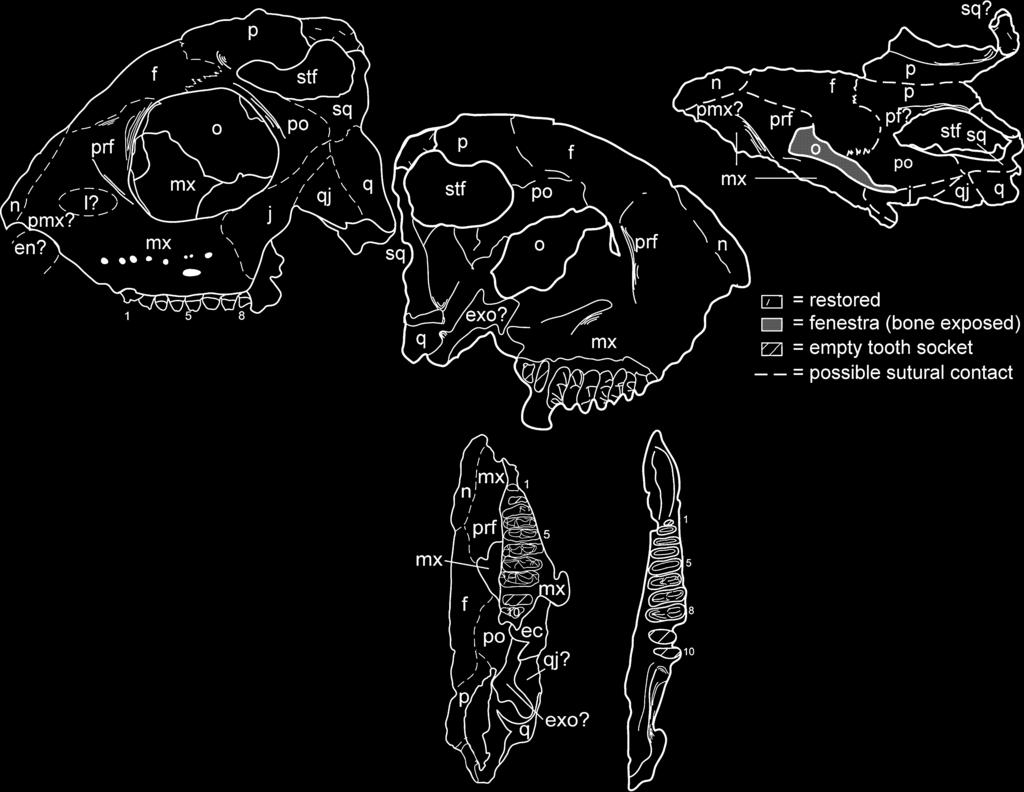
12 The new specimen of T. jacobsi we describe here is an articulated, incomplete skull and mandible. The skull is best preserved on the left side, which includes all of the bones except for most of the premaxilla, part of the anterior maxilla and the quadrate, and the ventral margins of the jugal, the quadratojugal and the quadrate. The right side is much less complete, and lacks part or all of all elements except for portions of the frontal, the postfrontal, the parietal and the squamosal. Although laterally compressed and damaged, NMMNH P preserves well the left side of the skull in both internal and external views (Textfig. 6A D). In overall shape it resembles the skull of T. buettneri (Gregory 1945, figs 2 4, pl. 20, fig. 1; Romer 1956, fig. 88), but differs in some key features: (1) the skull roof is not peaked at the lambdoidal crest in T. jacobsi, but instead peaks in a broad arc just posterodorsal to the orbit; (2) the relative size of the orbit of T. jacobsi is much larger than in T. buettneri, although this may be simply due to a difference in ontogenetic age of the specimen we illustrate here relative to specimens illustrated by Gregory (1945); and (3) T. jacobsi does not have a single, peaked sagittal crest as in T. buettneri, but instead has two separate parasagittal crests offset from the midline on the right and left sides (Text-fig. 6E F). The last of these could conceivably be an ontogenetic difference, although we consider this to be extremely unlikely as this kind of ontogenetic variation is not present in phylogenetically similar taxa such as rhynchosaurs (Benton 1990), nor is the observed variation in jaw adductor crests on the parietal of Prolacerta (Modesto and Sues 2004) attributable to ontogenetic variation. In lateral view (Text-fig. 6A B), NMMNH P lacks almost all of the premaxilla, so the anterior tip of the preserved skull begins at the posterior end of the nasal opening. The rostrum anterior to the orbit is short and tall, and ventral to the orbit there are nine teeth and two alveoli rooted in the maxilla. As in T. buettneri (e.g. Parks 1969), separate sutures of the nasals, the maxilla, the prefrontal and the lacrimal cannot be delineated on NMMNH P However, a clear suture for the jugal with the postorbital is visible just posterior to the ventral edge of the orbit, as is a squamosal-quadrate suture just posterior to the jugal-postorbital suture. The orbit is rather large (maximum diameter, 22 mm; maximum length, 24 mm). Part of the pterygoid and or ectopterygoid is visible in the medial wall of the orbit. Dorsal and posterior to the orbit, a relatively large (maximum diameter, 19 mm) supratemporal fenestra opens dorsally. In the following paragraphs we describe the observed bones of the skull individually. Most of the premaxilla is missing, and indeed, we can only infer its presence based on the preserved posterior margin of the external naris, as there are no discernible sutures between the premaxilla and the maxilla or the nasals. The first preserved teeth are clearly in the maxilla, so we infer that, like T. buettneri, T. jacobsi had an edentulous premaxilla. The maxilla is the largest preserved skull bone, and appears to occupy most of the skull ventral and anterior to the orbit (Textfig. 6A D). The maxilla appears to occupy the bulk of the ventral floor of the orbit, and the maxilla ventral to the orbit has several irregularly arranged nutrient foramina dorsal to the tooth row (Text-fig. 6B). The left maxilla preserves 11 tooth positions, eight of which are occupied by teeth in varied states of preservation (Text-fig. 6G H). Beginning with tooth 3, all are transversely broad and bear three cusps. Each of the teeth is c. 1Æ5 mm long. These teeth increase in width from c. 5Æ3 to 7Æ6 mm from tooth positions 3 to 8, before decreasing again to c. 5 mm at the end of the tooth row (inferred from the width of the alveolus). The central cusp is the most prominent, and is offset slightly labially from the centre of the tooth. The central cusp is weakly faceted, with two elongate facets on the lingual surface and two shorter facets on the labial side. All of these teeth bear cingula connecting the central cusp to the labial and lingual cusps. They also bear strong cingula distally and mesially. Lingually, the lingual cusp is rimmed by cingula, a feature not reported from T. jacobsi previously, that might serve to distinguish maxillary teeth of T. jacobsi from dentary teeth (described below). The lingual margins of the teeth are aligned so that the lingual cusps are nearly in a straight line anteroposteriorly. Consequently, the labial margin of the tooth row, as well as the arc formed by the central and labial cusps, is convex. Only a thin sliver of the posterior portion of the left nasal is preserved, based on our interpretation of a probable lateral suture with the maxilla and, possibly, the premaxilla (Textfig. 6A F). The prefrontal presumably forms most of a laterally projecting rim anterior to the orbit (Text-fig. 6A F). The bone appears to be roughly triangular in outline, although only its suture with the frontal is distinct. We infer the presence of a lacrimal based on comparison with the sutural relationships documented by Parks (1969, fig. 3) for T. buettneri, but there is no distinct ossification we can identify as such (Text-fig. 6B). The frontal comprises the largest single bone of the skull roof as preserved. It widens significantly from its anterior sutures with the nasals and the prefrontals and occupies much of the dorsal margin of the orbit. Posteriorly, it has a mediolaterally straight contact with the parietal, but its relationships with the postorbital and the postfrontal are not certain. The parietals are the best-preserved paired elements of the skull. As noted previously, the parietals form the bulk of the paired parasagittal crests, which are separated by a flat expanse of bone on the skull roof also composed of the parietals. This feature is the most significant non-dental difference between the skulls of T. jacobsi and T. buettneri. We do not think this is an ontogenetic feature, as it would require substantial restructuring of the parietal itself, including a medial migration of the jaw musculature, to attain the single crest condition seen in T. buettneri (compare Text-fig. 6E F with Gregory 1945, fig. 2). Modesto and Sues (2004) described some variation in the extent to which the jaw adductor musculature attached dorsally on the parietal of the archosauromorph Prolacerta broomi. However, the differences we document here are substantially greater between the two species of Trilophosaurus recognized here than Modesto and Sues (2004) found in Prolacerta, so we feel justified in using the different organization of the adductor crests (sagittal in T. buettneri, parasagittal in T. jacobsi) as a species-diagnostic feature. The anterior margin of the dorsal portion of neither squamosal is preserved, but impressions of both are present in the matrix, so we have reconstructed them on both the fossil itself and

13 A C E B D F G H I J
14 the illustrations we provide (Text-fig. 6A H). The squamosals are largely depressed below the level of the skull roof, and form the posterior of much of the ventral margin of the supratemporal fenestra. Anteriorly, the squamosal abuts the postorbital, but the ventral margin of this contact appears to be a complex suture with contacts with the jugal and the quadrate as well. The contact between the postorbital and the frontal is well defined, but its relationship to the parietal remains ambiguous, although it may form some of the steep wall on the medial rim of the supratemporal fenestra. The postorbital is bordered dorsally by the frontal, where it contacts the orbit, and appears to taper posteriorly along the ventral margin of the supratemporal fenestra. The jugal is the most clearly delineated bone in the skull, although much of its ventral margin is apparently missing. The anterior margin extends past the posterior end of the tooth margin, but the bone itself overlaps the maxilla and is, of course, not tooth-bearing. It floors only the posteroventral corner of the orbit. Posteriorly, this bone tapers more than that illustrated by Parks (1969, fig. 3) for T. buettneri. Relatively little of the quadratojugal is preserved, but its dorsal contact with the jugal is distinct and indicates that much of the ventral margin of the skull posterior to the tooth row must have been formed by this bone. Thus, it appears likely that the quadratojugal in T. jacobsi is a larger ossification (and, conversely, the jugal a smaller one) than its counterpart in T. buettneri. This would be even more pronounced if, as restored by Gregory (1945, fig. 4; also pl. 19, fig. 1) for T. buettneri, the articulation of the skull with the mandible is depressed below the level of the maxillary tooth row. Only the dorsalmost projection of the quadrate is preserved, although it is prominently exposed on the lateral surface of the skull as preserved. Its contact with the quadratojugal is straight, rather than curved (as illustrated by Parks 1969, fig. 3). The mandible was originally found in articulation with the skull, although slightly disarticulated at the mandibular symphysis. The left mandibular ramus is the better preserved (Textfig. 6I J), consisting of an essentially complete dentary with 11 tooth positions (nine occupied by teeth) and incomplete surangular, angular and splenial. The right mandibular ramus is broken anterior to the tooth row, such that the symphysial region is missing. The right dentary has 11 unambiguous tooth positions, nine of which are filled by teeth in variable states of preservation, although most are better preserved than their counterparts in the left dentary. The ventral and posterior portions of the mandible are poorly preserved. The dentary is by far the largest bone in the mandible, and has a complex sigmoid shape from the symphysis, with an anteriorly concave curve between the symphysis and the first tooth and a convex curve wrapping around the tooth row (Textfig. 6I J). The first two teeth are small and comparatively simple: the first is a simple peg, and the second has no more than two cusps. Tooth positions 3 9 are occupied by tricuspate, transversely broadened teeth that increase in size posteriorly. Positions 10 and 11 probably bore similar teeth, although the teeth were smaller (less wide) than tooth number 9, based on the size of the broken roots in those positions. The seven anteriormost tricuspate teeth (positions 3 9) are orientated so that their lingual cusps are anteroposteriorly aligned, and the labial margin of the tooth row is therefore convex, as in the maxilla. The last two tooth positions are deflected somewhat labially. The entire posterior portion of the ramus is therefore orientated at an angle of almost 45 degrees to the tooth row. The surangular is deeply grooved in dorsal (occlusal) view. The teeth are all c. 1Æ5 mm long. Teeth 3 9 all bear at least the remnants of three cusps, with the labial and the lingual cusps linked to the central cusp by thin, sharp cingula. Where preserved, the central cusp is always the tallest of the three. In occlusal view the central cusp is subtriangular, with the short base lingual to the cusp and the two longer sides mesial and distal, meeting labially to form a point between the central cusp and the labial cusp. The central cusp tends to lie closer to the labial cusp than the lingual cusp. This tooth morphology is identical to that described by Murry (1987) in T. jacobsi, even though this specimen is, in linear dimensions, at least an order of magnitude larger than the type of T. jacobsi (compare Textfig. 6 with Text-figs 4 5). In addition to the juvenile skull and mandible described in the preceding paragraphs, several other specimens we have collected from NMMNH locality 3775 preserve taxonomically significant features. These fossils include several dentulous fragments, including three incomplete mandibles, and four incomplete maxillae. There are six teeth in one maxillary fragment (NMMNH P-29959) we assign to Trilophosaurus jacobsi that was illustrated by Heckert et al. (2001, fig. 2a). The teeth are slightly damaged, but all are clearly transversely expanded and bear three cusps, one central and one each at the lingual and labial margins. The cusps are sharply pointed and aligned in a row connected by thin transverse cingula. The teeth are tightly packed and aligned medially, with narrower teeth anteriorly and broader teeth posteriorly, thus forming a labially convex tooth margin. Comparison with both specimens and illustrations of Trilophosaurus buettneri (Gregory 1945, figs 3, 6; Parks 1969, fig. 2) suggest that this element probably represents the anterior portion of the tooth battery of a left maxilla. However, it is also possible that it is the anterior portion of the tooth battery of a right dentary. In general, the maxilla of Trilophosaurus demonstrates a greater increase in the width of teeth posteriorly than does the dentary, so this element is most likely a left maxilla. TEXT-FIG. 6. A, C, E, G, J, photographs and B, D, F, H I, interpretative sketches of a juvenile skull and mandible (NMMNH P-41400) of Trilophosaurus jacobsi Murry from NMMNH locality A H, incomplete skull in A B, stereo left lateral, C D, stereo medial, E F, stereo dorsal, and G H, stereo ventral views. I-J, left mandibular ramus in stereo dorsal (occlusal) view. All 0Æ8. Numbers refer to tooth positions. Abbreviations: ect, ectopterygoid; en, external naris; exo, exoccipital; f, frontal; j, jugal; l, lacrimal; mx, maxilla; n, nasal; o, orbit; p, parietal; pf, postfrontal; pmx, premaxilla; po, postorbital; prf, prefrontal; pt, pterygoid; q, quadrate; qj, quadratojugal; sq, squamosal; stf, supratemporal fenestra.
15 One of the more striking features of NMMNH P is the close spacing of the teeth. Published illustrations and comparable specimens of Trilophosaurus buettneri from the WPA localities indicate that the teeth are much closer together in NMMNH P than in the TMM specimens. Similarly, the teeth of NMMNH P are also much closer together than in the holotype of T. buettneri (Text-fig. 3). This may represent an additional diagnostic feature of T. jacobsi. Another tooth illustrated by Heckert et al. (2001, fig. 3a), NMMNH P-29960, is similarly broad transversely, although the labial and the medial cusps are broken. Cingula are present on both the mesial and distal margins of this tooth, and evident on the teeth of NMMNH P as well. Clearly, this tooth is morphologically similar to those of NMMNH P The tooth crown is relatively tall, and there is a substantial root that is nearly as tall as the crown. Where the central cusp is preserved, including NMMNH P-29960, it is particularly swollen mesiodistally, so that the lower slopes of the central cusp divide the mesial and distal cingula into labial and lingual segments. This swollen base of the central cusp differs from that of both the type and the WPA specimens of T. buettneri and appears to be unique to T. jacobsi. Damage to the teeth of NMMNH P and P prevents us from describing detailed features, such as possible wear facets, with confidence. It is important to note that both P and P are substantially larger than P-41400, yet still have a similar overall tooth morphology, especially in terms of the presence and distribution of cingula. This indicates that the tooth characters used to differentiate T. jacobsi from T. buettneri, both by Murry (1987) and as used here, are valid taxonomic differences and not a result of ontogenetic variation (contra Elder 1978, 1987 and DeMar and Bolt 1981). Remarks. Murry (1987, p. 774) diagnosed T. jacobsi from T. buettneri based on the following characters: relatively smaller size, development of prominent medial and lateral cingula on each cusp, the asymmetrical placement of the central cusp, the successive development of a large central cusp on the posterior teeth, and the possible presence of a large, bulbous tooth on the posterior-most portion of the tooth row with numerous accessory cusps and prominent cingula. Also, the morphology of the jaws suggests that there was no edentulous beak as seen in the premaxilla and dentary of the large Otis Chalk Trilophosaurus skulls In Trilophosaurus jacobsi, both the dentary and the maxilla are extremely thin immediately anterior to the tooth row, posterior to the bulbous symphysis and anterior to the tooth row. We hypothesize that the holotype of T. jacobsi is thus broken at the most anterior point, and therefore only appears to be more dentulous than the anterior parts of upper or lower jaws of T. buettneri. Murry (1987) and Sues and Olsen (1993) noted that the tooth row is canted in different directions in the holotypes of T. buettneri and T. jacobsi. We suspect that the fragmentary holotype of T. jacobsi is thus actually a left maxilla, not a right dentary. If this is the case, then much of Murry s (1987) diagnosis of T. jacobsi is rendered inaccurate. We note here, however, that the differences in tooth crown morphology are sufficient to diagnose T. jacobsi from T. buettneri. The juvenile specimen we document here is much larger than any of the type series of T. jacobsi, and well within the size range of T. buettneri, including the small specimens from the Otis Chalk quarries. Indeed, this specimen is similar in size to the type of T. buettneri. We have not yet ascertained the maximum size of T. jacobsi, but the maxilla illustrated by Heckert et al. (2001) and redescribed above (NMMNH P-29959) bears teeth similar in size to the largest specimens of T. buettneri. The differences in tooth morphology noted by Murry (1987) are indeed valid, and we have expanded on these here. Note also that there is no evidence of ontogenetic change in tooth morphology in Trilophosaurus, as demonstrated by the preserved ontogenetic series of T. buettneri from the Otis Chalk quarries (Gregory 1945; Parks 1969; DeMar and Bolt 1981) and T. jacobsi from NMMNH locality 3775 documented here. Kaye and Padian (1994, fig. 9.59) illustrated an edentulous jaw fragment they identified as Trilophosaurus sp. We have re-examined the specimen and agree that it pertains to Trilophosaurus, but is not diagnostic to species. If it pertains to T. jacobsi, it is a topotype specimen of that taxon. SYSTEMATICS AND TOOTH NOMENCLATURE The principal systematic aim of this paper is to demonstrate that T. jacobsi is indeed closely related to T. buettneri. Sues (2003) and Modesto and Sues (2004) were the most recent to include the latter in a comprehensive phylogenetic analysis and, like Dilkes (1998) and most other workers, found that Trilophosaurus is an archosauromorph outside of crown-group Archosauria. Aside from the minor differences in cranial anatomy (paired sagittal crests, proportionately larger orbits) and the numerous complexities in the dentition (explored in greater detail below), T. jacobsi is clearly more similar to T. buettneri than any other taxon, including Teraterpeton hrynewichorum Sues. The skull of T. jacobsi illustrated in Plate 3 supports the assertions of Gregory (1945), Parks (1969) and Sues (2003) that Trilophosaurus lacks an infratemporal fenestra (contra Merck 1995). One of the key aspects of our study is the fact that, as suggested by Murry (1982, 1987), the teeth of Trilophosaurus are identifiable to species, which has implications for the systematics of these taxa as well as for the biostratigraphical and biochronological utility of both taxa. To facilitate comparison, we propose a standardized
.")
16 nomenclature of Trilophosaurus teeth (Text-fig. 7). Trilophosaurus teeth are transversely broadened, with a central cusp flanked by two marginal cusps, the labial and lingual cusps (Text-fig. 7). In T. buettneri, the three cusps are connected by a thin ridge or cingulum, the only such ridge on the tooth (Text-fig. 7D, F). Consequently, the areas between the cusps on the mesial (rostral) and distal (or caudal) faces of the tooth are comprised of poorly defined sulci. We term these the mesial labial, mesial lingual, distal labial and distal lingual sulci, respectively. The central cusp, marginal cusp, connecting cingulum and these sulci thus comprise the identifiable features of T. buettneri tooth crowns. The teeth of T. jacobsi are considerably more complex, and the nomenclature we propose here is designed to elucidate and describe that complexity (Text-fig. 7). As in T. buettneri, there is a central cusp, although it is typically displaced somewhat labially and is also generally taller cc A mc mc B mc cc mc lingual labial lingual labial C lingual mc dlb mlb pc cc dc dlab mlab distal mc labial D lingual mc dls mls distal cc dlas mlas mc labial mesial mesial E F G H TEXT-FIG. 7. Isolated teeth of Trilophosaurus documenting the differences that distinguish T. jacobsi (NMMNH P-34073; A, C, E, G) from T. buettneri (NMMNH P-34291; B, D, F, H). Teeth are in rostro-caudal (A B, G H) and occlusal (C F) views. Abbreviations: cc, central cusp; dc, distal cingulum; dlab, distal-labial basin; dlas, distal-labial sulcus; dlb, distal-lingual basin; dls, distal-lingual sulcus; mc, marginal cusp; pc, mesial cingulum; plab, posterolabial basin; plas, posterolabial sulcus; plb, posterolingual basin; pls, posterolingual sulcus. Scale bars represent 1 mm.
TAXONOMY AND BIOSTRATIGRAPHY OF THE LATE TRIASSIC ARCHOSAUROMORPH TRILOPHOSAURUS
 Lucas, S.G. and Spielmann, J.A., eds., 2007, Triassic of the American West. New Mexico Museum of Natural History and Science Bulletin 40. TAXONOMY AND BIOSTRATIGRAPHY OF THE LATE TRIASSIC ARCHOSAUROMORPH
Postilla PEABODY MUSEUM OF NATURAL HISTORY YALE UNIVERSITY NEW HAVEN, CONNECTICUT, U.S.A.
 Postilla PEABODY MUSEUM OF NATURAL HISTORY YALE UNIVERSITY NEW HAVEN, CONNECTICUT, U.S.A. Number 117 18 March 1968 A 7DIAPSID (REPTILIA) PARIETAL FROM THE LOWER PERMIAN OF OKLAHOMA ROBERT L. CARROLL REDPATH
SOME LITTLE-KNOWN FOSSIL LIZARDS FROM THE
 PROCEEDINGS OF THE UNITED STATES NATIONAL MUSEUM issued SWsK \ {^^m ^V ^^ SMITHSONIAN INSTITUTION U. S. NATIONAL MUSEUM Vol. 91 Washington : 1941 No. 3124 SOME LITTLE-KNOWN FOSSIL LIZARDS FROM THE OLIGOCENE
Exceptional fossil preservation demonstrates a new mode of axial skeleton elongation in early ray-finned fishes
 Supplementary Information Exceptional fossil preservation demonstrates a new mode of axial skeleton elongation in early ray-finned fishes Erin E. Maxwell, Heinz Furrer, Marcelo R. Sánchez-Villagra Supplementary
ONLINE APPENDIX 1. Morphological phylogenetic characters scored in this paper. See Poe (2004) for
 for") ONLINE APPENDIX Morphological phylogenetic characters scored in this paper. See Poe () for detailed character descriptions, citations, and justifications for states. Note that codes are changed from a
ONLINE APPENDIX Morphological phylogenetic characters scored in this paper. See Poe () for detailed character descriptions, citations, and justifications for states. Note that codes are changed from a
MANDIBLES OF JUVENILE PHYTOSAURS (ARCHOSAURIA: CRUROTARSI) FROM THE UPPER TRIASSIC CHINLE GROUP OF TEXAS AND NEW MEXICO, USA
 FROM THE UPPER TRIASSIC CHINLE GROUP OF TEXAS AND NEW MEXICO, USA") 228 Tanner, L.H., Spielmann, J.A. and Lucas, S.G., eds., 2013, The Triassic System. New Mexico Museum of Natural History and Science, Bulletin 61. MANDIBLES OF JUVENILE PHYTOSAURS (ARCHOSAURIA: CRUROTARSI)
228 Tanner, L.H., Spielmann, J.A. and Lucas, S.G., eds., 2013, The Triassic System. New Mexico Museum of Natural History and Science, Bulletin 61. MANDIBLES OF JUVENILE PHYTOSAURS (ARCHOSAURIA: CRUROTARSI)
New Carnivorous Dinosaurs from the Upper Cretaceous of Mongolia
 1955 Doklady, Academy of Sciences USSR 104 (5):779-783 New Carnivorous Dinosaurs from the Upper Cretaceous of Mongolia E. A. Maleev (translated by F. J. Alcock) The present article is a summary containing
REVISION OF REDONDASUCHUS (ARCHOSAURIA: AETOSAURIA) FROM THE UPPER TRIASSIC REDONDA FORMATION, NEW MEXICO, WITH DESCRIPTION OF A NEW SPECIES
 FROM THE UPPER TRIASSIC REDONDA FORMATION, NEW MEXICO, WITH DESCRIPTION OF A NEW SPECIES") Harris et al., eds., 2006, The Triassic-Jurassic Terrestrial Transition. New Mexico Museum of Natural History and Science Bulletin 37. REVISION OF REDONDASUCHUS (ARCHOSAURIA: AETOSAURIA) FROM THE UPPER
Harris et al., eds., 2006, The Triassic-Jurassic Terrestrial Transition. New Mexico Museum of Natural History and Science Bulletin 37. REVISION OF REDONDASUCHUS (ARCHOSAURIA: AETOSAURIA) FROM THE UPPER
New Mexico Geological Society
 New Mexico Geological Society Downloaded from: http://nmgs.nmt.edu/publications/guidebooks/56 Vertebrate fauna of the Upper Triassic Mesa Montosa Member (Petrified Forest Formation, Chinle Group), Chama
Lucas, S.G. and Spielmann, J.A., eds., 2007, The Global Triassic. New Mexico Museum of Natural History and Science Bulletin 41.
 Lucas, S.G. and Spielmann, J.A., eds., 2007, The Global Triassic. New Mexico Museum of Natural History and Science Bulletin 41. BIOSTRATIGRAPHIC UTILITY OF THE UPPER TRIASSIC AETOSAUR TECOVASUCHUS (ARCHOSAURIA:STAGONOLEPIDIDAE),
TOPOTYPES OF TYPOTHORAX COCCINARUM, A LATE TRIASSIC AETOSAUR FROM THE AMERICAN SOUTHWEST
 Lucas, S.G. and Spielmann, J.A., eds., 2007, The Global Triassic. New Mexico Museum of Natural History and Science Bulletin 41. TOPOTYPES OF TYPOTHORAX COCCINARUM, A LATE TRIASSIC AETOSAUR FROM THE AMERICAN
AMERICAN MUSEUM NOVITATES Published by
 AMERICAN MUSEUM NOVITATES Published by Number 782 THE AmzRICAN MUSEUM OF NATURAL HISTORY Feb. 20, 1935 New York City 56.81, 7 G (68) A NOTE ON THE CYNODONT, GLOCHINODONTOIDES GRACILIS HAUGHTON BY LIEUWE
complex in cusp pattern. (3) The bones of the coyote skull are thinner, crests sharper and the
 The bones of the coyote skull are thinner, crests sharper and the") DISTINCTIONS BETWEEN THE SKULLS OF S AND DOGS Grover S. Krantz Archaeological sites in the United States frequently yield the bones of coyotes and domestic dogs. These two canines are very similar both
DISTINCTIONS BETWEEN THE SKULLS OF S AND DOGS Grover S. Krantz Archaeological sites in the United States frequently yield the bones of coyotes and domestic dogs. These two canines are very similar both
2. Skull, total length versus length of the presacral vertebral column: (0); extremely elongated neck (e.g. Tanystropheus longobardicus).
; extremely elongated neck (e.g. Tanystropheus longobardicus).") Character list of the taxon-character data set 1. Skull and lower jaws, interdental plates: absent (0); present, but restricted to the anterior end of the dentary (1); present along the entire alveolar
Character list of the taxon-character data set 1. Skull and lower jaws, interdental plates: absent (0); present, but restricted to the anterior end of the dentary (1); present along the entire alveolar
Williston, and as there are many fairly good specimens in the American
 56.81.7D :14.71.5 Article VII.- SOME POINTS IN THE STRUCTURE OF THE DIADECTID SKULL. BY R. BROOM. The skull of Diadectes has been described by Cope, Case, v. Huene, and Williston, and as there are many
THE LATE TRIASSIC AETOSAUR PARATYPOTHORAX
 Harris et al., eds., 2006, The Triassic-Jurassic Terrestrial Transition. New Mexico Museum of Natural History and Science Bulletin 37. THE LATE TRIASSIC AETOSAUR PARATYPOTHORAX 575 SPENCER G. LUCAS 1,
v:ii-ixi, 'i':;iisimvi'\>!i-:: "^ A%'''''-'^-''S.''v.--..V^'E^'-'-^"-t''gi L I E) R.ARY OF THE VERSITY U N I or ILLINOIS REMO
 R.ARY OF THE VERSITY U N I or ILLINOIS REMO") "^ A%'''''-'^-''S.''v.--..V^'E^'-'-^"-t''gi v:ii-ixi, 'i':;iisimvi'\>!i-:: L I E) R.ARY OF THE U N I VERSITY or ILLINOIS REMO Natural History Survey Librarv GEOLOGICAL SERIES OF FIELD MUSEUM OF NATURAL
"^ A%'''''-'^-''S.''v.--..V^'E^'-'-^"-t''gi v:ii-ixi, 'i':;iisimvi'\>!i-:: L I E) R.ARY OF THE U N I VERSITY or ILLINOIS REMO Natural History Survey Librarv GEOLOGICAL SERIES OF FIELD MUSEUM OF NATURAL
SUPPLEMENTARY INFORMATION
 Character 155, interdental ridges. Absence of interdental ridge (0) shown in Parasaniwa wyomingensis (Platynota). Interdental ridges (1) shown in Coniophis precedens. WWW.NATURE.COM/NATURE 1 Character
New Mexico Geological Society
 New Mexico Geological Society Downloaded from: http://nmgs.nmt.edu/publications/guidebooks/56 Definition and correlation of the Lamyan: A new biochronological unit for the nonmarine Late Carnian (Late
SUPPLEMENTARY ONLINE MATERIAL FOR. Nirina O. Ratsimbaholison, Ryan N. Felice, and Patrick M. O connor
 http://app.pan.pl/som/app61-ratsimbaholison_etal_som.pdf SUPPLEMENTARY ONLINE MATERIAL FOR Nirina O. Ratsimbaholison, Ryan N. Felice, and Patrick M. O connor Ontogenetic changes in the craniomandibular
Fig. 5. (A) Scaling of brain vault size (width measured at the level of anterior squamosal/parietal suture) relative to skull size (measured at the
 Scaling of brain vault size (width measured at the level of anterior squamosal/parietal suture) relative to skull size (measured at the") Fig. 5. (A) Scaling of brain vault size (width measured at the level of anterior squamosal/parietal suture) relative to skull size (measured at the distance between the left versus right temporomandibular
Fig. 5. (A) Scaling of brain vault size (width measured at the level of anterior squamosal/parietal suture) relative to skull size (measured at the distance between the left versus right temporomandibular
A NEW GENUS AND SPECIES OF AMERICAN THEROMORPHA
 A NEW GENUS AND SPECIES OF AMERICAN THEROMORPHA MYCTEROSAURUS LONGICEPS S. W. WILLISTON University of Chicago The past summer, Mr. Herman Douthitt, of the University of Chicago paleontological expedition,
HONR219D Due 3/29/16 Homework VI
 Part 1: Yet More Vertebrate Anatomy!!! HONR219D Due 3/29/16 Homework VI Part 1 builds on homework V by examining the skull in even greater detail. We start with the some of the important bones (thankfully
THE VERTEBRATE FAUNA OF THE UPPER TRIASSIC CHINLE FORMATION IN NORTHERN ARIZONA
 Guidebook to the Triassic Formations of the Colorado Plateau in northern Arizona: Geology, Paleontology, and History. Sterling J. Nesbitt, William G. Parker, and Randall B. Irmis (eds.) Mesa Southwest
The cranial osteology of Belebey vegrandis (Parareptilia: Bolosauridae), from the Middle Permian of Russia, and its bearing on reptilian evolution
, from the Middle Permian of Russia, and its bearing on reptilian evolution") Blackwell Publishing LtdOxford, UKZOJZoological Journal of the Linnean Society0024-4082 2007 The Linnean Society of London? 2007 1511 191214 Original Articles RUSSIAN BOLOSAURID REPTILER. R. REISZ ET AL.
Blackwell Publishing LtdOxford, UKZOJZoological Journal of the Linnean Society0024-4082 2007 The Linnean Society of London? 2007 1511 191214 Original Articles RUSSIAN BOLOSAURID REPTILER. R. REISZ ET AL.
KRZYZANOWSKISAURUS, A NEW NAME FOR A PROBABLE ORNITHISCHIAN DINOSAUR FROM THE UPPER TRIASSIC CHINLE GROUP, ARIZONA AND NEW MEXICO, USA
 Heckert, A.B., and Lucas, S.G., eds., 2005, Vertebrate Paleontology in Arizona. New Mexico Museum of Natural History and Science Bulletin No. 29. KRZYZANOWSKISAURUS, A NEW NAME FOR A PROBABLE ORNITHISCHIAN
THE SKULLS OF ARAEOSCELIS AND CASEA, PERMIAN REPTILES
 THE SKULLS OF REOSCELIS ND CSE, PERMIN REPTILES University of Chicago There are few Permian reptiles of greater interest at the present time than the peculiar one I briefly described in this journal' three
Dominique; Bustamante, Rogelio; Cirks, Leah; Lopez, Martin; Moncada, Adriana; Ortega,
 An unusual archosauromorph tooth increases known archosauromorph diversity in the lower portion of the Chinle Formation (Late Triassic) of southeastern Utah, USA Lopez, Andres; St. Aude, Isabella; Alderete,
Mammalogy Lab 1: Skull, Teeth, and Terms
 Mammalogy Lab 1: Skull, Teeth, and Terms Be able to: Goals of today s lab Locate all structures listed on handout Define all terms on handout what they are or what they look like Give examples of mammals
A new sauropod from Dashanpu, Zigong Co. Sichuan Province (Abrosaurus dongpoensis gen. et sp. nov.)
") A new sauropod from Dashanpu, Zigong Co. Sichuan Province (Abrosaurus dongpoensis gen. et sp. nov.) by Ouyang Hui Zigong Dinosaur Museum Newsletter Number 2 1989 pp. 10-14 Translated By Will Downs Bilby
A new sauropod from Dashanpu, Zigong Co. Sichuan Province (Abrosaurus dongpoensis gen. et sp. nov.) by Ouyang Hui Zigong Dinosaur Museum Newsletter Number 2 1989 pp. 10-14 Translated By Will Downs Bilby
Giant croc with T. rex teeth roamed Madagascar
 Giant croc with T. rex teeth roamed Madagascar www.scimex.org/newsfeed/giant-croc-with-t.-rex-teeth-used-to-roam-in-madagascar Embargoed until: Publicly released: PeerJ A fossil of the largest and oldest
A new species of Hsisosuchus (Mesoeucrocodylia) from Dashanpu, Zigong Municipality, Sichuan Province
 from Dashanpu, Zigong Municipality, Sichuan Province") A new species of Hsisosuchus (Mesoeucrocodylia) from Dashanpu, Zigong Municipality, Sichuan Province Yuhui Gao (Zigong Dinosaur Museum) Vertebrata PalAsiatica Volume 39, No. 3 July, 2001 pp. 177-184 Translated
A new species of Hsisosuchus (Mesoeucrocodylia) from Dashanpu, Zigong Municipality, Sichuan Province Yuhui Gao (Zigong Dinosaur Museum) Vertebrata PalAsiatica Volume 39, No. 3 July, 2001 pp. 177-184 Translated
PEABODY MUSEUM OF NATURAL HISTORY, YALE UNIVERSITY NEW HAVEN, CONNECTICUT, U.S.A. A NEW OREODONT FROM THE CABBAGE PATCH LOCAL FAUNA, WESTERN MONTANA
 Postilla PEABODY MUSEUM OF NATURAL HISTORY YALE UNIVERSITY NEW HAVEN, CONNECTICUT, U.S.A. Number 85 September 21, 1964 A NEW OREODONT FROM THE CABBAGE PATCH LOCAL FAUNA, WESTERN MONTANA STANLEY J. RIEL
PALEONTOLOGICAL CONTRIBUTIONS
 THE UNIVERSITY OF KANSAS PALEONTOLOGICAL CONTRIBUTIONS August, 1965 Paper 2 A NEW WYOMING PHYTOSAUR By THEODORE H. EATON, JR. [Museum of Natural History, University of Kansas I ABSTRACT The skull of a
CRANIAL ANATOMY OF ENNATOSAURUS TECTON (SYNAPSIDA: CASEIDAE) FROM THE MIDDLE PERMIAN OF RUSSIA AND THE EVOLUTIONARY RELATIONSHIPS OF CASEIDAE
 FROM THE MIDDLE PERMIAN OF RUSSIA AND THE EVOLUTIONARY RELATIONSHIPS OF CASEIDAE") Journal of Vertebrate Paleontology 28(1):160 180, March 2008 2008 by the Society of Vertebrate Paleontology ARTICLE CRANIAL ANATOMY OF ENNATOSAURUS TECTON (SYNAPSIDA: CASEIDAE) FROM THE MIDDLE PERMIAN
Journal of Vertebrate Paleontology 28(1):160 180, March 2008 2008 by the Society of Vertebrate Paleontology ARTICLE CRANIAL ANATOMY OF ENNATOSAURUS TECTON (SYNAPSIDA: CASEIDAE) FROM THE MIDDLE PERMIAN
A New Pterosaur from the Middle Jurassic of Dashanpu, Zigong, Sichuan
 A New Pterosaur from the Middle Jurassic of Dashanpu, Zigong, Sichuan by Xinlu He (Chengdu College of Geology) Daihuan Yang (Chungking Natural History Museum, Sichuan Province) Chunkang Su (Zigong Historical
Big Bend Paleo-Geo Journal
 Big Bend Paleo-Geo Journal An Open Access Informal Publication from Mosasaur Ranch, Terlingua, Texas All rights reserved Copyright; Kenneth R. Barnes, 2014 New info and corrections in red 2 / 3 / 2015
Lower Cretaceous Kwanmon Group, Northern Kyushu
 Bull. Kitakyushu Mus. Nat. Hist., 11: 87-90. March 30, 1992 A New Genus and Species of Carnivorous Dinosaur from the Lower Cretaceous Kwanmon Group, Northern Kyushu Yoshihiko Okazaki Kitakyushu Museum
LOWER CRETACEOUS OF SOUTH DAKOTA.
 A NEW DINOSAUR, STP^GOSAURUS MARSHl, FROM THE LOWER CRETACEOUS OF SOUTH DAKOTA. By Frederic A. Lucas, Curator, Divisioii of Coiiipnrative Anatomy, in charge, of Section of Vertebrate Fossils. The name
A new species of sauropod, Mamenchisaurus anyuensis sp. nov.
 A new species of sauropod, Mamenchisaurus anyuensis sp. nov. by Xinlu He, Suihua Yang, Kaiji Cai, Kui Li, and Zongwen Liu Chengdu University of Technology Papers on Geosciences Contributed to the 30th
TRACHEMYS SCULPTA. A nearly complete articulated carapace and plastron of an Emjdd A NEAKLY COMPLETE SHELL OF THE EXTINCT TURTLE,
 A NEAKLY COMPLETE SHELL OF THE EXTINCT TURTLE, TRACHEMYS SCULPTA By Charles W. Gilmore Curator of Vertebrate Paleontology, United States National Museum INTRODUCTION A nearly complete articulated carapace
THE TETRAPOD FAUNA OF THE UPPER TRIASSIC LOWER CHINLE GROUP (ADAMANIAN: LATEST CARNIAN) OF THE ZUNI MOUNTAINS, WEST-CENTRAL NEW MEXICO
 OF THE ZUNI MOUNTAINS, WEST-CENTRAL NEW MEXICO") I Lucas~ S.G., Estep, }.W., Williamson/ T.E. and Morgan, G.S. eds., 1997, New Mexico's Fossil Record 1. New Mexico Museum of Natural History and Science Bulletin No. 11. 29 THE TETRAPOD FAUNA OF THE UPPER
I Lucas~ S.G., Estep, }.W., Williamson/ T.E. and Morgan, G.S. eds., 1997, New Mexico's Fossil Record 1. New Mexico Museum of Natural History and Science Bulletin No. 11. 29 THE TETRAPOD FAUNA OF THE UPPER
A NEW SPECIES OF EXTINCT TURTLE FROM THE UPPER PLIOCENE OF IDAHO
 A NEW SPECIES OF EXTINCT TURTLE FROM THE UPPER PLIOCENE OF IDAHO By Charles W. Gilmore Curator, Division of Vertebrate Paleontology United States National Museum Among the fossils obtained bj^ the Smithsonian
A new carnosaur from Yongchuan County, Sichuan Province
 A new carnosaur from Yongchuan County, Sichuan Province by Dong Zhiming Institute of Vertebrate Palaeontology and Palaeoanthropology, Academia Sinica Zhang Yihong, Li Xuanmin, and Zhou Shiwu Chongqing
A Fossil Snake (Elaphe vulpina) From A Pliocene Ash Bed In Nebraska
 From A Pliocene Ash Bed In Nebraska") University of Nebraska - Lincoln DigitalCommons@University of Nebraska - Lincoln Transactions of the Nebraska Academy of Sciences and Affiliated Societies Nebraska Academy of Sciences 198 A Fossil Snake
University of Nebraska - Lincoln DigitalCommons@University of Nebraska - Lincoln Transactions of the Nebraska Academy of Sciences and Affiliated Societies Nebraska Academy of Sciences 198 A Fossil Snake
FIELDIANA GEOLOGY NEW SALAMANDERS OF THE FAMILY SIRENIDAE FROM THE CRETACEOUS OF NORTH AMERICA
 FIELDIANA GEOLOGY Published by CHICAGO NATURAL HISTORY MUSEUM Volume 10 Sbftember 22, 1968 No. 88 NEW SALAMANDERS OF THE FAMILY SIRENIDAE FROM THE CRETACEOUS OF NORTH AMERICA Coleman J. Coin AND Walter
Anatomy. Name Section. The Vertebrate Skeleton
 Name Section Anatomy The Vertebrate Skeleton Vertebrate paleontologists get most of their knowledge about past organisms from skeletal remains. Skeletons are useful for gleaning information about an organism
VERTEBRATA PALASIATICA
 41 2 2003 2 VERTEBRATA PALASIATICA pp. 147 156 figs. 1 5 1) ( 100044), ( Parakannemeyeria brevirostris),,, : ( Xiyukannemeyeria),,, Q915. 864 60 Turfania (,1973), Dicynodon (, 1973 ; Lucas, 1998), (Lystrosaurus)
A R T I C L E S STRATIGRAPHIC DISTRIBUTION OF VERTEBRATE FOSSIL FOOTPRINTS COMPARED WITH BODY FOSSILS
 A R T I C L E S STRATIGRAPHIC DISTRIBUTION OF VERTEBRATE FOSSIL FOOTPRINTS COMPARED WITH BODY FOSSILS Leonard Brand & James Florence Department of Biology Loma Linda University WHAT THIS ARTICLE IS ABOUT
First records of diapsid Palacrodon from the Norian, Late Triassic Chinle Formation of Arizona, and their biogeographic implications
 First records of diapsid Palacrodon from the Norian, Late Triassic Chinle Formation of Arizona, and their biogeographic implications BEN T. KLIGMAN, ADAM D. MARSH, and WILLIAM G. PARKER Kligman, B.T.,
KATE E. ZEIGLER, ANDREW B. HECKERT and SPENCER G. LUCAS. New Mexico Museum of Natural History, 1801 Mountain Road NW, Albuquerque, NM
 Zeigler, K.E., Heckert, A.B., and Lucas, S.G., eds., 2003, Paleontology and Geology of the Snyder Quarry, New Mexico Museum of Natural History and Science Bulletin No. 24. AN ILLUSTRATED ATLAS OF THE PHYTOSAUR
290 SHUFELDT, Remains of Hesperornis.
 290 SHUFELDT, Remains of Hesperornis. [ Auk [July THE FOSSIL REMAINS OF A SPECIES OF HESPERORNIS FOUND IN MONTANA. BY R. W. SHUFELD% M.D. Plate XI7III. ExR,¾ in November, 1914, Mr. Charles W. Gihnore,
UPPER TRIASSIC TETRAPODS FROM THE LUCERO UPLIFT, CENTRAL NEW MEXICO
 New Mexico Geological Society Guidebook, 50th Field Conference, Albuquerque Geology, 1999 311 UPPER TRIASSIC TETRAPODS FROM THE LUCERO UPLIFT, CENTRAL NEW MEXICO ANDREW B. HECKERT Department of Earth &
.56 m. (22 in.). COMPSOGNATHOID DINOSAUR FROM THE. Medicine Bow, Wyoming, by the American Museum Expedition
. COMPSOGNATHOID DINOSAUR FROM THE. Medicine Bow, Wyoming, by the American Museum Expedition") Article XII.-ORNITHOLESTES HERMANNI, A NEW COMPSOGNATHOID DINOSAUR FROM THE UPPER JURASSIC. By HENRY FAIRFIELD OSBORN. The type skeleton (Amer. Mus. Coll. No. 6I9) of this remarkable animal was discovered
Article XII.-ORNITHOLESTES HERMANNI, A NEW COMPSOGNATHOID DINOSAUR FROM THE UPPER JURASSIC. By HENRY FAIRFIELD OSBORN. The type skeleton (Amer. Mus. Coll. No. 6I9) of this remarkable animal was discovered
Notes on Ceratopsians and Ankylosaurs at the Royal Ontario Museum
 Notes on Ceratopsians and Ankylosaurs at the Royal Ontario Museum Andrew A. Farke, Ph.D. Raymond M. Alf Museum of Paleontology 1175 West Baseline Road Claremont, CA 91711 email: afarke@webb.org Introduction
THE OCCURRENCE OF CONTOGENYS-LIKE LIZARDS IN THE LATE CRETACEOUS AND EARLY TERTIARY OF THE WESTERN INTERIOR OF THE U.S.A.
 Journal of Vertebrate Paleontology 29(3):677 701, September 2009 # 2009 by the Society of Vertebrate Paleontology ARTICLE THE OCCURRENCE OF CONTOGENYS-LIKE LIZARDS IN THE LATE CRETACEOUS AND EARLY TERTIARY
Supporting Online Material for
 www.sciencemag.org/cgi/content/full/329/5998/1481/dc1 Supporting Online Material for Tyrannosaur Paleobiology: New Research on Ancient Exemplar Organisms Stephen L. Brusatte,* Mark A. Norell, Thomas D.
4. Premaxilla: Foramen on the lateral surface of the premaxillary body (Yates 2007 ch. 4) 0 absent 1 present
 0 absent 1 present") The character matrix used as a basis for this study is that of Yates et al (2010) which is modified from the earlier matrix used by Yates (2007). This matrix includes characters acquired and/or modified
The character matrix used as a basis for this study is that of Yates et al (2010) which is modified from the earlier matrix used by Yates (2007). This matrix includes characters acquired and/or modified
Title: Phylogenetic Methods and Vertebrate Phylogeny
 Title: Phylogenetic Methods and Vertebrate Phylogeny Central Question: How can evolutionary relationships be determined objectively? Sub-questions: 1. What affect does the selection of the outgroup have
Erycine Boids from the Early Oligocene of the South Dakota Badlands
 Georgia Journal of Science Volume 67 No. 2 Scholarly Contributions from the Membership and Others Article 6 2009 Erycine Boids from the Early Oligocene of the South Dakota Badlands Dennis Parmley J. Alan
SOME NEW AMERICAN PYCNODONT FISHES.
 SOME NEW AMERICAN PYCNODONT FISHES. By James Williams Gidley, Assistant Curator of Fossil Mammals, United States National Museum. In the United States National Museum are several specimens representing
A New Ceratopsian Dinosaur from the Upper
 SMITHSONIAN MISCELLANEOUS COLLECTIONS VOLUME 63. NUMBER 3 A New Ceratopsian Dinosaur from the Upper Cretaceous of Montana, with Note on Hypacrosaurus (With Two Plates) CHARLES W. GILMORE Assistant Curator
The Discovery of a Tritylodont from the Xinjiang Autonomous Region
 The Discovery of a Tritylodont from the Xinjiang Autonomous Region Ailing Sun and Guihai Cui (Institute of Vertebrate Paleontology, Paleoanthropology, Academia Sinica) Vertebrata PalAsiatica Volume XXVII,
THE GORGONOPSIAN GENUS, HIPPOSAURUS, AND THE FAMILY ICTIDORHINIDAE * Dr. L.D. Boonstra. Paleontologist, South African Museum, Cape Town
 THE GORGONOPSIAN GENUS, HIPPOSAURUS, AND THE FAMILY ICTIDORHINIDAE * by Dr. L.D. Boonstra Paleontologist, South African Museum, Cape Town In 1928 I dug up the complete skeleton of a smallish gorgonopsian
Description of Cranial Elements and Ontogenetic Change within Tropidolaemus wagleri (Serpentes: Crotalinae).
.") East Tennessee State University Digital Commons @ East Tennessee State University Electronic Theses and Dissertations 5-2016 Description of Cranial Elements and Ontogenetic Change within Tropidolaemus
East Tennessee State University Digital Commons @ East Tennessee State University Electronic Theses and Dissertations 5-2016 Description of Cranial Elements and Ontogenetic Change within Tropidolaemus
In the following we are particularly elaborating the deviation of the position of lower canines
 IN THE BORZOI BREED PRELUDE At the revision and finalization of the RAS (Rasspecifika Avels Strategier -breed specific breeding strategies) at Borzoi-Ringen s annual meeting it was decided that this appendix
A NEW SALTICID SPIDER FROM VICTORIA By R. A. Dunn
 Dunn, R. A. 1947. A new salticid spider from Victoria. Memoirs of the National Museum of Victoria 15: 82 85. All text not included in the original document is highlighted in red. Mem. Nat. Mus. Vict.,
Vol. XIV, No. 1, March, The Larva and Pupa of Brontispa namorikia Maulik (Coleoptera: Chrysomelidae: Hispinae) By S.
 By S.") Vol. XIV, No. 1, March, 1950 167 The Larva and Pupa of Brontispa namorikia Maulik (Coleoptera: Chrysomelidae: Hispinae) By S. MAULIK BRITISH MUSEUM (NATURAL HISTORY) (Presented by Mr. Van Zwaluwenburg
Vol. XIV, No. 1, March, 1950 167 The Larva and Pupa of Brontispa namorikia Maulik (Coleoptera: Chrysomelidae: Hispinae) By S. MAULIK BRITISH MUSEUM (NATURAL HISTORY) (Presented by Mr. Van Zwaluwenburg
OF THE TRIAS THE PHYTOSAURIA
 THE PHYTOSAURIA OF THE TRIAS MAURICE G. MEHL University of Wisconsin Some time ago the writer gave a brief notice of a new genus of phytosaurs of which Angistorhinus grandis Mehl was the type.' It is the
PEABODY MUSEUM OF NATURAL HISTORY YALE UNIVERSITY NEW HAVEN, CONNECTICUT, U.S.A. GLYPTOLEPIS FROM THE MIDDLE DEVONIAN OF SCOTLAND
 Postilla PEABODY MUSEUM OF NATURAL HISTORY YALE UNIVERSITY NEW HAVEN, CONNECTICUT, U.S.A. Number 99 April 16, 1966 GLYPTOLEPIS FROM THE MIDDLE DEVONIAN OF SCOTLAND KEITH STEWART THOMSON 1 DEPARTMENT OF
 The following text is generated from uncorrected OCR. [Begin Page: Page 1] A NEW CERATOPSIAN DINOSAUR FROM THE UPPER CRETACEOUS OF MONTANA, WITH NOTE ON HYPACROSAURUS ' By CHARLES W. GILMORE assistant
Mammalogy Laboratory 1 - Mammalian Anatomy
 Mammalogy Laboratory 1 - Mammalian Anatomy I. The Goal. The goal of the lab is to teach you skeletal anatomy of mammals. We will emphasize the skull because many of the taxonomically important characters
A M E G H I N I A N A. Revista de la Asociación Paleontológia Argentina. Volume XV September-December 1978 Nos. 3-4
 A M E G H I N I A N A Revista de la Asociación Paleontológia Argentina Volume XV September-December 1978 Nos. 3-4 COLORADIA BREVIS N. G. ET N. SP. (SAURISCHIA, PROSAUROPODA), A PLATEOSAURID DINOSAUR FROM
A NEW GENUS AND SPECIES OF SPHENODONTIAN FROM THE GHOST RANCH COELOPHYSIS QUARRY (UPPER TRIASSIC: APACHEAN), ROCK POINT FORMATION, NEW MEXICO, USA
, ROCK POINT FORMATION, NEW MEXICO, USA") [Palaeontology, Vol. 51, Part 4, 2008, pp. 827 845] A NEW GENUS AND SPECIES OF SPHENODONTIAN FROM THE GHOST RANCH COELOPHYSIS QUARRY (UPPER TRIASSIC: APACHEAN), ROCK POINT FORMATION, NEW MEXICO, USA by
[Palaeontology, Vol. 51, Part 4, 2008, pp. 827 845] A NEW GENUS AND SPECIES OF SPHENODONTIAN FROM THE GHOST RANCH COELOPHYSIS QUARRY (UPPER TRIASSIC: APACHEAN), ROCK POINT FORMATION, NEW MEXICO, USA by
A GIANT SKULL, ONTOGENETIC VARIATION AND TAXONOMIC VALIDITY OF THE LATE TRIASSIC PHYTOSAUR PARASUCHUS
 222 Lucas, S.G. and Spielmann, J.A., eds., 2007, The Global Triassic. New Mexico Museum of Natural History and Science Bulletin 41. A GIANT SKULL, ONTOGENETIC VARIATION AND TAXONOMIC VALIDITY OF THE LATE
Cranial osteology of the African gerrhosaurid Angolosaurus skoogi (Squamata; Gerrhosauridae) HOLLY A. NANCE
 HOLLY A. NANCE") African Journal of Herpetology, 2007 56(1): 39-75. Herpetological Association of Africa Original article Cranial osteology of the African gerrhosaurid Angolosaurus skoogi (Squamata; Gerrhosauridae) HOLLY
African Journal of Herpetology, 2007 56(1): 39-75. Herpetological Association of Africa Original article Cranial osteology of the African gerrhosaurid Angolosaurus skoogi (Squamata; Gerrhosauridae) HOLLY
NOTES ON THE FIRST SKULL AND JAWS OF RIOJASAURUS INCERTUS (DINOSAURIA, PROSAUROPODA, MELANOROSAURIDAE) OF THE LATE TRIASSIC OF LA RIOJA, ARGENTINA
 OF THE LATE TRIASSIC OF LA RIOJA, ARGENTINA") NOTES ON THE FIRST SKULL AND JAWS OF RIOJASAURUS INCERTUS (DINOSAURIA, PROSAUROPODA, MELANOROSAURIDAE) OF THE LATE TRIASSIC OF LA RIOJA, ARGENTINA José F. Bonaparte and José A. Pumares translated by Jeffrey
NOTES ON THE FIRST SKULL AND JAWS OF RIOJASAURUS INCERTUS (DINOSAURIA, PROSAUROPODA, MELANOROSAURIDAE) OF THE LATE TRIASSIC OF LA RIOJA, ARGENTINA José F. Bonaparte and José A. Pumares translated by Jeffrey
YANGCHUANOSAURUS HEPINGENSIS - A NEW SPECIES OF CARNOSAUR FROM ZIGONG, SICHUAN
 Vol. 30, No. 4 VERTEBRATA PALASIATICA pp. 313-324 October 1992 [SICHUAN ZIGONG ROUSHILONG YI XIN ZHONG] figs. 1-5, pl. I-III YANGCHUANOSAURUS HEPINGENSIS - A NEW SPECIES OF CARNOSAUR FROM ZIGONG, SICHUAN
A NEW AUSTROSQUILLA (STOMATOPODA) FROM THE
 FROM THE") A NEW AUSTROSQUILLA (STOMATOPODA) FROM THE MARQUESAS ISLANDS BY ALAIN MICHEL Centre O.R.S.T.O.M., Noumea, New Caledonia and RAYMOND B. MANNING Smithsonian Institution, Washington, U.S.A. The At s,tstrosqzlilla
A NEW AUSTROSQUILLA (STOMATOPODA) FROM THE MARQUESAS ISLANDS BY ALAIN MICHEL Centre O.R.S.T.O.M., Noumea, New Caledonia and RAYMOND B. MANNING Smithsonian Institution, Washington, U.S.A. The At s,tstrosqzlilla
New Mexico Geological Society
 New Mexico Geological Society Downloaded from: http://nmgs.nmt.edu/publications/guidebooks/54 Tetrapod footprints from the Middle Triassic (Perovkan-Early Anisian) Moenkopi Formation, west-central New
eschweizerbartxxx author
 N. Jb. Geol. Paläont. Abh. 2009, vol. 252/3, p. 315 325, Stuttgart, June 2009, published online 2009 The oldest record of drepanosaurids (Reptilia, Diapsida) from the Late Triassic (Adamanian Placerias
Florida, Gainesville, Florida, 32611, U.S.A. b Smithsonian Tropical Research Institute, Ancon, Republic of Panama,
 This article was downloaded by: [78.22.97.164] On: 04 May 2013, At: 14:02 Publisher: Taylor & Francis Informa Ltd Registered in England and Wales Registered Number: 1072954 Registered office: Mortimer
BREVIORA LEUCOLEPIDOPA SUNDA GEN. NOV., SP. NOV. (DECAPODA: ALBUNEIDAE), A NEW INDO-PACIFIC SAND CRAB. Ian E. Efford 1
, A NEW INDO-PACIFIC SAND CRAB. Ian E. Efford 1") ac lc BREVIORA CAMBRIDGE, MASS. 30 APRIL, 1969 NUMBER 318 LEUCOLEPIDOPA SUNDA GEN. NOV., SP. NOV. (DECAPODA: ALBUNEIDAE), A NEW INDO-PACIFIC SAND CRAB Ian E. Efford 1 ABSTRACT. Leucolepidopa gen. nov.
ac lc BREVIORA CAMBRIDGE, MASS. 30 APRIL, 1969 NUMBER 318 LEUCOLEPIDOPA SUNDA GEN. NOV., SP. NOV. (DECAPODA: ALBUNEIDAE), A NEW INDO-PACIFIC SAND CRAB Ian E. Efford 1 ABSTRACT. Leucolepidopa gen. nov.
Major cranial changes during Triceratops ontogeny John R. Horner 1, * and Mark B. Goodwin 2
 273, 2757 2761 doi:10.1098/rspb.2006.3643 Published online 1 August 2006 Major cranial changes during Triceratops ontogeny John R. Horner 1, * and Mark B. Goodwin 2 1 Museum of the Rockies, Montana State
Results of Prof. E. Stromer's Research Expedition in the Deserts of Egypt
 Proceedings of the Royal Bavarian Academy of Science Mathematical-physical Division Volume XXVIII, Paper 3 Results of Prof. E. Stromer's Research Expedition in the Deserts of Egypt II. Vertebrate Remains
A skull without mandihle, from the Hunterian Collection (no.
 4 MR. G. A. BOULENGER ON CHELONIAN REMAINS. [Jan. 6, 2. On some Chelonian Remains preserved in the Museum of the Eojal College of Surgeons. By G. A. Boulenger. [Eeceived December 8, 1890.] In the course
Reprinted from: CRUSTACEANA, Vol. 32, Part 2, 1977 LEIDEN E. J. BRILL
 Reprinted from: CRUSTACEANA, Vol. 32, Part 2, 1977 LEIDEN E. J. BRILL NOTES AND NEWS 207 ALPHE0PS1S SHEARMII (ALCOCK & ANDERSON): A NEW COMBINATION WITH A REDESCRIPTION OF THE HOLOTYPE (DECAPODA, ALPHEIDAE)
A Short Report on the Occurrence of Dilophosaurus from Jinning County, Yunnan Province
 A Short Report on the Occurrence of Dilophosaurus from Jinning County, Yunnan Province by Hu Shaojin (Kunming Cultural Administrative Committee, Yunnan Province) Vertebrata PalAsiatica Vol. XXXI, No. 1
FHSU Scholars Repository. Fort Hays State University. Joshua J. Fry Fort Hays State University, Summer 2015
 Fort Hays State University FHSU Scholars Repository Master's Theses Graduate School Summer 2015 Redescription Of A Specimen Of Pentaceratops (Ornithischia: Ceratopsidae) And Phylogenetic Evaluation Of
NEW INFORMATION ON THE CRANIUM OF BRACHYLOPHOSAURUS CANADENSIS (DINOSAURIA, HADROSAURIDAE), WITH A REVISION OF ITS PHYLOGENETIC POSITION
, WITH A REVISION OF ITS PHYLOGENETIC POSITION") Journal of Vertebrate Paleontology 25(1):144 156, March 2005 2005 by the Society of Vertebrate Paleontology NEW INFORMATION ON THE CRANIUM OF BRACHYLOPHOSAURUS CANADENSIS (DINOSAURIA, HADROSAURIDAE), WITH
Journal of Vertebrate Paleontology 25(1):144 156, March 2005 2005 by the Society of Vertebrate Paleontology NEW INFORMATION ON THE CRANIUM OF BRACHYLOPHOSAURUS CANADENSIS (DINOSAURIA, HADROSAURIDAE), WITH
New Mexico Geological Society
 New Mexico Geological Society Downloaded from: http://nmgs.nmt.edu/publications/guidebooks/52 The Bennettitalean leaf "Zamites" Powellii from the Middle Triassic Moenkopi Formation, east-central New Mexico
List of characters used in the phylogenetic analysis. Capital letters T, R, and L, refer to
 1 Supplementary data CHARACTER LIST List of characters used in the phylogenetic analysis. Capital letters T, R, and L, refer to characters used by Tchernov et al. (2000), Rieppel, et al. (2002), and Lee
Recently Mr. Lawrence M. Lambe has described and figured in the
 56.81,9C(117:71.2) Article XXXV.-CORYTHOSAURUS CASUARIUS, A NEW CRESTED DINOSAUR FROM THE BELLY RIVER CRETA- CEOUS, WITH PROVISIONAL CLASSIFICATION OF THE FAMILY TRACHODONTIDA1X BY BARNUM BROWN. PLATE
UPOGEBIA LINCOLNI SP. NOV. (DECAPODA, THALASSINIDEA, UPOGEBIIDAE) FROM JAVA, INDONESIA
 FROM JAVA, INDONESIA") NOTES AND NEWS UPOGEBIA LINCOLNI SP. NOV. (DECAPODA, THALASSINIDEA, UPOGEBIIDAE) FROM JAVA, INDONESIA BY NGUYEN NGOC-HO i) Faculty of Science, University of Saigon, Vietnam Among material recently collected
NOTES AND NEWS UPOGEBIA LINCOLNI SP. NOV. (DECAPODA, THALASSINIDEA, UPOGEBIIDAE) FROM JAVA, INDONESIA BY NGUYEN NGOC-HO i) Faculty of Science, University of Saigon, Vietnam Among material recently collected
The Geological Society of America Special Paper
 GSA_SP427_15_Meredith.qxd 8/8/07 12:16 PM Page 209 The Geological Society of America Special Paper 427 2007 The largest mosasaur (Squamata: Mosasauridae) from the Missouri River area (Late Cretaceous;
School of Earth Sciences, University of Bristol, Queen s Road, Bristol BS8 1RJ, UK 2
 bs_bs_banner Zoological Journal of the Linnean Society, 2015, 173, 55 91. With 20 figures Osteology of Rauisuchus tiradentes from the Late Triassic (Carnian) Santa Maria Formation of Brazil, and its implications
Bulletin of Big Bend Paleo-Geo An Open Access Publication from Mosasaur Ranch Museum, Terlingua and Lajitas, Texas All rights reserved
 Bulletin of Big Bend Paleo-Geo An Open Access Publication from Mosasaur Ranch Museum, Terlingua and Lajitas, Texas All rights reserved This was a private report in 2003 on my thoughts on Platecarpus planifrons.
CRANIAL ANATOMY AND PHYLOGENETIC AFFINITIES OF THE PERMIAN PARAREPTILE MACROLETER POEZICUS
 CRANIAL ANATOMY AND PHYLOGENETIC AFFINITIES OF THE PERMIAN PARAREPTILE MACROLETER POEZICUS Author(s): LINDA A. TSUJI Source: Journal of Vertebrate Paleontology, 26(4):849-865. 2006. Published By: The Society
TRUE SKULL ROOF CONFIGURATION OF ICHTHYOSAURUS AND STENOPTERYGIUS AND ITS IMPLICATIONS
 Journal of Vertebrate Paleontology 25(2):338 342, June 2005 2005 by the Society of Vertebrate Paleontology TRUE SKULL ROOF CONFIGURATION OF ICHTHYOSAURUS AND STENOPTERYGIUS AND ITS IMPLICATIONS RYOSUKE
PRELIMINARY REPORT ON A CLUTCH OF SIX DINOSAURIAN EGGS FROM THE UPPER TRIASSIC ELLIO T FORMATION, NORTHERN ORANGE FREE STATE. J. W.
 41 Pa/aeont. afr., 22, 41-45 (1979) PRELIMINARY REPORT ON A CLUTCH OF SIX DINOSAURIAN EGGS FROM THE UPPER TRIASSIC ELLIO T FORMATION, NORTHERN ORANGE FREE STATE b y J. W. Kitching ABSTRACT A clutch of