Fine structure of Eimeria (S. l.) vanasi merogony stages in the intestinal mucosa of cichlid fishes
|
|
|
- Ariel Casey
- 6 years ago
- Views:
Transcription
1 Vol. 10: DISEASES OF AQUATIC ORGANISMS Dis. aquat. Org. Published May 8 Fine structure of Eimeria (S. l.) vanasi merogony stages in the intestinal mucosa of cichlid fishes Ilan Paperna Department of Animal Sciences, Faculty of Agriculture of the Hebrew University of Jerusalem. Rehovot, , Israel ABSTRACT: Electron microscopic studies of the gut of Oreochromis aurea X ndotica fry infected with Eimena (S. l.) vanasi Landsberg & Paperna, 1987 revealed merozoites contained in epicytoplasmic parasitophorous vacuoles bulging from the brush border of their epithelial host cells. Merozoites and early meronts were contained in parasitophorous vacuoles within both the host cell cytoplasm and the host cell nucleus. Special structures are formed at the juncture of the parasitophorous vacuole and the host cell cytoplasm. Before release from the parent meront's parasitophorous vacuole, some of the newly formed merozoites produce further daughter merozoites by endogenous division. INTRODUCTION Eimeria (s.1.) vanasi Landsberg & Paperna, 1987 parasitizes the intestinal epithelium of a variety of cichlid fish species in Israel and South Africa (Landsberg & Paperna 1987). Developing and dividing meronts and gamonts are either intracytoplasmic or epicytoplasmic, e.g. developing like piscine coccidia of the genus Epieimeria Dykova & Lom, 1981, contained in parasitophorous vacuoles (PV) in the microvillous apical end of the epithelial cell, bulging above the epithelial layer. Intracytoplasmic and epicytoplasmic meront progenies were found to be the same, and consisted of from 2 to 32 merozoites. It was also found that within the epithelial cells merozoites could develop either in the cytoplasm or in the host cell nucleus (Landsberg & Paperna 1987). Ultrastructural observations (Paperna & Landsberg 1987) revealed the presence of numerous tubuli in the host cell cytoplasm extending from funnels located at the border of the PV. Such tubuli are most developed and abundant in PVs containing gamonts. The fine structure of the gamonts was presented by Paperna (1990). In the present communication the fine structure of meronts and merozoites is reported and discussed. MATERIALS AND METHODS Infected fry of Oreochromis aurea X nilotica (the culture 'tilapia') were obtained from hatcheries in Beit Shaan valley, Israel. Infection was verified by examining Giemsa-stained smears prepared from segments of the intestine in a light microscope (LM) by methods described earlier (Landsberg & Paperna 1987). For transmission electron microscopy (TEM), pieces from the anterior gut were fixed in 2.5 O/O glutaraldehyde in 0.1 M cacodylate buffer, ph 7.4, for 24 h at 4OC. After repeated washes in the buffer, the material was postfixed in 1 % osmium tetroxide in buffer, for 1 h. After rinsing in the buffer, the material was dehydrated in ethanol and embedded in Epon. Thin sections cut by a Reichert ultratome with diamond knife were stained on a grid with uranyl acetate and lead citrate and examined with a Joel loocx TEM. RESULTS Epicytoplasmic merogony stages Epicytoplasmic merogony stages (Figs. 1 to 8) bulge from the rnicrovillous surface of the gut mucosa epithelial cells. They are enclosed in a parasitophorous envelope (PE) derived from the merging of the epithelial cell boundary with the PV limiting membrane (Fig. 5). Small projections on the outer surface of the PE are formed by tight foldings in the PE membranes (Fig. 5). Epicytoplasmic PVs form 2 types of junctures with the cytoplasm of the host cell. The PV with the first type juncture measures 4.0 to 5.4 X 4.0 to 4.5 pm (Figs. 1 to O Inter-Research/Printed in Germanv

2
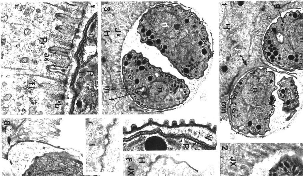
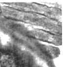
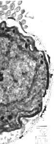
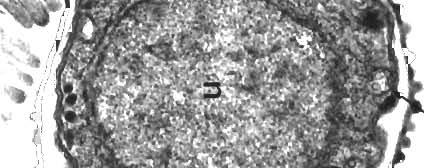
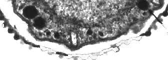
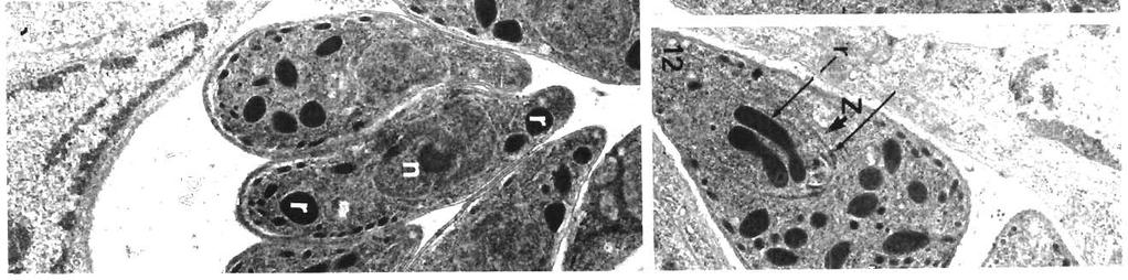
3 Paperna: Fine structure of Eimeria vanasi merozoites 197 3). At the contact zone the host cell cytoplasm forms numerous tightly-packed elongated, deep protrusions into the host cell cytoplasm. Each protrusion contains an axial filament or microtubule. Between the protrusions long microfibnls or filaments in the host cell cytoplasm congregate into 'brush-like' bundles (Fig. 4). The protrusions and the bundles are absent in what appears to be the center of the PV/host cytoplasm contact zone (Fig. l). The PV with the second type juncture measures 2.5 to 3.5 X 2.2 to 3.0 pm (Figs. 6 & 8). The PV wall bordering the host cell cytoplasn~ forms undulations or regularly spaced shallow projections containing an osmiophilic substance (Figs. 6 & 7). The PVs contain 1 (Figs. 1, 6 & 8) or 2 (Figs. 2 & 3) merozoites either bound by a triple-membrane pellicule (Figs. 2, 4 & 5), or having a 2-membrane boundary (Figs. 1 & 3). The merozoites contain an apical complex consisting of a conoid, electron-dense rhoptries and micronemes (Figs. 2, 3 & 6). Their round nucleus contains homogeneously distributed chromatin with an apparently eccentric nucleolus (Fig.3). The cytoplasm of some of the merozoites reveals penpherally arranged mitochondria, a Golgi apparatus and endoplasmic reticulum (ER) (Figs. 1, 4 & 8). Intracytoplasmic merogony stages Gut sections reveal PVs in the cytoplasm of epithelia1 cells. Some of the PVs contain from one to several merozoites measuring 7 to 8 X 2 to 4 pm merozoites (Figs. 9 to 13), a few of them still connected to the meront's residue (Figs. 9 & 10). A few others contain young, 4 X 3 pm meronts (Fig. 14), while still others contain clusters of 5 or more free 4.0 to 5.0 X 1.5 to 2.0 pm merozoites still in their parent PV (Fig. 15). The wall of the PV containing young meronts consists of a single fine membrane (Fig. 14), while the wall of the PV containing merozoites is made up of 3 parallel membranes (Figs. 11 & 13). In some of the latter PVs ground substance is deposited between the 2 inner membranes (Fig. 13). The PV wall is interrupted by a few (Fig. 9) or many funnels (Fig. 13). Intracytoplasmic tubuli are, however, absent. All merozoites are bounded by a trilayered pellicle and have a developed apical complex with rhoptries and micronemes (Figs. 9 to 13 & 15). The meront residue (Fig. 10) and the young meronts (Fig. 14) are each bounded by a single membrane. The young meront contains large food vacuoles, clusters of apparently extracted lipid vacuoles and rudiments of an apical complex (Fig. 14). The meront and merozoite nuclei contain heterochromatin with a rounded nucleolus. Endogenous division of merozoites The intracytoplasmic merozoites (Figs. 9 to 13) contain an anlage of daughter merozoites. Each anlage consists of an inner membrane complex, a developing conoid (Figs. 12 & 13), a relatively large rhoptry anlage (Figs. 10, 11 & 12) or several rhoptnes (Fig. l2), a mitochondrion, a Golgi complex and an extension of the parent merozoite nucleus (Figs. 10 & 13). Intranuclear infection Intranuclear infections are seen with single 6.0 X 2.5 pm merozoites (Fig. l6), and with young 3.5 X 4.0 pm meronts (Fig. 17). Parasites located in the nucleus are contained in a PV bounded by a tnlaminated wall (Fig. 18). The host's nuclear chromatin remains homogeneous, apparently intact. The merozoite is similar to those seen infecting the cytoplasm, with a trilaminated pellicule, rhoptries and micronemes, mitochondria and a Golgi complex. The young meronts still retain rudiments of an apical complex and contain large food vacuoles, several large, peripherally located mitochondria, a nucleus and a few lipid vacuoles. DISCUSSION Epicytoplasmic, intracytoplasmic and intranuclear infections found to occur simultaneously in intestines of the same individual cichlid fish hosts have been interpreted to belong to the same species, Eimeria s.1. vanasi (Landsberg & Paperna 1987). This view is challenged by the recent findings of Molnar (1989) showing that concurrent epicytoplasmic and intracytoplasmic infections in the same individual hosts are caused in Figs. 1 to 8. Eimeria (S. 1.) vanasi. Figs. 1 to 3. Merozoites in epicytoplasmic PV (V) with protrusions at the juncture with the host cell cytoplasm (H). A: apical complex with the conoid; fv: food vacuole; g: Golgi apparatus; H: host cell cytoplasm; JX: juncture zone; m: mitochondria; mn. micronemes; n: nucleus; PE: parasitophorous envelope; r: rhoptries; bold arrow: protrusion-free zone. (Fig. 1 X 10500; Fig. 2 X 26700; Fig. 3 X ). Rg. Enlargement of the juncture showing details of the protrusions (Pr) with the axial filament (ax) and the microfibrils (fl) terminating in brush-like bundles (b). See also details of the merozoite pellicle (P) within the PV (X 46200). Q Details of the PE and the merozoite pellicle (P); V: PV lumen (X 46200). Figs. 6 & 8. Epicytoplasmic PV (V) containing merozoite, with shallow projections at the juncture (JX). B- brush border microvllli (Fig. 6 X 20300; Fig. 8 X ). Flg Enlarged view of the PV(V)/host cell cytoplasm (H) juncture (X )
4 " %*B pi'.
; note extensions of the PV (Vx) N: host cell nucleus: Cy host cell cytoplasm; other abbreviations as in previous figures. (X 14800). Fig. 18.")

 eplcytoplasmic PV boundary/host cell cytoplasm juncinto an intracytoplasmic location (of sporulatlng ture is difficult: such a juncture has not been")
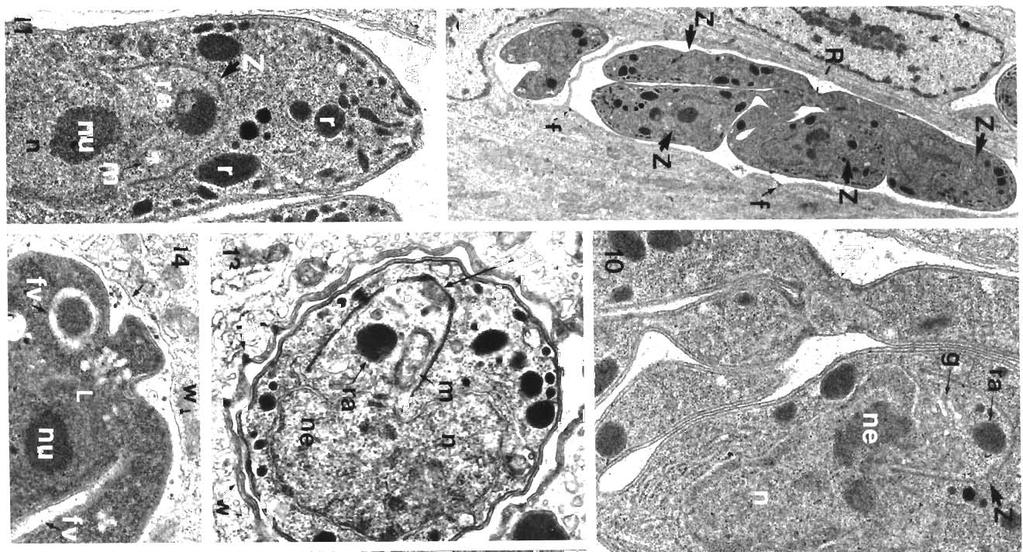
5 Paperna Fine structure of E~mena vanasi merozoites 199 Figs. 16 to 18. Eimeria (S. l.) vanasi. Figs. 16 & 17 Intranuclear ~nfechons Fig 16 Merozoite ( X 14400) Fig. 17 Young meront with vestigial apical complex (a); note extensions of the PV (Vx) N: host cell nucleus: Cy host cell cytoplasm; other abbreviations as in previous figures. (X 14800). Fig. 18. Enlargement of the borders between the meront (M), the PV wall (arrows) and the host nucleus (X ) fact by 2 distinct species. TEM studies of developing E. Merozoites within PV with different kind of junctures vanasi gamonts, however, reveal an actual process of to the host cell cytoplasm may represent different migration of macrogamonts with their PV through the developmental generations. An interpretation of the brushborder of the host epithelium (Paperna 1990, and fine structure of the presently reported first type of unpubl.). Shift from an epicytoplasmlc site (of gamonts) eplcytoplasmic PV boundary/host cell cytoplasm juncinto an intracytoplasmic location (of sporulatlng ture is difficult: such a juncture has not been seen in oocysts) has been also described in E. angulllae any other coccidlan infections. The prolections seen (Dykova & Lom 1981). Ultrastructural findings on alternating with fibrillar bundles at the PV boundary merogony stages presented in this con~munication do could be the n~icrovillous brush border of the host cell, not contribute enlightening new data to prove or dis- modified, compressed or displaced by the expanding prove conspecifity. This issue is currently being PV of the growing parasites. The protrusions and fibrils examined by an experimental study The latter study is appear to be absent in the center of the PV boundary also likely to define the actual number and sequences contact zone, the site at which the parasite apparently of merogony generations, which could not be so far enters and undergoes its pnmary development beneath established through past and present studies. the host cell brush border. The protrusions with their Figs. 9 to 15. Eimeria (S. 1.) vanasi. hg. Merozoites connected to a meront residue (R), with anlagen of daughter merozoites (Z), in an intraepithelial PV lined by a trilayered wall tnth funnels (f) (X 5700). Figs. 10 to 13. Enlargements of merozoites In a PV lined by a tnlaminated boundary (W) with funnels (f), showing a meront residue (R), merozoites' rhoptnes (r), micronemes (mn), nucleus (n) and anlage of daughter merozoite growing from an extension of the parent cell nucleus (ne), wth rudiments of conoid (A), Golgi apparatus (g), mitochondrion (m) and rhoptnes (r) or rhoptry anlagen (ra). nu. nucleolus. (Fig. 10 X Fig. 11 & 12 X ; Fig. 13 x 22300). Fig. 14. Young meront enclosed in PV bounded by a single membrane (W) contans a nucleus with a prominent nucleolus (nu), food vacuoles (fv) and small llpid vacuoles (L) (X 16700) Fig. l5 PV (Pv) with a formation of 5 dllferentiated merozoites (X )


6 200 Dis, aquat. Org 10: , 1991 axial filaments are somewhat reminiscent of the protrusions incorporated into the primary cyst walls of Sarcocystis spp. (Mehlhorn et al. 1976). Arguments for phylogenetic homology between the PV/host cell cytoplasm juncture and the sarcocyst wall may be very vague, but there is undoubtedly a functional analogy in that they are both routes for metabolic exchange between the host and parasite cells. The small projections on the PE do not seem to be vestiges of microvilh. These projections enlarge the area of the contact between the epiplasmic PV and the intestinal lumen and suggest the existence of some sort of metabolic exchange with the extracellular intestinal environment. Such projections occur in one undescribed epieimerian piscine species (Boulard & Blanc 1985), but are absent irr oihei- pisciiie iei11ie1id d~iyuiiide, Mvindr & Baska 1986; Epieimeria isabellae, Daoudi et al. 1985; Goussia zarnowskii, Jastrzebski & Komorowski 1990) and reptilian (Acroeimeria lineri, Paperna 1989) species. In a previous communication (Paperna & Landsberg 1987) the contact boundary of a PV containing a single nucleate meront was seen to include finger-like extensions into the host cell cytoplasm which were interpreted as vestiges or rudiments of the tubular system funnels. Similar finger-like extensions occur at the PV contact boundary of Epieimeria anguillae (Molnar & Baska 1986), E. isabellae (Daoudi et al. 1986) and G. zarnowskii (Jastrzebski & Komorowski 1990). These structures may have developed from the shallow depressions seen at the contact zone of the presently reported second type of epicytoplasmic PV boundary. If this is the case then the finger-like processes may be primordia, not vestiges of the tubular system funnels. Endogenous formation of merozoites within merozoites similar to the process of endodyogeny found in cyst-forming coccidia, such as Sarcocystis, Toxoplasma and Cystoisospora (Scholtyseck 1973, Hilali et al. 1979, Ferguson et al. 1980), seems to comprise a distinct stage in the sequence of asexual reproductions of Eimeria vanasi. Both intracytoplasmic and epicytoplasmic PVs containing 2 merozoites are frequently observed in LM-studied smears and sections of E. vanasi-infected intestines (Landsberg & Paperna 1987). The merozoite couples seen within epicytoplasmic PVs (Figs. 2 & 3) seem to have been formed by the same process. Multiplication by endodyogeny is also found in Eimeria magna (Danforth & Hammond 1972). In the latter, several merozoite anlagen occur within multinucleate merozoites (also termed 'sporozoite shaped schizonts') while in E. vanasi such merozoites always contain only a single nucleus. All observed merozoites contained only one merozoite anlagen. However, ultrathin sections of merozoites undergoing endodyogeny often reveal only one of the 2 merozoite anlagen (Hilali et al. 1979, Daly & Markus 1981). Intranuclear endogenous development is found in a number species of piscine coccidia [Lorn & Dykova 1982, Daoudi et al. 1987). It is relatively common among coccidia species infecting amphibians and reptiles, rare in those infecting mammals and thus far absent in those infecting birds (Pellerdy 1974). All intranuclear piscine species described to date develop exclusively in the host cell nucleus. A versatility in the site of development may nevertheless be implied, since, in cell culture, the intranuclear species found in cattle, Eimeria alabamensis, infects the host cell cytoplasm rather than its nucleus (Sampson & Hammond 1972). LITERATURE CITED Boulard, Y., Blanc, E. (1985). Presence d'une coccidie susepitheliale dans l'intestin de Labrus berggylta (Teleosteen). Society of Protozoology Abstracts, No. 36 Daly, T. J. M., Markus, M. B. (1981). Enteric multiplication of Isospora felis by endodygeny. Electron microscopy Soc. South. Afr, Proceedings 11: 99 Danforth, H. D.. Hammond, D. M. (1972). Stages of merogony in multinucleate merozoites of Eimeria magna. J. Protozool. 19: Daoudi, F., Marques, A, Bouix, G. (1985) Ultrastructure d'epieimena isabellae Lom et Dykova, 1982 Coccidie epicellulaire parasite intestinal du poisson Teleostecln Conger conger Linne Society of Protozoology Abstracts, No. 35 Daoudi, F., Radujkovic, B., Marques, A., Bouix, G. (1987). Nouvelles especes de Coccidies (Apicomplexa, Eimeriidae) des genres Eimeria Schneider, 1875, et Epieimeria Dykova et Lorn, 1981, parasites de poissons marines de la baie de Kotor (Yougoslavie). Bull. Mus. Hist. nat. Paris, 4e ser., 9 (section A, no. 2): Dykova, I., Lom, J. (1981). Fish coccid~a: critical notes on life cycles, classification and pathogenicity J. Fish Dis. 4: Ferguson, D. J. P,, Birch-Andersen. A., Hutchlnson, W. M,, Siim, J. Chr (1980). Ultrastructural observations showing enteric multiplication of Cystoisospora (Isospora) felis by endodyogeny. Z. Parasitenkde 63: Hilali, M., Ghaffar, F. A., Scholtyseck, E. (1979). Ultrastructural study of the endogenous stages of Isospora canis (Nemescmn. 1959) in the small intestine of dogs. Acta Vet. Acad. Sci. Hung Jastrzqbski, M., Komarowski, Z. (1990). bght and electron microscopic studies on Goussia zam'wskri (Jastrzebski, 1982): an intestinal coccidiurn parasitizing the three-spined stickleback, Gasterostreus aculeatns. J. Fish Dis. 13: 1-12 Landsberg, J H., Paperna, I. (1987). Intestinal infections by Eimeria S. l. vanasi n. sp. (Eimeriidae, Apicomplexa. Protozoa) In cichlid fish Ann. parasitol. Hum.. Cornp 62: Lorn, J.. Dykova, I. (1982). Some marine flsh coccidia of the genera Eimeria Schneider. Epieimeria Dykova & Lom and Goussia Labbe. J. Fish Dis. 5: Mehlhorn, H., Hartley, W J., Heydorn, A. 0. (1976). A comparative ultrastructural study of cyst walls of 13 Sarcocystis species. Protistologica 12:
7 Paperna: Fine structure of Eimena vanasi merozoites 201 Molnar, K. (1989). Nodular and epicellular coccidiosis in the intestine of cyprinid fishes. Dis. aquat. Org. 7: 1-12 Molnar, K., Baska, F. (1986). Light and electron microscopic studies on Epieimeria anguillae (Leger & Hollande, 1922), a coccidium parasitizing the European eel, Anguilla anguilla L. J. Fish Dis. 9, Paperna, I. (1989). Ultrastructure of Eimeria (S. 1.) sp. infecting the microvlllar zone of the intestinal epithelium of geckoes. Ann. Parasitol. Hum. Comp. 64: Paperna, I. (1990). Fine structure of the gamonts of Eimeria (S. 1.) vanasi, a cocc~dian from the intestine of cichlid fishes. Dis. aquat. Org. 9: Responsible Subject Editor: W Kortmg, Hannover, Germany Paperna, I., Landsberg, J. H. (1987). Tubular formations extending from parasitophorous vacuoles in gut epithelium of cichlid fish infected by Eimeria (S. l.) vanasi. Dis. aquat. Org. 2: Pellerdy, L. P. (1974). Coccidia and coccidiasis, 2nd edn. Verlag Paul Parey, Berlin Sampson, J. R., Hammond, D. M. (1972). Fine structural aspects of development of Eimeria alabamensis schizonts in cell cultures. J. Parasitol. 58: Scholtyseck, E. (1973). Die Deutung von Endodygeny und Sch~zogonie bei Coccidien und anderen Sporozoen 2. ParasitenKde 42: Manuscript first received: May 28, 1990 Rev~sed version accepted: February 4, 1991
Key words: Coccidia, Choleoeimeria rochalimai, fine structure, gall bladder epithelium, Hemidactylus mabouia, Brazil
 FOLIA PARASITOLOGICA 47: 91-96, 2000 Ultrastructural study of meronts and gamonts of Choleoeimeria rochalimai (Apicomplexa: Eimeriidae) developing in the gall bladder of the gecko Hemidactylus mabouia
The Fine Structure of the Endogenous Stages of Isospora hemidactyli Carini, 1936 in the Gecko Hemidactylus mabouia from North Brazil
 Mem Inst Oswaldo Cruz, Rio de Janeiro, Vol. 95(1): 43-47, Jan./Feb. 2000 The Fine Structure of the Endogenous Stages of Isospora hemidactyli Carini, 1936 in the Gecko Hemidactylus mabouia from North Brazil
Key words: Plasmodium, Kentropyx calcarata, Brazil, merogony, gametocytes, ultrastructure
 FOLIA PARASITOLOGICA 49: 2-8, 2002 Fine structure of erythrocytic stages of a Plasmodium tropiduri-like malaria parasite found in the lizard Kentropyx calcarata (Teiidae) from north Brazil Ilan Paperna
The specimens of Ameiva ameiva (Linn) were
 were") Article available at http://www.parasite-journal.org or http://dx.doi.org/10.1051/parasite/1999064359 FINE STRUCTURE OF THE EPICYTOPLASMIC EIMERID COCCIDIUM ACROEIMERIA PINTOI LAINSON & PAPERNA, 1999,
Article available at http://www.parasite-journal.org or http://dx.doi.org/10.1051/parasite/1999064359 FINE STRUCTURE OF THE EPICYTOPLASMIC EIMERID COCCIDIUM ACROEIMERIA PINTOI LAINSON & PAPERNA, 1999,
Ultrastructure of Endogenous Stages of Eimeria ninakohlyakimovae Yakimoff & Rastegaieff, 1930 Emend. Levine, 1961 in Experimentally Infected Goat
 Mem Inst Oswaldo Cruz, Rio de Janeiro, Vol. 92(4): 533-538, Jul./Aug. 1997 Ultrastructure of Endogenous Stages of Eimeria ninakohlyakimovae Yakimoff & Rastegaieff, 1930 Emend. Levine, 1961 in Experimentally
Ëtude ultrastructurale de la mérogonie de Schellackia cf. agamae (Lankesterellidae, Apicomplexa) chez le Lézard Agama stellio.
 chez le Lézard Agama stellio.") Masson, Paris, 1987 Ann. Parasitol. Hum. Comp. 1987, 62, n 5, pp. 380-386. ULTRASTRUCTURAL STUDIES ON THE MEROGONY OF SCHELLACKIA CF. AGAMAE (LANKESTERELLIDAE, APICOMPLEXA) FROM THE STARRED LIZARD AGAMA
Masson, Paris, 1987 Ann. Parasitol. Hum. Comp. 1987, 62, n 5, pp. 380-386. ULTRASTRUCTURAL STUDIES ON THE MEROGONY OF SCHELLACKIA CF. AGAMAE (LANKESTERELLIDAE, APICOMPLEXA) FROM THE STARRED LIZARD AGAMA
DISEASES OF AQUATIC ORGANISMS Dis. aquat. Org.
 Vol. 10: 121-125. 1991 DISEASES OF AQUATIC ORGANISMS Dis. aquat. Org. Published April 4 Ultrastructural observations on sporozoite stages of piscine Coccidia: Goussia carpelli and G. subepithelialis from
Revajová, Viera, Loószová, Adrian. The Journal of Protozoology Resea Citation RightsNational Research Center for Prot
 ' ' Morphological study of partridge Title development in the foreign host - (Gallus gallus) Revajová, Viera, Loószová, Adrian Author(s) Maria, Zibrín, Martin, Herich, Ro Mikulas The Journal of Protozoology
Light and electron microscopic study of the pathology and merogony of Goussia gadi (Apicomplexa: Coccidia) in the swimbladder wall
 in the swimbladder wall") Vol. 17: 113-125.1993 DISEASES OF AQUATIC ORGANISMS Dis. aquat. Org., Published November 18 I Light and electron microscopic study of the pathology and merogony of Goussia gadi (Apicomplexa: Coccidia)
Vol. 17: 113-125.1993 DISEASES OF AQUATIC ORGANISMS Dis. aquat. Org., Published November 18 I Light and electron microscopic study of the pathology and merogony of Goussia gadi (Apicomplexa: Coccidia)
Oocyst formation in the coccidian parasite Goussia carpelli
 Vol. 10: 203-209, 1991 DISEASES OF AQUATIC ORGANISMS Dis. aquat. Org. Published May 8 Oocyst formation in the coccidian parasite Goussia carpelli 'Institute of Parasitology, Czech Academy of Sciences,
Protozoan Parasites of Veterinary importance 2017
 Protozoan Parasites of Veterinary importance 2017 VPM-122 Laboratory 4 Spencer J. Greenwood PhD, DVM Dept. of Biomedical Sciences Room 2332N AVC North Annex sgreenwood@upei.ca Office phone # 566-6002 To
Phylum:Apicomplexa Class:Sporozoa
 Phylum:Apicomplexa Class:Sporozoa The most characteristic features of sporozoa are 1-unique appearance of most protozoa makes it possible for knowledge able person to identifiy them to level of genus and
Biology of toxoplasmosis
 1 Biology of toxoplasmosis E. Petersen 1 and J. P. Dubey 2 1 Statens Seruminstitut, Copenhagen, Denmark 2 U.S. Department of Agriculture, Beltsville, USA History Toxoplasma gondii is a coccidium, with
Sarcocystis heydorni, n. sp. (Apicomplexa: Protozoa) with cattle (Bos taurus) and human
 with cattle (Bos taurus) and human") 1 Sarcocystis heydorni, n. sp. (Apicomplexa: Protozoa) with cattle (Bos taurus) and human (Homo sapiens) cycle Jitender P. Dubey 1, Erna van Wilpe 2, Rafael Calero-Bernal 1, Shiv Kumar Verma 1, Ronald
1 Sarcocystis heydorni, n. sp. (Apicomplexa: Protozoa) with cattle (Bos taurus) and human (Homo sapiens) cycle Jitender P. Dubey 1, Erna van Wilpe 2, Rafael Calero-Bernal 1, Shiv Kumar Verma 1, Ronald
DISEASES OF AQUATIC ORGANISMS Dis. aquat. Org.
 Vol. 7: 149-153, 1989 DISEASES OF AQUATIC ORGANISMS Dis. aquat. Org. Published October 26 Developmental cycle of chelonian haemogregarines in leeches with extra-intestinal multiple sporozoite oocysts and
Protozoa. Apicomplexa Sarcomastigophora Ciliophora. Gregarinea Coccidia Piroplasma
 Protozoa Apicomplexa Sarcomastigophora Ciliophora Gregarinea Coccidia Piroplasma Coccidia characterized by thick-walled oocysts excreted in feces In Humans Cryptosporidium Isospora Cyclospora Sarcocystis
Redescription of Sarcocystis fusiformis sarcocysts from the water buffalo (Bubalus bubalis)
") Redescription of Sarcocystis fusiformis sarcocysts from the water buffalo (Bubalus bubalis) 1 J. P. DUBEY 1 *, M. HILALI 2,E.VANWILPE 3,S.K.VERMA 1,R.CALERO-BERNAL 1 and A. ABDEL-WAHAB 2 1 U. S. Department
Redescription of Sarcocystis fusiformis sarcocysts from the water buffalo (Bubalus bubalis) 1 J. P. DUBEY 1 *, M. HILALI 2,E.VANWILPE 3,S.K.VERMA 1,R.CALERO-BERNAL 1 and A. ABDEL-WAHAB 2 1 U. S. Department
cyst&' appeared to be of two kinds-one smaller and Smnith "is inclined to regard these epithelial cell parasites as
 COCCIDIA IN SUBEPITHELIAL INFECTIONS OF THE INTESTINES OF BIRDS PHILIP B. HADLEY From the Agricultural Experiment Station of the Rhode Island State College' Received for publication, July 10, 1916 In an
HISTOPATHOLOGY. Introduction:
 Introduction: HISTOPATHOLOGY Goats and sheep are the major domestic animal species in India. Much of the economy of the country has been depend upon the domestication of these animals. Especially economy
Apicomplexans Apicomplexa Intro
 Apicomplexans Apicomplexa Intro Cryptosporidium Apicomplexan Select Characteristics Gliding motility Apical Complex organelle for invasion of host cell Life cycle alternates b/w sexual and asexual phases
' Published September 29
 Vol. 7: 1-12, 1989 l DISEASES OF AQUATIC ORGANISMS Dis. aquat. Org. ' Published September 29 Nodular and epicellular coccidiosis in the intestine of cyprinid fishes Kalman Molnar Veterinary Medical Research
Parasitenkunde. (Odocoileus virginianus ) Ultrastructure of Sarcocystis sp. from the Muscle of a White-Tailed Deer
 Ultrastructure of Sarcocystis sp. from the Muscle of a White-Tailed Deer") Z Parasitenkd (1982) 68 : 33-38 Zeitschrift for Parasitenkunde Parasitology Research 9 Springer-Verlag 1982 Ultrastructure of Sarcocystis sp. from the Muscle of a White-Tailed Deer (Odocoileus virginianus
Z Parasitenkd (1982) 68 : 33-38 Zeitschrift for Parasitenkunde Parasitology Research 9 Springer-Verlag 1982 Ultrastructure of Sarcocystis sp. from the Muscle of a White-Tailed Deer (Odocoileus virginianus
Diagnosis, treatment and control: dealing with coccidiosis in cattle
 Vet Times The website for the veterinary profession https://www.vettimes.co.uk Diagnosis, treatment and control: dealing with coccidiosis in cattle Author : Adam Martin Categories : Vets Date : January
TARENTANNULARI INFECTING THE GECKO TARENTOLA ANNULARIS. Department of Zoology, Faculty of Science, University of Ain Shams, Cairo, Egypt - - -
 Qatar Univ. Sci. J. (1995), 15 (2) : 379-387 THE ULTRASTRUCTURE OF SOME STAGES OF HAEMOGREGARINA TARENTANNULARI INFECTING THE GECKO TARENTOLA ANNULARIS BY Nadia F. Ramadan, Shadia H. Mohammed and Samia
Ultrastructure of Sarcocystis bertrami sarcocysts from a naturally infected donkey (Equus
 Ultrastructure of Sarcocystis bertrami sarcocysts from a naturally infected donkey (Equus asinus) from Egypt J. P. DUBEY 1,*, E. VAN WILPE 2, S. K. VERMA 1, M. HILALI 3, 1 U. S. Department of Agriculture,
LABORATORY. The Protozoa. At the Bench
 LABORATORY Laboratory 8, Page 1 8 The Protozoa Introduction: The protozoa are unicellular animals that are classified on the basis of the organelles used for locomotion (flagella, pseudopodia, cilia or
Ahead of print online version
 Folia Parasitologica 60 [3]: 232 236, 2013 ISSN 0015-5683 (print), ISSN 1803-6465 (online) Institute of Parasitology, Biology Centre ASCR http://folia.paru.cas.cz/ A new species of Choleoeimeria (Apicomplexa:
PLASMODIUM MODULE 39.1 INTRODUCTION OBJECTIVES 39.2 MALARIAL PARASITE. Notes
 Plasmodium MODULE 39 PLASMODIUM 39.1 INTRODUCTION Malaria is characterized by intermittent fever associated with chills and rigors in the patient. There may be enlargement of the liver and spleen in the
Coccidia. Nimit Morakote, Ph.D.
 Coccidia Nimit Morakote, Ph.D. 1 Learning objectives After class, students will be able to: Describe morphology, life cycle, signs and symptoms, prevention and control, laboratory diagnosis and treatment
Transformed centrioles In adult and aged cat pinealocytes
 Transformed centrioles In adult and aged cat pinealocytes J. L. Calvo. J. Boya*. J. E. Garcia-Mauriño and D. Rancaño Department of Histology. Faculty of Medicine. University Complutense, 28040 Madrid.
DISEASES OF AQUATIC ORGANISMS Vol. 62: , 2004 Published November 23 Dis Aquat Org
 DISEASES OF AQUATIC ORGANISMS Vol. 62: 133 145, 2004 Published November 23 Dis Aquat Org Cryptosporidium scophthalmi n. sp. (Apicomplexa: Cryptosporidiidae) from cultured turbot Scophthalmus maximus. Light
Lacerta viridis. Functional anatomy of the lungs of the green lizard, (Accepted 18 February 1977)
") J. Anat. (1978), 125, 2, pp. 421-431 421 With 9 figures Printed in Great Britain Functional anatomy of the lungs of the green lizard, Lacerta viridis C. MEBAN Department of Anatomy, The Queen's University
J. Anat. (1978), 125, 2, pp. 421-431 421 With 9 figures Printed in Great Britain Functional anatomy of the lungs of the green lizard, Lacerta viridis C. MEBAN Department of Anatomy, The Queen's University
Studied tortoises, Testudo graeca, were collected from
 Article available at http://www.parasite-journal.org or http://dx.doi.org/10.1051/parasite/2006134267 HEMOLIVIA MAURITANICA (HAEMOGREGARINIDAE: APICOMPLEXA) INFECTION IN THE TORTOISE TESTUDO GRAECA IN
AN ULTRASTRUCTURAL STUDY OF THE DEVELOPMENT OF BABESIA. E. F. BLOUIN and LYNN VAN RENSBURG, Veterinary Research Institute, Onderstepoort OliO
 OnderstepoortJ. vet. Res., 55, 93-100(1988) AN ULTRASTRUCTURAL STUDY OF THE DEVELOPMENT OF BABESIA OCCULTANS INTHESALIVARYGLANDSOF ADULT HYALOMMA MARGINATUM RUFIPES E. F. BLOUIN and LYNN VAN RENSBURG,
Apicomplexa of Intestinal Pathology
 LECTURES #4, #5 & #6: APICOMPLEXA 1 Apicomplexa of Intestinal Pathology Cryptosporidium, Eimeria, Cystoisospora General Characteristics of Apicomplexa A. Morphology by stage Zoite o Tear-shaped (cylindrical
A Scanning Electron Microscopic Study of Eggshell Surface Topography of Leidynema portentosae and L. appendiculatum (Nematoda: Oxyuroidea)
") The Ohio State University Knowledge Bank kb.osu.edu Ohio Journal of Science (Ohio Academy of Science) Ohio Journal of Science: Volume 88, Issue 5 (December, 1988) 1988-12 A Scanning Electron Microscopic
The Ohio State University Knowledge Bank kb.osu.edu Ohio Journal of Science (Ohio Academy of Science) Ohio Journal of Science: Volume 88, Issue 5 (December, 1988) 1988-12 A Scanning Electron Microscopic
Ultrastructural and molecular identification of Sarcocystis tenella (Protozoa, Apicomplexa) in naturally infected Korean native goats
 in naturally infected Korean native goats") Original Paper Veterinarni Medicina, 61, 2016 (7): 374 381 Ultrastructural and molecular identification of Sarcocystis tenella (Protozoa, Apicomplexa) in naturally infected Korean native goats E.J. Hong
Original Paper Veterinarni Medicina, 61, 2016 (7): 374 381 Ultrastructural and molecular identification of Sarcocystis tenella (Protozoa, Apicomplexa) in naturally infected Korean native goats E.J. Hong
Sleepy lizards Tiliqua rugosa Gray (Scincidae)
") Article available at http://www.parasite-journal.org or http://dx.doi.org/10.1051/parasite/1997044359 THE TICK-TRANSMITTED HAEMOGREGARINID OF THE AUSTRALIAN SLEEPY LIZARD TILIQUA RUGOSA TO THE GENUS HEMOLIVIA
Article available at http://www.parasite-journal.org or http://dx.doi.org/10.1051/parasite/1997044359 THE TICK-TRANSMITTED HAEMOGREGARINID OF THE AUSTRALIAN SLEEPY LIZARD TILIQUA RUGOSA TO THE GENUS HEMOLIVIA
Malaria parasites of rodents of the Congo (Brazzaville) :
 :") Annales de Parasitologie (Paris), 1976, t. 51, n 6, pp. 637 à 646 Malaria parasites of rodents of the Congo (Brazzaville) : Plasmodium cbabaudi adami subsp. nov. and Plasmodium vinckei lentum Landau, Michel,
Annales de Parasitologie (Paris), 1976, t. 51, n 6, pp. 637 à 646 Malaria parasites of rodents of the Congo (Brazzaville) : Plasmodium cbabaudi adami subsp. nov. and Plasmodium vinckei lentum Landau, Michel,
SCANNING electron - microscopy has
 Characteristics of the Absorptive Surface of the Small Intestine of the Chicken from 1 Day to 14 Weeks of Age 1 R. C. BAYER, C. B. CHAWAN, F. H. BIRD AND S. D. MUSGRAVE Department of Animal and Veterinary
1) Most common, infectious, pathogenic animal (zoonotic) parasite of humans; estimated that 13% of humans are infected
 Most common, infectious, pathogenic animal (zoonotic) parasite of humans; estimated that 13% of humans are infected") XX Phylum Apicomplexa (Chapter 8) 2005 A. Characteristics 1. All are parasitic 2. APICAL COMPLEX a. Group of organelles used to invade host cells b. Visible only with electron microscopy Picture Slide
XX Phylum Apicomplexa (Chapter 8) 2005 A. Characteristics 1. All are parasitic 2. APICAL COMPLEX a. Group of organelles used to invade host cells b. Visible only with electron microscopy Picture Slide
Dermatitis in a dog associated with an unidentified Toxoplasma gondii-like parasite
 Veterinary Parasitology 116 (2003) 51 59 Short communication Dermatitis in a dog associated with an unidentified Toxoplasma gondii-like parasite J.P. Dubey a,, A.L. Pimenta b, L.C.S. Abboud b, R.R. Ravasani
Coccidiosis in macropods and other species
 Coccidiosis in macropods and other species Author: Derek Spielman Wildlife Assistance and Information Foundation; Sydney School of Veterinary Science, the University of Sydney Abstract This presentation
Joerg Kinne, Mansoor Ali*, Ulrich Wernery, and J. P. Dubey
 J. Parasitol., 88(3), 2002, pp. 548 552 American Society of Parasitologists 2002 CLINICAL LARGE INTESTINAL COCCIDIOSIS IN CAMELS (CAMELUS DROMEDARIUS) IN THE UNITED ARAB EMIRATES: DESCRIPTION OF LESIONS,
Extra-intestinal localization of Goussia sp. (Apicomplexa) oocysts in Rana dalmatina (Anura: Ranidae), and the fate of infection after metamorphosis
 oocysts in Rana dalmatina (Anura: Ranidae), and the fate of infection after metamorphosis") DISEASES OF AQUATIC ORGANISMS Vol. 70: 237 241, 2006 Published June 23 Dis Aquat Org Extra-intestinal localization of Goussia sp. (Apicomplexa) oocysts in Rana dalmatina (Anura: Ranidae), and the fate
DISEASES OF AQUATIC ORGANISMS Vol. 70: 237 241, 2006 Published June 23 Dis Aquat Org Extra-intestinal localization of Goussia sp. (Apicomplexa) oocysts in Rana dalmatina (Anura: Ranidae), and the fate
The South American opossum, Didelphis marsupialis, from Brazil as another definitive host for Sarcocystis speeri Dubey and Lindsay, 1999
 The South American opossum, Didelphis marsupialis, from Brazil as another definitive host for Sarcocystis speeri Dubey and Lindsay, 1999 589 J. P. DUBEY *, C. E. KERBER, D. S. LINDSAY, N. KASAI and H.
Biology of Isospora spp. from Humans, Nonhuman Primates, and Domestic Animals
 CLINICAL MICROBIOLOGY REVIEWS, Jan. 1997, p. 19 34 Vol. 10, No. 1 0893-8512/97/$04.00 0 Copyright 1997, American Society for Microbiology Biology of Isospora spp. from Humans, Nonhuman Primates, and Domestic
Parasitology Amoebas. Sarcodina. Mastigophora
 Parasitology Amoebas Sarcodina Entamoeba hisolytica (histo = tissue, lytica = lyse or break) (pathogenic form) o Trophozoite is the feeding form o Life Cycle: personfeces cyst with 4 nuclei with thicker
Progressive Retinal Atrophy in the Abyssinian Cat
 Progressive Retinal Atrophy in the Abyssinian Cat Electron Microscopy Kristina Narfstr6m*t and Sven Erik Nilsson* Seven adult Abyssinian cats at different stages of a recessively inherited retinal degenerative
A comparison of placental tissue in the skinks Eulamprus tympanum and E. quoyii. Yates, Lauren A.
 A comparison of placental tissue in the skinks Eulamprus tympanum and E. quoyii Yates, Lauren A. Abstract: The species Eulamprus tympanum and Eulamprus quoyii are viviparous skinks that are said to have
Effect of Sodium Hypochlorite on the Oocyst Wall of Eimeria tenella as Shown by Electron Microscopy1
 32 PROCEEDINGS OF THE HELMINTHOLOGICAL SOCIETY This alteration appeared similar to that observed by light microscopy (Figs. 5, 6). Literature Cited Dixon, K. E. 1966. The physiology of excystment of the
Hepatozoon-Like Parasite (Schizonts) in the Myocardium of the Domestic Cat
 in the Myocardium of the Domestic Cat") Vet. Path. 10: 185-190 (1973) Hepatozoon-Like Parasite (Schizonts) in the Myocardium of the Domestic Cat U. KLOPFER, T.A. NOBEL and F. NEUMANN Department of Pathology, Kimron Veterinary Institute, affiliated
Vet. Path. 10: 185-190 (1973) Hepatozoon-Like Parasite (Schizonts) in the Myocardium of the Domestic Cat U. KLOPFER, T.A. NOBEL and F. NEUMANN Department of Pathology, Kimron Veterinary Institute, affiliated
Observations on Eimeria species of Dasyprocta leporina (Linnaeus, 1758) (Rodentia: Dasyproctidae) from the state of Pará, North Brazil
 (Rodentia: Dasyproctidae) from the state of Pará, North Brazil") Mem Inst Oswaldo Cruz, Rio de Janeiro, Vol. 99: 000-000, 2004 1 Observations on Eimeria species of Dasyprocta leporina (Linnaeus, 1758) (Rodentia: Dasyproctidae) from the state of Pará, North Brazil Ralph
Mem Inst Oswaldo Cruz, Rio de Janeiro, Vol. 99: 000-000, 2004 1 Observations on Eimeria species of Dasyprocta leporina (Linnaeus, 1758) (Rodentia: Dasyproctidae) from the state of Pará, North Brazil Ralph
COCCIDIOSIS OF SANDHILL CRANES (GRUS CANADENSIS) WINTERING IN NEW MEXICO
 WINTERING IN NEW MEXICO") journal of Wtldltfe hemes, 22(1). 1986. pp 25-35 0 Wildlife Disease Association 1986 COCCIDIOSIS OF SANDHILL CRANES (GRUS CANADENSIS) WINTERING IN NEW MEXICO Brent B. Parker and Donald W. Duszynski Department
journal of Wtldltfe hemes, 22(1). 1986. pp 25-35 0 Wildlife Disease Association 1986 COCCIDIOSIS OF SANDHILL CRANES (GRUS CANADENSIS) WINTERING IN NEW MEXICO Brent B. Parker and Donald W. Duszynski Department
Article available at or
 Article available at http://www.parasite-journal.org or http://dx.doi.org/10.1051/parasite/1995023307 LIGHT AND ELECTRON MICROSCOPE STUDY OF A LANKESTERELLA PETITI N. SP., (APICOMPLEXA : LANKESTERELLIDAE)
Article available at or
 Article available at http://www.parasite-journal.org or http://dx.doi.org/10.1051/parasite/1998051017 PSEUDOKLOSSIA SEMILUNA N. SP. (APICOMPLEXA: AGGREGATIDAE): A COCCIDIAN PARASITE OF THE KIDNEY OF BLUE
Parasitology Departement Medical Faculty of USU
 Malaria Mechanism of infection Parasitology Departement Medical Faculty of USU Introduction Malaria parasites Phylum Order Suborder Family Genus Species : : Apicomplexa : Eucoccidiida : Haemosporida :
HISTOLOGY OF MAMMARY GLAND DURING LACTATING AND NON-LACTATING PHASES OF MADRAS RED SHEEP WITH SPECIAL REFERENCE TO INVOLUTION
 International Journal of Science, Environment and Technology, Vol. 5, No 3, 2016, 991 996 ISSN 2278-3687 (O) 2277-663X (P) HISTOLOGY OF MAMMARY GLAND DURING LACTATING AND NON-LACTATING PHASES OF MADRAS
A NEW TYPE OF BRYOZOAN GIZZARD, WITH REMARKS ON THE GENUS BUSKIA.
 A NEW TYPE OF BRYOZOAN GIZZARD, WITH REMARKS ON THE GENUS BUSKIA. RAYMOND C. OSBURN AND RUTH M. VETH Department of Zoology and Entomology, Ohio State University A certain few of the Ctenostome Bryozoa
Anat. Labor. of Prof. H. SETO, Tohoku University, On the Sensory Terminations Formed along the Ductus
 Anat. Labor. of Prof. H. SETO, Tohoku University, Sendai. On the Sensory Terminations Formed along the Ductus Pancreaticus in Cat. The existence of PACINIan bodies in the pancreas of mammals, especially
Article available at or
 Article available at http://www.parasite-journal.org or http://dx.doi.org/10.1051/parasite/1996034341 DESCRIPTION AND ULTRASTRUCTURE OF LANKESTERELLA SPECIES INFECTING FROGS IN KENYA PAPERNA I.* & OGARA
Hamed Mohamed Fayed; Mohamed Abd-Allah Shazly and Sayed Abd El-Monem
 Life cycle of Eimeria rousetti sp. nov. (Alveolata: Apicomplexa: Eimeriidae) infecting the frugivorous bat, Rousettus aegyptiacus Geoffroy, 1810 (Mammalia: Chiroptera: Pteropodidae) in Egypt. Hamed Mohamed
Goussia cruciata (Thelohan, 1892) a hepatic coccidian parasite of the horse mackerel Trachurus trachurus (Linnaeus, 1758) from the
 a hepatic coccidian parasite of the horse mackerel Trachurus trachurus (Linnaeus, 1758) from the") Bull. Eur. Ass. Fish Pathol., 2(6) 2, 219 Goussia cruciata (Thelohan, 1892) a hepatic coccidian parasite of the horse mackerel Trachurus trachurus (Linnaeus, 1758) from the Mediterranean coasts of northern
Bull. Eur. Ass. Fish Pathol., 2(6) 2, 219 Goussia cruciata (Thelohan, 1892) a hepatic coccidian parasite of the horse mackerel Trachurus trachurus (Linnaeus, 1758) from the Mediterranean coasts of northern
Criconemoides similis 1 G. W. BIRD ~
 Somatic Musculature of Trichodorus porosus and Criconemoides similis 1 G. W. BIRD ~ Abstract: The somatic musculature of Trichodorus porosus is transversely striated, and that of Criconemoides similis
A:Malaria (Plasmodium species) Plasmodium falciparum causes malignant tertian malaria P. malariae: causes Quartan malaria P. vivax: causes benign
 Plasmodium falciparum causes malignant tertian malaria P. malariae: causes Quartan malaria P. vivax: causes benign") A:Malaria (Plasmodium species) Plasmodium falciparum causes malignant tertian malaria P. malariae: causes Quartan malaria P. vivax: causes benign tertian malaria P. ovale: causes benign tertian malaria
A:Malaria (Plasmodium species) Plasmodium falciparum causes malignant tertian malaria P. malariae: causes Quartan malaria P. vivax: causes benign tertian malaria P. ovale: causes benign tertian malaria
Exotic Hematology Lab Leigh-Ann Horne, LVT, CWR Wildlife Center of Virginia
 Exotic Hematology Lab Leigh-Ann Horne, LVT, CWR Wildlife Center of Virginia lhorne@wildlifecenter.org Anne Lynch, LVT Cedarcrest Animal Clinic amllvt9@gmail.com Introduction While the general set-up for
BLOOD PARASITES MORPHOTYPES OF ROCK LIZARDS OF ARMENIA
 PROCEEDINGS OF THE YEREVAN STATE UNIVERSITY C h e m i s t r y a n d B i o l o g y 2015, 2, p. 45 49 B i o l o g y BLOOD PARASITES MORPHOTYPES OF ROCK LIZARDS OF ARMENIA T. K. HARUTYUNYAN, F. D. DANIELYAN,
Development of the Intestinal Villi Associated
 Development of the Intestinal Villi Associated with the Increased Epithelial Cell Mitosis in Chickens Koh-en YAMAUCHI, Eiji NAKAMURA and Yutaka ISSHIKI Laboratory of Animal Science, Faculty of Agriculture,
An Unidentified Sporozoan Encephalomyelitis in Sheep
 Vet. Path. 11: 1-12 (1974) An Unidentified Sporozoan Encephalomyelitis in Sheep W. J. HARTLEY and W. F. BLAKEMORE Department of Veterinary Medicine, The University of Sydney, Camden; and Wellcome Laboratory
"Comments on the nature and methods of collection of fish coccidia. " - Molnár, K. - Parasit. Hung. _
 Parasit. Hung 10. 1977. Comments on the Nature and Methods of Collection of Fish Coccidia Dr. Kálmán MOLNÁR Research Institute for Veterinary Science, Hungarian Academy of Sciences, Budapest "Comments
Ectoparasites Myobia musculi Radfordia affinis Radfordia ensifera
 Ectoparasites Fleas, ticks, and lice are uncommon in modern laboratory facilities, but may be seen on wild or feral rodents. Most ectoparasite infestations seen in rats and mice used for research are various
Sam R. Telford, Jr The Florida Museum of Natural History, University of Florida, Gainesville, Fl32611, USA
 Systematic Parasitology 23: 203-208, 1992. 0 1992 Kluwer Academic Publishers. Printed in the Netherlands. An eimeriid species (Apicomplexa: Eimeriidae) that parasitises the gallbladder and bile-duct of
BIO Parasitology Spring 2009
 BIO 475 - Parasitology Spring 2009 Stephen M. Shuster Northern Arizona University http://www4.nau.edu/isopod Lecture 10 Malaria-Life Cycle a. Micro and macrogametocytes in mosquito stomach. b. Ookinete
Systemic Apicomplexans. Toxoplasma
 Systemic Apicomplexans Toxoplasma Protozoan Groups Historically, protozoa have been grouped by mode of motility. Flagellates Hemoflagellates Trypanosoma cruzi Leishmania infantum Mucoflagellates Tritrichomonas
Protozoan Parasites: Lecture 20 Apicomplexans II Coccidia Part II & Cryptosporidium Pages 28-36
 Protozoan Parasites: Lecture 20 Apicomplexans II Coccidia Part II & Cryptosporidium Pages 28-36 Coccidia: Life cycle & treatment/control effectiveness? Asexual stages Sexual stages Prophylactic drugs Current
SOME NEW AMERICAN PYCNODONT FISHES.
 SOME NEW AMERICAN PYCNODONT FISHES. By James Williams Gidley, Assistant Curator of Fossil Mammals, United States National Museum. In the United States National Museum are several specimens representing
Ultrastructure of Ehrlichia canis
 INFECTION AND IMMUNrrY, Feb. 1973, p. 265-271 Copyright 1973 American Society for Microbiology Ultrastructure of Ehrlichia canis Vol. 7, No. 2 Printed in U.S.A. PAUL K. HILDEBRANDT, JAMES D. CONROY,I ADAM
AVIAN COCCIDIOSIS. One of the most potentially destructive diseases in domestic poultry production. Most costly of all poultry diseases.
 AVIAN COCCIDIOSIS One of the most potentially destructive diseases in domestic poultry production. Most costly of all poultry diseases. Strictly a gut infection in chickens and turkeys. All avian species
A Study of Coccidiosis in Livestock in the Island of Dominica. Joshua Santelises. Study Abroad Texas A&M University. Dr.
 A Study of Coccidiosis in Livestock in the Island of Dominica Joshua Santelises Study Abroad 2012 Texas A&M University Dr. Thomas Lacher Dr. Jim Woolley Abstract The following experiment was done to investigate
SUPPLEMENTARY INFORMATION
 doi:10.1038/nature11046 Supplementary Figure 1: Images of PB-positive cells in the subepidermal region (a-i) Representative images of PB positive cells in the subepidermis of the upper beak of the pigeon.
Light, Scanning and Transmission Electron Microscopical Study on the Oviduct of the Ostrich (Struthio
 Light, Scanning and Transmission Electron Microscopical Study on the Oviduct of the Ostrich (Struthio camelus) A.S.Saber*, S.A.M.Emara*, O.M.M.AboSaeda** * Faculty of Veterinary Medicine, Sadat City Branch,
A CYTOLOGICAL STUDY OF THE SPOROZOITES OF EIMERIA CAVIAE, A COCCIDIAN PARASITE OF THE DOMESTIC GUINEA PIG, CAVIA PORCELLUS
 A CYTOLOGICAL STUDY OF THE SPOROZOITES OF EIMERIA CAVIAE, A COCCIDIAN PARASITE OF THE DOMESTIC GUINEA PIG, CAVIA PORCELLUS An abstract of a Thesis by C. Bruce Moore December 1976 Drake University Advisor:
Fact sheet. All animals, particularly herbivores, appear to be natural hosts for coccidian species with a high degree of host specificity observed.
 Coccidia in k angaroos Fact sheet Introductory statement Coccidians are protozoan parasites which infect the intestinal tract of many animals. Within kangaroos, coccidia infections can lead to clinical
Morphological characterization of Cryptosporidium parvum life-cycle stages in an in vitro model system
 Morphological characterization of Cryptosporidium parvum life-cycle stages in an in vitro model system 13 H. BOROWSKI 1,R.C.A.THOMPSON 1 *, T. ARMSTRONG 1 and P. L. CLODE 2 1 WHO Collaborating Centre for
23 Plasmodium coatneyi Eyles, Fong, Warren, Guinn, Sandosham, and Wharton, 1962
 23 Plasmodium coatneyi Eyles, Fong, Warren, Guinn, Sandosham, and Wharton, 1962 IN the course of studies on simian malaria begun by the late Dr. Don Eyles in Malaya, he and his co-workers isolated a new
Zadar County Rural Development Agency, Zadar, Croatia. Fish Farm IHOR PARK, Jastrebarsko, Croatia
 . Veterinarski Arhiv 87 (1), 77-86, 2017 New data on Eimeria dicentrarchi (Apicomplexa: Eimeriidae), a common parasite of farmed European sea bass (Dicentrarchus labrax) from the mid-eastern Adriatic Emil
Giardia and Apicomplexa. G. A. Lozano UNBC
 Giardia and Apicomplexa G. A. Lozano UNBC NINE Protozoan diseases/parasites Ciliphora, Ichthyophthirius, Ick Sarcomastigophora, Giardia, giardiasis Apicomplexa: Eimeria, Toxoplasma, Sarcocystis, Cryptosporidium.
Myxosporeans and myxosporidiosis of common carp and gibel carp in China
 Myxosporeans and myxosporidiosis of common carp and gibel carp in China Zhang Jinyong, Liu Xinhua, Xi Bingwen, Kálmán Molnár zhangjy@ihb.ac.cn Hungary 2015 June.3 Laboratory of Fish Diseases; Institute
Coccidiosis of Cattle
 Utah State University DigitalCommons@USU USU Faculty Honor Lectures Lectures 5-1-1964 Coccidiosis of Cattle Datus M. Hammod Utah State University Follow this and additional works at: https://digitalcommons.usu.edu/honor_lectures
Introduction. Syst Parasitol (2014) 89:83 89 DOI /s
 89:83 89 DOI /s") Syst Parasitol (2014) 89:83 89 DOI 10.1007/s10-014-9510-7 Coccidial dispersion across New World marsupials: Klossiella tejerai Scorza, Torrealba & Dagert, 1957 (Apicomplexa: Adeleorina) from the Brazilian
Syst Parasitol (2014) 89:83 89 DOI 10.1007/s10-014-9510-7 Coccidial dispersion across New World marsupials: Klossiella tejerai Scorza, Torrealba & Dagert, 1957 (Apicomplexa: Adeleorina) from the Brazilian
Exploring simvastatin, an antihyperlipidemic drug, as a potential topical antibacterial agent
 Supplementary materials Exploring simvastatin, an antihyperlipidemic drug, as a potential topical antibacterial agent Shankar Thangamani 1, Haroon Mohammad 1, Mostafa Abushahba 1, Maha Hamed 1, Tiago Sobreira
Reprinted from: CRUSTACEANA, Vol. 32, Part 2, 1977 LEIDEN E. J. BRILL
 Reprinted from: CRUSTACEANA, Vol. 32, Part 2, 1977 LEIDEN E. J. BRILL NOTES AND NEWS 207 ALPHE0PS1S SHEARMII (ALCOCK & ANDERSON): A NEW COMBINATION WITH A REDESCRIPTION OF THE HOLOTYPE (DECAPODA, ALPHEIDAE)
Cryptosporidium spp. Oocysts
 Sampling and Source Tracking of Cryptosporidium spp. Oocysts June 28, 2005 Kristen L. Jellison, Ph.D. Department of Civil & Environmental Engineering Lehigh University Bethlehem, Pennsylvania Ultimate
of Nebraska - Lincoln
 University of Nebraska - Lincoln DigitalCommons@University of Nebraska - Lincoln Faculty Publications from the Harold W. Manter Laboratory of Parasitology Parasitology, Harold W. Manter Laboratory of 2006
Alveolar proteins stabilize cortical microtubules in Toxoplasma gondii
 Alveolar proteins stabilize cortical microtubules in Toxoplasma gondii Clare R. Harding 1,*, Matthew Gow 2, Joon Ho Kang 3,, Emily Shortt 1, Scott R. Manalis,5,6, Markus Meissner 2,7, and Sebastian Lourido
PREVALENCE AND PATHOLOGY OF RABBIT COCCIDIOSIS IN NAIROBI COUNTY, KENYA.
 PREVALENCE AND PATHOLOGY OF RABBIT COCCIDIOSIS IN NAIROBI COUNTY, KENYA. A research project submitted in partial fulfillment for the award of the degree of Bachelor of Veterinary Medicine, UON. Investigator:
Arrested oocyst maturation in Plasmodium parasites. lacking type II NADH:ubiquinone dehydrogenase
 Supplemental Information for: Arrested oocyst maturation in Plasmodium parasites lacking type II NADH:ubiquinone dehydrogenase Katja E. Boysen and Kai Matuschewski Contents: - Supplemental Movies 1 and
The oriental fruit fly, Bactrocera dorsalis
 Zoological Studies 37(2): 95-101 (1998) Morphology and Ultrastructure of the Alimentary Canal of Oriental Fruit Fly Bactrocera dorsalis (Hendel) (Diptera: Tephritidae) (I): The Structure of the Foregut
for presence of cryptosporidia by microscopy using aniline-carbol-methyl violet staining, and Cryptosporidium
 doi: http://folia.paru.cas.cz Research Article Cryptosporidium testudinis sp. n., Cryptosporidium ducismarci Traversa, 2010 and Cryptosporidium tortoise genotype III (Apicomplexa: Cryptosporidiidae) in
Coccidia. Toxoplasma gondii, Sarcocystis spp., Isospora belli, Cryptosporidium spp., Cyclospora cayetanenesis. Nimit Morakote, Ph.D.
 Coccidia Toxoplasma gondii, Sarcocystis spp., Isospora belli, Cryptosporidium spp., Cyclospora cayetanenesis Nimit Morakote, Ph.D. 1 เอกสารประกอบการบรรยายน จ ดทาสาหร บกระบวนว ชา 317331, ภาค เร ยนท 2 ป