Lacerta viridis. Functional anatomy of the lungs of the green lizard, (Accepted 18 February 1977)
|
|
|
- Elfreda Parks
- 5 years ago
- Views:
Transcription
 INTRODUCTION The mammalian pulmonary alveolus has been studied extensively by electron microscopy and")
1 J. Anat. (1978), 125, 2, pp With 9 figures Printed in Great Britain Functional anatomy of the lungs of the green lizard, Lacerta viridis C. MEBAN Department of Anatomy, The Queen's University of Belfast (Accepted 18 February 1977) INTRODUCTION The mammalian pulmonary alveolus has been studied extensively by electron microscopy and numerous descriptions of its morphological elements are now available (for recent reviews, see Weibel, 1973; Kilburn, 1974). Rather surprisingly, the microstructure of the gas-exchange areas of the lower vertebrates has received little attention. For example, only a few brief reports have been published on the ultrastructure of reptilian lungs (Okada et al. 1962; Nagaishi et al. 1964). It was therefore decided to study the lungs of a reptile, the green lizard (Lacerta viridis) by light microscopy and electron microscopy. The lung structure of this animal is compared with that of mammals and the functional significance of the various features is discussed. MATERIALS AND METHODS Adult lizards (Lacerta viridis) were obtained from a commercial source and maintained in the laboratory for at least 7 days before use. Batches of animals showing evidence of parasitic infection were discarded. The lizards were decapitated and small blocks of lung tissue were excised and fixed for 3 hours at 4 'C in either 3 % glutaraldehyde in 0-1 M phosphate buffer (ph 7 3) or an osmium tetroxideglutaraldehyde mixture (Hirsch & Fedorko, 1968). The blocks were then washed for 18 hours in cacodylate-hcl buffer (ph 7 3) containing 0 25 M sucrose, post-fixed for 1 hour in 2% osmium tetroxide solution, rapidly dehydrated in absolute ethanol and embedded in Durcupan or Aquon (Gibbons, 1959). Thin sections were cut on a Reichert ultramicrotome, stained with uranyl acetate and lead citrate, and examined in an AEI 801 electron microscope. The remainder of each lung was fixed in 4% formaldehyde solution, dehydrated, cleared and embedded in paraffin wax. Sections were cut at 10,um and stained with either haematoxylin and eosin or resorcin and fuchsin. Some lungs were also distended with aldehyde fixative and then photographed intact or after partial dissection. OBSERVATIONS General topography The lungs of Lacerta viridis are spindle-shaped sacs, approximately 4 cm long when fully expanded. Each bronchus enters its lung on the medial side about 1 cm from the apex. Once inside the lung, the bronchus opens directly into the axial air channel without further branching. The outer wall of the lung is thin and the internal septa can be seen through it


2 422 C. MEBAN 1 Fig. 1. External surface of a lung from Lacerta. The lines of attachment of the septa are visible through the thin lung wall. x 9. Fig. 2. View of the internal surface of the lung showing the arrangement of the septa. x 11.

3 Lungs of Lacerta viridis 423 (Fig. 1). The major external longitudinal arteries are also conspicuous in fresh specimens. The interior of the lung contains numerous thin septa. These are attached peripherally to the lung wall; centrally, their free ends are bulbous and serve to delineate the margins of the axial air channel. When a lung is opened longitudinally and the interior examined through a dissecting microscope, it becomes obvious that the septa can be classified into three groups on the basis of their length (Fig. 2). The longest septa divide the internal surface of the lung into polygonal recesses. The intermediate septa subdivide these recesses into smaller areas, each of which is further subdivided by the shortest septa into shallow depressions (about /tm in diameter). These three orders of air spaces are referred to in this study as 'air sacs'. The cores of the septa consist of a loose mat of collagenous and elastic tissue together with lesser amounts of smooth muscle fibres. On each face the septa bear a close-meshed plexus of capillaries. The epithelium overlying these capillaries is attenuated and cannot be resolved satisfactorily with the light microscope. Electron microscopy The septa in the lung of Lacerta viridis are covered by a continuous layer of epithelium which consists of two distinct types of pneumonocytes. The type I pneumonocyte is typically squamous with a thick central region which contains the nucleus and thinner peripheral sheets of cytoplasm (Figs. 3, 4). The nucleus is ovoid in shape and shows numerous indentations of its membrane. A profile of a nucleolus is usually present (Fig. 4). Most of the organelles are concentrated in the perinuclear region of the cytoplasm. These consist mainly of short tubular mitochondria, a small Golgi complex and elements of smooth-surfaced endoplasmic reticulum. In contrast to the central region of the cell, the cytoplasmic sheets are attenuated and contain very few organelles (Figs. 5, 8). At its extremity, each sheet is united to an adjacent type I or type II pneumonocyte by a tight junction (zonula occludens). The intercellular gaps deep to these junctions often display a Z-shaped profile in micrographs (Fig. 5). The type II pneumonocyte is roughly cuboidal in form (Fig. 6). Its nucleus is centrally placed and has an irregular outline. Pores are frequently observed in the nuclear envelope. The cytoplasm is closely packed with organelles (Figs. 3, 5). Mitochondria are particularly numerous and take the form of short tubules with transverse cristae. A Golgi complex, consisting of 4-6 stacks of flattened cisternae and associated small vesicles, is usually located near the nucleus. Profiles of agranular endoplasmic reticulum are common but the granular variety is scarce. Multivesicular bodies are numerous, particularly in the vicinity of the Golgi complex.the inclusions are probably the most conspicuous group of organelles. These bodies tend to be concentrated in the apical portion of the cytoplasm but they also occur either singly or in small groups in the infranuclear region (Fig. 6). Each body has a rounded profile and is limited by a membrane about 10 nm in thickness. The inclusion contents are invariably heavily stained with uranyl and lead salts and appear either as amorphous masses or as irregular tangles of membranous material. The latter appearance is particularly common in tissue fixed in a mixture of osmium tetroxide and glutaraldehyde (Fig. 7). In most cells a proportion of the inclusion bodies shows vacuolation of their contents. The apical surface of each type II pneumonocyte bears short blunt microvilli (Fig. 6). These microvilli do not appear to have an organized

4 424 C. MEBAN Fig. 3. Low power electron micrograph showing the edges of two septa. A, lumen of air sac; C, lumen of pulmonary capillary; En, nucleus of capillary endothelial cell; PI, nucleus of atypei pneumonocyte; P2, nucleus of a type II pneumonocyte. x internal structure. Externally, however, their plasma membranes are covered by a thin layer of filamentous material. The pulmonary capillaries lie underneath the epithelial layer and usually cause it to bulge out into the lumina of the air sacs (Figs. 3, 4). The capillaries consist of an endothelial layer which is surrounded in many places by the cytoplasmic processes of pericytes; in turn, both the endothelial cells and the pericytes are invested by a thick basal lamina. The capillary endothelium lacks pores or fenestrations but becomes extremely attenuated in regions where it forms the inner layer of the airblood barrier (Fig. 8). Numerous caveolae are present on both the luminal and outer membranes of the endothelial cells and micropinocytotic vesicles abound within the intervening cytoplasm. In addition, densely stained bodies are often observed in the

5 4 1* jj i3 icj$\.*...?w,. J4r, ~ ~ ~. ~. C A2 ls < d Lungs of Lacerta viridis 425 A1. C Al C... i.w. *.'...:':.. :tsr *'. '.' se 5< 4 Fig. 4. Low power view of a section through a thin portion of a septum. Al, lumen of an air sac; A2, lumen of an adjacent air sac; C, lumen of a pulmonary capillary; Cs, cytoplasmic sheet of a type I pneumonocyte; En, nucleus of a capillary endothelial cell; arrows, small densely stained granules in endothelium. x i: r; '' capillary endothelium. These have circular or oval profiles and rarely exceed 03,tm in their maximum diameter. Apart from the pulmonary blood vessels, the bulk of each septum consists of longitudinally orientated bundles of smooth muscle and a loose network of collagen and elastic fibres. Fibrocytes and macrophages are scarce. Many septa have relatively thin areas which correspond to sites where both capillaries and smooth muscle bundles are absent (Fig. 4). Septal perforations or apertures do not appear to exist.
 of two type I pneumonocytes.")

6 426 C. MEBAN '.r'4 > > * w *e', ffi <~~4 C Fig. 5. Junction between the cytoplasmic sheets (Csl, Cs2) of two type I pneumonocytes. Note the Z-shaped profile of the intercellular gap. A, air sac lumen; C, lumen of pulmonary capillary; Tj, tight junction. x Fig. 6. Section through a pulmonary capillary (C) and two type II pneumonocytes (P). The pneumonocytes contain osmiophilic inclusion bodies. Note also how the capillary bulges into the air sac lumen (A). x 8000.
; others have lamellated contents (L). Membranous material (M) is present in the air sac. x 20000.")
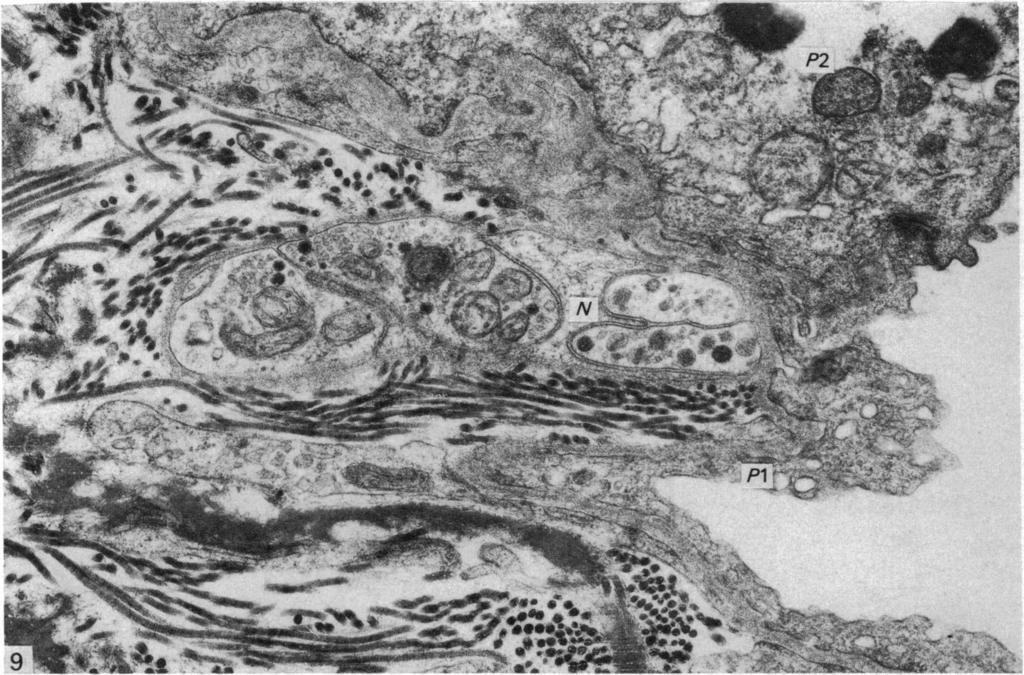
7 Lungs of Lacerta viridis t: 427 M ik Fig. 7. Superficial portion of a type II pneumonocyte fixed in an osmium tetroxide-glutaraldehyde mixture. The cell contains numerous inclusion bodies. Some are vacuolated (V); others have lamellated contents (L). Membranous material (M) is present in the air sac. x Nerve fibres are often present in the depths of the septa and also, less frequently, in the vicinity of pulmonary capillaries and pneumonocytes (Fig. 9). Nerve bundles consisting of as many as 12 unmyelinated axons have been observed near type II pneumonocytes. The axons usually contain neurotubules and some mitochondria, and are partially surrounded by the cytoplasm of Schwann cells. In no instance have the axons been found to breach the basal lamina of the capillary endothelium or the pneumonocytes. Furthermore, enlarged axonal endings have not been observed. Deposits of membranous material are often present within the air sacs. The membranes are intensely stained with uranyl and lead salts and are disposed in the form of loose tangles or, less commonly, as orderly three-dimensional lattices. Irrespective of their overall arrangement, the individual membranes measure about 7 nm in thickness.

8 428 C. MEBAN It al lel. 8
 and that they communicate directly with the axial air passageway.")
9 Lungs of Lacerta viridis 429 DISCUSSION This study has shown that the air spaces bounded by the septa in the interior of the lung of Lacerta viridis are relatively large (much larger than the alveoli of mammals or the air capillaries of birds) and that they communicate directly with the axial air passageway. These air spaces are functionally analogous to the alveoli of mammalian lungs in that they are the sites of gas-exchange. However, they differ from the latter in both their dimensions and in their relationship to the conducting airways. It seems best, therefore, to refer to the air spaces of Lacerta and other reptiles as 'air sacs' and to use the term 'alveoli' only to describe the terminal air chambers of mature mammalian lungs. The epithelium covering the septa in Lacerta lung is complete and consists of two different cell types. The first, here termed the 'type I pneumonocyte' because of its general similarity to the cell in this position in mammalian lungs, is squamous and spreads out over several pulmonary capillaries. It would appear to be directly involved with the process of gas-exchange. The other type, referred to here as 'the type IL pneumonocyte', is cuboidal in shape and contains a greater variety of organelles. Its large osmiophilic inclusions are particularly conspicuous. There can be little doubt that this cell has a secretory function, most likely producing surface-active agents of the type seen in the lungs of other vertebrates (Leeson & Leeson, 1966; Petrik & Riedel, 1968 a, b; Gil & Weibel, 1970; Meban, 1972, 1973). Meyrick & Reid (1968) have observed brush cells in the alveolar epithelium of adult rats. These cells have rounded profiles and are characterized by the presence of squat microvilli on their free surfaces and by bundles of thin filaments and particles of,-glycogen scattered throughout their cytoplasm. Cells of this type have not been encountered in the respiratory epithelium of Lacerta. In the past controversy has existed as to whether or not the alveolar structures of mammalian lung have a sensory and motor nerve supply (Larsell & Dow, 1933; Elftman, 1943; Honjin, 1956; Spencer & Leof, 1964). However, two recent electron microscope studies have provided irrefutable evidence of the existence of small nerve bundles in the interalveolar septa of rats (Meyrick & Reid, 1971) and mice (Hung et al. 1972). Furthermore, Hung et al. have observed two types of specialized nerve endings in mouse lung. One, which they believe to be sensory in function, is located in the septal interstitium or closely related to type I pneumonocytes. The other, supposed to be secretomotor in function, is generally found near type II pneumonocytes. In the present study, bundles of unmyelinated axons have frequently been detected in the depths of the intrapulmonary septa of Lacerta and, in a few instances, they have also been observed more superficially in the vicinity of pneumonocytes. However, axons have not been found to penetrate the basal lamina of the pneumonocytes nor have they been found to form specialized endings. Further work is therefore required to established whether the respiratory epithelium of reptiles has a nerve supply. The present study also demonstrated that the septa in Lacerta lung contained Fig. 8. Detail of the air-blood barrier. Bl, fused basal laminae of pneumonocyte and capillary endothelium; Cs, cytoplasmic sheet of a type I pneumonocyte; E, cytoplasm of an endothelial cell. Note the attentuation of the endothelium (arrows). x Fig. 9. A nerve bundle (N) with unmyelinated axons in the edge of a septum. P1, cytoplasmic sheet of a type I pneumonocyte; P2, a type II pneumonocyte. x

10 430 C. MEBAN 430 C EA substantial quantities of smooth muscle. Some of the bundles of muscle fibres run in the free edges of the septa and their contraction evidently decreases the size of the air sac entrance. In this respect they resemble the circular bands of smooth muscle (the 'entrance rings', Weibel, 1973) surrounding the mouths of mammalian alveoli. In contrast, the muscle bundles in the central regions of the septa are arranged longitudinally; their contraction is probably responsible for shortening the septa and hence reducing the capacity of the air sacs. To date, the behaviour of the smooth muscle in the lungs of reptiles has been virtually ignored. The pulmonary vasculature of Lacerta has several structural features which may serve to increase the overall efficiency of the lung. For example, the capillaries which cover each surface of the internal septa are arranged in a network with very close meshes; indeed, the network seems to approach the form which would permit an effective 'sheet-bloodflow' of the type envisaged by Sobin, Tremer & Fung (1970). Again, the individual elements of the capillary network are interesting in that they are evaginated into the air sacs rather than being countersunk into the substance of the pulmonary septa. Such an arrangement obviously allows a greater area of the capillary wall to be exposed to the air and hence facilitates gas exchange. In addition, the capillary endothelium in Lacerta is greatly attenuated in the region of the airblood barrier (for example, see Fig. 8). This attenuation would also help to reduce the resistance to gaseous diffusion. SUMMARY The gas-exchange area in the lung of Lacerta viridis has been studied by light microscopy and electron microscopy. The interior of the lung in this species is partitioned into air sacs by radially disposed septa. The surfaces of each septum are covered by a continuous epithelium, the cells of which are termed 'pneumonocytes'. Deep to the epithelium there is a close-meshed plexus of capillaries. The middle layer of the septum contains smooth muscle and fibrous tissue. Two varieties of pneumonocytes can be identified. The type I cells are squamous and give off attenuated sheets of cytoplasm which spread widely over the septal surface; these sheets contain few organelles. The type II cells are more compact and possess many organelles; their osmiophilic inclusion bodies are especially conspicuous. The pulmonary capillaries of Lacerta are evaginated into the air sacs and often display marked attentuation of their endothelium. The possible functional significance of these features is discussed. I am indebted to Professor J. J. Pritchard for his advice and encouragement. The technical assistance of Messrs G. Bryan, G. R. Dickson and M. S. Henderson is gratefully acknowledged. Thanks are also due to Mrs J. Hamilton for typing the manuscript. This study was supported by a grant from the Eastern Health and Social Services Board, Northern Ireland. REFERENCES ELFTMAN, A. G. (1943). Afferent and parasympathetic innervation of lungs and trachea of dog. American Journal of Anatomy 72, GIBBONS, I. R. (1959). An embedding resin mixable with water for electron microscopy. Nature 184, GIL, J. & WEIBEL, E. R. (1970). Improvements in the demonstration of lining layer of lung alveoli by electron microscopy. Respiration Physiology 8,
.")
11 Lungs of Lacerta viridis 431 HIRSCH, J. G. & FEDoRKo, M. E. (1968). Ultrastructure of human leukocytes after simultaneous fixation with glutaraldehyde and osmium tetroxide and 'post-fixation' in uranyl acetate. Journal of Cell Biology 38, HONJIN, R. (1956). On the nerve supply of the lung of the mouse, with special reference to the structure of the peripheral vegetative nervous system. Journal ofcomparative Neurology 105, HUNG, K. S., HERTWECK, M. S., HARDY, J. D. & LOOSLI, C. G. (1972). Innervation of pulmonary alveoli of the mouse lung: an electron microscope study. American Journal ofanatomy 135, KILBURN, K. H. (1974). Functional morphology of the distal lung. International Review of Cytology 37, LARSELL, 0. & Dow, R. S. (1933). The innervation of the human lung. American Journal ofanatomy 52, LEESON, T. S. & LEESON, C. R. (1966). Osmiophilic lamellated bodies and associated material in lung alveolar spaces. Journal of Cell Biology 28, MEBAN, C. (1972). An electron microscopic study of the acid mucosubstance lining the alveoli of hamster lung. Histochemical Journal 4, 1-8. MEBAN, C. (1973). The pneumonocytes in the lung of Xenopus laevis. Journal ofanatomy 114, MEYRICK, B. & REID, L. (1968). The alveolar brush cell in rat lung - a third pneumonocyte. Journal of Ultrastructure Research 23, MEYRICK, B. & REID, L. (1971). Nerves in rat intra-acinar alveoli: an electron microscopic study. Respiration Physiology 11, NAGAISHI, C., OKADA, Y., ISHIKO, S. & DAIDO, S. (1964). Electron microscopic observations of the pulmonary alveoli. Experimental Medicine and Surgery 22, OKADA, Y., ISHIKO, S., DAIDO, S., KIM, J. & IKEDA, S. (1962). Comparative morphology of the lung with special reference to the alveolar epithelial cells. II. Lung of the reptilia. Acta tuberculosea japonica 12, PETRIK, P. & REIDEL, B. (1968 a). A continuous osmiophilic noncellular membrane at the respiratory surface of the lungs of fetal chickens and young chicks. Laboratory Investigation 18, PETRIK, P. & RIEDEL, B. (1968b). An osmiophilic bilaminar lining film at the respiratory surfaces of avian lungs. Zeitschriftfiir Zellforschung und mikroskopische Anatomie 88, SOBIN, S. S., TREMER, H. M. & FUNG, Y. C. (1970). Morphometric basis of the sheet-flow concept of the pulmonary alveolar microcirculation in the cat. Circulation Research 26, SPENCER, H. & LEOF, D. (1964). The innervation of the human lung. Journal of Anatomy 98, WEIBEL, E. R. (1973). Morphological basis of alveolar capillary gas exchange. Physiological Reviews 53,
,,, THE MORPHOLOGY AND MORPHOMETRY OF THE PECTEN OCULI IN DIURNAL AND NOCTURNAL BIRDS: A
 ,,, THE MORPHOLOGY AND MORPHOMETRY OF THE PECTEN OCULI IN DIURNAL AND NOCTURNAL BIRDS: A COMPARATIVE STUDY" BY llijama, S.G., B. V. M. (NBI), Department of Veteri nary Anatomy, University of I\Jairobi.
Liver and Gallbladder Morphology of the juvenile Nile crocodile, Crocodylus niloticus (Laurenti, 1768)
") Liver and Gallbladder Morphology of the juvenile Nile crocodile, Crocodylus niloticus (Laurenti, 1768) by ERNA VAN WILPE Submitted in partial fulfilment of the requirements for the degree MSc DEPARTMENT
Liver and Gallbladder Morphology of the juvenile Nile crocodile, Crocodylus niloticus (Laurenti, 1768) by ERNA VAN WILPE Submitted in partial fulfilment of the requirements for the degree MSc DEPARTMENT
HISTOLOGY OF MAMMARY GLAND DURING LACTATING AND NON-LACTATING PHASES OF MADRAS RED SHEEP WITH SPECIAL REFERENCE TO INVOLUTION
 International Journal of Science, Environment and Technology, Vol. 5, No 3, 2016, 991 996 ISSN 2278-3687 (O) 2277-663X (P) HISTOLOGY OF MAMMARY GLAND DURING LACTATING AND NON-LACTATING PHASES OF MADRAS
BEAK AND FEATHER DYSTROPHY IN WILD SULPHUR-CRESTED COCKATOOS (CACATUA GALERITA)
") BEAK AND FEATHER DYSTROPHY IN WILD SULPHUR-CRESTED COCKATOOS (CACATUA GALERITA) Author(s): Steven McOrist, Douglas G. Black, David A. Pass, Peter C. Scott, and John Marshall Source: Journal of Wildlife
BEAK AND FEATHER DYSTROPHY IN WILD SULPHUR-CRESTED COCKATOOS (CACATUA GALERITA) Author(s): Steven McOrist, Douglas G. Black, David A. Pass, Peter C. Scott, and John Marshall Source: Journal of Wildlife
A comparison of placental tissue in the skinks Eulamprus tympanum and E. quoyii. Yates, Lauren A.
 A comparison of placental tissue in the skinks Eulamprus tympanum and E. quoyii Yates, Lauren A. Abstract: The species Eulamprus tympanum and Eulamprus quoyii are viviparous skinks that are said to have
SCANNING electron - microscopy has
 Characteristics of the Absorptive Surface of the Small Intestine of the Chicken from 1 Day to 14 Weeks of Age 1 R. C. BAYER, C. B. CHAWAN, F. H. BIRD AND S. D. MUSGRAVE Department of Animal and Veterinary
HISTOPHYSIOLOGICAL STUDIES ON THE HYPOPHYSIO- MAMMARY AXIS IN SHEEP (Ovis aries) - MAMMOTROPHS
 - MAMMOTROPHS") International Journal of Science, Environment and Technology, Vol. 5, No 3, 2016, 912 917 ISSN 2278-3687 (O) 2277-663X (P) HISTOPHYSIOLOGICAL STUDIES ON THE HYPOPHYSIO- MAMMARY AXIS IN SHEEP (Ovis aries)
International Journal of Science, Environment and Technology, Vol. 5, No 3, 2016, 912 917 ISSN 2278-3687 (O) 2277-663X (P) HISTOPHYSIOLOGICAL STUDIES ON THE HYPOPHYSIO- MAMMARY AXIS IN SHEEP (Ovis aries)
HISTOPATHOLOGY. Introduction:
 Introduction: HISTOPATHOLOGY Goats and sheep are the major domestic animal species in India. Much of the economy of the country has been depend upon the domestication of these animals. Especially economy
BREATHING WHICH IS NOT RESPIRATION
 BREATHING WHICH IS NOT RESPIRATION Breathing vs. Respiration All animals respire. A lot of people think respiration means breathing- this is not true! Breathing is the physical process of inhaling oxygen
Electron Microscopy of the Tapetum Lucidum of the Cat*
 Published Online: 25 January, 1959 Supp Info: http://doi.org/10.1083/jcb.5.1.35 Downloaded from jcb.rupress.org on December 26, 2018 Electron Microscopy of the Tapetum Lucidum of the Cat* By MAURICE H.
Key words: Plasmodium, Kentropyx calcarata, Brazil, merogony, gametocytes, ultrastructure
 FOLIA PARASITOLOGICA 49: 2-8, 2002 Fine structure of erythrocytic stages of a Plasmodium tropiduri-like malaria parasite found in the lizard Kentropyx calcarata (Teiidae) from north Brazil Ilan Paperna
Anat. Labor. of Prof. H. SETO, Tohoku University, On the Sensory Terminations Formed along the Ductus
 Anat. Labor. of Prof. H. SETO, Tohoku University, Sendai. On the Sensory Terminations Formed along the Ductus Pancreaticus in Cat. The existence of PACINIan bodies in the pancreas of mammals, especially
Vertebrates. Vertebrate Characteristics. 444 Chapter 14
 4 Vertebrates Key Concept All vertebrates have a backbone, which supports other specialized body structures and functions. What You Will Learn Vertebrates have an endoskeleton that provides support and
Key words: Coccidia, Choleoeimeria rochalimai, fine structure, gall bladder epithelium, Hemidactylus mabouia, Brazil
 FOLIA PARASITOLOGICA 47: 91-96, 2000 Ultrastructural study of meronts and gamonts of Choleoeimeria rochalimai (Apicomplexa: Eimeriidae) developing in the gall bladder of the gecko Hemidactylus mabouia
Progressive Retinal Atrophy in the Abyssinian Cat
 Progressive Retinal Atrophy in the Abyssinian Cat Electron Microscopy Kristina Narfstr6m*t and Sven Erik Nilsson* Seven adult Abyssinian cats at different stages of a recessively inherited retinal degenerative
A Scanning Electron Microscopic Study of Eggshell Surface Topography of Leidynema portentosae and L. appendiculatum (Nematoda: Oxyuroidea)
") The Ohio State University Knowledge Bank kb.osu.edu Ohio Journal of Science (Ohio Academy of Science) Ohio Journal of Science: Volume 88, Issue 5 (December, 1988) 1988-12 A Scanning Electron Microscopic
The Ohio State University Knowledge Bank kb.osu.edu Ohio Journal of Science (Ohio Academy of Science) Ohio Journal of Science: Volume 88, Issue 5 (December, 1988) 1988-12 A Scanning Electron Microscopic
SUPPLEMENTARY INFORMATION
 doi:10.1038/nature11046 Supplementary Figure 1: Images of PB-positive cells in the subepidermal region (a-i) Representative images of PB positive cells in the subepidermis of the upper beak of the pigeon.
Exotic Hematology Lab Leigh-Ann Horne, LVT, CWR Wildlife Center of Virginia
 Exotic Hematology Lab Leigh-Ann Horne, LVT, CWR Wildlife Center of Virginia lhorne@wildlifecenter.org Anne Lynch, LVT Cedarcrest Animal Clinic amllvt9@gmail.com Introduction While the general set-up for
Ultrastructure of Ehrlichia canis
 INFECTION AND IMMUNrrY, Feb. 1973, p. 265-271 Copyright 1973 American Society for Microbiology Ultrastructure of Ehrlichia canis Vol. 7, No. 2 Printed in U.S.A. PAUL K. HILDEBRANDT, JAMES D. CONROY,I ADAM
Revajová, Viera, Loószová, Adrian. The Journal of Protozoology Resea Citation RightsNational Research Center for Prot
 ' ' Morphological study of partridge Title development in the foreign host - (Gallus gallus) Revajová, Viera, Loószová, Adrian Author(s) Maria, Zibrín, Martin, Herich, Ro Mikulas The Journal of Protozoology
cyst&' appeared to be of two kinds-one smaller and Smnith "is inclined to regard these epithelial cell parasites as
 COCCIDIA IN SUBEPITHELIAL INFECTIONS OF THE INTESTINES OF BIRDS PHILIP B. HADLEY From the Agricultural Experiment Station of the Rhode Island State College' Received for publication, July 10, 1916 In an
Criconemoides similis 1 G. W. BIRD ~
 Somatic Musculature of Trichodorus porosus and Criconemoides similis 1 G. W. BIRD ~ Abstract: The somatic musculature of Trichodorus porosus is transversely striated, and that of Criconemoides similis
Frog Dissection Information Manuel
 Frog Dissection Information Manuel Anatomical Terms: Used to explain directions and orientation of a organism Directions or Positions: Anterior (cranial)- toward the head Posterior (caudal)- towards the
Development of the Intestinal Villi Associated
 Development of the Intestinal Villi Associated with the Increased Epithelial Cell Mitosis in Chickens Koh-en YAMAUCHI, Eiji NAKAMURA and Yutaka ISSHIKI Laboratory of Animal Science, Faculty of Agriculture,
COMPARATIVE VERTEBRATE HISTOLOGY ZOO 4756c Syllabus for Fall 2018
 COMPARATIVE VERTEBRATE HISTOLOGY ZOO 4756c Syllabus for Fall 2018 Instructor: Frank T. Logiudice Office: Biology Building, Room 202c Office Phone Number: (407) - 823-2495 Email Address: Frank.Logiudice@ucf.edu
Mechanism of a Crocodile s Circulatory System
 Mechanism of a Crocodile s Circulatory System Figure 1. A crocodile diving at Botswana (Nachoum, A. 2017) Ever wonder in one of those animal documentaries we watch in television, wherein a crocodile glides
Title. CitationJapanese Journal of Veterinary Research, 24(1-2): 37. Issue Date DOI. Doc URL. Type. File Information
: 37. Issue Date DOI. Doc URL. Type. File Information") Title DISTRIBUTION OF LYMPHATIC TISSUES IN DUCK CAECA Author(s)KITAMURA, Hirokazu; SUGIMURA, Makoto; HASHIMOTO, Yos CitationJapanese Journal of Veterinary Research, 24(1-2): 37 Issue Date 1976-05 DOI 10.14943/jjvr.24.1-2.37
Title DISTRIBUTION OF LYMPHATIC TISSUES IN DUCK CAECA Author(s)KITAMURA, Hirokazu; SUGIMURA, Makoto; HASHIMOTO, Yos CitationJapanese Journal of Veterinary Research, 24(1-2): 37 Issue Date 1976-05 DOI 10.14943/jjvr.24.1-2.37
OBSERVATIONS ON THE QUALITATIVE AND QUANTITATIVE STRUCTURAL CHARACTERISTICS OF THE REPTILIAN KIDNEYS.
 OBSERVATIONS ON THE QUALITATIVE AND QUANTITATIVE STRUCTURAL CHARACTERISTICS OF THE REPTILIAN KIDNEYS. ~B~SI"Y OF Nmlll,.tpj,Tb 1.11.,,)' A Thesis submitted to the university of Nairobi in partial fulfillment
Mesosomes are a definite event in antibiotic-treated Staphylococcus aureus ATCC 25923
 Tropical Biomedicine 24(1): 105 109 (2007) Mesosomes are a definite event in antibiotic-treated Staphylococcus aureus ATCC 25923 Santhana Raj, L. 1*, Hing, H.L. 2, Baharudin Omar 2, Teh Hamidah, Z. 1,
DEVELOPMENT OF THE HEAD AND NECK PLACODES
 DEVELOPMENT OF THE HEAD AND NECK Placodes and the development of organs of special sense L. Moss-Salentijn PLACODES Localized thickened areas of specialized ectoderm, lateral to the neural crest, at the
Importance of Electron Microscopy to reveal species-specific characteristics of gland secretion
 mportance of Electron Microscopy to reveal species-specific characteristics of gland secretion Gabriella Chieffi Baccari 1, Alessandra Santillo 1, and Sergio Minucci 2 1 Department of Life Sciences, Second
Gross and Microscopic Features of the Interdigital Sinus in the Barbados Black Belly Sheep in Trinidad
 Original Research Article International Journal of Current Research in Medical Sciences ISSN: 2454-5716 www.ijcrims.com Volume 2, Issue 7-2016 SOI: http://s-o-i.org/1.15/ijcrms-2016-2-7-4 Gross and Microscopic
Biology. Slide 1of 50. End Show. Copyright Pearson Prentice Hall
 Biology 1of 50 2of 50 Phylogeny of Chordates Nonvertebrate chordates Jawless fishes Sharks & their relatives Bony fishes Reptiles Amphibians Birds Mammals Invertebrate ancestor 3of 50 A vertebrate dry,
Diversity of Animals
 Classifying Animals Diversity of Animals Animals can be classified and grouped based on similarities in their characteristics. Animals make up one of the major biological groups of classification. All
Morphology of cat vomeronasal organ non-sensory epithelium during postnatal development
 Original rticle pissn 2093-3665 eissn 2093-3673 Morphology of cat vomeronasal organ non-sensory epithelium during postnatal development Sanaa. M. Elgayar, Heba M. Saad-Eldin, Ola. Haussein epartment of
Biology Slide 1 of 50
 Biology 1 of 50 2 of 50 What Is a Reptile? What are the characteristics of reptiles? 3 of 50 What Is a Reptile? What Is a Reptile? A reptile is a vertebrate that has dry, scaly skin, lungs, and terrestrial
Question Set 1: Animal EVOLUTIONARY BIODIVERSITY
 Biology 162 LAB EXAM 2, AM Version Thursday 24 April 2003 page 1 Question Set 1: Animal EVOLUTIONARY BIODIVERSITY (a). We have mentioned several times in class that the concepts of Developed and Evolved
Name Class Date. After you read this section, you should be able to answer these questions:
 CHAPTER 14 4 Vertebrates SECTION Introduction to Animals BEFORE YOU READ After you read this section, you should be able to answer these questions: How are vertebrates different from invertebrates? How
Physiological Aspects of Bovine Mammary Involution: a Biochemical and Morphological Investigation.
 Louisiana State University LSU Digital Commons LSU Historical Dissertations and Theses Graduate School 1987 Physiological Aspects of Bovine Mammary Involution: a Biochemical and Morphological Investigation.
The oriental fruit fly, Bactrocera dorsalis
 Zoological Studies 37(2): 95-101 (1998) Morphology and Ultrastructure of the Alimentary Canal of Oriental Fruit Fly Bactrocera dorsalis (Hendel) (Diptera: Tephritidae) (I): The Structure of the Foregut
A Lymphosarcoma in an Atlantic Salmon (Salmo salar)
") A Lymphosarcoma in an Atlantic Salmon (Salmo salar) Authors: Paul R. Bowser, Marilyn J. Wolfe, and Timothy Wallbridge Source: Journal of Wildlife Diseases, 23(4) : 698-701 Published By: Wildlife Disease
A Lymphosarcoma in an Atlantic Salmon (Salmo salar) Authors: Paul R. Bowser, Marilyn J. Wolfe, and Timothy Wallbridge Source: Journal of Wildlife Diseases, 23(4) : 698-701 Published By: Wildlife Disease
"Dole. Analom^i Of Phryno^oma
 "Dole. Analom^i Of Phryno^oma ANATOMY OF PHRYNOSOMA; THE RESPIRATORY SYSTEM BY LILLIAN DORA DOLE A. B. University of Illinois, 1915 THESIS Submitted in Partial Fulfillment of the Requirements for the
PLASMODIUM MODULE 39.1 INTRODUCTION OBJECTIVES 39.2 MALARIAL PARASITE. Notes
 Plasmodium MODULE 39 PLASMODIUM 39.1 INTRODUCTION Malaria is characterized by intermittent fever associated with chills and rigors in the patient. There may be enlargement of the liver and spleen in the
Histochemical localization of adenylate cyclase activity in some mammalian taste papillae
 Chemical Senses Volume 7 Number 1 1982 Histochemical localization of adenylate cyclase activity in some mammalian taste papillae Hiromichi Nomura and Naokazu Asanuma Department of Oral Physiology, Matsumoto
Recommended Resources: The following resources may be useful in teaching this
 Unit B: Anatomy and Physiology of Poultry Lesson1: Internal Anatomy of Poultry Student Learning Objectives: Instruction in this lesson should result in students achieving the following objectives: 1. Identify
DRAFT TANZANIA STANDARD
 Hatching eggs Specification DRAFT TANZANIA STANDARD TANZANIA BUREAU OF STANDARDS 1 Hatching eggs Specification TBS/AFDC 22 (5271) P3 0 FOREWORD This Tanzania standard was developed due to rapid increase
Both Rod and Cone Disc Shedding ore Related to Light Onset in the Cat
 Both Rod and Cone Disc Shedding ore Related to Light Onset in the Cat Sreven K. Fisher,* Bruce A. Pfeffer.f and Don H. Anderson* Nineteen domestic cats were entrained to a 12-hr light/12-hr dark lighting
Phylum Platyhelminthes Flatworms
 Phylum Platyhelminthes Flatworms The Acoelomates The acoelomates are animals that lack a coelom. Acoelomates lack a body cavity, and instead the space between the body wall and the digestive tract is filled
Dipartimento di Biologia Animale e Genetica dell Università di Firenze, Via Romana 17, Firenze, Italy 2
 Tropical Zoology 20: 75-89, 2007 Ultrastructure of the venom gland of the Andean Red-tailed Coral Snake Micrurus mipartitus decussatus (Duméril, Bibron & Duméril 1854) (Squamata Serpentes Elapidae) F.
Transformed centrioles In adult and aged cat pinealocytes
 Transformed centrioles In adult and aged cat pinealocytes J. L. Calvo. J. Boya*. J. E. Garcia-Mauriño and D. Rancaño Department of Histology. Faculty of Medicine. University Complutense, 28040 Madrid.
INVESTIGATIONS ON THE SHAPE AND SIZE OF MOLAR AND ZYGOMATIC SALIVARY GLANDS IN SHORTHAIR DOMESTIC CATS
 Bulgarian Journal of Veterinary Medicine (2009), 12, No 4, 221 225 INVESTIGATIONS ON THE SHAPE AND SIZE OF MOLAR AND ZYGOMATIC SALIVARY GLANDS IN SHORTHAIR DOMESTIC CATS Summary A. A. MOHAMMADPOUR Department
Topic 13: Energetics & Performance. How are gas exchange, circulation & metabolism inter-related?
 Topic 3: Energetics & Performance How are gas exchange, circulation & metabolism interrelated? How is it done in air and water? What organs are involved in each case? How does ventilation differ among
Technique for microdissection and measurement in biopsies of human small intestine
 Journal of Clinical Pathology, 1977, 30, 1068-1073 Technique for microdissection and measurement in biopsies of human small intestine ANNE FERGUSON, A. SUTHERLAND, T. T. MAcDONALD, AND FRANCES ALLAN From
SCANNING ELECTRON MICROSCOPY OF THE MUCOSAL SURFACE OF THE FORESTOMACHS AND ABOMASA OF GREY, WHITE AND BLACK KARAKUL LAMBS
 OnderstepoortJ. Vet. Res., 59, 167-174 (1992) SCANNING ELECTRON MICROSCOPY OF THE MUCOSAL SURFACE OF THE FORESTOMACHS AND ABOMASA OF GREY, WHITE AND BLACK KARAKUL LAMBS H. B. GROENEWALD, Department of
THE STRUCTURE OF ECHINOCOCCAL CYSTS AND HISTOPATHOLOGICAL CHANGES IN LIVER
 THE STRUCTURE OF ECHINOCOCCAL CYSTS AND HISTOPATHOLOGICAL CHANGES IN LIVER Michal Juszynski Helena Palenga, Danuta Cielecka PhD Department of General Biology and Parasitology Medical University of Warsaw
DLS Sample Preparation Guide
 DLS Sample Preparation Guide The Leica TCS SP8 DLS is an innovative concept to integrate the Light Sheet Microscopy technology into the confocal microscope. Due to its unique optical architecture samples
Sensory Setae of the First Tarsi and Palps of the Mite Macrocheles muscaedomesticae1.2
 $ Sensory Setae of the First Tarsi and Palps of the Mite Macrocheles muscaedomesticae1.2 ~, 'r L. B. COONS ANDR. C. AXTELL Department of Entomology, North Carolina State University, Raleigh 27607 By scanning
Characteristics of a Reptile. Vertebrate animals Lungs Scaly skin Amniotic egg
 Reptiles Characteristics of a Reptile Vertebrate animals Lungs Scaly skin Amniotic egg Characteristics of Reptiles Adaptations to life on land More efficient lungs and a better circulator system were develope
Lesson 6. References: Chapter 6: Reading for Next Lesson: Chapter 6:
 Lesson 6 Lesson Outline: General Features of the Integument Embryonic Origins of the Epidermis Specializations of the Epidermis o Glands o Keratin and Stratum Corneum Objectives: At the end of this lesson
EXOSTOSIS OF THE MANDIBLE OF THE CHICKEN
 EXOSTOSIS OF THE MANDIBLE OF THE CHICKEN COMPLICATING EDEMA OF THE WATTLES GEORGE MILTON SMITH, M.D.1 (AnutomioaZ Laboratory, Yale School of Medicine, New Haven, Connecticut) During the past year opportunity
Rat Control & Water Vole Conservation
 Rat Control & Water Vole Conservation Why are water voles important? Water Voles were once a common sight in Sussex but they have declined by over 90% in the last 30 years. Water voles have already become
Intestinal linear foreign body
 Vet Times The website for the veterinary profession https://www.vettimes.co.uk Intestinal linear foreign body Author : Sally Birch Categories : Companion animal, Vets Date : February 6, 2017 Your first
The specimens of Ameiva ameiva (Linn) were
 were") Article available at http://www.parasite-journal.org or http://dx.doi.org/10.1051/parasite/1999064359 FINE STRUCTURE OF THE EPICYTOPLASMIC EIMERID COCCIDIUM ACROEIMERIA PINTOI LAINSON & PAPERNA, 1999,
Article available at http://www.parasite-journal.org or http://dx.doi.org/10.1051/parasite/1999064359 FINE STRUCTURE OF THE EPICYTOPLASMIC EIMERID COCCIDIUM ACROEIMERIA PINTOI LAINSON & PAPERNA, 1999,
Gross and histological studies of digestive tract of broilers during postnatal growth and development
 J. Bangladesh Agril. Univ. 10(1): 69 77, 2012 ISSN 1810-3030 Gross and histological studies of digestive tract of broilers during postnatal growth and development M. Nasrin, M. N. H. Siddiqi, M. A. Masum
Coturnix coturnix coturnix
 Coturnix coturnix coturnix L. Coturnix coturnix coturnix L. 8 circadian clock 0 9 Galliformes Coturnix coturnix coturnix Phasianidae L. 0 9 8 7 Photoneuroendocrine gland Transmission TEM electron microscope
Sarcocystis heydorni, n. sp. (Apicomplexa: Protozoa) with cattle (Bos taurus) and human
 with cattle (Bos taurus) and human") 1 Sarcocystis heydorni, n. sp. (Apicomplexa: Protozoa) with cattle (Bos taurus) and human (Homo sapiens) cycle Jitender P. Dubey 1, Erna van Wilpe 2, Rafael Calero-Bernal 1, Shiv Kumar Verma 1, Ronald
1 Sarcocystis heydorni, n. sp. (Apicomplexa: Protozoa) with cattle (Bos taurus) and human (Homo sapiens) cycle Jitender P. Dubey 1, Erna van Wilpe 2, Rafael Calero-Bernal 1, Shiv Kumar Verma 1, Ronald
Development, comparative morphology and cornification of reptilian claws in relation to claws evolution in tetrapods
 Contributions to Zoology, 78 (1) 25-42 (2009) Development, comparative morphology and cornification of reptilian claws in relation to claws evolution in tetrapods Lorenzo Alibardi 1, 2 1 Dipartimento di
Light, Scanning and Transmission Electron Microscopical Study on the Oviduct of the Ostrich (Struthio
 Light, Scanning and Transmission Electron Microscopical Study on the Oviduct of the Ostrich (Struthio camelus) A.S.Saber*, S.A.M.Emara*, O.M.M.AboSaeda** * Faculty of Veterinary Medicine, Sadat City Branch,
Reproductive physiology and eggs
 Reproductive physiology and eggs Class Business Reading for this lecture Required. Gill: Chapter 14 1. Reproductive physiology In lecture I will only have time to go over reproductive physiology briefly,
International Journal of Science, Environment and Technology, Vol. 5, No 5, 2016,
 International Journal of Science, Environment and Technology, Vol. 5, No 5, 2016, 3249 3253 ISSN 2278-3687 (O) 2277-663X (P) HISTOPATHOLOGICAL STUDY OF PULMONARY ANTHRACOSIS IN SHEEP Amaravathi M* 1, Satheesh
Section 6. Embryonic Development and Hatchery Management Notes
 Section 6 Embryonic Development and Hatchery Management Notes Slide 2 A well run hatchery is critical for any integrated poultry company whether it be a primary breeder company or a commercial meat company.
1. Hair 2. Mammary glands produce milk 3. Specialized teeth 4. 3 inner ear bones 5. Endothermic 6. Diaphragm 7. Sweat, oil and scent glands 8.
 Class Mammalia The Mammals Key Characteristics of Mammals 1. Hair 2. Mammary glands produce milk 3. Specialized teeth 4. 3 inner ear bones 5. Endothermic 6. Diaphragm 7. Sweat, oil and scent glands 8.
Biology Review: Amphibians
 Name: Biology Review: Amphibians NOTE: USE THE SCANNED CHAPTER ON MY WEBSITE, NOT YOUR TEXTBOOK FOR THIS ASSIGNMENT 1-6. Amphibians were the first group of vertebrates to adapt to a land existence. What
Ectoparasites Myobia musculi Radfordia affinis Radfordia ensifera
 Ectoparasites Fleas, ticks, and lice are uncommon in modern laboratory facilities, but may be seen on wild or feral rodents. Most ectoparasite infestations seen in rats and mice used for research are various
Animal Form and Function. Amphibians. United by several distinguishing apomorphies within the Vertebrata
 Animal Form and Function Kight Amphibians Class Amphibia (amphibia = living a double life) United by several distinguishing apomorphies within the Vertebrata 1. Skin Thought Question: For whom are integumentary
Field necropsy techniques in mammal and poultry
 Field necropsy techniques in mammal and poultry Kidsadagon Pringproa, DVM, MS, PhD Department of Veterinary Biosciences and Veterinary Public Health Faculty of Veterinary Medicine Chiang Mai University
ZOOLOGISCHE MEDEDELINGEN
 ZOOLOGISCHE MEDEDELINGEN UITGEGEVEN DOOR HET RIJKSMUSEUM VAN NATUURLIJKE HISTORIE TE LEIDEN (MINISTERIE VAN WELZIJN, VOLKSGEZONDHEID EN CULTUUR) Deel 58 no. 19 16 november 1984 ISSN 0024-0672 CANTHARELLUS
Seasonal Variations of yeso sika Deer Skin and its Vegetable Tanned Leather
 Seasonal Variations of yeso sika Deer Skin and its Vegetable Tanned Leather Shigeharu Fukunaga, Akihiko Yoshie, Ikuo Yamakawa, Fumio Nakamura Laboratory of Animal By-product Science, Graduate School of
revealed a population of particles apparently of considerable homogeneity with
 MORPHOLOGY OF THE VIRUS OF AVIAN ERYTHRO- MYELOBLASTIC LEUCOSIS AND A COMPARISON WITH THE AGENT OF NEWCASTLE DISEASE' D. G. SHARP, EDWARD A. ECKERT,2 DOROTHY BEARD, AND J. W. BEARD Department of Surgery,
Anatomy. Name Section. The Vertebrate Skeleton
 Name Section Anatomy The Vertebrate Skeleton Vertebrate paleontologists get most of their knowledge about past organisms from skeletal remains. Skeletons are useful for gleaning information about an organism
Lesson 7. References: Chapter 6: Chapter 12: Reading for Next Lesson: Chapter 6:
 Lesson 7 Lesson Outline: Embryonic Origins of the Dermis Specializations of the Dermis o Scales in Fish o Dermal Armour in Tetrapods Epidermal/Dermal Interactions o Feathers o Hair o Teeth Objectives:
Histomorphology and scanning electron microscopy of the pharyngeal tonsil in goats
 Indian J. Anim. Res., 51 (3) 2017 : 464-468 Print ISSN:0367-6722 / Online ISSN:0976-0555 AGRICULTURAL RESEARCH COMMUNICATION CENTRE www.arccjournals.com/www.ijaronline.in Histomorphology and scanning electron
Ultrastructure of Endogenous Stages of Eimeria ninakohlyakimovae Yakimoff & Rastegaieff, 1930 Emend. Levine, 1961 in Experimentally Infected Goat
 Mem Inst Oswaldo Cruz, Rio de Janeiro, Vol. 92(4): 533-538, Jul./Aug. 1997 Ultrastructure of Endogenous Stages of Eimeria ninakohlyakimovae Yakimoff & Rastegaieff, 1930 Emend. Levine, 1961 in Experimentally
DISEASE SAMPLING. Readings. What to wear, what to wear 3/9/2009. Required. Supplemental. Rubber boots or waders Disposable gloves
 DISEASE SAMPLING Readings Required Standard operating procedures SEPARC collecting and shipping specimens for diagnostic testing Green et al. Disease Monitoring and Biosafety Section 26.3 and 26.4 Supplemental
26-3 Cnidarians Slide 2 of 47
 2 of 47 What Is a Cnidarian? What is a cnidarian? 3 of 47 What Is a Cnidarian? What Is a Cnidarian? Cnidarians are soft-bodied, carnivorous animals that have stinging tentacles arranged in circles around
Australian and New Zealand College of Veterinary Scientists. Fellowship Examination. Veterinary Anaesthesia and Critical Care Paper 1
 Australian and New Zealand College of Veterinary Scientists Fellowship Examination June 2016 Veterinary Anaesthesia and Critical Care Paper 1 Perusal time: Twenty (20) minutes Time allowed: Three (3) hours
PRODUCT DATA SHEET. Sika Pyroplast HW-100 with Topcoat Sika Pyroplast HW-211 Top PRODUCT DESCRIPTION
 PRODUCT DATA SHEET Water based fire protective coating for wood, interior use PRODUCT DESCRIPTION USES PROPERTIES TESTS Sika Pyroplast HW-100 is a water borne, transparent fire protection coating which
Jeff Baier MS DVM Birds of Prey Foundation Broomfield, CO
 Jeff Baier MS DVM Birds of Prey Foundation Broomfield, CO drjeffbaier@gmail.com Squamates Chelonians Snakes Lizards Varanids Monitor Lizards Crocodilians Reptilian adaptations Anaerobic glycolysis Low
Vertebrate and Invertebrate Animals
 Vertebrate and Invertebrate Animals Compare the characteristic structures of invertebrate animals (including sponges, segmented worms, echinoderms, mollusks, and arthropods) and vertebrate animals (fish,
RELATIONSHIP BETWEEN PHEROMONE TRAP CAPTURE AND EMERGENCE OF ADULT ORIENTAL FRUIT MOTHS, GRAPHOLZTHA MOLESTA (LEPIDOPTERA: TORTRICIDAE)'
'") RELATIONSHIP BETWEEN PHEROMONE TRAP CAPTURE AND EMERGENCE OF ADULT ORIENTAL FRUIT MOTHS, GRAPHOLZTHA MOLESTA (LEPIDOPTERA: TORTRICIDAE)' THOMAS C BAKER,^ RING T CARDE, and BRIAN A CROFT Department of Entomology
RELATIONSHIP BETWEEN PHEROMONE TRAP CAPTURE AND EMERGENCE OF ADULT ORIENTAL FRUIT MOTHS, GRAPHOLZTHA MOLESTA (LEPIDOPTERA: TORTRICIDAE)' THOMAS C BAKER,^ RING T CARDE, and BRIAN A CROFT Department of Entomology
1. Examine the specimens of sponges on the lab table. Which of these are true sponges? Explain your answers.
 Station #1 - Porifera 1. Examine the specimens of sponges on the lab table. Which of these are true sponges? Explain your answers. 2. Sponges are said to have an internal special skeleton. Examine the
VERTEBRATE READING. Fishes
 VERTEBRATE READING Fishes The first vertebrates to become a widespread, predominant life form on earth were fishes. Prior to this, only invertebrates, such as mollusks, worms and squid-like animals, would
The Fine Structure of the Endogenous Stages of Isospora hemidactyli Carini, 1936 in the Gecko Hemidactylus mabouia from North Brazil
 Mem Inst Oswaldo Cruz, Rio de Janeiro, Vol. 95(1): 43-47, Jan./Feb. 2000 The Fine Structure of the Endogenous Stages of Isospora hemidactyli Carini, 1936 in the Gecko Hemidactylus mabouia from North Brazil
Vertebrates. skull ribs vertebral column
 Vertebrates skull ribs vertebral column endoskeleton in cells working together tissues tissues working together organs working together organs systems Blood carries oxygen to the cells carries nutrients
F.L. Andr6s. Rua Tristao Vaz No Esq., 1400 Lisboa, Portugal
 Supranumerary Barrels Develop in the Somatosensory Cortex of Mice, After the Implantation of the Vibrissal Follicle Parts Containing Large Numbers of Receptors F.L. Andr6s Rua Tristao Vaz No. 37 1 Esq.,
THE CENTRAL CONNEXIONS OF DORSAL SPIN [l NERVE ROOTS AND THE ASCENDING TRACT IN THE SPINAL CORD OF LACERTA VIRIDIS
 J. Anat., Lond. (1962), 96, 2, pp. 153-170 1 With 2 plates and 2 text-figures Printed in Great Britain THE CENTRAL CONNEXIONS OF DORSAL SPIN [l NERVE ROOTS AND THE ASCENDING TRACT IN THE SPINAL CORD OF
Histological study on the ovaries of Haemonchus contortus (Nematoda)
") Int. J. of Life Sciences, 2017, Vol. 5 (2): 254-258 ISSN: 2320-7817 eissn: 2320-964X RESEARCH ARTICLE Histological study on the ovaries of Haemonchus contortus (Nematoda) Jatinderpal Singh Department of
Int. J. of Life Sciences, 2017, Vol. 5 (2): 254-258 ISSN: 2320-7817 eissn: 2320-964X RESEARCH ARTICLE Histological study on the ovaries of Haemonchus contortus (Nematoda) Jatinderpal Singh Department of
THE MICROMORPHOLOGY OF THE GLANDS OF THE INFRA-ORBITAL CUTANEOUS SINUS OF THE STEENBOK (RAPHICERUS CAMPESTRIS)
") Onderstepoort J. vet. Res. 45 (2), 59-66 (1978) THE MICROMORPHOLOGY OF THE GLANDS OF THE INFRA-ORBITAL CUTANEOUS SINUS OF THE STEENBOK (RAPHICERUS CAMPESTRIS) W. H. GERNEK ( 1 ) and M. COHEN(2) ABSTRACT
Onderstepoort J. vet. Res. 45 (2), 59-66 (1978) THE MICROMORPHOLOGY OF THE GLANDS OF THE INFRA-ORBITAL CUTANEOUS SINUS OF THE STEENBOK (RAPHICERUS CAMPESTRIS) W. H. GERNEK ( 1 ) and M. COHEN(2) ABSTRACT
Amphibians. Land and Water Dwellers
 Amphibians Land and Water Dwellers Amphibians Most amphibians do not live completely in the water or completely on land and most must return to water to reproduce http://potch74.files.wordpress.com/2007/09/amphibians.jpg
KINGDOM ANIMALIA Phylum Chordata Subphylum Vertebrata Class Reptilia
 KINGDOM ANIMALIA Phylum Chordata Subphylum Vertebrata Class Reptilia Vertebrate Classes Reptiles are the evolutionary base for the rest of the tetrapods. Early divergence of mammals from reptilian ancestor.