Ëtude ultrastructurale de la mérogonie de Schellackia cf. agamae (Lankesterellidae, Apicomplexa) chez le Lézard Agama stellio.
|
|
|
- Coleen Stewart
- 5 years ago
- Views:
Transcription
 FROM THE STARRED LIZARD AGAMA STELLIO K. OSTROVSKA*, I. PAPERNA* SUMMARY. Meront stages of Schellackia cf.")


1 Masson, Paris, 1987 Ann. Parasitol. Hum. Comp. 1987, 62, n 5, pp ULTRASTRUCTURAL STUDIES ON THE MEROGONY OF SCHELLACKIA CF. AGAMAE (LANKESTERELLIDAE, APICOMPLEXA) FROM THE STARRED LIZARD AGAMA STELLIO K. OSTROVSKA*, I. PAPERNA* SUMMARY. Meront stages of Schellackia cf. agamae (Laveran and Petit, 1909) were obtained from the anterior intestine mucosal epithelium of experimentally infected Agama stellio Hasselq. and Linn., Infection was recovered 5, 7, 10 and 15 days after feeding on blood and liver containing sporozoites from naturally infected A. stellio. The parasitophorous vacuole wall consisted of one bilaminate and one single unit membranes apposed at regular intervals. Following nuclear division merozoites differentiated by exogenuous budding. The ultrastructure of the meronts, the merozoite and the merogonous process conformed in all details with that of species of Eimeria. Key-words: Ultrastructure. Merogony. Schellackia cf. agamae. Parasitic in Agama stellio. Ëtude ultrastructurale de la mérogonie de Schellackia cf. agamae (Lankesterellidae, Apicomplexa) chez le Lézard Agama stellio. RÉSUMÉ. Les stades mérogoniques de Schellackia cf. agamae sont étudiés dans l épithélium de la partie antérieure de l intestin d Agama stellio infectés expérimentalement. Les différents stades ont été étudiés 5, 7, 10 et 15 jours après l ingestion, par ces Lézards, de sang et de foie d un A. stellio naturellement infecté et dont les organes contenaient des sporozoïtes. L enveloppe de la vacuole parasitophore comprend une membrane unitaire et une membrane bi-laminée accolées à intervalles réguliers. Après la division des noyaux les mérozoïtes se différencient par bourgeonnement externe. L ultrastructure des mérontes, du mérozoïte et le déroulement de la mérogonie sont conformes dans tous les détails à ceux des autres espèces d Eimeria. Mots-clés : Ultrastructure. Merogonie. Schellackia cf. agamae. Parasite d Agama stellio. * Department of Animal Sciences, Faculty of Agriculture, Hebrew University of Jerusalem, Rehovot, Israel. Accepté le 26 mars Article available at or
 described merogony stages, oocysts and sporozoites of S. agamae (Laveran and Petit, 1909) obtained by experimental infection of Agama colonorum.")


2 ULTRASTRUCTURE OF SCHELLACKIA 381 Introduction Data on merogony in Schellackia are available from light microscopy studies of 7 species (Lainson, Shaw and Ward, 1976). Rogier (1979) described merogony stages, oocysts and sporozoites of S. agamae (Laveran and Petit, 1909) obtained by experimental infection of Agama colonorum. Infection was acheived by feeding on blood and tissues containing sporozoites, taken from naturally infected agama of the same species, collected in the Central African Republic. S. cf. agamae was found to infect Agama stellio Hasselq. and Linn. in Israel. Only sporozoite stages of S. cf. agamae, in the blood and the liver, were found in naturally infected lizards (Ostrovska and Paperna, unpublished data). Merogony was studied in experimentally infected animals. This is the first electron microscopic study of merogony in a species of Schellackia. Materials and methods Juvenile sporozoite-free Agama stellio were innoculated with blood or force fed on blood and livers of co-specific adult lizards naturally infected with Schellackia cf. agamae. The lizards were kept in heated cages (24-29 C) and were autopsied 5, 7, 10 and 15 days after infection. Presence of infection was verified by light microscopic (LM) examination of Giemsa stained blood, intestine and liver smears. For transmission electron microscopy (TEM), pieces from the anterior gut were fixed in Karnowski s for 24 hours at 4 C, rinsed repeatedly in Cacodylate buffer, 0.1 M, ph 7.4 and postfixed in 1 % Osmium tetroxide, in the same buffer, for 1 hour. After rinsing in the buffer, the material was dehydrated in ethanol and embedded in Epon. Thin sections cut with diamond knife, were stained on grid with uranyl acetate and lead citrate and examined with a Joel 100 CX TEM. Results and discussion Young meronts were recovered from smears and sections of the anterior intestine of lizards autopsied 5 and 7 days post infection (p. i.). Mature, dividing meronts and merozoites were recovered in lizards sacrificed 10 and 15 days p. i. In S. agamae from Agama colonorum merogony had terminated by the 9th day p. i. (Rogier, 1977); in S. balli merogony terminated also by the 9th day p. i. (Le Bail and Landau, 1974), while in S. landauae merogony was observed in animals sacrificed 23 days p. i. (Lainson et al., 1976). Lainson et al. (1976) reported dividing merozoites of S. landauae in the liver; such extra-intestinal merozoites were not found in the liver or any other organs in either naturally or experimentally infected lizards presently studied.
. The outer membrane was bilaminate and the inner one consisted of a single unit.")
, Isospora rivolta (Pelster, 1973), I.")

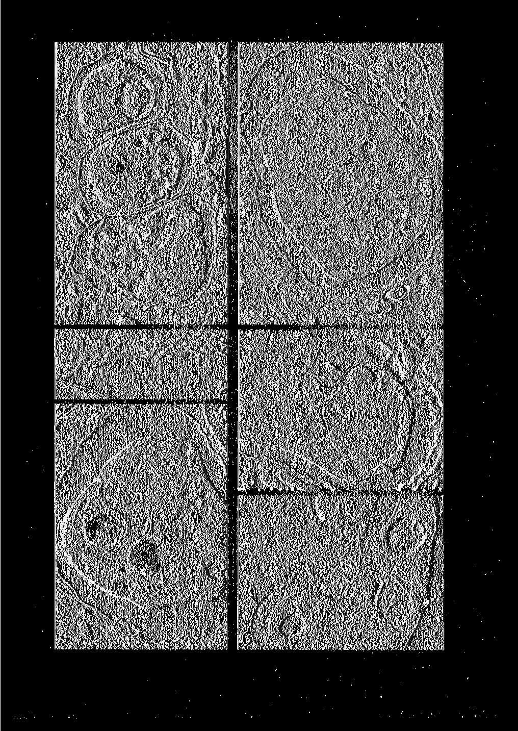
3 382 K. OSTROVSKA, I. PAPERNA 1 Early meronts (fig. 1-4)* Stages of the merogony were located within the cells of the anterior gut epithelium. Infected cells contained 1-4 meronts (fig. 1). The wall of the parasitophorous vacuole consisted of two membranes apposed at regular intervals (fig. 2). The outer membrane was bilaminate and the inner one consisted of a single unit. Same type of parasitophorous vacuole wall occurs also in host cells infected with gamonts of S. cf. agamae Ostrovska and Paperna, unpublished). Multimembranous structure of the wall of the parasitophorus vacuole was described gamont stage infections of Toxoplasma gondii (Pelster and Piekarski, 1971), Isospora rivolta (Pelster, 1973), I. felis (Ferguson, Birch-Andersen, Hutchinson and Siims, 1980) and Sarcocystis spp. (Scholtyseck and Hilali, 1978, Entzeroth, Chobotar and Scholtyseck, 1985). The wall of the parasitophorous vacuole consisted of a single membrane in sporozoite stage infections of S. cf. agamae (Ostrovska and Paperna, unpublisaheds), of other species of Schellackia and of Lankesterella (Stehbens, 1966, Heller, 1974, Sinden and Moore, 1974, Bikeung, Barta and Desser, 1986). Single membrane parasitophorous vacuole wall is found in merogony and gamogony stage infections of Eimeria spp. (Scholtyseck, 1979). The parasitophorous vacuole contained a very dilute flocculant substance. Early meronts with a single nucleus (fig. 1), with dividing nucleus (fig. 3) or already with two nuclei (fig. 4) were bound by a single unit membrane and a thicker discontinuous subpellicular membrane (fig. 3). The areas with double membrane may represent the future budding sites of the merozoites similar to those reported from meronts of Eimeria spp. (Kelly and Hammond, 1973, Dubremetz, 1975). Nuclei had prominent nucleoli (fig. 1-4). The cytoplasm contained one to several mitochondria (fig. 4), numerous ribosomes, smooth, and rough endoplasmic reticulum, a variable number of food vacuoles and electron lucent vacuoles, apparently lipid vacuoles exhausted of their content (fig. 1-4). * Abbreviation to figures: A: Apical complex; G: centrocone; ED: electron dense vesicle; er: endoplasmic reticulum; F: food vacuole; H: host cell; L: lipid vacuole; M: mitochondria; Mz: merozoites; mn: micronemes; N: nucleus; pm: merozoite primordium; Pv: Parasitophorous vacuole; R: rhoptries; RB: residual body: S: multilaminated inclusions. Planche1 Fig. 1. Mucosal epithelial cell infected by 4 young meronts ( x 7,500). Fig. 2. Wall of the parasitophorous vacuole ( x 32,000). Fig. 3. Meront with dividing nucleus, showing spindle (arrows) and one centrocone ( x 9,900). Fig. 4. Meront with two nuclei ( x 9,900). Fig. S and 6. Merozoites budding into a subpellicular inclusion (arrows) ( x 14,850 and x 22,700).
4

, at the exterior or")
. Subpellicular budding concurrently with exogenous budding was reported in E.")
5 384 K. OSTROVSKA, I. PAPERNA 2 Merogogy and Merozoites (fig. 5-11) In the mature meront the numerous nuclei were arranged at the periphery as well as inside the cells. The nuclei, like those of the early stage meront, had prominent nucleoli (fig. 7) (and by this are distinguishable from nuclei of microgamonts which lack distinct nucleolus, Ostrovska and Paperna, unpublished). The general process of merozoite formation was by exogenesis, merozoites developed, like in most species of Eimeria (Hope, 1974, Dubremetz, 1975, Dubremetz and Eisner, 1979), at the exterior or infolded periphery of the meront (fig. 7). Primordia of merozoites were seen forming on the surface of the meront (fig. 7). Some primordia of merozoites, however, were seen budding into a subpellicular inclusion, as if they were developing by endogenesis (fig. 5, 6). Subpellicular budding concurrently with exogenous budding was reported in E. tenella, there, some of the subpellicular inclusions were in fact, deep invaginations of the meront surface (Hope, 1974). Merogony by endogenesis is rare among species of Eimeria (Roberts, Hammond, Anderson and Speer, 1970, Sampson and Hammond, 1972), it occurs in Toxoplasma (Vivier, 1970), in Sarcocystis (Cerna and Senaud, 1977), and in piscine eimerians (Paterson and Desser, 1981). Light microscopic studies revealed a progeny of up to 32 merozoites per meront, same number as reported by Rogier (1977) for S. agamae. The budding merozoites were bound by two bilaminated membranes, while the residual body of the meront was bound by single bilaminated membrane and contained large one or two membrane bound electron dense bodies (fig. 8, 9). Similar organelles have been observed in immature merozoites of some species of Eimeria and were regarded as either rhoptry analgens (précurseur des rhopries), or anterior refractile bodies (Sampson and Hammond, 1972, Danford and Hammond, 1972, Melborn, Senaud and Scholtyseck, 1973, Dubremetz, 1975). In more developed merozoites, rhoptries as well as micronemes appeared and the apical complex became distinct (fig. 10, 11). In some of these merozoites, however, the large round electron dense vesicle was still retained (fig. 10). The residual body of the meront contained some lipid vacuoles and multilaminated inclusions indicative of degenerative changes (fig. 9). Planche II Fig. 7. Mature meronts with merozoites budding by exogenesis, elevation with adjacent subpellicular membranes (arrow) marks vestige of forming merozoite ( x 9,000). Fig. 8. Premature merozoites still attached to the meront residual body ( x 5,200). Fig. 9. Merozoites in their final stage of differentiation bud off the meronts residual body ( x 7,500). Fig. 10. Anterior end of detached merozoite with apical complex and large electron dense vesicle ( x 22,000). Fig. 11. Anterior end of free merozoites with developed rhoptries (x 22,200).

6
 (Paris), 1977, 285, 347-349. Danforth H. D., Hammond D. M.")

 in the small intestine of cats (Felis catus). Protistologica, 1985, 21, 399-408. Ferguson D. J. P., Birch-Andersen A.")
7 386 K. OSTROVSKA, I. PAPERNA AKNOWLEDGMENT. This research was supported by the S. A. Schonbrunn research endowment fund. REFERENCES Cerna Z., Senaud J. : Sur un type nouveau de multiplication asexuée d une Sarcosporidie, dans le foie de la souris. C. R. Acad. Sci. (D) (Paris), 1977, 285, Danforth H. D., Hammond D. M. : Stages of merogony in multinucleate merozoites of Eimeria magna. J. Protozool., 1972, 19, Ddbremetz J. F. : La génèse des mérozoïtes chez la coccidie Eimeria necatrix : étude ultrastructurale. J. Protozool., 1975, 22, Dubremetz J. F., Elsner Y. Y. : Ultrastructural study of schizogony of Eimeria bovis in cell cultures. J. Protozool., 1979, 26, Entzeroth R., Chobotar B., Scholtyseck E. : Electron microscopic study of gamogony of Sarcocystis muris (Protozoa, Apicomplexe) in the small intestine of cats (Felis catus). Protistologica, 1985, 21, Ferguson D. J. P., Birch-Andersen A., Hutchinson W. M., Sum J. Chr. : Ultrastructural observations on microgametogenesis and the structure of the microgamete of Isospora felis. Acta Pathol. Microbiol. Scand., Sect. B, 1980, 88, H eller G. : The fine structure of Lankeserella sp. sporozoites parasitic in the frog Rana pipiens. Acta Vet. Acad. Sci. Hungar., 1974, 24, Hoppe G. : La formation des mérozoïtes chez la coccidie Eimeria tenelta (Railliet et Lucert, 1891). Étude au microscope électronique. Protistologica, 1974, 10, K elly G. L., Hammond D. M. : Fine structural aspects of early development of Eimeria ninakohlyakimovae in cultured cells. Z. Parasitenkd., 1972, 38, Lainson R., Show J. J., Ward R. D. : Schellackia landauae sp. nov. (Eimeriorina: Lankesterellidae) in the Brazilian lizard Polychrus marmoratus (Iguanidae): experimental transmission by Culex pipiens fatigans. Parasitology, 1976, 72, Le Bail O., Landau I. : Description et cycle biologique expérimental de Schellackia balli n. sp. (Lankesterellidae) parasite de Crapauds de Guyane. Ann. Parasitol. Hum. Comp., 1974, 49, Melhorn H., Senaud J., Scholtyseck E. : La schizogonie chez Eimeria falciformis (Eimer, 1870) Coccidie, Eimeriidae parasite de l épithélium intestinal de la souris (Mus musculus) : étude au microscope électronique des mérozoïtes et leur développement au cours d infections expérimentales. Protistologica, 1973, 9, Paterson W. B., Desser S. S. : An ultrastructural study of Eimeria iroguoina Molnar & Fernando, 1974 in experimentally infected fathead minnows (Pimephatus promelas, Cyprinidae). 3. Merogony. J. Protozool., 1981, 28, P elster B. : Vergleichende electronenmicroskopische Untersuchungen an den Makrogameten von Isospora felis und I. rivolta. Z. Parasitenkd., 1973, 41, Pelster B., Piekarski G. : Electronenmikroskopische Analyse der Microgametenentwicklung bei Toxoplasma gondii. Z. Parasitenkd., 1971, 37, Roberts W. L., Hammond D. M., Anderson L. C., Speer C. A. : Ultrastructural study of schizogony in Eimeria callospermophilli. J. Protozool., 1970, 17, Rogier E. : Description et cycle biologique de Schellackia agamae (Laveran et Petitt, 1909), Lankesterellidae parasite d agames de République Centre-Africaine. Protistologica, 1977, 13, Sampson J. R., Hammond D. M. : Fine structural aspects of development of Eimeria alabamensis schizonts in cell cultures. J. Parasitol., 1972, 58, Scholtyseck E. : Fine structure of parasitic protozoa. Springer Verlag, Berlin, Heidelberg, New York, 1979, 206 p. Scholtyseck E., H ilali M. : Ultrastructural study of the sexual stages of Sarcocystis fusiformis (Raillet, 1897) in domestic cat. Z. Parasitenkd., 1978, 56, Sinden R. E., Moore J. : Fine structure of the sporozoite of Schellackia occidentalis. J. Parasitol., 1974, 60, Stehbens W. E. : The ultrastructure of Lankesterella hylae. J. Protozool., 1966, 13, Tse B., Barta J. R., Desser S. S. : Comparative ultrastrutural features of the sporozoite of Lankesterella minima (Apicomplexa) in its anuran host and Leech vector. Can. J. Zool., 1986, 64, Vivier E. : Observations nouvelles sur la reproduction asexuée de Toxoplasma gondii et considération sur la notion d endogenèse. C. R. Acad. Sci. (D) (Paris), 1970, 271,
Key words: Coccidia, Choleoeimeria rochalimai, fine structure, gall bladder epithelium, Hemidactylus mabouia, Brazil
 FOLIA PARASITOLOGICA 47: 91-96, 2000 Ultrastructural study of meronts and gamonts of Choleoeimeria rochalimai (Apicomplexa: Eimeriidae) developing in the gall bladder of the gecko Hemidactylus mabouia
The Fine Structure of the Endogenous Stages of Isospora hemidactyli Carini, 1936 in the Gecko Hemidactylus mabouia from North Brazil
 Mem Inst Oswaldo Cruz, Rio de Janeiro, Vol. 95(1): 43-47, Jan./Feb. 2000 The Fine Structure of the Endogenous Stages of Isospora hemidactyli Carini, 1936 in the Gecko Hemidactylus mabouia from North Brazil
Ultrastructure of Endogenous Stages of Eimeria ninakohlyakimovae Yakimoff & Rastegaieff, 1930 Emend. Levine, 1961 in Experimentally Infected Goat
 Mem Inst Oswaldo Cruz, Rio de Janeiro, Vol. 92(4): 533-538, Jul./Aug. 1997 Ultrastructure of Endogenous Stages of Eimeria ninakohlyakimovae Yakimoff & Rastegaieff, 1930 Emend. Levine, 1961 in Experimentally
Key words: Plasmodium, Kentropyx calcarata, Brazil, merogony, gametocytes, ultrastructure
 FOLIA PARASITOLOGICA 49: 2-8, 2002 Fine structure of erythrocytic stages of a Plasmodium tropiduri-like malaria parasite found in the lizard Kentropyx calcarata (Teiidae) from north Brazil Ilan Paperna
Fine structure of Eimeria (S. l.) vanasi merogony stages in the intestinal mucosa of cichlid fishes
 vanasi merogony stages in the intestinal mucosa of cichlid fishes") Vol. 10: 195-201. 1991 DISEASES OF AQUATIC ORGANISMS Dis. aquat. Org. Published May 8 Fine structure of Eimeria (S. l.) vanasi merogony stages in the intestinal mucosa of cichlid fishes Ilan Paperna Department
Vol. 10: 195-201. 1991 DISEASES OF AQUATIC ORGANISMS Dis. aquat. Org. Published May 8 Fine structure of Eimeria (S. l.) vanasi merogony stages in the intestinal mucosa of cichlid fishes Ilan Paperna Department
The specimens of Ameiva ameiva (Linn) were
 were") Article available at http://www.parasite-journal.org or http://dx.doi.org/10.1051/parasite/1999064359 FINE STRUCTURE OF THE EPICYTOPLASMIC EIMERID COCCIDIUM ACROEIMERIA PINTOI LAINSON & PAPERNA, 1999,
Article available at http://www.parasite-journal.org or http://dx.doi.org/10.1051/parasite/1999064359 FINE STRUCTURE OF THE EPICYTOPLASMIC EIMERID COCCIDIUM ACROEIMERIA PINTOI LAINSON & PAPERNA, 1999,
DISEASES OF AQUATIC ORGANISMS Dis. aquat. Org.
 Vol. 10: 121-125. 1991 DISEASES OF AQUATIC ORGANISMS Dis. aquat. Org. Published April 4 Ultrastructural observations on sporozoite stages of piscine Coccidia: Goussia carpelli and G. subepithelialis from
Article available at or
 Article available at http://www.parasite-journal.org or http://dx.doi.org/10.1051/parasite/1995023307 LIGHT AND ELECTRON MICROSCOPE STUDY OF A LANKESTERELLA PETITI N. SP., (APICOMPLEXA : LANKESTERELLIDAE)
Sleepy lizards Tiliqua rugosa Gray (Scincidae)
") Article available at http://www.parasite-journal.org or http://dx.doi.org/10.1051/parasite/1997044359 THE TICK-TRANSMITTED HAEMOGREGARINID OF THE AUSTRALIAN SLEEPY LIZARD TILIQUA RUGOSA TO THE GENUS HEMOLIVIA
Article available at http://www.parasite-journal.org or http://dx.doi.org/10.1051/parasite/1997044359 THE TICK-TRANSMITTED HAEMOGREGARINID OF THE AUSTRALIAN SLEEPY LIZARD TILIQUA RUGOSA TO THE GENUS HEMOLIVIA
Studied tortoises, Testudo graeca, were collected from
 Article available at http://www.parasite-journal.org or http://dx.doi.org/10.1051/parasite/2006134267 HEMOLIVIA MAURITANICA (HAEMOGREGARINIDAE: APICOMPLEXA) INFECTION IN THE TORTOISE TESTUDO GRAECA IN
Article available at or
 Article available at http://www.parasite-journal.org or http://dx.doi.org/10.1051/parasite/1996034341 DESCRIPTION AND ULTRASTRUCTURE OF LANKESTERELLA SPECIES INFECTING FROGS IN KENYA PAPERNA I.* & OGARA
Protozoan Parasites of Veterinary importance 2017
 Protozoan Parasites of Veterinary importance 2017 VPM-122 Laboratory 4 Spencer J. Greenwood PhD, DVM Dept. of Biomedical Sciences Room 2332N AVC North Annex sgreenwood@upei.ca Office phone # 566-6002 To
Protozoa. Apicomplexa Sarcomastigophora Ciliophora. Gregarinea Coccidia Piroplasma
 Protozoa Apicomplexa Sarcomastigophora Ciliophora Gregarinea Coccidia Piroplasma Coccidia characterized by thick-walled oocysts excreted in feces In Humans Cryptosporidium Isospora Cyclospora Sarcocystis
Revajová, Viera, Loószová, Adrian. The Journal of Protozoology Resea Citation RightsNational Research Center for Prot
 ' ' Morphological study of partridge Title development in the foreign host - (Gallus gallus) Revajová, Viera, Loószová, Adrian Author(s) Maria, Zibrín, Martin, Herich, Ro Mikulas The Journal of Protozoology
Phylum:Apicomplexa Class:Sporozoa
 Phylum:Apicomplexa Class:Sporozoa The most characteristic features of sporozoa are 1-unique appearance of most protozoa makes it possible for knowledge able person to identifiy them to level of genus and
Malaria parasites of rodents of the Congo (Brazzaville) :
 :") Annales de Parasitologie (Paris), 1976, t. 51, n 6, pp. 637 à 646 Malaria parasites of rodents of the Congo (Brazzaville) : Plasmodium cbabaudi adami subsp. nov. and Plasmodium vinckei lentum Landau, Michel,
Annales de Parasitologie (Paris), 1976, t. 51, n 6, pp. 637 à 646 Malaria parasites of rodents of the Congo (Brazzaville) : Plasmodium cbabaudi adami subsp. nov. and Plasmodium vinckei lentum Landau, Michel,
TARENTANNULARI INFECTING THE GECKO TARENTOLA ANNULARIS. Department of Zoology, Faculty of Science, University of Ain Shams, Cairo, Egypt - - -
 Qatar Univ. Sci. J. (1995), 15 (2) : 379-387 THE ULTRASTRUCTURE OF SOME STAGES OF HAEMOGREGARINA TARENTANNULARI INFECTING THE GECKO TARENTOLA ANNULARIS BY Nadia F. Ramadan, Shadia H. Mohammed and Samia
DISEASES OF AQUATIC ORGANISMS Dis. aquat. Org.
 Vol. 7: 149-153, 1989 DISEASES OF AQUATIC ORGANISMS Dis. aquat. Org. Published October 26 Developmental cycle of chelonian haemogregarines in leeches with extra-intestinal multiple sporozoite oocysts and
Apicomplexans Apicomplexa Intro
 Apicomplexans Apicomplexa Intro Cryptosporidium Apicomplexan Select Characteristics Gliding motility Apical Complex organelle for invasion of host cell Life cycle alternates b/w sexual and asexual phases
Parasitenkunde. (Odocoileus virginianus ) Ultrastructure of Sarcocystis sp. from the Muscle of a White-Tailed Deer
 Ultrastructure of Sarcocystis sp. from the Muscle of a White-Tailed Deer") Z Parasitenkd (1982) 68 : 33-38 Zeitschrift for Parasitenkunde Parasitology Research 9 Springer-Verlag 1982 Ultrastructure of Sarcocystis sp. from the Muscle of a White-Tailed Deer (Odocoileus virginianus
Z Parasitenkd (1982) 68 : 33-38 Zeitschrift for Parasitenkunde Parasitology Research 9 Springer-Verlag 1982 Ultrastructure of Sarcocystis sp. from the Muscle of a White-Tailed Deer (Odocoileus virginianus
Biology of toxoplasmosis
 1 Biology of toxoplasmosis E. Petersen 1 and J. P. Dubey 2 1 Statens Seruminstitut, Copenhagen, Denmark 2 U.S. Department of Agriculture, Beltsville, USA History Toxoplasma gondii is a coccidium, with
Apicomplexa of Intestinal Pathology
 LECTURES #4, #5 & #6: APICOMPLEXA 1 Apicomplexa of Intestinal Pathology Cryptosporidium, Eimeria, Cystoisospora General Characteristics of Apicomplexa A. Morphology by stage Zoite o Tear-shaped (cylindrical
Sarcocystis heydorni, n. sp. (Apicomplexa: Protozoa) with cattle (Bos taurus) and human
 with cattle (Bos taurus) and human") 1 Sarcocystis heydorni, n. sp. (Apicomplexa: Protozoa) with cattle (Bos taurus) and human (Homo sapiens) cycle Jitender P. Dubey 1, Erna van Wilpe 2, Rafael Calero-Bernal 1, Shiv Kumar Verma 1, Ronald
1 Sarcocystis heydorni, n. sp. (Apicomplexa: Protozoa) with cattle (Bos taurus) and human (Homo sapiens) cycle Jitender P. Dubey 1, Erna van Wilpe 2, Rafael Calero-Bernal 1, Shiv Kumar Verma 1, Ronald
Oocyst formation in the coccidian parasite Goussia carpelli
 Vol. 10: 203-209, 1991 DISEASES OF AQUATIC ORGANISMS Dis. aquat. Org. Published May 8 Oocyst formation in the coccidian parasite Goussia carpelli 'Institute of Parasitology, Czech Academy of Sciences,
Light and electron microscopic study of the pathology and merogony of Goussia gadi (Apicomplexa: Coccidia) in the swimbladder wall
 in the swimbladder wall") Vol. 17: 113-125.1993 DISEASES OF AQUATIC ORGANISMS Dis. aquat. Org., Published November 18 I Light and electron microscopic study of the pathology and merogony of Goussia gadi (Apicomplexa: Coccidia)
Vol. 17: 113-125.1993 DISEASES OF AQUATIC ORGANISMS Dis. aquat. Org., Published November 18 I Light and electron microscopic study of the pathology and merogony of Goussia gadi (Apicomplexa: Coccidia)
1) Most common, infectious, pathogenic animal (zoonotic) parasite of humans; estimated that 13% of humans are infected
 Most common, infectious, pathogenic animal (zoonotic) parasite of humans; estimated that 13% of humans are infected") XX Phylum Apicomplexa (Chapter 8) 2005 A. Characteristics 1. All are parasitic 2. APICAL COMPLEX a. Group of organelles used to invade host cells b. Visible only with electron microscopy Picture Slide
XX Phylum Apicomplexa (Chapter 8) 2005 A. Characteristics 1. All are parasitic 2. APICAL COMPLEX a. Group of organelles used to invade host cells b. Visible only with electron microscopy Picture Slide
Article available at or
 Article available at http://www.parasite-journal.org or http://dx.doi.org/10.1051/parasite/1998051017 PSEUDOKLOSSIA SEMILUNA N. SP. (APICOMPLEXA: AGGREGATIDAE): A COCCIDIAN PARASITE OF THE KIDNEY OF BLUE
The life cycle of Haemogregarina bigemina (Adeleina: Haemogregarinidae) in South African hosts
 in South African hosts") FOLIA PARASITOLOGICA 48: 169-177, 2001 The life cycle of Haemogregarina bigemina (Adeleina: Haemogregarinidae) in South African hosts Angela J. Davies 1 and Nico J. Smit 2 1 School of Life Sciences, Faculty
FOLIA PARASITOLOGICA 48: 169-177, 2001 The life cycle of Haemogregarina bigemina (Adeleina: Haemogregarinidae) in South African hosts Angela J. Davies 1 and Nico J. Smit 2 1 School of Life Sciences, Faculty
PLASMODIUM MODULE 39.1 INTRODUCTION OBJECTIVES 39.2 MALARIAL PARASITE. Notes
 Plasmodium MODULE 39 PLASMODIUM 39.1 INTRODUCTION Malaria is characterized by intermittent fever associated with chills and rigors in the patient. There may be enlargement of the liver and spleen in the
HISTOPATHOLOGY. Introduction:
 Introduction: HISTOPATHOLOGY Goats and sheep are the major domestic animal species in India. Much of the economy of the country has been depend upon the domestication of these animals. Especially economy
Coccidia. Nimit Morakote, Ph.D.
 Coccidia Nimit Morakote, Ph.D. 1 Learning objectives After class, students will be able to: Describe morphology, life cycle, signs and symptoms, prevention and control, laboratory diagnosis and treatment
FALLISIA COPEMANI N. SP. (HAEMOSPORIDIA: GARNIIDAE) FROM THE AUSTRALIAN SKINK CARLIA RHOMBOIDALIS
 FROM THE AUSTRALIAN SKINK CARLIA RHOMBOIDALIS") Mots-clés : Fallisia copemani n. sp. Carlia rhomboidalis. Aus tralie. Sex-ratio. Key-words: Fallisia copemani n. sp., Carlia rhomboidalis. Aus tralia. Sex-ratio. Ann. Parasitol. Hum. Comp., 199, 65 : n
Mots-clés : Fallisia copemani n. sp. Carlia rhomboidalis. Aus tralie. Sex-ratio. Key-words: Fallisia copemani n. sp., Carlia rhomboidalis. Aus tralia. Sex-ratio. Ann. Parasitol. Hum. Comp., 199, 65 : n
Redescription of Sarcocystis fusiformis sarcocysts from the water buffalo (Bubalus bubalis)
") Redescription of Sarcocystis fusiformis sarcocysts from the water buffalo (Bubalus bubalis) 1 J. P. DUBEY 1 *, M. HILALI 2,E.VANWILPE 3,S.K.VERMA 1,R.CALERO-BERNAL 1 and A. ABDEL-WAHAB 2 1 U. S. Department
Redescription of Sarcocystis fusiformis sarcocysts from the water buffalo (Bubalus bubalis) 1 J. P. DUBEY 1 *, M. HILALI 2,E.VANWILPE 3,S.K.VERMA 1,R.CALERO-BERNAL 1 and A. ABDEL-WAHAB 2 1 U. S. Department
LABORATORY. The Protozoa. At the Bench
 LABORATORY Laboratory 8, Page 1 8 The Protozoa Introduction: The protozoa are unicellular animals that are classified on the basis of the organelles used for locomotion (flagella, pseudopodia, cilia or
Parasitology Amoebas. Sarcodina. Mastigophora
 Parasitology Amoebas Sarcodina Entamoeba hisolytica (histo = tissue, lytica = lyse or break) (pathogenic form) o Trophozoite is the feeding form o Life Cycle: personfeces cyst with 4 nuclei with thicker
Ahead of print online version
 Folia Parasitologica 60 [3]: 232 236, 2013 ISSN 0015-5683 (print), ISSN 1803-6465 (online) Institute of Parasitology, Biology Centre ASCR http://folia.paru.cas.cz/ A new species of Choleoeimeria (Apicomplexa:
Dermatitis in a dog associated with an unidentified Toxoplasma gondii-like parasite
 Veterinary Parasitology 116 (2003) 51 59 Short communication Dermatitis in a dog associated with an unidentified Toxoplasma gondii-like parasite J.P. Dubey a,, A.L. Pimenta b, L.C.S. Abboud b, R.R. Ravasani
Hamed Mohamed Fayed; Mohamed Abd-Allah Shazly and Sayed Abd El-Monem
 Life cycle of Eimeria rousetti sp. nov. (Alveolata: Apicomplexa: Eimeriidae) infecting the frugivorous bat, Rousettus aegyptiacus Geoffroy, 1810 (Mammalia: Chiroptera: Pteropodidae) in Egypt. Hamed Mohamed
BIO Parasitology Spring 2009
 BIO 475 - Parasitology Spring 2009 Stephen M. Shuster Northern Arizona University http://www4.nau.edu/isopod Lecture 10 Malaria-Life Cycle a. Micro and macrogametocytes in mosquito stomach. b. Ookinete
A COCCIDIAN IN HAEMOGAMASID MITES; POSSIBLE VECTORS OF ELLEIPSISOMA THOMSONI FRANCA, 1912
 Masson, Paris, 1987. Ann. Parasitol. Hum. Comp., 1987, 62, n 2, pp. 107-116. A COCCIDIAN IN HAEMOGAMASID MITES; POSSIBLE VECTORS OF ELLEIPSISOMA THOMSONI FRANCA, 1912 H. A. MOHAMED, D. H. MOLYNEUX, K.
Diagnosis, treatment and control: dealing with coccidiosis in cattle
 Vet Times The website for the veterinary profession https://www.vettimes.co.uk Diagnosis, treatment and control: dealing with coccidiosis in cattle Author : Adam Martin Categories : Vets Date : January
Mesosomes are a definite event in antibiotic-treated Staphylococcus aureus ATCC 25923
 Tropical Biomedicine 24(1): 105 109 (2007) Mesosomes are a definite event in antibiotic-treated Staphylococcus aureus ATCC 25923 Santhana Raj, L. 1*, Hing, H.L. 2, Baharudin Omar 2, Teh Hamidah, Z. 1,
AN ULTRASTRUCTURAL STUDY OF THE DEVELOPMENT OF BABESIA. E. F. BLOUIN and LYNN VAN RENSBURG, Veterinary Research Institute, Onderstepoort OliO
 OnderstepoortJ. vet. Res., 55, 93-100(1988) AN ULTRASTRUCTURAL STUDY OF THE DEVELOPMENT OF BABESIA OCCULTANS INTHESALIVARYGLANDSOF ADULT HYALOMMA MARGINATUM RUFIPES E. F. BLOUIN and LYNN VAN RENSBURG,
Joerg Kinne, Mansoor Ali*, Ulrich Wernery, and J. P. Dubey
 J. Parasitol., 88(3), 2002, pp. 548 552 American Society of Parasitologists 2002 CLINICAL LARGE INTESTINAL COCCIDIOSIS IN CAMELS (CAMELUS DROMEDARIUS) IN THE UNITED ARAB EMIRATES: DESCRIPTION OF LESIONS,
Avian coccidiosis, a disease of major economic
 This is an Open Access article distributed under the terms of the Creative Commons Attribution License (http://creativecommons.org/licenses/by/2.0), which permits unrestricted use, distribution, and reproduction
REPRODUCTION OF THE CYCLE OF COCCIDIA EIMERIA ACERVULINA (TYZZER, 1929) IN CELL CULTURES OF CHICKEN KIDNEYS
 IN CELL CULTURES OF CHICKEN KIDNEYS") REPRODUCTION OF THE CYCLE OF COCCIDIA EIMERIA ACERVULINA (TYZZER, 1929) IN CELL CULTURES OF CHICKEN KIDNEYS Muriel NaciriBontemps To cite this version: Muriel NaciriBontemps. REPRODUCTION OF THE CYCLE
REPRODUCTION OF THE CYCLE OF COCCIDIA EIMERIA ACERVULINA (TYZZER, 1929) IN CELL CULTURES OF CHICKEN KIDNEYS Muriel NaciriBontemps To cite this version: Muriel NaciriBontemps. REPRODUCTION OF THE CYCLE
Giardia and Apicomplexa. G. A. Lozano UNBC
 Giardia and Apicomplexa G. A. Lozano UNBC NINE Protozoan diseases/parasites Ciliphora, Ichthyophthirius, Ick Sarcomastigophora, Giardia, giardiasis Apicomplexa: Eimeria, Toxoplasma, Sarcocystis, Cryptosporidium.
Progressive Retinal Atrophy in the Abyssinian Cat
 Progressive Retinal Atrophy in the Abyssinian Cat Electron Microscopy Kristina Narfstr6m*t and Sven Erik Nilsson* Seven adult Abyssinian cats at different stages of a recessively inherited retinal degenerative
Coccidiosis in macropods and other species
 Coccidiosis in macropods and other species Author: Derek Spielman Wildlife Assistance and Information Foundation; Sydney School of Veterinary Science, the University of Sydney Abstract This presentation
Effect of Sodium Hypochlorite on the Oocyst Wall of Eimeria tenella as Shown by Electron Microscopy1
 32 PROCEEDINGS OF THE HELMINTHOLOGICAL SOCIETY This alteration appeared similar to that observed by light microscopy (Figs. 5, 6). Literature Cited Dixon, K. E. 1966. The physiology of excystment of the
cyst&' appeared to be of two kinds-one smaller and Smnith "is inclined to regard these epithelial cell parasites as
 COCCIDIA IN SUBEPITHELIAL INFECTIONS OF THE INTESTINES OF BIRDS PHILIP B. HADLEY From the Agricultural Experiment Station of the Rhode Island State College' Received for publication, July 10, 1916 In an
Alveolar proteins stabilize cortical microtubules in Toxoplasma gondii
 Alveolar proteins stabilize cortical microtubules in Toxoplasma gondii Clare R. Harding 1,*, Matthew Gow 2, Joon Ho Kang 3,, Emily Shortt 1, Scott R. Manalis,5,6, Markus Meissner 2,7, and Sebastian Lourido
SCANNING electron - microscopy has
 Characteristics of the Absorptive Surface of the Small Intestine of the Chicken from 1 Day to 14 Weeks of Age 1 R. C. BAYER, C. B. CHAWAN, F. H. BIRD AND S. D. MUSGRAVE Department of Animal and Veterinary
The external morphology of Oestridae parasites
 Article available at http://www.parasite-journal.org or http://dx.doi.org/10.1051/parasite/1997043277 MORPHOLOGICAL COMPARISON OF SECOND STAGE LARVAE OF OESTRUS OVIS (LINNAEUS, 1758), CEPHALOPINA TITILLATOR
Biology of Isospora spp. from Humans, Nonhuman Primates, and Domestic Animals
 CLINICAL MICROBIOLOGY REVIEWS, Jan. 1997, p. 19 34 Vol. 10, No. 1 0893-8512/97/$04.00 0 Copyright 1997, American Society for Microbiology Biology of Isospora spp. from Humans, Nonhuman Primates, and Domestic
Infecting Anopheles stephensi With Rodent Malaria Parasites Alida Coppi & Photini Sinnis
 Infecting Anopheles stephensi With Rodent Malaria Parasites Alida Coppi & Photini Sinnis A. Reagents: 1. DMEM or RPMI DMEM (4.5g/L glucose) RPMI 1640 Cellgro #MT-10-017-CM Cellgro #MT-10-040-CM 2. Giemsa
A:Malaria (Plasmodium species) Plasmodium falciparum causes malignant tertian malaria P. malariae: causes Quartan malaria P. vivax: causes benign
 Plasmodium falciparum causes malignant tertian malaria P. malariae: causes Quartan malaria P. vivax: causes benign") A:Malaria (Plasmodium species) Plasmodium falciparum causes malignant tertian malaria P. malariae: causes Quartan malaria P. vivax: causes benign tertian malaria P. ovale: causes benign tertian malaria
A:Malaria (Plasmodium species) Plasmodium falciparum causes malignant tertian malaria P. malariae: causes Quartan malaria P. vivax: causes benign tertian malaria P. ovale: causes benign tertian malaria
Observations on Eimeria species of Dasyprocta leporina (Linnaeus, 1758) (Rodentia: Dasyproctidae) from the state of Pará, North Brazil
 (Rodentia: Dasyproctidae) from the state of Pará, North Brazil") Mem Inst Oswaldo Cruz, Rio de Janeiro, Vol. 99: 000-000, 2004 1 Observations on Eimeria species of Dasyprocta leporina (Linnaeus, 1758) (Rodentia: Dasyproctidae) from the state of Pará, North Brazil Ralph
Mem Inst Oswaldo Cruz, Rio de Janeiro, Vol. 99: 000-000, 2004 1 Observations on Eimeria species of Dasyprocta leporina (Linnaeus, 1758) (Rodentia: Dasyproctidae) from the state of Pará, North Brazil Ralph
Malaria parasites of lemurs
 Annales de Parasitologie (Paris), 1975, t. 50, n 4, pp. 409 à 418 Malaria parasites of lemurs by P. C. C. GARNHAM * and G. UILENBERG ** * Imperial College of Science and Technology, Ashurst Lodge, Ascot,
A Scanning Electron Microscopic Study of Eggshell Surface Topography of Leidynema portentosae and L. appendiculatum (Nematoda: Oxyuroidea)
") The Ohio State University Knowledge Bank kb.osu.edu Ohio Journal of Science (Ohio Academy of Science) Ohio Journal of Science: Volume 88, Issue 5 (December, 1988) 1988-12 A Scanning Electron Microscopic
The Ohio State University Knowledge Bank kb.osu.edu Ohio Journal of Science (Ohio Academy of Science) Ohio Journal of Science: Volume 88, Issue 5 (December, 1988) 1988-12 A Scanning Electron Microscopic
Hepatozoon-Like Parasite (Schizonts) in the Myocardium of the Domestic Cat
 in the Myocardium of the Domestic Cat") Vet. Path. 10: 185-190 (1973) Hepatozoon-Like Parasite (Schizonts) in the Myocardium of the Domestic Cat U. KLOPFER, T.A. NOBEL and F. NEUMANN Department of Pathology, Kimron Veterinary Institute, affiliated
Vet. Path. 10: 185-190 (1973) Hepatozoon-Like Parasite (Schizonts) in the Myocardium of the Domestic Cat U. KLOPFER, T.A. NOBEL and F. NEUMANN Department of Pathology, Kimron Veterinary Institute, affiliated
Ultrastructural and molecular identification of Sarcocystis tenella (Protozoa, Apicomplexa) in naturally infected Korean native goats
 in naturally infected Korean native goats") Original Paper Veterinarni Medicina, 61, 2016 (7): 374 381 Ultrastructural and molecular identification of Sarcocystis tenella (Protozoa, Apicomplexa) in naturally infected Korean native goats E.J. Hong
Original Paper Veterinarni Medicina, 61, 2016 (7): 374 381 Ultrastructural and molecular identification of Sarcocystis tenella (Protozoa, Apicomplexa) in naturally infected Korean native goats E.J. Hong
Transformed centrioles In adult and aged cat pinealocytes
 Transformed centrioles In adult and aged cat pinealocytes J. L. Calvo. J. Boya*. J. E. Garcia-Mauriño and D. Rancaño Department of Histology. Faculty of Medicine. University Complutense, 28040 Madrid.
Introduction. Syst Parasitol (2014) 89:83 89 DOI /s
 89:83 89 DOI /s") Syst Parasitol (2014) 89:83 89 DOI 10.1007/s10-014-9510-7 Coccidial dispersion across New World marsupials: Klossiella tejerai Scorza, Torrealba & Dagert, 1957 (Apicomplexa: Adeleorina) from the Brazilian
Syst Parasitol (2014) 89:83 89 DOI 10.1007/s10-014-9510-7 Coccidial dispersion across New World marsupials: Klossiella tejerai Scorza, Torrealba & Dagert, 1957 (Apicomplexa: Adeleorina) from the Brazilian
Parasitology Departement Medical Faculty of USU
 Malaria Mechanism of infection Parasitology Departement Medical Faculty of USU Introduction Malaria parasites Phylum Order Suborder Family Genus Species : : Apicomplexa : Eucoccidiida : Haemosporida :
Morphological characterization of Cryptosporidium parvum life-cycle stages in an in vitro model system
 Morphological characterization of Cryptosporidium parvum life-cycle stages in an in vitro model system 13 H. BOROWSKI 1,R.C.A.THOMPSON 1 *, T. ARMSTRONG 1 and P. L. CLODE 2 1 WHO Collaborating Centre for
The South American opossum, Didelphis marsupialis, from Brazil as another definitive host for Sarcocystis speeri Dubey and Lindsay, 1999
 The South American opossum, Didelphis marsupialis, from Brazil as another definitive host for Sarcocystis speeri Dubey and Lindsay, 1999 589 J. P. DUBEY *, C. E. KERBER, D. S. LINDSAY, N. KASAI and H.
COCCIDIOSIS OF SANDHILL CRANES (GRUS CANADENSIS) WINTERING IN NEW MEXICO
 WINTERING IN NEW MEXICO") journal of Wtldltfe hemes, 22(1). 1986. pp 25-35 0 Wildlife Disease Association 1986 COCCIDIOSIS OF SANDHILL CRANES (GRUS CANADENSIS) WINTERING IN NEW MEXICO Brent B. Parker and Donald W. Duszynski Department
journal of Wtldltfe hemes, 22(1). 1986. pp 25-35 0 Wildlife Disease Association 1986 COCCIDIOSIS OF SANDHILL CRANES (GRUS CANADENSIS) WINTERING IN NEW MEXICO Brent B. Parker and Donald W. Duszynski Department
A comparison of placental tissue in the skinks Eulamprus tympanum and E. quoyii. Yates, Lauren A.
 A comparison of placental tissue in the skinks Eulamprus tympanum and E. quoyii Yates, Lauren A. Abstract: The species Eulamprus tympanum and Eulamprus quoyii are viviparous skinks that are said to have
Ultrastructure of Sarcocystis bertrami sarcocysts from a naturally infected donkey (Equus
 Ultrastructure of Sarcocystis bertrami sarcocysts from a naturally infected donkey (Equus asinus) from Egypt J. P. DUBEY 1,*, E. VAN WILPE 2, S. K. VERMA 1, M. HILALI 3, 1 U. S. Department of Agriculture,
23 Plasmodium coatneyi Eyles, Fong, Warren, Guinn, Sandosham, and Wharton, 1962
 23 Plasmodium coatneyi Eyles, Fong, Warren, Guinn, Sandosham, and Wharton, 1962 IN the course of studies on simian malaria begun by the late Dr. Don Eyles in Malaya, he and his co-workers isolated a new
An Unidentified Sporozoan Encephalomyelitis in Sheep
 Vet. Path. 11: 1-12 (1974) An Unidentified Sporozoan Encephalomyelitis in Sheep W. J. HARTLEY and W. F. BLAKEMORE Department of Veterinary Medicine, The University of Sydney, Camden; and Wellcome Laboratory
ANNALES DE PARASITOLOGIE HUMAINE ET COMPARÉE Tome N 6
 ANNALES DE PARASITOLOGIE HUMAINE ET COMPARÉE Tome 51 1976 N 6 Annales de Parasitologie (Paris), 1976, t. 51, n 6, pp. 607 à 623 MEMOIRES ORIGINAUX Ultrastructural observations on the merocyst and gametocytes
 956 8 748557 0 0 6946 00869759 0 5676694 amira_el_far@hotmail.com ٢٨ ٣٢ ) 990 984 979 979 0 984 8 99 996 6 0 00 0 4 5 6 994 99 004 999 997 96 96 95 - 4 00 4 40 00 4 5 40 6 6 40 7 40 000 5 LIST OF PUBLICATIONS
Arrested oocyst maturation in Plasmodium parasites. lacking type II NADH:ubiquinone dehydrogenase
 Supplemental Information for: Arrested oocyst maturation in Plasmodium parasites lacking type II NADH:ubiquinone dehydrogenase Katja E. Boysen and Kai Matuschewski Contents: - Supplemental Movies 1 and
Ultrastructure of Ehrlichia canis
 INFECTION AND IMMUNrrY, Feb. 1973, p. 265-271 Copyright 1973 American Society for Microbiology Ultrastructure of Ehrlichia canis Vol. 7, No. 2 Printed in U.S.A. PAUL K. HILDEBRANDT, JAMES D. CONROY,I ADAM
Ralph Lainson/ +, Ilan Paperna*, Roberto D Naiff**
 Mem Inst Oswaldo Cruz, Rio de Janeiro, Vol. 98(1): 103-113, January 2003 103 Development of Hepatozoon caimani (Carini, 1909) Pessôa, De Biasi & De Souza, 1972 in the Caiman Caiman c. crocodilus, the Frog
Lacerta viridis. Functional anatomy of the lungs of the green lizard, (Accepted 18 February 1977)
") J. Anat. (1978), 125, 2, pp. 421-431 421 With 9 figures Printed in Great Britain Functional anatomy of the lungs of the green lizard, Lacerta viridis C. MEBAN Department of Anatomy, The Queen's University
J. Anat. (1978), 125, 2, pp. 421-431 421 With 9 figures Printed in Great Britain Functional anatomy of the lungs of the green lizard, Lacerta viridis C. MEBAN Department of Anatomy, The Queen's University
DISEASES OF AQUATIC ORGANISMS Vol. 62: , 2004 Published November 23 Dis Aquat Org
 DISEASES OF AQUATIC ORGANISMS Vol. 62: 133 145, 2004 Published November 23 Dis Aquat Org Cryptosporidium scophthalmi n. sp. (Apicomplexa: Cryptosporidiidae) from cultured turbot Scophthalmus maximus. Light
Extra-intestinal localization of Goussia sp. (Apicomplexa) oocysts in Rana dalmatina (Anura: Ranidae), and the fate of infection after metamorphosis
 oocysts in Rana dalmatina (Anura: Ranidae), and the fate of infection after metamorphosis") DISEASES OF AQUATIC ORGANISMS Vol. 70: 237 241, 2006 Published June 23 Dis Aquat Org Extra-intestinal localization of Goussia sp. (Apicomplexa) oocysts in Rana dalmatina (Anura: Ranidae), and the fate
DISEASES OF AQUATIC ORGANISMS Vol. 70: 237 241, 2006 Published June 23 Dis Aquat Org Extra-intestinal localization of Goussia sp. (Apicomplexa) oocysts in Rana dalmatina (Anura: Ranidae), and the fate
Light, Scanning and Transmission Electron Microscopical Study on the Oviduct of the Ostrich (Struthio
 Light, Scanning and Transmission Electron Microscopical Study on the Oviduct of the Ostrich (Struthio camelus) A.S.Saber*, S.A.M.Emara*, O.M.M.AboSaeda** * Faculty of Veterinary Medicine, Sadat City Branch,
沖繩産シリケンイモリより発見されたへモグレガリンの 1 新種 Haemogregarina shirikenimori. Citation 熱帯医学 Tropical medicine 19(2). p105-
. p105-") NAOSITE: Nagasaki University's Ac Title Author(s) 沖繩産シリケンイモリより発見されたへモグレガリンの 1 新種 Haemogregarina shirikenimori 宮田, 彬 Citation 熱帯医学 Tropical medicine 19(2). p105- Issue Date 1977-06-30 URL http://hdl.handle.net/10069/4222
NAOSITE: Nagasaki University's Ac Title Author(s) 沖繩産シリケンイモリより発見されたへモグレガリンの 1 新種 Haemogregarina shirikenimori 宮田, 彬 Citation 熱帯医学 Tropical medicine 19(2). p105- Issue Date 1977-06-30 URL http://hdl.handle.net/10069/4222
Sam R. Telford, Jr The Florida Museum of Natural History, University of Florida, Gainesville, Fl32611, USA
 Systematic Parasitology 23: 203-208, 1992. 0 1992 Kluwer Academic Publishers. Printed in the Netherlands. An eimeriid species (Apicomplexa: Eimeriidae) that parasitises the gallbladder and bile-duct of
GARNIA KARYOLYTICA N. SP. (APICOMPLEXA: HAEMOSPORINA: GARNIIDAE), A BLOOD PARASITE OF THE BRAZILIAN LIZARD
, A BLOOD PARASITE OF THE BRAZILIAN LIZARD") Article available at http://www.parasite-journal.org or http://dx.doi.org/10.1051/parasite/1999063209 GARNIA KARYOLYTICA N. SP. (APICOMPLEXA: HAEMOSPORINA: GARNIIDAE), A BLOOD PARASITE OF THE BRAZILIAN
Article available at http://www.parasite-journal.org or http://dx.doi.org/10.1051/parasite/1999063209 GARNIA KARYOLYTICA N. SP. (APICOMPLEXA: HAEMOSPORINA: GARNIIDAE), A BLOOD PARASITE OF THE BRAZILIAN
Protozoan Parasites: Lecture 20 Apicomplexans II Coccidia Part II & Cryptosporidium Pages 28-36
 Protozoan Parasites: Lecture 20 Apicomplexans II Coccidia Part II & Cryptosporidium Pages 28-36 Coccidia: Life cycle & treatment/control effectiveness? Asexual stages Sexual stages Prophylactic drugs Current
Coccidiosis of Cattle
 Utah State University DigitalCommons@USU USU Faculty Honor Lectures Lectures 5-1-1964 Coccidiosis of Cattle Datus M. Hammod Utah State University Follow this and additional works at: https://digitalcommons.usu.edu/honor_lectures
The larva is spindle-shaped, about 1 mm long and
 Article available at http://www.parasite-journal.org or http://dx.doi.org/10.1051/parasite/199906107 SCANNING ELECTRON MICROSCOPY OF OESTRUS OVIS LARVAE (DIPTERA: OESTRIDAE): SKIN ARMOUR AND POSTERIOR
Malaria in the Mosquito Dr. Peter Billingsley
 Malaria in the Mosquito Senior Director Quality Systems and Entomology Research Sanaria Inc. Rockville MD. 1 Malaria: one of the world s foremost killers Every year 1 million children die of malaria 250
Megía-Palma et al. Parasites & Vectors (2017) 10:470 DOI /s
 10:470 DOI /s") Megía-Palma et al. Parasites & Vectors (2017) 10:470 DOI 10.1186/s13071-017-2405-0 RESEARCH Phylogenetic analyses reveal that Schellackia parasites (Apicomplexa) detected in American lizards are closely
Megía-Palma et al. Parasites & Vectors (2017) 10:470 DOI 10.1186/s13071-017-2405-0 RESEARCH Phylogenetic analyses reveal that Schellackia parasites (Apicomplexa) detected in American lizards are closely
Exploring simvastatin, an antihyperlipidemic drug, as a potential topical antibacterial agent
 Supplementary materials Exploring simvastatin, an antihyperlipidemic drug, as a potential topical antibacterial agent Shankar Thangamani 1, Haroon Mohammad 1, Mostafa Abushahba 1, Maha Hamed 1, Tiago Sobreira
ANTICOCCIDIALS USED FOR THE THERAPY OF COCCIDIOSIS IN CHICKENS, TURKEYS AND GEESE
 ANTICOCCIDIALS USED FOR THE THERAPY OF COCCIDIOSIS IN CHICKENS, TURKEYS AND GEESE Guideline Title Anticoccidials used for the Therapy of Coccidiosis i n Chickens, Turkey and Geese Legislative Basis Directive
SUPPLEMENTARY INFORMATION
 doi:10.1038/nature11046 Supplementary Figure 1: Images of PB-positive cells in the subepidermal region (a-i) Representative images of PB positive cells in the subepidermis of the upper beak of the pigeon.
Ectoparasites Myobia musculi Radfordia affinis Radfordia ensifera
 Ectoparasites Fleas, ticks, and lice are uncommon in modern laboratory facilities, but may be seen on wild or feral rodents. Most ectoparasite infestations seen in rats and mice used for research are various
Liver and Gallbladder Morphology of the juvenile Nile crocodile, Crocodylus niloticus (Laurenti, 1768)
") Liver and Gallbladder Morphology of the juvenile Nile crocodile, Crocodylus niloticus (Laurenti, 1768) by ERNA VAN WILPE Submitted in partial fulfilment of the requirements for the degree MSc DEPARTMENT
Liver and Gallbladder Morphology of the juvenile Nile crocodile, Crocodylus niloticus (Laurenti, 1768) by ERNA VAN WILPE Submitted in partial fulfilment of the requirements for the degree MSc DEPARTMENT
Criconemoides similis 1 G. W. BIRD ~
 Somatic Musculature of Trichodorus porosus and Criconemoides similis 1 G. W. BIRD ~ Abstract: The somatic musculature of Trichodorus porosus is transversely striated, and that of Criconemoides similis
Absence of protection against Eimeria ninakohlyakimovae after primo-infection with E ovinoidalis in new-born kids
 Absence of protection against Eimeria ninakohlyakimovae after primo-infection with E ovinoidalis in new-born kids C Chartier, P Yvore, I Pors, R Mancassola To cite this version: C Chartier, P Yvore, I
Cryptosporidium spp. Oocysts
 Sampling and Source Tracking of Cryptosporidium spp. Oocysts June 28, 2005 Kristen L. Jellison, Ph.D. Department of Civil & Environmental Engineering Lehigh University Bethlehem, Pennsylvania Ultimate
HIGH DENSITY DIETS FOR DWARF LAYERS (1)
") HIGH DENSITY DIETS FOR DWARF LAYERS (1) J. H. QUISENBERRY Texas A and M University, Department of Poultry Science College Station, Texas U. S. A. 77843 SUMMARY The recent widespread introduction of a simply
HIGH DENSITY DIETS FOR DWARF LAYERS (1) J. H. QUISENBERRY Texas A and M University, Department of Poultry Science College Station, Texas U. S. A. 77843 SUMMARY The recent widespread introduction of a simply
Development of the Intestinal Villi Associated
 Development of the Intestinal Villi Associated with the Increased Epithelial Cell Mitosis in Chickens Koh-en YAMAUCHI, Eiji NAKAMURA and Yutaka ISSHIKI Laboratory of Animal Science, Faculty of Agriculture,
Morphological variability, host range and distribution of ticks. of the R hipicephalus sanguineus complex in Israel
 Annales de Parasitologie (Paris), 1974, t. 49, n 3, pp. 357 à 367 Morphological variability, host range and distribution of ticks of the R hipicephalus sanguineus complex in Israel by Ilan PAPERNA (1)