Descriptive and Comparative Osteology of the Oldest Fossil Squirrel, Protosciurus (Rodentia: Sciuridae)
|
|
|
- Howard Richardson
- 5 years ago
- Views:
Transcription

 ;RT J.")

1 Descriptive and Comparative Osteology of the Oldest Fossil Squirrel, Protosciurus (Rodentia: Sciuridae) ;RT J. EMRY I f 4H and RICHARD W. THORINGTON, JR. <%, ^ ONIAN CONTRIBUTIONS TO PALEOBIOLOGY NUMBER 47


2 SERIES PUBLICATIONS OF THE SMITHSONIAN INSTITUTION Emphasis upon publication as a means of "diffusing knowledge" was expressed by the first Secretary of the Smithsonian. In his formal plan for the Institution, Joseph Henry outlined a program that included the following statement: "It is proposed to publish a series of reports, giving an account of the new discoveries in science, and of the changes made from year to year in all branches of knowledge." This theme of basic research has been adhered to through the years by thousands of titles issued in series publications under the Smithsonian imprint, commencing with Smithsonian Contributions to Knowledge in 1848 and continuing with the following active series: Smithsonian Contributions to Anthropo/ogy Smithsonian Contributions to Astrophysics Smithsonian Contributions to Botany Smithsonian Contributions to the Earth Sciences Smithsonian Contributions to the Marine Sciences Smithsonian Contributions to Paleobiology Smithsonian Contributions to Zoology Smithsonian Studies in Air and Space Smithsonian Studies in History and Technology In these series, the Institution publishes small papers and full-scale monographs that report the research and collections of its various museums and bureaux or of professional colleagues in the world of science and scholarship. The publications are distributed by mailing lists to libraries, universities, and similar institutions throughout the world. Papers or monographs submitted for series publication are received by the Smithsonian Institution Press, subject to its own review for format and style, only through departments of the various Smithsonian museums or bureaux, where the manuscripts are given substantive review. Press requirements for manuscript and art preparation are outlined on the inside back cover. S. Dillon Ripley Secretary Smithsonian Institution


3 SMITHSONIAN CONTRIBUTIONS TO PALEOBIOLOGY NUMBER 47 Descriptive and Comparative Osteology of the Oldest Fossil Squirrel, Protosciurus (Rodentia: Sciuridae) Robert J. Emry and Richard W. Thorington, Jr. SMITHSONIAN INSTITUTION PRESS City of Washington 1982


4 ABSTRACT Emry, Robert J., and Richard W. Thorington, Jr. Descriptive and Comparative Osteology of the Oldest Fossil Squirrel, Protosciurus (Rodentia: Sciuridae). Smithsonian Contributions to Paleobiology, number 47, 35 pages, 16 figures, 3 tables, 1982.The early history of the Sciuridae is not well known, squirrels being generally poorly represented in the Tertiary fossil record. A nearly complete skeleton, recently discovered in early Oligocene deposits of Wyoming, represents what may be the oldest fossil squirrel known. For the first time, this early squirrel can be compared fully with its extant relatives. The specimen, assigned to Protosciurus jejfersoni, retains the primitive protrogomorphous zygomasseteric structure, as in other known Protosciurus, but the masseteric fossa of the mandible is farther forward than in most nonsciurid protrogomorphs. The auditory region of the skull has derived squirrel characters, but it is in the postcranial skeleton where similarities to extant squirrels are most apparent. Except for minor differences in joint construction, the skeleton is strikingly similar to that oi Sciurus niger, the living fox squirrel. It differs from extant ground squirrels in the more gracile proportions of its long bones and asymmetry of foot construction. This early member of the squirrel family was clearly an arboreal squirrel, with morphology, and presumably habits, very similar to those of extant Sciurinae. OFFICIAL PUBLICATION DATE is handstamped in a limited number of initial copies and is recorded in the Institution's annual report, Smithsonian Year. SERIES COVER DESIGN: The trilobite Phacops rana Green. Library of Congress Cataloging in Publication Data Emry, Robert J. Descriptive and comparative osteology of the oldest fossil squirrel (Protosciurus: Sciuridae: Rodentia) (Smithsonian contributions to paleobiology ; no. 47) Bibliography: p. 1. Protosciurus. I. Thorington, Richard W. II. Title. III. Series QE701.S56 no. 47 [QE882.R6] 560s [569'.323] AACR2
 18")
 28 Second Cuneiform (mesocuneiform) 28 Third Cuneiform (ectocuneiform) 28")
5 Contents Page Introduction 1 Acknowledgments 1 Material 1 Taxonomic Identity of USNM Anatomy 4 Skull, Mandibles, and Dentition 4 Skull 4 Auditory Region 4 Mandible 6 Dentition 7 Vertebral Column 10 Pectoral Limb 11 Scapula 11 Humerus 13 Ulna 15 Radius 16 Carpus 18 Scaphoid 18 Triquetrum (cuneiform) 18 Pisiform 18 Hamate (unciform) 19 Manus 19 Metacarpals 19 Phalanges 20 Interpretation of the Hand 20 Pelvic Limb 21 Inominate bone 21 Femur 22 Tibia 24 Fibula 24 Tarsus 24 Calcaneus 24 Astragalus 26 Navicular 27 First Cuneiform (entocuneiform) 28 Second Cuneiform (mesocuneiform) 28 Third Cuneiform (ectocuneiform) 28 Cuboid 28


6 IV SMITHSONIAN CONTRIBUTIONS TO PALEOBIOLOGY Pes 28 Metatarsals 28 Phalanges 29 Interpretation of the Foot 29 Summary of Characteristics of the Limbs of Protosciurus 31 Summary 32 Literature Cited 35


7 Descriptive and Comparative Osteology of the Oldest Fossil Squirrel, Protosciurus (Rodentia: Sciuridae) Robert J. Emry and Richard W. Thorington, Jr. Introduction The stimulus for this report was the discovery in 1975 of a nearly complete skeleton of what was apparently a fossil squirrel in the Oligocene White River Formation of Wyoming. Fossil remains of squirrels are relatively uncommon in the North American Tertiary record. Postcranial remains are even less common, and were completely unknown for the Oligocene members of the family. Thus, the specimen, with excellently preserved postcranial material, is particularly important; it gives us the first opportunity to determine how squirrel-like the early members of the family were. It also provides the evidence needed to place early Oligocene taxa confidently in the family Sciuridae, where they were placed only tentatively on the basis of dental and cranial morphology. ACKNOWLEDGMENTS.Our first expression of appreciation is to Jennifer Emry, who found the critical fossil, without which this study could not have begun. The specimen was skillfully prepared in the laboratory by Leroy Glenn and Frederick Grady. The pencil drawings are by Lloyd Logan. For access to collections in their care, or loans of Robert J. Emry, Department of Paleobiology, and Richard W. Thorington, Jr., Department of Vertebrate Zoology, National Museum of Natural History, Smithsonian Institution, Washington, D.C fossil material, we thank Donald Savage and Barbara Waters of the Museum of Paleontology, University of California, Berkeley, and Richard Tedford and Malcolm McKenna of the American Museum of Natural History. For improvements in the manuscript as a result of their careful critical reviews we thank the following: Dr. Craig C. Black, Director, Carnegie Museum of Natural History, Pittsburgh; Dr. Lawrence R. Heaney, Museum of Zoology, the University of Michigan, Ann Arbor; and Dr. John H. Wahlert, North Museum, Franklin and Marshall College, Lancaster, Pa. We thank John Wahlert also for sharing his knowledge of incisor enamel microstructure. Material The specimen that is the focus of this report is USNM (USNM = the former United States National Museum collections in the National Museum of Natural History, Smithsonian Institution), provisionally referred to Protosciurus jeffersoni. It consists of the palate with complete maxillary dentition, both auditory bullae with petrosals attached, and numerous other skull fragments, both mandibular rami with complete dentition, and most of the postcranial skeleton, partly I


8 SMITHSONIAN CONTRIBUTIONS TO PALEOBIOLOGY articulated or closely associated (Figure 1). The skeleton represents a young individual, with the complete mature dentition in place but practically unworn, and with the epiphyses still unfused; the gray squirrel, Sciurus carolinensis, reaches this stage of ontogenetic development at about eight months of age (240 days, USNM ). The specimen was found in the White River Formation in the head of Little Lone Tree Gulch, in the Flagstaff Rim area of Natrona County, Wyoming, at about 13.5 meters (45 feet) below ash B on the generalized zonation section (Emry, 1973:29). The White River Formation here is Chadronian in age, and the part of the section where USNM occurred is believed to represent, on the basis of much other faunal evidence, the early part of the Chadronian. It is probably slightly older than the well-known Pipestone Springs localities of Montana and probably younger than early Chadronian localities such as the Yoder of eastern Wyoming and the Porvenir of west Texas. Several other specimens of Protosciurus jeffersom (maxillae, jaws, or teeth) have been found in the same stratigraphic sequence, but this specimen occurred lower in the section than any other. If we are correct in its being slightly older than the Pipestone Springs occurrences oi P. jeffersom, then it is probably the oldest known fossil squirrel specimen. The cranial and dental parts of the fossil were FICURE 1.Posterior part of skeleton of Protosciurus, USNM , still in the original matrix as it occurred. (Approximately X.75.)


9 NUMBER 47 compared with other Protosciurus specimens, and other fossil rodents, as cited in the text. The postcranial skeleton was compared principally with specimens of recent squirrels of North America. It was compared most carefully with the following specimens in the collections of the Smithsonian's Division of Mammals: Sciurus niger, , , ; Sciurus carolinensis, ; Spermophilus beecheyi, and 21241; and Cynomys ludovicianus, The taxa were selected to match the fossil approximately in size. Sciurus niger and were chosen to match closely the ontogenetic stage of the fossil specimen. The variation of characters used in the comparisons was checked against a collection of approximately 200 Sciurus of 10 species, 90 Spermophilus of 12 species, and 40 Cynomys of three species. A number of characters were checked against specimens of Marmota, Ammospermophilus, Tamias, Eutamias, Xerus, Protoxerus, Heliosciurus, Funisciurus, Callosciurus, Ratufa, Petaurista, Eoglaucomys, and Glaucomys, particularly when the anatomy of the fossil did not closely resemble that of either Sciurus or Spermophilus. Generally, however, the comparisons of the fossil with Sciurus were most apt, those with Spermophilus and other Marmotinae were less so, and those with flying squirrels (i.e., Glaucomys, Eoglaucomys, and Petaurista) least appropriate. Comparisons of the postcranial material of USNM with other fossil rodents was limited by the amount and quality of appropriate fossil material. Adequate material of Paramys delicatus is available, most major elements of the appendicular skeleton being represented by USNM 13254, 17162, 18062, and Ischyromys is also represented by abundant material in the Smithsonian's paleobiology collections. As cited in the text, specific comparisons were made with certain other fossil rodents in which the appropriate parts are known and where comparisons were likely to be meaningful. We use the term paramyid in the sense of the Paramyidae of Wood (1962) or Wahlert (1974), i.e., we do not include Ischyromys and its close relatives. Taxonomic Identity of USNM In the following section, on the descriptive and comparative osteology of USNM , it is pointed out that its dental features most closely resemble those of PProtosciurus jeffersom, even though here there are distinctions to be made. Protosciurus was erected by Black (1963), who included in the genus what he then regarded as the geologically oldest sciurids. He later (1965) tentatively assigned to Protosciurus another species, which is even older geologically, and which has had a long and complex taxonomic history. This was Sciurus jeffersom Douglass (1901), from the medial Chadronian (approximately early Oligocene) Pipestone Springs locality of southwestern Montana. The species had been transferred to Prosciurus by Matthew (1909), to PProsciurus by Wood (1937), and to Cedromus by Wood (1962). Wood has since (1980) reiterated his belief that it belongs in Cedromus. Black's reluctance to place PProtosciurus jeffersom unequivocally in Protosciurus was apparently because certain dental features did not allow it to be placed confidently in the Sciuridae, whereas Protosciurus condoni (the genotypic species) and other referred species are more clearly members of the squirrel family. The same dental features (e.g., more distinct and multiple conules, more distinct hypolophids) that distinguish PP. jeffersoni from P. condom and P. mengi are present, but even more distinct, in USNM That is to say, both PP. jeffersoni and USNM diverge from typical Protosciurus in the same way, with USNM being somewhat more divergent. In the following description and comparison it is shown that USNM has several derived features that are found elsewhere only in members of the Sciuridae. On this basis, and in spite of its being protrogomorphous and having dental differences, USNM is placed in the Sciuridae. Its similarities to PP. jeffersoni suggest that, for the present, it is best to place them together in Protosciurus jeffersoni and to place this taxon firmly in the Sciuridae. This is done with the


10 SMITHSONIAN CONTRIBUTIONS TO PALEOBIOLOGY expectation that more and better material might show that P. jeffersoni is generically distinct from Protosciurus, and that USNM is specifically distinct from P. jeffersoni. For the purposes of this report, it is not necessary to resolve these questions. It is sufficient to conclude that USNM is certainly a sciurid, closest to Protosciurus jeffersoni, which is by inference also clearly a sciurid. In the following sections, a reference to Protosciurus is to USNM , unless it is otherwise qualified. Anatomy SKULL, MANDIBLES, AND DENTITION The anterior part of the skeleton, including the skull and mandibles, of USNM had been weathered out of the sediments when discovered, and, judging from the parts preserved, the skull had been crushed and broken before and during the burial process. The palate, with well-preserved teeth, is intact (Figure 2), and both tympanic bullae with attached periotics are preserved, along with numerous smaller skull fragments. The mandibles are both present; each is broken posteriorly, but all cheek teeth are intact. SKULL.Enough remains of the inferior zygomatic root of the maxillary to show that this specimen, like the others known of Protosciurus, is protrogomorphous, with the masseter limited to the ventral root of the zygoma. This is the primitive condition for rodents (found, for example, in most paramyids, ischyromyids, and sciuravids) and is not the condition found in modern sciurids, which lend their name to the sciuromorphous condition. As will be shown below, USNM has derived features found only in sciurids and is placed in the Sciuridae on the basis of these derived characters. It is clear then that the sciuromorphous condition is not primitive for the family Sciuridae. This had been suspected previously. Black (1963), for example, mentioned that in Protosciurus condoni (the genotypic species, of late Oligocene age) there is no indication that the masseter had expanded anterior to the infraorbital foramen. That sciuromorphy was not the primitive condition in Sciuridae should not be surprising. The condition was independently derived at least three times, in the castorids and geomyoids in addition to sciurids, and is developed to some degree in glirids and in the advanced ischyromyid Titanotheriomys. A fragment of parietal shows that USNM lacked the sagittal crest that is found invariably in the paramyids and most other protrogomorphs, and has instead the low, apparently lyre-shaped, temporal crests typical of modern squirrels but found in other advanced rodent groups too. In USNM , the anterior border of the internal nares is even with the posterior end of M3 (Figure 2), very much as in Protosciurus condoni and other fossil sciurids such as Protospermophilus (see Black, 1963, pis. 3, 14, 15). In modern sciurids (at least in Sciurus, Tamiasciurus, Tamias, Spermophilus, and Marmota) the palate is slightly longer, with the anterior edge of the internal nares somewhat behind M 3. This contrasts with the situation in paramyids, in which the anterior border of the internal nares is usually between the more posterior cheek teeth; e.g., in Leptotomus it is usually at the anterior edge of M 3, in Paramys from the anterior to middle part of M 3, and in Reithroparamys at the anterior edge of M 3. In other protrogomorphous contemporaries of Protosciurus, the internal nares are also more anteriorly placed; at the anterior edge of M 3 in Ischyromys, opposite the middle of M in Cylindrodon, and opposite the middle part of M 3 in Ardynomys. AUDITORY REGION.USNM has large tympanic bullae, completely fused to the periotics, as in modern sciurids, so that each bulla and periotic separates from the skull as a single unit. Both are detached from the skull in the fossil specimen, and except for part of the basioccipital adhering to the right bulla, neither is in place relative to other bones of the skull. In shape and size (e.g., relative to length of P 4 -M 3 ), the bullae of Protosciurus are very similar to those of Sciurus niger, even in details of the mastoid process, the
; B,")
.")

11 NUMBER 47 B FIGURE 2.A, stereogram of palate and complete maxillary dentition of Protosciurus, USNM (approximately X 3, scale in mm); B, scanning electron micrograph showing enamel microstructure of I, in sagittal section (arrow points in direction of tip; approximately X 1000). shape of the external meatus and the morphology of its lip, and in the position of the various foramina. Because the ventral surface of the periotic of modern Sciuridae has significant derived features, part of one bulla of Protosciurus was sacrificed to allow access to the periotic. The right bulla was chosen because it was not so well preserved as the left, the thinner bone being cracked and somewhat displaced due to crushing. During removal


12 SMITHSONIAN CONTRIBUTIONS TO PALEOBIOLOGY of this thin tympanic bone, it could be determined that, as in modern sciurids, transbullar septae were present, but because of the displacement of the bone, the number and orientation of the septae could not be determined. In modern Sciuridae, the stapedial artery penetrates the medial wall of the bulla, near its contact with the basioccipital, and enters a bony tube, apparently developed from the periotic, though probably also with some tympanic contribution. In many modern squirrels the artery is enclosed in this bony conduit until it leaves the periotic near the anterior margin. In some there is a short gap in the bony tube at the position of the stapes, but in others the artery is enclosed even where it passes through the stapes, so that in a dried specimen, the stapes surrounds, and is suspended by, this conduit of bone. In USNM , enough of the bulla was prepared away to determine that the stapedial artery is enclosed in the bony tube at least from the medial wall of the bulla across the promontorium and in the parts that can be seen is very similar to Sciurus. The malleus is in place in each bulla of Protosciurus; at least the face that can be observed through the external auditory meatus has no apparent differences from the malleus of Sciurus. The dorsomedian face of the periotic of Protosciurus is very similar to that oi Sciurus, comparing more closely, in position of features, to S. niger than it does in is". carolinensis, Tamiasciurus, Marmota, and Tamias. This surface is divided into two faces by a ridge that is a continuation of the tentorium. The internal auditory meatus, appendicular fossa, and cochlear duct are all seen on the part facing the cerebellar fossa, and their relative positions are as in Sciurus. The internal auditory meatus is divided into dorsal and ventral parts by a thin partition of bone; the upper part, the beginning of the facial canal (7th cranial nerve), opens as a flattened foramen into the dorsal surface of the internal auditory meatus, and the ventral part is divided into several smaller foramina for the fibers of the acoustic (8th cranial) nerve. The opening into the appendicular fossa is circular, and, as in Sciurus, the appendicular fossa is nearly spherical, with its maximum diameter somewhat larger than the opening. This is in contrast to Ischyromys, which has a circular opening, but a fossa that is nearly cylindrical, with the opening larger in diameter than any part of the fossa. The auditory region of this early squirrel already contains the derived features of modern sciurids: periotic and tympanic bulla fused into a single unit, bulla enlarged, transbullar septae present, and stapedial artery enclosed in a bony conduit through the middle ear. This contrasts with paramyids, in which the bulla is not coossified with any other skull bones and is usually not found with the skull. The bulla is completely unknown in Paramys, and may never have been ossified. Leptotomus has bullae that are at least partially ossified (Wood, 1962:66), but they are not fused to any other bones. Both Paramys and Leptotomus have a shallow channel on the surface of the petrosal indicating the presence, and course, of the stapedial artery (Wahlert, 1974). Reithroparamys has a well-formed, completely ossified bulla, not fused to other bones of the skull, and a stapedial artery indicated by a foramen between the tympanic and periotic (Wahlert, 1974). The enlarged, ossified bulla of Ischyromys and Cylindrodon are like those mentioned above in not being fused to other skull bones but differ in lacking the foramen for the stapedial artery. In spite of the similarities in zygomasseteric structure between Protosciurus and these geologically older and contemporary protrogomorphs, Protosciurus is very different in its auditory region. The combination of derived features seen in USNM is characteristic of, and apparently limited to, the Sciuridae. MANDIBLE.The lower jaw of USNM is generally like that described for other species of Protosciurus (Black, 1963, 1965), comparing best with PP. jeffersoni (Black, 1965). The anterior limit of the masseteric fossa is beneath the posterior part of Mi (Figure 3) as in other known Protosciurus. This is somewhat further forward than in most protrogomorphs; in paramyids for example, it is usually at least as far back as the posterior
 part of M2.")

.")
13 NUMBER 47, I I II I I I I I I I I I I I I I I I II I Mill I I I I I I I II I I I I FIGURE 3.Left mandible of Protosciurus, USNM : A, occlusal view, stereogram; B, lateral view. (Approximately X 3, scale in mm.) part of M2. On the other hand, most modern sciurids are somewhat more advanced, with the masseteric fossa extending forward beneath P 4. In USNM the anterior edge of the ascending ramus passes the alveolar border at the posterior edge of M3, somewhat more posterior than in P. mengi and P. condoni and somewhat more anterior than in paramyids, where it is invariably somewhat behind M3. The diastema between Ii and P4 is short and very little depressed, as is the case in the described specimens oi PP. jeffersoni (Black, 1965:18). This differs from P. condoni, in which the diastema is short but very much depressed, more like the condition in modern squirrels. DENTITION.The dentition of USNM is similar in most features to that of PProtosciurus


14 8 SMITHSONIAN CONTRIBUTIONS TO PALEOBIOLOGY jeffersoni described by Black (1965), differing mainly in having a slightly more complicated cusp pattern. It differs from geologically younger species of the genus (i.e., the genotypic species P. condoni and also P. mengi) in the same way, but to a greater degree than, the Chadronian material assigned by Black (1965) to PP. jeffersoni differs from the later species. The incisors in USNM are transversely compressed to about the same degree as in other described Protosciurus. The cross section of the upper incisor measures 2.0 mm transversely by 3.4 mm anteroposteriorly, for a ratio of.59 for width to length. The lower incisor is 1.7 by 3.3 mm, and the ratio about.52. In Sciurus niger (N=5) the same ratios are, respectively, about.52 and.46, and in S. carolinensis (N=4), the same ratios are.46 and.36. The incisors oi Protosciurus are therefore less transversely compressed than in these two modern Sciurus species but are much closer to these squirrels than to most of the paramyids, which have incisors that are about as broad as long in cross section. The enamel on the Protosciurus incisors is finely wrinkled, covering the anterior face and reaching about a quarter of the way back on the lateral face on the upper incisor and about a third of the way back on the lateral face of the lower incisor. Wood (1980:19) considered uniserial incisor enamel diagnostic of the Sciuridae (and of the infraorder Sciuromorpha). Wood believed it probable that the change in enamel microstructure from pauciserial in Ischyromyoidea to uniserial in Sciuromorpha accompanied the shift from protrogomorphous to sciuromorphous jaw muscles. Wilson (1972:221) suggested that the change from pauciserial to uniserial enamel may have preceded the development of the more powerful sciuromorphous jaw muscles and may have been a necessary prerequisite. Wilson's interpretation is supported best by the present evidence. USNM is protrogomorphous, and a scanning electron micrograph (Figure 2B) shows that it has uniserial enamel. The lamellae of the inner lamellar part are oriented approximately perpendicular to the enamel surface. This differs from the more derived uniserial enamels, in which the lamellae are inclined toward the tip of the tooth, as the prisms of the outer homogeneous layer are in this specimen. John Wahlert (pers. comm.) has observed that enamel like this, with lamellae not inclined, occurs in Eutypomys thomsoni and Prosciurus relictus, and that in Mesogaulus novellus it is but slightly inclined. Korvenkontio (1934) lists this condition in four species oi Sciurus and also in Arctomys, although Wahlert (pers. comm.) observed considerable superimposed complexity in the latter genus. In the other sciurids studied, the lamellae are inclined toward the tip of the tooth. The distribution of these enamel microstructure patterns suggests that the most primitive uniserial enamel has uninclined lamellae, the condition seen in Protosciurus and retained in Sciurus. The inclined lamellae of the other sciurids may be more derived. USNM is primitive (like paramyids, for example, and like many sciurids) in retaining P, in this case a relatively simple peg with a small posterior shelf. The other maxillary teeth, P -M, are each broader than long (see Figure 2). On P the anterior cingulum is swollen, anterior to the paracone, into a distinct parastyle. The same feature is present on the molars, but becomes progressively less prominent from M 1 to M. The first two molars have distinct mesostyles, which are also present but smaller on P 4 and M 3 The posterior cingula are swollen near their lingual ends, most noticeably on M 1 and M 2, into small hypocones. These are near the protocones and are connected to them but are distinct from them. On the lingual faces of M 1-2 small pits mark the constriction separating protocones from hypocones. The teeth of USNM are very similar in size to those of the type and topotypic specimens oi Protosciurus jeffersom (see Table 1). In P to M the protolophs connect to the protocone, and the metalophs do likewise except in M, where the metaloph is imperfectly formed, consisting only of small conules in the posterior basin (see Figure 2). M 3 has a triangular outline. The lophs are much more complex than in P. condom and P. mengi and are somewhat more
 Measurement USNM")
.")
15 NUMBER 47 TABLE 1.Measurements, in mm, of teeth of Protosciurus jeffersoni (AP = anteroposterior, TR = transverse, CM = Carnegie Museum of Natural History; data for CM specimens from Black, 1965) Measurement USNM AP TR Type GM 736 AP TR CM AP TR i p3_ M P -M J?» M M 2 V? h V M 3 \ ~ M l M U M complex than in the Pipestone Springs specimen referred to PP. jeffersom by Black (1965, fig. lb). Each loph is divided into several small conules. Black (1965:20) stated that in the Pipestone Springs specimen assigned to PP. jeffersoni, neither protoconules nor metaconules can be distinguished with certainty in the lophs, but Black's illustration (1965, fig. lb) shows what appears to be distinct metaconules on M 1 " 2. The lower dentition of USNM is like the upper dentition in being somewhat more complex than that of PP. jeffersoni described by Black and considerably more complex than in the later species of Protosciurus. P4 is the smallest of the lower cheek teeth, and M3 is the largest, with Mi being slightly smaller than M2 (Figure 3). The trigonid basin of P4 is like that of PP. jeffersoni in being a narrow anteroposteriorly directed slit between the closely spaced metaconid and protoconid. The trigonid basins are completely enclosed on M1-3 by metalophids. The anterior cingulids of M1.3 are quite thick and, on Mi_2, are so swollen that they appear cusplike, a characteristic noted by Black (1965:20) in the Mi of PP. jeffersom. Each lower cheek tooth has a distinct mesostylid in the valley between metaconid and entoconid. Entoconids are not distinctly separated from the posterior cingula (posterolophids). A distinct, transversely
 extend from the entoconids nearly all the way across the basins toward the hypoconids but do not quite join them.")
, who noted that hypolophids are not present in the later species of Protosciurus nor in modern Sciurinae. In dental characters, USNM 243981 is most like PP.")
16 10 SMITHSONIAN CONTRIBUTIONS TO PALEOBIOLOGY elongated mesoconid fills the valley between protoconid and hypoconid of each tooth; these seem to be more buccally elongated than in later Protosciurus species. Low but distinct hypolophids (least distinct on M3) extend from the entoconids nearly all the way across the basins toward the hypoconids but do not quite join them. These hypolophids are slightly more distinct than in the specimen of PP. jeffersoni described by Black (1965), who noted that hypolophids are not present in the later species of Protosciurus nor in modern Sciurinae. In dental characters, USNM is most like PP. jeffersoni (Black, 1965). It differs from later species oi Protosciurus, and from Sciurinae in general, in the same way that PP. jeffersoni does, but the differences are slightly more pronounced. One trend in sciurid evolution seems to be toward simplification of tooth crown pattern. USNM represents the beginning of this trend, most nearly retaining the dental characteristics of its presumed ancestral group. Most paramyids, for example, have conules in the protoloph and metaloph, complete metalophids, and, in Leptotomus, some development of a hypolophid. These are characters not seen in modern sciurinae, nor in the geologically younger species of Protosciurus, but do appear in PP. jeffersoni and in USNM The multiple conules in the protolophs and metalophs of USNM are reminiscent of the teeth of some flying squirrels. They are similar in this way, for example, to the several species from the Barstovian and Clarendonian of California referred by James (1963) and Lindsay (1972) to Sciuropterus and recently placed by Engesser (1979) in a new genus, Petauristodon. James (1963) and Lindsay (1972) believed the California taxa were congeneric with fossils from the European Neogene (for example, see Mein, 1970), but Engesser (1979) pointed out, as Lindsay had also mentioned, that certain North American paramyids have subdivided lophs, accessory lophules, hypocones, and other characters seen in the California material. Engesser concluded that the California material, which he called Petauristodon, has nothing to do with the European fossil "Sciuropterus" and instead was probably derived from a North American ancestor. USNM is temporally intermediate between the paramyids and the Petauristodon species, and this, in conjunction with the morphologic similarities, provides support for Engesser's hypothesis. VERTEBRAL COLUMN The vertebral column is remarkably well preserved for a fossil specimen. The anterior part of the skeleton was weathered and eroded, but at least parts of all the presacral vertebrae were nevertheless recovered, along with much of the tail. The atlas is comparable in detail to that of Sciurus. It has no spine. The transverse processes are incomplete but appear to have been identical to those of the modern squirrel. The vertebrarterial and atlantal foramina are small as in modern Sciuridae, rather than large as in Paramys and Ischyromys. The facets for the axis are not as widely separated as in Paramys. Little is preserved of the axis except for the centrum, which does not differ detectably from that of Sciurus. The atlantal articular facets are continuous with the articular surface of the odontoid process, as in squirrels, and unlike the condition seen in Paramys, where the atlantal facets are widely separated and isolated from the odontoid. The ventral surface is excavated on either side of a low median ridge, just as in Sciurus, whereas Paramys has a heavy median keel. The posterior five cervicals are still tightly articulated, along with the first thoracic, in cemented matrix. Some of the processes are broken, but, as far as can be determined from the outer surfaces, they are very much like the neck vertebrae of Sciurus. If Protosciurus had 12 thoracic vertebrae, as modern squirrels do, and as Wood (1937:179) pointed out is probably the primitive number for rodents, then parts of all are preserved in the fossil specimen. Parts of the second and third thoracics can be identified by their distinctive
 consist mostly of centra, though parts of the neural arch are preserved in two of them; these five are too poorly preserved to allow them to be put confidently")

17 NUMBER transverse processes. The next five (fourth through eighth) consist mostly of centra, though parts of the neural arch are preserved in two of them; these five are too poorly preserved to allow them to be put confidently into sequence. The ninth thoracic through first lumbar are associated as a unit. The anticlinal vertebra is the antepenultimate thoracic as in Sciurus. Of the second lumbar, only the left half of the centrum was recovered. The third lumbar through twelfth caudal are all preserved in articulation, or so nearly so that there can be no doubt about the correct sequence. The third through seventh (last) lumbar still remain as a unit, not completely freed from the matrix. None of the lumbars have all of the processes preserved, but all have most of them; they differ in no significant ways from those of Sciurus. As in most mammals, but particularly so in rodents and emphatically so in squirrels, the vertebrae increase progressively in size from the anterior thoracics through the lumbar series. The posterior lumbars are twice the dimensions of the anterior thoracics in Protosciurus, as they are in Sciurus. Protosciurus (USNM ) has one sacral and one fused pseudosacral. The following free vertebra, however, has the morphology of the second pseudosacral of squirrels, and very likely would have been fused into the sacrum had the individual been fully mature; it is considered a second pseudosacral. The first 12 caudals were articulated, or nearly so (see Figure 1), and broken parts of most of the more posterior caudals were scattered through the matrix. The original number of caudals cannot be determined, but there were clearly about as many as in modern squirrels. The 12 anterior ones are like those oi Sciurus in most details. Seven have complete neural arches, compared to nine in Sciurus (at least in S. niger, USNM and ), and, according to Wood (1962:23), six in Paramys and 10 in Ischyromys. There is some doubt about the associations in the latter two genera, however, and so the numbers should not be taken too confidently. In Protosciurus the centra are very short in the anterior caudals and rapidly increase in length posteriorly. The second caudal is shortest, and the 11th is longest (Figure 4). This is comparable to the pattern seen in Sciurus, where the second or third is the shortest, and the 11th to 13th the longest (Thorington, 1966:21). In Protosciurus the longest caudal is slightly more than three times the length of the shortest. This is more nearly comparable to the ratio seen in Glaucomys and only slightly greater than that seen in Eutamias. These have a somewhat higher ratio than in Tamiasciurus and Sciurus, in which the longest is about two-and-a-half times the length of the shortest. However, in all these modern genera except Sciurus, the longest caudal is usually more proximal than in Protosciurus; in Tamiasciurus the longest is eighth to tenth, Glaucomys the eighth to ninth, and Eutamias the ninth to eleventh (Thorington, 1966:16-21). There seems to be little doubt that Protosciurus could have flexed its tail abruptly over its back as squirrels do. The pattern of caudal vertebrae is very different in Paramys (Wood, 1962:23), which has a much more massive tail, with the longest vertebrae (the 13th and 14th) being barely twice the length of the shortest, which is the third (see Figure 4). Wood is probably justified in saying that Paramys could not have exhibited the extreme dorsiflexion of the tail seen in the squirrels, as it was restored by Matthew (1910:53). PECTORAL LIMB SCAPULA.The glenoid ends of both scapulae of USNM were recovered. These show the proximal ends of the subscapular spine, the suprascapular spine, and the coracoid process (Figure 5). In every feature that can be examined, the scapulae of Protosciurus clearly resemble those of Sciurus. The subscapular spine terminated approximately 7 mm from the glenoid articular surface. The coracoid was stout, as in Sciurus, and although broken, it was clearly larger than in Spermophilus. The glenoid surface has a prominent superior beak, shaped like that of Sciurus, perhaps


18 12 SMITHSONIAN CONTRIBUTIONS TO PALEOBIOLOGY 13 r x: P 10 -p o O -C -p Ml -p o -p «H O -P fl CD O r4 OJ PH * Paramys delicatus N = 1 Sciurus carolinensis N = 11 * Eutamias dorsalis N = 4 Protosciurus jeffersoni N Caudal vertebra number FIGURE 4.Relative lengths of centra of first 12 caudal vertebrae of four rodent species, in terms of percent each centrum represents of total length of all 12 in its respective tail. Protosciurus is closest to Eutamias dorsalis in relative flexibility of its tail. Data for Sciurus carolinensis and Eutamias dorsalis from Thorington, indicating that the origin of the long head of the biceps was oriented as in Sciurus, and differing slightly from that of Spermophilus. Similarly, the origin of the long head of the triceps, deep to the axillary border of the scapula, is like that in Sciurus and dissimilar to the strong origin in Sper-
; B, glenoid end only, Protosciurus, USNM 243981 (approximately X 5).")

19 NUMBER B FIGURE 5.Comparison of right scapulae of two squirrel species in medial view: A, Sciurus niger, USNM (approximately X 2); B, glenoid end only, Protosciurus, USNM (approximately X 5). Note particularly the subscapular spine. (Scales in mm.) mophilus, which is not overlapped by the axillary border. A character of particular taxonomic importance in Protosciurus is the presence of the subscapular spine. So far as we can determine, a subscapular spine is characteristic of the family Sciuridae, being present in all members of the family, regardless of size or habit of the animal, i.e., from small chipmunks to large marmots and from ground squirrels, to tree squirrels, to flying squirrels. Also, so far as we can determine, the only nonsciurid rodent with a subscapular spine is Anomalurus (it may occur in other anomalurid genera as well, but appropriate material is not available in the USNM collections). This feature therefore seems to be an important derived character, with taxonomic significance at the family level. HUMERUS.The humeri of ground squirrels appear more robust than do those of tree squirrels of similar body size. In part, this is due less to the stoutness than to the fact that ground squirrels have relatively shorter limbs. The humerus of Protosciurus is long and gracile, striking in its overall similarity to that oi Sciurus (Figures 6, 7). In ground squirrels, and in Tamias, the infraspinatus muscle inserts into a pit on the greater tuberosity, whereas in Sciurus, the posterior edge of the insertion is flush with the articular surface of the head. This is perhaps a function of the strength of the infraspinatus. In this characteristic, Protosciurus resembles Sciurus. The deltoid ridge of adult tree squirrels differs greatly in appearance from that of ground squirrels (Figure 7A-C). In Sciurus it is narrow, with the deltoid and pectoralis insertions parallel to one another. In Protosciurus it is clearly very similar to Sciurus, rather than broad proximally as it is even in young Spermophilus. Paramys is much like the ground squirrels in this respect, with the deltoid
 crest broad proximally, perhaps somewhat broader, relatively, than in Spermophilus but less so than in Marmota. Tamias is like the ground squirrels in this character.")


20 14 SMITHSONIAN CONTRIBUTIONS TO PALEOBIOLOGY A FIGURE 6.Left humerus of Protosciurus, USNM : A, anterior view; B, posterior view. (Approximately X 1.5.) crest broad proximally, perhaps somewhat broader, relatively, than in Spermophilus but less so than in Marmota. Tamias is like the ground squirrels in this character. B The posterior and medial surfaces of the shaft of the humerus of Protosciurus have two ridges, which probably bounded the origin of the medial head of the triceps, indicating that this muscle had a strong fascial origin. The medial ridge extends from the lesser tuberosity over the proximal third of the shaft, ending immediately distal to the level of insertions of the teres major and latissimus dorsi muscles. The posterior ridge is a continuation of the lateral ridge of the humerus and extends proximally to within five millimeters of the head. These ridges are not so sharply defined on any recent sciurid we have examined. They are present but faint on the humeri of Sciurus, Spermophilus, and Cynomys and slightly more distinct on adult Marmota. Paramys is more like Protosciurus in having the posterior of these two ridges quite distinct, though the medial ridge is less distinct, as in the sciurids mentioned. The medial epicondyle tends to be more robust in ground squirrels and prairie dogs than in tree squirrels, because of the stronger flexor musculature of the former. In Paramys, the median epicondyle is also quite robust. It is robust in some Sciurus species, but in Protosciurus it is much less» * 1- m -5= L FIGURE 7.Comparison of left humeri of three squirrel species: A, D, Sciurus niger, ; B, E, Protosciurus, USNM ; c, F, Spermophilus beecheyi, USNM (A-C, anterior views; D-F, posterior views; all to same scale, approximately X 1.5; scale in mm.)

21 NUMBER A I \ B FIGURE 8.Comparison of right ulnae of three squirrel species in anterior view: A, Sciurus niger; B, Protosciurus; c, Spermophilus beecheyi, showing different relationships between humeral and radial facets; D, Sciurus niger, USNM ; E, Protosciurus, USNM ; F, Spermophilus beecheyi, USNM The outward flexure of the distal part of the Protosciurus ulna is probably a preservational artifact, (A-C all at same scale, approximately X 4; D-F all at same scale, approximately X 1.5; scales in mm.) so, closely resembling in this respect the gray squirrel Sciurus carolinensis. The humero-ulnar ligaments appear to have differed in Protosciurus from those of recent squirrels. Posteriorly, between the medial edge of the trochlea and the median epicondyle, Protosciurus has a deep, transversely oriented, groove of insertion, where recent squirrels examined have only a pit, which is usually shallow. The deep pit on the medial surface of the trochlea of recent squirrels, a point of insertion of ligaments, is shallower in Protosciurus. Paramys shows yet another variation, having a shallow depression posteriorly, a moderately developed pit on the median surface of the trochlea, and a small but deep pit anteriorly, between the trochlea and median epicondyle. ULNA.The ulnas of tree squirrels differ from those of ground squirrels in proportions and in the appearance of the shaft. The general appearance of the ulna of Protosciurus is like that of tree squirrels of the genus Sciurus (Figure 8). In detail, however, there are several characteristics in which the fossil is either unique or more like the ground squirrels. The proximal end of the ulna differs subtly between tree and ground squirrels. In tree squirrels the proximal end of the olecranon is drawn out into a thin medial ridge, which is thinner and directed more in the axis of the ulna, whereas it is broader and more medially directed in ground squirrels. Protosciurus is intermediate in this character, which probably reflects some difference in the manner of insertion of the triceps tendon. Protosciurus resembles the tree squirrels in the shape and orientation of the proximal radial articular facet. In Sciurus it is more nearly continuous with the articular surface of the trochlear
.")

22 16 SMITHSONIAN CONTRIBUTIONS TO PALEOBIOLOGY notch, whereas in Spermophilus and especially in Cynomys the plane of the radial notch is more nearly parallel to the main axis of the ulna and hence at a greater angle with the trochlear notch (see Figure 8A-C). Paramys is like the ground squirrels in this character, perhaps having the radial surface deflected at an even greater angle away from the trochlear notch. The insertion of the brachialis in Protosciurus appears to have been more distal to the coronoid process than is usual in recent squirrels. We estimate the distance from the coronoid process to the middle of the brachialis insertion to be 4.3 mm in a Protosciurus, 2.9 mm in a Sciurus niger, 2.1 mm in a S. carolinensis, and 2.0 mm in a Spermophilus beecheyi; homologous points are difficult to define, but certainly this length is greatest in Protosciurus and least in Spermophilus. The more distal insertion of the brachialis could have enabled Protosciurus to flex its forearm more powerfully. In both tree and ground squirrels, the shaft of the ulna has a laterally flattened interosseous crest, forming a blade off the main part of the shaft, as if the interosseous membrane had become partially ossified. The crest is concave on the extensor side. In Sciurus and Protosciurus the crest is quite thin at midshaft, whereas in Spermophilus it is thicker. In Sciurus it disappears on the distal third of the ulna, whereas it tapers more gradually and extends further toward the distal end of the bone on Spermophilus and Protosciurus. It may thus provide a more substantial origin for distal fibers of the flexor and extensor muscles, which take origin in this interosseous area. The ridge of origin of the muscle extensor indicus, which lies distal to the extensor fossa of the ulna of ground squirrels, is not found on the ulna of Sciurus or Protosciurus. Particularly in Recent tree squirrels, but also to a lesser degree in Protosciurus, the shaft of the ulna bows outward (Figure 8). In Spermophilus it is very indistinctly curved, and in Paramys it is quite straight. The shaft of the bone in Paramys is proportionately heavier than in Spermophilus and much heavier than in Sciurus. In Paramys the interosseous crest has a distinct thickened area of insertion and is not produced into a blade as in Protosciurus and the modern sciurids. The distal end of the diaphysis of the ulna is triangular in cross-section in young squirrels. In adult Sciurus it becomes laterally flattened. In adult Spermophilus it retains a triangular crosssection because of the prominent ridge for origin of the pronator quadratus muscle, which is never as strongly developed in Sciurus. In Protosciurus, the distal end of the shaft is triangular in crosssection, like that of Sciurus and Spermophilus of similar developmental age, and the ridge of the pronator quadratus is very prominent. Therefore it is likely that the pronator quadratus muscle of Protosciurus was more like that found in Spermophilus than in Sciurus. In Paramys, the pronator quadratus ridge is very prominent, perhaps even more so than in the ground squirrels and in Protosciurus. The distal articular surfaces of the ulna of Protosciurus resemble those of Sciurus, more than those oi Spermophilus, because the carpal articular surface is elongate and approaches the radial articulation. In the ground squirrels, the carpal articular surface is hemispheroidal and well separated from the radial articulation. Protosciurus also shares with Sciurus a deep pit for insertion of the ulno-carpal ligament. This pit is much shallower in Spermophilus. Protosciurus is dissimilar to both Sciurus and Spermophilus in having a broader dorsal tunnel for the tendon of extensor carpi ulnaris. RADIUS.The radius oi Protosciurus is long and gracile as in tree squirrels and agrees in most details with those oi Sciurus (Figure 9). The right distal epiphysis is missing; the length of the right radius given in Table 3 is an estimate made by adding the thickness of the left epiphysis to the length of the part preserved. The proximal articular surface appears slightly more concave in Sciurus and Protosciurus than in Spermophilus. The neck of the radius oi Protosciurus is also like that of the tree squirrels, in that there is a ridge on the ulnar surface, between the head
 and the abductor pollicis prominence, which is present in Sciurus and not in Spermophilus.")

23 NUMBER FIGURE 9.Comparison of left radii of three squirrel species: A, D, Sciurus niger, USNM ; B, E, Protosciurus, USNM ; c, F, Spermophilus beecheyi, USNM (A-C, anterior views; D-F, lateral views; all to same scale, approximately X 1.5; scale in mm.) and the abductor pollicis prominence, which is present in Sciurus and not in Spermophilus. Proximal to the bicipital tuberosity there is a pit for the insertion of the quadrate ligament connecting the radius and the ulna. This is prominent in Sciurus, less prominent in Spermophilus and Cynomys, and very prominent in Protosciurus. At the midshaft on the ulnar side of the radius there is a groove for the interosseous membrane. This groove seems slightly more prominent in Spermophilus than in Sciurus, and it is much more prominent than either in Protosciurus. The supinator ridge on the lateral edge of the extensor surface of the radius is faint in Sciurus, more prominent in Spermophilus, and intermediate between the two in Protosciurus. The distal end of the radius is more closely bound to the ulna in tree squirrels than it is in ground squirrels, but when the bones are separated, the extent of the synovial joint and the extent of the distal radio-ulnar ligaments seem similar. Protosciurus has a similar extent of origin of the radio-ulnar ligaments and joint. The distal volar surface of the radius is notably flattened, perhaps even slightly concave, in ground squirrels, presumably associated with the strong origin of the pronator quadratus. In Protosciurus it is convex, as it is in Sciurus niger of the same developmental age. This seems somewhat surprising in view of the evidence of the ulna suggesting that Protosciurus had a strong pronator quadratus like that of ground squirrels. On the extensor surface, the radius of a ground squirrel shows many more features associated with extensor tendons than does the radius of a tree squirrel. The radius of Protosciurus is like that of Sciurus in being almost featureless in comparison with Spermophilus and Cynomys; however, the one distal epiphysis closely matches the left distal epiphysis of Spermophilus beecheyi. It shows a distinct groove for the abductor pollicis tendon, which is not prominent on the epiphysis of Sciurus. A radius oi Paramys has not been described, and no complete one was found in the USNM collection. The proximal part of a right radius is preserved in USNM (Paramys delicatus) and radii are known for other paramyids (Wood, 1962). All of those known are relatively stouter
: left and right pisiform, left and incomplete right triquetrum (cuneiform), right scaphoid, and left and right hamate (unciform).")
 the scaphoid oi Protosciurus is smaller than that of Sciurus and appears to be lacking the ulnar 1/3 to")
24 18 SMITHSONIAN CONTRIBUTIONS TO PALEOBIOLOGY than in Sciurus, and most are even relatively shorter and thicker than in Marmota. CARPUS.The following wrist bones were found and identified (Figure 10): left and right pisiform, left and incomplete right triquetrum (cuneiform), right scaphoid, and left and right hamate (unciform). Scaphoid: The scaphoid and lunate were not fused in Protosciurus. An unbroken right scaphoid exists, but the lunate was not found. The existence of the lunate as a separate bone was inferred from the following evidence: (1) the scaphoid oi Protosciurus is smaller than that of Sciurus and appears to be lacking the ulnar 1/3 to 1/4 of the bone, in comparison to the scapholunate oi Sciurus; (2) the articular surface of the scaphoid is narrower than the articular surface of the distal end of the radius, whereas the opposite is true in Sciurus and other modern squirrels; (3) the ulnar side of the scaphoid has an articular surface that does not articulate with the triquetrum; (4) the scaphoid does not articulate with the hamate in Protosciurus, although there is a facet on the hamate, which in Recent squirrels articulates with the scapholunate. Therefore, there must have been another bone, the lunate, between the scaphoid bone and the triquetral and hamate bones of Protosciurus. In Protosciurus, the radial articular surface of the scaphoid is broadly convex as in Recent squirrels, and it extends ventrally as in Spermophilus, further than in Sciurus. On the radial side of the scapholunate of modern squirrels, there is an articular surface for the falciform bone. In Sciurus and in Spermophilus there is a groove distal to this articular surface in which ligaments insert. In Protosciurus the medial surface is similar in shape to that oi Sciurus, presumably for the articulation of the falciform, but there is no groove; therefore, the ligaments must have been differently arranged, perhaps more like those in Protoxerus, which also lacks the groove. On the medial side of the palmar surface of the scaphoid there is a groove for ligament insertion. This is similar to a groove in the scapholunate of Sciurus. The homologous groove in the scapholunate of Spermophilus is deeper. The scaphoid oi Protosciurus has a planar lateral surface, which is tilted ventrolaterally and slightly distally. This is similar to the corresponding articular surface in other rodents that have unfused scaphoid and lunate bones (e.g., Bathyergus and Ctenodactylus). The distal surface of the scaphoid oi Protosciurus is slightly concave. In Sciurus and Spermophilus, the distal surface of the scapholunate is not only concave but has a deep pit in the middle. The homologue of the pit seems to have lain between the scaphoid and lunate in Protosciurus. Triquetrum (cuneiform): The triquetrum forms the lateral border of the carpal arch. The shape and orientation of the bone is thus a reflection of the shape of the arch, which differs between tree squirrels and ground squirrels. This makes comparison difficult. The ulnar articular surface is planar concave and appears to have the same general orientation to the whole wrist in tree and ground squirrels. Relative to the main axis of this facet, the main axis of the bone is at approximately 45 or less in Spermophilus, whereas it is at approximately 75 in Sciurus. In Protosciurus the orientation appears to be more like that oi Sciurus but is estimated to be an intermediate 60. The ulnar articular surface is most concave in Sciurus, less so in Spermophilus, and least in Protosciurus. The articular surface for the pisiform is broad and planar-convex in Sciurus, elongate and concave in Spermophilus, and elongate and planar in Protosciurus. On its medial surface, the triquetrum articulates with the scapholunate in Sciurus and Spermophilus and with the lunate in Protosciurus. In Spermophilus this facet is approximately perpendicular to the planes of the proximal and distal surfaces of the bone. In Sciurus it is more proximally directed. In Protosciurus it is shaped more like that of Spermophilus and is directed slightly distally. Distally, the triquetrum articulates with the hamate. This articulation is a broad and concave facet in all three genera. Pisiform: The pisiform of Protosciurus is generally intermediate in shape between those oi Sciurus and Spermophilus. At their proximal ends, where they articulate with the triquetrum and the ulna, they are proximo-distally compressed. The body
: The dorsal surface of the hamate is similar in shape in Protosciurus, Sciurus, and Spermophilus. The proximal surface articulates with the triquetral bone.")

25 NUMBER tends to be laterally compressed. The distal end is laterally compressed in Sciurus, knoblike in Spermophilus, and intermediate in Protosciurus. Hamate (unciform): The dorsal surface of the hamate is similar in shape in Protosciurus, Sciurus, and Spermophilus. The proximal surface articulates with the triquetral bone. In Spermophilus the lateral surface of the triquetral facet is deeply grooved, much more so than in Sciurus, so that part of the articular surface faces laterally. Protosciurus is similar to Sciurus in this orientation of the triquetral-hamate articulation. This covers a bit more than half of the proximal articular surface in Protosciurus. The more medial part is presumed to be the lunate-hamate articular surface. The distal surface of the hamate is roughly triangular in both Sciurus and Protosciurus. The broader palmar surface makes the hamate of Spermophilus appear more trapezoidal. The articular surfaces for the 4th and 5th metacarpals are very similar in Sciurus and Protosciurus. In Spermophilus the articular surface for the 4th metacarpal is more square (rather than rectangular), and for the 5th it is more rectangular (rather than triangular). The medial surface of the hamate is similarly FIGURE 10.Dorsal view of preserved parts of right and left carpus and metacarpus of Protosciurus, USNM (Approximately X 2; scale in mm.) shaped in Spermophilus and Protosciurus. In Sciurus there is a strong lip on the proximal edge, not found in the other two; however, the pit at the distal edge is much deeper in Spermophilus, suggesting a stronger ligament than found in Sciurus and Protosciurus. On the palmar surface the groove for ligaments is smaller in Sciurus than in Spermophilus or Protosciurus. In Protosciurus it is a broader trough than in the other two. MANUS.Neither hand of USNM is complete, but the following material was recovered (Figure 10): metacarpal and phalanges of left first digit, proximal end and distal half of right second metacarpal, right and left third metacarpals, most of right and left fourth metacarpals (each missing a short section of midshaft), complete right and incomplete left fifth metacarpals, and numerous phalanges, which probably cannot be definitely referred to their respective digits. Metacarpals: The metacarpals of tree squirrels are long and gracile compared to the shorter, stubbier metacarpals of ground squirrels. The metacarpals of Protosciurus resemble those of the tree squirrels, Sciurus, in their proportions. In Sciurus, metacarpal four is longer than three, whereas the reverse is true in the ground squirrels, Spermophilus. Unfortunately, in Protosciurus, each of the fourth metacarpals is broken, and a short section of shaft missing, making it impossible to determine their length, and hence the length relative to the third metacarpal. A single complete manus is apparently not known for Paramys, and so any interpretation of relative length of metacarpals would have to be based on a composite hand and therefore would not be reliable. The manus is known in other paramyids, however. In Pseudotomus robustus and Ischyrotomus petersoni, both members of the subfamily Manitshinae, the third metacarpal slightly exceeds the fourth in length, as in modern ground squirrels. The first metacarpal of Protosciurus is short (2.1 mm), as are the phalanges of this digit. The ungual phalanx is very short and broad, virtually identical in morphology to that of Sciurus. This similarity, and the similarity in other morpho-


26 20 SMITHSONIAN CONTRIBUTIONS TO PALEOBIOLOGY logic details of this digit, leaves little doubt that Protosciurus had a very short thumb tipped by a nail as in Recent squirrels. The reduction of the first digit is a characteristic of the order Rodentia, and so it is the extreme similarity in morphologic details between Protosciurus and Sciurus that is significant here, rather than the reduced size of the first digit. The proximal articular surface of the second metacarpal differs only slightly from that of Sciurus. On the lateral side, the palmar portion of the articular surface is slightly expanded in Sciurus, and in Spermophilus it is further expanded in comparison with Protosciurus. Consequently, the lateral edge of the articular surface is slightly notched in Sciurus and markedly notched in Spermophilus for passage of a ligament, whereas it is unnotched in Protosciurus. The proximal articular surfaces of the third metacarpals are virtually identical in Protosciurus and Sciurus, both being laterally compressed. This is in contrast to the broader articular surface in Spermophilus, in which the palmar and especially the dorsal edges are expanded laterally toward the fourth metacarpal. The fourth metacarpal is also similar in Protosciurus and Sciurus. The proximal articular surface is almost triangular in shape, broader dorsally and coming to a rounded point at the palmar end. In Spermophilus it is broader on the palmar surface, so that the overall shape of the articulation is more trapezoidal. Spermophilus has a rounded facet on the dorso-medial surface of the articular surface, where it articulates with the dorsal expansion of the third metacarpal. This facet is dissimilar to the relatively flat articulation between the metacarpals in Protosciurus and Sciurus. Similarly, the articulation between the fourth and fifth metacarpals is flat and dorso-ventrally oriented in Sciurus, whereas in Spermophilus the facet on the fifth metacarpal is more convex and fits into a concave facet on the fourth. Protosciurus seems to have been intermediate but most similar to Sciurus. The articular surface for the hamate is more triangular on the fifth metacarpals of Protosciurus and Sciurus and more rectangular in Spermophilus. Phalanges: A number of proximal, medial, and distal phalanges were recovered. These cannot be assigned with any certainty to their respective digits (some are slightly assymetrical, but it is virtually impossible to distinguish the proximal phalanx of the left third digit from that of the right fourth digit, for example). Each can be closely matched, however, to one of the phalanges of Sciurus niger. The insertions of the long flexor muscles on the proximal phalanges appear identical in Protosciurus and Sciurus. The proximal and medial phalanges of Spermophilus are relatively much shorter. The distal phalanges of Protosciurus are like those of Sciurus: dorsoventrally deep, laterally compressed, curving blades. The distal phalanges of ground squirrels are straighter, not so deep dorsoventrally and broader transversely. INTERPRETATION OF THE HAND.Perhaps the most striking, and unexpected, feature of the wrist of Protosciurus is the lack of fusion of the scaphoid and lunate bones. A fused scapholunate is characteristic of almost all living rodents. Rather vague and contradictory references in the literature regarding this feature in modern rodents have caused us to check its distribution as far as possible in modern rodents. Within the Bathyergidae, which have been cited as having separate scaphoid and lunate, we were able to check four of the five genera (three specimens of Cryptomys, one of Helwphobius, four of Bathyergus, and two of Heterocephalus; appropriate material of Georychus is not available in the USNM collections). Of these the scaphoid and lunate are separate in all except Lleterocephalus, which has a fused scapholunate. Within the Ctenodactylidae, which also have been mentioned as having separate scaphoid and lunate bones, we were able to check only Ctenodactylus (three specimens), and confirm that the bones are in fact separate. Appropriate material of the other ctenodactylids (Pectinator, Massoutiera, and Felovia) was not available, but they are all so similar to Ctenodactylus in other features that it would be surprising if the scaphoid and lunate were not separate in all of them.
 described separate scaphoid and lunate in Pseudotomus robustus, Ischyrotomus petersoni, and Leptotomus grangeri.")

27 NUMBER Matthew (1910) noted the presence of separate scaphoid and lunate in Bathyergus when stating that among modern rodents only, it shares this character state with the ischyromyoids. Matthew (1910) described separate scaphoid and lunate in Pseudotomus robustus, Ischyrotomus petersoni, and Leptotomus grangeri. Inasmuch as the two carpals were separate in all three of the paramyids in which material was sufficiently complete to allow a determination to be made, Matthew considered this condition to be characteristic of the family. It has since been determined that Reithroparamys has a fused scapholunate, and that in the type of Leptotomus grangeri (= L. leptodus), the left carpus has a fused scapholunate, and the right has two separate bones (Wood, 1962). It also has been determined (Wood, 1976) that the European paramyid Ailuravus has separate scaphoid and lunate bones. So, while the scaphoid and lunate are separate in some paramyids, they are fused in others, and the primitive state is not diagnostic of the family. The distribution of the primitive (unfused) and derived (fused) states of this character among fossil rodents is so incompletely known that it is not useful taxonomically. The retention of the primitive state in Protosciurus says nothing of the relationships of the Sciuridae. It does suggest, however, that the co-ossified scapholunate of recent squirrels was probably acquired independently from the same derived character in other rodents. Tree squirrels have longer, thinner hands than ground squirrels. The broader hands of the ground squirrels, with the axis strongly centered around the third digit, are presumably better adapted for digging. The different shapes of the articular surfaces between the metacarpals probably also reflect different ways that the hands are used by tree and ground squirrels. The tree squirrel morphology would seem to allow mostly dorsal and ventral movement of the metacarpals relative to one another, whereas the articular surfaces between the metacarpals of ground squirrels may allow more rotation, as when the hand is cupped, or may restrict motion and thus increase the strength of the hand for digging. In these characteristics, Protosciurus is much like the tree squirrels and dissimilar in morphology to the ground squirrels. Thus we conclude by analogy that it was not well adapted to digging. PELVIC LIMB Inominate Bone.Both inominates of Protosciurus are present but damaged. Each has the ilium, acetabulum, and dorsal ramus of the ischium relatively complete, but ventral rami of ischium and pubis are represented only by fragments. The ilium is shaped much like that of Sciurus (Figure 11). The iliac wings diverge slightly, not widely as in the ground squirrels and prairie dogs. The iliac ridge is more pronounced than in most Recent squirrels, forming a sharp division between the superior and inferior gluteal fossae. This condition is approached in some specimens of Sciurus griseus. The superior gluteal fossa is larger than the inferior, similar to the condition in Sciurus and unlike the condition in flying squirrels. There is a prominent ridge in the inferior gluteal fossa, presumably the origin of gluteus minimus, similar to that seen in Spermophilus and in some specimens of Sciurus carolinensis. The femoral tubercle is large and rounded, and it is separated by only a shallow groove from the acetabular lip, as seems characteristic of sciurids generally. In the acetabulum, the unfused sutures between the ilium, ischium, and pubis are seen to be normally located for sciurids, but it seems surprising that they are not completely fused in an animal of its developmental stage. In Sciurus carolinensis, for example, the acetabular sutures fuse at about four months of age, long before the long bone epiphyses fuse. As previously mentioned, the dental wear and stage of epiphyseal fusion of long bones in the Protosciurus specimen correspond to those of an eight-month-old Sciurus carolinensis. Compared to modern squirrels, the fusion of the acetabular sutures in Protosciurus was therefore apparently retarded relative to that of the long bones. The dorsal ramus of the ischium of Protosciurus


.")
28 22 SMITHSONIAN CONTRIBUTIONS TO PALEOBIOLOGY CENTIMETERS FIGURE 11.Comparison of pelvis and sacrum of three squirrel species: A, D, Sciurus niger, USNM ; B, F, Protosciurus, USNM ; c, E, Spermophilus beecheyi, USNM (A-C, dorsal views; D-F, lateral views; all to same scale, approximately X 1.5; scale in mm.) is not easily distinguished from that of Sciurus. The ischial spine of the dorsal border is prominent. This is more characteristic of tree squirrels and flying squirrels than of ground squirrels, presumably reflecting a different function of the obturator internus muscle. FEMUR.The femur oi Protosciurus is striking in its similarity to that of the fox squirrel Sciurus niger (Figure 12). A sharp distinction is seen between the tree squirrels and Spermophilus in the orientation of the lesser trochanter; in tree squirrels it is directed medially, and in ground squirrels it is directed posteromedially. In this character, Protosciurus is


29 NUMBER unambiguously similar to a tree squirrel like Sciurus (Figure 12). The lesser trochanter in Cynomys projects more medially, as in Sciurus, but is distinctive in other ways, such as the strong, elongated distal edge. In these respects, Protosciurus is like Sciurus and not like Cynomys. In Tamias, the lesser trochanter is directed posteromedially, much as in Spermophilus. The condylar width of Protosciurus is greater than that of Spermophilus and like that of Sciurus, but the intercondylar fossa is narrow as in ground squirrels and unlike the broad shallow intercondylar fossa of Sciurus. The facets for insertion of ligaments and tendons on the external surfaces of the condyles can be very prominent in adult Spermophilus. They are less prominent and very similar in Sciurus and Protosciurus. The medial condyle is only indistinctly larger than the lateral in Sciurus and Protosciurus (Figure 12), whereas it is distinctly larger in Spermophilus and Cynomys. The third trochanter is similarly positioned, high on the shaft, in Protosciurus, Sciurus, and Spermophilus, but is less prominent and laterally expanded in Spermophilus (Figure 12). The shaft of the bone is quite straight in all three genera. In Protosciurus and Sciurus the sides of the shaft are nearly parallel, whereas in Spermophilus the shaft is narrowest just below the trochanters and increases progressively in width from there distally to the condyles. When the femur of Protosciurus is compared to that of Paramys, differences are immediately apparent. The femur of Paramys (and most other paramyids and ischyromyids as well) is consider- >-!iii iui iiii ihi iiimiii iiii imi ihi iii! ini ii IMIII mi ii i ilia 11 ii III tii ii i ii 111 it it i FIGURE 12.Comparison of left femora of three squirrel species: A, D, Sciurus niger, USNM ; B, E, Protosciurus, USNM ; c, F, Spermophilus beecheyi, USNM (A-C, posterior views; D-F, anterior views; all to same scale, approximately X 1.5; scales in mm.)

, has a relatively longer neck, and the third trochanter is lower on the shaft than in Marmota. TIBIA.")
30 24 SMITHSONIAN CONTRIBUTIONS TO PALEOBIOLOGY ably more robust, the neck relatively longer, the third trochanter much more distally positioned, and the shaft usually bowed outward slightly and anteroposteriorly compressed. In Reithroparamys the femur is quite straight, has a relatively proximally placed third trochanter, and is relatively gracile for a paramyid; but even in this paramyid genus, the femur is no more gracile than that of Marmota (which is quite robust by sciurid standards), has a relatively longer neck, and the third trochanter is lower on the shaft than in Marmota. TIBIA.The tibia of Protosciurus is, as a whole, very similar to that oi Sciurus but in a few features is intermediate between tibiae oi Sciurus and Spermophilus (Figure 13). The tibial crest extends more distally in Protosciurus than it does in Sciurus, suggesting a more distal insertion of the semitendinosus muscle. The hamstring muscles of the ground squirrels insert more distally than do those of tree squirrels, especially if the shorter tibia is taken into account. Thus Protosciurus may have been somewhat intermediate in this set of characters. In ground squirrels the popliteal fossa is deep and bounded by a lateral ridge and an especially distinct medial ridge, on which the popliteus inserts. The popliteal fossa of Sciurus is shallower, and the bounding ridges less sharply defined. This region in Protosciurus approaches the condition in ground squirrels, Spermophilus (Figure 13). In the middle of the popliteal fossa oi Protosciurus there is a distinct ridge, like that seen in Spermophilus (Figure 13) but oriented a bit differently. This ridge is absent in modern Sciurus of similar developmental age (i.e., those in which the proximal epiphysis is not yet fused) and is lacking or very indistinct even in mature S. niger, though it is fairly distinct in some S. carolinensis. This ridge, and the prominent lateral ridge, probably indicate that Protosciurus had a strong muscle flexor digitorum fibularis. The distal ends of the tibiae of Sciurus, Spermophilus, Cynomys, and Protosciurus do not differ greatly. The median malleolus in Protosciurus is somewhat larger than in the other genera and the tibia participates slightly more in the lateral surface of the astragular joint. The tibio-fibular joint oi Protosciurus is apparently like that oi Sciurus; the tibia is not ridged at this articulation as it is in Cynomys and Spermophilus. The tibiae of paramyids, where known, are similar in general to those of sciurids but are much stouter. The muscular crests are even more strongly developed than in the ground squirrels. In Paramys, for example (USNM 23556), the cnemial crest is distinct more than halfway down the shaft, and the popliteal fossa is quite deep with a sharp medial ridge and a distinct lesser ridge within the fossa as mentioned above for Protosciurus. In Paramys the medial malleolus is large and contributes to the tibio-astragular joint to about the same extent as in Protosciurus, which is somewhat more than in living sciurids. FIBULA.The fragmented fibula of Protosciurus is somewhat stouter than that of Sciurus, particularly in its proximal half. Distally it is very similar to a Sciurus fibula. Protosciurus has an extensive flexor fibularis fossa, and the shaft does not seem to spiral as in Sciurus. The lateral protuberance at the distal end of the shaft is more distal in Spermophilus and Cynomys than in Sciurus and Protosciurus. Protosciurus has a deep pit for insertion of the talofibular ligaments, but this is seen in some Sciurus as well. The known paramyid fibulae are relatively even stouter than in Protosciurus. Wood's comment (1962:27) that the head of the fibula of Paramys is "larger than in any nonparamyid rodents with which it has been compared" is not negated by Protosciurus. TARSUS.The right tarsus of Protosciurus (USNM ) was found intact, articulated with the tibia and metatarsals (Figure 1), and is virtually complete. The left tarsus had been disarticulated before burial, and the only bones found were the calcaneus (broken), astragalus, navicular, and third cuneiform (ectocuneiform). Calcaneus: The posterior tuberosity of the calcaneus is more laterally compressed in Sciurus than in Spermophilus, and the medial surface is more curved. In Protosciurus, the tuberosity is stouter and less compressed laterally than in Sciurus, but the medial surface is more curved than in


31 NUMBER im iiiilmiliiiiliiiiliiiilni IIMIIIIIIIIIIIIIIIII I! FIGURE 13.Comparison of left tibiae of four squirrel species: A, D, Sciurus niger, USNM ; B, E, Protosciurus, USNM ; c, G, Spermophilus beecheyi, USNM ; F, Sciurus carolinensis, uncataloged specimen, (A-C, anterior views; D-G, posterior views; all to same scale, approximately X 1.5; scales in mm.) Spermophilus (Figure 14). The lateral ridge of the tuberosity is more prominent than in Sciurus, forming a more prominent shelf for attachment of the calcaneo-fibular ligament. The medial articular facet of the calcaneus is discrete and circular in Protosciurus, as it is also in Spermophilus. There is a very distinct groove between the medial and posterior articular facets, which is also like that seen in Spermophilus. In Sciurus, the groove between the facets is less distinctive, and the medial facet is less discrete and less circular. In Spermophilus and Protosciurus the transverse part of the calcaneus is more nearly perpendicular to the main axis than it is in Sciurus, causing the medial articular facet to be only slightly more distal than the peroneal process. As in Spermophilus, the medial process of Protosciurus lacks the broad connection with the distal end of the bone that is seen in Sciurus. Protosciurus must have had a strong interosseous ligament, because the groove distal to the posterior facet is as deep as that seen in Sciurus, and the lateral distal groove is as distinct as that in Spermophilus. The peroneal groove is more prominent in Sciurus than in Spermophilus and Protosciurus. In the latter two, it does not extend onto the body of the calcaneus the way it does in Sciurus.


32 26 SMITHSONIAN CONTRIBUTIONS TO PALEOBIOLOGY FIGURE 14.Dorsal views of right calcanea of three squirrel species: A, Sciurus niger, USNM ; B, Protosciurus, USNM ; c, Spermophilus beecheyi, USNM (All to same scale, approximately X 5; scale in mm.) The plantar surface of the medial process or sustentaculum of Protosciurus differs from both Sciurus and Spermophilus. Among the ground squirrels, the medial process is broadly concave, forming a groove for the tendon of the muscle flexor digitorum fibularis. In tree squirrels and in Protosciurus the plantar surface is more planar; but in Protosciurus there are two ridges, with a groove between them, rather like those seen in some Cynomys. Astragalus: In the astragalus of Sciurus, the lateral ridge of the trochlea is much larger than the medial ridge, and there is a strong groove between them. Protosciurus is more like Spermophilus in that the lateral ridge and medial ridges are more similar in size, but in Protosciurus the groove between the ridges is deeper than in either Sciurus or Spermophilus (Figure 15). In Sciurus, the groove of the trochlea and the articular surface extend into a pit distally, at the base of the neck. This pit is not present in Spermophilus or Protosciurus. The posterior medial surface of the trochlea is more extensive in Sciurus than Spermophilus. It forms a hook behind the medial articular facet of the calcaneus. Therefore the plantar surface of the astragalus has a groove for the medial calcaneo-astragalar articulation in Sciurus. This is not present in Spermophilus, so that the astragalus lies more on top of the calcaneus and does not hook around it. In Protosciurus the morphology is intermediate. The posterior surface forms a slight hook, not as extreme as in Sciurus, thereby forming a shallow plantar groove. The calcaneal surface of this hook does not appear to have an articular surface, as it does in Sciurus. Because of the hook, the medial surface of the trochlea is larger in Sciurus than it is in Spermophilus. The larger proportion of the medial surface seems to be for the insertion of the deltoid liga-


33 NUMBER ment in Sciurus, whereas the larger proportion in Spermophilus seems to be the articular surface for the medial malleolus of the tibia. Protosciurus appears intermediate, with a strong deltoid insertion but also a large articular surface. Protosciurus has a very distinct deep groove on the plantar surface of the astragalus, separating the posterior and medial calcaneal facets. There is only a faint suggestion of such a groove in Sciurus and even less in Spermophilus. The groove separating the facets is also found in the Asiatic giant squirrels, Ratufa, in which it is as prominent as in Protosciurus, and to a lesser extent in Protoxerus, the largest tree squirrels of Africa. The head of the astragalus is concavo-convex in Sciurus and planar convex in Spermophilus. This navicular articular surface is planar convex in Protosciurus and does not exhibit the saddle shape common to tree squirrels like Sciurus. Navicular: The navicular of Protosciurus is similar to that oi Spermophilus in having an astragalarnavicular joint that is almost planar concave. It has only the suggestion of a convexity at the plantar edge, like that in Spermophilus and in contrast to the distinct convexity seen in Sciurus. The dorsal lip of the joint is almost as prominent in Protosciurus as it is in Sciurus, more so than in Spermophilus. The plantar lip is more distinct than in the Recent squirrels. The medial side of the astragalar-navicular joint is expanded in Sciurus, so that it lies dorsal to the whole of the proximal end of the medial cuneiform. In Spermophilus it is not so expanded, and the joint lies dorsal to only about two-thirds of the proximal end of the medial cuneiform. Protosciurus is similar to Spermophilus in lacking the medial expansion of the navicular. The articular surface for the third cuneiform is triangular in shape in Protosciurus, similar to that oi Spermophilus and dissimilar to the thinner more elongate facet in Sciurus. The articular surface for the second cuneiform is shaped like a broad <4F? B D mm* *tm > FIGURE 15.Comparison of right astragali of three squirrel species in dorsal (above) and medial (below) views: A, B, Sciurus niger, USNM ; c, D, Protosciurus, USNM ; E, F, Spermophilus beecheyi, USNM (All to same scale, approximately X 5; scale in mm.)


34 28 SMITHSONIAN CONTRIBUTIONS TO PALEOBIOLOGY comma in all three genera. Sciurus has a prominent distal facet for articulation with the first cuneiform, in addition to the ventromedial facet found in Spermophilus. Protosciurus is like Spermophilus in lacking the distal facet. There is a prominent lateral facet for articulation with the cuboid in Protosciurus. Between this facet and the plantar process there is a broad groove, as in Spermophilus. This groove is very narrow in Sciurus. The plantar process is cylindrical like that oi Sciurus (contrary to the description for Sciurus given by Bryant, 1945:304). First Cuneiform (entocuneiform): In Protosciurus, as in other sciurids, this is a laterally compressed, proximodistally elongate bone. It has a proximal facet for the small supplemental medial tarsal bone, which is missing but was apparently present as in modern sciurids. We cannot determine if a medial sesamoid was present. The first cuneiform oi Protosciurus did not articulate proximally with the astragalus. On the dorsolateral surface of the proximal end are articular surfaces for the navicular and second cuneiform. The distal end has an elongate articular surface for the first metacarpal. In most of these characters, the first cuneiform is like that of Spermophilus. In some individuals oi Sciurus carolinensis and Tamiasciurus hudsonicus, the first cuneiform has a small articular surface for the astragalus and the articular surface for the navicular is more dorsal on the first cuneiform. In Sciurus niger, the first cuneiform does not contact the astragalus, and the navicular articulation faces proximally, at nearly right angles to the long axis of the bone, rather than facing proximolaterally as it does in Protosciurus. Second Cuneiform (mesocuneiform): The second cuneiform oi Protosciurus is roughly a half cylinder (bisected through its axis) or a three-dimensional comma. The plantar part of the distal end is missing. The proximal articular surface is planar. The distal surface has a ridge aligned dorsoventrally, which fits into a groove on the second metatarsal. The second cuneiform of Sciurus is quite distinct from that oi Spermophilus, especially when viewed from the medial side, but the missing fragment of this bone of Protosciurus makes comparison difficult. In general shape the bones of Protosciurus and Sciurus seem most similar, but in some details, such as the shape of the facet for the first cuneiform, Protosciurus is more similar to Spermophilus. Third Cuneiform (ectocuneiform): This bone of Protosciurus is narrow and proximodistally elongate like that of Sciurus, rather than short and broad as in Spermophilus. It is dissimilar to that of Sciurus in having a more wedge-shaped proximal articular surface, as in Spermophilus, rather than the more elaborate rectangular facet of Sciurus. This bone also lacks the distinct ventral groove found in Sciurus. Otherwise it is very similar to the homologous bone of Sciurus. The distal articular surface is bilobate, like a figure eight with a narrow ventral loop, as it is in Sciurus, and dissimilar to the wedge-shaped facet of Spermophilus. Cuboid: There are no differences that we can discover between the cuboid of Protosciurus and the same bone of Sciurus. The deep groove of the plantar surface, through which passes the tendon of the peroneus longus muscle, has a strong proximal margin as in Sciurus. In Spermophilus this lip is not so prominent. PES.The right metatarsals oi Protosciurus were intact, articulated with the tarsus (Figure 1). The first and third left metatarsals, and parts of the other three, were found individually in the matrix. None of the phalanges were articulated but most were found. Metatarsals: The metatarsals of tree squirrels are long and gracile in comparison with those of ground squirrels. The metatarsals of Protosciurus are intermediate, though closer to the proportions of those of Sciurus than to those of Spermophilus. The first metatarsal of Protosciurus is shorter and relatively stouter than that of S. carolinensis and S. niger (Figure 16). The proximal process is less keeled than in those modern squirrels and has a less concave proximal articular surface. The first metacarpal of Spermophilus beecheyi is relatively shorter, but the proximal process relatively longer, than in Sciurus and Protosciurus. Sciurus differs from both Protosciurus and Spermophilus in


 tarsals or with each other.")
35 NUMBER having the first metatarsal more twisted, so that the ventral process is directed more laterally relative to the distal head of the bone. The other four metatarsals have articular surfaces similar to those of Sciurus and Spermophilus. In both Sciurus and Protosciurus the fourth metatarsal is longer than the third (Figure 16), whereas in Spermophilus the third is slightly longer than the fourth. The metatarsals of Spermophilus are all relatively shorter and stouter than in either Sciurus or Protosciurus. Phalanges: As mentioned above, none of the phalanges were articulated with any of the meta- FIGURE 16.Right rear foot of Protosciurus, USNM , in dorsal view. (Approximately X 1.5; scale in mm.) tarsals or with each other. A few phalanges (such as the first phalanx of the first digit, which is distinctive in having the processes for the digital flexors more proximally placed) can be definitely assigned to the correct position. Most are so similar, except for slight size differences, that we cannot be sure to which digit they belong. Each phalanx can be matched very closely with a phalanx of Sciurus niger; some of the toes of the foot seen in Figure 16 are assembled by use of this modern analogue. We want to stress, however, that there is no reasonable alternate way of assembling these toes that would significantly change the morphology of the foot. The phalanges oi Protosciurus are virtually identical in proportions and details of construction (such as the position and strength of the ventral processes for insertion of tendons of the digital flexors) to the same bones of Sciurus. The distal phalanges (claws) oi Protosciurus are sickle shaped, transversely compressed, deep dorsoventrally, and quite curved. The phalanges of Spermophilus are significantly shorter and stouter, the distal ones being straighter and broader in cross section. INTERPRETATION OF THE FOOT.In the bones of the foot, Protosciurus exhibits an interesting constellation of characters, some like those of North American tree squirrels and others like those of North American ground squirrels. When a tree squirrel descends a tree head first, it extends and inverts its foot so that the toes point back up the tree. This involves extreme extension at the tibioastragalar joint and "rotation" at the subastragalar joints. The shapes of these articular surfaces in Protosciurus are more similar to those of ground squirrels than to those of the tree squirrels of the tribe Sciurini. In these characters, however, it would appear that the Sciurini exhibit highly derived tree-squirrel characteristics, which are not shared by all tree squirrels. For example, the Asiatic giant squirrels, Ratufa, retain the groove of the plantar surface of the astragalus, between the medial and posterior calcaneal facets, as seen in Protosciurus. This groove contains loose connective tissue except at its posterior end, where there is a strong astragalo-calcaneal ligament. Sepa-

 and Callosciurus of Asia are not as extreme as the Sciurini in their specializations in these joints.")
36 30 SMITHSONIAN CONTRIBUTIONS TO PALEOBIOLOGY rated by this groove, each facet appears to be part of an independent synovial joint. The loss of the groove appears to result from the coalescence of the two facets into one continuous synovial joint, probably associated with increased mobility between the astragalus and the calcaneus, but as Ratufa demonstrates, this is not an obligatory characteristic for tree squirrels. Similarly, the tree squirrels of Africa {Protoxerus, Heliosciurus, and Funisciurus) and Callosciurus of Asia are not as extreme as the Sciurini in their specializations in these joints. Members of the Sciurini exhibit the most extreme saddle joints between the astragalus and the navicular bones and the greatest posterior extension on the astragalus at the tibio-astragalar joint. Therefore we interpret the similarity of the joint morphology between Protosciurus and the Marmotini as retention of primitive characters in the latter, not as a demonstration that Protosciurus was a terrestrial squirrel. The proportions of the foot bones, like the proportions of the limb bones, are quite similar between Protosciurus and tree squirrels. Relatively shorter metatarsals seem to be characteristic of ground squirrelsboth Holarctic and African and shorter feet tend to be characteristic of terrestrial vs. arboreal rodents generally. Thus, it seems most likely that Protosciurus was a tree squirrel, that it was remarkably like modern tree squirrels in its proportions and long bone morphology, but that it was clearly primitive in details of its foot morphology, and that many recent tree squirrels are more specialized in these characters for arboreal locomotion than was Protosciurus. The relative lengths of the calcaneus and cuboid to the fourth metatarsal are similar in Sciurus and Protosciurus and different from Spermophilus and Cynomys as shown in Table 2. Compared with the length of the fourth metatarsal, the first two metatarsals are short in Protosciurus. The first phalanges of the first four toes are similar in proportion to the length of the fourth metatarsal in Sciurus and Protosciurus. Because the metatarsals are short in Spermophilus, the first phalanges are long in proportion. In Cynomys the first phalanges are proportioned to the fourth metatarsal as in the tree squirrels, even though the metatarsals are short. Thus Protosciurus had a foot proportioned very much like that of Sciurus, except that it had shorter metatarsals on the first and second digits. The foot of Protosciurus differs in many respects from that of Paramys and the other paramyids in which feet are known. The paramyid foot is considerably more robust than in Protosciurus and modern Sciurus. In Paramys the astragalus is much blockier, with a shorter neck and a broad relatively flat head with uniformly convex navicular articulation facet. The navicular of Paramys is TABLE 2.Ratios showing relative lengths of foot bones in various sciurids and Paramys (boxed numbers indicate important differences) Ratio Sciurus niger N = 5 Sciurus carolinensis N = 3 Sciurus Protosciurus Spermophilus griseus jeffersoni beecheyi N = 3 N = l N = 3 Cynomys ludovicianus N = 3 Paramys delicatus N = 1 Metatarsal 1/ metatarsal Metatarsal 2/ metatarsal Metatarsal 3/ metatarsal Proximal phalanx 1/ metatarsal Proximal phalanx 2/ metatarsal Proximal phalanx 3/ metatarsal Proximal phalanx 4/ metatarsal Calcaneus + cuboid/ metatarsal
.")

37 NUMBER narrower transversely, so that the first cuneiform defines the whole medial edge of the tarsus between the astragalus and first metatarsal. The contact of the astragalus and first cuneiform is much more extensive than in any modern squirrel (many of which do not have an astragalar facet at all on the first cuneiform). In Paramys the foot is quite symmetrical, centered around the third digit which is longest, as in the ground squirrels, and unlike Protosciurus and Sciurus which have a longer fourth toe. The foot of Paramys is broader not only because of the heavier proportions of the individual bones but in their orientation as well the metatarsals diverge much more in Paramys, compared to the more nearly parallel arrangement of metatarsals two to five in Protosciurus (Figure 16) and Sciurus. The proportions of the phalanges of Paramys are more nearly like, perhaps even slightly stouter than, those of Spermophilus, compared to the slender gracile toes of Protosciurus and the sciurines. The distal phalanges (claws) of Paramys are broadly triangular in cross section and quite straight compared to the thin sickle-shaped claws of Protosciurus and the living tree squirrels. Summary of Characteristics of the Limbs of Protosciurus Viewed as a squirrel, Protosciurus was clearly more advanced in its postcranial skeleton than in its dental and some of its cranial features. The long bones of the forelimb closely resemble those oi Sciurus in proportions and details of shape. The minor differences in muscle origins and insertions suggest that the forelimb of Protosciurus was slightly more powerful than that of Sciurus of the same size. The more prominent origin of the triceps, the more distal insertion of the brachialis, and the stronger insertion of ligaments at the elbow suggest that Protosciurus had stronger flexion and extension. It also probably had stronger pronation and supination, to judge from the more prominent supinator ridge and the distinct ulnar insertion of the pronator quadratus. Protosciurus was less robust in the forelimb than Spermophilus beecheyi, which perhaps indicates it was not a burrow-digging animal. It lacked the strong insertion of the infraspinatus, the broad insertion of the deltoid, and the strong medial humeral epicondyle, which characterize the recent North American ground squirrels and prairie dogs. In the carpus the separate scaphoid and lunate represent the primitive rodent condition and have little significance except to suggest that fusion of these two bones in the Sciuridae was independent from that in other rodent groups. The metacarpals and phalanges most clearly resemble those of Sciurus but do have some features seen in the hand of Spermophilus. As a whole the hand was thin as in Sciurus, differing significantly from the shorter, broader hand oi Spermophilus. The incompleteness of both fourth metacarpals precludes determining whether the fourth digit was longer than the third, as in arboreal squirrels, or the converse, as in ground squirrels. In general, the hind limb of Protosciurus is remarkably similar to that of Recent Sciurus. In the hip, the shape of the ilium suggests that the gluteal musculature was much more like that of Sciurus than that of Spermophilus, and the orientation of the lesser trochanter suggests the same for the iliopsoas musculature. At the knee, the more distal insertion of the semitendinosus and the deeper popliteal fossa suggest that Protosciurus had stronger flexion at the knee and ankle than does Sciurus. Except for the relatively shorter first metatarsal, and minor differences in the tarsus, the foot of Protosciurus is virtually that of Sciurus in all features, including proportions of the individual bones and their orientation. The astragalonavicular joint is not the concavo-convex saddle joint of the Sciurini, which are the most derived in this feature. It is similar to that of other (callosciurine and paraxerine) tree squirrels, however, and differs slightly from that of ground squirrels. The fourth digit of the foot oi Protosciurus is longer than the third, as in the modern arboreal squirrels, and unlike the ground squirrels in which the third is longer. Table 3 illustrates the similarity of Protosciurus


38 32 SMITHSONIAN CONTRIBUTIONS TO PALEOBIOLOGY TABLE 3.Lengths and ratios of lengths of the long bones of Protosciurus compared with those of the extant fox squirrel Sciurus niger Bone/Ratio Sciurus niger Protosciurus Significance Bone Humerus n.s. Radius n.s Femur n.s. Tibia n.s. Ratio Radius/ Humerus n.s. Tibia/ Femur I.063 p<.01 Humerus/ Femur = n.s. Radius/ Tibia * Intermembral n.s. n.s. ^Radius + Humerus Femur + Tibia to Sciurus niger in size, in the relative proportions of upper and lower limb elements, and in forelimb versus hind limb lengths. When these statistics are compared to similar ones for Paramys, and other rodents, given by Wood (1962:28), the similarity between Protosciurus and Sciurus is further emphasized. In Paramys, for example, the tibia is shorter than the femur, whereas the converse is true of Protosciurus and Sciurus. The hind limb of Paramys is significantly longer, relative to the forelimb, than it is in the squirrels. Summary Wood (1980:29) doubts that there are any certain North American Oligocene sciurids. This position is probably unassailable, given his definition of the Sciuridae, which states, among other conditions to be met, that the masseter lateralis anticus must have spread up along the side of the snout (i.e., that it must have achieved sciuromorphy). Wood (1980) regarded any protrogomorphous ancestor of the Sciuridae as belonging in the Ischyromyoidea. He continued to follow his 1962 assignment oi jeffersoni to the genus Cedromus; but, whereas he placed Cedromus in the Paramyidae in 1962, he placed it in the family Aplodontidae of the superfamily Aplodontoidea in In this scenario, the species is not even a member of the superfamily that Wood believes includes the ancestors of the Sciuridae. Wood (1980:30) left the genus Protosciurus tentatively in the Sciuridae, though he believed it should be placed in the Paramyidae.
, but it also has a number of derived characters that would make it a very unusual paramyid.")

39 NUMBER USNM is, quite clearly, closely related to, if not conspecific with, Protosciurus jeffersoni. It is protrogomorphous, which would exclude it from the Sciuridae as defined by Wood (1980), but it also has a number of derived characters that would make it a very unusual paramyid. For example, the incisors are more laterally compressed, the masseteric fossa of the mandible more anteriorly placed, and the fourth digit of the foot longer than the third. These nonparamyid features are characteristic of sciurids, although not unique to sciurids. There are, however, other nonparamyid characters of Protosciurus that do seem to be diagnostic of Sciuridae. These include the enlarged, septate, tympanic bulla tightly fused to the periotic; stapedial artery enclosed in a bony tube through the middle ear cavity; and the presence of a subscapular spine. Other unreported specimens ofprotosciurus jeffersoni show that the skull is very broad interorbitally and that the frontals have relatively large postorbital processes like those of extant squirrels. These derived characters, occurring in a specimen with a postcranial skeleton that is virtually that of a modern arboreal squirrel, seem to us sufficient reason for placing it in the Sciuridae, in spite of its not having achieved sciuromorphy. This specimen of Protosciurus suggests that, at least postcranially, some tree squirrels have changed very little since early Oligocene time. The postcranial skeletons of Protosciurus and Sciurus are so similar that Protosciurus tells us little more about the ancestry of squirrels than Sciurus does. The apparent lack of change since early Oligocene time is partly explained by the fact that squirrels are basically conservative rodents. They still retain the primitive rodent dental formula and show none of the loss or fusion of skeletal elements that characterize so many of the fossorial and saltatorial rodents, for example. The differences between Protosciurus and Paramys are many, but except for the few derived features of Protosciurus mentioned, the differences are quite subtle, being mainly in proportions. The limb elements of Paramys, and what we know of most other paramyids, are distinctly more robust than those of Protosciurus. Even the modern ground squirrels, which tend to have shorter and stouter limbs than arboreal squirrels, are more gracile than most of the paramyids. The differences that do exist between Paramys and Protosciurus are essentially the differences between Paramys and living squirrels and perhaps serve best to emphasize the extreme similarity between Protosciurus and living sciurines, particularly S. niger. Black (1963), while noting that postcranial material was not known for any of the early squirrels, visualized the ancestral squirrels as being chipmunk-like. He noted that chipmunks are intermediate, both in their morphology and in their ecology, between the tree squirrels and the ground squirrels. In the postcranial characters observed during the course of the present study, we find that Tamias is intermediate not so much in the sense of having individual characters that are intermediate, but in having some ground squirrel characters and some tree squirrel characters. In the details of joint morphology and of muscle origins and insertions, for example, Tamias seems to be more like the ground squirrels; but in the proportions of its limbs and feet, Tamias is more gracile than the ground squirrels, or more nearly comparable to the tree squirrels. In its postcranial osteology, Tamias might be viewed as a ground squirrel that has acquired, or perhaps retained, the slender proportions most compatible with its semiarboreal habit. The slenderness of its limbs and feet may be due in part to its small size; the largest chipmunk is still smaller than most ground squirrels. In the few details of joint morphology in which Protosciurus is more like the ground squirrels than the tree squirrels, it also is more like Tamias, but as we have mentioned above, these characters are most likely primitive ones retained in all forms that have them and therefore of little value in phylogenetic reconstruction. The postcranial skeleton of Protosciurus is, in our view, more nearly comparable to that of the Sciurini than of the Tamiini. Our comparisons would not support the view of Matthew (1910:52) that Paramys was "unmistakably adapted to an arboreal habitat." If Par-
, who did not \"feel quite so confident of there being any special arboreal adaptations, but it would seem probable that Paramys spent")
40 34 SMITHSONIAN CONTRIBUTIONS TO PALEOBIOLOGY amys was an arboreal rodent it was certainly not so specialized for arboreal locomotion as Protosciurus and the sciurines are. Our view would more nearly coincide with that of Wood (1962:27), who did not "feel quite so confident of there being any special arboreal adaptations, but it would seem probable that Paramys spent considerable periods of time on the ground as well as in the trees." Leptotomus sciuroides is exceptional among the paramyids in having relatively slender limbs, tibia proportionately long compared to the humerus, and inflated auditory bullae, all characteristics which make it more squirrel-like (Wood, 1962:100). Leptotomus sciuroides has no particular dental similarities to Protosciurus, however, and also is characteristically primitive in its very pinched, narrow postorbital region and in having the anterior edge of the masseteric fossa beneath the posterior edge of M 2. Wood (1962) was probably correct in concluding that L. sciuroides, in spite of its slender limb proportions, is not so close to the ancestry of the Sciuridae as some of the other paramyids may be. There seem to be no features of Protosciurus to suggest that it could not have been derived from within the Paramyidae, but the derivation of squirrels from the Paramyidae has long been accepted with little dissension. There has been disagreement, as Wood (1980:29) pointed out, as to the precise source within the Paramyidae (Black, 1963:229, 230; Bryant, 1945:264, 265; Wilson, 1960:64; Wood, 1962:116, 117). USNM sheds very little additional light on the problem. To sum up, USNM is closely related to, or is, Protosciurus jeffersoni. Whether this species should be placed in a different genus, perhaps in Cedromus or perhaps in a new genus, is a question that is not answered herein and is incidental to this report. Of more importance here is that it can be clearly recognized as a squirrel, that it was almost certainly a tree squirrel, with postcranial morphology, and presumably habits, very similar to those of the extant arboreal squirrels of the tribe Sciurini. We place it in the Sciuridae on the basis of several derived characters that appear to be diagnostic of the family. Its protrogomorphy is a primitive rodent feature, saying nothing of relationships, and should not exclude it from the family Sciuridae. The retention of this primitive character state in an early member of the family does suggest, however, that, of the several groups of rodents that independently attained complete sciuromorphy (sciurids, geomyoids, and castorids), the last to do so may have been the group from which the condition derives its name.
: 1-48. Bryant, M. D. 1945. Phylogeny of Nearctic Sciuridae.")

41 Literature Cited Black, C. C A Review of the North American Tertiary Sciuridae. Bulletin of the Museum of Comparative Zoology, 130: Fossil Mammals from Montana, Part 2: Rodents from the Early Oligocene Pipestone Springs Local Fauna. Annals of the Carnegie Museum, 38(1): Bryant, M. D Phylogeny of Nearctic Sciuridae. American Midland Naturalist, 33: Douglass, E Fossil Mammalia of the White River Beds of Montana. Transactions of the American Philosophical Society, 20(2): Emry, R. J Stratigraphy and Preliminary Biostratigraphy of the Flagstaff Rim Area, Natrona County, Wyoming. Smithsonian Contributions to Paleobiology, 18:1-43. Engesser, B Relationships of Some Insectivores and Rodents from the Miocene of North America and Europe. Bulletin of the Carnegie Museum of Natural History, 14:1-68. James, G. T Paleontology and Nonmarine Stratigraphy of the Cuyama Valley Badlands, California, Part 1: Geology, Faunal Interpretations, and Systematic Descriptions of Chiroptera, Insectivora, and Rodentia. University of California Publications in Geological Sciences, 45:1-154, plates 1-8. Korvenkontio, V. A Mikroskopische Untersuchungen an Nagerincisiven, unter Hinweis auf die Schmelzstruktur der Backenzahne: Histologisch-phyletisch Studie. Annates Zoologici Societatis Zoologicae-Botanicae Fenmcae Venamo, 2: Lindsay, E. H Small Mammal Fossils from the Barstow Formation, California. University of California Publications in Geological Sciences, 93: Matthew, W. D Fanual Lists of the Tertiary Mammalia of the West. United Stales Geological Survey Bulletin, 361: On the Osteology and Relationships of Paramys and the Affinities of the Ischyromyidae. Bulletin of the American Museum of Natural History, 28: Mein, P Les Sciuropteres (Mammalia, Rodentia) Neogenes D'Europe Occidentale. Geobios, 3(3):7-77. Thorington, R. W., Jr The Biology of Rodent Tails, a Study of Form and Function. In Technical Document AAL-TR-65-8, 137 pages. Fort Wainwright, Alaska: Arctic Aeromedical Laboratory. Wahlert, J. H The Cranial Foramina of Protrogomorphous Rodents: An Anatomical and Phylogenetic Study. Bulletin of the Museum of Comparative Zoology, 146(8): Wilson, R. W Early Miocene Rodents and Insectivores from Northeastern Colorado. University of Kansas Paleontological Contributions, Vertebrata, 7: Evolution and Extinction in Early Tertiary Rodents. Proceedings of 24th International Geological Congress, 1972, section 7: Wood, A. E Rodentia. In W. B. Scott, G. L. Jepsen, and A. E. Wood, editors, The Mammalian Fauna of the White River Oligocene, II. Transactions of the American Philosophical Society, new series, 28: The Early Tertiary Rodents of the Family Paramyidae. Transactions of the American Philosophical Society, new series, 52: The Paramyid Rodent Ailuravus from the Middle and Late Eocene of Europe, and Its Relationships. Palaeovertebrata, 7: The Oligocene Rodents of North America. Transactions of the American Philosophical Society, 70:
42


43 REQUIREMENTS FOR SMITHSONIAN SERIES PUBLICATION Manuscripts intended for series publication receive substantive review within their originating Smithsonian museums or offices and are submitted to the Smithsonian Institution Press with approval of the appropriate museum authority on Form SI-36. Requests for special treatmentuse of color, foldouts, casebound covers, etc.require, on the same form, the added approval of designated committees or museum directors. Review of manuscripts and art by the Press for requirements of series format and style, completeness and clarity of copy, and arrangement of all material, as outlined below, will govern, within the judgment of the Press, acceptance or rejection of the manuscripts and art. Copy must be typewritten, double-spaced, on one side of standard white bond paper, with IV4" margins, submitted as ribbon copy (not carbon or xerox), in loose sheets (not stapled or bound), and accompanied by original art. Minimum acceptable length is 30 pages. Front matter (preceding the text) should include: title page with only title and author and no other information, abstract page with author/title/series/etc., following the established format, table of contents with indents reflecting the heads and structure of the paper. First page of text should carry the title and author at the top of the page and an unnumbered footnote at the bottom consisting of author's name and professional mailing address. Center heads of whatever level should be typed with initial caps of major words, with extra space above and below the head, but with no other preparation (such as all caps or underline). Run-in paragraph heads should use period/dashes or colons as necessary. Tabulations within text (lists of data, often in parallel columns) can be typed on the text page where they occur, but they should not contain rules or formal, numbered table heads. Formal tables (numbered, with table heads, boxheads, stubs, rules) should be submitted as camera copy, but the author must contact the series section of the Press for editorial attention and preparation assistance before final typing of this matter. Taxonomic keys in natural history papers should use the alined-couplet form in the zoology and paleobiology series and the multi-level indent form in the botany series. If cross-referencing is required between key and text, do not include page references within the key, but number the keyed-out taxa with their corresponding heads in the text. Synonymy in the zoology and paleobiology series must use the short form (taxon, author, year:page), with a full reference at the end of the paper under "Literature Cited." For the botany series, the long form (taxon, author, abbreviated journal or book title, volume, page, year, with no reference in the "Literature Cited") is optional. Footnotes, when few in number, whether annotative or bibliographic, should be typed at the bottom of the text page on which the reference occurs. Extensive notes must appear at the end of the text in a notes section. If bibliographic footnotes are required, use the short form (author/brief title/page) with the full reference in the bibliography. Text-reference system (author/year/page within the text, with the full reference in a "Literature Cited" at the end of the text) must be used in place of bibliographic footnotes in all scientific series and is strongly recommended in the history and technology series: "(Jones, 1910:122)" or ".... Jones (1910:122)." Bibliography, depending upon use, is termed "References," "Selected References," or "Literature Cited." Spell out book, journal, and article titles, using initial caps in all major words. For capitalization of titles in foreign languages, follow the national practice of each language. Underline (for italics) book and journal titles. Use the colon-parentheses system for volume/number/page citations: "10(2):5-9." For alinement and arrangement of elements, follow the format of the series for which the manuscript is intended. Legends for illustrations must not be attached to the art nor included within the text but must be submitted at the end of the manuscriptwith as many legends typed, doublespaced, to a page as convenient. Illustrations must not be included within the manuscript but must be submitted separately as original art (not copies). All illustrations (photographs, line drawings, maps, etc.) can be intermixed throughout the printed text. They should be termed Figures and should be numbered consecutively. If several "figures" are treated as components of a single larger figure, they should be designated by lowercase italic letters (underlined in copy) on the illustration, in the legend, and in text references: "Figure 9b," If illustrations are intended to be printed separately on coated stock following the text, they should be termed Plates and any components should be lettered as in figures: "Plate 9b_." Keys to any symbols within an illustration should appear on the art and not in the legend. A few points of style: (1) Do not use periods after such abbreviations as "mm, ft, yds, USNM, NNE, AM, BC." (2) Use hyphens in spelled-out fractions: "two-thirds." (3) Spell out numbers "one" through "nine" in expository text, but use numerals in all other cases if possible. (4) Use the metric system of measurement, where possible, instead of the English system. (5) Use the decimal system, where possible, in place of fractions. (6) Use day/month/year sequence for dates: "9 April 1976." (7) For months in tabular listings or data sections, use three-letter abbreviations with no periods: "Jan, Mar, Jun," etc. Arrange and paginate sequentially EVERY sheet of manuscriptincluding ALL front matter and ALL legends, etc., at the back of the textin the following order: (1) title page, (2) abstract, (3) table of contents, (4) foreword and/or preface, (5) text, (6) appendixes, (7) notes, (8) glossary, (9) bibliography, (10) index, (11) legends.
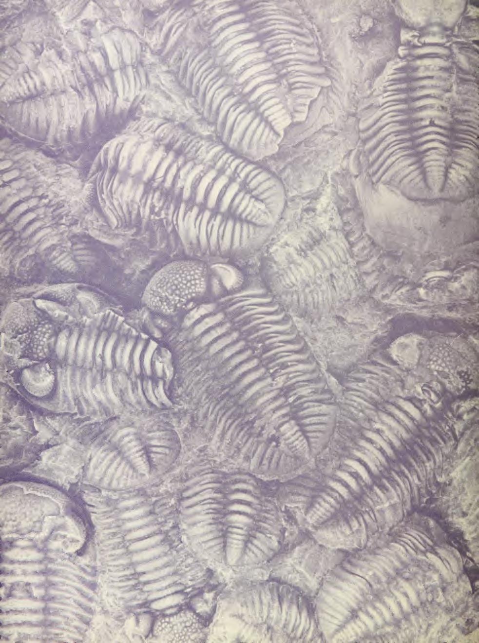

44
Mammalogy Laboratory 1 - Mammalian Anatomy
 Mammalogy Laboratory 1 - Mammalian Anatomy I. The Goal. The goal of the lab is to teach you skeletal anatomy of mammals. We will emphasize the skull because many of the taxonomically important characters
New Carnivorous Dinosaurs from the Upper Cretaceous of Mongolia
 1955 Doklady, Academy of Sciences USSR 104 (5):779-783 New Carnivorous Dinosaurs from the Upper Cretaceous of Mongolia E. A. Maleev (translated by F. J. Alcock) The present article is a summary containing
Exceptional fossil preservation demonstrates a new mode of axial skeleton elongation in early ray-finned fishes
 Supplementary Information Exceptional fossil preservation demonstrates a new mode of axial skeleton elongation in early ray-finned fishes Erin E. Maxwell, Heinz Furrer, Marcelo R. Sánchez-Villagra Supplementary
A new species of sauropod, Mamenchisaurus anyuensis sp. nov.
 A new species of sauropod, Mamenchisaurus anyuensis sp. nov. by Xinlu He, Suihua Yang, Kaiji Cai, Kui Li, and Zongwen Liu Chengdu University of Technology Papers on Geosciences Contributed to the 30th
Anatomy. Name Section. The Vertebrate Skeleton
 Name Section Anatomy The Vertebrate Skeleton Vertebrate paleontologists get most of their knowledge about past organisms from skeletal remains. Skeletons are useful for gleaning information about an organism
.56 m. (22 in.). COMPSOGNATHOID DINOSAUR FROM THE. Medicine Bow, Wyoming, by the American Museum Expedition
. COMPSOGNATHOID DINOSAUR FROM THE. Medicine Bow, Wyoming, by the American Museum Expedition") Article XII.-ORNITHOLESTES HERMANNI, A NEW COMPSOGNATHOID DINOSAUR FROM THE UPPER JURASSIC. By HENRY FAIRFIELD OSBORN. The type skeleton (Amer. Mus. Coll. No. 6I9) of this remarkable animal was discovered
Article XII.-ORNITHOLESTES HERMANNI, A NEW COMPSOGNATHOID DINOSAUR FROM THE UPPER JURASSIC. By HENRY FAIRFIELD OSBORN. The type skeleton (Amer. Mus. Coll. No. 6I9) of this remarkable animal was discovered
FURTHER STUDIES ON TWO SKELETONS OF THE BLACK RIGHT WHALE IN THE NORTH PACIFIC
 FURTHER STUDIES ON TWO SKELETONS OF THE BLACK RIGHT WHALE IN THE NORTH PACIFIC HIDEO OMURA, MASAHARU NISHIWAKI* AND TOSHIO KASUYA* ABSTRACT Two skeletons of the black right whale were studied, supplementing
complex in cusp pattern. (3) The bones of the coyote skull are thinner, crests sharper and the
 The bones of the coyote skull are thinner, crests sharper and the") DISTINCTIONS BETWEEN THE SKULLS OF S AND DOGS Grover S. Krantz Archaeological sites in the United States frequently yield the bones of coyotes and domestic dogs. These two canines are very similar both
DISTINCTIONS BETWEEN THE SKULLS OF S AND DOGS Grover S. Krantz Archaeological sites in the United States frequently yield the bones of coyotes and domestic dogs. These two canines are very similar both
SOME LITTLE-KNOWN FOSSIL LIZARDS FROM THE
 PROCEEDINGS OF THE UNITED STATES NATIONAL MUSEUM issued SWsK \ {^^m ^V ^^ SMITHSONIAN INSTITUTION U. S. NATIONAL MUSEUM Vol. 91 Washington : 1941 No. 3124 SOME LITTLE-KNOWN FOSSIL LIZARDS FROM THE OLIGOCENE
A Fossil Snake (Elaphe vulpina) From A Pliocene Ash Bed In Nebraska
 From A Pliocene Ash Bed In Nebraska") University of Nebraska - Lincoln DigitalCommons@University of Nebraska - Lincoln Transactions of the Nebraska Academy of Sciences and Affiliated Societies Nebraska Academy of Sciences 198 A Fossil Snake
University of Nebraska - Lincoln DigitalCommons@University of Nebraska - Lincoln Transactions of the Nebraska Academy of Sciences and Affiliated Societies Nebraska Academy of Sciences 198 A Fossil Snake
TRACHEMYS SCULPTA. A nearly complete articulated carapace and plastron of an Emjdd A NEAKLY COMPLETE SHELL OF THE EXTINCT TURTLE,
 A NEAKLY COMPLETE SHELL OF THE EXTINCT TURTLE, TRACHEMYS SCULPTA By Charles W. Gilmore Curator of Vertebrate Paleontology, United States National Museum INTRODUCTION A nearly complete articulated carapace
What we ve covered so far:
 What we ve covered so far: Didelphimorphia Didelphidae opossums (1 B.C. species) Soricomorpha Soricidae shrews (9 B.C. species) Talpidae moles (3 B.C. species) What s next: Rodentia Sciuridae squirrels
AMERICAN NATURALIST. Vol. IX. -DECEMBER, No. 12. OR BIRDS WITH TEETH.1 OI)ONTORNITHES,
ONTORNITHES,") AMERICAN NATURALIST. Vol. IX. -DECEMBER, 1875.-No. 12. OI)ONTORNITHES, OR BIRDS WITH TEETH.1 BY PROFESSOR 0. C. MARSH. REMAINS of birds are amono the rarest of fossils, and few have been discovered except
AMERICAN NATURALIST. Vol. IX. -DECEMBER, 1875.-No. 12. OI)ONTORNITHES, OR BIRDS WITH TEETH.1 BY PROFESSOR 0. C. MARSH. REMAINS of birds are amono the rarest of fossils, and few have been discovered except
AMERICAN MUSEUM NOVITATES Published by
 AMERICAN MUSEUM NOVITATES Published by Number 782 THE AmzRICAN MUSEUM OF NATURAL HISTORY Feb. 20, 1935 New York City 56.81, 7 G (68) A NOTE ON THE CYNODONT, GLOCHINODONTOIDES GRACILIS HAUGHTON BY LIEUWE
Fig. 5. (A) Scaling of brain vault size (width measured at the level of anterior squamosal/parietal suture) relative to skull size (measured at the
 Scaling of brain vault size (width measured at the level of anterior squamosal/parietal suture) relative to skull size (measured at the") Fig. 5. (A) Scaling of brain vault size (width measured at the level of anterior squamosal/parietal suture) relative to skull size (measured at the distance between the left versus right temporomandibular
Fig. 5. (A) Scaling of brain vault size (width measured at the level of anterior squamosal/parietal suture) relative to skull size (measured at the distance between the left versus right temporomandibular
PEABODY MUSEUM OF NATURAL HISTORY, YALE UNIVERSITY NEW HAVEN, CONNECTICUT, U.S.A. A NEW OREODONT FROM THE CABBAGE PATCH LOCAL FAUNA, WESTERN MONTANA
 Postilla PEABODY MUSEUM OF NATURAL HISTORY YALE UNIVERSITY NEW HAVEN, CONNECTICUT, U.S.A. Number 85 September 21, 1964 A NEW OREODONT FROM THE CABBAGE PATCH LOCAL FAUNA, WESTERN MONTANA STANLEY J. RIEL
Williston, and as there are many fairly good specimens in the American
 56.81.7D :14.71.5 Article VII.- SOME POINTS IN THE STRUCTURE OF THE DIADECTID SKULL. BY R. BROOM. The skull of Diadectes has been described by Cope, Case, v. Huene, and Williston, and as there are many
New York State Mammals. Order Lagomorpha Order Rodentia
 New York State Mammals Order Lagomorpha Order Rodentia FAMILY: LEPORIDAE Rabbits and hares Conspicuous tail Fenestra appears as bony latticework Some species molt seasonally Presence of a second incisor
A NEW ANKYLOSAUR FROM THE UPPER CRETACEOUS OF MONGOLIA E.A. Maleev Doklady Akademii Nauk, SSSR 87:
 translated by Dr. Tamara and F. Jeletzky, 1956 A NEW ANKYLOSAUR FROM THE UPPER CRETACEOUS OF MONGOLIA E.A. Maleev 1952. Doklady Akademii Nauk, SSSR 87:273-276 Armored dinosaurs make a considerable part
( M amenchisaurus youngi Pi, Ouyang et Ye, 1996)
") 39 4 2001 10 V ERTEBRATA PALASIATICA pp. 266 271 fig. 1,pl. I ( 643013), ( M amenchisaurus hochuanensis),,, Q915. 864 1995 12 31 (ZDM0126) ( M amenchisau rus hochuanensis Young et Chao, 1972),,, ZDM0126
39 4 2001 10 V ERTEBRATA PALASIATICA pp. 266 271 fig. 1,pl. I ( 643013), ( M amenchisaurus hochuanensis),,, Q915. 864 1995 12 31 (ZDM0126) ( M amenchisau rus hochuanensis Young et Chao, 1972),,, ZDM0126
A Partial Skeleton of Pseudaelurus (Carnivora: Felidae) from the Nambé Member of the Tesuque Formation, Española Basin, New Mexico
 from the Nambé Member of the Tesuque Formation, Española Basin, New Mexico") PUBLISHED BY THE AMERICAN MUSEUM OF NATURAL HISTORY CENTRAL PARK WEST AT 79TH STREET, NEW YORK, NY 10024 Number 3342, 31 pp., 19 figures, 1 table June 22, 2001 A Partial Skeleton of Pseudaelurus (Carnivora:
PUBLISHED BY THE AMERICAN MUSEUM OF NATURAL HISTORY CENTRAL PARK WEST AT 79TH STREET, NEW YORK, NY 10024 Number 3342, 31 pp., 19 figures, 1 table June 22, 2001 A Partial Skeleton of Pseudaelurus (Carnivora:
A new basal sauropodiform dinosaur from the Lower Jurassic of Yunnan Province, China
 SUPPLEMENTARY INFORMATION A new basal sauropodiform dinosaur from the Lower Jurassic of Yunnan Province, China Ya-Ming Wang 1, Hai-Lu You 2,3 *, Tao Wang 4 1 School of Earth Sciences and Resources, China
ZOOLOGISCHE MEDEDELINGEN
 MINISTERIE VAN ONDERWIJS, KUNSTEN EN WETENSCHAPPEN ZOOLOGISCHE MEDEDELINGEN UITGEGEVEN DOOR HET RIJKSMUSEUM VAN NATUURLIJKE HISTORIE TE LEIDEN DEEL XXXVII, No. 10 10 juli 1961 THE FOSSIL HIPPOPOTAMUS FROM
Mammalogy Lecture 8 - Evolution of Ear Ossicles
 Mammalogy Lecture 8 - Evolution of Ear Ossicles I. To begin, let s examine briefly the end point, that is, modern mammalian ears. Inner Ear The cochlea contains sensory cells for hearing and balance. -
PART FOUR: ANATOMY. Anatomy, Conformation and Movement of Dogs 41
 PART FOUR: ANATOMY Anatomy, Conformation and Movement of Dogs 41 ANATOMY The word anatomy is a scientific term that refers to the inner structure of the dog, comprising the muscles, skeleton and vital
ONLINE APPENDIX 1. Morphological phylogenetic characters scored in this paper. See Poe (2004) for
 for") ONLINE APPENDIX Morphological phylogenetic characters scored in this paper. See Poe () for detailed character descriptions, citations, and justifications for states. Note that codes are changed from a
ONLINE APPENDIX Morphological phylogenetic characters scored in this paper. See Poe () for detailed character descriptions, citations, and justifications for states. Note that codes are changed from a
Recently Mr. Lawrence M. Lambe has described and figured in the
 56.81,9C(117:71.2) Article XXXV.-CORYTHOSAURUS CASUARIUS, A NEW CRESTED DINOSAUR FROM THE BELLY RIVER CRETA- CEOUS, WITH PROVISIONAL CLASSIFICATION OF THE FAMILY TRACHODONTIDA1X BY BARNUM BROWN. PLATE
FIELDIANA GEOLOGY NEW SALAMANDERS OF THE FAMILY SIRENIDAE FROM THE CRETACEOUS OF NORTH AMERICA
 FIELDIANA GEOLOGY Published by CHICAGO NATURAL HISTORY MUSEUM Volume 10 Sbftember 22, 1968 No. 88 NEW SALAMANDERS OF THE FAMILY SIRENIDAE FROM THE CRETACEOUS OF NORTH AMERICA Coleman J. Coin AND Walter
SOME NEW AMERICAN PYCNODONT FISHES.
 SOME NEW AMERICAN PYCNODONT FISHES. By James Williams Gidley, Assistant Curator of Fossil Mammals, United States National Museum. In the United States National Museum are several specimens representing
Postilla PEABODY MUSEUM OF NATURAL HISTORY YALE UNIVERSITY NEW HAVEN, CONNECTICUT, U.S.A.
 Postilla PEABODY MUSEUM OF NATURAL HISTORY YALE UNIVERSITY NEW HAVEN, CONNECTICUT, U.S.A. Number 117 18 March 1968 A 7DIAPSID (REPTILIA) PARIETAL FROM THE LOWER PERMIAN OF OKLAHOMA ROBERT L. CARROLL REDPATH
LEIDY, SHOWING THE BONES OF THE FEET 'AND LIMBS
 CQNTEUBUTIONS FBOM THE MUSEUM OF PALEONTOLOGY (Confindion of Con&&&m froin UB Muaercm of Gcologg) UNIVERSITY OF ' MICHIGAN VOL V, No. 6, pp. 6W3 (e ph.) DEAXMBER 31,1036 A SPECIMEN OF STYLEMYS NEBRASCENSIS
290 SHUFELDT, Remains of Hesperornis.
 290 SHUFELDT, Remains of Hesperornis. [ Auk [July THE FOSSIL REMAINS OF A SPECIES OF HESPERORNIS FOUND IN MONTANA. BY R. W. SHUFELD% M.D. Plate XI7III. ExR,¾ in November, 1914, Mr. Charles W. Gihnore,
A skull without mandihle, from the Hunterian Collection (no.
 4 MR. G. A. BOULENGER ON CHELONIAN REMAINS. [Jan. 6, 2. On some Chelonian Remains preserved in the Museum of the Eojal College of Surgeons. By G. A. Boulenger. [Eeceived December 8, 1890.] In the course
Vol. XIV, No. 1, March, The Larva and Pupa of Brontispa namorikia Maulik (Coleoptera: Chrysomelidae: Hispinae) By S.
 By S.") Vol. XIV, No. 1, March, 1950 167 The Larva and Pupa of Brontispa namorikia Maulik (Coleoptera: Chrysomelidae: Hispinae) By S. MAULIK BRITISH MUSEUM (NATURAL HISTORY) (Presented by Mr. Van Zwaluwenburg
Vol. XIV, No. 1, March, 1950 167 The Larva and Pupa of Brontispa namorikia Maulik (Coleoptera: Chrysomelidae: Hispinae) By S. MAULIK BRITISH MUSEUM (NATURAL HISTORY) (Presented by Mr. Van Zwaluwenburg
Skulls & Evolution. 14,000 ya cro-magnon. 300,000 ya Homo sapiens. 2 Ma Homo habilis A. boisei A. robustus A. africanus
 Skulls & Evolution Purpose To illustrate trends in the evolution of humans. To demonstrate what you can learn from bones & fossils. To show the adaptations of various mammals to different habitats and
On the Discovery of the earliest fossil bird in China (Sinosauropteryx gen. nov.) and the origin of birds
 and the origin of birds") On the Discovery of the earliest fossil bird in China (Sinosauropteryx gen. nov.) and the origin of birds by Qiang Ji and Shu an Ji Chinese Geological Museum, Beijing Chinese Geology Volume 233 1996 pp.
On the Discovery of the earliest fossil bird in China (Sinosauropteryx gen. nov.) and the origin of birds by Qiang Ji and Shu an Ji Chinese Geological Museum, Beijing Chinese Geology Volume 233 1996 pp.
Erycine Boids from the Early Oligocene of the South Dakota Badlands
 Georgia Journal of Science Volume 67 No. 2 Scholarly Contributions from the Membership and Others Article 6 2009 Erycine Boids from the Early Oligocene of the South Dakota Badlands Dennis Parmley J. Alan
1/9/2013. Divisions of the Skeleton: Topic 8: Appendicular Skeleton. Appendicular Components. Appendicular Components
 /9/203 Topic 8: Appendicular Skeleton Divisions of the Skeleton: Cranial Postcranial What makes up the appendicular skeleton? What is the pattern of serial homology of the limbs? Tetrapod front limb morphology
CENE RUMINANTS OF THE GENERA OVIBOS AND
 DESCRIPTIONS OF TWO NEW SPECIES OF PLEISTO- CENE RUMINANTS OF THE GENERA OVIBOS AND BOOTHERIUM, WITH NOTES ON THE LATTER GENUS. By James Williams Gidley, Of the United States National Museum. Two interesting
A NEW GENUS AND SPECIES OF AMERICAN THEROMORPHA
 A NEW GENUS AND SPECIES OF AMERICAN THEROMORPHA MYCTEROSAURUS LONGICEPS S. W. WILLISTON University of Chicago The past summer, Mr. Herman Douthitt, of the University of Chicago paleontological expedition,
Mammalogy Laboratory 4 - Rodents I: Hystricomorpha, Sciuromorpha
 Mammalogy Laboratory 4 - Rodents I: Hystricomorpha, Sciuromorpha General Comments: We ll spend two weeks on the order Rodentia. This may seem like a lot of time, but almost half (~2552 species) of all
A NEW SPECIES OF EXTINCT TURTLE FROM THE UPPER PLIOCENE OF IDAHO
 A NEW SPECIES OF EXTINCT TURTLE FROM THE UPPER PLIOCENE OF IDAHO By Charles W. Gilmore Curator, Division of Vertebrate Paleontology United States National Museum Among the fossils obtained bj^ the Smithsonian
v:ii-ixi, 'i':;iisimvi'\>!i-:: "^ A%'''''-'^-''S.''v.--..V^'E^'-'-^"-t''gi L I E) R.ARY OF THE VERSITY U N I or ILLINOIS REMO
 R.ARY OF THE VERSITY U N I or ILLINOIS REMO") "^ A%'''''-'^-''S.''v.--..V^'E^'-'-^"-t''gi v:ii-ixi, 'i':;iisimvi'\>!i-:: L I E) R.ARY OF THE U N I VERSITY or ILLINOIS REMO Natural History Survey Librarv GEOLOGICAL SERIES OF FIELD MUSEUM OF NATURAL
"^ A%'''''-'^-''S.''v.--..V^'E^'-'-^"-t''gi v:ii-ixi, 'i':;iisimvi'\>!i-:: L I E) R.ARY OF THE U N I VERSITY or ILLINOIS REMO Natural History Survey Librarv GEOLOGICAL SERIES OF FIELD MUSEUM OF NATURAL
Biology 340 Comparative Embryology Lecture 12 Dr. Stuart Sumida. Evo-Devo Revisited. Development of the Tetrapod Limb
 Biology 340 Comparative Embryology Lecture 12 Dr. Stuart Sumida Evo-Devo Revisited Development of the Tetrapod Limb Limbs whether fins or arms/legs for only in particular regions or LIMB FIELDS. Primitively
Mammalogy Lab 1: Skull, Teeth, and Terms
 Mammalogy Lab 1: Skull, Teeth, and Terms Be able to: Goals of today s lab Locate all structures listed on handout Define all terms on handout what they are or what they look like Give examples of mammals
Reprinted from: CRUSTACEANA, Vol. 32, Part 2, 1977 LEIDEN E. J. BRILL
 Reprinted from: CRUSTACEANA, Vol. 32, Part 2, 1977 LEIDEN E. J. BRILL NOTES AND NEWS 207 ALPHE0PS1S SHEARMII (ALCOCK & ANDERSON): A NEW COMBINATION WITH A REDESCRIPTION OF THE HOLOTYPE (DECAPODA, ALPHEIDAE)
UN? RSITYOF. ILLIiwiS LIBRARY AT URBANA-CHAMPAIGN NATURAL HIST. SURVEY
 UN? RSITYOF ILLIiwiS LIBRARY AT URBANA-CHAMPAIGN NATURAL HIST. SURVEY FIELDIANA GEOLOGY Published by CHICAGO NATURAL HISTORY MUSEUM Volume 10 July 29, 1954 No. 17 FAUNA OF THE VALE AND CHOZA: 7 PELYCOSAURIA:
LOWER CRETACEOUS OF SOUTH DAKOTA.
 A NEW DINOSAUR, STP^GOSAURUS MARSHl, FROM THE LOWER CRETACEOUS OF SOUTH DAKOTA. By Frederic A. Lucas, Curator, Divisioii of Coiiipnrative Anatomy, in charge, of Section of Vertebrate Fossils. The name
OSTEOLOGICAL NOTE OF AN ANTARCTIC SEI WHALE
 OSTEOLOGICAL NOTE OF AN ANTARCTIC SEI WHALE MASAHARU NISHIWAKI* AND TOSHIO KASUYA* ABSTRACT This is a report of measurements on the skeleton of a male se1 whale caught in the Antarctic. The skeleton of
LATARJET Open Surgical technique
 1 LATARJET Open Surgical technique Steps A. Exposure B. Preparation of coracoid holes C. Cutting the coracoid D. Fixing the Double Cannula to the coracoid E. Exposure of both sides of Subscapularis F.
YALE PEABODY MUSEUM JOSEPH T. GREGORY AND THEODORE DOWNS INTRODUCTION
 YALE PEABODY MUSEUM OF NATURAL HISTORY Number 8 May 10, 1951 New Haven, Conn. BASSARISCUS IN MIOCENE FAUNAS AND "POTAMOTHERIUM LYCOPOTAMICUM COPE" JOSEPH T. GREGORY AND THEODORE DOWNS INTRODUCTION Cope
Animal Form and Function. Amphibians. United by several distinguishing apomorphies within the Vertebrata
 Animal Form and Function Kight Amphibians Class Amphibia (amphibia = living a double life) United by several distinguishing apomorphies within the Vertebrata 1. Skin Thought Question: For whom are integumentary
1) Explain why the skeleton plays an important role in the overall shape of animal and human being.
 Explain why the skeleton plays an important role in the overall shape of animal and human being.") 1) Explain why the skeleton plays an important role in the overall shape of animal and human being. 2) Substantiate the differences in animal and human skeleton, with the human skeleton built in such a
1) Explain why the skeleton plays an important role in the overall shape of animal and human being. 2) Substantiate the differences in animal and human skeleton, with the human skeleton built in such a
A Short Report on the Occurrence of Dilophosaurus from Jinning County, Yunnan Province
 A Short Report on the Occurrence of Dilophosaurus from Jinning County, Yunnan Province by Hu Shaojin (Kunming Cultural Administrative Committee, Yunnan Province) Vertebrata PalAsiatica Vol. XXXI, No. 1
Title: Phylogenetic Methods and Vertebrate Phylogeny
 Title: Phylogenetic Methods and Vertebrate Phylogeny Central Question: How can evolutionary relationships be determined objectively? Sub-questions: 1. What affect does the selection of the outgroup have
Lab 5: Rodentia and Lagomorpha
 Lab 5: Rodentia and Lagomorpha (8 families in B.C.) Sciuridae squirrels (16 species in B.C.) Muridae mice, rats, lemmings, voles (16) Aplodontidae mountain beaver (1) Castoridae beaver (1) Dipodidae jumping
THE GORGONOPSIAN GENUS, HIPPOSAURUS, AND THE FAMILY ICTIDORHINIDAE * Dr. L.D. Boonstra. Paleontologist, South African Museum, Cape Town
 THE GORGONOPSIAN GENUS, HIPPOSAURUS, AND THE FAMILY ICTIDORHINIDAE * by Dr. L.D. Boonstra Paleontologist, South African Museum, Cape Town In 1928 I dug up the complete skeleton of a smallish gorgonopsian
A NEARLY COMPLETE TURTLE SKELETON FROM THE UPPER CRETACEOUS OF MONTANA
 CONTRIBUTIONS PBOM THE MUSEUM OF PALEONTOLOGY UNIVERSITY OF MICHIGAN VOL VI, No. 1. pp. 1-19 (18 figs.) D~c~arrrm 1, 1989 A NEARLY COMPLETE TURTLE SKELETON FROM THE UPPER CRETACEOUS OF MONTANA BY E. C.
Lab 2 Skeletons and Locomotion
 Lab 2 Skeletons and Locomotion Objectives The objectives of this and next week's labs are to introduce you to the comparative skeletal anatomy of vertebrates. As you examine the skeleton of each lineage,
Diurus, Pascoe. sp. 1). declivity of the elytra, but distinguished. Length (the rostrum and tails 26 included) mm. Deep. exception
. declivity of the elytra, but distinguished. Length (the rostrum and tails 26 included) mm. Deep. exception") 210 DIURUS ERYTIIROPUS. NOTE XXVI. Three new species of the Brenthid genus Diurus, Pascoe DESCRIBED BY C. Ritsema+Cz. 1. Diurus erythropus, n. sp. 1). Allied to D. furcillatus Gylh. ²) by the short head,
210 DIURUS ERYTIIROPUS. NOTE XXVI. Three new species of the Brenthid genus Diurus, Pascoe DESCRIBED BY C. Ritsema+Cz. 1. Diurus erythropus, n. sp. 1). Allied to D. furcillatus Gylh. ²) by the short head,
UNIVERSITY OF MICHIGAN
 CONTRIBUTIONS FROM THE MUSEUM OF GEOLOGY UNIVERSITY OF MICHIGAN THE VERTEBRAL COLUMN OF COELOPHYSIS COPE BY E. C. CASE UNIVERSITY OF MICHIGAN ANN ARBOR 4 Pi Spectra ABCDEFGHIJKLM~~OPORSTUWXYZ~~~~~~~~~~~~~~OP~~~~~~Y~
First Ornithomimid (Theropoda, Ornithomimosauria) from the Upper Cretaceous Djadokhta Formation of Tögrögiin Shiree, Mongolia
 from the Upper Cretaceous Djadokhta Formation of Tögrögiin Shiree, Mongolia") First Ornithomimid (Theropoda, Ornithomimosauria) from the Upper Cretaceous Djadokhta Formation of Tögrögiin Shiree, Mongolia Tsogtbaatar Chinzorig¹, ³ *, Yoshitsugu Kobayashi², Khishigjav Tsogtbaatar³,
First Ornithomimid (Theropoda, Ornithomimosauria) from the Upper Cretaceous Djadokhta Formation of Tögrögiin Shiree, Mongolia Tsogtbaatar Chinzorig¹, ³ *, Yoshitsugu Kobayashi², Khishigjav Tsogtbaatar³,
SCIUROPTERUS MINDANENSIS SP. NOV., A NEW SPECIES OF FLYING SQUIRREL FROM MINDANAO
 SCIUROPTERUS MINDANENSIS SP. NOV., A NEW SPECIES OF FLYING SQUIRREL FROM MINDANAO By DioscoRO S. Rabor Of the Division of Fisheries^ Department of Agriculture and Commerce Manila FOUR PLATES In August,
Redpalh Museum, McGill University, Montreal, P.Q, Canada, HJA 2K6.
 143 Palaeont. afr., 21, 143-159 (1978) PERMO-TRIASSIC "LIZARDS" FROM THE KAROO SYSTEM PART II A GLIDING REPTILE FROM THE UPPER PERMIAN OF MADAGASCAR by Robert L. Carroll Redpalh Museum, McGill University,
J/ieuican JfiLsllm. The Genus Proterix (Insectivora, Erinaceidae) of the Upper Oligocene of North America BY CONSTANCE ELAINE GAWNE1 INTRODUCTION
 of the Upper Oligocene of North America BY CONSTANCE ELAINE GAWNE1 INTRODUCTION") A J/ieuican JfiLsllm PUBLISHED BY THE AMERICAN MUSEUM OF NATURAL HISTORY CENTRAL PARK WEST AT 79TH STREET, NEW YORK, N. Y. I0024 NUMBER 2 3 I 5 FEBRUARY 28, I968 The Genus Proterix (Insectivora, Erinaceidae)
A J/ieuican JfiLsllm PUBLISHED BY THE AMERICAN MUSEUM OF NATURAL HISTORY CENTRAL PARK WEST AT 79TH STREET, NEW YORK, N. Y. I0024 NUMBER 2 3 I 5 FEBRUARY 28, I968 The Genus Proterix (Insectivora, Erinaceidae)
The family Gnaphosidae is a large family
 Pakistan J. Zool., vol. 36(4), pp. 307-312, 2004. New Species of Zelotus Spider (Araneae: Gnaphosidae) from Pakistan ABIDA BUTT AND M.A. BEG Department of Zoology, University of Agriculture, Faisalabad,
2. Skull, total length versus length of the presacral vertebral column: (0); extremely elongated neck (e.g. Tanystropheus longobardicus).
; extremely elongated neck (e.g. Tanystropheus longobardicus).") Character list of the taxon-character data set 1. Skull and lower jaws, interdental plates: absent (0); present, but restricted to the anterior end of the dentary (1); present along the entire alveolar
Character list of the taxon-character data set 1. Skull and lower jaws, interdental plates: absent (0); present, but restricted to the anterior end of the dentary (1); present along the entire alveolar
UPOGEBIA LINCOLNI SP. NOV. (DECAPODA, THALASSINIDEA, UPOGEBIIDAE) FROM JAVA, INDONESIA
 FROM JAVA, INDONESIA") NOTES AND NEWS UPOGEBIA LINCOLNI SP. NOV. (DECAPODA, THALASSINIDEA, UPOGEBIIDAE) FROM JAVA, INDONESIA BY NGUYEN NGOC-HO i) Faculty of Science, University of Saigon, Vietnam Among material recently collected
NOTES AND NEWS UPOGEBIA LINCOLNI SP. NOV. (DECAPODA, THALASSINIDEA, UPOGEBIIDAE) FROM JAVA, INDONESIA BY NGUYEN NGOC-HO i) Faculty of Science, University of Saigon, Vietnam Among material recently collected
The Discovery of a Tritylodont from the Xinjiang Autonomous Region
 The Discovery of a Tritylodont from the Xinjiang Autonomous Region Ailing Sun and Guihai Cui (Institute of Vertebrate Paleontology, Paleoanthropology, Academia Sinica) Vertebrata PalAsiatica Volume XXVII,
ON THE FPERYLOSIS OF THE BLACK-THROATED DIVER.
 ON THE FPERYLOSIS OF THE BLACK-THROATED DIVER. BY W. P. PYCRAFT. IT is surely a matter for regret that so little interest has been taken in that side of ornithology which concerns structural characters,
YANGCHUANOSAURUS HEPINGENSIS - A NEW SPECIES OF CARNOSAUR FROM ZIGONG, SICHUAN
 Vol. 30, No. 4 VERTEBRATA PALASIATICA pp. 313-324 October 1992 [SICHUAN ZIGONG ROUSHILONG YI XIN ZHONG] figs. 1-5, pl. I-III YANGCHUANOSAURUS HEPINGENSIS - A NEW SPECIES OF CARNOSAUR FROM ZIGONG, SICHUAN
AMERICAN MUSEUM NOVITATES
 AMERICAN MUSEUM NOVITATES Number 1103 Published by THE AMERICAN MUSEUM OF NATURAL HISTORY New York City THE PES OF BAURIA CYNOPS BROOM BY BOBB SCHAEFFER1 In several respects the bauriamorphs are farther
KATE E. ZEIGLER, ANDREW B. HECKERT and SPENCER G. LUCAS. New Mexico Museum of Natural History, 1801 Mountain Road NW, Albuquerque, NM
 Zeigler, K.E., Heckert, A.B., and Lucas, S.G., eds., 2003, Paleontology and Geology of the Snyder Quarry, New Mexico Museum of Natural History and Science Bulletin No. 24. AN ILLUSTRATED ATLAS OF THE PHYTOSAUR
Temporal lines. More forwardfacing. tubular orbits than in the African forms 3. Orbits larger relative to skull size than in the other genera 2.
 Asian lorises More forwardfacing and tubular orbits than in the African forms 3. Characterized by a marked extension of the ectotympanic into a tubular meatus and a more angular auditory bulla than in
A NEW SALTICID SPIDER FROM VICTORIA By R. A. Dunn
 Dunn, R. A. 1947. A new salticid spider from Victoria. Memoirs of the National Museum of Victoria 15: 82 85. All text not included in the original document is highlighted in red. Mem. Nat. Mus. Vict.,
Chapter 2 Mammalian Origins. Fig. 2-2 Temporal Openings in the Amniotes
 Chapter 2 Mammalian Origins Fig. 2-2 Temporal Openings in the Amniotes 1 Synapsida 1. monophyletic group 2. Single temporal opening below postorbital and squamosal 3. Dominant terrestrial vertebrate group
SUPPLEMENTARY INFORMATION
 doi:10.1038/nature13086 Part I. Supplementary Notes A: Detailed Description of Cotylocara macei gen. et sp. nov. Part II. Table of Measurements for holotype of Cotylocara macei (CCNHM-101) Part III. Supplementary
Frog Dissection Information Manuel
 Frog Dissection Information Manuel Anatomical Terms: Used to explain directions and orientation of a organism Directions or Positions: Anterior (cranial)- toward the head Posterior (caudal)- towards the
NIVOROUS DINOSAUR. (SECOND COMMUNICATION.) By HENRY FAIRFIELD OSBORN. PLATE XXXIX. This great carnivorous Dinosaur of the Laramie was contemporary
 By HENRY FAIRFIELD OSBORN. PLATE XXXIX. This great carnivorous Dinosaur of the Laramie was contemporary") 56, 8i, 9 T (I 7: 786) Article VI.-TYRANNOSAURUS, UPPER CRETACEOUS CAR- NIVOROUS DINOSAUR. (SECOND COMMUNICATION.) By HENRY FAIRFIELD OSBORN. PLATE I. This great carnivorous Dinosaur of the Laramie was
56, 8i, 9 T (I 7: 786) Article VI.-TYRANNOSAURUS, UPPER CRETACEOUS CAR- NIVOROUS DINOSAUR. (SECOND COMMUNICATION.) By HENRY FAIRFIELD OSBORN. PLATE I. This great carnivorous Dinosaur of the Laramie was
A new Middle Jurassic sauropod subfamily (Klamelisaurinae subfam. nov.) from Xinjiang Autonomous Region, China
 from Xinjiang Autonomous Region, China") A new Middle Jurassic sauropod subfamily (Klamelisaurinae subfam. nov.) from Xinjiang Autonomous Region, China by Xijing Zhao Institute of Vertebrate Paleontology and Paleoanthropology, Academia Sinica
A new Middle Jurassic sauropod subfamily (Klamelisaurinae subfam. nov.) from Xinjiang Autonomous Region, China by Xijing Zhao Institute of Vertebrate Paleontology and Paleoanthropology, Academia Sinica
The Animal Bones from Excavations in Meshoko Cave in the Northern Caucasus
 The Animal Bones from Excavations in Meshoko Cave in the Northern Caucasus Ellen Hambleton and Mark Maltby Department of Archaeology, Anthropology and Forensic Sciences Bournemouth University, UK 1 Contents
BREVIORA LEUCOLEPIDOPA SUNDA GEN. NOV., SP. NOV. (DECAPODA: ALBUNEIDAE), A NEW INDO-PACIFIC SAND CRAB. Ian E. Efford 1
, A NEW INDO-PACIFIC SAND CRAB. Ian E. Efford 1") ac lc BREVIORA CAMBRIDGE, MASS. 30 APRIL, 1969 NUMBER 318 LEUCOLEPIDOPA SUNDA GEN. NOV., SP. NOV. (DECAPODA: ALBUNEIDAE), A NEW INDO-PACIFIC SAND CRAB Ian E. Efford 1 ABSTRACT. Leucolepidopa gen. nov.
ac lc BREVIORA CAMBRIDGE, MASS. 30 APRIL, 1969 NUMBER 318 LEUCOLEPIDOPA SUNDA GEN. NOV., SP. NOV. (DECAPODA: ALBUNEIDAE), A NEW INDO-PACIFIC SAND CRAB Ian E. Efford 1 ABSTRACT. Leucolepidopa gen. nov.
SUPPLEMENTARY INFORMATION
 Character 155, interdental ridges. Absence of interdental ridge (0) shown in Parasaniwa wyomingensis (Platynota). Interdental ridges (1) shown in Coniophis precedens. WWW.NATURE.COM/NATURE 1 Character
Man s Best Friend? Using Animal Bones to Solve an Archaeological Mystery*
 Man s Best Friend? Using Animal Bones to Solve an Archaeological Mystery* by Elizabeth A. Scharf Department of Anthropology University of North Dakota Part I Too Good To Be True? May 28, 2018 As a specialist
AUSTRALIAN MUSEUM SCIENTIFIC PUBLICATIONS
 AUSTRALIAN MUSEUM SCIENTIFIC PUBLICATIONS Riek, E. F., 1964. Merostomoidea (Arthropoda, Trilobitomorpha) from the Australian Middle Triassic. Records of the Australian Museum 26(13): 327 332, plate 35.
A new carnosaur from Yongchuan County, Sichuan Province
 A new carnosaur from Yongchuan County, Sichuan Province by Dong Zhiming Institute of Vertebrate Palaeontology and Palaeoanthropology, Academia Sinica Zhang Yihong, Li Xuanmin, and Zhou Shiwu Chongqing
SUPPLEMENTARY ONLINE MATERIAL FOR. Nirina O. Ratsimbaholison, Ryan N. Felice, and Patrick M. O connor
 http://app.pan.pl/som/app61-ratsimbaholison_etal_som.pdf SUPPLEMENTARY ONLINE MATERIAL FOR Nirina O. Ratsimbaholison, Ryan N. Felice, and Patrick M. O connor Ontogenetic changes in the craniomandibular
SHEEPMEAT. Goatmeat primal preparation are the same specification and codes as Sheepmeat
 SHEEPMEAT Goatmeat primal preparation are the same specification and codes as Sheepmeat Item No. Page No. BONE-IN SHEEPMEAT Assorted Cuts... 5036...102 Breast and Flap... 5010...95 Breast and Flap Pieces...
Jurassic Ornithopod Agilisaurus louderbacki (Ornithopoda: Fabrosauridae) from Zigong, Sichuan, China
 from Zigong, Sichuan, China") Jurassic Ornithopod Agilisaurus louderbacki (Ornithopoda: Fabrosauridae) from Zigong, Sichuan, China Guangzhao Peng (Zigong Dinosaur Museum) Vertebrata PalAsiatica Volume 30, No. 1 January, 1992 pp. 39-51
Jurassic Ornithopod Agilisaurus louderbacki (Ornithopoda: Fabrosauridae) from Zigong, Sichuan, China Guangzhao Peng (Zigong Dinosaur Museum) Vertebrata PalAsiatica Volume 30, No. 1 January, 1992 pp. 39-51
Biology 3315 Comparative Vertebrate Morphology Skulls and Visceral Skeletons
 Biology 3315 Comparative Vertebrate Morphology Skulls and Visceral Skeletons 1. Head skeleton of lamprey Cyclostomes are highly specialized in both the construction of the chondrocranium and visceral skeleton.
NAUSHONIA PAN AMEN SIS, NEW SPECIES (DECAPODA: THALASSINIDEA: LAOMEDIIDAE) FROM THE PACIFIC COAST OF PANAMA, WITH NOTES ON THE GENUS
 FROM THE PACIFIC COAST OF PANAMA, WITH NOTES ON THE GENUS") 5 October 1982 PROC. BIOL. SOC. WASH. 95(3), 1982, pp. 478-483 NAUSHONIA PAN AMEN SIS, NEW SPECIES (DECAPODA: THALASSINIDEA: LAOMEDIIDAE) FROM THE PACIFIC COAST OF PANAMA, WITH NOTES ON THE GENUS Joel
5 October 1982 PROC. BIOL. SOC. WASH. 95(3), 1982, pp. 478-483 NAUSHONIA PAN AMEN SIS, NEW SPECIES (DECAPODA: THALASSINIDEA: LAOMEDIIDAE) FROM THE PACIFIC COAST OF PANAMA, WITH NOTES ON THE GENUS Joel
FSS OPEN SHOW PROCEDURAL EXAM
 Judging Operations Department PO Box 900062 Raleigh, NC 27675-9062 (919) 816-3570 judgingops@akc.org www.akc.org Revised Sept 2013 FSS OPEN SHOW PROCEDURAL EXAM Refer to Rules, Policies and Guidelines
Comments on the Beauceron Standard By M. Maurice Hermel (Translated by C. Batson)
") Comments on the Beauceron Standard By M. Maurice Hermel (Translated by C. Batson) The following are comments written by M. Hermel for the FCI Standard #44 published on 10/25/06. They were approved by the
Comments on the Beauceron Standard By M. Maurice Hermel (Translated by C. Batson) The following are comments written by M. Hermel for the FCI Standard #44 published on 10/25/06. They were approved by the
THE SKULLS OF ARAEOSCELIS AND CASEA, PERMIAN REPTILES
 THE SKULLS OF REOSCELIS ND CSE, PERMIN REPTILES University of Chicago There are few Permian reptiles of greater interest at the present time than the peculiar one I briefly described in this journal' three
By HENRY FAIRFIELD OSBORN.
 Article XI.-FORE AND HINI) LIMBS OF CARNIVOR- OUS AND HERBIVOROUS DINOSAURS FROM THE JURASSIC OF WYOMING. DINOSAUR CONTRIBU- TIONS, NO. 3. By HENRY FAIRFIELD OSBORN. In the Bone Cabin Quarry, opened by
Fish 475: Marine Mammalogy
 Fish 475: Marine Mammalogy Taxonomy (continued) Friday, 3 April 2009 Amanda Bradford Course website: http://faculty.washington.edu/glennvb/fish475 Mysticeti: The baleen whales About 10-12 species; Formerly
Lab 8 Order Carnivora: Families Canidae, Felidae, and Ursidae Need to know Terms: carnassials, digitigrade, reproductive suppression, Jacobson s organ
 Lab 8 Order Carnivora: Families Canidae, Felidae, and Ursidae Need to know Terms: carnassials, digitigrade, reproductive suppression, Jacobson s organ Family Canidae Canis latrans ID based on skull, photos,
Mammalogy Lecture 3 - Early Mammals/Monotremes
 Mammalogy Lecture 3 - Early Mammals/Monotremes I. Early mammals - These groups are known as Mesozoic mammals, and there are several groups. Again, there have been lots of new groups discovered, and we
Neapolitan Mastiff. EXPRESSION Wistful at rest, intimidating when alert. Penetrating stare.
 Neapolitan Mastiff GENERAL APPEARANCE He is characterized by loose skin, over his entire body, abundant, hanging wrinkles and folds on the head and a voluminous dewlap. The essence of the Neapolitan is
Lagotto Romagnolo. Size The length of the head reaches 1/10 of the height at the withers. The dog is nearly as high a long.
 LISTED BREED-GROUP I SPORTING DOGS IL-3 Lagotto Romagnolo Origin & Purpose Ancient breed of water retrieving dogs in the lowlands of Comacchio and marshlands of Ravenna. During the centuries, the great