3. Structure of the brain cavity and inner ear of the centrosaurine ceratopsid dinosaur Pachyrhinosaurus based on CT scanning and 3D visualization
|
|
|
- Kelly Strickland
- 5 years ago
- Views:
Transcription
1 3. Structure of the brain cavity and inner ear of the centrosaurine ceratopsid dinosaur Pachyrhinosaurus based on CT scanning and 3D visualization LAWRENCE M. WITMER AND RYAN C. RIDGELY Abstract Information on the structure of the brain, cranial nerves, encephalic vasculature, and inner ear of the centrosaurine ceratopsid dinosaur Pachyrhinosaurus lakustai (Upper Cretaceous, Wapiti Formation, Alberta, Canada) is presented based on computed tomographic scanning of an isolated braincase followed by 3D visualization. The resulting digital cranial endocast and endosseous labyrinth are the most complete for any ceratopsid, to date, and are compared with physical and digital endocasts of other ceratopsians. In general, the structure of the brain of P. lakustai, as inferred from the endocast, was relatively primitive and much more like extant nonavian diapsid endocasts than like endocasts of archosaur groups with derived brains, such as pterosaurs, hadrosaurs, or coelurosaurs (including extant birds). Total brain size is relatively small, and none of the externally discernable brain regions (e.g., olfactory bulbs, cerebrum, cerebellum, optic lobes) are expanded. Currently, there are insufficient data to determine whether the simple structure and small size of the brain of P. lakustai are truly plesiomorphic or represent apomorphic reduction (evolutionary Note: An animation of the 3D visualization of the digital endocast is available on NRC Research Press Web site at books/books/ html. 117
2 reversal). The endosseous labyrinth reveals a short cochlea, suggesting that airborne sounds were not particularly important behaviorally. The semicircular canals of the labyrinth are actually somewhat more elongate compared with what little is known of other neoceratopsian labyrinths. The fossil specimen is slightly compressed transversely, and this plastic deformation was subsequently ameliorated by a simple digital retrodeformation that provides a realistic view of brain size and shape. The most significant biological result of the project is that, even accounting for any deformation, the sensorineural and cognitive capabilities of Pachyrhinosaurus were modest, certainly in comparison with some other dinosaur clades, such as hadrosaurids and coelurosaurian theropods. Key words: Dinosauria, Ceratopsia, Pachyrhinosaurus, brain, behavior, endocast, CT scanning. 118 Chapter 3: Witmer and Ridgley Résumé Cette étude présente des données sur la structure du cerveau, des nerfs crâniens, du système vasculaire crânien et de l oreille interne du dinosaure Pachyrhinosaurus lakustai (centrosaure cératopsien Crétacé supérieur, Wapiti Formation, Alberta, Canada), obtenues par tomodensitométrie d une boîte crânienne. Une analyse par visualisation en trois dimensions est également offerte. Les contre-empreintes (moulage) numériques du crâne et du labyrinthe endo-osseux de ce cératopsien, qui sont les plus détaillées à ce jour, sont comparées aux données physiques et numériques d autres cératopsiens. En général, la structure du cerveau de P. lakustai est relativement primitive et s apparente davantage aux contre-empreintes des reptiles diapsides existants plutôt qu à celles du cerveau dérivé d archosauriens (ptérosaures, hadrosaures, coelurosaures, y compris les oiseaux actuels). Le cerveau est relativement petit et aucune de ses régions externes (bulbes olfactifs, cerveau, cervelet, lobes optiques) n est élargie. Par ailleurs, les données sont actuellement insuffisantes pour établir si la structure simplifiée et la petite taille du cerveau de P. lakustai sont véritablement plésiomorphiques ou représentent plutôt une réduction apomorphique (renversement évolutif). L examen du labyrinthe endo-osseux révèle une cochlée réduite, ce qui suggère que les sons aériens jouaient un rôle accessoire sur le plan comportemental. Les canaux semi-circulaires du labyrinthe sont en fait relativement plus allongés par rapport aux autres labyrinthes néocératopsiens, bien qu encore peu connus. Le spécimen fossile est légèrement comprimé dans l axe transversal, et cette déformation plastique a été subséquemment améliorée par une simple rétrodéformation numérique offrant ainsi une image réaliste de la taille et de la forme du cerveau. Sur le plan biologique, le résultat le plus important du projet est que, même en tenant compte de toute déformation éventuelle, les capacités cognitives et neurosensorielles de Pachyrhinosaurus étaient rudimentaires, surtout par rapport à d autres clades de dinosaures comme les hadrosauridés et les thérapodes coelurosauriens.
3 Mots-clés : Dinosauria, Ceratopsia, Pachyrhinosaurus, cerveau, comportement, contre-empreinte, moulage, tomographie par ordinateur. [Traduit par la Rédaction] Introduction Computed tomographic (CT) scanning has revolutionized the study of brain evolution in extinct archosaurs, such as dinosaurs, its X rays digitally slicing through bone and stone to allow access to the endocranial cavity. CT has provided new insights into the brain structure of pterosaurs (Witmer et al. 2003), ankylosaurians (Witmer and Ridgely 2008), sauropods (Sereno et al. 2007; Witmer et al. 2008), and theropods (Rogers 1998, 1999, 2005; Brochu 2000, 2003; Larsson 2001; Franzosa 2004; Franzosa and Rowe 2005; Sampson and Witmer 2007; Kundrát 2007), including birds (Domínguez Alonso et al. 2004; Kurochkin et al. 2006, 2007). With the exception of a CT-based study of the basal ceratopsian Psittacosaurus (Zhou et al. 2007), the study of the brain structure of horned dinosaurs has not made the jump into the digital age, probably in part because the braincase tends to be attached to skulls that are simply too large to fit through most CT scanners. Here we report on the first CT-based study of ceratopsid endocranial structure, producing the most complete view to date for any ceratopsian. The disarticulated braincase of the centrosaurine Pachyrhinosaurus lakustai, derived from the Pipestone Creek bone bed (Grande Prairie, Alberta; see Currie et al. 2008), forms the basis of this study. Some previous studies of ceratopsid endocranial structure have drawn on observations of broken specimens, whereas others have made latex or plaster casts of the endocranial surface from specimens from which the rock matrix had been removed (known as brain casts or cranial endocasts). The endocranial morphology of only a few neoceratopsian taxa has been reported in the literature (see Hopson (1979) for an excellent review): Protoceratops (Brown and Schlaikjer 1940); the chasmosaurine ceratopsids Anchiceratops (Brown 1914) and Triceratops (Burckhardt 1892; Marsh 1896; Hay 1909; Gilmore 1919; Forster 1996); and of significance for this paper, the centrosaurine ceratopsid Pachyrhinosaurus canadensis (Langston 1975). Of these reports, only Marsh (1896), Brown (1914), Brown and Schlaikjer (1940), and Forster (1996) provided data based on cranial endocasts. Thus, the findings presented here will be not only the first CT-based study but also the first presentation of a centrosaurine endocast and inner ear (Figs. 1 3). Beyond simply affording a look inside, CT scanning provides a number of advantages for studying the endocranial region. For example, because CT scanning transfers the specimen into the digital realm, it provides new opportunities for visualization and analysis. Structures such as the brain endocast, cranial nerve trunks, vascular canals, and endosseous labyrinth can be digitally extracted (a process A new horned dinosaur from an Upper Cretaceous bone bed in Alberta 119
4 often termed segmentation ) and viewed in isolation or even in situ within the surrounding bone, which can be rendered transparent. This latter attribute can be useful for clarifying the identity of various apertures on the external surface of the braincase in that, whereas the external surface can be highly variable in form (due to the variable functional constraints to which this surface must respond), the internal surface of the braincase is evolutionarily conservative between even distantly related taxa. The ability to unambiguously trace canals through the bony walls of the braincase permits us to correct some previous identifications made on the basis of only external morphology. The jump into the digital realm also makes quantification a trivial matter, allowing the rapid and accurate determination of parameters such as endocast volume, etc. Moreover, the data can be digitally manipulated to ameliorate the effects of postmortem distortion. For example, Currie et al. (2008) reported that the P. lakustai braincase at hand is somewhat laterally compressed; we used a simple digital transformation to address the distortion and arrive at a more realistic representation of the braincase and endocast, as well as perhaps more accurate metrics. Institutional abbreviations: AMNH, American Museum of Natural History, New York, USA; IGM, Mongolian Institute of Geology, Ulaan Bataar, Mongolia; TMP, Royal Tyrrell Museum of Palaeontology, Drumheller, Alberta, Canada; USNM, United States National Museum of Natural History, The Smithsonian Institution, Washington, DC. Materials and Methods A single isolated braincase of Pachyrhinosaurus lakustai (TMP ) was available for CT scanning. This specimen derives from the Pipestone Creek bone bed in west central Alberta, in Upper Cretaceous (Upper Campanian to Maastrichtian) rocks of the Wapiti Formation (Currie et al. 2008). Based on comparison with braincases of other specimens from the bone bed, Currie et al. (2008) regarded this somewhat compressed transversely specimen as coming from a relatively small individual. Indeed, the degree of fusion of the sutures between bones suggests that the individual was mature but relatively young. Although the results will not be presented here, we have drawn on comparative analysis of CT scan data, digital endocasts, and endosseous labyrinths generated as part of a larger project. Relevant ceratopsian specimens in this larger sample include a Psittacosaurus skull (IGM 100/1132), an unnumbered IGM protoceratopsian skull, and an unnumbered TMP braincase of Centrosaurus. Moreover, the AMNH collection of physical endocasts 120 Chapter 3: Witmer and Ridgley
5 was studied, including the Protoceratops endocast (AMNH 6466) published by Brown and Schlaikjer (1940) and Hopson (1979), the Anchiceratops endocast (AMNH 5259) published by Brown (1914) and Hopson (1979), and the Triceratops endocast (USNM 4286) published by Hay (1909). TMP was scanned on 7 April 2005 at Canada Diagnostic Centre in Calgary, Alberta, on a General Electric LightSpeed Ultra Multislice CT scanner. It was scanned helically at a slice thickness of 1.3 mm, 140 kv, and 300 ma, and was reconstructed using both standard and bone algorithms. Data were output from the scanner in DICOM format and then imported into Amira (Mercury-TGS, Chelmsford, Massachusetts) for viewing, analysis, and visualization on 32- and 64-bit PC workstations with 4 GB of RAM and nvidia Quadro FX 3000 or 4500 video cards and running Microsoft Windows XP Professional and Windows XP Professional x64. Anatomical features of interest (e.g., cranial endocast, labyrinth, blood vessels, etc.) were highlighted and digitally extracted using Amira s segmentation tools for quantification and visualization. Both surfaces and volumes were generated and were used to illustrate this paper. To facilitate discussion, we will refer to the digital casts of structures as if they were the structures themselves (e.g., carotid canal versus digital cast of carotid canal ). Thus, bony structures and the equivalent structures on the cranial endocast will be referred to by the same name, despite topologically having the relationship of glove to hand. Movies and interactive animations of the digital endocast and braincase are available on the authors website ( Although quantitative metrics of various structures (e.g., volumes, areas, lengths) are easily generated by the software, we choose not to report those numbers here (with the exception of endocast volume, which is reported in the Discussion in the heuristic context of retrodeformation). Instead, we use relative, more qualitative, characterizations of dimensions. The reason for this decision stems mostly from the fact that the braincase at hand is a disarticulated specimen for which a body mass or other general size metric is unavailable (setting aside the problems associated with reliably estimating body mass in extinct animals). Also, the specimen is sufficiently distorted that the precise values may not be trustworthy, although the general proportions likely are. Finally, different researchers use different measurement schemes, and thus, we hesitate to introduce values into a literature already cluttered with metrics of doubtful comparability. That being said, all metric data are available on request from the authors, as are the digital models from which new measurements can be made. Note: An animation of the 3D visualization of the digital endocast is available on NRC Research Press Web site at books/books/ html. A New Horned Dinosaur From an Upper Cretaceous Bone Bed in Alberta 121
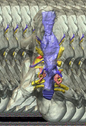
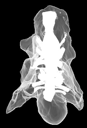
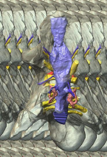
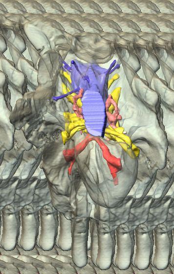
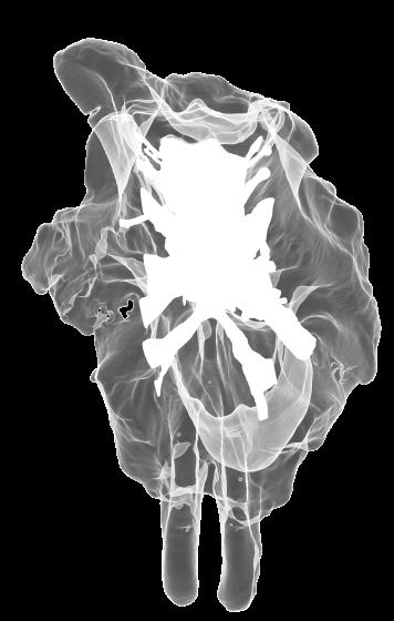
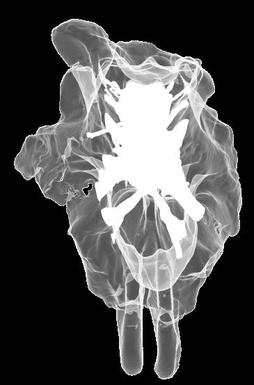
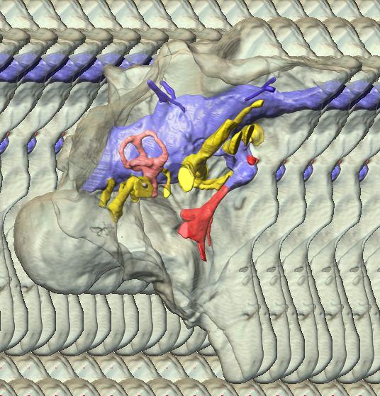
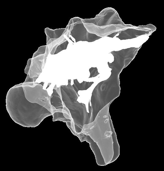
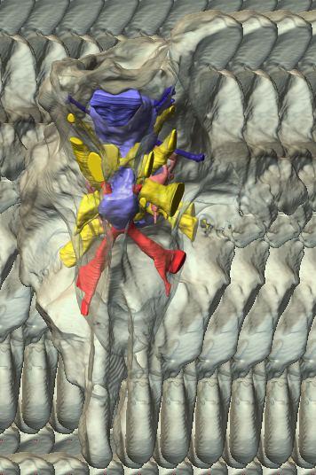
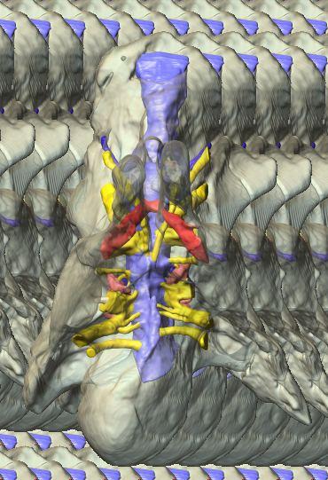
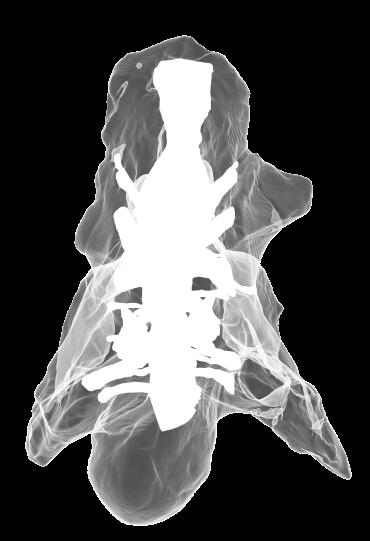
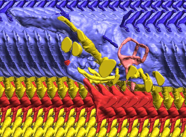
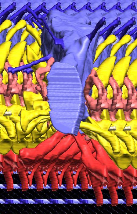
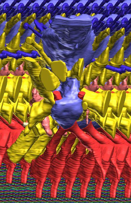
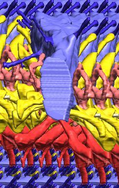
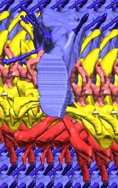
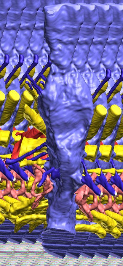
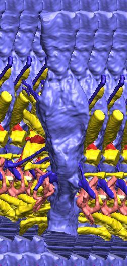
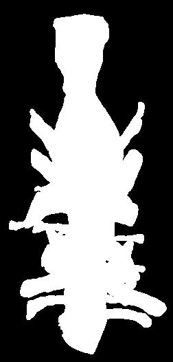
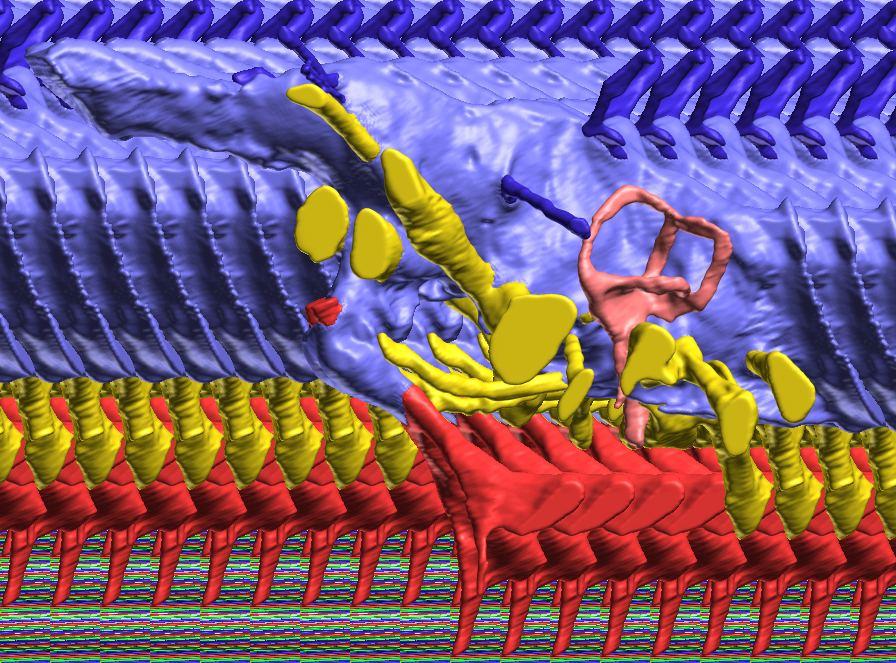
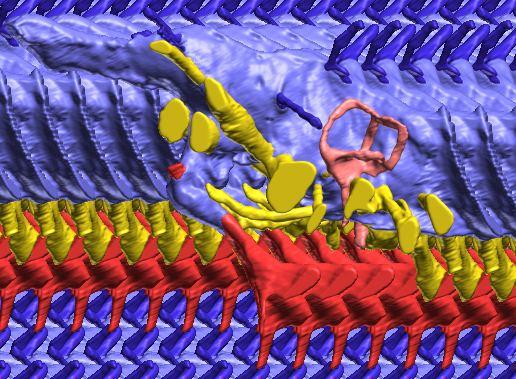
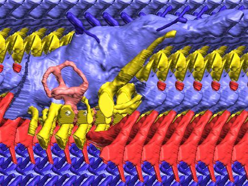
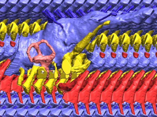
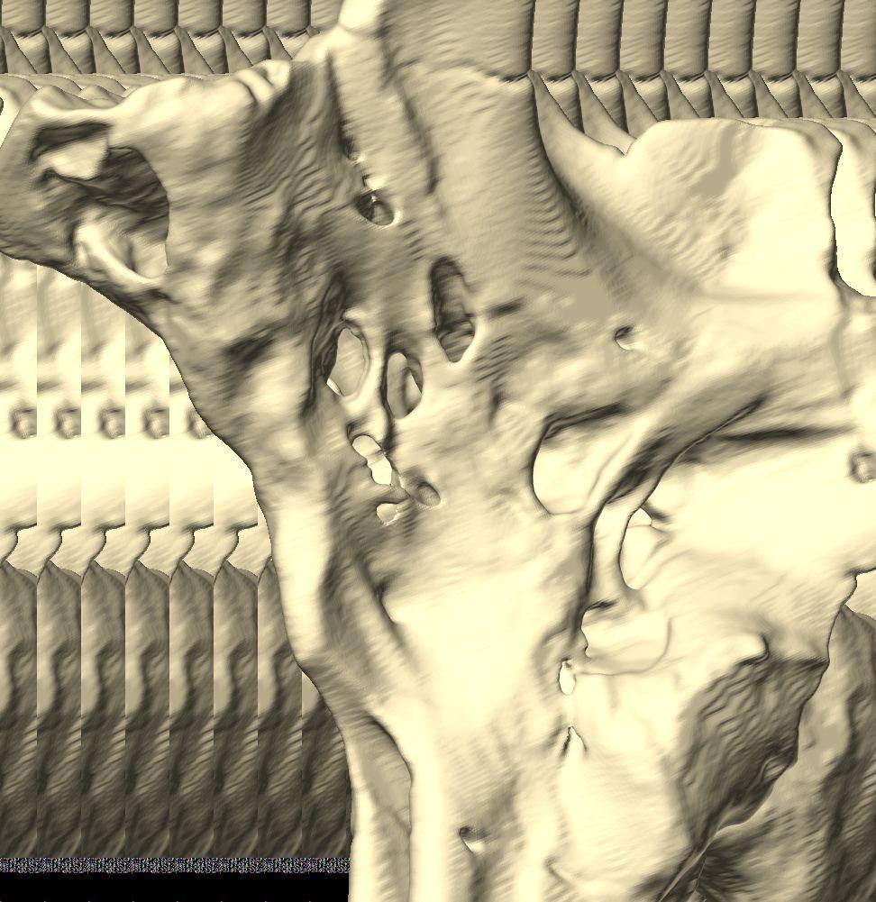
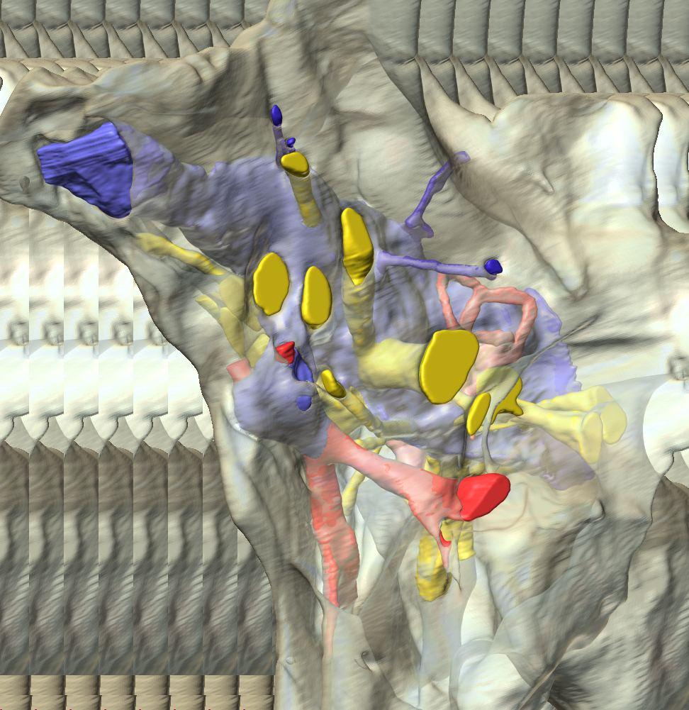
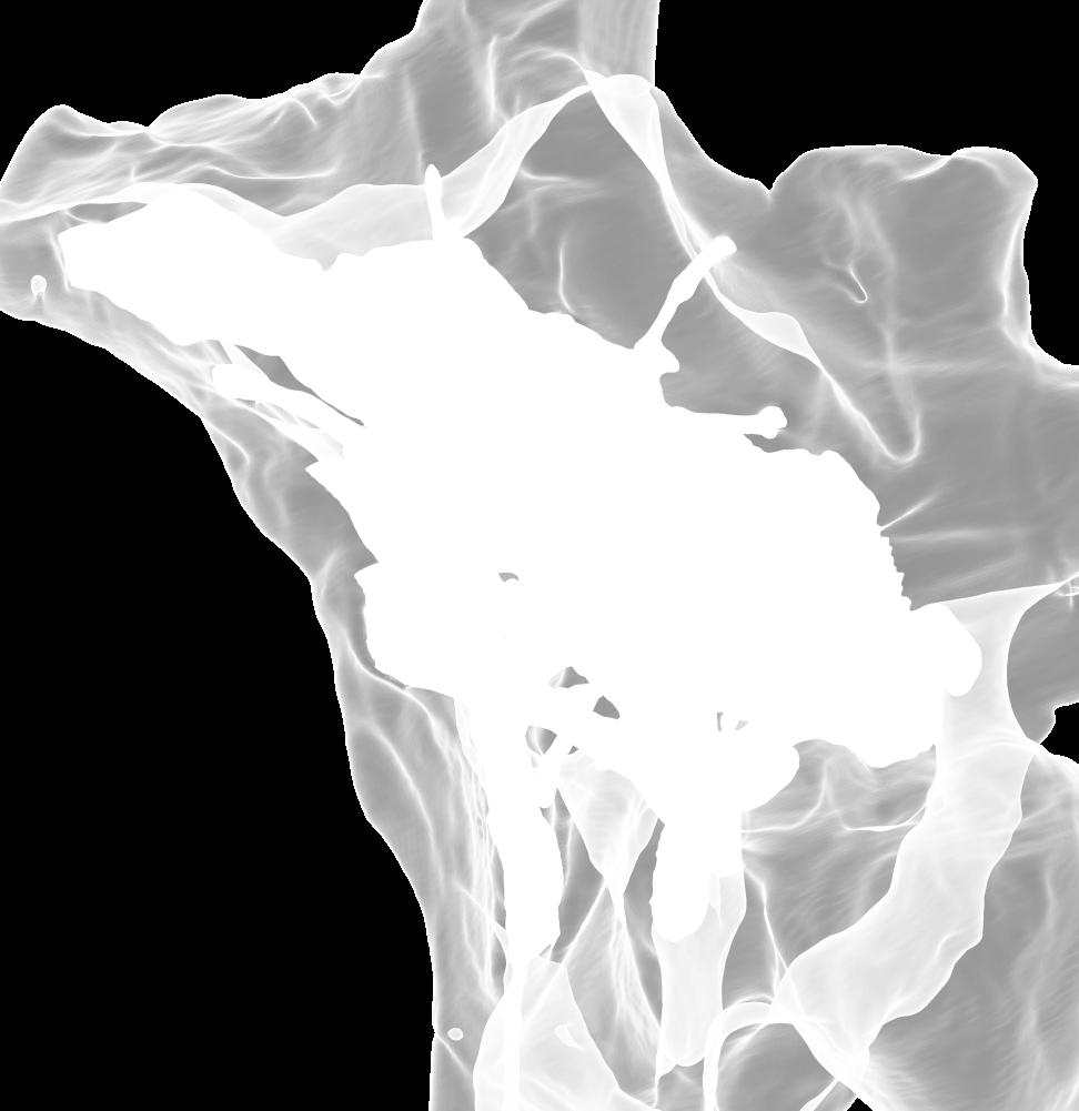
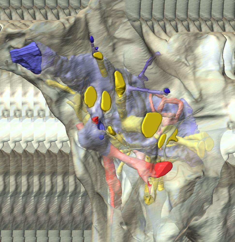
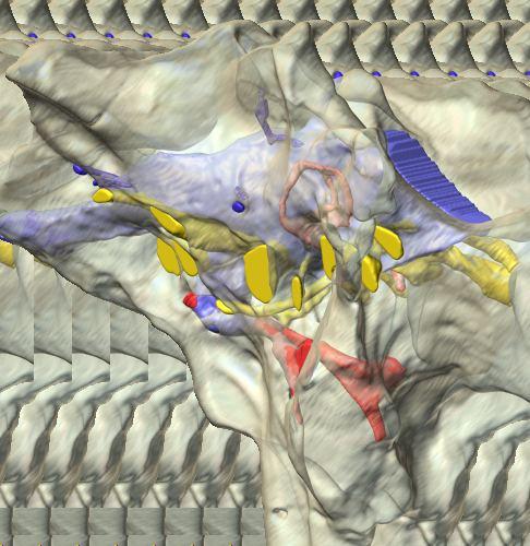
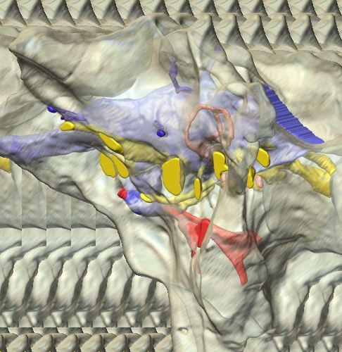
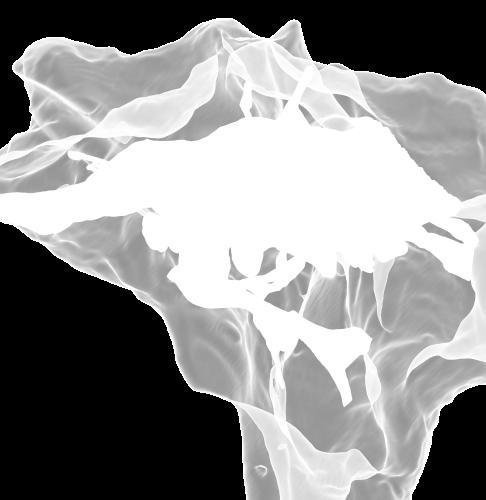
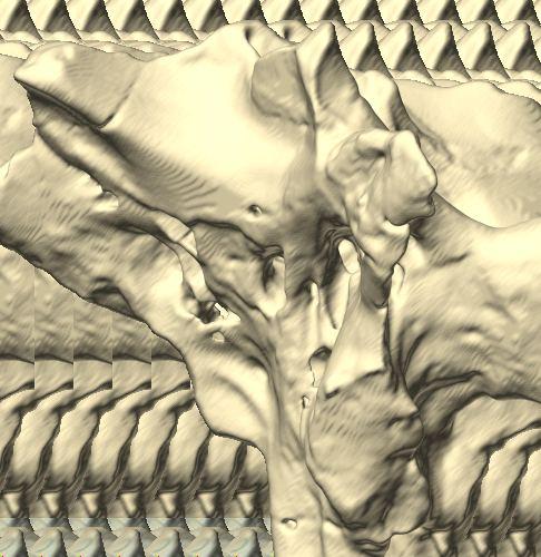
 derived from reconstructed computed tomographic (CT) scans and shown in the following views: (A) right lateral, (B) dorsal, (C) caudal, (D) left lateral, (E) ventral, and (F) rostral.")
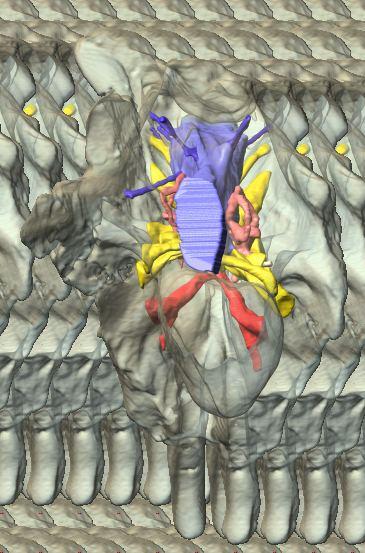
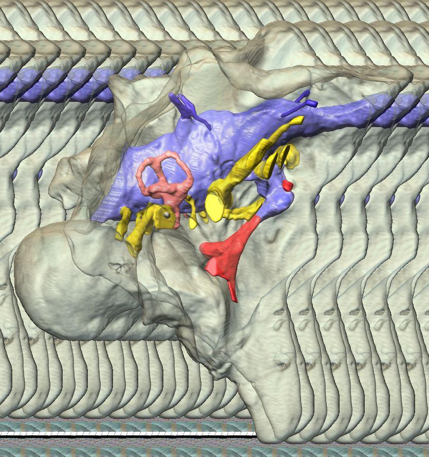
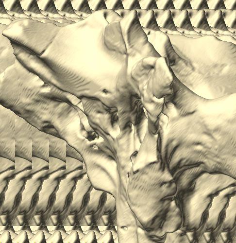
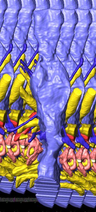
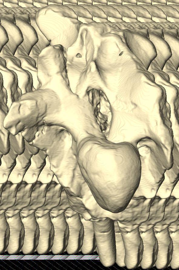
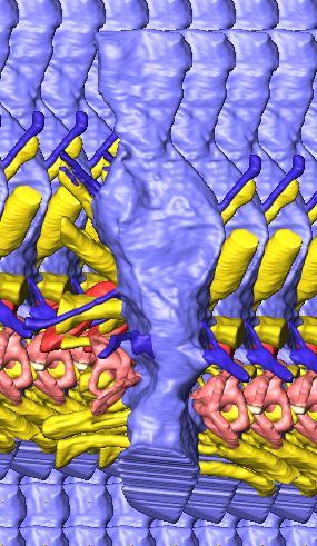
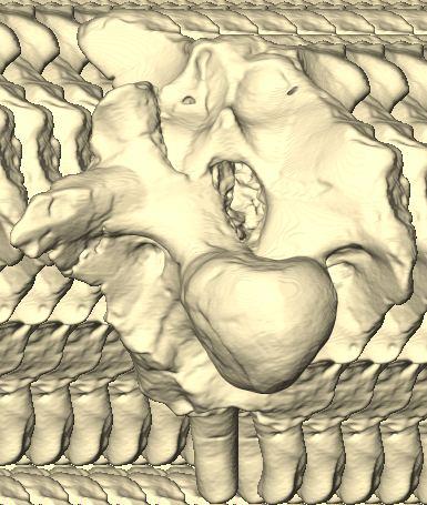
6 Figs. 1A C Fig. 1. Stereopairs of articulated braincase of Pachyrhinosaurus lakustai (TMP ) derived from reconstructed computed tomographic (CT) scans and shown in the following views: (A) right lateral, (B) dorsal, (C) caudal, (D) left lateral, (E) ventral, and (F) rostral. Bone is rendered semitransparent, revealing cranial endocast in blue, endosseous labyrinth in pink, nerve canals (most of which also transmit veins) in yellow, smaller venous canals in dark blue, and arterial canals in red. Scale bar = 4 cm. Abbreviations: bpp, basipterygoid process; bt, basal tuber; cap, capitate process of laterosphenoid; car, cerebral carotid artery canal; cer, cerebral hemisphere; ct, crista tuberalis; cvcm, caudal middle cerebral vein; lab, endosseous labyrinth; ob, olfactory bulb; oc, occipital condyle; pfo, pituitary (= hypophyseal) fossa; pop, paroccipital process; rvcm, rostral middle cerebral vein; V 1, ophthalmic nerve canal; V 2 3, maxillomandibular nerve canal; X, shared canal for vagus and accessory nerves and accompanying vessels; XII, hypoglossal nerve canal. 122 Chapter 3: Witmer and Ridgley

7 Figs. 1D F A New Horned Dinosaur From an Upper Cretaceous Bone Bed in Alberta 123
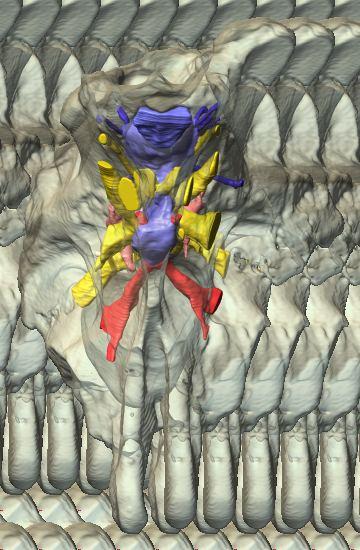
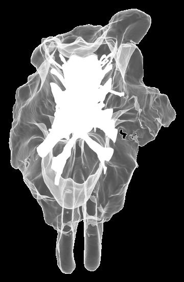
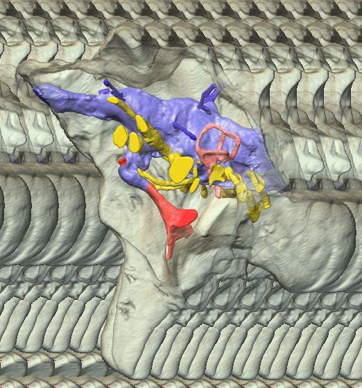
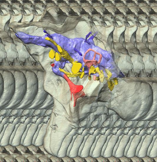
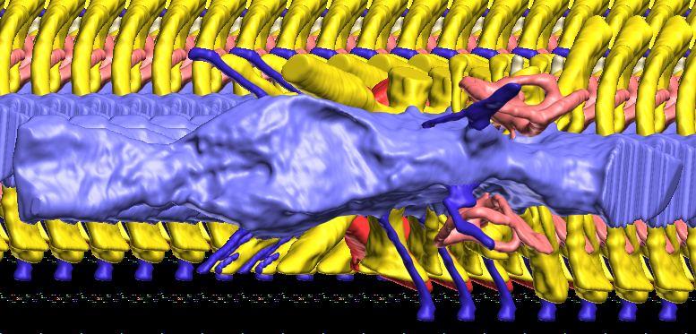
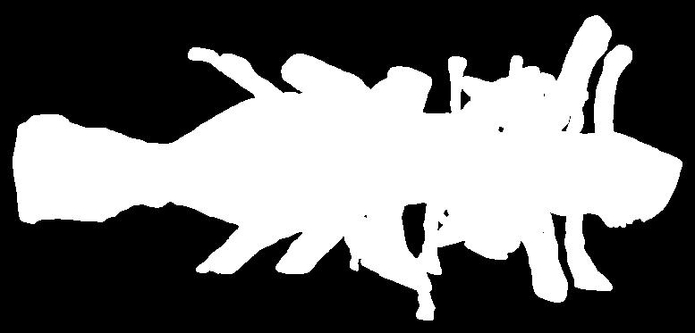
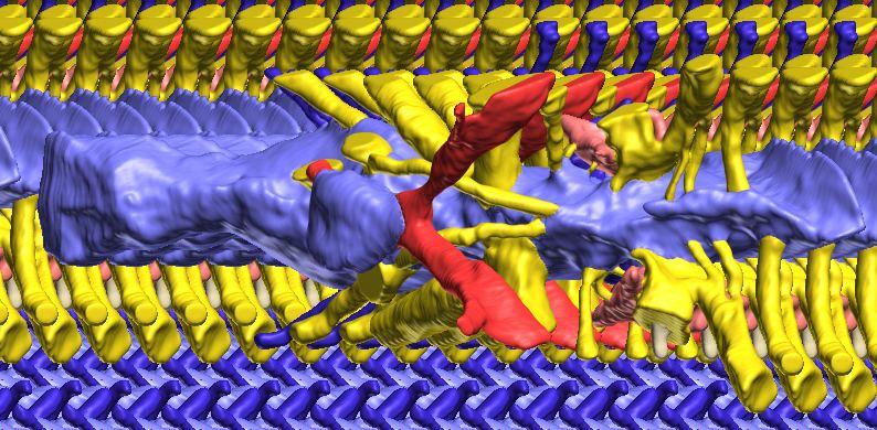
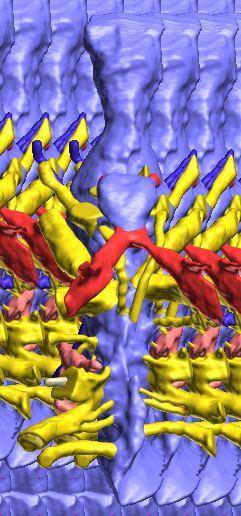
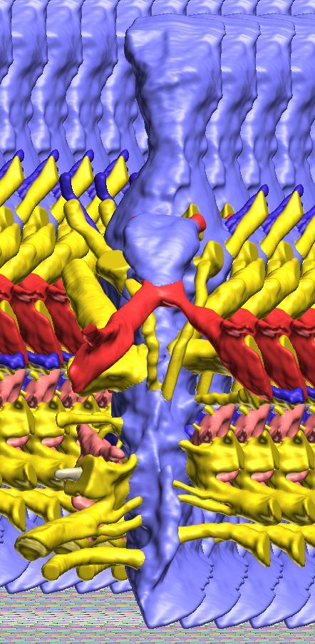
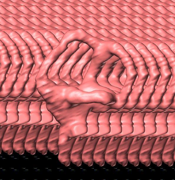
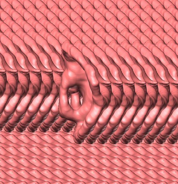
 reconstructed from computed tomographic (CT) scans and shown in the following views: (A) left lateral, (B) ventral, (C) dorsal, (D) rostral, and (E) caudal.")
8 Figs. 2A E Fig. 2. Cranial endocast of Pachyrhinosaurus lakustai (TMP ) reconstructed from computed tomographic (CT) scans and shown in the following views: (A) left lateral, (B) ventral, (C) dorsal, (D) rostral, and (E) caudal. Stereopairs are in the following views: (F) left lateral, (G) right lateral, (H) dorsal, (I) ventral, (J) caudal, and (K) rostral. Color scheme: cranial endocast, blue; endosseous labyrinth, pink; nerve canals (most of which also transmit veins), yellow; smaller venous canals, dark blue; arterial canals, red. Scale bar = 4 cm. Abbreviations: car, cerebral carotid artery canal; cbl, cerebellum; cc, columellar canal; cer, cerebral hemisphere; cvcm, caudal middle cerebral vein; col, columella (= stapes); dls, dorsal longitudinal sinus (a dural venous sinus); dls cer, cerebral branch of dorsal longitudinal sinus; lab, endosseous labyrinth; ob, olfactory bulb; ocv, orbitocerebral vein canal; otc, olfactory tract cavity; pfo, pituitary (= hypophyseal) fossa; rvcm, rostral middle cerebral vein; spha, sphenoid artery canal; ts, transverse sinus; vls, ventral longitudinal sinus (a dural venous sinus); II, optic nerve canal; III, oculomotor nerve canal; IV, trochlear nerve canal; V 1, ophthalmic nerve canal; V 2-3, maxillomandibular nerve canal; VI, abducens nerve canal; VII, facial nerve canal; VIII, vestibulocochlear nerve canal; IX, glossopharyngeal nerve canal (part of columellar canal); X, shared canal for vagus and accessory nerves and accompanying vessels; XII, hypoglossal nerve canal. 124 Chapter 3: Witmer and Ridgley

9 Figs. 2F K A New Horned Dinosaur From an Upper Cretaceous Bone Bed in Alberta 125
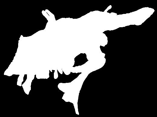
10 Fig. 3. Endosseous labyrinth of the left inner ear of Pachyrhinosaurus lakustai (TMP ) reconstructed from computed tomographic (CT) scans. Stereopairs are in the following views: (A) lateral, (B) caudal, and (C) dorsal. Orientations were determined based on orientation of the labyrinth within the skull and with the lateral semicircular canal placed horizontally. Scale bar = 4 cm. Abbreviations: c, cochlea (= lagena); crc, crus communis; csc, caudal (posterior vertical) semicircular canal; ed, endolymphatic duct; fp, fenestra perilymphaticum (= round window); fv, fenestra vestibuli (= oval window); lsc, lateral (horizontal) semicircular canal; lsca, ampulla of lateral semicircular canal; rsc, rostral (anterior vertical) semicircular canal; rsca, ampulla of rostral semicircular canal; ve, vestibule of inner ear. 126 Chapter 3: Witmer and Ridgley

11 A New Horned Dinosaur From an Upper Cretaceous Bone Bed in Alberta 127
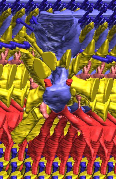
 reconstructed from computed tomographic (CT) scans in the (A) left rostroventrolateral view and (B) left caudolateral view to show the osteological correlates (e.g., foramina, fossae, crests) of many of the soft-tissue structures discussed in the text.")
12 Fig. 4A Fig. 4. Stereopairs of a braincase of Pachyrhinosaurus lakustai (TMP ) reconstructed from computed tomographic (CT) scans in the (A) left rostroventrolateral view and (B) left caudolateral view to show the osteological correlates (e.g., foramina, fossae, crests) of many of the soft-tissue structures discussed in the text. Each view consists of a set of stereopairs (above) showing a semitransparent braincase revealing enclosed soft-tissue structures, coupled with a set of stereopairs (below) in the same view showing the bony braincase and labeled structures. Color scheme: cranial endocast, blue; endosseous labyrinth, pink; nerve canals (most of which also transmit veins), yellow; smaller venous canals, dark blue; arterial canals, red. Scale bar = 4 cm. Abbreviations: bt, basal tuber; car, cerebral carotid artery canal; cc, columellar canal; ct, crista tuberalis; cvcm, caudal middle cerebral vein; fm, foramen magnum; gps, glossopharyngeal sulcus; obf, olfactory bulb fossa; ocv, orbitocerebral vein canal; rvcm, rostral middle cerebral vein; spha, sphenoid artery canal; II, optic nerve canal; III, oculomotor nerve canal; IV, trochlear nerve canal; V 1, ophthalmic nerve canal; V 2 3, maxillomandibular nerve canal; VI, abducens nerve canal; VII, facial nerve canal; X, shared canal for vagus and accessory nerves and accompanying vessels; XII, hypoglossal canal. 128 Chapter 3: Witmer and Ridgley

13 Fig. 4B A New Horned Dinosaur From an Upper Cretaceous Bone Bed in Alberta 129
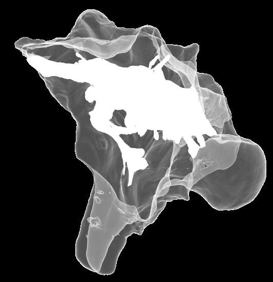
 Cranial endocast in dorsal view. Scale bar = 4 cm. In A and B, the structures are shown as preserved prior to retrodeformation.")
14 Fig. 5. Simple retrodeformation of a braincase and cranial endocast of Pachyrhinosaurus lakustai (TMP ) reconstructed from computed tomographic (CT) scans. (A, C) Braincase in caudal view. (B, D) Cranial endocast in dorsal view. Scale bar = 4 cm. In A and B, the structures are shown as preserved prior to retrodeformation. In C and D, the structures have been transformed by simply stretching the dataset transversely so that the width of the occipital condyle equals its height (the condition observed in undeformed specimens). Even this simple retrodeformation restores a more natural shape to the braincase and endocast and increases endocast volume by 35%. 130 Chapter 3: Witmer and Ridgley
15 Results The CT scan data for TMP were excellent, allowing clear discrimination of fossil bone, rock matrix, and the plaster used to reconstruct the missing portions (e.g., left basipterygoid process). Currie et al. (2008) provided a complete osteological description and illustration of this specimen, and thus this paper will focus on the cranial endocast, endosseous labyrinth, and endocranial vasculature. Nevertheless, some aspects of the bony braincase will be discussed, as they pertain to the reconstructed soft tissues, and the position of these soft-tissue systems are illustrated in place within the semitransparent braincase in Figure 1. Rather than provide a lengthy description of the structures, we will allow the illustrations to convey information on general form and relationships and instead focus on comparisons and significance. Cranial endocast The term cranial endocast (or just endocast ) is generally used for a replica (whether virtual or physical) of the internal surface of the brain cavity. As discussed by many other workers (e.g., Jerison 1973; Hopson 1979), an endocast does not always faithfully record the shape of the underlying brain because the brain may not sufficiently fill the endocranium. In that case, the endocast constitutes a replica of the envelope of the dura mater (Osborn 1912). In some archosaurs, such as pterosaurs (Witmer et al. 2003), derived coelurosaurs (Currie 1985, 1995; Currie and Zhao 1993; Osmólska 2004; Kundrát 2007), and perhaps some ornithischians (Evans 2005), the brain resembled that of extant birds in that it largely filled the endocranium to the extent that an endocast fairly represents the general brain structure (Iwaniuk and Nelson 2002). However, the endocast of P. lakustai (Figs. 1, 2) is more like that of extant reptiles (and indeed most fossil archosaurs; Hopson 1979; Witmer et al. 2008) in that the endocast is not particularly brain-like in form, suggesting that the brain itself was perhaps markedly smaller than the endocast. Again, the endocast is somewhat distorted due to plastic deformation of the braincase (see above), but the transverse compression has not grossly altered its morphological features. As is typically the case in archosaurs, the telencephalic portions of the brain are the most interpretable, presumably due to there having been less intervening tissue between the brain and bone. Thus, the cerebral hemispheres are clearly visible as rounded swellings in the forebrain region. Likewise, the paired olfactory bulbs are observable rostrally as they diverge from the median cavity for the olfactory tracts (Fig. 2C). Viewed in place (Fig. 1), the olfactory tract cavity can be seen to run through the dorsomedial roof of the orbit such that the olfactory bulbs are situated at the back of the nasal cavity. The pituitary region is also clearly visible (Figs. 2A, 2B), but the cast of the bony pituitary fossa does not provide a reliable measure of the A New Horned Dinosaur From an Upper Cretaceous Bone Bed in Alberta 131
16 132 Chapter 3: Witmer and Ridgley size and shape of the hypophysis itself because the fossa houses a number of other structures as well, such as the cerebral carotid artery and cavernous venous sinus. Nevertheless, the pituitary bears a well-marked infundibular constriction below the base of the oculomotor nerve trunk, and the structure generally resembles that illustrated for the endocasts of Triceratops (Marsh 1896; Hay 1909; Forster 1996) and Anchiceratops (Brown 1914; Hopson 1979). The cerebellum also is represented in the endocast as a low swelling (Fig. 2A). In extant diapsids, the cerebellum is typically located caudal to the transverse sinus system (including the rostral and caudal middle cerebral veins), between the paired rostral semicircular canals, and dorsal to the proximal trunk of the trigeminal nerve (Sampson and Witmer 2007; Ridgely and Witmer 2007). The swelling on the endocast of P. lakustai is in precisely this location, and a similar cerebellar swelling is present in the Centrosaurus endocast. Based on the size of the swelling, and even accounting for postmortem deformation, the cerebellum must have been relatively small in size. There is no indication in the endocast that the cerebellum had a floccular lobe (cerebellar auricle). Unlike the brain components just noted, the position of the optic tectum (lobe) is not clearly visible. Hay (1909) and Forster (1996) identified low swellings on their endocasts of Triceratops as pertaining to the optic tectum, although Hopson (1979) regarded the tecta as not being visible in his ceratopsid endocasts, and the latter is likewise true for P. lakustai. Based on comparisons with extant diapsids (Ridgely and Witmer 2007), the optic tectum should be located in the region just caudal to the cerebral swelling and just rostral to the transverse sinus system, but there is no discernable swelling in this location in the endocast of TMP This situation suggests that the optic tectum was relatively small, although the influence of postmortem deformation cannot be completely excluded. It moreover suggests that brain organization in P. lakustai resembled extant crocodylians and other reptiles in having an in-line brain with the cerebral hemispheres, optic tecta, and cerebellum in a rostrocaudal sequence and the contralateral optic tecta contacting each other. On the other hand, in more encephalized archosaurs, such as pterosaurs and many advanced theropods (especially birds), the optic tecta were displaced ventrolaterally and did not contact each other dorsally (Currie and Zhao 1993; Witmer et al. 2003; Kundrát 2007). The cranial endocast preserves a number of vascular elements, and although these vessel traces obscure some of the underlying neural components, they also provide evidence for the gross limits of some of the brain parts. For example, the transverse sinus system was discussed in the context of providing a rough boundary between the optic tectum and cerebellum. The transverse sinus (a dural venous sinus) is visible on the endocast as a low curving ridge (Fig. 2A). It is continued laterally and caudodorsally as the rostral and caudal middle cerebral veins, respectively, the lateral branch opening in the skull just dorsal
17 to the maxillomandibular foramen and the caudal branch opening on the occiput (Figs. 1, 4). Another dural sinus that is clearly visible is the ventral longitudinal sinus. Identified earlier in the theropod Majungasaurus (Sampson and Witmer 2007), the ventral longitudinal sinus is a common feature of archosaur endocasts, forming a median ridge, which is long in P. lakustai, extending the length of the brainstem (Fig. 2B). The upper shoulders of the ventral longitudinal sinus represent the ventral margin of the medulla. The occipital dural venous sinus obscures the dorsal margin of the medulla and grades rostrally into the dorsal longitudinal sinus (Figs. 2A, 2C). The dorsal longitudinal sinus extends rostrodorsally to the region directly above the cerebellum where it joins with the transverse sinus system. At this point, it splits and diverges, passing rostrally as a ridge along the dorsolateral edge of each cerebral hemisphere. These cerebral branches of the dorsal longitudinal sinus may correspond to the paired dorsal venous sinuses identified in Triceratops by Forster (1996). Interestingly, this branched dorsal longitudinal sinus is not present in the endocast of Centrosaurus but does appear to be present in the physical endocast of Protoceratops (AMNH 6466) and the digital endocast of Psittacosaurus (IGM 100/1132). In Psittacosaurus, the endocast ridges above the cerebral hemispheres are clearly coincident with sutures (e.g., frontal-laterosphenoid), perhaps suggesting that these features are architectural rather than vascular. However, dural venous sinuses positioned within bony sutures are common in archosaurs (Sampson and Witmer 2007; Witmer et al. 2008). Moreover, in Pachyrhinosaurus lakustai the ridges do not run entirely within sutures, and in both Pachyrhinosaurus and Psittacosaurus the endocast ridges are continuous with other, clearly vascular, features. In Pachyrhinosaurus lakustai, the cerebral branches of the dorsal longitudinal sinus drain into the orbit via the orbitocerebral vein canals (Figs. 2, 4). As in many other dinosaurs (see Witmer et al. 2008), the orbitocerebral vein(s) provided an important anastomotic connection between orbital veins and the endocranium. The cerebral carotid artery passed through the typical canal in the basisphenoid on its way to the hypophyseal (pituitary) fossa, each side opening separately into the caudoventrolateral corner of the fossa (Currie et al. 2008; Figs. 1, 2, 4). Emerging from the rostrodorsal aspect of the hypophyseal fossa was a branch of the carotid artery that supplied the orbital contents and dorsum of the palate. Although this same vessel has been called the ophthalmic artery in other ceratopsids (Hay 1909; Brown 1914; Hopson 1979; Forster 1996), we regard this as the sphenoidal or sphenopalatine artery (Sampson and Witmer 2007; Fig. 2), a term from avian anatomy that recognizes the fact that in many birds, the true ophthalmic artery ontogenetically regresses to be replaced by the sphenoidal artery. The pattern of the cranial nerve trunks is illustrated in Fig. 2 and their emergence externally on the braincase is in Fig. 4. In general, the nerve canals that enter the orbit are relatively large, A New Horned Dinosaur From an Upper Cretaceous Bone Bed in Alberta 133
18 134 Chapter 3: Witmer and Ridgley seemingly much larger than necessary to perform their neural functions (e.g., innervating extraocular muscles). In extant archosaurs, the cranial nerve canals and foramina particularly the rostral ones (CN II V) transmit not only nerves but also veins from the orbit and nasal cavity into the endocranial cavity (Sedlmayr 2002). Thus, it seems likely that P. lakustai was like some other dinosaurs (e.g., sauropods; Witmer et al. 2008) in not only having the extant condition but also elaborating it further by expanding the venous component. The basic pattern of the nerves (Fig. 2) largely agrees with that presented by previous workers (e.g., Brown 1914; Hopson 1979; Forster 1996), and indeed the pattern is conservative across major clades (Witmer et al. 2008). Brown s (1914) study of Anchiceratops is the only comparable study that treated the cranial nerve canals throughout their entire length. One notable difference between Pachyrhinosaurus and Anchiceratops is the trigeminal nerve. In both taxa (as in ceratopsians generally; Langston 1975), the ophthalmic branch (CN V 1 ) and the maxillomandibular branch (CN V 2 3 ) took separate courses through the braincase to emerge at separate foramina (Figs. 1, 2, 4). However, whereas in Anchiceratops the two branches share a long common trunk coming off the endocast prior to their distal split, in P. lakustai the two trunks come off the main endocast almost separately. Centrosaurus shares the P. lakustai condition. It is not known whether this condition characterizes just centrosaurines, and it is possible that the Anchiceratops condition is a preservational artifact. Current work by the authors on the digital endocast of Triceratops should shed light on the chasmosaurine condition. The facial and vestibulocochlear nerve canals (CN VII and CN VIII, respectively) share the typical common origin within the internal acoustic fossa. The facial nerve is unremarkable, but separate vestibular and cochlear branches of CN VIII were traceable on both sides (Fig. 2B). The caudalmost nerve trunks likewise display a relatively typical pattern. The vagus (CN X) and accessory (CN XI) nerve trunks were more or less indistinguishably joined within the vagal canal, and the glossopharyngeal nerve (CN IX) may well have joined them (but see subsequent discussion). As discussed by Sampson and Witmer (2007), numerous terms have been used for the common canal for these nerves (e.g., vagal, jugular, metotic), and the terminology is further complicated by the proximity of the perilymphatic system rostrally such that another set of terms (e.g., fenestra cochleae, fenestra pseudorotunda) is in play. The vagal canal passes caudal to the crista tuberalis (the bony crest that runs from the paroccipital process to the basal tuber; see Sampson and Witmer 2007) to open on the occiput (Figs. 1, 2, 4). The vagal canal receives the two rostralmost nerve canals of the three hypoglossal nerve canals, leaving the caudal-most and by far the largest hypoglossal trunk to open externally by way of its own foramen (Figs. 2, 4). Having three hypoglossal canals with the rostral two joining the vagal canal was reported previously for Pachyrhinosaurus (and Chasmosaurus) by Langston (1975)
19 and for Triceratops by Forster (1996), yet the digital endocast of Centrosaurus bears only two hypoglossal canals, the rostral one of which enters the vagal canal. Whereas the vagal canal passes caudal to the crista tuberalis, it is continuous rostrally with a space (a projection on the endocast) that extends rostral to the crista. We have labeled this structure the columellar canal in Figs. 2 and 4 because it clearly houses the columella (= stapes), which is preserved more or less in place on the right side of TMP (Figs. 2B, 2C, 2E). However, it also may have transmitted the glossopharyngeal nerve (CN IX) caudally given that there is a notch in the rostral margin of the crista tuberalis in this area (labeled glossopharyngeal sulcus in Fig. 4A; see also Fig. 2). The course of the glossopharyngeal nerve is variable in archosaurs and depends largely on whether the metotic fissure is divided or not, which it is in Ceratopsia. The structure on the endocast that we call the columellar canal may also contain some venous components but certainly contains portions of the perilymphatic system, which will be mentioned briefly in the next section on the inner ear. As correctly noted by Langston (1975, p. 1588), the external foramina in the braincase can be difficult to identify because the nerve and vascular canals branch and sometimes coalesce within the thickened ceratopsian cranial walls. Fortunately, CT scanning and 3D visualization can resolve almost all such uncertainties, and Fig. 4 compares illustrations of the endocast and neurovascular trunks in situ with the isolated braincase so that the foramina can be identified. This approach allows correction of some published accounts that did not have the benefit of CT-based visualization. For example, in the braincase illustration of Centrosaurus presented by Dodson et al. (2004, p. 501), the foramen labeled c.n. VI should be the ophthalmic branch of the trigeminal nerve (CN V 1 ). These same authors also illustrate a grouping of three foramina, one of which is labeled c.n. IV; actually, the ventralmost opening is the trochlear foramen (CN IV) and the upper two openings are orbitocerebral vein foramina. Endosseous labyrinth of the inner ear The labyrinth of the inner ear is well preserved on both sides of TMP (Fig. 3) and provides the best information to date on the inner ear of a ceratopsian (although we now have comparable data for Psittacosaurus, Centrosaurus, and Triceratops, which will be published elsewhere). Physical representations of the inner ears of Protoceratops (Brown and Schlaikjer 1940) and Anchiceratops (Brown 1914) have been published, having been made along with the cranial endocast. These are informative, but we assume that there must have been some measure of reconstruction involved given that they somehow were physically removed from the fossils. Zhou et al. (2007) presented CT-based data on the inner ear of their specimens A New Horned Dinosaur From an Upper Cretaceous Bone Bed in Alberta 135
20 136 Chapter 3: Witmer and Ridgley of Psittacosaurus. Our digital representation of the inner ear (Fig. 3) corresponds to neither the membranous (endolymphatic) labyrinth (which, being soft tissue, is not preserved) nor the osseous (bony) labyrinth but rather corresponds to the inside of the osseous labyrinth. Thus, although it is typically referred to as a digital osseous labyrinth (e.g., Sampson and Witmer 2007), we have adopted the more apt term endosseous labyrinth (Witmer et al. 2008; see also Sereno et al. 2007). The endosseous labyrinth of Pachyrhinosaurus lakustai (Figs. 2, 3) is of a relatively generalized form. The upper part of the inner ear (the vestibule and semicircular canals) is associated with the sense of balance and equilibrium with important links to eye movements (see Discussion). The semicircular canals of P. lakustai are not as thin and elongate as in the labyrinths of Psittacosaurus (IGM 100/1132) but are not quite as stocky as those of Anchiceratops (AMNH 5259). The rostral semicircular canal is the longest of the three in Pachyrhinosaurus lakustai and actually ascends above the level of the common crus. This situation is different from that observed in Anchiceratops where the rostral and caudal canals are more similar in length. Likewise, in Protoceratops (AMNH 6466) and Anchiceratops (AMNH 5259), the rostral canals are lower and descend from the common crus. Still, the vestibular portion of the labyrinth of Pachyrhinosaurus lakustai more closely resembles that of these neoceratopsians than it does the gracile, theropod-like ear of Psittacosaurus. In particular, the lateral canal of Pachyrhinosaurus lakustai is relatively short (Fig. 3C), although not as short as that in sauropods (Witmer et al. 2008). The lower part of the inner ear, the cochlea, comprises the hearing organ. We regard the cochlea as being that portion distal to the position of the fenestra vestibuli (= fenestra ovalis), which is where the footplate of the columella is seated. The cochlea of P. lakustai is relatively short, certainly in comparison to Psittacosaurus (IGM 100/1132) in which it is quite long. The form of the cochlea is not clear on the physical endocasts of Protoceratops (AMNH 6466) and Anchiceratops (AMNH 5259) but probably is comparable to that of Pachyrhinosaurus lakustai. The form of the perilymphatic system is not entirely clear. As noted, the embryonic metotic fissure is divided in ceratopsians, and the portion rostral to the crista tuberalis was partially occupied by the perilymphatic duct. In some archosaurs with divided metotic fissures (e.g., crocodyliforms, advanced theropods), the perilymphatic duct extended caudally where it abutted bony elements to form a secondary tympanic membrane (e.g., fenestra cochleae or fenestra pseudorotunda; see Sampson and Witmer 2007, and references therein). We are not sufficiently certain that these perilymphatic modifications were fully present in P. lakustai, and so our labeling in Fig. 3 uses terms more consistent with a primitive state, that is, a fenestra perilymphaticum on the caudal surface of the labyrinth. Further analysis of other specimens will shed light on this issue and may ultimately argue for yet another independent evolution of a fenestra cochleae.
21 Discussion Brain size and behavior The relationship between endocast volume and body mass has attracted a great deal of attention because it allows a proper assessment of relative brain size (Jerison 1973; Hopson 1977; Hurlburt 1996). Although we can calculate endocranial volume easily and precisely in the digital realm, reliable body masses for extinct animals remain problematic in most cases. This problem is particularly acute in the present case because the specimen from which we have generated a digital endocast (TMP ) is a disarticulated braincase found in a large bone bed and cannot be reliably associated with any postcranial elements (Currie et al. 2008). Thus, the traditional means of generating a body mass estimate are not available for TMP , and we cannot assess relative brain size at the present time. That is, we cannot calculate the encephalization quotient (EQ Jerison 1973; or its reptilian extension, REQ Hurlburt 1996), which provides a useful (although somewhat statistically problematic) comparative metric. It may ultimately be possible to establish a rough body mass estimate to accompany a braincase of this size, but until such time, we choose not to introduce poorly constrained estimates into the literature. Nevertheless, it does not take complicated statistical models to justify the observation that the brain of P. lakustai must have been relatively very small in size and basically rudimentary in structure. As noted, the general brain structure was reptilian in that there is no evidence for any of the morphological apomorphies seen in derived theropods and birds. The cerebrum, cerebellum, and optic lobes were not at all expanded. That said, other attributes of P. lakustai suggest considerable behavioral sophistication. Its elaborate cranial ornamentations indicate the importance of intraspecific (and maybe also interspecific) behavioral communication and display (Sampson et al. 1997; Sampson 2001). Likewise, the evidence for herding and potentially even migration (Currie 1992) suggests some measure of behavioral complexity. These findings suggest that fairly elaborate behavioral repertoires are consistent with modest to small brain sizes. Moreover, they also put in perspective the relatively expanded brains of such dinosaurs as hadrosaurids, a group whose radiation and behavioral range is often compared with that of ceratopsids. The large brains of many hadrosaurids (Ostrom 1961; Hopson 1979; Evans 2005) may reflect cognitive capabilities lacking in ceratopsids such as Pachyrhinosaurus. A New Horned Dinosaur From an Upper Cretaceous Bone Bed in Alberta 137
22 The effects of retrodeformation on brain size and form As discussed, the P. lakustai braincase at hand (TMP ) is somewhat deformed, being laterally compressed. Given that CT scanning digitizes the specimen, the possibility presents itself to address or even correct the fossil s deformation, a process called retrodeformation. A number of different approaches to retrodeformation have been proposed (e.g., Motani 1997; Srivastava and Shah 2006; Angielczyk and Sheets 2007), all of which have their advantages and disadvantages, and all of which require assumptions that we cannot adequately test for TMP Consequently, pending a more formal treatment, we performed a simple transformation in an attempt to improve the cranial endocast to a first approximation. The assumption underlying the transformation is derived from the assessment of this specimen presented by Currie et al. (2008) that the occipital condyle should be about as wide as high. Thus, using Amira s Transform Editor module, we uniformly stretched the transverse dimension until the x dimension of the condyle equaled the y dimension. Thus, we only addressed plastic deformation and ignored brittle deformation (i.e., fractures). Although certainly overly simplistic, this approach to retrodeformation has the advantages of making fewer initial assumptions and being quick and dirty. The results returned are generally satisfying (Fig. 5). The braincase itself compares more favorably with less distorted specimens. Moreover, the endocast is credible, agreeing in general proportions with the physical endocasts of Anchiceratops (AMNH 5259; Brown 1914) and Triceratops (Forster 1996) and our digital endocast of Centrosaurus. The retrodeformation has a considerable impact on endocast metrics. The volume of the endocast as preserved (i.e., prior to transformation) is 63.0 cm 3, whereas the retrodeformed volume is 85.4 cm 3, a 35% increase. Even though we have no reliable body mass to accompany these endocranial values, the retrodeformed value would be more appropriate to use in studies of relative brain size. That said, we have chosen to illustrate the nonretrodeformed braincase, endocast, and labyrinth in Figs. 1 4 because these represent the unadulterated documents as preserved in the fossil record. 138 Chapter 3: Witmer and Ridgley
23 Sensorineural functional inferences The dural envelope of P. lakustai (as represented by the cranial endocast) was sufficiently loose-fitting to the brain, as it was in most non-coelurosaurian dinosaurs, that fine details of brain structure are not recoverable. Nevertheless, based on what is preserved and also the structure of the endosseous labyrinth, some light may be shed on the sensorineural attributes and capabilities of this ceratopsid. Jerison s (1973) principle of proper mass (see also Butler and Hodos 2005) affirms that the size of a brain region (e.g., the optic tectum) relates to the biological importance of the function of that region (e.g., processing visual information). Thus, the fact that the olfactory bulbs, cerebrum, optic tecta, and cerebellum are so small (regardless of any retrodeformation) suggests that precise sensory integration and control were not at a premium for P. lakustai. Given that some of these regions are enlarged in other dinosaurs (e.g., enlarged olfactory bulbs in sauropods, enlarged cerebrum in hadrosaurids, enlarged optic tecta in many coelurosaurs; Witmer et al. 2008), their small size in P. lakustai indicates their lower level of function. One remarkable attribute is the large size of the trigeminal nerve canals. Although some portion of each canal was no doubt occupied by veins, it is reasonable to suggest that the nerves themselves were also enlarged. Nevertheless, it is difficult to know whether tactile sensation (e.g., mechanoreception) was particularly heightened or whether the large size of the nerves simply reflects the enormous head, in that considerable trigeminal innervation would be required for general somatic sensation, as well as for motor supply to the large jaw musculature. The structure of the inner ear shows that hearing was apparently not a particularly important sensory modality because the cochlea is quite short. The length of the cochlea is directly related to the length of the sensory epithelium (i.e., the basilar membrane; Manley 1990), and cochlear length has long been employed as a measure of hearing capabilities (Wever 1978; Gleich and Manley 2000; Gleich et al. 2004, 2005). The vestibular apparatus, on the other hand, was perhaps a little better developed than in some other neoceratopsians, in that the semicircular canals are somewhat more elongate. This elongation could be linked to a more general sense of balance and equilibrium, but recent research has shown that the semicircular canals have important neural links coordinating eye movements and head turning (Spoor et al. 2007, and references therein; see also Witmer et al. 2008). Thus, P. lakustai may have had somewhat better gaze stabilization mechanisms than some other neoceratopsians. We currently have insufficient data to assess whether the slight elongation would be statistically significant, but it is fair to suggest that it would be of doubtful biological significance. A New Horned Dinosaur From an Upper Cretaceous Bone Bed in Alberta 139
24 Acknowledgments We thank Philip Currie for the invitation to work on this project and to contribute to this volume. We thank Eric Snively, Michael Ryan, Philip Currie, and the staff of Canada Diagnostic Centre in Calgary, Alberta, for the CT scanning of the Pachyrhinosaurus specimen used here, as well as the Centrosaurus specimen mentioned several times. For help with CT scanning of other specimens in the larger study, we thank Heather Rockhold, and O Bleness Memorial Hospital, Athens, Ohio, as well as Timothy Ryan and Avrami Grader at the Center for Quantitative Imaging, Pennsylvania State University. The manuscript benefited from very useful reviews by Michael Ryan and Hans Larsson. We thank Mark Norell and Carl Mehling (AMNH) for access to their historic collection of dinosaur endocasts, as well as for loan of Psittacosaurus and Protoceratops specimens. We thank the Royal Tyrrell Museum of Palaeontology for access to the collections and particularly to Darren Tanke for his encyclopedic knowledge of Pachyrhinosaurus. The specimen used here represents the first specimen ever jacketed and removed from the field by Hans Larsson, and we thank him for his care. For funding, we acknowledge National Science Foundation grants IBN and IOB to L.M. Witmer and R.C. Ridgely, the Ohio University College of Osteopathic Medicine, and the Chang Ying-Chien Professorship of Paleontology. References Angielczyk, K.D., and Sheets, H.D Investigation of simulated tectonic deformation in fossils using geometric morphometrics. Paleobiology, 33: Brochu, C.A A digitally rendered endocast for Tyrannosaurus rex. Journal of Vertebrate Paleontology, 20:1 6. Brochu, C.A Osteology of Tyrannosaurus rex: insights from a nearly complete skeleton and high-resolution computed tomographic analysis of the skull. Society of Vertebrate Paleontology Memoir 7, Journal of Vertebrate Paleontology, 22 (Supplement to 2): Brown, B Anchiceratops, a new genus of horned dinosaurs from the Edmonton Cretaceous of Alberta. With discussion of the origin of the ceratopsian crest and the brain casts of Anchiceratops and Trachodon. Bulletin of the American Museum of Natural History, 33: Brown, B., and Schlaikjer, E.M The structure and relationships of Protoceratops. Annals of the New York Academy of Sciences, 40: Burckhardt, R Das Gehirne von Triceratops flabellatus Marsh. Neues Jahrbuch für Mineralogie, Geologie and Paläontologie, 1892: Chapter 3: Witmer and Ridgley
25 Butler, A.B., and W. Hodos Comparative vertebrate neuroanatomy: evolution and adaptation. 2nd ed. Wiley-Interscience, Hoboken, N.J. Currie, P.J Cranial anatomy of Stenonychosaurus inequalis (Saurischia, Theropoda) and its bearing on the origin of birds. Canadian Journal of Earth Sciences, 22: Currie, P.J Migrating dinosaurs. In The ultimate dinosaur. Edited by B. Preiss and R. Silverberg. Bantam Books, New York. pp Currie, P.J New information on the anatomy and relationships of Dromaeosaurus albertensis (Dinosauria: Theropoda). Journal of Vertebrate Paleontology, 15: Currie, P.J., and Zhao, X.-J A new troodontid (Dinosauria, Theropoda) braincase from the Dinosaur Park Formation (Campanian) of Alberta. Canadian Journal of Earth Sciences, 30: Currie, P.J., Langston, W., Jr., and Tanke, D.H A new species of Pachyrhinosaurus (Dinosauria, Ceratopsidae) from the Upper Cretaceous of Alberta, Canada. In A new horned dinosaur from an Upper Cretaceous bone bed in Alberta. NRC Research Press, Ottawa, Ontario, Canada. pp Dodson, P., Forster, C.A., and Sampson, S.D Ceratopsia. In The Dinosauria. 2nd ed. Edited by D.B. Weishampel, P. Dodson, and H. Osmólska. University of California Press, Berkeley, California. pp Domínguez Alonso, P., Milner, A.C., Ketcham, R.A., Cookson, M.J., and Rowe, T The avian nature of the brain and inner ear of Archaeopteryx. Nature, 430: Evans, D.C New evidence on brain endocranial cavity relationships in ornithischian dinosaurs. Acta Palaeontolica Polonica, 50: Forster, C.A New information on the skull of Triceratops. Journal of Vertebrate Paleontology, 16: Franzosa, J.W Evolution of the brain in Theropoda (Dinosauria). Ph.D. dissertation, Department of Geological Sciences, University of Texas, Austin, Tex. Franzosa, J.W., and Rowe, T Cranial endocast of the Cretaceous theropod dinosaur Acrocanthosaurus atokensis. Journal of Vertebrate Paleontology, 25: Gilmore, C.W A new restoration of Triceratops, with notes on the osteology of the genus. Proceedings of the United States National Museum, 55: Gleich, O., and Manley, G.A The hearing organ of birds and Crocodilia. In Comparative hearing: birds and reptiles. Edited by R.J. Dooling, R.R. Fay, and A.N. Popper. Springer, New York. pp A New Horned Dinosaur From an Upper Cretaceous Bone Bed in Alberta 141
26 142 Chapter 3: Witmer and Ridgley Gleich, O., Fischer, F.P., Köppl, C., and Manley, G.A Hearing organ evolution and specialization: archosaurs. In Evolution of the vertebrate auditory system. Edited by G.A. Manley, A.N. Popper, and R.R. Fay. Springer, New York. pp Gleich, O., Dooling, R.J., and Manley, G.A Audiogram, body mass, and basilar papilla length: correlations in birds and predictions for extinct archosaurs. Naturwissenschaften, 92: Hay, O.P On the skull and the brain of Triceratops, with notes on the brain-cases of Iguanodon and Megalosaurus. Proceedings of the United States National Museum, 36: Hopson, J.A Relative brain size and behavior in archosaurian reptiles. Annual Review of Ecology and Systematics, 8: Hopson, J.A Paleoneurology. In Biology of the Reptilia. Vol. 9. Neurology A. Edited by C. Gans. Academic Press, New York. pp Hurlburt, G.R Relative brain size in recent and fossil amniotes: determination and interpretation. Ph.D. dissertation, University of Toronto, Toronto, Ont. Iwaniuk, A.N., and Nelson, J.E Can endocranial volume be used as an estimate of brain size in birds? Canadian Journal of Zoology, 80: Jerison, H.J Evolution of the brain and intelligence. Academic Press, New York. Kundrát, M Avian-like attributes of a virtual brain model of the oviraptorid Conchoraptor gracilis. Naturwissenschaften, 94: Kurochkin, E.N., Saveliev, S.V., Postnov, A.A., Pervushov, E.M., and Popov, E.V On the brain of a primitive bird from the Upper Cretaceous of European Russia. Paleontological Journal, 40: Kurochkin, E.N., Dyke, G.J., Saveliev, S.V., Pervushov, E.M., and Popov, E.V A fossil brain from the Cretaceous of European Russia and avian sensory evolution. Biology Letters, 3: Langston, W., Jr The ceratopsian dinosaurs and associated lower vertebrates from the St. Mary River Formation (Maestrichtian) at Scabby Butte, southern Alberta. Canadian Journal of Earth Sciences, 12: Larsson, H.C.E Endocranial anatomy of Carcharodontosaurus saharicus (Theropoda: Allosauroidea) and its implications for theropod brain evolution. In Mesozoic vertebrate life: new research inspired by the paleontology of Philip J. Currie. Edited by D.H. Tanke and K. Carpenter. Indiana University Press, Bloomington, Ind. pp
27 Manley, G.A Peripheral hearing mechanisms in reptiles and birds. Springer, New York. Marsh, O.C The dinosaurs of North America. Annual Report of the United States Geological Survey, 16: Motani, R New technique for retrodeforming tectonically deformed fossils, with an example for ichthyosaurian specimens. Lethaia, 30: Osborn, H.F Crania of Tyrannosaurus and Allosaurus. Memoirs of the American Museum of Natural History, 1: Osmólska, H Evidence on relation of brain to endocranial cavity in oviraptorid dinosaurs. Acta Palaeontologia Polonica, 49: Ostrom, J.H The cranial anatomy of the hadrosaurian dinosaurs of North America. Bulletin of the American Museum of Natural History, 122: Ridgely, R.C., and Witmer, L.M Gross Anatomical Brain Region Approximation (GABRA): a new technique for assessing brain structure in dinosaurs and other fossil archosaurs. Journal of Morphology, 268: Rogers, S.W Exploring dinosaur neuropaleobiology: viewpoint computed tomography scanning and analysis of an Allosaurus fragilis endocast. Neuron, 21: Rogers, S.W Allosaurus, crocodiles, and birds: evolutionary clues from spiral computed tomography of an endocast. Anatomical Record, 257: Rogers, S.W Reconstructing the behaviors of extinct species: an excursion into comparative paleoneurology. American Journal of Medical Genetics, 134A: Sampson, S.D Speculations on the socioecology of ceratopsid dinosaurs (Ornithischia: Neoceratopsia). In Mesozoic vertebrate life: new research inspired by the paleontology of Philip J. Currie. Edited by D.H. Tanke and K. Carpenter. Indiana University Press, Bloomington, Ind. pp Sampson, S.D., and Witmer, L.M Craniofacial anatomy of Majungasaurus crenatissimus (Theropoda: Abelisauridae) from the Late Cretaceous of Madagascar. Society of Vertebrate Paleontology Memoir 8, Journal of Vertebrate Paleontology, 27 (Supplement to 2): Sampson, S.D., Ryan, M.J., and Tanke, D.H Craniofacial ontogeny in centrosaurine dinosaurs (Ornithischia: Ceratopsidae): taxonomic and behavioral implications. Biological Journal of the Linnean Society, 121: Sedlmayr, J.C Anatomy, evolution, and functional significance of cephalic vasculature in Archosauria. Ph.D. dissertation, Department of Biological Sciences, Ohio University, Athens, Ohio. A New Horned Dinosaur From an Upper Cretaceous Bone Bed in Alberta 143
28 144 Chapter 3: Witmer and Ridgleye Sereno, P.C., Wilson, J.A., Witmer, L.M., Whitlock, J.A., Maga, A., Ide, O., and Rowe, T Structural extremes in a Cretaceous dinosaur [online]. PLoS ONE 2(11): e1230. doi: /journal. pone Spoor, F., Garland, T., Jr., Krovitz, G., Ryan, T.M., Silcox, M.T., and Walker, A The primate semicircular canal system and locomotion. Proceedings of the National Academy of Sciences, 104: Srivastava, D.C., and Shah, J Digital method for strain estimation and retrodeformation of bilaterally symmetric fossils. Geology, 34: Wever, E.G The reptile ear. Its structure and function. Princeton University Press, Princeton, N.J. Witmer, L.M., and Ridgely, R.C The paranasal air sinuses of predatory and armored dinosaurs (Archosauria: Theropoda & Ankylosauria) and their contribution to cephalic architecture. Anatomical Record, 291. Witmer, L.M., Chatterjee, S., Franzosa, J.W., and Rowe, T Neuroanatomy of flying reptiles and implications for flight, posture and behavior. Nature, 425: Witmer, L.M., Ridgely, R.C., Dufeau, D.L., and Semones, M.C Using CT to peer into the past: 3D visualization of the brain and ear regions of birds, crocodiles, and nonavian dinosaurs. In Anatomical imaging: towards a new morphology. Edited by H. Endo and R. Frey. Springer-Verlag, Tokyo: pp Zhou, C.-F., Gao, K.-Q., Fox, R.C., and Du, X.-K Endocranial morphology of psittacosaurs (Dinosauria: Ceratopsia) based on CT scans of new fossils from the Lower Cretaceous, China. Palaeoworld, 16:


29 Philip J. Currie, Wann Langston, Jr., & Darren H. Tanke A New Horned Dinosaur From an Upper Cretaceous Bone Bed in Alberta
30 A Publication of the National Research Council of Canada Monograph Publishing Program Philip J. Currie, Wann Langston, Jr., & Darren H. Tanke A New Horned Dinosaur From an Upper Cretaceous Bone Bed in Alberta with contributions from Patricia E. Ralrick, Ryan C. Ridgely, and Lawrence M. Witmer
31 2008 National Research Council of Canada All rights reserved. No part of this publication may be reproduced in a retrieval system, or transmitted by any means, electronic, mechanical, photocopying, recording or otherwise, without the prior written permission of the National Research Council of Canada, Ottawa, ON K1A 0R6, Canada. Printed on acid-free paper. ISBN NRC No cover art copyright 2008 Michael W. Skrepnick Library and Archives Canada Cataloguing in Publication Currie, Philip J., A new horned dinosaur from an Upper Cretaceous bone bed in Alberta / Philip John Currie, Wann Langston, Jr., Darren H. Tanke. Issued by: National Research Council Canada. Includes bibliographical references. ISBN Pachyrhinosaurus. 2. Paleontology Alberta. 3. Dinosaurs Alberta. I. Langston, Wann, II. Tanke, Darren H III. National Research Council Canada IV. Title. QE862.O65C C The Publisher wishes to thank the County of Grande Prairie, AB with a special thanks to Walter Paszkowski for their financial contribution to this book. NRC Monograph Publishing Program Editor: P.B. Cavers (University of Western Ontario) Editorial Board: W.G.E. Caldwell, OC, FRSC (University of Western Ontario); M.E. Cannon, FCAE, FRSC (University of Calgary); K.G. Davey, OC, FRSC (York University); M.M. Ferguson (University of Guelph); S. Gubins (Annual Reviews); B.K. Hall, FRSC (Dalhousie University); W.H. Lewis (Washington University); A.W. May, OC (Memorial University of Newfoundland); B.P. Dancik, Editor-in-Chief, NRC Research Press (University of Alberta) Inquiries: Monograph Publishing Program, NRC Research Press, National Research Council of Canada, Ottawa, Ontario K1A 0R6, Canada. Web site: Correct citation for this publication: Currie, P.J., Langston, W., Jr., and Tanke, D.H A New Horned Dinosaur from an Upper Cretaceous Bone Bed in Alberta. NRC Research Press, Ottawa, Ontario, Canada. 144p.
32 Contributors Philip J. Currie, University of Alberta, Department of Biological Sciences, CW405 Biological Sciences Building, Edmonton, AB T6G 2E9, Canada ( Wann Langston, Jr., University of Texas at Austin, J.J. Pickle Research Campus, Vertebrate Paleontology Laboratory #6, Burnet Road, Austin, TX 78758, USA ( Patricia E. Ralrick, Interdisciplinary Graduate Program, University of Calgary, 2500 University Drive NW, Calgary, AB T2N 1N4, Canada ( Ryan C. Ridgely, Department of Biomedical Sciences, Ohio University College of Osteopathic Medicine, Athens, OH 45701, USA ( Darren H. Tanke, Royal Tyrrell Museum of Palaeontology, Box 7500, Drumheller, AB T0J 0Y0, Canada ( Lawrence M. Witmer, Department of Biomedical Sciences, Ohio University College of Osteopathic Medicine, Athens, OH 45701, USA ( iv Contributors

33 Figures 3. Structure of the brain cavity and inner ear of the centrosaurine ceratopsid dinosaur Pachyrhinosaurus based on CT scanning and 3D visualization LAWRENCE M. WITMER AND RYAN C. RIDGELY citation: Witmer, L. M., and R. C. Ridgely Structure of the brain cavity and inner ear of the centrosaurine ceratopsid Pachyrhinosaurus based on CT scanning and 3D visualization. Pp in P. J. Currie (ed.), A New Horned Dinosaur From an Upper Cretaceous Bone Bed in Alberta. National Research Council of Canada Monograph Series, Ottawa. WitmerLab web page with downloads of movies and 3D PDFs:
34
35
36
37
38 This page is intentionally blank so that relevant figure parts will remain on facing pages when printed.

39

40
41
42
Shedding Light on the Dinosaur-Bird Connection
 Shedding Light on the Dinosaur-Bird Connection This text is provided courtesy of the American Museum of Natural History. When people think of dinosaurs, two types generally come to mind: the huge herbivores
SUPPLEMENTARY INFORMATION
 doi:10.1038/nature12424 Figure S1. Bivariate plots of log-transformed data of body size. Body size is plotted against a, olfactory bulb volume, b, optic lobe volume, c, cerebellum volume, and d, brain
SUPPLEMENTARY ONLINE MATERIAL FOR. Nirina O. Ratsimbaholison, Ryan N. Felice, and Patrick M. O connor
 http://app.pan.pl/som/app61-ratsimbaholison_etal_som.pdf SUPPLEMENTARY ONLINE MATERIAL FOR Nirina O. Ratsimbaholison, Ryan N. Felice, and Patrick M. O connor Ontogenetic changes in the craniomandibular
Fig. 5. (A) Scaling of brain vault size (width measured at the level of anterior squamosal/parietal suture) relative to skull size (measured at the
 Scaling of brain vault size (width measured at the level of anterior squamosal/parietal suture) relative to skull size (measured at the") Fig. 5. (A) Scaling of brain vault size (width measured at the level of anterior squamosal/parietal suture) relative to skull size (measured at the distance between the left versus right temporomandibular
Fig. 5. (A) Scaling of brain vault size (width measured at the level of anterior squamosal/parietal suture) relative to skull size (measured at the distance between the left versus right temporomandibular
DEVELOPMENT OF THE HEAD AND NECK PLACODES
 DEVELOPMENT OF THE HEAD AND NECK Placodes and the development of organs of special sense L. Moss-Salentijn PLACODES Localized thickened areas of specialized ectoderm, lateral to the neural crest, at the
Major cranial changes during Triceratops ontogeny John R. Horner 1, * and Mark B. Goodwin 2
 273, 2757 2761 doi:10.1098/rspb.2006.3643 Published online 1 August 2006 Major cranial changes during Triceratops ontogeny John R. Horner 1, * and Mark B. Goodwin 2 1 Museum of the Rockies, Montana State
Anatomy. Name Section. The Vertebrate Skeleton
 Name Section Anatomy The Vertebrate Skeleton Vertebrate paleontologists get most of their knowledge about past organisms from skeletal remains. Skeletons are useful for gleaning information about an organism
AMERICAN MUSEUM NOVITATES Published by
 AMERICAN MUSEUM NOVITATES Published by Number 782 THE AmzRICAN MUSEUM OF NATURAL HISTORY Feb. 20, 1935 New York City 56.81, 7 G (68) A NOTE ON THE CYNODONT, GLOCHINODONTOIDES GRACILIS HAUGHTON BY LIEUWE
Notes on Ceratopsians and Ankylosaurs at the Royal Ontario Museum
 Notes on Ceratopsians and Ankylosaurs at the Royal Ontario Museum Andrew A. Farke, Ph.D. Raymond M. Alf Museum of Paleontology 1175 West Baseline Road Claremont, CA 91711 email: afarke@webb.org Introduction
SOME LITTLE-KNOWN FOSSIL LIZARDS FROM THE
 PROCEEDINGS OF THE UNITED STATES NATIONAL MUSEUM issued SWsK \ {^^m ^V ^^ SMITHSONIAN INSTITUTION U. S. NATIONAL MUSEUM Vol. 91 Washington : 1941 No. 3124 SOME LITTLE-KNOWN FOSSIL LIZARDS FROM THE OLIGOCENE
Mammalogy Lecture 8 - Evolution of Ear Ossicles
 Mammalogy Lecture 8 - Evolution of Ear Ossicles I. To begin, let s examine briefly the end point, that is, modern mammalian ears. Inner Ear The cochlea contains sensory cells for hearing and balance. -
Reconsidering the Avian Nature of the Oviraptorosaur Brain (Dinosauria: Theropoda)
") RESEARCH ARTICLE Reconsidering the Avian Nature of the Oviraptorosaur Brain (Dinosauria: Theropoda) Amy M. Balanoff 1,3 *, G. S. Bever 2,3, Mark A. Norell 3 1. Stony Brook University, Department of Anatomical
RESEARCH ARTICLE Reconsidering the Avian Nature of the Oviraptorosaur Brain (Dinosauria: Theropoda) Amy M. Balanoff 1,3 *, G. S. Bever 2,3, Mark A. Norell 3 1. Stony Brook University, Department of Anatomical
Williston, and as there are many fairly good specimens in the American
 56.81.7D :14.71.5 Article VII.- SOME POINTS IN THE STRUCTURE OF THE DIADECTID SKULL. BY R. BROOM. The skull of Diadectes has been described by Cope, Case, v. Huene, and Williston, and as there are many
HONR219D Due 3/29/16 Homework VI
 Part 1: Yet More Vertebrate Anatomy!!! HONR219D Due 3/29/16 Homework VI Part 1 builds on homework V by examining the skull in even greater detail. We start with the some of the important bones (thankfully
REPORT FROM A BOU-FUNDED PROJECT
 Pneumatisation and internal architecture of the Southern Cassowary Casuarius casuarius casque: a microct study CHARLOTTE A. BRASSEY 1*, THOMAS O MAHONEY 2 1 School of Science and the Environment, Manchester
Biology 3315 Comparative Vertebrate Morphology Skulls and Visceral Skeletons
 Biology 3315 Comparative Vertebrate Morphology Skulls and Visceral Skeletons 1. Head skeleton of lamprey Cyclostomes are highly specialized in both the construction of the chondrocranium and visceral skeleton.
A new species of Hsisosuchus (Mesoeucrocodylia) from Dashanpu, Zigong Municipality, Sichuan Province
 from Dashanpu, Zigong Municipality, Sichuan Province") A new species of Hsisosuchus (Mesoeucrocodylia) from Dashanpu, Zigong Municipality, Sichuan Province Yuhui Gao (Zigong Dinosaur Museum) Vertebrata PalAsiatica Volume 39, No. 3 July, 2001 pp. 177-184 Translated
A new species of Hsisosuchus (Mesoeucrocodylia) from Dashanpu, Zigong Municipality, Sichuan Province Yuhui Gao (Zigong Dinosaur Museum) Vertebrata PalAsiatica Volume 39, No. 3 July, 2001 pp. 177-184 Translated
Origin and Evolution of Birds. Read: Chapters 1-3 in Gill but limited review of systematics
 Origin and Evolution of Birds Read: Chapters 1-3 in Gill but limited review of systematics Review of Taxonomy Kingdom: Animalia Phylum: Chordata Subphylum: Vertebrata Class: Aves Characteristics: wings,
The Paranasal Air Sinuses of Predatory and Armored Dinosaurs (Archosauria: Theropoda and Ankylosauria) and Their Contribution to Cephalic Structure
 and Their Contribution to Cephalic Structure") THE ANATOMICAL RECORD 291:1362 1388 (2008) The Paranasal Air Sinuses of Predatory and Armored Dinosaurs (Archosauria: Theropoda and Ankylosauria) and Their Contribution to Cephalic Structure LAWRENCE M.
THE ANATOMICAL RECORD 291:1362 1388 (2008) The Paranasal Air Sinuses of Predatory and Armored Dinosaurs (Archosauria: Theropoda and Ankylosauria) and Their Contribution to Cephalic Structure LAWRENCE M.
New Carnivorous Dinosaurs from the Upper Cretaceous of Mongolia
 1955 Doklady, Academy of Sciences USSR 104 (5):779-783 New Carnivorous Dinosaurs from the Upper Cretaceous of Mongolia E. A. Maleev (translated by F. J. Alcock) The present article is a summary containing
These small issues are easily addressed by small changes in wording, and should in no way delay publication of this first- rate paper.
 Reviewers' comments: Reviewer #1 (Remarks to the Author): This paper reports on a highly significant discovery and associated analysis that are likely to be of broad interest to the scientific community.
Phylogeny Reconstruction
 Phylogeny Reconstruction Trees, Methods and Characters Reading: Gregory, 2008. Understanding Evolutionary Trees (Polly, 2006) Lab tomorrow Meet in Geology GY522 Bring computers if you have them (they will
Exceptional fossil preservation demonstrates a new mode of axial skeleton elongation in early ray-finned fishes
 Supplementary Information Exceptional fossil preservation demonstrates a new mode of axial skeleton elongation in early ray-finned fishes Erin E. Maxwell, Heinz Furrer, Marcelo R. Sánchez-Villagra Supplementary
Postilla PEABODY MUSEUM OF NATURAL HISTORY YALE UNIVERSITY NEW HAVEN, CONNECTICUT, U.S.A.
 Postilla PEABODY MUSEUM OF NATURAL HISTORY YALE UNIVERSITY NEW HAVEN, CONNECTICUT, U.S.A. Number 117 18 March 1968 A 7DIAPSID (REPTILIA) PARIETAL FROM THE LOWER PERMIAN OF OKLAHOMA ROBERT L. CARROLL REDPATH
Weekly Schedule of Neuroscience (2017/2018) Week 1
 Week 1") Week 1 28/1/2018 29/1/2018 30/1/2018 31/1/2018 1/2/2018 8:00 9:00 Health politics & 9:00 10:00 Introductory lecture Gross morphology of the brain Gross morphology of spinal cord Health politics & Blood
Week 1 28/1/2018 29/1/2018 30/1/2018 31/1/2018 1/2/2018 8:00 9:00 Health politics & 9:00 10:00 Introductory lecture Gross morphology of the brain Gross morphology of spinal cord Health politics & Blood
Name: GEOL 104 Dinosaurs: A Natural History Video Assignment. DUE: Mon. Oct. 29
 GEOL 104 Dinosaurs: A Natural History Video Assignment DUE: Mon. Oct. 29 Documentaries represent one of the main media by which scientific information reaches the general public. For this assignment, you
THE SMALLEST KNOWN TRICERATOPS SKULL: NEW OBSERVATIONS ON CERATOPSID CRANIAL ANATOMY AND ONTOGENY
 Journal of Vertebrate Paleontology 26(1):103 112, March 2006 2006 by the Society of Vertebrate Paleontology THE SMALLEST KNOWN TRICERATOPS SKULL: NEW OBSERVATIONS ON CERATOPSID CRANIAL ANATOMY AND ONTOGENY
First Ornithomimid (Theropoda, Ornithomimosauria) from the Upper Cretaceous Djadokhta Formation of Tögrögiin Shiree, Mongolia
 from the Upper Cretaceous Djadokhta Formation of Tögrögiin Shiree, Mongolia") First Ornithomimid (Theropoda, Ornithomimosauria) from the Upper Cretaceous Djadokhta Formation of Tögrögiin Shiree, Mongolia Tsogtbaatar Chinzorig¹, ³ *, Yoshitsugu Kobayashi², Khishigjav Tsogtbaatar³,
First Ornithomimid (Theropoda, Ornithomimosauria) from the Upper Cretaceous Djadokhta Formation of Tögrögiin Shiree, Mongolia Tsogtbaatar Chinzorig¹, ³ *, Yoshitsugu Kobayashi², Khishigjav Tsogtbaatar³,
PALEONTOLOGY AND BIOSTRATIGRAPHY OF MONGOLIA
 PALEONTOLOGY AND BIOSTRATIGRAPHY OF MONGOLIA THE JOINT SOVIET-MONGOLIAN PALEONTOLOGICAL EXPEDITION (Transactions, vol. 3) EDITORIAL BOARD: N. N. Kramarenko (editor-in-chief) B. Luvsandansan, Yu. I. Voronin,
A New Study of the Brain of the Predatory Dinosaur Tarbosaurus bataar (Theropoda, Tyrannosauridae)
") ISSN 0031-0301, Paleontological Journal, 2007, Vol. 41, No. 3, pp. 281 289. Pleiades Publishing, Ltd., 2007. Original Russian Text S.V. Saveliev, V.R. Alifanov, 2007, published in Paleontologicheskii Zhurnal,
ISSN 0031-0301, Paleontological Journal, 2007, Vol. 41, No. 3, pp. 281 289. Pleiades Publishing, Ltd., 2007. Original Russian Text S.V. Saveliev, V.R. Alifanov, 2007, published in Paleontologicheskii Zhurnal,
TRACHEMYS SCULPTA. A nearly complete articulated carapace and plastron of an Emjdd A NEAKLY COMPLETE SHELL OF THE EXTINCT TURTLE,
 A NEAKLY COMPLETE SHELL OF THE EXTINCT TURTLE, TRACHEMYS SCULPTA By Charles W. Gilmore Curator of Vertebrate Paleontology, United States National Museum INTRODUCTION A nearly complete articulated carapace
Outline 17: Reptiles and Dinosaurs
 Outline 17: Reptiles and Dinosaurs Evolution of Reptiles The first reptiles appeared in the Mississippian. They evolved from amphibians, which first appeared in the Devonian. The evolutionary jump was
New Information on the Braincase of the North American Therizinosaurian (Theropoda, Maniraptora) Falcarius utahensis
 Falcarius utahensis") New Information on the Braincase of the North American Therizinosaurian (Theropoda, Maniraptora) Falcarius utahensis Author(s): David K. Smith, Lindsay E. Zanno, R. Kent Sanders, Donald D. Deblieux, and
New Information on the Braincase of the North American Therizinosaurian (Theropoda, Maniraptora) Falcarius utahensis Author(s): David K. Smith, Lindsay E. Zanno, R. Kent Sanders, Donald D. Deblieux, and
Weekly Schedule of Neuroscience (2018/2019) Week 1
 Week 1") Week 1 27/1/2019 28/1/2019 29/1/2019 30/1/2019 31/1/2019 9:00 10:00 Introductory lecture Gross morphology of the brain Gross morphology of spinal cord Health politics & Blood supply of the CNS Language
Week 1 27/1/2019 28/1/2019 29/1/2019 30/1/2019 31/1/2019 9:00 10:00 Introductory lecture Gross morphology of the brain Gross morphology of spinal cord Health politics & Blood supply of the CNS Language
LABORATORY EXERCISE 7: CLADISTICS I
 Biology 4415/5415 Evolution LABORATORY EXERCISE 7: CLADISTICS I Take a group of organisms. Let s use five: a lungfish, a frog, a crocodile, a flamingo, and a human. How to reconstruct their relationships?
Origin and Evolution of Birds. Read: Chapters 1-3 in Gill but limited review of systematics
 Origin and Evolution of Birds Read: Chapters 1-3 in Gill but limited review of systematics Review of Taxonomy Kingdom: Animalia Phylum: Chordata Subphylum: Vertebrata Class: Aves Characteristics: wings,
A new sauropod from Dashanpu, Zigong Co. Sichuan Province (Abrosaurus dongpoensis gen. et sp. nov.)
") A new sauropod from Dashanpu, Zigong Co. Sichuan Province (Abrosaurus dongpoensis gen. et sp. nov.) by Ouyang Hui Zigong Dinosaur Museum Newsletter Number 2 1989 pp. 10-14 Translated By Will Downs Bilby
A new sauropod from Dashanpu, Zigong Co. Sichuan Province (Abrosaurus dongpoensis gen. et sp. nov.) by Ouyang Hui Zigong Dinosaur Museum Newsletter Number 2 1989 pp. 10-14 Translated By Will Downs Bilby
A New Dromaeosaurid Theropod from Ukhaa Tolgod (Ömnögov, Mongolia)
") PUBLISHED BY THE AMERICAN MUSEUM OF NATURAL HISTORY CENTRAL PARK WEST AT 79TH STREET, NEW YORK, NY 10024 Number 3545, 51 pp., 25 figures, 1 table December 7, 2006 A New Dromaeosaurid Theropod from Ukhaa
PUBLISHED BY THE AMERICAN MUSEUM OF NATURAL HISTORY CENTRAL PARK WEST AT 79TH STREET, NEW YORK, NY 10024 Number 3545, 51 pp., 25 figures, 1 table December 7, 2006 A New Dromaeosaurid Theropod from Ukhaa
LABORATORY EXERCISE 6: CLADISTICS I
 Biology 4415/5415 Evolution LABORATORY EXERCISE 6: CLADISTICS I Take a group of organisms. Let s use five: a lungfish, a frog, a crocodile, a flamingo, and a human. How to reconstruct their relationships?
Pinacosaurus: A Study. Abstract. dinosaurs, few of which left behind fossils for mankind to recover. One of which were the
 Johnson 1 Hope Johnson William Parker IFS 2087-0001 12 Nov 2015 Pinacosaurus: A Study Abstract When the dinosaurs roamed the earth, there were millions upon millions of species of dinosaurs, few of which
2. Skull, total length versus length of the presacral vertebral column: (0); extremely elongated neck (e.g. Tanystropheus longobardicus).
; extremely elongated neck (e.g. Tanystropheus longobardicus).") Character list of the taxon-character data set 1. Skull and lower jaws, interdental plates: absent (0); present, but restricted to the anterior end of the dentary (1); present along the entire alveolar
Character list of the taxon-character data set 1. Skull and lower jaws, interdental plates: absent (0); present, but restricted to the anterior end of the dentary (1); present along the entire alveolar
Title: Phylogenetic Methods and Vertebrate Phylogeny
 Title: Phylogenetic Methods and Vertebrate Phylogeny Central Question: How can evolutionary relationships be determined objectively? Sub-questions: 1. What affect does the selection of the outgroup have
What is a dinosaur? Reading Practice
 Reading Practice What is a dinosaur? A. Although the name dinosaur is derived from the Greek for "terrible lizard", dinosaurs were not, in fact, lizards at all. Like lizards, dinosaurs are included in
Do the traits of organisms provide evidence for evolution?
 PhyloStrat Tutorial Do the traits of organisms provide evidence for evolution? Consider two hypotheses about where Earth s organisms came from. The first hypothesis is from John Ray, an influential British
A NEW SPECIES OF TROODONT DINOSAUR FROM THE
 A NEW SPECIES OF TROODONT DINOSAUR FROM THE LANCE FORMATION OF WYOMING By Charles W. Gilmore Curator of Vertebrate Paleontology, United States National Museum INTRODUCTION The intensive search to which
complex in cusp pattern. (3) The bones of the coyote skull are thinner, crests sharper and the
 The bones of the coyote skull are thinner, crests sharper and the") DISTINCTIONS BETWEEN THE SKULLS OF S AND DOGS Grover S. Krantz Archaeological sites in the United States frequently yield the bones of coyotes and domestic dogs. These two canines are very similar both
DISTINCTIONS BETWEEN THE SKULLS OF S AND DOGS Grover S. Krantz Archaeological sites in the United States frequently yield the bones of coyotes and domestic dogs. These two canines are very similar both
From Slime to Scales: Evolution of Reptiles. Review: Disadvantages of Being an Amphibian
 From Slime to Scales: Evolution of Reptiles Review: Disadvantages of Being an Amphibian Gelatinous eggs of amphibians cannot survive out of water, so amphibians are limited in terms of the environments
Anatomical Markers for Elevated Cognition in Dinosaurs
 Bowling Green State University ScholarWorks@BGSU Honors Projects Honors College Spring 5-2-2016 Anatomical Markers for Elevated Cognition in Dinosaurs Thomas Gaetano tgaetan@bgsu.edu Follow this and additional
Geo 302D: Age of Dinosaurs. LAB 7: Dinosaur diversity- Saurischians
 Geo 302D: Age of Dinosaurs LAB 7: Dinosaur diversity- Saurischians Last lab you were presented with a review of major ornithischian clades. You also were presented with some of the kinds of plants that
muscles (enhancing biting strength). Possible states: none, one, or two.
. Possible states: none, one, or two.") Reconstructing Evolutionary Relationships S-1 Practice Exercise: Phylogeny of Terrestrial Vertebrates In this example we will construct a phylogenetic hypothesis of the relationships between seven taxa
Reconstructing Evolutionary Relationships S-1 Practice Exercise: Phylogeny of Terrestrial Vertebrates In this example we will construct a phylogenetic hypothesis of the relationships between seven taxa
Supplementary Figure 1 Cartilaginous stages in non-avian amniotes. (a) Drawing of early ankle development of Alligator mississippiensis, as reported
 Drawing of early ankle development of Alligator mississippiensis, as reported") Supplementary Figure 1 Cartilaginous stages in non-avian amniotes. (a) Drawing of early ankle development of Alligator mississippiensis, as reported by a previous study 1. The intermedium is formed at
Supplementary Figure 1 Cartilaginous stages in non-avian amniotes. (a) Drawing of early ankle development of Alligator mississippiensis, as reported by a previous study 1. The intermedium is formed at
FURTHER STUDIES ON TWO SKELETONS OF THE BLACK RIGHT WHALE IN THE NORTH PACIFIC
 FURTHER STUDIES ON TWO SKELETONS OF THE BLACK RIGHT WHALE IN THE NORTH PACIFIC HIDEO OMURA, MASAHARU NISHIWAKI* AND TOSHIO KASUYA* ABSTRACT Two skeletons of the black right whale were studied, supplementing
Introduction to phylogenetic trees and tree-thinking Copyright 2005, D. A. Baum (Free use for non-commercial educational pruposes)
") Introduction to phylogenetic trees and tree-thinking Copyright 2005, D. A. Baum (Free use for non-commercial educational pruposes) Phylogenetics is the study of the relationships of organisms to each other.
Introduction to phylogenetic trees and tree-thinking Copyright 2005, D. A. Baum (Free use for non-commercial educational pruposes) Phylogenetics is the study of the relationships of organisms to each other.
A New Ceratopsian Dinosaur from the Upper
 SMITHSONIAN MISCELLANEOUS COLLECTIONS VOLUME 63. NUMBER 3 A New Ceratopsian Dinosaur from the Upper Cretaceous of Montana, with Note on Hypacrosaurus (With Two Plates) CHARLES W. GILMORE Assistant Curator
A NEW GENUS AND SPECIES OF AMERICAN THEROMORPHA
 A NEW GENUS AND SPECIES OF AMERICAN THEROMORPHA MYCTEROSAURUS LONGICEPS S. W. WILLISTON University of Chicago The past summer, Mr. Herman Douthitt, of the University of Chicago paleontological expedition,
The Endocranial Cavity of Oviraptorosaur Dinosaurs and the Increasingly Complex, Deep History of the Avian Brain
 Original Paper Published online: August 10, 2018 The Endocranial Cavity of Oviraptorosaur Dinosaurs and the Increasingly Complex, Deep History of the Avian Brain Amy M. Balanoff a, b Mark A. Norell b Aneila
Supplementary Information for: 3D morphometric analysis of fossil canid skulls contradicts
 Supplementary Information for: 3D morphometric analysis of fossil canid skulls contradicts the suggested domestication of dogs during the late Paleolithic Abby Grace Drake 1, * Michael Coquerelle 2,3 Guillaume
Geo 302D: Age of Dinosaurs LAB 4: Systematics Part 1
 Geo 302D: Age of Dinosaurs LAB 4: Systematics Part 1 Systematics is the comparative study of biological diversity with the intent of determining the relationships between organisms. Humankind has always
THE GORGONOPSIAN GENUS, HIPPOSAURUS, AND THE FAMILY ICTIDORHINIDAE * Dr. L.D. Boonstra. Paleontologist, South African Museum, Cape Town
 THE GORGONOPSIAN GENUS, HIPPOSAURUS, AND THE FAMILY ICTIDORHINIDAE * by Dr. L.D. Boonstra Paleontologist, South African Museum, Cape Town In 1928 I dug up the complete skeleton of a smallish gorgonopsian
NOTE XVII. Dr. A.A.W. Hubrecht. which should he in accordance with. of my predecessors. alive or in excellent. further
 further either EUROPEAN NEMERTEANS. 93 NOTE XVII. New Species of European Nemerteans. First Appendix to Note XLIV, Vol. I BY Dr. A.A.W. Hubrecht In the above-mentioned note, published six months ago, several
Title: Fossil Focus: Reimagining fossil cats IMPORTANT COPYRIGHT CITATION OF ARTICLE
 Title: Fossil Focus: Reimagining fossil cats Author(s): Andrew Cuff Volume: 8 Article: 4 Page(s): 1-10 Published Date: 01/04/2018 PermaLink: https://www.palaeontologyonline.com/articles/2018/patterns-palaeontology-earliestskeletons/
Distribution Unlimited
 A t Project Title: Functional Measures of Sea Turtle Hearing ONR Award No: N00014-02-1-0510 Organization Award No: 13051000 Final Report Award Period: March 1, 2002 - September 30, 2005 Darlene R. Ketten
 The following text is generated from uncorrected OCR. [Begin Page: Page 1] A NEW CERATOPSIAN DINOSAUR FROM THE UPPER CRETACEOUS OF MONTANA, WITH NOTE ON HYPACROSAURUS ' By CHARLES W. GILMORE assistant
ONTOGENY OF CRANIAL EPI-OSSIFICATIONS IN TRICERATOPS
 Journal of Vertebrate Paleontology 28(1):134 144, March 2008 2008 by the Society of Vertebrate Paleontology ARTICLE ONTOGENY OF CRANIAL EPI-OSSIFICATIONS IN TRICERATOPS JOHN R. HORNER *,1 AND MARK B. GOODWIN
GEOL 104 Dinosaurs: A Natural History Homework 6: The Cretaceous-Tertiary Extinction. DUE: Fri. Dec. 8
 GEOL 104 Dinosaurs: A Natural History Homework 6: The Cretaceous-Tertiary Extinction DUE: Fri. Dec. 8 Part I: Victims and Survivors Below is a list of various taxa. Indicate (by letter) if the taxon: A.
Planet of Life: Creatures of the Skies & When Dinosaurs Ruled: Teacher s Guide
 Planet of Life: Creatures of the Skies & When Dinosaurs Ruled: Teacher s Guide Grade Level: 6-8 Curriculum Focus: Earth Science Lesson Duration: Three class periods Program Description Ancient creatures
Abstract. Troodon is a relatively small, bird-like dinosaur known from the Campanian age
 Lydia Clark Dr. Parker Dino Paper 16 November 2015 Abstract Troodon is a relatively small, bird-like dinosaur known from the Campanian age of the Cretaceous period. It has at least one species, Troodon
A new centrosaurine ceratopsid from the Oldman Formation of Alberta and its implications for centrosaurine taxonomy and systematics
 A new centrosaurine ceratopsid from the Oldman Formation of Alberta and its implications for centrosaurine taxonomy and systematics Michael J. Ryan and Anthony P. Russell 1369 Abstract: Centrosaurus brinkmani
DINOSAURS ADAPTED SECONDARILY FOR QUADRUPEDALISM
 DINOSAURS ADAPTED SECONDARILY FOR QUADRUPEDALISM by Louis DOLLO 1 Conservator with the Royal Museum of Natural History, Brussels. Boards XI and XII I. Introduction. 1. - In an adaptation it is necessary
Ceri Pennington VELOCIRAPTOR
 Ceri Pennington VELOCIRAPTOR The Velociraptor - meaning swift seizer - lived during the late Cretaceous period - 75-71 million years ago. They were a genus of dromaeosaurid theropod dinosaur and there
Witmer, L. M Craniofacial air sinus systems. pp in The Encyclopedia of Dinosaurs, P. J. Currie and K. Padian (eds.
 Witmer, L. M. 1997. Craniofacial air sinus systems. pp. 151 159 in The Encyclopedia of Dinosaurs, P. J. Currie and K. Padian (eds.), Academic Press, New York. Craniofacial Air Sinus Systems LAWRENCE M.
A NEW AUSTROSQUILLA (STOMATOPODA) FROM THE
 FROM THE") A NEW AUSTROSQUILLA (STOMATOPODA) FROM THE MARQUESAS ISLANDS BY ALAIN MICHEL Centre O.R.S.T.O.M., Noumea, New Caledonia and RAYMOND B. MANNING Smithsonian Institution, Washington, U.S.A. The At s,tstrosqzlilla
A NEW AUSTROSQUILLA (STOMATOPODA) FROM THE MARQUESAS ISLANDS BY ALAIN MICHEL Centre O.R.S.T.O.M., Noumea, New Caledonia and RAYMOND B. MANNING Smithsonian Institution, Washington, U.S.A. The At s,tstrosqzlilla
Science & Literacy Activity
 Science & Literacy Activity ACTIVITY OVERVIEW This activity, which is aligned to the Common Core State Standards (CCSS) for English Language Arts, introduces students to scientific knowledge and language
Abstract Protoceratops was a sheep-sized, quadrupedal dinosaur that lived during the Campanian
 Hope 1 Trevor Hope Dr. William Parker Trilobites to T. rex December 5, 2015 Dinosaur Paper (Protoceratops) Abstract Protoceratops was a sheep-sized, quadrupedal dinosaur that lived during the Campanian
Mammalogy Laboratory 1 - Mammalian Anatomy
 Mammalogy Laboratory 1 - Mammalian Anatomy I. The Goal. The goal of the lab is to teach you skeletal anatomy of mammals. We will emphasize the skull because many of the taxonomically important characters
ARTICLE. Midwestern University, N. 59th Ave., Glendale, Arizona 85308, U.S.A.
 Journal of Vertebrate Paleontology 31(3):1 21, May 2011 2011 by the Society of Vertebrate Paleontology ARTICLE CRANIAL OSTEOLOGY OF A JUVENILE SPECIMEN OF TARBOSAURUS BATAAR (THEROPODA, TYRANNOSAURIDAE)
290 SHUFELDT, Remains of Hesperornis.
 290 SHUFELDT, Remains of Hesperornis. [ Auk [July THE FOSSIL REMAINS OF A SPECIES OF HESPERORNIS FOUND IN MONTANA. BY R. W. SHUFELD% M.D. Plate XI7III. ExR,¾ in November, 1914, Mr. Charles W. Gihnore,
Video Assignments. Microraptor PBS The Four-winged Dinosaur Mark Davis SUNY Cortland Library Online
 Video Assignments Microraptor PBS The Four-winged Dinosaur Mark Davis SUNY Cortland Library Online Radiolab Apocalyptical http://www.youtube.com/watch?v=k52vd4wbdlw&feature=youtu.be Minute 13 through minute
The Brain and Senses. Birds perceive the world differently than humans. Avian intelligence. Novel feeding behaviors
 The Brain and Senses Birds perceive the world differently than humans Color and IR vision are highly developed Hearing is superior, owls track prey in total darkness Birds navigate using abilities to sense:
Biology Slide 1 of 50
 Biology 1 of 50 2 of 50 What Is a Reptile? What are the characteristics of reptiles? 3 of 50 What Is a Reptile? What Is a Reptile? A reptile is a vertebrate that has dry, scaly skin, lungs, and terrestrial
Bulletin of Big Bend Paleo-Geo An Open Access Publication from Mosasaur Ranch Museum, Terlingua and Lajitas, Texas All rights reserved
 Bulletin of Big Bend Paleo-Geo An Open Access Publication from Mosasaur Ranch Museum, Terlingua and Lajitas, Texas All rights reserved This was a private report in 2003 on my thoughts on Platecarpus planifrons.
Morphometric Changes In Semicircular Canal Shape Within Theropoda (Dinosauria: Saurischia) And Their Dietary Implications
 And Their Dietary Implications") Fort Hays State University FHSU Scholars Repository Master's Theses Graduate School Summer 2017 Morphometric Changes In Semicircular Canal Shape Within Theropoda (Dinosauria: Saurischia) And Their Dietary
Fort Hays State University FHSU Scholars Repository Master's Theses Graduate School Summer 2017 Morphometric Changes In Semicircular Canal Shape Within Theropoda (Dinosauria: Saurischia) And Their Dietary
Jurassic Food Web. Early Childhood Learning Objective
 Jurassic Food Web Early Childhood Learning Objective Language Development: Listening and understanding, speaking and communicating Literacy: Phonological awareness Science: Scientific knowledge Creative
Name: GEOL 104 Dinosaurs: A Natural History Video Assignment. DUE: Wed. Oct. 20
 GEOL 104 Dinosaurs: A Natural History Video Assignment DUE: Wed. Oct. 20 Documentaries represent one of the main media by which scientific information reaches the general public. For this assignment, you
Diapsida. BIO2135 Animal Form and Function. Page 1. Diapsida (Reptilia, Sauropsida) Amniote eggs. Amniote egg. Temporal fenestra.
 Amniote eggs. Amniote egg. Temporal fenestra.") Diapsida (Reptilia, Sauropsida) Vertebrate phylogeny Mixini Chondrichthyes Sarcopterygii Mammalia Pteromyzontida Actinopterygii Amphibia Reptilia! 1! Amniota (autapomorphies) Costal ventilation Amniote
Diapsida (Reptilia, Sauropsida) Vertebrate phylogeny Mixini Chondrichthyes Sarcopterygii Mammalia Pteromyzontida Actinopterygii Amphibia Reptilia! 1! Amniota (autapomorphies) Costal ventilation Amniote
In North America 1. the Triassic is represented by the thick Newark Group along the east coast, 2. by widespread red-bed and fluvial sediments in the
 The Triassic System The name Triassic derives from the three parts into which the Triassic is divided on the European platform: 3. Keuper (highest) 2. Muschelkalk 1. Bunter (lowest) In North America 1.
Vertebrates. Vertebrate Characteristics. 444 Chapter 14
 4 Vertebrates Key Concept All vertebrates have a backbone, which supports other specialized body structures and functions. What You Will Learn Vertebrates have an endoskeleton that provides support and
Contributions from the Museum of Paleontology, University of Michigan
 Contributions from the Museum of Paleontology, University of Michigan Vo l. 32, n o. 2, pp. 17-40 Ju ly 15, 2009 REASSESSMENT OF THE SAUROPOD DINOSAUR JAINOSAURUS (= ANTARCTOSAURUS ) SEPTENTRIONALIS FROM
Diapsida. BIO2135 Animal Form and Function. Page 1. Diapsida (Reptilia, Sauropsida) Amniote egg. Membranes. Vertebrate phylogeny
 Amniote egg. Membranes. Vertebrate phylogeny") Diapsida (Reptilia, Sauropsida) 1 Vertebrate phylogeny Mixini Chondrichthyes Sarcopterygii Mammalia Pteromyzontida Actinopterygii Amphibia Reptilia!! Amniota (autapomorphies) Costal ventilation Amniote
Diapsida (Reptilia, Sauropsida) 1 Vertebrate phylogeny Mixini Chondrichthyes Sarcopterygii Mammalia Pteromyzontida Actinopterygii Amphibia Reptilia!! Amniota (autapomorphies) Costal ventilation Amniote
Frog Dissection Information Manuel
 Frog Dissection Information Manuel Anatomical Terms: Used to explain directions and orientation of a organism Directions or Positions: Anterior (cranial)- toward the head Posterior (caudal)- towards the
Accepted Manuscript. News & Views. Primary feather vane asymmetry should not be used to predict the flight capabilities of feathered fossils
 Accepted Manuscript News & Views Primary feather vane asymmetry should not be used to predict the flight capabilities of feathered fossils Xia Wang, Robert L. Nudds, Colin Palmer, Gareth J. Dyke PII: S2095-9273(17)30453-X
Alimentary System 解剖學科徐淑媛
 Alimentary System 解剖學科徐淑媛 本堂重點 1. Structures derived from primitive guts 2. Specific events Alimentary System endoderm of primordial gut epithelium & glands of digestive tract ectoderm of stomodeum epithelium
A new ceratopsid from the Foremost Formation (middle Campanian) of Alberta
 of Alberta") 1251 A new ceratopsid from the Foremost Formation (middle Campanian) of Alberta Michael J. Ryan, David C. Evans, and Kieran M. Shepherd Introduction Abstract: Xenoceratops foremostensis gen. et. sp. nov.,
1251 A new ceratopsid from the Foremost Formation (middle Campanian) of Alberta Michael J. Ryan, David C. Evans, and Kieran M. Shepherd Introduction Abstract: Xenoceratops foremostensis gen. et. sp. nov.,
v:ii-ixi, 'i':;iisimvi'\>!i-:: "^ A%'''''-'^-''S.''v.--..V^'E^'-'-^"-t''gi L I E) R.ARY OF THE VERSITY U N I or ILLINOIS REMO
 R.ARY OF THE VERSITY U N I or ILLINOIS REMO") "^ A%'''''-'^-''S.''v.--..V^'E^'-'-^"-t''gi v:ii-ixi, 'i':;iisimvi'\>!i-:: L I E) R.ARY OF THE U N I VERSITY or ILLINOIS REMO Natural History Survey Librarv GEOLOGICAL SERIES OF FIELD MUSEUM OF NATURAL
"^ A%'''''-'^-''S.''v.--..V^'E^'-'-^"-t''gi v:ii-ixi, 'i':;iisimvi'\>!i-:: L I E) R.ARY OF THE U N I VERSITY or ILLINOIS REMO Natural History Survey Librarv GEOLOGICAL SERIES OF FIELD MUSEUM OF NATURAL
On the Development of the Skull of Leptodeira hotamboia.
 On the Development of the Skull of Leptodeira hotamboia. By Gwendolen T. Brock, M.Sc, D.Phil. (Oxon.). With Plates 12 and 13 and 6 Text-figs. INTRODUCTION. ACCOUNTS of the development of the Ophidian skull
Name Class Date. After you read this section, you should be able to answer these questions:
 CHAPTER 14 4 Vertebrates SECTION Introduction to Animals BEFORE YOU READ After you read this section, you should be able to answer these questions: How are vertebrates different from invertebrates? How
THE WILLIAMS Fork Formation preserves a diverse dinosaur
 J. Paleont., 79(2), 2005, pp. 251 258 Copyright 2005, The Paleontological Society 0022-3360/05/0079-251$03.00 RANGE EXTENSION OF SOUTHERN CHASMOSAURINE CERATOPSIAN DINOSAURS INTO NORTHWESTERN COLORADO
Yamaceratops dorngobiensis, a New Primitive Ceratopsian (Dinosauria: Ornithischia) from the Cretaceous of Mongolia
 from the Cretaceous of Mongolia") PUBLISHED BY THE AMERICAN MUSEUM OF NATURAL HISTORY CENTRAL PARK WEST AT 79TH STREET, NEW YORK, NY 10024 Number 3530, 42 pp., 20 figures September 08, 2006 Yamaceratops dorngobiensis, a New Primitive Ceratopsian
PUBLISHED BY THE AMERICAN MUSEUM OF NATURAL HISTORY CENTRAL PARK WEST AT 79TH STREET, NEW YORK, NY 10024 Number 3530, 42 pp., 20 figures September 08, 2006 Yamaceratops dorngobiensis, a New Primitive Ceratopsian
Ch 34: Vertebrate Objective Questions & Diagrams
 Ch 34: Vertebrate Objective Questions & Diagrams Invertebrate Chordates and the Origin of Vertebrates 1. Distinguish between the two subgroups of deuterostomes. 2. Describe the four unique characteristics
Dinosaurs. Dinosaurs LEVELED BOOK N. A Reading A Z Level N Leveled Book Word Count:
 Dinosaurs A Reading A Z Level N Leveled Book Word Count: 831 LEVELED BOOK N Dinosaurs Written by Elizabeth Austin Illustrated by Paula Schricker and Nora Voutas Visit www.readinga-z.com for thousands of