Contributions from the Museum of Paleontology, University of Michigan
|
|
|
- Marion Cain
- 6 years ago
- Views:
Transcription
1 Contributions from the Museum of Paleontology, University of Michigan Vo l. 32, n o. 2, pp Ju ly 15, 2009 REASSESSMENT OF THE SAUROPOD DINOSAUR JAINOSAURUS (= ANTARCTOSAURUS ) SEPTENTRIONALIS FROM THE UPPER CRETACEOUS OF INDIA BY JEFFREY A. WILSON 1, MICHAEL D. D EMIC 1, KRISTINA A. CURRY ROGERS 2, DHANANJAY M. MOHABEY 3, AND SUBASHIS SEN 4 Abstract Early evaluations of sauropod diversity in the Cretaceous of India recognized three genera, Titanosaurus, Antarctosaurus, and Laplatasaurus, each of which was thought to have closely related species on other southern landmasses. Recent systematic work has challenged the validity of the genus Titanosaurus and the supposed close relationship between its constituent species, particularly those from the Cretaceous of South America and Madagascar. Likewise, Laplatasaurus is currently recognized to be restricted to South America, and the Indian species is invalid. Here, we redescribe the anatomy of the Indian sauropod species Antarctosaurus (now known as Jainosaurus) septentrionalis with the goal of examining its validity, constituency, and phylogenetic affinity. The type series of Jainosaurus septentrionalis included a braincase, skull roof, and many postcranial elements that were united on the basis of their large size relative to the other taxon from the same quarry, Titanosaurus indicus. All postcranial bones were missing until recently, when the type series humerus and a cast of the scapula were rediscovered in the collections of the Indian Museum. We evaluate possible associations between type series elements, bringing to light circumstantial evidence that helps strengthen the case for grouping some of them as a single species. Both the cranial and some postcranial bones are diagnostic, allowing referral of additional materials collected from the same locality and elsewhere in Indo-Pakistan to the species J. septentrionalis. Currently two genera, Isisaurus and Jainosaurus, are recognized from the Cretaceous of India. Unlike most titanosaurs, both Isisaurus and Jainosaurus are known from diagnostic cranial and postcranial material recorded from multiple localities. These genera coexist in central and western India, and Isisaurus has been reported recently from western Pakistan. The affinities of Jainosaurus and Isisaurus within Titanosauria are not yet resolved, but a sister-taxon relationship to one another can be ruled out. Jainosaurus appears to have close relatives in Madagascar and South America based on the data at hand. Thus far, there is no evidence for an endemic Indian sauropod fauna during the Late Cretaceous. 1 Museum of Paleontology and Department of Geological Sciences, University of Michigan, 1109 Geddes Avenue, Ann Arbor, Michigan , U.S.A. (wilsonja@umich.edu; mdemic@umich.edu) 2 Biology and Geology Departments, Macalester College, 1600 Grand Avenue, St. Paul, Minnesota 55105, U.S.A. (rogersk@macalester.edu) 3 Palaeontology Division, Geological Survey of India (Central Region), Seminary Hills, Nagpur , India (dinomohabey@yahoo.com) 4 Curatorial Division, Geological Survey of India (Central Headquarters), 27 Jawaharlal Nehru Road, Kolkata , India (ssen84@gmail.com)


.")

. Nevertheless, the interrelationships amongst titanosaurs from southern landmasses remains a key issue for Gondwanan paleobiogeography.")
 from 1874 1882 (Thomas, 1917).")

, Lydekker was invited to describe the fossil mammal collections in the provincial museum in La")
2 18 J. A. Wi l s o n e t a l. INTRODUCTION Richard Lydekker and Friedrich von Huene made lasting contributions to the study of Indian and South American dinosaurs as a result of their successive surveys of two important collections in the late nineteenth and early twentieth centuries. Their taxonomic decisions, one of which is discussed in detail here, helped to shape the understanding of Indian and South American dinosaur faunas and their relationship to one another. In addition to describing some of the first dinosaurs from these landmasses, Lydekker and Huene independently asserted that India, South America, and Madagascar shared common titanosaur sauropod genera that evidenced a remarkable community of type which undoubtedly exists between the faunas of southern continents of the world (Lydekker, 1893: 3). The idea that titanosaurs were Gondwanan in origin and distribution persisted until quite recently (e.g., Bonaparte, 1999), but has given way to the current understanding of titanosaurs as a speciose (50+ genera), globally distributed clade that originated prior to the breakup of Pangea (e.g., Upchurch et al., 2004; Curry Rogers, 2005). Nevertheless, the interrelationships amongst titanosaurs from southern landmasses remains a key issue for Gondwanan paleobiogeography. Resolving the validity, constituency, and affinities of Gondwanan titanosaur genera is the first step in this process. Lydekker, who specialized in fossil and recent mammals, served on the Geological Survey of India in Calcutta (now Kolkata) from (Thomas, 1917). During his residence in India, Lydekker produced several volumes in Palaeontologia Indica describing Indian Tertiary and Pre-Tertiary Vertebrata, continuing a tradition started by Huxley in the 1840s. Among the many fossils he described were the tail vertebrae of India s first dinosaurs, the sauropods Titanosaurus indicus and T. blanfordi from Cretaceous beds of Bara Simla (Lydekker, 1877, 1879; Fig. 1). After returning to England and cataloguing the fossil tetrapods of the British Museum (Natural History), Lydekker was invited to describe the fossil mammal collections in the provincial museum in La Plata, Argentina, visiting twice between 1893 and 1894 (Anonymous, 1896). There, Lydekker described new sauropod dinosaurs as well, including two species of Titanosaurus ( T. australis, T. nanus) and the genera and species Argyrosaurus superbus and Microcoelus patagonicus (Lydekker, 1893). Huene was a generation younger than Lydekker and specialized in fossil reptiles. Like Lydekker, Huene was invited to describe fossil collections of the Geological Survey of India and of the Museo de La Plata in Argentina. Huene visited Argentina between 1923 and 1926 to study collections in La Plata and Buenos Aires. In La Plata, he revised the collection of bones that Lydekker (1893) studied and divided specimens allocated to T. australis into the new species T. robustus and the new genus and species Laplatasaurus araukanicus (Huene, 1929: 48, 53). At the Museo Argentino de Ciencias Naturales in Buenos Aires, Huene studied a partial skull and postcranial bones from Patagonia that he named Antarctosaurus wichmannianus after their southern provenance and discoverer (Huene, 1929: 66). Although Huene apparently never visited India, he was able to study Charles Matley s collection of Indian dinosaur bones Quetta Vitakri P A K I S T A N Deccan trap Upper sands Upper limestone Mottled nodular beds Lower or Main Limestone Green sand White clays of Jabalpur formation Islamabad Rahioli Delhi I N D I A Pisdura Jabalpur Dongargaon Kolkata 500 km Sauropod bed Ossiferous conglomerate Carnosaur bed FIGURE 1 Map of Indo-Pakistan showing the type locality for Jainosaurus septentrionalis at Jabalpur, as well as other titanosaur localities. Note that both Bara Simla and Chhota Simla are located at Jabalpur. Schematic stratigraphic section at Bara Simla based on Huene and Matley (1933: fig. 1). Political boundaries are approximate. whilst they were at the British Museum (Natural History) in the 1930s (Carrano et al., in press). In their monograph describing that collection, Huene and Matley (1933) described new species belonging to genera that Huene described from South America,
3 Reassessment of Ja i n o s a u r u s 19 including Laplatasaurus madagascariensis and Antarctosaurus septentrionalis. Whereas many of the overlapping taxa that Lydekker and Huene named from Argentina and India are fairly fragmentary (e.g., only caudal vertebrae), Antarctosaurus wichmannianus and A. septentrionalis include partial skulls and postcranial elements that contain numerous characters that can be compared. Huene studied the South American collections before the Indian collections, and consequently A. wichmannianus (Huene, 1929) was named prior to A. septentrionalis (Huene and Matley, 1933). The latter species epithet, septentrionalis, (septem, seven; trio, plough-ox) is a Latin term referring to the seven stars of Ursa Major (i.e., the big dipper or the plough), which in Roman times was a convenient way to refer to northern (e.g., used in Caesar s Gallic Wars ; see Hower, 1951). Huene worked within a fixist paleogeographic paradigm and may have chosen the somewhat counterintuitive moniker Antarctic reptile from the north to highlight the biogeographic implications of its discovery, which suggested...a direct land connection from Lemuria [which included India and Madagascar] to South America...via Antarctis, the Australian region, and the Sunda archipelago. (Huene and Matley, 1933: 72). Huene defended this distant connection between northern and southern Antarctosaurus species by the absence of meaningful differences, rather than unique similarities. Consequently, his justification for allocating the South American and Indian specimens to the same genus lacks detail: This fragment of a titanosaurid skull is very closely comparable with and similar to the posterior part of the skull of Antarctosaurus wichmannianus from Patagonia. The Indian specimen is larger than the Patagonian, but is more fragmentary. Apart from the size, the only important difference between these two skulls lies in the fact that the hypophysial fossa in the Indian skull is naturally open below (a so-called pituitary canal ), so that the carotids enter from below, whereas in the Patagonian species the hypophysial fossa is closed below, though very deep, and the carotids enter into the fossa from both sides...in the Indian skull also, the supraoccipital is less hidden by the parietals, and the basipterygoid processes and the presphenoid are relatively bigger and stronger than in the Patagonian species. But the structure of the skull in general (so far preserved) is very similar in both types; for instance, the angle formed by the axis of the presphenoid and the axis of the condyle is about 115 in both species. In spite of its larger size the Indian skull is less gibbous than the Patagonian. (Huene and Matley, 1933: 15 16). More recent comparisons between sauropod braincases and between other elements have revealed that the Indian species, A. septentrionalis, is not closely related to the type species, A. wichmannianus (McIntosh, 1990). Consequently, Hunt et al. (1994) created the new genus Jainosaurus for the Indian taxon, establishing the braincase as the lectotype and suggesting that some of the postcranial bones might belong to the same taxon. Below, the braincase and postcranial remains of Jainosaurus septentrionalis are redescribed as a means for evaluating its validity as a diagnostic taxon, similarity to other braincases reported from India, and phylogenetic relationships to other titanosaur taxa. INSTITUTIONAL ABBREVIATIONs GSI Geological Survey of India, Kolkata (Calcutta), India GSP Geological Survey of Pakistan, Quetta, Pakistan ISI Indian Statistical Institute, Kolkata (Calcutta), India MML Museo Municipal de Lamarque, Río Negro, Argentina MUCPv Museo de Geología y Paleontología de la Universidad Nacional del Comahue, Neuquén, Argentina SYSTEMATIC PALEONTOLOGY SAUROPODA Marsh, 1878 NEOSAUROPODA Bonaparte, 1986 TITANOSAURIA Bonaparte and Coria, 1993 Jainosaurus Hunt et al., 1994 Jainosaurus septentrionalis (Huene and Matley, 1933) Antarctosaurus septentrionalis Huene and Matley, 1933, p. 11, figs. 5 18; pls Lectotype. GSI K27/497, a partial braincase and skull roof (Hunt et al., 1994). Two additional cranial elements, identified as a hyoid and a squamosal, were considered to belong to the same skull by Huene and Matley (1933: 15). The element identified as a left squamosum, which until recently was missing from collections, is bulky and lacks definitive features of a squamosal (Huene and Matley, 1933: fig. 7). This element cannot be identified, and we exclude it from the lectotype. A small element identified as a hyoid by Huene and Matley (1933: fig. 8), can no longer be located at the GSI collections, and its identity is likewise uncertain. Postcranial elements found at the same locality and horizon were also attributed to Jainosaurus (=Antarctosaurus) septentrionalis by Huene and Matley (1933: 11 23; Fig. 2). Together with the cranial elements, they form the type series or syntypes of the species (ICZN Articles 72.1, 73.2). The postcranial elements include dorsal rib shaft fragments (GSI K20/326, K27/425), a caudal vertebra (GSI K20/317), four chevrons (GSI K27/ , 496), right and left scapulae (GSI unnumbered), a sternal plate (GSI K20/647), a humerus (GSI unnumbered), a radius (GSI K27/490), and an ulna (GSI K27/491). From their description and the discussion that followed, we surmise that Huene and Matley (1933) recognized the presence of at least three individuals. That is, although they considered the cranial and postcranial remains to belong to a single species, they did not consider them to belong to a single individual. Thus, attribution of all remains apart from the first-listed braincase, which Hunt et al. (1994) designated the lectotype, constitutes
4 20 J. A. Wi l s o n e t a l. a tacit referral, as discussed below (see Referred Material and Comments ). Locality and Horizon. The type series was discovered in the Sauropod bed within the lower part of the Mottled Nodular Limestone of the Lameta Formation at Bara Simla, Madhya Pradesh State, India (Fig. 1). Other bones recovered from this horizon at Bara Simla include holotypic and referred remains of Titanosaurus indicus (Lydekker, 1877; Huene and Matley, 1933). The Lameta Formation is capped by the Deccan Trap flood basalts and by virtue of their position are referred to as infratrappean sediments. They are considered to be Maastrichtian in age based on coccoliths (Salis and Saxena, 1998), magnetostratigraphy (Hansen et al., 2005), and on the basis of radiometric dates associated with the Deccan Traps (Courtillot et al., 1996; Allègre et al., 1999). Revised Diagnosis. Jainosaurus septentrionalis is recognized as a derived member of Titanosauria based on the presence of a contact between quadrate and the basal tubera and a pendant, nonarticular ventral flange on the paroccipital processes (Wilson, 2002, 2005). Jainosaurus shares general similarities with the South American titanosaur Antarctosaurus wichmannianus, such as a tall nuchal crest on the supraoccipital, a sinuous parietal-supraoccipital contact, and broad basal tubera. It shares more specific similarities with Malagasy Taxon B (Curry Rogers, unpublished data) and the South American titanosaurs Pitekunsaurus (Fillippi and Garrido, 2008), Muyelensaurus (Calvo et al., 2007), and an unnamed braincase from Río Negro, Argentina (MML 194; García et al., 2008). These include the presence of a broad, shallow fossa between the basal tubera and basipterygoid processes oriented parallel to the plane of the occiput. Additionally, Jainosaurus shares with Malagasy Taxon B basal tubera with a small ventrolateral process set off by a notch (Curry Rogers, unpublished data). Jainosaurus septentrionalis is characterized by an elongate spur of the prootic that extends onto the basipterygoid process, a medially inset and obliquely oriented humeral deltopectoral crest, a proximolateral bulge on the deltopectoral crest (shared with other titanosaurs, see below), anteroposteriorly thin bone bounding the deltopectoral fossa on the humerus; and an anteriorly expanded radial condyle on the distal humerus. Jainosaurus can be distinguished Isisaurus by the orientation of the occipital condyle relative to the occiput, the shape of the basal tubera, differences in shape of the scapula and proximal humerus, and the robustness of the ulnar shaft. Referred Material. Referred remains include a partial braincase (ISI R162), dorsal rib shaft fragments (GSI K20/326, K27/425; now lost), right and left scapulae (GSI unnumbered; now represented by a cast of the former), a right humerus (GSI unnumbered), a radius (GSI K27/490; now lost), and an ulna (GSI K27/491; now lost). Other materials from elsewhere in Indo-Pakistan, including humeri collected from Gujarat, Chhota Simla, and Balochistan share unique features with the humerus (GSI unnumbered) and likely belong to Jainosaurus septentrionalis. We treat the type series postcranial bones listed by Huene and Matley (1933: 34; Fig. 2) as referred elements because those authors clearly did not consider the cranial and postcranial bones to represent a single individual. Of those type series postcranial bones, we regard the ribs, scapulae, humerus, radius, and ulna to represent a single individual that we tentatively refer to Jainosaurus septentrionalis. Obviously, there is no overlap between the lectotype and these bones, and our decision relies on circumstantial evidence that is discussed below (see Comments ). Other elements in the type series (i.e., caudal vertebra, chevrons, sternal plate ) are not included in this referral. A fragmentary femur (GSI K22/754) and a second humerus (GSI unnumbered) were referred to Jainosaurus (=Antarctosaurus) sp. by Huene and Matley (1933: 34 35), neither of which can be located in the collections of the GSI. The provenance of the femur is uncertain, and the humerus comes from a horizon below the Sauropod bed. From what can be observed in the figures and plates of Huene and Matley (1933: fig. 24; pl. 5), the humerus is not complete enough to allow evaluation of diagnostic characters of Jainosaurus septentrionalis, and the femur does not overlap with type series elements. Neither the femur nor the second humerus can be confidently referred to or excluded from Jainosaurus septentrionalis. A partial braincase from the Lameta Formation of the Bara Simla locality at Jabalpur, Madhya Pradesh State, India (ISI R162, Chatterjee and Rudra, 1996: fig. 11), is also referable to Jainosaurus (its provenance was erroneously listed as Rahioli by Wilson et al. [2005: ]). The exact field locality and horizon of ISI R162 were not reported by Chatterjee and Rudra (1996). Thus, its position relative to the type series elements of Jainosaurus within the relatively large exposure at Bara Simla Hill (several tens of meters wide) is unknown. ISI R162 was referred to Jainosaurus septentrionalis based on similarities in the construction of the basipterygoid processes and paroccipital processes (Chatterjee and Rudra, 1996: 514). We support that referral with additional features, including a tall and sinuous suture between the supraoccipital and parietal, a prominent laterosphenoid pillar in the endocranial cavity, and a long spur of the prootic that extends ventrally towards the basiperygoid processes. Minor differences between the lectotype and referred braincases include the shape of the occipital condyle and the otic capsule. Importantly, ISI R162 preserves areas that are absent in the lectotype series braincase (e.g., basipterygoid processes, paroccipital processes) but is less complete in others (e.g., missing laterosphenoid, orbitosphenoid, skull roof). The smaller size and lack of parietal-supraoccipital and prootic-laterosphenoid fusion in ISI R162 suggest that it was younger than the lectotypic individual when it died. FIGURE 2 (facing page) Jainosaurus septentrionalis, type series elements (GSI K27/497). A, cranial elements (braincase, squamosal, hyoid ); B, dorsal rib fragments; C, caudal elements (caudal vertebra in left lateral, ventral, and anterior views; chevron in two views); D, right and left scapulae in lateral view; E, right humerus in anterior view; F, radius and ulna. The bone fragment identified by Huene and Matley as a sternal plate (1933: pl. 4,fig. 2) was not included. All images are from Huene and Matley (1933). Dashed lines were redrawn from original; dashed line on radius added. Scale equals 30 cm.
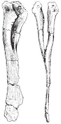

5 Reassessment of Jainosaurus A D 21 B C E F
 and anterior (B) views. Scale equals 5 cm.")

 did not establish a holotype for Jainosaurus (= Antarctosaurus ) septentrionalis.")


 was that the majority of them [the appendicular bones] belonged to the same individual.")
![..which died elsewhere... drifted to the locality...and [were] scattered over a limited area.](/docs-images/79/79702938/images/6-6.jpg "Second, there is ample information that justifies the association amongst some of the postcranial elements.")
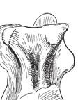
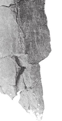
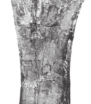
6 22 J. A. Wi l s o n e t a l. A B FIGURE 3 Jainosaurus septentrionalis, lectotypic braincase (GSI K27/497). Stereopairs and interpretive line drawings in posterior (A) and anterior (B) views. Scale equals 5 cm. Abbreviations (facing page): bo, basioccipital; bs, basisphenoid; bt, basal tubera; cr an, crista antotica; cr pr, crista prootica; eo-op, exoccipital-opisthotic; f, frontal; fm, foramen magnum; fo, fossa; gr, groove; hyp fo, hypophyseal fossa; ica, internal carotid artery; ls, laterosphenoid; mf, metotic foramen; os, orbitosphenoid; p, parietal; pro, prootic; so, supraoccipital; so cr, supraoccipital crest; stf, supratemporal fenestra; II XII, cranial nerve openings. Comments. Huene and Matley (1933) did not establish a holotype for Jainosaurus (= Antarctosaurus ) septentrionalis. Instead, they listed elements in a type series that included the braincase and skull roof, squamosum, hyoid, dorsal ribs, caudal vertebra, chevrons, scapulae, partial sternal plate, humeri, radius, and ulna. They did not associate all the elements as a single individual, but they considered them a single species based on their large size relative to bones of Titanosaurus indicus found at the same locality. They regarded the limb bones as belonging to different individuals based on limb proportions (Huene and Matley, 1933:32 33), which is surprising for several reasons. First, Matley s stated first impression (in Huene and Matley, 1933: 4) was that the majority of them [the appendicular bones] belonged to the same individual...which died elsewhere... drifted to the locality...and [were] scattered over a limited area. Second, there is ample information that justifies the association amongst some of the postcranial elements. Huene and Matley (1933: 19 21) remarked that the ribs were arranged in parallel order side by side, right and left scapulae have been found, possibly as a pair, and that the right humerus was found near the right scapula and 20 feet from the left scapula. The radius and ulna are of appropriate size relative to one another and to the humerus to pertain to a single individual (McIntosh, 1990; Wilson and Upchurch, 2003). Thus, there is a moderate level of support for associating some of the postcranial elements as a single individual (i.e., partial rib cage, shoulder, forelimb). In their discussion and diagnosis, Huene and Matley (1933: 32 33) considered the caudal vertebra, chevrons, braincase,
7 Reassessment of Ja i n o s a u r u s 23 eo-op p ls so cr so cr an f eo-op cr pr V gr pro fm bo bs p IV III VI fo ribs, and appendicular elements in turn, individually justifying their attribution to A. septentrionalis on the basis of their size. Specifically, they considered each of them to be too large to associate with Titanosaurus indicus caudal vertebrae, which were collected from the same locality:...haemopophyses cannot be associated with the smaller type of caudals, [t]he skull-fragment is too large for the smaller type of caudals, the ribs...probably go with the larger caudal, and elements of the shoulder and forelimb all are far too large for the post-median caudals of Titanosaurus indicus. Subtleties in the original description further illustrate that Huene and Matley (1933) did not consider all the elements in the type series to be a single individual. They distinguished between individual-level and species-level identity between bones in the type series in both the text and the legends to the plates and figures accompanying the paper. For example, the slender element identified as a hyoid was described as being from the same individual as the braincase, but postcranial elements in the type series were described as being the same species and from the same bed and locality p II hyp fo VII XII cr pr bs VI f eo-op ls pro p ls as the braincase (Huene and Matley, 1933:16 22; fig. 8). The lone caudal vertebra (GSI K20/317) they described, which provides the only potential overlap with T. indicus, was probably the same [taxon] as the braincase and other bones (Huene and Matley, 1933: fig 9). To summarize, Huene and Matley did not explicitly state that the cranial and postcranial elements of the type series of Jainosaurus (= Antarctosaurus ) septentrionalis belonged to a single individual, and there is evidence that they considered them to pertain to multiple individuals. Nonetheless, Huene and Matley (1933: 32 33) posited that these elements should form the basis of a new genus and species because of their relatively large size. Unfortunately, there is no supplementary data (e.g., field photographs, maps, personal accounts) that shed light on the original associations, but there is moderate support that at least the ribs, shoulder, and forelimb pertain to one individual (see Wilson and Upchurch, 2003). The key question, then, is whether circumstantial evidence supports individual associations between the braincase, forequarters, and caudal elements. Presently, five braincases are known from Indo-Pakistan that can be separated into two morphs the Titanosaurus morph and the Antarctosaurus morph (Wilson et al., 2005). Thus far, no definitive associations have been made between any of the five braincases and any postcranial remains. The strongest case for association comes from Dongargaon, central India, where a braincase (ISI R199; Berman and Jain, 1982) and the partial skeleton of Isisaurus (= Titanosaurus ) colberti (Jain and Bandyopadhyay, 1997) were found at the same quarry, although nearly 20 years apart. No other dinosaur bones have been collected at this locality, and Jain and Bandyopadhyay (1997: 131) considered that all material [from Dongargaon] belongs to T. colberti. If we accept the association between the Titanosaurus morph braincase and Isisaurus postcranial bones, and if we presume that there are only two common sauropod genera in the Indian Cretaceous, then we have an opportunity to forge associations between the other braincase morph (i.e., the Antarctosaurus morph ) with postcranial bones so long as those postcranial bones can be distinguished from Isisaurus postcranial bones and exhibit similar morphologies when they overlap. As described below, the Jainosaurus type series humerus is autapomorphic and can be easily differentiated from that of Isisaurus. Thus, on the basis of the probable association between cranial and postcranial remains at Dongargaon, the recognition of only two common sauropod genera in the Cretaceous of India, and the distinctiveness of both the braincase morphs and the postcranial morphs, we can tentatively link each of the braincase morphs to distinctive postcranial elements. Following these associations, we will refer to the Isisaurus morph and Jainosaurus morph braincases. DESCRIPTION Below is a redescription of the lectotype and referred Jainosaurus septentrionalis braincases as well as postcranial remains referred to the species (see above). This redescription is based on observations made at the Geological Survey of India collections
8 24 J. A. Wi l s o n e t a l. Table 1 Principal dimensions (in millimeters) of the lectotypic and referred braincases of Jainosaurus septentrionalis. In the lectotypic braincase, the braincase and skull roof are coossified, and many sutures are fully fused. The referred braincase, in contrast, is from an immature individual. Neither the orbitosphenoid-laterosphenoid nor the skull roof were coosified to the posterior skull elements. In the Malagasy titanosaur Rapetosaurus, the laterosphenoid and orbitosphenoid fuse to one another before they coossify with the posterior braincase elements (Curry Rogers and Forster, 2004). Asterisks (*) indicate measurement of an incomplete structure; the e indicates an estimate made by doubling. Region Dimension Lectotype (GSI K27/429) Referred (ISI R162) Posterior skull Width Height Supraoccipital to basal tubera Lateral skull Length 58 Supraoccipital Height Occipital condyle Width Height Length Foramen magnum Width 24 Height Basal tubera Width 94e 89 Basipterygoid processes Length distal to basal tubera 47 Supratemporal fenestra Hypophyseal fossa Anteroposterior length Distance to midline Length Width * in Kolkata, Nagpur, and Jaipur, the Indian Museum, Kolkata, the Indian Statistical Institute, Kolkata, Texas Tech University, Lubbock, and the Natural History Museum, London, as well as on information available in the published literature. Principal dimensions of the skulls are given in Table 1. The lectotypic braincase (GSI K27/497) lacks the paroccipital processes and basipterygoid processes, as well as the anteriormost portions of the basisphenoid and orbitosphenoid (Figs. 3 5). The parietal and frontal are preserved on the right side; the former lacks the squamosal articulation and posterior margin of the supratemporal fenestra, whereas the latter lacks the orbital margin and nasal and prefrontal articulations. The endocranial cavity was cast by Matley (Huene and Matley, 1933: fig. 6), and the floor, roof, and right side wall of the braincase are well preserved. A conspicuous suture separates the skull roof and braincase proper. Other sutures are fused, but their probable courses can be identified by roughened bone texture. A referred braincase (ISI R162) of a younger individual augments the description because it preserves anatomy not present in the lectotype. Skull Roof The endocranial cavity is capped by two paired midline elements, the frontal and parietal. These skull roof elements are united to the neurocranial elements that form the back and side walls of the endocranial cavity via a transversely broad, corrugated suture. Frontal. The frontal is mostly complete, but its margins are damaged. Only its posterior margin is completely preserved; its midline contact, anterior articulations with the nasal and prefrontal, as well as its orbital margin are absent. The frontal is broader than long, as in all sauropods except rebbachisaurids (Sereno et al., 1999), and it is thickest dorsoventrally at its lateral extreme (ca. 1.5 cm) and thinnest near the midline (< 0.5 cm). The plane formed by the frontal rises at an angle of degrees above horizontal from the midline towards its lateral extreme (Fig. 3B). Ventrally, the frontal contacts two lateral braincase elements, the orbitosphenoid and the laterosphenoid; the latter s contact is shared with the parietal (see below). The frontal-braincase suture subtends a gentle arc that begins at the posterior margin of the orbit and approaches the midline anteriorly. Although not completely preserved in Jainosaurus septentrionalis, this sutural arc would have continued anteriorly as a well marked ridge on the frontal that serves as a division between the olfactory and optic portions of the skull (e.g., Nemegtosaurus; Wilson, 2005: 293). Parietal. The parietal is a strap-shaped element that has sutural contacts with four neurocranial elements ventrally (supraoccipital, exoccipital-opisthotic, prootic, laterosphenoid), the frontal anteriorly. Its lateral contacts are not preserved. The parietal is abbreviate anteroposteriorly, and its anteroposterior length
9 Reassessment of Ja i n o s a u r u s 25 probably equaled one-fifth its transverse breadth. The midline parietal suture is sinuous and forms the middle portion of an H- shaped intersection with the frontals anteriorly and the occipital elements posteriorly. The parietal-frontal contact is distinguishable as a coarse, raised scar running transversely across the skull. Within the endocranial cavity, the parietal-frontal suture forms a transversely oriented ridge that, with the laterosphenoid, delimits forebrain from midbrain (see Endocast below). Laterally, the parietal separates into anterior and posterior rami, which form the medial border of the supratemporal fenestra. It is not known whether the supratemporal fenestra is completed laterally by the postorbital and the squamosal, as in most sauropods (e.g., Camarasaurus; Wilson and Sereno, 1998: fig. 7), or if the squamosal is excluded from its margin, as in some derived titanosaurs (e.g., Nemegtosaurus; Upchurch, 1995; Wilson, 2005: fig. 7). The preserved anteromedial portion of the supratemporal fenestra suggests that it was elliptical in dorsal view with its long axis directed nearly transversely (within 15 degrees of orthogonal to the midline). There is no well-defined supratemporal fossa medially on the parietal. The separation between the medial margin of the supratemporal fenestra (i.e., the point at which the parietal splits) and the skull midline is equivalent to half the estimated transverse breadth of the parietal. Based on its preserved medial margin, it appears that the supratemporal fenestra was horizontally oriented in both transverse and sagittal planes. In sauropod genera with laterally canted supratemporal fenestrae, both the parietal and the lateral elements (i.e., postorbital, squamosal) are tipped ventrally, so this condition can be detected from the parietal alone (e.g., Diplodocus; Wilson and Sereno, 1998: fig. 6). The anterior and posterior parietal rami of the parietal are floored by the laterosphenoid and prootic, respectively. The posterior margin of the parietal is raised into a transverse crest on either side of the midline. This parietal crest curls forward slightly in dorsal and lateral views (Figs. 4A, 5A) and has a convex dorsal margin that is highest near the medial margin of the supratemporal fenestra and lowest at the midline. The parietal contact with the posterior neurocranial elements is fused, and the exact course of the suture is difficult to discern. This suture is quite high near the midline and drops down sharply on either side of the supraoccipital crest and then climbs slowly along the dorsal margin of the exoccipital-opisthotic before dipping shortly and flattening out at its lateral extreme. Braincase The lectotypic braincase is well preserved, but lacks several critical features, such as the distal ends of the paroccipital processes, the basipterygoid processes, and the parasphenoid rostrum (Figs. 2 5). The left wall of the braincase is missing, but nearly all lateral elements are present on the right side. The bony enclosure of the brain is formed posteriorly and ventrally by three median elements (supraoccipital, basioccipital, basisphenoid) and laterally by four paramedian elements (exoccipital-opisthotic, prootic, laterosphenoid, orbitosphenoid). These elements are described in order below, with supplementary information from the referred braincase (ISI R162). Supraoccipital. The supraoccipital is a median element that forms the posterodorsal portion of the braincase and roofs the foramen magnum. It contacts the exoccipital-opisthotic ventrolaterally and the parietal dorsally. The supraoccipital of Jainosaurus is well preserved on its right side, but the left side is mostly missing. The contact between the supraoccipital and parietal is slightly obscured by fusion, but it appears drop sharply as it travels laterally from the nuchal crest, but then rises sharply near the level of the crest of the occipital process of the parietal. This highly sinuous contact is also present in the referred braincase and is shared with Antarctosaurus wichmannianus. The suture between the supraoccipital and exoccipital is nearly completely fused in the lectotypic and referred braincases, so the relative contribution of these elements to the foramen magnum is unknown. The height of the supraoccipital is slightly greater than the dorsoventral diameter of the foramen magnum, unlike the condition in Rapetosaurus and Nemegtosaurus (Wilson, 2002). The nuchal crest is prominent and formed from a single ridge, as in most titanosaurs. Exceptions include Rapetosaurus, Bonatitan, the Río Negro taxon (MML 194; García et al., 2008), and Muyelensaurus, which have a median groove, and Pitekunsaurus, which lacks both the groove and the ridge. Basioccipital. The basioccipital is the posteriormost ventral median braincase element. It is a short, heavy bone that forms the majority of the occipital condyle and part of the basal tubera. It is slightly damaged ventrally and on its left side. The occipital condyle projects 120 degrees from the vertical, as determined by the angle between the plane of the occiput and the dorsal surface of the occipital condyle (Fig. 4A). The articular surface circumscribing the condyle itself extends far forward at its ventral extreme, suggesting that the skull was held at an angle to the long axis of the anterior cervical vertebrae. The opening for cranial nerve XII passes through the dorsolateral neck of the occipital condyle; it is not clear whether the margin for this opening is partly shared by the exoccipital-opisthotic. A small, poorly defined, ventrally facing, circular depression is positioned between the occipital condyle and the basal tubera, a feature that is present in several neosauropods (Wilson, 2002). Anteriorly, the basioccipital is expanded into a transversely flat, vertically deep sheet of bone that articulates with the basisphenoid. The basioccipital-basisphenoid suture can be identified traversing the floor of the endocranial cavity between the right and left metotic (= jugular) foramina, though which exit cranial nerves IX XI and the jugular vein. Consequently, the basioccipital forms the posteroventral margin of the metotic foramen. The basioccipital-basisphenoid suture extends ventrally to the posterior portion of the basal tubera, the bulk of which are made up by the basisphenoid. The basioccipital is beveled on its lateral extremes for attachment of the paired exoccipital-opisthotic elements. The suture separating the basioccipital from the exoccipital-opisthotic is visible where it undercuts the dorsolateral shoulder of the occipital condyle. The occipital condyle is convex about its circumference except for the middle third of its dorsal margin, where it forms the concave floor of the foramen magnum. Basisphenoid. The basisphenoid contacts the basioccipital posteriorly, flooring the endocranial cavity from the level of the
 and internal (B) views. Scale equals 5 cm.")


.")
.")

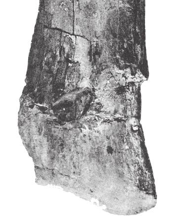
10 26 J. A. Wi l s o n e t a l. A B FIGURE 4 Jainosaurus septentrionalis, lectotypic braincase (GSI K27/497). Stereopairs and interpretive line drawings in right lateral (A) and internal (B) views. Scale equals 5 cm. Abbreviations (facing page) as in Figure 3. metotic foramen forwards. Dorsally, the basisphenoid contacts the prootic, laterosphenoid, and orbitosphenoid, which appear to exclude it from the margin of cranial nerves V and III. The basisphenoid apparently did not border the opening for cranial nerve VIII either, as it is excluded by the prootic and exoccipital-opisthotic. The opening for cranial nerve VI pierces the basisphenoid ventral to the level of these larger cranial nerve openings. The basisphenoid forms at least the posteroventral margin of the opening for cranial nerve II, but because the orbitosphenoid and basisphenoid are broken anteriorly the extent of their contributions is unknown. The basisphenoid forms the bulk of the basal tubera, the basipterygoid processes, a portion of the hypophysial canal, and the parasphenoid. Neither the parasphenoid nor the basipterygoid processes are preserved in the lectotypic braincase, but the referred Jainosaurus braincase (ISI R162) preserves basipterygoid processes. In ISI R162 the basipterygoid processes are long and directed parallel to the plane of the occiput (Fig. 6B). The distance from the base of the occipital condyle to the basal tubera is subequal to the distance from the basal tubera to the ends of the basipterygoid processes (~4.5 cm). The basipterygoid processes taper transversely towards their distal extreme, but they maintain their anteroposterior dimension along their length. The crosssection of the basipterygoid processes at mid-length is shaped like a quarter-circle, with the posterior face rounded and the anterior and lateral faces flat and oriented about 90 degrees to one another. The long axes of the basipterygoid processes diverge from the midline at an angle of about 50 degrees. The basal tubera are formed by the basioccipital and basisphenoid. They are sheetlike and quite broad laterally, extending well beyond the lateral margin of the occipital condyle. They do not project far posterior to the occipital plane, and
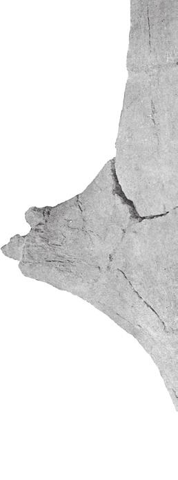
11 Reassessment of Ja i n o s a u r u s 27 pro eo-op cr an f bo bt ls IV os II III bs gr ica ls V p mf VII V they grade smoothly into the basipterygoid processes. Although not completely preserved on either side, their breadth and lateral thickening is reminiscent of those of the titanosaurs Nemegtosaurus and Quaesitosaurus, both of which have a bony connection between the basal tubera and the medial aspect of the quadrate (Wilson, 2005: figs. 9, 16). The basal tubera have a broad, gentle fossa on their posterior surface (Fig. 3A) that is also present in Malagasy Taxon B (Curry Rogers, unpublished data) and the South American titanosaurs Muyelensaurus (Calvo et al., 2007), Pitekunsaurus (Fillipi and Garrido, 2008), and an unnamed taxon from Río Negro (MML 194; García et al., 2008). The posterolateral corner of the basal tubera is not preserved in the lectotype, but it is preserved in the referred braincase (ISI R162). In ISI R162, the posterolateral portion of the basal tuber has a small, pendant process that is divided from the rest of the basal tuber by a sharp notch. This distinctive feature is also present in Malagasy Taxon B (Curry Rogers, unpublished data). VII III IV ls cr pr bt p f bs VI II so eo-op pro VIII mf XII eo-op bo os Exoccipital-opisthotic. The paired exoccipital-opisthotic bones form the posterior portion of the endocranial cavity and are exposed on the occiput, where they form the paroccipital processes, the lateral margin of the foramen magnum, and the dorsolateral shoulders of the occipital condyle. Only the right exoccipital-opisthotic is preserved, and it is broken near its base. Consequently, the shape of the paroccipital process as well as its contacts with the parietal, squamosal, and quadrate are not preserved in the lectotype. The broken base of the paroccipital process, however, shows the contact between the exoccipitalopisthotic and prootic in cross-section (Fig. 4A). The latter is sheet like and covers a portion of the anterior face of the paroccipital process. The exoccipital-opisthotic contacts the basioccipital along a nearly horizontal sutural plane extending from metotic foramen posteriorly to the occipital condyle. However, the exoccipital-opisthotic contact with the supraoccipital is not visible, so the relative contributions of these two elements to the border the foramen magnum cannot be determined. A low, transversely elongate prominence on each side of the foramen magnum may represent the proatlantal facets; it cannot be determined whether these are wholly or partly formed by the exoccipital-opisthotic. The left paroccipital process is completely preserved in the referred braincase from Bara Simla (ISI R162; Fig. 6). This specimen indicates that the paroccipital processes arched ventrally and projected ventral to the occipital condyle. A sharp notch in the dorsal border of the paroccipital process forms the lower margin of the posttemporal foramen. Just distal to this notch is a rounded prominence that articulated with the posterior portion of the quadrate head. This prominence marks the lateral extreme of the paroccipital process, which descends sharply from this point as a tongue-like flange. This flange, which is extremely narrow in lateral view, is probably homologous to the pendant, nonarticular processes present at the distal extreme of the paroccipital processes in other titanosaurs (e.g., Rapetosaurus, Nemegtosaurus, Malawisaurus). Prootic. The prootic is a transversely oriented element that forms the posteriormost portion of the braincase side wall. It is preserved only on the right side but is not complete laterally, like the exoccipital-opisthotic. As its name suggests, the prootic is positioned anterior to the ear and shares the perimeter of the metotic foramen with the exoccipitalopisthotic, basioccipital, and basisphenoid. It contacts the laterosphenoid anteriorly, and it has a ventrally-directed spur that overlaps the basisphenoid to the level of the basal tubera. The prootic and laterosphenoid contact one another along a suture that extends from the groove for the exit of cranial nerve V to the medial margin of the supratemporal fenestra, where it intersects the junction between the parietal and the neurocranial elements. The prootic forms a vertical crest, the crista prootica, that is visible in both lateral and anterior views. The crista prootica is not expanded laterally into a tab-like structure, as it is in dicraeosaurid sauropods such as Dicraeosaurus (Janensch, : figs ) and Amargasaurus (Salgado and Calvo, 1992: figs. 1B, 2B). Instead, it is a posteriorly convex crest that forms the posterior margin of the groove for cranial nerve V, which is completely enclosed by the prootic and
 and ventral (B) views. Scale equals 5 cm. Abbreviations (facing page) as in Figure 3.")


. The laterosphenoid also has sutural connections to three neurocranial elements, the prootic, orbitosphenoid, and basisphenoid.")

12 28 J. A. Wi l s o n e t a l. A B FIGURE 5 Jainosaurus septentrionalis, lectotypic braincase (GSI K27/497). Stereopairs and interpretive line drawings in dorsal (A) and ventral (B) views. Scale equals 5 cm. Abbreviations (facing page) as in Figure 3. laterosphenoid, continuing ventrally on the basisphenoid as the crest leading to the basipterygoid processes. Immediately posterior to the crista prootica and slightly ventral to the trigeminal foramen is the opening for cranial nerve VII. The prootic is the neurocranial element that houses the semicircular canals; these elements could not be viewed, but a bulge visible in medial view marks their location. The broken left prootic preserves a small channel that is likely the opening into the vestibule. Laterosphenoid. Another transversely oriented element, the laterosphenoid, is important because it marks boundaries within and external to the endocranial cavity. Internally, the pillar of the laterosphenoid divides forebrain from midbrain. The laterosphenoid pillar decreases in diameter dorsally. Externally, its transversely oriented, arched arm (the crista antotica) separates the supratemporal fenestra from the orbit (Figs. 3B, 4A, 5B). The dorsal surface of the laterosphenoid contacts the parietal and frontal and thus marks the course of their suture. Together, the laterosphenoid (ventrally), parietal (posterodorsally), and frontal (anterodorsally) make a three-way contact with the postorbital. The laterosphenoid is slightly expanded into a condyle distolaterally that represents a synovial contact with the postorbital. The crista antotica continues ventrally as a sharp but low ridge that forms the anterior margin of the groove for cranial nerve V (the posterior margin is formed by the prootic; see above). The laterosphenoid also has sutural connections to three neurocranial elements, the prootic, orbitosphenoid, and basisphenoid. The anterior and posterior margins of the laterosphenoid contact the orbitosphenoid and prootic, respectively. The laterosphenoid-prootic suture and the laterosphenoid-orbitosphenoid suture are visible on the internal surface of the endocranial cavity. Both are vertical or near-vertical sutures that are interrupted by openings for cranial nerves. Posteriorly, the laterosphenoid and prootic are separated by the opening for cranial nerve V, and their junction passes vertically to the medial margin of the supratemporal fenestra. Anteriorly, the laterosphenoid and orbitosphenoid are separated by the openings for cranial nerves III and IV, and their suture extends dorsally to a position within the posterior portion of the orbital cavity. Medially,
13 Reassessment of Ja i n o s a u r u s 29 so p II hyp fo ls f pro eo-op os the laterosphenoid bears a narrow pillar that is visible internal view (Fig. 4B). This ridge corresponds to the division of forebrain and midbrain, and it can be seen as a circumferential constriction in the endocast (Huene and Matley 1933: fig. 6). The contact between the laterosphenoid and the basisphenoid can only be identified internally, where their suture can be viewed on the posterior portion of the hypophyseal fossa. The laterosphenoid extends medially at its base to form the dorsolateral portion of the sella turcica. Orbitosphenoid. The orbitosphenoid is the only paired braincase element that forms a midline contact, enclosing the forebrain and forming the anterior portion of the hypophyseal fossa. The orbitosphenoid in Jainosaurus is not complete anteriorly, but its contacts with the basisphenoid ventrally, the laterosphenoid posteriorly, and the frontal dorsally are well preserved on the right side. A narrow, ventrally directed process of the orbitosphenoid overlaps the basisphenoid laterally, extending towards the opening for cranial nerve VI, which is near the sella turcica (Fig. 4A). The orbitosphenoid-basisphenoid suture courses anterodorsally to the margin of cranial nerve II. Dorsally, the orbitosphenoid has a long, broad contact with the frontal, with which it forms a portion of the orbit. Although it is not preserved anteriorly, the orbitosphenoid would have formed all but the roof of the openings for cranial nerve I. p II stf hyp fo bs f bo fo Cranial Nerves. Openings for all but the first cranial nerve are preserved on the lectotypic braincase (GSI 27/497), and all of the posterior cranial nerve openings (V XII) are preserved on the referred braincase (ISI R162). Openings for cranial nerves II, III, V, and VII XII fall on a line that extends to the foramen magnum. Openings for cranial nerves I and IV are positioned dorsal to this line, and the opening for cranial nerve VI is positioned ventral to it. This appears to be the general condition for sauropods. The margins of the opening for cranial nerve I are not preserved, but in most sauropods they are bounded ventrally by the orbitosphenoids and dorsally by the frontals. The anterior end of the braincase preserves approximately half of the margin of the opening for cranial nerve II, which is shared by the orbitosphenoid and basisphenoid. The opening for cranial nerve III is positioned anterior to the crista antotica, and its margin is formed by the orbitosphenoid and laterosphenoid. The opening for cranial nerve IV is also bordered by the orbitosphenoid and laterosphenoid, but it is much smaller than and positioned dorsal to the opening for cranial nerve III. The trigeminal foramen is formed by the prootic and laterosphenoid. It is large and appears to have accommodated all three branches of the trigeminal nerve exit from a single foramen. In some non-sauropod dinosaurs, branches of the trigeminal nerve (cranial nerve V) have separate exits from the braincase (e.g., Majungasaurus; Sampson and Witmer, 2007). The path of the mandibular branch of cranial nerve V is marked by a shallow groove that extends ventrally. In both the lectotypic and referred braincases, this groove is bounded posteriorly by the crista prootica and anteriorly by a narrow spur of the laterosphenoid that represents the continuation of the crista antotica. Just posterior to the crista prootica is a small opening for cranial nerve VII, which is preserved in both braincases. The opening for cranial nerve VI is small and positioned ventrally. As in other sauropods, it faces anterolaterally. The metotic foramen (cranial nerves IX XI) and the foramen ovale (cranial nerve VIII) are seated in a large fossa just posterior to the crista prootica. The margin of the metotic foramen is shared by prootic, exoccipital-opisthotic, and the basioccipital, whereas the foramen ovale appears to bounded by only the prootic and exoccipital-opisthotic. This latter condition is best observed internally, where the foramen ovale is positioned dorsal to the metotic foramen and clearly bordered by only two bones. Externally, however, the foramen ovale appears to be positioned anterodorsal to the metotic foramen, which is typical for sauropods. The foramen ovale is also positioned anterior to the metotic foramen in the referred braincase (ISI R162; Chatterjee and Rudra, 1996: fig. 9B). It appears that the trajectories of these two closely positioned openings are slightly divergent such that their positions relative to one another differ between the inside and outside of the braincase. A single opening for cranial nerve XII is located on the dorsolateral side of the neck of the occipital condyle. Endocast. An endocast was prepared by Matley and described and figured by Huene and Matley (1933:fig. 6). Several
 posterior and (B) left lateral views. Scale equals 5 cm.")

 and the")

 septentrionalis by")


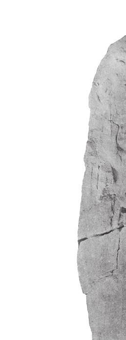
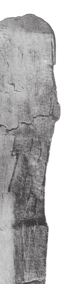
14 30 J. A. Wi l s o n e t a l. pop fo A no bpt pop bt B FIGURE 6 Jainosaurus septentrionalis, referred braincase (ISI R162). Stereopairs in (A) posterior and (B) left lateral views. Scale equals 5 cm. Abbreviations as in Figure 3, plus: no, notch; pop, paroccipital process. bpt general features are observable on the endocast, including the constriction between the forebrain and midbrain by the laterosphenoid pillar, the proximal hypophyseal fossa, and the bases of most cranial nerves (II V, VII, IX XI, XII; Fig. 7). The strong circumferential division at the midpoint of the endocast marks the sutural zone between the laterosphenoid (ventrally) and the frontal/parietal (dorsally). The shape of the distal portion of the hypophysis is not preserved in the endocast; its shape and the shape and size of the exit for the internal carotid arteries are unknown. Postcranial Skeleton Several postcranial elements were included within the type series of Jainosaurus (=Antarctosaurus) septentrionalis by Huene and Matley (1933: 11 23), some of which appear to represent a single individual (Fig. 2). These elements, which pertain to the anterior rib cage and forelimb, are redescribed below based on first-hand observations of the two elements that remain in collections. Other elements from the type series are not attributable to this individual (i.e., caudal vertebra, chevrons, sternal plate ) and not described here; neither are the second humerus and femur referred to the genus by Huene and Matley (1933: 34 35). Ribs. Huene and Matley (1933: 19) reported that a large number (about 28) of broken pieces of ribs were found in different parts of the Sauropod bed, some of them arranged in parallel order side by side... The ribs figured by Huene and Matley (1933: figs ; Fig. 2B) are all broad, but some of the ribs are described by the authors as being triangular in crosssection. Plank-like anterior dorsal ribs with internal pneumaticity would be expected in Jainosaurus, as in other titanosauri-

 views. Positions of brain regions are approximate and based on Witmer et al.")


 to pertain to a single individual due to their similar size and")
 remarked that the left scapula was found in 1917 and the")
, but it was subsequently broken presumably prior to photography for the")
. The left scapula, in turn, was found incomplete (length = 1.32 m).")


15 Reassessment of Jainosaurus 31 de sul ls I sc ce op cb me hyp fo III A VII V II IV IX-XI XII hyp fo B FIGURE 7 Jainosaurus septentrionalis, endocast prepared by C. Matley from the lectotypic braincase (GSI K27/497) in right lateral (A) and ventral (B) views. Positions of brain regions are approximate and based on Witmer et al. (2008). Dotted lines indicate position of cranial nerves. Scale equals 2 cm. Abbreviations as in Figure 3, plus: cb, cerebellum; ce, cerebrum; de, dural expansion; me, medulla; op, optic lobe; sc, spinal cord; sul ls, sulcus for laterosphenoid pillar. forms (Wilson, 2002), but we cannot confirm these predictions because these elements cannot be located in the collections of the GSI. Scapula. A pair of right and left scapulae (GSI unnumbered) were considered by Huene and Matley (1933) to pertain to a single individual due to their similar size and proximity to one another in the Sauropod bed (Fig. 2D). Huene and Matley (1933: 20) remarked that the left scapula was found in 1917 and the right scapula in The latter was complete when it was first found (length = 1.67 m), but it was subsequently broken presumably prior to photography for the monograph, which clearly shows a broken distal blade (Huene and Matley 1933: pl. 3, fig. 1). The left scapula, in turn, was found incomplete (length = 1.32 m). Despite minor differences, which were enhanced by presentation of the two scapulae in different orientations and at different scales in Huene and Matley (1933: pl. 3), we consider it likely that these elements pertain to the same individual based on their similar size and shape. Unfortunately, neither scapula can be located in the collections of the GSI, but a cast of the right scapula sits atop cabinets in the Invertebrate Fossil Gallery of the Indian Museum (Kolkata). There are minor differences between the cast and the published photos of the right scapula (Huene and Matley 1933: pl. 3, fig. 1). Most of these differences are attributable to subtle differences in the orientation of the element, as well as apparent breakage to the base of the scapula between photographs (compare Huene and Matley, 1933: fig. 1a and 1b), but there is nonetheless a minor difference in the dorsal portion of the distal blade resulting from reconstruction to the cast. Based on available photographs of the scapulae, in particular the better-preserved
 is indicated by the tick mark adjacent to it. Scale equals 25 cm.")

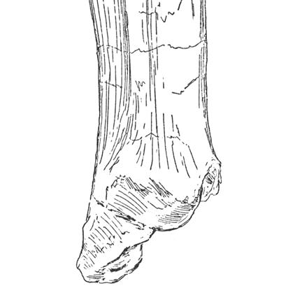
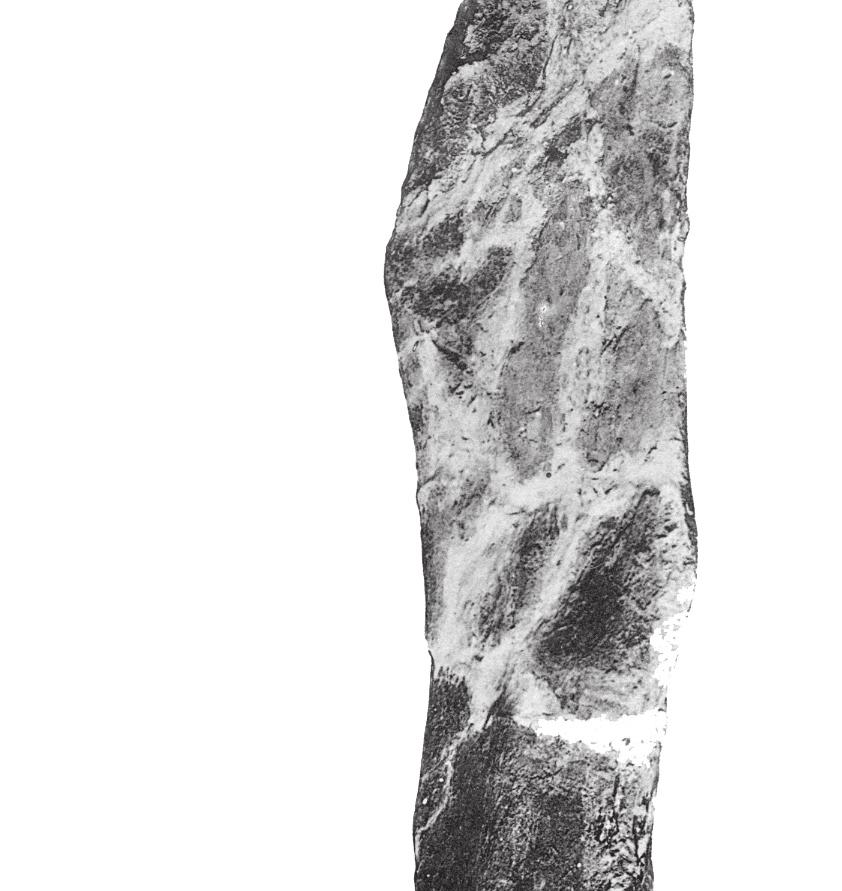
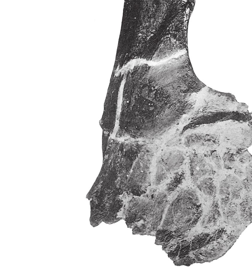
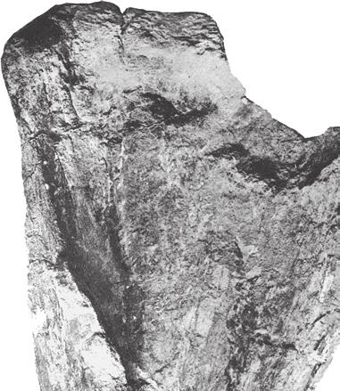
16 32 J. A. Wi l s o n e t a l. dpf plc dpc dpf b b A B FIGURE 8 Jainosaurus septentrionalis, lectotypic right humerus (GSI unnumbered) in (A) anterior and (B) posterior views. Crosssections are oriented so that anterior faces up and lateral faces left. The position of each section (black-filled shapes) is indicated by the tick mark adjacent to it. Scale equals 25 cm. Abbreviations: b, bulge; dpc, deltopectoral crest; dpf, deltopectoral fossa; plc, proximolateral corner. but less complete left scapula, there is a gentle expansion of the dorsal margin of the blade distally. Just proximal to this expansion, the blade is markedly concave and grades into the acromion. The apparent sharp narrowing of the ventral margin of the blade visible in the left scapula (see Huene and Matley, 1933: fig. 16; Fig. 2) is clearly due to breakage; the right scapula shows a continuous, gently concave ventral margin of the scapula. In cross section, the blade appears to have been concave medially and somewhat sharply convex laterally, due to the presence of a strong ridge that extends from the acromial fossa to mid-blade. Neither glenoid is preserved, and the orientation of its articular surface is not known. Humerus. The type series humerus (GSI unnumbered) was recently rediscovered atop a case in the Siwalik Gallery of the Indian Museum. Portions of the proximal end and a small piece of the distal end are now missing, but the bone is remarkably well preserved and provides details that were not previously visible in published photographs. Although images of the humerus in two views have been available (Huene and Matley, 1933: pl. 4, fig. 1), inadvertent cropping of the photograph and/or slight rotation of the humerus resulted in loss of one of the diagnostic features of this bone (see Figs. 2E, 7). The humerus is not complete, but it would have been somewhat gracile; the breadth of the proximal end is only 30-40% the estimated length of the humerus, and the midshaft transverse diameter (21.5 cm) is only about 15% of the preserved length of the humerus (133 cm). The proximolateral corner of the Jainosaurus humerus is pronounced; its lateral margin is very slightly concave and forms a slight but distinct angle with its more gently rounded dorsal margin. This feature has been variously noted as a squared proximolateral corner (e.g., Curry Rogers and Forster, 2001: character 119), a prominent process on the lateral portion of the proximal end of the humerus (Upchurch, 1998: character C160), or as the enlarged attachment site for the m. supracoracoideus (Borsuk-Bialynicka, 1977). The anterior face of the proximal end of the humerus (i.e., the deltopectoral fossa) is shallowly concave, anteroposteriorly thin, and transversely broad (48 cm). The curvature of the lateral margin of the humeral shaft is less than that of the medial border. The cross-section of the humerus at midshaft is elliptical and measures 21.5 cm by
17 Reassessment of Ja i n o s a u r u s cm, with a circumference of 61 cm (Fig. 8). The deltopectoral crest is located proximally, with its apex located only about 25% of the preserved humeral length from the proximolateral corner of the humerus. The transversely thickened deltopectoral crest is medially offset and obliquely oriented with respect to the long axis of the humerus (Fig. 8A). The apex of the deltopectoral crest thickens transversely towards midshaft. Just lateral to its thickest point is a prominent posterolateral bulge, which bears a roughened area indicative of a muscular attachment site. The distal end of the humerus is not preserved. Several isolated humeri from Indo-Pakistan share diagnostic features with the type series humerus, including the obliquely oriented and medially offset deltopectoral crest, anteroposteriorly thin bone in the deltopectoral fossa, and the prominent posterolateral bulge near the deltopectoral crest, as well as more general features such as its generally gracile profile. Some of these features are present in other titanosaurs (e.g., Epachthosaurus, Opisthocoelicaudia) and their distribution is not yet well constrained, but they are thought to be derived within Titanosauria rather than primitive for the group. These features are present in sauropod humeri from Indo-Pakistan, such as materials from Rahioli, Gujarat (GSI , 20012) described by Mathur and Pant (1986). On the basis of several metrics, Mathur and Pant (1986) linked the Gujarati humeri to the genus Antarctosaurus, but not to A. septentrionalis due to size differences. Materials from Chhota Simla (near Bara Simla) collected by Matley and described by Swinton (1947) also preserve these features (Wilson et al., in preparation), as do incomplete humeri from the Pab Formation of Pakistan. Although the lengths of the referred Gujarat humeri (70 85 cm) and the Chhota Simla humerus (94 cm) are significantly shorter than the type series humerus (134 cm), their overall shape and diagnostic features justify referral to Jainosaurus septentrionalis. The more complete Gujarat humeri can be referred to Jainosaurus, and they furnish additional information about humeral morphology, as well as an additional diagnostic feature the radial condyle of the distal humerus is expanded and exposed anteriorly, whereas the ulnar condyle is not. This feature also appears to be present in the humerus of the recently described Chinese titanosaur Qingxiusaurus (Mo et al., 2008) and that of an undescribed large titanosaur from Argentina (MUCPv 1164; J. Calvo, personal communication 2009). Radius and Ulna. A radius (GSI K27/490) and ulna (GSI K27/491) were described and figured by Huene and Matley (1933: figs ; Fig. 2F). These bones cannot be located in the collections of the GSI, so our comments are based on the text and illustrations. The radius appears to be expanded at one end. Most have interpreted that to be the distal end (Huene and Matley, 1933; McIntosh, 1990), but it could reasonably be interpreted as the proximal end. The radius preserves a prominent ridge that likely represents the attachment surface for interosseus ligaments. The ulna is badly eroded proximally and distally, so many of the relevant features cannot be determined (e.g., presence of olecranon, expansion of distal end). Huene and Matley (1933: 22) described its cross-sectional shape as stout and somewhat subrectangular, which distinguishes it from the gracile and triradiate shaft of Isisaurus (Wilson and Upchurch, 2003). The humerus:radius ratio reported by Huene and Matley (1933) is 0.58, similar to the value for some derived titanosaurs (Wilson and Upchurch, 2003: table 2). We could not confirm that these bones pertain to the right side, as stated by Huene and Matley (1933). Note that Huene and Matley (1933) incorrectly specified that figures 17 and 18 were scaled to 1/4 natural size. The images are actually scaled to 1/6 natural size, as were other other limb elements, based on the measurements listed in the text. COMPARISONS AND PHYLOGENETIC AFFINITIES Early assessments of the phylogenetic affinities of Jainosaurus septentrionalis linked it to the South American titanosaur Antarctosaurus wichmannianus based on features now demonstrated to have a broader distribution within titanosaurs or titanosauriforms (McIntosh 1990; Curry Rogers and Forster, 2004; Wilson, 2005). Although titanosaur phylogeny is still hindered by a lack of significant skull material for the majority of taxa, several comparisons can be made. Below, we make comparisons to titanosaur species that might be expected to share a close relationship with Jainosaurus septentrionalis on the basis of original taxonomy (Antarctosaurus wichmannianus), sympatry (Isisaurus colberti), paleobiogeography (Rapetosaurus krausei, Malagasy Taxon B ), and morphology (various taxa). Antarctosaurus wichmannianus. As with Jainosaurus, the absence of documented field associations has fostered doubts about the phylogenetic affinities of Antarctosaurus wichmannianus. Material originally referred to that genus by Huene (1929) has been suggested to pertain to two or more taxa (Upchurch, 1999; Sereno and Wilson 2005; Wilson, 2005). More recently, others have claimed that the cranial materials pertain to a single individual (e.g., Apestiguía, 2004; Gallina and Apestiguía, 2005; Novas et al., 2005; Salgado and Bonaparte, 2005; Salgado and Coria, 2005). Herein we limit our comparisons to the braincase and skull roof of A. wichmannianus, which share with Nemegtosauridae a reduced posterior process of the postorbital and beveled basal tubera, as well as the more widespread condition of paroccipital processes with pendant non-articular distal processes (Wilson, 2005). McIntosh (1990) and Hunt et al. (1994) noted that the braincases of Jainosaurus septentrionalis and A. wichmannianus were too different to allow their placement into the same genus (both studies also note that the postcrania are more similar than the braincases of the two specimens). The frontals of A. wichmannianus are peaked medially and meet in a prominent midline ridge, a feature that may be autapomorphic. In contrast, the frontals of Jainosaurus descend slightly towards the midline and lack a midline ridge. In both

18 34 J. A. Wi l s o n e t a l. A. wichmannianus and Jainosaurus, the plane of the frontals rises laterally; but in the latter they are elevated slightly more so than the former. Features of the braincase also distinguish the two taxa. A. wichmannianus possesses two openings near the foramen magnum visible in posterior view. Huene (1929: pl. 28) identified the smaller of these as the opening for cranial nerve XII, and the larger as the vestibular foramen, but Powell (2003) regarded the larger as the opening for cranial nerve XII (he did not identify the smaller opening). The identity of these openings remains ambiguous, but the elevated location of cranial nerve XII and the presence of a second opening on the occiput differ from the condition in Jainosaurus. In A. wichmannianus the occipital condyle is approximately as tall as it is wide, whereas in Jainosaurus the condyle is slightly wider than it is tall. The foramen magnum is relatively taller and narrower in A. wichmannianus than it is in Jainosaurus, but the supraoccipital appears relatively shorter. The basal tubera are broader than is the occipital condyle in both taxa, but other aspects of the basicrania differ. In Jainosaurus, the basal tubera have a broad posterior fossa and grade gently into the basipterygoid processes, whereas A. wichmannianus lacks this fossa, and its basal tubera are distinctly offset from the basipterygoid processes. In Jainosaurus, the basipterygoid processes have a straight axis, but in A. wichmannianus, they are recurved posteriorly. In lateral view, the relative positions of the openings for cranial nerves II V differ in A. wichmannianus and Jainosaurus. In Jainosaurus the openings for cranial nerves II, III, and V are collinear, but in A. wichmannianus this line is inclined such that it also passes through the opening for cranial nerve I. This feature may be autapomorphic. In addition, the braincase of Jainosaurus is larger than that of A. wichmannianus, but fewer elements are fused, suggesting that the two taxa had substantially different adult body sizes. Although they do not pertain to the same genus, the braincases of both Jainosaurus septentrionalis and Antarctosaurus wichmannianus appear to belong to derived titanosaurs, sharing close affinities with Nemegtosauridae. However, the paucity of cranial remains within Titanosauria is such that cranial synapomorphies of Nemegtosauridae are ambiguous (Wilson, 2005); how closely the two genera are related awaits further discoveries and analyses. Isisaurus colberti. According to Wilson et al. (2005), Indian sauropod braincases appear to fall into two morphs: a robust and compact Isisaurus morph and a wide and flat Jainosaurus morph. Isisaurus morph braincases include specimens from Dongargaon, central India (ISI R199; Berman and Jain, 1982), Gujarat, western India (ISI R467; Chatterjee and Rudra, 1996), and Balochistan, western Pakistan (GSP- UM 7000; Wilson et al., 2005). Jainosaurus morph braincases include the two specimens from Bara Simla, central India, described here (GSI 27/497; ISI R162; Chatterjee and Rudra, 1996). A salient difference between the Isisaurus and Jainosaurus morph braincases is the angle of projection of the occipital condyle. Berman and Jain (1982) noted that the occipital condyle is deflected from the plane of the occiput at an angle of about 120 degrees in the Isisaurus morph braincase from Dongargaon, dpc FIGURE 9 Isisaurus colberti, right humerus (ISI R335/59) in anterior view. Scale equals 25 cm. Abbreviations as in Figure 8. India (ISI R199), a feature that they regarded to be similar to Diplodocus and Apatosaurus. Wilson et al. (2005) reported a significant downward deflection of the occipital condyle in the Isisaurus morph braincase from Pakistan, in which the skull roof and occipital condyle meet at an angle of about 120 degrees. It should be noted that these two metrics measure different angles; the former is in reference to a subvertical plane formed by the plane of the occiput (paroccipital processes and supraoccipital), and the latter is in reference to a subhorizontal plane formed by the frontals and parietals. These two metrics attempt to capture the same feature the orientation of the occipital condyle relative to a fixed plane in the skull. The long axis of the occipital condyle and the plane of the skull roof are nearly parallel (within 10 degrees) in most sauropods, including Shunosaurus (Zheng, 1991), Camarasaurus (Madsen et al., 1995), Brachiosaurus (Janensch, ), Diplocodus (Osborn, 1912), Nemegtosaurus (Wilson, 2005), Nigersaurus (Sereno et al., 2007), Rapetosaurus (Curry Rogers and Forster, 2004), and the indeterminate titanosauriform from Texas (Tidwell and Carpenter, 2003). In contrast, the long axis of the occipital condyle more closely parallels (within 25 degrees) the plane of the occiput in Isisaurus morph braincases from Pakistan, Rahioli, and Dongargaon (GSP-UM 7000, ISI R467, ISI R199, respectively). Wilson et al. (2005) pointed out that the deflection of the skull shared between diplodocids and Isisaurus morph braincases is achieved via different structural arrangements of the bones of dpf
A Sauropod Braincase from the Pab Formation (Upper Cretaceous, Maastrichtian) of Balochistan, Pakistan
 of Balochistan, Pakistan") Gond. Geol. Magz., Spl. V. 8, April, 2005. pp. 101-109 A Sauropod Braincase from the Pab Formation (Upper Cretaceous, Maastrichtian) of Balochistan, Pakistan 1 2 1 Jeffrey A. Wilson, M. Sadiq Malkani,
Gond. Geol. Magz., Spl. V. 8, April, 2005. pp. 101-109 A Sauropod Braincase from the Pab Formation (Upper Cretaceous, Maastrichtian) of Balochistan, Pakistan 1 2 1 Jeffrey A. Wilson, M. Sadiq Malkani,
AMERICAN MUSEUM NOVITATES Published by
 AMERICAN MUSEUM NOVITATES Published by Number 782 THE AmzRICAN MUSEUM OF NATURAL HISTORY Feb. 20, 1935 New York City 56.81, 7 G (68) A NOTE ON THE CYNODONT, GLOCHINODONTOIDES GRACILIS HAUGHTON BY LIEUWE
2. Skull, total length versus length of the presacral vertebral column: (0); extremely elongated neck (e.g. Tanystropheus longobardicus).
; extremely elongated neck (e.g. Tanystropheus longobardicus).") Character list of the taxon-character data set 1. Skull and lower jaws, interdental plates: absent (0); present, but restricted to the anterior end of the dentary (1); present along the entire alveolar
Character list of the taxon-character data set 1. Skull and lower jaws, interdental plates: absent (0); present, but restricted to the anterior end of the dentary (1); present along the entire alveolar
A new species of Hsisosuchus (Mesoeucrocodylia) from Dashanpu, Zigong Municipality, Sichuan Province
 from Dashanpu, Zigong Municipality, Sichuan Province") A new species of Hsisosuchus (Mesoeucrocodylia) from Dashanpu, Zigong Municipality, Sichuan Province Yuhui Gao (Zigong Dinosaur Museum) Vertebrata PalAsiatica Volume 39, No. 3 July, 2001 pp. 177-184 Translated
A new species of Hsisosuchus (Mesoeucrocodylia) from Dashanpu, Zigong Municipality, Sichuan Province Yuhui Gao (Zigong Dinosaur Museum) Vertebrata PalAsiatica Volume 39, No. 3 July, 2001 pp. 177-184 Translated
A NEW GENUS AND SPECIES OF AMERICAN THEROMORPHA
 A NEW GENUS AND SPECIES OF AMERICAN THEROMORPHA MYCTEROSAURUS LONGICEPS S. W. WILLISTON University of Chicago The past summer, Mr. Herman Douthitt, of the University of Chicago paleontological expedition,
Exceptional fossil preservation demonstrates a new mode of axial skeleton elongation in early ray-finned fishes
 Supplementary Information Exceptional fossil preservation demonstrates a new mode of axial skeleton elongation in early ray-finned fishes Erin E. Maxwell, Heinz Furrer, Marcelo R. Sánchez-Villagra Supplementary
A M E G H I N I A N A. Revista de la Asociación Paleontológia Argentina. Volume XV September-December 1978 Nos. 3-4
 A M E G H I N I A N A Revista de la Asociación Paleontológia Argentina Volume XV September-December 1978 Nos. 3-4 COLORADIA BREVIS N. G. ET N. SP. (SAURISCHIA, PROSAUROPODA), A PLATEOSAURID DINOSAUR FROM
YANGCHUANOSAURUS HEPINGENSIS - A NEW SPECIES OF CARNOSAUR FROM ZIGONG, SICHUAN
 Vol. 30, No. 4 VERTEBRATA PALASIATICA pp. 313-324 October 1992 [SICHUAN ZIGONG ROUSHILONG YI XIN ZHONG] figs. 1-5, pl. I-III YANGCHUANOSAURUS HEPINGENSIS - A NEW SPECIES OF CARNOSAUR FROM ZIGONG, SICHUAN
Mammalogy Laboratory 1 - Mammalian Anatomy
 Mammalogy Laboratory 1 - Mammalian Anatomy I. The Goal. The goal of the lab is to teach you skeletal anatomy of mammals. We will emphasize the skull because many of the taxonomically important characters
SAUROPOD DINOSAURS FROM THE EARLY CRETACEOUS OF MALAWI, AFRICA. Elizabeth M. Gomani
 Palaeontologia Electronica http://palaeo-electronica.org SAUROPOD DINOSAURS FROM THE EARLY CRETACEOUS OF MALAWI, AFRICA Elizabeth M. Gomani ABSTRACT At least two titanosaurian sauropod taxa have been discovered
SUPPLEMENTARY ONLINE MATERIAL FOR. Nirina O. Ratsimbaholison, Ryan N. Felice, and Patrick M. O connor
 http://app.pan.pl/som/app61-ratsimbaholison_etal_som.pdf SUPPLEMENTARY ONLINE MATERIAL FOR Nirina O. Ratsimbaholison, Ryan N. Felice, and Patrick M. O connor Ontogenetic changes in the craniomandibular
Bulletin of Big Bend Paleo-Geo An Open Access Publication from Mosasaur Ranch Museum, Terlingua and Lajitas, Texas All rights reserved
 Bulletin of Big Bend Paleo-Geo An Open Access Publication from Mosasaur Ranch Museum, Terlingua and Lajitas, Texas All rights reserved This was a private report in 2003 on my thoughts on Platecarpus planifrons.
Redescription of the Mongolian Sauropod NEMEGTOSAURUS MONGOLIENSIS Nowinski (Dinosauria:
 Journal of Systematic Palaeontology 3 (3): 283 318 Issued 24 August 2005 doi:10.1017/s1477201905001628 Printed in the United Kingdom C The Natural History Museum Redescription of the Mongolian Sauropod
ONLINE APPENDIX 1. Morphological phylogenetic characters scored in this paper. See Poe (2004) for
 for") ONLINE APPENDIX Morphological phylogenetic characters scored in this paper. See Poe () for detailed character descriptions, citations, and justifications for states. Note that codes are changed from a
ONLINE APPENDIX Morphological phylogenetic characters scored in this paper. See Poe () for detailed character descriptions, citations, and justifications for states. Note that codes are changed from a
FURTHER STUDIES ON TWO SKELETONS OF THE BLACK RIGHT WHALE IN THE NORTH PACIFIC
 FURTHER STUDIES ON TWO SKELETONS OF THE BLACK RIGHT WHALE IN THE NORTH PACIFIC HIDEO OMURA, MASAHARU NISHIWAKI* AND TOSHIO KASUYA* ABSTRACT Two skeletons of the black right whale were studied, supplementing
New Carnivorous Dinosaurs from the Upper Cretaceous of Mongolia
 1955 Doklady, Academy of Sciences USSR 104 (5):779-783 New Carnivorous Dinosaurs from the Upper Cretaceous of Mongolia E. A. Maleev (translated by F. J. Alcock) The present article is a summary containing
A new sauropod from Dashanpu, Zigong Co. Sichuan Province (Abrosaurus dongpoensis gen. et sp. nov.)
") A new sauropod from Dashanpu, Zigong Co. Sichuan Province (Abrosaurus dongpoensis gen. et sp. nov.) by Ouyang Hui Zigong Dinosaur Museum Newsletter Number 2 1989 pp. 10-14 Translated By Will Downs Bilby
A new sauropod from Dashanpu, Zigong Co. Sichuan Province (Abrosaurus dongpoensis gen. et sp. nov.) by Ouyang Hui Zigong Dinosaur Museum Newsletter Number 2 1989 pp. 10-14 Translated By Will Downs Bilby
List of characters used in the phylogenetic analysis. Capital letters T, R, and L, refer to
 1 Supplementary data CHARACTER LIST List of characters used in the phylogenetic analysis. Capital letters T, R, and L, refer to characters used by Tchernov et al. (2000), Rieppel, et al. (2002), and Lee
Anatomy. Name Section. The Vertebrate Skeleton
 Name Section Anatomy The Vertebrate Skeleton Vertebrate paleontologists get most of their knowledge about past organisms from skeletal remains. Skeletons are useful for gleaning information about an organism
SOME LITTLE-KNOWN FOSSIL LIZARDS FROM THE
 PROCEEDINGS OF THE UNITED STATES NATIONAL MUSEUM issued SWsK \ {^^m ^V ^^ SMITHSONIAN INSTITUTION U. S. NATIONAL MUSEUM Vol. 91 Washington : 1941 No. 3124 SOME LITTLE-KNOWN FOSSIL LIZARDS FROM THE OLIGOCENE
A new species of sauropod, Mamenchisaurus anyuensis sp. nov.
 A new species of sauropod, Mamenchisaurus anyuensis sp. nov. by Xinlu He, Suihua Yang, Kaiji Cai, Kui Li, and Zongwen Liu Chengdu University of Technology Papers on Geosciences Contributed to the 30th
Monograph. ZOOTAXA
 Zootaxa 3848 (1): 001 066 www.mapress.com/zootaxa/ Copyright 2014 Magnolia Press Monograph http://dx.doi.org/10.11646/zootaxa.3848.1.1 http://zoobank.org/urn:lsid:zoobank.org:pub:b72cf242-610b-45e8-a0c4-813eb115fd5b
Fig. 5. (A) Scaling of brain vault size (width measured at the level of anterior squamosal/parietal suture) relative to skull size (measured at the
 Scaling of brain vault size (width measured at the level of anterior squamosal/parietal suture) relative to skull size (measured at the") Fig. 5. (A) Scaling of brain vault size (width measured at the level of anterior squamosal/parietal suture) relative to skull size (measured at the distance between the left versus right temporomandibular
Fig. 5. (A) Scaling of brain vault size (width measured at the level of anterior squamosal/parietal suture) relative to skull size (measured at the distance between the left versus right temporomandibular
Williston, and as there are many fairly good specimens in the American
 56.81.7D :14.71.5 Article VII.- SOME POINTS IN THE STRUCTURE OF THE DIADECTID SKULL. BY R. BROOM. The skull of Diadectes has been described by Cope, Case, v. Huene, and Williston, and as there are many
( M amenchisaurus youngi Pi, Ouyang et Ye, 1996)
") 39 4 2001 10 V ERTEBRATA PALASIATICA pp. 266 271 fig. 1,pl. I ( 643013), ( M amenchisaurus hochuanensis),,, Q915. 864 1995 12 31 (ZDM0126) ( M amenchisau rus hochuanensis Young et Chao, 1972),,, ZDM0126
39 4 2001 10 V ERTEBRATA PALASIATICA pp. 266 271 fig. 1,pl. I ( 643013), ( M amenchisaurus hochuanensis),,, Q915. 864 1995 12 31 (ZDM0126) ( M amenchisau rus hochuanensis Young et Chao, 1972),,, ZDM0126
HONR219D Due 3/29/16 Homework VI
 Part 1: Yet More Vertebrate Anatomy!!! HONR219D Due 3/29/16 Homework VI Part 1 builds on homework V by examining the skull in even greater detail. We start with the some of the important bones (thankfully
CRANIAL OSTEOLOGY OF SUUWASSEA EMILIEAE (SAUROPODA: DIPLODOCOIDEA: FLAGELLICAUDATA) FROM THE UPPER JURASSIC MORRISON FORMATION OF MONTANA, USA
 FROM THE UPPER JURASSIC MORRISON FORMATION OF MONTANA, USA") Journal of Vertebrate Paleontology 26(1):88 102, March 2006 2006 by the Society of Vertebrate Paleontology CRANIAL OSTEOLOGY OF SUUWASSEA EMILIEAE (SAUROPODA: DIPLODOCOIDEA: FLAGELLICAUDATA) FROM THE UPPER
Journal of Vertebrate Paleontology 26(1):88 102, March 2006 2006 by the Society of Vertebrate Paleontology CRANIAL OSTEOLOGY OF SUUWASSEA EMILIEAE (SAUROPODA: DIPLODOCOIDEA: FLAGELLICAUDATA) FROM THE UPPER
Postilla PEABODY MUSEUM OF NATURAL HISTORY YALE UNIVERSITY NEW HAVEN, CONNECTICUT, U.S.A.
 Postilla PEABODY MUSEUM OF NATURAL HISTORY YALE UNIVERSITY NEW HAVEN, CONNECTICUT, U.S.A. Number 117 18 March 1968 A 7DIAPSID (REPTILIA) PARIETAL FROM THE LOWER PERMIAN OF OKLAHOMA ROBERT L. CARROLL REDPATH
A NEW ANKYLOSAUR FROM THE UPPER CRETACEOUS OF MONGOLIA E.A. Maleev Doklady Akademii Nauk, SSSR 87:
 translated by Dr. Tamara and F. Jeletzky, 1956 A NEW ANKYLOSAUR FROM THE UPPER CRETACEOUS OF MONGOLIA E.A. Maleev 1952. Doklady Akademii Nauk, SSSR 87:273-276 Armored dinosaurs make a considerable part
Biology 3315 Comparative Vertebrate Morphology Skulls and Visceral Skeletons
 Biology 3315 Comparative Vertebrate Morphology Skulls and Visceral Skeletons 1. Head skeleton of lamprey Cyclostomes are highly specialized in both the construction of the chondrocranium and visceral skeleton.
Supporting Online Material for
 www.sciencemag.org/cgi/content/full/329/5998/1481/dc1 Supporting Online Material for Tyrannosaur Paleobiology: New Research on Ancient Exemplar Organisms Stephen L. Brusatte,* Mark A. Norell, Thomas D.
SUPPLEMENTARY INFORMATION
 Character 155, interdental ridges. Absence of interdental ridge (0) shown in Parasaniwa wyomingensis (Platynota). Interdental ridges (1) shown in Coniophis precedens. WWW.NATURE.COM/NATURE 1 Character
NOTES ON THE FIRST SKULL AND JAWS OF RIOJASAURUS INCERTUS (DINOSAURIA, PROSAUROPODA, MELANOROSAURIDAE) OF THE LATE TRIASSIC OF LA RIOJA, ARGENTINA
 OF THE LATE TRIASSIC OF LA RIOJA, ARGENTINA") NOTES ON THE FIRST SKULL AND JAWS OF RIOJASAURUS INCERTUS (DINOSAURIA, PROSAUROPODA, MELANOROSAURIDAE) OF THE LATE TRIASSIC OF LA RIOJA, ARGENTINA José F. Bonaparte and José A. Pumares translated by Jeffrey
NOTES ON THE FIRST SKULL AND JAWS OF RIOJASAURUS INCERTUS (DINOSAURIA, PROSAUROPODA, MELANOROSAURIDAE) OF THE LATE TRIASSIC OF LA RIOJA, ARGENTINA José F. Bonaparte and José A. Pumares translated by Jeffrey
CRANIAL ANATOMY OF ENNATOSAURUS TECTON (SYNAPSIDA: CASEIDAE) FROM THE MIDDLE PERMIAN OF RUSSIA AND THE EVOLUTIONARY RELATIONSHIPS OF CASEIDAE
 FROM THE MIDDLE PERMIAN OF RUSSIA AND THE EVOLUTIONARY RELATIONSHIPS OF CASEIDAE") Journal of Vertebrate Paleontology 28(1):160 180, March 2008 2008 by the Society of Vertebrate Paleontology ARTICLE CRANIAL ANATOMY OF ENNATOSAURUS TECTON (SYNAPSIDA: CASEIDAE) FROM THE MIDDLE PERMIAN
Journal of Vertebrate Paleontology 28(1):160 180, March 2008 2008 by the Society of Vertebrate Paleontology ARTICLE CRANIAL ANATOMY OF ENNATOSAURUS TECTON (SYNAPSIDA: CASEIDAE) FROM THE MIDDLE PERMIAN
ON SOME REPTILIAN REMAINS FROM THE DINOSAUR BEDS OF NYASALAND. By S. H. HAUGHTON, D.Sc., F.G.S.
 ( 67 ) ON SOME REPTILIAN REMAINS FROM THE DINOSAUR BEDS OF NYASALAND. By S. H. HAUGHTON, D.Sc., F.G.S. (Published by permission of the Hon. the Minister for Mines and Industries.) (With Plates II-V and
TRACHEMYS SCULPTA. A nearly complete articulated carapace and plastron of an Emjdd A NEAKLY COMPLETE SHELL OF THE EXTINCT TURTLE,
 A NEAKLY COMPLETE SHELL OF THE EXTINCT TURTLE, TRACHEMYS SCULPTA By Charles W. Gilmore Curator of Vertebrate Paleontology, United States National Museum INTRODUCTION A nearly complete articulated carapace
NEW INFORMATION ON THE CRANIUM OF BRACHYLOPHOSAURUS CANADENSIS (DINOSAURIA, HADROSAURIDAE), WITH A REVISION OF ITS PHYLOGENETIC POSITION
, WITH A REVISION OF ITS PHYLOGENETIC POSITION") Journal of Vertebrate Paleontology 25(1):144 156, March 2005 2005 by the Society of Vertebrate Paleontology NEW INFORMATION ON THE CRANIUM OF BRACHYLOPHOSAURUS CANADENSIS (DINOSAURIA, HADROSAURIDAE), WITH
Journal of Vertebrate Paleontology 25(1):144 156, March 2005 2005 by the Society of Vertebrate Paleontology NEW INFORMATION ON THE CRANIUM OF BRACHYLOPHOSAURUS CANADENSIS (DINOSAURIA, HADROSAURIDAE), WITH
4. Premaxilla: Foramen on the lateral surface of the premaxillary body (Yates 2007 ch. 4) 0 absent 1 present
 0 absent 1 present") The character matrix used as a basis for this study is that of Yates et al (2010) which is modified from the earlier matrix used by Yates (2007). This matrix includes characters acquired and/or modified
The character matrix used as a basis for this study is that of Yates et al (2010) which is modified from the earlier matrix used by Yates (2007). This matrix includes characters acquired and/or modified
A new basal sauropodiform dinosaur from the Lower Jurassic of Yunnan Province, China
 SUPPLEMENTARY INFORMATION A new basal sauropodiform dinosaur from the Lower Jurassic of Yunnan Province, China Ya-Ming Wang 1, Hai-Lu You 2,3 *, Tao Wang 4 1 School of Earth Sciences and Resources, China
PALEONTOLOGY AND BIOSTRATIGRAPHY OF MONGOLIA
 PALEONTOLOGY AND BIOSTRATIGRAPHY OF MONGOLIA THE JOINT SOVIET-MONGOLIAN PALEONTOLOGICAL EXPEDITION (Transactions, vol. 3) EDITORIAL BOARD: N. N. Kramarenko (editor-in-chief) B. Luvsandansan, Yu. I. Voronin,
.56 m. (22 in.). COMPSOGNATHOID DINOSAUR FROM THE. Medicine Bow, Wyoming, by the American Museum Expedition
. COMPSOGNATHOID DINOSAUR FROM THE. Medicine Bow, Wyoming, by the American Museum Expedition") Article XII.-ORNITHOLESTES HERMANNI, A NEW COMPSOGNATHOID DINOSAUR FROM THE UPPER JURASSIC. By HENRY FAIRFIELD OSBORN. The type skeleton (Amer. Mus. Coll. No. 6I9) of this remarkable animal was discovered
Article XII.-ORNITHOLESTES HERMANNI, A NEW COMPSOGNATHOID DINOSAUR FROM THE UPPER JURASSIC. By HENRY FAIRFIELD OSBORN. The type skeleton (Amer. Mus. Coll. No. 6I9) of this remarkable animal was discovered
v:ii-ixi, 'i':;iisimvi'\>!i-:: "^ A%'''''-'^-''S.''v.--..V^'E^'-'-^"-t''gi L I E) R.ARY OF THE VERSITY U N I or ILLINOIS REMO
 R.ARY OF THE VERSITY U N I or ILLINOIS REMO") "^ A%'''''-'^-''S.''v.--..V^'E^'-'-^"-t''gi v:ii-ixi, 'i':;iisimvi'\>!i-:: L I E) R.ARY OF THE U N I VERSITY or ILLINOIS REMO Natural History Survey Librarv GEOLOGICAL SERIES OF FIELD MUSEUM OF NATURAL
"^ A%'''''-'^-''S.''v.--..V^'E^'-'-^"-t''gi v:ii-ixi, 'i':;iisimvi'\>!i-:: L I E) R.ARY OF THE U N I VERSITY or ILLINOIS REMO Natural History Survey Librarv GEOLOGICAL SERIES OF FIELD MUSEUM OF NATURAL
Notes on Ceratopsians and Ankylosaurs at the Royal Ontario Museum
 Notes on Ceratopsians and Ankylosaurs at the Royal Ontario Museum Andrew A. Farke, Ph.D. Raymond M. Alf Museum of Paleontology 1175 West Baseline Road Claremont, CA 91711 email: afarke@webb.org Introduction
A NEW SPECIES OF TROODONT DINOSAUR FROM THE
 A NEW SPECIES OF TROODONT DINOSAUR FROM THE LANCE FORMATION OF WYOMING By Charles W. Gilmore Curator of Vertebrate Paleontology, United States National Museum INTRODUCTION The intensive search to which
Cranial osteology and phylogenetic relationships of Hamadasuchus rebouli (Crocodyliformes: Mesoeucrocodylia) from the Cretaceous of Morocco
 from the Cretaceous of Morocco") Blackwell Publishing LtdOxford, UKZOJZoological Journal of the Linnean Society0024-4082 2007 The Linnean Society of London? 2007 1494 533567 Original Articles HAMADASUCHUS REBOULIH. C. E. LARSSON and H.-D.
Blackwell Publishing LtdOxford, UKZOJZoological Journal of the Linnean Society0024-4082 2007 The Linnean Society of London? 2007 1494 533567 Original Articles HAMADASUCHUS REBOULIH. C. E. LARSSON and H.-D.
LOWER CRETACEOUS OF SOUTH DAKOTA.
 A NEW DINOSAUR, STP^GOSAURUS MARSHl, FROM THE LOWER CRETACEOUS OF SOUTH DAKOTA. By Frederic A. Lucas, Curator, Divisioii of Coiiipnrative Anatomy, in charge, of Section of Vertebrate Fossils. The name
A Fossil Snake (Elaphe vulpina) From A Pliocene Ash Bed In Nebraska
 From A Pliocene Ash Bed In Nebraska") University of Nebraska - Lincoln DigitalCommons@University of Nebraska - Lincoln Transactions of the Nebraska Academy of Sciences and Affiliated Societies Nebraska Academy of Sciences 198 A Fossil Snake
University of Nebraska - Lincoln DigitalCommons@University of Nebraska - Lincoln Transactions of the Nebraska Academy of Sciences and Affiliated Societies Nebraska Academy of Sciences 198 A Fossil Snake
A skull without mandihle, from the Hunterian Collection (no.
 4 MR. G. A. BOULENGER ON CHELONIAN REMAINS. [Jan. 6, 2. On some Chelonian Remains preserved in the Museum of the Eojal College of Surgeons. By G. A. Boulenger. [Eeceived December 8, 1890.] In the course
A Short Report on the Occurrence of Dilophosaurus from Jinning County, Yunnan Province
 A Short Report on the Occurrence of Dilophosaurus from Jinning County, Yunnan Province by Hu Shaojin (Kunming Cultural Administrative Committee, Yunnan Province) Vertebrata PalAsiatica Vol. XXXI, No. 1
A NEW SPECIES OF CARCHARODONTOSAURUS (DINOSAURIA: THEROPODA) FROM THE CENOMANIAN OF NIGER AND A REVISION OF THE GENUS
 FROM THE CENOMANIAN OF NIGER AND A REVISION OF THE GENUS") Journal of Vertebrate Paleontology 27(4):902 916, December 2007 2007 by the Society of Vertebrate Paleontology ARTICLE A NEW SPECIES OF CARCHARODONTOSAURUS (DINOSAURIA: THEROPODA) FROM THE CENOMANIAN OF
Journal of Vertebrate Paleontology 27(4):902 916, December 2007 2007 by the Society of Vertebrate Paleontology ARTICLE A NEW SPECIES OF CARCHARODONTOSAURUS (DINOSAURIA: THEROPODA) FROM THE CENOMANIAN OF
THE SKULLS OF ARAEOSCELIS AND CASEA, PERMIAN REPTILES
 THE SKULLS OF REOSCELIS ND CSE, PERMIN REPTILES University of Chicago There are few Permian reptiles of greater interest at the present time than the peculiar one I briefly described in this journal' three
A new Middle Jurassic sauropod subfamily (Klamelisaurinae subfam. nov.) from Xinjiang Autonomous Region, China
 from Xinjiang Autonomous Region, China") A new Middle Jurassic sauropod subfamily (Klamelisaurinae subfam. nov.) from Xinjiang Autonomous Region, China by Xijing Zhao Institute of Vertebrate Paleontology and Paleoanthropology, Academia Sinica
A new Middle Jurassic sauropod subfamily (Klamelisaurinae subfam. nov.) from Xinjiang Autonomous Region, China by Xijing Zhao Institute of Vertebrate Paleontology and Paleoanthropology, Academia Sinica
Neoteny and the Plesiomorphic Condition of the Plesiosaur Basicranium
 Marshall University Marshall Digital Scholar Biological Sciences Faculty Research Biological Sciences 2006 Neoteny and the Plesiomorphic Condition of the Plesiosaur Basicranium F. Robin O Keefe Marshall
complex in cusp pattern. (3) The bones of the coyote skull are thinner, crests sharper and the
 The bones of the coyote skull are thinner, crests sharper and the") DISTINCTIONS BETWEEN THE SKULLS OF S AND DOGS Grover S. Krantz Archaeological sites in the United States frequently yield the bones of coyotes and domestic dogs. These two canines are very similar both
DISTINCTIONS BETWEEN THE SKULLS OF S AND DOGS Grover S. Krantz Archaeological sites in the United States frequently yield the bones of coyotes and domestic dogs. These two canines are very similar both
A NEW DICRAEOSAURID SAUROPOD, AMARGASAURUS CAZAUI GEN. ET SP. NOV., FROM THE LA AMARGA FORMATION, NEOCOMIAN OF NEUQUÉN PROVINCE, ARGENTINA
 p. 333 A NEW DICRAEOSAURID SAUROPOD, AMARGASAURUS CAZAUI GEN. ET SP. NOV., FROM THE LA AMARGA FORMATION, NEOCOMIAN OF NEUQUÉN PROVINCE, ARGENTINA Leonardo SALGADO and José F. BONAPARTE ABSTRACT: The material
The cranial osteology of Belebey vegrandis (Parareptilia: Bolosauridae), from the Middle Permian of Russia, and its bearing on reptilian evolution
, from the Middle Permian of Russia, and its bearing on reptilian evolution") Blackwell Publishing LtdOxford, UKZOJZoological Journal of the Linnean Society0024-4082 2007 The Linnean Society of London? 2007 1511 191214 Original Articles RUSSIAN BOLOSAURID REPTILER. R. REISZ ET AL.
Blackwell Publishing LtdOxford, UKZOJZoological Journal of the Linnean Society0024-4082 2007 The Linnean Society of London? 2007 1511 191214 Original Articles RUSSIAN BOLOSAURID REPTILER. R. REISZ ET AL.
CENE RUMINANTS OF THE GENERA OVIBOS AND
 DESCRIPTIONS OF TWO NEW SPECIES OF PLEISTO- CENE RUMINANTS OF THE GENERA OVIBOS AND BOOTHERIUM, WITH NOTES ON THE LATTER GENUS. By James Williams Gidley, Of the United States National Museum. Two interesting
[Accepted 8th October CONTENTS INTRODUCTION
 183 THE CRANIAL MORPHOLOGY OF A NEW GENUS AND SPECIES OF ICTIDOSAURAN BY A. W. CROMPTON S. A. Museum, Cape Town [Accepted 8th October 19571 (With 7 figures in the text) CONTENTS lntroduction..............
Cranial osteology of the African gerrhosaurid Angolosaurus skoogi (Squamata; Gerrhosauridae) HOLLY A. NANCE
 HOLLY A. NANCE") African Journal of Herpetology, 2007 56(1): 39-75. Herpetological Association of Africa Original article Cranial osteology of the African gerrhosaurid Angolosaurus skoogi (Squamata; Gerrhosauridae) HOLLY
African Journal of Herpetology, 2007 56(1): 39-75. Herpetological Association of Africa Original article Cranial osteology of the African gerrhosaurid Angolosaurus skoogi (Squamata; Gerrhosauridae) HOLLY
First Ornithomimid (Theropoda, Ornithomimosauria) from the Upper Cretaceous Djadokhta Formation of Tögrögiin Shiree, Mongolia
 from the Upper Cretaceous Djadokhta Formation of Tögrögiin Shiree, Mongolia") First Ornithomimid (Theropoda, Ornithomimosauria) from the Upper Cretaceous Djadokhta Formation of Tögrögiin Shiree, Mongolia Tsogtbaatar Chinzorig¹, ³ *, Yoshitsugu Kobayashi², Khishigjav Tsogtbaatar³,
First Ornithomimid (Theropoda, Ornithomimosauria) from the Upper Cretaceous Djadokhta Formation of Tögrögiin Shiree, Mongolia Tsogtbaatar Chinzorig¹, ³ *, Yoshitsugu Kobayashi², Khishigjav Tsogtbaatar³,
Description of Cranial Elements and Ontogenetic Change within Tropidolaemus wagleri (Serpentes: Crotalinae).
.") East Tennessee State University Digital Commons @ East Tennessee State University Electronic Theses and Dissertations 5-2016 Description of Cranial Elements and Ontogenetic Change within Tropidolaemus
East Tennessee State University Digital Commons @ East Tennessee State University Electronic Theses and Dissertations 5-2016 Description of Cranial Elements and Ontogenetic Change within Tropidolaemus
THE SKULL OF TELEOSAURUS CADOMENSIS (CROCODYLOMORPHA; THALATTOSUCHIA), AND PHYLOGENETIC ANALYSIS OF THALATTOSUCHIA
, AND PHYLOGENETIC ANALYSIS OF THALATTOSUCHIA") Journal of Vertebrate Paleontology 29(1):88 102, March 2009 # 2009 by the Society of Vertebrate Paleontology ARTICLE THE SKULL OF TELEOSAURUS CADOMENSIS (CROCODYLOMORPHA; THALATTOSUCHIA), AND PHYLOGENETIC
Journal of Vertebrate Paleontology 29(1):88 102, March 2009 # 2009 by the Society of Vertebrate Paleontology ARTICLE THE SKULL OF TELEOSAURUS CADOMENSIS (CROCODYLOMORPHA; THALATTOSUCHIA), AND PHYLOGENETIC
OSTEOLOGICAL NOTE OF AN ANTARCTIC SEI WHALE
 OSTEOLOGICAL NOTE OF AN ANTARCTIC SEI WHALE MASAHARU NISHIWAKI* AND TOSHIO KASUYA* ABSTRACT This is a report of measurements on the skeleton of a male se1 whale caught in the Antarctic. The skeleton of
On the Discovery of the earliest fossil bird in China (Sinosauropteryx gen. nov.) and the origin of birds
 and the origin of birds") On the Discovery of the earliest fossil bird in China (Sinosauropteryx gen. nov.) and the origin of birds by Qiang Ji and Shu an Ji Chinese Geological Museum, Beijing Chinese Geology Volume 233 1996 pp.
On the Discovery of the earliest fossil bird in China (Sinosauropteryx gen. nov.) and the origin of birds by Qiang Ji and Shu an Ji Chinese Geological Museum, Beijing Chinese Geology Volume 233 1996 pp.
Mammalogy Lab 1: Skull, Teeth, and Terms
 Mammalogy Lab 1: Skull, Teeth, and Terms Be able to: Goals of today s lab Locate all structures listed on handout Define all terms on handout what they are or what they look like Give examples of mammals
A New Dromaeosaurid Theropod from Ukhaa Tolgod (Ömnögov, Mongolia)
") PUBLISHED BY THE AMERICAN MUSEUM OF NATURAL HISTORY CENTRAL PARK WEST AT 79TH STREET, NEW YORK, NY 10024 Number 3545, 51 pp., 25 figures, 1 table December 7, 2006 A New Dromaeosaurid Theropod from Ukhaa
PUBLISHED BY THE AMERICAN MUSEUM OF NATURAL HISTORY CENTRAL PARK WEST AT 79TH STREET, NEW YORK, NY 10024 Number 3545, 51 pp., 25 figures, 1 table December 7, 2006 A New Dromaeosaurid Theropod from Ukhaa
TOPOTYPES OF TYPOTHORAX COCCINARUM, A LATE TRIASSIC AETOSAUR FROM THE AMERICAN SOUTHWEST
 Lucas, S.G. and Spielmann, J.A., eds., 2007, The Global Triassic. New Mexico Museum of Natural History and Science Bulletin 41. TOPOTYPES OF TYPOTHORAX COCCINARUM, A LATE TRIASSIC AETOSAUR FROM THE AMERICAN
Vol. XIV, No. 1, March, The Larva and Pupa of Brontispa namorikia Maulik (Coleoptera: Chrysomelidae: Hispinae) By S.
 By S.") Vol. XIV, No. 1, March, 1950 167 The Larva and Pupa of Brontispa namorikia Maulik (Coleoptera: Chrysomelidae: Hispinae) By S. MAULIK BRITISH MUSEUM (NATURAL HISTORY) (Presented by Mr. Van Zwaluwenburg
Vol. XIV, No. 1, March, 1950 167 The Larva and Pupa of Brontispa namorikia Maulik (Coleoptera: Chrysomelidae: Hispinae) By S. MAULIK BRITISH MUSEUM (NATURAL HISTORY) (Presented by Mr. Van Zwaluwenburg
Jurassic Ornithopod Agilisaurus louderbacki (Ornithopoda: Fabrosauridae) from Zigong, Sichuan, China
 from Zigong, Sichuan, China") Jurassic Ornithopod Agilisaurus louderbacki (Ornithopoda: Fabrosauridae) from Zigong, Sichuan, China Guangzhao Peng (Zigong Dinosaur Museum) Vertebrata PalAsiatica Volume 30, No. 1 January, 1992 pp. 39-51
Jurassic Ornithopod Agilisaurus louderbacki (Ornithopoda: Fabrosauridae) from Zigong, Sichuan, China Guangzhao Peng (Zigong Dinosaur Museum) Vertebrata PalAsiatica Volume 30, No. 1 January, 1992 pp. 39-51
REVISION OF THE GENUS MARTINICHTHYS, MARINE FISH (TELESOSTEI, TSELFATIIFORMES) FROM THE LATE CRETACEOUS OF KANSAS (UNITED STATES)
 FROM THE LATE CRETACEOUS OF KANSAS (UNITED STATES)") 1 REVISION OF THE GENUS MARTINICHTHYS, MARINE FISH (TELESOSTEI, TSELFATIIFORMES) FROM THE LATE CRETACEOUS OF KANSAS (UNITED STATES) TAVERNE L., 2000. Revision of the genus Martinichthys, marine fish (Teleostei,
1 REVISION OF THE GENUS MARTINICHTHYS, MARINE FISH (TELESOSTEI, TSELFATIIFORMES) FROM THE LATE CRETACEOUS OF KANSAS (UNITED STATES) TAVERNE L., 2000. Revision of the genus Martinichthys, marine fish (Teleostei,
AEROSAURUS WELLESI, NEW SPECIES, A VARANOPSEID MAMMAL-LIKE
 Journal of Vertebrate Paleontology 1(1):73-96. 15 June 1981 1 AEROSAURUS WELLESI, NEW SPECIES, A VARANOPSEID MAMMAL-LIKE REPTILE (SYNAPSIDA: PELYCOSAURIA) FROM THE LOWER PERMIAN OF NEW MEXICO WANN LANGSTON
Contributions from the Museum of Paleontology, University of Michigan
 Contributions from the Museum of Paleontology, University of Michigan Vol. 32, no. 11, pp. 189 243 April 10, 2017 MOABOSAURUS UTAHENSIS, N. GEN., N. SP., A NEW SAUROPOD FROM THE EARLY CRETACEOUS (APTIAN)
Temporal lines. More forwardfacing. tubular orbits than in the African forms 3. Orbits larger relative to skull size than in the other genera 2.
 Asian lorises More forwardfacing and tubular orbits than in the African forms 3. Characterized by a marked extension of the ectotympanic into a tubular meatus and a more angular auditory bulla than in
A New Saurolophine Dinosaur from the Latest Cretaceous of Far Eastern Russia
 A New Saurolophine Dinosaur from the Latest Cretaceous of Far Eastern Russia Pascal Godefroit 1 *, Yuri L. Bolotsky 2, Pascaline Lauters 1,3 1 Department of Palaeontology, Institut Royal des Sciences Naturelles
A NEW CROCODYLIFORM FROM THE MIDDLE CRETACEOUS GALULA FORMATION, SOUTHWESTERN TANZANIA
 Journal of Vertebrate Paleontology 34(3):576 596, May 2014 2014 by the Society of Vertebrate Paleontology ARTICLE A NEW CROCODYLIFORM FROM THE MIDDLE CRETACEOUS GALULA FORMATION, SOUTHWESTERN TANZANIA
PALEONTOLOGICAL CONTRIBUTIONS
 THE UNIVERSITY OF KANSAS PALEONTOLOGICAL CONTRIBUTIONS August, 1965 Paper 2 A NEW WYOMING PHYTOSAUR By THEODORE H. EATON, JR. [Museum of Natural History, University of Kansas I ABSTRACT The skull of a
Taxonomy of Late Jurassic diplodocid sauropods from Tendaguru (Tanzania)
") Fossil Record 12 (1) 2009, 23 46 / DOI 10.1002/mmng.200800008 Taxonomy of Late Jurassic diplodocid sauropods from Tendaguru (Tanzania) Kristian Remes Bereich Palåontologie, Steinmann-Institut fçr Geologie,
Fossil Record 12 (1) 2009, 23 46 / DOI 10.1002/mmng.200800008 Taxonomy of Late Jurassic diplodocid sauropods from Tendaguru (Tanzania) Kristian Remes Bereich Palåontologie, Steinmann-Institut fçr Geologie,
Major cranial changes during Triceratops ontogeny John R. Horner 1, * and Mark B. Goodwin 2
 273, 2757 2761 doi:10.1098/rspb.2006.3643 Published online 1 August 2006 Major cranial changes during Triceratops ontogeny John R. Horner 1, * and Mark B. Goodwin 2 1 Museum of the Rockies, Montana State
OF THE TRIAS THE PHYTOSAURIA
 THE PHYTOSAURIA OF THE TRIAS MAURICE G. MEHL University of Wisconsin Some time ago the writer gave a brief notice of a new genus of phytosaurs of which Angistorhinus grandis Mehl was the type.' It is the
FIELDIANA GEOLOGY NEW SALAMANDERS OF THE FAMILY SIRENIDAE FROM THE CRETACEOUS OF NORTH AMERICA
 FIELDIANA GEOLOGY Published by CHICAGO NATURAL HISTORY MUSEUM Volume 10 Sbftember 22, 1968 No. 88 NEW SALAMANDERS OF THE FAMILY SIRENIDAE FROM THE CRETACEOUS OF NORTH AMERICA Coleman J. Coin AND Walter
A new carnosaur from Yongchuan County, Sichuan Province
 A new carnosaur from Yongchuan County, Sichuan Province by Dong Zhiming Institute of Vertebrate Palaeontology and Palaeoanthropology, Academia Sinica Zhang Yihong, Li Xuanmin, and Zhou Shiwu Chongqing
A Complete Late Cretaceous Iguanian (Squamata, Reptilia) from the Gobi and Identification of a New Iguanian Clade
 from the Gobi and Identification of a New Iguanian Clade") PUBLISHED BY THE AMERICAN MUSEUM OF NATURAL HISTORY CENTRAL PARK WEST AT 79TH STREET, NEW YORK, NY 10024 Number 3584, 47 pp., 19 figures September 6, 2007 A Complete Late Cretaceous Iguanian (Squamata,
PUBLISHED BY THE AMERICAN MUSEUM OF NATURAL HISTORY CENTRAL PARK WEST AT 79TH STREET, NEW YORK, NY 10024 Number 3584, 47 pp., 19 figures September 6, 2007 A Complete Late Cretaceous Iguanian (Squamata,
A New Ceratopsian Dinosaur from the Upper
 SMITHSONIAN MISCELLANEOUS COLLECTIONS VOLUME 63. NUMBER 3 A New Ceratopsian Dinosaur from the Upper Cretaceous of Montana, with Note on Hypacrosaurus (With Two Plates) CHARLES W. GILMORE Assistant Curator
Sauropoda from the Kelameili Region of the Junggar Basin, Xinjiang Autonomous Region
 Sauropoda from the Kelameili Region of the Junggar Basin, Xinjiang Autonomous Region Zhiming Dong (Institute of Vertebrate Paleontology and Paleoanthropology, Academia Sinica) Vertebrata PalAsiatica Volume
Evidence of a new carcharodontosaurid from the Upper Cretaceous of Morocco
 http://app.pan.pl/som/app57-cau_etal_som.pdf SUPPLEMENTARY ONLINE MATERIAL FOR Evidence of a new carcharodontosaurid from the Upper Cretaceous of Morocco Andrea Cau, Fabio Marco Dalla Vecchia, and Matteo
A peer-reviewed version of this preprint was published in PeerJ on 11 April 2017.
 A peer-reviewed version of this preprint was published in PeerJ on 11 April 2017. View the peer-reviewed version (peerj.com/articles/3119), which is the preferred citable publication unless you specifically
Florida, Gainesville, Florida, 32611, U.S.A. b Smithsonian Tropical Research Institute, Ancon, Republic of Panama,
 This article was downloaded by: [78.22.97.164] On: 04 May 2013, At: 14:02 Publisher: Taylor & Francis Informa Ltd Registered in England and Wales Registered Number: 1072954 Registered office: Mortimer
A Late Jurassic Protosuchian Sichuanosuchus huidongensis from Zigong, Sichuan Province. Guangzhao Peng. Zigong Dinosaur Museum, Zigong, Sichuan
 A Late Jurassic Protosuchian Sichuanosuchus huidongensis from Zigong, Sichuan Province Guangzhao Peng Zigong Dinosaur Museum, Zigong, Sichuan 643013 Vertebrata PalAsiatica Volume 34, Number 4 October,
A NEW SPECIES OF EXTINCT TURTLE FROM THE UPPER PLIOCENE OF IDAHO
 A NEW SPECIES OF EXTINCT TURTLE FROM THE UPPER PLIOCENE OF IDAHO By Charles W. Gilmore Curator, Division of Vertebrate Paleontology United States National Museum Among the fossils obtained bj^ the Smithsonian
 The following text is generated from uncorrected OCR. [Begin Page: Page 1] A NEW CERATOPSIAN DINOSAUR FROM THE UPPER CRETACEOUS OF MONTANA, WITH NOTE ON HYPACROSAURUS ' By CHARLES W. GILMORE assistant
ON TWO NEW SPECIMENS OF LYSTROSAURUS-ZONE CYNODONTS
 ON TWO NEW SPECMENS OF LYSTROSAURUS-ZONE CYNODONTS By A. S. Brink ABSTRACT n this paper the skulls of two new specimens of Lystrosaurus-zone cynodonts are described. One is a skull of Notictosaurus luckh1fi
REVISION OF REDONDASUCHUS (ARCHOSAURIA: AETOSAURIA) FROM THE UPPER TRIASSIC REDONDA FORMATION, NEW MEXICO, WITH DESCRIPTION OF A NEW SPECIES
 FROM THE UPPER TRIASSIC REDONDA FORMATION, NEW MEXICO, WITH DESCRIPTION OF A NEW SPECIES") Harris et al., eds., 2006, The Triassic-Jurassic Terrestrial Transition. New Mexico Museum of Natural History and Science Bulletin 37. REVISION OF REDONDASUCHUS (ARCHOSAURIA: AETOSAURIA) FROM THE UPPER
Harris et al., eds., 2006, The Triassic-Jurassic Terrestrial Transition. New Mexico Museum of Natural History and Science Bulletin 37. REVISION OF REDONDASUCHUS (ARCHOSAURIA: AETOSAURIA) FROM THE UPPER
Recently Mr. Lawrence M. Lambe has described and figured in the
 56.81,9C(117:71.2) Article XXXV.-CORYTHOSAURUS CASUARIUS, A NEW CRESTED DINOSAUR FROM THE BELLY RIVER CRETA- CEOUS, WITH PROVISIONAL CLASSIFICATION OF THE FAMILY TRACHODONTIDA1X BY BARNUM BROWN. PLATE
A NEW CROCODYLOMORPH ARCHOSAUR FROM THE UPPER TRIASSIC OF NORTH CAROLINA
 Journal of Vertebrate Paleontology 23(2):329 343, June 2003 2003 by the Society of Vertebrate Paleontology A NEW CROCODYLOMORPH ARCHOSAUR FROM THE UPPER TRIASSIC OF NORTH CAROLINA HANS-DIETER SUES 1 *,
NEW SAUROPOD FROM THE LOWER CRETACEOUS OF UTAH, USA
 ORYCTOS, Vol. 2 : 21-37, Décembre 1999 NEW SAUROPOD FROM THE LOWER CRETACEOUS OF UTAH, USA Virginia TIDWELL, Kenneth CARPENTER and William BROOKS Department of Earth and Space Sciences, Denver Museum of
AMERICAN MUSEUM. Novitates PUBLISHED BY THE AMERICAN MUSEUM OF NATURAL HISTORY CENTRAL PARK WEST AT 79TH STREET
 AMERICAN MUSEUM Novitates PUBLISHED BY THE AMERICAN MUSEUM OF NATURAL HISTORY CENTRAL PARK WEST AT 79TH STREET NEW YORK, N.Y. 10024 U.S.A. NUMBER 2662 NOVEMBER 21, 1978 RONN W. COLDIRON Acroplous vorax
A new species of the primitive dinosaur Thecodontosaurus (Saurischia: Sauropodomorpha) and its implications for the systematics of early dinosaurs
 and its implications for the systematics of early dinosaurs") Journal of Systematic Palaeontology 1 (1): 1 42 Issued 23 April 2003 DOI: 10.1017/S1477201903001007 Printed in the United Kingdom C The Natural History Museum A new species of the primitive dinosaur Thecodontosaurus
Journal of Systematic Palaeontology 1 (1): 1 42 Issued 23 April 2003 DOI: 10.1017/S1477201903001007 Printed in the United Kingdom C The Natural History Museum A new species of the primitive dinosaur Thecodontosaurus
Big Bend Paleo-Geo Journal
 Big Bend Paleo-Geo Journal An Open Access Informal Publication from Mosasaur Ranch, Terlingua, Texas All rights reserved Copyright; Kenneth R. Barnes, 2014 New info and corrections in red 2 / 3 / 2015
CRANIAL ANATOMY AND PHYLOGENETIC AFFINITIES OF THE PERMIAN PARAREPTILE MACROLETER POEZICUS
 CRANIAL ANATOMY AND PHYLOGENETIC AFFINITIES OF THE PERMIAN PARAREPTILE MACROLETER POEZICUS Author(s): LINDA A. TSUJI Source: Journal of Vertebrate Paleontology, 26(4):849-865. 2006. Published By: The Society
LEIDY, SHOWING THE BONES OF THE FEET 'AND LIMBS
 CQNTEUBUTIONS FBOM THE MUSEUM OF PALEONTOLOGY (Confindion of Con&&&m froin UB Muaercm of Gcologg) UNIVERSITY OF ' MICHIGAN VOL V, No. 6, pp. 6W3 (e ph.) DEAXMBER 31,1036 A SPECIMEN OF STYLEMYS NEBRASCENSIS