The parasites and parasitic diseases of cattle
|
|
|
- Bertha Gibson
- 5 years ago
- Views:
Transcription
1 Louisiana State University LSU Digital Commons LSU Agricultural Experiment Station Reports LSU AgCenter 1948 The parasites and parasitic diseases of cattle W R. Odson Follow this and additional works at: Recommended Citation Odson, W R., "The parasites and parasitic diseases of cattle" (1948). LSU Agricultural Experiment Station Reports This Article is brought to you for free and open access by the LSU AgCenter at LSU Digital Commons. It has been accepted for inclusion in LSU Agricultural Experiment Station Reports by an authorized administrator of LSU Digital Commons. For more information, please contact gcoste1@lsu.edu.


 October 1960 UNIVERSITY OF MARYLAN * P A R Y Louisiana State")
2 The Parasites and Parasitic Diseases of Cattle Roy L. Mayhew Calf showing advanced stages of chronic parasitism. Note the rough coat and the general depressed condition. The swelling under the jaw, so evident in this animal, develops in some cases only. Bulletin No. 428 (Revised) October 1960 UNIVERSITY OF MARYLAN * P A R Y Louisiana State University and MAR Agricultural and Mechanical College Agricultural Experiment StatioiL _ Charles W. Upp, Director


3 CATTLE FOR PARASITES OR PROFITS? Losses from parasites of the digestive organs, liver flukes, lungworms, and coccidia cost the livestock owner a very large sum of money annually in deaths, loss of flesh, stunted growth, extra care, etc. It is fundamental in the problem of control of parasites to keep in mind that there is a part of the life of the parasites inside and a part outside of the cow or calf. The part inside consists of larval development and the adult. The larvae, for which at present there is no treatment, cause the acute symptoms and the most serious damage. The acute stage of the infection passes into the chronic with the maturing of the larvae into adults. The adults of some species can be more or less effectively removed by appropriate treatment. The part of the cycle outside consists of larval stages which have hatched from the eggs passed in the manure. These develop into the infective stage, which must gain access to the digestive tract in order to develop further. It is at this point that it is possible to apply control measures through management. Prevention of losses from parasitism is a problem of proper herd management based on the life cycles of the species involved. The following practices will serve to keep parasitic infections at a low level. 1. Sanitation in all phases, especially the weekly or frequent removal of manure from barns, sheds, and shade areas, is a most important step. 2. Good drainage in pastures and lots leads to the destruction of the larval stages of all parasites. Dryness is especially important in the barn sanitation program. 3. Pasture rotation, practiced where, and as often as, possible, will aid in the reduction of infections. 4. Feeding of phenothiazine in the grain ration at the rate of U/ 2 grams per day is effective in reducing the number of larvae of the hookworm, nodular worm, stomach worm, Cooperia punctata, and Trichostrongylus axei about the premises, thus reducing infections in the next generation of animals. 5. Treatment of animals at regular intervals two or three times a year tends to eliminate the adults and hence reduces the number of larvae that produce new infections. Consult a veterinarian in regard to a parasite control program for your farm. The expenses of good management practices will be offset in the raising of more and healthier animals that produce more meat and milk. Management is much more effective in parasite control than drugs.


4 1 TABLE OF CONTENTS Introduction 4 The Rumen Fluke 8 Parasites of the Fourth Stomach 8 The Large Stomach Worm 8 Small Thread-Like Stomach Worms 12 Parasites of the Small Intestine 13 The Hookworm 13 The Cooperias 15 Trichostrongylus colubriformis. 16 The Ascarid 16 Thread-Necked Worms 17 Strongyloides papillosus 17 Roundworms of the Abdominal Cavity 18 Parasites of the Large Intestine 18 The Nodular Worm 18 The Whipworm 21 Tapeworms 9 Adult Forms 21 Larval Forms 22 Lungworms 24 Liver Flukes 27 Common Liver Flukes 27 The Lancet Fluke 32 Fascioloides magna 33 Coccidia and Coccidiosis 33 Johne's Disease 37 Immunity to Parasitic Infections 38 The Importance of Diagnosis 39 Prevention 40 Management Practices 40 Low Level Feeding of Phenothiazine 44 Treatment 46 Phenothiazine 46 Copper Sulphate and Copper Sulphate-Nicotine Sulphate New Anthelmintics 50 Page


5 The Parasites and Parasitic Diseases of Cattle Roy L. Mayhew 1 INTRODUCTION Cattle, as all domestic animals, are affected by numerous species of internal and external parasites. Among the internal parasites, twentysix species of roundworms, four species of flukes, five species of adult and larval tapeworms, twenty-four species of protozoa, and an additional miscellaneous group of seven species, including the lungworm and abdominal worm, are reported as occurring in the digestive organs. Many of these species do not have a wide distribution or cause serious damage and therefore are not included in the following pages. Of the external parasites at least twenty-seven species of ticks, lice, mites, and flies have been reported as infesting cattle. Since the external parasites are on the outside surfaces of the animals their presence is easily detected and methods for their control are fairly easily and safely carried out. The methods for their control and eradication are also more generally known than for the internal parasites. It is for these reasons that a discussion of external parasites is not included in this bulletin, but an effort has been made to include information regarding all species of internal parasites that cause disturbances of digestion and the accompanying disorders. Brief mention is made of Johne's disease because it can be, and sometimes is, confused with parasitism. The symptoms produced by the various species of parasites of cattle have many similarities and may be divided into acute and chronic stages. A diagram based on the results of experiments at the Louisiana Experiment Station showing the relationship of the periods in the life of parasites to the effects on calves is shown in Figure 1. In the upper portion of this diagram are indicated the events that take place inside the calf or cow, and in the lower portion those that occur outside. It is necessary for all of these steps to take place and they must occur in this order. There is no intermediate host necessary in the case of the roundworm parasites of cattle such as occur in the case of the flukes. Neither is there any multiplication of the parasites within the calf or cow as is the case in the bacterial diseases. As a result of this the severity of the disease is dependent upon the number of infective larvae gaining access to the susceptible animal. The larvae that hatch from ithe information contained in this bulletin has been compiled from many sources. An attempt has been made to give proper credit to the many research workers who have supplied details of life histories, anthelmintics, etc., etc. Much information has been included which is the result of the author's studies of cattle parasites. The author wishes to express his appreciation to the many persons who have read the manuscript and for their many suggestions and for the information they have contributed. 4
 : C A L F ^ CO LA<^ -HATCVA x FIGURE 1. -Relationship of the periods in the life of parasites to the effects on calves.")


6 0 x /CACUTE STAGE <C/ OF Q. INFECTION CHRONIC STAGE S OF INFECTION O < ///~*\ V\ A s // WEIGHT \ Vv. // F CALVES \ _ CE/THAT BECAME\C <-T SICK \ DEAD B^- J INSIDE H r OUTSID! %gf*lop STAGED CALF DEAD (/) : C A L F ^ CO LA<^ -HATCVA x FIGURE 1. -Relationship of the periods in the life of parasites to the effects on calves. the eggs that are laid in the manure by the adult worms develop to the infective stage and gain entrance to the digestive system through the mouth and/or skin, depending on the species. In the digestive system they undergo a series of changes, in the progress of which they become adult worms. The time required for these developmental stages between infection and the production of eggs is called the prepatent period. Experiments have shown that very severe symptoms are produced by these larvae during this time due to their penetrating the intestinal wall and otherwise causing a general interruption of the normal digestive processes. The diarrhea, loss in weight, and lack of appetite which constitute the acute symptoms of the disease develop during this period and are much more severe than any damage that has been observed during the period of the adult worms. The symptoms observed during the adult period are a continuation of those started during the larval period and, therefore, this is called the chronic stage of the infection. 5
 FROM THE u,manu RE u 1 PERIOD OF PERIOD OF IMMUNITY f ADULT LIFE OF THE PARASITES CHRONIC STAGE OF THE INFECTION MONTHS OF THE")


7 i \1 I 1 1 i i i i I i 375 i 350 WEIGHIT IN POUNDS d A / J 250 IMPP CTED G) THAT DIE D I 225 At ^ C\J\J yi- / / y IINFECTED r 'no. :ad LARVAL PERIOD ACUTE STAGE INFEC CALF TED 0 F EGG S RECOVEREC) FROM THE u,manu RE u 1 PERIOD OF PERIOD OF IMMUNITY f ADULT LIFE OF THE PARASITES CHRONIC STAGE OF THE INFECTION MONTHS OF THE EXPERIMENT FIGURE 2. Relationship of the periods in the life of parasites to the effects on calves. Table 1 contains a summary of the important facts in the life cycle of the various species. An attempt is made to show diagramatically in Figures 1 and 2 the weights of animals that become sick. Line A in Figure 1 shows the course of the gain or loss in weight of an animal that survived the disease. His weight and general condition remain below normal for a long period, but in the course of months or a year or more, he gradually recovers from the damage caused by the larvae. Line B (Figure 1) represents the weight gains of an animal that makes considerable gain during the chronic stage but, when unfavorable conditions come along, is not able to muster enough reserve strength to survive. Such an animal has a good appetite and improves more or less in weight and general condition, but when the pasture becomes short and unfavorable winter weather comes along, he gets down and sooner or later death results. 6


8 r f: be O be^ Site re e cue.5 ft > re Si co CO cm re re 3 cu o -I 5 Oj O u - (U be 5 be 5 M S 2 o SS re CO & re <y bo!/3 re * O II h CU.re Si a o J ^3 re *j S3 3 <u o *r3 _ co II * o * g,4 u re "I CO SCO 3 5 U 11 C/3 3* 2 O So fee e 5? 3* s * «3t h -?u V5 c 1 2 «co <J CM o "o ts CU.j, co h CM e O o U re H3 g o g a re g <U *2 S3 be S3 g ^.0 0 w Zj Sh re <u S3 b u S a s -d o 7


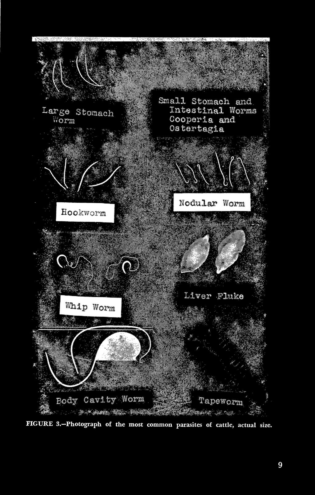
9 (See picture on front cover.) Line C (Figure 1) represents the weight gains of an animal that becomes very severely affected, loses weight rapidly, and dies during the acute stage of the disease. Figure 2 shows the course of the disease with reference to weights and egg counts. The economic losses from parasites may be said to be due to deaths resulting from severe infection and from loss in weight and the general condition of recovered animals. There is also the extra expense of special care, feed, and treatment of those animals showing symptoms. There is no way of determining the total economic loss from parasitic diseases of cattle of all ages, but it is certainly a large sum. THE RUMEN FLUKE One fluke (Cotylophoron cotylophorum) is found in the rumen or paunch and in small numbers in the reticulum or honeycomb (second stomach) of cattle. These parasites are small worms somewhat conical in shape and convex on the top or dorsal side. They are about onefourth inch in length and reddish in color when alive. Life Cycle-The life cycle is similar to that of other species of flukes in that a snail is required as an intermediate host. The eggs are passed in the manure, and small larvae hatch from the eggs in from 11 to 29 days, depending on the temperature. The snail (Fossaria parva) rarely exceeds one-fourth of an inch in length and is commonly found along the margin of open ponds and streams where moisture and decaying vegetation are present and where there is no shade. The larval forms that hatch from the eggs are called miracidia. They enter the body of the snail and there develop through a succession of stages into the infective stage. The time required for development within the snail varies from 30 to 91 days, according to the temperature. These larvae encyst on grass in the pasture and thus are taken into the digestive organs of cattle during "grazing. They first pass to the duodenum or front portion of the small intestine where they remain from three to five weeks. From the duodenum they migrate forward to the rumen where they complete their development. The time required for the development of the larvae to adult worms in the calf is about four months. The total time required for the life cycle is from five to eight months, depending on the temperature. Symptoms In heavy infections in South Africa it is said that severe inflammation is produced by the young stages in the duodenum, which results in diarrhea. The adults are not believed to cause any damage. In the United States, losses from this parasite have not been reported. PARASITES OF THE FOURTH STOMACH The Large Stomach Worm In the past these parasites have been considered as belonging to one species, Haemonchus contortus. Recently some consider that there are two species infecting cattle, sheep, and goats, H. contortus being more common in sheep and goats and H. placei the more common species 8

10


11 in cattle. A third species, H. similis, has been reported in cattle in the gulf coast states. The large stomach worm is often present in large numbers along sheep, the walls and folds of the fourth stomach or abomasum of cattle, and goats. These parasites are three-fourths to one and one-half inches in length, of sufficient diameter to be easily seen, and, particularly the larger specimens, are marked with a spiral red stripe which has led to their sometimes being called the twisted wire worm. Figure 3 shows this species in relation to the other important parasites of cattle. Life Cycle The eggs are deposited by the adult females in the contents of the fourth stomach of the calf and pass out in the manure. Small larvae hatch from these eggs in from 18 to 24 hours. A diagram showing the stages in the life cycle is shown in Figure 4. The newly hatched larvae require about five days in which to develop so they can produce infection in another calf. This stage is recognized under the microscope by the presence of a thin, transparent outer covering or sheath which offers some protection against being killed by drying out or too low or too high temperatures. Infection is by way of the mouth. Upon reaching the fourth stomach, the larvae complete their development into adults in from two to four weeks, most of them requiring about four weeks. That the adults will live and produce eggs for as long as 14 months in calves under conditions in which reinfection does not take place has been shown in our experiments. The length of life of the larvae under the various soil, weather, and grass conditions is not known but probably varies greatly under the different combinations of conditions in the pasture. Symptoms Calves have been found to gain little or lose weight, and a more or less severe anemia develops during the larval period. The adults are bloodsuckers, having mouth parts for piercing the tissues, and in this manner they no doubt contribute to the unthrifty condition of infected animals. This parasite has not been found to cause as much damage as other species. Immunity Experiments at the Louisiana Agricultural Experiment Station have shown that an immunity or resistance to infection with the large stomach worm is developed by inoculation with pure cultures of the infective larvae. Infection takes place as a result of the first inoculations, then with additional or sometimes without additional inoculations the number of adult worms that have become established is suddenly reduced sharply and abruptly, as indicated by the number of eggs recovered from the manure. Subsequent inoculations failed to establish any large number of adults or produce any symptoms of parasitic infection. Attempts to establish this immunity previous to the initial inoculations by the injection of extracts of adult worms or powdered whole worms have thus far failed to produce any protection from infection. Apparently this immunity is permanent when it is once established, because when some of these animals were placed in a heavily contaminated pasture the infection remained at a very low 10


12 LIFE CYCLE OF STOMACH WORM FIGURE 4.


13 level, as evidenced by the number of eggs recovered from the manure and by the general physical condition of the animals. It was possible to follow some of these animals until they were six years old. Poor feeding during the winter months had no influence on the immunity, since there was no increase in the level of the infection. Neither was it possible to break down the immunity by lowering the plane of nutrition to a very low level by a ration of only cottonseed hulls. Animals fed cottonseed hulls until they showed definite emaciation, even almost to the point of death, did not become reinfected when reinoculated. Treatment The copper sulphate and copper sulphate-nicotine sulphate solutions discussed in the section on treatment have long been used for the removal of the large stomach worm. Phenothiazine has recently been found to be very effective also, and for a discussion of its use the reader is referred to the section on general treatment. Small Thread-Like Stomach Worms The small stomach worms belong to two genera, namely Ostertagia and Trichostrongylus, and are very much smaller in size than the large stomach worm. Figure 3 shows some of these small worms in comparison with other species. Two species of the genus Trichostrongylus are found in calves, T. the small intestine. axei in the fourth stomach and T. colubriformis in Some comments on the latter are to be found in the section on parasites of the small intestine. Trichostrongylus axei occurs in sheep, goats, and horses as well as cattle. These small worms are one-fourth to one-half inch in length and very slender; consequently, they are very easily overlooked at postmortem examinations. If present in large numbers they can sometimes be detected as tiny thread-like objects if the folds of the stomach are lifted slowly. The most successful way to recover them, however, is to wash the contents of the stomach and mucus rinsed from the walls into a bucket or garbage can holding three or four gallons of water. After allowing the sediment to settle 10 to 15 minutes, pour off almost all the water, fill the can, and again allow to settle, repeating the process until the water stays nearly clear. Some of the contents may then be examined in a glass jar or dish, preferably against a dark background. Generally these worms, if present in large numbers and if the calf has not been dead too long, will have a distinct reddish color when thus cleared of the sediment. Life Cycle The larvae of these worms are believed to become infective in about a week and to develop to the adult stage in the calf in from 19 to 30 days after infection, and the adults can live at least seven and one-half months. Dr. D. J. Doran, Animal Disease and Parasite Research Branch, A.R.S., USDA, Beltsville, Md., has studied infections of T. axei in calves. He reports that the manure may become soft and diarrhetic fromi the first to the eighth day after infection, while eggs do not ap- 12


14 . pear in the manure until between 19 and 30 days after infection. Loss of weight and appetite and general weakness are other symptoms. Severe inflammation was found in the fourth stomach. The life cycle of Ostertagia ostertagi has been studied at the Virginia Experiment Station. Eggs of this species hatch in about 24 hours and reach the infective stage in five to six days. The infective larvae become adults in about three weeks after gaining entrance to the digestive system by way of the mouth. The larval stages of these parasites are very resistant to adverse external conditions and have been found to remain infective on dry hay for over a year. Treatment The various treatments recommended for the removal of parasites are not very effective against these species, possibly because they are found embedded in the layer of mucus in close contact with the lining of the stomach and consequently are not easily attacked by the drug. Phenothiazine is recommended as the most effective drug for their removal. A discussion of its use will be found in the general section on treatment (page 46) The Hookworm PARASITES OF THE SMALL INTESTINE This parasite (Bunostomum phlebotomum) is found in the anterior half of the small intestine, chiefly in the first eight or ten feet. A very few are usually recovered from the fourth stomach. These worms are about one-half to three-fourths inch long, relatively larger in diameter than the large stomach worm, and distinctly whitish in color. The females are for the most part noticeably larger than the males. Figure 3 shows the hookworm in comparison with other species. The two specimens on the left are males. Life Cycle-The stages in the life cycle are shown in Figure 5. The eggs are laid in the contents of the intestine by the females and pass out with the manure. Hatching takes place in about 24 hours, and the larvae develop to the infective stage in seven to eight days during summer temperatures in Louisiana. Infection takes place by way of the mouth or by penetrating the skin while the calf is lying down on contaminated bedding or ground in the shade or pasture. Infection with severe symptoms and death has been produced in our experiments at Louisiana State University by placing the larvae on the skin. The time required for the larvae to develop to maturity in the calf varies from 52 to 79 days. During this time the larvae pass to the lungs, penetrate the tissues of the air spaces, and are coughed up and swallowed and complete their development in the intestine. Larvae have been recovered from the lungs of both sheep and calves, and it is possible that their presence plays a part in the development of pneumonia although this has not yet been determined. Symptoms-Diarrhea has been found to develop in about a month after inoculation. There is a loss of flesh, unthriftiness, and other symp- 13


15 LIFE CYCLE OF HOOKWORM LB, 14 FIGURE 5.


16 toms of parasitism. These observations agree with those made on sheep inoculated with the species of hookworm parasitic in sheep. It is evident that the symptoms of parasitism were caused by the larvae and not by the adults, since no parasite eggs of any kind were recovered from the manure during the period of diarrhea. During the period of adult life of the worms no symptoms developed. The length of life of the adult worms in the calf may be at least ten months, judging by the presence of eggs in the manure. It has been found that calves develop an immunity or resistance to this species in a manner much like they do to the large stomach worm and to the nodular worm. Treatment There is considerable question as to the value of either of the drugs discussed in the section of this bulletin on treatment. A limited number of experiments carried on at L.S.U. indicate that they are not effective against the adults of these parasites. The Cooperias Five species of small roundworms belonging to the genus Cooperia infect calves. No common name has been established for these parasites. They are relatively small, less than half an inch long, very slender and hair-like, and somewhat reddish in color when alive. Figure 3 shows some of these worms. They are more numerous in the front portion of the small intestine. Their resemblance to some of the fine sediment present in the contents is so close that it is sometimes necessary to examine them under the microscope to be certain of their identity. They are likely to be overlooked because of their small size unless the washing procedure described for the recovery of the small stomach worms is carried out. Life Cycle The life cycle of one of these, Cooperia punctata, has been studied in detail at L.S.U. and by Dr. W. S. Bailey at Auburn University, Auburn, Alabama. The details of the other species are probably similar to C. punctata and to the species found in sheep. Larvae hatch from eggs passed in the manure by infected animals and develop to the infective stage in about a week. After reaching the digestive tract of the calf, the larvae develop to sexual maturity and begin passing eggs in from 9 to 17 days. Diarrhea may begin between the fifth and ninth day after infection. The length of life of the adults of Cooperia punctata was found by Dr. Bailey to be as long as nine months. Five animals in our experiments at L.S.U. have remained infected as follows: \S]/ 2, 14, 15, 2U/ 2 > and 24i/4 months. Two additional ones had exceptionally long records of infection from the original inoculation, namely 4 years 3 months and 4 years 5 months. The infections in 24 others were cut short by the development of immunity within a week or few months after the appearance of eggs in the manure. The length of the natural life of adult parasites plays a very important part in the control program. This is especially important in the case of species against which anthelmintics have little or no effect. 15


17 Twenty-six animals infected with pure cultures of Cooperia punctata have developed an immunity, which was demonstrated by resistance to reinfection. Three of these animals were kept for 2 years and were still resistant to reinfection. After they were demonstrated to be immune to C. punctata they were successfully infected with the nodular worm and Trichostrongylus axei. Animals immune to the nodular worm have likewise been infected with T. axei. Symptoms This parasite is capable of producing severe symptoms such as diarrhea, loss of weight and appetite, and death in severe infections. The anterior 10 feet of the small intestine show numerous tiny hemorrhagic spots, and occasionally larger areas are affected in this manner. Beyond the first 10 feet these spots gradually decrease and the posterior half of the small intestine appears normal. Larvae and adults may be found embedded in the wall of the anterior portion of the intestine, but generally they are found only in the contents. Treatment No effective treatment has been recommended for the removal of the adult Cooperias. Phenothiazine removes only a small percentage or none at all, according to our experimental results. Trichostrongylus colubriformis Trichostrongylus colubriformis is another species found in the small intestine. Pure infections of this parasite can cause the death of calves, according to the investigations of Harry Herlich at the U.S.D.A. Regional Animal Disease Research Laboratory, Auburn, Ala. The symptoms are the same as produced by T. axei, which is found in the fourth stomach. Eggs were first recovered from the manure in from 15 to 23 days after inoculation. The Ascarid This parasite (Neoascaris vitulorum) is found in the small intestine of cattle. It is a very large worm 6 to 12 inches in length and has approximately the diameter of a pencil. Life Cycle and Symptoms The stages of the life cycle and symptoms produced by Neoascaris vitulorum have not been fully studied because of unknown difficulties in producing experimental infections. Much evidence has been accumulated by Dr. Porter and Mr. Herlich, U.S.D.A. Regional Animal Disease Research Laboratory, Auburn, Ala., and by Dr. Refuerzo and other workers, Bureau Animal Industry, Philippine Islands, that infection of calves can take place before birth. It is the belief of these workers that infection probably takes place after birth also. Only very light infections occur in the U.S. but in the Philippine Islands, it is the cause of severe losses. The life cycle is probably similar to that of the closely related species, Ascaris lumbricoides, parasitic in pigs. The eggs of this species are laid by the females in the intestine and pass out with the manure. the larvae do not hatch but they Unlike many other roundworm eggs, develop to the infective stage within the shell in about two weeks. It has 16


18 been found that within the shell these larvae are very well protected against external conditions such as drying, freezing, and chemicals and may remain alive for years in some types of soil and in the shade. Infection takes place by way of the mouth. The larvae hatch in the intestine, penetrate the intestinal wall, and pass by way of the blood to the lungs, where they penetrate the tissues of the small air sacs and enter the air spaces. In the course of time they find their way up the air passages to the throat, are swallowed, and upon reaching the intestine complete their development. The presence of the larvae in the lungs of pigs causes a large amount of irritation and inflammation, and pneumonia is a frequent result. It is not reported that the cattle ascarid plays any part in pneumonia in calves, but it is possible that it does. Thread-Necked Worms These worms are half an inch to an inch long and have the head end somewhat more slender than the other. They are found in the small intestine and are highly reddish in color when alive. Two species have been distinguished, Nematodirns spathiger and N. helvetianus. Life Cycle The stages of the life cycle of N. helvetianus have recently been studied by Mr. Harry Herlich at the Animal Disease Research Laboratory, Auburn, Ala. The eggs are the largest of any of the roundworm parasites in cattle, ovoid in shape, clear and with 2-8 cells centrally located. The eggs of these worms also differ from other nematodes in that the larvae do not hatch until they reach the infective stage, while others hatch much earlier and complete their development outside the shell. Thus, they possibly take advantage of the protection offered by the shell during this part of their development. Eggs begin to appear in the manure in from 21 to 26 days after the calf becomes infected. Symptoms of the infection diarrhea, loss in weight, etc. begin to develop about 14 days after infection. The species of Nematodirus parasitic in sheep have similar life cycles and cause serious symptoms. Strongyloides papillosus These are very small roundworms nearly microscopic in size that are found in the small intestine. They are so small that they will be overlooked at post-mortem examinations unless a special effort is made to recover them. A microscopic examination of the manure will reveal the presence of the eggs. Observations of calves under controlled conditions of infection indicate that these worms can be the cause of an intermittent diarrhea and a generally upset digestive system that result in the calf's being in poor condition for a long period of time. The course of the infection and the damage done by this parasite have been studied in experimental infections by H. H. Vegors, Regional Animal Disease Laboratory, Auburn, Ala. It has a general distribution and probably more than 75 per cent of the calves in the Southeastern 17
 after infection.")

19 states become infected under 4 months of age. Animals become much more readily and severely infected when larvae are placed on the skin than when given by mouth. Eggs are found in the manure during the second week (9 to 11 days) after infection. Diarrhea usually occurred during the first and second weeks after infection and continued intermittently for as long as 3 months. Weight gains were 35 to 75 per cent less than in uninfected calves of comparable ages. Infected animals develop an immunity which gives nearly complete protection against reinfection. Sheep and goats are also infected with this worm, in which it produces the same symptoms. ROUNDWORMS OF THE ABDOMINAL CAVITY Frequently, when cattle are being slaughtered for meat or at a postmortem examination, slender white worms 2 to 4 inches in length will be found in the fluid from the body cavity or among the internal organs. Figure 3 shows some of these worms in comparison with other species. These are commonly called body cavity worms and have the scientific name Setaria labiatopapillosa. They, insofar as is known, do not cause any damage to the cattle and do not injure the meat in any way for human consumption. Although the life history of these worms has not been studied, it is probable that it is similar to that of related species found in other animals. Eggs are not produced, but larvae deposited in the body fluids, such as lymph and blood, are transmitted to new hosts by bloodsucking insects. The Nodular Worm PARASITES OF THE LARGE INTESTINE This parasite is called the nodular worm because of the small, hard lumps or nodules which the larvae cause to develop in the walls of the posterior portion of the small intestine and caecum. The scientific name of this worm is Oesophagostomum radiatum. A related species, O. columbianum, occurs in sheep. The adults are found in the caecum and anterior portion of the large intestine and are about one-half inch long. They differ from the other roundworms of cattle in that a portion of the anterior end is bent in the shape of a walking cane, and the females for the most part have a dark-colored spot near one end. Figure 3 shows some of these worms in comparison with other species. The three smaller specimens on the left are males and the others are females. Life Cycle The eggs, which are deposited in the manure, hatch in about 24 hours at a temperature of 70 to 85 degrees. Figure 6 shows stages in the life cycle. The larvae reach the infective stage in about five days after the eggs are expelled in the manure. Infection takes place very readily by way of the mouth, the larvae being acquired on contaminated grass or hay cut from infested pastures. After infection has occurred the larvae reach maturity and begin laying eggs in from 33 to 44 days. 18


20 LIFE CYCLE OF NODULARWORM " _ LBoss FIGURE 6. 19


21 Experiments at the Louisiana Agricultural Experiment Station indicate that the larvae also produce light infections by penetrating the skin. The larvae were placed on the skin of the flanks of two calves that were free of parasites and kept under conditions free of parasitic infection. The calves were then placed in stanchions so they could not lick themselves until the eggs appeared in the manure. Therefore, it seems advisable to consider that poorly cleaned barns and the contaminated soil of shade areas as well as the pasture are potential sources of infection. Calves also may lick the larvae from their hair after lying down in contaminated places. Symptoms The larvae, upon reaching the posterior half of the small intestine and caecum, penetrate the tissues of the wall until they get about halfway through. There they grow and develop partly into the adult stage, and their presence causes severe disturbances which result in thickening of the wall and rupturing of small blood vessels, causing bleeding into the intestinal cavity. These disorders interfere with normal digestion and absorption and cause a diarrhea of varying degrees of severity. The discharge becomes very watery, sometimes is bloody, and usually has a very disagreeable odor. The calf may begin to lose its appetite in four or five days after inoculation. Some become very weak and emaciated and those that survive the acute stages of the infection will remain in a weakened condition for a month or two before they show much improvement. Some individuals gain very little weight while others lose weight rapidly during the time the larvae are in the intestinal wall. After the larvae leave the intestinal wall and remain in the cavity of the large intestine the damaged tissues heal and the animals begin to show some improvement in condition, although they may remain weak, in poor flesh, and with a rough coat for months. Some individuals in our experiments that live, improve slowly with good feed and care for six months or more and then die quite suddenly when the feed gets less nutritious and the weather unfavorable. This probably accounts for the deaths of some animals during winter or under conditions of poor feeding and management. In severely affected animals the number of eggs was very greatly reduced and very few worms were recovered at post-mortem examination, an indication that it was not the adult worm that was the cause of the condition of the animals. Treatment of animals in such a weakened condition may result in their death, since the drugs which kill the adult worms are more or less poisonous to the calves as well as to the worms. Nothing is known about the removal of the larval stages by treatment. Good care, good feed, and protection from unfavorable weather are of first consideration for animals suffering from such parasitic infections. Animals that survive the acute stages of larval nodular worm infection regain their strength during the adult stage of the parasites. Eventually they develop an immunity or resistance to the adults and larvae as indicated by the reduced number of eggs recovered from the manure 20


22 and by failure to develop symptoms and by failure to become reinfected when reinoculated. Treatment Phenothiazine as discussed in the general section on treatment is recommended as the most effective and safest treatment for the removal of these worms. The Whipworm The whipworm (Trichuris ovis) is found in the caecum or blind gut of cattle, sheep, and goats. It gets its common name from its resemblance to a whip and consists of a long, slender anterior portion corresponding to the lash and a thick posterior portion corresponding to the handle. The total length is 2 to 3 inches, twothirds or more of the total being slender. The color of the worm is white. Figure 3 shows some of these worms in comparison with other species. Life Cycle The life cycle has not been studied in cattle or sheep, but it probably closely resembles that of related species in man and dog. In these related species the eggs do not hatch, but the embryo develops to the infective stage within the protective shell. When the eggs are swallowed the embryos are freed in the digestive organs and develop to the adult in the caecum. Symptoms The slender anterior ends of the worms are usually found piercing in and out as if sewed into the inner layers of the caecal wall. In man when the worms are numerous a low grade inflammation is set up. Severe symptoms may develop in dogs, but in cattle and sheep it is not thought that any serious damage is done. It would seem, however, that bacterial invasion of the tissues might easily follow the penetration of the anterior portions of these worms into the tissues and be a contributing factor to the effects of other parasites if the infection is heavy. Treatment No effective treatment is known for the removal of these parasites. TAPEWORMS Tapeworms, in the adult or egg producing stage, are long, ribbonlike, segmented parasites found in many of our domestic animals. The eggs that are produced by the adults must undergo a period of larval development in another kind of animal before the adult stage is again reached. As an example, one of the tapeworms that are found as adults in cattle and sheep undergoes its larval development in small grassmites similar to red bugs, and one of the species (Taenia saginata) that pass their larval development in cattle has its adult form in man. Man is not susceptible to infection with the adult forms of either of the two species found in cattle and sheep. Adult Forms The Moniezias The two species of this genus found in cattle are Moniezia expansa and M. benedeni. They are indistinguishable from 21


23 each other in size and external appearance but differ internally. M. benedeni is more often found in cattle while M. expansa is more often found in sheep. They are found in the small intestine and are flat, ribbon-like worms many feet long. Lengths up to 30 or 35 feet may be recovered although they are generally broken into shorter pieces in the process of opening the intestine. Upon examination they are found to be divided into joints or segments and if an entire worm is followed to each of the ends, one will be found to widen to a maximum of threefourths of an inch. The other end will become narrower, and at the very end will be found a small, bead-like structure, the head or scolex by which it attaches itself to the wall of the intestine. Figure 3 shows a small portion from the central region of a tapeworm. Life Cycle Stages in the life cycle of these tapeworms are shown in Figure 7. The eggs escape to the outside in the manure, but in order for them to develop into adult worms they must be eaten by small mites. These small mites are similar to red bugs in some respects. They frequent the soil and vegetation and feed, insofar as is known, on decaying vegetation, bacteria, molds, etc., in decomposing manure containing the eggs. Within these mites the tapeworm eggs hatch and the small embryo develops to the infective stage. The infected mite is carried into the digestive tract of cattle, sheep, and goats on contaminated feed, where the larvae are freed from the mite and develop into adult tapeworms. Infection experiments have not yet been carried out using cattle, but in sheep the larvae require about 40 days to reach maturity and the adult worms continue to produce eggs for about two months. Development of the larvae in the mite requires three and one-half to four months. Some experimental observations indicate that pastures can remain infective 17 to 22 months. Symptoms There is some disagreement as to the amount of injury done by the tapeworms, but experimental observations carried out with sheep indicate that no serious damage is done. No observations have been made on cattle. Animals under field conditions are usually found to be infected with other parasites as well, so that it is difficult to assign to each the exact amount of damage. Treatment An effective treatment for the removal of these worms has not been found. Among those that have been recommended are kamala, copper sulphate, oleoresin of male fern, nicotine sulphate, and sodium arsenite. The effectiveness of these, however, seems to be still in question and, besides, they are more or less toxic and dangerous to the animal. Larval Forms Beef Measles This condition of beef is caused by the presence of the larvae of a species of tapeworm found in man and may be recognized by the presence of small, whitish, spherical or elliptical cysts one-eighth to three-eighths of an inch in size, more or less embedded in the muscle. They are usually most numerous in the jaw muscles and the heart 22


24 LIFE CYCLE OF TAPEWORM LBoss FIGURE 7. 23


25 but may occur in any part of the body. No symptoms are produced by these larvae in cattle. These larvae develop into the adults of the tapeworm Taenia saginata in the intestine of man when the infected beef is not properly refrigerated or sufficiently cooked. The eggs deposited by the adults are passed in the human feces and cattle become infected from contaminated pastures or feed in the feed lots. Upon gaining entrance to the digestive system of cattle, the larvae are freed from the shell, then penetrate the intestinal wall and gain access to the circulatory system, by which they reach the heart muscles or other parts of the body in which they develop to the infective stage. The Hydatid Cyst The hydatid or echinococcus cysts contain the larval forms of a tapeworm (Echinococcus granulosus) parasitic in dogs and other carnivorous animals. The cysts are whitish, tough-walled objects more or less embedded in the livers of cattle and hogs. They vary in size from one-fourth inch to as large as six inches in diameter and, although most commonly found in the liver, may develop in the lungs and several other organs. Infected livers should not be fed to dogs, since dogs very readily become infected with the small adult tapeworm about one-half inch in length, a very large number developing usually from even a single cyst. The injury done to cattle by the cysts is not great unless the number is very large. However, infected portions of livers, and if heavily infected, the entire organ, must be discarded. This is a cause of considerable economic loss. Another danger of considerable consequence, although not common, is the possibility of accidental infection with the cysts of persons who are associated with dogs infected with the adult tapeworms. LUNGWORMS The lungworms are parasites of the trachea and bronchial tubes of the lungs and are especially serious in calves. They are rather long, whitish, thread-like worms two to four inches in length and are found by opening the bronchial tubes, which are the air passages leading into the lungs. These air passages should be followed out to the smaller divisions and the walls and especially the frothy mucus that may be present examined for the presence of the worms. Life Cycle A diagram showing the stages of the life cycle of the lungworm is shown in Figure 8. The adult females deposit eggs in the air passages of the lungs, where some hatch, and the larvae, together with the unhatched eggs, become entangled in the mucus and are coughed up and swallowed. The eggs that have not hatched in the lungs hatch in the intestine, and the larvae are discharged in the manure. These larvae are not infective but require about four days in the open in which to develop to that stage. Infection occurs by way of the mouth on contaminated hay, grass, or in water. The larvae, upon reaching the intestine, penetrate its wall and gain access to the lymphatic system, in which they travel to the lungs, reaching there in about ten days. The larvae, when swallowed in large numbers, may cause digestive disturb- 24


26 LIFE CYCLE OF FOR DEVELOPMENT LUNGWORM FIGURE 8. 25


27 ances as they penetrate the intestinal wall, and diarrhea may develop. In a similar manner, as they enter the air passages of the lungs from the capillaries they cause tiny hemorrhages which may lead to pneumonia. The irritation caused by the adults is said to be the most serious result from infections with this parasite. The larvae reach maturity usually in from 21 to 30 days but occasionally not until 41 days after being swallowed. The length of time animals continue to pass larvae in the manure is short, and varies from 27 to 72 days in different individuals. An immunity develops in animals that live, and they are resistant to reinfection. Michel, working in England, believes that symptoms may be produced in immune animals after a year but the larvae are prevented from reaching the adult stage and producing eggs and larvae. Vaccination with irradiated larvae shows considerable promise as a preventive procedure but this still is in the experimental stages at the present writing. Symptoms Animals begin to show symptoms between 8 and 26 days after infections. The adult worms cause a catarrhal condition which results in excessive mucus formation sometimes colored with blood. This mucus contains the eggs and hatched larvae of the adult worms and is coughed up and, for the most part, swallowed. Severe infections cause coughing, difficulty in breathing, discharge from the nose, and a general unthriftiness manifested by loss in weight, rough coat, and sometimes diarrhea. Pneumonia may also develop as a result of the larvae penetrating the walls of the air passages on their way from the blood vessels to the lungs and also as a result of the irritation developed by the adult worms. Treatment The subject of treatment of lungworm infections is considered at this time because the methods are so different from those employed in other parasitic infections. In general, the treatment of animals infected with lungworms has proved unsatisfactory up to the present time. The various methods that have been employed may be grouped under the following heads: 1. Administration of drugs as liquids into the trachea. Among those tried are chloroform, turpentine, iodine, creosote, benzene, pyrethins in olive oil, and carbon tetrachloride. 2. Administration of drugs as vapors into the trachea. Among those tried are ether, oil turpentine, sulphur dioxide, chloroform, chlorine, ammonia, and formalin. 3. Administration indirectly through the digestive tract and intravenous and intramuscular injections. Carbon tetrachloride when administered by mouth is eliminated by the lungs and in this way it comes in contact with the worms, but it is very dangerous to cattle. Various copper salts have been tried by intravenous and intramuscular injection but not with promising results. While experimental results show that some of these drugs kill some of the worms, the introduction of such substances into the lungs and 26


28 otherwise into the animal in many instances has resulted in very serious damage. Most of these substances, it will be noted, are highly irritating to the delicate membranes lining the respiratory passages and, consequently, when present in sufficient quantity to kill the worms, produce complications that often lead to serious results. Care of Infected Animals-The best procedure to follow in handling animals infected with lungworms is to provide the best of care possible in the way of shelter, feed, and pure water. Remove the animals from the source of infection and do all that is possible to aid the infected animals to live with the parasites until they outlive them. Prevention-Calves especially should not be allowed to graze with older animals that are infected or be pastured on the ground formerly grazed by such animals. Low, wet pastures should be avoided, overstocking should not be allowed to occur, especially where young stock is concerned, and a plan of rotation should be adopted if possible. Resting a well-drained pasture for six weeks will usually allow the infective larvae to die out. Longer periods, however, are advisable if it is at all possible. It has been determined that the adult worms may live in the lungs from one to four months. Common Liver Flukes LIVER FLUKES The common liver fluke (Fasciola hepatica) is a flat, leaf-like worm one-half to one and one-fourth inches in length and is found in the bile ducts and gall bladder of cattle, sheep, goats, and sometimes pigs. It is also found occasionally in the lungs as well as in the liver. Figure 3 shows some specimens of this parasite. The walls of the bile ducts are more or less thickened, depending on the number that are present, and have a gritty calcareous deposit adhering to their inner lining. The flukes are freed by cutting open the ducts. They are sluggish, flat worms with a small, cone-like projection at one end bearing a tiny, dot-like depression, the oral sucker. On one of the flat surfaces a short distance from this cone-like projection is the ventral sucker. When examined under the microscope, the surface of the fluke is found to be covered with minute, sharp spines which contribute to the irritation of the walls of the bile ducts, causing the changes described above. In some instances the damage to the duct wall and the resulting deposits of calcareous material are so great as to obstruct the flow of bile. There is also a large amount of damage done to the liver tissue during the wandering of the larvae through it, referred to later under "Life Cycle." This results in the formation of whitish, tough scar tissue which not only interferes with the normal functions of the liver but also renders it tough and unfit for food. In the United States the liver fluke is found in the Pacific Coast, the Rocky Mountain, and the Gulf Coast states. It is prevalent in low, wet pastures and wherever suitable snails are present for the develop- 27


29 INFECTION BY MOUTH CERCARIA ENCYSTS ON GRASS EGGS IN MANURE AND DEVELOPS INTO FREE-SWIMMING LARVA CALLED CERCARIA LARVA CALLED MIRACIDIUM HATCHES FROM EGG THEN ENTERS SNAIL LIFE CYCLE OF THE LIVER FLUKE LBoss 28 FIGURE 9.


30 ment of the larval stages. The economic losses from liver fluke infections have been listed under the following headings: livers unfit for food at slaughtering, loss of flesh due to resulting unthriftiness, loss due to deaths, decreased milk production, and reduced breeding efficiency because of the unthrifty condition. The exact amount in dollars, of course, cannot be determined, owing to the impossibility of securing accurate and complete information, but it must be a large sum annuallv. The losses from condemned livers alone is estimated at between 3i/2 to 4 million dollars annually. Life Cycle The stages in the life cycle of liver flukes are shown in Figure 9. The eggs are laid in the bile ducts and pass to the intestine in the bile and then out of the digestive tract in the manure. A small larva called a miracidium hatches from the eggs in from 7 to 10 days and, in order for it to develop further and be able to produce infection, it must penetrate the body of certain species of snails. The particular kind of snail necessary is a water snail and is found in moist and marshy places only. After it penetrates the snail, several developmental and multiplication stages develop from this small larva. As a result several hundred small, tadpole-like organisms, the cercariae, in the course of time escape from the snail and encyst on grass or other objects. In this condition they wait until they are taken into the digestive organs of the cow. In this encysted condition the cercariae are able to withstand a certain amount of heat and drought although they are in time killed by the hot, dry weather of summer. The total time required for the development in the snail is about 7 weeks. When the cercariae are taken into the intestine with grass or hay, they penetrate the wall and wander about in the abdominal cavity until they come in contact with the liver. Then they penetrate this organ and feed on its tissues for about two months. During this time they burrow about, causing a large amount of injury and destruction of the living tissue which nature attempts to heal by the formation of the tough, whitish scar characteristic of healed wounds. This can be seen on the surface of the livers of fluke-infected animals that have been slaughtered. This destruction of the liver and the formation of the resulting scar reduces and interferes with the normal functions of the liver and renders it unfit for food. After about two months spent in the liver, the larvae resemble small adult flukes in appearance and are now ready to pass into the bile ducts, where they grow and develop into adult flukes in about a month. Under favorable conditions the complete cycle of stages of development may be completed in about five months but may be delayed to some extent by external factors such as low temperatures. Liver flukes and viable eggs have been recovered from sheep experimentally infected and kept under conditions which prevented later infections for 7 years in one animal and for 11 years in another at the Animal Disease Research Center at Beltsville, Md. Thus, without treatment, animals can be a source of infection for a long time. 29


31 Symptoms The symptoms of liver fluke infections do not differ in essential details from those of other parasitic diseases. Animals with light infections sometimes remain in an unthrifty condition in spite of good pasture and care and fail to respond with the expected gains when put in the feed lot. Where the infection is heavy, animals go down in condition with general symptoms of other parasitic infections, and death is invariably the result, especially if the animals are young. The finding of the liver fluke eggs in the manure by an experienced laboratory worker is necessary to determine the presence of adults. Treatment If liver fluke infection is suspected, the owner should, if at all possible, consult a competent veterinarian or communicate with his state livestock sanitary board for advice and assistance in diagnosis and in the administration of the drugs. Carbon tetrachloride has been recommended as a treatment, but, while it seems to destroy the flukes rather effectively, it is so toxic to cattle that it is dangerous to use. The drug is especially toxic to milking animals and to those in a weakened condition. Hexachlorethane is an effective treatment for the removal of the adults in the bile ducts but does not affect the larvae that are still in the liver. It should be remembered that the larvae are the cause of serious and permanent damage to the animal and that a complete cure of all animals is impossible, since nature is powerless to repair all the damage done by the larvae. A very important thing to remember about the employment of treatment is the destruction of the adult and, consequently, the elimination of the eggs as a source of infection for other animals during the present and following seasons. It is advisable to continue the treatment of animals on infected pastures for several seasons in order to prevent the number of parasites from increasing and reaching dangerous proportions. Treatment of infected animals that have not been too seriously damaged by the larvae is said to result in rapid improvement. The following directions for the use of hexachlorethane are quoted, by permission, from a circular by Dr. O. Wilford Olsen (Nov. 1944, while he was associate zoologist, Bureau of Animal Industry, Zoological Division, U.S.D.A.) who made an extensive study of the use of this chemical at the Texas Agricultural Experiment Station at Angleton, Texas. (Now at Colo. State Univ.) "The drug should be prepared as an aqueous suspension and administered as a drench. "The hexachlorethane suspension is prepared by mixing the ingredients on the basis of one pound of finely ground hexachlorethane (60-mesh size) and \i/ 2 ounces of bentonite (a finely powdered clay) with 25 ounces or slightly over \y 2 pints of water. The addition of about one-quarter teaspoonful of white flour facilitates the mixing and improves the resulting suspension. Mixing should be done with a powerdriven apparatus of sufficient speed and force to 30 insure thorough distri-


32 bution of the ingredients. When mixed in the above proportions approximately one quart of the suspension is produced. "A measured dose of 6i/ 2 ounces of the suspension for cattle and 3 \/± ounces for calves over three months old is given by means of a metal dose syringe of 4-ounce capacity or greater. Calves under three months of age need not be treated because any flukes they may harbor would be too young to be killed by the treatment. 1 "When administered as an aqueous suspension, hexachlorethane has a wide margin of safety for the treatment of all classes of range cattle, with the exception of very debilitated ones. Extremely weak animals should be treated with caution since, occasionally, unfavorable effects such as staggering and reeling, sometimes prostration and death, may result from giving a full dose. "One dose of hexachlorethane suspension is usually sufficient to kill the adult flukes in the bile ducts; young flukes are somewhat resistant to the treatment. The dead flukes pass from the liver by way of the common bile duct into the intestine and to the outside with the droppings. "In cases where the poor or unthrifty condition of the cattle is due to liver flukes, there is generally a remarkable improvement in the weight and appearance of animals within a short time after treatment with hexachlorethane. Exceptions occur, however, in instances where the damage to the liver is so extensive that the animals are unable to recover, even though the flukes that they harbored are destroyed. "In planning a program for controlling liver flukes, the time of treatment should be chosen so as to take advantage of the weakest point in the life cycle of the fluke. In general the flukes are the most vulnerable in the spring and fall, and it is probably at this time that the treatment may be given most advantageously. "In the Gulf Coast region, the season of snail activity is during the mild, wet winter and spring. It is during this time that the infective stages of the liver fluke are able to come out of the snails and get on the grass. Treatment of all the cattle in the herd in the spring, or at the beginning of the dry season, when the snails go into the soil, and again in the late fall before the onset of the wet season, gives excellent results. This arrangement takes advantage of the fact that many of the cysts on the pasture have been killed by the heat and drought during the summer, and the majority of the flukes already in the liver are mature and readily killed before the snails become active again in the winter. "In regions where cattle are taken off pastures and not subjected to continuous infection during the cold winter months, treatment should be given at the time when the animals are removed from the infested range in the fall and again in the spring before they are returned to it. ithe drug is available in prepared form from veterinary supply houses. When it is purchased from such a source care should be observed to follow the directions given by the manufacturer for administration. (Author's footnote.) 31


33 The fall treatment kills the flukes that have reached maturity during the grazing season and the spring treatment destroys those flukes that were too small to be readily killed at the time of the first treatment. Animals treated in this manner should be practically free of parasites when they are returned to the range. Such a program not only kills the greatest number of flukes, but also reduces the possibility of infection of more snails on the pasture the next grazing season. A program of drenching cattle twice a year, however, will not eradicate liver flukes, but it will greatly reduce their numbers and improve the health of the cattle." The Lancet Fluke The lancet fluke (Dicrocoelium dendriticum) is a recent importation to North America and to date is known to occur only in Canada and New York. It has been known to occur in six counties in central New York since The rapid spread of this newly introduced parasite should be cause for alarm, since in Europe it has been found to be a menace to the livestock industry. It is a parasite of many wild and domestic animals and thus could become rapidly distributed and established if the proper intermediate hosts were available. In New York it has been found in the woodchuck, white-tailed deer, cattle, sheep, goat, and horse. Extensive and careful studies were carried out at Cornell University by Drs. C. R. Mapes and W. H. Krull on this parasite. The reader is referred to Volumes 41 and 42 of The Cornell Veterinarian for an account of their work. The lancet flukes are 5 to 15 mm. (3/16-9/16 inch) in length and 1.5 to 2.5 mm. wide. They are thus much smaller than the common liver fluke, which is about 1.25 inches in length, and have been mistaken for the larval stages of the latter. The pathological changes resulting from infection with this parasite are confined to the liver. The normal cells are destroyed and become replaced by scar tissue, resulting in extensive cirrhosis, and thus large areas of the liver cease to function normally. These changes increase in extent and severity with the duration of the infection, and no evidence has been found of the building up of an immunity or resistance which develops in the case of some other parasitic infections. Pathological changes of this nature are permanent and, therefore, little improvement in the condition of the infected animal can be expected. No drugs have been found that are effective in removing the parasites up to the present time. Life Cycle The life cycle of the lancet fluke differs from that of the common liver fluke in that there are two intermediate hosts instead of one. The larvae that hatch from the eggs that are discharged in the manure of the infected cow, sheep, etc., undergo certain stages of development in a small terrestrial snail (Cionella lubrica). These larvae then must enter the body of an ant (Formica fused) where they develop further into the larvae that are capable of infecting the final host. The final host (cattle, sheep, etc.) then becomes infected by accidentally eating the ants while grazing. The establishment of the parasite in a 32
 may be found in cattle in locations where they graze over the same areas as deer.")

34 new location is dependent on its finding there both of the proper intermediate hosts. Fascioloides magna Another species of liver fluke (Fascioloides magna) may be found in cattle in locations where they graze over the same areas as deer. It seems that the deer is the normal host for this parasite and, when it succeeds in infecting cattle, it is encapsulated or walled off and destroyed in the liver; therefore, it does not spread from cattle to cattle, but only the deer serves as a source of infection. There is a considerable amount of damage done to the liver by the larvae of this species, and because of this they are of economic importance. Eggs are not discharged in the bile ducts; consequently, infection cannot be determined except at post-mortem examination. COCCIDIA AND COCCIDIOSIS The coccidia belong to the group of simple one-celled animals called protozoa and produce the disease called coccidiosis or bloody diarrhea. Coccidiosis occurs not only in cattle but also in chickens, cats, dogs, pigs, sheep, goats, rats, sparrows, etc. There are many species of coccidia involved in the production of the disease, and it should be emphasized that species infective for one animal are not infective for another with but very few exceptions. For example, those infective for chickens cannot infect calves, cats, or dogs but only have the ability to develop in the digestive organs of chickens. Therefore, the origin of outbreaks of coccidiosis in one kind of animal should not be blamed on another because they have been in close association. Ten species of coccidia are reported as occurring in cattle in the United States. Workers at the U.S.D.A. Regional Animal Disease Research Laboratory at Auburn, Alabama, who have been making an extensive study of the disease have studied nine of these species. These species differ from each other in the size, shape, and nature of the shell of the oocyst and in some other respects such as length of sporulation time, but they do not differ in the main as to symptoms produced. Three species have been found to be the most common cause of outbreaks of the disease. It is not unusual to find coccidial infections in cattle that do not show outward symptoms of the disease. It most commonly occurs in young animals but may develop in yearlings and older animals as well. Symptoms The first noticeable symptoms of coccidiosis are diarrhea, which may or may not be bloody, loss in strength, and in general condition. Affected animals become weak and emaciated very quickly and may be unable to stand or even hold up their heads. The diarrhea may have more or less blood mixed with the discharge, is usually severe, and lasts generally only five to ten days. Positive diagnosis of the disease is made by a microscopic examination of the manure and finding the organisms. Calves that survive the first week or two of the attack generally live if other complications, such as pneumonia, do not de- 33


35 SPORES DEVELOP IN LARGE INTESTINE IN TWO TO FOUR WEEKS PRODUCING OOCYSTS AND BLOODY DIARRHEA INFECTION BY MOUTH ENCYSTED OOCYST, IS IN THE MANURE STAGE, CALLED EXPELLED SPORULATED OOCYST DEVELOPS IN 2-5 DAYS LIFE CYCLE OF COCCIDIA FIGURE 10. velop. The loss by death is generally not great even if the animals become very sick, but mortality as high as 50 per cent has been reported. The animals that live usually regain their strength in a surprisingly short time, though severely affected ones may remain out of good condition for months and should be given the best possible care and feed. Animals in a weakened condition from the infection have their general resistance lowered and are more susceptible to pneumonia; they should be given special protection from the weather and should not be subjected to overcrowding or be forced back from their feed by older or stronger individuals. 34


36 Life Cycle A diagram showing the stages in the life cycle of the coccidia is shown in Figure 10. Calves become infected by having access to feed or water that has become contaminated by manure containing the infective stages of the parasites. The intestine is the region of the digestive system in which the disease develops. The infective stages are microscopic in size and enter the walls of the intestine and there multiply to such an extent that the tissues are ruptured, with the result that the blood vessels are torn open and hemorrhage follows. Inflammation of the delicate tissues, a thickening of the wall, and the general disorders of digestion and absorption manifested by diarrhea and emaciation then result. The organisms known as oocysts that develop as a result of the process of multiplication in the tissues escape into the intestine and are discharged in the manure. These oocysts are round, oval, or pear shaped, depending upon the species, and have a shell that protects them very effectively against certain unfavorable conditions of the outside world. Their internal structure consists of a single cell. At this stage they are not infective. By a series of internal divisions the single cell of the oocyst produces eight tiny cells or spores inside the original shell. This process is called sporulation, and the oocyst is said to be sporulated and is now infective. If enough of these gain access to the digestive system of a calf, they may cause coccidiosis. Prevention The time required for sporulation to take place varies from two to five days or more, depending on the species and environmental conditions. It is during this time that sanitary measures aimed at controlling the spread of the disease must be undertaken. When an outbreak of the disease occurs, the quarters should be thoroughly cleaned of all old manure and bedding and, if possible, cleaned every day or at least every other day for the next week or more to remove the oocysts that are passed by the affected animals before they have time to sporulate and become infective. Too much emphasis cannot be placed on immediate, thorough, and regular cleaning in the prevention of further outbreaks. In case the calves have had access to pasture or a lot, it would be well to confine the affected calves so that all the manure can be collected and disposed of to prevent further contamination of feed or pasture. Dryness is another factor that is important in preventing the spread of coccidiosis. The oocysts do not live very long if they become dried out. If bedding has been used, it possibly would be better not to put in bedding after cleaning if an outbreak occurs, since bedding tends to hold moisture. Water should not be used in cleaning if the floors are not concrete or board or other material that will dry quickly. Moisture is a very favorable factor in the development of outbreaks of coccidiosis in cattle and chickens as well as in other animals. It is of prime importance to prevent contamination of all feed and water with the infective stages, which are in manure. Hayracks should be so constructed that hay is not pulled out and trampled underfoot where portions may be picked up and eaten. Feed and water containers 35


37 should be constructed and placed so that manure does not get into them. While enormous numbers of oocysts are produced during the first few days of the attack, the number rapidly decreases and by the end of a week only a small percentage of the original quantity is given off. Consequently, daily cleaning can be dispensed with after six or eight days, but at least weekly cleaning should be religiously carried out for two or three months, since some oocysts are discharged for a long period and may again build up to dangerous proportions. The use of antiseptics and disinfectants has so far proved of little value in the destruction of the oocysts because the shell prevents the penetration of these substances into the enclosed living cell. Lime scattered on the floor after cleaning has a certain amount of value in that lime absorbs moisture and in this way aids in drying out the quarters, but it cannot be depended upon to destroy the oocysts in any other way. Effective prevention of outbreaks by the use of drugs has so far not given promise of value. In experimentally infected calves sulfaquanadine has been of some value in preventing coccidiosis. However, because of the difficulty in anticipating an outbreak of the disease, it should not be used as a substitute for sanitation. Too much stress cannot be placed upon sanitation and management practices in preventing coccidiosis. Where the group of animals is large enough, the practice of separating the calves by age groups and having a series of pens into which each group is moved in succession has been found effective in preventing not only coccidiosis but parasitic infections as well. By this method the younger animals are not subjected to infection in the areas that may have become contaminated by older animals through constant association in a common group. A regular and effective system of cleaning all quarters of all groups is, of course, a necessary part of the program. The expense of acquiring equipment and the time required to carry out an effective sanitation and management program will be offset in a short time by increased gains in weight and prevention of losses from death not only from coccidiosis but from worm parasite infections, white scours, and pneumonia. In the end this will result in greater profits than will depending upon the use of drugs or expecting the veterinarian to bring about a cure after the animal has become sick. When animals are found to have developed the disease, it is advisable to isolate the sick individuals and give them special feed and shelter in order to prevent them from being crowded away from the feed and disturbed by the stronger individuals. This also tends to prevent the spread of the infection to other individuals. Treatment Up to the present time there is no known effective treatment for coccidiosis in cattle. It should be remembered that the time of serious symptoms of the disease is so short that any treatment or anything that is done seems to do a lot of good, and as a consequence many preparations of no value have been recommended and sold for use during outbreaks in various domestic animals. It was not until the 36


38 , studies of the stages of the life cycle illustrated in Figure 10 and the careful observation of the course of the disease that these treatments were found to be of no value insofar as that particular attack of the disease is concerned. This is because the infective stages of the organisms that gain entrance to the digestive tract of the calf develop at the same rate and are ready to emerge from the wall of the intestine at approximately the same time. There is then no recurrence of attacks because the oocysts pass out in the manure and must sporulate outside and then gain entrance to the calf's digestive system in order to produce a new attack of the disease. This is in contrast to such diseases as malaria, where there is a recurrence of fever and symptoms as a result of development of the malaria parasite within the blood of the infected person. JOHNE'S DISEASE A brief discussion of Johne's disease is included in this bulletin because of the similarity between its symptoms and those of parasitism. Johne's disease is caused by a bacterium (not by an animal parasite) which is similar to the organism causing tuberculosis and is named after one of the early students of the disease. These germs attack the wall of the large intestine where they multiply rather slowly. Thus it re- propor- quires months or even years for the infection to reach sufficient tions to cause serious symptoms. Consequently, symptoms of the disease are not usually observed in animals under two years of age (although positive diagnosis has been made in animals under six months) while symptoms of parasitism very commonly develop in animals under FIGURE 11.-Cow infected with Johne's disease. 37


39 one year of age. There is also a difference to be found in the intestinal wall in some cases. The wall of the large intestine of some animals infected with the organism causing Johne's disease is noticeably thickened, raised into folds, and less elastic than that of animals dead from parasitism and coccidiosis. There is much similarity in the symptoms of the two diseases. Both show emaciation, general weakness, diarrhea, and sometimes a swelling under the jaw and a watery thickening or oedema of the mesentery supporting the digestive organs. Figure 11 shows a picture of a cow with Johne's disease in the advanced stages. There is no treatment for this disease, and death is invariably the result in animals showing severe symptoms. Johne's disease can be diagnosed by the Johnin test. This should be carried out only by an experienced veterinarian, and the Johnin developed by the U.S.D.A. preferably should be used in the test. IMMUNITY TO PARASITIC INFECTIONS Immunity to parasitic infections has been found by a number of investigators to develop as a result of infection with parasites. This is evidenced first by a sharp decrease in the number of eggs discharged in the feces and by a resistance to reinfection. In cattle, immunity has been observed to develop to the large stomach worm, hook worm, nodular worm, Strongyloides papillosus, Cooperia punctata, Trichostrongylus axei, and the lungworm. Sheep develop an immunity to Cooperia curticei and lungworm. Dogs become immune to the hookworm (Ancylostoma caninum) and cats to Toxocara cati. It seems highly possible that the development of immunity plays an important part in making it possible to keep older animals under conditions and on premises heavily contaminated with parasitic larvae. The beginning of immunity is indicated by a sudden decrease in the number of eggs discharged in the manure and its continuation by the fact that when the animals are reinoculated the larvae are not allowed to develop into adults and no symptoms of infection develop. Some of these animals have been repeatedly reinoculated and some placed in a heavily contaminated pasture where nonresistant animals developed severe infections and died. Under suitable conditions of management this resistance probably lasts for a long time. In our experiments at L.S.U. three animals have remained resistant to reinfection with Cooperia punctata for two years. Some of the animals remained in the above mentioned pasture 3 and 4 years and still remained resistant. Observations to date indicate that immunity to certain cattle parasites is species specific. Animals demonstrated to be resistant to Cooperia punctata have been infected with the nodular worm and Trichostrongylus axei under experimental conditions. Likewise animals immune to the nodular worm have been infected with Trichostrongylus axei. The same appears to be true on pasture, since two animals resistant to Cooperia punctata died from nodular worm infection when placed in a pasture known to be contaminated with the nodular worm. 38


40 It is most important to remember that immunity, as it applies to any specific disease-producing agent, is a matter of degree-sometimes high, sometimes low. A great many factors known and unknown account for this. Under farm conditions it is usually difficult to determine the exact cause of the poor condition or death of animals because of the manyfactors that may have entered into the situation. These may include lack of feed, poor quality of feed, vitamin deficiency, and unsanitary management practices that result in bacterial and/or virus as well as parasitic infections. It is equally difficult to determine the role that the various species of parasites have had in bringing about the observed conditions. It is impossible also to determine the part immunity has had in determining the numbers of the various species found at postmortem examinations and the numbers of eggs found in the feces, because information as to the exact extent and degree of parasitic infection preceding the time of observation is lacking. THE IMPORTANCE OF DIAGNOSIS The correct diagnosis of any disease condition is of prime importance. To date we have no definite diagnostic procedure for the detection of parasitism such as is available to the veterinarian for tuberculosis, Bang's disease, etc. However, an examination of the manure by an experienced technician trained in the identification of the eggs of the worms belonging to the various species is of importance. It will reveal the species present and degree of infection to some extent in certain animals and in the group as a whole. The common practice of considering that animals in poor condition are heavily infected with parasites without an examination of the manure results in much labor and unnecessary use of drugs in treatment, since very often such animals have relatively few parasite eggs or the species present are those not affected by anthelmintics. Since the efficiency of most drugs on the various species is known as the result) of critical tests, the expense and labor of treatment can be saved b$ knowing if the species present will be effectively removed. The presence or absence of coccidia will also be revealed by such an examination. It may also be possible to apply some of the facts of the life cycle of the particular parasites in planning the control measures. Of equal importance is the consideration of management practices in the diagnosis of parasitism. It is sometimes said that parasites become worse in the fall" and with the coming of winter. At this time of year pastures become poorer and even disappear as sources of feed. Often hay is of poor quality and a good grain concentrate is expensive. As a result the owner often trys to get by with the feed at hand with the result that the animals lose flesh and get in poor condition. Inadequate feed troughs and hay racks also influence the amount of feed and hay obtained by timid or weakened animals. Good feed in adequate amount is the best treatment of "parasitism" in many instances. 39


41 Post-mortem examinations are often inconclusive because of the manner in which they are carried out. A small amount of material taken from the lining of the fourth stomach or intestine for microscopic examination can be taken from an area which has only a few parasites or from the only area which has a large number. The recovery of the small species is a long and tedious process that is usually not carried out. The exact role which the various species play as adults in producing the unthrifty condition is not well understood. Only two have been definitely incriminated as causing the loss of blood from the tissues. What food constituents of the host are used by parasites and how they obtain them is unknown. The evidence against the adults, therefore, is largely circumstantial. It has been shown, however, that the larvae produce very severe inflammation and damage to the stomach and intestinal walls, and open the way for bacteria to enter the tissues. This damage has to be slowly repaired by nature; hence the animal remains in poor condition often for months even with good care. As a result of the above observations a most important consideration, after a definite diagnosis of parasitism has been made, is the prevention of the spread of the infection to other susceptible animals. Treatment may be carried out if it is indicated and good feed and shelter provided for the sick animals. Prevention of infection through management is a more important consideration than the dependence on drugs to prevent further losses. PREVENTION Management Practices The prevention of losses from parasitic infections is distinctly a problem of management based on the known facts of the life cycle of the species involved. The stages of development in the life cycle may be compared to the links of a chain; and a chain, of course, is no stronger than its weakest link. When the owner has the basic information concerning the life cycle he may be able to adapt some of the details of his scheme of management so that this chain of stages may be broken and severe infections prevented. All dairymen and cattle owners will not be able to put into operation any one complete plan of management, because of the expense involved or peculiarities of individual farms. However, some procedures can ordinarily be put into operation that will reduce the parasite burden and prevent losses. There are three factors that are of basic importance in influencing the vitality and length of life of the eggs and larvae of internal parasites and the encysted stages of coccidia. These are temperature, moisture, and time. Of these, temperature is the one over which we have the least control, but it is nevertheless very important for the successful development of the larval stages. In the states bordering the Gulf of Mexico the larvae have very favorable if not ideal temperatures for development during 40


42 most of the year. The hot periods in summer combined with less rainfall during some of the months no doubt cause the death of large numbers of larvae. This, however, cannot be relied upon to bring about effective control, because of the shortness of the periods of drought and the carry-over of adult parasites in already infected animals. Neither does the temperature go low enough during the winter months to cause the death of any considerable number of eggs or larvae. Realization of the small part temperature plays in the control of parasites in the Southern states serves to emphasize the importance of undertaking other measures. Moisture is an essential factor in the life of parasitic eggs and larvae and encysted stages of coccidia and can be controlled in the selection of well-drained pastures and by modifying poor drainage of existing pastures and lots occupied by calves and young stock. Barns and shelters also should be constructed so that the floors are as dry as possible. Time is a factor in the life of parasites because the eggs and larvae are able to withstand the various natural conditions of the environment for a limited time only. Each of the various stages of the life cycle also requires definite periods of time. Therefore, it is sometimes possible to modify management so as to prevent or reduce infection. We may now consider the application of the above factors to management practices. Drainage From the standpoint of parasite control, drainage should be of first consideration in the location of lots and pastures, especially those to be occupied by calves and young stock, since they are more susceptible than adult animals. Where natural drainage is not satisfactory, ditches should be dug to remove the excess rainfall quickly. It is not desirable to have water from pastures or lots occupied by older animals drain into or across calf pastures or lots, since eggs and larvae may be washed from one to the other. Barn and shelter floors should be constructed so that they are kept dry. Cement and board floors are the most desirable and dirt, the least. The ground about the entrances should be well drained, but if it is not, a plank or cement entrance platform should be constructed. Ponds are undesirable as sources of drinking water, because of the continuous presence of a margin of mud through which the animals must wade to drink. Manure is deposited in this mud and the larvae have favorable conditions for development and for gaining entrance to growing about the the digestive organs of animals feeding on the grass margin. The species that penetrate the skin also have favorable conditions for development and an opportunity to come in contact with the skin. Where ponds are utilized as sources of drinking water, it is very desirable to fence them and pipe the water over the side into a trousrh. A float valve should be put on the pipe to prevent overflow and the development of a mudhole about the trough, otherwise the purpose of the trough will be defeated. Time Time can be relied upon to destroy eggs, larvae, and coccidia 41


43 and can be utilized in pasture rotation, barn sanitation, lot and shade sanitation, etc. Knowing how long it takes for the infective stages of the larvae to develop makes it possible at times to eliminate the source of infection by cleaning at the proper time. Barn, lot, and shade sanitation have not received the emphasis they deserve in the past, but since we know now that infection with certain species occurs through the skin, attention should be directed to this phase of control. Barn sanitation involves cleaning the barn at regular intervals, preferably once a week, in order to prevent the accumulation of developing larvae. Where animals are showing symptoms of parasitism, weekly cleaning should be carried out in order to prevent the infection from reaching greater proportions. Weekly cleaning is important, because, as pointed out in the discussion of the various species and in the life cycle diagrams, the larvae hatch and develop to the infective stage in about a week or a little less. Thus by weekly cleaning, the eggs and the larvae are removed before they reach the infective stage. Care should be taken to remove all the manure from the corners. Water should not be used unless the floor is constructed so that it will drain and dry quickly. Bedding is desirable but it is always necessary to keep in mind the conditions under which it is used. If cleaning is not carried out regularly and thoroughly, bedding tends to hold moisture and otherwise protect the eggs and developing larvae. In this connection it is well to consider possibilities of introducing parasitic larvae and coccidia from outside sources. If calves are confined to stalls, larvae may be introduced on the shoes or boots of the attendant if it is necessary to enter the stall to feed the animals. If the calves have access to a yard or pen in which a mild infection exists, they will infect the bedding by carrying it on their feet. If hay is used for bedding it should be cut from land that has not been grazed, because in recent experiments it has been shown that the larvae of some species remain infective at least a year on hay. Isolation of calves in separate stalls is the most desirable method to follow in controlling all diseases. By this method infection does not spread so rapidly from individual to individual and thus is not so likely to reach serious proportions. Another method used extensively in the tropics and recommended for control of coccidiosis by workers at the Regional Animal Disease Research Laboratory at Auburn, Ala., is segregation by age groups. Animals are separated according to age and never allowed to mingle with the individuals of the older or younger groups. The following groups are suggested: (1) under 3 weeks, (2) 3-6 weeks, (3) 6 weeks to 3 months, and (4) animals over 3 months. In this system when the oldest calves reach maximum age for that group, each group is moved to the quarters of the next highest age group, which leaves the quarters of the youngest group empty to accommodate the animals to be born during the next three weeks. The advantage of this system is that the younger animals, which are more susceptible 42


44 to parasite damage, are not exposed to the heavier infections of older animals in which there has been more time for large numbers of parasites to build up. Another advantage is that if an infection gets established in one group, the animals of the others are not exposed. It should be kept in mind, however, that utilization of a segregation plan does not eliminate the necessity of a system of thorough cleaning at regular intervals in order to remove any organisms that may be in the quarters. One of the cleaning times should, of course, fall on the date when the calves are changed into the quarters of the next higher age group. Another method recommended by the Regional Animal Disease Research Laboratory at Auburn, Ala., is the use of portable pens. These are in use at the laboratory for raising animals for their experiments, and by many dairymen over the states of Alabama, Georgia, and Mississippi. The pens are made about four feet by eight feet out of light material, usually a wooden frame covered with woven fence wire. One end is closed in on the sides and top to protect the calf from bad weather. The pens are set at first preferably on the low side of sloping ground and moved up the slope to clean ground once a week. By the selection of sloping ground, any infection which may have gotten a start cannot be washed onto the areas to be used at later dates. Even though the pens are quite open and the animals are exposed to the weather, it is claimed that pneumonia and other diseases are practically eliminated because of the lack of accumulation of the disease-producing organisms as compared with tightly closed quarters of barns and sheds. Detailed information as to construction and use of these pens can be secured by writing to the Animal Disease Research Laboratory at Auburn, Ala. Pasture Management Since infection with most species takes place readily by mouth, the pasture must be given serious consideration in the problems of control. The problems are also likely to increase when pasture improvement is undertaken, for with improvement comes greater and better grass production which will feed more animals, and concentrating the cattle population always increases the probability of a parasite problem. One answer is pasture rotation, especially for young animals. This may be carried out by plowing under the infected topsoil and grass and planting another crop for one or two years before reseeding and using for grazing again. Rotation assumes that other ground is available in a suitable location for pasture, but if such is not the case, the pasture may be plowed in the fall and planted to oats, winter grass, or some crop that can be quickly utilized for grazing. This turns* under the infected topsoil and grass to a large extent, and fresh, uncontaminated grazing plants are quickly made available. This plan is especially applicable if the original sod is Bermuda, which will quickly resod the following season. Mowing pastures also probably helps to some extent in controlling parasites, since the taller growth is cut and animals feed on the newer growth, which has had less time to become contaminated with larvae. 43


45 Harrowing or dragging contaminated pastures possibly aids also in the destruction of larvae in that piles of manure are scattered and dry faster and thus less protection is offered to the developing larvae. Mowing and harrowing, however, has the disadvantage of scattering the manure and, at the same time, the eggs and infective larvae, thus making them more available for infection. Cattle manure from the barn should not be scattered on the pasture grazed by young stock because of the danger of introducing eggs and larvae and thus increasing the infection or adding species not already there. It should be pointed out again that pasture sanitation alone cannot be relied upon to control parasites; the barns and shade are equally important. In some instances heavy infections develop in animals which are too young to have eaten large quantities of grass or hay and which receive their feed mostly in the form of milk and concentrates. In addition to barns the other places where calves lie down should receive attention because of the skin-penetrating species and because calves are constantly licking their coat and may in this manner acquire larvae that have gotten on the hair. Where calves are known to be infected, it would be advisable to remove the accumulations of manure from the shade areas at four- or five-day intervals. Removing the adult worms by an effective anthelmintic reduces the number of eggs deposited in the manure and thus reduces the number of larvae which may be picked up by the animals on pasture. Treatment should be carried out a week or so before animals are transferred to a clean pasture, in order to avoid its immediate contamination. In other instances where animals are being transferred to clean quarters, pastures, or lots recently plowed, the transfer should be made after treatment in order to reduce contamination of the clean ground. Low Level Feeding of Phenothiazine as a Means of Prevention Controlled experiments carried out at the Agricultural Experiment Station, Louisiana State University, show that the continuous feeding of small amounts of phenothiazine should be extremely useful in preventing the buildup of infections and in the reduction of high infections on badly contaminated premises. Low level feeding exerts its effects in one of two ways. In the case of the large stomach worm and the nodular worm the drug somehow injures the female worm and egg production is stopped. However, in case of the hookworm, C. punctata and T. axei, there is no interference with egg production but the eggs do not produce infective larvae. For maximum effect, the animal must get continuously at least grams of the drug every day. Since the drug stops the supply of infective larvae of the above very common parasites of cattle, reinfection is reduced and the younger animals are protected from infection. Continuous feeding of the drug until the existing supply of larvae has been eliminated from the premises is therefore necessary. Experiments have shown that phenothiazine fed in this manner has no effect on infective larvae after they get into the animal. Calves fed the 44


46 drug before and for several days following inoculation readily become infected. No evidence has been found to indicate that there is any beneficial effect on the animals already infected or that an outbreak of acute symptoms will be stopped as a result of low level feeding of phenothiazine. It should be kept in mind that acute infections "run their course" as explained in the introduction to this bulletin. It is believed that weight gains and general improvement in condition of the animals are results of the improvement in digestion following the recovery from damage done by the larval stages. The benefits to be expected from low level feeding of phenothiazine are in the protection of the next generation of calves by the elimination of infective larvae about the pastures, lots, and barns. Likewise, there is a reduction of reinfection. Sources of infection from the outside, such as introduction of infected animals into the herd by purchase, and contaminated feed, especially hay cut from pastures grazed by infected animals, must be avoided. In feeding the drug the owner must determine the number of pounds of grain mixture he is feeding per day to each calf and add \i/ 2 grams of phenothiazine to this amount. Since a pound contains 453 grams, one pound of the drug would last one calf 300 days, or about 10 months. If 10 calves are being fed, one pound should be sufficient for those 10 calves for 30 days. If the animals are of such an age that they are being fed five pounds of grain a day, 50 pounds of feed would be consumed per day and 1,500 pounds in 30 days; so U/ 2 pounds of phenothiazine should be added to one month's supply of grain. In this manner the owner can determine the amount necessary to add to the feed in order to obtain the expected results. The above figures are on the basis of the avoirdupois pound of 16 ounces and not the apothecaries' pound of 12 ounces. The following table will be of assistance in making up the feed mixtures: Lbs. Pheno. Lbs. Pheno. Ozs. Pheno. No. lbs feed per 100 lbs. per 1,000 lbs. per 100 lbs. per day feed feed feed Ordinarily it will be more convenient to have the phenothiazine weighed out in the proper amount at the time of purchase. As its action is to prevent the development of infective larvae, it should be pointed out that continuous feeding of the drug is necessary since it is effective in protecting animals against the hookworm, Cooperia punctata, and Trichostrongylus axei only while it is being fed. Although egg production of the nodular worm is stopped, continuous feeding is necessary because eggs reappear one to three months after feeding is discontinued. Continuous feeding should be carried out for at least 2 or 3 years in order to stop the contamination of the premises 45


47 and allow time for the larvae already on the premises to die out. Our experiments indicate that the cost of accurately carrying out this method of feeding the drug will be small when compared to the losses should a future outbreak of parasitism occur. These observations indicate the importance of routine low level feeding as an integral part of the total management program where conditions are favorable for the development of parasitism. TREATMENT Treatments of various sorts have long been recommended for the removal of the adult parasites of domestic animals and man. Anthelmintics, which are drugs used for removal of parasites, are selective to a considerable degree in their action on different parasites; therefore, information as to the species with which animals are infected is useful in the choice of the drug to be used. The use of these drugs or chemicals is based upon the principle that they are more toxic or poisonous to the parasite than to the host animal to which they are administered. Some are more toxic or poisonous to the host animal than others and therefore must be given in reduced amounts if the animal is in poor condition. The amount administered must also be adjusted according to age and size of the animal. In weakened animals it is often advisable to reduce the dosage according to their condition and repeat the treatment in about two weeks. It is also strongly recommended that special care and good feed be given those individuals showing the effects of parasitism in order to build up their general resistance to other diseases. The efficiency of the drug is affected by the fact that it is at once diluted by the contents of the digestive tract as well as acted upon by the various substances in the contents, with the result that it is rendered less effective. Phenothiazine Because of its efficiency against several of the species and because of its low toxicity, phenothiazine is the most satisfactory drug in use at the present time for the removal of adult parasites from cattle. It has been found to be very efficient against the large stomach worm, the nodular worm, and Trichostrongylus axei but not so efficient against Cooperia, Ostertagia, and the hookworm. It is relatively safe to administer since when given to experimental animals in quantities of two and three times the large dose of 20 grams to 100 pounds body weight recommended by some, no ill effects were observed. Experiments indicate it is not necessary to keep the animals off feed before giving the drug in order to secure the best results. Even though the administration of this drug is relatively safe, it is desirable to secure the services of a veterinarian for the job, especially if the animals are valuable or if the number to be treated is large. However, this is not economical in many instances; in such cases it is well 46


48 to consult a veterinarian and secure the necessary quantities of the drug, instruments for its administration, and advice as to dosage, restraint, etc. While the preparations sold by the various manufacturers are similar in most details, it cannot be emphasized too strongly that the directions on the label should be carefully studied and closely followed in order to avoid unfavorable results. Administration-The most desirable method of administering any treatment is to each individual animal rather than to the group or herd. This insures that each animal receives an accurate amount of the drug and permits adjustment of the dose according to the size and physical condition of the animal. If the group is allowed access to feed to which the drug has been added, the greediest and healthiest individuals will eat faster and crowd other individuals to one side, and thus the animals that may really need the treatment the least, secure the most of the drug. The tastes of individual animals also vary and some will reject such drug feed mixtures, thus allowing other animals to eat more than their share. The best results are obtained only by individual treatment unless there are reasons that make individual handling of the animals impractical. Phenothiazine is supplied by a number of companies furnishing veterinary supplies and may be obtained in the form of a liquid suspension, in boluses tablets, and in powder form. The liquid suspension is designed as a drench, but because of the danger of getting the material into the trachea, it is not advisable to give it from a bottle, but by means of a metal dose syringe. These are made in three sizes-2-, 4-, and 6-ounce capacity-so that the quantity to be given may be measured. They are provided with a metal tube of sufficient length so that when the tube is inserted into the mouth, the drug is deposited back of the base of the tongue in a position to be easily swallowed. The animal should be securely held and the nose raised a little higher than the back of the head and in a position so that swallowing is not interfered with. The boluses are designed to be administered with the aid of a balling gun, which places the drug back of the base of the tongue in a position to be swallowed. Care should be taken not to get it in a position to pass into the trachea. If the drug is mixed with the feed, the animals should be fed individually if at all possible. Should group treatment be decided upon, it is highly desirable to divide the animals according to size and condition and allow the groups access to the feed separately. Dosage The amount of phenothiazine recommended as a dose for cattle varies between 10 and 20 grams per 100 pounds of body weight, with a maximum dose of 60 to 75 grams for grown animals. An apothecaries' ounce is equivalent to 31 grams. Dr. P. D. Harwood, who discovered the effectiveness of phenothiazine while in the U.S.D.A. Bureau of Animal Industry, recommends its administration at the rate of 10 grams per 100 pound of body weight, with a maximum dose of 60 to 75 grams. Dr. D. A. Porter of the Regional Animal Disease Research Lab- 47


49 oratory at Auburn, Ala., recommends 20 grams to each 100 pounds of body weight, with a maximum dose of 60 grams. Since there is so much variation in the size of individuals of the same breed as well as different breeds, it would seem advisable to administer the drug according to weight rather than age in order to secure the greatest efficiency, and economy. safety, If the liquid suspension, boluses, or tablets are obtained it is wise to follow the manufacturer's recommendations as to dosage, because products vary as to the actual amount of the drug they contain. If the powder form is obtained it is necessary to measure it in some way in order to get the proper dosage. If the powder is to be mixed with the feed, it can be measured by tablespoonfuls. Since there is much variation in the size of ordinary tablespoons, it is advisable to use one of the tablespoon measures used in the kitchen. A packed level tablespoon measure will hold between 9 and 10 grams of the powder when well packed and scraped off even across the top; when not well packed it may hold as little as 7 grams. Hence two well-packed level tablespoon measures would be approximately the maximum dose per 100 pounds body weight. Some veterinarians, as a result of their experience with the drug, recommend as low as 10 grams per 100 pounds body weight and adjust the dose to the condition of the animal. Toxic Reactions Toxic reactions to phenothiazine are not common, and those that do occur usually are of little consequence. However, recently treated animals should be watched in order to detect any individuals that do not respond favorably. Sometimes the eyes become sensitive to bright light, and temporary blindness results. Animals so affected should be confined to a darkened stable or dense shade until recovery. Constipation may follow in animals to which large doses have been given, and to these a cathartic should be given if necessary, although recovery usually follows spontaneously. Occasionally animals will be off feed and will be unsteady in movement for a day or so but quickly recover. The urine may be colored red for a few days owing to the excretion of the chemical, but this is no cause for alarm. Effectiveness-Because of recent reports of the ineffectiveness of phenothiazine it seems appropriate to examine the most recent experimental results on this drug. A summary of these findings, together with the references, has been compiled in consultation with the USDA Regional Animal Disease Research Laboratory at Auburn, Ala., and the Animal Disease and Parasite Research Laboratory, U.S.D.A., Beltsville, Md. These are presented in Table 2. These results show that the drug is very effective against the large stomach worm (Haemonchus), the nodular worm (Oesophagostomum radiatum), and TrichostrongyJus axei. It is either very variable or not effective against the other species found in cattle. Factors which enter into the failure of the drug to control cattle parasites may be as follows: Since the drug is not 100% effective in removing all species,
 u 2 3 c * 3 a o e I i I l\9s \"o -2 \"3 ^ 3-5 c 2 c '~ «O 3 O,-H ^")

50 ! s! 3 P 3 j 1 o o a g t!.2 c. 1) u 2 3 c * 3 a o e I i I l\9s "o -2 "3 ^ 3-5 c 2 c '~ «O 3 O,-H ^ U U "Oh be 5 o o S3 <u 3 ^> to 0 ^3 o o y J O «rd < u OJ I "S to _ H CO 00 c/3 ^ * '8 Si >s 0 do t1 5? O CD 1 o CO rh 00 c«cm =«.<CM 4J J3 l) O) 3 cu Q O c/5 111 H c«o ^ 5 be


51 enough are left to continue contamination of premises so that the supply of infective larvae builds up to the point where damage again results. 2. The human element. Any drug should probably be as nearly as possible 100% effective under experimental conditions to be practical under farm management in order to allow for variations in administration by less experienced hands, etc. 3. Not administered frequently enough. More frequent administration by less experienced hands, etc. 4. Not administered at the most advantageous season of the year because of lack of information in regard to the environmental conditions affecting the larvae. 5. There are some who believe that smaller particle size and the purified phenothiazine is more effective. This, at present, should be considered as still in the experimental stages. Copper Sulphate and Copper Sulphate-Nicotine Sulphate Copper sulphate, or bluestone, has long been recommended as an anthelmintic for cattle, sheep, and goats. However, it is not considered very effective against any of the species parasitic in the digestive tract of these animals except the large stomach worm. Since recent experiments have shown that the large stomach worm is much less serious in its effect on cattle than formerly supposed, the arguments in favor of the use of this treatment have less weight. However, it is recommended and used by some. The strength usually used is a 1 per cent solution in water. This is made by dissolving i/ 2 pound of the blue crystals of copper sulphate in 1 pint of boiling water and then adding enough water to make 3 gallons of solution. Porcelain, earthenware, or glass containers should be used in the preparation and storage of the solution, as it will corrode metal. The dose recommended is as follows: calves, 3.5 to 4 ounces; yearlings, 6 to 8 ounces; two years old and over, 16 ounces to 1 quart. A Coca-Cola bottle holds 7 ounces when full, is easily obtained, and is properly shaped for a good drench bottle. Many prefer the addition of 1 ounce of 40 per cent nicotine sulphate (Black Leaf 40) to each gallon of the 1 per cent solution of copper sulphate and believe it is more effective. The dosage is the same as for the copper sulphate solution. Animals are susceptible to poisoning with both of these preparations, and those in poor condition should have the dose reduced. Severe poisoning and death are not uncommon even in healthy animals if the amount is excessive. Animals in a weakened condition should not be given more than one-third the recommended dosage. New Anthelmintics There are a number of new products the anthelmintic values of which are not definitely proven at the present time. Several organic phosphate and piperazine compounds are among those being tested. The results of experiments using some of these are presented in Table 3. The use of these compounds should be considered with caution because the degree of toxicity has not been fully demonstrated. 50


52 Z 2 o CO in o o GO CT3 i u cu ltd S 2 a> 1/0 o o > Oh 5-t U O u O o <o CO 00 o 1- ^ as CD 6 S CO o as a, 1 «QJ CO i! <u c kj o O O u C <u co ox N <v DO: <i O ^ 5 ^ H 0 io 5h 1 1 w. 01 ^ ^ as. oo.. i I > CO O pq rn O. <u > CO T1 i 3^ < ^ Sh <U. CO 5 vy 3 51


53
INTERNAL PARASITES OF SHEEP AND GOATS
 7 INTERNAL PARASITES OF SHEEP AND GOATS These diseases are known to occur in Afghanistan. 1. Definition Parasitism and gastrointestinal nematode parasitism in particular, is arguably the most serious constraint
Feline and Canine Internal Parasites
 Feline and Canine Internal Parasites Internal parasites are a very common problem among dogs. Almost all puppies are already infected with roundworm when still in the uterus, or get the infection immediately
X-DISEASE TH. OF CATTLE I AYllG{ - OIS. ~I RCU lar 656 Extension Service in Agriculture and Home Economics INCE
 X-DISEASE TH S INCE OF CATTLE I AYllG{ - OIS By C. C. MORRILL and R. P. LINK UIIJH8SITY OF lluuoij Coll ege of Veterinary Medicine and Agricultural Experiment Station 1941 A DISEASE OF CATTLE which is
EC Prevent Worms...to Increase Sheep Profits
 University of Nebraska - Lincoln DigitalCommons@University of Nebraska - Lincoln Historical Materials from University of Nebraska- Lincoln Extension Extension 1981 EC81-1905 Prevent Worms...to Increase
Hoofed Animals. Section E. Muskox Section E-2. Caribou Section E-1. Moose Section E-3
 Section E Caribou Section E-1 Muskox Section E-2 Moose Section E-3 Section E Diseases and Parasites of Nose and throat bots Head Photo Credit: NWT Wildlife Division Caribou Section E-1.1 Lumpy jaw Contagious
Training Module No 2
 Training Module No 2 Theory 1. Heartwater 2. 5 point check for internal parasites 3. Checking for signs of anaemia 4. Roundworm 5. Taking temperature and weighing your goat Property of Abafuyi Media Training
FREEDOM OF INFORMATION SUMMARY
 Date of Approval Letter: FREEDOM OF INFORMATION SUMMARY SUPPLEMENTAL NEW ANIMAL DRUG APPLICATION NADA 110-048 VALBAZEN (albendazole)...for the removal and control of a variety of internal parasites common
This is the smallest tapeworm that can affect human being but it s not really proper human tapeworm (the human is not the primary host).
.") Echinococcus Granulosus Small Tapeworm (1 cm), Cestode. This is the smallest tapeworm that can affect human being but it s not really proper human tapeworm (the human is not the primary host). The primary
Echinococcus Granulosus Small Tapeworm (1 cm), Cestode. This is the smallest tapeworm that can affect human being but it s not really proper human tapeworm (the human is not the primary host). The primary
DEWORMING PROCESS KRISHIMATE AGRO AND DAIRY PVT LTD NO.1176, 1ST CROSS, 12TH B MAIN, H A L 2ND STAGE, INDIRANAGAR BANGALORE , INDIA
 DEWORMING PROCESS KRISHIMATE AGRO AND DAIRY PVT LTD NO.1176, 1ST CROSS, 12TH B MAIN, H A L 2ND STAGE, INDIRANAGAR BANGALORE-560008, INDIA Email: sales@srisaiagro.com Www.srisaiagro.com INSTRODUCTION According
EC Prevent Worms to Increase Sheep Profits
 University of Nebraska - Lincoln DigitalCommons@University of Nebraska - Lincoln Historical Materials from University of Nebraska- Lincoln Extension Extension 1964 EC64-1905 Prevent Worms to ncrease Sheep
Johne's disease infectious diarrhea of cattle
 Louisiana State University LSU Digital Commons LSU Agricultural Experiment Station Reports LSU AgCenter 1927 Johne's disease infectious diarrhea of cattle Harry Morris Follow this and additional works
Stalled Calves Strongyle- type eggs per gram Other parasites
 Reedy Fork Farms- 3 May 2012 History Reedy Fork Farms has been a certified organic dairy since 2007, currently with Holsteins and Holstein- Jersey Crosses. There is also an organic feed mill on the property,
The Rat Lungworm Lifecycle
 Hawaii Island Rat Lungworm Working Group Daniel K. Inouye College of Pharmacy University of Hawaii, Hilo The Rat Lungworm Lifecycle Rat Lungworm IPM RLWL-3 It is important to understand the lifecycle of
Large Animal Topics in Parasitology for the Veterinary Technician Jason Roberts, DVM This presentation is designed to review the value veterinary
 Large Animal Topics in Parasitology for the Veterinary Technician Jason Roberts, DVM This presentation is designed to review the value veterinary technicians can add to mixed or large animal practices
EC Cost You Money : Controlling Internal Parasites in Swine
 University of Nebraska - Lincoln DigitalCommons@University of Nebraska - Lincoln Historical Materials from University of Nebraska- Lincoln Extension Extension 1969 EC69-1902 Cost You Money : Controlling
Parasite Management for Small Ruminants
 Parasite Management for Small Ruminants Slides contributed by tatiana Stanton, Steve Hart, Betsy Hodge, Katherine Petersson, Susan Schoenian, Mary Smith DVM and James Weber DVM and many others Part 1.
REEDY FORK DAIRY FARM
 History REEDY FORK DAIRY FARM The Reedy Fork Farm is set on 600 acres and houses both a feed mill and an organic dairy operation. The feed mill was started in 2007 when the dairy transitioned to organic,
Fasimec Cattle Oral Flukicide and Broad Spectrum Drench
 Product name: Fasimec Cattle Oral Flukicide and Broad Spcctrum Drench Page: 1 of 10 Display box front panel 5 L gun pack only CAUTION KEEP OUT OF REACH OF CHILDREN READ SAFETY DIRECTIONS BEFORE OPENING
Controlling "Worms" In Poultry
 Controlling "Worms" In Poultry or E. M. Dickinson W. E. Babcock Agricultural Experiment Station Oregon State College Corvallis Circular of Information 522 February 1953 Controlling "Worms" in Poultry E.
Antihelminthic Trematodes (flukes): Cestodes (tapeworms): Nematodes (roundworms, pinworm, whipworms and hookworms):
: Cestodes (tapeworms): Nematodes (roundworms, pinworm, whipworms and hookworms):") Antihelminthic Drugs used to treat parasitic worm infections: helminthic infections Unlike protozoa, helminthes are large and have complex cellular structures It is very important to identify the causative
Antihelminthic Drugs used to treat parasitic worm infections: helminthic infections Unlike protozoa, helminthes are large and have complex cellular structures It is very important to identify the causative
(Hemorrhagic Septicemia of Fowls) By ROBERT GRAHAM. A Brief Statement of the Cause, Symp" toms, Lesions, and Preventive Measures
 By ROBERT GRAHAM. A Brief Statement of the Cause, Symp toms, Lesions, and Preventive Measures") Fowl Cholera (Hemorrhagic Septicemia of Fowls) By ROBERT GRAHAM A Brief Statement of the Cause, Symp" toms, Lesions, and Preventive Measures Chickens with fowl cholera often sit quietly with necks contracted
Fowl Cholera (Hemorrhagic Septicemia of Fowls) By ROBERT GRAHAM A Brief Statement of the Cause, Symp" toms, Lesions, and Preventive Measures Chickens with fowl cholera often sit quietly with necks contracted
Training Module No 3
 Training Module No 3 Theory 1. Pneumonia 2. Condition scoring 3. Tapeworm cyst (Turning disease/draaikop) 4. Visual Examination 17-point check Property of Abafuyi Media Training Module 3 Pneumonia treat
Reedy Fork Dairy Farm Parasitology Report Fall 2016
 Reedy Fork Dairy Farm Parasitology Report Fall 2016 By Catherine Si and Nick De Castro History: Reedy Fork Dairy Farm, certified organic since 2007, has a herd of 200 cattle, which are mostly Jersey/ Holstein
SEMESTER ONE 2007 INFECTION and IMMUNITY GRADUATE ENTRY PROGRAMME PARASITOLOGY PRACTICAL 9 Dr TW Jones NEMATODES
 SEMESTER ONE 2007 INFECTION and IMMUNITY GRADUATE ENTRY PROGRAMME PARASITOLOGY PRACTICAL 9 Dr TW Jones NEMATODES Objectives After this class I expect you to be able to: 1. Describe and recognise the range
EC Internal Parasites of Cattle
 University of Nebraska - Lincoln DigitalCommons@University of Nebraska - Lincoln Historical Materials from University of Nebraska- Lincoln Extension Extension 1969 EC69-1907 nternal Parasites of Cattle
Guard against intestinal worms with Palatable All-wormer
 Guard against intestinal worms with Palatable All-wormer WHIPWORMS HOOKWORMS TAPEWORMS ROUNDWORMS Palatable All-wormer, for superior, flexible protection of dogs and cats. GENTLE ON PETS, TOUGH ON WORMS.
Best Management Practices: Internal Parasite control in Louisiana Beef Cattle
 Christine B. Navarre, DVM Best Management Practices: Internal Parasite control in Louisiana Beef Cattle Introduction Controlling internal parasites in grazing cattle has a signiicant positive return on
Mastitis in Dairy. Cattle. Oregon State System of Higher Education Agricultural Experiment Station Oregon State College JOHN 0.
 STATION CIRCULAR 163 Mastitis in Dairy Cattle JOHN 0. SCHNAUTZ Oregon State System of Higher Education Agricultural Experiment Station Oregon State College Figure 1. Mastitis milk showing Streptococcus
Understanding the Lifecycle of the Hydatid Tapeworm
 Hydatid Tapeworm The Hydatid Tapeworm (scientific name Echinococcus granulosis) is one of a number of tapeworms that infect dogs. The reason this tapeworm is considered the most significant is that, unlike
"BOVINE PARASITISM" Recent Issues and Strategies for Parasite Control in Grazing Cattle. Donald H. Bliss, Ph.D. Veterinary Parasitologist VERONA, WI
 "BOVINE PARASITISM" Recent Issues and Strategies for Parasite Control in Grazing Cattle Donald H. Bliss, Ph.D. Veterinary Parasitologist VERONA, WI Bovine parasitism is a highly variable and complicated
We Check Your Pets For Internal Parasites
 We Check Your Pets For Internal Parasites Why have a fecal exam done twice yearly? Hookworm egg, whipworm egg, roundworm egg Question: Vets typically want to a microscopic exam of a stool sample from our
Unit C Animal Health. Lesson 1 Managing Diseases and Parasites
 Unit C Animal Health Lesson 1 Managing Diseases and Parasites 1 Terms Biologics Contagious External parasites Internal parasites Intradermal Intramuscular Intraperitoneal Intraruminal Intravenous Natural
Cydectin. Fort Dodge PRODUCT DESCRIPTION
 Cydectin Fort Dodge moxidectin Injectable Solution for Beef and Nonlactating Dairy Cattle Antiparasitic Contains 10 mg moxidectin/ml Not for use in female dairy cattle of breeding age, veal calves, and
FEEDLOT PERFORMANCE, HEALTH, AND CARCASS CHARACTERISTICS OF BEEF HEIFERS TREATED WITH CYDECTIN OR DECTOMAX AT PROCESSING
 Beef Cattle Research 2005 FEEDLOT PERFORMANCE, HEALTH, AND CARCASS CHARACTERISTICS OF BEEF HEIFERS TREATED WITH CYDECTIN OR DECTOMAX AT PROCESSING R. L. Hale, D. Gray 1, and R. Armendariz 2 Summary Two
ON THE TRANSPLANTABILITY OF THE LARVA OF TEh'IA CRASSICOLLIS AND THE PROBABLE R~LE OF THE LIVER IN CYSTICERCUS DISEASE OF RATS
 ON THE TRANSPLANTABILITY OF THE LARVA OF TEh'IA CRASSICOLLIS AND THE PROBABLE R~LE OF THE LIVER IN CYSTICERCUS DISEASE OF RATS E'. 1). BULLOCI< AND M. 1%. CURTIS (Prom Coltil?lhin Uiiroersity, Iiistitute
Managing Internal Parasites in Small Ruminants. Dan Macon UC Cooperative Extension September 14, 2018
 Managing Internal Parasites in Small Ruminants Dan Macon UC Cooperative Extension September 14, 2018 Overview What are the major parasites that can infect small ruminants in California? Symptoms of infection
Backdoor Buggers. Parts of droppings. Appearance Partially digested food viral disease
 Disclaimer: The pictures and information given herein are collected from different websites and Disclaimer: The picture and information given herein is collected from different websites and from from different
Biosecurity at the Farm Level. Dr. Ray Mobley Extension Veterinarian Florida A&M University. Introduction
 Biosecurity at the Farm Level Dr. Ray Mobley Extension Veterinarian Florida A&M University Introduction Biosecurity (biological safety and well-being) is the management practices that prevent infectious
Contains most of the medically important tapeworms Scolex has 4 suckers and compact vitelline gland are characteristic Range from mm to >10m
 Cyclophyllidae Contains most of the medically important tapeworms Scolex has 4 suckers and compact vitelline gland are characteristic Range from mm to >10m Family Taeniidae Taenia saginata: beef tapeworm
Review of the Parasites of Large Animals
 LABORATORY Laboratory 10 Pg. 1 10 Introduction: Review of the Parasites of Large Animals In labs 2 through 10 we presented you with the various parasites of veterinary importance in a taxonomic manner.
We will need to know your pets weight in order to prescribe the correct dose of medication.
 Care Guide Flea and worm prevention. There are many medications available to treat and protect your pets against parasites. We are always happy to advise you on a specific regime tailored to meet your
ECHINOCOCCOSIS. By Dr. Ameer kadhim Hussein. M.B.Ch.B. FICMS (Community Medicine).
.") ECHINOCOCCOSIS By Dr. Ameer kadhim Hussein. M.B.Ch.B. FICMS (Community Medicine). INTRODUCTION Species under genus Echinococcus are small tapeworms of carnivores with larval stages known as hydatids proliferating
ECHINOCOCCOSIS By Dr. Ameer kadhim Hussein. M.B.Ch.B. FICMS (Community Medicine). INTRODUCTION Species under genus Echinococcus are small tapeworms of carnivores with larval stages known as hydatids proliferating
Schistosoma mansoni, S. japonicum, S. haematobium
 Schistosoma mansoni, S. japonicum, S. haematobium The Organisms More than 200 million people are infected worldwide with Schistosoma species. The adult worms are long and slender (males are 6 12 mm in
Fenbender 100 CAUTION. Oral Anthelmintic for cattle & horses. ACTIVE CONSTITUENT: 100 g/l FENBENDAZOLE
 Apparent Pty Ltd Suite G.08, 762 Toorak Road, Glen Iris VIC 3146, PO Box 3092 Cotham PO Kew Vic 3101 Phone 03 9822 1321 Mobile 0411 227 338 APVMA Approval No: 69913/62050 CAUTION KEEP OUT OF REACH OF CHILDREN
Control of Helminth Parasites in Cow Calf Operations in the Southern United States
 Control of Helminth Parasites in Cow Calf Operations in the Southern United States Thomas M. Craig, Ph.D. Department of Veterinary Pathobiology, Texas A&M University, College Station, TX To control parasitic
Farm Newsletter - February 2017
 Farm Newsletter - February 2017 Lung Worm in Cattle The disease is caused by the worm Dictyocaulus viviparus. Adult worms live in the animal s lungs where they produce eggs which hatch quickly. The first
Diagnosing intestinal parasites. Clinical reference guide for Fecal Dx antigen testing
 Diagnosing intestinal parasites Clinical reference guide for Fecal Dx antigen testing Screen every dog at least twice a year The Companion Animal Parasite Council (CAPC) guidelines recommend including
SensPERT TM Giardia Test Kit
 SensPERT TM Giardia Test Kit Giardia Test Kit Summary : Detection of specific antigens of Giardia within 10 minutes Principle : One-step immunochromatographic assay Detection Target : Giardia Lamblia antigen
EC Cattle Grub Control in Nebraska
 University of Nebraska - Lincoln DigitalCommons@University of Nebraska - Lincoln Historical Materials from University of Nebraska- Lincoln Extension Extension 1971 EC71-1528 Cattle Grub Control in Nebraska
Diagnosing intestinal parasites. Clinical reference guide for Fecal Dx antigen testing
 Diagnosing intestinal parasites Clinical reference guide for Fecal Dx antigen testing Screen every dog at least twice a year The Companion Animal Parasite Council (CAPC) guidelines recommend including
Science Read. 06 Feb. 2.8m-long tapeworm found in Singapore patient who had no symptoms
 Science Read Issue 04 06 Feb Career Guidance Interesting Science Real Life Application Real Time News Upper Secondary 2.8m-long tapeworm found in Singapore patient who had no symptoms Janice Tai, Social
 Liver Fluke Infestation; What You Need to Know Mamoon Rashid, Sheep and Goat Specialist Manitoba Agriculture, Food and Rural Initiatives Liver fluke is an internal parasite (worm) that affects sheep and
SPCA CERTIFIED. Table 1. Animal Health Response Plan. Calf mortality pre-weaning exceeds 5 % per calving season
 SPCA CERTIFIED Herd Health Planning for Beef Cattle The following Tables 1 & 2 are provided as examples of minimum response and plans and are not exhaustive. Consider additional information, conditions
For Beef Cattle, Dairy Cattle and Deer. For the control & treatment of internal and external parasites in cattle and deer
 For Beef Cattle, Dairy Cattle and Deer For the control & treatment of internal and external parasites in cattle and deer ACTIVE INGREDIENT CONCENTRATION 10g/L abamectin INDICATIONS Cattle: Roundworms,
Vaccination. Why do I need to vaccinate my dog? many dogs don t survive. Several outbreaks of Parvovirus are reported in the UK each year.
 Caring for your Dog This booklet will detail the most important aspects of dog healthcare and preventative care. Part of responsible dog ownership is ensuring all of the routine prevention is up to date.
MAJOR INTERNAL PARASITES AFFECTING HORSES AND OTHER EQUIDS
 W 654 MAJOR INTERNAL PARASITES AFFECTING HORSES AND OTHER EQUIDS Jennie L. Ivey, PhD, PAS, Assistant Professor Lew Strickland, DVM, MS, DACT, Assistant Professor Holly Evans, Animal Science Intern Department
Name Class Date. After you read this section, you should be able to answer these questions:
 CHAPTER 14 4 Vertebrates SECTION Introduction to Animals BEFORE YOU READ After you read this section, you should be able to answer these questions: How are vertebrates different from invertebrates? How
Parasites in Sheep Flocks
 Parasites in Sheep Flocks 1 WHAT IS NEW IN PARASITE CONTROL FOR SHEEP FLOCKS? Drew E. Hunnisett, DVM Honeywood and Warder Veterinary Services 132 Commerce Park Drive, Unit N Barrie, Ontario L4N 8W8 705
Intestinal Worms CHILDREN SAY THAT WE CAN. Intestinal worms affect millions of children worldwide.
 Intestinal worms affect millions of children worldwide. Older children can learn and share knowledge about the life cycle of intestinal worms, the available treatment for worms and what they can do to
Clozanox 150. Closantel Oral Solution. Protect your live stock with. COMPOSITION: Each ml contains: Closantel 150 mg
 Clozanox 150 Closantel Oral Solution COMPOSITION: Closantel 150 mg The Gold standard Control & treatment of Liver flukes, gastro intestinal round worms & ectoparasites. In Cattle, Buffalo, Sheep, Goat,
Cattle and Swine Parasites
 Ostertagia ostertagi Brown stomach worm 6-10 mm Larvae destroy gastric glands Severe diarrhea & weight loss Tx: Albendazole, Fenbendazole, Ivermectin, Moxidextin N.B.: Ostertagia is the most important
Pesky Ectoparasites. Insecta fleas, lice and flies. Acari- ticks and mites
 Pesky Ectoparasites Parasite control should be at the forefront of every pet owner s life as all animals have the propensity to contract numerous ones at one stage or another. They are a challenge to the
What s Hiding in your Pet?
 What s Hiding in your Pet? by Erin Quigley, DVM Potentially harmful parasites! A parasite is an organism that lives on (external) or in (internal) an organism of another species (such as dog, cat or human),
Flatworms Flatworms Platyhelminthes dorsoventrally free-living planarian parasitic fluke tapeworm label three body layers ectoderm mesoderm
 Flatworms Flatworms are in the phylum Platyhelminthes. Flatworms are flattened dorsoventrally (top to bottom). The group includes the freshwater, free-living planarian and the parasitic fluke and tapeworm.
Characterization of Haemonchus contortus
 Nineteen percent of producers used anthelmintics exclusively in parasite management. Eighty percent use some form of pasture rest and/or rotation, 31 percent graze fields, and 7 percent are attempting
HISTOPATHOLOGY. Introduction:
 Introduction: HISTOPATHOLOGY Goats and sheep are the major domestic animal species in India. Much of the economy of the country has been depend upon the domestication of these animals. Especially economy
Prevalence of gastro-intestinal strongyles in native beef cattle under small holder management condition in Udon Thani, Thailand
 11 Prevalence of gastro-intestinal strongyles in native beef cattle under small holder management condition in Udon Thani, Thailand Sudawan Chuenpreecha 1*, Yoswaris Semaming 1, Rittichai Pilachai 1, Pranpreya
Parasites of the Bison
 Parasites of the Bison Roll Call: Name a parasite. You will hear some of the following answers: Roundworms stomach worms tapeworms mange flies lice ticks lungworms mites flukes Objectives Level One 1.
Frog Dissection Information Manuel
 Frog Dissection Information Manuel Anatomical Terms: Used to explain directions and orientation of a organism Directions or Positions: Anterior (cranial)- toward the head Posterior (caudal)- towards the
The Scoop on Poop 2019 Country Living Expo Classes #123 & #223
 The Scoop on Poop 2019 Country Living Expo Classes #123 & #223 TIM CUCHNA, DVM NORTHWEST VETERINARY CLINIC STANWOOD (360) 629-4571 NWVETSTANWOOD@GMAIL.COM WWW.NWVETSTANWOOD.COM Fecal Exam Today s plan
THE MICROSCOPE PATHOGEN IDENTIFICATION
 CONTENTS 5 ABOUT THE AUTHOR 5 ACKNOWLEDGEMENTS 6 OVERVIEW 6 What is the Purpose of this Book? 6 What are the Limitations of Light Microscopy as a Diagnostic Tool? 7 When Should I Contact a Veterinarian?
Mariana Grazing and Livestock Management Academy Livestock Health: Pinkeye, Bloat, and Foot Rot
 Mariana Grazing and Livestock Management Academy Livestock Health: Pinkeye, Bloat, and Foot Rot Mark S. Thorne, Ph.D. University of Hawaii at Manoa Cooperative Extension Service College of Tropical Agriculture
Vertebrates. Vertebrate Characteristics. 444 Chapter 14
 4 Vertebrates Key Concept All vertebrates have a backbone, which supports other specialized body structures and functions. What You Will Learn Vertebrates have an endoskeleton that provides support and
NADIS Parasite Forecast Webinar: October 2018
 NADIS Parasite Forecast Webinar: October 2018 Preliminary autumn fluke forecast Joint SCOPS and COWS press release Quarantine treatments Roundworm infection risks Sheep Cattle Parasite control at housing
ECONOMICS OF DEWORMING BEEF CATTLE & HERD MONITORING WITH FECAL EGG COUNTS
 Animal Health Fact Sheet ECONOMICS OF DEWORMING BEEF CATTLE & HERD MONITORING WITH FECAL EGG COUNTS Clell V. Bagley, DVM, Extension Veterinarian Utah State University, Logan UT 84322-5600 July 1997 AH/Beef/12
SUMMARY OF PRODUCTS CHARACTERISTICS
 SUMMARY OF PRODUCTS CHARACTERISTICS Revised: 15 January 2009 1. NAME OF THE VETERINARY MEDICINAL PRODUCT Tramazole 2.5% w/v SC Oral Suspension 2. QUALITATIVE AND QUANTITATIVE COMPOSITION Active Substance
"Our aim is to improve the health and productivity of livestock through evidence based collaborative research, knowledge and experience"
 "Our aim is to improve the health and productivity of livestock through evidence based collaborative research, knowledge and experience" LIVESTOCK DOCS Forward to friends and family. If not currently recieving
Eukaryotic Parasites. An Illustrated Guide to Parsitic Life Cycles to Accompany Lecture. By Noel Ways
 Eukaryotic Parasites An Illustrated Guide to Parsitic Life Cycles to Accompany Lecture By Noel Ways Giardia lamblia Life Cycle Reservoir: Beavers strongly implicated. Also, many other wild animals as well
Swine: Swine Health Program 1
 Swine: Swine Health Program 1 Randy Walker 2 PREVENTING SWINE DISEASE BUILD-UP Swine disease outbreaks are often related to a high concentration of pigs raised continuously in the same facility or on the
Emergency preparedness PICs and Annual Returns
 Emergency preparedness PICs and Annual Returns Dr. Kate Sawford District Veterinarian, Braidwood Property Identification Codes (PICs) Improve traceability back to properties allowing a response to animal
PARASITES OF PIGS By Dr. Fred O. Obonyo
 TRENDS IN VETERINARY PARASITOLOGY A TWO-DAYS COURSE DEPARTMENT OF VETERINARY PATHOLOGY, MICROBIOLOGY & PARASITOLOGY FACULTY OF VETERINARY MEDICINE UNIVERSITY OF NAIROBI 10 TH & 11 TH AUGUST 2011 PARASITES
DEPARTMENT: AGRICULTURE REPUBLIC OF SOUTH AFRICA PARASITIC CYSTS AND LESIONS IN MEAT JENNY TURTON
 DEPARTMENT: AGRICULTURE REPUBLIC OF SOUTH AFRICA PARASITIC CYSTS AND LESIONS IN MEAT JENNY TURTON Information provided by Animal Health for Developing Farmers, ARC-Onderstepoort Veterinary Institute, Private
EBA Series FOOTHILL ABORTION UPDATE: PART I: THE TICK
 EBA Series FOOTHILL ABORTION UPDATE: PART I: THE TICK Foothill abortion in cattle, also known as Epizootic Bovine Abortion (EBA), is a condition well known to beef producers who have experienced losses
Anthelmintic resistance in beef cattle what are the levels and what can be done to lessen its impact?
 Anthelmintic resistance in beef cattle what are the levels and what can be done to lessen its impact? Dr Orla Keane Teagasc, Grange Teagasc Beef Conference 30 th Oct 2018 Overview Background Anthelmintic
3rd Quarter County Agent Comment. Early Rain Late Crop
 3rd Quarter 2015 County Agent Comment Early Rain Late Crop The early and heavy rains did not cause flooding for Union County producers unlike those in Southwest Arkansas, Oklahoma, Texas and Louisiana.
What Is This Module About? What Will You Learn From This Module?
 What Is This Module About? Have you ever seen a child who is thin and pale and has a big abdomen? From his/her appearance, you can guess that the child is sick. If a child has no appetite, often gets dizzy
What causes heartworm disease?
 Heartworm Disease: What causes heartworm disease? Heartworm disease (dirofilariasis) is a serious and potentially fatal disease in dogs and cats. It is caused by a blood-borne parasite called Dirofilaria
Eukaryotic Organisms
 Eukaryotic Organisms A Pictoral Guide of Supportive Illustrations to accompany Select Topics on Eukaryotic Oranisms Bacteria (Not Shown) Agent of Disease Reservoir Vector By Noel Ways Favorable Environmental
Presentation of Quiz #85
 Presentation of Quiz #85 ***Reminder: Slides are copyrighted and cannot be copied for publication. A 36 year old male from Columbia was admitted to the hospital with seizures. This patient had previously
Cydectin Pour-On for Cattle
 Cydectin Pour-On for Cattle moxidectin Pour-On for Beef and Dairy Cattle Antiparasitic Contains 5 mg moxidectin/ml For Treatment of Infections and Infestations Due to Internal and External Parasites of
Parasites of the Bison
 Parasites of the Bison Roll Call: Name a parasite. Using roll call answers from club members, fill in these blanks. Internal parasites External parasites Welcome to the intermediate section of Parasites
Johne s Disease Control
 Johne s Disease Control D. Owen Rae DVM, MPVM College of Veterinary Medicine UF/IFAS Gainesville, FL Introduction Johne s disease is caused by the bacteria Mycobacterium avium paratuberculosis (MAP). The
CANINE HEARTWORM DISEASE
 ! CANINE HEARTWORM DISEASE What causes heartworm disease? Heartworm disease (dirofilariasis) is a serious and potentially fatal disease in dogs. It is caused by a blood-borne parasite called Dirofilaria
Hookworms in Dogs & Cats Blood-Sucking Parasites in our Pets
 Hookworms in Dogs & Cats Blood-Sucking Parasites in our Pets Recently I came across a news story of a couple who visited the Dominican Republic. While in the tropical paradise, they became infected with
Infection Control and Standard Precautions
 Home Care Aide Training Guide Infection Control and Standard Precautions Pre-Service Training Course #1 Home Care Aide Orientation Training Manual: Infection Control & Standard Precautions Page 2 Table
The Worms / Chapter 34 and Partial 35 (pgs )
") Name: The Worms / Chapter 34 and Partial 35 (pgs.712-716) 1-6. Worms are not the simple organisms most people think of at first sight. List three specific features that support the idea that worms are
Name: The Worms / Chapter 34 and Partial 35 (pgs.712-716) 1-6. Worms are not the simple organisms most people think of at first sight. List three specific features that support the idea that worms are
FAMACHA for Parasite Control
 FAMACHA for Parasite Control Dr. Steve Hart Langston University There are several sources for information on parasite control and FAMACHA. The best single source is http://www.scsrpc.org. You can also
30-3 Amphibians Slide 1 of 47
 1 of 47 What Is an Amphibian? What Is an Amphibian? An amphibian is a vertebrate that, with some exceptions: lives in water as a larva and on land as an adult breathes with lungs as an adult has moist
Summary of Product Characteristics
 Summary of Product Characteristics 1 NAME OF THE VETERINARY MEDICINAL PRODUCT Zearl 10 mg/ml Solution for Injection for Cattle and Sheep. 2 QUALITATIVE AND QUANTITATIVE COMPOSITION Each ml contains: Active
FFA Poultry Career Development Event 2000 Poultry Judging Contest Arkansas State FFA Judging Contest
 FFA Poultry Career Development Event 2000 Poultry Judging Contest Arkansas State FFA Judging Contest Contestant Name: Contestant ID: 1. The per capita egg consumption is: A. 100 eggs per year B. 234 eggs