Laboratory diagnosis of ruminant abortion in Europe
|
|
|
- Jesse Poole
- 5 years ago
- Views:
Transcription
1 Zurich Open Repository and Archive University of Zurich Main Library Strickhofstrasse 39 CH-8057 Zurich Year: 2014 Laboratory diagnosis of ruminant abortion in Europe Borel, N; Frey, C F; Gottstein, B; Hilbe, M; Pospischil, A; Franzoso, F D; Waldvogel, A Abstract: Abortion in ruminants is a major cause of economic loss worldwide, and the management and control of outbreaks is important in limiting their spread, and in preventing zoonotic infections. Given that rapid and accurate laboratory diagnosis is central to controlling abortion outbreaks, the submission of tissue samples to laboratories offering the most appropriate tests is essential. Direct antigen and/or DNA detection methods are the currently preferred methods of reaching an aetiological diagnosis, and ideally these results are confirmed by the demonstration of corresponding macroscopic and/or histopathological lesions in the fetus and/or the placenta. However, the costs of laboratory examinations may be considerable and, even under optimal conditions, the percentage of aetiological diagnoses reached can be relatively low. This review focuses on the most commonly occurring and important abortifacient pathogens of ruminant species in Europe highlighting their epizootic and zoonotic potential. The performance characteristics of the various diagnostic methods used, including their specific advantages and limitations, are discussed. DOI: Posted at the Zurich Open Repository and Archive, University of Zurich ZORA URL: Journal Article Accepted Version Originally published at: Borel, N; Frey, C F; Gottstein, B; Hilbe, M; Pospischil, A; Franzoso, F D; Waldvogel, A (2014). Laboratory diagnosis of ruminant abortion in Europe. Veterinary Journal, 200(2): DOI:
2 Commissioned Review Laboratory diagnosis of ruminant abortion in Europe Nicole Borel a, *, Caroline F. Frey b, Bruno Gottstein b, Monika Hilbe a, Andreas Pospischil a, Francesca D. Franzoso a, Andreas Waldvogel a a Institute of Veterinary Pathology, Vetsuisse Faculty, University of Zurich, Switzerland b Institute of Parasitology, Vetsuisse Faculty, University of Berne, Switzerland * Corresponding author. Tel.: address: n.borel@access.uzh.ch
3 Abstract Abortion in ruminants is a major cause of economic loss worldwide, and the management and control of outbreaks is important in limiting their spread, and in preventing zoonotic infections. Given that rapid and accurate laboratory diagnosis is central to controlling abortion outbreaks, the submission of tissue samples to laboratories offering the most appropriate tests is essential. Direct antigen and/or DNA detection methods are the currently preferred methods of reaching an aetiological diagnosis, and ideally these results are confirmed by the demonstration of corresponding macroscopic and/or histopathological lesions in the fetus and/or the placenta. However, the costs of laboratory examinations may be considerable and, even under optimal conditions, the percentage of aetiological diagnoses reached can be relatively low. This review focuses on the most commonly occurring and important abortifacient pathogens of ruminant species in Europe highlighting their epizootic and zoonotic potential. The performance characteristics of the various diagnostic methods used, including their specific advantages and limitations, are discussed Keywords: Abortion; Infectious; Ruminant; Diagnosis; Zoonotic; Europe
4 30 Introduction An increase in the number of spontaneous abortions in a herd or flock is a dramatic event for the farmer involved, and a range of epizootic and/or zoonotic diseases, or even emerging diseases, may be the cause. In such situations farmers, along with their veterinary practitioners, and potentially state veterinarians, expect rapid reliable results from diagnostic veterinary laboratories, a process that is not always easily achieved. While a plethora of pathogens can cause abortion in ruminants, there is no single diagnostic procedure that can be used to identify these, and in some circumstances the infectious event triggering an abortion may precede it some weeks or months so that evidence of the presence of the pathogen may be obliterated by autolysis. By this time it may no longer be possible to demonstrate a rise in maternal antibody indicative of recent infection. Since attempting to rule out all the possible causes of abortion, can prove costly, diagnostic laboratories primarily focus on the most likely aetiologies and those with zoonotic potential This review assesses the most important viral, bacterial, fungal and protozoal causes of abortion in cattle, sheep and goats in an industrialised European country (Switzerland), focusing on the methods used to reach a diagnosis and highlighting protocols that optimise pathogen detection. The information presented will be of interest to laboratory diagnosticians, as well as veterinary practitioners and state veterinarians. An overview of the infectious abortifacients discussed is given in Table Viral causes Bovine herpesvirus type I Bovine herpesvirus 1 (BoHV-1) infections remain a major cause of abortion, venereal and respiratory disease in ruminants in countries where this pathogen has not been eradicated
5 (Kirkbride, 1992). Latency with recurrent infection is typical for infection with these viruses: during latency the virus survives within cells without causing clinical signs, and upon reactivation, repeated abortion may occur (Nandi et al., 2009). Given that virus is shed during reactivation, an infected animal remains a source of infection for in-contact herd-mates. For this reason, European countries such as Austria, Denmark, Finland, Sweden, Italy, Switzerland and Norway have eradicated this economically significant infection BoHV-1 abortion can be diagnosed by demonstrating the presence of the virus in the aborted fetus and, in countries free of the virus, specific antibodies in maternal sera. PCR is currently the most sensitive method of identifying the virus in fetal tissues, particularly the liver (Crook et al., 2012). In endemic regions, serology is of little value in establishing a diagnosis of BHV-1 abortion, as maternal infection may precede abortion for up to two months (Kennedy and Richards, 1964). Thus by the time abortion occurs, maternal antibody levels may have already peaked so that demonstrating a rise in specific antibody levels may no longer be possible. The only grossly visible evidence of BoHV-1 infection in the fetus is subtle multifocal necrosis, particularly of the liver. The fetus is typically autolysed on expulsion with haemoglobin-tinged fluid in its body cavities. Microscopic examination of the liver and adrenal glands may facilitate the identification of necrotic foci with attendant leucocyte infiltration (Schlafer and Miller, 2007). When such lesions are observed, further tests for BoHV-1 infection are recommended, even in regions free of infection Pestiviruses Bovine viral diarrhoea virus (BVDV) and Border Disease virus (BDV) belong to the genus pestivirus of the family Flaviviridae, are single-stranded RNA viruses, and exist as both non-cytopathic and cytopathic biotypes, respectively. An animal may remain persistently
6 infected with a non-cytopathic biotype if exposed during the first trimester of pregnancy (Bachofen et al., 2008; Hilbe et al., 2009). In a retrospective study of bovine abortion in Switzerland between 1986 and 1995, 22/223 (9.9%) were positive for BVDV infection on immunohistochemistry, the second most common cause of infectious abortion after Neospora caninum infection (Reitt et al., 2007). Macroscopically visible brain malformations such as porencephaly, hydranencephaly and cerebellar hypoplasia may result from fetal infection (Moening, 1990; Nettleton and Entrican, 1995; Grooms, 2004). However, since such lesions may also result from infection with other viruses, exposure to toxic compounds or genetic disorders, and since fetal infection with BVDV does not necessarily produce morphological alterations, demonstration of the presence of virus is required to confirm the diagnosis BDV can cause infertility, abortion, stillbirth and the birth of hairy-shaker lambs depending on the time of fetal infection. The name Border Disease was coined as the disease was first reported in the border region between England and Wales (Sawyer, 1992). In persistently-infected sheep the non-cytopathic virus induces histologically visible myelin deficiency in the CNS, resulting in tremor and an increase or enlargement in the number of primary hair follicles. These hairy-shaker animals are persistently infected with virus (Nettleton, 1987; Sawyer, 1992; Nettleton and Entrican, 1995; Nettleton et al., 1998), and antigen can be found in smooth muscle cells (e.g. of blood vessels), epithelial cells (e.g. of hair root sheath), lymphocytes, neurons and glial cells using immunohistochemistry (Brodersen, 2004; Saliki and Dubovi, 2004; Sandvik, 2005; Hilbe et al., 2007a,b) Persistent infection with either BVDV or BDV is best confirmed by immunohistochemistry and RT-PCR on skin. Samples from the ear are typically used for this purpose and no fixation or pre-treatment is required. When a whole aborted fetus is available,
7 snap frozen samples of skin, tongue, and thyroid gland are used for immunohistochemistry (Brodersen, 2004; Sandvik, 2005; Hilbe et al., 2007a, b). Carrying out an ELISA on skin or tissue such as thyroid gland from aborted bovine fetuses gives false positive results, perhaps due to the effects of autolysis BVDV or BDV may cross the placenta in persistently infected animals or during the viraemic phase in an acutely infected animal. Infection may go unnoticed as clinical signs may be absent or very mild during acute infection with BVDV and the impact on the fetus depends largely on the stage of fetal development at the time of infection: fetal resorption, abortion, mummification or malformation such as cerebellar hypoplasia or porencephaly may result (Brownlie et al., 1987; Moening, 1990; Nettleton and Entrican, 1995; Grooms, 2004). Infection with non-cytopathic BVDV at approximately 40 to 120 days gestation can induce immunotolerance in the fetus to the infecting virus strain (Brownlie et al., 1987; Brock, 2003; Grooms, 2004; Bachofen et al., 2008), ultimately resulting in persistent viraemia in these animals. By approximately 150 days gestation the fetus is sufficiently immunocompetent to eliminate the infection (Nettleton and Entrican, 1995; Brock, 2003; Grooms, 2004). With BDV ovine fetuses become persistently infected between approximately day 60 and 80 of gestation (Nettleton, 1987; Sawyer, 1992; Braun et al., 2002) As abortion due to BVDV infection is often the result of one or more persistently infected animals in the herd, control measures must be directed at identifying and eliminating such individuals (Presi and Heim, 2010; Presi et al., 2011). Since pestiviruses are not strictly species-specific, sheep may infect cattle and vice versa (Carlsson, 1991; Sawyer, 1992; Braun et al., 2002). This factor should therefore be considered in herds where no obvious source of infection can be detected. Zoonotic infections with pestiviruses have not been reported.
8 Teratogenic viruses When an increased number of abortions with attendant malformations of the central nervous (CNS) and musculoskeletal systems occur, infection with members of the Orthobunyavirus group or Bluetongue virus (BTV) must be considered. Since these viruses are vector-born their geographic range is limited by the habitat of competent vectors. Nevertheless, both Schmallenberg (SBV) and Bluetongue virus have both recently caused epizooties with consequent massive economic loss in Europe, an area where infections with these viruses had previously been unknown: an important reminder of the importance of the ongoing surveillance of livestock for emerging diseases Schmallenberg virus is a novel Orthobunyavirus, belonging to the Simbu serogroup of Shamonda/Sathuperi-like viruses. Following its initial detection in dairy cattle in North Rhine-Westphalia in Germany in 2011, this virus infection spread rapidly across Europe: to the Netherlands, Belgium, France, Germany, Italy, Luxembourg, Spain, Switzerland and the UK. Schmallenberg virus RNA was also found in Culicoides spp. in Denmark and Belgium (ProMED-Mail, 2012; Rasmussen et al., 2012). Infection may cause hyperthermia, decreased milk production, and watery diarrhoea in adult cattle. Gross lesions in aborted, stillborn and neonatal lambs, goat kids and calves include brachygnatia inferior, torticollis, kyphosis, scoliosis, arthrogryposis, vertebral malformations with unilateral spinal muscle atrophy, hydrancephaly, porencephaly, hydrocephalus, cerebellar hypoplasia and micromyelia (Garigliany et al., 2012; Herder et al., 2012). The main histopathological changes in the CNS are lympho-histiocytic meningoencephalomyelitis with glial nodules in the mesencephalon and hippocampus in lambs and goats, and neuronal degeneration/necrosis in the brain stem of calves. Myofibrillar hypoplasia is also reported in lambs and calves (Herder et al., 2012).
9 Since infection with other pathogens may result in comparable lesions, infection with SBV requires confirmation by the demonstration of virus in the cerebrum and brain stem, amniotic fluid and/or meconium (OIE, 2012). RT-qPCR is the standard method of detecting SBV in lambs, kids and calves (Garigliany et al., 2012). To date, there is no evidence that SBV is zoonotic (Garigliany et al., 2012). Schmallenberg virus is closely related to Akabane virus (Hahn et al., 2012), and studies of the congenital abnormalities resulting from fetal Akabane virus infection in cattle suggest that hydranencephaly and porencephaly develop when infection occurs between 76 and 104 days of gestation. In contrast, arthrogryposis results from infection at between 103 and 174 days of infection (Kirkland et al., 1988). Evaluating the type of lesions caused by SBV might therefore be useful in estimating the time infection is introduced into a naïve herd. However, more data from field cases and/or experimental infections will be needed in order to fully characterise this recently emerged disease Blue tongue virus belongs to the family Reoviridae, genus Orbivirus, and is usually transmitted to domestic and wild ruminants by haematophagus insects of the genus Culicoides (Maclachlan, 2011). The European strain BTV-8, which emerged in the summer of 2006, is one of the most virulent, spreading rapidly through western and central Europe. Infection resulted in significant infertility, early embryonic death, abortions and stillbirth, as well as cerebral malformation (Dal Pozzo et al., 2009; Maclachlan et al., 2009; Wouda et al., 2009; Saegerman et al., 2011; OIE, 2012). Hydrancephaly was described in calves and lambs at necropsy, and brachycephaly with brachygnathia superior and cerebellar aplasia were also found in lambs (Vercauteren et al., 2008; Williamson et al., 2010; Saegerman et al., 2011). 178
10 The CNS malformations caused by BTV infection have been linked to the particular susceptibility of neuronal and glial progenitor cells to infection prior to their migration to the cerebral cortex and sub-cortical white matter (Maclachlan et al., 2000, 2009). Thus, fetal infection results in cyst formation and dilated ventricles following selective destruction of undifferentiated glial cells (Dal Pozzo et al., 2009). CNS defects in bovine fetuses are most severe if infection occurs prior to 130 days of gestation (Dal Pozzo et al., 2009), and their severity is considered inversely proportional to the period of gestation at which infection occurs (Waldvogel et al., 1992; Maclachlan et al., 2000). The extent of lesions may also be determined by viral dose and virulence and/or by the genetic predisposition of the fetus (Waldvogel et al., 1987; Maclachlan et al., 2009; Williamson et al., 2010). The CNS malformations described in calves infected with serotype-8 in Europe were similar to those reported following infection with serotypes elsewhere (Desmecht et al., 2008; Maclachlan et al., 2009) Bluetongue virus is not zoonotic, and a presumptive diagnosis can be confirmed by RT-qPCR on fetal tissues (Toussaint et al., 2007; Vandenbussche et al., 2008). RT-qPCR and sequencing have numerous advantages in terms of speed, sensitivity and specificity and are the most extensively used diagnostic method by reference centres throughout Europe (Brito et al., 2011; Zientara et al., 2012). Other non-viral causes of congenital malformation of the CNS or musculoskeletal system in cattle include plant poisoning (James et al., 1994) and genetic defects (Murphy et al., 2007). Bacterial causes Brucella spp. Brucellosis causes significant losses due to abortion and infertility in ruminants, but is also zoonotic resulting in persistent or undulant fever with influenza-like symptoms and can
11 be fatal when endocarditis and encephalitis ensues (OIE, 2012). The genus Brucella contains highly infectious species that infect a wide variety of mammals. Brucella spp. are small, nonmotile Gram-negative rods, and infection in humans arises from direct or indirect contact with infected animals, or through the consumption of contaminated meat or dairy products (Lista et al., 2011). Percutaneous and airborne infection (in laboratories and abattoirs) have also been documented. Large quantities of bacteria are released into the environment by aborting animals, and animals typically become infected by the oral and venereal routes (Seleem et al., 2010). The genus Brucella comprises several species of which B. melitensis and B. abortus most commonly infect the pregnant uterus of small ruminants and cattle, respectively. Brucella spp. considered zoonotic are B. melitensis, B. suis (biovars 1, 3, and 4), B. abortus, and sporadically B. canis (Franz et al., 1997; Brenner et al., 2005; Whatmore, 2009). Brucella suis biovars 2 and 5 are not considered to be pathogenic for humans (Lista et al., 2011) Abortion is the most frequent clinical manifestation of brucellosis in ruminants, and grossly visible lesions consist of a leathery thickening of the inter-cotyledonary placenta as well as cotyledonary necrosis (Carvalho Neta et al., 2010). Placental lesions may not be uniformly distributed, and can be difficult to detect in some areas (Carvalho Neta et al., 2010). A presumptive diagnosis is based on the demonstration of organisms using a modified acid-fast stain (STAMP) on placental smears (OIE, 2009). However, other organisms causing abortion in ruminants such as Chlamydia spp. and Coxiella spp. may also stain with this technique. Confirmation therefore requires demonstration of the agent within the placenta or fetal organs by culture or PCR (Carvalho Neta et al., 2010). Although serology may confirm a diagnosis at a herd level, false positive reactions due to infection with Yersinia spp. can occur (McGiven et al., 2009). Since bacterial shedding can extend over long periods of time,
12 infected animals should be identified and eliminated from the herd as soon as possible in order to minimise the spread of infection Chlamydia abortus and Chlamydia-like bacteria Ovine enzootic abortion (OEA) or enzootic abortion of ewes (EAE) is an economically significant disease causing late-term abortions, neonatal losses and the birth of weak lambs. In Europe, OEA is the most common infectious cause of abortion in sheep and goats (Longbottom and Coulter, 2003; Aitken and Longbottom, 2007). The same organism can also infect cattle, but attendant abortion is more sporadic (Borel et al., 2006). OEA is caused by infection with the Gram-negative, obligate intracellular bacterium Chlamydia abortus of the Chlamydiaceae family within the order Chlamydiales. To date, the order Chlamydiales consists of at least eight families (Wheelhouse and Longbottom, 2012) The seven recently described families have been termed Chlamydia-like or Chlamydia-related bacteria. Each of these families exhibits an 80-90% 16S rdna sequence similarity to the family Chlamydiaceae (Wheelhouse and Longbottom, 2012). Within Chlamydia-like bacteria, Waddlia chondrophila (family Waddliaceae) was first isolated from an aborted bovine fetus in the USA (Dilbeck et al., 1990). Members of the Chlamydia-related families Parachlamydiaceae and Rhabdochlamydiaceae have been recently implicated as novel abortifacients in cattle, and to a lesser extent small ruminants in Switzerland and the UK (Borel et al., 2010; Deuchande et al., 2010; Wheelhouse et al., 2010b; Wheelhouse and Longbottom, 2012) OEA is typically introduced into an immunologically-naïve flock by a sub-clinically infected pregnant ewe. Clinical signs in infected pregnant sheep are rarely observed and are
13 limited to vaginal discharges, seen more frequently in goats than sheep. Abortion occurs in the last 2-3 weeks of gestation or weak/dead lambs are delivered at term, both accompanied by the shedding of very large numbers of infectious organisms. Placentitis is key to the pathogenesis of chlamydial abortion (Buxton et al., 2002): placental infection becomes established between day 60 and 90 of gestation, with lesions first detectable after 90 days. Infection of the fetus is secondary to placentitis of probable embolic origin. Where nonpregnant females or females in late pregnancy become infected, infection remains latent at a currently undetermined anatomical location and may induce abortion in the next gestation (Pospischil et al., 2010) Placental membranes and cotyledons are the specimens of choice for diagnosing chlamydial abortion due to C. abortus as these contain myriad organisms. Alternatively, swabs from the vagina at the time of abortion or of the moist coat of aborted fetuses can be used (Longbottom and Coulter, 2003). Historically, isolation in cell culture was considered the gold standard test but is time-consuming and expensive, and requires specialist expertise. A presumptive diagnosis of chlamydial abortion is made when late-term abortions occur and when the placenta exhibits macroscopically visible oedematous, inflammatory and necrotic lesions covered by purulent exudate. Smears prepared from the placental membranes and cotyledons can be stained using the modified Ziehl-Neelsen, Giemsa, Gimenez, or Machiavello techniques in order to identify chlamydial elementary bodies (EBs) (Sachse et al., 2009). Although the sensitivity and specificity of stained smears are somewhat insufficient, these can be used as an initial screen when PCR methods are not available. Histopathologically, purulent to necrotising placentitis with vasculitis are features, and if attendant thrombo-embolic infection of the fetus occurs, necrotic and inflammatory lesions are observed in the lung and liver (Buxton et al., 2002). Placental or fetal tissues can be
14 examined by immunofluorescence and immunohistochemistry, using antibodies directed against the chlamydial lipopolysaccharide (LPS) or other surface antigens (Sachse et al., 2009). However, such antibodies are often not species-specific Currently, nucleic acid amplification tests (NAATs) are considered the gold standard for the diagnosis of chlamydial infections in humans and animals: conventional and real-time PCR methods specific for C. abortus, and Chlamydiaceae family-specific PCR assays. Such Chlamydiaceae family-specific PCR protocols are often used as a screening method to be followed by chlamydial species determination, using a species-specific PCR (Pantchev et al., 2010), by sequencing of the PCR product, or by ArrayTube microarray (Borel et al., 2008). Contamination of the placenta and expelled fetus with bedding and faecal material at parturition/abortion can lead to false-positive PCR results. Correlation of the typical histopathology with detection of the agent or its DNA in lesions is required to unambiguously confirm a diagnosis (Borel et al., 2006; Sachse et al., 2009). Methods recommended for diagnosing chlamydial abortion in cattle are similar to those used in sheep and goats. However, bovine chlamydial abortion is more sporadic and the placental antigen load is substantially less than in sheep and goats (Borel et al., 2006). The serological diagnosis of chlamydial abortion is limited by several factors and is not recommended on an individual animal basis: none of the currently available serological tests are able to differentiate vaccinated from naturally-infected animals Samples from ruminant abortions are not routinely screened for Chlamydia-like organisms and Chlamydiaceae-family-specific PCR methods are not capable of amplifying Chlamydia-like organisms. These infections are frequently detected by nucleic acid amplification assays targeting highly conserved genes such as the 16S rrna gene, a step
15 which is then followed by sequencing (Corsaro and Greub, 2006; Lienard et al., 2011). Specific real-time PCRs to detect Parachlamydia (Casson et al., 2008) and Waddlia chondrophila (Goy et al., 2009) in samples of placenta and aborted fetus from cattle (Ruhl et al., 2009; Blumer et al., 2011) and sheep/goats (Ruhl et al., 2008) have been described. In cases due to Parachlamydia, the intracellular bacteria and inclusions may be detectable in haematoxylin and eosin-stained formalin-fixed, paraffin-embedded samples of cotyledon (Wheelhouse et al., 2012). Histopathologically, a purulent and/or necrotising placentitis is the most consistent finding with Parachlamydia infection (Borel et al., 2010; Wheelhouse et al., 2012) Chlamydia-like organisms may play a role in bovine and, to a lesser extent, ovine/caprine abortion. However, more data is needed to elucidate the host range, pathogenic potential and transmission routes of this pathogen in farm animals. Where inflammation of undetermined aetiology is identified in samples, these should be forwarded to specialised laboratories for further investigation. Abortion due to C. abortus or Chlamydia-like organisms emphasise the importance of including placental samples when examining ruminant abortion material (Deuchande et al., 2010) Control strategies for OEA include antibiotic treatment and vaccination. Currently, inactivated and modified live vaccines are available (Longbottom and Livingstone, 2006). The live vaccine contains the 1B mutant strain of C. abortus originally derived from the virulent field strain AB7 and represents a temperature-sensitive variant (Rodolakis, 1983). Comparison of the parental and mutant strain identified 22 single nucleotide polymorphisms (SNPs) unique to the mutant (Burall et al., 2009). There is some evidence that the 1B vaccine strain could cause OEA (Wheelhouse et al., 2010a), and PCR-RFLP markers allow the
16 differentiation between C. abortus field strains and the 1B live vaccine strain (Laroucau et al., 2010). The same differentiation is possible using a novel and less time-consuming highresolution melt PCR analysis (Vorimore et al., 2012). Chlamydia abortus is zoonotic and poses a considerable risk of abortion in pregnant women (Buxton, 1986; Pospischil et al., 2002). Waddlia chondrophila, P. acanthamoebae and Rhabdochlamydiaceae may also have zoonotic potential as all have been associated with recurrent pregnancy failure (Baud et al., 2008) and miscarriage (Baud et al., 2011), as well as with pneumonia (Greub, 2009) and bronchiolitis in children (Goy et al., 2009) Coxiella burnetii Coxiella burnetii is a gram-negative, intracellular bacterium with worldwide distribution. In ruminants, C. burnetii infection can result in late-term abortion, stillbirth, and in the delivery of weak/non-viable neonates (Palmer et al., 1983; Moore et al., 1991; Bildfell et al., 2000). In non-pregnant animals, infection is typically subclinical with reactivation occuring during pregnancy. The organisms are excreted in large quantities at parturition in placental, vaginal and uterine discharges, as well as in milk, urine, and faeces (Woldehiwet, 2004). Transmission is largely by the inhalation of aerosols from the infected placenta, body fluids or contaminated dust following desiccation of exudates (Angelakis and Raoult, 2010). Coxiella burnetii can survive in the environment long-term as an endospore-like structure, and can exist in two antigenic phases: a virulent, naturally occurring phase one form and a less virulent phase II form which develops when the organism is cultured in vitro (Quevedo Diaz and Lukacova, 1998) Q (for query) fever in humans caused by C. burnetii infection, is a widely occurring zoonosis (Rodolakis, 2009), its zoonotic potential recently highlighted by a severe outbreak of
17 disease in the Netherlands. Aborting domestic ruminants are the main source of human infection, and it is largely transmitted through inhalation of aerosols (Maurin and Raoult, 1999; Woldehiwet, 2004; Angelakis and Raoult, 2010). The placenta and amniotic fluids are the samples of choice for detecting C. burnetii in aborting ruminants: the organism can be demonstrated in impression smears from such samples stained using the Machiavello or modified Ziehl-Neelsen techniques. Given that C. burnetii must be differentiated morphologically from C. abortus or B. abortus, these identification methods have limited specificity and inferior sensitivity to PCR techniques (Woldehiwet, 2004; Arricau-Bouvery and Rodolakis, 2005). PCR can be performed on placental samples, genital swabs, milk, or faeces, and a real-time PCR targeting the insertion sequence IS111 gene of C. burnetii was found to be highly sensitive (detection limit of 10 copies of template), and specific when tested on ruminant placental cotyledons and fetal samples (Jones et al., 2010) Demonstrating the presence of C. burnetii does not necessarily mean this organism caused the abortion, as clinically normal animals can shed bacteria. Therefore, identifying macroscopically and microscopically visible supportive lesions in the placenta is important: goats and sheep exhibit intercotyledonary thickening and exudation. Acute suppurative and necrotising placentitis often without vasculitis is present histologically, and cytoplasmic vacuoles in chorionic epithelial cells contain large numbers of organisms in well-preserved specimens (Moore et al., 1991; Sachez et al., 2006). In cases of bovine abortion, the affected chorionic stroma is infiltrated by mononuclear cells, and there is accompanying necrosis of chorionic trophoblasts, and focal exudation of fibrin and neutrophils (Bildfell et al., 2000). Serology is not useful in diagnosing abortions in individual ruminants as animals can shed bacteria prior to seroconversion, while others never seroconvert despite the fact that they have
18 been infected and shed C. burnetii over a long time period (Berri et al., 2002; 2007; Rousset et al., 2009) Given that massive amounts of infectious material is shed at the time of abortion caused by C. burnetii, it is essential that environmental contamination is limited through the appropriate disposal of placental and fetal material, cleansing and disinfection, and the separation of pregnant from aborting animals. Animals that are aborting and those in late pregnancy can be treated with tetracyclines, although this will not fully prevent abortions or the shedding of C. burnetii (Rodolakis, 2009; Stuen and Longbottom, 2011). Vaccines are available but their production has been hampered by the phase variation exhibited by C. burnetii. Phase I vaccines can prevent abortion and reduce shedding of C. burnetii thus reducing the risk of dissemination but these do not eliminate C. burnetii from animals naturally infected prior to vaccination (Arricau-Bouvery et al., 2005; Arricau-Bouvery and Rodolakis, 2005; Stuen and Longbottom, 2011) Salmonella Abortusovis Infection with Salmonella enterica subsp. enterica serovar Abortusovis (Salmonella Abortusovis), is highly specific for sheep: human infection has not been reported (Jack, 1968). In a naïve herd, this pathogen can cause abortion during the last trimester in 30 50% of pregnant ewes. In endemic regions, infection may only result in sporadic abortions as a result of background protective immunity (Wirz-Dittus et al. 2010). Aborted fetuses are typically severely autolysed, and fetal lesions are relatively non-specific. Diagnosis is based on the demonstration of the pathogen within the fetus, in particular in the stomach contents, and in this context PCR is more sensitive than culture (Belloy et al., 2009).
19 Infections with Salmonella Abortusovis are quite common in Switzerland, although cases are rarely reported, despite the fact that infection is notifiable (Wirz-Dittus et al., 2010). This is likely due to the fact that there is insufficient awareness of and/or inadequate diagnostic techniques to detect, this infection. While most bacteria causing fetal infections can be cultured on blood and Mac Conkey agar after aerobic incubation for h, Salmonella Abortusovis may not be detected for 48 h (P. Boujon, personal communication). Furthermore, E. coli, a frequent contaminant in abortion samples, may inhibit the growth of Salmonella Abortusovis (Belloy et al., 2009). Thus PCR is the method of choice for detecting Salmonella Abortusovis Miscellaneous bacteria Diverse bacterial species, of varying pathogenicity, are associated with sporadic abortions in ruminants. Many of these bacteria are ubiquitous and are facultative pathogens not typically associated with disease in adult animals: they are often found in the environment or on mucous membranes. A maternal bacteraemia is considered the most common pathway through which bacteria reach the gravid uterus and infect the placenta/fetus. The resulting abortions may occur at any stage of gestation, but occur most commonly in the second and last trimesters. Stillbirth and birth of weak, non-viable neonates may also occur A major challenge in the diagnosis of bacterial abortion is distinguishing environmental contamination of the fetus from true fetal infection (Kirkbride, 1990). The following three criteria have been proposed to determine if bacteria, identified in an aborted fetus, are significant: (i) The organism is found in large numbers and/or pure or almost pure culture in the fetal abomasum and/or other tissues.
20 (ii) (iii) There is associated inflammation in the fetal tissues and/or membranes. Tests exclude other common abortigenic agents. If all three criteria are met, the causative role of the bacterial species involved can be established with significant certainty. However, in some cases a rapid onset, lethal bacterial septicaemia in the fetus may result in abortion before it can mount a discernible inflammatory response Most of the bacteria associated with abortion in ruminants can be isolated from the placenta, fetal organs (such as lung and liver), and abomasal contents by aerobic culture on standard media such as sheep blood and bromothymol-blue lactose agar. Anaerobic bacteria may be under-reported as abortifacients as anaerobic culture is not usually carried out as part of routine diagnostic procedures. Gram staining of smears from placental membranes or abomasal fluid may facilitate the detection of Trueperella pyogenes, while specialised conditions are required to culture Ureaplasma spp., Mycoplasma spp. and Campylobacter spp. from cases of bovine abortion Multifocal hepatic necrosis and suppurative bronchopneumonia are lesions typically identified in fetuses infected with bacteria. Fetal icterus may suggest leptospiral infection, but confirming this diagnosis can be challenging given that Leptospira spp. are very labile and difficult to culture: a mild lymphoplasmacytic interstitial nephritis may be present on histopathological examination and the characteristic long slender bacteria may be demonstrated by Warthin-Starry staining. Examination of fetal kidney impression smears using a fluorescent antibody (FA) test with multivalent antisera is convenient, rapid and inexpensive but not particularly sensitive and specific diagnostic method.
21 Listeria monocytogenes and possibly L. ivanovii may cause sporadic abortions at all stages of pregnancy, as well as stillbirths and neonatal septicemias in sheep, cattle and goats (Dennis, 1975; Moeller, 2001). Following abortion, the placenta is often retained and the fetus exhibits marked autolysis. In this case the bacteria access the fetal bloodstream via the placenta, leading to generalised infection with attendant miliary pyogranulomatous lesions mainly in liver and placenta (Low and Donachie, 1997; Vazquez-Boland et al., 2001). Listeria spp. are readily isolated from fetal abomasal content and placenta (Kirkbride, 1990; Njaa, 2012), and antigen can be detected in formalin-fixed, paraffin-embedded tissues using immunohistochemistry (Navarro et al., 2009) Campylobacter fetus ssp. fetus is found in the intestine of cattle and sheep and while the organism can cause sporadic bovine abortion, this event more commonly occurs in sheep (Moeller, 2001; Schlafer and Miller, 2007). Sources of infection include feed or water contaminated with faeces, and infected exudates around parturition. Grossly visible fibrinous peritonitis and/or pleuritis/pericarditis with accompanying large circumscribed foci of hepatic necrosis, are typical findings (Schlafer and Miller, 2007). Placental lesions consist of necrosuppurative inflammation with vasculitis and trophoblasts containing numerous small gram-negative bacteria (Kirkbride, 1993a,b; Schlafer and Miller, 2007). Special media are required to isolate this bacterium (OIE, 2012) Parasitic causes Neospora caninum Neospora caninum is one of the most important causes of infectious abortion in dairy cattle in industrialised countries, with a prevalence rate of up to 40% (Dubey and Schares, 2011). Abortions due to N. caninum have also been reported for small ruminants (Haessig et
22 al., 2003; 2011). This protozoan parasite is taxonomically located within the family Toxoplasmatidae and the order Eimeriina. It is closely related to Toxoplasma gondii, and dogs, wolves, coyotes and dingoes have been identified as final hosts (Dubey and Schares, 2011). Environmentally-resistant oocysts are shed in the faeces and intermediate hosts may become infected by the oral route. Most importantly, N. caninum infection can persist in cows and cause fetal infections over several pregnancies (Davison et al., 1999). Infected animals may abort or deliver weak or clinically normal congenitally-infected calves. In cattle, vertical infection occurs approximately ten times more frequently than oral infection (Bartels et al., 2007; Dijkstra et al., 2008), although the latter mode of infection may cause substantial abortion storms (Sager et al., 2005) There are no specific gross lesions in aborted fetuses with a diagnosis resting on the demonstration of small disseminated foci of necrosis by histopathological examination and examination of brain tissue by PCR (Baszler et al., 1999). Although PCR is more sensitive than histopathology, the characteristic microscopic lesions in the brain, skeletal muscle, heart and placenta are required to confirm the diagnosis (Dubey and Schares, 2006): these lesions consist of necrotic foci and mononuclear cell infiltrates (Schlafer and Miller, 2007). Immunohistochemistry can also be used to demonstrate parasitic antigen in fetal tissues. Seropositive cows are more likely to abort than seronegative animals and this risk increases with increasing levels of N. caninum-specific antibodies in individual animals. Thus N. caninum-specific antibody concentrations may be useful in identifying at risk animals (Dubey et al., 2007). Antibody avidity correlates with stage of infection, thus allowing serological discrimination between recently and chronically infected animals (Sager et al., 2003). Neosporosis is primarily a disease of cattle and is not considered zoonotic (Dubey and Schares, 2011).
23 Toxoplasma gondii Toxoplasma gondii infection is a frequent cause of abortion in small ruminants: Chanton-Greutmann et al. (2002) identified this parasite as the cause of abortion in 19% and 15% of cases in sheep and goats, respectively. Toxoplasmosis is considered one of the most significant food-borne zoonoses worldwide (EFSA, 2007), and the life cycle of the organism includes a feline definitive host that can excrete environmentally-resistant oocysts, and several vertebrate intermediate hosts, including wildlife species. The route of infection for both definitive and intermediate hosts is oral or transplacental (Dubey, 2009). While no significant grossly visible lesions are evident in aborted fetuses, numerous white foci 1 3 mm in size are present on placental cotyledons and the intercotyledonary tissue exhibits oedema (Schlafer and Miller, 2007). Histologically, necrotic foci are observed in the white matter of the brain and in the cotyledons (Schlafer and Miller, 2007) To incriminate toxoplasmosis as a cause of abortion, fetal fluids can be indirectly assessed for parasite-specific antibodies, a technique that can be complemented by the direct detection of parasite-antigen in brain and/or placenta (Uggla et al., 1987; Dubey, 2009). As described above for N. caninum, histopathology complemented by immunohistochemistry can be carried out on fetal tissues. Furthermore, highly sensitive, specific PCR diagnostic techniques exist (Switaj et al., 2005). In contrast to N. caninum, T. gondii rarely causes repeated abortions (Dubey, 2009) Tritrichomonas foetus Tritrichomonosis is a specific contagious disease of cattle associated with abortion, or more often, early embryonic death or infertility. Tritrichomonas foetus is venereally
24 transmitted, and sub-clinically infected bulls may play an important role in the spread of infection. Although effectively controlled by the use of artificial insemination, the disease remains a major cause of reproductive loss where natural breeding is practiced: e.g. in large beef herds in the Americas, South Africa and Australia (Campero and Gottstein, 2007). No intermediate hosts or environmentally stable forms are known for T. foetus and no specific lesions are induced in the fetus. However, large numbers of the protozoa may be found in the fetal fluids and stomach (Schlafer and Miller, 2007). The most valuable samples useful in diagnosing abortion due to this parasite are fetal abomasal fluid, followed by other fetal fluids, and the organism can be demonstrated by PCR (Felleisen, 1997; BonDurant et al., 2003; Grahn et al., 2005) or culture. The parasites may also be visible by histopathology and/or immunohistochemistry on aborted fetal tissue or placenta (Rhyan et al., 1988; 1995). Although described with tritrichomonosis, pyometra is a relatively uncommon complication (Schlafer and Miller, 2007). Control measures include the survey of all animals in a herd, especially bulls, for freedom of infection, and the culling of infected animals identified Fungal causes Aspergillus spp. Mycotic abortions in ruminants are usually sporadic events. Knudtson and Kirkbride (1992) evaluated the results of the laboratory diagnoses of 6,858 cases of bovine abortion and stillbirth in the northern plains region of the USA: they identified fungi as the cause of abortion in 6.8% of cases with Aspergillus fumigatus most frequently incriminated (5%). This fungal species was also determined to be the most common mycotic cause of abortion in Denmark (Jensen et al., 1991) and Ireland (Sheridan et al., 1985). However other species of saprophytic/environmental fungi such as Absidia spp., Mucor spp., Rhizopus spp. and Candida spp. have also been implicated in fetal loss (Schlafer and Miller, 2007).
25 Mycotic abortions usually occur in the third trimester of pregnancy, and clinical signs in the dam are infrequently observed apart from retention of the placenta (Schlafer and Miller, 2007). The diagnosis of fungal abortion is based on the demonstration of the typical grossly and histopathologically visible lesions in association with the presence of fungi. Grossly visible placental lesions include a leathery, diffusely thickened intercotyledonary placenta with necrotic haemorrhagic infarcts in the cotyledons. Fetal lesions may be absent and autolysis minimal. Occasionally, locally extensive circular skin lesions may be present on the fetus. Microscopically there is typically a severe suppurative placentitis and vasculitis with intralesional fungi (Schlafer and Miller, 2007). Occasionally, inflammatory lesions associated with fungal invasion may be present in the fetal respiratory or digestive tracts. Direct identification of the fungi using a potassium hydroxide wet-mount examination of lesion scrapings may facilitate the diagnosis. The observation of yeast-like and hyphal elements on histopathological examination of the placenta may be facilitated by the use of periodic acidschiff (PAS) and Grocott s methenamine silver stains (Jensen et al., 1991). Since abortifacient fungi may be present in the environment as saprophytes, their recovery from placenta on culture can lead to false positive diagnoses, and culture of lung or intestinal samples are considered more reliable. The fertility of cows does not appear to be impaired by fungal abortion (McCausland et al., 1987). Discussion The objective of this review article is to provide essential information about the most commonly occurring causes of ruminant abortion in Europe. Veterinary laboratory diagnosticians need to provide expert advice to veterinary practitioners and state veterinarians on the taking of the most appropriate samples in cases of ruminant abortion as well as on the interpretation of test results. Sourcing the relevant information can prove challenging as such
26 material is often dispersed over a wide variety of sources. In many instances, the only samples submitted to a diagnostic laboratory are fragments of fetal membrane, maternal blood samples, and a very rudimentary clinical history. Such samples may not be appropriate when attempting to identify a number of the abortigenic pathogens we have discussed in this review. Determining the cause of an abortion must thus start with the correct sampling protocol by the veterinarian in the field In terms of case history, information indicating the number of recent and previous abortions, the number of pregnant animals, and the time-period since the first abortion should be identified, as well as details of any fetal malformations. Such information may be important in supporting/validating the subsequent laboratory results or otherwise. For example, while fungal infection is a plausible explanation for sporadic abortions, it is unlikely to result in a full scale abortion storm Retention of fetal membranes may make it difficult to obtain sufficient sample material, and placenta may also be difficult to locate in animals aborting at pasture. Nonetheless, the sampling and testing of placenta is central to the diagnosis of Brucella spp., Chlamydia spp., Coxiella burnetii, and Toxoplasma gondii. Since these pathogens are also potentially zoonotic, at least three cotyledons and some intercotyledonary placenta should be sampled. Where possible, the entire fetus should be submitted, especially when a bacterial cause is suspected as Salmonella Abortusovis may not grow in the presence of contaminating E. coli. Aborted material needs to be treated as a biohazard and any contamination of the environment minimized through the use of waterproof containers and surrounding absorbent material. Although large, full-term bovine fetuses may prove challenging in terms of their
27 size, it is recommended that at least the head be submitted given the requirement to examine the brain in reaching a diagnosis of the important bovine abortifacient N. caninum At the diagnostic laboratory, a step-wise investigative approach is recommended. The following diagnostic approach is illustrated by the associated figures: i) Obtain relevant case history: early vs. late-term abortion, mummified vs. fresh fetus, number of abortions in relation to number of pregnant animals, time interval from first abortion to case submission and corresponding number of abortions, clinical signs in dams, changes in husbandry, transport over long distances. ii) Collect blood from affected animals for serology: useful in diagnosing non-endemic infections and/or infections against which animals are not vaccinated. iii) Carry out a macroscopic examination of the placenta (Figs. 1 and 2) and fetus (Figs. 3-5). iv) Sample placenta and fetal organs for microbiology, histopathology, and molecular analysis. Sampling fetal body fluids may facilitate the diagnosis of viral infections with BTV or Schmallenberg virus. v) Perform routine bacteriology including direct smears, aerobic culture and the digestion of suspicious lesions with potassium hydroxide to demonstrate potential fungal organisms. vi) Carry out histopathological examination of the placenta and/or fetal organs: valuable in distinguishing bacterial contamination from infection (Figs. 6-8). vii) Use immunohistochemistry or in situ hybridisation to demonstrate presence of pathogen within placental/fetal tissue (Figs. 9 and 10) viii) Notifiable diseases reported on diagnosis.
28 Where gritty, chalk-like depositions in cotyledons are confirmed on histopathological examination as metastatic mineralisation, a visit to the farm of origin would be required to assess if feed or pastures are contaminated by calcinogenic plants such as Trisetum flavecens. Serology may yield valuable information about the health status of a herd in areas where diseases such as brucellosis, or in some countries bovine herpes virus -1 infection, have been eradicated. However, in areas where these infectious agents are endemic the diagnostic value of a single serum sample from a dam may be limited. Seropositivity may be the result of vaccination against, or exposure to, the pathogen. Furthermore, paired serum samples to demonstrate seroconversion at the time of abortion may not yield a conclusive result either since maternal infection may precede abortion by several weeks, i.e. the antibody titer may no longer be rising at the time of abortion. Serology on fetal heart blood or on thoracic/abdominal fluid can prove useful in diagnosing infection with viruses such as BDV, Schmallenberg and bluetongue virus, other Bunyaviridae and Flaviviridae (Njaa, 2012) The application of molecular-based PCR-type techniques is becoming more commonplace in veterinary diagnostic laboratories. Although these tests are often highly sensitive and specific, particular technical requirements, expertise and additional costs can restrict their application. Ultimately, the linking of molecular findings to grossly and/or microscopically visible lesions is always recommended in order to mitigate against falsepositive results. Conclusions In summary, the accurate diagnosis of abortion in ruminants (or indeed other species) needs to be based on: obtaining a concise clinical history, careful gross and histopathological examination of any placental/fetal lesions, and the selection and submission to the laboratory of appropriate samples. The results of the tests carried out should be correlated back to the
29 particular circumstances of the abortion problem in order to validate their plausibility. While such stepwise, methodical procedures may not necessarily raise the percentage of fetal laboratory submissions where an exact diagnosis is reached, they will facilitate the out-ruling of many infections of epizootic and zoonotic importance. Conflict of interest statement None of the authors of this paper has a financial or personal relationship with other people or organisations that could inappropriately influence or bias the content of the paper. Acknowledgements The authors wish to thank Professor M.M. Wittenbrink, of the Institute for Veterinary Bacteriology, Vetsuisse Faculty, at the University of Zurich for his helpful input und discussion. References Aitken, I.D., Longbottom, D., Chlamydial abortion. In: Diseases of Sheep, Aitken, I.D. (Ed.). 4th edition, Blackwell, Oxford, UK, pp Angelakis, E., Raoult, D., Q fever. Veterinary Microbiology 140, Arricau-Bouvery, N., Rodolakis, A., Is Q fever an emerging or re-emerging zoonosis? Veterinary Research 36, Arricau-Bouvery, N., Souriau, A., Bodier, C., Dufour, P., Rousset, E., Rodolakis, A., Effect of vaccination with phase I and phase II Coxiella burnetii vaccines in pregnant goats. Vaccine 23, Bachofen, C., Stalder, H., Braun, U., Hilbe M., Ehrensperger, F., Peterhans, E., Coexistence of genetically and antigenetically diverse bovine viral diarrhoea viruses in an endemic situation.veterinary Microbiology 131, Bartels, C.J., Huinink, I., Beiboer, M.L., van Schaik, G., Wouda, W., Dijkstra, T., Stegeman, A., Quantification of vertical and horizontal transmission of Neospora caninum infection in Dutch dairy herds. Veterinary Parasitology 148, Baszler, T.V., Gay, L.J., Long, M.T., Mathison, B.A., Detection by PCR of Neospora caninum in fetal tissues from spontaneous bovine abortions. Journal of Clinical Microbiology 37,
30 Baud, D., Regan, L., Greub, G., Emerging role of Chlamydia and Chlamydia-like organisms in adverse pregnancy outcomes. Current Opinion Infectious Diseases 21, Baud, D., Goy, G., Osterheld, M.-C., Borel, N., Vial, Y., Pospischil, A., Greub, G., Waddlia chondrophila: from bovine abortion to human miscarriage. Clinical Infectious Diseases 52, Belloy, L., Decrausaz, L., Boujon, P., Hächler, H., Waldvogel, A.S., Diagnosis by culture and PCR of Salmonella infection under clinical conditions in aborting sheep in Switzerland. Veterinary Microbiology 138, Berri, M., Souriau, A., Crosby, M., Rodolakis, A., Shedding of Coxiella burnetii in ewes in two pregancies following an episode of Coxiella abortion in a sheep flock. Veterinary Microbiology 85, Berri, M., Rousset, E., Champion, J.L., Russo, P., Rodolakis, A., Goats may experience reproductive failures and shed Coxiella burnetii at two successive parturitions after a Q fever infection. Research in Veterinary Science 83, Bildfell, R.J., Thomson, G.W., Haines, D.M., McEwen, B.J., Smart, N., Coxiella burnetii infection is associated with placentitis in cases of bovine abortion. Journal of Veterinary Diagnostic Investigation 12, Blumer, S., Greub, G., Waldvogel, A., Hässig, M., Thoma, R., Tschuor, A., Pospischil, A., Borel, N., Waddlia, Parachlamydia and Chlamydiaceae in bovine abortion. Veterinary Microbiology 152, BonDurant, R.H., Campero, C.M., Anderson, M.L., Van Hoosear, K.A., Detection of Tritrichomonas foetus by polymerase chain reaction in cultured isolates, cervicovaginal mucus, and formalin-fixed tissues from infected heifers and fetuses. Journal of Veterinary Diagnostic Investigation 15, Borel, N., Thoma, R., Spaeni, P., Weilenmann, R., Teankum, K., Brugnera, E., Zimmermann, D.R., Vaughan, L., Pospischil, A., Chlamydia-related abortions in cattle from Graubunden, Switzerland.Veterinary Pathology 43, Borel N., Kempf, E., Hotzel, H., Schubert, E., Torgerson, P., Slickers, P., Ehricht, R., Tasara, T., Pospischil, A., Sachse, K., Direct identification of Chlamydiae from clinical samples using a DNA microarray assay: A validation study. Molecular Cellular Probes 22, Borel, N., Pospischil, A., Greub, G., Parachlamydia acanthamoebae and its zoonotic risk. Clinical Microbiology Newsletter 32, 24, Braun, U., Hilbe, M., Ehrensperger, F., Salis, F., Alther, P., Strasser, M., Stalder, H.P., Peterhans, E., Border Disease in einem Schafbetrieb. Schweizer Archiv für Tierheilkunde 144,
31 Brenner, D.J., Krieg, N.R., Staley, J.T., Corbel, M.J., Banai, M., In: Bergey's Manual of Systemic Bacteriology, Vol.2, Corbel, M.J., Banai, M., (Eds.), Springer Science, New York, USA, pp Brito, B.P., Gardner, I.A., Hietala, S.K., Crossley, B.M., Variation in Bluetongue virus real-time reverse transcription polymerase chain reaction assay results in blood samples of sheep, cattle, and alpaca. Journal of Veterinary Diagnostic Investigation 23, Brock, K.V., The persistence of bovine viral diarrhea virus. Biologicals 31, Brodersen, B.W., Immunohistochemistry used as a screening method for persistent bovine viral diarrhea virus. Veterinary Clinics of North America: Food Animal Practice 20, Brownlie, J., Clarke, M.C., Howard, C.J., Pocock, D.H., 1987.Pathogenesis and Epidemiology of Bovine Virus Diarrhoea Virus Infection of Cattle. Annales de Recherches Veterinaires 118, Burall, L.S., Rodolakis, A., Rekiki, A., Myers, G.S., Bavoil, P.M., Genomic analysis of an attenuated Chlamydia abortus live vaccine strain reveals defects in central metabolism and surface proteins. Infection and Immunity 77, Buxton, D., 1986.Potential danger to pregnant women of Chlamydia psittaci from sheep. Veterinary Record 18, Buxton, D., Andersen, I.E., Longbottom, D., Livingstone, M., Wattegedera, S., Entrican, G., Ovine chlamydial abortion: characterization of the inflammatory immune response in placental tissues. Journal of Comparative Pathology 127, Campero, C.M., Gottstein, B., Tritrichomonosis; control measures. In: Protozoal Abortion in Farm Ruminants. Ortega-Mora, L.M., Gottstein, B., Conraths, F.J., Buxton, D. (Eds.), CAB International, Oxfordshire, UK, pp Carlsson, U., Border disease in sheep caused by transmission of virus from cattle persistently infected with bovine virus diarrhoea virus. Veterinary Record 128, Carvalho Neta, A.V., Mol, J.P., Xavier, M.N., Paixão, T.A., Lage, A.P., Santos, R.L., Pathogenesis of bovine brucellosis. The Veterinary Journal 184, Casson, N., Posfay-Barbe, K.M., Gervaix, A., Greub, G., New diagnostic real-time PCR for specific detection of Parachlamydia acanthamoebae DNA in clinical samples. Journal of Clinical Microbiology 46, Chanton-Greutmann, H., Thoma, R., Corboz, L., Borel, N., Pospischil, A., 2002.Abortion in small ruminants in Switzerland: Investigations during two lambing seasons ( ) with special regard to chlamydial abortions. [Article in German] Schweizer Archiv für Tierheilkunde 144,
32 Corsaro, D., Greub, G., Pathogenic potential of novel chlamydiae and diagnostic approaches to infections due to these obligate intercellular bacteria. Clinical Microbiology Reviews 19, Crook, T., Benavides, J., Russell, G., Gilray, J., Maley, M., Willoughby, K., Bovine herpesvirus 1 abortion: Current prevalence in the United Kingdom and evidence of hematogenous spread within the fetus in natural cases. Journal of Veterinary Diagnostic Investigation 24, Dal Pozzo, F., Saegerman, C., Thiry, E., Bovine infection with bluetongue virus with special emphasis on European serotype 8. The Veterinary Journal 182, Davison, H.C., Otter, A., Trees, A.J., 1999.Estimation of vertical and horizontal transmission parameters of Neospora caninum infections in dairy cattle. International Journal of Parasitology 29, Dennis, S.M., Perinatal lamb mortality in Western Australia. Australian Veterinary Journal 51, Desmecht, D., Bergh, R.V., Sartelet, A., Leclerc, M., Mignot, C., Evidence for transplacental transmission of the current wild-type strain of bluetongue virus serotype 8 in cattle. Veterinary Record 163, Deuchande, R., Gidlow, J., Caldow, G., Baily, J., Longbottom, D., Wheelhouse, N., Borel, N., Greub, G., Parachlamydia involvement in bovine abortions in a beef herd in Scotland. Veterinary Record 166, Dijkstra, T., Lam, T.J., Bartels, C.J., Eysker, M., Wouda, W., Natural postnatal Neospora caninum infection in cattle can persist and lead to endogenous transplacental infection.veterinary Parasitology 152, Dilbeck, P.M., Evermann, J.F., Crawford, T.B., Ward, A.C., Leathers, C.W., Holland, C.J., Mebus, C.A., Logan, L.L., Rurangirwa, F.R., McGuire, T.C., Isolation of previously undescribed Rickettsia from an aborted bovine fetus. Journal of Clinical Microbiology 28, Dubey, J.P., Toxoplasmosis in sheep - the last 20 years. Veterinary Parasitology 163, Dubey, J.P., Schares, G., Diagnosis of bovine neosporosis. Veterinary Parasitology 140, Dubey, J.P., Schares, G., Neosporosis in animals - the last five years. Veterinary Parasitology 180, Dubey, J.P., Schares, G., Ortega-Mora, L.M., 2007.Epidemiology and control of neosporosis and Neospora caninum. Clinical Microbiology Reviews 20,
33 EFSA, 2007.Surveillance and monitoring of Toxoplasma in humans, food and animals, scientific opinion of the panel on biological hazards. European Food Safety Authority Journal 583, Felleisen, R.S., Comparative sequence analysis of 5.8S rrna genes and internal transcribed spacer (ITS) regions of trichomonadid protozoa. Parasitology 115, Franz, D.R., Jahrling, P.B., Friedlander, A.M., McClain, D.J., Hoover, D.L., Bryne W.R., Pavlin, J.A., Christopher, G.W., Eitzen, E.M. Jr., Clinical recognition and management of patients exposed to biological warfare agents. Journal of the American Medical Association. 278, Garigliany, M.M., Bayrou, C., Kleijnen, D., Cassart, D., Jolly, Linden, A., Desmecht, D., Schmallenberg virus: A new Shamonda/Sathuperi-like virus on the rise in Europe. Antiviral Research 95, Grahn, R.A., BonDurant, R.H., van Hoosear, K.A., Walker, R.L., Lyons, L.A., An improved molecular assay for Tritrichomonas foetus. Veterinary Parasitology 127, Greub, G., Parachlamydia acanthamoebae, an emerging agent of pneumonia. Clinical Microbiology and Infection 15, Grooms, D.L., Reproductive consequences of infection with bovine viral diarrhea virus. Veterinary Clinics of North America: Food Animal Practice 20, Goy, G., Croxatto, A., Posfay-Barbe, K.M., Gervaix, A., Greub, G., Development of a real-time PCR for the specific detection of Waddlia chondrophila in clinical samples. European Journal of Clinical Microbiology and Infectious Diseases 28, Hässig, M., Sager, H., Reitt, K., Ziegler, D., Strabel, D., Gottstein, B., Neospora caninum in sheep: A herd case report. Veterinary Parasitology 117, Hahn, K., Habierski, A., Herder, V., Wohlsein, P., Peters, M., Hansmann, F., Baumgärtner, W., Schmallenberg virus in central nervous system of ruminants. Emerging Infectious Diseases 19, Herder, V., Wohlsein, P., Peters, M., Hansmann, F., Baumgärtner, W., Salient lesions in domestic ruminants infected with the emerging so-called Schmallenberg virus in Germany. Veterinary Pathology 49, Hilbe, M., Arquint, A., Schaller, P., Zlinszky, K., Braun, U., Peterhans, E., Ehrensperger, F., 2007a. Immunohistochemical diagnosis of persistent infection with bovine viral diarrhea virus (BVDV) on skin biopsies. Schweizer Archiv für Tierheilkunde 149, Hilbe, M., Stalder, H., Peterhans, E., Haessig, M., Nussbaumer, M., Egli, C., Schelp, C., Zlinszky, K., Ehrensperger, F., 2007b. Comparison of five diagnostic methods for
34 detecting bovine viral diarrhea virus infection in calves. Journal of Veterinary Diagnostic Investigation 19, Hilbe, M., Camenisch, U., Braun, U., Peterhans, E., Stalder, H., Zlinszky, K., Ehrensperger, F., Mucosal lesions in a sheep infected with the border disease virus (BDV). Schweizer Archiv Tierheilkunde 151, Jack, E.J., Salmonella abortus ovis: An atypical Salmonella. Veterinary Record 82, James, L.F., Panter, K.E., Stegelmeier, B.L., Molyneux, R.J., Effect of natural toxins on reproduction. Veterinary Clinics of North America: Food Animal Practice 10, Jensen, H.E., Krogh, H.V., Schønheyder, H., Bovine mycotic abortion--a comparative study of diagnostic methods. Zentralblatt für Veterinärmedizin. Reihe B 38, Jones, R.M., Twomey, D.F., Hannon, S., Errington, J., Pritchard, G.C., Sawyer, J., Detection of Coxiella burnetii in placenta and abortion samples from British ruminants using real-time PCR. Veterinary Record 167, Kennedy, P.C, Richards, W.P.C, The Pathology of abortion caused by the virus of infectious bovine rhinotracheitis. Veterinary Pathology 1, Kirkbride, C.A., Laboratory Diagnosis in Livestock Abortion.Third Ed., Kirkbird C.A. (Ed.), Iowa State University Press, John Wiley and Sons Ltd, West Sussex, UK. Kirkbride, C.A., Viral agents and associated lesions detected in a 10-year study of bovine abortions and stillbirths. Journal of Veterinary Diagnostic Investigation 4, Kirkbride, C.A., 1993a. Bacterial agents detected in a 10-year study of bovine abortions and stillbirths. Journal of Veterinary Diagnostic Investigation 5, Kirkbride, C.A., 1993b. Diagnoses in 1784 ovine abortions and stillbirths. Journal of Veterinary Diagnostic Investigation 5, Kirkland, P.D., Barry, R.D., Harper, P.A., Zelski, R.Z., The development of Akabane virus-induced congenital abnormalities in cattle. Veterinary Record 122, Knudtson, W.U., Kirkbride, C.A., Fungi associated with bovine abortion in the northern plains states (USA). Journal of Veterinary Diagnostic Investigation 4, Laroucau, K., Vorimore, F., Sachse, K., Vretou, E., Siarkou, V.I., Willems, H., Magnino, S., Rodolakis A., Bavoil, P.M., Differential identification of Chlamydophila abortus live vaccine strain 1B and C. abortus field isolates by PCR-RFLP. Vaccine 28, Lienard, J., Croxatto A., Aeby, S., Jaton K., Posfay-Barbe, K., Gervaix, A., Greub, G., Development of a new Chlamydiales-specific real-time PCR and its application to respiratory samples. Journal of Clinical Microbiology 49,
35 Lista, F., Reubsaet, F.A, De Santis, R., Parchen, R.R., de Jong, A.L., Kieboom, J., van der Laaken, A.L., Voskamp-Visser, I.A., Fillo, S., Jansen, H.J. et al., Reliable identification at the species level of Brucella isolates with MALDI-TOF-MS. BMC Microbiology 11, 267. Longbottom, D., Coulter, L.J., Animal chlamydioses and zoonotic implications. Journal of Comparative Pathology 128, Longbottom, D., Livingstone, M., Vaccination against chlamydial infections in men and animals. Veterinary Journal 171, Low, J.C., Donachie, W., A review of Listeria monocytogenes and listeriosis. The Veterinary Journal 153, Maclachlan, N.J., Bluetongue: History, global epidemiology, and pathogenesis. Preventive Veterinary Medicine 102, Maclachlan, N.J., Conley, A.J., Kennedy, P.C., Bluetongue and equine viral arteritis viruses as models of virus-induced fetal injury and abortion. Animal Reproduction Science 60-61, Maclachlan, N.J., Drew, C.P., Darpel, K.E., Worwa, G., The pathology and pathogenesis of bluetongue. Journal of Comparative Pathology 141, Maurin, M., Raoult, D., Q fever. Clinical Microbiology Reviews 12, McCausland, I.P., Slee, K.J., Hirst, F.S., Mycotic abortion in cattle. Australian Veterinary Journal 64, McGiven, J.A., Thompson, I.J., Commander, N.J., Stack J.A., Time-resolved fluorescentresonance energy transfer assay for simple and rapid detection of anti- Brucella antibodies in ruminant serum samples. Journal of Clinical Microbiology 47, Moeller, R.B., Causes of caprine abortion: Diagnostic assessment of 211 cases ( ). Journal of Veterinary Diagnostic Investigation 13, Moening, V., Pestiviruses: A review. Veterinary Microbiology 23, Moore, J.D., Barr, B.C., Daft, B.M., O Connor, M.T., Pathology and diagnosis of Coxiella burnetii in a goat herd. Veterinary Pathology 28, Murphy, A.M., MacHugh, D.E., Park, S.D., Scraggs, E., Haley, C.S., Lynn, D.J., Boland, M.P., Doherty, M.L., Linkage mapping of the locus for inherited ovine arthrogryposis (IOA) to sheep chromosome 5. Mammalian Genome 18, Nandi, S., Kumar, M., Manohar, M., Chauhan, R.S., Bovine herpes virus infections in cattle. Animal Health Research Reviews 10,
36 Navarro, J.A., Ortega, N, Buendia, A.J., Gallego, M.C., Martínez, C.M., Caro, M.R., Sánchez, J., Salinas, J., Diagnosis of placental pathogens in small ruminants by immunohistochemistry and PCR on paraffin-embedded samples. Veterinary Record 165, Nettleton, P.F., Pathogenesis and epidemiology of border disease. Annales de Recherches Veterinaires 18, Nettleton, P.F., Entrican, G., Ruminant Pestiviruses. British Veterinary Journal 151, Nettleton, P.F., Gilray, J.A., Russo, P., Dlissi, E., Border disease of sheep and goats. Veterinary Research 29, Njaa, B.L., Kirkbride s diagnosis of abortion and neonatal loss in animals. Forth Ed., Njaa, B.L. (Ed.), Wiley-Blackwell, Oxford, UK. OIE, World Organization for animal Health, World animal health information database interface.animal Disease Information Summaries. Available at: (accessed on 24 August 2012). Palmer, N.C., Kierstead, M., Key, D.W., Williams, J.C., Peacock, M.G., Vellend, H., Placentitis and abortion in goats and sheep in Ontario caused by Coxiella burnetii. Canadian Veterinary Journal 24, Pantchev, A., Sting, R., Bauerfeind, R., Tyczka, J., Sachse, K., Detection of all Chlamydophila and Chlamydia spp. of veterinary interest using species-specific realtime PCR assay. Comparative Immunology, Microbiology and Infectious Diseases 33, Pospischil, A., Thoma, R., Hilbe, M., Grest, P., Gebbers, J.O., Abortion in woman caused by caprine Chlamydophila abortus (Chlamydia psittaci serovar 1). Swiss Medical Weekly 132, Pospischil, A., Borel, N., Andersen, A.A., Chlamydia. In: Pathogenesis of Bacterial Infections in Animals, Gyles, C.L., Prescott, J.F., Glenn Songer, J., Thoen, C.O. (Eds.), fourth Ed., Blackwell Publishing, Oxford, UK, pp Presi, P., Heim, D., BVD eradication in Switzerland A new approach. Veterinary Microbiology 142, Presi, P., Struchen, R., Knight-Jones, T., Scholl, S., Heim, D., Bovine viral diarrhea (BVD) eradication in Switzerland - experiences of the first two years. Preventive Veterinary Medicine 99, ProMED-Mail 2012 Schmallenberg virus Europe: vector, morphology. archive no , published date OIE notification report Ref OIE: ( ); report reference: Schmallenberg_CH1.
37 Quevedo Diaz, M.A., Lukacova, M., Immunological consequences of Coxiella burnetii phase variant. Acta Virologica 42, Rasmussen, L.D., Kristensen, B., Kirkeby, C., Rasmussen, T.B., Belsham, G.J., Bodker, R., Botner, A., Culicoids as vectors of Schmallenberg virus. Emerging Infectious Diseases 18, Reitt, K., Hilbe, M., Voegtlin, A., Corboz, L., Haessig, M., Pospischil, A., Aetiology of bovine abortion in Switzerland from 1986 to 1995 a retrospective study with emphasis on detection of Neospora caninum and Toxoplasma gondii by PCR. Journal of Veterinary Medicine Series A - Physiology Pathology Clinical Medicine 54, Rhyan, J.C., Wilson, K.L., Burgess, D.E., Fetal and placental lesions in bovine abortion due to Tritrichomonas foetus. Veterinary Pathology 25, Rhyan, J.C., Wilson, K.L., Burgess, D.E., Stackhouse, L.L., Quinn, W.J., Immunohistochemical detection of Tritrichomonas foetus in formalin-fixed, paraffinembedded sections of bovine placenta and fetal lung. Journal of Veterinary Diagnostic Investigation 7, Rodolakis, A., In vitro and in vivo properties of chemically induced temperaturesensitive mutants of Chlamydia psittaci var. ovis: Screening in a murine model. Infection and Immunity 42, Rodolakis, A., Q fever in dairy animals. Annals of The New York Academy of Sciences 1166, Rousset, E., Berri, M., Durand, B., Dufour, P., Prigent, M., Delcroix, T., Touratier, A., Rodolakis, A., Coxiella burnetii sheeding routes and antibody response after outbreaks of Q fever-induced abortion in dairy goat herds. Applied and Environmental Microbiology 75, Ruhl, S., Goy, G., Casson, N., Thoma, R., Pospischil, A., Greub, G., Borel, N., Parachlamydia acanthamoebae infection and abortion in small ruminants. Emerging Infectious Diseases 14, Ruhl, S., Casson, N., Kaiser, C., Thoma, R., Pospischil, A., Greub, G., Borel, N., Evidence for Parachlamydia in bovine abortion. Veterinary Microbiology 135, Sachez, J., Souriau, A., Buendia, A.J., Arricau-Bouvery, N., Martinez, C.M., Salinas, J., Rodolakis, A., Navarro, J.A., Experimental Coxiella burnetii infection in pregnant goats: A histopathological and immunohistochemical study. Journal of Comparative Pathology 135, Sachse, K., Vretou, E., Livingstone, M., Borel, N., Pospischil, A., Longbottom, D., Recent developments in the laboratory diagnosis of chlamydial infections. Veterinary Microbiology 135, 2-21.
38 Saegerman, C., Bolkaerts, B., Baricalla, C., Raes, M, Wiggers, L., Leeuw, I., Vandenbussche, F., Zimmer, J.V., Haubruge, E., Cassart, D. et al., The impact of naturallyoccuring, trans-placental bluetongue virus serotype-8 infection on reproductive performance in sheep. The Veterinary Journal 187, Sager, H., Gloor, M., Björkman, C., Kritzner, S., Gottstein B., Assessment of antibody avidity in aborting cattle by a somatic Neospora caninum-tachyzoite antigen IgG avidity ELISA. Veterinary Parasitology 112, Sager, H., Hüssy, D., Kuffer, A., Schreve, F., Gottstein, B., First documentation of a Neospora-induced abortion storm (exogenous transplacental transmission of Neospora caninum) in a Swiss dairy farm. [Article in French] Schweizer Archiv für Tierheilkunde 147, Saliki, J.T., Dubovi, E.J., Laboratory diagnosis of bovine viral diarrhea virus infections. Veterinary Clinics of North America: Food Animal Practice 20, Sandvik, T., Selection and use of laboratory diagnostic assays in BVD control programmes. Preventive Veterinary Medicine 72, Sawyer, M.M., Border disease of sheep: The disease in the newborn, adolescent and adult. Comparative Immunology, Microbiology and Infectious Diseases 15, Schlafer, D.H., Miller, R.B., The Female Genital System. In: Jubb, Kennedy, and Palmer s Pathology of Domestic Animals, Fifth Ed., Grant Maxie, M. (Ed.), Elsevier, Philadelphia, USA, pp Seleem, M.N., Boyle, S.M., Sriranganathan, N., Brucellosis: A re-emerging zoonosis. Veterinary Microbiology 140, Sheridan, J.J., White, D.S., McGarvie, Q.D., The occurrence of and organisms concerned with bovine mycotic abortion in some counties of Ireland. Veterinary Research Communication 9, Stuen, S., Longbottom, D., Treatment and control of chlamydial and rickettsial infections in sheep and goats. Veterinary Clinics of North America: Food Animal Practice 27, Switaj, K., Master, A., Skrzypczak, M., Zaborowski, P., Recent trends in molecular diagnostics for Toxoplasma gondii infections. Clinical Microbiology and Infection 11, Toussaint, J.F.C., Sailleau J., Mast, P., Houdart, G, Czaplicki, G., Demeestere, L., Vandenbussche, F., Van Dessel, W., Goris, N., Breard, E. et al., Bluetongue in Belgium, Emerging Infectious Diseases 13, Uggla, A., Sjöland, L., Dubey, J.P., Immunohistochemical diagnosis of toxoplasmosis in fetuses and fetal membranes of sheep. American Journal of Veterinary Research 48,
39 Vandenbussche, F.T.B., Vanbinst, W., Verheyden, W., Van Dessel, W., Demeestere, L., Houdart, P., Bertels, G, Praet, N., Berkvens, D, Mintiens, K., Goris, N. et al., Evaluation of antibody-elisa and real-time RT-PCR for the diagnosis and profiling of bluetongue virus serotype 8 during the epidemic in Belgium in 2006.Veterinary Microbiology 129, Vazquez-Boland, J.A., Kuhn, M., Berche, P., Chakraborty, T., Domínguez-Bernal, G., Goebel, W., González-Zorn, B., Wehland, J., Kreft, J., Listeria pathogenesis and molecular virulence determination. Clinical Microbiology Reviews 14, Vercauteren, G., Miry, C., Vandenbussche, F., Ducatelle, R., van der Heyden, S., Vandemeulebroucke, E., De Leeuw, I., Deprez, P., Chiers, K., De Clercq, K., Bluetongue virus serotype 8 - associated congenital hydrancephaly in calves. Transboundary and Emerging Diseases 55, Vorimore, F., Cavanna, N., Vicari, N., Magnino, S., Willems, H., Rodolakis, A., Siarkou, V.I., Laroucau, K., High-resolution melt PCR analysis for rapid identification of Chlamydia abortus live vaccine strain 1B among C. abortus strains and field isolates. Journal of Microbiological Methods 90, Waldvogel, A.S., Anderson, C.A., Higgins, R.J., Osburn, B.I., Neurovirulence of the UC-2 and UC-8 strains of bluetongue virus serotype 11 in newborn mice. Veterinary Pathology 24, Waldvogel, A.S., Anderson, G.A., Phillips, D.L., Osburn, B.I., Association of virulent and avirulent strains of bluetongue virus serotype 11 with premature births of lateterm bovine foetuses. Journal of Comparative Pathology 106, Whatmore, A.M., Current understanding of the genetic diversity of Brucella, an expanding genus of zoonotic pathogens. Infection, Genetics and Evolution 9, Wheelhouse, N., Longbottom, D., Endemic and emerging chlamydial infections of animals and their zoonotic implications. Transboundary and Emerging Diseases 59, Wheelhouse, N., Aitchison, K., Laroucau, K., Thomson, J., Longbottom, D., 2010a. Evidence of Chlamydophila abortus vaccine strain 1B as a possible cause of ovine enzootic abortion. Vaccine 28, Wheelhouse, N., Katze, F., Wright, F., Longbottom, D., 2010b. Novel Chlamydia-like organisms as cause of bovine abortions, UK. Emerging Infectious Diseases 16, Wheelhouse, N, Howie, F., Gidlow, J., Greub, G., Dagleish, M., Longbottom, D., Involvement of Parachlamydia in bovine abortions in Scotland. Veterinary Journal 193,
40 Williamson, S.M., Scholes, S.F.E., Welchman, D de B., Dennison, M., Batten, C.A., Williams, DL, Mertens, P.P.C., Mellor, P.S., Darpel, K.E., Bluetongue virus serotype 8-associated hydrancephaly in two calves in south-eastern England. Veterinary Record 167, Wirz-Dittus, S., Belloy, L., Hüssy, D., Waldvogel, A.S., Doherr, M.G., Seroprevalence survey for Salmonella Abortusovis infection in Swiss sheep flocks. Preventive Veterinary Medicine 97, Woldehiwet, Z., Q fever (coxiellosis): Epidemiology and pathogenesis. Research in Veterinary Sciences 77, Wouda, W., Peperkamp, N.H., Roumen, M.P., Muskens, J., van Rijn, A., Vellema, P., Epizootic congenital hydrancephaly and abortion in cattle due to bluetongue virus serotype 8 in the Netherlands. Tijdschr Diergeneeskd 134, Zientara, S., Amat, J.P., Sailleau, C., Viarouge, C., Desprat, A., Vitour, D., Breard, E., Difficulties in the interpretation of bluetongue RT-PCR results in France. Veterinary Record 170,
41 Table 1 Overview of infectious causes of abortion in cattle, sheep and goats in Europe. Infectious agent Cattle Sheep Goats Viruses Bovine herpesvirus type-1 ++, dt, epi - - Pestiviruses ++ a, dt, epi and vt + b, dt, epi and vt - Teratogenic viruses: Bluetongue virus +, vb, enz +, vb, enz +, vb, enz Schmallenberg virus ++, vb, enz ++, vb, enz ++, vb, enz Bacteria Brucella spp. ++, dt, epi, zoo ++, dt, epi, zoo ++, dt, epi, zoo Chlamydia abortus +, dt, epi, zoo ++, dt, epi, zoo ++, dt, epi, zoo Coxiella burnetii ++, dt, epi, zoo ++, dt, epi, zoo ++, dt, epi, zoo Salmonella Abortusovis - ++, dt, epi - Miscellaneous bacteria Parasites Neospora caninum ++, ih and vt +, ih +, ih Toxoplasma gondii - ++, ih ++, ih Tritrichomonas fetus Fungi Aspergillus fumigatus , important in this species; +, occasional cause in this species; -, of unknown significance in this species. epi, epizootic; enz, enzootic; zoo, zoonotic; vb, vector borne; dt, direct transmission; ih, intermediate host; vt, vertical transmission a Bovine viral diarrhoea virus b Border disease virus
42 Figure legends Fig. 1. Photograph of normal bovine placenta illustrating a cotyledon and surrounding intercotyledonary tissue. Fig. 2. Photograph illustrating a purulent and necrotising placentitis of unknown aetiology. A placental cotyledon exhibits diffuse reddening and multifocal yellow discolouration (necrosis). The inter-cotyledonary tissue is covered in purulent exudate. Fig. 3. An aborted ovine fetus showing arthrogryposis and scoliosis. Schmallenberg virus RNA was detected in the fetal brain by RT-PCR. Fig. 4. An aborted bovine fetus exhibiting multifocal fungal plaques over the head, left ear and neck region. Fig.5. Brain of a neonatal calf with cerebellar hypoplasia as a result of in-utero bovine viral diarrhoea virus infection. Fig. 6. Photomicrograph of bovine placenta illustrating a purulent and necrotizing placentitis with vasculitis of a placental arteriole (arrow). Haematoxylin and eosin stain. Scale bar, 100 µm. Fig. 7. Higher magnification photomicrograph of placenta illustrated in Fig. 6 illustrating myriad PAS-positive fungal hyphae. Periodic acid schiff stain. Scale bar, 20 µm. Fig. 8. High magnification photomicrograph of aborted ovine fetal brain illustrating focal necrosis of the neuropil with a surrounding glial cell reaction. The arrow indicates a protozoal cyst which stained positively using a monoclonal antibody directed against Toxoplasma gondii. Haematoxylin and eosin stain. Scale bar, 50 µm. Fig. 9. Photomicrograph illustrating a bright orange-red Neospora caninum cyst in the brain of an aborted bovine fetus. Immunohistochemical stain with haematoxylin counterstain. Scale bar, 50 µm.
43 Fig. 10. Snap-frozen tissue sample from the pinna of an aborted bovine fetus persistentlyinfected with bovine viral diarrhoea virus (BVDV). The skin was labeled immunohistochemically using the anti-bvdv antibody Ca3 at a 1:100 dilution. Hair root sheath and basal epidermal cells, as well as dermal fibrocytes and blood vessels exhibit redbrown granular intra-cytoplasmic virus-positive labeling. EnVision immunohistochemical method without counterstain. Scale bar, 100 µm. 1230
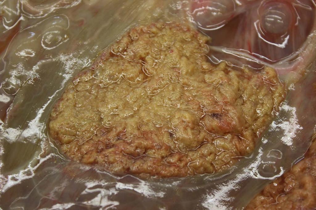
44
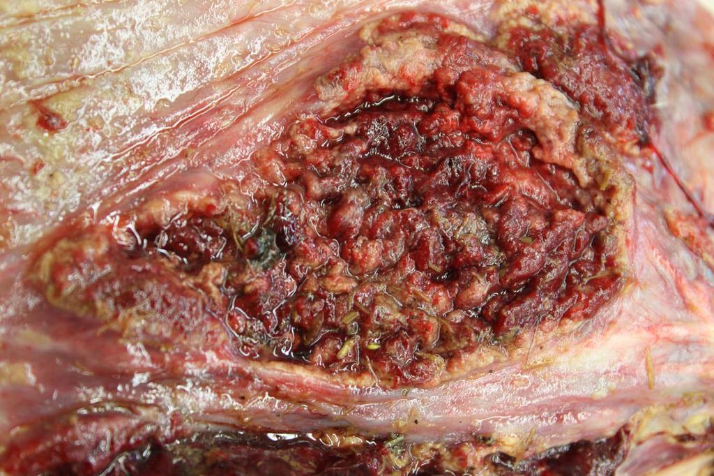
45

46
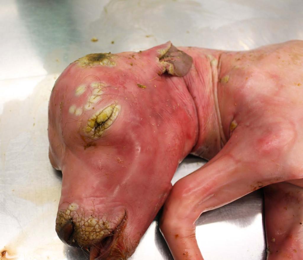
47
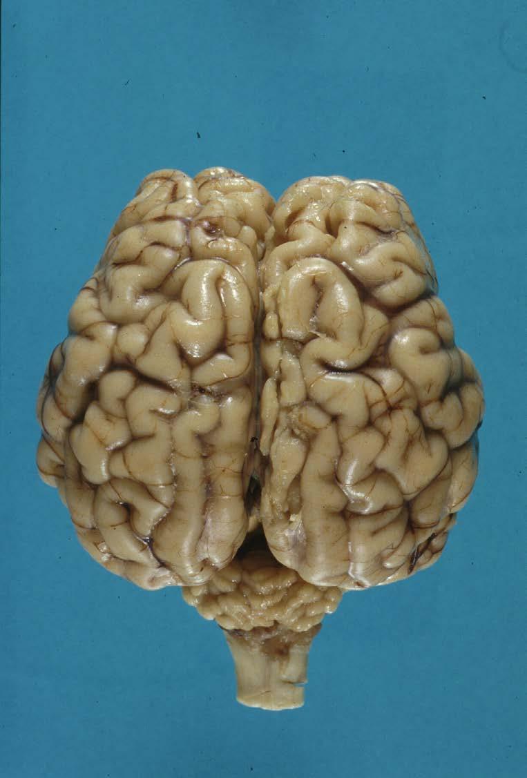
48
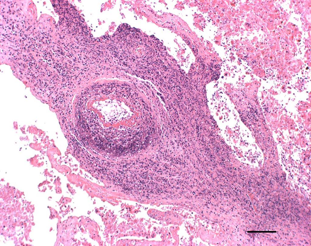
49
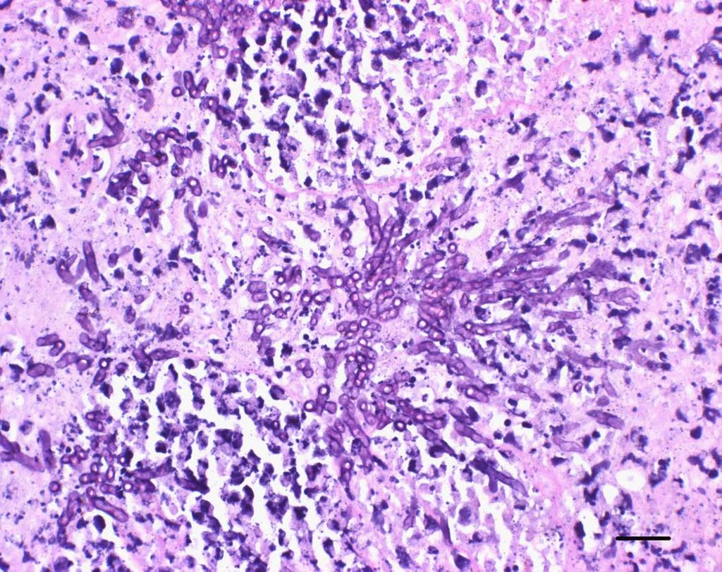
50
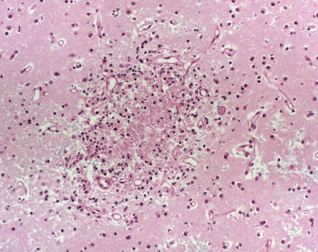
51
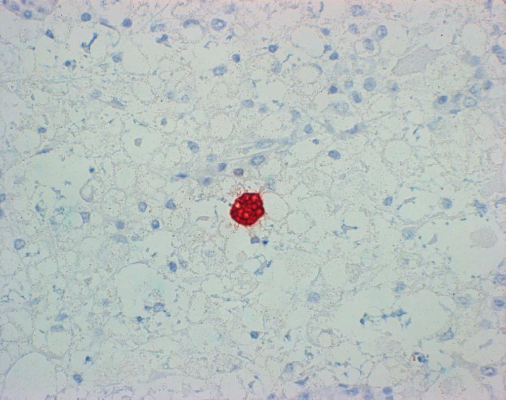
52
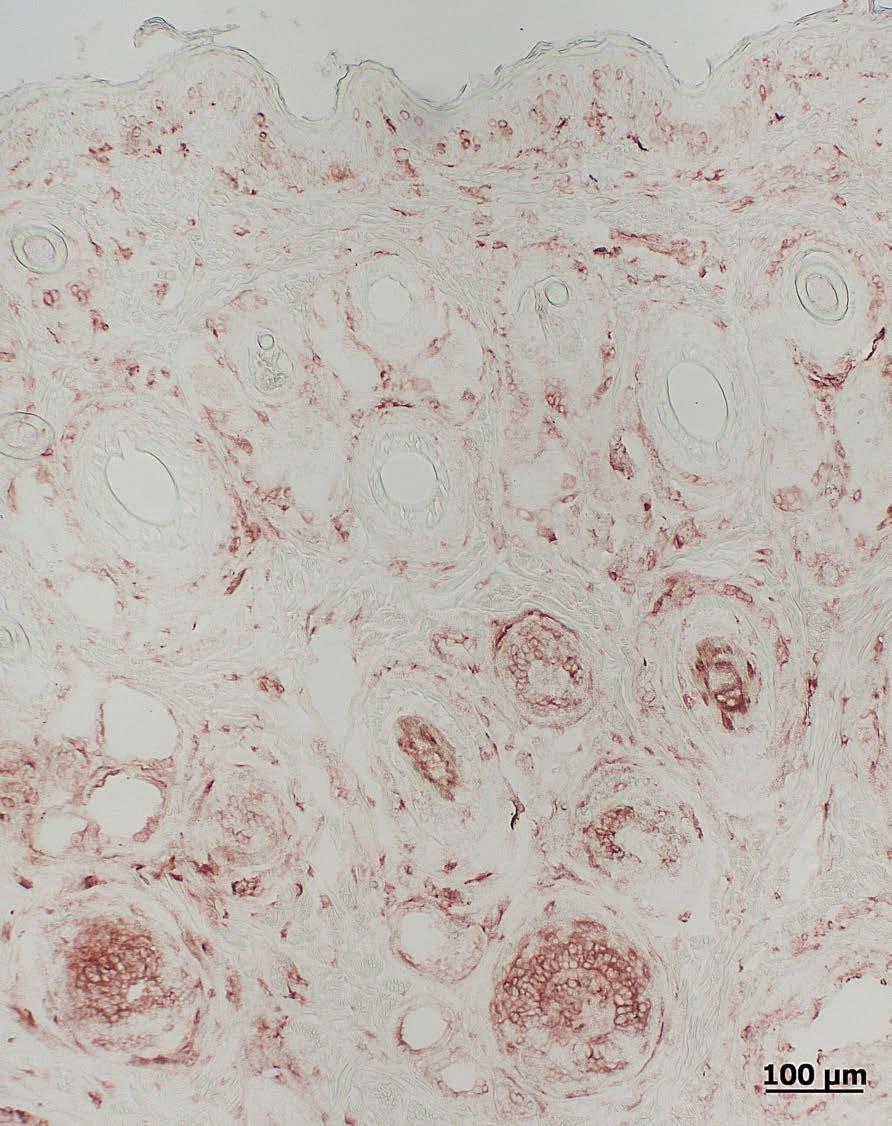
53
Enzootic abortion in sheep and its economic consequences
 Vet Times The website for the veterinary profession https://www.vettimes.co.uk Enzootic abortion in sheep and its economic consequences Author : Louise Silk Categories : Farm animal, Vets Date : February
Surveillance of animal brucellosis
 Surveillance of animal brucellosis Assoc.Prof.Dr. Theera Rukkwamsuk Department of large Animal and Wildlife Clinical Science Faculty of Veterinary Medicine Kasetsart University Review of the epidemiology
Investigations into novel pathogens associated with bovine reproductive failure
 Investigations into novel pathogens associated with bovine reproductive failure Nick Wheelhouse Bovine Reproductive failure Economics Poor reproductive performance across a herd costs estimated 231 per
Detecting new diseases such as Schmallenberg Virus infections (SBV) Guda van der Burgt, Veterinary Investigation Officer AHVLA Luddington
 Guda van der Burgt, Veterinary Investigation Officer AHVLA Luddington") Detecting new diseases such as Schmallenberg Virus infections (SBV) Guda van der Burgt, Veterinary Investigation Officer AHVLA Luddington 1 SURVEILLANCE WHAT DOES IT NEED TO DO? Detect at an early stage
Detecting new diseases such as Schmallenberg Virus infections (SBV) Guda van der Burgt, Veterinary Investigation Officer AHVLA Luddington 1 SURVEILLANCE WHAT DOES IT NEED TO DO? Detect at an early stage
Abortions and causes of death in newborn sheep and goats
 Abortions and causes of death in newborn sheep and goats Debrah Mohale What is abortion? Abortion is the result of a disturbance in the functioning of the afterbirth (placenta). This causes the premature
Emerging diseases but don t forget the old ones! Lynn Batty
 Emerging diseases but don t forget the old ones! Lynn Batty SAC Consulting is a division of Scotland s Rural College Leading the way in Agriculture and Rural Research, Education and Consulting What barren
Schmallenberg Virus Infections in Ruminants
 Schmallenberg Virus Infections in Ruminants F. J. Conraths, B. Hoffmann, D. Höper, M. Scheuch, R. Jungblut, M. Holsteg, H. Schirrmeier, M. Eschbaumer, K. Goller, K. Wernike, M. Fischer, A. Breithaupt,
New Mexico Department of Agriculture
 Veterinary Diagnostic Services New Mexico Department of Agriculture The New Mexico Organic Farming Conference 2018 New Mexico Scientific Laboratories New Mexico Department of Agriculture Veterinary Diagnostic
Cercetări bacteriologice, epidemiologice şi serologice în bruceloza ovină ABSTRACT
 ABSTRACT Thesis entitled BACTERIOLOGICAL, EPIDEMIOLOGICAL AND SEROLOGICAL RESEARCHES IN BRUCELLOSIS OVINE is scientific and practical reasons the following: - Infectious epididymitis in Romania, described
Reproductive Vaccination- Deciphering the MLV impact on fertility
 Reproductive Vaccination- Deciphering the MLV impact on fertility Safety Decision Efficacy Prebreeding Vaccination of Cattle should Provide fetal & abortive protection (BVD and BoHV-1) Not impede reproduction
Diseases of Concern: BVD and Trichomoniasis. Robert Mortimer, DVM Russell Daly, DVM Colorado State University South Dakota State University
 Diseases of Concern: BVD and Trichomoniasis Robert Mortimer, DVM Russell Daly, DVM Colorado State University South Dakota State University The Epidemiologic Triad Host Management Agent Environment Trichomoniasis
FACT SHEET FEBRUARY 2007
 FARM FACT SHEET FEBRUARY 2007 ABORTION IN EWES Abortions in ewes are the result of many factors that stress the pregnant animal. Intrauterine infections are the most common cause. The commonly reported
Protozoan Parasites: Lecture 20 - Heteroxenous Coccidia - Part 1 Pages 39-51
 Protozoan Parasites: Lecture 20 - Heteroxenous Coccidia - Part 1 Pages 39-51 Tissue cyst -forming Coccidia General Taxonomy Apicomplexa Heteroxenous Two host life cycles Asexual & sexual reproduction Intestinal
Vaccination to Improve Reproductive Health. Cow/Calf Meetings. Sandy Stuttgen, DVM UWEX Agriculture Educator, Taylor County
 Vaccination to Improve Reproductive Health Cow/Calf Meetings Sandy Stuttgen, DVM UWEX Agriculture Educator, Taylor County June, 2013 Reproductive Diseases Bacteria Brucella Camplyobacter (Vibrio) Leptospira
Canine Distemper Virus
 Photo: LE Carmichael, MJ Appel Photo: LE Carmichael, MJ Appel Photo: LE Carmichael, MJ Appel Canine Distemper Virus Canine Distemper (CD) is a highly contagious infectious disease of dogs worldwide caused
DOWNLOAD OR READ : VIRAL DISEASES OF CATTLE 2ND EDITION PDF EBOOK EPUB MOBI
 DOWNLOAD OR READ : VIRAL DISEASES OF CATTLE 2ND EDITION PDF EBOOK EPUB MOBI Page 1 Page 2 viral diseases of cattle 2nd edition viral diseases of cattle pdf viral diseases of cattle 2nd edition Animal Health.
Managing Infectious Subfertility in Expanding Dairy herds. John Mee Teagasc, Moorepark Dairy Production Research Centre
 1 Managing Infectious Subfertility in Expanding Dairy herds John Mee Teagasc, Moorepark Dairy Production Research Centre 2 The Four Pillars of Dairy Herd Fertility Management Nutrition Dairy Herd Fertility
Farm Vet News. Effective Colostrum Management. Bi-Monthly Newsletter of Endell Farm Vets
 Farm Vet News Bi-Monthly Newsletter of Endell Farm Vets Issue 06 March 2019 In this issue: PAGE 1 - EFFECTIVE COLOSTRUM MANAGEMENT PAGE 2 - INVESTIGATING ABORTIONS PAGE 4 - BABESIOSIS (RED WATER FEVER)
Import Health Standard. For. Bovine Semen
 Import Health Standard For Bovine Semen Short Name: bovsemid.gen MAF Biosecurity New Zealand Ministry of Agriculture and Forestry P.O Box 2526 Wellington 6011 New Zealand BOVSEMID.GEN 27 June 2011 Page
Cattle keepers guide to safeguarding health
 Cattle keepers guide to safeguarding health 1 Crown Copyright 2015 WG25764 ISBN 978-1-4734-4233-7 Digital ISBN 978-1-4734-4231-3 Contents Foreword 2 Introduction 3 Bovine Viral Diarrhoea 4 Infectious Bovine
TIMELY INFORMATION Agriculture & Natural Resources
 ANIMAL SCIENCES SERIES TIMELY INFORMATION Agriculture & Natural Resources September 2011 Trichomoniasis prevention and control 1 Soren Rodning, DVM, MS, Extension Veterinarian and Assistant Professor 2
Index. Note: Page numbers of article titles are in boldface type.
 Index Note: Page numbers of article titles are in boldface type. A Abdominal viscera, examination of, in investigation of emerging infectious diseases of food animals, 6 American Veterinary Medical Association,
04/02/2013. Parasites and breeding dogs: These parasites we don t hear so much about. Main internal parasites found in breeding kennels
 Parasites and breeding dogs: These parasites we don t hear so much about Main internal parasites found in breeding kennels Isospora sp. Giardia sp. Toxocara canis Something else? Breeders burden I m kind
Diversity, impact and control of chlamydial infections in ruminants. David Longbottom Moredun Research Institute, Edinburgh, UK
 Diversity, impact and control of chlamydial infections in ruminants David Longbottom Moredun Research Institute, Edinburgh, UK Chlamydial diversity Chlamydiae Obligate intracellular bacteria Variety of
The surveillance and control programme
 Annual Reports 2010 Surveillance and control programmes for terrestrial and aquatic animals in Norway The surveillance and control programme for Brucella abortus in cattle in Norway Ståle Sviland Berit
Canine and Feline Distemper. Description. The following chart indicates the animals which are susceptible to infection by canine and feline distemp
 Canine and Feline Distemper Description Canine and feline distemper are diseases affecting many wild and domestic carnivo The following chart indicates the animals which are susceptible to infection by
Seroprevalence of antibodies to Schmallenberg virus in livestock
 Seroprevalence of antibodies to Schmallenberg virus in livestock Armin R.W. Elbers Dept. Epidemiology, Crisis organisation and Diagnostics Central Veterinary Institute (CVI) part of Wageningen UR armin.elbers@wur.nl
Systemic Apicomplexans. Toxoplasma
 Systemic Apicomplexans Toxoplasma Protozoan Groups Historically, protozoa have been grouped by mode of motility. Flagellates Hemoflagellates Trypanosoma cruzi Leishmania infantum Mucoflagellates Tritrichomonas
Index. Note: Page numbers of article titles are in boldface type
 Index Note: Page numbers of article titles are in boldface type A Abomasal bloat diarrhea in calves 3 months old or younger due to, 460 461 Abomastitis diarrhea in calves 3 months old or younger due to,
Protozoan Parasites: Lecture 21 Apicomplexans 3 Heteroxenous Coccidia - Part 1 Pages 37-49
 Protozoan Parasites: Lecture 21 Apicomplexans 3 Heteroxenous Coccidia - Part 1 Pages 37-49 Tissue cyst -forming Coccidia General Taxonomy Apicomplexa Heteroxenous Two host life cycles Asexual & sexual
Risk assessment of the re-emergence of bovine brucellosis/tuberculosis
 Risk assessment of the re-emergence of bovine brucellosis/tuberculosis C. Saegerman, S. Porter, M.-F. Humblet Brussels, 17 October, 2008 Research Unit in Epidemiology and Risk analysis applied to veterinary
Wageningen Bioveterinary Research. Biomedical and veterinary research to safeguard animal and public health
 Wageningen Bioveterinary Research Biomedical and veterinary research to safeguard animal and public health Veterinary research to safeguard animal and public health Wageningen Bioveterinary Research (WBVR)
DISEASE DETECTION OF BRUCELLOSIS IN GOAT POPULATION IN NEGERI SEMBILAN, MALAYSIA. Abstract
 7 th Proceedings of the Seminar in Veterinary Sciences, 27 February 02 March 2012 DISEASE DETECTION OF BRUCELLOSIS IN GOAT POPULATION IN NEGERI SEMBILAN, MALAYSIA Siti Sumaiyah Mohd Yusof, 1,3 Abd. Wahid
Classificatie: intern
 Classificatie: intern Animal Health Service Deventer Jet Mars part 1: Paratuberculosis ParaTB approach In the NL: control program, not an eradication program Quality of dairy products as starting point
Farm Newsletter - February 2017
 Farm Newsletter - February 2017 Lung Worm in Cattle The disease is caused by the worm Dictyocaulus viviparus. Adult worms live in the animal s lungs where they produce eggs which hatch quickly. The first
Procedures for the Taking of Prevention and Eradication Measures of Brucellosis in Bovine Animals
 Republic of Latvia Cabinet Regulation No. 881 Adopted 18 December 2012 Procedures for the Taking of Prevention and Eradication Measures of Brucellosis in Bovine Animals Issued in accordance with Section
Reproductive Management. of Beef Cattle Herds. Reproductive Management. Assessing Reproduction. Cow and Heifer Management
 Reproductive Management of Beef Cattle Herds For a cow-calf operation, good reproductive rates are critical to operational success and profitability. It is generally expected that each breeding-age female
Bovine Viral Diarrhea (BVD)
") Bovine Viral Diarrhea (BVD) Why should you test your herd, or additions to your herd? Answer: BVD has been shown to cause lower pregnancy rates, increased abortions, higher calf morbidity and mortality;
Bovine Viral Diarrhea (BVD) Why should you test your herd, or additions to your herd? Answer: BVD has been shown to cause lower pregnancy rates, increased abortions, higher calf morbidity and mortality;
Interpretation of Bulk Tank Milk Results
 Interpretation of Bulk Tank Milk Results Introduction Culturing bulk tank milk (BTM) to monitor milk quality has limitations based on the amount and frequency of sampling and the amount and types of microorganisms
Opportunistic Disease Surveillance in Culled Wild Fallow Deer (Dama dama)
") Opportunistic Disease Surveillance in Culled Wild Fallow Deer (Dama dama) Nigel Gillan District Veterinarian Central Tablelands Local Land Services - Mudgee Q: Is the spread of livestock or human diseases
Opportunistic Disease Surveillance in Culled Wild Fallow Deer (Dama dama) Nigel Gillan District Veterinarian Central Tablelands Local Land Services - Mudgee Q: Is the spread of livestock or human diseases
MATTILSYNET THE NORWEGIAN FOOD SAFETY AUTHORITY
 MATTILSYNET THE NWEGIAN FOOD SAFETY AUTHITY SANITARY CERTIFICATE For export of bovine semen from Norway to New Zealand COUNTRY: 1.Consignor (Exporter): Name: Address: Reference number: 2. Certificate reference
Assignment 13.1: Proofreading Bovine Spongiform Encephalopathy
 Technical Editing, A 13.1, Proofreading Technical Editing Assignment 13.1: Proofreading Bovine Spongiform Encephalopathy The context This document is now set in type as it will appear in print unless corrected.
AKC Canine Health Foundation Grant Updates: Research Currently Being Sponsored By The Vizsla Club of America Welfare Foundation
 AKC Canine Health Foundation Grant Updates: Research Currently Being Sponsored By The Vizsla Club of America Welfare Foundation GRANT PROGRESS REPORT REVIEW Grant: 00748: SNP Association Mapping for Canine
MATTILSYNET NORWEGIAN FOOD SAFETY AUTHORITY
 MATTILSYNET NWEGIAN FOOD SAFETY AUTHITY Referencenumber: N O - COUNTRY: 1.Consignor (Exporter): Name: Address: 2. Certificate reference number: 3. Veterinary Authority: 4. Import permit number: 5. Consignee
Short information about the ZOBA. Participating on proficiency tests. Monitoring programme
 Short information about the ZOBA Laboratory methods Participating on proficiency tests Research projects Monitoring programme Raymond Miserez DVM, ZOBA, Institute of Veterinary Bacteriology, Vetsuisse
HUSK, LUNGWORMS AND CATTLE
 Vet Times The website for the veterinary profession https://www.vettimes.co.uk HUSK, LUNGWORMS AND CATTLE Author : Alastair Hayton Categories : Vets Date : July 20, 2009 Alastair Hayton discusses how best
Hurricane Animal Hospital 2120 Mount Vernon Road Hurricane, WV or
 Hurricane Animal Hospital 2120 Mount Vernon Road Hurricane, WV 25526 304-757-5937 or 304-757-2287 www.hurricaneanimalhospital.com Feline Leukemia Virus (FELV) This information handout is designed as a
Chlamydia pecorum: fetal and placental lesions in sporadic caprine abortion
 5729VDIXXX10.1177/1040638715625729Pathologic findings in Chlamydia pecorum induced abortion in a goatgiannitti et al. 015 Brief Communication Chlamydia pecorum: fetal and placental lesions in sporadic
Antibiotic Resistance in the European Union Associated with Therapeutic use of Veterinary Medicines
 Antibiotic Resistance in the European Union Associated with Therapeutic use of Veterinary Medicines Report and Qualitative Risk Assessment by the Committee for Veterinary Medicinal Products Annex III Surveillance
Medical Bacteriology- Lecture 14. Gram negative coccobacilli. Zoonosis. Brucella. Yersinia. Francesiella
 Medical Bacteriology- Lecture 14 Gram negative coccobacilli Zoonosis Brucella Yersinia Francesiella 1 Zoonosis: A disease, primarily of animals, which is transmitted to humans as a result of direct or
Bacterial infections in the urinary tract
 Zurich Open Repository and Archive University of Zurich Main Library Strickhofstrasse 39 CH-8057 Zurich www.zora.uzh.ch Year: 2014 Bacterial infections in the urinary tract Gerber, B Posted at the Zurich
Above: life cycle of toxoplasma gondii. Below: transmission of this infection.
 Toxoplasmosis PDF This article is based on a paid for research paper dated 1972 of similar title and authored by J.K.Frenkel and J.P. Dubey. It was published by The Journal of Infectious Diseases Vol.
Tritrichomonas Foetus in Cats
 Tf Tritrichomonas Foetus in Cats A practical guide for breeders By Dr S F Moreland BA Vet MB MRCVS GCCF Veterinary Officer September 2017 TRITRICHOMONAS FOETUS IN CATS WHAT IS Tf? Tf is the commonly used
Food safety related to camelids products: Brucellosis and its impact on Public Health and the consumers as an example
 DIRECCION GENERAL DE LABORATORIOS Y CONTROL TECNICO Food safety related to camelids products: Brucellosis and its impact on Public Health and the consumers as an example Third Global Conference of OIE
Johne s Disease. for Goat Owners
 Johne s Disease Q&A for Goat Owners The National Johne s Education Initiative recognizes Dr. Elisabeth Patton and Dr. Gretchen May with the Wisconsin Department of Agriculture, Trade and Consumer Protection
Questions and answers about methicillin-resistant Staphylococcus aureus (MRSA)
") Questions and answers about methicillin-resistant Staphylococcus aureus (MRSA) Updated FAQ, 18 November 2014 Methicillin-resistant Staphylococcus aureus (MRSA) are bacteria which are resistant to certain
Questions and answers about methicillin-resistant Staphylococcus aureus (MRSA) Updated FAQ, 18 November 2014 Methicillin-resistant Staphylococcus aureus (MRSA) are bacteria which are resistant to certain
For Public Health Personnel
 For Public Health Personnel General Information Toxoplasma gondii is a protozoal parasite capable of infecting any warm-blooded animal, including humans. Wild and domestic cats are the only known definitive
Elk Brucellosis Surveillance and Reproductive History
 2013-14 Elk Brucellosis Surveillance and Reproductive History Neil Anderson, Montana Fish, Wildlife and Parks, 1400 South 19 th Ave., Bozeman, MT 59718. Kelly Proffitt, Montana Fish, Wildlife and Parks,
Break Free from BVD. What is BVD? BVD outbreak in 2013/ cow dairy herd in Staffordshire. Costs Calculation Costs*
 Break Free from BVD Poor growth rates, calf mortality, youngstock pneumonia, poor conception rates and abortions can all highlight an underlying Bovine Viral Diarrhoea (BVD) infection in your herd. BVD
ASA Master Class -Flock Health Click to Challenges edit Master title style
 ASA Master Class -Flock Health Click to Challenges edit Master title style Michael Doherty CD School of Veterinary Medicine 1 What are the sheep health priorities? 2 We need flock health planning Dissemination
Controlling BVD & Johne s.
 Controlling BVD & Johne s www.hccmpw.org.uk BVD and Johne s in beef herds 1 - Bovine Viral Diarrhoea 2 - Johne s disease 9 Economics of disease 13 Biosecurity 16 Key Points 18 BVD and Johne s in Beef herds
The surveillance programme for bovine virus diarrhoea (BVD) in Norway 2016
 in Norway 2016") Annual Report The surveillance programme for bovine virus diarrhoea (BVD) in Norway 2016 Norwegian Veterinary Institute The surveillance programme for bovine virus diarrhoea (BVD) in Norway 2016 Content
Annual Report The surveillance programme for bovine virus diarrhoea (BVD) in Norway 2016 Norwegian Veterinary Institute The surveillance programme for bovine virus diarrhoea (BVD) in Norway 2016 Content
Drd. OBADĂ MIHAI DORU. PhD THESIS ABSTRACT
 UNIVERSITY OF AGRICULTURAL SCIENCES AND VETERINARY MEDICINE ION IONESCU DE LA BRAD IAŞI FACULTY OF VETERINARY MEDICINE SPECIALIZATION MICROBIOLOGY- IMUNOLOGY Drd. OBADĂ MIHAI DORU PhD THESIS ABSTRACT RESEARCHES
EUROPEAN COMMISSION HEALTH & CONSUMERS DIRECTORATE-GENERAL. Unit G5 - Veterinary Programmes
 EUROPEAN COMMISSION HEALTH & CONSUMERS DIRECTORATE-GENERAL Unit G5 - Veterinary Programmes SANCO/10853/2012 Programmes for the eradication, control and monitoring of certain animal diseases and zoonoses
There is no one correct way to describe a slide. Macroscopic Veterinary Pathology. Be concise. Look at the center of the slide.
 Macroscopic Veterinary Pathology There is no one correct way to describe a slide. Bruce Williams, DVM, DACVP Senior Pathologist, JPC Email: williams@cldavis.org Tissue from a sheep Tissue from a foal Be
EFSA Scientific Opinion on canine leishmaniosis
 EFSA Scientific Opinion on canine leishmaniosis Andrea Gervelmeyer Animal Health and Welfare Team Animal and Plant Health Unit AHAC meeting 19 June 2015 PRESENTATION OUTLINE Outline Background ToR Approach
ELISA assays for parasitic and tick-borne diseases
 ELISA assays for parasitic and tick-borne diseases We are passionate about the health and well-being of humans and animals. Immunodiagnostics from contribute to a global, adequate supply of safe and nutritious
Course Curriculum for Master Degree in Poultry Diseases/Veterinary Medicine
 Course Curriculum for Master Degree in Poultry Diseases/Veterinary Medicine The Master Degree in Poultry Diseases /Veterinary Medicine, is awarded by the Faculty of Graduate Studies at Jordan University
Update on diagnosis of feline infectious peritonitis (FIP)
") Update on diagnosis of feline infectious peritonitis (FIP) Séverine Tasker RCVS Specialist in Feline Medicine The Feline Centre Langford Veterinary Services University of Bristol http://www.felinecentre.co.uk/
Update on diagnosis of feline infectious peritonitis (FIP) Séverine Tasker RCVS Specialist in Feline Medicine The Feline Centre Langford Veterinary Services University of Bristol http://www.felinecentre.co.uk/
TECHNICAL DOCUMENT. Incorporating Rules for Cattle Health Schemes. Issued: July Final Version
 TECHNICAL DOCUMENT Incorporating Rules for Cattle Health Schemes Issued: July 2014 Contents Page Introduction 3 4 The Diseases 5 10 The Rules of CHeCS 11 17 Bulk Milk Monitoring Programmes 18 Infectious
Bovine Brucellosis Control of indirect ELISA kits
 Bovine Brucellosis Control of indirect ELISA kits (Pooled milk samples) Standard Operating Procedure Control of Bovine brucellosis Milk ELISA kits SOP Page 1 / 6 02 February 2012 SAFETY PRECAUTIONS The
The infection can be transmitted only by sexual intercourse and not by the environment. Bovine trichomoniasis is not transmitted to people.
 Revised Oct. 2015 ASWeb-132 Texas Bovine Trichomoniasis Control Program: Facts for Cattle Owners Rick Machen, Ron Gill, Floron Faries and Tom Hairgrove* Bovine trichomoniasis (Trich) is a venereal disease
Simple Herd Level BVDV Eradication for Dairy
 Simple Herd Level BVDV Eradication for Dairy Dr. Enoch Bergman DVM So why is BVDV important to dairy producers? Global BVDV research, whilst examining differing management systems, consistently estimates
TB IN GOATS - REDUCING THE RISK IN THE LARGER HERD
 INTRODUCTION These guidelines have been produced by the Goat Veterinary Society, but only give generic advice. No two goat units are identical, and the information given below is intended as a guide to
Federal Expert Select Agent Panel (FESAP) Deliberations
 Deliberations") Federal Expert Select Agent Panel (FESAP) Deliberations FESAP and Biennial Review Established in 2010 and tasked with policy issues relevant to the security of biological select agents and toxins Per recommendations
Federal Expert Select Agent Panel (FESAP) Deliberations FESAP and Biennial Review Established in 2010 and tasked with policy issues relevant to the security of biological select agents and toxins Per recommendations
Epidemiological analysis of the 2006 bluetongue virus serotype 8 epidemic in north-western Europe. Within herd distribution of infection
 Epidemiological analysis of the 26 bluetongue virus serotype 8 epidemic in north-western Europe Within herd distribution of infection A.R.W. Elbers 1, K. Mintiens 2, G. Gerbier 3, A.N. van der Spek 4,
Biological Threat Fact Sheets
 Biological Threat Fact Sheets Anthrax Agent: Bacillus anthracis There are three clinical forms of B. anthracis which are determined by route of entry: Pulmonary or Inhalation BT implications Cutaneous
VETERINARY BACTERIOLOGY FROM THE DARK AGES TO THE PRESENT DAY
 VETERINARY BACTERIOLOGY FROM THE DARK AGES TO THE PRESENT DAY D.J.TAYLOR MA PhD VetMB DipECPHM DipECVPH MRCVS EMERITUS PROFESSOR OF VETERINARY BACTERIOLOGY AND PUBLIC HEALTH UNIVERSITY OF GLASGOW INTRODUCTION
CONTAGIOUS BOVINE PLEURO- PNEUMONIA steps towards control of the disease. Rose Matua -Department of Veterinary Services, Kenya
 CONTAGIOUS BOVINE PLEURO- PNEUMONIA steps towards control of the disease Rose Matua -Department of Veterinary Services, Kenya Introduction CBPP is a highly contagious acute, subacute or chronic disease
Johne's disease infectious diarrhea of cattle
 Louisiana State University LSU Digital Commons LSU Agricultural Experiment Station Reports LSU AgCenter 1927 Johne's disease infectious diarrhea of cattle Harry Morris Follow this and additional works
Mastitis: Background, Management and Control
 New York State Cattle Health Assurance Program Mastitis Module Mastitis: Background, Management and Control Introduction Mastitis remains one of the most costly diseases of dairy cattle in the US despite
EBA Series FOOTHILL ABORTION UPDATE: PART I: THE TICK
 EBA Series FOOTHILL ABORTION UPDATE: PART I: THE TICK Foothill abortion in cattle, also known as Epizootic Bovine Abortion (EBA), is a condition well known to beef producers who have experienced losses
FAO-APHCA/OIE/USDA Regional Workshop on Prevention and Control of Neglected Zoonoses in Asia July, 2015, Obihiro, Japan.
 FAO-APHCA/OIE/USDA Regional Workshop on Prevention and Control of Neglected Zoonoses in Asia 15-17 July, 2015, Obihiro, Japan Dr Gillian Mylrea 1 Overview What is a Neglected Zoonotic Disease? The important
Course Curriculum for Master Degree in Internal Medicine/ Faculty of Veterinary Medicine
 Course Curriculum for Master Degree in Internal Medicine/ Faculty of Veterinary Medicine The Master Degree in Internal Medicine/Faculty of Veterinary Medicine is awarded by the Faculty of Graduate Studies
Pesky Ectoparasites. Insecta fleas, lice and flies. Acari- ticks and mites
 Pesky Ectoparasites Parasite control should be at the forefront of every pet owner s life as all animals have the propensity to contract numerous ones at one stage or another. They are a challenge to the
Mycoplasmal pneumonia in calves
 Vet Times The website for the veterinary profession https://www.vettimes.co.uk Mycoplasmal pneumonia in calves Author : Adam Martin Categories : Vets Date : November 2, 2009 Mycoplasma bovis is responsible
Septic cats are not small septic dogs
 Vet Times The website for the veterinary profession https://www.vettimes.co.uk Septic cats are not small septic dogs Author : ROGER WILKINSON Categories : Vets Date : September 29, 2008 ROGER WILKINSON
Doctor B s BARF & Toxoplasmosis
 Doctor B s BARF & Toxoplasmosis Copyright Ian Billinghurst Introduction Ignorance is bliss so they say! Sometimes the less we know, the happier we are. Ignorance can most definitely be a source of bliss
Official Journal of the European Union
 11.6.2003 L 143/23 COUNCIL DIRECTIVE 2003/43/EC of 26 May 2003 amending Directive 88/407/EEC laying down the animal health requirements applicable to intra- Community trade in and imports of semen of domestic
Infectious Disease. Topic-Actinomycosis. Topic-Anaerobic Infections. Topic-Aspergillosis - Disseminated. Topic-Blastomycosis.
 Topic-Actinomycosis Figure 1. VD thoracic radiograph of consolidated lung lobe secondary to actinomycosis. Topic-Anaerobic Infections Figure 1. Test tube of effusive fluid removed from the thorax of a
Fertility and health management of suckler cow herds
 [Beef 2016 Profitable Technologies, Teagasc, Grange, Dunsany, Ireland, Open Day July 2016] Fertility and health management of suckler cow herds David Kenny 1, Michael Diskin 2, Christy Watson 4 and Riona
Approved by the Food Safety Commission on September 30, 2004
 Approved by the Food Safety Commission on September 30, 2004 Assessment guideline for the Effect of Food on Human Health Regarding Antimicrobial- Resistant Bacteria Selected by Antimicrobial Use in Food
PARASITOLOGICAL EXAMINATIONS CATALOGUE OF SERVICES AND PRICE LIST
 INSTITUTE OF PARASITOLOGY Biomedical Research Center Seltersberg Justus Liebig University Giessen Schubertstrasse 81 35392 Giessen Germany Office: +49 (0) 641 99 38461 Fax: +49 (0) 641 99 38469 Coprological
Feline Vaccines: Benefits and Risks
 Feline Vaccines: Benefits and Risks Deciding which vaccines your cat should receive requires that you have a complete understanding of the benefits and risks of the procedure. For this reason, it is extremely
Brucellosis and Yellowstone Bison
 Brucellosis and Yellowstone Bison Overview Brucellosis has caused devastating losses to farmers in the United States over the last century. It has cost the Federal Government, the States, and the livestock
Emerging threats. Cattle. Quarterly & Annual Report: FOURTH QUARTER & Contents. Highlights. Date: October-December 2011.
 Emerging threats Cattle Quarterly & : FOURTH QUARTER & 2011 Date: October-December 2011 Contents Page The VIDA diagnoses are recorded on the AHVLA FarmFile database and SAC LIMS database and comply with
The Salmonella. Dr. Hala Al Daghisatni
 1 Dr. Hala Al Daghisatni The Salmonella Salmonellae are often pathogenic for humans or animals when acquired by the oral route. They are transmitted from animals and animal products to humans, where they
Campylobacter species
 ISSUE NO. 1 SEPTEMBER 2011 1. What are Campylobacter spp.? Campylobacter spp. are microaerophilic, Gram-negative, spiral shaped cells with corkscrew-like motility. They are the most common cause of bacterial
Burn Infection & Laboratory Diagnosis
 Burn Infection & Laboratory Diagnosis Introduction Burns are one the most common forms of trauma. 2 million fires each years 1.2 million people with burn injuries 100000 hospitalization 5000 patients die
Veterinary Diagnostics Portfolio Overview. Complete solutions for veterinary testing and pathogen research
 Veterinary Diagnostics Portfolio Overview Complete solutions for veterinary testing and pathogen research Sample preparation products Cat. no. (number of preps) Target analyte Product Short description