A comparative osteological study of two species of Colubridae: (Pituophis and Thamnophis)
|
|
|
- Shanna Fox
- 5 years ago
- Views:
Transcription


1 Brigham Young University Science Bulletin, Biological Series Volume 8 Number 3 Article A comparative osteological study of two species of Colubridae: (Pituophis and Thamnophis) Robert E. Bullock Lethbridge College, Lethbridge, Alberta, Canada Wilmer W. Tanner Department of Zoology and Entomology, Brigham Young University, Provo, Utah Follow this and additional works at: Part of the Anatomy Commons, Botany Commons, Physiology Commons, and the Zoology Commons Recommended Citation Bullock, Robert E. and Tanner, Wilmer W. (1966) "A comparative osteological study of two species of Colubridae: (Pituophis and Thamnophis)," Brigham Young University Science Bulletin, Biological Series: Vol. 8 : No. 3, Article 1. Available at: This Article is brought to you for free and open access by the Western North American Naturalist Publications at BYU ScholarsArchive. It has been accepted for inclusion in Brigham Young University Science Bulletin, Biological Series by an authorized editor of BYU ScholarsArchive. For more information, please contact scholarsarchive@byu.edu, ellen_amatangelo@byu.edu.


2 I A/ Y^ Brigham Young University Science Bulletin -^^OWP^ zc UNIVERSITY A COMPARATIVE OSTEOLOGICAL STUDY OF TWO SPECIES OF COLUBRIDAE (PITUOPHIS AND THAMNOPHIS) by ROBERT E. BULLOCK and WILMER W. TANNER BIOLOGICAL SERIES VOLUME VIII, NUMBER 3 ^y DECEMBER, 1966


3 BRIGHAM YOUNG UNIVERSITY SCIENCE BULLETIN BIOLOGICAL SERIES Editor: Dorald M. Allred, Department of Zoology and Entomology, Brigham Young University, Provo, Utah Associate Editor: Eabl M. Christensen, Department of Botany, Brigham Yoimg University, Provo, Utah Members of the Editorial Board: V. Beck, Bacteriology J. C. Lynn HArwAUD, Zoology W. Derby Lavs^s, Agronomy Howard C. Stutz, Botany WiLMER W. Tanner, Zoology, Chairman of the Board David L. Hanks, Botany Ex officio Members: Rudger H. > Walker, Dean, College of Biological and Agricultural Sciences Ernest L. Olson, Chairman, University PubHcations The Brigham Young University Science Bulletin, Biological Series, publishes acceptable papers, particularly large manuscripts, on all phases of biology. Separate numbers and back volumes can be purchased from University Publications, Brigham Young University, Provo, Utah. All remittances should be made payable to Brigham Young University. Orders and materials for library exchange should be directed to the Division of Gifts and Exchange, Brigham Young University Library, Provo, Utah.


4 Brigham Young University Science Bulletin A COMPARATIVE OSTEOLOGICAL STUDY OF TWO SPECIES OF COLUBRIDAE (PITUOPHIS AMD THAMNOPHIS) by ROBERT E. BULLOCK and WILMER W. TANNER BIOLOGICAL SERIES VOLUME VIII, NUMBER 3 DECEMBER, 1966


5 TABLE OF CONTENTS Page INTRODUCTION 1 MATERIALS AND METHODS 2 VERTEBRAL COLUMN 3 Atlas 7 Axis 7 First Thoracic Vertebra 7 Other Thoracic Vertebrae 9 Lumbar Vertebrae 9 Anterior Caudal Vertebrae 9 Middle Caudal Vertebrae 13 Posterior Caudal Vertebrae 13 Ribs 13 SKULL 15 Premaxilla 15 Nasals 15 Septomaxillae 15 Vomers 15 Frontals 15 Prefrontals 17 Parietals 17 Postorbitals 17 Supraoccipital 17 Prootics - 17 Exoccipitals 19 Basioccipital 19 Basisphenoid 19 MaxiUae 19 Ectopterygoids 19 Palatines 19 Pterygoids 21 Supratemporals 21 Quadrates 21 Mandible ^ 21 Stapes 22 Hyoid Apparatus 22 OSTEOLOGICAL COMPARISONS 22 Vertebral Column 22 Skull.. 23 DISCUSSION 25 SUMMARY AND CONCLUSIONS 27 LITERATURE CITED 28 ABBREVIATIONS USED IN PLATES 28


6 LIST OF FIGURES Figure Page 1. Atlas 4 2. Axis 5 3. First Thoracic and Thoracic Vertebrae 6 4. Thoracic and Lumbar Vertebrae 8 5. Anterior Caudal Vertebra Middle Caudal Vertebra Posterior Caudal Vertebra and Rib Skull, Dorsal View Skull, Lateral View Skull, Ventral View Hyoid Apparatus, Mandible, and Cranium 20
 has rightfully referred to this section of")

7 A COMPARATIVE OSTEOLOGICAL STUDY OF TWO SPECIES OF COLUBRIDAE {PITUOPHIS AND THAMNOPHIS) by Robert E. Bullock' and Wilmer W. Tanner- INTRODUCTION Snake anatomy has been genei-ally neglected. Although some early writers recorded observations on the anatomy of snakes, no attempt has been made to undertake a comprehensive study, and Cole (1944) has rightfully referred to this section of vertebrate anatomy as an "almost virgin field." Snakes deserve more intensive investigation because of the considerable number of adaptations demonstrated by them, their peculiar types of locomotion, and methods of feeding. It is important to comprehend their anatomical specializations if we are to understand the evolutionary trends among modern reptiles. It also is evident that comparative descriptions of serpents, thus far based mainly on external characteristics, need to be supplemented by accounts of their internal anatomy. The purpose of this study is threefold: (1) To work out in detail the osteology of two common members of the family Colubridae. (2) To compare the bones and bone structures of the two species in order to determine their anatomical similarities and differences, and to use these comparisons in determining the different speciahzations that have been made by two common serpents. (3) To relate the structural differences of these genera, as far as possible, to some of the other generic groups previously reported. Our original intent was also to include the myology of the head, neck, and anterior tnmk region (first 10 vertebrae). This proved to be a major effort in itself, and such a report will appear separately at a later date. The gopher snake, Pituophis catenifer Blain- N'ille, and the plains garter snake, Thamnophis radix (Baird and Girard), were chosen for this study because of their moderately large size and relative abundance. One of them (P. catenifer) cxjnstricts its prey. The other ( T. radix ) swallows it alive. This difference in mode of feeding may be responsible for some structural modifications. Although it is not the purpose of this study directly to resolve any phylogenetic or ta.xonomic problems, it is hoped that it will encourage other anatomical studies of reptiles, add to our present understanding of homologies, and indicate important internal phylogenetic and taxonomic structures as suggested by Robinson and Tanner (1962). Although no previous investigator has studied the osteology of the two species included in this study, the general gross anatomy of various other snakes has been known for a long time. Ahrenfeldt (1955) stated that as far back as 1573 there were some fragmentary accounts of snake anatomy published in Europe, but Cole ( 1944 ) mentioned that it was not until 1683 that the first workmanlike and relatively accurate description of serpent anatomy was made by Edward Tyson on a "Timber Rattlesnake" said to have been collected in the West Indies. Owen ( 1866 ) gave one of the first detailed accounts of the osteology and myology of snakes based on his work with the species Crotalus horridtis, Pijthon tigris. Boa constrictor, Naja tripudians, and Deirodon scaber. Although Owen's work on this section of the vertebrates is a general account by our standards, in his time it was a major contribution to this area of anatomy. Another work dealing with snake anatomy is a laboratory dissecting guide by Kellicott (1898) for the genus Heterodon. Unfortunately, the descriptions lack detail and are therefore somewhat superficial and in many areas incomplete. Huxley (1871), Gegenbaur (1878), Hoffman (1890), Sedgwick (1905), Wiedersheim and Parker (1907), VVilliston (192,5), Kingsley (1917), Goodrich (1930), and Versluys (1937) were some of the early textbook writers who ^Lethbridge College. Lethbridge, AJberta, Canad ^Department of Zoology and Entomology, Brigha Young University, Provo, Utah.

 devoted entirely to the anatomy of serpents deal rather extensively with the developmental and adult anatomy of several species of snakes, including a few colubrid")
8 Brigham Young University Science Bulletin dealt with the subject of snake osteology. Most of these writers restricted their studies to the skull of serpents, and on occasion included one or more generalized vertebrae. Romer's "Osteology of the Reptiles" (1956) deals only briefly with serpents, but does bring some of the terminology and many of the homologies up to date. Two volumes by Ludicke (1962, 1964) devoted entirely to the anatomy of serpents deal rather extensively with the developmental and adult anatomy of several species of snakes, including a few colubrid species. Many of the descriptions and comparisons in these works are of a general nature and, therefore, are of limited value in making comparisons with the colubrid species involved in this study. Sood (1941, 1948) published two papers dealing with the vertebral column of serpents. The first is concerned with the caudal vertebrae of the sandsnake, Eryx johnii (Russell), and illustrates many unique peculiarities in its vertebral anatomv. The second studv includes both minute and gross vertebral anatomy for several other Asian species. An extensive work by Edgeworth (1935) describes and compares the development of the cranial muscles and associated skeletal structures in forms ranging from the fishes to the mammals. Haas (1930) and Cowan and Hick (1951) also dealt with the musculature of snakes, but included skeletal structures as well. Albright and Nelson ( ) dealt with the cranial osteology and the musculature of the colubrid snake Elaphe obsolcta quadrivittata. Perhaps more osteology has been done with venomous species, particularly with the teeth (fangs) and sk-ull. The work of Klauber (1956: ) is a good example. We made no major attempt here to e.xhaust the references which may refer to snake anatomy. Those referred to above represent some of the more important works that we have seen. We were greatly surprised when we found relatively few studies dealing with either of the two common colubrid genera Pittiophis and Thamnophis. Our prime concern, therefore, is to provide detailed descriptions and drawings which wiu set forth the skeletal anatomy as we found it in these genera. MATERIALS AND METHODS This comparative study utilized the species Pituophls catenifer deserticola Stejneger and Thamnophw radix haydeni Kennicott. The fourteen specimens of P. c. deserticola used were collected in Grand, Kane, Utah, San Juan, Garfield, and Uintah Counties, Utah. The twelve specimens of T. r. haydeni came from Warner County, Alberta, Canada. However, two specimens of P. catenifer sayi Schlegel from Medicine Hat, Alberta, Canada, two T. elegans vagrans Baird and Girard from Utah County, Utah, and four T. sirtalis parietalis Say from Salt Lake County, Utah, were also dissected and studied. These additional species are similar within their respective genera; therefore, a description of these is not included. The skeletons were prepared in several different ways, depending on whether fresh or preserved snakes were used, and upon the degree of articulation required. Disarticulated skeletons were prepared from fresh material by bacterial action. The snake was skinned, eviscerated, and plac-ed in a container with enough water to cover the specimen so as to accelerate the decomposition process. After a few days in hot weather, the macerated material was placed on a fine mesh screen and a jet of water was used to clear away the remaining tissue. Some of the bones were bleached in a 3% solution of hvdrogen peroxide for ten to twelve hours. Most of the articulated skeletons were prepared from fresh material. The fresh, skinned, and eviscerated snake was placed in a 25% solution of ammonium hydroxide for one week, then boiled for t^vo to ten minutes until the tissues were loose but the ligaments still intact. A small jet of water was used to clear the soft tissue from the vertebral column. This jet proved to be too harsh on the more delicate and loosely connected bones of the skull. To remove the deeper skull muscles it was necessary to use forceps under a stereoscopic microscope. This latter method was essential in preparing skulls without distortion or loss of small bones. A few snakes preserved in alcohol were utilized in the preparation of skeletal material. These were cleaned by placing them for two days in a solution of two ounces of trisodium phosphate to each quart of water. Tlie material was then boiled in water for varying lengths of time, the time determined by the type of skeletal preparation desired. Soaking the preserved

, Kellicott (1898), Romer (19.56). Sood (1941, 1948), and the more recent works by Albright and Nelson (19.")
9 OsPEOLOGv OF Snakes material in concentrated ammonium livdroxide instead of trisodium phosphate before boihng proved to be almost as satisfactory. Specimens were studied and drawings made to scale with the aid of a hand micrometer. Literature dealing with the complete osseous skeleton of the snake is fragmentary and limited. Because the literature to date, as far as we were able to ascertain, reveals no account of the osteology of either P. catenifer or T. radix, the homologies were worked out and derived from work done by Owen (1866), Kellicott (1898), Romer (19.56). Sood (1941, 1948), and the more recent works by Albright and Nelson (19.59), and Ludicke (1962, 1964). The terminology for the skull structures used herein was adopted chiefly from Romer (19.56), and the terminology for the vertebral column from Sood ( 1941, 1948). The two snakes studied are anatomically similar; therefore, to avoid unnecessary repetition a complete description of P. catenifer was prepared, but T. radix is discussed only in connection with comparative structures that differ in the two forms. Tlie plates were prepared with sufficient detail to aid in the comparisons of similar structures in the two species. VERTEBRAL COLUMN In P. catenifer as in other snakes, the vertebrae are numerous and procoelous, with ball and socket articulations wliich allow free movement. All of the vertebrae articulate with ribs except the atlas, axis, first thoracic, and caudals. The successive vertebrae not only articulate by the usual pre- and post-zygapophyses and by the procoelous centra, but also by additional articulations such as the zygosphenes and zygantra. The division of the ophidian vertebral column into well-marked regions is less distinct than it is in other higher vertebrates. Owen (1866), Sedgwick (1905), Reynolds (1913), Williston (1925), and others considered the column to be made up of two regions a precaudal region, and a caudal region. Five regions were distinguished by Rochebrune ( 1881 ) the cervical, thoracic, pelvic, sacral, and cocygeal. Although these regions can be distinguished one from another, the differences are slight and variable. Because this classification was somewhat superficial and could not be regarded as ecjuivalent to the regions of the vertebral column of other vertebrates, it was presumably not adopted by later workers. Sood (1941, 1948) retained the division which separated the column into two regions (precaudal and caudal), but distinguished several subregions of each as follows: I. Tlie Precaudal Region A. Cervical Subregion, composed of the first two vertebrae, atlas, and axis. B. Thoracic Subregion, consisting of all the vertebrae that follow the axis and bear prominent hypapophyses. II. C. Lumbar Subregion, consisting of the vertebrae situated between the thoracic subregion and the caudal region. The hypapophyses are reduced in size or completely lacking in this area. The Caudal Region A. Anterior Caudal Subregion, situated immediately behind the precaudal vertebrae. Vertebrae are provided with both fi.xed and articulating bifurcated ribs called processi costotransversii or lymphapophyses. B. Middle Caudal Subregion, containing fixed ribs which are unforked and differing from the posterior caudal subregion in the absence of haemapophyses. C. Posterior Caudal Subregion comprising vertebrae with a pair of flat, platelike haemapophyses on the ventral aspect of their centra. This area shows a gradual reduction in the size and development of the vertebrae and vertebral processes from anterior to posterior until they are represented near the end of the tail by extremely short vertebrae with almost wholly vestigial processes. Because the division of the vertebral column as outlined by Sood is in general use, the preceding vertebral classification will generally be followed; however, a few modifications have been made in order to adapt it to the species involved in this study.


10 Bhigham Young University Science Bulletin A.INT.F. A.NEU.F. A.NEU.F Fig. 1. Atla-s. Pituophii catenifer, A-D, 9X: A, anterior; B, posterior; C, dorsal; D, lateral. Tliumiiophis radix, E-H, 15X: E, anterior; F. posterior; G, dorsal; H, lateral.


11 Osteology of Snakes N.S. ZE.


12 Bricham Young University Science Bulletin PRZ.F Fig. 3. Pituophts catenifer, A-D, 7X: A-B, first thoracic vertebra: A, dorsal; B, lateral; C-D, thoracic vertebra: C, anterior; D, posterior. Thamnophis radix, E-H, 9X: E-F, first thoracic vertebra: E, dorsal; F, lateral; G-H, poi thoracic vertebra: G. anterior; H, posterior.


13 Osteology of Snakes Fig. 1-A, B, C and D The first cervical vertebrae or atlas differs widely from the general vertebral pattern. It is a relatively narrow ring composed of tliree separate bones fused together by means of sutures. Tlie two lateral neural arches or neurapophyses join dorsally as the sagittal neural suture; thev form a pair of neurocentral sutures at their ventral borders where they unite with the intercentrum. Tliere is no neural spine, but a nidimentary dorsal crest is developed. The centrum of the atlas actually coalesces with that of the axis leaving the atlas without a true centrum, its place being taken by the autogenous hypapophysis (Owen, 1866). Romer (1956) stated that the atlas has been robbed by the a.xis of its centnmi, but has retained its intercentnmi. Anteriorly the intercentrum presents a concave articular surface which articulates with the basioccipital tubercle of the occipital condyle. Posteriorly a similar surface adjoins the ventral surface of the odontoid process, as well as the interior facet of the anterior hypapophysis of the axis. Posteroventrally the intercentrum develops a small conical hypapophysis. The base of each neural arch half possesses an anteromesial, concave articular surface which receives the exoccipital tubercle of the occipital condyle, and a posteromesial surface which articulates with the dorsal and lateral convex portions of the odontoid process. Each neural arch half carries a short postzygapophysis from the lateral border of its dorsoposterior region, and from its ventrolateral border a short conical transverse process points posteriorlv. The neural canal is formed by the dorsal expansion of the neural arch, while the smaller ventrally located condylar or odontoid canal is formed between the mesial concave articular surfaces of the arch and the dorsal surface of the intercentrum. The two canals are partially separated by a tough transverse ligament extending from the dorsomesial border of one neiu-al arch half facet to the other. The intervertebral foramina are formed when the notches in the posteroventral edges of the neural ring of the atlas come in contact with the anteroventral neural ring notches of the adjacent axis. AXIS Fig. 2-A, B, C and D The axis has a very stout and elongate centrum which protrudes anteriorly, forming the odontoid process, and terminates posteriorly as a ball-like articulating condyle. The odontoid process is a separate cone-shaped bone connected to the main centrum and the anterior hypapophvsis by a broad, curved suture. The apex of the odontoid has a rounded process which extends through the odontoid cavity of the atlas and rests lightly upon the basioccipital portion of the occipital condyle. The lateral articular surfaces of the process are c-onvex and are received between and articulate with the posteromesial neural arch facets of the atlas. The neural arch of the axis develops an elongate, posteriorly-projecting, ribless, transverse process from each side of its base, a partially developed zygosphene from the anterior dorsolateral border of each side, and a moderately long, posteriorlv-projecting neural spine from its coalesced dorsal surface. The posterolateral expansions of the neural arch form the zvgantrum internally and the postzygapophyses externally. The postzygapophyses contain flat, oval-shaped facets pointing ventrolaterallv from the expanded surfaces, whereas the zygantrum has two similarly shaped articulating surfaces excavated from the inner surface of these same expansions, which point in a dorsomesial direction. The postzygapophyses articulate with the prezygapophyses of the first thoracic vertebra while the zygentral facets articulate with the zygosphenal surfaces of the same thoracic vertebra. There are two hvpapophyses developed on the ventral surface of the axis. The anterior one is sutured to the ventral border of the odontoid process as well as the anterior border of the posterior hypapophysis. It is somewhat triangular in shape with its curved dorsal suture representing the base of the triangle, and its apex pointing ventrally. The anterior vertical border contains an elongate, oval facet which articulates with the intercentrum of the atlas. The posterior hypapophysis is an elongate spinelike process developed on the midventral aspect of the centrum which points obliquely posterior. FIBST THORACIC VERTEBRA Fig. 3-A and B The first thoracic vertebra lacks free ribs and is also otherwise modified. It differs from the other thoracic vertebrae in possessing a rather elongate, transverse process much like that of the axis, a fairly long, narrow, posteriorly-pointing, bladelike neural spine, and in the absence of articular surfaces for the attachment of ribs. The transverse process of these vertebrae is considered by Romer ( ) to be partially composed of short fused ribs. He therefore states that they can be referred to as "cervical" vertebrae. Other structures developed on this first thoracic verte-


14 Brigham Young Univebsity Science Bulletin Fig. 4. Pituophis catenifer, A-D: A-B, thoracic vertebra, 7X: A, dorsal; B, lateral; C-D, lumbar vertebra, 6X: C, dorsal; D, lateriil. Thamrwphis radix, E-H: E-F, thoracic vertebra, 9X: E, dorsal; F, lateral. G-H, lumbar vertebra, 7X: G, dorsal; H, lateral.


15 Osteology of Snakes bra are similar to the other more typical vertebrae of tliis subregion and are discussed in c-onnection with them. OTHER THORACIC VERTEBRAE Figs. 3-C and D, 4-A and B The typical thoracic vertebrae are strong and blocklike, being wider than they are long. Tlie number of these vertebrae varied from fifty to fifty-three in the specimens of P. catenifer studied, but averaged approximately fifty-t\vo in number. Tlie centrum is not round, but ratlier compressed dorsoventrally. Its anterior face bears a concavity, the vertebrae being procelous. Tlie socket faces a little ventrad from the greater prominence of the upper border; the prominent ball terminates the back part of the centrum rather more obliquely, its aspect facing somewhat upward. Ventrally the centrum bears an elongate median hypapophysis e.vtending posteroventrally, and terminating slightly posterior to the condyle of the centrum. Tlie transverse processes are short, bilobed structures arising from the anterolateral portions of the centrum, and extending ventrally in an obliquelv posterior direction. The major portion of each transverse process is covered by the rib articular surface. The dorsal lobe of each process is a convex surface, whereas the ventral area is flattened and even slightly concave. The area of the transverse process which extends ventrally and anteriorly below the level of the centrum is referred to as the parapophysis. The neural arches are broad, swollen structures facilitating the arrangement of the zygapophyses, whose nearly horizontal articular surfaces are placed far apart from each other at a level not far above the floor of the jaeural canal. The oval, transversally elongate facet of each prezygapophysis is supported by a lateral process arising from the dorsal aspect of the transverse process, and is facing dorsad. Sood ( 1948 noticed that each prezygapophysis supported a lateral projection pointing outwardlv and slightly forward, serving as a point of attachment for muscles, and seemed to correspond to the metapophysis of mammals. A wedge-shaped process, the zygosphene, is developed from the anterior border of the biuse of the neural spine and bears two smooth, oval, flat articular surfaces. These zygosphenal facets extend from the ventral apex of the wedge (sloping dorsolaterally ) to its dorsolateral borders. This wedge Ls received into the cavity (zygantrum) containing the zygantral facets, wliich is excavated in the posterior ex-j^ansion of the neural arch of the preceding vertebra. The zygosphenal surfaces are adapted to articulate with those of the zygantra. The postzygapophyses and zygantra of the thoracic vertebrae are very similar to those of the axis except for their being slightly larger and more horizontally placed. The neiu-al spine is of moderate height, placed about equidistant in its anteroposterior extent on the vertebra, and is laterally compressed and tnmcate. There are typically two sets of foramina in the vertebral column the intervertebral and the intravertebral (Sood, 1948). Tlie intervertebral foramina have already been mentioned in connection with the atlas and a.xis, but they are found along the entire column between contiguous vertebrae. When viewed laterally each foramen is composed of two apertures a dorsolateral, superior inters'ertebral foramen and a ventrolateral, inferior intervertebral foramen. The zygapophysial articulation separates the single intervertebral foramen into its superior and inferior external apertures. A feature not noted previously is the presence of several pairs of intravertebral foramina in addition to the pair of minute apertures situated ventrally in the middle of each centrum on either side of the median longitudinal line. These additional intravertebral foramina are located as follows: a foramen on the dorsal surface of the proximal portion of each metapophysis, a foramen in the middle of the base of each lateral wall of the neural arch, a foramen or pair of foramina on each side of the anterior central articular socket, and several pairs of minute foramina on the roughened ridge dorsal to each postzygapophysis. These foramina are found not only in the thoracic vertebrae, but arc common to most vertebrae in all of the subregions. LUMBAR VERTEBRAE Fig. 4-C and D The lumbar vertebrae vary in number from 183 to 187 in the specimens studied. These vertebrae lack hv-papophyses, but possess a small, rudimentary mid-ventral ridge. Most vertebrae iire slightly larger and heavier than those of the thoracic subregion, but with the exception of lacking hypapophyses, they are morphologically very similar. ANTERIOR CAUDAL VERTEBRAE Fig. 5-A. B, C and D In general structure these five or six vertebrae resemble those of the precaudal series. The


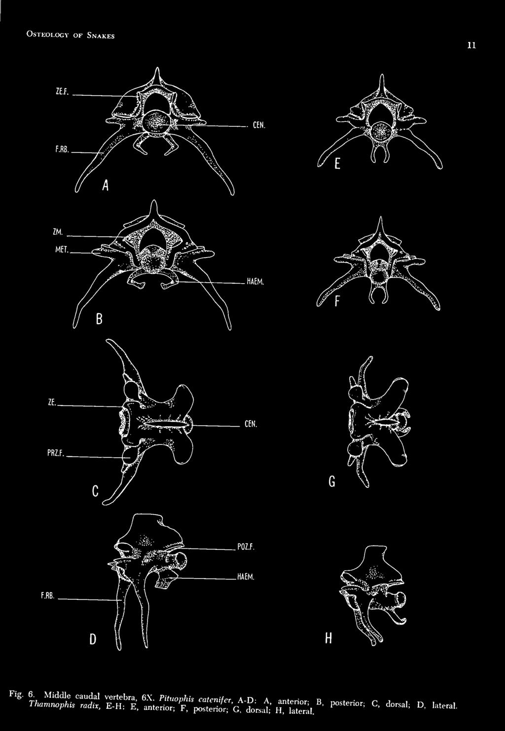
16 Bhigham Young University Science Bulletin POZ.F. Fig. 5. Anterior caudal vertebra, 6X. Pituophk Mtenifer, A-D: A, anterior; B, posterior; C. dorsal; D, lateral. Thamnophis radix, E-H: E, anterior; F, posterior; G, dorsal; H. lateral.

17 Osteology of Snakes 11 '* rj::^^^.7s^.z:::::'r;s:s::t2:j:r;:s;^:'- ^' ^"* ^ '-"'


18 lateral, 5X. Thamrwphis radix, posterior caudal vertebra, E-G, 12X; E, anterior; F, dorsal; G, lateral. Bhigham Young University Science Bulletin HAEM HAEM. POZ.F. HAEM. CO.PR ^'^' LerJl"Z^%hT'''7-^' J-^'^-^ ''''" '^^"'^''^'^^^^ 1«'^- A' ^"t^ --^ B. dorsal; C, lateral; D, rib,


19 Osteology of Snakes 13 region can be distinguislied, however, by the absence of free ribs and tlie presence of fused or articulating bifurcated ribs called lymphapophyses. In the younger specimens studied the lymphapophyses of the first anterior caudal vertebra articulate with the body of the vertebra, as do the preceding ribs of the lumbar vertebrae. In the majority of the older and more mature specimens the articulating surfaces were somewhat fused, and in most incidences the lymphapophyses were not free to move in the same manner as articulating ribs. In a few of the mature specimens the only evidence of the previous articulation is a groove aroimd the base of the lymphapophyses. The lymphapophyses of the remaining vertebrae of this subregion do not articulate or demonstrate any superficial indication of such. These bifurcated structures (lymphapophyses) are associated with muscle attachment as well as with the large lymph hearts on either flank; the divided processes extend outward, dorsal and ventral to the hearts. Tlie more ventral branch is an elongate hornlike process pointing posteroventrally. Tlie dorsal branch is much shorter and arcs in a ventrolateral direction. The dorsal and ventral branches of these vertebrae, however, do show some degree of morphological modification from vertebra to vertebra and from specimen to specimen. Both branches show continuity in becoming reduced in size as they progress posteriorly. The dorsal branch is reduced to the point of becoming nonexistent in the middle caudal subregion, but the ventral branch remains in its reduced form throughout the remaining caudal region. The first one or two vertebrae of this subregion have developed a small ridgelike hypapophysis, but are devoid of haemapophyses. In P. catenifer, however, the last few vertebrae of tliis section possess a pair of flat, platelike, curved haemapophyses projecting ventrallv from the posterior part of each centrum. These structures are considered to be chevrons by Romer (1956), and serially homologous with the hypapophyses of the precaudals. There was no apparent indication of sexual dimorphism in the vertebrae of this (cloacal) area. MIDDLE CAUDAL VERTEBRAE Fig. 6-A, B, C and D These three or four vertebrae differ from the anterior caudals in possessing unforked, fixed ribs which are relatively long and only moderately curved. Although in Sood's criteria for this subregion the vertebrae lack haemapophyses, both colubrid snakes involved in this study possessed them. Tlie possession of haemapophyses by middle caudals makes it rather difficult to distinguish between the last few vertebrae of this group and the first few vertebrae of the posterior caudal subregion. POSTERIOR CAUDAL VERTEBRAE Fig. 7-A, B and C The number of vertebrae involved in this subregion vary greatly from specimen to specimen; however, an average range is fifty-five to sixty-five. These vertebrae are similar in general pattern to the middle caudals, but they gradually diminish in size until the last few are rudimentary. Their processes also show a gradual reduction in size until they are almost vestigial near the tip of the tail. Although the demarcation between these vertebrae and the middle caudal group is somewhat arbitrary, it is possible to separate the two areas by the length and direction of the fixed ribs (transverse processes) as well as the structure of the haemapophyses. In this latter caudal group the transverse processes point aiiteroventrally; the fixed ribs of the middle caudals point either ventrally or posteroventrally, and are longer. The apices of the haemapophyses approach each other mesially until they almost touch in the anterior portion of the posterior caudals, whereas they are farther apart and more rudimentary in the middle caudals of the majority of the specimens studied. About midway along the posterior caudals, the haemapophyses undergo a transition whereby they project almost straight downward in a posteroventral direction with little or no inward curve, remaining rather vddely separated at their distal ends. Fig. 7-D All of the precaudal vertebrae, except the atlas, a.xis, and first thoracic, articulate with a pair of vertebral ribs ( pleurapophyses ). The ribs in the middle of the body are longer and heavier than the first and last few pairs. Tliey are terete, curved, pointed, and are so articulated that they move freely in an anteroposterior plane. Each articulates with the anterior edge of the centrum, the head being connected with the bilobed transverse process. Although the rib head is single, the articular surface is divided into a flattened ventral portion and an adjacent dorsal cupped area. Tlie rib head does not develop a


20 14 Brigham Young University Science Bulletin EC. PO. PAL. SM.PR. Fig. 8. Skull, dorsal. A, Pituophis catenifer, 4X. B, Thamnophis radix, 5X.


21 Osteology of Snakes 15 true capitiilum or tubt'rciilum as is characteristic a posterodorsally-directed costal process for the of higher vertebrates. Distal to the head there is attachment of muscle fibers. SKULL Tlie skull of P. catcnifcr is well ossified and the bones are dense; the cranium is relatively long and broad, and the brain cavity extends Ix'tween the orbits. The apparent large size of the head is partially due to the arrangement of the supratemporals, cjuadrates, and mandibular structures. Tlie bones making up the skull are for the most p;irt loosely articulated, thus making possible a greater flexibility in the mouth region. PREM AXILLA Figs. 8-A, 9-A. 10-A The premaxilla is a single, small median bone which terminates the snout. Its anterior surface is an inverted Y-shaped ridge, the prongs of which extend in a ventrolateral direction. Tlie dorsal process of the ridge curves posteriorly and wedges between the anterior portion of the t^vo nasals. From the ventral crotch of the "Y" there is a horizontal platelike process, terminally bifurcated, ex-tending posteriorly. Its dorsal surface articulates with the ventral surfaces of the anterior septomaxillary processes, thus forming the roof of the anterior portion of the mouth. Tlie prema.villa does not bear teeth. Figs. 8-A, 9-A, 10-A From a dorsal view the articulated nasals appear as an ov^alshaped structure between the premaxilla and the frontals, and are loosely connected to them by connective tissue. The nasals separate anteriorly to form an interspace, which receives the dorsal process of the premaxilla, but remain connec-ted posteriorly, forming a pointed process which is received into the anterior cleft between the two frontals. Dorsally the nasals do not articulate with the frontals. The nasals sheath the nasal cavities dorsolaterally, and form a double median septum between them. This vertical septum extends postero\'entrallv as a process which comes in contact with the fused anteroventral borders of the frontals. The posterior portion of the septum is in contact with the dorsal surface of the septoma.xillae. SEPTOMAXILLAE Figs. 8-A, 9-A. 10-A The elongate, horizontal, platelike septomaxillae form the floor of the internal nares. Tliey are in contact dorsally with the nasals, anteriorly with the premaxillae, ventrally with the vomers, and posteriorly with the frontals. The septoma.xillae lie close together near the mid-dorsal line, but do not suture. Their posterior ex-tremities turn ventrad and come in contact with the frontals. A pair of horizontal winglike processes extend laterally from about the middle of each bone, become narrow, and turn dorsally until they almost touch the descending dorsolateral borders of the nasals. Figs. 8-A, 9-A, 10-A The edentulous vomers are connected to the ventral surfaces of the septomaxillae; at this union they fomi a pair of hollow, spherical vomeronasal organs which open by paired orifices into the buccal cavity. The inner margins of the vomers do not suture, but are joined to adjacent elements only by connective tissue. Each vomer has three vertically-flattened processes extending from its flat mesial border an anterior process connected to the septomaxilla, a posterior process connected to the mesial surface of the posteroventral process of the nasal and the posterior process of the septomaxilla, and a larger posteroventral process with a large triangular fenestra occupying most of its area. The posteroventral process does not articulate with any skull structures, but is connected by connective tissue to other adjacent elements. FRONTALS Figs. 8-A, 9-A, 10-A The frontals are highly developed and form a complete enclosure for the anterior portion of the brain. Tliey remain separate, but are joined mesially by the sagittal suture. The dorsal surface of each frontal is flat, subquadrate, longer than broad, and there is a groove in each, parallel with the indented orbital edge. This groove is perforated with several supraorbital foramina in the adult forms. In younger snakes some of these foramina are not completely formed, but are mere indentations from the orbital margin. The anterolateral edge has a small, depressed articular surfac-e to which the prefrontal is loosely at-


22 Brigham Young University Science Bulletin SOC. PR SM.PR. Fig. 9. SkuU, lateral. A. Pituophis catcnifcr, 4X. B, TJwwnoplm radix, 5X.

rtion of each orbital or optic foramen.")
23 Osteology of Snakes 17 tached. The lateral surface, considered to be the orbitosphenoidal plate of the frontal, joins the dorsolateral margin of the frontal at an acute angle, thus forming a major portion of the mesial wall of the orbit. Ventrallv the descending walls rest upon the presphenoidal prolongation of the basisphenoid, completing the orbital septum and cranial floor. Each wall is notched posteroventrally where it sutures with the anterior wall of the parietal, thus forming the anterior jx)rtion of each orbital or optic foramen. Through these foramina pass the optic nerves, eye-muscle nerves, and blood vessels. Anteriorly, where the frontals contact the nasals and septomaxillae, two large canals or ethmoid foramina, to facilitate the passage of olfactory nerves, are formed by way of a vertiaxl, median double lamina (one from each frontal). PREFRONTALS Figs. 8-A, 9-A, 10-A Although some early workers considered the lacrimals to be present in snakes, Romer (1956) stated that "lacrimals are absent; a foramen or groove for the lacrimal duct is present in the prefrontal." According to Gregory (1913), the prefrontal of reptiles is not homologous with the lacrimal of mammals. From a lateral view each prefrontal is an irregular cone-shaped structure with a laterally compressed anterior process (forming the apex of the cone) extending laterally to the posterior portion of the nasal, septomaxilla, and vomer. The prefrontals loosely articulate with the anterolateral surfaces of the frontals, forming the anterior edge of each orbit. A rather large lacrimal foramen or duct is located on the ventral border of each prefrontal near its articulation with the dorsal surface of the maxilla. Figs. 8-A, 9-A, 11-C Posteriorly the frontals articulate with the fused (coalesced) parietals, there being some degree of motility between the frontals and the parietal. The parietal is the largest of the cranial elements and forms the greater part of the braincase roof, but like the frontals, extends far down either side of the brain, reaching ventrally to the basisphenoid and forming the posterior portion of each orbit. The posterolateral borders of the parietal suture with the prootics. The triangular-shaped, dorsal surface has its narrow apex attached to the supraoccipital posteriorly. Dorsally a V-shaped pair of crests converge posteriorly near the supraoccipital articulation. The posterodorsal surface of each orbit is expanded laterally by the parietal into a flattened lobelike process, the lateral border of which articulates with the curved mesial surface of the postorbital. A smaller coneshaped process, ventral to the lobelike process, articulates dorsally with the posteroventral border of the postorbital. About midway down the coalesced sagittal suture, there are two small parietal foramina immediately lateral to the midline. POSTORBITALS Figs. 8-A, 9-A, 11-C The small postorbitals, considered to be postfrontals by Owen (1866) and Kellicott (1898), are narrow, elongate flattened bones which articulate with the anterolateral surfaces of the parietal, and form the dorsoposterior boundary of each orbit. A strong ligament connects the posterior part of each postorbital with the anterodorsal surface of the ectopterygoids. SUPRAOCCIPITAL Figs. 8-A, 9-A, 11-C The small butterfly-shaped supraoccipital is medially placed and unites anteriorly with the parietal, laterally with the prootics, and posteriorly with the exoccipitals. It forms the roof of the posterior part of the brain cavity, and is internally ex-panded from its lateral wings to form the dorsal portion of each ohc capsule. The lambdoidal ridge or nuchal crest is prominent, and extends obliquely posterolaterally down each side of the posterior portion of the cranium. Each lateral branch of the crest diminishes in size near the exoccipital articulation, b t continues as a rather sharp ridge along the anterior border of the exoccipital, terminating at the posterior border of the fenestra ovalis. Dorsally a short median longitudinal ridge extends posteriorly, ending where the mid-dorsal suture joins the exoccipitals. Figs. 8-A, 9-A, 11-C Each prootic is an irregular quadrate-shaped bone forming the anterior part of the internal otic capsule and the jx)sterolateral wall of the braincase. It is bordered by the parietal anteriorly, supraoccipital dorsally, exoccipital posteriorly, and the basisphenoid and basioccipital ventrally. The anterior half of each fenestra ovalis is formed by the prootic; the exoccipital completes the fenestra posteriorly. The foramen is basically for the passage of the trigeminus, but


24 Brigham Young University Science Bulletin BO.HYP. Fig. 10. Skull, ventral. A, Pituophis catenifer, 4X. B, Tliamnophis radix, 5X.


25 Osteology of Snakes 19 also carries some facial nerve fibers. It is divided externally into two openings, both anterior to the fenestra ovalls. The posterior foramen is larger than the anterior one, and has a lateral proct*ss projecting posteriorly from its anterior border, therefore partially obscuring the opening. Other smaller foramina pierce each prootic, carrying cranial nerve fibers. EXOCCIPITALS Fig.s. 8-A, 9-A, 10-A, U-C The exoccipital bones form the posterolateral walls of the braincase as well as part of its roof. They are joined together by a mid-dorsal suture, connected to the supraoccipital and prootics anteriorly, and resting upon the basioccipital ventrally. The opisthotics are fused with the exoccipitals, the combined bone thus surrounding the jugular foramen and extending forward to form the [X)sterior border of the fenestra ovalis (Romer, 19.56). This jugular foramen, which is internally subdivided into two or more smaller aspects, is just posterior to the fenestra ovalis, and is used for the passage of the ninth and tenth cranial nerves. Posteriorly the exoccipitals form the entire dorsoventrallycompressed, oval foramen magnum, except for a small ventral portion of the occipital condyle. Posteroventrally the exoccipitals terminate as a pair of articulating tubercles which form the lateral portions of the crescent-shaped occiptal condyle. BASIOCCIPITAL Figs. 9-A, 10-A, 11-C The basioccipital is a pentagonal-shaped bone forming the floor of the posterior part of the brain ca\'ita'. The ventral surface has a low, sagittal ridge with a laterally depressed area on either side. Posteriorly a midventral tubercle forms the ventral portion of the occipital condyle, thus completing the foramen magnum. The basioccipital is bordered laterally bv the exoccipitals, and is joined anteroventraliy with the basisphenoid. BASISPHENOID Figs. 9-A, 10-A In the skull of adult serpents, the basisphenoid is applied without a suture anteriorly to the elongate parasphenoid, forming a single bone. The basisphenoidal portion of the bone is a flat, hexagonal plate, bounded dorsally at its margins by prootic and parietal walls, and posteriorly by the basioccipital. A pair of prominent transverse processes extend obliquely and anteriorly toward the midline from its ventrolateral surfaces. Paired carotid foramina pierce the bone laterally to each process to enter the pituitary fossa. This fossa is seen ventrally as a convex area, and is the floor of the sella turcica in which the pituitary gland lies. A pair of foramina also pierce the posterolateral borders of this bone. The narrow elongate parasphenoidal portion of the basisphenoid extends anteriorly into the rostrum area, and becomes the floor of the orbit and optic foramen. A median longitudinal groove extends the entire length of the ventral surface, whereas a dorsomesial keel is wedged between the ventral connection of the frentals. MAXILLAE Figs. 8-A, 9-A, 10-A Each maxilla is a short, curved bar that conforms to the shape of the head. On the mesial border, about midway, there is a small horizontal dorsoposteriorly-pointing process that articulates with the ventral surface of the prefrontal. The posterior end of the maxilla is broadened somewhat, and is received by the flattened clubshaped ventral surface of the ectopterygoid. Each maxilla bears sockets for about fifteen or sixteen curved teeth, but not all of the sockets contain rigidly fixed teeth; therefore, some are usually lost in the preparation of the skull. In P. catenifer about every other tooth on the maxillae, as well as the other tooth-bearing structures, was only loosely lodged in the grooves and could be removed very easily. The maxillae are connected by fibrous tissue to the premaxillae, and not by an osseous articulation as in most vertebrates. ECTOPTEBYGOroS Figs. 8-A, 9-A, 10-A The ectopterygoids, which were sometimes referred to as the transpalatines by earlier workers such as Kellicott (1898) and Wiedersheim and Parker (1907), are small club-shaped, flat bones connecting the maxillae to the pterygoid, and overlying each at its ends. Each flattened spatulatelike anterior process is notched anteriorly where it ligamentously articulates with the maxilla, and an elongate, posterior end articulates with the dorsolateral groove in the pterygoid which runs parallel with the curved contour of tliis bone for its entire length. The ectopterygoids do not bear teeth. PALATINES Figs. 9-A, 10-A Tlie palatines are short bars placed anteriorly to the pterygoids, and mesially to the ma.xillae,


26 Brigham Young University Science Bulletin SOC. FO.M. OT.C. BR.COR. BO. HYP. Fig. 11. Pituophis catenifer, A-C: A, hyoid apparatus, ventral, 4X; B. mandible, medial, 5X; C. cranium, po.sterior. 5X. D, Thamtiophis radix, cranium, posterior, 9X.


27 Osteology of Snakes each bearing nine or ten teeth. The posterior end of each pahitine articulates with the anterior end of the pterygoid. About midway each palatine possesses a broad, flattened, horizontal, median process with apex directed anteriorly, and a similar but narrower lateral process articulating with the ventral border of the prefrontal. The palatines are connected by fibrous connective tissue to the snout elements. sagittal plane, whereas its concave distal articulating surface is extended transversely to form an intercondyloid fossa for mandibular articulation. About midway down the mesial side of each quadrate there is a small, rectangular, raised process which articulates with the distal end of the stapes. MANDIBLE Figs. 8-A, 9-A, 10-A, 11-B PTERYGOIDS Figs. 8-A, 9-A, 10-A Each pter)'goid is a curved, flattened bar extending from the palatine posterolaterally to the angle of the mandible. Anteriorly it narrows and forms a loose articulation with the posterior end of the palatine. From this articulation a row of eight to twelve teeth extends posteriorly along the medial border, ex'tending about midway. The pterygoid receives the ectopterygoid into an elongated, dorsolateral groove. Posteriorly the pterygoid narrows into a slender, curved process that re.iches the base of the c uadrate as well as the mandible. SUPRATEMPORALS Figs. 8-A, 9-A, lo-a The supratemporals are not true squamosals as some of the earlier writers indicated (Romer, 1956). Owen (1866) referred to them as mastoids, but this terminology has been dropped in favor of the more applicable term (supratemporal) used in this paper. The problem of the homologies of the scjuamosal, supratemporal, etc., is, however, far from being solved in the various reptile groups, as pointed out by Jollie (1960). Each supratemporal is a short, harrow, flattened bone connecting the posterolateral dorsal part of the skull with the proximal end of the quadrate. Anteriorly each supratemporal overlays a portion of the parietal, prootic, supraoccipital, and exoccipital, and is attached to these cranial elements by fibrous connective tissue. The two bones are almost parallel to each other, converging only sbghtly as they extend posteriorly. its QUADRATES Figs. 8-A, 9-A, 10-A Each quadrate is a strong, rectangular bone; proximal surface articulates with the posterolateral border of the supratemporal, and its distal, notched, condylar surface with that of the mandibular condyle. Its proximal surface is twisted to become obliquely aligned along the The mandible is composed of two jaw bones which lack a firm anterior symphysis, but are connected by an elastic ligament anteriorly, and is held together posteriorly to the symphysis by the transverse muscles. Each jaw bone is long, curved anteromesially, and is separable into two major bones in the adult snake; however, the splenial and angular bones can sometimes be separated in addition to these two major elements. The longer pro.ximal part of each jaw is composed of angular, prearticular, articular, surangular, and splenial aspects, and is without teeth. Although the splenial and angular are the only bones that can be separated from this proximal portion of the jaw, the other bones are coalesced and form the following areas: the articular and prearticular form the mandibular condyle area; the surangular expands into a longitudinal dorsomesial crest near the condyle, a deep lateral groove is formed at the base of the crest, and the inferior dental foramen opens at the bottom of the groove. The splenial and angular bones are triangular shaped with their bases together and their apices pointing anteriorly and posteriorly from their midventral location on the mesial surface of the jaw. The apex of the splenial points anteriorly; and owing to the reduction in its size, the anterior part of the meckelian canal becomes an open mesial groove in the dentary just anterior to the splenial. Bogert ( 1943 ) uses the term postarticular when referring to the process of the articular that extends posteriorly from the mandibular articulation with the quadrate. His work was with the cobra and other elapids. The dentary forms the anterolateral portion of the jaw, and contains a row of sixteen to eighteen of the usual hooked or recurved teeth. It is short, curved, pointed anteriorly, and bifurcated posteriorly on the lateral surface, thus articulating with the anterior projection of the proximal portion of the jaw. This contact between the two bones is not a close one; therefore, it gives a degree of flexibility to the jaw. The mental foramen is located near the middle of the lateral surface of the dentary.
 is a delicate, slender, rodlike stylus connecting the otic capsule, by way of the fenestra ovalis, to the rectangular articular surface of the quadrate. Its pro.")
 attached to the midventral raphe between the lower jaws; each rodlike branchial cornu")
28 Bbigham Young University Science Bulletin STAPES Figs. 8-A, 9-A Each stapes (columella) is a delicate, slender, rodlike stylus connecting the otic capsule, by way of the fenestra ovalis, to the rectangular articular surface of the quadrate. Its pro.ximal end is enlarged to forni a footplate that fits into the foramen. The stapes is believed to transmit sound vibrations from the various jaw bones to the otic capsule. HYOID APP.\RATUS Fig. 11-A The hyoid apparatus is a cartilagenoiis, V- shaped structure with its vertex or base (basihyobranchial ) attached to the midventral raphe between the lower jaws; each rodlike branchial cornu e-vtends posteriorly along the lateral border of the elongate tongue, being inserted into the base of one of the elongate retractor muscles. Suj^erficial, ventral cranial muscles are attached to the basihyobranchial and the anterior portion of each comu. The hyoid apparatus is not attached to any of the skull elements as it is in most other vertebrates, but lies imbedded in the muscle and fascia of the intermandibular region. The tongue, along with its posteriorly continuing retractor muscles, is more than twice as long as the skull. The hyoid apparatus, therefore, is modified into a very long structure in this species even though it is a rather delicate organ. OSTEOLOGICAL COMPARISONS The two species considered in this study (Pituophis cafenifer and Thamnophis radix) show many osteological similarities. There are, however, a few significant structural differences existing between them. Some of these differences appear to be highly correlated with the special adaptations made by the two species in becoming more compatible with their specific environments and food habits; however, there are many minor structural differences that would seem to be nonadaptive, or perhaps they ma\' have other effects of survival value not apparent to the authors. Only those differences which appear to be of sufficient magnitude to be compared, and perhaps have some bearing on the adaptation of these two species, will be discussed. A complete set of osteological illustrations for Thamnophis radix, corresponding to those of Pituophis catenifer, has been included and will be referred to in the course of the comparative discussion. VERTEBRAL COLUMN Precaudal Vertebrae The atlas of T. radix (Fig. 1-E, F, G, and H) is proportionately broader along the sagittal, neural suture line ( Fig. 1-G and H ) than in P. catenifer (Fig.l-C and D), but its dorsoposterior projections containing the postzygapophyses are not as prominent. The hypapophysls of the atlas of T. radix (Fig. 1-H) does not point posteroventrally as it does in P. catenifer Fig. 1-D, ( ) but instead is represented as a small vertical process extending ventrad from the ventral portion of the intercentrum. The major differences in the axis of the two species are found in the anterior and posterior hypapophyses. In T. radix the axis (Fig. 2-E, F, G and H) develops a stout, hatchet-shaped, anterior hypapophysis (Fig. 2-H) with its anterior articular facet placed vertically on its anterior border, whereas the previously-mentioned, wedge-shaped, anterior hypapophysis of P. catenifer (Fig. 2-D) possesses a narrower posteroventrally situated facet. The posterior hyjiapophysis of T. radix (Fig. 2-H) is not as spineuke or tapered as in the other species, but is more trapezoid in shape with its ventral margin being somewhat truncate. Some of the less obvious differences existing in these vertebrae are the pro[x>rtionally narrower odontoid process (Fig. 2-G), the more posteriorly placed, dorsoventral odontoid suture (Fig. 2-G and H), and the greater anteriorly projecting portion of the neural spine (Fig. 2-H) in T. radix. In T. radix the first thoracic vertebra (Fig. 3-E and F) lacks free ribs, just as it does in P. catenifer. The fixed ribs or transverse processes, however, are short, and extend posteriorly in both species. In the majority of the specimens studied, the transverse processes of the first thoracic vertebrae were a little thicker at the base, and less spinelike in T. radix (Fig. 3-F). This species also differs from P. catenifer in that the neural spine is narrower and slightly longer (Fig. 3-F), the anterior zygapophyses are placed


29 Osteology of Snakes 23 more mesially and are partially covered by the zygosphenes when viewed dorsally ( Fig. 3-E ) and there is a complete lack of metapophyses. The other thoracic or typical t'loracic vertebrae show few noticeable differences between the two species. It can be observed, however, that in T. radix these twenty-two or twentythree thoracic vertebrae ( Figs. '3-G and H; 4-E and F) develop proportionally smaller metapophvses but larger transverse proc-esses (Fig. 3-G and H). The dorsal convex portion of the transverse prck-ess, bilobed rib, articular surface projects more laterally (Fig. 4-E and F), and the facets of the anterior zygapophyses are tilted slightly more oblicjuely dorsad (Fig. 3-G) in T. radix than those of P. catenifer (Fig. 3-C). The lumbar vertebrae of T. radix (Fig. 4-G and H) retain relatively large hypapophyses (Fig. 4-H), whereas the hypapophyses of P. catenifer (Fig. 4-D) are lacking in this region, and are represented only by rudimentary midventral ridges. The presence of hypapophyses on the lumbar vertebrae of T. radix makes it very difficult to distinguish the first few vertebrae of this subregion from the last two or three thoracics. However, there is a decisive reduction in the length of the hypapophyses extending over three or four vertebrae, making it jx>ssible to distinguish between the two vertebral areas with a relative degree of consistency. In T. radix there is an average of approximately 136 or 137 lumbar vertebrae, compared to 185 or 186 found in P. catenifer. The lumbar vertebrae of T. radix (Fig. 4-G and H) develop transverse processes much like those of the thoracic vertebrae. The dorsal rib facets are placed more dorsally (Fig. 4-H), and are more prominent laterally (Fig. 4-G) than in P. catenifer. In tliis species the ventral apex of each transverse process (parapophysis ) is devoid of a rib articular surface, and thus forms an anteroventrally projecting process similar to those found on the thoracic vertebrae of botli species (Fig. 4-B and F). Caudal Vertebrae The anterior caudal vertebrae of T. radix (Fig. 5-E, F, G and H) number five or six in the specimens studied. In addition to their total number being essentially the same as in P. catenifer, they also develop a ridgehke hypapophysis (Fig. 5-E and F) on the first one or two vertebrae of this subregion, and rather rudimentary haemapophyses on the remaining three or four. The dorsal branch of the lymphapophysis does not arc as much ventrally in T. radix (Fig. 5-E and F), and the ventral branch does not attain as great a length proportionally, sweep back posteriorly, or curve as much ventrally at its apex. The neural spine is also narrower and projects more posteriorly (Fig. 5-H) in T. radix. There are three or four vertebrae belonging to the middle caudal subregion in T. radix ( Fig. 6-E, F, G and H), the same number as was found to be present in P. catenifer. These vertebrae are similar in the two species, but there are noticeable differences in their fixed ribs (transverse processes ) and haemapophyses. In T. radix the fixed ribs curve posteroventrally (Fig. 6-G) instead of anteroventrally ( Fig. 6-C ) as they do in P. catenifer. The haemapophyses of this species are more delicate in structure, not as widely arched from each other at their midpoint (Fig. 6-E and F), and they project more posteriorly (Fig. 6-G and H) than in P. catenifer. The vertebrae of the posterior cauda subregion of T. radix (Fig. 7-E, F and G) vary greatly in number from sjxjcimen to specimen just as in P. catenifer; however, an average would fall somewhere between fifty and sixty (an average of about five less than in P. catenifer). For the most part, the same interspecific differences found in the middle caudal vertebrae are carried over into the posterior subregion; however, the neural spine slopes more obliquely anteriorly ( Fig. 7-G ), and the transverse processes curve more anteriorly at their apices and are more pointed and less foothke (Fig. 7-G) in T. radix. There were no noticeable differences in the ribs of the two species except for size. Thamnophis radix, being the smaller serpent, developed proportionately smaller ribs. The skulls of both species are composed of similar bones and bone structures; however, there are a few morphological and size relationship differences that do exist. The most apparent of these differences and those thought to be of some significance will be discussed. A dorsal view of the skull of T. radix (Fig. 8-B), a lateral view (Fig. 9-B), a ventral view (Fig. 10- B), and a posterior view of the cranium (Fig. 11-D) have been included to facihtate the comparative discussion. The premaxilla of T. radix is more compressed dorsoventrally, having a distinct depression or fossa about midway along its dorsal surface (Fig. 9-B) in contrast to the ridgelike process in P. catenifer ( Fig. 9-A ). This posterodorsal process does not extend dorsally to wedge


30 24 Brigham Young University Science Bulletin between the anterior portions of the nasals as it does in P. catcnifer. The septomaxillae are heavier and more extensive in T. radix. They cover a greater proportion of the ventrallv-attached vomers in this species, and the lateral winglike processes are proportionately broader and turn posterodorsally (Fig. 9-B) instead of anterodorsally (Fig. 9- A) as they do in P. catcnifer. The elongate, flat anterior processes from the septoma.xillae are less tapered and more horizontally situated in T. radix. Although tlie frontals and prefrontals are more rigidly attached in T. radix, there are no significant differences in these bones in a comparison of the two species. The parietal and postorbitals, however, show a major structural modification in T. radix. The parietal is narrower proportionately, develops more prominent dorsolateral crests, and lacks the small, paired foramina on its dorsal surface (Fig. 8-B). In P. catcnifer the parietal foramina (Fig. 8-A) are quite prominent. In comparing the length of each parietal with its width, it was found that in T. radix the parietals were 1.1 times as wide as they were long, whereas in P. catcnifer they were 1.3 times as broad as long. The postorbitals of T. radix are not just narrow, elongate bones attached to the anterolateral margins of the parietals as they are in P. catcnifer (Figs. 9-A, 11-C). b'lt thev extend ventrally, coming in contact with the dorsal surfaces of the anterior spatulate processes of the ectopterygoids (Figs. 9-B, 11-D). Some of the less obvious differences of the bones comprising the braincase are the narrower basisphenoid (Fig. 10-B) in the case of T. radix, with its transverse processes more posteriorly situated and extending laterally (perpendicular to the midline) instead of obliquely and posterolaterally as in P. catcnifer (Fig. 10-A). The posteroventral surface of this bone presents a convexity instead of a concavity as it does in P. catcnifer. In T. radix the prootics have a prominent crest running longitudinally along their lateral borders just dorsal to the large prootic foramina. This crest is somewhat hidden by the lateral border of the supratemporal. The lateral process which projects posteriorly, partially obscuring the opening to the posterior prootic foramen, is not present in T. radix. Therefore, the foramen appears round, from a lateral view (Fig. 9-B), instead of kidneyshaped (Fig. 9-A) as in P. catcnifer. The basioccipital of T. radix develops a prominent, median, crestlike process or hypapophysis (Fig. 11-D) for the attacliment of ventral neck muscles; this structure is not found on the basioccipital of P. catcnifer. From a dorsal view (Fig. 8) the size of the po,steromesial notch in the dorsal surface of each exoccipital, where each sutures in the midline, is greater in the case of T. radix (Fig. 8-B). Both the quadrate and the supratemporal are proportionately heavier and broader (Figs. S-B, 9-B) in T. radix. Tliev do, however, occupy approximately the same position in both species. The maxillae, ectopterygoids and palatines (Fig. 10-A and B) are similar structurally in the two species, but there is a difference in the number of teeth on the tooth-bearing structures ( maxillae and palatines ). The maxillae of T. radix bear about twenty-two or twenty-three curved teeth as compared to the fifteen or sixteen in P. catcnifer; the palatines bear about eighteen teeth as compared to the nine or ten teeth developed in P. catcnifer. The large, flattened, median process of the palatine is directed more dorsally in T. radix, and in most cases the dorsal apex of this process arches mesially, terminating in a ventral direction. The pterygoids (Fig. 10-A and B) are proportionately broader, their longitudinal ventral grooves deeper, and the number of teeth is greater in T. radix. This species bears about twenty-seven or twenty-eight teeth (more than twice as many as found in P. catcnifer) in a longitudinal median row which extends almost twice as far posteriorly as it does in P. catenifer. The pterygoid is abruptly curved laterally at its posterior end, making the mandibular articulation in T. radix; whereas in P. catcnifer the entire bone is gradually curved laterally until it articulates posteriorly with the angle of the jaw. The mandible of T. radix is curved a Httle more mesially at its middle dentary articulation (Fig. 10-B), and it contains from twenty-eight to tlilrtv teeth on the dentary portion, or about twice as many as previously recorded for P. catenifer. Both species have splenial and angular bones that can be separated from the anteromesial border of the proximal half of the jaw bone (Fig. 11-B). Although the shape and structure of the hyoid apparatus (Fig. 11-A) is very similar in the two species, it is proportionately longer in T. radix, being appro.ximately 2.2 times longer than its mandible. In P. catenifer the hyoid is only approximately 1.4 times longer than the
, Elaphe ohsoleta has a hyoid apparatus about twice the length of its skull. Smith and Wanier (194S) figured a series of ophidian hyobranchia and included the genus Pituophi.")

31 Osteology of Snakes 25 mandible. Both specit's, however, develop a rather large and functional hvoid apparatus. According to Albright and Nel.son (1959), Elaphe ohsoleta has a hyoid apparatus about twice the length of its skull. Smith and Wanier (194S) figured a series of ophidian hyobranchia and included the genus Pituophi.s, but not Thamnuphis. Their Fig. 1-R of Pituophii- is similar to our Fig. 11-A, but differs in that it is smoothly rounded anteriorly, whereas we find Pituophis to exhibit a small anterior protuberance which we have designated as the basihyobranchial. The specimens of T. radix used in this study, however, did not possess this anterior protuberance. Although Cowan and Hick (1951) did not discuss the osteology of the hyoid apparatus of Thamnophis siitalis as such, they indicated its structure by figures and descriptions concerning the hyoid musculature. According to these authors the basihyal (basihyobranchial) portion of the hyoid of T. sirtalis is smoothly rounded anteriorly, and does not possess an anterior protuberance. DISCUSSION From the foregoing comparisons it is evident that there are osteological differences existing in the two species. In order to provide a complete comparison of the differences it would be necessary to correlate and compare as many of these interspecific differences as possible with other serpent groups (particularly colubrids) in order to arrive at some osteological continuity in the species studied, and to postulate some reasons for these differences. This is obviously beyond the scope of this study. There are, however, a few osteological differences that can be discussed, and these will be considered insofar as our data will permit. The ribless condition of the first thoracic vertebrae, as demonstrated by P. catenifer and T. radix, has not been reported in the species studied by the majority of workers ( op. cit. ) Owen (1866) stated that the ribs commence in the cobra, as they do in other serpents, at the third vertebra from the head. Kellicott (1898) mentioned short transverse processes in connection with the heads of the ribs as being present on all of the body vertebrae ( not including atlas and axis ) in Heterodon. A general statement was made by Gadow (1901) concerning snakes, in which he states that "all the vertebrae, except the atlas, carry ribs." Albright and Nelson (1959) reported short ribs on vertebrae 3 and 4 of Elaphe, the first full-length rib being on the fifth vertebra. Ludicke (1962), on the other hand, reported that all of the vertebrae, with the exception of the atlas, can bear free ribs, but the axis and the first two "cervical" or thoracic vertebrae may also lack ribs in some species. The latter statement appears to coincide more fully with the findings of our study in which the first thoracic vertebra was lacking free ribs. Because of the close adherence of the atlas to the cranial elements and the difficulty involved in separating it from the skull intact, it may be that some of the above statements are not based on sufficiently meticulous dissection to be complete. Possibly the true atlas has been overlooked in some cases, and has not been included as part of the anterior vertebrae. A detailed study of many different genera would be necessary in order to determine the extent of the ribless first thoracic vertebrae in the various types of colubrid snakes. The presence of metapophyses on the first thoracic vertebra of P. catenifer, along with their greater development on the other thoracic vertebrae, seems to indicate a more extensive area for the attachment of lateral trunk muscles, giving this constriction species an advantage in both strength and motility in the neck and anterior trunk region. Tltatnnophis radix, being the smaller of the two species, has fewer precaudal vertebrae, but the proportionate number of the various types of vertebrae in this region vary in the two species (Table 1). There is a decrease of approximately 48 (26%) lumbar and 28 (29%) thoracic vertebrae in T. radix when compared to P. catenifer. It is evident from the above data that the larger snake (P. catenifer) owes much of its greater precaudal body length to the greater number of lumbar vertebrae, but proportionately or percentage-wise, there is an addition of about twice as many thoracic vertebrae as compared to the lumbar. Owen (1866) stated that hypapophyses are developed on the first seventy-four of 253 body vertebrae in Python tigris, the first sixty anterior l)ody vertebrae (305 total, counting caudals) in


32 Table 1. A comparison of the number of vertebrae in each of the vertebral regions and subregions of fourteen Pituophis catenifer and twelve Thamnophis radix. Both male and female specimens are included. Brigham Young University Science Bulletin

 the basioccipital hv'papophysis, which is present in T. radix but missing in P. catenifer, is also lacking in Heterodon. Owen (18.")
33 Osteology of Snakes 27 row postorbitals approach but do not articulate with the upper jaw structures. Such an osseous connection is also lacking in Heterodon. Tlie proportionately heavier and broader (juadrate and supratemporal in the case of T. radix seems to c-oincide with the previously stated conjecture concerning rigidity in the jaw structures necessary for the feeding habit of this species. According to Kellicott (1898) the basioccipital hv'papophysis, which is present in T. radix but missing in P. catenifer, is also lacking in Heterodon. Owen (18.54) stated that the python has a prominent hypapophvsis with a recurved point, and that the rattlesnake also develops a long, strong, recurved hypapophvsis from its basioccipital which acts as a point of insertion for the powerful ventral neck muscles, by which the downward stroke of the head is performed in the act of inflicting a wound. In both species studied, there were occasional specimens which demonstrated an abnormally large number of teeth on some of their toothed structures. It was noted that the bases of the extra teeth were lined up along the sides of the teeth in the normal tooth row, one tooth being firmly fi.\ed in the socket, and the more medially placed tooth being only connected to the bone by fascia. It appeared as if the loosely connected teeth were being replaced by the firmly fixed ones, thus maintaining a complete set of functional teeth at all times. This observation, pertaining to the continual replacement of teeth in serpents, was noted in the works of West (1898), Kingsley (1917), Wiedersheim and Parker ( 1907 ), Gadow ( 1901 ), and more recently by Edmund (1960), to mention a few. Almost all reptiles are polyphyodont ( number of dentitions indefinite), but in some cases such as Typhlopidae, certain teeth are not replaced and others undergo reduction (Wiedersheim and Parker, 1907). The greater number of teeth found on all of the tooth-bearing structures in T. radix ( Table 2) seems to give this species an advantage in grasping and retaining struggling prey such as frogs, toads, fish, grasshoppers, mammals, and other regularly eaten animals. Tliis adaptation to the nonconstrictor habit of food-getting was noted previously in connection with the postorbitals, quadrates, and supratemporals. The proportionately narrower posterior portion of the skull, due mainly to the narrower parietal, has possibly resulted from selection for more streamlined forms, in the case of T. radix, which would have an advantage for life in the swampy areas where diving and swimming become an integral part of its life. Table 2. A comparison of the number of teeth developed on die various tooth-bearing structures of fourteen Pituophis catenifer and twelve Thamnophis radix. Tooth-bearing Structures Maxilla Average Number of Teeth ( Ranges in Parentheses P. catenifer T. radix
 the presence or absence of basioccipital hypapophyses, (12) the number of maxillary, pterygoid, and mandibular teeth, ( 13 ) the relative size and")
 II. Edward Tyson: Comparative anatomy of the timber rattle,snaike. Herpetologica, 2(l):49-69. Albright, R. C, and E. M. Nelson. 1959.")
34 28 Bricham Young University Science Bulletin cranium and its component bones, (8) the shape and character of the septomaxillary process, (9) the existence or lack of paired parietal foramina, (10) the shape and extent of the postorbitals, (11) the presence or absence of basioccipital hypapophyses, (12) the number of maxillary, pterygoid, and mandibular teeth, ( 13 ) the relative size and character of the supratemporal, quadrate, and mandible, and ( 14 ) the relative size of the hyoid apparatus. LITERATURE CITED Ahrenfeldt, R. H Two Briti.sh anatomical studies on American reptiles ( ) II. Edward Tyson: Comparative anatomy of the timber rattle,snaike. Herpetologica, 2(l): Albright, R. C, and E. M. Nelson Craniiil kinetics of the generalized colubrid snake Elaphii obsoleta qtiadrivittata. I. Descriptive morphology, and II. Functional morphology. J. Morph., 105(2): Bogert, C. M Dentitional phenomena in cobras and other elapids with notes on adaptive modifications. Bull. Amer. Mus. Nat. Hist., 8 1 ( ) : Brongersma, L. D On the presence or absence of hypapophyses under the posterior precaudal vertebrae in some snakes. Zool. Meded. Leiden, 20: Cole, F. J A history of comparative anatomy. Macmillan, London. Cowan, I., and W. Hick A comparative study of the myology of the head region in three species of Thamnophis. Trans. Roy. Soc. Canada, ser. 3, 45(3). Edgeworth, F. H The cranial muscles of vertebrates. Univ. Cambridge Press, Cambridge. Edmund, A. G Se{juence and rate of tooth replacement in the Crocodilia. Life Sciences Division Contribution 56, Toronto. Gadow, H Amphibians and reptiles. Macmillan, London. Gegenbaur, C Elements of comparative anatomy. Macmillan, London. Goodrich, E. S Studies on the structure and development of vertebrates. Macmillan, London. Gregory, W. K Homology of the "lacrimal" and the "alisphenoid" in the recent and fossil reptiles. Bull. Geol. Soc. Amer., 24: Haas, G Ueber das kopfskelett und die hautmuskulatiu- der Typlilopiden und Glauconiiden. Zool. J;ihrb., Abt. Anat., 52:1-94, , Hoffman, C. K Reptilien. In Bronn's Kla.ssen und Ordmmgen des Tier Reich, 6(3) : Hu.\ley. T. H A manual of anatomy of vertebrated animals. J. and A. Churchill, London. Jollie, M. T The head skeleton of the hzard. Acta Zoologica, 41:1-64. Kellicott, D. S. 1898, The dissection of the Ophidi;in. Gen. Biol. Supply House, Chicago. Kingslcy, J. S Outline of comparative anatomy of vertebrates. P. Blakiston, Philadelphia. Klauber, L. M Ratdesnakes. Univ. California Press, Los Angeles, 2: Ludicke, M Ordnung der Khisse Reptiha, Serpentes. Handbuch der Zoologie, 7(5): Ordnung der Klasse Reptilia. Serpentes. Handbuch der Zoologie, 7(6) : Mookerjee, H. K., and G. M. Das Occurrence of a paired parietal bone in a snake. Nature, 130: 629. Owen, R The principal forms of the skeleton and of the teeth. Blanchard and Lae, Philadelphia,, 1866, The anatomy of vertebrates, Longman, Green and Co,, London, 1:53-57, Reynolds, S. H, 1913, The vertebrate.skeleton. University Press, Cambridge. Robison, G. W., and W. W. Tanner, A comparative study of the species of the genus Crotaphi/tus Holbrook ( Iguanidae ), Brigham Young University Sci, Bull,, Biol, Series, 2(1):1-31, Rochebrune, A, F, 1881, Memoire.sur les vertebres des ophidiens, J, Anat, Physiol. (Paris), 17: Romer, A. S Osteology of the reptiles. Univ. Chicago Press, Ilhnois. Sedgwick. A A student's text-book of zoology. Swan Sonnenschein and Co., Ltd., London, v, 2, Sood, M. S The caudal vertebrae of Erijx jolmii (Russel). Proc. Indian Acad. Sci., 14: The anatomy of the vertebral column in serpents. Proc. Indian Acad. Sci., 28:1-26. Versluys, Kranium und visceralskeuett der J. Reptilien, In Bolk et al. Handbuch der vergleichenden anatomic der WirbelHer, 4:780-7, West, G, S, 1898, Tooth succession, J. Linn, Soc, 26: Wiedersheim, R,, and W. N. Parker Comparative anatomy of vertebrates. Macmillan, London. Williston, S, W The osteology of the reptiles. Harvard Univ. Press, Cambridge. ABBREVIATIONS USED IN PLATES
tnp]iapophysis dorsal branch. lymphapophysis ventral branch. metapophysis. neiiro-central suture. neurapophysis. neural spine. neural suture. odontoid canal. odontoid process.")

35 Osteology of Snakes 29 LP.D.B. LP.V.B. MET. N.C.S. NEU. N.S. N.SU. O.CL. O.PR. PARP. P. HYP. P.INT.F. POZ. POZ.F. PRZ. PRZ.F. RB.F. RB.H. SH. T.PR. ZE. ZE.F. ZM. ZM.F. A. AR. BH. BO. BO.HYP. BO.T.PR. BR.COR. B.S. l)tnp]iapophysis dorsal branch. lymphapophysis ventral branch. metapophysis. neiiro-central suture. neurapophysis. neural spine. neural suture. odontoid canal. odontoid process. parapopliysis. posterior hypapophysis. posterior intercentral facet. postzygapophysis. postzygapophysial facet. prezygapophysis. prezygapophysial facet. rib facet. rib head. shaft. transverse process. zygosphene. zygosphenal facet. zygantrum. zygantral facet. angular. articular. basihyobranchial. basioccipital. basioccipital hypapophysis. basioccipital transverse process. branchial comu. basisphenoid. D. D.F. EC. EO. FO.M. FOV. FR. J.F. M. ME.F. NAS. o.c. OP.F. OT.C. PAL. PAR. PARA. PAR.F. PER. PM. PO. PR. PRA. PT. QU. SA. SM. SM.PR. SOC. SP. ST. SUP. vo. V. dentary. dental foramen. ectopterygoid. exoccipital. foramen magnum. foramen (fenestra) ovalis. frontal. jugular foramen. ma.villa. mechelian foramen, canal or groove, nasal. occipital condyle, optic foramen, otic capsule, palatine, parietal, parasphenoid. parietal foramen, prefrontal, premaxilla. postorbital. prootic. prearticular. pterygoid, quadrate, surangular. septoma.xilla. septomaxillary process, supraoccipital. splenial. stapes. supratemporal. vomer, fifth cranial nerve.
AMERICAN MUSEUM NOVITATES Published by
 AMERICAN MUSEUM NOVITATES Published by Number 782 THE AmzRICAN MUSEUM OF NATURAL HISTORY Feb. 20, 1935 New York City 56.81, 7 G (68) A NOTE ON THE CYNODONT, GLOCHINODONTOIDES GRACILIS HAUGHTON BY LIEUWE
Exceptional fossil preservation demonstrates a new mode of axial skeleton elongation in early ray-finned fishes
 Supplementary Information Exceptional fossil preservation demonstrates a new mode of axial skeleton elongation in early ray-finned fishes Erin E. Maxwell, Heinz Furrer, Marcelo R. Sánchez-Villagra Supplementary
ONLINE APPENDIX 1. Morphological phylogenetic characters scored in this paper. See Poe (2004) for
 for") ONLINE APPENDIX Morphological phylogenetic characters scored in this paper. See Poe () for detailed character descriptions, citations, and justifications for states. Note that codes are changed from a
ONLINE APPENDIX Morphological phylogenetic characters scored in this paper. See Poe () for detailed character descriptions, citations, and justifications for states. Note that codes are changed from a
A new species of Hsisosuchus (Mesoeucrocodylia) from Dashanpu, Zigong Municipality, Sichuan Province
 from Dashanpu, Zigong Municipality, Sichuan Province") A new species of Hsisosuchus (Mesoeucrocodylia) from Dashanpu, Zigong Municipality, Sichuan Province Yuhui Gao (Zigong Dinosaur Museum) Vertebrata PalAsiatica Volume 39, No. 3 July, 2001 pp. 177-184 Translated
A new species of Hsisosuchus (Mesoeucrocodylia) from Dashanpu, Zigong Municipality, Sichuan Province Yuhui Gao (Zigong Dinosaur Museum) Vertebrata PalAsiatica Volume 39, No. 3 July, 2001 pp. 177-184 Translated
YANGCHUANOSAURUS HEPINGENSIS - A NEW SPECIES OF CARNOSAUR FROM ZIGONG, SICHUAN
 Vol. 30, No. 4 VERTEBRATA PALASIATICA pp. 313-324 October 1992 [SICHUAN ZIGONG ROUSHILONG YI XIN ZHONG] figs. 1-5, pl. I-III YANGCHUANOSAURUS HEPINGENSIS - A NEW SPECIES OF CARNOSAUR FROM ZIGONG, SICHUAN
2. Skull, total length versus length of the presacral vertebral column: (0); extremely elongated neck (e.g. Tanystropheus longobardicus).
; extremely elongated neck (e.g. Tanystropheus longobardicus).") Character list of the taxon-character data set 1. Skull and lower jaws, interdental plates: absent (0); present, but restricted to the anterior end of the dentary (1); present along the entire alveolar
Character list of the taxon-character data set 1. Skull and lower jaws, interdental plates: absent (0); present, but restricted to the anterior end of the dentary (1); present along the entire alveolar
List of characters used in the phylogenetic analysis. Capital letters T, R, and L, refer to
 1 Supplementary data CHARACTER LIST List of characters used in the phylogenetic analysis. Capital letters T, R, and L, refer to characters used by Tchernov et al. (2000), Rieppel, et al. (2002), and Lee
Mammalogy Laboratory 1 - Mammalian Anatomy
 Mammalogy Laboratory 1 - Mammalian Anatomy I. The Goal. The goal of the lab is to teach you skeletal anatomy of mammals. We will emphasize the skull because many of the taxonomically important characters
A Fossil Snake (Elaphe vulpina) From A Pliocene Ash Bed In Nebraska
 From A Pliocene Ash Bed In Nebraska") University of Nebraska - Lincoln DigitalCommons@University of Nebraska - Lincoln Transactions of the Nebraska Academy of Sciences and Affiliated Societies Nebraska Academy of Sciences 198 A Fossil Snake
University of Nebraska - Lincoln DigitalCommons@University of Nebraska - Lincoln Transactions of the Nebraska Academy of Sciences and Affiliated Societies Nebraska Academy of Sciences 198 A Fossil Snake
SUPPLEMENTARY ONLINE MATERIAL FOR. Nirina O. Ratsimbaholison, Ryan N. Felice, and Patrick M. O connor
 http://app.pan.pl/som/app61-ratsimbaholison_etal_som.pdf SUPPLEMENTARY ONLINE MATERIAL FOR Nirina O. Ratsimbaholison, Ryan N. Felice, and Patrick M. O connor Ontogenetic changes in the craniomandibular
A NEW GENUS AND SPECIES OF AMERICAN THEROMORPHA
 A NEW GENUS AND SPECIES OF AMERICAN THEROMORPHA MYCTEROSAURUS LONGICEPS S. W. WILLISTON University of Chicago The past summer, Mr. Herman Douthitt, of the University of Chicago paleontological expedition,
Anatomy. Name Section. The Vertebrate Skeleton
 Name Section Anatomy The Vertebrate Skeleton Vertebrate paleontologists get most of their knowledge about past organisms from skeletal remains. Skeletons are useful for gleaning information about an organism
Biology 3315 Comparative Vertebrate Morphology Skulls and Visceral Skeletons
 Biology 3315 Comparative Vertebrate Morphology Skulls and Visceral Skeletons 1. Head skeleton of lamprey Cyclostomes are highly specialized in both the construction of the chondrocranium and visceral skeleton.
SUPPLEMENTARY INFORMATION
 Character 155, interdental ridges. Absence of interdental ridge (0) shown in Parasaniwa wyomingensis (Platynota). Interdental ridges (1) shown in Coniophis precedens. WWW.NATURE.COM/NATURE 1 Character
.56 m. (22 in.). COMPSOGNATHOID DINOSAUR FROM THE. Medicine Bow, Wyoming, by the American Museum Expedition
. COMPSOGNATHOID DINOSAUR FROM THE. Medicine Bow, Wyoming, by the American Museum Expedition") Article XII.-ORNITHOLESTES HERMANNI, A NEW COMPSOGNATHOID DINOSAUR FROM THE UPPER JURASSIC. By HENRY FAIRFIELD OSBORN. The type skeleton (Amer. Mus. Coll. No. 6I9) of this remarkable animal was discovered
Article XII.-ORNITHOLESTES HERMANNI, A NEW COMPSOGNATHOID DINOSAUR FROM THE UPPER JURASSIC. By HENRY FAIRFIELD OSBORN. The type skeleton (Amer. Mus. Coll. No. 6I9) of this remarkable animal was discovered
FURTHER STUDIES ON TWO SKELETONS OF THE BLACK RIGHT WHALE IN THE NORTH PACIFIC
 FURTHER STUDIES ON TWO SKELETONS OF THE BLACK RIGHT WHALE IN THE NORTH PACIFIC HIDEO OMURA, MASAHARU NISHIWAKI* AND TOSHIO KASUYA* ABSTRACT Two skeletons of the black right whale were studied, supplementing
SOME LITTLE-KNOWN FOSSIL LIZARDS FROM THE
 PROCEEDINGS OF THE UNITED STATES NATIONAL MUSEUM issued SWsK \ {^^m ^V ^^ SMITHSONIAN INSTITUTION U. S. NATIONAL MUSEUM Vol. 91 Washington : 1941 No. 3124 SOME LITTLE-KNOWN FOSSIL LIZARDS FROM THE OLIGOCENE
Williston, and as there are many fairly good specimens in the American
 56.81.7D :14.71.5 Article VII.- SOME POINTS IN THE STRUCTURE OF THE DIADECTID SKULL. BY R. BROOM. The skull of Diadectes has been described by Cope, Case, v. Huene, and Williston, and as there are many
A new sauropod from Dashanpu, Zigong Co. Sichuan Province (Abrosaurus dongpoensis gen. et sp. nov.)
") A new sauropod from Dashanpu, Zigong Co. Sichuan Province (Abrosaurus dongpoensis gen. et sp. nov.) by Ouyang Hui Zigong Dinosaur Museum Newsletter Number 2 1989 pp. 10-14 Translated By Will Downs Bilby
A new sauropod from Dashanpu, Zigong Co. Sichuan Province (Abrosaurus dongpoensis gen. et sp. nov.) by Ouyang Hui Zigong Dinosaur Museum Newsletter Number 2 1989 pp. 10-14 Translated By Will Downs Bilby
Osteology and myology of Phrynosoma p. platyrhinos Girard and Phrynosoma d. hernandesi Girard
 Brigham Young University Science Bulletin, Biological Series Volume 9 Number 4 Article 1 6-1968 Osteology and myology of Phrynosoma p. platyrhinos Girard and Phrynosoma d. hernandesi Girard Richard L.
OF THE TRIAS THE PHYTOSAURIA
 THE PHYTOSAURIA OF THE TRIAS MAURICE G. MEHL University of Wisconsin Some time ago the writer gave a brief notice of a new genus of phytosaurs of which Angistorhinus grandis Mehl was the type.' It is the
CRANIAL OSTEOLOGY OF SCHIZOTHORAICHTHYS NIGER (MECKEL) MISRA (CYPRINIDAE: SCHIZOTHORACINAE). L NEUROCRANIUM
 MISRA (CYPRINIDAE: SCHIZOTHORACINAE). L NEUROCRANIUM") CRANIAL OSTEOLOGY OF SCHIZOTHORAICHTHYS NIGER (MECKEL) MISRA (CYPRINIDAE: SCHIZOTHORACINAE). L NEUROCRANIUM A. R. YousuF, A. K. PANDIT AND A. R. KHAN Postgraduate Department of Zoology, University of Kashmir,
CRANIAL OSTEOLOGY OF SCHIZOTHORAICHTHYS NIGER (MECKEL) MISRA (CYPRINIDAE: SCHIZOTHORACINAE). L NEUROCRANIUM A. R. YousuF, A. K. PANDIT AND A. R. KHAN Postgraduate Department of Zoology, University of Kashmir,
CRANIAL ANATOMY OF ENNATOSAURUS TECTON (SYNAPSIDA: CASEIDAE) FROM THE MIDDLE PERMIAN OF RUSSIA AND THE EVOLUTIONARY RELATIONSHIPS OF CASEIDAE
 FROM THE MIDDLE PERMIAN OF RUSSIA AND THE EVOLUTIONARY RELATIONSHIPS OF CASEIDAE") Journal of Vertebrate Paleontology 28(1):160 180, March 2008 2008 by the Society of Vertebrate Paleontology ARTICLE CRANIAL ANATOMY OF ENNATOSAURUS TECTON (SYNAPSIDA: CASEIDAE) FROM THE MIDDLE PERMIAN
Journal of Vertebrate Paleontology 28(1):160 180, March 2008 2008 by the Society of Vertebrate Paleontology ARTICLE CRANIAL ANATOMY OF ENNATOSAURUS TECTON (SYNAPSIDA: CASEIDAE) FROM THE MIDDLE PERMIAN
complex in cusp pattern. (3) The bones of the coyote skull are thinner, crests sharper and the
 The bones of the coyote skull are thinner, crests sharper and the") DISTINCTIONS BETWEEN THE SKULLS OF S AND DOGS Grover S. Krantz Archaeological sites in the United States frequently yield the bones of coyotes and domestic dogs. These two canines are very similar both
DISTINCTIONS BETWEEN THE SKULLS OF S AND DOGS Grover S. Krantz Archaeological sites in the United States frequently yield the bones of coyotes and domestic dogs. These two canines are very similar both
The cranial osteology of Belebey vegrandis (Parareptilia: Bolosauridae), from the Middle Permian of Russia, and its bearing on reptilian evolution
, from the Middle Permian of Russia, and its bearing on reptilian evolution") Blackwell Publishing LtdOxford, UKZOJZoological Journal of the Linnean Society0024-4082 2007 The Linnean Society of London? 2007 1511 191214 Original Articles RUSSIAN BOLOSAURID REPTILER. R. REISZ ET AL.
Blackwell Publishing LtdOxford, UKZOJZoological Journal of the Linnean Society0024-4082 2007 The Linnean Society of London? 2007 1511 191214 Original Articles RUSSIAN BOLOSAURID REPTILER. R. REISZ ET AL.
Fig. 5. (A) Scaling of brain vault size (width measured at the level of anterior squamosal/parietal suture) relative to skull size (measured at the
 Scaling of brain vault size (width measured at the level of anterior squamosal/parietal suture) relative to skull size (measured at the") Fig. 5. (A) Scaling of brain vault size (width measured at the level of anterior squamosal/parietal suture) relative to skull size (measured at the distance between the left versus right temporomandibular
Fig. 5. (A) Scaling of brain vault size (width measured at the level of anterior squamosal/parietal suture) relative to skull size (measured at the distance between the left versus right temporomandibular
Description of Cranial Elements and Ontogenetic Change within Tropidolaemus wagleri (Serpentes: Crotalinae).
.") East Tennessee State University Digital Commons @ East Tennessee State University Electronic Theses and Dissertations 5-2016 Description of Cranial Elements and Ontogenetic Change within Tropidolaemus
East Tennessee State University Digital Commons @ East Tennessee State University Electronic Theses and Dissertations 5-2016 Description of Cranial Elements and Ontogenetic Change within Tropidolaemus
THE SKULLS OF ARAEOSCELIS AND CASEA, PERMIAN REPTILES
 THE SKULLS OF REOSCELIS ND CSE, PERMIN REPTILES University of Chicago There are few Permian reptiles of greater interest at the present time than the peculiar one I briefly described in this journal' three
A NEW SPECIES OF EXTINCT TURTLE FROM THE UPPER PLIOCENE OF IDAHO
 A NEW SPECIES OF EXTINCT TURTLE FROM THE UPPER PLIOCENE OF IDAHO By Charles W. Gilmore Curator, Division of Vertebrate Paleontology United States National Museum Among the fossils obtained bj^ the Smithsonian
New Carnivorous Dinosaurs from the Upper Cretaceous of Mongolia
 1955 Doklady, Academy of Sciences USSR 104 (5):779-783 New Carnivorous Dinosaurs from the Upper Cretaceous of Mongolia E. A. Maleev (translated by F. J. Alcock) The present article is a summary containing
Brigham Young University Science Bulletin, Biological Series
 Brigham Young University Science Bulletin, Biological Series Volume 11 Number 1 Article 1 6-1970 Osteological and mylogical comparisons of the head and thorax regions of Cnemidophorus tigris septentrionalis
Vol. XIV, No. 1, March, The Larva and Pupa of Brontispa namorikia Maulik (Coleoptera: Chrysomelidae: Hispinae) By S.
 By S.") Vol. XIV, No. 1, March, 1950 167 The Larva and Pupa of Brontispa namorikia Maulik (Coleoptera: Chrysomelidae: Hispinae) By S. MAULIK BRITISH MUSEUM (NATURAL HISTORY) (Presented by Mr. Van Zwaluwenburg
Vol. XIV, No. 1, March, 1950 167 The Larva and Pupa of Brontispa namorikia Maulik (Coleoptera: Chrysomelidae: Hispinae) By S. MAULIK BRITISH MUSEUM (NATURAL HISTORY) (Presented by Mr. Van Zwaluwenburg
Cranial osteology of the African gerrhosaurid Angolosaurus skoogi (Squamata; Gerrhosauridae) HOLLY A. NANCE
 HOLLY A. NANCE") African Journal of Herpetology, 2007 56(1): 39-75. Herpetological Association of Africa Original article Cranial osteology of the African gerrhosaurid Angolosaurus skoogi (Squamata; Gerrhosauridae) HOLLY
African Journal of Herpetology, 2007 56(1): 39-75. Herpetological Association of Africa Original article Cranial osteology of the African gerrhosaurid Angolosaurus skoogi (Squamata; Gerrhosauridae) HOLLY
Bulletin of Big Bend Paleo-Geo An Open Access Publication from Mosasaur Ranch Museum, Terlingua and Lajitas, Texas All rights reserved
 Bulletin of Big Bend Paleo-Geo An Open Access Publication from Mosasaur Ranch Museum, Terlingua and Lajitas, Texas All rights reserved This was a private report in 2003 on my thoughts on Platecarpus planifrons.
HONR219D Due 3/29/16 Homework VI
 Part 1: Yet More Vertebrate Anatomy!!! HONR219D Due 3/29/16 Homework VI Part 1 builds on homework V by examining the skull in even greater detail. We start with the some of the important bones (thankfully
FIELDIANA GEOLOGY NEW SALAMANDERS OF THE FAMILY SIRENIDAE FROM THE CRETACEOUS OF NORTH AMERICA
 FIELDIANA GEOLOGY Published by CHICAGO NATURAL HISTORY MUSEUM Volume 10 Sbftember 22, 1968 No. 88 NEW SALAMANDERS OF THE FAMILY SIRENIDAE FROM THE CRETACEOUS OF NORTH AMERICA Coleman J. Coin AND Walter
Mammalogy Lab 1: Skull, Teeth, and Terms
 Mammalogy Lab 1: Skull, Teeth, and Terms Be able to: Goals of today s lab Locate all structures listed on handout Define all terms on handout what they are or what they look like Give examples of mammals
VERTEBRAL COLUMN
 - 66 - VERTEBRAL COLUMN The vertebral polumn of fishes is composed of two portions, namely the precaudal and caudal, the line of separation between the two being marked by the position of the anus. The
Postilla PEABODY MUSEUM OF NATURAL HISTORY YALE UNIVERSITY NEW HAVEN, CONNECTICUT, U.S.A.
 Postilla PEABODY MUSEUM OF NATURAL HISTORY YALE UNIVERSITY NEW HAVEN, CONNECTICUT, U.S.A. Number 117 18 March 1968 A 7DIAPSID (REPTILIA) PARIETAL FROM THE LOWER PERMIAN OF OKLAHOMA ROBERT L. CARROLL REDPATH
NEW INFORMATION ON THE CRANIUM OF BRACHYLOPHOSAURUS CANADENSIS (DINOSAURIA, HADROSAURIDAE), WITH A REVISION OF ITS PHYLOGENETIC POSITION
, WITH A REVISION OF ITS PHYLOGENETIC POSITION") Journal of Vertebrate Paleontology 25(1):144 156, March 2005 2005 by the Society of Vertebrate Paleontology NEW INFORMATION ON THE CRANIUM OF BRACHYLOPHOSAURUS CANADENSIS (DINOSAURIA, HADROSAURIDAE), WITH
Journal of Vertebrate Paleontology 25(1):144 156, March 2005 2005 by the Society of Vertebrate Paleontology NEW INFORMATION ON THE CRANIUM OF BRACHYLOPHOSAURUS CANADENSIS (DINOSAURIA, HADROSAURIDAE), WITH
Supporting Online Material for
 www.sciencemag.org/cgi/content/full/329/5998/1481/dc1 Supporting Online Material for Tyrannosaur Paleobiology: New Research on Ancient Exemplar Organisms Stephen L. Brusatte,* Mark A. Norell, Thomas D.
The skull of Sphenacodon ferocior, and comparisons with other sphenacodontines (Reptilia: Pelycosauria)
") Circular 190 New Mexico Bureau of Mines & Mineral Resources A DIVISION OF NEW MEXICO INSTITUTE OF MINING & TECHNOLOGY The skull of Sphenacodon ferocior, and comparisons with other sphenacodontines (Reptilia:
Circular 190 New Mexico Bureau of Mines & Mineral Resources A DIVISION OF NEW MEXICO INSTITUTE OF MINING & TECHNOLOGY The skull of Sphenacodon ferocior, and comparisons with other sphenacodontines (Reptilia:
Department of Biology, Faculty of Science, Razi University, Kermanshah, Iran 2
 Iranian Journal of Animal Biosystematics (IJAB) Vol.13, No.2, 247-262, 2017 ISSN: 1735-434X (print); 2423-4222 (online) DOI: 10.22067/ijab.v13i2.64614 A comparative study of the skull between Trachylepis
NOTES ON THE FIRST SKULL AND JAWS OF RIOJASAURUS INCERTUS (DINOSAURIA, PROSAUROPODA, MELANOROSAURIDAE) OF THE LATE TRIASSIC OF LA RIOJA, ARGENTINA
 OF THE LATE TRIASSIC OF LA RIOJA, ARGENTINA") NOTES ON THE FIRST SKULL AND JAWS OF RIOJASAURUS INCERTUS (DINOSAURIA, PROSAUROPODA, MELANOROSAURIDAE) OF THE LATE TRIASSIC OF LA RIOJA, ARGENTINA José F. Bonaparte and José A. Pumares translated by Jeffrey
NOTES ON THE FIRST SKULL AND JAWS OF RIOJASAURUS INCERTUS (DINOSAURIA, PROSAUROPODA, MELANOROSAURIDAE) OF THE LATE TRIASSIC OF LA RIOJA, ARGENTINA José F. Bonaparte and José A. Pumares translated by Jeffrey
A new species of sauropod, Mamenchisaurus anyuensis sp. nov.
 A new species of sauropod, Mamenchisaurus anyuensis sp. nov. by Xinlu He, Suihua Yang, Kaiji Cai, Kui Li, and Zongwen Liu Chengdu University of Technology Papers on Geosciences Contributed to the 30th
OSTEOLOGICAL NOTE OF AN ANTARCTIC SEI WHALE
 OSTEOLOGICAL NOTE OF AN ANTARCTIC SEI WHALE MASAHARU NISHIWAKI* AND TOSHIO KASUYA* ABSTRACT This is a report of measurements on the skeleton of a male se1 whale caught in the Antarctic. The skeleton of
A Complete Late Cretaceous Iguanian (Squamata, Reptilia) from the Gobi and Identification of a New Iguanian Clade
 from the Gobi and Identification of a New Iguanian Clade") PUBLISHED BY THE AMERICAN MUSEUM OF NATURAL HISTORY CENTRAL PARK WEST AT 79TH STREET, NEW YORK, NY 10024 Number 3584, 47 pp., 19 figures September 6, 2007 A Complete Late Cretaceous Iguanian (Squamata,
PUBLISHED BY THE AMERICAN MUSEUM OF NATURAL HISTORY CENTRAL PARK WEST AT 79TH STREET, NEW YORK, NY 10024 Number 3584, 47 pp., 19 figures September 6, 2007 A Complete Late Cretaceous Iguanian (Squamata,
A M E G H I N I A N A. Revista de la Asociación Paleontológia Argentina. Volume XV September-December 1978 Nos. 3-4
 A M E G H I N I A N A Revista de la Asociación Paleontológia Argentina Volume XV September-December 1978 Nos. 3-4 COLORADIA BREVIS N. G. ET N. SP. (SAURISCHIA, PROSAUROPODA), A PLATEOSAURID DINOSAUR FROM
v:ii-ixi, 'i':;iisimvi'\>!i-:: "^ A%'''''-'^-''S.''v.--..V^'E^'-'-^"-t''gi L I E) R.ARY OF THE VERSITY U N I or ILLINOIS REMO
 R.ARY OF THE VERSITY U N I or ILLINOIS REMO") "^ A%'''''-'^-''S.''v.--..V^'E^'-'-^"-t''gi v:ii-ixi, 'i':;iisimvi'\>!i-:: L I E) R.ARY OF THE U N I VERSITY or ILLINOIS REMO Natural History Survey Librarv GEOLOGICAL SERIES OF FIELD MUSEUM OF NATURAL
"^ A%'''''-'^-''S.''v.--..V^'E^'-'-^"-t''gi v:ii-ixi, 'i':;iisimvi'\>!i-:: L I E) R.ARY OF THE U N I VERSITY or ILLINOIS REMO Natural History Survey Librarv GEOLOGICAL SERIES OF FIELD MUSEUM OF NATURAL
Reprinted from: CRUSTACEANA, Vol. 32, Part 2, 1977 LEIDEN E. J. BRILL
 Reprinted from: CRUSTACEANA, Vol. 32, Part 2, 1977 LEIDEN E. J. BRILL NOTES AND NEWS 207 ALPHE0PS1S SHEARMII (ALCOCK & ANDERSON): A NEW COMBINATION WITH A REDESCRIPTION OF THE HOLOTYPE (DECAPODA, ALPHEIDAE)
Comparative Osteology of the Genus Pachytriton (Caudata: Salamandridae) from Southeastern China
 from Southeastern China") Asian Herpetological Research 2012, 3(2): 83 102 DOI: 10.3724/SP.J.1245.2012.00083 Comparative Osteology of the Genus Pachytriton (Caudata: Salamandridae) from Southeastern China Yunke WU 1, Yuezhao WANG
Asian Herpetological Research 2012, 3(2): 83 102 DOI: 10.3724/SP.J.1245.2012.00083 Comparative Osteology of the Genus Pachytriton (Caudata: Salamandridae) from Southeastern China Yunke WU 1, Yuezhao WANG
Cranial osteology and phylogenetic relationships of Hamadasuchus rebouli (Crocodyliformes: Mesoeucrocodylia) from the Cretaceous of Morocco
 from the Cretaceous of Morocco") Blackwell Publishing LtdOxford, UKZOJZoological Journal of the Linnean Society0024-4082 2007 The Linnean Society of London? 2007 1494 533567 Original Articles HAMADASUCHUS REBOULIH. C. E. LARSSON and H.-D.
Blackwell Publishing LtdOxford, UKZOJZoological Journal of the Linnean Society0024-4082 2007 The Linnean Society of London? 2007 1494 533567 Original Articles HAMADASUCHUS REBOULIH. C. E. LARSSON and H.-D.
SUPPLEMENTARY OBSERVATIONS ON THE SKULL OF
 SUPPLEMENTARY OBSERVATIONS ON THE SKULL OF THE FOSSIL PORPOISE ZARHACHIS FLAGELLATOR COPE By Remington Kellogg Of the Bureau of Biological Survey, United States Department of Agriculture During the past
Mammalogy Lecture 8 - Evolution of Ear Ossicles
 Mammalogy Lecture 8 - Evolution of Ear Ossicles I. To begin, let s examine briefly the end point, that is, modern mammalian ears. Inner Ear The cochlea contains sensory cells for hearing and balance. -
AMERICAN MUSEUM. Novitates PUBLISHED BY THE AMERICAN MUSEUM OF NATURAL HISTORY CENTRAL PARK WEST AT 79TH STREET
 AMERICAN MUSEUM Novitates PUBLISHED BY THE AMERICAN MUSEUM OF NATURAL HISTORY CENTRAL PARK WEST AT 79TH STREET NEW YORK, N.Y. 10024 U.S.A. NUMBER 2662 NOVEMBER 21, 1978 RONN W. COLDIRON Acroplous vorax
A Short Report on the Occurrence of Dilophosaurus from Jinning County, Yunnan Province
 A Short Report on the Occurrence of Dilophosaurus from Jinning County, Yunnan Province by Hu Shaojin (Kunming Cultural Administrative Committee, Yunnan Province) Vertebrata PalAsiatica Vol. XXXI, No. 1
PALEONTOLOGY AND BIOSTRATIGRAPHY OF MONGOLIA
 PALEONTOLOGY AND BIOSTRATIGRAPHY OF MONGOLIA THE JOINT SOVIET-MONGOLIAN PALEONTOLOGICAL EXPEDITION (Transactions, vol. 3) EDITORIAL BOARD: N. N. Kramarenko (editor-in-chief) B. Luvsandansan, Yu. I. Voronin,
AMERICAN NATURALIST. Vol. IX. -DECEMBER, No. 12. OR BIRDS WITH TEETH.1 OI)ONTORNITHES,
ONTORNITHES,") AMERICAN NATURALIST. Vol. IX. -DECEMBER, 1875.-No. 12. OI)ONTORNITHES, OR BIRDS WITH TEETH.1 BY PROFESSOR 0. C. MARSH. REMAINS of birds are amono the rarest of fossils, and few have been discovered except
AMERICAN NATURALIST. Vol. IX. -DECEMBER, 1875.-No. 12. OI)ONTORNITHES, OR BIRDS WITH TEETH.1 BY PROFESSOR 0. C. MARSH. REMAINS of birds are amono the rarest of fossils, and few have been discovered except
AEROSAURUS WELLESI, NEW SPECIES, A VARANOPSEID MAMMAL-LIKE
 Journal of Vertebrate Paleontology 1(1):73-96. 15 June 1981 1 AEROSAURUS WELLESI, NEW SPECIES, A VARANOPSEID MAMMAL-LIKE REPTILE (SYNAPSIDA: PELYCOSAURIA) FROM THE LOWER PERMIAN OF NEW MEXICO WANN LANGSTON
A New Dromaeosaurid Theropod from Ukhaa Tolgod (Ömnögov, Mongolia)
") PUBLISHED BY THE AMERICAN MUSEUM OF NATURAL HISTORY CENTRAL PARK WEST AT 79TH STREET, NEW YORK, NY 10024 Number 3545, 51 pp., 25 figures, 1 table December 7, 2006 A New Dromaeosaurid Theropod from Ukhaa
PUBLISHED BY THE AMERICAN MUSEUM OF NATURAL HISTORY CENTRAL PARK WEST AT 79TH STREET, NEW YORK, NY 10024 Number 3545, 51 pp., 25 figures, 1 table December 7, 2006 A New Dromaeosaurid Theropod from Ukhaa
[Accepted 8th October CONTENTS INTRODUCTION
 183 THE CRANIAL MORPHOLOGY OF A NEW GENUS AND SPECIES OF ICTIDOSAURAN BY A. W. CROMPTON S. A. Museum, Cape Town [Accepted 8th October 19571 (With 7 figures in the text) CONTENTS lntroduction..............
A New Ceratopsian Dinosaur from the Upper
 SMITHSONIAN MISCELLANEOUS COLLECTIONS VOLUME 63. NUMBER 3 A New Ceratopsian Dinosaur from the Upper Cretaceous of Montana, with Note on Hypacrosaurus (With Two Plates) CHARLES W. GILMORE Assistant Curator
 The following text is generated from uncorrected OCR. [Begin Page: Page 1] A NEW CERATOPSIAN DINOSAUR FROM THE UPPER CRETACEOUS OF MONTANA, WITH NOTE ON HYPACROSAURUS ' By CHARLES W. GILMORE assistant
SOME NEW AMERICAN PYCNODONT FISHES.
 SOME NEW AMERICAN PYCNODONT FISHES. By James Williams Gidley, Assistant Curator of Fossil Mammals, United States National Museum. In the United States National Museum are several specimens representing
REVISION OF THE GENUS MARTINICHTHYS, MARINE FISH (TELESOSTEI, TSELFATIIFORMES) FROM THE LATE CRETACEOUS OF KANSAS (UNITED STATES)
 FROM THE LATE CRETACEOUS OF KANSAS (UNITED STATES)") 1 REVISION OF THE GENUS MARTINICHTHYS, MARINE FISH (TELESOSTEI, TSELFATIIFORMES) FROM THE LATE CRETACEOUS OF KANSAS (UNITED STATES) TAVERNE L., 2000. Revision of the genus Martinichthys, marine fish (Teleostei,
1 REVISION OF THE GENUS MARTINICHTHYS, MARINE FISH (TELESOSTEI, TSELFATIIFORMES) FROM THE LATE CRETACEOUS OF KANSAS (UNITED STATES) TAVERNE L., 2000. Revision of the genus Martinichthys, marine fish (Teleostei,
A Late Jurassic Protosuchian Sichuanosuchus huidongensis from Zigong, Sichuan Province. Guangzhao Peng. Zigong Dinosaur Museum, Zigong, Sichuan
 A Late Jurassic Protosuchian Sichuanosuchus huidongensis from Zigong, Sichuan Province Guangzhao Peng Zigong Dinosaur Museum, Zigong, Sichuan 643013 Vertebrata PalAsiatica Volume 34, Number 4 October,
Osteology of the Clupeiform fish, genus Hyperlophus (II)
") Bull. Kitakyushu Mas. Nat. Hist., 4: 77-102. December 31, 1982 Osteology of the Clupeiform fish, genus Hyperlophus (II) Yoshitaka Yabumoto Kitakyushu Museum of Natural History, Nishihonmachi, Yahatahigashiku,
Bull. Kitakyushu Mas. Nat. Hist., 4: 77-102. December 31, 1982 Osteology of the Clupeiform fish, genus Hyperlophus (II) Yoshitaka Yabumoto Kitakyushu Museum of Natural History, Nishihonmachi, Yahatahigashiku,
Your use of the JSTOR archive indicates your acceptance of the Terms & Conditions of Use, available at
 The Evolution of the Mammalian Jaw Author(s): A. W. Crompton Source: Evolution, Vol. 17, No. 4 (Dec., 1963), pp. 431-439 Published by: Society for the Study of Evolution Stable URL: http://www.jstor.org/stable/2407093
A NEW ANKYLOSAUR FROM THE UPPER CRETACEOUS OF MONGOLIA E.A. Maleev Doklady Akademii Nauk, SSSR 87:
 translated by Dr. Tamara and F. Jeletzky, 1956 A NEW ANKYLOSAUR FROM THE UPPER CRETACEOUS OF MONGOLIA E.A. Maleev 1952. Doklady Akademii Nauk, SSSR 87:273-276 Armored dinosaurs make a considerable part
Florida, Gainesville, Florida, 32611, U.S.A. b Smithsonian Tropical Research Institute, Ancon, Republic of Panama,
 This article was downloaded by: [78.22.97.164] On: 04 May 2013, At: 14:02 Publisher: Taylor & Francis Informa Ltd Registered in England and Wales Registered Number: 1072954 Registered office: Mortimer
THE GORGONOPSIAN GENUS, HIPPOSAURUS, AND THE FAMILY ICTIDORHINIDAE * Dr. L.D. Boonstra. Paleontologist, South African Museum, Cape Town
 THE GORGONOPSIAN GENUS, HIPPOSAURUS, AND THE FAMILY ICTIDORHINIDAE * by Dr. L.D. Boonstra Paleontologist, South African Museum, Cape Town In 1928 I dug up the complete skeleton of a smallish gorgonopsian
APPENDIX. 160 Miscellaneous Intelligence.
 160 Miscellaneous Intelligence. OBITUARY. GENERAL ANDREW A. HUMPHREYS. Brigadier-General Andrew Atkinson Humphreys died in Washington, on the 28th of November last, in the seventy-fourth year of his age.
A skull without mandihle, from the Hunterian Collection (no.
 4 MR. G. A. BOULENGER ON CHELONIAN REMAINS. [Jan. 6, 2. On some Chelonian Remains preserved in the Museum of the Eojal College of Surgeons. By G. A. Boulenger. [Eeceived December 8, 1890.] In the course
DISCOVERY AND CAPTURE
 46 NSHWAK AND KASUYA Hitherto, this species has occurred no other place in the world on 33 28' N. around, San Diego and Casabranca for example. Our news was not only the evidence of this occurrence. When
Reexamination of a primitive ornithomimosaur, Garudimimus brevipes Barsbold, 1981 (Dinosauria: Theropoda), from the Late Cretaceous of Mongolia
, from the Late Cretaceous of Mongolia") Reexamination of a primitive ornithomimosaur, Garudimimus brevipes Barsbold, 1981 (Dinosauria: Theropoda), from the Late Cretaceous of Mongolia Yoshitsugu Kobayashi and Rinchen Barsbold 1501 Abstract:
Reexamination of a primitive ornithomimosaur, Garudimimus brevipes Barsbold, 1981 (Dinosauria: Theropoda), from the Late Cretaceous of Mongolia Yoshitsugu Kobayashi and Rinchen Barsbold 1501 Abstract:
A NEW AUSTROSQUILLA (STOMATOPODA) FROM THE
 FROM THE") A NEW AUSTROSQUILLA (STOMATOPODA) FROM THE MARQUESAS ISLANDS BY ALAIN MICHEL Centre O.R.S.T.O.M., Noumea, New Caledonia and RAYMOND B. MANNING Smithsonian Institution, Washington, U.S.A. The At s,tstrosqzlilla
A NEW AUSTROSQUILLA (STOMATOPODA) FROM THE MARQUESAS ISLANDS BY ALAIN MICHEL Centre O.R.S.T.O.M., Noumea, New Caledonia and RAYMOND B. MANNING Smithsonian Institution, Washington, U.S.A. The At s,tstrosqzlilla
Macro-anatomical studies of the African giant pouched rat (Cricetomys gambianus) axial skeleton
 axial skeleton") Standard Scientific Research and Essays Vol1 (10): 221-227, October 2013 http://www.standresjournals.org/journals/ssre Research Article Macro-anatomical studies of the African giant pouched rat (Cricetomys
Standard Scientific Research and Essays Vol1 (10): 221-227, October 2013 http://www.standresjournals.org/journals/ssre Research Article Macro-anatomical studies of the African giant pouched rat (Cricetomys
THE LARVA OF ROTHIUM SONORENSIS MOORE & LEGNER. BY IAN MOORE Department of Entomology, University of California, Riverside, California 92521
 THE LARVA OF ROTHIUM SONORENSIS MOORE & LEGNER WITH A KEY TO THE KNOWN LARVAE OF THE GENERA OF THE MARINE BOLITOCHARINI (COLEOPTERA STAPHYLINIDAE) BY IAN MOORE Department of Entomology, University of California,
CENE RUMINANTS OF THE GENERA OVIBOS AND
 DESCRIPTIONS OF TWO NEW SPECIES OF PLEISTO- CENE RUMINANTS OF THE GENERA OVIBOS AND BOOTHERIUM, WITH NOTES ON THE LATTER GENUS. By James Williams Gidley, Of the United States National Museum. Two interesting
A NEW SPECIES OF TROODONT DINOSAUR FROM THE
 A NEW SPECIES OF TROODONT DINOSAUR FROM THE LANCE FORMATION OF WYOMING By Charles W. Gilmore Curator of Vertebrate Paleontology, United States National Museum INTRODUCTION The intensive search to which
Results of Prof. E. Stromer's Research Expedition in the Deserts of Egypt
 Proceedings of the Royal Bavarian Academy of Science Mathematical-physical Division Volume XXVIII, Paper 3 Results of Prof. E. Stromer's Research Expedition in the Deserts of Egypt II. Vertebrate Remains
Marshall Digital Scholar. Marshall University. F. Robin O Keefe Marshall University,
 Marshall University Marshall Digital Scholar Biological Sciences Faculty Research Biological Sciences 2008 Cranial anatomy and taxonomy of Dolichorhynchops bonneri new combination, a polycotylid (Sauropterygia:
Frog Dissection Information Manuel
 Frog Dissection Information Manuel Anatomical Terms: Used to explain directions and orientation of a organism Directions or Positions: Anterior (cranial)- toward the head Posterior (caudal)- towards the
BREVIORA LEUCOLEPIDOPA SUNDA GEN. NOV., SP. NOV. (DECAPODA: ALBUNEIDAE), A NEW INDO-PACIFIC SAND CRAB. Ian E. Efford 1
, A NEW INDO-PACIFIC SAND CRAB. Ian E. Efford 1") ac lc BREVIORA CAMBRIDGE, MASS. 30 APRIL, 1969 NUMBER 318 LEUCOLEPIDOPA SUNDA GEN. NOV., SP. NOV. (DECAPODA: ALBUNEIDAE), A NEW INDO-PACIFIC SAND CRAB Ian E. Efford 1 ABSTRACT. Leucolepidopa gen. nov.
ac lc BREVIORA CAMBRIDGE, MASS. 30 APRIL, 1969 NUMBER 318 LEUCOLEPIDOPA SUNDA GEN. NOV., SP. NOV. (DECAPODA: ALBUNEIDAE), A NEW INDO-PACIFIC SAND CRAB Ian E. Efford 1 ABSTRACT. Leucolepidopa gen. nov.
TRACHEMYS SCULPTA. A nearly complete articulated carapace and plastron of an Emjdd A NEAKLY COMPLETE SHELL OF THE EXTINCT TURTLE,
 A NEAKLY COMPLETE SHELL OF THE EXTINCT TURTLE, TRACHEMYS SCULPTA By Charles W. Gilmore Curator of Vertebrate Paleontology, United States National Museum INTRODUCTION A nearly complete articulated carapace
Cranial morphology and taxonomy of South African Tapinocephalidae (Therapsida: Dinocephalia): the case of Avenantia and Riebeeckosaurus
: the case of Avenantia and Riebeeckosaurus") Cranial morphology and taxonomy of South African Tapinocephalidae (Therapsida: Dinocephalia): the case of Avenantia and Riebeeckosaurus Saniye Güven*, Bruce S. Rubidge & Fernando Abdala Evolutionary Studies
Cranial morphology and taxonomy of South African Tapinocephalidae (Therapsida: Dinocephalia): the case of Avenantia and Riebeeckosaurus Saniye Güven*, Bruce S. Rubidge & Fernando Abdala Evolutionary Studies
SAUROPOD DINOSAURS FROM THE EARLY CRETACEOUS OF MALAWI, AFRICA. Elizabeth M. Gomani
 Palaeontologia Electronica http://palaeo-electronica.org SAUROPOD DINOSAURS FROM THE EARLY CRETACEOUS OF MALAWI, AFRICA Elizabeth M. Gomani ABSTRACT At least two titanosaurian sauropod taxa have been discovered
A NEW SALTICID SPIDER FROM VICTORIA By R. A. Dunn
 Dunn, R. A. 1947. A new salticid spider from Victoria. Memoirs of the National Museum of Victoria 15: 82 85. All text not included in the original document is highlighted in red. Mem. Nat. Mus. Vict.,
Jurassic Ornithopod Agilisaurus louderbacki (Ornithopoda: Fabrosauridae) from Zigong, Sichuan, China
 from Zigong, Sichuan, China") Jurassic Ornithopod Agilisaurus louderbacki (Ornithopoda: Fabrosauridae) from Zigong, Sichuan, China Guangzhao Peng (Zigong Dinosaur Museum) Vertebrata PalAsiatica Volume 30, No. 1 January, 1992 pp. 39-51
Jurassic Ornithopod Agilisaurus louderbacki (Ornithopoda: Fabrosauridae) from Zigong, Sichuan, China Guangzhao Peng (Zigong Dinosaur Museum) Vertebrata PalAsiatica Volume 30, No. 1 January, 1992 pp. 39-51
SUPPLEMENTARY INFORMATION
 doi:10.1038/nature11227 Contents 1. Character list 2. Edits to prior character descriptions/codings 3. Materials examined 4. Results of phylogenetic analyses 5. Ophidian characters of Coniophis and diagnoses
DESCRIPTIONS OF NEW AND LITTLE-KNOWN FOSSIL LIZARDS FROM NORTH AMERICA
 PROCEEDINGS OF THE UNITED STATES NATIONAL MUSEUM SMITHSONIAN INSTITUTION U. S. NATIONAL MUSEUM Vol.86 Washington: 1938 No. 3042 DESCRIPTIONS OF NEW AND LITTLE-KNOWN FOSSIL LIZARDS FROM NORTH AMERICA By
On the cranial anatomy of the polycotylid plesiosaurs, including new material of Polycotylus latipinnis, Cope, from Alabama
 Marshall University Marshall Digital Scholar Biological Sciences Faculty Research Biological Sciences 2004 On the cranial anatomy of the polycotylid plesiosaurs, including new material of Polycotylus latipinnis,
Notes on Ceratopsians and Ankylosaurs at the Royal Ontario Museum
 Notes on Ceratopsians and Ankylosaurs at the Royal Ontario Museum Andrew A. Farke, Ph.D. Raymond M. Alf Museum of Paleontology 1175 West Baseline Road Claremont, CA 91711 email: afarke@webb.org Introduction
Temporal lines. More forwardfacing. tubular orbits than in the African forms 3. Orbits larger relative to skull size than in the other genera 2.
 Asian lorises More forwardfacing and tubular orbits than in the African forms 3. Characterized by a marked extension of the ectotympanic into a tubular meatus and a more angular auditory bulla than in
NAUSHONIA PAN AMEN SIS, NEW SPECIES (DECAPODA: THALASSINIDEA: LAOMEDIIDAE) FROM THE PACIFIC COAST OF PANAMA, WITH NOTES ON THE GENUS
 FROM THE PACIFIC COAST OF PANAMA, WITH NOTES ON THE GENUS") 5 October 1982 PROC. BIOL. SOC. WASH. 95(3), 1982, pp. 478-483 NAUSHONIA PAN AMEN SIS, NEW SPECIES (DECAPODA: THALASSINIDEA: LAOMEDIIDAE) FROM THE PACIFIC COAST OF PANAMA, WITH NOTES ON THE GENUS Joel
5 October 1982 PROC. BIOL. SOC. WASH. 95(3), 1982, pp. 478-483 NAUSHONIA PAN AMEN SIS, NEW SPECIES (DECAPODA: THALASSINIDEA: LAOMEDIIDAE) FROM THE PACIFIC COAST OF PANAMA, WITH NOTES ON THE GENUS Joel
NIVOROUS DINOSAUR. (SECOND COMMUNICATION.) By HENRY FAIRFIELD OSBORN. PLATE XXXIX. This great carnivorous Dinosaur of the Laramie was contemporary
 By HENRY FAIRFIELD OSBORN. PLATE XXXIX. This great carnivorous Dinosaur of the Laramie was contemporary") 56, 8i, 9 T (I 7: 786) Article VI.-TYRANNOSAURUS, UPPER CRETACEOUS CAR- NIVOROUS DINOSAUR. (SECOND COMMUNICATION.) By HENRY FAIRFIELD OSBORN. PLATE I. This great carnivorous Dinosaur of the Laramie was
56, 8i, 9 T (I 7: 786) Article VI.-TYRANNOSAURUS, UPPER CRETACEOUS CAR- NIVOROUS DINOSAUR. (SECOND COMMUNICATION.) By HENRY FAIRFIELD OSBORN. PLATE I. This great carnivorous Dinosaur of the Laramie was
4. Premaxilla: Foramen on the lateral surface of the premaxillary body (Yates 2007 ch. 4) 0 absent 1 present
 0 absent 1 present") The character matrix used as a basis for this study is that of Yates et al (2010) which is modified from the earlier matrix used by Yates (2007). This matrix includes characters acquired and/or modified
The character matrix used as a basis for this study is that of Yates et al (2010) which is modified from the earlier matrix used by Yates (2007). This matrix includes characters acquired and/or modified
CHARACTER LIST: Nesbitt et al., 2011
 CHARACTER LIST: Nesbitt et al., 2011 1. Vaned feathers on forelimb symmetric (0) or asymmetric (1). The barbs on opposite sides of the rachis differ in length; in extant birds, the barbs on the leading
CRANIAL ANATOMY AND PHYLOGENETIC AFFINITIES OF THE PERMIAN PARAREPTILE MACROLETER POEZICUS
 CRANIAL ANATOMY AND PHYLOGENETIC AFFINITIES OF THE PERMIAN PARAREPTILE MACROLETER POEZICUS Author(s): LINDA A. TSUJI Source: Journal of Vertebrate Paleontology, 26(4):849-865. 2006. Published By: The Society
FHSU Scholars Repository. Fort Hays State University. Joshua J. Fry Fort Hays State University, Summer 2015
 Fort Hays State University FHSU Scholars Repository Master's Theses Graduate School Summer 2015 Redescription Of A Specimen Of Pentaceratops (Ornithischia: Ceratopsidae) And Phylogenetic Evaluation Of