The skull of Sphenacodon ferocior, and comparisons with other sphenacodontines (Reptilia: Pelycosauria)
|
|
|
- Myron Day
- 5 years ago
- Views:
Transcription
1
2 Circular 190 New Mexico Bureau of Mines & Mineral Resources A DIVISION OF NEW MEXICO INSTITUTE OF MINING & TECHNOLOGY The skull of Sphenacodon ferocior, and comparisons with other sphenacodontines (Reptilia: Pelycosauria) by David A. Eberth Department of Paleontology, University of California, Berkeley, California SOCORRO 1985
3
4
5
6 5 Abstract Sphenacodon Marsh, known from the Lower Permian of New Mexico, Utah, and Arizona, is one of the best represented pelycosaurs in fossil collections, yet its skull has never been completely described. Virtually complete, disarticulated cranial remains of Sphenarodon ferocior from the Lower Permian Cutler and Abo Formations of north-central New Mexico are described in detail for the first time. These descriptions provide the basis for the most detailed comparisons to date within the genus Sphenacodon and the subfamily Sphenacodontinae. No morphological differences exist between the skulls of Sphenacodon and Ctenospondylus, and only minor morphological differences exist between Sphenacodon and Ctenospondylus on the one hand and Dimetrodon on the other. On the basis of cranial morphology alone, Sphenacodon and Ctenospondylus appear to be more closely related than the current taxonomy reflects. Macromerion is undiagnostic below the family level and materials referred to Neosaurus, Bathygnathus, and "Oxyodon" are undiagnostic below the subfamily level. The retention of all of these names except "Oxyodon" is recommended as a paleogeographic convenience. "Oxyodon" has been shown by Paton (1974) to be preoccupied, and pelycosaur material assigned to that taxon should be therefore reclassified as Sphenacodontinae incertae sedis. Introduction The Late Pennsylvanian to Late Permian subfamily Sphenacodontinae, as recognized by Romer and Price (1940) and Romer (1945), consists of seven genera (Dimetrodon, Ctenospondylus, Sphenacodon, Bathygnatuss, Neosaurus, Oxyodon, and Macromerion). Only three of these, Dimetrodon, Ctenospondylus, and Sphenacodon, include complete cranial remains. Known from the prolific Lower Permian red beds of Texas, Oklahoma, and New Mexico, Dimetrodon is represented by abundant skeletal material including numerous complete and partial skulls which have been thoroughly studied (Romer and Price, 1940). Ctenospondylus is represented by material from a number of different localities, which includes one articulated skull and postcranial material from southeast Utah, fragmentary cranial and postcranial remains from the Dunkard Group of West Virginia, and isolated spines and vertebrae pertaining to one individual from Texas. The cranial material from Utah was thoroughly studied and described by Vaughn (1964, 1969). The remaining cranial material from West Virginia was described and discussed by Berman (1978). Sphenacodon, known from the Lower Permian Cutler and Abo Formations of New Mexico and the Cutler Group of Arizona and Utah, is one of the better represented pelycosaurs in North American fossil collections (second only to Dimetrodon); yet, it has received only a modicum of attention in the literature. Large collections of Sphenacodon material (cranial and postcranial) that were assembled by field parties from the Yale Peabody Museum in 1878, the University of Chicago and the University of Michigan in 1911, and the Harvard Museum of Comparative Zoology in 1937 have been the basis of virtually all the descriptions and figures of Sphenacodon cranial morphology (Marsh, 1878; Case, 1907; Williston, 1911, 1916, 1917, 1918; Case and Williston, 1913; Romer, 1937; Romer and Price, 1940). Unfortunately, much of this material is still in need of preparation, and that portion that has been prepared does not include a complete set of cranial elements. Thus, the cranial descriptions and figures are incomplete and somewhat patchy. Sphenacodon material collected by the University of California Museum of Paleontology during 1934 and 1935 was thoroughly prepared during the Work Project Administration years, and includes virtually every cranial element; yet, ironically, this material has never been described or figured. The reason for this neglect appears to have been the publication of the monumental Review of the Pelycosauria (Romer and Price, 1940). It has long been recognized that the skulls of Sphenacodon and Dimetrodon are very similar (Case, 1907; Williston, 1918; Romer and Price, 1940; Vaughn, 1964). Subsequent to the publication of the detailed descriptions and drawings of the articulated and associated cranial elements of Dimetrodon limbatus in the Review of the Pelycosauria, further interest in the cranial morphology of Sphenacodon and other sphenacodontines greatly diminished. Since 1940 most references to general sphenacodontine or sphenacodontid cranial morphology have been made by referral to the Romer and Price descriptions and reconstructions (e.g. Romer, 1956, 1966; Barghusen, 1968, 1973; Currie, 1979; Reisz, 1980; Brinkman and Eberth, 1983). The University of California Museum of Paleontology Sphenacodon collection includes disarticulated, associated, and partially articulated skulls of no fewer than 31 individuals from one locality, as well as fragmentary and less abundant material from a number of other localities. Such a rich collection offers a unique opportunity to (1) provide the first in-depth, bonebybone description of a sphenacodontid, (2) examine variation in a sphenacodontine death assemblage, and (3) attempt an evaluation of intrasphenacodontine cranial variation. In the framework of Paleozoic reptilian systematics the description of this material is imperative. Much remains to be clarified concerning relationships of taxa within the Pelycosauria (Reisz, 1980; Brinkman and Eberth, 1983) and of pelycosaurs with other reptilian groups (especially therapsids). A thorough understanding of all pelycosaur genera is an essential step in addressing the nature of these
7 6 interrelationships. The University of California Museum of Paleontology Sphenacodon collection thus is thoroughly described in this paper. The goals are (1) to present a detailed, bone-by-bone description of the skull of Sphenacodon in the University of California collection, and (2) to provide cranial comparisons both within the genus Sphenacodon and among other sphenacodontines. Acknowledgments This study was undertaken at the Department of Paleontology of the University of California at Berkeley in as a partial fulfillment of a Master of Arts degree. I am greatly indebted to the late Dr. Malcolm J. Heaton of the Tyrrell Museum of Palaeontology, Alberta, Canada, at whose suggestion this study was initiated. Special thanks are due to Dr. Joseph T. Gregory who served as my thesis supervisor and critiqued earlier drafts of this paper. Drs. William A. Clemens and Harry W. Greene also read earlier drafts of this work, and I am grateful for their comments and observations. Drs. Robert R. Reisz and Donald Brinkman served as readers and provided many helpful comments and suggestions; both have been en ergetic sources of encouragement. Dr. P. P. Vaughn graciously allowed me to study and borrow the Ctenospondylus cranial material in his care. Research and travel were partially funded by monies from a University of California Regents Fellowship and the W. P. Schmidt Fund of the Field Museum of Natural History, Chicago. Field-work funds and publication expenses were generously provided by the New Mexico Bureau of Mines and Mineral Resources, Socorro, New Mexico. I am greatly indebted to Drs. Frank E. Kottlowski and Donald L. Wolberg of that institution. Abbreviations Institutional abbreviations used throughout this paper are as follows: UCMP, University of California Museum of Paleontology, Berkeley; MCZ, Museum of Comparative Zoology, Harvard University, Cambridge, Massachusetts; U.C., University of Chicago, Chicago, Illinois (this material is housed at the Field Museum of Natural History); WM, Walker Museum/ Field Museum of Natural History, Chicago, Illinois; NTM VP, Navajo Tribal Museum, Vertebrate Paleontology, Window Rock, Arizona. Materials and methods The genus Sphenacodon includes two species, the Shale of the Four Corners area (Vaughn, 1964). type species S. ferox (Marsh, 1878) and S. ferocior Abundant, though fragmentary, remains of (Romer, 1937). The type of S. ferox is from the Lower Sphenacodon sp. have been noted in the Permo- Permian Cutler Formation, Baldwin bone bed locality in the village of Arroyo de Agua in Rio Arriba County, north-central New Mexico (Fig. 1; Langston, 1953, fig. 3). It consists of a single dentary and a pair of maxillae Pennsylvanian red beds of Socorro, San Miguel, and Santa Fe Counties in New Mexico (see Langston, 1952)..Among all of these localities the Anderson quarry, in of questionable association. Other referred specimens Arroyo de Agua, has produced the largest and most of S. ferox have been collected from stratigraphically complete assemblage of disarticulated and articulated equivalent horizons throughout Arroyo de Agua and cranial remains of Sphenacodon. For this reason, and adjacent towns, from El Cobre Canyon 15 km northeast of Arroyo de Agua, and from the Johnson locality S. ferocior from the Anderson quarry was chosen as because most of the specimens were already prepared, in the San Diego Canyon (see Langston, 1953). the basis for the following descriptions and S. ferocior is represented by articulated and disarticulated specimens that have been collected from localities of wider geographical extent than those containing S. ferox. The type of S. ferocior (MCZ 1489) an articulated skull and partial postcranial skeleton was collected from the Lower Permian Abo Formation in the San Diego Canyon, 2.5 km south of the Spanish Queen mine (Fig. 1; Langston, 1953, fig. 4). Referred specimens have been collected from localities illustrations. At least 31 individuals are present and, in some cases, different ontogenetic stages of elements are represented. In most cases, elements are represented by more than one specimen, allowing for the study of variation in this death assemblage. Five cranial elements are not represented in the Anderson quarry collection (tabular, vomer, supratemporal, ectopterygoid, and postparietal). The first two are described as they occur in the type (MCZ in Arroyo de Agua that are stratigraphically 1489). The supratemporal is known in an articulated, m higher than the Baldwin bone bed locality though crushed, skull of S. ferox (U.C. 35). It is described and figured as it occurs in that specimen. The (Anderson and Cardillo quarries), throughout the San Diego Canyon (south of Jemez Springs) at stratigraphically higher positions than the S. ferox localities in contact on the supraoccipital. The ectopterygoid is postparietal is discussed with reference to its area of that canyon, from the Abo Formation near Socorro in not discussed in any detail. south-central New Mexico, and from the Organ Rock The specimen drawings are on coquille board. Outlines for each figure were obtained from enlargements
8 7 of photographic negatives projected directly upon the coquille board. Outlines of some of the smaller specimens were obtained directly from the specimen. Shading is present only where the element is preserved. In those cases where the full extent of the element can be reconstructed on the basis of evidence from adjacent elements, the reconstructed outline is dashed. Reconstructions were only attempted where an articulated or obviously associated skull portion was available. The lateral-view reconstructions in Fig. 3A and B are based upon the articulated snout, UCMP Note that the cheek and temporal regions of the two reconstructions differ dramatically. The cheek region in Fig. 3A is based upon the jugal morphology present in UCMP (Fig. 14). The remainder of the temporal region in this reconstruction is an hypothetical reconstruction based upon disarticulated elements. The cheek region in Fig. 3B is based upon the jugal morphology in UCMP The remainder of the temporal and cheek region is an hypothetical reconstruction based upon an associated squamosal (UCMP 34196, Fig. 16) and other disarticulated elements. The lower-jaw reconstructions (Fig. 3C, D) are based upon a complete, articulated lower jaw, UCMP Insufficient articulated material was available to warrant an occipital, palatal, or dorsal reconstruction; thus, accurate measurements for these areas cannot be provided. The sphenacodontines Neosaurus, Macromerion, and "Oxyodon" are in European collections, and, therefore, direct comparisons among these taxa, and between them and other taxa, could not be made. In these cases, comparisons are limited to those areas that have been described or figured by other authors. Dimetrodon specimens at the Harvard Museum of Comparative Zoology, a cast of the one known specimen of Bathygnathus, and the one known articulated skull of Ctenospondylus (NTM VP 1001) have all been examined first hand. Locality and geology The Anderson quarry is located in the village of Arroyo de Agua, Rio Arriba County, north-central New Mexico (SW 1/4 N W 1/4 Sec. 11, T. 22 N., R. 3 E.). In this area, deep valleys carved by erosive streams expose up to 200 in of sandstones and mudstones of the Cutler Formation (Fig. 2). Eight separate bone beds and numerous localities occur throughout the lower 100 m of this exposed section. Specimens that have been referred to S. ferox have been collected from the lowest six bone beds, all essentially at the same horizon (see Langston, 1952, 1953). The Anderson quarry bone bed occurs m above these six lower bone beds. The quarry is exposed on the side of a small arroyo and includes an area of about 20 m 2. Boneproducing layers total approximately 1.5 m in thickness and the quality and quantity of preserved material is matched by few Permian localities. For the most part, the specimens are disarticulated, but when carefully extracted and prepared, elements are often fully preserved. Environmental and/or taphonomic interpretations of the numerous bone beds in the Arroyo de Agua area have largely been avoided by workers concerned with systematics problems. Langston (1952, 1953), however, interpreted four of the lower bone beds as the discontinuously exposed remains of a large Lower
9 8 Permian lake. He applied a similar interpretation to the higher-occurring Anderson quarry deposits, stating (1953, p. 360):... conditions of sedimentation were similar to those that produced the Welles quarry deposit, [one of the lower bone beds] but the chain of events was reversed and deposition took place nearer the shore. Repeated drying in the later history of the Anderson quarry lake is indicated by at least seven superincumbent marly layers in the upper 4 feet of the section. Recent examination of this deposit has shown that the 1.5 m thick fossiliferous deposits occur as a laterally limited, U-shaped body containing three separate horizons. At the base a green-gray sandy silt rests unconformably upon a thick, multistoried sandstone body. This is overlain by a thin, nodular-rich marly horizon which, in turn, passes up into a thick, iron-rich marl horizon. The basal horizon is rich in carbonized plant material and produces the greatest variety of disarticulated, unassociated fossil remains. No obvious bedforms are preserved in any of these horizons. The presence of a basal sandy-silt deposit containing abundant disarticulated and unassociated vertebrate remains and plant material suggests a complex transport history for this organic hash, followed by a quick burial. The overlying nodular-marl and ironrich-marl horizons both contain the disarticulated and unabraded remains of larger tetrapods, often in association. Such information suggests the continued presence of a topographic low acting as a locus for pooling and maceration of carcasses. Although the U- shaped nature of these deposits speaks against their interpretation as remnant lake deposits, the lack of primary sedimentary structures makes any more definite interpretation difficult. It should be noted, however, that elsewhere in the section where laterally limited bodies of reduced sediments occur, their interpretation as the remains of proximal crevasse-channel fills is better documented (Eberth, in prep.). The majority of the UCMP specimens collected from this deposit are from the lower two horizons. The upper marly horizon is very difficult to work and elements collected from it are often broken in the process of collecting. Although the lower two horizons undoubtedly represent two depositional events, rates of deposition and accumulation in environments where crevasse-channel deposits are forming today are very high (29-60 cm/100 yrs) (Smith, 1983). Thus, it is not unreasonable to consider the Anderson quarry Sphenacodon specimens as members of a single population.
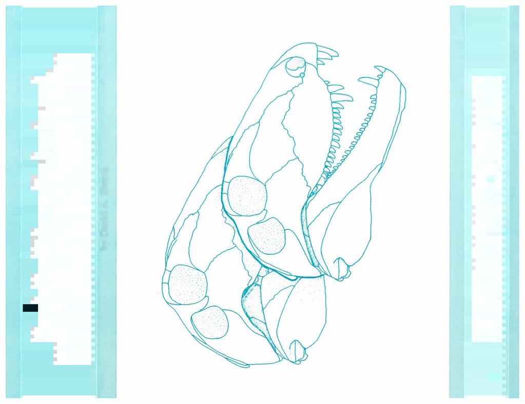
10 9 Osteology General osteology of the skull The skull of S. ferocior (Fig. 3) is quite tall and narrow, with a long preorbital region and a short postorbital region. The bone of the snout is thin except at the alveolar ridges. The tooth row is convex in lateral view and the tooth bases are square in cross section, while the distal tips are compressed, with unserrated cutting edges along the anterior and posterior edges. Above the orbits the supraorbital ridge consists of thick bone and projects laterad, forming the widest portion of the skull table. A single temporal opening is present behind and slightly lower than the orbit. The occiput is a flat plate of bone inclined forward and set slightly anterior to the posterolateral corners of the skull. The stout paroccipital and supraoccipital processes brace the occiput against the posterior cheek region. Elongate internal choanae occur at the anterior end of the palate, adjacent to the thickened buttresses of the alveolar ridges. A wide, toothed, transverse flange of the pterygoid braces the posterior portion of the palate against the medial surface of the skull. Between these points the palate is covered with a shagreen of small, peg-like teeth arranged randomly in two fields along and on either side of the midline. Posterior to the transverse flanges, vertical processes of the pterygoids are sutured to the quadrates, thus forming stout braces between the occiput and the palate. The lower jaw is quite narrow through most of its length and becomes thickened mediolaterad at the anterior end where the large caniniforms insert. The tooth row is concave in lateral view and the tooth morphology is as described above. Posterior to the tooth row, the mandible becomes tall and provides a large area for muscle insertion in the meckelian fossa on the medial surface. A meckelian canal runs forward from the fossa. Ventral to the fossa, the angular is a deep, narrow element. The condylar recesses of the articular are set just below the level of the tooth row and face dorsomediad. When the jaw is closed, the teeth of the lower jaw, with the exception of the caniniforms, come to lie medial to the maxillary teeth. The large caniniforms of the dentary are accommodated on the outside of the maxilla by a "step" formed along the anteroventral margin of the latter element. Descriptions of cranial elements Premaxilla (14 specimens) The premaxilla, forming the blunt tip of the snout, is a tripartite, heavily ossified element that carries three teeth. Three processes contact the nasal dorsally, the maxilla and septomaxilla laterally, and the vomer and septomaxilla medially. The figured specimen (UCMP 83047) lacks only the tips of the three processes. Discussion of these regions is based upon the articulated partial skull, UCMP The external surface of the bone is heavily pitted, as in most large reptiles, allowing an intricate set of blood vessels and nerves to communicate with the overlying skin. The first tooth is invariably the largest when compared to others in a fully erupted state. All the teeth are markedly ribbed in typical sphenacodont fashion. The roots of the first and second teeth are deeply implanted in the body of the premaxilla and, when fully erupted, extend up to the ventral border of the narial opening. Arching posterodorsad for a distance of cm from the body of the premaxilla, the dorsal process lies medial to the nasal and contacts the medial surface of its counterpart at the midline. This process, triangular in cross section, tapers dorsad, disappearing from the dorsal surface of the skull above and about 1 cm behind the posterior border of the narial opening. The sutural surface for the nasal lies along the lateral and posterior faces of the dorsal process. The midline sutural contact of the dorsal process (Fig. 4D) is in the vertical plane and is continuous with the medial sutural surface of the premaxillary body. The slightly downturned maxillary process extends posteriorly 4-5 cm from the anterior border of the narial opening. It is sutured to the medial surface of the maxilla, terminating just above the "step" of the maxilla. The dorsal surface of this process is in contact with the footplate of the septomaxilla, although the full extent of this contact is not adequately indicated by the preserved material. Extending posteriorly from the midline of the premaxillary body is the short, narrow, median process that marks the ventral contact of the nasal septum on the premaxilla. The medial and lateral surfaces of the process contact the anterior inner surfaces of the folded vomer. Only the medial sutural surface is visible, however. Between this short process and the maxillary process, which extends parallel to it, is a distinct furrow that may have carried a branch of the maxillary artery and perhaps the ethmoid nerve. Within this furrow, at a point directly behind the first-tooth root, is the prepalatal foramen. This opens into a vertical canal that in turn exits ventrally between the first and second teeth. The furrow continues forward entering the element at a point where the dorsal process begins to arch posteriorly from the body of the premaxilla (premaxillary foramen, Fig. 4B). Within the body of the premaxilla this foramen communicates with an extensive and complex series of canals. The manner in which these canals communicate with the surface of the premaxilla was determined by studying a series of 64 photographs taken from a serially ground specimen (UCMP 83033) at increments of 0.25 mm. It was determined that the posterior premaxillary foramen communicates with the root of the first tooth and continues as a large canal to emerge directly in front of the first tooth on the ventral surface of the premaxillary body. Throughout the course of this canal, smaller, anastomosing branches frequently run to the surface of the premaxilla. There is no evidence to suggest the existence of a single large sinus within the
11
12 11 maxillary processes of the premaxilla. In medial view (Fig. 5B), UCMP appears as a gently convex structure firmly sutured to the internal surface of the nasal. A shallow groove runs along the ventral border of the nasal and ends in an area of crushed bone, preventing any attempt to trace its course. The groove marks the course of the lacrimal duct as it approaches the septomaxillary foramen, which is itself poorly preserved in this specimen. The area of crushed bone appears much too high to have been the region for the septomaxillary foramen and, more probably, represents the crushed posterior process of the septomaxilla that is applied to the internal surface of the nasal. The duct probably passed farther ventrad into the region behind the footplate. Even here, however, the presence of the septomaxillary foramen cannot be confirmed because of distortion. Anterior to this region of crushing, a light vessel scar branches ventrally. In lateral view (Fig. 5A) only a small portion of the septomaxilla is visible; the anterior part of the element has been lost. The moderately sized septomaxillary foramen is clearly visible posteroventrally. A small portion of the medial shelf is preserved along the anterior edge. Below the shelf is a small, anteriorly opening foramen. MCZ 1489, in contrast, shows the medial shelf well developed in either view (5C, D), but does not display a dorsal contact of the septomaxilla with the nasal. Along the ventral margin of the element, the footplate has lost its contact with the premaxilla. Thus, the septomaxilla appears curled up. In medial view (Fig. 5D) the septomaxillary foramen is visible and can be traced through to the external surface of the skull where it opens posterior to the footplate. The small anterior foramen seen in UCMP is not visible in any view, and the lacrimal duct scar leading to the septomaxillary foramen is not preserved. However, MCZ 1489 does show a completely preserved contact of the posterior process of the septomaxilla with the internal surface of the nasal (Fig. 5D). dorsal process as seen in captorhinids (Heaton, 1979). The dorsal process remains cancellous throughout its length and appears to be similar in this respect to the dorsal processes of iguanids. Septomaxilla (2 specimens) The two available septomaxillae of S. ferocior are known from articulated, partial skulls and, in each case, have undergone a certain amount of distortion. UCMP 341% (Fig. 5A, B) shows the least distorted condition, but is poorly preserved anteriorly and medially. MCZ 1489 (Fig. 5C, D), on the other hand, is preserved anteriorly, but is compressed dorsoventrally, distorting the true nature of its contact with the nasal. The septomaxilla provides protection for the anterior region of the nasal capsule and sits in the posterior margin of the narial opening, extending the full height of that orifice. The element is quite wide, extending from the lateral edge of the narial opening almost to the midline. It rests upon the medial and Nasal (9 specimens) The nasal is an elongate, gently arched element (Fig. 6A, B). At the anterior end the nasal sends a dorsal and a ventral process around the narial opening. The dorsal process contacts the lateral and the posterior surface of the dorsal process of the premaxilla along a narrow sutural surface. The ventral process extends some distance behind and beneath the narial opening and probably takes part in bordering the septomaxillary foramen dorsally. This process provides a broad ventral area of contact for the septomaxilla and maxilla on its medial surface. The nasal is bounded laterally by the lacrimal and maxilla. In external view (Fig. 6B) the posterior part of the lateral edge shows a small sutural surface for the lacrimal. This surface is quite thin and is poorly preserved in all specimens. The nasal tapers to a point between an anterior process of the frontal and the medial surface of the prefrontal (Fig. 6B). The medial edge of the element is thick through its entire extent. In medial view there
13
14 13 is a narrow longitudinal groove, located lateral to the midline, that runs the length of the element. This possibly marks the point of attachment of the nasal septum. Lateral to this is a much wider, gently rounded groove that is well demarcated posteriorly. A similar groove has been hypothesized to mark the dorsal contact of the nasal septum in specimens of Dimetrodon (Romer and Price, 1940). It is more likely that this groove results from the presence of a turbinal ridge lateral to it. Lateral to the turbinal ridge, the body of the nasal becomes thinner as it arches ventrolaterad. Along the medial surface of the element, just posterior to the narial border, is the light trace of a groove that is tentatively interpreted as the lacrimal-duct scar. Frontal (13 specimens) The frontal is a triradiate, flattened element that consists of an anterior process, an orbital process, and a posterior process. The anterior process is long, narrow, and is convex dorsally when viewed laterally (Fig. 7A). It is structurally similar to the posterior region of the nasal with which it is continuous. A turbinal ridge extends forward along most of the ventral surface of the anterior process. The lateral surface of the anterior process displays sutural surfaces for the nasal and prefrontal. The prefrontal also contacts the frontal along the anterior edge of the orbital process, sending a small projection into the ventromedial corner formed between the anterior and orbital processes. The contact between the prefrontal and frontal extends forward to a point midway along the lateral surface of the anterior process of the frontal. At this point the two elements are separated by a posterior
15 14 projection of the nasal. Williston (1925) indicated in his reconstruction of S. ferox that the nasal also projects posteriorly a short distance along the medial surface of the anterior process of the frontal. The articulated snout MCZ 1489 shows this condition to be present in S. ferocior as well. The laterad projecting orbital process is dorsoventrally thick (7 mm) and forms the bulk of the supraorbital shelf. Along the posterior edge of the orbital process the sutural surface for the postfrontal is visible (Fig. 7D). The sigmoid curvature of the orbital process is visible in posterior view. The angle formed by the orbital process extending dorsolaterad from the midline is no more than 35 from the horizontal. The posterior process displays an extensive area of contact with the postfrontal along its lateral surface. In ventral view (Fig. 7B), a shallow sutural scar marks the narrow contact of the parietal along the posteroventral surface of the posterior process of the frontal. In ventral view the frontal displays a small "step" that runs from the sutural scar for the parietal to the anteromedial corner of the orbital process. This marks the line of contact between the frontal and the dorsal extent of the planum supraseptale (solum supraseptale of Oelrich, 1956, and Heaton, 1979). Farther forward, a small ridge extends anteriorly, parallel to the turbinal ridge. This small ridge probably marks the line of contact between the nasal septum and the frontal. Between the anterior extent of the ridge marking the course of the planum supraseptale and the posterior portion of the turbinal ridge is a shallow depression that marks the posterior extent of the cartilaginous nasal capsule. The existence of a "pit" in this area, as noted by Romer and Price (1940) for Dimetrodon, is not present. In posterior view (Fig. 7D) what appears to be a large foramen opens into the longitudinal axis of the frontal. The identity of this foramen is unknown. In ventral view (Fig. 8B) the anterior and lateral edges of the wing are scarred by sutures marking the extent of postorbital contact. A narrow, scarred ridge runs along the posterior edge of the wing, marking the extent of the contact with the dorsal edge of the tabular. Sutural scars for the tabular along both the dorsal and ventral posterior edges suggest that the parietal was wedged into the dorsal edge of the tabular. From the material at hand it is impossible to determine the nature of contact between the parietal and the squamosal. Between the tabular and postorbital sutural scars a shallow, broad, concave surface Parietal (4 specimens) The parietal is a small, wing-shaped bone at the posterior end of the skull table. The anterior surface of the element contacts the postorbital, postfrontal, and frontal. The posterior edge is in contact with the postparietal, tabular, and possibly the squamosal. The ventral surface is smoothly contoured, marking the limits of muscle attachments, and forms the dorsal roof of the braincase in the area immediately posterior to the pineal foramen. Fig. 8A, B presents reconstructions of the parietal based upon all of the available specimens. Sutural surfaces for the postfrontal and frontal are visible in dorsal view (Fig. 8A). A large notch for the pineal organ occurs midway along the medial edge. In MCZ 1489 the pineal opening is just posterior to the midlength of the element, while in the three specimens from the Anderson quarry the notch occurs at about the midlength. On the posterior surface a large, forward-sloping sutural surface marks the contact of the postparietal and the tabular. The lateral wing of the parietal extends posteroventrad from the body of the element. Its dorsal surface is lightly striated, marking the anterior extent of the supratemporal.

16 15 that is heavily pitted around its periphery marks the site of attachment of part of the external adductor musculature. Medial to this area the parietal surface is softly contoured, possibly marking the site of attachment of the internal pterygoideus branch of the adductor musculature. Striae that occur along the ventral surface of the parietal, lateral to the pineal opening, might represent the contact point between the epipterygoid and the parietal, and thus the lateral limit of the braincase. Postparietal (no specimens) No isolated specimens of the postparietal exist in the UCMP Anderson quarry collection. However, much of the morphology of the element is known from sutural scars on the supraoccipital and the parietal. Fig. 25A shows a posterior view of the supraoccipital with a large sutural-scar region for the postparietal. The element is apparently triangular, with its apex oriented ventrad. The limits and proportions of the postparietal can generally be deduced from these scars. The scars extend laterad to a point where the postparietal apparently overlies the medial edge of the tabular. Unfortunately, no sutural scars are preserved on the medial edge of the one fragmentary tabular used in this study (MCZ 1489) and, therefore, the exact lateral limit of the postparietal cannot be determined. The supraoccipital abruptly contacts the posteroventral edge of the parietal, and the postparietal apparently covers this line of contact, extending dorsad onto the posterodorsal edge of the parietal. Prefrontal (13 specimens) The prefrontal bounds the anterodorsal area of the orbit, contacts the frontal and nasal along its narrow, dorsomedial edge, and broadly underlies the lacrimal ventrolaterally. No isolated prefrontals in the UCMP collection are preserved with the thin ventrolateral and anterior laminae that contact the lacrimal and nasal. The articulated snout UCMP was used to define the limits of these areas in Fig. 9. The thick, posterodorsal orbital process forms the anterior portion of the supraorbital shelf. Anterior to the orbit a shallow fossa on the lateral surface of the element (Fig. 9A) marks an area of thin bone between the thickened orbital border and the medial edge. In medial view (Fig. 9B) the thick orbital margin, triangular in cross section, appears continuous with the "step" on the ventral surface of the frontal that marks the contact between the frontal and the planum supraseptale. The ventromedial surface is lightly marked with striae. A complex sutural surface for the frontal and the nasal is visible along the dorsomedial edge of the prefrontal. Along the posterior half of the dorsomedial edge, the orientation of the sutural scars suggests an interdigitating contact with the frontal and the posterior half of the lateral edge of the nasal. Along the anterior half of the dorsomedial edge of the prefrontal, sutural scars indicate that the prefrontal overlies the ventrolateral edge of the nasal. The remainder of the medial surface of the prefrontal dis plays a number of thin, strut-like ridges running anteroventra d posterodorsad. Fig. 9C shows, in posterior view, the prominent interfingering sutural scar marking the extent of the contact with the frontal. Also visible in this view is the thickened orbital process. Two shallow grooves, possibly vessel scars, appear along the posterior surface of the bone. Postfrontal (5 specimens) The postfrontal is a thick, rectangular bone lying behind the frontal, with sutural surfaces on three sides and a rounded orbital margin on the fourth (Fig. 10). It strongly resembles the orbital process of the frontal and is frequently mistaken for that element in museum collections. Sutural scars on the anterior face of the element (Fig. 10A) mark the contact with the posterior surface of the orbital process of the frontal. This contact continues around to the midpoint of the medial surface where it is supplanted by the sutural surface for the parietal. The latter continues onto the medial half of the posterior face of the postfrontal (Fig. 10C). The lateral half of the posterior surface is covered by a large, forward-inclined, postorbital sutural scar (Fig. 10C, D). The orbital margin is thicker than the rest of the element and is convex-up in lateral view (Fig. 10B). Postorbital (11 specimens) The postorbital is a triradiate element that forms the posterodorsal border of the orbit. It is rarely found fully preserved; the posterior and ventral processes are often lost during collection. Most sutural surfaces are visible in medial view (Fig. 11D). The posterior process, which is triangular in cross section, curves posteroventrad to rest upon
17
18 juveniles (UCMP 45895, Fig. 36; UCMP 83100) the "step" is poorly developed. Large subadults and adults display strongly developed "steps" (Fig. 36). It is hypothesized here that the reduction in precaniniformtooth number and the development of a "step" are two ways in which to accommodate the large caniniform mandibular teeth during jaw closure. Posterior to the "step" a smoothly contoured buttress receives the immense and deeply implanted roots of the two caniniform teeth. Although only one fully erupted caniniform tooth is present in many specimens, some do possess two caniniforms simultaneously. Posterior to the buttress the alveolar ridge contracts and becomes rectangular in cross section. Sutural surfaces for the palatine and jugal-ectopterygoid are visible along its medial face. The palatine contacts the maxilla above the second caniniform tooth and continues the contact posteriorly, expanding onto the dorsal surface of the alveolar ridge. The sutural scars for the jugal and ectopterygoid are indistinguishable and are limited to the medial surface of the 17
19 18 ridge. The lacrimal footplate gains contact in a shallow groove on the dorsal surface of the alveolar ridge, lateral to the palatine and above the last two maxillary teeth. The nasal and a part of the lacrimal contact the lateral surface of the dorsal wall of the maxilla, along the anterior edge of the latter. The dorsal wall of the maxilla is thickened at the nasal sutural surface. The sutural surface for the lacrimal extends posteriorly along the lateral surface of the dorsal edge of the wall just behind the nasal sutural surface. The lacrimal sutural surface switches onto the medial surface posterior to the dorsal apex of the dorsal wall. It is not extensive and is superseded by a sutural surface for the jugal (Fig. 12A). A vessel scar appears at midlength on the dorsal surface of the alveolar ridge and continues forward, up the caniniform buttress where it becomes visible in medial view (Fig. 12A). A foramen on the buttress marks the point where soft tissue entered that structure. The vessel scar continues forward over the buttress and, as it descends the anterior surface, bifurcates, sending another scar down the medial face. The anterior scar continues forward into the depression formed by the dorsal wall and the thickened lateral edge of the premaxillary process. It bifurcates again before reaching the anterior end of the maxilla. The identity of the structures carried in these scars is not clear. It seems plausible that they were the rami of the maxillary artery and possibly the lateral palatine artery. lateral border of the skull just posterior to the orbit. In lateral view (Fig. 14A) the postorbital process ascends between the orbit and the temporal fenestra. The process is incised anteriorly to receive the jugal process of the postorbital. The posterior process of the jugal tapers posteroventrad. A few specimens possess strongly downturned posterior processes (e.g. Fig. 15B). Much of the lateral face of the posterior process is overlapped by the squamosal. Anteroventrally, the maxillary sutural surface appears as a narrow strip. In medial view (Fig. 14B) an anteroventrad recurving ridge, triangular in cross section, originates on the postorbital process of the jugal and terminates as a blunt ventral projection just below the ventral edge of the body of the element. The medial surface of this blunt projection abuts against the dorsolateral corner of the transverse process of the pterygoid. Anterior to this, scars mark the broad sutural surface for the lacrimal. The full extent of this contact has been inferred from the morphology of the adjacent lacrimal. A series of tiny pits and parallel ridges along the medial surface of the posterior process indicates a heavy concentration of nutritional foramina. Squamosal (9 specimens) The squamosal is a markedly three-dimensional bone with flattened lateral and posterior surfaces oriented at 90 to one another. In lateral view (Fig. 16A) the Lacrimal (3 specimens) The lacrimal extends forward from the orbit as one of the thinnest and lightest dermal bones of the skull. Presumably because of its lightness and susceptibility to distortion and destruction, only two isolated specimens are known from the Anderson quarry. In external view (Fig. 13A) the large jugal and smaller maxillary sutural surfaces are visible but not very distinct. The external surface of the bone is lightly scarred by blood-vessel tracks. A small portion of the element is exposed in the orbital margin and houses the opening for the lacrimal duct at its ventral edge. The lacrimal duct is small, with a diameter not exceeding 3 mm. The duct runs forward, opening on the inside of the element, 1-2 cm from the anterior edge of the element. The duct presumably continued forward closely appressed to the medial surface of the nasal, terminating at the septomaxillary foramen. The dorsal edge of the lacrimal overlaps the nasal and prefrontal (Fig. 13B). The ventral edge overlies the anterior portion of the dorsal wall of the maxilla and underlies the posterior portion of the wall. The internal surface of the lacrimal is covered completely with a series of anteroventrad oriented ridges, one of which the lacrimal footplate has an extensive contact with the dorsal surface of the alveolar ridge of the maxilla. Jugal (14 specimens) The jugal is a flat bone that forms the ventral border of the orbit and temporal fenestra, and the ventro-
20 19 element is essentially a triangular sheet of thin bone. It is lightly sculptured and does not display sutural surfaces to any great extent. A narrow ridge extends for 4-8 mm posteroventrad from the posterior surface of the element. This ridge marks the apparent origin of the depressor mandibulae. Above this ridge the squamosal becomes narrow and the sutural surface for the postorbital becomes just visible. In posterior view (Fig. 16B) sutural surfaces lateral and medial to the point of origin of the depressor mandibulae mark the extent of the supratemporal and tabular contacts, respectively. Below the ridge, however, the posterior surface is smooth. A slight, laterally placed and vertically oriented ridge marks the lateral extent of the depressor mandibulae and other musculature tissue. At midheight, along the medial edge of the posterior surface, a slight thickening and a heavily scarred area mark the point of contact with the tip of the paroccipital process. Above this area the posterior surface expands mediad as a thin sheet of bone. This is somewhat depressed forward and contains a small pit in the center, the function of which is unknown. The supraoccipital may have gained contact with the medial extent of this lamina, but no evidence for this is demonstrable. The tabular appears to have overlain this extensive region of the squamosal as it extended dorsolaterad to the medial edge of the groove marking the contact of the postorbital. No evidence for a contact between the squamosal and parietal is present in any of the specimens. In anterior view (Fig. 16C) sutural scars mark the contacts of the quadratojugal in the ventrolateral corner, and the quadrate medial to this. Barely visible in this view is the sutural surface for the jugal. The border of the temporal fenestra is somewhat thickened and light striae radiate from it anteroventrad. At the posteroventral corner of the temporal fenestra there is a slight horizontal ridge of unknown function. Tabular (1 specimen) The tabular (Fig. 17) forms the lateral dorsolateral edge of the occiput and is a fairly massive element. In occipital view the left tabular appears as an upsidedown L. The dorsal and lateral edges of the element form a continuous, upraised ridge that forms the boundary between the occiput, and the skull table and cheek. The posteroventral edge of the parietal is wedged into a groove that lies along the dorsal edge of the tabular. This wedge-like contact may prevent contact between the squamosal and the parietal. Along the dorsal edge of the tabular, where the tabular approaches the midline, a more gentle overlapping contact of the tabular on the parietal results in the loss of the ridge previously mentioned. The tabular is probably overlain by the postparietal close to the mid-line. The supraoccipital is also wedged into the ventral and medial edges of the tabular. Fig. 17 shows a crosssectional break through the tabular. Ventrolateral to this cross section the tabular turns ventrad and forms the lateral border of the post-temporal fenestra. Beyond the fenestra, the extent of the tabular can only

21 20 be inferred from the squamosal, and is discussed with that element. Supratemporal (no specimens) No isolated supratemporals exist in the collections of S. ferocior from the Anderson quarry. However, the size and extent of the element can be largely deduced from the presence of its sutural scars on adjacent elements as well as by examining its presence in one highly distorted skull of S. ferox (U.C. 35). Fig. 18 is an outline drawing of U.C. 35 in occipital view. On either side the narrow supratemporal contacts the squamosal medial to, and above the level of, the process for the adductor mandibulae. The element continues dorsad to overlie the external surface of the parietal wing. However, the extent of its contact with the postorbital is unknown. Vomer (2 specimens) Paired vomers in 5. ferocior are known only in the type (MCZ 1489) and are poorly preserved. The right vomer (Fig. 19A, B) retains its anterior contact with the premaxilla, but has lost much of the dorsal lamina. Conversely, the left vomer (Fig. 19C) has been displaced dorsad and contact with the premaxilla, pterygoid, and palatine has been lost, but much of the dorsal lamina is still present. The posterior end of the element is U-shaped in cross section (Fig. 19A) and is overlain by the pterygoid medially and the palatine laterally. The dorsal lamina of the vomer is continuous with the dorsal lamina of the pterygoid and serves as the medial limit of the nasal capsule. Still in lateral view (Fig. 19A), and beginning at a position just posterior to the mid-
22 Fig. larger this sp 21 length of the element, a narrow groove runs forward to the anterior border of the element where it deepens and receives the narrow vomerine process of the pre-maxilla. Fig. 19B shows the right vomer in medial view. The dorsomedial surface contacts the opposing vomer. Above this area of contact, an anteroposteriorly elongate groove offsets the remainder of the dorsal lamina laterad, thus preventing further contact between the medial surfaces of the vomers. In dorsal or ventral view (Fig. 20B, C) the palatine is subtriangular and forms the anterolateral floor of the palate. UCMP (Fig. 20) only lacks the delicate posterior and medial edges. Fig. 20A shows the palatine in lateral view. The lateral edge is thick and forms a shelf that contacts the medial and dorsal surfaces of the alveolar ridge of the maxilla well posterior to the caniniform buttress. Contact between the palatine and the alveolar ridge just behind the caniniform buttress appears to have been of an abutting type, not involving an overlapping shelf. Above the maxillary contact the anterolateral surface of the palatine is extensively marked with vessel scars, some of which can be matched with those on the maxilla. The maxillary and possibly the lateral palatine arteries run along the dorsolateral surface of the palatine (Fig. 20B). At about midlength, a branch runs onto the dorsal surface of the alveolar ridge of the maxilla as described for that element. The remaining branch on the palatine runs onto the lateral surface of the palatine and then continues forward, bifurcating as shown in Fig. 20A. The vomerine process of the palatine extends anterodorsad and is separated from the maxillary process of the palatine by the narrow, posterior extension of the internal flares. In ventral view (Fig. 20C) a small, scarred region along the anteromedial surface of the palatine represents the area of contact with the pterygoid. In dorsal view (Fig. 20B) the pterygoid contact on the maxillary process is just visible. Posterior to this, along the dorsal inner edge of the medial wall, is a narrow sutural scar. It is tentatively interpreted as a scar marking the contact between the palatine and the pterygoid. The actual manner in which the vomer, pterygoid, and palatine come together is difficult to determine on the basis of disarticulated material. Palati

23 22 The ventral surface of the palatine is a thin sheet that is toothed and strongly convex ventrad. The tooth field appears to be well developed in the figured specimen and the teeth are largest at the deepest extent of the element. The nature of the contact with the ectopterygoid is unclear. The posterior edge of the ventral surface of the palatine is lightly scarred and might mark an area where the ectopterygoid comes into contact with the element. Ectopterygoid (no specimens) There are no clear examples of ectopterygoids in the UCMP Sphenacodon collection or in collections of Sphenacodon in other museums. The element occurs in Dimetrodon anterolateral to the transverse flange of the pterygoid, posterior to the palatine, and medial to the jugal (Romer and Price, 1940; Reisz, 1980). Among these elements in the UCMP Sphenacodon collection, only the palatine shows sutural scars that have been tentatively identified as those resulting from a contact with the ectopterygoid (Fig. 20C). The description of the ectopterygoid will have to await better preserved material. Pterygoid (35 specimens) Fig. 21A shows the pterygoid in medial view. The large vertical, dorsal lamina of the palatal process is separated from the ventrally convex, toothed palatal surface by an elongate ridge which also marks the line of contact between the paired pterygoids. The dorsal lamina is continuous with the dorsal lamina of the vomer and, as in the latter element, serves as the medial wall of the nasal capsule. The edges of the palatal process are missing and sutural scars thus are largely absent. It is thought, however, that the pterygoid contacts the medial ventromedial surface of the vomerine process of the palatine; together the pterygoid and palatine are underlain by the posterior end of the vomer. Still in medial view, the transverse flange of the pterygoid dips laterad; six prominent teeth point mediad and slightly forward. The jugal process extends dorsolaterad from the transverse flange to make contact with the jugal. Along the dorsal edge of the quadrate process of the pterygoid, just posterior to the transverse flange, a deep groove marks the area where the epipterygoid contacts the pterygoid. The full extent of the palatal-tooth field is visible in ventral view (Fig. 21B). Teeth are arranged randomly in this field. Anterolateral to the tooth field, a series of grooves and ridges marks the area of contact with the posteromedial surface of the palatine. A small portion of the palatal-tooth field, the sutural surface for the jugal, and the incised area for the epipterygoid contact are all visible in lateral view (Fig. 21C). At the posterior end of the quadrate process, a series of extensive scars marks the extent of the pterygoid's contact with the quadrate. Epipterygoid (3 specimens) The three known epipterygoids are poorly preserved, lacking much of the extremities. Fig. 22A shows a right epipterygoid in medial view. The anterior border of the bone is thickened and circular in cross section. The dorsal apex of the anterior border ends in unfinished bone. Behind the thick, rounded border, the remainder of the element becomes a flattened sheet of bone. The articular area for the basipterygoid processes is roughly oval in medial view and possesses two facets whose surfaces are unfinished. The more dorsal of the two faces anteroventromediad at about 30 from the vertical. The smaller ventral facet faces anterodorsomediad. These orientations are difficult to justify in light of the corresponding orientations of basipterygoid processes. This probably reflects a certain degree of overpreparation and the undoubted presence of a cartilaginous region between the two. However, there is no doubt that two distinct facets
24 23

25 24 are present. Ventral to the facets the bone becomes thinner, still retaining a rounded anterior surface. The ventral edge of the element is incomplete and the edges that contact both the quadrate ramus of the pterygoid and the dorsal tip of the quadrate are not preserved. Quadrate quadratojugal (27 specimens) In lateral view (Fig. 23C) the quadrate is a relatively large, flattened, diamond-shaped bone. It has two anteroposteriorly elongate condyles ventrally that contact the articular of the mandible, and a slight sutural scar along its posterolateral edge marking the contact with the squamosal. In medial view (Fig. 23D) the sutural surface for the pterygoid begins as a V-shaped indentation above the medial condyle. This sutural surface continues along the leading edge to within 1 cm of the apex of the quadrate where a difference in scarring and a slight ridge mark the pterygoid's dorsal limit. The extent of the contact with the epipterygoid is not readily apparent. The ventral half of the pterygoid contact area is rugose, deeply scarred, and is bordered anteriorly and posteriorly by slightly thickened ridges. The dorsal half of the pterygoid contact area is only lightly scarred. Just posterior to this dorsal limit of the pterygoid, a region of light scarring extends ventrad a short distance from the apex of the quadrate. This scar marks the extent of the quadrate's contact with the paroccipital process. Above the medial condyle and adjacent to the pterygoid sutural surface a shallow excavation receives the cartilaginous extension of the ventral process of the stapes. In ventral view (Fig. 23B) the anteroposteriorly elongate axes of the condyles converge slightly in front of the condyles. The medial condyle is positioned slightly ahead of, and lower than, the lateral condyle. The posterior border of the quadrate (Fig. 23D) bears the sutural scars for the squamosal and the quadratojugal. This single sutural surface extends from about 2 cm above the ventral surface of the lateral condyle to just below the dorsal apex of the quadrate. Although complete quadratojugals are unknown, this sutural surface suggests that the quadratojugal was a tall, narrow element. Fig. 23C, D shows the ventral portion of the quadratojugal sutured to the dorsal surface of the lateral condyle. A region of unscarred bone above this seat marks the probable position of the quadratojugal foramen. Basioccipital (8 specimens) The basioccipital is fairly well represented in the Anderson quarry collection and is most commonly found with articulated braincases. In all specimens the element is convex ventrally (posterior view, Fig. 24A), forms the ventral portion of the occipital con-

26 25 of the foramen magnum and are excluded from contact with one another dorsally by a small portion of the supraoccipital. They are indistinct from the adjacent opisthotics, but can be identified by the facets for the proatlas contact. Ventral to these facets each extends laterad as a small wedge beneath the ventral border of the paroccipital process. The exoccipitals are also indistinguishably fused with the basioccipital, but the notochordal pit probably marks the ventral extent of the exoccipitals. Viewed through the foramen magnum, the exoccipitals meet in the midline to form the floor of the braincase in this region. A slight ridge along the midline marks their mutual contact. Supraoccipital (10 specimens) The supraoccipital forms most of the large occipital plate. Its limits are difficult to delineate, however, because of the fusion that occurs between braincase elements in larger specimens. Fig. 25A shows the supraoccipital in posterior view. A massive and broad dorsal process rises above the body of the element and terminates in a thick, unfinished surface. Apparently a cartilaginous forward extension from this surface forms the roof of the braincase in the region beneath the parietal. The posterior surface of the dorsal process is deeply scarred, marking the extent of the postparietal's contact in the midline and the tab- dyle, and is indistinguishably fused with the exoccipitals. Laterally positioned, ventrad projecting processes of the basioccipital each brace the adjacent opisthotic in the region posterior to the fenestra ovalis. The processes also run forward along the ventral surface of the braincase (Fig. 24B) and are separated from one another by the concave, scarred, ventral surface of the basioccipital. The medial surface of the process shows scarring where it becomes applied against the medial, inner surface of the basiparasphenoid tubera. A bar of bone, perhaps composed of exoccipital as well as basioccipital, separates the anterior infundibular region from this posterior basiparasphenoidal sutural surface. Exoccipital (5 specimens) Exoccipitals are known from five articulated braincases. The exoccipitals (Fig. 24A) form the lateral walls
27 26 ular's contact at the dorsolateral corner. Paired lateral processes extend horizontally almost as far as the tip of the paroccipital process. They become thin along their ventral edge. The tip of each process is thickened and heavily scarred, marking the extent of the tabular's contact. A gap between each lateral process and paroccipital process marks the position of the small post-temporal fenestrae and probably represents the ventral extent of the supraoccipital medial to it. A thickened vertical ridge occurs in the midline of the supraoccipital. A series of light striae, marking the insertional area for neck musculature, radiates from it. Below the ridge, the supraoccipital is narrowly exposed in the foramen magnum. In anterior view (Fig. 25B) the surface of the dorsal process is smooth and is notable only for the presence of paired blunt processes projecting forward from the dorsolateral corners (only one is visible in Fig. 25B). A light sutural scar for the tabular is visible along the dorsal edge of the lateral process. A vascular groove runs a short distance mediad from the medial edge of the post-temporal fenestra. Opisthotic (10 specimens) The opisthotic forms the ventrolateral portion of the braincase, including the elongate paroccipital process and the lateral walls of the otic capsule posterior to the fenestra ovalis. In posterior view (Fig. 24A) the paroccipital processes run from the position of the foramen magnum posteroventrolaterad to contact the squamosal. The distal ends of the processes become compressed anteroposteriorly, ending in unfinished bone that is somewhat visible in posterior view. Adjacent to the basioccipital, each opisthotic forms a thin, fan-shaped flange that fills the space between the ventral edge of the paroccipital process and the ventrad projecting basioccipital process. The jugular foramen occurs at the juncture between the exoccipital and the opisthotic flange, just above the level of the occipital condyle. Figs. 24A and 25A show occipital views of two braincases from the Anderson quarry. A certain amount of variation in the orientation of the paroccipital processes is noticeable. Fig. 25A shows the processes projecting posteroventrolaterad at about 40 below the horizontal. Fig. 24A shows the paroccipital processes directed posteroventrolaterad at about 20 below the horizontal. This situation is reminiscent of the variation displayed in the jugals (Fig. 15), and was taken into account in the two reconstructions (Fig. 3A, B). In ventral view (Fig. 24B), lateral to the ventral flanges of the opisthotics, the paroccipital processes are excavated to receive the broad dorsal head of the stapes. Much of this area is distorted by breakage, however, and details are unclear. In lateral view (Fig. 24D) the fenestra ovalis appears as an oval opening in the ventrolateral plane. In this region as well, distortion makes interpretation of the details unreliable. Prootic (10 specimens) The remainder of the otic region of the braincase consists of large paired prootics that fuse to form the prootic bridge or dorsum sellae anterior and medial to the notch for the trigeminal nerve. In lateral view (Fig. 24D) the prootic fuses with the opisthotic along a line that runs from the level of the post-temporal fenestra to the anteroventral edge of the paroccipital process and then ventromediad, above and then in front of the fenestra ovalis. It is 'assumed that the lateral surface of the otic capsule, anterior to the fenestra ovalis, consists solely of prootic and that the edges of the opening for the infundibulum are the surfaces where the prootic contacts the basisphenoid (Romer and Price, 1940, pp ). Dorsal to the infundibular pit (Fig. 24C) the prootics fuse in the midline to form the dorsum sellae. A slight ridge visible in the midline marks the line of fusion. The dorsolateral edge of the dorsum sellae is notched, marking the point of exit of the trigeminal nerve. The prootic is distinguished from the supraoccipital by a slight groove that extends ventrad from just posterior to the notch for the trigeminal nerve. The foramen for the VIIth cranial nerve can be seen dorsal and slightly anterior to the fenestra ovalis in ventral, anterior, and medial views. A shallow depression of unknown function appears slightly lateral and ventral to the foramen. Basiparasphenoid (3 specimens) Although the basisphenoid and parasphenoid are separate elements, their fusion early in ontogeny justifies discussing their morphology in the same section. The ventral surface of the basiparasphenoid consists primarily of the parasphenoid. The dorsal and internal surfaces are formed by the basisphenoid. The tubera form the widest portion of the complex and appear as two separate projections, convex ventrad (Fig. 26B) and concave dorsad (Fig. 26C). In lateral view (Fig. 26C) the tubera are open posteriorly and the internal surface consists of unfinished bone. The dorsal surfaces of the lateral edges of the tubera contact the ridges formed by the basioccipital and prootic contact, medial to the foramen for the VIIth cranial nerve. The tubera extend just posterior to the position of the fenestra ovalis and may contact the footplate of the stapes. The posterior edges of the tubera (Fig. 26B, C) appear thickened and heavily marked by striae. Romer and Price (1940) hypothesized that these striae in Dimetrodon result from the insertion of the prevertebral musculature. In ventral view (Fig. 26B) the tubera converge anteriorly and a narrow shelf forms between them. Anterior to this shelf are the two anterolaterad projecting basipterygoid processes. The processes display two distinct facets: a lower facet that faces anteroventrolaterad and an upper facet that faces anterodorsolaterad. A foramen for the carotid artery is present posterior to the neck of each basipterygoid process (Fig. 26B). In lateral view (Fig. 26A, C) the basiparasphenoid has a pair of posteriorly concave pillars positioned slightly behind a vertical line drawn through the basipterygoid processes. The inner surfaces of the pillars contact the inner and outer edges of the infundibular
28 27 pit. In the midline, anterior to the pillars, the pituitary fossa marks the position of the pituitary. Anterior to the pituitary fossa the cultriform process arches dorsad and continues forward to contact the sphenethmoid. The process is V-shaped in cross section and is made up of very thin bone that is apparently highly susceptible to distortion. Sphenethmoid (1 specimen) The sphenethmoid is a thin plate of bone that rests within the cultriform process and extends dorsad to the skull roof. The bone is dorsally hollow and is covered by a diamond-shaped plate of unfinished bone (Fig. 27C). The surface of the plate is lightly marked with anteroposteriorly oriented striae and paired grooves run from the anterolateral corners to the posterior apex. In lateral and anterior views (Fig. 27A, B, respec tively) two foramina are visible on either side, opening anteriorly just below the dorsal surface of the element on the midlateral surface. The foramina appear to have been involved in transmitting the paired
29 28 olfactory nerves forward to the snout. The entry point of the olfactory tract was probably a single foramen in the posterior surface of the element. The posterior surface is poorly preserved, however, and details in this region are unclear. A small crescentic ridge, concave ventrad, is visible on the lateral surfaces (Fig. 27A). This ridge marks the contact of the medial surface of the cultriform process with the sphenethmoid. The sphenethmoid is poorly preserved ventrally, but enough remains to demonstrate a slight amount of thickening in the region where the sphenethmoid rests in the cultriform process. Stapes (4 specimens) The stapes consists of an anteroposteriorly flattened plate and an ovate, dorsomediad projecting, concave footplate that articulates with the fenestra ovalis. Most of the important morphological features are visible in posterior view (Fig. 28A). The body of the element is thickened along the medial edge beneath the footplate, tapers slightly as it extends ventrolaterad, and ends as an unfinished blunt process. A slight ridge of unknown function lies parallel to the medial edge. The dorsal process lies lateral to the footplate and is separated from it by a shallow notch. The dorsal process thickens and widens slightly as it extends dorsad above the level of the footplate. Its dorsal edge is unfinished and makes contact with the groove in the ventral surface of the paroccipital process. The neck separating the body of the stapes from the footplate is narrow and perforated by a large stapedial foramen which measures mm in diameter. A slim bar of bone that formed the dorsal border of the foramen has been lost in all of the available specimens. The footplate is oval in medial view (Fig. 28B) and its medial surface is unfinished. The exact contours of the surface are thus unclear. In anterior view (Fig. 28C) the medial edge of the body of the element is shallowly excavated. Romer and Price (1940, p. 88) suggest that a similar excavation in Dimetrodon results from a ligamentous attachment between the stapes and the stapedial pit of the quadrate. Dentary (32 specimens) Lower jaw The dentary is the tooth-bearing element of the mandible and is the most massive element in the lower jaw, extending three-quarters of its total length. Of the 32 dentaries and dentary fragments present in the UCMP collection, 14 show complete dental counts. These range from 20 to 26 (Table 2). No juvenile lower jaws are present in the collection and subadult dentaries are difficult to distinguish from adult dentaries. Thus, ontogenetically controlled tooth-count variation is not understood in this sample. In all the dentaries examined there is only one moderately sized precaniniform tooth followed by space for two large caniniform teeth. Typically only one caniniform tooth is present, but, as in the maxilla, some specimens do possess two. The postcaniniform teeth are mediolaterally flattened and recurved, and are generally smaller than their maxillary counterparts. Where the large caniniform teeth insert, the dentary becomes swollen and bulges laterad. The lateral surface in this area is rugose (Fig. 29A). In the same view and immediately below the tooth row, a system of fine channels and grooves marks the paths of blood vessels. The remainder of the lateral surface is relatively smooth. In medial view (Fig. 29B) the mandibular symphysis covers most of the surface below and anterior to the
30 29 first caniniform tooth. It is bisected by the anterior extension of the meckelian canal. The alveolar ridge forms a prominent shelf posterior to the symphysis that extends back to the position of the last dentary tooth. The medial surface of the shelf is marked by vessel scars, and scars delineating the extent of contact with the anterior and posterior coronoids and the splenial. Tooth implantation along the dorsal surface of the shelf is subthecodont. The alveolar shelf forms the dorsal roof of the meckelian canal; its ventral surface is smooth and marked by occasional nutritional foramina. The remainder of the medial surface of the dentary is heavily scarred, marking the limits of contact with the angular, splenial, and surangular. Splenial (10 specimens) Fig. 30A shows the splenial in medial view. The symphyseal region of the element is clearly offset from the body of the element and is easily distinguished by its roughened surface. Unlike the symphyseal portion of the dentary, the splenial symphysis projects mediad 2-3 mm from the body of the element (Fig. 30C). In lateral view (Fig. 30B) the element displays sutural surfaces for the dentary and the angular. A channel bisects the sutural surface for the dentary, thus marking the path of the meckelian canal as it passes forward to the symphysis. Coronoids (1 specimen of each) The coronoids are the most rarely preserved lowerjaw elements and are only known from an articulated specimen (UCMP 83459). The anterior coronoid (Fig. 31A) is the less robust of the two and extends along the medial surface of the alveolar shelf from the first postcaniniform tooth to the 16th or 17th postcaniniform tooth, where it pinches out beneath the posterior coronoid. The posterior coronoid (Fig. MB) continues back along the medial surface of the alveolar shelf to the anterior border of the meckelian fossa. Above the meckelian fossa it contacts the anterior edge of the surangular and extends dorsad to form a small coronoid process. Pits and striae along this process may indicate that some of the adductor musculature inserted here. Surangular (22 specimens) The surangular forms the posterodorsal portion of the mandible. In lateral view (Fig. 32A) the surangular is largely covered by the sutural surface for the dentary. Along the ventrolateral border, the sutural surface for the angular continues forward from the posterior end of the element three-quarters of the length of the element. The intersection of the sutural surfaces for the dentary and angular demonstrates that the dentary overlies the angular. Above the sutural surface for the angular, the surface of the surangular is slightly rugose and marked with grooves that continue onto the angular ventrally (see below). In medial view (Fig. 32B) a large knob-like process that ends in unfinished bone rises above the body of the element. A small foramen pierces the knob ventrally. Behind the process, sutural scars mark the area of contact with the articular. In articulated specimens

31 30 the articular contacts the surangular only in this basined area; a cartilaginous contact probably also occurred between the knob-like process and the articular. Anterior to this region, the meckelian fossa covers much of the medial surface of the surangular. Some scarring of the element in the fossa may indicate an area of attachment for the adductor musculature. Angular (21 specimens) The angular is an elongate, flattened bone that contributes to the posteroventral portion of the mandible. The anterior portion of the element is covered with sutural scars both medially and laterally so that, in articulation, only the posterior half of the element is exposed. In lateral view (Fig. 33A) an extensive sutural scar covering the anterior portion of the element marks the area of firm contact with the dentary. Posterior to the dentary contact the surface of the bone becomes rugose, channeled and slightly pitted in areas, indicating contact with skin and blood vessels. Lo-
2. Skull, total length versus length of the presacral vertebral column: (0); extremely elongated neck (e.g. Tanystropheus longobardicus).
; extremely elongated neck (e.g. Tanystropheus longobardicus).") Character list of the taxon-character data set 1. Skull and lower jaws, interdental plates: absent (0); present, but restricted to the anterior end of the dentary (1); present along the entire alveolar
Character list of the taxon-character data set 1. Skull and lower jaws, interdental plates: absent (0); present, but restricted to the anterior end of the dentary (1); present along the entire alveolar
Postilla PEABODY MUSEUM OF NATURAL HISTORY YALE UNIVERSITY NEW HAVEN, CONNECTICUT, U.S.A.
 Postilla PEABODY MUSEUM OF NATURAL HISTORY YALE UNIVERSITY NEW HAVEN, CONNECTICUT, U.S.A. Number 117 18 March 1968 A 7DIAPSID (REPTILIA) PARIETAL FROM THE LOWER PERMIAN OF OKLAHOMA ROBERT L. CARROLL REDPATH
AMERICAN MUSEUM NOVITATES Published by
 AMERICAN MUSEUM NOVITATES Published by Number 782 THE AmzRICAN MUSEUM OF NATURAL HISTORY Feb. 20, 1935 New York City 56.81, 7 G (68) A NOTE ON THE CYNODONT, GLOCHINODONTOIDES GRACILIS HAUGHTON BY LIEUWE
ONLINE APPENDIX 1. Morphological phylogenetic characters scored in this paper. See Poe (2004) for
 for") ONLINE APPENDIX Morphological phylogenetic characters scored in this paper. See Poe () for detailed character descriptions, citations, and justifications for states. Note that codes are changed from a
ONLINE APPENDIX Morphological phylogenetic characters scored in this paper. See Poe () for detailed character descriptions, citations, and justifications for states. Note that codes are changed from a
Williston, and as there are many fairly good specimens in the American
 56.81.7D :14.71.5 Article VII.- SOME POINTS IN THE STRUCTURE OF THE DIADECTID SKULL. BY R. BROOM. The skull of Diadectes has been described by Cope, Case, v. Huene, and Williston, and as there are many
SUPPLEMENTARY INFORMATION
 Character 155, interdental ridges. Absence of interdental ridge (0) shown in Parasaniwa wyomingensis (Platynota). Interdental ridges (1) shown in Coniophis precedens. WWW.NATURE.COM/NATURE 1 Character
The cranial osteology of Belebey vegrandis (Parareptilia: Bolosauridae), from the Middle Permian of Russia, and its bearing on reptilian evolution
, from the Middle Permian of Russia, and its bearing on reptilian evolution") Blackwell Publishing LtdOxford, UKZOJZoological Journal of the Linnean Society0024-4082 2007 The Linnean Society of London? 2007 1511 191214 Original Articles RUSSIAN BOLOSAURID REPTILER. R. REISZ ET AL.
Blackwell Publishing LtdOxford, UKZOJZoological Journal of the Linnean Society0024-4082 2007 The Linnean Society of London? 2007 1511 191214 Original Articles RUSSIAN BOLOSAURID REPTILER. R. REISZ ET AL.
SUPPLEMENTARY ONLINE MATERIAL FOR. Nirina O. Ratsimbaholison, Ryan N. Felice, and Patrick M. O connor
 http://app.pan.pl/som/app61-ratsimbaholison_etal_som.pdf SUPPLEMENTARY ONLINE MATERIAL FOR Nirina O. Ratsimbaholison, Ryan N. Felice, and Patrick M. O connor Ontogenetic changes in the craniomandibular
Exceptional fossil preservation demonstrates a new mode of axial skeleton elongation in early ray-finned fishes
 Supplementary Information Exceptional fossil preservation demonstrates a new mode of axial skeleton elongation in early ray-finned fishes Erin E. Maxwell, Heinz Furrer, Marcelo R. Sánchez-Villagra Supplementary
A NEW GENUS AND SPECIES OF AMERICAN THEROMORPHA
 A NEW GENUS AND SPECIES OF AMERICAN THEROMORPHA MYCTEROSAURUS LONGICEPS S. W. WILLISTON University of Chicago The past summer, Mr. Herman Douthitt, of the University of Chicago paleontological expedition,
A new species of Hsisosuchus (Mesoeucrocodylia) from Dashanpu, Zigong Municipality, Sichuan Province
 from Dashanpu, Zigong Municipality, Sichuan Province") A new species of Hsisosuchus (Mesoeucrocodylia) from Dashanpu, Zigong Municipality, Sichuan Province Yuhui Gao (Zigong Dinosaur Museum) Vertebrata PalAsiatica Volume 39, No. 3 July, 2001 pp. 177-184 Translated
A new species of Hsisosuchus (Mesoeucrocodylia) from Dashanpu, Zigong Municipality, Sichuan Province Yuhui Gao (Zigong Dinosaur Museum) Vertebrata PalAsiatica Volume 39, No. 3 July, 2001 pp. 177-184 Translated
CRANIAL ANATOMY OF ENNATOSAURUS TECTON (SYNAPSIDA: CASEIDAE) FROM THE MIDDLE PERMIAN OF RUSSIA AND THE EVOLUTIONARY RELATIONSHIPS OF CASEIDAE
 FROM THE MIDDLE PERMIAN OF RUSSIA AND THE EVOLUTIONARY RELATIONSHIPS OF CASEIDAE") Journal of Vertebrate Paleontology 28(1):160 180, March 2008 2008 by the Society of Vertebrate Paleontology ARTICLE CRANIAL ANATOMY OF ENNATOSAURUS TECTON (SYNAPSIDA: CASEIDAE) FROM THE MIDDLE PERMIAN
Journal of Vertebrate Paleontology 28(1):160 180, March 2008 2008 by the Society of Vertebrate Paleontology ARTICLE CRANIAL ANATOMY OF ENNATOSAURUS TECTON (SYNAPSIDA: CASEIDAE) FROM THE MIDDLE PERMIAN
Cranial osteology of the African gerrhosaurid Angolosaurus skoogi (Squamata; Gerrhosauridae) HOLLY A. NANCE
 HOLLY A. NANCE") African Journal of Herpetology, 2007 56(1): 39-75. Herpetological Association of Africa Original article Cranial osteology of the African gerrhosaurid Angolosaurus skoogi (Squamata; Gerrhosauridae) HOLLY
African Journal of Herpetology, 2007 56(1): 39-75. Herpetological Association of Africa Original article Cranial osteology of the African gerrhosaurid Angolosaurus skoogi (Squamata; Gerrhosauridae) HOLLY
HONR219D Due 3/29/16 Homework VI
 Part 1: Yet More Vertebrate Anatomy!!! HONR219D Due 3/29/16 Homework VI Part 1 builds on homework V by examining the skull in even greater detail. We start with the some of the important bones (thankfully
Description of Cranial Elements and Ontogenetic Change within Tropidolaemus wagleri (Serpentes: Crotalinae).
.") East Tennessee State University Digital Commons @ East Tennessee State University Electronic Theses and Dissertations 5-2016 Description of Cranial Elements and Ontogenetic Change within Tropidolaemus
East Tennessee State University Digital Commons @ East Tennessee State University Electronic Theses and Dissertations 5-2016 Description of Cranial Elements and Ontogenetic Change within Tropidolaemus
complex in cusp pattern. (3) The bones of the coyote skull are thinner, crests sharper and the
 The bones of the coyote skull are thinner, crests sharper and the") DISTINCTIONS BETWEEN THE SKULLS OF S AND DOGS Grover S. Krantz Archaeological sites in the United States frequently yield the bones of coyotes and domestic dogs. These two canines are very similar both
DISTINCTIONS BETWEEN THE SKULLS OF S AND DOGS Grover S. Krantz Archaeological sites in the United States frequently yield the bones of coyotes and domestic dogs. These two canines are very similar both
SOME LITTLE-KNOWN FOSSIL LIZARDS FROM THE
 PROCEEDINGS OF THE UNITED STATES NATIONAL MUSEUM issued SWsK \ {^^m ^V ^^ SMITHSONIAN INSTITUTION U. S. NATIONAL MUSEUM Vol. 91 Washington : 1941 No. 3124 SOME LITTLE-KNOWN FOSSIL LIZARDS FROM THE OLIGOCENE
v:ii-ixi, 'i':;iisimvi'\>!i-:: "^ A%'''''-'^-''S.''v.--..V^'E^'-'-^"-t''gi L I E) R.ARY OF THE VERSITY U N I or ILLINOIS REMO
 R.ARY OF THE VERSITY U N I or ILLINOIS REMO") "^ A%'''''-'^-''S.''v.--..V^'E^'-'-^"-t''gi v:ii-ixi, 'i':;iisimvi'\>!i-:: L I E) R.ARY OF THE U N I VERSITY or ILLINOIS REMO Natural History Survey Librarv GEOLOGICAL SERIES OF FIELD MUSEUM OF NATURAL
"^ A%'''''-'^-''S.''v.--..V^'E^'-'-^"-t''gi v:ii-ixi, 'i':;iisimvi'\>!i-:: L I E) R.ARY OF THE U N I VERSITY or ILLINOIS REMO Natural History Survey Librarv GEOLOGICAL SERIES OF FIELD MUSEUM OF NATURAL
A new sauropod from Dashanpu, Zigong Co. Sichuan Province (Abrosaurus dongpoensis gen. et sp. nov.)
") A new sauropod from Dashanpu, Zigong Co. Sichuan Province (Abrosaurus dongpoensis gen. et sp. nov.) by Ouyang Hui Zigong Dinosaur Museum Newsletter Number 2 1989 pp. 10-14 Translated By Will Downs Bilby
A new sauropod from Dashanpu, Zigong Co. Sichuan Province (Abrosaurus dongpoensis gen. et sp. nov.) by Ouyang Hui Zigong Dinosaur Museum Newsletter Number 2 1989 pp. 10-14 Translated By Will Downs Bilby
List of characters used in the phylogenetic analysis. Capital letters T, R, and L, refer to
 1 Supplementary data CHARACTER LIST List of characters used in the phylogenetic analysis. Capital letters T, R, and L, refer to characters used by Tchernov et al. (2000), Rieppel, et al. (2002), and Lee
Bulletin of Big Bend Paleo-Geo An Open Access Publication from Mosasaur Ranch Museum, Terlingua and Lajitas, Texas All rights reserved
 Bulletin of Big Bend Paleo-Geo An Open Access Publication from Mosasaur Ranch Museum, Terlingua and Lajitas, Texas All rights reserved This was a private report in 2003 on my thoughts on Platecarpus planifrons.
AMERICAN MUSEUM. Novitates PUBLISHED BY THE AMERICAN MUSEUM OF NATURAL HISTORY CENTRAL PARK WEST AT 79TH STREET
 AMERICAN MUSEUM Novitates PUBLISHED BY THE AMERICAN MUSEUM OF NATURAL HISTORY CENTRAL PARK WEST AT 79TH STREET NEW YORK, N.Y. 10024 U.S.A. NUMBER 2662 NOVEMBER 21, 1978 RONN W. COLDIRON Acroplous vorax
YANGCHUANOSAURUS HEPINGENSIS - A NEW SPECIES OF CARNOSAUR FROM ZIGONG, SICHUAN
 Vol. 30, No. 4 VERTEBRATA PALASIATICA pp. 313-324 October 1992 [SICHUAN ZIGONG ROUSHILONG YI XIN ZHONG] figs. 1-5, pl. I-III YANGCHUANOSAURUS HEPINGENSIS - A NEW SPECIES OF CARNOSAUR FROM ZIGONG, SICHUAN
Cranial osteology and phylogenetic relationships of Hamadasuchus rebouli (Crocodyliformes: Mesoeucrocodylia) from the Cretaceous of Morocco
 from the Cretaceous of Morocco") Blackwell Publishing LtdOxford, UKZOJZoological Journal of the Linnean Society0024-4082 2007 The Linnean Society of London? 2007 1494 533567 Original Articles HAMADASUCHUS REBOULIH. C. E. LARSSON and H.-D.
Blackwell Publishing LtdOxford, UKZOJZoological Journal of the Linnean Society0024-4082 2007 The Linnean Society of London? 2007 1494 533567 Original Articles HAMADASUCHUS REBOULIH. C. E. LARSSON and H.-D.
AEROSAURUS WELLESI, NEW SPECIES, A VARANOPSEID MAMMAL-LIKE
 Journal of Vertebrate Paleontology 1(1):73-96. 15 June 1981 1 AEROSAURUS WELLESI, NEW SPECIES, A VARANOPSEID MAMMAL-LIKE REPTILE (SYNAPSIDA: PELYCOSAURIA) FROM THE LOWER PERMIAN OF NEW MEXICO WANN LANGSTON
Notes on Ceratopsians and Ankylosaurs at the Royal Ontario Museum
 Notes on Ceratopsians and Ankylosaurs at the Royal Ontario Museum Andrew A. Farke, Ph.D. Raymond M. Alf Museum of Paleontology 1175 West Baseline Road Claremont, CA 91711 email: afarke@webb.org Introduction
CRANIAL ANATOMY AND PHYLOGENETIC AFFINITIES OF THE PERMIAN PARAREPTILE MACROLETER POEZICUS
 CRANIAL ANATOMY AND PHYLOGENETIC AFFINITIES OF THE PERMIAN PARAREPTILE MACROLETER POEZICUS Author(s): LINDA A. TSUJI Source: Journal of Vertebrate Paleontology, 26(4):849-865. 2006. Published By: The Society
New Carnivorous Dinosaurs from the Upper Cretaceous of Mongolia
 1955 Doklady, Academy of Sciences USSR 104 (5):779-783 New Carnivorous Dinosaurs from the Upper Cretaceous of Mongolia E. A. Maleev (translated by F. J. Alcock) The present article is a summary containing
Supporting Online Material for
 www.sciencemag.org/cgi/content/full/329/5998/1481/dc1 Supporting Online Material for Tyrannosaur Paleobiology: New Research on Ancient Exemplar Organisms Stephen L. Brusatte,* Mark A. Norell, Thomas D.
Mammalogy Laboratory 1 - Mammalian Anatomy
 Mammalogy Laboratory 1 - Mammalian Anatomy I. The Goal. The goal of the lab is to teach you skeletal anatomy of mammals. We will emphasize the skull because many of the taxonomically important characters
The cranial skeleton of the Early Permian aquatic reptile Mesosaurus tenuidens: implications for relationships and palaeobiology
 Blackwell Publishing LtdOxford, UKZOJZoological Journal of the Linnean Society0024-4082The Linnean Society of London, 2006? 2006 146? 345368 Original Article THE CRANIAL SKELETON OF MESOSAURUS TENUIDENSS.
Fig. 5. (A) Scaling of brain vault size (width measured at the level of anterior squamosal/parietal suture) relative to skull size (measured at the
 Scaling of brain vault size (width measured at the level of anterior squamosal/parietal suture) relative to skull size (measured at the") Fig. 5. (A) Scaling of brain vault size (width measured at the level of anterior squamosal/parietal suture) relative to skull size (measured at the distance between the left versus right temporomandibular
Fig. 5. (A) Scaling of brain vault size (width measured at the level of anterior squamosal/parietal suture) relative to skull size (measured at the distance between the left versus right temporomandibular
THE SKULLS OF ARAEOSCELIS AND CASEA, PERMIAN REPTILES
 THE SKULLS OF REOSCELIS ND CSE, PERMIN REPTILES University of Chicago There are few Permian reptiles of greater interest at the present time than the peculiar one I briefly described in this journal' three
THE GORGONOPSIAN GENUS, HIPPOSAURUS, AND THE FAMILY ICTIDORHINIDAE * Dr. L.D. Boonstra. Paleontologist, South African Museum, Cape Town
 THE GORGONOPSIAN GENUS, HIPPOSAURUS, AND THE FAMILY ICTIDORHINIDAE * by Dr. L.D. Boonstra Paleontologist, South African Museum, Cape Town In 1928 I dug up the complete skeleton of a smallish gorgonopsian
Anatomy. Name Section. The Vertebrate Skeleton
 Name Section Anatomy The Vertebrate Skeleton Vertebrate paleontologists get most of their knowledge about past organisms from skeletal remains. Skeletons are useful for gleaning information about an organism
A M E G H I N I A N A. Revista de la Asociación Paleontológia Argentina. Volume XV September-December 1978 Nos. 3-4
 A M E G H I N I A N A Revista de la Asociación Paleontológia Argentina Volume XV September-December 1978 Nos. 3-4 COLORADIA BREVIS N. G. ET N. SP. (SAURISCHIA, PROSAUROPODA), A PLATEOSAURID DINOSAUR FROM
A new species of sauropod, Mamenchisaurus anyuensis sp. nov.
 A new species of sauropod, Mamenchisaurus anyuensis sp. nov. by Xinlu He, Suihua Yang, Kaiji Cai, Kui Li, and Zongwen Liu Chengdu University of Technology Papers on Geosciences Contributed to the 30th
Vol. XIV, No. 1, March, The Larva and Pupa of Brontispa namorikia Maulik (Coleoptera: Chrysomelidae: Hispinae) By S.
 By S.") Vol. XIV, No. 1, March, 1950 167 The Larva and Pupa of Brontispa namorikia Maulik (Coleoptera: Chrysomelidae: Hispinae) By S. MAULIK BRITISH MUSEUM (NATURAL HISTORY) (Presented by Mr. Van Zwaluwenburg
Vol. XIV, No. 1, March, 1950 167 The Larva and Pupa of Brontispa namorikia Maulik (Coleoptera: Chrysomelidae: Hispinae) By S. MAULIK BRITISH MUSEUM (NATURAL HISTORY) (Presented by Mr. Van Zwaluwenburg
Follow this and additional works at: Part of the Paleontology Commons
 California State University, San Bernardino CSUSB ScholarWorks Theses Digitization Project John M. Pfau Library 2003 A reinterpretation of the small Captorhinid Reptile Captorhinikos Parvus Olson as a
A Complete Late Cretaceous Iguanian (Squamata, Reptilia) from the Gobi and Identification of a New Iguanian Clade
 from the Gobi and Identification of a New Iguanian Clade") PUBLISHED BY THE AMERICAN MUSEUM OF NATURAL HISTORY CENTRAL PARK WEST AT 79TH STREET, NEW YORK, NY 10024 Number 3584, 47 pp., 19 figures September 6, 2007 A Complete Late Cretaceous Iguanian (Squamata,
PUBLISHED BY THE AMERICAN MUSEUM OF NATURAL HISTORY CENTRAL PARK WEST AT 79TH STREET, NEW YORK, NY 10024 Number 3584, 47 pp., 19 figures September 6, 2007 A Complete Late Cretaceous Iguanian (Squamata,
A NEW SPECIES OF EXTINCT TURTLE FROM THE UPPER PLIOCENE OF IDAHO
 A NEW SPECIES OF EXTINCT TURTLE FROM THE UPPER PLIOCENE OF IDAHO By Charles W. Gilmore Curator, Division of Vertebrate Paleontology United States National Museum Among the fossils obtained bj^ the Smithsonian
A Fossil Snake (Elaphe vulpina) From A Pliocene Ash Bed In Nebraska
 From A Pliocene Ash Bed In Nebraska") University of Nebraska - Lincoln DigitalCommons@University of Nebraska - Lincoln Transactions of the Nebraska Academy of Sciences and Affiliated Societies Nebraska Academy of Sciences 198 A Fossil Snake
University of Nebraska - Lincoln DigitalCommons@University of Nebraska - Lincoln Transactions of the Nebraska Academy of Sciences and Affiliated Societies Nebraska Academy of Sciences 198 A Fossil Snake
PALEONTOLOGICAL CONTRIBUTIONS
 THE UNIVERSITY OF KANSAS PALEONTOLOGICAL CONTRIBUTIONS August, 1965 Paper 2 A NEW WYOMING PHYTOSAUR By THEODORE H. EATON, JR. [Museum of Natural History, University of Kansas I ABSTRACT The skull of a
Oedaleops campi (Reptilia: Pelycosauria) New Genus and Species from the Lower Permian of New Mexico, and the Family Eothyrididae
 New Genus and Species from the Lower Permian of New Mexico, and the Family Eothyrididae") BULLETIN OF THE TEXAS MEMORIAL MUSEUM NUMBER 9 JANUARY 15, 1965 Oedaleops campi (Reptilia: Pelycosauria) New Genus and Species from the Lower Permian of New Mexico, and the Family Eothyrididae by WANN
BULLETIN OF THE TEXAS MEMORIAL MUSEUM NUMBER 9 JANUARY 15, 1965 Oedaleops campi (Reptilia: Pelycosauria) New Genus and Species from the Lower Permian of New Mexico, and the Family Eothyrididae by WANN
A New Ceratopsian Dinosaur from the Upper
 SMITHSONIAN MISCELLANEOUS COLLECTIONS VOLUME 63. NUMBER 3 A New Ceratopsian Dinosaur from the Upper Cretaceous of Montana, with Note on Hypacrosaurus (With Two Plates) CHARLES W. GILMORE Assistant Curator
Department of Biology, Faculty of Science, Razi University, Kermanshah, Iran 2
 Iranian Journal of Animal Biosystematics (IJAB) Vol.13, No.2, 247-262, 2017 ISSN: 1735-434X (print); 2423-4222 (online) DOI: 10.22067/ijab.v13i2.64614 A comparative study of the skull between Trachylepis
 The following text is generated from uncorrected OCR. [Begin Page: Page 1] A NEW CERATOPSIAN DINOSAUR FROM THE UPPER CRETACEOUS OF MONTANA, WITH NOTE ON HYPACROSAURUS ' By CHARLES W. GILMORE assistant
A New Pterosaur from the Middle Jurassic of Dashanpu, Zigong, Sichuan
 A New Pterosaur from the Middle Jurassic of Dashanpu, Zigong, Sichuan by Xinlu He (Chengdu College of Geology) Daihuan Yang (Chungking Natural History Museum, Sichuan Province) Chunkang Su (Zigong Historical
NEW INFORMATION ON THE CRANIUM OF BRACHYLOPHOSAURUS CANADENSIS (DINOSAURIA, HADROSAURIDAE), WITH A REVISION OF ITS PHYLOGENETIC POSITION
, WITH A REVISION OF ITS PHYLOGENETIC POSITION") Journal of Vertebrate Paleontology 25(1):144 156, March 2005 2005 by the Society of Vertebrate Paleontology NEW INFORMATION ON THE CRANIUM OF BRACHYLOPHOSAURUS CANADENSIS (DINOSAURIA, HADROSAURIDAE), WITH
Journal of Vertebrate Paleontology 25(1):144 156, March 2005 2005 by the Society of Vertebrate Paleontology NEW INFORMATION ON THE CRANIUM OF BRACHYLOPHOSAURUS CANADENSIS (DINOSAURIA, HADROSAURIDAE), WITH
REVISION OF THE AQUATIC ERYOPID TEMNOSPONDYL GLAUKERPETON AVINOFFI ROMER, 1952, FROM THE UPPER PENNSYLVANIAN OF NORTH AMERICA
 ANNALS OF CARNEGIE MUSEUM vol. 81, number 1, PP. 33 60 31 DecembeR 2012 REVISION OF THE AQUATIC ERYOPID TEMNOSPONDYL GLAUKERPETON AVINOFFI ROMER, 1952, FROM THE UPPER PENNSYLVANIAN OF NORTH AMERICA Ralf
4. Premaxilla: Foramen on the lateral surface of the premaxillary body (Yates 2007 ch. 4) 0 absent 1 present
 0 absent 1 present") The character matrix used as a basis for this study is that of Yates et al (2010) which is modified from the earlier matrix used by Yates (2007). This matrix includes characters acquired and/or modified
The character matrix used as a basis for this study is that of Yates et al (2010) which is modified from the earlier matrix used by Yates (2007). This matrix includes characters acquired and/or modified
OF THE TRIAS THE PHYTOSAURIA
 THE PHYTOSAURIA OF THE TRIAS MAURICE G. MEHL University of Wisconsin Some time ago the writer gave a brief notice of a new genus of phytosaurs of which Angistorhinus grandis Mehl was the type.' It is the
Biology 3315 Comparative Vertebrate Morphology Skulls and Visceral Skeletons
 Biology 3315 Comparative Vertebrate Morphology Skulls and Visceral Skeletons 1. Head skeleton of lamprey Cyclostomes are highly specialized in both the construction of the chondrocranium and visceral skeleton.
Big Bend Paleo-Geo Journal
 Big Bend Paleo-Geo Journal An Open Access Informal Publication from Mosasaur Ranch, Terlingua, Texas All rights reserved Copyright; Kenneth R. Barnes, 2014 New info and corrections in red 2 / 3 / 2015
A NEW SALTICID SPIDER FROM VICTORIA By R. A. Dunn
 Dunn, R. A. 1947. A new salticid spider from Victoria. Memoirs of the National Museum of Victoria 15: 82 85. All text not included in the original document is highlighted in red. Mem. Nat. Mus. Vict.,
FURTHER STUDIES ON TWO SKELETONS OF THE BLACK RIGHT WHALE IN THE NORTH PACIFIC
 FURTHER STUDIES ON TWO SKELETONS OF THE BLACK RIGHT WHALE IN THE NORTH PACIFIC HIDEO OMURA, MASAHARU NISHIWAKI* AND TOSHIO KASUYA* ABSTRACT Two skeletons of the black right whale were studied, supplementing
Cranial morphology and taxonomy of South African Tapinocephalidae (Therapsida: Dinocephalia): the case of Avenantia and Riebeeckosaurus
: the case of Avenantia and Riebeeckosaurus") Cranial morphology and taxonomy of South African Tapinocephalidae (Therapsida: Dinocephalia): the case of Avenantia and Riebeeckosaurus Saniye Güven*, Bruce S. Rubidge & Fernando Abdala Evolutionary Studies
Cranial morphology and taxonomy of South African Tapinocephalidae (Therapsida: Dinocephalia): the case of Avenantia and Riebeeckosaurus Saniye Güven*, Bruce S. Rubidge & Fernando Abdala Evolutionary Studies
Major cranial changes during Triceratops ontogeny John R. Horner 1, * and Mark B. Goodwin 2
 273, 2757 2761 doi:10.1098/rspb.2006.3643 Published online 1 August 2006 Major cranial changes during Triceratops ontogeny John R. Horner 1, * and Mark B. Goodwin 2 1 Museum of the Rockies, Montana State
CENE RUMINANTS OF THE GENERA OVIBOS AND
 DESCRIPTIONS OF TWO NEW SPECIES OF PLEISTO- CENE RUMINANTS OF THE GENERA OVIBOS AND BOOTHERIUM, WITH NOTES ON THE LATTER GENUS. By James Williams Gidley, Of the United States National Museum. Two interesting
290 SHUFELDT, Remains of Hesperornis.
 290 SHUFELDT, Remains of Hesperornis. [ Auk [July THE FOSSIL REMAINS OF A SPECIES OF HESPERORNIS FOUND IN MONTANA. BY R. W. SHUFELD% M.D. Plate XI7III. ExR,¾ in November, 1914, Mr. Charles W. Gihnore,
A NEW SPECIES OF TROODONT DINOSAUR FROM THE
 A NEW SPECIES OF TROODONT DINOSAUR FROM THE LANCE FORMATION OF WYOMING By Charles W. Gilmore Curator of Vertebrate Paleontology, United States National Museum INTRODUCTION The intensive search to which
The Discovery of a Tritylodont from the Xinjiang Autonomous Region
 The Discovery of a Tritylodont from the Xinjiang Autonomous Region Ailing Sun and Guihai Cui (Institute of Vertebrate Paleontology, Paleoanthropology, Academia Sinica) Vertebrata PalAsiatica Volume XXVII,
FIELDIANA GEOLOGY NEW SALAMANDERS OF THE FAMILY SIRENIDAE FROM THE CRETACEOUS OF NORTH AMERICA
 FIELDIANA GEOLOGY Published by CHICAGO NATURAL HISTORY MUSEUM Volume 10 Sbftember 22, 1968 No. 88 NEW SALAMANDERS OF THE FAMILY SIRENIDAE FROM THE CRETACEOUS OF NORTH AMERICA Coleman J. Coin AND Walter
School of Earth Sciences, University of Bristol, Queen s Road, Bristol BS8 1RJ, UK 2
 bs_bs_banner Zoological Journal of the Linnean Society, 2015, 173, 55 91. With 20 figures Osteology of Rauisuchus tiradentes from the Late Triassic (Carnian) Santa Maria Formation of Brazil, and its implications
Redescription of the Mongolian Sauropod NEMEGTOSAURUS MONGOLIENSIS Nowinski (Dinosauria:
 Journal of Systematic Palaeontology 3 (3): 283 318 Issued 24 August 2005 doi:10.1017/s1477201905001628 Printed in the United Kingdom C The Natural History Museum Redescription of the Mongolian Sauropod
Mammalogy Lab 1: Skull, Teeth, and Terms
 Mammalogy Lab 1: Skull, Teeth, and Terms Be able to: Goals of today s lab Locate all structures listed on handout Define all terms on handout what they are or what they look like Give examples of mammals
SOME NEW AMERICAN PYCNODONT FISHES.
 SOME NEW AMERICAN PYCNODONT FISHES. By James Williams Gidley, Assistant Curator of Fossil Mammals, United States National Museum. In the United States National Museum are several specimens representing
Cranial Anatomy of the Spade-Headed Amphisbaenian Diplometopon zarudnyi (Squamata, Amphisbaenia) Based on High-Resolution X-ray Computed Tomography
 Based on High-Resolution X-ray Computed Tomography") JOURNAL OF MORPHOLOGY 267:70 102 (2006) Cranial Anatomy of the Spade-Headed Amphisbaenian Diplometopon zarudnyi (Squamata, Amphisbaenia) Based on High-Resolution X-ray Computed Tomography Jessica Anderson
JOURNAL OF MORPHOLOGY 267:70 102 (2006) Cranial Anatomy of the Spade-Headed Amphisbaenian Diplometopon zarudnyi (Squamata, Amphisbaenia) Based on High-Resolution X-ray Computed Tomography Jessica Anderson
Correspondence to: F. Spindler D. Scott and R. R. Reisz
 doi:10.5194/fr-18-17-2015 Author(s) 2015. CC Attribution 3.0 License. New information on the cranial and postcranial anatomy of the early synapsid Ianthodon schultzei (Sphenacomorpha: Sphenacodontia),
PALEONTOLOGY AND BIOSTRATIGRAPHY OF MONGOLIA
 PALEONTOLOGY AND BIOSTRATIGRAPHY OF MONGOLIA THE JOINT SOVIET-MONGOLIAN PALEONTOLOGICAL EXPEDITION (Transactions, vol. 3) EDITORIAL BOARD: N. N. Kramarenko (editor-in-chief) B. Luvsandansan, Yu. I. Voronin,
CRANIAL OSTEOLOGY OF HYPSOGNATHUS FENNERI, A LATEST TRIASSIC PROCOLOPHONID REPTILE FROM THE NEWARK SUPERGROUP OF EASTERN NORTH AMERICA
 Journal of Vertebrate Paleontology 20(2):275 284, June 2000 2000 by the Society of Vertebrate Paleontology CRANIAL OSTEOLOGY OF HYPSOGNATHUS FENNERI, A LATEST TRIASSIC PROCOLOPHONID REPTILE FROM THE NEWARK
PEABODY MUSEUM OF NATURAL HISTORY YALE UNIVERSITY NEW HAVEN, CONNECTICUT, U.S.A. GLYPTOLEPIS FROM THE MIDDLE DEVONIAN OF SCOTLAND
 Postilla PEABODY MUSEUM OF NATURAL HISTORY YALE UNIVERSITY NEW HAVEN, CONNECTICUT, U.S.A. Number 99 April 16, 1966 GLYPTOLEPIS FROM THE MIDDLE DEVONIAN OF SCOTLAND KEITH STEWART THOMSON 1 DEPARTMENT OF
A Short Report on the Occurrence of Dilophosaurus from Jinning County, Yunnan Province
 A Short Report on the Occurrence of Dilophosaurus from Jinning County, Yunnan Province by Hu Shaojin (Kunming Cultural Administrative Committee, Yunnan Province) Vertebrata PalAsiatica Vol. XXXI, No. 1
Your use of the JSTOR archive indicates your acceptance of the Terms & Conditions of Use, available at
 The Evolution of the Mammalian Jaw Author(s): A. W. Crompton Source: Evolution, Vol. 17, No. 4 (Dec., 1963), pp. 431-439 Published by: Society for the Study of Evolution Stable URL: http://www.jstor.org/stable/2407093
Cranial Osteology of the Andean Lizard Stenocercus guentheri (Squamata: Tropiduridae) and Its Postembryonic Development
 and Its Postembryonic Development") JOURNAL OF MORPHOLOGY 255:94-113 (2003) Cranial Osteology of the Andean Lizard Stenocercus guentheri (Squamata: Tropiduridae) and Its Postembryonic Development Omar Torres-Carvajal* Natural History Museum
JOURNAL OF MORPHOLOGY 255:94-113 (2003) Cranial Osteology of the Andean Lizard Stenocercus guentheri (Squamata: Tropiduridae) and Its Postembryonic Development Omar Torres-Carvajal* Natural History Museum
.56 m. (22 in.). COMPSOGNATHOID DINOSAUR FROM THE. Medicine Bow, Wyoming, by the American Museum Expedition
. COMPSOGNATHOID DINOSAUR FROM THE. Medicine Bow, Wyoming, by the American Museum Expedition") Article XII.-ORNITHOLESTES HERMANNI, A NEW COMPSOGNATHOID DINOSAUR FROM THE UPPER JURASSIC. By HENRY FAIRFIELD OSBORN. The type skeleton (Amer. Mus. Coll. No. 6I9) of this remarkable animal was discovered
Article XII.-ORNITHOLESTES HERMANNI, A NEW COMPSOGNATHOID DINOSAUR FROM THE UPPER JURASSIC. By HENRY FAIRFIELD OSBORN. The type skeleton (Amer. Mus. Coll. No. 6I9) of this remarkable animal was discovered
BREVIORA LEUCOLEPIDOPA SUNDA GEN. NOV., SP. NOV. (DECAPODA: ALBUNEIDAE), A NEW INDO-PACIFIC SAND CRAB. Ian E. Efford 1
, A NEW INDO-PACIFIC SAND CRAB. Ian E. Efford 1") ac lc BREVIORA CAMBRIDGE, MASS. 30 APRIL, 1969 NUMBER 318 LEUCOLEPIDOPA SUNDA GEN. NOV., SP. NOV. (DECAPODA: ALBUNEIDAE), A NEW INDO-PACIFIC SAND CRAB Ian E. Efford 1 ABSTRACT. Leucolepidopa gen. nov.
ac lc BREVIORA CAMBRIDGE, MASS. 30 APRIL, 1969 NUMBER 318 LEUCOLEPIDOPA SUNDA GEN. NOV., SP. NOV. (DECAPODA: ALBUNEIDAE), A NEW INDO-PACIFIC SAND CRAB Ian E. Efford 1 ABSTRACT. Leucolepidopa gen. nov.
First Ornithomimid (Theropoda, Ornithomimosauria) from the Upper Cretaceous Djadokhta Formation of Tögrögiin Shiree, Mongolia
 from the Upper Cretaceous Djadokhta Formation of Tögrögiin Shiree, Mongolia") First Ornithomimid (Theropoda, Ornithomimosauria) from the Upper Cretaceous Djadokhta Formation of Tögrögiin Shiree, Mongolia Tsogtbaatar Chinzorig¹, ³ *, Yoshitsugu Kobayashi², Khishigjav Tsogtbaatar³,
First Ornithomimid (Theropoda, Ornithomimosauria) from the Upper Cretaceous Djadokhta Formation of Tögrögiin Shiree, Mongolia Tsogtbaatar Chinzorig¹, ³ *, Yoshitsugu Kobayashi², Khishigjav Tsogtbaatar³,
[Accepted 8th October CONTENTS INTRODUCTION
 183 THE CRANIAL MORPHOLOGY OF A NEW GENUS AND SPECIES OF ICTIDOSAURAN BY A. W. CROMPTON S. A. Museum, Cape Town [Accepted 8th October 19571 (With 7 figures in the text) CONTENTS lntroduction..............
Reprinted from: CRUSTACEANA, Vol. 32, Part 2, 1977 LEIDEN E. J. BRILL
 Reprinted from: CRUSTACEANA, Vol. 32, Part 2, 1977 LEIDEN E. J. BRILL NOTES AND NEWS 207 ALPHE0PS1S SHEARMII (ALCOCK & ANDERSON): A NEW COMBINATION WITH A REDESCRIPTION OF THE HOLOTYPE (DECAPODA, ALPHEIDAE)
TRACHEMYS SCULPTA. A nearly complete articulated carapace and plastron of an Emjdd A NEAKLY COMPLETE SHELL OF THE EXTINCT TURTLE,
 A NEAKLY COMPLETE SHELL OF THE EXTINCT TURTLE, TRACHEMYS SCULPTA By Charles W. Gilmore Curator of Vertebrate Paleontology, United States National Museum INTRODUCTION A nearly complete articulated carapace
FHSU Scholars Repository. Fort Hays State University. Joshua J. Fry Fort Hays State University, Summer 2015
 Fort Hays State University FHSU Scholars Repository Master's Theses Graduate School Summer 2015 Redescription Of A Specimen Of Pentaceratops (Ornithischia: Ceratopsidae) And Phylogenetic Evaluation Of
A skull without mandihle, from the Hunterian Collection (no.
 4 MR. G. A. BOULENGER ON CHELONIAN REMAINS. [Jan. 6, 2. On some Chelonian Remains preserved in the Museum of the Eojal College of Surgeons. By G. A. Boulenger. [Eeceived December 8, 1890.] In the course
ABSTRACT. we define the taxa Alligatoroidae and Alligatoridae to be the descent community and crown group,
 AMERICAN MUSEUM No vtates PUBLISHED BY THE AMERICAN MUSEUM OF NATURAL HISTORY CENTRAL PARK WEST AT 79TH STREET, NEW YORK, N.Y. 10024 Number 3116, 26 pp., 10 figures, 1 table December 28, 1994 The Late
The phylogeny and evolutionary history of tyrannosauroid dinosaurs
 Supplementary information for: The phylogeny and evolutionary history of tyrannosauroid dinosaurs Stephen L. Brusatte 1#* & Thomas D. Carr 2# 1 School of GeoSciences, University of Edinburgh, Grant Institute,
ON THE SCALOPOSAURID SKULL OF OLIVIERIA PARRINGTONI, BRINK WITH A NOTE ON THE ORIGIN OF HAIR
 ON THE SCALOPOSAURID SKULL OF OLIVIERIA PARRINGTONI, BRINK WITH A NOTE ON THE ORIGIN OF HAIR By G. H. Findlay, D.Sc., M.D. (Professor of Dermatology, University of Pretoria; Director, C.S.I.R. Photobiology
VERTEBRATA PALASIATICA
 41 2 2003 2 VERTEBRATA PALASIATICA pp. 147 156 figs. 1 5 1) ( 100044), ( Parakannemeyeria brevirostris),,, : ( Xiyukannemeyeria),,, Q915. 864 60 Turfania (,1973), Dicynodon (, 1973 ; Lucas, 1998), (Lystrosaurus)
Marshall Digital Scholar. Marshall University. F. Robin O Keefe Marshall University,
 Marshall University Marshall Digital Scholar Biological Sciences Faculty Research Biological Sciences 2008 Cranial anatomy and taxonomy of Dolichorhynchops bonneri new combination, a polycotylid (Sauropterygia:
A new parareptile with temporal fenestration from the Middle Permian of South Africa
 A new parareptile with temporal fenestration from the Middle Permian of South Africa 9 Sean P. Modesto, Diane M. Scott, and Robert R. Reisz Abstract: The partial skeleton of a small reptile, from the Middle
The Lower Jaws of Baenid Turtles
 AMERICAN MUSEUM Novitates PUBLISHED BY THE AMERICAN MUSEUM OF NATURAL HISTORY CENTRAL PARK WEST AT 79TH STREET, NEW YORK, N.Y. 10024 Number 2749, pp. 1-10, figs. 1-4, table 1 September 27, 1982 The Lower
TWO NEW SPECIES OF WATER MITES FROM OHIO 1-2
 TWO NEW SPECIES OF WATER MITES FROM OHIO 1-2 DAVID R. COOK Wayne State University, Detroit, Michigan ABSTRACT Two new species of Hydracarina, Tiphys weaveri (Acarina: Pionidae) and Axonopsis ohioensis
Muséum national d Histoire naturelle, F-75005, Paris, France c Karoo Palaeontology, Iziko South African Museum, PO Box 61, Cape Town, 8000, South
 This article was downloaded by: [76.187.62.88] On: 16 May 2014, At: 23:11 Publisher: Taylor & Francis Informa Ltd Registered in England and Wales Registered Number: 1072954 Registered office: Mortimer
Jurassic Ornithopod Agilisaurus louderbacki (Ornithopoda: Fabrosauridae) from Zigong, Sichuan, China
 from Zigong, Sichuan, China") Jurassic Ornithopod Agilisaurus louderbacki (Ornithopoda: Fabrosauridae) from Zigong, Sichuan, China Guangzhao Peng (Zigong Dinosaur Museum) Vertebrata PalAsiatica Volume 30, No. 1 January, 1992 pp. 39-51
Jurassic Ornithopod Agilisaurus louderbacki (Ornithopoda: Fabrosauridae) from Zigong, Sichuan, China Guangzhao Peng (Zigong Dinosaur Museum) Vertebrata PalAsiatica Volume 30, No. 1 January, 1992 pp. 39-51
A Late Jurassic Protosuchian Sichuanosuchus huidongensis from Zigong, Sichuan Province. Guangzhao Peng. Zigong Dinosaur Museum, Zigong, Sichuan
 A Late Jurassic Protosuchian Sichuanosuchus huidongensis from Zigong, Sichuan Province Guangzhao Peng Zigong Dinosaur Museum, Zigong, Sichuan 643013 Vertebrata PalAsiatica Volume 34, Number 4 October,
A peer-reviewed version of this preprint was published in PeerJ on 22 September 2015.
 A peer-reviewed version of this preprint was published in PeerJ on 22 September 2015. View the peer-reviewed version (peerj.com/articles/1263), which is the preferred citable publication unless you specifically
A RELICT RHINESUCHID (AMPHIBIA: TEMNOSPONDYLI) FROM THE LOWER TRIASSIC OF SOUTH AFRICA
 FROM THE LOWER TRIASSIC OF SOUTH AFRICA") A RELICT RHINESUCHID (AMPHIBIA: TEMNOSPONDYLI) FROM THE LOWER TRIASSIC OF SOUTH AFRICA by M. A. SHISHKIN and B. S. RUBIDGE ABSTRACT. `Lydekkerina' putterilli Broom from the Lystrosaurus Assemblage Zone
A RELICT RHINESUCHID (AMPHIBIA: TEMNOSPONDYLI) FROM THE LOWER TRIASSIC OF SOUTH AFRICA by M. A. SHISHKIN and B. S. RUBIDGE ABSTRACT. `Lydekkerina' putterilli Broom from the Lystrosaurus Assemblage Zone
A New Lower Permian Trematopid (Temnospondyli: Dissorophoidea) from Richards Spur, Oklahoma
 from Richards Spur, Oklahoma") A New Lower Permian Trematopid (Temnospondyli: Dissorophoidea) from Richards Spur, Oklahoma by Brendan Polley A thesis submitted in conformity with the requirements for the degree of Master of Science
A New Lower Permian Trematopid (Temnospondyli: Dissorophoidea) from Richards Spur, Oklahoma by Brendan Polley A thesis submitted in conformity with the requirements for the degree of Master of Science
Cranial morphology of Sinornithosaurus millenii Xu et al (Dinosauria: Theropoda: Dromaeosauridae) from the Yixian Formation of Liaoning, China
 from the Yixian Formation of Liaoning, China") 1739 Cranial morphology of Sinornithosaurus millenii Xu et al. 1999 (Dinosauria: Theropoda: Dromaeosauridae) from the Yixian Formation of Liaoning, China Xing Xu and Xiao-Chun Wu Abstract: The recent discovery
1739 Cranial morphology of Sinornithosaurus millenii Xu et al. 1999 (Dinosauria: Theropoda: Dromaeosauridae) from the Yixian Formation of Liaoning, China Xing Xu and Xiao-Chun Wu Abstract: The recent discovery
UN? RSITYOF. ILLIiwiS LIBRARY AT URBANA-CHAMPAIGN NATURAL HIST. SURVEY
 UN? RSITYOF ILLIiwiS LIBRARY AT URBANA-CHAMPAIGN NATURAL HIST. SURVEY FIELDIANA GEOLOGY Published by CHICAGO NATURAL HISTORY MUSEUM Volume 10 July 29, 1954 No. 17 FAUNA OF THE VALE AND CHOZA: 7 PELYCOSAURIA:
A new ceratopsid dinosaur (Ornithischia) from the uppermost Horseshoe Canyon Formation (upper Maastrichtian), Alberta, Canada
 from the uppermost Horseshoe Canyon Formation (upper Maastrichtian), Alberta, Canada") 1243 A new ceratopsid dinosaur (Ornithischia) from the uppermost Horseshoe Canyon Formation (upper Maastrichtian), Alberta, Canada Xiao-chun Wu, Donald B. Brinkman, David A. Eberth, and Dennis R. Braman
1243 A new ceratopsid dinosaur (Ornithischia) from the uppermost Horseshoe Canyon Formation (upper Maastrichtian), Alberta, Canada Xiao-chun Wu, Donald B. Brinkman, David A. Eberth, and Dennis R. Braman