Pathology of the Domestic Ferret. Mustela putorius furo. Canine Distemper. Photograph Credits 7/16/2013. Bruce H. Williams, DVM, DACVP
|
|
|
- Godfrey Gordon
- 5 years ago
- Views:
Transcription

 Males average 2.5 pounds Females average 1.")

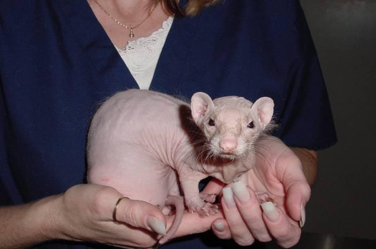
1 Photograph Credits Pathology of the Domestic Ferret Bruce H. Williams, DVM, DACVP Chairman, AFIP Dept. of Telemedicine (202) Dr. John Gorham Dr. John King Dr. Lois Roth Dr. Tracey McNamara Dr. Renata Miracca Dr. Charles Weiss Dr. James Fox Dr. Xiantang Li Dr. Fabio Del Piero Dr. Mike Garner Dr. Matti Kiupel Dr. Dick Montali Dr. Peter Fisher Dr. Robert Monaco Dr. Cathy Pfent Dr. Alice Davis Ms. Tara Radford Ms. Lisa Leidig Ms. Beverly Fox Mustela putorius furo Mustela putorius furo (domestic ferret) Males average 2.5 pounds Females average 1.25 pounds Usually purchased neutered and descented Males - hobs or gibs Females - jills or sprites Lifespan years Adult male Mustela putorius furo Canine Distemper 100% fatal in ferrets; day progression Most commonly seen in pet store kits or as a facility outbreak Diagnosis should be made on clinical signs, followed by euthanasia of all affected animals Antemortem FA testing available but not recommended. 1


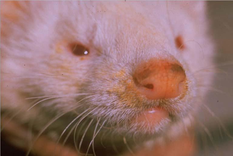


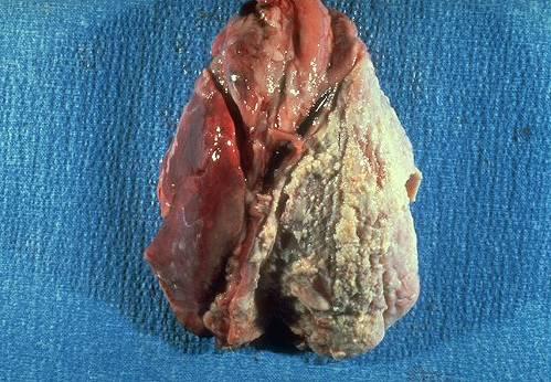
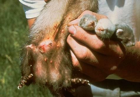
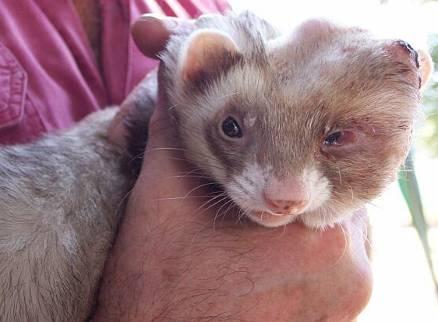
2 Canine Distemper Oculonasal Discharge Hyperkeratosis of Nasal Planum and Footpads Skin rash Diarrhea Weight loss Lethargy Pneumonia Canine Distemper Hyperkeratosis of Nasal Planum and Footpads 2


3 Canine Distemper Rabies Uncommon disease Less than 50 diagnosed cases Ferret susceptible to skunk, bat, and raccoon strains Dumb and furious presentations Should be a ruleout for all neuro cases. Negri bodies in mink brain Rabies Neural Tube Defects No treatment Quarantine period in most states Annual IM vaccination for all ferrets beginning at 15 weeks Imrab-3 vaccine Color diluted ferrets Varying degrees of: Agenesis of cerebrum Spinal dysraphism Often associated with: Other birth defects Intra-utero growth retardation Growth retardation and NTD s in ferret kits 3


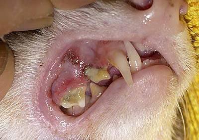
4 Neural Tube Defects Neural Tube Defects Iniencephaly Anencephaly Craniorachischisis Cervical vertebral fusion Other birth defects Iniencephaly Anencephaly Craniorachischisis Cervical vertebral fusion Other birth defects Iniencephaly in a color-diluted ferret kit Neural tube defect in iniencephalic kit Dental disease 4


5 Megaesophagus Uncommon disease Primary middle-aged males No apparent cause Treat as other domestic species 5


6 6


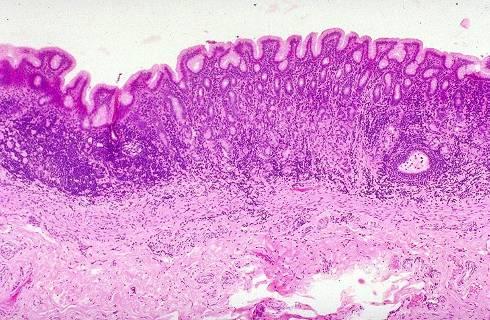
7 Helicobacter mustelae Gastric Ulcers Diagnosis Most readily made from pyloric biopsies Characteristic L-P gastritis Argyrophilic extracellular spiral bacteria associated with mucus superlayer or within crypts Diff-Kwik on endoscopic or surgical biopsy impression smears Lymphofollicular gastritis in ferret with H. mustelae (HE, 20X) Common in ferrets and other mustelids under stressful conditions or with concurrent disease May be associated with Helicobacter mustelae Hemorrhage associated with non-lethal ulceration 7


8 Ferret Coronavirus Epizootic catarrhal enteritis (ECE) High morbidity, low mortality Asymptomatic carriers often pet store kits. Older animals more severely affected Prolonged shedding of virus Pathogenesis Viral infection of villar tips Necrosis of cells Loss of surface area and brush border enzymes Passive secretory diarrhea Malabsorption Mucus hypersecretion Normal ferret jejunum Chronic Lesions Last up to one year post infection Villar atrophy fusion, and blunting Lymphocytic enteritis Ferret Coronavirus Jejunum of ferret with ECE with villar atrophy, fusion and blunting and lymphocytic enteritis Ferret Coronavirus Ferret Coronavirus Acute Lesions Vacuolar degeneration and necrosis of villar tip enterocytes Villar blunting Vacuolar degeneration and necrosis of coronavirus-infected jejunal enterocytes 8


9 Ferret Coronavirus Inflammatory Bowel Disease 9


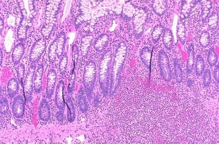
10 Proliferative colitis Clinical signs Frequent, painful defecation Frank blood or mucous in stool Anorexia Weight loss Abdomen painful on palpation Clinical signs exacerbated by stress May progress to anemia, death. Cobblestone appearance to colon in ferret with PC Proliferative colitis GI Parasites Uncommon Coccidia Giardia Pathogen? Nematodes rare Yeasts - commensal Treatment 50 mg/kg Chloramphenicol palmitate orally BID Giardia lamblia in a domestic ferret Coccidiosis Coccidiosis Eimeria furonis, E. ictaluri Generally asymptomatic, but may be lifethreatening in young kits or severely affected animals Fecal floatation Villar blunting and loss due to coccidiosis 10


11 Gastrointestinal foreign bodies Very common in ferrets High index of suspicions in ferrets less than 1 year of age May be seen in bored, caged ferrets. Latex, rubber, cloth most popular NO house is completely ferret-proofed! Gastrointestinal foreign bodies Hairballs - Less common than traditional foreign bodies Foreign bodies rarely show up on radiographs Ferret trichobezoars! Electrical cord injury 11


12 Mycobacterium avium-intracellulare Clostridium perfringens Chronic wasting disease in ferrets Minimal zoonotic potential Most commonly seen in skin, but may arise from oral epithelium Predilectiou to invade jaw bones Radical surgical excision is only documented cure Low metastatic potential, but massive tissue destruction. Squamous cell carcinoma Extensive mandibular involvement by SCC GI Neoplasia GI Neoplasia Intestinal adenocarcinoma in a ferret (Photo courtesy John King) 12


13 Figure 1. Ferret (Mustela putorius furo). Girard-Luc A et al. J VET Diagn Invest 2009;21: Copyright by American Association of Veterinary Laboratory Diagnosticians 13


 Hepatic")

14 Hepatic lipidosis Fatty livers Common physiologic finding Due to inanition and mobilization of peripheral fat stores Fatty Liver in a Ferret Hepatic tumors Hepatic Tumors High percentage of malignancy in this organ Lymphoma most common Primary neoplasms exhibit slow growth Hepatic carcinoma in a ferret (Photo courtesy of Dr. Renato Miracca) Hepatic Neoplasms Large biliary cystadenoma in ferret liver affecting multiple lobes (Photo courtesy Charles Weiss) 14


 Histology does not correlate with")


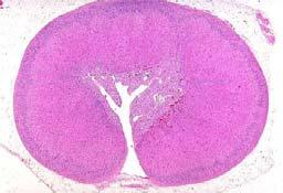
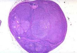
15 Islet cell tumor Most common ferret tumor May be function or non-functional Inappropriate secretion of insulin resulting in trances, hindlimb paresis, salivation, seizures and coma. Benign progression Pancreatic islet cell tumor in a ferret Islet cell tumor Islet cell tumor Diagnosis History and clinical signs Blood glucose test g/dl - questionable <60 positive Insulin testing generally not necessary Normal pancreas in a ferret (note gastric and jejunal arms) Histology does not correlate with behavior Watch out for pancreatic nodular hyperplasia! Adrenal-associated endocrinopathy Extremely common Due to hyperestrogenism, not Cushings!!! Proliferative lesions (hyperplasia, adenoma, carcinoma have identical clinical signs Classic bilateral truncal alopecia in ferret with AAE 15


16 Adrenal-associated endocrinopathy On the adrenal cortical cells are LH receptors that are activated after neutering by high LH levels Adrenal-associated endocrinopathy 16


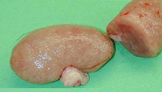
17 Other Adrenal Neoplasms Adrenal teratoma (Gross photo courtesy R. Geske) Splenomegaly Commonly seen, especially in older ferrets Stereotypical response to chronic smoldering inflammation Less than 5% are neoplastic Marked splenomegaly in a ferret 17


 Aspirates Good for preliminary, but")
")


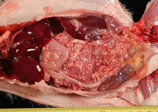
18 Splenomegaly Malignant lymphoma (Lymphosarcoma) 95% are benign extramedullary hematopoiesis Splenectomy is treatment of choice. Most common malignancy in ferrets 1-2 years - Juvenile (Lymphoblastic) - visceral distribution 2-7 years - Lymphocytic - lymph node distribution 2-7 years - Immunoblastic polymorphous Splenic extramedullary hematopoeisis in enlarged ferret spleen Juvenile lymphoma in a young ferret Malignant lymphoma (Lymphosarcoma) Diagnosis Clinical signs Organ-specific changes in clinical pathology data Biopsy of enlarged lymph node or organ Can not diagnose on CBC alone!!! Malignant lymphoma (Lymphosarcoma) Aspirates Good for preliminary, but not definitive diagnosis Can be extremely difficult to interpret Histo required for definitive diagnosis Malignant lymphoma (Lymphosarcoma) Aleutian Disease Treatment Poor prognosis except in primary cutaneous cases Chemotherapy regimes available, but less than 10% respond. Renal lymphosarcoma in an adult ferret Resurgent disease in ferrets new strain? New outbreaks have almost 100% morbidity and mortality. Insidious disease with long latency period Death in 2-3 years, as opposed to mink (strain variation? Abdominal viscera of a ferret infected with ADV. 18


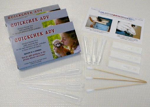
19 Aleutian Disease Characteristic appearance of glomerulonephritis in ADV-infected ferret Aleutian Disease Hematuria in ADV-infected ferret Coronaviral-Associated Granulomatous Disease Mostly young male ferrets Hypergammaglobuli nemia (>6.0), leukocytosis, mild anemia Avecon POCT 19


")


20 Hemangiosarcoma Low metastatic potential for skin tumors Moderate metastatic potential for visceral tumors Solid hemangiosarcoma in a ferret spleen (Photos courtesy of Robert Monaco, DVM) Hemangiosarcoma May arise in any organ Neoplasms of blood vessels Often result in hemorrhage 20


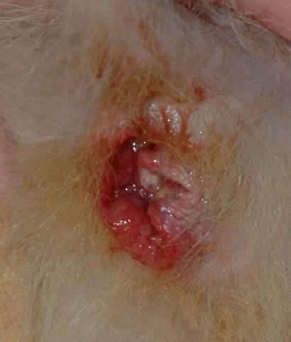
21 Cystic Prostatic Disease Sequelae to adrenal disease in male ferrets Dysuria, but easily expressed Estrogen effect on glandular epithelium Adrenalectomy is curative 21


22 Renal cysts Very common in older ferrets Triangular areas of cortical scarring containing obsolescent glomeruli Not infarcts 22


23 Estrus-associated Anemia Estrus-associated Anemia Signs referrable to which line(s) affected in marrow. Death by severe anemia is most popular. Mastitis 23


24 Cardiomyopathy Common in American bloodlines Genetic with incomplete penetrance Dilatative cardiomyopathy in a ferret with biventricular enlargement and passive hepatic congestion Cardiomyopathy Cardiomyopathy In fulminant cases, may see active myocardial necrosis Marked pleural effusion in ferret with cardiomyopathy 24


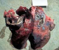
25 Aspiration pneumonia Endogenous lipid pnemonia Influenza Common incidental finding which is misinterpreted by practitioners. Can contract both Type A and Type B influenza from humans Symptoms similar to man, but slightly more severe and longer lasting Symptomatic treatment only if anorexic May use antihistamines to decrease watery nasal discharge Figure 1. Necrosuppurative bronchiolitis in the lung of a ferret infected with Influenza A virus. Systemic mycoses Patterson Copyright A by R American et al. J Association VET Diagn of Veterinary Invest 2009;21: Laboratory Diagnosticians 25


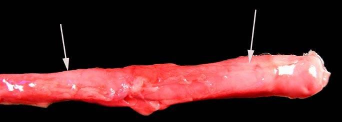
26 Mycobacterium genavense macroscopy B Chordoma Chordoma Most common orthopedic tumor of ferret Only on spine Develops from primitive notochord Most commonly seen at tail tip Chordoma at tip of tail Formalin-fixed specimen of chordoma at tail tip. 26


27 Osteoma Benign tumors of flat bones Complete surgical excision curative Osteoma Osteosarcoma Uncommon malignancies that are most commonly seen on extremities. Low malignant potential. 27


28 Actinomycosis Ectoparasites Earmites (Otodectes cyanotes) Ectoparasites Ixodes hexagonus (hedgehog) Ectoparasites Demodex sp 28


29 Sebaceous epithelioma Often look much worse than they really are Don t believe malignant diagnoses! Skin Neoplasia Skin Neoplasia Apocrine cysts from ferret prepuce 29


30 Skin Neoplasia Spindle cell tumors usually arise from smooth muscle Locally aggressive, slow to metastasize 30


31 Figure 1. Skin; ferret. Rodrigues A et al. Vet Pathol 2010;47: Copyright by American College of Veterinary Pathologists Brain Tumors Uncommon Astrocytomas most common Tend to cause progressive dysfunction as tumor grows Seizure activity Coma Diagnosis and therapy are difficult. Meningioma compressing brainstem in a ferret (Photo courtesy of Mike Garner. DVM) Cataracts Glaucoma 31


32 32
33 Pathology of the Domestic Ferret Instructor: Bruce H. Williams, DVM, DACVP Dept. of Veterinary Pathology, AFIP (202) PURPOSE The purpose of this 3-hour block of instruction is to gain knowledge and experience in the gross diagnosis of diseases of the domestic ferret (Mustela putorius furo). Of course, the study of disease in this species far exceeds what can be presented in a twohour block of time, but I will attempt to cover a number of diseases of interest. In some cases, inclusion or exclusion from this collection was the result of the availability of highquality photographs. I am a firm believer that one can learn far more from one excellent photograph of a single entity, than from many poor ones. If the only available image is of poor quality, the image won t leave a lasting impression, and the student learns nothing. I have included a brief morphologic or etiologic diagnosis for each entity. The formulation of concise, accurate morphologic diagnoses is a major pursuit of every good pathologist, especially those who seek certification in this specialty. The formulation of a good morphologic diagnosis is a learned skill; for those seeking additional experience in this endeavor, I would suggest attendance at the annual C.L. Davis Descriptive Pathology Course, given each year in the US and Europe, and often on other continents. 1
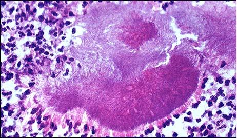
34 Slide Organ Condition Morphologic No. Diagnosis 1 INTRODUCTION Notes 2 Photograph Credits 3 Presentation Normal Normal Normal fitch ferret Males average 2.5 lbs, females 1.25 lbs. Most ferrets are already neutered and descented. 4 Nervous System 5 Presentation Canine distemper Cachexia Ferret distemper is highly infectious and 100% fatal in this species 6 Presentation Cachexia, diarrhea Diarrhea is commonly seen in affected animals. Note nasal hyperkeratosis. 7 Conjunctiva Diffuse mucopurulent conjunctivitis 8 Nasal planum Diffuse mild nasal hyperkeratosis with mucopurulent rhinitis 9 Footpads Diffuse mild footpad hyperkeratosis Clinical signs resemble distemper in other species; photophobia and mucupurulent conjunctivitis and rhinitis are early presenting signs. Hardpad disease (hyperkeratosis of the nasal planum and foodpads is a pathognomonic sign of distemper in ferrets.) 10 Footpad (mink) Diffuse severe footpad hyperkeratosis 11 Lung Diffuse fibrinosuppurative bronchopneumonia 12 Urinary bladder Numerous intracytoplasmic and intranuclear epithelial Pneumonia is a result of immunosuppression and is the most common cause of death in affected animals. Inclusions can be found in many epithelial tissues in affected animals. Urinary 2
35 inclusions 13 Footpad Diffuse severe footpad hyperkeratosis with numerous intracytoplasmic viral inclusions 14 Lung Diffuse necrotizing bronchointerstitial pneumonia bladder, lung, and Biliary tract are good sites to sample. Intracytoplasmic and intranuclear inclusions may be found in in apparently unaffected skin. IFA is even more sensitive, if available. Immunohistochemistry for morbillivirus antigen shows the extent of infection in affected lungs 15 Presentation Rabies Aggression Furious and mad forms of rabies are seen in ferrets 16 Cerebrum Intracytoplasmic viral inclusions (Negri bodies) in cerebral neurons in a mink Less than 60 cases of rabies in ferrets have been recorded, making it one of the most rarely affected domestic species 17 Vaccine box Ferrets require annual IM vaccination; treated like dogs and cats in bite incidents (10- day quarantine in US) 18 Presentation Neural tube Multiple defects including cleft palate, iniencephaly, craniorachischisis, and exenteration 19 Neural tube Skull, vertebral column Diffuse craniorachischisis with iniencephaly 20 Diffuse craniorachischisis with iniencephaly Neural tube defects occur with highest incidence in litters from color-diluted sires. Iniencephaly is a malformation/fusion of cervical veterbrae resulting a stargazing appearance. In affected animals neural defects (as seen here) may include agenesis of cerebral hemispheres and spinal cord. 21 Gastrointestinal System 3
36 22 Teeth Broken teeth Bilateral mandibular canine fractures 23 Teeth Periodontal disease Diffuse moderate dental calculus 24 Severe dental calculus with periodontitis and root abscess The pulp cavity of the ferret tooth is relatively shallow and becomes less prominent with age ferrets can tolerate more significant loss of the crown without a requirement for endodontic therapy. Ferrets are similar to other domestic animals with regard to frequency of periodontal disease, which may be worsened in animals on bland diets. 25 Oral cavity Mucocele Oral mucocele Mucoceles are occasionally seen in ferrets, likely as a result of chewing foreign objects 26 Head, subcutis Zygomatic salivary 27 duct 28 Zygomatic salivary gland mucocele Rupture of the duct of the zygomatic salivary gland often results in mucoceles on top of the head. Long-term drainage is usually required. 29 Esophagus Megaesophagus Megaesophagus with marked intrathoracic dilation 30 Lungs Aspiration pneumonia Multifocal to coalescing gangrenous pneumonia 31 Esophagus Myofasciitis (Disseminated idiopathic myositis) Multifocal severe neutrophilic esophagitis 32 Esophagus, muscularis and serosa: Esophagitis, neutrophilic focally extensive, severe. This disease of young, male ferrets has not yet been linked to other conditions. Aspiration is the eventual cause of death of most animals with megaesophagus. Look for fungal infections in the ulcerated esophagus as well. Myofasciitis is an idiopathic fatal condition of young ferrets resulting in characteristic neutrophilic inflammation in the esophagus, heart, skeletal muscle, and lymph nodes. 4
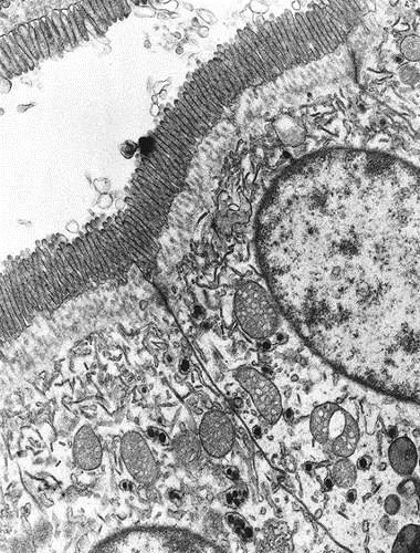
37 33 Hind legs Diffuse severe hindlimb muscle atrophy (normal on left) 34 Stomach Helicobacter mustelae Diffuse m0derate atrophic gastritis with melena 35 Ultrastructural appearance of spiral bacteria 36 Multifocal to coalescing lymphoplasmacytic gastritis 37 Small numbers of argyrophilic bacteria (Warthin-Starry 4.0) 38 Stomach Gastric ulcers Diffuse pyloric ulcers with melena Affected skeletal muscles show marked wasting, and occasionally may have neutrophilic inflammation as well. Clinical signs and gross lesions of this common infection of middle-aged and older ferrets are usually tenuous. H. mustelae are spiral-shaped bacteria that live in the mucous layer overlying the gastric mucosa. Evidence of Helicobacter infection is commonly seen in clinically normal animals as well. Organisms are best demonstrated in the pylorus with a silver stain. The presence of digested blood in the stomach is positive proof of gastric ulcers. 39 Gastric contents Tarry stools The presence of tarry, unformed stools is a sign of a potentially life-threatening condition. 40 Stomach Focal pyloric ulcer with hemorrhage 41 Stomach Pinpoint pyloric ulcers; perforating pyloric ulcer 42 Feces Epizootic catarrhal enteritis Large pyloric ulcers can result in fatal gastric bleeds in 5 mins. Numerous small bleeding points in the pyloric stomach is far more common than large focal ulcers. The first name of the disease was the Green Slime. One still hears this type of feces referred to as the greenies 5
38 today. 43 Cadaver Diffuse serous atrophy of fat 44 Small intestine Diffuse lymphocytic and atrophic enteritis with maldigested feces Affected animals rapidly lose condition. Note colon filled with green, loose feces. The affected section of intestine ahs a thinned, congested wall and contains birdseed-like feces. 45 Normal ferret jejunum The normal ferret jejunum has long, plush villi. 46 Focal villar tip enteric necrosis 47 Diffuse lymphoplasmacytic enteritis with villar atrophy, fusion, and blunting 48 FeCoV antigen in villar tip enterocytes 49 Coronavirus particles in enterocyte SER 50 Presentation Inflammatory bowel disease The acute lesion of ECE is necrosis of enterocytes at the villar tips. Following necrosis of villar tip enterocytes, villar shortening and re-epithelialization may result in marked decreases in absorptive surface area and diarrhea. Ferret enteric coronavirus, a group A coronavirus, characteristically infects villar tip enterocytes. Transmission EM of coronaviral particles in enterocyte ER; coronaviral particles in feces IBD is rapidly becoming one of the most common diagnoses in ferrets today; while virtually unheard of a decade ago. 51 Feces Birdseed feces This type of feces is commonly seen in IBD and other malabsorptive disorders seeds represent undigested globules of fat and protein 6
39 52 Small intestine Diffuse lymphocytic enteritis with villar fusion and blunting. The key to diagnosis of the lymphocytic form is villar blunting and presence of increased numbers of intraepithelial lymphocytes (IELs) 53 Small intestine, lymph node IBD (eosinophilic gastroenteritis) Diffuse eosinophilic enteritis; diffuse eosinophilic lymphadenitis with Splendore-Hoeppli material Eosinophilic gastroenteritis is a more severe clinical form of IBD but also responds to immunosuppression and dietary changes. 54 Colon Lawsonia intracellulare Diffuse proliferative colitis (serosal view) L. intracellulare is a pathogen in a number of species, but the ferret is the only species in which the colon is affected. 55 Multifocal to coalescing proliferative colitis ( adenomatosis ) 56 Feces Blood and mucous in feces 57 Colon Diffuse proliferative and lymphocytic colitis; argyrophilic bacteria in apical cytoplasm (Warthin-Starry 4.0) 58 Small intestine Giardia lamblia Low and high magnifications of trophozoites attached to the luminal surface. 59 Small intestine Eimeria, Isospora sp Diffuse lymphocytic enteritis with villar blunting Marked proliferation of immature mucosa form glandular mucosal thickenings. Fine bleeding points result in hematochezia Affected animals pass frequent small bowel moves with tenesmus and frank blood. Note the proliferation of immature, non-mucouscontaining colonic epithelium. In silver stains, massive numbers of bacilli are present in apical cytoplasm of colonic epithelium Giardia trophozoites and cysts can be recovered from the feces of clinically normal animals as well. Coccidia is occasionally seen as a facility outbreak as a result of poor sanitation. Infections in very young and very old 7
40 animals may be fatal. 60 Abundant intraepithelial schizonts and gamonts 61 Small intestine Rotavirus Focally extense superficial and necrotizing enteritis 62 Presentation Gastrointestinal foreign bodies Small intestine Focally extensive necrotizing enteritis with rubber foreign boy and proximal dilatation Almost every epithelial cell in this segment of gut has a schizont, which often excyst at the same time, resulting in large stretches denuded of epithelium. Common in ferret kits at 2-3 weeks of age, histologic lesions are mild and consist of vacuolation and necrosis of villar tip enterocytes. Disease in adults is subclinical. Most common cause of severe GI distress in ferrets less than 1 year of age. Rubber, latex, and cloth are most common in ferrets. Cloth foreign bodies are most often seen in older ferrets due to boredom. 65 Stomach Trichobezoars GI signs minimal until wellformed. Assume shape of the stomach. 66 Oral cavity Foreign bodies Foreign object between maxillary dental arcade; electrical cord burn with oronasal fistula Ferrets often explore their environment with their mouth, leading to misadventure. 67 Liver, intestinal tract Mycobacterium avium Multifocal to coalescing granulomatous hepatitis and enteritis Ferrets are susceptible to a wide range of mycobacteria including M. avium, which affects the gastrointestinal tract, M. bovis (in feral ferrets in New Zealand) and M. genovense. 68 Stomach Clostridium Diffuse necrotizing Outbreaks of black-footed 8
41 perfringens type A gastritis ferret kits occurred at the National Zoo in the 1990 s. 69 Rectum Prolapsed rectum Rectal prolapsed Rectal prolapse may be seen in concert with loose stools of any cause. 70 Mandible Oral squamous cell carcinoma 71 Intestine Intestinal T-cell lymphoma 72 Ileum Intestinal adenocarcinoma Mandibular squamous cell carcinoma Diffuse intestinal lymphoma Intestinal adenocarcinoma Oral squamous cell carcinoma is the most common neoplasm of the ferret oral cavity, and is often mistaken in early stages for periodontal disease. Lymphoma is the most common neoplasm of the GI tract, with both T- and B-cell lymphoma reported. Adenocarcinomas of the stomach and intestine have a characteristic napkin ring appearance with proximal dilatation. 72 Ileum GIST Gastrointestinal stromal tumor Single reportof a C-Kit positive neoplasm. Interesting that this one infiltrates the mucosa. 74 Omentum Mesothelioma Mesothelioma May be associated with highprotein ascites ( malignant ascites ). 75 Anal sac Squamous cell 76 carcinoma of the anal 77 sac Anal sac squamous cell carcinoma SCC arising from the anal sac is a rare neoplasm in the ferret. 78 Hepatobiliary and Exocrine Pancreas 79 Liver Hepatic lipidosis Diffuse hepatic lipidosis Hepatic lipidosis is a common physiologic finding in anorexia and often results in elevated ALT and alkaline phosphatase. 80 Pancreas Pancreastic exocrine hyperplasia Multifocal pancreatic exocrine hyperplasia Exocrine pancreatic hyperplasia is a common aging finding; nodules are the same color as 9
42 81 Liver Hepatocellular carcinoma 82 Liver Biliary 83 cystadenocarcinoma Hepatocellular carcinoma Biliary cystadenocarcinoma the surrounding pancreas. These slow-growing neoplasms eventually result in liver failure. These slow-growing tumors often look benign histologically, but eventually replace hepatic lobes and cross over into other lobes. 84 Endocrine System 85 Presentation Islet cell tumor Signs of hypoglycemia in ferrets include trances, ptyalism, and coma. 86 Pancreas Pancreatic islet cell tumor Islet cell tumors, the most common neoplasm in ferrets, are generally red or pink in colors (due to vascularity). 87 Normal pancreas Ferrets, as obligate carnivores, have large pancreases, with two distinct lobes. 88 Islet cell tumor Encapsulated and 89 unencapsulated islet cell tumors All islet cell tumors are potentially malignant whether encapsulated or not. 90 Presentation Adrenal-associated endocrinopathy Bilaterally symmetrical truncal alopecia Hair loss is the most common presenting sign for adrenal disease in ferrets. Other causes of hair loss are extremely uncommon in ferrets. 91 Severe diffuse alopecia Alopecia is the result of estrogen effect on hair follicles. 92 Diagram Diagram of pituitarygonadal-adrenal interaction Adrenocortical disease in ferrets is due to interruption of the pituitary-gonadal axis and the effects of unremittent LH production on receptors in the adrenal cortex 10
43 93 Pop Quiz Can you spot the adrenal ferret? 94 Vulva Diffuse vulvar hypertrophy 95 Adrenal glands Normal anatomy of ferret adrenal glands 96 Adrenal gland Carcinoma Adrenocortical carcinoma 97 Adrenal glands Adrenocortical hyperplasia, adenoma, adenocarcinoma 98 Right adrenal gland Biliary cysts Briliary cysts within right adrenal gland in association with hyperplasia Estrogen receptors on vulvar fibroblasts results in 200x increase in size in young ferrets in heat, or 50% of spayed ferrets with adrenal disease. The right adrenal gland is located under the caudate liver lobe and on top of the posterior vena cava, making for tricky surgical removal. Recently identified histochemical markers of note for adrenal malignancies include cytochrome b5 and GATA-4 both markers of gonadal cell differentiation. Histologic features of proliferative adrenal lesions in ferrets. Regardless of type, a good prognosis is usually warranted if early treatment is accomplished. Epitehlial lined cysts containing a waxy amorphous material are common finding in the adrenal right adrenal gland of the ferret. The cyst epithelium resembles biliary epithelium and is CK 7 positive Adrenal gland Teratoma Adrenal teratoma Teratomas are rarely seen in ferret adrenal glands and often appear as mineralized densities on radiographs. 101 Pancreas Diabetes mellitus Diffuse islet cell glycogenosis While spontaneous cases do arise, many are the result of 11
44 long-term prednisone use 101 Hematolymphatic System 102 Spleen Splenomegaly Diffuse splenic extramedullary hematopoiesis 103 Spleen Splenomegaly Diffuse splenic extramedullary hematopoiesis Splenic hematopoiesis is a common finding in middleaged and older ferrets, and is a stereotypical response to chronic inflammation 95% of enlarged spleens are the result of extramedullary hematopoiesis; the remainder are due to neoplasia 104 Thymus, liver, spleen Lymphoblastic lymphoma Thymic, hepatic, and splenic lymphoma with splenic infarction Lymphoblastic (juvenile) lymphoma generally affects ferrets less than 2 years of age; tumors are predominantly visceral. 105 Cervical lymph nodes Lymphocytic lymphoma Nodal lymphoma Lymphocytic lymphoma generally affects animals over 5 years of age, results in married enlargement of lymph nodes, and neoplastic cells are mature lymphocytes. 106 Cytologic preparation Lymphocytic leukemia Look for monomorphic populations on cytologic aspirates in order when diagnosing lymphoma. 107 Spleen Lymphoblastic leukemia Splenic lymphoma with focal infarct and splenic rupture Infiltration of splenic sinusoids by neoplastic cells often results in stasis and splenic infarction. 108 Abdominal viscera Aleutian disease Bilateral glomerulonephritis, splenomegaly with infarct, ecchymotic gastric serosal hemorrhage, and hematuria The basic mechanism of Aleutian Disease is the precipitation of antigenantibody complexes in basement membranes throughout the body. 12
45 109 Illustration Viral particles and crystallographic reconstruction 110 Kidneys Diffuse membranous glomerulonephritis 110 Urinary bladder Hematuria with serosal hemorrhage 112 Kidney Diffuse membranous glomerulonephritis; plasmacytic infiltrates in numerous tissues Due to its configuration, the Aleutian disease parvovirus results in the formation of abundant non-neutralizing antibodies. Renal glomeruli are the largest vascular bed in the body making the kidneys the target organ in Aleutian disease in ferrets. Excessive levels of antigenantibody complexes result in clotting deficiencies in terminal disease. Plasmacytic infiltrates in numerous organs is characteristic for Aleutian disease. Don t confuse aging changes in glomeruli for glomerulonephritis look for proteinaceous casts in tubules as well. 113 Test kit While in-house testing kits are available, CIEP is still the gold standard test for this disease. 114 Abdominal viscera Coronavirusassociated systemic granulomatous disease Multifocal granulomatous peritonitis, mesenteric lymphadenitis, and colitis 115 Spleen Multifocal to coalescing granulomatous splenitis 116 Liver Multifocal to coalescing granulomatous hepatitis with thrombophlebitis Within the last five years, a number of cases of systemic granulomatous disease have been diagnosed in young ferrets, with lesions resembling the dry form of feline infectious peritonitis. Feline enteric coronavirus (FeCoV) antigen, a Group A coronavirus, has been recovered from the lesions. 13
46 117 Spleen Hemangiosarcoma Splenic hemangiosarcoma Abdominal cavity Mesenteric hemangiosarcoma 120 Skin Cutaneous hemangiosarcoma Hemangiosarcoma in the spleen may be surgically excised if detected early; in other viscera it has a poor prognosis. Skin tumors warrant a good prognosis. 121 Urinary Tract 122 Kidney Bacterial urinary tract infections 123 Prostate Cystic prostatic disease Hydronephrosis Squamous metaplasia of prostatic glandular epithelium with cyst formation Female ferrets are prone to urinary tract infections, with bacteria able to reach he kidney in as little as two weeks. Blockage of the ureter by cellular debris may result in hydronephrosis. Estrogen secretion by functional adrenocortical lesions results in squamous metaplasia of the prostatic epithelium. 124 Radiograph Focally extensive prostatic cysts with suppurative inflammation Large prostatic cysts in male ferrets results in dysuria and post-renal azotemia, and may ultimately result in total urinary obstruction. 125 Prostate The presence of keratin debris within prostatic cysts often incites a profound suppurative inflammatory response. 126 Urinary bladder Urolithiasis Diffuse necrohemorrhagic cystitis with struvite urolith 127 Radiograph Urolithiasis Struvite uroliths Urolithiasis is less commonly seen today as a result of decreased usage of plant proteins in ferret chows. Today, dysuria is more commonly associated with prostatic disease. 14
47 128 Stones 129 Kidney Renal cysts Multiple renal cortical cysts 130 Renal cyst Focally extensive renal cyst Renal cysts are seen in up to 33% of animals at necropsy, and are rarely considered of clinical importance. Cysts may attain a large size, but due to the toughness of the renal capsule, rarely rupture 131 Polycystic kidney disease 132 Chronic interstitial nephritis Multiple renal cortical and medullary cysts with nephrosclerosis Diffuse chronic interstitial nephritis Autosomal recessive PKD is seen in ferrets as in many other species and results in death in young animals. CIN is a common finding in older ferrets. 133 Reproductive System 134 Skin, vulva Estrus-associated bone marrow suppression Multifocal to coalescing cutaneous hemorrhage, melena, and vulvar swelling 135 Focally extensive cutanesous ecchymosis with vulvar swelling 136 Digits Severe anemia Ferrets are induced ovulators, so estrus production continues for a prolonged period. Bone marrow suppression may affect any one or all three lines of marrow cells (RBC, WBC, platelets) resulting in severe anemia, hemorrhage, or secondary bacterial infections. Approximately 50% of unbred jills die as a result of unterminated estrus. 137 Lungs Diffuse purulent pleuritis 138 Mammary glands Mastitis Diffuse suppurative mastitis Coliforms and Staphylococcus are common causes. Kits must be weaned or moved to another jill. 139 Cardiovascular System 140 Heart Dilatative Bilateral ventricular dilatation with chronic Dilatative cardiomyopathy is the most common form of CMP 15
48 cardiomyopathy passive hepatic congestion 141 Cardiomyopathy with marked pleural effusion 142 Multifocal to coalescing myocardial fibrosis with myofiber loss (HE) in ferrets. Many older ferrets have evidence of CMP at necropsy. The cardinal histologic lesion is myofiber loss and replacement by fibrosis; in acute stages, lymphocytic inflammation may be present in the myocardium. 143 Multifocal to coalescing myocardial fibrosis with myofiber loss (Masson s trichrome)) 144 Heart Dirofilaria immitis Cardiac dirofilariasis Ferrets in heartworm=endemic areas should be on prevention. 145 Respiratory System 146 Lung Aspiration pneumonia Multifocal to coalescing gangrenous pneumonia Aspiration pneumonia is often seen in animals that have been force fed by syringe, or occasionally in animals receiving liquid antibiotics. 147 Lung Endogenous lipid pneumonia Multifocal to coalescing subpleural histiocytosis Endogenous lipid pneumonia is a common incidental finding in furbearing animals. 148 Conjunctiva Influenza Diffuse mild serous conjunctivitis 149 Lung Influenza Necrosuppurative bronchiolitis with marked edema Ferrets are the animals model for Type A and B influenza; the natural disease is self-limiting There are many groups publishing papers utilizing ferrets for experimental hosts for virulent influenza. 150 Lung Systemic fungal infection 151 Lung Mycobacterium genavense Multifocal to coalescing granulomatous pneumonia Diffuse granulomatous pneumonia Systemic infections with Blastomyces and Histoplasma are occasionally seen in ferrets. In recent years, a number of atypical bacterial species have been reported in ferrets, 16
49 including M. genavense, M. abscessus, M. celatum, and M. microti 152 Musculoskeletal System 153 Tip of tail Chordoma Caudal vertebral 154 chordoma 155 Radiograph Cervical vertebral chordoma Chordomas are the most common neoplasm of the musculoskeletal system. They are slow-growing, invasive tumors of little metastatic potential, but if they arise anywhere other than the tail, a poor prognosis is warranted. Chordoma has been reported a number of times in the cervical vetebrae of the ferret with uniformly poor results. 156 Cervical vertebra Chordomas are extremely infiltrative neoplasms which predispose to pathologic fractures and para- or tetraparesis Skull Skull, X-ray Osteoma Cranial osteoma Osteomas are slow-growing benign bone tumors which often arise on the skull or other flat bones. 159 Skull, maxilla Maxillary osteoma Surgical excision Is generally curative, however, this practitioner was hesitant in attempting removal Femur Osteosarcoma Femoral, vertebral osteosarcoma 162 Vertebra Osteosarcomas may be seen on either flat or long bones, with no predilection for each. There has been no reports of metastasis of osteosarcoma in the ferret. 17
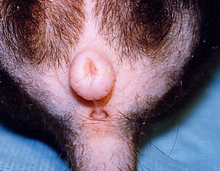
50 163 Eye, orbit Actinomyces sp. Focally extensive granulomatous orbital cellulitis and osteomyelitis Actinomyces has been reported in a number of cases of deepseated infection in the skull of ferrets. 164 Integumentary System External ear canal Otodectes cynotis Aural otodectiasis Ear mites are a common infection in ferrets and easily treated with cleansing and ivemectin. 167 Facial skin Ixodes hexagonus Dermal ixodiasis Ticks are occasionally seen in animals housed outdoors, usually in poor conditions. 168 Haired skin Demodex sp. Follicular demodicosis Demodex mites are commonly found in normal ferrets, and may be seen in increased numbers in immunosuppressed individuals. 169 Foot Sebaceous epithelioma Cutaneous sebaceous epithelioma Sebaceous epitheliomas and other variants of sebaceous adenomas are the most common neoplasm of the skin and invariably benign. 170 Haired skin Although they look ugly both grossly and histologically, sebaceous epithelioma is still invariably benign. 171 Haired skin Mast cell tumor Cutaneous mast cell 172 tumor 173 Mast cell tumors are also invariably benign, although they may appear either singly or multiply Prepuce Apocrine cysts Apocrine gland adenocarcinoma Preputial apocrine cysts Preputial apocrine adenocarcinoma Apocrine cysts, and neoplasms, are most common on the face, neck, prepuce, and perineum. Neoplasms on the prepuce or perineum have a high incidence of malignancy, and are the site of most malignancies in ferret 18
51 skin. 177 Ear Leiomyosarcoma Cutaneous leiomyosarcoma Smooth muscle tumors are commonly seen, especially in the dorsal neck and back, where they arise from hair follicles (piloleiomyomas). 178 Haired skin Vaccine-related sarcoma 179 Haired skin, face Peripheral nerve sheath tumor Cutaneous fibrosarcoma Malignant peripheral nerve sheath tumor Vaccine-related sarcomas are rarely reported in ferrets. Inflammatory lesions due to deposition of vaccine in the dermis are more common. Peripheral nerve sheath tumors in the ferret are most commonly seen on the face. 180 Recurrence of previously excised tumor but much more aggressive (as is often the case following surgical intervention. 181 Haired skin Erythema multiforme Multifocal to coalescing ulcerative dermatitis Immune-mediated skin diseases, along with other immune-mediated disease in this species appears to be on the rise. I have seen a number of cases to date. Good biopsies are hard to come by. 182 Cutaneous papillomavirus Multicentric squamous cell carcinoma Papillomavirus was associated with a multicentric squameous cell carcinoma. Not to difficult a leap. 183 NERVOUS SYSTEM 184 Cerebrum Meningioma Cerebral meningioma Lymphoma is the most common neoplasm in the brain of the ferret. 185 Eyes Cataracts Bilateral lenticular cataracts A common finding in older ferrets, there is no known cause for cataracts. 19
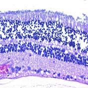
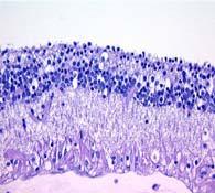
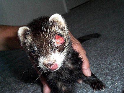
52 186 Eyes Glaucoma Unilateral bupthalmia Both secondary and primary glaucoma have been seen in the ferret. 187 Retina Peripheral retinal atrophy Diffuse loss of inner nuclear and plexiform layers This condition is common in oder ferrets, whose owners often do not know that they are blind. Histologically, It is similar to RP in humans. 188 Eye Lymphoma Retroorbital lymphoma Lymphoma is the most common cause of bulging eyes in ferrets. 20
There is no one correct way to describe a slide. Macroscopic Veterinary Pathology. Be concise. Look at the center of the slide.
 Macroscopic Veterinary Pathology There is no one correct way to describe a slide. Bruce Williams, DVM, DACVP Senior Pathologist, JPC Email: williams@cldavis.org Tissue from a sheep Tissue from a foal Be
HEMATOPOETIC LAB. Heather Fenton/S. Martinson VPM 222 March, 2013
 HEMATOPOETIC LAB Heather Fenton/S. Martinson VPM 222 March, 2013 Diffuse Splenomegaly: Bloody vs Meaty Photos: Pathologic basis of veterinary disease Bloody Spleen: Swollen margins Dark red Oozes blood
Adrenal Cortical Carcinomas with Myxoid Differentiation in Ferrets
 Adrenal Cortical Carcinomas with Myxoid Differentiation in Ferrets M. Kiupel,, R.A. Peterson II, C.C. Capen,, D.B. Wilson 4125 Beaumont Road Lansing, MI 48910 www.animalhealth.msu.edu Signalment 5-year-old
Canine and Feline Distemper. Description. The following chart indicates the animals which are susceptible to infection by canine and feline distemp
 Canine and Feline Distemper Description Canine and feline distemper are diseases affecting many wild and domestic carnivo The following chart indicates the animals which are susceptible to infection by
Australian and New Zealand College of Veterinary Scientists. Membership Examination. Small Animal Medicine Paper 1
 Australian and New Zealand College of Veterinary Scientists Membership Examination June 2015 Small Animal Medicine Paper 1 Perusal time: Fifteen (15) minutes Time allowed: Two (2) hours after perusal Answer
2007 BICHON FRISE BREEDERS HEALTH SURVEY Part 4: Prevalence of Health Conditions
 2007 BICHON FRISE BREEDERS HEALTH SURVEY Part 4: Prevalence of Health Conditions 25 Surveys reported the number of Bichons having no health problems Males 204 Females 221 11 surveys did not answer Temperament
Pathology of the Hematopoietic System. Lecture 2: Lympho/Myelo-proliferative diseases and Lymph nodes
 Pathology of the Hematopoietic System Lecture 2: Lympho/Myelo-proliferative diseases and Lymph nodes Shannon Martinson, September 2015 Primary Hematopoietic Neoplasia Lymphoma Hematopoietic Neoplasia Lymphoproliferative
Veterinary Science Preparatory Training for the Veterinary Assistant. Floron C. Faries, Jr., DVM, MS
 Veterinary Science Preparatory Training for the Veterinary Assistant Floron C. Faries, Jr., DVM, MS Post-Mortem Examinations Floron C. Faries, Jr., DVM, MS Objectives Define necropsy Discuss the importance
4-year-old neutered male American domestic shorthair cat with a locally extensive area of swelling ulceration and crusting over the nasal planum.
 4-year-old neutered male American domestic shorthair cat with a locally extensive area of swelling ulceration and crusting over the nasal planum. Which of the following is the most likely disease? 1. Squamous
Report from the Kennel Club/ British Small Animal Veterinary Association Scientific Committee
 Report from the Kennel Club/ British Small Animal Veterinary Association Scientific Committee Summary results of the Purebred Dog Health Survey for Welsh Springer Spaniels Warning: The results of this
What s Your Diagnosis?
 What s Your Diagnosis? Signalment: Maine Coone (8 month old, female intact) Presenting complaint: Lethargy, inappetence, serosanguinous vaginal discharge History: Lives with 11 other Maine Coone cats (males
COMMON CLINICAL CONDITIONS IN RABBITS AND GUINEA PIGS
 COMMON CLINICAL CONDITIONS IN RABBITS AND GUINEA PIGS Megan H. Nowland, DVM, Diplomate ACLAM Assistant Professor Unit for Laboratory Animal Medicine University of Michigan NCRC Building 10, Suite G90 2800
Diseases Affecting 4H Sheep and Goats
 Diseases Affecting 4H Sheep and Goats Dr. Chad Frank DVM, MS, DACVP CSU Veterinary Diagnostic Laboratory 1 Clostridial Diseases C. perfringens type D (Enterotoxemia) Usually sudden changes in diet Sheep-
Canine Distemper Virus
 Photo: LE Carmichael, MJ Appel Photo: LE Carmichael, MJ Appel Photo: LE Carmichael, MJ Appel Canine Distemper Virus Canine Distemper (CD) is a highly contagious infectious disease of dogs worldwide caused
LIFELONG CARE PLAN FELINE
 LIFELONG CARE PLAN FELINE Your pet is unique, and our veterinarian s treatment plan will be tailored to their specific needs. As your pet grows, however, there are certain health concerns associated with
COMMON CLINICAL CONDITIONS IN RATS AND MICE
 COMMON CLINICAL CONDITIONS IN RATS AND MICE Megan H. Nowland, DVM, Diplomate ACLAM Assistant Professor Unit for Laboratory Animal Medicine University of Michigan NCRC Building 10, Suite G90 2800 Plymouth
The Ones We All Know About. Infectious Diseases of Ferrets. The Odd Ones. Ferret Acute Hemorrhagic Syndrome IFC Symposium, Pittsburgh, PA
 The Ones We All Know About Infectious Diseases of Ferrets Karen Purcell DVM Canine Distemper (Morbillivirus) Aleutian Disease (Parvovirus) Rabies (Rhabdovirus) ECE (Coronavirus) Ringworm (Trichophyton
HISTOPATHOLOGY. Introduction:
 Introduction: HISTOPATHOLOGY Goats and sheep are the major domestic animal species in India. Much of the economy of the country has been depend upon the domestication of these animals. Especially economy
COPROPHAGIA ECTOPARASITES SUPERFICIAL DERMATOMYCOSES PRUITIS CONSUMPTION OF FECES AND THIS IS A WAY IN WHICH PATHOGENIC (ORIGIN AND
 COPROPHAGIA CONSUMPTION OF FECES AND THIS IS A WAY IN WHICH PATHOGENIC (ORIGIN AND DEVELOPMENT OF THE DISEASE) MATERIAL CAN GET INTO THE ANIMAL INTEGUMENTARY PATHOLOGIES ANY PHYSICIAL CONDITION THAT DISRUPTS
Report from the Kennel Club/ British Small Animal Veterinary Association Scientific Committee
 Report from the Kennel Club/ British Small Animal Veterinary Association Scientific Committee Summary results of the Purebred Dog Health Survey for Soft Coated Wheaten Terriers Warning: The results of
Schistosoma mansoni, S. japonicum, S. haematobium
 Schistosoma mansoni, S. japonicum, S. haematobium The Organisms More than 200 million people are infected worldwide with Schistosoma species. The adult worms are long and slender (males are 6 12 mm in
Copper-Storage Liver Disease Basics
 Copper-Storage Liver Disease Basics OVERVIEW Abnormal accumulation of copper in the liver, causing sudden (acute) inflammation of the liver (hepatitis) or long-term (chronic) hepatitis and eventually progressive
What s Your Diagnosis? By Sohaila Jafarian, Class of 2018
 Signalment: Greeley, 3 yo MC DSH Presenting Complaint: ADR History: What s Your Diagnosis? By Sohaila Jafarian, Class of 2018 Patient is an indoor/outdoor cat. Previously healthy and up to date on vaccines
Alimentary System 解剖學科徐淑媛
 Alimentary System 解剖學科徐淑媛 本堂重點 1. Structures derived from primitive guts 2. Specific events Alimentary System endoderm of primordial gut epithelium & glands of digestive tract ectoderm of stomodeum epithelium
Infectious Disease. Topic-Actinomycosis. Topic-Anaerobic Infections. Topic-Aspergillosis - Disseminated. Topic-Blastomycosis.
 Topic-Actinomycosis Figure 1. VD thoracic radiograph of consolidated lung lobe secondary to actinomycosis. Topic-Anaerobic Infections Figure 1. Test tube of effusive fluid removed from the thorax of a
Shannon Martinson, BSc, DVM, MVSc, DACVP Department of Pathology and Microbiology Atlantic Veterinary College, University of Prince Edward Island
 Shannon Martinson, BSc, DVM, MVSc, DACVP Department of Pathology and Microbiology Atlantic Veterinary College, University of Prince Edward Island Reptile pathology: Performing a necropsy Do a careful external
Pathology of the Alimentary System Lecture 5 Diseases of stomach & abomasum
 Systemic Pathology I - VPM 221 Pathology of the Alimentary System Lecture 5 Diseases of stomach & abomasum Enrique Aburto Fall 2014 VI. Diseases of Stomach & Abomasum Physical influences Gastric Ulcers
Index. Note: Page numbers of article titles are in boldface type
 Index Note: Page numbers of article titles are in boldface type A Abomasal bloat diarrhea in calves 3 months old or younger due to, 460 461 Abomastitis diarrhea in calves 3 months old or younger due to,
Recommended Resources: The following resources may be useful in teaching this
 Unit B: Anatomy and Physiology of Poultry Lesson1: Internal Anatomy of Poultry Student Learning Objectives: Instruction in this lesson should result in students achieving the following objectives: 1. Identify
NDSU Veterinary Diagnostic Laboratory
 NDSU Veterinary Diagnostic Laboratory February 2015, Vol. 2, No. 1 In This Issue Welcome Flat-rate, One-day Shipping for $7 Pooled Tritrichomonas Testing Now Available Bacteriology Changes for 2015 Noteworthy
Associated Terms: Breast Cancer, Radical Mastectomy, Mastectomy, Mammectomy, Mammary Adenocarcinoma
 Associated Terms: Breast Cancer, Radical Mastectomy, Mastectomy, Mammectomy, Mammary Adenocarcinoma The term "ACVS Diplomate" refers to a veterinarian who has been board certified in veterinary surgery.
Frog Dissection Information Manuel
 Frog Dissection Information Manuel Anatomical Terms: Used to explain directions and orientation of a organism Directions or Positions: Anterior (cranial)- toward the head Posterior (caudal)- towards the
COMPARATIVE VERTEBRATE HISTOLOGY ZOO 4756c Syllabus for Fall 2018
 COMPARATIVE VERTEBRATE HISTOLOGY ZOO 4756c Syllabus for Fall 2018 Instructor: Frank T. Logiudice Office: Biology Building, Room 202c Office Phone Number: (407) - 823-2495 Email Address: Frank.Logiudice@ucf.edu
ECHINOCOCCOSIS. By Dr. Ameer kadhim Hussein. M.B.Ch.B. FICMS (Community Medicine).
.") ECHINOCOCCOSIS By Dr. Ameer kadhim Hussein. M.B.Ch.B. FICMS (Community Medicine). INTRODUCTION Species under genus Echinococcus are small tapeworms of carnivores with larval stages known as hydatids proliferating
ECHINOCOCCOSIS By Dr. Ameer kadhim Hussein. M.B.Ch.B. FICMS (Community Medicine). INTRODUCTION Species under genus Echinococcus are small tapeworms of carnivores with larval stages known as hydatids proliferating
Report from the Kennel Club/ British Small Animal Veterinary Association Scientific Committee
 Report from the Kennel Club/ British Small Animal Veterinary Association Scientific Committee Summary results of the Purebred Dog Health Survey for Dachshunds Warning: The results of this survey and particularly
Feline lower urinary tract disease (FLUTD)
") Feline lower urinary tract disease (FLUTD) Feline lower urinary tract disease (FLUTD) is not a specific disease, but rather is the term used to describe conditions that can affect the urinary bladder and/or
Feline lower urinary tract disease (FLUTD) Feline lower urinary tract disease (FLUTD) is not a specific disease, but rather is the term used to describe conditions that can affect the urinary bladder and/or
The Equine Stomach. by: Multiple Authors March , Article # 5068
 The Equine Stomach by: Multiple Authors March 01 2004, Article # 5068 The Milne Lecture, named for AAEP past president and distinguished life member Frank J. Milne, each year honors a researcher for his
Digestive System Dissection
 Digestive System Dissection THE TERMS YOU NEED FOR THE PRACTICAL ARE IN THIS DISSECTION GUIDE. Instructions: Do one of the 2 respiratory dissections, and then the digestive dissection. Wordlist for cat
ford residence southampton, ny
 P ford residence southampton, ny What bacterial infections cause canine liver disease Causes of Liver Disease. Here are ten causes of liver disease: Poor diet; Bacterial infection; Viral infection; Trauma,
Clinical Programme. Dermatology
 2018 The diagnosis and management of skin represents a major component of small animal practice. Through lectures, case discussions and practical sessions, this modular programme will enable you to learn
Asilomar Definitions and Classification Guidelines
 Asilomar Definitions and Classification Guidelines STANDARD OPERATING PROCEDURE #E103.2 Written: July 2, 2008 Updated: February 12, 2009 By Emilia Gordon, DVM, Medical Director APPROVED: Emilia Gordon,
Australian and New Zealand College of Veterinary Scientists. Fellowship Examination. Small Animal Medicine Paper 1
 Australian and New Zealand College of Veterinary Scientists Fellowship Examination June 2015 Small Animal Medicine Paper 1 Perusal time: Twenty (20) minutes Time allowed: Four (4) hours after perusal Answer
Feline Wellness Report
 Demo/Sample Clinic Feline Wellness Report 59 YOUR CAT'S AGE, IN HUMAN YEARS: Environment, genetics, nutrition and size are factors in determining a cat's age. Although this calculation is not exact, it
Feline Vaccines: Benefits and Risks
 Feline Vaccines: Benefits and Risks Deciding which vaccines your cat should receive requires that you have a complete understanding of the benefits and risks of the procedure. For this reason, it is extremely
After Evaluation ICHS Action Healthy (H)
") Iowa County Humane Society Asilomar Accords Animal Classifications In order to improve consistency and clarity the Iowa County Humane Society (ICHS) has decided to accept the following Asilomar Accords
Iowa County Humane Society Asilomar Accords Animal Classifications In order to improve consistency and clarity the Iowa County Humane Society (ICHS) has decided to accept the following Asilomar Accords
Table of Contents. About the Author. Preface. Acknowledgments. Part One: Performing the Feline Physical Examination
 Table of Contents About the Author Preface Acknowledgments Part One: Performing the Feline Physical Examination 1Setting the Stage: Feline-Friendly Practice 1.1 Challenges Faced in Feline Practice 1.2
Update in Veterinary Medicine. Dr. Maria M. Crane Zoo Atlanta
 Update in Veterinary Medicine Dr. Maria M. Crane Zoo Atlanta Overview of Discussion Medical management of captive orangutans Preventative Medicine Anesthesia Protocols Vaccinations TB testing Current Health
Vaccines for Cats. 2. Feline viral rhinotracheitis, FVR caused by FVR virus, also known as herpes virus type 1, FHV-1
 Vaccines for Cats Recent advances in veterinary medical science have resulted in an increase in the number and type of vaccines that are available for use in cats, and improvements are continuously being
Iguana Husbandry, Nutrition and Disease
 Iguana Husbandry, Nutrition and Disease The green iguana (Iguana iguana) has a natural range from Mexico through Central and South America. It is arboreal, diurnal, mainly folivorous, and solitary except
Understanding your pet s LIVER CONDITION
 Understanding your pet s LIVER CONDITION Why is the liver so important? What causes liver disease in dogs and cats? The liver is one of the largest organs in your pet s body, and it s vital for their good
Report from the Kennel Club/ British Small Animal Veterinary Association Scientific Committee
 Report from the Kennel Club/ British Small Animal Veterinary Association Scientific Committee Summary results of the Purebred Dog Health Survey for the Saluki breed Warning: The results of this survey
Neoplasitic Diseases in Ferrets in Japan: A Questionnaire Study for 2000 to 2005
 FULL PAPER Internal Medicine Neoplasitic Diseases in Ferrets in Japan: A Questionnaire Study for 2000 to 2005 Yasutsugu MIWA 1), Asuka KUROSAWA 2), Hiroyuki OGAWA 1), Hiroyuki NAKAYAMA 3), Hiroshi SASAI
Feline zoonoses. Institutional Animal Care and Use Committee 12/09
 Feline zoonoses Institutional Animal Care and Use Committee 12/09 Cat scratch disease Bacterial infection caused by Bartonella henselae Associated with a cat bite or scratch Infection at point of injury,
Enteric Clostridia 10/27/2011. C. perfringens: general. C. perfringens: Types & toxins. C. perfringens: Types & toxins
 C. perfringens: general Enteric Clostridia Formerly called C. welchii Thick rods, forming spores Non motile Grow fast Habitats: Soil and sewage and in the intestines of animals and humans Double zone hemolysis
Report from the Kennel Club/ British Small Animal Veterinary Association Scientific Committee
 Report from the Kennel Club/ British Small Animal Veterinary Association Scientific Committee Summary results of the Purebred Dog Health Survey for Dobermanns Warning: The results of this survey and particularly
Gastric Dilatation-Volvulus
 Gastric Dilatation-Volvulus The term "ACVS Diplomate" refers to a veterinarian who has been board certified in veterinary surgery. Only veterinarians who have successfully completed the certification requirements
Ferret. Caring for your Pet Ferret. Basic Husbandry General Information
 Caring for your Pet Basic Husbandry General Information Vital Statistics Body Weight: Male 1000g 2000g; Female 500 1000g Life Span: 5 8 years Sexual Maturity: 4 8 months Behavior and Handling s have a
What you need to know to successfully live with your new Kitten-Cat
 What you need to know to successfully live with your new Kitten-Cat Basic information for owners A Publication of Sykesville Veterinary Clinic Table of Contents KITTEN PACKAGES BRONZE SILVER GOLD VACCINATIONS
Acute Vomiting & Diarrhea Overview & Presentation
 STEP 1: Comprehensive Overview Acute Vomiting & Diarrhea Overview & Presentation Craig Datz, DVM, MS, DABVP (Canine & Feline), DACVN University of Missouri Vomiting and diarrhea may indicate a primary
FROG DISSECTION. a. Why is there a difference in size proportion between the hind and fore limbs?
 FROG DISSECTION External Anatomy 1. The division of a frog s body includes the head, trunk and limbs. Examine the front and hind limbs of the frog. The hind limbs are the long, more muscular limbs of the
PDP can be completed in the context of small animal, equine or farm animal practice, or any combination of these three.
 Clinical procedures checklists DRAFT REVISIONS PDP can be completed in the context of small animal, equine or farm animal practice, or any combination of these three. RCVS YEAR ONE CLINICAL PROCEDURES
Fungal Disease. What is a fungus?
 Fungal Disease What is a fungus? A fungus is a living organism. It goes through a complicated life cycle and is able to spread in the environment by producing large numbers of spores that are easily dispersed
EYE CONDITIONS IN THE DOMESTIC FERRET
 EYE CONDITIONS IN THE DOMESTIC FERRET Several conditions can impact the eyes of domestic ferrets. The following conditions are the most common: cataracts, glaucoma, uveitis, infections, nutritional or
Report from the Kennel Club/ British Small Animal Veterinary Association Scientific Committee
 Report from the Kennel Club/ British Small Animal Veterinary Association Scientific Committee Summary results of the Purebred Dog Health Survey for the Newfoundland breed Warning: The results of this survey
Report from the Kennel Club/ British Small Animal Veterinary Association Scientific Committee
 Report from the Kennel Club/ British Small Animal Veterinary Association Scientific Committee Summary results of the Purebred Dog Health Survey for American Cocker Spaniels Warning: The results of this
NECROPSY FORM STRAND LOCATION: FLOATING IN VAQUITA REFUGE BY MX TIME: 10 AM
 NECROPSY FORM FIELD #: Ps 9 NECROPSY DATE: April 4 2018 SPECIES: PHOCOENA SINUS STRAND DATE: March 28 2018 AGE CLASS: ADULT STRAND LOCATION: FLOATING IN VAQUITA REFUGE BY MX NAVY, BAJA CALIFORNIA, MX SEX:
Therapeutic apheresis in veterinary
 Therapeutic apheresis in veterinary 1 I.P.Pavlov First St.-Petersburg State Medical University, Saint-Petersburg, Russia. Voinov V.A. A. By types of animals on the basis of anatomical and physiological
Equine Diseases. Dr. Kashif Ishaq. Disease Management
 Equine Diseases Dr. Kashif Ishaq Disease Management Prevention is the singularly most important aspect Vaccinate regularly Keep horse areas cleaned up and sanitized Proper feeds and feeding management
Diagnosing intestinal parasites. Clinical reference guide for Fecal Dx antigen testing
 Diagnosing intestinal parasites Clinical reference guide for Fecal Dx antigen testing Screen every dog at least twice a year The Companion Animal Parasite Council (CAPC) guidelines recommend including
Pesky Ectoparasites. Insecta fleas, lice and flies. Acari- ticks and mites
 Pesky Ectoparasites Parasite control should be at the forefront of every pet owner s life as all animals have the propensity to contract numerous ones at one stage or another. They are a challenge to the
Introduction. Primary objective. To Spay or Not to Spay That is the question. If to Spay When to spay. Do we know the answers?
 The Optimal Time for Spay / Neuter: An Analysis of Critical Spay Neuter Literature Phil Bushby, DVM, MS, ACVS Professor Emeritus Marcia Lane Endowed Chair of Humane Ethics and Animal Welfare College of
Diagnosing intestinal parasites. Clinical reference guide for Fecal Dx antigen testing
 Diagnosing intestinal parasites Clinical reference guide for Fecal Dx antigen testing Screen every dog at least twice a year The Companion Animal Parasite Council (CAPC) guidelines recommend including
Any Animal. Section G. General Conditions (many different animals) Sicknesses seen on the outside of the animal. Injuries.
 Sicknesses seen on the outside of the animal. Injuries.") Section G General Conditions (many different animals) Sicknesses seen on the outside of the animal Injuries Photo credit: Dr. M. Cattet See Section G-1.1 Emaciation See Section G-1.2 Photo credit: WCVM
Section G General Conditions (many different animals) Sicknesses seen on the outside of the animal Injuries Photo credit: Dr. M. Cattet See Section G-1.1 Emaciation See Section G-1.2 Photo credit: WCVM
Hurricane Animal Hospital 2120 Mount Vernon Road Hurricane, WV or
 Hurricane Animal Hospital 2120 Mount Vernon Road Hurricane, WV 25526 304-757-5937 or 304-757-2287 www.hurricaneanimalhospital.com Feline Leukemia Virus (FELV) This information handout is designed as a
A PRACTICAL GUIDE TO FELINE CANCER FOR THE
 A PRACTICAL GUIDE TO FELINE CANCER FOR THE CARING CAT OWNER by Dr. Martha S. Gearhart DVM, Diplomate American Board of Veterinary Practitioners INTRODUCTION One would hope to be spared the difficulty of
Acute Hemorrhagic Diarrhea Syndrome (AHDS) A Cause of Bloody Feces in Dogs
 A Cause of Bloody Feces in Dogs") Acute Hemorrhagic Diarrhea Syndrome (AHDS) A Cause of Bloody Feces in Dogs No dog parent wants to clean up diarrhea. Cleaning up bloody diarrhea is even more unpleasant. Unfortunately, the development
Acute Hemorrhagic Diarrhea Syndrome (AHDS) A Cause of Bloody Feces in Dogs No dog parent wants to clean up diarrhea. Cleaning up bloody diarrhea is even more unpleasant. Unfortunately, the development
Enteric Clostridia. C. perfringens: general
 Enteric Clostridia C. perfringens: general Formerly called C. welchii Thick rods, forming spores Non motile Grow fast Habitats: Soil and sewage and in the intestines of animals and humans Toxins More than
Equine Emergencies. Identification and What to do Until the Vet Arrives Kathryn Krista, DVM, MS
 Equine Emergencies Identification and What to do Until the Vet Arrives Kathryn Krista, DVM, MS Common Equine Emergencies Cellulitis/lymphangitis Choke (esophageal obstruction) Colic Eye abnormalities Fever
2018 General Health Survey
 2018 General Health Survey Manchester Terrier (UK/FCI) Summary From February 1 March 31, 2018, the Canadian and American Manchester Terrier Clubs administered a comprehensive online health survey of Manchester
This is the smallest tapeworm that can affect human being but it s not really proper human tapeworm (the human is not the primary host).
.") Echinococcus Granulosus Small Tapeworm (1 cm), Cestode. This is the smallest tapeworm that can affect human being but it s not really proper human tapeworm (the human is not the primary host). The primary
Echinococcus Granulosus Small Tapeworm (1 cm), Cestode. This is the smallest tapeworm that can affect human being but it s not really proper human tapeworm (the human is not the primary host). The primary
Treatment of septic peritonitis
 Vet Times The website for the veterinary profession https://www.vettimes.co.uk Treatment of septic peritonitis Author : Andrew Linklater Categories : Companion animal, Vets Date : November 2, 2016 Septic
Feline Idiopathic Cystitis (icatcare)
") Kingsbrook Animal Hospital 5322 New Design Road, Frederick, MD, 21703 Phone: (301) 631-6900 Website: KingsbrookVet.com Feline idiopathic cystitis (FIC) [1] Feline Idiopathic Cystitis (icatcare) Feline
Kingsbrook Animal Hospital 5322 New Design Road, Frederick, MD, 21703 Phone: (301) 631-6900 Website: KingsbrookVet.com Feline idiopathic cystitis (FIC) [1] Feline Idiopathic Cystitis (icatcare) Feline
TREATMENT AND PREVENTION OF UROGENITAL DISEASE IN RABBITS
 Vet Times The website for the veterinary profession https://www.vettimes.co.uk TREATMENT AND PREVENTION OF UROGENITAL DISEASE IN RABBITS Author : Livia Benato, Chris Shepherd Categories : Vets Date : April
Canine Anaplasmosis Anaplasma phagocytophilum Anaplasma platys
 Canine Anaplasmosis Anaplasma phagocytophilum Anaplasma platys It takes just hours for an infected tick to transmit Anaplasma organisms to a dog. What is canine anaplasmosis? Canine anaplasmosis is a disease
AUSTRALIAN AND NEW ZEALAND COLLEGE OF VETERINARY SCIENTISTS. Sample Exam Questions. Veterinary Practice (Small Animal)
") AUSTRALIAN AND NEW ZEALAND COLLEGE OF VETERINARY SCIENTISTS Sample Exam Questions Veterinary Practice (Small Animal) Written Examination (Component 1) Written Paper 1 (two hours): Principles of Veterinary
AUSTRALIAN AND NEW ZEALAND COLLEGE OF VETERINARY SCIENTISTS Sample Exam Questions Veterinary Practice (Small Animal) Written Examination (Component 1) Written Paper 1 (two hours): Principles of Veterinary
PSPP System for Otitis Externa by Craig Griffin. Treatment T / S AB/AF/GC/EC/AllRx/D/Ot Causes and Factors Cyt/Oto/CR/Ot C LTM LLRx
 PSPP System for Otitis Externa by Craig Griffin Name Date Determine and Assess Treatment T / S AB/AF/GC/EC/AllRx/D/Ot Causes and Factors Cyt/Oto/CR/Ot C LTM LLRx Identified imary Secondary Perpetuating
Report from the Kennel Club/ British Small Animal Veterinary Association Scientific Committee
 Report from the Kennel Club/ British Small Animal Veterinary Association Scientific Committee Summary results of the Purebred Dog Health Survey for Norfolk Terriers Warning: The results of this survey
Muscle Lab. - any chronic disease (avian tuberculosis, aspergillosis, amyloidosis, neoplasia)
") Muscle Lab Case 1. The tale of three Black Ducks. 1. What is going on with bird A? This bird is in extremely poor body condition and is emaciated. There is marked pectoral muscle atrophy caused by malnutrition
Muscle Lab Case 1. The tale of three Black Ducks. 1. What is going on with bird A? This bird is in extremely poor body condition and is emaciated. There is marked pectoral muscle atrophy caused by malnutrition
Report from the Kennel Club/ British Small Animal Veterinary Association Scientific Committee
 Report from the Kennel Club/ British Small Animal Veterinary Association Scientific Committee Summary results of the Purebred Dog Health Survey for the Lhasa Apso breed Warning: The results of this survey
Subacute Adenitis. Ann M. Loeffler, MD
 Subacute Adenitis Ann M. Loeffler, MD Lymphadenitis Swelling and hyperplasia of sinusoidal lining cells Infiltration of leukocytes +/- abscess formation Granulomatous or non-granulomatous Pyogenic adenitis
S100A12 concentrations and myeloperoxidase activities are increased in the intestinal mucosa of dogs with chronic enteropathies
 Hanifeh et al. BMC Veterinary Research (2018) 14:125 https://doi.org/10.1186/s12917-018-1441-0 RESEARCH ARTICLE S100A12 concentrations and myeloperoxidase activities are increased in the intestinal mucosa
Improved Cancer Surgery Imaging
 Feline Report 2017 ADVANCING HEALTH, SAVING LIVES Treatments for Upper Respiratory Disease Feline upper respiratory tract disease is expensive to treat and a leading cause of euthanasia in shelters. Two
Report from the Kennel Club/ British Small Animal Veterinary Association Scientific Committee
 Report from the Kennel Club/ British Small Animal Veterinary Association Scientific Committee Summary results of the Purebred Dog Health Survey for Rottweilers Warning: The results of this survey and particularly
FELINE INFECTIOUS PERITONITIS Visions Beyond the Tip of the Iceberg!
 FELINE INFECTIOUS PERITONITIS Visions Beyond the Tip of the Iceberg! Richard B. Ford, DVM, MS, Dipl. ACVIM Professor of Medicine North Carolina State University Richard_Ford@ncsu.edu As long as we've known
Biology and Diseases of the Laboratory Ferret
 Biology and Diseases of the Laboratory Ferret NCALAM Workshop in Laboratory Animal Medicine May 19, 2017 Slide credit: Chandra D. Williams, DVM, DACLAM, CPIA Who am I? Allison Rogala, DVM, DACLAM DISCLAIMER
Australian and New Zealand College of Veterinary Scientists. Fellowship Examination. Canine Medicine Paper 1
 Australian and New Zealand College of Veterinary Scientists Fellowship Examination June 2014 Canine Medicine Paper 1 Perusal time: Twenty (20) minutes Time allowed: Four (4) hours after perusal Answer
Signalment: 14-year-old male castrated domestic short haired cat
 Signalment: 14-year-old male castrated domestic short haired cat History: The cat was presented to an emergency service for a skin wound on the caudal dorsum and respiratory distress. Several months prior
Describing characteristics of Feline Fecal Matter as it relates to Parasitic, Bacterial and Digestive maladies and infestations.
 THE POOP PATROL Compiled by Terri Jorgensen Describing characteristics of Feline Fecal Matter as it relates to Parasitic, Bacterial and Digestive maladies and infestations. Special thanks to Tom Ward,
Intestinal linear foreign body
 Vet Times The website for the veterinary profession https://www.vettimes.co.uk Intestinal linear foreign body Author : Sally Birch Categories : Companion animal, Vets Date : February 6, 2017 Your first
Hydatid Cyst Dr. Nora L. El-Tantawy
 Hydatid Cyst Dr. Nora L. El-Tantawy Ass. Prof. of Parasitology Faculty of Medicine, Mansoura university, Egypt Echinococcus granulosus Geographical Distribution: cosmopolitan especially in sheep raising