Physiological Aspects of Bovine Mammary Involution: a Biochemical and Morphological Investigation.
|
|
|
- Amos Clark
- 5 years ago
- Views:
Transcription
1 Louisiana State University LSU Digital Commons LSU Historical Dissertations and Theses Graduate School 1987 Physiological Aspects of Bovine Mammary Involution: a Biochemical and Morphological Investigation. Lorraine Marie Sordillo Louisiana State University and Agricultural & Mechanical College Follow this and additional works at: Recommended Citation Sordillo, Lorraine Marie, "Physiological Aspects of Bovine Mammary Involution: a Biochemical and Morphological Investigation." (1987). LSU Historical Dissertations and Theses This Dissertation is brought to you for free and open access by the Graduate School at LSU Digital Commons. It has been accepted for inclusion in LSU Historical Dissertations and Theses by an authorized administrator of LSU Digital Commons. For more information, please contact gradetd@lsu.edu.
2 INFORMATION TO USERS While the most advanced technology has been used to photograph and reproduce this manuscript, the quality of the reproduction is heavily dependent upon the quality of the material submitted. For example: Manuscript pages may have indistinct print. In such cases, the best available copy has been filmed. Manuscripts may not always be complete. In such cases, a note will indicate that it is not possible to obtain missing pages. Copyrighted material may have been removed from the manuscript. In such cases, a note will indicate the deletion. Oversize materials (e.g., maps, drawings, and charts) are photographed by sectioning the original, beginning at the upper left-hand corner and continuing from left to right in equal sections with small overlaps. Each oversize page is also filmed as one exposure and is available, for an additional charge, as a standard 35mm slide or as a 17 x 23 black and white photographic print. Most photographs reproduce acceptably on positive microfilm or microfiche but lack the clarity on xerographic copies made from the microfilm. For an additional charge, 35mm slides of 6 x 9 black and white photographic prints are available for any photographs or illustrations that cannot be reproduced satisfactorily by xerography.
3 Order Number Physiological aspects of bovine mammary involution: A biochem ical and m orphological investigation Sordillo, Lorraine Marie, Ph.D. The Louisiana State University and Agricultural and Mechanical Col., 1987 U MI 300 N. ZeebRd. Ann Arbor, MI 48106
4 PLEASE NOTE: In all cases this material has been filmed in the best possible way from the available copy. Problems encountered with this docum ent have been identified here with a check mark V. 1. Glossy photographs or pages ^ 2. Colored illustrations, paper or print 3. Photographs with dark background ^ 4. Illustrations are poor copy _ 5. Pages with black marks, not original copy 6. Print shows through as there is text on both sides of p a g e 7. Indistinct, broken or small print on several pages 8. Print exceeds margin requirements 9. Tightly bound copy with print lost in spine 10. Computer printout pages with indistinct print 11. Page(s) lacking when material received, and not available from school or author. 12. Page(s) seem to be missing in numbering only as text follows. 13. Two pages num bered. Text follows. 14. Curling and wrinkled p ag es 15. Dissertation contains pages with print at a slant, filmed a s received 16. Other University Microfilms International
5 PHYSIOLOGICAL ASPECTS OF BOVINE MAMMARY INVOLUTION: A BIOCHEMICAL AND MORPHOLOGICAL INVESTIGATION A Dissertation Submitted to the Graduate Faculty of the Louisiana State University and Agricultural and Mechanical College in partial fulfillment of the requirements for the degree of Doctor of Philosophy in The Department of Dairy Science by Lorraine M. Sordillo B.S., University of Massachusetts, 1981 M.S., University of Massachusetts, 1984 August 1987
6 ACKNOWLEDGMENT The author would like to express sincere appreciation to Dr. Stephen C. Nickerson for his enduring support and guidance during the course of these investigations and preparation of this dissertation. His contributions were invaluable and his patience most appreciated. Gratitude is extended to Dr. W. Nelson Philpot for providing excellent facilities to carry out this research in the Mastitis Heseach Laboratory, Hill Farm Research, Homer, LA. The author is grateful to members of the dissertation advisory committee, Drs. Robert W. Adkinson, John E. Chandler, Jerrold T. Haldiman, Ronald J. Siebeling, and Leonard C. Kappel for their contributions and evaluation of this research. The author is thankful for the friendship of her colleagues at the Mastitis Research Laboratory. Special thanks are extended to Nancy Boddie and Frances McKenzie for their expert assistance. The author would like to express appreciation to her family for their support and being there when they were most needed. Special thanks and deepest appreciation is expressed to the author's parents, Howard and Jacquelyn Neill, whose never ending encouragement and support kept her "reaching for the stars" throughout her academic endeavors. Finally, the author wishes to express sincere gratitude and appreciation to her loving husband, Jeff, for his support and patience throughout the long months of study and research. ii
7 TABLE OF CONTENTS ACKNOWLEDGMENT... ii LIST OF TABLES... v LIST OF FIGURES... LIST OF APPENDIX TABLES LIST OF APPENDIX FIGURES... A B S T R A C T... Chapter vii x xiv xvii Page I. LITERATURE REVIEW... 1 Introduction... 1 Functional transitions of the mammary gland... 1 Mastitis and the dry period... 2 Physiology of Mammary Gland Involution... 3 Cessation of milk synthesis and secretion... 3 Merits of a dry period... A Functional morphology during i n v o l u t i o n... 6 Functional morphology during lactogenesis... 7 Association of corpora amylacea with nonlactating tissue... 8 Secretion composition of the nonlactating gland Susceptibility to Mastitis During the Dry Period Economics of mastitis c o n t r o l Factors affecting bovine mastitis Defense Systems of the Mammary Gland Anatomical defense mechanisms Cellular aspects of mammary immunity Humoral immune system Chemical defense mechanisms Enhancement of mammary defenses References II. SECRETION COMPOSITION DURING BOVINE MAMMARY INVOLUTION AND THE INTERRELATIONSHIP WITH MASTITIS Abstract Introduction Materials and Methods Experimental design Microbiological procedures Milk somatic cells Total protein and butterfat Compositional analyses iii
8 Chapter Page Results Discussion References Tables and Figures III. MORPHOLOGICAL CHANGES IN THE BOVINE MAMMARY GLAND DURING INVOLUTION AND LACTOGENESIS Abstract Introduction Materials and Methods Experimental design Tissue preparation Morphometric analysis Ultrastructural examination Microbiological procedures Results Discussion References Tables and Figures IV. QUANTIFICATION AND IMMUNOGLOBULIN CLASSIFICATION OF PLASMA CELLS IN NONLACTATING BOVINE MAMMARY TISSUE Abstract Introduction Materials and Methods Experimental design Microbiological procedures Tissue preparation Staining procedure for histochemical analysis Morphometric analysis Results Discussion References Tables and Figures V. SUMMARY AND CONCLUSIONS References A P P E N D I X VITA APPROVAL SHEET 203
9 LIST OF TABLES Table Number Page 1 Frequency of bacterial isolates in bovine mammary secretion from drying off through early lactation Effect of bacterial infection on numbers of total somatic cell and differential cell counts in mammary secretion of nonlactating cows Frequency of bacterial isolates from bovine mammary foremilk samples from drying off through lactogenesis Histological analysis* of bovine mammary tissue from drying off through lactogenesis Cytological analysis* of bovine mammary epithelium from drying off through lactogenesis Effect o infection status on histological analysis of nonlactating bovine mammary tissue Effect o infection status on cytological analysis of nonlactating bovine mammary tissue Ultrastructural analysis* of alveolar cells in bovine mammary tissue from drying off through lactogenesis Effect of infection status on ultrastructural analysis of alveolar cells in nonlactating bovine mammary t i s s u e Cytological comparison of infiltrating cells in uninfected mammary tissue from drying off through lactogenesis Frequency of bacterial isolates from bovine mammary foremilk samples from drying off through lactogenesis v
10 LIST OF TABLES (continued) Table Number 12 Enumeration of specific plasma cgll glasses (mean number of cells per 6 X 10 pm tissue area) in the bovine mammary gland from drying off through lactogenesis in uninfected and infected quarters Effect of infection status on distribution (mean number of cells per 6 X 10 pm tissue area) of specific plasma cell classes in the bovine mammary gland from drying off through lactogenesis...
11 LIST OF FIGURES Figure Number Page 1 Changes in total somatic cell counts in bovine mammary secretion from drying off through the first 14 days of lactation Changes in concentrations of lactoferrin and citrate and the citrate to lactoferrin molar ratio in bovine mammary secretion in uninfected quarters from drying off through the first 14 days of lactation Effect of infection status on percent fat and total protein in bovine mammary secretion from drying off through the first 14 days of lactation Effect of infection status on ph of bovine mammary secretion from drying off through the first 14 days of lactation Changes in concentrations of bovine serum albumin in mammary secretion from drying off through the first 14 days of lactation Effect of infection status on concentrations of immunoglobulin G in bovine mammary secretion from drying off through the first 7 days of l a c t a t i o n Changes in concentration of a-lactalbumin in bovine serum from drying off through the first 14 days of l a c t a t i o n Mammary tissue typically obtained at drying off and calving exhibiting minimal stromal area (S) with larger proportions of epithelium (E) and distended lumina (L) occupying the tissue area. Fully active epithelial cells were characterized by the basally located nuclei (N), large cytoplasmic to nuclear ratio, and presence of numerous secretory vesicles (SV) in the apical cytoplasm X Mammary tissue obtained at 14 days after drying off appeared nonactive with a large proportion of stromal area (S) with minimal luminal area (L). The shrunken alveoli (A) were characterized by a layer of closely packed cells, and the limited luminal areas stained deeply basophilic X vii
12 LIST OF FIGURES (continued) Figure Number Page 10 Portion of an alveolus typically obtained at drying off and calving characterized by polarized cells with a basal nuclei (N) and supranuclear Golgi (G). Abundant rough endoplasmic reticulum (R), mitochondria (M), and apically situated fat (F) and secretory vesicles (SV) occupied the cytoplasmic area with microvilli protruding from the apical surface X 6, Nonactive epithelial cells 1A days after drying off characterized by a small cytoplasmic to nuclear ratio and irregularly shaped nuclei (N). The cytoplasm consisted only of Golgi dictyosomal membranes (G) and scattered mitochondria, but no rough endoplasmic reticulum cisternae. The apical surface (arrows) lacked extensive microvilli and the alveolar lumen (L) contained an accumulation of electron-dense material. S, stroma X 6, Portion of an alveolus obtained 7 days prior to to calving demonstrating fluid accumulation. F, fat; G, Golgi aparatus; L, lumen; N, nucleus; S, stroma; and SV, secretory vesicle X 7, Corpora amylacea (CA) were most frequently observed filling the alveolar lumen X Ill 1A Polymorphonuclear leukocytes (P) exhibited phagocytic vacuole containing mammary secretion components (arrows), and macrophages (M) with internalized cellular debris (arrowheads) X 5, Ill 15 Positive staining of plasma cells (arrows) located in the subepithelial stroma (S) and within the alveolar epithelial (E) lining of mammary tissue involuted for 7 days X 1,A Clusters of positive staing plasma cells within the stromal area (S) of mammary parenchyma obtained at parturition. E, epithelium X 1,A viii
13 LIST OF FIGURES (continued) Figure Number Page 17 Clusters of plasma cells exhibiting abundant parallel lamellae of rough endoplasmic reticulum containing granulated, electron-lucent material (arrows) X 6, ix
14 LIST OF APPENDIX TABLES Table Number Page la 2a Sources of variation, degrees of freedom, and mean squares for numbers of total somatic cells and differential cell counts in bovine mammary secretion from uninfected quarters and those infected with minor and major pathogens Source of variation, degrees of freedom, and mean squares for numbers of total somatic cells and differential cell counts in bovine mammary secretion from uninfected and infected quarters a Sources of variation, degrees of freedom, and mean squares for concentrations of lactoferrin and citrate in bovine mammary secretion from uninfected quarters and those infected with minor and major pathogens a Sources of variation, degrees of freedom, and mean squares for concentrations of lactoferrin and citrate in bovine mammary secretion from uninfected and infected quarters a Sources of variation, degrees of freedom, and mean squares for percent fat and total protein in bovine mammary secretion from uninfected quarters and those infected with minor and major pathogens a 7a 8a Sources of variation, degrees of freedom, and mean squares for percent fat and total protein in bovine mammary secretion from uninfected and infected quarters Sources of variation, degrees of freedom, and mean squares for concentration of immunoglobulin G in bovine mammary secretion from uninfected quarters and those infected with minor and major pathogens Sources of variation, degrees of freedom, and mean squares for concentration of immunoglobulin G in bovine mammary secretion from uninfected and infected quarters x
15 LIST OF APPENDIX TABLES (continued) Table Number 9a 10a 11a 12a 13a 14a 15a 16a Sources of variation, degrees of freedom, and mean squares for ph and concentrations of bovine serum albumin and a-lactalbumin in bovine mammary secretion from uninfected quarters and those infected with minor and major pathogens... Sources of variation, degrees of freedom, and mean squares for ph and concentrations of bovine serum albumin and a-lactalbumin from uninfected and infected quarters... Sources of variation, degrees of freedom, and mean squares for concentrations of a-lactalbumin in bovine blood sera... Sources of variation, degrees of freedom, and mean squares for numbers of infiltrating leukocytes within the epithelial lining of bovine mammary tissue from uninfected quarters and those infected with minor and major pathogens... Sources of variation, degrees of freedom, and mean squares for numbers of infiltrating leukocytes within the epithelial lining of bovine mammary tissue from uninfected and infected quarters Sources of variation, degrees of freedom, and mean squares for numbers of infiltrating leukocytes within alveolar lumina of bovine mammary tissue from uninfected quarters and those infected with minor and major pathogens... Sources of variation, degrees of freedom, and mean squares for numbers of infiltrating leukocytes within alveolar lumina of bovine mammary tissue from uninfected and infected quarters Sources of variation, degrees of freedom, and mean squares for numbers of infiltrating leukocytes within the subepithelial stroma of bovine mammary tissue from uninfected quarters and those infected with minor and major pathogens......
16 LIST OF APPENDIX TABLES (continued) Table Number Page 17a 18a 19a 20a 21a 22a 23a Sources of variation, degrees of freedom, and mean squares for numbers of infiltrating leukocytes within the subepithelial stroma of bovine mammary tissue from uninfected and infected quarters Sources of variation, degrees of freedom, and mean squares for percent tissue area composed of epithelium, lumen, and stroma in bovine mammary glands from uninfected quarters and those infected with minor and major pathogens Sources of variation, degrees of freedom, and mean squares for percent tissue area composed of epithelium, lumen, and stroma in uninfected and infected bovine mammary glands Sources of variation, degrees of freedom, and mean squares for percent tissue area composed of nonactive, moderately active, and fully active secretory epithelium in bovine mammary glands from uninfected quarters and those infected with minor and major pathogens Sources of variation, degrees of freedom, and mean squares for percent tissue area composed of nonactive, moderately active, and fully active secretory epithelium in bovine mammary glands from uninfected and infected quarters Sources of variation, degrees of freedom, and mean squares for numbers of corpora amylacea found within the lumen, epithelium, and stroma of bovine mammary tissue from uninfected quarters and those infected with minor and major pathogens Sources of variation, degrees of freedom, and mean squares for numbers of corpora amylacea found within the lumen, epithelium, and stroma of bovine mammary tissue from uninfected and infected quarters xii
17 LIST OF APPENDIX TABLES (continued) Table Number Page 24a 25a 26a 27a Sources of variation, degrees of freedom, and mean squares for percent alveolar cell area composed of nucleus, cytoplasm, rough endoplasmic reticulum, Golgi, mitochondria, fat, secretory vesicles, and stasis vacuoles in bovine mammary tissue from uninfected quarters and those infected with minor and major pathogens Sources of variation, degrees of freedom, and mean squares for percent alveolar cell area composed of nucleus, cytoplasm, rough endoplasmic reticulum, Golgi, mitochondria, fat, secretory vesicles, and stasis vacuoles in bovine mammary tissue from uninfected and infected quarters Sources of variation, degrees of freedom, and mean squares for percent tissue area composed of immunoglobulin G^, G 2 * A, and M plasma cells in bovine mammary tissue from uninfected quarters and those infected with minor and major pathogens Sources of variation, degrees of freedom, and mean squares for percent tissue area composed of immunoglobulin G., G^, A, and M plasma cells in bovine mammary tissue from uninfected and infected quarters xiii
18 LIST OF APPENDIX FIGURES Figure Number Page la 2a 3a 4a 5a 6a 7a 8a 9a 10a Changes in percent monocytes in bovine mammary secretion by week of involution and infection status Changes in percent lymphocytes in bovine mammary secretion by week of involution and infection status Changes in percent polymorphonuclear leukocytes in bovine mammary secretion by week of involution and infection status Changes in concentrations of lactoferrin in bovine mammary secretion by week of involution and infection status Changes in concentrations of citrate in bovine mammary secretion by week of involution and infection status Changes in percent fat in bovine mammary secretion by week of involution and infection status Changes in percent protein in bovine mammary secretion by week of involution and infection status Changes in concentration of bovine serum albumin in bovine mammary secretion by week of involution and infection status Changes in concentration of a-lactalbumin in bovine mammary secretion by week of involution and infection status Changes in percent tissue area composed of epithelium from bovine mammary glands by week of involution and infection status xiv
19 LIST OF APPENDIX FIGURES (continued) Figure Number Page 11a 12a 13a 14a 15a 16a 17a 18a 19a 20a Changes in percent tissue area composed of lumen from bovine mammary glands by week of involution and infection status Changes in percent tissue area composed of stroma from bovine mammary glands by week of involution and infection status Changes in percent alveolar area composed of nonactive secretory cells from bovine mammary glands by week of involution and infection status Changes in percent alveolar area composed of moderately active secretory cells from bovine mammary glands by week of involution and infection status Changes in percent alveolar area composed of fully active secretory cells from bovine mammary glands by week of involution and infection status Changes in percent epithelial cell area composed of nucleus from bovine mammary tissue by week of involution and infection status Changes in percent epithelial cell area composed of unoccupied cytoplasm from bovine mammary tissue by week of involution and infection status Changes in percent epithelial cell area composed of rough endoplasmic reticulum from bovine mammary tissue by week of involution and infection status Changes in percent epithelial cell area composed of Golgi apparatus from bovine mammary tissue by week of involution and infection status Changes in percent epithelial cell area composed of mitochondria from bovine mammary tissue by week of involution and infection status xv
20 LIST OF APPENDIX FIGURES (continued) Figure Number Page 21a Changes in percent epithelial cell area composed of fat from bovine mammary tissue by week of involution and infection status a 23a 24a 25a Changes in percent epithelial cell area composed of secretory vesicles from bovine mammary tissue by week of involution and infection status Changes in percent epithelial cell area composed of milk stasis vacuoles from bovine mammary tissue by week of involution and infection status Changes in numbers of immunoglobulin G^producing plasma cells in bovine mammary tissue by week of involution and infection status Changes in numbers of immunoglobulin G^producing plasma cells in bovine mammary tissue by week of involution and infection status a Changes in numbers of immunoglobulin A- producing plasma cells in bovine mammary tissue by week of involution and infection status a Changes in numbers of immunoglobulin M- producing plasma cells in bovine mammary tissue by week of involution and infection status xvi
21 ABSTRACT Quarter milk secretion samples and blood serum for compositional analysis were collected weekly from 29 cows beginning at drying off and continuing until 2 wk postpartum. Quarter biopsies were taken from 5 additional animals at weekly intervals beginning at drying off through parturition. Histological and cytological parameters of tissues were correlated with biochemical characteristics of secretions. Increased tight junction permeability and decreased synthetic ability of secretory epithelium became evident by changes in mammary secretion composition and tissue morphology during the first 2 wk of involution. Somatic cell counts, serum albumin, lactoferrin, immunoglobulin G, ph, total protein, and serum concentrations of a-lactalbumin increased while fat, citrate, and the citrate to lactoferrin molar ratio decreased. Morphometric analysis of tissue demonstrated increases in stroma and - i, nonactive secretory epithelium with decreases in epithelium, lumen, and fully active secretory epithelium during the first 2 wk of involution. Decreases in organelles associated with milk synthesis and secretion were observed also. These biochemical and structural changes reversed beginning 2 wk prepartum, and by parturition, cell function and structure was typical of lactating glands. Infected quarters had significantly higher somatic cell counts, ph, and percent polymorphonuclear leukocytes, but lower concentrations of lactoferrin and percent lymphocytes compared to uninfected quarters. Tissue from infected quarters also had less synthetic and xvii
22 secretory ability with higher percentages of stroma and nonactive cells, but lower percentages of lumen compared to uninfected quarters. Plasma cell populations in bovine mammary tissue increased gradually from drying off, reached peak concentrations 2 wk prepartum, and dropped significantly during the last wk of gestation. Immunoglobulins and G 2 were the most numerous isotypes followed by immunoglobulins A and M. Immunoglobulin M cells were more numerous in tissue infected with minor pathogens than uninfected quarters. Ultrastructural examination revealed rough endoplasmic reticulum cisternae engorged with flocculent material indicative of antibody synthesis. Exposure to minor bacterial pathogens apparently elicited an immune response in nonlactating mammary tissue. These data provide information concerning the quantitation and distribution of components involved in the mammary immmune system which may be manipulated to enhance the natural defense mechanisms of the involuted bovine mammary gland. xviii
23 C H A P T E R I LITERATURE REVIEW Introduction Functional transitions of the mammary gland Functional activity of the mammary gland varies from a dormant phase in nonlactating animals to a vigorous level during lactation. During successive reproductive cycles, transformation of mammary cells from an involuted to a secretory state is highly dependent on hormonal (20), nutritional (41,104),- and neurohormonal (60,123) influences. Likewise, cessation of copious milk secretion following peak milk yield is under similar control. Involution in the rat results, in part, by a reduction in total secretory cell numbers as determined by mammary gland deoxyribonucleic acid (DNA) (84). Previous studies have reported considerable decreases in both cell size and number resulting from lysosomal digestion during rat mammary involution (31,33). Gradual reduction in milk yield following peak lactation in the cow may be attributed also to reductions cells (63). in synthetic and secretory capabilities of remaining Relatively little information is available regarding the demise of existing mammary secretory cells with each functional transition. Autoradiography of rat mammary glands suggests as much 1
24 2 as a 75% carry-over of secretory cells from one lactation to the next (93). The extent to which alveoli persist through involution into the subsequent lactation in ruminant mammary glands is unknown. Adequate proliferation and differentiation of mammary secretory epithelium during the nonlactating period was shown to be essential for optimal synthetic and secretory function in the ensuing lactation of both cows and goats (73,115,116). Mechanisms which regulate cellular differentiation and the onset of lactation need to be further defined, and a greater understanding of these processes may provide new approaches for increasing milk production in dairy cattle. Mastitis and the dry period Although the dynamics of lactogenesis and involution are not understood clearly, susceptibility of the bovine mammary gland to bacterial infection is greatest during these functional transitions. Neave and coworkers (67) found the incidence of new intramammary infection (IMI) during the first 3 wk of involution to be 7 to 10 times greater than during lactation. Moreover, the new infection rate during the dry period was thought to account for the level of mastitis in subsequent lactations (106). Studies demonstrated that unmilked quarters were more susceptible to IMI than milked quarters as a result of cessation of the flushing action associated with the milking process and fluid accumulation (69). Cows producing large quantities of milk at drying off were found to be more susceptible to new IMI during the early dry period (79). The high rate of IMI
25 3 during periods of mammary transition has been associated also with lower levels of natural protective factors in mammary secretion (81). The early dry period and the periparturient period are logical points of attack for the control of mastitis. Unfortunately, histochemical changes occurring within the udder during involution, and the interaction of IMI with involution are not understood. Further details concerning changes in both mammary morphology and biochemistry with respect to immunological function are warranted and may lead to innovative approaches for mastitis control. Physiology of Mammary Gland Involution Cessation of milk synthesis and secretion Bovine mammary gland involution can be characterized as progressing through three distinct phases: a) gradual involution; b) initiated involution; and c) senile involution (45). Gradual involution occurs during the course of a normal lactation following peak milk yield. This is manifested by a decrease in milk yield which results presumably from a gradual reduction in total secretory cell numbers (65) and/or depressed synthetic activity of remaining cells as observed in mice (121). Initiated involution has a more drastic effect on the "drying off" process than does naturally-occurring involution following peak lactation. Abrupt cessation of milking in the goat was found to reduce mammary secretion rate up to 80% by day 3 of the dry period (25). Mammary
26 4 distension, with a concomitant increase in intramammary pressure, is thought to be responsible for arresting milk secretion in the goat (91). In addition, infiltration of phagocytic cells has been shown also to expedite the involution process in the rat (31,100). Gradual reduction in milk yield with advancing age or lactation number is referred to as senile involution. Changes in milk production are thought to be due to mammary gland deterioration and reduction in secretory tissue brought about by increased incidence of mastitis in older animals (45). Merits of a dry period A customary procedure implemented by dairy farmers is to initiate involution during the seventh to eighth month of pregnancy. Previous studies indicated that duration of the nonlactating period is related critically to secretory activity during the ensuing lactation (17,107,117). Dairy cows which averaged 10 to 40 days dry produced less milk in the following lactation than cows having a dry period of 40 to 60 days (17). Moreover, cows which were milked continually throughout pregnancy produced 33% less milk during the subsequent lactation compared to their twins with a 2 mo dry period (117). Benefits derived from a dry period involve more than improvements in the cow's nutritional status for the forthcoming lactation. Favorable effects of involution on subsequent milk yield result from regeneration and/or reactivation of secretory epithelium before the next lactation begins. Biosynthetic activity of secretory cells and the total population of such cells play decisive roles in determining milk
27 5 yield. During lactation, total mammary DNA declines following peak lactation and continues to decrease as lactation progresses in mice (42). Because mitotic activity is absent in secretory cells during established lactation in mice (42), loss in cell numbers based on decreased DNA content cannot be replenished during lactation. The majority of cellular proliferation in ruminants, guinea pigs, and hamsters occurs in nonlactating mammary glands during pregnancy (2,4,105). Previous studies have found the greatest increases in mammary DNA content of goats (3), guinea pigs (4), and heifers (118) to occur in the last trimester of pregnancy and continuing occasionally to day 5 of lactation. The plant alkaloid, colchicine, has been used extensively to examine factors which regulate milk production and mammary gland development. Mode of colchicine action is disruption of intracellular microtubular integrity necessary for mitosis (53) and exocytotic mechanisms (74,94). Prepartum intramammary infusion of colchicine in heifers altered secretion composition and lowered milk production in the subsequent lactation (1). Histological and cytological evidence in both bovine and caprine mammary glands supported the concept that prepartum colchicine treatment suppressed irreversibly differentiation of mammary epithelia (73,115). In a recent study, colchicine infusion during the last trimester of pregnancy in goats interfered apparently with mitosis during a period of active mammogenesis. Lower milk yields were observed with colchicine-treated udder halves, resulting presumably from a reduction in total secretory cell numbers (116). These findings support the contention that an adequate dry period is
28 6 essential for cellular proliferation, epithelial development, and optimal milk production. Functional morphology during involution Helminen and Ericsson (30,31,32,33) studied the histological and ultrastructural changes of rat mammary glands during involution. They found that once milk cessation occurred, the rat mammary gland became distended with milk, and alterations in milk producing cells became visible within approximately 24 h. Accumulation of milk in alveoli and ducts increased intramammary pressure, and caused degeneration of secretory cells with subsequent disruption of alveolar and lobular structures. Milk stasis became evident with an accumulation of fat droplets and secretory vesicles, and a reduction in size of the rough endoplasmic reticulum (RER) (31,32,99). As involution progressed, secretory and synthetic organelles became reduced substantially in size and number (31,99). Nuclei appeared pyknotic and the cytoplasm became vacuolated extensively with a concomitant increase in cytosomes. Macrophage-like cells containing numerous fat droplets appeared. By 48 h, an increase in autophagocytosis by lysosomes within the epithelium was found, accompanied by leukocytic infiltration and notable reduction in cell volume (33). After 72 h into involution, macrophages were observed often between epithelial cells and ingesting fragments of cellular debris (31). Degenerative cells were shed into alveolar lumens within 48 to 72 h after weaning, leaving only basement membranes intact (31,32). However, the rat mammary gland did not regress
29 7 entirely as many alveoli persisted. Many myoepithelial cells remained while secretory cells an important role in bridging were eliminated, and appeared to play gaps where necrotic epithelial cells had sloughed, thereby preventing total loss of organized structure (31). In the fully involuted caprine mammary gland, total area of secretory tissue decreased proportionately to increased amounts of intralobular and interlobular connective tissue (115). In contrast to what was observed in the fully involuted rat mammary gland, sloughing of epithelial cells into alveolar lumina was not apparent in the goat. Instead, alveoli exhibited small lumina filled with electron-dense proteinaceous material, and alveolar epithelial cells were in an undifferentiated state. Functional morphology during lactogenesis Histological and cytological evidence showed lobulo-alveolar growth increased rapidly in cows between days 110 and 140 of gestation (125). Cowie (19) observed limited changes in the structure of primigravid goat mammary glands during the first half of pregnancy. However, a period of advanced alveolar growth occurred between 60 and 120 days of gestation (19). The periparturient period in both mice and goats was associated with intense mammary growth and rapid differentiation of secretory parenchyma (39,42,115). Prepartum goat mammary tissue exhibited characteristics indicative of copious milk synthesis and secretion, and as parturition approached, total area of stroma decreased with synchronous increases in luminal and epithelial areas (115).
30 8 Cytological examination indicated gradual differentiation of mammary epithelium during the last trimester of pregnancy with an increased cytoplasmic to nuclear ratio, a higher degree of cellular polarity, and more apically located secretory vesicles. Association of corpora amylacea with nonlactating tissue Corpora amylacea are spherical, lamellated inclusion bodies observed frequently in bovine mammary tissues. Notice of their appearance in bovine mammary glands dates back to the early 1900's (82). Biochemical analysis found bovine corpora amylacea to be composed of dicalclum and monocalcium phosphates (28), alkaline and acid phosphatases, proteins, and lipids (52). Early morphological studies of bovine corpora amylacea indicated complex heterogeneous structures composed of a number of distinct concentric layers (59). Ultrastructurally, corpora amylacea appeared in 2 basic morphological forms. Dense bodies were deeply basophilic and displayed often several lamellated striations. Centrally located, casein-like material appeared to be deposited among fibrillar components of these amyloid bodies. These structures comprised 70% of the total amyloid population. Fibrillar bodies (30%) were less basophilic than dense forms and appeared to contain only amyloid fibrils. Fibrils were arranged in parallel arrays measuring approximately 10 nm in diameter and displayed often a filamentous network (10,76). Occurrence of bovine corpora amylacea throughout the lactation cycle was reviewed extensively by Nickerson et al. (76). Prevalence
31 9 of corpora amylacea Increased gradually from early to late lactation and ultimately peaked during early involution. As involution progressed, numbers of corpora gradually decreased toward lactogenesis. Although the origin and demise of corpora amylacea are not understood clearly, previous research suggests they are derived from aggregation of casein micelles in alveolar milk. It has been postulated that mechanisms of aggregation in the initial stages of amylaceum formation involve co-precipitation of casein with calcium phosphate (10). This concept is consistent with theories of others (62,76) who suggested corpora amylacea developed from deposits of synthetic and secretory processes. Recent studies on growth patterns of bovine corpora amylacea suggest the development of these structures is not restricted to a particular stage of lactation, although nucleation appears to occur within alveolar lumens. Gradual increases in size and number of corpora from parturition to late lactation indicate that development of the structures accelerates as lactation progresses (114), Amyloid concentrations found within the bovine mammary gland during lactation have been implicated in milk stasis and the onset of involution by filling luminal spaces and clogging small ducts (76). Accumulation of corpora during late lactation may interfere with mechanisms of milk synthesis and secretion, resulting in reduced milk yield up to 30% (14). It has been theorized that corpora diminish throughout the dry period by the phagocytic action of macrophages and multinucleated giant cells (MGC) (75). Phagocytosis appears to be instrumental in reducing concentration of
32 10 amyloid prior to the subsequent lactation and preventing accumulation throughout the productive lifetime of the animal. Secretion composition of the nonlactating gland Several biochemical changes following cessation of lactation. in secretion composition occur Synthesis and secretion of major milk constituents (casein, lactose, and fat) decrease considerably by the fourth day of involution in the cow (29,131). In the rabbit mammary gland, a substantial decrease in both casein messenger ribonucleic acid (mrna) and its transcription can also be detected during earlier stages of initiated involution (120). Conversely, concentrations of immunoglobulins (Ig), sodium, chloride, bicarbonate, and bovine serum albumin (BSA) increase, and ph increases with cessation of lactation in cows (29) and goats (25,91). These compositional changes are correlated closely to the breakdown of secretory epithelium and reduced metabolic activity of remaining cells during involution. In cows and goats, gradual increases in serum protein and ion concentrations in milk are synchronous with the period of mammary fluid volume reduction between the third and seventh day of involution (77,91,112) resulting in the concentration of the 2 components. Previous research suggested also that these changes may result from a loss of alveolar cell integrity allowing entry of plasma constituents and ions into alveolar lumina (25,AO). Linzell and Peaker (A9.51) examined changes in colostrum composition in the goat at about the time of parturition. They found that lactose and
33 11 potassium concentrations in the prelactating gland decreased when tight junctions between adjacent secretory cells became "leaky". In the lactating gland, tight junctions became impermeable, enforcing polarized transport of serum-derived components via a transcellular pathway, and resulted in increased lactose and potassium concentrations following parturition. A continuous increase in concentration of milk constituents derived from de novo synthesis during the initial stages of involution suggests that the involutionary process also involves a change in alveolar cell activity as opposed to complete cellular dissolution. The iron-binding protein, lactoferrin (Lf), is a major whey protein in secretion of fully involuted bovine mammary glands (108). Lactoferrin is thought to be synthesized by secretory epithelium and, to a lesser extent, by polymorphonuclear neutrophilic leukocytes (PMN) (54,55). Lactoferrin concentrations increase by the fourth day of involution and remain elevated as involution progresses (129). Conversely, levels of citrate decrease gradually as the involutionary process continues. Milk citrate is formed from acetyl CoA and oxaloacetate within mitochondria, and secreted by vesicles derived from Golgi components (24,133). Citrate concentrations do not decrease until approximately 7 days after the onset of involution (81). This indicates that metabolic activity in secretory epithelial cells continues past the third, and up to the seventh day after drying off. However, the citrate to Lf molar rat io decreases gradually from drying off and continues to decline as the gland involutes (81).
34 12 Lactogenesis is initiated hormonally near the end of pregnancy, and is characterized by biochemical and morphological changes in the mammary gland which have been defined loosely in 2 stages (26). The first comprises the prepartum period during which cytological and enzymatic differentiation of alveolar cells is accompanied by appearance of precolostral fluid. Stage 2 begins just before parturition and is noted by the onset of copious colostral secretion (26,29). As parturition approaches, major changes in the composition of colostrum were demonstrated in cows (29), goats (51), and rats (13). However, only in goats (50,115) and rats (13) were biochemical changes correlated with structural differentiation of mammary cells. Ln prelactating glands, "leaky" tight junctions readily allow passage of sucrose, lactose, Ig, and sodium and chloride ions from blood to milk and vice versa. At parturition, junctions become less permeable and block paracellular movement of serum proteins and ions into milk (50). Instead, all transport is via the transcellular route resulting in decreased levels of sodium, chloride, and Ig, with increased levels of potassium, a-lactalbumin, and lactose (49,51). Recently, results have demonstrated changes in concentration of a-lactalbumin in dry cow secretions through parturition (35). The mammary gland synthesizes locally a-lactalbumin, and transient increases observed prepartum may reflect cellular redevelopment of mammary tissue as well as the onset of milk synthesis and secretion (35). These findings support the contention that changes in secretion composition following drying off and during lactogenesis result from changes in both the integrity of the blood-milk barrier and metabolic activity of
35 13 existing alveolar cells. Susceptibility to Mastitis Purine the Dry Period Economics of mastitis control Mastitis is a general term which refers to an inflammation of the mammary gland. Most mastitis results from presence of living microorganisms within the gland. In the bovine, it has been estimated that approximately one half of the dairy cows world-wide have some form of mastitis (57). Apart from the debilitating effects of the disease on the animal, production losses have proven to be exorbitant. Dollar losses occur from costs of veterinary services and drugs, increased culling rate of chronically-infected animals, and discarding mastitic or antibiotic-contaminated milk (21). However, the greatest loss occurs from reduced milk production caused by subclinical mastitis. In fact, it has been shown that subclinically-infected quarters produce up to 45% less milk than uninfected quarters (92). Factors affecting bovine mastitis Current control programs consist of correct use of functionally adequate milking systems, disinfection of teats immediately after milking, prompt treatment of clinical cases, antibiotic treatment of all quarters at drying off, and culling of chronically-infected cows. Although these procedures have proven to be highly effective in lactating animals, most offer little protection against new IMI
36 14 during the dry perlad. Further progress In mastitis control procedures are clearly needed not only to reduce new infection rate, but also to eliminate existing infections. Intramammary infections occur when microorganisms gain entrance to the gland via the streak canal and colonize the duct system and alveoli. Several vectors have been identified which facilitate penetrability of these organisms, i.e, environment, milking equipment, and milking hygiene (36). The internal environment of the gland is often favorable to survival and multiplication of invading pathogenic bacteria. Byproducts of bacterial growth and metabolism cause irritation to delicate secretory parenchyma, resulting in an inflammatory reaction. Changes in secretion composition include increases in leukocyte, Ig, ion (sodium and chloride), and trace mineral concentrations, with concomitant decreases in the concentrations of lactose, total protein, solids-not-fat, total solids, calcium, phosphorus, and potassium (71). Research has indicated that susceptibility of the bovine mammary gland to bacterial pathogens is related critically to functional transitions that occur during involution and lactogenesis (22,67). While establishment of new IMI is greatest during the first 3 days following drying off (110), the lowest incidence of new infection occurs in the fully involuted gland approximately 3 to 5 wk following drying off (67). Moreover, the mammary gland is highly susceptible to Gram-positive pathogens during the early stages of the dry period (103,122) and highly resistant to Gram-negative pathogens in the fully involuted gland (6,23).
37 15 Hensons for the high IMi rate at drying off, and changes in susceptibility throughout the dry period are poorly understood. However, several studies have shown that unmilked quarters are markedly more susceptible to new infection than those milked at regular intervals (69,122). Because new infection rates during the dry period exceeded those of lactation, it was suggested that pathogens in the streak canal were not flushed out in the absence of regular milking. At drying off, teat sanitation is discontinued which may also have an effect on susceptibility to mastitis (36), This, however, does not explain the high rate of new infection during colostrogenesis. Susceptibility to infection may also be related to the size and shape of the streak canal. It has been suggested that heightened susceptibility in the early dry period and during colostrogenesis is due to the relative ease with which bacterial pathogens penetrate the streak canal. Temporary increases in intramammary pressure following cessation of milking may cause shortening and dilation of the streak canal, thus allowing penetration of bacterial pathogens (79). Bacteria inoculated into streak canals immediately after drying off multiplied and often penetrated the teat cistern, but were restricted to the site of inoculation or eliminated entirely from cows dry 28 days or more (18). It was postulated that changes within the streak canal, such as development of bacterial inhibitors, may make penetration more difficult in later stages of involution. Progressive changes in the composition of mammary during involution may also influence establishment of secretions infection after bacterial penetration of the streak canal. Involuted bovine
38 16 mammary gland* appeared to be more susceptible to Escherichia coll and Klebsiella pneumonia (6,23) just prior to parturition as opposed to the early or mid dry period. This phenomenon was related to fluctuating citrate to Lf molar ratios during involution (5). Lactoferrin sequesters iron from the environment which is required by these bacteria for normal growth. Enteric bacteria also possess an iron-sequestering system involving citrate (111). The degree of growth inhibition when both citrate and Lf are present is related to their molar ratio. Therefore, as the citrate to Lf molar ratio decreased in late lactation and through involution, there appeared to be an increase in growth inhibition of coliform test strains in vitro. Conversely, a decrease in the citrate to Lf molar ratio, as encountered just prior to parturition, resulted in a subsequent increase in coliform growth. Dry cow therapy is currently recommended for prevention and treatment of IMI during involution. However, because of the complex nature of mastitis and the diversity in organisms that cause infection, dry cow therapy products are not always effective in preventing new infections or eliminating those already in existence. Consequently, mastitis remains widespread in most dairy herds. Manipulation of mammary physiology in an attempt to enhance natural protective systems associated with mammary tissue and secretion may provide an alternative to less effective mastitis control procedures. The natural defense mechanism of the mammary gland is a complex system which includes nonspecific resistance, antibody production, and cell-mediated immunity (CMI) (72,128). The efficiency of this defense system is a major factor which governs
39 17 establishment of mastitis. A better understanding of the immunological potential of the mammary gland could evolve into an effective, economical, and practical nonantibiotic mastitis control program. Widespread use of antibiotics and germicides for treatment and prevention of bovine mastitis has raised public concern. Improper use of mastitis treatment may cause residues in animal products and present serious health problems to consumers. Alternative methods of mastitis control which rely less on antibiotics would be advantageous from a public health standpoint. Defense Systems of the Mammary Gland Anatomical defense mechanisms Nonspecific protective factors of the mammary gland include anatomic structures, phagocytic cells, and antibacterial proteins. The streak canal provides the primary line of defense against infection (64). Mastitis-causing organisms first must traverse teat end tissues to establish infection within the gland. It follows that susceptibility to new infection is influenced greatly by factors which increase survivability or penetrability of bacterial pathogens within the streak canal. This structure is surrounded by smooth muscle fibers which function in maintaining tight closure of the canal. The ability of certain bacterial pathogens to penetrate the mammary gland is related to the tonus of the sphincter muscles surrounding the streak canal (64). Cows with patent streak canals are more susceptible to mastitis (69).
40 18 Ultrastructural observations of the bovine streak canal revealed the mesh-like character of the keratin lining. This material fills the lumen of the canal and provides an effective barrier against pathogenic bacteria. It was demonstrated in cows inoculated experimentally with Staphylococcus aureus that the nature of keratin may inhibit progressive movement of cocci from the streak canal to the gland cistern (12). The keratin lining streak canals of susceptible quarters was found to be much thinner and less dense compared to resistant quarters (56). Moreover, removal of keratin from the streak canal of bovine mammary glands was found to increase susceptibility to Streptococcus agalactiae infection (64). New IMI rates also appear to be related to bacterial populations to which teats of the dry udder are exposed. Neave and Oliver (68) demonstrated a positive correlation between the numbers of S. aureus applied to teats of dry cows and occurrence of new IMI. They also found that in the absence of repeated exposure during milking, S. aureus numbers on teat skin diminished greatly. Others have shown that cessation of milking favored penetration of Gram-positive cocci into the teat cistern (98). Lactating and early involuted glands appeared susceptible to S. aureus, but more resistant to Streptococcus uberis. However, isolations of S. uberis from teat skin and orifices increased greatly after 21 days into the dry period. Reasons for the high rate of new IMI and changes in bacterial flora of teat ends during the dry period remain unclear. Findings suggest, that Gram-positive bacteria adhere more readily to the ductal epithelium of bovine mammary glands (27). S. aureus and S.
41 19 agalactiae colonize better on ductal epithelium than do Streptococcus faecalis, E. coli, or Corynebacterium bovis. Those organisms shown to adhere better are those which cause mastitis most often during the early dry period. Since unmilked quarters are more susceptible to new infection than those which are milked regularly, it follows that pathogens adhere more readily to the streak canal when the flushing action of the milking process ceases. Cellular aspects of mammary immunity Once bacteria breach the streak canal, they are attacked by a population of leukocytes within the mammary gland referred to as somatic cells. Electron microscopic studies have shown that mammary somatic cells include PMN, macrophages, lymphocytes, and a small percentage of epithelial cells (48). The concentration of cells in milk from uninfected glands is generally 1.0 x 10^ to 3.0 x 10^ cells/ml (72). In infected glands, bacterial products and factors i released from affected tissues evoke inflammation resulting in migration of leukocytes from blood to milk with levels as high as mi 11 ions/ml. Phagocytosis of invading pathogens is considered the second line of defense against mastitis (89). Polymorphonuclear leukocytes and macrophages are the principal phagocytic cells and comprise 80 to 907 of the cells in uninfected bovine milk (48). It has been demonstrated that more than 5.0 x 10^ leukocytes/ml of foremilk are required to protect against IMI (102). During the inflammatory process, PMN accumulate in mammary tissue and milk through the
42 20 process of chemotaxis. Breakdown products of epithelium, leukocytes, and bacteria serve as chemotactic agents to increase the influx of PMN. Leukocyte levels increase only after microbial populations have increased, causing tissue irritation and damage. A substantial time delay occurs between initiation of irritation in the mammary gland and appearance of PMN in milk (103). Although milk leukocytes are essential for defense against microbial invasion, the time Lapse allows a sufficient period for bacteria to become established. Previous studies have also demonstrated deficiencies in the ability of milk PMN to phagocytose mammary gland pathogens (66,101). Lower phagocytic and bactericidal properties of milk PMN, compared to blood PMN, have been attributed to: a) 38% reduction in milk PMN glucose (66); b) deficiencies in opsonins and complement in milk (132); c) binding of casein to PMN surfaces (101); d) loss of PMN pseudopods due to fat ingestion (130); and e) depletion of hydrolytic enzymes within PMN following fat and casein ingestion (95). Macrophages may also play an important role in the phagocytosis and intracellular killing of invading microorganisms. These phagocytes are believed to be the first leukocyte type that bacteria encounter in previously uninfected quarters upon breaching the streak canal. Although PMN are most numerous in milk from infected glands, colostrum, and secretion during early involution (47), macrophages are the predominant cell type of uninfected lactating and nonlactating glands (38,45). In the involuted mammary gland, macrophages actively ingest fat globules and appear often as large foamy cells (48). Bovine mammary macrophages bear Fc receptors for
43 21 IgG^ and IgG^ which promote Ingestion and killing of bacterial pathogens as well (34). Although the major role of these cells appears to be removal of foreign material and cell debris, they also may play an important role in antigen processing, and in regulating the magnitude of lymphocyte response in the bovine (83). During involution, macrophages may make initial contact with, and present bacterial antigens to, lymphoid cells (126). Moreover, macrophages have been shown to enhance the transformation of blood lymphocytes in response to phytohemagglutinin in vitro (83). Migratory lymphocyte populations constitute the cellular basis of mammary gland immunity. Data from CMI studies demonstrated that 73% T-lymphocytes and 27% B-lymphocytes comprise the total bovine peripheral blood lymphocyte population. Percentages of T- and B-lymphocytes found in normal bovine milk during lactation were approximately 50% and 20%, respectively (16). B-lymphocytes respond to antigenic stimulation by multiplication and differentiation into 2 morphologically and functionally discrete populations: plasma cells and memory cells. Antigens bind to specific surface Ig, leading to proliferation of sensitized B-lymphocyte clones. Some cells acquire RER and eventually develope into antibody-secreting plasma cells. The other population of cells derived from stimulated antigen-sensitive B-lymphocytes possesses Ig receptors of the same specificity as their parent. These are long-lived memory cells which have the ability to initiate a heightened response to a second dose of antigen (124). T-lymphocytes react to specific antigen in a similar fashion by differentiation into 2 cell populations. Like the B-lymphocytes,
44 22 memory cells maintain sensitivity over an extended period of time and will respond to subsequent antigen exposure. However, instead of synthesizing antibody as B-lymphocytes, the other population of T-lymphocytes takes on an effector function and acts as both suppressors and helpers. Activated T-lymphocytes release lymphokines that enhance recruitment, activation, and immobilization of macrophages and PMN in infected tissue areas (119). T-helper cells release substances which stimulate B-lymphocyte response to antigenic stimulation. Humoral immune system Immunoglobulin concentrations of mammary secretion may play an important role in local immunity to infection. The origin of antibody in milk varies with Ig class. Both IgG^ and IgG^ are serum derived while IgA and IgM are of local origin (70). Immunoglobulin cone -ntration in mammary secretion varies considerably throughout th lactation cycle and is dependent on the degree of vascular tmeability of milk secreting tissue (72). Levels of IgG, IgA, and IgM are lowest during lactation (1 mg/ml) and increase during involution and colostrogenesis (50 to 150 mg/ml) gradually (8,A3). Evidence suggests antibodies pass into secretion through mammary cells in small vesicles originating at the basal border. During inflammation, however, Ig levels are elevated due to increased tight junction permeability and passage of serum components into milk (128). Mammary gland antibodies IgG^, IgG2» and IgM function by opsonizing bacterial antigens and facilitating phagocytosis by PMN
45 23 and macrophages. AntigBn-antibody immune complexes, either alone or with complement, can bind to Fc and C3b receptors on phagocytic cell surfaces (34). Activation of complement pathways can lyse and destroy pathogenic organisms. Bactericidal consequences of antibody-complement complexes are a function of bacterial cell wall thickness and are ineffective against Gram-positive bacteria. Although IgA does not function as an opsonin, it has been implicated in toxin neutralization, bacterial agglutination, and preventing bacterial adherence to cell membranes (78). Chemical defense mechanisms Nonspecific bacteriostatic proteins of mammary secretion include Lf, lysozyme, and the lactoperoxidase/thiocyanate/hydrogen peroxide (LP) system. Lactoferrin is a major whey protein in secretions of fully involuted bovine mammary glands (109). Lactoferrin concentrations become elevated by day 4 of involution and continue to increase linearly as involution progresses. Citrate to Lf molar ratio decreases gradually from drying off to day 7 of involution (81). Lactoferrin is bacteriostatic for a variety of bacteria because of its iron-chelating ability which makes iron unavailable for bacterial growth. Gram-negative bacteria have high iron requirements and are consequently more influenced by Lf concentrations than Gram-positive organisms (97). Lysozyme hydrolyzes the 1-4, B-linkage between muramic acid and N-acetylglucosamine of bacterial cell wall peptidoglycan (127).
46 24 donubtitratiohs of lyaoaytne are extremely low in both bovine milk and PMN; production is thought to occur via local synthesis or diffusion from blood. Although lysozyme levels are too low to be effective, it has been shown to lyse bacteria in the presence of complement, and after lysis, to stimulate opsonic activity of IgM, and increase the bactericidal activity of IgM plus complement (96). The LP system in bovine lacteal secretion was shown to be bactericidal for Gram-positive and some Gram-negative bacteria (96). Lactoperoxidase is synthesized locally in mammary tissue, whereas thiocyanate (SCN ) is derived from serum as a result of glycoside hydrolysis. Hydrogen peroxide ( ^ C ^ ) is not present in milk, but is produced metabolically by streptococci (96,97). The LP system inhibits bacterial growth when lactoperoxidase combines with ^2^2 to oxidize SCN. The resulting intermediary oxidation product then modifies the sulfhydryl groups of bacterial cell membranes, which are necessary for glucose transport. organisms are catalase positive, exogenous Since Gram-negative is required before the LP system can protect against IMI with E. coli. Enhancement of mammary defenses Although considerable information is available regarding natural defense systems of the mammary gland, a practical method of enhancing resistance to infection has not been elucidated. Several techniques for stimulating local immune mechanisms are currently being developed. Considering the importance of PMN in bacterial killing, attempts have been made to induce leukocytosis in the mammary gland to establish a protective PMN barrier. Infusion of
47 25 small amounts of E. coli endotoxin induced a PMN response which prevented subsequent establishment of experimental S. agalactiae infection (11). Other studies have established subclinical infection with C. bovis or Staphylococcus epidermidis which appeared to stimulate PMN influx upon subsequent exposure to more pathogenic bacteria (7) and provide resistance to infection by S. aureus (90). Research efforts have been directed also toward eliciting local PMN migration within the gland cistern using an intramammary polyethylene device (IMD). The IKD is a sterile plastic loop which is inserted through the streak canal into the gland cistern where it remains for several lactations (86). The IMD provokes chronic increases in leukocyte numbers in foremilk samples to protective levels, e.g., > 900,000 cells/ml (87). Reports indicate that milk quality of IMD-fitted quarters is not affected adversely, and any loss in milk production is offset by preventing losses that would have resulted from infection (86,88). However, a more recent study reports significant decreases in milk yield for IMD-fitted glands (37). Further studies examining adverse side effects of IMD implantation are necessary before the devices can be used for mastitis control. Attempts have been made also to increase nonspecific components of immunity during early involution when new infections are prevalent (81). The fully involuted mammary gland is highly resistant to IMI due to distinct changes in secretion composition. Mammary secretion from nonlactating cows contains elevated natural protective factors including phagocytes, lymphocytes, Ig, and bacteriostatic proteins such as Lf (45,112), and lower
48 26 concentrations of casein, lactose, and citrate which can be utilized for bacterial growth and colonization. These changes enhance resistance to mastitis. Intramammary injection of colchicine, plant lectins (concanavalin A and phytohemagglutinin), and endotoxin during the early dry period in cows have been shown to inhibit bacterial growth by accelerating involution, a process which occurs normally over several wk. These treatments resulted in increased levels of natural protective factors in mammary secretion during the period when the gland is most susceptible to new IMI (9,81). In a more recent study, pathogenesis of S. uberis infection in the mouse was modified effectively by intramammary injections of pokeweed mitogen (PWM) prior to experimental challenge (113). Immunostimulation of mammary glands with PWM at drying off accelerated mammary involution, enhanced antimicrobial defenses, and facilitated a marked cellular response which reduced the severity of experimental S. uberis infection (113). Considerable effort has been directed toward development of a suitable vaccine against bovine mastitis. Unfortunately, several problems of vaccination during lactation are associated with mammary immunology: a) milk contains relatively few components of immune defense compared to secretion from the fully involuted gland; b) milk fat and casein have inhibitory effects on mammary PMN phagocytic efficiency; c) milk is an excellent growth medium for most bacteria; d) heterogeneity of microorganisms which cause mastitis; and e) the extensive surface area of secretory epithelium requiring immunological surveillance (15). Although several successful attempts at vaccination have been reported under
49 27 experimental conditions (15,44,46,58), a better understanding of the immune response of mammary tissue to bacterial infection is needed to overcome the limitations of vaccines against bovine mastitis. Numerous studies have demonstrated that the bovine mammary gland is most susceptible to invasion by mastitis pathogens during early involution (36,69,79,80,81,106). However, efforts to control the disease have concentrated primarily on lactating animals. Progress in developing an effective mastitis control program for nonlactating dairy cows has been limited by an inadequate understanding of the mammary immune system. The mammary gland has a natural ability to prevent invasion by pathogenic bacteria; however, physiological transition to, or from, a state of active milk synthesis and secretion has been shown to inhibit this defense capability. An understanding of the involutionary process and the interrelationship of IMI with involution is necessary for development of new mastitis control procedures which will be effective in nonlactating glands. The objectives of this study were: a) to describe the biochemical changes in mammary secretion composition from involution through lactogenesis and compare these with morphological changes in mammary parenchyma; b) to compare infection status with changes in mammary secretion and tissue morphology during involution and lactogenesis; and c) to identify and quantitate the Ig classes of plasma cells associated with nonlactating mammary tissue and compare with infection status.
50 28 References 1. Akers, R. M., and S. C. Nickerson Effects of prepartum blockade of microtubule formation on milk production and biochemical differentiation of the mammary epithelium in Holstein heifers. Int. J. Biochem. 15: Anderson, R. R Mammary growth in sheep. J. Anlm. Sci. 41: Anderson, R. R., J. R. Harness, A. F. Snead, and M. S. Salah Mammary growth pattern in goats during pregnancy and lactation. J. Dairy Sci. 64: Anderson, R. R., M. S. Salah, J. R. Harness, and A. F. Snead Mammary growth patterns in guinea pigs during puberty, pregnancy and lactation. Biol. Reprod. 17: Bishop, J. C., F. S. Schanbacher, C. C. Ferguson, and K. L. Smith tn vitro growth inhibition of mastitis-causing coliform bacteria by bovine apo-lactoferrin and reversal of inhibition by citrate and high concentrations of apo-lactoferrin. Inf. Immun. 14: Bramley, A. J Variations in the susceptibility of lactating and non-lactating bovine udders to infection when infused with Escherichia coli. J. Dairy Res. 43: Bramley, A. J The effect of subclinical Staphylococcus epidermidis infection with Streptococcus agalatiae and Escherichia coli. Br. Vet. J. 134: Brandon, M. R., D. L. Watson, and A. K. Lascelles The mechanisms of transfer of immunoglobulins into mammary secretion of cows. Aust. J. Exp. Biol. Med. Sci. 49: Breau, W. C., and S. P. Oliver Growth inhibition of environmental mastitis pathogens during physiologic transitions of the bovine mammary gland. Am. J. Vet. Res. 47: Brooker, B. E The origin, structure, and occurrence of corpora amylacea.in the bovine mammary gland and milk. Cell Tissue Res. 191: Brownlie, J The effect of an intramammary infusion of endotoxin on the establistiment of experimental mastitis by Streptococcus agalatiae in the cow. J. Hyg. 83:103.
51 Chandler, R. L., A. W. D. Lepper, and J. Wilcox Ultrastructural observations of the bovine teat duct. J. Comp. Pathol. 79: Chatterton, R. T. Jr., J. A. Harris, and R. M. Wynn Lactogenesis in the rat: an ultrastructural study of the initiation of the secretory process. J. Reprod. Fert. 43: Chumakov, V. P., and M. A. Fel'dshtein Lacteal calculi in the bovine udder. Veterinariya (Moscow) 5: Colditz, I. G., and D. L. Watson The immunophysiological basis for vaccinating ruminants against mastitis. Aust. Vet. J. 62: Concha, C., 0. Holmberg, and B. Morein Characterization of bovine mammary lymphocytes at different periods of lactation. Page 806 iji The ruminant immune system. J. E. Butler, ed. Plenum Press, New York, N. Y, 17. Coppock, C. E., R. W. Everett, R. P. Natzke, and H. R. Ainslie Effect of dry period length on Holstein milk production and selected disorders at parturition. J, Dairy Sci. 57: Cousins, C. L., T. M. Higgs, E. R. Jackson, F. K. Neave, and F. H. Dodd Susceptibility of the bovine udder to bacterial infection in the dry period. J. Dairy Res. 47: Cowie, A. T The hormonal control of milk secretion. Page 123 iji Lactation. I. E. Falconer, ed. Butterworths, London. 20. Cowie, A. T., I. A. Forsyth, and I. C. Hart Hormonal control of lactation. Springer-Verlag, Berlin. 21. Dobbins, C. N Mastitis losses. J. Am. Vet. Med. Assoc. 170: Dodd, F. H., D. R. Westgarth, and T. F. Griffin Strategy of mastitis control. J. Am. Vet. Med. Assoc. 170: Eberhart, R. J Coliform mastitis. J. Am. Vet. Med. Assoc. 170: Faulkner A., and M. Peaker, Reviews of the progress of Dairy Science: secretion of citrate into milk. J. Dairy Res. 49: Fleet, I. R., and M. Peaker Mammary infection and its control at the cessation of lactation in the goat. J. Physiol. 279:491.
52 Fleet, 1. H., J. H. Goode, M. H. Hamon, M. S. Laurie, J. L. Linzell, and M. Peaker Secretory activity of the goat mammary glands during pregnancy and the onset of lactation. J. Physiol. 251: Frost, A. J., D. D. Wanasinghe, and J. B. Woolcock Some factors affecting selective adherence of microorganisms in the bovine mammary gland. Inf. Immunol. 15: Hadwen, S., and R. Gwatkin The detection of abnormal cow's milk by microscopic methods. Can. J. Res. 17: Hartmann, P. E Changes in the composition and yield of mammary secretion of cow during the initiation of lactation. J. Endocrinol. 59: Helminen, H. J., and J. L. E. Ericsson Studies on mammary gland involution. I. On the ultrastructure of the lactating mammary gland. J. Ultrastruct. Res. 25: Helminen, H. J., and J. L. E. Ericsson Studies on mammary gland involution. II. Ultrastructural evidence for auto- and heterophagocytosis. J. Ultrastruct. Res. 25: Helminen, H. J., and J. L. E. Ericsson Studies on mammary gland involution. III. Alterations outside auto- and heterophagocytic pathways for cytoplasmic degradation. J. Ultrastruct. Res. 25: Helminen, H. J., and J. L. E. Ericsson Effects of enforced milk stasis on mammary gland epithelium, with special reference to changes in lysosomes and lysosomal enzymes. Exp. Cell Res. 68: Howard, L. J., G. Taylor, and J. Brownlie Surface receptors for immunoglobulin on bovine polymorphonuclear neutrophils and macrophages. Res. Vet, Sci. 29: Hurley, W. L., and J. J. Rejman p-lactoglobulin and a-lactalbumin in mammary secretions during the dry period: Parallelism of concentration changes. J. Dairy Sci. 69: Jain, N. C Common mammary pathogens and factors in Infection and mastitis. J. Dairy Sci. 62: Jaster, E. H., A. R. Smith, and T. A. McPherson The effect of an intramammary device on milk production, somatic cells, conductivity, and bovine serum albumin in dairy cows. J. Dairy Sci. 65 (Suppl. 1):175. (Abstract), 38. Jensen, D. L., and R. J. Eberhart Macrophages in bovine milk. Am. J. Vet. Res. 36:619.
53 Jones, K. A Changes in the activity of lactose synthesis in the goat udder during pregnancy. J. Dairy Res. 46: Kitchen, B. J Review of the progress of Dairy Science: Bovine mastitis: Milk compositional changes and related diagnostic tests. J. Dairy Res. 48: Knight R Feeding for milk quality. ADAS Quat. Rev. 39: Knight, C. H., and M. Peaker Mammary cell proliferation in mice during pregnancy and lactation in relation to milk yield. Quart. J. Exp. Physiol. 67: Larson, B. L., H. L. Leary, and J. E. Devery Immunoglobulin production and transport by the mammary gland. J. Dairy Sci. 63: Lascelles, A. K. 1979, The immune system of the ruminant mammary gland and its role in the control of mastitis. J. Dairy Sci. 62: Lascelles, A. K., and C. S. Lee Involution of the mammary gland. Page 115 jji Lactation, a comprehensive treatise. B. L. Larson, ed. Academic Press, New York, N. Y. 46. Lascelles, A. K. f and G. H. McDowell Localized humoral immunity with particular reference to ruminants. Transplant. Rev. 19: Lee, C. S., and P. M. Outteridge Leucocytes of sheep colostrum, milk, and involution secretion, with particular reference to uttrastruct.ural and lymphocyte sub-populations. J. Dairy Res. 48: Lee, C. S., F. B. P. Wooding, and P. Kemp Identification, properties,* and differential counts of cell populations using electron microscopy of dry cow secretions, colostrum, and milk from normal cows. J. Dairy Res. 47: Linzell. J. L., and M. Peaker Intracellular concentrations of sodium, potassium and chloride in the lactating mammary gland and their relation to the secretory mechanism. J. Physiol. 216: Linzell, J. L., and M. Peaker The permeability of mammary ducts. J. Physiol. 216: Linzell,,J. L., and M. Peaker Changes in colostrum composition and in permeability of mammary epithelium at about the time of parturition in goats. J. Physiol. 243:129.
54 Lomakina, 0. M Lacteal calculi. Veterinariya (Moscow) 3: Margolis, R. L., and L. Wilson Mitotic mechanism based on intrinsic microtubule behavior. Nature 272: Masson, P. L., and J. F. Heremans Studies on lactoferrin, the iron-binding protein of secretion. Protides Biol. Fluids Proc. Colloq. 14: Masson, P. L., J. F. Heremans, and E. Shonne Lactoferrin, an iron binding protein in neutrophilic leukocytes. J. Exp. Med. 130: McDonald, J. S Microscopic observations of teat canals from susceptible and resistant bovine mammary glands: A preliminary report, jm Proc. 6th Int. Conf. Cattle Diseases. 97: McDonald, J. S Symposium: Bovine mastitis. J. Dairy Sci. 62: McDowell, G. H., and A. K. Lascelles Local immunization of ewes with staphylococcal cell and cell-toxoid vaccines. Res. Vet. Sci. 12: McFadyean, J The corpora anylacea of the mammary gland of the cow. J. Comp. Pathol. 43: Mena F., P. Pacheco, and C. E. Grosvenor Effect of electrical stimulation of mammary nerve upon pituitary and plasma prolactin concentrations in anaesthetized rats. Endocrinol. 106: Miller, R. H., A. J. Guidry, M. J. Paape, A. M. Dulin, and L. A. Fulton Relationship between immunoglobulin concentrations in milk and phagocytosis by bovine polymorphonuclear leukocytes. Vet. Immunol. Immunopathol. (In Press). 62. Morrill, C. C A histopathological study of the bovine udder. Cornell Vet. 28:196* 63. Munford, R. E A review of anatomical and biochemical changes in the mammary gland with particular reference to quantitative methods of assessing mammary development. Dairy Sci. Abstr. 26: Murphy, J. M., and 0. M. Stuart The effect of introducing small numbers of Streptococcus agalactiae (Cornell Strain 48) directly into the bovine teat cavity. Cornell Vet. 43:290.
55 Nagai, J., and N. K. Sarkar Relationship between milk yield and mammary gland development in mice. J. Dairy Sci. 61: Naidu, T. C., and F. H. S. Newbould Glycogen in leukocytes from bovine blood and milk. Can. J. Comp. Med. 37: Neave, F. K., F. H. Dodd, and E. Henriques Udder infections in the "dry period". J. Dairy Res. 17: Neave, F. K., and J. Oliver The relationship between the number of mastitis pathogens placed on the teats of dry cows, their survival, and the amount of intramammary infection caused. J. Dairy Res. 29: Neave, F. K., J. Oliver, F. H. Dodd, and T. M. Higgs Rate of infection of milked and unmilked udders. J. Dairy Res. 35: Newby, T. J,, and J. Bourne The nature of the local immune system of the bovine mammary gland, J. Immunol. 118: Newstead, D. F Effects of mastitis on milk composition and properties. N. Z. J. Dairy Sci. Tech. 8: Nickerson, S. C Immune mechanisms of the bovine udder: An overview. J. Am. Vet. Med. Assoc. 187: Nickerson, S. C., and R. M. Akers Effects of prepartum blockade of microtubule formation on ultrastructural differentiation of mammary epithelium in Holstein heifers. Int. J. Biochem. 15: Nickerson, S. C., J. J. Smith, and T. W. Keenan Role of microtubules in milk secretion - Action of colchicine on microtublules and exocytosis of secretory vesicles in rat mammary epithelial cells. Cell Tiss. Res. 207: Nickerson, S. C., and L. M. Sordillo Role of macrophages and multinucleated giant cells in the resorption of corpora amylacea in the involuting bovine mammary gland. Cell Tiss. Res. 240: Nickerson, S. C., L. M. Sordillo, N. T. Boddie, and A. M. Saxton Prevalence and ultrastructural characteristics of bovine mammary corpora amylacea during the lactation cycle. J. Dairy Sci. 68: Nonnecke, B. J., and K. L. Smith Biochemical and antibacterial properties of bovine mammary secretion during mammary involution and at parturition. J. Dairy Sci. 67:2863.
56 Norcross, N. L Immune response of the mammary gland and role of immunization in mastitis control. J. Am. Vet. Med. Assoc. 170: Oliver, J., F. H. Dodd, and F. K. Neave Udder infections in the dry period. IV. The relationship between the new infection rate in the early dry period and the daily milk yield at drying-off when lactation was ended by either intermittent or abrupt cessation of milking. J. Dairy Res. 23: Oliver, S. P., and B. A. Mitchell Susceptibility of bovine mammary glands to infections during the dry period. J. Dairy Sci. 66: Oliver, S. P., and K. L. Smith Milk yield and secretion composition following intramammary infusion of colchicine. J. Dairy Sci. 65: Ottolenghi, D Beitrag zur histologie der functionirenden milchdrusen. Arch. Mikrosk. Anat. Entwicklongsmech. 58: Outteridge, P. M., and C. S. Lee Cellular immunity in the mammary gland with particular reference to T, B lymphocytes, and macrophages. Page 513 in The ruminant immune system. J. E. Butler, ed. Plenum Press, New York, N. Y. 84. Paape, M. J., A. J. Krai, and R. H. Miller Collagen of rat mammary glands during post-lactational involution. J. Dairy Sci. 55: Paape, M. J., and A. J. Guidry Effect of fat and casein on intracellular killing of Staphylococcus aureus by milk leukocytes. Proc. Soc. Exp. Biol. Med. 155: Paape, M. J., W. D. Schultze, A. J. Guidry, W. M. Kortum, and B. T. Weinland Effect of an intramammary polyethylene device on the concentration of leukocytes and immunoglobulins in milk and on the leukocyte response to Escherichia coli endotoxin and challenge exposure with Staphylococcus aureus. Am. J. Vet. Res. 42: Paape, M..1., W. D. Schultze, and W. M. Kortum Leukocyte response in mammary quarters fitted with an intramammary device. Page 320 Jjt. Proc. 17th Annu. Mtg, Am. Soc. Anim. Sci. (Abstract). 88. Paape, M. J., W. D. Schultze, and R. R. Peters Intramammary devices in bovine mastitis control. J. Am. Vet. Med. Assoc. 184:1362.
57 Paape, M. J., W. P. Wergin, and A. J. Guidry Leukocytes - second line of defense against invading mastitis pathogens. J. Dairy Sci. 62: Pankey, J. W., S. C. Nickerson, R. L. Boddie, and J. S. Hogan Effects of Corynebacterium bovis infection on susceptibility to major mastitis pathogens. J. Dairy Sci. 68: Peaker, M The effect of raised intramammary pressure on mammary function in the goat in relation to the cessation of lactation. J. Physiol. Lond. 301: Philpot, W. N Influence of subclinical mastitis on milk production and milk composition. J. Dairy Sci. 50: Pitkow, H. S., R. P. Reece, and G. G. Waszilyzcsak The integrity of mammary alveolar cells in two consecutive lactations. Proc. Soc. Exp. Biol. Med. 139: Heaven, E. P., and G. M. Reaven Distribution and content of microtubules in relation to the transport of lipid. J. Cell Biol. 75: Reinitz, D. M., and M. J. Paape Deficiency in the lowering of ph in phagosomes of milk polymorphonuclear leukocytes (PMN) following phagocytosis. J. Dairy Sci. 62: Reiter, B Review of the progress of Dairy Science: Antimicrobial systems in milk. J. Dairy Res. 45: Reiter, B., and A. J. Bramley Defense mechanisms of the udder and their relevence to mastitis control. Page 210 in Proc. Int. Dairy Fed. Seminar on Mastitis Control. Brussels, Belgium. 98. Reiter, B., M. E. Sharpe, and T. M. Higgs Experimental infection of the non-lactating bovine udder with Staphylococcus aureus and Streptococcus uberis. Res. Vet. Sci. 11: Richards, R. C., and G. K. Benson Ultrastructural changes accompanying involution of the mammary gland in the albino rat..1. Endocrinol. 51: Richards, R. C., and G. K. Benson Involvement of the macrophage system in the involution of the mammary gland in the albino rat. J. Endocrinol. 51: Russell, M. W., and B. Reiter Phagocytic deficiency of bovine milk leukocytes: An effect of casein. J. Reticuloendothel. Soc. 18:1.
58 Schalm, O. W., J. Lasmanis, and E. J. Carroll Significance of leukocytic infiltration into the milk in experimental Streptococcus agalactiae mastitis in cattle. Am. J. Vet. Res. 27: Schalm, 0. M., J. Lasmanis, and E. J. Carroll Experimental Pseudomonas aeruginosa mastitis in cattle. Am. J. Vet. Res. 28: Sejrsen K., R. M. Akers, E. M. Fitzgerald, H. A. Tucker, and J. T. Huber Effect of plane of nutrition of mammary development pre- and postpuberty. J. Dairy Sci. 63(Suppl. 1):125. (Abstract) Sinha, K. N., R. R. Anderson, and C. W. Turner Growth of the mammary gland of the golden hamster, Mesocricetus auratus. Biol. Reprod. 2: Smith, A., F. H. Dodd, and F. K. Neave, The effect of intramammary infection during the dry period on the milk production of the affected quarter at the start of the succeeding lactation. J. Dairy Res. 35: Smith, A., J. V. Wheelock, and F. H. Dodd Effect of milking throughout pregnancy on milk yield in the successive lactation. J. Dairy Sci. 49: Smith, K. L., H. R. Conrad, and R. M. Porter Lactoferrin and IgG immunoglobulins from involuted bovine mammary glands. J. Dairy Sci. 54: Smith, K. L., and S. P. Oliver Lactoferrin: A component of nonspecific defense of the involuting bovine mammary gland. Page 535 jji The ruminant immune system. J. E. Butler, ed. Plenum Press, New York, N. Y Smith, K. L., S. P. Oliver, and P. S. Schoenberger Effect of endotoxin infusion on bovine mammary gland involution and resistance to infection during the early dry period. J. Dairy Sci. 62 (Suppl. 1)s124. (Abstr.) 111. Smith, K. L., and F. L. Schanbacher Lactoferrin as a factor of resistance to infection of the bovine mammary gland. J. Am. Vet. Med. Assoc. 170: Smith, K. L., and D. A. Todhunter The physiology of mammary glands during the dry period and the relationship to infection. Page 87 i_n Proc. 21st Annu. Mtg. Natl. Mastitis Counc., Inc., Arlington, VA.
59 Sordillo, L. M., and S. C. Nickerson Morphological changes caused by experimental Streptococcus uberis mastitis in mice following intramammary infusion of pokeweed mitogen. Proc. Soc. Exp. Biol. Med. 182: Sordillo, L. M., and S. C. Nickerson Growth patterns and histochemical characterization of bovine mammary corpora amylacea. J. Histochem. Cytochem. 34: Sordillo, L. M., S. P. Oliver, R. T. Duby, and R. Rufner Effects of colchicine on milk yield, composition, and cellular differentiation during caprine lactogenesis. Int. J. Biochem. 16: Sordillo, L. M., S. P. Oliver, and S. C. Nickerson Caprine mammary differentiation and initiation of lactation following prepartum colchicine infusion. Int. J. Biochem. 16: Swanson, E. W Comparing continuous milking with sixty-day dry periods in successive lactations. J. Dairy Sci. 48: Swanson, E. W., and J. I. Poffenbarger, Mammary gland development of dairy heifers during their first gestation. 62: Targowski, S. P., and D. T. Berman Leukocytic response of bovine mammary gland to injection of killed cells and cell walls of Staphylococcus aureus. Am. J. Vet. Res. 36: Teyssot, B., and L. M. Houdebine Role of progesterone and glucocorticoids in the transcription of the 3-casein and 28-S ribosomal genes in the rabbit mammary gland. Eur. J. Biochem. 114: Thatcher, W. W., and H. A. Tucker Intensive nursing and lactational performance during extended lactation. Proc. Soc. Exp. Biol. Med. 128: Thomas, C. L., F. K. Neave, F. H. Dodd, and T. M. Higgs The susceptibility of milked and unmilked udder quarters to intramammary infection. J. Dairy Sci. 39: Tindal,.1. S Neuroendocrine control of lactation. Page 67 Jjn Lactation, a comprehensive treatise. B. L. Larson, ed. Plenum Press, New York, N. Y Tizard, I. R An Introduction to veterinanry immunology. Saunders, Philadelphia, PA.
60 Turner, C. W., H. Yammamoto, and H. L. Ruppert The experimental induction of growth of the cow's udder and the initiation of milk secretion. J. Dairy Sci. 39: Unanue, E. R Secretory function of mononuclear phagocytes. Am. J. Pathol. 83: Vakil, J. R., R. C. Chandler, and R. M. Perry Susceptibility of several microorganisms to milk lysozymes. J. Dairy Sci. 52: Watson, D. L., M. R. Brandon, and A. K. Lascelles Concentrations of immunoglobulins in mammary secretion of ruminants during involution with particular reference to selective transfer of IgG. Aust. J. Exp. Biol. Med. Sci. 50: Welty, F. K., K. L. Smith, and F. L. Schanbacher Lactoferrin concentration during involution of the bovine mammary gland. J. Dairy Sci. 59: Wergin, W. P., and M. J. Paape Structure of polymorphonuclear leukocytes (PMN) isolated from bovine blood and milk. J. Cell Biol. 75: Wheelock, J. V., A. Smith, F. H. Dodd, and R. L. J. Lyster Changes in the quantity and composition of mammary gland secretion in the dry period between lactations I. The beginning of the dry period. J. Dairy Res. 34: Wisniowski, J., K. Ronanivkowa, and H. Grojewski Phagocytosis phenomenon in the mammary glands of cows. I. The opsonizing factor and the phagocytic activity of leukocytes in milk and blood. Bull. Vet. Inst. Pulway. 9: Zulak I. M., and T. W. Keenan Citrate accumulation by a Golgi apparatus rich fraction from lactating bovine mammary gland. Int. J. Biochem. 15:747.
61 C HAPTER II Running head: Bovine Mammary Secretion Composition Key words: Involution, Lactogenesis, Secretion Composition, Mastitis, Mammary, Bovine SECRETION COMPOSITION DURING BOVINE MAMMARY INVOLUTION AND THE RELATIONSHIP WITH MASTITIS L. M. SORDILLO, S. C. NICKERSON, R. M, AKERS1, and S. P. OLIVER.2 Louisiana Agricultural Experiment Station, Louisiana State University Agricultural Center, Hill Farm Research Station, Homer department of Dairy Science, VPI & S U., Blacksburg Department of Animal Science, University of Tennessee, Knoxville Correspondence sent to: Lorraine M. Sordillo-Gandy Department of Animal Science University of Tennessee P.O. Box 1071 Knoxville, Tennessee Approved for publication by the Director of the Louisiana Agricultural Experiment Station as manuscript number
62 Abstract Quarter mammary secretion samples for bacteriological and compositional analyses were collected from 29 dairy cows from drying off through early lactation. Bacteriological analysis revealed that 30% contained coagulase-negative staphylococci, Staphylococcus aureus, Corynebacterium bovis, or streptococci. As involution progressed, somatic cell counts, percent protein, ph, and concentrations of serum albumin, lactoferrin, and immunoglobulin G increased while percent fat, concentrations of citrate, and the citrate to lactoferrin molar ratio decreased. Compared to uninfected quarters, those which were infected had significantly higher somatic cell counts especially during colostrogenesis and early lactation. Differences in numbers of macrophages were not detected between quarters. However, uninfected quarters had significantly higher percentages of lymphocytes but lower percentages of polymorphonuclear leukocytes compared to infected quarters. Percentages of fat and protein and concentrations of serum albumin and citrate were similar between all quarters during most of the sampling period. However, mammary secretion from infected quarters contained significantly lower percentages of of fat and higher ph compared to uninfected quarters during the first. 2 wk of lactation. Infected quarters also contained lower concentrations of lactoferrin during the nonlactating period compared to uninfected quarters. Results suggest that udders involute by 14 to 21 days post drying off. Intramammary infection altered normal secretion composition during lactogenesis.
63 Introduction The bovine mammary gland is most susceptible to invasion by bacterial pathogens during functional transitions from lactation to involution and from involution to lactogenesis (14,18). Incidence of new intramammary infection (IMI) during the first 3 wk of the dry period may be 7 times greater than during lactation (14). The fully involuted mammary gland appears to be highly resistant to new IMI, but as parturition approaches, susceptibility to IMI increases. Secretions of fully involuted udders contain high concentrations of leukocytes, immunoglobulins (Ig)> and bacteriostatic whey proteins that have been implicated as resistance factors to IMI (10,21). Predominant leukocyte types present in dry secretion are macrophages and polymorphonuclear leukocytes (PMN) that function by phagocytosing invading pathogens (7). Immunoglobulin in mammary secretion is of humoral or local origin (9). In the bovine, IgG^, IgG2» and IgM function as opsonins and prepare bacteria for phagocytosis by macrophages and PMN. Immunoglobulin A in milk may play a role in preventing bacterial colonization of epithelial surfaces, inhibiting bacterial multiplication, neutralizing toxins, and agglutinating bacteria (15,22). Lactoferrin (Lf) is a major whey protein of dry secretion that inhibits bacterial growth in presence of bicarbonate by sequestering iron (2,24). The iron-binding activity of Lf in vitro is diminished by citrate (2); consequently, the citrate to Lf molar ratio plays a role in udder defense.
64 42 Susceptibility or resistance to mastitis is related to changes in antibacterial components of secretions during the lactation cycle (3,17). The rate of change in these components during early involution apparently is not sufficient to prevent new IMI. Other biochemical components that undergo changes during this time include protein, fat, ph, bovine serum albumin (BSA), and a-lactalbumin which can be measured to monitor secretory activity and glandular permeability. Because new IMI occurring during involution are thought to account for the level of infection in subsequent lactations (23), the nonlactating period is a logical point of attack for mastitis control. The purpose of this study was to examine effects of IMI on lacteal secretions during the nonlactating and early lactating periods to determine if presence of bacteria affected the processes of involution and lactogenesis.
65 43 Materials and Methods Experimental design Twenty-nine Jersey cows from the Hill Farm Research Station were used. At drying off, animals were treated with intramammary infusion products containing 1,000,000 units procaine penicillin G and 1 g dihydrostreptomycin (Quartermaster, Upjohn Co., Kalamazoo, Ml). Mammary quarter secretion samples were collected at drying off; 7, 14, 21, and 28 days after drying off; 14 and 7 days prepartum; at parturition; and at 7 and 14 days postpartum. Duplicate foremilk samples (10 ml) were used to determine infection status, and to quantitate concentrations of Ig, Lf, citrate, and BSA. Total somatic and differential cell counts, percentages of total protein and fat, ph, and the citrate to Lf molar ratio were determined also. Blood samples were collected at the above times by venipuncture, to quantitate concentration of a-lactalbumin in sera. Data were analyzed by least square analysis of variance using the general linear model procedure to determine effects of wk during involution and infection status on biochemical composition of bovine mammary secretion. The statistical analysis included effect of cow, wk during involution, infection status, and interaction of wk during involution with infection status. The following model was used: Y... = p + x. + (3. + a p.. + e... ijk l ijk Where: u = effect common to all cows, Xj = fixed effect of ith wk during involution, P. - fixed effect of ith infection status, J J
66 AA t (S.. = fixed effect due to interaction between ith wk during involution and jth infection status, e.,, = random error associated with each observation, ijk The effect of cow was absorbed. Preplanned comparisons of least square means from the overall model were made by pairwise T-test. Means were contrasted between infection status group within a time period and between time period within infection status group. No other comparisons were made. MicrobioloRical procedures Samples were processed, examined for microbial growth as in (A), and identified to species level. Staphylococcal isolates were identified using the API Staph-Ident System (Analytab Products, Plainview, NY) and streptococcal isolates were identified by the API 20S System (Analytab Products). Microorganisms were classified as minor (coagulase-negative staphylococci and Corynebacterium bovis) and major (streptococci and Staphylococcus aureus) pathogens. Milk somatic cells Somatic cell counts (SCC) were determined using a Fossomatic Cell Counter (Foss Electric Ltd., Hillerod, Denmark), and differential cell counts were determined on dried milk film stained with GS-Wright's stain (General Scientific, Richmond, VA). For each smear, 200 cells were differentiated microscopically at 1000 X and expressed as percent of total cells counted. Cells were classified as macrophages, lymphocytes, and PMN.
67 45 Total protein and butterfat A Fossomatic Milk Analyzer (Foss Electric Ltd.) was used to determine total protein and butterfat. Assays were performed on 10 ml-milk samples preserved with potassium dichromate ( ^ C ^ O ^ ) at the Dairy Herd Improvement Association Laboratory, Baton Rouge, LA. Compositional analyses Skim milk and whey fractions were prepared as in (19). Immunoglobulin, Lf, and BSA were quantitated in wheys by electroimmunodlffusion (EID) on cellulose acetate plates (17). Rabbit anti-igg ( F a b ^ and the IgGj standard were purchased from Pel-Freez Biologicals, Rogers, AR, and diluted with a 1:4 dilution of high resoiution buffer, ph 8.8 (Gelman Instrument Co., Ann Arbor, MI) in distilled water. Antiserum to bovine Lf and bovine Lf standards were prepared from purified bovine Lf (24), and diluted with.0125 M sodium phosphate buffer, ph 7.4. Rabbit anti-bsa and BSA standards were purchased from Miles Laboratory, Kankakee, IL. Quantification of citrate in skim milk was by a modification of (25). Blood samples were centrifuged at 1,240 X g for 25 min and sera frozen and stored at -20 C until needed. Concentration of a-lactalbumin in whey samples was determined by radioimmunoassay (1) using a Beckman L-4000 gamma counter.
68 46 Results Frequencies of bacterial isolates throughout the nonlactating and early lactating periods are in Table 1. Percent uninfected quarters increased markedly from D-0 to D+7 and D+14. At D+21, percent uninfected quarters began to decrease gradually through C+14 to levels observed at D-0. Isolations of C. bovis decreased markedly from D-0 to early lactation. Coagulase-negative staphylococci (CNS) included Staphylococcus epidermidis, Staphylococcus simulans, Staphylococcus hyicus, and Staphylococcus warneri. Coagulase-negative staphylococci were the most frequent isolates throughout the sampling period. Percent quarters containing CNS and S. aureus decreased from D-0 to early involution (D+7 and D+14), but increased gradually beginning at D+21 through early lactation to percentages observed at D-0. Isolation of streptococci was most frequent from D-0 to D+28 but remained low from C-14 through C+14. Somatic cell counts from uninfected quarters increased from D-0 to D+7, then decreased steadily to minimum levels at C+14 (Figure 1). Although infected quarters were more variable over time, they had significantly higher SCC compared to uninfected quarters (Table 2). Somatic cell counts in quarters infected with major pathogens were elevated significantly over uninfected quarters from C-7 to C+7 (Figure 1). Percentages of macrophages and lymphocytes for all quarters increased from D-0 ( and ) to peak concentrations at D+28 ( and ). Beginning
69 47 at C-14, percentages of these cell types decreased, reaching lowest levels at C+14 ( and ) (Appendix Figures la and 2a). There was no significant effect of infection status on macrophage concentration (Table 2, Appendix Tables la and 2a). However, uninfected quarters had significantly higher total percentages of lymphocytes, and significantly lower percentages of PMN compared to infected quarters. The proportion of PMN in all quarters decreased as involution progressed, with lowest levels at D+28 ( ). Polymorphonuclear leukocyte numbers increased significantly at C-14 ( ), continuing through the preand postpartum periods, and peaking at C+14 ( , Appendix Figure 3a). Lactoferrin concentrations in uninfected quarters increased significantly from D-0 to D+7, and remained high through the nonlactating period (Figure 2, Appendix Figure 4a)). At C-0, Lf levels dropped significantly and continued to decrease through the early lactating sampling periods. Compared to uninfected quarters ( mg/ml) and those infected with minor pathogens ( mg/ml), quarters infected with major pathogens had significantly lower total concentrations of Lf ( mg/ml) (Appendix Tables 3a and 4a). Citrate values and the citrate to Lf molar ratio in uninfected quarters decreased markedly from D-0 to D+7 and remained low during the nonlactating period (Figure 2, Appendix Figure 5a). At C-0, citrate concentrations and the citrate to Lf molar ratio increased markedly and remained high during the early lactating sampling periods. There was no significant effect of infection status on
70 48 citrate concentrations. Changes In percent fat and protein were observed with respect to sampling period (Figure 3, Appendix Figures 6a and 7a). Percent fat from both uninfected and infected quarters decreased gradually as involution progressed from D-0 through C-7. At C-0, the percent fat increased markedly and continued to increase through C+14 where it reached stable values in uninfected quarters. The protein content of mammary secretions from uninfected quarters increased gradually as involution continued up through C-7, then decreased following parturition, reaching lowest percentages at C+14. Infected quarters had lower percentages of fat and protein throughout most of the sampling period. However, infected quarters had significantly higher percentages of protein at C-0 compared with uninfected quarters (Appendix Tables 5a and 6a). The ph of mammary secretions from both uninfected and infected quarters increased markedly from D-0 to D+7, and remained high until D+28 (Figure 4). At C-14, ph decreased reaching lowest levels at C-0. Slight increases in ph were observed for uninfected and infected quarters at C+7 and C+14. Secretion from quarters infected with major pathogens had significantly higher ph values from C-0 to C+14 compared to uninfected quarters (Appendix Tables 9a and 10a). Concentration of BSA in all quarters increased significantly from D-0 to D+7 (Figure 5, Appendix Figure 8a). As involution contjmiod, USA content remained high through the C-7 sampling period. At C-0, BSA concentrations decreased significantly to levels observed at: D-0 and continued to decrease as lactation
71 49 continued. No differences in BSA concentration were detected between uninfected and infected quarters (Appendix Tables 9a and 10a). Concentrations of IgG in mammary secretions of both uninfected and infected quarters increased from D-0 to D+7 and continued to increase reaching peak concentrations at C-14 and C-7 (Figure 6). Compared to uninfected quarters ( ), secretion from infected quarters ( ) had less IgG concentration throughout most of the sampling period. However, concentrations of IgG were higher at C-14 in quarters infected with major pathogens and higher at C-0 in quarters infected with minor pathogens compared to uninfected quarters (Appendix Table 8a). Concentration of a-lactalbumin in sera of cows over the nonlactating period increased significantly from D-0 to D+7 (Figure 7). At D+14, the a-lactalbumin content of serum decreased significantly and levels remained low through C-14. At C-0, a-lactalbumin levels increased significantly reaching peak concentrations. A marked decrease was observed following C-0 and concentrations continued to decline through C+14. Additional information on a-lactalbumin content in mammary secretion is found in the Appendix on Tables 9a and 10a and on Figure 9a.
72 50 Discussion At D+7, numbers of infected quarters were much lower compared with D-0. Decreased numbers of infected quarters during the early nonlactating period in this study was most likely due to administration of antibiotics at D-0. Percent quarters infected with C. bovis remained low throughout the dry period and into lactation. Although infections with CNS and S. aureus were lower at D+7 and remained lower throughout C-14, frequency of isolation at C-0 and during early lactation (43%) was higher than that observed at D-0 (32%). These findings suggest that the bovine udder is highly susceptible to IMI by CNS and S. aureus during the peripartum period. Susceptibility to mastitis is associated with the physiological transitions of the mammary gland either to or from a state of active milk synthesis and secretion (13,19). Although several studies have characterized antibacterial components of mammary secretion during the dry period (17,21), our understanding of the changes in composition with respect to susceptibility to mastitis is vague. Major changes in biochemical composition and antibacterial properties of mammary secretion were found during the early and late stages of involution. Increases were observed within the first 7 days of Involution in numbers of SCC; percentages of macrophages, lymphocytes, and protein; ph; and concentrations of Lf, BSA, and IgG reaching peak or stable values by D+14 to D+21. As parturition approached (from C-7 to C-0) concentrations of these parameters decreased and remained low during the early lactating period.
73 51 Conversely, percentages of PMN and fat, concentration of citrate, and the citrate to Lf molar ratio declined within the first wk of involution while progressively increasing from C-7 to C+14, Levels of SCC, percent protein, ph, and concentrations of BSA and IgG in bovine mammary secretion during involution reflect degree of cellular integrity and permeability of the blood-milk barrier (8). In this study, transient changes in SCC were similar to those reported in previous studies. It was hypothesized that the influx of cells during the early dry period was a function of the cessation of milk removal and fluid resorption (7,10). Elevated cell populations from D+7 to D+14 may have resulted from an increased migration of leukocytes from the circulatory system and/or a concentration effect resulting from fluid resorption. The subsequent decrease in leukocyte population following parturition may be due to a dilution effect and/or removal of cells from the gland during the milking process. Differential cell counts of mammary secretion revealed approximately equal numbers of macrophages and PMN during the first 14 days of involution. Macrophages and lymphocytes became the major cell types in secretions From the fully involuted gland and PMN were prevalent in colostrum and milk during early lactation. These data are consistent with the Findings of others (7,11,12). Changes in concentrations of Lf, citrate, and the citrate to Lf molar ratio are indicative of the functional transition of the mammary gland (19,24). Increased Lf concentration in the nonlactating mammary gland is a marker for involution and decreased synthetic and secretory ability of mammary epithelial cells (19,24).
74 52 Results of this study demonstrated that Lf levels peaked on D+14, suggesting the udder reaches a fully involuted state between D+14 and D+28 when Lf reaches maximum concentrations. Changes in citrate concentration were monitored to follow mammary secretory activity, which decreased significantly by D+7. The subsequent increase in citrate concentration, and the citrate to Lf molar ratio during the last 7 days of gestation were attributed to the onset of milk synthesis and secretion. Changes in serum concentrations of a-lactalbumin were determined and used as an indicator of mammary cell synthetic activity during the nonlactating period (1,6). By D+7, concentrations of a-lactalbumin increased significantly and as involution progressed, concentrations decreased through D+28 which suggests alteration in tight junctions between mammary secretory cells. Consequently, a-lactalbumin appears to follow a paracellular route from milk to blood. Decreased serum concentrations of a-lactalbumin after D+14 indicates reduced synthetic capability of remaining cells. During the last wk of gestation, a-lactalbumin concentrations in serum rose dramatically reaching peak levels at parturition as reported in (6). increases in serum concentrations prior to parturition Transient indicate structural differention of mammary cells, the onset of milk synthesis and secretion, and fluid accumulation resulting in leakage of milk to blood. Compositional changes in mastitic milk during lactation have been reviewed (8). Mastitis generally results in a decrease in milk yield, reductions in concentrations of lactose and fat, and
75 53 increases in SCC, ph, BSA, Ig, and Lf (8), Such alterations have been attributed to inflammatory damage of mammary secretory tissue and disruption of the blood-milk barrier (8). During the early lactating period, significantly lower percentages of fat and higher ph in secretion from infected quarters compared to uninfected quarters reflect this loss in cellular integrity. In secretions from infected quarters, SCC were also higher during involution and early lactation compared to uninfected quarters. Elevated leukocyte numbers may be indicative of an inflammatory response of the mammary gland to bacterial toxins. In infected quarters, PMN were the most numerous cell type during the dry and early lactating periods. A previous study also found PMN to be the predominant cell type in secretion from infected quarters at all stages of the dry period (7). Neutrophilia during the early dry and periparturient periods in uninfected quarters appears ineffective in reducing the high rates of new IMI during this critical time. Paape and Guidry (20) have shown that phagocytosis and intracellular bacteriolysis by PMN are inhibited by the indiscriminant ingestion of fat and casein in mammary secretion. Results of this study support the concept that phagocytosis of bacteria by mammary PMN and macrophages is less effective during periods of functional transition. Immunoglobulin concentration in lacteal secretions vary with respect to stage of lactation and presence of IMI. Most IgG is serum-derived and selectively transferred into mammary secretions (15). In the lactating gland, concentrations of IgG increased dramatically during acute mastitis (8). Inflammation of the mammary gland ruptures the blood-milk permeability barrier allowing IgG to
76 54 passively enter lacteal secretions from serum. In this study, nonlactating uninfected quarters had slightly higher IgG concentrations when compared to infected quarters during most of the sampling period. This may be explained by compromised lymphocyte and Ig-producing plasma cell function as reported previously in quarters infected with chronic staphylococcal mastitis (16). Elevated concentrations of IgG during colostrogenesis is likely due to selective transport via receptors on mammary secretory cells (5). However, significantly higher concentrations of IgG in infected quarters observed at C-14 and C-0 compared to uninfected quarters may be due to rupture of the blood-milk barrier. Fully involuted mammary glands exhibit increased resistance to IMI (3,19,24). Secretions from involuted mammary glands contain elevated natural protective factors including phagocytes and Lf, but lower concentrations of casein, lactose, and citrate which can be utilized by invading bacteria for colonization and growth (21). Previous studies demonstrated the inhibitory action of secretion from nonlactating mammary glands on mastitis-causing organisms in vitro (3,17). A decline in the citrate to Lf molar ratio, and elevation of Lf and IgG concentrations have been shown to contribute to the antimicrobial action against environmental organisms. Lactoferrin interferes with the ability of these bacteria to sequester iron from mammary secretions. In this study, infected quarters had lower Lf concentrations compared to unifected quarters. Lower levels of this antibacterial component may have reduced the natural defense potential of the gland, allowing colonization by minor and major pathogens during the peripartum period.
77 55 References 1. Akers, R. M., T. B. McFadden, W. E. Beal, A. J. Guidry, and H. M. Farrell Radioimmunoassay for measurment of bovine a-lactalbumin in serum, milk and tissue culture media. J. Dairy Res. In Press. 2. Bishop, J. C., F. S. Schanbacher, C. C. Ferguson, and K. C. Smith In vitro growth inhibition of mastitis-causing coliform bacteria by bovine apo-lactoferrin and reversal of inhibition by citrate and high concentrations of apo-lactoferrin. Inf. Immun. 14: Breau, W. C., and S. P. Oliver Growth inhibition of environmental mastitis pathogens during physiologic transitions of the bovine mammary gland. Am. J. Vet. Res. 47: Brown, R. W., D. A. Barnum, D. E. Jasper, J. S. McDonald, and W. D. Schultze Microbiological procedures for use in the diagnosis of bovine mastitis. 2nd Ed., Natl. Mastitis Counc., Inc., Arlington, VA. 5. Howard, L. J., G. Taylor, and J. Brownlie Surface receptors for immunoglobulin on bovine polymorphonuclear neutrophils and macrophages. Res. Vet. Sci. 29: Hurley, W. L., and J. J. Rejman (3-lactoglobulin and a-lactalbumin in mammary secretions during the dry period: Parallelism of concentration changes. J. Dairy Sci. 69: Jensen, D. I,., and R. J. Eberhart, Total and differential cell counts in secretions of the nonlactating bovine mammary gland. Am. J. Vet. Res. 42:743.
78 56 8. Kitchen, B. J. 1981, Review of the progress of Dairy Science: Bovine mastitis: Milk compositional changes and related diagnostic tests. J. Dairy Res. 48: Larson, B. L., H. L. Leary, and J. E. Devery Immunoglobulin production and transport by the mammary gland. J. Dairy Sci. 63: Lascelles, A. K., and C. S. Lee Involution of the mammary gland. Page 115 in Lactation, a comprehensive treatise. B. L. Larson, ed. Academic Press, New York, NY. 11. Lee, C. S., G. H. McDowell, and A. K. Lascelles The importance of macrophages in the removal of fat from the involuting mammary gland. Res. Vet. Sci. 10: Lee, C. S., F. B. P. Wooding, and P. Kemp Identification, properties, and differential counts of cell populations using electron microscopy of dry cow secretions, colostrum, and milk from normal cows. J. Dairy Res. 47: McDonald, J. S Symposium: Bovine mastitis. J. Dairy Sci. 62: Neave, F. K., F. H. Dodd, and E. Henriques Udder infections in the "dry period". J. Dairy Res. 17: Nickerson, S. C Immune mechanisms of the bovine udder: An overview. J. Am. Vet. Med. Assoc. 187: Nonnecke, B. J., and J. A. Harp Effect of chronic sf.apy lucocca I mastitis on mitogenic responses of bovine lymphocytes. J. Dairy Sci, 68:3323.
79 Nonnecke, R. J., and K. L. Smith. 1984, Biochemical and antibacterial properties of bovine mammary secretion during mammary involution and at parturition. J. Dairy Sci. 67:2863, 18. Oliver, S. P., and B. A. Mitchell Susceptibility of bovine mammary glands to infections during the dry period. J. Dairy Sci. 66: Oliver, S. P., and K. L. Smith Milk yield and secretion composition following intramammary infusion of colchicine. J. Dairy Sci. 65: Paape, M. J., and A. J. Guidry Effect of fat and casein on intracellular killing of Staphylococcus aureus by milk leukocytes. Proc. Soc. Exp. Biol. Med. 155: Schanbacher, F. L., and K. L. Smith Formation and role of unusual whey proteins and enzymes: Relation to mammary function. J. Dairy Sci. 58: Sheldrake, R. F., and A. J. Husband Immune defences at mucosal surfaces in ruminants. J. Dairy Res. 52: Smith, A., F. H. Dodd, and F. K. Neave The effect of intramammary infection during the dry period on the milk production of the affected quarter at the start of the succeeding lactation. J. Dairy Res. 35: Smith, K. L., and S. P. Oliver Lactoferrin: A component of nonspecific defense of the involuting bovine mammary gland. Page 535 jni The ruminant immune system. J. E. Butler, ed. Plenum Press, New York, NY. 25. White, J. C. D., and D. T. Davies The determination of citric acid in milk and milk sera. J. Dairy Res. 30:171.
80 TABLE 1. Frequency of bacterial isolates in bovine mammary secretion from drying off through early 1ac tat ion. Samp 1ing Per iod ^ Bac ter i a ^ D C-lit C-7 C-0 C+7 C+14 Tota 1 0 n ifk (58.0) (88.5) (88.4) (71.0) (79.2) (69.8) (64.6) (63.2) (60.4) (57.9) (70.0) CB n c. (10.7) (0) (2.3) (2.5) 0.4) (3.2) (0) (1.1) (2.2) (4.2) (2.9) CNS n 19 1 if ( 17.0) (0.9) (^ - 7) (15-0) (9.7) (17.5) (22.8) (22.1) (20.9) (23.2) (15-1) SA r> (11.6) (7.D (2.3) (7-5) (6.9) (9-5) (12.7) (11.6) (16.5) (13-7) (10.1) STP n o (2.7) (3-5) (2.3) (3.8) (2.8) (0) (0) (2.1) (0) (1.1) (1.9) Tota ^Days relative to drying off (D) or calving (C). ^Pathogens isolated at time of sampling; 0, uninfected; CB, Corynbacterium bovis; CN5, coagu1ase-negative staphylococci; SA, Staphylococcus aureus; and STP, streptococci. ^Total number of quarter samples.
81 59 TABLE 2. Effect of bacterial infection on numbers of total somatic cell and differential cell counts in mammary secretion of nonlactating cows. Infection Status^ Parameter 0 INF MINOR MAJOR see 2. 28b a b a +.34 (106 ) MAC 34.6Ia a a a (%) LYM 34.26a b b b U ) PMN 29.92b a a a (%) Pathogens isolated at time of sampling; 0, uninfected; INF, bacteriologically positive; MINOR, Corynebacterium bovis and coagulase-negative staphylococci; MAJOR, Staphylococcus aureus and streptococci. q b Means between infection status with different superscripts differ (P<.05).
82 60 Figure 1- Changes in total somatic cell counts In bovine mammary secretion from drying off through the first 14 days of lactation. Means within sampling period with different letters (a,b) differ (P<.05).
83 D-0 D+7 D+14 D+21 D+28 C-14 C-7 C-0 C+7 C+14 SAMPLING PERIOD 50 Uninfected Minor III Major o>
84 62 Figure 2. Changes in concentration of lactoferrin and citrate and the citrate to lactoferrin molar ratio in bovine mammary secretion in uninfected quarters from drying off through the first 14 days of lactation. Means with different letters (a,b,c,d) differ (P<.05). *The citrate to lactoferrin molar ration was calculated from citrate and lactoferrin least square means at each sampling period.
85 O Lactoferrin ItrateiLactoferrin ab 3.0 o 15- CITRATE & LACTOFERRIN (m g/m l) to 1.0 ab D-0 D+ D+U 0+21 D+28 C-7 C-0 C+7 C+14 SAMPLING PERIOD 0) U
86 64 Figure 3. Effect of infection status on percent fat and total protein in bovine mammary secretion from drying off through the first 14 days of lactation. Means within sampling period with different letters (a,b) differ (P<.05).
87 D-0 D+7 D+14 D+21 D+28 C-14 C-7 C-0 C+7 C+14 SAMPLING PERIOD ^ i % Fat Uninfected %Fat Infected 31111% Protein Uninfected WS A % Protein Infected o> ui
88 66 Figure 4. Effect of infection status on ph of bovine mammary secretion from drying off through the first 14 days of lactation. Means within sampling period with different letters (a,b) differ (PC.05).
89 D+7 D+14 D+21 D+28 C-14 C-7 SAMPLING PERIOD Uninfected Minor 'SSS/S* Major
90 68 Figaro 5. Chongoo in concentrations of bovine serum albumin in mammary secretion from drying off through the first 14 days of lactation. Means with different letters (a,b,c,d) differ (PC.05),
91 BOVINE SERUM ALBUMIN (mg/ml) O I o [Ha a ii" Q H o O #m t 10> Iff SAMPLING PERIOD o 4 m to CB o I Hu O -IT JD m O O + C H a o 4- «O t a 69
92 70 Figure 6. Effect of infection status on concentrations of immunoglobulin G in bovine mammary secretion from drying off through the first 7 days of lactation. Means with different letters (a,b) differ (PC.05).
93 IMMUNOGLOBULIN G (mg/ml) ^ M U ft {D a O O O P I ft O a 08 S J5. o SAMPLING PERIOD H IH IIH IIIIIIIIIIH IIIIIIH u
94 72 Figure 7. Changes in concentration of a-lactalbumin in bovine serum from drying off through the first 14 days of lactation. Means with different letters (a,b,c) differ (PC.05).
95 Ia ab t-lactalbumin (nahil) % c % \ \ ***»»! I 200- D O D-7 D*14 D*21 D*28 C-14 C*7 C O C+7 C*14 SAMPLING PERIOD 03
96 C H A P T E R III Running head: Morphology of Bovine Mammary Involution Key words: Bovine, Mammary Ultrastructure, Involution, Lactogenesis. MORPHOLOGICAL CHANGES IN THE BOVINE MAMMARY GLAND DURING INVOLUTION AND LACTOGENESIS. L. M. SORDILLO and S. C. NICKERSON Louisiana Agricultural Experiment Station, Louisiana State University Agricultural Center, Hill Farm Research Station, Homer, Correspondence sent to: L. M, Sordillo-Gandy Department of Animal Science University of Tennessee P.O. Box 1071 Knoxville, Tennessee Approved for publication by the Director of the Louisiana Agricultural Experiment Station as manuscript number
97 75 Abstract Morphological changes occurring during bovine mammary involution were examined. Quarter biopsies were taken weekly from 5 cows beginning at drying off through parturition. Light and electron microscopic examination of mammary tissue indicated a gradual reduction in synthetic and secretory activity of alveolar epithelium as involution progressed. Light microscopic morphologic analysis demonstrated increases in stroma and nonactive secretory epithelium with concomitant decreases in epithelium, lumen, and fully active secretory epithelium during the first 2 wk of involution. Electron microscopic analysis of alveolar epithelium demonstrated a decreased number of organelles associated with milk synthesis and secretion during this time. These changes reversed gradually beginning 2 wk prepartum, and by the time of calving, cell structure exhibited morphology typical of lactating glands. Tissue from infected quarters had less synthetic and secretory ability as indicated by significantly higher percentages of stroma and nonactive cells, but lower percentages of lumen and moderately active cells compared to uninfected quarters. Infected quarters also had more leukocytes infiltrating the epithelium, lumen, and stroma compared to uninfected quarters. Microscopic examination of macrophages and polymorphonuclear leukocytes suggested these cells removed milk components and cellular debris during involution. Large numbers of plasma cells, exhibiting distended cisternae of rough endoplasmic reticulum, suggested local antibody production during the periparturient period.
98 76 Introduction Morphological changes occurring during mammary gland involution and lactogenesis have been reported previously in laboratory animals (3,4,7,11). However, relatively little information is available describing these processes following the cessation of milking in the bovine mammary gland. Research has demonstrated the importance of a nonlactating period of adequate duration on milk yield of dairy cows (2). Dairy cows having a dry period of 40 to 60 days have been shown to produce more milk in subsequent lactations than those with a less than 40 day dry period. It has been hypothesized that mammary cells lost following peak milk production during lactation are replaced with new, more efficient cells during the nonlactating period (7). The nonlactating period is also a time of increased susceptibility to mastitis, and the effect of bacterial infection on mammary tissue during this period has not been determined. An understanding of the extent to which the bovine mammary gland regresses during involution, the nature of secretory cell growth and redevelopment in the prepartum period, and the relationship of intramammary infection with the involutionary process may lead to new approaches to maximize milk production in subsequent lactations and possibly shorten the nonlactating period. The purpose of this study was to examine the structural changes occurring in the bovine mammary gland during involution and lactogenesis, and to determine the effect of infection on changes in tissue morphology.
99 77 Materials and Methods Experimental design Five Jersey cows from the Hill Farm Research Station were used. Foremilk samples were collected aseptically prior to tissue collection and used to evaluate infection status of each mammary gland quarter. Mammary tissue samples were obtained by needle biopsy technique from each quarter at drying off; 7 and 14 days post drying off; and 14 and 7 days prepartum. At parturition, animals 3 were slaughtered and approximately 1 cm of tissue was obtained from each quarter. All tissue samples were prepared for microscopic examination. Data were analyzed by least squares analysis of variance using the general linear model procedure to determine effects of wk during involution and infection status on morphological parameters. The statistical analysis included effect of cow, wk during involution, infection status, and interaction of wk during involution x infection status. The following model was used: Y... = p + x. + p. + xp,. + e, ijk l 'j ij ijk Where: p = effect common to all cows, x^ = fixed effect of ith wk during involution, Pj = fixed effect of jth infection status, xp., = fixed effect due to interaction between ij ith wk during involution and jth infection status, e... ijk = random error associated with each observation
100 78 The effect of cow was absorbed. Preplanned comparisons of least squares means from the overall model were made by pairwise T-test. Means were contrasted between infection status group within a time period and between time period within infection status group. No other comparisons were made. Tissue preparation Cows were anesthetized locally prior to biopsy with 2 ml of 2% Lid-o-cain (Butler Co., Columbus, OH). A small puncture was made through the skin with a scalpel blade above the gland cistern, and approximately 3 cm x 2 mm of tissue was removed using a disposable biopsy needle (Travenol, Dallas, TX), followed by topical application of antibiotics. Biopsy samples and tissue samples obtained at slaughter were fixed in 2.5% glutaraldehyde in 0.1 M cacodylate buffer (ph 7.0 at 37 C) for 2 h. Tissue was then post-fixed in 0.1 M cacodylate-buffered osmium tetroxide (ph 7.0 at 5 C) for 1.5 h, dehydrated in a graded series of ethanol, and embedded in epoxy resins. Thick sections (0.5 to 1.0 pm) were obtained on a Porter Blum MT-5000 microtome and stained with toluidine blue for light microscopy. Ultrathin sections approximately 60 nm thick were stained with 5.0% uranyl acetate in 50% methanol for 20 min followed by 0.4% lead citrate for 10 min and examined using a Philips EM 300 electron microscope at 60 kv. Morphometric analysis Quantitative morphologic analysis was used to determine percentage mammary tissue area composed of interalveolar stroma,
101 79 epithelium, and alveolar lumen. For each tissue sample, 10 replications of 100 contact points were counted per slide at a magnification of X 630 using a Zeiss Standard 18 research microscope. A reference grid in the microscope ocular provided fixed points used in the counting process. Alveolar epithelium was characterized further as (1) nonactive, (2) moderately active, or (3) fully active. Tissue specimens of mammary parenchyma were also examined for the presence of corpora amylacea, macrophages, lymphocytes, polymorphonuclear leukocytes (PMN), mast cells, and plasma cells. Prevalence of these cell populations and corpora amylacea were quantitated with respect to tissue location (i.e. within the epithelial lining, lumen, or stroma) in 10 randomly selected microscopic fields per tissue sample at X 630. Ultrastructural examination Based on light microscopic observations, tissue areas from all quarters were selected to evaluate secretory cell activity. Morphometric analysis of secretory epithelial cells sectioned through the approximate midportion of the nucleus and showing apical, basal, and lateral cell membranes was performed using 12 x 20 cm micrographs at a magnification of X 9,500. A modification of the Solari method (19) was used to determine percentages of cell area occupied by nucleus, membrane-free cytoplasm, rough endoplasmic reticulum (RER), Golgi apparatus, mitochondria, fat droplets, secretory vesicles, and milk stasis vacuoles. The cytoplasmic to nuclear ratio was determined also.
102 80 Microbiological procedures Prior to tissue collection, duplicate foremilk samples were collected for microbiological analysis. Presumptive identification of bacterial isolates was determined by methods decribed by Brown et al. (1). Intramammary infection was defined as isolation of the same microorganism from duplicate foremilk samples.
103 81 Results Frequencies of bacterial isolates during the sampling period are in Table 3. Because of the low number of isolates, quarters were classified as uninfected or infected. Quarters were further classified as infected with minor ( Corynebacterium bovis or coagulase negative staphylococci), or major ( Staphylococcus aureus, Streptococcus uberis, or Nocardia) pathogens. Percentages of tissue area composed of epithelium and lumen from uninfected and infected quarters decreased gradually as involution progressed reaching lowest levels at D+1A while stromal area peaked (Table A, Appendix Figures 10a-12a). At C-1A, percentages of epithelium and lumen began to increase as stroma decreased, continuing through C-0, reaching values observed at D-0. Compare Figures 8 and 9. Percent of epithelial area composed of nonactive cells from uninfected and infected quarters increased significantly from D-0 to D+7 peaking at D+1A (Table 5, Appendix Figure 13a). At C-1A, percent nonactive epithelium began to decrease, reaching a significantly lower percentage at C-0 compared to D+1A. Conversely, percent epithelial area composed of fully active cells decreased significantly from D-0 to D+7 reaching a minimum at D+1A (Appendix Figure laa). At C-1A, percent fully active epithelium began to increase, reaching a significantly higher percentage at C-0. Percent of epithelial area composed of moderately active cells
104 82 fluctuated very little from D-0 to C-14, but was significantly higher at C-7 compared to D-0. Percent tissue areas composed of stroma and nonactive epithelia were significantly higher from infected quarters, regardless of pathogen, compared to uninfected quarters (Tables 6 and 7). Likewise, infected quarters had significantly lower percentages of lumen and moderately active epithelium irrespective of sampling time. No significant differences were observed between quarters infected with minor or major pathogens in histological or cytological analyses. Ultrastructural analysis demonstrated that at D-0, epithelial cells exhibited abundant parallel RER cisternae, supranuclear Golgi dictyosomal components, numerous mitochondria scattered about the cytoplasm, several apically located secretory vesicles containing casein micelles, apical fat droplets, and a large cytoplasmic to nuclear ratio. Microvilli were present on the apical surfaces, tight junctions between cells were intact, and nuclei were oval and basally located (Figure 10). At D+7, considerable changes in fine structure were observed compared to D-0 (Table 8). Cytoplasm exhibited significantly fewer RER cisternae, Golgi dictyosomal components, and mitochondria. Large fat droplets occupied most of the cytoplasm, nuclei were irregular in shape, and there was a significant decrease in the cytoplasmic to nuclear ratio. Stasis vacuoles increased with a concomitant significant decrease in secretory vesicles (Appendix Figures 16a-23a).
105 83 At D+14 and C-14, percent cell area composed of nuclei continued to increase significantly, while percentages of RER, Golgi, and mitochondria decreased (Table 8, Figure 11). Epithelial cells exhibited increases in numbers of stasis vacuoles, as well as a high percentage of cytoplasmic area composed of fat. Lysosomes were observed within the cytoplasm. At C-7, percentages of cell area occupied by nuclei, unoccupied cytoplasm, fat, and stasis vacuoles decreased, while percentages of RER, Golgi, mitochondria, and secretory vesicles increased significantly in comparison with cells obtained at C-14. The cytoplasmic to nuclear ratio also increased. Epithelial cells began to exhibit secretory activity with appearance of small fat droplets, and vesicles containing casein micelles that accumulated under the apical plasma membrane. The nuclei of some epithelial cells took on a smooth ovoid shape and microvilli became evident along the apical surface. The alveolar lumen was filled with fat and protein granules. The outline of the alveolar structure appeared rounded as the secretory material accumulated in lumina and epithelial cells became cuboidal and flattened (Figure 12). At C-0, ultrastructural examination showed that, in comparison with C-7, percentages of Golgi and secretory vesicles increased while percent Fat and nuclei decreased. Epithelial cells appeared polarized and exhibited abundant and parallel RER in the basal region, supranuclear Golgi apparatus with secretory vesicles containing casein micelles, and numerous mitochondria throughout the cytoplasm. Secretory vesicles and fat droplets accumulated in the apical cytoplasm. Microvilli on the apical surface were shown to
106 84 protrude into the alveolar lumen, nuclei were oval and basally located, and there was a larger cytoplasmic to nuclear ratio (Figure 10). Comparison of tissue from uninfected and infected quarters revealed no differences in percent Golgi apparatus, mitochondria, fat, or stasis vacuoles (Table 9). However, infected quarters had significantly higher percentages of nuclei and organelle-free cytoplasm, but significantly lower percentages of RER and secretory vesicles compared to uninfected tissues (Appendix Tables 24a and 25a). For the most part, no significant differences in ultrastructural analysis were observed between quarters infected with minor and major pathogens. However, quarters infected with major pathogens had significantly lower percentages of RER compared to uninfected quarters or those infected with minor pathogens. Corpora amylacea were observed most frequently during the first 14 days of the dry period. The majority of corpora were seen in alveolar lumina where they often completely filled the luminal space and caused flattening of the epithelium (Figure 13). Electron microscopic observations demonstrated the dense granular material abutting the apical cytoplasm of degenerating epithelial cells. Corpora amylacea were observed to a lesser extent in the alveolar epithelial lining and in the interalveolar stroma. Infection status had no effect on the prevalence of corpora amylacea (Appendix Tables 22a and 23a). Lymphocytes were the most prevalent cell type in the epithelial lining, followed by macrophages and PMN. Macrophages increased significantly from D-0 to D+7 and continued to increase through C-0.
107 85 Lymphocyte populations fluctuated throughout the sampling period as numbers increased significantly from D-0 to D+14, but then decreased significantly at C-14 and C-0 compared to concentrations at C-7. No significant changes in numbers of PMN were observed throughout the sampling period. Comparison of cell numbers within the epithelial lining demonstrated no differences in numbers of lymphocytes and macrophages between uninfected and infected quarters (Appendix Tables 12a and 13a). However, compared to uninfected quarters, those which were infected had significantly higher numbers of PMN (1.22 +,78 vs ). Macrophages and PMN were the most prevalent cell types in alveolar lumina. Many exhibited cytoplasmic vacuoles containing fat, casein, and cellular debris (Figure 14). Lymphocytes were observed less frequently throughout the sampling period. Numbers of macrophages increased gradually throughout the sampling period reaching significantly higher concentrations at C-0 compared to D-0. Lymphocyte populations were highest at D+14 and C-0, but were significantly lower at C-7 in comparison. No significant changes in PMN numbers were observed throughout the sampling period. Comparison of total cell numbers found within the alveolar lumen demonstrated no differences between uninfected and infected quarters in numbers of lymphocytes. Conversely, when compared to uninfected quarters, those which were infected had significantly higher numbers of macrophages ( vs ) and PMN ( vs ) (Appendix Tables 14a and 15a).
108 86 Subepithelial stromal areas were the most common site of leukocyte infiltration. Macrophages were the most prevalent cell type followed by lymphocytes, plasma cells, mast cells, and PMN (Table 10). Plasma cells, macrophages, and lymphocytes increased gradually from D-0 through C-7 where they reached peak concentrations that were significantly higher than at D-0. At parturition, numbers of plasma cells and macrophages remained high while lymphocyte numbers decreased significantly. Mast cell numbers decreased gradually from D-0 reaching significantly lower concentrations at C-0 compared to all other sampling times. No significant differences in PMN numbers were observed throughout the sampling period. Comparison of total cell numbers indicated no differences in levels of macrophages, lymphocytes, mast cells, or plasma cells within the stroma of uninfected and infected quarters. However, numbers of PMN were significantly higher in infected quarters ( ) compared to uninfected quarters ( ) throughout the sampling period (Appendix Tables 16a and 17a).
109 87 Discussion Although considerable information is available in rats and mice regarding alterations in mammary structure during involution, morphological changes accompanying involution and the onset of lactation in the bovine have not been elucidated. The rate and extent to which milk producing tissues regress following cessation of milking, and redevelopment during lactogenesis varies greatly with species (7). Consequently, data available in laboratory animals cannot be extrapolated effectively to the processes of involution and lactogenesis in the bovine. This study examined morphological changes in bovine mammary tissue during the nonlactating period. Morphological changes during involution indicated marked shifts in the secretory activity of the gland during the first wk of the nonlactating period followed by more gradual changes as calving approached. Following the first wk of involution, secretory activity of mammary epithelium decreased as evidenced by a reduction in alveolar luminal area with a concomitant increase in stromal area. Luminal spaces shrank when secretion no longer displaced the alveolar area. Once milk sythesis ceased and mammary fluid was resorbed, stromal areas expanded proportionately to compensate for the reduced alveolar luminal area. Cytological analysis of mammary epithelium also revealed an increase in prevalence of nonactive cells with a concurrent decrease in fully active cells through D+14. Lee and Lascelles (8) reported similar changes in the ewe mammary gland. They found as regression of the parenchyma progressed, there
110 88 was a corresponding increase in stroma within 16 days of weaning (8). Ultrastructure of bovine alveolar cells at D-0 resembled closely cells described previously from lactating glands (5,12). The cell cytoplasm contained abundant RER with polarized and parallel cisternae, well developed Golgi apparatus, and numerous mitochondria, all of which are indicative of synthetic activity. On D+7, regressive changes became evident at the ultrastructural level which indicated reduction in secretory activity. The pronounced accumulation of large fat droplets in the cytoplasm and reduced luminal area were changes observed also in rat mammary glands 3 days following weaning (16). Early changes in bovine alveoli also included alterations in numbers and organization of cytoplasmic organelles. Rough endoplasmic reticulum cisternae were broken up into irregular strands, Golgi dictyosomes were reduced considerably in size, and numbers of mitochondria were decreased. Such reductions in synthetic and secretory organelles observed after 1 wk dry closely resembled changes reported in rat (3,16) and mouse (19) mammary glands on the second to third day of induced involution. Stasis vacuoles were first observed in epithelial cells at D+7, but increased in number to D+14 and C-14. The build up of intramammary pressure associated with initial stages of involution may trigger the formation of stasis vacuoles due to intracellular stasis and failure of the fusion and release mechanism (19,23).
111 89 Studies of mammary gland Involution in the mouse suggest these intracellular protein granules and other secretory products may be removed during involution through digestion in lysosomal vacuoles (19). Many epithelial cells in this study exhibited vacuolation of the cytoplasm with electron-dense granules. Previous studies suggested these electron dense granules were lysosomes (3). The mammary gland of ewes involutes completely by 32 days postweaning. At the light microscopic level, degenerative cells disappeared, and the alveolar remnants were lined with a few layers of closely packed epithelial cells (8). The present study suggested involution of mammary glands from pregnant cows did not regress to the same extent as noted in other species. Morphological data indicated bovine mammary glands regressed markedly between D+14 and C-14 although not to the degree as previously noted in sheep. Histological examination of mammary tissue found luminal spaces still discernible, but filled with a deeply basophilic, proteinaceous fluid. In a companion study (22), changes in mammary secretion composition from nonlactating cows revealed significant increases in total protein and concentrations of bovine serum albumin and lactoferrin during this same stage of involution. Although a portion of the proteinaceous fluid occupying luminal areas is of serum origin (bovine serum albumin), elevated levels of locally produced whey proteins (lactoferrin) suggests some synthetic capabilities in the bovine mammary gland during this period of maximum regression.
112 90 Ultrastructural examination of mammary tissue from D+14 and C-14 found the cytoplasmic to nuclear ratio was smallest at this point and the minimal amount of cytoplasm was occupied by numerous stasis vacuoles and fat droplets. Although epithelial cells were dedifferentiating and appeared less capable of synthetic activity, the general lobulo-alveolar framework remained intact. Bovine alveoli apparently did not involute to the same degree as observed in other species. In the rat, no alveoli were observed 15 days after weaning, and only small ducts passing through dense connective tissue remained (16). Epithelial cells were found to slough into alveolar lumens, leaving only the basement membrane intact. Previous studies suggested that myoepithelial cells survived the destructive processes and functioned as a framework to prevent total loss of organized structure in the fully involuted rat mammary gland (15). Corpora amylacea were most numerous during the first 14 days of the dry period. Previous studies have noted the prevalence of corpora amylacea during the later stages of lactation and early involution (13). Morphological relationships between these amyloid bodies and mammary parenchyma suggest they may suppress milk secretion by engorging luminal spaces and obstructing small ducts. Changes in histological structure of rat and mouse mammary glands showed a marked rise in alveolar diameter and a corresponding fall in alveoli per unit area at parturition (11). In the present study, similar changes were observed at the light microscopic level in prepartum bovine mammary tissue. During
113 91 the last 2 wk of gestation, activity was apparent by higher increased synthetic and secretory percentages of epithelium and lumen accompanied by lower percentages of stroma. As mammary fluid accumulated, luminal spaces became engorged. Expansion of alveolar lumina area resulted in a concomitant compression of surrounding stromal area. As parturition approached, fully active cells also became more numerous while nonactive cells decreased. At C-7, the epithelial cell cytoplasm enlarged significantly and contained numerous fat droplets at the basal region and accumulated secretory vesicles at the apical region, showing the typical morphology indicative of milk synthesis and secretion (12). In the mouse, similar lobulo-alveolar development did not become complete until the third or second day prepartum (5), indicating marked species variation. Infection status influenced normal changes in mammary structure during involution and lactogenesis. Quarters infected with minor and major pathogens had less synthetic and secretory activity as indicated by significantly lower percentages of lumen, RER, and secretory vesicles, but significantly higher percentages of stroma and nonactive epithelium compared to uninfected quarters. Quarters infected with major pathogens had also significantly higher percentages of membrane-free cytoplasm and lower percentages of RER which further substantiate impaired synthetic ability. The presence of bacteria within the gland during the nonlactating period appeared to have a deleterious effect on development of secretory cells. These findings support the contention that infections during the dry period may interfere with normal mammary secretory cell
114 92 differentiation and decrease milk yields in subsequent lactations. Jensen and Eberhart (6) found that macrophages were the most numerous cell type during most of the dry period. PMN were prevalent initially, decreased as the dry period progressed, and increased as parturition approached. increased during involution and then The proportion of lymphocytes decreased near parturition. Previous studies have demonstrated the presence of leukocytes infiltrating the epithelium, lumen, and underlying connective tissue in nonlactating rat and mouse mammary glands (3,17). In this study, quantification of infiltrating leukocytes in nonlactating bovine mammary tissue revealed an increase in all cell types during the first wk of involution. Numbers of macrophages and PMN were elevated at parturition also. Large numbers of foamy cells observed within the alveolar lumen were similar morphologically to the fat-laden macrophages described by others in involuted mouse mammary glands (8,17). The presence of fat, casein, and cellular debris observed in the cytoplasm of both PMN and macrophages supports the findings of others that these cells play an important role in the resorption of milk components and facilitating the removal of degenerated epithelial cells from the involuting mammary gland (9). Mast cells were most numerous in the connective tissue during the first 2 wk of the dry period, but were observed less frequently as parturition approached. A similar pattern in mast cell numbers was observed in involuted ewe mammary glands (8). Mast cells in connective tissue of other organ systems have been reported to release heparin and histamine under pathological conditions, resulting in increased vascular permeability (18). Increased
115 93 concentrations of these cells during involution has been attributed to the increasing prominence of stroma as a consequence of alveolar degeneration (8). However, mast cell numbers could have decreased at C-0 from degranulation as a result of the inflammation associated with parturition. Macrophages have been shown to be the predominant cell type in secretions from noninfected involuted glands (10). However, during mammary infection, PMN may accumulate to over 50 x 10^ cells per ml (14). In this study, infection status of quarters influenced greatly the mean number of leukocytes infiltrating the mammary parenchymal tissue. Compared to uninfected quarters, those infected with major pathogens had consistently higher numbers of leukocytes throughout the nonlactating period. Numbers of macrophages and PMN within the epithelial lining, lumen, and stroma of infected quarters were especially high during the first and last wk of involution. Presence of bacteria within the gland may have evoked an inflammation resulting in amplified migration of leukocytes from blood to milk, and exceeding levels normally associated with involution. Plasma cell numbers of infected tissue were especially elevated at C-0. Ultrastructural examination of these cells revealed grossly distended RER cisternae engorged with flocculent material. In a recent study (21), immunocytochemical techniques were used to demonstrate immunoglobulin secretory activity by these plasma cells. Exposure to bacterial pathogens may have enhanced plasma cell proliferation and local antibody production in response to antigenic stimulation.
116 94 Results of this study suggest the bovine mammary gland involutes gradually over a 2 wk period. Bovine epithelial cells do not regress to the same extent observed in rat mammary glands and appear to maintain some synthetic and secretory activity during the nonlactating period. The presence of intramammary infection altered normal mammary structure during involution and lactogenesis which may decrease milk yields in subsequent lactations.
117 95 References 1. Brown, R. W., D. A. Barnum, D. E. Jasper, J. S. McDonald, and W. D. Schultze Microbiological procedures for use in the diagnosis of bovine mastitis. 2. Coppock, C. E., R. W. Everett, R. P. Natzke, and H. R. Ainslie Effect of dry period length on Holstein milk production and selected disorders at parturition. J. Dairy Sci. 57: Helminen, H. J., and J. L. E. Ericsson Studies on mammary gland involution. II. Ultrastructural evidence for auto- and heterophagocytosis. J. Ultrastruct. Res. 25: Helminen, H. J., and J. L. E. Ericsson Effects of enforced milk stasis on mammary gland epithelium, with special reference to changes in lysosomes and lysosomal enzymes. Exp. Cell Res. 68: Hollmann, K. H Cytology and fine structure of the mammary gland. Page 3 iji Lactation, a comprehensive treaties. B. L. Larson, ed. Academic Press, New York, N. Y. 6. Jensen, D. L., and R. J. Eberhart Total and differential cell counts in secretions of the nonlactating bovine mammary gland. Am. J. Vet. Res. 42: Lascelles, A. K., and C. S. Lee Involution of the mammary gland. Page 115 in Lactation, a comprehensive treatise. B. L. Larson, ed. Academic Press, New York, N. Y. 8. Lee, C. S., and A. K. Lascelles The histological changes in involuting mammary glands of ewes in relation to the local allergic response. Aust. J. Exp. Biol. Med. Sci. 47:613.
118 96 9. Lee, C. S., G. H. McDowell, and A. K. Lascelles The Importance of macrophages in the removal of fat from the involuting mammary gland. Res. Vet. Sci. 10: Lee, C. S., F. B. P. Wooding, and P. Kemp Identification, properties, and differential counts of cell populations using electron microscopy of dry cow secretions, colostrum, and milk from normal cows. J. Dairy Res. 47: Munford, R. E A review of anatomical and biochemical changes in the mammary gland with particular reference to quantitative methods of assessing mammary development. Dairy Sci. Abstr. 26: Nickerson, S. C., and R. M. Akers Effects of prepartum blockade of microtubule formation on ultrastructural differentiation of mammary epithelium in Holstein heifers. Int. J. Biochem. 15: Nickerson, S. C., and L. M. Sordillo Role of macrophages and multinucleated giant cells in the resorption of corpora amylacea in the involuting bovine mammary gland. Cell Tiss. Res. 240: Paape, M. J., W. P. Wergin, and A. J. Guidry Leukocytes - second line of defense against invading mastitis pathogens. J. Dairy Sci. 62: Randor, C. R. P Myoepithelium in involuting mammary glands of the rat. J. Anat. 112: Richards, R. C., and G. K. Benson Ultrastructural changes accompanying involution of the mammary gland in the albino rat. J. Endocrinol. 51:127.
119 Richards, R. C., and G. K. Benson Involvement of the macrophage system in the involution of the mammary gland in the albino rat. J. Endocrinol. 51: Riley, J. F Histamine in tissue mast cells: Mast cells, histamine, and heprin in pathological conditions. iji The mast cells. J. F. Riley, ed. E. and S. Livingstone, LTD. 19. Sekhri, K. K., D. R. Pitelka, and K. B. DeOme Studies of mouse mammary glands. I, Cytomorpholgy of the normal mammary gland. J. Natl. Cancer Inst. 39: Solari, A Etude quantitative d'organes ou de tissus. I. Methodes d'estimation des volumes. Ann. Biol. Anim. Bioch. Biophys. 13: Sordillo, L. M., S. C. Nickerson, and S. P. Oliver Quantification and immunoglobulin classification of plasma cells in nonlactating bovine mammary tissue. J. Dairy Sci. (submitted). 22. Sordillo, L. M., S. C. Nickerson, and S. P. Oliver Secretion composition during bovine mammary involution and the interrrelationship with mastitis. Int. J. Biochem. (submitted). 23. Wellings S. R., and K. B. DeOme (1963) Electron microscopy of milk secretion in the mammary gland of the C3H/Crg mouse. III. Cytomorphology of the involuting gland. J Natl Cancer Inst 30:
120 98 TABLE 3. Frequency of bacterial isolates from bovine mammary foremilk samples from drying off through lactogenesis. Sampling Period^ Organism D-0 D+7 D+14 C-14 C-7 C-0 None N coagulase (-) % staphylococci N C. bovis N S. aureus % N S. uberis N Nocardia % N ^Days relative to drying off (D) or calving (C). %, Percentage of total quarters within each sampling period. N, Number of quarters within each sampling period.
121 99 TABLE 4. Histological analysis* of bovine mammary tissue from drying off through lactogenesis. Tissue SamplinR: Period^ classification D-0 D+7 D+14 C-14 C-7 C-0 Epithelium X 45.36a ab 38.59b 41.89a 43.73a 45.82a SE Lumen X 16.87ab 13.59b 13.38b 15,48ab 20.49a 20.88a SE Stroma X 37.66cd 45.61ab 48.21a 42.70bcd 35.98d 33.53d SE Data are expressed as mean percent of tissue area. 2 Days relative to drying off (D) or calving (C). & b c d 1 Means between sampling periods with different superscripts differ (P<.05).
122 100 TABLE 5. Cytological analysis* of bovine mammary epithelium from drying off through lactogenesis. Epithelial 2 Sampling Period classification D-0 D+7 D+14 C-14 C-7 C-0 Nonactive X SE 23.49C b 48.51a bc C 6.01 Moderately active X SE 35.89b ab 46.45ab lab a ab 6.64 Fully active X SE 40.46a cd 5.02d cd a 6.89 Data are expressed as mean percent of tissue area. 2 Days relative to drying off (D) or calving (C). 3 b c ' ' Means between sampling periods with different superscripts differ (PC.05).
123 101 TABLE 6. Effect of infection status on histological analysis^ of nonlactating bovine mammary tissue. Tissue Infection 2 Status Classification 0 INF MINOR MAJOR Epithelium X 41.95a 41.74a 45.03a 41.29a SE Lumen X 21.25a 16.42b 12.56b 16.53b SE Stroma X 36.60b 42.09a 42.69a 42.55a SE Data are expressed as mean percent of tissue area. 2 Infection status at time of sampling: 0, uninfected; INF, infected; MINOR, Corynebacterium bovis and coagulase negative staphylococci; MAJOR, Staphylococcus aureus, Streptococcus uberis, and Nocardia. 3 b Means between infection status with different superscripts differ (PC.05).
124 102 TABLE 7, Effect of infection status on cytological analysis* of nonlactating bovine mammary epithelium. Epithelial 2 Infectioni Status Classification 0 INF MINOR MAJOR Nonactive X 21.99b 32.26a ,35a SE Moderately h V active X 52.66a ab SE Fully active X 25.39a 23.33a a SE Data are expressed as mean percent of tissue area. 2 Infection status at time of sampling: 0, uninfected; INF, infected; MINOR, Corynebacterium bovis and coagulase negative staphylococci; MAJOR, Staphylococcus aureus, Streptococcus uberis, and Nocardia. 3 b * Means between infection status with different superscripts differ (PC.05).
125 TABLE 8. Ultrastructural 1actogenesi s. analysis^ of alveolar cells in bovine mammary tissue from drying off through Samp 1inq Per iod2 Parameter 3 D-0 D+7 D+14 C - 14 C-7 C-0 NUC 25-95c ± b± K a± a± c c±2.29 CYT l8.45b± I4b± l b± T a± b± !b± RER 19.I5a± b± c c ± 1.21 l8.00a± *84a± 1.83 GOL 11.44b± c ± d± d± b+.96 I8.68b± 1.22 M!T 13.42a± c± d d b± b± 1.22 FAT 6.45bc±2.00 I7.00a± 1.37 l6.69a± 1.23 i 14.69ab± b b± 1.99 SEC 4.50a± b±.77,02c C± ab± -88 6, 04a± l.12 STA.11a± a± a± a±.73.21a±.87.50a± 1.11 C Y T :NUC 2.85a± b± c ± c± ab± a ±.82 ^Data are expressed as mean percentage contribution ± S.E. to a total cell area. ^Days relative to drying off (D) or calving (C). 3parameters measured: NUC, nucleus; CYT, unoccupied cytoplasm; RER, rough endoplasmic reticulum; GOL, Golgi apparatus; MIT, mitochondria; FAT, fat droplets; SEC, secretory vesicles; STA, stasis vacuoles; and CYT:NUC, cytoplasmic to nuclear ratio. a,b,c,dmeans between sampling periods with different superscripts differ (P<.05).
126 104 TABLE 9. Effect of Infection status on ultrastructural analysis* of alveolar cells in bovine mammary tissue. Infection Status^ Parameter 0 INF MINOR MAJOR NUC X 31.12b 34.30a 30.69ab 34.98a SE CYT X 19.46b 22.09a 20.56ab 22.34a SE RER X 13.66a 11.39b 14.49a 10.49b SE GOL X 9.58a 8.68a 10.03a 8.38a SE MIT X 8.28a 7.23a 7. 16a 7.30a SE FAT X 12.13a 12.10a 13.72a 11.79a SE SEC X 3.79a 2.27b 1.41b 2.55b SE STA X a.38a 1.19a SE Data are expressed as mean percentage contribution to a total cell area. Infection status at time of sampling: 0, uninfective; INF,infected; MINOR, Corynebacterium bovis and coagulase negative staphylococci; and MAJOR, Staphylococcus aureus, Streptococcus uberis, and Nocardia. 3 b Means between infection status with different superscripts differ (PC.05).
127 105 TABLE 10. Cytological comparison of infiltrating cells in uninfected mammary tissue from drying off through lactogenesis. 2 Sampling Period Cell Types D-0 D+7 D+l A C-14 C-7 C-0 Epithelium MAC X 10.93C 24.25b 25.88b 21.94b 33.68a 27.61ab SE LYM X 42.41b 58.47ab 66.93a 48.92b b SE PMN X.15b 1.46a 2.20a.99a.97a 1.85a SE Lumen MAC X,62b 6.56ab 7.88ab 4.42ab 5.12ab 9.96a SE LYM X.37ab.46ab 1.07a.36ab.14b.98a SE PMN X.78b 5.19a 6.22a 2.98a 1.34a 4.28a SE Stroma MAC X 32.96b 42.46b 65.78a 51.00ab 71.35a 61.55a SE LYM X 23.19b 32.99a 32.26a 34.72a 42.97a 27.79b SE PMN X 1.50a.47a.94a 2.74a 1.80a 1.72a SE MAST X 10.29a 8.59a 9.08a 7.88a b SE PLM X 16.10b 20.17b 29.92ab 23.37b 41.47a 27.35ab SE Data are expressed^as m^an number of cells enumerated per unit tissue area (6 x 10 pm ). 2 Days relative to drying off (D) and calving (C). Cell types are: MAC, macrophages; LYM, lymphocytes; PMN, polymorphonuclear leukocyte; MAST, mast cells; and PLM, plasma cells. 3 b ' Means between sampling period with different superscripts differ (PC.05).
128 106 Fig. 8. Mammary tissue typically obtained at drying off and calving exhibiting minimal stromal area (S) with larger proportions of epithelium (E) and distended lumina (L) occupying the tissue area. Fully active epithelial cells were characterized by the basally located nuclei (N), large cytoplasmic to nuclear ratio, and presence of numerous secretory vesicles (SV) in the apical cytoplasm X 935. Fig. 9. Mammary tissue obtained at 14 days after drying off appeared nonactive with a large proportion of stromal area (S) with minimal luminal area (L). The shrunken alveoli (A) were characterized by a layer' of closely packed cells, and the limited luminal areas stained deeply basophilic X 935. Fig. 10. Portion of an alveolus typically obtained at drying off and calving characterized by polarized cells with a basal nuclei (N) and supranuclear Golgi (G). Abundant rough endoplasmic reticulum (R), mitochondria (M), and apically situated fat (F) and secretory vesicles (SV) occupied the cytoplasmic area with microvilli protruding from the apical surface. L, lumen X 6,075. Fig. 11. Nonactive epithelial cells 14 days after drying off characterized by a small cytoplasmic to nuclear ratio and irregularly shaped nuclei (N). The cytoplasm consisted only of Golgi dictyosomal membranes (G) and scattered mitochondria, but no rough endoplasmic reticulum cisternae. The apical surface (arrows) lacked extensive microvilli and the alveolar lumen (L) contained an accumulation of electron-dense material X 6,075.
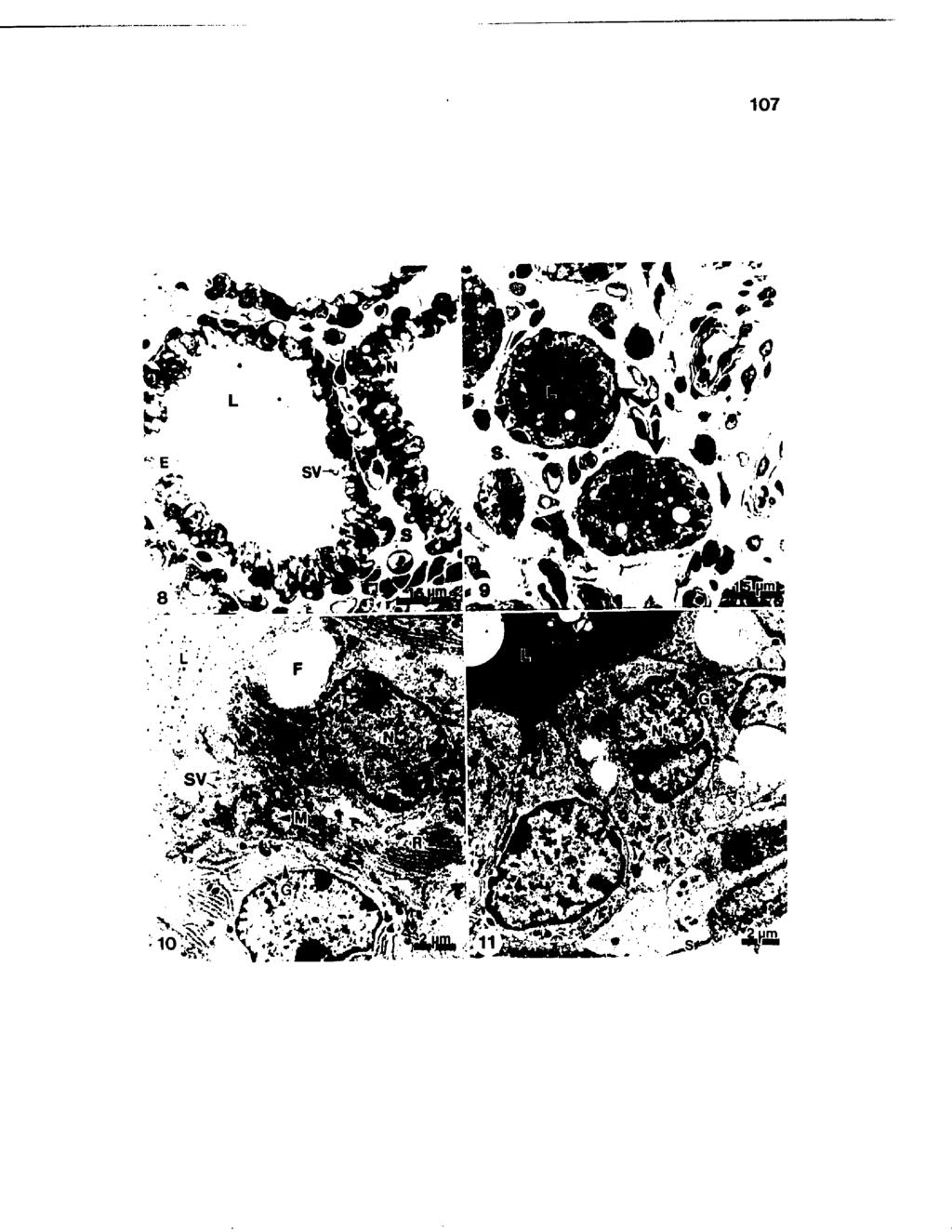
129 107
130 108 Fig. 12. Portion of an alveolus obtained at 7 days prior to calving demonstrating fluid accumulation. F, fat; G, Golgi apparatus; L, lumen; N, nucleus; S, stroma; and SV, secretory vesicle X 7,560.

131 . V f4. 109
132 110 Fig. 13. Corpora amylacea (CA) were most frequently observed filling the alveolar lumen. E, epithelium and S, stroma X 650. Fig. 14. Polymorphonuclear leukocytes (P) exhibited phagocytic vacuoles containing mammary secretion components (arrows), and macrophages (M) with internalized cellular debris (arrowheads) X 5,625.
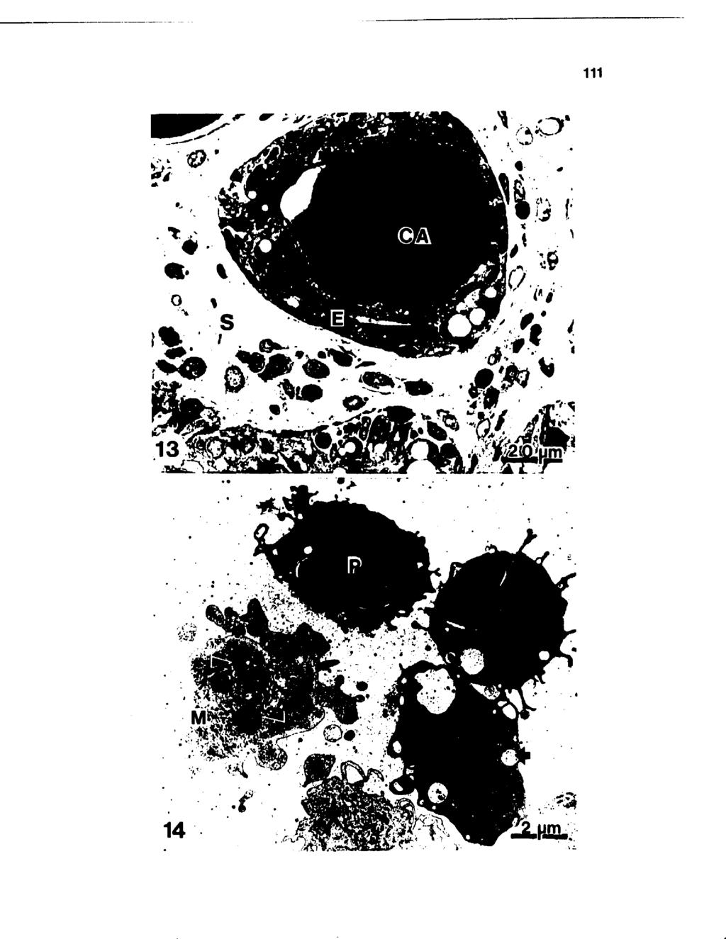
133 111
134 C H A P T E R IV Running head: Plasma Cell Isotypes in Mammary Tissue Key words: Involution, Lactogenesis, Plasma Cells, Mastitis, Mammary Biopsy, Immunohistochemistry, Immunoglobulin. QUANTIFICATION AND IMMUNOGLOBULIN CLASSIFICATION OF PLASMA CELLS IN NONLACTATING BOVINE MAMMARY TISSUE. L. M. SORDILLO and S. C. NICKERSON Louisiana Agricultural Experiment Station, Louisiana State University Agricultural Center, Hill Farm Research Station, Homer, Correspondence sent to: L. M. Sordillo-Gandy Department of Animal Science University of Tennessee P.O. Box 1071 Knoxville, Tennessee Approved for publication by the Director of the Louisiana Agricultural Experiment Station as manuscript number
135 113 Abatract Plasma cell populations in bovine mammary tissue were examined during involution using electron microscopic and immunohistochemical techniques. Biopsies were taken from each quarter of 5 Jersey cows at weekly intervals beginning at drying off through parturition. Plasma cells were observed proximal to alveolar epithelial cells. This association may facilitate transport of antibody through epithelium and into milk. Immunoglobulin-producing plasma cell numbers increased gradually from drying off, reached peak concentrations 2 wk prepartum, and dropped significantly during the last wk of gestation. Immunoglobulin G^- and G2_producing plasma cells were the most numerous isotypes observed during the nonlactating period followed by immunoglobulin M- and immunoglobulin A-producing plasma cells. Immunoglobulin M-producing plasma cells were more numerous in tissue infected with minor pathogens than uninfected quarters. Ultrastructural examination of these cells revealed rough endoplasmic reticulum cisternae engorged with flocculent material, indicative of antibody synthesis. Exposure to minor pathogens may have enhanced plasma cell proliferation and local antibody production in response to antigenic stimulation. Results of plasma cell distribution over the nonlactating period in bovine mammary tissue indicate times when local immunostimulation may be most effective in enhancing immunity to intramammary infection.
136 114 Introduction Immunoglobulins are an important soluble component of mammary immunity. Immunoglobulin (Ig) isotypes G ^, G2» and M opsonize bacteria for phagocytosis by leukocytes (10,16). Immunoglobulin A does not function as an opsonin, but has been implicated in toxin neutralization, bacterial agglutination, and preventing bacterial adhesion to cell membranes (15). Immunoglobulins in mammary secretion are derived from blood, or produced locally by plasma cells in the subepithelial connective tissue. Most IgG is serum-derived, whereas IgA and IgM are primarily of local origin (3, 7). In ruminants, IgG^ is the predominant isotype in mammary secretions for all stages of lactation and is transported selectively from blood to milk in the absence of udder inflammation (3). Production of IgA and IgM by plasma cells creates a concentration gradient adjacent to the basement membrane of alveoli and these Igs are transported in pinocytotic vesicles across mammary epithelia and released into milk (5,16). Systemic immunization of ruminants has been shown to reduce the severity of mastitis, but does not prevent occurrence of new intramammary infection (IMI) (14,17). Failure has been attributed to the blood-milk barrier which may prevent most circulating Ig from reaching milk (7). Local immunization to elevate Ig concentrations in lacteal secretions has proven to be a more effective means of enhancing udder defense mechanisms (10,17). Previous studies demonstrated the role of plasma cells in antibody production in
137 115 ruminant mammary glands (12,18). This locally synthesized Ig may function in udder defense against infection (9), but basic information is needed concerning the prevalence and Ig isotypes of plasma cells in mammary tissues. Immunocytochemical methods were used previously to demonstrate Ig-producing cells in normal mammary glands of cows (19), and during udder inflammation (11,18). However, no data are available describing changes in plasma cell isotype concentrations in the bovine mammary gland during the nonlactating period and in response to bacterial infection during this time. Because locally-synthesized Ig may provide a form of specialized immunological protection of bovine mammary tissue, this study examined changes in plasma cell populations during involution and through parturition to develop a basis for future attempts at local immunostimulation and enhancing Ig production.
138 116 Materials and Methods Experimental design Five Jersey cows from the Hill Farm Research Station were used. Approximately 5 ml of foremilk were collected aseptically prior to tissue collection and used to evaluate infection status. Mammary tissue samples were obtained by needle biopsy from each quarter at drying off, 7 and 14 days after drying off, and 14 and 7 days prior to parturition. At parturition, animals were slaughtered and 3 approximately 1 cm of tissue was obtained from each quarter. All tissue samples were processed for immunohistochemical and ultrastructural examination. Data were analyzed by least squares analysis of variance using the general linear model procedure to determine effects of wk during involution and infection status on each of the 4 Ig-producing plasma cell isotypes. The statistical analysis included effect of cow, wk during involution, infection status, and interaction of wk during involution by infection status. Dependent variables were IgG^-, IgG^-, IgA-, and IgM-producing plasma cells enumerated per unit tissue area. The following model was used: Y... = p + T x3.. + e.,. ijk i j ij ijk where: p = effect common to all cows, l = fixed effect of ith wk, 3j = fixed effect of jth infection status, x 3.. = fixed effect due to interaction between ith wk ij and jth infection status,
139 117 e,,, ijk = random error associated with each observation, The effect of cow was absorbed. Preplanned comparisons of least square means from the overall model were made by pairwise T-test. Means were contrasted between status group within a time period and between time period within a status group. No other comparisons were made. Microbiological procedures Quarter secretion samples were cultured, and presumptive identification of bacterial isolates was as described by Brown (2). Intramammary infection was defined as isolation of the same microorganism from duplicated foremilk samples. Tissue preparation Mammary tissues for immunohistochemical and ultrastructural examination were obtained and prepared for microscopy. Cows were anesthetized locally with 2 ml of 2% Lid-o-cane (Butler, Co., Columbus, OH). A small puncture was made through the skin with a scalpel blade above the gland cistern and approximately 3 cm x 2 mm of tissue was removed using a disposable biopsy needle (Travenol, Dallas, TX). Antibiotics were administered topically at the biopsy site. For light microscopy, biopsy samples and tissue samples obtained at slaughter were fixed for 48 h in 10% formalin buffered
140 118 to ph 7.4 with.025 M phosphate. Mammary tissues were prepared for sectioning by infiltration and embedding in Paraplast (American Scientific Products, McGaw Park, IL). For electron microscopy, biopsy samples and tissue sample obtained at slaughter were fixed in.1 M cacodylate buffered 2.5% glutaraldehyde, followed by.1 M cacodylate buffered osmium tetroxide, dehydrated in a graded series of ethanol, washed in propylene oxide, and embedded in epoxy resin. Tissue blocks were sectioned on an MT-5000 ultramicrotome, and 60 run thick sections stained with 5.0% uranyl acetate in 50% methanol for 20 min followed by.4% lead citrate for 10 min. Staining procedure for histochemical analysis Sections (2pm thick) were stained using a peroxidase-antiperoxidase (PAP) staining kit (Miles Scientific, Naperville, IL). Following deparaffinization, sections were treated with a 3% hydrogen peroxide solution to destroy endogenous peroxidase activity, and incubated at room temperature with normal serum to suppress nonspecific protein binding. A drop of 20% primary antibody (rabbit antibovine-igg^, IgG2» IgA, or IgM), provided by Dr. A. Guidry (USDA, Beltsville, MD), was placed on the section to react with the antigen in a humidity chamber for 30 min. Free antibody was washed off with.05 M Tris buffer (ph 7.6), and the link antibody (porcine antirabbit IgG) was added in excess to ensure one free binding site. Sections were washed again in.05 M Tris buffer, incubated for 20 min in PAP complex, and unbound PAP reagent was removed by washing in Tris buffer. Sections were then incubated for 40 min in a substrate solution (.3% hydrogen peroxide
141 119 In water and amino-ethyl-carbasole) forming a reddish precipitate at antigen sites. Sections were rinsed, counterstained in Mayer's hematoxylin, and coverslipped with glycerol-gelatin for microscopic examination. Morphometric analysis Quantitative morphometric analysis was used to enumerate Ig-producing plasma cells. Sections were observed with a Zeiss Standard 18 research microscope at X 1000 and plasma cells were / A counted in 10 randomly selected microscopic fields (6.0 x 10 2 pm /section). Tissue specimens were examined also for the location of plasma cells with respect to mammary epithelium and ultrastructural characteristics.
142 120 Results Frequencies of bacterial isolates during the sampling period are in Table 11. Because of the low numbers of isolates, quarters were classified as uninfected, infected (bacteriologically positive), infected with minor pathogens (coagulase negative staphylococci or Corynebacterium bovis), or infected with major pathogens ( Staphylococcus aureus, Streptococcus uberis. or Nocardia). Plasma cell numbers/unit area for IgG^, IgA, and IgM isotypes across uninfected and infected quarters increased significantly from D-0 to D+7, while IgG2 ~producing plasma cells increased significantly at D+14 (Table 12, Appendix Figures 24a-27a). Immunoglobulin Gj cells remained elevated through C-0. At C-7, numbers of IgG 2 _, IgA-, and IgM-producing plasma cells decreased significantly and remained relatively lower through C-0. Immunoglobulin G^- and IgG2 _producing plasma cells were the predominant isotypes observed throughout the sampling period, while IgA- and IgM-producing plasma cells were less abundant. No significant differences in numbers of IgG^-, IgG2 _, or IgA-producing plasma cells were detected between uninfected and infected quarters (Appendix Tables 26a and 27a). Mean number of IgM-producing plasma cells were significantly higher in quarters infected with minor pathogens compared to all other quarters (Table 13). Compared to uninfected quarters, those infected with minor pathogens had higher numbers of all plasma cell isotypes while those infected with major pathogens had lower numbers of these cells.
143 121 Examination of immunohistochemically stained tissues demonstrated that the majority of plasma cells were located in the subepithelial stroma proximal to, but not directly associated with the alveolar epithelium (Figures 15 and 16). Some plasma cells were observed lodged between the basal portions of epithelial cells lining alveoli (Figure 15). Electron microscopic examination of these cells revealed typical ultrastructure exhibited by antibody-producing plasma cells. The nuclei exhibited peripherally condensed chromatin and prominent nucleoli. The cytoplasm was filled with abundant, parallel arrays of rough endoplasmic reticulum (RER) which contained an electron-lucent substance (Figure 17).
144 122 Discussion This study demonstrated that the bovine mammary gland has the cellular machinery available for Ig synthesis during the dry period. Plasma cells were observed most frequently proximal to basal surfaces of epithelial cells lining alveolar lumina of nonlactating tissue. Ultrastructural examination of these cells exhibited the typical morphology of antibody-producing cells observed by others (12) with abundant and dilated RER cisternae containing granular or flocculent material previously identified as immunoglobulin. Others have noted the proximity of parenchymal plasma cells with epithelia (8,12) and suggested direct access for antibody transport through alveolar cells and into milk with minimal diffusion into surrounding connective tissue. Mean number of plasma cells for all Ig classes increased significantly from D-0 to D+7 and D+14, reaching peak concentrations at C-14, Increasing numbers of Ig-producing plasma cells with advancing pregnancy was shown also in the mammary glands of rats (8). Elevated concentrations of Ig in bovine mammary secretions were reported during the periparturient period (4). More recently, total IgG in mammary secretion from nonlactating cows was shown to also reach peak concentrations 2 wk prepartum (L. M. Sordillo et al., 1986, unpublished data). Increased numbers of Ig-producing cells at C-14 reported in this^ study supports the contention that the bovine mammary gland produces Ig locally as well as accumulates Ig from serum. Enhancing Ig concentrations during the last 2 wk of gestation by either stimulating plasma cell proliferation or increasing productivity of existing cells may help
145 123 to protect the gland when susceptibility to IMI is increased. Alternatively, because the bovine mammary gland is most susceptible to IMI during the first 3 wk of involution, local immunostimulation of mammary glands at drying off may result in elevated plasma cell populations when numbers are normally low. Yurchak et al. (19) used a fluorescent antibody technique to localize Ig-producing plasma cells in lactating bovine mammary tissue. He found that mammary tissue had mainly IgG-producing plasma cells while the other isotypes were present only in small numbers. In this study, IgG^ and IgG2 cells were also the predominant Numbers of isotypes throughout the entire nonlactating period. Ig-producing plasma cells in nonlactating bovine mammary tissue during colostrogenesis apparently reflect the concentrations of Ig in secretions of cows during this time. During the periparturient period, colostrum normally contains up to 150 mg Ig/ml of which IgG is about 85%, IgM about 7%, and IgA about 5% (3,A). Although most IgG is derived from blood, high concentrations of IgG cells in the underlying connective tissue may contribute to total concentrations in mammary secretion. Because most IgA and IgM are produced locally (3,5), lower levels of these Ig in mammary secretion may be explained by fewer IgA and IgM cells in the underlying connective tissue observed in this study. Prevalence of some cells may have resulted from local antigenic stimulation via the teat canal and/or following blast transformation induced in peripheral lymphoid tissue. No differences in numbers of IgG^-, IgG2 ", or IgA-producing plasma cells were detected between uninfected and infected quarters. These findings are consistent
146 124 with those of Nickerson and Heald (11) who found no significant difference in numbers of IgG- and IgA-staining plasma cells between S. aureus infected and control quarters. However, in quarters infected with minor pathogens, numbers of IgM-producing plasma cells were higher at most sampling times compared to all other quarters. Minor pathogens are isolated frequently from the bovine mammary gland and may produce elevated somatic cell counts. However, infections caused by minor pathogens go unnoticed often as they only produce a mild or subclinical form of mastitis. Boddie et al. (1) have shown a marked plasma cell response in tissues from quarters infected with coagulase negative staphylococci. Data from this study suggests colonization of the streak canal by these minor pathogens in previous lactations may have resulted in sensitization of mammary lymphocytes and subsequent proliferation into Ig-producing plasma cells. Conversely, lower numbers of Ig-producing plasma cells in quarters infected with major pathogens suggest depressed lymphocyte proliferation or compromised plasma cell function. Similar findings have been reported previously by Nonnecke and Harp (13). They found depressed lymphocyte response to mitogenic stimulation in quarters with chronic staphylococcal mastitis. Studies have shown that local immunization of bovine mammary glands with staphylococcal vaccines provided a substantial degree of protection against experimental staphylococcal challenge (17). Intramammary infusion of antigens during the dry period in sheep induced local production of Ig which persisted into the ensuing lactation (9). Most locally-produced Ig following antigenic
147 125 stimulation in ruminants was IgA and to a less extent, IgM (6,7,9). This study demonstrated higher IgM-producing plasma cell numbers in quarters infected with minor pathogens possibly due to longer periods of antigenic stimulation from teat canal colonization. Although relatively fewer numbers of IgA and IgM cells were demonstrated in nonlactating bovine mammary glands, numbers of these Ig-producing plasma cell isotypes peaked along with IgG^ and IgG2 cells approximately 2 wk prior to parturition. Importance of locally synthesized Ig in mammary defense against infection has been demonstrated (7). Low concentrations of Ig-producing plasma cells at D-0 may render the udder more susceptible to IMI at this time. Alternatively, high concentrations of Ig-producing plasma cells in the bovine mammary tissue 2 wk prepartum should provide a source of immunity during lactogenesis when incidence of new IMI is high. Local antigenic stimulation of nonlactating bovine mammary glands at D-0 and C-14 may stimulate and amplify, respectively, the local antibody response to mastitis pathogens and help protect against new IMI during the nonlactating and early lactating periods.
148 126 References 1. Boddie, R. L., S. C. Nickerson, W. E. Owens, and J. L. Watts Udder microflora in nonlactating heifers. Agri-practice (submitted). 2. Brown, R. W., D. A. Barn urn, D. E. Jasper, J. S. McDonald, and W. D. Schultze Microbiological procedures for use in the diagnasis of bovine mastitis. 2nd Ed., Natl. Mastitis Counc., Inc. Arlington VA. 3. Butler, J. E Immunoglobulins of the mammary secretions. Page 217 jui Lactation. B. L. Larson and V. R. Smith, ed. Academic Press, New York, NY. 4. Guidry, A. J., J. E. Butler, R. E. Pearson, and B. T. Weinland IgA, I g G p IgM, and BSA in serum and mammary secretion throughout lactation. Vet. Immunol. Immunopathol. 1: Larson, B. L., H. L. Leary, and J. E. Devery Immunoglobulin production and transport by the mammary gland. J. Dairy Sci. 63: Lascelles, A. K The immune system of the ruminant mammary gland and its role in the control of mastitis. J. Dairy Sci. 62: Lascelles, A. K., and G. H. McDowell Localized humoral immunity with particular reference to ruminants. Transplant. Rev. 19: Lee, C. G., P. W. Ladds, and D. L. Watson Immunocyte populations in the mammary gland of the rat at different stages of pregnancy and lactation. Res. Vet. Sci. 24:322.
149 McDowell, G. H., and A. K. Lascelles Local immunization of ewes with staphylococcal cell and cell-toxoid vaccines. Res. Vet. Sci. 12: Nickerson, S. C Immune mechanisms of the bovine udder: An overview. J. Am. Vet. Med. Assoc. 187: Nickerson, S. C. and C. W. Heald Cells in local reaction to experimental Staphylococcus aureus infection in bovine mammary tissue. J. Dairy Sci. 65: Nickerson, S. C., J. W. Pankey, and N. T, Boddie Distribution, location, and ultrastructure of plasma cells in the uninfected, lactating bovine mammary gland. J. Dairy Res. 51: Nonnecke, B. J., and J. A. Harp Effect of chronic staphylococcal mastitis on mitogenic responses of bovine lymphocytes. J. Dairy Sci. 68: Pankey, J. W., N. T. Boddie, J. L. Watts, and S. C. Nickerson Evaluation of protein A and a commercial bacterin as vaccines against Staphylococcus aureus using mastitis experimental challenge. J. Dairy Sci. 68: Sheldrake, R. F., and A. J. Husband Immune defences at mucosal surfaces in ruminants. J. Dairy Res. 52: Watson, D. L Immunological functions of the mammary gland and its secretion. Comparative review. Aust. J. Biol. Sci. 33:403.
150 Watson, D. L., and A. K. Lascelles The Influence of systemic immunization during mammary involution on subsequent antibody production in the mammary gland. Res. Vet. Sci. 18: Willoughby, R. A Bovine staphylococci mastitis: an immunohistochemical study of the cellular sites of antibody formation. Am. J. Vet. Res. 27: Yurchak, A. M., J. E. Butler, and T. B. Tomasi Flourescent localization of immunoglobulins in the tissues of the cow. J. Dairy Sci. 54:1324.
151 129 TABLE 11. Frequency of bacterial isolates from bovine mammary foremilk samples from drying off through lactogenesis. Sampling Period* Organism D-0 D+7 D+14 C- 14 C-7 C-0 None % N coagulase (-) % staphylococci N C. bovis % N S. aureus N S. uberis % N Nocardia % N *Days relative to drying off (D) or calving (C). %, Percentage of total quarters within each sampling period. N, Number of quarters within each sampling period.
152 130 TABLE 12. Enumeration^ o specific plasma cell classes (mean number of cells/6 x 10 pm tissue area) in the bovine mammary gland from drying off through lactogenesis in infected and uninfected quarters. Sampling Period^ Immunoglobulin Class D-0 D+7 D+14 C-14 C-7 C-0 igc1 X S.E. 7.39C llb a ab b b 6.30 IgG2 X S.E b b a a b llb 4.69 IgA X S.E b b a b b 3.30 IgM X S.E d c b a b bc 3.64 *Days relative to drying off (D) and calving (C). * means between sample period with different superscripts differ (P<.05).
153 131 TABLE 13. Effect of ^injection status on distribution (mean number of cells/6 x 10 pm tissue area) of specific plasma cell classes in the bovine mammary gland from drying off through lactogenesis. Infection Status* Immunoglobul in Class 0 INF MINOR MAJOR IgGj X a 28.47a 24.14a S.E Ib g 7 X 28.14a 26.65a 29.69a 24.75a Z S.E IgA IgM X 21.63a a 18.01a S.E X 23.47b 23.37b 34.62a 19.39b S.E Pathogens isolated at time of sampling: 0, uninfected; INF, infected: MINOR, Corynebacterium bovis and coagulase negative staphylococci; MAJOR, Staphylococcus aureus and streptococci. b Means within immunoglobulin class with different superscripts differ (PC.05).
154 132 FIGURE Nonlactating bovine mammary tiaaue atained immunohistochemically for presence of immunoglobulin Gj-producing plasma cells. (15) Positive staining of plasma cells (arrows) located in the subepithelial stroma (S) and within the alveolar epithelial (E) lining of mammary tissue involuted for 7 days. L, lumen X 1,460. (16) Clusters of positive staining plasma cells (arrows) within the stromal (S) area of mammary parenchyma obtained at parturition. E, epithelium X 650.
155 / * ' / V ' * $ * *. ir? 'fl. * v > * N i '. «V > t - «T», ' 10 um «* ' Vi r?; ' "<r>. v y 10 )jm
156 134 FIGURE 17. Clusters of plasma cells exhibiting abundant parallel lamellae of rough endoplasmic reticulum containing granulated, electron-lucent material (arrowheads). N, nucleus and S, stroma X 6,320.

157 vyy*-vr. 135
HISTOLOGY OF MAMMARY GLAND DURING LACTATING AND NON-LACTATING PHASES OF MADRAS RED SHEEP WITH SPECIAL REFERENCE TO INVOLUTION
 International Journal of Science, Environment and Technology, Vol. 5, No 3, 2016, 991 996 ISSN 2278-3687 (O) 2277-663X (P) HISTOLOGY OF MAMMARY GLAND DURING LACTATING AND NON-LACTATING PHASES OF MADRAS
Lactation. Macroscopic Anatomy of the Mammary Gland. Anatomy AS 1124
 Lactation AS 1124 Macroscopic Anatomy of the Mammary Gland Species differences in numbers and locations of glands inguinal - caudal to the abdomen, between the hind legs (cow, mare, ewe) abdominal - along
Understanding the Basics of Mastitis
 publication 404-233 Understanding the Basics of Mastitis G.M. Jones, Professor of Dairy Science and Extension Dairy Scientist, Milk Quality & Milking Management, Virginia Tech T.L. Bailey, Jr., Assistant
Mastitis: Background, Management and Control
 New York State Cattle Health Assurance Program Mastitis Module Mastitis: Background, Management and Control Introduction Mastitis remains one of the most costly diseases of dairy cattle in the US despite
Proper Dry-Off Procedures to Prevent New Infections and Cure Existing Cases of Mastitis. Stephen C. Nickerson University of Georgia
 Proper Dry-Off Procedures to Prevent New Infections and Cure Existing Cases of Mastitis Stephen C. Nickerson University of Georgia scn@uga.edu Michelle Arnold, DVM DABVP (Food Animal) Ruminant Extension
MILK COMPOSITIONAL CHANGES DURING MASTITIS
 MASTITIS PA R T 2 MILK COMPOSITIONAL CHANGES DURING MASTITIS Increased SCC Na Cl Whey protein (e.g. serum albumin, Ig, lactoferrin) Decreased Production α-lactalbumin & Lactose Casein K MILK LOSS LACTOFERRIN
Caused by microorganisms (usually bacteria) that invade the udder, multiply, and produce toxins that are harmful to the mammary gland
 that invade the udder, multiply, and produce toxins that are harmful to the mammary gland") MASTITIS PA R T 1 MASTITIS Mast = breast; itis = inflammation Inflammation of the mammary gland Caused by microorganisms (usually bacteria) that invade the udder, multiply, and produce toxins that are
MASTITIS PA R T 1 MASTITIS Mast = breast; itis = inflammation Inflammation of the mammary gland Caused by microorganisms (usually bacteria) that invade the udder, multiply, and produce toxins that are
MASTITIS. Therefore, mastitis is an inflammation of the mammary gland.
 MASTITIS Mastos = breast itis = inflammation Therefore, mastitis is an inflammation of the mammary gland. Or Reaction to a tissue injury. Therefore, inflammation can and does result in the loss of function
Mastitis in Unbred and Primigravid Dairy Heifers.
 Louisiana State University LSU Digital Commons LSU Historical Dissertations and Theses Graduate School 1989 Mastitis in Unbred and Primigravid Dairy Heifers. Pedro Trinidad Louisiana State University and
Mastitis MANAGING SOMATIC CELLS COUNTS IN. Somatic Cell Count Are Affected by. Somatic Cells are NOT Affected by:
 MANAGING SOMATIC CELLS COUNTS IN COWS AND HERDS Pamela L. Ruegg, DVM, MPVM University of Wisconsin, Madison Bacterial infection of the udder 99% occurs when bacterial exposure at teat end exceeds ability
Detection of Mastitis
 Detection of Mastitis Changes in milk composition Changes in milk composition Physical examination Signs of inflammation Empty udder Differences in firmness Unbalanced quarters Taste Test 60% of salty
Milk Quality Management Protocol: Fresh Cows
 Milk Quality Management Protocol: Fresh Cows By David L. Lee, Professor Rutgers Cooperative Extension Fresh Cow Milk Sampling Protocol: 1. Use the PortaSCC milk test or other on-farm mastitis test to check
The organism Infection process Tissue reaction SCC response Prevention Treatment
 Prevention and control of Staphylococcus aureus mastitis The organism Infection process Tissue reaction SCC response Prevention Treatment Staphylococcus aureus: Gram-positive Staphylo = Coccus = Cluster
TEAT DIP- POST DIP- PRE DIP- STRIPING
 TEAT DIP- POST DIP- PRE DIP- STRIPING KRISHIMATE AGRO AND DAIRY PVT LTD NO.1176, 1ST CROSS, 12TH B MAIN, H A L 2ND STAGE, INDIRANAGAR BANGALORE-560008, INDIA Email: sales@srisaiagro.com Www.srisaiagro.com
Dairy Science 305 Lactation Physiology Fall 2014 Syllabus
 Dairy Science 305 Lactation Physiology Fall 2014 Syllabus Instructor: Laura Hernandez, Ph.D. Office: 864 Animal Science Phone: 263-9867 Email: llhernandez@wisc.edu Office Hours: By appointment Lecture:
HISTOPHYSIOLOGICAL STUDIES ON THE HYPOPHYSIO- MAMMARY AXIS IN SHEEP (Ovis aries) - MAMMOTROPHS
 - MAMMOTROPHS") International Journal of Science, Environment and Technology, Vol. 5, No 3, 2016, 912 917 ISSN 2278-3687 (O) 2277-663X (P) HISTOPHYSIOLOGICAL STUDIES ON THE HYPOPHYSIO- MAMMARY AXIS IN SHEEP (Ovis aries)
International Journal of Science, Environment and Technology, Vol. 5, No 3, 2016, 912 917 ISSN 2278-3687 (O) 2277-663X (P) HISTOPHYSIOLOGICAL STUDIES ON THE HYPOPHYSIO- MAMMARY AXIS IN SHEEP (Ovis aries)
Mastitis in Dairy Goats 1
 DS 85 Mastitis in Dairy Goats 1 J. K. Shearer & B. Harris, Jr. 2 Mastitis is a general term which refers to inflammation of the mammary gland, regardless of cause. It is characterized by physical, chemical,
Management Practices and Intramammary Infections: New Ideas for an Old Problem
 Management Practices and Intramammary Infections: New Ideas for an Old Problem (Recent data from a pan-canadian study) Simon Dufour, Daniel Scholl, Anne-Marie Christen, Trevor DeVries University of Montreal,
Subclinical mastitis in small ruminants: prevalence, comparative aspects and prevention
 Subclinical mastitis in small ruminants: prevalence, comparative aspects and prevention Dr. Gabriel Leitner, National Mastitis Reference Center, Kimron Veterinary Institute, Israel Dr. Nissim Silanikove
ENVIRACOR J-5 aids in the control of clinical signs associated with Escherichia coli (E. coli) mastitis
 mastitis") GDR11136 ENVIRACOR J-5 aids in the control of clinical signs associated with Escherichia coli (E. coli) mastitis February 2012 Summary The challenge data presented in this technical bulletin was completed
GDR11136 ENVIRACOR J-5 aids in the control of clinical signs associated with Escherichia coli (E. coli) mastitis February 2012 Summary The challenge data presented in this technical bulletin was completed
Using SCC to Evaluate Subclinical Mastitis Cows
 Using SCC to Evaluate Subclinical Mastitis Cows By: Michele Jones and Donna M. Amaral-Phillips, Ph.D. Mastitis is the most important and costliest infectious disease on a dairy farm. A National Mastitis
Managing Mastitis in Dairy Heifers to Improve Overall Herd Health
 Managing Mastitis in Dairy Heifers to Improve Overall Herd Health Stephen C. Nickerson, Professor; Felicia M. Kautz, Research Associate; and Elizabeth L. Nace, Graduate Research Assistant Animal and Dairy
, Pamela L. Ruegg
 Premiums, Production and Pails of Discarded Milk How Much Money Does Mastitis Cost You? Pamela Ruegg, DVM, MPVM University of Wisconsin, Madison Introduction Profit centered dairy farms strive to maximize
Strep. ag.-infected Dairy Cows
 1 Mastitis Control Program for Strep. ag.-infected Dairy Cows by John Kirk Veterinary Medicine Extension, School of Veterinary Medicine University of California Davis and Roger Mellenberger Department
Use of a novel adjuvant to enhance the antibody response to vaccination against Staphylococcus aureus mastitis in dairy heifers.
 Use of a novel adjuvant to enhance the antibody response to vaccination against Staphylococcus aureus mastitis in dairy heifers. C. L. Hall, S. C. Nickerson, L.O. Ely, F. M. Kautz, and D. J. Hurley Abstract
DISSERTATION. Paige Nicole Gott, M.S. Graduate Program in Comparative and Veterinary Medicine. The Ohio State University. Dissertation Committee:
 Impact of milk cessation method on intramammary infections at calving and milk yield and quality in the subsequent lactation DISSERTATION Presented in Partial Fulfillment of the Requirements for the Degree
Herd Navigator and mastitis management
 Herd Navigator and mastitis management 1. What is mastitis? in some cases of E. coli mastitis the milk production in the affected Mastitis is the most common and costly disease in dairy herds. In quarter
New Developments in Mastitis Research
 New Developments in Mastitis Research Tine van Werven Department of Farm Animal Health, Utrecht University, the Netherlands University Farm Animal Practice, Harmelen, the Netherlands Email: t.vanwerven@uu.nl
Controlling Contagious Mastitis
 Controlling Contagious Mastitis John R. Middleton College of Veterinary Medicine, University of Missouri Quiz High SCC Objectives Definitions Causes Detection/Diagnosis Control Treatment Conclusion Definitions
DeLaval Cell Counter ICC User Strategies Guide
 Introduction 1. Bulk Tank Sampling Somatic cell count is one of the key indicators of udder health and has a major impact on milk production and farm costs. The DeLaval ICC mobile device allows for somatic
Guideline on the conduct of efficacy studies for intramammary products for use in cattle
 1 2 3 18 October 2013 EMEA/CVMP/EWP/141272/2011 Committee for Medicinal products for Veterinary Use (CVMP) 4 5 6 Guideline on the conduct of efficacy studies for intramammary products for use in cattle
Mastitis Management and SCC Control in Once a Day Herds. Don Crowley- Teagasc
 Mastitis Management and SCC Control in Once a Day Herds Don Crowley- Teagasc What is a SCC? Somatic cells (or body cells) are a mixture of milk-producing cells shed from the udder tissue (about 2%) and
Update on Staphylococcus aureus Mastitis. John R. Middleton College of Veterinary Medicine, University of Missouri, Columbia
 Update on Staphylococcus aureus Mastitis John R. Middleton College of Veterinary Medicine, University of Missouri, Columbia 1 Staphylococcus aureus Gram-positive, facultatively anaerobic, non-motile, non-sporulating,
29/11/2017. Best Milking Practices. Greg Strait- Fulton County Extension Amber Yutzy- Huntingdon County Extension
 Best Milking Practices Greg Strait- Fulton County Extension Amber Yutzy- Huntingdon County Extension 1 Milking is a complex interaction AND not likely related to ONE factor alone What is Mastitis? Bacterial
Premiums, Production and Pails of Discarded Milk How Much Money Does Mastitis Cost You? Pamela Ruegg, DVM, MPVM University of Wisconsin, Madison
 Premiums, Production and Pails of Discarded Milk How Much Money Does Mastitis Cost You? Pamela Ruegg, DVM, MPVM University of Wisconsin, Madison Introduction Profit centered dairy farms strive to maximize
University of Missouri Extension Using the California Mastitis Test
 University of Missouri Extension Using the California Mastitis Test Robert T. Marshall and J. E. Edmondson Department of Food Science and Nutrition Barry Steevens Department of Animal Sciences One of the
Mastitis Prevention and Cure Rates in Heifers Treated with Spectramast Dry Cow Therapy and/or Orbeseal Dry Cow Teat Sealant
 Mastitis Prevention and Cure Rates in Heifers Treated with Spectramast Dry Cow Therapy and/or Orbeseal Dry Cow Teat Sealant J. R. Booth, F. M. Kautz, and S. C. Nickerson Introduction: Dairy cows are vital
LOOKING FOR PROFITS IN MILK QUALITY
 LOOKING FOR PROFITS IN MILK QUALITY Richard L. Wallace TAKE HOME MESSAGES Begin monitoring milk quality practices by recording bulk tank data, DHIA somatic cell count (SCC) information, and clinical mastitis
MASTITIS CASE MANAGEMENT
 MASTITIS CASE MANAGEMENT The 2nd University of Minnesota China Dairy Conference Hohhot Sarne De Vliegher Head of M-team UGent & Mastitis and Milk Quality Research Unit @ UGent OVERVIEW Mastitis case management
Mastitis in ewes: towards development of a prevention and treatment plan
 SCHOOL OF LIFE SCIENCES, UNIVERSITY OF WARWICK Mastitis in ewes: towards development of a prevention and treatment plan Final Report Selene Huntley and Laura Green 1 Background to Project Mastitis is inflammation
DRY YOUR BEST. The incidence and importance of milk leakage in the dry cow. Ana I. de Prado Corporate Technical Manager Ceva Santé Animale (France)
") DRY YOUR BEST The incidence and importance of milk leakage in the dry cow Ana I. de Prado Corporate Technical Manager Ceva Santé Animale (France) The incidence of milk leakage is an indirect indicator
DRY YOUR BEST The incidence and importance of milk leakage in the dry cow Ana I. de Prado Corporate Technical Manager Ceva Santé Animale (France) The incidence of milk leakage is an indirect indicator
Interpretation of Bulk Tank Milk Results
 Interpretation of Bulk Tank Milk Results Introduction Culturing bulk tank milk (BTM) to monitor milk quality has limitations based on the amount and frequency of sampling and the amount and types of microorganisms
Influence of Experimentally- induced clinical mastitis on Reproductive Performance of Dairy Cattle
 Influence of Experimentally- induced clinical mastitis on Reproductive Performance of Dairy Cattle Dr. Mitch Hockett Department of Animal Science North Carolina State University Characteristics of Mastitis
Economic Review of Transition Cow Management
 Economic Review of Transition Cow Management John Fetrow VMD, MBA, DSc (hon) Emeritus Professor of Dairy Production Medicine College of Veterinary Medicine University of Minnesota This presentation is
Advanced Interherd Course
 Advanced Interherd Course Advanced Interherd Training Course... 2 Mastitis... 2 Seasonal trends in clinical mastitis... 2... 3 Examining clinical mastitis origins... 3... 4 Examining dry period performance
Field Efficacy of J-VAC Vaccines in the Prevention of Clinical Coliform Mastitis in Dairy Cattle
 Field Efficacy of J-VAC Vaccines in the Prevention of Clinical Coliform Masitis in Dairy.. Page 1 of 5 Related References: Field Efficacy of J-VAC Vaccines in the Prevention of Clinical Coliform Mastitis
Somatic Cell Count as an Indicator of Subclinical Mastitis. Genetic Parameters and Correlations with Clinical Mastitis
 Somatic Cell Count as an Indicator of Subclinical Mastitis. Genetic Parameters and Correlations with Clinical Mastitis Morten Svendsen 1 and Bjørg Heringstad 1,2 1 GENO Breeding and A.I. Association, P.O
MILK QUALITY PROGRAMS FOR TRANSITION COWS AND HEIFERS. Leo Timms Iowa State University, Ames IA
 MILK QUALITY PROGRAMS FOR TRANSITION COWS AND HEIFERS Leo Timms Iowa State University, Ames IA 50011 ltimms@iastate.edu TAKE HOME POINTS: Mastitis in transition cows and heifers can be a major contributor
SUMMARY OF PRODUCT CHARACTERISTICS. Lincomycin (as Lincomycin hydrochloride) Neomycin (as Neomycin sulphate) Excipients Disodium edetate
 Neomycin (as Neomycin sulphate) Excipients Disodium edetate") SUMMARY OF PRODUCT CHARACTERISTICS AN: 00221/2013 1. NAME OF THE VETERINARY MEDICINAL PRODUCT Lincocin Forte S Intramammary Solution 2. QUALITATIVE AND QUANTITATIVE COMPOSITION Active substances Lincomycin
SUMMARY OF PRODUCT CHARACTERISTICS AN: 00221/2013 1. NAME OF THE VETERINARY MEDICINAL PRODUCT Lincocin Forte S Intramammary Solution 2. QUALITATIVE AND QUANTITATIVE COMPOSITION Active substances Lincomycin
Milk Quality Evaluation Tools for Dairy Farmers
 AS-1131 Mastitis Control Programs Milk Quality Evaluation Tools for Dairy Farmers P J. W. Schroeder, Extension Dairy Specialist roducers have a variety of informational tools available to monitor both
IMMUNOSTIMULATING EFFECTS OF A COMMERCIAL FEED SUPPLEMENT IN DAIRY HEIFERS VACCINATED WITH A STAPHYLOCOCCUS AUREUS BACTERIN VALERIE JOANNE EUBANKS
 IMMUNOSTIMULATING EFFECTS OF A COMMERCIAL FEED SUPPLEMENT IN DAIRY HEIFERS VACCINATED WITH A STAPHYLOCOCCUS AUREUS BACTERIN by VALERIE JOANNE EUBANKS (Under the direction of Stephen C. Nickerson) ABSTRACT
Mastitis cows and immunization
 In Spain, the antibiotherapy against mastitis moves 12,000,000 with an interannual growth of 10.2%. Only 4 of these millions are drying antibiotherapy. Conclusion: farmers spend a lot of money on mastitis
Using DHIA and bacteriology to investigate herd milk quality problems.
 Using DHIA and bacteriology to investigate herd milk quality problems. Nigel B. Cook BVSc MRCVS Clinical Assistant Professor in Food Animal Production Medicine University of Wisconsin-Madison, School of
Course Curriculum for Master Degree Theriogenology & Artificial Insemination/Faculty of Veterinary Medicine
 Course Curriculum for Master Degree Theriogenology & Artificial Insemination/Faculty of Veterinary Medicine The Master Degree in Theriogenology & Artificial Insemination /Faculty of Veterinary Medicine
Walter M. Guterbock, DVM, MS Veterinary Medicine Teaching and Research Center University of California, Davis
 Walter M. Guterbock, DVM, MS Veterinary Medicine Teaching and Research Center University of California, Davis 1993 WESTERN LARGE HERD MANAGEMENT CONFERENCE V LAS VEGAS NEVADA 27 Alternatives To Antibiotic
Mastitis Module Risk Assessment Guide by Pathogen. Streptococcus agalactiae
 ! Mastitis Module Risk Assessment Guide by Pathogen Risk Factors Risk Information # Informational Statement! Intervention tactic Risk factors on this farm (level of implementation) Farm Feasibility Y,N
Check that milk is suitable to go in the vat
 TECHNOTE 3 Check that milk is suitable to go in the vat CALVING For milk quality reasons, all cows must have their colostrum milk withheld from the vat for at least eight milkings after calving. Colostrum
New Model. Digital Mastitis Detector. Reduce of risk at early stage
 New Model Digital Mastitis Detector Reduce of risk at early stage Introduction hat is mastitis?? inflammation f mammary gland in breast or udder. Due to bacterial infection via a damaged teat. Mastitis
Milk quality & mastitis - troubleshooting, control program
 Milk quality & mastitis - troubleshooting, control program Jim Reynolds, DVM, MPVM University of California, Davis Tulare Veterinary Medicine Teaching and Research Center 18830 Road 112 Tulare, CA 93274
Irish Medicines Board
 IRISH MEDICINES BOARD ACT 1995 EUROPEAN COMMUNITIES (ANIMAL REMEDIES) (No. 2) REGULATIONS 2007 (S.I. No. 786 of 2007) VPA: 10999/056/001 Case No: 7004318 The Irish Medicines Board in exercise of the powers
Differential Somatic Cell Count with the Fossomatic 7 DC - a novel parameter
 Differential Somatic Cell Count with the Fossomatic 7 DC - a novel parameter By: Dr. Daniel Schwarz, Cattle Disease Specialist, FOSS, Denmark Dedicated Analytical Solutions Somatic cell count (SCC) represents
Dairy/Milk Testing Report Detecting Elevated Levels of Bacteria in Milk-On-Site Direct- From-The-Cow Within Minutes as Indicator of Mastitis
 Dairy/Milk Testing Report Detecting Elevated Levels of Bacteria in Milk-On-Site Direct- From-The-Cow Within Minutes as Indicator of Mastitis EnZtek Diagnostics Incorporated has investigated and successfully
Course Curriculum for Master Degree in Internal Medicine/ Faculty of Veterinary Medicine
 Course Curriculum for Master Degree in Internal Medicine/ Faculty of Veterinary Medicine The Master Degree in Internal Medicine/Faculty of Veterinary Medicine is awarded by the Faculty of Graduate Studies
مادة االدوية المرحلة الثالثة م. غدير حاتم محمد
 م. مادة االدوية المرحلة الثالثة م. غدير حاتم محمد 2017-2016 ANTIMICROBIAL DRUGS Antimicrobial drugs Lecture 1 Antimicrobial Drugs Chemotherapy: The use of drugs to treat a disease. Antimicrobial drugs:
Gina M Pighetti & Raul Almeida. University of Tennessee
 Gina M Pighetti & Raul Almeida University of Tennessee Ultimate goal most vaccines Prevent infection Typically by increasing production of antibodies specific to an organism BUT, mastitis vaccines face
Presented at Central Veterinary Conference, Kansas City, MO, August 2013; Copyright 2013, P.L Ruegg, all rights reserved
 MILK MICROBIOLOGY: IMPROVING MICROBIOLOGICAL SERVICES FOR DAIRY FARMS Pamela L. Ruegg, DVM, MPVM, University of WI, Dept. of Dairy Science, Madison WI 53705 Introduction In spite of considerable progress
Prototheca Mastitis in Dairy Cows
 1 Mastitis Control Program for Prototheca Mastitis in Dairy Cows by John Kirk Veterinary Medicine Extension, School of Veterinary Medicine University of California Davis and Roger Mellenberger Department
Course Curriculum for Master Degree in Poultry Diseases/Veterinary Medicine
 Course Curriculum for Master Degree in Poultry Diseases/Veterinary Medicine The Master Degree in Poultry Diseases /Veterinary Medicine, is awarded by the Faculty of Graduate Studies at Jordan University
Transition cows have decreased immune function. The transition period. Inflammation, Immune Function, and the Transition Cow.
 Overview Inflammation, Immune Function, and the Transition Cow Barry Bradford Kansas State University Herd Health & Nutrition Conferences April 2016 Immunity and inflammation in the transition cow Long
Mastitis Causing Pathogens within the Dairy Cattle Environment
 Mastitis Causing Pathogens within the Dairy Cattle Environment Ayuba Caleb Kudi (Corresponding Author), M.P. Bray & Aziwo.T.Niba Faculty of Science, School of Biological Sciences, University of Plymouth
Ubroseal Dry Cow 2.6 g intramammary suspension for cattle
 Health Products Regulatory Authority 1 NAME OF THE VETERINARY MEDICINAL PRODUCT Ubroseal Dry Cow 2.6 g intramammary suspension for cattle 2 QUALITATIVE AND QUANTITATIVE COMPOSITION Each 4g intramammary
Case Study: Dairy farm reaps benefits from milk analysis technology
 Case Study: Dairy farm reaps benefits from milk analysis technology MARCH PETER AND SHELIA COX became the first dairy farmers in the UK to install a new advanced milk analysis tool. Since installing Herd
MATERIALS AND METHODS
 Effects of Feeding OmniGen-AF Beginning 6 Days Prior to Dry-Off on Mastitis Prevalence and Somatic Cell Counts in a Herd Experiencing Major Health Issues S. C. Nickerson 1, F. M. Kautz 1, L. O. Ely 1,
EPIDIDYMITIS IN RANGE
 Animal Health Fact Sheet July 1997 EPIDIDYMITIS IN RANGE AND PUREBRED RAMS Clell V. Bagley, DVM, Extension Veterinarian and Mark C. Healey, DVM, PhD Utah State University, Logan UT 84322-5600 AH/Sheep/14
Last 2-3 months of lactation
 Last 2-3 months of lactation Guideline 14 15 Decide dry cow management strategy Consider culling persistently infected cows CellCheck Farm CellCheck Guidelines Farm for Guidelines Mastitis Control for
2012 Indiana Regional Dairy Meetings. Purdue University College of Veterinary Medicine Dr. Jon Townsend Dairy Production Medicine
 2012 Indiana Regional Dairy Meetings Purdue University College of Veterinary Medicine Dr. Jon Townsend Dairy Production Medicine Focusing on the selection of the correct animals, diagnosis of causative
Dominance/Suppression Competitive Relationships in Loblolly Pine (Pinus taeda L.) Plantations
 Plantations") Dominance/Suppression Competitive Relationships in Loblolly Pine (Pinus taeda L.) Plantations by Michael E. Dyer Dissertation submitted to the Faculty of the Virginia Polytechnic Institute and Stand University
Dominance/Suppression Competitive Relationships in Loblolly Pine (Pinus taeda L.) Plantations by Michael E. Dyer Dissertation submitted to the Faculty of the Virginia Polytechnic Institute and Stand University
Milk Secretion and Quality Standards
 Introduction Milk Secretion and Quality Standards Pamela L. Ruegg, DVM, MPVM, Dip. ABVP-Dairy University of Wisconsin, Madison, USA The dairy industry is a large and dynamic segment of the agricultural
Effect of omitting post-milking teat disinfection on the mastitis infection rate of dairy cows over a full lactation
 57 th Annual Meeting of the European Association for Animal Production Antalya (Turkey), September 17-20, 2006 Session: M19 Free communications animal management and health Effect of omitting post-milking
Dairy Herdsman Certificate
 Dairy Herdsman Certificate Kindly supported by: Welcome to......the FarmSkills Dairy Herdsman Certificate Our programme of practical and hands-on workshops are led by our experienced vets and industry
Mastitis in non-bovine dairy species, companion animals and breastfeeding mothers. Chris Knight
 Mastitis in non-bovine dairy species, companion animals and breastfeeding mothers Chris Knight Objectives To stimulate thought/discussion regarding the relevance and importance of mastitis and mastitis
Chemotherapeutic Agents
 Chemotherapeutic Agents The cell is the basic structure of all living organisms. The cell membrane features specifi c receptor sites that allow interaction with various chemicals, histocompatibility proteins
SUMMARY OF PRODUCT CHARACTERISTICS
 SUMMARY OF PRODUCT CHARACTERISTICS 1. NAME OF THE VETERINARY MEDICINAL PRODUCT Marbocare 20 mg/ml solution for injection for cattle and pigs (UK, IE, FR) Odimar 20 mg/ml solution for injection for cattle
ANIMAL HUSBANDARY AND VETERINARY SCIENCE (CODE NO. 02) PAPER - I
 PAPER - I") ANIMAL HUSBANDARY AND VETERINARY SCIENCE (CODE NO. 02) PAPER - I 1. Animal Nutrition Metabolism of carbohydrates, proteins and fats, Requirements for maintenance, growth and production of milk, meat,work,
ANIMAL HUSBANDARY AND VETERINARY SCIENCE (CODE NO. 02) PAPER - I 1. Animal Nutrition Metabolism of carbohydrates, proteins and fats, Requirements for maintenance, growth and production of milk, meat,work,
Payback News. Beef Herd Nutrition Challenges
 August, 2015 Volume 2, Issue 3 CHS Nutrition Payback News In this issue of Payback News: Beef Herd Nutrition Challenges Impacts of Foot Rot in Feedlot Cattle Inside this issue: Beef Herd Nutrition Challenges
MSU Extension Publication Archive
 MSU Extension Publication Archive Archive copy of publication, do not use for current recommendations. Up-to-date information about many topics can be obtained from your local Extension office. Mastitis
Quad Plate User s Manual
 A part of Eurofins DQCI SSGN - SSGNC Mastitis Culture Quad Plate User s Manual Eurofins Microbiology Laboratories / Eurofins DQCI Services 5205 Quincy Street, Mounds View, MN 55112 P: 763-785-0485 F: 763-785-0584
Profitable Milk System
 INON Profitable Milk System We have developed a range of solutions that can help the dairy farmer maximize the profit potential of his dairy farm. Each of these products is based on more than 40 years
Mastitis control in bred heifers: Use of dry cow therapy and teat sealant for curing existing intramammary infections and preventing new ones
 Mastitis control in bred heifers: Use of dry cow therapy and teat sealant for curing existing intramammary infections and preventing new ones Abstract S. C. Nickerson, F. M. Kautz, A. E. Harding, J. R.
Prepared by Small Ruminants Task Force Lynn Hinckley, Task Force Director
 Direct Microscopic Examination of Milk From Small Ruminants Prepared by Small Ruminants Task Force Lynn Hinckley, Task Force Director Lead Author: Dan Scruton Frank Fillman, Lynn Hinckley, Debora Miller
Dairy Industry Overview. Management Practices Critical Control Points Diseases
 Dairy Industry Overview Management Practices Critical Control Points Diseases Instructor Contact Information: Hans Coetzee Office: I-107 I FAH&M Building Phone: 785-532 532-4143 Email: jcoetzee@vet.ksu.edu
Principles of Anti-Microbial Therapy Assistant Professor Naza M. Ali. Lec 1
 Principles of Anti-Microbial Therapy Assistant Professor Naza M. Ali Lec 1 28 Oct 2018 References Lippincott s IIIustrated Reviews / Pharmacology 6 th Edition Katzung and Trevor s Pharmacology / Examination
Innovation in Mastitis Treatment
 Innovation in Mastitis Treatment Dr Kiro R Petrovski DVM, MVSc, PGDipVCSc, PhD Senior Lecturer March 2014 kiro.petrovski@adelaide.edu.au Biography Started working with dairy cows at age of 11 First independent
April Boll Iowa State University. Leo L. Timms Iowa State University. Recommended Citation
 AS 652 ASL R2102 2006 Use of the California Mastitis Test and an On-Farm Culture System for Strategic Identification and Treatment of Fresh Cow Subclinical Intramammary Infections and Treatment of Clinical
Mastitis in Dairy. Cattle. Oregon State System of Higher Education Agricultural Experiment Station Oregon State College JOHN 0.
 STATION CIRCULAR 163 Mastitis in Dairy Cattle JOHN 0. SCHNAUTZ Oregon State System of Higher Education Agricultural Experiment Station Oregon State College Figure 1. Mastitis milk showing Streptococcus
Decision tree analysis of treatment strategies for mild and moderate cases of clinical mastitis occurring in early lactation
 J. Dairy Sci. 94 :1873 1892 doi: 10.3168/jds.2010-3930 American Dairy Science Association, 2011. Decision tree analysis of treatment strategies for mild and moderate cases of clinical mastitis occurring
PHYSIOLOGIC FACTORS THAT MODIFY THE EFFICIENCY OF MACHINE MILKING IN DAIRY EWES
 PHYSIOLOGIC FACTORS THAT MODIFY THE EFFICIENCY OF MACHINE MILKING IN DAIRY EWES B. C. McKusick Department of Animal Sciences University of Wisconsin-Madison Introduction Machine milking efficiency can
Liver and Gallbladder Morphology of the juvenile Nile crocodile, Crocodylus niloticus (Laurenti, 1768)
") Liver and Gallbladder Morphology of the juvenile Nile crocodile, Crocodylus niloticus (Laurenti, 1768) by ERNA VAN WILPE Submitted in partial fulfilment of the requirements for the degree MSc DEPARTMENT
Liver and Gallbladder Morphology of the juvenile Nile crocodile, Crocodylus niloticus (Laurenti, 1768) by ERNA VAN WILPE Submitted in partial fulfilment of the requirements for the degree MSc DEPARTMENT
Mobilization of neutrophils and defense of the bovine mammary gland
 Reprod. Nutr. Dev. 43 (2003) 439 457 439 INRA, EDP Sciences, 2004 DOI: 10.1051/rnd:2003031 Review Mobilization of neutrophils and defense of the bovine mammary gland Pascal RAINARD*, Céline RIOLLET Laboratoire
Mastitis and the link to infertility
 CONTINUING EDUCATION I LARGE ANIMAL Mastitis and the link to infertility Mastitis and infertility are the two most common disease complexes in dairy cattle worldwide. Both are major reasons for culling