Light and scanning electron microscopy of the tongue of the sand lizard (Lacerta agilis)
|
|
|
- Emma Jenkins
- 5 years ago
- Views:
Transcription
 Authors: Petr Cizek, Pavla Hamouzova, Pavel Kvapil, Michal")
1 Powered by TCPDF ( This is a provisional PDF only. Copyedited and fully formatted version will be made available soon. ONLINE FIRST ISSN: e-issn: Light and scanning electron microscopy of the tongue of the sand lizard (Lacerta agilis) Authors: Petr Cizek, Pavla Hamouzova, Pavel Kvapil, Michal Kyllar DOI: /FM.a Article type: ORIGINAL ARTICLES Submitted: Accepted: Published online: This article has been peer reviewed and published immediately upon acceptance. It is an open access article, which means that it can be downloaded, printed, and distributed freely, provided the work is properly cited. Articles in "Folia Morphologica" are listed in PubMed.
2 Light and scanning electron microscopy of the tongue of the sand lizard (Lacerta agilis) Running head: Structure of the sand lizard s tongue Petr Cizek 1, Pavla Hamouzova 1, Pavel Kvapil 2, Michal Kyllar 1,3 1 Department of Anatomy, Histology and Embryology, Faculty of Veterinary Medicine, University of Veterinary and Pharmaceutical Sciences Brno, Czech Republic 2 ZOO Ljubljana, Slovenia 3 Companion Care Vets, United Kingdom Address for correspondence: Petr Cizek, Department of Anatomy, Histology and Embryology, Faculty of Veterinary Medicine, University of Veterinary and Pharmaceutical Sciences Brno, Palackeho 1946/1, Brno, Czech Republic, tel: cizekpetr@yahoo.com Abstract Despite the fact that numerous reptile species are widely studied by the researchers, information describing the detailed structure of particular organs in many reptiles is missing. The tongue of the sand lizard (Lacerta agilis) was examined under the light and scanning electron microscope. It is divided into bifurcated apex, corpus and bifurcated radix. The tip of the lingual apex is devoid of lingual papillae. The remaining dorsal surface of the tongue bears either fused papillae in the form of caudally directed ridges or individual papillae represented by mushroom like or semilunar prominences (lingual apex) or fish scale-like papillae (lingual corpus) and horizontally laid ridges extending in the form of lobulated prominences (lingual corpus, lingual radix). Regardless of the shape, lingual papillae contain numerous muscle fibers and they are all considered to be mechanical. The lingual epithelium changes from the simple squamous into stratified
3 squamous in the caudal direction. No salivary glands or sensory structures were recognised. This description is to be used mainly for comparative studies. It could also help to understand how different lizards capture the pray. Keywords: lingual papillae, Lacertidae, morphology, reptiles, SEM Introduction The tongue is a muscular organ located in the oral cavity. The type of food intake and processing of food in animals are important factors that affect the morphological diversity of structures on the surface of the tongue. The tongue plays a prominent role in the intra-oral transport and deglutition of food. This important role has led to morphological studies of several animals tongues to examine differences and similarities among them in terms of feeding habits and also to identify relations among diverse species [4]. Lacerta agilis is a small to medium sized lizard, strongly diurnal, largely insectivorous, actively chasing and consuming a range of spiders and insects, especially orthopterans, bugs and beetles. This species is also known to eat its own young [7]. The morphology of the tongue has been described in various species of turtles [e.g. 12, 14, 15, 19]. In lizards, the lingual morphology is still unknown in many species. The lack of detailed description in specific species has hindered the understanding of the functional morphology of the lizard tongue, very important for the description of lingual pathologies. Therefore the aim of the present study was to describe the morphology of the tongue of the sand lizard by light and scanning electron microscopy. The results are helpful to better understand the food processing and also the medical approach in the oral cavity disorders in lizards. The tongue of squamates is connected to the floor of the oral cavity and resembles the flat and less keratinized tongue of some amphibians. Some lizards, as well as turtles, cannot protrude their tongue. Despite this fact, the lingual apex of many lizards is forked and the lingual radix also extends distally in the form of two processes [6]. However, the tongue of the rough tailed rock agama (Laudakia stellio) is not forked [16].
4 The dorsal surface of the tip of the tongue is smooth in many lizards such as the green anole, Anolis carolinensis [18], blue-tongued skink, Tiliqua scincoides [2], Japanese lizard, Takydromus tachydromoides [13], bearded dragon, Pogona vitticeps [21], Italian lizard, Podarcis sicula [3], rough tailed rock agama, Laudakia stellio [16] and green iguana, Iguana iguana [1, 6]. Cylindriform papillae were distinguished on the dorsal lingual surface in the green anole, Anolis carolinensis [18], blue-tongued skink, Tiliqua scincoides [2], rough tailed rock agama, Laudakia stellio [16], green iguana, Iguana iguana [1, 6], Madagascan collared iguana, Oplurus cuvieri [8], Italian lizard, Podarcis sicula [3] and bearded dragon, Pogona vitticeps [21]. Plumose papillae were described in the green anole, Anolis carolinensis [18] and bearded dragon, Pogona vitticeps [21]. The plumose papillae may be important in mastication and deglutition of food [18]. Foliate-like papillae were found in the Italian lizard, Podarcis sicula [3] and blue-tongued skink, Tiliqua scincoides [2], conical papillae were described in the Madagascan collared iguana, Oplurus cuvieri [8], green iguana, Iguana iguana [1], conical flattened filiform papillae in the ringed wall gecko, Tarentola annularis [9], fungiform papillae in the rough tailed rock agama, Laudakia stellio [16] and scale-like papillae in the Japanese lizard, Takydromus tachydromoides [13], Tsinling dwarf skink, Scincella tsinlingensis [20] and Gekko japonicus [11]. Regardless of type, all lingual papillae of the green anole (Anolis carolinensis) contain a single longitudinally oriented skeletal muscle fiber that originates from the underlying lingual muscles [18]. Materials and methods Animals Three adult males and one female sand lizard (Lacerta agilis) used in this study were born and kept in a zoological collection. Their age varied between four and six years. The animals were fed with a balanced diet that consisted mostly of insects and vitaminmineral supplements. All the animals died or were euthanized due to various reasons related to old age (internal organ failure, neoplasia etc.) at the veterinary ambulance of
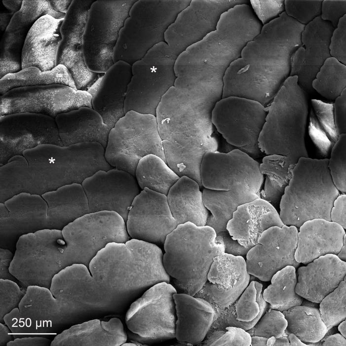
5 Ljubljana ZOO. None of the animals used in this study suffered from any of the oral cavity disorders. Light microscopy The tongues were fixed in 4 % neutral buffered formaldehyde for 5-7 days. After fixation had been completed, samples for light microscopy were dehydrated in a graded alcohol series (the ethanol concentration in each subsequent bath was increased by 10 %), acetone, and three baths of xylene. At the end of dehydration process, samples were infiltrated with hot paraffin and embedded in paraffin wax. Then, 3-4 μm thin sections were cut in the routine manner. The sections were dried, stained with haematoxylin and eosin, mounted and examined, and finally photographed under an Olympus BX51 light microscope using an Olympus DP70 digital camera. Scanning electron microscopy The samples for scanning electron microscopy were dehydrated in a graded alcohol series (60, 70, 96 and 100 %, 20 min for each concentration), transferred to pure acetone, dried at the critical point (Bal-tec CPD 030 Critical Point Dryer, Bal-Tec, UK), coated with gold (Balzers SCD 040 using a current 30 ma for 4 min.) and finally examined and photographed under a Tescan VEGA TS 5136 XM scanning electron microscope in a high vacuum and accelerated voltage 20 kv by using an SE detector. Results The tongue of the sand lizard (Fig. 1) is dorsoventrally flattened and roughly wedge shaped. It is divided into three areas bifurcated apex, corpus and bifurcated radix. The tip of the lingual apex (Fig. 2) is relatively smooth and devoid of papillae. Approximately in the middle of the bifurcated apex, caudally directed ridges are formed. These ridges then separate into individual papillae (Fig. 3). Each papilla is represented by a mushroom like prominence and measures around 100 µm in diameter. Papillae covering the margin of the apex of the tongue are different. On its lateral surface, these papillae tend to be bigger.
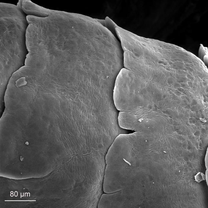
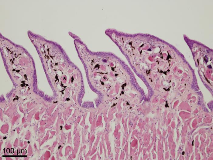
6 Medially they appear as a few semilunar prominences. Regardless the shapes of the papillae, the interpapillary grooves are rather inconspicuous. The papillae in the rostral part of the lingual body (Fig. 4) are rostrocaudally flattened. Each papilla bends caudally and extends in the form of several lobulated prominences. Their shape resembles a fish scale. Papillae covering the lingual body are roughly twice as big compared to the papillae on the apex, i.e. around 200 µm. On the margin of the lingual body, the papillae are even more flattened and their appearance changes into horizontally laid, long, narrow prominences that remarkably resemble a few merged papillae (Fig 5). These papillary ridges extend in the form of similar lobulated prominences as the papillae in the medial area of the lingual body. Some of these lobes are separated by clearly evident notches. In the caudal direction medially (Fig. 6), papillary ridges elongate so they cover the entire caudodorsal lingual surface of the lingual body. Subsequently, individual scale-like papillae gradually disappear. The lingual radix (Fig. 7) is bifurcated as well as the lingual apex. Its margin is more rounded compared to the proximal portions of the tongue. The dorsal lingual surface bears the same papillary ridges that are found on the lingual body (Fig. 8). The interpapillary notches are very deep. The superficial cells covering all the various papillae of the dorsal lingual surface are densely covered with microvilli. A simple squamous epithelium covers the lingual papillae on the apex. Aborally, the number of layers of epithelial cells increases. Nonkeratinized stratified squamous epithelium is on the surface of papillae on the body and radix. Papillary cores are formed by the proper connective tissue and numerous striated muscle fibers that originate from the underlying proper lingual muscle (Fig. 9, Fig. 10). This muscle is arranged in well-defined fascicles of the horizontal, longitudinal, as well as transversal course. Light microscopy also reveals the presence of melanin throughout the loose connective tissue of the tongue. The melanin granules are particularly abundant in the papillary cores. No serous or mucous lingual glands, as well as sensory structures were found. No sensory papillae with taste buds were found so all described papillae were considered to be mechanical.
7 Discussion The tongue of the sand lizard resembles the tongue of the Podarcis sicula in its shape, as well as distribution and type of the lingual papillae [3]. Both these saurian animals, belonging to the Family of Lacertidae, are largely insectivorous [7] and thus this similarity reflects their feeding habits. The observation of the tongue of the L. agilis revealed that the organ is divided into three different areas, i.e. apex, body and root. Its lingual apex is bifurcated, as has been described in the Iguana iguana [1, 6], Podarcis sicula [3], Takydromus tachydromoides [13], Scincella tsinlingensis [20] and Pogona vitticeps [21]. Thus, shaping of the lingual apex can be related to an easier access to the vomeronasal organ. The smooth dorsal surface of the tip of the tongue has also been described in Anolis carolinensis [18], Tiliqua scincoides [2], Takydromus tachydromoides [13], Pogona vitticeps [21], Podarcis sicula [3], Laudakia stellio [16] and Iguana iguana [1, 6]. The reason of such an appearance of the lingual apex remains unknown among the researchers so requires further investigation. Described scale-like papillae on the surface of the lingual body were similar to those found in Takydromus tachydromoides, Lacertidae [13], Scincella tsinlingensis, Scincidae [20] and Gekko japonicus, Gekkonidae [11]. All of these species, as well as L. agilis, are mainly insectivores so such shaping of the papillae possibly helps the food intake and its processing. It follows from the above mentioned comparison of species that the microscopic structure of the tongue in lizards is not a characteristic feature of the specific Families. A more important factor is the type of diet or the way of capturing prey. Skeletal muscle fibers in the cores of the papillae were also described in Anolis carolinensis. Some authors suspect the papillae can be moved at will [18], although it is hard to imagine moving individual lingual papillae without a relationship to the remaining musculature of the particular area of the tongue.
8 The epithelium of the dorsal lingual surface is nonkeratinized stratified squamous, as was reported in Anolis carolinensis [18], Scincella tsinlingensis [20], Pogona vitticeps [21], Tarentola annularis [9], Podarcis sicula [3], Iguana iguana [6], Laudakia stellio [16] and Gekko japonicus [11]. This suggests that the occurrence of this type of the lingual epithelium is typical in lizards. A single layer of columnar epithelial cells was reported in Gekko japonicus [11]. Nevertheless, increasing numbers of epithelial layers in the rostrocaudal direction has not been described yet. Lingual glands were identified in some species of lizards such as Pogona vitticeps [21], Laudakia stellio [16] and Scincella tsinlingensis [20]. Similarly, as in L. agilis, no glands were described in Iguana iguana [6]. It is stated that the salivary glands are located within different structures bordering the oral cavity. The presence of melanin throughout the tongue was also described in Tiliqua scincoides [2]. No taste buds were found on the tongue of the sand lizard. Nevertheless, the taste buds in reptiles are often located in different parts of the oral cavity. In Podarcis sicula, no sensory structures were found on the dorsal lingual surface, however, numerous taste buds were present on the gingival epithelium [3]. No taste buds were found also on the lingual surface in Tiliqua scincoides [2]. In the Malayan box turtle (Cuora amboinensis), the taste buds were distributed predominantlyon the rostral (prechoanal) part of the palate [10]. In the two-colored blind snake (Leptotyphlops bicolor) and Texas blind snake (Leptotyphlops dulcis), the taste buds were located only in the mucosa in the vicinity of the vomeronasal organ [5]. On the contrary, the presence of the taste buds in the conical filiform papillae was reported in Tarentola annularis [9]. In the Japanese four-lined ratsnake (Elaphe quadrivirgata), the individual taste buds were located on the sensory papillae, one of which always contained just a single taste bud [17]. It can be assumed that the taste buds in the sand lizard may be located in the other areas of the oral cavity, as was described in the above mentioned reptiles. Understanding the morphology of the tongue of the sand lizard completes the knowledge of the oral structures in different reptile species. It could also help to explain how different lizards capture the prey.
9 Conclusion The tongue of the sand lizard is divided into bifurcated apex, corpus and bifurcated radix. Except of the tip of the lingual apex, the dorsal surface of the tongue bears papillae. The lingual epithelium changes from the simple squamous into stratified squamous in the caudal direction. No salivary glands or sensory structures were recognised. This description is to be used mainly for comparative studies. It could also help to understand how different lizards capture the pray. References 1. Abbate F, Latella G, Montalbano G, Guerrera MC, Levanti MB, Ciriaco E. Scanning electron microscopical study of the lingual epithelium of Green iguana (Iguana iguana). Anat Histol Embryol. 2008; 37: , doi: /j x. 2. Abbate F, Latella G, Montalbano G, Guerrera MC, Germana GP, Levanti MB. The lingual dorsal surface of the blue-tongue skink (Tiliqua scincoides). Anat Histol Embryol. 2009; 38: , doi: /j x. 3. Abbate F, Guerrera MC, Montalbano G, Zichichi R, Germana A, Ciriaco E. Morphology of the lingual dorsal surface and oral taste buds in Italian lizard (Podarcis sicula). Anat Histol Embryol. 2010; 39: , doi: /j x. 4. Akbari G, Babaei M, Hassanzadeh B. Morphological study of the European hedgehog (Erinaceus europaeus) tongue by SEM and LM. Anat Sci Int. 2017;doi: /s Berkhoudt H, Wilson P, Young B. Taste buds in the palatal mucosa of snakes. Afr Zool. 2001; 36: Cizek P, Krejcirova L, Kocianova I, Tichy F. Light and scanning electron microscopy of the developing lingual papillae in the green iguana, Iguana iguana. Vet Med (Praha). 2011; 56:
10 7. Corbett KF & Tamarind DL. Conservation of the sand lizard, Lacerta agilis, by habitat management. Brit J Herpetol. 1979; 5: Delheusy V, Toubeau G, Bels VL (1994). Tongue structure and function in Oplurus cuvieri (Reptilia: Iguanidae). Anat Rec. 1994; 238: El-Sayyad HIH, Sabry DA, Khalifa SA, Abou-El-Naga AM, Foda YA (2011). Studies on tongue of reptilian species Psammophis sibilans, Tarentola annularis and Crocodylus niloticus. Int J Morphol. 2011; 29: Heiss E, Plenk H, Weisgram J. Microanatomy of the palatal mucosa of the semiaquatic Malayan box turtle, Cuora amboinensis, and functional implications. Anat Rec. 2008; 291: , doi: /ar Iwasaki S. Fine structure of the dorsal lingual epithelium of the lizard, Gekko japonicus (Lacertilia, Gekkonidae). Am J Anat. 1990; 187: Iwasaki S. Fine structure of the dorsal lingual epithelium of the tongue of the freshwater turtle, Geoclemys reevesii (Chelonia, Emydinae). J Morphol 1992; 211: Iwasaki S & Miyata K. Scanning electron microscopy of the dorsal lingual surface of the Japanese lizard, Takydromus tachydromoides. Okajimas Folia Anat Jpn. 1985; 62: Iwasaki S, Asami T, Asami Y, Kobayashi K. Fine structure of the dorsal epithelium of the tongue of the Japanese terrapin, Clemmys japonica (Chelonia, Emydinae). Arch Histol Cytol. 1992; 55: Iwasaki S, Wanichanon C, Asami T. Histological and ultrastructural study of the lingual epithelium of the juvenile Pacific ridley turtle, Lepidochelys olivacea (Chelonia, Cheloniidae). Ann Anat. 1996; 178: Koca YB, Oguz EO, Osanc E. Morphology, and muscle- and papilla-volume ratios, of the tongue of Laudakia stellio (Agamidae, Squamata): a histological and stereological study. Zool Sci. 2007; 24: , doi: /zsj
11 17. Nishida Y, Yoshie S, Fujita T. Oral sensory papillae, chemo- and mechano-receptors, in the snake, Elaphe quadrivirgata. A light and electron microscopic study. Arch Histol Cytol. 2000; 63:55 70, doi: /aohc Rabinowitz T & Tandler, B. Papillary morphology of the tongue of the American chameleon: Anolis carolinensis. Anat Rec. 1986; 216: Winokur RM. The buccopharyngeal mucosa of the turtles (testudines). J Morphol. 1988; 196: Yang C & Wang LM. Histological and morphological observations on tongue of Scincella tsinlingensis (Reptilia, Squamata, Scincidae). Micron 2016; 80:24 33, doi: /j.micron Zghikh LN, Vangysel E, Nonclercq D, Legrand A., Blairon B, Berri C, Bordeau T, Remy C, Burtea C, Montuelle SJ, Bels V. Morphology and fibre-type distribution in the tongue of the Pogona vitticeps lizard (Iguania, Agamidae). J Anat. 2014; 225: , doi: /joa LEGENDS OF FIGURES Fig. 1 Tongue of a sand lizard. LA - lingual apex, LC - lingual corpus, LR - lingual radix. Fig. 2 Lingual apex. Asterisks indicate caudally directed ridges. Fig. 3 Lingual apex. Detail. Ridges separate into individual papillae. Fig. 4 Lingual corpus. Caudally bent papillae on its rostral portion. Fig. 5 Lingual corpus. Papillae on its margin. Fig. 6 Lingual corpus. Papillary ridges on its distal portion. Fig. 7 Lingual radix. Papillary ridges.


12 Fig. 8 Lingual radix. Detail. Fig. 9 Lingual corpus. Caudally bent papillae containing striated muscle fibers. Fig. 10 Lingual corpus. Detail. Asterisks indicate striated muscle fibers in the papillary cores. Fig. 1


13 Fig. 2 Fig. 3 Fig. 4

14 Fig. 5 Fig. 6

15 Fig. 7 Fig. 8

16 Fig. 9 Fig. 10
Journal of American Science, 2012;8(2)
") Comparative Histological and Ultrastructural Study of the Tongue in Ptyodactylus guttatus and Stenodactylus petrii (Lacertilia, Gekkonidae) Samah T. Darwish Biological & geological department, Arish Faculty
Comparative Histological and Ultrastructural Study of the Tongue in Ptyodactylus guttatus and Stenodactylus petrii (Lacertilia, Gekkonidae) Samah T. Darwish Biological & geological department, Arish Faculty
A comparison of placental tissue in the skinks Eulamprus tympanum and E. quoyii. Yates, Lauren A.
 A comparison of placental tissue in the skinks Eulamprus tympanum and E. quoyii Yates, Lauren A. Abstract: The species Eulamprus tympanum and Eulamprus quoyii are viviparous skinks that are said to have
Fine structure of the dorsal lingual epithelium in Tarentola annularis and Crocodylus niloticus
 O R I G I N A L A R T I C L E Folia Morphol. Vol. 75, No. 2, pp. 162 172 DOI: 10.5603/FM.a2015.0091 Copyright 2016 Via Medica ISSN 0015 5659 www.fm.viamedica.pl Fine structure of the dorsal lingual epithelium
Studies on Tongue of Reptilian Species Psammophis sibilans, Tarentola annularis and Crocodylus niloticus
 Int. J. Morphol., 29(4):1139-1147, 2011. Studies on Tongue of Reptilian Species Psammophis sibilans, Tarentola annularis and Crocodylus niloticus Estudios sobre la Lengua de las Especies de Reptiles Psammophis
Title. CitationJapanese Journal of Veterinary Research, 24(1-2): 37. Issue Date DOI. Doc URL. Type. File Information
: 37. Issue Date DOI. Doc URL. Type. File Information") Title DISTRIBUTION OF LYMPHATIC TISSUES IN DUCK CAECA Author(s)KITAMURA, Hirokazu; SUGIMURA, Makoto; HASHIMOTO, Yos CitationJapanese Journal of Veterinary Research, 24(1-2): 37 Issue Date 1976-05 DOI 10.14943/jjvr.24.1-2.37
Title DISTRIBUTION OF LYMPHATIC TISSUES IN DUCK CAECA Author(s)KITAMURA, Hirokazu; SUGIMURA, Makoto; HASHIMOTO, Yos CitationJapanese Journal of Veterinary Research, 24(1-2): 37 Issue Date 1976-05 DOI 10.14943/jjvr.24.1-2.37
Microscopy: advances in scientific research and education (A. Méndez-Vilas, Ed.)
") Scanning electron microscopy investigation of the filter-feeding apparatus in the domestic goose (Anser anser f. domestica) and the domestic duck (Anas platyrhynchos f. domestica) K. Skieresz-Szewczyk
Scanning electron microscopy investigation of the filter-feeding apparatus in the domestic goose (Anser anser f. domestica) and the domestic duck (Anas platyrhynchos f. domestica) K. Skieresz-Szewczyk
A Scanning Electron Microscopic Study of Eggshell Surface Topography of Leidynema portentosae and L. appendiculatum (Nematoda: Oxyuroidea)
") The Ohio State University Knowledge Bank kb.osu.edu Ohio Journal of Science (Ohio Academy of Science) Ohio Journal of Science: Volume 88, Issue 5 (December, 1988) 1988-12 A Scanning Electron Microscopic
The Ohio State University Knowledge Bank kb.osu.edu Ohio Journal of Science (Ohio Academy of Science) Ohio Journal of Science: Volume 88, Issue 5 (December, 1988) 1988-12 A Scanning Electron Microscopic
QuickTime and a TIFF (Uncompressed) decompressor are needed to see this picture.
 decompressor are needed to see this picture.") QuickTime and a TIFF (Uncompressed) decompressor are needed to see this picture. QuickTime and a Sorenson Video 3 decompressor are needed to see this picture. QuickTime and a Sorenson Video
QuickTime and a TIFF (Uncompressed) decompressor are needed to see this picture. QuickTime and a Sorenson Video 3 decompressor are needed to see this picture. QuickTime and a Sorenson Video
Light and scanning electron microscopic observations on the tongue of Nile monitor, Varanus niloticus niloticus
 International Journal of Advanced Research in Biological Sciences ISSN: 2348-8069 www.ijarbs.com DOI: 10.22192/ijarbs Coden: IJARQG(USA) Volume 5, Issue 4-2018 Research Article DOI: http://dx.doi.org/10.22192/ijarbs.2018.05.04.001
An Examination of the Sensory Structures in the Oral Cavity of the American Alligator (Alligator mississippiensis)
") JOURNAL OF MORPHOLOGY 275:1312 1320 (2014) An Examination of the Sensory Structures in the Oral Cavity of the American Alligator (Alligator mississippiensis) Susan J. Rehorek, 1 * Michael Duffy, 1 Janelle
JOURNAL OF MORPHOLOGY 275:1312 1320 (2014) An Examination of the Sensory Structures in the Oral Cavity of the American Alligator (Alligator mississippiensis) Susan J. Rehorek, 1 * Michael Duffy, 1 Janelle
Morphological, histological and ultrastructural (sem) characterization of the egyptian tortoise s tongue
 characterization of the egyptian tortoise s tongue") International Journal of Zoology Studies ISSN: 2455-7269 Impact Factor: RJIF 5.14 www.zoologyjournals.com Volume 3; Issue 2; March 2018; Page No. 101-111 Morphological, histological and ultrastructural
International Journal of Zoology Studies ISSN: 2455-7269 Impact Factor: RJIF 5.14 www.zoologyjournals.com Volume 3; Issue 2; March 2018; Page No. 101-111 Morphological, histological and ultrastructural
Lab VII. Tuatara, Lizards, and Amphisbaenids
 Lab VII Tuatara, Lizards, and Amphisbaenids Project Reminder Don t forget about your project! Written Proposals due and Presentations are given on 4/21!! Abby and Sarah will read over your written proposal
Seasonal Variations of yeso sika Deer Skin and its Vegetable Tanned Leather
 Seasonal Variations of yeso sika Deer Skin and its Vegetable Tanned Leather Shigeharu Fukunaga, Akihiko Yoshie, Ikuo Yamakawa, Fumio Nakamura Laboratory of Animal By-product Science, Graduate School of
HISTOPATHOLOGY. Introduction:
 Introduction: HISTOPATHOLOGY Goats and sheep are the major domestic animal species in India. Much of the economy of the country has been depend upon the domestication of these animals. Especially economy
Shannon Martinson, BSc, DVM, MVSc, DACVP Department of Pathology and Microbiology Atlantic Veterinary College, University of Prince Edward Island
 Shannon Martinson, BSc, DVM, MVSc, DACVP Department of Pathology and Microbiology Atlantic Veterinary College, University of Prince Edward Island Reptile pathology: Performing a necropsy Do a careful external
BEAK AND FEATHER DYSTROPHY IN WILD SULPHUR-CRESTED COCKATOOS (CACATUA GALERITA)
") BEAK AND FEATHER DYSTROPHY IN WILD SULPHUR-CRESTED COCKATOOS (CACATUA GALERITA) Author(s): Steven McOrist, Douglas G. Black, David A. Pass, Peter C. Scott, and John Marshall Source: Journal of Wildlife
BEAK AND FEATHER DYSTROPHY IN WILD SULPHUR-CRESTED COCKATOOS (CACATUA GALERITA) Author(s): Steven McOrist, Douglas G. Black, David A. Pass, Peter C. Scott, and John Marshall Source: Journal of Wildlife
General morphology of the oral cavity of the Nile crocodile, Crocodylus niloticus (Laurenti, 1768). I. Palate and gingivae
. I. Palate and gingivae") Onderstepoort Journal of Veterinary Research, 70:281 297 (2003) General morphology of the oral cavity of the Nile crocodile, Crocodylus niloticus (Laurenti, 1768). I. Palate and gingivae J.F. PUTTERILL
Onderstepoort Journal of Veterinary Research, 70:281 297 (2003) General morphology of the oral cavity of the Nile crocodile, Crocodylus niloticus (Laurenti, 1768). I. Palate and gingivae J.F. PUTTERILL
Outline. Identifying Idaho Amphibians and Reptiles
 Identifying Idaho Amphibians and Reptiles Wildlife Ecology, University of Idaho Fall 2011 Charles R. Peterson Herpetology Laboratory Department of Biological Sciences, Idaho Museum of Natural History Idaho
Plestiodon (=Eumeces) fasciatus Family Scincidae
 fasciatus Family Scincidae") Plestiodon (=Eumeces) fasciatus Family Scincidae Living specimens: - Five distinct longitudinal light lines on dorsum - Juveniles have bright blue tail - Head of male reddish during breeding season - Old
Plestiodon (=Eumeces) fasciatus Family Scincidae Living specimens: - Five distinct longitudinal light lines on dorsum - Juveniles have bright blue tail - Head of male reddish during breeding season - Old
Gross and Microscopic Features of the Interdigital Sinus in the Barbados Black Belly Sheep in Trinidad
 Original Research Article International Journal of Current Research in Medical Sciences ISSN: 2454-5716 www.ijcrims.com Volume 2, Issue 7-2016 SOI: http://s-o-i.org/1.15/ijcrms-2016-2-7-4 Gross and Microscopic
THE ORAL CAVITY OF REPTILES - ANATOMY, PHYSIOLOGY AND CLINICAL PERSPECTIVES
 THE ORAL CAVITY OF REPTILES - ANATOMY, PHYSIOLOGY AND CLINICAL PERSPECTIVES Jeannette Wyneken 1 *, PhD, Douglas Made~*, MS, DVM, DABVP 1Florida Atlantic University, 777 Glades Road, Boca Raton, Florida,
Characteristics of a Reptile. Vertebrate animals Lungs Scaly skin Amniotic egg
 Reptiles Characteristics of a Reptile Vertebrate animals Lungs Scaly skin Amniotic egg Characteristics of Reptiles Adaptations to life on land More efficient lungs and a better circulator system were develope
INVESTIGATIONS ON THE SHAPE AND SIZE OF MOLAR AND ZYGOMATIC SALIVARY GLANDS IN SHORTHAIR DOMESTIC CATS
 Bulgarian Journal of Veterinary Medicine (2009), 12, No 4, 221 225 INVESTIGATIONS ON THE SHAPE AND SIZE OF MOLAR AND ZYGOMATIC SALIVARY GLANDS IN SHORTHAIR DOMESTIC CATS Summary A. A. MOHAMMADPOUR Department
A Rhode Island Non-Profit Organization Bearded Dragon Care
 www.rirescue.org A Rhode Island Non-Profit Organization Bearded Dragon Care Bearded dragons are solitary lizards of the Agama family who originate from the grasslands of Australia and spend most of their
SCANNING electron - microscopy has
 Characteristics of the Absorptive Surface of the Small Intestine of the Chicken from 1 Day to 14 Weeks of Age 1 R. C. BAYER, C. B. CHAWAN, F. H. BIRD AND S. D. MUSGRAVE Department of Animal and Veterinary
Supplementary Figure 1 Cartilaginous stages in non-avian amniotes. (a) Drawing of early ankle development of Alligator mississippiensis, as reported
 Drawing of early ankle development of Alligator mississippiensis, as reported") Supplementary Figure 1 Cartilaginous stages in non-avian amniotes. (a) Drawing of early ankle development of Alligator mississippiensis, as reported by a previous study 1. The intermedium is formed at
Supplementary Figure 1 Cartilaginous stages in non-avian amniotes. (a) Drawing of early ankle development of Alligator mississippiensis, as reported by a previous study 1. The intermedium is formed at
Alimentary System 解剖學科徐淑媛
 Alimentary System 解剖學科徐淑媛 本堂重點 1. Structures derived from primitive guts 2. Specific events Alimentary System endoderm of primordial gut epithelium & glands of digestive tract ectoderm of stomodeum epithelium
OBSERVATIONS ON THE QUALITATIVE AND QUANTITATIVE STRUCTURAL CHARACTERISTICS OF THE REPTILIAN KIDNEYS.
 OBSERVATIONS ON THE QUALITATIVE AND QUANTITATIVE STRUCTURAL CHARACTERISTICS OF THE REPTILIAN KIDNEYS. ~B~SI"Y OF Nmlll,.tpj,Tb 1.11.,,)' A Thesis submitted to the university of Nairobi in partial fulfillment
Class Reptilia Testudines Squamata Crocodilia Sphenodontia
 Class Reptilia Testudines (around 300 species Tortoises and Turtles) Squamata (around 7,900 species Snakes, Lizards and amphisbaenids) Crocodilia (around 23 species Alligators, Crocodiles, Caimans and
COMPARATIVE VERTEBRATE HISTOLOGY ZOO 4756c Syllabus for Fall 2018
 COMPARATIVE VERTEBRATE HISTOLOGY ZOO 4756c Syllabus for Fall 2018 Instructor: Frank T. Logiudice Office: Biology Building, Room 202c Office Phone Number: (407) - 823-2495 Email Address: Frank.Logiudice@ucf.edu
Sam R. Telford, Jr The Florida Museum of Natural History, University of Florida, Gainesville, Fl32611, USA
 Systematic Parasitology 23: 203-208, 1992. 0 1992 Kluwer Academic Publishers. Printed in the Netherlands. An eimeriid species (Apicomplexa: Eimeriidae) that parasitises the gallbladder and bile-duct of
Gross and histological studies of digestive tract of broilers during postnatal growth and development
 J. Bangladesh Agril. Univ. 10(1): 69 77, 2012 ISSN 1810-3030 Gross and histological studies of digestive tract of broilers during postnatal growth and development M. Nasrin, M. N. H. Siddiqi, M. A. Masum
Liver and Gallbladder Morphology of the juvenile Nile crocodile, Crocodylus niloticus (Laurenti, 1768)
") Liver and Gallbladder Morphology of the juvenile Nile crocodile, Crocodylus niloticus (Laurenti, 1768) by ERNA VAN WILPE Submitted in partial fulfilment of the requirements for the degree MSc DEPARTMENT
Liver and Gallbladder Morphology of the juvenile Nile crocodile, Crocodylus niloticus (Laurenti, 1768) by ERNA VAN WILPE Submitted in partial fulfilment of the requirements for the degree MSc DEPARTMENT
8/19/2013. What is convergence? Topic 11: Convergence. What is convergence? What is convergence? What is convergence? What is convergence?
 Topic 11: Convergence What are the classic herp examples? Have they been formally studied? Emerald Tree Boas and Green Tree Pythons show a remarkable level of convergence Photos KP Bergmann, Philadelphia
Importance of Electron Microscopy to reveal species-specific characteristics of gland secretion
 mportance of Electron Microscopy to reveal species-specific characteristics of gland secretion Gabriella Chieffi Baccari 1, Alessandra Santillo 1, and Sergio Minucci 2 1 Department of Life Sciences, Second
A Lymphosarcoma in an Atlantic Salmon (Salmo salar)
") A Lymphosarcoma in an Atlantic Salmon (Salmo salar) Authors: Paul R. Bowser, Marilyn J. Wolfe, and Timothy Wallbridge Source: Journal of Wildlife Diseases, 23(4) : 698-701 Published By: Wildlife Disease
A Lymphosarcoma in an Atlantic Salmon (Salmo salar) Authors: Paul R. Bowser, Marilyn J. Wolfe, and Timothy Wallbridge Source: Journal of Wildlife Diseases, 23(4) : 698-701 Published By: Wildlife Disease
Amphibians and Reptiles Division B
 Amphibians and Reptiles Division B Amphibians and Reptiles KEY (corrected) Station I siren 1. Write the scientific name of this specimen (siren lacertian) 2. To which order do these belong?
What pets can be kept and in what circumstances The action GHA will take when pets are kept inappropriately or cause a nuisance to neighbours.
 Policy Name Policy No. 034 Pet Policy Introduction GHA recognises that pets can have a very positive effect on people s lives and can provide companionship and enjoyment for a great many people. We wish
Frog Dissection Information Manuel
 Frog Dissection Information Manuel Anatomical Terms: Used to explain directions and orientation of a organism Directions or Positions: Anterior (cranial)- toward the head Posterior (caudal)- towards the
Anat. Labor. of Prof. H. SETO, Tohoku University, On the Sensory Terminations Formed along the Ductus
 Anat. Labor. of Prof. H. SETO, Tohoku University, Sendai. On the Sensory Terminations Formed along the Ductus Pancreaticus in Cat. The existence of PACINIan bodies in the pancreas of mammals, especially
A characterisation for markings of the smooth snake (Coronella austriaca)
") A characterisation for markings of the smooth snake (Coronella austriaca) Steve Langham Surrey Amphibian and Reptile Group (SARG) November 2018 Characterisation of smooth snake (Coronella austriaca) markings:
A characterisation for markings of the smooth snake (Coronella austriaca) Steve Langham Surrey Amphibian and Reptile Group (SARG) November 2018 Characterisation of smooth snake (Coronella austriaca) markings:
8/19/2013. Topic 14: Body support & locomotion. What structures are used for locomotion? What structures are used for locomotion?
 Topic 4: Body support & locomotion What are components of locomotion? What structures are used for locomotion? How does locomotion happen? Forces Lever systems What is the difference between performance
International Journal of Science, Environment and Technology, Vol. 5, No 5, 2016,
 International Journal of Science, Environment and Technology, Vol. 5, No 5, 2016, 3249 3253 ISSN 2278-3687 (O) 2277-663X (P) HISTOPATHOLOGICAL STUDY OF PULMONARY ANTHRACOSIS IN SHEEP Amaravathi M* 1, Satheesh
Key words: feeding; pharyngeal papillae; oral cavity; aquatic gas exchange; tongue INTRODUCTION
 THE ANATOMICAL RECORD 293:1416 1424 (2010) The Fish in the Turtle: On the Functionality of the Oropharynx in the Common Musk Turtle Sternotherus odoratus (Chelonia, Kinosternidae) Concerning Feeding and
Stuart S. Sumida Biology 342. Simplified Phylogeny of Squamate Reptiles
 Stuart S. Sumida Biology 342 Simplified Phylogeny of Squamate Reptiles Amphibia Amniota Seymouriamorpha Diadectomorpha Synapsida Parareptilia Captorhinidae Diapsida Archosauromorpha Reptilia Amniota Amphibia
NIGERIAN VETERINARY JOURNAL
 NIGERIAN VETERINARY JOURNAL ISSN 0331-3026 Nig. Vet. J., December 2015 Vol. 36 (4): 1288-1298. ORIGINAL ARTICLE Anatomical Studies of the Gastrointestinal Tract of the Striped Sand Snake (Psammophis Sibilans)
SUPPLEMENTARY INFORMATION
 doi:10.1038/nature11046 Supplementary Figure 1: Images of PB-positive cells in the subepidermal region (a-i) Representative images of PB positive cells in the subepidermis of the upper beak of the pigeon.
Reptile Identification Guide
 Care & preservation of Surrey s native amphibians and reptiles Reptile Identification Guide This identification guide is intended to act as an aid for SARG surveyors. Adder, Vipera berus A short, stocky
HISTOPHYSIOLOGICAL STUDIES ON THE HYPOPHYSIO- MAMMARY AXIS IN SHEEP (Ovis aries) - MAMMOTROPHS
 - MAMMOTROPHS") International Journal of Science, Environment and Technology, Vol. 5, No 3, 2016, 912 917 ISSN 2278-3687 (O) 2277-663X (P) HISTOPHYSIOLOGICAL STUDIES ON THE HYPOPHYSIO- MAMMARY AXIS IN SHEEP (Ovis aries)
International Journal of Science, Environment and Technology, Vol. 5, No 3, 2016, 912 917 ISSN 2278-3687 (O) 2277-663X (P) HISTOPHYSIOLOGICAL STUDIES ON THE HYPOPHYSIO- MAMMARY AXIS IN SHEEP (Ovis aries)
Fischthal and Kuntz (1964) reported the
 reported the") Zoological Studies 41(3): 283-287 (2002) Meristocotyle provitellaria sp. nov. (Digenea: Meristocotylidae) from Varanus salvator in China Wei Liu 1, Qing-Kui Li 2, Hsiu-Hui Shih 3 and Zhao-Zhi Qiu 1, *
Zoological Studies 41(3): 283-287 (2002) Meristocotyle provitellaria sp. nov. (Digenea: Meristocotylidae) from Varanus salvator in China Wei Liu 1, Qing-Kui Li 2, Hsiu-Hui Shih 3 and Zhao-Zhi Qiu 1, *
SCANNING ELECTRON MICROSCOPY OF THE MUCOSAL SURFACE OF THE FORESTOMACHS AND ABOMASA OF GREY, WHITE AND BLACK KARAKUL LAMBS
 OnderstepoortJ. Vet. Res., 59, 167-174 (1992) SCANNING ELECTRON MICROSCOPY OF THE MUCOSAL SURFACE OF THE FORESTOMACHS AND ABOMASA OF GREY, WHITE AND BLACK KARAKUL LAMBS H. B. GROENEWALD, Department of
08 AMPHIBIANS & REPTILES (B) AND HERPETOLOGY (C) TRAINING HANDOUT By Karen L. Lancour
 AND HERPETOLOGY (C) TRAINING HANDOUT By Karen L. Lancour") 08 AMPHIBIANS & REPTILES (B) AND HERPETOLOGY (C) TRAINING HANDOUT By Karen L. Lancour This event will test knowledge of amphibians, turtles, crocodiles & reptiles. The Official National List will be used
08 AMPHIBIANS & REPTILES (B) AND HERPETOLOGY (C) TRAINING HANDOUT By Karen L. Lancour This event will test knowledge of amphibians, turtles, crocodiles & reptiles. The Official National List will be used
Gross anatomy of the oropharyngeal cavity in the ostrich (Struthio camelus)
") Gross anatomy of the oropharyngeal cavity in the ostrich (Struthio camelus) Tadjalli, M. 1* ; Mansouri, S. H. 1 and Poostpasand, A. 2 1 Department of Anatomical Sciences, School of Veterinary Medicine,
Gross anatomy of the oropharyngeal cavity in the ostrich (Struthio camelus) Tadjalli, M. 1* ; Mansouri, S. H. 1 and Poostpasand, A. 2 1 Department of Anatomical Sciences, School of Veterinary Medicine,
Maturation of taste buds on the soft palate of the postnatal rat
 Physiology & Behavior 68 (2000) 333 339 Maturation of taste buds on the soft palate of the postnatal rat Shuitsu Harada*, Keiko Yamaguchi, Norikazu Kanemaru, Yasuo Kasahara Department of Oral Physiology,
THE REPTILES OF THE INDO AUSTRALIAN ARCHIPELAGO 2
 page 1 / 5 page 2 / 5 the reptiles of the pdf Healthy reptiles and amphibians can carry Salmonella and other germs that make people sick especially young children. Take steps to keep you and your family
Tail bifurcation in Common wall lizard (Podarcis muralis LAURENTI, 1768) from Liguria, Italy. Lukáš Pola & Daniel Koleška.
 from Liguria, Italy. Lukáš Pola & Daniel Koleška.") Tail bifurcation in Common wall lizard (Podarcis muralis LAURENTI, 1768) from Liguria, Italy Lukáš Pola & Daniel Koleška Czech University of Life Sciences Prague, Faculty of Agrobiology, Food and Natural
Tail bifurcation in Common wall lizard (Podarcis muralis LAURENTI, 1768) from Liguria, Italy Lukáš Pola & Daniel Koleška Czech University of Life Sciences Prague, Faculty of Agrobiology, Food and Natural
NECROPSY FORM STRAND LOCATION: FLOATING IN VAQUITA REFUGE BY MX TIME: 10 AM
 NECROPSY FORM FIELD #: Ps 9 NECROPSY DATE: April 4 2018 SPECIES: PHOCOENA SINUS STRAND DATE: March 28 2018 AGE CLASS: ADULT STRAND LOCATION: FLOATING IN VAQUITA REFUGE BY MX NAVY, BAJA CALIFORNIA, MX SEX:
Guide To Lizards: More Than 300 Essential-to-Know Species (Pocket Professional Guide Series) By Robert G. Sprackland PhD.
 By Robert G. Sprackland PhD.") Guide To Lizards: More Than 300 Essential-to-Know Species (Pocket Professional Guide Series) By Robert G. Sprackland PhD. If you are searched for the book Guide to Lizards: More Than 300 Essential-to-
Guide To Lizards: More Than 300 Essential-to-Know Species (Pocket Professional Guide Series) By Robert G. Sprackland PhD. If you are searched for the book Guide to Lizards: More Than 300 Essential-to-
Taxonomy. Chapter 20. Evolutionary Development Diagram. I. Evolution 2/24/11. Kingdom - Animalia Phylum - Chordata Class Reptilia.
 Taxonomy Chapter 20 Reptiles Kingdom - Animalia Phylum - Chordata Class Reptilia Order Testudines - turtles Order Crocodylia - crocodiles, alligators Order Sphenodontida - tuataras Order Squamata - snakes
REPTILES OF JAMAICA. Peter Vogel Department of Life Sciences Mona Campus University of the West Indies
 REPTILES OF JAMAICA Peter Vogel Department of Life Sciences Mona Campus University of the West Indies Order Testudines: Turtles Jamaican Slider Turtle (freshwater) Marine Turtles Jamaican Slider Turtle
Taste and Smell. Bởi: OpenStaxCollege
 Bởi: OpenStaxCollege Taste, also called gustation, and smell, also called olfaction, are the most interconnected senses in that both involve molecules of the stimulus entering the body and bonding to receptors.
Squamates of Connecticut
 Squamates of Connecticut Reptilia Turtles are sisters to crocodiles and birds Yeah, birds are reptiles, haven t you watched Jurassic Park yet? Lizards and snakes are part of one clade called the squamates
Porter County 4-H Herpetology MANUAL
 Porter County 4-H Herpetology MANUAL EO-016 2007 WEB Purdue Cooperative Extension Service Porter County Office Porter County Administration Center, 155 Indiana Ave., Ste. 301, Valparaiso, IN 46383-5555
Fact Sheet: Oustalet s Chameleon Furcifer oustaleti
 Fact Sheet: Oustalet s Chameleon Furcifer oustaleti Description: Size: o Males: 2.5 ft (68.5 cm) long o Females:1 ft 3 in (40 cm) long Weight:: 14-17 oz (400-500g) Hatchlings: 0.8 grams Sexual Dimorphism:
Development of the Intestinal Villi Associated
 Development of the Intestinal Villi Associated with the Increased Epithelial Cell Mitosis in Chickens Koh-en YAMAUCHI, Eiji NAKAMURA and Yutaka ISSHIKI Laboratory of Animal Science, Faculty of Agriculture,
Total Distribution of Taste Buds on the Tongue of the Pup
 The Ohio State University Knowledge Bank kb.osu.edu Ohio Journal of Science (Ohio Academy of Science) Ohio Journal of Science: Volume 4, Issue 6 (November, 194) 194-11 Total Distribution of Taste Buds
Alligators. very long tail, and a head with very powerful jaws.
 Reptiles Reptiles are one group of animals. There are two special features that make an animal a reptile. Those two features are bodies covered in scales and having a cold-blooded body. Adult reptiles
Bearded Dragon GUIDE TO. Introduction. Types of Bearded Dragon
 GUIDE TO K E E P I N G Bearded Dragon Introduction Buying any pet is a big decision but there are several things you may want to consider first to make sure that a Bearded Dragon (Pogona vitticeps) is
Maturity and Other Reproductive Traits of the Kanahebi Lizard Takydromus tachydromoides (Sauria, Lacertidae) in Mito
 in Mito") Japanese Journal of Herpetology 9 (2): 46-53. 1981. Maturity and Other Reproductive Traits of the Kanahebi Lizard Takydromus tachydromoides (Sauria, Lacertidae) in Mito Sen TAKENAKA SUMMARY: Reproduction
Japanese Journal of Herpetology 9 (2): 46-53. 1981. Maturity and Other Reproductive Traits of the Kanahebi Lizard Takydromus tachydromoides (Sauria, Lacertidae) in Mito Sen TAKENAKA SUMMARY: Reproduction
The 1st studies on the blood of reptiles
 Zoological Studies 42(1): 173-178 (2003) Erythrocyte Size and Morphology of Some Tortoises and Turtles from Turkey. I smail HakkI Uǧurta *, Murat Sevinç and Hikmet Sami YIldIrImhan Science and Art Faculty,
Indochinese Rat Snake Non Venomous Not Dangerous
 Indochinese Rat Snake Non Venomous Not Dangerous Extra beautiful after hatching the Indo-Chinese rat snake juvenile doesn t resemble most of the adults which turn dark brown, grey, or black as they mature.
KINGDOM ANIMALIA Phylum Chordata Subphylum Vertebrata Class Reptilia
 KINGDOM ANIMALIA Phylum Chordata Subphylum Vertebrata Class Reptilia Vertebrate Classes Reptiles are the evolutionary base for the rest of the tetrapods. Early divergence of mammals from reptilian ancestor.
Sarcocystis heydorni, n. sp. (Apicomplexa: Protozoa) with cattle (Bos taurus) and human
 with cattle (Bos taurus) and human") 1 Sarcocystis heydorni, n. sp. (Apicomplexa: Protozoa) with cattle (Bos taurus) and human (Homo sapiens) cycle Jitender P. Dubey 1, Erna van Wilpe 2, Rafael Calero-Bernal 1, Shiv Kumar Verma 1, Ronald
1 Sarcocystis heydorni, n. sp. (Apicomplexa: Protozoa) with cattle (Bos taurus) and human (Homo sapiens) cycle Jitender P. Dubey 1, Erna van Wilpe 2, Rafael Calero-Bernal 1, Shiv Kumar Verma 1, Ronald
Taste bud distribution and innervation on the palate of the rat
 Chemical Senses Volume 7 Number 1 1982 Taste bud distribution and innervation on the palate of the rat Inglis J.Miller,Jr. and Kevin M.Spangler Department of Anatomy, Wake Forest University, Bowman Gray
Phylogenetics: Which was first, TSD or GSD?
 Ecology, Evolution and Organismal Biology Publications Ecology, Evolution and Organismal Biology 2004 Phylogenetics: Which was first, TSD or GSD? Fredric J. Janzen Iowa State University, fjanzen@iastate.edu
A COMPARATIVE MORPHOLOGICAL STUDY ON THE TONGUES OF MANATEE AND DUGONG (SIRENIA)
") A COMPARATIVE MORPHOLOGICAL STUDY ON THE TONGUES OF MANATEE AND DUGONG (SIRENIA) FUSAO YAMASAKI, SHUNRO KOMATSU Department of Biology, Sapporo Medical College, Sapporo AND TOSHIRO KAMIYA* Department of
A COMPARATIVE MORPHOLOGICAL STUDY ON THE TONGUES OF MANATEE AND DUGONG (SIRENIA) FUSAO YAMASAKI, SHUNRO KOMATSU Department of Biology, Sapporo Medical College, Sapporo AND TOSHIRO KAMIYA* Department of
Piggy s Herpetology Test
 Piggy s Herpetology Test Directions : There will be 20 stations. Each station will have 5 questions, and you will have 2.5 minutes at each station. There will be a total of 100 questions, each worth 1
*Using the 2018 List. Use the image below to answer question 6.
 Herpetology Test 1. Hearts in all herps other than consists of atria and one ventricle somewhat divided by a septum. (2 pts) a. snakes; two b. crocodiles; two c. turtles; three d. frogs; four 2. The food
ENGL-4 Echo Lake_Adams_Nonfiction Practice 1
 ENGL-4 Echo Lake_Adams_Nonfiction Practice 1 [Exam ID:LFYSLM] Scan Number:13405 Read the following passage and answer questions 1 through 8. Ladybug to the Rescue 1 A hundred years ago, harmful insects
COMPARATIVE STUDY OF TONGUE PROTRUSION IN THREE IGUANIAN LIZARDS, SCELOPORUS UNDULATUS, PSEUDOTRAPELUS SINAITUS AND CHAMAELEO JACKSONII
 The Journal of Experimental Biology 203, 2833 2849 (2000) Printed in Great Britain The Company of Biologists Limited 2000 JEB2973 2833 COMPARATIVE STUDY OF TONGUE PROTRUSION IN THREE IGUANIAN LIZARDS,
Grade Level: 1-2. Next Generation Sunshine State Standards SC.1.L.14.1; SC.1.L.17.1; SC.1.N.1.1 SC.2.L.17.1; SC.2.L.17.2; SC.2.N.1.
 Grade Level: 1-2 Next Generation Sunshine State Standards SC.1.L.14.1; SC.1.L.17.1; SC.1.N.1.1 SC.2.L.17.1; SC.2.L.17.2; SC.2.N.1.1 Program Overview Reptiles Rock! Meet live reptiles up close and investigate
PREY TRANSPORT KINEMATICS IN TUPINAMBIS TEGUIXIN AND VARANUS EXANTHEMATICUS: CONSERVATION OF FEEDING BEHAVIOR IN CHEMOSENSORY-TONGUED LIZARDS
 The Journal of Experimental Biology 203, 791 801 (2000) Printed in Great Britain The Company of Biologists Limited 2000 JEB2424 791 PREY TRANSPORT KINEMATICS IN TUPINAMBIS TEGUIXIN AND VARANUS EXANTHEMATICUS:
Biology. Slide 1of 50. End Show. Copyright Pearson Prentice Hall
 Biology 1of 50 2of 50 Phylogeny of Chordates Nonvertebrate chordates Jawless fishes Sharks & their relatives Bony fishes Reptiles Amphibians Birds Mammals Invertebrate ancestor 3of 50 A vertebrate dry,
TRACHEMYS SCULPTA. A nearly complete articulated carapace and plastron of an Emjdd A NEAKLY COMPLETE SHELL OF THE EXTINCT TURTLE,
 A NEAKLY COMPLETE SHELL OF THE EXTINCT TURTLE, TRACHEMYS SCULPTA By Charles W. Gilmore Curator of Vertebrate Paleontology, United States National Museum INTRODUCTION A nearly complete articulated carapace
INDIVIDUAL IDENTIFICATION OF GREEN TURTLE (CHELONIA MYDAS) HATCHLINGS
 HATCHLINGS") INDIVIDUAL IDENTIFICATION OF GREEN TURTLE (CHELONIA MYDAS) HATCHLINGS Ellen Ariel, Loïse Corbrion, Laura Leleu and Jennifer Brand Report No. 15/55 Page i INDIVIDUAL IDENTIFICATION OF GREEN TURTLE (CHELONIA
INDIVIDUAL IDENTIFICATION OF GREEN TURTLE (CHELONIA MYDAS) HATCHLINGS Ellen Ariel, Loïse Corbrion, Laura Leleu and Jennifer Brand Report No. 15/55 Page i INDIVIDUAL IDENTIFICATION OF GREEN TURTLE (CHELONIA
HIGLEY UNIFIED SCHOOL DISTRICT INSTRUCTIONAL ALIGNMENT. Zoology Quarter 3. Animal Behavior (Duration 2 Weeks)
") HIGLEY UNIFIED SCHOOL DISTRICT INSTRUCTIONAL ALIGNMENT Zoology Quarter 3 Animal Behavior (Duration 2 Weeks) Big Idea: Essential Questions: 1. Compare and contrast innate and learned behavior 2. Compare
HIGLEY UNIFIED SCHOOL DISTRICT INSTRUCTIONAL ALIGNMENT Zoology Quarter 3 Animal Behavior (Duration 2 Weeks) Big Idea: Essential Questions: 1. Compare and contrast innate and learned behavior 2. Compare
ABSTRACT. aspect is very sparse and in view of its importance. MATERIALS AND METHODS
 MICROMETRICAL STUDIES ON THE SKIN OF MADRAS RED SHEEP (OVIS ARIES) IN DIFEERENT AGE GROUPS Mir Shabir Ahmad 1, O.R. Sathyamoorthy 2, Geetha Ramesh 3 and C. Balachandran 4 Department of Veterinary Anatomy
Intestinal linear foreign body
 Vet Times The website for the veterinary profession https://www.vettimes.co.uk Intestinal linear foreign body Author : Sally Birch Categories : Companion animal, Vets Date : February 6, 2017 Your first
Biogeography. Lecture 15
 Biogeography. Lecture 15 Alexey Shipunov Minot State University March 21, 2016 Shipunov (MSU) Biogeography. Lecture 15 March 21, 2016 1 / 50 Outline Reptiles and amphibians Overview Shipunov (MSU) Biogeography.
KINEMATICS OF FEEDING BEHAVIOUR IN (REPTILIA: IGUANIDAE)
") J. exp. Biol. 170, 155-186 (1992) 155 Printed in Great Britain The Company of Biologists Limited 1992 KINEMATICS OF FEEDING BEHAVIOUR IN CUVIERI (REPTILIA: IGUANIDAE) OPLURUS BY VERONIQUE DELHEUSY AND
J. exp. Biol. 170, 155-186 (1992) 155 Printed in Great Britain The Company of Biologists Limited 1992 KINEMATICS OF FEEDING BEHAVIOUR IN CUVIERI (REPTILIA: IGUANIDAE) OPLURUS BY VERONIQUE DELHEUSY AND
Growth and Development. Sex determination Development: embryogenesis and morphogenesis Metamorphosis
 Herp Development Growth and Development Sex determination Development: embryogenesis and morphogenesis Metamorphosis Growth and Development Sex determination Development: embryogenesis and morphogenesis
Class Reptilia. Lecture 19: Animal Classification. Adaptations for life on land
 Lecture 19: Animal Classification Class Reptilia Adaptations for life on land بيض جنيني egg. Amniotic Water-tight scales. One occipital condyle one point of attachement of the skull with the vertebral
Bearded Dragon. Cup Diets. Highly Palatable Food. Convenient serving portions Tasty and nutritious Light-shielded to preserve nutrients
 N U T R I T I O N Cup Diets Exo Terra Bearded Dragon Cup Diet is a delicious reptile food, carefully formulated to meet the needs of even the most finicky reptile, and packaged to maintain freshness for
Field Guide: Teacher Notes
 Field Guide: Teacher Notes Bob Winters Classification Objectives After completing this activity, students will be able to: Investigate how living things are classified. Group, or classify organisms according
Proceeding of the SEVC Southern European Veterinary Conference
 www.ivis.org Proceeding of the SEVC Southern European Veterinary Conference Oct. 17-19, 2008 Barcelona, Spain http://www.sevc.info Reprinted in the IVIS website with the permission of the SEVC www.ivis.org
Reptile and Amphibian Study At Home Work
 Reptile and Amphibian Study At Home Work We will follow the BSA requirements for the Reptile and Amphibian Merit Badge as described by the Boy Scouts of America. There is a significant amount of at-home
BREATHING WHICH IS NOT RESPIRATION
 BREATHING WHICH IS NOT RESPIRATION Breathing vs. Respiration All animals respire. A lot of people think respiration means breathing- this is not true! Breathing is the physical process of inhaling oxygen
REPTILES. Scientific Classification of Reptiles To creep. Kingdom: Animalia Phylum: Chordata Subphylum: Vertebrata Class: Reptilia
 Scientific Classification of Reptiles To creep Kingdom: Animalia Phylum: Chordata Subphylum: Vertebrata Class: Reptilia REPTILES tetrapods - 4 legs adapted for land, hip/girdle Amniotes - animals whose
THE KOMODO DRAGON. endangered species L ARCHE PHOTOGRAPHIQUE CHARACTERISTICS. Animal Phylum. Kingdom
 L ARCHE PHOTOGRAPHIQUE ACTIONS FOR BIODIVERSITY CHARACTERISTICS It looks like a dragon from legend. Moreover, the Komodo dragon is the biggest and heaviest lizard in the world, and it is also known as
2019 Herpetology (B/C)
") 2019 Herpetology (B/C) Information shared by: Emily Burrell - Piedmont Herpetology Coach Maya Marin - NC State Herpetology Club Corina Mota - Piedmont Head Coach Adapted from KAREN LANCOUR - National Bio
2019 Herpetology (B/C) Information shared by: Emily Burrell - Piedmont Herpetology Coach Maya Marin - NC State Herpetology Club Corina Mota - Piedmont Head Coach Adapted from KAREN LANCOUR - National Bio