A COMPARATIVE MORPHOLOGICAL STUDY ON THE TONGUES OF MANATEE AND DUGONG (SIRENIA)
|
|
|
- Kelley Megan Barton
- 5 years ago
- Views:
Transcription
1 A COMPARATIVE MORPHOLOGICAL STUDY ON THE TONGUES OF MANATEE AND DUGONG (SIRENIA) FUSAO YAMASAKI, SHUNRO KOMATSU Department of Biology, Sapporo Medical College, Sapporo AND TOSHIRO KAMIYA* Department of Anatomy, Faculty of Medicine, University of Tokyo, Tokyo ABSTRACT Tongues of two species of manatee, Trichechus senegalensis, and T. manatus, and of the dugong, Dugong dugon, were observed macro- and light microscopically. The tongue, with an anterior short free tip in both families, was a slender, firm, very muscular structure set rather vertically but with adipose tissues being rather rich in the posterior part. No circumvallate papillae, sulcus terminalis, and foramen caecum could be seen on the dorsum. The lingual apex was rounded in the manatee and truncated in the dugong. While many digitiform, cuticular spines were present in the tip region, in dugong they extended further posteriorly and were shaggier than in manatee. The remainder of the dorsum of the manatee was quite smooth whereas that of the dugong had small elevations, some of which had a bunch of spines. A pair of multifossulate swellings was present on both posterior margins with another pair occurring on the posterior part of the side wall in the manatee. In the dugong, two rows with pits could be seen on either of two regions corresponding to the dorsal swelling in the manatee. Similar pits were also found in a group at the base of the lingual side wall. These structures on the dorsum of both manatee and dugong are homologous to each other, as are those on the side wall. However, those on the dorsum and side wall may exhibit variations in the vallate and foliate papillae, respectively. Taste buds were limited to the thin epithelia of the fossulae of the swelling in the manatee and to the epithelia of the pits in the dugong. Mucous glands, though partially mixed ones, in the manatee and well developed serous glands in the dugong opened into the fossulae and pits, respectively. The posterior glandular portion contained well developed pure mucous glands. The lateral surfaces were studded with round, flat patches in the form of large-sized fungiform papillae, which may act as tactile organ. * Present address: Anatomical Laborato'f)1, College of Jl.{edical Technology, University of Tsukuba, lbaraki 305, Japan.,
2 128 YAMASAKI, KOMATSU AND KAMIYA INTRODUCTION The sirenian order is one of three major mammalian groups (along with cetaceans and pinnipeds) that have adapted to living in water. There are two living sirenian families; manatees (Trichechidae) and dugongs (Dugonidae). Trichechidae has only one genus, and that genus is classified into three species; Trichechus manatus, T. inunguis, and T. senegalensis. On the other hand, among Dugonidae, only one species, Dugong dugon, now remains. As sirenians feed on water plants (being entirely vegetarian) in shallow water, their digestive systems are interesting and differ considerably from those of cetaceans or pinnipeds. Not a few morphological observations have been made on the tongues of manatee or dugong (Rapp, 1857; Owen, 1868; Murie, 1870; Gmelin, 1892; Beddard, 1897; Weber, 1904; Gudernatsch, 1907, 1908, 1909; Sonntag, 1922; Aoki et al., 1938; Hill, 1945; Gohar, 1957; Nair et al., 1975; Husar, 1978). However, most of these descriptions excepting Gudernatsch's (1908, 1909) have been very brief and ambiguous, and comparative studies of manatee and dugong tongues have been very scarce. Since we have identified some differing morphological characteristics on the tongues between the two families, the results are reported here and offered as an addition to the knowledge of sirenian tongues. MATERIALS AND METHODS Two tongues of the African manatee, Trichecus senegalensis, one of the West Indian manatee, T. manatus, and three of the dugong, Dugong dugon, were used for this study (Table 1 ). The African manatees (body length 158 cm, male, specimen No. TS-1; and 198 cm, male, TS-2), sent by boat from Africa in a frozen state, were given us by the Ocean Research Institute of the University of Tokyo. The West Indian manatee (body length 282 cm, male, TM- I) was provided by the Yomiuriland Aquarium, Tokyo. One of the dugongs (138 cm, male, DD-1, collected in the Philippines) was imported for scientific research by permission of the Philippine Government, while the other two (204 cm, female, DD-2; and 254 cm, DD-3, collected in Indonesia) were provided by the Okinawa Expo Memorial Park Aquarium, Motobu, Ja pan. After dissection, the tongues of these animals were fixed with 10% formalin solution in our laboratory. Following gross anatomical observations of the tongues, materials for histological observations were excised from several parts of the tongues and were embedded in paraffin or celloidin. Sections made from these materials were stained with hematoxylin-eosin. OBSERVATIONS Gross anatomy The tongues of the sirenians, both manatee and dugong, observed were firm and hard muscular structures. They were slender when viewed from the dorsal aspect (Figs 1 and 6), and were set rather vertically due to the narrowness of the space
3 TONGUES OF THE SIRENIA 129 between the mandibular rami. There were no median sulcus, vallate papillae, sulcus terminalis or foramen caecum on the dorsum. Since the anterior two-thirds, which was elevated, was divisible from the posterior lower glandular portion, for convenience sake we regarded the anterior and the prosterior as the body and the root of the tongue, respectively, in this study. Measurements of the tongues are shown in the following table. Specimens TABLE 1. Body length (cm) MEASUREMENTS OF THE TONGUES OF MANATEES AND DUGONGS OBSERVED Sex Length* (cm) Trichechus senegalensis TS male 14 TS male 15 Trichechus manatus TM male 19 Dugong dugon DD male 10.5 DD female 13 DD female 16 Breadth** (cm) * Whole length from the lingual apex to the base of the epiglottis. ** The widest portion at the posterior of the lingual body. *** The thickest portion from the floor of the mouth at the mid-part of the body Thickness*** (cm) The anterior free tip of the tongue, without frenulum, was short, about 1 cm to 1.5 cm in length, and the tongue, ventrally, was extensively attached to the floor of the mouth, indicating that the sirenian tongue may possesses only limited mobility. The tip of the tongue of the manatee was rounded (Fig. 1) while that of the dugong was truncated (Fig. 6). The tongue of sirenians thickened progressively from the anterior to the posterior part of the body, and thinned in the posterior glandular portion, the lingual root (Figs 2 and 7). The mid-part of the dorsum of the body was considerably convex, both lengthwise and transversely in the dorsal direction. In the dugong, a wide longitudinal elevation of about one-third of the lingual width was seen on the mid-part of the dorsum, thus forming lateral strips on either side. In the young dugong, DD-1, the lateral strips were not so marked. The anterior dorsum in the African manatee, approximately 2 cm in extent from the apex, was covered with many hard, digitiform, cuticular spines, each about 2 mm in length and 0.2 mm in thickness, which were anteriorly directed along the cornified hard anterior margin (Fig. 1 ). Small amounts of fine, soft spines were seen extending almost to the posterior half of the lateral margin. In the West Indian manatee, the anterior margin was rather uneven in appearance, and a few spines, 1 mm or less in length, were seen in several parts along the margin. In both species of the manatee, the dorsum, excluding the anterior spine covered region, was quite smooth, both to touch and the naked eye, and was devoid of the papillous processes seen in other land mammalian tongues. In the dugong, the anterior dorsum of the tongue, about 1.5 cm in extent from the apex, was thickly cov

4 130 YAMASAKI, KOMATSU AND KAMIYA ered with hard spines quite closely resembling those in the manatee, though far shaggier (Fig. 6). Anterior spines extended to the ventral side of the free tip, being about 5 mm in breadth and having a clear border. Smaller and softer spines than those in the apex region occurred on the anterior fourth of the mid-part and on lateral strips, except at the middle longitudinal elevation. In the case of the young dugong, DD-1, they extended far posteriorly covering almost all of the dorsal surface of the body and were shaggier than those in the apex region. Spines in the dugong were stained dark brown in color, probably by the juices of seaweed. In the dugong, the lingual dorsum had small elevations, 0.2 to 1 mm in diameter, with superficial minute unevenness, especially remarkable along the mid-line area except for the tip region. A bunch of digitiform spines grew on the elevations (Fig. 8), showing the patterns of what Gohar in 1957 called "papillae calici-penicilliformes". In the manatee, specific areas with many fossulae forming pairs were present near both posterior lateral margins of the dorsum (Figs 1, 3 and 4). There were roughly 25 to 30 rather large fossulae in the area, each area being clearly distinguishable from the neighborhood. These areas formed remarkable swellings, multifossulate swellings, in the African manatee (Figs 1, 3 and 11) but not such remarkable ones in the West Indian manatee (Fig. 4). The swellings, with gradual thinning anteriorly, were slightly opened anteriorly. The swellings in the African manatee were approximately 2 cm long and 0.7 cm wide in the posterior part; was 2.5 cm long and 0.8 cm wide, in TS-1 and TS-2, respectively; and was about 2 cm long and 0.8 cm wide in the West Indian species. Another multifossulate swelling was present on either lateral wall, in the anterodorsal region of the palatoglossal arch (Figs 2 and 5). This lateral swelling was about 0.8 cm across in TS-1, and 1 cm in TS-2, and had about six to eight fossulae*. Since the swelling of this area was rather more poorly developed than that of the dorsal swelling, and since a part of the lateral swelling intermingled with the patches which are mentioned later, the boundary of the lateral swelling was rather inconspicous compared with that of the dorsal swelling. Some fossulae resembling those of the swellings were seen at the lingual margin between the dorsal and lateral swellings. Therefore, at first sight it appeared that both the dorsal and lateral swellings were fused each together into one continuous large swelling**. Fossulae at the swellings were rather elliptic, about 1 mm in minor and 2 mm in major axes, and were arranged in a rather parallel manner in the African manatee. Fossulae located at the posterior part of the dorsal swelling in the West Indian manatee were rather shallow and composed of several minute glandular openings. Many small glandular openings were observable on the dorsum from the region between the middle part of both dorsal swellings to the epiglottis. In the case of African manatee, TS-2, both sides of the dorsum of the root were slightly swollen with rather large openings being observable on their surface. * In the West Indian manatee the regions on both sides on which the lateral swellings may be present were not observable, because they were excoriated. ** We described both swellings, dorsal and lateral, as a single swelling in a previous paper (Yamasaki et al., 1980).
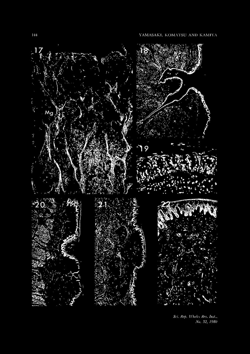
5 TONGUES OF THE SIRENIA 131 On the other hand, in the dugong tongue, two rows with deep pits (three on both sides in DD-1; right, six, and left, seven in DD-2; three on both sides in DD- 3) were present near both posterior lateral margins which correspond to the posterolateral region of the dorsal swelling in the manatee (Figs 6 and 9). The regions with the dorsal pits were not elevated. The pits were round, about 1 mm in diameter. Arranged longitudinally, they opened slightly anteriorly with a separation of about 3 to 4 mm. Small projections could be seen in some pits. Glandular openings, smaller than those of the pits and surrounded by small elevations, were observed on the dorsum from the posteriormost pit to the epiglottis. Three to seven pits, the lateral pits, (seven on the right and six on the left in DD-1; six on the right in DD-2*; three on either side in DD-3) resembling the dorsal pits, were present in a group, a little nearer the dorsal surface adjacent to the anterior region of the palatoglossal arch (Figs 7 and 9). This region was confined to within about 1 cm or less and was not elevated. In both, manatee and dugong, 20 to 25 round, fiat conspicuous patches were seen extending on both sides from about the anterior one-fifth of the tongue almost to the palatoglossal arch in the manatee (Fig. 2), and from the mid-part to near the arch in the dugong (Fig. 7). The patches were rather white in color in the fixed state, 1 to 3 mm in diameter, and slightly elevated. One or more small glandular openings were seen on the surface of some of these patches. Those located at the posterior intermingled regionally with the lateral swelling in the African manatee, and the lateral pits in the dugong. Small glandular openings were seen in the ventral region of these patches, being particularly abundant on the posterior part. Histology Though the sirenian tongues observed were extremely muscular, adipose tissues was rich in muscular tissues in the posterior area. Superior longitudinal muscle bundles were rather well developed immediately subjacent to the mucous membrane on the dorsum except in the posterior glandular portion. The epithelium of the dorsum at the spiny apex region was thick, approximately 2 mm or more, but gradually thinned toward the rear. The epithelium of the dorsum along the midline region of the manatee was relatively thin, with the epithelium becoming thick at the lingual lateral margin. The thickness of the epithelium of the side wall was about 1 mm. A superficial layer, about 15 µm, of the epithelium, excluding the rear part, was cornified. Epithelia of the dorsum and side wall of the dugong were not thick when compared with those of the manatee. The spines ofsirenians, which were strongly cornified filiform papillae, consisted of a cornified superficial epithelium without a conical elevation of the tunica propria. All spines located posterior of the dorsum were thinner than those in the apex region but were almost the same in histological appearance. The submucosa beneath the multifossulate swellings in the manatee was oc- * Pits on the left could not be counted, because this area was excoriated. No, 32, 1980
.")
6 132 YAMASAKI, KOMATSU AND KAMIYA cupied by a lobulated gland mass mostly composed of mucous glands (Figs 11-14). In regions with fossulae, mixed glands which were usually present often opened directly into the fossulae at the relatively upper part (Figs 12 and 14). The gland masses at the mid-part of the dorsal swelling were approximately 7 mm and 8 mm deep in the African manatee and West Indian species, respectively. The gland mass of the lateral swelling (Figs 15 and 16) reached about half the depth (TS-2) or less (TS-1) of that of the dorsal swelling. The gland masses of both the dorsal and lateral swellings fused with each other. The fossulae were approximately 1 mm in depth. The inside wall of the fossula was covered with stratified squamous epithelium of about 50 µm in thickness, without cornification. Secondary papillae were rather poorly developed in this part. This epithelium contained pale flask-shaped structures or irregularly arranged cell masses with occupied almost the total thickness of the epithelium (Fig. 12). These were undoubtedly degenerated taste buds, although they were not clearly confirmed due to their poor condition. Well developed pure mucous glands about 6 mm in depth were present from the front of the multifossulate area to the rear. This mucous gland field extended from the anterior part of the palatoglossal arch to the ventral region of the side wall of the tongue. Capacious ducts were seen in places in the glandular tissues of the tongue (Figs 12 and 13). On the other hand, in the dugong, pits, both dorsal (Fig. 17) and lateral (Fig. 18), were approximately 3 mm in depth, and surrounded by well developed lobulated pure serous gland masses, clearly distinguishable from the neighboring well developed pure mucous glands (Fig. 17). Wide lumens of gland ducts were observed in these glandular tissues. Since the serous gland mass was fused.to the adjacent gland mass, large serous gland areas existed beneath the pits. The gland mass at the dorsum was approximately 14 mm in depth, and that at the side wall was about 10 mm deep. Serous glands opened into the pits and the neighboring mucous glands opened directly onto the dorsum. Some of pits had small projections protruding from their bottoms or walls (Figs 17 and 18). The epithelia of the pits, in which taste buds could be seen, were thin, being about 50 µm in thickness. Taste buds were found grouped together in some places, especially in the lateral pits (Figs 18 and 19). There seemed to be a larger number of taste buds in the pits of the dugong than in the fossulae of the manatee. Sections of the round patches on the lateral surfaces of the tongues of manatee (Fig. 20) and dugong (Fig. 21) showed a thinning of the epithelium of approximately 100 to 200 µm. Secondary papillae at the patches were relatively short with a superficial 12 to 20 µm of their epithelium having a tendency towards cornification. The lamina propria of this region contained relatively abundant thick nerve fiber bundles (Fig. 22). No taste buds were present at the patches. Mucous glands, comparatively poorly developed in relation to those at the rear part of the dorsum, opened on the surface of some of the patches. The mucous glands of the rear part of the tongue extended to the palate and pharynx. Those in the dugong were well developed when compared with those in the manatee. Dugong glands also extended down almost to the end of the
7 TONGUES OF THE SIRENIA 133 esophagus. Condensations of lymphoid tissue could not be observed at the lingual root except in one case, African manatee, TS-2, which had some lymphatic nodulelike structures with crypts. DISCUSSION The sirenian tongue is difficult to divide strictly into two portions, the body and the root, due to the lack of a row of vallate papillae, sulcus terminalis, and a trace of foramen caecum, which are features seen at the border between the two portions in many land mammalian tongues. Therefore, the whole length of the tongue was measured from the apex to the base of the epiglottis. However, data reported by former investigators have often been unclear as to the posterior measuring point. Measuring points are necessary in the estimation of data showing the length of tongues such as those of sirenians, since they have no border marks between the body and the root as in cetacean tongues (Yamasaki et al., 1976a, 1976b, 1978a, 1978b). There are some ambiguities among previous descriptions on the manatee and dugong tongues. However, the characteristics of the sirenian tongue observed were 1) comparative smallness in relation to body size, 2) slenderness and vertical thickness, 3) absence of sulci on the dorsum, 4) existence of hard, digitiform, cuticular spines in the apex region, 5) existence of large-sized patches on the side wall, 6) presence of peculiar gustatory organs, varied types of foliate and vallate papillae. Anatomical differences between the two families are summarized and shown in table 2. Items in the table will be compared and discussed. TABLE 2. ANATOMICAL DIFFERENCES BETWEEN MANATEE AND DUGONG TONGUES I. Apex 2. Spines 3. Dorsum 4. Gustatory organ 5. Glands of the gustatory organ manatee round existing in the apex region only smooth, devoid of papillous projections forming dorsal and lateral multifossulate swellings mucous, partially mixed dugong truncate extending posteriorly on the body of the tongue full of small elevations with spines having several pits on dorsum and side wall, forming no swellings serous, well developed The outline of the apex of the manatee tongue is rounded. That of the dugong is truncated, but the bifid tip in the dugong as reported by Hill (1945) could not be observed. Since the apex in the young dugong is round as in the manatee, age and individual differences may be related to its outline. In young dugong, Gohar (1957) observed that the cornified tip of the tongue is smooth but that with advancement in age it develops stiff bristles and becomes rasp-like in appearance. Our observations on a young dugong, whose spines at the tip were very short, is in approximate agreement with his descriptions. Drawings of a conspicuous horny
8 134 YAMASAKI, KOMATSU AND KAMIYA process at the apex pointing anteriad in the dugong tongue were done by Sonntag (1922) and Aoki et al. (1938), and the process was described by the latter. However, we could not locate such a process at the tip region in the tongues of either family. One of the great differences between the two families is the appearance of the dorsum. In the dugong, small epithelial elevations, each with a bunch of spines, spread over the dorsum in contrast with the smoothness of the dorsum of the manatee. Gohar (1957) made very detailed descriptions of the appearance of the dorsum of the tongue of the Red Sea dugong. He stated that no typical circumvallate papillae could be seen, and proposed to call the special type of papillae observed, a bunch of spines on the elevation, "papillae calici-penicilliformes". He looked upon them as a modified form of the circumvallate papillae. We cannot agree that the structure composed of special papillae is a modification of the circumvallate papillae, but we assume that it may be a specially arranged filiform papilla, because of its form, distribution, number and histological features. Vallate papillae have been reported on the posterior part of the dorsum of the manatee by several investigators; Rapp (1857)* in Manatus latirostris, Murie (1870)* in M. americanus, Gmelin, (1892) in M. latirostris, Weber (1904), species unclear. In the dugong, clustered vallate papillae and rudimentary ones have been reported by Sonntag (1922) and Hill (1945)**, respectively. However, Gudernatsch (1909) observed that these papillae were absent in sirenian tongues, and we also could not locate such papilla in our specimens. Gudernatsch (1909) reported "Spiegeln" on the dorsum resembling those on the side wall of M. latirostris and he considered them to be fungiform papillae. However, in our specimens we could not find such structures on the dorsum, except for the side wall. The most characteristic feature of the sirenian tongue is the presence of peculiar gustatory organs, a multifossulate swelling in the manatee and pits in the dugong, with notable differences in the organs being marked between the two families. Several interpretations have been made by former investigators and there has been confusioh in terminology among them, especially on the organs of the manatee. The structures that seem to indicate a dorsal swelling in manatee judging from the descriptions, drawings or figures are: 1. a sieve and tonsil-like plate by Rapp (1857) in M. latirostris 2. many fossulate-papillae by Owen (1868)***, species unclear 3. a double set of circumvallate glands by Murie (1870) in M. americanus 4. an anterior tonsil by Gmelin (1892) in M. latirostris (with a drawing) those seemingly indicating a lateral swelling are: * Rapp (1857) and Murie (1870) might have considered even the patches on the side wall as vallate papillae, since they indicated that the structures extended to the anterior third of the tongue. This distribution almost corresponds to that of the patches of our specimens. ** Hill (1945) reported another group of vallate papillae just anterior to the foremost group in the pit on the side wall in the dugong. *** Owen (1868) described the structures as extending to the anterior third of the tongue.
9 TONGUES OF THE SIRENIA a simple lateral gustatory organ by Gmelin (1892) in lvl. latirostris (with a drawing) 2. a large papillary plate by Weber (1904), species unclear and those probably indicating a continual single swelling, dorsal and lateral, are: 1. a large patch of circumvallate papillae (Mayer's organ)* by Beddard (1897) in lvl. inunguis (with drawings) 2. a compact glandular apparatus with gustatory pits by Gudernatsch (1909) in lvl. latirostris (with photographs) 3. a large cushion with numerous fissures (lateral organ)** by Sonntag (1922) in lvl. americanus (with a drawing) However, the descriptions of the pits in the dugong by former investigators have been clearer than those on the swelling in the manatee, though the pits have been written of with various terms: Gudernatsch (1907, 1908), on the dorsal and lateral pits (with photographs); Aoki et al. (1938), dorsal and lateral (with drawings); Hill (1945), dorsal and lateral; Gohar (1957), lateral only (with a drawing); Nair et al. (1975), lateral only; Husar (1978) dorsal only. Although detailed observations were made on the gustatory organs in both manatee and dugong by Gudernatsch (1908, 1909), for some reason, no investigators have referred to his articles. Undoubtedly the multifossulate swelling and pits are a variation of the gustatory organs*** formed by lingual papillae. However, it is difficult to conclude whether they correspond to the vallate or foliate papillae, since neither animal has these two kinds of papillae, at the corresponding regions as seen in other land mammalian tongues. The classification of the lingual papillae is based on their form and distribution. Since the gustatory organs on the dorsum and on the side wall in sirenian tongues are quite similar to each other morphologically, it would be more advisable to clarify the organs by distribution rather than by form. The lateral and dorsal pits of the dugong are clearly distinguishable in their location. As the lateral swelling in the manatee is located in almost the same region as the lateral pits in the dugong, both structures may be considered homologous to each other as indicated by Gudernatsch (1909). Some former investigators have stated that structures corresponding to the lateral swelling (Gmelin, 1892; Weber, 1904; Gudernatsch, 1909) and the lateral pits (Gudernatsch, 1908)**** were primitive type of the foliate papillae. We agree that the structures belong to the foliate papillae, but believe it advisable to consider them a varied type of foliate papillae rather than * Mayer's organ is a structure with numerous fissures located on the lateral surface of the elephant tongue. As this structure was first described by Mayer in 1844, it has since been called Mayer's organ in honor of its discoverer (Forbes, 1879). This structure in the elephant tongue consists of the foliate papillae themselves (Shimizu et al., 1960). Therefore, the description by which Beddard (1897) regarded circumvallate papillae as Mayer's organ is inadequate. ** Although Sonntag's description is extremely ambiguous in the use of the term lateral organ, we have judged from his drawing that the organ may have indicated a swelling. *** Aoki et al. (1938) and Gohar (1957) considered the pits of the dugong as merely glandular openings. **** Gudernatsch (1908) also used the term" remnants" for the structures.
10 136 YAMASAKI, KOMATSU AND KAMIYA to label them a primitive type of foliate papillae variation. Gudernatsch (1909) observed that the group of gustatory pits on the side wall (=lateral swelling) in the manatee extended to the dorsum, that is, he believed the pits on the dorsum ( = dorsal swelling) to be a continuation of those on the side wall. We, however, consider the dorsal swelling in the manatee to be an independent structure from the lateral swelling as is also true in the case of the dorsal pits in the dugong, and believe that the structures on the dorsum in both animals may be a variation of the vallate papillae. In the case of the manatee, the organ is multifossulated and expanded, and swells upwards. In the dugong, on the other hand, the organ grows downwards, with each pit becoming deep instead of being multifossulated. In the manatee, when each swelling, dorsal and lateral, has become well developed with age, the swellings may fuse with each other, and may then be seen as a single continuous large swelling as shown in Beddard's figure (1897). There have been no descriptions concerning the existence of taste buds in the manatee, and only Gudernatsch reported numerous ones in the dorsal pits (1907) and in the lateral ones (1908) in the dugong. However, though the number of taste buds in the sirenians observed may not be as numerous as those of vegetarian land mammals, they are much more numerous than those in cetaceans (Komatsu and Yamasaki, 1980). The region with pits in dugong consists of a well developed serous gland mass, while that of the swelling in the manatee is composed of a mass of mucous glands, though partially mixed ones, in agreement with Gudernatsch's descriptions (1908, 1909). In the region having taste buds in the fossulae, mixed glands are found near the upper part of the fossulae in manatee. Serous fluid may be useful in rinsing the fossula or pit having taste buds, particularly in the case of dugong, which feed on sea grasses. It is possible that some enzymes contained in serous fluid may turn the polysaccharide of sea grasses into the small molecules which stimulate the taste buds. Gudernatsch (1909) considered that the manatee's gustatory organ glands showed a far simpler grade of development than those in dugong, based on the poorly developed serous glands in manatee, and stated that marked morphological differences of the gustatory organs between the animals might depend on some biological factors rather than on the difference in food habits. Since this is a very interesting problem and since adaptation for foods cannot be disregarded, further studies are expected. In the dugong, Gohar (1957) observed that mucous glands containing fewer and smaller serous glands were present under the whole surface of the tongue except for the anterior tip. However, in our observations mucous glands were not present in the lingual body except on the lower part of the side wall, but were present at the root, and serous glands were limited to the pit region. Dugong glands, in particular, are more well developed than those of manatee and extend down almost to the end of the esophagus. The other characteristic external feature is the presence of patches studded on the lateral surface of the sirenian tongue. Gudernatsch (1908, 1909) called these structures "Spiegeln" in the dugong and manatee, and regarded them as fungi-
11 TONGUES OF THE SIRENIA 137 form papillae. In the dugong, Gohar (1957) also described various sized fungiform papillae, whereas Hill (1945) stated that these were absent. We consider the patches in sirenians to be large-sized fungiform papillae. Although they have no taste buds, it is supposed that the structures play the role of sensory organs, probably tactile organs. Rapp (1857) reported tonsils, which were oval plates having numerous openings and showing a sieve-like appearance, on each side in the posterior part of the tongue in M. latirostris. Gmelin (1892) showed anterior* and posterior tonsils in his drawing of the same species. However, it is as yet uncertain whether or not lingual tonsils are present in the sirenian tongue at the corresponding region since only in one case of African manatee have structures which seem to be poorly developed lingual tonsils with crypts been seen. ACKNOWLEDGMENTS We are greatful to Doctors M. Nishiwaki, University of the Ryukyus, Naha, Okinawa and T. Kasuya, Ocean Research Institute, University of Tokyo, for kindly supplying the materials for this study. We wish to thank Doctors J. E. Reynolds III, School of Marine and Atmospheric Science, University of Miami, K. Kubota, Institute of Stomatognathic Science, Tokyo Medical and Dental University, and K. Takahashi, Department of Anatomy, Sapporo Medical College, who gave us valuable suggestions and advice for our study. The techincal assistance of Mr S. Ohtani, Risto-Technical Center, Sapporo Medical College, is also acknowledged. REFERENCES AOKI, B., S. TATEISHI, R. TANAKA and K. FuRuHATA, Anatomical notes on the dugong. Kagaku no Taiwan (in Japanese), Lab. Comp. Morph. l\1ammal., Zoo[. Inst. Taihoku Imp. Univ., 5: BEDDARD, F. E., Notes upon the anatomy of a manatee (Manatus inunguis) lately living in the society's garden. Proc. Zool. Soc., London, pp FORBES, \V. A., On the anatomy of the African elephant (Elephas africanus Blum.). Proc. Zool. Soc., London, pp GMELIN, W Zur Morphologie der Papilla vallata und foliata. Arch. f. mikr. Anal., 40: Gm1AR, H.A.F., The Red Sea dugong. Publ. l\llarine Biol. Sta. Chardaqa, 9: GuDERNATSCH, ]. F., Untitled. Amer. Nat., 41: 665. GuDERNATSCH, J. F., Zur Anatomie und Histologie des Verdauungstraktes von Halicore dugong Erxl. Morph. ]b., 37: GuDERNATSCH, J. F., Zur Anatomie und Histologie des Verdauungstraktes der Sirenia. II. Die Zunge von Manatus latirostris Harl. Morph. ]b., 40: HILL, W. W.C.O., Notes on the dissection of two dugongs. J. Mammal., 26: HusAR, S. L., Dugong dugon. Mammalian Species, 88: 1-7. KOMATSU, S. and F. YAMASAKI, Formation of the pits with taste buds at the lingual root in the striped dolphin, Stenella coeruleoaha. j. Morph., 164: MAYER, F.J.C., Uber die Zunge als Geschmacksorgan. Nova Acta Acad. Leop. Carol., 20: MURIE,]., On the form and stucture of the manatee (Manatus americanus). Tran. Zoo[. Soc. London, 8: * As mentioned above, this seems to be the structure corresponding to our dorsal swelling.
12 138 YAMASAKI, KOMATSU AND KAMIYA NAIR, R. V., R.S.L. MOHAN and K. S. RAo, The dugong, Dugong dugon. Cent. Marine Fish. Res. Inst. Cochin, 26: OWEN, R., Organ of taste. In: On the anatomy rifvertebrates. Vol. III. Mammals., Longmans, Green and Co., London, pp RAPP, W. VON, Anatomische Untersuchungen tiber Manatus (Lamantin). Jahrh. Ver. vateral. Natkd. Wurttenburg, Stuttgart, pp SHIMIZU, Y., T. FUJITA, T. KAMIYA and S. IsoKAWA, Anatomy of a female Indian elephant with special reference to its visceral organs (in Japanese with English summary). Acta Anat. Nipponica, 35: SONNTAG, C. F., The compartaive anatomy of the tongue of the mammalia. VII. Cetacea, Sirenia and Ungulata. Proc. Zool. Soc., London, pp WEBER, M., Sirenia. In: Die Saugetiere. Verlag von Gustav Fisher, Jena, pp YAMASAKI, F., H. SATOMI and T. KAMIYA, 1976a. The tongue offranciscana (La Plata dolphin), Pontoporia blainvillei. Okajimas Fol. anal. jap., 53: YAMASAKI, F., H. SATOMI and T. KAMIYA, 1976b. An observation on the papillary projections at the lingual margin in the striped dolphin. 28: YAMASAKI, F., S. KOMATSU and T. KAMIYA, 1978a. Papillary projections at the lingual margin in the.striped dolphin, Stenella coeruleoalba, with special reference to their development and regression. J. JY!orph., 157: YAMASAKI, F., S. KOMATSU and T. KAMIYA, 1978b. Taste buds in the pits at the posterior dorsum of the tongue of Stenella coeruleoalba. 30: YAMASAKI, F., S. KOMATSU and T. KAMIYA, An anatomical note on the tongue of dugongs, Dugong dugon. Proc. Seminar Workshop Dugongs James Cook Univ. in 1979, (in press). Sci. Rep. Whales Res. Inst.
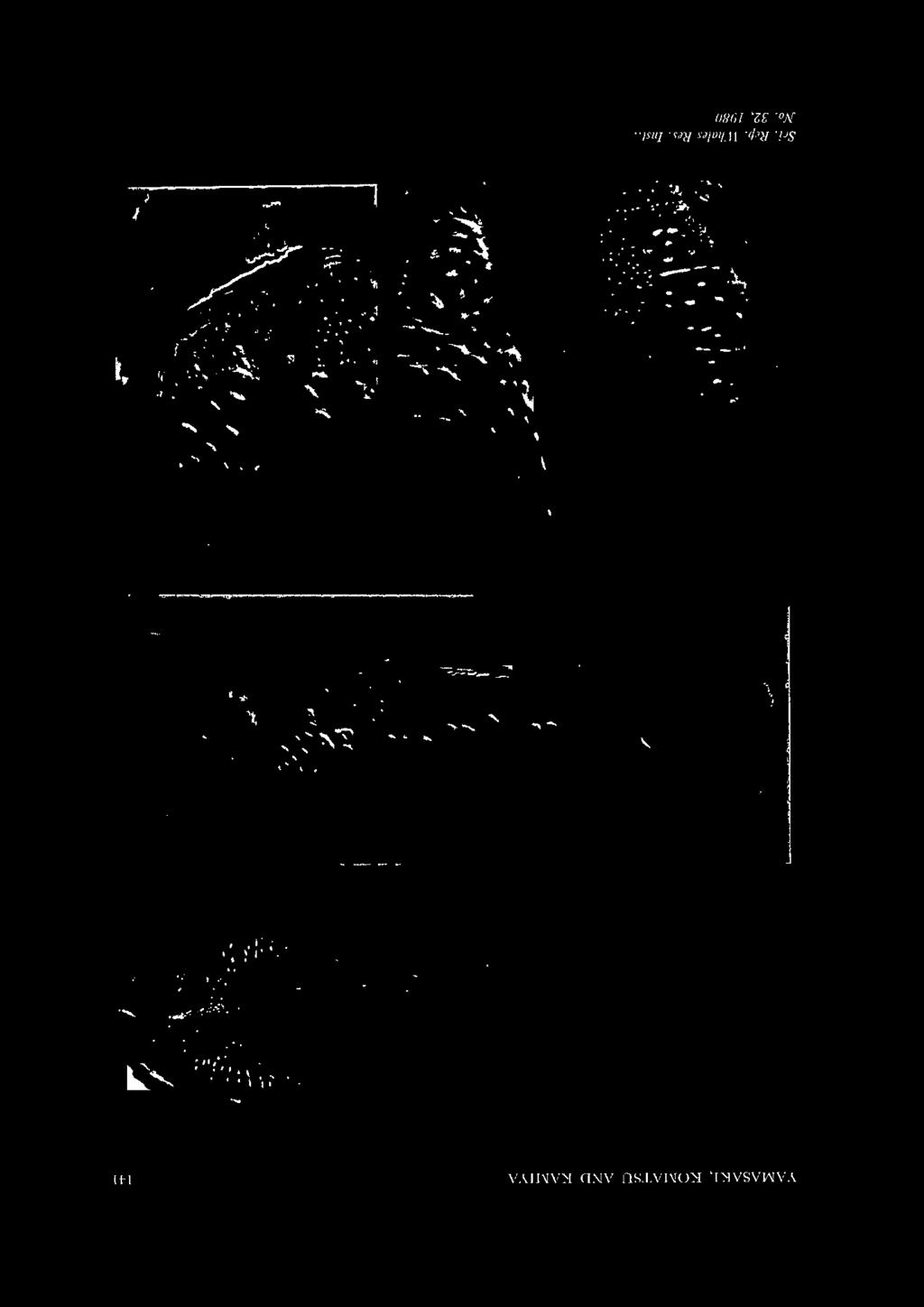
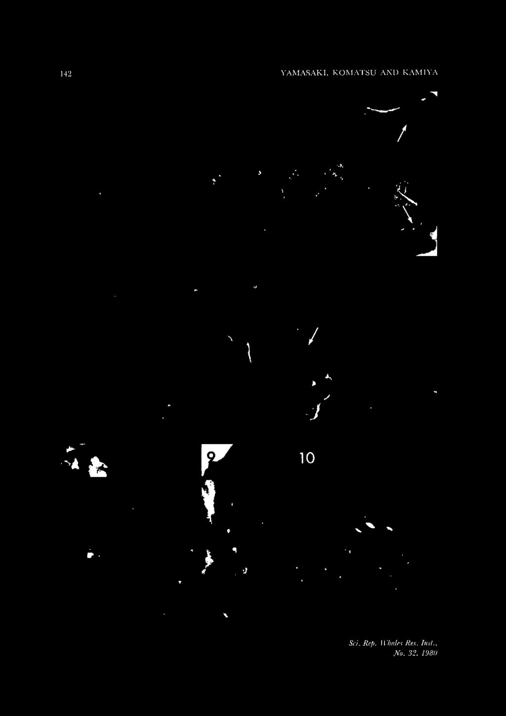
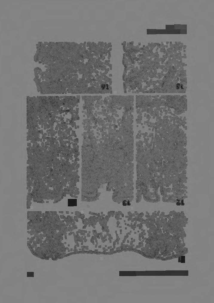
13 TONGUES OF THE SIRENIA 139 EXPLANATIONS OF FIGURES Fig. I. A dorsal view of the tongue of an African manatee (TS-2). The tongue is rather slender as a whole, and its tip is rounded. The tip region is covered with many hard, digitiform, cuticular spines. Dorsal multifossulate swellings, slightly opened anteriorly, forming a pair may be observed near both posterior lateral margins of the dorsum. Numerous small openings of mucous glands are visible on the posterior part of the dorsum. X 1. Fig. 2. Left side view of TS-2 tongue. The tongue becomes thick posteriorly but thin at the lingual root. A lateral multifossulate swelling is observed in the anterodorsal region of the palatoglossal arch. Some twenty-five round fiat patches are present, extending from the anterior fifth of the side wall to near the palatoglossal arch. Small mucous glandular openings are visible from the mid-part to the arch. X 1. Fig. 3. A higher magnified view of the left dorsal swelling of TS-2 in figure I. Roughly twenty-five rather oval, large fossulae are obviously visible on the swelling. Numerous glandular openings, far smaller than the fossulae in diameter, are clearly seen from the posterior region of the swelling to the posterior part of the tongue. x2. Fig. 4. A left dorsal swelling of a West Indian manatee (TM-I). The swelling, having many large fossulae, is not as remarkable as that ofts-2. X2. Fig. 5. A higher magnified view of the lateral swelling of TS-2 tongue in figure 2, showing more than ten oval fossulae. Numerous glandular openings, far smaller than the fossulae in diameter, are observable. x2. Fig. 6. A dorsal view of the tongue of a dugong (DD-3). The tongue is rather slender as a whole, with the spine-covered tip region being truncated. Thin, soft spines extend posteriorly, being especially shaggier on the lateral strips. Small elevations are seen on the dorsum. A row of dorsal pits can be seen at both posterior margins (arrows). x I. Fig. 7. Left side view of DD-3 tongue. The body of the tongue strongly bows anteroposteriorly, with the root being situated at a lower level to the body. Three lateral pits are observable on the posterior side wall (arrow). About fifteen round flat patches are present on the mid-part of the side wall. X 1. Fig. 8. Bunches of spines grow on the small elevations at the anterior dorsum of the DD-3 tongue. x20. Fig. 9. A higher magnified view of the dorsal pits on the left side of the dorsum of DD-3 in figure 6. Small mucous glandular openings can be seen on the posterior part of the pits. X 2.5. Fig. 10. A higher magnified view of the lateral pits and patches of figure 7. The pits and patches vary in size. Small mucous glandular openings are observable on the ventral side of the pits and patches. X 2.5. Fig. 11. A cross section of both dorsal swellings with fossulae in an African manatee (TS-1). Mixed glands are present in the regions with fossulae, whereas in the area between both swellings the submucosa is occupied by pure mucous glands. x5.5. Fig. 12. A cross section of two fossulae of a dorsal swelling of TS-1. Several fiaskshaped spaces in the epithelium of the side wall of the fossulae may show the remnants of taste buds, the cells of which have sloughed off. Glands are, for the most part, mucous in nature but partially mixed, especially in the upper part of the gland mass. Capacious ducts are seen at points in the glandular tissues. The secondary papillae of the side wall of the fossula are poorly developed. X 15. Fig. 13. A cross section of a fossula of a dorsal swelling of TS-2. Mucous glands
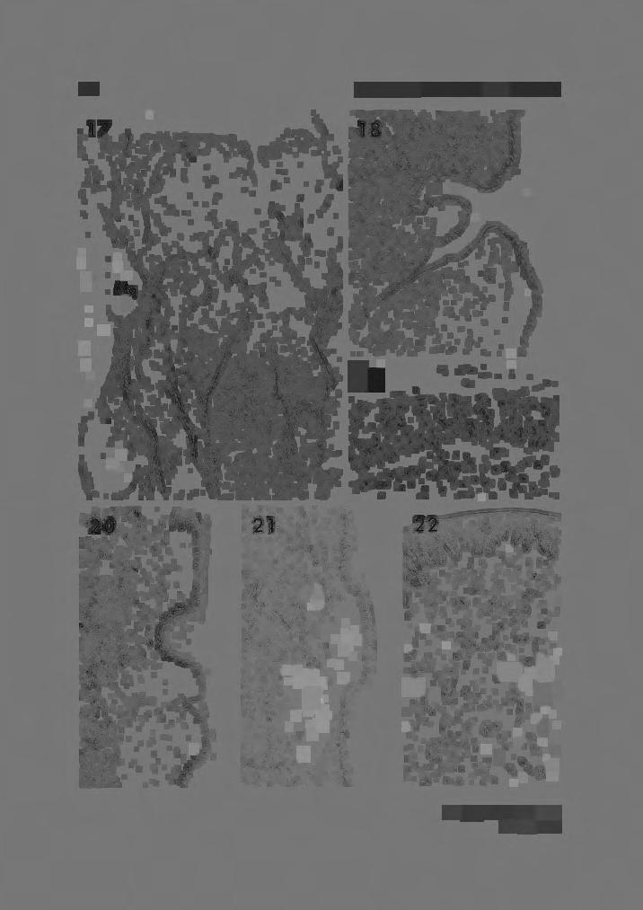
14 140 YAMASAKI, KOMATSU AND KAMIYA more developed than those of TS-1 are seen but serous glands intermingle partially at the bottom of the fossula. The thin epithelium of the inner wall of the fossula has exfoliated due to its poor condition. X 15. Fig. 14. A cross section of two fossulae of a dorsal swelling of the West Indian manatee (TI-I). Well developed glands are mostly mucous in nature, but partially mixed in the upper part of the gland mass. X 15. Figs 15 and 16. Cross sections of two fossulae (TS-1, Fig. 15) and a fossula (TS- 2, Fig. 16) of the lateral swelling. The nature of glands is almost the same as that of the dorsal swelling, but the glands of the lateral swelling are poorly developed as compared with those of the dorsal swelling. These glands are well developed in TS-2 as compared with those in TS-1. X 15. Fig. 17. A cross section of a dorsal pit of a dugong (DD-3). Small projections can be seen in the pit. The pit is surrounded by well developed serous glands (Sg) which are clearly distinguished from mucous glands (Mg). The epithelium of the pit is very thin. X 7.5. Fig. 18. A cross section of a lateral pit of DD-3. A small projection having taste buds in its thin epithelium can be seen in the pit surrounded by well developed serous glands. X 25. Fig. 19. A more highly magnified view of a part of the projection in figure 18. Taste buds lie in the thin epithelium of the projection. This figure is taken from the area where taste buds are crowded together. X 300. Fig. 20. A cross section of two patches of TS-2. The epithelium is rather thin and secondary papillae are poorly developed in the patches as compared with those of the remainder. Mucous glands and a glandular opening can be seen in the lower patch. X 15. Fig. 21. A cross section of a patch of DD-3. The epithelium is rather thin as compared with that of the remainder. Mucous glands can be seen in places. X 15. Fig. 22. A more highly magnified view of a cross section of a patch of DD-3. Abundant, rather thick nerve fibers are observable in the submucosa of the patch. x50.


15 S~/Vl/.


16 '~ 2

17
18
Anat. Labor. of Prof. H. SETO, Tohoku University, On the Sensory Terminations Formed along the Ductus
 Anat. Labor. of Prof. H. SETO, Tohoku University, Sendai. On the Sensory Terminations Formed along the Ductus Pancreaticus in Cat. The existence of PACINIan bodies in the pancreas of mammals, especially
Total Distribution of Taste Buds on the Tongue of the Pup
 The Ohio State University Knowledge Bank kb.osu.edu Ohio Journal of Science (Ohio Academy of Science) Ohio Journal of Science: Volume 4, Issue 6 (November, 194) 194-11 Total Distribution of Taste Buds
VARIATION IN MONIEZIA EXPANSA RUDOLPHI
 VARIATION IN MONIEZIA EXPANSA RUDOLPHI STEPHEN R. WILLIAMS, Miami University, Oxford, Ohio In making a number of preparations of proglottids for class study at the stage when sex organs are mature and
Title. CitationJapanese Journal of Veterinary Research, 24(1-2): 37. Issue Date DOI. Doc URL. Type. File Information
: 37. Issue Date DOI. Doc URL. Type. File Information") Title DISTRIBUTION OF LYMPHATIC TISSUES IN DUCK CAECA Author(s)KITAMURA, Hirokazu; SUGIMURA, Makoto; HASHIMOTO, Yos CitationJapanese Journal of Veterinary Research, 24(1-2): 37 Issue Date 1976-05 DOI 10.14943/jjvr.24.1-2.37
Title DISTRIBUTION OF LYMPHATIC TISSUES IN DUCK CAECA Author(s)KITAMURA, Hirokazu; SUGIMURA, Makoto; HASHIMOTO, Yos CitationJapanese Journal of Veterinary Research, 24(1-2): 37 Issue Date 1976-05 DOI 10.14943/jjvr.24.1-2.37
Vol. XIV, No. 1, March, The Larva and Pupa of Brontispa namorikia Maulik (Coleoptera: Chrysomelidae: Hispinae) By S.
 By S.") Vol. XIV, No. 1, March, 1950 167 The Larva and Pupa of Brontispa namorikia Maulik (Coleoptera: Chrysomelidae: Hispinae) By S. MAULIK BRITISH MUSEUM (NATURAL HISTORY) (Presented by Mr. Van Zwaluwenburg
Vol. XIV, No. 1, March, 1950 167 The Larva and Pupa of Brontispa namorikia Maulik (Coleoptera: Chrysomelidae: Hispinae) By S. MAULIK BRITISH MUSEUM (NATURAL HISTORY) (Presented by Mr. Van Zwaluwenburg
HISTOPATHOLOGY. Introduction:
 Introduction: HISTOPATHOLOGY Goats and sheep are the major domestic animal species in India. Much of the economy of the country has been depend upon the domestication of these animals. Especially economy
Fish 475: Marine Mammalogy
 Fish 475: Marine Mammalogy Taxonomy (continued) Friday, 3 April 2009 Amanda Bradford Course website: http://faculty.washington.edu/glennvb/fish475 Mysticeti: The baleen whales About 10-12 species; Formerly
Gross and histological studies of digestive tract of broilers during postnatal growth and development
 J. Bangladesh Agril. Univ. 10(1): 69 77, 2012 ISSN 1810-3030 Gross and histological studies of digestive tract of broilers during postnatal growth and development M. Nasrin, M. N. H. Siddiqi, M. A. Masum
Digestive & Respiratory System Anterior Respiratory Dissection
 Digestive & Respiratory System Anterior Respiratory Dissection We will be looking at both systems during this dissection. The cat respiratory dissection WILL BE ON THE NEXT LAB PRACTICAL!! We will do 2
SOME NEW AMERICAN PYCNODONT FISHES.
 SOME NEW AMERICAN PYCNODONT FISHES. By James Williams Gidley, Assistant Curator of Fossil Mammals, United States National Museum. In the United States National Museum are several specimens representing
COMPARATIVE VERTEBRATE HISTOLOGY ZOO 4756c Syllabus for Fall 2018
 COMPARATIVE VERTEBRATE HISTOLOGY ZOO 4756c Syllabus for Fall 2018 Instructor: Frank T. Logiudice Office: Biology Building, Room 202c Office Phone Number: (407) - 823-2495 Email Address: Frank.Logiudice@ucf.edu
INVESTIGATIONS ON THE SHAPE AND SIZE OF MOLAR AND ZYGOMATIC SALIVARY GLANDS IN SHORTHAIR DOMESTIC CATS
 Bulgarian Journal of Veterinary Medicine (2009), 12, No 4, 221 225 INVESTIGATIONS ON THE SHAPE AND SIZE OF MOLAR AND ZYGOMATIC SALIVARY GLANDS IN SHORTHAIR DOMESTIC CATS Summary A. A. MOHAMMADPOUR Department
The family Gnaphosidae is a large family
 Pakistan J. Zool., vol. 36(4), pp. 307-312, 2004. New Species of Zelotus Spider (Araneae: Gnaphosidae) from Pakistan ABIDA BUTT AND M.A. BEG Department of Zoology, University of Agriculture, Faisalabad,
Oribatid Mites of the Family Otocepheidae from Tian-mu Mountain in China (Acari: Oribatida)1'
1'") Acta arachnol,, 42 (1): 1-6, August 30, 1993 Oribatid Mites of the Family Otocepheidae from Tian-mu Mountain in China (Acari: Oribatida)1' Jun-ichi AoKI2' and Sheng-hao Hu3' Abstract Dolicheremaeus wangi
Acta arachnol,, 42 (1): 1-6, August 30, 1993 Oribatid Mites of the Family Otocepheidae from Tian-mu Mountain in China (Acari: Oribatida)1' Jun-ichi AoKI2' and Sheng-hao Hu3' Abstract Dolicheremaeus wangi
posterior part of the second segment may show a few white hairs
 April, 1911.] New Species of Diptera of the Genus Erax. 307 NEW SPECIES OF DIPTERA OF THE GENUS ERAX. JAMES S. HINE. The various species of Asilinae known by the generic name Erax have been considered
NOTE XVII. Dr. A.A.W. Hubrecht. which should he in accordance with. of my predecessors. alive or in excellent. further
 further either EUROPEAN NEMERTEANS. 93 NOTE XVII. New Species of European Nemerteans. First Appendix to Note XLIV, Vol. I BY Dr. A.A.W. Hubrecht In the above-mentioned note, published six months ago, several
Alimentary System 解剖學科徐淑媛
 Alimentary System 解剖學科徐淑媛 本堂重點 1. Structures derived from primitive guts 2. Specific events Alimentary System endoderm of primordial gut epithelium & glands of digestive tract ectoderm of stomodeum epithelium
HELMINTHES OF ANIMALS IMPORTED IN JAPAN I Tanqua ophidis Johnston and Mawson, 1948 of Water Snakes from Samarinda, Indonesia
 Japan. J. Trop. Med. Hyg., Vol. 5, No. 2, 1977, pp. 155-159 155 HELMINTHES OF ANIMALS IMPORTED IN JAPAN I Tanqua ophidis Johnston and Mawson, 1948 of Water Snakes from Samarinda, Indonesia NOBORU KAGEI1
Fischthal and Kuntz (1964) reported the
 reported the") Zoological Studies 41(3): 283-287 (2002) Meristocotyle provitellaria sp. nov. (Digenea: Meristocotylidae) from Varanus salvator in China Wei Liu 1, Qing-Kui Li 2, Hsiu-Hui Shih 3 and Zhao-Zhi Qiu 1, *
Zoological Studies 41(3): 283-287 (2002) Meristocotyle provitellaria sp. nov. (Digenea: Meristocotylidae) from Varanus salvator in China Wei Liu 1, Qing-Kui Li 2, Hsiu-Hui Shih 3 and Zhao-Zhi Qiu 1, *
A NEW SALTICID SPIDER FROM VICTORIA By R. A. Dunn
 Dunn, R. A. 1947. A new salticid spider from Victoria. Memoirs of the National Museum of Victoria 15: 82 85. All text not included in the original document is highlighted in red. Mem. Nat. Mus. Vict.,
Reprinted from: CRUSTACEANA, Vol. 32, Part 2, 1977 LEIDEN E. J. BRILL
 Reprinted from: CRUSTACEANA, Vol. 32, Part 2, 1977 LEIDEN E. J. BRILL NOTES AND NEWS 207 ALPHE0PS1S SHEARMII (ALCOCK & ANDERSON): A NEW COMBINATION WITH A REDESCRIPTION OF THE HOLOTYPE (DECAPODA, ALPHEIDAE)
Frog Dissection Information Manuel
 Frog Dissection Information Manuel Anatomical Terms: Used to explain directions and orientation of a organism Directions or Positions: Anterior (cranial)- toward the head Posterior (caudal)- towards the
Diurus, Pascoe. sp. 1). declivity of the elytra, but distinguished. Length (the rostrum and tails 26 included) mm. Deep. exception
. declivity of the elytra, but distinguished. Length (the rostrum and tails 26 included) mm. Deep. exception") 210 DIURUS ERYTIIROPUS. NOTE XXVI. Three new species of the Brenthid genus Diurus, Pascoe DESCRIBED BY C. Ritsema+Cz. 1. Diurus erythropus, n. sp. 1). Allied to D. furcillatus Gylh. ²) by the short head,
210 DIURUS ERYTIIROPUS. NOTE XXVI. Three new species of the Brenthid genus Diurus, Pascoe DESCRIBED BY C. Ritsema+Cz. 1. Diurus erythropus, n. sp. 1). Allied to D. furcillatus Gylh. ²) by the short head,
Skulls & Evolution. 14,000 ya cro-magnon. 300,000 ya Homo sapiens. 2 Ma Homo habilis A. boisei A. robustus A. africanus
 Skulls & Evolution Purpose To illustrate trends in the evolution of humans. To demonstrate what you can learn from bones & fossils. To show the adaptations of various mammals to different habitats and
A NEW AUSTROSQUILLA (STOMATOPODA) FROM THE
 FROM THE") A NEW AUSTROSQUILLA (STOMATOPODA) FROM THE MARQUESAS ISLANDS BY ALAIN MICHEL Centre O.R.S.T.O.M., Noumea, New Caledonia and RAYMOND B. MANNING Smithsonian Institution, Washington, U.S.A. The At s,tstrosqzlilla
A NEW AUSTROSQUILLA (STOMATOPODA) FROM THE MARQUESAS ISLANDS BY ALAIN MICHEL Centre O.R.S.T.O.M., Noumea, New Caledonia and RAYMOND B. MANNING Smithsonian Institution, Washington, U.S.A. The At s,tstrosqzlilla
UPOGEBIA LINCOLNI SP. NOV. (DECAPODA, THALASSINIDEA, UPOGEBIIDAE) FROM JAVA, INDONESIA
 FROM JAVA, INDONESIA") NOTES AND NEWS UPOGEBIA LINCOLNI SP. NOV. (DECAPODA, THALASSINIDEA, UPOGEBIIDAE) FROM JAVA, INDONESIA BY NGUYEN NGOC-HO i) Faculty of Science, University of Saigon, Vietnam Among material recently collected
NOTES AND NEWS UPOGEBIA LINCOLNI SP. NOV. (DECAPODA, THALASSINIDEA, UPOGEBIIDAE) FROM JAVA, INDONESIA BY NGUYEN NGOC-HO i) Faculty of Science, University of Saigon, Vietnam Among material recently collected
DISCOVERY OF GENUS PLATOLENES (COLEOP TERA : TENEBRIONIDAE) FROM INDIA WITH DESCRIPTION OF TWO NEW SPECIES G. N. SABA
 FROM INDIA WITH DESCRIPTION OF TWO NEW SPECIES G. N. SABA") Rec. zool. Surv. India, 85(3) : 433-437,1988 DISCOVERY OF GENUS PLATOLENES (COLEOP TERA : TENEBRIONIDAE) FROM INDIA WITH DESCRIPTION OF TWO NEW SPECIES By G. N. SABA Zoological Survey of India M-Block,
Rec. zool. Surv. India, 85(3) : 433-437,1988 DISCOVERY OF GENUS PLATOLENES (COLEOP TERA : TENEBRIONIDAE) FROM INDIA WITH DESCRIPTION OF TWO NEW SPECIES By G. N. SABA Zoological Survey of India M-Block,
Exceptional fossil preservation demonstrates a new mode of axial skeleton elongation in early ray-finned fishes
 Supplementary Information Exceptional fossil preservation demonstrates a new mode of axial skeleton elongation in early ray-finned fishes Erin E. Maxwell, Heinz Furrer, Marcelo R. Sánchez-Villagra Supplementary
A comparison of placental tissue in the skinks Eulamprus tympanum and E. quoyii. Yates, Lauren A.
 A comparison of placental tissue in the skinks Eulamprus tympanum and E. quoyii Yates, Lauren A. Abstract: The species Eulamprus tympanum and Eulamprus quoyii are viviparous skinks that are said to have
Studies on the Papilla Foliata of Japanese. 2. The number of taste buds. with 3 text-figures. Introduction.
 Studies on the Papilla Foliata of Japanese. 2. The number of taste buds. By Yoshiro Mochizuki. From the Department of Anatomy Nihon University Dental College, Tokyo, Japan. (Director : Prof. Dr. Y.Izawa.)
Taste and Smell. Bởi: OpenStaxCollege
 Bởi: OpenStaxCollege Taste, also called gustation, and smell, also called olfaction, are the most interconnected senses in that both involve molecules of the stimulus entering the body and bonding to receptors.
Recommended Resources: The following resources may be useful in teaching this
 Unit B: Anatomy and Physiology of Poultry Lesson1: Internal Anatomy of Poultry Student Learning Objectives: Instruction in this lesson should result in students achieving the following objectives: 1. Identify
TWO NEW SPECIES OF WATER MITES FROM OHIO 1-2
 TWO NEW SPECIES OF WATER MITES FROM OHIO 1-2 DAVID R. COOK Wayne State University, Detroit, Michigan ABSTRACT Two new species of Hydracarina, Tiphys weaveri (Acarina: Pionidae) and Axonopsis ohioensis
Phylum Platyhelminthes Flatworms
 Phylum Platyhelminthes Flatworms The Acoelomates The acoelomates are animals that lack a coelom. Acoelomates lack a body cavity, and instead the space between the body wall and the digestive tract is filled
Title. Author(s)Takahashi, Ryoichi. CitationInsecta matsumurana, 14(1): 1-5. Issue Date Doc URL. Type. File Information
Takahashi, Ryoichi. CitationInsecta matsumurana, 14(1): 1-5. Issue Date Doc URL. Type. File Information") Title Some Aleyrodidae from Mauritius (Homoptera) Author(s)Takahashi, Ryoichi CitationInsecta matsumurana, 14(1): 1-5 Issue Date 1939-12 Doc URL http://hdl.handle.net/2115/9426 Type bulletin File Information
Title Some Aleyrodidae from Mauritius (Homoptera) Author(s)Takahashi, Ryoichi CitationInsecta matsumurana, 14(1): 1-5 Issue Date 1939-12 Doc URL http://hdl.handle.net/2115/9426 Type bulletin File Information
Postilla PEABODY MUSEUM OF NATURAL HISTORY YALE UNIVERSITY NEW HAVEN, CONNECTICUT, U.S.A.
 Postilla PEABODY MUSEUM OF NATURAL HISTORY YALE UNIVERSITY NEW HAVEN, CONNECTICUT, U.S.A. Number 117 18 March 1968 A 7DIAPSID (REPTILIA) PARIETAL FROM THE LOWER PERMIAN OF OKLAHOMA ROBERT L. CARROLL REDPATH
FURTHER STUDIES ON TWO SKELETONS OF THE BLACK RIGHT WHALE IN THE NORTH PACIFIC
 FURTHER STUDIES ON TWO SKELETONS OF THE BLACK RIGHT WHALE IN THE NORTH PACIFIC HIDEO OMURA, MASAHARU NISHIWAKI* AND TOSHIO KASUYA* ABSTRACT Two skeletons of the black right whale were studied, supplementing
A Lymphosarcoma in an Atlantic Salmon (Salmo salar)
") A Lymphosarcoma in an Atlantic Salmon (Salmo salar) Authors: Paul R. Bowser, Marilyn J. Wolfe, and Timothy Wallbridge Source: Journal of Wildlife Diseases, 23(4) : 698-701 Published By: Wildlife Disease
A Lymphosarcoma in an Atlantic Salmon (Salmo salar) Authors: Paul R. Bowser, Marilyn J. Wolfe, and Timothy Wallbridge Source: Journal of Wildlife Diseases, 23(4) : 698-701 Published By: Wildlife Disease
AMERICAN MUSEUM NOVITATES Published by
 AMERICAN MUSEUM NOVITATES Published by Number 782 THE AmzRICAN MUSEUM OF NATURAL HISTORY Feb. 20, 1935 New York City 56.81, 7 G (68) A NOTE ON THE CYNODONT, GLOCHINODONTOIDES GRACILIS HAUGHTON BY LIEUWE
PSYCHE A NEW GENUS AND SPECIES OF SALDIDAE FROM SOUTH AMERICA (HEMIPTERA) BY CARL J. DRAKE AND LUDVIK HOBERLANDT. Iowa State College, Ames
 BY CARL J. DRAKE AND LUDVIK HOBERLANDT. Iowa State College, Ames") PSYCHE Vol. 59 September, 1952 No. 3 A NEW GENUS AND SPECIES OF SALDIDAE FROM SOUTH AMERICA (HEMIPTERA) BY CARL J. DRAKE AND LUDVIK HOBERLANDT Iowa State College, Ames Through the kindness of Dr. P. J.
PSYCHE Vol. 59 September, 1952 No. 3 A NEW GENUS AND SPECIES OF SALDIDAE FROM SOUTH AMERICA (HEMIPTERA) BY CARL J. DRAKE AND LUDVIK HOBERLANDT Iowa State College, Ames Through the kindness of Dr. P. J.
Title ON DAUGHTER CYSTS OF COENURUS SERIALIS GERVAIS, Author(s)YAMASHITA, Jiro; OHBAYASHI, Masashi; KONNO, Seiji
YAMASHITA, Jiro; OHBAYASHI, Masashi; KONNO, Seiji") Title ON DAUGHTER CYSTS OF COENURUS SERIALIS GERVAIS, 1847 Author(s)YAMASHITA, Jiro; OHBAYASHI, Masashi; KONNO, Seiji CitationJapanese Journal of Veterinary Research, 5(1): 14-18 Issue Date 1957-03-25
Title ON DAUGHTER CYSTS OF COENURUS SERIALIS GERVAIS, 1847 Author(s)YAMASHITA, Jiro; OHBAYASHI, Masashi; KONNO, Seiji CitationJapanese Journal of Veterinary Research, 5(1): 14-18 Issue Date 1957-03-25
Morphological and Histological Study on Vermiform Appendix in Rabbit, Goat and Human Being.
 Original Article ISSN (O):2395-2822; ISSN (P):2395-2814 Morphological and Histological Study on Vermiform Appendix in Rabbit, Goat and Human Being. Jay Prakash Bharti 1, Saif Omar 2, Nawal Kishore Pandey
Gross and Microscopic Features of the Interdigital Sinus in the Barbados Black Belly Sheep in Trinidad
 Original Research Article International Journal of Current Research in Medical Sciences ISSN: 2454-5716 www.ijcrims.com Volume 2, Issue 7-2016 SOI: http://s-o-i.org/1.15/ijcrms-2016-2-7-4 Gross and Microscopic
Title. Author(s)KAMIYA, Haruo; ISHIGAKI, Kenkichi; YAMASHITA, Jiro. CitationJapanese Journal of Veterinary Research, 22(4): 116- Issue Date
KAMIYA, Haruo; ISHIGAKI, Kenkichi; YAMASHITA, Jiro. CitationJapanese Journal of Veterinary Research, 22(4): 116- Issue Date") Title CITELLINA PETROVI SCHULZ, 1930 FROM THE JAPANESE FLY ORII KURODA Author(s)KAMIYA, Haruo; ISHIGAKI, Kenkichi; YAMASHITA, Jiro CitationJapanese Journal of Veterinary Research, 22(4): 116- Issue Date
Title CITELLINA PETROVI SCHULZ, 1930 FROM THE JAPANESE FLY ORII KURODA Author(s)KAMIYA, Haruo; ISHIGAKI, Kenkichi; YAMASHITA, Jiro CitationJapanese Journal of Veterinary Research, 22(4): 116- Issue Date
THE EFFECT OF MUTILATION ON THE TAPEWORM TAENIA TAENIAEFORMIS
 THE EFFECT OF MUTILATION ON THE TAPEWORM TAENIA TAENIAEFORMIS JOE N. MILLER AND WM. P. BUNNER The reader is undoubtedly aware of work which has been done by Child (1910) and others in mutilating certain
Phylum Echinodermata. Biology 11
 Phylum Echinodermata Biology 11 General characteristics Spiny Radial symmetry Water vascular system Endoskeleton Endoskeleton Hard, spiny, or bumpy endoskeleton covered with a thin epidermis. Endoskeleton
HISTOPHYSIOLOGICAL STUDIES ON THE HYPOPHYSIO- MAMMARY AXIS IN SHEEP (Ovis aries) - MAMMOTROPHS
 - MAMMOTROPHS") International Journal of Science, Environment and Technology, Vol. 5, No 3, 2016, 912 917 ISSN 2278-3687 (O) 2277-663X (P) HISTOPHYSIOLOGICAL STUDIES ON THE HYPOPHYSIO- MAMMARY AXIS IN SHEEP (Ovis aries)
International Journal of Science, Environment and Technology, Vol. 5, No 3, 2016, 912 917 ISSN 2278-3687 (O) 2277-663X (P) HISTOPHYSIOLOGICAL STUDIES ON THE HYPOPHYSIO- MAMMARY AXIS IN SHEEP (Ovis aries)
A new species of Antinia PASCOE from Burma (Coleoptera: Curculionidae: Entiminae)
") Genus Vol. 14 (3): 413-418 Wroc³aw, 15 X 2003 A new species of Antinia PASCOE from Burma (Coleoptera: Curculionidae: Entiminae) JAROS AW KANIA Zoological Institute, University of Wroc³aw, Sienkiewicza
Genus Vol. 14 (3): 413-418 Wroc³aw, 15 X 2003 A new species of Antinia PASCOE from Burma (Coleoptera: Curculionidae: Entiminae) JAROS AW KANIA Zoological Institute, University of Wroc³aw, Sienkiewicza
 Title Life cycle of Bougainvillia Anthomedusae) in Japan bitenta Author(s) Kubota, Shin; Horita, Takushi Citation PUBLICATIONS OF THE SETO MARINE BIO LABORATORY (1995), 36(5-6): 351-363 Issue Date 1995-07-31
Phylum Mollusca (mollis, soft)
") Phylum Mollusca Phylum Mollusca (mollis, soft) Body usually an anterior head, ventral foot and a dorsal visceral mass. Covered by a fleshy outgrowth of the body wall called a mantle. Shell if present is
Phylum Mollusca Phylum Mollusca (mollis, soft) Body usually an anterior head, ventral foot and a dorsal visceral mass. Covered by a fleshy outgrowth of the body wall called a mantle. Shell if present is
PSEUDANDRYA MKUZll sp. nov, ( CESTODA: HYMENOLEPIDl DAE) FROM /CHNEUMIA ALBICAUDA
 FROM /CHNEUMIA ALBICAUDA") Onderstepoort J. Vet. Res. (1963), 30 (2), 127-132 Printed by the Government Printer, Pretoria PSEUDANDRYA MKUZll sp. nov, ( CESTODA: HYMENOLEPIDl DAE) FROM /CHNEUMIA ALBICAUDA R. J. ORTLEPP, Veterinary
Onderstepoort J. Vet. Res. (1963), 30 (2), 127-132 Printed by the Government Printer, Pretoria PSEUDANDRYA MKUZll sp. nov, ( CESTODA: HYMENOLEPIDl DAE) FROM /CHNEUMIA ALBICAUDA R. J. ORTLEPP, Veterinary
Second Specimen of a Rare Deep-sea Chiton, Deshayesiella sinica (Xu, 1990) (Polyplacophora, Lepidopleurida, Protochitonidae) from Northern Japan
 (Polyplacophora, Lepidopleurida, Protochitonidae) from Northern Japan") Bull. Natl. Mus. Nat. Sci., Ser. A, 38(1), pp. 7 11, February 22, 2012 Second Specimen of a Rare Deep-sea Chiton, Deshayesiella sinica (Xu, 1990) (Polyplacophora, Lepidopleurida, Protochitonidae) from
Bull. Natl. Mus. Nat. Sci., Ser. A, 38(1), pp. 7 11, February 22, 2012 Second Specimen of a Rare Deep-sea Chiton, Deshayesiella sinica (Xu, 1990) (Polyplacophora, Lepidopleurida, Protochitonidae) from
A New Species of the Genus Asemonea (Araneae: Salticidae) from Japan
 from Japan") Acta arachnol., 45 (2): 113-117, December 30, 1996 A New Species of the Genus Asemonea (Araneae: Salticidae) from Japan Hiroyoshi IKEDA1 Abstract A new salticid spider species, Asemonea tanikawai sp. nov.
Acta arachnol., 45 (2): 113-117, December 30, 1996 A New Species of the Genus Asemonea (Araneae: Salticidae) from Japan Hiroyoshi IKEDA1 Abstract A new salticid spider species, Asemonea tanikawai sp. nov.
THE EGGS AND EARLY DEVELOPMENTS OF TWO EELS FROM yizhinjam. Vizhinjam Research Centre of Central Marine Fisheries Research Institute
 THE EGGS AND EARLY DEVELOPMENTS OF TWO EELS FROM yizhinjam. RANI MARY GEORGE Vizhinjam Research Centre of Central Marine Fisheries Research Institute The eggs and early developments of an Ophichthyid and
Beaufortia. (Rathke) ZOOLOGICAL MUSEUM - AMSTERDAM. July. Three new commensal Ostracods from Limnoria lignorum
 ZOOLOGICAL MUSEUM - AMSTERDAM. July. Three new commensal Ostracods from Limnoria lignorum") Beaufortia SERIES OF MISCELLANEOUS PUBLICATIONS ZOOLOGICAL MUSEUM - AMSTERDAM No. 34 Volume 4 July 30, 1953 Three new commensal Ostracods from Limnoria lignorum (Rathke) by A.P.C. de Vos (Zoological Museum,
Beaufortia SERIES OF MISCELLANEOUS PUBLICATIONS ZOOLOGICAL MUSEUM - AMSTERDAM No. 34 Volume 4 July 30, 1953 Three new commensal Ostracods from Limnoria lignorum (Rathke) by A.P.C. de Vos (Zoological Museum,
Taste bud distribution and innervation on the palate of the rat
 Chemical Senses Volume 7 Number 1 1982 Taste bud distribution and innervation on the palate of the rat Inglis J.Miller,Jr. and Kevin M.Spangler Department of Anatomy, Wake Forest University, Bowman Gray
NOTES ON THE APHIDIDAE. (I.) Observations on a Semi-aquatic Aphid, Aphis aquaticus n. sp.
 Observations on a Semi-aquatic Aphid, Aphis aquaticus n. sp.") Jan., 1908.] Notes on the Aphididae. I. 243 NOTES ON THE APHIDIDAE. (I.) Observations on a Semi-aquatic Aphid, Aphis aquaticus n. sp. C. F. JACKSON. This species is a typical representative of the genus
Jan., 1908.] Notes on the Aphididae. I. 243 NOTES ON THE APHIDIDAE. (I.) Observations on a Semi-aquatic Aphid, Aphis aquaticus n. sp. C. F. JACKSON. This species is a typical representative of the genus
SOME LITTLE-KNOWN FOSSIL LIZARDS FROM THE
 PROCEEDINGS OF THE UNITED STATES NATIONAL MUSEUM issued SWsK \ {^^m ^V ^^ SMITHSONIAN INSTITUTION U. S. NATIONAL MUSEUM Vol. 91 Washington : 1941 No. 3124 SOME LITTLE-KNOWN FOSSIL LIZARDS FROM THE OLIGOCENE
OSTEOLOGICAL NOTE OF AN ANTARCTIC SEI WHALE
 OSTEOLOGICAL NOTE OF AN ANTARCTIC SEI WHALE MASAHARU NISHIWAKI* AND TOSHIO KASUYA* ABSTRACT This is a report of measurements on the skeleton of a male se1 whale caught in the Antarctic. The skeleton of
TitleA NEW PORCELLANID CRAB FROM.
 TitleA NEW PORCELLANID CRAB FROM MIDDLE Author(s) Miyake, Sadayoshi Citation PUBLICATIONS OF THE SETO MARINE BIO LABORATORY (1957), 6(1): 75-78 Issue Date 1957-06-30 URL http://hdl.handle.net/2433/174572
NECROPSY FORM STRAND LOCATION: FLOATING IN VAQUITA REFUGE BY MX TIME: 10 AM
 NECROPSY FORM FIELD #: Ps 9 NECROPSY DATE: April 4 2018 SPECIES: PHOCOENA SINUS STRAND DATE: March 28 2018 AGE CLASS: ADULT STRAND LOCATION: FLOATING IN VAQUITA REFUGE BY MX NAVY, BAJA CALIFORNIA, MX SEX:
PEABODY MUSEUM OF NATURAL HISTORY, YALE UNIVERSITY NEW HAVEN, CONNECTICUT, U.S.A. A NEW OREODONT FROM THE CABBAGE PATCH LOCAL FAUNA, WESTERN MONTANA
 Postilla PEABODY MUSEUM OF NATURAL HISTORY YALE UNIVERSITY NEW HAVEN, CONNECTICUT, U.S.A. Number 85 September 21, 1964 A NEW OREODONT FROM THE CABBAGE PATCH LOCAL FAUNA, WESTERN MONTANA STANLEY J. RIEL
A new species of torrent toad (Genus Silent Valley, S. India
 Proc. Indian Acad. Sci. (Anirn. ScL), Vol. 90, Number 2, March 1981, pp. 203-208. Printed in India. A new species of torrent toad (Genus Silent Valley, S. India Allsollia) from R S PILLAI and R PATTABIRAMAN
1. On Spiders of the Family Attidae found in Jamaica.
 Peckham, G. W. and E. G. Peckham. 1901. On spiders of the family Attidae found in Jamaica. Proceedings of the Zoological Society of London for 1901 (2): 6-16, plates II-IV. This digital version was prepared
-Cl No. of baleen plates. ..c KASUYA AND RICE E ~20 Q. 10. Sci. Rep. Whales Res. Inst., No. 22, 1970.
 4 KASUYA AND RICE plate along the lateral edge. As seen in this figure, the length of the baleen plates in the anterior part of the series is not bilaterally symmetrical. The plates on the right side are
NORTH AMERICA. ON A NEW GENUS AND SPECIES OF COLUBRINE SNAKES FROM. The necessity of recognizing tlie two species treated of in this paper
 ON A NEW GENUS AND SPECIES OF COLUBRINE SNAKES FROM NORTH AMERICA. BY Leonhard Stejneger, and Batrachians. Curator of the Department of Reptiles The necessity of recognizing tlie two species treated of
By H. G. JOHNSTON, Ames, Iowa.
 Dec., 19930 Bulletin of the Brooklyn Entomological Society 295 FOUR NEW SPECIES OF MIRIDAE FROM TEXAS (HEMIPTERA).* By H. G. JOHNSTON, Ames, Iowa. Phytocoris conspicuus n. sp. This species is readily distinguished
IDENTIFICATION / GENERAL CHARACTERISTICS OF TICK GENERA (HARD AND SOFT TICKS)
") Ticks Tick identification Authors: Prof Maxime Madder, Prof Ivan Horak, Dr Hein Stoltsz Licensed under a Creative Commons Attribution license. IDENTIFICATION / GENERAL CHARACTERISTICS OF TICK GENERA (HARD
Ticks Tick identification Authors: Prof Maxime Madder, Prof Ivan Horak, Dr Hein Stoltsz Licensed under a Creative Commons Attribution license. IDENTIFICATION / GENERAL CHARACTERISTICS OF TICK GENERA (HARD
INSTITUTE FOR STRATEGIC BIOSPHERIC STUDIES CONFERENCE CENTER HUNTSVILLE, TEXAS
 INSTITUTE FOR STRATEGIC BIOSPHERIC STUDIES CONFERENCE CENTER HUNTSVILLE, TEXAS Mantis/Arboreal Ant Species September 2 nd 2017 TABLE OF CONTENTS 1.0 INTRODUCTION... 3 2.0 COLLECTING... 4 3.0 MANTIS AND
Chapter 7. Marine Animals Without a Backbone
 Chapter 7 Marine Animals Without a Backbone Echinoderms Characteristics of Phylum: Name means "Spiny Skin" Endoskeleton Skeleton on inside of body Covered by tissue All 7000 species exclusively marine
A Scanning Electron Microscopic Study of Eggshell Surface Topography of Leidynema portentosae and L. appendiculatum (Nematoda: Oxyuroidea)
") The Ohio State University Knowledge Bank kb.osu.edu Ohio Journal of Science (Ohio Academy of Science) Ohio Journal of Science: Volume 88, Issue 5 (December, 1988) 1988-12 A Scanning Electron Microscopic
The Ohio State University Knowledge Bank kb.osu.edu Ohio Journal of Science (Ohio Academy of Science) Ohio Journal of Science: Volume 88, Issue 5 (December, 1988) 1988-12 A Scanning Electron Microscopic
A DESCRIPTION OF CALLIANASSA MARTENSI MIERS, 1884 (DECAPODA, THALASSINIDEA) AND ITS OCCURRENCE IN THE NORTHERN ARABIAN SEA
 AND ITS OCCURRENCE IN THE NORTHERN ARABIAN SEA") Crustaceana 26 (3), 1974- E. J. BiiU, Leide A DESCRIPTION OF CALLIANASSA MARTENSI MIERS, 1884 (DECAPODA, THALASSINIDEA) AND ITS OCCURRENCE IN THE NORTHERN ARABIAN SEA BY NASIMA M. TIRMIZI Invertebrate
Crustaceana 26 (3), 1974- E. J. BiiU, Leide A DESCRIPTION OF CALLIANASSA MARTENSI MIERS, 1884 (DECAPODA, THALASSINIDEA) AND ITS OCCURRENCE IN THE NORTHERN ARABIAN SEA BY NASIMA M. TIRMIZI Invertebrate
Central Marine Fisheries Research Institute, Mandapam Camp
 w«r n Mar. biol. Ass. India, 1961, 3 (1 & 2): 92-95 ON A NEW GENUS OF PORCELLANIDAE (CRUSTACEA-ANOMURA) * By C. SANKARANKUTTY Central Marine Fisheries Research Institute, Mandapam Camp The specimen described
Flatworms Flatworms Platyhelminthes dorsoventrally free-living planarian parasitic fluke tapeworm label three body layers ectoderm mesoderm
 Flatworms Flatworms are in the phylum Platyhelminthes. Flatworms are flattened dorsoventrally (top to bottom). The group includes the freshwater, free-living planarian and the parasitic fluke and tapeworm.
However, until a full series showing the merging of the THE BREMUS RESEMBLING MALLOPHORE OF THE ASILID2E). BY S. W. BROMLEY, Amherst, Mass.
. BY S. W. BROMLEY, Amherst, Mass.") 190 Psyche [une THE BREMUS RESEMBLING MALLOPHORE OF THE SOUTHEASTERN UNITED STATES (DIPTERA ASILID2E). BY S. W. BROMLEY, Amherst, Mass. The robber-flies of the genus Mallophora are, for the most part,
190 Psyche [une THE BREMUS RESEMBLING MALLOPHORE OF THE SOUTHEASTERN UNITED STATES (DIPTERA ASILID2E). BY S. W. BROMLEY, Amherst, Mass. The robber-flies of the genus Mallophora are, for the most part,
Aedes Wtegomyial eretinus Edwards 1921
 Mosquito Systematics Vol. 14(Z) 1982 81 Aedes Wtegomyial eretinus Edwards 1921 (Diptera: Culicidae) John Lane Department of Entomology London School of Hygiene and Tropical Medicine Keppel Street, London
Title. Author(s)Nishijima, Yutaka. CitationInsecta matsumurana, 20(1-2): Issue Date Doc URL. Type.
Nishijima, Yutaka. CitationInsecta matsumurana, 20(1-2): Issue Date Doc URL. Type.") Title On two new species of the genus Gampsocera Schiner f Author(s)Nishijima, Yutaka CitationInsecta matsumurana, 20(1-2): 50-53 Issue Date 1956-06 Doc URL http://hdl.handle.net/2115/9586 Type bulletin
Title On two new species of the genus Gampsocera Schiner f Author(s)Nishijima, Yutaka CitationInsecta matsumurana, 20(1-2): 50-53 Issue Date 1956-06 Doc URL http://hdl.handle.net/2115/9586 Type bulletin
A NEW TYPE OF BRYOZOAN GIZZARD, WITH REMARKS ON THE GENUS BUSKIA.
 A NEW TYPE OF BRYOZOAN GIZZARD, WITH REMARKS ON THE GENUS BUSKIA. RAYMOND C. OSBURN AND RUTH M. VETH Department of Zoology and Entomology, Ohio State University A certain few of the Ctenostome Bryozoa
SOME ERYTHRONEURA OF THE COMES GROUP (HOMOPTERA: CICADELLIDAE)
") SOME ERYTHRONEURA OF THE COMES GROUP (HOMOPTERA: CICADELLIDAE) DOROTHY M. JOHNSON During a study of the Erythroneura of the Comes Group, chiefly from Ohio, several undescribed species and varieties were
SOME ERYTHRONEURA OF THE COMES GROUP (HOMOPTERA: CICADELLIDAE) DOROTHY M. JOHNSON During a study of the Erythroneura of the Comes Group, chiefly from Ohio, several undescribed species and varieties were
TRACHEMYS SCULPTA. A nearly complete articulated carapace and plastron of an Emjdd A NEAKLY COMPLETE SHELL OF THE EXTINCT TURTLE,
 A NEAKLY COMPLETE SHELL OF THE EXTINCT TURTLE, TRACHEMYS SCULPTA By Charles W. Gilmore Curator of Vertebrate Paleontology, United States National Museum INTRODUCTION A nearly complete articulated carapace
Maturation of taste buds on the soft palate of the postnatal rat
 Physiology & Behavior 68 (2000) 333 339 Maturation of taste buds on the soft palate of the postnatal rat Shuitsu Harada*, Keiko Yamaguchi, Norikazu Kanemaru, Yasuo Kasahara Department of Oral Physiology,
HONR219D Due 3/29/16 Homework VI
 Part 1: Yet More Vertebrate Anatomy!!! HONR219D Due 3/29/16 Homework VI Part 1 builds on homework V by examining the skull in even greater detail. We start with the some of the important bones (thankfully
African Anthophora 23
 1946] African Anthophora 23 Anthophora katangensis Cockerell CAngOONS: Meter (G. Schwab). Anthophora flavicollis loveridgei, new subspecies 9. Exactly the size and aspect of A. flavicollis Gerst., with
Aging by molt patterns of flight feathers of non adult Steller s Sea Eagle
 First Symposium on Steller s and White-tailed Sea Eagles in East Asia pp. 11-16, 2000 UETA, M. & MCGRADY, M.J. (eds) Wild Bird Society of Japan, Tokyo Japan Aging by molt patterns of flight feathers of
Lesson 6. References: Chapter 6: Reading for Next Lesson: Chapter 6:
 Lesson 6 Lesson Outline: General Features of the Integument Embryonic Origins of the Epidermis Specializations of the Epidermis o Glands o Keratin and Stratum Corneum Objectives: At the end of this lesson
click for previous page SEA TURTLES
 click for previous page SEA TURTLES FAO Sheets Fishing Area 51 TECHNICAL TERMS AND PRINCIPAL MEASUREMENTS USED head width (Straight-line distances) head prefrontal precentral carapace central (or neural)
ON A NEW SPECIES OF ICHTHYURUS (CHAULIOGNATHIDAE : COLEOPTERA) FROM SILENT VALLEY
 FROM SILENT VALLEY") RIc. zool. Surv. Itldia, 84 (1-4): 131-136, 1986 ON A NEW SPECIES OF ICHTHYURUS (CHAULIOGNATHIDAE : COLEOPTERA) FROM SILENT VALLEY KOSHY MATHEW and K. RAMACHANDRA RAO Southern Regional Station Zoological
RIc. zool. Surv. Itldia, 84 (1-4): 131-136, 1986 ON A NEW SPECIES OF ICHTHYURUS (CHAULIOGNATHIDAE : COLEOPTERA) FROM SILENT VALLEY KOSHY MATHEW and K. RAMACHANDRA RAO Southern Regional Station Zoological
Sepia prabahari sp. nov. (Mollusca/Cephalopoda), a new species of Acanthosepion species complex from Tuticorin bay, southeast coast of India
, a new species of Acanthosepion species complex from Tuticorin bay, southeast coast of India") Indian Journal of Marine Sciences Vol. 31(1), March 2002, pp. 45-51 Sepia prabahari sp. nov. (Mollusca/Cephalopoda), a new species of Acanthosepion species complex from Tuticorin bay, southeast coast of
Indian Journal of Marine Sciences Vol. 31(1), March 2002, pp. 45-51 Sepia prabahari sp. nov. (Mollusca/Cephalopoda), a new species of Acanthosepion species complex from Tuticorin bay, southeast coast of
FIELDIANA GEOLOGY NEW SALAMANDERS OF THE FAMILY SIRENIDAE FROM THE CRETACEOUS OF NORTH AMERICA
 FIELDIANA GEOLOGY Published by CHICAGO NATURAL HISTORY MUSEUM Volume 10 Sbftember 22, 1968 No. 88 NEW SALAMANDERS OF THE FAMILY SIRENIDAE FROM THE CRETACEOUS OF NORTH AMERICA Coleman J. Coin AND Walter
WITH THE TABLE OF THE MORPHOLOGICAL FEATURES OF TAPEWORMS IN VAMPIROLEPIS. (Received: December 22nd, 1965)
") Japan. J. Med. Sci. Biol. 19, 51-57, 1966 *ON A NEW TAPEWORM, VAMPIROLEPIS ISENSIS, FOUND IN BATS WITH THE TABLE OF THE MORPHOLOGICAL FEATURES OF TAPEWORMS IN VAMPIROLEPIS ISAMU SAWADA Biological Laboratory,
Japan. J. Med. Sci. Biol. 19, 51-57, 1966 *ON A NEW TAPEWORM, VAMPIROLEPIS ISENSIS, FOUND IN BATS WITH THE TABLE OF THE MORPHOLOGICAL FEATURES OF TAPEWORMS IN VAMPIROLEPIS ISAMU SAWADA Biological Laboratory,
EXOSTOSIS OF THE MANDIBLE OF THE CHICKEN
 EXOSTOSIS OF THE MANDIBLE OF THE CHICKEN COMPLICATING EDEMA OF THE WATTLES GEORGE MILTON SMITH, M.D.1 (AnutomioaZ Laboratory, Yale School of Medicine, New Haven, Connecticut) During the past year opportunity
NATIONAL BIORESOURCE DEVELOPMENT BOARD Dept. of Biotechnology Government of India, New Delhi
 NATIONAL BIORESOURCE DEVELOPMENT BOARD Dept. of Biotechnology Government of India, New Delhi MARINE BIORESOURCES FORMS DATA ENTRY: Form- 1(general ) (please answer only relevant fields;add additional fields
A REDESCRIPTION OF THE HOLOTYPE OF CALLIANASSA MUCRONATA STRAHL, 1861 (DECAPODA, THALASSINIDEA)
") Crustaceana 52 (1) 1977, E. J. Brill, Leiden A REDESCRIPTION OF THE HOLOTYPE OF CALLIANASSA MUCRONATA STRAHL, 1861 (DECAPODA, THALASSINIDEA) BY NASIMA M. TIRMIZI Department of Zoology, University of Karachi,
Crustaceana 52 (1) 1977, E. J. Brill, Leiden A REDESCRIPTION OF THE HOLOTYPE OF CALLIANASSA MUCRONATA STRAHL, 1861 (DECAPODA, THALASSINIDEA) BY NASIMA M. TIRMIZI Department of Zoology, University of Karachi,
Harold W. Manter Laboratory, University of Nebraska State Museum, Lincoln, Nebraska 68588
 Proc. Helminthol. Soc. Wash. 48(2), 1981, pp. 130-136 Observations of the Head and Tail Regions of Male Physaloptera praeputialis von Linstow, 1889, and Physaloptera rara Hall and Wigdor, 1918, Using Scanning
290 SHUFELDT, Remains of Hesperornis.
 290 SHUFELDT, Remains of Hesperornis. [ Auk [July THE FOSSIL REMAINS OF A SPECIES OF HESPERORNIS FOUND IN MONTANA. BY R. W. SHUFELD% M.D. Plate XI7III. ExR,¾ in November, 1914, Mr. Charles W. Gihnore,
ARIEGE POINTING DOG (Braque de l Ariège)
") FEDERATION CYNOLOGIQUE INTERNATIONALE (AISBL) SECRETARIAT GENERAL: 13, Place Albert 1 er B 6530 Thuin (Belgique) 07.08.1998/EN FCI-Standard N 177 ARIEGE POINTING DOG (Braque de l Ariège) 2 TRANSLATION
FEDERATION CYNOLOGIQUE INTERNATIONALE (AISBL) SECRETARIAT GENERAL: 13, Place Albert 1 er B 6530 Thuin (Belgique) 07.08.1998/EN FCI-Standard N 177 ARIEGE POINTING DOG (Braque de l Ariège) 2 TRANSLATION
Title. Author(s)OHBAYASHI, Masashi. CitationJapanese Journal of Veterinary Research, 15(1): 1-3. Issue Date DOI. Doc URL.
OHBAYASHI, Masashi. CitationJapanese Journal of Veterinary Research, 15(1): 1-3. Issue Date DOI. Doc URL.") Title GRYPORHYNCHUS NYCTICORACIS YAMAGUTI, 1956 (DILEPIDID APHARYNGOSTRIGEA ARDEOLINA VIDYARTHI, 1937 (STRIGEID CINEREA JOUYI CLARK Author(s)OHBAYASHI, Masashi CitationJapanese Journal of Veterinary Research,
Title GRYPORHYNCHUS NYCTICORACIS YAMAGUTI, 1956 (DILEPIDID APHARYNGOSTRIGEA ARDEOLINA VIDYARTHI, 1937 (STRIGEID CINEREA JOUYI CLARK Author(s)OHBAYASHI, Masashi CitationJapanese Journal of Veterinary Research,
YALE PEABODY MUSEUM OF NATURAL HISTORY A NEW CAVERNICOLOUS PSEUDOSCORPION BELONGING TO THE GENUS MICROCREAGR1S WILLIAM B. MUCHMORE
 YALE PEABODY MUSEUM OF NATURAL HISTORY Number 70 November 5, 1962 New Haven, Conn. A NEW CAVERNICOLOUS PSEUDOSCORPION BELONGING TO THE GENUS MICROCREAGR1S WILLIAM B. MUCHMORE UNIVERSITY OF ROCHESTER, ROCHESTER,
THE LARVA OF ROTHIUM SONORENSIS MOORE & LEGNER. BY IAN MOORE Department of Entomology, University of California, Riverside, California 92521
 THE LARVA OF ROTHIUM SONORENSIS MOORE & LEGNER WITH A KEY TO THE KNOWN LARVAE OF THE GENERA OF THE MARINE BOLITOCHARINI (COLEOPTERA STAPHYLINIDAE) BY IAN MOORE Department of Entomology, University of California,
NEW CAVE PSEUDOSCORPIONS OF THE GENUS APOCHTHONIUS (ARACHNIDA: CHELONETHIDA) 1
 1") NEW CAVE PSEUDOSCORPIONS OF THE GENUS APOCHTHONIUS (ARACHNIDA: CHELONETHIDA) 1 WILLIAM B. MUCHMORE 2 Department of Biology, University of Rochester, Rochester, N. Y. ABSTRACT Six new cavernicolous species
NEW CAVE PSEUDOSCORPIONS OF THE GENUS APOCHTHONIUS (ARACHNIDA: CHELONETHIDA) 1 WILLIAM B. MUCHMORE 2 Department of Biology, University of Rochester, Rochester, N. Y. ABSTRACT Six new cavernicolous species