PROTOCERATOPSIDAE (DINOSAURIA) OF ASIA (Plates XXXVI-L)
|
|
|
- Barnaby Hutchinson
- 5 years ago
- Views:
Transcription
1 TERESA MARYANSKA & HALSZKA OSMOLSKA PROTOCERATOPSIDAE (DINOSAURIA) OF ASIA (Plates XXXVI-L) Abstract. - The paper describes a new protoceratopsid material from the Gobi Desert, Mongolia. One new genus and species Bagaceratops rozhdestvenskyi and one new species? Protoceratops koz/owskii are described from the supposed Middle Campanian. Additional osteological data are given concerning Mic roceratops gobiensis BoHLIN, which are based on new material from the deposits probably older than the Campanian. Individual variability of B. rozhdestvenskyi was studied, as wellas some aspects of post-embryonicontogeny of the Protoceratopsidae. The supposed relationship of genera within the Protoceratopsidae is discussed. The Psittacosauridae are considered here as an early and highly specialized familyof the suborder Ceratopsia. They could not be, however,ancestral either to the Protoceratopsidae or Ceratopsidae. None of the known protoceratopsid genera can be considered ancestral;to the Ceratopsidae. Contents Introduction Systematic part Microceratops gobiensis BOHLIN, 1953?Protoceratops kozlowskii sp.n,. Bagaceratops rozhdestvenskyi gen.n., sp.n, General remarks Variability of the skulls in Bagaceratops rozhdestvenskyi. Ontogeny of the Protoceratopsidae Distribution and relationships of genera within the Protoceratopsidae. Phylogeny of the Protoceratopsidae and their relationships. References. ' Pago INTRODUCTION The first known representative of the family Protoceratopsidae in Asia - Protoceratops andrewsi GRANGER & GREGORY, was discovered by the Third Asiatic Expedition of the American Museum of Natural History in Mongolia and was preliminarily described by GRANGER & GREGORY (1923). The species was later described in detail by BROWN & SCHLAIKJER (1940a, 1940b, 1940c) and discussed or mentioned by many subsequent authors (HAAS 1955; OSTROM 1964, 1966; ROZHDESTVENSKYI 1965; KURZANOV 1972). A new locality for P. andrewsi - Toogreeg (Toogreegeen Shire, Toogreegeen Us) (TyrpIU<HH - Yc), situated in the same region as the type locality Bayn Dzak, was discovered by DASHZEVEG(1963). NIKOLOFF & HUENE (1966) reported the discovery of a new occurrence of Protoceratops sp. in the locality spelled by them as Tugruk. It is, however, clear that it was the same locality mentioned earlier by DASHZEVEG (I. c.). The age of the deposits was incorrectly determined by NIKOLOFF (l. c.,
2 134 TERESA MARYANSKA & HALSZKA OSMOLSKA Species I Locality Table 1 Distribution of the Protoceratopsidae m Asia I Formation I Supposed stratigraphic age I Asoci sated vertebrate fauna." Bagaceratops KhermeenTsa vi Khermeen Tsav?Middle Campanian IDjadochtatherium catapsaloides, rozhdestvenskyi and 11 (SW of formation (according to KIELAN- Nemegtbaatar gobiensis and other gen.n. sp.n, Nemegt Basin, JAWOROWSKA 1974, multituberculates, MPR) 1975a, 1975b) Barunlestes butleri, Asioryctes nemegtensis, Deltatheridium pretrituberculare tardum, Maerocephalosaurus gi/morei, Cherminsaurus kozlowskii, Erdenetesaurus robinsonae,darchansaurus estesi and other lizards, Velociraptor sp., Oviraptor sp. ankylosaurs?protoceratops Khulsan (Ne-. Baron Goyot?Middle Campanian Djadoehtatherium eatapsaloides, Nekozlowskii sp.n, megt Basin, Formation (KIIi,LAN-JAWOROWSKA megtbaatargobiensis and other multi- MPR) 1974, 1975a, 1975b) tuberculates, IBarunlestes butleri, Asioryctes nemegtensis, Deltatheridium pretriturberculare tardum, Naransaurus ehulsanensis, Macrocephalosaurus gi/morel (1) and other lizards, Zangerlia testudinimorpha, Gobipteryx minuta, Velociraptor sp., Tylocephale gilmorei, sauropod indet. (teeth), carnosaur indet. (tooth) Protoceratops Bayn Dzak (Sha- Djadokhta Santonian Djadochtatherium matthewl, Kryptoandrewsi GRAN- barakh Usu, Formation (KIELAN-JAWOROWSKA baatar dashzevegi and othermultituber- OER&GREOORY, MPR) 1974) culates, 1923 Deltatheridium pretrltubereularepretri- I tubereulare, Zalambdalestes lechei, Kennalestes gobiensis, Deltatheroides I cretacicus, Hyotheridium dobsoni, Adamisaurus magnidentatus, Macroeephalosaurus /errugenous and other lizards, Shamosuchus djadochtaensis, Gobiosuchus kielanae, Velociraptor mongoliensis, Saurornithoides mongoliensis, Oviraptor philoceratops, Pinaeosaurus grangeri, hadrosaur indet. (teeth) Toogreeg (Bayn Toogreeg Santonian (con tempo- Velociraptormongoliensis Dzak region, formation raneous with Djadokhta MPR) Formation: MARTIN- SON, 1966, KURZANOW 1972) I cont.
3 PROTOCERATOPSIDAE OF ASIA 135 Species I Locality I Formation I Supposed stratigraphic age I Associated fauna * Protoceratops Ulan Tsonch Ulan Tsonch?Sant'onian (possibly Shamosuchus sp., andrewsi (Kansu, China) formati on contemporaneous with cf. Velociraptor mongoliensis, GRANGER & Djadokhta Formation GREGORY, 1923._. - on basi s of dinosau rs) Microceratops Chia Yii Kuan Chia Yii Kuan possibly olderthan Dja- Heishansaurus pachycephalus, Chiaylisulcidens BOHLlN, (Kansu, China) formation dokhta Formation on suchus cingulatus, Chiayiisaurus lacu basis ofdinosaurs strls, carnosaur indet. (tooth), tortoises, M icroceratops Tanankou, S of Tanankou older than Djadokhta cf. Velociraptor mongoliensis, Bactrocf. gobiensis Tzoyun formation.. Formation on basis of saurus johnsoni, BoHLlN, 1953 (N. Shansi, dinosaurs China) Microceratops Tsondolain Tsondolain older than Djadokhta "Stegoceras" bexelli, gobiensisbohlln, Khuduk Khuduk Formation on basis of 1953 (Kansu, China) formation dinosaurs - Sheeregeen Sheeregeen older than Djadokhta "Syrmosaurus" disparoserratus, orni- Gashoon (N of Gashoon Formation on basis of th omimid indet., primitive hadrosaur Nernegt Basin, formation dinosaurs indet., theropod indet. Paralligator MPR) gradilifrons, * Quoted after: BoHLlN 1953, EUANOWSKI 1974, GILMORE 1943, KIELAN-]AWOROWSKA 1969, 1970, 1974, 1975a 1975b, KONZHUKOVA 1954, MALEYEV 1954, MARYANSKA 1971, Ml.YNARSKl 1972, MOOK 1924, OSBORN 1924, OSM6LSKA 1972, SULlMSKI 1972, 1975, YOUNG 1958a. Informal lithostratigraphical un it, see discussion in KIELAN-]AWOROWSKA 1975a. Fig. 3) to be Lower Cretaceous. HUENE, in the same paper, suggested that the protoceratopsid. from this site may represent Leptoceratops rather than Protoceratops. Judging from the teeth illustrated by these authors, the form mentioned by them represents, in fact, P. andrewsi. This was also demonstrated by KURZANOV (/. c.), who studied material from the same locality. The white sand and sandstone in Toogreeg (Toogreeg formation) are lithologically. different from the deposits in Bayn Dzak, but they yielded the same dinosaurian species, The Polish-Mongolian Expeditions collected in Toogreeg, in 1971, three specimens ofp. andrewsi. as well as a skeleton of Velociraptor mongoliensis OSBORN. The lizards and mam mals were not fou nd in this locality. Judging from the dinosaurian remains, the deposits in Toogreeg are.contemporaneous with the sediments of the Djadokhta Formation in Bayn Dzak. An other occurrence site of P. andrewsi - Ulan Tsonch in Kansu (China ) was reported by BOHLIN (1953). This author (T. c.) esta blished also a new genus Microce ratops with two species: M. gobiensis BOHLIN, 1953 from Tsondolein Khuduk (Kansu) and? M. sulcidens BOHLIN, 1953 from Chia Yii Kuan (Kansu). The exact age of these deposits was not determined. The material of Microceratops, although very incomplete (consisting mainly of teeth and fragments of the postcranial bones), exhibits very distinct differences from Protoceratops. Subsequently YOUNG (1958a) reported the presence (If a protoceratopsid cf.. Microceratops gobiensis, represented by jaws and fragments of the limb bones, in Tzoyun, North Shansi (China). In the course of the Polish-Mongolian Palaeontological Expedition, between 1964 and 1971 (KIELAN-JAWOROWSKA & DOVCHIN 1969, KIELAN-JAWOROWSKA & BARSBOLD 1972), new material of P. andrewsi, including six very immature skulls, was collected 'from the Djadokhta Formation in Bayn Dzak.
4 136 TERESA MAR YANSKA & HALSZKA OSMOLSKJ\ A specimen of Microceratops gobiensis BOHLIN (referred to as "small unidentified reptile" by KIELAN-JAWOROWSKA & BARSBOLD 1972) was found in Sheeregeen Gashoon locality (Sheeregeen Gashoon formation) by the Polish-Mongolian Palaeontological Expedition, in The age ofthese deposits is believed to be older than that of the depo sits with P. andrewsi', Two immature skulls and other skeletal elements of?protoceratops kozlowskii sp. n. were discovered in Khulsan (Nemegt Basin, Barun Goyot Formation; GRADZINS'KI & JERZY KIEWICZ 1972, Fig. 4, nos.: I, 4, 5). Many skulls, some of which are very immature, of Bagaceratops rozhdestvenskyi gen. n., sp. n. were collected in the red beds of Khermeen Tsav (GRADZINSKI & JERZYKIEWICZ 1974) situated some 40 km south-west from the westernmost part of the Nemegt Basin. For the purposes of simplicity the informal lithostratigraphical unit "Khermeen Tsav formation" is used in this paper (see KIELAN-JAWOROWSKA 1975a) for the red beds above mentioned. Similarly, the informallithostratigraphical units are introduced in this paper for the deposits in other localities, which yielded the protoceratopsid remains and which so far were not determined stratigraphically (Table I). The deposits in Khulsan (Barun Goyot Formation) and the red beds in Khermeen Tsav (Khermeen Tsav formation) are most probably contemporaneous, being according to KIELAN-JAWOROWSKA (1974) definitely younger than the Djadokhta Formation and possibly of the Middle Campanian age. These two localities yielded, however, different protoceratopsid species, although the most of other reptilian fauna and the mammals are the same in both of them (Table 1). The protoceratopsid material described in this paper is quite abundant and comparatively well preserved. It consists mainly of skulls and occasionally of some fragmentary postcranial skeletons; it was obtained not by means of the excavations but found on the eroded surfaces of the strata. Most of this material is represented by small, young skulls, some of which are much smaller than the smallest "immature individual" of P. andrewsi described by BROWN & SCHLAIKJER (1940a). These would appear to be the smallest dinosaurs yet described. The Asian species, as well as the North American representatives of this family, demonstrate that the Protoceratopsidae were well diversified during the Upper Cretaceous, but known forms cannot be arranged in a continuous phyletic sequence (Table 7). The material here described is housed in the Palaeozoological Institute of the Polish Academy of Sciences in Warsaw. The photographs were taken by Miss E. MULAWA (Palaeozoological Institute, Polish Academy of Sciences, Warsaw) and Mr L. DWORNIK (Museum of Earth, Polish Academy of Sciences, Warsaw). The drawings were made by Mrs K. BUDZYNSKA (Palaeozoological Institute, Polish Academy of Sciences, Warsaw). The authors acknowledge very sincerely dr. D. RUSSElL'S (National Museum, Ottawa) help, who offered most valuable criticism and improved the English of the manuscript. Thanks are also expressed here to dr. A. K. ROZHDESTVENSKY (Palaeontological Museum, Moscow) who kindly read the manuscript and made some useful suggestions. The authors are also very grateful to dr. R. GRADZINSKI (Institute of Geological Sciences, Polish Academy of Sciences, Cracow) who discussed the stratigraphic points of this paper. Abbreviations used for institutions: AMNH - American Museum of Natural History (New York). NMC - National Museum of Canada (Ottawa). ZPAL - Palaeozoological Institute of the Polish Academy of Sciences (Warsaw). 1) When this paper was in page pro of, Rozhdestvensky' s work appe ared (1974) in which he conside rs the Sheeregeen Gashoon deposits as being stratigraphically younger than the Dj adokhta Formation and of the Campanian age. Convincing evidence for this opinion is, however, still lacking.
5 PROTOCERATOPSIDAE OF ASIA 137 SYSTEMATIC PART Suborder CERATOPSIA MARSH, 1890 Family PROTOCERATOPSIDAE GRANGER & GREGORY, 1923 Genus MICROCERATOPS BOHLIN, 1953 Microceratops gobiensis BOHLIN 1953 (PIs XXXVI-XXXVIII, Text-figs 1-4) Microceratops gobiensis nov. sp. ; B. BOHLIN, p. 34, text-figs 13, 14a-c, 15c-f, 16, 19a-c, e-g; PI. 2, Figs 4 6, Revised diagnosis. - Lightly built, cursorial protoceratopsid of small size. Parietosquamosal frill short, fenestrated. Jugal shallow. Mandible shallow with straight ventral border. Fore and hind limbs long, slender; length of tibia 116% that of femur; metatarsus long, narrow, compact. Material. - One individual (ZPAL MgD-I/156) consisting of: damaged skull, including posterior portion of jugals, quadrates, quadratic wings of pterygoids, quadratojugals, fragments of ectopterygoids, fragment of maxilla containing three teeth, basiooccipital, fragment of exoccipital, posterior part of parietosquamosal frill; mandibles lacking predentary and splenial but containing three dentary teeth; postcranial skeleton including 23 vertebrae (nearly all with damaged neural arches) among which are 4 posterior cervicals, 12 dorsals, 7 sacrals; right scapula, proximal part of left scapula, left coracoid, right humerus, radius and fragmentary ulna, proximal and distal extremities of left humerus, proximal parts of both pubes, fragments of both ilia and fragment of right ischium, right femur, tibia and nearly complete pes, distal part of left tibia, fragmentary left pes, tarsals, ribs; from strata considered to be of early Upper Cretaceous age (Sheeregeen Gashoon formation) of the locality Sheergeen Gashoon, Gobi Desert, Mongolian People's Republic. Description. - Skull (PI. XXXVII, Fig. 2, Text-fig. 1). The basicranium is very incomplete and the sutures are obscure. The frill is well developed, although short, and bears a slightly convex posterior margin. The fenestrae are large. The lateral region of the posterior margin of the frill is formed, to a large extent, from the squamosal. The posterior part of the squamosal is thick, and dorsoventrally flattened in its medial portion. The element remains very thick behind the quadratic cotylus, where its outer margin becomes more elevated. The quadrate seems to curve backwards dorsally. The quadratojugal is drop-shaped and relatively narrow ventrally. It would appear to be devoid of any medial projection. The quadratojugal is attached to the quadrate, but its ventral edge is placed well above the mandibular articulation. The jugal is narrow, with a horizontal ventral profile and a vertical ascending wing. The jugal is very weakly expanded laterally in its posterior extremity. An overlapping contact between the adjacent wings of the quadrate and pterygoid is very loose. Mandible (Pls XXXVI, XXXVIl, Fig. 1). - The mandibular ramii diverge posteriorly at an angle of about 60. The dentary is shallow and its outer surface is slightly convex. The coronoid process is relatively low and seems to be medially inclined. The dorsolateral edge of the dentary rises anteriorly, and a surangular ridge is present, below which the mandible is slightly concave. The articular is very massive and forms more than a half of the articular surface for the quadrate. Maxillary and dentary teeth are poorly preserved, but both possess a very strong, asymmetrically placed median ridge. The maxillary teeth bear weakly developed posterior carina. Cranial measurements see Table 3.
6 138 TERESA MAR YANSKA & HALSZKA OSM6LSKA 5cm \ t-i------~ Fig. 1 Microceratops gobiensls Bohlin, diagrammatic reconstruction of the skull in dorsal view. Based on specimen ZPAL MgD-Ij156. Abbreviations: a - angular, aaf- additional antorbital fenestra, af- antorbital fossa, afe - antorbital fenestra, ar - articular, bo - basioccipital, bpt - basipterygoid process, bs - basisphenoid, c- coronoid, ci - carotid foramen, d - dentary, ec - ectoperygoid, eo - exoccipital, eu - Eustachian opening, It ---: frontal, fd - frontal depression,fo - fenestra ovalis,fr - fenestra rotundum, h - nasal horn core, in - internal nare.j - jugal, I - lacrimal, If-lacrimal foramen, m - maxilla, n - nasal, op - opisthotic, or - orbitosphenoid,p - parietal,par - prearticular, pd - predentary, pl- palatine, pm - premaxilla, po - postorbital, pr - prootic, prs - presphenoid, ps - parasphenoid, pt - pterygoid, q - quadrate, qj- quadratojugal, qpt - quadrate wing of the pterygoid, r - rostral, sa - surangular, so - supraoccipital, sq - squamosal, v - vomer, ve - vessel opening, I-XII openings for nerves. Postcranial skeleton (PIs XXXVI, XXXVII, Fig. '3, XXXVIII, Text-figs 2-4). - The vertebrae are of the same shape as in other protoceratopsids but seem to be slightly more delicate in construction. There are apparently seven vertebrae in the sacrum, but they are not coalesced. This character is rather peculiar, but it cannot surely be attributed to the possible immaturity of the individual. RUSSELL (1970) stated that the posterior sacral vertebrae are not coalesced in adults of Leptoceratops gracilis. The scapula is very slender, but strongly thickened at the glenoid. The coracoid is large and markedly convex externally; its anteroventral margin is strongly recurved. The posterolateral projection is long and strongly pointed, and notch separating it from the glenoid is broad and deep. The humerus is very slender and possesses a long shaft. Its proximal extremity is slightly expanded, and the medial border is strongly deflected inwards. The deltoid crest is situated on the proximal half of the shaft. The distal extremity of the bone bears well developed, rounded condyles. The axes of both articular surfaces are parallel. The radius is very slender and has but weakly expanded extremities. Its length is about 70% of that of the humerus. The preserved proximal portion of the pubis (Text-fig. 4C) is massive, although the prepubis is rather rod-like and only slightly
7 PROTOCERATOPSIDAE OF ASIA 139 flattened dorsoventrally. The postpubis is broken away, but its base suggested that it was slender. The remaining pelvic elements (Text-fig. 4A, B) are too fragmentary to warrant description. The femur is slender and curved; its anterior surface is convex in lateral profile. The articular head is large, distinctly medially directed and bears a short, massive condylar neck. The moderately large and weakly pendant fourth trochanter is placed slightly above the midlength of the shaft. The distal condyles are well developed. The medial condyle is the smaller of the two and passes dorsally into a sharp crest on the posterior surface of the femur. A u 5cm Fig. 2 Mieroeeratops gobiensis Bohlin, A - right humerus, anterior view; B - reconstruction of the left pectoral girdle with fore limb. Abbreviations : cor - coracoid, dp - deltopectoral crest, h - humerus, r - radius, se - scapula, u - ulna; ZPAL MgD-I/156.
8 140 TERESA MARYANSKA & HALSZKA OSMOLSKA The intercondylar fossa is deep. The tibia is very long and slender, exceeding the femur in length. The proximal articular end is more expanded than the distal one. The metatarsus is very long (55% of the femur length) and contains four metatarsals, as preserved. It is very narrow, compact and strongly arched. The metatarsals are closely applied to each other along their entire lengths, although metatarsal I diverges somewhat distally from the metatarsal 11. Metatarsal I is short and very thin, its length being slightly more than 3/4 that of the metatarsal 11. Metatarsal 11 is slightly shorter than metatarsal Ill, which is the longest and most robust element in the foot. Metatarsal IV is slightly shorter than metatarsal 11, and more slender than the latter element. The phalangeal formula of pes is 2, 3, 4, 5, O. Phalanx 1 of digit I is distinctly the longest of all the pedal phalanges. The proportions of the phalanges of the remaining digits are generally similar to those in P. andrewsi, with the exception that phalanx 5 of the digit IV is the shortest of all phalanges. The unguals are pointed and dorsoventrally flattened. Table 2 Dimensions of postcranial skeleton of Microceriitops gobiensis, ZPAL MgD-I/156 (in mm) Length of scapula Width of distal end of scapula. Width of proximal end of scapula Length of humerus Width of distal end of humerus Length of radius Length of femur Length of tibia Width of proximal end of tib ia Width of distal end of tibia. Length of metatarsal I. Length of metatarsal 11. Length of metatarsal III Length of metatarsal IV. Length of phalanx Il e Length of phalanx 12. Length of phalanx III. Length of phalanx IP. Length of phalanx 113. Length of phalanx IIIl Length of phalanx IIJ2 Length of phalanx III" Length of phalanx III4 Length of phalanx IVl Length of phalanx IV' Length of phalanx IV" Length of phalanx IV4 Length of phalanx IV5 Total length of 12 dorsal vertebrae. Total length of 7 sacral vertebrae. 17 e e. 90 e. Discussion. ~ Microceratops gobiensis BOHLIN is advanced in some of its characteristics, such as in the presence of a fenestrated frill. The frill itself is, however, comparatively short, the jugal is shallow, as is also the mandible, and these latter attributes of the cranial structure may be considered primitive. Additional characters of a primitive aspect are found in the structure of the postcranial skeleton. The hind limb of M. gobiensis appears particularly primitive in that it resembles the hind limb of members of the Hypsilophodontidae in its slenderness, and the proportions of the elements of the hind limb (Table 8) compare very closely with those in Hypsilophodon foxi HUXLEY (fide GAl.TON, 1971, table 1). The ratio between the length of the entire hind limb (excluding the digits) and the length of the trunk (length of the combined dorsal vertebrae) is 1 92 in M. gobiensis, and index which appears to be exceptional among the Ornithischia. This index as well as the length of the tibia, which is nearly 25% longer than the femur, suggests an unusual cursorial ability for Microceratops. The long, strongly transversely arched and compact metatarsus, which is nearly half as long as the tibia, the very short metatarsal I and probably a much reduced metatarsal V also underscore the cursorial character of the hind limb in M. gobiensis. Judging from the combined length of the humerus and radius, the forelimb was also very long. This is rather surprising in view
9 PROTOCERATOPSIDAE OF ASIA 141 of the structure of the hind limb, which is of such distinctly bipedal character, as well as in view of the relatively early stratigraphic position of M. gobiensis. The forelimb (humerus + radius) to hind limb (femur + tibia) ratio is in Protoceratops andrewsi and 0.71 in M. gobiensis. An index similar to that of M. gobiensis occurs in Leptoceratops gracilis ( ), but the limbs in the latter species are massive and of a distinctly quadrupedal character. The hind limb of M..gobiensis, which is so strongly reminiscent of the Hypsilophodontidae, which are considered by GALTON (1971) to be highly cursorial animals, indicates that M. gobiensis might be a very fast runner, probably one of the best among the post Triassic ornithischians. Increase in the size and weight of the skull (frill) might have created difficulties in exclusively bipedal locomotion. The lengthening of the forelimb may, then, have been necessary adaptation to make possible a quadrupedal mode of locomotion, which, however, most probably was not very rapid (see below). E u If) c A B Fig. 3 Mlcro ceratops gobiensis Bohlin, A - right femur, posterior view; B - right tibia, medial view; C - right metatarsus, medial view; D - right pes, dorsal view; ZPAL MgD-Ij156. A strong medial inflection of the proximal extremity of the humerus seems to reflect the presence of a relatively powerful adductor musculature. Judging from the position of the glenoid, which faces slightly backwards, the humerus was also directed posteriorly, but its distal articulation was generally directed ventrally (Text-fig. 2B). The distinct posterior overhanging of the humeral head made possible the posterior rotation of the humerus, to a nearly III
10 ,.,..., ;"",,'" I \_--.".,., ,." ", " ~ '... I... I... _~ E o li) o
11 PROTOCERATOPSIDAE OF ASIA 143 horizontal position. However, the amount of forwards movement of the humerus was limited, and even with the maximum possible rotation of the vertebral end of the scapula ventrally (BENNET & DALZELL, 1973) the humerus probably could not assume a vertical orientation. The medial position of the humeral head on the proximal end of the humeral shaft increases the separation between the shaft and the trunk. The slenderness of the forelimb together with the curvature of the humeral shaft seem to imply that it was not well adapted to a locomotory function. Thus, we presume that Microceratops was usually bipedal, and that the forelimbs were occasionally used for support during rest and slow locomotion. Unfortunately the manus in Microceratops is not known, for its structure would be the best indicator as to what extent the forelimb was adapted to quadrupedal locomotion. BOHLIN (1953) assumed that the femur and humerus of the M. gobiensis material he described could have belonged to the same individual. He suggested that if this were true, the humerus to femur ratio would be However, in ZPAL material of this species (which certainly represents one individual) the femoro-humeral index is BOHLIN (I. c.) also I described a foot of? M. sulcidens Bohlin, with fragmentary metatarsals. The pes of our specimen of M. gobiensis, in which the natural arrangement of the metatarsals is retained, indicates that the metatarsus was compact in M. gobiensis. We think this was also the case in? M. sulcidens, not as it has been reconstructed by BOHLIN (I. c., PI. 2, Fig. 13). The pes of? M. sulcidens is larger, and the bones are more massive than in M. gobiensis, although the general structure is similar. This would imply that BOHLIN was correct in assigning M. sulcidens to Microceratops, to a species distinct from M. gobiensis. The slenderness and the length of the hind limb with the recurved femur, the long and slender forelimb with the medially deflected humerus and very thin radius, and the slender, rod-like prepubis clearly separate M. gobiensis from all other known genera ofprotoceratopsids. There is no doubt that Microceratops is a very well defined form which cannot be placed in synonymy within Protoceratops, as has been suggested by some authors. Genus PROTOCERATOPS GRANGER & GREGORY, 1923?Protoceratops kozlowskii sp. n. (PIs XL, XLI, XLIX, Figs 2, 3, Text-figs 5, 11B, C) Holotype: Immature skull with mandible and fragmentary postcranial skeleton (ZPAL MgD-I/117) including poorly preserved vertebrae of the cervical, dorsal and sacral regions, right scapula, left humerus, ulna and radius, left ilium lacking posterior portion and fragment of left ischium, right femur and proximal portion of left femur, fragmentary right tibia and fibula lacking articular extremities, fragments of right metatarsus, fragmentary ribs; Pis XL, XLI, Fig. 1; Text-fig. 5. Type horizon: Upper Cretaceous,? Middle Campanian, Barun Goyot Formation. Type locality: Khulsan, Nemegt Basin, Gobi Desert, Mongolian People's Republic. Derivation of the name: In honour of Prof. ROMAN KOZLOWSKI, the eminent Polish palaeontologist. Diagnosis. - Protoceratopsid with anteriorly sloping cranial profile. Prefrontal very long and narrow, posterior ala extending behind midpoint of orbit. Jugal deep below postorbital Fig. 4 Microceratops gobiensis Bohlin, A - fragment of right ilium, lateral view; B - fragment of right ischium, lateral view; C - proximal part of right pubis, lateral view; D - reconstruction of the left pelvic girdle with hind limb; ZPAL MgD-JJI56.
12 144 TERESA MARYANSKA & HALSZKA OSM6LSKA bar. Mandible deep with straight lower edge. Nasal - frontal suture located behind anterior margin of orbit. Eight vertebrae coalesced into sacrum in immature specimen. Anterior process of ilium distinctly everted. Humerus short and stout. Material. - In addition to the holotype the following specimens are housed in ZPAL collection: one very immature, distorted skull with mandible (MgD-Ij116); two nearly complete dentaries with fragmentary postcranial elements of young adult individual (MgD- Ij 118); anterior fragment of dentary and three neural arches of caudal vertebrae (MgD-Ij119); anterior parts of two dentaries with teeth and several isolated teeth (MgD-IjI20); fragment of maxilla of very immature individual with four teeth (MgD-JjI21); several isolated teeth (MgD-JjI22); all from the Barun Goyot Formation of Khulsan locality. Cranial measurements see Table 3. Description. - Skull as a whole (PI. XL, Text-fig. 5). The following description is based on two immature specimens (ZPAL MgD-IjI16, 117) in which the palatal and occipital regions are damaged. The skull is narrow, the width across the quadrates amounting to only about 72% of the basal length. The profile of the skull smoothly descends anteriorly, beginning near the centre of the frontal. The frill is comparatively long, equalling about 67% of the basal length of the skull in this dimension. It becomes more elevated posteriorly. A narrow sagittal crest is developed along the midline of the frill. The posterior margin of the frill has not been well enough preserved to determine the presence or absence of fenestration. The premaxilla bears two teeth, and seven are present in the maxilla. The antorbital fossa is large and deep. The prefrontal is narrow and extremely long, forming at least half of the dorsal margin of the orbit. The frontal is large and contacts the nasal opposite the anterior part of the orbit. The frontals are slightly convex posteriorly, with weakly developed frontal depression, immediately in front of the supratemporal fenestrae. The anterior boundary of the supratemporal fossa is formed entirely from the frontal, and the frontal-postorbital suture is parallel to the long axis of the skull. The postorbital-squamosal arcades are low, and lie nearly parallel to each other and to the midline of the skull. The squamosal projects far above and behind the quadrate. The quadrate is somewhat anteroventrally inclined. The infratemporal fenestra is long, in a vertical direction, and narrow. The jugal is very deep, with a rapidly descending posterodorsal margin; the posterior extremity of the bone curves slightly in a lateral direction. The medial projection of the quadratojugal is long. Mandible (PIsXL, Figs 1a, le, 2b, 2c, XLI, Fig. 2). Ihe mandibles in?p. kozlowskii are preserved with the two immature skulls mentioned above, as are the two dentaries of the young adult individual (ZPAL MgD-Ij118). The mandible is relatively deep and the dentary has a straight ventral margin, the latter feature being apparent in the adult specimen. The anterior edge of the coronoid process curves very steeply upwards, and the symphyseal portion of the dentary is also strongly dorsally recurved. The splenial reaches the symphysis and covers slightly less than a half of the symphyseal border of the dentary. The mandible is slightly concave in the area of contact between the surangular and angular, in the immature specimens, and a surangular ridge is not developed. There are 11 teeth in the dentary of the adult individual (ZPAL MgD-Ij1l8) in a tooth row measuring 5 7 cm in length. The dentary of the smallest specimen (ZPAL MgD-Ij116) contains only 7 teeth. The skull of the smallest, very immature individual of?p. kozlowskii (ZPAL MgD-I/116) is high and has a very short snout (PI. XL, Fig. 2):Itis characterized by its very large, convex and broad parietals, and, in conjunction with the extreme breadth of parietals, a very long and straight frontal-parietal suture. The anterior border of the supratemporal fossa is formed from the parietal, the frontal being completely excluded. The frontal is also convex, both transversely
13 .. C> I "d ~,. ;; o a o0' l!'! ~ 'tl o 0' a n ~ Z? '" I Table 3 Dimensions of skulls (in mm) A B C D E F G H M N 0 greatest basal anterior center of length ZPAL center of depth of margin of orbit to median greatest of skull Species Cat. No. length orbit to skull width width supralength of posterior length width of (condyle MgD- of orbit front of (depth of across across temporal mandible rostral I end of of skull frill face in to anterior quadrates jugals fossa to frill brackets) end of end of I maxilla) frill Mieroceratops 1/ I e.200 e.1i0 - - e. 100 I e. 135 e. 80 e.1i0 gobiensis 1/ e.18(i5) e e. 30 I I I - I Bagaceratops 1/ e.53 e. 106? e. 45 (40) 71?? 39 e. 77 rozhdestvenskyi 1/ e (58) / e e (65) / ??? e. 65 (?) 121? e. 146?? Protoceratops Il/7 17 e e. 62? 29 (19) e. 36? e ? andrewsi II/24 22??? e (27) 49 46?? e. 60 (immature skulls) II/23 31 e. 58?? e. 58 e. 46 (28) e. 68 e. 60 e. 70? 71 ~ d Q :;ll >- d "'tl en ~ m o '!l ~ ;;?Protoceratops 1/ ?? '? 18 (?) 29? 22?? kozlowskii 1/ e e (?) 40 e ?... ~ VI
14 146 TER ESA MAR YANSK A & HA LSZKA OSMO LSKA -f+---qj A d 1cm Fig. 5? Protoceratops kozlowskii sp. n., A - skull of a holot ype specimen, lateral view; B - same specimen, dorsal view; ZPAL MgD.I/117. Abbreviat ions as in Fig. 1. and longitudinall y. It descends steeply anteriorly, as does the nasal, resulting in the very abbreviated anterior profile for the skull. The premaxilla of this very immature individual bears alveolae for two teeth.
15 PROTOCERATOPSIDAE OF ASIA 147 Postcranial skeleton (PIs XL, Fig. 1a, XLI, Pig. 1). The cervical and dorsal vertebrae are fragmentary in the specimen ZPAL MgD-l/117, their number is unknown. The two posteriormost dorsals are present between the anterior processes of the ilia, and their ribs were in free contact with the ilium. Eight sacral vertebrae, and the basal caudal vertebra articulate with the ilia through their diapophyses and ribs. The neural arches of all vertebrae are badly preserved, and the neural spines have been broken away and lost. Three median dorsal ribs (probably the fifth, sixth and seventh) are preserved, and these are very thin. They were found in articulation with the vertebrae and have a distinct posterior inclination in the ventral part of their arc. The scapula is slender, straight but weakly convex externally. The ventral part of the scapula is slightly expanded anteroposteriorly and robust, and the thin dorsal portion of the scapula is also slightly expanded. No coracoid has been preserved nor have the clavicles and sternalia. The humerus is relatively short and stout, incorporating 53% Table 4 Dimensions of postcranial skeleton of?protoceratops kozlowskii, ZPAL MgD-Ij117 (in mm) Length of scapula Width of proximal end of scapula Width of distal end of scapula. Length of humerus Width of distal end of humerus Length of ulna. Length of radius.... '... Length of femur Width of proximal end of femur. Width of distal end of femur Length of ilium e e e. of the total length of the forelimb (without the manus). The deltopectoral crest is not very strong and is located within the proximal third ofthe humeral shaft. The shaft is comparatively short, and the proximal and distal extremities are only slightly expanded. The ulna is slender, flattened anteroposteriorly and bears a small olecranon. The radius is relatively long and slender, incorporating 47% of the total length of the limb (without the manus). The ilium has a comparatively long anterior process, with a very shallow ventral shelf which is probably absent at the anterior tip. The eversion of the margin of the anterior process is very distinct. The ischiac peduncle is more massive than the pubic peduncle. The iliac blade is vertical above the acetabulum and there is no trace of an antitrochanter. Only the proximal surface of the postacetabular part of the ilium has been preserved. The ischium is incomplete distally, but seems to have been only slightly curved. The femur is relatively slender. The lesser trochanter is distinctly separated from the shaft by a deep groove, on the external surface of the femur. The fourth trochanter is weak, not pendant, and situated on the upper half of the shaft. The head and the distal articular surface of the femur are poorly preserved. The tibia is circular in cross-section and its distal and proximal extremities are poorly preserved. What is preserved of the fibula is very thin. The bones of the manus and pes are very incompletely preserved; Discussion. - The fact that?p. kozlowskii sp. n. is based largely on very immature individuals renders comparisons with P. andrewsi difficult. The smallest skull of the latter species (AMNH 6419) available to BROWN & SCHLAIKJER (1940a) and considered by them to be very immature, is still about twice as large as our holotype specimen. However, in our 10
16 148 TERESA MAR YANSKA & HALSZKA OSM6LSKA collection of P. andrewsi are six skulls which are smaller than AMNH Four of them are reasonably well preserved and the smallest is only slightly shorter than the holotype skull of?p. kozlowskii. The significant differences between these skulls, all of comparable size, are as follows: the prefrontal is long, forming more than a half of the anterodorsal border of the orbit in?p. kozlowskii while it occupies only the anterodorsal corner of the orbit in P. andrewsi; the anterior tip of the frontal is located slightly behind the anterior limit of the orbit in?p. kozlowskii, while in the immature P. andrewsi it lies in front of, or opposite the anterior orbital margin; the anterior border of the supratemporal fossa is bounded entirely by the frontal in?p. kozlowskii, but mostly by the parietal and postorbital, with the frontal reaching the border over a relative short span in P. andrewsi; the jugal is deeper in?p. kozlowskii than in P. andrewsi, both in the suborbital ramus and in the main body of the bone, and below the postorbital bar; the mandible is deeper in?p. kozlowskii than in P. andrewsi, and its ventral margin is straight even in the young adult individual. The differences cited above justify, in our opinion, the establishment of a new species for the specimens from Khulsan, which are younger stratigraphically than P. andrewsi. The postcranial skeleton of the young specimen of?p. kozlowskii is relatively more massive than that of the young P. andrewsi described by BROWN & SCHLAIKJER (l940 a). In some respects it even resembles the postcranial skeleton of very large individuals of P. andrewsi more closely, as, for example, in the presence of eight sacral vertebrae, a number typical of fully adult specimens of P. andrewsi, the number in the young individual in this species being seven. The deltopectoral crest in?p. kozlowskii is similar in position to that in adult specimens of P. andrewsi, while it is more proximally situated in "immature" individuals of the latter species. The structure of the anterior process of the ilium in?p. kozlowskii is much more advanced in that it has a distinct eversion of the margin. This feature has so far been considered as a characteristic of the Ceratopsidae, although it is much more strongly pronounced in this family than in?p. kozlowskii. This character is only incipiently present in P. andrewsi, and it may indicate, together with some cranial characters, that?p. kozlowskii may be descended from P. andrewsi. Although the tibia in?p. kozlowskii ha s damaged articular surfaces and its entire length cannot be estimated, what is preserved of it is as long as the femur, indicating that it was surely longer than the latter bone. The differences in the cranial and posicranial characters between?p. kozlowskii and P. andrewsi are so extensive, that we would assign it to a new genus, if it were not represented by materials which are so scarce and pertain primarily to immature individuals. Genus BAGACERATOPS novo Type species: Bagaceratops rozhdestvenskyi sp.n. Derivation of the name: Mong. baga - small; probably smaller in size than other protoceratopsids. Diagnosis. - Genus mototypic; diagnosis, stratigraphic and geographic distribution as for the type species. Bagaceratops rozbdestvenskyi sp. n. (PIs XLII-XLVIII, XLIX, Figs 4-6, Text-figs 6-10, 11 D, 12-14) Holotype: Nearly complete skull with mandible, lacking anterior portion of snout (ZPAL MgD-I/126: PIs XLII, XLIII, Fig. 1, Text-fig. 6). Type horizon: Upper Cretaceous, red beds (Khermeen Tsav formation) of the same age as Barun Goyot Formation 1 Middle Campanian. Type locality: Khermeen Tsav I, south/west of Nemegt Basin, Gobi Desert, Mongol ian People's Republic. Derivation of the name: in honour of Dr. A. K. RozHDEsTVENSKY, in recognition of his work on dinosaurs.
17 PROTOCERATOPSIDAE OF ASIA 149 Diagnosis. - Parietosquamosal frill short, most probably without fenestrae. Nasals with prominent horn core. Additional antorbital fenestra present, located in premaxilla-maxilla suture. Preorbital portion of snout short. Premaxillary dentition absent. Ten maxillary teeth. Anterior, edentulous portion of upp er jaw very long. Mandible shallow with straight ventral margin. Material. - In addition to the holotype the following specimens are housed in ZPAL collections: one skull with dentary, of very immature individual (MgD-IjI23) lacking palatal as well as basicranial and occipital regions; two nearly complete sub-adult skulls with mandibles (MgD-Ij124, 125); nearly complete adult skull lacking nasals and posterior cranial region (MgD-Ij129); seventeen fragmentary skulls, some with fragmentary mandibles (MgD-IjI27, 128, 130~136, " ), eight more or less complete mandibles (MgD-Ij , 142, ), several loose teeth (MgD-Ij141, 151); some fragmentary postcranial bones (MgD-Ij142, 146, 152, 154); all from the Khermeen Tsav formation of locality Khermeen Tsav. Description. - Skull as a whole (PIs XLII, XLIII, Fig. 1, XLIV, Fig. 1, XLV, XLVI, Figs 1, 2, XLVII, XLVIII, XLIX, Fig. 4, Text-figs 6-9). The following description is based on the complete or nearly complete skulls of ZPAL MgD-IjI24, 125, 126, 127, 129, which represent adult or sub-adult specimens. The skull is triangular, relatively narrow, the width across the quadrates representing 60-76% of the basal length of the skull (Table 6). The frill is relatively short, the length of the postorbital portion of the skull generally representing 50%of the basal length of the skull (Table 6), and nearly horizontal in position. The fenestrae in the frill seem to be absent on well preserved skulls. The snout is short and contains an additional antorbita1 fenestra. The dorsal boundary of the antorbital fossa is situated on a level above that of the centre of the orbh. The nasals bear an unpaired horn core of moderate size. In lateral view, the snout is very deep in the region of the external narial opening. A frontal depression is rarely present, and never more than weakly developed. The infratemporal fenestra is large and approximately subquadrangular in outline, with a long, straight upper border (PI. XLIX, Fig. 4). The palatal region is very narrow and deep. The premaxilla lacks teeth, and the maxilla of the largest specimen is provided with but 10 teeth. The anterior, edentulous region of the upper jaw (excluding the rostral) is long and equals 50% of the total length of the jaw. The axes of the lower articular surfaces of the quadrates converge anteromedially. Cranial measurements see Table 3. Bones of the skull. Exoccipital. The exoccipital is strongly elongated laterally but narrow vertically; the main body of the exoccipital is comparatively thin dorsally and more massive ventrally. It bounds the foramen magnum laterally and ventrally and sheaths the basioccipital with a thin blade of bone. Seen in posterior view, the bone contacts the supraoccipital medially and more distally is overlapped by the parietal. It nearly reaches the quadrate laterally but is separated from the latter element by a thin flange of the squamosal. Its share in the formation of the occipital condyle is extremely small. The exoccipital is also visible on the medial wall of the supratemporal fossa, where it is wedged between the parietal and supraoccipital above, and the prootic below. The exoccipital contains three openings, the largest of which is elongated and posteriormost in position, representing the exit of nerve XII; its entrance is visible within the cavity of the medulla oblonga. The foramen situated below that for nerve XII probably represents the exit for nerve XI; the entrance for this nerve is also visible within the endocranial cavity, below that for nerve XlI. A third foram en pierces the exoccipitallaterally and in front of the foramen for nerve XII, but opens on the posterior wall of the middle ear cavity. Most probably, it transmitted nerves IX and X from the foramen rotundum, through the middle
18 150 TERESA MAR YANSKA & HALSZKA OSMOLSKA 5cm B X1-----;?1j--,-,
19 PROTOCERATOPSIDAE OF ASIA 151 5cm Fig. 7 Bagaceratops rozhdestvenskyi gen. n. sp.n., diagrammatic drawing of the skull, posterior view. Left halfof the basioccipital, left exoccipital and left quadrate wing of the pterygoid removed for exposing palatine; ZPAL MgD-I /129. Abbreviations as in Fig. I. ear cavity. It is also possible that this fora men could have conveyed the jugular-vein away from the head. In B. rozhdestvenskyi the exoccipital is very similar to that in other protoceratopsids, where it has been described. Supraoccipital. The supraoccipital is very large and laterally expanded. It forms the entire upper boundary of the foramen magnum. The contact of the supraoccipital with the parietal forms a straight line sloping from the cranial midline laterally, in posterior aspect. Medially, this contact is not visible in any specimen at our disposal, the dorsal apex of the supraoccipital having been damaged in each case. The supraoccipital sends a. short, narrow tongue ventrolaterally, which is wedged in the exoccipital. Within the endocranial cavity the supraoccipital forms the dorsomedi al wall of the inner ear, where it contacts the exoccipital ventrally and the prootic anteriorly. The supraoccipital rises anteriorly along the cranial midline, meeting the parietal at a sharp angle on the occipital surface of the skull. It is also visible on the medial wall of the supratemporal fossa, where it contacts the laterosphenoid anteriorly, the parietal dorsally and the prootic and exoccipital ventrally. The bone is similar in morphology to the element in P. andrewsi, althought its contact with the parietal on the occipital surface of the skull is less sharp in the latter species. Basioccipital. The basioccipital forms most of the occipital condyle but is excluded from the margin of the foramen Fig. 6 Bagaceratops rozhdestvenskyi gen.n., ' sp.n., A - holotype skull, dor sal view; B - same specimen, posterior view; C - same specimen, lateral view; ZPAL MgD-I/I26. Abbreviations as in Fig. I.
20 152 TERESA MARYANSKA & HALSZKA OSM6LSKA magnum. The ventral portion of the basioccipital is short and plate-like. Its share in formation of the basal tubera is very small. The bone closely resembles the basioccipital in P. andrewsi. Basisphenoid. The basisphenoid is massive ventrally. It is overlapped posteriorly-by the basioccipital. There is an extensive cleft situated on the contact of these bones centrally, which may correspond to the median eustachian opening identified on the ventral surface of the basisphenoid by BROWN & SCHLAIKJER (l940a). A deep, vertical groove is present on the lateral surface of the basisphenoid, which is bounded anteriorly by a high otosphenoidal crest formed of the basisphenoid and the prootic. It communicates with the middle ear cavity and may have contained the ramus palatinus of the facial nerve. The entrance for the carotis interna was present within this groove, located in the prootic far below the exit for nerve VII. The basisphenoid-prootic suture is horizontal and also lies below the exit of nerve VU. The basipterygoidal processes are very long. The basisphenoid is underlain by the parasphenoid in front of the basipterygoid processes. There is an extensive contact between the basisphenoid and the exoccipital along the dorsal portion of the crest-like posterior limit of the above mentioned groove. The basisphenoid of B. rozhdestvenskyi is essentially similar to that of other protoceratopsids, with the exception that the basipterygoid processes are longer in our new species. BROWN & SCHLAIKJER (I. c., Fig. 15) located the opening for the exit of VI nerve on the lateral surface of the basisphenoid of P. andrewsi. In our opinion, this position is improbable, because nerve VI generally emerges from the braincase more anteriorly, after piercing the sella turcica and passing through the pituitary fossa. Opisthotic. The proximal portion of the opisthotic is visible in ' our specimen. It underlies the exoccipital posteriorly and takes part in the formation of the paroccipital process. It contacts the prootic anteriorly, above the middle ear cavity. The bone was not mentioned by BROWN & SCHLAIKJER (I. c.) in their description of P. andrewsi. However, as it is evident in the material of P. andrewsi at our disposal, the opisthotic is also present in this species, where it occupies the same position as it does in B. rozhdestvenskyi. Prootic. The dors al part of prootic forms the posterior portion of the crest which bounds the supratemporal fossa medially. The contact of the prootic with the laterosphenoid anteriorly is visible above the large opening for the exit of nerve V. Dorsally, the prooticcontacts the supraoccipital, and, more posteriorly, the exoccipital. It meets the opisthotic posteroventrally, along the lower surface of the proximal portion of paroccipital process. The prootic bounds the middle ear cavity anteriorly and, above the horizontal suture between the prootic and basisphenoid, is pierced by the exit of nerve VII. The prootic in B. rozhdestvenskyi is generally similar to the element as described by BROWN & SCHLAIKJER (I. c.) in P. andrewsi. However, in the latter species the exit for nerve VII is bounded ventrally by the basisphenoid, which is not the case in B. rozhdestvenskyi. These authors did not describe the contact between the prootic and opisthotic in P. andrewsi, because they did not observe the latter bone in their specimens. The contact is visible, however, in our material of P. andrewsi. The middle ear cavity is deep, and is bounded posteriorly and ventroposteriorly by the exoccipital, dorsoposteriorly by a thin tongue of the opisthotic, and anteriorly and dorsoanteriorly by the prootic. It would appear that a thin ascending wedge of the basisphenoid also reaches the middle ear cavity ventrally. On the medial wall of the cavity are exposed two foramina, separated from each other by a thin bridge of bone (only the dorsal portion of which is preserved in our specimens); the anterior foramen is the fenestra ovalis, and behind it is the fenestra rotundum. BROWN & SCHLAIKJER (I. c., Fig. 15) illustrated one round opening, the "fenestra ovalis", within the middle ear cavity of P. andrewsi. This bridge of bone is present, however, in our specimens of P. andrewsi, dividing the concavity into two fenestrae in most specimens.
21 PROTOCERATOPSIDAE OF ASIA 153 Parasphenoid. The limits of this bone are quite distinct on one young specimen of B. rozhdestvenskyi (ZPAL MgD-Ij133). Its posterior contact with the basisphenoid is visible on the ventral surface of the basicranium, in front of the basipterygoid processes. The parasphenoid underlies the centre of the basisphenoid at this point (which is irregularly fr IV------'l~ VII op 2cm t ec bo A Fig. 8 Bagaceratops rozhdestvenskyi gen. n., sp.n., A :- brain case, anterior view; ZPAL MgD-I /133; B - brain case, lateral view; ZPAL MgD-I /133. Palatal region compiled from the specimen ZPAL MgD-I /129. Abbreviations as in Fig. 1. thickened here) and also descends along at least the basal portions of the basipterygoid processes; it forms two sharp ridges which converge anteriorly, and then continues forwards in form of a single, laterally compressed blade. The suture of the parasphenoid with the basisphenoid is not visible on the lateral surfaces. The parasphenoidal rostrum passess between the pterygoids and its relationship to these bones is the same as in P. andrewsi. The contacts with the presphenoid (if it indeed existed) is not preserved. Presphenoid and orbitosphenoid. On the same specimen, which yielded the foregoing information on the parasphenoid, two paired ossifications are present, which are located in the vicinity of the centrally placed, large opening for the exit of nerve Il. They participate in the formation of the anteroventral wall of the braincase and represent, most probably, the lateral wings of the presphenoid and orbitosphenoid. The medial portions of these bones are damaged. The presphenoid seems to be bound suturally to the laterosphenoid laterodorsally, and forms the ventrolateral boundary of the optic foramen. It is pierced on the side of the skull by an opening, which may represent the exit for nerve III and probably also for nerve VI. The dorsal part of the presphenoid contacts the element, here tentatively described as the orbitosphenoid, anteriorly. This bone bounds the optic foramen dorsoanteriorly and forms the lower boundary of the exit for nerve l. It is pierced by two foramina. The lower and the larger of those is located close to the contact between the orbitosphenoid and laterosphenoid, adjacent to the presphenoid. It probably represents the exit for nerve IV. Other openings visible in this region may represent the B
22 154 TERESA MARYANSKA & HALSZKA OSM6LSKA openings for blood vessels. These two bones have not been previously described in the protoceratopsids, for their sutures become obscure with individual maturity. Laterosphenoid. This element forms the anterior portion ofthe crest which bounds the supratemporal fossa medially. A thick, laterally ascending ramus of the laterosphenoid contacts the postorbital and an anterolateral tongue of the parietal, on the anteromedial wall of the supratemporal fossa. It also contacts the ventral surface of the frontal anterioriy. The contact between the laterosphenoid and supraoccipital noted by BROWN & SCHLAIKJER in P. andrewsi (l. c.) was not observed because the skulls of our specimens were damaged in this area. The laterosphenoid meets the supposed presphenoid and orbitosphenoid medially. Frontal. The frontal forms a very small portion of the orbital margin. The bone extensively underlies the nasal anteriorly; on the dorsal surface of the skull the frontal-nasal suture is located opposite the middle of the orbit. It lies behind the anterior margin of the orbit, even in young specimens. The suture of the frontal with the prefrontal is very extensive on the ventral surface of the skull roof, but is very short dorsally. The postorbital suture is long and straight. The frontal forms about a half of the anterior margin of the supratemporal fossa. The bone is supported by the lateral tongue of the laterosphenoid ventrally, beneath the anterior boundary of the supratemporal fossa. The contact with the parietal is located behind the shallow frontal depression, when the latter is developed. The frontals are slightly concave transversely between their contacts with the postorbitals, in specimens in which frontal depression is lacking. The ventral surface of the froontal bears a broad and shallow groove for receiving the olfactory tract. The frontal in B. rozhdestvenskyi is basically similar to that of P. andrewsi, but the anterior terminus of the bone on the dorsal midline of the skull is always behind the anterior margin of the orbit. This is the case not only in large skulls (as in P. andrewsi), but also in the smallest specimen, which is much smaller than any known in P. andrewsi. The frontal depression (present only in one specimen) is shallower and not so extensive longitudinally as it is in the "male" skulls of P. andrewsi (BROWN & SCHLAIKJER, 1940a). It is formed exclusively in the frontals and we accordingly call it the "frontal depression" rather than the "frontoparietal depression". One of our specimens which lacks this depression (ZPAL MgD-I/126) clearly shows that the posterior portion of the frontals is concave, beginning more or less behind a line connecting the posterior margins of the orbits. This specimen is similar in this respect to the "female" skull of P. andrewsi (BROWN & SCHLAIKJER, I. c.). The frontal in B. rozhdestvenskyi is similar to that of?p. kozlowskii in that its anterior limit is situated behind a line connecting the anterior margins of the orbits. The posterior region of the frontals in B. rozhdestvenskyi is concave as in P. andrewsi, but not in?p. kozlowskii, where the frontals are somewhat convex posteriorly, both in a transverse and longitudinal direction. The frontal forms only part of the anterior border of the supratemporal fossa in B. rozhdestvenskyi, not the entire border as in?p. kozlowskii; the frontal depression (when present) is also more deep in B. rozhdestvenskyi than in the latter species. It is however not known whether these two characters may be considered to have taxonomic value, for the available skull of?p. kozlowskii is of a young individual. The frontal of L. gracilis differs from that of B. rozhdestvenskyi in more broadly entering into the formation of the orbital margin. The posterior region of the frontal in L. gracilis is slightly concave and comparable to this part of the skull in the specimen of our species which lacks the frontal depression (ZPAL MgD-I/126). The frontal of "Leptoceratops" sp. (GILMORE, 1939) is much more concave transversely in the posterior region of the bone and the frontal depression is deeper than in B. rozhdestvenskyi. Parietal. The parietals are fused even in the youngest specimens. They form a relatively short, probably unfenestrated frill. The contact with the frontals is located slightly behind the anterior boundary of the supratemporal
23 ~ PROTOCERATOPSIDAE OF ASIA 155 ~~ pm B Bagaceratops rozhdestvenskyi gen.n., sp.n., A - on specimens: ZPAL MgD-I/127, 129, 133; B - Fig. 9 reconstruction, of the skull of the large individual, dorsal view. Based anterior portion of the skull, palatal view; ZPAL MgD-I/129. Abbreviations as in Fig. I. fossa. The narrow, anterolateral tongue of the parietal is wedged between the frontal and laterosphenoid, its tip reaching the suture between the frontal and postorbital. The contact between the parietal and laterosphenoid is extensive on the medial side of the supratemporal fossa; more posteriorly the parietal contacts the supraoccipital and, ventrally the exoccipital, all within the supratemporal fossa. The suture between the parietal and squamosal is long and extends posterodorsally, reaching the posterior margin of the frill. The contact between
24 156 TERESA MAR YANSKA & HALSZKA OSMOLSKA the parietals and supraoccipital is not preserved medially, on the occipital surface of the skull; laterally, the contact between these bones is long. More distally, the parietal contacts the underlying exoccipitai. The posterior margin of the parietal frill is slightly thickened and straight or slightly concave. The parietals are greatly thickened along the line of their fusion where a high sagittal crest is developed. The parietals form more than a half of the roof of the braincase. The parietals in B. rozhdestvenskyi differ from those in P. andrewsi in being shorter, never elevated posteriorly and probably unfenestrated. In these characters they resemble the parietals in L. gracilis, although in the later species the bones are slightly shorter longitudinally. Rostral. The rostral is only fragrnentarily preserved on two specimens (ZPAL MgD-IjI24, 127). From the impressions preserved on the premaxilla it is possible to state that it overlapped about a half of the ventral edge of the premaxilla. Premaxilla. The premaxilla is comparatively large. In lateral view, it is flat and subquadratic. There are two dorsal processes, one of which bounds the anterior half of the narial opening and the other forms the entire posterior border of this structure. The posterior process also forms the anterior margin of an additional antorbital fenestra. The palatal wing of the premaxilla is narrow and steeply inclined. The palatal wings of both premaxillae are separated from each other along their posterior third by narrow, anterior tongues of the maxillae. ' The ventral edge of premaxilla is straight and moderately sharp. The premaxilla in B. rozhdestvenskyi differs from that of P. andrewsi in that its ventral edge is straight, while it is concave in the latter species. This edge is convex in L. gracilis. The suture premaxilla-maxilla is long in lateral aspect in P. andrewsi and L. gracilis, but not in B. rozhdestvenskyi, where an additional antorbital fenestra is present. The premaxilla in?p. kozlowskii is fragmentary, but it would seem that there was no additional fenestra between the premaxilla and maxilla. The premaxilla in B. rozhdestvenskyi is devoid of premaxillary teeth and alveoli, in which respect it resembles L. gracilis, and differs from both species of Protoceratops. Maxilla. The maxilla is deep and contains a large portion of the antorbital fossa ( = maxillary sinus of STERNBERG, 1951, = antorbital recess of GALTON, 1970, 1973) on its lateral surface. A small antorbital foramen is present dorsoposteriorly (= preorbital fossa of LULL, 1933, = antorbital fenestra of GALTON, 1970, 1973). The floor of the invaginated portion of the antorbital fossa is pierced by an elongate cleft which communicates with a narrow intramaxillary sinus. The contact of the maxilla with the nasal is very short, but the contact with the jugal is much longer. The maxillary ridge is relatively weakly developed. In ventral aspect, a narrow process can be seen, extending anteriorly from the maxilla. The anterior maxillary processes of both maxillae are separated by the vomer along their anterior extremities. They are wedged between the premaxillae. The posterior wing of the maxilla is vertically oriented and contacts with the palatine through a long, oblique suture. The anterior quarter to third of the ventral edge of the maxilla is edentulous and comparatively sharp. The maxilla in our species is generally similar to that of P. andrewsi, but the antorbital fossa appears to be slightly shallower in the latter species; the floor of the investigated region is perforated by a slit-like opening in both species. The absence of an additional antorbital fenestra in P. andrewsi,?p. kozlowskii and L. gracilis causes the anterior portion of the maxilla to appear slightly longer in these species. The contact of the maxilla with the nasal is short in B. rozhdestvenskyi, as it is in P. andrewsi, but is longer in L. gracilis. The edentulous margin of the maxilla is relatively shorter in P. andrewsi and L. gracilis than in B. rozhdestvenskyi; the latter species also has fewer (10) maxillary teeth in the largest specimen in ZPAL collections. It would appear that the ventral margins of both maxillae are closer to each other in B. rozhdestvenskyi than in the other known protoceratopsid forms, with the result that the palatal portion of the snout appears to be broader in P. andrewsi,?p. kozlowskii
25 PROTOCERATOPSIDAE OF ASIA 157 and L. gracilis. Nasal. A narrow tongue of the nasal overlaps the premaxilla ventrolaterally and forms between one-half and one-third of the posterodorsal border of the external narial opening. Another tongue of the bone forms more than a half of the internarial bridge. The suture between the nasal and the frontal is located posterolaterally, above the centre of the orbit, where the nasal is very close to the orbital margin and is separated from the latter structure by a very narrow, posterior lamina of the prefrontal. The suture with the prefrontal is relatively long dorsally, and nearly parallels the long axis of the skull. On the lateral surface of the snout, the nasal is overlapped posteriorly by the lacrimal. Its contact with the lacrimal is very short. Dorsoanteriorly the nasal forms the int~rnal border of the additional antorbital fenestra, although this region is covered externally by a large ascending tongue of the premaxilla. The nasal supports a more or less developed unpaired horn core in all specimens of B. rozhdestvenskyi. It is comparatively narrow at the base, relatively high in one specimen (ZPAL MgD-I/125), and lower and more elongate longitudinally in others. There are also some differences in the position of the horn core. Although its centre is always located in front of the orbit, the posterior slope sometimes lies above the anterior portion of the orbit. The nasal in our species differs markedly from that of P. andrewsi in that its posterior end closely approaches the orbital margin and the nasal horn core is more strongly developed. The nasal is devoid of a horn core in L. gracilis. The unpaired horn core is larger in Montanoceratops cerorhynchus (BROWN & SCHLAIKJER 1940b) than in B. rozhdestvenskyi. The horn core is located much further anteriorly in M. cerorhynchus, similarly to its position in ceratopsids. There is some evidence of fusion of the two halves at the base of the horn core in M. cerorhynchus, but the horn core in B. rozhdestvenskyi lacks, even -in the most immature specimens, any trace of separation. Lacrimal. The lacrimal is only fragmentarily preserved in all of the specimens of B. rozhdestvenskyi at our disposal. It would appear, however, that the bone forms no more than three quarters of the anterior margin of the orbit, and is distinctly less well exposed on the lateral surface of the snout than is the case in L. gracilis, and slightly less so than in P. andrewsi. In contrast to conditions in P. andrewsi, the lacrimal curves strongly outwards to meet the anterior margin of the orbit in B. rozhdestvenskyi. As a result, the posterior wall of the antorbital fossa lies at a high angle to the longitudinal axis of the skull and in some specimens is nearly perpendicular to the cranial midline. Prefrontal. The prefrontal in B. _rozhdestvenskyi is very similar in shape and proportions to that of P. andrewsi. The prefrontal of?p. kozlowskii (young specimen) is quite different from that of B. rozhdestvenskyi (and of P. andrewsi as well) in its great length and in the posterior terminus of the element extending slightly behind the centre of the orbit. The prefrontal of L. gracilis is broader than that of B. rozhdestvenskyi and the two species of Protoceratops. Palpebral. The palpebral is triangular and slightly concave medially. The bone is relatively thick, but it thins gradually towards the contact with the prefrontal, as well as in a posterodorsal direction. It forms a loose articulation with the prefrontal. By comparison with the palpebral in P. andrewsi, this element seems to be broader but shorter in B. rozhdestvenskyi. The postorbital contacts the frontal on the dorsolateral wall of the skull. 11' overlaps the squamosal posteriorly and jugal ventrally. The postorbital caps the distal end of the laterosphenoid anteriorly and nearly contacts the anterolateral tongue of the parietal, within the supratemporal fossa. The lateral ramus of the postorbital forms a nearly right angle to the dorsal ramus. There is no trace of the horn core, and the dorsal surface of the bone is only weakly rugose. The anterior edge of the postorbital, which forms the posterior margin of the orbit, is vertical and straight. The postorbital forms the external half of the anterior margin of the supratemporalfenestra, where its contact with the frontal is rugose. The postorbital in B. rozhdestven-
26 158 TERESA MAR YANSKA & HALSZKA OSMOLSKA skyi is generally similar to the element in a juvenile skull of P. andrewsi. It differs, however, in that the bone reaches the anterodorsal corner of the infratemporal fenestra covering the jugal completely in the region of the upper part of the postorbital bar. The lateral portion of the postorbital slopes slightly backwards in the young skull of?p. kozlowskii, in which character it differs both from that of B. rozhdestvenskyi and P. andrewsi. The postorbital is less expanded medially within the supratemporal fenestra in P. andrewsi, and it forms the external half of the anterior margin of the fenestra only in the very young individuals of this species. The postorbital was not described in L. gracilis. The postorbital in M. cerorhynchus is incompletely preserved, but it does possess a strong rugosity, which is lacking on the postorbital of our species. Squamosal. The squamosal is comparatively low, and parallels the long axis of the skull in lateral view. Its straight lower margin forms nearly the entire upper boundary of the infratemporal fenestra. It is overlapped laterally by the postorbital and contacts the jugal in the vicinity of the dorsoanterior corner of the fenestra. This contact is not visible, for it is covered laterally by the postorbital. A cotylus in the squamosal fits over the dorsal extremity of the quadrate posteriorly. That portion of the squamosal which forms the lateral margin of the frill is abbreviated behind the quadrate. It contacts the parietal within the supratemporal fossa and forms the lateral part of the frill in this area. The squamosal is broadly overlapped by the parietal medially. The postquadratic process of the squamosal separates the dorsal part of the exoccipital from the quadrate. The squamosal of B. rozhdestvenskyi is quite different from that ofp. andrewsiand?p. kozlowskii, primarily in that the postquadrate portion is shorter. It is also shallower posteriorly and horizontal in an anteroposterior direction in both species of Protoceratops. The upper boundary of the infratemporal fenestra in P. andrewsi,?p. kozlowskii and L. gracilis is formed partly from the squamosal and partly of the postorbital, but in B. rozhdestvenskyi it is nearly exclusively formed from the squamosal. The bone is not produced behind the quadrate in L. gracilis, as it is to a certain extent in our species. The squamosal in M. cerorhynchus is much shorter and deeper than in B. rozhdestvenskyi. Jugal. The jugal is relatively shallow along its entire length and its ventral margin is nearly horizontal, sloping but slightly in a posterior direction. The posterior ala of the jugal is slightly deflected outwards. The jugal forms the entire ventral margin of the orbit which is nearly horizontal. The ascending wing of the jugal is vertical and its posterior edge is straight. The jugal-maxilla suture is nearly horizontal posteriorly. The contact with the ectopterygoid is visible along the anterolateral border ofthe subtemporal fenestra. The epijugal is fragmentarily preserved on only a few specimens. The jugal of the species herewith described is, as is that of L. gracilis, very similar to the element in young specimens of P. andrewsi, for it is nearly horizontal even in adult individuals. In B. rozhdestvenskyi the jugal is shallower than in the latter species. In the species of Protoceratops the posterior margin of the ascending wing of the jugal is also arched, but this region of the element is straight in our species. The jugal in?p. kozlowskii is very deep, even in the young individual, and the ascending wing slopes dorsoposteriorly, and is not vertical as in B. rozhdestvenskyi. Quadratojugal. The quadratojugal is a relatively large, drop-shaped bone. It is broad ventrally and sheaths the quadrate posteriorly and, to some extent, laterally down to the articular surface. The bone embraces the posterior extremity of the jugal posteriorly and medially. The medial projection of the quadratojugal is short, while the posterior projection is relatively longer and more extensive laterally. The quadratojugal is well removed from the squamosal, but a narrow dorsal tongue does reach the posterior boundary of the infratemporal fenestra. The above described quadratojugal differs ventrally from that of P. andrewsi for the medial projection is shorter and extends to the articular surface of the quadrate as it does in L. gracilis. The posterior projection of the
27 PROTOCERATOPSIDAE OF ASIA 159 quadratojugal appears to be less well developed in P. andrewsi than in B. rozhdestvenskyi. Quadrate. The posterior edge of the quadratic shaft is straight. The long axis of the articular surface for the mandible is oblique, so that the medial condyle is in front of the lateral condyle. The quadrate is separated dorsally from the exoccipital by a ventral tongue of the squamosal, as in P. andrewsi. The contact of the quadrate with the pterygoid seems to be similar to that in P. andrewsi, but could not be satisfactorily observed on any specimen. The oblique orientation of the articular surface of the quadrate in B. rozhdestvenskyi seems to be a character peculiar to this species. Pterygoid. This bone is incomplete in all of our specimens. To the extent to which it is preserved it is basically similar to the pterygoid in P. andrewsi. There is no wing of the pterygoid extending posteriorly beneath the basioccipital tuber in B. rozhdestvenskyi, as is present in L. gracilis (STERNBERG, 1951). In this character the pterygoid in our species resembles that of. P. andrewsi. Ectopterygoid. The ectopterygoid is preserved only laterally and in its anterior palatal portion. It bounds the posterior palatine fenestra, and closely overlaps both sides of the maxilla posteriorly, behind the tooth row. The ectopterygoid forms most of the medial and anterior margin of the subtemporal fenestra. The external tip of the ectopterygoid is thick, narrow and meets the internal surface of the maxilla close to the contact of the latter bone with the jugal. Its contact with the lacrimal is unknown. Palatine. The palatal portion of the bone is nearly vertical and forms the posterior as well as posteromedial boundary of a moderately large internal naris. It contacts the maxilla ventrally along a long oblique suture and the pterygoid and ectopterygoid dorsoposteriorly. Medially, the palatine overlaps the posterior part of vomer. The lateral wing of the palatine lies at an angle to the palatal wing. It forms the posterior wall of the ventral portion of the nasal cavity and closes the narrow intramaxillary sinus posteriorly, It has extensive contacts with the jugal, lacrimal and maxilla in the region where these bones meet near the small antorbital foramen, within the ant orbital fossa. It cannot be ascertained whether or not this portion of the palatine contacted the prefrontal because the bone is damaged in this region. The internal nares are placed entirely in the parasagittal plane and their posterior boundary is situated opposite the fourth and fifth maxillary tooth (from the front ofthe jaw). The palatine in B. rozhdestvenskyi is generally similar to the bone in P. andrewsi, but it is less steeply placed in the latter species, where the palate is broader and less highly arched. In comparison with the palatine in L. gracilis, that of B. rozhdestvenskyi is shorter and the internal naris is larger. Vomer. The vomer is a very long bone in B. rozhdestvenskyi. It is thin and unpaired anteriorly, where it is wedged between the anterior tongues of the maxillae. Behind this region the vomer thickens very distinctly, and it is evident that it is fused with its fellow. The vomer is overlapped by the palatine and medially overlaps the pterygoid, as in P. andrewsi. Mandible (PIs XLII, Fig. 1, XLIII, Fig. I, XLIV, Fig. 2, XLVII, XLVIII, Fig. la, XLIX, Fig. 6, Text-figs 6 C, 10). The mandible is long and shallow, with a straight ventral margin. No more than 10teeth are present. The mandibular shelf is very broad and its external surangular ridge is very strong. The coronoid process is low. Predentary. The predentary is long and shallow, and covers the entire symphysis. The anterior portion of the predentary is badly preserved. In the adult individuals the predentary occupies about one-third of the total mandibular length in lateral aspect and meets the dentary posteriorly along a straight suture. The predentary in L. gracilis, P. andrewsi and probably also?p. kozlowskii is distinctly larger than that in B. rozhdestvenskyi. The ratio of its length to that of the entire mandible is similar to the ratio of these structures in M. cerorhynchus, as estimated by BROWN & SCHLAIKJER (1942). The posterior region of the contact between the predentary and dentary on the lateral surface of the mandible is not straight in L. gracilis, as it is in B. rozhdestvenskyi and in both
28 160 TERESA MARYANSKA & HALSZKA OSM6LSKA species of Protoceratops, and is instead deeply notched dorsally. Dentary. The dentary is long and shallow, with a straight ventral margin. The SUI face of the bone curves inward dorsally, forming a horizontal and very broad mandibular shelf, which extends medially to the tooth row. Ten dentary teeth are present in the largest specimen, although only seven teeth are present in the most immature mandible (ZPAL MgD-I/123). The dentary portion of the processus coronoideus, 5cm d A Fig. 10 Bagaceratops rozhdestvenskyi gen. n., sp. n., A - mandible lacking syrnphy sal portion, external view; ZPAL MgD-Jj137. B - posterior port ion of mandible, internal view; ZPAL MgD-JjI38. Abbreviations as in F ig. 1. as well as the contact of this bone with the angular and surangular, corresponds in morphology to that of P. andrewsi. The contact of the dentary with splenial is visible on the lateral surface of the posterior part of the mandible. The dentary in B. rozhdestvenskyi is similar to that of M. cerorhynchus and?p. ko zlowskii in its considerable length and straight ventral margin, in which characters it differs from the dentaries of other protoceratopsids. The straight ventral margin was, however, observed in one specimen of P. andrewsi (AMNH 6419), considered by BROWN & SCHLAIKJER (I940a) as a very immature individual and in our small specimens of this species (PI. XXXIX, Figs 1-3). The extremely shallow dentary, even in the adult specimens, separates B. rozhdestvenskyi from other representatives' of the family. The broad mandibular shelf, similar to the structure in the dentary of B. rozhdestvenskyi, is also present in?p. kozlowskii. Splenial. Only the posterior portion of the splenial was sufficiently preserved to warrant a place in the description of our material. The outline of the anterior part of the splenial was, however, present on the dentaries of some specimens, which makes it possible to state that the bone nearly extends to the symphysis. The dorsal margin of the splenial reaches the row of internal dental foramina posteriorly, and along the last two teeth a surface is developed, which may represent a contact between the splenial and the intercoronoid. This surface is absent on all other available specimens. The splenial does not form the lower internal edge of the mandibular fossa. The splenial separates into two tongues posteriorly, which 'more ventrally overlap the prearticular and angular. The splenial does not contact the articular. In B. rozhdestvenskyi the splenial is similar to this bone in P. andrewsi. It is, however, shallower in its posteriormost region, where it does not participate in the lower edge of the mandibular fossa either in young or in adult specimens, contrary to conditions in adult specimens of P. andrewsi. According to STERNBERG (1951), the splenial in L. gracilis is close to the articular, which is not the case in our species. Surangular. A sharp ridge is present on the external surface of this bone. The posterior portion of the surangular, above this ridge, is low and longitudinally elongated. The surangular forms the external cotylus of the articular surface for the quadrate, and this surface is separated from the internal cotylus by a ridge of bone. The surangular in B
29 PROTOCERATOPSIDAE OF ASIA 161 our species is generally similar to that of P. andrewsi. There are some differences in the structure of the posterior part of the element; in the latter species the upper margin of the surangular rises steeply anteriorly, while it is parallel to the surangular ridge posteriorly and less steep anteriorly in B. rozhdestvenskyi. The entire articular surface for the quadrate is also less massive in P. andrewsi than in our species. Prearticular. The prearticular is long and narrow. It broadly overlaps the articular posteriorly, and its anterodorsal extremity reaches the contact between the dentary and splenial. The bone forms the entire lower margin of the mandibular fossa. The posterior region of the prearticular contacts the angular ventrally and its anterior ramus underlies the splenial. In B. rozhdestvenskyi the bone does not differ greatly from that of P. andrewsi, although it does not extend as far forward as the dentary in the latter species. It seems, however, that in the specimen AMNH 6471 of P. andrewsi, illustrated by BROWN & SCHLAIKJER (1940a, Fig. 20B), the bone in question shows to bear a relationship with the surrounding elements similar to that found in our species. Angular. The bone lies in a longitudinally horizontal direction and its anterior portion is overlapped by a tongue from the splenial. The angular in our species is similar to the bone in P. andrewsi. The angular is more extensive in a vertical direction in L. gracilis, what is connected with the strong upward curvature of the posterior part of the mandible. Articular. This bone is only fragmentarily preserved in B. rozhdestvenskyi. The posteriormost part of the articular is not present. The articular is embraced laterally and anterolaterally by the surangular, ventrally by the angular and medially by the prearticular. It extends anteriorly to the mandibular fossa. The bone is heavy and its articular surface is very slightly concave. It forms the internal articular surface for the quadrate. Coronoid. The ventral tongue of the coronoid is preserved in only one specimen of B. rozhdestvenskyi, as was also the trace of the contact of the dorsal portion of the coronoid and the dentary. The preserved portion of the coronoid is comparatively massive and lies lateral to and above the tooth row. A thin, ventral spur passes down behind the posterior boundary of the intercoronoid, nearly reaching the splenial. The coronoid in P. andrewsi is different from the above described element, in that it lacks the thin downward projection which nearly reaches the splenial in B. rozhdestvenskyi. In P. andrewsi, the intercoronoid seems to occupy the position of the ventral spur (BROWN & SCHLAIKJER 1940c). A B c o Fig. 11 Maxillary teeth: A - Protoceratops andrewsi Granger & Gregory; C -? Protoceratops kozlowskii sp.n.; D - Baga. ceratops rozhdestvenskyi gen.n., sp.n.; B - mandibular tooth of?p. kozlowskii. Dentition (PI. XLIII, Figs 2-4, Text-fig. 11 D). The teeth in all protoceratopsids do not differ in their general structure. They bear a very strong single longitudinal rib placed slightly excentrically, and several much lower and thinner ridges, located on either side of the median 11 - Palaeontologia Polonica No. 33
30 162 TERESA MARYANSKA & HALSZKA OSMOLSKA one. The angle of the wear surface, in relation to the longitudinal plane of the tooth, is quite different on the maxillary and mandibular teeth in all protoceratopsids, although it varies slightly along the toothseries, within the same maxilla or mandible. The wear surfaces are oblique in both upper and lower teeth, but the angle, which the wear surfaces form with the ename1ed sides of the crowns, is much larger on the maxillary than on the mandibular teeth. In consequence, the occlusal surface is almost vertical and more extensive on the mandibular teeth (this does not apply to Leptoceratops, in which the mode of occlusion differs from that of other protoceratopsids). For the purposes of taxonomic determination, the maxillary teeth are more important within the Protoceratopsidae, because the teeth in the lower jaws are very similar in all species. The number of teeth differs not only in the various species, but it also changes with maturity and teeth are still being added comparatively late during individual growth. The shape of the protoceratopsid tooth and the number and the character of the smaller, lateral ribs are variable even in the adult individuals and vary accordingly to the place which the tooth occupies in the jaw. For these reasons, we recommend that isolated protoceratopsid teeth should not be used as a basis for the erection of new taxa. Comparisons can best be made between the teeth of the middle part of the maxilla, where the teeth are most typically developed. In B. rozhdestvenskyi the maxillary teeth are basically similar to those in P. andrewsi (PI. XLIX, Fig. 1, Text-fig. 11A). A difference may be seen, however, in the side portions of the enameled surface of the crown. These are shallow and rise gradually towards the root in B. rozhdestvenskyi. They 'are deep on both sides of the central ridge along the entire height of the crown in P. andrewsi, and deep "pockets" are developed, in this species, on the uppermost part of the crown, at the boundary with the root. These pockets are generally V-shaped in P. andrewsi, while the corresponding portions of the crown are generally V-shaped in B. rozhdestvenskyi. A comparison cannot be made with the teeth in?p. kozlowskii, because only a series of the maxillary teeth of a very immature specimen of this species is available in ZPAL collection. The teeth ofthe adult individual were derived from the mandible (PI. XLIX, Fig. 3, Text-fig. 11B) and are therefore not useful in specific determinations. There is only one isolated maxillary tooth of?p. kozlowskii (PI. XLIX, Fig. 2, Text-fig. 11C) in ZPAL collection and it seems to be very similar to the teeth in P. andrewsi. Fig. 12 Bagaeeratop s rozhdest venskyi gen.n., sp. n., diagrammatic drawing of the natural arrangement of the ribs on anterior dorsals (see also PI. XLIX, Fig. 5). Based on specimen ZPAL MgD-I/154. Abbreviations: rb - ribs, se - imprint of the, posterior extremity of scapula, vt - vertebrae. Postcranial skeleton. The preserved portions of the postcranial skeleton (ZPAL MgD-IjI42) contains fragmentary sacrum with at least 6 coossified centra, the right ischium (Text-fig. BA), which is almost complete but lacks its posterior, distal portion, the fragment of left ischium as well as a proximal part of the left pubis with fragmentary postpubis. The proximal half
31 PROTOCERATOPSIDAE OF ASIA 163 of the left femur (PIs XLVI, Fig. 3, Text-fig. 13B) was found in articulation with the fragmentary sacropelvic region above mentioned. The fragments of the tibia and fibula were also found in the same piece of matrix. The ischium is generally similar to this bone in P. andrewsi but its anterior process is more slender and relatively longer in B. rozhdestvenskyi sp. n. The femoral head is more massive in our species and not so well differentiated from the shaft as it is in P. andrewsi. The neck of the femur is also thicker and the head is placed only slightly above the greater trochanter in B. rozhdestvenskyi, while it is located considerably higher in P. andrewsi. The lesser trochanter is well developed and blade-like in our species. 2cm Fig. 13 Bagaceratops rozhdestvenskyi gen.n., sp.n., A - proximal portion of the right ischium, medial view; B - proximal end of the right femur, lateral view; ZPAL MgD-Ij142. Another specimen is present in ZPAL collection (MgD-IjI54), which contains several anterior dorsal vertebrae with the ribs in the natural arrangement, preserved partly in form of imprints (PI. XLIX, Fig. 5, Text-fig. 12). There are also preserved the imprints of the scapular blades on both sides of the vertebral column. The specimen shows that the ribs on the several first anterior dorsals (supposedly 1-3) are placed differently than it was usually reconstructed in P. andrewsi. They seem to be close to the vertebral column so far backwards as they are covered by the scapula. Their proximal portions nearly parallel the axial line and direct backwards. The ribs behind the distal ends of scapula distinctly deviate from the axial line, already in their proximal portions. Discussion. - Bagaceratops rozhdestvenskyi gen. n., sp. n. differs from all previously described Protoceratopsidae in the presence of an additional antorbital fenestra, which, most probably, communicates with the nasal cavity. The frill is not completely preserved in any of the skulls in ZPAL collection, but in two skulls, which have it most complete (ZPAL MgD-IjI25, 126) the fenestration appears to be absent. The relatively short snout and frill, the toothless premaxilla, the well developed, unpaired nasal horn core and the shallow mandibles separate this species from both species of Protoceratops. B. rozhdestvenskyi resembles Leptoceratops gracilis BROWN in its probably unfenestrated frill and in the absence of the premaxillary dentition, but it differs from this species in having a slightly longer frill and a nasal horn core developed. The occlusal surfaces of the mandibular teeth in our species also differ from those of L. gracilis, and they are more similar to those of P. andrewsi and?p. kozlowskii. The nasal horn core is only slightly less pronounced in B. rozhdestvenskyi than in M ontanoceratops cerorhynchus BROWN & SCHLAIKJER, but in the latter species the horn core is placed more anteriorly and bears a distinct Itrace of the groove dividing it into 11*
32 164 TERESA MARYANSKA & HALSZKA OSMOLSKA two halves, which is not the case in B. rozhdestvenskyi. Our species resembles Microceratops gobiensis BOHLIN in the relative length ofthe frill, but in M. gobiensis this structure is fenestrated, probably unlike thatofb. rozhdestvenskyi. The jugals in both of these species are similar in their shallowness, which is a primitive character. The mandible, which is shallow in our species, also resembles that of M. gobiensis. More detailed comparisons of the individual bones of the skull are noted under the descriptions of these bones. GENERAL REMARKS VARIABILITY OF THE SKULLS IN BAGACERATOPS ROZHDESTVENSKYI Variations in the cranial morphology and proportions of P. andrewsi were described by BROWN & SCHLAIKJER.( 1940 a). Some of differences were interpreted by these authors as probably being derived from sexual dimorphism. Recently KURZANOV (1972) discussed sexual dimorphism in P. andrewsi. According to this author, some of the characters considered by BROWN & SCHLAIKJER (1. c.) to be linked to sexual dimorphism, such as the parietofrontal depression, are due, in fact, to individual variation. KURzANov states (/. c.) that in his opinion true sexual characters include: 1. the nasal horn core, present in males and absent in females; 2. the angle of inclination of the frill, not less than 40 in males, and not more than 20 in females; 3. the angle of widening of the frill, not less than 70 in males and not more than 30 in females; 4. the angle of widening of the face, not less than 40 in males and not more than 15 in females. Our collection contains skulls of individuals of B. rozhdestvenskyi in different stages of growth; some of these were certainly very young and possibly sexually immature. Thus, we are not able to indicate which features are sexual characters in their skulls. However, five skulls in our collection may belong to mature individuals. They do not differ greatly in size, and all exhibit a very limited inclination of the frill, a widening of the frill equal to about 23, a widening of the face between 34-40, and a horn core present in every case where the Table 5 Variability of skulls in Bagaceratops rozhdestvenskyi ZPAL Cat. No. MgD-I/ Basal length in mm '/ '/ '/ '/ Horn core elongate short, '/ '/ elongate elongate '/ '/ high Frontal depression '/ absent absent '/ absent, '/ present absent, frontals frontals slightly slightly concave concave.inclination of frill nearly nearly '/ '/ nearly? '/ weakly horizontal horizontal horizontal steep Angle of widening of frill? 18? '/ '/ 23 Facial angle '/ 24 '/ '/.34 '/
33 PROTOCERATO PSIDAE OF ASIA 165 nasals are preserved. A frontoparietal depression, comparable to that in "males" of P. andrewsi (according to BROWN & SCHLAIKJER, I. c.) is developed only in one of the above mentioned mature skulls, and in other skulls the frontal is only slightly concave. The only specimen (ZPAL MgD-I/125) which departs from this pattern, exhibiting a widening of the frill of only 16, a widening of the face of 24 and a comparatively strongly developed horn core, may represent a gender opposite to that of the others. However, it is much. smaller, and can at most represent a young adult, if not an immature specimen. In our opinion, the sexual characters indicated by BROWN & SCHLAIKJER (l. c.) and KURZANOV (I. c.) cannot be applied as such to our species because they' do not separate our material into two distinct groups. It is more probable, that in our species the characters cited above are linked either with the individual or ontogenetic variability. The table above (Table 5) illustrates cranial variability in B. rozhdestvenskyi. The characters listed therein have been interpreted to be of a sexual character by BROWN & SCHLAIKJER (l940a) and KURZANOV (1972). The specimens are arranged from the smallest to the largest. ONTOGENY OF THE PROTOCERATOPSIDAE The smallest skull of P. andrewsi described by BROWN & SCHLAIKJER (1940a) and considered by them as pertaining to a very immature individual (AMNH 6419) is 130 m111 long (medial length). The smallest dentary (AMNH 6499), 23 mm long and containing 6 alveoli, was considered by these authors as of an unhatched individual. In our collection ofp. andrewsi, derived from the same locality, there are 6 skulls (Pl. XXXIX) smaller than AMNH.6419, the smallest of which measures about 62 mm (ZPAL MgD-I/7) in medial length. The mandible articulated with this skull contains a dentary about 25 mm in length. Judging from the wear on the teeth, this specimen represents the skull of an hatched individual. There are eight teeth in the maxilla. We can confirm that the growth changes observed in our material are in the agreement with BROWN'S & SCHLAIKJER'S earlier observations (l. c.). ZPAL collection also includes the skulls of immature individuals of other species of protoceratopsids, including?p. kozlowskii sp. n. (PI. XL, Fig. 2) and Bagaceratops rozhdestvenskyi gen. n., sp. n., some of which are ontogenetically less mature than the smallest specimens of P. andrewsi. An immature skull (47 mm long) of B. rozhdestvenskyi (ZPAL MgD-I/123) is well preserved and represents the youngest and the smallest individual ofa dinosaur so far reported (PI. L, Figs 1-3, Text-fig. 14). For this reason we include the following detailed description. The skull in question of B. rozhdestvenskyi exhibits features which could be expected in any immature reptile (Table 6). In keeping with its immaturity, the orbits are very large (nearly 2/3 of the basal length of the skull) and the snout is very short (less than a half of the basal length). This skull already bears a distinct, unpaired swelling on the nasals, which does not exhibit any trace of an internasal suture. The suture between the nasal and the frontal is placed well in front of the centre of the orbit. The frontal forms a large part of the dorsal orbital margin. That portion of the parietals which roofs the braincase is slightly convex, broad in dorsal view, without a trace of an interparietal suture and possesses a very weakly developed medial crest. A shallow, obliquely situated depression is present on each of the parietals; these depressions converge posteriorly. The frill is short, does not project behind the quadrate and is not elevated posteriorly. It is also devoid of the fenestration. A well developed longitudinal crest is present medially, on either side of which the frill slopes slightly toward the supratemporal fossa. The squamosals parallel each other in dorsal aspect. The additional antorbital fenestra is already present in this very immature skull. The maxillary
34 166 TERESA MAR YANSKA & HALSZKA OSM6LSKA wing of the jugal is long, shallow and horizontal in position: The ascending wing of the jugal is vertical. The infratemporal fossa is subquadrangular in shape, and the postorbital reaches to its posterodorsal corner. The squamosal is horizontal in lateral view and its ventral edge is straight, forming most of the dorsal boundary of the infratemporal fossa. The quadrate is only partly preserved, yet its dorsal portion is only slightly inclined posteriorly. Six teeth A 8 1cm Fig. 14 Bagaceratops rozhdestvenskyi gen.n., sp.n., immature skull: A -lateral view; B - dorsal view; ZPAL MgD-I/123. Abbreviations as in Fig. I. are present in the maxilla. The posteriormost tooth is not worn, or is but little worn, in the left maxilla, although the fifth tooth is already distinctly worn. The labial, enameled sides of the crowns of both these teeth differ greatly from the same surfaces of the teeth in older individuals of this species, in being less specialized. The ridges are few in number, more equal in development; the thick, central ridge is only very slightly higher than the lateral ones and divides the crown nearly symmetrically. The fourth tooth is nearly completely worn and a large replacement tooth is present and was about to become functional. The third tooth is worn, but its crown is large and the morphology of the enameled side more closely resembles that ofthe tooth in older individuals. The second tooth is already present, but is not as fully erupted as the replacement tooth in position 4. The first tooth is also small, but too poorly preserved to merit description. The right maxilla exhibits the same characters and arrangement of teeth,
35 PROTOCERATOPSIDAE OF ASIA 167 but with the difference that the old fourth tooth has already been worn away. The right mandible is preserved, although.its posterior portion is lacking. The mandible is very shallow and possesses a distinct horizontal shelf lateral to the tooth row. There are seven teeth in the mandible, but the first and the last have been broken away. As in the case in the maxilla, re,. placement teeth are present in positions 2 and 4, the latter being more nearly functional than the former. The second, fifth and sixth teeth are smaller and unspecialized. A small, slightly eroded skull with a mandible (ZPAL MgD-I/116) and the fragments ' of the lower and upper jaws of two other specimens (ZPAL MgD-I/120, 121) of?p. kozlowskii are present in ZPAL collection from the Barun Goyot Formation of Khulsan, all of which are about the same size as the immature skull of B. rozhdestvenskyi described above. The skull of the immature specimen of?p. kozlowskii is similar to that of B. rozhdestvenskyi in that the anterior portion ofthe parietals is broad and convex and the teeth are similarly unspecialized' (although there would appear to be seven in the maxilla). Both of these attributes are evidently due to the extremely immature stage of ontogenetic development of the individual. The skull of the immature individual of?p. kozlowskii differs from the juvenile specimen of B. rozhdestvenskyi in all characters which we consider to have taxonomic value. It is evident from an examination of these immature skulls of B. rozhdestvenskyi (ZPAL MgD-I/123) and?p. kozlowskii (ZPAL MgD-I/116), that the broad, straight suture between, the frontals and parietals extends across the centre of the convex cranial roof. In these skulls the parietal forms the entire anterior and medial boundary ofthe supratemporal fossa, which is externally bounded by the postorbital. In a slightly older individual of?p. kozlowskii (ZPAL MgD-1/117) the anterior boundary of the supratemporal fossa is nearly entirely formed from the frontal, as is also the case in adult, much larger individuals of P. andrewsi. In the succession of skulls of P. andrewsi, which are smaller than AMNH 6419, we observe that the frontoparietal suture migrates gradually backwards and, in the specimen ZPAL MgD-ll/23, it reaches the positionwhich it occupies in AMNH The latter specimen, from the collections of the American Museum of Natural History, is only slightly larger than ours. However, it differs from smaller as well as larger skulls of this species in the position of the postorbital, which enters into the formation of the anterolateral boundary of the supratemporal fossa. This difference is most probably a manifestation of intraspecific variation. In contrast, the supratemporal fossa is formed about equally from the parietal and postorbital in immature skulls of B. rozhdestvenskyi, with the exception of the smallest skull (ZPAL MgD-I/123) of this species. The frontal reaches the anterior boundary of the fossa for the first time in much larger, presumably adult skulls of B. rozhdestvenskyi. The smallest skull of P. andrewsi (Table 3) in our collection (ZPAL MgD-II/7) has an incipiently developed horn core on the nasals, which is divided into two parts by a suture. The nasal horn core is more prominent and entirely unpaired already in the smallest skull of B. rozhdestvenskyi. We calculated some indices (Table 6), which illustrate the growth changes and may be useful in the identification of particular growth stages of specimens of different species within the family Protoceratopsidae. As these indices demonstrate, the skull in B. rozhdestvenskyi exhibits the following changes during its early ontogenetic development: 1. the relative length of the orbit decreases; 2. the length of the snout increases slightly; 3. the length of the frill at first increases, than appears to stop growing and its length becomes relatively shorter during the successive stages; 4. the width of the frill increases; 5. the width across the quadrates and jugals increases.
36 168 TERESA MAR YANSKA & HALSZKA OSM6 LSKA Table 6 Skull ratios in the Protoceratopsidae Species Cat. Nos. E G C A M B N H A D D G G G G G G D Bagaceratops ZPAL MgD-l/ rozhdestvenskyi 1/124? ? ? 0.27 " 1/ " 1/ ? " 1/127??? ??? Protoceratop s andrewsi ZPAL MgD-II/7? 0' ? 0.27 II/24??? 0.45??? 0.90? " II!23??? ? 0.88? " AMNH ? 0.86? ? 0.89? " ? 1.20? " ? 0.98? " ? 1.08? " ?P. ko zlowskii ZPAL MgD-l/1I6??? ??? 1/ " Leptoceratops gracilis NMC 8887? 0.72???? 0.45?? 8889? 0.71???? 0.46?? " A - length of orbit, B - center of orbit to front of rostral, C - center of orbit to posterior end of frill, D median length of skull, E - greatest width of frill, G - basal length of skull (condyle to anterior end of maxilla), H width across quadrates, M - width across jugals, N - anterior margin of supratemporal fossa to end of skull. For dimensions camp. Table 3. As can be seen from the above list, B. rozhdestvenskyi follows the same growth pattern as does P. andrewsi, although some differences are, however, apparent. Although the snout also increases in relative length in P. andrewsi, it is already proportionally longer in the smallest specimen of this species than it is in B. rozhdestvenskyi. In the latter species it never reaches 100%of the basal skull length as it does in P. andrewsi. The lenght of the frill increases continuously in P. andrewsi, in contrast to conditions in B. rozhdestvenskyi, and the width of the skull across the quadrates is also relatively larger in the smallest specimen of P. andrewsi. The ontogenetic development of?p. kozlowskii could not be investigated to the same extent as in B. rozhdestvenskyi and P. andrewsi, because of the lack of sufficient material. Generally,?P. kozlowskii underwent growth changes similar to those ofp. andrewsi, although differences can be seen in the relative length of the frill. In an immature specimen of?p. kozlowskii this structure is already longer than in specimens of P. andrewsi of comparable size. This, together with the already noted changes in the position of the frontoparietal suture in?p. kozlowskii may indicate that the growth of the frill was more rapid in young individuals of?p. kozlowskii than in those ofp. andrewsi. The ventral outline of the mandible, in young specimens of all of the known species of protoceratopsids, seen in lateral aspect, is straight. The teeth of immature individuals of B. rozhdestvenskyi and?p. ko zlowskii differ from those of adults in lacking any morphologic specialization; this was probably true for all protoceratopsids. The
37 PROTOCERATOPSIDAE OF ASIA 169 number of the teeth in newly hatched specimens was probably less than eight, the number which has been suggested by BROWN & SCHLAIKJER (1940a), and was probably even less than seven, according to the material in ZPAL collections. DISTRIBUTION AND RELATIONSHIPS OF GENERA WITHIN THE PROTOCERATOPSIDAE. According to present information, the Protoceratopsidae are restricted to the Upper Cretaceous deposits of the Asian and North American continents. So far the Protoceratopsidae were known from four genera representing a total of five to six species, but only two of these, Protoceratops andrewsi GRANGER & GREGORY and Leptoceratops gracilis BROWN, contain sufficient material for-fully define their skeletal morphology. The new material collected in Mongolia by the Polish-Mongolian Expeditions, which among other forms, also includes diagnostic elements of Bagaceratops rozhdestvenskyi gen. n., sp. n. and?protoceratops kozlowskii sp. n., demonstrates that this family was more differentiated than has been thought and that P. andrewsi was not its youngest Asian representative, as it has been considered up to the present. An excellently preserved, although somewhat incomplete specimen of Microceratops gobiensis BOHLIN, is also present in our collection. The type of this species, described by BOHLIN in 1953 from the locality Tsondolein Khuduk in China, was found together with a pachycephalosaurid ("Stegocera s" bexelli BOHLIN). The deposits which yielded these forms were considered by BOHLIN to be older than those of Bayn Dzak (Djadokhta Formation). RozHDESTVENSKY (1971) expressed the opinion, that M. gobiensis might be a junior synonym of P. andrewsi and consequently considered the deposits in the two above mentioned localities to be generally contemporaneous. We (MARYANSKA & OSM6Lska, 1974) followed ROZHDESTVENSKY'S interpretation concerning the age of the deposits in Tsondolein Khuduk in our earlier paper. However, after concluding our study of the new material of M. gobiensis from the Sheeregeen Gashoon locality, we no longer doubt that M. gobiensis is in many respects much more primitive than P. andrewsi. It may, therefore, also be stratigraphically older than the latter species, possibly representing the oldest pro toceratopsid known. Additionalevidence for an early stratigraphicage for the Sheeregeen Gashoon formation and, consequently, of M. gobiensis, is the presence in the same strata of fragmentary jaws of a primitive hadrosaur and of a primitive ankylosaur. A review of the morphology of Asian protoceratopsids permits us to define which characters can be regarded as primitive or as advanced within this family. The following characters are considered by us to be among the most important and meaningful of all characters in determining the relative position of a form within the phylogeny of the Protoceratopsidae: 1. the frill: short - primitive, long - advanced; 2. the horn core: lacking or incipient - primitive, well developed - advanced; 3. the snout: long - primitive, shortened - advanced; 4. the premaxillary teeth: present - primitive, absent - advanced; 5. the frontoparietal depression: absent - primitive, present - advanced; 6. the mandible: shallow with straight ventral border - primitive, deep with curved ventral border - advanced; 7. the long bones of limbs: slender - primitive, massive - advanced; the bones of hind limb long - primitive, short - advanced, the bones of forelimb short - primitive, longadvanced; 8. the ungual phalanges: pointed, claw-like - primitive, blunt, hoof-like - advanced.
38 170 TERESA MAR YANSKA & HALSZKA OSM6LSKA These characters enabled us to arrive at a method for establishing the mutual affinities of protoceratopsid genera, which we illustrate on Table 7. The evaluation of the characters enumerated above is generally in agreement with those presented by BROWN & SCHLAIKJER (194Oa) with one exception: these authors considered that the deep and ventrally bowed dentary in P. andrewsi represents a primitive character. This view was a consequence of the opinion that Protoceratops was a direct ancestor of the Ceratopsidae, in which the lower jaw has a straight ventral border. Contrary to BROWN'S & SCHLAIKJER'S opinion, we consider that a deep and curved mandible is an advanced structure, taking into account that: a. the stratigraphically oldest protoceratopsid, M. gobiensis, has a shallow and straight dentary; b..the mandible changes during the ontogenetic development in P. andrewsi from shallow and straight to deep and curved ventrally; and c. the mandible is shallow and straight in the primitive ornithischians. Ta bie 7 Distribution of the Protoceratopsidae, NCffi1i N 1LRlCA ASIA Formatior. Species Upper EdrtontDn Lept ocer atops gracilis i Formatioo Nerregt Speci es Bagaceratops rozhdes t venskyi Bar1m Goyot and its equivalents, ~?Pro tocera top~ ko z lowskii ~ 1 I Djadokhta and its equivalents r Prot oc er at ops andrewsi ~ Tsondolein Khuduk I{:c=m, o~ ~".~;. Illla-yU-Kuan Sheeregeen GasOOon, \ \ Mic rocer atops? su l cidens g! >;er EdtontDn I' ~ <, ~ St.Mary Hontanoceratops cero rhynchus I l 'I' T\,O Modicine "Leptoc9ca t op s " S p a! / "\ I.. / protoce r atopsld Microceratops, which is the most ancient genus in the family, and is in some respects very primitive, cannot be considered to be ancestral to any of the later known protoceratopsids, because it already has very long forelimbs (a humerus + radius to femur + tibia ratio of 0.71). A comparable degree of elongation of the forelimbs was achieved in L. gracilis, but this species cannot, in our opinion, be derived from M. gobiensis, which is specialized in possessing'
39 PROTOCERATOPSIDAE OF ASIA 171 a fenestrated and somewhat longer frill. Our new genus Bagaceratops can by no means be derived from the strati graphically antecedant Protoceratops, although it exhibits some more advanced characteristics, such as the loss of the premaxillary dentition, the comparatively well developed horn, and a shortened snout. It has, however, a distinctly shorter frill which lacks probably any fenestration, and the mandible is shallow and its ventral border is straight. In these characters it is therefore more primitive than is Protoceratops.?P. kozlowskii, which is stratigraphically younger than P. andrewsi, may be regarded as occupying a more advanced level in the evolution of this genus. The frill is proportionally longer in this species, the frontoparietal suture has migrated further posteriorly, and the sacrum is longer, containing eight sacral vertebrae even in immature individuals. The North American protoceratopsids were not available to us, but the most completely know species, L. gracilis, could not be derived either from Protoceratops, or from any North American genus so far known. In many respects its skeletal morphology is quite primitive. The oldest American protoceratopsid remains were collected in strata of the Early Campanian, implying that the migration of this family from Asia to North America may have occurred prior to Campanian time. PHYLOGENY OF THE PROTOCERATOPSIDAE AND THEIR RELATIONSHIPS It is generally believed that the Protoceratopsidae may have been derived from the Psittacosauridae. However, ROZHDESTVENSKY (1955, 1960) suggested it cannot be true. STEEL (1969) expressed the view, that the genus Psittacosaurus cannot be a direct ancestor of the horned dinosaurs, because of the absence of a premaxillary dentition in this form. Nevertheless, he assumed that the protoceratopsids "were derived from a more generalized' psittacosaurid stock". STEEL'S view seems reasonable but we would look for a possible ancestor of protoceratopsids outside this family, for none of the six species constituting the family Psittacosauridae could have given rise to the Protoceratopsidae. To the evidence cited by STEEL (l. c.) we can add the following: the most primitive and strati graphically the oldest protoceratopsid, Microceratops, has slender hind limbs, which are more similar to those of primitive bipedal ornithopods than those of any known psittacosaurid (Table 8); the widening of the skull across the jugals is greatest in the psittacosaurids, exceeding that found in any known protoceratopsid and the psittacosaurids lack the antorbital fossa, a primitive character which is present in all protoceratopsids. The most convincing line of evidence against the derivation of protoceratopsids from the psittacosaurids is the strong reduction of the fourth and fifth digits in the manus of the latter group. This reduction is much more extreme than it is in the protoceratopsids, where the fourth digit still retains three phalanges and the fifth two. In the typically cursorial structure of its hind limbs and its fundamentally bipedal posture, Microceratops closely resembles the primitive Triassic hypsilophodontids (sensu THULBoRN 1971, 1972). This indicates, in our view, that the Protoceratopsidae can be derived directly from this group. There is no evidence for the presence of a "psittacosaurid phase" in their evolution. RUSSELL (1970) suggested that "Psittacosaurids, protoceratopsids and ceratopsids represent distinct lineages of the same general group of ornithischian dinosaurs". We find this opinion acceptable. The Psittacosauridae are generally classified within the suborder Ornithopoda, mainly on the basis of their supposed bipedality. We have elsewhere expressed the view (MARYANSKA & OSM6LSKA, 1974), that bipedality alone cannot be considered as a character of great taxonomic value. The skull of the psittacosaurids exhibits some attributes which are not present
40 172 TERESA MARYANSKA & HALSZKA OSM6LSKA Table 8 Limb ratios in the Psittacosauridae and Protoceratopsidae Species, Cat. Nos. tibia mts 1II mts 1II humerus humerus + radius humerus femur femur tibia femur femur + tibia scapula Psittacosaurus mongoliensis 1) AMNH ? Psitta cosaurus sinensis 2) V. 738 and V (est.) (est.) Protiguanodon mongoliensis 1) AMNH ? Proto ceratops andre wsi 3) AMNH Proto ceratops andrewsi 3) AMNH (est.) 0.68 (est.) 0.95 (est.) Leptoceratops gracilis ') NMC 'lprotoceratops kozlowskii ZPAL MgD-I/117??? 0.75? 0.80 Microceratops gobiensis ZPAL MgD-I/ I Indices calculated basing on measurements given by: 1) OSBORN 1924,2) YOUNG 1958,8) BROWN & 1940a, ') STERNBERG SCHLAIKJER within any other group of ornithopods but are common to both the Protoceratopsidae and Ceratopsidae. The most obvious of these attributes can be listed as follows: 1. the development ofthe nasal horn core, which is present in Psittacosaurus youngi Chao; 2. the development of the frill, which is at an incipient stage in all psittacosaurids; 3. the widening of the skull across the jugals, a character which is maximally developed in the psittacosaurids; 4. The beak-like character of the anterior portion of the snout; it should be noted that the snout of the psittacosaurids is peculiar according to existing descriptions (OSBORN, 1923, 1924, YOUNG 1958b, CHAO 1962) in the mutual relations of the bones and in the absence of a rostral. The bone identified as the "premaxilla" is very small and it is far from the external nares. This relation is so unique among the reptiles that it is difficult to believe such a structure could be found on a reptilian snout. More probably, the "premaxilla" represents in fact the rostral, so the more than this bone is unpaired, and the unusually dorsally expanded "maxilla". includes the premaxilla as well as the maxilla, although the suture between these bones has been obliterated; if our suggestion is correct, the structure of the snout in the psittacosaurids is essentially identical to that in the Protoceratopsidae, except that the antorbital fossa is lacking in the psittacosaurids; 5. the similar structure of teeth. With so many characters in the skull of the Psittacosauridae occuring also within the
41 PROTOCERATOPSIDAE OF ASIA 173 Protoceratopsidae on the one hand and within the Ceratopsidae on the other, we believe that it is more convenient and reasonable to place the family Psittacosauridae within the suborder Ceratopsia, than to leave them within the Ornithopoda, from which they so obviously differ. The idea is not completely new, because many authors have emphasized either the aberrancy of the psittacosaurids (STEEL 1969, ROMER 1956) or their relationship to the Protoceratopsidae (ROMER 1956, YOUNG 1958b, GREGORY 1957). ROZHDESTVENSKY (1960, 1964) even suggested that the Psittacosauridae should be placed in a separate suborder. More recently COOMBS (unpublished doctor's thesis) expressed also the opinion that the Psittacosauridae should be assigned to the suborder Ceratopsia. We are not able, at present, to discuss in detail the question of the possible protoceratopsid origin of the Ceratopsidae, as the representatives of the latter family are not available to us. However, we do not think that Protoceratops, which is a specialized form (see for example the structure of the mandible) could have given rise to any known ceratopsid. In our opinion it is more probable that the ancestor of the Ceratopsidae can be found among the earlier protoceratopsids, perhaps near the adaptive level of Microceratops, rather than among the late Cretaceous representatives of the family. The high degree of differentiation and the specializations of the earliest known cera topsids suggest a long previous history. So far no undoubted remains of the ceratopsids have been found in Asia. However, the supposed remains of ceratopsids were described several times from Asian Cretaceous deposits ofpre-campanian age (RIABININ 1931, 1939, GILMORE 1933, ROZHDESTVENSKY 1964b, ROZHDESTVENSKY & KHOSATZKY 1967). This material is very fragmentary, and only one specimen, a fragment of a possible horn core in the collections of the Palaeontological Museum in Moscow, was probably derived from a ceratopsid (a similar specimen, assigned to "Crataeomus pawlowitschii", was described by SEELEY, 1881, p. 650, PI. 28, Fig. 4, from the Gosau Formation). If this is true, it is possible that the Ceratopsidae were present in Asia before Microceratops appeared. Their absence in higher Cretaceous horizons, as well as the absence of the Protoceratopsidae in the uppermost Cretaceous of Asia, may be taken as evidence that suitable environmental conditions for these reptiles no longer existed. Such conditions were evidently present on the North American continent, where both the Protoceratopsidae and Ceratopsidae lived to the end of the Cretaceous and constituted a significant element of the dinosaurian fauna. The territory of the present-day Mongolia, which has yielded most of the remains of Upper Cretaceous dinosaurs in Asia, was probably far from the sea shore and dominated at that time by rather harsh, xeric environments (except, may be, during the time of deposition of the Nemegt Formation). Milder, more humid environments were probably present to the east and to the west of central Asia. Thus, only more extensive exploration of terrestrial deposits of Cretaceous age in these regions can provide a resolution of the problem of a possible origin or evolution of the Ceratopsidae in Asia. Polska Akademia Nauk Muzeum Ziemi Warszawa, Al. Na Skarpie 20/26 May 1974 Polska Akademia Nauk Zaklad Paleozoologii Warszawa, Al. ZWirki i Wigury 93 May 1974 REFERENCES BENNET, A. F. & DALZELL, B Dinosaur physiology: a critique. - Evolution, 27, 1, , Lancaster. BOHLIN, B Fossil reptiles from Mongolia and Kansu. - The Sino-Swedish Expedition Pub!., 37, VI, 6, 1-105, Stockholm.
42 174 TERESA MARYANSKA & HALSZKA OSM6LSKA BROWN, B Leptoceratops a new genus of Ceratopsia from the Edmonton Cretaceous of Alberta. - Bull. Amer, Mus. Nat. Hist., 33, , New York. -&SCHLAIKJER, E. M. 1940a. The structure and relationship of Protoceratops. - Ann. N. Y. Acad. Sci., 40, , New York. -&- 1940b. The origin of ceratopsian horn-cores. - Amer. Mus. Novit., 1065, 1-8, New York. -&- 1940c. A new element in the ceratopsian jaw with additional notes on the mandible. - Ibidem, 1092, 1-13, New York. - & The skeleton ofleptoceratops with the description ofa new species. - Ibidem. 1169, 1-15, New York. BUNzEL, E Die Reptilfauna der Gosau formation in der Neuen Welt bei Wiener-Neustadt. - Abh. k. k, geol. Reichsanstalt, 5, 1, 1-18, Wien. CHAO, S A new species of psittacosaurs from Laiyang, Shantung. - Vertebr. Palaslat., 6, 4, , Peking. COOMBS, W. P. The Ankylosauria (Reptilia, Ornithischia). - Unpublished doctor's thesis, Brooklyn College, Dept. of Biology, C.U.N.Y. DASHZEVEG, D. - see,uaiu3:m3r,,u. ELzANowsKI, A Preliminary note on the palaeognathous bird from the Upper Cretaceous of Mongolia. Results..., V. - Palaeont. Pol., 30, , Warszawa. GALTON, P. M Ornithischian dinosaurs and the origin of birds. - Evolution, 24, 2, , Lancaster The mode of life of Hypsilophodon, the supposedly arboreal ornithopod dinosaur. - Lethaia, 4, 4, , Oslo Redescription of the skull and mandible ofparksosaurus from the Late Cretaceous with comments on the family Hypsilophodontidae (Ornithischia). - Life Sci. Contr., R. Onto Mus., 89, 1-21, Toronto. GILMORE, C. W Two new dinosaurian reptiles from Mongolia with notes on some fragmentary specimens. Amer. Mus. Novit., 679, 1-20, New York Ceratopsian dinosaurs from the Two Medicine formation, Upper Cretaceous of Montana. - Proc. U. S. Nat. Mus., 87, 1-18, Washington Fossil lizards of Mongolia. - Bull. Amer. Mus. Nat, Hist., 81, 4, , New York. GRADZINSKI, R. & JERZYKIEWICZ, T Additional geographical and geological data from the Polish-Mongolian Palaeontological Expeditions. Results..., IV. - Palaeont. Pol., 27, 17-30, Warszawa. GRANGER, W. & GREGORY, W. K Protoceratops andrewsi, a pre-ceratopsian dinosaur from Mongolia. - Amer. Mus. Novit., 72, 1-9, New York. GREGORY, W. K Evolution emerging , Mcmillan Company, New York. HAAS, G The jaw musculature in Protoceratops and in other ceratopsians. - Amer. Mus. Novit., 1729,1,24, New York. KIELAN-JAWOROWSKA, Z. i969. Preliminary data on the Upper Cretaceous eutherian mammals from Bayn Dzak, Gobi Desert. - Results..., I. - Palaeont. Pol., 19, , Warszawa New Upper Cretaceous multituberculate genera from Bayn Dzak, Gobi Desert. Results..., H. - Ibidem, 21, Multituberculate succession in the Late Cretaceous of the Gobi Desert (Mongolia). Results..., V. - Ibidem, 30, a. Preliminary description of two new eutherian genera from the Late Cretaceous of Mongolia. Results..., VI. - Ibidem, 33, b. Evolution of the therian mammals in the Late Cretaceous of Asia. Part I. Deltatheriidae. Results... VI. Ibidem, 33, & BARsBoLD, R Narrative ofthe Polish-Mongolian Palaeontological Expeditions Results..., IV. Ibidem, 27, & DOVCHIN, N Narrative of the Polish-Mongolian Palaeontological Expeditions Results..., I. Ibidem, 19, KURZANOV, S. M. - see KYP3AHOB, C. M. LULL, R. S A revision of the Ceratopsia or horned dinosaurs. - Mem. Peabody Mus. Nat, Hist., 3, 3, 1-135, New Haven. MALEYEV, E. A. - see MAJIEEB, E. A. MARYANSKA, T New data on the skull of Pinacosaurus grangeri (Ankylosauria). Results..., HI. - Palaeont, Pol., 25, 45-53, Warszawa. - & OSM6LSKA, H Pachycephalosauria, new suborder of ornithischian dinosaurs. Results..., V. - Ibidem, 30, MLYNARSKI, M Zangerlia testudinimorpha n.gen., n.sp., a primitive land tortoise from the Upper Cretaceous of Mongolia. Results... IV. - Ibidem, 27,
43 PROTOCERATOPSIDAE OF ASIA 175 MooK, C. C A new crocodilian from Mongolia. - Amer. Mus. Novit., 117, 1-5, New York. NIKoLOFF, I. & HUENE, F Neue Vertebratenfunde in der Wuste Gobi. - Neues Jahr. Geol. Pal., 11, , Stuttgart. OSBORN, H. F Two Lower Cretaceous dinosaurs of Mongolia. - Amer. Mus. Novit., 95, 1-10, New York. 1924a. Psittacosaurus and Protiguanodon: two Lower Cretaceous iguanodontids from Mongolia. - Ibidem, 127, 1-27, New York. 1924b. Three new Theropoda, Protoceratops zone, Central Mongolia. -Ibidem, 114, OSMOLSKA, H Preliminary note on a crocodilian from the Upper Cretaceous of Mongolia. Results..., IV. Palaeont. Pol., 27, 43-47, Warszawa. OSTROM, J. H A functional analysis ofjaw mechanics in the dinosaur Triceratops. - Postil/a, 88,1-25, New Haven Functional morphology and evolution of the ceratopsian dinosaurs. - Evolution, 20, , Lancaster. RIABININ, A. N. - see PHIlliHJ\H, A. H. ROMER, A. S Osteology of Reptiles, , The University of Chicago Press, Chicago. RozHDESTVENSKY, A. K. - see POlliAECT,BEHCKJ\H, A. K. RussELL,D. A A skeletal reconstruction ofleptoceratops gracilis from the Upper Edmonton Formation (Cretaceous) of Alberta. - Canad. J. Earth Sci., 7, I, , Ottawa. SEELEY, H. G The reptile fauna ofthe Gosau Formation preserved in the Geological Museum ofthe University of Vie- 0 nna.- Quart. J. Geol. Soc., 37, , London. STEEL,.R Suborder Ceratopsia Marsh In: Encyclopedia of Paleoherpetology. Part 15, 62-81, Jena. STERNBERG, C. M Complete skeleton of Leptoceratops gracilis Brown from the Upper Edmonton Member on Red Deer River, Alberta. - Bull. Nat. Mus. Canada, 123, , Ottawa. SULIMSKI, A Adamisaurus magnidentatus n. gen., n. sp, (Sauria) from the Upper Cretaceous ofmongolia. Results..., IV. - Palaeont. Pol., 27, 33-40, Warszawa Macrocephalosauridae and Polyglyphanodontidae (Sauria) from the Late Cretaceous ofmongolia. Results..., VI. - Ibidem, 33, YOUNG, C. 1958a. The first record of dinosaurian remains from Shansi. - Vertebr. Palasiat., 2, 4, , Peking b. The dinosaurian remains of Laiyang, Shantung. - Palaeont. Sin. N. S., 16, 1-139, Peking..naIII33Ber,.n IHQa AJ\Ho3aBpoB. - Tlpapozta, 9, 100, Mocxaa, KYP3aHOB, C. M nojiobom AliMop<PJ\3Me npotoi~epatoncob. - Ilaneonm, )[(yphaa, 1, , MOCKBa. MAJIEEB, E. A Ilaausrpasre AJ\H03aBpbl nepxnero MeJIa MOHrOJIlili. - J(OKA. AH. CCCP, 87, 1, , Mocxaa, PHIlJ\HJ\H, A. H AliH03aBpOBblX OCTaTKax 1i3 sepxnero MeJIa H~Hero TetteHliH AMy-.napbJi. - 3an. PyCCK. uuuepas, O-Ba, 60, 1, , Mocxsa <t>ayha n03bohollhblx 1i3 sepxnero MeJIa lo>khoro Kasaxcraaa. - Tp. ahhrph, 113, 1-40, 1IeHHHrpaA. POlliAECTBEHCKJ\H, A. K HOBble AaHHble 0 ncarraxoaanpax - MeJIODblX opnarononax, - BonPOCbl eeon, Asuu, 2, , Mocxsa MeCToHaxo>J<AeHHe HJ\lliHeMeJIOBbIX AHIlo3aBpoB B KY36acce. - Ilaneoum, )[(ypuall, 2, 1965, Mocxaa. 1964a. CeMeHCTBo Psittacosauridae. In: OCHOBbl IlilJIeoHT.555, Mocxsa HOBble nannsre 0 MeCToHaxolliAeHJ\Hx AJ\H03aBpoB Ha reppnropna Ka3aXCTaHa H CpeAHeH A3H». Tp. Tauoceumcxoeo roc. Y-ma, 234, , TaIIXl<eHT B03paCTHaH H3MeHlIHBOCTb H nexoropsre aorrpocsr C»CTeMaTHKH AJ\H03aBpoB A3H». - Ilaneonm, )[(ypuan, 3, , Mocxna Hayxenae AJ\H03aBpoB MOHrOJIHH Ji HX pojil B pacljjiehehhji KOHTJiHeHTaJILHOrO Me3030H. COBMeCT HaH COBeTCI<o-MoHrOJILCKaH HayttHo-l1cCJIeAOBaTeJILcKaH Feonorauecxaa 3KCne,ltliQHH. - Tp. 3, 21-32, Mocxaa, I1CTopHH AHHo3aBpoBblX <payh A3f1n H Apyrflx MaTepflKoB n nonpocsr naneoreorpadiaa. <1>aYHa ii 6f10CTpa11l:rpa<pnSl Me3030Sl H KaHHo30Sl MOHrOJIHI!. - Tp. COBM. COB. - Mo1U. Tlaneoum, 3Kcn., 1, Mocxaa, & X03AQI<JIH, Ilo3AHeMe3030HCKHe naaeansre n03bohollhble asaarcxoit ttactji CCCP. In: CTPaTJIrpa<pHSI Ji naneohtojiorjisl Me3030HcKJ\X It naneoren-neorenomax J<OHTJlHeHTaJILHblX OTJIo>J<eH»ll asaarcxoa ttacth CCCP, 82-92, Mocxaa,
44 176 TERESA MAR YANSKA & HALSZKA OSMOLSKA EXPLANATION OF PLATES PLATE XXXVI Microceratops gobiensis BOHLIN, (see also PIs XXXVII and XXXVIII) Page 137? Early Upper Cretaceous, Sheeregeen Gashoon formation, Sheeregeen Gashoon, Gobi Desert, Mongolia Fig. 1. Entire specimen as found, ventral view. r - radius, h - humerus, se - scapula, rp - right pes, t - tibia f - femur, lp - left pes; ZPAL MgD-JjI56, x 0.5. Photo: E. Mulawa PLATE XXXVII Page Microceratops gobiensis BOHLIN, (see also Pis XXXVI and XXXVIII)? Early Upper Cretaceous, Sheeregeen Gashoon formation, Sheeregeen Gashoon, Gobi Desert, Mongolia Fig. 1a. Left mandible lacking symphyseal portion with fragmentary quadrate visible, lateral view. Fig. 1b. Left and right mandible, dorsal view. Note strong inward flexion of the coronoid process. Fig. 2. Right half of the frill with medial, parietal crest visible to the left, dorsal view. Fig. 3a. Stereo-photograph of the coracoid with proximal part of the scapula, lateral view. Both elements were found separated and are here glued together. Fig. 3b. Stereo-photograph of the same specimen, medial view; ZPAL MgD-IjI56. All X 1 l'hoto: E. Mulawa PLATE XXXVIII Page Microceratops gobiensis BOHLIN, (see also PIs XXXVI and XXXVII)? Early Upper Cretaceous, Sheeregeen Gashoon formation, Sheeregeen Gashoon, Gobi Desert, Mongolia Fig. 1. Right femur in posterior view with right tibia in medial view. Visible: to the left bottom, dorsal vertebrae in ventral view; to the left top, digits IV-II (from left to right) of the left pes, ventral view. Fig. 2a. Right metatarsus, ventral view. Fig. 2b. Same specimen, dorsal view. Fig. 3. Visible from left to right: right fragmentary ulna and radius, right humerus in anterior view, right scapula in external view; ZPAL MgD-Ij156. All x 1 Photo: E. Mulawa & L. Dwornlk
45 PROCERATOPSIDAE OF ASIA 177 PLATE XXXIX Page Protoceratops andrewsi GRANGER & GREGORY, ' (see also PI. XLIX, Fig. 1) Upper Cretaceous, Djadokhta Formation, Bayn Dzak, Gobi Desert, Mongolia Fig. 1a. SkuIl with lower jaw of a very young individual, dorsal view; ZPAL MgD-II/7. Fig. 1b. Same specimen, latera l view. Fig. 2. Fragmentary skuil with lower jaw of a young individual, lateral view; ZPAL MgD-II/24. Fig. 3a. Badly eroded skull with lower jaw of a young specimen, dorsal view; ZPAL MgD-II/23. Fig. 3a. Same specimen, right lateral view; quadrate exposed to the left. Fig. 3e. Same specimen, left lateral view. AIl x 1 Photo: E. Mulawa PLATE XL?Protoceratops kozlowskii Sp. n (see also PIs XLI and XLIX, Figs 2, 3) Page 143 Upper Cretaceous, Baron Goyot Formation, Khulsan, Gobi Desert, Mongolia Fig. 1a. SkuIl with fragmentary. postcranial skeleton of a young individual, lateral view. Visible: skuil, fragmentary bones of the right fore limb, cervical vertebrae, nearly complete right hind limb, lacking pes and a fragment of the ischium. Holotype, ZPAL MgD-I/1l7, x Fig. 1b. Stereo-photograph of the skuil of the same individual, dorsal view. Fig. 1e. Stereo-photograph of the same specimen, lateral view. Fig. la. Stereo-photograph of the skuil of a very immature individual, dorsal view; ZPAL MgD-I/116. Fig. 2b. Same specimen, right lateral view. Fig. 2e. Same specimen, left lateral view. AIl x l, except Fig. 1a Photo: E. Mulawa PLATE XLI?Protoceratops kozlowskii Sp. n (see also PIs XL and XLIX, Figs 2, 3) Page 143 Upper Cretaceous, Baron Goyot Formation, Khulsan, Gobi Desert, Mongolia Fig. 1a. Fragment of the postcranial skeleton (anterior to the right) oblique, laterodorsal view. Visible: dorsal vertebrae, pelvic region, right femur. Holotype, ZPAL MgD-I/117. Fig. 1b. Same specimen, left lateral view. Visible: dorsal vertebrae, left ilium, fragment of the left femur. Fig. 1e. Same specimen (anterior to the right), ventral view. Visible: posterior face of the right femur and a fragment of the ischium, above; proximal portion of the scapula, left humerus, ulna and radius, below. Fig. 2a. Left dentary, medial view; ZPAL MgD-I/1l8. Fig. 2b. Same specimen, dorsal view. Fig. 2e. Same specimen, lateral view. AIl x 1 Photo: E. Mulawa " L. Dwo,,,lk 12 - Palaeorrtologia Polonica No. 33
46 178 TERESA MARYANSKA & HALSZKA OSM6LSKA PLATE XLII Bagaceratops rozhdestvenskyi gen. n., sp. n. (see also Pis XLIII-XLVIII, XLIX, Figs 4-6 and L) Page 148 Upper Cretaceous, Khermeen Tsav formation, Khermeen Tsav 11, Gobi Desert, Mongolia Fig. la. Stereo-photograph of the skull with lower jaw, lateral view. Holotype ; ZPAL MgD-I/126. Fig. 1b. Stereo-photograph of the same specimen: dorsal view. Fig. 1c. Stereo-photograph of the same specimen, occipital view. All x 0.38 Photo: E. Mulawa PLATE XLIII Bagaceratops rozhdestvenskyi gen. n., sp. n. (see also Pis XLII, XLIV-XLVIII, XLIX, Figs 4-6 and L) Page 148 Upper Cretaceous, Khermeen Tsav formation, Khermeen Tsav 11, Gobi Desert, Mongolia Fig. 1a. Stereo-photograph of the skull with lower jaw, lateral view. Holotype, ZPAL MgD-I/126, x Fig. 1b. Same specimen, anterior view, x Fig. 1c. Stereo-photograph of the same specimen, oblique postero-ventral view, x Fig. 2. Fragmentary, badly eroded right maxilla with teeth, lateral view; ZPAL MgD-I/136, x 1. Fig. 3. Loose, slightly worn mandibular tooth, lingual surface; ZPAL MgD-I/151, x 1. Fig. 4. Loose, not used mandibular tooth, lingual surface; ZPAL MgD-I/151, x 1. Photo: E. Mulawa PLATE XLIV Bagaceratops rozhdestvenskyi gen. n., sp. n. (see also Pis XLII, XLIII, XLV-XLVIII, XLIX, Figs 4-6 and L) Pago 148 Upper Cretaceous, Khermeen Tsav formation, Khermeen Tsav, Gobi Desert, Mongolia Fig. 1a. Stereo-photograph of the brain case, oblique posteroventral view; ZPAL MgD-I/133. Fig. 1b. Same specimen, anterior view of the brain case. Fig. 1c. Same specimen, dorsal view. Fig. 1d. Stereo-photograph of the same specimen, lateral view. Fig. 2a. Posterior portion of the left mandible, lateral view; ZPAL MgD-I/138. Fig. 2b. Same specimen, medial view. All x 1 Photo: E. Mulawa
47 PROCERATOPSIDAE OF ASIA 179 PLATE XLV Bagaceratops rozhdestvenskyi gen. n., sp. n. (see also PIs XLII-XLIV, XLVI-XLVIII, XLIX, Figs 4-6, and L) Pago 148 Upper Cretaceous, Khermeen Tsav formation, Khermeen Tsav, Gobi Desert, Mongolia Fig. I a. Anterior portion of the skull, left lateral view; ZPAL MgD-I/129. Fig. lb. Same specimen, right lateral view. Fig. I c. Same specimen, palatal view. Fig. I d. Same specimen, dorsal view. Fig. le. Same specimen, posterior view. All X 0.44 Photo: E. Mulawa PLATE XLVI Bagaceratops rozhdestvenskyi gen. n., sp, n. (see also PIs XLII-XLV, XLVII, XLVIII, XLIX, Figs 4-6 and L) Page 148 Upper Cretaceous, Khermeen Tsav formation, Khermeen Tsav II, Gobi Desert, Mongolia Fig. 1. Damaged skull with lower jaw, of a young individual, lateral view; ZPAL MgD-I/124. Fig. 2a. Skull of a young adult individual, anterior view; ZPAL MgD-I/125. Fig. 2b. Same specimen, preserved portion of the frill removed, posterior view. Fig. 3a. Proximal portion of the femur, anterior view; ZPAL MgD-I/142. Fig. 3b. Same specimen. Visible: proximal articular surface of the femur in dorsal view, above; three sacral centra in ventral view, below. All X I Photo: E. Mulawa PLATE XLVII Bagaceratops rozhdestvenskyi gen. n., sp. n. (see also PIs XLII-XLVI, XLVIII, XLIX, Figs 4-6 and L) Upper Cretaceous, Khermeen Tsav formation, Khermeen Tsav II, Gobi Desert, Mongolia Page 148 Fig. I a. Skull with lower jaw of a young adult individual, frill not preserved, left lateral view; ZPAL MgD-I/125. Fig. I b. Same specimen with preserved posterior portion of the frill, right lateral view. All xl 12 Photo: E. Mulall/a
48 180 TERESA MARYANSKA & HALSZKA OSM6LSKA PLATE XLVIII Bagaceratops rozhdestvenskyi gen. n., sp. n. (see also PIs XLII-XLVII, XLIX, Figs 4-6 and L) Page 148 Upper Cretaceous, Khermeen Tsav formation, Khermeen Tsav, Gobi Desert, Mongolia Fig. I a. Lower jaw with skull of a young adult individual, ventral view; ZPAL MgD-I/125. Fig. I b. Same specimen, skull in dorsal view. Visible right lateral portion of the frill preserved to the very posterior margin. All x I Photo: E. Mulawa PLATE XLIX Protoceratops andrewsi GRANGER & GREGORY, (see also PI. XXXIX) Page 161 Upper Cretaceous, Djadokhta Formation, Bayn Dzak, Gobi Desert, Mongolia Fig. 1. Fragment of the left maxilla with mostly well worn teeth, lateral view; ZPAL MgD-II/, x 1.?Protoceratops kozlowskii Sp. n (see also Pis XL and XLI) 143 Upper Cretaceous, Barun Goyot Formation, Khulsan, Gobi Desert, Mongolia Fig. 2. Loose, worn maxillary tooth, labial surface; ZPAL MgD-I/122a, x 1. Fig, 3. Loose, not used mandibular tooth, lingual surface; ZPAL MgD-I/122, x 1. Bagaceratops rozhdestvenskyi gen. n., sp. n. (see also PIs XLII-XLVIII and L) 148 Upper Cretaceous, Khermeen Tsav formation, Khermeen Tsav, Gobi Desert, Mongolia Fig. 4. Damaged skull of an adult individual, lateral view; ZPAL MgD-I/127, x 0'5. Fig. 5. Anterior dorsal region of the vertebral column, dorsal view. Visible: damaged anterior dorsals with ribs (preserved mostly as impressions) in natural arrangement ; ZPAL MgD-I/154, x 1. Fig. 6a. Eroded right mandibleof a large individual.jackingsymphysal portion, lateral view. Visible to the left a fragment of the quadrate placed in glenoid; ZPAL MgD-I/153, x Fig. Sb. Same specimen, dorsal view, x Fig. 6c. Same specimen, medial view, x Phote : E. Mulawa
49 PROCERATOPSIDAE OF ASIA 181 PLATE L Bagaceratops rozhdestvenskyi gen. n., sp. n. (see also PIs XLII-XLVIII and XLIX, Figs 4-6) Pale 148 Upper Cretaceous, Kherrneen Tsav formation, Khermeen Tsav, Gobi Desert, Mongolia Fig. la. Stereo-photograph of a very immature skull, dorsal view; ZPAL MgD-I/123. Fig. lb. Stereo-photograph of the same specimen, left lateral view. Fig. 1c. Same specimen, right lateral view. Fig. 1d. Same specimen, palatal view. F ig. 2a. Right dentary of the same individual, medial view. Fig. 2b. Same specimen, lateral view. Fig. 3. Maxillary teeth of the same individual, labial side, x 3. Fig. 4a. Slightly damaged nasals with high, unpaired horn core of a large individual, lacking any trace of the separation, dorsal view; ZPAL MgD-I/135. Fig. 4b. Same specimen, lateral view. All x l, except Fig. 3 Photo: E. Mulawa


50 Paiaeontologia Polonica, No. 33, 1975 PI. XXXVI T. MARYANSKA & H. OSM6LSKA: PROTO CERATOPSIDAE
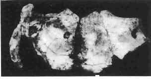
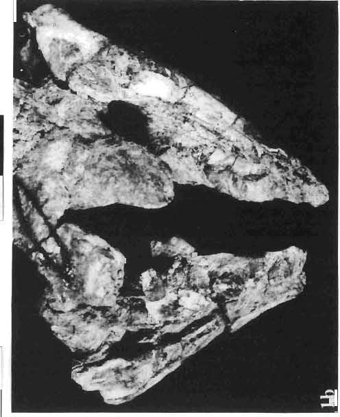
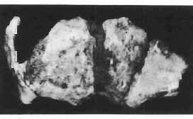
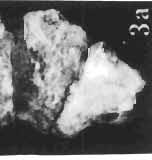
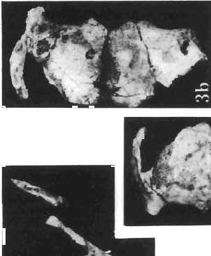

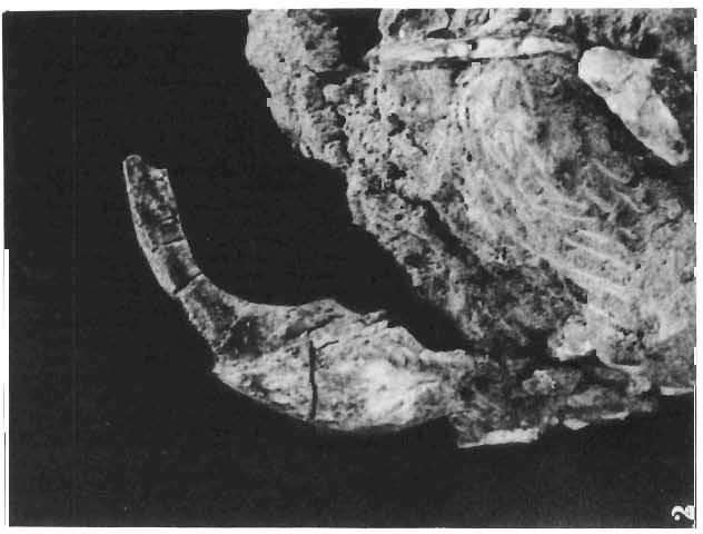

51 Palaeontologia Polonica, No. 33, 1975 PI. XXXVlI T. MARYANSKA & H. OSM6LSKA: PROTOCERATOPSIDAE


52 Palaeontologia Polonica, No. 33, 1975 PI. XXXV11l T. MARYANSKA & H. OSM6LSKA: PROTOCERATOPSIDAE
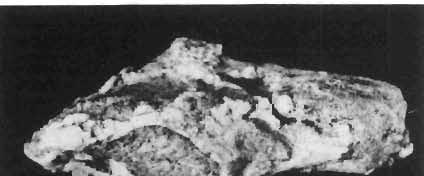
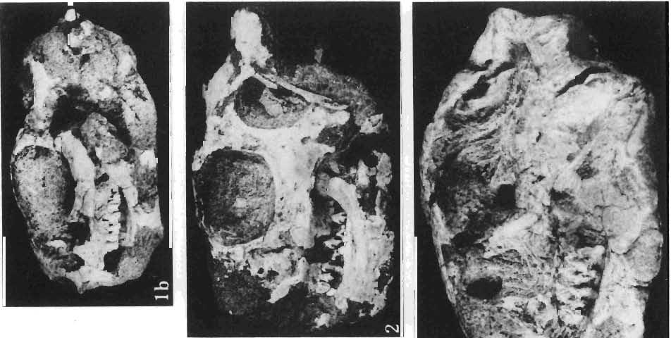
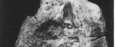
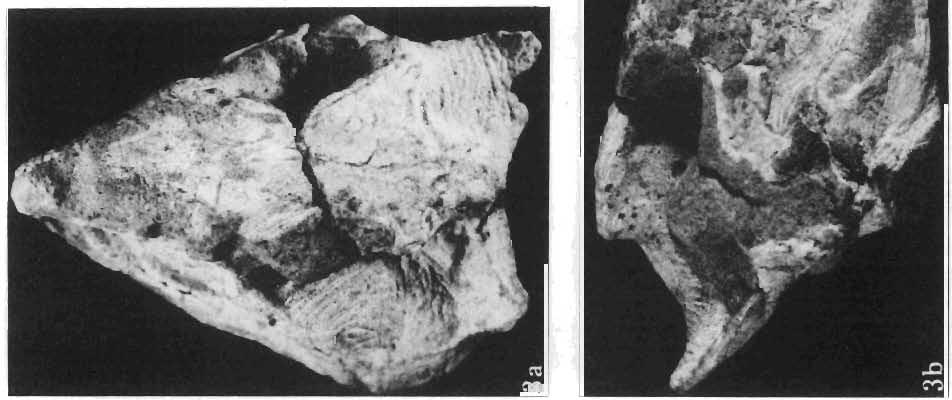
53 Palaeontologia Polonica, No. 33, 1975 PI. XXXIX T. MARYANSKA & H. OSM6LSKA: PROTOCERATOPSIDAE
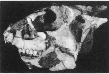
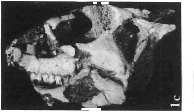
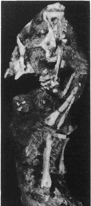
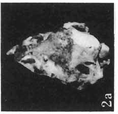
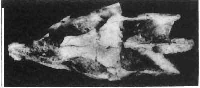
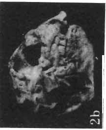
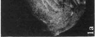
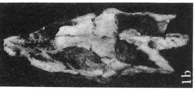
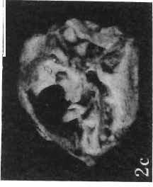
54 Palaeontologia Polonica, No. 33, 1975 PI. XL T. MARYANSKA & H. OSM6LSKA: PROTOCERATOPSIDAE
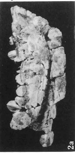
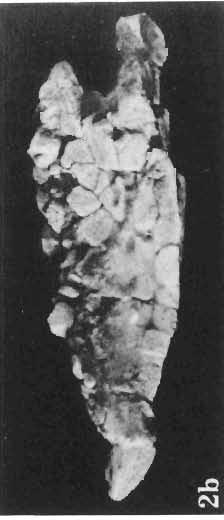
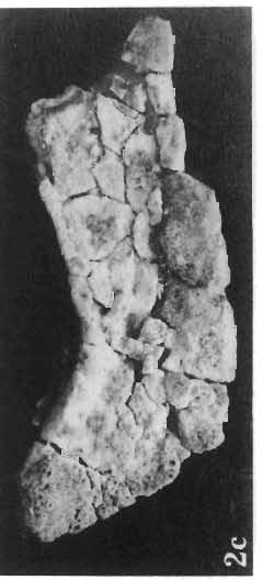
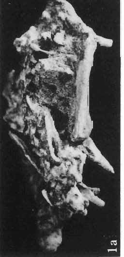
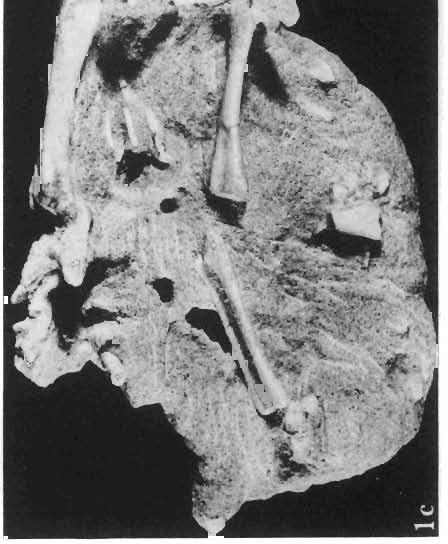
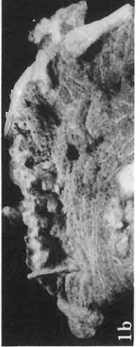
55 Palaeontologia Polonica, No. 33, 1975 PI. XLI T. MARYANSKA & H. OSMOLSKA: PROTOCERATOPSIDAE
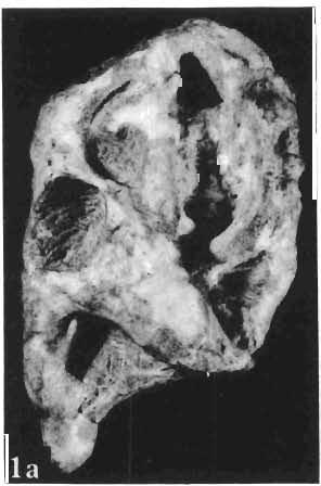
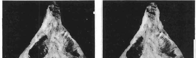
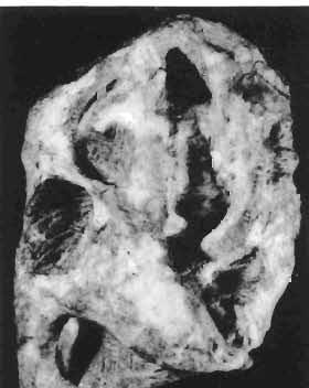
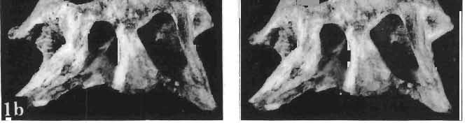
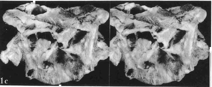
56 Palaeontologia Polonica, No. 33, 1975 PI. XLII T. MARYANSKA & H. OSM6LSK A: PROTO CERATOPSIDAE
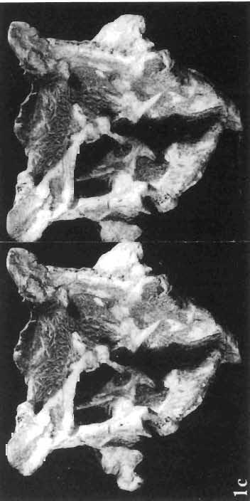
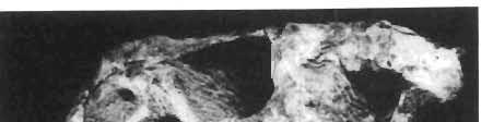
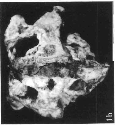
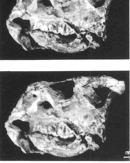
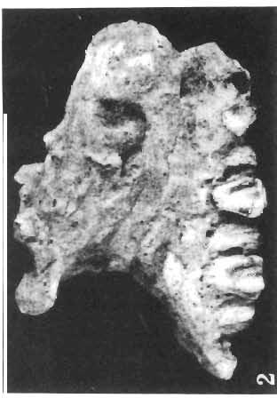
57 Palaeontologia Polonica, No. 33, 1975 PI. XL/IJ T. M ARYANSKA & H. O SM6 LSKA : P ROTOCERATOP SID AE
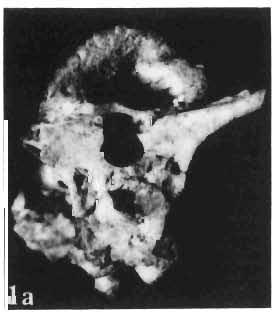
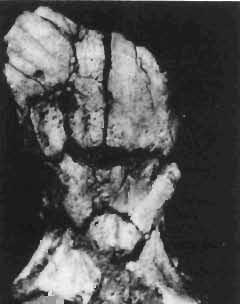
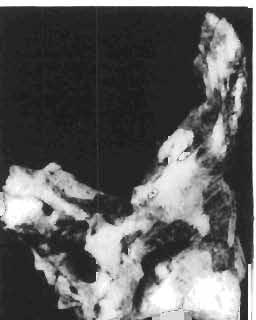
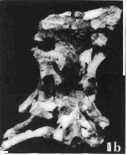
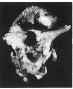
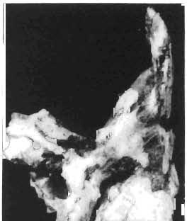
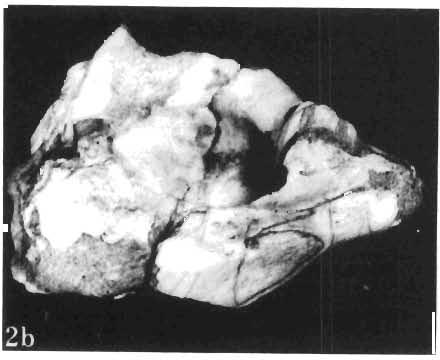
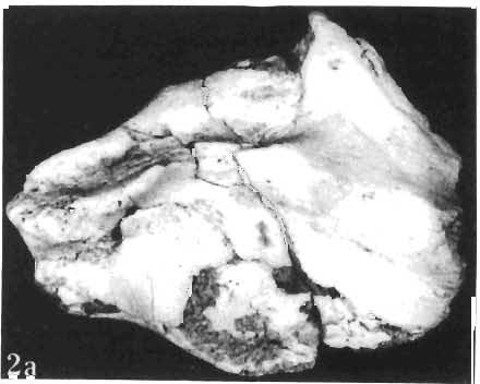
58 Palaeontologia Polonica, No. 33, 1975 PI. XLIV T. MARYANSKA & H. OSM6LSKA : PROTOCERATOPSIDAE
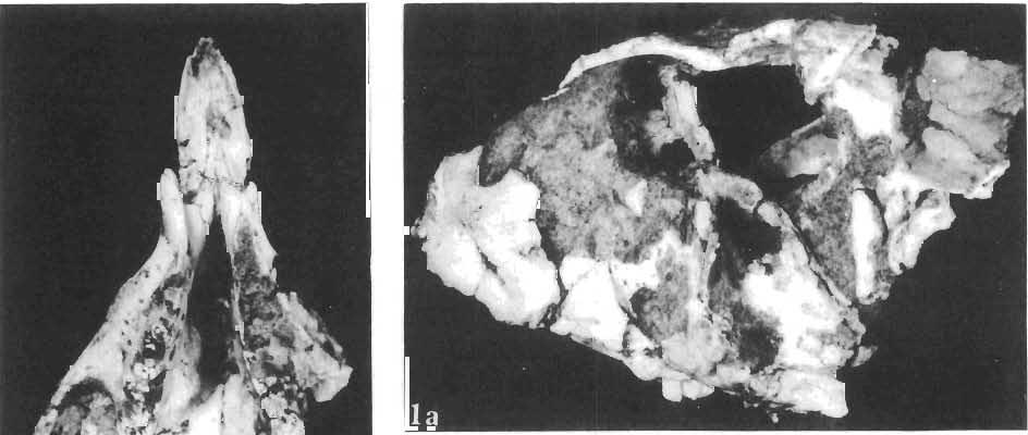
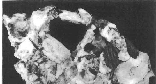
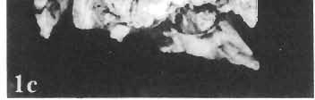
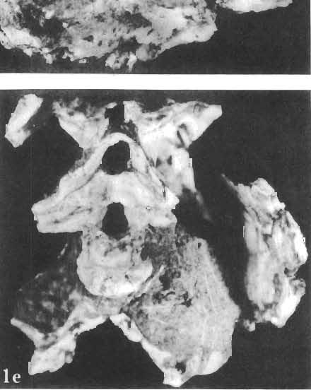
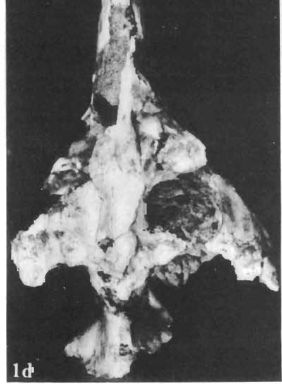
59 Palaeontologia Polonica, No. 33, 1975 PI. XLV T. MARYANSKA & H. OSMOLSKA: PROTOCERATOPSIDAE
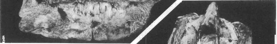

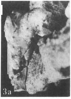
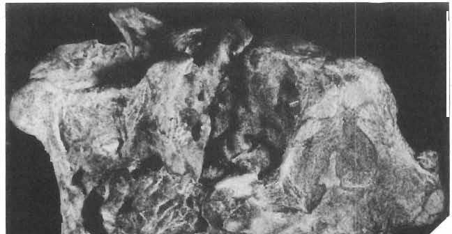
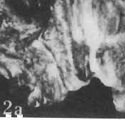
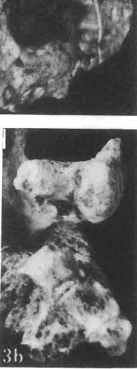
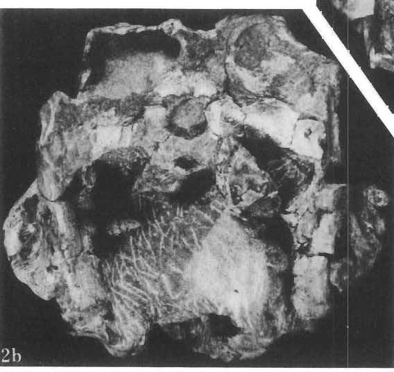
60 Palaeontologia Polonica, No. 33, 1975 PI. XLVI T. MARYANSKA & H. OSMOLSKA: PROTOCERATOPSIDAE


61 Palaeontologia Polonica, No. 33, 1975 PI. XLVIJ T. MARYANSKA & H. OSMOLSKA: PROTOCERATOPSIDAE


62 Palaeontologia Polonica, No. 33, 1975 PI. XLVIII T. MARYANSKA & H. OSM6LSKA: PROTOCERATOPSIDAE
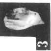
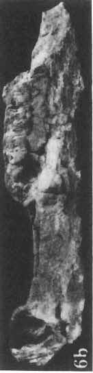
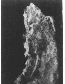
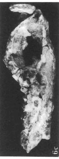
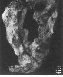
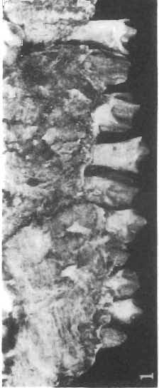
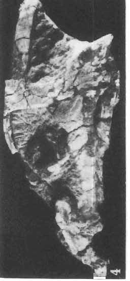
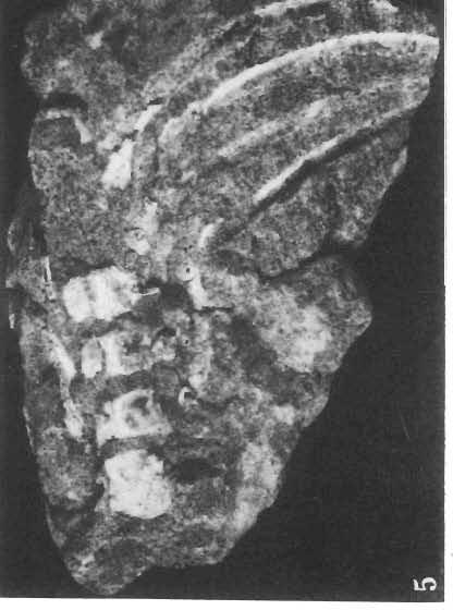
63 Palaeontologia Polonica, No. 33, 1975 PI. X LIX T. MARYANSKA & H. OSM6LSKA: PROTOCERATOPSIDAE
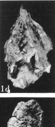
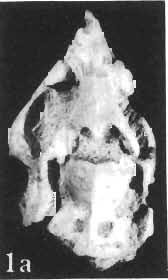
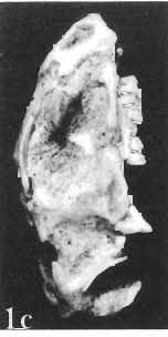
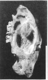
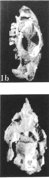
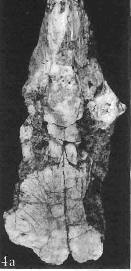
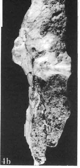
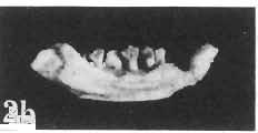
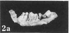
64 Palaeontologia Polonica, No. 33, 1975 PI. L 2b~ T. MARYANSKA & H. OSMOLSKA: PROTOCERATOPSIDAE
New Carnivorous Dinosaurs from the Upper Cretaceous of Mongolia
 1955 Doklady, Academy of Sciences USSR 104 (5):779-783 New Carnivorous Dinosaurs from the Upper Cretaceous of Mongolia E. A. Maleev (translated by F. J. Alcock) The present article is a summary containing
Mammalogy Laboratory 1 - Mammalian Anatomy
 Mammalogy Laboratory 1 - Mammalian Anatomy I. The Goal. The goal of the lab is to teach you skeletal anatomy of mammals. We will emphasize the skull because many of the taxonomically important characters
AMERICAN MUSEUM NOVITATES Published by
 AMERICAN MUSEUM NOVITATES Published by Number 782 THE AmzRICAN MUSEUM OF NATURAL HISTORY Feb. 20, 1935 New York City 56.81, 7 G (68) A NOTE ON THE CYNODONT, GLOCHINODONTOIDES GRACILIS HAUGHTON BY LIEUWE
YANGCHUANOSAURUS HEPINGENSIS - A NEW SPECIES OF CARNOSAUR FROM ZIGONG, SICHUAN
 Vol. 30, No. 4 VERTEBRATA PALASIATICA pp. 313-324 October 1992 [SICHUAN ZIGONG ROUSHILONG YI XIN ZHONG] figs. 1-5, pl. I-III YANGCHUANOSAURUS HEPINGENSIS - A NEW SPECIES OF CARNOSAUR FROM ZIGONG, SICHUAN
SUPPLEMENTARY ONLINE MATERIAL FOR. Nirina O. Ratsimbaholison, Ryan N. Felice, and Patrick M. O connor
 http://app.pan.pl/som/app61-ratsimbaholison_etal_som.pdf SUPPLEMENTARY ONLINE MATERIAL FOR Nirina O. Ratsimbaholison, Ryan N. Felice, and Patrick M. O connor Ontogenetic changes in the craniomandibular
.56 m. (22 in.). COMPSOGNATHOID DINOSAUR FROM THE. Medicine Bow, Wyoming, by the American Museum Expedition
. COMPSOGNATHOID DINOSAUR FROM THE. Medicine Bow, Wyoming, by the American Museum Expedition") Article XII.-ORNITHOLESTES HERMANNI, A NEW COMPSOGNATHOID DINOSAUR FROM THE UPPER JURASSIC. By HENRY FAIRFIELD OSBORN. The type skeleton (Amer. Mus. Coll. No. 6I9) of this remarkable animal was discovered
Article XII.-ORNITHOLESTES HERMANNI, A NEW COMPSOGNATHOID DINOSAUR FROM THE UPPER JURASSIC. By HENRY FAIRFIELD OSBORN. The type skeleton (Amer. Mus. Coll. No. 6I9) of this remarkable animal was discovered
A new species of Hsisosuchus (Mesoeucrocodylia) from Dashanpu, Zigong Municipality, Sichuan Province
 from Dashanpu, Zigong Municipality, Sichuan Province") A new species of Hsisosuchus (Mesoeucrocodylia) from Dashanpu, Zigong Municipality, Sichuan Province Yuhui Gao (Zigong Dinosaur Museum) Vertebrata PalAsiatica Volume 39, No. 3 July, 2001 pp. 177-184 Translated
A new species of Hsisosuchus (Mesoeucrocodylia) from Dashanpu, Zigong Municipality, Sichuan Province Yuhui Gao (Zigong Dinosaur Museum) Vertebrata PalAsiatica Volume 39, No. 3 July, 2001 pp. 177-184 Translated
Anatomy. Name Section. The Vertebrate Skeleton
 Name Section Anatomy The Vertebrate Skeleton Vertebrate paleontologists get most of their knowledge about past organisms from skeletal remains. Skeletons are useful for gleaning information about an organism
2. Skull, total length versus length of the presacral vertebral column: (0); extremely elongated neck (e.g. Tanystropheus longobardicus).
; extremely elongated neck (e.g. Tanystropheus longobardicus).") Character list of the taxon-character data set 1. Skull and lower jaws, interdental plates: absent (0); present, but restricted to the anterior end of the dentary (1); present along the entire alveolar
Character list of the taxon-character data set 1. Skull and lower jaws, interdental plates: absent (0); present, but restricted to the anterior end of the dentary (1); present along the entire alveolar
A NEW GENUS AND SPECIES OF AMERICAN THEROMORPHA
 A NEW GENUS AND SPECIES OF AMERICAN THEROMORPHA MYCTEROSAURUS LONGICEPS S. W. WILLISTON University of Chicago The past summer, Mr. Herman Douthitt, of the University of Chicago paleontological expedition,
A Short Report on the Occurrence of Dilophosaurus from Jinning County, Yunnan Province
 A Short Report on the Occurrence of Dilophosaurus from Jinning County, Yunnan Province by Hu Shaojin (Kunming Cultural Administrative Committee, Yunnan Province) Vertebrata PalAsiatica Vol. XXXI, No. 1
A new species of sauropod, Mamenchisaurus anyuensis sp. nov.
 A new species of sauropod, Mamenchisaurus anyuensis sp. nov. by Xinlu He, Suihua Yang, Kaiji Cai, Kui Li, and Zongwen Liu Chengdu University of Technology Papers on Geosciences Contributed to the 30th
A NEW ANKYLOSAUR FROM THE UPPER CRETACEOUS OF MONGOLIA E.A. Maleev Doklady Akademii Nauk, SSSR 87:
 translated by Dr. Tamara and F. Jeletzky, 1956 A NEW ANKYLOSAUR FROM THE UPPER CRETACEOUS OF MONGOLIA E.A. Maleev 1952. Doklady Akademii Nauk, SSSR 87:273-276 Armored dinosaurs make a considerable part
Exceptional fossil preservation demonstrates a new mode of axial skeleton elongation in early ray-finned fishes
 Supplementary Information Exceptional fossil preservation demonstrates a new mode of axial skeleton elongation in early ray-finned fishes Erin E. Maxwell, Heinz Furrer, Marcelo R. Sánchez-Villagra Supplementary
ONLINE APPENDIX 1. Morphological phylogenetic characters scored in this paper. See Poe (2004) for
 for") ONLINE APPENDIX Morphological phylogenetic characters scored in this paper. See Poe () for detailed character descriptions, citations, and justifications for states. Note that codes are changed from a
ONLINE APPENDIX Morphological phylogenetic characters scored in this paper. See Poe () for detailed character descriptions, citations, and justifications for states. Note that codes are changed from a
A M E G H I N I A N A. Revista de la Asociación Paleontológia Argentina. Volume XV September-December 1978 Nos. 3-4
 A M E G H I N I A N A Revista de la Asociación Paleontológia Argentina Volume XV September-December 1978 Nos. 3-4 COLORADIA BREVIS N. G. ET N. SP. (SAURISCHIA, PROSAUROPODA), A PLATEOSAURID DINOSAUR FROM
A new basal sauropodiform dinosaur from the Lower Jurassic of Yunnan Province, China
 SUPPLEMENTARY INFORMATION A new basal sauropodiform dinosaur from the Lower Jurassic of Yunnan Province, China Ya-Ming Wang 1, Hai-Lu You 2,3 *, Tao Wang 4 1 School of Earth Sciences and Resources, China
Williston, and as there are many fairly good specimens in the American
 56.81.7D :14.71.5 Article VII.- SOME POINTS IN THE STRUCTURE OF THE DIADECTID SKULL. BY R. BROOM. The skull of Diadectes has been described by Cope, Case, v. Huene, and Williston, and as there are many
List of characters used in the phylogenetic analysis. Capital letters T, R, and L, refer to
 1 Supplementary data CHARACTER LIST List of characters used in the phylogenetic analysis. Capital letters T, R, and L, refer to characters used by Tchernov et al. (2000), Rieppel, et al. (2002), and Lee
MACROCEPHALOSAURIDAE AND POLYGL YPHANODONTIDAE (SAURIA) FROM THE LATE CRETACEOUS OF MONGOLIA
 FROM THE LATE CRETACEOUS OF MONGOLIA") ANDRZEJ SULIMSKI MACROCEPHALOSAURIDAE AND POLYGL YPHANODONTIDAE (SAURIA) FROM THE LATE CRETACEOUS OF MONGOLIA (Plates VIII-XXVII) Abstract - Large Late Cretaceous lizards from the?santonian Djadokhta Formation,?
ANDRZEJ SULIMSKI MACROCEPHALOSAURIDAE AND POLYGL YPHANODONTIDAE (SAURIA) FROM THE LATE CRETACEOUS OF MONGOLIA (Plates VIII-XXVII) Abstract - Large Late Cretaceous lizards from the?santonian Djadokhta Formation,?
FURTHER STUDIES ON TWO SKELETONS OF THE BLACK RIGHT WHALE IN THE NORTH PACIFIC
 FURTHER STUDIES ON TWO SKELETONS OF THE BLACK RIGHT WHALE IN THE NORTH PACIFIC HIDEO OMURA, MASAHARU NISHIWAKI* AND TOSHIO KASUYA* ABSTRACT Two skeletons of the black right whale were studied, supplementing
PALEONTOLOGY AND BIOSTRATIGRAPHY OF MONGOLIA
 PALEONTOLOGY AND BIOSTRATIGRAPHY OF MONGOLIA THE JOINT SOVIET-MONGOLIAN PALEONTOLOGICAL EXPEDITION (Transactions, vol. 3) EDITORIAL BOARD: N. N. Kramarenko (editor-in-chief) B. Luvsandansan, Yu. I. Voronin,
SOME LITTLE-KNOWN FOSSIL LIZARDS FROM THE
 PROCEEDINGS OF THE UNITED STATES NATIONAL MUSEUM issued SWsK \ {^^m ^V ^^ SMITHSONIAN INSTITUTION U. S. NATIONAL MUSEUM Vol. 91 Washington : 1941 No. 3124 SOME LITTLE-KNOWN FOSSIL LIZARDS FROM THE OLIGOCENE
NOTES ON THE FIRST SKULL AND JAWS OF RIOJASAURUS INCERTUS (DINOSAURIA, PROSAUROPODA, MELANOROSAURIDAE) OF THE LATE TRIASSIC OF LA RIOJA, ARGENTINA
 OF THE LATE TRIASSIC OF LA RIOJA, ARGENTINA") NOTES ON THE FIRST SKULL AND JAWS OF RIOJASAURUS INCERTUS (DINOSAURIA, PROSAUROPODA, MELANOROSAURIDAE) OF THE LATE TRIASSIC OF LA RIOJA, ARGENTINA José F. Bonaparte and José A. Pumares translated by Jeffrey
NOTES ON THE FIRST SKULL AND JAWS OF RIOJASAURUS INCERTUS (DINOSAURIA, PROSAUROPODA, MELANOROSAURIDAE) OF THE LATE TRIASSIC OF LA RIOJA, ARGENTINA José F. Bonaparte and José A. Pumares translated by Jeffrey
A new sauropod from Dashanpu, Zigong Co. Sichuan Province (Abrosaurus dongpoensis gen. et sp. nov.)
") A new sauropod from Dashanpu, Zigong Co. Sichuan Province (Abrosaurus dongpoensis gen. et sp. nov.) by Ouyang Hui Zigong Dinosaur Museum Newsletter Number 2 1989 pp. 10-14 Translated By Will Downs Bilby
A new sauropod from Dashanpu, Zigong Co. Sichuan Province (Abrosaurus dongpoensis gen. et sp. nov.) by Ouyang Hui Zigong Dinosaur Museum Newsletter Number 2 1989 pp. 10-14 Translated By Will Downs Bilby
SUPPLEMENTARY INFORMATION
 Character 155, interdental ridges. Absence of interdental ridge (0) shown in Parasaniwa wyomingensis (Platynota). Interdental ridges (1) shown in Coniophis precedens. WWW.NATURE.COM/NATURE 1 Character
( M amenchisaurus youngi Pi, Ouyang et Ye, 1996)
") 39 4 2001 10 V ERTEBRATA PALASIATICA pp. 266 271 fig. 1,pl. I ( 643013), ( M amenchisaurus hochuanensis),,, Q915. 864 1995 12 31 (ZDM0126) ( M amenchisau rus hochuanensis Young et Chao, 1972),,, ZDM0126
39 4 2001 10 V ERTEBRATA PALASIATICA pp. 266 271 fig. 1,pl. I ( 643013), ( M amenchisaurus hochuanensis),,, Q915. 864 1995 12 31 (ZDM0126) ( M amenchisau rus hochuanensis Young et Chao, 1972),,, ZDM0126
Jurassic Ornithopod Agilisaurus louderbacki (Ornithopoda: Fabrosauridae) from Zigong, Sichuan, China
 from Zigong, Sichuan, China") Jurassic Ornithopod Agilisaurus louderbacki (Ornithopoda: Fabrosauridae) from Zigong, Sichuan, China Guangzhao Peng (Zigong Dinosaur Museum) Vertebrata PalAsiatica Volume 30, No. 1 January, 1992 pp. 39-51
Jurassic Ornithopod Agilisaurus louderbacki (Ornithopoda: Fabrosauridae) from Zigong, Sichuan, China Guangzhao Peng (Zigong Dinosaur Museum) Vertebrata PalAsiatica Volume 30, No. 1 January, 1992 pp. 39-51
THE SKULLS OF ARAEOSCELIS AND CASEA, PERMIAN REPTILES
 THE SKULLS OF REOSCELIS ND CSE, PERMIN REPTILES University of Chicago There are few Permian reptiles of greater interest at the present time than the peculiar one I briefly described in this journal' three
v:ii-ixi, 'i':;iisimvi'\>!i-:: "^ A%'''''-'^-''S.''v.--..V^'E^'-'-^"-t''gi L I E) R.ARY OF THE VERSITY U N I or ILLINOIS REMO
 R.ARY OF THE VERSITY U N I or ILLINOIS REMO") "^ A%'''''-'^-''S.''v.--..V^'E^'-'-^"-t''gi v:ii-ixi, 'i':;iisimvi'\>!i-:: L I E) R.ARY OF THE U N I VERSITY or ILLINOIS REMO Natural History Survey Librarv GEOLOGICAL SERIES OF FIELD MUSEUM OF NATURAL
"^ A%'''''-'^-''S.''v.--..V^'E^'-'-^"-t''gi v:ii-ixi, 'i':;iisimvi'\>!i-:: L I E) R.ARY OF THE U N I VERSITY or ILLINOIS REMO Natural History Survey Librarv GEOLOGICAL SERIES OF FIELD MUSEUM OF NATURAL
Major cranial changes during Triceratops ontogeny John R. Horner 1, * and Mark B. Goodwin 2
 273, 2757 2761 doi:10.1098/rspb.2006.3643 Published online 1 August 2006 Major cranial changes during Triceratops ontogeny John R. Horner 1, * and Mark B. Goodwin 2 1 Museum of the Rockies, Montana State
CHARACTER LIST: Nesbitt et al., 2011
 CHARACTER LIST: Nesbitt et al., 2011 1. Vaned feathers on forelimb symmetric (0) or asymmetric (1). The barbs on opposite sides of the rachis differ in length; in extant birds, the barbs on the leading
A new carnosaur from Yongchuan County, Sichuan Province
 A new carnosaur from Yongchuan County, Sichuan Province by Dong Zhiming Institute of Vertebrate Palaeontology and Palaeoanthropology, Academia Sinica Zhang Yihong, Li Xuanmin, and Zhou Shiwu Chongqing
The cranial osteology of Belebey vegrandis (Parareptilia: Bolosauridae), from the Middle Permian of Russia, and its bearing on reptilian evolution
, from the Middle Permian of Russia, and its bearing on reptilian evolution") Blackwell Publishing LtdOxford, UKZOJZoological Journal of the Linnean Society0024-4082 2007 The Linnean Society of London? 2007 1511 191214 Original Articles RUSSIAN BOLOSAURID REPTILER. R. REISZ ET AL.
Blackwell Publishing LtdOxford, UKZOJZoological Journal of the Linnean Society0024-4082 2007 The Linnean Society of London? 2007 1511 191214 Original Articles RUSSIAN BOLOSAURID REPTILER. R. REISZ ET AL.
Supporting Online Material for
 www.sciencemag.org/cgi/content/full/329/5998/1481/dc1 Supporting Online Material for Tyrannosaur Paleobiology: New Research on Ancient Exemplar Organisms Stephen L. Brusatte,* Mark A. Norell, Thomas D.
Basal Ceratopsia. Definition and Diagnosis. Anatomy TWENTY-TWO YOU HAILU PETER DODSON
 TWENTY-TWO Basal Ceratopsia YOU HAILU PETER DODSON Ceratopsia consists of Psittacosauridae and Neoceratopsia, the latter formed by numerous basal taxa and Ceratopsidae. Consequently, this chapter on basal
Fig. 5. (A) Scaling of brain vault size (width measured at the level of anterior squamosal/parietal suture) relative to skull size (measured at the
 Scaling of brain vault size (width measured at the level of anterior squamosal/parietal suture) relative to skull size (measured at the") Fig. 5. (A) Scaling of brain vault size (width measured at the level of anterior squamosal/parietal suture) relative to skull size (measured at the distance between the left versus right temporomandibular
Fig. 5. (A) Scaling of brain vault size (width measured at the level of anterior squamosal/parietal suture) relative to skull size (measured at the distance between the left versus right temporomandibular
AMERICAN MUSEUM NOVITATES
 AMERICAN MUSEUM NOVITATES Published by Number 144 THz AmzxzcAN MusumokorNATURAL HISTORY Novemoer 7, 1924 56.81,9T(117:51.7) THREE NEW THEROPODA, PROTOCERATOPS ZONE, CENTRAL MONGOLIA' BY HENRY FAIRFIELD
A primitive pachycephalosaurid from the Cretaceous of Anhui, China, Wannanosaurus yansiensis gen. et sp. nov
 A primitive pachycephalosaurid from the Cretaceous of Anhui, China, Wannanosaurus yansiensis gen. et sp. nov by Lianhai Hou Institute of Vertebrate Paleontology and Paleoanthropology Vertebrata PalAsiatica
Recently Mr. Lawrence M. Lambe has described and figured in the
 56.81,9C(117:71.2) Article XXXV.-CORYTHOSAURUS CASUARIUS, A NEW CRESTED DINOSAUR FROM THE BELLY RIVER CRETA- CEOUS, WITH PROVISIONAL CLASSIFICATION OF THE FAMILY TRACHODONTIDA1X BY BARNUM BROWN. PLATE
CRANIAL ANATOMY OF ENNATOSAURUS TECTON (SYNAPSIDA: CASEIDAE) FROM THE MIDDLE PERMIAN OF RUSSIA AND THE EVOLUTIONARY RELATIONSHIPS OF CASEIDAE
 FROM THE MIDDLE PERMIAN OF RUSSIA AND THE EVOLUTIONARY RELATIONSHIPS OF CASEIDAE") Journal of Vertebrate Paleontology 28(1):160 180, March 2008 2008 by the Society of Vertebrate Paleontology ARTICLE CRANIAL ANATOMY OF ENNATOSAURUS TECTON (SYNAPSIDA: CASEIDAE) FROM THE MIDDLE PERMIAN
Journal of Vertebrate Paleontology 28(1):160 180, March 2008 2008 by the Society of Vertebrate Paleontology ARTICLE CRANIAL ANATOMY OF ENNATOSAURUS TECTON (SYNAPSIDA: CASEIDAE) FROM THE MIDDLE PERMIAN
Supporting Online Material for
 www.sciencemag.org/cgi/content/full/317/5843/1378/dc1 Supporting Online Material for A Basal Dromaeosaurid and Size Evolution Preceding Avian Flight Alan H. Turner,* Diego Pol, Julia A. Clarke, Gregory
AMERICAN NATURALIST. Vol. IX. -DECEMBER, No. 12. OR BIRDS WITH TEETH.1 OI)ONTORNITHES,
ONTORNITHES,") AMERICAN NATURALIST. Vol. IX. -DECEMBER, 1875.-No. 12. OI)ONTORNITHES, OR BIRDS WITH TEETH.1 BY PROFESSOR 0. C. MARSH. REMAINS of birds are amono the rarest of fossils, and few have been discovered except
AMERICAN NATURALIST. Vol. IX. -DECEMBER, 1875.-No. 12. OI)ONTORNITHES, OR BIRDS WITH TEETH.1 BY PROFESSOR 0. C. MARSH. REMAINS of birds are amono the rarest of fossils, and few have been discovered except
Toothless Dinosaurs of Mongolia. R. Barsbold. Fossil Vertebrates of Mongolia Academy of Sciences of USSR Paleontological Institute
 Toothless Dinosaurs of Mongolia R. Barsbold Fossil Vertebrates of Mongolia Academy of Sciences of USSR Paleontological Institute Academy of Sciences of the MPR Geological Institute Nauka Moscow 1981 Trudy
Supplementary information to A new troodontid dinosaur from China with avian-like sleeping-posture. Xing Xu 1 and Mark Norell 2
 Supplementary information to A new troodontid dinosaur from China with avian-like sleeping-posture Xing Xu 1 and Mark Norell 2 1 Institute of Vertebrate Paleontology & Paleoanthropology, Chinese Academy
First Ornithomimid (Theropoda, Ornithomimosauria) from the Upper Cretaceous Djadokhta Formation of Tögrögiin Shiree, Mongolia
 from the Upper Cretaceous Djadokhta Formation of Tögrögiin Shiree, Mongolia") First Ornithomimid (Theropoda, Ornithomimosauria) from the Upper Cretaceous Djadokhta Formation of Tögrögiin Shiree, Mongolia Tsogtbaatar Chinzorig¹, ³ *, Yoshitsugu Kobayashi², Khishigjav Tsogtbaatar³,
First Ornithomimid (Theropoda, Ornithomimosauria) from the Upper Cretaceous Djadokhta Formation of Tögrögiin Shiree, Mongolia Tsogtbaatar Chinzorig¹, ³ *, Yoshitsugu Kobayashi², Khishigjav Tsogtbaatar³,
On the Discovery of the earliest fossil bird in China (Sinosauropteryx gen. nov.) and the origin of birds
 and the origin of birds") On the Discovery of the earliest fossil bird in China (Sinosauropteryx gen. nov.) and the origin of birds by Qiang Ji and Shu an Ji Chinese Geological Museum, Beijing Chinese Geology Volume 233 1996 pp.
On the Discovery of the earliest fossil bird in China (Sinosauropteryx gen. nov.) and the origin of birds by Qiang Ji and Shu an Ji Chinese Geological Museum, Beijing Chinese Geology Volume 233 1996 pp.
Yamaceratops dorngobiensis, a New Primitive Ceratopsian (Dinosauria: Ornithischia) from the Cretaceous of Mongolia
 from the Cretaceous of Mongolia") PUBLISHED BY THE AMERICAN MUSEUM OF NATURAL HISTORY CENTRAL PARK WEST AT 79TH STREET, NEW YORK, NY 10024 Number 3530, 42 pp., 20 figures September 08, 2006 Yamaceratops dorngobiensis, a New Primitive Ceratopsian
PUBLISHED BY THE AMERICAN MUSEUM OF NATURAL HISTORY CENTRAL PARK WEST AT 79TH STREET, NEW YORK, NY 10024 Number 3530, 42 pp., 20 figures September 08, 2006 Yamaceratops dorngobiensis, a New Primitive Ceratopsian
4. Premaxilla: Foramen on the lateral surface of the premaxillary body (Yates 2007 ch. 4) 0 absent 1 present
 0 absent 1 present") The character matrix used as a basis for this study is that of Yates et al (2010) which is modified from the earlier matrix used by Yates (2007). This matrix includes characters acquired and/or modified
The character matrix used as a basis for this study is that of Yates et al (2010) which is modified from the earlier matrix used by Yates (2007). This matrix includes characters acquired and/or modified
A New Ceratopsian Dinosaur from the Upper
 SMITHSONIAN MISCELLANEOUS COLLECTIONS VOLUME 63. NUMBER 3 A New Ceratopsian Dinosaur from the Upper Cretaceous of Montana, with Note on Hypacrosaurus (With Two Plates) CHARLES W. GILMORE Assistant Curator
 The following text is generated from uncorrected OCR. [Begin Page: Page 1] A NEW CERATOPSIAN DINOSAUR FROM THE UPPER CRETACEOUS OF MONTANA, WITH NOTE ON HYPACROSAURUS ' By CHARLES W. GILMORE assistant
APPENDIX. 344 Mni-s/i Restorations of Claosaurus and Geratosaurus.
 344 Mni-s/i Restorations of Claosaurus and Geratosaurus. Claosaurics, Marsh, 1890.* The most important feature in the restoration of Claosaurus annectens given on Plate VI is the skull, which will be fully
A New Dromaeosaurid Theropod from Ukhaa Tolgod (Ömnögov, Mongolia)
") PUBLISHED BY THE AMERICAN MUSEUM OF NATURAL HISTORY CENTRAL PARK WEST AT 79TH STREET, NEW YORK, NY 10024 Number 3545, 51 pp., 25 figures, 1 table December 7, 2006 A New Dromaeosaurid Theropod from Ukhaa
PUBLISHED BY THE AMERICAN MUSEUM OF NATURAL HISTORY CENTRAL PARK WEST AT 79TH STREET, NEW YORK, NY 10024 Number 3545, 51 pp., 25 figures, 1 table December 7, 2006 A New Dromaeosaurid Theropod from Ukhaa
THE GORGONOPSIAN GENUS, HIPPOSAURUS, AND THE FAMILY ICTIDORHINIDAE * Dr. L.D. Boonstra. Paleontologist, South African Museum, Cape Town
 THE GORGONOPSIAN GENUS, HIPPOSAURUS, AND THE FAMILY ICTIDORHINIDAE * by Dr. L.D. Boonstra Paleontologist, South African Museum, Cape Town In 1928 I dug up the complete skeleton of a smallish gorgonopsian
Mammalogy Lab 1: Skull, Teeth, and Terms
 Mammalogy Lab 1: Skull, Teeth, and Terms Be able to: Goals of today s lab Locate all structures listed on handout Define all terms on handout what they are or what they look like Give examples of mammals
A Fossil Snake (Elaphe vulpina) From A Pliocene Ash Bed In Nebraska
 From A Pliocene Ash Bed In Nebraska") University of Nebraska - Lincoln DigitalCommons@University of Nebraska - Lincoln Transactions of the Nebraska Academy of Sciences and Affiliated Societies Nebraska Academy of Sciences 198 A Fossil Snake
University of Nebraska - Lincoln DigitalCommons@University of Nebraska - Lincoln Transactions of the Nebraska Academy of Sciences and Affiliated Societies Nebraska Academy of Sciences 198 A Fossil Snake
HONR219D Due 3/29/16 Homework VI
 Part 1: Yet More Vertebrate Anatomy!!! HONR219D Due 3/29/16 Homework VI Part 1 builds on homework V by examining the skull in even greater detail. We start with the some of the important bones (thankfully
NEW INFORMATION ON THE CRANIUM OF BRACHYLOPHOSAURUS CANADENSIS (DINOSAURIA, HADROSAURIDAE), WITH A REVISION OF ITS PHYLOGENETIC POSITION
, WITH A REVISION OF ITS PHYLOGENETIC POSITION") Journal of Vertebrate Paleontology 25(1):144 156, March 2005 2005 by the Society of Vertebrate Paleontology NEW INFORMATION ON THE CRANIUM OF BRACHYLOPHOSAURUS CANADENSIS (DINOSAURIA, HADROSAURIDAE), WITH
Journal of Vertebrate Paleontology 25(1):144 156, March 2005 2005 by the Society of Vertebrate Paleontology NEW INFORMATION ON THE CRANIUM OF BRACHYLOPHOSAURUS CANADENSIS (DINOSAURIA, HADROSAURIDAE), WITH
complex in cusp pattern. (3) The bones of the coyote skull are thinner, crests sharper and the
 The bones of the coyote skull are thinner, crests sharper and the") DISTINCTIONS BETWEEN THE SKULLS OF S AND DOGS Grover S. Krantz Archaeological sites in the United States frequently yield the bones of coyotes and domestic dogs. These two canines are very similar both
DISTINCTIONS BETWEEN THE SKULLS OF S AND DOGS Grover S. Krantz Archaeological sites in the United States frequently yield the bones of coyotes and domestic dogs. These two canines are very similar both
A Complete Late Cretaceous Iguanian (Squamata, Reptilia) from the Gobi and Identification of a New Iguanian Clade
 from the Gobi and Identification of a New Iguanian Clade") PUBLISHED BY THE AMERICAN MUSEUM OF NATURAL HISTORY CENTRAL PARK WEST AT 79TH STREET, NEW YORK, NY 10024 Number 3584, 47 pp., 19 figures September 6, 2007 A Complete Late Cretaceous Iguanian (Squamata,
PUBLISHED BY THE AMERICAN MUSEUM OF NATURAL HISTORY CENTRAL PARK WEST AT 79TH STREET, NEW YORK, NY 10024 Number 3584, 47 pp., 19 figures September 6, 2007 A Complete Late Cretaceous Iguanian (Squamata,
Redescription of the Mongolian Sauropod NEMEGTOSAURUS MONGOLIENSIS Nowinski (Dinosauria:
 Journal of Systematic Palaeontology 3 (3): 283 318 Issued 24 August 2005 doi:10.1017/s1477201905001628 Printed in the United Kingdom C The Natural History Museum Redescription of the Mongolian Sauropod
A NEW SPECIES OF EXTINCT TURTLE FROM THE UPPER PLIOCENE OF IDAHO
 A NEW SPECIES OF EXTINCT TURTLE FROM THE UPPER PLIOCENE OF IDAHO By Charles W. Gilmore Curator, Division of Vertebrate Paleontology United States National Museum Among the fossils obtained bj^ the Smithsonian
Cranial osteology and phylogenetic relationships of Hamadasuchus rebouli (Crocodyliformes: Mesoeucrocodylia) from the Cretaceous of Morocco
 from the Cretaceous of Morocco") Blackwell Publishing LtdOxford, UKZOJZoological Journal of the Linnean Society0024-4082 2007 The Linnean Society of London? 2007 1494 533567 Original Articles HAMADASUCHUS REBOULIH. C. E. LARSSON and H.-D.
Blackwell Publishing LtdOxford, UKZOJZoological Journal of the Linnean Society0024-4082 2007 The Linnean Society of London? 2007 1494 533567 Original Articles HAMADASUCHUS REBOULIH. C. E. LARSSON and H.-D.
Supplementary Note 1. Additional osteological description
 Supplementary Note 1 Additional osteological description The text below provides additional details of Jianianhualong that were not pertinent to the salient osteological description provided in the main
A New Pterosaur from the Middle Jurassic of Dashanpu, Zigong, Sichuan
 A New Pterosaur from the Middle Jurassic of Dashanpu, Zigong, Sichuan by Xinlu He (Chengdu College of Geology) Daihuan Yang (Chungking Natural History Museum, Sichuan Province) Chunkang Su (Zigong Historical
A new oviraptorid from the Upper Cretaceous of Nei Mongol, China, and its stratigraphic implications
 51 2 2013 4 VERTEBRATA PALASIATICA pp. 85-101 gs. 1-3 A new oviraptorid from the Upper Cretaceous of Nei Mongol, China, and its stratigraphic implications XU Xing 1 TAN Qing-Wei 2 WANG Shuo 1,3 Corwin
Yimenosaurus, a new genus of Prosauropoda from Yimen County, Yunnan Province
 Yimenosaurus, a new genus of Prosauropoda from Yimen County, Yunnan Province by Ziqi Bai, Jie Yang, and Guohui Wang Yuxi Regional Administrative Academy of Yunnan Province Yuxiwenbo (Yuxi Culture and Scholarship)
NIVOROUS DINOSAUR. (SECOND COMMUNICATION.) By HENRY FAIRFIELD OSBORN. PLATE XXXIX. This great carnivorous Dinosaur of the Laramie was contemporary
 By HENRY FAIRFIELD OSBORN. PLATE XXXIX. This great carnivorous Dinosaur of the Laramie was contemporary") 56, 8i, 9 T (I 7: 786) Article VI.-TYRANNOSAURUS, UPPER CRETACEOUS CAR- NIVOROUS DINOSAUR. (SECOND COMMUNICATION.) By HENRY FAIRFIELD OSBORN. PLATE I. This great carnivorous Dinosaur of the Laramie was
56, 8i, 9 T (I 7: 786) Article VI.-TYRANNOSAURUS, UPPER CRETACEOUS CAR- NIVOROUS DINOSAUR. (SECOND COMMUNICATION.) By HENRY FAIRFIELD OSBORN. PLATE I. This great carnivorous Dinosaur of the Laramie was
OSTEOLOGICAL NOTE OF AN ANTARCTIC SEI WHALE
 OSTEOLOGICAL NOTE OF AN ANTARCTIC SEI WHALE MASAHARU NISHIWAKI* AND TOSHIO KASUYA* ABSTRACT This is a report of measurements on the skeleton of a male se1 whale caught in the Antarctic. The skeleton of
A Late Jurassic Protosuchian Sichuanosuchus huidongensis from Zigong, Sichuan Province. Guangzhao Peng. Zigong Dinosaur Museum, Zigong, Sichuan
 A Late Jurassic Protosuchian Sichuanosuchus huidongensis from Zigong, Sichuan Province Guangzhao Peng Zigong Dinosaur Museum, Zigong, Sichuan 643013 Vertebrata PalAsiatica Volume 34, Number 4 October,
Reexamination of a primitive ornithomimosaur, Garudimimus brevipes Barsbold, 1981 (Dinosauria: Theropoda), from the Late Cretaceous of Mongolia
, from the Late Cretaceous of Mongolia") Reexamination of a primitive ornithomimosaur, Garudimimus brevipes Barsbold, 1981 (Dinosauria: Theropoda), from the Late Cretaceous of Mongolia Yoshitsugu Kobayashi and Rinchen Barsbold 1501 Abstract:
Reexamination of a primitive ornithomimosaur, Garudimimus brevipes Barsbold, 1981 (Dinosauria: Theropoda), from the Late Cretaceous of Mongolia Yoshitsugu Kobayashi and Rinchen Barsbold 1501 Abstract:
Redescription of neoceratopsian dinosaur Archaeoceratops and early evolution of Neoceratopsia
 Redescription of neoceratopsian dinosaur Archaeoceratops and early evolution of Neoceratopsia HAI LU YOU and PETER DODSON You, H. L. and Dodson, P. 2003. Redescription of neoceratopsian dinosaur Archaeoceratops
A NEW CROCODYLOMORPH ARCHOSAUR FROM THE UPPER TRIASSIC OF NORTH CAROLINA
 Journal of Vertebrate Paleontology 23(2):329 343, June 2003 2003 by the Society of Vertebrate Paleontology A NEW CROCODYLOMORPH ARCHOSAUR FROM THE UPPER TRIASSIC OF NORTH CAROLINA HANS-DIETER SUES 1 *,
On the morphoplogy and taxonomic status of Xinpusaurus kohi JIANG et al., 2004 (Diapsida: Thalattosauria) from the Upper Triassic of China
 from the Upper Triassic of China") Palaeodiversity 7: 47 59; Stuttgart 30 December 2014. 47 On the morphoplogy and taxonomic status of Xinpusaurus kohi JIANG et al., 2004 (Diapsida: Thalattosauria) from the Upper Triassic of China MICHAEL
Palaeodiversity 7: 47 59; Stuttgart 30 December 2014. 47 On the morphoplogy and taxonomic status of Xinpusaurus kohi JIANG et al., 2004 (Diapsida: Thalattosauria) from the Upper Triassic of China MICHAEL
Description of Cranial Elements and Ontogenetic Change within Tropidolaemus wagleri (Serpentes: Crotalinae).
.") East Tennessee State University Digital Commons @ East Tennessee State University Electronic Theses and Dissertations 5-2016 Description of Cranial Elements and Ontogenetic Change within Tropidolaemus
East Tennessee State University Digital Commons @ East Tennessee State University Electronic Theses and Dissertations 5-2016 Description of Cranial Elements and Ontogenetic Change within Tropidolaemus
OF THE TRIAS THE PHYTOSAURIA
 THE PHYTOSAURIA OF THE TRIAS MAURICE G. MEHL University of Wisconsin Some time ago the writer gave a brief notice of a new genus of phytosaurs of which Angistorhinus grandis Mehl was the type.' It is the
A new Middle Jurassic sauropod subfamily (Klamelisaurinae subfam. nov.) from Xinjiang Autonomous Region, China
 from Xinjiang Autonomous Region, China") A new Middle Jurassic sauropod subfamily (Klamelisaurinae subfam. nov.) from Xinjiang Autonomous Region, China by Xijing Zhao Institute of Vertebrate Paleontology and Paleoanthropology, Academia Sinica
A new Middle Jurassic sauropod subfamily (Klamelisaurinae subfam. nov.) from Xinjiang Autonomous Region, China by Xijing Zhao Institute of Vertebrate Paleontology and Paleoanthropology, Academia Sinica
Biology 3315 Comparative Vertebrate Morphology Skulls and Visceral Skeletons
 Biology 3315 Comparative Vertebrate Morphology Skulls and Visceral Skeletons 1. Head skeleton of lamprey Cyclostomes are highly specialized in both the construction of the chondrocranium and visceral skeleton.
New Specimens of Microraptor zhaoianus (Theropoda: Dromaeosauridae) from Northeastern China
 from Northeastern China") PUBLISHED BY THE AMERICAN MUSEUM OF NATURAL HISTORY CENTRAL PARK WEST AT 79TH STREET, NEW YORK, NY 10024 Number 3381, 44 pp., 31 figures, 2 tables August 16, 2002 New Specimens of Microraptor zhaoianus
PUBLISHED BY THE AMERICAN MUSEUM OF NATURAL HISTORY CENTRAL PARK WEST AT 79TH STREET, NEW YORK, NY 10024 Number 3381, 44 pp., 31 figures, 2 tables August 16, 2002 New Specimens of Microraptor zhaoianus
Notes on Ceratopsians and Ankylosaurs at the Royal Ontario Museum
 Notes on Ceratopsians and Ankylosaurs at the Royal Ontario Museum Andrew A. Farke, Ph.D. Raymond M. Alf Museum of Paleontology 1175 West Baseline Road Claremont, CA 91711 email: afarke@webb.org Introduction
A NEW SPECIES OF TROODONT DINOSAUR FROM THE
 A NEW SPECIES OF TROODONT DINOSAUR FROM THE LANCE FORMATION OF WYOMING By Charles W. Gilmore Curator of Vertebrate Paleontology, United States National Museum INTRODUCTION The intensive search to which
Bulletin of Big Bend Paleo-Geo An Open Access Publication from Mosasaur Ranch Museum, Terlingua and Lajitas, Texas All rights reserved
 Bulletin of Big Bend Paleo-Geo An Open Access Publication from Mosasaur Ranch Museum, Terlingua and Lajitas, Texas All rights reserved This was a private report in 2003 on my thoughts on Platecarpus planifrons.
IU Press uncorrected proof. Copyrighted material. For proofing purposes only.
 26.1. Hexing qingyi, JLUM-JZ07b1 (holotype). Skull in right lateral view. L, left; R, right. 466 26-BD Ch26 (466-87).indd 466 A New Basal Ornithomimosaur (Dinosauria: Theropoda) from the Early Cretaceous
Postilla PEABODY MUSEUM OF NATURAL HISTORY YALE UNIVERSITY NEW HAVEN, CONNECTICUT, U.S.A.
 Postilla PEABODY MUSEUM OF NATURAL HISTORY YALE UNIVERSITY NEW HAVEN, CONNECTICUT, U.S.A. Number 117 18 March 1968 A 7DIAPSID (REPTILIA) PARIETAL FROM THE LOWER PERMIAN OF OKLAHOMA ROBERT L. CARROLL REDPATH
Big Bend Paleo-Geo Journal
 Big Bend Paleo-Geo Journal An Open Access Informal Publication from Mosasaur Ranch, Terlingua, Texas All rights reserved Copyright; Kenneth R. Barnes, 2014 New info and corrections in red 2 / 3 / 2015
A skull without mandihle, from the Hunterian Collection (no.
 4 MR. G. A. BOULENGER ON CHELONIAN REMAINS. [Jan. 6, 2. On some Chelonian Remains preserved in the Museum of the Eojal College of Surgeons. By G. A. Boulenger. [Eeceived December 8, 1890.] In the course
Cranial osteology of the African gerrhosaurid Angolosaurus skoogi (Squamata; Gerrhosauridae) HOLLY A. NANCE
 HOLLY A. NANCE") African Journal of Herpetology, 2007 56(1): 39-75. Herpetological Association of Africa Original article Cranial osteology of the African gerrhosaurid Angolosaurus skoogi (Squamata; Gerrhosauridae) HOLLY
African Journal of Herpetology, 2007 56(1): 39-75. Herpetological Association of Africa Original article Cranial osteology of the African gerrhosaurid Angolosaurus skoogi (Squamata; Gerrhosauridae) HOLLY
VERTEBRATA PALASIATICA
 41 2 2003 2 VERTEBRATA PALASIATICA pp. 147 156 figs. 1 5 1) ( 100044), ( Parakannemeyeria brevirostris),,, : ( Xiyukannemeyeria),,, Q915. 864 60 Turfania (,1973), Dicynodon (, 1973 ; Lucas, 1998), (Lystrosaurus)
A NEW AUSTROSQUILLA (STOMATOPODA) FROM THE
 FROM THE") A NEW AUSTROSQUILLA (STOMATOPODA) FROM THE MARQUESAS ISLANDS BY ALAIN MICHEL Centre O.R.S.T.O.M., Noumea, New Caledonia and RAYMOND B. MANNING Smithsonian Institution, Washington, U.S.A. The At s,tstrosqzlilla
A NEW AUSTROSQUILLA (STOMATOPODA) FROM THE MARQUESAS ISLANDS BY ALAIN MICHEL Centre O.R.S.T.O.M., Noumea, New Caledonia and RAYMOND B. MANNING Smithsonian Institution, Washington, U.S.A. The At s,tstrosqzlilla
290 SHUFELDT, Remains of Hesperornis.
 290 SHUFELDT, Remains of Hesperornis. [ Auk [July THE FOSSIL REMAINS OF A SPECIES OF HESPERORNIS FOUND IN MONTANA. BY R. W. SHUFELD% M.D. Plate XI7III. ExR,¾ in November, 1914, Mr. Charles W. Gihnore,
A New Saurolophine Dinosaur from the Latest Cretaceous of Far Eastern Russia
 A New Saurolophine Dinosaur from the Latest Cretaceous of Far Eastern Russia Pascal Godefroit 1 *, Yuri L. Bolotsky 2, Pascaline Lauters 1,3 1 Department of Palaeontology, Institut Royal des Sciences Naturelles
SAUROPOD DINOSAURS FROM THE EARLY CRETACEOUS OF MALAWI, AFRICA. Elizabeth M. Gomani
 Palaeontologia Electronica http://palaeo-electronica.org SAUROPOD DINOSAURS FROM THE EARLY CRETACEOUS OF MALAWI, AFRICA Elizabeth M. Gomani ABSTRACT At least two titanosaurian sauropod taxa have been discovered
UN? RSITYOF. ILLIiwiS LIBRARY AT URBANA-CHAMPAIGN NATURAL HIST. SURVEY
 UN? RSITYOF ILLIiwiS LIBRARY AT URBANA-CHAMPAIGN NATURAL HIST. SURVEY FIELDIANA GEOLOGY Published by CHICAGO NATURAL HISTORY MUSEUM Volume 10 July 29, 1954 No. 17 FAUNA OF THE VALE AND CHOZA: 7 PELYCOSAURIA:
Florida, Gainesville, Florida, 32611, U.S.A. b Smithsonian Tropical Research Institute, Ancon, Republic of Panama,
 This article was downloaded by: [78.22.97.164] On: 04 May 2013, At: 14:02 Publisher: Taylor & Francis Informa Ltd Registered in England and Wales Registered Number: 1072954 Registered office: Mortimer
Vol. XIV, No. 1, March, The Larva and Pupa of Brontispa namorikia Maulik (Coleoptera: Chrysomelidae: Hispinae) By S.
 By S.") Vol. XIV, No. 1, March, 1950 167 The Larva and Pupa of Brontispa namorikia Maulik (Coleoptera: Chrysomelidae: Hispinae) By S. MAULIK BRITISH MUSEUM (NATURAL HISTORY) (Presented by Mr. Van Zwaluwenburg
Vol. XIV, No. 1, March, 1950 167 The Larva and Pupa of Brontispa namorikia Maulik (Coleoptera: Chrysomelidae: Hispinae) By S. MAULIK BRITISH MUSEUM (NATURAL HISTORY) (Presented by Mr. Van Zwaluwenburg
What is a dinosaur? Reading Practice
 Reading Practice What is a dinosaur? A. Although the name dinosaur is derived from the Greek for "terrible lizard", dinosaurs were not, in fact, lizards at all. Like lizards, dinosaurs are included in
AEROSAURUS WELLESI, NEW SPECIES, A VARANOPSEID MAMMAL-LIKE
 Journal of Vertebrate Paleontology 1(1):73-96. 15 June 1981 1 AEROSAURUS WELLESI, NEW SPECIES, A VARANOPSEID MAMMAL-LIKE REPTILE (SYNAPSIDA: PELYCOSAURIA) FROM THE LOWER PERMIAN OF NEW MEXICO WANN LANGSTON
Cranial morphology of Sinornithosaurus millenii Xu et al (Dinosauria: Theropoda: Dromaeosauridae) from the Yixian Formation of Liaoning, China
 from the Yixian Formation of Liaoning, China") 1739 Cranial morphology of Sinornithosaurus millenii Xu et al. 1999 (Dinosauria: Theropoda: Dromaeosauridae) from the Yixian Formation of Liaoning, China Xing Xu and Xiao-Chun Wu Abstract: The recent discovery
1739 Cranial morphology of Sinornithosaurus millenii Xu et al. 1999 (Dinosauria: Theropoda: Dromaeosauridae) from the Yixian Formation of Liaoning, China Xing Xu and Xiao-Chun Wu Abstract: The recent discovery
Article. A new dromaeosaurid (Dinosauria: Theropoda) from the Upper Cretaceous Wulansuhai Formation of Inner Mongolia, China
 from the Upper Cretaceous Wulansuhai Formation of Inner Mongolia, China") Zootaxa 2403: 1 9 (2010) www.mapress.com/zootaxa/ Copyright 2010 Magnolia Press Article ISSN 1175-5326 (print edition) ZOOTAXA ISSN 1175-5334 (online edition) A new dromaeosaurid (Dinosauria: Theropoda)
Zootaxa 2403: 1 9 (2010) www.mapress.com/zootaxa/ Copyright 2010 Magnolia Press Article ISSN 1175-5326 (print edition) ZOOTAXA ISSN 1175-5334 (online edition) A new dromaeosaurid (Dinosauria: Theropoda)
Sauropoda from the Kelameili Region of the Junggar Basin, Xinjiang Autonomous Region
 Sauropoda from the Kelameili Region of the Junggar Basin, Xinjiang Autonomous Region Zhiming Dong (Institute of Vertebrate Paleontology and Paleoanthropology, Academia Sinica) Vertebrata PalAsiatica Volume