MACROCEPHALOSAURIDAE AND POLYGL YPHANODONTIDAE (SAURIA) FROM THE LATE CRETACEOUS OF MONGOLIA
|
|
|
- August Potter
- 5 years ago
- Views:
Transcription
1 ANDRZEJ SULIMSKI MACROCEPHALOSAURIDAE AND POLYGL YPHANODONTIDAE (SAURIA) FROM THE LATE CRETACEOUS OF MONGOLIA (Plates VIII-XXVII) Abstract - Large Late Cretaceous lizards from the?santonian Djadokhta Formation,? Middle Campanian Barun Goyot Formation and informally designated? Middle Campanian Khermeen Tsav formation of Mongolia, belonging to the two families Macrocephalosauridae novo and Polyglyphanodontidae GILMORE, 1942 are described and figured. The Macrocephalosauridae are represented by Macrocephalosaurus ferrugenous GILMORE, 1943-from the Djadokhta Formation and the following species from the Barun Goyot Formation: Macrocephalosaurus gilmorei sp.n., M. chulsanensis sp.n, and Darchansaurus estesi gen.n., sp.n. from the Khermeen Tsav formation. Polyglyphanodontidaeare represented by Erdenetesaurus robinsonae gen.n., sp.n. and Cherminsaurus kozlowskii gen.n., sp.n, from the Khermeen Tsav formation. On the basis of the well preserved material of M. gilmorei and M. chulsanensis, a new diagnosis of Macrocephalosaurus is given. Ofall the new species described in this paper M. chulsanen sis is represented by the most complete and best preserved osteological material and its skull, including the structure of the brain case and kinetism as well as the postcranial skeleton are described in full detail. Macrocephalosauridae differ from all known Sauria in having a contact of the vorners with the pterygoids, known otherwise in such primitive groups as the Eolacertilia, Rhynchocephalia and Eosuchia. In many respects the Macrocephalosauridae show more resemblances to the Scincomorpha than to the Iguania, and are tentatively referred to the former. The morphology of the dentition and the manner of tooth replacement in the Macrocephalosauridae is of the iguanid type, whereas the heterodont dentition and elongate frontal proportions are scincomorph characters. If the assignment of the Macrocephalosauridae to the Scincomorpha proves to be correct, they will be the first Upper Cretaceous representatives of this infraorder known from Asia. Cherminsaurus gen.n., and Erdenetesaurus gen.n. are referred to the Polyglyphanodontidae, a family previously known only from the Maastrichtian (North Horn Formation) of North America. Cherminsaurus known from the Khermeen Tsav formation is more primitive than the North American representatives of this family in the Latest Cretaceous. Polyglyphanodontidae with except of Erdenetesaurus gen.n., show a dental morphology unusual for the Sauria, they are placed here as primitive scincomorphid lizards, but cannot be referred definitively to the Teiidae as suggested earlier by HOFFSTETIER (1955, 1962) and ESTES (1964). Contents Page Introduction Descriptions Family Macrocephalosauridae nov.. 28 Genus Macrocephalosaurus GILMORE, M. f errugenous GILMORE, M. gilmorei sp. n M. chulsanensis sp. n Genus Darchansaurus gen.n D. estesi sp.n...., Family Polyglyphanodontidae GILMORE, Genus Erdenetesaurus gen.n E. robinsonae sp.n Genus Cherminsaurus gen.n C. kozlowskii sp. n..,., , Comments on the systematic position and relationships of the Macrocephalosauridae and Polyglyphanodontidae 90 References
2 26 ANDRZEJ SULIMSKI INTRODUCTION The first lizards discovered in Late Cretaceous sediments of Mongolia were found by the Central Asiatic Expeditions of the American Museum of Natural History (GILMORE, 1943b). The collection assembled by these expeditions, housed in the American Museum of Natural History in New York, includes 18 specimens, half of which derive from the Late Cretaceous Djadokhta Formation of Bayn Dzak in the Gobi Desert (referred to in North American literature as Shabarakh Usu) the remainder from the Tertiary beds. From the Djadokhta Formation GILMORE (1943b) described new five genera and species. These were: two large species, M acrocephalosaurus ferrugenous and Conicodontosaurus djadochtaensis, which were assigned tentatively to the Agamidae on the basis of the acrodont dentition; Mimeosaurus crassus 1, assigned tentatively to the Chamaeleonidae; and Isodontosaurus gracilis and Telmasaurus grangeri, both assigned to the infraorder Anguimorpha. The specimens described by GILMORE are mostly fragmentary and badly preserved. From GILMORE'S collection only the skull of Macrocephalosaurus ferrugenous is discussed in this paper. s\-\ EREGEEN BASIN 100km Pig. 1 Upper Cretaceous Saurian localities in the Gobi Desert, Mongolia: 1 - Bayn Dzak, 2 - Khulsan, 3 - Nemegt (Red Walls, Red and Southern Monadnocks), 4 - Khermeen Tsav I, 5 - Khermeen Tsav 11. The Polish-Mongolian Palaeontological Expeditions to the Gobi Desert assembled between a large collection of Late Cretaceous lizards from the Djadokhta Formation (KIELAN-JAWORowSKA & DOVCHIN, 1969; KIELAN-JAWOROWSKA& BARSBOLD, 1972), from the younger Barun Goyot Formation (known also as the Lower Nemegt Beds) - (GRADZINSKI, 1970; GRADZINSKI, KAiMIERczAK&LEFELD, 1969; GRADZINSKI&JERZYKIEWICZ, 1972, 1974; LE- 1 GINSBURG (1970, p. 1241) erroneously cited the presence of Mimeosaurus in the Late Cretaceous beds of North America.
3 MACROCEPHALOSAURIDAE AND POLYGLYPHANODONTIDAE 27 FELD, 1965, 1971), as well as from its stratigraphical equivalent, informally designated as the Khermeen Tsav formation (KIELAN-JAWOROWSKA, 1975). The age of the Djadokhta Formation has been estimated on the basis of the differentiation of the multituberculates by KrnLAN JAWOROWSKA (1970) as?coniacian or Santonian, but later the same author referred to the age of the Djadokhta Formation on the basis of the further studies of multitubercuiates as?santonian, while the age of the Baron Goyot Formation has been estimated on the same basis as? Middle Campanian (KIELAN-JAWOROWSKA, 1974). The collection of the Late Cretaceous lizards assembled by the Polish-Mongolian Palaeontological Expeditions, housed at the Palaeozoological Institute of the Polish Academy of Sciences in Warsaw, embraces about 100 specimens from the Djadokhta Formation of Bayn Dzak and over 130 specimens from two localities in the Barun Goyot Formation, Khulsan and Nemegt within the Nemegt Basin and about 160 specimens from the Khermeen Tsav formation, Khermeen Tsav I and Khermeen Tsav II, situated some 40 km south-westwards from the westernmost corner of the Nemegt Basin (Text-fig. 1). In the collection of lizards from these formations there are representatives of the following infraorders: Gekkota, Iguania,. Scincomorpha and Anguimorpha. So far only one genus and species from this collection has been described: Adamisaurus magnidentatus assigned tentatively to the family Agamidae (SULIMSKI, 1972); the remaining part of the collection is still in process of study. Terminology used in the morphological descriptions is that of OELRICH (1956) and ROMER (1956). It concerns also the abbreviations in the present paper. The systematic arrangement is that of KUHN (1939, 1963, 1967). The following abbreviations are used for the institutional collections: AMNH - American Museum of Natural History (New York). CNHM - Chicago Natural History Museum (Chicago). USNM - United States National Museum (Washington). ZPAL - Palaeozoological Institute of the Polish Academy of Sciences (Warsaw). ACKNOWLEDG.EMENTS This study was carried out in the Palaeozoological Institute of the Polish Academy of Sciences, Warsaw. I am especially grateful to Prof. ZOFIA KIELAN-JAWOROWSKA, director of the Institute, for her suggestions, interest, and very useful comments. I would like to express my sincere thanks to Prof. RICHARD ESTES (California State University, San Diego). Prof. R. ESTES has given generously of his time in reading the manuscript, and his constructive criticism and suggestions were most helpful. I would like especially to thank Prof. M. MLY NARSKI (Institute of Systematic Zoology, Cracow) for his continuous encouragement, criticism and advice throughout this study. I am indebted to Dr H. OSM6LSKA (Palaeozoological Institute of the Polish Academy of Sciences, Warsaw) and Dr T. MARYANSKA (Museum of Earth of the Polish Academy of Sciences, Warsaw) for reading my work and many suggestions and valuable discussions. Throughout the course of my studies of the Mongolian lizard fauna I have received assistance, comments and discussions from my colleague, Mr. A. EL ZANOWSKI. Mr. W. SICINSKI and Mrs. J. SKARZYNSKA helped in the preparation of the specimens, the photographs were taken by Miss M. CZARNOCKA and Miss E. MULAWA (all from the Palaeozoological Institute in Warsaw). The Text-figures were made by the author.
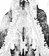
4 28 ANDRZEJ SULlMSKI DESCRIPTIONS Class REPTILIA LAURENTI, 1768 Subclass LEPIDOSAURIA HAECKEL, 1868 Order SQUAMATA OPPEL, 1811 Suborder SAURIA MAC CARTNEY, 1802 (= LACERTILIA WAGLER, 1830 (GONTHER, 1867» Infraorder SCINCOMORPHA CAMP, 1923 Family MACROCEPHALOSAURIDAE nov. Type genus: Macrocephalosaurus GILMOR E, 1943 pm4+m24-28 Diagnosis. - Dental formula d Large, tetrapod, terrestrial lizards, herbi- or insectivorous. Skull mm in length. Premaxilla and parietal unpaired. Lacrimal small, externally visible. Maxilla vertically or almost vertically arranged in relation to the naso -prefrontal and palatal regions..postfrontal and postorbitalseparated. Palate toothless. Suborbital fenestrae reduced to small hollows. Pterygoids in contact with vomers. Epipterygoids rod-like, strong. Postorbital arch composed of postfrontal only. Squamosal short, wide, roughly triangular. Supratemporal fused to squamosal. Quadratum high, movable, with a wide external conch. Parietal foramen in parietal or on fronto-parietal suture. Postero-ventral process of the jugal strong, sharply pointed or developed as a short spine or eminence. Lower jaw massive or more slender, but always deep in the angular region. Splenial wide, triangular and long, almost completely covering Meckel's groove. Anterior inferior alveolar foramen present in middle-length of the splenial. Symphysis short. Dentition subpleurodont or pleurodont, heterodont, well or weakly developed caninelike anterior teeth of the maxilla. Cheek tooth morphology and method of tooth replacement of iguanid type. Vertebral number: 8 cervicals, 27 presacrals, 2 sacrals and about caudals.-shoulder girdle strong and compact. Clavicle much ventrally widened, with a large perforation. Interclavicle cruciform with anterior process bent upwards. Humerus shortened with wide epiphyses. Forearm shortened and wide. Phalangeal formula of manus Pelvic girdle normal with no tendency to reduction. Hind limbs well developed with IV finger longest and V considerably shortened. Phalangeal formula of pes Tail relatively long, probably longer than the body. Assigned genera: Macrocephalosaurus GILMORE, 1943 and Darchansaurus gen. n. Stratigraphical and geographical range. - Late Cretaceous (Djadokhta Formation, Barun Goyot Formation and Khermeen Tsav formation) of Mongolia. Genus MACROCEPHALOSAURUS GILMORE,_1943 Type species: Macrocephalosaurus fe rrugenous GILMOR E, 1943 pm4+m24-25 Revised diagnosis. - Dental formula d Length of skulls from 70 to 120 mm. Maxillary segment much more prominent than the occipital one. External nares large, obliquely
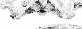
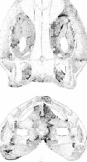
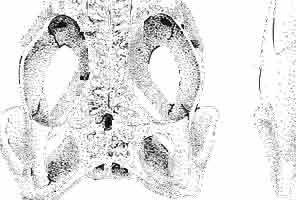
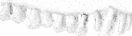
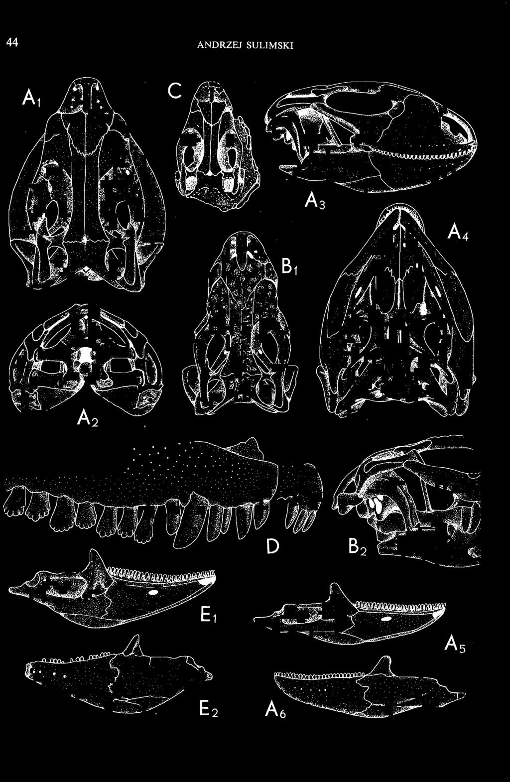
5 MACROCEPHALOSAURIDAE AND POLYGLYPHANODONTIDAE 29 A D E F Fig. 2 Macrocephalosaurus chulsanensis sp.n. - Skull and lower jaw (schematical drawing). Skull: A - lateral view, B - dorsal view, C - ventral view, D - occipital view; lower jaw : E - lingual view, F - lateral view. All about nat. size. Abbrev iations: an - angular, apo - postorbital arch, apt - posttemporal arch, ar - articular, ast - supratemporal arch, bo - basioccipital, bs - basisphenoid, c - coronoid, cad - adductor crest, cexexternal quadrate conch, co - occipital condyle, cso - supraoccipital crest, d - dentary, ep - epipterygoid, ec - ectopterygoid, ex - exoccipital, [-frontal, [a - adductor fossa, [d - dental foramen, ftpt - interpterygoid vacuity, ft - lacrimal forarnen, [M - Meckelian foramen, [m - foramen magnum, [ne - external narial forarnen, - fpln - palato-narial vacuit y, [p - pa rietal foramen, [pt - posttemporai fenestra, [so - suborbital fenestra, [sp - splenial foramen, fst - supratemporal fossa, [v - vagus forarnen, fvn - vomero-nasal foramen, j - jugal, I - lacrimal, Id - dental lamina, m - maxilla, n - nasal, 0 - orbit, op - opisthotic, p - parietal, pbo - bdsioccipital process, pbpt - basipterygoid process, pd - dorsal premaxillar process, p[- postfrontal, pj - jugal j process, pi - palatine, pm - premaxilla, po - postorbital, pot - prootic, ppoc - paroccipital process, ppt posterior pte rygoid proce ss, pr[- prefrontal, prt - retroarticular proce ss, ps - para sphenoid, pt - pterygoid, q - quadrate; sa - surangular, sip - fronto-parietal suture, sm - septomaxilla, so - supraoccipital, sp - splenial, sq - squamosal, ss - sagittal suture, st - supratemporal, sy - symphysis, svpt - vornero-pterygoid suture, v-vomer.
6 I 30 ANDRZEJ SULIMSKI, situated. Premaxilla with a long dorsal process. Septomaxilla well developed. Maxilla vertically arranged in relation to the naso-prefrontal and palatal regions. Nasals unfused, nearly equilateral. Frontals unfused, long, with weak orbital excavations. Sculpture ofroofing bones weakly developed. Parietal short, but wide, with long posterior processes arran-ged at an angle of about Descending processes of frontals developed as low oblong borders. Prefrontal large, trapezoidal in outline with considerable lateral crest. Postfrontal short, forked proximally and distally, with a well developed parietal process. Orbits very large, anteroposteriorly distinctly elongated. Jugal with a strong, sharply pointed postero-ventral process. Postorbital anteroposteriorly much elongated, dorsoventrally flattened, reaching the posterior border of the upper temporal fossa. Upper temporal fossa considerably smaller than the orbit, anteroposteriorly elongated, narrowing towards the back. Parietal foramen entirely within parietal or close, or on the fronto-parietal suture. Suborbital fenestrae in the form of small hollows with a perforation or completely overgrown bybone. Coronoid process well developed, small, vertical or directed slightly backwards. Subpleurodont or pleurodont dentition with considerably enlarged canine-like anterior teeth of maxilla. Cheek teeth lateromedially flattened, without distinct denticles or well denticulated on the cutting edges. Metakinetism well marked, mesokinetism much limited. Assigned species:.macrocephalosaurus ferrugenous GILMORE, 1943, M. gilmorei sp. n. and M. chulsanensis sp. n. Discussion. - See page 37. Macrocephalosaurus ferrugenous GILMORE, 1943 (Text-fig. 3) 1943 b. Macrocepha/osaurus f errugenous n.gen., n. sp.; C. W. GILMORE, pp , Figs 1-2. Material. - Holotype AMNH 6520 from the Djadokhta Formation, Bayn Dzak; an incomplete skull with part of the right lower jaw. Skull strongly distorted by the dorso-ventral crushing. Anterior part of the left side, occiput, palatalregion and braincase are missing. To this species probably belong the part of the shoulder girdle with a fragment of the clavicle, the posterior cervicals and the proximal part of the humerus - ZPAL MgR-IIJ59. Measurements and description. ~ See GILMORE (1943b). Discussion.-In the collection of lizards from the Djadokhta Formation, housed in the Palaeozoological Institute in Warsaw, Macrocephalosaurus ferrugenous is not represented, except for the partial shoulder girdle (ZPAL MgR-IIJ59), tentatively assigned to this species. The fragment of humerus preserved together with the shoulder girdle is large and strong but not larger than the same bone in M. chulsanensis. The same concerns the bones of the shoulder girdle. As Macrocephalosaurus ferrugenous is the only species oflarge lizard known from the Djadokhta Formation, and the here discussed shoulder girdle from the same formation belongs to a large lizard, it seems probable that it belongs to M. ferrugenous. The present author has not had the opportunity to examine the holotype of this species, housed in the American Museum of Natural History in New York. However; a comparison of the description and figures ofthe holotype given by GILMORE (1943b) with the rich and well preserved material of M. gilmorei and M. chulsanensis from the Khermeen Tsav formation and Barun Goyot Formation have shown that the three species are closely related. The comparison of the structure of these species enabled the writer to make the new reconstruction of the skull

 Holotype: ZPAL MgR-II1/1S - Almost complete skull with lower jaws. Upper dentition, right jugal, supratemporal arch and the base of the brain case are missing.")


7 MACROCEPHALOSAURIDAE AND POLYGL YPHANODONTIDAE 31 of M. ferrugenous, figured here as Text-fig. 3 B. The comparison of M. ferrugenous and both species - M. gilmorei and M. chulsanensis and remarks on the structure of M. ferrugenous are given on page 39. A fn e pr n -'\!?' m f po q 10cm Fig. 3 Macrocephalosaurus ferrugenous GILMORE, 1943, AMNH holotype. A - Skull in dorsal view (after GILMORE, 1943). B - The reconstruction of the same skull. Abbreviations see Fig. 2. MacrocepbaIosaurus gilmorei sp. n. (PIs. VIII-X; Text-figs SE, 6, 14G) Holotype: ZPAL MgR-II1/1S - Almost complete skull with lower jaws. Upper dentition, right jugal, supratemporal arch and the base of the brain case are missing. Quadrates in the form ofcephalic condyles. Khermeen Tsav I. Type horizon and locality: Upper Cretaceous,? Middle Campan ian, Khermeen Tsav formation, Khermeen Tsav I, Gobi Desert, Mongolian People's Republic. Derivation ofthe name: Named in honour of Dr. CHARLES GILMORE, who first described Mongolian fossil lizards. Material. -ZPAL MgR-l/19-Anterior part of a skull with.premaxilla, palate and part of the occipital regions preserved. Lower jaws fragmentary. It is not certain whether this specimen comes from the locality of Nemegt, Barun Goyot Formation or from the locality of Khermeen Tsav I, Khermeen Tsav formation. Khermeen Tsav formation, locality Khermeen Tsav I: ZPAL MgR-Ill/I? - Skull with lower jaws lacking the anterior and posterior ends. Left jugal and premaxilla missing, right quadrate well preserved, lower dentition incomplete, brain case damaged. Five thoracal vertebrae in line. ZPAL MgR-III/15 - Left side of a skull, much deformed, with fragments of upper and lower jaws, frontals and part of occipital preserved. ZPAL MgR-III/IO Strongly deformed skull with fragments of lower jaws, zygomatic arches, right quadrate and part of postcranial skeleton preserved. ZPAL MgR-III/16 - Considerably deformed frontal
8 32 ANDRZEJ SULIMSKI Table I Measurements in mm: Macrocephalosaurus gilmorei sp. n. ZPAL cat. nos. Skull: Condylo-basal length Total length Width at the level of the jugals Width at the supratemporal arch es Ratio of the width to the length Length of the naso-prefrontal region Width of the naso -prefrontal region Posterior width of frontals Length of frontals (lateral) Length of frontals (midline) Ratio of the width to the length Angle betwee n the posterior parietal proce sses Length of supratemporal arches Length of the palate Width of the palate Ratio of the width to the length Height of the palate floor Height of the skull with the lower jaws in the occip ital region Vertical orbit diameter Horizontal orbit diameter Length of the supratemporal fossa Anter ior width of the supratemporal fossa Length of nasals (midline) Anterior width of nasals Length of the parieto-occipital region Length of the maxillary segment Ratio of the length of both segments Length of the parietal (mid line) Wid th of the parietal (at cen ter) Distance between the paroccipital processes Least interorbital space Height of the quadrate Antero-posterior diameter of the head Height of the maxilla Length of the postfrontal with processes W idth at the center Length of the jugal process Length of the postorbital (lateral) Posterior width of the postorbital Length of the squamosal Posterior width of the squamosal Length of vomers Width of vorners MgR-Ill/IS Holotype 75.0 SO.O IS S S \ \ ea S Mg R-III/ S IS ea. 9.0 IS IS'O 14.0 ea ea ea ea ea. 9.0 cont.
9 MACROCEPHALOSAURlDAE AND POL YGLYPHANODONTIDAE 33 ZPAL cat. nos. IMgR-IIJ/18 Holotype \ MgR-IIIJI7 Length of the palatine Width of the palatine Length of the pterygoid 40.0 ea Length of the epipterygoid Length of the basioccipital and basisphenoid together 20.0 ca Length of upper tooth row Length of the canine-like tooth Antero-posterior diameter of this tooth Average number of the teeth in 1 cm 8 8 Lower jaw: Length with the retroarticular process Height below the coronoid process Length of anterior section Length of posterior section Posterior height of the dentary 20.0 ea Length of the dentary 35.0 ea Height of the coronoid process (lateral) Length of the surangular Height of the surangular below the coronoid process Length of the angular 30.0 ca Width of the angular Length of the splenial 35.0 ea Posterior height of the splenial Length of lower tooth row Average number of the teeth in 1 cm 8 8 part of skull containing a fragment of lower jaw. ZPAL MgR-IlIj21 - Large skull, strongly mineralized. Frontal part of parietal, part of frontals, prefrontals, part of supratemporal arch, part of right quadrate, fragment of lower jaw and left jugal with posterior process preserved. ZPAL MgR-Illj20 - Fragment of small skull with palatal region, maxillae without teeth, part of occipital and the base of the brain case. ZPAL MgR-IIlj8 - Almost complete left lower jaw with dentition, without retroarticular process. ZPAL MgR-BIj7 - Strongly damaged frontal part of a skull including part of palate and lower jaws with teeth. ZPAL MgR-IlIj9 - Frontal part of a skull containing left lower jaw, strongly deformed and mineralized. ZPAL MgR-IlIjll - Strongly deformed skull with postcranial skeleton. Bones displaced and strongly mineralized. ZPAL MgR-IIlj28 - Fragment of left lower jaw without teeth. ZPAL MgR-IlIj29 - Fragment of right lower jaw without teeth. ZPAL MgR-IIIj13 Fragment of right lower jaw without teeth. Diagnosis. - Length of skulls not exceeding 80 mm. Sculpture of the roofing bones often developed in the form of delicate irregularly arranged tubercles. Jugal with posteroventral process developed as a tuber or outwardly, tilted, widened spur. Parietal foramen close to the fronto-parietal suture. Pleurodont dentition with well developed anterior caninelike teeth of the maxilla. Cheek tooth cutting edges with well developed denticles of iguanid type. Lower jaw massive, high, with much ventral angular swelling. Retroarticular process short, wide, sharply pointed. Measurements. - See Table 1. Description. - Skull as a whole. The holotype skull is slender with a distinct nasoprefrontal expansion. Sculpture of roofing bones developed in the form of irregularly arranged 3 - Palaeon'tologia Polonica No. 33
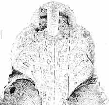
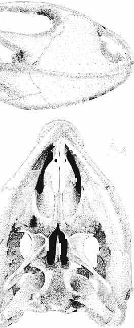
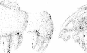
10 34 ANDRZEJ SULIMSKI tubercles (see PI. VIII and Text-fig. 4) on the nasals, prefrontals, anterior part of parietal, and as tubers or eminences on the prefrontals. Cranial roof Premaxilla with eight teeth and a long dorsal process reaching to the anterior edges of the nasals. Width of premaxilla, in dorsal view, is smaller than that of the nasoprefrontal region. External nares large and diagonally situated. Nasals comparatively short forming together an equilateral triangle, sometimes elongated posteriorly. Anteromedially, the nasals enfold the extremity of the dorsal process of the premaxilla, widely surround the upper border of the external nares join laterally with the maxillae have a short contact with the prefrontals posterolaterally, and posteriorly by means of an overlapping wedge suture, meet the frontals. Frontals are not fused. Anterior ends of frontals sutured with nasals, their postero-lateral processes wedged between nasals and prefrontals. No fronto-maxillary contact. Frontals are long, widened in the fronto-parietal suture region. Lateral borders of these bones from mid-length of the orbits of even width, not widening anteriorly. Posteriorly, the frontals join the parietal by means of an irregular suture, and at their postero-lateral expansion meet the frontal process of the postfrontal. Postfrontal and prefrontal do not meet above the orbit. The ratio between the posterior width of both frontals to their maximal length is 0.5. On the ventral side the frontals have distinct lateral ridges, which formed the lateral borders for the olfactory trunk. Parietal only slightly wider than its sagittal length. Posterior processes long, diverging at an angle of about 90. The bone is Joined anteriorly by an irregular suture with the frontals, anterolaterally with the parietal process of the postfrontal, its posterior processes being joined to the squamosais and paraoccipital processes. In the central part of the bone, on the ventral side, distinct prominent swellings occur at the contacts with the epipterygoids and supraoccipital. The parietal surrounds the supratemporal fossa laterally and forms posteriorly, by means of posterior processes, the posttemporal arch and fossa. No sagittal crest on parietal. In all specimens parietal fora men occurs just behind the fronto-parietal suture; occasionally its anterior border meeting frontals directly. Septomaxilla fragmentary. Part of the right bone shows the presence of a posterior process, reaching to the vomer. Maxillary region. (PI. VIII-IX; Text.-fig. 4 A 4 ). Prefrontal strongly developed, trapezoidal in dorsal view. The prefrontal meets the frontal medially by a long, straight suture, has a short anterior contact with the nasal, anterolaterally joins the maxilla and contacts ventrally the small plate-like lacrimal. The prefrontal has two surfaces - dorsal and lateral; the.former, together with nasals and anterior parts of frontals, forming the wide and..flat naso-prefrontal region characteristic of all representatives of Macrocephalosauridae. In M. gilmorei it forms, in relation to frontal region, a gentle curve, without a crest. Posterior surface of prefrontal forms the antero-ventral floor of the orbit; reaching the palatine medioventrally. Viewed from the front, maxilla oriented almost at right angle to naso-prefrontal and palatal regions. Maxilla high, with highest point of its dorsal process near suture with nasal and prefrontal. On the bone surface small foramina for nerves and blood vessels are visible. Maxilla with three processes: dorsal (described above); antero-ventral, sutured to the premaxilla; and posterior, joining the jugal and extending under the orbit. Anteriorly, the maxilla surrounds the postero-lateral margin of the external nares and posteriorly, by means Fig. 4 Macrocephalosaurus gilmorei sp.n., ZPAL MgR-I1I/I8 - holotype. A - Skull, lower jaw and dentition: 1 - skull, dorsal view, 2 - ventral view, 3 - occipital view, 4 - lateral view, 5 - left lower tooth row fragment, lingual view. ZPAL MgR-I11/8. B -left lower tooth row, lingual view. Iguana iguana LINNAEUS, Recent. C - Lower tooth row fragment, lingual view (after EDMUND, 1969). Macrocephalosaurus gilmorei sp.n., ZPAL MgR-I/19. D - Right upper tooth row: 1 -lateral view, 2 -lingual view. Abbreviations see Fig. 2. A 1-4 nat. size; A 5, B-D X 5.
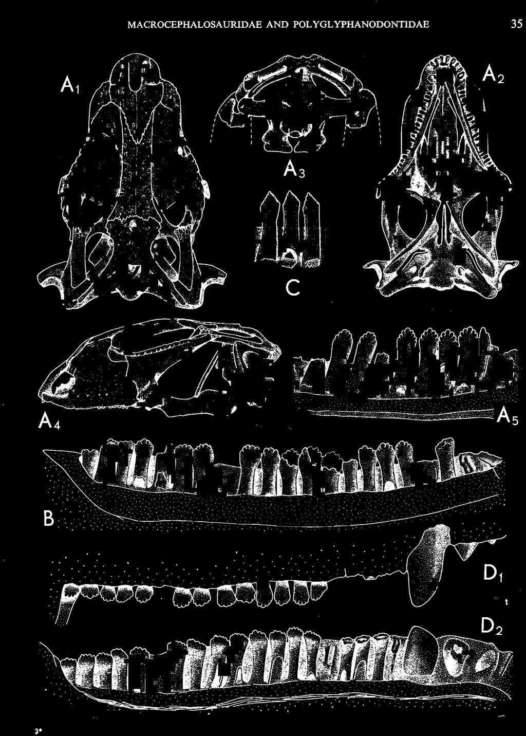
11 MACROCEPHALOSAURlDAE AND POLYGL YPHANODONTIDAE 35 c =--.:., ", " " 'W ' ~.: : ' ';'''' :~< : I:i';'! :::.,..,. B,',.....:.'....:. ',::. '." ".:: : ~..~~s~~~~~~~ t ', '. '.., '. 3
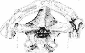
12 36 ANDRZEJ SULIMSKI of a long suture, it joins the jugal and lacrimal. Lacrimal situated in the antero-ventral rim of orbit, dorsally joined to the prefrontal, anteriorly to the maxilla, ventrally to the jugal and in the orbital floor to the palatine. Lacrimal bone with a single lacrimal foramen. Jugal forms a pronounced arch, anteriorly connected by a long suture to the maxilla and lacrimal and by means of a narrowed process to the postfrontal and squamosal. Present on the posteroventral part of the bone, just behind the ending of the posterior maxillary process, is a tuberous jugal process, directed slightly outwards. The upper edge of the jugal serves as the lower border of the orbit, and its medial side as the latero-ventral floor of the orbital cavity. The joining between jugal and postfrontal is a straight, vertical suture, which seems to have been movable. Jugal surface uneven, providing places of attachment for external muscles. Temporal region (PIs VIII-IX; Text-fig. 4A}). Postfrontal not large, but longer than that of M. chulsanensis and Darchansaurus estesi. Frontal and parietal processes widely divergent, embracing medially the frontal and parietal in their suture. Lateral part of the postfrontal narrower than the medial, its processes being more vertically arranged, embracing dorsally the anterior, tuberal part of the postorbital. Postfrontal forms a strong, rather long postorbital arch, separating the large orbital cavity from the supratemporal fossa. Postorbital long, dorsoventrally compressed and rather wide, anteriorly entering the posterior part of the orbit. On the surface where it joins the postfrontal and jugal, a rugose eminence occurs. Posteriorly, the postorbital is joined to the squamosal by an overlapping suture. Posterior end of the bone reaches beyond the posterior boundary of the supratemporal fossa. The straplike posterior process in M. gilmorei is of almost uniform width on its whole length. Squamosal large, roughly triangular, with three processes. The anterior process joins the jugal, underlapping the postorbital; its posterior part widely covers the cephalic condyle of the quadrate, and its posterior end reaches the paroccipital process, forming together with the quadrate a syndesmotic joint. The dorsal process of the squamosal is directed upwards and forwards, and closes the supratemporal fossa from the back. Between the posterior end of the postorbital and the posterior parietal process, parallel to the latter, occurs a delicately marked groove indicating that the dorsal process of the squamosal is actually a fused supratemporal. Supratemporal fossa rather large, roughly triangular with rounded angles. It is delimited anteriorly by the postfrontal, medially by the parietal, posteriorly by the squamosal and laterally by the postorbital. Orbits large with antero-posterior dimension larger than the dorso-ventral. The axis of the orbit is laterally oblique at an angle of 45. Quadrate has a strong, wide cephalic condyle, its dorsal surface strongly bent backwards, joining the paraoccipital process. The external conch has a strong, rather wide, roughened crest, almost vertical to the cephalic condyle. The posterior crest is strong, wide, ventral to the cephalic condyle, ventrally narrowing to join the lower articular condyle. The medial conch is also wide, wider than the external one, especially in the ventral part of the bone. Both conches have strong ventral excavations. Distal condyle widely bifurcated. In the ventral part of the medial conch occurs an articular facet for attachment with the tip of the pterygoid. The distal tubercle of the tympanic crest is weakly developed. Palatal region (PIs VIII-IX ; Text-fig. 4A 2 ). Vomers are connected by means of a weak but extensive midline suture. These bones are wide and rather long. In anterior part they are narrowed with distinct incisions for the vomero-nasal organs, but posteriorly widened. The anterior process of the vomer joins to the premaxilla and septomaxilla, while from the back the bone is joined medially, by a narrow process, to the pterygoid and laterally by the same process to the palatine. The lateral wing of the vomer almost reaches the maxilla, separating the anterior part of the internal nares from the posterior. On the midline, at the vomero-pterygoid
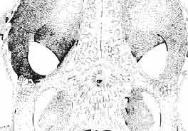
13 MACROCEPHALOSAURIDAE AND POL YGL YPHANODONTlDAE 37 contact, distinct, strong ventral raphae of vomers (raphae vomeri ventralis) are present on both bones, about mm in height. In the central part, the lateral borders of vomers are rised upwards forming in cross-section a paiaeochoanate type of bone arrangement (see GUIBE, 1970, pp , Fig. 80; SINITZEN, 1928). Palatine relatively long and not very wide. Anteriorly and posteriorly the bone surrounds the endochoanal foramen, and with its long antero-lateral process joins the maxilla. The palatine lies in the same plane as vomer. Posteriorly, the bone arches and reaches the ventral side, and together with anteriorpartofpterygoid forms the posterior palatal roof. Medially the palatine joins the posteriorvomerine process and anteriorprocesses of the pterygoid, laterally it joins both maxilla and ectopterygoid. lhe palatine together with the ectopterygoid and part of the pterygoid process forms, at the place of their sutural connection; a distinct hollow, which is a reduced suborbital fenestra. The palatine is edentate. The palatal roofis high, placed considerably above the level of the posterior pterygoid processes, the difference in level being about 10 mm in known specimens. Pterygoid not completely preserved, but the extent of the posterior process and the shape of anterior part of the bone can easily be reconstructed. Pterygoid massive, especially in comparison with the vomer and palatine. The pterygoid has three processes: the vomerine process, slender, reaching anteromedially and joining with the pterygoid process of the vomer; the transverse process, wide, laterally joining the ectopterygoid and reaching into the suborbital fenestra; and the posterior process, joining the quadrate by means of a wide, lateromedially flattened end. In the central part, a marked sharp transverse ridge occurs, which is a postero-ventral rim of the palatal roof. The transverse process together with the ectopterygoid form a strong and wide, distinctly ventrally directed tuber or process. On the lateral side of this united process a wide syndesmotic articular surface is visible that connects with the lingual side of the coronoid process. Medially the pterygoid surrounds a rather wide interpterygoid vacuity. The area of the interpterygoid vacuity is divided posterioriy by a long calcified parasphenoid process. Along the ventral surface of the pterygoid, almost at an angle of 90, a prominent crest extends diagonally, 'which, behind the joining with basipterygoid process, reaches the lateromedially compressed extremity of this bone. On the dorsal side, above the basipterygoid joint, a socket for the articulation of epipterygoid is visible. Posterior ends of pterygoids reach the distal medial articular facet of the quadrate. Pterygoid teeth absent. Ectopterygoid elongated, narrow, roughly triangular, with three processes. Generally, as with the rest of the representatives of the family, the bone is reduced in size. Maxillary process of the ectopterygoid is sutured by a long suture to the maxilla, medially to the palatine, and posteriorly it joins the large transverse process of the pterygoid. The ectopterygoid, together with the palatine and the pterygoid, form the suborbital fenestra. Epipterygoid is reduced to a long rod-like support between the pterygoid and parietal, oval in cross-section. The posterior border of the bone rests on the alar process of the prootic. Occipital region (PIs VIII-IX; Text-fig. 4A 3 ). Supraoccipital wide, with strong, sharp occipital crest. From below, in the place of the suture with the paroccipital process, distinct horizontal margins are present, these being places of attachments for the internal muscles of the neck. Supraoccipital borders the foramen magnum ventrally and laterally joins the prootic. Dorsally, the supraoccipital connects, by means of a syndesmotic joint, the thickened descending processes of parietals. Exoccipital fused with opisthotic, surrounds laterally the foramen magnum and passes outwards into a strong and wide paroccipital process. The latter process joins the squamosal, posterior process of the parietal and posterior apex of the cephalic condyle of the quadrate, forming in this place an intercalary cartilage (see VERSLUYS, 1912; REESE, 1923; JOLLIE, 1960). Angle between paroccipital processes obtuse, about 75. Occipital
14 38 ANDRZEJ SULIMSKI condyle distinctly dividing into three parts, with a concave dorsal surface for the glenoid surface of the odontoid process of the axis. Basicranium (Pls VIII-IX; Text-fig. 4A 2 ). Basicranium composed of three unpaired bone elements united completely in the adult stage. In young specimens there is a distinct suture between the basioccipital and basisphenoid bones. Parasphenoid process ossified at base, forming lateral trabecular attachments, Basisphenoid bears anteriorly two strong, short and distally widened basipterygoid processes, arranged at an angle of about 90. Basioccipital is incompletely.preserved, but the basioccipital processes are very well developed and strong. On the medial sides of these processes distinct central hollows are present. Latero-sphenoid margins well marked with a deep central groove between. Brain case badly preserved. Lower jaw - (PI. X; Text-fig. 5E/. 2 ) as a whole massive, relatively short, high at the level of the coronoid and angular bones, anteriorly narrowing abruptly up to symphysis. Dentary long, very wide distally, slightly overlapping to the lingual side of the jaw. On the lateral surface occur 5 to 6 small mental foramina. Posteriorly, the dentary joins the surangular, angular and coronoid, while on the lingual side it is joined by a direct suture, above and below, to the splenial. On the alveolar margin cheek teeth are fused to the lateral dentary wall. Alveolar margin, viewed from above, is slightly sinusoidally bent. Symphysis short, not widened. Coronoid well developed, with a rather low dorsal process in relation to the height of the jaw, widened at its base and ending in a rather prominent, blunt point. Anterior margin of the process directed backwards, the posterior one almost vertical. Labial process of the coronoid small, extending anteriorly to overlap the dentary and the anterior part of the surangular. Medially, the anterior process joins the dentary, splenial and prearticular, the posterior one being long and bent posteroventrally, surrounding the anterior rim of the adductor fossa and joining the articular bone below. A distinct crest is running from the tip of the dorsal process ventrally along the posterior process for pseudotemporal muscles. Surangular high and short, on the lateral side covering almost the whole space between dentary and articular bone. From the postero-dorsal angle of the bone, under the glenoid cavity of the articular, an oblique crest begins, continuing downwards, towards the front and vanishing by the border of angular suture. This crest serves the insertion of the strong external adductor muscles of the jaw. On the lingual side, the surangular is joined by a vertical suture to the articular bone and from the front to the coronoid process. The suture between surangular and prearticular indistinct. Angular laterally well visible, forming a strong, ventral angular swelling of the jaw. It is dorsally sutured to surangular, anteriorly to dentary and posteriorly to articular. Lingually it underlies the prearticular and anteriorly contacts the splenial and dentary. Articular wide, short from the ventral side, it forms a curved surface, gently passing over to the lingual side and posteriorly. Anteriorly the bone joins the surangular, from below it joins the angular and on the lingual side it is completely fused with prearticular. Behind the glenoid fossa the bone is wide, concave and terminates in a short and wide retroarticular process. Glenoidfossa has two hollows and a gentle medial crest arranged almost transversally to the long axis of the jaw. Splenial, a comparatively thin, flat, triangular plate of bone covering the whole region between upper and lower dentary edges and also covering the Meckelian canal. Apex of bone placed just behind the anterior Meckel's foramen. On the splenial, almost at mid-point and close to the upper dentary suture, a large oval anterior inferior alveolar foramen is visible. On the lateral side splenial not visible. Dentition (PIs VIII-X; Text-figs 4A j,b, D/_ 2, 14G). An alinost complete lower and upper
15 MACROCEPHALOSAURIDAE AND POLYGLYPHANODONTIDAE 39 marginal dentition in M. gilmorei is represented only by the specimen ZPAL MgR-I/19. The remaining specimens have gaps in dental series. Premaxillary teeth, 8 in number, have conical, sharply pointed crowns and do not extend as far ventrally as the cheek teeth. In the anterior part of the maxilla, two, three, or sometimes four large canine-like teeth occur. The longest of these teeth is the second in the series. Its crown three or even four times higher than the crowns of the cheek teeth. The crowns in cross-section oval or round, without posterior and anterior cutting edges. The first tooth has sometimes a slight swelling on the crown, which also may occur in the third and fourth tooth in the series. All have one apex and are considerably higher than the cheek teeth. The latter are rather small with slender, round shafts, while their crowns are latero-medially flattened. The crowns are of lancet-like shape with sharp denticulate cutting edges. On both sides of the largest, central tuber, 2 or 3 small dentic1es occur, as in Sauromalus, Iguana and Dipsosaurus. The lateral sides of the shafts of the premaxillary and maxillary teeth are fused to the internal wall of the bone ;at the tooth bases, irregularly situated resorption pits occur. On the lower jaws only two types of teeth are differentiated - incisiform and cheek teeth. The anterior, incisiform teeth are 3-5 in number, and have simplified crowns, usually with one apex and without tendency to enlargement. The structure of crowns and shafts of cheek teeth is the same as that in the upper jaw. In the lower dental series significant reduction of posterior cheek teeth is not observed. The type of crown morphology of cheek teeth resembles that seen in such iguanids as Dipsosaurus. Heterodontism is especially visible in the upper dentition (three types of teeth). In lower dentition it is less distinct (only two types of teeth). The tooth replacement according to EDMUND (1960, 1969) is of iguanid type. Postcranial skeleton. This is preserved only in specimens ZPAL MgR-Ill/tO, 11 and 17 and is incomplete and poorly preserved. Individual bone elements are displaced and fragmentary and description is not possible. Discussion. - M. gilmorei from the Barun Gyot Formation differs from M. ferrugenous from the Djadokhta Formation by its smaller dimensions. Length of skull in M. ferrugenous is 120mm whereas in M. gilmorei it is only 70-80mm. Other differences concern the proportions of the skulls, in particular of the frontals; in M. gilmorei width/length ratio of the frontals is 0.4, whereas in Mi ferrugenous this ratio amounts to 0.5. Consequently, the skull of Mi ferrugenous is relatively wider than in M. gilmorei. The jugal process is in M. ferrugenous much longer, tapering posteriorly, while in M. gilmorei it is comparatively short and rather tuberlike, bent outwards. Further differences concern the structure of the dentition and the mode of implantation. Cheek teeth in M. ferrugenous are latero-medially flattened, but with no distinct cusps or denticles on their cutting edges. Shafts of these teeth are rather shallowly attached to the dental groove in a subpleurodont implantation. The teeth of M. gilmorei, however, have prominent denticles on the crowns and slender shafts distinctly attached by their lateral surfaces in a distinctly pleurodont implantation. The mode of tooth replacement of M. ferrugenous is unknown. The presence of resorption pits within M. gilmorei indicates the iguanid mode of replacement (see EDMUND, 1960, p. 60). It is possible that a similar condition was present in M. ferrugenous. Differentiation of the teeth of both species is distinct and is especially indicated in the strong development of the anterior canine-like teeth of the maxilla. Similarities occur also in the general structure of skulls, placement of the parietal foramen, arrangement. of fronto-parietal suture and posterior parietal processes, structure of temporal region and it seems also in the construction of naso-prefrontal region, which can be stated in spite of the fact that in M. ferrugenous it is not completely preserved. Lack
16 40 ANDRZEJ SULIMSKI of evidence of the structure of the palatal region and internal morphology of lower jaws in M. ferrugenous does not allow one to complete the description and compare it with the skulls of M. gilmorei. GILMORE (1943b) described, but not very clearly, a fragment of M.ferrugenous tempora region, which he named the postorbital bone. According to GILMORE "there is a single element uniting the jugal with the parietal and frontal at their junction" and he stated also that "whether it is the postfrontal or postorbital or a fusion of the two, there is no way of determining, but as studies of recent Sauria have shown that the postfrontal is absent in most Agamidae, it will here be designated as postorbital" (GILMORE, I. c., p. 364). It can be stated, on the basis of the illustration given by GILMORE, that between the jugal and the bone fragment directed towards parietal and frontal, an oblique suture separating two distinct bone elements is present (see temporal region in M. gilmorei, M. chulsanensis and Darchansaurus estesiy i.e. postorbital and postfrontal. The former borders the orbit posteriorly, joining the jugal and postfrontal, and with a long and flat posterior process joins the squamosal. The postorbital, as in M. gilmorei, forms the supratemporal arch. The latter bone is small, both sides forked and joins the frontal and parietal to the supratemporal arch, forming a rather short postorbital arch. The quadrate in M. ferrugenous is of the same structure as in M. gilmorei. It is slightly larger, possessing a stronger cephalic condyle and wider external conch with distinct tympanic crest. The lacrimal of both species is small, and externally visible. The prefrontal of M. ferrugenous is deformed, displaced and suggests a different morphology of this bone than in M. gilmorei. The maxilla, in spite of dorso-ventral crushing, is rather high and vertically arranged in relation to the palatal region (see GILMORE, I. c., Fig. 1B). The anterior part of.the maxilla has a rather high notch, which forms the posterior border of a large and oval external nares. The incomplete dentition in M. ferrugenous indicates, according to GILMORE, an old specimen with worn-down tooth crowns. However, it seems, on the basis of GILMORE'S sentence, that the teeth were not worn down while the animal was still alive "a long replacement tooth in the maxillary near the center of the series has an unworn crown that is bluntly chise1 shaped, with a steep internal bevel" (GILMORE, I. c., p. 364). The crowns of the fourth, seventh and eighth teeth of the maxilla and also the two first lower teeth (counting from the front of the drawing - see GILMORE, I. c., Fig. 2) appear normally developed, laterally compressed, without distinct denticles on their cutting edges. The teeth of both jaws in M. ferrugenous are not typical for agamids, which have as a rule an acrodont mode of implantation. The distinct marking between the crowns and shafts on one side and the bones of the jaw on the other seems to indicate that these teeth were not very deeply fused with the lateral wall of the dental parapet. Therefore a subpleurodont mode of implantation is suggested. The lack of data on the structure of the lower jaws, maxilla and the internal morphology of teeth in M. ferrugenous does not allow a closer comparison with M. gilmorei. Characteristic for M. ferrugenous and M. gilmorei is the presence of large canine-like teeth in the anterior part of the maxilla. The largest of them in the former species seem to lack a natural crown. According to GILMORE this tooth rapidly became worn and also "its crown was more or less flattened, except the low cusps, which in this specimen have been in size reduced by wear" (GILMORE, I. c., p. 364). However, one can see from the illustration (I. c., Fig. 2) that the tooth was broken and the uneven surface being irregular, which may have suggested to him that cusps were present. This tooth as well as the slightly smaller one preceding it must have had normal, high canine-like crowns and have been lateromedially compressed. Similar maxillary teeth are also present in M. gilmorei but are in that species more oval in cross-section.
17 MACROCEPHALOSAURIDAE AND POLYGL YPHANODONTIDAE 41 Macrocephalosaurus chulsanensis sp. n. (pis XI-XXII; Text-figs 2; 5-11, 141) Holotype: ZPAL MgR-I ji4 - Complete skull with the postcranial skeleton. Distal end of tail, left humerus, a part of anterior autopodium and dorsal elements of the scapulae missing. Barun Goyot Formation, Khul san. Type horizon and locality: Upper Cretaceous,? Middle Campanian, Barun Goyot Formation, Khul san, Nernegt Valley, Gobi Desert, Mongolian People's Republic. Derivation of the name: From Khulsan, the name of the type locality. Material. - All specimens come from the Baron Goyot Formation. Southern Monadnocks, Nemegt: ZPAL MgR-21 - Skull complete, sculptured and with almost entire postcranial skeleton preserved. The distal part of the tail, some ribs and some limb elements missing. Khulsan: ZPAL MgR-I/2S - Skull large, almost complete with the postcranial skeleton. Shoulder and pelvic girdles incomplete. ZPAL MgR-I/1S - Skull without the left maxilla, lower jaws and the postcranial skeleton. ZPAL MgR-l/16 - The occipital fragment of the skull without lower jaws. Quadrate, a fragment of right zygomatic arch, fragment of parietal and right supratemporal arch preserved. ZPAL MgR-Ill7 - Anterior part of the skull together with fragments of lower jaw. The specimen badly preserved, mineralized. ZPAL MgR-I/18 - Anterior part of the postcranial skeleton with shoulder girdle. ZPAL MgR-Ij20 Strongly deformed skull of a young specimen. ZPAL MgR-I/22 - Fragment of postcranial skeleton with its pelvic girdle, caudals showing autotomy and parts of hind limbs preserved. ZPAL MgR-I/23 - Almost complete pelvic girdle. ZPAL MgR-I/24 - Skull with right lower jaw, a fragment of the left jaw and a part of the postcranial skeleton preserved; a young specimen. ZPAL MgR-I/26 - Brain case, slightly damaged. ZPAL MgR-I/27 - Skull, strongly mineralized and eroded. ZPAL MgR-I/28 - Anterior part of the skull with no dorsal roofing bones, dentition preserved. ZPAL MgR-I/29 - A fragment of right lower jaw with teeth preserved. ZPAL MgR-I/30 - A fragment of right lower jaw without teeth. ZPAL MgR-I/31 Anterior fragment of lower jaws with teeth, joint in symphysis. Diagnosis. - Length of skulls about 40 mm in the case of young specimens and mm for adult ones. Skull slender, with widened naso-prefrontal region. The ratio of occipital to maxillary segments is 1: 3 and width to length of frontals Parietal foramen always on the fronto-parietal suture. Surface ofnaso-prefrontal region forms a plane, slightly inclined anteriorly in relation to the fronto-parietal surface. Sculpture of covering bones often well developed in [he form of regularly arranged cusps and rosettes. Parietal process of the postfrontal poorly developed, frontal long and slender. Jugal process wide, short and sharply pointed, slightly bent outwards. Suborbital fenestrae reduced to small perforated hollows. Anterior teeth of the maxilla, four-five in number, enlarged, canine-like, but not so well developed as in M. gilmorei. Anterior teeth of the lower jaw, five-six in number, are unicuspid and are equal in height to cheek teeth. Lower jaw slender and not so deep in the angular region, as in M. gilmorei. Teeth in both jaws numerous, about 27 or 28; 8 premaxillary teeth are present. Scapulo-coracoid of iguanid type with three fenestrae. Measurements. - See Tables Nos Description. - Skull as a whole (PIs XII, XIV ; Text-figs 2, SA-D). The skull of the type specimen is completely preserved, only slightly damaged. In lateral view the skull is roughly triangular in shape with its fronto-nasal region rounded. Parietal and frontals are in one plane, while the naso-prefrontal region is slightly diagonally arranged, directed forward and downwards. Cranial arches well developed and strong. Lower jaw at level of angular region deep,
18 42 ANDRZEJ SULlMSKI Table 2 Measurements in mm: Macrocephalosaurus chulsanensis sp. n. (Skulls) ZPAL cat. nos. I MgR-I/14 Holotype I MgR-I/21 MgR-I /25 MgR-I/24 I I Young Skull: Condylo-basal length ea Total length ca Width at the level of the jugals Width at supratemporal arches Rat io of the width to the length Length of the naso-prefrontal region ca Width of the naso-prefrontal region ea Length of frontals (lateral) Length of frontals (midline) Posterior width of frontals ca. 9.0 Ratio of the width to the length (mid.) Angle between the posterior parietal pr. 90 ea. 90 ea. 80 ca. 90 Length of supratemporal arches ea Length of the palate Posterior width of the palate 28.0 ea, Ratio of the width to the length 0.7 ca. 0.7 ca Height of the palate floor ca Height of the skull with lower jaws at the occipital region ea Height of the skull with lower jaws at the frontal region ea Vertical diameter of the orbi t Horizontal diameter of the orbit Antero-posterior length of the supratemporal fossa Anterior diameter of the supratemporal fossa Length of 'nasals (midline) Anterior width of nasals' Length ' of the parieto-occipital region ea. 8.0 Length of the maxillary segment ea Ratio of the length of both segments ea. 0.3 Length of the parietal (midline) Length of the- parietal with posterior processes Width of the parietal at the center Distance between paroccipital processes ea Least interorbital space Height of the quadrate Antero-posterior diameter of the head Height of the maxilla ca Length of the postfrontal with processes ea Width of the postfrontal at the center Length of the jugal process Length of the postorbital (lateral) ea Posterior width of the postorbital Length of the squamosal ca. 8.0 Posterior width of the squamosal ca. 5.0 Length of vomers ea ca I
19 MACROCEPHALOSAURIDAE AND POLYGL YPHANODONTIDAE 43 ZPAL cat. nos. I MgR-IjI4 I MgR-Ij21 I MgR-Ij25 I MgR-Ij24 Holotype Young Width of vomers ea ca Length of the palatine ca ea Posterior width of the palatine ca ea Length of the pterygoid 37.0 ea ea Posterior width of the pterygoid 13.0 ca ea. 7.0 Length of the ectopterygoid ea ea ea ea. 4.0 Length of the epipterygoid Length of the basioccipital and basisphenoid together Width of the same Length of upper tooth row ea Length of the canine-like tooth Antero-posterior diameter of the same Average number of the teeth in 1 cm 8 8 8? Lower jaw: Length with the retroarticular process ca Height below the coronoid process Height of the coronoid process (lateral) Length of the anterior section Length of the posterior section Length of the dentary ea ea. 37,0 ea Posterior height of the dentary ca ea ea Length of the surangular ca. 2?0 - Height below the coronoid process ea, Length of the angular ea Width of the same ea. 3.0 Length of the splenial ea ca ea Posterior height of the splenial ea Length of lower tooth row ca Average number of the teeth in 1 cm 8 8 8?, lower; it is slender, especially in its anterior part and relatively short in the posterior one. The articular part with retroarticular process is shortened and rather wide. The skull of M. chulsanensis is kinetic, especially in the occipital region (metakineticism). Cranial roof. Premaxilla (Text-figs 2A-B, 5A-C) has 'well-developed postero-lateral maxillary processes which surround the external nares and join posteriorly the antero-ventral processes of the maxillae. Dorsal process of premaxilla extends upwards between the nares, reaching the nasals. Posterior extremity of this process overlaps the anterior nasal parts, forming on the sagittal suture slight articular depression. Postero-lateral maxillary processes on the dorsal surface are perforated by small blood vessel foramina. On the alveolar margin, pleurodont, unicuspid, sharp, conical teeth - four on each side - occur. Dorsal surface of dorsal process of premaxilla smooth, without sculpture. Nasal bones are joined by means of a straight sagittal suture, which in forms with well developed sculpture, is apparently overgrown and hidden. The bones form an almost equilateral triangle with the anterior side surrounding from above the external nares and joining on the midline the dorsal process of the premaxilla. The posterior angle ofthis triangle, situated on the sagittal suture, wedges between the fromals, overlapping the latter. In the remaining sides of the triangle, slightly anteriorly, oblique, medially directed fissures are present which do not reach the sagittal line of the skull. The nasals join anterolaterally with the bent parts of the dorsal processes of the maxilla and

20 44 ANDRZEJ SULIMSKI
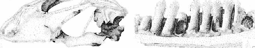
21 MACROCEPHALOSAURIDAE AND POL YGL YPHANODONTIDAE 45 laterally have a small contact with the prefrontals. Dorsal surface of nasals slightly concave and generally sculptured with tubercles or regularly arranged rosettes composed of small tubercles (Text-fig. 5B l ). Frontal is anteroposteriorly elongated and widened anteriorly, forming the dorsal margin of the orbit. Lateral sides of both bones slightly concave, posteriorly joined by means of irregular sutures to the parietals and postero-laterally to the frontal processes of the postfrontals. Anteriorly they underly the nasals and anterolaterally join the prefrontals. The frontals meet one another on a long sagittal suture; the dorsal surface often covered by "delicate tuberosities. Postero-medial processes of nasals wedged between frontals and at the same time the latter, by means of the antero-lateral processes, are wedged between the nasals and prefrontals, not reaching the nasal processes of the maxillae. On the midline, between frontals and parietal, a parietal foramen is situated. Sutures between frontals and parietal and also between the frontals, form an irregular line. Lateral margins of frontals slightly thickened and rounded. Anteriorly and posteriorly two small indentations indicate the sutural joining of frontals to prefrontals and postfrontals respectively. Ventral surface of frontal posteriorly flat, the antero-lateral orbital borders gradually elongating anteriorly and downwards, forming low descending ridges on each side of the cavity for the olfactory tract. Parietal is an unpaired, quadrangular bone with four processes. The fro mal process together with the antero-medial part of frontals form a mesokinetic hinge joint. Posterior parietal processes (supratemporal processes) are long and diverge at an angle of about 90 in adult specimens and 60 in young specimens. On both sides of the central depression of the parietal numerous ventral descending swellings occur, which laterally are sutured to dorso-lateral margins of the supraoccipital bone. Anterior, sharp extremities of these swellings join the proximal epipterygoid ends. The anterior part of [he parietal is concave from its ventral side and forms a dorsal cover for the cerebral hemispheres and optic lobes. Attachments of the pterygoid levator muscles poorly visible. Dorsal parietal surface is rather weakly sculptured. More distinct sculpture of the covering bones of the skull may indicate male specimens. Sagittal crest not present on the parietal bone. Medial parts of posterior processes possess slight longitudinal depressions. Septomaxilla is a small bone, covering the vomero-nasal organ and embracing the anterior part of the olfactory cavity. The septomaxiila is joined by means of a ventral septal process to the dorsal process of the vomer. It forms a rather thin, flat bone with three surfaces. The dorsal surface is directed anterolaterally, medially it reaches the nasal septum, suturing laterally to the antero-ventral process of the maxilla, and posterolaterally joining the anterior margin of the external nares. Medial septum wide and flat, directed ventrally, reaching to the medial vomer margin. Maxillary region (Text-figs 2A, 5A 3 ). Prefrontal has an irregular shape, and three processes and three external surfaces. The postero-dorsal process extends posteriorly along the side of the frontal. The palatal process is directed ventrally and sutures to the maxillary process of the palatine. The third, anterior process, reaches the suture, joining the nasal bone with the dorsal process of the maxilla. The medial margin of the bone sutures to the lateral Fig. 5 Macrocephalosaurus chulsanensis sp.n., ZPAL MgR-I/I4 - holotype. A - Skull and lower jaw : 1 - skull, dorsal view, 2 - occipital view, 3 - lateral view, 4 - ventral view, 5 - lower jaw, lingual view, 6 - lateral view. ZPAL MgR-I/21. B - Skull with the sculpture: 1 - dorsal view, 2 -latero-occipital view. ZPAL MgR-I/24. C - Skull of the young individual, dorsal view. ZPAL MgR.I/22. D - Upper tooth row, lateral view. Macrocephalosaurus gilmorei sp.n., ZPAL MgR-IIl/I8 - holotype. E - lower jaw: 1 - lingual view, 2 - lateral view. Abbreviations see Fig. 2. A-C, E nat, size, D x 5.
22 46 ANDRZEJ SULIMSKI margin of the frontal and.anteromedially has a short contact with the nasal. The anterolateral margin joins the maxilla and ventroposteriorly the lacrimal. Dorsal surface of prefrontal is trapezoidal and situated on the same plane as the nasal bone. The lateral surface of the bone forms a small area situated slightly over the lacrimal and growing into the vertical wall formed by the maxilla. Ventro-posterior (palatal) process of prefrontal concave, smooth and forming antero-medial floor of orbit. Medial margins of the palatal process of the prefrontal, together with frontal and palatine, form part of a wide orbito-nasal foramen. Dorsal surface of prefrontal with the same sculpture as the nasal bone. Maxilla deep, almost vertically-arranged in relation to palatal surface and to nasoprefrontal region. Posteriorly it joins, by means of a long suture, the jugal and partly underlies the anterior part of the orbit at about one-third the length from the front of the orbit. Surface of the maxilla not sculptured. Dorsally the maxilla joins the lacrimal, prefrontal and nasal, and anteriorly the antero -ventral processes are joined to the maxillary processes of the premaxilla. Maxilla outline roughly triangular, with its highest apex situated at mid-length of the alveolar margin. The alveolar margin of the maxilla is slightly arched downwards, possessing one row of numerous pleurodont teeth. The premaxillary process is mediolaterally flattened and laterally overlaps the maxillary process of the premaxilla. Medially it sutures to the anterior process of the vomer. On the dorsal surface of the process, in its medial part, a small crest for attachment of the anterior transversal lamina is present. The jugal process joins dorsally a small lacrimal, medially meets the ectopterygoid, and forms the medio-iateral part of the orbital cavity floor. The naso-prefrontal process is a thin bony lamina, which on reaching the nasal and prefrontal bones bends at almost right angles. This process forms the lateral wall of the nasal capsule, posterior margin of the external nares and dorsally sutures to the nasal and prefrontal bones. The bent upper parts of the naso-prefrontal processes, together with the prefrontals, nasals and anterior parts of the frontal bones, form the nasoprefrontal region, which is sometimes covered by tubercles. The maxilla does not contact the frontal. On the external surface of the maxilla, in the lamina of the naso-prefrontal process, small foramina for nerve endings are visible. Over the alveolar margin occur six, sometimes seven foramina for the skin branches of the alveolar nerves and for the maxillary artery. Interpretation of the medial part of the maxilla is difficult, as this part of the bone is considerably damaged or not accessible. On the ventro-medial side a suture joining the maxilla and maxillary process of the palatine is present. Lacrimal is a small, flat bone situated in the anterior margin of the orbit. It sutures dorsally and medioventrally to the prefrontal, anteriorly to the nasal process of the maxilla and from the ventral side to the jugal. The medial bone surface is separated from the palatal process of the prefrontal by a rather large dorsoventrally fissure-shaped lacrimal foramen, while the antero-medial surface is elongated towards the nasal capsule. Jugal is very much elongated, anteriorly widened and strongly narrowed posteriorly. By means of an anterior broad process it is joined by a diagonal suture to the maxilla, anterodorsally to the lacrimal and to the maxillary process of the palatine, and medioventrally to the ectopterygoid. The temporal process of the jugal is connected with the postfrontal, and its flattened end inserts into the squamosal. The postero-ventral process of the jugal is wide, slightly outwardly directed and pointed with a spine dorsally visible. Dorso-mesial surface of jugal forms the underlying rim of the orbit. The ridge beginning at the postero-lateral process of the prefrontal bone extends backwards across the dorsal part of the lacrimal, continuing on the dorso-lateral side of the jugal and reaching the postfrontal, forming the sharp rim of the orbit. This ridge is often uneven and possesses rather numerous swellings. Lateral surface of the jugal per-
23 MACROCEPHALOSAURIDAE AND POLYGL YPHANODONTIDAE 47 forated anteriorly by a few small foramina, which are the continuation of the alveolar foramina present on the maxilla. Medially to the jugal is a recess for the coronoid process. The posteroventral part of the jugal process forms an elongation of the alveolar margin of the maxilla. The temporal process forms a lateral strut for the postorbital arch, where it joins the postorbital and postfrontal, as well as forming a strut for the supratemporal arch at the joining of the postorbital and squamosal bones., Temporal region (Text-figs 2B, 5A l, B l, C). The supratemporal fossa is separated by a supratemporal arch composed mainly of the postorbital and the anterior part of the squamosal. Between the parietal, partly squamosal bone and occipital segment of the skull occurs a broad flattened posttemporal fossa. Postfrontal is short, forked on both sides, possessing two pairs of processes - proximal frontal and parietal processes and distal postorbital processes. The _postfrontal is the main element of the postorbital arch, joining the frontal and parietal bones to the supratemporal arch: Dorsal surface of postfrontal smooth, with no traces of sculpture. By means of a long frontal process it is sutured to the frontal, and by a short parietal process to the parietal bone. Both processes embrace the lateral widenings of the corresponding bones in the fronto-parietal suture. Postorbital processes of postfrontal meet the postorbital in a forked suture. Postorbital is anteroposteriorly elongated, dorsoventrally flattened. The anterior part forms a small area of the rim of the posterior orbital ridge. Remaining part of the bone is anteroposteriorly elongated, surrounding laterally the supratemporal fossa and dorsally reaching slightly behind its posterior boundary. In the region of postorbital arch, the postorbital bone is sutured to the postfrontal intruding between its lower processes. Laterally the postorbital is joined by means of a straight suture to the laterally flattened end of the jugal. On the dorsal surface of the anterior part of the postorbital tuberosities occur. The posterior bone section is smooth and dorsally overlaps the anterior part of the squamosal bone. The posterior end of the postorbital bone is slightly widened and rounded. Squamosal is roughly triangular with three processes and two surfaces. The anteroventral process joins with its narrowed end the posterior extremity of the jugal, ventrally underlies the postorbital. The posterior process follows the direction of the cephalic condyle of the quadrate, is sharply pointed, intrudes with its ventral swelling between the medial surface of the cephalic condyle of the quadrate and the lateral ending of the posterior process of the parietal. The third process is formed from a fused supratemporal, and joins the posterior parietal process at the syndesmoticjoining of the paroccipital process with the cephalic condyle of the quadrate. The dorsal squamosal surface is smooth, without sculpture. Supratemporal absent. Quadrate is deep and in lateral view, has a slender shaft with a strongly posteriorly bent cephalic condyle and rather wide double distal condyle. The bone is free and movable (streptostylic). The distal condyle is mediolaterally wide with a smooth and lateromedially concave articular surface. The medial tuber of the condyle is slightly shifted posterad, while the lateral one is placed under the lower cusp of the tympanic crest. Dorsal extremity of the quadrate semicircular, and viewed from above, with a triangular outline and a slight longitudinal ridge for joining with the concave ventral side of the squamosal. The posterior extremity of the cephalic condyle reaches to the lateral tip of the paroccipital process and attaches to the supratemporal section of the squamosal bone. The anterior surface of the quadrate is slightly mesiolaterally concave, narrowed from above and widened ventrally. Above the place where the quadratejoins the posterior extremity of the posterior process of the pterygoid occurs a small foramen. The posterior surface of the quadrate is strongly concave and divided by a posterior crest into a medial and lateral part. This crest begins from the top of the ce-
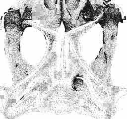
24 48 ANDRZEJ SULIMSKI phalic condyle and disappears ventrally, passing into the distal condyle. The medial conch has a strong ridge to which part of the posterior adductor muscle is attached. At the dorsal end of the medial conch occurs a triangular small surface to which the ligament of the posterior process of the prootic bone was attached, giving some stability to the quadrate bone. Over the articular facet with the posterior process of the pterygoid occurs a small swelling. The place of attachment of extracolumella above on the posterior crest is faintly visible. On the lateral side the quadrate has a distinct tympanic crest and a deep concavity extending laterally to the posterior crest. Below the lower cusp of the tympanic crest a small notch or groove for the attachment of the parallel ligament is present. Palatal region (Text-figs 2C, 5A 4 ). Vomers form the anterior part of the palate, medial margins of the external vomero-nasal fenestrae and medial edges of the exochoanal fenestrae. Both vomers are closely sutured together. Pterygoid process of the vomer and vomerine process of the pterygoid separate the palatines from the sagittal line. Ventral surface of the vomer is slightly convex. In anterior section, on the lateral margin of the vomer, an incision or recess occurs, which forms the medial margin of the vomero-nasal canal. Dorsal surface of the bone slightly concave. On both vomers, along the sagittal line, occur ridges lower than on the ventral side. To these ridges reached the narial septae. Structure of the nasal capsule in M. chulsanensis is not well known since the internal bone elements are damaged. Palatine is relatively wide, anteroposteriorly elongated and has four processes. The vomerine process is sutured anteriorly to the vomer, the maxillary process joins anterolaterally the medial maxillary border, the ectopterygoid process joins posterolaterally the medial ectopterygoid margin, while the sharply pointed pterygoid process is joined from the back to the pterygoid by means of an overlapping suture. Vomer and palatine bones together form the dorsal palatal floor while with the ectopterygoid and pterygoid bones they participate in the formation of the antero-posterior orbital floor and posterior region of the nasal capsules. Vomerine process surrounds medially the vomerine process of the pterygoid bone and laterally and posteriorly - the fenestra exochoanalis. Maxillary process long, intruding between the ectopterygoid and fenestra exochoanalis, and joins the maxilla in a straight suture. The ectopterygoid process reaches to the suborbital concavity, where it joins the pterygoid process of the ectopterygoid, which overlaps with a wide wedge the anterior region of the pterygoid. Sutures of the palatine, ectopterygoid and pterygoid bones, meet in a well marked concavity or depression, where in Recent lizards a more or less wide suborbital fenestra occurs. This fenestra in Recent lizards is surrounded by at least four or five bones of the palatal region. In M. chulsanensis this foramen has more the character of a concavity which may be perforated. The suborbital concavity is anteriorly surrounded by the palatine, medially and posteriorly by the pterygoid and laterally by the ectopterygoid. The dorsal palatal floor, at the site of the pterygoid process of the palatine, breaks into an arch towards the ventral side and reaches the transverse crests of the pterygoids. The latter structural elements form the postero-ventral margin of the palatal vault, which is placed about 10 mm dorsally to the level of the more posterior pterygoid region. If a foramen was present in the suborbital concavity, it was according to OELRICH (1956, p. 26) the place where the superior alveolar nerve and infraorbital artery were probably passing. On the posterior maxillary process of the palatine occurs a small, elongated depression. The ventral surface of the bone is smooth, without traces of teeth or denticles. On the dorsal palatine surface small depressions are present, through which perhaps the inferior nasal artery and medial palatal branch of the facial nerve ran. Pterygoid is anteroposteriorly elongated, and in posterior part bent laterally. Anteriorly
25 MACROCEPHALOSAURIDAE AND POLYGL YPHANODONTIDAB 49 the bone is dorsoventrally concave, in the central part forming an antero-posterior rectangular ventral surface. Posteriorly the bone is mediolaterally flattened, tilted outwards the quadrate bone at an angle of 45 in relation to the central part. The pterygoid bone bears four main processes. The antero-medial vomerine process (pi. XII; Text-figs 2C, 5A 4 ) reaches medially to the pterygoid process of the vomer, joining it obliquely. Distinct (up to 2.0 mm in height) ventral ridges on the ventral, vomero-pterygoid raphae are present. Vomerine process of pterygoid laterally joins the palatine and medially adjacent pterygoid bone. Palatine process rather short, intruding between the palatine and ectopterygoid bones and together with them forms a small hollow. This hollow, at the side of the suborbital concavity, is sometimes punctured by a small foramen. The third or ectopterygoid process extends almost transversally to the long palatal axis and is strongly sutured to the posterior part of the ectopterygoid bone. Together with the latter bone it forms a lateral articular surface distinctly directed downwards, connecting with the medial depression of the coronoid process. The latter fourth process is the longest and mediolaterally flattened. At its base and where it bends a medial articular swelling occurs, to which the condyle of the basipterygoid process is sutured. Posterior end of pterygoid syndesmotically joins, the disto-medial part of the medial conch of the quadrate bone. On the dorsal side of the pterygoid, slightly forward, over the angular bend of the posterior process, a small articular socket for the distal end of the epipterygoid is present. Vomerine, palatine and ectopterygoid processes of the pterygoid, together with its anterior part, form a posterior floor of the oral cavity. The transverse crest in the anterior part of the pterygoid shaft, together with a similar crest on the opposite side, form the posterior margin of this floor. In the pterygoid bone, dentition is not present. Dorsal surface of the palatine process forms the ventro-medial part of the orbit floor. On the dorsal side of the oral cavity, vomerine processes intruding between the palatines are visible. Posteriorly, between the medial edges of the pterygoids, at about the mid-length of the vomerine processes, a rather wide and not very long interpterygoid vacuity occurs. This vacuity is divided by thin processus cultriformis of the parasphenoid. On the dorsal surface of the posterior section of the pterygoid bone, almost behind the articular hollow of the epipterygoid, a small swelling is situated. Posteriorly, on the medial side of the process, occurs an elongated hollow. Lateral side of the posterior process smooth and slightly convex. Ectopterygoid is small, joined medially to the palatine, posteriorly to the ectopterygoid process of the pterygoid and laterally to the jugal and maxilla. Palatine process (anterior) is long, sharp and wedged from the ventral side between the palatine and maxilla. The posterior section of the palatine suture ventrally passes into the suborbital concavity. Postero-medial process of the ectopterygoid bone extends from the suborbital concavity into a ventrally directed pterygo-coronoid process. Lateral surface of the ectopterygoid is sutured to the medial surface of the jugal and medio-posterior surface of the maxilla. Dorsally, the ectopterygoid forms a part of the orbital cavity floor. The postero-lateral hollow in the ectopterygoid bone is a recess into which the apex of the coronoid process intruded during closing of the jaws. Epipterygoid has the shape of a long and strong rod, placed between the pterygoid below and the parietal above-.the distal end is set into an articular socket on the dorsal pterygoid surface and the proximal extremity reaches the sharp ending of the descending swelling of the parietal bone. The upper part of the bone rests posteromedially on the alar process of the prootic. The bone shaft is oval in section. Occipital region (PI. XIII; Text-figs 2D, 5A l ). The brain case forms a wide wedge, inserted ventrally, between the pterygoids, posteriorly, between the postero-lateral extremities 4 - Palaeontologia Polonica No. 33
26 50 ANDRZEJ SULIMSKI of the temporal region bones and dorsally extending under the parietal. The brain case is connected by means of the basipterygoid processes with the pterygoids and by the calcified extremity of the ascending process of the supraoccipital to the ventral descending swellings of the parietal; by the extremities of the paroccipital processes it is syndesmotically sutured to the posterior parietal processes, squamosals and quadrates. Anteriorly the brain case is widely open. Supraoccipital has the shape of a roughly triangular cone, opened from the front. It forms the dorsal margin of the fora men magnum and the postero-dorsal cover of the skull cavity. Anteriorly it articulates with the lateral edge of the alar process of the pro otic bone and posteriorly with the exocciptal bone. It was also joined,.while the animal was living, to the parietal by means of a cartilaginous connection. The dorsal surface of the bone is smooth, its lateral walls steeply directed downwards from the occipital crest. On both sides of the anterior part of the occipital crest occur roughly triangular, slightly concave, surfaces. Lower down are the wide hollows of the supraoccipital bone. The posterior part of the supraoccipital bone, surrounding from above the foramen magnum, is almost horizontal; the anterior part of the bone reaches to the ventral swellings of the parietal bone, which surround it laterally. The anterior margin of the supraoccipital bone has the shape of an inverted V. Dorsal extremities of the bone in specimens of M. chulsanensis are damaged, but it is probable a small ascending process was present here, which intruded into the ventral hollow between the swellings of the parietal bone. The morphology of this region indicates that as in living lizards, this region of the supraoccipital bone formed a flexible joint between the occipital and maxillary segments (FRAzETTA, 1962). Ventral surface of supraoccipital widely concave, covering dorsally the posterior part of the brain cavity. Posteriorly, on the medial walls of the bone, distinct swellings for the internal ear occur. The suture between supraoccipital and exooccipital posteriorly and prootic bone anteriorly is completely fused. The foramen of the endolymphatic canal, present on the dorsal part of the internal ear swelling, is poorly visible. Anteriorly from this swelling extends an arch-like thickening, in the recess of which the medial cerebral vein was situated. The orbito-temporal part included to a large extent only membranous elements, which have not been preserved with the exception in one specimen of a small calcified fragment of orbitosphenoid. Exooccipital in adult specimens of M. chulsanensis is fused to the opisthotic bone. It forms the postero-lateral wall of the skull cavity and the lateral part of the occipital condyle. Branching off from the lateral rim of the fora men magnum is a long, wide and posterolaterally directed paroccipital process, by means of which the occipital segment joins the maxillary segment. Both processes branch off at a very obtuse angle. The extremities of these processes are dorsoventrally widened, with articular facets present on the lateral sides. These facets are dorsally joined to the posterior apexes of the posterior parietal processes, ventrally to the medial surface ofthe posterior squamosal process (with supratemporal part of this bone) and to the medio-dorsal part of the cephalic condyle of the quadrate. The suture joining the lateral part of the paroccipital process to the posterior prootic process is poorly visible. Present under the widened end of the paroccipital process is a long and deep recess, extending into a wide prootic jugular vein recess. In this recess a fenestra ovalis occurs, its anterior part being form ed by the prootic bone. The stapes in M. chulsanensis is not preserved, but probably it was well developed. Fenestra ovalis and fenestra rotunda are separated by a ventrally widened interfenestral crest. The deep occipital recess, drop-shaped, contains a fenestra rotunda, internally placed. A small recess is placed in the hollow ofthe exoccipital bone, beneath the fenestra rotunda. This recess medially borders the perilymphatic foramen, which opens into the skull cavity. The postero-medial surface
27 MACROCEPHALOSAURIDAE AND POL YGLYPHANODONTIDAE 51 of the exoccipital bone forms the triangular part of the occipital condyle and medial surface of the paroccipital process. The articular surface of the occipital condyle forms a diarthritic joint with the sides of the atlas and is included in the recess for the atlas and for the attachment of the atlanto-occipital ligament. Below the occipital condyle occurs the posterior, vertical surface of the tuberal crest. In.a deep recess between the occipital condyle and the medial part of the occipital bone, two small ventrally-placed hypoglossal foramina occur. A larger, dorsoventrally elongated vagus foramen is visible above them. Medially in relation to the vagus foramen occurs a small recess. Lateral ridges, running obliquely and slightly posteriorly from the foramen magnum, divide the posterior surface of the paroccipital process into upper (wide) and lower (narrow) recesses. A small hollow occurs in the lateral wall of the foramen magnum. On the medial concave surface of the exoccipital bone, several more foramina are present. A large, oval perilymphatic foramen for the glossopharyngeal nerve is placed in the anteroventral corner of the lateral surface. Posteriorly from this foramen three small openings occur for the roots of the hypoglossal nerve. Prootic (PI. XIII) is an antero-lateral element of the occipital segment and forms the wall of the skull cavity. Prootic sutured to basioccipital and basisphenoid from below as well as to the three occipital segment bones -.:. exo- par- and supraoccipitai. Large alar process present, but rather short. Postero-dorsal border of this process joins supraoccipitai. Alar crest arches from the trigeminal notch to sutural articulation with the supraoccipitai. Inferior prootic process, situated under the alar process, forms the ventral margin of the trigeminal notch. The ventral margin of the inferior process, joined to basisphenoid and basioccipital, forms at its exooccipital edge part of the margin of the fenestra ovalis. Posterior process of prootic bone with long, rather wide extremity is directed posteriorly, joining the antero-1ateral surface of the paroccipital process and exoccipital bone. Laterally, by the extremity of the posterior process of the prootic, there is a small hollow, which is syndesmotically joined to the medial crest of the quadrate. The smooth lateral surface of the prootic ventrally passes into an oblique pro otic crest. The prootic crest runs towards the front, reaching the inferior ventral process and bends laterally. Under the prootic crest, a wide and deep recess for the jugular vein is present. A small, posteriorly directed foramen for the facial nerve occurs on the lateral wall of the prootic bone (n. VII). The wide jugular vein recess forms part of the wall of the tympanic cavity. In the internal wall of the pro otic bone, at the base of the alar process, occurs a distinct recess for the medial cerebral vein. This recess dorsally passes into the prolongation of the supraoccipital recess. Medially to the recess runs a small thickened, forwardly directed, bluntly-ended swelling of the supratrigeminal process. In Recent Ctenosaura pectinata (see OELRICH, 1956, p. 16), a similarly situated, long and sharply-ended process is clearly visible between the alar and inferior processes. The internal wall of the prootic bone, behind the supratrigeminal process, is distinctly widened, forming a swelling for the inner ear. Ventrally to this swelling is a deep, but not wide acoustic recess (see PI. XIII). This recess has three openings, two of which are placed dorsally, transmitting the branches of the auditory nerve to the anterior ampullar recess (posterior foramen; n. VIII), and leading to the canal ofthe semicircular labyrinth (anterior foramen). The third foramen, placed ventrally, transmits the facial nerve (n. VII). In Ctenosaura pectinata, in the acoustic recess, occurs an additional posterior "semiforamen", supplemented by the exoccipital margin; it transmits the posterior branch of the auditory nerve to the capsular cavity. No such additional "semiforamen" is present in the M. chulsanensis. Basicranium (Text-figs 2C, 5A 4 ). Basioccipital forms the ventro-posterior part of the skull cavity and the medial part of the occipital condyle. Anteriorly it is fused with the 4"
28 52 ANDRZEJ SULIMSKI Table 3 Macrocephalosaurus chulsanensis sp. n. (Postcranial skeleton) Measurements of the holotype ZPAL MgR-I/14 (in mm) I Vertebral column Length of the whole preserved vertebral column 200'0 (estimated) Attention: Length and width of vertebrae see Text-fig. 8 Subregions of presacral vertebrae I No of ribs (presacrals) I Length Posterior cervical Sternal Anterior thoracal Posterior thoracal (lumbar) Pectoral girdle Dorso-ventral length of the scapulo-coracoid 35.0 Maximal length of the coracoid 30.0 Maximal length of the scapula 30.0 Length of the clavicle 38.0 Ventral width of the clavicle 13.0 Dorsal width of the clavicle 8.0 Length of the interclavicle 45.0 Distance between the lateral processes 33.0 Length of the anterior process 9.0 Humerus Ulna Length 46.0 Length 3.7 Width of proximal end 18.0 Width of proximal end 7.5 Width of distal end 20.0 Width of distal end 6.5 Diameter of the shaft 5.0 Least diameter of the shaft 3.4 Radius Metacarpals (length) Length 31.0 I 7.5 Width of proximal end Width of distal end 7.5 III 12.0 Least diameter of the shaft 2.5 IV 10.0 V 9.0 cont.
29 MACROCEPHALOSAURIDAE AND POLYGL YPHANODONTIDAE 53 Digitis (outer length) Pelvic girdle (pelvis) I 25.0 Length of ilium-pubis Length of the ilium 37.0 III 32.5 Length of the ischium 24.0 IV 30.0 Length of the pubis 35.0 V 23.0 Distance between the ilium spines 60.0 Femur Tibia Length 65.0 Length 48.0 Width of proximal end 19.0 Width of proximal end 14.0 Width of distal end 20.0 Width of distal end 10.0 Least diameter of the shaft 7.0 Least diameter of the shaft 5.0 Diameter of the head 12.0 Fibula Metatarsals (length) Length 49.0 I 16.5 Width of proximal end Width of distal end 7.0 III 27.0 Least diameter of the shaft 3.0 IV 32.0 V 11.0 Digits (outer length) I III 32.0 IV 50.0 V 23.0 basisphenoid, dorsally joining the lower pro otic processes and the interfenestral and tuberal crests of the exoccipital. The dorsal basiooccipital is widely concave. In the postero-dorsal section of the foramen magnum occur a pair of small openings (probably nutrient foramina). Posteriorly to these small openings is a small hollow - the place ofattachmentfor the ligament of the odontoid process. The ventral part of the occipital recess, present in basioccipital, and the dorsal part of this recess in exoccipital, form together a large entrance to the fenestra rotunda. Basisphenoid forms the ventro-anterior part of the skull cavity. It is fused to the basioccipital, dorsally joins the antero-ventral prootic process and ventrally, by means of widely forked basipterygoid processes (at an angle of about 90 ) it is sutured to the posterior pterygoid processes. In the anterior part of the dorsal depression of the basisphenoid, on both sides, occur small paired openings that lead to the canals of the abducens nerves (VI n.), The latter obliquely pierce the antero-dorsal edge of the dorsum sellae to emerge in the lateral hollows, forming the lateral wings of this saddle. The basipterygoid processes are short but wide, with strong articular condyles. From the base, in the sagittal line of the dorsum sellae the bony part of parasphenoid together with its paired trabecular crests extend forwards. Between the small alar processes extends a slightly concave sellar crest. The dorsum sellae in M. chulsanensis forms a transverse, deep hollow, composed of two lateral depressions, limited by well marked retractor crests and a central depression (sella turcica) with a vertical crest situated on its posterior wall. Lateral depressions - retractor pits, placed under the small alar processes. Here emerge the small openings of the abducens nerve canals. In the central part of dorsum sellae the elongated, large openings for internal carotid arteries emerge
30 54 ANDRZEJ SULIMSKI u F ig. 6 Macrocephalosaurus chulsanensis sp.n., ZPAL MgR-I/14 - holotype. Nearly complete skeleton, before the final preparation (see also PI. Xi). x 0.5. Dotted - cervicals, white - dorsals, black - sacrals, lined - caudals.
31 MACROCEPHALOSAURIDAE AND POL YGLYPHANODONTIDAE 55 laterally. Spanning these openings and the large foramina of the Vidian canals are bony ridges of the retractor crests. Posterior edges of the basipterygoid processes and ventro-lateral basisphenoid form high, strong, ventro-lateral crests, which build the anterior edges of the tympanic cavity. Dorso-lateral surfaces of the small alar processes are flat and show insertion surfaces of pterygoid muscles. Under the wings of the small alar processes, small notches are visible. Over the basipterygoid processes occur small concave hollows. The basisphenoid base, in relation to the- basioccipital, is directed sharply ventroanteriorly, passing laterally into the bases of basipterygoid processes. -Lower jaw (Text-figs 2E-F, 5As.6 ) ' Dentary is a slender bone, rather long, with a well developed dorsal dental sulcus. The lateral wall of the dentary, to which are attached the shafts of the cheek teeth, is rather high, and almost vertically oriented. Labially, over the parapet, only crowns of the teeth are visible. Along the posterior edge of the lateral surface of the demary extends a small groove, into which the anterior small process of the coronoid inserts. On the external side of the dentary occur six or more small openings for the endings of the lower alveolar nerve. Coronoid is well-developed, situated at 2/3 the length of the jaw. Coronoid process rather high, almost vertical, ' with a rounded top. Anterior labial process small, wedged between the suture joining the dentary to the surangular. The lingual processes are rather long, the anterior one joined anteriorly to the dentary, anteroventrally to the splenial and posteroventrally to the prearticular as well as to the surangular. The posterior process, on the other hand, arches posteroventrally, joining the prearticular and posteriorly the surangular bones. Posterior margin of this process forms the anterior rim of the adductor fossa. Surangular is a flat bone, forming the lateral wall of the adductor fossa and together with the articular and angular bones builds the posterior section of the Meckelian canal. Dorsal margin of surangular rounded. Lateral surface of bone smooth and slightly concave. In the articulo-surangular suture occurs a small posterior surangular foramen and in some specimens, below the coronoid, a larger anteriorly-directed anterior surangular foramen is visible. The ventral margin of the surangular is arranged at a right angle to the lateral wall of the bone. On the lateral wall, from the cotylus, a sharp crest runs obliquely, disappearing in the antero-ventral part of the surangular. This crest is the place of attachment for the external adductor muscles. The anterior process reaches the dentary. Angular is a small ventral element of the lower jaw, joined posteriorly to the articular, mediolaterally to the surangular and anterolaterally to the dentary and splenial (anteromedially). Anterior process of the angular situated on the ventral side, at the level of the posterior ending of a large anterior inferior alveolar foramen. A small posterior mylohyoid foramen is also present. Articular an irregular bone with a wide cotylus, small anterior process and a shortened retroarticular section. In the posterior region occurs a wide, concave, transverse cotylus. From behind the cotylus emerges a wide, shortretroarticular process, to which was attached (perhaps very well developed here) a ligament of strong depressor muscle. Medially, the anterior process of the articular is ~oncave and forms posteriorly the rim of the adductor fossa. Mesially to the cotylus, an angular process of the articular is present. Splenial is very similar to that in M. gilmorei but the anterior inferior alveolar foramen is situated more in the mid-length of the splenial. The anterior Meckel's opening, situated by the symphysis, is long, slightly larger than in M. gilmorei. The Meckel's groove in all specimens of M. chulsanensis is almost completely covered by the splenial. Postcranial skeleton (PI. XI, XV-XXII; Text-figs 6-11). Vertebral column. The description of the postcranial skeleton is based on the best preserved type specimen ZPAL MgR-IjI4. On this specimen, six subregions can be differentiated. The


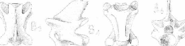
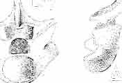
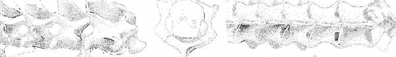
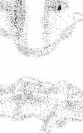
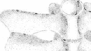
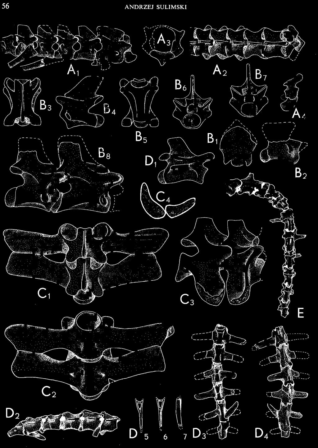
32 56 ANDR ZEJ SULIMSKI --, J I, I -,., E
33 MACROCEPHALOSAURIDAE AND POLYGLYPHANODONTIDAE 57 number of presacrals in M. chulsanensis is 27 (as compared with 29 in Polyglyphanodon sternbergi, 24 in Iguana iguana,27 in Macroscincus cocteaui and 27 in Lacerta lepida). In the type specimen (PI. XI, Text-fig. 6), between the seventh and eighth cervical vertebrae occurs a small gap, caused by a natural crack in the rock. The seventh vertebra is damaged in the posterior part. A second gap in the vertebral column occurs between the seventeenth and eighteenth presacrals, but not damage in the vertebrae is observed. Almost all ribs on the specimen are preserved with the exception of several ribs in the cervical and the posterior dorsal subregions. Cervical subregion (Text-figs 6, 7A l -4 ' B l _] ). The atlas in the type specimen is not completely preserved, but is well preserved in specimen ZPAL MgR-I/2!. This vertebra is basically of the same structure as similar vertebrae in genera Iguana, Tupinambis or Varanus. It is composed of three elements - hypocentrum and paired lateral neurapophyses, surrounding the neural canal. Also present are small zygapophyses for joining with the axis. In the anterior part of the atlas, on the antero-dorsal side of the hypocentrum and on the antero-medial side of each neurapophysis, a semicircular depression (articular facet) for joining to the occipital condyle is visible. On the dorsal side of the hypocentrum occurs a distinct sulcus for the odontoid process of the axis. This latter process rests also on the dorsal sulcus of the occipital process. Ventral crest well developed in the form of a small keel present on the hypocentrum. Axis has a long centrum, its anterior part is elongated and widened, forming dorsally the odontoid process. The articular surface of the anterior part of the centrum is widely rounded, the centrum itself being in this place wider than higher. Ventrally, the centrum is rounded and beneath it, anteriorly, occurs a medial process, directed downwards; (in the specimen this process is broken). The hypocentrum is not fused with the centrum and this applies also to the hypocentra between the succeeding anterior cervical vertebrae. On the lateral sides ofthe centrum, small diapophyses are visible. Their position is situated more towards the front than in Polyglyphanodon or Iguana. Prezygapophyses and postzygapophyses are well developed. Spinal process on the axis large, long, almost the same length as the centrum. Posterior section of this process reaches behind the posterior boundary of the centrum. Establishment of the boundary between the cervical and the thoracal sections was often difficult. Many authors gave the average number of neck vertebrae as six on the assumption that vertebrae joined to the sternum belong to the dorsal section, while those not joined to the sternum belong to the cervicals. This interpretation was maintained by COPE, 1892; CAMP, 1923 ; WILLINSTON, 1925; GILMORE, 1942; ROMER, 1945, 1966b and others. Later investigations, however, suggested that the average number of neck vertebrae could be eight (HOFFSTETTER & GASC, 1967/68, 1969; GUIBE, 1970; GASC, 1971). Specimen ZPAL MgR-I/21 (Text-fig. 7A l _] ) has a completely preserved section of cervical vertebrae, with all structural elements well developed. The vertebrae with prominent hypocentra (free in four anterior vertebrae and coalesced in the two last ones) differ distinctly from the remaining vertebrae, i.e. the sternal Fig. 7 Macrocephalosaurus chulsanensis sp.n., ZPAL MgR-I/21. A - Cervical vertebrae and atlas : 1 -lateral view, 2 - dorsal view, 3 - atlas, anterior view, 4 - lateral view. ZPAL MgR-I/14 - holotype. B - axis, thoracal and posterior presacral vertebrae: 1 - ventral view, 2 -lateral view, 3 - middle thoracal vertebra, dorsal view, 4 -lateral view, 5 - ventral view, 6 - anterior view, 7 - posterior view, 8 - two posterior presacral vertebrae, lateral view. The same specimen. C - Sacral vertebrae: 1 - dorsal view, 2 - ventral view, 3 -latero-dorsal view, 4 - distal ends of transversal processes (in outline). The same specimen. D - First caudal vertebrae, tail and chevron: 1 - lateral view, 2 - proximal part of the tail, lateral view, 3 - ventral view, 4 - dorsal view, 5 - chevron, anterior view, 6 - posterior view, 7 -lateral view. ZPAL MgR-I/22. E - Proximal part of the tail with the autotomy septae, dorsal view. A 1-2, B 1-7, D 1 - x 2; B 8, C x 2.5; x 3; D 2-4, E nat. size.
34 58 ANDRZEJ SULIMSKI vertebrae, in the build of the shafts, the emplacement of synapophyseal prominences, the structure and arrangement of the spinal processes. These vertebrae are shorter, dorsally and ventrally more square. Their spinal processes are shorter, slender and more slanted posteriorly. The para- and diapophyses of the anterior cervical vertebrae (from the third to the fifth) are connected to one another to form elongated, thick and obliquely arranged crests ~ synapophyses, which in the last vertebrae are developed as round tubercles. The hypocentra on the axis and on the third, fourth and fifth vertebrr.e are not fused to the centra and are placed in the antero-vemral end of the centrum. In the remaining cervical vertebrae, the hypocentra are firmly fused to the centra, forming laterally well visible lateral compressed processes. In the described specimen, between those vertebrae with hypocentra and the vertebra situated behind them, occurs a small gap, caused by the post-mortem separation of the spine. The first dorsal vertebrae, belonging to the sterna I subregion, are without hypocentra, their synapophyses having the form of a small, oval tubercle. Differentiation between the anterior and posterior thoracal subregions is based, according to GASC (1971), on the differences in the length of the ribs attached to these vertebrae. The measurements of the length and width of vertebrae, given as a diagram (Text-fig. 8), form in M. chulsanensis a characteristic curve. The anterior part of this curve confirms the morphological differences between the neck and the thorax. For comparison a diagram for Iguana iguana L. (see HOFFSTETTER & GASC, 1969, p. 248; A 1~ mm I : I,_-~-..,, 'I', '" I, , I I I I I : I I...- I1 I I'... I I '-.. '- I \ I I I I I I I I I 12 I 13 r- 14 ' 15' 16 ' 17' 18' 19: 1 2 I 1 ' 2 3 L;, 5 ' 6 7 A L S :5ACR.: e A U 0 A L 5 Fig. 8. The length and width diagram of presacrals in: A - Iguana iguana LINNAEUS, Recent (after Gxsc & HOFFSTETTER, 1967/68, 1969; Gxsc, 1971). B - Macrocephalosaurus chulsanensis sp.n., ZPAL MgR-I/14 - holotype. The continuous line - length, broken line - width. B
35 MACROCEPHALOSAURIDAE AND POLYGLYPHANODONTIDAE 59 Fig. 39; GASC, 1971, p. 7, Fig. 3) is also given, where similarly the curve breaks between the eighth and ninth presacrals. The boundary between the neck and the thorax in Polyglyphanodon sternbergi (GILMORE, 1942) is marked, as mentioned by GILMORE, by a different structure of the spinal processes. However, according to that author, the presence of long ribs joined to the seventh and eighth vertebrae seems to indicate that these vertebrae were attached to the sternum as in Iguana and thus belong to the thorax. According to HOFFSTETTER &GAse (1969), Gxsc (1971) and the observations of the present author, Iguana iguana has 8 distinct cervical vertebrae. While describing the thoracals, GILMORE (1942) stated that the first and second thoracals, i.e. seventh and eighth presacrals have narrowed spinal processes and distinctly differ from the processes of the succeeding thoracals. It can be definitely stated 'that the vertebrae of the cervical section in Polyglyphanodon sternbergi, as in M. chulsanensis and Iguana iguana, end at the eighth presacral. The main modifications in the structure of cervicals are as stated above: the presence of lateral prominences - diapophyses and parapophyses. These prominences between the third and sixth vertebrae gradually enlarge to the last cervicals, when they again become smaller. On the axis occurs a small single tubercle, to which a strongly reduced rib might have been attached. In the following vertebrae para- and diapophyses are coalesced, forming synapophyses of an elongated tubercle shape. Ribs were attached by means of their slightly forked proximal extremities to these tubercles. In the specimen ZPAL MgR-IjI4 only ribs of vertebrae fifth and sixth are preserved. On the last cervicals the synapophyses are more rounded. Shafts of the cervicals are square and shorter than the thoracals. The ventral surface ofthe shafts is rounded, a slight, wide crest being present. The zygospheno-zygantral articulation on the vertebrae is well marked. Spinal processes of the cervicals are slender, with thickened posterior and sharp anterior margins. All processes are. distinctly bent posteriorly. Sternal subregion in M. chulsanensis has been established on the basis of the work of Gxsc (1971) and includes presacrals 9 to 13. In the type specimen, placing of the boundary between the sternal and anterior thoracal subregion is possible only on the basis of the small differences in the rib course and their distal extremities. The ribs of the sternal subregion are long, more bent than the ribs of the succeeding subregion and their endings are only slightly widened. Anterior thoracal subregion. Beginning from the fourteenth and ending on the twentieth or twenty-first presacrals includes longer, straighter ribs evenly truncated distally. Posterior thoracal subregion. Begins from the twenty-first or twenty-second presacrals and ends with the twenty-seventh, last vertebra, differing from the previous ones in a distinctly shortened shaft. The ribs of this subregion are distinctly shortened and almost straight. All the ribs of the above mentioned three subregions are unicephalic, this being indicated by the tuberous synapophyses. The centra of the thoracals are posteriorly distinctly narrowed. On the ventral side, the centra are flattened, with a wide keel visible. All presacrals, with the exception of the cervical ones, possess anteroposteriorly wide spinal processes, with almost vertical posterior and slanted anterior margins. Height of the spinal processes is more or less even on the whole presacral section. The last presacrals have stronger and wider spinal processes. Length of the sterno-thoracal vertebrae, beginning from the second, gradually gets bigger to reach the maximum at the eleventh-fourteenth vertebrae; then it gradually decreases, until at the eighteenth and nineteenth vertebrae their length is almost the same as that of the first vertebrae of this section of the vertebral column. The last presacral vertebra is distinctly shorter than the previous ones. On the lateral side, the sterno-thoracal vertebrae have a distinct rounded edge, extending from the synapophysis obliquely, ventrally and posteriorly. The centra below
36 60 ANDRZEJ SULIMSKI this edge are slightly depressed. Such a structure of the shafts is found in almost the whole series of the described section of the vertebral column. Over the synapophyseal margin and the margin joining the prezygapophyses and postzygapophyses occurs a depression, widened posteriorly. Synapophyses protrude laterally from the centra and are situated between the neurapophyses and the centra. Articular facets for the ribs, which are present on the anterior vertebrae of the sterno-thoracal section, are elongated, oval and slightly obliquely arranged. The largest f cets are placed on the first five vertebrae, gradually decreasing in size up to the eleventh vertebra. From the twelfth vertebra the shape of the synapophyses and facets distinctly changes. The ribs also change in this section, being shorter and more straight. The ribs of the posterior thoracal subregion are almost 5 or 6 times shorter than these of the sternal subregion and the anterior ribs of the anterior thoracal subregion. The zygospheno-zygantral joining is well developed in the whole sterno-thoracal section. This section in M. chulsanensis is about 200 mm in length. Sacral subregion (Text-figs 6,7 C l -4)' The sacral section of the vertebral column is always composed of two unfused vertebrae, joinded in a similar manner as the vertebrae mentioned above. Centra of the sacrals are distinctly shorter and possess strong condyles. The anterior vertebra is slightly shorter and wider than the posterior one and has stronger transverse processes. These latter processes are completely fused with the centra. The processes of the anterior vertebra are so arranged in relation to the processes of the posterior vertebra, that their anterior margin is raised upwards, and the posterior margin ~ downwards, at an angle of 45. On the other hand, the anterior margin of the posterior vertebral processes is slanted downwards and the posterior one - upwards, at an angle of about 30. Posterior margins of the processes of the anterior vertebra are in contact (but not fused) with the anterior margins ofthe processes of the posterior vertebra. Medially, towards the vertebral centra and between the processes, occur distinct transversally elongated sacral foramina. Ventral surfaces of both pairs of transverse processes are situated slightly over the lower border of the centra. The processes, by means of wide distal extremities, join the internal surfaces of the ilium bones. In the anterior vertebra, the transverse processes run vertically from the centrum, while in the posterior one they are directed slightly towards the front. Close to the centra of both vertebrae, on the ventral side, small depressions are visible, with openings for the arteries and nerve branching. From ventral side, the centra of both vertebrae are transversally rounded and possess a distinct keel. Spinal processes are slightly anteroposteriorly shortened, but are equal in height to the spinal processes of the thoracal vertebrae. The zygospheno-zygantral articulation is well developed. Caudal subregion (PI. XX, Text-figs 6, 7 D l -4' E). The caudal region is incomplete. The distal part, which comprises up to two-thirds of the whole tail, is missing. The maximum number of vertebrae preserved in M. chulsanensis is eleven, these being joined to the rest of the vertebral column (ZPAL MgR-I/22) (Text-fig. 7E). The first caudal vertebra in its dimensions does not differ from the posterior sacral, but the transverse processes are distinctly inclined backwards. Beginning from the fifth or sixth vertebra, autotomic septae occur (these septae are probably present also on the third and fourth caudal vertebra, but these latter are badly damaged). The fracture planes at the same position as in M. chulsanensis occur also in Scincus scincus (EI-Toubi, 1938).The centra of the caudal vertebrae, beginning from the second, are gradually elongated, so that the sixth-eighth have centra 2 to 3 times longer than wide. Transverse processes, from the second caudal vertebra, are rather rapidly shortened to a small spur, present on the last preserved vertebra. Zygapophyses strong and short. The zygospheno-
37 MACROCEPHALOSAURIDAE AND POLYGL YPHANODONTIDAE 61 zygantral articulation present in the anterior vertebrae. The spinal processes gradually disappear to form a small, anteroposteriorly elongated crest, present on the distal vertebrae. On the ventral side of the centra of the proximal vertebrae occur small, paired articular cusps, situated just by the anterior end of the centrum. To these cusps chevr-ons were attached. Ribs(PI. XI; Text-fig. 6). All presacral vertebrae with the exception of the first (atlas) and probably ofthe second (axis), possess ribs. The axis has small lateral tubercles, but with no distinct articular facets. Visible on the third-cervical; are well developed diapophyses with traces of articular facets. Vertebrae 4 and 5 have well developed, but short ribs with slightly forked proximal extremities. Distally, the rib-shafts rapidly become wedge-shaped. Ribs of cervicals six-eight have strong and rather long shafts. They are distinctly longer than those of the former. The ribs of the thoracals together with the sternai ones have already been described. It should be mentioned that the proximal sections of these ribs are slightly widened, but are connected to the centrum by only a single joining. Chevrons (PI. XIX, Text-fig. 7 D S _ 7 ) of the anterior caudal section are long, slender and distinctly longer than the spinal processes. Chevrons are attached to the cusps present on the centra by means of paired processes (pedincules) often coalesced with each other. The chevrons are placed almost intercentrally. The first chevron, judging by the preserved articular tubera on the centra, was attached to the third caudal vertebra. Pectoral girdle (PIs XVI-XVIII, Text-fig. 9). The scapulo-coracoid part is preserved almost completely in the type specimen. Only the dorsal endings of the scapulae are missing. Scapula is slender, rather long, with one rather deep emargination. The upper ending of the scapula is distinctly widened. In ZPAL-MgR-I/25 this region is completely preserved with large and trapezoidal suprascapulae and well developed emarginations in scapulo-coracoid section (PI. XXII, Fig. 1d). In the young specimen - ZPAL MgR~I/24, over the upper margin of the scapula, a small bone fragment occurs, which may correspond to a part of the suprascapula. Posterior margin of the scapula is slightly concave. In the type specimen, the prescapular process and part of the scapula, situated under it, are missing. On the lateral surface of the scapula, from the apex to the coracoid, runs a wide and rounded margin. The upper part of the glenoid fossa is developed as a tuber. In this specimen, the suture running between the scapula and the coracoid is almost invisible. In a young specimen, on the other hand, this suture is distinctly marked and runs from the scapulocoracoid emargination to the glenoid fossa, over the precoracoid process. The scapulo-coracoid emargination is well developed and deep, with the long and slender scapular andprecoracoid processes surrounding it from above and below. The coracoid bone in the type specimen is completely preserved. This bone has one anterior emargination - deep and wide. In the specimen ZPAL MgR-I/18 (PI. XVIII, Text-fig. 9F) the left coracoid has a slightly marked additional emargination, placed on the ventral side. This is probably due to the disappearance of a second coracoid emargination, existing in the early ontogenetic development. Between the posterior margin of the large coracoid emargination and the glenoid fossa, occurs a large coracoid foramen. Beneath it, a rounded margin is present, running from the anterior process to the lower tuber of the glenoid fossa. The preglenoid process is thick and broadly rounded. Ventral margin of coracoid long and strongly arched. In many legwans scapulo-coracoid has four emarginations, but in scincomorphid lizards as a rule there are three (HOFFSTETTER, 1964, Fig. 4) as in M. chulsanensis. The dorsal parts of the clavicles in the type specimen are damaged. They are in the natural position, contacting the scapulo-coracoid and the interclavicle. The clavicles are strongly built, massive, with widened ventra-medial parts; in these


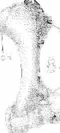
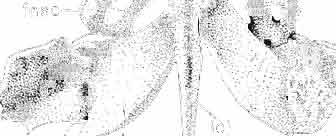
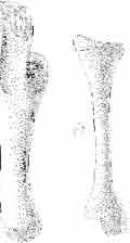
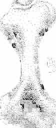
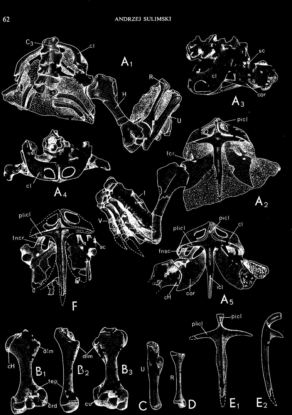
38 62 ANDRZEJ SULIMSKI u D
39 MACROCEPHALOSAURIDAE AND POL YGL YPHANODONTIDAE 63 latter, large oval perforations occur. The dorsal part of the clavicle is almost twice as narrow as the ventral part, bending arch-like to surround the scapula laterally and anteriorly. Anterodorsal margin of the clavicle widely rounded, the postero-ventral margin sharp. On the external surface, the shaft of the clavicle is slightly depressed on the whole length. Viewed from below (pi. XVII, Text-fig. 9A 4 ), the clavicles ventrally join, forming there a strong, sharp keel. From the internal side, between the keel-like incision of the clavicles, intrudes an anterior interclavicle process, widened and bent upwards. Interclavicle in the holotype is almost complete, cruciform, with a well developed anterior process. This process is slender at the base, widening laterally and bending arch-like upwards (in specimen ZPAL MgR-I/21 this widening of the process has three distinct margins, two lateral and one medial, however the, ending of the process is not forked). Similar crests occur in the Pleistocene skink - Didosaurus mauritianus (HOFFSTETTER, 1949, Fig. 14). On the dorsal side of the process runs a sharp, medial keel, flanked by elongated surfaces. These surfaces,are tightly, internally connected by the keel-like bent parts of the clavicles, forming together a compact hinge. Lateral processes of the interclavicle are rather short compared with the shaft, dorsoventrally flattened with slight, elongated swellings. These processes grow almost vertically from the axis of the shaft. The shaft of the bone is long, compressed in the medial part and posteriorly sharply ended. The extremity of the shaft reaches beyond the posterior endings of the coracoids. Along the shaft, on ventral side, from the anterior process extends the prolongation of the rounded elongated keel. Fore limb (PI. XVI, Text-fig. 9A-D, 11 B). The humerus and forearm bones in the holotype are anatomically arranged; the carpus, metacarpals and fingers are in a slightly contracted position, lying on the dorsal surfaces of the bones on the rock. Left limb incomplete. Humerus is strong, with wide extremities and a shortened slender shaft, almost round in cross-section. Surfaces of both articular extremities arranged at an angle of about 45 to each other. The proximal extremity directed towards the external side, the distal - towards the internal. Articular surface of the proximal extremity is oval and convex in both directions, especially anteroposteriorly, reaching to the posterior part of the bone. Externally, on the proximal section of the bone shaft, occurs a distinct deltoid crest, while on the internal side, close to the upper articular facet, a swelling is present. To the above mentioned prominent deltoid crest strong muscles were attached, binding the massive part of the scapulo-coracoid with the front limb. Anterior or ventral surface of the proximal epiphysis is deeply concave and wide. Articular surfaces on the anterior side for the forearm bones are directed strongly upwards. A large and strong external condyle for attachment to the ulna occurs, mainly 'on the ventral side, while a smaller one, for the radius, is present on both sides of the extremity. On the ventral side, over both condyles a depression is visible. The entepicondyle and 'ectepicondyle well developed, the former placed slightly lower and possessing a wide rounded surface. The latter, lower one, is situated slightly higher. Over Fig. 9 Maeroeephalosaurus chulsanensis sp. n., ZPAL MgR-I/I4 - holotype. A - Pectoralgirdle with right fore limb: 1 - dorsal view, 2 - ventral view, 3 -lateral view, 4 - anterior view. The same specimen. B - Humerus: 1 - posterior view, 2 -lateral view, 3 - anterior view. The same specimen. CoD - Ulna and radius: lateral view. The same specimen. E - Interclavicle: 1 - ventral view, 2 -lateral view. ZPAL MgR-IllS. F - Pectoral girdle, ventral view. Nat size. Abbreviations: ch - humeral head, c/- clavicle, cor - coracoid, erd - radial condyle, eu - ulnar condyle, dlmdeltoid margin,fep - ectepicondylar foramen,fner - coracoid fenestra,fnse - scapulocoracoid fenestra,fer - coracoid foramen, fse - scapular fenestra, H - humerus, ic/ - interclavicle, picl - anterior interclavicular process, plicllateral interclavicular process, R - radius, se - scapula, sse - suprascapula, U - ulna.


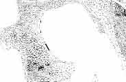
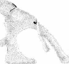
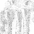
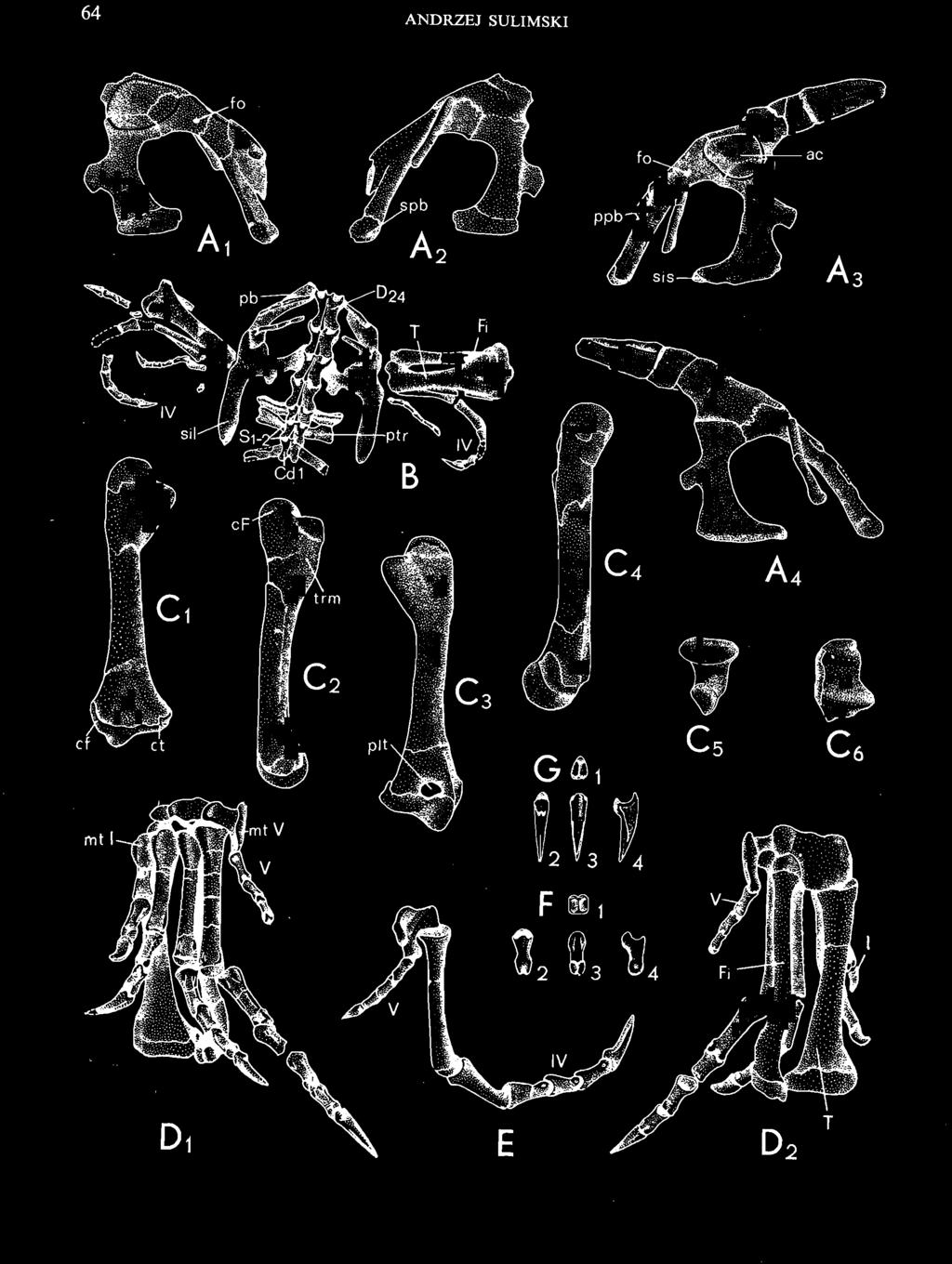
40 64 ANDRZEJ SULIMSKI ~ " E
41 MACROCEPHALOSAURIDAE AND POL YGLYPHANODONTIDAE 65 its upper ending, a small ectepicondylar foramen is present. Between the distal extremity of the humerus (sometimes in the intercondylar depression) and the proximal extremity of the ulna occurs a small, irregularly shaped, sesamoid bone - patella ulnaris. According to CAMP (1923) this bone is placed in the tendon of attachment of the triceps muscle, which is attached to the end of the humerus and reaches olecranon. The radius, is slender, with almost equal extremities. The proximal extremity is widened, with fossa-like depression for joining with the radial condyle of the humerus. The shaft of the bone straight, rather long, rounded in crosssection. The distal extremity has an oblique, slightly concave articular surface. On the internal (medial) side occurs a distinct process, which is joined to the depressed articular facet of the radiale. The ulna is longer and stronger than the radius. The shaft is wider, the olecranon long and strong, about 1/4 the length of the shaft. Proximal articular surface concave, crossing over and upwards to the anterior surface of the olecranon. In this place, on the medial side, occurs a depression into which the lateral part of the proximal extremity of the humerus intrudes. The shaft of the bone almost even, slightly widened upwards, especially in the articular surface and is slightly transversally compressed. The distal extremity of the bone widened, articular surface convex, overlapping the anterior and posterior parts of the bone, joining to the ulnare. Carpus (Text-fig. 11 B). Radiale is large, irregular in shape, swollen posteriorly and narrow anteriorly, and is distinctly displaced downwards and posteriorly relatively to its natural position. In the middle of its antero-ventral surface occurs a small facet for attachment to centrale 1. Externally to it occurs a second facet joining carpale 2. Ulnare is of a blocky build and is the largest bone in the carpus. In the holotype occur 5 carpals and 2 centrals placed above them and of more or less equal size. Between the radial and ulnar bones occurs a rather irregular unfused intermedium. In addition, on the lateral side of the carpus an oval pisiform is present. Arrangement of the carpal bones differs from that of some Recent lizards mainly in the presence of a small centrale 2, the lower surface of which, in the holotype, is fused to carpale 2. Manus (PI. XVI, Text-fig. 11 B). Autopodium in the holotype is almost completely preserved only the distal phalanges of the fourth and fifth fingers are missing. Metacarpals in their proportions more or less correspond to the proportions of the same bones in Iguana iguana or Polyglyphanodon stembergi. Phalangeal formula of manus The proximal extremities of the phalanges are thicker and shorter than similar elements in Iguana and Polyglyphanodon. The fore limb in relation to the hind limb is more strongly built, rather wide and short. The phalanges are joined together by means of a tongue and groove articulation. All fingers possess long and sharply pointed terminal phalanges. The latter are slightly bent, arch-like and with long depressions at the sides, indicating the strong development of claws. On the ventral side of the proximal part of these phalanges basal tubers are visible. Fig. 10 Macrocephalosaurus chulsanensis sp.n., ZPAL MgR-I/14 - holotype. A - Pelvic girdle and hind limb : 1 - right half of pelvis, lateral view, 2 - medial view, 3 - left half of pelvis, lateral view, 4 - medial view. B - Pelvic girdle with hind limb and posterior dorsal, sacral and anterior caudal vertebrae, dorsal view. C - Femur: 1 - anterior view, 2 medial view, 3 - posterior view, 4 - lateral view, 5 - proximal articular surface, 6 - distal articular surface. D Zeugopodial and autopodial part: 1 - ventral view, 2 - dorsal view. E - Fourth and fifth fingers, dorso-iateral view. F - Distal phalanx: 1 - articular surface, 2 - ventral view, 3 - dorsal view, 4 - lateral view. G - Clawed phalanx: 1 - articular surface, 2 - dorsal view, 3 - ventral view, 4 - lateral view: A, C-G - nat.size; B - x 0.5. Abbreviations: ac - acetabulum, cf - femoral head, cf- fibular condyle, fo - obturator foramen, ppb - pubic process, ptrtransversal process, si! - ilium spine, sis - ischiadic symphysis, spb - pubic symphysis, trm - trochanter major, D - dorsal vertebra, S - sacral vertebra, Cd - caudal vertebra, 1-V - fingers. Other abbreviations see Fig Palaeontologia Polonica No. ~
42 66 ANDRZEJ SULIMSKI Pelvic girdle (PI. XIX, Text-fig. loa-b) in the holotype is almost completely preserved Only the distal part of the right pubis is missing. The structure of pelvis keeps to the classical construction of its components as observed in most of the Recent large lizards, such as Varanus, Iguana, Tupinambis or the fossil Polyglyphanodon. The ilium is an, elongated, rather wide and bluntly ended bone, in relation to the long axis of the vertebral column, is slightly slanted upwards and posteriorly. In the anterior part of the base, on.the dorsal side, occurs a tuber. The shaft of the ilium does not show any swellings, whereas on the medial side distinct, elongated prominences are present, to which the tr ansverse processes of the sacrals are attached. The ilium is joined by means of II distinct suture in the acetabulum to the ischium and pubis. Gradually tapering posterior extremity of the ilium and an evenly wide shaft are observed in Iguanidae, Teiidae and Varanidae. A similar situation is observed in M. chulsanensis. Articular facet oval, the antero-posterior diameter being the largest. In.the holotype both ischia are well preserved, with the exception of damage in the vicinity of the ischium tuber. Below the ischium tuber, on the posterior margin of the bone, strong notch is visible. Posterior margin of the ischium is very thin and easily damaged. For that reason the notch may have been originally slightly smaller. Anterior end of the ischium directed ventroanteriorly. The ventral margin of the bone is joined, in the medial line, to the ischium of the opposite side. Anterior margin of the bone strongly concave, surrounding posteriorly a large puboischiadic fenestra. In the proximal part, the ischium joins the ilium from above and the pubis anteriorly, forming the acetabulum. In relation to the ilium the ischium is strongly oblique, directed interiorly and downwards. The described bone differs in M. chulsanensis from the ischia in Iguana, Varanus and Tupinambis in its more slender structure, more concave anterior margin and in the presence of the ischium notch. Pubis in type specimen is almost completely preserved in the left half of the girdle and is directed anteriorly, downwards and towards the medial line. By means of a rather short pubic symphysis it joins the pubis of the opposite side, anteriorly enclosing the pelvic girdle. The symphyseal end of the bone is not widened. Externally, from the acetabulum, extends a distinct ridge, reaching to the pubic tubercle. A small obturator foramen is present anteriorly on the acetabulum under the external crest. In frontal view, the proximal part of the pubis, up to the pubic tuber, is strongly widened. Just in front of the acetabulum and slightly over the obturator foramen a small tuber is visible, to which the muscles bending the limb were attached. A similar tuber is well developed in the genera Varanus, Iguana and Tupinambis. The pubis together with the ischium form a large puboischiadic opening. Hind limb (PIs XX, XXI ; Text-figs 10C-D, 11 A). Femur in the holotype is'well preserved, being distinctly longer than the humerus, slender, with strong extremities. Compared to the femur in Polyglyphanodon, it is distinctly more slender. The main difference lies in the different position of the trochanter major in relation to the posterior surface of the bone. The trochanter major in Polyglyphanodon is placed on the posterior side, while in M. chulsanensis it is more medially directed. A similar situation is observed in the femur of Varanus and Tupinambis. Much smaller differences in the proportions, position of the trochanter major, distal condyles and articular head occur between the femora in Varanus griseus and M. chulsanensis. The angle between the antero-posterior diameter of the articular head and the surface of the distal condyles in M. chulsanensis is about A similar angle is observed in the genus Varanus. The trochanter major is well developed, tuberous, with a thick edge underneath. Attachment surface on the trochanter major is large, oval and directed upwards. Between the trochanter major and the head of the femur occurs a large and rather deep depression, reaching to the distal end of the bone at more or less mid-length. The middle part of the shaft of the fem ur
43 MACROCEPHALOSAURIDAE AND POL YGL YPHANODON_TIDAE 67 is round in cross-section; laterally, the shaft is proximally straight and distally slightly-bent backwards. The articular surface of the distal extremity, viewed from below, is trapezoidal in outline, the surfacejoining the tibia larger, that joining the fibula smaller. Both surfaces are separated by a strong, circular condyle,reaching well onto the posterior side of the bone. The place of joining to the fibula is on the external side of the condyle. The intercondylar fossa is shallow. Posteriorly, over the distal epiphysis, a depression is visible in the shaft of the bone, in which a small, ' oval sesamoid bone (patella tibialis) is present. The epiphyseal suture, occurring between the shaft and the distal articular surface, is visible from all sides, similarly as the suture 'separating the articular surface of the head and the proximal end of the shaft (the latter less marked). Tibia is best preserved in the right limb of the holotype. It is by about one-fourth shorter than the femur. T~ proximal end of the boneiswide, with irregular articular surface and rather long and slender shaft. The maximal diameter of the proximal articular surface is directed.obliquely in relation to the long axis of the bone. Cnemial crest not distinct; external edge ofthe crest slightly concave, the internal-convex. The proximal condyles, especially the external one, distinctly reach beyond the posterior boundary of the bone shaft. Distal end of the tibia is slightly widened, articular surface sub-oval, slightly depressed externally and convex medially. No prominent angular differences observed between the directions of the proximal articular surface and the distal articular facets. The distal articular surface rests directly on the strong internal part of the tibio-fibular bone (astragalocalcaneum). Fibula (PI. XXI), together with the tibia, lies in proper anatomical position in relation to the tarsus and manus. The fibula is slender, long and in structure resembles rather the fibula in Varanus than in Iguana or Tupinambis. It is also longer than the tibia. The articularsurfaces of both extremities, on the other hand, are arranged obliquely to this axis. Proximal extremity wider, with a swelling or tuber directed medially and a rather long crest on the anterior side. Upper articular surface long and narrow, joining the fibular condyle of the femur. On the external side of the proximal extremity runs a rather thick Crest. Distal extremity is less widened, diagonally thickened. Articular surface of the latter extremity convex and obliquely arranged. The shaft oval in cross section. Between the proximal extremity of the fibula and the similar extremity of the tibia, where they laterally join, occurs a small additional. bone, which can be considered as an interarticular sesamoid bone of the fibula. This,bone in Recent lizards is usually situated in the tibio-fibular ligament, which runs beneath the wide attachment of the tendon rectus tibialis muscle. Usually in this ligament patella tibialis is developed. Tarsus (PI. XXI, Text-figs IOD, 11 A). The tarsal bones in the.type specimen are strongly calcified, and -the small tarsus elements are. variable in structure. In the type specimen only two small bones, situated between the coalesced tibio-fibulare and metatarsals are visible. It is possible that the tarsus consisted of six or more bones. In the proximal row only a large, wide tibio-fibulare is present. This bone includes intermedium which is fused without traces of the joining. Articular surface of the tibial section is almost oval, biconcave and anteriorly only slightly depressed. The distal articular facet of this bone is directed ventroanteriorly. Posteriorly to the facet there is a riarrow articular surface, directed upwards to the posterior side of the bone and reaching to almost mid-length. The lateral part - fibulare, is more slender, with a concave upper articular surface for articulating to the fibula. This surface is oblique in relation to the horizontal length of the bone. Ventral surface concave, with an articular facet directed downwards and interiorly. At the extreme, distal end occurs a second facet, which was perhaps joined to tarsal 5. A compressed, irregular bone element, placed above metatarsal IV and laterally joined to metatarsal V, belongs to tarsal 4. Small medially 5'


44 68 ANDRZEJ SULIMSKI v Fig. 11 Macrocephalosaurus chulsanensis sp. n., ZPAL-I/14 - holotype. A - The fore limb: 1-anterior view, in flexion, 2 - the same, in extension. B - The hind limb: 1 - antero-dorsal view, 2 - postero-ventral view. Nat. size. Abbreviations: ag - astragalus, e - centrals, ea - carpals, eal- calcaneum, ep - proximal centrals, et-tibial condyle, f - fibular bone, F - femur, Fi - fibula, i - intermedium, me - metacarpals, mt - metatarsals, pi - pisiform bone, r - radial bone, T - tibia, I - tibial bone. Is - tarsals. u - ulnar bone.
45 MACROCEPHALOSAURIDAE AND POLYGL YPHANODONTIDAE 69 directed bone element placed at tarsal 4 and joined to metatarsal III belongs to tarsal 3. Pes (PI. XXI, Text-fig. 10D-G, 11 A). Length of the hind foot, measured along the IVth finger, is about 80 mm. It is distinctly longer than the fore foot. Metatarsals gradually become / longer from the first to fourth finger. The latter is the longest in the series. On the other hand, metatarsal V is here considerably shortened, strong, with a hooked widening in the upper section. A similar metatarsal V, considerably shorter, occurs in the genus Varanus. In the complete foot, the hooked widening ofmetatarsal V is directed interiorly, behind metatarsal IV, where its upper margin joins the fibular and the internal bone element, possibly cuboideum. In the living animal the fifth finger must have been distinctly bent posteriorly. Metatarsals join in proximal section in such a way as to overlap, the postero-medial sides of the first bone contacting the antero-iateral side of the following one. On the ventral side, every metatarsal on the proximal end possesses a distinct crest, especially well developed in metatarsal Ill. In the remaining ones it is shaped as a tuber (metatarsal), IV) or as an elongated eminence (metatarsal 11). All fingers are terminated by long, sharply pointed clawed phalanges. The structure of the clawed phalanges is similar to that of the fore limb, but they are distinctly longer. The phalangeal formula of pes is Comparing the foot in M. chulsanensis with these of Recent lizards, for example Iguana iguana or Varanus niloticus as well as with foot of the fossil Polyglyphanodon sternbergi, a great similarity in the general structure of all bone elements, proportions of length, width of the foot and slenderness of the whole hind limb may be seen. Such a structure of the foot and limb is characteristic for almost aij terrestrial quadrupedal lizards, in which the tendency to the bipedal locomotion was possible (see SNYDER, 1952, 1954). Differences refer mainly to a lesser or greater coalescence of the proximal tarsus row, construction of metatarsal V (more or less developed hook crest), length of the last finger, length of clawed phalanges and arrangement of the last finger in relation to the remaining ones. Discussion.- M. chulsanensis differs from M. ferrugenous and M. gilmorei in its smaller dimensions, more delicate and slender structure of skull, strong construction of the pectoral girdle and slightly different dentition. Dentition in the former species has a distinct pleurodont method of implantation, while in M. ferrugenous the condition is distinctly subpleurodont. In M. chulsanensis the tooth replacement pattern is of iguanid type (according to Edmund's terminology; 1960, 1969) but in M. ferrugenous that mode is not known yet, the subpleurodont implantation indicates some modification of this pattern in line with the different tooth structure, especially that of the cheek teeth. While in M. ferrugenous the crowns of the cheek teeth seem to have no denticles on their cutting edges, in M. chulsanensis these teeth are distinctly mediolaterally compressed and bear very well developed small denticles on the cutting edges. A comparison of dentitions in M. chulsanensis with that of M. gilmorei indicates differences in the dental serie ~. While in M. gilmorei (and also in M. ferrugenous) there are distinctly enlarged canine-like teeth in the front of the maxilla, in M. chulsanensis these teeth are much smaller and not much higher than the crowns of the cheek teeth. Moreover, while in M. ferrugenous and M. gilmorei the shafts of the cheek teeth are as a rule covered by an external parapet, in M. chulsanensis often, in addition to the crown, the upper parts of the shafts are externally visible. All three described species show distinct similarities in the structure of the naso-prefrontal, temporal 'and palatal regions. These similarities clearly indicate a very close relationship. Assigning them to a common family seems obvious. M. chulsanensis differs more in structure and proportions ofthe skull from Darchansaurus estesi. While in M. chulsanensis the skull is slender, well arched in the parieto-occipital part, more evenly elongated, with a slight lowering of the naso-prefrontal region, a longer tem-
46 70 ANDRZEJ SULIMSKI poral region, with lower jaws more elongated, slender and rather low in the subcoronoid region, in D. estesi the skull is distinctly short, massive, lowered in the parieto-occipital region, angular in profile, with a strongly slanted naso-prefrontal region, shortened temporal region (especially in the supratemporal arch), and with the lower jaws short and high in the subcoronoid section. At the dorsal aspect, the skull of D. estesi is wide in the parietotemporal region, while in M. chulsanensis this region is narrower, more slender and the supratemporal arches longer and narrower. Besides, in M. chulsanensis the parietal fora men is situated in the fronto -parietal suture, while in D. estesi it occurs in the parietal bone, just behind the fronto-parietal suture. Dentition are generally the same in both species, with iguanid type of replacement, pleurodont teeth implantation and similar morphology of all tooth crowns of both jaws. Both compared genera unite the very characteristic structure of the temporal and palatal regions (vomero-pterygoid contact) and the general structure of the skulls. In this respect, it is also right to include both genera to one family. It is interesting that a similar type of morphology of the cheek teeth crowns to that observed in the above compared species is also found in some Recent vegetarian members of the family Iguanidae as for example Iguana and Sauromalus (see EDMUND, 1960, p. 20, Fig. 4; Fig. 13e; 1969, p. 154, Fig. 25) as well as in a genus of the family Scincidae - Macroscincus (see HOFFSTETTER, 1949,p. 57, Fig. 7B). In all three genera this type of crown morphology is connected mainly with a.vegetarian diet (ROMER, 1956, 1966a, 1966b; HOTTON, 1955; EDMUND, 1960, 1969; GUIBE, 1970; GRZIMEK, 1971). On this basis the Upper Cretaceous species of Macrocephalosaurus should also be considered as vegetarian lizards, and such teeth morphology solely as an adaptive feature. M. chulsanensis differs greatly from the Upper Cretaceous species Polyglyphanodon sternbergi not only in smaller dimensions and construction of skull (mainly the temporal and palatal regions), but primarily in a distinctly different tooth morphology, implantation and tooth replacement. These differences exclude the possibility of assigning M. chulsanensis to the family Polyglyphanodontidae. Some similarities between them occur in the general structure of the postcranial skeleton. It concerns mainly the vertebrae, pelvic girdle and construction of the limbs. These similarities, however, are due only to a similar adaptation to a terrestrial life. In the material belonging to M. chulsanensis, there is a small skeleton with a skull ZPAL MgR-Ij24 (PI. XXII; Text-fig. 5e), of a young individual. No great differences were observed between the individual parts of its postcranial skeleton and the corresponding parts of adult animals. Differences occur only in the better marked sutures of the skull, especially in the region of the fronto -parietal suture and angle between the posterior processes of the parietal; in the postcranial skeleton a more slender clavicle can be observed, with well marked perforation, as well as more delicate bone elements of the pelvic girdle, vertebrae and limb bones (sutures of the extremities well marked). ' Genus DARCHANSAURUS novo Type species: Darchansaurus estesi sp, n., the only species known. Derivation of the name. - Mong. Darkhan - celebrated, Greek - sauros - lizard, reptile c ut pm? 4+m d Genenc and specific dlagnosis, - Denta rormu a d30. Dentition p euro ont, weakly heterodont. Iguanid tooth replacement. Skull short, wide, massive and strongly constructed, with slanting naso-prefrontal region. Skull length about 56 mm, width 40 mm. Nasals


, 6 - lower jaw, lingual view, 7 - atlas,")


47 MACROCEPHALOSAURIDABAND POLYGLYPHANODONTIDAB short, roughly triangular.broad anteriorly.. Frontals anteriorly widened, bent at an angle, I. in,relation to the fronto-parietal surface, posteriorly elongated, with distinct orbital,notches. Fronto-parietal suture with a medial anteriorly directed pro~iilence. Parietalforamen.situated in the parietal: Parietal wide, short..posterior processes forked at an angle of Maxilla Fig. 12 Darchansaurusestesi gen. n., sp, n.,zpalmgr-iii/12. A - Skull of the young individual, dorsal view. ZPAL MgR-III/6 holotype. B - Skull; lower jaw and atlas: 1 - skull, dorsai view, 2 - occipital view, 3 - ventral view, 4 -lateral view,5 - palatal view(reconstruction), 6 - lower jaw, lingual view, 7 - atlas, ventral view, 8 - lateral view, 9 - dorsal view. A, B '1-6 - nat, size; B x 5. Abbreviations see Figs. 2 and 11. shortened but high, slightly inclined medially ventral. Jugals widely separated, with strong postero-ventral tubers bent outwards. Orbits large, oval. Postfrontal short, wide, without parietal process. Postorbital strong anteriorly, shortened, posteriorly widened. Squamosal roughly triangular, widely joining the jugal. Supraoccipital with a distinct sagittal crest.
48 72 ANDRZEJ SULIMSKI Paroccipital process dorsoventrally very wide. Supratemporal fossa small, roughly triangular. Quadrate with a narrow external conch and a strong posterior crest. Vomers long and wide, with distinct ventral raphae. Palate short, wide and high. Ectopterygoid strongly reduced. Palatines wide and short. Basipterygoid processes short, with wide condyles. The lower jaw short, massive, with a distinct angular outline. Retroarticular process short, wide, posteriorly pointed. Splenial posteriorly wide, anteriorly sharply pointed. Coronoid process rather low and straight. Darchansaurus estesi sp. n. (pi. XXI, XXIII, Text-fig. 12) Holotype: ZPAL MgR-IIIj6 - A complete skull with lower jaws in occlusal position, atlas and axis in connection with skull, distal fragments of humerus, crus and femur. Type horizon and locality: Upper Cretaceous,? Middle Campanian, Khermeen Tsav formation; Khermeen Tsav I, Gobi Desert, Mongolian People's Republ ic. Derivation ofthe name : Named in honour ofprof. RICHARD ESTES from San Diego State University (San Diego). Material. - ZPAL MgR-III/12 - Partly damaged skull with fragments of postcranial skeleton. Young specimen. : Measurements. - See Table 4. Diagnosis.- As for the genus. Description.- Skull. The roofed bones of the holotype skull are naturally prepared from the rock and smoothed by erosion, so that on many of them the spongy structure and sutures are present on the skull and the lower jaws. The skull of a young specimen is well preserved, but uncover~ d only on the surface. Cranial roof (PIs XXI, XXIII; Text-fig. 12A, B,_s)' Premaxilla in type specimen not fully preserved. Probably it was an unpaired element with a not very long dorsal process. Nasals not fused, short, anteriorly wide. Both elements form an obtuse isosceles triangle, the apex directed posteriorly, the base towards the front. They form the wide dorsal margin of the large external nares, contacting, on a short antero-medial section, the end of the dorsal premaxillary process, anterolaterally joining the maxilla and posteriorly, by means of a wedgelike suture, the frontals. The nasals contact the prefrontals only on a small section. The dorsal surface of the bones little sculptured. Frontals long, distinctly narrowed in the orbital part, widened anteriorly and posteriorly. By their antero-lateral processes the frontals embrace the nasals, and laterally, by means of a straight suture, join the prefrontals. In the orbital region, these bones form a thickened dorsal margin of the large orbits. Only a faint sculpture of the dorsal surface present. Lateral descending margins developed on the anterior section of the frontals, where they join the prefrontals. The fronto-parietal suture almost transverse to the skull axis with a small medial curve, directed anteriorly. Parietal roughly rectangular, short but wide. Parietal foramen placed completely in the parietal. Posterior processes long and strongly diverging at an angle ofabout 105 to 110. Dorsal surface of the parietal poorly sculptured. Septomaxilla is a small, lamellar bone, covering from below the external openings of the nares. Maxillary region (PI. XXIII, Text-fig. 12B 4 ). Prefrontals widely trapezoidal, with long dorsal processes and a wide dorsal surface, forming lateral part of the wide naso-prefrontal region. The prefrontals are joined by their antero-medial ends to the nasals and by a straight and long suture to the frontals. From below they are joined to the maxilla by an arch-like suture,
49 MACROCEPHALOSAURlDAE AND POL YGL YPHANODONTlDAE 73 Table 4 Measurements in mm: Darchansaurus estesi gen. n., sp. n. ZPAL cat. nos. I MgR-I1I/6 MgR-I1I/12 Holotype I I Estimated length of skull Width of skull at the level of the jugals Width of skull at the supratemporal arches Width of naso-prefrontal region Posterior width of frontals 14.0 ea. 8.0 Length of frontals 30.0 ca Ratio of the width to the length of frontals 0.46 ca Angle between posterior parietal processes Length of supratemporal arches Length of palate ea Width of palate ea Height of skull with lower jaws in the occipital region Height of skull with lower jaws in the frontal region Vertical orbit diameter, Horizontal orbit diameter Length of supratemporal fossa Anterior width of supratemporal fossa Length of parieto-occipital region Length of maxillary segment Ratio of the length of both segments 0.42 ca Length of parietal Width of par ietal Occipital width of the skull 32.0 ea Minimal distance between the orbit s Height of quadrate Height of maxilla Estimated length of the lower jaw 60.0 ea I Height of lower jaw in the coronoid 20.0 ea Number of teeth in 1 cm 9 9 and to the lacrimals on the antero-lateral orbital floor. Maxilla high, short, anteriorly surrounds the posterior margin of the large external nares, from above it is joined to the nasal, prefrontal and small lacrimal, and posteriorly to the jugal, by means of a diagonal suture. The posterior maxillary end reaches to half the antero-posterior diameter of the orbit. Lateral surface of the bone slightly ventromedially to the midline directed. The naso-prefrontal region well developed, arranged at an acute angle in relation to the lateral surface ofthe maxilla. Visible on the surface of the maxilla are small foramina for the veins and nerves of the cheek region of the skull. Lacrimal small, anteriorly sutured to the prefrontal, from below to the maxilla, posteriorly to the jugal and, in the anterior part of the orbital floor, joins the palatine. Jugal strong anteriorly and slender posteriorly..posterior processes of both jugals are laterally widely divergent. Anterior part of the jugal is diagonally joined to the maxilla, its posterior process joining the squamosal and postorbital. Postero-ventral section of the bone possesses a distinct tuber, slightly bent outwards.
50 74 ANDRZEJ SULIMSKI Temporal region (PI. XXIII, Text-fig. 12B J ). Postfrontals form short, postorbital arches with no parietal processes joining the parietal. These bones are short, rather wide, with distinct and rather long frontal processes. From below they embrace in a fork-like way the anterior part of the postorbitals. Postorbitals strong and wide in the anterior part (surrounding the orbits posteriorly), with wide posterior processes, joining the squamosals, by means of an overlapping sutures. Posterior processes rounded at extremities. Squamosal short, roughly triangular with a well' developed supratemporal section. Anteriorly this bone underlies the postorbital, reaches the jugal, the ventral surface covering the head of the quadrate. Posteriorly the squamosal joins the posterior parietal and paroccipital process. Quadrate is high, slender, with a wide cephalic condyle. The external conch narrow, narrower than the conch.in M. chulsanensis.,the tympanic crest rather wide, the posterior surface of the quadrate supplied with a strong posterior crest. The distal condyle widely divided, with a distinct medial articular depression. Distal part of the internal conch joins the wide dorso-ventral ending ofthe posterior process of the pterygoid. The head of the quadrate bears on its dorsal surface an anteroposterior swelling which intrudes into the ventral depression of the squamosal. Palatal region (PI. XXIII, Text-fig. 12Bs). Vomers wide and long, with distinct pterygoid processes and ventral raphae present in the midline. In the anterior section, vomers possess small openings. Where the vomers join the septomaxillae, there are distinct small lateral depressions (notches) for transmission of the vomero-nasal organs. Posteriorly the vomers join the palatines, laterally surrounding long and narrow palato-narial vacuities. The vomeropterygoid contact is very distinct here. Palatine is a short and wide bone. The both palatal bones do not join in medial line, they surround posteriorly the palato-narial openings, anteriorly joining the vomers. These bones are medially joined to the vomerine processes of the pterygoids and posteriorjy, by means of an irregular overlapping suture, to the anterior processes of the pterygoids. The palatines together with the reduced ectopterygoids form the suborbital concavities. The palatal floor high, wide and short. Pterygoid is of an irregular shape with three distinct processes present. Vomerine process, situated medially, joins the pterygoid process of the vomer and anteriorly joins the palatine. The ectopterygoid process of this bone is sutured to the ectopterygoid bone and the longest posterior process goes to the distal part of the internal conch of the quadrate. Medially the pterygoid surrounds the short but wide interpterygoid vacuity. At about mid-length of the posterior process, on the medial side, there is an articular facet in contact with the condyle of the basipterygoid process.. On the dorsal surface, over this facet, occurs a small socket for joining with the epipterygoid. In the central partofthe pterygoid, runs a transverse margin, directed towards the ectopterygoid, forming the postero-ventral margin of the palatal floor. Ectopterygoid bears a long, sharp anterior process, which is joined to the maxilla. This bone, together with the transverse pterygoid process, forms a rather long and wide articular tuber, directed downwards, intruding into the medial depression in the coronoid. Laterally, where the ectopterygoid, palatine and pterygoid join, they form an suborbital concavity. Epipterygoid rod-like, with the distal end placed in the articular socket of the pterygoid and the proximal end, attached to the descending swelling of the parietal. Cross-section of the bone round, without ridges. Occipital region (PI. XXIII, Text-fig. 12B 2 ). The occipital region in Darchansaurus estesi has a similar construction as that on Macrocephalosaurus chulsanensis. Supra occipital is rather high, with a distinct sagittal crest running from the apex of the bone to the dorsal margin of the foramen magnum. Where it joins the parotic process, a long margin is visible. Apex of the supraoccipital is joined to the descending swellings of the parietal by means of a cal-
51 MACROCEPHALOSAURIDAE AND POLYGLYPHANODONTIDAE 75 cified process. The paroccipital process is dorsoventrally wide and runs almost vertically to the axis of the skull towards the squamosal and quadrate. Exooccipital together with the basioccipital, assists in the structure of the reniform occipital condyle and laterally borders the foramen magnum ; Basicraniurn is composed of the fused basioccipital and basisphenoid, dorsally joined to the paired prootic and opisthotic bones, which together with the supraoccipital form a strong, posteriorly closed cranial cavity. All bone elements are similar in structure to those in M. chulsanensis. This also refers to the arrangement and direction of the openings for skull nerves. Differences occur in the stages of development of the condyles of the basipterygoid processes, direction of paroccipital processes and stronger development of basioccipital condyles. Lower jaw '(PI. XXIII, Text-fig. 12B 4 6 ). Branches of the lower jaw strong, rather short and high in the region of coronoid and angular bones. Anterior section - from the symphysis to coronoid - is almost twice as long as posterior section. The sutures between bones of the jaw well visible. The retroarticular processes have been broken of in the holotype. Dentary occupies almost half the length of the whole jaw. The alveolar margin of the bone bears about 30 teeth. On the external surface occur six or seven small mental openings. Symphysis small, oval. The suture between the dentary and surangular runs in front of the anterior margin of the coronoid process. On the lingual side, the bone is joined dorsally and ventrally to the splenial. Coronoid on the external side is almost three times lower than the jaw in this place. The lateral process is very small, not intruding between the dentary and surangular. The anterior margin of the coronoid is bent backwards, while the posterior margin is almost straight. The apex of this process is slightly bent backwards. Postero-internal process long, rather wide and arch-like, directed downwards and posteriorly. On this process runs a distinct, elongated sharp edge. The anterior lingual process is short, anteriorly joining the dentary, from below sutured -to the splenial, surangular and prearticular. Internal depression of the coronoid process joins the ventral, wide articular tuber composed of the ectopterygoid and pterygoid processes. Surangular is rather short and wide. The diagonal crest for attachment of the external adductor muscle well developed, but disappears at mid-length of the bone. Below and slightly anteriorly to the cotylus occurs a small posterior foramen, in the anterodorsal angle of the bone, an anterior fora men is also present (anterior surangular foramen MESZOELY, 1970, p. 96). The latter is situated below the suture between the coronoid process and surangular. On the lingual side one can see a suture joining the surangular to the articular, running in the posterior margin of the adductor fossa (ROMER, 1956). The adductor fossa is short and distinctly deep. Angular in forming the base of the jaw is bent ventrally at a distinct angle. The bone is joined by means of the antero-medial process to the dentary and splenial, and posteriorly to the surangular as well as articular bones. Articular is rather small with a short but strong and wide retroarticular process. The cotylus transversally wide with a not very high, rounded intercondylar' crest. Behind the cotylus, the retroarticular process runs posteriorly in agreement with the jaw axis. On the lingual side, the articular strongly concave, on the lateral side covered by the angular, with the exception of the postero-ventral section; which forms the base of the retroarticular process. Splenial posteriorly wide, reaching to the symphysis and covering almost completely the Meckelian groove. Dorsally and ventrally it is joined by means of straight sutures to the dentary. On mid-length of the bone, just under the dorsal suture, a largeanteroposteriorly elongated anterior inferior alveolar foramen is visible (OELRICH, 1956, Fig. 28, and MESZOELY, 1970, Fig. 4 at the place of anterior mylohyoid
52 76 ANDRZEJ SULIMSKI foramen). Between the symphysis and the anterior ending of the splenial occurs a small anterior opening for transmission of anterior veins and nerves of the jaw (anterior foramen of the Meckelian canal). Dentition. The morphology of cheek teeth, mode of implantation and tooth replacement are the same as for M. chulsanensis. Differences occur in the less developed heterodontism and greater reduction of anterior canine-like teeth of the maxilla. Vertebral column. Atlas (Text-fig. 12B 7 9 ) is almost complete with all its elements well preserved. The hypocentrum possesses a distinct ventral keel. Lateral neurapophyses with zygapophyseal processes. In relation to the atlas in M. chulsanensis, this vertebra is only somewhat wider and slightly longer. Axis in the holotype is damaged in neural arch and much rounded from the ventral side in front of the centrum a mark is visible, where the keel-like hypocentrum was broken off. On the sides of the centrum occur small tubercles - initial diapophyses. Anterior part of the centrum widened with a well visible odontoid process. The limb bones. Humerus is only fragmentarily preserved in the type specimen. The distal extremity with well developed articular condyles does not differ from the same bone in M. chulsanensis. One can only state that the bone is more slender and delicate in construction. Femur is distally uncovered in the holotype. The structure of the distal end and its articular surfaces almost the same as in M. chulsanensis. Also in the latter species, in the intercondylar depression occurs a small, oval sesamoid bone - patella tibialis. Tibia with the exception of the distal end and part of proximal extremities is well preserved. The shape of the preserved bone and proportions are similar to those in M. chulsanensis. Differences occur only to less important details in the morphology. Fibula besides a slightly damaged shaft is well preserved. Both ends are more or less equally widened and the shaft is long and slender. On the proximal part of the shaft occurs a ligamentary crest and a tuber on the opposite side. Discussion. - Darchansaurus estesi differs from the three species of the genus Macrocephalosaurus in having a smaller skull, shortened facial and occipital regions, stronger development of the naso-prefrontal region, a greater slant towards the fronto-parietal surface, lack of large canine-like teeth in the maxilla, shorter nasals, a short and wide parietal, larger angle between the parietal processes, ventrad to the midline directed maxillae in relation to the palatal level, shortened and tuberal jugal process, lack of the parietal process on the postfrontal, shortened and wide postorbital, smaller supratemporal fossa, narrower external conch on the quadrate, proportionally wider and longer vomer, shorter and wider palatines, more reduced ectopterygoids, suborbital concavities and a shorter lower jaw. D. estesi differs from M. chulsanensis in addition to its shortened skull, also in a stronger development and greater incline of the naso-prefrontal region, reduction of canine-like teeth in the maxilla, arrangement of the maxilla in relation to the naso-prefrontal and palatal regions, shorter palatal bones, especially palatines and ectopterygoids, long and wide vomers, position of the parietal foramen, shorter and wider nasals, larger angle between the parietal processes, lack of parietal process on the postfrontal, shortened and wider postorbital, distinct crest present on the supraoccipital, wider paroccipital processes, a smaller supratemporal fossa, narrower external conch on the quadrate, short and wide basipterygoid processes, a shorter and lower jaw, lower coronoid process and more reduced suborbital concavities. These species resemble each other not only in general construction of the skull and lower jaws, but also in the same type of tooth replacement and implantation, construction of the temporal region and its elements, weaker heterodontism, long Jugal spines reduced to tubers, large and oval orbits, roughly triangular and short squamosals and large, and well developed splenials.
53 MACROCEPHALOSAURIDAE AND POLYGL YPHANODONTIDAE 77 In Darchansaurus estesi one can notice stronger development of the naso-prefrontal region, widening and shortening of theparieto-occipital region. The angle between the parietooccipital surface and the alveolar margin of the upper jaw is about 20 0, the angle between the parieto-frontal and naso-prefrontal surfaces The fronto-parietal surface is almost parallel to the ventral base of the lower jaw. The relation between the width and length of the frontals is 0.4, and the relation between the length of the parieto-occipital region and that of the maxillary region Such structure of the skull, with a strong facial region, rather wide in the occipital part possessing widely directed parietal processes, must have been furnished with strong musculature, especially in the region of the neck. The wide anteriorly bent surface of the naso-prefrontal region may indicate a specific.mode of action for this region and for the skull while, most probably, getting plant food from under the ground. Dentition is this species is, as in almost all representatives of the family Macrocephalosauridae disproportionally small in relation to the massive jaws. The presence on the palate in Darchansaurus estesi of strongly developed ventral raphae is also interesting. Probably their presence is connected with the breaking up hard plant food as for example hard-shelled fruits or rhizoms. It is also possible that this lizard, as probably also other representatives of the family Macrocephalosauridae lived on a diet of bird eggs. The wide and capacious nasal capsule indcates a very well developed olfactory organ (presence of well marked depressions arid emarginations on the vomers also indicates a well developed vomero-nasal organ). The large orbits, and also strongly bent and depressed external conch of the quadrate, suggest highly developed organs of hearing and vision. Family POLYGL YPHANODONTIDAE GILMORE, 1942 Type genus: Polyglyphanodon GILMORE, 1940 R pm2-3+mi8 evised diagnosis.- Dental formula 11. ular ] l' d18-19-' Sku subtnang ar m out me, deep, anteroposteriorly elongated with slightly tapering snout. Cheek teeth morphology variable - crowns transversely expanded, diagonally arranged to the longitudinal jaw axis or normally situated with latero-medial flattening. Cutting edges transverse, harp or with 5-6 or more, low, bluntly pointed cusps. Tooth replacement of agamid or iguanid type. Subacrodont, subpleurodont to pleurodont implantation. Resorption pits at tooth bases poorly or well developed. Anterior teeth small, conical with round shafts, unicuspid and bluntly pointed. Canine-like teeth in maxilla absent. Posterior cheek teeth reduced in size. Premaxilla and parietal, and sometimes frontals, fused. Nasals anteroposteriorly elongated, nearly rectangular or triangular in shape. Frontals strap shaped of almost equal width for their whole length, relatively short or widened posteriorly and narrowed anteriorly, sculptured. Lateral descending ridges of frontals low. Parietal very short, wide, transversely constricted or long, widened anteriorly. Parietal foramen in frontoparietal suture or in parietal bone. Naso-prefrontal region not expanded. Prefrontal and postfrontal not in contact above the orbit. Lacrimal small with one foramen. Maxilla deep, triangular in shape, slightly sloping dorsally toward the midline. Contact between maxilla and frontal absent. Jugal with long, sharply pointed postero-ventral spur or with short, bluntly ended tuber. Postfrontal trapezoidal in shape or with proximally and distally forked processes. Postorbital arch composed by postfrontal and antero-dorsal part of postorbital or by post-
54 78 ANDRZEJ SVLIMSKI frontal only. Postorbital with short, wide posterior process not-extending to posterior end of supratemporal fossa. Squamosal long, dorsoventrally deep and with squarely truncate posterior end or wide, short and triangular in shape. Supratemporal bone small, squamous, or completely reduced. Supratemporal fossa relatively large and roughly triangular. Vomeronasal organ separated. Vomers, palatines and pterygoids toothless. Suborbital fenestra reduced in size, small and anteroposteriorly elongated. Lower jaw strong, slender, not high in angular region. Surangular short and deep. Diagonal external ridge on surangular present. Splenial large, almost completely covers the Meckelian groove. Coronoid process high with the top decidedly or slightly backwards bending. Retroarticular process wide and massive. Vertebrae procoelous with tapering centra. Zygospheno-zygantral articulation present. 8 cervicals and 21 presacrals. Autotomy septa present. Clavicles not expanded, perforated. Interclavicle cruciform.with bifurcated anterior process. Ilium with rectangular upper extremity. Ischium and pubis iguanid-iike in structure. Limbs moderately long. Osteodermal structures not present. Assigned genera. - Polyglyphanodon GILMORE, 1940, Paraglyphanodon GILMORE, 1940, Erdenetesaurus gen. n., and Cherminsaurus gen. n. "Stratigraphical and geographical range. - Lower Maastrichtian (North Horn Formation) of D.S.A.,? Middle Campanian (Khermeen Tsav formation) of Mongolia. Genus ERDENETESAURUS novo Type species: Erdenetesaurus robinsonae sp. n. - the only species known. Derivation of the name. - Mong. Erdenete - precious, Greek - sauros -lizard, reptile. pm?3+m?18 Generic and specific diagnosis. - Dental formula d? 19 Skull high, well arched. Parieto-occipital region elongated. Maxilla moderately high, slightly inclined medially dorsal. Postfrontal with poorly developed distal processes. Supratemporal fossa small, oval, slit shaped. Supratemporal bone squamous,.vestigial. Parietal foramen in parietal. Posterior processes of the parietal diverge at an angle of about 80. Frontals partially fused, posteriorly widened. Descending thickening of the parietal well developed. Ectopterygoid with a long anterior process. Lower jaw slender with slight posterior backward inclination of coronoid process. Retroarticular process wide and long. Dentition pleurodont, almost homodont. Tooth replacement of iguanid type. Cheek tooth crowns rounded in outline, mediolaterally compressed with six-seven radially arranged denticles... Discussion. - See p. 80. Erdenetesaurus robinsonae sp. n. (Pis XIII, XXIV, Text-fig. 14 ) Holotype: ZPAL MgR-IHjI9 - SkuIl with lower jaws. The nasal s, frontals, parietal, part of palatines, fragment of the left supratemporal arch, quadrates, brain case and maxillae with teeth are preserved. To this specimen belong the anterior part of the postcranial skeleton with the set ofcervicals and anterior dorsals, bone fragments of the fore limb (radius, ulna and metacarpals), loose dorsals and several ribs. Pectoral and pelvic girdles and the tail not found. Type horizon and locality: Upper Cretaceous,? Middle Campanian, Khermeen Tsav formation, Kherrneen Tsav H, Gobi Desert, Mongolian People's Republic. Derivation ofthe name; Named in honour of Dr. Pamela L. Robinson (Department of Zoology, Uni versity CoIlege, London University).
55 MACROCEPHALOSAURIDAE AND POL YGL YPHANODONTIDAE 79 Material. - ZPAL MgR-IIIj22 - Anterior part of the skull with nasals, frontals, maxillae and lower jaws. Diagnosis. - As for the genus. Table 5 Measurements. - Given in mm Estimated length of the skull 55 Estimated length of the lower jaw 52 The least space between the orb its 7 Length of the frontals Width of the skull Height of the quadrate Height of the skull with lower jaws 25 ca Description. - Skull as a whole. The skull is well arched, high. The profile of the facial region gentle, with no bend between the naso-prefrontal and fronto-parietal regions. Cranial roof(text-fig. 14 2)' Premaxilla not preserved in the holotype, but well developed in the specimen ZPAL MgR-IIIj22. This bone is symmetrical, with a.long dorsal process directed more upwards than in, for example, M. chulsanensis. Nasal rather narrow, long, nearly rectangular in outline. Antero-lateral processes of the nasals surround the upper margin of the external nares, laterally however, they are joined to the high naso-prefrontal process of the maxilla, and posteriorly joined to the frontals, by means of a wedged suture. The nasals not sutured to the prefrontals. Frontals are partially fused, long, anteriorly narrow and widened posteriorly. Laterally these bones border the upper margin of the orbit. Width in relation to the length of the frontals Parietal is a comparatively long bone, rather narrow than long. The parietal foramen is situated behind the fronto-parietal suture. The latter is slightly digitating and transverse. The posterior processes of the parietal long, diverging at an angle of about 80. Maxillary region (Text-fig. 14 /). Prefrontal roughly triangular with a long posterior process. Medially it joins the fro'ntal,' anteriorly and from below the maxilla and lacrimal, and posteriorly it borders, by means of a long, postero-dorsal process the anterior part of the orbit, and form its antero-ventral floor. Maxilla rather high, slightly inclined medially dorsal. The dorsal process of the bone extends to the nasal. and anterior part of the frontal wedging between both bones. Alveolar margin straight. Posteriorly the maxilla meets with a small lacrimal and rather slender jugal. Anterior process of the bone rather long and slender. The external narial openings large, more vertically oriented. Jugal widened anteriorly where it joins the maxilla, forms the ventral orbital border and by means of a slender posterior process reaches the postorbital and squamosal. Probably the jugal process does not occur. Temporal region (Text-fig. 14 2)' Postfrontal small, forked on both sides. Proximal processes well developed, distal ones weaker and short. The postfrontal forms a short, but strong postorbital arch, Proximal processes include lateral widenings of the parietal and frontal in their suture. Postorbital has a slightly different structure than in the representatives of the Macrocephalosauridae. Anteriorly the bone is rather wide and borders the orbit from the back; the posterior process overlaps the squamosal, and terminates about two-thirds of the length of the supratemporal arch. Posterior part of postorbital flat and obliquely arranged, with the dorsal surface directed towards the side of the skull. Squamosal triangular, long and posteriorly wide. Anteriorly it extends under the postorbital and is joined to the jugal. From the back, the bone, by means of a wide lamella, covers the head of the quadrate and joins the posterior end of the parietal. On the medial side, in the posterior border of the
56 80 ANDRZEJ SULIMSKI supratemporal fossa and the lateral surface of the parietal posterior process occurs a small, lamellar bone fragment, which is designated as the supratemporal. Quadrate high with a narrow external conch; shaft of the bone slender, the head strong and much arched posteriorly. The tympanic crest well developed and the posterior surface of the external conch strongly depressed. The distal condyle divided by a deep groove. Head of the quadrate joins the squamosal medially and by means of a small roughly triangular facet meets the paroccipital and parietal processes. The joining with the posterior end of the pterygoid well visible on the internal conch. Palatal region (Text-fig. 14E 4 ). In the palatal region in the holotype only the posterior parts of the palatines, anterior processes of the pterygoids and parts of ectopterygoids are preserved. The outline of the palato-narial vacuities is only partly visible. In the holotype the vomerine processes of the pterygoids and pterygoid processes of the vomers are damaged, but the general appearance of the region indicates that pterygoids and vomers may have not met as in all representatives of the familly Macrocephalosauridae. Ectopterygoid is a smallelement with a long, sharp anterior process joined to the maxilla and partly to the jugal. Between the ectopterygoids, pterygoids and palatines occur narrow, anteroposteriorly elongated suborbital fenestrae. The structure of the brain case in Erdenetesaurus robinsonae is generally very similar to that in M. chulsanensis (PI. XIII). The internal structure, placement of openings for the nerves of the skull is also almost the same as in M~ chulsanensis. Lower jaw (PI. XXIV, Text-fig. 14E 3 ). Lower jaw is slender, rather long with no distinct angular bend. Dentary takes up almost half the length of the jaw. Coronoid rather low, slightly bent posteral. Splenial wide with a large anterior inferior alveolar opening. Remaining bone elements of the jaw are unrecognizable. Preserved retroarticular process is wide and rather long. Dentition (PI. XXIV, Text-fig. 14E 5-6 )' In examined specimens the presence of teeth on the palatal bones was not confirmed. The anterior teeth to the fourth in both jaws, are unicuspid, conical with slightly swollen crowns. Crowns of cheek teeth distinctly mediolaterally compressed, with the inner slope steeper than the external. On the cutting edges six or seven delicate, radially arranged denticles are visible. The last two cheek teeth are smaller than the preceding cheek teeth. All cheek teeth crowns rounded in outline with no visible main cusp present. Mode of implantation of the teeth pleurodont, the shafts ingrown to the shallow dental sulcus and the lateral wall of the dentary. Tooth replacement of iguanid type. At the base of the shafts wide resorption pits occur with crowns of young teeth in them. Complete number of teeth present in the upper jaw is probably not greater than eighteen and in the lower jaw not more than nineteen. Postcranial skeleton. The structure of the atlas and axis does not differ much from the structure of similar vertebrae in M. chulsanensis or D. estesi. These vertebrae are only slightly smaller and possess more delicate processes. It includes also the cervicals and anterior dorsals. Eight cervicals occur. The remaining bone elements belonging to this species are fragmentary, therefore their detailed description is not possible. Discussion. - Erdenetesaurus robinsonae differs from Cherminsaurus kozlowskii in having distinctly smaller dimensions, a different tooth morphology, more medially ventrad inclined maxilla, slenderness of lower jaws and not completely fused frontals. The two species closely resemble each other, however, in skull proportions, position of the parietal foramen in the parietal, reduction of the suborbital fenestra, joining of the postorbital and squamosal, frontal proportions, position of the posterior parietal processes, structure of the postfrontal and lack of the jugal process are similar. The different tooth morphology, however, does not exclude the Erdenetesaurus robinsonae from belonging to a common family. The same approximate
57 MACROCEPHALOSAURIDAE AND POLYGL YPHANODONTIDAE 81 tooth number, the shape of the skull, wide and long splenial, the reduction in the size of the posterior teeth, the naso-prefrontal region no widened, the maxilla high lacking canine-like teeth, general construction of the temporal region, and structure of palatal elements are similar in Erdenetesaurus robinsonae and Cherminsaurus kozlowskii. From the species of the family Macrocephalosauridae, Erdenetesaurus robinsonae differs in smaller dimensions of skull, unexpanded squamosal, probably short postorbital, position of the parietal foramen always in the parietal, no more than 20 teeth in the jaws, rounded cutting edges with six or seven delicate radially arranged dentic1es, different development of the naso-prefrontal region and higher arching of the occipital part of the skull, laterally wide and strongly developed articular, and long, wide prearticular process. Genus CHERMINSAURUS novo Type species: Cherminsaurus kozlowskii sp. n., the only species known. Derivation of the name: From Khermeen Tsav locality name, Greek - sauros - lizard, reptile. pm2+ml8 Generic and specific diagnosis. - Dental formula d?i8' C~eek teeth large, with diagonally arranged crowns at a mean angle of about 30 to longitudinal jaw axis. Cutting edges of cheek teeth with five or six low, bluntly pointed cusps. Tooth replacement of iguanid type. Tooth implantation subpleurodont. Premaxilla with very short dorsal process. Nasals rectangular in shape, with short medio-anterior ends and wedged suture to frontals. Frontals relatively short, unfused, widened posteriorly and narrowed anteriorly. Parietal long constricted centrally and widened anteriorly. Divergence between posterior processes of parietal at about an angle of 60. Anterior end of subtriangular prefrontal contacts nasal. Jugal deep, with short, bluntly ended postero-ventral tuber. Jugal laterally connected with postorbital and reaches squamosal. Postfrontal short with proximally forked processes. Postorbital with very short antero-dorsal process. Squamosal short, triangular and more dorsoventrally arranged. Supratemporal probably present. Posterior crest of quadrate prominent, long, but shorter than length of external conch and perpendicularly inward directed. Inner side of quadrate without wing-like process. Supraoccipital with weak sagittal crest. Paroccipital process with slender ends. Vomers wide and short. Low ventral raphae on the midline of vomers present. Palatines long, wide, probably separated by vomers and pterygoids from the midline of palate. Pterygoids strong with outwardly directed posterior processes. Interpterygoid vacuity short, wide in posterior part and very narrow between anterior parts of pterygoids. Suborbital fenestra small, reduced in size, anteroposteriorly elongated. Coronoid process relatively high, with a slightly backwardly directed tip. Retroarticular process wide, long, massively constructed. Discussion. - See page 87. Cherminsaurus kozlowskii sp. n. (pis XXV-XXVII, Text-figs l3a, 14A) Holotype: ZPAL MgR-IIIj24 - Skull with lower jaws in occlusal position, dorsolaterally depressed. Missing are: a large part of maxilla, nasals and parts of frontals (an impression in the rock is present), left jugal and a large part of the occiput. Upper and lower dentit ion almost complete, well preserved. Type horizon and locality : Upper Cretaceous,? Middle Campanian Khermeen Tsavformation; Khermeen Tsav I, Gobi Desert, Mongolian People' s Republic. Derivation of the name: Dedicated in honour of my teacher, Prof. Roman Kozlowski. 6 - Palaeontologia Polonica No. 33
58 82 ANDRZEJ SULIMSKI Table 6 Measurements in mm: Cherminsaurus kozlowskii gen. n., sp. n. ZPAL cat. nos. Skull: Total length Width at the level of the supratemporal arche s Width at the level of the jugals Ratio of the width to the length Length of frontals (midline) Width of frontals in the fronto-parietal suture Ratio of the width to the length Angle between the posterior parietal processes Length of supratemporal arches Length of the palate Posterior width of the palate Ratio of the width to the length Height of the palate floor Height of the skull with lower jaws at the frontal region Vertical diameter of the orbit Horizontal diameter of the orbit Antero-posterior length of the supratemporal fossa Anterior diameter of the supratemporal fossa Length of the parieto-occipital region Length of the maxillary segment Ratio of the length of both segments Length of the parietal (midline) Width of the parietal at the center Least interorbital space Height of the quadrate Antero-posterior diameter of the head Height of the maxilla Length of the postfrontal with processes Width of the post frontal at the center Length of the postorbital (lateral) Length of the squamosal Length of vomers Width of vomers Length of the palatine Width of the palatine Length of the pterygoid Width of the pterygoid at the center Length of the ectopterygoid Length of upper tooth row Average number of the teeth in I cm Lower jaw: Length with the retroarticular process Height below the coronoid process Height of the coronoid process (lateral) MgR-1lI /24 Holotype \ ea IJ.O 3.5 ea ea I \.0 29' cont.
59 MACROCEPHALOSAURIDAE AND POLYGLYPHANODONTIDAE 83 Length of the ant erior section Length of the posterior section Length of the sura ngular Height below the coronoid pro cess Length of the angul ar Wid th of the same Length of the splenial Po sterior height of the splenial Length of lower tooth row Average number of the teeth in 1 cm Dentition: Premaxillar tooth number Maxillar tooth number Mandibular tooth number ZPAL cat. nos. MgR-III/24 Holotype ea /19 Diagnosis. - As for the genus. Measurements. - See Table 6. Description. - Skull. Cranial roof (PIs XXV-XXVI, Text-fig. 13A 2 ). Premaxilla is a narrow element with four small, conical, bluntly pointed teeth. Shafts of these teeth ingrown to internal wall of the bone in a shallow groove. Cross-section of shafts round. Nasals, judging on the basis of their natural mold, in the rock were rather short, anteroposteriorly elongated, and rectangular. The apex of both bones situated between the anterior processes of the frontals. Frontals comparatively short with poor orbital emarginations, narrowed anteriorly and widened posteriorly. These bones are joined by means of a weakly digitating suture to the parietal, and anteriorly with their forked and underl apping processes embrace the posterior endings of the nasals. Postero-lateral widenings ofthe frontals connected with the dorso-anterior processes of the postfrontals. The dorsal surface of the frontals weakly sculptured. Parietal long, narrowed centrally, possessing long, slender posterior processes diverging at an angle of about 60. In the anterior part of the parietal occurs a small, anteroposteriorly elongated parietal foramen situated about 1.5 mm from the fronto-parietal suture. Laterally the parietal joins the postero-dorsal processes of the postfrontals and, by means of posterior processes the squamosals, paroccipital processes and the quadrate head. Maxillary region (PI. XXV, Text-fig. 13A]). Prefrontals are incomplete, the preserved fragments indicate that their shape was roughly triangular with anterior endings extending to the nasals. Maxilla in the holotype is strongly damaged on both sides, but easy to reconstruct. The bone has the shape of a high triangle with upper process reaching to the nasal bone, and not possessing a posteriorly directed process inclined between the nasal and prefrontal. Anterior margin of the maxilla forms a rather deep and wide notch,.indicating the presence of large, obliquely arranged openings of the external nares. Maxilla joins the jugal by means of an oblique, long suture, and with its posterior end almost reaches to half the anteroposterior diameter of the orbit. Internally the maxilla joins for a short contact the ectopterygoid and palatine. Lacrimal small, well visible externally, with one foramen. Jugal in anterior section high, strong and posteriorly slender and narrow. In the postero- ventral corner of the jugaloccurs a short, bluntly ended tuber. The posterior section of the jugal underlaps laterally 6'


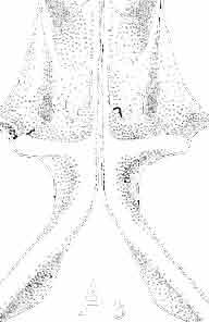
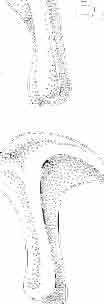
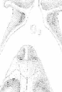
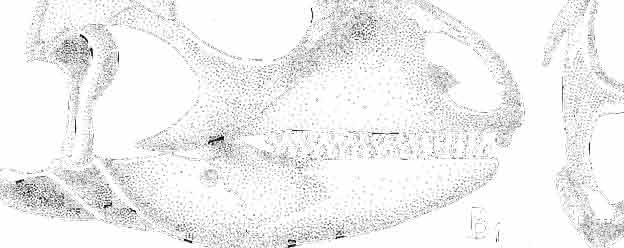
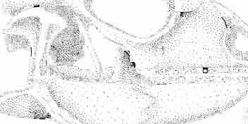
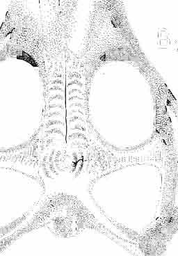
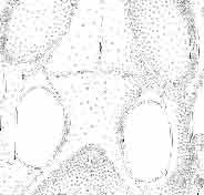
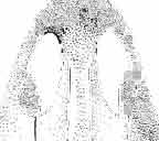
60 84 ANDRZEJ SULIMSKI B s AA B.6
61 MACROCEPHALOSAURIDAE AND POL YGLYPHANODONTIDAE 85 and from below the orbit, reaches the postorbital and ventrally extends to the anterior process of the squamosal. Temporal region (PI. XXV, Text-fig. 13A 2 6 ). Postfrontal is a short, wide bone with proximally forked processes as in Macrocephalosauridae. Both proximal processes embrace the lateral widenings of frontal and parietal at their common suture, the distal process being joined to the anterior section of the postorbital. Postfrontal together with the anterior part of postorbital forms a rather short but strong postorbital arch. Postorbital is an anteroposteriorly elongated bone, which in its anterior part forms the posterior margin of the orbit and joins the postfrontal. With a posterior, rather short, overlapping suture the bone joins the squamosal, terminating at about half way back on the length of the supratemporal fossa. Postorbital and squamosal form a rather wide and strong supratemporal arch. Squamosal subtriangular, short, posteriorly widened, and dorsoventrally flat. Quadrate high with a strong anteroposteriorly arched head, a slender shaft and a prominent double distal condyle. The posterior crest is well marked along the whole length. Dorsally the quadrate joins the squamosal, intruding into its ventral depression and joining the ending ofparietal and paroccipital process. From below, by means of a diarthritic joint the bone joins the cotylus on the articular bone. Medially and distally the quadrate is joined by means of a movable joint to the posterior pterygoid process. Palatal region (PI. XXVI, Text-fig. 13A j ). Vomers in the type specimen not fused but closely articulating each other. Anterior end of the vomer reaches the premaxilla and septomaxilla. Laterally, just behind a small opening, a small vomero-nasal notch is visible. Posteriorly the vomer widens, and laterally forms upward directed lateral wings. Posterolaterally the vomer joins the palatine and towards the back its posterior process not joins the vomerine process of the pterygoid. Palato-narial vacuity anteriorly slit-like, posteriorly widened, surrounded by the maxilla (laterally), palatine (posteriorly) and the vomer (medially). Palatine irregular, with an anteriorly deep groove for the palato-narial vacuity. The bone joins the maxilla laterally and posterolaterally the anterior ectopterygoid process. Posteriorly the palatinejoins, by means of a wedged suture, the pterygoid and anteromediaiiy the vomer. Pterygoid is the largest bone in the palatal region possessing three processes. The antero-medial not joins the posterior vomer process, the Iatero-anterior joins the ectopterygoid and the longest posterior process joins syndesmotically the quadrate. On the shaft of the bone occurs a transversally extending edge, which forms the posterior margin of the palatal floor. The lateral pterygoid process adheres to the distinct ectopterygoid process together forming a strong ventrally directed process. The posterior pterygoid process is mediolaterally compressed, its end bent outwards. On the dorsal margin of the pterygoid occurs a socket for joining to the epipterygoid. Articular surfaces, medially situated on the posterior process are large, indicating a rather wide joining of'the pterygoid and basipterygoid proces s. Ectopterygoid subtriangular with a long antero-lateral Fig. 13 Cherminsaurus ko zlowskii gen.n., sp.n., ZPAL MgR-1II /24 - holo type. A - Skull, lower jaw and other bone fragments: 1 - skull (recon struction), lateral view, 2 - dorsal view, 3 - palatal view (reconstruction), 4 - left quadrate, lateral view, 5 -lower jaw, lingual view, 6 - temporal regionydorsal view. Polyglyphanodon sternbergi GILMORE, 1940, USNM holotype. B - Skull, lower jaw and other bone fragmen ts: 1 - skull, lateral view, 2 - dorsal view, 3 - palatal view, 4 -left quadrate, lateral view, 5 - lower jaw, lingual view, 6 - temporal region, dorsal view (after GILMORE, 1942). All about nar. size. Abbreviations: Temporal regions are schematically drawing - black postfrontal, thickly dotted - postorbital, sparsely dotted - squamosal. The lower jaw and quadrate of Polyglyphanodon sternbergi GILMORE are reversed for the bette r comparison. Other abbreviations see Fig. 2.
62 86 ANDRZEJ SULIMSKI process present. This bone joins, by means of a narrow anterior process the palatine, laterally the maxilla and posteriorly, by means of a strong and ventrally very prominent coronoid process it joins the pterygoid. Suborbital fenestra small, anteroposteriorly elongated, surrounded only by palatine, ectopterygoid and pterygoid. Epipterygoid fragmentary, rode-like with no distinct crests present. On the vomers, palatines, pterygoids and ectopterygoids no traces of teeth occur. The low ventral raphae are present on the medial line in the vomero-pterygoid region. Occipital region. Occiput, apart from a small fragment of the occipital condyle, is only partly preserved in the form of distal sections of the paroccipital processes and parts of the exoccipitai. Basicranium in Cherminsaurus kozlowskii has only parts of the basipterygoid processes, parts of the basisphenoid and a fragment of the basioccipital preserved. The basipterygoid processes are wide, short and strong. In this region can also be seen parts of the brain case and paroccipital processes. The basioccipital condyles are strong, large, with widely marked recesses. Lower jaw (PIs XXV-XXVII, Text-fig. 13As) in C. kozlowskii is strong, rather long, slender (in relation to the skull), but also rather high, with no distinct angular curvature present. Dentary long, occupying almost half the length of the jaw, in symphyseal part slightly widened, posteriorly reaching to the anterior margin of the coronoid process. Symphysis short, rounded. Behind the symphysis occurs a small anterior Meckelian foramen. The dentary on the lateral side reaches to the coronoid process joining the surangular and from below, the angular. In the anterior part there are five or six small mental foramina. Coronoid is rather high, wide at the base with its upper apex bent slightly towards the back. The coronoid is situated at almost mid-length of the jaw. The base of the coronoid has three processes, the lateral one very small, the two internal ones well developed. Anterior lingual process joins the dentary, splenial and partly the surangular. The posterior process is however the largest with a well marked longitudinal crest. Surangular is a rather short, dorsoventrally wide bone joining the angular, from below, posteriorly it reaches the articular and dorsoanteriorly the coronoid. On the inner side, the surangular forms a lateral wall of a deep but not very long adductor fossa. Angular forms the posterior base of the lower jaw and is poorly visible on the lateral side. Anteriorly the bone joins the dentary, from below and above it reaches the surangular, anteriorly and lingually it meets the splenial and posteriorly articular. In the posterior part of the surangular, close to the angular suture one can see a diagonal, rather prominent crest for attachment of the adductor muscles. Articular well developed with a bifossate cotylus present. The posterior section of the articular widened, lingually depressed with a strong, long and wide retroarticular process. Splenial in Cherminsaurus kozlowskii is large, posteriorly widened, narrowed anteriorly and almost completely covering the Meckelian groove. Posteriorly the bone joins the coronoid and surangular. From above and below the splenial is joined by means oflong sutures to the dentary. At mid-length of the splenial, closer to the upper dentary suture occurs a rather large foramen. Dentition (PI. XXVII, Text-fig. 14A I _ 3 ). Dentition in C. kozlowskii is unique, not found in any other known Sauria. In anterior section of the jaws occur simply constructed, conical, unicuspid teeth ; shafts of these teeth are straight, rather short and round in cross section. The following teeth, beginning from the fourth in the series, rapidly increase in size, so that largest are two or eventhree times larger than the anterior teeth. The crowns, beginning from the sixth tooth in the series, are obliquely arranged, so that the cutting edges are at an angle of about 30 to the long axis of the jaw. This angle is slightly smaller on the last teeth. Thus
63 MACROCEPHALOSAURIDAE AND POL YGL YPHANODONTIDAE 87 on the seventh, eighth and ninth teeth this angle is about 40, on the tenth through fourteenth it decreases to an average of 30, and in the post erior teeth (fifteenth, sixteenth and seventeenth) it is The last tooth (eighteenth) is small in both the lower and upper jaws. With the exception of the front teeth and the last one, the crowns have an uniform plan. Distances between individual teeth are very small, but well marked. The crowns are diagonally compressed, their lateral sides steeper, and their lingual sides with a more gently shaped slope. On the cutting edge occurs a row of cusps variable in number. On the anterior cheek teeth (fifth-to seventh in the series) occur two or three cusps, while in the central (from eighth to seventeenth) the number of cusps is rather constant from four to six. These cusps are low and rather wide. In the holotype the cusps bear distinct abrasion surfaces. The dentition of the upper jaw does not differ materially from that of the lower jaw. Differences only refer to the construction of the crowns, where the lateral and lingual sides are more or less similarly slanted and the distances between teeth slightly greater. As in the lower jaw, last cheek tooth is distinctly smaller than those preceeding it. The mode of tooth implantation can be generally interpreted as more subpleurodont than as subacrodont. From the lateral side, over the dentary parapet, one can see crowns and shafts of more or less uniform height. It seems as if the teeth are directly grown to the upper margin of the jaw. From the lingual side, however, one can distinctly see that the shafts are slightly longer and are implanted along groove, and that parts ofthe shafts are attached to the internal wall of the jaw. Distinct interdental septa or bone ridges that might indicate a subacrodont mode of implantation are missing, and the lingual wall of the jaw is lower than the lateral. It is possible that this type of implantation is more primitive than, for example the subacrodont implantation observed in Polyglyphanodon sternbergi. Tooth replacement in Cherminsaurus kozlowskii seems to be alternative. Tooth replacement in other species described here IS of iguanid type, common in many Recent lizards. Well marked resorption pits present at the bases of the teeth distinctly indicate this. However, the way in which the older generations of teeth are replaced by new ones is not completely clear. It is probable that there occurred some irregularity in the emergence of succeeding generations of teeth, or an acceleration of the replacement process itself in some sections of the jaws. Such irregularities have been noted by EOMUNO (1960, 1969). One can distinctly see in some fossils teeth, which have not yet become solidly attached to the jaw groove, new replacement pits in the walls of these shafts are already marked. It may be that this disturbance of the rhythm of replacement was rapidly compensated by the acceleration of the growth of the tooth replacement and rapid resorption of the base of the old one. In this way in C. kozlowskii a complete assemblage of functional teeth was preserved on the jaws throughout the life of the animal. Direction of the replacement waves seems to have extended, as in most Sauria, from the back towards the front. Discussion. - The uncommon character of dentition in Cherminsaurus ko zlo wskii in general outline resembles the dentition observed in Upper Cretaceous species - Polyglyphanodon sternbergi, Paraglyphanodon gazini and P. utahen sis. It concerns on one hand, a more or less similar number of teeth - not more than 20. On the oth er hand it concerns the simplification of the anterior teeth, and reduction of the last cheek tooth (see GILMORE, 1942, Figs 19,21). Also the presence of sharp, transverse cutting edges or tubercles on these edges, transverse or oblique arrangement of tooth crowns in relation to the jaw axis are rare in fossil and Recent lizards. Presently the oblique tooth arrangement occur s only in living teiid Dicrodon.and in fossil Peneteius. The dentition of Polyglyphanodon sternbergi and Paraglyphanodon gazini shows a distinct similarity in the transverse arrangement of crowns with sharp cutting
64 88 ANDRZEJ SULIMSKI edges (GILMORE, 1940, Figs 1-2; 1942, Figs 19-22, and 1943a, Figs 8-9). According to ESTES (1969, p. 4) "it is possible that Paraglyphanodon is only a small individual or the young of the Polyglyphanodon." This conclusion based on the tooth morphology is not sufficient. The differences concerning the cheek teeth number and skull structure of mentioned above both genera are connected with the individual variability. It refers as well as to the size of skulls. Paraglyphanodon utahensis, however, from the same locality as P. gazini, differs in the specific tooth morphology. Any decision on the taxonomic status of these specimens, however, must await new study of the actual material and comparison with growth series of Recent species, a project in course of preparation by Prof. R. ESTES. Cherminsaurus kozlowskii differs from the species of both genera - Polyglyphanodon and Paraglyphanodon in decidedly diagonal tooth crowns arrangement in relation to the jaw axis, and presence of four, five or six cusps on cutting edges. Differences occur also in tooth implantation (in C. kozlowskii - subpleurodont, in compared species - subacrodont mode of implantation), in tooth replacement (in the former - iguanid, and in latter, rather a agamid type, according to the terminology of EDMUND (1960, 1969) & COOPER et al. (1970). They are also noted some differences in the morphology of skulls for (instance in the parietal foramen position, the shape and arrangement ofthe frontals and postfrontals). Moreover, Cherminsaurus kozlowskii from the Paraglyphanodon utahensis differs not only in greater dimensions but above all in an entirely different structure of tooth crowns (see Text-fig. 14A I _ 3 ). In all members of the family Polyglyphanodontidae enlarged canine-like teeth are lacking, which are rather characteristic for macrocephalosauridsand in many living macroteiids. Therefore heterodontism in polyglyphanodontids is less marked and occurs mainly as differentiation of the crown character into two types - anterior small, unicuspid teeth and larger, complexcrowned posterior ones. The wide bases of tooth shafts in C. kozlowskii ingrown into a shallow sulcus, and their lower parts into the inner side of the low labial wall. The teeth in Polyglyphanodon sternbergi, Paraglyphanodon gazini, P. utahensis and Peneteius aquilonius are rather grown to the alveolar margin (see GILMORE, 1942, 1943a; ESTES, 1969) or into very shallow depressions. Resorption pits in C. kozlowskii are very well developed and rather regularly arranged (PI. XXVII). In P. sternbergi in and papserh Paraglyphanodon gazini, these pits are Fig. 14 Cherminsaurus kozlowskii gen.n., sp.n., ZPAL MgR-I11/24 - holotype. A - Dentition: 1 - two cheek teeth, lingual view, 2 - occlusal view, 3 -latero-iingual tooth section (all schematically drawing). Polyglyphanodon sternbergi GILMORE, 1940, USNM B - Teeth! 1 -lower fourteenth tooth, occlusal view, 2 -lateral view, 3 -lower fourth tooth, lingual view, 4 - occlusal view (after GILMORE, 1942). Paraglyphanodon gazini GILMORE, 1943, USNM holotype. C - Upper cheek tooth, occlusal view (after GILMORE; 1943). Paraglyphanodon utahensis GILMORE, 1940, USNM holotype. D - Upper cheek tooth, occlusal view (after GILMORE, 1940). Erdenetesaurus robinsonae gen. n., sp, n., ZPAL MgR-I11/19 - holotype. E - Skull, lower jaw and dentition: 1 - skull (reconstruction), lateral view, 2 - dorsal view (reconstruction), 3 - lower jaw, lingual view (partly reconstructed), 4 - palatal region (reconstruction), slightly enlarged, 5 - middle part of cheek tooth row, lingual view, 6 -lateral view. Ctenosaura pectinata (WIEGMANN, 1834). F - The left lower tooth row fragment, lingual view (after OELRICH, 1956). Macrocephalosaurus gilmorei sp. n., ZPAL MgR-I1I/l7. G - The left lower cheek tooth row, lingual view. Iguana iguana LINNAEUS, Recent. H - The right lower cheek tooth row fragment, lingual view. Original. Chamops segnis MARSH, I - The right lower cheek tooth row fragment, lingual view (after CAMP, 1923; EsTES, 1964). Macrocephalosaurus chulsanensis sp.n., ZPAL MgR-I/28. J - The upper tooth row fragment, lateral view. Macroscincus cocteaui DUMERIL & BIBRON, K - The right lower cheek tooth row fragment, lingual view (after HOFFSTETTER, 1949). Tupinambis nigropunctatus SPIX, 1825 CNHM L - Three lower cheek teeth, lingual view: I - anterior tooth, 2 - middle tooth, 3 - posterior tooth (after EOMUNO, 1960; JOLLlE, 1960, 1962; REESE, 1923). Sauromalus after DUMERIL & BIBRON, 1856 USNM M - The lower tooth row fragments, lingual view: 1 - anterior teeth, 2 - cheek teeth (after EOMUNO, 1960). A, E 5-6, F-M - x 5; B-D ea. x 7; E 1-3 nat. size.
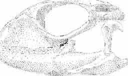
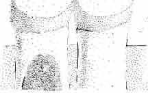

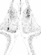
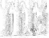
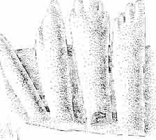
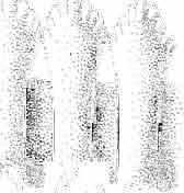
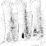
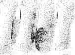
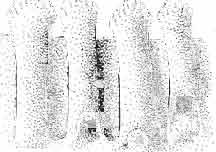
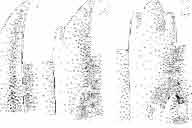
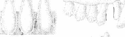
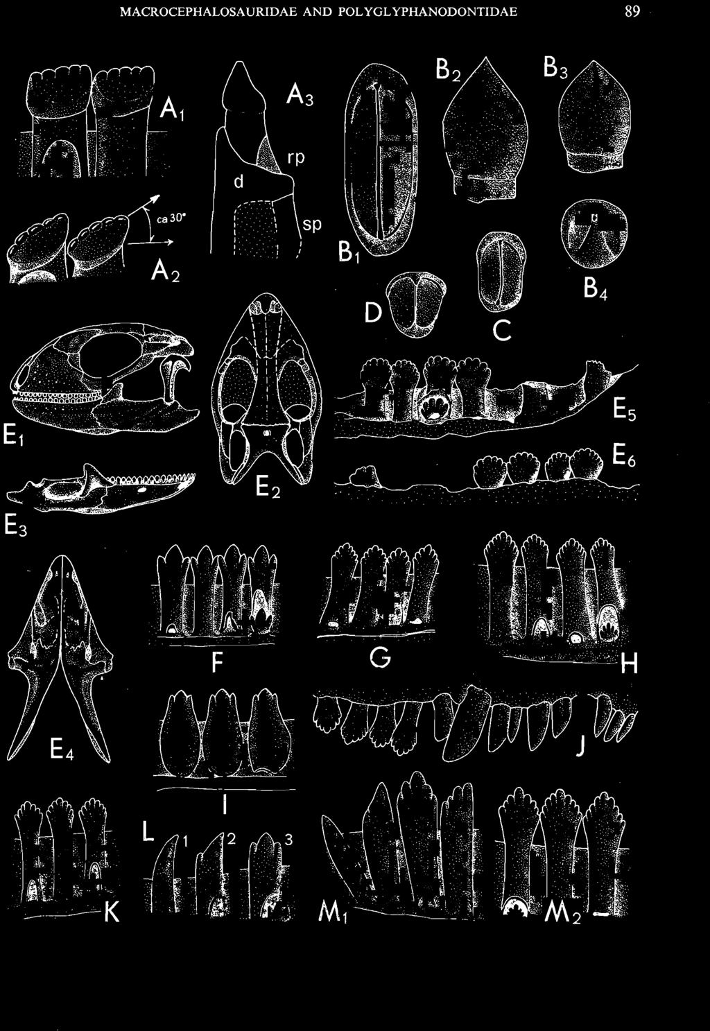
65 MACROCEPHALOSAURIDAE AND POLYGL YPHAN ODONTIDAE 89..'! I I I sp D c.....' ~ ",.. F ~14:.: I G ~.".,'.,: : t. ;~.H J 7JI1t
66 90 ANDRZEJ SULIMSKI very small and arranged in dentary bone below each tooth in almost the whole series (GILMORE, 1942, Fig. 20), as seen in Adamisaurus magnidentatus (SULI.MSKI, 1972) or in Dracaena guianensis (EDMUND, 1960; VANZOLlNI & VALENCIA, 1965). In polyglyphanodontid species, on the lingual side of lower jaws, occurs a wide, long, thin and roughly triangular splenial bone with an anteroposterioriy elongated and medially situated anterior inferior alveolar foramen (medial Meckelian fora men). The splenial covers almost completely the wide Meckelian can al. Such large and wide splenial occurs also in many living teiids. Well developed and relatively wide splenial was noted also in Jurassic lizards included to different families (for example Dorsetisaurus, Becklesisaurus, Saurillus, M acellodus etc. - see HOFFSTETTER, 1966; SEIFERT, 1970). The splenial in Jurassic forms reaches posterioriy the posterior margin of the coronoid process, but in the front does not extend the symphysis, while in polyglyphanodontids this bone 'extends anterioriy nearly to the anterior end of the lower jaw just behind the symphysis, and ends below the anterior border of the coronoid process posteriorly. Distinct differences in the morphology of dentition, in the mode of implantation and skull morphology do not allow for the inclusion of the Mongolian species into the infraorder Iguania. For the same reason they cannot be included to the family Macrocephalosauridae, eventhough the presence of a postfrontal forked proximally and a similar structure of the palatal region in C. kozlowskii may suggest it. In spite of similarities in the development of the splenial, some analogies in dentition (general arrangement of individual sections of dental series), simplification ofanterior teeth and reduction of the last tooth (or teeth), as well as in the roofed bones, especially frontals and the relation between their width and length, rule out their inclusion to the family Teiidae. It seems more reasonable to include all polyglyphanodontids in a group of primitive lizards in the infraorder Scincomorpha, quite possibly related to teiids. Cherminsaurus kozlowskii considered here as polyglyphanodontid lizard is characterised by the possession of features which acclaim it as a more primitive form than the other representatives of family Polyglyphanodontidae. ~ COMMENTS ON THE SYSTEMATIC POSITION AND RELATIONSHIPS OF MACROCEPHALOSAURIDAE AND POLYGLYPHANODONTIDAE A detailed analysis of the skulls in represent ativ es of the Macrocephalosauridae shows the pre sence of the vomero-pterygoid contact in the palatal region. A comparison of this region with the same region in both Recent and advanced fossil lizards reveals that in none of the latter Sauria s. s. does a joining between the vomers and pterygoids exist. Instead, both bone elements are separated more or less by widely developed palatines (COPE, 1892; WILLlSTON,' 1925; ZITTEL, 1932; ROMER, 1956; HUENE, 1956; JORDANSKY, 1968; BELLAIRS, 1969; GUIBE, 1970). The vomero-pterygoid contact is without doubt a primitive feature found in many fossil and some modern reptiles, among them in Eosuchia and Rhynchocephalia. In Eosuchia the vomero-pterygoid contact occurs together with a number of other primitive characters, for instance: palatal dentition, paired all cranial roof elements, presence of a ventral opisthotic process, large parietal fora men and presence of the quadratojugal bone. This contact is also well marked in the Triassic genus Kuehneosaurus (ROBlNSON, 1962, 1967). In the latter case, however, ROBINSON does not comment on the significance of thi s contact, but considers that Kuehneosaurus in spite of its many primitive features, is alre ady a very advanced lizard and thus includes it in the suborder Sauria. This decision was based above all on the lack of
67 MACROCEPHALOSAURIDAE AND POL YGL YPHANODONTlDAE 91 quadratojugal bone, presence of free and movable quadrate, reduction of the ventral arm of squamosal, and on the absence of the lower temporal arch. In spite of this, Kuehneosaurus possesses a considerable number of primitive features such as the presence of the palatal dentition, paired all bone elements of the skull roof, large lacrimal, well developed parietal foramen in the fronto-parietal suture, a ventral opisthotic process as in Eosuchia, delicate, slender and long lower jaws, and very large palatal fenestrae. It seems to be correct to include the vomero-pterygoid contact also into these primitive characters. If the presence or absence of this contact were to be accorded with a great diagnostic significance, then it follows that the family Macrocephalosauridae novo should be placed in or near the eolacertilian forms. However, the detailed studies of the skull structure in the both new species of Macrocephalosaurus and in Darchansaurus estesi indicate, that, with the exception of the vomero-pterygoid contact and primitive structure of the temporal region, there is a complete conformity of features with almost all modern and advanced fossil Sauria. In all macrocephalosaurids, typical saurian features can be observed, such as: lack of palatal dentition, lack of quadratojugal bone and lower temporal bar, lacrimal is small almost invisible externally, free and movable quadrate, reduced or small, but fused to the squamosal-supratemporal, normal metakinetic skull, lack of the ventral arm of the squamosal, saurian construction of the brain case, premaxilla and parietal unpaired, differentiated dentition, and mode of implantation and tooth replacement. In spite of the differences, which occur in the structure of the palatal (vomero-pterygoid contact) and temporal region, one can assign the family Macrocephalosauridae novo to the suborder Sauria. The described macrocephalosaurids also possess the hinge-joint between the frontals and parietal, which is much limited in movements. It perhaps has a certain significance in the skull kineticism, but has not in all probability, such diagnostic importance as has generally been ascribed to it. In the skull structure one can usually observe a more strongly developed facial region than the parieto-occipital one, especially in the naso-prefrontal region. This latter region is wide, flat and arranged at an angle to the fronto-parietal surface (see Darchansaurus). The elongated facial region is observed in lizards which habitually use a crushing action of the jaws. This may cause also the ventral directed expansion of a mesial flange of the prefrontal. As in many modern lizards which crush large or hardshelled prey, in the macrocephalosaurids occurs a well marked ventrally curved alveolar margin of the maxilla. On the strong crushing action of the jaws indicate also a well developed coronoid, and ventrally directed pterygoectopterygoid expansion, to guide the action of the lower jaw. In addition, in all macrocephalosaurids there occurs a characteristic arrangement of bone elements of the temporal region and their relation to the quadrate. The postfrontal, forked on both extremities, postorbital with a flat and long posterior process, and rather large but short, roughly triangular and dorsoventrally compressed squamosal, are most typical for the Macrocephalosauridae. The head of the quadrate is placed in the shallow but extensive hollow on ventral side of the squamosal as in Kuehneosaurus or Prolacerta. The long head enables on a greater antero-posterior movements of the lower jaw in relation to the neurocranium. Therefore, it seems -to be possible, that apart from the crushing action, the cutting a nd shearing ones occur. The many living lizards retain the supratemporal which articulates with the paroccipital process and squamosal, but in the opposition to the early forms of lepidosaurus, is situated deeper, in the place of the antero-ventral border of the posterior process of the parietal and it separates the squamosal from the paroccipital process. It is interesting that, as stated by ROBINSON (1967, p. 4) "in all modern lacertilians which retain the squamosal, the dorsal process is much abbreviated or absent altogether". In the genera
68 92 ANDRZEJ SULIMSKI of the Macrocephalosauridae, at this place, where the dorsal process of the squamosal in the modern lizards occurs, a fused to the squamosal the supratemporal bone is found. Ro BINSON (1967) has pointed out that in most modern lizards which retain the squamosal a little bony peg and notch or hole in the quadrate are present. Detailed studies of the quadrate in the two genera of the Macrocephalosauridae, do not indicate the presence either of the bony peg or mentioned notch or hole. Instead, the anterior process of the squamosal is here well developed as in the majority of lizards. The dorsal process of the squamosal in the living iguanids and teiids is much abbreviated to a small vestigial projection or absent altogether. Therefore, in both families the supratemporal opening is rounded posteriorly. Lastly, and apart from the primitive saurian features in the skull structure (vomeropterygoid contact, temporal and palatal regions), the dentition of the Macrocephalosauridae demonstrates a typical pleurodont morphology, closely related to the iguanid lizards. The dentition falls here into three categories - anterior, small, conical, unicuspid; medial (in the maxilla) large, canine-like, and the posterior cheek teeth with a leaf-shaped, denticulate pattern very similar to that in Iguana, Sauromalus, Dipsosaurus or in the skink - Macroscincus (HOFFSTETTER, 1949). The teeth, in contrary to the high and strong jaws, are very small and numerous. The lancet-like crowns of the cheek teeth perhaps have played an important role in more efficient shearing action during the disintegration of the prey. In the structure of the roof bones of the skulls, the uncoalesced, rather long and narrow frontals are especially noticeable. The proportion of these bones, although paired correspond more or less to these in some macroteiids as Dracaena, Teius and Tupinambis (REESE, 1923; JOLLIE, 1960, 1962; VANZOLINI& VALENCIA, 1965; WAHLERT, 1966). In the Polyglyphanodontidae (with the exception of Paraglyphanodon) in the temporal region the bone arrangement is different than in the Macrocephalosauridae. The postorbital and squamosal are arranged almost vertically in relation to the head of the quadrate. In Polyglyphanodon sternbergi the squamosal is "deep dorsoventrally, with a squarely truncate posterior end" (GILMORE, 1942, p. 237). Similar arrangement of both bones occurs in Erdenetesaurus robinsonae and Cherminsaurus kozlowskii (Text-figs 13-14). Moreover, the representatives of the Polyglyphanodontidae differ from the genera of the Macrocephalosauridae as well as from all other Sauria in the character of their dentition. In the dental series, two distinct teeth categories may be differentiated ~ anterior, small, unicuspid, and posteriorcheek teeth with considerably enlarged crowns, arranged transversally or obliquely to the jaw axis, possessing sharp transverse crest or low tubers on the cutting edges. The exception here is Erdenetesaurus, in which crowns are lateromedially compressed and possess radially arranged denticles on the cutting edges. The tooth number in all the polyglyphanodontids is relatively constant (not more than 20). In upper dentition the enlarged, canine-like teeth and widening and flattening of the naso-prefrontal region, so characteristic for the genera of the Macrocephalosauridae, are not present in the representatives of the Polyglyphanodontidae. In the palatal region of Cherminsaurus ko zlowskii there occurs a short medial process ofthe pterygoid, which does not extend far forward. Unfortunately, this part of the palatal region is incomplete in the only known specimen of this species. The same is true for Erdenetesaurus robinsonae. A similar situation is found in Polyglyphanodon sternbergi, in which the same region is damaged as a result of lateral distortion of the skull. According to GILMORE (1942, p. 239) in Polyglyphanodon "the palatines appear to be narrowly separated on the midline at least posteriorly; anteriorly they may have been in apposition as in Iguana". If this interpretation is correct, the arrangement of the palatal bone elements is the same or similar to that of all modern lizards. Lack of more complete material of Cherminsaurus and Erdenetesaurus does
69 MACROCEPHALOSAURIDAE AND POLYGLYPHANODONTIDAE 93 The most important osteological differences between the families Macrocephalosauridae and Polyglyphanodontidae are as follows: Characters I Macrocephalosauridae I Polyglyphanodontidae Nasals triangular, short, contact with elongated, rectangular, contact with prefrontal absent prefrontal present Maxilla high, reaches frontal, vertically high, does not reach frontal, always outwardly ventrad sloping arranged Naso-prefrontal square-shaped, flat, wide convex dorsally, narrow region Palatines not in apposition in apposition Ectopterygoid small, narrow large, wide Pterygoid slender, shafts narrow massive, shafts wide Suborbital reduced to a concavity, occasion- large, present as fenestra fenestra ally perforated Vomero- present lacking pterygoid contact Postfrontal short, narrow, forked at extre- trapezoidal, sometimes proximally forked, wide mit ies Postorbital dorsoventrally compressed, flat, lateromedially compressed, short, reaching only midlength of long, reaching end of supra- supratemporal arch, almost vertical temporal opening, horizontal Squamosal V-shaped, capping head of the squarely truncate posteriorly, vertical quadrate, horizontal Supratemporal fused to the squamosal separated, thin, squamous Supratemporal long, horizontal, composed of long, vertical or oblique, composed of postorbital and squaarch postorbital and squamosal po - mosal in equal parts ster iorly Supratemporal small with rounded posterior end large with acute posterior end opening Postorbital arch consisting only of postfrontal consisting of postfrontal and antero-dorsal part of postorbital Quadrate delicate, without medial process, robust, medial process distally expanded, head anterohead anteroposteriorly long posteriorly short Lower jaw massive, rather short, deep in massive but slender, without deep angular region angu lar region cont.
70 94 ANDRZEJ SULlMSKI I Retroarticular short, widened and pointed po- long, deep and rounded posteriorly proce ss stcriorly I Tooth heterodont, iguanid type, crown s weak heterodontism, crown s tran sversely or obliquely arranged morphology flattened lateromedially, lancet - with sharp cutting edges or with low tubers (in exceptional like and denticulated case flattened lateromedially, crowns with denticl es radially arranged - Erdenetesaurus robinsonae) Canine-like large, longer than cheek teeth lacking teeth Tooth iguanid type iguanid or agam id type replacement Number of teeth not allow one to ascertain whether the vomero-pterygoid contact is rea lly present in these genera or no t. Dentition, especially the cheek teeth, of Cherminsaurus ko zlowskii can be compared to the cheek teeth of an Upper Cretaceous teiid lizard Peneteius aquilonius (ESTES, 1969). In both species there is an oblique arrangement of crowns in relation to the jaw axis. However, P. aquilonius in addition to its very small dimensions, differs from C. ko zlowskii in hav ing only two tubers on the crowns. P. aquilonius shows some intermediate characters between Polyglyphanodon an d such modern macroteiids as Teius or Dicrodon (although closer to the later; ESTES, 1969; it was assigne d by this author to the family Teiidae). HOFFsTETTER (1955) suggeste d the teiid relationship of Polyglyphanodon and Paraglyphanodon; he assigned them later to the Teiidae (HoFFsTETTER, 1962). This conclusion was also confirmed by ESTES (1964) and G UlBE (1970). The assignement of these genera to the Teiidae was at that time based mainly on the structure of the roof bones of the skull, proportions of the skull, especially those of the fronta1s. ESTES & PRICE U973) doubt whether the two mentioned genera could be assigned to the Teiidae. It is pos sible that both fami lies - Macrocephalosauridae an d Polyglyphanodontidae form two related groups of primitive Scincomorpha (with some teiid characters). In bot h families a large number of progressive advanced features occur such as: absence of qua drato-jugal, free, streptostylic quadrate, fused premaxilla and parietal, small, reduced or fused to the squamosal supratemporal, lack of the palatal teeth, allowing to include them into the suborder Sauria. Polska Akademia Nauk Zaklad Palaeozoologii Warszawa, Al. ZWirki i Wigury 93 June 1974 REFERENC ES BELLAIRS, A. d'a The life of the Reptilia. - 1/2, 1-590, Weidenfeld & Nichol son, Nat. Hist., London. CAMP, C. L Classification of the lizards. - Bull. Amer. Mu s. Nat. Hist., 48, , New York. CoOPER, J. S., POOLE, D. T, G. & LAWSON, R The dentition of agamid lizards with special references to tooth replacement. -J. Zool., 162, 85-98, London. COPE, E. D The osteology of the Lacertilia. - Proc. Amer. Phil. Soc., 30, , New York.
71 MACROCE PHALOSAURlDAE AND POL YGLYPHANODONTlDAE 95 EDMUND, A. G Tooth replacement phenomena in the Lower Vertebrates. - Roy. Ontario, Mus. Life, Sci. Div., Contr., 52, 1-190, Toronto Dentition. In: Gans, C. (ed.). - Biology of the Reptilia, 1, Morphology A, , Academic Press Inc. London-New York. EL TOUBJ, M. R The osteology of the lizard Scincus scincus. - Bull. Fae. Sci. Cairo, 14, 1-14, Cairo. ESTES, R Fossil Vertebrates from the Late Cretaceous, Lan ce Formation, Eastern Wyoming. - Univ, Call]. PubI. Geol. Sci., 49, 1-180, Berkeley & Los Angeles Relationships of two Cretaceous lizards (Saur ia, Teiidae). - Breviora, 317,1-8, Cambridge. - & PRICE, L. J Iguanid lizard from the Upper Cretaceous of Brasil. - Science, 180, , New York. FRAzETTA, T. H A functional consideration of cranial kinesis in lizards. - J. Morph., 111, 3, , New York. GASC, J. P Les variations columnaires dan s la region presacree des Sauriens. - Ann. Paleont., 57, 1, , Paris. GILMORE, C. W New fossil lizards from the Upper Cretaceous of Utah. - Smithson. Miscell. Coli., 99, 16, 1-3, Washington Osteology of Polyglyphanodon, an Upper Cretaceous lizard from Utah. - Proc, U. S. Nat. Mus., 92, 3148, , Washington. 1943a. Osteology of Upper Cretaceous lizards from Utah, with a description of a new species. - Ibidem, 93, 3163, b. Fossil lizards of Mongolia. - Bull. Amer. Mus. Nat. Hist., 81, , New York. GINSBURG, L Les reptiles fossiles. In: Grasse, P. P. (ed.). - Traite de Zoologie, 14, 3, , Masson et Cie., Paris. GRADZINSKI, R Sedimentation of dinosaur bearing Upper Cretaceous deposits of the Nemegt Basin, Gobi Desert. Results of the Polish-Mongolian Palaeontological Expeditions, Part Ill. - Palaeont. Pol., 21, , Warszawa. & JERZYKIEWICZ, T Additional geographical and geological data from the Polish-Mongolian Palaeontological Expeditions. Results... IV. - Ibidem, 27, & Sedimentation of the Barun Goyot Formation. Results... V. - Ibidem, 30, KAZMIERCZAK, J. & LEFELD, J Geographical and geological data from the Polish-Mongolian Palaeontological Expeditions. Results... I. - Ibidem, 19, GRZIMEK, B Grzimek's Tierleben. Kriechtiere. - 6, 1-609, Kindler Verlag, ZUrich. GUIDE, J In: Grasse, P. P. (ed.). - Traite de Zoologie. 14,2,33-193, , ; 3, , , Masson et Cie., Paris. HOFFSTETTER, R Les reptiles subfossiles de l'lie Mauri ce, I, Les Scincidae. - Ann. Paleont., 35, 43-72, Paris Squamata de type moderne. In : Piveteau, J. (ed.), - Traite de Paleontologie, 5, , Masson et Cie., Paris Revue des recentes acqu isition s concernant I'histoire de la systematique de Squamates. - Coli. Intern. e. N. R. S., 104, , Paris Les Sauria du Jurassique superieur et specialernent les Gekkota de Baviere et de Manchurie. - Senck. bioi., 45, 3/5, , Frankfurt a. M Coup d'oeil sur les Sauriens (= Lacertiliens) des Couches de Purbek (Jurassique superieur d'angleterre). Coli. Intern. C. N. R. S., 163, , Paris. & GASC, J. P Observations sur le squelette cervical. - Bull. Mus. Nat, Hist, Nat., 39, 2, , Paris. & Vertebrae and ribs ofmodern reptiles. In: Gan s, C. (ed.), - Biology of the Reptilia, 1, Morphology A, , Academic Press In c., London-New York. HOTTON, N A survey of adaptative relationships of dentition to diet in North American Iguanidae. - Amer. MidI. Nat., 53, 1, , New York. HUENE, F. v Palaontologie und Phylogenie der Niederen Tetrapoden , Gustav Fischer Verlag, Jena. JOLUE, M. T The head skeleton of lizards. - Acta Zool., 41, 1-64, Stockholm Chordate morphology , Reinhold Book Corp. New York-Amsterdam-London. JORDANSKY, N. N See 110P~AHcJ{U:lJ, H. H. KIELAN-JAWOROWSKA, Z New Upper Cretaceous multituberculate genera from Bayn Dzak, Gobi Desert. Results of the Polish-Mongolian Palaeontological Expeditions, Part IT. - Palaeont, Pol., 21, 35-49, Warszawa Multituberculate succession in the Late Cretaceous of the Gobi Desert (Mongolia). Results... V. - Ibidem, 30, Preliminary descript ion of two new eutherian genera from the Late Cretaceousof Mongolia. Results... VI. Ibidem, 33, 5-15.
72 96 ANDRZEJ SULlMSKI & BARSBOLD, R Narrative of the Poli sh-mongolian Palaeontological Expeditions Results... IV. Ibidem, 27, & DOVCHIN, N Narrative of the Poli sh-mongolian Palaeontological Expeditions Results... I. Ibidem, 19, KUHN, O Squamata. Lacertilia et Ophidia. - Foss. Cat., I: Animalia, 86, 1-89, Neubrandenburg Supplementum. Sauria. - Ibidem, 104, 1-87, s'gravenshage Amphibia und Reptilia. Katalog der Superfamilien und hoheren Taxa mit Nachweis des ersten Auftretens , Stuttgart. LEFELD, J The age of mammals containing beds at Bain Dzak, Northern Gobi Desert. - Bull. Acad. Sci. Pol., Cl. HI, 13, 1, 81-83, Warszawa Geology of the Djadokhta Formation at Bayn Dzak (Mongolia). Results of the Polish-Mongolian Palaeontological Expeditions, Part Ill. - Palaeont. Pol., 25, , Warszawa. MESZOELY, C. A. M North American fossil anguid lizards. - Bull. Mus. Comp. Zool., 139, 2, , Cambridge. OELRICH, T. M The anatomy of the head of Ctenosaura pectinata (Iguanidae), - Mise. Publ. Mus. Zool. Univ., Michigan, 94, 1-122, Ann Arbor. REEsE, A. M The osteology of Tegu, Tupinambis nigropunctatus. - J, Morph., 38, 1-16, New York. ROBINsoN, P. L Gliding lizards from the Upper Keuper of Great Britain. - Proc. Soc. Geol. London, 1601, , London The evolution of the Lacertilia. - Coli. Intern. C. N. R. S., 163, , Paris. ROMER, A. S Vertebrate paleontology , University of Chicago Press, Chicago-London Osteology of the Reptiles , University of Chicago Press, Chicago-London a. Vertebrate paleontology , University of Chicago Press, Chicago-London b. Vergleichende Anatomie der Wirbeltiere , Verlag Paul Parey, Hamburg-Berlin. SEIFERT, J Oberjurassische Lazertilier aus der Kohlengrube Guimarota bei Leiria (Mittelportugal). - Inaug, Diss. Math. naturwiss. Fakultat, 1-180, Berlin. SINITZEN, D. T The types of the skull in the family Teiidae. - Zool. Anz., 76, , Leipzig. SNYDER, R. C Quadrupedal and bipedal locomotion in lizards. - Copeia, 2, , New York The anatomy and function of the pelvic girdle and hind limb in lizard locomotion. - Amer. J. Zool., 95, 1-46, New York. SULlMSKI, A Adamisaurus magnidentatus n.gen., n.sp, from the Upper Cretaceous of Mongolia. Results of the Polish-Mongolian Palaeontological Expeditions, Part IV. - Palaeont, Pol., 27, 33-40, Warszawa. VANZOLlNI, P. & VALENCIA, J The genus Dracaena, with a brief consideration of macroteiid relationships (Sauria, Teiidae). - Arq. Zool., 13, 7-35, Sao Paulo. VERSLUYS, J Streptostylic Problem und die Bewegungen in Schadel bei Sauropsiden. - Zool. lb. Ana!. Suppl., 15, 2, , Berlin. WAHLERT, J. H. MS. Reconnaisance of macroteiid skull morphology , WILLlSTON, S. W The osteology of the reptiles. - I-XIII , Cambridge Harvard University Press, Reprint ZITTEL, K. A. v Textbook of Palaeontology. - 2, 1-464, Macmillan and Co., London. Authorized reprint HOPiJ:AHCI<lIli, H. H MOP<P0JIOnI'leCI<lie 1I <PYHI<I.1IiOHaJIhHhle npeofipaaonauaa B 3BOm0I.1IiH penthjihh naa nciiij:hoh rpynrnsr IlaJIeOHTOJIOrliH Ii crparnrparpna, Anropediepar, 1-22, MOCI<Ba.
73 MACROCEPHALOSAURIDAE AND POL YGLYPHANODONTIDAE 97 EXPLANATION OF PLATES PLATE VIII Macrocephalosaurus gilmorei sp. n. Page 31 Upper Cretaceous,? Middle Campanian, Kherrneen Tsav formation, Khermeen Tsav I, Gobi Desert, Mongolia Fig. 1. ZPAL MgR-III/18 - holotype, skull : a -lateral view, b - occipital view, c - dorsal view, d - palatal view, x 1.5. Photo: M. Czarnocka PLATE IX Macrocephalosaurus gilmorei sp. n. Page 31 Upper Cretaceous,? Middle Carnpanian, Khermeen Tsav formation, Khermeen Tsav I, Gobi Desert, Mongolia Fig. 1. ZPAL MgR-Ill/I? - skull; a -lateral view, b - occipital view, c - dorsal view, d - palatal view, x 1.5 Photo: M. Czarnocka PLATE X Macrocephalosaurus gilmorei sp. n. Page 31 Upper Cretaceous,? Middle Campanian, Khermeen Tsav formation, Khermeen Tsav I, Gobi Desert, Mongolia Fig. 1. ZPAL MgR-Ill/IS - holotype; left lower jaw : a -lingual view, b -lateral view, c - occlusal view, X 1. Fig. 2. ZPAL MgR-Ill/I? -left lower tooth row fragment: a ':- lingual view, b -lateral view, x 5. Macrocephalosaurus gilmorei sp. n. 31 Upper Cretaceous,? Middle Campanian, Barun Goyot Formation, Nemegt, SouthernMonadnocks, Gobi Desert, Mongolia Fig. 3. ZPAL MgR-I/I9 - skull fragment: a -lateral view, b - palatal view, c - the same specimen, right upper tooth row fragment, lateral view, d -lingual view, a, b x 1.5; c, d x Palaeorrtologla Polonica No. 33 Photo: M. Czamocka, E. Mulawa
74 98 ANDRZEJ SULIMSKI PLATE Xl Page Macrocephalosaurus chulsanensis sp. n Upper Cretaceous,? Middle Campanian, Barun Goyot Formation, Khulsan, Gobi Desert, Mongolia Fig. 1. ZPAL, MgR-I/14 - holotype, skull with postcranial skeleton in situ, before the final preparation, x 0.5 Photo: M. Czarnock a PLATE XII Page Macrocephalosaurus chulsanensis sp. n Upper Cretaceous,? Middle Campanian, Barun Goyot Formation, Khulsan, Gobi Desert, Mongolia Fig. 1. ZPAL MgR-I/14 - holotype, skull - stereo-photographs; a - dorsal view, b - ventral view, c -, occipital view, d - left lateral view, e - right lateral view, x 1. 'Photo: E. Mulawa PLATE xnr Erdenetesaurus robinsonae gen. n., sp. n. Upper Cretaceous,? Middle Campanian, Khermeen Tsav formation, Khermeen Tsav 11, Gobi Desert, Mongolia Fig. 1. ZPAL MgR-lII/19 - holotype, brain case; a -left half, medial view, b -lateral view, c - right half, lateral view, d - medial view, x 3. Page 78 Macrocephalosaurus chulsanensis sp. n.. Upper Cretaceous,? Middle Campanian, Barun Goyot Formation, Khulsan, Gobi Desert, Mongolia 41 Fig. 2. ZPAL MgR-I/21 - brain case; a -lateral view, b - occipital view, c - ventral view, d - dorsal view, e anterior view, x 2. hoto: E. Mulawa PLATE XIV Page Macrocephalosaurus chulsanensis sp. n Upper Cretaceous,? Middle Campanian, Barun Goyot Formation, Khulsan, Gobi Desert, Mongolia
75 MACROCEPHALOSAURIDAE AND POL YGLVPHANODONTIDAE 99 Fig. 1. ZPAL MgR-Ij21 - skull in dorsal view, X 1.5. Fig. 2.' The same specimen, lower jaw; a -lateral view, b -lingual view, X 1. Fig. 3. ZPAL MgR-Ij14 - holotype, right upper tooth row in lateral view, X 5. Fig. 4. ZPAL MgR-Ij28 - right upper tooth row in lateral view, X 5. Photo: E. Mulawa PLATE XV Page Macrocephalosaurus chulsanensis Sp. n Upper Cretaceous,? Middle Campanian, Barun Goyot Formation, Khulsan, Gobi Desert, Mongolia Fig. 1. ZPAL MgR-Ij14 - holotype, dorsal fragment of the vertebral column with ribs, dorsal view, X 1. Fig. 2. The same specimen, part of vertebral column with posterior dorsals, sacrals and proximal caudals: a - dorsal view, b - ventral view, C - lateral view, X l, Photo: E. Mulawa PLATE XVI Page Macrocephalosaurus chulsanensis Sp. n Upper Cretaceous,? Middle Campanian, Barun Goyot Formation, Khulsan, Gobi Desert, Mongolia Fig. 1. ZPAL MgR-IjI4 - holotype, pectoral girdle with the right fore limb, in situ: a - ventral view, b - dorsal view, X 1. Fig. 2. The same specimen, distal fragment of the humerus, ulna, radius and digits; a - ventral view, b - dorsal view, X 1.5. Fig. 3. The same specimen, clawed phalanges in lateral view: a - first 'digit, b - second digit, c - third digit, x 2. Photo: E. Mulawa PLATE XVII Page Macrocephalosaurus chulsanensis Sp. n Upper Cretaceous,? Middle Campanian, Barun Goyot Formation, Khulsan, Gobi Desert, Mongolia Figv L ZPAL MgR-IjI4 - holotype, pectoral girdle: a - anterior view, b - ventral view, c - lateral view, X 2. 7 Photo: M. Czarnocka
76 100 ANDRZEJ SULIMSKI PLATE XVIII Page Macrocephalosaurus chulsanensis sp. n Upper Cretaceous,? Middle Campanian, Barun Goyot Formation, Khul san, Gobi Desert, Mongolia Fig. 1. ZPAL MgR-IllS - pectoral girdle; a - ventral view, b - dorsal view, C - ventro-iateral view, a, b x 2; c x 1. Fig. 2. ZPAL MgR-I/14 - holotype, pectoral girdle in ventro-lateral view, x 1. Fig. 3. ZPAL MgR-I/2l - pectoral girdle, ventral view, x 1. Fig. 4. ZPAL MgR-I/25 - scapulocoracoid, lateral view, x 1. Photo: M. Czarnocka, E. Mulawa PLATE XIX Page Macrocephalosaurus chulsanensis sp. n Upper Cretaceous,? Middle Campanian, Barun Goyot Formation, Khulsan, Gobi Desert, Mongolia. Fig. 1. ZPAL MgR-I/14 - holotype, pelvic girdle, right half of pelvis: a -lateral view, b - medial view, x 1 Fig. 2. The same specimen, left half of pelvis; a - medial view, b -lateral view, x 1. Fig. 3. The same specimen, chevron : a - posterior view, b :-Iateral view, c - anterior view, x 2. Fig. 4. The same specimen, caudals: a - dorsal view, b - ventral view, x 1. Fig. 5. ZPAL MgR-I/23 - pelvic girdle: a - dorsal view, b - ventral view, C -lateral view, d - anterior view, x 1 Photo: E. Mulawa PLATE XX Page Macrocephalosaurus chulsanensis sp. n Upper Cretaceous,? Middle Campanian, Barun Goyot Formation, Khul san, Gob i Desert, Mongolia Fig. 1. ZPAL MgR-I/22 - pelvic girdle fragment with the proximal part of caudals : a - dorsal view, b - the same (caudals only), dorsal view, a x l ; b x 2. Fig. 2. The same specimen, femur: a - anterior view, b -lateral view, C - posterior view, d - medial view, x 1. Photo: M. Czarnocka PLATE XXI Darchansaurus estesi gen. n., sp. n. Page 72 Upper Cretaceous,? Middle Campanian, Khermeen Tsav formation, Khermeen Tsav I, Gobi Desert, Mongolia Fig. 1. ZPAL MgR-III/2l - skull of the young individual: a - dorsal view, b -lateral view, C - palatal view, x 1.5.
77 MACROCEPHALOSAURIDAE AND POLYGLYPHANODONTIDAE Macrocephalosaurus chulsanensis sp. n Page 41 Upper Cretaceous, '] Middle Campanian, Barun Goyot Formation, Khulsan, Gobi Desert, Mongolia Fig. 2. ZPAL MgR-IjI4 - holotype, femur: a - anterior view, b - medial view, c - posterior view, d - lateral view, X 1. Fig. 3. The same specimen, tibia, fibula, tarsus, metatarsals and digits of the right hind limb : a - dorsal view, b - ventral view, c - fourth and fifth digits of the left hind limb, ventral view, X 1. Photo: E. Mulawa PLATE XXII Page Macrocephalosaurus chulsanensis Sp. n Upper Cretaceous,? Middle Campanian, Barun Goyot Formation, Khulsan, Gobi Desert, Mongolia Fig. 1. ZPAL MgR-Ij24 - skull and postcranial elements of the young individual: a - skull, dorsal view, b - ventral view, c - right lateral view, d - pectoral girdle in lateral view, e - '] tibia, f - pelvic girdle with femur, tibia and caudals in ventral view, g - femur, tibia and fibula fragments, h - long bone fragments, i - autopodial fragment of the hind foot, X 1.5. Photo: E. Mulawa PLATE XXIJI Darchansaurus estesi gen. n., sp. n. Upper Cretaceous, '] Middle Carnpanian, Khermeen Tsav formation, Kherrneen Tsav I, Gobi Desert, Mongolia Fig. 1. ZPAL MgR~IJIj6 - holotype, skull with lower jaws: a - right lateral view, b -left lateral view, c occipital view, d - ventral view, e - dorsal view, X 1 5. Fig. 2. The same specimen, fragment of the right temporal region, X 2. Page 72 Photo: M. Czarnocka PLATE XXIV Erdenetesaurus robinsonae gen. n., sp. n. Upper Cretaceous, '] Middle Campanian, Khermeen Tsav formation, Khermeen Tsav li, Gobi Desert, Mongolia Fig. 1. ZPAL MgR-IIIjI9 - holotype, skull with left lower jaw: a - ventral view, b -lateral view, X 1.5. Fig. 2. The same specimen, right lower tooth row: a -lingual view, b -lateral view; the same lower jaw: c -lateral view, d -lingual view, a, b X 5; C, d X 1.5. Page 78 Photo: E. Mulawa
78 102 ANDRZEJ SULIMSKI PLATE XXV Cherminsaurus kozlowskii gen. n., sp. n. Upper Cretaceous,? Middle Carnpanian, Kherrneen Tsav formation, Khermeen Tsav I, Gobi Desert, Mongolia Fig. 1. ZPAL MgR-III/24 - holotype, skull with lower jaws: a - right lateral view, b - ventral view, c - dorsal view, d - left lateral view, x 1.5. Fig. 2. The same specimen, right lower jaw: a -lingual view, b -lateral view, x 1.5. Page,81 Photo: Czarnocka, E. Mulawa PLATE XXVI Cherminsaurus kozlowskii gen. n., sp. n. Upper Cretaceous,? Middle Campanian, Khermeen Tsav formation, Khermeen Tsav I, Gobi Desert, Mongolia Fig. 1. ZPAL MgR-III/24 - holotype, skull and lower jaw - stereo-photographs: a - dorsal view, b - ventral view, c - right lateral view, d -left lateral view; right lower jaw, e -lateral view,.!-lingual view, x 1. Page 81 Photo : E. Mu/awn PLATE XXVII Cherminsauruskozlowskii gen. n., sp. n. Upper Cretaceou s,? Middle Carnpanian, Khermeen Tsav formation, Khermeen Tsav I, Gobi Desert, Mongolia Fig. 1. ZPAL MgR-III/24 - holotype: a -left upper tooth row, lateral view, b -lingual view, c -left lower tooth row, occlusal view, d - right lower tooth row, lingual view, e - occlusal view, f - lateral view, x 5. Page 81 Photo: E. Mulawa
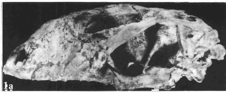
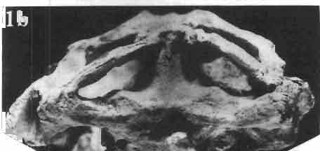
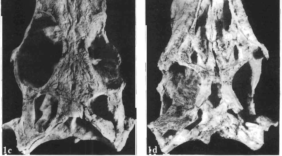
79 Palaeontologia Polonica, No. 33, 1975 PI. VlII A. SULIMSKI: MAtROCEPHALOSAURIDAE AND POLYGLYPHANODONTIDAE
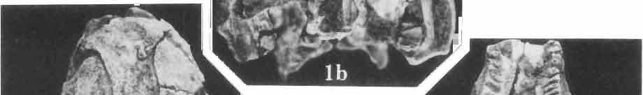
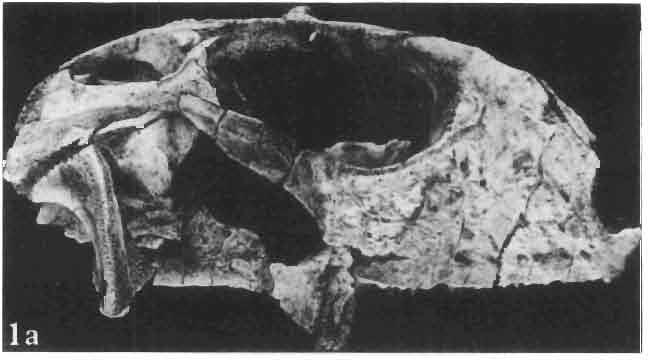
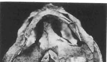
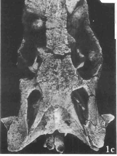
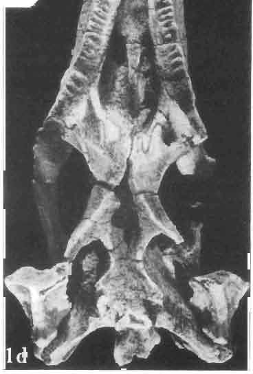
80 Palaeontologia Polonica, No. 33, 1975 PI. IX A. SULIMSKI : MACROCEPHALOSAURIDAE AND POLYGlYPHANODONTIDAE
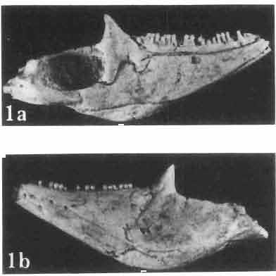
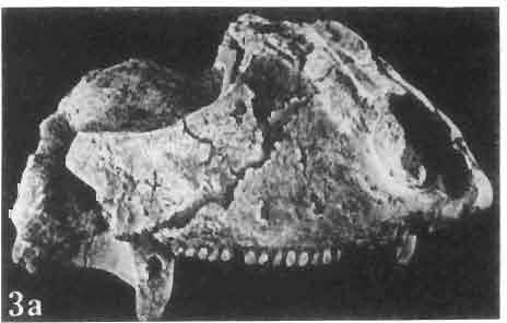
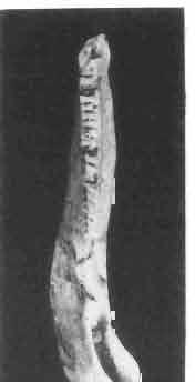
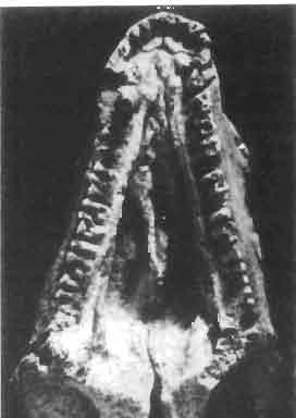
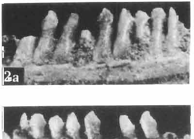
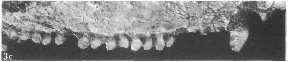
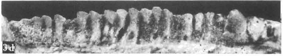
81 Palaeontologia Polonica, No. 33, 1975 PI. X A. SULIMSKI: MACROCEPHAWSAURlDAE AND POLYOLYPHANODONTlDAE


82 Palaeontologia Polonica, No. 33, 1975 PI. XI A. SULIMSKI: MACROCEPHALOSAURIDAE AND POLYGLYPHANODONTlDAE
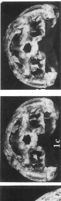
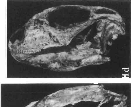
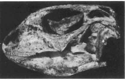
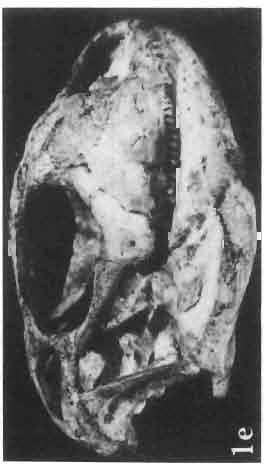
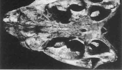
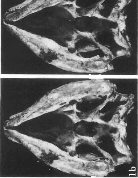
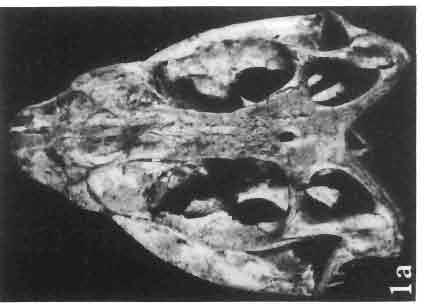
83 Palaeontologia Polonica, No. 33, 1975 PI. XII A. SULIMSKI : MACROCEPHALOSAURIDAE AND POLYGLYPHANODONTIDAI!
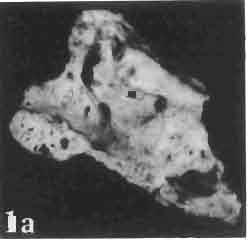
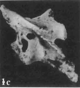
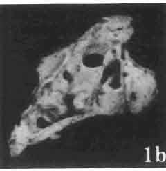
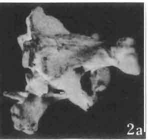
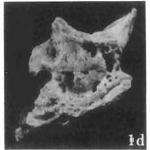
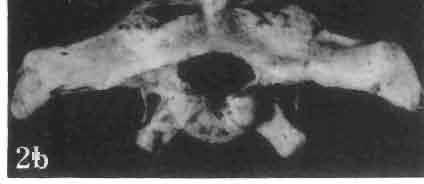
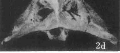
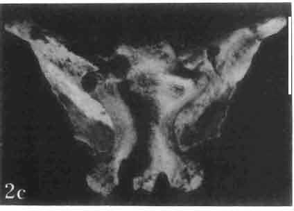
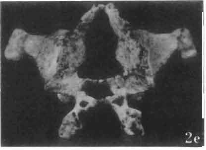
84 Palaeontologia Polonica, No. 33, 1975 PI. X11I A. SULlMSKI: M ACROCEPHALOSAURIDAE AND POLYGLYPHANODONl1DAE
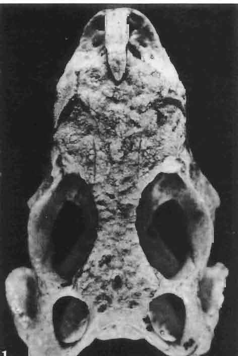
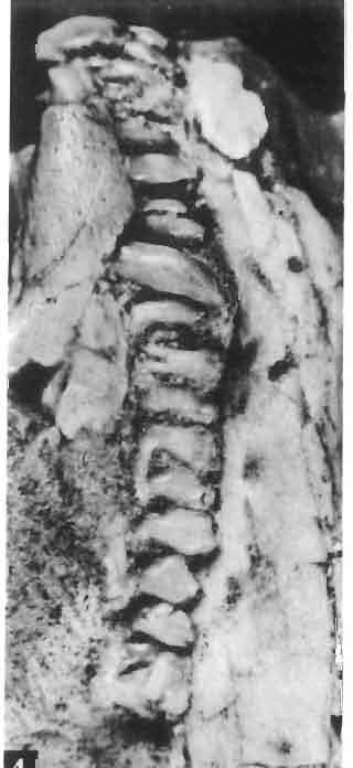
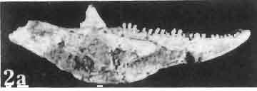
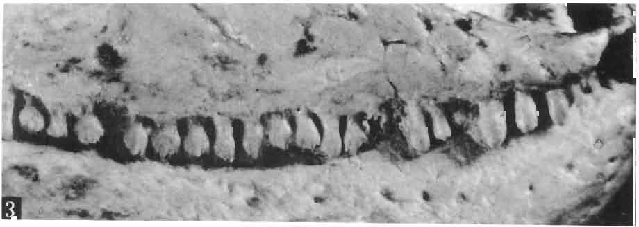
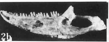
85 Palaeontologia Polonica, No. 33, 1975 PI. XIV A. SULlMSKI: MACROCEPHALOSAURIDAE AND POLYGLVPHANODONTIDAB
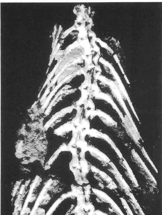
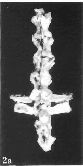
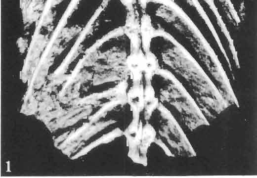
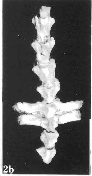
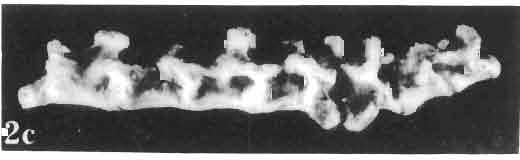
86 Palaeontologia Polonica, No. 33, 1975 PI. XV A. SULIMSKI : M ACROC EPHALOSAURIDAE AND POLYGLYPHANODONTlDAE
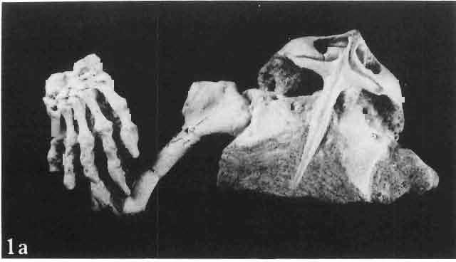
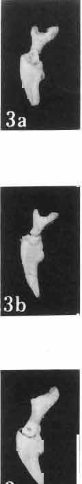
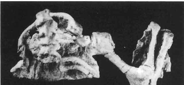
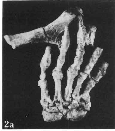
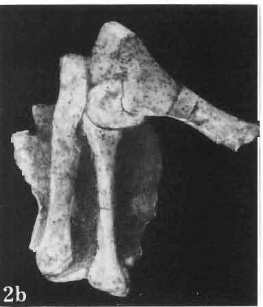
87 Palaeontologia Polonica, No. 33, 1975 PI. XVI A. SULIMSKI: MACROCEPHALOSAURIDAE AND POLYGLYPHANODONTIDAE
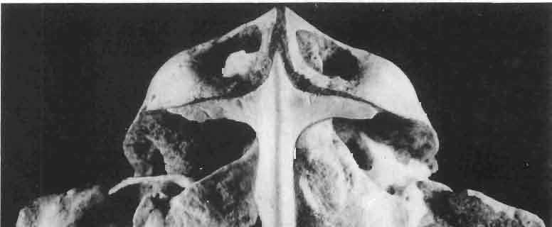
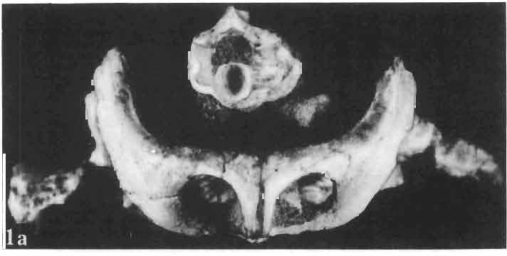
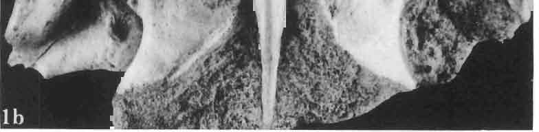
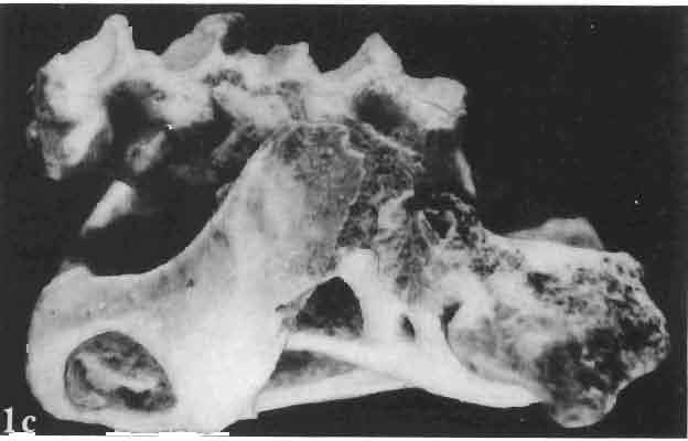
88 Palaeontologia Polonica, No. 33, 1975 PI. XVII A. SULlMSKI : M ACROCEPHALOSAURIDAE AND POLYGLYPHANODONTIDAB
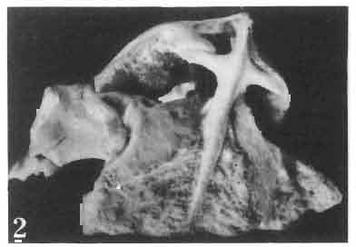
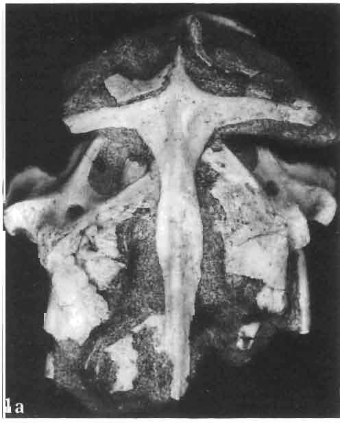
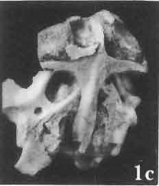
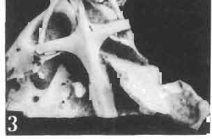
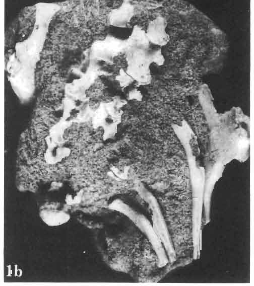
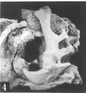
89 Palaeontologia Polonica, No. 33, 1975 PI. XV11I A. SULlMSKI: MACROCEPHALOSAURIDAE AND POLYGLYPHANODONTIDAE
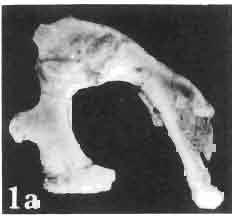
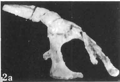
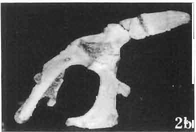
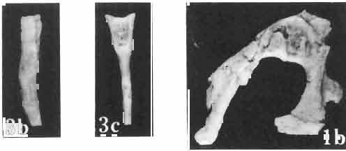
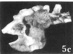
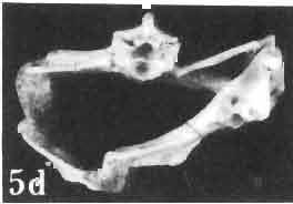
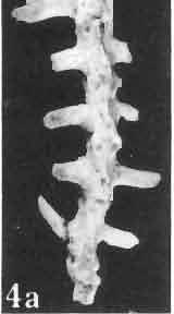
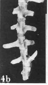
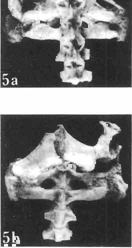
90 Palaeontologia Polonica, No. 33, 1975 PI. XIX A. SULlMSKI: MACROCEPHALOSAURIDAE AND POLYGLVPHANODONTIDAE


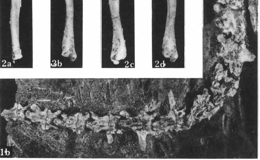
91 Palaeontologia Polonica, No. 33, 1975 PI. XX A. SULIMSKI: MACROCEPHALOSAURlDAE AND POLYGLYPHANODONTIDAE
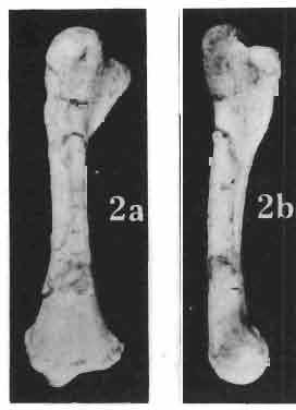
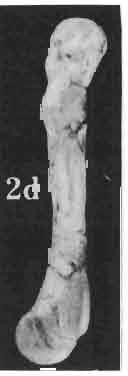
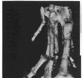
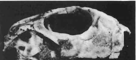
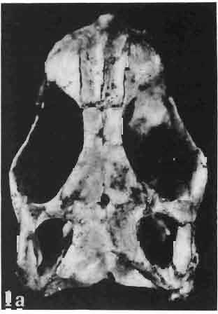
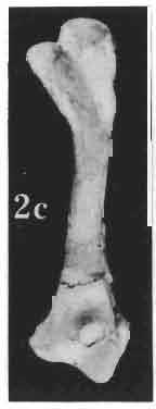
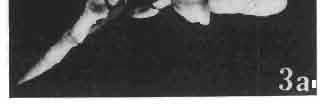
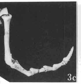
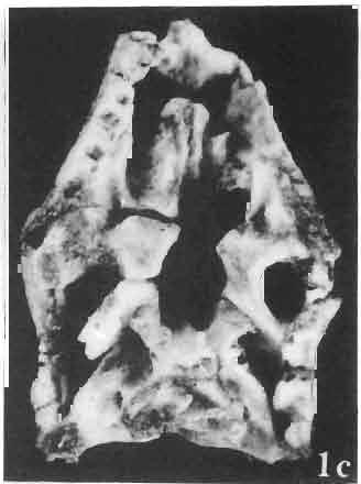
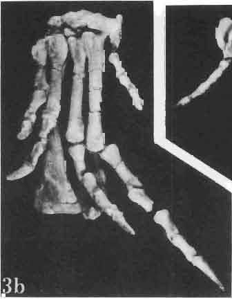
92 Palaeontologia Polonica, No. 33, 1975 PI. XX1 A. SULIMSKI: MACROCEPHALOSAURlDAE AND POLYGLYPHANOD ONTIDAE
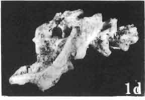
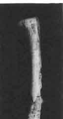
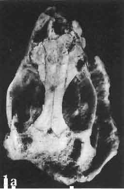
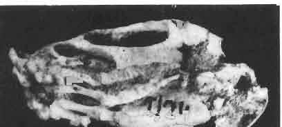
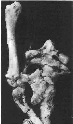
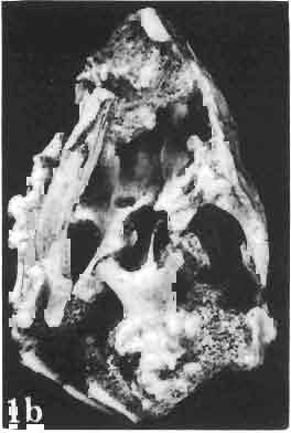
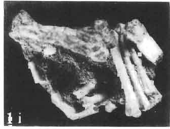
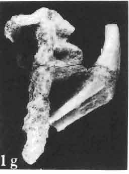
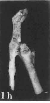
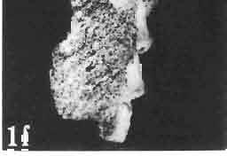
93 Palaeontologia Polonica, No. 33, 1975 PI. XXll A. SULIMSKI: MACROCEPHALOSAURIDAE AND POLYGLYPHANODONTIDAE
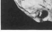
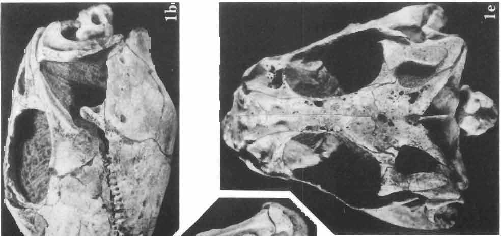
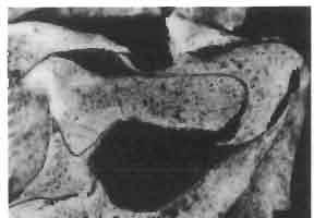
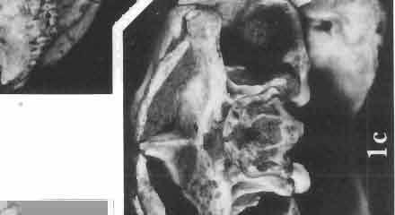
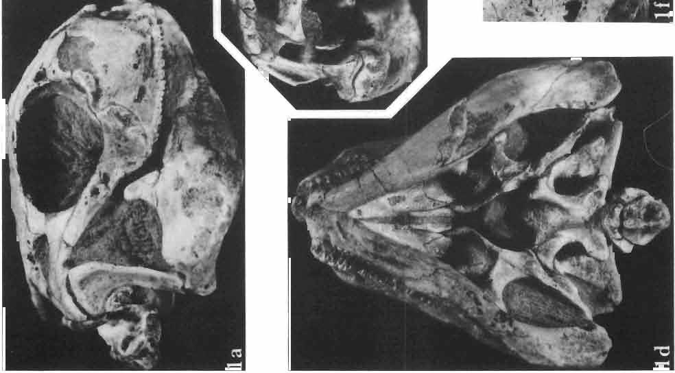
94 Palaeontologia Polonica, No. 33, 1975 PI. XXIll A. SULlMSKl: MACROCEPHALOSAURlDAE AND POLYGLYPHANODONTlDAE
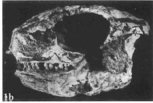
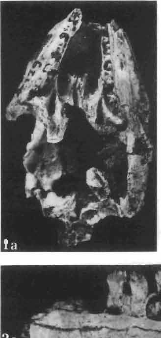
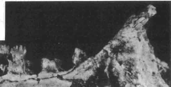
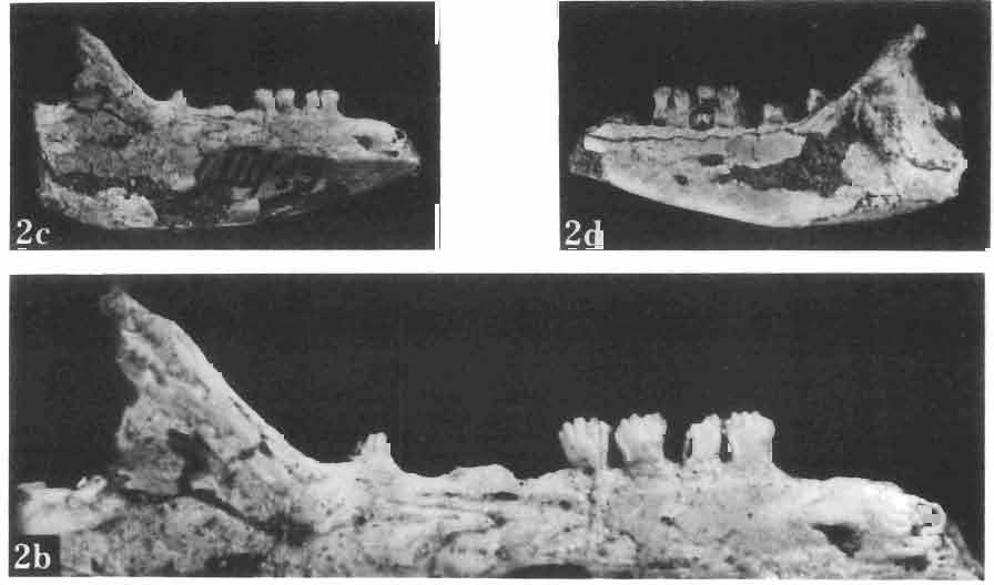
95 Palaeontologia Polonica, No. 33, 1975 PI. XXIV A. SULlMSKI : MACROCEPHALOSAURIDAE AND POLYGLVPHANODONTlDAB
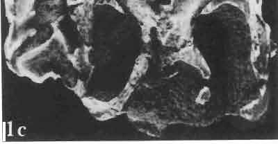
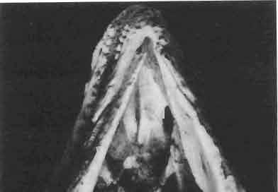
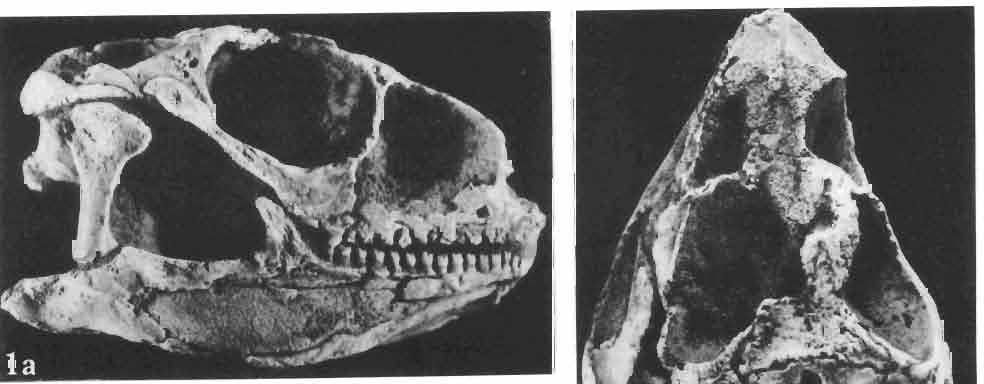
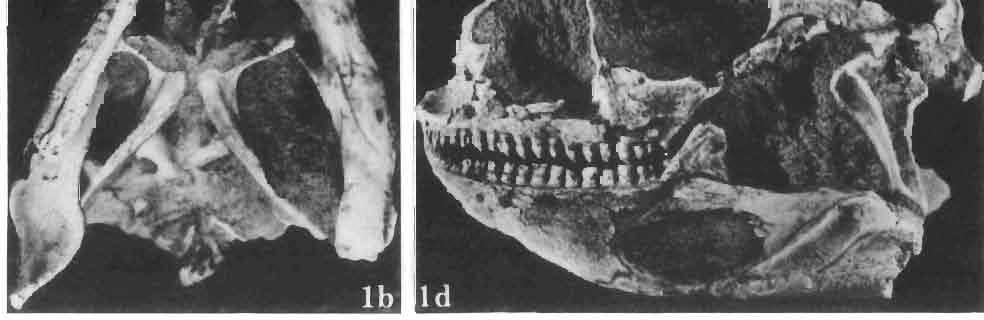
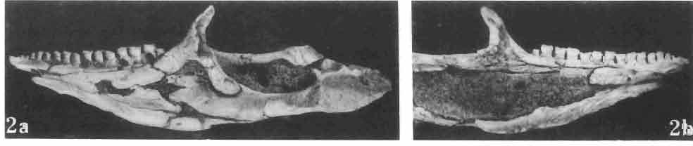
96 Palaeontologia Polonica, No. 33, 1975 PI. XXV A. SULIMSKI: MACROCEPHALOSAURIDAE AND POLYGLYPHANODONTlDAE
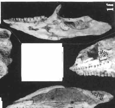
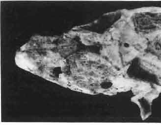
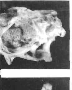
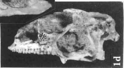
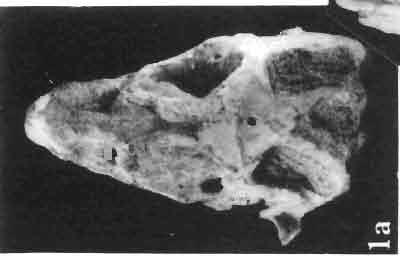
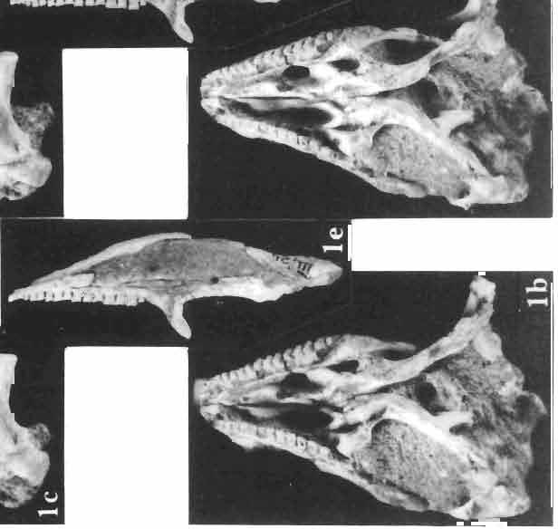
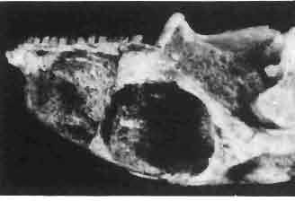
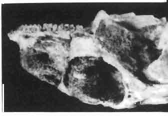
97 Palaeontotogia Polonica, No. 33, 1975 PI. X X VI A. S ULlMSKI: MACROCEPHALOSAURIDAE AND POLYGLYPHANODONTIDAE
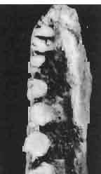
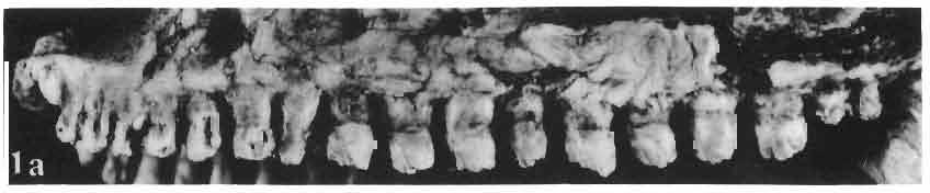
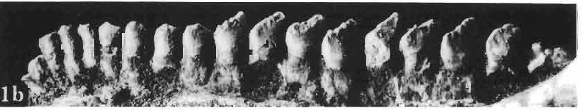
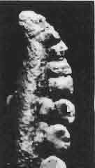
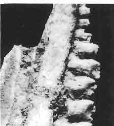
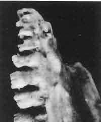
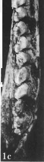
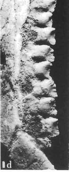
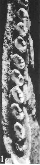
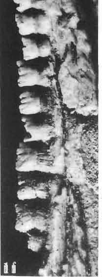
98 Palaeontologia Polonica, No. 33, 1975 PI. XXVll A. SULlMSKI: MACROCEPHALOSAURIDAE AND POLYGLYPHANODONTIDAE
AMERICAN MUSEUM NOVITATES Published by
 AMERICAN MUSEUM NOVITATES Published by Number 782 THE AmzRICAN MUSEUM OF NATURAL HISTORY Feb. 20, 1935 New York City 56.81, 7 G (68) A NOTE ON THE CYNODONT, GLOCHINODONTOIDES GRACILIS HAUGHTON BY LIEUWE
ONLINE APPENDIX 1. Morphological phylogenetic characters scored in this paper. See Poe (2004) for
 for") ONLINE APPENDIX Morphological phylogenetic characters scored in this paper. See Poe () for detailed character descriptions, citations, and justifications for states. Note that codes are changed from a
ONLINE APPENDIX Morphological phylogenetic characters scored in this paper. See Poe () for detailed character descriptions, citations, and justifications for states. Note that codes are changed from a
SOME LITTLE-KNOWN FOSSIL LIZARDS FROM THE
 PROCEEDINGS OF THE UNITED STATES NATIONAL MUSEUM issued SWsK \ {^^m ^V ^^ SMITHSONIAN INSTITUTION U. S. NATIONAL MUSEUM Vol. 91 Washington : 1941 No. 3124 SOME LITTLE-KNOWN FOSSIL LIZARDS FROM THE OLIGOCENE
New Carnivorous Dinosaurs from the Upper Cretaceous of Mongolia
 1955 Doklady, Academy of Sciences USSR 104 (5):779-783 New Carnivorous Dinosaurs from the Upper Cretaceous of Mongolia E. A. Maleev (translated by F. J. Alcock) The present article is a summary containing
A NEW GENUS AND SPECIES OF AMERICAN THEROMORPHA
 A NEW GENUS AND SPECIES OF AMERICAN THEROMORPHA MYCTEROSAURUS LONGICEPS S. W. WILLISTON University of Chicago The past summer, Mr. Herman Douthitt, of the University of Chicago paleontological expedition,
2. Skull, total length versus length of the presacral vertebral column: (0); extremely elongated neck (e.g. Tanystropheus longobardicus).
; extremely elongated neck (e.g. Tanystropheus longobardicus).") Character list of the taxon-character data set 1. Skull and lower jaws, interdental plates: absent (0); present, but restricted to the anterior end of the dentary (1); present along the entire alveolar
Character list of the taxon-character data set 1. Skull and lower jaws, interdental plates: absent (0); present, but restricted to the anterior end of the dentary (1); present along the entire alveolar
SUPPLEMENTARY INFORMATION
 Character 155, interdental ridges. Absence of interdental ridge (0) shown in Parasaniwa wyomingensis (Platynota). Interdental ridges (1) shown in Coniophis precedens. WWW.NATURE.COM/NATURE 1 Character
Mammalogy Laboratory 1 - Mammalian Anatomy
 Mammalogy Laboratory 1 - Mammalian Anatomy I. The Goal. The goal of the lab is to teach you skeletal anatomy of mammals. We will emphasize the skull because many of the taxonomically important characters
Exceptional fossil preservation demonstrates a new mode of axial skeleton elongation in early ray-finned fishes
 Supplementary Information Exceptional fossil preservation demonstrates a new mode of axial skeleton elongation in early ray-finned fishes Erin E. Maxwell, Heinz Furrer, Marcelo R. Sánchez-Villagra Supplementary
Fig. 5. (A) Scaling of brain vault size (width measured at the level of anterior squamosal/parietal suture) relative to skull size (measured at the
 Scaling of brain vault size (width measured at the level of anterior squamosal/parietal suture) relative to skull size (measured at the") Fig. 5. (A) Scaling of brain vault size (width measured at the level of anterior squamosal/parietal suture) relative to skull size (measured at the distance between the left versus right temporomandibular
Fig. 5. (A) Scaling of brain vault size (width measured at the level of anterior squamosal/parietal suture) relative to skull size (measured at the distance between the left versus right temporomandibular
v:ii-ixi, 'i':;iisimvi'\>!i-:: "^ A%'''''-'^-''S.''v.--..V^'E^'-'-^"-t''gi L I E) R.ARY OF THE VERSITY U N I or ILLINOIS REMO
 R.ARY OF THE VERSITY U N I or ILLINOIS REMO") "^ A%'''''-'^-''S.''v.--..V^'E^'-'-^"-t''gi v:ii-ixi, 'i':;iisimvi'\>!i-:: L I E) R.ARY OF THE U N I VERSITY or ILLINOIS REMO Natural History Survey Librarv GEOLOGICAL SERIES OF FIELD MUSEUM OF NATURAL
"^ A%'''''-'^-''S.''v.--..V^'E^'-'-^"-t''gi v:ii-ixi, 'i':;iisimvi'\>!i-:: L I E) R.ARY OF THE U N I VERSITY or ILLINOIS REMO Natural History Survey Librarv GEOLOGICAL SERIES OF FIELD MUSEUM OF NATURAL
complex in cusp pattern. (3) The bones of the coyote skull are thinner, crests sharper and the
 The bones of the coyote skull are thinner, crests sharper and the") DISTINCTIONS BETWEEN THE SKULLS OF S AND DOGS Grover S. Krantz Archaeological sites in the United States frequently yield the bones of coyotes and domestic dogs. These two canines are very similar both
DISTINCTIONS BETWEEN THE SKULLS OF S AND DOGS Grover S. Krantz Archaeological sites in the United States frequently yield the bones of coyotes and domestic dogs. These two canines are very similar both
A new sauropod from Dashanpu, Zigong Co. Sichuan Province (Abrosaurus dongpoensis gen. et sp. nov.)
") A new sauropod from Dashanpu, Zigong Co. Sichuan Province (Abrosaurus dongpoensis gen. et sp. nov.) by Ouyang Hui Zigong Dinosaur Museum Newsletter Number 2 1989 pp. 10-14 Translated By Will Downs Bilby
A new sauropod from Dashanpu, Zigong Co. Sichuan Province (Abrosaurus dongpoensis gen. et sp. nov.) by Ouyang Hui Zigong Dinosaur Museum Newsletter Number 2 1989 pp. 10-14 Translated By Will Downs Bilby
A new species of Hsisosuchus (Mesoeucrocodylia) from Dashanpu, Zigong Municipality, Sichuan Province
 from Dashanpu, Zigong Municipality, Sichuan Province") A new species of Hsisosuchus (Mesoeucrocodylia) from Dashanpu, Zigong Municipality, Sichuan Province Yuhui Gao (Zigong Dinosaur Museum) Vertebrata PalAsiatica Volume 39, No. 3 July, 2001 pp. 177-184 Translated
A new species of Hsisosuchus (Mesoeucrocodylia) from Dashanpu, Zigong Municipality, Sichuan Province Yuhui Gao (Zigong Dinosaur Museum) Vertebrata PalAsiatica Volume 39, No. 3 July, 2001 pp. 177-184 Translated
List of characters used in the phylogenetic analysis. Capital letters T, R, and L, refer to
 1 Supplementary data CHARACTER LIST List of characters used in the phylogenetic analysis. Capital letters T, R, and L, refer to characters used by Tchernov et al. (2000), Rieppel, et al. (2002), and Lee
The cranial osteology of Belebey vegrandis (Parareptilia: Bolosauridae), from the Middle Permian of Russia, and its bearing on reptilian evolution
, from the Middle Permian of Russia, and its bearing on reptilian evolution") Blackwell Publishing LtdOxford, UKZOJZoological Journal of the Linnean Society0024-4082 2007 The Linnean Society of London? 2007 1511 191214 Original Articles RUSSIAN BOLOSAURID REPTILER. R. REISZ ET AL.
Blackwell Publishing LtdOxford, UKZOJZoological Journal of the Linnean Society0024-4082 2007 The Linnean Society of London? 2007 1511 191214 Original Articles RUSSIAN BOLOSAURID REPTILER. R. REISZ ET AL.
Williston, and as there are many fairly good specimens in the American
 56.81.7D :14.71.5 Article VII.- SOME POINTS IN THE STRUCTURE OF THE DIADECTID SKULL. BY R. BROOM. The skull of Diadectes has been described by Cope, Case, v. Huene, and Williston, and as there are many
.56 m. (22 in.). COMPSOGNATHOID DINOSAUR FROM THE. Medicine Bow, Wyoming, by the American Museum Expedition
. COMPSOGNATHOID DINOSAUR FROM THE. Medicine Bow, Wyoming, by the American Museum Expedition") Article XII.-ORNITHOLESTES HERMANNI, A NEW COMPSOGNATHOID DINOSAUR FROM THE UPPER JURASSIC. By HENRY FAIRFIELD OSBORN. The type skeleton (Amer. Mus. Coll. No. 6I9) of this remarkable animal was discovered
Article XII.-ORNITHOLESTES HERMANNI, A NEW COMPSOGNATHOID DINOSAUR FROM THE UPPER JURASSIC. By HENRY FAIRFIELD OSBORN. The type skeleton (Amer. Mus. Coll. No. 6I9) of this remarkable animal was discovered
YANGCHUANOSAURUS HEPINGENSIS - A NEW SPECIES OF CARNOSAUR FROM ZIGONG, SICHUAN
 Vol. 30, No. 4 VERTEBRATA PALASIATICA pp. 313-324 October 1992 [SICHUAN ZIGONG ROUSHILONG YI XIN ZHONG] figs. 1-5, pl. I-III YANGCHUANOSAURUS HEPINGENSIS - A NEW SPECIES OF CARNOSAUR FROM ZIGONG, SICHUAN
SUPPLEMENTARY ONLINE MATERIAL FOR. Nirina O. Ratsimbaholison, Ryan N. Felice, and Patrick M. O connor
 http://app.pan.pl/som/app61-ratsimbaholison_etal_som.pdf SUPPLEMENTARY ONLINE MATERIAL FOR Nirina O. Ratsimbaholison, Ryan N. Felice, and Patrick M. O connor Ontogenetic changes in the craniomandibular
THE SKULLS OF ARAEOSCELIS AND CASEA, PERMIAN REPTILES
 THE SKULLS OF REOSCELIS ND CSE, PERMIN REPTILES University of Chicago There are few Permian reptiles of greater interest at the present time than the peculiar one I briefly described in this journal' three
Mammalogy Lab 1: Skull, Teeth, and Terms
 Mammalogy Lab 1: Skull, Teeth, and Terms Be able to: Goals of today s lab Locate all structures listed on handout Define all terms on handout what they are or what they look like Give examples of mammals
CRANIAL ANATOMY OF ENNATOSAURUS TECTON (SYNAPSIDA: CASEIDAE) FROM THE MIDDLE PERMIAN OF RUSSIA AND THE EVOLUTIONARY RELATIONSHIPS OF CASEIDAE
 FROM THE MIDDLE PERMIAN OF RUSSIA AND THE EVOLUTIONARY RELATIONSHIPS OF CASEIDAE") Journal of Vertebrate Paleontology 28(1):160 180, March 2008 2008 by the Society of Vertebrate Paleontology ARTICLE CRANIAL ANATOMY OF ENNATOSAURUS TECTON (SYNAPSIDA: CASEIDAE) FROM THE MIDDLE PERMIAN
Journal of Vertebrate Paleontology 28(1):160 180, March 2008 2008 by the Society of Vertebrate Paleontology ARTICLE CRANIAL ANATOMY OF ENNATOSAURUS TECTON (SYNAPSIDA: CASEIDAE) FROM THE MIDDLE PERMIAN
A NEW SPECIES OF EXTINCT TURTLE FROM THE UPPER PLIOCENE OF IDAHO
 A NEW SPECIES OF EXTINCT TURTLE FROM THE UPPER PLIOCENE OF IDAHO By Charles W. Gilmore Curator, Division of Vertebrate Paleontology United States National Museum Among the fossils obtained bj^ the Smithsonian
A NEW CRETACEOUS SCINCOMORPH LIZARD FROM MONGOLIA (Plates 26-28)
") ANDRZEJ SULIMSKI A NEW CRETACEOUS SCINCOMORPH LIZARD FROM MONGOLIA (Plates 26-28) SULlMSKI, A. A new Cretaceous scincomorph lizard from Mongolia. Palaeont ologia Polonica, 46, 143-155, 1984. The primitive
ANDRZEJ SULIMSKI A NEW CRETACEOUS SCINCOMORPH LIZARD FROM MONGOLIA (Plates 26-28) SULlMSKI, A. A new Cretaceous scincomorph lizard from Mongolia. Palaeont ologia Polonica, 46, 143-155, 1984. The primitive
OF THE TRIAS THE PHYTOSAURIA
 THE PHYTOSAURIA OF THE TRIAS MAURICE G. MEHL University of Wisconsin Some time ago the writer gave a brief notice of a new genus of phytosaurs of which Angistorhinus grandis Mehl was the type.' It is the
A NEW ANKYLOSAUR FROM THE UPPER CRETACEOUS OF MONGOLIA E.A. Maleev Doklady Akademii Nauk, SSSR 87:
 translated by Dr. Tamara and F. Jeletzky, 1956 A NEW ANKYLOSAUR FROM THE UPPER CRETACEOUS OF MONGOLIA E.A. Maleev 1952. Doklady Akademii Nauk, SSSR 87:273-276 Armored dinosaurs make a considerable part
A Fossil Snake (Elaphe vulpina) From A Pliocene Ash Bed In Nebraska
 From A Pliocene Ash Bed In Nebraska") University of Nebraska - Lincoln DigitalCommons@University of Nebraska - Lincoln Transactions of the Nebraska Academy of Sciences and Affiliated Societies Nebraska Academy of Sciences 198 A Fossil Snake
University of Nebraska - Lincoln DigitalCommons@University of Nebraska - Lincoln Transactions of the Nebraska Academy of Sciences and Affiliated Societies Nebraska Academy of Sciences 198 A Fossil Snake
Cranial osteology of the African gerrhosaurid Angolosaurus skoogi (Squamata; Gerrhosauridae) HOLLY A. NANCE
 HOLLY A. NANCE") African Journal of Herpetology, 2007 56(1): 39-75. Herpetological Association of Africa Original article Cranial osteology of the African gerrhosaurid Angolosaurus skoogi (Squamata; Gerrhosauridae) HOLLY
African Journal of Herpetology, 2007 56(1): 39-75. Herpetological Association of Africa Original article Cranial osteology of the African gerrhosaurid Angolosaurus skoogi (Squamata; Gerrhosauridae) HOLLY
FURTHER STUDIES ON TWO SKELETONS OF THE BLACK RIGHT WHALE IN THE NORTH PACIFIC
 FURTHER STUDIES ON TWO SKELETONS OF THE BLACK RIGHT WHALE IN THE NORTH PACIFIC HIDEO OMURA, MASAHARU NISHIWAKI* AND TOSHIO KASUYA* ABSTRACT Two skeletons of the black right whale were studied, supplementing
A skull without mandihle, from the Hunterian Collection (no.
 4 MR. G. A. BOULENGER ON CHELONIAN REMAINS. [Jan. 6, 2. On some Chelonian Remains preserved in the Museum of the Eojal College of Surgeons. By G. A. Boulenger. [Eeceived December 8, 1890.] In the course
A Complete Late Cretaceous Iguanian (Squamata, Reptilia) from the Gobi and Identification of a New Iguanian Clade
 from the Gobi and Identification of a New Iguanian Clade") PUBLISHED BY THE AMERICAN MUSEUM OF NATURAL HISTORY CENTRAL PARK WEST AT 79TH STREET, NEW YORK, NY 10024 Number 3584, 47 pp., 19 figures September 6, 2007 A Complete Late Cretaceous Iguanian (Squamata,
PUBLISHED BY THE AMERICAN MUSEUM OF NATURAL HISTORY CENTRAL PARK WEST AT 79TH STREET, NEW YORK, NY 10024 Number 3584, 47 pp., 19 figures September 6, 2007 A Complete Late Cretaceous Iguanian (Squamata,
Bulletin of Big Bend Paleo-Geo An Open Access Publication from Mosasaur Ranch Museum, Terlingua and Lajitas, Texas All rights reserved
 Bulletin of Big Bend Paleo-Geo An Open Access Publication from Mosasaur Ranch Museum, Terlingua and Lajitas, Texas All rights reserved This was a private report in 2003 on my thoughts on Platecarpus planifrons.
A Short Report on the Occurrence of Dilophosaurus from Jinning County, Yunnan Province
 A Short Report on the Occurrence of Dilophosaurus from Jinning County, Yunnan Province by Hu Shaojin (Kunming Cultural Administrative Committee, Yunnan Province) Vertebrata PalAsiatica Vol. XXXI, No. 1
Anatomy. Name Section. The Vertebrate Skeleton
 Name Section Anatomy The Vertebrate Skeleton Vertebrate paleontologists get most of their knowledge about past organisms from skeletal remains. Skeletons are useful for gleaning information about an organism
A new species of sauropod, Mamenchisaurus anyuensis sp. nov.
 A new species of sauropod, Mamenchisaurus anyuensis sp. nov. by Xinlu He, Suihua Yang, Kaiji Cai, Kui Li, and Zongwen Liu Chengdu University of Technology Papers on Geosciences Contributed to the 30th
A New Ceratopsian Dinosaur from the Upper
 SMITHSONIAN MISCELLANEOUS COLLECTIONS VOLUME 63. NUMBER 3 A New Ceratopsian Dinosaur from the Upper Cretaceous of Montana, with Note on Hypacrosaurus (With Two Plates) CHARLES W. GILMORE Assistant Curator
Department of Biology, Faculty of Science, Razi University, Kermanshah, Iran 2
 Iranian Journal of Animal Biosystematics (IJAB) Vol.13, No.2, 247-262, 2017 ISSN: 1735-434X (print); 2423-4222 (online) DOI: 10.22067/ijab.v13i2.64614 A comparative study of the skull between Trachylepis
PALEONTOLOGY AND BIOSTRATIGRAPHY OF MONGOLIA
 PALEONTOLOGY AND BIOSTRATIGRAPHY OF MONGOLIA THE JOINT SOVIET-MONGOLIAN PALEONTOLOGICAL EXPEDITION (Transactions, vol. 3) EDITORIAL BOARD: N. N. Kramarenko (editor-in-chief) B. Luvsandansan, Yu. I. Voronin,
 The following text is generated from uncorrected OCR. [Begin Page: Page 1] A NEW CERATOPSIAN DINOSAUR FROM THE UPPER CRETACEOUS OF MONTANA, WITH NOTE ON HYPACROSAURUS ' By CHARLES W. GILMORE assistant
( M amenchisaurus youngi Pi, Ouyang et Ye, 1996)
") 39 4 2001 10 V ERTEBRATA PALASIATICA pp. 266 271 fig. 1,pl. I ( 643013), ( M amenchisaurus hochuanensis),,, Q915. 864 1995 12 31 (ZDM0126) ( M amenchisau rus hochuanensis Young et Chao, 1972),,, ZDM0126
39 4 2001 10 V ERTEBRATA PALASIATICA pp. 266 271 fig. 1,pl. I ( 643013), ( M amenchisaurus hochuanensis),,, Q915. 864 1995 12 31 (ZDM0126) ( M amenchisau rus hochuanensis Young et Chao, 1972),,, ZDM0126
Biology 3315 Comparative Vertebrate Morphology Skulls and Visceral Skeletons
 Biology 3315 Comparative Vertebrate Morphology Skulls and Visceral Skeletons 1. Head skeleton of lamprey Cyclostomes are highly specialized in both the construction of the chondrocranium and visceral skeleton.
THE GORGONOPSIAN GENUS, HIPPOSAURUS, AND THE FAMILY ICTIDORHINIDAE * Dr. L.D. Boonstra. Paleontologist, South African Museum, Cape Town
 THE GORGONOPSIAN GENUS, HIPPOSAURUS, AND THE FAMILY ICTIDORHINIDAE * by Dr. L.D. Boonstra Paleontologist, South African Museum, Cape Town In 1928 I dug up the complete skeleton of a smallish gorgonopsian
Postilla PEABODY MUSEUM OF NATURAL HISTORY YALE UNIVERSITY NEW HAVEN, CONNECTICUT, U.S.A.
 Postilla PEABODY MUSEUM OF NATURAL HISTORY YALE UNIVERSITY NEW HAVEN, CONNECTICUT, U.S.A. Number 117 18 March 1968 A 7DIAPSID (REPTILIA) PARIETAL FROM THE LOWER PERMIAN OF OKLAHOMA ROBERT L. CARROLL REDPATH
A M E G H I N I A N A. Revista de la Asociación Paleontológia Argentina. Volume XV September-December 1978 Nos. 3-4
 A M E G H I N I A N A Revista de la Asociación Paleontológia Argentina Volume XV September-December 1978 Nos. 3-4 COLORADIA BREVIS N. G. ET N. SP. (SAURISCHIA, PROSAUROPODA), A PLATEOSAURID DINOSAUR FROM
The Discovery of a Tritylodont from the Xinjiang Autonomous Region
 The Discovery of a Tritylodont from the Xinjiang Autonomous Region Ailing Sun and Guihai Cui (Institute of Vertebrate Paleontology, Paleoanthropology, Academia Sinica) Vertebrata PalAsiatica Volume XXVII,
On the cranial anatomy of the polycotylid plesiosaurs, including new material of Polycotylus latipinnis, Cope, from Alabama
 Marshall University Marshall Digital Scholar Biological Sciences Faculty Research Biological Sciences 2004 On the cranial anatomy of the polycotylid plesiosaurs, including new material of Polycotylus latipinnis,
Description of Cranial Elements and Ontogenetic Change within Tropidolaemus wagleri (Serpentes: Crotalinae).
.") East Tennessee State University Digital Commons @ East Tennessee State University Electronic Theses and Dissertations 5-2016 Description of Cranial Elements and Ontogenetic Change within Tropidolaemus
East Tennessee State University Digital Commons @ East Tennessee State University Electronic Theses and Dissertations 5-2016 Description of Cranial Elements and Ontogenetic Change within Tropidolaemus
Supporting Online Material for
 www.sciencemag.org/cgi/content/full/329/5998/1481/dc1 Supporting Online Material for Tyrannosaur Paleobiology: New Research on Ancient Exemplar Organisms Stephen L. Brusatte,* Mark A. Norell, Thomas D.
AMERICAN MUSEUM. Novitates PUBLISHED BY THE AMERICAN MUSEUM OF NATURAL HISTORY CENTRAL PARK WEST AT 79TH STREET
 AMERICAN MUSEUM Novitates PUBLISHED BY THE AMERICAN MUSEUM OF NATURAL HISTORY CENTRAL PARK WEST AT 79TH STREET NEW YORK, N.Y. 10024 U.S.A. NUMBER 2662 NOVEMBER 21, 1978 RONN W. COLDIRON Acroplous vorax
OSTEOLOGICAL NOTE OF AN ANTARCTIC SEI WHALE
 OSTEOLOGICAL NOTE OF AN ANTARCTIC SEI WHALE MASAHARU NISHIWAKI* AND TOSHIO KASUYA* ABSTRACT This is a report of measurements on the skeleton of a male se1 whale caught in the Antarctic. The skeleton of
Marshall Digital Scholar. Marshall University. F. Robin O Keefe Marshall University,
 Marshall University Marshall Digital Scholar Biological Sciences Faculty Research Biological Sciences 2008 Cranial anatomy and taxonomy of Dolichorhynchops bonneri new combination, a polycotylid (Sauropterygia:
A NEW SPECIES OF TROODONT DINOSAUR FROM THE
 A NEW SPECIES OF TROODONT DINOSAUR FROM THE LANCE FORMATION OF WYOMING By Charles W. Gilmore Curator of Vertebrate Paleontology, United States National Museum INTRODUCTION The intensive search to which
SUPPLEMENTARY INFORMATION
 doi:10.1038/nature11227 Contents 1. Character list 2. Edits to prior character descriptions/codings 3. Materials examined 4. Results of phylogenetic analyses 5. Ophidian characters of Coniophis and diagnoses
VERTEBRATA PALASIATICA
 41 2 2003 2 VERTEBRATA PALASIATICA pp. 147 156 figs. 1 5 1) ( 100044), ( Parakannemeyeria brevirostris),,, : ( Xiyukannemeyeria),,, Q915. 864 60 Turfania (,1973), Dicynodon (, 1973 ; Lucas, 1998), (Lystrosaurus)
A new carnosaur from Yongchuan County, Sichuan Province
 A new carnosaur from Yongchuan County, Sichuan Province by Dong Zhiming Institute of Vertebrate Palaeontology and Palaeoanthropology, Academia Sinica Zhang Yihong, Li Xuanmin, and Zhou Shiwu Chongqing
Cranial morphology and taxonomy of South African Tapinocephalidae (Therapsida: Dinocephalia): the case of Avenantia and Riebeeckosaurus
: the case of Avenantia and Riebeeckosaurus") Cranial morphology and taxonomy of South African Tapinocephalidae (Therapsida: Dinocephalia): the case of Avenantia and Riebeeckosaurus Saniye Güven*, Bruce S. Rubidge & Fernando Abdala Evolutionary Studies
Cranial morphology and taxonomy of South African Tapinocephalidae (Therapsida: Dinocephalia): the case of Avenantia and Riebeeckosaurus Saniye Güven*, Bruce S. Rubidge & Fernando Abdala Evolutionary Studies
NOTES ON THE FIRST SKULL AND JAWS OF RIOJASAURUS INCERTUS (DINOSAURIA, PROSAUROPODA, MELANOROSAURIDAE) OF THE LATE TRIASSIC OF LA RIOJA, ARGENTINA
 OF THE LATE TRIASSIC OF LA RIOJA, ARGENTINA") NOTES ON THE FIRST SKULL AND JAWS OF RIOJASAURUS INCERTUS (DINOSAURIA, PROSAUROPODA, MELANOROSAURIDAE) OF THE LATE TRIASSIC OF LA RIOJA, ARGENTINA José F. Bonaparte and José A. Pumares translated by Jeffrey
NOTES ON THE FIRST SKULL AND JAWS OF RIOJASAURUS INCERTUS (DINOSAURIA, PROSAUROPODA, MELANOROSAURIDAE) OF THE LATE TRIASSIC OF LA RIOJA, ARGENTINA José F. Bonaparte and José A. Pumares translated by Jeffrey
[Accepted 8th October CONTENTS INTRODUCTION
 183 THE CRANIAL MORPHOLOGY OF A NEW GENUS AND SPECIES OF ICTIDOSAURAN BY A. W. CROMPTON S. A. Museum, Cape Town [Accepted 8th October 19571 (With 7 figures in the text) CONTENTS lntroduction..............
Toothless Dinosaurs of Mongolia. R. Barsbold. Fossil Vertebrates of Mongolia Academy of Sciences of USSR Paleontological Institute
 Toothless Dinosaurs of Mongolia R. Barsbold Fossil Vertebrates of Mongolia Academy of Sciences of USSR Paleontological Institute Academy of Sciences of the MPR Geological Institute Nauka Moscow 1981 Trudy
PALEONTOLOGICAL CONTRIBUTIONS
 THE UNIVERSITY OF KANSAS PALEONTOLOGICAL CONTRIBUTIONS August, 1965 Paper 2 A NEW WYOMING PHYTOSAUR By THEODORE H. EATON, JR. [Museum of Natural History, University of Kansas I ABSTRACT The skull of a
CRANIAL ANATOMY AND PHYLOGENETIC AFFINITIES OF THE PERMIAN PARAREPTILE MACROLETER POEZICUS
 CRANIAL ANATOMY AND PHYLOGENETIC AFFINITIES OF THE PERMIAN PARAREPTILE MACROLETER POEZICUS Author(s): LINDA A. TSUJI Source: Journal of Vertebrate Paleontology, 26(4):849-865. 2006. Published By: The Society
The skull of Sphenacodon ferocior, and comparisons with other sphenacodontines (Reptilia: Pelycosauria)
") Circular 190 New Mexico Bureau of Mines & Mineral Resources A DIVISION OF NEW MEXICO INSTITUTE OF MINING & TECHNOLOGY The skull of Sphenacodon ferocior, and comparisons with other sphenacodontines (Reptilia:
Circular 190 New Mexico Bureau of Mines & Mineral Resources A DIVISION OF NEW MEXICO INSTITUTE OF MINING & TECHNOLOGY The skull of Sphenacodon ferocior, and comparisons with other sphenacodontines (Reptilia:
Temporal lines. More forwardfacing. tubular orbits than in the African forms 3. Orbits larger relative to skull size than in the other genera 2.
 Asian lorises More forwardfacing and tubular orbits than in the African forms 3. Characterized by a marked extension of the ectotympanic into a tubular meatus and a more angular auditory bulla than in
Redescription of the Mongolian Sauropod NEMEGTOSAURUS MONGOLIENSIS Nowinski (Dinosauria:
 Journal of Systematic Palaeontology 3 (3): 283 318 Issued 24 August 2005 doi:10.1017/s1477201905001628 Printed in the United Kingdom C The Natural History Museum Redescription of the Mongolian Sauropod
First Ornithomimid (Theropoda, Ornithomimosauria) from the Upper Cretaceous Djadokhta Formation of Tögrögiin Shiree, Mongolia
 from the Upper Cretaceous Djadokhta Formation of Tögrögiin Shiree, Mongolia") First Ornithomimid (Theropoda, Ornithomimosauria) from the Upper Cretaceous Djadokhta Formation of Tögrögiin Shiree, Mongolia Tsogtbaatar Chinzorig¹, ³ *, Yoshitsugu Kobayashi², Khishigjav Tsogtbaatar³,
First Ornithomimid (Theropoda, Ornithomimosauria) from the Upper Cretaceous Djadokhta Formation of Tögrögiin Shiree, Mongolia Tsogtbaatar Chinzorig¹, ³ *, Yoshitsugu Kobayashi², Khishigjav Tsogtbaatar³,
Cranial osteology and phylogenetic relationships of Hamadasuchus rebouli (Crocodyliformes: Mesoeucrocodylia) from the Cretaceous of Morocco
 from the Cretaceous of Morocco") Blackwell Publishing LtdOxford, UKZOJZoological Journal of the Linnean Society0024-4082 2007 The Linnean Society of London? 2007 1494 533567 Original Articles HAMADASUCHUS REBOULIH. C. E. LARSSON and H.-D.
Blackwell Publishing LtdOxford, UKZOJZoological Journal of the Linnean Society0024-4082 2007 The Linnean Society of London? 2007 1494 533567 Original Articles HAMADASUCHUS REBOULIH. C. E. LARSSON and H.-D.
A New Dromaeosaurid Theropod from Ukhaa Tolgod (Ömnögov, Mongolia)
") PUBLISHED BY THE AMERICAN MUSEUM OF NATURAL HISTORY CENTRAL PARK WEST AT 79TH STREET, NEW YORK, NY 10024 Number 3545, 51 pp., 25 figures, 1 table December 7, 2006 A New Dromaeosaurid Theropod from Ukhaa
PUBLISHED BY THE AMERICAN MUSEUM OF NATURAL HISTORY CENTRAL PARK WEST AT 79TH STREET, NEW YORK, NY 10024 Number 3545, 51 pp., 25 figures, 1 table December 7, 2006 A New Dromaeosaurid Theropod from Ukhaa
PROTOCERATOPSIDAE (DINOSAURIA) OF ASIA (Plates XXXVI-L)
 OF ASIA (Plates XXXVI-L)") TERESA MARYANSKA & HALSZKA OSMOLSKA PROTOCERATOPSIDAE (DINOSAURIA) OF ASIA (Plates XXXVI-L) Abstract. - The paper describes a new protoceratopsid material from the Gobi Desert, Mongolia. One new genus
TERESA MARYANSKA & HALSZKA OSMOLSKA PROTOCERATOPSIDAE (DINOSAURIA) OF ASIA (Plates XXXVI-L) Abstract. - The paper describes a new protoceratopsid material from the Gobi Desert, Mongolia. One new genus
Brigham Young University Science Bulletin, Biological Series
 Brigham Young University Science Bulletin, Biological Series Volume 11 Number 1 Article 1 6-1970 Osteological and mylogical comparisons of the head and thorax regions of Cnemidophorus tigris septentrionalis
CENE RUMINANTS OF THE GENERA OVIBOS AND
 DESCRIPTIONS OF TWO NEW SPECIES OF PLEISTO- CENE RUMINANTS OF THE GENERA OVIBOS AND BOOTHERIUM, WITH NOTES ON THE LATTER GENUS. By James Williams Gidley, Of the United States National Museum. Two interesting
Florida, Gainesville, Florida, 32611, U.S.A. b Smithsonian Tropical Research Institute, Ancon, Republic of Panama,
 This article was downloaded by: [78.22.97.164] On: 04 May 2013, At: 14:02 Publisher: Taylor & Francis Informa Ltd Registered in England and Wales Registered Number: 1072954 Registered office: Mortimer
HONR219D Due 3/29/16 Homework VI
 Part 1: Yet More Vertebrate Anatomy!!! HONR219D Due 3/29/16 Homework VI Part 1 builds on homework V by examining the skull in even greater detail. We start with the some of the important bones (thankfully
Taxonomic revision of lizards from the Paleocene deposits of the Qianshan Basin, Anhui, China
 第 54 卷第 3 期 2016 年 7 月 古脊椎动物学报 VERTEBRATA PALASIATICA pp. 243 268 figs. 1 7 Taxonomic revision of lizards from the Paleocene deposits of the Qianshan Basin, Anhui, China DONG Li-Ping 1 Susan E. EVANS 2
A Late Jurassic Protosuchian Sichuanosuchus huidongensis from Zigong, Sichuan Province. Guangzhao Peng. Zigong Dinosaur Museum, Zigong, Sichuan
 A Late Jurassic Protosuchian Sichuanosuchus huidongensis from Zigong, Sichuan Province Guangzhao Peng Zigong Dinosaur Museum, Zigong, Sichuan 643013 Vertebrata PalAsiatica Volume 34, Number 4 October,
Major cranial changes during Triceratops ontogeny John R. Horner 1, * and Mark B. Goodwin 2
 273, 2757 2761 doi:10.1098/rspb.2006.3643 Published online 1 August 2006 Major cranial changes during Triceratops ontogeny John R. Horner 1, * and Mark B. Goodwin 2 1 Museum of the Rockies, Montana State
REVISION OF THE GENUS MARTINICHTHYS, MARINE FISH (TELESOSTEI, TSELFATIIFORMES) FROM THE LATE CRETACEOUS OF KANSAS (UNITED STATES)
 FROM THE LATE CRETACEOUS OF KANSAS (UNITED STATES)") 1 REVISION OF THE GENUS MARTINICHTHYS, MARINE FISH (TELESOSTEI, TSELFATIIFORMES) FROM THE LATE CRETACEOUS OF KANSAS (UNITED STATES) TAVERNE L., 2000. Revision of the genus Martinichthys, marine fish (Teleostei,
1 REVISION OF THE GENUS MARTINICHTHYS, MARINE FISH (TELESOSTEI, TSELFATIIFORMES) FROM THE LATE CRETACEOUS OF KANSAS (UNITED STATES) TAVERNE L., 2000. Revision of the genus Martinichthys, marine fish (Teleostei,
Recently Mr. Lawrence M. Lambe has described and figured in the
 56.81,9C(117:71.2) Article XXXV.-CORYTHOSAURUS CASUARIUS, A NEW CRESTED DINOSAUR FROM THE BELLY RIVER CRETA- CEOUS, WITH PROVISIONAL CLASSIFICATION OF THE FAMILY TRACHODONTIDA1X BY BARNUM BROWN. PLATE
ON TWO NEW SPECIMENS OF LYSTROSAURUS-ZONE CYNODONTS
 ON TWO NEW SPECMENS OF LYSTROSAURUS-ZONE CYNODONTS By A. S. Brink ABSTRACT n this paper the skulls of two new specimens of Lystrosaurus-zone cynodonts are described. One is a skull of Notictosaurus luckh1fi
PEABODY MUSEUM OF NATURAL HISTORY, YALE UNIVERSITY NEW HAVEN, CONNECTICUT, U.S.A. A NEW OREODONT FROM THE CABBAGE PATCH LOCAL FAUNA, WESTERN MONTANA
 Postilla PEABODY MUSEUM OF NATURAL HISTORY YALE UNIVERSITY NEW HAVEN, CONNECTICUT, U.S.A. Number 85 September 21, 1964 A NEW OREODONT FROM THE CABBAGE PATCH LOCAL FAUNA, WESTERN MONTANA STANLEY J. RIEL
Cranial Osteology of the Andean Lizard Stenocercus guentheri (Squamata: Tropiduridae) and Its Postembryonic Development
 and Its Postembryonic Development") JOURNAL OF MORPHOLOGY 255:94-113 (2003) Cranial Osteology of the Andean Lizard Stenocercus guentheri (Squamata: Tropiduridae) and Its Postembryonic Development Omar Torres-Carvajal* Natural History Museum
JOURNAL OF MORPHOLOGY 255:94-113 (2003) Cranial Osteology of the Andean Lizard Stenocercus guentheri (Squamata: Tropiduridae) and Its Postembryonic Development Omar Torres-Carvajal* Natural History Museum
Notes on Ceratopsians and Ankylosaurs at the Royal Ontario Museum
 Notes on Ceratopsians and Ankylosaurs at the Royal Ontario Museum Andrew A. Farke, Ph.D. Raymond M. Alf Museum of Paleontology 1175 West Baseline Road Claremont, CA 91711 email: afarke@webb.org Introduction
ON THE SCALOPOSAURID SKULL OF OLIVIERIA PARRINGTONI, BRINK WITH A NOTE ON THE ORIGIN OF HAIR
 ON THE SCALOPOSAURID SKULL OF OLIVIERIA PARRINGTONI, BRINK WITH A NOTE ON THE ORIGIN OF HAIR By G. H. Findlay, D.Sc., M.D. (Professor of Dermatology, University of Pretoria; Director, C.S.I.R. Photobiology
TRACHEMYS SCULPTA. A nearly complete articulated carapace and plastron of an Emjdd A NEAKLY COMPLETE SHELL OF THE EXTINCT TURTLE,
 A NEAKLY COMPLETE SHELL OF THE EXTINCT TURTLE, TRACHEMYS SCULPTA By Charles W. Gilmore Curator of Vertebrate Paleontology, United States National Museum INTRODUCTION A nearly complete articulated carapace
AMERICAN MUSEUM NOVITATES
 AMERICAN MUSEUM NOVITATES Published by Number 144 THz AmzxzcAN MusumokorNATURAL HISTORY Novemoer 7, 1924 56.81,9T(117:51.7) THREE NEW THEROPODA, PROTOCERATOPS ZONE, CENTRAL MONGOLIA' BY HENRY FAIRFIELD
Vol. XIV, No. 1, March, The Larva and Pupa of Brontispa namorikia Maulik (Coleoptera: Chrysomelidae: Hispinae) By S.
 By S.") Vol. XIV, No. 1, March, 1950 167 The Larva and Pupa of Brontispa namorikia Maulik (Coleoptera: Chrysomelidae: Hispinae) By S. MAULIK BRITISH MUSEUM (NATURAL HISTORY) (Presented by Mr. Van Zwaluwenburg
Vol. XIV, No. 1, March, 1950 167 The Larva and Pupa of Brontispa namorikia Maulik (Coleoptera: Chrysomelidae: Hispinae) By S. MAULIK BRITISH MUSEUM (NATURAL HISTORY) (Presented by Mr. Van Zwaluwenburg
4. Premaxilla: Foramen on the lateral surface of the premaxillary body (Yates 2007 ch. 4) 0 absent 1 present
 0 absent 1 present") The character matrix used as a basis for this study is that of Yates et al (2010) which is modified from the earlier matrix used by Yates (2007). This matrix includes characters acquired and/or modified
The character matrix used as a basis for this study is that of Yates et al (2010) which is modified from the earlier matrix used by Yates (2007). This matrix includes characters acquired and/or modified
The cranial skeleton of the Early Permian aquatic reptile Mesosaurus tenuidens: implications for relationships and palaeobiology
 Blackwell Publishing LtdOxford, UKZOJZoological Journal of the Linnean Society0024-4082The Linnean Society of London, 2006? 2006 146? 345368 Original Article THE CRANIAL SKELETON OF MESOSAURUS TENUIDENSS.
Yamaceratops dorngobiensis, a New Primitive Ceratopsian (Dinosauria: Ornithischia) from the Cretaceous of Mongolia
 from the Cretaceous of Mongolia") PUBLISHED BY THE AMERICAN MUSEUM OF NATURAL HISTORY CENTRAL PARK WEST AT 79TH STREET, NEW YORK, NY 10024 Number 3530, 42 pp., 20 figures September 08, 2006 Yamaceratops dorngobiensis, a New Primitive Ceratopsian
PUBLISHED BY THE AMERICAN MUSEUM OF NATURAL HISTORY CENTRAL PARK WEST AT 79TH STREET, NEW YORK, NY 10024 Number 3530, 42 pp., 20 figures September 08, 2006 Yamaceratops dorngobiensis, a New Primitive Ceratopsian
FIELDIANA GEOLOGY NEW SALAMANDERS OF THE FAMILY SIRENIDAE FROM THE CRETACEOUS OF NORTH AMERICA
 FIELDIANA GEOLOGY Published by CHICAGO NATURAL HISTORY MUSEUM Volume 10 Sbftember 22, 1968 No. 88 NEW SALAMANDERS OF THE FAMILY SIRENIDAE FROM THE CRETACEOUS OF NORTH AMERICA Coleman J. Coin AND Walter
ABSTRACT. we define the taxa Alligatoroidae and Alligatoridae to be the descent community and crown group,
 AMERICAN MUSEUM No vtates PUBLISHED BY THE AMERICAN MUSEUM OF NATURAL HISTORY CENTRAL PARK WEST AT 79TH STREET, NEW YORK, N.Y. 10024 Number 3116, 26 pp., 10 figures, 1 table December 28, 1994 The Late
NEW INFORMATION ON THE CRANIUM OF BRACHYLOPHOSAURUS CANADENSIS (DINOSAURIA, HADROSAURIDAE), WITH A REVISION OF ITS PHYLOGENETIC POSITION
, WITH A REVISION OF ITS PHYLOGENETIC POSITION") Journal of Vertebrate Paleontology 25(1):144 156, March 2005 2005 by the Society of Vertebrate Paleontology NEW INFORMATION ON THE CRANIUM OF BRACHYLOPHOSAURUS CANADENSIS (DINOSAURIA, HADROSAURIDAE), WITH
Journal of Vertebrate Paleontology 25(1):144 156, March 2005 2005 by the Society of Vertebrate Paleontology NEW INFORMATION ON THE CRANIUM OF BRACHYLOPHOSAURUS CANADENSIS (DINOSAURIA, HADROSAURIDAE), WITH
Osteology and myology of Phrynosoma p. platyrhinos Girard and Phrynosoma d. hernandesi Girard
 Brigham Young University Science Bulletin, Biological Series Volume 9 Number 4 Article 1 6-1968 Osteology and myology of Phrynosoma p. platyrhinos Girard and Phrynosoma d. hernandesi Girard Richard L.
AUSTRALIAN MUSEUM SCIENTIFIC PUBLICATIONS
 AUSTRALIAN MUSEUM SCIENTIFIC PUBLICATIONS Riek, E. F., 1964. Merostomoidea (Arthropoda, Trilobitomorpha) from the Australian Middle Triassic. Records of the Australian Museum 26(13): 327 332, plate 35.
Fossil locality of Messel, No. 37. Saniwa feisti n. sp., a varanid (Lacertilia, Reptilia) from the middle Eocene of Messel near Darmstadt
 from the middle Eocene of Messel near Darmstadt") [A translation of Stritzke, R. (1983) Saniwa feisti n. sp., ein Varanide (Lacertilia, Reptilia) aus dem Mittel-Eozän von Messel bei Darmstadt, Senckenbergiana Lethaea 64(5/6): 497-508. Figure captions
[A translation of Stritzke, R. (1983) Saniwa feisti n. sp., ein Varanide (Lacertilia, Reptilia) aus dem Mittel-Eozän von Messel bei Darmstadt, Senckenbergiana Lethaea 64(5/6): 497-508. Figure captions
DESCRIPTIONS OF NEW AND LITTLE-KNOWN FOSSIL LIZARDS FROM NORTH AMERICA
 PROCEEDINGS OF THE UNITED STATES NATIONAL MUSEUM SMITHSONIAN INSTITUTION U. S. NATIONAL MUSEUM Vol.86 Washington: 1938 No. 3042 DESCRIPTIONS OF NEW AND LITTLE-KNOWN FOSSIL LIZARDS FROM NORTH AMERICA By
NORTH AMERICA. ON A NEW GENUS AND SPECIES OF COLUBRINE SNAKES FROM. The necessity of recognizing tlie two species treated of in this paper
 ON A NEW GENUS AND SPECIES OF COLUBRINE SNAKES FROM NORTH AMERICA. BY Leonhard Stejneger, and Batrachians. Curator of the Department of Reptiles The necessity of recognizing tlie two species treated of
A New Pterosaur from the Middle Jurassic of Dashanpu, Zigong, Sichuan
 A New Pterosaur from the Middle Jurassic of Dashanpu, Zigong, Sichuan by Xinlu He (Chengdu College of Geology) Daihuan Yang (Chungking Natural History Museum, Sichuan Province) Chunkang Su (Zigong Historical
BREVIORA LEUCOLEPIDOPA SUNDA GEN. NOV., SP. NOV. (DECAPODA: ALBUNEIDAE), A NEW INDO-PACIFIC SAND CRAB. Ian E. Efford 1
, A NEW INDO-PACIFIC SAND CRAB. Ian E. Efford 1") ac lc BREVIORA CAMBRIDGE, MASS. 30 APRIL, 1969 NUMBER 318 LEUCOLEPIDOPA SUNDA GEN. NOV., SP. NOV. (DECAPODA: ALBUNEIDAE), A NEW INDO-PACIFIC SAND CRAB Ian E. Efford 1 ABSTRACT. Leucolepidopa gen. nov.
ac lc BREVIORA CAMBRIDGE, MASS. 30 APRIL, 1969 NUMBER 318 LEUCOLEPIDOPA SUNDA GEN. NOV., SP. NOV. (DECAPODA: ALBUNEIDAE), A NEW INDO-PACIFIC SAND CRAB Ian E. Efford 1 ABSTRACT. Leucolepidopa gen. nov.
THE OCCURRENCE OF CONTOGENYS-LIKE LIZARDS IN THE LATE CRETACEOUS AND EARLY TERTIARY OF THE WESTERN INTERIOR OF THE U.S.A.
 Journal of Vertebrate Paleontology 29(3):677 701, September 2009 # 2009 by the Society of Vertebrate Paleontology ARTICLE THE OCCURRENCE OF CONTOGENYS-LIKE LIZARDS IN THE LATE CRETACEOUS AND EARLY TERTIARY