A peer-reviewed version of this preprint was published in PeerJ on 28 May 2018.
|
|
|
- Stella Norris
- 5 years ago
- Views:
Transcription
1 A peer-reviewed version of this preprint was published in PeerJ on 28 May View the peer-reviewed version (peerj.com/articles/4808), which is the preferred citable publication unless you specifically need to cite this preprint. Wang Y, Zhu M Redescription of Phymolepis cuifengshanensis (Antiarcha: Yunnanolepididae) using high-resolution computed tomography and new insights into anatomical details of the endocranium in antiarchs. PeerJ 6:e4808
2 Redescription of Phymolepis cuifengshanensis (Antiarcha: Yunnanolepididae) using high-resolution computed tomography and new insights into anatomical details of the endocranium in antiarchs Yajing Wang 1, 2, 3, Min Zhu Corresp. 1, 2, 3 1 Key Laboratory of Vertebrate Evolution and Human Origins of Chinese Academy of Sciences, Institute of Vertebrate Paleontology and Paleoanthropology, Beijing, China 2 CAS Center for Excellence in Life and Paleoenvironment, Beijing, China 3 University of Chinese Academy of Sciences, Beijing, China Corresponding Author: Min Zhu address: zhumin@ivpp.ac.cn Background. Yunnanolepidoids constitute either the basal-most consecutive segments or the most primitive clade of antiarchs, a highly diversified jawed vertebrate group from the Silurian and Early Devonian periods. Although the general morphology of yunnanolepidoids is well established, their endocranial features remain largely unclear, thus hindering our further understanding of antiarch evolution, and early gnathostome evolution. Phymolepis cuifengshanensis, a yunnanolepidoid from the Early Devonian of southwestern China, is re-described in detail to reveal the information on endocranial anatomy and additional morphological data of head and trunk shields. Methods. We scanned the material of P. cuifengshanensis using high-resolution computed tomography a nd generated virtual restorations to show the internal morphology of its dermal shield. The dorsal aspect of endocranium in P. cuifengshanensis was therefore inferred. The phylogenetic analysis of antiarchs was conducted based on a revised and expanded dataset that incorporates ten new cranial characters. Results. The lateroventral fossa of trunk shield and Chang s apparatus arethree-dimensionally restored in P. cuifengshanensis. The canal that is positioned just anterior to the internal cavity of Chang s apparatus, probably corresponds to the rostrocaudal canal of euantiarchs. The endocranial morphology of P. cuifengshanensis corroborates a general pattern for yunnanolepidoids with additional characters distinguishing them from sinolepids and euantiarchs, such as a developed cranio-spinal process, an elongated endolymphatic duct, and a long occipital portion. Discussion. In light of new data from Phymolepis and Yunnanolepis, we summarized the morphology on the visceral surface of head shield in antiarchs, and formulated additional ten characters for the phylogenetic analysis. These cranial characters exhibit a high degree of morphological disparity between major subgroups of antiarchs, and highlight the endocranial character evolution in antiarchs.
3 1 Author Cover Page Article title: 5 Redescription of Phymolepis cuifengshanensis (Antiarcha: Yunnanolepididae) using 6 high-resolution computed tomography and new insights into anatomical details of the 7 endocranium in antiarchs 8 9 Authors: 10 Yajing Wang 1, 2, 3 and Min Zhu1, 2, Affiliations: 13 1 Key Laboratory of Vertebrate Evolution and Human Origins of Chinese Academy of 14 Sciences, Institute of Vertebrate Paleontology and Paleoanthropology, Chinese 15 Academy of Sciences, Beijing , China 16 2 CAS Center for Excellence in Life and Paleoenvironment, Beijing, , China 17 3 University of Chinese Academy of Sciences, Beijing , China Corresponding author 20 Min Zhu, zhumin@ivpp.ac.cn
4 21 ABSTRACT 22 Background. Yunnanolepidoids constitute either the basal-most consecutive segments 23 or the most primitive clade of antiarchs, a highly diversified jawed vertebrate group from 24 the Silurian and Early Devonian periods. Although the general morphology of 25 yunnanolepidoids is well established, their endocranial features remain largely unclear, 26 thus hindering our further understanding of antiarch evolution, and early gnathostome 27 evolution. Phymolepis cuifengshanensis, a yunnanolepidoid from the Early Devonian of 28 southwestern China, is re-described in detail to reveal the information on endocranial 29 anatomy and additional morphological data of head and trunk shields. 30 Methods. We scanned the material of P. cuifengshanensis using high-resolution 31 computed tomography and generated virtual restorations to show the internal 32 morphology of its dermal shield. The dorsal aspect of endocranium in P. 33 cuifengshanensis was therefore inferred. The phylogenetic analysis of antiarchs was 34 conducted based on a revised and expanded dataset that incorporates ten new cranial 35 characters. 36 Results. The lateroventral fossa of trunk shield and Chang s apparatus are three- 37 dimensionally restored in P. cuifengshanensis. The canal that is positioned just anterior 38 to the internal cavity of Chang s apparatus, probably corresponds to the rostrocaudal 39 canal of euantiarchs. The endocranial morphology of P. cuifengshanensis corroborates a 40 general pattern for yunnanolepidoids with additional characters distinguishing them from 41 sinolepids and euantiarchs, such as a developed cranio-spinal process, an elongated 42 endolymphatic duct, and a long occipital portion. 43 Discussion. In light of new data from Phymolepis and Yunnanolepis, we summarized 44 the morphology on the visceral surface of head shield in antiarchs, and formulated 45 additional ten characters for the phylogenetic analysis. These cranial characters exhibit a 46 high degree of morphological disparity between major subgroups of antiarchs, and 47 highlight the endocranial character evolution in antiarchs.
5 48 INTRODUCTION 49 Antiarchs, one of the most diverse and widespread fish groups during the Middle 50 Paleozoic, have been resolved at the base of the diversification of jawed vertebrates in 51 most of recent phylogenetic studies (Brazeau, 2009; Davis, Finarelli & Coates, 2012; 52 Zhu et al., 2012; Giles, Rücklin & Donoghue, 2013; Zhu et al., 2013; Dupret et al., 2014; 53 Zhu, 2014; Giles, Friedman & Brazeau, 2015; Long et al., 2015; Qiao et al., 2016; Zhu et 54 al., 2016). It is noteworthy that King et al. (2016) corroborated the placoderm monophyly 55 and proposed antiarchs as a clade sister to petalichthyids and ptyctodonts based on the 56 Bayesian tip-dated clock methods. Since the first description of antiarchs in (Eichwald, 1840), their general morphology has been well established (Young & Zhang, ; Janvier, 1996; Young & Zhang, 1996; Zhu, 1996; Lukševičs, 2001; Young, 2008; 59 Young, 2010; Zhu et al., 2012; Long et al., 2015). However, the anatomical atlas of 60 endocranium in antiarchs is poorly known, largely due to the absence of perichondral 61 ossification (Denison, 1978). While the impressions on the overlying head shield help to 62 restore the endocranial morphology to some extent (Stensiö, 1948; Denison, 1978; 63 Moloshnikov, 2008), such as in Bothriolepididae (Stensiö, 1948; Young, 1984), 64 Asterolepididae (Obruchev, 1933; Stensiö, 1969), and Minicrania lirouyii (Zhu & Janvier, ), little attention has been paid to character transformations of antiarch endocrania 66 in lack of corresponding data from primitive antiarchs. 67 Yunnanolepidoids are endemic antiarchs discovered from the South China and 68 Indochina blocks (Liu, 1983; Pan & Dineley, 1988; Tông-Dzuy, Janvier & Phuong, ; Wang, Qu & Zhu, 2010). They are considered to be the most primitive antiarchs 70 because of the absence of characteristic dermal brachial process (Chang, 1978; 71 Zhang, 1978; Zhang, 1980; Young, 1981a; Long, 1983; Janvier, 1995; Zhu & Janvier, ; Carr, Johanson & Ritchie, 2009; Zhu et al., 2012), although their monophyly has 73 not yet reached a consensus (Janvier & Pan, 1982; Young & Zhang, 1996). To date, 74 yunnanolepidoids also represent the oldest known antiarchs, even though the date of 75 the oldest yunnanolepidoid Shimenolepis (Wang, 1991) has been revised to Late 76 Ludlow recently (Zhao et al., 2016). 77 Yunnanolepididae, a major clade of Yunnanolepidoidei, is characterized by the 78 small brachial fossa and the crista transversalis interna posterior lying in front of the 79 posterior process and pit of trunk shield. It includes the following seven genera: 80 Yunnanolepis, Parayunnanolepis, Phymolepis, Mizia, Grammaspis, Chuanbeiolepis 81 and Yunlongolepis (Chang, 1978; Zhang, 1978; Wang, 1988; Tông-Dzuy & Janvier, ; Zhu, 1996; Pan & Lu, 1997; Zhang, Wang & Wang, 2001; Pan et al., 2017). 83 Phymolepis Chang, 1978 (Figs. 1 3) is a yunnanolepid antiarch from the Lower 84 Devonian of South China. The first description of the type species of Phymolepis, P. 85 cuifengshanensis, was based on material (IVPP V4425) from the Xitun Formation of 86 Cuifengshan in Qujing, Yunnan (Chang, 1978; Zhang, 1978). Amongst all the referred 87 specimens of P. cuifengshanensis (Chang, 1978), V (Fig. 4), a trunk shield with a
6 88 row of medial marginal plates of pectoral fin, was later assigned to Yunnanolepis parvus 89 (Zhang, 1980: pl. 5, fig. 1). V differs from the holotype and other referred 90 specimens of P. cuifengshanensis in its comparatively small size, a sharp median dorsal 91 ridge running throughout the anterior median dorsal plate, and the absence of a 92 conspicuous tergal angle of trunk shield. As such, we follow Zhang (1980) to remove 93 V from P. cuifengshanensis. 94 Young and Zhang (1996) described three specimens from the Xitun Formation (IVPP 95 V ) as P. cuifengshanensis, however these specimens are distinguishable 96 from V (Fig.3), a referred specimen of P. cuifengshanensis preserving part of 97 the head shield (Chang, 1978). The orbital fenestra in V is semilunar in 98 shape and occupies nearly half of the total breadth of the head shield. Accordingly, we 99 also remove V from P. cuifengshanensis. 100 Zhu (1996) assigned additional material from the Xishancun Formation at 101 Liaokuoshan in Qujing (IVPP V , V ) to P. cuifengshanensis, making 102 the first occurrence of this genus no later than early Lochkovian. He also revealed the 103 Chang s apparatus (Zhu, 1996: fig. 11A) and the lateroventral fossa of trunk shield 104 (Zhu, 1996: figs. 11F and 11G) in P. cuifengshanensis, and placed Phymolepis as the 105 sister taxon of Mizia in the phylogenetic analysis. 106 To have a deeper understanding of yunnanolepidoids, their cranial morphology in 107 particular, here we used high-resolution computed tomography (CT) to examine the 108 internal morphology of dermal shield in P. cuifengshanensis. On the basis of resulting 109 new data, P. cuifengshanensis was re-described in more detail. We also conducted the 110 phylogenetic analysis of antiarchs based on a new character matrix expanded and 111 revised from previous analyses (Zhu, 1996; Jia, Zhu & Zhao, 2010; Pan et al., 2017). 112 Several cranial characters were compared and discussed among subgroups of 113 antiarchs to illuminate the endocranial character transformations MATERIALS AND METHODS 116 Material 117 The specimens of P. cuifengshanensis in this study are housed at Institute of 118 Vertebrate Paleontology and Paleoanthropology (IVPP), Chinese Academy of 119 Sciences (CAS). The material was all collected from muddy limestone of the Xitun 120 Formation in Cuifengshan, Qujing, Yunnan Province. 121 The Xishancun, Xitun, Guijiatun and Xujiachong formations (in ascending 122 chronological order) represent the Early Devonian non-marine strata in Qujing District 123 (Liu & Wang, 1973; P'an et al., 1978; Zhu, Wang & Fan, 1994; Liu, Gai & Zhu, 2018). 124 The Xitun Formation consists mainly of grayish-green muddy limestone and mudstone 125 (Fang et al., 1985; Xue, 2012), yielding a rich biota (Cuifengshan Assemblage) 126 characterized by the diversification of sarcopterygians (Chang & Yu, 1981; Chang &
7 127 Yu, 1984; Zhu, Yu & Janvier, 1999; Zhu, Yu & Ahlberg, 2001; Zhu & Yu, 2002) and 128 primitive antiarchs including Yunnanolepis, Parayunnanolepis, Zhanjilepis, 129 Chuchinolepis and Phymolepis. Other fishes in the Xitun Formation include the 130 endemic agnathans (Liu et al., 2015), arthrodires (Dupret, Zhu & Wang, 2017), 131 actinopterygians (Zhu et al., 2006; Lu et al., 2016), acanthodians and chondrichthyans, 132 the latter two of which are mostly known from microvertebtrate remains (Wang, 1984). 133 The Xitun Formation has been dated as late Lochkovian (c million years 134 ago) with evidence from fossil assemblages (Gao, 1981; Fang et al., 1994; Zhao et al., ; Xue, 2012) CT analysis 138 V4425.2, which preserved the only head shield material for Phymolepis in addition to 139 the almost complete trunk shield, was CT scanned at the Key Laboratory of Vertebrate 140 Evolution and Human Origins of Chinese Academy of Science, Beijing, using the 225 KV 141 micro-tomography scanner (developed by Institute of High Energy Physics, CAS) with 142 following parameters: 150 kv voltage; 100 ma current; μm voxel size. All scans 143 were conducted using a 720 rotation with a step size of 0.5 and an unfiltered aluminium 144 reflection target. A total of 1440 transmission images were reconstructed in a matrix of 1536 slices. The software Mimics v was applied for the three- 146 dimensional reconstruction (segmentation and rendering) Phylogenetic analyses 149 The character matrix of antiarchs herein consists of 42 ingroup taxa, 2 outgroup 150 taxa (Kujdanowiaspis and Romundina), and 79 morphological characters. The matrix 151 is modified from those of Zhu (1996), Jia, Zhu & Zhao (2010) and Pan et al. (2017), 152 with revised codings and the addition of ten cranial characters. More details including 153 additional references and character re-formulations are provided in the Supplementary 154 Information. 155 We performed a traditional search in TNT v 1.5 (Goloboff et al., 2008), using random addition sequence replicates, saving 100 trees per replication. We assessed 157 nodal supports through bootstrap values with 100 pseudoreplicates and Bremer decay 158 indices. All characters were treated as equally weighted, and unordered (except 159 Characters 19, 49 and 50). Character state transformations to the nodes of one of the 160 most parsimonious trees (MPTs) were reconstructed in PAUP*4.0a (Swofford, 2003) 161 adopting DELTRAN and ACCTRAN optimizations respectively. Character mapping was 162 performed in MacClade 4.0 (Maddison & Maddison, 2000) RESULTS
8 166 Systematic paleontology 167 Placodermi McCoy, Antiarcha Cope, Yunnanolepidoidei Gross, Yunnanolepididae Miles, Phymolepis Chang, Type species. Phymolepis cuifengshanensis Chang, Included species. Phymolepis guoruii Zhu, Emended diagnosis. Yunnanolepididae in which the posterior median dorsal plate 175 bears a strong posterior process; the anterior median dorsal plate with anterior division 176 longer than posterior division; anterior ventral process and pit situated just below a 177 conspicuous tergal angle and at the same level of the lateral corners of the anterior 178 median dorsal plate; sharp median dorsal ridge between the tergal and posterior dorsal 179 angles. 180 Remarks. The diagnosis of this genus follows Zhu (1996) with a minor revision. While 181 examining V based on high-resolution CT, we noticed that the anterior ventral 182 process and pit are situated at the level of the lateral corners of the anterior median 183 dorsal plate rather than behind it PHYMOLEPIS CUIFENGSHANENSIS Chang, (Figures 1 3, 5 11) Phymolepis cuifengshanensis Chang, p. 292, pl. XXV (5 7) Phymolepis cuifengshanensis Zhang, p. 147, figs , pl. VI Phymolepis cuifengshanensis Zhu, p. 257, figs , pls. I (8 10), IV (1 9) Holotype. IVPP V4425.3, a relatively complete trunk shield (Figs. 1A 1C). 193 Paratype. IVPP V4425.6, a posterior median dorsal plate (Figs. 1D and 1E). 194 Referred specimens. IVPP V (Fig. 2), trunk shield; V (Fig. 3) nearly 195 complete dermal shield only missing the anteriormost portion of head shield and the 196 posterior median dorsal plate; V , left anterior dorsolateral plate; V , 197 V , posterior median dorsal plates; V , left posterior dorsolateral plate; 198 V , right posterior lateral plate; V , left anterior ventrolateral plate; 199 V , right posterior ventrolateral plate. 200 Occurrence. The material was collected from two sites (Cuifengshan and 201 Liaokuoshan) in Qujing city, eastern Yunnan, southwestern China.
9 202 Emended diagnosis. Phymolepis species in which the posterior process of the posterior 203 median dorsal plate reaches one third of the plate length; the median dorsal ridge of 204 trunk shield developed as a blunt elevation in front of the tergal angle and as a sharp 205 crest behind the tergal angle. 206 Remarks. The diagnosis follows Zhu (1996) with an addition of the shape of the median 207 dorsal ridge Description 210 Reconstruction and ornamentation 211 Using the complete specimen of Yunnanolepis chii (Zhang, 1978: V , fig. 1) 212 as a reference, Phymolepis cuifengshanensis could reach 84 mm in the dermal shield 213 length, and represents the largest known species among Yunnanolepididae. 214 This re-investigation of P. cuifengshanensis brings together all referred specimens 215 and leads to a new reconstruction (Figs. 5 and 6). The tentative restoration for the 216 missing pre-orbital portion of head shield follows that of Y. chii (Zhang, 1978: fig. 1). 217 Small, round tubercles are densely distributed on the dorsal surface of the head and 218 trunk shields. The tubercles are generally larger on various ridges and along outer 219 margins of head shield than elsewhere. They are aligned parallel to the sutures between 220 dermal plates, or radiated from the angles on the dorsal wall of trunk shield. In addition, 221 they tend to form the rows along the sensory grooves. The tubercles on the lateral wall of 222 the trunk shield are weakly developed and finer than those elsewhere. The ventral wall of 223 the trunk shield is sparsely covered with tubercles that are slightly larger than the rest of 224 the dermal shield Head shield 227 The orbital fenestra (orb, Figs. 7A and 7C) is comparatively small, and occupies about 228 one fourth of the breadth of the head shield (Table 1). The obstantic margin (om, Fig. 7B) 229 is straight and long, with the preobstantic corner of head shield (proc, Fig. 7A) at the 230 midway of the postorbital division. The posterior margin of the head shield between well- 231 marked postobstantic corners (ptoc, Figs, 7B and 7D), has convex lateral parts (formed 232 mainly by the posterior margin of the paranuchal plate) and a slightly embayed mesial 233 part (formed by the posterior margin of the nuchal plate). The obtected nuchal area (nm, 234 Fig. 7A) occupies 27% length of the nuchal (Table 1). 235 The lateral plate (La, Fig. 7A) has long contact margins for surrounding dermal plates. 236 In visceral view, the anterior attachment area for the submarginal plate is missing, 237 however, the posterior attachment area on the lateral plate (a 1, Figs. 7B and 7C) 238 continues onto the postmarginal plate (a 2, Figs. 7B and 7C).
10 239 The postpineal plate (PP, Fig. 7A) is wider than long. Its anterior margin is concave, 240 unlike the straight margin in Yunnanolepis, Mizia and Parayunnanolepis. The postpineal 241 thickening (pp.th, Fig. 7A) is extremely developed as a prominent tuberculate elevation, 242 which totally encompasses the posterior border of the orbital fenestra and occupies 243 about half of the postpineal plate length. On the visceral surface, paired postorbital 244 cristae (cr.po, Fig. 7D) run somewhat obliquely along the anterior margin of the plate. 245 Two cristae on either side of the postpineal plate are separated far away by a faint 246 median ridge (mr, Fig. 7B), which lies at anterior margin of the plate and does not extend 247 backwards as in many euantiarchs. 248 The nuchal plate (Nu, Figs. 7A and 7B) is broadest across the anterolateral angle. The 249 postpineal notch is broad and deep. The posterolateral margin is about twice as long as 250 the anterolateral one (Table 1). The robust anterolateral ridge (alr, Fig. 7A) sits mainly in 251 the anterior division of the nuchal plate. The transverse nuchal crista (cr.tv, Fig. 7B) on 252 the visceral surface is well developed and thickened laterally. 253 The paranuchal plate (PNu, Figs. 7A and 7B) is as broad as it is long. The obtected 254 area of the plate is steeply inclined to the ornamented surface, especially near its suture 255 with the nuchal plate. The postmarginal plate (PM, Fig. 7A) is rhombic and longitudinally 256 extended. On the visceral surface, the attachment for the submarginal plate is narrower 257 posteriorly Endocranium 260 Like other antiarchs, only the dorsal aspect of the endocranium can be inferred in P. 261 cuifengshanensis from the impressions on the visceral surface of the head shield, which 262 is digitally visible with a high level of details. 263 The otico-occipital depression of P. cuifengshanensis is deeper posteriorly, along with 264 the gradually thickened paramarginal crista (cr. pm, Figs. 7B and 7D). This depression is 265 laterally extended at the suture between the lateral and paranuchal plates, where the 266 paramarginal crista lies underneath the infraorbital sensory groove. As such, the 267 paramarginal crista in P. cuifengshanensis with the convex median part, differs from the 268 straight one in asterolepidoids (Hemmings, 1978; Young. 1983) and laterally concave 269 one in bothriolepidoids (Chang, 1965; Young, 1988). 270 The anterolateral corner of the otico-occipital depression (p.apo, Figs. 7B and 7D), 271 which represents the imprint for the anterior postobital process of endocranium (Young, ), is weakly developed and apically rounded. Significantly, it is located at the same 273 level with the posterior border of the orbital notch. 274 Near the posterior end of the paramarginal crista, the paranuchal plate is deeply 275 excavated by the large cavity (c.csp, Figs. 7B and 7D) for the cranio-spinal process of 276 the endocranium. The cavity is conical and tapers laterally with a B/L ratio of around 3.0; 277 its axis is perpendicular to the paramarginal crista (Figs. 8D F).
11 278 The semicircular depressions (dsc, Figs. 7B and 7D) sit just in front of the level of 279 cranio-spinal processes. The anterior and posterior semicircular depressions are 280 relatively short, and meet in a confluence that is located midway between the posterior 281 border of the orbital notch and the transverse nuchal crista. As the lateral extension of 282 the otico-occipital depression roughly levels with the confluence, this lateral extension 283 appears to relate with the labyrinth cavity (d.sac?, Fig. 7D) as seen in Arenipiscis westolli 284 (Young, 1981b: fig. 6). In view of the otic region, which can be estimated by the position 285 of semicircular depressions, lies mainly in the anterior half of the otico-occipital 286 depression, so the occipital region of P. cuifengshanensis is fairly long compared with 287 euantiarchs. 288 Median to the posterior end of the semicircular depression, the internal pore for the 289 endolymphatic duct (d.end, Figs. 7B and 7D) is rounded and situated far ahead of the 290 transverse nuchal crista, while the external pore (d.end, Figs. 7A and 7C) is situated far 291 posteriorly at the anterior margin of obtected nuchal area. The distance between the 292 internal pores of both sides is 2.5 times longer than the distance between the external 293 ones. The digital visualization reveals that the endolymphatic duct of P. 294 cuifengshanensis is a long and roughly straight tube. It runs posterodorsally within the 295 nuchal plate, swings laterally while close to the midline of the plate and opens to the 296 exterior (Figs. 7A C). 297 Posteriorly, a pair of supraoccipital pits (sop, Figs, 7B and 7D) is positioned just in front 298 of the transverse nuchal crista. This pit is easily distinguished from the internal pore of 299 the endolymphatic duct by its large size and ellipsoidal shape. The pit is dorsomedially 300 oriented within the nuchal plate, and gradually tapers off just beneath the ornamented 301 surface (Figs. 8A C). The supraoccipital pit also occurs in Vukhuclepis (Racheboeuf et 302 al. 2006: fig. 4) and Yunnanolepis at the same position. It is noteworthy that Liu (1963: fig ) misidentified the supraoccipital pit in Yunnanolepis as the internal pore for the 304 endolymphatic duct. 305 Just anterior to the cavity for the cranio-spinal process, a corner (c.vg, Fig. 8F) in a 306 nearly right angle is set on approximately at the posterior end of a semicircular 307 depression level, and thus the hindmost level of the otic region. This corner is also 308 positioned between the anterior postorbital process and cranio-spinal process of 309 endocranium. Therefore, we tentatively interpret this corner as the depression of the 310 vagal process as it shares the same topological relationships to that of arthrodires and 311 petalichthyids Trunk shield 314 The trunk shield is fairly high, with a conspicuous tergal angle (Figs. 2C 2D; 9C 9D) 315 taking up almost half of the trunk shield height. The small pectoral fossa (pf, Fig. 1C) is 316 set just above the bottom of the trunk shield, and occupies a quarter of the lateral wall
12 317 height (Table 2). Both the dorsolateral and ventrolateral ridges of the trunk shield are 318 robust (dl, vl, Figs. 1C and 3D). 319 The dorsal wall has a convex anterior margin. The median dorsal, dorsal diagonal and 320 dorsal transverse ridges (dmr, ddr, dtr, Figs. 1 3A) on the dorsal wall radiate from the 321 tergal angle as in Mizia longhuaensis, Yunnanolepis porifera and Chuchinolepis 322 qujingensis (Zhu, 1996: figs. 4C, 5C 5D, 21A). The lateral wall carries the lateral and 323 oblique ridges (lr, or, Figs. 1C, 3D and 9D), which are widely developed in 324 yunnanolepidoids. 325 The main lateral line (lc, Figs. 2C, 3D and 10A) runs posteriorly very close and 326 subparallel to the dorsolateral ridge of trunk shield. It terminates at the end of the 327 dorsolateral ridge on the posterior dorsolateral plate. 328 The anterior median dorsal plate (AMD, Figs. 1 3A, 9A and 11A) is pentagonal in 329 shape. The posterolateral margin is embayed near its posterior end as in Yunnanolepis 330 (Zhang, 1980: fig. 3C). The concave posterior margin of the plate is delimited laterally by 331 distinct posterolateral angles (pla, Figs. 1A and 11A). The tergal angle lies at the same 332 level with the lateral corner of the plate. Internally, the anterior ventral pit (pt1, Fig. 11A) 333 with thin rim is located right beneath the tergal angle. It extends posteriorly to form a low 334 ridge (prv1, Fig. 11A). 335 The posterior median dorsal plate (PMD, Figs. 1D and 1E) bears a large posterior 336 process (pr.p, Fig. 1E), which occupies about two fifths of the plate in length and three 337 fourths in breadth. The dorsal median ridge and the posterior lateral ridges of both sides 338 (plr, Fig. 1D) converge to the posterior dorsal angle, which is developed as a small 339 nodule. The posterior corner of the plate (pa, Fig. 1D) is rounded. Internally, the crista 340 transversalis interna posterior (cr.tp, Fig.1E) is developed as a low ridge just anterior to 341 the posterior ventral process (pt2, Fig. 1E). 342 The anterior dorsolateral plate (ADL, Figs. 2C, 3D, 9D) consists of articular, dorsal and 343 lateral laminae. The transversely extending articular fossa (f.ca, Fig. 2D and 9C) is 344 delimited by the supra- and infra-articular ridges (sar, iar, Fig. 2E). The supra-articular 345 ridge, which extends laterally from the postnuchal ornamented corner (pnoa, Figs. 2C, 346 3C and 9C), is longer than the infra-articular one. The dorsal lamina is slightly arched 347 with a dorsal diagonal ridge. The dorsal division of the ridge caused by Chang s 348 apparatus (r.c, Figs. 2E and 9D) is positioned on the lateral lamina adjacent to the 349 obstantic margin of the head shield. 350 The anterior ventrolateral plate (AVL, Figs. 9) consists of lateral and ventral laminae, 351 which meet at the ventrolateral ridge. The lateral lamina bears the ventral division of the 352 ridge caused by Chang s apparatus near its anterior margin. The ventral lamina shows a 353 shallow semilunar notch. Internally, both the posterior branchial lamina (pbl, Figs. 2D E 354 and 10D) and crista transversalis interna anterior (cit, Figs. 2D E and 10D) are strongly 355 developed. The posterior branchial lamina, ornamented by denticulate ridges, is present
13 356 close to the anterior margin of the trunk shield. It runs anteromedially from the lateral 357 lamina of the plate to the ventral lamina as a narrow band. The crista transversalis 358 interna anterior is located immediately behind the postbranchial lamina. Dorsally, the 359 crista extends from the AVL to the base of the articular fossa on the ADL (Figs. 2D, 2E 360 and 9C), where it is just behind Chang s apparatus (c.c, Fig. 10C). The left AVL overlaps 361 the right. 362 Between the posterior branchial lamina and crista transversalis interna anterior, a 363 canal (rc, Figs. 10A, D F) is present just anterior to the internal cavity of Chang s 364 apparatus (Fig. 10B). The canal passes ventrally along the lateral wall of the trunk shield. 365 With a relatively large diameter, it probably carries both vessels and nerves and 366 corresponds to the rostrocaudal canal in Chuchinolepis (Young & Zhang, 1992), 367 sinolepids and euantiarchs, which is similarly positioned to supply the fin muscles 368 (Young, 2008). 369 The posterior dorsolateral plate (PDL, Fig. 9D) consists of the dorsal and lateral 370 laminae. The dorsal lamina is slightly less than twice as long as it is broad (Table 2). 371 The posterior lateral plate (PL, Figs. 2C, 3C and 9D) is arched along the lateral ridge 372 of trunk shield. The anteroventral margin is concave, and longer than the anterodorsal 373 one. The dorsal margin of the PL overlaps the PDL. 374 The posterior ventrolateral plate (PVL, Figs. 2C, 9B and 9D) consists of lateral and 375 ventral laminae. The subanal division of the ventral lamina is too short to define. The left 376 PVL overlaps the right one. 377 On the visceral surface of the trunk shield, a fossa (f.lv, Figs. 11B and 11C) is located 378 at the thickened junction of the AVL, PVL and PL plates, as in Yunnanolepis and 379 Zhanjilepis (Zhu, 1996). The fossa was termed the lateroventral fossa, and regarded as 380 a synapomorphy of yunnanolepids by Zhu (1996). 381 Posteriorly, a deeply grooved internal structure (cg, Fig. 11F) is developed along the 382 caudal opening of trunk shield. The groove has a smooth internal surface, delimited 383 anteriorly and posteriorly by the developed crista transversalis interna posterior and 384 posterior margins of trunk plates (PVL, PL and PDL) respectively. It consists of upper 385 and lower halves divided by a thin septum (ms, Figs. 11D and 11F). The similar structure 386 in Yunnanolepis porifera, as well as in Pterichthyodes milleri (Hemmings, 1978: fig. 15D), 387 was assumed to be related to internal fertilization (Long et al., 2015). 388 The semilunar plate (SL, Fig. 9B) is triangular in shape, and approximately twice as 389 broad as long. It is overlapped posteriorly by the AVL. Internally, the postbranchial 390 lamina extends anteromesially from the AVL onto the semilunar plate, and meets the 391 lamina from the opposite. 392 The median ventral plate (MV, Figs. 1B, 2B and 9B) is rhombic. The exposed surface 393 accounts for two fifths of the ventral wall of trunk shield in length and a half of the ventral 394 wall in breadth. The plate is thinner than the surrounding plates.
14 DISCUSSION 397 Anatomical comparisons of several cranial characters in antiarchs 398 The restoration of the endocranium in antiarchs was mainly based on the imprints of 399 its dorsal aspect on the visceral surface of the head shield (Stensiö, 1948; Stensiö, 1969; 400 Miles, 1971;Denison, 1978; Young, 1984). The exception was Minicrania, which 401 preserved the internal cast of the endocranial canals and part of the cranial cavity, thus 402 providing information on its deeper endocranial structures (Zhu & Janvier, 1996). 403 In yunnanolepidoids, the visceral surface of head shield was known in Yunnanolepis 404 (Liu, 1963: fig. 1) and Chuchinolepis (Tông-Dzuy & Janvier, 1990: fig. 17). The digital 405 visualization of Phymolepis shows not only its visceral surface of head shield but also 406 some internal architecture within the dermal plates, such as the trajectory of the 407 endolymphatic duct and the cavity for the cranio-spinal process. We also re-examine 408 the holotype (V2690.1, Fig. 12A) and one referred specimen (V4423.3, Fig. 12B) of Y. 409 chii from the Early Devonian of Qujing, and provide more details for the visceral 410 surface of head shield in Yunnanolepis. Based on these new data, we make 411 comparisons in antiarchs, and show a high degree of morphological disparity with 412 respect to the endocranium. 413 Anterior postorbital process. The endocrania of gnathostomes share a developed 414 lateral projection where the orbits meet the otic capsules (Brazeau & Friedman, 2014). 415 This process was termed the anterior postorbital process in placoderms, and deemed 416 as supporting the hyoid arch articulation and delimiting the posterior boundary of the 417 spiracular chamber by its anterior surface (Young & Zhang, 1996; Brazeau & 418 Friedman, 2014). The positional differences of the anterior postorbital process (and 419 associated cranial nerves) along the longitudinal axis of dermal shield were thought to 420 be informative for phylogenetic analysis (Carr et al., 2009; Dupret et al., 2017). 421 Antiarchs have a well-developed anterior postorbital process. The process 422 extending in front of the anterior border of the orbital notch, has been considered as 423 one of the synapomorphies uniting Bothriolepis and Grossilepis (Zhang & Young, ; Zhu, 1996; Jia, Zhu & Zhao, 2010; Pan et al., 2017). Accordingly, the process 425 behind the anterior border of the orbital notch is referred to a plesiomorphy of 426 antiarchs and this state has been simply summarized as anterior postorbital process 427 short in previous phylogenetic analyses (Zhang & Young, 1992). 428 When examining the short anterior postorbital process in antiarchs, we recognized 429 that this state can be subdivided into two conditions: the anterior postorbital process at 430 the same level with the posterior border of the orbital notch in yunnanolepidoids, 431 Minicrania (Zhu & Janvier, 1996), and probably Sinolepis (Liu & P'an, 1958; Long, 1983); 432 the process anteriorly beyond the posterior border of the orbital notch, but behind its 433 anterior border in euantiarchs excluding Grossilepis and Bothriolepis. In this case, the
15 434 distinction between these two conditions can be added to the transformation series of the 435 anterior postorbital process. 436 Postorbital crista. The postorbital crista in antiarchs separates the otico-occipital 437 depression from the orbital region in front. In several euantiarchs, the crista extends 438 obliquely from the crest of the spiracular groove on the lateral plate to the nuchal plate 439 as a mesial wall of the semicircular depression, such as in Bothriolepis (Stensiö, 1948), 440 Monarolepis (Young & Gorter, 1981), Pterichthyodes (Hemmings, 1978) and 441 Wufengshania (Pan et al., 2017). For the rest of antiarchs including yunnanolepidoids, 442 the postorbital crista runs from the lateral plate to the postpineal plate rather than the 443 nuchal plate as a transversely directed crest embracing the suborbital fenestra 444 posteriorly. 445 Supraotic thickening. The supraotic thickening (Young, 1983: sot, fig. 3D) is 446 bounded posteriorly by the transverse nuchal crista and extensively developed at its 447 connection with the crista. As the supraotic thickening is porous, different from the rest 448 of dermal skeleton in microstructure, it was considered as a junction that is co-ossified 449 with both the endocranium and overlying head shield (Stensiö, 1948; Karatajūte- 450 Talimaa, 1963; Moloshnikov, 2004; Moloshnikov, 2008). Euantiarchs have a persistent 451 supraotic thickening with the exception of Microbrachius, which bears a deep groove 452 throughout the whole length of the otico-occipital depression (Hemmings, 1978: figs C F). The presence of this thickening on the visceral surface of euantiarchs is in 454 stark contrast to the condition in yunnanolepidoids, Minicrania and sinolepids, which 455 lack any thickening in the corresponding area. 456 Median occipital crista. This crista was first identified and named by Stensiö (1931: 457 cro, figs. 11 and 12) in Bothriolepis, and was also termed the posterior median process 458 by Hemmings (1978) in Pterichthyodes. Lying on the descending lamina of occipital part 459 of the head shield, it is separated from the otico-occipital depression by the transverse 460 nuchal crista in euantiarchs. A shallow depression of levator muscles (termed the 461 insertion fossa on head shield for levator muscles ) usually flanks on each side of the 462 crista. In sinolepids, such as Grenfellaspis (Ritchie et al., 1992), the insertion fossa is 463 elongated as that in euantiarchs but lacks the median crista. In yunnanolepidoids, the 464 insertion fossa is either very short (Yunnanolepis, Fig. 12B), or totally absent 465 (Phymolepis, Fig. 7B). 466 Posterior process of head shield. The posterior process of head shield (prnm, see 467 Young (1988): figs. 7B, 37C and 44A) in euantiarchs, also termed the nuchal process 468 or posterior median process (Long & Werdelin, 1986; Moloshnikov, 2004; 469 Moloshnikov, 2008; Moloshnikov, 2010), was first identified and named by Stensiö 470 (1931: figs. 4, 9 and 12). Although the median occipital crista is usually continuous with 471 the posterior process of head shield, the process is apparently independent of the crista 472 in development as evidenced by Asterolepis and Remigolepis, which possess the 473 process but lack the crista. The process is usually developed in euantiarchs, in contrast
16 474 to its absence in yunnanolepidoids and sinolepids. 475 Among non-antiarch placoderms, the posterior process is also known in 476 petalichthyids (Liu, 1991; Pan et al., 2015) and arthrodires (Wang & Wang, 1983; 477 Gardiner & Miles, 1990; Young, 2005; Carr & Hlavin, 2010; Rücklin, Long & Trinajstic, ). 479 The cavity for cranio-spinal process. The cranio-spinal process was named by 480 Nielsen (1942), and also termed the supravagal process by Stensiö (1969) and the 481 paroccipital process by Eaton (1939). It is widely developed in early gnathostomes, 482 including arthrodires (Young, 1979), petalichthyids (Stensiö, 1925), acanthodians 483 (Miles, 1973), actinopterygians (Patterson, 1975) and dipnoans (Miles, 1977). 484 However, the cavity for cranio-spinal process on the visceral surface of head shield, 485 which might function for fixing the endocranium to the external bony shield, is only found 486 in primitive antiarchs and some arthrodires. 487 The cranio-spinal process in yunnanolepidoids is strongly developed, as indicated 488 by the large cavity for the process. The process and the corresponding cavity in 489 euantiarchs were either reduced or absent (Young, 1984). 490 Supraoccipital pit. The supraoccipital pit of the head shield is present for housing the 491 endocranial supraocciptial process. It is bounded posteriorly by the transverse nuchal 492 crista in yunnanolepidoids. The same condition is also seen in Grenfellaspis (Ritchie 493 et al., 1992) and Minicrania, despite the supraoccipital pit in the latter has ever been 494 interpreted as impression of endolymphatic sac (Zhu & Janvier, 1996; Dupret et al., ). In euantiarchs, the supraoccipital pit is only seen in few Bothriolepis species 496 with two different positions: either immediately anterior to the transverse nuchal crista 497 as exemplified by B. tatongensis (Long & Werdelin, 1986), or on the transverse nuchal 498 crista as in B. macphersoni and B. portalensis (Young, 1988). 499 In non-antiarch placoderms, the supraoccipital pit has been observed in 500 petalichthyids (Liu, 1991: cv.ifnu, fig. 2), and most arthrodires, including 501 Holonematidae (Miles, 1971: tf; figs. 53 and 117; Young, 2005: if.pt, fig. 2C), 502 Buchanosteidae (Young, 1981b: if.pt, fig. 6), Coccosteoidea (Miles & Westoll, 1968: 503 p.pts.nu, fig. 2a), Dunkleosteoidea (Zhu, Zhu & Wang, 2016: f.pt.u, fig. 5; Carr & 504 Hlavin, 2010: pt.u, fig. 6A) and Dinichthyidae (Carr & Hlavin, 2010: pt.u, fig. 1A). 505 Trajectory of endolymphatic duct. The trajectory of the endolymphatic duct through 506 the dermal bone in respect of length and orientation mainly depends on the relative 507 position between the internal and external pores. This character was considered 508 informative for the resolution of placoderm interrelationships. The trajectory had ever 509 been decomposed into two states: vertical (a trait in most non-arthrodire placoderms), 510 long and oblique (a trait shared by arthrodires) by Goujet & Young (1995). Coates & 511 Sequeira (1998) considered the posteriorly oriented duct as a primitive character of 512 gnathostomes as it is shared by agnathans, placoderms and osteichthyans. Brazeau
17 513 (2009) suggested the presence of posterodorsally angled trajectory as an arthrodire 514 character and the absence of oblique trajectory of endolymphatic duct as a character 515 shared by antiarchs, Brindabellaspis and petalichthyids. Our study herein shows the 516 condition in antiarchs is more complicated than previously thought. 517 In antiarchs, the distance between the internal pores is usually greater than that of 518 external ones (Stensiö, 1948; Karatajūte-Talimaa, 1966; Long, 1983), and the 519 endolymphatic duct extends dorsomesially. As the external pore of endolymphatic duct 520 is always positioned close to the posterior edge of the nuchal plate in antiarchs, the 521 relative position of the internal pore along the antero-posterior axis reflects the relative 522 length and orientation of the endolymphatic duct. 523 In yunnanolepidoids, the internal pore of endolymphatic duct is located far in front of 524 the transverse nuchal crista, and thus far from the external pore. As such, the 525 endolymphatic duct is elongated through the nuchal plate and obliquely oriented. 526 Sinolepids (Ritchie et al., 1992; Janvier, 1996) and euantiarchs differ in having a short, 527 slight oblique endolymphatic duct as the internal pore is positioned just anterior to the 528 external one. 529 In non-antiarch placoderms, the elongated endolymphatic duct is also present in 530 arthrodires (Young, 2010; Dupret et al., 2017). However, the endolymphatic duct of 531 arthrodires is directed dorsolaterally, not dorsomesially as in antiarchs. 532 Occipital portion of endocranium. The internal pore for the endolymphatic duct in 533 antiarchs, is located roughly at the posterior boundary of the semicircular depression on 534 the visceral surface of head shield as that in arthrodires (Zhu, Zhu & Wang, 2016), hence 535 we use this pore as a proxy to denote the otic-occipital boundary. Taking the length of 536 the otic-occipital depression as the constant variable in antiarchs, the length between the 537 internal pore of endolymphatic duct and the posterior border of otic-occipital depression 538 represents the occipital proportion in endocranium. 539 In yunnanolepidoids, the internal pore on the visceral surface of the head shield is far 540 from the transverse nuchal crista as described above, implying the occipital portion of the 541 endocranium is elongated as in arthrodires (Zhu, Zhu & Wang, 2016). By comparison, 542 the occipital portion is short in other antiarchs (Stensiö,1948; Ritchie et al., 1992). 543 Confluence of anterior and posterior semicircular canals. The anterior and 544 posterior semicircular canals meet at the confluence in the medial part of the inner ear 545 (Dupret et al., 2017a). As the posterior border of orbital notch and the transverse nuchal 546 crsita roughly border the anterior and posterior margins of the otic-occipital depression 547 respectively, we can use them as references to estimate the relative position of the 548 confluence in endocranium. 549 In yunnanolepids and Minicrania (Zhu, 1996), the confluence is halfway between 550 the posterior border of the orbital notch and the transverse nuchal crista. By 551 comparison, the confluence is closer to the transverse nuchal crista than to the
18 552 posterior border of orbital notch in sinolepids and euantiarchs Phylogenetic results 555 The maximum parsimony analysis yields 726 MPTs of 179 steps each (consistency 556 index= ; retention index= ). All the MPTs are summarized as a strict 557 consensus tree (Fig. 13A) and a 50% majority-rule consensus tree (Fig. 13B). One MPT 558 is selected to illustrate the character transformations at nodes (Fig. 14A), and the list of 559 synapomorphies defining various nodes is shown in Supplementary Information. 560 Antiarchs (Fig. 14A: node 1) are characterized by up to 10 synapomorphies including 561 two newly proposed cranial features (Character 27 0, absence of posterior process of 562 head shield; Character 38 1, presence of supraoccipital pit). Character 27 shows a 563 reversal in euantiarchs (Fig. 14A: node 15). Character 38 is a highly homoplastic 564 character, and shows a reversal in euantiarchs and a parallelism in Bothriolepis. 565 Yunnanolepidoids (Fig. 14A: node 2) form the basal members of antiarchs, consistent 566 with the position as they were first phylogenetically analysed (Zhu, 1996). However in 567 new scenario, Chuchinolepis, Vanchienolepis and a clade formed by yunnanolepids, 568 Zhanjilepis and Heteroyunnanolepis fall into a polytomy with remaining antiarchs. In 569 yunnanolepids, Yunnanolepis is the sister group of a polytomic clade comprising 570 Phymolepis, Mizia and Parayunnanolepis. 571 Four new cranial characters provide further support the monophyly of euantiarchs (Fig A: node 15), including one uniquely shared character (Character 36 1, anterior 573 postorbital process lying in front of posterior level of orbital notch) and three homoplastic 574 characters (Character 26 1, presence of median occipital pit of head shield; Character 27 1, 575 presence of posterior process of head shield and Character 38 0, absence of 576 supraoccipital pit of head shield). 577 Microbrachiids (Fig. 14A: node 16) are resolved as the sister group of the remaining 578 euantiarchs, and the conventional bothriolepidoids are resolved as a paraphyletic 579 assemblage. These results are congruent with previous analyses of Zhu (1996) and Pan 580 et al. (2017). Relationships of the remaining bothriolepidoids (Fig. 14A: node 19) are 581 completely unresolved in the strict consensus tree, which may be related to the large 582 number of missing data in some of them. Euantiarchs excluding microbrachiids bear one 583 uniquely shared endocranial character (Character 25 1, presence of supraotic thickening 584 of head shield). 585 Our analysis that incorporate new cranial characters yields resultant trees, which are 586 consistent with previous resolutions of Zhu (1996), Jia, Zhu & Zhao (2010) and Pan et al. 587 (2017) in broad phylogenetic pattern. Under the new phylogenetic scenario, we can trace 588 the character transformations relating to the dorsal aspect of endocranium in antiarchs. 589 Yunnanolepidoids (Figs. 7, 12 and14a) and Minicrania show primitive character states,
19 590 such as the anterior postorbital process being posteriorly positioned (Character 36 0 ), 591 presence of cranio-spinal process (Character 37 1 ) and supraoccipital process (Character ), anterior and posterior semicircular canals being anteriorly positioned (Character ), long endolymphatic duct (Character 40 0 ), and long occipital portion (Character 41 0 ). 594 At the node comprising sinolepids and euantiarchs (Fig. 14A: node 10), there are two 595 derived endocranial character states: short endolymphatic duct (Character 40 1 ) and 596 short occipital region (Character 41 1 ). Euantiarchs differ from sinolepids in possessing 597 the following derived states: the anterior postorbital process lying in front of the posterior 598 level of orbital notch (Character 36 1 ), and the absence of the supraoccipital process 599 (Character 38 0 ). In short, there exists a large morphological disparity relating to the 600 dorsal aspect of endocranium between yunnanolepidoids, sinolepids and euantiarchs CONCLUSIONS 603 The re-investigation of Phymolepis cuifengshanensis with assistance of high- 604 resolution CT scanning, offers comprehensive information for this taxon and new insights 605 into the morphology and phylogeny of antiarchs. 606 The exoskeleton of Phymolepis cuifengshanensis shows typical yunnanolepid 607 characters, such as the small orbital fenestra, presence of both developed postbranchial 608 lamina and crista transversalis interna anterior on the trunk shield. The endocranium of P. 609 cuifengshanensis also resembles that of other yunnanolepidoids in the presence of 610 developed cranio-spinal process and supraoccipital process, the anterior postorbital 611 process lying at the same level with the posterior border of the orbital notch, elongated 612 endolymphatic duct and long occipital region. 613 We compare cranial characters among subgroups of antiarchs, and formulate ten 614 additional characters that deemed to be of phylogenetic significance. Phylogenetic 615 analysis of a revised and expanded dataset draws new perspectives on the 616 interrelationships of antiarchs, and corroborates the monophyly of yunnanolepidoids by 617 the presence of cavity for cranio-spinal process. 618 The character transformations relating to the dorsal aspect of endocranium in 619 antiarchs are inferred under the new phylogenetic scenario. By comparison to 620 yunnanolepidoids and Minicrania, which retain several primitive endocranial traits, 621 sinolepids and euantiarchs evolved two apomorphic features (short endolymphatic duct 622 and short occipital portion). Euantiarchs are more derived in the anterior postorbital 623 process lying in front of the posterior level of orbital notch, and the absence of the 624 supraoccipital process ACKNOWLEDGMENTS 627 We thank Dinghua Yang for life restoration, Xiaocong Guo for suggestions on the
20 628 interpretative drawings, You-an Zhu for discussions on arthrodire characers, Liantao Jia 629 and Wei Gao for the assistance to take photographs REFERENCES 632 Brazeau MD The braincase and jaws of a Devonian acanthodian and modern 633 gnathostome origins. Nature 457: DOI: /nature Brazeau MD, Friedman M The characters of Palaeozoic jawed vertebrates. 635 Zoological Journal of the Linnean Society 170: DOI: /zoj Carr RK, Hlavin WJ Two new species of Dunkleosteus Lehman, 1956, from the 637 Ohio Shale Formation (USA, Famennian) and the Kettle Point Formation (Canada, 638 Upper Devonian), and a cladistic analysis of the Eubrachythoraci (Placodermi, 639 Arthrodira). Zoological Journal of the Linnean Society 159: DOI: /j x. 641 Carr RK, Johanson Z, Ritchie A The phyllolepid placoderm Cowralepis 642 mclachlani: Insights into the evolution of feeding mechanisms in jawed vertebrates. 643 Journal of Morphology 270: DOI: /jmor Chang K-J New antiarchs from the Middle Devonian of Yunnan. Vertebrata 645 PalAsiatica 9: Chang K-J Early Devonian antiarchs from Cuifengshan, Yunnan. In: Institute of 647 Geology and Mineral Resources of the Chinese Academy of Geological Sciences, 648 ed. Symposium on the Devonian System of South China. Beijing: Geological Press, Chang M-M, Yu X-B A new crossopterygian, Youngolepis praecursor, gen. et sp. 651 nov., from Lower Devonian of E. Yunnan, China. Scientia Sinica 24: Chang M-M, Yu X-B Structure and phylogenetic significance of Diabolichthys 653 speratus gen. et sp. nov., a new dipnoan-like form from the Lower Devonian of 654 eastern Yunnan, China. Proceedings of the Linnean Society of New South Wales : Coates MI, Sequeira SEK The braincase of a primitive shark. Transactions of the 657 Royal Society of Edinburgh: Earth Sciences 89: Cope ED The position of Pterichthys in the system. American Naturalist 19: Davis SP, Finarelli JA, Coates MI Acanthodes and shark-like conditions in the last 661 common ancestor of modern gnathostomes. Nature 486: DOI: /nature Denison RH Placodermi. In: Schultze HP, ed. Handbook of Paleoichthyology, vol Stuttgart: Gustav Fischer Verlag, Dupret V, Sanchez S, Goujet D, Ahlberg PE The internal cranial anatomy of 666 Romundina stellina Orvig, 1975 (Vertebrata, Placodermi, Acanthothoraci) and the 667 origin of jawed vertebrates-anatomical atlas of a primitive gnathostome. PLoS One
21 668 12:e DOI: /journal.pone Dupret V, Sanchez S, Goujet D, Tafforeau P, Ahlberg PE A primitive placoderm 670 sheds light on the origin of the jawed vertebrate face. Nature 507: DOI: /nature Dupret V, Zhu M, Wang J-Q Redescription of Szelepis Liu, 1981 (Placodermi, 673 Arthrodira), from the Lower Devonian of China. Journal of Vertebrate Paleontology :e DOI: / Eaton TH A paleoniscid braincase. Journal of the Washington Academy of 676 Sciences 29: Eichwald EIv Geognostische ersicht von Esthland und den Nachbar-Gegenden. 678 Neues Jahrbuch der Mineralogie, Geologie und Paläontologie 1840: Fang R-S, Jiang N-R, Fan J-C, Cao R-G, Li D-Y, The Middle Silurian and Early 680 Devonian Stratigraphy and Palaeontology in Qujing District, Yunnan. Kunming: 681 Yunnan People's Publishing House. 682 Fang Z-J, Cai C-Y, Wang Y, Li X-X, Wang C-Y, Geng L-Y, Wang S-Q, Gao L-D, Wang 683 N-Z, Li D-Y New advance in the study of the Silurian-Devonian Boundary in 684 Qujing, East Yunnan. Journal of Stratigraphy 18: Gao L-D Devonian spore assemblages of China. Review of Palaeobotany and 686 Palynology 34: Gardiner BG, Miles RS A new genus of eubrachythoracid arthrodire from Gogo, 688 Western Australia. Zoological Journal of the Linnean Society 99: Giles S, Friedman M, Brazeau MD Osteichthyan-like cranial conditions in an Early 690 Devonian stem gnathostome. Nature 520: DOI: /nature Giles S, Rücklin M, Donoghue PCJ Histology of placoderm dermal skeletons: 692 Implications for the nature of the ancestral gnathostome. Journal of Morphology : DOI: /jmor Goloboff PA, Carpenter JM, Arias JS, Rafael D, Esquivel M Weighting against 695 homoplasy improves phylogenetic analysis of morphological data sets. Cladistics : Goujet DF, Young GC Interrelationships of placoderms revisited. Geobios, 698 Mémoire Spécial 19: Gross W Über die Placodermen-Gattungen Asterolepis und Tiaraspis aus dem 700 Devon Belgiens und einen fraglichen Tiaraspis-rest aus dem Devon Spitzbergens. 701 Institut royal des Sciences naturelles de Belgique, Bulletin 41: Hemmings SK The Old Red Sandstone antiarchs of Scotland: Pterichthyodes and 703 Microbrachius. Monographs of the Palaeontographical Society 131: Janvier P The brachial articulation and pectoral fin in antiarchs (Placodermi). 705 Bulletin du Muséum national d'histoire naturelle, Paris 17: Janvier P Early Vertebrates. Oxford: Clarendon Press. 707 Janvier P, Pan J Hyrcanaspis bliecki n.g. n.sp., a new primitive euantiarch 708 (Antiarcha, Placodermi) from the Middle Devonian of northeastern Iran, with a
22 709 discussion on antiarch phylogeny. Neues Jahrbuch für Geologie und Paläontologie, 710 Abhandlungen 164: Jia L-T, Zhu M, Zhao W-J A new antiarch fish from the Upper Devonian 712 Zhongning Formation of Ningxia, China. Palaeoworld 19: DOI: /j.palwor Johanson Z, Young GC New Bothriolepis (Antiarchi: Placodermi) from the 715 Braidwood region, New South Wales, Australia (Middle-Late Devonian). Records of 716 the Australian Museum supplement 57: Karatajūte-Talimaa VN Genus Asterolepis from the Devonian of Russian 718 Platform. In: Grigelis A, and Karatajūte-Talimaa VN, eds. Voprosy geologii Litvy [= 719 Data on Geology of Lithuania]. Vilnius: Institute of Geology and Geography, Karatajūte-Talimaa VN Bothriolepids of Šventoji Regional Stage of the Baltics. In: 722 Grigelis A, ed. Paleontologiya i stratigrafiya Pribaltiki I Belorussii [= Palaeontology 723 and Stratigraphy of Baltics and Byelorussia] I (VI). Vilnius: Mintis, King B, Qiao T, Lee MSY, Zhu M, Long JA Bayesian morphological clock 725 methods resurrect placoderm monophyly and reveal rapid early evolution in jawed 726 vertebrates. Systematic Biology 66: DOI: /sysbio/syw Liu S-F Biogeography of Siluran and Devonian vertebrates in China. Vertebrata 728 PalAsiatica 21: Liu T-S, P'an K Devonian fishes from Wutung Series near Nanking, China. 730 Palaeontologica Sinica, new series C 141: Liu Y-H On the Antiarchi from Chutsing, Yunnan. Vertebrata PalAsiatica 7: Liu Y-H On a new petalichthyid, Eurycaraspis incilis gen. et sp. nov., from the 733 Middle Devonian of Zhanyi, Yunnan. In: Chang M-M, Liu Y-H, and Zhang G-R, eds. 734 Early Vertebrates and Related Problems of Evolutionary Biology. Beijing: Science 735 Press, Liu Y-H, Gai Z-K, Zhu M New findings of galeaspids (Agnatha) from the Lower 737 Devonian of Qujing, Yunnan, China. Vertebrata PalAsiatica 56: Liu Y-H, Zhu M, Gai Z-K, Lu L-W Subclass Galeaspida. In: Zhu M, ed. 739 Palaeovertebrata Sinica, Volume I, Fishes, Fascile 1, Agnathans. Beijing: Science 740 Press, Liu Y-H, Wang J-Q Discussion of several problems regarding the Devonian of 742 eastern Yunnan. Vertebrata PalAsiatica 11: Long JA New bothriolepid fish from the late Devonian of Victoria, Australia. 744 Palaeontology 26: Long JA, Mark-Kurik E, Johanson Z, Lee MS, Young GC, Zhu M, Ahlberg PE, Newman 746 M, Jones R, Blaauwen JD, Choo B, Trinajstic K Copulation in antiarch 747 placoderms and the origin of gnathostome internal fertilization. Nature 517: DOI: /nature Long JA, Werdelin L A new Late Devonian bothriolepid (Placodermi, Antiarcha)
23 750 from Victoria, with description of other species from the state. Alcheringa 10: Lu J, Giles S, Friedman M, den Blaauwen JL, Zhu M The oldest actinopterygian 753 Highlights the cryptic early history of the hyperdiverse ray-finned fishes. Current 754 Biology 26: DOI: /j.cub Lukševičs E Bothriolepid antiarchs (Vertebrata, Placodermi) from the Devonian of 756 the north-western part of the East European Platform. Geodiversitas 23: Maddison WP, Maddison DR MacClade. Version 4.0 analysis of phylogeny and 758 character evolution. Sunderland, Massachusetts: Sinauer Associates. 759 McCoy F On some new fossil fish from the Carboniferous Period. Annals And 760 Magazine of Natural History 2:1-10, Miles RS The Old Red Sandstone antiarchs of Scotland: Family Bothriolepididae. 762 Palaeontographical Society Monographs 122: Miles RS The Holonematidae (placoderm fishes), a review based on new 764 specimens of Holonema from the Upper Devonian of western Australia. 765 Philosophical Transactions of the Royal Society of London, Series B 263: Miles RS Relationships of acanthodians. In: Greenwood PH, Miles RS, and 767 Patterson C, eds. Interrelationships of Fishes. London: Academic Press, Miles RS Dipnoan (lungfish) skulls and the relationships of the group: a study 769 based on new species from the Devonian of Australia. Zoological Journal of the 770 Linnean Society 61: Moloshnikov SV Crested antiarch Bothriolepis zadonica H.D. Obrucheva from the 772 lower Famennian of Central European Russia. Acta Palaeontologica Polonica : Moloshnikov SV Devonian antiarchs (Pisces, Antiarchi) from central and southern 775 European Russia. Paleontological Journal 42: DOI: /s Moloshnikov SV Middle Devonian bothriolepiform antiarchs (Pisces, Placodermi) 778 from central Kazakhstan and their implication for the antiarch system and 779 phylogeny. Paleontological Journal 44: Nielsen E Studies on Triassic fishes from East Greenland. I. Glaucolepis and 781 Boreosomus. Meddelelser om Grønland 138: Obruchev DV Description of four new fish species from the Devonian of Leningrad 783 Province. Materials of the Central Scientific Geological and Prospecting Institute, 784 Palaeontology and Stratigraphy Magazine 1: P'an K, Wang S-T, Gao L-D, Hou J-P The Devonian System of South China. In: 786 Institute of Geology and Mineral Resources of the Chinese Academy of Geological 787 Sciences, ed. Symposium on the Devonian System of South China, Beijing: 788 Geological Press, Pan J, Dineley DL A review of early (Silurian and Devonian) vertebrate 790 biogeography and biostratigraphy of China. Proceedings of the Royal Society of
24 791 London Series B-Biological Sciences 235: Pan J, Lu L-W Grammaspis, a new antiarch fish (placoderm) from Early Devonian 793 of Jiangyou, Sichuan Province. In: Evidence of Evolution-Essays in Honor of Prof 794 Chungchien Young on the Hundredth Anniversary of His Birth. Beijing: China 795 Ocean Press, Pan Z-H, Zhu M, Zhu Y-A, Jia L-T A new petalichthyid placoderm from the Early 797 Devonian of Yunnan, China. Comptes Rendus Palevol 14: DOI: /j.crpv Pan Z-H, Zhu M, Zhu Y-A, Jia L-T A new antiarch placoderm from the Emsian 800 (Early Devonian) of Wuding, Yunnan, China. Alcheringa DOI: / Patterson C The braincase of pholidophorid and leptolepid fishes, with a review of 803 the actinopterygian braincase. Philosophical Transactions of the Royal Society of 804 London, Series B 269: Qiao T, King B, Long JA, Ahlberg PE, Zhu M Early gnathostome phylogeny 806 revisited: multiple method consensus. PLoS One 11:e DOI: /journal.pone Rücklin M, Long JA, Trinajstic K A new selenosteid arthrodire ( Placodermi ) from 809 the Late Devonian of Morocco. Journal of Vertebrate Paleontology 35:e DOI: / Racheboeuf P, Phuong TH, Hung NH, Feist M, Janvier P Brachiopods, 812 crustaceans, vertebrates, and charophytes from the Devonian Ly Hoa, Nam Can 813 and Dong Tho Formations of Central Vietnam. Geodiversitas 28: Ritchie A, Wang S-T, Young GC, Zhang G-R The Sinolepidae, a family of 815 antiarchs (placoderm fishes) from the Devonian of South China and eastern 816 Australia. Records of the Australian Museum 44: DOI: /j Stensiö E Elasmobranchiomorphi Placodermata Arthrodires. In: Piveteau J, ed. 819 Traité de Paléontologie. Paris: Masson, Stensiö EA On the head of the macropetalichthyids with certain remarks on the 821 head of the other arthrodires. Geological Series 4: Stensiö EA Upper Devonian vertebrates from East Greenland collected by the 823 Danish Greenland expeditions in 1929 and Meddelelser om Grønland 86: Stensiö EA On the Placodermi of the Upper Devonian of East Greenland. II. 826 Antiarchi: subfamily Bothriolepinae. With an attempt at a revision of the previously 827 described species of that family. Meddelelser om Grønland 139: Swofford DL PAUP*: Phylogenetic analysis using parsimony (* and other 829 methods), version 4.0b 10. Sunderland, Massachusetts: Sinauer Associates. 830 Tông-Dzuy T, Janvier P Les Vertébrés du Dévonien inférieur du Bac Bo oriental 831 (provinces de Bac Thaï et Lang Son, Viêt Nam). Bulletin du Muséum national
25 832 d'histoire naturelle, Paris 4e sér, Section C 12: Tông-Dzuy T, Janvier P, Phuong TH Fish suggests continental connections 834 between the Indochina and South China blocks in Middle Devonian time. Geology : Wang J-Q The Antiarchi from Early Silurian of Hunan. Vertebrata PalAsiatica : Wang J-Q, Wang N-Z A new genus of Coccosteidae. Vertebrata PalAsiatica 21: Wang N-Z Thelodont, acanthodian and chondrichthyan fossils from the Lower 841 Devonian of southwest China. Proceedings of the Linnean Society of New South 842 Wales 107: Wang S-T Vertebrate Paleontology. In: Sciences CAoG, ed. The Devonian 844 System and the stratigraphic palaeontology in Longmenshan Mountain Region, 845 Sichuan. Beijing: Geological Publishing House, Wang W, Qu Q-M, Zhu M A brief review of the Middle Palaeozoic vertebrates 847 from Southeast Asia. Palaeoworld 19: DOI: /j.palwor Wang Z-S New discovery of yunnanolepids - Heteroyunnanolepis qujingensis 849 gen. et sp. nov. Vertebrata PalAsiatica 32: Xue J-Z Lochkovian plants from the Xitun Formation of Yunnan, China, and their 851 palaeophytogeographical significance. Geological Magazine 149: Young GC New information on the structure and relationships of Buchanosteus 853 (Placodermi: Euarthrodira) from the Early Devonian of New South Wales. 854 Zoological Journal of the Linnean Society 66: Young GC. 1981a. Biogeography of Devonian vertebrates. Alcheringa 5: Young GC. 1981b. New Early Devonian brachythoracids (placoderm fishes) from the 857 Taemas-Wee Jasper region of New South Wales. Alcheringa 5: Young GC A new antiarchan fish (Placodermi) from the Late Devonian of 859 southeastern Australia. BMR Journal of Australian Geology & Geophysics 8: Young GC Reconstruction of the jaws and braincase in the Devonian placoderm 861 fish Bothriolepis. Palaeontology 27: Young GC Antiarchs (placoderm fishes) from the Devonian Aztec Silstone, 863 Southern Victoria Land, Antarctica. Palaeontographica Abt A 202: Young GC Early Devonian arthrodire remains (Placodermi,?Holonematidae) from 865 the Burrinjuck area, New South Wales, Australia. Geodiversitas 27: Young GC The relationships of antiarchs (Devonian placoderm fishes) Evidence 867 supporting placoderm monophyly. Journal of Vertebrate Paleontology 28: Young GC Placoderms (armored fish): dominant vertebrates of the Devonian 869 period. Annual Review of Earth and Planetary Sciences 38: /annurev-earth Young GC, Gorter JD A new fish fauna of Middle Devonian age from the 872 Taemas/Wee Jasper region of New South Wales. Bulletin of the Bureau of Mineral
26 873 Resources Geology and Geophysics Australia 209: Young GC, Zhang G-R Structure and function of the pectoral joint and operculum 875 in Antiarchs, Devonian placoderm fishes. Palaeontology 35: Young GC, Zhang G-R New information on the morphology of yunnanolepid 877 antiarchs (placoderm fishes) from the Early Devonian of South China. Journal of 878 Vertebrate Paleontology 16: Zhang G-R The antiarchs from the Early Devonian of Yunnan. Vertebrata 880 PalAsiatica 16: Zhang G-R, Wang J-Q, Wang N-Z The structure of pectoral fin and tail of 882 Yunnanolepidoidei, with a discussion of the pectoral fin of chuchinolepids. 883 Vertebrata PalAsiatica 39: Zhang G-R, Young GC A new antiarch (placoderm fish) from the Early Devonian 885 of South China. Alcheringa 16: Zhang M-M Preliminary note on a Lower Devonian antiarch from Yunnan, China. 887 Vertebrata PalAsiatica 18: Zhao W-J, Wang N-Z, Zhu M, Mann U, Herten U, Lucke A Geochemical 889 stratigraphy and microvertebrate assemblage sequences across the 890 SiIurian/Devonian transition in South China. Acta Geologica Sinica (English 891 Edition) 85: Zhao W-J, Zhu M, Liu S, Pan Z-H, L-T A new look at the Silurian fish-bearing 893 strata around the Shanmen Reservoir in Lixian, Hunan province. Journal of 894 Stratigraphy 40: Zhu M The phylogeny of the Antiarcha (Placodermi, Pisces), with the description 896 of Early Devonian antiarchs from Qujing, Yunnan, China. Bulletin du Muséum 897 nationα1 d'histoire naturelle 18: Zhu M Bone gain and loss: insights from genomes and fossils. National Science 899 Review 1: Zhu M, Ahlberg PE, Pan Z-H, Zhu Y-A, Qiao T, Zhao W-J, Jia L-T, Lu J A Silurian 901 maxillate placoderm illuminates jaw evolution. Science 354: DOI: /science.aah Zhu M, Janvier P A small antiarch, Minicrania lirouyii gen. et sp. nov., from the 904 Early Devonian of Qujing, Yunnan (China), with remarks on antiarch phylogeny. 905 Journal of Vertebrate Paleontology 16: Zhu M, Wang J-Q, Fan J-H Early Devonian fishes from Guijiatun and Xujiachong 907 Formations of Qujing, Yunnan, and related biostratigraphic problems. Vertebrata 908 PalAsiatica 32: Zhu M, Yu X-B A primitive fish close to the common ancestor of tetrapods and 910 lungfish. Nature 418: Zhu M, Yu X-B, Ahlberg PE A primitive sarcopterygian fish with an eyestalk. 912 Nature 410: Zhu M, Yu X-B, Janvier P A primitive fossil fish sheds light on the origin of bony
27 914 fishes. Nature 397: Zhu M, Yu X-B, Wang W, Zhao W-J, Jia L-T A primitive fish provides key 916 characters bearing on deep osteichthyan phylogeny. Nature 441: Zhu M, Yu X-B, Ahlberg PE, Choo B, Lu J, Qiao T, Qu Q-M, Zhao W-J, Jia L-T, Blom H, 918 Zhu Y-A A Silurian placoderm with osteichthyan-like marginal jaw bones. 919 Nature 502: DOI: /nature Zhu M, Yu X-B, Choo B, Wang J-Q, Jia L-T An antiarch placoderm shows that 921 pelvic girdles arose at the root of jawed vertebrates. Biology Letters 8: DOI: /rsbl Zhu Y-A, Zhu M, Wang J-Q Redescription of Yinostius major (Arthrodira: 924 Heterostiidae) from the Lower Devonian of China, and the interrelationships of 925 Brachythoraci. Zoological Journal of the Linnean Society 176: DOI: /zoj
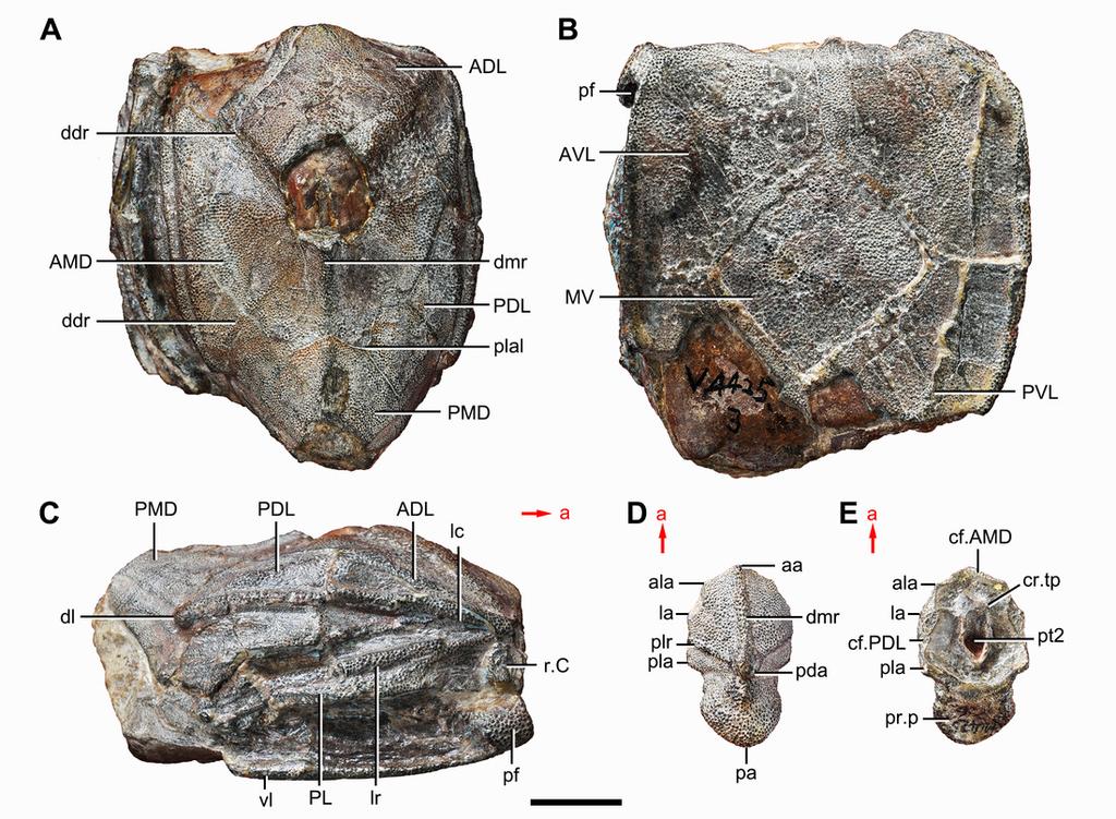
28 Figure 1 Holotype and paratype of Phymolepis cuifengshanensis. (A C) IVPP V4425.3, holotype, trunk shield in dorsal (A), ventral (B) and right lateral (C) views. (D E) IVPP V4425.6, paratype, PMD in dorsal (D) and ventral (E) views. Abbreviations: aa, anterior angle of PMD; ADL, anterior dorsolateral plate; ala, anterolateral angle of PMD; AMD, anterior median dorsal plate; AVL, anterior ventrolateral plate; cf.amd, area overlapping AMD; cf.pdl, area overlapping PDL; cr.tp, crista transversalis interna posterior; ddr, dorsal diagonal ridge of trunk shield; dl, dorsolateral ridge of trunk shield; dmr, dorsal median ridge; la, lateral angle of PMD; lc, main lateral line canal; lr, lateral ridge of lateral wall of trunk shield; MV, median ventral plate; pa, posterior angle of PMD; pda, posterior dorsal angle; PDL, posterior dorsolateral plate; pf, pectoral fossa; PL, posterior lateral plate; pla, posterolateral angle of PMD; plal, posterolateral angle of AMD; plr, posterior lateral ridge of PMD; PMD, posterior median dorsal plate; pr.p, posterior process of PMD; pt2, posterior ventral pit of dorsal wall of trunk shield; PVL, posterior ventrolateral plate; r.c, ridge caused by Chang s apparatus; vl, ventrolateral ridge of trunk shield. Red arrow represents the direction of the specimen: a, anterior direction. Scale bar equals 1 cm.
29
30 Figure 2 Paratype of Phymolepis cuifengshanensis (IVPP V4425.1). (A) Dorsal view. (B) Ventral view. (C) Right lateral view. (D E) Anterior view, photo (D) and drawing (E). Abbreviations: ADL, anterior dorsolateral plate; AMD, anterior median dorsal plate; AVL, anterior ventrolateral plate; cit, crista transversalis interna anterior; ddr, dorsal diagonal ridge of trunk shield; dma, tergal angle of trunk shield; dmr, dorsal median ridge of trunk shield; f.ca, fossa for neck-joint; iar, infra-articular ridge; lc, main lateral line canal; MV, median ventral plate; o.c, opening of Chang s apparatus; pbl, postbranchial lamina; PDL, posterior dorsolateral plate; PL, posterior lateral plate; pnoa, postnuchal ornamented corner of ADL; PVL, posterior ventrolateral plate; r.c, ridge caused by Chang s apparatus; sar, supraarticular ridge. Red arrow represents the direction of the specimen: a, anterior direction. Scale bars equal 1 cm.

31
32 Figure 3 Paratype of Phymolepis cuifengshanensis (IVPP V4425.2). (A) Dorsal view. (B) Ventral view. (C) Right lateral view. (D) Anterior view. Abbreviations: ADL, anterior dorsolateral plate; AMD, anterior median dorsal plate; AVL, anterior ventrolateral plate; ddr, dorsal diagonal ridge of trunk shield; dl, dorsolateral ridge of trunk shield; dma, tergal angle; dmr, dorsal median ridge; dtr, dorsal transverse ridge of trunk shield; La, lateral plate; lr, lateral ridge of lateral wall of trunk shield; MV, median ventral plate; Nu, nuchal plate; or, oblique ridge of lateral wall of trunk shield; orb, orbital fenestra; PDL, posterior dorsolateral plate; PL, posterior lateral plate; PM, postmarginal plate; pmc, postmarginal sensory canal; pnoa, postnuchal ornamented corner of ADL; PNu, paranuchal plate; PP, postpineal plate; PVL, posterior ventrolateral plate; SL, semilunar plate; vl, ventrolateral ridge of trunk shield. Scale bar equals 1 cm.

33

34 Figure 4 Yunnanolepis parvus (IVPP V4425.7). (A) Dorsal view. (B) Ventral view. (C) Anterior view. (D) Right lateral view. (E) Left lateral view. Abbreviations: ADL, anterior dorsolateral plate; AMD, anterior median dorsal plate; AVL, anterior ventrolateral plate; cit, crista transversalis interna anterior; dma, tergal angle; dmr, dorsal median ridge; pbl, postbranchial lamina; PDL, posterior dorsolateral plate; PL, posterior lateral plate; PMD, posterior median dorsal plate; p.pf, plates of pectoral fin; PVL, posterior ventrolateral plate. Red arrow represents the direction of the specimen: a, anterior direction; p, posterior direction. Scale bar equals 5 mm.
 Ventral view. (C) Right lateral view. Stripped lines delineate the unknown part.")
35 Figure 5 Outline restoration of the dermal shield of Phymolepis cuifengshanensis. (A) Dorsal view. (B) Ventral view. (C) Right lateral view. Stripped lines delineate the unknown part. Scale bar equals 5 mm.

36 Figure 6 Life reconstruction of Phymolepis cuifengshanensis. Artwork by Dinghua Yang.
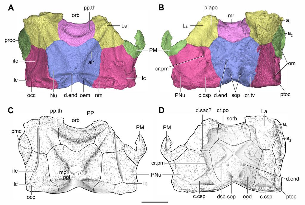
37 Figure 7 Head shield of Phymolepis cuifengshanensis (IVPP V4425.2) based on high-resolution CT. (A B) Three-dimensional reconstructions in dorsal (A) and ventral (B) views. (C D) Interpretative drawings in dorsal (C) and ventral (D) views. Abbreviations: a 1, a 2, attachment areas for the dermal operculum on the lateral and paranuchal plates, respectively; alr, anterior lateral ridge on head shield; c.csp, cavity for cranio-spinal process; cr.pm, paramarginal crista; cr.po, postorbital crista; cr.tv, transverse nuchal crista; d.end, opening for endolymphatic duct; d.sac, depression for sacculus; dsc, depression caused by semicircular canal; ifc, infraorbital sensory canal; La, lateral plate; lc, main lateral line canal; mpl, middle pit-line; mr, median ridge of postpineal plate; nm, obtected nuchal margin; Nu, nuchal plate; occ, occipital cross commissure; oem, median occipital eminence; om, obstantic margin of head shield; ood, otico-occipital depression of head shield; orb, orbital fenestra; p.apo, anterior postorbital process; PM, postmarginal plate; pmc, postmarginal sensory canal ; PNu, paranuchal plate; PP, postpineal plate; plc, posterolateral corner of head shield; ppl, posterior pit-line; pp.th, postpineal thickening; ptoc, postobstantic corner of paranuchal plate; sop, supraoccipital pit of head shield; sorb, suborbital fenestra. Scale bar equals 5 mm.
38
 Semitransparent Nu in lateral view. (D E) Semi-transparent right PNu in ventral (D) and posterior (E) views. (F) Right PNu in left lateral view. Abbreviations: cr.pm, paramarginal crista; cr.")
39 Figure 8 Cavities within the head shield of Phymolepis cuifengshanensis (IVPP V4425.2) based on high-resolution CT. (A) Semi-transparent Nu in dorsal view. (B) Transparent Nu in anterior view. (C) Semitransparent Nu in lateral view. (D E) Semi-transparent right PNu in ventral (D) and posterior (E) views. (F) Right PNu in left lateral view. Abbreviations: cr.pm, paramarginal crista; cr.tv, transverse nuchal crista; c.vg, cavity for vagal process, d.end, opening for endolymphatic duct; Nu, nuchal plate; PNu, paranuchal plate. Red arrow represents the direction of the specimen; a, anterior direction; d, dorsal direction; l, left direction; v, ventral direction. Scale bar equals 2 mm.
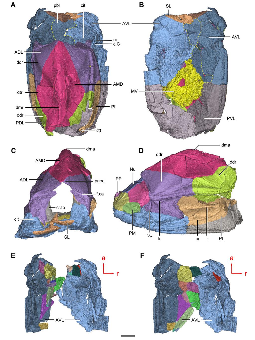
40 Figure 9 Phymolepis cuifengshanensis (IVPP V4425.2) based on high-resolution CT. (A C) Trunk shield in dorsal (A), ventral (B) and anterior (C) views. (D) Head and trunk shields in right lateral view. (E F) AVL plates and their displaced fragments as preserved (E) and restored (F). Yellow dash lines in (B) delimit restored portions. Abbreviations: ADL: anterior dorsolateral plate; AMD, anterior median dorsal plate; AVL, anterior ventrolateral plate; c.c; cavity of Chang s apparatus; cg, caudal groove of trunk shield; cit, crista transversalis interna anterior; cr.tp, crista transversalis interna posterior; ddr, dorsal diagonal ridge of trunk shield; dma, tergal angle of trunk shield; dmr, dorsal median ridge of trunk shield; dtr, dorsal transverse ridge of trunk shield; f.ca, fossa for neck-joint; lc, main lateral line canal; lr, lateral ridge of lateral wall of trunk shield; MV, median ventral plate; Nu, nuchal plate; or, oblique ridge of lateral wall of trunk shield; PDL, posterior dorsolateral plate; PL, posterior lateral plate ; PM, postmarginal plate; pnoa, postnuchal ornamented corner of ADL; PP, postpineal plate; PVL, posterior ventrolateral plate; pbl, postbranchial lamina; rc, rostrocaudal canal; r.c, ridge caused by Chang s apparatus; SL, semilunar plate. Red arrow represents the direction of the specimen: a, anterior direction; r, right direction. Scale bar equals to 5 mm.
41
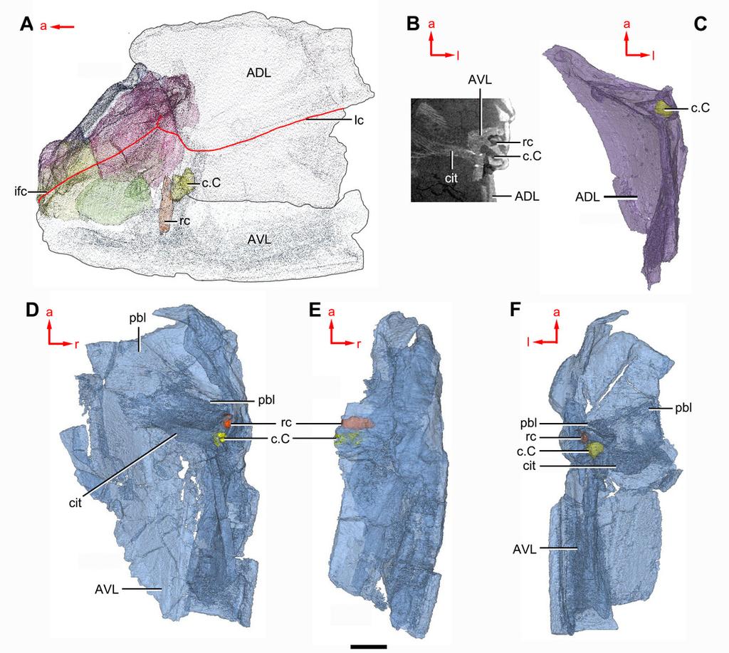
42 Figure 10 Phymolepis cuifengshanensis (IVPP V4425.2) based on high-resolution CT. (A) Head shield and anterior portion of trunk shield. (B) Axial section through the left AVL in CT slice, showing the positions of the internal cavity of Chang s apparatus and the rostrocaudal canal. (C) Semi-transparent left ADL in ventral view. (D E) Semi-transparent right AVL in dorsal (D) and lateral (E) views. (F) Semi-transparent left AVL in dorsal view. Abbreviations: ADL, anterior dorsolateral plate; AVL, anterior ventrolateral plate; c.c; cavity of Chang s apparatus; cit, crista transversalis interna anterior; ifc, infraorbital sensory canal; lc, main lateral line canal; pbl, postbranchial lamina; rc, rostrocaudal canal. Red arrow represents the direction of the specimen: a, anterior direction; l, left direction; r, right direction. Scale bar equals 3mm.
43
 Lateroventral fossa in lateral view. (D) Transverse section through the left caudal groove in CT slice. (E) Axial section through the right caudal groove in CT slice.")
44 Figure 11 Phymolepis cuifengshanensis (IVPP V4425.2) based on high-resolution CT. (A) AMD in ventral view. (B) Transverse section through the lateroventral fossa in CT slice. (C) Lateroventral fossa in lateral view. (D) Transverse section through the left caudal groove in CT slice. (E) Axial section through the right caudal groove in CT slice. (F) Left caudal groove in lateral view. Abbreviations: AMD, anterior median dorsal plate; AVL, anterior ventrolateral plate; cf.adl, area overlapping ADL; cf.pdl, area overlapping PDL; cg, caudal groove of trunk shield; cr.tp, crista transversalis interna posterior; f.lv, lateroventral fossa of trunk shield; lal, lateral angle of AMD; ms, median septum; PDL, posterior dorsolateral plate; PL, posterior lateral plate; plal, posterolateral angle of AMD; prv1, anterior ventral process of dorsal wall of trunk shield; pt1, anterior ventral pit of dorsal wall of trunk shield; PVL, posterior ventrolateral plate; wa, outer wall of caudal groove. Red arrow represents the direction of the specimen: a, anterior direction; d, dorsal direction; r, right direction. Scale bars equal 3 mm.

45 Figure 12 Head shields of Yunnanolepis chii in visceral view. (A) IVPP V (B) IVPP V Abbreviations: c.csp, cavity for cranio-spinal process; cr.pm, paramarginal crista; cr.po, postorbital crista; cr.tv, transverse nuchal crista; d.end, opening for endolymphatic duct; d.sac, depression for sacculus; dsc, depression caused by semicircular canal; fm, unpaired insertion fossa on head shield for levator muscles; mr, medial ridge of postpineal plate; ood, otico-occipital depression of head shield; p.apo, anterior postorbital process; r.spr, subpremedian ridge; sop, supraoccipital pit of head shield; tlg, transverse lateral groove of head shield. Scale bars equal to 5 mm.
46 Figure 13(on next page) Phylogenetic results of antiarchs based on a revised data matrix. (A) Strict consensus tree of 726 parsimonious trees (tree length = 179; consistency index = , homoplasy index = , retention index = , rescaled consistency index = ). Numbers above and below nodes represent bootstrap values ( 50% are shown) and Bremer decay indices, respectively. (B) 50 % majority-rule consensus tree of 726 parsimonious trees based on the same dataset as in (A). Numbers next to nodes indicate the percentage of the shortest trees in which the partition is supported (100% are not shown).
47 A Euantiarcha Kujdanowiaspis Romundina Vanchienolepis Chuchinolepis Heteroyunnanolepis Zhanjilepis Yunnanolepis Parayunnanolepis Mizia Phymolepis Minicrania Liujiangolepis Xichonolepis Dayaoshania Grenfellaspis Sinolepis Hohsienolepis Wudinolepis Microbrachius Tenizolepis Dianolepis Vietnamaspis Bothriolepis Grossilepis Briagalepis Wufengshania Monarolepis Luquanolepis Nawagiaspis Jiangxilepis Ningxialepis Kirgisolepis Hunanolepis Wurungulepis Byssacanthus Stegolepis Pambulaspis Asterolepis Remigolepis Pterichthyodes Sherbonaspis Gerdalepis Grossaspis Lepadolepis Yunnanolepidoidei Sinolepididdae Bothriolepidoidei Asterolepidoidei B
48 Figure 14(on next page) Phylogenetic results of antiarchs and visceral surface conditions of head shield among major antiarch subgroups. (A) One of the most parsimonious trees with node numbers defining various clades. Named nodes: 1, Antiarcha; 2, Yunnanolepidoidei; 11, Sinolepididae; 15, Euantiarcha; 16, Microbrachiidae; 26, Asterolepidoidei. (B) Restorations of the head shields in ventral view to show endocranial character transformations, redrawn from Ritchie et al. (1992), Young (1983), Stensiö (1948) and (1969). Vertical bars on the right side show the longitudinal proportion of otico-occipital region of endocranium on the head shied (blue region), the location of the confluence of semicircular canals (orange circle), the location of the internal pore for endolymphatic duct (purple circle). Abbreviations: c.csp, cavity for cranio-spinal process; cr.im, inframarginal crista; cr.tv, transverse nuchal crista; cro, median occipital crista of head shield; d.end, opening for endolymphatic duct; dsc, depression caused by semicircular canal; f.cu, cucullaris fossa; fm, unpaired insertion fossa on head shield for levator muscles; p.apo, anterior postorbital process; prnm, posterior process of head shield; sop, supraoccipital pit of head shield; sot, supraotic thickening of head shield; tlg, transverse lateral groove of head shield.


49 A Mizia Phymolepis Minicrania Liujiangolepis Xichonolepis Dayaoshania Grenfellaspis Sinolepis Hohsienolepis Wudinolepis Microbrachius Tenizolepis Dianolepis Vietnamaspis Bothriolepis Grossilepis Briagalepis Wufengshania Monarolepis Luquanolepis Nawagiaspis Jiangxilepis Ningxialepis Kirgisolepis Hunanolepis Wurungulepis Byssacanthus Stegolepis Pambulaspis Asterolepis Remigolepis Pterichthyodes 39 Gerdalepis 40 Grossaspis 41 Lepadolepis B Kujdanowiaspis Romundina Vanchienolepis Chuchinolepis Heteroyunnanolepis Zhanjilepis Yunnanolepis Parayunnanolepis Sherbonaspis d.end tlg p.apo dsc c.csp sop cr.tv Phymolepis d.end fm sop cr.tv Grenfellaspis p.apo tlg dsc d.end sot fm d.end prnm cro Bothriolepis prnm cr.tv cr.tv p.apo cr.im f.cu sot Asterolepis tlg sot d.end prnm cr.tv Pambulaspis
The phylogeny of antiarch placoderms. Sarah Kearsley Geology 394 Senior Thesis
 The phylogeny of antiarch placoderms Sarah Kearsley Geology 394 Senior Thesis Abstract The most comprehensive phylogenetic study of antiarchs to date (Zhu, 1996) included information not derived from observation.
Exceptional fossil preservation demonstrates a new mode of axial skeleton elongation in early ray-finned fishes
 Supplementary Information Exceptional fossil preservation demonstrates a new mode of axial skeleton elongation in early ray-finned fishes Erin E. Maxwell, Heinz Furrer, Marcelo R. Sánchez-Villagra Supplementary
SUPPLEMENTARY INFORMATION
 Supplementary Information for Devonian arthrodire embryos and the origin of internal fertilization in vertebrates John A. Long 1-3, Kate Trinajstic 4, and Zerina Johanson 5 1 Museum Victoria, PO Box 666,
HONR219D Due 3/29/16 Homework VI
 Part 1: Yet More Vertebrate Anatomy!!! HONR219D Due 3/29/16 Homework VI Part 1 builds on homework V by examining the skull in even greater detail. We start with the some of the important bones (thankfully
AMERICAN MUSEUM NOVITATES Published by
 AMERICAN MUSEUM NOVITATES Published by Number 782 THE AmzRICAN MUSEUM OF NATURAL HISTORY Feb. 20, 1935 New York City 56.81, 7 G (68) A NOTE ON THE CYNODONT, GLOCHINODONTOIDES GRACILIS HAUGHTON BY LIEUWE
LABORATORY EXERCISE 6: CLADISTICS I
 Biology 4415/5415 Evolution LABORATORY EXERCISE 6: CLADISTICS I Take a group of organisms. Let s use five: a lungfish, a frog, a crocodile, a flamingo, and a human. How to reconstruct their relationships?
Phylogeny Reconstruction
 Phylogeny Reconstruction Trees, Methods and Characters Reading: Gregory, 2008. Understanding Evolutionary Trees (Polly, 2006) Lab tomorrow Meet in Geology GY522 Bring computers if you have them (they will
Postilla PEABODY MUSEUM OF NATURAL HISTORY YALE UNIVERSITY NEW HAVEN, CONNECTICUT, U.S.A.
 Postilla PEABODY MUSEUM OF NATURAL HISTORY YALE UNIVERSITY NEW HAVEN, CONNECTICUT, U.S.A. Number 117 18 March 1968 A 7DIAPSID (REPTILIA) PARIETAL FROM THE LOWER PERMIAN OF OKLAHOMA ROBERT L. CARROLL REDPATH
2. Skull, total length versus length of the presacral vertebral column: (0); extremely elongated neck (e.g. Tanystropheus longobardicus).
; extremely elongated neck (e.g. Tanystropheus longobardicus).") Character list of the taxon-character data set 1. Skull and lower jaws, interdental plates: absent (0); present, but restricted to the anterior end of the dentary (1); present along the entire alveolar
Character list of the taxon-character data set 1. Skull and lower jaws, interdental plates: absent (0); present, but restricted to the anterior end of the dentary (1); present along the entire alveolar
muscles (enhancing biting strength). Possible states: none, one, or two.
. Possible states: none, one, or two.") Reconstructing Evolutionary Relationships S-1 Practice Exercise: Phylogeny of Terrestrial Vertebrates In this example we will construct a phylogenetic hypothesis of the relationships between seven taxa
Reconstructing Evolutionary Relationships S-1 Practice Exercise: Phylogeny of Terrestrial Vertebrates In this example we will construct a phylogenetic hypothesis of the relationships between seven taxa
SUPPLEMENTARY ONLINE MATERIAL FOR. Nirina O. Ratsimbaholison, Ryan N. Felice, and Patrick M. O connor
 http://app.pan.pl/som/app61-ratsimbaholison_etal_som.pdf SUPPLEMENTARY ONLINE MATERIAL FOR Nirina O. Ratsimbaholison, Ryan N. Felice, and Patrick M. O connor Ontogenetic changes in the craniomandibular
LABORATORY EXERCISE 7: CLADISTICS I
 Biology 4415/5415 Evolution LABORATORY EXERCISE 7: CLADISTICS I Take a group of organisms. Let s use five: a lungfish, a frog, a crocodile, a flamingo, and a human. How to reconstruct their relationships?
Title: Phylogenetic Methods and Vertebrate Phylogeny
 Title: Phylogenetic Methods and Vertebrate Phylogeny Central Question: How can evolutionary relationships be determined objectively? Sub-questions: 1. What affect does the selection of the outgroup have
TRACHEMYS SCULPTA. A nearly complete articulated carapace and plastron of an Emjdd A NEAKLY COMPLETE SHELL OF THE EXTINCT TURTLE,
 A NEAKLY COMPLETE SHELL OF THE EXTINCT TURTLE, TRACHEMYS SCULPTA By Charles W. Gilmore Curator of Vertebrate Paleontology, United States National Museum INTRODUCTION A nearly complete articulated carapace
ONLINE APPENDIX 1. Morphological phylogenetic characters scored in this paper. See Poe (2004) for
 for") ONLINE APPENDIX Morphological phylogenetic characters scored in this paper. See Poe () for detailed character descriptions, citations, and justifications for states. Note that codes are changed from a
ONLINE APPENDIX Morphological phylogenetic characters scored in this paper. See Poe () for detailed character descriptions, citations, and justifications for states. Note that codes are changed from a
A new species of Hsisosuchus (Mesoeucrocodylia) from Dashanpu, Zigong Municipality, Sichuan Province
 from Dashanpu, Zigong Municipality, Sichuan Province") A new species of Hsisosuchus (Mesoeucrocodylia) from Dashanpu, Zigong Municipality, Sichuan Province Yuhui Gao (Zigong Dinosaur Museum) Vertebrata PalAsiatica Volume 39, No. 3 July, 2001 pp. 177-184 Translated
A new species of Hsisosuchus (Mesoeucrocodylia) from Dashanpu, Zigong Municipality, Sichuan Province Yuhui Gao (Zigong Dinosaur Museum) Vertebrata PalAsiatica Volume 39, No. 3 July, 2001 pp. 177-184 Translated
First Ornithomimid (Theropoda, Ornithomimosauria) from the Upper Cretaceous Djadokhta Formation of Tögrögiin Shiree, Mongolia
 from the Upper Cretaceous Djadokhta Formation of Tögrögiin Shiree, Mongolia") First Ornithomimid (Theropoda, Ornithomimosauria) from the Upper Cretaceous Djadokhta Formation of Tögrögiin Shiree, Mongolia Tsogtbaatar Chinzorig¹, ³ *, Yoshitsugu Kobayashi², Khishigjav Tsogtbaatar³,
First Ornithomimid (Theropoda, Ornithomimosauria) from the Upper Cretaceous Djadokhta Formation of Tögrögiin Shiree, Mongolia Tsogtbaatar Chinzorig¹, ³ *, Yoshitsugu Kobayashi², Khishigjav Tsogtbaatar³,
Vol. XIV, No. 1, March, The Larva and Pupa of Brontispa namorikia Maulik (Coleoptera: Chrysomelidae: Hispinae) By S.
 By S.") Vol. XIV, No. 1, March, 1950 167 The Larva and Pupa of Brontispa namorikia Maulik (Coleoptera: Chrysomelidae: Hispinae) By S. MAULIK BRITISH MUSEUM (NATURAL HISTORY) (Presented by Mr. Van Zwaluwenburg
Vol. XIV, No. 1, March, 1950 167 The Larva and Pupa of Brontispa namorikia Maulik (Coleoptera: Chrysomelidae: Hispinae) By S. MAULIK BRITISH MUSEUM (NATURAL HISTORY) (Presented by Mr. Van Zwaluwenburg
The family Gnaphosidae is a large family
 Pakistan J. Zool., vol. 36(4), pp. 307-312, 2004. New Species of Zelotus Spider (Araneae: Gnaphosidae) from Pakistan ABIDA BUTT AND M.A. BEG Department of Zoology, University of Agriculture, Faisalabad,
BREVIORA LEUCOLEPIDOPA SUNDA GEN. NOV., SP. NOV. (DECAPODA: ALBUNEIDAE), A NEW INDO-PACIFIC SAND CRAB. Ian E. Efford 1
, A NEW INDO-PACIFIC SAND CRAB. Ian E. Efford 1") ac lc BREVIORA CAMBRIDGE, MASS. 30 APRIL, 1969 NUMBER 318 LEUCOLEPIDOPA SUNDA GEN. NOV., SP. NOV. (DECAPODA: ALBUNEIDAE), A NEW INDO-PACIFIC SAND CRAB Ian E. Efford 1 ABSTRACT. Leucolepidopa gen. nov.
ac lc BREVIORA CAMBRIDGE, MASS. 30 APRIL, 1969 NUMBER 318 LEUCOLEPIDOPA SUNDA GEN. NOV., SP. NOV. (DECAPODA: ALBUNEIDAE), A NEW INDO-PACIFIC SAND CRAB Ian E. Efford 1 ABSTRACT. Leucolepidopa gen. nov.
AUSTRALIAN MUSEUM SCIENTIFIC PUBLICATIONS
 AUSTRALIAN MUSEUM SCIENTIFIC PUBLICATIONS Riek, E. F., 1964. Merostomoidea (Arthropoda, Trilobitomorpha) from the Australian Middle Triassic. Records of the Australian Museum 26(13): 327 332, plate 35.
Introduction to phylogenetic trees and tree-thinking Copyright 2005, D. A. Baum (Free use for non-commercial educational pruposes)
") Introduction to phylogenetic trees and tree-thinking Copyright 2005, D. A. Baum (Free use for non-commercial educational pruposes) Phylogenetics is the study of the relationships of organisms to each other.
Introduction to phylogenetic trees and tree-thinking Copyright 2005, D. A. Baum (Free use for non-commercial educational pruposes) Phylogenetics is the study of the relationships of organisms to each other.
Introduction to Cladistic Analysis
 3.0 Copyright 2008 by Department of Integrative Biology, University of California-Berkeley Introduction to Cladistic Analysis tunicate lamprey Cladoselache trout lungfish frog four jaws swimbladder or
Juehuaornis gen. nov.
 34 1 2015 3 GLOBAL GEOLOGY Vol. 34 No. 1 Mar. 2015 1004 5589 2015 01 0007 05 Juehuaornis gen. nov. 1 1 1 2 1. 110034 2. 110034 70% Juehuaornis zhangi gen. et sp. nov Q915. 4 A doi 10. 3969 /j. issn. 1004-5589.
DESCRIPTIONS OF THREE NEW SPECIES OF PETALOCEPHALA STÅL, 1853 FROM CHINA (HEMIPTERA: CICADELLIDAE: LEDRINAE) Yu-Jian Li* and Zi-Zhong Li**
 Yu-Jian Li* and Zi-Zhong Li**") 499 DESCRIPTIONS OF THREE NEW SPECIES OF PETALOCEPHALA STÅL, 1853 FROM CHINA (HEMIPTERA: CICADELLIDAE: LEDRINAE) Yu-Jian Li* and Zi-Zhong Li** * Institute of Entomology, Guizhou University, Guiyang, Guizhou
499 DESCRIPTIONS OF THREE NEW SPECIES OF PETALOCEPHALA STÅL, 1853 FROM CHINA (HEMIPTERA: CICADELLIDAE: LEDRINAE) Yu-Jian Li* and Zi-Zhong Li** * Institute of Entomology, Guizhou University, Guiyang, Guizhou
A new basal sauropodiform dinosaur from the Lower Jurassic of Yunnan Province, China
 SUPPLEMENTARY INFORMATION A new basal sauropodiform dinosaur from the Lower Jurassic of Yunnan Province, China Ya-Ming Wang 1, Hai-Lu You 2,3 *, Tao Wang 4 1 School of Earth Sciences and Resources, China
Species: Panthera pardus Genus: Panthera Family: Felidae Order: Carnivora Class: Mammalia Phylum: Chordata
 CHAPTER 6: PHYLOGENY AND THE TREE OF LIFE AP Biology 3 PHYLOGENY AND SYSTEMATICS Phylogeny - evolutionary history of a species or group of related species Systematics - analytical approach to understanding
Diurus, Pascoe. sp. 1). declivity of the elytra, but distinguished. Length (the rostrum and tails 26 included) mm. Deep. exception
. declivity of the elytra, but distinguished. Length (the rostrum and tails 26 included) mm. Deep. exception") 210 DIURUS ERYTIIROPUS. NOTE XXVI. Three new species of the Brenthid genus Diurus, Pascoe DESCRIBED BY C. Ritsema+Cz. 1. Diurus erythropus, n. sp. 1). Allied to D. furcillatus Gylh. ²) by the short head,
210 DIURUS ERYTIIROPUS. NOTE XXVI. Three new species of the Brenthid genus Diurus, Pascoe DESCRIBED BY C. Ritsema+Cz. 1. Diurus erythropus, n. sp. 1). Allied to D. furcillatus Gylh. ²) by the short head,
Biology 340 Comparative Embryology Lecture 12 Dr. Stuart Sumida. Evo-Devo Revisited. Development of the Tetrapod Limb
 Biology 340 Comparative Embryology Lecture 12 Dr. Stuart Sumida Evo-Devo Revisited Development of the Tetrapod Limb Limbs whether fins or arms/legs for only in particular regions or LIMB FIELDS. Primitively
1 EEB 2245/2245W Spring 2014: exercises working with phylogenetic trees and characters
 1 EEB 2245/2245W Spring 2014: exercises working with phylogenetic trees and characters 1. Answer questions a through i below using the tree provided below. a. The sister group of J. K b. The sister group
SOME NEW AMERICAN PYCNODONT FISHES.
 SOME NEW AMERICAN PYCNODONT FISHES. By James Williams Gidley, Assistant Curator of Fossil Mammals, United States National Museum. In the United States National Museum are several specimens representing
Line 136: "Macroelongatoolithus xixiaensis" should be "Macroelongatoolithus carlylei" (the former is a junior synonym of the latter).
.") Reviewers' comments: Reviewer #1 (Remarks to the Author): This is a superb, well-written manuscript describing a new dinosaur species that is intimately associated with a partial nest of eggs classified
Reviewers' comments: Reviewer #1 (Remarks to the Author): This is a superb, well-written manuscript describing a new dinosaur species that is intimately associated with a partial nest of eggs classified
SUPPLEMENTARY INFORMATION
 doi:10.1038/nature12980 Terminology: nasal capsule vs. rostral capsule Different anatomical terms have been used in the past to denote the distinct fissure-bounded nasal region of placoderms. Here we apply
A new species of Antinia PASCOE from Burma (Coleoptera: Curculionidae: Entiminae)
") Genus Vol. 14 (3): 413-418 Wroc³aw, 15 X 2003 A new species of Antinia PASCOE from Burma (Coleoptera: Curculionidae: Entiminae) JAROS AW KANIA Zoological Institute, University of Wroc³aw, Sienkiewicza
Genus Vol. 14 (3): 413-418 Wroc³aw, 15 X 2003 A new species of Antinia PASCOE from Burma (Coleoptera: Curculionidae: Entiminae) JAROS AW KANIA Zoological Institute, University of Wroc³aw, Sienkiewicza
SOME LITTLE-KNOWN FOSSIL LIZARDS FROM THE
 PROCEEDINGS OF THE UNITED STATES NATIONAL MUSEUM issued SWsK \ {^^m ^V ^^ SMITHSONIAN INSTITUTION U. S. NATIONAL MUSEUM Vol. 91 Washington : 1941 No. 3124 SOME LITTLE-KNOWN FOSSIL LIZARDS FROM THE OLIGOCENE
UPOGEBIA LINCOLNI SP. NOV. (DECAPODA, THALASSINIDEA, UPOGEBIIDAE) FROM JAVA, INDONESIA
 FROM JAVA, INDONESIA") NOTES AND NEWS UPOGEBIA LINCOLNI SP. NOV. (DECAPODA, THALASSINIDEA, UPOGEBIIDAE) FROM JAVA, INDONESIA BY NGUYEN NGOC-HO i) Faculty of Science, University of Saigon, Vietnam Among material recently collected
NOTES AND NEWS UPOGEBIA LINCOLNI SP. NOV. (DECAPODA, THALASSINIDEA, UPOGEBIIDAE) FROM JAVA, INDONESIA BY NGUYEN NGOC-HO i) Faculty of Science, University of Saigon, Vietnam Among material recently collected
290 SHUFELDT, Remains of Hesperornis.
 290 SHUFELDT, Remains of Hesperornis. [ Auk [July THE FOSSIL REMAINS OF A SPECIES OF HESPERORNIS FOUND IN MONTANA. BY R. W. SHUFELD% M.D. Plate XI7III. ExR,¾ in November, 1914, Mr. Charles W. Gihnore,
Williston, and as there are many fairly good specimens in the American
 56.81.7D :14.71.5 Article VII.- SOME POINTS IN THE STRUCTURE OF THE DIADECTID SKULL. BY R. BROOM. The skull of Diadectes has been described by Cope, Case, v. Huene, and Williston, and as there are many
INQUIRY & INVESTIGATION
 INQUIRY & INVESTIGTION Phylogenies & Tree-Thinking D VID. UM SUSN OFFNER character a trait or feature that varies among a set of taxa (e.g., hair color) character-state a variant of a character that occurs
A NEW GENUS AND SPECIES OF AMERICAN THEROMORPHA
 A NEW GENUS AND SPECIES OF AMERICAN THEROMORPHA MYCTEROSAURUS LONGICEPS S. W. WILLISTON University of Chicago The past summer, Mr. Herman Douthitt, of the University of Chicago paleontological expedition,
TWO NEW SPECIES OF ACUTIGEBIA (CRUSTACEA: DECAPODA: GEBIIDEA: UPOGEBIIDAE) FROM THE SOUTH CHINA SEA
 FROM THE SOUTH CHINA SEA") THE RAFFLES BULLETIN OF ZOOLOGY 2013 61(2): 571 577 Date of Publication: 30 Aug.2013 National University of Singapore TWO NEW SPECIES OF ACUTIGEBIA (CRUSTACEA: DECAPODA: GEBIIDEA: UPOGEBIIDAE) FROM THE
THE RAFFLES BULLETIN OF ZOOLOGY 2013 61(2): 571 577 Date of Publication: 30 Aug.2013 National University of Singapore TWO NEW SPECIES OF ACUTIGEBIA (CRUSTACEA: DECAPODA: GEBIIDEA: UPOGEBIIDAE) FROM THE
A NEW SPECIES OF A USTROLIBINIA FROM THE SOUTH CHINA SEA AND INDONESIA (CRUSTACEA: BRACHYURA: MAJIDAE)
") 69 C O a g r ^ j^a RAFFLES BULLETIN OF ZOOLOGY 1992 40(1): 69-73 A NEW SPECIES OF A USTROLIBINIA FROM THE SOUTH CHINA SEA AND INDONESIA (CRUSTACEA: BRACHYURA: MAJIDAE) H P Waener SMITHSONIAN INSTITUTE
69 C O a g r ^ j^a RAFFLES BULLETIN OF ZOOLOGY 1992 40(1): 69-73 A NEW SPECIES OF A USTROLIBINIA FROM THE SOUTH CHINA SEA AND INDONESIA (CRUSTACEA: BRACHYURA: MAJIDAE) H P Waener SMITHSONIAN INSTITUTE
A NEW AUSTROSQUILLA (STOMATOPODA) FROM THE
 FROM THE") A NEW AUSTROSQUILLA (STOMATOPODA) FROM THE MARQUESAS ISLANDS BY ALAIN MICHEL Centre O.R.S.T.O.M., Noumea, New Caledonia and RAYMOND B. MANNING Smithsonian Institution, Washington, U.S.A. The At s,tstrosqzlilla
A NEW AUSTROSQUILLA (STOMATOPODA) FROM THE MARQUESAS ISLANDS BY ALAIN MICHEL Centre O.R.S.T.O.M., Noumea, New Caledonia and RAYMOND B. MANNING Smithsonian Institution, Washington, U.S.A. The At s,tstrosqzlilla
Evolution and development of the synarcual in early vertebrates
 DOI 10.1007/s00435-012-0169-9 ORIGINAL PAPER Evolution and development of the synarcual in early vertebrates Zerina Johanson Kate Trinajstic Robert Carr Alex Ritchie Received: 14 March 2012 / Revised:
SUPPLEMENTARY INFORMATION
 Table of Contents Part A. Supplementary Notes 1) Geological Provenance and Stratigraphy 2) Taxonomic Notes and History 3) Lateral Processes of Early Gnathostome Neurocrania Part B. Phylogenetic Analyses
Reprinted from: CRUSTACEANA, Vol. 32, Part 2, 1977 LEIDEN E. J. BRILL
 Reprinted from: CRUSTACEANA, Vol. 32, Part 2, 1977 LEIDEN E. J. BRILL NOTES AND NEWS 207 ALPHE0PS1S SHEARMII (ALCOCK & ANDERSON): A NEW COMBINATION WITH A REDESCRIPTION OF THE HOLOTYPE (DECAPODA, ALPHEIDAE)
IDENTIFICATION / GENERAL CHARACTERISTICS OF TICK GENERA (HARD AND SOFT TICKS)
") Ticks Tick identification Authors: Prof Maxime Madder, Prof Ivan Horak, Dr Hein Stoltsz Licensed under a Creative Commons Attribution license. IDENTIFICATION / GENERAL CHARACTERISTICS OF TICK GENERA (HARD
Ticks Tick identification Authors: Prof Maxime Madder, Prof Ivan Horak, Dr Hein Stoltsz Licensed under a Creative Commons Attribution license. IDENTIFICATION / GENERAL CHARACTERISTICS OF TICK GENERA (HARD
DEVELOPMENT OF THE HEAD AND NECK PLACODES
 DEVELOPMENT OF THE HEAD AND NECK Placodes and the development of organs of special sense L. Moss-Salentijn PLACODES Localized thickened areas of specialized ectoderm, lateral to the neural crest, at the
Anatomy. Name Section. The Vertebrate Skeleton
 Name Section Anatomy The Vertebrate Skeleton Vertebrate paleontologists get most of their knowledge about past organisms from skeletal remains. Skeletons are useful for gleaning information about an organism
SUPPLEMENTARY INFORMATION
 In comparison to Proganochelys (Gaffney, 1990), Odontochelys semitestacea is a small turtle. The adult status of the specimen is documented not only by the generally well-ossified appendicular skeleton
A NEW SPECIES OF EXTINCT TURTLE FROM THE UPPER PLIOCENE OF IDAHO
 A NEW SPECIES OF EXTINCT TURTLE FROM THE UPPER PLIOCENE OF IDAHO By Charles W. Gilmore Curator, Division of Vertebrate Paleontology United States National Museum Among the fossils obtained bj^ the Smithsonian
NOTE XXXVIII. Three new species of the genus Helota DESCRIBED BY. C. Ritsema+Cz. is very. friend René Oberthür who received. Biet.
 Subshining; HELOTA MARIAE. 249 NOTE XXXVIII. Three new species of the genus Helota DESCRIBED BY C. Ritsema+Cz. The first of these species is very interesting as it belongs to the same section as the recently
Supporting Online Material for
 www.sciencemag.org/cgi/content/full/329/5998/1481/dc1 Supporting Online Material for Tyrannosaur Paleobiology: New Research on Ancient Exemplar Organisms Stephen L. Brusatte,* Mark A. Norell, Thomas D.
Bio 1B Lecture Outline (please print and bring along) Fall, 2006
 Fall, 2006") Bio 1B Lecture Outline (please print and bring along) Fall, 2006 B.D. Mishler, Dept. of Integrative Biology 2-6810, bmishler@berkeley.edu Evolution lecture #4 -- Phylogenetic Analysis (Cladistics) -- Oct.
Bio 1B Lecture Outline (please print and bring along) Fall, 2006 B.D. Mishler, Dept. of Integrative Biology 2-6810, bmishler@berkeley.edu Evolution lecture #4 -- Phylogenetic Analysis (Cladistics) -- Oct.
Supplementary Information (ZHU and YU: A primitive fish close to the common ancestor of tetrapods and lungfish)
") 1 Supplementary Information (ZHU and YU: A primitive fish close to the common ancestor of tetrapods and lungfish) ------------------------------------------ I. List of 158 characters used for phylogenetic
1 Supplementary Information (ZHU and YU: A primitive fish close to the common ancestor of tetrapods and lungfish) ------------------------------------------ I. List of 158 characters used for phylogenetic
These small issues are easily addressed by small changes in wording, and should in no way delay publication of this first- rate paper.
 Reviewers' comments: Reviewer #1 (Remarks to the Author): This paper reports on a highly significant discovery and associated analysis that are likely to be of broad interest to the scientific community.
SUPPLEMENTARY INFORMATION
 Character 155, interdental ridges. Absence of interdental ridge (0) shown in Parasaniwa wyomingensis (Platynota). Interdental ridges (1) shown in Coniophis precedens. WWW.NATURE.COM/NATURE 1 Character
TWO NEW SPECIES OF WATER MITES FROM OHIO 1-2
 TWO NEW SPECIES OF WATER MITES FROM OHIO 1-2 DAVID R. COOK Wayne State University, Detroit, Michigan ABSTRACT Two new species of Hydracarina, Tiphys weaveri (Acarina: Pionidae) and Axonopsis ohioensis
What are taxonomy, classification, and systematics?
 Topic 2: Comparative Method o Taxonomy, classification, systematics o Importance of phylogenies o A closer look at systematics o Some key concepts o Parts of a cladogram o Groups and characters o Homology
complex in cusp pattern. (3) The bones of the coyote skull are thinner, crests sharper and the
 The bones of the coyote skull are thinner, crests sharper and the") DISTINCTIONS BETWEEN THE SKULLS OF S AND DOGS Grover S. Krantz Archaeological sites in the United States frequently yield the bones of coyotes and domestic dogs. These two canines are very similar both
DISTINCTIONS BETWEEN THE SKULLS OF S AND DOGS Grover S. Krantz Archaeological sites in the United States frequently yield the bones of coyotes and domestic dogs. These two canines are very similar both
NOTE XVII. Dr. A.A.W. Hubrecht. which should he in accordance with. of my predecessors. alive or in excellent. further
 further either EUROPEAN NEMERTEANS. 93 NOTE XVII. New Species of European Nemerteans. First Appendix to Note XLIV, Vol. I BY Dr. A.A.W. Hubrecht In the above-mentioned note, published six months ago, several
1 EEB 2245/2245W Spring 2017: exercises working with phylogenetic trees and characters
 1 EEB 2245/2245W Spring 2017: exercises working with phylogenetic trees and characters 1. Answer questions a through i below using the tree provided below. a. Identify the taxon (or taxa if there is more
Geo 302D: Age of Dinosaurs LAB 4: Systematics Part 1
 Geo 302D: Age of Dinosaurs LAB 4: Systematics Part 1 Systematics is the comparative study of biological diversity with the intent of determining the relationships between organisms. Humankind has always
Mammalogy Lecture 8 - Evolution of Ear Ossicles
 Mammalogy Lecture 8 - Evolution of Ear Ossicles I. To begin, let s examine briefly the end point, that is, modern mammalian ears. Inner Ear The cochlea contains sensory cells for hearing and balance. -
Bulletin of Big Bend Paleo-Geo An Open Access Publication from Mosasaur Ranch Museum, Terlingua and Lajitas, Texas All rights reserved
 Bulletin of Big Bend Paleo-Geo An Open Access Publication from Mosasaur Ranch Museum, Terlingua and Lajitas, Texas All rights reserved This was a private report in 2003 on my thoughts on Platecarpus planifrons.
HAWAIIAN BIOGEOGRAPHY EVOLUTION ON A HOT SPOT ARCHIPELAGO EDITED BY WARREN L. WAGNER AND V. A. FUNK SMITHSONIAN INSTITUTION PRESS
 HAWAIIAN BIOGEOGRAPHY EVOLUTION ON A HOT SPOT ARCHIPELAGO EDITED BY WARREN L. WAGNER AND V. A. FUNK SMITHSONIAN INSTITUTION PRESS WASHINGTON AND LONDON 995 by the Smithsonian Institution All rights reserved
Cladistics (reading and making of cladograms)
") Cladistics (reading and making of cladograms) Definitions Systematics The branch of biological sciences concerned with classifying organisms Taxon (pl: taxa) Any unit of biological diversity (eg. Animalia,
Cladistics (reading and making of cladograms) Definitions Systematics The branch of biological sciences concerned with classifying organisms Taxon (pl: taxa) Any unit of biological diversity (eg. Animalia,
A Scanning Electron Microscopic Study of Eggshell Surface Topography of Leidynema portentosae and L. appendiculatum (Nematoda: Oxyuroidea)
") The Ohio State University Knowledge Bank kb.osu.edu Ohio Journal of Science (Ohio Academy of Science) Ohio Journal of Science: Volume 88, Issue 5 (December, 1988) 1988-12 A Scanning Electron Microscopic
The Ohio State University Knowledge Bank kb.osu.edu Ohio Journal of Science (Ohio Academy of Science) Ohio Journal of Science: Volume 88, Issue 5 (December, 1988) 1988-12 A Scanning Electron Microscopic
Fischthal and Kuntz (1964) reported the
 reported the") Zoological Studies 41(3): 283-287 (2002) Meristocotyle provitellaria sp. nov. (Digenea: Meristocotylidae) from Varanus salvator in China Wei Liu 1, Qing-Kui Li 2, Hsiu-Hui Shih 3 and Zhao-Zhi Qiu 1, *
Zoological Studies 41(3): 283-287 (2002) Meristocotyle provitellaria sp. nov. (Digenea: Meristocotylidae) from Varanus salvator in China Wei Liu 1, Qing-Kui Li 2, Hsiu-Hui Shih 3 and Zhao-Zhi Qiu 1, *
ZOOLOGISCHE MEDEDELINGEN
 MINISTERIE VAN ONDERWIJS, KUNSTEN EN WETENSCHAPPEN ZOOLOGISCHE MEDEDELINGEN UITGEGEVEN DOOR HET RIJKSMUSEUM VAN NATUURLIJKE HISTORIE TE LEIDEN Vol. 40 no. 9 8 juli 1964 SESARMA (SESARMA) CERBERUS, A NEW
Cranial osteology of the African gerrhosaurid Angolosaurus skoogi (Squamata; Gerrhosauridae) HOLLY A. NANCE
 HOLLY A. NANCE") African Journal of Herpetology, 2007 56(1): 39-75. Herpetological Association of Africa Original article Cranial osteology of the African gerrhosaurid Angolosaurus skoogi (Squamata; Gerrhosauridae) HOLLY
African Journal of Herpetology, 2007 56(1): 39-75. Herpetological Association of Africa Original article Cranial osteology of the African gerrhosaurid Angolosaurus skoogi (Squamata; Gerrhosauridae) HOLLY
A new species of sauropod, Mamenchisaurus anyuensis sp. nov.
 A new species of sauropod, Mamenchisaurus anyuensis sp. nov. by Xinlu He, Suihua Yang, Kaiji Cai, Kui Li, and Zongwen Liu Chengdu University of Technology Papers on Geosciences Contributed to the 30th
Oribatid Mites of the Family Otocepheidae from Tian-mu Mountain in China (Acari: Oribatida)1'
1'") Acta arachnol,, 42 (1): 1-6, August 30, 1993 Oribatid Mites of the Family Otocepheidae from Tian-mu Mountain in China (Acari: Oribatida)1' Jun-ichi AoKI2' and Sheng-hao Hu3' Abstract Dolicheremaeus wangi
Acta arachnol,, 42 (1): 1-6, August 30, 1993 Oribatid Mites of the Family Otocepheidae from Tian-mu Mountain in China (Acari: Oribatida)1' Jun-ichi AoKI2' and Sheng-hao Hu3' Abstract Dolicheremaeus wangi
v:ii-ixi, 'i':;iisimvi'\>!i-:: "^ A%'''''-'^-''S.''v.--..V^'E^'-'-^"-t''gi L I E) R.ARY OF THE VERSITY U N I or ILLINOIS REMO
 R.ARY OF THE VERSITY U N I or ILLINOIS REMO") "^ A%'''''-'^-''S.''v.--..V^'E^'-'-^"-t''gi v:ii-ixi, 'i':;iisimvi'\>!i-:: L I E) R.ARY OF THE U N I VERSITY or ILLINOIS REMO Natural History Survey Librarv GEOLOGICAL SERIES OF FIELD MUSEUM OF NATURAL
"^ A%'''''-'^-''S.''v.--..V^'E^'-'-^"-t''gi v:ii-ixi, 'i':;iisimvi'\>!i-:: L I E) R.ARY OF THE U N I VERSITY or ILLINOIS REMO Natural History Survey Librarv GEOLOGICAL SERIES OF FIELD MUSEUM OF NATURAL
FIELDIANA GEOLOGY NEW SALAMANDERS OF THE FAMILY SIRENIDAE FROM THE CRETACEOUS OF NORTH AMERICA
 FIELDIANA GEOLOGY Published by CHICAGO NATURAL HISTORY MUSEUM Volume 10 Sbftember 22, 1968 No. 88 NEW SALAMANDERS OF THE FAMILY SIRENIDAE FROM THE CRETACEOUS OF NORTH AMERICA Coleman J. Coin AND Walter
UNIT III A. Descent with Modification(Ch19) B. Phylogeny (Ch20) C. Evolution of Populations (Ch21) D. Origin of Species or Speciation (Ch22)
 B. Phylogeny (Ch20) C. Evolution of Populations (Ch21) D. Origin of Species or Speciation (Ch22)") UNIT III A. Descent with Modification(Ch9) B. Phylogeny (Ch2) C. Evolution of Populations (Ch2) D. Origin of Species or Speciation (Ch22) Classification in broad term simply means putting things in classes
UNIT III A. Descent with Modification(Ch9) B. Phylogeny (Ch2) C. Evolution of Populations (Ch2) D. Origin of Species or Speciation (Ch22) Classification in broad term simply means putting things in classes
Lecture 11 Wednesday, September 19, 2012
 Lecture 11 Wednesday, September 19, 2012 Phylogenetic tree (phylogeny) Darwin and classification: In the Origin, Darwin said that descent from a common ancestral species could explain why the Linnaean
Archana Pathak *, S.K. Gupta, Abhinov Verma, M.M. Farooqui, Ajay Prakash and Prabhakar Kumar
 DOI: 10.5958/2277-940X.2017.00074.2 Journal of Animal Research: v.7 n.3, p. 501-505. June 2017 Comparative Gross Anatomy of the Sternum in Peacock (Pavo cristatus), Turkey (Meleagris gallopavo), Duck (Anas
VARIATION IN MONIEZIA EXPANSA RUDOLPHI
 VARIATION IN MONIEZIA EXPANSA RUDOLPHI STEPHEN R. WILLIAMS, Miami University, Oxford, Ohio In making a number of preparations of proglottids for class study at the stage when sex organs are mature and
MUNIDOPSIS ALBATROSSAB, A NEW SPECIES OF DEEP-SEA GALATHEIDAE (DECAPODA, ANOMURA) FROM THE EASTERN PACIFIC OCEAN
 FROM THE EASTERN PACIFIC OCEAN") MUNIDOPSIS ALBATROSSAB, A NEW SPECIES OF DEEP-SEA GALATHEIDAE (DECAPODA, ANOMURA) FROM THE EASTERN PACIFIC OCEAN BY WILLIS E. PEQUEGNAT and LINDA H. PEQUEGNAT Department of Oceanography, Texas A & M University,
MUNIDOPSIS ALBATROSSAB, A NEW SPECIES OF DEEP-SEA GALATHEIDAE (DECAPODA, ANOMURA) FROM THE EASTERN PACIFIC OCEAN BY WILLIS E. PEQUEGNAT and LINDA H. PEQUEGNAT Department of Oceanography, Texas A & M University,
LOWER CRETACEOUS OF SOUTH DAKOTA.
 A NEW DINOSAUR, STP^GOSAURUS MARSHl, FROM THE LOWER CRETACEOUS OF SOUTH DAKOTA. By Frederic A. Lucas, Curator, Divisioii of Coiiipnrative Anatomy, in charge, of Section of Vertebrate Fossils. The name
Fig. 5. (A) Scaling of brain vault size (width measured at the level of anterior squamosal/parietal suture) relative to skull size (measured at the
 Scaling of brain vault size (width measured at the level of anterior squamosal/parietal suture) relative to skull size (measured at the") Fig. 5. (A) Scaling of brain vault size (width measured at the level of anterior squamosal/parietal suture) relative to skull size (measured at the distance between the left versus right temporomandibular
Fig. 5. (A) Scaling of brain vault size (width measured at the level of anterior squamosal/parietal suture) relative to skull size (measured at the distance between the left versus right temporomandibular
Erycine Boids from the Early Oligocene of the South Dakota Badlands
 Georgia Journal of Science Volume 67 No. 2 Scholarly Contributions from the Membership and Others Article 6 2009 Erycine Boids from the Early Oligocene of the South Dakota Badlands Dennis Parmley J. Alan
A NEW SALTICID SPIDER FROM VICTORIA By R. A. Dunn
 Dunn, R. A. 1947. A new salticid spider from Victoria. Memoirs of the National Museum of Victoria 15: 82 85. All text not included in the original document is highlighted in red. Mem. Nat. Mus. Vict.,
CLADISTICS Student Packet SUMMARY Phylogeny Phylogenetic trees/cladograms
 CLADISTICS Student Packet SUMMARY PHYLOGENETIC TREES AND CLADOGRAMS ARE MODELS OF EVOLUTIONARY HISTORY THAT CAN BE TESTED Phylogeny is the history of descent of organisms from their common ancestor. Phylogenetic
A DUMP Guide to Dung beetles - Key to the species Aphodius
 A DUMP Guide to Dung beetles - Key to the species Aphodius Dung beetle UK Mapping Project @Team_DUMP This key is based on Jessop (1986) with added images, corrections and updates in nomenclature and taxonomy.
Description of Cranial Elements and Ontogenetic Change within Tropidolaemus wagleri (Serpentes: Crotalinae).
.") East Tennessee State University Digital Commons @ East Tennessee State University Electronic Theses and Dissertations 5-2016 Description of Cranial Elements and Ontogenetic Change within Tropidolaemus
East Tennessee State University Digital Commons @ East Tennessee State University Electronic Theses and Dissertations 5-2016 Description of Cranial Elements and Ontogenetic Change within Tropidolaemus
SOME ERYTHRONEURA OF THE COMES GROUP (HOMOPTERA: CICADELLIDAE)
") SOME ERYTHRONEURA OF THE COMES GROUP (HOMOPTERA: CICADELLIDAE) DOROTHY M. JOHNSON During a study of the Erythroneura of the Comes Group, chiefly from Ohio, several undescribed species and varieties were
SOME ERYTHRONEURA OF THE COMES GROUP (HOMOPTERA: CICADELLIDAE) DOROTHY M. JOHNSON During a study of the Erythroneura of the Comes Group, chiefly from Ohio, several undescribed species and varieties were
POSTILLA PEABODY MUSEUM YALE UNIVERSITY NUMBER OCTOBER 1976 SPECIALIZED SCALES IN THE CLOACAL REGION OF TWO PALEOZOIC FISHES (CROSSOPTERYGII)
") POSTILLA PEABODY MUSEUM YALE UNIVERSITY NUMBER 170 21 OCTOBER 1976 SPECIALIZED SCALES IN THE CLOACAL REGION OF TWO PALEOZOIC FISHES (CROSSOPTERYGII) KEITH S. THOMSON JEROME S. RACKOFF JOAN S. DARLING SPECIALIZED
POSTILLA PEABODY MUSEUM YALE UNIVERSITY NUMBER 170 21 OCTOBER 1976 SPECIALIZED SCALES IN THE CLOACAL REGION OF TWO PALEOZOIC FISHES (CROSSOPTERYGII) KEITH S. THOMSON JEROME S. RACKOFF JOAN S. DARLING SPECIALIZED
INVESTIGATIONS ON THE SHAPE AND SIZE OF MOLAR AND ZYGOMATIC SALIVARY GLANDS IN SHORTHAIR DOMESTIC CATS
 Bulgarian Journal of Veterinary Medicine (2009), 12, No 4, 221 225 INVESTIGATIONS ON THE SHAPE AND SIZE OF MOLAR AND ZYGOMATIC SALIVARY GLANDS IN SHORTHAIR DOMESTIC CATS Summary A. A. MOHAMMADPOUR Department
VERTEBRATA PALASIATICA
 41 2 2003 2 VERTEBRATA PALASIATICA pp. 147 156 figs. 1 5 1) ( 100044), ( Parakannemeyeria brevirostris),,, : ( Xiyukannemeyeria),,, Q915. 864 60 Turfania (,1973), Dicynodon (, 1973 ; Lucas, 1998), (Lystrosaurus)
New Carnivorous Dinosaurs from the Upper Cretaceous of Mongolia
 1955 Doklady, Academy of Sciences USSR 104 (5):779-783 New Carnivorous Dinosaurs from the Upper Cretaceous of Mongolia E. A. Maleev (translated by F. J. Alcock) The present article is a summary containing
TWO NEW PINE-FEEDING SPECIES OF COLEOTECHNITES ( GELECHIIDAE )
") Journal of the Lepidopterists' Society 32(2), 1978, 118-122 TWO NEW PINE-FEEDING SPECIES OF COLEOTECHNITES ( GELECHIIDAE ) RONALD W. HODGES l AND ROBERT E. STEVENS2 ABSTRACT. Two new species of moths,
Journal of the Lepidopterists' Society 32(2), 1978, 118-122 TWO NEW PINE-FEEDING SPECIES OF COLEOTECHNITES ( GELECHIIDAE ) RONALD W. HODGES l AND ROBERT E. STEVENS2 ABSTRACT. Two new species of moths,
PEABODY MUSEUM OF NATURAL HISTORY YALE UNIVERSITY NEW HAVEN, CONNECTICUT, U.S.A. GLYPTOLEPIS FROM THE MIDDLE DEVONIAN OF SCOTLAND
 Postilla PEABODY MUSEUM OF NATURAL HISTORY YALE UNIVERSITY NEW HAVEN, CONNECTICUT, U.S.A. Number 99 April 16, 1966 GLYPTOLEPIS FROM THE MIDDLE DEVONIAN OF SCOTLAND KEITH STEWART THOMSON 1 DEPARTMENT OF
an aspimothoracid arthrodire (Placodermi) from the Famennian of northern Ohio. with a
 from the Famennian of northern Ohio. with a")
Ch 1.2 Determining How Species Are Related.notebook February 06, 2018
 Name 3 "Big Ideas" from our last notebook lecture: * * * 1 WDYR? Of the following organisms, which is the closest relative of the "Snowy Owl" (Bubo scandiacus)? a) barn owl (Tyto alba) b) saw whet owl
NAUSHONIA PAN AMEN SIS, NEW SPECIES (DECAPODA: THALASSINIDEA: LAOMEDIIDAE) FROM THE PACIFIC COAST OF PANAMA, WITH NOTES ON THE GENUS
 FROM THE PACIFIC COAST OF PANAMA, WITH NOTES ON THE GENUS") 5 October 1982 PROC. BIOL. SOC. WASH. 95(3), 1982, pp. 478-483 NAUSHONIA PAN AMEN SIS, NEW SPECIES (DECAPODA: THALASSINIDEA: LAOMEDIIDAE) FROM THE PACIFIC COAST OF PANAMA, WITH NOTES ON THE GENUS Joel
5 October 1982 PROC. BIOL. SOC. WASH. 95(3), 1982, pp. 478-483 NAUSHONIA PAN AMEN SIS, NEW SPECIES (DECAPODA: THALASSINIDEA: LAOMEDIIDAE) FROM THE PACIFIC COAST OF PANAMA, WITH NOTES ON THE GENUS Joel
Sepia prabahari sp. nov. (Mollusca/Cephalopoda), a new species of Acanthosepion species complex from Tuticorin bay, southeast coast of India
, a new species of Acanthosepion species complex from Tuticorin bay, southeast coast of India") Indian Journal of Marine Sciences Vol. 31(1), March 2002, pp. 45-51 Sepia prabahari sp. nov. (Mollusca/Cephalopoda), a new species of Acanthosepion species complex from Tuticorin bay, southeast coast of
Indian Journal of Marine Sciences Vol. 31(1), March 2002, pp. 45-51 Sepia prabahari sp. nov. (Mollusca/Cephalopoda), a new species of Acanthosepion species complex from Tuticorin bay, southeast coast of
Central Marine Fisheries Research Institute, Mandapam Camp
 w«r n Mar. biol. Ass. India, 1961, 3 (1 & 2): 92-95 ON A NEW GENUS OF PORCELLANIDAE (CRUSTACEA-ANOMURA) * By C. SANKARANKUTTY Central Marine Fisheries Research Institute, Mandapam Camp The specimen described
Beaufortia. (Rathke) ZOOLOGICAL MUSEUM - AMSTERDAM. July. Three new commensal Ostracods from Limnoria lignorum
 ZOOLOGICAL MUSEUM - AMSTERDAM. July. Three new commensal Ostracods from Limnoria lignorum") Beaufortia SERIES OF MISCELLANEOUS PUBLICATIONS ZOOLOGICAL MUSEUM - AMSTERDAM No. 34 Volume 4 July 30, 1953 Three new commensal Ostracods from Limnoria lignorum (Rathke) by A.P.C. de Vos (Zoological Museum,
Beaufortia SERIES OF MISCELLANEOUS PUBLICATIONS ZOOLOGICAL MUSEUM - AMSTERDAM No. 34 Volume 4 July 30, 1953 Three new commensal Ostracods from Limnoria lignorum (Rathke) by A.P.C. de Vos (Zoological Museum,