Studies on the genus Mesocestoides (Cestoda: Cyclophyllidea)
|
|
|
- Blaze Beverly Manning
- 5 years ago
- Views:
Transcription

1 Retrospective Theses and Dissertations Iowa State University Capstones, Theses and Dissertations 1968 Studies on the genus Mesocestoides (Cestoda: Cyclophyllidea) Hugo Alvin James Iowa State University Follow this and additional works at: Part of the Zoology Commons Recommended Citation James, Hugo Alvin, "Studies on the genus Mesocestoides (Cestoda: Cyclophyllidea) " (1968). Retrospective Theses and Dissertations This Dissertation is brought to you for free and open access by the Iowa State University Capstones, Theses and Dissertations at Iowa State University Digital Repository. It has been accepted for inclusion in Retrospective Theses and Dissertations by an authorized administrator of Iowa State University Digital Repository. For more information, please contact
2 This dissertation has been microfilmed exactly as received JAMES, Hugo Alvin, STUDIES ON THE GENUS MESOCESTOIDES (CESTODA: CYCLOPHYLLIDEA). Iowa State University, Ph.D., 1968 Zoology University Microfilms, Inc., Ann Arbor, Michigan
3 STUDIES ON THE GENUS MESOCESTOIDES (CESTODA: CYCLOPHYLLIDEA) by Hugo Alvln James A Dissertation Submitted to the Graduate Faculty In Partial Fulfillment of The Requirements for the Degree of DOCTOR OP PHILOSOPHY Major Subject: Zoology (Parasitology) Approved : Signature was redacted for privacy. Signature was redacted for privacy. Head of Major Department Signature was redacted for privacy. Iowa State University Ame^, Iowa 1968
4 il TABLE OF CONTENTS Page INTRODUCTION 1 PART I. SYSTEMATIC CONSIDERATIONS 4 MATERIALS AND METHODS 5 LITERATURE REVIEW 9 NATURAL INFECTIONS 13 Adults 13 Species and species criteria 13 Geographic distribution Hosts Tetrathyridia 33 Relationship to adult 33 Geographic distribution 37 Hosts 38 EXPERIMENTAL STUDIES 46 Eggs and Oncospheres 46 Feeding experiments 46 Invertebrate hosts 46 Vertebrate hosts 53 Injection experiments 59 Tetrathyridia 60 Feeding experiments 60 Injection experiments ' 77 PART II. THE MESOCESTOIDID PARAUTERINE ORGAN 80 MATERIALS AND METHODS Bl GENERAL CONSIDERATIONS 84
5 ill Page MORPHOLOGY AND HISTOCHEMISTRY 88 Origin of the Parauterine Organ 91 Structure 93 Incidental Observations on Eggs and Developing Oncospheres 97 Metabolic Activities 99 Calcareous Corpuscles 101 Proteins 104 Carbohydrates 10? Lipids 113 Nucleic Acids 117 Inorganic Constituents 119 DISCUSSION 121 SUMMARY AND CONCLUSIONS 129 LITERATURE CITED 132 ACKNOWLEDGEMENTS 144 APPENDIX A. TABLES 146 APPENDIX B. ILLUSTRATIONS 190 Abbreviations 191
6 1 INTRODUCTION This Investigation was initiated during the early summer of 1961 when particularly heavy Infections of the cestode genus, Mesocestoldes, were found in raccoons In the environs of Lakeside Laboratory in northwestern Iowa. According to published reports, no substantiated life cycle of these cestodes had been published, nor was the position of the tetrathyrldlum, in the mesocestoidld life cycle, clear. The availability of gravid Mesocestoides provided an ample supply of eggs for feeding experiments with selected Invertebrate and vertebrate hosts. Finding of tetrathyridla in local toads and frogs provided the materials for a developmental study of this larval stage. The genus Mesocestoides Vaillant, I863 has been an enigma since the description of its type species by Goeze in I782. Exclusive of generic synonyms, 35 species, varieties and/or forms of species have appeared In the literature. In the absence of an experimentally proven life cycle for any member of this group, the systematic position of the genus or even family is not clearly understood. During the course of this investigation, attempts to elucidate the life cycle of Mesocestoides have proven unsuccessful. However, data obtained from natural and experimental infections of host animals coupled with those data available in the literature have resulted in a clearer understanding of the taxonomic
7 2 position of the genus and, to a lesser degree, of the family. Witenberg (1934) and Voge (1955) have suggested that morphological features used in determining mesocestoidld species are unsuitable or at least unreliable. Witenberg has pointed out that the anatomy of the members of this genus is so unstable that even apparently distinctive characteristics overlap to such a degree that species identification is quite difficult. This study supports the contention that present criteria for defining species of Mesocestoldes are useless and strongly suggests that if more than one species does exist, criteria for separation will probably be physiological and not morphological. Numerous early investigations, well reviewed in Witenberg (1934), have shown conclusively that tetrathyrldla are larvae of Mesocestoldes. However, whether a tetrathyrldium is re- * quired in the cycle, or is merely paratenlc, or is the terminal stage of oncospheral development in a cycle requiring only a single intermediate host, as suggested by Spasskil (1951), are all a matter of conjecture. Most previous Investigators have felt that Mesocestoldes required tv;o intermediate hosts. This study casts doubt on the validity of the currently proposed life cycles of mesocestoidids and suggests a need for an experimentally proven cycle before any categorical statements are made. However, the primitive *As defined in Baer (1951).
8 3 phylogenetlc position of this genus, suggested by its broad host spectrum for both adult and larval stages, points clearly to the potentiality of the tetrathyridium to exist paratenlcally. Experimental transfers of tetratbyridja from one class of vertebrate hosts to another conclusively support the paratenic capabilities of this larval stage. Because the taxonomic position and productivity of parasitic worms is so closely associated with their morphology, an interest in the anatomy of the Mesocestoididae could hardly be avoided. Morphologically, the family is an exception to the general pattern of cyclophyllideans. Observations of Byrd and Ward (1943) on the segmental anatomy of a gravid mesocestoidid from an opossum have been corroborated during the course of this study but have not been incorporated in it. However, the parauterine (= paruterine) organ, because of its especial role as a taxonomic criterion and its function as a possible mechanism of egg dispersal and/or protection, has been carefully studied in detail. Ultrastructural and histochemical observations of the mesocestoidid parauterine organ are reported in Part II of this dissertation. The dual nature of this investigation has necessitated a physical dichotomy of the work: Part I is comprised of systematic problems and their possible solutions; Part II, of morphology and histochemistry of th^i parauterine organ.
9 4 PART I. SYSTEMATIC CONSIDERATIONS
10 5 MATERIALS AND METHODS Post-tetrathyrldial stages of Mesocestoldes used in this study, were obtained from both naturally and experimentally infected hosts. Wild hosts were taken by shooting and trapping or as road kills. In many cases during the trapping season, only viscera of host animals were examined and data from these animals are hence incomplete. Experimental hosts were taken either as nursing young and were reared to infective age, or were obtained at an already infective age from the Iowa State Conservation Commission at Boone, Iowa. Infective age is used in this study to mean the age at which hosts could take solid food, since no experimental work was done to determine at what chronological age infectivity could occur. All hosts received from the Iowa Conservation Commission had been held in captivity for a minimum of 33 days, during which time they were fed on commercially prepared food. During the course of this investigation, animals in the laboratory were fed Purina Dog Chow unless an otherwise specified diet v/as required. Preceding all experimental exposures to parasites, animals were quarantined for several days during which time they were wormed and their faeces periodically checked for proglottids. Animals to be infected were fed tetrathyridia obtained from frogs (Rana pipiens Schreber) and/or toads (Bufo americanus Holbrook and B. cognatus Say). Progs harboring tetrathyridia were caught
11 5 wild in northwestern Iowa or came from E. G. Steinhilber & Company, Inc. in Oskosh, Wisconsin but, according to Mr. Steinhidber,* were not necessarily collected in Wisconsin. Toads were collected from central and northwestern Iowa (B. amerlcanus) and southeastern South Dakota (B. cognatus). Larvae obtained from the body cavities of the amphibian hosts were transferred to mice of moderately inbred Marker stock, maintained by the Iowa State University Genetics Department. The technique of Specht and Voge (1965) was followed but modified by omission of antibiotics. A few & laboratory-reared Peromyscus, Rattus and Cltellus as well as frogs (R. pipiens), toads (B. amerlcanus) and salamanders (Ambystoma tigrinum Green) were also Injected. Following injection, tetrathyridla were successfully maintained in mice for several months. Tetrathyridla obtained from naturally Infected amphibian hosts or experimentally Infected transfer hosts were fed to a variety of animals including amphibians, birds, and several species of mammals. These are listed specifically in the text under experimental feedings. In addition to the vertebrate hosts, numerous invertebrate hosts, some taken from the wild and some laboratory reared, were fed whole proglottijs, parauterine organs *Steinhilber, E. G. Steinhilber & Company, Inc., Oskosh, Wisconsin. Personal communication (letter). I967.
12 7 excised from proglottlds, or crushed proglottids. Some tenebrlonld beetles were Injected with oncospheres artificially removed from their eggs with pepsin-trypsin treatment. Egg shells were easily weakened sufficiently to release oncospheres by treating them with 1.0 percent pepsin dissolved In a 0.01 percent HCl solution. The HCl proved to be toxic to the oncospheres and as soon as their activity slowed appreciably, the acid medium was decanted and 1.0 percent trypsin in normal saline, adjusted to ph 8-10 with NIl^^OH, was added. The basic trypsin solution Increased activity of oncospheres and further weakened the egg shell. Upon release of the oncospheres from eggs, the trypsin solution was decanted and a basic normal saline (ph 3 -.1) was added. Oncospheres have been maintained alive in this way for up to three hours. Released oncospheres freed in this manner were easily Injected Into selected hosts by using a syringe and narrow bore needle of appropriate diameter. Standard posting techniques were used to obtain immature, mature, or gravid worms from the small Intestines of definitive hosts and were then transferred to an appropriate saline solution. Some gravid proglottids obtained in this manner provided eggs with oncospheres for feeding experiments and Tor testing the effects of temperature, desiccation and aging on the oncosphere. However, most proglottids obtained for experimental purposes were taken from naturally shed faecal masses of infected hosts. Adult
13 8 and larval worms to be used for wholemount preparations or sections were cold-relaxed, whenever possible, and were stretched over a cylindrical bottle of appropriate diameter. Wholemount specimens were generally fixed in PAAG (Turtox formula) and stained in Mayer's paracarmine; when counterstained, a variable but highly diluted fast green was used. Material for sectioning was fixed in PAAG, 70 percent ethanol, Bouin's, Carnoy's, Zenker's, osmium tetroxlde, 10 percent formalin phosphate buffered to a ph of or similarly buffered glutaraldehyde. Glutaraldehyde fixed material was usually post-fixed in osmium tetroxlde and embedded in Maraglas (Marblette Corporation, Long Island City, New York) for electron microscopy. Formalin fixed material v;as embedded in gelatin from which frozen sections were cut to be hlstochemically tested for presence of lipids. All other fixed material was embedded in paraffin, sectioned at 6-10 mlcra, and stained with standard hematoxylin and eosin or specific histochemical stains. Most histochemical stains and procedures were taken from the Armed Forces Institute of Pathology Manual of Histologic and Special Staining Techniques (196O) and from Pearse (196O). Special materials and methods specifically concerned with histochemical and ultrastructural techniques are described in detail under Section II of this dissertation.
14 9 LITERATURE REVIEW Since the first published account of mesocestoidid species from birds and mammals (Goeze, 1782), several hundred reports have appeared in the literature. Of these, few represent experimental studies; the majority are taxonomlc, in the classical sense, and deal primarily with species, hosts, and host localities of Mesocestoldes. Most of the described * species (Table l) have already been reduced to synonomy but, for Mesocestoldes, as for so many parasitic organisms, taxonomlc status can be validated only when the life cycle of at least one member of the genus is understood. Thus far, in spite of reports to the contrary, no mesocestoidid cycle has been fully elucidated, either by experimental or natural means. Works on Mesocestoldes can be divided arbitrarily into the following four categories or groups: (l) taxonomlc reports of new or previously described species, hosts, and host localities or reviews of such reports; (2) taxonomlc revisions, usually based on examination of previous reports, type specimens and/or other available specimens; (3) life cycle studies, generally experimental although some conjectural reports based on natural findings have occurred; (4) anatomical or histochemlcal studies, usually, although not always. Incorporated into papers of the above three groups. *Por all tables, see Appendix A.
15 10 Most of the significant studies in this latter category are reported in Section II of this study. Group 1. Taxonomic papers comprise the greatest bulk of literature on Mesocestoides, are widely scattered and no single review contains even a fraction of the currently available data. Since this study is not intended to be a taxonomic revision, no attempt has been made to introduce all or even the greater part of the taxonomicoj.ly oriented literature. Certainly the most important segment of this group includes those papers introducing new species noted in Table 1. Of great value are taxonomic reviews which are compilations of widely scattered and often obscure reports, yet generally contain little or no original material. Excellent in this respect arc the following general works: Neumann (1905), Meggitt (1924), Meggitt (1931), Puhrmann (1932), Sprehn (1932), Joyeux and Baer (193^), Neveu-Lemaire (1936), Lopez-Neyra (1947) and Yamaguti (1959). Reviews restricted to Mesocestoides are Cameron (I925), Mueller (1928), Joyeux and Baer (1932) and Petrov and Spasskll (195^0- Although Witenberg (1934) and Voge (1955) are, in part, reviews, they more properly belong with Group 2. Group 2. Revisions incorporate reviews, and hence belong in part to Group 1. Most revisions of Mesocestoides have resulted in reduction of species by synonomy and althouglj several minor revisions exist, only two major works on Mesoces-
16 11 toldes have been attempted. The finest and most complete work of this type was that of VJltenberg (1934), who reduced all of the species then known to three, one from mammals and two from birds. In this same work, Wltenberg (1934) Included an extensive experimental study of his own. Disregarding VJltenberg's (1934) suggestions, most American helmlnthologlsts not only continued to accept and use many of the species placed into synonomy by him but have since erected several new ones. Voge (1955), in her revision, North American Cestodes of the Genus Mesocestoldes, attempted to reduce the existing taxonomlc confusion and further synonomy resulted. However, In spite of the two excellent revisions cited. Chandler (1942, 1944), ciordla (1955) and Grundman (1956) have added four new species to the long list (Table l). Group 3. The first successful experimental study to prove the relationship between tetrathyrldia of Mesocestoldes and their adult forms was that of Henry (I927). Both before and after Henry's (1927) work, experiments to clarify the life cycle were undertaken. VJltenberg (1934) reviewed these experimental efforts and presented some of his original studies as well. Since Wltenberg's (1934) review. Carta (1939), Srlvastava (1939), Wetzel and Qulttek (1940), Soldatova (1944), Webster (1949), Maskar (1953), Anantamaran (1954) and Specht and Voge (1965) have all contributed to life cycle studies on Mesocestoldes. Wltenberg (1934) did not report Investigations of Joyeux, Baer and Martin (1933),
17 12 Markowski (1933) or Ssolonltzin (1933). Moniez (l880) suggested that tetrathyridla found in birds may be the larval stage of adult oestodes in raptorial birds, but apparently no extensive experimental studies have been made on mesocestoidids in birds. Group 4. The first detailed anatomical and histological study of Mesocestoides to appear in the literature seems to be that of Zschokke (I885, 1889). This work is of historical interest, but more recent studies, noted in Part II of this investigation, are of greater value.
18 13 NATURAL INFECTIONS Adults Species and species criteria The first known record for Mesocestoides is that of Goeze (1782), who described Taenia candelabrarius, T. perlatus, and T. llneata from owl, buzzard, and wild cat, respectively. The generic diagnosis, provided by Vaillant (1863), was based on a specimen recovered from a genet (Genetta genetta) collected in North Africa and which he named Mesocestoides amblguus. The family was erected in 1897 by Perrler. Since the report of Goeze (1782), 35 species, excluding forms and varieties of species of Mesocestoides or generic synonyms, have been recorded. Currently, the taxonomic state of the group is highly confused. Following the introduction of the species into the literature, numerous characteristics were employed in erecting and/or identifying the described 35 species. Most characteristics used seem to have been selected on the basis of fev; or incomplete specimens and in most instances, without comparative laboratory infections. Some major taxonomic criteria used in identifying species of Mesocestoides are outlined below. In species delineation, some workers have used a single characteristic, others have employed several in combination, but no attempt is made below
19 14 to identify individual workers nor to associate certain, criteria with individual taxonomists. Taxonomic criteria previously used in identifying species of Mesocestoides include the following: 1. Scolex: width, length 2. Suckers: diameter, shape, notch position 3. Neck: presence, absence, length when present 4. Strobila: width, length, color 5. Proglottids: number, size, shape 6. Calcareous corpuscles: presence, absence, size, shape, position 7. Testes: number, size, location 8. Cirrus: spination, if any 9. Cirrus pouch: size, shape, position 10. Parauterine organ: size, shape, position 11. Caudal appendage: presence, absence, shape 12. Eggs: size, number 13. Uterus: size, shape 14. Ovaries: size, shape, position relative to vitellaria 15. Vitellaria: size, shape, position relative to ovaries and to each other 16. Genital aperture: position 17. Host species. Species identification of cestodes may be difficult with whole specimens, but is even more so when only partial sped-
20 15 mens are available. Erection of species, however, with few or Incomplete organisms at hand is an unwise practice but one that has undoubtedly contributed to the much confused taxonomlc status of Mesocestoides and to many other groups of organisms as well. During this Investigation, several hundred mesocestoidids have been observed alive, fixed and in various stages of relaxation. Studies on wholemount preparations and sections as well as on living specimens have provided not only data on these criteria but also additional information pertaining to morphological and histochemical studies presented in Section II. It would appear that body length is taxonomlcally valueless especially when Mesocestoides have been reported with total lengths of 5-5 to approximately I6OO mm. (Table 2). In this study, a single, carefully relaxed specimen of Mesocestoides measured 60 cm. but was capable of contracting to 30 cm. or of stretching, without breaking, to well over 120 cm. The degree of relaxation affects proglottid shape. Proglottids which might appear trapezoidal (Figure 10) in some preparations could easily be square or rectangular if fixed under different conditions (Figures 66 and 67). Furthermore, some adult worms derived from the same larval source were over 100 cm. long (Figure 4), but others were less than 50 cm.; yet, all were gravid and had been handled as identically as possible in preparation. Strobilar width
21 l6 appears to be correlated with length; exceptionally long worms may or may not be wider (at their widest point) in a relaxed state than similarly treated shorter specimens (Tables 14 and 15). Size of internal organs may vary considerably in worms from the same and different hosts and accordingly, under the same host conditions, development of worms is not always equal. A single host infected with ten larvae may produce worms of varying lengths and stages of reproductive development. Hence, so-called prepatent periods (i.e., from infection to first appearance of proglottids in faeces) ascribed to "species" of Mesocestoldes seem meaningless. Worm color apparently varies from white to pink and hence should be considered a useless taxonomic criterion. Number of proglottids in strobila is deceptive since mesocestoidids shed fully gravid proglottids either singly or in chains and, ordinarily, only young or early gravid worms are found to be entire. In my studies, several worms from the same host, infected at the same time with larvae of known lineage, produced strobilae varying from 36O visible segments (+ 42 mm. of unsegmented strobila, neck and scolex) to over 430 segments. The taxonomic uselessness of sucker notches becomes apparent upon examination of drawings of scoleces (Skrjabin and Schultz 1926:71, Cameron 1925=35 and others) which illustrates the instability of this characteristic contrary to Grundmann (1956) who suggested position of such notches as one difference between M. carnivoricolis
22 17 and M. cortl. It is clear from numerous representative mesocestoldid scoleces (Figures 57 thru 6l) studied during this investigation that notching of sucker rims cannot be employed as a valid taxonomic criterion. The considerable morphological variation in internal organs is also directly related to the contracted or relaxed state of the cestodes. In stretched specimens, an oval parauterine organ and straight caudal appendage may be present (Figure 65), but in a more relaxed region of the same worm a round parauterine organ and typically S-shaped caudal appendage may appear (Figure 64). Age of the entire worm or, more specifically, that portion of the specimen used for descriptive purposes must be considered. Testes of younger mature proglottids (Figure 7) may be small but discrete, and testes of early gravid stages (Figure 12) may also be small as a result of degenerative processes. Unless the investigator has at hand an entire worm or at least that portion of the specimen from scolex to gravid proglottids, organ size and/or form may be considered taxonomically weak and useless for comparative purposes. In those mesocestoidids examined during the course of this study, fully developed testes of maximum size appear in only several proglottids, often comprising only one or two centimeters of a 50 cm. worm. It is doubtful that most investigators prepare entire specimens of the larger cestodes
23 18 for observation; hence, testes measurements from the literature may be of little comparative value. Other reproductive elements such as vltellaria, ovaries, parauterine organs, cirrus pouch position etc. are also affected by age and, as with testes, morphological variations occur and similar taxonomic problems are introduced. It is suspected that the difficulty in preparing a good wholemount specimen of an entire 50 cm. cestode (common length for mesocestoidids), a procedure absolutely necessary for any comparative study, has probably resulted in the use of partial specimens and hence reduces the accuracy and usefulness of resultant descriptions. However, age does not appear to affect the average number of testes per proglottid In a single specimen of Mesocestoides. Immature proglottids have exhibited the same average number of testes primordia as fully developed testes of late mature proglottids. Since eggs for all described mesocestoidids are generally consistent in form and size (Figure 62), egg number appears to vary in direct proportion to parauterine organ size. Numbers of testes or proglottids and measurements of various structures used as taxonomic criteria for species of Mesocestoides have been compared (Table 2) and have been found to be unacceptable. The quantitative and qualitative overlap (Figures 2 and 3) of all criteria precludes their usefulness in the designation and/or identification of species of Mesocestoides.
24 19 VJltenberg (1934) in Europe, and Voge (1955) in North America, realized the enigmatic state of this cestode group and attempted clarification. The species of Mesocestoides. however, remain poorly delineated if, indeed, more than one species exists. Witenberg (1934) has shown that length of neck, width of scolex, size and shape of cirrus pouch or of female organs as well as number of testes are all morphologically unstable and therefore unsuitable as criteria for classification. Observations made in this study and those of Voge (1955) support VJltenberg's (1934) rejection of several taxonomic characteristics already stated. He did, however, retain as valid species criteria, shape of segments and uterus as well as distribution of testes. But, as he wisely cautioned, the anatomy of this genus is so unstable that even the most distinctive characteristics overlap to such a degree that species identification, if possible, is most difficult. On the basis of his studies, Witenberg (1934) reduced to three the 25 then recognized species; he retained M. llneatus (morphae: caesta, llneata and litterata) from mammals ag well as M. perlatus and M. charadrll from rapacious birds. He separated these species as follows:
25 20 Testes mostly or entirely medial to excretory vessels ïer.tes mostly external to the excretory vessels Segments longer than broad Segments broader than long M. lineatus M. charadrll fforma llneata) (forma lltterata) M. lineatus (forma caesta) M. perlatus _Even if retention of morphae or varieties were desirable, length and width of segments do not, as discussed above, appear to be acceptable criteria. My own studies of proglottid form (Tables 2, l4, I5) as well as those by Voge (1955) have indicated that because of such variability in length and width of proglottids from both identical and/or different host species, this taxonomic characteristic should be discarded. If the uselessness of proglottid shape is accepted, there remains as a distinguishing characteristic only the position of the testes relative to excretory canals. Again, in those mesocestoidids examined by me and in those figured by Voge (1955)J placement of testes relative to the excretory canals appears to be ambiguous and its weakness as a taxonomic criterion is clear. Witenberg (1934) has indicated that no sharp boundary exists between M. lineatus and M, perlatus and that the anatomy of the two species is substantially similar. The slightly pink appearance of M. perlatus in its livinp state is not restricted to Mesocestoides from birds, for I have noted on several occasions color variations from white through yellow to pink in living specimens taken from mammals.
26 21 The trapezoidal shape of the proglottids of M. perlatus used also by Voge (1955) as a species characteristic does not hold. Observations In this study have indicated that proglottids of Mesocestoldes, laboratory reared from infective tetrathyrldia of known lineage, may be trapezoidal, square or horizontally or vertically rectangular (Figures 9, 66, 67). M. charadrii, retained by Witenberg (1934) as a valid species, is reportedly rare, and Witenberg (1934) himself noted that M. tenuis Meggitt, 1931j (= M. lineatus) from a puppy is identical to M. charadrii. Hence, it would appear that those species of Mesocestoldes described between I782 and 1934 are most probably all M. lineatus (Goeze, 1782). Voge (1955) has not attempted a study of European forms but has concentrated on revising those species of Mesocestoldes described from mammals in North America. She has, however, examined some original material from Europe and her descriptions of these specimens do not differ significantly from details presented by Witenberg (1934) for M. lineatus. Voge (1955), in an attempt- to lessen the existing systematic confusion, compared numerous specimens of the then six known species (currently eight) of Mesocestoldes described from mammals in North America; her results may have compounded the problem. She has shown that specific characteristics now in use do overlap, but nonetheless, has retained, although questionably, three North American species: M. cortl, M.
27 22 klrbyl and M. latus. She has not chosen to refute Wltenberg's (1934) three valid species. M. latus, she has pointed out, does not have trapezoidal proglottlds common to the other species and M. klrbyl possesses a distinctive "hairy" or "spiny" cirrus. It is interesting that Muehllng (1898) described spiny cirri in some of his mesocestoidld specimens from mammals. In this study, cirri with hair-like processes have been observed. Such processes as Voge (1955) has noted may be of muscular origin, but contrary to Voge (1955) are apparently not limited to M. klrbyl. Voge (1955) further states that the high number of testes In M. klrbyl is another valid diagnostic feature for this species, yet her own comparative tables show considerable overlap of this characteristic with that of other species (see Table 2). Testes number according to Voge (1955) for M. klrbyl ranges from (average, less than 100), and numbers of testes in other socalled valid species range from a low of 20 (average, 30) in M. manterl to a high of 11? (average, IO5) in M. latus from opossum in Texas. It would seem that testes number, using Voge's (1955) data, is not an acceptable species characteristic and that Mesocestoides latus, cortl or klrbyl cannot be distinguished from one another on the basis of any currently employed diagnostic features. Unfortunately, since Voge (1955) published her revision, two new mesocestoidld species, M. jonesi Ciordia, 1955 and M. carnivoricolis Grundmann, 1956, for which there seems to be no valid grounds
28 23 (see Table 2), have entered the American literature. In summary, no characteristics used in defining the socalled species of Mesocestoides have been found which do not overlap. Further experimental evidence, pending a known life cycle, is needed in order to show conclusively that criteria used in differentiating the various "species" of Mesocestoides are useless. If more than one species does exist, the criteria for separation will probably be physiological and not morphological. It has been of Interest to note during the literature survey for this work that nearly all European parasitologists have heeded the suggested reduction of species and have restricted the number to only two: M. llneatus from mammals and M. perlatus from birds. When and where M. charadril was dropped from the European listing of mesocestoldid species is not clear, but it does not seem to have appeared in the literature more recently than 1936 (Joyeux and Baer). It is hoped that North American helmlnthologists will follow this European trend and restrict their species designations for this genus until such time as clear, undisputed distinguishing characteristics are available. Whether Mesocestoides from mammals will develop In birds or vice versa is not known, but the evident weakness of available taxonomlc criteria necessitates a further reduction of species. It is here suggested that Mesocestoides is a
29 24 monotypic genus and that all forms of the genus from both birds and mammals be relegated to the single species, M. lineatus (Goeze, I782). Geographic distribution A thorough search of the literature indicates Mesocestoides to be a surprisingly ubiquitous cestode genus. Hosts for both adult and larval stages, their general distribution and specific collection localities (when known) are represented in Table 3. In addition. Figure 1 presents habitai zones denoting general distribution and/or collection sites. Each zone encompasses the following arbitrarily defined geographic regions: A) most of North America; B) North Africa, Europe and eastern Asia; C) most of Asia and the Orient; D) Arctic, north of 70 N. latitude; E) Central and South America south of the Tropic of Cancer; F) Madagascar, Central of Southern Africa and southern Arabia, south of the Tropic of Cancer; G) Australasia and adjacent islands south of the Tropic of Cancer; H) Antarctica south of latitude. Exact zonal boundaries (Table 3) are presented on and coordinated with zones of Figure 1. Examination of Table 3 indicates a large number and variety of both definitive and intermediate hosts reported as having been infected with some stage of Mesocestoides. Host distribution is so widespread (as shown on Figure l) that no attempt to plot every recorded host locality has
30 25 been made. Any given symbol (Figure l) may refer to one or more species of infected hosts or to varying numbers of infected individuals. It would appear that the greatest number of infected host species has been reported from the more temperate regions of the world. No Mesocestoides have been reported from continental Australia or nearby islands. This and the fact that Australia and associated islands have not been connected with the Asian mainland since the late Cretaceous (as evidenced by fossil land fauna), have provided strong bases for postulating a host and geographic origin for Mesocestoides. This Is discussed in the following section, and, in addition, some indication of possible phylogenetlc position of the genus will be noted. Hosts It has long been known that Mesocestoides infects a large number and variety of host species. A listing of naturally Infected hosts appears in Table 3. Because Mesocestoides is an obligate parasite, a review of its recorded, naturally infected hosts may provide valuable information relative to phylpgeny, taxonomy, ecology and subsequent experimental studies. An attempt has been made to correlate previously published information with data accumulated In this study from the examination of 365 potential hosts which have embraced l4 North Amierlcan species (Table 4). Potential hosts are here considered to be those species or related
31 26 groups from which Mesocestoides have previously been reported. Of hosts examined for adult stages, seven species harbored this cestode and 8l or 28.4 percent of 285 individuals of these seven species were infected with various developmental stages of adult Mesocestoides. Regrettably, only viscera of many hosts were obtained for examination (from the Iowa State Veterinary Research Division), and in such instances, only cestodes were prepared for identification. Refrigerated or frozen viscera, often removed from the host for three to five days previous to examination, contained living helminths, including Mesocestoides. Potential hosts which proved negative for adult mesocestoidids have not been tabulated but include 13 Fells catus L. (all but two from Iowa), 7 Taxldea taxus Waterhouse, I8 Mustela vison Schreber, 1 Mustela erminea L., 17 Canis latrans Say, approximately 20 Mus musculus L. and 3 Bubo virginlanus (Gmelin). Numbers of other hosts besides those potential host species listed have been examined and also found to be negative for adults of Mesocestoides. Collections made by the Iowa State University Parasite Research Unit from 1952 to I967 (unpublished records) have provided additional supporting data corroborating the mesocestoidid host record herein reported. No hosts, other than anurans, were found to be naturally infected with larval stages. It is a matter of record that Mesocestoides is primarily
32 27 a cestode of carnivorous birds and mammals, although it has been reported from the common house mouse (Mus museulus) by Krabbe (I865), Hoeppli (1925) and others. Equally well known is the fact that carnivore hosts ordinarily obtain their infections from a larval stage, the tetrathyridium, which may also be found in a great variety of hosts (Table 3). How Mus become Infected is still unknown. Experiments by Henry (1927), Witenberg (1934) and Srivastava (1939) have shown that mice exposed to mesocestoidid eggs do not become infected with either adult or larval stages. In this study, mice similarly fed, proved negative upon examination, and these results have supported the findings of earlier investigators. Mice are often insectivorous as are, to some degree, most of the definitive mesocestoidid hosts. This is suggestive of the generally assumed presence of a third and probably invertebrate intermediate host. Whether or not an invertebrate primary host is required in the enigmatic life cycle of this cestode group is a point of conjecture for future discussion. The domestic mouse is known to act both as the definitive and intermediate host for Mesocestoides, and adults from this host do not appear to differ significantly from other mesocestoidid species (Voge, 1955). Several other naturally infected definitive hosts of Mesocestoides have also been reported to harbor both larval and adult worms. Among these are the following mammals:
33 28 domestic cats Ejsmont (1928b), Henry (1927), Petrov (1940), Blumberg (1882), _ Cadeac (1909) domestic dogs wolves (Canis lupus ) Petrov (1940), Pagasinski and Piusinski (I96O), Blumberg (1882), Neumann (I905), Railliet (1893), Riley (1921), Setti (1897)f Ward (1895)> Baumann and Bohm (l94l) and Ssolonitzin (1933) Neumann (I896) jackal (Canis aureusjl T" Neumann (1896) genet (Genetta genetta) } T Buck and Buckley (1959) raccoon dog (Nyctereutes procyonides) Penrose (I882) mustelids: Martes foina) Petrov (19^0) Martes martes) Petrov (l94o), Diesing (185O), Alessandrini (I907), Railliet (1893),, ^ (Mustela erminea)....petrov (1940). In addition, both Diesing (185O) and Rudolphi (I819) have reported larval forms in species of falcons known to harbor adult worms. Infections of definitive hosts with both larval and adult Mesocestoides are interesting but not surprising. It has been shown repeatedly (Henry, 1927, Witenberg 1934, Ssolonitzin 1933, and others) that tetrathyridia under unfavorable conditions (not necessarily in unsuitable hosts as suggested by Witenberg (1934) ) may penetrate a host's intestinal wall and remain as larval forms within the body cavity.!
34 29 Of all definitive hosts examined in this study, the raccoon (Procyon lotor) proved to be most commonly infected; of 71, 45.1 percent proved positive for adult Mesocestoides. The opossum (Pldelphis marsupialis L.) was next in abundance, 33.3 percent of 93 examined harbored the parasite. Similar and even higher percentages of infection have been reported for other species in other parts of the world. Intermediate hosts, a discussion of which follows, appear to be so varied that one or more intermediate hosts would presumably be available in most localities throughout the world. With an initial Infection present and assuming the availability of an intermediate host, it is proposed that the percentage of infection of any one definitive host species is probably closely related to and is dependent upon Its food habits. For example, Srlvastava (1939) found that nearly all Tropidonotus platyceps, a non-poisonous snake of India, were infected with tetrathyridia; however, dogs, the local major definitive hosts, did not readily eat these snakes. Upon further investigation, local lizards and rats, common prey of local dogs, were found to be infected almost as heavily as Tropidonotus. The large number and variety of definitive hosts strongly indicates that the genus Mesocestoides is a highly adaptable parasite, showing little host-specificity and presumably, on this basis, is phylogenetlcally primitive. Its high percentage of infection in a marsupial (the opossum)
35 30 would tend to suggest an ancient origin for members of the genus, but geographic evidence indicates otherwise. As indicated above, Mesocestoldes is absent from Australia, Tasmania and surrounding Islands. The early (presumably late Cretaceous) separation of this region, as implied by its relict marsupial population and by its fossil record, prevented the invasion of the more ecologically successful and more recently evolved Carnlvora. If Mesocestoldes were a primitive helminth, it should (assuming the presence of requisite intermediate hosts) be in the Australian region. Its absence from the Australian continent and associated Islands, when correlated with Its infection among the Carnlvora elsewhere, implies a parasitic origin of Mesocestoldes in the Carnlvora. A single record from an Australlan- Tasmanian marsupial was reported by Ransom (1907) who described a larval form from the Tasmanian "wolf", Thalicynus cynocephalus, which had died in a Washington, D.C. zoo. This single record, often repeated in the literature (see Imperial Bureau of Agricultural Parasitology, 1933) cannot, with certainty, be accepted as a record from Tasmania and probably is not. If mesocestoldlds originated in carnivorous birds, as might be suspected, the distribution of this cestode group would not have been so sharply restricted by the oceanic separation of the Australian region proper from Asia; there are, from this latter region, several species of raptorial birds which are known to harbor
36 31 adult Mesocestoldes. Marsupials other than those of Australia and adjacent areas apparently have succumbed to the amazing adaptability of this cestode, and it is therefore proposed that Mesocestoldes as a group has evolved recently, together with the Carnivora and is', most likely, a highly adaptable group of primitive cyclophyllidean cestodes rather than a highly specialized (phylogenetically advanced) conglomerate of morphologically indistinguishable species. Support for the cyclophyllidean nature of the genus is offered below as well as in Section II. Morphological, geographic, and host-correlated data lend support to Spasskii (1951) who has suggested an early anoplocephalid origin for Mesocestoldes. He has based his contention on the resemblance of early larval stages ("larvacyst") of Mesocestoldes to the cystlcercolds of some anoplocephalans. Whether mesocestoldlds arose parallel with or shortly following the appearance of taenild cestodes, as proposed by Spasskii (1951), Is a controversial point; findings in this study may tend to support, rather than to refute, his interesting viewpoint. The family Mesocestoidldae contained but one genus, Mesocestoldes, until Voge (1952) described Mesogyna (genotype; M. hepatlca Voge, 1952) from the liver of Vulpes macrotis arslpus collected in California (U.S.A.). It is suspected that this new genus is, in actuality, an aberrant form of Mesocestoldes which has developed abnormally in the liver of its host. Tetrathyridia have been known to develop
37 32 strangely under various conditions. Teratogenic specimens of Mesocestoldes have been reported by Ciordla (1955) and Voge (1955) and are not uncommon among those mesocestoidids collected during this investigation (Figures 69, 70, 71). If members of Mesocestoides were highly specialized, reports of their ability to develop in an abnormal host or organ would be less acceptable, but the primitive, highly adaptable nature of this cestode group makes the possibility of recovering such anomalous specimens seem plausible. The fact that members of Mesocestoides have been known to survive in an abnormal host implies that a specimen might survive equally well, though rarely, in an abnormal location within a normal host. Evidence to this effect has been reported more recently by Hart (1967) who has described strobilate tetrathyridia in experimental vivo and vitro studies. The report of Mesocestoides from a fish, Angulla vulgaris, by Creplin (1825) was in error and has been shown by Riggenbach (I896) and Luhe (19IO) to have been the genus Proteocephalus. A full discussion of this mistake may be found in Witenberg (1934). There have been seven reported cases of mesocestoidid infections of man. All reports occurring in the literature have been thoroughly reviewed by Gleason and Healy (1967) v;ho have noted the need for a detailed study of Mesocestoides from all known host species. It is hoped that this investigation has fulfilled that need. Both qualitative and quan-
38 33 tltative characteristics of those mesocestoidlds described from humans are all within the bounds of similar taxonomlc criteria noted for other so-called species of the genus. Hence, species criteria listed for those Mesocestoides from man appear to add further support to the one species concept herein proposed. Tetrathyridia Relationship to adult The tetrathyrldlum larva of Mesocestoides, like the adult, has had a long and complex history. Although the tetrathyrldlum has been known since I819, when it was first described by Rudolphi, its relationship remained enigmatic until Leuckart (1874) and Moniez (188O) suspected and proposed respectively that the tetrathyrldlum is a developmental stage of Mesocestoides. This view has been repeatedly supported by numerous investigations Including those of Neumann (1896), Skrjabln and Shul'ts (1926), Henry (1927), Schwartz (1927)> Joyeux and Baer (1932), Joyeux, Baer and Martin (1933), Markowskl (1933), Witenberg (1934), Srivastava (1939), Carta (1939), Wetzel and Quittek (1940), Soldatova (1944), Maskar (1953) and others. Although Tetrathyrldlum Rudolphi, 1819 was originally proposed as a generic designation, it is currently considered a type of larva, a viewpoint universally accepted by helmlnthologlsts. This study supplied further support for the mesocestoidid
39 34 relationship of tetrathyridia, although little has been added to the overall understanding of this relationship since Neumann (1896) experimentally obtained gravid Mesocestoldes from dogs to which he had fed tetrathyridia. More complete information on earlier studies may be found in reports of experimental investigations as well as in general reviews. In this study, tetrathyridia obtained from naturally infected frogs and toads, first recorded as hosts for mesocestoidid larvae in North America by James and Ulmer (1967), when fed to several species of experimental hosts, resulted in the recovery of adult Mesocestoides. Details of these results are considered under "Experimental Studies". Forty years have passed since experiments of Henry (1927) and Schwartz (1927) conclusively supported the relationship of the tetrathyridium to Mesocestoides, yet the status of this larval form in the mesocestoidid life cycle remains dubious. One, two or three-host life cycles have been suggested for Mesocestoides. Neumann (1896) considered tetrathyridia to be an erratic, immature worm which. Instead of following a typical course to adult development, penetrated the Intestine and, having reached an abnormal site, eventually perished without further development. He postulated that Mesocestoides exhibits a direct life cycle and that the oncosphere, in a suitable host and location, could develop directly into an adult cestode. However, Neumann (1896) was aware that his experiments were poorly executed
40 35 and his results, inconclusive. Skrjabin and Shul'ts (1926) suggested that tetrathyridia are not aberrant forms destined to degenerate, but are potentially infective larvae occurring as a normal part of the mesocestoidid life cycle. In 1927, Henry and Schwartz, working independently, experimentally showed Skrjabin and Shul'ts (1926) to be correct. VJitenberg (1934) indicated that tetrathyridia were able to reestablish themselves as tetrathyridia in normal definitive or in abnormal hosts which he termed "facultative hosts". Might not, he questioned, the tetrathyridial stage be entirely unnecessary to the mesocestoidid life cycle? Although experimentally unable to establish infections of tetrathyridia by feeding eggs to laboratory hosts, Witenberg (1934) concluded, as had Henry (1927), that the tetrathyridium is a second larval stage and that a primary stage, yet undiscovered, must exist. Schwartz (1927) has suggested that the tetrathyridium is to Mesocestoides as sparganum or cysticercus is to Spirometra or Taenia, respectively. However, the fact that tetrathyridia will penetrate a host's intestine and continue to exist as larvae is one of their distinctive features. Carta (1939) with little or no substantiating evidence has proposed a typical cyclophyllidean cycle for Mesocestoides, stating that tetrathyridia are undoubtedly parasites in paratenic hosts and that only a single, undiscovered, first intermediate host is required. His views must be
41 36 relegated to the ranks of conjecture. In an equally dogmatic manner, Antamaran (1954) stated, "It is beyond doubt now that the tetrathyridium [his underscoring] carried by the vertebrate intermediate host is the second of two larval stages of [Mesocestoides]." Although logical, this proposal is suppositional and must be considered as such. Hence, it is evident that the status of tetrathyridia in the life cycle of Mesocestoides is still questionable. That these larvae can and do become Mesocestoides is clear, but whether or not tetrathyridia are an obligatory part of the life cycle is still not known. Only when the complete mesocestoidid cycle has been made clear can the number and status of its hosts be ascertained. At present, it is impossible to state that Mesocestoides requires one, two or even three hosts in its development, arid these suppositions made as statements of fact must be categorically rejected. If the tetrathyridium is an unncessary stage in the life cycle, or if it is a necessary stage requiring reestablishment in a new host, the use of "paratenic host" suggested by Carta (1939), and assumed to have the meaning given it by Baer (l95l), is preferred to that of "facultative host" used in the same sense by Witenberg (1934) and Srivastava (1939). However, it is possible that tetrathyridia are required in the life cycle, and under such conditions, only those hosts infected with reinvasive tetrathyridia would be paratenic. The first host to carry tetrathy-
42 37 ridla should be designated as "second Intermediate" if it is shown that an obligatory, non-tetrathyridial stage precedes it; on the other hand, such a host should be referred to as an "intermediate host" if tetrathyridia develop directly from the oncosphere without an intervening, invertebrate host. However, like the status of the tetrathyridium itself, proper host terminology, for which some alternatives have been proposed, must await a completed life cycle. Geographic distribution Geographically, tetrathyridia are as widely distributed as are adults of the genus. Table 3 includes both general distribution of potential host genera and specific localities from which these larval forms have been reported. Localities from which the heaviest tetrathyridial infections have been reported occur, as expected, in those areas most heavily infected with adults. The greatest number of infected host species has been reported from Asia (Figure 1, Zone C), but it is entirely possible that the high number of reports from Asia is a direct reflection on the extensive parasite surveys undertaken in that area. It is suspected that similarly organized surveys in other parts of the world (Australian region excepted), would yield considerably more host species Infected with tetrathyridia than are presently known. As previously discussed, the absence of tetrathyridia from Australia does not preclude the presence of potential hosts.
43 38 and hence the possible extension of the Infection to that continent. On the basis of number and variety of hosts which have been shown to be either naturally or experimentally susceptible to infection with tetrathyridia, it is doubtful that any world locality can continue to remain mesocestoidid free. Man, it would seem, with his domestic animals is a prime disseminating agent for this cestode and has possibly accounted for much of its widespread distribution. Hosts Rudolphi (1819) described larvae (now known to be tetrathyridia) from reptiles, birds, and mammals. Amphibians were first implicated in the mesocestoidid host complex by Joyeux and Baer (1933), who reported tetrathyridia in an experimentally infected European tree frog (Hyla arborea). The second report, and first natural infection, of this larval stage in an amphibian host was that of Dubinina (1950), who recovered tetrathyridia from the edible European bullfrog (Rana esculenta). Subsequently, Vojtkova (1963) also described tetrathyridia from this species of bullfrog and added the fire bellied toad (Bombina bombina) collected in Czechoslovakia as an additional host. Dollfus (1965) considered the presence of tetrathyridia which he found in R. esculenta to be accidental. Prom these records and from evidence obtained during this investigation, it
44 39 would appear that amphibians are, without question, additional hosts of tetrathyridia. Thus the complex of hosts capable of harboring tetrathyridia now includes amphibians, reptiles, birds, and mammals. The wide range of phyletically diverse hosts for larval mesocestoidids is even greater than that exhibited by adults. It is parasitologically unusual for larval cestodes to be so adaptable. Perhaps the phyletic diversity exhibited by tetrathyridia is another indication of the presumably primitive and unspecialized nature of Mesocestoides. Tetrathyridia have been reported from 8 orders of mammals, 11 orders of birds, and 2 each of reptiles and amphibians (Table 3). Reptiles and amphibians have never been reported to harbor adults; however, l4 species in 10 genera of lizards, l8 species in l4 genera of snakes, 3 species each in 2 genera of frogs and 2 genera of toads have been recorded as hosts of tetrathyridia. Among mammals, 6l genera are represented as harboring Mesocestoides, 42 of these incorporating 74 species Infected with the larval stage. The 11 orders of birds listed as mesocestoidid hosts include 34 genera, of which l8 (22 species) were reported to have contained tetrathyridia. Among the mammals, 4 orders have species known to harbor both adult and larval stages although not necessarily at the same time. Similarly, among birds, only 2 orders have produced both developmental stages (Table 3)
45 40 Maskar (1953), referring to Neveu-Lemaire (1936), reported that Schwartz (1927) has indicated the presence of tetrathyridia in the kangaroo. This report is apparently in error for no reference to this host could be found in either of the two references Maskar cited, and the error may have been due to Maskar's Interpretation of the word "marsupial", which does occur in Neveu-Lemaire (1936). During the five years of this investigation, numerous hosts, representative of potential intermediate hosts for Mesocestoides, have been thoroughly examined for both larval and adult stages. In addition, since 1953, the Parasite Research Unit of Iowa State University and the Iowa Lakeside Laboratory have contributed its records spanning 15 years of parasite-host surveys of Iowa, and these have added considerable support to the collection records of the writer. From the large numbers of hosts examined, only anuran amphibians were found to be naturally infected with tetrathyridia (James and Ulmer, 196?). Amphibian host species found in this study to be naturally infected with tetrathyridia were Bufo americanus, B. cognatus and Rana plpiens. All of the toads and some of the frogs were caught wild in central, southeastern and northwestern Iowa and southeastern oouth Dakota; most of the R. pipiens came from supply houses in Wisconsin and Minnesota. Mr. E. G. Steinhilber*, whose supply *Steinhllber, E. G. Steinhilber & Company, Inc., Oskosh, Wisconsin. Personal communication (letter)
46 41 house provided most of the frogs, has Indicated that those frogs obtained from him could have come from Mexico, Canada, Minnesota, or the Dakotas. The report by James and Ulmer (1967), representing the first North American record of tetrathyrldla from Amphibia, Includes three new host species and the first world report for tetrathyrldla in the family Bufonldae. Tadpoles as well as immature and adult hosts were examined, but only the larger and presumably older animals harbored tetrathyrldla. The percentage of infection was low: 2.5 percent of 433 B. americanus, 5.9 percent of 17 B. cognatus and 1.7 percent of 1,568 R. pipiens. In addition, other amphibians were all negative for tetrathyrldla. These included Rana clamitans, R. catesbelana, Acrls gryllus and the tiger salamander, Ambystoma tigrlna. Vojtkova (1963) has reported that in Czecheslovakia 9.6 percent of Bombina bomblna and 25 percent of R. esculanta were Infected with tetrathyrldla. Prom her description, tetrathyrldla taken from the European amphibian hosts are Identical to those described from North American species. Of those R. pipiens and B. americanus whose sex was recorded, the following was noted: Among 428 R. pipiens (166 male, 262 femalp) only 1 of 4 hosts (25 percent) infected was male; of 275 B. americanus (249 male, 26 female) 5 of 6 (93 percent) infected hosts were male. Because the sex showing the highest percentage of Infection is also the
47 42 sex making up the greatest part of the total number of individuals examined, it is doubtful that frequency of tetrathyridial infections is related to sex of the host. It is presumed that an even distribution of host sexes would result in a relatively similar even distribution of infected animals. Tetrathyridia, varying in length from 0.32 to 1.40 mm., were usually scattered in numbers of about 30 to 4,000 throughout the bodies of their frog and/or toad hosts (Figures 110 and 111). Tetrathyridia generally occur encysted in single or multiple cysts of apparent host origin (Figures 73, 74, 75). On rare occasions, larvae were found unencysted in the abdominal cavity. Large numbers of tetrathyridia in hosts are not uncommon; Witenberg (1934) reported, from various sources, over 1,000 from a cat, 950 in a mouse, and 300 from a magpie. Since no pathological change, other than the formation of the cyst wall, seems to occur in amphibian hosts, there appears to be no reason why these hosts cannot support the heavy infections which have been reported. A section of a large cyst (Figure 74) from a frog was found to be very similar to a section of a cyst taken from a hedgehog and illustrated by Maskar (1953:192). That extremely large numbers of tetrathyridia are found in hosts without any visible Indication that asexual multiplication has occurred (either in frogs, toads or mammalian experimental hosts), coupled with the fact that nearly all these parasites are encased in cysts of host origin, tends
48 43 to add support to the plausibility of a single intermediate host cycle. Even though tetrathyridia vary in size, their associated cyst walls are complete and in a single host are invariably at the same stage of development. This combination of factors (i.e., large numbers of tetrathyridia, similarity of cyst wall development, and lack of asexual reproduction) implies a single rather than multiple infection. Accordingly, it is difficult to imagine any invertebrate host available to hosts of tetrathyridia, which is large enough to support the thousands of parasites known to occur in a single amphibian host. Furthermore, it is reasonable to assume that oncospheres may, in some manner, develop directly into tetrathyridia. Under such conditions, a one intermediate host, typically cyclophyllidean, life cycle is implied. As yet, however, no experimental evidence had provided support for this suggestion. Almost all organs of amphibian hosts may harbor tetrathyridia, but most of the parasites are usually found embedded dorsally in the kidney tissue, within the intestinal wall, or attached to or surrounded by mesentery of the brachial region (Figure 74). No indication of asexual reproduction of tetrathyridia was observed. Morphology of those tetrathyridia removed from amphibian hosts is similar to that described in numerous previous reports dealing with larvae as well as those from various hosts examined during
49 44 this study (Figures 22 thru 29 and 73 thru 76), The majority of reported tetrathyridia are non-vesicular, solid larvae. No cystic forms as described by Carta (1939) or Markowski (1933) have been seen in this study although a few acephalous individuals, perhaps similar to those described by Witenberg (1934), were observed. The smallest tetrathyridium recovered (Figure 25) was poorly developed internally and in general appearance, including its outer cyst of host origin, resembled the larval stages described by Soldatova (1944) as cysticercoids which she recovered from mites experimentally fed with eggs of Mesocestoides. The very close resemblance of these two larval forms, one in an invertebrate, the other in a host known to harbor tetrathyridia, lends some support to Carta (1939) who has proposed a life cycle for Mesocestoides requiring but one intermediate host. The tetrathyridium, as a larval stage, has been variously described. Freeman (1957) considers it to be cyclophyllidean plerocercoid, not unlike the sparganum; Markowski (1933) has referred to the encysted and free forms as cycticerci and plerocerci, respectively. Voge (1967), from her ^ vitro studies of mesocestoidid development, has suggested, on the basis of an apparent apical organ observed on the developing "tetrathyridium" (the early development of which she has shown to be procercoid-like), that this larval form possesses both proteocephalan and
50 45 pseudophyllldean affinities. Although the early stages of tetrathyridlal development appear to be procercoid-llke, as indicated by Voge (1967a, b), and hence are suggestive of a proteocephalan-pseudophyllidean relationship for Mesocestoldes, subsequent developmental stages of the tetrathyridium are distinctly cyclophyllldean and add considerably to evidence supporting the affinity of mesocestoidids with this group. Tetrathyridia recovered from naturally- infected frogs and toads were used in subsequent experimental studies in order to understand better the developmental and systematic affinities of this enigmatic genus. These are discussed in detail below.
51 46 EXPERIMENTAL STUDIES Eggs and Oncospheres Feeding experiments The following experimental Investigation was undertaken to understand better the biological affinities of Mesocestoides already implied above in studies of natural Infections. Although the life cycle, initially planned as a part of this study, was not completed, accumulated experimental data have contributed to a better understanding of the phylogenetic and systematic relationships of both larval and adult stages. These studies have involved feeding eggs or tetrathyridla to potential hosts and injecting eggs or tetrathyridla into various organisms. Invertebrate hosts A variety of Invertebrate and vertebrate hosts were exposed to whole or crushed proglottlds, to parauterine organs, or to eggs pressed from or crushed within proglottlds. Normally shed proglottlds were obtained throughout the course of this investigation from naturally or experimentally infected hosts. Since a large number of proglottlds was required, several hosts and host species were necessarily utilized. Although attempted Infections of Invertebrate hosts have been repetitions of, and have supported much of Webster's (1949) work, some additional new hosts have been used (Table 7). Only those invertebrates which Ingested some part of infective, egg-bearing
52 material have been Included In the data. Trlbollum, Tenebrlo and Stegoblum (beetles) as well as Collembola and Hyalella were laboratory reared; all other hosts listed In Tablé 7 were collected from the wild. The results of these experiments suggest the desirability of additional study of these hosts. Of the large number of invertebrates exposed to eggs of Mesocestoides, only one each of three species, Necrophorus orbicolis, Tenebrio molltor and Onthaphagus hecate, v;as found to be infected with some kind of helminth or helminth-like larval forms following exposure. All other invertebrates examined were negative. The report of Soldatova (1^44) of oribatid mites as first intermediate hosts for Mesocestoldes seems premature in light of evident lack of additional experimental support and is, hence, rejected on this basis. In this study, large numbers of oribatid, mesostigmatid and a few other unidentified mites were fed eggs of Mesocestoides, but none became infected, regardless of the variety of conditions under which feeding experiments were conducted. It is also unlikely that a mite could support the numbers of larval helminths so often found in some hosts for tetrathyridia, amphibian or otherwise. The possible occurrence of repeated infections of tetrathyridial hosts through Ingestion of mites is Improbable, since all tetrathyridia in any one host appear to be of approximately the same age. Similarity in degree of development of tetrathyridia and of their cyst
53 48 walls was used as a criterion for determining comparative age of infections. Evidence of repeated infections of a host, which presumably would result in varying degrees of development of both the parasite and its host-produced cyst wall, was never apparent in exposed organisms. Since no sign of asexual reproduction in natural infections of tetrathyridia, as described by Specht and Voge (1965) and Voge and Coulombe (1966), has been noted, this mechanism, presumably abnormal, does not appear to be a typical, naturally occurring process. Ssolonitzjjn( 1933)j in experiments with dogs, reported asexual multiplication of tetrathyridia by budding as an apparently new form of reproductive process for Mesocestoides, but noted that many of the asexually produced larvae were abnormal and were incapable of developing into adult worms. Further discussion of investigations dealing with tetrathyridial reproductive capacity and development has been included in a following portion of this thesis under the heading "Tetrathyridia". Results of my studies support the investigations of * Whittaker who, having fed mesocestoidid eggs to numbers of laboratory reared oribatid mites, suggests that, at best, mites perhaps act as abnormal invertebrate hosts. It is doubtful that the "cysticercoids" described by Soldatova (1944) were viable. She was unable to complete the cycle *Whittaker, P. H. University of Louisville, Louisville, Kentucky. Personal communication (letter). I967.
54 49 using her experimental cysticercolds from mites and suggested that her larvae were not old enough to be infective. No further work has appeared to establish her contention that the mite is a normal, first intermediate host of Mesocestoldes. The validity of Soldatova's proposed mesocestoidid life cycle must, hence, be considered questionable. Several hundred adult and larval tenebrionid beetles (T. molitor) received mesocestoidid eggs fresh or after having been subjected to conditions of drying, wetting, aging, cooling, shaking and predigestion in hosts of both the same and different invertebrate species. One adult Tenebrlo contained at least three oncospheres of Mesocestoides. These were observed crawling in or on intestinal tissue when the intestine was removed and examined under coverslip pressure. Although active, oncospheres had not changed form and were Identical to those released artificially from eggs (Figure 32). Measurements of living larvae averaged 48 X 24 u. When fully extended, larvae attained a length of 68 u. Because no other tenebrionid was ever found to be Infected, it was assumed that the egg shell had accidentally ruptured and a few oncospheres managed to escape and remained alive until that particular host was examined. Numbers of egg shells and isolated hooks ordinarily seen in the hind gut or faeces of exposed tenebrionids indicate that these invertebrates are not acceptable hosts for this parasite.
55 50 One of several sexton beetles (Necrophorus orblcolls), caught wild and fed large numbers of fresh proglottids, contained typical, unarmed cysticercoids (Figures l8 and 19). These larvae were encysted in host tissue adjacent to the haemocoel and were especially numerous attached to or in association with tracheal tubules. Calcareous corpuscles were numerous, and four distinct suckers were visible. In general, these larvae measured O.58 mm. x O.51 mm.; their cysts averaged 0.74 mm. x O.69 mm. Suckers, almost circular, measured approximately 0.1 mm. in diameter. At the time this infection was observed, local amphibians were not known to be hosts of tetrathyridia; consequently, the infected beetle was fed to a laboratory-reared opposum which ate it ravenously; upon examination 35 days later, however, this host was negative. Attempts to repeat the infection of Necrophorus with mesocestoidid eggs have not been successful, and no further cestode larvae from this host, similar to those figured (Figures 18 and 19) have been found. Additional feedings of Necrophorus beetles are contemplated. Witenberg (1934) and others have suggested that dung beetles might be proper first intermediate hosts for Mesocestoides. Joyeux and Kobozieff (1928) described "cystlcerci" from Geotrupes sylvaticus which, they indicated, might be larvae of Mesocestoides; they were, however, unable experimentally to substantiate their opinion. In my studies, dung beetles, Onthophagus hecate, usually copraphagous but scav
56 51 engers of carrion as well, were collected from beneath cow dung and. In the laboratory, were fed a constant supply of proglottlds placed on small pieces of decaying meat during a period of approximately 2 to 4 weeks. Examination of exposed beetles yielded one which contained, in its haemocoel, numerous, odd, acephalous, larval organisms shown in Figures 15 and l6. These organisms, containing obvious calcareous corpuscles, were never experimentally identified, but may be similar to those acephalous tetrathyrldia described by Neumann (1914). They most certainly bear a surprising likeness to those developing stages of Mesocestoides shown by Voge (1967). Although subsequent feedings did not produce more of these larva-like forms, it is strongly felt that they might be early developmental stages of Mesocestoides, but whether they are normal or abnormal, necessary or unnecessary to the cycle, is not yet kno#n. Experiments in progress will attempt to answer some of the questions raised by such unusual larvae. It should be noted that at the time of these findings, the Importance of frogs and toads in the cycle of local mesocestoidld infections was not known. The beetle carcass containing a number of these organisms was fed to a mouse which was negative upon examination almost one month later. The larvae recovered from Onthaphagus, like those described by Neumann (1914) and Meggltt (l93l) were free in the coelom. They moved with a rapid, gliding effect and their extremely plastic bodies were capable of twisting,
57 32 turning and circumnavigating obstacles. Flexibility of shape exhibited by these individuals is illustrated by Figure 15. In addition to these presumed larvae, spherical structures were also present in the coelom. These did not move or change shape, but like the "larvae", possessed calcareous corpuscles and are possibly different developmental stages of the came organism. The largest of these spherical nonmotile organisms was approximately I5O-16O u in diameter; the smaller of two motile specimens measured 84 u in length and the larger, 240 u. Calcareous corpuscles appeared typical in both types, and some measured up to 24 u in greatest diameter. Cephaline gregarines, common in these beetles, are easily distinguishable from these unidentified organisms. The presence of such organisms in Onthophagus and their great resemblance to larvae shown by Voge (1967) suggests that this genus may be a satisfactory host for Mesocestoldcs. Perhaps, as with adults and tetrathyridia, numerous hosts are susceptible to Infection with mesocestoidid eggs, but the role of the coprophagous Insect in the life cycle of Mesocestoides deserves renewed experimental attention. It is entirely possible that acephalous larvae observed in vertebrate hosts by Neumann (1914), Meggitt (1931) and those reported from vitro cultures of Voge (1967) and now, presumably. In thp beetle (Onthophagus hecate) are all a functionally parallel part of the cycle, and that Inter
58 53 mediate hosts of Mesocestoidos are perhaps more diverse than this or any other study has shown. Vertebrate hosts Feeding eggs of Mesocestoldes to vertebrate hosts has produced almost wholly negative results. In my experiments, eggs were fed to the following vertebrates: 28 Bufo amerlcanus adults and 39 tadpoles, 68 adult Rana plpiens, 1 canary, 6 opossum, 3 mice, 6 rats, and 1 raccoon. All hosts, other than adult frogs and toads, were laboratoryreared. Among vertebrates, frogs and toads appeared to be the most likely hosts, if tetrathyridia were to develop directly under natural conditions. Numbers of these anurans were consequently fed eggs under various conditions and were examined at equally varied time intervals. Observations resulting from these feedings have produced a clearer understanding of proglottid structure, oncosphere excitory stimuli, and the effect of host digestion on proglottids and their incorporated structures. Generally, feedings were accomplished by means of pipette, by tubing, or by merely allowing the experimental host to ingest segments placed on or in some acceptable food. Experimental animals were fed from one to a maximum of 20 proglottids. Proglottids were either fresh or whole, fresh and crushed, dried, or whole ones cooled for various periods of time at 5 C. Prior to all feedings, one or more proglottids were checked to determine state of oncospheral
59 54 activity. Proglottlds were generally discarded If oncospheral activity had ceased. On several occasions, inactive proglottlds were purposely fed to hosts under the assumption that possibly a dormant period for larvae might be required. Some amphibian hosts were starved and others well fed before receiving an egg supply. Upon subsequent examination of experimental animals, no larvae beyond the stage of free oncospheres were recovered. In some frogs and toads, starved or well fed, after parenchymatous and muscular tissues of proglottlds had been digested, parauterine organs broke and released eggs into the small intestine where oncospheres actively emerged from the enzyme weakened egg shell (Figures 30 thru 33). Numerous frogs and toads fed in this manner have resulted in the following observations: (l) no additional larval development of Mesocestoldes could be detected; (2) all proglottid tissue other than that of the parauterine organ is readily digested, partially in the stomach and completely in the small Intestine; (3) the wall of the 1 parauterine organ sometimes breaks, but is never digested by either stomach or intestinal secretions. Known numbers of proglottlds repeatedly fed to these hosts have resulted in the recovery from' the cloaca of the same number of parauterine organs, often devoid of eggs. In starved hosts, the proglottid tissue proper is digested away within approximately one to two hours; and.
60 55 within three to five hours, isolated parauterine organs, free from all other proglottid tissue, can be recovered. In well-fed hosts the process is similar, but requires a longer time. Parauterine organs reaching the cloaca may remain there for days or until a subsequent feeding elicits defecation. The large number of parauterine organs devoid of their egg masses cannot be explained, but the presence of residual parauterine organ tissues following otherwise total digestion suggests, perhaps, a collagenous nature of this organ. Most certainly, its resistance to digestion is indicative of a protective function, the exact role of which must await the solution of a life cycle. Isolated hooks, usually an indication that oncospheres were destroyed, have not been recovered, even though egg shells and calcareous corpuscles of all sizes were frequently observed in the cloacal contents. It is possible that hooks, because of their small size, were overlooked, but this is not probable considering the extent and frequency of feedings, and the great care and time taken in making examinations. Hence, the fate of oncospheres released from eggs (as indicated by empty egg shells, the presence of parauterine organs and the absence of hooks) has not been determined. It has already been stated that oncospheres may be naturally released from proglottids taken into the digestive system of a frog or toad (Figure 33). The mechanism for
61 release appears to be similar to that indicated for the artificial technique employed for the releasing of oncospheres for injection experiments described in "Materials and Methods" and discussed below. Once muscles and other organs of the proglottid are digested by stomach and intestinal juices, (presumably a pepsin-trypsin enzyme system) the parauterine organ, free from surrounding tissue, is directly subjected to intestinal conditions. Oncospheres are completely inactive in the acid (pepsin-hcl) medium of the.stomach as in the similar medium used in artificial hatching procedures. It is not until they reach the digestive juices of the small intestine that they become active. Bile in the amphibian intestine seems to intensify oncospheral movements, but in heavy concentrations it also kills them. In both artificial and natural digestion, acid-pepsin followed by basic-trypsin are required to release and activate the oncospheres (Figures 32 and 33). Released oncospheres quickly die in any acidic medium, but may live for hours (as many as five in my experiments) in a slightly basic normal saline. It must be assumed that whatever the requisite host may be for the natural development of the oncosphere to occur, it must provide, in proper sequence, the conditions observed and described in this study. Presumably, oncospheres are never released in the stomach, and, most probaly, the release site is the small intestine. It is conceivable, however.
62 57 that some non-vertebrate host may provide the requisite conditions for release of hexacanth larvae and subsequent successful development. The negative results obtained from the feedings of eggs to vertebrate hosts in this study strongly suggests that Witenberg (1934), Henry (1927) and others may be correct in assuming a non-vertebrate host for the earliest developmental stage in the mesocestoidid life cycle. The possibility of having overlooked larval stages in experimentally-exposed invertebrates or vertebrates was considered, and one set of indirect feeding experiments of eggs to vertebrates via previously exposed invertebrates was undertaken. A dung beetle and a silphid beetle which had been fed one and two proglottids each, respectively, were subsequently fed to two laboratory-reared opossum. Examination of these two vertebrate hosts 36 days post feeding, produced no sign of infection. Apparently the mesocestoidid infection does not appear to Involve the indirect passage from beetle to opossum for its development. Additional experiments similar to those described immediately above were designed to determine whether or not the infection could be produced in a seminatural environment. Attempts to stimulate infections were made by placing several opossum, 2 gophers (Geomys byrsarius) and 13 toads (B. americanus) in large cages in order to approximate, as closely as possible, a natural environment for these hosts. The cage
63 58 floor was well seeded with proglottids and large mesh (one inch) screening permitted access to most potential invertebrate hosts and even many small vertebrates into the enclosure. Although this experiment was conducted for more than two months, it resulted in no infections. Since mites, mice, small frogs and toads are common Inhabitants of the area where these enclosures were set, it would appear that either the proper host or hosts were not present or that some mitigating factor, yet unknown, controls the successful progression of mesocestoidid eggs (oncospheres) to subsequent developmental stages. A skunk, examined in 19^5, was found to be newly infected with Mesocestoides. The worms were so young that they compared well with those recovered from an experimental dog, raccoon and mouse, only several hours after having been fed tetrathyridla. Several of these young worms from the skunk were pipetted into the stomach of two laboratory-reared mice. One month later, the mice proved negative upon examination. Although only two mice were used in this experiment, it can probably be safely concluded that: (l) post tetrathyridlal stages of Mesocestoides cannot tolerate the digestive action of a new host and are, therefore, incapable of reestablishing themselves; (2) the mouse, known to be a suitable definitive host for Mesocestoides, may be an incompatible host because of strain differences; (3) a combination of foregoing factors might have prevented development; or
64 59 (4) the young worms, although alive when taken from the skunk, may have been physiologically weak or in the process of deteriorating. Because of the number of possible detrimental factors, these results are inconclusive. Injection experiments Oncospheres have repeatedly been observed actively moving within their restraining egg shells. It was felt that although some invertebrates might be capable of supporting oncospheral development, they were incapable of enabling release of oncospheres because of mechanical or chemical reasons. Apparently, requisite conditions (discussed above) were not present for weakening the egg shells, and oncospheres consequently perished within the eggs. Therefore, living oncospheres, artificially released from their shells by means of pepsin-trypsln digestion, were injected into the hemocoel of several laboratory-reared tenebrlonld beetles. Examination of injected beetles after a few hours or after several days yielded either negative results or, in a few instances, only dead and usually deteriorating oncospheres. It is possible that injection of living oncospheres into a more suitable host would have permitted them to survive and develop, at least to some degree. Artificial digestion techniques used in this study and described under "Materials and Methods" are little different from natural digestive processes of man or other
65 60 vertebrates. It Is the apparent requirement of a pepslntrjrpsin digestive sequence for oncospheral release that has caused some reluctance on my part to accept the need or even the presence of an Invertebrate, presumably insect, host In the life cycle of Mesocestoides. However, evidence from feeding experiments outweighs the physiochemical implications and those who propose the presence of an invertebrate host, whether obligatory or facultative, may be correct. Injections of artificially released oncospheres into two laboratory-reared mice resulted, upon examination 17 days later, in no apparent infection. It is presumed that for vertebrates, as well as for invertebrates into v/hich oncospheres have been injected, similar problems exist. The fact that apparently normal oncospheres can be successfully released artificially suggests the egg of Mesocestoides to be a valuable experimental resource, as Voge (1967) has already demonstrated. Tetrathyridia Feeding experiments Tetrathyridia taken from amphibian hosts during this study were shown to be capable of developing into apparently normal adult worms in a variety of definitive hosts (Table 8). Such studies have reaffirmed numerous earlier investigations which established the relationship of tetrathyridia to Mesocestoides. Whenever possible, tetrathyridia of known
66 6l lineage (from a single host) were used to infect numbers of potential host species. All tetrathyridia were obtained from three amphibian host species, namely: Bufo amerlcanus, B. cognatus and Rana pipiens. The low percentage of infection and great similarity between those Mesocestoldes recovered from various hosts Involved in feeding experiments have strengthened the already suggested concept that this cestode genus consists of a single but morphologically variable species. It is improbable that tetrathyridia from a single, infected, amphibian host are the product of more than one exposure, nor is it likely that tetrathyridia from anuran amphibians develop into more than one species of Mesocestoldes. Freshly removed tetrathyridia used in feeding or injection experiments were counted and placed into amphibian Ringer's solution. Those used for wholemounts or sections were prepared using techniques similar to those used in handling adult worms. Feedings were accomplished by introducing larvae into the stomach or mouth by way of pipette or plastic tubing or, more often, by placing the desired number of tetrathyridia on a morsel of food. In some instances, tetrathyridia, in the absence of a particular species of definitive host, were maintained for as long as several months in the body cavities of laboratory-reared mice (see injection experiments below). Hence, laboratory mice so used in this study may be considered as experimental, paratenic
67 62 hosts. Tetrathyridia maintained in mice, when fed to definitive hosts, developed in a normal manner without differing noticeably from those fed directly from amphibian hosts. It is assumed that the change from amphibian to mammalian intermediate hosts is not detrimental to development or eventual viability, since the resulting adult worms have appeared normal in every respect. Oncospheres produced by worms from all experimentally-infected hosts were no less active than those from naturally-infected hosts. Unfortunately, in the absence of an experimentally-determined life cycle, oncosphere viability can only be presumed. In this study, normal appearance of eggs and activity of oncospheres have, by necessity, been used to gauge infectlvlty although it admittedly is a weak criterion. Whenever possible, laboratory-reared hosts were used, but the difficulty of obtaining laboratory-reared wild carnivores in the quantities needed was, and continues to be, a plaguing problem. In the absence of laboratory-reared definitive hosts, mesocestoidid free animals were used. By mesocestoldld free is implied those hosts showing no evidence of mesocestoidid proglottlds during an Imposed 30-day minimum quarantine period. It was found that 30 days was usually, although not always, longer than the average prepatent period. In addition, faeces were checked for eggs of other helminths, which on most occasions were removed by administering appropriate helminthics approximately one week previous to parasite
68 exposure. Control animals, unexposed to tetrathyrldla, were kept and sacrificed at regular Intervals throughout the Investigation. No control animal became infected with Mesocestoides. The presence of other helminths In a host does not appear to hinder subsequent Infection with Mesocestoides. Hosts known to harbor ascarids, taeniids, Oochoristica or even Mesocestoides and in which these infections were purposely maintained as a means of checking possible immune response, were readily infected by feedingr. of tetrathyrldla. No immunity seems to have resulted from hetero- or homoparasitic infections or from reinfections. One raccoon, used as a source for needed proglottids, was repeatedly rein fected. Hyperparasitism occurred commonly; in one raccoon a heavy infection of Mesocestoides alone completely blocked the lumen of the sm^ll intestine from which approximately 40 worms were subsequently recovered. Size or state of develop ment of worms recovered from heavy infections did not appear to have been consistently affected by crowding. In some heavily parasitized hosts, worms were all of a similar size and development, while in others, size and stage of development showed extreme variability. For example, a few gravid mesocestoidids (3O-6O cm.) were present as well as other worms of the same species varying greatly in size and in stages of development; one immature worm was as small as three centimeters. The same variability occurred in some
69 64 hosts where crowding was not evident. In one experimental coyote, two worms, one gravid and one Immature, were recovered. It would appear that the presence of other parasites or the occurrence of large numbers of Mesocestoides Is not a major cause for developmental or size variations shown to be exhibited by this genus. Similar observations have been made in hosts naturally-infected with Mesocestoides, but lack of background information of these Infections makes correlation with experimental results difficult. It is probable that factors other than crowding or hyperparasltism, namely physiological factors, may be responsible for the marked extremes of mesocestoldid variability. The prepatent period in Mesocestoides Infected hosts Is as inconsistently varied as are variations in morphology and development of the cestode. Wetzel and Qulttek (1940) have described a prepatent period of l6 days, and Maskar (1953) of 80 days in dogs. Additional periods of prepatency in dogs and other hosts species have been noted by Schwartz (1927), Srlvastava (1939), Carta (1939), Neumann (1896) and others. The longest prepatent periods (some over 100 days) have been described by several workers dealing with infections in domestic cats. Srlvastava (1939) has suggested that an extended prepatent period implies the presence of a parasite in an insultable host. The great variations of the prepatent period among hosts, such as dog and raccoon, known to be proper definitive hosts, for Mesocestoides, however,
70 65 may indicate that the unsuitability, if it exists, is not necessarily due to host species but to some individual incompatibility between worm and host. It is also possible that the state of development of the tetrathyridium at the time of its ingestion might affect the time required by the worm to become gravid and consequently apolytic. Under no circumstances should prepatency be employed as a systematic criterion for Mesocestoides. In this study, prepatent periods of 16 and 52 days have been recorded from the raccoon; about 30 days is average for this and other successfully infected hosts (Table 8). The long prepatent period repeatedly described for domestic cats is presumably the result of the unsuitability of this particular host species to Mesocestoides, as has been stated by Voge (I965). Schwartz (1927), Henry (1927), Witenberg (1934) and others also found that cats were difficult or impossible to Infect and that ordinarily, successfully infected cats yielded gravid specimens of Mesocestoides only after 109 days (Carta, 1939) or more. Histologically, the structure of the cat intestine is substantially different from that of all other host species examined for Mesocestoides during this study. Its muscularis layers are very thick and villi are low, numerous and small, a combination of characteristics which may contribute to the difficulty of establishing mesocestoidid infections in this host. The relatively common reports of tetrathyridia in the cat may also point to its
71 66 general imsuitablllty as a host for adult Mesocestoldes. In this study, tetrathyrldla were fed to cats either directly from amphibian hosts or from a mouse transfer host which had received, via Injection, tetrathyrldla from an amphibian. Of eight cats fed tetrathyrldla, none became Infected/ and tetrathyrldla were not recovered from the body cavity or from tissues. Tetrathyrldlal feedings to hosts other than cats resulted In a high percentage of uninfected hosts, and It Is probable, aside from unsultablllty of host species or physiological Incompatibility, that technical difficulties were responsible. The host list presented in Table 8 shows many hosts which could be fed, whether forcibly or naturally, with infective larvae. However, undomesticated carnivores were often fed with great difficulty. Narcotizing carnivores proved difficult and costly in terms of host loss. Some hosts would eat readily, but others refused to eat; some mouthed the food and dropped it after several minutes. Hence, it was not always clear If and when given numbers of tetrathyrldla fed were actually swallowed by a particular host. As a result of these difficulties, the percentage of infection obtained experimentally cannot be considered as an indication of natural infectlvity. Skarbllovlch (1945) pointed out that M. llneatus, collected from foxes, sheds segments in autumn, leaving only the scoleces within the host. She found very few segments in
72 67 winter, and strobilization appeared to cease until the following spring, perhaps as a means of reserving generative powers for a greater abundance of suitable vectors. Observations in this study made throughout the year from natural as well as from experimental infections do not support the contentions of Skarbilovlch (1945). Adult Mesocestoldes, in every conceivable stage of development, from early immature stages of only a few hours infection to fully-developed gravid worms, have been found at all seasons of the year and in a variety of hosts. Apparently, sources of tetrathyridia are available during the winter as well as in other seasons, since young infections in winter were common and infection can and does occur throughout the year. In one skunk and in several raccoons collected during mid-winter in Iowa, the mesocestoidids found were situated in the anterior duodenum; and the probable source of infection, the pocket gopher (Geomys bursarius)*, was the only food organism found in the digestive tract. Gophers were readily identified by tooth and claw structure. Although no tetrathyridia were found in 27 gophers (15 from Iowa and 12 from Vermillion Co., South Dakota), this host remains suspect. Rissky (1962) in a survey of parasites of gophers from southeastern South Dakota reported finding unidentified cestode larvae throughout the body cavity in about 98 percent of hosts examined by him. In this study. Species determination by the Iowa State University Fish and Wildlife Section.
73 68 however, gophers collected from the same general locality were not infected with cestode larvae. Rissky's report contains no drawings or description of these cestode larvae, and specimens are not available for examination, having been either * lost or misplaced, according to Rissky. However, experimental feedings have shown that In carnivores there is a marked correlation between the state of digestion of gophers or gopher-like prey and the location in the intestine of early developmental stages of Mesocestoides. It is not likely that the tetrathyrldia which infected the previously described skunk and raccoon could have come from any other vertebrate source than Geomys. However, until cestode larvae from this rodent can be examined, the relationship between Geomys and. winter Infections of definitive hosts of Mesocestoides must remain purely suppositional. Some attempt was made in this study to determine how diet of the host might affect Mesocestoides. Each of three laboratory-reared racoons was fed five tetrathyrldia from a leopard frog. One raccoon was fed on a high carbohydrate diet, another on a high protein diet and the third, starved. Only the starved host animal was positive upon examination 38 days later and harbored two mature, 11 cm. worms. Since development of these two worms was much retarded in comparison with those ordinarily recovered from the raccoon after a *Rissky, R. W. Mitchell High School, Mitchell, South Dakota. Personal communication. I966.
74 69 similar period of time following exposure, it is probable that starvation had adversely affected development of the parasite. Three more raccoons were similarly fed and treated except that each received ten instead of five tetrathyridia from a different frog. The "starved" animal received only I50 gms. of Purina Dog Chow every two days. Shedding of proglottids was observed in this animal 23 days after exposure, and only one gravid worm was recovered from the small intestine 11 days later. The host maintained on a heavy protein diet of lean, raw beef also shed proglottids 23 days following infection, and four extremely small worms were recovered. Eggs from these small worms measured approximately 29 X 36 u while those from "starved host" worms averaged 4l x 48 u. The third raccoon, given a high carbohydrate diet, was negative as in the preceding group. All animals were permitted unlimited quantities of water throughout the experimental period. It is difficult to draw any correlation between diet and development of worms on the basis of these few hosts. Paucity of laboratory-reared hosts of the same species and age has curtailed additional experimental feedings. Ssolonitzjn (1933) first described asexual reproduction (by budding) of larval Mesocestoldes in the dog, and noted considerable variation of form and viability among these tetrathyridia. He experimentally fed such larvae to other
75 70 dog hosts and his reported observations are of interest. He felt that migration of tetrathyridia in the host followed circulatory pathways. No indication that this means of dispersal within a host has been observed here, nor has it been observed by other investigators. His suggestion that young definitive hosts produce larger worms cannot be accepted, for large worms are known to develop in young or old hosts and, in Mesocestoides, host age does not appear to affect infectivity. Feeding of tetrathyridia to dogs produced for Ssolonitzln (1933) normal adults in the intestine as well as tetrathyridia which re-established themselves in the body cavity. More larvae penetrated into the body cavity than developed normally. He has described four types of mesocestoidid larvae from the body cavity: (l) small cystic larvae, (2) small compact larvae, (3) elongate larvae with parenchymal spaces, and (4) asexually reproducing types exhibiting endogenous budding. Ssolonitzln(1933) stated that sterile forms were primarily from serosal cavities but could be found coexisting with normal, infective tetrathyridia. Larvae, intermediate between normal and sterile types, were also described. Histologically, both sterile and normal larvae were reported as generally similar. It is important to note that all tetrathyridia described by Ssolonitzin (1933) were recovered from animals which had been dead for an unstated period of time. Further evidence that asexual multiplication of Mesoces-
76 71 toldes by budding does occur has been provided by other workers. Specht and Voge (1965) and Voge and Coulombe (1966) were able to obtain true asexual multiplication of Mesocestoides in vivo and vitro, respectively. However, it is suggested that since neither Ssolonitzin nor Voge and coworkers have been able to obtain normal adult worms from their asexually formed tetrathyridia, perhaps Ssolonitzin's observation that asexually produced tetrathyridia are sterile is correct. The association of asexually reproducing tetrathyridia with parasitic ascites in dogs has been confirmed both by Ssolonitzin (1933) and by Voge and Berntzen (1963), who have described this pathological finding in dogs containing masses of asexually reproduced tetrathyridia. However, whether tetrathyridia are the etiologic agent of this disease condition or merely accidently present in ascitic animals has never been experimentally determined. Ssolonitzin (1933) also discovered that severing of tetrathyridia did not kill them, for both sections of each severed individual were maintained by him in the living state for two to three days. Hart (1967) has found through recent ^ vivo and ^ vitro studies that anterior halves of transected tetrathyridia will proliferate and regenerate or even strobilate. Posterior ends, however, do not regenerate but may survive for as long as 40 days in vitro.
77 72 The relationship of the tetrathyrldlum to other cestode larval stages is still unclear. It has been described variously (as noted above) as solid, cystic, or intermediate between these forms. It has been referred to as cysticercold, cysticercus, plerocercoid, plerocercus and combinations of these. Whatever its true status, the tetrathyrldlum is, in fact, a highly variable larval type which apparently may or may not reproduce asexually. The determination of the phylogenetlc position of Mesocestoides depends, in part, upon its larval stages as well as upon its development, and hence remains enigmatic. Voge (1967) has recently suggested proteocephalan affinities for the genus, whereas the pseudophyllidean characteristics of Mesocestoides have long been noted by numerous investigators. Tetrathyridia, encapsulated in the tissues of snakes and lizards, were experimentally fed by Joyeux and Baer (1933) to a European green frog (Hyla arborea); they observed that only scoleces of ingested tetrathyridia penetrated the intestinal wall. These investigators subsequently recovered from the frog's body cavity new, normal tetrathyridia which had re-encysted. An attempt by Joyeux and Baer (1933) to repeat this experiment was unsuccessful. They noted that calcareous corpuscles were evenly distributed throughout the parenchyma rather than decreasing toward the posterior end of the larva as they do in plerocercolds. Joyeux and Baer thus implied a cyclophyllldean relationship of the tetrathyrldlum.
78 73 In my opinion. Joyeux and Baer (1933) were probably dealing with early stages of tetrathyridial metamorphosis in an abnormal host, a frog. This anterior portion of the tetrathyridlum, instead of developing into a strobilate worm. Invaded frog tissues and became re-established as a typical larva. It would appear that the only observations on posttetrathyridial development of Mesocestoides are those by Joyeux and Baer (1933) and my own studies recorded below. The loss of the cystic body and the development of the scolex into the definitive strobilate cestode is characteristic of cylophyllidean cestodes. Observations made in this study on early stages of tetrathyridla within the digestive tract to early strobilation of young cestodes tend to support Joyeux and Baer (1933) and have provided additional evidence in support of the cyclophyllidean nature of Mesocestoides. An extensive series of feeding experiments of tetrathyridla to laboratory-reared definitive hosts was undertaken in order to follow the development of Mesocestoides to fully developed strobilate adults. Tetrathyridla recovered from the tissues of naturally-infected toads (Bufo americanus) were fed by pipette to 25 laboratory-reared domestic mice (Mus musculus). Exposed mice were then sacrificed periodically from 25 minutes post exposure to a maximum of 9-5 days, and a series of early developmental stages of Mesocestoides was obtained from 17 of the mice. Additional experiments involved the feeding of tetrathyridla from naturally-infected
79 74 frogs (R. plpens) to two dogs and to two raccoons, all laboratory-reared. Worms from dogs were recovered 8 hours post feeding. Those from one of the raccoons were recovered after 1.5 to 2 hours. The second raccoon was fed tetrathyrldla at various intervals as follows: an initial exposure of 60 tetrathyrldla on June 28, 1966; a second exposure of more than 100 tetrathyrldla on August 27, 1966, and a third exposure of numerous larvae 8 hours before sacrificing the host on January 12, These experimental data, coupled with data from various naturally-infected hosts, have permitted comparative studies of early development to be made on a variety of definitive hosts. The following account is based upon these laboratory infections. Earliest stages were recovered from the stomach and duodenum of two mice, 25 and 30 minutes post-feeding and from the stomach of a raccoon, 9 hours post-feeding (Figure 4l). In one mouse, a single tetrathyridium was recovered from the stomach (Figure 4o); in the second mouse, seven were found in the stomach and seven within the duodenum. Most of these larvae were unchanged, except that calcareous corpuscles subjected to acidic digestive Juices of the host had degenerated and darkened (Figures 40 and 4l). Within the duodenum, a few centimeters beyond the stomach, scoleces had everted but were still attached to the tetrathyrldial body (Figure 45). In another infected mouse examined one hour post feeding, the scolex and neck had separated from
80 75 the remainder of the tetrathyridial body. This separation of scolex may occur whether or not it has evaginated (Figures 43 and 44). It should be emphasized that that portion of the body into which the scolex invaginates is not cystic, for it is devoid of any lumen and is parenchymatous. This is clearly evident in Figure 46, illustrating a 1.5 hour experimental infection from the duodenum of a laboratory-reared raccoon. Scoleces may be withdrawn and evaginated at will, even after separation from the remainder of the tetrathyridial body (Figures 47 arid 50). Following separation of the scolex region, very young worms (Figure 20) are similar to older ones (Figure 21) both in their movement and in their mode of attachment to host villi. Young adults may move actively and may change markedly in their general body configurations. Apparently, however, development is considerably retarded in mice, as may be seen by comparing sizes of worms from mice 37 hours post feeding (Figure 52) with those from raccoons at 8 hours post feeding (Figure 50). This appears to be early evidence that mice are not as suitable as other mammals recorded as definitive hosts for Mesocestoides. Additional evidence is provided by the absence of strobllation, or by only weak strobilation, in worms from mice 6-1/2 days post feeding (Figure 55) and Its early presence in adults from raccoons only 8 hours post feeding (Figure 36). The parenchyma of recently separated scoleces and of
81 76 later developing stages contains numerous calcareous corpuscles (Figure 38). The body usually also retains a stalklike or vesicular remnant of tissue, by means of which the scolex-neck complex is attached to the remainder of the tetrathyridium (Figures 47 thru 51). This remnant or "tall" may persist in young worms and has even been observed still attached to the posteromost segment of strobilate adults (Figure 37). Subsequent development'of Mesocestoides involves increase in body length and the attainment of sexual maturity. Strobilation normally occurs in the lower duodenum, and in my studies was observed in all experimental feedings involving raccoons (Figure 36), dogs (Figure 35), and mice (Figure 34). In the latter, however, it is much delayed, as noted above. Young adult worms, comparable to those described in these experimental infections, were also recovered from naturallyinfected fox (Figure 53) and skunk (Figure 56). In additi an opossum previously maintained in the laboratory for more than two months was fed tetrathyridia from R. pipiens and a few hours later, 12 early stages (Figure 51) were recovered. Young adults (Figure 54) were also recovered from a laboratoryreared and similarly infected dog. All these hosts contained representative early stages of worms indistinguishable from those young Mesocestoides recovered from experimental infections. The finding of similar stages in both naturally and experimentally-infected hosts confirms my belief that the
82 77 experimental observations herein reported are normal and typical of mesocestoidids in general. Development of Mesocestoldes adults to gravidity has been well documented by numerous investigations and will not be considered here. With the knowledge of post-tetrathyridial development as described above, it becomes clear that the tetrathyridium consists of two distinct regions an outer, solid protective coat and an inner scolex-neck complex. It is evident that the tetrathyridium is to Mesocestoldes what the cysticercus Is to most other cyclophyllldeans, for only the neck and scolex region of the tetrathyridium are involved in the formation of the adult worm, contrary to the development of pseudophyllidean or proteocephalan cestodes in which the entire larva becomes incorporated into the adult body. However, unlike most cyclophyllldeans, the tetrathyridial body is ordinarily solid and apparently is only rarely cystic. Cystic forms, however, are presumably abnormal. The sequence of development from tetrathyridia to young adult worms Is typically cyclophyllidean. It offers additional support for the affinities of Mesocestoldes to the order Cyclophyllidea. Injection experiments This study was in progress when Specht and Voge (1965) reported that, experimentally, tetrathyridia (found naturally in lizards of the genus Sceloporus) when injected into laboratory mice were capable of undergoing asexual multiplication.
83 78 In my studies, tetrathyridla from naturally-infected amphibian hosts have never shown clear evidence of asexual multiplication. However, because a supply of tetrathyridia from frogs (Rana pipiens) and toads (Bufo americanus) was available to me, injections similar to those performed by Specht and Voge were carried out in the laboratory. Those tetrathyridia injected into amphibian and mammalian hosts, including laboratory-reared mice (inbred Marker stocks, Iowa State University Genetics Department), have been viably maintained for as long as ten months without any apparent increase in numbers or any substantial increase or decrease in size. Hosts injected with tetrathyridia from R. pipiens or Bufo americanus included: 3 R. pipiens, 5 B. americanus, 6 Ambystoma tigrinum, 24 Mus musculus (laboratory mice), 1 Tamias striatus and 2 Rattus (laboratory stock). Those hosts in which injected larvae were successfully maintained included: 14 Mus, a single Tamias, 5 Bufo and 1 Rana. The difference in results of these studies from those reported by Specht and Voge (1965) may be due to physiological differences in members of this highly variable genus, but more likely, indicates that the asexual reproduction exhibited by tetrathyridia from the lizard is abnormal in the mice, if not in the reptilian hosts. Those tetrathyridia taken from amphibian hosts have been shown to be viable and develop into normal Mesocestoldes when fed to proper definitive hosts either before or after
84 79 injection into transfer hosts. It is of interest that the asexually produced tetrathyridia reported by Voge have not developed into normal adults.
85 80 PART II. THE MESOCESTOIDID PARAUTERINE ORGAN
86 81 MATERIALS AND METHODS One of several gravid mesocestoidid tapeworms was obtained from the Ileum of a freshly road-killed grey fox (Urocyon cinereoargenteus (Schreber)) in Ames, Iowa. The worm was removed as rapidly as possible and was fixed by the modified double aldehyde-osmium technique jf Sabatini et al. (1963). The worm was flooded in a 3 percent phosphate-buffered glutaraldehyde fixative, then washed in O.13 m phosphate buffer containing 0.24 m sucrose and postfixed for two hours in cold, phosphate-buffered 1 percent osmium tetroxide. Tissues were gradually dehydrated through a standard ethanol series and embedded in a mixture of Maraglas (72 parts), Dow epoxy resin 732 (16 parts), dibutyl pthalate (10 parts), and benzyl dimethylamine (2 parts). Following the method of Jenkins, the plastic impregnated strobila was cut into lengths and serially flat-embedded. The boat of unpolymerized epoxy containing the tissue segments was then evacuated for five minutes and transferred to an oven for curing at 60 C for 18 hours. Segments of strobila were sawed from' the block as desired. Sections displaying silver to gold interference colors were cut on a Reichert "Em U 2" microtome using handbroken glass knives. Sections were then mounted on copper grids and doubly stained with a lead citrate (Reynolds, 1963) Jenkins, R. A.. Ames, Iowa. Electron microscopy technique from personal files. Private communication. I966.
87 82 and 2 percent aqueous uranyl acetate (Watson, 1958). Staining procedures involving 30 minutes in uranyl acetate, followed by 7 minutes in lead citrate with brief washings in carbonatefree deionized distilled water, produced good results. Preparations were examined with an RCA EMU 3P electron microscope operated at 50 kv. throughout. Thick skip sections were cut serially and examined regularly as a guide to the desired areas of tissue. Microtome and hand-cut thick sections were mounted, unstained or stained, in both temporary and/or permanent preparations; temporary mounts were set in immersion oil; permanent mounts, in Permount. Plastic embedded thick tissue sections were stained with 1 percent methylene blue and 1 percent azure II, each in a 1 percent sodium borate solution. Stained and unstained material was observed and photographed under phase using a Zeiss photomicroscope. Gravid mesocestoidids, used for the histochemical portion of this study, were removed from their hosts and the following fixation and staining techniques employed: Fixatives used were FAAG (Turtox), 10 percent phosphate-buffered formalin at ph , Bouin's, Carnoy's, Zenker's, 70 percent ethanol and osmium tetroxide. Histochemical and staining techniques used were Harris's hematoxylin and eosin for general, comparative sections; Van Gieson's stain for collagen fibers; Oil red 0 and osmium tetroxide stains for lipids; Best's carmine stain for glycogen; periodic acid-schiff reaction (PAS) with
88 diastase control for carbohydrates; colloidal iron trichrome rjtaln for acid mucopolysaccharides In tlsnue; alclan blue (short method) v;xthout hyaluronldase foi? acid mucopolysaccharides; Gomori's iron reaction; and von Kossa's method for demonstrating calcium. All of the above techniques may be found in the Armed Forces Institute of Pat' ology Manual of Histologic and Special Staining Technics, I96O. Additional techniques included: Heldenhain's azan triple stain (Dawson and Priedgood, 1938); malachite green method for glands and egg shell material; brom-phenol blue technique for vitelline globules (Johri and Smytli, 195^); Bonhag's modification of the mercury-brom-phenol blue method for protein; Mazla's total protein (Pearse, 196O); azure B bromide for RNA and DNA (Flax and Hlmes, 1952); hot and cold perchloric acid with RNAse (crystalline, salt-free) controls, and the Feulgen reaction for DNA (Lillie, I965).
89 84 GENERAL CONSIDERATIONS The presence of a parauterine organ is not restricted to the mesooestoidids, for similar organs are present among the avitellinid Anoplocephaloidea Spasskii, 1951, and other cestodes. The biological significance of the parauterine organ, as well as variations in its development and morphology, have been briefly but well reviewed by Spasskii (l95l). Spasskii and earlier workers have ascribed to this usually thick-walled and fibrous structure (Figures l4 and 72) a protective role in keeping eggs, contained within the shed, gravid segments, from hazards of the external environment. Experiments by Dubnitskii (1949) have shown that mesocestoidid oncospheres, in eggs removed from the parauterine organ, became dessicated and died in 0.5 minutes; but when left in the parauterine organ, the oncospheres remained active for 25 to 40 days, clearly demonstrating one protective role of this organ. Observations on oncospheres, both in and out of the proglottid, subjected to various environmental conditions, have supported Dubnitskii's findings and have provided additional information concerning the functional aspects of the mesocestoidid parauterine organ (see Tables 5 and 6). The oncospheres subjected to controlled conditions of temperature, humidity, salt, and ph concentrations have been shown in this study to survive best within the parauterine organ and even better when the parauterine organ itself was retained within the protective envelope of the proglottid. It was found that
90 distilled water killed oncospheres- as: rapidly as desiccation and that a cool and humid environment increased this longevity. ; I have kept oncospheres alive, when contained within the intact proglottid, for a maximum of 51 days. Hence, a multiple protective role of the parauterine organ is Indicated. Spasskii (1951) has suggested that the parauterine organ, in addition to its protective role, plays a part in attracting the attention of the potential host organism which, possibly assuming it to be food, ingests masses of infective oncospheres rather than a scattered one or few. But in Mesocestoldes it is the entire proglottid and not the parauterine organ which probably attracts the host. In all freshly-dropped faecal masses from hosts Infected with Mesocestoldes, I have observed that proglottlds actively migrate to the exterior of the faecal mass (Figure 17) and, standing on end, undulate like some white grub, such as fly larvae. Spasskii (1951) has pointed out, at least for anoplocephalans, that in proglottlds with large numbers of eggs, parauterine organs are numerous; conversely, in proglottlds with low numbers of eggs, parauterine organs are few. In Mesocestoldes, neither of these observations holds. Usually, large numbers of eggs, about 100 to several hundred, are contained within a single parauterine organ, a most significant factor among several contributing to the possible life cycle proposed for this genus. Indeed, if the eggs are transmitted en masse within the parauterine organ as is suspected, no small intermediate host
91 86 such as a mite could support the obviously large numbers of oncospheres which would be released--even If the entire parauterine organ were ingested by a mite. On the other hand, it is well known that hosts for tetrathyridia often contain several hundred to thousands of these larvae (James and Ulmer, 1967) and that most of these hosts are somewhat insectivorous and would probably be naturally attracted to a grub-like proglottid on a faecal mass. Also, since dried proglottids containing the protective egg capsule look much like plant seeds, it seems reasonable that even the seed-eating hosts for tetrathyridia might be attracted to the dry, but infective, mesocestoidid proglottids1 It is, therefore, here suggested that both fresh and dried proglottids might be equally attractive to potential and available host organisms, but that only larger invertebrates would be able to support the large numbers of oncospheres which would be freed from one or more ingested parauterine organs. Other evidence supporting the suggested one intermediate host cycle for Mesocestoides has already been introduced in Part I. Spasskii (l95l) has postulated that a large, single infection might be advantageous to the parasite, considering that acquired immunity of hosts to helminths is known to occur. But, whatever role the parauterine organ takes in the elusive life cycle of Mesocestoides, it is morphologically and functionally a most intriguing structure. In the words of Spasskii (1951):
92 87...the formation of capsules with eggs in parasitic worms is an adaptation which has arisen in the course of phylogenesis, an adaptation to the parasitical mode of existence, one which ensures better infective conditions (intermediate and definitive) for entering the host, and better conditions of existence by the parasites in the organism of the definite host. Clearly, the mode of group infection endows many parasites with decided advantages, since the possibility of forming capsules makes its appearance independently in highly different groups of helminths (convergence).
93 88 MORPHOLOGY AND HISTOCHEMISTRY In perhaps no other organism can morphogenesis be so readily studied as in the cestodes. Because of the chronologically graded, serial arrangement of proglottids, organs can be found in all stages of development, from primordial stages to old age, within a single worm. Although morphological descriptions of Isolated segments or structures are common, few recent extensive investigations into cestode organogeny have been made. Byrd and Ward (1943) and Chandler (1946, 1947) have described the segmental anatomy and morphogenesis of the parauterine organ in Mesocestoldes. Several errors, made by Chandler (1946), although partially corrected by him (1947), make his work useful only in connection with that of teyrd and Ward (1943), whose findings this study substantiates. Byrd and Ward (1943), in agreement with Joyeux and Baer (1936), have stated that "The egg-capsule is formed as an evaginatlon from that part of the uterus which was embedded in the parauterine cells of the mature segment. Its lumen is derived directly from the lumen of the uterus and the major portion of the wall comes from the parauterine cells." Chandler (1946), on the other hand, accepts Puhrmann's ( : In Chandler, (1946)) description of the development of the parauterine organ in other genera, stating that it "forms Independently of the uterus, between it and the ovary in the median line of the proglottid, as a solid mass
94 89 of deeply-staining cells. It soon acquires a cavity and then establishes a broad connection with the uterus at a point posterior to the entrance of the uterine duct... [and] the walls condense and become fibrous, leaving a twisted passageway to the parauterine duct..." Of these two diametrically opposed conclusions relative to the morphogenesis of the mesocestoidid parauterine organ only that of Byrd and Ward (1943) appears to be valid. This study, employing techniques in light and electron microscopy, has involved the examination of the origin, organization and fate of the parauterine cells, in the development of the parauterine organ, from its inception in the early mature proglottid (Figure 7) to its definitive structure in the gravid proglottid (Figure l4). The parauterine cells, in very early mature proglottids, appear to arise in the medullary tissue approximately on the midline of the segment, between the anterior margins of the ovarian lobes and directly anterior to the oticapt (Figures 5 and 9). Thick skip-sections of this region illustrate the relationship of the developing parauterine organ to the remaining structures in a mature proglottid (Figures 87 thru 92). It is possible that some fibrous extensions of these cells may extend posterior to the oëcapt and become intimately associated with parenchymal fibers, thus anchoring the posterior end of the organ. Further study is needed to support this observation. It is suspected that the "caudal
95 90 appendage" (Figures 64, 65, 134) of Byrd and Ward is a combination of these binding fibers plus degenerated ducts. Initially, the parauterine cells show no apparent organization (Figure 89). In later stages, and in a manner not yet apparent, these cells form a syncytial, circumferentially arranged, network of cells and fibers (Figures 92 and 93). The entire reticular syncytium of cells seems to flatten out and to send out cytoplasmic processes (Figures 93, 98, 99) until the final structure of the organ, resembling wood in tangential section rather than any familiar cellular pattern has been completed (Figures 8$, 86, 94 thru 97). Observations Indicate that parauterine cells, even In their lamellar state, remain active and are often associated with granules of varying sizes, texture, and chemical constituency as well as with rough endoplasmic reticulum, particularly in the region of the egg mass (Figures 104 and 106). It is possible that numerous cellular structures (ordinarily associated with high cellular activity) found within the viscous medium surrounding the eggs and contained within the parauterine organ wall are a product of this remarkable organ. Observations made thus far on the parauterine organ suggest, not only the previously described passive, protective function, but a highly active one as well. It is highly probable that the mitochondria, myelin figures, endoplasmic reticulum, and various granules found within the confines of the parauterine wall may be Involved with the development of the
96 91 embryos and/or egg shells. Histochemlcal studies appearing in the following section were undertaken in order to understand better the possible function of the parauterine organ in relation to its origin and structure. Although anatomical observations have been useful In clarifying some of the problems concerned with the mesocestoidid parauterine organ, the static nature of an anatomical study alone, at any level, is not conducive to understanding the dynamics of development and/or function. In this study, combined histochemlcal and anatomical findings have hopefully resulted in a clearer understanding of parauterine organ structure, its mode of development, and suggested function. Evaluation of histochemlcal observations has been based primarily on Pearse (i960), von Brand (1966) and the cited references for each technique used. Results of histochemlcal staining appear in Tables 9 thru 13. Origin of the Parauterine Organ Chandler (1946) stated that the parauterine organ forms independently of the uterus, later unites with it, and Implied that each retains its identity. Byrd and Ward (1943) have indicated that the uterus itself becomes an integral part of the parauterine organ. My own observations of wholemounts and sections support the contentions of Byrd and Ward (1943). Progressive development of organs is illustrated by Figures 6 thru 14. In early development, the uterus (Figures 87 thru 92, 109), becomes surrounded posteriorly by
97 92 parauterine cells whose nuclei are obvious. Because the inner lining of the uterus is morphologically and chemically different from its outer glandular cells, this lining may be readily observed within the parauterine organ (Figure 72), Azan triple (Figures 112 and 113), Mazia's total protein (Figures 134 and 135) and van Gieson's stains differentiate outer and inner uterine cell layers which, although continuous, appear to be morphologically and functionally modified when enclosed by parauterine tissue. Posterior to the parauterine organ, the remnants of the uterus and other genital ducts are clearly visible (Figures 72 and 134) and form, in part, the caudal appendage of Byrd and Ward (1946). van Gieson's collagen stain, Mazia's total protein or azure B bromide stain illustrate exceptionally well the uterine origin of this appendage. The differentially staining outer uterine gland cells can be seen within the anterior part of the parauterine organ (Figures 134 and 135). These cells maintain their histochemlcal identity even when enveloped by parauterine organ tissue; they appear never to merge with it as do cells of the inner uterine lining. Neither parauterine organ nor uterus show positive PAS or glycogen reactions with Carnoy-flxed tissue, but strong PAS and weak glycogen reactions have been demonstrated with Zenker-fixed material; perhaps this is another indication of the close structural and functional relationship of these two organs.
98 93 Histochemical evidence clearly shows that the parauterine organ is formed from both uterus and differentiated parauterine cells; the inner lining is continuous with that of the uterus, and the outer, fibrous-appearing layers are formed from the parauterine cells. Structure Although several aspects of the developing and fully developed structure of the parauterine organ have been discussed, details of combined histochemical and anatomical findings may be of value. The overall appearance of the parauterine organ when observed by means of standard light microscope techniques is that of a homogeneous, fibrous capsule (Figures 68 and 72), Not until the capsule is examined under phase (Figures 83 and 84) or by means of differential staining can its true laminated nature be observed. Full" appreciation for its intricate and morphologically varied stratification is gained only through ultrastructural and histochemical studies, The innermost layer of the parauterine organ is a continuation of the thin epithelial-like lining of the uterus (Figures 72 and 135) which extends throughout the parauterine organ, enfolding the egg mass. This lining emerges at the posterior end of the organ as part of the caudal appendage (Figure 134). Immediately peripheral to the lining mtrbrane is a loose but apparently metabolically active lamina con-
99 94 tainlng the previously noted rough endoplasmic reticulum, mitochondria and various granules (Figure 106), some of which (mitochondria and granules) also appear internal to the inner membrane and constitute a part of the viscous media of the egg mass (Figure 104). The viscous matrix is so dense and sticky that it does not flow when the parauterine organ is broken, but encases and holds the eggs as though they were embedded in soft gelatine. However, from the inner laminated region to the outermost region of the parauterine organ, no sharp boundaries exist between laminae (Figures 100 thru 104). As a result of fixation, membranes frequently separate but remain connected by retlcular-like fibers (Figures 68, 100, 104). Scattered throughout the membranes of the layers are found discrete bundles of granules of two types which, because of their abundance and location, when compared with similar granules elsewhere throughout the tissue, are assumed to be glycogenic (Figures 85, 86, 102, 103) and proteinaceous (Figures 97 and 102). The glycogen granules are minute, of relatively consistent shape, widely scattered, and strongly osmiophilic (Figures 97 and IO8). The protein granules are generally much larger, variable in shape, appear to be amorphous, and are weakly osmiophilic. Granules of glycogen far surpass the proteinaceous onpf; in number, volume, and distribution In the parauterine organ. Whether these granules are remnants of storage products unused in the formative processes of the organ, or are formed
100 95 within the developed parauterine tissue is not known. It is my contention that the former is true, since the areas of the fully developed parauterine organ which contain most of these granules do not show typical cellular structure denoting active metabolic synthesis. It might be possible for the granules to move inward from rich storage sources in the parenchymal cells (Figure 108) and to pass between laminae to reach the position from which they have been described. However, the tightly laminated structure of the parauterine organ and the apparent intramembranous position of the granules (Figures 9h and 96, 101 thru IO3) tends to negate the latter point of view. The true source and destiny of these granules remains unanswered. The laminae between the outermost and the innermost layers are substantially homogeneous in appearance. They look not unlike tangentially cut wood (Figures 85 and 86, 94 thru 97) and little of their cellular structure can be recognized at - this final stage of development (Figure 96). Active cell structures are wholly or almost absent throughout this central region, and the membranous character of the laminae is vague. Although the typical unit membrane could not be demonstrated, the histochemically positive lipid and protein reactions substantiate the proposed cellular nature of the laminae. Hence, the structure of the greater part of the mesocestoidld parauterine organ is not fibrous as previously suggested (Chandler, 1946), but membranous,
101 96 formed from a flattening and layering of the parauterine cells (Figures 91, 92, 93). The outermost region of the parauterine organ closely interconnects with invading fibers of muscle and parenchyma. Muscle fibers may act to move the eggs into the organ from the uterus and perhaps to move them about ^ situ, but this is not known. Muscle fibers attached to a parauterine organ, teased from its muscular and parenchymal moorings into normal saline, contract spasmodically but rhythmically in ever increasing time intervals. Regrettably, no records were kept of the contractile time intervals, but early contractions occurred every few seconds. Closely associated with the muscle fibers are parenchymal fibers which Interlace with muscle fibers and peripheral fibers of the parauterine organ. These parenchymal fibers are syncytial and most probably strengthen and support the organ. Like muscle fibers, parenchymal fibers can be demonstrated histochemically and are especially clear In Zenkerfixed, Azan stained sections. The earliest stages of the developing parauterine organ studies Indicate it to be histologically cellular. The cells closely approximate typical stellate cells of the parenchyma and are apparently syncytial. Nuclei, although distinct in early parauterine cells (Figure 93) are sparse in the late gravid stage of the parauterine organ. Nuclei have not been observed in studies on the fine structure of the late-stage
102 97 parauterine organ, but they may be demonstrated with Peulgen's or other nuclear stains. The early developing parauterine organ (Figure 93) has fewer organelles of the kind usually associated with metabolic activity than do early gravid stages which contain developing eggs (Figures 100 and 106). It is possible that energy for the laminating activity of these cells is provided from some extraneous storage source, since large quantities of lipids, proteins, and carbohydrates are found in the parenchyma (Figure 108). The early parauterine cells are filled with a poorly osmlophillc, homogeneous material (Figure 93) which seems to be similar in appearance to the individual laminae of the fully developed parauterine organ (Figure 94). All parauterine organs are attached, by means of the muscle and parenchyma associated with the caudal appendage, to the posteromost aspect of the proglottid which is in turn connected with the parenchyma of the anterior part of the next posterior proglottid. In effect, parauterine organs are linked to one another throughout a stroblla. Presumably, these interparauterine connections provide further structural support for the organ. Incidental Observations on Eggs and Developing Oncospheres Incidental observations were made on eggs and developing embryos. The malachite green method (Johri and Smyth, 1956) was employed to demonstrate origin and/or distribution of
103 98 egg shell material. The shell of trematodes and pseudophyllldean cestodes Is known to be predominantly sclerotin, a qulnonetanned protein (Smyth, 1957 and Smyth and Clegg, 1959). Malachite green Is known to have an affinity for vitelline globules, the precursors of sclerotln (Johrl and Smyth, 1956 and Smyth and Clegg, 1959). In Mesocestoldes, only the uterine gland cells and oncospheral hooks gave positive reactions as Indicated by their light green color (Figures 121 and 138). No hooks were found in the developing embryos far removed from gland cells. Hooks, egg shell, and gland cells were protein positive with mercuric bromphenol blue. The "shell" showed a positive reaction for glycogen (Best's carmine), but the hooks did not. Gland cells of the uterus gave a high positive test for carbohydrate (PAS), a low test for glycogen (Best's carmine), and demonstrated an extremely rich RNA content (Azure B bromide). The malachite green positive gland cells and hooks, in conjunction with the absence of carbohydrate, suggest that hooks of mesocestoidid oncospheres are formed, at least in part, by some substance, probably protelnaceous, produced in the uterine gland cells. The problem of the composition and development of oncospheral hooks requires further investigation. It would seem that the mesocestoidid egg shell is chemically different from egg shells of trematodes and pseudophyllldean cestodes (Smyth and Clegg, 1959). It is most probably similar to the shell (embryophore) of Multiceps,
104 99 shown by Smyth (1957) to be a combination of a protein and of a polysaccharide of the disulfide type. The PAS test for carbohydrate was far stronger than glycogen in the uterine gland cells indicating the presence of a non-glycogenic carbohydrate. A performic acid Schiff test for disulfides was not made, and the composition of the mesocestoidid egg shell can only be inferred. It is presumably a dlsaccharide and protein complex, similar to that described for the Multiceps embryophore by Smyth (1957). The apparently similar composition of the egg shell of Mesocestoides and Multiceps, coupled with additional factors noted above, adds further evidence to the cyclophyllidean nature of mesocestoidid tapeworms. Metabolic Activities Histochemical evidence adds support to the supposition that the parauterine organ has an active role in the development of eggs and developing oncospheres. High concentrations of carbohydrates (the greater proportion of which has been identified as glycogen) and moderate amounts of protein as well as RNA are present in both the uterine and parauterine contents in which developing and fully formed eggs are bathed. Read (1959) has discussed in detail the metabolic need of carbohydrates by cestodes. Read and Rothman (1958) found high concentrations of glucose and moderate amounts of galactose in Mesocestoides. Read (1959) has pointed out that
105 100 carbohydrate requirements are far greater for young and growing worms than for older or senescent ones, and that the deleterious effects caused by the absence of carbohydrates is most pronounced among young worms. A search of the literature indicates little Information concerning carbohydrate requirements for developing embryos, but since they represent an active state of cestode development, it is assumed that their need for carbohydrates parallels or exceeds that of young worms. Both uterine and parauterine contents of Mesocestoldes show quantities of carbohydrate. The highest concentration of PAS positive material was found in parauterine organs of younger proglottldsj the converse is true of older proglottids. Von Brand (1966) has pointed out that little is known of the biochemistry of nucleic acids in helminths. However, the well established function of RNA in protein metabolism would strongly support a similar need and function In them. The high protein content of developing embryos and organs of developing and fully developed worms suggeizts that the RNA existing in the viscous matrix of the parauterine organ and uterus Is active In protein production. The glandular cells surrounding the posterior two thirds of the uterus are rich in RNA and probably secrete their products into the uterus for use by developing eggs and embryos. Histochemlcal observations in this study (Azure B bromide and Peulgen's stains) on developing embryos of Mesocestoldes
106 101 have supported the work of Ogren (1952), who obtained similar results with the methyl green-pyronin technique for RNA-DNA. The high concentrations of proteins, and carbohydrates in eggs of Mesocestoides, coupled with the presence of like substances in the uterine and parauterine matrix surrounding the eggs, lend strong support to the previously noted observations on ultrastructure. It would be difficult to doubt the active role of the mitochondria, rough endoplasmic reticulum, myelin configurations and granules (presumably carbohydrate and protein) observed in the inner wall and in the contents of the parauterine organ. The higher concentration of organelles and cell products and their close approximation to the egg mass in younger parauterine organs strongly support the active nature of the mesocestoidid parauterine organ. Calcareous Corpuscles Calcareous corpuscles, as in most cestodes, are a major and characteristic constituent of larval and adult forms of Mesocestoides (Figures 39^ 63, 130, 131). Von Brand (1966) has written an extensive review on these structures in which he suggepts that the deposition of corpuscles parallels the formation of new living tissue, thus implying localized sites of metabolic activity. According to von Brand (1966), corpuscles have been shown to contain a protein base into which may be incorporated
107 102 glycogen, mucopolysaccharides, lipids, and alkaline phosphatases as well as calcium, magnesium, carbon dioxide, and phosphorous. Some minor constituents are aluminum, boron, copper, iron, lead, manganese, silicon, sodium, and tin. The calcareous corpuscles of Mesocestoldes observed in this study have shown positive staining reactions for proteins. Including collagen, glycogen, acid mucopolysaccharides, lipids, calcium, and iron (Fe"'"^) (Tables 9 and 12). Little is known of the formative mechanism of these corpuscles, but several ideas have been proposed concerning their biological significance. The view that they may act as neutralizing buffers has been expressed by several authors (as noted by von Brand) and has recently received some experimental support; it is also conceivable that they may serve as a phosphorous reserve (von Brand, I966), but Timofeev (1964) has suggested that they are waste products and may possibly have an additional buffering capacity. Observations on these corpuscles have been made on Mesocestoldes of all ages throughout this study. Their number Increases in older proglottids. They appear within the parauterine organ during later stages of oncospheral development and are seldom present during the most active development of eggs and embryos. The uterus of mature proglottids contains high concentrations of acid mucopolysaccharides, but in corpuscles found within such proglottids, no evidence of acid mucopolysaccharides can be demonstrated. In late
108 103 gravid proglottids, however, acid mucopolysaccharides have almost disappeared from the uterus, but heavy concentrations appear in the calcareous corpuscles. Corpuscles may be found scattered throughout the parenchyma and within the confines of the parauterine organ, but they are especially numerous immediately surrounding the parauterine organ. Within it, corpuscles are almost always at the periphery of the egg mass and seldom embedded in it. If these structures were buffering mechanisms, their buffering capacity would probably be much reduced considering the extremely viscous nature of the parauterine matrix surrounding the eggs. It appears, at least for Mesocestoides, that calcareous corpuscles may be associated with regions of high metabolic activity, and that they appear late in the tissues as a by-product and marker of that activity. According to observations in this study, Timofeev (1964) is probably correct in assuming that calcareous corpuscles are metabolic by-products. Regardless of their true function, such corpuscles are most likely associated with metabolic activity. Their presence in older active regions of the worm implies previous or ongoing metabolic activity in that region, adding further support to the suggested active role of the parauterine organ. Histochemical results noted above have been used to corroborate and/or explain anatomical, including ultrastructural, observations. In the following sections, a more detailed analysis of histochemical findings is presented to
109 104 gether with a discussion of their histochemical implications as related to cestodes. Proteins Proteins have been shown to make up a large percentage of body weight of parasites, particularly among cestodes. For various species of cestodes, von Brand (1966) lists protein compositions ranging from 21 to 67 percent of dry body weight. Complex heteroproteins conjugated with lipids or carbohydrates are common. Keratin-like proteins have been shown to be present, as noted by von Brand (1966), but differ from known vertebrate keratins. Pence and Miller (unpublished abstract) have reported the presence of keratin in the embryophore and oncospheral hooks of Dlpylldium caninum. Von Brand (1966), citing Gallagher (1964), states that the hooks of Echinococcus granulosus are keratin-like. Collagenous proteins have been reported, but like keratin, are different from collagen of vertebrates according to von Brand (1966). Sclerotin or qulnone-tanned protein, the major component of egg shells of trematodes ând pseudophyllidean cestodes, is almost entirely a product of vitellarla. Numerous other proteins and/or protein products have been described, and the higher concentrations of proteins have usually been associated with the more highly active or growing organs or organisms. In Mesocestoldes, proteins appear to be second only to
110 105 carbohydrates in quantity. In this study, dye tests for proteins Included Mazla's mercuric brom phenol blue (HgBPB) for total protein content (Pearse, 196O) (Figures 134 and 135); van Gieson's stain for collagen (Armed Forces Institute of Pathology, 1957) (Figures 122 and 123); brom phenol blue for vitelline contained proteins (Johrl and Smyth, 195^); and malachite green for sclorotins (Johrl and Gmyth, 1956) (Figures 121 and il38). Pepsin--HCl controls were used In conjunction with Mazla's total protein stain, and a fivehour incubation period at 56 C was required to remove all proteins from all tissues. A three-hour digestive period at the same temperature resulted In heavy protein deposits remaining In tegument, inner lining of uterus, caudal appendage, and outer lamina of the parauterine organ. In non-control sections, glandular cells of the uterus, egg shells, debris of egg mass matrix, as well as deposits in parenchymal and parauterine calcareous corpuscles showed a light staining reaction. With protein staining, a definite relationship of the caudal appendage and uterus may be demonstrated. The protejnaceous nature of egg shell and hooks Is evident, and In conjunction with the malachite green technique, one may Infer that the protein composition of the hooks in mesocestoidld oncospheres is perhaps similar to the quinone-tanned proteins of trematodes and pseudophyllldean cestodes. In Mesocestoides, however, the source of malachite green posl-
111 106 tlve material Is not vitelline glands but rather, uterine glands which exhibit the same staining reaction as do the hooks. No other malachite green positive structures were observed in sections of Mesocestoides. Other highly proteinaceous organs of the mature proglottlds are ovaries, vltellaria, linings of male and female ducts, although neither the vas deferens nor the sperm in them stain significantly. Testes, however, exhibit protein positive follicles and the outer fibers of the cirrus react strongly. Presence of collagen was observed with van Gleson's stain in sections of gravid and mature worms, but no collagenase controls were used. The inner lining of the uterus., as well as inner and outer laminae of the parauterine organ, were positive. Hooks of oncospheres were entirely negative for collagen, but shells and internal structure were positive and aggregates of collagen rich substance were apparent throughout the egg mass. In addition, the tegument of Mesocestoides shows a division into three distinct, histologlcally-defined layers similar to those described by Monne (1959) and not into two layers as suggested by Rundell (1957). Of those mesocestoidld sections examined in this study, a thin collagenous inner layer (Figure 115) and a thicker positive outermost layer could be distinguishedj the region, or layer, between these two layers was entirely devoid of van Gleson positive material
112 107 and is here considered a third layer. These three distinctly different and chemically discrete layers of the tegument described for Mesocestoides substantially supports the work of Monne (1959). Perhaps Rundell's (1957) interpretation, based solely upon his observations on anatomy, emphasizes the need for microscopists to employ correlative techniques whenever possible. Carbohydrates Carbohydrates are known to be an excellent source of anaerobic energy. Their oxidized carbon atoms of intermediate metabolism ideally supply the requirements of most intestinal parasites inhabiting oxygen low or oxygen free environments. Therefore, it is not unexpected that carbohydrates, as storage products and/or structural components, constitute the largest chemical fraction of cestode tissues. Free glucose, trehalose, glycogen, and various polysaccharides are known to occur in tapeworms (von Brand, 1966). Simple sugars are probably polymerized into polysaccharides or utilized as constituents of conjugated proteins and lipids; carbohydrate studies, by several investigators according to von Brand (I966), have demonstrated little free sugar in the bodies of tapeworms. Von Brand (1966) has pointed out that reserve polysaccharides described from parasites have all been polymers of either amylopectin or glycogen, and it would appear that glycogen is by far the
113 108 most abundant and widely distributed form of energy reserve in helminths. Analysis of physical and chemical characteristics of glycogens from parasites has shown that they differ slightly from typical glycogens of vertebrates, and that polysaccharide-protein complexes occurring in tapeworms are not necessarily restricted to conjugates of glycogen (von Brand, 1966). Perhaps, as von Brand (1966) has noted, the stage of development and eventual nite of the parasite within its host, as well as species and physical condition of the host, are important factors governing the amount and distribution of reserve carbohydrates. Of the polysaccharides, the mucopolysaccharides are probably the most abundant type in cestodes, having been reported, according to von Brand (1966) from scoleces, laminated membranes, cystic fluids and larvae of various cyclophyllidean species. Although histochemically identified, analytical evidence for the true nature of mucopolysaccharides has been sparse, Monne'' (1959) has identified acid mucopolysaccharides in the "main cuticular layer" (middle tegumental layer, in this study) of several cestodes, Including Mesocestoides. However, von Brand (1966) has clearly shown that without chemical analysis great difficulty exists distinguishing histochemically between types of mucopolysaccharides without chemical analysis and that histochemical evidence alone may prove misleading. He also contends that helminths probably contain a far greater variety of polysaccharides than have thus far
114 109 been recognized, but this supposition, he cautions, has had only little analytical support. Pearse (i960) has stated that glycogen is the only naturally-occurring polysaccharide to remain in animal tissue following aqueous fixation and standard paraffin embedment. Of the five major carbohydrate groups treated by Pearse (i960), glycogen alone was labile following treatment with diastase and is, therefore, readily identified from other polysaccharides in tissue sections. Glycogen does, however, form diastase-fast complexes such as glycollplds and glycoproteins, and it is not unreasonable, as suggested by Pearse (i960), that other carbohydrates, ordinarily soluble or enzyme digestible, also form insoluble or indigestible conjugate complexes. For this study, the PAS reaction (Figures II6 and 117) was used to determine the general concentration and distribution of carbohydrates in mesocestoldld tissues (Tables 9 thru 13). Known PAS-positlve tissues, reported by various investigators have been listed by Pearse (i960). Although Tables 9 thru 13 summarize results of staining, the extent of carbohydrate concentration and distribution in Mesocestoides prompts a more detailed discussion of this biochemical group. In sections of mature and gravid proglottlds examined, carbohydrate content was sufficiently extensive to obscure much of the counterstalned,.supposedly carbohydrate-
115 110 negative, tissues. PAS positive regions in gravid proglottids included tegument (exclusive of its innermost layer), subtegumental parenchyma, scattered granules and globules among the medullary muscles and cortical parenchyma, as well as extremely heavy deposits associated with the osmoregulatory canals and medullary parenchyma. Throughout most of the parauterine organ, fine carbohydrate globules and granules of carbohydrates of varying sizes and concentrations were apparent; but its inner lining, continuous with inner uterine lining, was generally negative. Scattered carbohydrate deposits were also observed in the egg mass matrix. Egg shells and oncospheres were strongly reactive, and carbohydrate granules within the oncospheres were approximately the same size as those in the vitellaria. The uterus, both outer and inner layers, appeared to be negative in some gravid proglottids but positive in others. Since carbohydrate deposits in uteri of mature proglottids vary, it is probable, if the energy requirement of developing segments is similar to that of mature segments, that depleted reserves of gravid segments would be comparable to the initial storage deposits of their earlier stages. Thus, in view of the suspected active nature of uterus and parauterine organ, the variable results reported (Tables 9 thru 13) are not surprising. Generally, PAS stained tissues of mature proglottids reacted similarly to those of gravid proglottids. Ovary and testes, however, which degenerate in late gravid proglottids of
116 Ill Mesocestoldes, were negative. Diastase controls, subjected to enzyme action prior to oxidation with 0.5 percent periodic acid, resulted in an almost unstained tissue. However, if digestion followed oxidation, action of diastase was inhibited, and PAS staining was similar to, but for some unexplained reason, slightly more dense than non-control tissues. It is clear that the carbohydrate make-up of Mesocestoides like part of other species of cestodes is predominately glycogenic. Best's carmine stain for glycogen (Figures II8 and 119) indicated a close similarity of glycogen deposition to the PAS-stained total carbohydrate content. However, calcareous corpuscles In the parenchyma and parauterine organs of gravid proglottids were weakly to moderately positive for glycogen; thus, all calcareous corpuscles in examined tissues of mature proglottids appeared negative. Only the outermost of the tv;o outer tegumental layers' was positive; the middle layer, which stained heavily for carbohydrate with PAS, did not stain with Best's carmine and is presumably other than glycogen. It is at least in part an acid mucopolysaccharide for which a slightly positive reaction was obtained. In view of the strong staining reaction of the middle layer, it is more probable that its major carbohydrate component Is other than those tested In this study. Monne' (1959) also found difficulty in staining the PAS positive middle layer of the tegument and concluded that it was an acid mucopolysaccharide
117 112 of much roduced acidity. The difference in otalninf; density between the PAS positive reaction of the middle layer and other staining reactions employed for acid mucopolysaccharides in this study and by Monne' (1959) suggests that this tegumental layer is, in part, a non- or weakly acidic mucopolysaccharide consisting of some, as yet unidentified, nonglycogenic carbohydrate. Throughout the parauterine organ, a weak Best's carmine positive reaction occurs. The outer wall of the parauterine organ in mature proglottids shows moderately strong staining as do shells of intrauterine and parauterine eggs. Glandular cells of the uterus were negative, but the inner wall exhibited a variable, weakly positive reaction. Ovary and testes proper were negative, but vitellaria contained scattered positive staining granules; parenchymal structures of all reproductive organs were medium to strongly glycogen-posltive in both mature and gravid proglottids of Mesocestoides. Results of alcian blue and colloidal iron staining for acid mucopolysaccharides (Figures 132, 133, 136, 137) correspond well (Tables 9 thru 13). The greatest concentration of acid mucopolysaccharide material was observed in vaginal and uterine material (Figures 133 and 137) from mature proglottids, the density of which decreased as development of the proglottid progressed. However, as previously Indicated, the converse is true of calcareous corpuscles which, in late gravid proglottids, showed the greatest
118 113 affinity for acid mucopolysaccharides. Weakly stained acid mucopolysaccharide deposits were observed scattered inconsistently throughout the parenchyma of both mature and gravid proglottids (Tables 9 thru 13). The inconsistent appearance of these deposits, pending further investigation, remains unexplained. The tegument, discussed above, gave in part a weak to strong positive reaction. Monne' (1959), having obtained similar results, suggested that this "staining Is probably not due to the rods of the brush border, but to an acid mucopolysaccharide which is present in the Interstices between the rods." He further states, "It is probable that the mucopolysaccharides of the main [= middle layer of this study] cuticular layer extend down into the Interstices between the rods where the degree of its acidity is changed." This is an interesting point of view and would partly explain the more weakly staining activity of the middle tegumental layer. Worthy of note, but Incidental to this study, is the fact that Monne' (1959) found the tegument of Mesocestoides from the scolex to the late-gravid proglottids of the strobila, to have identical histochemical properties. Lipids In accordance with the definition of Pearse (i960), and as used in this study, lipids include all naturally occurring fat-like substances, insoluble in water, but soluble in organic solvents. For mammalian tissues at least, most
119 114 lipids are associated with proteins as complex conjugates (Pearse, 196O), and there is no reason to expect that lipids of cestodes should be significantly different. Substantiating evidence for their complex nature has been presented by von Brand (1966). Like carbohydrates, lipids presumably fulfill a number of physiological functions including, according to von Brand (1966), storage of energy reserves and of waste products from anaerobic metabolism. Von Brand (I966) has pointed out that cestodes are the only parasitic invertebrates from which have been reported galactosldes, glycollplds similar to the cerebrosides, and gangllosldes of vertebrates. In addition, he lists the following lipid substances reported from cestodes: unidentified sterols, cholesterols, several phospholipids, and sterol or phospholipid-bound fatty acids. He further cautions that lipid distribution has been primarllly studied by means of standard hlstochemlcal staining techniques which show only a part "often only a small fraction" of these chemically extractable substances. Therefore, more often than not, negative results are misleading, and tissues reported in this study and elsewhere as negative are, more likely, positive. Von Brand (1966), citing several investigators, indicates that parenchyma ordinarily is the most important site for lipid storage in cestodes. Other organs, reportedly, contain little or no lipids. Lipids have, however, been observed in cuticle, eggs, lumina of
120 115 uteri and of osmoregulatory canals. He further reports that investigations have shown a close connection between lipids and "excretory systems" of some tapeworm species, and that calcareous corpuscles also contain lipid deposits. It is the point of view of von Brand (1966) and of others reported in von Brand (1966) that lipid deposits, perhaps less toxic than other metabolic residues, probably are stored waste products of cestodes as well as of other anaerobes. If lipids are metabolic waste products, their presence in calcareous corpuscles lends support to proposals that these structures function as repositories for metabolic wastes. Because of the technical difficulty in accurately separating lipid groups, no attempt has been made in this study to identify them. Total lipid content and its localization in tissues of Mesocestoides is presented in Tables 9 thru 13. Oil red 0 (Figures 124, 126, 127) and osmium tetroxide (Figures 77 and 82) both of which are general, broad spectrum stains for lipids, were used to locate lipid deposits in mature and gravid segments; both stains produced similar results in comparable sections. In mature mesocestoldld proglottlds, concentrations of lipid material consistently vary relative to internal localization and age of segments. Early-mature to earlygravid developmental stages contain the greatest density of lipid-stainlng substance (Figures 77 thru 82, 124). Distribution of lipids within mature proglottlds appears to be
121 116 correlated with metabolically active organs or areas. The greatest concentration accumulates around the early uterus (Figure 70). Heavy lipid positive areas also surround active testes, ovaries, vitellaria, and other related genitalia. When compared with deposits in the reproductive region of the proglottid, concentration of lipids in peripheral and medullary parenchyma, as well as that associated with the osmoregulatory canals, must be considered moderate to light (Figures 79, 80, 82). In gravid proglottlds, parenchymal deposits are generally similar to those described for mature proglottlds, but major concentrations noted in association with reproductive organs of mature proglottlds are reduced or absent. The parauterine organ shows ah overall, low-density staining reaction (Figures 126 and 127), but heavy conglomerate deposits occur between the egg mass and the inner parauterine wall at both posterior and anterior ends of the organ (Figure 127). Egg shells, like much of the proteinaceous tissue, are weakly positive. The sharp decrease of lipids found in older proglottlds possibly indicates that developing embryos, egg shells, and other tissues have incorporated some of the supposed reserves into structurally bound lipids. Histochemical observations of Mesocestoldes strongly infer that lipids in this cestode fulfill two functions, namely; (l) they serve as energy sources or storage reserves; and (2) they may represent waste products, and like acid
122 117 mucopolysaccharides, residual lipids or their metabolic by-products are stored to some degree in the calcareous corpuscles. There is good reason to assume a polyfunctional role for other histochemically identifiable metabolites as well. Nucleic Acids The role of RNA and DNA in biological systems is well known, but as von Brand (1966) has noted, chemical observations on nucleic acids of helminths are scant. Chemically, according to von Brand (1966), "nucleic acids of parasites seem to correspond more or less to those of free living organisms." Lillie (1965), on the other hand, has noted that in Peulgen-stained tissues of intracellular parasites a positive but paler reaction than that observed in their host cells usually occurs. In this study, RNA was identified by the azure B bromide method of Flax and Himes (1952). This technique has been, for both qualitative and quantitative determination of both RNA and DNA, reviewed in and highly recommended by Wied (1966). In my studies, appropriate control tissues were subjected to 10 percent perchloric acid extraction and RNAse digestion, although Pearse (i960) has suggested that results obtained by PGA extraction do not parallel those of RNAse digestion and are, therefore, open to question. While RNAse is highly selective, PGA may remove, depending
123 118 upon time and temperature, any or all nucleic acids as well as some glyco- and lipoproteins, and it is, therefore, suggested that RNAse is the better control measure for determining the presence of RNA (Pearse, i960). DNA, which in azure B stained mesocestoidid tissues could not be satisfactorily differentiated from RNA, was positively identified using the standard Peulgen technique (Lillie, 1965). Contrary to Pearse (i960), PCA-treated control tissues (37 C for 15 minutes) were similar to RNAse-treated material (tris buffered, 37 C for tv/o hours). However, neither method resulted in complete extraction or digestion, but the staining reaction was sufficiently reduced to permit positive identification of RNA sites. Pearse (196O) is probably correct, however, in suggesting that these two control methods for RNA are not comparable since PGA treatment at increased temperature (90 C for 15 minutes) resulted in azure B negative tissues, and it must be assumed that use of Intermediate times or temperatures would have produced results between the extremes indicated. Azure B bromide staining of mesocestoidid tissue (Figures ll4, 115, 125, 139) did not produce the expected metachromasia described by previous users of the technique (Wied, I966) or that observed, in this study, in control slides of simultaneously processed vertebrate tissue. The blue-green nuclear DNA, clearly visible in vertebrate tissue, was never apparent in the cestode material. However, the blue to purple hues, representing presence of RNA (Tables 9 thru 13), were strong.
124 119 Identification of DNA by means of Peulgen's stain, in conjunction with the noted PCA and RNAse controls for RNA, substantiated the azure B positive sites as being predominantly RNA. In mesocestoidid tissues, DNA was demonstrated only with difficulty. Extended staining or periods of oxidation, as well as increased concentrations of stain, did not improve nuclear affinity for DNA in tissues of Mesocestoides. A comparison of Peulgen stained mesocestoidid and vertebrate tissues suggests a difference in the chemical nature of the DNA found in Mesocestoides (Figures 128 and 129) from that In its vertebrate host. This difference with reference to other parasites has already been inferred by Lillie (1965). That RNA and DNA in cestode tissue should apparently differ from that in tissues of free living organisms, particularly vertebrates, is not surprising in view of the already noted biochemical differences between some carbohydrates and proteins of parasites and their counterparts in free living organisms. Inorganic Constituents The only inorganic substances histochemically tested in this study were iron and calcium. Both of these elements, together with organic complexes, were found to be components of the calcareous corpuscles. Other than the calcareous corpuscles, the nature and evaluation of which have been presented above, no tissues in Mesocestoides were found to
125 120 be calcium or iron positive. Gomori's iron reaction (Armed Forces Institute of Pathology, 1957) produced in the calcareous corpuscles a prussian blue, denoting presence of ferric ion rather than the lighter Turnbull's blue, indicative of ferrous ion (Figure 120). Solutions of Fe^^^ and Fe^^ were used, as a control, for comparison of color reactions obtained in tissues. Identification of individual calcium or iron compounds was not attempted, but it is suspected from results obtained from this and other studies that calcareous corpuscles of Mesocestoides differ little from those described from other helminths and that they are proven sites for inorganic elements, particularly calcium.
126 121 DISCUSSION The absence of a substantial mesocestoidid life cycle has been and continues to be a barrier to any definitive study of these cyclophyllidean tapeworms. However, experimental and descriptive data from this Investigation, coupled with parallel data from other studies, have extended the systematic, phylogenetic, developmental, and morphological knowledge of this enigmatic cestodc group. Few, if any, categorical statements may be made concerning the systematic state of Mesocestoides. When the parameters used in identifying species of this genus are compared (Tables 2, l4, 15; Figures 2 and 3), it is apparent that the numerous so-called species described in the literature (Table l) are merely morphological variants of one highly adaptable species. The similarity of form and mode of development of tetrathyridia from various hosts, as well as the number of species of definitive hosts susceptible to infection with larvae of known lineage, implies a high degree of adaptability for both larval and adult mesocestoidids. Unlike the trematode Telorchis bonnerensls shown by Watertor (1967) to exhibit host-induced variability, mesocestoidid variation may be, but is not necessarily, host-induced. Data from this study show that different species of hosts may produce similar adult worms from larval forms of known lineage. Furthermore, a single species of host may provide
127 122 specimens which differ considerably from one another. Because variation and variability of mesocestoidids at all known levels of development do occur, the currently accepted criteria for species determination are totally inadequate. Hence it seems valueless to continue to propose new species or to even accept as valid more than one, namely Mesocestoides lineatus (Goeze, 1782) which, by priority, is the earliest validly named species. It is here proposed that all currently known species of Mesocestoides be considered as M. lineatus. If Mesocestoides is a monotypic genus, its extreme lack of host specificity implies a recent phylogenetic origin of the group, as Freeman (1957) has suggested. Whether Mesocestoides first infected carnivorous birds or mammals is uncertain. The apparent absence of the genus from Australia and nearby islands lends further support to the idea of a recent origin. Because the distribution of.rapacious bird hosts extends from the mainland of southeastern Asia to Australia, one might expect these avian hosts in Australia to carry the infection. However, since they do not, it would seem that carnivorous mammals of other continents rather than birds were and continue to be principal hosts for this tapeworm genus. The few examples of non-carnivorous birds and noncarnivorous mammals reported as harboring adult Mesocestoides must be considered as being of minor importance. Apparently, they serve as hosts because of the extreme physiological
128 123 adaptability of this cestode group. The concept of Mesocestoides as a more recently evolved cestode group is in full accord with its generally accepted position as a cyclophyllidean rather than as a proteocephalan (Wardle and McLeod, 1952; Yamaguti, 1958). Cyclophyllidean characteristics of Mesocestoides outweigh any pseudophyllidean aspects exhibited by them. The mesocestoidid hexacanth is adapted to a terrestrial mode of infection and resembles in most respects its typical taeniid counterpart, and the egg itself is substantially taenia-like. Histochemical observations of Johri, Smyth and Clegg (1959) have shown that both trematode and pseudophyllidean egg shells are composed of tanned quinones, whereas those of cyclophyllideans tested do not show this chemical composition. Histochemical tests performed on the egg shell of Mesocestoides in this study have shown it to be apparently without tanned quinones and, hence, more like those of the cyclophyllideans. It is probable that further histochemical observations on the eggs of parasitic and non-parasitic platyhelminths may prove to be of value in determining intraphyletic phylogenetic relationships. In addition, the larval development of Mesocestoides, traced during the course of this investigation from tetrathyridium to adult, has provided further support for the cyclophyllidean nature of the group. Freeman (1957) has discussed the possible plerocercoid nature of the tetrathyridium, suggesting, in support of Joyeux and Baer (1936) and Wardle and
129 124 McLeod (1952), that a plerocercoid (similar to that of Paruterlna) and not a cysticercoid or cysticercus best characterizes the tetrathyridlum. More recently, Voge (1967a, 1967b), Voge and Coulombe (1966), Voge and Seldel (I968) have shown that pretetrathyridial stages of Mesocestoides oncospheres, hatched and developed in vitro, are procercoid-like. However, it has been shown in this study that the development of tetrathyridla to adult worms'is remarkably similar to development of cysticercolds to adults. Tetrathyridla observed have shown a developmental pattern identical to that described by Sawada (1959) for Raillletlna kashiwarensis which, states Voge (1967b) in her excellent review on "Post-embryonic Developmental Stages of Cestodes," is typical of cysticercoid development. As with Raillletlna, only the scoleces of larval mesocestoldlds, after detaching from the tetrathyridlal body, become adult tapeworms. That tetrathyridla may reproduce asexually is of great interest, but asexual development of other cestode larvae, both pseudophyllidean and cyclophyllidean, is well documented. Thus it would seem that the tetrathyridlum larva of Mesocestoides, which possesses characteristics common to procercoid, plerocercoid and cysticercoid larval, is as unique in development, structure, and host association as the adult Into which it develops. Whether or not two or three intermediate hosts are required in the life cycle of Mesocestoides remains questionable. Attempts in this study to determine the pre-tetrathyridial
130 125 portion of the life cycle have been unsuccessful. Webster (1949), witenberg (1934), Henry (192?) and others have suggested the necessity of tv;o Intermediate hosts. Although the present study provides some evidence for the likelihood of a second intermediate host, more compelling reasons exist for considering the cycle to Involve only one such host. That a direct 1-host cycle exists is improbable because numerous feedings of eggs to known definitive hosts in this study and others has never resulted in an infection. Two reasons for the likelihood of a 3-host cycle may be cited. First, the odd larval forms found in the coprophagous beetle Onthaphagus resemble so much the developmental stages shown by Voge (I967) in her vitro experiments with mesocestoldld oncospheres, that this Insect is highly suspect as a potential intermediate host. Secondly, most hosts that harbor tetrathyrldla are Insectivorous to some degree, yet feedings of eggs to these hosts produced no infection. Data in support of the likelihood of a 2-hpst cycle include the cystlcercoidlike form of early (small) tetrathyrldia, as well as the large numbers of larvae of apparently the same developmental stage. Most recent ^ vitro studies by Voge and Seldel (1968) in which development of tetrathyrldia from oncospheres was attained has added strong support to the latter contention. Anatomically as well as developmentally, Mesocestoides is a unique cyclophylildean. Although Its central dorsal reproductive pore (Byrd and Ward, 19^3) is analogous to the central,
131 126 ventral reproductive opening of the pseudophyllideans, it is more likely, in view of the absence of any uterine opening in mesocestoidids, that these cestodes are probably not related to the more primitive Pseudophyllidea. On the other hand, the presence of a parauterine organ is so typical of the anoplocephalan cyclophyllideans that Spasskii (I95l) has included Mesocestoides in his monograph on the Anoplocephala. His opinion that Mesocestoides is probably an early offshoot of the cyclophyllidea seems likely in view of evidence presented. That the parauterine organ itself is of considerable consequence from the systematic and phylogenetic point of view prompted the detailed study made of this organ during this investigation. The segmental anatomy proposed by Byrd and Ward (1943) for Mesocestoides is fully accepted in this thesis, and the proposal by these authors that the outer parauterine wall is formed by discrete special parauterine cells which envelop the uterine wall and which together with the uterus form the parauterine organ appears to be a correct interpretation. The ideas of Chandler (1946) relative to the formation of this organ have been rejected. Ultrastructural and histochemical studies made during this investigation have provided a clearer understanding of the mode of formation of the parauterine organ. This structure, in addition to its accepted passive role of protection for eggs, is presumably active in supplying energy to the embryos within it. It is possible that the laminated otruc-
132 127 ture of the parauterine organ wall and the large number of cell products found within the organ occur as a result of cellular breakdown. It is well known that cellular disintegration and later reorganization occurs in metamorphosing insects (Lockshin and Williams, I965). The apparent similarity of the parauterine cells of Mesocestoides and those cells undergoing reorganization in some metamorphosing arthropods is striking. As in the development of metamorphosing insects, the developing and definitive parauterine organ shows evidence of such cellular breakdown. Structurally, cellular organelles unbounded by cell membranes are common to both. It has been shown in this study that mitochondria, myelin configurations, endoplasmic reticulum, and other cell substances are common in the parauterine region. The apparent absence of any secretory cells associated with this organ is suggestive of cellular breakdown and, correspondingly, of lysosomal activity even though, structurally, no lysosomes have been demonstrated. Chemically, however, a high acid phosphatase reaction was obtained in the parauterine tissue and its viscous contents. In insects, such activity in association with free cellular organelles has been shown to indicate the presence of active lysosomes. It is possible that as in Insects, the products of cellular breakdown in Mesocestoides may be utilized as an energy source. Further support for the purported active nature of the parauterine organ is provided by the Increased number of
133 128 calcareous corpuscles suggested by von Brand (1966) as possible reservoirs of metabolic waste. In addition, high concentrations of glycogen within the parauterine organ and its depleted state in old proglottlds seems indicative of the active nature of this organ. The fact that the parauterine organ itself is non-digestible in proteolytic enzymes provides additional evidence for the passive, protective function of this organ. As with many other aspects of Mesocestoides, the exact role of the parauterine organ in the life cycle and development of this cestode must await knowledge of a completed cycle.
134 129 SUMMARY AND CONCLUSIONS 1. Species of adult Mesocestoldes from both naturally and experimentally infected hosts, principally from Iowa, were critically examined. Criteria employed for differentiation of species were found to be inadequate and it is hence proposed that all species of the genus be relegated to M. lineatus (Goeze, 1782), 2. Studies on world geographic distribution, host specificity, tetrathyridial development, and histochemistry of the adult indicate that mesocestoidids are primitive, highly adaptable, and recently evolved cyclophyllldean cestodes. 3. The well known tetrathyridium of mesocestoldld tapeworms has been shown to have a distinct cystlcercoidlike developmental pattern leading to the young strobilate adult. 4. Mesocestoidids are geographically ubiquitous although apparently absent from Australia and nearby islands. Mesocestoldes has been most commonly reported from Europe and northern Asia, but collections made in this study show it to be a common cestode of numerous North American hosts as well. 5. A survey of reports of mesocestoldld infections made in this study has shown that no less than 203 species of mammals, birds, reptiles and amphibians have been reported as hosts for either adult or larval Mesocestoldes. Larval
135 130 Infections are restricted to poikilotherms, whereas homoiotherms may harbor larvae and/or adults. Both tetrathyridla and adult worms are known to infect the same host species, sometimes simultaneously. 6. Although asexual development of tetrathyrldia was not demonstrated or evident, large numbers of these larvae, up to 4,000, were found to be present in a single toad (Bufo americanus). 7. As a result of this study, amphibians (Bufo americanus, B. cognatus and Rana pipiens) have been shown to be normal and common hosts of tetrathyrldia. 8. A detailed morphological (including electron and light microscopy) and histochemical study of the parauterine organ confirms that it is formed from cells lining the uterus as well as from differentiated parenchymal cells; hence, it is a true parauterine structure. 9. Functionally, the parauterine organ seems to have an active role in providing energy, and a passive role in providing protection for developing eggs and embryos. 10. Increasing numbers of calcareous corpuscles within the maturing parauterine organ, large quantities of carbohydrate, particularly glycogen, associated with this structure, as well as the presence of cellular organelles presumed to be a product of cellular breakdown, all contribute to the idea of an active role for the parauterine organ, whereas, a passive, protective function for it is supported by the
136 331 undigestlble, apparently collagenous, nature of Its wall. 11. Chemically, the mesocestoidid egg shell does not appear to be a quinone-tanned scleroprotein, but oncospheral hooks arid uterine gland cells, when stained with malachite green, gave a positive reaction for scleroproteins. It is probable that the uterine gland cells may contribute to the composition of hooks in the mesocestoidid hexacanth. 12. The histochemical composition of maturing and gravid proglottids of Mesocestoides has been discussed in light of currently known information from related studies.
137 132 LITERATURE CITED Alessandrini, G Su un Dlthyrldlum Rud. del polmone di galllna. Bollettlno Soc. Zool. Ital. Roma. Ser. 2, 8: Anantaraman, M On the scheme of development In the genus Mesocestoldes of tapeworms. Indian Vet. J. 31: Armed Forces Institute of Pathology Manual of Histologic and Special Staining Technics. 2nd Ed. New York, N.Y., McGraw-Hill (Blakiston) Book Co., Inc. Baer, J. G Contribution a 1'etude de la faune helminthologique africaine. Rev. Suisse Zool. 39: Ecology of Animal Parasites. Urbana, 111., University of Illinois Press. Batsch, A. J. G. C Naturgeschichte der Bandwurmgattung uberhaupt und ihrer Arten insbesondere, nach den neuern Beobachtungen in einem systematischen Auszuge. Halle. Baumann, R. and L. K. Bohm Cysticercose des Lendenmarkes beim Hunde. Wien. Tierarztl. Monatschr. 28:7-14. Blumberg, Ç Uber einen neuen Parasiten beim Hunde und der Katze (Cysticercus elongatus). Deutsche Ztschr. Thiermed. b: Buck, S. C., P. R. N. Pester and J. J. C. Buckley "Sparganum" from man, Uganda. 2. "Tetrathyridum" from genpt cat, Kenya. Abstract of demonstration. Roy. Soc. Trop. Med. and Hyg. Tr. 53: Byrd, E. E. and J. W. Ward Observations on the segmental anatomy of the tapeworm, Mesocestoides variabilis Mueller, 1928, from the opossum. J. Parasitol. 29:
138 133 Cadeac, C Dlthyridiose perltoneale et pleurale du chat. J. Med. Vet. et Zootech. Lyon 60: Cameron, T. W. M The cestode genus Mesocestoides Vaillant. J. Helminth. 3: Carta, A L'IdentlfIcazlone del clclo evolutivo del Mesocestoides lineatus provata sperimentalmente (With summaries in Fr., Eng., and Germ.). Riv. Parassitol., Roma 3: Chandler, A. C Mesocestoides manteri n. op. from a lynx, with notes on other North American species of Mesocestoides. J. Parasitol. 28: A new species of Mesocestoides, M. kirbyi, from Canis latrans. J. Parasitol. 30: Observations on the anatomy of Mesocestoides. J. Parasitol. 32: The anatomy of Mesocestoides corrections. J. Parasitol. 33:444. Ciordia, H Mesocestoides jonesi, n. sp. from the gray fox, with descriptions of the chromosome complement and a dicephalic specimen. J. Tenn. Acad. Sci. 30: Creplin, F. C. H Observations de Entozois. Greifswald Novae observationes de Entozois. Berolini. Dawson, A. B. and H. B. Prigdgood Differentiation ot two classes of acidophiles in the anterior pituitary of the female rabbit and cat. Stain Tech. 13: Diesing, K. M Systema helminthum. Vol. 1, Vindobonae.
139 134 Dollfus, R. P Centodes de Carnlvors, Rongeurs, Insectlvors, Reptiles et Batraciens. Ann. Parasltol. Hum. Comp. 48: Dublnlna, M. N Ecological Investigations of the parasite fauna of Rana ridibunda Pall, in the Volga delta (Russian text). Parazitol. Shorn., Zoo. Inst., Akad. Nauk SSSR 12: Dubnitskil, A. A The principal cestode diseases of foxes and polar foxes. Abstract of dissertation (Russian text). Trudy Lab. Gel'mint., Akad. Nauk SSSR 2: Ejsmont, L Sur la question du cycle évolutif de Mesocestoides llneatus (Goeze) (French summary). Wladom. Wet. 7:1-0. Pagasinski, A. and W. Piuslnski. i960. Infestation of Tetrathyridium elongatum (Blumberg, 1882) in a dog (English summary). Wiadom. Parazytol. 6: Flax, M. H. and M. Himes Microspectrophotometrlc analysis of metachromatic staining of nucleic acids. Physiol. Zool. 25: Freeman, R. S Life cycle and morphology of Paruterlna rauschi n. sp. and P. candelabraria (Goeze, 1782") (Cestoda) from owls, and significance of plerocercoids In the order Cyclophyllldea. Can. J. Zool. 35: Puhrmann, Die Cestoden der Vogel des weissen Nils. Results of Swedish Zool. Exped. Egypt and White Nile, Pt. 3t No. 27. Jagerskiold Les ténias des oiseux. Neuchâtel. Mémoires de 1'Université de Neuchâtel, Vol. 8. Gleason, N. N. and G. R. Healy Report of a case of Mesocestoides (Cestoda) in a child In Missouri. J. Parasltol. 53:83-84.
140 135 Gmelin, J. P., Carol! a Llnne. Systema Naturae; Part 6 (Vermes), pp Goeze, J. A. E Versuch elner Naturgeschichte der Eigeweldevmrmer thlerischer Korper. Blankenbburg. Grundmann, A. W A new tapeworm, Mesocestoldes carnlvorlcolus, from carnivores of the Great Salt Lake desert region of Utah. Proc. Helminth. Soc. Wash. 23: J Hart, J. L In vivo and vitro regeneration of Mesocestoldes tetrathyrldia. An abstract of a paper read before the 42nd A.S.P. Meeting at Tuscon, Arizona (Abstract No. 87). Author's library. Henry, A. C. L Tetrathyrldlum et Mesocestoldes. Bull. Soc. Centr. Med. Vet. 80: Hoeppll, R. J. C Mesocestoldes cortl, a new species of cestode from the mouse. J. Parasltol. 12: Imperial Bureau of Agricultural Parasitology Notes and Memoranda, No The Helminth Parasites of Marsupials (Reprinted from the J. of Helminthology). Imp. Bureau of Agrlc., St. Albans, England. James, H. A. and M. J. Ulmer New amphibian host records for Mesocestoldes sp. (Cestoda: Cyclophlllidea). J. Parasltol. 53:59. Johrl, L. N. and J. D. Smyth A histochemlcal approach to the study of helminth morphology. Parasltol. 46: Joyeux, G. E Le re'encapsulement de quelques larves de cestodes. Compt. Rend. Acad. Sel. Paris 197: and J. G. Baer Recherches sur les cestodes appartenant au genre Mesocestoldes Vaillant. Bull. Soc. Path. Exot. 25:
141 136 Joyeux, C. E. and J. G. Baer Cestodes. Faune de France. Paris. and R. Martin Sur le cycle évolutif des Mesocestoides. Compt. Rend. Soc. Biol., Paris 114: and N. J. Kobozieff. 192d. Recherches sur 1'Hymenolepis microstoma (Dujardin, 1845). Ann. Parasitol. 6: Krabbe, H Helminthologiske Unders/gelser i Danmark og paa Island med saerligt Hensyn til Blaereormildelserne paa Island. Kj/benhavn. Leuckart, K. G. P. R Bericht ûber die wissenschaftlichen Leistungen in der Naturgeschichte der niederen Thiere wâhrend der Jahre 1872-I875 (A continuation of same title, 1873). Arch. Naturg., Berlin, 40. J. (6), Vol. 2: Lillie, R. D Histopathologic technic and practical histochemistry. 3rd ed. New York, N.Y., McGraw-Hill Book Co. Lockshin, Richard A. and C. M. Williams Programmed Cell Death--!. Cytology of Degeneration in the Integumental Muscles of the Pernyi Silkmoth. J. Ins. Physiol. 11: Lopez-Neyra, C. R Helmintos de los Vertebrados Ibericos. In three parts. Granada. Luhe, M. P. L Parasitische Plattwurmer. Vol. 2, Cestodes. In Siisswasserfauna Deutschlands (Brauer), Heft 18, pp MacCallum, G. A Studies in Helminthology. Part 1, Trematodes, Part 2, Cestodes. Part 3, Nematodes. Zoopathologica, 1: Markowski, S Beitrag zur Kenntnis der Entwicklung der Larve Tetrathyridium varlabile (Diesing, I85O). German text. Mem. Acadl Polon. Sci. et Lett., Cracovie, CI. Sci. Math, et Nat., s. B:Sci. Nat. 6:43-51.
142 137 Maskar, U Uber die experlmentell erzeugte Entwicklung des Hundebandwurmes (Mesocestoldes lineatus) aus den Dithyridien von Huhn und Igel (English summary) Schweizer Arch, fur Tlerheilkunde 95: Meggitt, F. J The cestodes of mammals. London. 192b. Report on a collection of Cestoda, mainly from Egypt. Part III. Cyclophyllidea (conclusion): Tetraphyllidea. Parasitol. 20: On cestodes collected in Burma. Part II. Parasitol. 23: Moniez, R. L Essai monographique sur les cysticerques. Thesis. Lille Same title published in: Trav. Inst. Zool. Lille 3: Monne", L On the external cuticles of various helminths and their role in the host-parasite relationship: A histochemical study. Ark. Zool. Stockholm, 2nd series. Vol. 12, Hafte 4 No. 23:3^ Monticelli, F. S Notes on some Entozoa in the collection of the British Museum. Proc. Zool. Soc. London (21-22): Muehling, P Die HeIminthen-Fauna der Wlrbeltlere Ostpreussens. Arch. Naturg., Berlin 1: Mueller, J. P Two new species of the cestode genus Mesocestoldes. Tr. Am. Micr. Soc. 46: The genus Mesocestoldes in mammals. Zool. Jahrb., Jena, Abt. Syst. 55: Neumann, L. G Notes sur les ten lade's du chien et du chat. Sur le genre Mesocestoldes. Mémoires de la Soc. Zool. de France 9:
143 138 Neumann, L. G A treatise on the parasites and parasitic diseases of the domesticated animals. A translation of (1892) by G. Fleming, 2nd ed. Revised and edited by J. MacQueen. London Traité des parasites et des maladies parasitaires du chien et du chat. Paris. Neveu-Lemaire, M Traite d'helmlnthologle medicale et veterlnaire. Paris. Ogren, R. E Demonstration of ribonucleic acid in the onchosphere and adult of Mesocestoldes by means of rlbonuclease. J. Parasltol. 38:49#. Pearse, A. G. E. i960. Histochemistry (Theoretical and Applied). 2nd ed. London, England, J. A. Churchill Ltd. Pence, D. B. and J. H. Miller Pine structure and histochemistry of Dlpylldlum canlnum eggs. Unpublished abstract of a paper presented at the 42nd A.S.P. Meeting In Tuscon, Arizona. Aug., I967. Author's library. Penrose, F. G On a Cystlcercus from the peritoneal cavity of a raccoon-like dog (Nyctereutes procyonides). Ann. and Mag. Nat. Hist. Series 5, 10:1-7. Perrier, E Traite'de Zoologie. Part 2, Fasc. 4. Vers (suite). mollusquestuniciers. Paris. Petrov, A. M.' Parasitic worms of Mustelldae of the Moscow Zoological Gardens. Russian text with English summary. Trudy Moskovsk. Zooparka, 1: and A. A. Spas ski Qestodes Mesocestoldata of domestic and wild animals. Russ. Trudy Gel'mint. Lab., Akad. Nauk SSSR 7: Polonio, A. P. i860. Catalogo del cefalocotilel Italianl e alcune osservazioni sul loro sviluppe. Atti. Soc. Ital. Sci. Nat., Milano ( ) 2:
144 139 Rallliet, A., Traite de Zoologie me'dicale et agricole. 2nd ed. Pasc. 1. Paris. Ranson, B. H Tapeworm cysts (Pithyridium cynocephali n. sp.) in muscles of a marsupial wolf (Thylacinus cynocephalus). Tr. Am. Micro. Soc. 27: Read, G. P The role of carbohydrates in the biology of cestodes. VIII. Some conclusions and hypotheses. Exper. Parasitol. 8: and A. H. Rothman. 195b. The role of carbohydrates in the biology of cestodes. VI. The carbohydrates metabolized in vitro by some cyclophyllidean species. Exper. Parasitol. 7: Reynolds, E. S The use of lead citrate at high ph as an electron opaque stain in electron microscopy. J. Cell. Biol. 17: Riggenbach, E Das Genus Ichthyotaenia. Rev. Suisse Zool. 4: Riley, W. A An annotated list of the animal parasites of foxes. Parasitol. 13: Rissky, R. W Parasites of the Plains Pocket Gopher, Geomys bursarius (Shaw) in Clay County, South Dakota. So. Dakota Acad. Sci. 41: Rudolphi, C. A Entozoorum sive vermium intestinalium historia naturalis. Vol. 2. No. 2. Amstelaedami. Ibl9. Entozoorum synopsis cui accedunt mantissa duplex et indices locupletissimi. Berolini. Rundell, H. L Electron microscope.studies of the body covering of cestodes. Unpublished Ph.D. thesis. State University, Iowa. Library, Iowa City, Iowa. Title and abstract published in: Diss. Abstr., 17:3138.
145 l40 Sabatlnl, D., K. Bensch, and R. Barrnett Cytochemistry and electron microscopy. The preservation of cellular ultrastructure and enzymatic activity by aldehyde fixation. J. Cell. Biol. 17: Sawada, I Experimental studies on the evaginatlon of the cysticercoids of Raillletina kashiwarensls. Exp. Parasitoi. 8: Schwartz, B The life history of tapeworms of the genus Mesocestoides. Science, n.s. (1696) 66: Setti, E Nuovi elmintl dell' Eritrea. Boll. Mus. Zool,, Genova, No. 57. Genova. I897. Idem.: in Atti Soc. Llgust. Soi. Nat. e Geogr. 8: Skarbilovich, T. S Seasonal destrobilization in the cestode Mesocestoides lineatus parasitic of silver-black foxes. Dokl. Akad. Nauk SSSR n.s., an. 13,46: Skrjabin, K. I. and R..E. S. Shul'ts Affinités entre le Dithyridium des souris et le Mesocestoides lineatus (Goeze, I782) des carnivores. Ann. Parasitol. 4: Smyth, J. D Studies on tapeworm physiology, IX. A hlstochemical study of egg shell formation in Schistocephalus solldus (Pseudophyllidea). Exp. Parasitol. 5: and J. A. Clegg Egg shell formation in Trematodes and Cestodes. Exp. Parasitol. 8: Soldatova, A. P. 1944, A contribution to the study of the development cycle in the cestode Mosocestoldes lineatus (Goeze, 1782) parasitic of carnivorous mammals. Dokl. Akad. Nauk SSSR, an 12, n.s. 45: Ssolonitzin, I. A Mehrfacher Tetrathyridlos der serosen H'ôhlen dos Hundes. Ztschr. Infektlonskr. Haustlere 45: Spasskii, A. A Essentials of Cestology. Under direction of K. I. Skrjabin, Acad, of Scl. of the USSR. Vol. 1. [Translation: USDA; P.S.T. Cat. No. IO8; (1961) ]
146 l4l Specht, D. and M. Voge Multiplication of Mesocestoides tetrathyridia in laboratory animals. J. Parasitol. 51: Sprehn, C. E. VJ Lehrbuch der Helminthologie. Eine Naturgeschichte der in deutschen Saugetieren und Vogeln schmarotzenden Wurmer, unter besonderer Berûcksichtingung der Helminthen des Menschen, der Haustiere und wichtigsten Nutztiere. Berlin. Srivastava, H. D A study of the life-history of a common tapeworm, Mesocestoides llneatus, of Indian dogs and cats. Indian J. Vet. Sci. and An. Husb. 9" Stossich, M Ricerche elmintologiche. Boll. Soc. Adriat. Sci. Nat. Trieste 17: Swift, H The quantitative cytochemistry of RNA. In VJied, George L., ed. ' Introduction to quantitative cytochemistry. Pp New York, N.Y., Academic Press. Timofeev, V. A An electron microscope study of calcareous bodies in the plerocercoid and sexually mature stages of Schistocephalus puneitti. Dokl. Akad. Nauk SSSR 156: ( in English translation). Vaillant, L Sur deux helminthes cestoides de la genette. Institut, Paris, 1. Sect : Viborg, E. N Sammlung von Abhandlungen fur Thierarzte und Oekomen Erstes Bandchen. Copenhagen, photostat copy of pp in Zool. Div. of John Crerar Library, Chicago. Voge, M Mesogyna hepatica n.g., n. sp. (Cestoda: Cyclophyllidea) from kitfox, Vulpes macrotis. Tr. Am. Micr. Soc. 71: North American cestodes of the genus Mesocestoides. Univ. Calif. Pub. Zool. 59:
147 142 Voge, M. 1967a. pevelopment In vitro of Mesocestoides (Cestoda; From oncosphere to young tetrathyridium. J. parasitol. 53: b. The post-embryonic developmental stages of Cestodes. In Dawes, Ben., ed. Advance in Parasitology. Vol. 5. Pp New York, N.Y., Academic Press, Inc. and A. K. Berntzen Asexual multiplication of larval tapeworms as the cause of fatal parasitic ascites in dogs. J. Parasitol. 49: and L. S. Coulombe Growth and asexual multiplication ^ vitro of Mesocestoides tetrathyridla. Am. J. Trop. Med. Hyg. 15: and J. S. Seidel. 196b. Continuous growth vitro of Mesocestoides (Cestoda) from oncosphere to fully developed tetrathyridium. J. Parasitol. 54: and D. Specht Asexual multiplication of Mesocestoides tetrathyridla in laboratory animals. J. Parasitol. 5I: Vojtkova, L Larvenstadien der Helminthen in den Amphiblen der CSSR. Germ. Sum. Ôesk. Parasitol. 10: von Brand, T Biochemistry of parasites. New York, N.Y., Academic Press. Walter, H, Helminthologische Studien. "J. Ber. Offenbacker Ver. Naturk. (1865-I866), pp Ward, Henry Baldwin The parasitic worms of man and the domestic animals. Ann. Rep. Nebraska St. Bd. Agrlc. (I894). Pp Wardle, R. and J. A. McLeod The zoology of tapeworms. Minneapolis, Minnesota, Minnesota Press.
148 143 Watertor, Jean L Intraspeciflc variation of adult Telorchls bonnerensls (Trematoda: Telorchiidae) in amphibian and reptilian hosts. J. Parasitol. 53: Watson, M. L Staining of tissue sections for electron microscopy with heavy metals. J. Biophys. Biochem. Cytol. 4: Webster, J. D, Fragmentary studies on the life li:i.:;toi*y of the ce.'itode Mesocestoides latus. J. Parasitol. 3: Wetzel, R.^^and G. Quittek Uber die Éntwicklungsdauer (Prapatentperiode) der parasitischen Wurmer in V/irtstier. Arch. V/issensch. u. Prakt. Tierh. 75: V.'ied, G. L. (ed.) Introduction to quantitative cytochemistry. New York, N.Y., Academic Press. Witenberg, G. G Studies on cestode genus Mesocestoides (with summaries in French, English and German). Arch. Zool. Ital. 20: Yamagutl, S Systerna helmlnthum. Vol. 2, (Cestode). New York, N.Y., Interscience Publishers, Inc. Zschokke, P Uber den Bau der Goschlechtswerkzeuge von Taenia litterata. Zool. Anz., Leipzig 198:380-38?! TH89: Recherches sur la structure anatomlque et histologique des cestodes. Mem. Inst. Nation. Genevois. 17:1-396, pis. 1-9, figs. I-I56.
149 144 ACKNOWLEDGEMENTS I wish first to express my deepest appreciation to Dr. Martin J. Ulmer for assistance, advice, and most especially his friendly understanding all of which and more he gave so unstintingly throughout my doctoral program. Secondly, I,want to acknowledge these groups who have provided funds or needed host animals which have helped to make this work possible, namely: the National Science Foundation for a Science Faculty Fellowship ( ), a Graduate Research Grant (1965) as well^as funds from Research Grant No. GB2384 issued to and administered by Dr. Martin J. Ulmer; Iowa State University for a Research Assistantship (1966), and the University of Bridgeport for a Faculty Research Grant and a sabbatical leave. Acknowledgement is expressed to Dr. William Zimmerman of the Iowa State University Veterinary Research Institute and to the Iowa State Conservation Commission and to Dr. W. P. Hollander of Iowa State University Genetics Department for contributions of numerous animals needed and used in this study. Thirdly, to those persons who have had a direct influence in stimulating me to pursue a teaching/research career I owe much. Among these, I particularly wish to thank the late Professor William W. Everett, my former undergraduate advisor and Chairman of the Biology Department at the University of Bridgeport, Connecticut, where I first taught; Dr.
150 145 Horton H. Hobbs, Jr., who guided me through a Master's degree at the University of Virginia, and Dr. Thomas C. Cheng, who introduced me to the excitement and pleasures of helminthologlcal research. Finally, because so many persons have helped me during the course of this study, I can only say "thank you" to all; I shall never forget your kindnesses.
151 s 146 APPENDIX A. TABLES
152 Table 1. Species, type hosts and host localities of Mesocestoldes Vaillant, 1863 (Mesocestoldldae Perrler, 1897) Species Type host alaudae (Stosslch, I896) ambiguus (Vaillant, 1863) angustatus (Rudolphl, I819) bassarisci MacCallum, 1921^ caestus Cameron, 1925 Alauda arvensls Viverra genetta Meles taxus Bassarlscus astutus Melllvora ratel candelabrarlus (Goeze, 1782) Strix aluco L. canls lagopodls (Rudolphl, 18IO) carnlvoricolis (Grundmann, 1956) Alopex lagopus Taxldea taxus catenlformis vulpls (Gmelln, 1790) foxes (Vulpes) charadrll Puhrmann, 1909^' chrysaeti (Viborg, 1795) cortl Hoeppli, 1925*' dlssimilis Baer, 1933 elongatus Meggltt, 1928 Imbutlformls (Polonio, i860) jonesl Ciordia, 1955 klrbyl Chandler, 1944^' Tringa (Llmonltes) minuta Aqulla chrysaeti Mus musculus Myonax sanguineus wolf (Canls) Anser ferus Urocyon clnereoargenteus Canls latrans ^Species recognized by Voge, 1955 ^Species recognized by Wltenberg, 1934 ^Species recognized by Wardle and McLeod, 1952
153 148 Table 1. (Continued) Host localities Trieste, Italy- North Africa Europe Mexico N.E. Africa Europe (?) Europe Utah, U.S.A. Europe Egypt Europe Colorado, U.S.A. Africa Egypt Italy Tennessee, U.S.A. California, U.S.A.
154 Table 1. (Continued) Species Type host latus Mueller, 1927*' llneatus (Goeze, 1782)^'^' lltteratus (Batsch, 1786) lonrlstrlatus Settl, 1897 macrocephala (Creplin, 1825) makellanlcus Monticelll, 1889 manterl Chandler, 1942^ margaretlfera (Creplin, 1829) mesorchls Cameron, 1925 mlchaelsenll Loennberg, I896 perlatuo (Goeze, 1782)^' polystrlatus Settl, 1897 pseudocucumerlna (Balllet, 1863) pseudo-elllptlca (Balllet, 1866) tenuis (Creplin, 1829) tenuis MeRgltt, 1931 utrlcullferus Walter, 1866 variabilis Mueller, 1927 Mephitis mlnnesotae Pells sylvestrls fox (Vulpes) Canls aureus Anguilla vulgaris not named Lynx rufus birds of prey Vulpes feritatus Canls azarae Buteo buteo (L.) Fells catus dog (Canls) dog (Canls) Falco subbuteo dog (Canls) fox (Vulpes) Urocyon cinereoargenteus
155 150 Table 1. (Continued) Host localities Minnesota, U.S.A. Europe ("am Fuss des Brockens") Europe Erithrea Europe not named Nebraska, U.S.A. Europe and Asia London Zoo (from Nepal) Brazil Europe (?) Erithrea Europe (?) Europe Europe (?) Burma Europe U.S.A.
156 Table 2. Criteria used in differentiating species of Mesocestoides ^ 2 (Minima and maxima from various sources) ' M. species Strobila Length Width Scolex Diameter Sucker Diameter llneatus perlatus charadril variabilis 10^-1600 to ^-900 (25003f) 100^-350^ ^ ^ 50' 0.5' S 510' g 50^-300"+ O.8&-I.5 a 247"-6l6 l8of-285& 240^x50 190^x230 92*-l88*x 99^-208^ corti 40*-80* 1.0= 500&-600* 200^x300* manteri 9*-30* 1^-1.25^ 277*-400* 90*-l80*x 126^-210* bassarlsci 35*-60* 297^-420* 90^-126^X latus,a ^o^a 19^-1230* 1.4 '-2.5^ 393^ ^-130^ 126&-195^x 128&-272* ^Superscript letters denote the following sources: a = Voge (1955)J b = Giordia (1955), c = Grundman (1956), d = Neveau-Lemalre (1936), e = VJitenberg (1934), f = Wardle and McLeod (1952), g = Joyeux and Baer (1936), h = Joyeux, Baer (1932), 1 = no data. p "Strobila measurements in millimeters; all other measurements In microns. O In error according to Mueller (1928), Wltenberg (1934). According to Mueller I928 (in Wltenberg, 1934), error by Rallllet perpetuated: 2500 mm. should be 25O mm.
157 152 Table 2. (Continued) Testes Cirrus pouch Egg capsule Number Diameter Length, Width Length Width 25^-100^ 40^-81* 126*-300d 90&-144& 359^-680^ -1 20^-70S 32^^-35^ ^ l *-86* 36&-86& 8l*-l80^ 59 '-103^ 239^-447^ 215^-454^ 35&-60& 26^ * 60* 350& -1 20&-45* 29*-65* 7l*-l40* 54^-110^ 245^-275^ 24*-35* 80*-98* 31&-117* 52*-92* 8l*-l80* 63^-120* 285^-408* 200*-297*
158 Table 2. (Continued) Stroblla Scolex Sucker M. species Length Width Diameter Diameter kirbyi 190^-300^ 1.6^-2.0^ 350^ of-255*x 207^-300^ jonesi 5.5^-12^ 0.38^-.4?^ 190^-270^ 130^-220^x carnivoricolis 62^ x4l M. species wild, this study M. species experimental this study
159 154 Table 2. (Continued) Cirrus pouch Egg capsule Number Diameter Length Width Length Width 54^-136^ 40&-72& 117^-220^ 90^-178 ' 314^-560^ 222^-320^ 24^-32^ 25^-38^X 66^-135^ 33^-75^ 105^-183^ 80^-144^ 48^-35^ % 81 (Ave) 6l (Ave) 275 x C-38C
160 Table 3. Hosts recorded for adult and larval Mesocestoides^ Number of species per genus g Habitat zones ABCDEFGH Mammalia (6l Genera, 108 Species) Marsupialia Dasyuridae Thylacinus Didelphidae Chironectes Didelphis Insectivora Erinaceidae Erinaceus Paraechinus Soricidae Crocidura Talpidae Desmana Talpa Chiroptera Vespertilion idae Miniopterus Plecotus Vespertilio g ^ 0 jzf Symbols denote the following: 0 = world distribution for host genus, 0 = hosts reported for adult Mesocestoides, 0 = hosts with tetrathyridia only, 0 = tetrathyridia and adult worms. 2 Refer to map zones. Figure 1.
161 Table 3. (Continued) Number of species per genus Habitat zones'^ ABCDEFGH Primates Cercopithecidae Cercopithecus Chaeropithecus Hominidae Homo Lemuridae Lemur Lagomorpha Leporidae Lepus Rodentia Cricetidae Arvicola Clethrionomys Cricetus-^ "hamster' Ellobius Microtus Ondatra Peromyscus Pitymys Gliridae Dryomys # s s ^Spread by man and possibly in an unnatural locality.
162 Table 3. (Continued) Number of species per genus 2 Habitat zones A B C D E P G H Muridae Acomys Apodemus Micromys Mus _...Rat tus Procaviidae Hyrax Sciuridae Citellus Eutamias Marmota Sciurus Zapodidae Zapus Garnivora Canidae Alopex Canis^ Dusicyon Nyctereutes Urocyon Vulpes Felidae Pells Lynx Panthera 1 ^ O j Z ^ j Z f O O O O O ^ ^ ^ Q O O O O Q ( 6 ) 0 ^ Q 0 0 O
163 Table 3. (Continued) Number of species Habitat zones per genus ABC D- E F G H Mustelidae Gulo Lutra Martes Meles Mellivora Mephitis Mustella Spilogale Taxidea Procyonidae Bassariscus Procyon Ursidae Ursus Viverridae Genetta Herpestes Artiodactyla Cervidae Capreolus Odocoileus ^ Q ' 0 1(4) G 5 ^ ^ Q 0 0 1(3)
164 Table 3. (Continued) Number of species per genus Habitat zones A B C D E P G H Aves (Birds) (34 Genera, 57 Species) Accipitriformes Aquila Astur Buteo Circaetus Circus Palco (+ Cerchnels) Milvus Pseudogyps Terathopius An se ri forme s Anser^ Clangula Mergus Ardeiformes Ardea Charadrliformes Tringa (= Limonltes) Columbiformes G our a Cypseliformes Cypselus 6 0 Q Q
165 Table 3. (Continued) Number of species per genus Habitat zones A B C D E F G H Galliformes Gallus Lyrurus Meleagris Numlda Pavo Perdlx Lariformes Larus Passeriformes Alauda Coleus Corvus Lanlus Passer Pica Turdus Podicipediformes Aechmophorus Strigiformes Asio Strix Lamprocolius G 1 0 Q
166 Table 3. (Continued) Number of species per genus Habitat zones ABCDEFGH Reptilia (24 Genera, 32 Species) Lacertilia (Lizards) Chalcides Emeces Gerrhonotus Lacerta Leiopisma Lialis Mabuia Sceloporus Stellio Tarentola Ophidia (Snakes) Bungarus Chrysopelea Crotalis Coronella Elaphe Hypsirhina Myonax Naja Matrix Psammophis Rhabdophis Simotes Tropidonotus Zamenis
167 Table 3. (Continued) Number of species per genus ABCDEPGH Amphibia (4 Genera, 6 Species) Anura (Progs) Hyla Rana Urodela (Toads) Bufo Bombina
168 Table 4. Summary of mammallaji hosts naturally infected with adult Mesocestoldes, Host Locality State County Number examined Number infected Percent infected Dldelphls marsuplalls L. Conn. R.I. Va. Iowa Fairfield New Haven Washington Giles 1 1 Rockingham Audubon Boone Dickinson Hamilton Ida 1 1 Iowa Mahaska Marshall Poweshiek Sac Story Tama Union Warren
169 Table 4. (Continued) Host Locality- State County Number Number Percent examined Infected infected Canis famlliaris L. Conn. Fairfield 3 0- Iowa Story 2 2_ - S S Urocyon cinereoargenteus (Schreber) Iowa Story I I - Vulpes fulva (Desmarest) Conn. Va. New Haven Rockingham Iowa Audubon Dickinson Montgomery Poweshiek Story Union s o l o
170 Table 4. (Continued) Host Locality State County Mephitis mephitis Schreber Iowa Audubon Dickinson Mahaska Montogomery Poweshiek Sac Story Tama Warren Wright Number examined Number infected Percent infected \ c Spilogale putorius (L) Iowa Dickinson Montogomery Poweshiek Wright 7 \
171 Table 4. (Continued) Locality Number Number Percent Host State County examined infected infected Procyon lotor L. Conn. Fairfield Hartford New Haven Iowa Boone 1 0 Dickinson Ida Mahaska Poweshiek Story Tama Union V,'right TOTAL * *Does not Include 7 species (79 individuals) of potentially positive hosts reported in the text of this study which were examined and found to be uninfected.
172 167 Table 5. Longevity of oncospheres within proglottids subjected to several environmental conditions (Based on examination of a minimum of three proglottids per day) Survival Humidity Temperature in C time (days) Dry^ 36 4 Dry Damp (average 12) Damp Wet Wet ^Dry = exposed to room atmosphere without any added moisture. ^Damp = kept in most chamber but no direct contact with water. ^Wet = placed on wet filter paper in petri dish (covered).
173 168 Table 6. Longevity of oncospheres removed from parauterine organs and subjected to several environmental conditions (Prom observations of oncospheres in eggs from a minimum of five proglottids) Conditions Survival time (minutes) Distilled water 3 Percent NaCl Saline ( ), ph 3-5 (with or without pepsin) Saline (O.65-O.9), ph 8-10 (with trypsin) Saline (O.65-O.9), ph 8 (in normal saline) Amphibian intestinal juice Dry at room temperature O+/
174 Table 7. Invertebrates fed eggs of Mesocestoldes ' Annelida Lumbrlcus op. (earthworm)^ Mollusca^ Anguloplra r.p. (tiger snail) Arthropoda: Acarina Orlbatld mltes^'^ Mesostlgmatld mites Unidentified mites Arthropoda: Crustacea Cyclops^ Oniocus (sow or pill bugs)^ Hyalella (scud, sideswimmer) Arthropoda: Dlplopoda (millipedes)^ Arthropoda: Chllopoda (centipedes)^ Arthropoda: Insecta Collembola (springtailo)^ Coleoptera (beetles) Staphyllnldao (rove) adults and larvae Carabidae (ground) adults and larvae^ ^Invertebrates previously reported to have been fed mesocestoidld eggs; letters in superscript position Indicate a = Joyeux and Baer (1932), b = Webster (19^9), c = V/hittaker (personal communication), d = Soldatova (1944), e = Witenberg (1934), f = Henry (1927). 2 Each group represented by several to many individuals.
175 170 Table 7. (Continued) Stegobium np. (spice) Dermestidae (dermestids) adults and larvae Tenebrio molltor^ (mealworm) adults and larvae^ Nitldulidae (picnic) Necrophorus (sexton)^ Harpalus sp. Tribolium (confused flour)^ Coccinellidae (ladybird) 3 Onthophagus (dung) Silpha (silphids) adults and larvae Curculionidae (weevils) Histcr sp.^ Orthoptera Tr;W:igoniidae (locusts) Gryllidae (field crickets)^ Gryllacrididae (camel crickets)^ Blattidae (cockroaches)^'^' Siphonaptera (flea) larvae Diptera (flies) larvae^'^' ^Those organisms which may represent positive infections; see Experimental Studies: Feeding Experiments, Invertebrate Hosts.
176 Table 8. List of vertebrate hosts fed tetrathyridia^ Hosts Tet. Number Time ^ Worms source fed (days) recovered Birds Passer domesticus Gallus gallus Bubo vlrginianus R R B Mammals 22 Dldelphls marsupialls Number infected: 10 (45.4 Percent) R R R B B R R B Control Control Control Abbreviations denote the following: B = Bufo, e = early, G = gravid, I = Immature, M = mature, 0 = negative, P = proglottids only, R = Rana. 2 Time in days refers to time elapsed between feeding and examination of host, or between feeding and recovery of proglottids (P); 0 refers to absence of worm.
177 Table 8. (Continued) Hosts Number Time p Worms fed (days) recovered Mammals (continued) Dldelphls marsupialis 2 Canis familiaris Number infected: 2 (100 Percent) 2 Canis latrans Number infected : 1 (50 Percent) 3 Vulpes fulva Number infected: 1 (33.3 Percent) Ge G (115 several G I several 9 12 I several 34 0 several 30, 2 M many- 60± 1 I several 27 2 I several 30 4 M I M + G le G + I several G many 65 0
178 Table 8. (Continued) Hosts Number Time p Worms fed (days) recovered Mammals (continued) 8 Pelis catus None infected 28 Procyon lotor Number infected: 18 (64.3 Percent) many P P 24 P 40 P P 39 1 G P 38 5 G 20 ï'^/hrs. Many le ) Many le Many G Many IMG many 1
179 Table 8. (Continued) Hosts Number Time g Worms fed (days) recovered Mammals (continued) Procyon lotor G P le G negative) - 0 carcases) I several 45 G 14 M G M 23 F P 19 3 G 0 many I M
180 Table 8. (Continued) Hosts Tet. source Number fed Time ^ (days) iv orms recovered Mammals (continued) Mephitis mephitis Number Infected : 5 (55.5 Percent) R B B Control R R B B F p 0 1 Mustela vison Homo sapiens [36 year old man) R B Ondatra zibethicus (muskrat) B Mus musculus ^ ("laboratory mice) Number Infected: 16 (53.3 Percent) R R R R R several le TOTAL INFECTION: 53/110 = 48.2 Percent
181 Table 9. Hlstochemical staining: Parauterine organ^ Stains Outer parauterine wall Inner parauterine membrane General Azan triple B3,R1 R2 Protein HgBPB (Mazia's total) van Gieson's (collagen) Carbohydrate PAS (total Best's (glycogen) Acid mucopolysaccharides Colloidal iron Alcian blue 2/2 2/1 2/0-1 -/I Lipids Oil red 0 Osmium tetroxide -/I 1 -/I 1 Calcium von Kossa's -/o -/o Iron Gomori's -/O -/O DNA and/or RNA Peulgen (DNA) Azure B (RNA) after cold PCA after hot PCA after RNase Vitelline material Brom phenol blue reduced stain in Azure B positive areas no staining as in cold PCA no differentiation of vitelline material Egg shell (Malachite green) 0/0 0/0 The following symbols are used in Tables 9 thru 13: B = blue, R = red, s = scattered; 0, 1, 2, 3 = density of color reaction from none to high; m/g = mature proglottid/ gravid proglottid; -/g = no data for mature proglottid; m/- = no data for gravid.
182 177 Table 9. (Continued) Viscid content of egg mass Calcareous corpuscles Caudal appendage Egg shell Oncospheral hooks B2,Rl-3 R3 R3/R3 B1 -/o J O o 0-3 -/0-2!/i -/I -/o -/O-l -/O-1 -/1-2 -/O-l 0 -/2 1 -/o 0 -/o 0 -/O -/3 -/O -/o -/o -/O -/I -/o -/O -/o 0/0 0/0 0/0 0/0 0/0
183 Table 10. Hlstochemical staining: Uterus Stains Outer glandular cells Inner lining General Azan triple Protein HgBPB (Mazia's total) van Gieson's (collagen Carbohydrate PAS (total) Best's (glycogen) Acid mucopolysaccharides Colloidal iron Aldan blue Lipids Oil red 0 Osmium tetroxlde Calcium von Kossa's Iron Gomorl's Rl/Rl -/2 0/0 :^o -/o -/O R3/R3 -/3 0-1/1 0-1/) -/O -/o -/o DNA and/or RNA Peulgen (DNA) Azure B bromide (RNA) after cold PCA after hot PCA after RNase Vitelline material Brom phenol blue 2/2 1/0 2/3 0/0 reduced stain in Azure b positive areas no staining as in cold PCA no differentiation of vitelline material Egg shell (Malachite green) 0/2 0/0
184 179 Table 10. (Continued) Egg Oncospheral Uterine shell hooks content B1 0/0 -/2 -/2 -/O-l /0 -/0 -/0 3 -/O -/O-l -/2 -/O -/O -/O -/O 3/1 -/o -/O 3/1 -/0 -/0 -/0 -/O -/O -/o -/o -/O -/o -/o -/o -/o 0/0 0/0 0/0 -/O -/O 0/0 0/0 0/0 0/0
185 Table 11. Histochemical staining: Male and female organs (exclusive of parauterine organ and uterus) Stains Ovary Ovarian membrane General Azan triple Protein HgBPB (Mazia's total) van Gieson's (collagen) Carbohydrate PAS (total) Best's (glycogen) Acid mucopolysaccharides Colloidal iron Alcian blue Lipids Oil red 0 Osmium tetroxide Calcium von Kossa's Iron Gomori's R3/- 3/- 0-1/0 % 0/0 0/0 0/0 0/0 B2/B2 % 0/- 0/0 0/0 0/0 0/0 0/0 DNA and/or RNA Peulgen (DNA) Azure B bromide (RNA) after cold PCA after hot PCA after RNase reduced stain in azure B positive areas no staining as in cold PCA Vitelline material Brom phenol blue no differentiation of vitelline material Egg shell (Malachite green) 0/- 0/-
186 I8l Table 11. (Continued) Vitelline Vitellaria membrane Testes R3/- B2/B2 Rl/- 2/- 0/- 3/- 0/- 1/- 1/- 2/- 3/- 0/- l/- 3/- O-i/- 0/- 0/- 1/- 0/0 0/0 0/0 0/0 0/0 0/0 0/0 0/0 0/0 0/0 0/0 0/0 0/0 0/0 0/0 l/- 0/- 3/- 3/- 0/- 3/- o/- o/- o/-
187 Table 12. Hlstochemical staining: Parenchyma and associated structures Extra- Stains Subtegumental parenchyma medullary parenchyma General Azan triple R3-1/R1 B2/B1 Protein HgBPB (Mazia's total) van Gieson's (collagen) 2-3/2-3 0/0 0-1/0-1 1/1 Carbohydrate PAS (total) Best's (glycogen) Acid mucopolysaccharides Colloidal iron Alcian blue Lipids Oil red 0 Osmium tetroxide Calcium von Kossa's 2-3/2-3 2/2 0/0 0/0 0/0 0/0 0/0 3/3 3/3 0/3 1-2/0 -/3 2/2 0/0 Iron Gomori's DNA and/ol? RNA Peulgen (DNA) Azure B bromide (RNA) after cold PCA after hot PCA after RNase Vitelline material Brom phenol blue 0/0 0/0 2/2 1/1 3/3 2A'' reduced stain in azure B positive areas no staining as in cold PCA no differentiation of vitelline material Egg shell (Malachite green) 0/0 0/0
188 183 Table 12. (Continued) Lining of Muscle bands and fibers Calcareous corpuscles excretory ducts R3/R3 R1-3/R1-3 B2/B2 3/3 0-3/0-3 1/1 0/0 0-1/0-3 1/1 1-2^/2^ 0/0 0/0 1-2/2 0/0-2 0/0 0/0 0/0-1 0/0 0/0 0/3 1/1 -/0 /0 1 / 0 0/0 0-1/0 0/0 0/0 3/3 0/0 0/0 1/1 0/0 0-1/0 0/0 0-1/1 O-l/O.0/0 0-1/0-1 0/0 0/0 0/0
189 Table 13. Histochemical staining: Tegument Stains Outer layer General Azan triple Protein HgBPB (Mazia's total) van Gieson's (collagen) Carbohydrate PAS (total) Best's (glycogen) Acid mucopolysaccharides Colloidal iron Alcian blue Lipids Oil red 0 Osmium tetroxide Calcium von Kossa's Iron Gomori's DMA and/or RNA Feulgen (DNA) Azure B bromide (RNA) after cold PCA after hot PCA after RNase B-R3/B-R3 1/3 2/3 0/0 0/0 0/0 0/0 0/0 0/0 Vitelline material no differential staining Egg shell (Malachite green) 0/0
190 185 Table 13. (Continued) Middle layer Inner layer B-R3/B-R3 B3/B3 2/3 1/3 1/0 2/1-2 2/1 0/0 0/0 0/0 0/0 0/0 1/0 0/0 0/0 0/0 0/0 0/0 0/0 0/0 0/0 0/0 0/0 0/0 0/0 0/0 0/0 0/0
191 Table l4. Measurements of adult Mesocestoldes from experimentally infected hosts^ Host Age^ Stroblla Length Width Scolex Diameter Sucker Diameter Dldelphls M \ 149 Procyon G M G Mephitis G I Canls canls G Vulpes G Procyon G Canls latrans G Mephitis G ^Stroblla measurements In millimeters; all other measurements In microns. ^Abbreviations; I = Immature, M = mature, G = gravid.
192 187 Table 14. (Continued) Testes Cirrus pouch Egg capsule Number Diameter Length Width Length Width & ,
193 Table 15. Measurements of àdult Mesocestoldea from naturally Infected hosts^ P Strobila Scolex Sucker Host Age^ Length Width Diameter Diameter Didelphis G Didelphis M Procyon G Didelphis M Didelphis G Didelphis G Didelphis G Mephitis G Procyon M Didelphis M Didelphis G Procyon G Procyon G Procyon G Procyon G Procyon G M M Procyon G Procyon G ^Stroblla measurements In millimeters; all other measurements in microns. ^Abbreviations: M = mature, G = gravid.
194 189 Table 15. (Continued) Testes Cirrus pouch Egg capsule Number Diameter Length Width Length Width 100± neg. neg
195 190 APPENDIX B. ILLUSTRATIONS
196 191 ABBREVIATIONS USED C cirrus PC CA caudal appendage PG (? ) CC CP D calcareous corpuscle cirrus pouch degenerating reproductive complex PO PU E egg ST EB excretory bladder T ED excretory duct TC EM ER G H IL egg mass endoplasmic reticulum glycogen hooks inner lining of parauterine organ S TR U UC L lipid material VE M mitochondria VG MB muscle bands W MC MV myelin configurations microvilli V n u parauterine cells proteinaceous granules parauterine organ primary uterus scolex sperm tails testes transverse excretory canal tail remnant uterus uterine component of parauterine organ vitellaria vas efferens vagina wall of parauterine organ zygote 0 ovary OD P PA developing oncosphere proglottid parenchymal cell
197 PLATE I Figure 1. World distribution of larval and adult Mesocestoides.
198 193
199 PLATE II Figure 2. Comparison of taxonomlc criteria used In determination of ten currently accepted species of Mesocestoides.
200 0. LINCATUS b " PCNLATUS c OMItAORII «VMIABIUS CORTI f MANÎCfll MSSARTSCt h LâTUS 1 KIWI t MNESt k CARNIVOmCOUS J I M VO U1 STROBILA LENGTH IN mm. m 100 STROBILA WIDTH SUCKER DIAMETER IN /I g 100 TESTES NUMBER I 100 TESTES CIRRUS POUCH CIRRUS POUCH EGG CAPSULE EGG CAPSULE DIAMETER LENGTH WIDTH LENGTH WIDTH I 100 IN /I I 100 IN pi 100 IN >11100 in/i I 100
201 PLATE III Figure 3. Measurements used in identifying reported species of Mesocestoldes compared with similar measurements from mesocestoidids examined in this study.
202 CS NATURAL INFECTION, THIS STUDY am ' EXPERMENTAL INFECTION, THIS STUDY CZ3 ' ALL MEASUREMENTS, OTHER STUDIES LENGTH STOBILAWtfTH SCOLEX SUCKER,100 INinin. DIAMETER DIAMETER INjUxlOO IN>uxlOO TESTES CIRRUS CIRRUS EGG EGG DIAMETER POUCH POUCH CAPSULE CAPSULE xlo LENGTH WIDTH LENGTH WIDTH IN/ixlOO INjux 10 IN/ixlOO IN/i x 100
203 PLATE IV Figure 4. Gravid Mesocestoides llneatus from experimentally infected opossum. 5cale shown in millimeters. Figure 5. Mature segment of M. llneatus showing parauterine cells.
204 199
205 PLATE V Figures 6 thru 10, Selected strobllar regions of M. lineatus from naturally infected skunk (Mephitis mephitis) showing progressive development of reproductive organs. (All drawn to same scale as shown in Figure 6.). Figure 6. Young proglottlds with genital primordia. Figures 7 thru 10. Progressive development of mature proglottlds. Note trapezoidal shape of proglottlds.
206 201
207 PLATE VI Figures 11 thru l4. [Continuation of] Selected strobilar regions of M. lineatus from naturally Infected skunk (Mephitis mephitis) showing progressive development ofreproductive organs. (All drawn to same scale as shown in Figure 11.). Figure 11. Late mature proglottids. Note early development of parauterine organ. Figures 12 thru l4. Stages in development of gravid proglottids.
208 203
209 PLATE VII Figure 15. Larvae (possibly mesocestoidld developmental stages) taken from haemocoel of the dung beetle, Onthaphagus hecate, collected from the wild but fed eggs of Mesocestoldes In the laboratory. Figure 16. Enlarged view of one larva shown in Figure 15. Note numerous calcareous corpuscles scattered throughout the body. Figure 17. Faecal mass from an infected opossum (Pidelphis vlrglnlana) showing typical surficial position of Mesocestoldes proglottids. Figures 18 and 19. Unarmed cysticercolds from the mesenteries and tracheal tubules of a sextant beetle (Necrophorus orblcolis). (Both figures drawn to scale shown in Figure 18.).
210 205
211 PLATE VIII Figure 20, Longitudinal section of scolex of M. lineatucs attached to villi of lower small intestine of experimentally infected mouse, 134 hours postfeeding. Figure 21. Section of scolex of M. lineatus attached to duodenum of experimentally infected raccoon, 44 days post-infection. (Same scale as Figure 20.). Figures 22 thru 29. Tetrathyridia from various vertebrate hosts. (All but Figure 25 are at same scale as that shown in Figure 22.). Figures 22 thru 24. Tetrathyridia from Rana pipiens showing variations in size. Figure 25. Smallest tetrathyridium (0.32 nun. diameter) recovered from a naturally infected R. plpiens. Figure 26. Tetrathyridium from naturally infected Bufo cognatus. Figure 27. Tetrathyridium from naturally infected B. amerlcanus. Figure 28. Tetrathyridium from experimentally infected mouse previously fed tetrathyridia from a naturally infected R. pipiens. Figure 29. Tetrathyridium (U.S.N.M. Coll. No ) from liver of naturally infected shrew (Crocldura oliveri).

212 LOZ
213 PLATE IX Figure 30. Figure 31. Figure 32. Figure 33. Egg mass of Mesocestoldes llneatus teased Trom parauterine organ, phase contrast. Egg-encased oncospheres of M. llneatus from egg mass. Note calcareous, corpuscles. Oncosphere of M. llneatus emerging from egg shell. Note oncospheral hooks. Oncospheres of M. llneatus on Intestinal mucosa of Rana plplens, 5 hours post-feeding. (Same scale as shown In Figure 31.).
214
215 PLATE X Figure 34. Figure 35. Figure 36. Figure 37. Figure 38. Early strobilation in young M. llneatus from experimentally Infected mouse, 15 hours postfeeding. Posterior end of young adult M. llneatus from experimentally Infected dog, H hours postfeeding. Early strobilation in young M. llneatus from duodenum of experimentally Infected raccoon, 8 hours post-feeding. (Same scale as shown in Figure 34.). "Tail" remnant of young strobllate M. llneatus from experimentally Infected raccoon, 14 days post-feeding. Scolex of very young M. llneatus recently separated frfom the tetrathyridlum. Note caudal point of attachment (tail remnant) and numerous calcareous corpuscles. (Same scale as shown in Figure 35.).
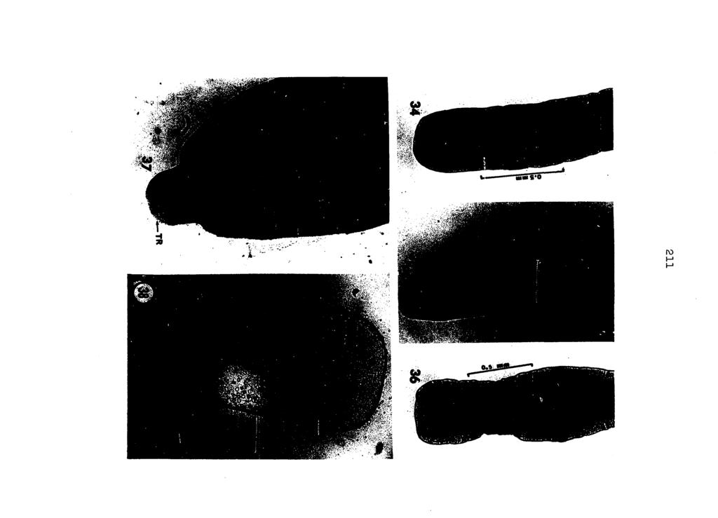
216
217 PLATE XI Figures 39 thru 52. Developmental stages of Mesocestoldes llneatus from tetrathyridium to young nonstrobilate adult. (All photographs taken at scale shown in Figure 39.). Figure 39. Figure 40, Figure 4l. Figure 42. Tetrathyridium from Rana plpiens showing numerous calcareous corpuscles (alizarin red preparation). Tetrathyridium from stomach of experimentally infected mouse, 1/2 hour post-feeding. Body in initial stage of degeneration. Tetrathyridium from stomach of experimentally infected raccoon, 9 hours post-feeding. Later stage In body degeneration of tetrathyridium from stomach of experimentally Infected mouse, 1 hour post-feeding. Figures 43 thru 44. Stages in release of scolex from tetrathyridial body taken from stomach of experimentally infected mouse, 1 hour post-feeding. Figure 45. Tetrathyridium with scolex everted from body; from experimentally infected mouse, 1/2 hour post-feeding. Figure 46. Tetrathyridial body following release of scolex; from experimentally Infected raccoon, 1-1/2 hours postfeeding. Figure 47. Very young M. llneatus showing invaglnated scolex and "tall"; from experimentally infected raccoon, 1-1/2 hours post-feeding. Figures 48 and 49. Extended young worms from small intestine of mouse, 30 minutes post feeding.
218 213 PLATE XI (continued) Figure 50. Figure 51. Figure 52. Evaginated scoleces of young M. lineatus recovered from small intestine of experimentally infected raccoon, 8 hours post-feeding. As above (Figure 50), but from opossum. Early young adults recovered from small intestine of experimentally infected mouse, 37 hours postfeeding. Note absence of "tail".
219 214
220 PIATE XII Figures 53 thru 56. Young adult M, lineatus from various vertebrate hosts. (All but Figure 5^ at same scale as Figure 53.). Figure 53. From naturally infected red fox (Vulpea vulpes). Figure 54. Figure 55. Figure 56. From experimentally infected dog, 8 hours post-feeding. From experimentally infected mouse, 157 hours post-feeding. From an intestine of naturally infected skunk (Mephitis mephitis).
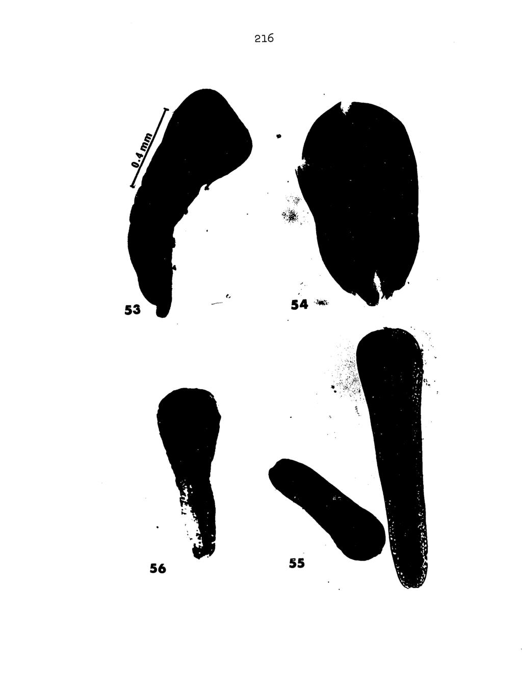
221
222 PLATE XIII Figures 57 thru 6l. Variations in scoleces of Mesocestoides lineatus. Figure 57. Figure 58. Figure 59. Figure 60. Figure 61. Scolex from naturally infected skunk. Note anterior notch in sucker. Scolex from naturally infected raccoon. Scolex from naturally infected skunk. (Same scale as shown in Figure 58.). Scolex from naturally infected raccoon. Note posterior notch in sucker. (Same scale as shown in Figure 57.). Scolex from experimentally infected raccoon. (Same scale as shown in Figure 57-)
223 218
224 PLATE XIV Figure 62. Typical egg of M. lineatus, phase contrast, 30 X 40 microns. Figure 63. Distribution of calcareous corpuscles in young, mature M. lineatus (air-dried preparation cleared in methyl salicylate). Figures 64 and 65. Parauterine organ and caudal appendage of M. lineatus from a raccoon. (Same scale as in Figure 63.). Figure 64; Note S-shaped caudal appendage. Figure 65. Note straight caudal appendage.
225 220
226 PLATE XV Figure.66. Figure 6?. Figure 68. Variations in shape (square, rectangular) or gravid proglottids of M. lineatus from experimentally infected opossum. Rectangular proglottids from experimentallyinfected opossum. Wall of parauterine organ showing its fibrous nature, photographed from life, phase contrast. Figures 69 thru 71. Abnormalities in Mesocestoides lineatus (Figures 69 and 70 are from naturally infected raccoons; Figure 71 is from an experimentally infected opossum). (Same scale as shown in Figure 66.). Figure 69. Figure 70. Figure 71. Incomplete transverse division of segment (arrow). Bifurcate stroblla. Reversal and doubling of reproductive organs. Figure 72. Parauterine organ showing extension of uterine component through parauterine wall. (Same scale as shown in Figure 67.).

227
228 PLATE XVI Figures 73 thru 75. Sections of tetrathyrldla from naturally Infected frogs (Rana piplens). Figure 73. Tetrathyridia 3^ situ, wall of small intestine. Figure 74. Multiple cyst from brachial region. (Same scale as shown in Figure 73.). Figure 75. Enlargement of tetrathyridium shown in Figure 73. (Same scale as shown in Figure 76.). Figure 76. Whole mount of tetrathyridium from naturally infected toad (Bufo cognatus). Note excretory bladder.

229 224
230 PLATE XVII Figures 77 thru 82. Representative cross-sections through mature proglottid of M. llneatus, osmium tetroxide preparations of hand-cut thick sections, phase contrast. (All to same scale as shown in Figure 82.). Figure 77. Figure 78. Figure 79. Figure 80. Figure 8I. Figure 82. Section immediately posterior to developing parauterine organ. Section through developing parauterine organ. Section through mid-region of proglottid at level of cirrus. Section through anterior region of uterus. Section through anteromost region of uterus. Note accumulation of lipids around uterus. Section through anteromost region of proglottid showing longitudinal excretory duct and transverse excretory canal.
231 226
232 PLATE XVIII Figure 83. Figure 84. Figure 85. Figure 86. Section of gravid parauterine organ, phase contrast. Enlargement of wall of parauterine organ shown in Figure 83. Electron micrograph of wall of parauterine organ from region similar to that shown in Figure 84. Enlarged portion of parauterine wall region shown in Figure 85. (Areas A, B, and C maybe seen at greater magnification in Figures 94, 95, and 97, respectively.).
233 228
234 PLATE XIX Figures 87 thru 92. Selected cross-sections through posterior region of mature proglottid to show early development of parauterine organ in relation to associated reproductive structures. Phase contrast, sections cut at 2 microns (see also Figures IO5, IO7, and IO9). (All at same scale as shown in Figure 87.). Figure 87. Section through region of ovaryshowing vagina and primary uterus (see also Figures 105, IO7, and 109). Figures 88 thru 92. Progressive sections from posterior to anterior through the early developing parauterine organ.
235 230 o
236 PLATE XX Figure 93. Montage of early mature proglottid of Mesocestoldes lineatus showing very early development of parauterine organ and typical early parauterine cells as seen in Figures 87 through 92.
237 232
238 PLATE XXI Figures 94 thru 97. High magnification electron micrographs of wall of parauterine organ. Figure 94. Figure 95. Figure 96. Figure 97. Region of parauterine wall shown in Figures 86, area A. Region of parauterine wall shown in Figure 86J area B. (Same scale as shown in Figure 94.). High magnification view of lamellar region of parauterine wall. Region of parauterine wall shown in Figure 86, area C. (Same scale as shown in Figure 96.).

239 frss
240 PLATE XXII Free-hand drawing of montage (Figure.99) showing general relationship of the three major reproductive structures In the region of the parauterine organ. Montage of the area of the developing parauterine organ In mature proglottid (see also Figure 8?).

241 mm 236
Title. Author(s)YAMASHITA, Jiro; OHBAYASHI, Masashi; KONNO, Seiji. CitationJapanese Journal of Veterinary Research, 4(3): Issue Date
YAMASHITA, Jiro; OHBAYASHI, Masashi; KONNO, Seiji. CitationJapanese Journal of Veterinary Research, 4(3): Issue Date") Title STUDIES ON ECHINOCOCCOSIS : III. ON EXPERIMENTAL INF DEVELOPMENT OF ECHINOCOCCUS GRANULOSUS (BATSCH, 1786 Author(s)YAMASHITA, Jiro; OHBAYASHI, Masashi; KONNO, Seiji CitationJapanese Journal of Veterinary
Title STUDIES ON ECHINOCOCCOSIS : III. ON EXPERIMENTAL INF DEVELOPMENT OF ECHINOCOCCUS GRANULOSUS (BATSCH, 1786 Author(s)YAMASHITA, Jiro; OHBAYASHI, Masashi; KONNO, Seiji CitationJapanese Journal of Veterinary
THE EFFECT OF MUTILATION ON THE TAPEWORM TAENIA TAENIAEFORMIS
 THE EFFECT OF MUTILATION ON THE TAPEWORM TAENIA TAENIAEFORMIS JOE N. MILLER AND WM. P. BUNNER The reader is undoubtedly aware of work which has been done by Child (1910) and others in mutilating certain
ON THE TRANSPLANTABILITY OF THE LARVA OF TEh'IA CRASSICOLLIS AND THE PROBABLE R~LE OF THE LIVER IN CYSTICERCUS DISEASE OF RATS
 ON THE TRANSPLANTABILITY OF THE LARVA OF TEh'IA CRASSICOLLIS AND THE PROBABLE R~LE OF THE LIVER IN CYSTICERCUS DISEASE OF RATS E'. 1). BULLOCI< AND M. 1%. CURTIS (Prom Coltil?lhin Uiiroersity, Iiistitute
VARIATION IN MONIEZIA EXPANSA RUDOLPHI
 VARIATION IN MONIEZIA EXPANSA RUDOLPHI STEPHEN R. WILLIAMS, Miami University, Oxford, Ohio In making a number of preparations of proglottids for class study at the stage when sex organs are mature and
Proteocephalus filicollis (Rud. 1810) in the Netherlands
 in the Netherlands") Proteocephalus filicollis (Rud. 1810) in the Netherlands by J.J. Willemse AND A.L.M. Veltman Zoological Laboratory, University of Amsterdam INTRODUCTION in another glass dish containing about 50 specimens
Proteocephalus filicollis (Rud. 1810) in the Netherlands by J.J. Willemse AND A.L.M. Veltman Zoological Laboratory, University of Amsterdam INTRODUCTION in another glass dish containing about 50 specimens
Title: Phylogenetic Methods and Vertebrate Phylogeny
 Title: Phylogenetic Methods and Vertebrate Phylogeny Central Question: How can evolutionary relationships be determined objectively? Sub-questions: 1. What affect does the selection of the outgroup have
VERTEBRATE READING. Fishes
 VERTEBRATE READING Fishes The first vertebrates to become a widespread, predominant life form on earth were fishes. Prior to this, only invertebrates, such as mollusks, worms and squid-like animals, would
Presentation of Quiz #85
 Presentation of Quiz #85 ***Reminder: Slides are copyrighted and cannot be copied for publication. A 36 year old male from Columbia was admitted to the hospital with seizures. This patient had previously
Unit 19.3: Amphibians
 Unit 19.3: Amphibians Lesson Objectives Describe structure and function in amphibians. Outline the reproduction and development of amphibians. Identify the three living amphibian orders. Describe how amphibians
Introduction to Helminthology
 Introduction to Helminthology HELMINTHES (WORMS) - Characteristics Eukaryotic, multicellular animals that usually have digestive, circulatory, nervous, excretory, and reproductive systems. Worms with bilateral
Darwin and the Family Tree of Animals
 Darwin and the Family Tree of Animals Note: These links do not work. Use the links within the outline to access the images in the popup windows. This text is the same as the scrolling text in the popup
Studies on the Helminth Fauna of Iowa II. Cestodes of Amphibians
 OF WASHINGTON, VOLUME 43, NUMBER 2, JULY 1976 191 Literature Cited Dickey, L. B. 1921. A new amphibian cestode. J. Parasit. 7: 129-136. Douglas, L. T. 1958. The taxonomy of nematotaeniid cestodes. J. Parasit.
Title. Author(s)OHBAYASHI, Masashi. CitationJapanese Journal of Veterinary Research, 15(1): 1-3. Issue Date DOI. Doc URL.
OHBAYASHI, Masashi. CitationJapanese Journal of Veterinary Research, 15(1): 1-3. Issue Date DOI. Doc URL.") Title GRYPORHYNCHUS NYCTICORACIS YAMAGUTI, 1956 (DILEPIDID APHARYNGOSTRIGEA ARDEOLINA VIDYARTHI, 1937 (STRIGEID CINEREA JOUYI CLARK Author(s)OHBAYASHI, Masashi CitationJapanese Journal of Veterinary Research,
Title GRYPORHYNCHUS NYCTICORACIS YAMAGUTI, 1956 (DILEPIDID APHARYNGOSTRIGEA ARDEOLINA VIDYARTHI, 1937 (STRIGEID CINEREA JOUYI CLARK Author(s)OHBAYASHI, Masashi CitationJapanese Journal of Veterinary Research,
Coyote (Canis latrans)
") Coyote (Canis latrans) Coyotes are among the most adaptable mammals in North America. They have an enormous geographical distribution and can live in very diverse ecological settings, even successfully
Coyote (Canis latrans) Coyotes are among the most adaptable mammals in North America. They have an enormous geographical distribution and can live in very diverse ecological settings, even successfully
Vertebrates and Parasites
 Vertebrates and Parasites Parasites indicators of biodiversity o Lots of parasites with complex life histories = area of high biodiversity with a good ecosystem o Provide deep phylogenetic and ecological
I the BUSSEY INSTITUTION of HARVARD UNIVERSITY, it was found that
 THE RELATION OF ALBINISM TO BODY SIZE IN MICE W. E. CASTLE Division of Genetics, University of Calijornia, Berkeley, California Received January 24, 1938 N PREVIOUS studies made in cooperation with former
MISCELLANEA ZOOLOGICA HUNGARICA. Tomus p Morphological investigation of Mesocestoides (Cestoda, Mesocestoididae) species parasitizing
 species parasitizing") MISCELLANEA ZOOLOGICA HUNGARICA Tomus 12. 1998 p. 11-19 Morphological investigation of Mesocestoides (Cestoda, Mesocestoididae) species parasitizing Vulpes vulpes in Hungary by A. Gubányi and E. Eszterbauer
MISCELLANEA ZOOLOGICA HUNGARICA Tomus 12. 1998 p. 11-19 Morphological investigation of Mesocestoides (Cestoda, Mesocestoididae) species parasitizing Vulpes vulpes in Hungary by A. Gubányi and E. Eszterbauer
A Scanning Electron Microscopic Study of Eggshell Surface Topography of Leidynema portentosae and L. appendiculatum (Nematoda: Oxyuroidea)
") The Ohio State University Knowledge Bank kb.osu.edu Ohio Journal of Science (Ohio Academy of Science) Ohio Journal of Science: Volume 88, Issue 5 (December, 1988) 1988-12 A Scanning Electron Microscopic
The Ohio State University Knowledge Bank kb.osu.edu Ohio Journal of Science (Ohio Academy of Science) Ohio Journal of Science: Volume 88, Issue 5 (December, 1988) 1988-12 A Scanning Electron Microscopic
General introduction
 Spirometra mansoni General introduction Distributed worldwide, mainly in southeast Asia. Larval infection of S. mansoni may cause serious clinical disease ---Sparganosis Morphology Adult worm measures
MORPHOTAXONOMICAL STUDY OF A NEW CESTODE GANGESIA (GANGESIA) CHOPARAI N.SP. FROM A FRESH WATER FISH, WALLAGO ATTU FROM JALAUN (U.P.
 CHOPARAI N.SP. FROM A FRESH WATER FISH, WALLAGO ATTU FROM JALAUN (U.P.") FLORA AND FAUNA 2016 Vol. 22 No. 1 PP 115-120 ISSN 0971-6920 MORPHOTAXONOMICAL STUDY OF A NEW CESTODE GANGESIA (GANGESIA) CHOPARAI N.SP. FROM A FRESH WATER FISH, WALLAGO ATTU FROM JALAUN (U.P.) INDIA ALOK
FLORA AND FAUNA 2016 Vol. 22 No. 1 PP 115-120 ISSN 0971-6920 MORPHOTAXONOMICAL STUDY OF A NEW CESTODE GANGESIA (GANGESIA) CHOPARAI N.SP. FROM A FRESH WATER FISH, WALLAGO ATTU FROM JALAUN (U.P.) INDIA ALOK
Breast Mass on Mammography
 Breast Mass on Mammography Bobbi Pritt, MD 2014 MFMER slide-1 Clinical Presentation 68 year old woman was noted to have 2 adjacent masses in the right breast on screening mammography Well-circumscribed,
ECHINOCOCCOSIS. By Dr. Ameer kadhim Hussein. M.B.Ch.B. FICMS (Community Medicine).
.") ECHINOCOCCOSIS By Dr. Ameer kadhim Hussein. M.B.Ch.B. FICMS (Community Medicine). INTRODUCTION Species under genus Echinococcus are small tapeworms of carnivores with larval stages known as hydatids proliferating
ECHINOCOCCOSIS By Dr. Ameer kadhim Hussein. M.B.Ch.B. FICMS (Community Medicine). INTRODUCTION Species under genus Echinococcus are small tapeworms of carnivores with larval stages known as hydatids proliferating
Field and Laboratory Study Evaluating the Possibility of Manodistomum syntomentera Causing Malformations In Frogs of the Mississippi River Valley
 11 Field and Laboratory Study Evaluating the Possibility of Manodistomum syntomentera Causing Malformations In Frogs of the Mississippi River Valley Laurie Carter Faculty Sponsor: Dr. Daniel Sutherland,
Introduction to phylogenetic trees and tree-thinking Copyright 2005, D. A. Baum (Free use for non-commercial educational pruposes)
") Introduction to phylogenetic trees and tree-thinking Copyright 2005, D. A. Baum (Free use for non-commercial educational pruposes) Phylogenetics is the study of the relationships of organisms to each other.
Introduction to phylogenetic trees and tree-thinking Copyright 2005, D. A. Baum (Free use for non-commercial educational pruposes) Phylogenetics is the study of the relationships of organisms to each other.
Rec. zool. Surv. India, 85(4); , 1989
; , 1989") Rec. zool. Surv. India, 85(4); 583-588, 1989 CSTODS OF DOMSTIC FOWL AT VISAKHAPATNAM WITH DSCRIPTION OF A NW SPCIS OF RAILLITINA (RAILLITINA) By SR RAMULU KOLLURI AND C. VIJAYA LAKSHMI Department of Zoology,
Rec. zool. Surv. India, 85(4); 583-588, 1989 CSTODS OF DOMSTIC FOWL AT VISAKHAPATNAM WITH DSCRIPTION OF A NW SPCIS OF RAILLITINA (RAILLITINA) By SR RAMULU KOLLURI AND C. VIJAYA LAKSHMI Department of Zoology,
Evolution as Fact. The figure below shows transitional fossils in the whale lineage.
 Evolution as Fact Evolution is a fact. Organisms descend from others with modification. Phylogeny, the lineage of ancestors and descendants, is the scientific term to Darwin's phrase "descent with modification."
WITH THE TABLE OF THE MORPHOLOGICAL FEATURES OF TAPEWORMS IN VAMPIROLEPIS. (Received: December 22nd, 1965)
") Japan. J. Med. Sci. Biol. 19, 51-57, 1966 *ON A NEW TAPEWORM, VAMPIROLEPIS ISENSIS, FOUND IN BATS WITH THE TABLE OF THE MORPHOLOGICAL FEATURES OF TAPEWORMS IN VAMPIROLEPIS ISAMU SAWADA Biological Laboratory,
Japan. J. Med. Sci. Biol. 19, 51-57, 1966 *ON A NEW TAPEWORM, VAMPIROLEPIS ISENSIS, FOUND IN BATS WITH THE TABLE OF THE MORPHOLOGICAL FEATURES OF TAPEWORMS IN VAMPIROLEPIS ISAMU SAWADA Biological Laboratory,
The Taxonomic Value and Variability of Certain Structures in the Cestode Genus Echinococcus (Rudolphi, 1801) and a Review of Recognized Species
 and a Review of Recognized Species") University of Nebraska - Lincoln DigitalCommons@University of Nebraska - Lincoln Faculty Publications from the Harold W. Manter Laboratory of Parasitology Parasitology, Harold W. Manter Laboratory of 11-15-1953
University of Nebraska - Lincoln DigitalCommons@University of Nebraska - Lincoln Faculty Publications from the Harold W. Manter Laboratory of Parasitology Parasitology, Harold W. Manter Laboratory of 11-15-1953
LINKAGE OF ALBINO ALLELOMORPHS IN RATS AND MICE'
 LINKAGE OF ALBINO ALLELOMORPHS IN RATS AND MICE' HORACE W. FELDMAN Bussey Inslitutim, Harvard Univwsity, Forest Hills, Boston, Massachusetts Received June 4, 1924 Present concepts of some phenomena of
PSEUDANDRYA MKUZll sp. nov, ( CESTODA: HYMENOLEPIDl DAE) FROM /CHNEUMIA ALBICAUDA
 FROM /CHNEUMIA ALBICAUDA") Onderstepoort J. Vet. Res. (1963), 30 (2), 127-132 Printed by the Government Printer, Pretoria PSEUDANDRYA MKUZll sp. nov, ( CESTODA: HYMENOLEPIDl DAE) FROM /CHNEUMIA ALBICAUDA R. J. ORTLEPP, Veterinary
Onderstepoort J. Vet. Res. (1963), 30 (2), 127-132 Printed by the Government Printer, Pretoria PSEUDANDRYA MKUZll sp. nov, ( CESTODA: HYMENOLEPIDl DAE) FROM /CHNEUMIA ALBICAUDA R. J. ORTLEPP, Veterinary
Alligators. very long tail, and a head with very powerful jaws.
 Reptiles Reptiles are one group of animals. There are two special features that make an animal a reptile. Those two features are bodies covered in scales and having a cold-blooded body. Adult reptiles
Antimicrobial Stewardship and Use Monitoring Michael D. Apley, DVM, PhD, DACVCP Kansas State University, Manhattan, KS
 Antimicrobial Stewardship and Use Monitoring Michael D. Apley, DVM, PhD, DACVCP Kansas State University, Manhattan, KS Defining antimicrobial stewardship is pivotal to our ability as veterinarians to continue
Contains most of the medically important tapeworms Scolex has 4 suckers and compact vitelline gland are characteristic Range from mm to >10m
 Cyclophyllidae Contains most of the medically important tapeworms Scolex has 4 suckers and compact vitelline gland are characteristic Range from mm to >10m Family Taeniidae Taenia saginata: beef tapeworm
Taseko Prosperity Gold-Copper Project. Appendix 5-6-D
 Appendix 5-6-D Appendix C Prosperity Mine 2006 Amphibian Survey Field Report C.1 Methods C.2 Results Amphibian surveys were conducted between June 13 23, 2006 and July 24 August 2, 2006 using a combination
Scientific background concerning Echinococcus multilocularis. Muza Kirjušina, Daugavpils University, Latvia
 Scientific background concerning Echinococcus multilocularis Muza Kirjušina, Daugavpils University, Latvia Echinococcus multilocularis Infection with the larval form causes alveolar echinococcosis (AE).
Chapter 1 COPYRIGHTED MATERIAL. Introduction to Veterinary Pathology. What is pathology? Who does pathology?
 What is pathology? Who does pathology? Chapter 1 Introduction to Veterinary Pathology Anatomic pathology Clinical pathology Microbiology Parasitology Immunology Toxicology Veterinary forensic pathology
Cestodes. Tapeworms from man and animals
 Cestodes Tapeworms from man and animals Taenia sp. The common (beef) tapeworm is several meters long. Courtesy Peters W. & Gilles H. Courtesy CDC Courtesy CDC Taenia sp. Unstained egg with four (visible)
BROOD REDUCTION IN THE CURVE-BILLED THRASHER By ROBERTE.RICKLEFS
 Nov., 1965 505 BROOD REDUCTION IN THE CURVE-BILLED THRASHER By ROBERTE.RICKLEFS Lack ( 1954; 40-41) has pointed out that in species of birds which have asynchronous hatching, brood size may be adjusted
rodent species in Australia to the fecal odor of various predators. Rattus fuscipes (bush
 Sample paper critique #2 The article by Hayes, Nahrung and Wilson 1 investigates the response of three rodent species in Australia to the fecal odor of various predators. Rattus fuscipes (bush rat), Uromys
Required and Recommended Supporting Information for IUCN Red List Assessments
 Required and Recommended Supporting Information for IUCN Red List Assessments This is Annex 1 of the Rules of Procedure for IUCN Red List Assessments 2017 2020 as approved by the IUCN SSC Steering Committee
SELECTION FOR AN INVARIANT CHARACTER, VIBRISSA NUMBER, IN THE HOUSE MOUSE. IV. PROBIT ANALYSIS
 SELECTION FOR AN INVARIANT CHARACTER, VIBRISSA NUMBER, IN THE HOUSE MOUSE. IV. PROBIT ANALYSIS BERENICE KINDRED Division of Animal Genetics, C.S.I.R.O., University of Sydney, Australia Received November
Phylum Platyhelminthes Flatworms
 Phylum Platyhelminthes Flatworms The Acoelomates The acoelomates are animals that lack a coelom. Acoelomates lack a body cavity, and instead the space between the body wall and the digestive tract is filled
1.0 INTRODUCTION. Echinococcosis, a cyclozoonotic helminthosis caused by the dwarf dog
 INTRODUCTION 1.0 INTRODUCTION Echinococcosis, a cyclozoonotic helminthosis caused by the dwarf dog tapeworm Echinococcus granulosus is highly endemic and is considered to be one of the most important parasitic
Flatworms Flatworms Platyhelminthes dorsoventrally free-living planarian parasitic fluke tapeworm label three body layers ectoderm mesoderm
 Flatworms Flatworms are in the phylum Platyhelminthes. Flatworms are flattened dorsoventrally (top to bottom). The group includes the freshwater, free-living planarian and the parasitic fluke and tapeworm.
University of Canberra. This thesis is available in print format from the University of Canberra Library.
 University of Canberra This thesis is available in print format from the University of Canberra Library. If you are the author of this thesis and wish to have the whole thesis loaded here, please contact
Factors Influencing Egg Production
 June, 1930 Research Bulletin No. 129 Factors Influencing Egg Production II. The Influence of the Date of First Egg Upon Maturity and Production By C. W. KNOX AGRICULTURAL EXPERIMENT STATION IOWA STATE
Field Herpetology Final Guide
 Field Herpetology Final Guide Questions with more complexity will be worth more points Incorrect spelling is OK as long as the name is recognizable ( by the instructor s discretion ) Common names will
THE VETERINARIAN'S CHOICE. Compendium clinical Trials. Introducing new MILPRO. from Virbac. Go pro. Go MILPRO..
 THE VETERINARIAN'S CHOICE. Introducing new MILPRO from Virbac. Compendium clinical Trials Go pro. Go MILPRO.. milbemycin/praziquantel Content INTRODUCTION 05 I. EFFICACY STUDIES IN CATS 06 I.I. Efficacy
Owl Pellet Dissection A Study of Food Chains & Food Webs
 NAME Owl Pellet Dissection A Study of Food Chains & Food Webs INTRODUCTION: Owl pellets are masses of bone, teeth, hair, feathers and exoskeletons of various animals preyed upon by raptors, or birds of
Biodiversity and Extinction. Lecture 9
 Biodiversity and Extinction Lecture 9 This lecture will help you understand: The scope of Earth s biodiversity Levels and patterns of biodiversity Mass extinction vs background extinction Attributes of
PROPOSED REDUCTION OF ITS SPECIES TO THREE
 MONIEZIA, A GENUS OF CESTODE WORMS AND THE PROPOSED REDUCTION OF ITS SPECIES TO THREE By E. Leonard Taylor Of the Veterinary Laboratory of the British Ministry of Agriculture and Fisheries The work on
LABORATORY. Introduction: Objectives: Laboratory 6 Pg.1
 LABORATORY Laboratory 6 Pg.1 6 Platyhelminthes and Acanthocephala Introduction: The Phylum Platyhelminthes contains both the trematodes and cestodes. The trematodes (flukes) have an incomplete digestive
Raw Meat Diet. Transcript:
 Transcript: Raw Meat Diet Hi, this is Dr. Karen Becker, and today we re going to discuss why dogs and cats can eat raw meat. This is probably the most common question I get, especially from uneducated
FACULTY OF VETERINARY MEDICINE
 FACULTY OF VETERINARY MEDICINE DEPARTMENT OF VETERINARY PARASITOLOGY AND ENTOMOLOGY M.Sc. AND Ph.D. DEGREE PROGRAMMES The postgraduate programmes of the Department of Veterinary Parasitology and Entomology
Between 1850 and 1900, human population increased, and 99% of the forest on Puerto Rico was cleared.
 Case studies, continued. 9) Puerto Rican Parrot Low point was 13 parrots in 1975. Do not breed until 4 years old. May be assisted by helpers at the nest, but this is not clear. Breeding coincides with
Laboratory 7 The Effect of Juvenile Hormone on Metamorphosis of the Fruit Fly (Drosophila melanogaster)
") Laboratory 7 The Effect of Juvenile Hormone on Metamorphosis of the Fruit Fly (Drosophila melanogaster) (portions of this manual were borrowed from Prof. Douglas Facey, Department of Biology, Saint Michael's
Laboratory 7 The Effect of Juvenile Hormone on Metamorphosis of the Fruit Fly (Drosophila melanogaster) (portions of this manual were borrowed from Prof. Douglas Facey, Department of Biology, Saint Michael's
Notes on Varanus salvator marmoratus on Polillo Island, Philippines. Daniel Bennett.
 Notes on Varanus salvator marmoratus on Polillo Island, Philippines Daniel Bennett. Dept. Zoology, University of Aberdeen, Scotland, AB24 2TZ. email: daniel@glossop.co.uk Abstract Varanus salvator marmoratus
MORPHOLOGICAL CHARACTERIZATION OF ADULT ECHINOCOCCUS GRANULOSUS AS A MEANS OF DETERMINING TRANSMISSION PATTERNS
 J. Parasitol., 79(1), 1993, p. 57-61? American Society of Parasitologists 1993 MORPHOLOGICAL CHARACTERIZATION OF ADULT ECHINOCOCCUS GRANULOSUS AS A MEANS OF DETERMINING TRANSMISSION PATTERNS Clare C. Constantine,
High Mortality of a Population of Cowbirds Wintering at Columbus, Ohio
 The Ohio State University Knowledge Bank kb.osu.edu Ohio Journal of Science (Ohio Academy of Science) Ohio Journal of Science: Volume 67, Issue 1 (January, 1967) 1967-01 High Mortality of a Population
Anatomy. Name Section. The Vertebrate Skeleton
 Name Section Anatomy The Vertebrate Skeleton Vertebrate paleontologists get most of their knowledge about past organisms from skeletal remains. Skeletons are useful for gleaning information about an organism
ECHINOCOCCUS GRANULOSUS
 48 ECHINOCOCCUS GRANULOSUS 48.1 INTRODUCTION E granulosus are small tape worms that parasitize the intestines of carnivores like dogs. About one million people are infected with this tape worm worldwide.
The Rat Lungworm Lifecycle
 Hawaii Island Rat Lungworm Working Group Daniel K. Inouye College of Pharmacy University of Hawaii, Hilo The Rat Lungworm Lifecycle Rat Lungworm IPM RLWL-3 It is important to understand the lifecycle of
Red Eared Slider Secrets. Although Most Red-Eared Sliders Can Live Up to Years, Most WILL NOT Survive Two Years!
 Although Most Red-Eared Sliders Can Live Up to 45-60 Years, Most WILL NOT Survive Two Years! Chris Johnson 2014 2 Red Eared Slider Secrets Although Most Red-Eared Sliders Can Live Up to 45-60 Years, Most
Breeding White Storks( Ciconia ciconia at Chessington World of Adventures Paul Wexler
 Breeding White Storks(Ciconia ciconia) at Chessington World of Adventures Paul Wexler The White Stork belongs to the genus Ciconia of which there are seven other species incorporated predominantly throughout
POSTNATAL DEVELOPMENT IN PEROMYSCUS MANICULATUS-POLIONOTUS HYBRIDS I. DEVELOPMENTAL LANDMARKS AND LITTER MORTALITY 12
 OSTNATAL DEVELOENT IN EROYSCUS ANICULATUS-OLIONOTUS HYBRIDS I. DEVELOENTAL LANDARKS AND LITTER ORTALITY WALLACE D. DAWSON Department Zoology and Entomology, The Ohio State University, Columbus, Ohio ABSTRACT
Ectoparasites Myobia musculi Radfordia affinis Radfordia ensifera
 Ectoparasites Fleas, ticks, and lice are uncommon in modern laboratory facilities, but may be seen on wild or feral rodents. Most ectoparasite infestations seen in rats and mice used for research are various
European Convention for the Protection of Vertebrate Animals used for Experimental and Other Scientific Purposes *
 European Treaty Series - No. 123 European Convention for the Protection of Vertebrate Animals used for Experimental and Other Scientific Purposes * Strasbourg, 18.III.1986 Appendix B Statistical tables
Mexican Gray Wolf Reintroduction
 Mexican Gray Wolf Reintroduction New Mexico Supercomputing Challenge Final Report April 2, 2014 Team Number 24 Centennial High School Team Members: Andrew Phillips Teacher: Ms. Hagaman Project Mentor:
The Friends of Nachusa Grasslands 2016 Scientific Research Project Grant Report Due June 30, 2017
 The Friends of Nachusa Grasslands 2016 Scientific Research Project Grant Report Due June 30, 2017 Name: Laura Adamovicz Address: 2001 S Lincoln Ave, Urbana, IL 61802 Phone: 217-333-8056 2016 grant amount:
Large Animal Topics in Parasitology for the Veterinary Technician Jason Roberts, DVM This presentation is designed to review the value veterinary
 Large Animal Topics in Parasitology for the Veterinary Technician Jason Roberts, DVM This presentation is designed to review the value veterinary technicians can add to mixed or large animal practices
HUMAN APPENDIX BATS & TROPICAL FLOWERS
 HUMAN APPENDIX In humans, the appendix is a short piece of tissue off the large intestine. It is not used by humans for digestive functions. In other mammals, like rabbits and deer, the cecum is a large
LAND AT REAR OF PARAPET HOUSE LENHAM KENT REPTILE SCOPING SURVEY
 LAND AT REAR OF PARAPET HOUSE LENHAM KENT REPTILE SCOPING SURVEY BY MARTIN NEWCOMBE 5 th June 2015 D100. Lenham (TQ8961152288).R2 Martin Newcombe Wildlife Management Consultancy 01233 720229 Page 1.0 INTRODUCTION
Loss of wildlands could increase wolf-human conflicts, PA G E 4 A conversation about red wolf recovery, PA G E 8
 Loss of wildlands could increase wolf-human conflicts, PA G E 4 A conversation about red wolf recovery, PA G E 8 A Closer Look at Red Wolf Recovery A Conversation with Dr. David R. Rabon PHOTOS BY BECKY
10/24/2016 B Y E M I LY T I L L E Y
 ALL ABOUT ANIMALS B Y E M I LY T I L L E Y 1 M A M M A LS: H A V E A B A C K B O N E, A R E W A R M - B L O O D E D, H A V E H A I R O N T H E I R B O D I E S, A N D P R O D U C E M I L K T O F E E D T
CLADISTICS Student Packet SUMMARY Phylogeny Phylogenetic trees/cladograms
 CLADISTICS Student Packet SUMMARY PHYLOGENETIC TREES AND CLADOGRAMS ARE MODELS OF EVOLUTIONARY HISTORY THAT CAN BE TESTED Phylogeny is the history of descent of organisms from their common ancestor. Phylogenetic
Parasites of Small Mammals in Grand Teton National Park: Babesia and Hepatozoon
 University of Wyoming National Park Service Research Center Annual Report Volume 19 19th Annual Report, 1995 Article 13 1-1-1995 Parasites of Small Mammals in Grand Teton National Park: Babesia and Hepatozoon
Shoot, shovel and shut up: cryptic poaching slows restoration of a large
 Electronic Supplementary Material Shoot, shovel and shut up: cryptic poaching slows restoration of a large carnivore in Europe doi:10.1098/rspb.2011.1275 Time series data Field personnel specifically trained
. - many countries in Asia. Twenty species of Gnathostoma have been recorded in the literature although only
 SEATO ~edical ~esearch Studies on Gnathostomiasis in Thailand. Professor Svasti Daengsvang, M.D. Special Consultant to the Director. Principal investigator: Professor Svasti Daengsvang M.D. Associate Investigator:
Rules and liability insurance applicable for Expo Mitt and Expo Syd
 Rules and liability insurance applicable for Expo Mitt and Expo Syd Important changes: Ban on scaleless reptiles, Ban on venomoids (with surgically removed venom glands), Minimum container size 1.5 dl,
SHEEP SIRE REFERENCING SCHEMES - NEW OPPORTUNITIES FOR PEDIGREE BREEDERS AND LAMB PRODUCERS a. G. Simm and N.R. Wray
 SHEEP SIRE REFERENCING SCHEMES - NEW OPPORTUNITIES FOR PEDIGREE BREEDERS AND LAMB PRODUCERS a G. Simm and N.R. Wray The Scottish Agricultural College Edinburgh, Scotland Summary Sire referencing schemes
Taenia saginata Programme
 (TO BE READ IN CONJUNCTION WITH THE Procedures for cattle from properties on the T. saginata disease surveillance Suspect List) Taenia saginata Programme Management Plan Guidelines A SUMMARY DOCUMENT December
V. Subclass Eucestoida (Chapters 20 & 21, BLY 459, 2010)
") V. Subclass Eucestoida (Chapters 20 & 21, BLY 459, 2010) A. Characteristics (Ignore Cestodaria) 1. Differences from trematodes a. No digestive tract (1) No mouth, gut, nor anus (2) All nutrients absorbed
V. Subclass Eucestoida (Chapters 20 & 21, BLY 459, 2010) A. Characteristics (Ignore Cestodaria) 1. Differences from trematodes a. No digestive tract (1) No mouth, gut, nor anus (2) All nutrients absorbed
1 EEB 2245/2245W Spring 2017: exercises working with phylogenetic trees and characters
 1 EEB 2245/2245W Spring 2017: exercises working with phylogenetic trees and characters 1. Answer questions a through i below using the tree provided below. a. Identify the taxon (or taxa if there is more
Sample Questions: EXAMINATION I Form A Mammalogy -EEOB 625. Name Composite of previous Examinations
 Sample Questions: EXAMINATION I Form A Mammalogy -EEOB 625 Name Composite of previous Examinations Part I. Define or describe only 5 of the following 6 words - 15 points (3 each). If you define all 6,
cyst&' appeared to be of two kinds-one smaller and Smnith "is inclined to regard these epithelial cell parasites as
 COCCIDIA IN SUBEPITHELIAL INFECTIONS OF THE INTESTINES OF BIRDS PHILIP B. HADLEY From the Agricultural Experiment Station of the Rhode Island State College' Received for publication, July 10, 1916 In an
Redescription of Anoplocephaloides indicata (Sawada et Papasarathorn, 1966) comb. nov. (Cestoda, Anoplocephalidae) from Tapirus indicus
 comb. nov. (Cestoda, Anoplocephalidae) from Tapirus indicus") Acta Parasitologica, 2005, 50(2), 118 123; ISSN 1230-2821 Copyright 2005 W. Stefañski Institute of Parasitology, PAS Stefański Redescription of Anoplocephaloides indicata (Sawada et Papasarathorn, 1966)
Acta Parasitologica, 2005, 50(2), 118 123; ISSN 1230-2821 Copyright 2005 W. Stefañski Institute of Parasitology, PAS Stefański Redescription of Anoplocephaloides indicata (Sawada et Papasarathorn, 1966)
Minnesota_mammals_Info_9.doc 11/04/09 -- DRAFT Page 1 of 64. Minnesota mammals
 Minnesota_mammals_Info_9.doc 11/04/09 -- DRAFT Page 1 of 64 Minnesota mammals This is a short guide to Minnesota mammals, with information drawn from Hazard s Mammals of, Walker s Mammals of the World,
May 10, SWBAT analyze and evaluate the scientific evidence provided by the fossil record.
 May 10, 2017 Aims: SWBAT analyze and evaluate the scientific evidence provided by the fossil record. Agenda 1. Do Now 2. Class Notes 3. Guided Practice 4. Independent Practice 5. Practicing our AIMS: E.3-Examining
Introduction to the Cheetah
 Lesson Plan 1 Introduction to the Cheetah CRITICAL OUTCOMES CO #1: Identify and solve problems and make decisions using critical and creative thinking. CO #2: Work effectively with others as members of
DEPOSEL Slow Release Selenium Injection for Cattle and Sheep
 Date of change: 21 October 2004 Page: 1 of 9 Carton (front panel). POISON KEEP OUT OF REACH OF CHILDREN FOR ANIMAL TREATMENT ONLY DEPOSEL Slow Release Selenium Injection for Cattle and Sheep Active ingredient:
Heartworm Disease in Dogs
 Kingsbrook Animal Hospital 5322 New Design Road, Frederick, MD, 21703 Phone: (301) 631-6900 Website: KingsbrookVet.com What causes heartworm disease? Heartworm Disease in Dogs Heartworm disease or dirofilariasis
Some Foods Used by Coyotes and Bobcats in Cimarron County, Oklahoma 1954 Through
 .180 PROOf OF THE QKLA. ACAD. OF SCI. FOR 1957 Some Foods Used by Coyotes and Bobcats in Cimarron County, Oklahoma 1954 Through 1956 1 RALPH J. ELLIS and SANFORD D. SCBEMNITZ, Oklahoma Cooperative Wildlife
American Veterinary Medical Association
 AVMA American Veterinary Medical Association Governmental Relations Division 1910 Sunderland Place, NW Washington, DC 20036-1642 phone 202.789.0007 800.321.1473 fax 202.842.4360 AVMA Headquarters 1931
Objectives: Outline: Idaho Amphibians and Reptiles. Characteristics of Amphibians. Types and Numbers of Amphibians
 Natural History of Idaho Amphibians and Reptiles Wildlife Ecology, University of Idaho Fall 2005 Charles R. Peterson Herpetology Laboratory Department of Biological Sciences, Idaho Museum of Natural History
These small issues are easily addressed by small changes in wording, and should in no way delay publication of this first- rate paper.
 Reviewers' comments: Reviewer #1 (Remarks to the Author): This paper reports on a highly significant discovery and associated analysis that are likely to be of broad interest to the scientific community.
AMPHIBIAN AND REPTILE PRE-CONSTRUCTION SURVEYS AND PRIORITY SPECIES EVALUATION ADDENDUM
 Calligan Creek Hydroelectric Project FERC No. 13948 AMPHIBIAN AND REPTILE PRE-CONSTRUCTION SURVEYS AND PRIORITY SPECIES EVALUATION ADDENDUM Prepared for: Everett, WA Prepared by: Bellingham, WA Calligan
Title. Author(s)YAMASHITA, Jiro; OHBAYASHI, Masashi; KITAMURA, Yukit. CitationJapanese Journal of Veterinary Research, 6(2): 89-92
YAMASHITA, Jiro; OHBAYASHI, Masashi; KITAMURA, Yukit. CitationJapanese Journal of Veterinary Research, 6(2): 89-92") Title STUDIES ON ECHINOCOCCOSIS VII. : ON THE DEVELOPMENT IN THE TAPEWORM STAGE Author(s)YAMASHITA, Jiro; OHBAYASHI, Masashi; KITAMURA, Yukit CitationJapanese Journal of Veterinary Research, 6(2): 89-92
Title STUDIES ON ECHINOCOCCOSIS VII. : ON THE DEVELOPMENT IN THE TAPEWORM STAGE Author(s)YAMASHITA, Jiro; OHBAYASHI, Masashi; KITAMURA, Yukit CitationJapanese Journal of Veterinary Research, 6(2): 89-92
HUSK, LUNGWORMS AND CATTLE
 Vet Times The website for the veterinary profession https://www.vettimes.co.uk HUSK, LUNGWORMS AND CATTLE Author : Alastair Hayton Categories : Vets Date : July 20, 2009 Alastair Hayton discusses how best
New Species of the Ptychobothridean Tapeworm Circumoncobohrium from Mastacembalus armatus
 New Species of the Ptychobothridean Tapeworm Circumoncobohrium from Mastacembalus armatus M B Sonune and C R Kasar 1 Department of Zoology, Shri. Shivaji Science, and Arts College, Chikhali, Dist- Buldhana,
Nomination of Populations of Dingo (Canis lupus dingo) for Schedule 1 Part 2 of the Threatened Species Conservation Act, 1995
 for Schedule 1 Part 2 of the Threatened Species Conservation Act, 1995") Nomination of Populations of Dingo (Canis lupus dingo) for Schedule 1 Part 2 of the Threatened Species Conservation Act, 1995 Illustration by Marion Westmacott - reproduced with kind permission from a
Nomination of Populations of Dingo (Canis lupus dingo) for Schedule 1 Part 2 of the Threatened Species Conservation Act, 1995 Illustration by Marion Westmacott - reproduced with kind permission from a