Submitted By C. MANIKANDAN M. Sc., M. Phil., B.Ed.,
|
|
|
- Bryce Tate
- 5 years ago
- Views:
Transcription

1 STUDY OF PREVALENCE AND ANTIMICROBIAL SUSCEPTIBILITIES OF BACTERIA AND FUNGI ISOLATED FROM PATIENTS WITH URINARY TRACT INFECTIONS IN PATTUKKOTTAI, TAMIL NADU, INDIA Thesis submitted to BHARATHIDASAN UNIVERSITY In partial fulfillment of the requirement For the award of degree of DOCTOR OF PHILOSOPHY IN ZOOLOGY Submitted By C. MANIKANDAN M. Sc., M. Phil., B.Ed., Under the Guidance of Dr. A. AMSATH, M. Sc., M. Phil., Ph. D. P. G. AND RESEARCH DEPARTMENT OF ZOOLOGY KHADIR MOHIDEEN COLLEGE ADIRAMPATTINAM DECEMBER-2013
2 Dr. A. AMSATH, M. Sc., M. Phil., Ph. D., Associate Professor and Research Advisor P.G. and Research Department of Zoology, Khadir Mohideen College, Adirampattinam Tamil Nadu, India Date:.. CERTIFICATE This is to certify that the thesis entitled, Study of prevalence and antimicrobial susceptibilities of bacteria and fungi isolated from patients with urinary tract infections in Pattukkottai, Tamil Nadu, India is a record of research work done by the candidate Mr. C. Manikandan, during the year submitted to Bharathidasan University, Tiruchirappalli in partial fulfillment of the requirements for the award of Degree of Doctor of Philosophy in Zoology under my supervision and that the thesis has not been submitted earlier for the award of any Degree anywhere. ii
3 DECLARATION I hereby declare that the thesis entitled Study of prevalence and antimicrobial susceptibilities of bacteria and fungi isolated from patients with urinary tract infections in Pattukkottai, Tamil Nadu, India is a record of research work done by me in the Post Graduate and Research Department of Zoology, Khadir Mohideen College, Adirampattinam under the supervision of Dr. A. AMSATH, during the year submitted to Bharathidasan University, Tiruchirappalli in partial fulfillment of the requirements for the award of Degree of Doctor of Philosophy in Zoology and that the thesis has not been submitted earlier for the award of any Degree anywhere. Station: Adirampattinam. Date : (C.MANIKANDAN) iii
4 ACKNOWLEDGEMENTS I give thanks to almighty GOD for his providence, grace and mercy upon my life, and for His divine enablement without Him nothing would have been possible. In the first place I would like to record my gratitude to Dr. A. AMSATH, M.Sc., M.Phil., Ph.D., Associate Professor of Zoology, Khadir Mohideen College, Adirampattinam for suggesting the problem, supervision, advice, guidance and encouragement from the very early stage of this research as well as giving me extraordinary experiences throughout the work. His truly scientist intuition has made him as a constant oasis of ideas and passions in science, which exceptionally inspire and enrich my growth as a student, a researcher and a scientist want to be. I am indebted to him more than he knows. My heartiest thanks to honorable Prof. Dr. A. JALAL, M.Com., M.Phil., Ph.D., Principal, Khadir Mohideen College, Adirampattinam for his kind support for completing this Research in a successful manner. I would to express my thanks to honorable Prof. Dr. A. UDUMAN MOHIDEEN, M.Sc., M.Phil., Ph.D., Vice Principal, Khadir Mohideen College, Adirampattinam for his encouragement during the study period. Words are insufficient to express my deep sense of gratitude and heartfelt thanks to Honorable Prof. Dr. P. KUMARASAMY, M.Sc., M.Phil., Ph.D., HOD and Associate Professor of Zoology, iv
5 Khadir Mohideen College, Adirampattinam for having permitted me to pursue my research work and for having given me the opportunity and necessary facilities for my research programme in the college of excellence. I wish to express my special thanks to Prof. Dr. S. RAVEENDRAN, M.Sc., M.Phil., B.Ed., BLIS., Ph.D., Associate Professor and Dr. O. SATHIK, M.Sc., M.Phil., Ph.D., Assistant Professor, Doctoral committee members, Department of zoology, Khadir Mohideen College, for their immense help for completing my task. I sincerely express my heart full thanks to Dr. P. PRABHAHARAN, M.Sc., M.Phil., Ph.D., Assistant Professor, Department of Biotechnology, Srimad Andavan Arts and Science College, Thiruchirapalli for his assistance to publish the research work. Words are inadequate in offering my thanks to Dr. A. MOHAMED HATHA, M.Sc., M.Phil., Ph.D., HOD of Environmental sciences, Cochin University of Science and Technology, Cochin, who has spent their valuable time to help me do molecular work. I express my sincere thanks to Dr. R. SUBHASHKUMAR, M.Sc., M.Phil., Ph.D., project convener, Department of Microbiology, Kongu Nadu Arts and Science College, Coimabatore and Dr. T. THAUMANAVAN, M.Sc., M.Phil., Ph.D., Department v
6 of Microbiology, Assistant Professor of Microbiology, SNR College, Coimbatore, for their friendly guidance and help It is my privilege to thank Dr. P. BASKARAN MS., Medical officer, Government Hospital, Pattukkottai, Dr. P. SRINIVASAN MBBS, D.Ortho., Devasena Hospital, Dr. M. GUNASEKARAN MD., Thiravidan Hospital, Pattukkottai, Dr. M. AYYASAMY MBBS., DCH, Dr. S. SURIYAMOORTHY MBBS., DCH., and Dr. T. NEELAVATHI MBBS., DGO., Janaki Hospital, Dr. ADR. ANBARASAN MS., and Dr. A. MAHESHWARI MBBS. DGO., GRAM Hospital, Dr. T. DEVI MD., DGO., Sangeetha hospital, Dr. R. IGBAL SHERIFF MBBS.,DNB., Dr. A. MAHALINGAM MD., Dr., C. CHINNADURAI MS., Elangovan Hospital, Dr. C. TAMILSELVAN MD., DCH., Dr. R.RAJAGOPAL MS., (URO), Ln.U.POTHIYAPPAN B.Sc., DMLT, Jayam Clinical Laboratory, Pattukkottai for their prompt help in sample collection and cooperation at various phases of the experimental work. My sincere thanks will also extend to all members of Zoology Department, Khadir Mohideen College for exchanging knowledge and experiences and assisting me during studies. Words fail me to express my appreciation to my wife M. MAHALAKSHMI, B.Sc., B.Ed., MCA., M.Phil., for her dedication, love and persistent confidence in me, has taken the load off my shoulder. I owe her for being unselfishly let her intelligence, passions, and ambitions collide with mine. I express my lovable thanks to my elder son M. SHANMUGAPRAKASH, my younger vi
7 son M. POKUTTEZHINI for their cooperation during my study Period. I would like to thank the Laboratory Technologists C. SUGANYA B.Sc., DMLT, B. DHANALAKSHMI B.Sc., DMLT and R. SATHYA B.Sc., DMLT of Gangasaras Diagnostic and Research Centre, Pattukkottai for the facilities provided to carry out the work and their consistent support and help in this study. I express my heartfelt thanks to my friends for their help and wishes for the successful completion of this project. I would like to thank everybody who was important to the successful realization of thesis, as well as expressing my apology that I could not mention personally one by one. Last, but not the least, I am very much grateful to all patients without whom is study would not have been completed. C. Manikandan vii
8 CONTENTS CHAPTER NO. TITLE PAGE NO. 1 INTRODUCTION 1 Objectives of the study 20 2 REVIEW OF LITERATURE 22 3 MATERIALS AND METHODS Study of prevalence of urinary tract infections (UTIs) Isolation and identification of bacteria causing UTIs Antibacterial susceptibility testing Isolation and Characterisation of the pathogenic Candida species Antifungal susceptibility testing Plasmid analysis- molecular characterisation of ESBL producing E.coli 93 4 RESULTS Study of the prevalence of UTIs Study population Distribution of uropathogens according to age groups and gender 95 viii
9 Isolation and identification of pathogenic bacteria from UTIs Sources of isolated strains with percentage Correlation between pyuria, Gram s stain and culture in UTIs Uropathogens identified on Chromagar media according to pigment reactions Distribution of isolates among positive urine specimens Antibacterial susceptibility pattern of bacterial isolates from the UTIs Characterisation and susceptibility pattern of E.coli Characterisation and susceptibility pattern of Klebsiella pneumoniae Characterisation and susceptibility pattern of Pseudomonas spp Characterisation and susceptibility pattern of Proteus spp Characterisation and susceptibility pattern of Enterobacter spp Characterisation of Gram Positive Bacteria Antibiotic susceptibility pattern of CoNS Antibiotic susceptibility pattern of Enterococcus spp Antibiotic susceptibility pattern of Staphylococcus saprophyticus Antibiotic susceptibility pattern of Staphylococcus aureus ix
10 Antibiotic resistance patterns of all Bacterial isolates from UTIs Aminoglycoside resistance in bacterial isolates from UTIs Beta-lactam group of antibiotics resistance in bacterial isolates from UTIs Fluoroquinolone resistance in bacterial isolates from UTIs Two stand-alone antibiotics resistance in bacteria from UTIs Isolation and characterisation of the pathogenic Candida species Antifungal susceptibility pattern of Candida species causing UTIs Plasmid analysis-molecular characterisation of ESBL producing E.coli ESBL Positive E.coli PCR and RFLP analysis DISCUSSION SUMMARY AND CONCLUSION 158 REFERENCES 162 APPENDIX 1: LIST OF PAPER PUBLICATIONS APPENDIX 2: LIST OF PAPER PRESENTATIONS x
11 LIST OF TABLES Table No CAPTION Page No 1. Pathogens in complicated and uncomplicated UTIs Antimicrobial drugs and their sensitivity profiles Result of urine culture among study population Distribution of urine pathogens according to age groups and gender The sources of isolated strains from UTIs Correlation between pyuria, Gram s stain and culture in UTIs Urine isolates presumptively identified on Chromagar media Identification of uropathogens by Gram s staining, SBA and MA agar. Biochemical identification of bacteria isolated from UTIs Frequency of bacterial isolates from urine samples Antibiotic susceptibility pattern of E. coli Antibiotic susceptibility pattern of K.pneumoniae Antibiotic susceptibility pattern of Pseudomonas spp., Antibiotic susceptibility pattern of Proteus spp., 118 xi
12 15. Antibiotic susceptibility pattern of Enterobacter spp., Antibiotic susceptibility pattern of CoNS Antibiotic susceptibility pattern of Enterococcus spp., Antibiotic susceptibility pattern of Staphylococcus saprophyticus Antibiotic susceptibility pattern of Staphylococcus aureus Antibiotic resistance pattern of bacteria isolated from urine samples Characteristics of Candida species on Chromagar Candida media Distribution of isolated Candida species Identification of isolated Candida species by Direct wet mount and Germ tube methods Antifungal susceptibility pattern of Candida species causing UTIs xii
13 LIST OF FIGURES Figure No 1. CAPTION Route of urinary tract infections Page No Predisposing factors for complicated UTIs 3. Distribution of symptomatic urinary tract infection and prevalence of asymptomatic bacteriuria by age and sex 4. Urinary system of women and men 5. The sources of isolated strains from UTIs 6. Isolation of bacteria on Nutrient agar and isolation of Candida species on Sabouraud dextrose agar 7. Gram s staining, Direct microscopy, Germ tube test 8. Identification of bacterial species on Chromagar media 9. Identification of bacteria on MacConkey and Blood agar 10. Identification of bacteria through biochemical testing and selective media 11. Antibiotic resistance of E. coli strains from patients with UTIs 12. Antibiotic susceptibility pattern of bacteria isolated from UTIs 13. Antibiotic susceptibility pattern of bacteria isolated from UTIs 14. Antibiotic susceptibility pattern of Proteus spp., and MDR E.coli isolated from UTIs xiii
14 15. Antibiotic resistance of K.pneumoniae from patients with UTIs 16. Antibiotic resistance of Pseudomonas strains from patients with UTIs 17. Antibiotic resistance of Proteus strains from patients with UTIs 18. Antibiotic resistance of Enterobacter strains from patients with UTIs 19. Antibiotic resistance of CoNS strains from patients with UTIs 20. Antibiotic resistance of Enterococcus strains from patients with UTIs 21. Antibiotic resistance of S. saprophyticus strains from patients with UTIs 22. Antibiotic resistance of S. aureus strains from patients with UTIs 23. Antibiotic resistant of all bacterial strains isolated from UTIs 24. Characteristics of Candida species on Chromagar Candida media 25. Antifungal susceptibility pattern of Candida species Antifungal susceptibility of Candida species isolated from UTIs 136 Double disc synergy test 137 Molecular Characteristics of ESBL producing MDR E.coli 139 xiv
15 µg : Micro gram µl : Micro litre ABBREVIATIONS A AATF ACB AK AM AX BPH Candida spp., CAUTI CAZ CDC CFU CIP CL CLSI CoNS COT : Acid production : Antimicrobial availability task force : Antibody coated bacteria : Amikacin : Ampicillin : Amoxicillin : Benign prostatic hyperplasia : Candida species : Catheter associated urinary tract infection : Ceftazidime : Centers for Disease control : Colony forming units : Ciprofloxacin : Cephalexin : Clinical Laboratory Standards Institute : Coagulase negative Staphylococcus : Cotrimoxazole xv
16 CT CTX DDST E.coli ESBLs ExPEC G GEN GNB GPC HCW HIV HPF ICU IMViC IP IPM K.pneumoniae Kbp M MA : Ceftriaxone : Cefotaxime : Double disc synergy test : Escherichia coli : Extended-spectrum β-lactamases : Extra-intestinal pathogenic E.coli : Gas production : Gentamicin : Gram Negative Bacteria : Gram Positive Cocci : Health care workers : Human immunodeficiency virus : High power field : Intensive care unit : Indole, Methyl red, Voges-Prokauer and citrate : In- patient : Imipenem : Klebsiella pneumoniae : Kilo base pairs : Marker : MacConkey Agar xvi
17 MDR Mg MH Agar Ml MICs mm MSU NA NCCLS NIT NX OF OP P.aeruginosa P.vulgaris PCDDT PCR PFGE RFLP rpm : Muti-drug resistance : Milligram : Muller-Hinton Agar : Milliliter : Minimal inhibitory concentrations : Millimeter : Mid Stream Urine : Nalidixic acid : National Committee for Clinical Laboratory standards : Nitrofurantoin : Norfloxacin : Ofloxacin : Out -patient : Pseudomonas aeruginosa : Proteus vulgaris : Phenotypic confirmatory disc diffusion test : Polymerase Chain Reaction : Pulsed-field gel electrophoresis : Restriction fragment length polymorphism : Revolution per minute xvii
18 S. aureus : Staphylococcus aureus S. epidermidis : Staphylococcus epidermidis S.saprophyticus SDA SHV Spp., TB TEM TST U UPEC UTIs UV WBCs Wt WHO β : Staphylococcus saprophyticus : Sabouraud Dextrose Agar : Sulfhydryl variable-active site : Species : Tobramycin : Temnoniera : Triple Sugar Iron : Unit : Uropathogenic Escherichia coli : Urinary Tract Infections : Ultra violate : White blood corpuscles : Weight : World Health Organistion : Beta xviii
19 1. INTRODUCTION 1.1. General information Urinary tract infection (UTI) is one of the most important causes of morbidity in the general population, and is the second most common cause of hospital visits (Ronald, 2002; Kolawale et al., 2009; Upadhyay et al., 2013). Urinary tract infections (UTIs) are more common among women than men, although the prevalence in elderly men and women is similar. Most of the research on UTI has focused on young, sexually active women who are at high risk for developing an infection (Harrington and Hooton, 2000). The predominant UTI risk factors in young women are sexual intercourse and the use of spermicidal contraceptives. Worldwide, about 150 million people are diagnosed with UTI each year (Gupta, 2001; Amin et al., 2011). Urinary tract infections (UTIs) is one of the common infectious diseases, and nearly 10% of people will experience a UTI during their life-time (Foxman et al., 2003). UTI is the leading cause of morbidity and health care expenditures in persons of all the ages (Prabhu and Selvaraj, 2012). UTI is one of the most common infectious diseases which have been extremely studied in the field of clinical practice (Payam et al., 2010). It is the most common health care associated group of bacterial infections affecting humans in India (Suman et al., 2013). UTI is among the most common bacterial infections in humans both in community and hospital settings which occur in all age groups and in both genders (Orret and Davis, 2006). UTI is the major cause of morbidity in both the hospital and community settings (Omigie et al., 2009; Prakash and Saxena, 2013) and affecting both outpatients and inpatients (Suwangool, 2012).
20 2 Under normal circumstances, the urine is sterile until it reaches the distal urethra. Various defense mechanisms of body prevent the infection of urinary tract (Ramzan et al., 2004). One of the most important defense mechanism is the flow of urine that washes bacteria out of the body. In men prostate gland produces secretions that prevent bacterial growth. The acidic ph (5.5) and low osmolarity of urine also discourage the bacterial growth (Acharya, 1992). Similarly there are a number of factors that increase the risk of developing urinary tract infection. Some of these are: sex, age, pregnancy, catheterisation, kidney stones, tumors, urethral structures, neurological diseases, congenital or acquired anomalies of bladder, vesica-ureteric reflux, suppressed immune system, Diabetes mellitus, enlarged prostate and ureteric and stresses (Ramzan et al., 2004). Urinary tract infection may involve only the lower urinary tract or both the upper and the lower tracts. The term cystitis has been used to describe the syndrome involving dysuria, frequency, and occasionally suprapubic tenderness. Acute pyelonephritis (Lane and Takhar, 2011) describes the clinical syndrome characterised by flank pain or tenderness, or both, and fever, often associated with dysuria, urgency, and frequency (Mandell et al., 2005). More than 95% of urinary tract infections are caused by a single bacterial species (Bush et al., 2011) Epidemiology UTI spans all age groups from neonates to elder. It is much more common in boys during first three months, often in association with urologic abnormalities. During preschool years it is common in girls than boys. Presence of bacteriuria in childhood defines a population at higher risk for development of bacteriuria in adulthood (Sobel and Kaye, 2005; Najar et al., 2009).
21 3 Once adulthood is reached, prevalence of asymptomatic bacteriuria increases in the female population (Richard et al., 2006). Up to 40% to 50% of female population will experience asymptomatic UTI at some time during their life. The prevalence of bacteriuria in adult men is low, until later years, when it rises. In young men lack of circumcision increases the risk for UTI caused by Escherichia coli including the development of symptomatic urethritis (Spach et al., 1992; Singh et al., 2005). Obstructive uropathy due to enlarged prostate and loss of bactericidal activity of prostatic secretions in men and poor emptying of bladder due to prolapse of uterus in women are the possible reasons (Nicolle, 2001). As a result of anatomic and hormonal changes that favour development of UTIs, the incidence of bacteriuria increases during pregnancy. UTIs are important complications of diabetes, renal disease, renal transplantation and structural abnormalities that interfere with urine flow (Forbes, 2007) Sources of urinary tract infection spread Urinary tract infection (UTI) is an infection of one or more structures in the urinary system. The urinary tract includes urethra, bladder, ureters, prostate and kidneys. So UTI encompasses a broad range of clinical entities namely; Urethritis Acute urethral syndrome Prostatitis Cystitis Pyelonephritis Asymptomatic bacteriuria
22 4 Clinical presentation of UTI varies from asymptomatic infection to full blown pyelonephritis. Some symptoms may be non-specific and frequently symptoms overlap in patients with lower UTIs and upper UTIs. Symptoms of bladder infection (lower UTI) relate to inflammationfrequency, urgency and dysuria. The upper UTI which involves infection of ureters and kidneys may be associated with loin pain, fever, rigors, and vomiting (Forbes, 2007) Urethritis Urethritis is inflammation of the urethra. It is usually an ascending infection in men. In women, it is usually associated with cystitis or vaginitis (Bradshaw et al., 2006). Urethral discharge is usually present, along with dysuria and frequency. It is a syndrome seen in young sexually active women, who experience persistent or recurrent frequency, urgency, and dysuria, but excrete fewer than 10 5 colony forming units (CFU) of bacteria per ml in urine. The majority of patients with urethral syndrome have bacteriuria on some occasions; while at other times have low count bacteriuria. Approximately 90% of these women have pyuria, an important discriminatory feature of infection (Forbes, 2007) Prostatitis The term prostatitis has been used for various inflammatory conditions affecting the prostate including acute and chronic infections with specific bacteria and more commonly instances in which signs and symptoms of prostatic inflammation is present, but no specific organisms can be detected (Stamm, 2008). Both acute and chronic bacterial prostatitis is associated with urinary infection; prostatic secretions contain an excess of leukocytes and macrophages and bacteria can be cultured from the secretions (Catell, 2005). The hallmark of chronic bacterial prostatitis is
23 5 bacterial persistence in repeated urine cultures yielding the same organisms (Sobel and Kaye, 2005) Cystitis Symptoms of bladder infection relate to inflammation- frequency, urgency, and dysuria. Occasionally tenderness and pain over bladder area may be present. Urine may have offensive smell. Haematuria occurs in approximately one third of cases. Cystitis is localised infection, fever and other signs of systemic illness are usually not present (Forbes, 2007). Pus cells and bacteria may be detected by examination of uncentrifuged urine in most cases (Stamm, 2008). In majority of women, acute cystitis is an isolated event, never or very infrequently repeated (Catell, 2005) Pyelonephritis Pyelonephritis refers to inflammation of the kidney parenchyma, calices and pelvis, usually caused by bacterial infection (Forbes, 2007). Clinical diagnosis applied to patients with bacteriuria, who have loin pain, fever, and flank tenderness (Catell, 2005). Symptoms develop rapidly and symptoms of cystitis are sometimes present. Severe manifestations like necrotizing intrarenal and perinephric abscess can occur in compromised hosts like those having urinary tract obstruction, diabetes or other immunosuppressing conditions (Robert and Edwards, 1999). Neonates and children below two years have non-specific symptoms including failure to thrive, vomiting and fever. Children between 2-5 years have localised symptoms like frequency, dysuria and abdominal or flank pain. Adults with lower UTI present with dysuria, frequency, urgency and occasionally suprapubic tenderness. Upper UTIs, particularly with acute pyelonephritis is accompanied by lower urinary tract symptoms in addition to flank pain, tenderness and fever (Alonto, 2007).
24 Asymptomatic bacteriuria Asymptomatic bacteriuria is a significant number of bacteria in the urine that occurs without usual symptoms such as burning during urination or frequent urination. Asymptomatic bacteriuria has been alleged to be associated with several complications of pregnancy notably low birth weight, fetal loss, pre-eclampsia and maternal anaemia (Catell, 2005). Screening and treatment of asymptomatic bacteriuria is recommended for pregnant women, males undergoing transurethral resection of prostate and individuals undergoing urologic procedures (Forbes, 2007). It is the presence of equal to or greater than 10 5 CFU/ml of same bacterial species in two consecutive midstream urine samples. In women the minimum prevalence is 2% to 4% in younger women and 10% in older women. In men it is rare until 55 years of age and approaches the rate in elderly women after this age. Prostatic hypertrophy and increased likelihood of instrumentation account for symptomatic bacteriuria in older men (Najar et al., 2009; Thomas et al., 2010). Among young adults bacteriuria is 30 times more frequent in women than men. However, in the above 65 years of age, the ratio alters dramatically with progressive decrease in female to male ratio (Marques et al., 2012). Bacteriuria, or the presence of bacteria in urine, is associated with both asymptomatic and symptomatic urinary tract infection and underpins much of the dynamic of microbial colonisation of the urinary tract (Ipe et al., 2013) Factors influencing UTIs Anything that disrupts normal urine flow or complete emptying of bladder, facilitates access of organisms to bladder will predispose an individual to infection.
25 Sex The female urethra appears to be particularly prone to colonisation with colonic Gram-negative bacilli, because of its proximity to the anus; it has short length and its termination beneath the labia. Sexual intercourse causes the introduction of bacteria into the bladder. Use of spermicidal compounds dramatically alters the normal introital bacterial flora and has been associated with marked increase in vaginal colonisation with E.coli. UTIs in healthy postmenopausal women are reported to be less common than in pre-menopausal women, probably due to lack of data for the latter. UTIs in young healthy men are very uncommon (Hooton, 2000; Grabe et al., 2008). In male patients who are more than 50 years old and who have no history of heterosexual or homosexual insertive rectal intercourse, UTI is exceedingly uncommon. An important factor predisposing to bacteriuria in men is urethral obstruction due to prostatic hypertrophy (Stamm, 2008) Age Infections are rare in boys except in association with anatomic or functional abnormalities in the first year of life. Infections are also infrequent in the 2 to 13 years of old girls, but some young girls experience multiple repeated episodes of recurrent cystitis or pyelonephritis. UTI may be associated with dysfunctional voiding and bladder instability (Koff et al., 1998). The presence of bacteriuria in childhood defines a population at higher risk for the development of bacteriuria in adulthood (Kunin, 1987). It has been observed that 7% of children with UTI develop renal scarring (Young, 2012).
26 Vesicoureteral reflux Vesicoureteral reflux due to congenital abnormality, to bladder over distension or to unknown causes probably contributes to upper urinary tract infections via the ascending route. Clinical observations have demonstrated that infection may produce reflux especially in children (Feld and Mattoo, 2010) Pregnancy Symptomatic upper urinary tract infections are common during pregnancy due to decreased urethral tone, decreased urethral peristalsis and temporary incompetence of the vesicoureteral valves. UTI in pregnancy may be associated with an increased neonatal mortality (Jennifer et al., 2012). Recently it was found that about 20% of patients had pyelonephritis as the cause of primary renal disease Neurologic bladder dysfunction Interference with bladder enervation, as in spinal cord injury, tabes dorsalis, multiple sclerosis, diabetes and other diseases may be associated with UTI. Infection may be initiated by using of catheter for bladder drainage and is favoured by prolonged stasis of urine in the bladder (Stamm, 2008) Obstruction Any impediment to free flow of urine such as tumor, restriction, stone or prostatic hypertrophy results in hydronephrosis and a greatly increased frequency of UTI (Stamm, 2008).
27 Catheter associated UTIs Catheter associated UTIs account for 40% of all nosocomial infections (Thomas et al., 2010). With insertion of a catheter, bacteria may be pushed along the urethra into the bladder or with an indwelling catheter, may migrate along the track between the catheter and urethral mucosa, gaining access to the bladder. Most microorganisms causing endemic catheter associated UTIs derive from the patients own colonic and perineal flora or from the hands of health-care personal during catheter insertion or manipulation of the collection system (Maki and Tambyah, 2001). Factors associated with an increased risk of catheter associated UTI include, female sex, prolonged catheterisation, severe underlying disease, and disconnection of the catheter and drainage tube and lack of systemic antimicrobial therapy (Stamm, 2008). Using urethral catheterisation it has been shown that approximately 50% of women with asymptomatic bacteriuria had infection in their upper urinary tracts (Valerie, 2013) Pathogenesis Bacteria gain access to the urinary tract by the ascending route, the hematogenous route and lymphatic pathways. Once established in the bladder, bacteria may ascend the ureters, probably aided in many cases by vesicoureteral reflux or by peristaltic dilated ureters caused by intraluminal infection, an inflammation of the genitourinary tract musculature. Infection of the renal parenchyma by many species of Gram-positive bacteria, following Staphylococcal bacterimia or endocarditis, mycobacterial infection and Candida infection occurs by haematogenous route. Gramnegative infections rarely occur by haematogenous route (Sobel and Kaye, 2005).
28 Aetiologic agents of bacteriuria The Enterobactericeae is responsible for about 80 percent of bacterial urinary tract infection. No unique symptoms complex is associated with any particular species. E.coli accounts for 80 percent of communityacquired urinary tract infections. Klebsiella pneumoniae cause about 5 percent of infection whereas Enterobacter species and Proteus species each cause 2 percent of infection. Proteus infections are often associated with renal infection. Pseudomonas aeruginosa are almost always hospitalacquired as a result of a failure of infection control practices, usually after urethral catheterisation or manipulation. Staphylococcus saprophyticus is the second most common pathogen isolated from young women in most surveys and accounts for about 10 percent of acute urethrocystitis. The most common group of bacteria responsible for urinary tract infections were Escherichia coli, Staphylococcus aureus, Klebsiella pneumoniae, Pseudomonas aeruginosa, Citrobacter, Enterococci bacteria, and Proteus mirabilis (Jepson and Craig, 2008) E.coli Escherichia coli are a bacterium commonly found in the large intestine of humans and other warm blooded animals. Escherichia coli, the most prevalent facultative Gram-negative bacillus in the human fecal flora, usually inhabit the colon as an innocuous commensal. According to the special pathogenicity theory (Orskov and Orskov, 1985), special properties enabling E. coli to overcome host defenses in a new environment, are necessary in order for it to escape the limitations of the colonic mileu and move into new niches devoid of competition from other bacterial species (Eisenstein and Jones, 1988). Strains of E.coli that cause disease outside of the gastrointestinal tract are referred to as extraintestinal pathogenic E.coli (ExPEC) and are divided into uropathogenic E.coli (UPEC) strains
29 11 causing neonatal meningitis and septicemic E.coli (Stecher and Hardt, 2008). UPEC is the most common pathotype of ExPEC and is found in patients with urinary tract infections (Katouli, 2010) Klebsiella pneumoniae Klebsiella pneumoniae is among the most common Gram-negative bacteria encountered by physician worldwide (Lin et al., 2010). These bacteria have become important pathogens in nosocomial infections (Nordamann et al., 2009), which have been well documented in the United States (Graybill et al., 1973) and India (Mathur et al., 1991). Epidemic and endemic nosocomial infections caused by Klebsiella pneumoniae are leading causes of morbidity and mortality. Klebsiella pneumoniae can also cause a urinary tract infection in children and adults. In the United States, K. pneumoniae accounts for 3-7% of all nosocomial bacterial infections, placing them among the eight most important infectious pathogens in hospitals (Sarathbabu et al., 2012) Pseudomonas aeruginosa Pseudomonas aeruginosa is a bacterium that is often encountered in urinary tract infection worldwide and has shown varied antibiotic susceptibility patterns. Pseudomonas is a large and complex genus of Gram-negative bacteria of importance, as it includes species with both clinical and environmental implication. The genus Pseudomonas first proposed by Migula in 1894 (Palleroni, 2005). Pseudomonas aeruginosa is a motile Gram-negative rod that belongs to the family Pseudomonadaceae. It is a leading causes of nosocomial infections, especially among critically ill admitted in intensive care unit, immunocompromised patients (Govan, 1998). Pseudomonas aeruginosa is widely distributed in nature, but has higher prevalence in hospital environment, as the wards encourage bacterial growth (Hugbo and Olurinola, 1992). The characteristic features
30 12 of Pseudomonas aeruginosa isolates that allows the persistence in hospital is the ability to acquire resistance to many of antibiotics, withstands physical conditions like temperature, high concentration of salts and antiseptics (Erdem, 1999). Pseudomonas aeruginosa is an opportunistic pathogen that causes extensive morbidity and mortality in individuals who are immunocompromised or have underlying medical conditions such as urinary tract, respiratory tract and skin infections and primarily causes of nosocomial infections, and it is frequently resistant to commonly used antibiotics and disinfectants (Pollack, 2000) Proteus species Proteus species are members of the family Enterobacteriaceae (Caroline et al., 2000). Proteus species are Gram-negative bacilli that thrive in soil, water and the intestinal tracts of mammals, are capable of swarming or swimming in a coordinated manner, on solid surfaces. Several species of Proteus bacteria are known to colonies and infect the human host, but the one most frequently linked with causing human disease is Proteus mirabilis. It is more commonly associated with urinary tract infections (UTIs) in those individuals with structural or functional abnormalities, especially ascending infections in patients undergoing urinary catheterisation (Sandra et al., 2011) Enterobacter species Enterobacter is a genus of common Gram-negative, facultatively anaerobic, rod-shaped, non spore forming bacteria of the family Enterobacteriaceae. Several strains of these bacteria are pathogenic and cause opportunistic infections in immunocompromised hosts and in those who are on mechanical ventilation. The urinary and respiratory tracts are
31 13 the most common sites of infection. The genus Enterobacter is a member of the coliform group of bacteria (Cabral, 2010) Citrobacter The genus Citrobacter is a distinct group of aerobic Gram-negative bacilli from the Enterobacteriaceae family (Gill and Schutze, 1999). Citrobacter species are primary inhabitants of intestinal tract, often found in human feces. These organisms are isolated from clinical specimens like urine, pus, blood, and cerebrospinal fluid. Organisms of genus Citrobacter are Gram-negative straight rods, found singly or in pairs, and are motile by peritrichous flagellae (Doran, 1999) Acinetobacter Members of the genus Acinetobacter are ubiquitous, free living, aerobic, Gram-negative coccobacilli that prefer a moist environment and can be easily obtained from soil, water, food and sewage (Taneja et al., 2011). They are usually considered opportunistic pathogens and cause nosocomial infections in hospitalised patients like bacteremia, pneumonia, meningitis and urinary tract infections (Bergogne et al., 1996). Multidrug-resistant Acinetobacter baumannii is a rapidly emerging pathogen in the health care setting, where it causes infections that include bacteremia, pneumonia, meningitis, urinary tract infection, and wound infection. The organism s ability to survive under a wide range of environmental conditions and to persist for extended periods of time on surfaces make it a frequent cause of outbreaks of infection and an endemic, health care associated pathogen (Fournier and Richet, 2006; Lisa et al., 2008).
32 Coagulase-negative Staphylococci (CoNS) Coagulase-negative Staphylococci (CoNS) are the most important pathogens in infectious disease. CoNS are reported as the third most widespread causative agent of nosocomial infections (Mohan et al., 2002). CoNS are important and frequently encountered pathogens in hospital surroundings, and they account as a majority of all nosocomial infections (Von et al., 2001). Infection is the major complication associated with the use of foreign bodies such as catheters (Heilmann and Peters, 2000). Only 16 Coagulase-negative species have been found in specimens of human origin. Among the CoNS, Staphylococcus epidermidis principal cause of infection, chiefly in hospitalised patients with indwelling foreign bodies and in immunocompromised patients (Piette and Verschraegen, 2009) Staphylococcus saprophyticus Staphylococci saprophyticus tends to cause infection in young women of a sexually active age (Schneider and Riley, 1996). Staphylococcus saprophyticus is implicated in 10-20% of urinary tract infections (UTIs). In females between the ages of about it is the second most common causative agent of acute UTIs, after Escherichia coli. The bacteria may also reside in the urinary tract and bladder of sexually active females (Rupp et al., 1992; Ishihara et al., 2001). It is referred to as "honeymooner's" UTI due to its association with intercourse. Staphylococcus saprophyticus is a member of the Coagulase-negative Staphylococci (CoNS). Unlike most other CoNS, S. saprophyticus is rarely resistant to most antibiotics active against Gram-positive organisms (Kumari et al., 2001).
33 Staphylococcus aureus Staphylococcus aureus is a relatively uncommon cause of urinary tract infection in the general population (Robert et al., 2006). Bacteriuria with Staphylococcus aureus is uncommon; it accounts for <2% of urinary tract infections. It is reported primarily in hospitalised older adults after they undergo surgery, catheterisation, or other invasive procedures. In the community, malignancy and other causes of obstructive uropathy are associated with Staphylococcus aureus bacteriuria. Many older patients have significant pyuria, but are afebrile and asymptomatic. Bacteremia from a urinary source develops in <5% of patients with Staphylococcus aureus bacteriuria (Thomas et al., 2002). S.aureus bacteriuria more often occurs as a consequence of bacteremia or endocarditis Enterococcus species Enterococci are Gram positive cocci, which are normal commensal of the gastrointestinal tract, genital tract, and anterior urethra. However, in recent years, it has gained importance as a nosocomial pathogen because of its antibiotic resistance (Rupali et al., 2012). Enterococci are the commensal of the human intestinal flora. Sites less often colonised by these organisms include the oral cavity, genitourinary tract, and skin, especially in the perineal area. The main sites of colonisation in the hospitalised patients are soft tissue wounds, ulcers, and the gastrointestinal tract. Enterococci were traditionally regarded as low-grade pathogens, but have emerged as an increasingly important cause of nosocomial infections in recent years. The spectrum of disease varies from UTI, wound infection, soft tissue infection to bacteremia. It is the second most common cause of UTI and third most common cause of bacteremia. Urinary tract instrumentation or catheterisation, genitourinary pathology, prior use of antibiotics, prolonged hospitalisation is some of the predisposing factors for enterococcal infections (Sood et al., 2008).
34 Antibiotic sensitivity tests Antimicrobial chemotherapy has been a leading causes for the dramatic rise of average life expectancy in the twentieth century. However, disease causing microbes that have become resistant to antibiotic drug therapy are increasing public health problem (Todar, 2011). There are three mechanisms that can cause antibiotic resistance: prevention of interaction of drug with target organisms, decreased uptake due to either an increased efflux or a decreased influx of the antimicrobial agent and enzymatic modification or destruction of the compound (Bonilla and Muniz, 2001). Antimicrobial resistance developed by microbes against antibiotics open serious debates in this issue and recognised as a serious problem by global medicinal and research community (Finch, 2004). Many factors play in the emergence of resistance (WHO, 2012) from poor utilisation of antimicrobial agents, to the transmission of resistant bacteria from patient to patient and from healthcare workers to patients and vice versa, to a lack of guidelines for an appropriate and judicious use of antimicrobial agents, to lack of easy-to-use auditing tools for restriction (Mahmoud and Hanan, 2012). In addition, there is a clear misuse of antimicrobial in the animal industry, those are the same agents used in humans. All these factors together led to the inevitable rise and emergence of resistance. Clinical Laboratory and Standards Institute (CLSI, previously NCCLS, 2009) has published an abbreviated identification system for the laboratory identification of diverse pathogenic bacteria and yeasts. This includes both cultural and biochemical characters well suited for small and medium bacteriologic laboratories. This is a document developed through a consensus process that describes criteria for operating practice, procedure, or material for voluntary use. These tests allow reliable identification of organisms with a high degree of certainty, decreasing the necessity for time consuming tests. This, CLSI says, enhances timely patient care. CLSI is a
35 17 widely accepted organisation and laboratories in many parts of the world follow its recommendations for bacterial identification. The tests described in the document include tests/characters that have been validated by workers in the US. Expertise as a microbiologist and initial correctness in the interpretation of Gram stain and colony characters is essential for good results Candiduria Candiduria is one of the most common symptoms of urinary tract infections caused by several species of Candida, which is a normal flora of human body. Candida albicans has played an important role in candiduria (Nayman et al., 2011). Candida species are the most common cause of fungal infections leading to a range of life threatening invasive to non-lifethreatening diseases (Jacqueline et al., 2010). Urinary tract infections as a result of Candida species is becoming increasingly common in hospitalised setting particularly in intensive care units (Jain et al., 2011). Epidemiological surveillance indicates that Candida species are now the most common pathogens causing nosocomial blood stream and urinary tract infection (Horvath et al., 2003). Urinary system infections are usually bacterial, however, fungal etiology, particularly Candida species are encountered in about 10% of these infections (Nayman et al., 2011). C. albicans is still the most frequently isolated species in candiduria. The presence of Candida species in urine is a common clinical finding, particularly in hospitalised patients. Candida species accounts for almost 9 to 40% of nosocomial urinary tract infections (Jacqueline et al., 2010). About 14 Candida species have been implicated in human infections, with Candida albicans being the most prevalent among the yeast isolates. The most frequently isolated species is Candida albicans, but Candida tropicalis, Candida glabrata, Candida
36 18 krusei, and Candida parapsilosis are also emerging as important etiologic agents of Candida infection (Krcmery and Barnes, 2002). The frequency of infection of the urinary tract due to Candida species is increasing in parallel with the rapid advances of medical progress, and these infections are now among the most common problems facing physicians. Despite this fact, much remains to be learned regarding the pathogenesis, diagnosis, and management of blood borne (antegrade) kidney infections and ascending (retrograde) invasion of the urinary collecting system (John, 2011). The susceptibility range of Candida varies to antifungal drugs. C. albicans are usually sensitive to amphotericine B. However, several reports show that non-albicans are more resistant to antifungal, especially fluconazole (Saha et al., 2008; Yanga et al., 2008) believe that differences in sensitivity Candida species to antifungal are associated with geographical distributions. Several brands of chromogenic media are available for rapid identification of yeast and bacteria (Cooke et al., 2002). These special media yield microbial colonies with varying pigmentation secondary substrates that react with enzymes secreted by microorganisms ( These media are specific, allowing the organisms to be identified to the species level by their color and colonial characteristics. The manufacturer of CHROMagar Candida currently advertises its product as able to detect and differentiate many species, C. albicans by growth as light to medium green colonies, C. tropicalis by growth as steel blue colonies accompanied by purple pigmentation diffused into surrounding agar, and C. krusei by growth as large, fuzzy, rose colored colonies with white edges, after incubation for 48 hours at 37 C, as also reported in several studies (Topley and Wilson, 2005). Use of chromogenic media in clinical microbiology laboratories for the isolation and
37 19 presumptive identification of important Candida species is easy to perform, requires less time and is cost effective too (Pfaller et al., 1996; Willinger et al., 2001) Molecular Characteristics of ES L Producing E.coli Antimicrobial resistance is a growing threat worldwide. Resistance mechanisms have been found for every class of antibiotic agents. In recent years, increased incidence of resistance to beta-lactams among members of the family Enterobacteriaceae has been reported worldwide (Bradford, 2001). The Extended Spectrum of -Lactamases (ESBL) are plasmidmediated enzymes which are capable of hydrolysing and inactivating a wide variety of -lactams including third generation cephalosporins, penicillins and aztreonam (Chaudhary and Aggarwal, 2004). First plasmid mediated -lactamase in Gram negative organisms was reported in 1965 from an Escherichia coli isolate belonging to a patient in Athens, Greece, named Temnoniera (hence designated TEM). Another common plasmid mediated - lactamase found in Klebsiella pneumoniae and E. coli is SHV-1 (named after the sulfhydryl-variableactive site). There is no consensus on the precise definition of ESBLs. A commonly used working definition is that, ESBLs are β-lactamases capable of conferring bacterial resistance to the penicillins; first, second and third generation cephalosporins; and aztreonam by hydrolysis of these antibiotics, and which are inhibited by β-lactamase inhibitors such as clavulanic acid (Paterson and Bonomo, 2005). Production of extended spectrum beta-lactamases (ESBLs) and AmpC beta-lactamases are the most common mechanisms of antimicrobial resistance in Gram negative bacilli (Taneja et al., 2008).
38 20 ESBL-producing Gam negative bacteria are becoming a major global concern (Al-Jasser, 2006) and usually harbor plasmid-mediated enzymes of the TEM, SHV, OXA, PER, and CTX-M types (Deepti and Deepthi, 2010). The TEM-1/2 and SHV-1 broad-spectrum -lactamases are the most prevalent secondary -lactamases among clinical isolates of Enterobacteriaceae worldwide. Prevalence of ESBL producing strains also varies from one geographical region to another paralleling the misuse or overuse of beta- lactam drugs (Manchanda et al., 2005). Resistance to beta lactam antibiotics is mainly due to the enzymes that hydrolyse the betalactam ring of the antibiotics, making it lifeless. The infection of urinary tract by bacteria and fungi are important and serious problems in the clinical field. Moreover the bacterial urinary tract infection is the common danger disease in Pattukkottai area. Therefore, the present investigations were undertaken to study the prevalence and antimicrobial susceptibilities of bacteria and fungi isolated from patients with urinary tract infections in Pattukkottai area, Tamil Nadu, India OBJECTIVES OF THE STUDY Main objective To determine the antibacterial and antifungal susceptibility patterns of uropathogens and the molecular detection of the ESBL gene types prevailing in clinical isolates of mult-drug resistance E.coli isolated from patients suspected with urinary tract infection in various Hospitals at Pattukkottai area, Thanjavur district, Tamilnadu, India.
39 Specific objectives 1. To study the prevalence and the distribution of different bacterial pathogens isolated from patients with UTIs. 2. To isolate and characterise pathogens causing UTIs in patients. 3. To study the antibiotic sensitivity pattern of the urinary isolates, so as to provide a basic guideline in treating UTIs. 4. To isolate and characterise of the uropathogenic Candida species 5. To study the antifungal susceptibility pattern of Candida isolates from the UTIs 6. This study the molecular detection of the ESBL gene types prevailing in urine isolates of multi-drug resistance E.coli and describing the epidemiology.
40 2. REVIEW OF LITERATURE GENERAL INFORMATION Urinary tract infection (UTI) is the third most common infection experienced by humans after respiratory and gastro-intestinal infections. In fact, bacterial infections of the urinary tract are the most common cause of community acquired and nosocomial infections for patients admitted to hospitals in India. It is distressing and occasionally life-threatening. However, the prognosis and management of urinary tract infections depends on the site of infection and any predisposing factors (Najar et al., 2009). UTI may be defined as a condition in which bacteria are established and multiplying within the urinary tract. Diagnosis requires demonstration of bacteriuria. Exceptions to this include patients with pyogenic abscess of kidney or perinephric tissue, obstructed pyonephrosis or bacterial prostatitis in whom the urine may be sterile (Grabe et al., 2008). Some definitions are necessary because the infection of the urinary tract may result from microbial invasion of any of the tissues extending from urethral orifice to the renal cortex. Although the infection and resultant symptoms may be localised, the presence of bacteria in urine places the entire urinary system at risk of invasion by bacteria (Najar et al., 2009).
41 Forms of bacteriuria In healthy patients most uropathogens originate from rectal flora and enter the urinary tract via the urethra into the bladder (Handley et al., 2002). This is known as the ascending route and uropathogens initially adhere to and colonise urothelium of the distal urethra. Enhancement of this route is exacerbated in patients with soiling around the perineum, in patients with urinary catheters and in females that use spermicidal agents (Foxman, 2003). Many studies have suggested that 95% of all urinary tract infections (UTIs) develop through an ascending route of infections, caused mostly by Gram-negative bacteria, while the other 5% develops by a descending route (hematogenous infection). The latter is usually caused by Gram-positive organisms (Bahrani et al., 2002). Ascending route UTIs begin with the colonisation of bacteria in the periurethral area, followed by an upward progression of bacteria to infect the bladder. In patients with established cystitis up to 50% of infections may ascend into the upper urinary tracts and most episodes of pyelonephritis are caused by ascension of bacteria from the bladder through the ureter and into the renal pelvis (Najar et al., 2009). Bacterial ascent is aided by conditions such as pregnancy and ureteral obstruction as these conditions inhibit ureteral peristalsis. Bacteria that reach the renal pelvis can penetrate the renal parenchyma through the collecting ducts and disrupt the renal tubules. In healthy individuals infection of the kidney through the haematogenous route is uncommon. Occasionally, the renal parenchyma may be breached in patients with Staphylococcus aureus bacteraemia or Candida fungaemia that originate from oral sources in immunosuppressed patients. On rare occasions bacteria from adjacent organs may penetrate the urinary tract via the lymphatics (Figure 1). Conditions associated with the lymphatic route are retroperitoneal abscesses and severe bowel infections (Niall et al., 2011).
42 24 Figure 1. Route of urinary tract infections. Urinary tract infections may arise from ascending, haematogenous or lymphatic routes. Ascending routes of infection are most common among patients with an established UTI Complicated and uncomplicated urinary tract infection There is a general agreement that for the best management of patients with urinary tract infections, it is important to distinguish between complicated and uncomplicated infections (Najar et al., 2009). UTIs can be classified as either complicated or uncomplicated depending on underlying host factors and on underlying uropathogens as illustrated in Table Complicated UTIs A complicated urinary tract infection is a urinary infection occurring in a patient with a structural or functional abnormality of the genitourinary tract. Underlying host factors such as age, catheterisation, Diabetes mellitus and spinal cord injury predispose to complicated UTIs (Figure 2). In complicated UTIs less virulent uropathogens (that rarely cause disease in a normal urinary tract) can cause significant damage to an abnormal
43 25 urinary tract. Complicated UTIs are associated with elderly patients, development of infections due to instrumentation such as insertion of catheters, antimicrobial treatments, infections that are also associated with anatomic abnormalities of the genitourinary tract, infections in diabetic patients and urethritis due to inflammation of the prostate gland in men (Hooton, 2000; Kalra and Raizada, 2009). Figure 2. Predisposing factors for complicated UTIs. Children with comorbidities are more likely to develop complicated UTIs and Staphylococcus aureus is the most frequently isolated microorganism in paediatric patients with indwelling catheters (Schlager, 2001). Candida and Coagulase-negative Staphylococci are associated with complicated UTIs after instrumentation of the paediatric urinary tract. Of note, Enterobacteriaceae are the most frequently isolated uropathogen in children with uncomplicated UTIs (Schlager, 2001). UTIs are among the top 10 complicating illnesses in patients with Diabetes mellitus with E.coli, Klebsiella, Group B Streptococci and Enterococcus among the common uropathogens. In fact, Group B Streptococcus and Klebsiella pneumoniae are 2-3 times more common in patients with diabetes mellitus than in patients without the condition (Ronald and Ludwig, 2001).
44 Uncomplicated UTIs Uncomplicated urinary tract infections (UTIs) are among the most frequently encountered infections in the outpatient setting. After respiratory tract infections, they are the most common reason why antibiotics are prescribed. The uncomplicated urinary tract infections are including acute uncomplicated cystitis and pyelonephritis. Recently, the level of resistance of pathogens causing uncomplicated UTI has risen significantly (Gupta et al., 2011). Uncomplicated UTI may have a rather benign course, whereas complicated UTI has been associated with increased morbidity and mortality. Sexual intercourse is one of the most important risk factors associated with the risk of uncomplicated UTI. The increased risk caused by sexual intercourse appears to operate through a mechanical effect of introducing uropathogens into the bladder (Hooton et al., 1991) and possibly through a trauma effect (Foxman et al., 1997). The aetiology of uncomplicated UTIs has remained constant over the last 2 to 3 decades with E. coli accounting for the vast majority of cases. Previously, female patients with uncomplicated UTIs generally remained sensitive to a trimethoprim-sulfamethoxazole combination and the traditional approach to therapy had been an empirical short-course treatment with this antibiotic regimen (Hooton and Stamm, 1997). Unfortunately, a number of more recent studies have demonstrated increasing antimicrobial resistance among uropathogens causing uncomplicated cystitis and traditional antibiotic regimens (Gupta et al., 1999). For many years, pathogens associated with uncomplicated UTI have remained constant, with E. coli identified as the etiological agent in about 75-90% of infections (Hooton and Stamm, 1997). Five to ten percent of uncomplicated cases are caused by S. saprophyticus (Gupta et al., 1999)
45 27 with Klebsiella, Proteus, Enterococcus and Pseudomonas species seen in smaller percentages. (Gupta et al., 2001). Table 1. Pathogens in complicated and uncomplicated UTIs. Pathogens in complicated UTIs Pathogens in uncomplicated UTIs Escherichia coli K. pneumoniae Enetrobacter cloacae Enterococcus faecalis Escherichia coli K. pneumoniae Staphylococcus saprophyticus Enterococcus faecalis Serratia marcescens Proteus mirabilis Pseudomonas aeruginosa Group B streptococci Uncomplicated community acquired urinary tract infections is one of the most common bacterial infections that affect the patients in all age groups and both sexes (Wada and Gan, 2012). Uncomplicated UTI occurs in patients who have a normal, an obstructed genitourinary tract, who have no history of recent instrumentation, and whose symptoms are confined to the lower urinary tract. Uncomplicated UTIs are most common in young sexually active women with far fewer cases occurring in older women, those who are pregnant and in men.
46 Symptomatic and Asymptomatic bacteriuria Asymptomatic bacteriuria When a significant number of bacteria show up in the urine, this is called "bacteriuria" (Rahimkhani et al., 2008). Finding bacteria in the urine can mean there is an infection somewhere in the urinary tract. The urinary tract is the system that includes: The kidneys, which make urine The ureters thin tubes connecting the kidneys to the bladder The bladder, where urine can be stored The urethra - the final pathway to move urine from the bladder to outside the body. In asymptomatic bacteriuria, large numbers of bacteria are present in the urine. However, the person has no symptoms of a urinary tract infection (asymptomatic means without symptoms). It is not clear why the bacteria don't cause symptoms. It may be that asymptomatic bacteriuria is caused by weaker (less "virulent") bacteria (Kutlay et al., 2003). The condition does not always need to be treated. Asymptomatic bacteriuria is most common in: Elderly women People with diabetes People with bladder catheters.
47 29 This is especially common in women as evidenced by a minimum prevalence of 2-4% in young and 10% in elderly women. The cumulative prevalence of asymptomatic bacteriuria in women increases about 1% per decade throughout life regardless of ethnicity and geographic locations. In contrast to women, the occurrence of asymptomatic bacteriuria in men is rare until after 55 years of age, at which time the prevalence increases per decade and approaches the rate in elderly women (Kammire, 2013). Prostatic hypertrophy and increased likelihood of instrumentation account for the bacteriuria in older men (Kunin, 1987). Differences between men and women in the rates of bacteriuria have been attributed to the shorter female urethra and its proximity to the vagina and rectal mucosa and their abundant microbial flora. Asymptomatic bacteriuria is the presence of at least 10 5 CFU/ml of the same urinary pathogen in consecutive voided urine specimens in asymptomatic women (Zanel et al., 1990). Asymptomatic bacteriuria in young women is a strong predictor of subsequent symptomatic UTI (Hooton et al., 2000). In pregnant women asymptomatic bacteriuria is a common finding and may lead to pyelonephritis (30%) and preterm labour. These events may be prevented by adequate antibiotic treatment. Antibiotic treatment was associated with a reduction in incidence of pre-term delivery or low-birth weight babies (Smaill, 2001). Infants with asymptomatic bacteriuria represent a low risk group with a tendency to spontaneous resolution usually within few weeks-months (Caramia and Fanos, 2002). Asymptomatic bacteriuria is often defined as significant isolation of bacteria from urine that is consistent with infection, but lacks any signs of illness or symptoms that are usually associated with UTIs (Foxman, 2003; Johnson, 2008). Asymptomatic UTIs are often seen in diabetic females, patients with previous history of UTIs, women with cystoceles (hernias in urinary bladder), and in about 50% of geriatric women (Kass and Finland,
48 ). Asymptomatic UTIs have been of great interest since these silent infections can result in sepsis or even death (Foxman, 2003) Symptomatic urinary tract infection These occur in all age groups. Among newborns, infants and boys are affected more than the girls. When urinary tract is the source of neonatal sepsis, serious underlying congenital anomalies are frequently present (Otajevwo, 2013). During childhood, persistent bacteriuria with or without repeated symptomatic episodes occurs in a small group (less than 2%) of school-aged girls. Such girls and also school-aged boys with bacteriuria should have a urological evaluation to detect correctable structural abnormalities when UTIs are documented (Kunin, 1987; Figure 3). Figure 3. Distribution of symptomatic urinary tract infection and prevalence of asymptomatic bacteriuria by age and sex (Male- shaded area; Female-line) (Kunin, 1987)
49 31 Sexually active women have a markedly increased risk of cystitis. Vast majority of acute symptomatic infections involve young women. A prospective study demonstrated an annual incidence of episodes per patient year in this group (Fihn, 2003). In the absence of prostatitis, bacteriuria and symptomatic UTIs are unusual in men. The risk of cystitis in young men due to uropathogenic E.coli increases because of lack of circumcision or having a partner with vaginal colonisation with such P- fimbriated E.coli. At any age, both sexes may develop symptomatic infections in the presence of risk factors that alter urinary flow. These include (Schrier, 2000). Congenital anomalies, renal calculi, ureteral occlusion (partial or total), vesico-ureteral reflux, residual urine in bladder, neurogenic bladder, urethral structure, prostatic hypertrophy, instrumentation of urinary tract, indwelling urinary catheters, catheterisation, urethral dilatation and cystocopy (Ramzan et al., 2004; Al- Badr and Al-Shaikh, 2013) Urine analysis Urinalysis is a test that evaluates a sample of urine. Urinalysis is used to detect and assess a wide range of disorders, including urinary tract infection, kidney disease and diabetes (Patel, 2006.) Microscopic bacteriuria, which is most conveniently assessed using Gram- stained, uncentrifuged urine, is found in over 90% of UTIs with colony counts of 10 5 CFU/ml. or more and is highly specific finding (Jenkins et al., 1986). When pyuria in voided urine specimens is carefully assessed using haemocytometer method and when UTI is defined as more than 10 2 CFU/ml, plus acute urinary symptoms, pyuria is a highly sensitive indicator of UTI. Pyuria and/or bacteriuria on microscopy are highly suggestive of UTI and are useful criteria to select specimens for direct
50 32 sensitivity testing. Pyuria is considered by some to be a poor predictor of infection. Bacteriuria literally means bacteria in urine. The probability of the presence of infected urine in the bladder can be ascertained by quantifying the numbers of bacteria in voided urine or in urine obtained via urethral catheterisation (Walter et al., 1989; Ipe et al., 2013). Significant bacteriuria has been used to describe the numbers of bacteria in voided urine that usually exceed the numbers caused by contamination from anterior urethra (i.e., 10 5 bacteria/ml). The implication is that in the presence of at least 10 5 bacteria / ml of urine, infection must be considered seriously (Smith, 2000). The presence of white blood corpuscles (WBCs) in the urine is known as pyuria. It indicates inflammatory response of the urothelium to invading bacteria. Presence of > 10 WBCs / HPF is considered significant (Abrahamian et al., 2013). Routine follow up of cultures for test-of-cure are not recommended for patients who have been treated for asymptomatic bacteriuria, acute uncomplicated cystitis or acute uncomplicated pyelonephritis and for whom there is evidence of an appropriate clinical response to therapy (Winickoff et al., 1989). Urine cultures may not be necessary as part of the evaluation of outpatients with uncomplicated UTIs (Wing et al., 2000). However urine cultures are necessary for outpatients, who have recurrent UTIs, experience treatment failures or have complicated UTIs (Mohsin and Siddiqui, 2010). Urine cultures are also necessary for inpatients that develop UTIs. The bacterial culture remains an important test in the diagnosis of UTI, not only because it helps to document infection, but also because it is necessary for determination of the identity of the infecting microorganisms and for
51 33 antimicrobial susceptibility testing. This is particularly true because of the increased incidence of antimicrobial resistance (Wilson and Gaido, 2004). Follow up cultures are recommended for patients with infections that do not respond to therapy, patients who have recurrent UTIs, patients who have anatomic or functional abnormalities of the urinary tract or patients who continue to have unexplained abnormal urinalysis findings (Wilson and Gaido, 2004). Hanif (2006) demonstrated that out of one thousand pregnant women, 426 (42.6%) complained of one or more urinary symptoms. Diurinal and nocturnal frequency was the most commonly encountered symptom (87.32%), followed by irritative symptoms and voiding difficulties. Complete urine examination of symptomatic patients revealed<5 pus cells /HPF (high power field) in 322 cases and 6-20 pus cell/hpf in the remaining 104 cases UTIs according to different sex and age The lesser prevalence of UTI in men than women is thought to result from a variety of factors, including; the greater distance between the anus and urethral meatus, the drier environment surrounding the male urethra, the greater length of male urethra, and the antibacterial activity of prostatic fluid (Lipsky, 1989). The four major risk groups for community acquired UTI are, school-aged girls, young women in their sexually active years, males with prostatic obstruction and the elderly (Stamm et al., 1989; Denise et al., 2013). The exact prevalence of UTIs is age and sex dependent. During the first year of life, UTIs are more common in males (Nguyen, 2011). However, the incidence of UTIs among males is low after age of one year and until approximately age of 60 years when enlargement of the prostate
52 34 interferes with emptying of the bladder. Therefore, UTI is predominantly a disease of females. Extensive studies have shown that the incidence of bacteriuria among girls age 5 through 14 is 1-2%. This incidence increases to 5% in girls over age 10. The prevalence of bacteriuria in females increases gradually with time to as high as 10-20% in elderly women. In women between the ages of 20 and 40 whom have UTIs, as many as 50% may become reinfected with 1 year. The association of UTIs with sexual intercourse may also contribute to this increased incidence because sexual activity serves to increase the chances of bacterial contamination of the female urethra (Betty et al., 1998). The incidence of UTI is greater in women as compared to men, which may be either due to anatomical predisposition or urothelial mucosal adherence to the mucopolysaccharide lining or other host factors (Schaeffer et al., 2001). Factors that may influence the risk of UTI include recent sexual intercourse (Nicolle et al., 2001) use of a diaphragm with permicide and delayed postcoital micturation. Urinary tract infections (UTIs) are a common cause of morbidity in children (Paterson, 2004). UTI causes significant illness in the first 2 years of life. Misdiagnosis very often leads to avoidable ill health and long-term renal damage. At least 8% of girls and 2% of boys have urinary tract infections in childhood, and between 30% and 40% have another episode within two years. Male to female ratio was 1:104. Nearly all UTIs are ascending infection. Urinary tract infections (UTIs) are a common complication of pregnancy. Several anatomical and hormonal changes in pregnant women lead to ureter dilatation and urinary stasis (Briggs et al., 2004). Symptomatic UTI occurs in 1% to 2% of pregnancies, while asymptomatic bacteriuria has been reported in 2% to 13% of pregnant women. Untreated UTIs can lead to complications, such as pyelonephritis, low-birth-weight
53 35 infants, premature delivery, and stillbirth, therefore, prompt treatment of symptomatic UTIs and asymptomatic bacteriuria is warranted in pregnant women. The incidence of UTI in men s ages 15 to 50 years were very low. The female urethra is less than two inches long, and microorganisms traverse it readily. It is also closer to the anal opening than the male urethra and its contaminating intestinal bacteria. These considerations are reflected in the fact that the rate of urinary tract infections in women is eight times that of men (Susan, 2005) Urinary tract infections Types of urinary tract infection A urinary tract infection is an infection involving the organs that produce urine and carry it out of the body. These structures include the kidneys, ureters (long, slender tubes connecting the kidneys with the bladder), bladder and urethra. Doctors often divide urinary tract infections into two types, lower tract infections and upper tract infections (Grabe et al., 2008). Lower tract infections in women: Infection of the bladder is called cystitis (bladder infection). Bacteria normally found in the intestine are the main cause of lower urinary tract infections. These bacteria spread from the anus to the urethra and bladder, where they grow, invade the tissue and cause infection (Figure 4A). Upper tract infections in women: These involve the ureters and kidneys. These infections are called pyelonephritis or kidney infections. Upper urinary tract infections usually occur because
54 36 bacteria travel up from the bladder into the kidney. Sometimes, they occur when bacteria travel from other areas of the body through the bloodstream and settle in the kidney (Figure 4A). Lower tract infections in men: These include cystitis (bladder infection) and urethritis (infection of the urethra). Lower urinary tract infections commonly are caused by intestinal bacteria, which enters and contaminate the urinary tract from below, usually by spreading from the skin to the urethra and then to the bladder. Urethritis can be caused by microorganisms that are transmitted through sexual contact, including gonorrhea and Chlamydia. Another form of male urinary infection is prostatitis, which is an inflammation of the prostate (Figure 4B). Upper tract infections in men: These involve the ureters and kidneys and include pyelonephritis (kidney infection). Upper tract infections often occur because bacteria have traveled upward in the urinary tract from the bladder to the kidney or because bacteria carried in the bloodstream have collected in the kidney (Figure 4B). Women are affected much more often than men because women have short urethras that allow relatively easy passage of bacteria into the bladder. Sexual intercourse can cause bacteria to spread upward into the bladder. Also, the use of contraceptive diaphragms and spermicides may change the normal bacterial environment around the urethra and make infection more likely (Donders, 2010). In pregnant women, temporary changes in the physiology and anatomy of the urinary tract make expectant mothers prime candidates for cystitis and pyelonephritis. Kidney and bladder infections can pose a serious risk to pregnant women and their unborn children because they

55 37 increase the risk of premature contractions or delivery and sometimes death of the fetus or newborn infant (Fitz and Graziano, 2007). Most cases of urinary tract infections occur in women. Of those that occur in men, relatively few affect younger men. In men older than 50, the prostate gland (a gland near the bottom of the bladder, close to the urethra) can enlarge and block the flow of urine from the bladder. This condition is known as benign prostatic hyperplasia or BPH. This condition can prevent the bladder from emptying completely, which increases the likelihood that bacteria will grow and trigger an infection. Cystitis is more common in men who practice anal intercourse and in those who are not circumcised (Najar et al., 2009). Other factors that increase the risk of urinary infections include an obstruction, such as that caused by a partial blockage of the urethra known as a structure, and non-natural substances, such as rubber catheter tubes (as may be inserted to relieve a blockage in the urethra. Figure 4. Urinary system of women (A) and men (B) (Grabe et al., 2008).
56 Pyelonephritis Acute pyelonephritis: It is a clinical syndrome characterised by flank pain, tenderness or both, and fever. It is often associated with dysuria, urgency and frequency. However, these symptoms can also occur in the absence of infection (in case of in renal infarction or renal calculus) (Yoshikawa et al., 1996). Chronic pyelonephritis: This may rise from either infection or metabolic disorders. It refers to pathologic changes in the kidney caused by infection only. However, identical pathologic alterations are found in several other entities, such as chronic urinary tract obstruction, analgesic nephropathy, hypokalemic nephropathy, vascular disease and uric acid nephropathy (Yoshikawa et al., 1996; Orenstein and Wong, 1999). In chronic pyelonephritis, one or both kidneys contain gross scars, but even when involvement is bilateral, the kidneys are not equally damaged. This uneven scarring is useful in differentiating chronic pyelonephritis from diseases that cause symmetrical contracted kidneys- for e.g. chronic glomerulonephritis. In severe pyelonephritis, the kidney is somewhat enlarged and discrete, yellowish, raised abscesses are apparent on the surface. The pathognomic histologic feature is suppurative necrosis or abscess formation within the renal substance (Orenstein and Wong, 1999). Patients with acute pyelonephritis present with flank, low back or abdominal pain as well as fever, rigors, headache, nausea, vomiting and malaise. Symptoms and signs of cystitis may or may not be present. A wide spectrum of illness is encountered in person with acute pyelonephritis, ranging from mild disease to Gram-negative septicaemia.
57 39 These latter complications are usually associated with urinary tract obstruction, diabetes and immunosuppression (Ahmed, 2004). Patients with pyelonephritis are usually unwell with back (lion) pain, high temperature and shaking episodes (rigors). Symptoms of cystitis may or may not be present as well. Pyelonephritis can permanently damage the kidney causing fibrosis (scaring). Repeated attacks of pyelonephritis can, overtime, cause the kidneys to stop working (Will et al., 2005). There are approximately 250,000 cases of acute pyelonephritis each year, resulting in more than 100,000 hospitalisations. The most common etiologic cause is infection with Escherichia coli. Urine cultures are positive in 90 percent of patients with acute pyelonephritis, and cultures should be obtained before antibiotic therapy is initiated. Outpatient oral antibiotic therapy with a fluoroquinolone is successful in most patients with mild uncomplicated pyelonephritis. Most cases of "communityacquired" pyelonephritis are due to bowel organisms that enter the urinary tract. Common organisms are E. coli (70-80%) and Enterococcus faecalis. Hospital-acquired infections may be due to coliforms and enterococci, as well as other organisms uncommon in the community (e.g. K. pneumoniae Pseudomonas aeruginosa). Most cases of pyelonephritis start off as lower urinary tract infections, mainly cystitis and prostatitis (Ramakrishnan and Scheid, 2005) Cystitis It has been used to describe the syndrome involving dysuria, frequency, urgency of micturition and occasionally suprapubic tenderness. However, these symptoms may be related to lower urinary tract inflammation caused by urethritis (Stephan, 2003).
58 40 Patients considered to have acute uncomplicated cystitis are those who present with a less than 1-week history of dysuria, pyuria, frequency, or urgency, alone or in combination, and non pregnant women; in these patients, Escherichia coli is the most common pathogen (Judith et al., 2002). Acute cystitis generally occurs in women. Patients usually present with dysuria, frequency, urgency, voiding small amounts of urine, incontinence and suprapubic or pelvic pain (Ahmed, 2004). The typical symptoms of cystitis are frequency (frequent passing of urine) and dysuria (pain of stinging on passing urine). There may be a mild temperature and suprapubic discomfort. The urine may be cloudy or blood-stained (haematuria) (Will et al., 2005). Cystitis is a common inflammation of the urinary tract bladder in females. Symptoms often include dysuria (difficult painful, urgent urination) and pyuria. Most cases are due to infection by E.coli; also secondary bacterial cause is the Coagulase negative Staphylococcus saprophyticus (Gerand et al., 2007) Recurrences of urinary tract infection It may be, due to relapses or re-infections. Relapses of bacteriuria refer to a recurrence of bacteriuria with the same infecting microorganism that was present before therapy was started. This is caused by the persistence of the organism in the urinary tract (Schaeffer, 1990). Re-infection is a recurrence of bacteriuria with a microorganism different from the original infecting bacterium it is a new infection. Re-infection may occur with the same microorganism, which may have persisted in the vagina or feces. This can be mistaken for a relapse (Elder, 2004).
59 Urosepsis Urinary tract infection (UTI) is the most frequent bacterial infection in infants younger than 90 days, and mainly affects uncircumcised male infants are at higher risk for infection with highly virulent uropathogenic E. coli strains than are females. Preputial colonisation may have a key role in the selection of such strains (Bonacorsi et al., 2000). The term is commonly used to describe the sepsis syndrome caused by urinary tract infection. It includes clinical evidence of urinary tract infection plus two or more of the following: Temperature- >38º C or < 36º C Heart rate - > 90 beats/min Respiratory rate- > 20/min or PaCO 2 < 32 mm of Hg White blood count- > 12,000 cells/cumm (Paterson, 2004) Chronic urinary tract infection True chronic infection should really mean persistence of the same organism for months or years with relapses after treatment. Re-infection does not mean chronicity any more than repeated episodes of pneumonia indicate chronic pneumonia (Orenstein and Wong, 1999). The symptoms associated with UTIs have been characterised according to the site of infections. Patients who have been diagnosed with acute cystitis usually present symptoms of micturition (urge to urinate frequently), dysuria (difficulty or painful discharge of urine), discomfort or pressure in the lower abdomen, cramping of pelvic area and strong odor of urine accompanied by the presence of white blood cells (pyuria) (Hooton, 2000 and Foxman, 2010). Patients with acute pyelonephritis usually present the same symptoms of lower UTIs lasting more than one week. In addition to these symptoms, patients with pyelonephritis also develop
60 42 nocturia (excessive urination at night), as well as chills, persistent fever, pain at the waist area, vomiting and nausea (Hooton and Stamm, 1997). Infections in the urinary tract can also be asymptomatic (Kass and Finland, 2002) Catheter-Associated Urinary Tract Infection (CAUTI) Catheter-associated bacteriuria is the most common health care associated infection worldwide and is a result of the widespread use of urinary catheterisation, much of which is inappropriate, in hospitals and long-term care facilities (Hooton et al., 2010). CAUTI can lead to such complications as cystitis, pyelonephritis, Gram-negative bacteremia, prostatitis, epididymitis, orchitis in males, less commonly, endocarditis, vertebral osteomyelitis, septic arthritis, endophthalmitis and meningitis in all patients. Complications associated with CAUTI cause discomfort to the patient, prolonged hospital stay, and increased cost and mortality (Magill and Hellinger, 2012) Prostatitis Men become more susceptible to UTIs after 50 years of age, when they begin to develop prostate problems. Benign prostatic hyperplasia (BPH), enlargement of the prostate gland, can produce obstruction in the urinary tract and increase the risk for infection. In men, recurrent urinary tract infections are also associated with prostatitis, an infection of the prostate gland (Franco, 2005). Although only about 20% of UTIs occur in men, these infections can cause more serious problems than they do in women. Men with UTIs are far more likely to be hospitalised than women. Acute bacterial prostatitis is caused by uropathogens, presents with a tender prostate gland, and responds promptly to antibiotic therapy. Chronic
61 43 bacterial prostatitis is a subacute infection, may present with a variety of pelvic pain and voiding symptoms, and is characterised by recurrent urinary tract infections (Lipsky, 1999) Suspected cancer Bladder cancer is a common malignancy, worldwide; it is the seventh most prevalent cancer, accounting for 3.2% of all malignancies (Beaglehole et al., 2004). Nitrate contamination of drinking water was reported as a risk of bladder cancer. Nitrates are endogenously reduced to nitrites, which through subsequent nitrosation give rise to highly carcinogenic nitroso compounds (Jankovic and Radosavljevic, 2007). Other etiological factors implicated in the development and progression of bladder cancer includes urinary tract infections (UTIs) including bacterial, parasitic, fungal, and viral infections; urinary lithiasis and pelvic radiation (Pasin et al., 2008). Bladder cancer is a common malignancy in Egypt. A history of urinary tract infection can be considered as a risk factor for bladder cancer. Escherichia coli infection is responsible for 70% of urinary tract infection (El-Mosalamy et al., 2012) Urinary stones The term infection stones refers to calculi that following urinary tract infection (UTIs) caused by urease-producing Gram-negative organisms. They consist of magnesium ammonium phosphate, monoammonium urate and carbonate apatite. Alkaline urine is most favorable to their formation. Urinary tract obstruction, neurogenic bladder, voiding dysfunction, temporary or indwelling urinary catheters, distal renal tubular acidosis and medullary sponge kidney are considered the main risk factors for developing infection stones. The relationship between urinary stones and UTIs is well known and shows two different clinical pictures:
62 44 (1) stones that develop following UTIs (infection stones) which play a key role in stone pathogenesis, and (2) stones complicated by UTIs (stones with infection) which are metabolic stones that passively trap bacteria from coexistent UTIs and may consist of calcium or non-calcium (Miano et al., 2007) CAUSATIVE BACTERIAL AGENTS OF URINARY TRACT INFECTIONS E. coli accounts for 85% of community acquired and 50% of hospital acquired urinary tract infections. Gram negative bacteria such as Klebsiella and Proteus; and Gram positive Enterococcus faecalis and Staphylococcus saprophyticus are causative agents for the remainder of community acquired infections (Kennedy et al., 1965). The remainder of hospital acquired infections usually occurs after colonisation with Klebsiella, Enterobacter, Citrobacter, Serratia, Pseudomonas aeruginosa, Providencia, E. faecalis, or S. epidermidis (Kennedy et al., 1965). Notably, the patient s age may influence they type of infective organism present with Staphylococcus saprophyticus now accounting for 10% of UTIs in young females compared to less than 1% in elderly female patients. Escherichia coli predominate as the most common urinary pathogen accounting for at least 85% of community-acquired urinary tract infections (Sobel, 1991). Escherichia coli are the most common infecting agent in the urinary tract, targeting most frequently neonates, preschool girls, sexually active women and elderly women. While UTIs can be caused by other bacterial strains, including Pseudomonas aeruginosa, K. pneumoniae Enterococcus spp. and Proteus mirabilis, Escherichia coli is identified as the etiologic agent at least 75% of women who present symptoms of cystitis. E. coli also accounts for 90 to 100% of bacterial infection in the kidneys or acute pyelonephritis (Jones et al., 1999).
63 45 During 1999, a total of 13,774 non hospital urine samples were analysed and a total of 2798 strains were isolated. About half of these were E. coli with Proteus mirabilis, Enterococcus spp. and Klebsiella pneumonia occurring in relatively small numbers. Non-glucose-fermenting Gram negative bacilli and other enterobacteria made up 27% of isolates; Gram positive cocci, 19% (Staphylococcus spp. 10% and Streptococcus spp. 9%) and yeasts, 8% (Rosa et al., 2001). Catheter-associated UTIs are common and often polymicrobial and the most causative agents include Pseudomonas aeruginosa and other nosocomial Gram-nagative rods, often with more resistant susceptibility profiles; Enterococci and Candida species (Judith et al., 2002). Escherichia coli causes 75 to 90 percent of episodes of acute uncomplicated cystitis, and Staphylococcus saprophyticus accounts for 5 to 15 percent, mainly in younger women, Enterococci and aerobic Gramnegative rods other than E. coli, such as Klebsiella pneumoniae and Proteus mirabilis, are isolated in the remainder of these cases (Ronald, 2002). During 2001, urine samples were analysed, of which 4260 (25.8%) had significant bacteriuria and the pathogens which causing the predominant urinary tract infection are Gram negative rods as E.coli, Klebsiella pneumoniae, Proteus species, Citrobacter isolates, Mycobacterium tuberculosis, where as other isolates included other organisms as Pseudomonas aeruginosa, Enterococcus species, S.aureus and S.agalactiae, which had very low incidence and were predominantly isolated from hospitalised patients (Raka et al., 2004). Urinary tract infection (UTI) is the second most common infectious presentation in community practice. All over the world, Escherichia coli accounts for 75% to 90% of UTI isolates, and Staphylococcus
64 46 saprophyticus accounts for 5% to 15% of cases of uncomplicated cystitis (Astal, 2005). Zhanel et al. (2005) determined that the most common organisms were Escherichia coli (57.5%), Klebsiella pneumoniae (12.4%), Enterococcus spp. (6.6%), Proteus mirabilis (5.4%), Pseudomonas aeruginosa (2.9%), Citrobacter spp. (2.7%), Staphylococcus aureus (2.2%), Enterobacter cloacae (1.9%), Coagulase-negative Staphylococci (1.3%), Staphylococcus saprophyticus and K. pneumoniae. (1.2%), Enterobacter aerogenes (1.1%) and Streptococcus agalactiae (1.0%). A variety of Enteropathogenic bacteria are known to cause UTI worldwide. E. coli being the predominant aetiological agent in community practice. Other bacterial agents include species of Klebsiella, Enterobacter, Proteus, Pseudomonas, Staphylococcus, Streptococcus and Enterococcus faecalis (Nerurkar et al., 2012). The Honeymoon women were showing higher rate of UTI than other women in (87 from 95) (91.5 %) and (19 from 35) (54.3%) respectively. E. coli (46%) and Staphylococcus aureus (42%) were predominant isolate in honeymoon (Nada Khazal et al., 2013). E.coli is the most frequent infecting organism in acute infection. Klebsiella, Staphylococci, Enterobacter, Proteus, Pseudomonas, and Enterococci species are more often isolated from inpatients, whereas there is a greater preponderance of E. coli in an outpatient population (Monali et al., 2013). E.coli was the leading uropathogen 56.82% followed by Klebsella spp %, Pseudomonas spp. 6.82%, Staphylococcus aureus 6.82%, Enterococcus spp. 4.55% Candida spp. 4.55%, Enterobacter spp. 2.27% and Streptococcus spp. 2.27% respectively. The most common uropathogen was E.coli. Higher prevalence of UTIs was observed in female population. The prevalence of UTIs was highest for age group of 21 to 40 years (Ghazi et al., 2013).
65 47 Lewis et al., (2013) reported that 460 women recruited, 425 MSU samples were processed and 204 UTI pathogens were identified in 201 samples. Most pathogens were Gram negative bacilli (182; 89.2%) and 22 (10.8%) were Gram positive cocci. E. coli were the most frequent GNB (160; 79.6%), while Enterococcus faecalis was the predominant GPC (8; 4.0%). Poonam et al., (2013) reported that Urinary tract infections (UTIs) are a common problem in women at all stages of life; this is particularly true of pregnant women. Isolated and diagnosed bacteria from pregnant women were: Escherichia coli (43.47%), Staphylococcus aureus (23.91%), Proteus vulgaris (19.56%), Klebsiella pneumoniae (5.43%), Coagulasenegative Staphylococci (7.6%). The isolated uropathogens showed resistant to ampicillin, cotrimoxazole, ciprofloxacin, and ceftazidime and sensitive to nitrofurantoin and cephotaxime. In conclusion, E coli were found to be the common cause of UTI among the pregnant women ANTIBIOTIC SUSCEPTIBILITY PATTERN TO ANTIMICROBIAL AGENTS Widespread use of antimicrobial agents often leads to the selection of multi-drug resistant microorganisms. Acquired or emerging bacterial resistance to one or several antimicrobial agents is a global problem. Many microorganisms have become resistant to antimicrobial agents (Kader et al., 2001). The norfloxacin is more effective than cotrimoxazole when tested against Gram negative isolates. The activity of norfloxacin was % where as cotrimoxazole has 42.86% activity against Gram negative urinary tract isolates. On the other hand, 65.15% and 47.88% Gram positive urinary tract isolates were sensitive to norfloxacin and cotrimoxazole respectively. This difference might to due to the increasing resistance of
66 48 microorganism to cotrimoxazole. The reason being the wide spread indiscriminate use of cotrimoxazole in varieties of infections by local quakes and general practitioners. Again, cotrimoxazole is very old chemotherapeutic agent while norfloxacin is comparatively new drug (Norrby et al., 1987). Nitrofurantoin showed the lowest resistance rate for E. coli and amoxicillin the highest. Among the Gram-negative isolates of the Enterobacteriaceae group, Proteus spp. showed the highest resistance to trimethoprim. The E. coli showed high rates of resistance to amoxicillin, amoxicillin-clavulanate and ciprofloxacin (Kader et al., 2001). The antimicrobial agent with the highest levels of activity against Gram-negative bacilli was amikacin which was restricted to hospital use while cefuroxime, ciprofloxacin, fosfomycin, gentamicin and nitrofurantoin showed acceptable levels of activity. Nitrofurantoin was active against all strains of Staphylococcus aureus, but there is a reduction in the activity of amoxycillin with clavulanate, cotrimoxazole and quinolones to E.coli (Rosa et al., 2001). The efficacy of trimethoprim is similar whether it is used alone or in combination with sulfamethoxazole and in some locales, such as the southeastern and western United States, including southern California, resistance to trimethoprim-sulfamethoxazole has become widespread and it detected in up to 18% of the pathogens cultured from the urine of women with acute cystitis (Manges et al., 2001). Uncomplicated acute pyelonephritis was treated by a fluoroquinolone or trimethoprim-sulfamethoxazole if the organism is susceptible to it and amoxicillin or amoxicillin-clavulanate is an alternative for infections caused by Gram-positive organisms, also nitrofurantoin, trimethoprim,
67 49 ciprofloxacin, or norfloxacin diminishes recurrences by 95% and may prevent pyelonephritis (Hooton, 2001). Antibiotics used in therapy of UTI are usually able to reach high urinary concentrations, which are likely to be clinically effective. Fluoroquinolones are preferred as initial agents for empiric therapy of UTI in areas where resistance is likely to be of concern (Acar and Goldstein, 1997). This is because they have high bacteriological and clinical cure rates, as well as low rates of resistance, among most common uropathogens. Ciprofloxacin is the most frequently prescribed fluoroquinolone for UTIs because of its availability in oral and intravenous formulations. Ciprofloxacin has shown an excellent activity against pathogens commonly encountered in complicated UTIs. Resistance rates in southern Europe and Bangladesh have been as high as 30 to 50 percent (Perfetto and Gondek, 2002). E.coli resistance was most common to ampicillin (29.8%) and sulfamethaxazole (14.1%) followed by trimethoprim (14.8%), trimethoprim sulfamethaxazole (14.1%) and nalidixic acid (5.4%) also resistance of E.coli to coamoxiclav, mecillinam, cefadroxil, nitrfurantoin, fosfomycin, gentamicin and ciprofloxacin was <3% but in Portugal, co-amoxiclav resistance was (9.3%) as was resistance to the quinolones but nalidixic acid and ciprofloxacin were (11.6% and 5.8%, respectively). Proteus mirabilis was less resistance to ampicillin (16.1%) and more resistance to trimethoprim (25.5%) than E.coli, where as Klebsiella pneumoniae were more resistance to ampicillin (83.5%) and fosfomycin (56.7%). Other Enterobacteriacae were more resistance to the broad spectrum ß-lactams (ampicillin 45.9%, co- amoxiclav 21.3% and cefadroxil 24.6%), nitrofurantoin (40.2%) and fosfomycin (15.6%), but resistance development of Staphylococcus saprophyticus was rare (Kahlmeter, 2003).
68 50 Fluroquinolones are active against Staphylococcus saprophyticus and most typical Gram- negative uropathogens, but against only 60 to 70 % of Enterococci. Nitrofurantoin was considerably less active than trimethoprim-sulfamethoxazole or the fluoroquinolones against aerobic Gram-negative rods other than E.coli and it was inactive against Proteus and Pseudomonas species. The use of beta-lactams (e.g., ampicillin and amoxicillin) should be avoided because of frequent bacterial resistance to these agents and low cure rates. Amoxicillin-clavulanate may be somewhat more active, but generally is not recommended because of its high cost and frequent adverse gastrointestinal effects (Stephan, 2003). The resistance rates for strains of E. coli isolated from the hospitalised patients were 61% to amoxicillin, 35% to amoxicillin-clavulanate, 47% to trimethoprim, 38% to ciprofloxacin, 31% to cephalexin and 13% to gentamicin. These rates were higher than those from the outpatients (52, 36, 40, 32, 29 and 5%) respectively, Out of the 42 (2%) multi-drug resistant E. coli and Klebsiella pneumoniae, 23 (1%) were found to be positive for extended spectrum beta lactamase. The resistance rate to ciprofloxacin among the isolates of E. coli, K. pneumoniae, Proteus spp. and Pseudomonas spp. was higher. Also, Acinetobacter spp. showed the highest rate of resistance to ciprofloxacin and gentamicin, while Pseudomonas isolates were resistant to ceftazidime and gentamicin (Kader et al., 2001). Forty-one medical centres (30 from the USA and 11 from Canada) participated, with each centre submitting up to 50 consecutive outpatient midstream urine isolates. Of the 1990 isolates collected, 75.1% (1494) were collected from the USA and 24.9% (496) were collected from Canada. Among all 1990 isolates, 45.9% were resistant to ampicillin, 20.4% to SMX/TMP, 14.3% to nitrofurantoin, 9.7% to ciprofloxacin and 8.1% to levofloxacin. Fluoroquinolone resistance was highest in patients >
69 51 or = 65 years of age. For the 1142 E. coli isolates, resistance rates were: ampicillin 37.7%, SMX/TMP 21.3%, ciprofloxacin 5.5%, levofloxacin 5.1% and nitrofurantoin 1.1%. For all 1990 isolates and for the 1142 E. coli only, resistance rates were significantly higher in US compared with Canadian medical centres (Zhanel et al., 2005). Escherichia coli were the most frequently isolated organism (41.5%), and it was highly susceptible to chloramphenicol, ciprofloxacin, ceftizoxime and amikacin (Al-Haddad, 2005). The most common causative agent was Escherichia coli (87% of cases) followed by Klebsiella pneumoniae (10%). Resistance to ampicillin (74.2%) and cotrimoxazole (61.3%) was significant in all isolates. Nitrofurantoin was the most active agent against E coli (2.2% resistant isolates), followed by amikacin (4.9%), ceftriazone (7.5%) and ciprofloxacin (12%). None of the isolates from Group I patient was resistance to ciprofloxacin and low resistance rate (7.1%) was noted for amikacin. In Group II patients, none of the isolates were resistance to amikacin, and ceftriaxone was the second most suitable antibiotics (resistance rate 2.2%). In group III patients, the lowest resistance rate was against nitrofurantoin (2.7%) (Yuksel et al., 2006). In India antimicrobial susceptibility pattern of uropathogens vary widely by region. High resistance rates to oral antibiotics have been observed, probably due to uncontrolled consumption of these antibiotics. Resistance to amikacin, piperacillin- tazobactum and meropenem are low, likely reflecting lower usage of these drugs. The worldwide trend of empirically treating community acquired UTI may not apply for specific geographical regions such as India, where decreased susceptibility rates are documented for common urinary pathogens (Kothari and Sagar, 2008).
70 52 E.coli showed variable antimicrobial resistance to different antibiotics. 92%, 86%, 80%, 62%, 47%, 20% and 4% of the isolates were found to be resistant to ampicillin, cotrimoxazole, ciprofloxin, gentamicin, nitrofuratoin and amikacin, respectively. The most effective in vitro agents were found to be amikacin followed by gentamicin (among the parenterals), and ciprofloxin among the orally given ones. A higher prevalence of UTIs was observed in the female population and E. coli showed no resistance to nitrofuratoin in age groups of 50+ and 70+ in both genders (Bashir et al., 2008). Escherichia coli, the predominant organism isolated, frequently was resistant to commonly prescribed oral antibiotics. Trimethoprimsulfamethoxazole remains the best empiric antimicrobial therapy for a urinary tract infection, but nitrofurantoin should be considered if E. coli is identified (Das et al., 2009). E. coli isolates were mostly susceptible to nitrofurantoin (71.3%), followed by ciprofloxacin (68.1%); however, only 38.2% of E. coli isolates were susceptible to trimethoprim-sulfamethoxazole. Nitrofurantoin may be considered a first-line empiric antibacterial agent for urinary tract infections in outpatients in Tehran, Iran (Kashef et al., 2010). Most of the urinary isolates showed hundred percent resistant to ampicillin and high degree of resistance to nalidixic acid, nitrofurantoin, cotrimoxazole followed by ciprofloxacin and gentamicin. More than 50% of common pathogens were resistant to ceftriaxone. Uropathogens were more sensitive to cephotaxime, amikacin, ofloxacin and norfloxacin. Uropathogens resistant to 3 rd generation cephalosporin are increasing. Irrational and repeated use of antibiotics is the main cause of increasing resistant organism of UTI (Acharya et al., 2011).
71 53 More than 80% of the isolates were sensitive to amikacin and nitrofurantoin, while more than 70% was sensitive to norfloxacin, ciprofloxacin and levofloxacin. Very high rate of resistance was seen against cotrimoxazole (81.82%), amoxicillin (77.42%) and amoxi-clav (64.34%). E. coli showed high sensitivity to amikacin 98.91% (91), nitrofurantoin 93.48% (86). 75% of E. coli isolates were sensitive to minocycline, showing a good utility of this drug for the treatment of outdoor patients with urinary tract infections (Joshi et al., 2011). Esherichia coli was the most predominant isolate, 53.8% followed by Klebsiella pneumoniae 22.4% and Pseudomonas aeruginosa 7.6%. All isolates were fully sensitive to ofloxacin, and more than 94% was sensitive to cefuroxime. Apart from group D Streptococcus, the overall response to ampicillin by all isolates was less than 15%. The prevalence of multiresistant Pseudomonas aeruginosa in community-acquired urinary tract infections is increasing. All Pseudomonas aeruginosa isolates were fully susceptible to cefuroxime and ofloxacin (Ahmad, 2012). It is recommended that cefuroxime and ofloxacin or both are used in the blind treatment of urinary tract infection while awaiting the culture and sensitivity results. E. coli was the most common isolate (55%) followed by Enterobacter (13.3%) and S. aureus (11.76%). Susceptibility of E. coli to nitrofurantoin was 85.7%, gentamicin 82.5%, cefotaxime 81.9% and ciprofloxacin 78%. High resistance rate was observed among E. coli against ampicillin (100%). The sensitivity rates of Enterobacter isolates for ciprofloxacin, gentamicin and cefotaxime were 75, 76.4 and 77.7%, respectively. S.aureus strains were sensitive to gentamicin (84.2%), nitrofurantoin (75%) and ciprofloxacin (60%). Nitrofurantoin should be used for first line empirical treatment in UTI (Forouzan and Amir, 2013).
72 54 Ampicillin showed 100% drug resistance, sulphamethoxazole /trimethoprim (81.25%) and ciprofloxacin and chloramphenicol, both 43.8%. Multi-drug resistance showed in 81.25% isolates. Of those who had history of urinary tract infection, 77.77% exhibited resistance to three or more drugs. However, 96.87% susceptibility was seen to nitrofurantion, ceftriaxone (84.4%), cefotaxime (81.25%) and gentamicin (75.0%). Most uroisolates illustrated high-level of drug resistance (Mucheye et al., 2013) CANDIDA SPECIES FROM THE UTIs Candida species are opportunistic pathogens and cause disease in hosts who are compromised by underlying local or systemic pathological processes. Candida species can cause a number of diseases ranging from localised mild infections to deep-seated candidiasis (Pfaller, 1996; Coleman et al., 1998; Carrasco et al., 2005; Meurman et al., 2007). Since the 1980s, there has been a significant increase in the number of Candida infections, especially in hospitalised patients which regarded to several factors. Predisposing factors include immunosuppression, prolonged administration of antimicrobial agents, surgery, burns, and indwelling catheters intravenous drug use (Benedict and Colagreco, 1994). Chromagar Candida media can be reliably used for isolation of yeasts. Use of this medium even allows mycology laboratories to identify rapidly clinically important species. Chromagar Candida culture will also enable the clinician to choose appropriate antifungal drugs and there by decreasing patients mortality and morbidity (Horvath et al., 2003). Modern intensive care unit technology in conjugation with increasing population of critically ill patients has substantially increased the incidence of candiduria with a mortality exceeding more than 70%. In view of this high mortality, efforts should be made to identify and evaluate intensive
73 55 care unit candiduria (Toya et al., 2007). Several reports have indicated that candiduria is a very common infection in hospitalised patients and its incidence is linked to antibiotic usage, long stay in hospitals old age (Weinberger et al., 2003; Dalen et al., 2005). Candiduria is a common finding. Yeasts can be detected in urine that is contaminated during collection, in patients who have bladder colonisation, and in patients who have upper urinary tract infection that developed either from retrograde spread from the bladder or hematogenous spread from a distant source. Most patients with candiduria are asymptomatic. The rate of development of complications is not known, but appears to be low; candidemia rarely results from asymptomatic candiduria unless obstruction is present or instrumentation of the urinary tract is done (Kauffman, 2005). The microbiology of candiduria is changing, with 50% of urinary isolates now belonging to non-candida albicans species. The presence of yeast in the urine is associated with increased mortality, especially in patients in ICUs with much comorbidity. However, mortality is not often directly attributable to invasive candidiasis. Nevertheless, candiduria may be a marker for serious underlying illness (Kauffman, 2005). Many of researchers believed that candiduria is not a marker for disseminated candidiasis (Bukhary, 2008). As a result, detection asymptomatic candiduria from bladder and renal infection is problematic. Nevertheless, candiduria in hospitalised patients in intensive care unit (ICU) can be a relevant marker for systemic candidiasis (Kristina et al., 1999).
74 56 Asymptomatic candiduria: For asymptomatic candiduria in a previously healthy individual, the finding should be verified by obtaining a second carefully collected urine specimen. For many patients, the finding of candiduria will not be replicated. For those who have persistent candiduria, treatment of the underlying conditions or removal of risk factors is usually sufficient to clear candiduria and no antifungal agent is needed. Antifungals have not been shown to have a benefit in the absence of clinical evidence of infection of the kidney or collecting system (John, 2011). Symptomatic candiduria: Symptomatic Candida cystitis, in most instances, will respond to fluconazole because the drug is concentrated in urine; remains a highly active antifungal agent against most species of Candida, especially C.albicans; and is well tolerated and inexpensive. The pharmacokinetics of other azoles is not favorable (Brammer, 1990) and the newer ones remain expensive. For refractory bladder infections, flucytosine, which is also concentrated in urine, may be considered; however, potential toxicity to the bone marrow is a major drawback. Treatment with 1 intravenous doses of amphotericin B (AmB) deoxycholate is a third option, because the drug has prolonged urinary excretion and is a potent antifungal compound. Bladder irrigation has been used to treat this disease, but has a high relapse rate and is a strategy rarely needed except for persistent cystitis due to fluconazole-resistant organisms (Glew, 2005). Several reports showed that the frequency of urinary tract infection (UTI) due to yeasts has increased during the last decades (Laverdiere et al., 2007; Saha et al., 2008). Prolonged hospitalisation, long stay in ICU, urinary tract abnormality, immunocompromised patients, antibacterial therapy with broad spectrum for long time and prophylaxis by antifungal
75 57 agents are presented as more important risk factors for UTI (Nayman et al., 2011; Dalen et al., 2005). Uma chaudhary in their in her study on Rapid identification and antifungal susceptibility pattern of Candida isolates from critically ill patients with Candiduria, showed high prevalence rates in females. Most common species isolated were C.albicans (34%), C. dubliniensis (31%), C. krusei (19%), C. tropicalis (15%) and C. glabrata (1%), with highest susceptibility to Amphotericin B (Chaudary et al., 2009). In a study by Manisha Jain on Candiduria in catheterised intensive care unit patients, non- Candida albicans species emerged as the predominant pathogens and were responsible for 71.4% of nosocomial fungal urinary tract infection. C.tropicalis accounted for 52.9% of cases where as C.albicans was seen in 28.6% of cases. Candiduria was also seen in extremes of ages (Jain et al., 2011). Vinitha Mohandas in her study on Candidial isolates from urine samples and its virulence factors showed C.krusei (50%) followed by C.albicans (25.5%). Biofilm production, Proteinase activity and Phospholipase activity was detected 73%, 80% and 44.14% respectively (Mohandas and Ballal, 2011) ANTIFUNGAL SUSCEPTIBILITY PATTERN OF CANDIDA SPECIES Out of 250 high vaginal swabs, Candida species were isolated in 100 (40%) of cases. Out of 100, C. albicans 30 (30%), C.tropicalis 21 (21%), C. parapsillosis 10 (10%), C. parakrusi 8 (8%), C.glabrata 8 (8%) and C. krusei 3 (3%) were isolated. In vitro antifungal activity indicated clotrimazole (MIC 16 and 8 µg/ml) effective against 68 (70%) of Candida
76 58 spp., fluconazole (MIC 64 and 32 µg/ml) effective against 29 (36.2%) and nystatin disc (100 units) was 51 (63.5%) effective. C. albicans was mainly isolated. Clotrimazole was more effective as compared to fluconazole and nystatin (Fouzia and Rakhshanda, 2010). In a study on randomised double blinded study of treatment with Fluconazole and placebo by Sobel et al. (2000) among Candiduria patients stated that oral fluconazole is safe and effective in treating symptomatic Candiduria patients. The susceptibility decreased to fluconazole although voriconazole, Amphotericin B and flucytosine continue to show good efficacy and concluded that there is gradual increase in antifungal antifungal resistance in India (Adhikary and Joshi, 2011). Feglo reported that out of 528 samples tested 67 yielded yeasts giving a prevalence of 12.7%. Candida albicans was the commonest species isolated with a prevalence of 33 (49.3%) followed by Candida glabrata 12 (17.9%), Candida tropicalis 8 (11.9%), Candida dubliniensis 4 (6%), Candida krusei 3 (4.5%) and Candida sake 2 (3%), whilst Candida guilliermondii and Candida parapsilosis prevalence was 1 (1.5%) each and Cryptococcus neoformans prevalence was 3 (4.5%). All the isolates were susceptible to flucytosine, amphotericin B, fluconazole, itraconazole and voriconazole except C. albicans, C. glabrata, C. tropicalis and C. krusei all having about 79% susceptibility to flucytosine, amphotericin B, and itraconazole (MICs mg/l). All the Candida krusei isolated were resistant to fluconazole (MICs 64mg/l). Generally yeast resistance ranges from 4.5% to 22.2% to flucytosine, amphotericin B, fluconazole and itraconazole (Feglo and Narkwa, 2012). The most frequently isolated species were Candida albicans, which showed good (100%) susceptibilities to 5-fluorocytosine (5-FC) and fluconazole (FLCZ) but not to voriconazole (VRCZ), followed by C. glabrata. ICU was the greatest source of Candida positive samples, and
77 59 the most relevant underlying diseases of ICU patients were pneumonia followed by renal failure and post-liver transplantation status. Combined isolation with other bacteria was seen in 27 cases (42.9%) in 2009, 25 (33.3%) in 2010 and 31 (31.3%) in 2011 and comparatively often seen in non-icu patients. Other Candida species than C.albicans showed significantly decreased susceptibility to FLCZ over these 3 years (P=0.004). One hundred (97.1%) of 103 ICU cases were given antibiotics at the time of Candida isolation, and the most often used antibiotics were cefazolin or meropenem. In conclusion, C. albicans was representatively isolated in Candida UTI and showed good susceptibilities to 5-FC, FLCZ and VRCZ, but other candidas than C. albicans showed significantly decreased susceptibility to FLCZ in the change of these 3 years (Kayo et al., 2013). Candiduria presents as an increasingly common nosocomial infection, which may involve urinary tract. Spectrum of disease is varying from asymptomatic candiduria to clinical sepsis (Jacqueline et al., 2010). Disease is most commonly caused by Candida albicans. Zahra et al., (2013) studied those 402 patients with the age range <1-14 years were sampled (59.2% males and 40.8% females). Prevalence of Candida among enrolled patients was found to be 5.2% (71.4% males and 28.6% females). In this study C. albicans was identified in 19 cases as the most common yeast followed by nine C. glabrata and one C. krusei. Urine cultures were yielded more than CFU/ml in 14.3% of the cases followed by CFU/ml (28.5%) and CFU/ml (57.2%). Antifungal susceptibility testing revealed that only one isolate of C. glabrata and seven isolates of C. albicans was resistant to nystatin and ketoconazole, respectively. However, all tested isolates were resistance to fluconazole (Zahra et al., 2013).
78 MOLECULAR CHARACTERISTICS OF ESBL PRODUCING E.COLI Bacterial resistance to antibacterial drugs The discovery of antibiotics in the late 1920s, and their subsequent use in treating and preventing infections beginning in the 1940s, is undoubtedly one of the great medical breakthroughs in the last 100 years (Davies and Davies, 2010). In the early years of antibiotic treatment, many scientists and doctors believed that infectious disease had been triumphed once and for all (Davies, 2007). And, while it is true that antibiotics have largely nullified several diseases and infections that were once very difficult to treat, there is reason to be concerned that this may not always be the case. Less than a decade after the first antibiotics were introduced in medicine, evidence of bacterial strains resistant to those antibiotics began to surface (Davies and Davies, 2010). Shortly thereafter, scientists uncovered evidence that bacteria were not only capable of developing resistance to one antibiotic, but to multiple antibiotics that were also transferable to sensitive strains (Davies, 2007). The rise of multi-drug resistant (MDR) bacteria is a result of unscrupulous antibiotic use in medicine and agriculture over the last several decades (Mazel and Davies, 1999; Cocchi et al., 2007). Today, MDR bacteria provide numerous challenges and problems for healthcare providers, including increases in hospital-acquired infections, reduced treatment options, higher morbidity and mortality rates, and healthcare cost increases due to longer hospital stays (Ducel et al., 2002). MDR bacteria may be resistant to a couple of antibiotics, several classes of antibiotics, and in some cases every antibiotic. Even MDR bacteria that are resistance to only a couple of antibiotics can greatly complicate treatment.
79 61 Often, such bacteria are resistance to the primary antibiotic preferred for treatment, requiring the use of secondary and tertiary drugs instead, which may be less effective and more toxic to the patient (CDC, 2012). The growing problem of MDR infections is made even more concerning by the fact that new discoveries of antimicrobial agents have been few and far between in recent years (Coates et al., 2011; Davies, 2007). Over the last five decades, only two new classes of antibiotics have reached the market, and current information suggests that no new antibiotic classes will be introduced in the near future. Without the continuous introduction of new antibiotics, as was seen during the first 20 years of their use, the threat of a return to the pre-antibiotic era is very real (Coates et al., 2011). Antimicrobial resistance among UPEC continues to rise in the United States, contributing to greater difficulty in the management of UTI (Gupta et al., 2001; Perfecta and Gondek, 2002). Studies carried out in France by Leflon et al. (2000) and in the United States by Kaye et al. (2004) have shown a steady increase in resistance to most commonly used antimicrobial drugs for treatment of UTI caused by E.coli. The changing etiology of UTI and increasing antibiotic resistance require periodic monitoring and possibly modification of empirical regimens. The Antimicrobial Availability Task Force (AATF), established by the Infectious Diseases Society of America, identified several particularly problematic pathogens, one of which included extended-spectrum betalacatamase (ESβL)-producing Enterobacteriaceae (Talbot et al., 2006). ESβLs are enzymes produced by bacteria that confer resistance to multiple antibiotic classes, namely cephalosporins, penicillins, monobactams, and beta-lactamases. Over 500 different ESβLs have been identified, the most common belonging to the CTX and CMY gene families. Infections caused by ESβL producers usually must be treated with a carbapenem
80 62 (e.g. imipenem, meropenem). Recently however, ESβL-producing Gramnegative bacteria have been identified that are also resistant to the carbapenem class of antibiotics (Talbot et al., 2006; CDC, 2012). Urinary tract infections are among the most common infections encountered in clinical practice. Study was conducted to detect extendedspectrum beta lactamase (ESBL) type of resistance in urinary isolates in North Eastern Region of India. In all, 171 isolates of Gram-negative bacilli were detected of which 42 isolates produced ESBL. So the detection rate of ESBL in the study was 24.56%. The ESBL-producing isolates were 19 (28.78%) in males and 23 (21.9%) in females, and this difference was not found to be significant (P >0.05). In 97.61% of isolates, associated resistance was observed for ampicillin and cotrimoxazole. Ciprofloxacin and gatifloxacin showed coresistance of 69.04% and 71.42%, respectively. Associated resistance for amoxicillin with clavulanic acid and piperacillin/tazobactum was 38.09% and 35.71%. All the isolates of Enterobacteriaceae producing ESBL were 100% sensitive to imipenem (Das and Borthaku, 2012). Chaudhary and Murthy (2013) reported that three hundred urine specimens were studied. Significant bacteriuria was present in 35% of specimen. The most common pathogens isolated were Escherichia coli 52.4%. The resistance pattern of uropathogens was for amikacin 19.04%, nitrofurantoin 40%. We found 55% Gram-negative uropathogen harbored the ESBLs. Majority of ESBLs seen in Klebsiella pneumoniae 60% and Escherichia coli 55%. The ESBLs producing Escherichia coli were highly susceptible to imipenum, 90.90% and meropenem, 94.45%. Screening of multidrug resistant bacteria especially GNB poses considerable therapeutic challenges in critical care patients because of the production of ESBLs.
81 Bacterial mechanisms of antibiotic resistance In addition to adding and deleting genes on the chromosome, bacteria can change phenotype through the gain or loss of plasmids (Leflon et al., 2000). Antibiotic resistance, for example, can be transferred via plasmid and has been demonstrated to be transmitted across bacterial species (Sahm et al., 2001). An outbreak would follow the path of the plasmid (horizontal and vertical gene transfer) rather than clonal spread in the path of a particular bacterial clone. The identification of uropathogenic factors and their mode of transmission between pathogens would greatly assist understanding of UTI epidemiology and pathogenesis and ability to prevent disease via vaccination or other strategies (Foxman and Riley, 2001). These types of studies require epidemiologic methods to collect appropriate sample isolates from well-defined populations and to make appropriate inferences about the findings based on laboratory analyses. The genotypic characterisation of pathogens therefore has become an important objective in epidemiologic investigations of infectious agents Detection of Extended Spectrum Beta Lactamase The increased prevalence of Enterobacteriaceae producing ESBL creates a great need for laboratory testing methods that accurately identify the presence of these enzymes in clinical isolates (Bradford, 2001). Although most ESBL confer resistance to one or more of the oxyimino-βlactam antibiotics, the β-lactamase does not always increase the MICs to high levels to be called resistant by the Clinical Laboratory Standards Institute (CLSI) interpretive guidelines (Wayne, 2008). The sensitivity and specificity of a susceptibility test to detect ESBL vary with the cephalosporin tested. Several ESBL detection tests that have been proposed are based on the Kirby-Bauer disc diffusion test methodology (Bradford, 2001).
82 64 These ESBL enzymes are plasmid borne and have evolved from point mutations altering the configuration of the active site of the original and long known β lactamases designated TEM-1, TEM-2, and SHV-1 (Bradford, 2001). The activity of these enzymes is limited to ampicillin, penicillin, and carbenicillin. Initially these bacteria contained a single ESBL gene, but later multiple ESBL genes are commonly present in a single strain, further complicating the process of detecting them and identifying an appropriate treatment regimen. To date, more than 90 TEMtype and more than 25 SHV-type β lactamases have been identified (Joumana and George, 2003). The AmpC β-lactamases hyper producing Enterobacteriaceae have been reported worldwide, but few data are available about their prevalence in human clinical specimens. These enzymes are numerous, and they mutate continuously in response to the heavy pressure of antibiotic use, leading to the development of ESBL (Joumana and George, 2003). Lack of awareness, resources and facilities to conduct ESBL identification, contribute to the spread of multi-drug resistance in most Enterobacteriaceae organisms. It has been observed that while antibiotics revolutionised the treatment of infectious diseases in the 20 th century, resistance threatens to render these drugs ineffective in the 21 st century (Salyers et al., 2004). Rodrigues et al., (2004) conducted a study for the detection of beta lactamases in nosocomial Gram negative clinical isolates. Out of 286 isolates 53% were ESBL producers. Inducible Amp C lactamase production was detected in 7% of the isolates. ESBL was detected in 30.18% of K. pneumoniae by PCDDT (phenotypic confirmatory disc diffusion test) and 27.3% by DDST (double disc synergy test). They concluded that detection of ESBL production by PCDDT is a simple and cost effective test. Out of 70 isolates screened for ESBLs, 20% were ESBL
83 65 producers of which Enterobacter 28.5%, K. pneumoniae, 21.2% and E. coli 19.2%. Three dimensional tests detected 85.7% ESBL producers whereas only 14.2% were detected by Double disc synergy test. Out of 9076 isolates 31.9% E. coli, 26.2% P. mirabilis, 15.1% K. pneumoniae, 7.5% Enterobacter aerogenes and 7.1% Providencia stuartii were ESBL producers. Extended spectrum β-lactamase enzymes (ESBL) are modified β-lactamase enzymes mainly derived from TEM1/2, SHV-1 and CTX-M plasmid mediated enzymes, which hydrolyse expanded spectrum cephalosporin to varying degrees (Thomson et al., 2007). The extended spectrum β-lactamase enzymes are widespread all over the world, but the prevalence and phenotypic characteristic among clinical isolates may vary between geographical areas (Sirot et al., 1991; Jacoby et al., 2006). The original TEM was first discovered in E.coli isolates in a patient named Temoniera in Greece, but it spread rapidly to other bacteria. The production of plasmid mediated ESBL have emerged as an important mechanism of resistance to β-lactam antibiotics among E.coli (Kaye et al., 2004). Parul et al., 2008 reported that resistance to broad spectrum β lactams, mediated by extended spectrum beta lactamase (ESβL) and AmpC βl enzymes is an increasing problem worldwide. Presence of these in clinical infections can result in treatment failure if one of the second or third generation cephalosporins is used. In this study, a total of 250 Escherichia coli (E. coli) isolates were subjected to double disc test and AmpC disc test for the detection of ESβL- and AmpC βl-producing strains, respectively. Prevalence of ESβL- and AmpC βl-producing strains among E.coli isolates, over a 3-month-period in the hospital-based population of Jaipur, was 64.80% (162/250). AmpC βl producers were 24.00% (60/250) and co-existence of ESβL and AmpC βl was detected in 8.00% (20/250) of the isolates.
84 66 Hassan et al., (2013) studied that the overall prevalence of ESBLproducing isolates was 4.8% (253/5256). Most isolates (80%) were from the inpatient department. The ESBL phenotype was more frequently detected in K. pneumonia. CTX-M genes were the most prevalent ESBL genes, detected in 82% of the studied isolates. The ESBL producers demonstrated a high multidrug resistance rate (96.6%). In transconjugation assay, the same ESBL gene pattern was transmitted from 29.7% of K. pneumoniae donors to the recipient strain, and the latter exhibited concomitant decreased aminoglycosides and cotrimoxazole susceptibility. The presence of ESBL screen-positive but confirmatory-negative isolates (8.9%). Phenotypic tests for the production of AmpC β-lactamase tested positive in 52% of these isolates.
85 67 3. MATERIALS AND METHODS 3.1. STUDY OF PREVALENCE OF URINARY TRACT INFECTIONS Study area and specimen collection Study area: Pattukkottai is located along the southeast coast of India in the Eastcentral region of Tamil Nadu. The coast of the Bay of Bengal is just 12 km away from this town. A total of 2400 midstream urine samples were collected into sterile screw-capped containers containing few crystals of boric acid as preservative from outpatients who had visited the private hospital, Pattukkottai to see doctors with various complaints which were diagnosed tentatively as symptoms of urinary tract infection (UTI). The consulting doctors had then referred the patients to the Gangasaras Diagnostic and Research laboratory for urine mcs (microscopy, culture and sensitivity) investigation for the purpose of making definite diagnosis. Recruited outpatients were instructed on how to collect the samples. All collected samples were appropriately labeled and processed immediately. The study was carried out between January 2012 and June Samples recovered from inpatient and outpatients of the hospitals were received from various specialties like Medicine, Surgery, Obstetrics and Gynecology, Pediatrics, Orthopedics, Nephrology and Intensive Care Units. Patient s history and provisional diagnosis of the infection were obtained from hospital records. Pattukkottai is located along the southeast coast of India in the East-central region of Tamil Nadu. The coast of the Bay of Bengal is just 12 km away from this town.
86 68 Method of specimen collection: Midstream urine samples were collected. Every patient was given a sterile wide mouth container and explained the proper method of collection of urine to avoid contamination. Male patients were instructed to clean their genital area before voiding. Female patients were instructed to clean the vulva and perineum with soap and water, dry the area. They were told to provide 10 ml of urine sample. Collected urine samples were processed without delay. The specimens were processed according to standard bacteriological methods and identified by standard conventional methods. Inclusion criteria Both male and female patients having clinically suspected UTI were included in the study. Clinical diagnostic criteria dysuria, frequency, urgency and fever. Exclusion criteria Patients on long term antibiotic therapy prior to or during the investigation. Persons who are HIV positive. Patients having malignancies Wet film examination: All samples that recorded significant bacterial counts were subjected to urine microscopy test to detect presence of five pus cells per high power focus using X40 objective microscopically. All samples that were positive for significant bacterial count and also recorded five pus cells and above were then cultured on laboratory media. Similarly presence of bacteria,
87 69 casts, crystals, RBCs was noted. Another drop of uncentrifuged urine sample was placed on a clean slide and was allowed to air dry. This smear was Gram stained and examined under oil immersion. Presence of at least one organism per field was considered significant ISOLATION AND IDENTIFICATION OF BACTERIA CAUSING UTIs Isolation of bacteria and colony count A standard bacteriological loopful of each urine sample (0.01ml) was spread over the surface of sterile Nutrient agar, MacConkey agar and Blood agar plates (Himedia). After inoculation, the plates were left on the bench for sometime in order to allow the urine to be absorbed into the agar medium. The plates were then inverted and incubated at 37 o C for hours. The number of bacterial colonies were counted and multiplied by 100 to give an estimate of the number of bacteria per milliliter of urine. A significant bacterial count was taken as any count equal to or in excess of 10 5 per milliliter. Media for culture: Nutrient agar, MacConkey agar and Blood agar are usually sufficient for the recovery of the organisms Nutrient agar Beef extract : 3.0g Yeast extract : 3.0g Peptone : 5.0g Sodium chloride : 5.0g Agar : 15g
88 70 The contents were suspended in one liter of distilled water bring to dissolve completely. Sterilise by autoclaving at 121 C for 15 minutes, ph was adjusted at 7.4 ± 0.2 before sterilisation MacConkey Agar Pancreatic digest of gelatin : 17.0g Pancreatic digest of casein : 1.5g Peptic digest of animal tissue : 1.5g Lactose : 10.0g Bile salts : 1.5g Sodium chloride : 5.0g Neutral red : 0.03g Crystal violet : 0.001g Agar : 13.5g Distilled water : 1000 ml The contents were suspended in one liter of distilled water. Mix thoroughly heat with frequent agitation and boil for 1 min to completely dissolve the powder and autoclaved at 121ºC for 15 minutes Blood haemolysis Test Blood agar supports the growth of most common pathogen. Sterile defibrinated sheep blood: 7 ml.
89 71 Nutrient agar (melted) :100ml Nutrient agar first sterilised and cooled to C. Than sterile sheep blood was added and mixed well. Quantity of 15 ml of blood agar poured on each Petridis. Blood hemolysis test (Collee et al., 2007): This test was used to detect the ability of bacteria to produce hemolycin and as indicated by hemolysis. Tested bacteria were examined by streaking on plate of blood agar and incubated at 37 C for 48 hrs. The appearance of a clear zone around the colony indicates β-hemolysis while the presence of green-color indicates α-hemolysis Gram s staining Gram stain was the key step and was carried out according to Harrigan and McCance (1966). Smears were prepared from hours old culture and allowed to air dry and heat fixed. Smears were then subjected to Gram staining as per the following procedure. Slide flooded with crystal violet and allowed to react for 1 min. Slides were then rinsed with distilled water, flooded with iodine and allowed to react for 1 min. The smears were again rinsed with distilled water and decolourised with acetone alcohol by adding drop by drop until crystal violet failed to wash off from the smear. The smears were again rinsed in distilled water and counter stained with safranin for 1 min.
90 72 Slides rinsed again with distilled water, blot dried and observed under oil immersion objective of light microscope (Olympus CH 20i). Reagents used for Gram s staining: A. Crystal violet solution (g/l) Crystal violet : 2.0g Ethyl alchol,95% Ammonium oxalate Distilled water : 20 ml : 0.8 ml : 100 ml B. Iodine solution Potassium iodide : 2.0g Iodine crystals Distilled water : 1.0 g : 100 ml C. Decolourised Acetone Ethyl alcohol, 95% : 50 ml : 20 ml D. Safranin Solution- Counter stain Safranin : 0.25g Ethyl alcohol Distilled water : 10 ml : 100 ml Safranin was dissolved in alcohol, then water added and filtered.
91 Nutrient agar for slants Peptone Yeast Extract Beef Extract Nacl Agar Distilled water : 5 gm : 3gm : 3gm : 5gm : 15 gm : 1000 ml The ingredients were suspended in one liter of distilled water, heated till boiling to dissolve them completely, distributed in 250ml conical flasks and sterilised by autoclaving at 121 C for 15 minutes and poured as slants in sterilised test-tubes. Test isolate was inoculated lightly over the slant avoiding the media on which it was grown and incubated at 37 C upto 96 hours. All the strains were maintained in nutrient agar slants/deeps Biochemical tests for identification of bacteria The following biochemical tests were carried out to determine the identity of the bacterial isolate Catalase test (Cappuccino and Sherman, 2001) The principle of this test depend up on the basis that the breakdown of hydrogen peroxide to oxygen and water is mediated by the enzyme catalase. When a bacterial strain that produces catalase is grown in nutrient broth or on an agar slope to which about 1ml. of H 2 O 2 (10 vol) is added, introduced into hydrogen peroxide, elaboration of bubbles of oxygen, the
92 74 gaseous product of the enzyme activity, is manifested immediately and after 5 minutes. The catalase test was carried out follows:- 1. With sterile loop, a small amount of pure growth was transferred from the agar plate onto the surface of a clean, dry glass slide. 2. A drop of 3% hydrogen peroxide (H 2 O 2 ) was placed immediately onto a portion of a colony on the slide. 3. Evolutions of bubbles of gas were observed, indicating a positive test. This test is mainly used to differentiate Saphylococci from Sreptococci when Gram positive cocci are grown in urine culture and also in the identification of aerobic Gram-negative bacilli. Positive control Staphylococcus aureus Negative control Streptococcus species Gelatin liquefaction (Collee et al., 2007; Wolfgang et al., 1998) Tubes containing gelatin media were stabbed once of the center with inoculating needle then, incubated at 37 C for 24 hours, then the tubes incubated at 4 C for half hr., the liquefied media indicate the positive test Lactose test The lactose-utilising properties of Gram-negative bacilli can be directly evaluated from MacConkey agar by observing the red pigmentation of the colonies.
93 Indole production test (Wolfgang et al., 1998) Peptone Water Peptone Sodium chloride Distilled water : 20.0 gm : 5 gm : 100 ml The ingredients were dissolved in distilled water and ph was adjusted to 7.6. It was distributed into small test tubes in 2 ml quantities and autoclaved. It is used as a base for sugar fermentation test and also used to test for indole production by Enterobacteriaceae. Kovac's indole reagent Isomyl alcohol (3 methyl-butanol) : 150.0ml Dimethylamine benzaldhyde : 10.0g HCl (concentrated) : 50.0ml P-dimethylaminobenzaldhyde was dissolved in the isoamyl alcohol. Concentrated HCl was added and mixed well and transferred to clean brown bottle and stored at 2-8 C. The organism was inoculated in peptone water and after incubation at 37 C for 48 hours; the presence of indole is detected by adding kovac's reagent in positive case ring was seen. Positive Control E. coli. Negative Control K. pneumoniae.
94 Methyl red test (MR) The tested organisms were inoculated in glucose phosphate peptone and incubated at 37 C for 24 hours, then about 5 drops of the methyl red reagent were added, mixed and the test was read immediately. Production of bright red colour indicates mixed acid fermentation. Negative test was indicated by the production of yellow colour. Positive control Escherichia coli. Negative control K.pneumoniae Methyl red Voges Proskauer broth MR-VP Peptone Glucose Dipotassium phosphate Distilled water : 7g : 5g : 5g : 1000 ml Dissolved the ingredients in distilled water by gentle heating and sterilise at 121 C for 15 min Voges-Proskauer test The tested organism was inocubated in 2 ml sterile glucose phosphate peptone water and incubated at 37 C for 48 hours. One ml of 40% potassium hydroxide and 3 ml of 5% solution of alpha naphthol in absolute alcohol were added. Shake the tube gently to expose the medium to atmospheric oxygen and allow the tube to remain undisturbed for 10-15
95 77 minutes; the development of pink colour was positive reaction. Yellow colour indicated negative reaction. Positive control K.pneumoniae Negative control Escherichia coli Citrate utilisation test (Baron et al., 1994) Contents:- Magnesium sulphate : 0.2g Ammonium dihydrogen phosphate : 0.2g Sodium ammonium phosphate : 0.8g Sodium citrate tribasic : 2.0g Sodium chloride : 5.0g Bromothymol blue : 0.08g Agar : 15.0g The ingredients were suspended in one liter of distilled water, heated till boiling to dissolve them completely, distributed in 250 ml conical flasks and sterilised by autoclaving at 121 C for 15 minutes and poured as slants in sterilised test-tubes. Test isolate was inoculated lightly over the slant avoiding the media on which it was grown and incubated at 37 C upto 96 hours. A streak of growth and development of blue colour was positive. No growth and original green colour was negative test. Positive control Negative control K. pneumoniae. E. coli
96 Oxidase test (Collee et al., 2007) The oxidase test determines the presence of oxidase enzymes. a) Media Use either nutrient agar or tryptic soy agar plates to streak cultures and produce isolated colonies. From these, the inoculum was obtained for oxidase testing on impregnated filter paper. The tryptic soy agar medium consists of Tryptone : 15.0g Soyton : 5.0g Sodium chloride : 5.0g Agar : 15.0g Water (reagent-grade water) :1000 ml ph should be 7.3± 0.2 after sterilisation at 121 C for 15 min. b) Reagents Tetramethyl p-phenylenediamine dihydrochloride 1% aquous solution freshly prepared or refrigerated for no longer than 1 week. Impregnate a filter paper strip (Whatman No.1) with this solution.
97 79 c) Procedure Removed some of a colony from agar with a platinum wire, and smeared on the test strip. A dark purple or violet colour that developed within 10 seconds indicated a positive oxidase test. Positive control Pseudomonas aeruginosa Negative control Escherichia coli Coagulase test (Cheesbrough, 2000) Coagulase is an enzyme that clots blood plasma by catalysing the conversion of a soluble protein (fibrinogen) to an insoluble protein (fibrin). This test is performed on Gram-positive, catalase positive species to identify the Coagulase positive Staphylococcus aureus. Coagulase is a virulence factor of Staphylococcus aureus. The formation of clot around an infection caused by these bacteria likely protects it from phagocytosis Plasma was diluted 1:10 in physiological saline and 0.5ml of the diluted plasma was placed in a small tube. A colony of the test organism was emulsified in the diluted plasma. The tubes were incubated at 37 C and checked every 30 min for up to 4 hours for clot formation by tilling the tube. Also, a colony of bacteria is emulsified in a drop of plasma on a glass slide. Bacterial clumping within 2 minutes indicates the presence of bound coagulase and constitutes a positive test result. Positive control Staphylococcus aureus. Negative control Staphylococcus epidermidis.
98 Urease test (Wolfgang et al., 1998) Urease contents:- Peptone : 30.0g HCl : 5.0g Potassium dihydrogen phosphate (2H 2 O) : 1.0g Phenol red (0.25%) : 2.0ml Agar : 20.0g Glucose Urea (20%) Distilled water : 100ml : 100ml : 1000 ml Phenol red (0.25%) was prepared by dissolved 0.25g in 50ml of absolute ethanol and 50ml of dissolved water was added and mixed well. Methods The dry ingredients were mixed in water and heated to 100 C to dissolve the chemicals and allowed to cool to C and the phenol red solution was added and mixed well, dispensed in screw capped bottles and sterilised in autoclave at 121 C for 15 minutes with caps loosened, when the medium has cooled. A Christensen s urea agar inoculated with the organism and incubated at 37 C for 18 hours. The development of deep pink colour in the upper half of the slop was an indication of positive test. Positive control P. vulgaris Negative control E. coli
99 Triple sugar iron agar (TSI) (Enan et al., 1996). Contents Sucrose : 10.0g Beef extract : 3.0g Yeast extract : 3.0g Peptone : 15.0g Proteose peptone : 5.0g Lactose : 10.0g Glucose : 1.0g Ferrous sulfate : 0.2g Sodium chloride : 5.0g Sodium thiosulfate : 0.3g Agar : 12.0g Phenol red : 0.024g Distilled water :1000 ml Triple sugar iron medium (Himedia) was prepared by dissolving 65gms of dehydrated TSI medium in 1000 ml of distilled water and autoclaved at 121 C for 15 minutes. The medium was poured in test tubes in such a way that, a butt with a slant was formed. It was inoculated with the help of a straight inoculation wire. A well isolated colony of test organism was touched with the tip of the sterile inoculation wire and stabbed through the center of the TSI butt and then streaked over the
100 82 surface of the slant. The tube was incubated at 37 C for hours. It detects ability of organism to ferment sugar (glucose-lactose-sucrose). Development of pink colour indicates alkaline and yellow colour indicates acid reactions. Reactions are read as follows; Acid slant/acid butt (A/A) glucose, sucrose, and/or lactose fermented Alkaline slant/acid butt (K/A) only glucose fermented, peptones utilised Alkaline slant/no change in the butt (K/NC) No fermentation of glucose, lactose or sucrose, peptone utilised. A black precipitate in the butt indicates H 2 S production. Bubbles or cracks in the tube indicate production of CO 2 or H Oxidation / fermentation (OF) test (Collee et al., 2007) Oxidation/fermentation media (Hi-Media) was prepared by dissolving grams dehydrated media in1000 ml of distilled water, poured into tubes and sterilised by free steaming for 30 minutes. It is a semi-solid medium containing sugars like glucose, lactose or sucrose. The isolate to be tested was inoculated to a depth of 1cm in to the O/F agar by stabbing 4-5 times with a sterile, cool straight wire containing the colony to be identified. Two tubes were inoculated; one of the tubes was overlaid with sterile liquid paraffin to detect fermentation. Tubes were incubated at 37 C up to seven days. Acid production is indicated by the change of colour from blue to yellow. If the organism was oxidative, yellow colour develops from top downwards in the tube which is not overlaid with liquid paraffin. Fermentative organisms change to yellow colour from bottom of the tube, or throughout the tube, in both tubes. Fermentative Escherichia coli Oxidative Pseudomonas aeruginosa.
101 Nitrate reduction test Nitrate broth was prepared by dissolving 0.2 gm of potassium nitrate and 5 gm of peptone in 1000 ml of distilled water. Two drops of overnight broth culture of the test organism was added to 5 ml of nitrate broth in a test tube with a Durham s tube and incubated at 37 C for 48 hours. Testing was done after 24 hours after obvious growth was detected. Durham s tube was observed for production of gas. Then, 5 drops each of nitrate reagent solution A (sulphanilic acid in acetic acid) and solution B (α-naphthylamine in acetic acid) were added. Development of red colour indicated reduction of nitrate to nitrite. Negative tests were confirmed by detection of nitrate in the media by adding zinc powder. Development of red colour after addition of zinc powder confirms negative test. Positive control E.coli Negative control Acinetobactor Bile esculin Agar Bile esculin agar media was prepared by dissolving 64.5 grams of dehydrated media (Hi-Media) in 1000ml of distilled water, sterilised by autoclaving at 121 C for 15 minutes and poured as slopes in tubes. The test isolate was inoculated onto the surface of the slope. Inoculated tubes were incubated at 37 C for 48 hours. Blackening of the media indicates growth in the presence of 40% bile and esculin hydrolysis Growth in 6.5% NaCl Nutrient broth with 6.5% NaCl was inoculated with two or three colonies of the isolate and incubated at 37 C for 3 days. Turbidity seen at the end of incubation indicates growth in 6.5% NaCl.
102 84 Positive control Enterococcus faecalis. Negative control Viridans streptococcus Sugar fermentation media (Wolfgang et al., 1998) Peptone water media containing 1% sugar were used. Peptone water was prepared by dissolving 15 gms of peptone and 5gm of sodium chloride in 1000 ml of distilled water. Ten ml of Andrade s indicator and desired sugar to a final concentration of 1% were incorporated in to the peptone water. The medium was poured into test-tubes containing Durham s tubes and sterilised by free steaming for 30 minutes. Broth culture of the test organisms were inoculated into the sugar media using sterile Pasteur pipette. The tubes were incubated at 37 C for 24h. Development of pink colour indicates acid production due to fermentation of the test sugar and displacement of medium from Durham s tube indicates gas production. Sugars used in the test for identification were glucose, lactose, sucrose, maltose, mannitol and arabinose Chromagar media for the isolation and differentiation of uropathogens (Odds and Bernaerts, 1994; Okulicz et al., 2008). Contents Peptone : 17.0g Yeast extract : 17.0g Chromogenic mix : 1.0g Agar : 15.0g Deionised water : 1000 ml ph : 7.0 ± 0.2
103 85 For the 100 C heating step, mixture may also be brought to a boil in a microwave oven: after initial boiling, removed from the oven, stirred gently, then returned to oven for short repeated bursts of heating until complete fusion of the agar grains has taken place (large bubbles replacing foam). Cooled in a water bath to C, swirling or stirring gently. Poured into sterile petri dishes or tubes and allowed to gel and dry. Prepared media plates can be kept for one day at ambient temperature. Interpretation Chart Microorganisms - Typical colony appearance on Chromagar E.coli : Dark pink to reddish Enterococcus : Turquoise blue Klebsiella, Enterobacter, : Metallic blue Citrobacter Proteus : Brown halo Pseudomonas : Cream, translucent S. aureus : Golden, opaque, small S. saprophyticus : Pink, opaque, small The samples inoculated on Chromagar media, growth was observed after 24 and 48 hours of incubation. Isolates were identified by colony's colour and morphology.
104 ANTIBIOTIC SUSCEPTIBILITY TESTING All bacterial strains isolated were tested for antibiotic sensitivity by standardisation disc diffusion technique. The agar diffusion disc technique described by Bauer et al., 1966 was applied Muller-Hinton Agar Contents:- Beef extract : 30.0g casamino acids : 17.5g Starch : 1.5g Agar : 17.0g Deionised water : 1000 ml The contents were suspended in one liter of distilled or deionised water and boiled to dissolved completely; sterilised at 121ºC for 15 minutes, and ph was adjusted to 7.2±0.4. Immediately after autoclaving, it was allowed to cool in a 45 to 50ºC water bath. Freshly prepared and cooled medium was poured into glass or plastic, flat- bottomed Petri dishes on a level, horisontal surface to give a uniform depth of approximately 4 mm. The agar medium was allowed to cool to room temperature and, unless the plate is used the same day, otherwise stored in a refrigerator (2 to 8ºC). Plates were used within seven days after preparation unless adequate precautions, such as wrapping in plastic, have been taken to minimise drying of the agar. A representative sample of each batch of plates was examined for sterility by incubating at 30 to 35º C for 24 hours or longer. The ph of each batch of Mueller-Hinton agar was checked when
105 87 the medium was prepared. The agar medium with a ph between 7.2±0.4 at room temperature after gelling was used Inoculation of test plates At least three to five well isolated colonies of the same morphological type were selected from an agar plate. The top of each colony was touched with a loop, and the growth was transferred into a tube containing 4-5 ml of a suitable broth medium. A lawn of test pathogen (1ml of an 18 hours peptone broth culture) was prepared by evenly spreading 100µl inoculums with the help of a sterilised swab onto the entire surface of the agar plate. The swab was rotated several times and pressed firmly on the inside wall of the tube above the fluid level. This will remove excess inoculam from the swab. The dried surface of Mueller- Hinton agar plate was inoculated by streaking the swab over the entire sterile agar surface. This procedure was repeated by streaking two more times, rotating the plate approximately 60º each time to ensure an even distribution of inoculam. As a final step, the rim of the agar was swabbed. The lid was left apart for 3-5 minutes, but no more than 15 minutes, to allow for any excess surface moisture to be absorbed before applying the antibiotic discs Application of Discs to Inoculated Agar Plates Kirby-Bauer disc diffusion method of antimicrobial susceptibility testing is the most practical method and is still the method of choice for diagnostic microbiology laboratories. The Kirby-Bauer method recommended by the CLSI guidelines (2009) was used for antimicrobial susceptibility testing. Antimicrobial disc susceptibility tests were performed for 628 study isolates. The accuracy and reproducibility of the test are dependent on maintaining a standard set of procedures as described here.
106 88 Antimicrobial susceptibility testing of all bacterial species was carried out by the disc diffusion technique using a commercially available disc (Himedia- CLSI, 2009). The antimicrobial sensitivity of the test strains to sixteen antibacterial drugs was done using the Kirby-Bauer disc diffusion method (Bauer et al., 1966; Forbes et al., 2002; Macfaddin, 2000; Sharma, 2008). The antibiotics used were amoxicillin (AX, 30µg), ampicillin (AM, 10µg), cefotaxime (CTX, 30µg), ceftazidime (CAZ, 30µg), ceftriaxone (CT,30µg), cephalexin (CL,30µg), ciprofloxacin (CIP, 5µg), nalidixic acid (NA,30µg), norfloxacin (NX, 10µg), ofloxacin (OF, 5µg), amikacin (AK, 30µg), gentamicin (GEN, 10µg), tobramycin (TB, 10µg), imipenem (IPM, 10µg), nitrofurantoin (NIT, 300µg) and cotrimoxazole (COT, 30µg). Antimicrobial discs were dispensed onto the surface of the inoculated agar plate and were pressed down to ensure complete contact with the agar surface distributed evenly so that they were no closer than 24 mm from centre to centre. The plates were inverted and placed in an incubator set to 37ºC within 15 minutes after the discs were applied. If an antimicrobial activity was present on the plates, it was indicated by an inhibition zone. The diameter of the inhibition zones was measured in millimeter at 24 hours using a scale. An organism was interpreted as highly susceptible if the diameter of inhibition zone was more than 19 mm, intermediate if diameter was mm and resistant if the diameter was less than 13 mm. The intermediate readings were considered as sensitive in the assessment of the data (Table 2).All the strains were maintained in nutrient agar slants/deeps. All strains of E.coli were also stored on 15% glycerol-supplemented Luria-Bertani medium at 80ºC for further molecular test.
107 89 Table 2. Antimicrobial drugs and their sensitivity profiles (CLSI, 2009). Antimicrobial Suscepti agent Resistant Intermediate ble Symbol Concentration mm mm mm Amoxicillin AX 30 µg Ampicillin AM 10 µg Cefotaxime CTX 30 µg Ceftazidime CAZ 30 µg Ceftriaxone CT 30 µg Cephalexin CL 30 µg Ciprofloxacin CIP 5 µg Nalidixic acid NA 30 µg Norfloxacin NX 10 µg Ofloxacin OF 5 µg Amikacin AK 30 µg Gentamicin GEN 10 µg Tobramycin TB 10 µg Imipenem IPM 10 µg Nitrofurantoin NIT 300 µg Cotrimoxazole COT 30 µg
108 ISOLATION AND CHARACTERISATION OF THE PATHOGENIC CANDIDA SPECIES Characterisation of Candida Species Twenty two isolates of Candida species were used for microscopy analysis through Gram staining and culture on Chromagar media, Sabouraud dextrose agar medium, supplemented with 50 mg ml-1 of chloramphenicol. The cultures were incubated at 37 o C, for 24-48h, under aerobic conditions Sabouraud Dextrose Agar (SDA) Glucose 40g, Peptone 10g, Agar 20g, Distilled water 1 Liter. The ingredients were steamed to dissolve, and then the medium was autoclaved at 121º C for 15 minutes and was dispensed in plates. Finally, it was left to cool to 50º C before the addition of Chlramphenicol at a concentration of (40 mg/ Liter) Chromagar media used for isolation of Candida species CHROMagar Candida (Himedia) was prepared according to the manufacturer's instructions. CHROMagar Candida is composed of (per litre): peptone (10g), glucose (20g), agar (15g), chloramphenicol (0.5g) and chromogenic mix (2g). Twelve grams of CHROMagar Candida powder which was added to 250 ml of sterile distilled water in a sterile Erlenmeyer flask. The suspension was completely dissolved by boiling (<100 C) and mixing. The medium dose not require sterilisation by autoclave, therefore
109 91 after cooling in a water bath to 45 C the agar was poured into sterile petri dishes. After allowing cooling, the plates were stored at 4 C prior to use Germ tube test Small portion of an isolated colony was suspended in a test tube containing 0.5 ml of human serum then incubated at 37ºC for 2 hours then examined microscopically at 30 minutes intervals up to 2 hours for the presence of germ tube Sucrose assimilation One colony of Candida isolate was added to 5 ml of distilled water to make suspension. Five drops of Candida suspension was added to yeast nitrogen base agar after cool at 45ºC then poured into plates. Filter paper discs impregnated with saturated sucrose solution were placed on the surface of agar, and then incubate at 27-30ºC up to 48 hours. Positive growth indicated by growth of Candida around the assimilated sucrose ANTIFUNGAL SUSCEPTIBILITY TESTING Antifungal susceptibility testing was carried out using the disc diffusion method following the National Committee for Clinical Laboratory Standards institute (CLSI, 2004) guidelines, using fluconazole (25µg), itraconazole (50µg), ketoconazole (10µg), and amphotericin B (20µg) antifungal discs. Supplemented Mueller-Hinton agar [Mueller- Hinton agar + 2% glucose and 0.5 g/ml methylene blue dye, (GMB medium)] was used for performing the antifungal susceptibility testing.
110 Preparation of inoculam Inoculum was prepared by picking five distinct colonies of approximately 1mm from 24 hours old culture grown on Saboured Dextrose Agar (SDA agar) incubated at 35-37ºC. Colonies were suspended in 5 ml of sterile 0.85% saline Susceptibility test procedure 1. Prepared plates with Mueller Hinton Agar +2% glucose and 0.5 µg/ml methylene blue dye (GMB) medium for carrying out susceptibility of antifungal discs. The medium in the plates should be sterile and have a depth of about 4 mm. 2. The prepared inoculum streaked in the entire agar surface of the plate with the cotton swab three times, turning the plate at 60º angle between each streaking. The inoculum allowed to drying for 5-15 minutes with lid in place. 3. The discs were applied using aseptic technique. Deposit the discs with centers at least 24 mm apart. 4. Inverted the plates and placed in an incubator set to 35-37ºC within 15 minutes after the discs were applied. 5. Examined all plate after hours of incubation. 6. Measured the zone diameter to the nearest whole millimeter at the point at which there is prominent reduction in growth.
111 PLASMID ANALYSIS-MOLECULAR CHARACTERISATION OF ESBL PRODUCING E.COLI Clinical isolates A total of 100 highly Multi Drug Resistant (MDR) E.coli culture isolates were selected from urine specimens for ESBL activity and molecular characteristics test. Only a single positive culture was included in the analysis Double disc synergy test E. coli that exhibited resistance to third generation cephalosporins were screened to detect ESBL production by DDST. Cefotaxime 30g was placed at a distance of 15mm edge to edge from a centrally placed augmentin disc containing 20g of amoxicillin+10g of clavulanic acid. Plates were incubated at 37 C for hours and the pattern of zone inhibition was noted. Isolates that exhibited a distinct shape/size with potentiation towards amoxicillin + clavulanic disc were considered potential ESBL producers (CLSI, 2002) Preparation of genomic DNA Genomic DNA was purified by phenol extraction method (Sambrook et al., 2001). The DNA was stored at -20ºC. The samples were run on 0.8% Agarose gel and stained with Ethidium bromide. The stained gel was examined under UV light to look for the presence of DNA bands of particular size using a molecular weight marker; digests. DNA Hind III double
112 PCR amplification for -lactamase encoding genes PCR analysis for -lactamase genes of the family TEM was carried out. Primers obtained from Medox, (Chennai) used for bla TEM amplification were 5 - CTTCCTGTTTTGCTCACCCA -3 and 5 TACGATACGGGAGGGCTTAC - 3. For PCR amplification, the parameters were, 94 C for 1 minute initial denaturation, and 30 cycles of 94 C for 30 seconds, 63 C for 1 minute, 72 C for 1 minute and a final extension in 72 C for 7 minutes. The amplified products were resolved in 1.5% agarose gel. The gel was visualised by staining with ethidium bromide. A 100bp ladder molecular weight marker was used to measure the molecular weights of amplified products Molecular typing of E. coli DNA by RFLP The DNA samples were digested by using XbaI restriction enzyme. The digestion mix constitutes 5µl of Restriction enzyme, 7µl of Restriction assay buffer, 18µl of sterile water. The mixture was distributed into each 0.5ml tubes. 10µl of the DNA sample was then added to each vial. The digestion tubes were spun again for a few seconds for complete settlement. The tubes were then kept at 37 C for 4 hours. The digested product were then loaded and run on 1.5% agarose gel containing ethidium bromide and the fragments were visualised under UV transilluminator.
113 4. RESULTS STUDY OF THE PREVALENCE OF UTIs Study population A total of 2400 midstream urine samples were processed from patients having clinically suspected Urinary Tract Infections (UTIs) attending various hospitals in pattukkottai area from January 2012 to June Out of the 2400 specimens, 650 (27.1%) were culture positive and 1750 (72.9%) specimens were negative (Table 3). Table 3. Result of urine culture among study population (n=2400). Growth and Sex No. of patients Percentage (%) Growth No growth Male Female Distribution of uropathogens according to age groups and gender The age and sex-wise distribution of male and female patients is given in Table 4. Among them 980 (40.8%) were males and 1420 (59.2%) were female patients (Table 3). Out of the 1420 samples collected from females 454 (69.8%) showed growth. Of the 980 male urine samples 196
114 96 (30.2%) grew uropathogens in culture. Among females, children of 0-10 years of age group had 15.5% Urinary tract infections, years of age group had 10%, years of age group had above 16.2% years of age group had 10.8% years of age group had above 7.7% years of age group had above 4.3% years of age group had above 3.0%. Above 71 years of age group had above 2.3% infection (Table 4). Male children of 0-10 years of age group had 9.2%, years of age group had 2.8%, Similar to females, males of above years of age group showed approximately, more than 6.2% infection, years of age group had 3.4%, years of age group had above 2.6%, years of age group had 2.0% years of age group had 2.2%. Above 71 years of age group had 1.8% infection (Table 4). There is significant difference of positive growth between female and male patients (P<0.05). Table 4. Distribution of uropathogens according to age groups and sex. Age groups in No of % No of % years Females males infected infected 0-10 (Children) Above Total
115 ISOLATION AND IDENTIFICATION OF PATHOGENIC BACTERIA FROM UTIs Sources of isolated strains with percentage Table 5 and Figure 5 showed that the sources of the isolated strains from 650 patients with different diseases of urinary tract infections. urinary tract infection (396) with the percentage 60.9%, urinary cystitis (63) with the percentage 9.7 %, urethritis (51) with the percentage 7.9%, catheter (50) with the percentage 7.7%, pyelonephritis (28) with the percentage 4.3%, suspected cancer (25) with the percentage 3.8%, prostatitis (20) with the percentage 3.1%, stone (10) with the percentage 1.5 % and ureteric stone (7) with the percentage 1.1%. Table 5. The sources of isolated strains from UTIs. Clinical Diagnosis No of isolated uropathogens Percentage % Urinary tract infection Cystitis Urethritis Catheterisation Pyelonephritis Suspected cancer Prostatitis Stone Ureteric Stone 7 1.1

116 UTI Cystitis Urethritis Catheteriz ation P yelonephritis cancer P rostatitis S tone Ureteric stone P ercentage (% ) Name of the specimen Figure 5. The sources of isolated strains from UTIs Correlation between pyuria, Gram s stain and culture in UTIs Microscopy to detect pyuria: Among 650 samples showing bacterial growth in culture, microscopy of wet mount revealed 260 (40%) samples with 0-3 pus cells/hpf, 170 (26.2%) samples with 4-6 pus cells/hpf and 220 (33.8%) samples with >6 pus cells/hpf. The details of wet mount microscopy findings along with results of Gram s staining are given in Table 6 and Figure 7(g). Gram s staining and microscopy: Microscopy of 1750 Gram s stained uncentrifuged urine samples did not show any bacteria. These samples also failed to grow any uropathogens in culture. Another 205 samples also were negative for bacteria by microscopy, but grew bacteria in culture. However, 445 samples were positive both by microscopy and culture. 125 (19.2%) culture positive samples showed >6 pus cells/hpf but no bacilli on Gram s staining. 95 (14.6%) of culture positive samples showed both >6 pus cells/hpf and bacilli in Gram s staining. All the samples which showed bacilli in Gram s staining were
 culture positive samples showed 4-6 pus cells / HPF, but no bacilli were observed upon")
 culture positive samples had 0 3 pus cells /HPF and no bacilli seen on Gram s staining")

117 99 culture positive. 50 (12 08%) culture positive samples showed 4-6 pus cells / HPF, but no bacilli were observed upon Gram s staining. 30 (40 58%) culture positive samples had 0 3 pus cells /HPF and no bacilli seen on Gram s staining (Table 6). Table 6. Correlation between pyuria, Gram s stain and culture in UTIs. Gram s staining Growth in culture Number of pus cells/hpf 0-3/hpf 4-6/hpf >6/hpf Total Negative Nil Present Positive Present Figure 6. Isolation of bacteria on Nutrient agar (a-g) and isolation of Candida species on Sabouraud dextrose agar (h).


118 Figure 7 shows Gram s staining: a. Staphylococcus aureus, b. Staphylococcus saprophyticus, c. E.coli, d. K. pneumoniae e and f. Candida albicans, g. Direct microscopy- pus cells, RBC, Epithelial cells and Candida species, h. Germ tube test for identification candida albicans. 100
119 Uropathogens identified on Chromagar media according to pigment reactions The colony characteristics and colour of the different microorganisms detected are described in Table 7. E. coli, Proteus spp. and Enterococci grow on this medium in typical differentiated colonies. Acinetobacter spp. were also easily differentiated and distinguished from Pseudomonas isolates. The similarity of colours produced by Klebsiella, Enterobacter and Citrobacter spp. prevents differentiation among them, and additional biochemical tests were done for final identification. The results showed that overnight incubation is optimal for the growth response of microorganisms on Chromagar medium. Longer incubation of up to 72 hours confirmed the results and deepened the colony colours (Table 7; Figure 8). Table 7. Urine isolates presumptively identified on Chromagar media according to pigment reactions. Organisms E. coli Small, pink-purple Morphology and/or colour (18-48 hours incubation) K. pneumoniae Mucoid, a metallic blue Proteus spp. Pseudomonas Enterobacter spp. Citrobacter spp. Acinetobacter spp. CoNS Enterococcus spp. Pale brown Green Metallic blue Metallic blue Nontransparent, cream, white Colourless, small, undifferentiated Tiny blue, dry S. saprophyticus Small translucent; opaque S. aureus Small, colourless Candida species Creamy, wet convex

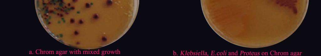
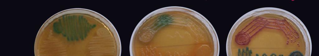
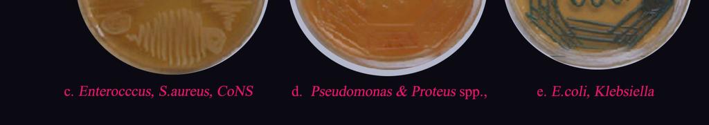
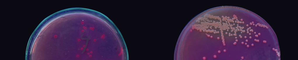
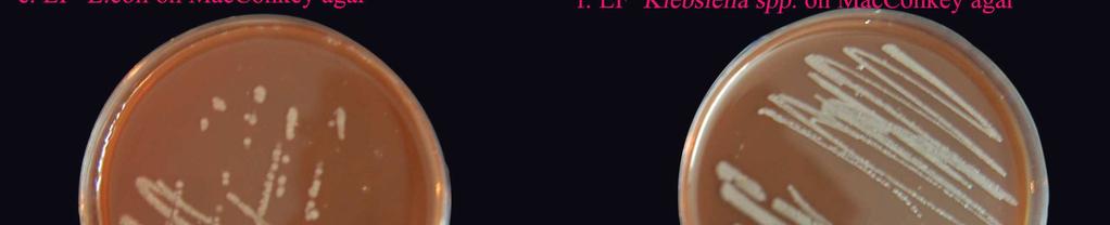
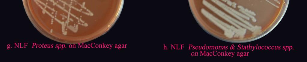
120 102 Figure 8. Identification of bacterial species on Chromagar media. Figure 9. Identification of bacteria on MacConkey agar and Blood agar.
121 103 Table 8. Identification of uropathogens by Gram s staining, SBA and MA agar. Organisms Gram s staining Sheeb Blood Agar (SBA) MacConkey Agar (MA) E. coli GNB B-haemolysis LF, Pink, rough K. pneumoniae GNB - LF, Pink, mucoid Proteus spp. GNB - NLF, Colorless. Pseudomonas spp. GNB, Pigment and fruit odour - NLF, Colorless. Enterobacter spp. Citrobacter spp. GNB - - GNB - - Acinetobacter spp. GNB, diplobacilli - NLF GPC in clusters No No growth to CoNS haemolysis, slight growth white pigment (pale pink). GPC in clusters No S.saprophyticus haemolysis, white pigment Enterococcus spp. GPC, Oval cocci in pairs and short chains No haemolysis S. aureus GPC in clusters B-haemolysis Candida species Gram positive budding yeast with no pseudohyphae, mucoid colony on SDA, capsule +ve in indian ink preparation, Urease +ve.


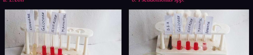
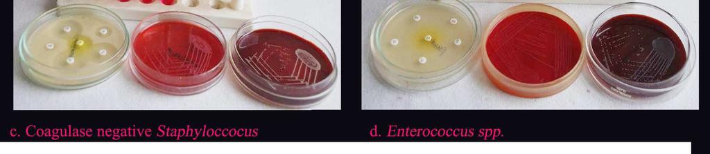
122 Figure 10. Identification of bacteria through biochemical testing and selective media 104
123 105 Table 9. Biochemical identification of bacteria isolated from UTIs. Bacteria Catalase Oxidase Coagulase Indole MR VP Citrate Urease TSI Nitrate Enterococcus spp. + Nd - Nd Nd Nd Nd Nd Nd Nd S. aureus + Nd + Nd Nd Nd Nd Nd Nd Nd S. saprophyticus + Nd - Nd Nd Nd Nd Nd Nd Nd CoNS + Nd - Nd Nd Nd Nd Nd Nd Nd E. coli + - Nd A/AG + K. pneumoniae + - Nd A/AG + P. aeruginosa + + Nd Nd + Proteus spp. + - Nd K/A H2S Acinetobacter spp. + - Nd Nd - + Citrobacter spp. + - Nd A/A H2S Enterobacter spp. + - Nd A/A + + MR: Methyl Red test; VP: Voges-Proskauer test; TSI: Triple Sugar Iron test; V: variable; A/A: Acid in slant and butt; A/AG H2S: Acid in slant and butt with H2S Gas production; A/AG: Acid in slant and butt with Gas production; K/A H2S: Alkali in slant and butt with H2S Gas production; Nd: not done; +: positive; -: negative.
124 Distribution of isolates among positive urine specimens The distribution of isolates among positive uring specimens are presented in the Table 10. Table 10. Frequency of bacterial isolates from urine samples (n = 2400). Data are reported as number of isolates and percentages of total. Microorganisms Frequency Percentage Escherichia coli % K. pneumoniae % Pseudomonas spp % Proteus spp % Enterobacter spp % Citrobacter spp % Acinetobacter spp % GNB Total % CoNS % Enterococcus spp % S. saprophyticus % S.aureus 7 1.1% GPC Total % Candida spp % Total %
125 107 Out of total 2400 samples were collected, only 27.1% of the cases showed growth of bacteria and fungi. 650 uropathogens belonging to 12 different species were isolated (Table 10). The most common isolates in this study have been the Gram negative bacilli which accounts for 85.4% of the total positive isolates. In the Gram negative bacilli, the predominant isolates from UTI were 355 strains of E.coli. The frequency of other uropathogens in descending order were 72 strains of K. pneumoniae 68 strains of Pseudomonas spp., 44 strains of Proteus spp., 10 strains of Enterobacter spp., 4 strains of Citrobacter spp., 2 strains of Acinetobacter spp. In the Gram positive bacteria the main organisms identified were 27 strains of CoNS, 24 strains of Enterococcus spp., 15 strains of Staphylococcus saprophyticus, 7 strains of S. aureus and 22 numbers of Candida spp. Thus, E. coli (54.6%) was the maximally isolated UTI causing bacterium, followed by K. pneumoniae (11.2%), Pseudomonas spp. (10.5%), Proteus spp. (6.8%), CoNS (4.1%), Enterococcus spp. (3.7%), Staphylococcus saprophyticus (2.3%), Enterobacter spp. (1.5%), S. aureus (1.1%), Citrobacter spp. (0.6%) and Acinetobacter spp. (0.3%). Table 10 shows the detailed frequency of all the isolates identified ANTIBACTERIAL SUSCEPTIBILITY PATTERN OF BACTERIAL ISOLATES FROM THE UTIs Characterisation and susceptibility pattern of E.coli In the Gram negative bacilli, the most predominant isolate from UTI were 355 strains of E.coli (54.6%) included for this study. Biochemical Tests: All Gram negative, raised, entire, circular, motile, lactose, glucose fermenting, indole positive, methyl red positive, voges-proskauer negative, citrate negative and urease negative bacilli strains were identified as Escherichia coli. While on MacConkey s agar, lactose fermenting (pink) colonies was detected. On the TSI test E.coli
126 108 strains produced acids both in butt and slant along with gas production (Figure 6(b), 7(c), 8(b), 9(c) and 10(a); Table 7, 8 and 9). This experiment was carried out to study the susceptibility of the bacterial isolates collected from urine specimens toward different 16 antibiotics. The percentages of susceptibility of E.coli isolates to the antibiotics which are commonly used to treat E.coli infections are shown in Table 11. The lowest percentage of susceptibility was manifested against amoxicillin (8.2%) followed by ampicillin (11.3%), cephalexin (11.5%), nalidixic acid (22.8%), cotrimoxazole (23.7%), whereas more susceptibility was observed with imipenem (93.5%) and amikacin (88.2%) followed by nitrofurantoin (87%), tobramycin (76.9%), ciprofloxacin (69.0%), norfloxacin (67.6%), ofloxacin (64.5%), gentamicin (62.5%), cefotaxime (58.0%), ceftazidime (54.7%) and ceftriaxone (47.0%) respectively (Figure 12, 13 and 14). Three aminoglycoside antibiotics, amikacin (11.8%), tobramycin (23.1%) and gentamicin (37.5%) were moderately resistant to species of pathogens used. Similarly, resistance patterns of the beta-lactam group, ampicillin (88.7%) and amoxicillin (91.8%) antibiotics were almost equally resistant to the isolated E.coli. Imipenem resistance patterns were 6.5 of E.coli. Further, the cephalosporin antibiotics, ceftriaxone (53%), ceftazidime (45.3%) and cefotaxime (42.0%) were moderately resistant and cephalexin (88.5%) was highly resistant to species of pathogens used. Similarly, the fluoroquinolone group, ofloxacin (35.5%), norfloxacin (32.4%) and ciprofloxacin (31%) were low resistant and nalidixic acid (77.2%) were highly resistant to species of pathogens used. Among these four antibiotics, nalidixic acid was recorded to be more resistant to these pathogens. Lastly, detailed antibiograms of two stand-alone antibiotics, cotrimoxazole and nitrofurantoin were recorded. Nitrofurantoin was found very low resistance for 13% of E.coli. The resistance percent values of the cotrimoxazole were 76.3% (Figure 11).
127 109 Table 11. Antibiotic susceptibility pattern of E. coli. Antibiotic Sensitive No (%) Moderately Sensitive No (%) Total Sensitive (%) Total Resistant No (%) Amoxicillin 20 (5.6) 9 (2.6) 29 (8.2) 326 (91.8) Ampicillin 30 (8.5) 10 (2.8) 40 (11.3) 315 (88.7) Cefotaxime 190 (53.5) 16 (4.5) 206 (58.0) 149 (42.0) Ceftazidime 188 (53.0) 6 (1.7) 194 (54.7) 161 (45.3) Ceftriaxone 163 (45.9) 4 (1.1) 164 (47.0) 188 (53) Cephalexin 39 (10.9) 2 (0.6) 41 (11.5) 314 (88.5) Ciprofloxacin 239 (67.3) 6 (1.7) 245 (69.0) 110 (31.0) Nalidixic acid 78 (22.0) 3 (0.8) 81 (22.8) 274 (77.2) Norfloxacin 237 (66.8) 3 (0.8) 240 (67.6) 115 (32.4) Ofloxacin 227 (63.9) 2 (0.6) 229 (64.5) 126 (35.5) Amikacin 297 (83.7) 16 (4.5) 313 (88.2) 42 (11.8) Gentamicin 212 (59.7) 10 (2.8) 222 (62.5) 133 (37.5) Tobramycin 269 (75.8) 4 (1.1) 271 (76.9) 82 (23.1) Imipenem 327 (92.1) 5 (1.4) 332 (93.5) 23 (6.5) Nitrofurantoin 303 (85.3) 6 (1.7) 309 (87.0) 46 (13.0) Cotrimoxazole 81 (22.8) 3 (0.9) 84 (23.7) 271 (76.3) Results are expressed as a percentage of 355 E.coli isolates susceptible, moderately susceptible and resistant, respectively, for each antimicrobial Percentage (%) d AX AM CTX CAZ CT CL CIP NA NX OF AK GEN TB IPM NIT COT Antibiotics Figure 11. Antibiotic resistance of E. coli strains from patients with UTIs.

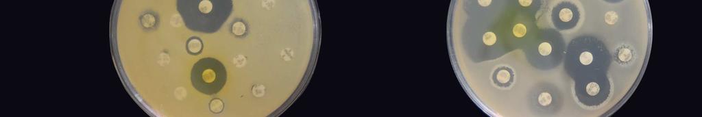
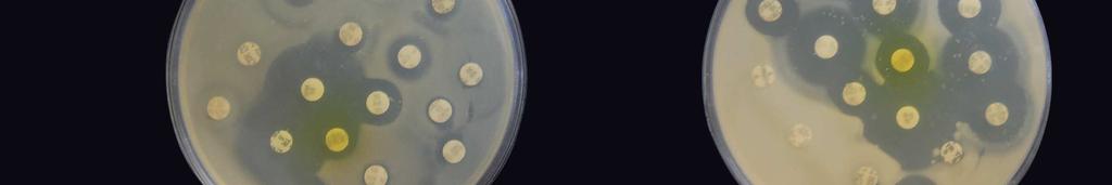

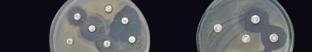


128 Figure 12. Antibiotic susceptibility pattern of bacteria isolated from UTIs. 110


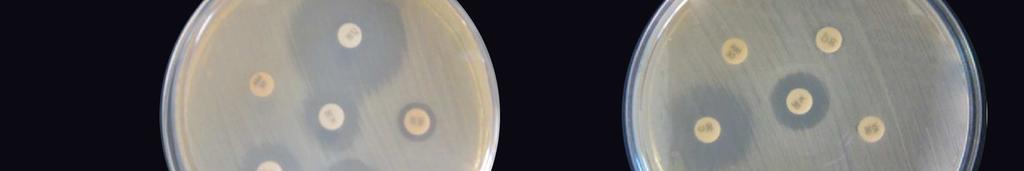

129 Figure 13. Antibiotic susceptibility pattern of bacteria isolated from UTIs. 111


130 Figure 14. Antibiotic susceptibility pattern of Proteus spp. and MDR E.coli isolated from UTIs. 112
131 Characterisation and susceptibility pattern of K. pneumonia. Biochemical Tests: Gram negative, mucoid, non-motile, lactose, glucose fermenting, voges-proskauer positive, urease positive, citrate positive and indole negative bacilli strains were identified as Klebsiella pneumoniae. While on MacConkey s agar, lactose fermenting (pink) mucoid colonies was detected. On the TSI test, Klebsiella pneumoniae produced acids both in butt and slant along with gas production (Table 7, 8 and 9; Figure 6(d), 7(d), 8(b, c) and 9(f). The percentages of susceptibility of Klebsiella pneumoniae to the antibiotics which are commonly used to treat Klebsiella infections are shown in Table 12. The lowest percentage of susceptibility was manifested against amoxicillin (11.1%) followed by ampicillin (16.7%), cotrimoxazole (29.2%), cephalexin (30.6%) and nalidixic acid (49.4%), whereas more susceptibility was observed with amikacin/imipenem (86.1%), followed by gentamicin/tobramycin (80.6%), ofloxacin (79.2%), ciprofloxacin (76.4%), norfloxacin (75%), cefotaxime (66.7%), ceftriaxone (66.7%), ceftazidime (54.2%) and nitrofurantoin (50%) respectively (Figure 12 and 13). Three aminoglycoside antibiotics, amikacin (13.9%), and gentamicin, tobramycin (19.4%) were moderately resistant to species of pathogens used. Similarly, resistance patterns of the beta-lactam group, ampicillin (83.3%) and amoxicillin (88.9) are detailed in Table 12. These two antibiotics were almost equally resistant to the isolated Klebsiella pneumoniae. Imipenem resistance patterns were 13.9% of Klebsiella pneumoniae.
132 114 Table 12. Antibiotic susceptibility pattern of K. pneumoniae Antibiotic Sensitive No (%) Moderately Sensitive No (%) Total Sensitive (%) Total Resistant No (%) Amoxicillin 6 (8.3) 2 (2.8) 8 (11.1) 64 (88.9) Ampicillin 10 (13.9) 2 (2.8) 12 (16.7) 60 (83.3) Cefotaxime 43 (59.7) 5 (7.0) 48 (66.7) 24 (33.3) Ceftazidime 35 (48.6) 4 (5.6) 39 (54.2) 33 (45.8) Ceftriaxone 48 (66.7) 0 48 (66.7) 24 (33.3) Cephalexin 19 (26.4) 3 (4.2) 22 (30.6) 50 (69.4) Ciprofloxacin 55 (76.4) 0 55 (76.4) 17 (23.6) Nalidixic acid 32 (49.4) 0 32 (49.4) 40 (50.6) Norfloxacin 52 (72.2) 2 (2.8) 54 (75.0) 18 (25.0) Ofloxacin 56 (77.8) 1 (1.4) 57 (79.2) 15 (20.8) Amikacin 62 (86.1) 0 62 (86.1) 10 (13.9) Gentamicin 58 (80.6) 0 58 (80.6) 14 (19.4) Tobramycin 57 (79.2) 1 (1.4) 58 (80.6) 14 (19.4) Imipenem 59 (81.9) 3 (4.2) 62 (86.1) 10 (13.9) Nitrofurantoin 34 (47.2) 2 (2.8) 36 (50.0) 36 (50.0) Cotrimoxazole 20 (27.8) 1 (1.4) 21 (29.2) 51 (70.8) Results are expressed as a percentage of 72 K. pneumoniae susceptible, moderately susceptible and resistant, respectively, for each antimicrobial. Percentage (%) AX AM CTX CAZ CT CL CIP NA NX OF AK GEN TB IPM NIT COT Antibiotics Figure 15. Antibiotic resistance of Klebsiella strains from patients with UTIs.
133 115 Further, resistance-percent values of UTI bacteria to cephalosporin antibiotics, Cefotaxime/ceftriaxone (33.3%), ceftazidime (45.8%) were moderately resistant and cephalexin (69.4%) was highly resistant to species of pathogens used. Similarly, resistance-percent values of UTI bacteria to antibiotics of the fluoroquinolone group, ofloxacin (20.8%), ciprofloxacin (23.6%), norfloxacin (25%) were low resistant and nalidixic acid (50.6%) were moderately resistant to species of pathogens used. These antibiotics were resistant to Klebsiella pneumoniae in the order: nalidixic acid, norfloxacin, ofloxacin and ciprofloxacin. Among these four antibiotics, nalidixic acid was recorded to be more resistant to these pathogens. The antibiograms of two stand-alone antibiotics, cotrimoxazole (50%) and nitrofurantoin (70.8%) were recorded (Figure 15) Characterisation and susceptibility pattern of Pseudomonas spp. The rate of isolation of Pseudomonas species from urine samples was found to be 2.8% from 2400 samples. Biochemical Tests: All isolates of Pseudomonas species were Gram negative bacilli, actively motile and show different colonies large and small ones with irregular translucent edges and dark center. All strains were oxidase positive, produce acid by oxidation of sugars not by fermentation, produce gelatin liquefaction and formed slime with surface pellicles when grown in nutrient broth. Large, flat, opaque, aerobic, irregular colonies having grape-like smell, yellow-green pyocyannin pigment producing colonies on common culture media, oxidase positive colonies which grow at 42 o C were identified as Pseudomonas aeruginosa (Figure 6(c), 8(d), 9(b) and 10(b); Table 7, 8 and 9).
134 116 Table 13. Antibiotic susceptibility pattern of Pseudomonas spp. Antibiotic Sensitive No (%) Moderately Sensitive No (%) Total Sensitive (%) Total Resistant No (%) Amoxicillin 0 (0.0) 0 (0.0) 0 (0.0) 68 (100) Ampicillin 0 (0.0) 0 (0.0) 0 (0.0) 68 (100) Cefotaxime 46 (67.6) 4 (5.9) 50 (73.5) 18 (26.5) Ceftazidime 39 (57.4) 2 (2.9) 41 (60.3) 27(39.7) Ceftriaxone 24 (35.3) 2 (2.9) 26 (38.2) 42 (61.8) Cephalexin 10 (14.7) 2 (2.9) 12 (17.6) 56 (82.4) Ciprofloxacin 24 (35.3) 2 (2.9) 26 (38.2) 42 (61.8) Nalidixic acid 5 (7.4) 0 (0.0) 5 (7.4) 63 (92.6) Norfloxacin (4.4) 13 (19.1) 55 (80.9) Ofloxacin 24 (35.3) 1 (1.5) 25 (36.8) 43 (63.2) Amikacin 60 (88.2) 0 (0.0) 60 (88.2) 8 (11.8) Gentamicin 46 (67.6) 1 (1.5) 47 (69.1) 21 (30.9) Tobramycin 59 (86.8) 0 (0.0) 59 (86.8) 9 (13.2) Imipenem 39 (57.3) 1 (1.5) 40 (58.8) 28 (41.2) Nitrofurantoin 19 (27.9) 1 (1.5) 20 (29.4) 48 (70.6) Cotrimoxazole 0 (0.0) 0 (0.0) 0 (0.0) 68 (100) Results are expressed as a percentage of 68 Pseudomonas isolates susceptible, moderately susceptible and resistant, respectively, for each antimicrobial Percentage (%) AX AM CTX CAZ CT CL CIP NA NX OF Antibiotics AK GEN TB IPM NIT COT Figure 16: UTIs. Antibiotic resistance of Pseudomonas strains from patients with
135 117 This constitutes 10.5% (68) of the Pseudomonas species, which were highly sensitive towards amikacin (88.2%), tobramycin (86.8%), cefotaxime (73.5%), gentamicin (69.1%), ceftazidime (60.3%) and imipenem (58.8%). However, they show a low sensitivity rate towards ceftriaxone (38.2%), ciprofloxacin (38.2%), ofloxacin (36.8%), nitrofurantoin (29.4%), norfloxacin (19.1%), cephalexin (17.6%), nalidixic acid (7.4%), amoxicillin (0%), ampicillin (0%) and cotrimoxazole (0%) (Table 13 and Figure 16) Characterisation and susceptibility pattern of Proteus spp. Gram negative, swarming, fish odour colonies on sodium chloridecontaining media, indole negative and urease positive strains were identified as Proteus mirabilis. On the TSI test, Proteus strains produced acids in both butt and slant along with H 2 S gas (Figure 6(c), 8 (e), 9(g); Table 7, 8 and 9). Proteus isolates exhibited highest susceptibility against amikacin 100%, gentamicin 100%, tobramycin 90.9%, ciprofloxacin 88.7%, imipenem 88.6%, ceftriaxone 84.1%, cefotaxime 81.8%, ofloxacin 79.5% and norfloxacin 75%. Other agents showed less sensitivity to nalidixic acid 65.9%, ceftazidime 54.6%, nitrofurantoin 45.5%, ampicillin 24%, cephalexin 22.7%, amoxicillin 20.5% and cotrimoxazole 20.5% (Table 14, Figure 14 and 17).
136 118 Table 14. Antibiotic susceptibility pattern of Proteus spp. Antibiotic Sensitive Moderately Total Total No (%) Sensitive Sensitive Resistant No (%) (%) No (%) Amoxicillin 4 (9.1) 5 (11.4) 9 (20.5) 35 (79.5) Ampicillin 3 (6.8) 6 (13.6) 9 (24.0) 35 (79.6) Cefotaxime 36 (81.8) 0 (0.0) 36 (81.8) 8 (18.2) Ceftazidime 23 (52.3) 1 (2.3) 24 (54.6) 20 (45.4) Ceftriaxone 37 (84.1) 0 (0.0) 37 (84.1) 7 (15.9) Cephalexin 8 (18.2) 2 (4.5) 10 (22.7) 34 (77.3) Ciprofloxacin 38 (86.4) 1 (2.3) 39 (88.7) 5 (11.3) Nalidixic acid 28 (63.6) 1 (2.3) 27 (65.9) 15 (34.1) Norfloxacin 32 (72.7) 1 (2.3) 33(75.0) 11 (25) Ofloxacin 35 (79.5) 0 (0.0) 35 (79.5) 9 (20.5) Amikacin 44 (100) 0 (0.0) 44 (100) 0 (0.0) Gentamicin 43 (97.7) 1 (2.3) 44 (100) 0 (0.0) Tobramycin 39 (88.6) 1 (2.3) 40 (90.9) 4 (9.1) Imipenem 39 (88.6) 0 (0.0) 39 (88.6) 5 (11.4) Nitrofurantoin 19 (43.2) 1 (2.3) 20 (45.5) 24 (54.5) Cotrimoxazole 9 (20.5) 0 (0.0) 9 (20.5) 35 (79.5) Results are expressed as a percentage of 44 Proteus isolates susceptible, moderately susceptible and resistant, respectively, for each antimicrobial Percentage (%) AX AM CTX CAZ CT CL CIP NA NX OF AK GEN TB IPM NIT COT Antibiotics Figure 17. Antibiotic resistance of Proteus strains from patients with UTIs.
137 Characterisation and susceptibility pattern of Enterobacter spp. The isolate from urine produced smooth yellow-colored colonies, 2-3 mm in diameter on blood agar and moist, pink colonies on MacConkey's agar after 24 hours incubation at 37 C. Strains and colonies that were Gram negative, motile, non-sporing, lactose fermenting, indole negative, methyl red negative, voges-proskaeur positive and citrate positive were confirmed to be Enterobacter spp. It fermented glucose, lactose and mannitol with the production of acid and gas. The organism was identified as Enterobacter (Table 7, 8 and 9). All Enterobacter showed 100% sensitivity to amikacin, tobramycin and imipenem, and highly sensitive to ciprofloxacin 90%, gentamicin 90%, ceftriaxone 80%, ofloxacin 80%, cefotaxime 70%, norfloxacin 70%, nalidixic acid 60%, ceftazidime 50%, cotrimoxazole 50%, whereas more than 50% of these isolates were less sensitive to cephalexin 40%, nitrofurantoin 40%, amoxicillin 30% and ampicillin 10% (Table 15; Figure 12 and18) AX AM Percentage (%) CTX CAZ CT CL CIP NA NX OF AK GEN TB IPM NIT COT Antibiotics Figure 18. Antibiotic resistance of Enterobacter strains from patients with UTIs.
138 120 Table 15. Antibiotic susceptibility pattern of Enterobacter spp. Antibiotic Sensitive No (%) Moderately Sensitive Total Sensitive Total Resistant No (%) (%) No (%) Amoxicillin 2 (20) 1 (10) 3 (30) 7 (70) Ampicillin 1 (10) 0 (0.0) 3 (10) 9 (90) Cefotaxime 7 (70) 0 (0.0) 7 (70) 3 (30) Ceftazidime 5 (50) 0 (0.0) 5 (50) 5 (50) Ceftriaxone 7 (70) 1 (10) 8 (80) 2 (20) Cephalexin 2 (20) 2 (20) 4 (40) 6 (60) Ciprofloxacin 8 (80) 1 (10) 9 (90) 1 (10) Nalidixic acid 6 (60) 0 (0.0) 6 (60) 4 (40) Norfloxacin 7 (70) 0 (0.0) 7 (70) 3 (30) Ofloxacin 8 (80) 0 (0.0) 8 (80) 2 (20) Amikacin 10 (100) 0 (0.0) 10 (100) 0 (0.0) Gentamicin 8 (80) 1 (10) 9 (90) 1 (10) Tobramycin 10 (100) 0 (0.0) 10 (100) 0 (0.0) Imipenem 10 (100) 0 (0.0) 10 (100) 0 (0.0) Nitrofurantoin 4 (40) 0 (0.0) 4 (40) 6 (60) Cotrimoxazole 4 (40) 1 (10) 5 (50) 5 (50) Results are expressed as a percentage of 10 Enterobacter isolates susceptible, moderately susceptible and resistant, respectively, for each antimicrobial. Citrobacter spp. was identified by its colony characteristics on MacConkey's agar and results obtained from the biochemical tests; it produced light pink-coloured late-lactose fermenting (LLF) colonies after an 48 hours of incubation on MacConkey's agar; particularly, it was found
139 121 positive to catalase, MR, citrate and nitrate tests, whereas negative to oxidase, indole, VP and urease tests. On the TSI, the bacterium produced both acid and H 2 S gas during growth (Table 7, 8 and 9). Acinetobacter spp. was identified on colony characteristics on nutrient agar and MacConkey agar and from results obtained from adopted biochemical procedures: it grew as colourless, smooth, opaque, raised and pinpoint colonies on Nutrient agar, but as colourless, smooth, opaque, raised and non-lactose-fermenting (NLF) colonies on MacConkey's agar; it was found positive to catalase, VP and citrate tests, whereas negative to oxidase, indole, MR and nitrate tests (Table 7, 8 and 9) Characterisation of Gram Positive Bacteria Further, Gram- positive bacteria as medium to large, smooth, entire, slightly raised, creamy yellow, green/beta-haemolytic colonies on blood agar, found positive to catalase and coagulase tests were confirmed to be S. aureus. Further, bile esculin producing colonies, negative to catalase and coagulase tests were taken as Enterococcus spp., which produced grayish, round, small colonies without any haemolytic zones on blood agar (Figure 6(a,f), 7(a,b), 8(c), 9(c) and 10(c); Table 7, 8 and 9) Antibiotic susceptibility pattern of CoNS Coagulase negative Staphylococcus (CoNS) were showed high rate susceptibility to ciprofloxacin 100%, amikacin 100%, gentamicin 100%, tobramycin 100%, nitrofurantoin 100%, imipenem 96.3%, ceftriaxone 70.4%, ofloxacin 63%, norfloxacin 59.3%, cephalexin 55.6% and amoxicillin (51.9%), but more than half of CoNS showed less sensitivity against cefotaxime 37%, ceftazidime 37%, cotrimoxazole 37%, ampicillin 14.8% and nalidixic acid 0% (Table 16). More than 50% of Coagulase Staphylococcus strains were resistant to the most of commonly used antibiotics (Figure, 12 and 19).
140 122 Table 16. Antibiotic susceptibility pattern of CoNS. Antibiotic Sensitive No (%) Moderately Sensitive No (%) Total Sensitive (%) Total Resistant No (%) Amoxicillin 14 (51.9) 0 (0.0) 14 (51.9) 13 (48.1) Ampicillin 3 (11.1) 1 (3.7) 4 (14.8) 23 (85.2) Cefotaxime 8 (29.6) 2 (7.4) 10 (37.0) 17 (63.0) Ceftazidime 9 (33.3) 1 (3.7) 10 (37.0) 17 (63.0) Ceftriaxone 18 (66.7) 1 (3.7) 19 (70.4) 8 (29.6) Cephalexin 15 (55.6) 0 (0.0) 15 (55.6) 12 (44.4) Ciprofloxacin 27 (100) 0 (0.0) 27 (100) 0 (0.0) Nalidixic acid 0 (0.0) 0 (0.0) 0 (0.0) 27 (100) Norfloxacin 16 (59.3) 0 (0.0) 16 (59.3) 11 (40.7) Ofloxacin 15 (55.6) 2 (7.4) 17 (63) 10 (37.0) Amikacin 27 (100) 0 (0.0) 27 (100) 0 (0.0) Gentamicin 27 (100) 0 (0.0) 27 (100) 0 (0.0) Tobramycin 27 (100) 0 (0.0) 27 (100) 0 (0.0) Imipenem 26 (96.3) 0 (0.0) 26 (96.3) 1 (3.7) Nitrofurantoin 27 (100) 0 (0.0) 27 (100) 0 (0.0) Cotrimoxazole 9 (33.3) 1 (3.7) 10 (37.0) 17 (63.0) Results are expressed as a percentage of 27 CoNS isolates susceptible, moderately susceptible and resistant, respectively, for each antimicrobial Percentage (%) AX AM CTX CAZ CT CL CIP NA NX OF AK GEN TB IPM NIT COT Antibiotics Figure 19. Antibiotic resistance of CoNS strains from patients with UTIs.
141 Antibiotic susceptibility pattern of Enterococcus spp. Enterococci were fully susceptible to ceftriaxone 100%, amikacin 100%, gentamicin 100%, tobramycin 100%, followed by cefotaxime 83.3%, ciprofloxacin 83.3%, imipenem 83.3%, nitrofurantoin 79.2%, ofloxacin 75%,; but it was low sensitivity to ampicillin 54.2%, cephalexin 54.2%, norfloxacin 54.2%, amoxicillin 50%, ceftazidime 41.7%, cotrimoxazole 41.7% and nalidixic acid 0% (Table 17; Figure 12 and 20). Table 17. Antibiotic susceptibility pattern of Enterococcus spp. Antibiotic Sensitive No (%) Moderately Sensitive No (%) Total Sensitive (%) Total Resistant No (%) Amoxicillin 11 (45.8) 1 (4.2) 12 (50.0) 12 (50.0) Ampicillin 13 (54.2) 0 (0.0) 13 (54.2) 11 (45.8) Cefotaxime 20 (83.3) 0 (0.0) 20 (83.3) 4 (16.7) Ceftazidime 10 (41.7) 0 (0.0) 10 (41.7) 14 (58.3) Ceftriaxone 24 (100) 0 (0.0) 24 (100) 0 (0.0) Cephalexin 13 (54.2) 0 (0.0) 13 (54.2) 11 (45.8) Ciprofloxacin 20 (83.3) 0 (0.0) 20 (83.3) 4 (16.7) Nalidixic acid 0 (0.0) 0 (0.0) 0 (0.0) 24 (100) Norfloxacin 13 (54.2) 0 (0.0) 13 (54.2) 11 (45.8) Ofloxacin 17 (70.8) 1 (4.2) 18 (75.0) 6 (25.0) Amikacin 24 (100) 0 (0.0) 24 (100) 0 (0.0) Gentamicin 24 (100) 0 (0.0) 24 (100) 0 (0.0) Tobramycin 24 (100) 0 (0.0) 24 (100) 0 (0.0) Imipenem 20 (83.3) 0 (0.0) 20 (83.3) 4 (16.7) Nitrofurantoin 19 (79.2) 0 (0.0) 19 (79.2) 5 (20.8) Cotrimoxazole 10 (41.7) 0 (0.0) 10 (41.7) 14 (58.3) Results are expressed as a percentage of 24 Enterococcus isolates susceptible, moderately susceptible and resistant, respectively, for each antimicrobial
142 AX AM CTX CAZ CT CL CIP NA NX OF AK GEN TB IPM NIT COT AX Percentage (%) AM CTX CAZ CT CL CIP NA NX OF AK GEN TB IPM NIT COT Antibiotics Figure 20. Antibiotic resistance of Enterococcus strains from patients with UTIs Antibiotic susceptibility pattern of Staphylococcus saprophyticus Staphylococcus saprophyticus was 100% sensitive to amikacin, gentamicin, tobramycin, imipenem, while it was highest sensitive to ofloxacin 89.7%, ciprofloxacin 86.7%, norfloxacin 80%, nitrofurantoin 73.4%, cotrimoxazole 53.3%, ceftazidime 46.7%, cefotaxime 40%, ceftriaxone 40% and cephalexin 26.7%. Staphylococcus aureus was 100 % resistant to three antibiotics amoxicillin, ampicillin and nalidixic acid (Table 18; Figure 13 and 21). Percentage (%) Antibiotics Figure 21. Antibiotic resistance of S.saprophyticus from patients with UTIs.
143 125 Table 18. Antibiotic susceptibility pattern of Staphylococcus saprophyticus. Antibiotic Sensitive No (%) Moderately Sensitive No (%) Total Sensitive (%) Total Resistant No (%) Amoxicillin 0 (0.0) 0 (0.0) 0 (0.0) 15 (100) Ampicillin 0 (0.0) 0 (0.0) 0 (0.0) 15 (100) Cefotaxime 6 (40.0) 0 (0.0) 6 (40.0) 9 (60.0) Ceftazidime 7 (46.7) 0 (0.0) 7 (46.7) 8 (53.3) Ceftriaxone 6 (40.0) 0 (0.0) 6 (40.0) 9 (60.0) Cephalexin 3 (20.0) 1 (6.7) 4 (26.7) 11 (73.3) Ciprofloxacin 13 (86.7) 0 (0.0) 13 (86.7) 2 (13.3) Nalidixic acid 0 (0.0) 0 (0.0) 0 (0.0) 15 (100) Norfloxacin 12 (80.0) 0 (0.0) 12 (80.0) 3 (20.0) Ofloxacin 13 (86.7) 0 (0.0) 13 (89.7) 2 (13.3) Amikacin 15 (100) 0 (0.0) 15 (100) 0 (0.0) Gentamicin 15 (100) 0 (0.0) 15 (100) 0 (0.0) Tobramycin 15 (100) 0 (0.0) 15 (100) 0 (0.0) Imipenem 15 (100) 0 (0.0) 15 (100) 0 (0.0) Nitrofurantoin 10 (66.7) 1 (6.7) 11 (73.4) 4 (26.6) Cotrimoxazole 8 (53.3) 0 (0.0) 8 (53.3) 7 (46.7) Results are expressed as a percentage of 15 S.saprophyticus isolates susceptible, moderately susceptible and resistant, respectively, for each antimicrobial Antibiotic susceptibility pattern of Staphylococcus aureus Staphylococcus aureus were fully susceptible to ceftriaxone, ciprofloxacin, ofloxacin, amikacin, gentamicin, tobramycin, imipenem and nitrofurantoin, followed by cefotaxime 85.7%, cephalexin (85.7%), norfloxacin 85.7%, ceftazidime 71.4%, cotrimoxazole 71.4%, amoxicillin 57.1%, while it were showed less sensitivity against ampicillin 42.9% and nalidixic acid 28.6% (Table 19; Figure 12 and 13).
144 126 Table 19. Antibiotic susceptibility pattern of Staphylococcus aureus. Antibiotic Sensitive No (%) Moderately Sensitive No (%) Total Sensitive (%) Total Resistant No (%) Amoxicillin 4 (57.1) 0 (0.0) 4 (57.1) 3 (42.9) Ampicillin 3 (42.9) 0 (0.0) 3 (42.9) 4 (57.1) Cefotaxime 5 (71.4) 1 (14.3) 6 (85.7) 1 (14.3) Ceftazidime 5 ( (0.0) 5 (71.4) 2 (28.6) Ceftriaxone 7 (100) 0 (0.0) 7 (100) 0 (0.0) Cephalexin 6 (85.7) 0 (0.0) 6 (85.7) 1 (14.3) Ciprofloxacin 7 (100) 0 (0.0) 7 (100) 0 (0.0) Nalidixic acid 2 (28.6) 0 (0.0) 2 (28.6) 5 (71.4) Norfloxacin 6 (85.7) 0 (0.0) 6 (85.7) 1 (14.3) Ofloxacin 7 (100) 0 (0.0) 7 (100) 0 (0.0) Amikacin 7 (100) 0 (0.0) 7 (100) 0 (0.0) Gentamicin 7 (100) 0 (0.0) 7 (100) 0 (0.0) Tobramycin 7 (100) 0 (0.0) 7 (100) 0 (0.0) Imipenem 7 (100) 0 (0.0) 7 (100) 0 (0.0) Nitrofurantoin 7 (100) 0 (0.0) 7 (100) 0 (0.0) Cotrimoxazole 4 (57.1) 1 (14.3) 5 (71.4) 2 (28.6) Results are expressed as a percentage of 7 S.aureus isolates susceptible, moderately susceptible and resistant, respectively, for each antimicrobial Percentage (%) AX AM CTX CAZ CT CL CIP NA NX OF AK GEN TB IPM NIT COT Antibiotics Figure 22. Antibiotic resistance of S. aureus from patients with UTIs.
145 Antibiotic resistance patterns of all Bacterial isolates from UTIs All the bacterial isolates from urinary tract infection patients were highly susceptible to amikacin 90.4%, imipenem 88.6%, tobramycin 82.5%, gentamicin/nitrofurantoin 72.8%, ciprofloxacin 70.9%, ofloxacin 65.8%, norfloxacin 63.3%, cefotaxime 62.5%, ceftriaxone 55%, ceftazidime 53.7%, nalidixic acid 24.9%, cotrimoxazole 24.4%, cephalexin 20.4%, ampicillin 13.2%, and amoxicillin 12.7%. High level of resistance was seen to ampicilin, amoxicillin, cephalexin and nalidixic acid. Most of the isolates were sensitive to amikacin, imipenem, tobramycin, gentamicin, nitrofurantoin, ciprofloxacin and ofloxacin. Amikacin was found to be very effective against all the isolates. (Table 20 and Figure 23) AX AM CTX CAZ CT CL CIP NA NX OF AK GEN TB IPM NIT COT Percentage (%) Antibiotics Figure 23. Antibiotic resistant of all bacterial strains isolated from UTIs Aminoglycoside resistance in bacterial isolates from UTIs Three aminoglycoside antibiotics (µg/disc), amikacin, gentamicin and tobramycin were moderately resistant to all bacteria used, in ranges, 11.8% to 37.5% of 355 strains of E. coli, 13.9% to 19.4% of 72 strains of K. pneumoniae 11.8% to 30.9% of 68 strains of Pseudomonas spp. 0% to 9.1% of 44 strains of Proteus spp. 0% to 1% of 10 strains of Enterobacter spp. 0% of 27 strains of CoNS 0% of 24 strains of Enterococcus spp. 0% of 15 strains of S.saprophyticus and 0% of seven strains of S. aureus. Among these three antibiotics, gentamicin was recorded to be more resistant to these pathogens (Table 20).
146 Table 20. Antibiotic resistance pattern of bacteria isolated from urine sample (n=628). Antibiotics E. coli No. (%) Klebsiella No. (%) Pseudomonas No. (%) Proteus No. (%) Enterobacter No. (%) CoNS No. (%) Enterococcus No. (%) S. saprophyticus No. (%) S.aureus No. (%) Total No. (%) AX 326 (91.8) 64 (88.9) 68 (100) 35 (79.5) 7 (70) 13 (48.1) 12 (50) 15 (100) 3 (42.9) 543 (87.3) AM 315 (88.7) 60 (83.3) 68 (100) 35 (79.5) 9 (90) 23 (85.2) 11 (45.8) 15 (100) 4 (57.1) 540 (86.8) CTX 149 (42.0) 24 (33.3) 18 (26.5) 8 (18.2) 3 (30) 17 (63.0) 4 (16.7) 9 (60.0) 1 (14.3) 233 (37.5) CAZ 161 (45.3) 33 (45.8) 27(39.7) 20 (45.5) 5 (50) 17 (63.0) 14 (58.3) 8 (53.3) 2 (28.6) 287 (46.1) CT 188 (53.0) 24 (33.3) 42 (61.8) 7 (15.9) 2 (20) 8 (29.6) 0 (0.0) 9 (60.0) 0 (0.0) 280 (45.0) CL 314 (88.5) 50 (69.4) 56 (82.4) 34 (77.3) 6 (60) 12 (44.4) 11 (45.8) 11 (73.3) 1 (14.3) 495 (79.6) CIP 110 (31.0) 17 (23.6) 42 (61.8) 5 (11.4) 1 (10) 0 (0.0) 4 (16.7) 2 (13.3) 0 (0.0) 181 (29.1) NA 274 (77.2) 40 (55.6) 63 (92.6) 15 (34.1) 4 (40) 27 (100) 24 (100) 15 (100) 5 (71.4) 467 (75.1) NX 115 (32.4) 18 (25.0) 55 (80.9) 11 (25.0) 3 (30) 11 (40.7) 11 (45.8) 3 (20.0) 1 (14.3) 228 (36.7) OF 126 (35.5) 15 (20.8) 43 (63.2) 9 (20.5) 2 (20) 10 (37.0) 6 (25.0) 2 (13.3) 0 (0.0) 213 (34.2) AK 42 (11.8) 10 (13.9) 8 (11.8) 0 (0.0) 0 (0.0) 0 (0.0) 0 (0.0) 0 (0.0) 0 (0.0) 60 (9.60) GEN 133 (37.5) 14 (19.4) 21 (30.9) 0 (0.0) 1 (10) 0 (0.0) 0 (0.0) 0 (0.0) 0 (0.0) 169 (27.2) TB 82 (23.1) 14 (19.4) 9 (13.2) 4 (9.1) 0 (0.0) 0 (0.0) 0 (0.0) 0 (0.0) 0 (0.0) 109 (17.5) IPM 23 (6.5) 10 (13.9) 28 (41.2) 5 (11.4) 0 (0.0) 1 (3.7) 4 (16.7) 0 (0.0) 0 (0.0) 71 (11.4) NIT 46 (13.0) 36 (50.0) 48 (70.6) 24 (54.5) 6 (60) 0 (0.0) 5 (20.8) 4 (26.6) 0 (0.0) 169 (27.2) COT 271 (76.3) 51 (70.8) 68 (100) 35 (79.5) 5 (50) 17 (63.0) 14 (58.3) 7 (46.7) 2 (28.6) 470 (75.6) Total 355 (54.6) 72 (11.2) 68 (10.5) 44 (6.8) 10 (1.5) 27 (4.1) 24 (3.7) 15 (2.3) 7 (1.1) 622 (95.7) 128
147 Beta-lactam group of antibiotics resistance in bacterial isolates from UTIs The percentages of resistance patterns of both Gram negative bacilli and Gram positive cocci with two antibiotics of the beta-lactam group (ampicillin, amoxicillin) are detailed (Table 20); resistance patterns were in ranges: 88.7% to 91.8% of 355 strains of E. coli, 83.3% to 88.9% of 72 strains of K. pneumoniae 100% of 68 strains of Pseudomonas spp. 79.5% of 4 strains of Proteus spp. 70% to 90% of 10 strains of Enterobacter spp. 48.1% to 85.2% of 27 strains of CoNS 45.8% to 50% of 24 strains of Enterococcus spp. 100% of 15 strains of S.saprophyticus and 42.9% to 57.1% of 7 strains of S. aureus. All these two antibiotics were almost equally resistant to the isolated UTI bacteria. Imipenem is an intravenous β-lactam antibiotic Imipenem belongs to the subgroup of carbapenems. The percentages of resistance patterns of both Gram positive and Gram negative bacteria with these antibiotics are detailed (Table 20); resistance patterns were in ranges: 6.5% of 355 strains of E. coli, 13.9% of 72 strains of Klesiella spp. 41.2% of 68 strains of Pseudomonas spp. 11.4% of 4 strains of Proteus spp. 3.7% of 27 strains of CoNS 16.7% of 24 strains of Enterococcus spp. 0% of 10 strains of Enterobacter spp. 15 strains of S. saprophyticus and 7 strains of S. aureus. Further, resistance-percent values of UTI bacteria to cephalosporin antibiotics (Cefotaxime, ceftazidime, ceftriaxone and cephalexin) were in ranges, 42% to 88.5% of 355 strains of E.coli, 33.3% to 69.4% of 72 strains of K. pneumoniae 26.5% to 82.4% of strains of Pseudomonas spp. 15.9% to 77.3% of 4 strains of Proteus spp. 20% to 60% of 10 strains of Enterobacter spp. 29.6% to 63% of 27 strains of CoNS 16.7% to 58.3% of 24 strains of Enterococcus spp. 53.3% to 73.3% of 15 strains of S. saprophyticus 4.3% to 28.6% of 7 strains of S. aureus. All these four antibiotics were moderately resistant to the isolated UTI pathogens. These
148 130 antibiotics were resistant to UTI pathogens in the order: cephalexin >ceftazidime>cefotaxime>ceftriaxone (Table 20) Fluoroquinolone resistance in bacterial isolates from UTIs Similarly, resistance-percent values of UTI bacteria to antibiotics of the fluoroquinolone group (ciprofloxacin, nalidixic acid, norfloxacin and ofloxacin) were in ranges; 31% to 77.2%of 355 strains of E. coli, 20.8% to 55.6% of 72 strains of K. pneumoniae 61.8% of 92.6 strains of Pseudomonas spp. 11.4% to 34.1 of 4 strains of Proteus spp. 10% to 40% of 10 strains of Enterobacter spp. 0% to 100% of 27 strains of CoNS 16.7% to 100% of 24 strains of Enterococcus spp. 13.3% to 100% of 15 strains of S.saprophyticus and 0% to 71.4% of 7 strains of S. aureus (Table 20). These antibiotics were resistant to UTI pathogens in the order: nalidixic acid> norfloxacin>ofloxacin>ciprofloxacin. Among these four antibiotics, nalidixic acid was recorded to be more resistant to these pathogens Two stand-alone antibiotics resistance in bacteria from UTIs The detailed antibiograms of two stand-alone antibiotics, cotrimoxazole and nitrofurantoin were recorded. Nitrofurantoin was found resistance for 13% of 355 strains of E. coli, 50% of 72 strains of K. pneumoniae 70.6% of 68 strains of Pseudomonas spp. 54.5% of 4 strains of Proteus spp. 60% of 10 strains of Enterobacter spp. 0% of 27 strains of CoNS 20.8% of 24 strains of Enterococcus spp. 26.6% of 15 strains of S.saprophyticus and 0% of 7 strains of S. aureus. Surprisingly, resistance-percent values of UTI bacteria to antibiotic of the cotrimoxazole were 76.3% of 355 strains of E. coli, 70.8% of 72 strains of K. pneumoniae, 100% of 68 strains of P. aeruginosa. 79.5% of 4 strains of Proteus spp. 50% of 10 strains of Enterobacter spp. 63% of 27 strains of CoNS 58.3% of 24 strains of Enterococcus spp. 46.7% of 15 strains of S. saprophyticus and 28.6% of 7 strains of S. aureus (Table 20).
149 ISOLATION AND CHARACTERISATION OF THE PATHOGENIC CANDIDA SPECIES Candida species were isolated from twenty two numbers of patients. The species were identified according to morphology and color of colonies on CHROMagar Candida. The color of colonies on CHROMagar Candida was similar as given by the manufacturer, i.e. green colonies of C.albicans, steel blue colonies of C.tropicalis accompanied by purple pigmentation which diffuses into surrounding agar by growth, and large, fuzzy, rose colored colonies with white edges of C.krusei, the smooth white to light pink colonies of C. glabrata which later became pink (Table 21). The prevalence candiduria in critically ill patients in the study was 3.4%. Out of the total 22 strains isolated, 4(18.2%) and 18(81.8%) candida strains were isolated from male and female patients respectively. C. albicans (45.5%) was the highest occurring pathogens isolated, followed by C. glabrata (22.7%), C. tropicalis (18.2%), and C. krusei (13.6%) (Table 21 and 22; Figure 24). Table 21. Characteristics of Candida species on Chromagar Candida media. Species Colony characteristics on CHROMagar Candida Total number. of isolates C. albicans Apple green colonies; consistent 10 (45.5%) C. glabrata White large glossy pale pink to violet colonies C. tropicalis Dull blue, to purple color that diffused into surrounding agar with pale pink edges C. krusei Large, flat, spreading, pale pink colonies with matt surfaces 5 (22.7%) 4 (18.2%) 3 (13.6%)
 2 8 C. glabrata 5 (22.")
 1 2 Total 22 (100%) 4 (18.2%) 18 (81.8%) Figure 24.")
150 132 Table 22. Distribution of isolated Candida species (n=22). Species No. of organisms Male Female C. albicans 10 (45.5%) 2 8 C. glabrata 5 (22.7%) 1 4 C. tropicalis 4 (18.2%) 0 4 C. krusei 3 (13.6%) 1 2 Total 22 (100%) 4 (18.2%) 18 (81.8%) Figure 24. Characteristics of Candida species on Chromagar Candida media.
151 133 Table 23. Identification of isolated Candida species by Direct wet mount and Germ tube methods. Sl. No. Samples No Sex of patient Female Female Female Female Female Female Female Female Female 842 Female 951 Female 954 Female 1266 Male 1288 Male 1290 Male 1350 Female 1401 Female 1602 Female 1655 Female 1656 Female Male Female Direct wet mount examination Yeast like structure Yeast like structure Yeast like structure Yeast like structure Yeast like structure Yeast like structure Yeast like structure Yeast like structure Yeast like structure Yeast like structure Yeast like structure Yeast like structure Yeast like structure Yeast like structure Yeast like structure Yeast like structure Yeast like structure Yeast like structure Yeast like structure Yeast like structure Yeast like structure Yeast like structure Chromagar C. tropicalis Germ tube Not formed C. albicans Formed C. albicans Formed C. albicans Formed C. albicans Formed C. albicans Formed C. albicans Formed C. krusei C. krusei Not formed Not formed C. albicans Formed C. albicans Formed C. krusei Not formed C. albicans Formed C. glabrata - C. tropicalis Not formed C. glabrata - C. glabrata - C. glabrata - C. tropicalis Not formed C. glabrata - C. albicans Formed C. tropicalis Not formed
152 ANTIFUNGAL SUSCEPTIBILITY PATTERN OF CANDIDA SPECIES CAUSING UTIs The antifungal sensitivity tests carried out using commercially available antifungal disc that 10 (100%) strains of C. albicans were sensitive to amphotericin B and itraconazole, while 7 (70%) strains of C. albicans were resistant to fluconazole and 2 (20) to Ketoconazole. Of the C. glabrata strains, all (100%) strains were sensitive to Itraconazole, 4 (80%) were sensitive to amphotericin B and itraconazole, whereas 3 strains (60%) were resistant to Ketoconazole. Among the C. tropicalis strains, four (100%) were found sensitive to amphotericin B and Itraconazole. While all four strains (100%) were found resistant fluconazole and 2 stains resistant (20%) to Ketoconazole. C. krusei, 3 (100%) strains were found sensitive to amphotericin B and 100 % resistant to Fluconazole and Ketoconazole, followed by 1 (33.3%) to Itraconazole antifungal agents. Amphotericin B and itraconazole 21 (95.5%) was found to be the most effective antifungal agent (Table 24; Figure 25 and 26).
153 Table 24. Antifungal susceptibility pattern of Candida species causing UTIs. Candida Species Amphotericin B (20µg) Fluconazole (25µg) Itraconazole (50µg) Ketoconazole (10µg) S R S R S R S R C. albicans 10 (100%) 0 (0.0%) 3 (30%) 7 (70%) 10 (100%) 0 (0.0%) 2 (20%) 8 (80%) C. glabrata 4 (80%) 1 (20%) 1 (20%) 4 (80%) 5 (100%) 0 (0.0%) 2 (40%) 3 (60%) C. tropicalis 4 (100% 0 (0.0%) 0 (0.0%) 4 (100%) 4 (100%) 0 (0.0%) 0 (0.0%) 4 (100%) C. krusei 3 (100%) 0 (0.0%) 0 (0.0%) 3 (100%) 2 (66.7%) 1 (33.3%) 0 (0.0%) 3 (100%) Total 21 (95.5%) 1 (4.5%) 4 (18.2%) 18 (81.8%) 21 (95.5%) 1 (4.5%) 4 (18.2%) 18 (81.8%) 135
 80 60 40 20 Amphotericin B Fluconazole")

154 136 Figure 25. Antifungal susceptibility pattern of Candida species P e r c e n t a g e ( % ) Amphotericin B Fluconazole Itraconazole Ketoconazole 0 C. albicans C. glabrata C. tropicalis C. krusei Figure 26. Antifungal susceptibility of Candida species isolated from candiduria

155 PLASMID ANALYSIS-MOLECULAR CHARACTERISATION OF ESBL PRODUCING E.COLI ESBL Positive E.coli Totally 100 MDR E. coli isolate were isolated from different source of urine specimens. Ten isolates were confirmed positive for ESBL production. Third generation cephalosporins showed 37% to 45% resistance. For these isolates gentamicin showed 27.2% resistance, whereas amphicillin and amoxicillin showed 86% to 87% resistance. Imipenem and amikacin depicted 88.6% and 90.4% sensitive respectively. Using the DDST method, ESBL was confirmed in 10 isolates (10%) of which two from cystitis samples (20%), four from pyelonephritis samples (40%) and the rest were normal urine samples (40%). Figure 27. Double disc synergy test.(augmentin Disc (20 g of amoxicillin + 10 g of clavulanic acid) with 30 g of cefotaxime ).
156 PCR and RFLP analysis Confirmed ESBL samples were checked for the presence of TEM gene by PCR amplification and it was found to be positive. PCR products of molecular size 717 bps showed that it was TEM gene and it resulted in 10% of isolates contained bla TEM genes (Figure 26). The results showed that 10% of isolates were positive for ESBL both by phenotypic and genotypic methods. Further Restriction fragment length polymorphism (RFLP) was carried out for genomic DNA with Xba I restriction enzyme to check for the polymorphism (Figure 26). RFLP analysis showed eight different banding patterns for the ten samples among which four samples showed identical banding pattern and other two samples also had the similar banding pattern, indicating two different source of origin. Other samples had different patterns indicating that they are not from same samples. Thus ten samples showed eight different banding patterns concludes that the samples are not from same origin.


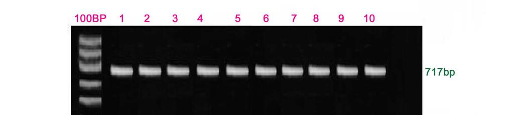

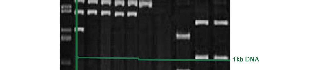

157 Figure 28. Molecular Characteristics of ESBL producing MDR E.coli. 139
Dr. C. MANIKANDAN, Director,
 STUDY OF PREVALENCE AND ANTIMICROBIAL SUSCEPTIBILITIES OF BACTERIA AND FUNGI ISOLATED FROM PATIENTS WITH URINARY TRACT INFECTIONS IN PATTUKKOTTAI, TAMIL NADU, INDIA Dr. C. MANIKANDAN, Director, Gangasaras
ESBL Producers An Increasing Problem: An Overview Of An Underrated Threat
 ESBL Producers An Increasing Problem: An Overview Of An Underrated Threat Hicham Ezzat Professor of Microbiology and Immunology Cairo University Introduction 1 Since the 1980s there have been dramatic
A retrospective analysis of urine culture results issued by the microbiology department, Teaching Hospital, Karapitiya
 A retrospective analysis of urine culture results issued by the microbiology department, Teaching Hospital, Karapitiya LU Edirisinghe 1, D Vidanagama 2 1 Senior Registrar in Medicine, 2 Consultant Microbiologist,
Detection of ESBL Producing Gram Negative Uropathogens and their Antibiotic Resistance Pattern from a Tertiary Care Centre, Bengaluru, India
 ISSN: 2319-7706 Volume 4 Number 12 (2015) pp. 578-583 http://www.ijcmas.com Original Research Article Detection of ESBL Producing Gram Negative Uropathogens and their Antibiotic Resistance Pattern from
Key words: Urinary tract infection, Antibiotic resistance, E.coli.
 Original article MICROBIOLOGICAL STUDY OF URINE ISOLATES IN OUT PATIENTS AND ITS RESISTANCE PATTERN AT A TERTIARY CARE HOSPITAL IN KANPUR. R.Sujatha 1,Deepak S 2, Nidhi P 3, Vaishali S 2, Dilshad K 2 1.
Bacterial Pathogens in Urinary Tract Infection and Antibiotic Susceptibility Pattern from a Teaching Hospital, Bengaluru, India
 ISSN: 2319-7706 Volume 4 Number 11 (2015) pp. 731-736 http://www.ijcmas.com Original Research Article Bacterial Pathogens in Urinary Tract Infection and Antibiotic Susceptibility Pattern from a Teaching
Safe Patient Care Keeping our Residents Safe Use Standard Precautions for ALL Residents at ALL times
 Safe Patient Care Keeping our Residents Safe 2016 Use Standard Precautions for ALL Residents at ALL times #safepatientcare Do bugs need drugs? Dr Deirdre O Brien Consultant Microbiologist Mercy University
Cork and Kerry SARI Newsletter; Vol. 2 (2), December 2006
, December 2006") Cork and SARI Newsletter; Vol. 2 (2), December 6 Item Type Newsletter Authors Murray, Deirdre;O'Connor, Nuala;Condon, Rosalind Download date 31/1/18 15:27:31 Link to Item http://hdl.handle.net/1147/67296
Cork and SARI Newsletter; Vol. 2 (2), December 6 Item Type Newsletter Authors Murray, Deirdre;O'Connor, Nuala;Condon, Rosalind Download date 31/1/18 15:27:31 Link to Item http://hdl.handle.net/1147/67296
Prevalence of Extended Spectrum Beta- Lactamase Producers among Various Clinical Samples in a Tertiary Care Hospital: Kurnool District, India
 International Journal of Current Microbiology and Applied Sciences ISSN: 319-77 Volume Number (17) pp. 57-3 Journal homepage: http://www.ijcmas.com Original Research Article https://doi.org/1.5/ijcmas.17..31
Int.J.Curr.Microbiol.App.Sci (2017) 6(3):
 6(3):") International Journal of Current Microbiology and Applied Sciences ISSN: 2319-7706 Volume 6 Number 3 (2017) pp. 891-895 Journal homepage: http://www.ijcmas.com Original Research Article https://doi.org/10.20546/ijcmas.2017.603.104
International Journal of Current Microbiology and Applied Sciences ISSN: 2319-7706 Volume 6 Number 3 (2017) pp. 891-895 Journal homepage: http://www.ijcmas.com Original Research Article https://doi.org/10.20546/ijcmas.2017.603.104
Cipro for gram positive cocci in urine
 Buscar... Cipro for gram positive cocci in urine 20-6-2017 Pneumonia can be generally defined as an infection of the lung parenchyma, in which consolidation of the affected part and a filling of the alveolar
ALARMING RATES OF PREVALENCE OF ESBL PRODUCING E. COLI IN URINARY TRACT INFECTION CASES IN A TERTIARY CARE NEUROSPECIALITY HOSPITAL
 ALARMING RATES OF PREVALENCE OF ESBL PRODUCING E. COLI IN URINARY TRACT INFECTION CASES IN A TERTIARY CARE NEUROSPECIALITY HOSPITAL Pearl. A Prabal*,Sourav Maiti Institute of Neurosciences, Kolkata, India
RETROSPECTIVE STUDY OF GRAM NEGATIVE BACILLI ISOLATES AMONG DIFFERENT CLINICAL SAMPLES FROM A DIAGNOSTIC CENTER OF KANPUR
 Original article RETROSPECTIVE STUDY OF GRAM NEGATIVE BACILLI ISOLATES AMONG DIFFERENT CLINICAL SAMPLES FROM A DIAGNOSTIC CENTER OF KANPUR R.Sujatha 1,Nidhi Pal 2, Deepak S 3 1. Professor & Head, Department
Northwestern Medicine Central DuPage Hospital Antimicrobial Criteria Updated 11/16/16
 Northwestern Medicine Central DuPage Hospital Antimicrobial Criteria Updated 11/16/16 These criteria are based on national and local susceptibility data as well as Infectious Disease Society of America
CONTAGIOUS COMMENTS Department of Epidemiology
 VOLUME XXIII NUMBER 1 July 2008 CONTAGIOUS COMMENTS Department of Epidemiology Bugs and Drugs Elaine Dowell, SM (ASCP), Marti Roe SM (ASCP), Ann-Christine Nyquist MD, MSPH Are the bugs winning? The 2007
1. The preferred treatment option for an initial UTI episode in a 22-year-old female patient
 1 Chapter 79, Self-Assessment Questions 1. The preferred treatment option for an initial UTI episode in a 22-year-old female patient with normal renal function is: A. Trimethoprim-sulfamethoxazole B. Cefuroxime
Prevalence of Metallo-Beta-Lactamase Producing Pseudomonas aeruginosa and its antibiogram in a tertiary care centre
 International Journal of Current Microbiology and Applied Sciences ISSN: 2319-7706 Volume 4 Number 9 (2015) pp. 952-956 http://www.ijcmas.com Original Research Article Prevalence of Metallo-Beta-Lactamase
Aerobic bacterial infections in a burns unit of Sassoon General Hospital, Pune
 Original article Aerobic bacterial infections in a burns unit of Sassoon General Hospital, Pune Patil P, Joshi S, Bharadwaj R. Department of Microbiology, B.J. Medical College, Pune, India. Corresponding
Comparative Assessment of b-lactamases Produced by Multidrug Resistant Bacteria
 Comparative Assessment of b-lactamases Produced by Multidrug Resistant Bacteria Juhee Ahn Department of Medical Biomaterials Engineering Kangwon National University October 23, 27 Antibiotic Development
Mili Rani Saha and Sanya Tahmina Jhora. Department of Microbiology, Sir Salimullah Medical College, Mitford, Dhaka, Bangladesh
 Detection of extended spectrum beta-lactamase producing Gram-negative organisms: hospital prevalence and comparison of double disc synergy and E-test methods Mili Rani Saha and Sanya Tahmina Jhora Original
UTI Dr S Mathijs Department of Pharmacology
 UTI Dr S Mathijs Department of Pharmacology Introduction Responsible for > 7 million consultations annually 15% of all antibiotic prescriptions 40% of all hospital acquired infections Significant burden
National Surveillance of Antimicrobial Resistance
 National Surveillance of Antimicrobial Resistance Report to Ministry of Health by Sri Lanka College of Microbiologists SLCM ARSP & NLBSA Technical Committees December 2014 National Surveillance of Antimicrobial
International Journal of Pharma and Bio Sciences ANTIMICROBIAL SUSCEPTIBILITY PATTERN OF ESBL PRODUCING GRAM NEGATIVE BACILLI ABSTRACT
 Research Article Microbiology International Journal of Pharma and Bio Sciences ISSN 0975-6299 ANTIMICROBIAL SUSCEPTIBILITY PATTERN OF ESBL PRODUCING GRAM NEGATIVE BACILLI * PRABHAKAR C MAILAPUR, DEEPA
Prevalence of Extended-spectrum β-lactamase Producing Enterobacteriaceae Strains in Latvia
 Prevalence of Extended-spectrum β-lactamase Producing Enterobacteriaceae Strains in Latvia Ruta Paberza 1, Solvita Selderiņa 1, Sandra Leja 1, Jelena Storoženko 1, Lilija Lužbinska 1, Aija Žileviča 2*
A Study on Urinary Tract Infection Pathogen Profile and Their In Vitro Susceptibility to Antimicrobial Agents
 Original Article Print ISSN: 2321-6379 Online ISSN: 2321-595X DOI: 10.17354/ijss/2017/65 A Study on Urinary Tract Infection Pathogen Profile and Their In Vitro Susceptibility to Antimicrobial Agents M
Liofilchem Chromatic Chromogenic culture media for microbial identification and for the screening of antimicrobial resistance mechanisms
 Liofilchem Chromatic Chromogenic culture media for microbial identification and for the screening of antimicrobial resistance mechanisms Microbiology Products since 1983 Liofilchem Chromatic ESBL Selective
ETHIOLOGICAL AND THERAPEUTICAL PARTICULARITIES OF URINARY INFECTIONS IN UROLOGICAL PATIENTS
 Bulletin of the Transilvania University of Braşov Series VI: Medical Sciences Vol. 7 (56) No. 1-2014 ETHIOLOGICAL AND THERAPEUTICAL PARTICULARITIES OF URINARY INFECTIONS IN UROLOGICAL PATIENTS G.G. MATEESCU
VPM 201: Veterinary Bacteriology and Mycology 26-27/10/2011. LABORATORY 8a - URINARY TRACT INFECTIONS (UTIs)
") VPM 201: Veterinary Bacteriology and Mycology 26-27/10/2011 LABORATORY 8a - URINARY TRACT INFECTIONS (UTIs) A. MICROBIAL ASPECTS OF URINARY TRACT INFECTIONS The following comments apply mainly to dogs,
VPM 201: Veterinary Bacteriology and Mycology 26-27/10/2011 LABORATORY 8a - URINARY TRACT INFECTIONS (UTIs) A. MICROBIAL ASPECTS OF URINARY TRACT INFECTIONS The following comments apply mainly to dogs,
Aerobic bacteriological profile of urinary tract infections in a tertiary care hospital
 ISSN: 2319-7706 Volume 3 Number 3 (2014) pp. 120-125 http://www.ijcmas.com Original Research Article Aerobic bacteriological profile of urinary tract infections in a tertiary care hospital V.Vijaya Swetha
Florida Health Care Association District 2 January 13, 2015 A.C. Burke, MA, CIC
 Florida Health Care Association District 2 January 13, 2015 A.C. Burke, MA, CIC 11/20/2014 1 To describe carbapenem-resistant Enterobacteriaceae. To identify laboratory detection standards for carbapenem-resistant
2012 ANTIBIOGRAM. Central Zone Former DTHR Sites. Department of Pathology and Laboratory Medicine
 2012 ANTIBIOGRAM Central Zone Former DTHR Sites Department of Pathology and Laboratory Medicine Medically Relevant Pathogens Based on Gram Morphology Gram-negative Bacilli Lactose Fermenters Non-lactose
JMSCR Vol 05 Issue 07 Page July 2017
 www.jmscr.igmpublication.org Impact Factor 5.84 Index Copernicus Value: 83.27 ISSN (e)-2347-176x ISSN (p) 2455-0450 DOI: https://dx.doi.org/10.18535/jmscr/v5i7.202 Original Research Article Profile of
Other Beta - lactam Antibiotics
 Other Beta - lactam Antibiotics Assistant Professor Dr. Naza M. Ali Lec 5 8 Nov 2017 Lecture outlines Other beta lactam antibiotics Other inhibitors of cell wall synthesis Other beta-lactam Antibiotics
The Journal of MacroTrends in Applied Science
 MACROJOURNALS The Journal of MacroTrends in Applied Science Isolation of Associated Microorganisms among the Urinary Tract Infections (UTI) patients Md Abul Hossain 1, Subal Chandra Roy 2, Md Nazmul Haque
Enterobacter aerogenes
 Enterobacter aerogenes Enterobacter sp. Enterobacter sp. Species: Enterobacter aerogenes Enterobacter agglomerans Enterobacter cloacae causes UTI, enterotoxigenic Often found in the normal intestinal flora,
ESBL- and carbapenemase-producing microorganisms; state of the art. Laurent POIREL
 ESBL- and carbapenemase-producing microorganisms; state of the art Laurent POIREL Medical and Molecular Microbiology Unit Dept of Medicine University of Fribourg Switzerland INSERM U914 «Emerging Resistance
Multi-Drug Resistant Organisms (MDRO)
") Multi-Drug Resistant Organisms (MDRO) 2016 What are MDROs? Multi-drug resistant organisms, or MDROs, are bacteria resistant to current antibiotic therapy and therefore difficult to treat. MDROs can cause
Multi-Drug Resistant Organisms (MDRO) 2016 What are MDROs? Multi-drug resistant organisms, or MDROs, are bacteria resistant to current antibiotic therapy and therefore difficult to treat. MDROs can cause
Protein Synthesis Inhibitors
 Protein Synthesis Inhibitors Assistant Professor Dr. Naza M. Ali 11 Nov 2018 Lec 7 Aminoglycosides Are structurally related two amino sugars attached by glycosidic linkages. They are bactericidal Inhibitors
South As. J. Biol. Sci. 2(Supp.1): ISSN
: ISSN") South As. J. Biol. Sci. 2(Supp.1):140-149 ISSN 2249-6599 Phenotypic Characterization of Urinary Tract Infection Causing Escherichia coli in Paediatric age group along with Prevalence of Extended Spectrum
South As. J. Biol. Sci. 2(Supp.1):140-149 ISSN 2249-6599 Phenotypic Characterization of Urinary Tract Infection Causing Escherichia coli in Paediatric age group along with Prevalence of Extended Spectrum
Burn Infection & Laboratory Diagnosis
 Burn Infection & Laboratory Diagnosis Introduction Burns are one the most common forms of trauma. 2 million fires each years 1.2 million people with burn injuries 100000 hospitalization 5000 patients die
Isolation, identification and antimicrobial susceptibility pattern of uropathogens isolated at a tertiary care centre
 International Journal of Current Microbiology and Applied Sciences ISSN: 2319-7706 Volume 4 Number 10 (2015) pp. 951-955 http://www.ijcmas.com Original Research Article Isolation, identification and antimicrobial
BACTERIOLOGICALL STUDY OF MICROORGANISMS ON MOBILES AND STETHOSCOPES USED BY HEALTH CARE WORKERS IN EMERGENCY AND ICU S
 Research Article Harika A,, 2013; Volume 2(3): 290-297 ISSN: 2277-8713 BACTERIOLOGICALL STUDY OF MICROORGANISMS ON MOBILES AND STETHOSCOPES USED BY HEALTH CARE WORKERS IN EMERGENCY AND ICU S HARIKAA A,
Study of drug resistance pattern of principal ESBL producing urinary isolates in an urban hospital setting in Eastern India
 Research article Study of drug resistance pattern of principal ESBL producing urinary isolates in an urban hospital setting in Eastern India Mitali Chatterjee, 1 M. Banerjee, 1 S. Guha, 2 A.Lahiri, 3 K.Karak
Objectives. Basic Microbiology. Patient related. Environment related. Organism related 10/12/2017
 Basic Microbiology Vaneet Arora, MD MPH D(ABMM) FCCM Associate Director of Clinical Microbiology, UK HealthCare Assistant Professor, Department of Pathology and Laboratory Medicine University of Kentucky
General Approach to Infectious Diseases
 General Approach to Infectious Diseases 2 The pharmacotherapy of infectious diseases is unique. To treat most diseases with drugs, we give drugs that have some desired pharmacologic action at some receptor
VCH PHC SURGICAL PROPHYLAXIS RECOMMENDATIONS
 VCH PHC SURGICAL PROPHYLAXIS RECOMMENDATIONS CARDIAC Staphylococcus aureus, S. epidermidis, except for For patients with known MRSA colonization, recommend decolonization with Antimicrobial Photodynamic
Infection Prevention and Control Policy
 Infection Prevention and Control Policy Control of Multi-Drug-Resistant Gram-Negative Bacilli N.B. Staff should be discouraged from printing this document. This is to avoid the risk of out of date printed
03/09/2014. Infection Prevention and Control A Foundation Course. Talk outline
 Infection Prevention and Control A Foundation Course 2014 What is healthcare-associated infection (HCAI), antimicrobial resistance (AMR) and multi-drug resistant organisms (MDROs)? Why we should be worried?
Mercy Medical Center Des Moines, Iowa Department of Pathology. Microbiology Department Antibiotic Susceptibility January December 2016
 Mercy Medical Center Des Moines, Iowa Department of Pathology Microbiology Department Antibiotic Susceptibility January December 2016 These statistics are intended solely as a GUIDE to choosing appropriate
Isolation of Urinary Tract Pathogens and Study of their Drug Susceptibility Patterns
 International Journal of Current Microbiology and Applied Sciences ISSN: 2319-7706 Volume 5 Number 4 (2016) pp. 897-903 Journal homepage: http://www.ijcmas.com Original Research Article http://dx.doi.org/10.20546/ijcmas.2016.504.101
11/10/2016. Skin and Soft Tissue Infections. Disclosures. Educational Need/Practice Gap. Objectives. Case #1
 Disclosures Selecting Antimicrobials for Common Infections in Children FMR-Contemporary Pediatrics 11/2016 Sean McTigue, MD Assistant Professor of Pediatrics, Pediatric Infectious Diseases Medical Director
Antimicrobial resistance at different levels of health-care services in Nepal
 Antimicrobial resistance at different levels of health-care services in Nepal K K Kafle* and BM Pokhrel** Abstract Infectious diseases are major health problems in Nepal. Antimicrobial resistance (AMR)
GENERAL NOTES: 2016 site of infection type of organism location of the patient
 GENERAL NOTES: This is a summary of the antibiotic sensitivity profile of clinical isolates recovered at AIIMS Bhopal Hospital during the year 2016. However, for organisms in which < 30 isolates were recovered
Acute Pyelonephritis POAC Guideline
 Acute Pyelonephritis POAC Guideline Refer full regional pathway http://aucklandregion.healthpathways.org.nz/33444 EXCLUSION CRITERIA: COMPLICATED PYELONEPHRITIS Discuss with relevant specialist for advice
Suggestions for appropriate agents to include in routine antimicrobial susceptibility testing
 Suggestions for appropriate agents to include in routine antimicrobial susceptibility testing These suggestions are intended to indicate minimum sets of agents to test routinely in a diagnostic laboratory
Antimicrobial Susceptibility Testing: Advanced Course
 Antimicrobial Susceptibility Testing: Advanced Course Cascade Reporting Cascade Reporting I. Selecting Antimicrobial Agents for Testing and Reporting Selection of the most appropriate antimicrobials to
An Approach to Appropriate Antibiotic Prescribing in Outpatient and LTC Settings?
 An Approach to Appropriate Antibiotic Prescribing in Outpatient and LTC Settings? Dr. Andrew Morris Antimicrobial Stewardship ProgramMt. Sinai Hospital University Health Network amorris@mtsinai.on.ca andrew.morris@uhn.ca
Interactive session: adapting to antibiogram. Thong Phe Heng Vengchhun Felix Leclerc Erika Vlieghe
 Interactive session: adapting to antibiogram Thong Phe Heng Vengchhun Felix Leclerc Erika Vlieghe Case 1 63 y old woman Dx: urosepsis? After 2 d: intermediate result: Gram-negative bacilli Empiric antibiotic
Bacterial infections in the urinary tract
 Zurich Open Repository and Archive University of Zurich Main Library Strickhofstrasse 39 CH-8057 Zurich www.zora.uzh.ch Year: 2014 Bacterial infections in the urinary tract Gerber, B Posted at the Zurich
Multi-drug resistant microorganisms
 Multi-drug resistant microorganisms Arzu TOPELI Director of MICU Hacettepe University Faculty of Medicine, Ankara-Turkey Council Member of WFSICCM Deaths in the US declined by 220 per 100,000 with the
Aberdeen Hospital. Antibiotic Susceptibility Patterns For Commonly Isolated Organisms For 2015
 Aberdeen Hospital Antibiotic Susceptibility Patterns For Commonly Isolated s For 2015 Services Laboratory Microbiology Department Aberdeen Hospital Nova Scotia Health Authority 835 East River Road New
Guidelines for Treatment of Urinary Tract Infections
 Guidelines for Treatment of Urinary Tract Infections Overview This document details the Michigan Hospital Medicine Safety (HMS) Consortium preferred antibiotic choices for treatment of uncomplicated and
Intrinsic, implied and default resistance
 Appendix A Intrinsic, implied and default resistance Magiorakos et al. [1] and CLSI [2] are our primary sources of information on intrinsic resistance. Sanford et al. [3] and Gilbert et al. [4] have been
Pharmacology Week 6 ANTIMICROBIAL AGENTS
 Pharmacology Week 6 ANTIMICROBIAL AGENTS Mechanisms of antimicrobial action Mechanisms of antimicrobial action Bacteriostatic - Slow or stop bacterial growth, needs an immune system to finish off the microbe
Community-Associated C. difficile Infection: Think Outside the Hospital. Maria Bye, MPH Epidemiologist May 1, 2018
 Community-Associated C. difficile Infection: Think Outside the Hospital Maria Bye, MPH Epidemiologist Maria.Bye@state.mn.us 651-201-4085 May 1, 2018 Clostridium difficile Clostridium difficile Clostridium
2010 ANTIBIOGRAM. University of Alberta Hospital and the Stollery Children s Hospital
 2010 ANTIBIOGRAM University of Alberta Hospital and the Stollery Children s Hospital Medical Microbiology Department of Laboratory Medicine and Pathology Table of Contents Page Introduction..... 2 Antibiogram
Carbapenemase-producing Enterobacteriaceae (CRE) T H E L A T E S T I N T H E G R O W I N G L I S T O F S U P E R B U G S
 T H E L A T E S T I N T H E G R O W I N G L I S T O F S U P E R B U G S") Carbapenemase-producing Enterobacteriaceae (CRE) T H E L A T E S T I N T H E G R O W I N G L I S T O F S U P E R B U G S CRE Enterobacteriaceae (Gram Negative Bacilli) Citrobacter species Escherichia coli***
Carbapenemase-producing Enterobacteriaceae (CRE) T H E L A T E S T I N T H E G R O W I N G L I S T O F S U P E R B U G S CRE Enterobacteriaceae (Gram Negative Bacilli) Citrobacter species Escherichia coli***
Acinetobacter species-associated infections and their antibiotic susceptibility profiles in Malaysia.
 Biomedical Research 12; 23 (4): 571-575 ISSN 97-938X Scientific Publishers of India Acinetobacter species-associated infections and their antibiotic susceptibility profiles in Malaysia. Nazmul MHM, Jamal
Study of Bacteriological Profile of Corneal Ulcers in Patients Attending VIMS, Ballari, India
 International Journal of Current Microbiology and Applied Sciences ISSN: 2319-7706 Volume 5 Number 7 (2016) pp. 200-205 Journal homepage: http://www.ijcmas.com Original Research Article http://dx.doi.org/10.20546/ijcmas.2016.507.020
Helen Heffernan and Rosemary Woodhouse Antibiotic Reference Laboratory
 METHODS USED IN NEW ZEALAND DIAGNOSTIC LABORATORIES TO IDENTIFY AND REPORT EXTENDED-SPECTRUM β-lactamase- PRODUCING ENTEROBACTERIACEAE by Helen Heffernan and Rosemary Woodhouse Antibiotic Reference Laboratory
Urinary Tract Infection: Study of Microbiological Profile and its Antibiotic Susceptibility Pattern
 International Journal of Current Microbiology and Applied Sciences ISSN: 2319-7706 Volume 4 Number 9 (2015) pp. 592-597 http://www.ijcmas.com Original Research Article Urinary Tract Infection: Study of
Childrens Hospital Antibiogram for 2012 (Based on data from 2011)
") Childrens Hospital Antibiogram for 2012 (Based on data from 2011) Prepared by: Department of Clinical Microbiology, Health Sciences Centre For further information contact: Andrew Walkty, MD, FRCPC Medical
Childrens Hospital Antibiogram for 2012 (Based on data from 2011) Prepared by: Department of Clinical Microbiology, Health Sciences Centre For further information contact: Andrew Walkty, MD, FRCPC Medical
EXTENDED-SPECTRUM BETA-LACTAMASE (ESBL) TESTING
 TESTING") EXTENDED-SPECTRUM BETA-LACTAMASE (ESBL) TESTING CHN61: EXTENDED-SPECTRUM BETA-LACTAMASE (ESBL) TESTING 1.1 Introduction A common mechanism of bacterial resistance to beta-lactam antibiotics is the production
EXTENDED-SPECTRUM BETA-LACTAMASE (ESBL) TESTING CHN61: EXTENDED-SPECTRUM BETA-LACTAMASE (ESBL) TESTING 1.1 Introduction A common mechanism of bacterial resistance to beta-lactam antibiotics is the production
Characterization and Antimicrobial susceptibility testing of Uropathogens from Urinary Tract Infections
 International Journal of Current Microbiology and Applied Sciences ISSN: 2319-7706 Volume 4 Number 2 (2015) pp. 1010-1016 http://www.ijcmas.com Original Research Article Characterization and Antimicrobial
URINARY TRACT INFECTION TREATMENT IN COMMUNITY PRACTICE. Clinical Assistant Professor School of Pharmacy LIU
 URINARY TRACT INFECTION TREATMENT IN COMMUNITY PRACTICE Jihan Sf Safwan, Pharm.D. Clinical Assistant Professor School of Pharmacy LIU LEARNING OBJECTIVES Identify patients with uncomplicated cystitis (UC)
Regional community-acquired urinary tract infections in Israel: diagnosis, pathogens, and antibiotic guidelines adherence: A prospective study
 International Journal of Infectious Diseases (2007) 11, 245 250 http://intl.elsevierhealth.com/journals/ijid Regional community-acquired urinary tract infections in Israel: diagnosis, pathogens, and antibiotic
Bacteriological Study of Catheter Associated Urinary Tract Infection in a Tertiary Care Hospital
 International Journal of Current Microbiology and Applied Sciences ISSN: 2319-7706 Volume 5 Number 9 (2016) pp. 640-644 Journal homepage: http://www.ijcmas.com Original Research Article http://dx.doi.org/10.20546/ijcmas.2016.509.073
Antimicrobial Susceptibility Profile of E. coli Isolates Causing Urosepsis: Single Centre Experience
 International Journal of Current Microbiology and Applied Sciences ISSN: 2319-7706 Volume 7 Number 05 (2018) Journal homepage: http://www.ijcmas.com Original Research Article https://doi.org/10.20546/ijcmas.2018.705.298
ETX0282, a Novel Oral Agent Against Multidrug-Resistant Enterobacteriaceae
 ETX0282, a Novel Oral Agent Against Multidrug-Resistant Enterobacteriaceae Thomas Durand-Réville 02 June 2017 - ASM Microbe 2017 (Session #113) Disclosures Thomas Durand-Réville: Full-time Employee; Self;
Other Enterobacteriaceae
 GUIDE TO INFECTION CONTROL IN THE HOSPITAL CHAPTER NUMBER 50: Other Enterobacteriaceae Author Kalisvar Marimuthu, MD Chapter Editor Michelle Doll, MD, MPH Topic Outline Topic outline - Key Issues Known
Urinary Tract Infection Workshop
 Urinary Tract Infection Workshop Diagnosis, sampling, antibiotic selection, recurrence, prophylaxis Nick Francis, Robin Howe, Harry Ahmed Outline Diagnosis and sampling Nick 10 min Choice of antibiotic
2015 Antimicrobial Susceptibility Report
 Gram negative Sepsis Outcome Programme (GNSOP) 2015 Antimicrobial Susceptibility Report Prepared by A/Professor Thomas Gottlieb Concord Hospital Sydney Jan Bell The University of Adelaide Adelaide On behalf
Multi-Drug Resistant Gram Negative Organisms POLICY REVIEW DATE EXTENDED Printed copies must not be considered the definitive version
 Multi-Drug Resistant Gram Negative Organisms POLICY REVIEW DATE EXTENDED 2018 Printed copies must not be considered the definitive version DOCUMENT CONTROL POLICY NO. IC-122 Policy Group Infection Control
Antimicrobial Susceptibility Patterns of Salmonella Typhi From Kigali,
 In the name of God Shiraz E-Medical Journal Vol. 11, No. 3, July 2010 http://semj.sums.ac.ir/vol11/jul2010/88030.htm Antimicrobial Susceptibility Patterns of Salmonella Typhi From Kigali, Rwanda. Ashok
International Journal of Health Sciences and Research ISSN:
 International Journal of Health Sciences and Research www.ijhsr.org ISSN: 2249-9571 Original Research Article Antibiotic Susceptibility Pattern of Pseudomonas Aeruginosa Isolated From Various Clinical
Service Delivery and Safety Department World Health Organization, Headquarters
 Service Delivery and Safety Department World Health Organization, Headquarters WHO global (laboratory-based) survey on multidrug-resistant organisms (MDROs) in health care PROJECT SUMMARY Given the important
MICRO-ORGANISMS by COMPANY PROFILE
 MICRO-ORGANISMS by COMPANY PROFILE 2017 1 SAPROPHYTES AND PATHOGENES SAPROPHYTES Not dangerous PATHOGENES Inducing diseases Have to be eradicated WHERE ARE THERE? EVERYWHERE COMPANY PROFILE 2017 3 MICROORGANISMS
CONTAGIOUS COMMENTS Department of Epidemiology
 VOLUME XXIX NUMBER 3 November 2014 CONTAGIOUS COMMENTS Department of Epidemiology Bugs and Drugs Elaine Dowell SM MLS (ASCP), Marti Roe SM MLS (ASCP), Sarah Parker MD, Jason Child PharmD, and Samuel R.
Group b strep and macrodantin
 Group b strep and macrodantin The Borg System is 100 % Group b strep and macrodantin 12-10-2017 Group B Streptococcus, also known as Streptococcus agalactiae, was once considered a pathogen of only domestic
Testimony of the Natural Resources Defense Council on Senate Bill 785
 Testimony of the Natural Resources Defense Council on Senate Bill 785 Senate Committee on Healthcare March 16, 2017 Position: Support with -1 amendments I thank you for the opportunity to address the senate
Research Article. Antimicrobial sensitivity profile of nosocomial uropathogens in a tertiary care hospital of South India
 Available online www.jocpr.com Journal of Chemical and Pharmaceutical Research, 2015, 7(3):686-690 Research Article ISSN : 0975-7384 CODEN(USA) : JCPRC5 Antimicrobial sensitivity profile of nosocomial
2015 Antibiogram. Red Deer Regional Hospital. Central Zone. Alberta Health Services
 2015 Antibiogram Red Deer Regional Hospital Central Zone Alberta Health Services Introduction. This antibiogram is a cumulative report of the antimicrobial susceptibility rates of common microbial pathogens
Antibiotic susceptibility pattern of Escherichia coli isolated from urine samples in Pattukkottai, Tamilnadu
 ISSN: 2319-7706 Volume 3 Number 10 (2014) pp. 449-457 http://www.ijcmas.com Original Research Article Antibiotic susceptibility pattern of Escherichia coli isolated from urine samples in Pattukkottai,
Antibiotic Susceptibility Patterns of Community-Acquired Urinary Tract Infection Isolates from Female Patients on the US (Texas)- Mexico Border
- Mexico Border") Antibiotic Susceptibility Patterns of Community-Acquired Urinary Tract Infection Isolates from Female Patients on the US (Texas)- Mexico Border Yvonne Vasquez, MPH W. Lee Hand, MD Department of Research
Antibiotic Susceptibility Patterns of Community-Acquired Urinary Tract Infection Isolates from Female Patients on the US (Texas)- Mexico Border Yvonne Vasquez, MPH W. Lee Hand, MD Department of Research
An evaluation of the susceptibility patterns of Gram-negative organisms isolated in cancer centres with aminoglycoside usage
 Journal of Antimicrobial Chemotherapy (1991) 27, Suppl. C, 1-7 An evaluation of the susceptibility patterns of Gram-negative organisms isolated in cancer centres with aminoglycoside usage J. J. Muscato",
2017 Antibiogram. Central Zone. Alberta Health Services. including. Red Deer Regional Hospital. St. Mary s Hospital, Camrose
 2017 Antibiogram Central Zone Alberta Health Services including Red Deer Regional Hospital St. Mary s Hospital, Camrose Introduction This antibiogram is a cumulative report of the antimicrobial susceptibility
Dr. Shaiful Azam Sazzad. MD Student (Thesis Part) Critical Care Medicine Dhaka Medical College
 Critical Care Medicine Dhaka Medical College") Dr. Shaiful Azam Sazzad MD Student (Thesis Part) Critical Care Medicine Dhaka Medical College INTRODUCTION ICU acquired infection account for substantial morbidity, mortality and expense. Infection and
Dr. Shaiful Azam Sazzad MD Student (Thesis Part) Critical Care Medicine Dhaka Medical College INTRODUCTION ICU acquired infection account for substantial morbidity, mortality and expense. Infection and
Role of the nurse in diagnosing infection: The right sample, every time
 BROUGHT TO YOU BY Role of the nurse in diagnosing infection: The right sample, every time The module has been written by Shanika Anne-Marie Crusz and Amelia Joseph Authors affiliation: Department of Clinical
2009 ANTIBIOGRAM. University of Alberta Hospital and the Stollery Childrens Hospital
 2009 ANTIBIOGRAM University of Alberta Hospital and the Stollery Childrens Hospital Division of Medical Microbiology Department of Laboratory Medicine and Pathology 2 Table of Contents Page Introduction.....
PHARMA SCIENCE MONITOR
 PHARMA SCIENCE MONITOR AN INTERNATIONAL JOURNAL OF PHARMACEUTICAL SCIENCES A STUDY ON PRESCRIPTION PATTERN OF ANTIBIOTICS FOR URINARY TRACT INFECTIONS IN SHIMOGA DISTRICT OF KARNATAKA Ramoji Alla *, I.