INVESTIGATION OF THE POTENTIAL USE, PHARMACOKINETICS AND SAFETY OF TILMICOSIN IN HORSES. A Thesis Submitted to the
|
|
|
- Alyson Richardson
- 5 years ago
- Views:
Transcription
1 INVESTIGATION OF THE POTENTIAL USE, PHARMACOKINETICS AND SAFETY OF TILMICOSIN IN HORSES A Thesis Submitted to the College of Graduate Studies and Research in Partial Fulfillment of the Requirements for the Degree of Doctor of Philosophy in the Department of Veterinary Biomedical Sciences University of Saskatchewan Saskatoon Christopher Robert Clark Copyright Christopher Robert Clark, April All rights reserved.
2 PERMISSION TO USE In presenting this thesis in partial fulfillment of the requirements for Postgraduate degree from the University of Saskatchewan, I agree that the Libraries of this University may make it freely available for inspection. I further agree that permission for copying of this thesis in any manner, in whole or in part, for scholarly purposes may be granted by the professor or professors who supervised my thesis work or, in their absence, by the Head of Department or the Dean of the College in which my thesis work was done. It is understood that any copying or publication or use of this thesis or parts thereof for financial gain shall not be allowed without my written permission.. It is also understood that due recognition shall be given to me and to the University of Saskatchewan in any scholarly use which may be made of any material in my thesis. Requests for permission to copy or to make other use of material in this thesis in whole or part should be addressed to: Head of Department of Veterinary Biomedical Sciences University of Saskatchewan Saskatoon, Saskatchewan S7N 5B4 i
3 ABSTRACT The potential use of the macrolide antimicrobial tilmicosin in the horse was assessed by initially reviewing bacterial isolates from equine infections. This demonstrated that respiratory disease due to Gram positive organisms was the most common bacterial infection documented at WCVM. Furthermore, 45% of Streptococcus zooepidemicus isolates were resistant to the commonly used potentiated sulphonamides. It was necessary to first develop and validate a robust HPLC analytical technique to detect tilmicosin in a variety of equine tissues. The methodology was fully validated in plasma and lung with LODs of 13 ng/ml and 181 ng/g respectively. In a preliminary trial, we administered tilmicosin to recently weaned foals at a dose of 4 mg/kg PO sid or 10 mg/kg SC q 72 hrs. The oral dose did not result in detectable tissue concentrations of tilmicosin. The pharmacokinetics of the injectable dose were similar to previous reports in other species. The injectable preparation resulted in severe swelling at the site of injection associated with edema and tissue necrosis. Otherwise, tilmicosin was well tolerated by the foals and no foals developed severe colitis. However, a semi-quantitative fecal bacteriological technique demonstrated marked changes in the normal fecal flora, with profound overgrowth of the Enterbacteriacae and almost complete removal of the normal β-hemolytic streptococci population. No known pathogens were isolated from the feces. In a subsequent study, we investigated the administration of higher doses of oral tilmicosin to unweaned foals to simulate treatment of R. equi. A dose of 40 mg/kg PO sid resulted in detectable plasma concentrations of tilmicosin. Foals were treated at this dose regimen for 2 weeks and sequentially euthanized. Tissue analysis demonstrated ii
4 concentrations of tilmicosin in tissues similar to those seen with the 10 mg/kg sc dose with a C max of 4 µg/g in lung and a MRT which was shorter at 8.8 hrs. The MIC 50 of R. equi to tilmicosin was 4 µg/g. Based on pharmacodynamic studies it appears that oral tilmicosin has the potential to be of use in the treatment of R. equi pneumonia in foals. No adverse clinical effects were noted in the foals; however, the fecal flora was again changed by tilmicosin administration. The fecal flora of the unweaned foals was different from that of the older animals with almost no β-haemolytic streptococci and a predominantly Gram negative flora. Disruption of the fecal flora did result in overgrowth of Cl. perfringens which was not associated with disease. In a final study, we compared the effects of tilmicosin and ceftiofur on the fecal flora of adult horses. The fecal flora of the horses receiving tilmicosin was severely disrupted in the same manner as the weaned foals with the added effect of overgrowth of Cl. perfringens. Ceftiofur which is widely regarded as being associated with antimicrobial associated diarrhea had very little effect on the fecal flora. It is concluded that oral tilmicosin shows potential for the treatment of R. equi pneumonia in young foals. However, care should be taken due to possibility of developing colitis. The drug s use should be avoided in older horses due to the very real risk of developing acute bacterial colitis. The injectable preparation should not be used in horses due to the severity of the reaction at the injection site. iii
5 ACKNOWLEDGEMENTS A project such as this cannot be achieved by one person it is in fact the product of contributions from a large group. In no particular order I would like to acknowledge the contribution of the following: My committee members for their guidance and patience through what has been a very long process, especially; Dr Joe Boison for his supervision of this thesis and assistance with all things analytical, Dr Trisha Dowling who had the original idea and supervised my ACVCP residency, Dr Manuel Chirino whose assistance with the bacteriology was invaluable and Dr Hugh Townsend for his help with experimental design. Dr Baljit Singh and Dr Gillian Muir were at various times the Graduate Chair in the Department of Veterinary Biomedical Sciences; I would like to thank them for their support, encouragement and all the assistance they have provided during this PhD. Technical assistance in this project was provided by Sharon Ross who taught me a great deal about HPLC especially to be wary when things appear to be working normally! Fei Huang who assisted with the fecal bacteriology and had to re-teach me most of my microbiology techniques. Funding for this thesis was provided by the WCVM Equine Health Research Fund and I am very grateful for their continuing support of equine research in western Canada. Eli Lily provided the Micotil, Pulmotil and tilmicosin reference standard. Supplies for the analytical methodology were in part supplied by the CFIA - CVDR. Sarah Greenwood was my summer student in 2004 her assistance in all aspects of the second pharmacokinetic study, animal handling and analysis of the retrospective bacteriology data was invaluable the project would not have been completed without her iv
6 assistance. Her summer position was funded by University of Saskatchewan, Summer Term Employment Program (STEP). Murray Woodbury s assistance in setting up the drug residue laboratory at WCVM was very much appreciated. This thesis has taken a long time to complete due to my other commitments as a faculty member. I would like to again thank my committee for supporting me through this unorthodox PhD and to also thank my colleagues in the Department of Large Animal Clinical Studies who allowed me to continue my studies while working as a faculty member. I must also thank the students of WCVM who have always shown interest in my studies and especially those individuals who volunteered to lend a hand when ever it was needed. Finally I must thank my wife Kim Tryon who has put up with this thesis for a very long time, her patience and support are very much appreciated even if I don t say it often enough! v
7 DEDICATION This thesis is dedicated to my wife Kimberly Tryon. Without her love, support and encouragement it would never have been completed. vi
8 TABLE OF CONTENTS PERMISSION TO USE ABSTRACT ACKNOWLEDGEMENTS DEDICATION TABLE OF CONTENTS LIST OF TABLES LIST OF FIGURES LIST OF ABBREVIATIONS i ii iv vi vii xii xiv xvi 1. INTRODUCTION AND LITERATURE REVIEW Introduction Literature review Basic macrolide chemistry and pharmacology Drug characteristics Mechanism of action Mechanisms of resistance Pharmacokinetic properties Drug interactions Adverse effects Clinical uses and pharmacokinetics The use of macrolide antimicrobials in the horse Review of macrolide toxicity (especially tilmicosin) Antimicrobial associated diarrhea The normal fecal flora of the horse Causes of acute bacterial colitis in the horse Salmonella Clostridium perfringens Clostridium difficile Antimicrobials associated with ADD Oxytetracycline β-lactam antibiotics Sulfonamides Fluoroquinolones Florfenicol Macrolides and related antimicrobials Analytical techniques for tilmicosin Hypothesis BACTERIAL ISOLATES FROM HORSE CASES IN WESTERN CANADA ( ) Abstract Introduction Materials and Methods 69 vii
9 2.4 Results Discussion Bacterial submissions Antimicrobial susceptibility Bacterial etiology of infection in horses Respiratory tract infection Reproductive tract infections Urinary tract infections Wounds Post-procedural infections Bacterial keratitis Neonatal Sepsis Conclusions DEVELOPMENT AND VALIDATION OF A METHOD FOR DETERMINATION OF TILMICOSIN RESIDUES IN EQUINE PLASMA AND TISSUES USING HPLC Abstract Introduction Materials and Methods Apparatus HPLC System Reagents Preparation of standard solutions Sample Preparation Liquid Chromatographic Analysis Validation Quantitative Analysis Recovery, Inter-Assay, and Intra- Assay Precision for Plasma and Lung Limit of Quantitation and Limit of Detection Stability Studies Results and Discussion Acknowledgements 110 viii
10 4. PHARMACOKINETICS OF TILMICOSIN IN EQUINE TISSUES AND PLASMA Abstract Introduction Materials and Methods Study animals Phase 1 single dose study Phase 2 multiple dose study Drug residue analysis Data analysis Results Discussion Acknowledgements PHARMACOKINETICS OF ORAL TILMICOSIN IN UNWEANED FOALS Abstract Introduction Materials and Methods Study animals Phase 1 single dose study Phase 2 - multiple dose study Drug analysis Data analysis Tilmicosin susceptibility testing of R. equi isolates Results Discussion Acknowledgements THE EFFECT OF TILMICOSIN ON THE FECAL FLORA OF RECENTLY WEANED FOALS Abstract Introduction Materials and Methods Study animals Phase 1 single dose study Phase 2 multiple dose study Fecal bacteriological analysis Statistical analysis Results 155 ix
11 Fecal analysis Discussion Acknowledgements A COMPARISON OF THE EFFECTS OF ORAL TILMICOSIN AND INTRAMUSCULAR CEFTIOFUR ON THE FECAL FLORA OF ADULT HORSES Abstract Introduction Materials and Methods Fecal bacteriological analysis Statistical analysis Results Discussion Acknowledgements THE EFFECT OF ORAL TILMICOSIN ON THE FECAL FLORA OF UNWEANED FOALS Abstract Introduction Materials and Methods Study animals Phase 1 single dose study Phase 2 - multiple dose study Fecal bacteriological analysis Statistical analysis Results Fecal analysis Discussion Acknowledgements AN ASSESSMENT OF THE TOXIC EFFECTS OF TILMICOSIN ON THE HORSE Abstract Introduction Materials and Methods Study Study Investigation of gastro-intestinal disturbance Statistical analysis Results 212 x
12 Study Study Discussion Acknowledgements DISCUSSION Introduction Assay evaluation Pharmacokinetic studies Antimicrobial associated diarrhea Toxicity Ideas for future research REFERENCES 242 xi
13 LIST OF TABLES 1.1. In vitro activity (MIC 90 ) of veterinary macrolides against selected bacterial and mycoplasmal pathogens (Giguere 2006) Criteria for characterizing bacteriological sample site Most common bacterial isolates from anatomical sites Antimicrobial in vitro susceptibility data for the most common bacterial isolates Details of standard curve preparation for the 5 tissue matrices. Tilmicosin (TIL) Tylosin (TYL) numbers refer to the concentration (µg/ml) HPLC settings used for each of the different tissue matrices Recovery of tilmicosin and tylosin from equine tissues. Table 3a shows the recovery of tilmicosin and tylosin from plasma and Table 3b shows the recovery from lung Intra-assay precision and accuracy of the analytical method for plasma (a) and lung (b) Inter-assay precision and accuracy of the analytical method for plasma (a) and lung (b) Verification of the accuracy of the analytical method on blind-fortified samples Mobile phase conditions used for the isocratic analyses of tilmicosin residues in equine plasma and tissue extracts Pharmacokinetic parameters for tilmicosin in plasma following a single s.c. administration of 10 mg/kg (C max was defined as the highest observed concentration and T max the time at which the concentration occurred) Mean plasma concentrations of tilmicosin following repeated dose administration of 10 mg/kg s.c. every 72 h Pharmacokinetic parameters following administration of 10 mg/kg s.c. tilmicosin every 72 h for two weeks HPLC operating parameters for non-standard tissues. Full details see Clark et al 2008 (3) Pharmacokinetic parameters following administration of 40 mg/kg p.o. tilmicosin every 24 h for two weeks Tilmicosin concentrations in feces following oral administration of tilmicosin 40 mg/kg Wilcoxon Signed Rank Test comparing fecal bacterial counts (geometric mean) on day zero and mid way through the study (Phase 1, day 2; Phase 2, day 7) Wilcoxon Rank Sum Test comparing average fecal bacterial counts (geometric mean) throughout the study 175 xii
14 7.2. Wilcoxon Signed Rank Test comparing fecal bacterial counts (geometric mean) on day zero and day Wilcoxon Rank Sum Test comparing fecal bacterial counts between treatment groups (geometric mean) on day zero and day Comparison of the average fecal bacterial counts seen during phase 1 of the study comparing tilmicosin treated foals with controls Comparison of the fecal bacterial counts on Day 2 of the study comparing tilmicosin treated foals with controls Comparison of the fecal bacterial counts before and two days after treatment with oral tilmicosin 198 xiii
15 LIST OF FIGURES 1.1. Chemical structure of macrolide antimicrobials commonly used in veterinary medicine Classification of macrolide antimicrobials according to the size of the macrocyclic lactone ring (Giguere 2006) Fecal bacterial counts from healthy adult horses (Wierup 1977) Fecal bacterial counts from 21 adult horses with acute colitis (Wierup 1977) Typical chromatograms of (a) an extract of negative control (drug-free) equine plasma containing tylosin (Tyl) as an internal standard, (b) an extract of negative control equine plasma fortified with 150 ng/ml tilmicosin (Til) (c) an extract of negative control (drug-free) equine lung tissue containing tylosin as the internal standard and (d) an extract of negative control equine lung containing the internal standard and 2000 ng/g tilmicosin. Each horizontal division is equivalent to 1 minute. The vertical axis is in arbitrary absorption units Long term storage stability studies for tilmicosin in equine plasma (3.3a) and equine lung (3.3b) using identical samples stored at -20 C (Lines indicate mean and mean ± S.D.) Disposition of tilmicosin in plasma of 6 foals following s.c. administration of 10 mg/kg tilmicosin Plasma concentrations of tilmicosin following repeated dose administration of 10 mg/kg s.c. every 72 h Distribution profile of the concentrations of tilmicosin measured in equine tissues and plasma following multiple dose administration of 10 mg/kg s.c. (one foal per time point) Tissue distribution of tilmicosin in equine tissues following an oral dose of 40 mg/kg. 1 foal per time point Figure 5.2. a. Distribution of minimum inhibitory concentrations (MIC) for 45 R. equi isolates to tilmicosin. b. Cumulative distribution of R. equi isolate susceptibilities (MIC), for tilmicosin The effect of a single dose of tilmicosin (either 10 mg/kg sc or 4 mg/kg po) on fecal coliform counts The effect of a single dose of tilmicosin (either 10 mg/kg sc or 4 mg/kg po) on fecal β-hemolytic streptococcal counts The effect of repeated doses of tilmicosin (either 10 mg/kg sc q 72 hours or 4 mg/kg po q 24 hours) on fecal coliform counts The effect of repeated doses of tilmicosin (either 10 mg/kg sc q 72 hours or 4 mg/kg po q 24 hours) on fecal streptococcal counts 160 xiv
16 7.1. The effect of a single dose of ceftiofur (4.4 mg/kg im) or tilmicosin (4 mg/kg po) on the number of β-hemolytic streptococci isolated from equine feces The effect of a single dose of ceftiofur (4.4 mg/kg im) or tilmicosin (4 mg/kg po) on the number of coliform bacteria isolated from equine feces The effect of a single dose of tilmicosin (4 mg/kg po) on the number of Cl. perfringens isolated from the feces of three horses The effect of a single dose of oral tilmicosin (10-30 mg/kg) on the number of coliform bacteria isolated from the feces of healthy unweaned foals The effect of a single dose of oral tilmicosin (10-30 mg/kg) on the number of Bacillus spp. bacteria isolated from the feces of healthy unweaned foals The effect of a single dose of oral tilmicosin (10-30 mg/kg) on the number of Cl. perfringens bacteria isolated from the feces of healthy unweaned foals The effect of daily oral tilmicosin (40 mg/kg) therapy on the number of coliform bacteria isolated from the feces of healthy unweaned foals The effect of daily oral tilmicosin (40 mg/kg) therapy on the number of Bacillus spp. bacteria isolated from the feces of healthy unweaned foals Photograph showing superficial necrosis of the skin after a subcutaneous injection of Micotil Photograph showing bleaching of the hair overlying an injection site for Micotil 300 (large division on scale = 1cm) Cross-section through an injection site in the side of the neck demonstrating inflammation of the subcutaneous tissues with edema and an area of central focal necrosis in the superficial muscle (large division on scale = 1cm). 216 xv
17 LIST OF ABBREVIATIONS AAD ACN ADI AUC AUMC AUFS BAL Bid BRD CCAC CFU CLSI CDSA CFIA CI CPE CSF C max CVDR ECG EDTA ELISA F FDA Foi GC HPLC IM IV LC LOD LOQ LPS MIC(x) MLD MRL MRT MS Antimicrobial associated diarrhea Acetonitrile Acceptable daily intake Area under the curve Area under the moment of the curve Absorbance units full scale Broncho-alveolar lavage Twice daily Bovine respiratory disease Canadian Committee on Animal Care Colony forming units Clinical Laboratory Standards Institute (formally NCCLS) Cl. difficile specific agar Canadian Food Inspection Agency Confidence interval Clostridium perfringens enterotoxin Cerebrospinal fluid Maximum concentration Center for Veterinary Drug Residues Electrocardiogram Ethylenediamine tetraacetic acid Enzyme linked immunosorbant assay Bioavailability Food and Drug Administration Freedom of information Gas chromatography High performance liquid chromatography Intramuscular Intravenous Liquid chromatography Limit of detection Limit of quantitation Lipopolysaccharide Minimum inhibitory concentration (number in subscript denotes proportion of isolates susceptible) Median Lethal Dose Maximum residue limit Mean residence time Mass spectrometry xvi
18 OR PAE PBPK PCR PDS PELF PMN PMU PO Ppm RNA RR RSD SC Sid SPE T 1/2 Tid Til T max TMS Tyl UV Vd VTH WBC WCVM Odds ratio Post antibiotic effect Physiologically based pharmacokinetics Polymerase chain reaction Prairie Diagnostic Services Pulmonary epithelial lining fluid Polymorpho neutrophil Pregnant mare s urine Per os Parts per million Ribonucleic acid Relative risk Relative standard deviation Subcutaneous Once daily Solid phase extraction Half life Three times daily Tilmicosin Time of maximum concentration Trimethoprim potentiated sulphonamide Tylosin Ultraviolet Volume of distribution (may be designated area or ss-steady state) Veterinary Teaching Hospital White blood cell Western College of Veterinary Medicine xvii
19 CHAPTER 1 INTRODUCTION AND LITERATURE REVIEW 1.1. Introduction The initial idea for this PhD thesis was based upon three clinical observations: Firstly, the impression that oral trimethoprim potentiated sulphonamide (TMS) antimicrobials are commonly prescribed to treat a wide variety of equine bacterial infections. The decision to use this medication is commonly made based upon the ease of administration rather than a particular belief that either the spectrum of activity, pharmacokinetics, or pharmacodynamic are particularly suited to the case. Secondly, Streptococcus equi subsp. zooepidemicus (S. zooepidemicus) is commonly isolated from infections affecting many equine body systems and that a high proportion of these isolates are reported resistant to TMS by our bacteriology laboratory. Finally, the horse meat export industry is relatively large in Canada. In ,715 horses were slaughtered; 11.7 million kilograms of meat were produced most of which was exported. The export industry is valued at approximately $60 million dollars per year. Most of this product is exported to either Europe or Japan (Wiecek 2006). Currently, there are no pharmaceuticals licensed for use in the horse as a food animal in Canada. The nature of the equine feedlot industry is not dissimilar from the bovine feedlot industry and respiratory disease in groups of recently mixed horses is extremely common. These disease outbreaks are typically treated with a variety of injectable antimicrobials 1
20 (Dowling-unpublished data). The use of these antimicrobials is extra-label since the animals are destined for human consumption and no withdrawal intervals exist as the depletion of tissue residues has not been studied in the horse. Estimation of appropriate withdrawal intervals is complicated by the lack of maximum residue limits (MRLs) set by Health Canada. We hypothesized that a macrolide antimicrobial may have potential as an oral antimicrobial for use against equine infections caused by Gram positive organisms. Macrolides typically have a high oral bioavailability, high volume of distribution, partition well into pulmonary tissue (making them particularly effective for the treatment of respiratory disease) and have a good safety profile in most species. Their use has generally been avoided in the horse because of unsubstantiated reports that describe a possible association with severe diarrhea (Prescott 2000). Of the several macrolides available, tilmicosin was chosen in particular for a number of reasons. Firstly, tilmicosin is a veterinary product with no potential of being used in the human field. Many of the other macrolide antimicrobials that have been investigated for use in horses are human products. Consequently, treatment is extremely expensive. Additionally, there are increasing concerns about using human products in the veterinary field due to concerns about the development of antimicrobial resistance and the possible transfer of resistance to human pathogens. Secondly, tilmicosin is already licensed for use in several food producing species and is available in both an injectable and oral (feed additive) formulation so it has potential for use in the horse as a food producing species. Although the use of injectable tilmicosin is discouraged in horses due to concerns about diarrhea and cardiac toxicity 2
21 (Prescott 2000)(U.S. Product label), there is little evidence to support this view. Pigs are also very susceptible to cardiac toxicity following injectable tilmicosin (Jordan, Byrd et al. 1993) but tolerate the oral formulation very well. We hypothesized that the use of the medicated feed mix in horses would be free of the adverse reactions seen with the injectable formulation. Finally, there is one report of the use of a compounded tilmicosin product to successfully treat refractory Rhodococcus equi pneumonia in two foals (Fenger 2000). In our initial plan for investigating tilmicosin use in horses, we did not focus on R equi as the disease is extremely rare in our geographical area (typically one or two cases per year diagnosed at the WCVM) Literature review The goals of this literature review are four fold: 1. To review the basic chemistry and pharmacology, pharmacokinetics and pharmacodynamics of the macrolide class of antimicrobials with particular reference to tilmicosin; 2. To specifically review what is known about the use of macrolide antimicrobials in horses; 3. To review the toxicology of macrolide antimicrobials with particular reference to the syndrome of antimicrobial associated diarrhea (AAD) and the known cardiac toxicity of tilmicosin; and 4. To review what is published on analytical techniques for tilmicosin in animal tissue matrices. 3
22 Basic Macrolide Chemistry and Pharmacology The chemistry and basic pharmacology of the macrolide antimicrobials has been extensively reviewed (Prescott 2000; Chambers 2001; Papich and Riviere 2001). The macrolide antibiotics are a group of chemically related compounds. The original member of the group, erythromycin was isolated from the soil borne bacteria Streptomyces erythreus in 1952 by McGuire and co-workers. The other members of the group were derived from related bacteria (e.g. tylosin) or by chemical modification of the original compounds (e.g. tilmicosin, azithromycin, tulathromycin and clarithromycin). The basic chemical structure of the group is highly complex. It is classed as being a macrocyclic lactone, with between 12 and 20 carbon atoms in the lactone ring structure depending on the compound (Figures 1.1, 1.2). The group consists of erythromycin, tylosin, tilmicosin, roxithromycin, dirithromycin, azithromycin, clarithromycin, spiramycin, tulathromycin, oleandomycin, carbomycin and flurithromycin. The group of drugs may be further subdivided into tylonides such as tylosin, tilmicosin; azalides such as azithromycin and triamilides such as tulathromycin. Erythromycin, clarithromycin and azithromycin are licensed for human use and are the focus of most research publications for this class of drugs This group of antibiotics is closely associated in activity to the lincosamides and to chloramphenicol and related compounds. However the chemical morphology of these other compounds differs greatly Drug characteristics Macrolides are all weak bases with pk a s ranging from 6-9. Some of the drugs are poorly absorbed from the gastrointestinal tract (most notably erythromycin which is also 4

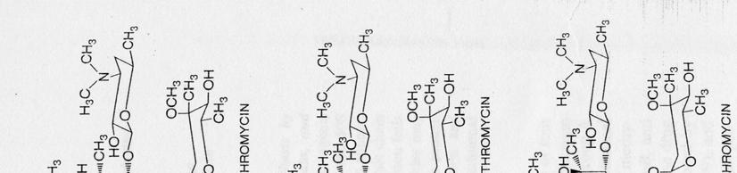
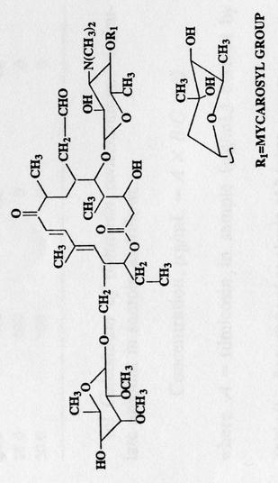
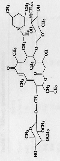
23 Tylosin Tilmicosin Tulathromycin Figure 1.1. Chemical structure of macrolide antimicrobials commonly used in veterinary medicine 5

24 6 Figure 1.2. Classification of macrolide antimicrobials according to the size of the macrocyclic lactone ring (Giguere 2006)
25 very unstable in gastric acid). A number of different formulations of erythromycin have been developed in order to overcome this problem. These include the estolate and ethylsuccinate esters which are absorbed before being hydrolyzed to release the active erythromycin base, but these esters are highly susceptible to degradation by stomach acid. An alternative is the formation of a stearate or phosphate salt which dissociates in the intestines releasing the free drug which is then absorbed. These formulations are also highly susceptible to degradation by stomach acid and it is necessary to provide an enteric coating to protect the drug from the gastric acid until it enters the intestines. The estolate is reported as being preferred for oral administration (Chambers 2001). Various formulations are also available for IM and IV administration. The glucoptate and lactobionate forms are used for IV administration as they are soluble in an aqueous solution. Clarithromycin is more rapidly absorbed in the upper intestines and is stable in stomach acid. Unfortunately, it has a very high first pass metabolism by the liver and bioavailability is in the region of 50%. Azithromycin is also rapidly absorbed and has a much higher bioavailability Mechanism of action Macrolides exert their action by reversibly binding to a specific site on the prokaryote 50S ribosomal subunit. This binding site is closely related (but not identical) to the binding site of chloramphenicol and is thought to be the same binding site as that of the lincosamides. Concurrent use of antibiotics from these different groups may result in an overall decrease in activity as the drugs act as antagonists at the other sites. The presence of the macrolides on the ribosome prevents the translocation of the trna 7
26 disrupting protein synthesis. The drugs are selective for the prokaryotic 50S ribosome and do not bind to the mammalian ribosome. Macrolides are capable of binding mitochondrial ribosomes but in contrast to chloramphenicol they cannot enter the mitochondria, and consequently, are free of many of the adverse reactions associated with chloramphenicol. The drugs are generally regarded as being bacteriostatic. However, in some circumstances they may be bactericidal especially against streptococci. The bactericidal action is time dependant and is more pronounced in an alkali environment with optimum activity seen at ph 8 (Chambers 2001) Mechanisms of resistance Resistance to the macrolides occurs by several different mechanisms and has been well reviewed (Chambers 2001). These mechanisms are generally regarded as being plasmid mediated although they may also occur as a result of one step chromosomal mutations. Such mutations may result in high levels of resistance but are typically unstable. i. The first mechanism involves a decrease in the intracellular accumulation of the drug. This is achieved by decreasing the permeability of the bacterial cell wall to the drug. This mechanism is described for Staphylococcus epidermidis. This mechanism is also the explanation of the inherent resistance of Gram negative organisms to many of the macrolide antibiotics. Gram positive organisms typically accumulate 100 times more erythromycin than Gram negative organisms. Acquired efflux mechanisms are the products of the genes mrsa and mefa seen in staphylococci and mefe in streptococci. 8
27 ii. The second mechanism of resistance is associated with alteration of the ribosomal binding site to reduce the affinity of the antimicrobial. This is achieved by methylation of the binding site on the 50S ribosome. This greatly reduces the affinity of the site for the drug and results in cross resistance for other macrolides and lincosamides. This mechanism of resistance is seen in both Gram positive and negative organisms. This mechanism is mediated by the genes erma, ermb and ermc. Such resistance also confers resistance to the lincosamide antimicrobials. A chromosomal modification of the 50S ribosomal protein is described in Bacillus subtilis and Campylobacter spp. ii. The final mechanism of resistance is described in Enterobacteriaceae and involves the production of an esterase which hydrolyses the drug, thereby inactivating it. Most mechanisms of resistance will confer a degree of cross-resistance to other macrolides and related antimicrobials. However, the 16 membered ring structures (such as tilmicosin) are commonly unaffected by these acquired resistance mechanisms (Giguere 2006) Pharmacokinetic properties A large number of studies have been conducted on the pharmacokinetics of macrolide antimicrobials in numerous species. The section below gives an overview of comparative pharmacokinetics between the drugs. The pharmacokinetics of the individual drugs in the horse are also discussed in the next section. The oral absorption of the drug (especially for erythromycin) is dependant on its being protected during passage through the stomach. The kinetics of absorption are also 9
28 highly dependant on the presence of food within the upper intestines (it should be noted that crushing tablets to aid administration is contra-indicated as this removes the enteric coating and allows the drug to become degraded in the stomach). Tylosin, azithromycin and clarithromycin are well absorbed from the gastrointestinal tract and do not require enteric coating. Tylosin is formulated as both a phosphate and tartrate. Differences are observed in the absorption kinetics of these two preparations with the tatrate being more readily absorbed (Prescott 2000). To our knowledge the oral bioavailability of tilmicosin has never been determined. Azithromycin has a very high bioavailability in dogs (97%) (Papich and Riviere 2001). All macrolide drugs have a high volume of distribution. They typically reach high concentrations in tissues which are traditionally regarded as difficult to treat such as the prostate and middle ear. The basic nature of the drugs also results in accumulation in certain types of cell. Azithromycin accumulates in fibroblasts resulting in an extremely prolonged elimination half-life. The extremely high concentrations of drug found in certain tissues and their extreme persistence contrasted with the low plasma concentrations and short plasma elimination half-life would lead one to question the validity of any discussion of plasma kinetics. Erythromycin, tylosin, azithromycin, tulathromycin and tilmicosin all accumulate in lung tissue and phagocytes at concentrations many times those seen in plasma. Tilmicosin, and tulathromycin in particular, persist in lung tissue for at least 72 hours following a single dose in cattle (Papich and Riviere 2001). The macrolide drugs are mainly excreted by the liver with a small proportion of the drug excreted unchanged in urine. For erythromycin, much of the drug is excreted 10
29 unchanged in the bile where concentrations may reach 250 µg/ml (Chambers 2001). Clarithromycin is metabolized by the liver to several metabolites, the most significant of which is the 14-hydroxy metabolite, which maintains the activity of the parent compound. A significant amount of clarithromycin and its metabolites are excreted in urine. Azithromycin undergoes some hepatic metabolism and the parent drug and metabolites are mainly excreted in the bile. Enterohepatic circulation does not appear to be a significant factor with any of the macrolides Drug interactions Drug interactions have been most widely studied for erythromycin. It is known that phenobarbital will induce the cytochrome p450 enzyme responsible for the degradation of erythromycin (CYP3A4) and consequently increase the clearance of the drug potentially resulting in therapeutic failure. However, erythromycin is also a potent inhibitor of this enzyme. Inhibition of this enzyme may interfere with the elimination of other drugs with a low therapeutic index, including theophylline, cyclosporine, digoxin and warfarin. Clarithromycin appears to have similar interactions to erythromycin while azithromycin is free of many of these problems. There is no information available on drug interactions with tilmicosin (Papich and Riviere 2001; Giguere 2006) Adverse effects The main adverse effect reported following macrolides use in animals is gastrointestinal disturbance. In humans, use of the estolate ester of erythromycin is also associated with a reversible cholestatic hepatitis which is only seen during long term therapy and may be very severe. Diarrhea and vomiting are much more common and may occur for two main reasons: 11
30 i. Macrolides may seriously disrupt normal intestinal flora resulting in the potential overgrowth of pathogenic bacteria and development of clinical gastrointestinal disease (Job and Jacobs 1997). ii. Erythromycin appears to also have a separate prokinetic effect on the gastrointestinal tract. This effect appears to be specific for erythromycin and involves the increased activation of motolin receptors (Peeters, Matthijs et al. 1989) Clinical uses and pharmacokinetics Erythromycin is generally used for the treatment of infections caused by Gram positive organisms, especially; Staphylococcus spp., Streptococcus spp., Corynebacterium spp., Clostridium spp., Listeria spp., Bacillus spp., Erysipelothrix spp., some Gram negative organisms Haemophilus spp., Brucella spp., Fusobacterium spp., Pasteurella spp., Borrelia spp., Campylobacter spp. and also for Mycoplasma spp.. In human medicine, it is largely seen as an alternative to penicillin for individuals with a penicillin allergy; it is also widely used for the treatment of respiratory infections (Papich and Riviere 2001). It s main veterinary uses are in the treatment of pyoderma, respiratory disease and diarrhea due to Campylobacter spp. It has been used in a great variety of species including cattle, swine, poultry, and dogs. Until recently, it was the treatment of choice in combination with rifampicin for the treatment of R. equi pneumonia in horses (Lakritz and Wilson 2002). Its use has also been recommended for other refractory infections in horses including Potomac Horse Fever (Palmer and Benson 1992) and Lawsonia intercellularis (Lavoie, Drolet et al. 2000). The MICs of macrolide antimicrobials against a variety of veterinary pathogens are shown in Table

31 13 Table 1.1. In vitro activity (MIC90) of veterinary macrolides against selected bacterial and mycoplasmal pathogens (Giguere 2006)
32 Tylosin (Tylan, Elanco Animal Health, Guelph, ON) has been used in the treatment of pinkeye (Moraxella bovis), respiratory disease in swine, cattle and poultry and intestinal disease in dogs, swine, poultry and cats. It can be used to reduce the incidence of liver abscesses in feedlot cattle and has also been used as a growth promoter in swine, cattle and poultry (Prescott 2000). Clarithromycin (Biaxin, Abbott Laboratories, Chicago, IL) is a new semi-synthetic derivative of erythromycin. It is tolerated better in humans, has a wider spectrum of activity and a longer half-life. Its use has not been widely reported in veterinary medicine (Papich and Riviere 2001). Azithromycin (Zithromax, Pfizer Inc., St. Louis, MO) is also derived from erythromycin but is classified as an azalide. It has many advantages over erythromycin including better oral absorption, longer half life (especially in tissues) and a broader spectrum of activity. It is more active against organisms such as Haemophilus spp., but has reduced activity against Staphylococcus spp.. The major advantage of azithromycin is the dramatic accumulation and the prolonged half life in tissues. Azithromycin accumulates especially in leukocytes. Veterinary uses for azithromycin are similar to erythromycin, and it has been used in dogs, cats, birds and horses (Papich and Riviere 2001). More recently the pharmacokinetics of azithromycin and clarithromycin have been described in foals (Jacks, Giguere et al. 2001; Jacks, Giguere et al. 2002). Tulathromycin (Draxxin, Pfizer Animal Health, Kirkland, QC) is a new macrolide antimicrobial belonging to the new triamilide class. It was developed by Pfizer and is marketed under the name Draxxin for the treatment of Bovine Respiratory Disease (BRD). Tulathromycin is a semi-synthetic derivative of erythromycin with three polar 14
33 amine groups. Tulathromycin is thought to have enhanced penetration of Gram negative bacteria, and enhanced penetration and accumulation in pulmonary tissue. This increases the spectrum of action and the pharmacokinetics are ideal for an antimicrobial for use in treating pneumonia. Draxxin is formulated as 100 mg/ml aqueous solution for use at a dose 2.5 mg/kg SC in cattle and IM in swine. Similar to tilmicosin, tulathromycin accumulates at high concentrations in lung tissue (C max 4.1 µg/g vs. 0.5 µg/ml plasma) with a greatly prolonged tissue half-life (184 hours lung vs. 90 hours plasma) (Evans 2005). This drug has now been shown to be highly effective in the treatment of respiratory disease in both swine (Nanjiani, McKelvie et al. 2005) and feedlot cattle (Rooney, Nutsch et al. 2005). Tilmicosin (20-deoxo-20-(3,5-dimethyl-piperidin-1-yl) desmycosin) is a chemical modification of the naturally occurring macrolide tylosin (Ose 1987). It is chemically closely related to erythromycin. The spectrum of activity is mainly Gram positive aerobes, but includes the Gram negative respiratory pathogens Pasteurella spp., Mannheimia spp. and Histophilus spp.. The spectrum also includes some Mycoplasma spp. The MIC breakpoint for Mannheimia haemolytica and Pasteurella multocida is set at <8 µg/ml (NCCLS 1999) In one study, the MIC 90 of M. haemolytica and P. multocida isolates was less than 6.2 µg/ml (Ose and Tonkinson 1988). In another study, the MIC 95 of 155 M. haemolytica isolates was 3.12 µg/ml (Ose 1987). However, in vitro susceptibility is not always a good predictor of clinical efficacy for tilmicosin (Musser, Mechor et al. 1996) with bacterial strains reported as resistant in the laboratory responding well in the field. Tilmicosin is considered bacteriostatic at concentrations equivalent to the MIC, concentrations four times the MIC are considered bactericidal 15
34 (Ose 1987). At concentrations as low as ¼ the MIC there is still a decrease in bacterial growth. The post-antibiotic effect (PAE) of tilmicosin may be up to 8 hours when concentrations exceed the MIC (Diarra, Malouin et al. 1999; Lim and Yun 2001). Tilmicosin is currently available in a 300 mg/ml solution in 25% propylene glycol / water ph buffered to 7.0 with phosphoric acid (Micotil, Provel, Eli Lily Canada Inc. Guelph On). The product is licensed for the treatment of respiratory disease in cattle and sheep at a single dose of 10 mg/kg SC and is one of the most widely used medications for the treatment and control of feedlot pneumonia in cattle. Tilmicosin is also available as a feed premix containing 20% tilmicosin (Pulmotil- Elanco Animal Health Guelph, ON). The product is licensed for use in growing swine at a dose of 200 ppm in feed. An alternative injectable formulation of microcrystallized tilmicosin as a fatty acid salt (IDEXX Pharmaceuticals, Durham, NC) has also been developed. This product has been formulated with the intention of using it in dogs and cats but it has been used in horses (Fenger 2000; Womble, Giguere et al. 2006). Tilmicosin has mainly been used for the treatment of respiratory infections in cattle and sheep in its injectable form. More recently, the drug has been formulated as a feed additive for the treatment of respiratory disease in swine (Pulmotil). Publications have also described its use in the treatment of foot rot, pinkeye and the oral form has also been used experimentally in the treatment of enzootic pneumonia in calves. The main advantages of tilmicosin are the high concentrations of the drug that accumulate in the lung tissue and its persistence at this site. It has been widely used in the treatment of BRD complex, for the treatment of individual animals, metaphylaxis and also 16
35 prophylaxis. The widespread use of tilmicosin has been prevented by problems with toxicity in other animals (Prescott 2000). More recently, an injectable tilmicosin product (Micotil) has been licensed for use in rabbits in Italy. The efficacy of tilmicosin for treating bacterial pneumonia in food producing animals is beyond question; tilmicosin has been widely studied in cattle feedlots and is as effective, or more effective than other commonly used medications such as oxytetracycline, florfenicol or ceftiofur (Schumann, Janzen et al. 1991; Musser, Mechor et al. 1996; Jim, Booker et al. 1999). Tilmicosin is also effective in the treatment of enzootic pneumonia of calves (Ose and Tonkinson 1988; Picavet, Muylle et al. 1991; Fodor, Varga et al. 1993). Clinical efficacy in sheep (Naccari, Giofre et al. 2001; Christodoulopoulos, Warnick et al. 2002) and pigs (Moore, Basson et al. 1996; Hoflack, Maes et al. 2001; Mateusen, Maes et al. 2001) is also well documented. The clinical efficacy of tilmicosin has also been assessed in a number of non-target animal species including chickens (Jordan and Horrocks 1996; Kempf, Reeve-Johnson et al. 1997) and rabbits (McKay, Morck et al. 1996). The main advantage of tilmicosin over tylosin is its pharmacokinetics. Tilmicosin has a longer elimination half-life and accumulates in lung tissue at higher concentrations. The pharmacokinetics in cattle are well described. In the first pharmacokinetic study, researchers treated 15 steers with a mean body weight of 240 kg with a label dose of tilmicosin (10 mg/kg SC). Analysis of serum samples and lung samples collected at timed euthanasia periods demonstrated low serum concentrations (C max 0.71 µg/ml) and a long plasma half-life (t 1/2 ) of 30 hours (Thompson and Lawrence 1994). By contrast, lung concentrations reached a C max of 7.17 µg/g with a 17
36 t 1/2 of approximately 48 hours. Tissue concentrations exceeded the MIC of M. haemolytica for at least 72 hours. These pharmacokinetics are ideal for a single treatment antimicrobial for use in the treatment of feedlot BRD. A more detailed evaluation of the pharmacokinetics is reported by Ziv et al (Ziv, Shem-Tov et al. 1995). 14 cows received the standard 10 mg/kg SC dose and 3 cows received the same dose intravenously (1 as a bolus and 2 as an infusion 0.5 mg/kg/min). The intravenous bolus resulted in severe adverse reactions; however, sufficient data was collected to model the pharmacokinetics (it should be noted that these kinetics were obtained from animals in a state of cardiovascular shock and their validity may be questioned). The results show a typical two compartment model. The volume of distribution (V darea ) is 2.89 L/kg and the half-lives of the two compartments are 14 minutes and 46 minutes. The C max is 2.39 µg/ml. The two animals receiving the IV infusion also showed signs of toxicity. The data from their study gives a 1 compartment model with a t 1/2 of approximately 1 hour and V darea of 2.25 L/kg. In contrast, the animals receiving the SC dose had a C max of 0.13 µg/ml at 1.84 hours post-injection and a t 1/2 of 4.18 hrs. The bioavailability of tilmicosin was calculated as 22%. Tilmicosin also partitioned well into the udder of lactating cows reaching concentrations that were typically 50 times higher than those seen in plasma. Another study in 10 adult cows (Modric, Webb et al. 1998) found a serum C max of 0.87 µg/ml and a serum t 1/2 of 29.4 hours similar to that originally described by Thompson (Thompson and Lawrence 1994). More recently, a detailed pharmacokinetics study of tilmicosin in cattle has again been investigated comparing a SC dose with slow IV infusion (Hunter, Hassfurther et al. 2006). The IV infusion was 18
37 conducted using 10 mg/kg diluted in saline and administered over 5 hours. The V dss was calculated to be 15 L/kg and, interestingly, the t 1/2 of the two groups are very similar at 28 hours. However, the MRTs of the two groups were considerably different (IV 23 hours, SC 32 hours), but the reason for this difference was not investigated. The values of C max and t 1/2 described in the studies differ markedly. The reasons for this discrepancy are not clear, but each study used a different method of measuring tilmicosin concentrations; bioassay (Ziv, Shem-Tov et al. 1995) vs. HPLC (Thompson and Lawrence 1994; Modric, Webb et al. 1998) or LC/MS (Hunter, Hassfurther et al. 2006). No details are given as to the suitability of the HPLC method used by Thompson and Lawrence (Thompson and Lawrence 1994), the HPLC used by Modric et al (Modric, Webb et al. 1998) had an LOD of 2.6 ng/ml and an LOQ of 50 ng/ml and a CV <10%. The bioassay used by Ziv et al (Ziv, Shem-Tov et al. 1995) has an LOQ of 150 ppb and a coefficient of variation of 10%. Hunter at al (Hunter, Hassfurther et al. 2006) simply describe that their LC/MS assay had been validated. Consequently, all plasma concentrations reported by Ziv et al (Ziv, Shem-Tov et al. 1995) are below the LOQ of the assay. There is also a possibility that there was a difference in the injection technique between the studies influencing the absorption kinetics. Finally, Ziv et al. (Ziv, Shem-Tov et al. 1995) used lactating, pregnant dairy cows while all other studies were conducted in non-pregnant beef cattle. These results reveal a number of important features about tilmicosin. The drug appears to be slowly absorbed from the SC injection site. The slow absorption for the injection site is beneficial; tilmicosin may have a short elimination t 1/2 in plasma of 46 minutes (Ziv, Shem-Tov et al. 1995), however, the delayed absorption from the injection 19
38 site results in flip flop kinetics with an elimination t 1/2 ranging from hours. Tilmicosin is not formulated for slow release. Therefore, the delay in absorption may be related to the severe inflammatory reaction that occurs at the site of injection (Van Donkersgoed, Dubeski et al. 2000). The bioavailability of the SC dose of 22% (Ziv, Shem-Tov et al. 1995) is excessively low and may be an artifact caused by the slow absorption and rapid sequestration of tilmicosin into a tissue site separate from the plasma artificially decreasing the AUC. It should be noted that Hunter et al (Hunter, Hassfurther et al. 2006) found the plasma elimination half-life to be much longer and independent of absorption from the SC injection site. The pharmacokinetics of tilmicosin in sheep are similar to those described for cattle. The first report of the pharmacokinetics administered doses of 10 or 20 mg/kg SC over the chest wall (Parker, Patel et al. 1994). The C max in plasma is 1177 (± 208) or 2282 (± 692) ng/ml respectively, with a C max in lung of (± 3532) or (± 4657) ng/g. The t 1/2 in plasma had two phases 7.9 hrs and 29.6 hrs and the t 1/2 in lung was 26.9 hours. These results are similar to those previously described in cattle. Modric et al. (Modric, Webb et al. 1998) specifically compared the plasma pharmacokinetics of tilmicosin in sheep and cattle and concluded that the results are very similar; only the T max differed significantly between the species (3.8 hrs in sheep vs. 0.5 hrs in cattle). C max is 0.87 µg/ml in cattle and 0.82 µg/ml in sheep. Half-life in cattle is 29.4 hours vs 34.6 hrs in sheep. Naccari et al. (Naccari, Giofre et al. 2001) modeled the plasma pharmacokinetics in 5 healthy and 5 pneumonic sheep. The pharmacokinetics in the healthy sheep are similar to those described by Modric et al. (Modric, Webb et al. 1998) but the half-lives 20
39 are shorter (T max 6 hours, C max, 1.32 µg/ml, t 1/2 13 hours), the values in the pneumonic sheep (with the exception of T max ) were significantly different (C max 1.97 µg/ml, t 1/2 20 hours). Tilmicosin is also licensed for use in swine. However, due to adverse reactions associated with injection (Jordan, Byrd et al. 1993) the drug is only used as a feed additive. The pharmacokinetics are described in this species (Thomson, Darby et al. 1994). Swine were fed rations containing either 200 or 400 ppm (approximating to a daily intake of 10 or 20 mg/kg/day). Serum concentrations in the 200 ppm group are barely above the LOQ of the assay, serum concentrations in the 400 ppm group were also low with a C max of 230 ng/ml. Lung concentrations are much higher with a C max of 1.43 µg/g and 2.59 µg/g respectively. The pharmacokinetics of tilmicosin are described in a number of non-target animals. In elk, the pharmacokinetics appear to be similar to those described for cattle (Clark, Woodbury et al. 2004). Cochrane and Thomson (Cochrane and Thomson 1990) describe plasma concentrations in horses following SC doses of 3, 10 or 30 mg/kg. Analysis of their data reveals that at doses of 3, 10 and 30 mg/kg the C max in plasma is 0.17, 0.36 and 1.07 µg/ml respectively. The MRT is somewhat variable at 22.5, 42 and 38.4 hours respectively. The T max ranged from 8-12 hours. Rabbits treated with a dose of 25 mg/kg SC had a plasma C max of 1.91 µg/ml at 2 hours. The t 1/2 in plasma was 5.97 hours. Lung concentrations reached a maximum of µg/g at 2 hours falling to 5.1 µg/g at 24 hours (McKay, Morck et al. 1996). In all species studied, the consistent pharmacokinetic features of tilmicosin are the slow absorption from the injection site, low plasma / serum concentrations, high lung 21
40 concentrations and a prolonged elimination half-life (especially from lung tissue). A number of researchers have investigated the nature of the tilmicosin accumulation in lung tissue. Brown et al. administered 10 mg/kg tilmicosin SC to mice (Brown, Deleeuw et al. 1995). Perfusion of the lung with saline and removal of the blood did not affect the tilmicosin concentration in the lung tissue. When the same study was repeated with ceftiofur, perfusion of the lung removed approximately 75% of the ceftiofur. The study indicates that unlike ceftiofur, the tilmicosin is actually sequestered in the lung tissue. The accumulation of tilmicosin into lung tissue can be enhanced in pneumonia but the results are somewhat confusing. In rats (Modric, Webb et al. 1999), the lung concentrations of tilmicosin are significantly higher in pneumonic rats and the elimination half-life of the tilmicosin from the pneumonic lung appears significantly longer. In contrast, Thompson et al. (Thompson, Laudert et al. 1994), report that the concentration of tilmicosin in the consolidated lung tissue is less than that of normal lung µg/g versus 9.5 µg/g. Pulmonary macrophages recovered from the lungs of clinically healthy calves and incubated with radio-labelled tilmicosin media (10 or 20 µg/ml) contained 651 µg/ml or 1450 µg/ml tilmicosin respectively after 24 hours of incubation indicating that the drug actively accumulates in these cells (Thompson, Laudert et al. 1994). A similar study using porcine alveolar macrophages found similar results (Thomson, Darby et al. 1994) with tilmicosin concentrations of 517 and 1500 µg/ml respectively. The most detailed examination of the subcellular distribution of tilmicosin in bovine white blood cells was conducted by Scorneaux and Shryock (Scorneaux and 22
41 Shryock 1999). Macrophages, neutrophils and epithelial cells were recovered from the mammary gland of lactating cattle. Activated cells were recovered from two cows that were exposed to either S. aureus or E. coli lipopolysaccaride. Monocytes and neutrophils were recovered from blood and pulmonary macrophages were recovered from the lungs of a freshly euthanized cow. All cells accumulated tilmicosin, but the pulmonary macrophages accumulated the most, concentrations reaching 195 x that of the extracellular concentration after 4 hours of incubation; 80% of tilmicosin is found in the lysosomes. It is generally assumed that the accumulation of an antimicrobial within the lysosomes of phagocytic cells is beneficial. The assumption is based on the fact that this is a site where phagocytosed bacteria reside and are killed by the phagocytic cell. It is also the environment in which a number of pathogenic bacteria (notably R. equi) are able to survive. The interaction of an antimicrobial with the phagocytic cell is highly complex. The simplistic view that the accumulation of an antimicrobial will result automatically in enhanced microbial killing is therefore probably not correct. Brumbaugh et al. (Brumbaugh, Herman et al. 2002) demonstrated that tilmicosin did not affect chemotactic or phagocytic activities of bovine or porcine alveolar macrophages. Tilmicosin enhanced the bacterial killing of the macrophages but the relationship between tilmicosin concentration and effect was not clear. The nature of the complexity between cellular accumulation and antimicrobial activity is becoming apparent for tilmicosin. The first study of the interaction between tilmicosin and the neutrophil in pneumonia revealed some surprising results (Chin, Morck et al. 1998). Tilmicosin not only resulted in significant clearance of M. 23
42 haemolytica from the lungs but also increased apoptosis of neutrophils. Tilmicosin did not impair infiltration, phagocytic function or oxidative metabolism (clarithromycin and azithromycin impair phagocytosis in human peripheral neutrophils (Wenisch, Parschalk et al. 1996)). The apparent contradiction in action between increased neutrophil action and apoptosis is thought to be beneficial in cases of M. haemolytica as the neutrophil plays a key role in the tissue damage that occurs during disease (Radostits, Gay et al. 2007). The same group of researchers (Chin, Lee et al. 2000) demonstrated that the apopototic effect of tilmicosin occurs regardless of the presence of M. haemolytica and that the apopototic cells are rapidly cleared by macrophages. The effect is also specific to tilmicosin and was not seen with any other antimicrobials or anti-inflammatory drugs. Another group (Fajt, Apley et al. 2000; Fajt, Apley et al. 2003) has conducted similar work comparing tilmicosin and danofloxacin and was not able to demonstrate any difference between the two antimicrobials in cattle experimentally infected with M. haemolytica. The authors concluded that that tilmicosin did not have any effect on neutrophil function or apoptosis. The reason for the discrepancies between the two studies is not known. A final tool that has been used to investigate the activity of tilmicosin is immunohistochemistry. Tilmicosin is a high molecular weight molecule and it has been possible to generate monoclonal antibodies to the molecule (Beier, Creemer et al. 2005). Immunohistochemistry using these antibodies has allowed the precise location of tilmicosin in treated lung to be mapped. There are at present few reports of the use of these techniques. The technique has been used in healthy swine and shows tilmicosin accumulation in bronchial epithelial cells and glandular areas. Tilmicosin also 24
43 accumulates in macrophages in the alveolar wall, the interstice, and in the lumen and on the surface of the bronchi (Jackman, Spencer et al. 1997) The use of macrolide antimicrobials in the horse The most widely used macrolide in the horse is erythromycin, which is used almost exclusively for the treatment of pyogranulomatous pneumonia in foals due to infection with R. equi (Lakritz and Wilson 2002). It is often used for this purpose in conjunction with rifampin. The initial studies regarding this treatment were based on the observed pharmacokinetics of erythromycin (Prescott, Hoover et al. 1983) and in vitro sensitivities of collected R. equi isolates (Prescott 1981; Prescott and Nicholson 1984; Prescott and Sweeney 1985). The initial clinical trials of the efficacy of this treatment remain the only published clinical trials for the treatment of R. equi (Hillidge 1987; Sweeney, Sweeney et al. 1987). The first of these studies is in reality a simple retrospective analysis of 48 clinical cases of R. equi pneumonia in foals. The foals were treated with a variety of antibiotics, both singly and in combination (Sweeney, Sweeney et al. 1987). The authors concluded that treatment with erythromycin was associated with an increased chance of survival. In the second study (Hillidge 1987), 89 foals were treated with erythromycin and rifampin. There was no control and the case definition was relatively loose as it included animals with clinical signs, but no positive culture for R. equi. However, the authors demonstrated an approximately 85% recovery rate in the treated foals. Before addressing the studies looking at the pharmacokinetics of erythromycin in the horse it is worth recognizing two potential problems: 25
44 First, there is the method by which the plasma drug concentrations are measured. In the earlier studies plasma concentrations were routinely measured by means of a bioassay (Prescott, Hoover et al. 1983; Ewing, Burrows et al. 1994), while later studies have used HPLC methods. HPLC has the advantage of being able to measure plasma concentrations of erythromycin, anhydroerythromycin and erythromycin base independently. When the results of the 2 assays were compared (Lakritz, Wilson et al. 1999), substantial differences were noted. In particular the bioassay consistently over estimated the concentration of drug in the body measured as AUC. Rate kinetics and mean residence time were also significantly different. These differences may be because the bioassay does not measure the concentration of the parent drug and the metabolites or conjugated pro-drug individually. Second, all studies conducted have referenced the initial studies looking at in vitro sensitivity of R. equi isolates to erythromycin (Prescott 1981) in which the MIC was determined to be 0.25 μg/ml. These studies have assumed that plasma concentrations in excess of this concentration should be effective. Such an assumption is hard to justify given the intracellular nature of the bacteria and its location in a pyogranulomatous abscess within the lung. It is generally assumed that the concentration of drug in the lung should exceed plasma concentrations, but there is no evidence to support this theory in the horse or any other species. There is also no evidence to suggest how long the plasma concentration should exceed the MIC. The first assessment of the potential use of erythromycin in horses looked at the pharmacokinetics in both foals and horses (Prescott, Hoover et al. 1983). The study used mare/foal pairs and looked at the pharmacokinetics in foals at 1, 3, 5, 7, and
45 weeks of age. The mares were also used to evaluate the pharmacokinetics in adult horses. Erythromycin was administered in the form of gluceptate at 5 and 20 mg/kg IV, estolate at 5-20 mg/kg orally and free base at 10 mg/kg IM. Minimal analysis was conducted on the plasma concentrations which were measured using a bioassay. The results demonstrated that erythromycin is rapidly eliminated from the plasma after intravenous administration. However, after intra-gastric or intramuscular administration there is a slowed absorption phase resulting in sustained plasma concentrations (flip-flop kinetics). Bioavailability of the oral estolate formulation appears to be relatively low. The base form resulted in unacceptable swellings at the site of injection. Subsequent studies have used alternative forms of erythromycin. Ewing at al. (Ewing, Burrows et al. 1994), used estolate (37.5 mg/kg bid), phosphate (25 mg/kg tid), stearate (25 mg/kg tid) and ethylsuccinate (25 mg/kg tid). Their study used adult horses and plasma concentrations were analyzed using a bioassay. The authors concluded that the simple salts were better absorbed than the esters (in contrast to studies conducted in humans) and that erythromycin phosphate or stearate should be used for R. equi therapy in foals as plasma concentrations were significantly higher than those seen with the estolate used by Prescott et al. (Prescott, Hoover et al. 1983). However, all forms of the drug resulted in peak concentrations in excess of 0.25 μg/ml, the recommended MIC for R. equi. The authors also concluded that a dose interval of 12 hours is the most appropriate. Unfortunately, the authors did not address the issue of performing the study in adult horses when the target population for therapy is unweaned foals. 27
46 Lakritz et al. (Lakritz, Wilson et al. 2000) compared the pharmacokinetics of erythromycin estolate and erythromycin phosphate when administered intragastrically to non-fed foals. The plasma analysis in this study was conducted using HPLC and plasma anhydroerythromycin A (an inactive degradation product) was also measured. Once again erythromycin phosphate showed significantly greater plasma concentrations. Administration of the estolate did result in reduced concentrations of anhydroerythromycin A, but the majority of the drug in the plasma was in the inactive estolate form. The authors finally concluded that the phosphate should be administered at a dose of 25 mg/kg every 6 hours, but that the potential exists for the estolate to be more effective. No evidence is given to support this claim. The same group of researchers also looked at the effects of feeding on the absorption of erythromycin base in foals (Lakritz, Wilson et al. 2000). The study was a crossover design in which 25 mg/kg of microencapsulated erythromycin base was administered to fasted and non-fasted foals. The results indicated that the bioavailability of the drug in fasted foals was 26% (+/- 15.4) compared with 7.7% (+/- 6.7) in fed foals. This decrease in absorption was not mediated through an increase in the degradation of the active drug as determined by measurement of anhydroerthromycin in plasma. A final study by the same research group looked at the pharmacokinetics of erythromycin ethylsuccinate (Lakritz, Wilson et al. 2002). Twenty five mg/kg were administered orally to fasted foals. Concentrations did not exceed 0.25 μg/ml for more than 4 hours and a large amount of anhydroerythromycin was measured in plasma. The authors did not recommend the use of this product for the treatment of R. equi infection in foals. 28
47 The results of these studies demonstrate several interesting points: Firstly, erythromycin has a poor oral bioavailability in the horse. The bioavailability is dependent on the formulation of the drug and the fed state of the horse. While it is understandable that a researcher should attempt to control this variability as much as possible by administering the drug intragastrically to a fasted horse, the practical application of this approach must be considered as the target treatment group is young foals that must be treated 3 4 times daily, often for more than one month. The second major problem is the continual assumption that the plasma concentrations of the drug must exceed the MIC for R. equi. There is no evidence that this is an appropriate marker of efficacy for the treatment of R. equi pneumonia. The pharmacodynamics of the macrolide antimicrobials has not been fully determined and it is unclear whether they should be considered either concentration or time dependent in their mechanism of bacterial killing. Furthermore, the macrolide antimicrobials have a very high volume of distribution with only a small percentage of the drug being present in plasma. The majority of the drug is sequestered in the tissues. R. equi is an intracellular pathogen causing pyogranulomatous lesions in the lung tissue. One must, therefore, question the applicability of plasma concentrations to concentrations achieved / required at the site of infection. There are very few studies of the efficacy of erythromycin with which to compare these pharmacokinetic studies. While the pharmacokinetics seem to suggest that the simple phosphate base has the best pharmacokinetic profile, the only clinical trial used either the estolate or the succinate form three times daily (Hillidge 1987) The concerns regarding the variability of erythromycin absorption, the potential for adverse reactions (Stratton-Phelps, Wilson et al. 2000) coupled with increasing 29
48 anecdotal reports of isolates of R. equi resistant to erythromycin have led to the use of newer macrolides in foals. The frequent dosing associated with erythromycin treatment has led researchers to look at the new generation of macrolide antimicrobials which typically have higher oral bioavailability and prolonged elimination half-lives. As a result, the pharmacokinetics of azithromycin have been investigated by two research groups (Jacks, Giguere et al. 2001; Davis, Gardner et al. 2002). In the first of these studies, doses of 10 mg/kg were compared when administered intragastrically and intravenously (foals were not fasted). Analysis of drug concentrations was conducted using a bioassay. The oral bioavailability was 56% and the plasma elimination half-life was very long (20.3 hours after IV administration). In addition to plasma concentrations, azithromycin concentrations were also determined in synovial and peritoneal fluid where the concentrations were found to be similar to those seen in plasma. Bronchoalveolar lavage fluid (BAL) was also collected and estimates were made of the concentration of azithromycin found in bronchoalveolar cells and in pulmonary epithelial lining fluid (PELF). Azithromycin accumulated in the PELF at concentrations approximately 50 times those seen in plasma; the concentrations in the bronchoalveolar cells were even higher at approximately 500x plasma concentrations. The authors suggest that once daily dosing for 5 days followed by a dose interval of 48 hours should be sufficient to treat R. equi. However, the basis for these recommendations (i.e. nature of target drug concentrations) is not clear. The second study (Davis, Gardner et al. 2002) was similar using slightly older foals and the azithromycin was administered orally (10 mg/kg). The analysis was 30
49 conducted using HPLC. The results are similar with slightly reduced oral bioavailability (39%) and the elimination of the azithromycin was faster. The concentration of azithromycin in peripheral polymorphonuclear (PMN) cells was also measured in addition to BAL fluid and bronchoalveolar cells. The concentration of azithromycin in PMNs was approximately 25 times greater than that seen in plasma and the concentrations persisted for several days (T 1/ hrs). The authors again suggest that azithromycin shows potential for the treatment of R. equi pneumonia. Clarithromycin is a semi-synthetic derivative of erythromycin. It is reported to be approximately twice as active against bacteria as erythromycin on a weight by weight basis and has a half life approximately twice that of erythromycin (Giguere 2006). In addition, the bioavailability is higher and volume of distribution is also improved. Finally, the incidence of adverse reactions in humans is reduced when compared with erythromycin (Giguere 2006). The pharmacokinetics of clarithromycin in foals have been reported (Jacks, Giguere et al. 2002). Administration of 10 mg/kg intra-gastrically in non-fasted foals resulted in a C max of 0.92 (+/- 0.17) μg/ml at 1.5 hrs. The t ½ is 4.81 hrs. The MIC 90 of R. equi for clarithromycin has been reported as 0.12 μg/ml and the authors, therefore, suggest an oral dose of 7.5 mg/kg q 12 h for the treatment of R. equi pneumonia. Unfortunately, the concentrations of clarithromycin in lung tissue were not measured and plasma concentrations were measured using a bioassay. Since the publication of all these pharmacokinetic trials, two additional studies have been published. The first of these investigated the in vitro susceptibility of R. equi to the macrolide antimicrobials (Jacks, Giguere et al. 2003). The MIC 90 for azithromycin, clarithromycin and erythromycin were 1.0, 0.12 and 0.25 µg/ml 31
50 respectively. This covers quite a range with clarithromycin being almost 10 times as potent as azithromycin. A retrospective study of therapy for R. equi pneumonia has also been conducted (Giguere, Jacks et al. 2004). The study followed 81 foals with naturally acquired R. equi infection that presented at Florida State University. Twenty foals received azithromycin, 18 received clarithromycin and 24 received erythromycin (stearate). Most foals also received rifampin. Foals treated with clarithromycin showed a significantly greater survival rate in both long and short term follow up and better resolution of radiographic change in the lungs. The authors concluded that clarithromycin was the treatment of choice and that azithromycin did not offer any advantages over the traditional therapy of erythromycin. Interestingly, azithromycin has become the accepted standard for treatment for R. equi pneumonia (Cohen personal communication). The use of tilmicosin in the horse has not been well investigated due to concerns about potential toxicity (Prescott 2000). However, there are no reports of toxicity in horses in the veterinary literature to support these concerns. There is one case report (Fenger 2000) that reports the successful use of a novel microcrystalline injectable form of tilmicosin to treat R. equi pneumonia in two foals. The foals were both suffering from an infection that was refractory to erythromycin treatment. The tilmicosin preparation used in this study has not been widely available. However, a study of its pharmacokinetics has now been published (Womble, Giguere et al. 2006). The tilmicosin formulation consists of a fatty acid salt formulation (250 mg/ml; IDEXX Pharmaceuticals, Durham, NC). It was administered to 7 foals between 5 and 8 weeks 32
51 of age at a dose of 10 mg/kg via the intramuscular route using the muscles of the hindlimb. Samples collected included plasma, BAL fluid, cerebrospinal fluid (CSF), peritoneal fluid and lung biopsies. Tilmicosin concentrations were measured in serum, CSF, peritoneal fluid, lung tissue, PELF, BAL cells and blood neutrophils using HPLC- MS/MS. Serum concentrations of tilmicosin peaked at 0.19 µg/ml with an MRT of 34.5 hours. As expected, the lung concentrations were considerably higher at 1.9 µg/g with a MRT of 323 hours (although the time to T max was much longer 30.8 hours vs. 5.5 hours for serum). The C max for PELF, BAL cells and serum neutrophils were 2.91, 20.1 and 66 µg/ml respectively, with MRTs of 180, 117 and 99 hours respectively. The researchers found the MIC 90 for R. equi to be 32 µg/ml. Administration of the tilmicosin formulation resulted in swelling at the site of injection and self limiting diarrhea in four foals. One foal developed tachypnea and profuse sweating. The researchers believed that their results warrant further investigation of the use of tilmicosin for the treatment of pneumonia caused by R. equi despite the very high MIC 90 (32 µg/ml). Although the results of a wide variety of pharmacokinetic studies using different macrolide formulations are reported for horses, interpretation of the data is difficult. Most studies have simply administered the antimicrobial orally (often by stomach tube in a fasted foal) and measured plasma concentrations of the drug. The significance of these plasma concentrations for a drug with such a high volume of distribution must be questioned. The macrolide antimicrobials rarely excerpt their activity within the plasma (especially in the case of pyogranulomatous pneumonia caused by R. equi) and the 33
52 relationship between efficacy and plasma concentration is not known. The use of the in vitro MIC 90 as a target for plasma concentrations is without basis. Furthermore, many of these studies have used unvalidated bioassays to measure drug concentrations and these assays have been shown to have a number of weaknesses (Lakritz, Wilson et al. 1999). In contrast to the horse, most food animal studies of macrolide pharmacokinetics have focused on tissue (especially lung) concentrations of the drug. Again the usefulness of these concentrations is not clear but they seem to equate with clinical efficacy and tissue concentrations are certainly closer to the site of action. A number of recent studies in the horse have attempted to answer some of these questions by using repeated samples such as BAL fluid to assess macrolide concentrations in PELF and leucocytes. What is really needed are studies which attempt to link the tissue distribution of the antimicrobial and the efficacy of treatment so that we can better understand the complex pharmacodynamics of antimicrobial activity and their highly complex interaction with the immune system Review of macrolide toxicity (especially tilmicosin) The macrolide class of antimicrobials generally has a very good safety profile (Periti, Mazzei et al. 1993). The most commonly reported adverse reaction is gastrointestinal disturbance. Other reported adverse reactions include allergic skin eruptions, cholestatic hepatitis, transient ototoxicity and irritation at the site of injection. Also, there have been reports of unusual reactions which are considered rare or questionable affecting a wide variety of body systems. 34
53 A number of researchers have investigated a possible linkage between macrolides and cardiovascular toxicity. Wakabayashi and Yamada (Wakabayashi and Yamada 1972) demonstrated that erythromycin, oleandomycin, leucomycin and spiramycin all caused decreased blood pressure in dogs. The administration of macrolides was associated with an increase in blood histamine concentrations and the effect was attenuated by pretreatment with diphenhydramine. They concluded that the macrolides stimulate the release of histamine which resulted in the observed cardiovascular effects. More recently, a study was conducted that compared the effects of various macrolides on isolated beating rat atria (Tamargo, De Miguel et al. 1982). Josamycin and erythromycin produced dose dependent decreases in rate and contractile force. They also affected the sinus node recovery time and the refractory period. The effect is not attenuated by atropine, pentolamine, practolol, diphenhydramine, cimetidine, methysergide, or indomethacine. The authors finally concluded that erythromycin and josamycin inhibit the transmembrane flux of calcium into atrial cells. There is one clinical case report of erythromycin being associated with a ventricular arrhythmia in a human patient (Freedman, Anderson et al. 1987). The patient suffered from idiopathic long QT syndrome and was treated with erythromycin. This resulted in syncope. A controlled erythromycin infusion and monitoring of the electrocardiogram (ECG) demonstrated T-wave alternans and premature ventricular complexes. The effect was blocked by the use of propranalol. Tilmicosin is unusual compared to other macrolides as it is linked to acute cardiac toxicity. The cardiac toxicity of tilmicosin has been extensively reviewed (Jordan, Byrd et al. 1993). The oral median lethal dose (MLD) in fasted rats is mg/kg. If the 35
54 animals were recently fed, the oral MLD rose to 2250 mg/kg. Deaths were per acute occurring within 2 hours and no specific tissue lesions were found at post-mortem examination. The MLD following SC injection was 100 mg/kg in mice and mg/kg in rats (range due to sex differences). Once again, effects were acute and no lesions were found at post-mortem examination. Primates appear to be more susceptible to toxicity. A small study in Rhesus monkeys found signs of toxicity at 20 mg/kg SC. A dose of 30 mg/kg resulted in vomiting, behavioral changes, labored breathing and death. The nature of the effect of tilmicosin on the heart is not known. The most detailed study on the cardiovascular effects of tilmicosin was conducted in dogs (Main, Means et al. 1996). Administration of intravenous tilmicosin to unrestrained dogs results in negative ionotropic and chronotropic effects with resultant tachycardia. These effects are exacerbated by treatment with propranalol. Dobutamine infusion attenuated some of the effects. It is now thought that the toxic effects of tilmicosin on the heart may be mediated through rapid depletion of calcium (Main, Means et al. 1996). Chronic dosing studies have been conducted in a wide range of species. Rats dosed daily with tilmicosin ranging from mg/kg PO for 2 weeks did not show any overt signs of toxicity. There were mild increases in serum AST and induction of hepatic microsomal enzymes but no evidence of tissue toxicity was found. In a separate study, oral doses ranging from mg/kg were administered to rats for 3 months. Deaths were seen at the higher doses along with non-specific changes such as decreased body weight. 36
55 Beagle studies used oral doses of 4 36 mg/kg bid for a year. All dogs survived, but the animals receiving the higher doses showed some signs of cardiac toxicity. Tachycardia was seen as well as depression in the ST segment of the ECG. These animals showed moderate cardiomegally at post-mortem examination but no histopathological changes were found. Toxicity studies in cattle have demonstrated a wide margin of safety when the drug is used according to the label dose (10 mg/kg SC). Four doses of 150 mg/kg SC 3 days apart resulted in 50% mortality. Death was acute and associated with tachycardia and changes in the ECG. At necropsy small foci of myocardial necrosis were evident in the papillary muscles of the left ventricle. The higher dose was also associated with very severe reactions at the site of injection, characterized by severe edema and some necrosis of the underlying muscle. The loss of plasma proteins into the site resulted in a measurable decrease in plasma total protein. In a separate study, doses ranging from mg/kg were administered subcutaneously every 72 hours for 3 doses. There were no overt signs of toxicity, but at necropsy, lesions were again found in the papillary muscles of the left ventricle. A dose of 5 mg/kg IV in feedlot cattle resulted in labored breathing and lethargy with a 50% mortality rate. No lesions were found at necropsy. In a pharmacokinetic study, Ziv et al. (Ziv, Shem-Tov et al. 1995) administered a 10 mg/kg IV bolus, or as a slow infusion over 20 minutes at 0.5 mg/kg/min. The cow receiving the bolus developed ataxia, tachycardia, jugular pulses, hyperpnea and collapsed although the animal recovered. The cows receiving the infusion dose showed less pronounced signs. The intravenous dosing resulted in plasma concentrations above 1 µg/ml in contrast to 37
56 the standard 10 mg/kg SC dose with a C max of 0.13 µg/ml suggesting that the toxic effects of tilmicosin are dependent on plasma concentrations and that the toxic concentrations are very hard to achieve with either oral dosing or injection by either the PO, SC or IM routes. Toxicity in non-target agricultural animals has been assessed in neonatal calves. Doses of 5 or 10 mg/kg IV resulted in labored breathing and lethargy. One calf receiving the 10 mg/kg IV dose died. No lesions were found at necropsy (Jordan, Byrd et al. 1993). Pigs appear to be more susceptible to tilmicosin toxicity. Intra-muscular doses ranging from mg/kg resulted in recumbency and convulsions at the 10 mg/kg dose. At the higher doses there was ataxia, restlessness and labored breathing with 75% mortality at 20 mg/kg and 100% mortality at 30 mg/kg. There was severe reaction at the injection sites. Oral dosing with medicated feed containing ppm for 6 weeks had no adverse reactions. Tilmicosin has since been licensed for use in swine feed at 200 ppm for the treatment and prevention of respiratory disease in growing pigs. Studies in sheep found the toxicity profile to very closely resemble that of cattle and the drug was licensed for use in sheep to treat respiratory disease (Modric, Webb et al. 1998). The injectable product has also been licensed for use in rabbits in Italy. Jordan (Jordan, Byrd et al. 1993) reports that SC and IM dose of >10 mg/kg resulted in toxic reactions in horses and goats and that both species exhibited adverse reactions to IV doses of less than 10 mg/kg. The details of the horse studies are found in (Cochrane and Thomson 1990). Horses received a range of doses; 3, 10, 30 mg/kg SC, 2.5, 5, 7.5 mg/kg IV and 1 mg/kg IM. Horses were monitored for adverse reactions and 38
57 plasma was collected to measure tilmicosin concentrations. A detailed necropsy was conducted upon completion of the study. Intravenous doses of 2.5 or 5 mg/kg did not result in any adverse reactions; a dose of 7.5 mg/kg resulted in signs of colic, convulsions and labored breathing. The horse was recumbent within 15 minutes and died within 1 hour. The intramuscular dose resulted in stiffness at the site of injection lasting 3 4 days. The subcutaneous doses resulted in swelling and reaction at the injection site. Administration of subcutaneous tilmicosin was well tolerated at 3 or 10 mg/kg. A dose of 30 mg/kg resulted in signs of toxicity during the following 24 hours (anorexia, labored respiration, recumbency, depression), horses then appeared normal. One horse receiving 30 mg/kg SC died 6 days post injection. The two horses that died during the study had similar findings in their heart muscle. There were multiple foci in the left ventricle of subacute necrosis characterized by loss of sarcoplasm, minimal cellular infiltration and increased prominence of small blood vessels within the damaged areas. Of the remaining horses in the study 2 had no cardiac lesions, one had focal lymphoid cell infiltration and one had mild lesions similar to the two that died (this horse had received a SC dose of 30 mg/kg and an IV dose of 2.5 mg/kg). A second study fed horses diets containing 400, 1200 or 2000 ppm tilmicosin for 14 days (approximating to a daily intake of 4 mg/kg, 12 mg/kg and 20 mg/kg tilmicosin)(buck and Thomson 1997). Horses receiving the 400 ppm showed minimal reaction with 2 out of 6 horses showing a slight decrease in feed intake during the treatment period. Two of the horses were recorded as showing signs of toxicity 39
58 (lethargy, anorexia loose stool). All of the horses receiving 1200 ppm had significantly reduced feed intake during the second week of tilmicosin administration. All six horses were reported to show signs of toxicity. Mainly lethargy, soft stool, signs of colic and anorexia. The etiology of the loose stool was not investigated. The horses receiving 2000 ppm had highly variable feed intakes throughout the study. Five of the six horses in this group showed similar signs to those previously reported and one horse was found dead on the eleventh day on feed (a post-mortem examination was not conducted). The potential for human toxicity following accidental self injection has been a particular concern with the use of Micotil. A 1 ml injection of Micotil (300mg) would be a dose of 5 mg/kg for an average 60 kg human which is very close to the dose causing toxic effects in monkeys (Jordan, Byrd et al. 1993). A number of accidental injections in farm workers have been reported. One review reports 36 cases of accidental exposure in Canada (McGuigan 1994); all but two cases occurred in adults, 72% of cases were injections, the remainder were splashes to skin, eyes or mouth, 75% of cases involved less than 1 ml of Micotil. No long term adverse effects were described and most patients were treated with simple supportive care In another case a worker injected ml tilmicosin into his left, fifth finger (Crown and Smith 1999). His presenting complaint was dizziness, weakness and ataxia. He was also found to be tachycardic and lost the ability to speak or perform purposeful movement. The patient developed severe chest pain that resolved with nitrogycerine treatment. There was also headache, abdominal pain and vomiting. The patient recovered without ill effects with simple supportive care. 40
59 Another case involved a worker who injected up to 6 ml Micotil into his forearm while attempting to treat an animal (Von Essen, Spencer et al. 2003). 5 hours later the worker reported severe chest pain and was rushed to hospital. He was tachycardic and had ECG abnormalities. He was treated with nitroglycerine and morphine without apparent reduction in pain. He was managed symptomatically and was released from hospital in 3 days. A more recent review of human exposure to tilmicosin documents 3168 accidental exposures (Veenhuizen, Wright et al. 2006). Information on the route of exposure was available for 1980 of the cases of which 1207 were by injection. 29% of cases in which the route of exposure were recorded indicate no adverse reactions. The recorded reactions were generally mild including injection site pain, bleeding and swelling. Severe adverse effects were reported in 156 cases (5%), clinical signs categorized as severe included heart rate disorder, hypertension, hypotension, chest pain or death. There have been 13 human deaths linked to tilmicosin, but 11 of these were determined to be suicides and all are assumed to be due to injection (in one case the exposure may have been oral). The effect of tilmicosin in the fatal suicide cases and in 7 additional attempted suicide cases is complicated by the concurrent use of other medications. The overall risk of tilmicosin to human health is considered to be small (2 cases per 1 million doses administered). The risk of death is extremely small (1 per 41 million doses administered) (Veenhuizen, Wright et al. 2006). All these data suggest that tilmicosin differs from other macrolide antimicrobials in having a direct effect on the heart. The effect appears to be dose dependent and related to the plasma concentration of tilmicosin. The high V d of tilmicosin and its slow 41
60 absorption after SC or oral dosing ensure that at label doses plasma concentrations are low (<1 µg/ml) consequently toxic effects are rare. However, following overdose, IV or IM administration concentrations may be much higher resulting in acute cardiac toxicity manifesting as ECG abnormalities, tachycardia and decreased cardiac output. If the individual survives the acute toxicity, it is likely that there will be focal areas of necrosis scattered throughout the myocardium, especially the left ventricle in the region of the papillary muscles. Use of erythromycin in foals is not without risk. There have been numerous anecdotal reports of adverse reactions including diarrhea, hyperthermia and respiratory distress. There has been one retrospective study which attempted to look at the incidence of these adverse reactions in a controlled manner (Stratton-Phelps, Wilson et al. 2000). The study identified 73 foals that had been treated with erythromycin either alone or in combination with either rifampin or gentamicin. This was compared with a control group consisting of 70 foals treated with either procaine penicillin G or Trimethoprim sulfonamide combination (TMS). When the two groups were compared significant difference were observed including age, treatment with bronchodilators and previous treatment with antibiotics. Analysis of the data demonstrated a relative risk for development of diarrhea of 8.3 (95% CI ), this effect was not modified by any of the reported confounders. The relative risk for development of hyperthermia was 25 (95% CI 14 to 36) after foals with pyrexia at the time of presentation were ruled out. This effect did show modification for increased risk in foals treated by the field service and foals treated during the summer. The relative risk for respiratory distress was 15 (95% CI 6-24) this also occurred more frequently in the summer. 42
61 Antimicrobial Associated Diarrhea The syndrome of antimicrobial associated diarrhea (AAD) is not specific to equine medicine. It exists in the field of human medicine (Job and Jacobs 1997) and other subspecialties of veterinary medicine. While the syndrome has been widely discussed, there is actually very little published research available. The majority of literature consists of scattered case reports and case series; most include a report of diarrhea, but in only very few cases are there evidence that the etiology of the diarrhea was investigated. Review of this information is further hampered by the fact that in almost all cases of apparent AAD, the animals were being treated for an underlying pathological condition (in many cases surgical colic) and were stressed and often housed within a veterinary facility; all risk factors for the development of diarrhea (Traub Dargatz, Salman et al. 1990). An antimicrobial could potentially cause diarrhea in one of three ways: 1. The antimicrobial may exert a direct action on the gastrointestinal tract resulting in diarrhea. This may be as a result of a pharmacological action distinct from its antimicrobial action or as the result of a direct toxicity; 2. The antimicrobial may disrupt the normal intestinal flora preventing normal hind gut fermentation leading to the development of diarrhea; and 3. The antimicrobial may selectively disrupt the normal fecal flora resulting in over growth by a resistant pathogen and an overt bacterial colitis e.g. Salmonella spp. or Clostridium spp. 43
62 Within the veterinary field, ADD seems to be of greatest concern in the horse, although the true incidence of the condition is unknown in most species. The concern in the horse is probably less due to its frequency than difficulties in managing a horse with severe colitis, the poorer prognosis and risk of complications such as laminitis. The concerns of the equine veterinarian fall largely into two categories: Firstly, there is Hippocrates dictum that we should first do no harm. No one wants the condition of a patient to deteriorate as a result of his/her actions; and Secondly, there is the concern that should an animal develop colitis following a course of antimicrobials the veterinarian may be held legally responsible for the subsequent health problems of the animal. In an absence of good data, anecdotal experience has become accepted as fact. The syndrome was named colitis X, with recommendations made for its avoidance (Vaughan 1973; Carlson 1976; Schiefer 1981). This is surprising as the etiology of the disease and the risk factors for development were unknown. The subject of AAD has been reviewed in the past (Fey and Sasse 1997; Jones 2004) but none of these reviews has extensively reviewed the past and current literature available on the subject. There is only one controlled epidemiological study that truly investigated the development of antimicrobial associated diarrhea in the horse (Wilson, MacFadden et al. 1996). In this report, records from the equine hospital at the College of Veterinary Medicine, University of Missouri were analyzed in two separate studies. The first study was a case-control study comprising 28 diarrheic animals and 107 controls. Administration of a potentiated sulfonamide or penicillin was not associated with 44
63 development of diarrhea. Administration of other antibiotics was associated with the development of diarrhea (OR 3.2 ( )); length of stay in the hospital (>15 days) was also associated with the development of diarrhea (OR 3.43 ( )). The second portion of the study was a historical cohort study using a total of 784 horses; 27 horses developed diarrhea. Administration of potentiated sulfonamides was again not associated with the development of diarrhea. However, administration of penicillin (RR 2.3 ( )), penicillin and potentiated sulfonamides (RR 2.7 ( )) or another antibiotic (RR 2.8 ( )) was associated with an increased risk of diarrhea. This study indicates that the risk of antimicrobial associated diarrhea in the horse is relatively low (27/784) and that many antimicrobials may be implicated as risk factors. However, it should be noted that the greatest risk factor identified was the length of stay in the hospital The normal fecal flora of the horse Any discussion of acute bacterial enteritis will naturally focus on disturbances of the normal bacterial flora present within the intestines. Before discussing these disturbances it would seem prudent to define the normal fecal flora present in the equine intestines. Unfortunately, there is little published research in this area. Most of this research is restricted to analysis of fecal flora because it is easily accessible. It is likely that the fecal flora does not truly represent the normal flora present throughout the entire intestinal tract. However, it is accessible to sampling and is typically used in the diagnosis of acute colitis. 45
64 <5 5.00E E E E E E E E E E No. bacterial colonies per gram feces Figure 1.3. Fecal bacterial counts from healthy adult horses (Wierup 1977) Cl. perfringens Bacillus spp. Moulds Alpha strep. Beta strep. Coliforms No. of horses No. of horse 46
65 The most detailed assessment of the normal fecal flora was conducted by Wierup (Wierup 1977). He used quantitative bacteriological techniques to define the normal fecal flora of 91 horses. These results are summarized in Figure 1.3. The technique involved collecting feces and diluting a weighed sample in sterile saline. The solution was then serially diluted and a known volume of solution applied to an agar plate and incubated. In this relatively simple assay the feces were found to consist primarily of α- hemolytic streptococci, β-hemolytic streptococci and coliforms. (Since this research was published Streptococcus faecalis the most likely α-hemolytic streptococci isolated by Wierup has been renamed Enterococcus faecalis). There were much lower numbers of Bacillus spp., and occasional moulds. Cl. perfringens was rarely isolated from the feces of healthy horses. The study was repeated using a population of adult horses presenting with acute colitis. The horses with acute colitis had slightly reduced numbers of β-hemolytic streptococci and slightly higher numbers of coliforms than the healthy horses (Figure 1.4). Unfortunately no data was given in this series regarding the number of Cl. perfringens isolated. In a separate group of 25 horses the median number of Cl. perfringens isolated per gram of feces was 100,000 (Wierup 1977). Under normal circumstances the feces contained low numbers of Cl. perfringens Causes of acute bacterial colitis in the horse. There are a number of species of bacteria associated with diarrhea in the horse. Some such as Lawsonia intercellularis are associated only with chronic disease (Lavoie, Drolet et al. 2000). Other pathogens such as Neorickettsia risticii (Potomac Horse Fever) cause a systemic disease state which results in colitis (Palmer and Benson 1992). 47
66 <5 5.00E E E E E E E E E E No. bacterial colonies per gram feces Fig Fecal bacterial counts from 21 adult horses with acute colitis (Wierup 1977) Bacillus spp. Moulds Alpha strep. Beta strep. Coliforms No. of horses No horse 48
67 Three main bacterial pathogens are associated with colonization of the intestines resulting in acute, severe colitis. The association of each pathogen with disease, the pathophysiology and the difficulty of confirming the diagnosis will be discussed in detail below Salmonella Salmonella infection has long been associated with acute colitis in the horse and is perhaps the most studied cause of diarrhea in the horse. The disease has been extensively reviewed (Jones 2004). A wide variety of serotypes have been identified as causing disease. The most common serotype identified is Salmonella Typhimurium (Jones 2004). Discussion of the role of Salmonella as a cause of acute colitis in the horse is complicated by the fact that a large number of horses carry Salmonella in their intestines without signs of disease. The incidence of the carrier state has been estimated by a number of researchers and may range from as low as 1-2% to as high as 20% of adult horses (Traub Dargatz, Salman et al. 1990). It is likely that the variation in the study results occurs due to the differences in methodology of isolating Salmonella and the population of horses studied. Excretion of Salmonella by carrier animals may be influenced by stresses such as transport, surgery and by the administration of antibiotics (Owen, Fullerton et al. 1983). It would also seem likely that concurrent illness would be a factor (Traub Dargatz, Salman et al. 1990). The virulence of the bacteria varies greatly even within serotypes. Disease is caused by a combination of toxin production and invasion of the intestinal wall causing severe inflammation. In some cases the intestinal barrier is lost resulting in septicemia and endotoxemia (Jones 2004). 49
68 The diagnosis of Salmonella infection in the diseased or carrier state is complicated by the fact that the organisms may be shed intermittently and in very small numbers. It is typically recommended that fecal samples be collected daily for 5 days and cultured using a selective enrichment technique. Culture of a rectal biopsy specimen may improve the chances of culturing Salmonella (Palmer, Whitlock et al. 1985). More recently PCR techniques have distinguished themselves as the most rapid and sensitive test for Salmonella in equine feces (Cohen, Martin et al. 1996) Clostridium perfringens Cl. perfringens is a Gram positive anaerobic bacteria that has been associated with enterocolitis in many species (Jones 2000). Cl. perfringens may be sup-typed on the basis of the production of 5 major toxins, alpha, beta, epsilon, iota and Cl. perfringens enterotoxin. (CPE). The production of CPE is most commonly associated with Cl. perfringens Type A, although its production has been demonstrated from other types. Equine enterocolitis has been mainly associated with Cl. perfringens Type A, although it has also been associated with Type C (Jones 2004). An ELISA test is now available to detect CPE in feces to facilitate the diagnosis. Large numbers of Clostridium perfringens are not a component of the normal fecal flora of the horse. Wierup and DiPietro (Wierup and DiPetro 1981) found that 87.5% of normal horses had <10 CFU/g feces. However, the association of the number of Cl. perfringens isolated from the feces and with enterocolitis is less clear. Wierup and DiPietro (Wierup and DiPetro 1981) suggested that quantization of the number of Cl. perfringens per gram of feces could be used to predict whether Cl. perfringens was a likely etiology of the disease. However, this association between high numbers of Cl. 50
69 perfringens and clinical disease has more recently been shown to be very poor (Weese, Staempfli et al. 2001). The key tool for defining the significance of Cl. perfringens in equine enterocolitis is the detection of CPE (Weese, Staempfli et al. 2001) Clostridium difficile The discovery that Cl. difficile plays a role in acute equine colitis is relatively recent (Jones 2000). Diarrhea is due to the production of two specific toxins A and B. Toxin A causes vasodilation and secretion into the intestines. This is caused by inducing neutrophil influx and activation of neutrophils and mast cells to produce vasoactive compounds such as serotonin, histamine and other cytokines. Involvement of the enteric nervous system is also implicated via substance-p. The role of toxin B is less clear (Jones 2004). The culture of Cl. difficile from equine feces is not easy. The organism does not survive well in aerobic storage, is particularly fastidious and is easily overgrown, requiring specific culture methodologies (Weese, Staempfli et al. 2000). In addition, not all strains of Cl. difficile produce the toxins and identification of the organism is not sufficient to confirm the diagnosis. In human medicine, the diagnosis is now commonly made through the use of a fecal ELISA for the toxins. This same test has been used with success in horses (Weese, Staempfli et al. 2000). The most sensitive test is currently a PCR to detect the genes used for toxin production; however, the test may suffer from a lack of specificity as it is able to detect exceptionally low numbers of Cl. difficile that may not be clinically relevant complicating the interpretation of the results (Weese, Staempfli et al. 2000). 51
70 Antimicrobials associated with ADD Most classes of antimicrobials have been associated with AAD in the horse at some time; although certain classes of antimicrobials most noticeably oxytetracycline and the macrolides, are more associated with colitis than the others Oxytetracycline Oxytetracycline was the first antimicrobial to be reported as being associated with the development of colitis in the horse. The original report (Andersson, Ekman et al. 1971) is also one of the most detailed investigations to determine the mechanism by which the colitis was induced. In the study, 4 out of 4 adult horses receiving a single pre-operative dose of 15g oxytetracycline IV (approximating to mg/kg) developed profuse diarrhea. The investigators treated a further 4 horses with oxytetracycline 40 mg/kg IV and monitored the animals extremely closely to determine the effect of the drug on the horses health. All four horses developed severe diarrhea within 3 to 4 days of commencing therapy and 3 of the 4 horses died. It seems likely from the postmortem examination that Cl. perfringens was the cause of the disease, but the techniques to confirm the diagnosis were not available at the time. However, the researchers in this study used a form of quantitative fecal bacteriology to define the effects of the oxytetracycline administration on the normal fecal flora of the horse. They demonstrated that administration of oxytetracycline was associated with a rapid overgrowth of fecal coliforms (approximately 100,000 fold increase). The change in the fecal flora occurred within 24 hours and persisted throughout the entire study. They also demonstrated that there was a marked overgrowth of Cl. perfringens. Cl. perfringens was not found in the feces prior to the administration of oxytetracycline. 52
71 The overgrowth of Cl. perfringens occurred more slowly than the coliforms, reaching a peak of approximately 100,000 per gram in 48 hours. In one horse the Cl. perfringens was cleared within 3 days. In the other horses the numbers persisted throughout the study. They also investigated other possible methods of toxicity (renal, hepatic and cardiovascular) and although changes in these systems were noted, it is probable that these changes were secondary to the severe colitis, dehydration and electrolyte imbalances. The publication of Andersson s (Andersson, Ekman et al. 1971) paper led to a flurry of letters in the Veterinary Record describing similar scenarios around the UK. Cook (Cook 1973) described clinical cases which were treated with a much lower dose of oxytetracycline (approximately 5 mg/kg) used prophylactically for a variety of surgeries. Fatal colitis is described in three horses developing within three days of the onset of oxytetracycline therapy. The authors also described a number of anecdotal reports of similar cases associated with either IV or oral treatment with low dose oxytetracycline and suggested that stress may be a factor in the development of colitis. Baker and Leyland (Baker and Leyland 1973) described 5 further cases treated with low dose oxytetracycline either for surgical prophylaxis or prophylaxis for respiratory disease. In all cases, a fatal colitis developed although no etiological diagnosis was made. Finally, Mackellar et al. (MacKellar, Vaughan et al. 1973) resported that they had observed similar cases but had minimized the effects by prophylactically treating horses with vitamin B preparations. 53
72 Baker reviewed these cases and concluded that oxytetracycline should never be used prophylactically in horses and should only be used with caution in exceptional cases (Baker 1975). More recently, a herd outbreak of enterocolitis was described associated with a feed manufacturing problem in which an equine ration was inadvertently prepared containing 10 ppm oxytetracycline (Moore Keir, Stampfli et al. 1999). Four out of 7 horses developed diarrhea and one animal died of colitis X. A bacterial etiology for the diarrhea was never confirmed. Salmonella spp. was not isolated from any of the horses. Moderate numbers of Cl. perfringens were cultured from the intestinal contents of the animal that died. The significance of this finding is not known since no assay was conducted for clostridial toxins. White and Prior (White and Prior 1982) repeated the quantitative fecal bacteriology conducted by Andersson et al. (Andersson, Ekman et al. 1971). The study however differed in two important aspects. Firstly, the oxytetracycline was administered orally at a rate of 10 or 40 mg/kg/day. Secondly, the researches investigated the changes in a wider range of fecal bacteria. Their results were similar to those of Andersson et al. (Andersson, Ekman et al. 1971) in that they showed a marked increase (10,000 fold) in the number of coliforms isolated from the feces after administration of oxytetracycline. This effect occurred rapidly (with 24 hours) but the fecal flora began to return to normal within 4 days despite continued administration of the oxytetracycline. There was a slight dose dependent effect with the higher dose of oxytetracycline resulting in higher fecal coliform counts. The effect of oxytetracycline on the fecal streptococci counts was almost identical to that of the coliforms with 54
73 marked increases in the total number (1000 fold) and a gradual return to normal before the cessation of treatment. This effect was also seen with the Bacteroides spp. present in the feces. The effect on Bacteroides spp. counts was less than that seen with the coliforms or streptococci and the dose dependent nature of the response was also lost. All horses treated with oxytetracycline also developed marked overgrowth of Cl. perfringens. This overgrowth occurred slowly (within 48 hours) and persisted throughout treatment. The number of Cl. perfringens present in the feces after treatment was typically 1,000,000 per gram. All horses receiving oxytetracycline developed diarrhea within 24 hours of the first dose. The horses receiving 10 mg/kg remained bright and the fecal consistency normalized before completion of the study. The horse receiving 40 mg/kg also developed diarrhea and became depressed. This animal received only two doses of oxytetracycline before it was withdrawn from the study. The horse recovered fully without further treatment. While it may be expected that an orally administered dose of oxytetracycline would disrupt the fecal flora as the drug is actually present in the intestines, this did not explain the effect of intravenously administered drug. Horspol and McKellar (Horspool and McKellar 1991) demonstrated that following an intravenous dose of 10 mg/kg oxytetracycline, very high concentrations could be detected in the feces. The amount of oxytetracycline found in the feces reached a peak of 5-10 µg/g and the concentrations persisted for up to 7 days. The concentrations of oxytetracycline found in the feces exceeded those found in plasma at all time points (Horspool and McKellar 1990). While there were minor changes in the consistency of the feces, the dry matter content of the feces did not change significantly and none of the animals showed signs of 55
74 clinical disease. Oxytetracycline is known to be cleared mainly through the kidneys, but there is a component of hepatic excretion and a large amount of the drug undergoes enterohepatic circulation (Prescott 2000). Consequently, it is not surprising that relatively large concentrations of oxytetracycline are found in the feces. The horses involved in these studies were healthy animals. Most cases of antimicrobial associated diarrhea occur in animals that are sick or stressed for some reason, hence the administration of the antimicrobial. Owen et al. (Owen, Fullerton et al. 1983) attempted to define the relative significance of these factors. Fifteen horses previously infected with a Salmonella Typhimurium resistant to oxytetracycline were exposed to the stresses of surgery and/or transport with or without oxytetracycline administration (10 mg/kg IV bid for 5 days). Unfortunately, there were insufficient numbers of animals within the study to separate the relative importance of transport, surgery and oxytetracycline administration as risk factors for the development of diarrhea. Eight horses developed diarrhea and 9 horses increased their excretion of Salmonella. As part of a research project to determine the pharmacokinetics of a long-acting oxytetracycline product in horses, Dowling (Dowling and Russell 2000) also investigated the effects of the drug on the fecal flora. Six yearlings were dosed with oxytetracycline and daily fecal samples were cultured for Salmonella. No horses developed diarrhea. One sample on one day was positive for a strain of Salmonella. Susceptibility testing showed that the isolate was in fact sensitive to oxytetracycline. In conclusion, it is apparent that administration of oxytetracycline by the oral or intravenous route to healthy horses has on occasion resulted in severe and occasionally 56
75 fatal enterocolitis. While this condition is typically referred to as colitis X, there is some evidence to suggest that it may be associated with either Salmonella spp. or clostridial infection. The incidence of this adverse reaction is not known and oxytetracycline has also been used in both healthy and sick horses without ill effect (Prescott 2000) β-lactam antibiotics The β-lactam antibiotics are among the most widely used antimicrobials in equine medicine. There is some published information linking their use to the development of enterocolitis. In their epidemiological survey, Wilson et al. (Wilson, MacFadden et al. 1996) found that the administration of penicillin was significantly linked with the development of diarrhea in both the case-control and the cohort study. However, anecdotally, little is heard of this association. More recently an association has been made between the administration of crystalline penicillin G preparations and diarrhea. This effect is very rapid in onset and is seen most commonly with the potassium salts of penicillin G. The extremely rapid nature of the association implies that this is a direct pharmacological effect on intestinal motility rather than an effect influencing the normal intestinal fecal flora (Roussel, Hooper et al. 2003). Based upon what we know of the pharmacokinetics of the penicillins and the semisynthetic penicillin drugs, these compounds would not be expected to exert much effect on the intestinal bacterial flora. The drugs generally have a relatively low volume of distribution due to the fact that they are electrically charged and do not easily cross lipid membranes. They are, therefore, largely confined to the extracellular fluid. As a result the drugs would not be expected to penetrate the intestinal mucosa or intestinal contents to any great degree. The drugs 57
76 are also exclusively cleared by rapid renal filtration and active secretion. There is little or no hepatic clearance and therefore, the drugs cannot enter the intestines through the bile. This is confirmed in a study by Horspool and McKellar (Horspool and McKellar 1995) in which 3 horses were each treated on two occasions with IV sodium penicillin G at 10 mg/kg. Fecal samples were collected from the rectum and in two horses samples of cecal liquor were obtained from canulae. Penicillin was never found in feces (LOD 0.07 µg/g). In one horse on one occasion penicillin was found in the cecal samples reaching a peak of 0.6 µg/g. The penicillin was only detectable during the four hours immediately after administration. However, a recent controlled study has demonstrated that administration of procaine penicillin G IM can influence the excretion of Cl. difficile (Gustafsson, Baverud et al. 2004). In the study, 10 mature horses were inoculated with an oral Cl. difficile broth prepared from a Cl. difficile strain isolated from a clinical case of AAD. The broth was administered alone or following three days treatment with procaine penicillin 20 mg/kg sid IM. Fecal samples were collected twice daily for 10 days after challenge and any Cl. difficile collected was analyzed for toxin production. No horses developed diarrhea and no horses tested positive for Cl. difficile toxin production. Horses treated with penicillin were more likely to excrete Cl. difficile and more likely to excrete it for longer periods than untreated animals. The cephalosporins, in contrast, although closely related to the penicillins possesses some properties that may allow them to more easily disrupt the fecal flora. They typically have a wider spectrum of activity and while they are primarily excreted through the kidneys, there may be significant hepatic elimination (Prescott 2000). 58
77 Consequently, the cephalosporins are among the antimicrobials most commonly implicated in AAD in humans especially associated with Cl. difficile (Job and Jacobs 1997). Ceftiofur is a broad spectrum cephalosporin that is widely used in equine medicine. There are many anecdotal reports of diarrhea linked to ceftiofur use and Cl. difficile is typically implicated although there are no published reports. There is one published study regarding AAD and ceftiofur. The study consisted of 4 subsets of horses. The first consisted of 30 pairs of recently transported horses with respiratory infections treated either with a control or ceftiofur (2.2 mg/kg IM sid). The second group consisted of 16 pairs of animals with the same history treated with either ceftiofur as before or with ampicillin 6.6 mg/kg IM bid. The third group consisted of 18 young horses with strangles receiving either procaine penicillin G (22,000 IU/kg IM bid) or ceftiofur. The final group consisted of 36 ponies undergoing experimental ventral midline laparotomies and treated either with a control or ceftiofur twice daily. The results of the first and second group studies were combined and while diarrhea was reported as being more prevalent in the ceftiofur treated group the results are hard to interpret as the author reports a high prevalence of diarrhea in this group prior to the onset of treatment. No difference was seen in the third group. Colitis was more prevalent in the surgical group treated with ceftiofur. However, surgery is a known risk factor for the development of colitis and no attempts were made during the study to identify the etiology of the colitis. Consequently, the study is of limited value. 59
78 Sulfonamides There are anecdotal reports linking the use of oral or injectable potentiated sulfonamides to the development of acute colitis. However, there are no documented case reports. Furthermore, the only detailed epidemiological study conducted on AAD in the horse was primarily designed to look at this association (Wilson, MacFadden et al. 1996). No significant association was found. White and Prior (White and Prior 1982) compared the effects of oxytetracycline and trimethoprim potentiated sulphadiazine (TMS) on the fecal flora of horses and found that TMS had relatively little effect on the fecal flora when compared to the effects of the oxytetracycline. They concluded that TMS was unlikely to have a high risk of producing colitis Fluoroquinolones The use of fluoroquinolones in the horse is a relatively recent advance and there are no licensed products in North America. Consequently, we have little information on any adverse reactions. There is one documented case series reporting four cases of enterocolitis in relatively healthy animals following the administration of ciprofloxacin (Weese, Kaese et al. 2002). The animals were treated following the development of mild respiratory infections. The dose used was 3750 mg sid po (approx 7.5 mg/kg). In all cases the onset of colitis was somewhat delayed several days after the start of treatment. Two horses recovered with intensive therapy; the other 2 were euthanized on humane grounds. An etiological diagnosis was made in only two cases. One was diagnosed with Cl. difficile, and the other with Salmonella Kentucky. 60
79 Florfenicol Following the licensing of florfenicol for the treatment of bovine respiratory disease there were a number of anecdotal reports of its use in horses and subsequent development of colitis (McKellar and Varma 1996). Dowling (Dowling 2001) investigated the pharmacokinetics in horses combined with an assessment of the effects on the fecal flora. Administration of florfenicol resulted in an increase in E. coli and a corresponding decrease in streptococci. There was also an increase in the number of Cl. perfringens isolated. Salmonella spp. was isolated from two samples, Cl. difficile was never isolated. No animals developed diarrhea at any time during the study Macrolides and related antimicrobials The macrolide antimicrobials have long been associated with AAD in many species. Erythromycin is the antimicrobial most commonly implicated in ADD but discussion of the effects of erythromycin is complicated by the fact that this drug has recently been shown to have a direct prokinetic effect on the intestinal smooth muscle via the motolin receptors (Lester, Merrit et al. 1998; Nieto, Rakestraw et al. 2000; Roussel, Hooper et al. 2000). The lincosamide antimicrobials have also been implicated as being associated with diarrhea (Raisbeck, Holt et al. 1981). The use of lincomycin coupled with intestinal contents from a horse that had died of colitis (later implicated as resembling Cl. cadaveris) to reliably reproduce fatal colitis is described (Prescott, Staempfli et al. 1988). However, the successful use of lincomycin to treat unresponsive bacterial infections without colitis is also reported (Plenderleith 1988). 61
80 Due to concerns of AAD erythromycin has not been widely used in adult horses. However, it has been widely used in foals for the treatment of R. equi pneumonia (Lakritz and Wilson 2002). Diarrhea has been described in foals being treated for pneumonia (Stratton-Phelps, Wilson et al. 2000), the relative risk for erythromycin being 8.3 (CI ) compared to foals treated with other antimicrobials. The etiology of the diarrhea was not determined. Even treatment of the foal with erythromycin is associated with colitis in the mare (Gustafsson, Baverud et al. 1997). This has led research trying to further understand the association between the use of erythromycin and colitis in the horse. In the first study, 6 normal adult horses were treated with a low dose of oral erythromycin ethylsuccinate ( mg/kg tid) (normal dose 25 mg/kg) and/or a low dose of oral rifampicin ( mg/kg bid)(normal dose 5 mg/kg)(erythromycin ethylsuccinate is an interesting choice as researchers have suggested that its pharmacokinetics in the horse make it a poor choice for therapy (Lakritz, Wilson et al. 2002)). In all cases the drug concentrations in the feces were measurable ranging from µg/g for erythromycin and µg/g for rifampin. Two of the study horses developed acute diarrhea, one developed loose stool and one had reduced feed intake. The effects on fecal flora are reported for only one horse that developed diarrhea and indicate an increase in the number of coliforms per gram of feces. Cl. difficile was isolated from two of the study horses (one with diarrhea and one with reduced feed intake). The horse with the loose feces also excreted very high numbers of Cl. perfringens. Adverse reactions were seen only in the horses treated with erythromycin. The study demonstrates that only very small doses of erythromycin are 62
81 required to disrupt the normal fecal flora of the horse and that this disruption can be associated with clinical disease and the presence of known pathogenic bacteria especially Cl. difficile. In a second study, a form of case-control study was conducted using 79 mares with foals (Baverud, Franklin et al. 1998). Eleven mares had developed colitis while their foals were treated with erythromycin and rifampin. The mares were compared with three groups of controls, including mares with foals treated as above (4), mares with foals treated with gentamicin and rifampin (8) and mares with untreated foals (56). Five of the clinically affected mares tested positive for Cl. difficile and none of the other mares. The strains of Cl. difficile isolated were resistant to both erythromycin and rifampin. The fecal flora of the affected mares was considered to be within normal limits when compared to Wierup s data (Wierup 1977). The other factor that all mares affected with colitis had in common was that they had all recently visited a veterinary facility before the onset of diarrhea. It should be noted that acute colitis was never mentioned in any of the pharmacokinetic studies conducted in horses using erythromycin or any of the other macrolide antimicrobials. It would seem that only very low doses of oral macrolide antimicrobials are required to disrupt the fecal flora of horses. In susceptible animals this disruption can be associated with overgrowth of pathogenic bacteria most notably Cl. difficile. The syndrome of AAD in horses is well recognized and it has been discussed at great length in an anecdotal manner. There is, however, very little in the area of directed research in this area. The technique of quantitative fecal bacteriology is very 63
82 simple and yet it has been rarely used to investigate this syndrome. Certainly, there have been no studies to use the technique proactively as part of the general safety assessment of the use of an antimicrobial in horses. From the literature review it would appear that Salmonella spp., Cl. perfringens and Cl. difficile all have the potential to be associated with the syndrome of AAD Analytical techniques for tilmicosin A large number of publications are available describing many different analytical techniques for detecting macrolide residues in biological matrices. Most of the earlier studies used biological assays involving inhibition of bacteria growth (typically Micrococcus lutea)(ziv, Shem-Tov et al. 1995). The major problem with biological assays is the lack of specificity. Biological matrices may contain a number of bactericidal and bacteriostatic compounds and cells which may vary with the immune status of the research subject and may be subject to a number of biases. In addition, such biological assays may not distinguish between pro-drug, drug and active metabolites seriously complicating any pharmacokinetic analysis. Lakritz et al (Lakritz, Wilson et al. 1999) compared a bioassay with HPLC for erythromycin in horses and found a lack of specificity with the bioassay apparently because the bioassay was also detecting the metabolite anhydroerythromycin which led to a significant overestimation of a number of pharmacokinetic parameters. In light of this finding, it would be prudent to avoid bioassays when other techniques are available. An evaluation of a bioassay for tilmicosin has been published (Coleman, Peloso et al. 1995). The technique is simple and easy to perform with a Limit of Detection (LOD) of 50 ng/ml, recovery was 64
83 estimated as % and the coefficient of variation ranged from %. The main disadvantage of this assay was its inability to distinguish between tilmicosin and tylosin (Moran, Turner et al. 1997). HPLC has been the main method for analyzing tilmicosin residues. Formal evaluation and validation of tilmicosin assays is relatively rare. However, a number of researchers have described HPLC techniques to measure tilmicosin concentrations in a variety of tissues. An HPLC technique developed to overcome the disadvantages of the bioassay (Moran, Turner et al. 1997), was validated for bovine and porcine serum. The technique simply used a C 18 solid phase extraction (SPE) cartridge to clean up the serum extract. The tilmicosin was eluted and dried, resuspended in water, methanol and dibutylammonium phosphate buffer, and analyzed by HPLC with UV detection at 280 nm. The assay was determined to have an LOD of 50 ng/ml and an LOQ of approximately 100 ng/ml. The relative standard deviation (RSD) ranged from 1.6-2% and the recovery ranged from 86-95%. An identical technique has been used to determine tilmicosin residues in bovine and porcine milk (Ngoh 1996). The only real difference is that the milk is centrifuged at -4 C to remove the fat. This technique used standards from ng/ml. The LOD was 13 ng/ml and LOQ 20 ng/ml. Recovery ranged from % and the coefficient of variation was 3-9%. This technique was further refined by (Stobba-Wiley and Readnour 2000) who extracted the milk with acetonitrile (ACN) prior to the SPE and used a gradient HPLC system to detect the tilmicosin. This technique improved the sensitivity of the assay with a LOQ of 10 ng/ml for bovine milk and 25 ng/ml for ovine milk. 65
84 While all these techniques were being described, a fully validated assay for tissue (bovine liver and kidney) (Chan, Gerhardt et al. 1994) had already been developed. In this technique, tilmicosin was extracted from the tissue using ACN and a phosphate buffer. The resulting supernatant is collected after centrifugation and cleaned up using a C 18 SPE cartridge. Tilmicosin is eluted from the cartridge using ammonium acetate, and analyzed with a mobile phase consisting of buffered ammonium formate, water, ACN and methanol. This technique was capable of detecting and separating tilmicosin and tylosin. The recovery was 79.9% for tylosin and 92.6% for tilmicosin. The LOD of the assay was calculated as 20 ng/ml for tylosin and 10 ng/ml for tilmicosin, coefficients of variation were reported as 2.8-5%. This technique has since been adopted by the CFIA as the standard operating protocol for determining tilmicosin and tylosin residues in bovine and porcine liver and kidney (Salisbury 1998). There are no reports of a regular HPLC technique using UV detection for tilmicosin in horse tissues. Based upon these studies it is apparent that HPLC assays are robust and have the potential to be adapted in different tissue matrices and different species. They are also selective for the macrolide drug and are free of some of the problems associated with active metabolites or prodrugs. The equipment required is relatively accessible and the techniques required are easy to learn Hypothesis The basic hypotheses of this thesis are that tilmicosin can be safely and effectively used in the horse and that the pharmacokinetics will support this. 66
85 CHAPTER 2 BACTERIAL ISOLATES FROM EQUINE INFECTIONS IN WESTERN CANADA ( ) Accepted by the Canadian Veterinary Journal July Abstract All bacterial samples of equine origin submitted to the diagnostic laboratory at the Western College of Veterinary Medicine from January 1998 to December 2003 from either in-clinic or Field Service cases were accessed (1323 submissions). The most common bacterial isolates from specific presenting signs were identified, along with their in vitro antimicrobial susceptibility patterns. The most common site from which significant bacterial isolates were recovered was the respiratory tract, followed by wounds. Streptococcus zooepidemicus was the most common isolate from most infections, followed by Escherichia coli. Antimicrobial resistance was not common in the isolates and acquired antimicrobial resistance to multiple drugs was rare. The results are compared with previous published studies from other institutions and used to suggest appropriate antimicrobial treatments for equine infections in western Canada. 2.2 Introduction 67
86 In recent years, there have been important changes in antimicrobial therapy in equine practice. New antimicrobials are available and there is a greater database of pharmacokinetic information, allowing for more accurate drug dosing. Concerns over drug residues in food animals and antimicrobial resistance led to the development of the Canadian Veterinary Medical Association s prudent use guidelines(2000); these guidelines stress obtaining a diagnosis and selecting appropriate antimicrobial therapy. In practice situations, it is often difficult to submit samples for microbiologic culture and in vitro antimicrobial susceptibility testing, or it may not be prudent to delay treatment until such results are available. Empirical antimicrobial selection has been based on data from university teaching hospitals and veterinary diagnostic laboratories from eastern Canada, the United States, and Europe (Sweeney, Holcombe et al. 1991; Giguere and Sweeney 2000; Marsh and Palmer 2001). These reviews were from tertiary care facilities with caseloads not typical of general practice. Information from these studies may not be applicable to equine cases in western Canada, as differences in antimicrobial availability and local disease occurrence(2000) may affect bacterial populations and their in vitro antimicrobial susceptibility patterns. The Western College of Veterinary Medicine (WCVM) at the University of Saskatchewan has a varied equine caseload that includes a large number of 1st opinion cases, so bacterial isolates and their in vitro antimicrobial susceptibilities are likely to be similar to cases seen in western Canadian practices. The purpose of this study was to identify the causes of bacterial infections and to formulate appropriate antimicrobial therapy guidelines for treating horses in western Canada. 68
87 2.3. Materials and Methods The Prairie Diagnostic Services (PDS) database at WCVM was searched to identify all bacteriological submissions of equine origin from the Veterinary Teaching Hospital (VTH) between 1 st January 1998 and 31 st December 2003 (Table 1). The majority of the cases (~75%) were 1st opinion cases from the Saskatoon area. The remainder were referral cases from British Columbia, Alberta, Saskatchewan and Manitoba. All submissions were examined individually; submissions that resulted in no growth or the growth of nonsignificant organisms were not used in the remainder of the study. The clinical significance of isolated bacteria was based upon the number of bacteria grown, the opinion that the veterinary bacteriologist expressed in the bacteriology report, and the examination of the medical record by a diplomate of the American College of Veterinary Internal Medicine in large animal medicine (Clark). Bacterial isolates were categorized according to sampling site (Table 2.1). In the review of the WCVM data, musculoskeletal infections were subdivided into traumatic wounds (involving skin, muscle, bone, and synovial structures, both acute and chronic) and postprocedural infections associated with either soft tissue or orthopedic procedures. Isolates recovered from septicemic foals were considered separately and divided into 2 categories: those isolated from the umbilicus and those isolated from all other tissues and fluids, including blood cultures and joint fluid. In vitro antimicrobial susceptibility testing was conducted on significant aerobic bacterial isolates (antimicrobial susceptibility testing is not routinely conducted on anaerobes by PDS) by using the disk diffusion method of Bauer et al. 69
88 Sample site No of Sample criteria submissions resulting in significant isolates Eye 19 Any bacterial sample collected from the eye or orbit Guttural pouch 38 Guttural pouch washes via endoscope Other 161 Samples collected from all other sites Pleural fluid 8 Samples from pleurocentesis Postprocedural (orthopedic) 12 A nosocomial infection after a veterinary procedure involving either bone or a synovial structure Postprocedural (soft tissue) 29 A nosocomial infection after a veterinary procedure involving soft tissues Septic foal 14 Samples from neonatal foals (<1 week of age) excluding umbilical submissions Trachea 195 Any samples from either tracheal wash or Broncho-alveolar Lavage (BAL) Umbilicus 12 Samples collected from the umbilicus of neonatal foals Urine 25 Any urine submissions Uterine 67 Routine uterine culture from pre-breeding examination Wound (acute) 16 Any wound <24 h old Wound (chronic) 53 Any wound >24 h old Total 664 Table 2.1. Criteria for characterizing bacteriological sample sites. 70
89 (Bauer, Roberts et al. 1959), according to the standards of the Clinical and Laboratory Standards Institute (CLSI, formerly known as NCCLS) (NCCLS 2002). Isolates were reported as susceptible to an antimicrobial if the diameter of the zone of inhibition was greater than the breakpoint for that drug, according to NCCLS Standard M31-A2 (NCCLS 2002)(It should be noted that although these standards are widely used they have for the most part not been validated for equine pathogens (Papitch personal communication)) Results Thirteen hundred and twenty-three equine submissions from clinic cases were made to the PDS bacteriology laboratory from Six hundred and sixty-four submissions with 1026 significant bacterial isolates were consistent with bacterial infection. Although most samples were from active clinical cases, 84 of the isolates were recovered at post mortem examination. Bacterial isolates were categorized according to sampling site (Table 2.2). Streptococcus equi subspecies zooepidemicus (S. zooepidemicus) was the most common isolate from all submission sites, accounting for 22% (221/1026) of all isolates. This was followed by Escherichia coli (E. coli) (82/1026), Actinobacillus suis (69/1026), alpha-hemolytic streptococci (45/1026) and Enterobacter spp. (42/1026). Rhodococcus equi (6/1026) and Salmonella spp. (4/1026) were infrequent causes of bacterial infections in horses in western Canada. The in vitro antimicrobial susceptibility data for the most common bacterial isolates is presented in Table
90 Site Number of sample submissions Trachea Uterine Wound (chronic) Guttural pouch Total number of bacterial isolates Bacterial isolate Number of isolates (percentage of cases with this isolate) Streptococcus 79 (40.5%) zooepidemicus Actinobacillus suis 44 (22.6%) Actinobacillus equuli 30 (15.4%) Streptococcus spp. (αhem) 27 (13.8%) Enterobacter spp. 21 (10.8%) Escherichia coli 21 (10.8%) Actinobacillus spp. 14 (7.2%) Pseudomonas spp. 11 (5.6%) Serratia spp. 9 (4.6%) Staphylococcus aureus 7 (3.6%) Pseudomonas aeruginosa 7 (3.6%) Pasteurella spp. 7 (3.6%) Other 57 Streptococcus 31 (46.3%) zooepidemicus Escherichia coli 12 (17.9%) Staphylococcus spp. 9 (13.4%) Enterococcus spp. 5 (7.5%) Streptococcus spp. (αhem) 4 (6.0%) Other 26 Streptococcus 23 (43.4%) zooepidemicus Escherichia coli 10 (18.9%) Enterococcus spp. 6 (11.3%) Actinobacillus suis 5 (9.4%) Pasteurella spp. 5 (9.4%) Other 34 Streptococcus 10 (26.3%) zooepidemicus Actinobacillus suis 7 (18.4%) Streptococcus equi 7 (18.4%) Enterobacter spp. 5 (13.2%) Actinobacillus equuli 5 (13.2%) Other 15 72
91 Postprocedural (soft tissue) Urine Eye Wound (acute) Septic foal Umbilicus Escherichia coli 10 (34.5%) Staphylococcus aureus 7 (24.2%) Pseudomonas aeruginosa 5 (17.2%) Streptococcus 4 (13.8%) zooepidemicus Enterobacter spp. 4 (13.8%) Enterococcus spp. 4 (13.8%) Other 19 Escherichia coli 9 (36%) Streptococcus spp. (αhem) 5 (20%) Enterococcus spp. 5 (20%) Other 18 Streptococcus 13 (68.4%) zooepidemicus Staphylococcus spp. 3 (15.8%) Streptococcus spp. (αhem) 2 (10.5%) Corynebacterium spp. 2 (10.5%) Other 9 Streptococcus 6 (37.5%) zooepidemicus Enterobacter spp. 3 (18.8%) Pseudomonas aeruginosa 2 (12.5%) Staphylococcus aureus 2 (12.5%) Actinobacillus equuli 2 (12.5%) Enterococcus spp. 2 (12.5%) Other 5 Escherichia coli 5 (35.7%) Actinobacillus equuli 3 (21.4%) Streptococcus 2 (14.3%) zooepidemicus Streptococcus spp. (αhem) 2 (14.3%) Other 8 Escherichia coli 6 (50%) Streptococcus 5 (41.7%) zooepidemicus Clostridium perfringens 4 (33.3%) Enterococcus spp. 3 (25%) Other 5 73
92 Postprocedural (orthopedic) Pleural fluid 8 15 Streptococcus 6 (50%) zooepidemicus Enterococcus spp. 2 (16.7%) Other 9 Fusobacterium spp. 2 (25%) Streptococcus 2 (25%) zooepidemicus Streptococcus 2 (25%) pneumoniae Other 9 Table 2.2. Most common bacterial isolates from anatomical sites. The table indicates the most common bacterial isolates subdivided by submission site. Column 2 indicates the number of submissions and column 3 the total number of significant isolates recovered from the site. Typically the 5 most common bacteria isolated are presented along with the frequency of isolation. The percentage refers to the proportion of cases being positive for that isolate. 74
93 75 Isolate No. isolates Amp Cef Ceph Enro Gen Neo Pen Tet TMS Ery Co-Am Ami Spec Streptococcus zooepidemicus E. coli Actinobacillus suis Streptococcus spp (α-ham) Actinobacillus equuli Enterobacter spp Staphylococcus aureus Enterococcus spp Staphylococcus spp Streptococcus equi Pseudomonas spp Pseudomonas aeruginosa Actinobacillus spp Pasteurella spp Table 2.3. Antimicrobial in vitro susceptibility data for the most common bacterial isolates (susceptibility determined using the CLSI breakpoints for veterinary pathogens (NCCLS 2002)). Numbers refer to the percentage of isolates classified as sensitive to the appropriate antibiotic. (Amp Ampicillin, Cef ceftiofur, Ceph cephalothin, Enro-enrofloxacin, Neo-neomycin, Pen-penicillin, Tettetracycline, TMS- trimethoprim sulfa, Ery-erythromycin, Co-Am-Co-amoxyclav, Ami-Amikacin, Spec-spectinomycin).
94 The respiratory tract was the most frequently sampled site in the study, with 334 isolates cultured from 195 transtracheal washes, 65 isolates cultured from 37 nasal swabs, and 49 isolates cultured from 38 guttural pouch washes. A few isolates were also obtained from cultures of lung and pleural fluid. From all sites the most common isolate was S. zooepidemicus (150/334), followed by A. suis (57/334), A. equuli (40/334), and alpha-hemolytic streptococci (30/334). Infections from S. equi subsp. equi (S. equi) were uncommon, with only 22 isolates identified. The low number of S. equi isolates was most likely due to practitioners not submitting samples from obvious cases of strangles. Opportunistic pathogens, such as Pseudomonas spp., Enterobacter spp., Serratia spp., and Staphylococcus aureus were typically cultured from cases with advanced respiratory tract lesions, such as chronic pleuropneumonia with abscessation. Ninety-five percent of S. zooepidemicus isolates were susceptible to penicillin and 99% were susceptible to ceftiofur. Of the S. equi isolates, 100% were susceptible to both penicillin and ceftiofur; only 55% of S. zooepidemicus and 79% of S. equi isolates were susceptible to trimethoprim/sulphonamide (TMS) combinations. Other antimicrobials with good in vitro activity against S. zooepidemicus were ampicillin (92%), cephalothin (99%), and erythromycin (91%). Only 59% of isolates were susceptible to oxytetracycline. For S. equi isolates, 100% were susceptible to ampicillin, cephalothin, erythromycin, and ampicillin/sulbactam, while 92% were susceptible to oxytetracycline. The 2nd most common site for bacterial culture was the reproductive tract of mares, reflecting the large equine theriogenology caseload and routine culturing as part of the pre-breeding examination. A total of 87 isolates were cultured from 67 uterine samples. A further 15 isolates were collected from 11 mares that had recently aborted. 76
95 The majority of isolates were S. zooepidemicus (31/87). The next most common isolate was E. coli (12/87). Pseudomonas spp. were cultured from 2 mares. From horses with urinary tract infections, 25 cultures grew 37 isolates and the most common pathogen isolated was E. coli (9/37), followed by alpha streptococci (5/37) and Enterococcus spp. (5/37). Submissions represent both free catch samples and samples collected by catheterization. Pseudomonas spp., Enterococcus spp. or Enterobacter spp. were found in 25% of submissions and were resistant to most antimicrobials. The remaining isolates were a mixture of gram-positive and gramnegative bacteria. Traumatic wounds involved skin, muscle, bone, and synovial structures. Streptococcus zooepidemicus predominated in both acute (6/23) and chronic wounds (23/83). Chronic wounds were more likely to be a mixed infection (83 isolates from 53 submissions), with a greater variety of bacteria. Orthopedic infections occurred after fracture fixation, arthroscopy, arthrocentesis, and joint injections. Mixed infections were less common (17 isolates from 12 cases), and half of all animals were affected by S. zooepidemicus (6/12). Twenty-nine isolates were cultured from 19 eyes with infectious keratitis. The majority of infections were due to S. zooepidemicus (13/29). The other isolates were alpha hemolytic streptococci, Staphylococcus spp., and Corynebacterium spp. Bacterial isolates from septic foals were divided into 2 categories: isolates from the umbilicus and isolates from all other tissues and fluids, including blood cultures and joint fluid. Isolates from the latter category were mainly E. coli (5/20 isolates), similar to previous reports (Marsh and Palmer 2001). Bacterial isolates from the umbilicus 77
96 were similar to other foal isolates, except that mixed infections were more common (23 isolates from 12 cases); Clostridium perfringens was also isolated Discussion Bacterial submissions The usefulness of a review of clinical material depends on the quality of the available data. In this study, all bacterial isolates of equine origin from WCVM cases were evaluated. However, samples were not submitted from all cases. The decision to submit a sample for bacterial culture rested with the attending veterinarian and the samples submitted were probably biased towards the more unusual, more complex, or cases that had been treated and failed to respond to treatment. However, this bias may have been reduced because clinicians at a teaching institution are more likely to perform routine bacterial cultures than are veterinarians at a private practice Antimicrobial susceptibility Diagnostic laboratories routinely perform in vitro antimicrobial susceptibility testing on clinical isolates, and veterinarians use the results to guide antimicrobial therapy. Many laboratories use the disk diffusion method to determine bacterial susceptibility to various antimicrobials, even though the breakpoints for susceptibility or resistance have been validated only for ampicillin, ceftiofur, and gentamicin in the horse and only ceftiofur has clinical data to support the recommendations (M. Papich, personal communication). The difficulties of applying laboratory susceptibility data to the clinical situation have been well described (Walker 2000), so susceptibility test results should only be considered as a guide to choosing appropriate antimicrobial therapy, not as a guarantee of efficacy. The in vitro 78
97 antimicrobial susceptibility data presented in Table 3 differ from previously published data from horses (Giguere and Sweeney 2000). Antimicrobial resistance was not an apparent problem in the WCVM isolates, probably reflecting the large proportion of 1st opinion cases seen at this facility. The in vitro susceptibility rates of S. zooepidemicus to TMS combinations were much lower in the WCVM isolates than in those reported from veterinary teaching hospitals in other countries (Moore, Schneider et al. 1992; Marsh and Palmer 2001; Wilson 2001). The availability of TMS products suitable for use in horses varies among countries. With the availability of injectable formulations and convenient oral formulations, TMS is frequently administered to horses in western Canada for treatment of respiratory disease and other infections. Such frequent use may be selecting for TMS resistant populations. A recent study by Feary et al. (Feary, Hyatt et al. 2005) described a similar rate of TMS resistance in equine isolates of S. zooepidemicus. However, a false rate of resistance may be reported if disk diffusion is not conducted according to the exact CLSI standards. The PDS laboratory in Saskatoon follows the CLSI standards for determining susceptibility to TMS, including the running of appropriate quality controls, so the errors described by Feary et al. (Feary, Hyatt et al. 2005) were avoided. Even if culture and sensitivity results indicate efficacy, the correlation with clinical efficacy is poor. Ensink et al. (Ensink, Bosch et al. 2005) demonstrated in a clinical study that even prophylactic administration of appropriate doses of trimethoprim/sulfadiazine did not prevent infection and abscess formation when S. zooepidemicus was inoculated into tissue cages placed in the neck muscle of ponies. Only 5% of S. zooepidemicus isolates were susceptible to amikacin. This finding is clinically important, due to frequent recommendations for amikacin in the treatment of equine musculoskeletal infections (Moore, Schneider et al. 1992), especially in regional 79
98 perfusion treatment protocols (Scheuch, Van Hoogmoed et al. 2002). For the western Canadian caseload with a high frequency of S. zooepidemicus musculoskeletal infections, gentamicin is a more appropriate first choice aminoglycoside. The pharmacodynamics of gentamicin are similar to those of amikacin (both are concentration dependent antimicrobials and work well for regional perfusion), but gentamicin has a broader spectrum of activity than amikacin, with good activity against S. zooepidemicus. Amikacin should be reserved for cases where bacterial culture confirms the presence of organisms such as Pseudomonas aeruginosa or S. aureus which are resistant to the more commonly used antimicrobials. The in vitro susceptibility profile of the S. aureus isolates in this study is different compared with that in other surveys, in particular antimicrobial resistance is less prevalent (Giguere and Sweeney 2000). The antimicrobial susceptibility profiles for E. coli are similar to those described previously (Giguere and Sweeney 2000). The only truly multidrug resistant bacterial species that are isolated in any frequency are Pseudomonas aeruginosa and Enterococcus spp. These pathogens are typically resistant to many routinely used antimicrobials. Such susceptibility profiles are similar to those described previously (Giguere and Sweeney 2000), as multiple antimicrobial resistance in these bacterial species is both inherent and easily acquired (Tsukayama, Van Loon et al. 2004) Bacterial etiology of infection in horses At the WCVM, bacterial infections were most commonly caused by organisms considered to be commensals of horses. These data are very different to those from United States teaching hospitals with tertiary care caseloads, where staphylococci and gram-negative pathogens are the most common isolates from equine infections (Moore, 80
99 Schneider et al. 1992; Marsh and Palmer 2001; Wilson 2001). At the WCVM, infections caused by opportunistic pathogens, such as S. aureus and Pseudomonas spp., were uncommon and typically associated with severe lesions; their antimicrobial susceptibility patterns are suggestive of previous antimicrobial use Respiratory tract infection The role of S. zooepidemicus and S. equi in equine respiratory tract infections has been well documented (Sweeney, Holcombe et al. 1991; Hoffman, Viel et al. 1993; Lavoie, Fiset et al. 1994; Oikawa, Kamada et al. 1994; Dixon, Railton et al. 1995; Burrell, Wood et al. 1996; Christley, Hodgson et al. 2001). Streptococcus zooepidemicus is considered to be a normal flora of the upper respiratory tract that becomes problematic when it invades the lower respiratory tract. Besides its association with strangles, S. equi is known to cause persistent, asymptomatic guttural pouch infection (Woolcock 1975; Newton, Wood et al. 1997), although other bacteria may also colonize the guttural pouch (Judy, Chaffin et al. 1999). Penicillin, ceftiofur, and TMS are the usual 1st line treatment choices for streptococcal infections in horses (Wilson 2001). The results of the WCVM study support the use of penicillin and ceftiofur for treatment of bacterial sinusitis and guttural pouch infections, since there was a high degree of in vitro susceptibility (> 90%) to these antimicrobials; but they suggest that TMS should only be used with appropriate culture and sensitivity results. Pneumonia and pleuropneumonia are often polymicrobic in horses, with the lower respiratory tract being initially colonized by S. zooepidemicus, followed by gramnegative and anaerobic pathogens (Sweeney, Holcombe et al. 1991). The gram-negative bacteria A. suis and A. equuli were the most common isolates from pneumonia and 81
100 pleuropneumonia cases after S. zooepidemicus. This is in contrast to a previous study that found that E coli and Pasteurella spp. were the most common gram-negative isolates (Sweeney, Holcombe et al. 1991). Anaerobes are likely to be present in those cases with a putrid breath odor, although lack of a putrid odor does not rule out the possibility of an anaerobic infection (Sweeney, Divers et al. 1985). In the WCVM study, only 9 anaerobes were isolated from transtracheal washes and they tended to be from cases with advanced disease. More virulent pathogens, such as Pseudomonas spp., Serratia spp., and S. aureus, were isolated from chronic cases with severe lesions. Enrofloxacin or gentamicin showed the greatest activity against the respiratory pathogens isolated at the WCVM; however, neither drug is efficacious against obligate anaerobes, and the susceptibility of Pseudomonas spp. and Klebsiella spp. was variable to both drugs. Mycoplasma spp. appear to be opportunistic pathogens in equine respiratory tract infections (Wood, Chanter et al. 1997). Mycoplasma spp. were isolated from 6 horses with respiratory disease and in all cases were part of chronic, mixed infections. Isolates from pleural fluid were submitted only from severe cases of pleuropneumonia and were most often mixed infections. The bacterial populations isolated were similar to those previously described (Raphel Sweeny, Divers et al. 1985; Sweeney, Holcombe et al. 1991), except that anaerobes were not isolated as frequently. From the WCVM data, the most logical treatment choice for bacterial pneumonia or pleuropneumonia is a combination of penicillin, ampicillin, or ceftiofur with gentamicin or enrofloxacin. The use of gentamicin or enrofloxacin for respiratory infections in horses is extralabel, but it is consistent with prudent use guidelines in that it is based on culture and sensitivity testing. Practitioners should be familiar with the potential for adverse effects from either of these drugs and client consent should be 82
101 obtained before initiating treatment (Wilson 2001). Antimicrobial therapy targeted against anaerobes improves survival rates of horses with pleuropneumonia (Sweeney, Divers et al. 1985). Although the β-lactam antimicrobials are highly effective against most anaerobes, resistance by betalactamase-producing Bacteroides fragilis has been documented (Sweeney, Holcombe et al. 1991). Oral metronidazole can be added to β- lactam therapy, as it is inexpensive and has excellent activity against all anaerobes, including B. fragilis, and good tissue distribution characteristics. While the WCVM data can guide practitioners in choosing initial therapy of respiratory tract infections, due to the frequency of mixed infections and variable susceptibilities of gram-negative isolates, culture and susceptibility testing from a transtracheal wash or pleural fluid sample should always be conducted. Follow-up sampling should be considered, as bacterial populations and susceptibility patterns may shift as the disease progresses Reproductive tract infections Except for the low prevalence of Pseudomonas spp., the culture results agreed with those of previous studies from other teaching hospitals (Hinrichs, Cummings et al. 1988; McCue, Hughes et al. 1991). The majority of reproductive tract infections are limited to the mucosa and superficial endometrium; therefore, intrauterine therapy is the preferred method of treatment (Perkins 1999). Systemic therapy should be limited to cases of postpartum metritis where the mare shows systemic illness or where a uterine biopsy suggests deep inflammation and infection. Currently, treatment regimens (including drug, dose, frequency, and method of infusion) for endometritis in the mare are based more on convenience and practicality than on scientific evidence (Perkins 1999). In Canada, only gentamicin and amikacin are approved for intrauterine use in mares with endometritis. Based on the results from the WCVM study, gentamicin is the 83
102 first choice for intrauterine treatment of endometritis. Since only 5% of S. zooepidemicus isolates showed in vitro susceptibility to amikacin, its use should be reserved for gram-negative isolates with documented resistance to gentamicin Urinary tract infections Infection of the urinary tract in horses typically occurs as an ascending infection from skin and gastrointestinal flora (Wilson 2001). Previous reports of bacterial isolates of urinary origin demonstrate similar results to those presented here (MacLeay and Kohn 1998). Disk diffusion susceptibility breakpoints are based on achievable plasma concentrations, but most antimicrobials are eliminated in high concentrations in the urine. Therefore, in vitro susceptibility results do not always predict therapeutic efficacy for bacterial cystitis, as drugs reported as resistant may be clinically effective. From the WCVM data, ceftiofur is appropriate for initial therapy, due to its activity against E. coli and streptococci, but gentamicin or enrofloxacin may be necessary for treatment of Pseudomonas spp. or Enterobacter spp. infections, and ampicillin is the best choice for enterococcal infections Wounds Bacterial isolates from musculoskeletal infections have been extensively reviewed (Moore, Schneider et al. 1992; Schneider, Bramlage et al. 1992; Butt, Bailey et al. 2001), but all forms of musculoskeletal infection (septic arthritis, iatrogenic infections, and neonatal septicemia) were grouped together, regardless of the etiology. Consequently, the most commonly isolated bacteria were Enterobacteriacae, non-βhemolytic streptococci, and coagulase negative staphylococci. Due to the inclusion of septic foals and iatrogenic infections, there was a high rate of antimicrobial resistance in these studies. From these data, the combination of a cephalosporin and amikacin became 84
103 the standard recommended antimicrobial therapy for all musculoskeletal infections (Moore, Schneider et al. 1992). Bacterial isolates from acute wounds must be cautiously interpreted as they may represent environmental contamination rather than active infection. The distinction between contamination and colonization is not absolute and must be based on the type of bacterium, history of the wound, and number of bacteria isolated. As infections become established, the bacterial populations may change. Streptococcus zooepidemicus and Enterococcus spp. were the only bacteria found in both categories. Based on the data in Table 2 when a traumatic open wound that is either contaminated or infected or likely to become infected is treated, the chosen antimicrobial must be active against S. zooepidemicus. Since mixed infections are common (Table 2), a broad spectrum antimicrobial, such as ampicillin or ceftiofur, may be indicated while awaiting the results of bacterial culture (Table 3). Although the WCVM results support the in vitro efficacy of amikacin against S. aureus and Pseudomonas spp. isolates (100% susceptible), its in vitro activity against other common isolates was poor. Very few S. zooepidemicus bacteria were susceptible to amikacin (5%). Although gentamicin was deemed poorly effective for musculoskeletal infections in a previous study (Moore, Schneider et al. 1992), it was highly active (> 90% in vitro susceptibility) against the pathogens from the WCVM cases and is considerably less expensive than amikacin. Ceftiofur is also an appropriate antimicrobial choice for WCVM pathogens, except for poor activity against enterococcal infections (29% in vitro susceptibility) Post-procedural infections Iatrogenic infections differed between those involving soft tissue and orthopedic procedures. The majority of soft tissue infections were suture line infections. Most 85
104 occurred following laparotomy and were likely related to anesthetic recovery in a recovery room that was wet and contaminated by fecal material. Surgical contamination appeared unlikely as peritonitis was not a feature of these cases. Consequently, E. coli predominated (10/53) and mixed infections were common (53 isolates from 29 submissions). Unlike in traumatic wound infections, S. aureus was relatively common (7/53 isolates) in iatrogenic infections. Staphylococcus aureus can be isolated from normal equine skin lesions (Shimozawa, Anzai et al. 1997); however, the specific association with iatrogenic wounds in this study raises the question as to whether humans represent a potential source of infection. Conversely, Pseudomonas aeruginosa (5/53) is often an environmental opportunist with inherent antimicrobial resistance mechanisms. The routine use of certain antimicrobials may directly select for infections caused by this bacterium. (Hariharan, McPhee et al. 1995). The antimicrobial susceptibility patterns of the gram-negative isolates were highly variable; however, the S. aureus isolates were routinely susceptible to cephalosporins, enrofloxacin, aminoglycosides, tetracycline, and TMS. The frequency of S. zooepidemicus isolates and the lack of multidrug resistant strains of S. aureus suggest that these infections result from contamination with cutaneous flora, in contrast to the multiresistant strains associated with environmentally acquired infections reported from other equine hospitals (Traub-Dargatz, George et al. 2002). The data from the WCVM cases indicate that a β-lactam antimicrobial is the treatment of choice for prophylaxis of orthopedic infections and that gentamicin is a better choice for soft tissue infections. These recommendations are appropriate for initial treatment; however, culture and susceptibility testing is mandatory for postsurgical 86
105 infections in order to select appropriate antimicrobial therapy and to identify emerging nosocomial problems Bacterial keratitis The large number of S. zooepidemicus isolates differs from a previous report of 63 cases of infectious keratitis in horses, where 58% of cultured isolates were gram-positive organisms and 48% were gram-negative, with nearly 50% of the gram-negative isolates being Pseudomonas spp. (Moore, Collins et al. 1995) Due to the consequences of nonresponsive or inadequately treated corneal infections in horses, it is reasonable to initiate treatment with broad spectrum antimicrobial therapy effective against staphylococci and pseudomonads (Moore, Collins et al. 1995). Gentamicin or triple antibiotic preparations are good initial choices. Triple antibiotic contains neomycin, bacitracin, and polymixin. Neomycin has good activity against Staphylococcus spp. and gram-negative bacteria. Polymixin B is rapidly bactericidal against gram-negative bacteria; including Pseudomonas spp. Polymixin B also binds and inactivates endotoxin, reducing inflammation and tissue destruction. Due to systemic toxicity, polymixin B is only used topically, typically it is not included on susceptibility reports from microbiology services. However, P. aeruginosa veterinary isolates are routinely susceptible to polymixin B (Hariharan, McPhee et al. 1995). Like polymixin B, bacitracin is a topical product not routinely included on susceptibility reports. Bacitracin is active against gram-positive bacteria, with a mechanism of action similar to that of the ß-lactam antibiotics. Penicillins and cephalosporins are not used as commercial ophthalmic formulations, because of the risk of contact sensitization, so bacitracin is their equivalent (Dowling 2006). Human ophthalmic formulations of tobramycin and ciprofloxacin are available for the treatment of resistant Pseudomonas infections. 87
106 Neonatal Sepsis Escherichia coli was the most common isolate from foals. However, recent reports of sepsis in humans indicate the reemergence of gram-positive bacteria, such as Enterobacter spp. and Enterococcus spp., as the major causes of systemic sepsis coupled with resistance to multiple antimicrobials (Tanowitz and Chan 2000). This trend was also documented in a study of critically ill neonatal foals from Pennsylvania (Marsh and Palmer 2001); however, we found no evidence of this trend in our small sample of foals from western Canada. The mixed bacterial isolates recovered from the foals navels probably indicate environmental contamination of the umbilical remnant. A previous report of umbilical infection in foals isolated bacteria from only 4 of 16 cases (Adams and Fessler 1987). Escherichia coli, Streptococcus spp., and Proteus spp. were the only organisms isolated in that study. Data from the WCVM isolates emphasize the need for culture and susceptibility testing of samples from septic neonates, but the majority of foal bacterial isolates were susceptible to ceftiofur Conclusion There has been much discussion about the importance and scale of antimicrobial resistance in veterinary medicine (Apley, Brown et al. 1998). The results of this survey indicate that while there are a few specific instances of acquired antimicrobial resistance, it is uncommon in bacteria of equine origin in western Canada. Procaine penicillin G and gentamicin still appear efficacious for most equine infections, but the results presented here suggest that S. zooepidemicus has developed resistance to TMS. Potent, narrow 88
107 spectrum antimicrobials, such as amikacin, should be reserved for those cases in which their need has been confirmed by bacterial culture and susceptibility testing. Computerization has made database review practical and such reviews need to be conducted periodically, as pathogenic organisms and their in vitro antimicrobial susceptibilities may change with time or treatment. Practitioners can use this information to select appropriate initial antimicrobial therapy. Final selection of the optimal antimicrobial must also consider other factors, such as the site of infection, pharmacokinetics of the drug, risks of adverse side effects, cost of therapy, and effect of underlying diseases. 89
108 CHAPTER 3 DEVELOPMENT AND VALIDATION OF A METHOD FOR DETERMINATION OF TILMICOSIN RESIDUES IN EQUINE PLASMA AND TISSUES USING HPLC Prepared for submission to Journal of Liquid Chromatography and Related Technologies 3.1 Abstract A sensitive liquid chromatographic method with UV detection has been developed for the analysis of tilmicosin concentrations in equine tissues and biological fluids. Tilmicosin is extracted from tissues using acetonitrile and a phosphate buffer. The extract is centrifuged, filtered and cleaned up on a conditioned C 18 solid phase extraction cartridge. Tilmicosin is eluted with ammonium acetate in methanol (8 mg/ml) and diluted with (0.1M) ammonium acetate. It was analyzed by reversed phase liquid chromatography with UV detection at 287 nm. The method was shown to be effective for detecting tilmicosin in plasma, muscle, liver, kidney and lung. The LOD was 24 ng/ml in plasma and 381 ng/g in lung. The assay was used to determine the pharmacokinetics of tilmicosin in horses. 90
109 3.2. Introduction Tilmicosin is a macrolide antibiotic formed from a chemical modification of tylosin. Micotil (Elanco Animal Health, Guelph, ON) is licensed for subcutaneous injection for the treatment of respiratory disease in sheep and cattle and is used extensively in North American feedlots. Pulmotil (Elanco Animal Health, Guelph, ON) is licensed as a feed additive for the prevention and control of respiratory disease in swine. Tilmicosin has been tested and used in various other species including rabbits and poultry (McKay, Morck et al. 1996; Charleston 1998) and is licensed for use in these species in Europe. Tilmicosin is efficacious for the treatment of respiratory disease due to its accumulation in pulmonary tissues, its long elimination half-life, and antimicrobial spectrum of activity that includes most Gram-positive aerobes, Gramnegatives associated with respiratory disease, some anaerobes and Mycoplasma spp. (Prescott 2000). With these characteristics, tilmicosin would be ideal for treatment of equine bacterial respiratory infections. Experimental study of the use of tilmicosin in horses requires that the concentration in plasma be determined in order to determine the plasma pharmacokinetics. Since tilmicosin has a large volume of distribution and is known to selectively accumulate in tissues, especially the lung, it is also necessary to determine the concentration of tilmicosin in other tissues. The concentration of tilmicosin in these tissues could be used to determine potential dosing regimens as well as estimations of withdrawal intervals since the horse is used as a food animal in some countries. An HPLC method for measuring tilmicosin residues in animal tissues has been described for bovine and porcine kidney, liver and muscle (Chan, Gerhardt et al. 1994) but there has been no technique described for equine plasma, serum or tissues. 91
110 This paper describes the analytical method that was adapted from the method of Chan et al. (Chan, Gerhardt et al. 1994) and validated for the determination of tilmicosin residues in equine plasma, lung, liver, kidney and muscle tissues Materials and methods Apparatus Solid phase extraction (SPE) cartridges, C mg (6 ml capacity) were obtained from Varian (Harbor City, CA) HPLC System Analysis was conducted using a Waters 590 pump, Waters 490 Programmable Detector set at 287 nm, and a Waters 712 WISP Autosampler. The liquid chromatography (LC) column, was an Inertsil ODS-2, 5μm, 150 x 4.6mm column (Lablink inc. Rockford, IL) with a guard column, Inertsil ODS-2, 5 μm, 4.0 x 10 mm (Lablink inc. Rockford, IL). Separation was achieved under isocratic conditions with the mobile phase at room temperature and a flow rate of 1 ml/ min Reagents All solvents used in this study including acetonitrile and methanol were of HPLC grade obtained from VWR Canlab (Mississauga, ON). Ammonium acetate, ammonium formate, ortho-phosphoric acid Sodium phosphate and trifluroacetic acid were of reagent grade and were also obtained from VWR Canlab (Mississauga, ON). The tilmicosin standard was generously provided by Elanco Animal Health (Guelph, ON). The tylosin 92
111 tartrate standard was obtained from Sigma-Aldrich (Oakville, ON). Water was obtained from a reverse osmosis still. A 0.2 M ammonium formate solution (ph 5.0) was prepared by dissolving g ammonium acetate in approximately 800 ml water and adjusting the ph to 5.0 with trifluroacetic acid. It was mixed and made up to volume with water in a 1000 ml volumetric flask. Elution solution for eluting adsorbed tilmicosin and tylosin from the C 18 SPE cartridge was prepared by dissolving 0.771g ammonium acetate in approximately 80 ml methanol and making it up to volume with methanol in a 100 ml volumetric flask. A 0.1 M ammonium acetate solution was prepared in the same way using water instead of methanol. A 0.1 M phosphate buffer (ph 2.5) was prepared by dissolving 6.90g monobasic sodium phosphate in approximately 400 ml water. The ph was adjusted to 2.5 with ortho-phosphporic acid. The solution was mixed and made up to volume in a 500 ml volumetric flask. The mobile phase was prepared by mixing 0.2 M ammonium formate solution (ph 5.0), water, methanol and acetonitrile. The exact composition was varied according to the tissue being analyzed Preparation of standard solutions Stock standard solutions of tilmicosin (TIL) and tylosin (TYL) (1 mg/µl) were prepared by dissolving mg tilmicosin (88.4% purity) or mg tylosin tartrate (88.3% purity) in 80 ml methanol. The solution was thoroughly mixed and brought up 93
112 to volume in a 100 ml volumetric flask. The stock solutions were stored at -20 C and new solutions prepared every 6 months. Working standard solutions were prepared from the stock solutions by using the appropriate dilution with methanol. For tilmicosin the working standards consisted of 5 (TIL 5) and 50 µg/ml (TIL 50), for tylosin they consisted of 50 (TYL 50) and 100 µg/ml (TYL 100). Working standards were also stored at -20 C and replaced each time a new stock standard was prepared Sample Preparation The method of sample preparation varied slightly depending on the tissue of interest. The concentrations of tilmicosin used in the standard curve reflect the concentrations of tilmicosin found in the tissue following administration of a 10 mg/kg intramuscular dose (Clark, Dowling et al. 2008(6)). Portions (5 ±0.05 ml plasma, 5 ±0.05 g Muscle, 2.5 ±0.05 g liver, lung, kidney) of thawed test sample were accurately measured into individual 50 ml polypropylene centrifuge tubes (tissues were finely diced). Four other tubes were prepared containing drug free tissue samples. The quantities of tilmicosin added to each sample and the amount of tylosin (internal standard) added to prepare matrix fortified calibration standards are shown in Table 3.1 (the concentration of the internal standard was typically higher that the tilmicosin standard since the tylosin response from the detector array is much lower than that of tilmicosin). Samples were allowed to sit for 15 minutes. Each plasma test sample or fortified control plasma was mixed with 20 ml Acetonitrile and 5 ml of 0.1 M Phosphate buffer (ph 2.5), the solution was then vortexed for 1 minute. The extraction procedure for the 94
113 Tissue matrix Standard 1 Standard 2 Standard 3 Standard 4 Tylosin Internal standard Volume Final Volume Final Volume Final Volume Final Volume Final added Conc. added Conc added Conc added Conc added Conc (µl) ng/g (µl) ng/g (µl) ng/g (µl) ng/g (µl) ng/g Plasma ml TIL 5 TIL 50 TIL 50 TIL 50 TYL 50 Lung g TIL 50 TIL 50 TIL 50 TIL 50 TYL 100 Liver g TIL 50 TIL 50 TIL 50 TIL 50 TYL 100 Muscle g TIL 50 TIL 50 TIL 50 TIL 50 TYL 50 Kidney g TIL 50 TIL 50 TIL 50 TIL 50 TYL 100 Table 3.1. Details of standard curve preparation for the 5 tissue matrices. Tilmicosin (TIL) Tylosin (TYL) numbers refer to the concentration (µg/ml). 95
114 tissues was slightly different. 10 ml of acetonitrile was added to the polypropylene centrifuge tube and the tissue was homogenized (Polytron Model PT (Brinkman Instruments Ltd., Rexdale, Ont)) at high speed for 20 seconds. The probe was rinsed with 2 x 2 ml aliquots of acetonitrile into the sample tube. The solutions were placed on a benchtop shaker for 5 minutes and then centrifuged at 200 g for 5 minutes. The supernatant was decanted and collected in a clean labeled centrifuge tube. The tissue plug was re-suspended in 10 ml acetonitrile and 5 ml of phosphate buffer, vortexed and placed on the bench top shaker at high speed for a further 5 minutes. This solution was then centrifuged at 2250 g for 5 minutes and the supernatant from this extraction combined with the first supernatant. For all tissue matrices the resulting extracts were centrifuged at 2000 g for 5 minutes after which they were filtered through a Whatman GF/B, 5.5 cm filter paper, and diluted with approximately 70 ml of water. The SPE cartridges were conditioned by washing with 10 ml methanol followed by 10 ml water (it is important that the columns not be allowed to dry out after this point). The extracted solutions were loaded onto the columns and allowed to drain under gravity. The columns were then washed with 20 ml water. The columns were dried for 5 minutes under a vacuum of 20 mm/hg. The compounds were eluted from the column using 2 x 750 µl of the elution solution into a 2 ml volumetric tube. The volume of the eluent was brought up to 2 ml using 0.1 M ammonium acetate. The final solution was prepared for HPLC analysis by mixing and filtering through an Acrodisc syringe filter, 0.45 µm. The HPLC setting for the different matrices are shown in Table
115 Tissue matrix Mobile phase Injection AUFS (0.2 M ammonium formate volume (µl) (ph 5), water, acetonitrile, methanol) Plasma 20:32:24: Muscle 20:32:24: Liver 30:25:25: Lung 30:25:25: Kidney 30:25:25: Table 3.2. HPLC settings used for each of the different tissue matrices. 97
116 Liquid Chromatographic Analysis Peak heights of the peak responses of tilmicosin and tylosin in the extracts from the fortified control tissue standards and the test samples were measured using digital calipers. Response ratios (peak height of tilmicosin/peak height of tylosin) were calculated at each concentration of tilmicosin added. A standard curve of response ratios versus tilmicosin concentration added was plotted using linear regression. If tilmicosin was detected in the test extract, its response ratio was calculated and its concentration determined by interpolation from the regression curve Validation Sensitivity and Specificity The selectivity of the method (i.e. the ability of the test method to detect truly negative samples as negative) was demonstrated by analyzing control (drug free) tissues obtained from six horses (the horses represented both sexes and a variety of ages and breeds with a known history that they had never been treated with a macrolide antimicrobial) using the developed analytical method. Figure 3.1 (a) is a typical chromatogram of an extract obtained from control equine plasma fortified with the internal standard (tylosin) processed and analyzed according to the described procedure. Figure 3.1 (b) is a chromatogram of a plasma sample fortified with tilmicosin (til) and the same fixed amount of the internal standard, processed and analyzed using the described method. Figure 3.1 (c) is a typical chromatogram of an extract obtained from control equine lung tissue fortified with the internal standard (Tylosin) processed and analyzed according to the described procedure. Figure 3.1 (d) is a chromatogram of a 98
 an extract of negative control (drug-free)")

 an")

117 (a) (b) (b) (c) (d) (d) Figure 3.1. Typical chromatograms of (a) an extract of negative control (drug-free) equine plasma containing tylosin (Tyl) as an internal standard, (b) an extract of negative control equine plasma fortified with 150 ng/ml tilmicosin (Til) (c) an extract of negative control (drug-free) equine lung tissue containing tylosin as the internal standard and (d) an extract of negative control equine lung containing the internal standard and 2000 ng/g tilmicosin. Each horizontal division is equivalent to 1 minute. The vertical axis is in arbitrary absorption units. 99
118 lung tissue sample fortified with tilmicosin and the same fixed amount of the internal standard, processed and analyzed using the described method. Since the assay was to be used in a controlled setting for pharmacokinetic analysis, the assay was not evaluated further for interference with other commonly used veterinary pharmaceuticals since the subjects of the study would not be receiving any other medications Quantitative Analysis Standard calibration curves were generated from four negative control matrix samples individually fortified with working solutions according to Table 3.1. The samples were analyzed as previously described. An unweighted least squares regression curve was generated by plotting the response ratio (i.e., detector response [peak height] of tilmicosin/detector response [peak height] of tylosin) vs. concentration of tilmicosin added to the control tissue. For all tissue matrices the relationship between tilmicosin concentration and response ratio was highly linear (R 2 >0.99). Response ratios obtained for any test samples found to contain tilmicosin were measured and used to calculate the concentrations of tilmicosin in those test samples Recovery, Inter-Assay, and Intra- Assay Precision for Plasma and Lung The absolute recovery of tilmicosin from the tissue was calculated by comparing the detector responses obtained from a set of negative control tissues fortified for the typical calibration curve (Matrix-fortified samples) (Table 3.3) with the detector responses obtained from chemical standards that were added to negative control tissue extract that had passed through the extraction procedure (matrix-matched samples). To demonstrate the presence or absence of matrix effects, the detector responses were also 100
119 Table 3.3a (02 apr 2004) Amount of tilmicosin added per ml of plasma (ng) Detector response in arbitrary units Matrixmatched standards Tilmicosin in elution solution Matrix-fortified standards Amount of tylosin added per ml of plasma (ng) Detector response in arbitrary units Absolute recovery (%) Tissue standard/postextraction Matrixmatched standards Tylosin in elution solution Matrix-fortified standards (0.6) 62.9 (0.3) 52.2 (1.4) 88 Absolute recovery (%) Tissue standard/postextraction Table 3.3b (aug phase 1 rpt) Amount of Detector response in arbitrary units tilmicosin added Matrixmatched in elution standards Tilmicosin Matrix-fortified per gram of lung (ng) standards solution Amount of tylosin added per gram of lung (ng) Detector response in arbitrary units Absolute recovery (%) Tissue standard/postextraction Matrixmatched standards Tylosin in elution solution Matrix-fortified standards (9.2) 45.6(3.4) 42.1 (2.8) 80 Absolute recovery (%) Tissue standard/postextraction Table 3.3. Recovery of tilmicosin and tylosin from equine tissues. Table 3a shows the recovery of tilmicosin and tylosin from plasma and Table 3b shows the recovery from lung. For tylosin studies the result represents the mean detector response and the number in parenthesis represents the standard deviation. 101
120 compared with samples of elution solution that contained the same standards that had not been subjected to any sample preparation procedures (Table 3.3). Intra-assay precision and accuracy was determined by preparing three sets of three spiked samples within the standard curve. These three sets of samples were analyzed on the same day and the results compared (Table 3.4). Inter-assay precision and accuracy was determined by preparing the same set of spiked samples on three separate days and comparing the results (Table 3.5). The accuracy of the analytical method was verified by using the described procedure to analyze tissue samples prepared by the laboratory quality manager or designate, randomized, coded and presented blind to an analyst familiarized with the method (Table 3.6) Limit of Quantitation and Limit of Detection The limit of detection (LOD) of the method for tilmicosin detection was calculated only for plasma and lung. The LOD was calculated by analyzing five sets of matrix calibration standards over three days in the region at which the assay was anticipated to be used (Plasma, 60, 250, 400 ppb; Lung 600, 1200, 2500). An unweighted linear least squares regression equation was generated for each set of standards and the background noise was estimated from the standard deviation of the intercept of the y-axis. The LOD was calculated as being the concentration at which the signal-to-noise ratio (S/N) was equal to 3. The LOD for plasma was 13 ng/ml and 181 ng/g for lung. The limit of quantitation (LOQ) was defined as the concentration at which the S/N ratio was equal to 10. The LOQ for plasma was 43 ng/ml and 626 ng/g for lung. 102
121 (a) Tilmicosin Conc. in plasma Tilmicosin (ng/ml) Experimentally determined in fortified plasma replicates ng/ml Mean determined tilmicosin conc. (ng/ml) Accuracy (%) ±8.5 a ± ± (b) Tilmicosin Conc. in plasma Tilmicosin (ng/ml) Experimentally determined in fortified lung replicates ng/ml Mean determined tilmicosin conc. (ng/ml) Accuracy (%) ± 32 a ± ± a Mean ± S.D. Table 3.4. Intra-assay precision and accuracy of the analytical method for plasma (a) and lung (b) 103
122 (a) Tilmicosin Conc. in plasma Tilmicosin (ng/ml) Experimentally determined in fortified plasma on ng/ml Day 1 Day 2 Day 3 Mean determined tilmicosin conc. (ng/ml) Accuracy (%) ± ± ± (b) Tilmicosin Conc. in lung Tilmicosin (ng/ml) Experimentally determined in fortified lung on ng/ml Day 1 Day 2 Day 3 Mean determined tilmicosin conc. (ng/ml) Accuracy (%) Table 3.5. Inter-assay precision and accuracy of the analytical method for plasma (a) and lung (b) 104
123 Sample ID Tilmicosin concentration in ng/ml added to control plasma (ng/ml) Concentration of tilmicosin found in control plasma fortified with tilmicosin (ng/ml) Accuracy (%) Sample ID Tilmicosin concentration in ng/g added to control equine lung (ng/g) Concentration of tilmicosin found in control lung fortified with tilmicosin (ng/g) Accuracy (%) Note: Accuracy (%) = [(amount found-amount added)/amount added]*100 Table 3.6. Verification of the accuracy of the analytical method on blind-fortified samples. 105
124 Stability Studies Stability of Standard Solutions and Tissue Extracts During Analysis The stability of the standard solutions and the tissue extracts during analysis under typical laboratory room temperature conditions were evaluated by analyzing tissue extracts containing the chemical standards over three cycles of analysis over a 12 hour period. Stability of fortified negative control tissues under freezer storage conditions. The stability of control tissue matrices fortified with tilmicosin (250 ng/ml plasma and 1000 ng/g lung) and frozen at -20 C were also monitored over a period of 12 months for plasma and 4 months for lung to evaluate the effects of freezing storage and freeze/thaw cycles on the stability of tilmicosin (Figure 3.2). 106
125 3.2a 300 Tillmicosin Conc. (ng/ml) Days 3.2b 1400 Tilmicosin Conc. (ng/g)gdfh Days Figure 3.2. Long term storage stability studies for tilmicosin in equine plasma (3.3a) and equine lung (3.3b) using identical samples stored at -20 C (Lines indicate mean and mean ± S.D.) 107
126 3.4. Results and Discussion Figure 3.1 shows a typical chromatogram of an extract of negative control plasma and lung tissue fortified with the internal standard, tylosin, at concentrations of 500 ng/ml and 8000 ng/g, respectively. The tissues were processed according to the described procedure. Figures 3.1b and 3.1d show chromatograms derived from control tissues fortified with tilmicosin and tylosin at concentrations of 150 ng/ml plasma and 2000 ng/g lung. It can be seen from Figure 3.1 that tilmicosin and tylosin eluted from the analytical column with a retention time of approximately 6 and 7 minutes, respectively. Minor changes in the mobile phase needed for the chromatographic separation of the lung and the other organ extracts (with exception of muscle) were necessary to ensure baseline separation from other endogenous components. The tilmicosin peak was always coupled with a much smaller peak eluting approximately 30 seconds prior to the main peak. The tylosin (internal standard) peak typically had an association with 1or 2 lesser peaks. The presence or absence of these lesser peaks had no effect on the separation of the lung and other organ extracts. Both compounds were resolved from all other tissue co-extractives and there were no endogenous tissue components that were likely to interfere with the analysis. The results of the analysis of the negative control tissues indicated that the analytical method was able to detect truly negative samples as negatives regardless of the age, breed or sex of the horse from which the sample was taken. It was, therefore, concluded that the HPLC method was selective. Table 3.3 summarizes the results of experiments conducted to determine the recovery of tilmicosin added to control plasma and lung using the described method. The method is able to recover approximately 96% of the tilmicosin from plasma and 108
127 88% of tylosin from plasma. Slight differences can be seen comparing the peak shape in plasma matrix to elution solution suggesting that the tissue matrix has a minor effect on tilmicosin detection; however the effect is very slight. The recovery was lower for lung tissue with only 46% of tilmicosin recovered and 80% of tylosin. Although the techniques used by the two methods are similar, this is a remarkable difference. The reason is likely that plasma is a liquid matrix which is simply mixed with acetonitrile during the extraction process. The only material disposed of during the process is the precipitated plasma proteins. This is in contrast to the solid tissues which must be homogenized in buffered acetonitrile and centrifuged with all solid waste being discarded. Presumably the unrecovered portion of the drug remains trapped in the discarded tissue. The marked difference in the recovery of tylosin and tilmicosin may simply reflect that tilmicosin partitions into tissues better than tylosin. Matrix fortified calibration curves generated for tilmicosin in all tissue were found to be linear within the analytical ranges described in Table 3.1. The LOD for the analytical method was found to be 13 ng/ml for plasma and 181 ng/g for lung. The 10 fold difference in the LOD for the two assays is in part due to the concentrations of tilmicosin used in the calibration curve (Table 3.1) reflecting the concentrations of tilmicosin found in different body tissues (Clark, Dowling et al. 2008(6)). The other reason is the difficulty in extracting the tilmicosin from the solid tissues compared to plasma. Lung in particular is a difficult tissue with which to work. The tissue matrix is spongy and air-filled; consequently it is extremely difficult to homogenize resulting in inherent variability in the method. While the LOD of the other tissues would also be higher than plasma it is unlikely that they would be as high as that found in lung since they are much easier to process. 109
128 The accuracy of the method was determined by examination of blind-fortified samples prepared by a laboratory quality manager (Table 3.4). These results confirm that the method is suitable for quantifying tilmicosin concentrations in real unknown samples. The accuracy of the method for tilmicosin detection was <10% for plasma and <23% for lung tissue. Tilmicosin tissue extracts were shown to be stable under analysis conditions. Control tissues fortified with tilmicosin and stored at -25 C were stable for 12 months in the case of plasma and at least 4 months for lung. The method described in this study is accurate and effective for determining tilmicosin concentrations in equine tissues for pharmacokinetic studies of tilmicosin in the horse Acknowledgements This study was kindly funded by the WCVM Equine Health Research Fund. Tilmicosin reference standard was provided by Elanco Animal Health (Guelph, ON) 110
129 CHAPTER 4 PHARMACOKINETICS OF TILMICOSIN IN EQUINE TISSUES AND PLASMA Accepted for publication by the Journal of Veterinary Pharmacology and Therapeutics, September Abstract The macrolide antibiotic tilmicosin has potential for treating bacterial respiratory tract infections in horses. A pharmacokinetic study evaluated the disposition of tilmicosin in the horse after oral (4 mg/kg) or subcutaneous (10 mg/kg) administration. Tilmicosin was not detected in equine plasma or tissues after oral administration at this dose. With s.c. injection, tilmicosin concentrations reached a maximum concentration of approximately 200 ng/ml in the plasma of the horses. Tilmicosin concentrations in plasma persisted with a mean residence time (MRT) of 19 hours. Maximum tissue residue concentrations (C max ) of tilmicosin measured in equine lung, kidney, liver and muscle tissues after s.c. administration were 2784, 4877, 1398, and 881 ng/g, respectively. The MRT of tilmicosin in these tissues was approximately 27 hours. Subcutaneous administration of tilmicosin resulted in severe reactions at the injection sites. 111
130 4.2. Introduction Tilmicosin is a macrolide antibiotic formed from a chemical modification of tylosin. Micotil 300 (Elanco Animal Health, Guelph, ON) is licensed for subcutaneous injection for the treatment of respiratory disease in sheep and cattle and is used extensively in North American feedlots. Pulmotil (Elanco Animal Health, Guelph, ON) is licensed as a feed additive for the prevention and control of respiratory disease in swine. In Europe, tilmicosin is also labeled for use in rabbits and chickens (McKay, Morck et al. 1996; Charleston 1998). It is efficacious for the treatment of respiratory disease because of its accumulation in pulmonary tissues, its long elimination half-life, and antimicrobial spectrum of activity that includes most Gram-positive aerobes, Gram negative aerobes causing pneumonia (M. haemolytica, P. multocida and Histophilus somni) some anaerobes and Mycoplasma spp. (Giguere 2006). With these characteristics, tilmicosin would be ideal for treatment of equine bacterial respiratory infections. Use of the injectable product in non-ruminant species has been associated with acute cardiac toxicity in swine, dogs and horses (Jordan, Byrd et al. 1993; Main, Means et al. 1996). However, overt cardiac toxicity was not seen in unpublished manufacturer reports evaluating oral (400 parts per million (ppm) in feed) or s.c. (10 mg/kg) tilmicosin in horses (Cochrane and Thomson 1990; Buck and Thomson 1997). An alternative injectable formulation of tilmicosin was used to successfully treat Rhodococcus equi infection in two foals with no reported adverse effects (Fenger 2000). The pharmacokinetics of the same product (a fatty acid salt formulation) were recently described no serious adverse effects were reported although 4/7 foals developed self 112
131 limiting diarrhea and 5/7 foals developed some degree of swelling at the site of injection (Womble, Giguere et al. 2006). In this pilot study, we investigated the plasma and tissue pharmacokinetics of tilmicosin in the horse after oral and subcutaneous administration to determine its suitability for use in horses Materials and methods Study animals This study was approved by the University of Saskatchewan s Committee on Animal Care and Supply in accordance with the guidelines provided by the Canadian Council on Animal Care (CCAC). The study was performed on 12 recently weaned cross-bred, female foals acquired from a Pregnant Mare Urine (PMU) ranch in western Canada. The foals were approximately 6 months old at the start of the study. Their weights ranged from kg. Ten of the 12 foals were used in a laparoscopic ovariectomy study prior to entering the tilmicosin study. A 4 week washout period separated the two studies Phase 1 single dose study Foals were randomly allocated using a coin toss to receive either the label 10 mg/kg s.c. tilmicosin or 4 mg/kg p.o. tilmicosin, as a single dose. The 4 mg/kg p.o. dose was chosen based on the study of Buck and Thomson (Buck and Thomson 1997) where a dose approximating to 4 mg/kg p.o. had minimal effects on the health of the animals in the study. The dose of 4 mg/kg sid also approximated to the same total dose as the animals receiving 10 mg/kg q 72 hrs. Micotil 300 was administered s.c. in the neck. 113
132 Pulmotil was weighed out and placed in a catheter tip syringe, diluted with 20 ml of water and 10 ml of corn syrup and administered into the back of the mouth. Blood samples were collected from a jugular catheter immediately prior to drug administration and at 15, 30, 45, 60 and 90 minutes after administration. Additional blood samples were drawn at 2, 2.5, 3, 4, 5, 6, 7, 8, 12, 24, 36, 48, 60, 72, 84, and 96 h. Blood was collected into EDTA collection tubes and immediately centrifuged. The plasma was harvested and frozen at -80 ºC until analyzed Phase 2 - multiple dose study Following the completion of the phase 1 study, all foals had a minimum of a two week washout period. Foals remained allocated to the same groups and received either 10 mg/kg s.c. tilmicosin every 72 h or 4 mg/kg tilmicosin p.o. every 24 h, so that each foal received approximately the same total dose of tilmicosin during the study. For the first four treatments, blood was collected by jugular venipuncture immediately prior to drug administration and 8 h post-administration. Foals were treated with tilmicosin for a minimum of two weeks and then individually euthanized at 4, 16, 26, 40, 50 and 64 h post-treatment. Blood samples were collected immediately prior to euthanasia. A postmortem examination was performed and samples of liver, lung, kidney and muscle were collected, immediately frozen, and stored at -80 ºC until analyzed Drug residue analysis Sample Preparation Extraction of tilmicosin and tylosin from plasma samples 114
133 5.0 ± 0.05 ml of test (unknown) plasma, were accurately measured into a polypropylene centrifuge tube. 5.0 ± 0.05 ml each of drug-free plasma were measured into five polypropylene centrifuge tubes. Appropriate volumes of the tilmicosin standard solution were added to each drug-free plasma sample to prepare matrix fortified plasma samples containing 40, 150, 250, and 500 ng/ml of tilmicosin with a constant concentration of 500 ng/ml of tylosin (as internal standard). No tilmicosin was added to the fifth drug-free plasma sample, but tylosin was added at a concentration of 500 ng/ml. An appropriate volume of the internal standard solution was added to each test (unknown) sample at a concentration of 500 ng/ml. Plasma samples were allowed to sit for 15 min. Each plasma sample was mixed with 20 ml acetonitrile and 5 ml of 0.1 M phosphate buffer (ph 2.5), vortex mixed for 1 min and centrifuged at 2250 g for 5 min. The supernatant was filtered through a Whatman GF/B, 5.5 cm filter paper and diluted with approximately 70 ml of water. The sample was loaded onto a conditioned C18 cartridge as described below for the tissue sample extracts. Extraction of tilmicosin and tylosin from tissue samples Five ± 0.05 g thawed muscle, 2.5 ± 0.05 g thawed liver, lung, or kidney (each tissue sample must be finely diced) test (or unknown) samples were accurately measured into individual 50 ml polypropylene centrifuge tubes. Equivalent weights of drug-free tissue were measured into five polypropylene centrifuge tubes. For muscle, appropriate volumes of the tilmicosin standard solution were added to prepare tissue standards containing 250, 500, 1000, and 1500 ng/g tilmicosin and 4000 ng/g tylosin as internal standard. No tilmicosin was added to the fifth drug-free tissue sample, but tylosin at a concentration of 4000 ng/g was added. 115
134 To the kidney, liver and lung tissues, appropriate volumes of the tilmicosin standard were added to prepare tissue standards containing 500, 1000, 2000, and 3000 ng/g tilmicosin with tylosin at a concentration of 8,000 ng/g. No tilmicosin was added to the fifth drug-free tissue sample, but tylosin was added at a concentration of 8000 ng/g. Appropriate volumes of the internal standard solution were added to each test (unknown) sample. The samples were allowed to sit for 15 min. 10 ml of acetonitrile was added to the tissue samples and homogenized (Polytron Model PT (Brinkman Instruments Ltd., Rexdale, Ont)) at high speed for 20 seconds. The probe was rinsed with 2 x 2 ml aliquots of acetonitrile into the sample tube. The solutions were placed on a benchtop shaker for 5 minutes and centrifuged at 200g for 5 minutes. The supernatant was decanted and collected into a separately labeled centrifuge tube. The tissue plug was re-suspended in 10 ml acetonitrile and 5 ml of phosphate buffer, vortex mixed for 20 s and placed on the bench top shaker at high speed for a further 5 minutes. This solution was then centrifuged at 2250 g for 5 minutes and the supernatant was combined with the first supernatant. The resulting supernatant was filtered through a Whatman GF/B, 5.5cm filter paper. The resulting solution was diluted with approximately 70 ml of water. Plasma and Tissue Sample Extract Clean Up The SPE cartridges were conditioned with 10 ml methanol followed by 10 ml water (it is important that the cartridges not be allowed to dry out after this point). The extracted solutions were loaded onto the cartridges and allowed to pass through under gravity. The cartridges were then washed with 20 ml water, after which time they were allowed to dry for 5 minutes under a vacuum of 20 mm/hg. The retained analytes were 116
135 eluted from the cartridge with 2 x 750 µl of the elution solution into a 2 ml graduated centrifuge tube. The volume of the solution in the centrifuge tube was adjusted to 2 ml using 0.1 M ammonium acetate. The solution was mixed and filtered through a 0.45µm Acrodisc syringe filter for HPLC analyses. Analysis was performed using a Waters 590 pump, a Waters 490 Programmable Detector set at 287 nm, and a Waters 712 WISP Autosampler. The liquid chromatography (LC) column, was an Inertsil ODS-2, 5μm, 150 x 4.6mm column (Lablink inc. Rockford, IL) with a guard column, Inertsil ODS-2, 5μm, 4.0 x 10mm (Lablink inc. Rockford, IL). Separation was achieved under isocratic conditions with varying mobile phase compositions consisting of 0.1 M ammonium formate buffer/water/acetonitrile/methanol at a flow rate of 1 ml/min. The HPLC conditions for the different matrices are shown in Table 4.1. Chromatograms were recorded on a chart recorder with the sensitivity set at 10 mv and the chart speed at 2 mm/min Data analysis Peak heights of tilmicosin and tylosin (as internal standard) were measured using digital calipers. Response ratios (peak height of tilmicosin/peak height of tylosin) were calculated at each concentration of tilmicosin added. A standard curve of response ratios versus tilmicosin concentration added was plotted using linear regression, and the concentrations of the drug in the unknowns were calculated using the regression equation. Pharmacokinetic analysis was performed using WinNonLin (ver 2.1 Pharsight Corp., Raleigh, NC), C max and T max were simply determined from the data at the highest recorded concentration. Elimination kinetics were calculated employing a noncompartmental extravascular input model with uniform weighting (Riviere 1999). 117
136 Tissue /matrix Mobile phase Injection AUFS (0.2 M ammonium formate (ph 5), water, volume (µl) acetonitrile, methanol) Plasma 20:32:24: Muscle 20:32:24: Liver 30:25:25: Lung 30:25:25: Kidney 30:25:25: Table 4.1. Mobile phase conditions used for the isocratic analyses of tilmicosin residues in equine plasma and tissue extracts. 118
137 4.4. Results Tilmicosin concentrations in tissues were determined using an HPLC method developed by Chan et al. (Chan, Gerhardt et al. 1994) for bovine muscle and kidney tissues which we modified and adapted for tilmicosin residue analyses in equine plasma and tissues. The limit of detection (LOD) of the modified method for plasma and lung were determined to be 13 ng/ml and 181 ng/g, respectively. Plasma pharmacokinetics of a single 10 mg/kg s.c. dose of tilmicosin are shown graphically in Figure 4.1 and calculated pharmacokinetic parameters are summarized in Table 4.2. Tilmicosin was undetectable in the plasma following a single oral dose of 4 mg/kg. With s.c. injection, absorption of tilmicosin appeared multiphasic, with no distinct time to maximum concentration (T max ). This made it impossible to calculate meaningful values for the maximum concentrations (C max ). The mean highest measured concentration of tilmicosin in plasma was 258 ± 92 ng/ml occurring at a mean time of 6.0 ± 9.2 h. Plasma concentrations persisted with a mean retention time (MRT) of 19 hours. With multiple dosing at 10 mg/kg s.c. every 72 h, some plasma accumulation of tilmicosin occurred.(table 4.3, Figure 4.2) As in the single dose phase, plasma concentrations were still undetectable following oral administration. Results of the analysis of the tissue residues at the time of euthanasia are shown in Figure 4.3. Calculated pharmacokinetic parameters in tissues (lung, kidney, liver and muscle) are shown in Table 4.4. Compared to plasma, tilmicosin selectively accumulates in equine tissues, especially kidney, liver and lung. The calculated tilmicosin kidney C max was 4877 ng/g at 16 h. It is worthy to note that this concentration is 33 times 119
138 Plasma Concentration (ng/ml) Time (h) Figure 4.1. Disposition of tilmicosin in plasma of 6 foals following s.c. administration of 10 mg/kg tilmicosin (mean values ± S.D.) 120
139 Horse No. AUC MRT (h) T max (h) C max (ng/ml) (h ng/ml) Median ( ) 253 ( ) (interquartile ( ) ( ) range) Table 4.2. Pharmacokinetic parameters for tilmicosin in plasma following a single s.c. administration of 10 mg/kg (C max was defined as the highest observed concentration and T max the time at which the concentration occurred). 121
140 Time Mean ± SD (ng/ml) Day 1: pre-dosing 0 Day 1: 8 hours post-dose ± 50.1 Day 6: pre-dosing 43.1 ± 55.8 Day 6: 8 hours post-dose ± Day 9: pre-dosing 46.2 ± 72.8 Day 9: 8 hours post-dose ± Table 4.3. Mean plasma concentrations of tilmicosin following repeated dose administration of 10 mg/kg s.c. every 72 h. 122
141 Plasma tilmicosin conc. (ng/ml) Time (days) Horse 1 Horse 3 Horse 5 Horse 7 Horse 9 Horse 11 Figure 4.2. Plasma concentrations of tilmicosin following repeated dose administration of 10 mg/kg s.c. every 72 h. 123
142 10000 Tilmicosin conc. (ng/g) in equine tissues Plasma Liver Kidney Lung Muscle Time (h) Figure 4.3. Distribution profile of the concentrations of tilmicosin measured in equine tissues and plasma following multiple dose administration of 10 mg/kg s.c. (one foal per time point). 124
143 Tissue MRT T max C max AUC (h) (h) (ng/g) (h ng/g) Lung Kidney Liver Muscle Table 4.4 Pharmacokinetic parameters following administration of 10 mg/kg s.c. tilmicosin every 72 h for two weeks. 125
144 greater than the corresponding plasma concentration. Similarly, the tilmicosin lung C max of 2783 ng/g is 20 times greater than the corresponding concentration in plasma. Injection of tilmicosin resulted in severe edematous painful swellings which were initially very large but regressed over several days. In one case there was necrosis of the skin overlaying the injection site. Administration of tilmicosin by either route did not result in colitis Discussion Tilmicosin is highly lipophilic and extensively accumulates in tissues (Giguere 2006). With minimal concentrations in plasma, tissue concentrations must be measured to determine therapeutic dosing regimens. Tissue disposition results in horses were similar to cattle ( Thompson, Laudert et al. 1994) and elk (Clark, Woodbury et al. 2004), in that tilmicosin accumulated in kidney and lung, likely because of the high blood flow to these tissues. However, the measured tilmicosin concentrations in equine lung were lower than those seen in elk ( h (Clark, Woodbury et al. 2004)), cattle ( h ( 1992), or mice ( h (Brown, Deleeuw et al. 1995)). The MRT of 1.2 days in lung tissue was shorter than the 1.8 days for reported for cattle and 4.6 days reported for elk. The accumulation seen in liver was expected due to the hepatic clearance of tilmicosin. There is limited information on the susceptibility of equine pathogens to tilmicosin. The MIC 90 for Staphylococcus aureus and Streptococcus agalactiae are reported as being 1 µg/ml and 4 µg/ml respectively (Giguere 2006). Tissue concentrations in lung exceeded the MIC for Staphylococcus 126
145 aureus for 40 hours but never reached the MIC for Streptococcus agalactiae. However, in vitro susceptibility is not always a good predictor of clinical efficacy (Musser, Mechor et al. 1996) with bacterial strains reported as resistant in the laboratory responding well in the field. In a recent study, Womble et al. (Womble, Giguere et al. 2006) investigated the pharmacokinetics of 10 mg/kg of a fatty acid salt of tilmicosin administered intramuscularly. The product produced similar peak concentrations in lung (C max = 1900ng/g) with a much longer MRT (323 hours). Plasma concentrations were very low and declined rapidly in a manner similar to those observed in this study. Subcutaneous injection of tilmicosin in cattle causes inflammation at the injection site (Van Donkersgoed, Dubeski et al. 2000). Horses reacted more to s.c. tilmicosin than cattle, with severe swelling and edema (Clark, Dowling et al (8)). The injection site reactions may have affected tilmicosin absorption and caused the erratic absorption pattern. The prolonged absorption phase suggests the plasma elimination rate of tilmicosin is governed by the rate of absorption, otherwise known as flip-flop kinetics (Riviere 1999). It is likely that tilmicosin residues would persist at the injection site due to the high concentration following injection and the inflammatory changes, unfortunately the residues at the injection site were not measured in this study as the severity of the injection site reaction would prevent the use of tilmicosin in this manner. Despite ideal pharmacokinetics and antimicrobial activity, the severity of injection site reactions precludes the use of s.c. tilmicosin in the horse. Oral tilmicosin was easy to administer and well-tolerated by foals, but failed to produce detectable plasma or tissue concentrations at the dose investigated. Most macrolide antimicrobials 127
146 have a good oral bioavailability (although bioavailability is dependent on the species and the formulation of the product) and tilmicosin is well absorbed orally in other species. Oral administration of Pulmotil to pigs at a dose of 400 ppm in feed (equivalent to a dose of 20 mg/kg of tilmicosin assuming a 5 % body weight consumption) resulted in high concentrations of tilmicosin residues in many tissues (kidney 4.3 μg/g, liver 4.5 μg/g). Tilmicosin administered orally to calves at a dose of 12.5mg/kg or 25mg/kg was also found to be effective for the treatment of bacterial pneumonia (Fodor, Reeve- Johnson et al. 2000). Other oral macrolide antibiotics such as azithromycin and clarithromycin have been used effectively in the horse (Lakritz and Wilson 2002), and azithromycin has good oral bioavailability in foals (Davis, Gardner et al. 2002). Further dose titration studies with oral tilmicosin in horses are planned to determine the dose that will achieve therapeutic tilmicosin concentrations in lung tissue. Use of tilmicosin in horses is extra-label (the U.S. label for Micotil states that the use of tilmicosin in horses is contraindicated) and although horses can no longer be slaughtered for human consumption in the U.S. this is common in other countries. There are no maximum residue levels determined for tilmicosin in horses in countries were horses are slaughtered for human consumption. If administered to a horse destined for human consumption, a prolonged withdrawal period would be required. There is not sufficient information from this pilot study to estimate a withdrawal interval or determine the target tissue. 128
147 4.6. Acknowledgements This study was funded by the Western College of Veterinary Medicine, Equine Health Research Fund. Micotil 300, Pulmotil and the tilmicosin reference standard were generously provided by Elanco Animal Health, Eli Lilly Canada Inc. Guelph, ON. 129
148 CHAPTER 5 PHARMACOKINETICS OF ORAL TILMICOSIN IN UNWEANED FOALS Formatted for Journal of Veterinary Pharmacology and Therapeutics 5.1 Abstract An oral dose of tilmicosin (40 mg/kg PO) resulted in concentrations of tilmicosin in equine tissues especially, the lungs, equivalent to those associated with efficacy in other species. Peak concentrations (C max ) of approximately 4 µg/g, are equivalent to the MIC 50 of Rhodococcus equi. The mean residence time (MRT) in lung was 8.8 hours. In contrast, plasma concentrations of tilmicosin were low, C max was 58 ng/ml and the MRT was 6.2 hours. The oral bioavailability was estimated as approximately 10% compared to a subcutaneous dose. No adverse effects were noted in the treated foals. It is hypothesized that oral tilmicosin may have potential for the treatment of R. equi infection in foals. 5.2 Introduction The macrolide and related classes of antimicrobials are not widely used in equine medicine because of fears of antimicrobial associated diarrhea. They are most commonly used for the treatment of Rhodococcus equi pneumonia in foals. Erythromycin has been used in this manner in combination with rifampin for more than 130
149 30 years (Hillidge 1986). Although erythromycin is effective, it has a number of disadvantages. There are reports of developing antimicrobial resistance (Kenney, Robbins et al. 1994), the pharmacokinetics of the various oral erythromycin products are less than ideal and typically require frequent dosing (3-4 times daily) (Plumb 2002) and there are a number of reports of adverse drug reactions in both foals and their dams following the use of erythromycin, mainly acute colitis and hyperthermia (Baverud, Franklin et al. 1998; Stratton-Phelps, Wilson et al. 2000). Clarithromycin and azithromycin are both used as alternatives to erythromycin for the treatment of R. equi pneumonia and their pharmacokinetics are described (Jacks, Giguere et al. 2001; Davis, Gardner et al. 2002; Jacks, Giguere et al. 2002). These are both human drugs and there are concerns about their use in animals. Tilmicosin is a macrolide antibiotic formed from a chemical modification of tylosin. Micotil (Elanco Animal Health, Guelph, ON) is licensed for subcutaneous injection for the treatment of respiratory disease in sheep and cattle and is used extensively in North American feedlots. The product is also licensed in Italy for use in rabbits. Pulmotil (Elanco Animal Health, Guelph, ON) is licensed as a feed additive for the prevention and control of respiratory disease in swine. It is licensed in other parts of the world for use in poultry. Tilmicosin is efficacious for the treatment of respiratory disease due to its accumulation in pulmonary tissues, long duration of action, and antimicrobial spectrum of activity that includes most Gram-positive aerobes, Gram-negative respiratory pathogens, some anaerobes and Mycoplasma spp. (Prescott 2000). Use of the injectable product in non-ruminant species is avoided due to suggestions of acute cardiac toxicity in swine, dogs and horses (Jordan, Byrd et al. 1993; Main, Means et al. 1996). No overt cardiac toxicity was described in unpublished 131
150 manufacturer reports evaluating oral (400 ppm in feed) or SC (10 mg/kg) tilmicosin in horses (Cochrane and Thomson 1990; Buck and Thomson 1997). The use of a compounded injectable formulation of tilmicosin was reported to successfully treat R. equi infection in two foals (Fenger 2000). Tilmicosin is a licensed veterinary pharmaceutical; it is inexpensive compared to that the newer human macrolide products. Consequently it is an ideal choice for investigation as a potential therapeutic agent in horses. Our previous study (Clark, Dowling et al. 2008(5)) in recently weaned foals demonstrates that a subcutaneous dose of 10 mg/kg tilmicosin results in concentrations in the lung similar to those seen in other species (Brown, Deleeuw et al. 1995; Ziv, Shem-Tov et al. 1995; Modric, Webb et al. 1998; Clark, Woodbury et al. 2004). These concentrations are known to be efficacious for respiratory disease in these species. Unfortunately, the local reaction at the injection site in horses prevents the use of tilmicosin in this manner (Clark, Dowling et al (8)). An oral dose of 4 mg/kg in the same aged animals did not result in detectable concentrations of tilmicosin in plasma or any tested body tissues. Regardless of the route of administration systemic toxicity or disease were not observed (Clark, Dowling et al (8)). However, the fecal flora was significantly disrupted in all treated foals (Clark, Dowling et al. 2008(5)). The aim of this study is to investigate the use of higher oral doses of tilmicosin in unweaned foals and to determine if therapeutic drug concentrations can be achieved in lung tissue Materials and methods 132
151 Study animals The study was conducted on 8 newborn cross-bred foals acquired from a Pregnant Mare s Urine (PMU) ranch in Western Canada. Foals were approximately 2 weeks old at the start of the study and their weights ranged from kg. Foals were kept with their dams and nursed throughout the study. Mares and foals were housed in an outdoor pen at the Western College of Veterinary Medicine (WCVM) with free access to alfalfa hay and a supplemental oats ration. The use of foals in this study was approved by the University of Saskatchewan Animal care Committee Phase 1 single dose study After a 2 week acclimation period, foals were allocated by pulling their names at random to receive either10 mg/kg PO, 20 mg/kg PO or 30 mg/kg p.o. tilmicosin, initially as a single dose. Two foals were maintained as control animals to allow comparison of the effects of tilmicosin on the fecal flora and evaluation of possible tissue toxicity (data not shown). Tilmicosin premix (200 mg/g) was weighed out and placed in a catheter tip syringe, diluted with 20 ml of water and 10 ml of molasses and administered into the back of the mouth. Blood samples were collected from a jugular catheter immediately prior to drug administration and at 15, 30, 45, 60 and 90 minutes after administration. Additional blood samples were drawn at 2, 2.5, 3, 4, 5, 6, 7, 8, 12, 24, 36, 48, 60, 72, 84, 96 hours. Blood was collected into EDTA collection tubes and immediately centrifuged. Plasma was harvested and frozen at -80 ºC until analyzed. To facilitate blood collection, mares and foals were housed in a stall of the veterinary teaching hospital for the first 24 hours of the study. 133
152 Phase 2 - multiple dose study A two week washout period separated the two phases of the study. During this time the analysis of the phase 1 plasma samples determined the tilmicosin concentrations. This data was used to determine the dose regimen for the chronic administration portion of the study. Foals received 40 mg/kg PO tilmicosin daily for 14 days. Foals were then euthanized using pentobarbital sodium (Euthanyl Forte (Vetoquinol N.-A. Inc. Lavaltrie QC) at specific time intervals following the last dose of tilmicosin (4, 7, 12, 16, 24 hrs). Blood samples were collected immediately prior to euthanasia. Immediately after euthanasia synovial fluid was aspirated from the tibiotarsal joints and CSF collected from the atlanto-occipital joint. A fecal sample was also collected from the rectum. A post-mortem examination was conducted without delay. The lungs were removed and separated. A bronchio-alveolar lavage (BAL) tube was passed into the right bronchus, wedged and 50 ml of saline was washed into the lung to mimic performing a typical BAL. Samples of liver, left lung, kidney and muscle were collected and stored at -80 ºC until analyzed Drug analysis Tilmicosin concentrations in tissues and plasma were determined using an HPLC method developed by Chan et al. (Chan, Gerhardt et al. 1994) for bovine tissues and adapted for tilmicosin in horses plasma and tissues by Clark et al. (Clark, Dowling et al. 2008(3)) using tylosin as an internal standard and with a limit of detection (LOD) of 381 ng/g (tissue) or 24 ng/ml (plasma) and a limit of quantification (LOQ) of 1270 ng/g (tissue) or 80 ng/ml (plasma). 134
153 The plasma technique was adapted for CSF, synovial fluid and the BAL fluid. The tissue technique was adapted for feces. The specific details of the adapted techniques are shown in Table Data analysis Response ratios (peak height of tilmicosin/peak height of tylosin) were calculated for each concentration of added tilmicosin. A standard curve of response ratios versus tilmicosin concentrations was plotted using linear regression, and the concentration of the drug in the unknowns was calculated using the regression equation. Pharmacokinetic analysis was conducted using WinNonLin (ver2.1 Pharsight Corp., Raleigh NC), employing a non-compartmental extravascular input model with uniform weighting (Riviere 1999) Tilmicosin susceptibility testing of R. equi isolates Four field isolates of R. equi were obtained from the WCVM and a further 41 field isolates were generously provided by Dr J. Prescott at the Ontario Veterinary College. Tilmicosin susceptibility testing was conducted according to the Clinical and Laboratory Standards Institute (CLSI, formerly known as NCCLS) standards (NCCLS 2002). Tilmicosin dilutions ranged from µg/ml using pure tilmicosin reference standard provided by Elanco Animal Health (Guelph, ON). The MIC 50 and MIC 90 were calculated from the data. 135
154 Tissue Quantity Standards (tilmicosin ng/g or ng/ml) Internal Standard (tylosin ng/g or ng/ml) Mobile phase composition (0.2M Am For: Water: ACN: MeOH Inject vol. (µl) Feces 1g 2500;5000; 20,000 30:25:25: ,000;15000 Bronchioalveloar 10mL 20;75;125; :32:24: lavage (BAL) fluid CSF 5mL 40;150;250; :32:24: Synovial Fluid 5mL 40;150;250; :32:24: Detection parameters (AUFS) Table 5.1. HPLC operating parameters for non-standard tissues. Full details see Clark et al. (Clark, Dowling et al. 2008(3)). 136
155 5.4. Results Foals tolerated the administration of the tilmicosin well. Pulmotil is not soluble in water and forms a thick paste with the water and molasses that can be difficult to inject through the dosing syringe. Cutting the catheter tip off the syringe facilitated administration of the tilmicosin paste. Administration of tilmicosin did not result in adverse effects. One foal was lame at the start of the study. This was diagnosed as a septic fetlock joint during phase 1. The foal was euthanized on human grounds and was not replaced. Analysis of the plasma samples collected in phase 1 of the study demonstrated low concentrations of tilmicosin in the plasma of the foals receiving the 30 mg/kg dose (Data not shown). Tilmicosin was not measurable in the plasma samples from the foals receiving the 10 or 20 mg/kg doses. The concentrations found in the 30 mg/kg samples were in the region of the LOD of the assay (Clark, Dowling et al. 2008(3)) and much lower than those found following a 10 mg/kg SC dose (Clark, Dowling et al. 2008(2)). Consequently we choose to use a dose of 40 mg/kg PO in phase 2 of the study. Foals tolerated the oral dose of 40 mg/kg well, with no adverse effects noted. The results of the phase 2 tissue residue study are shown in Figure 5.1 (no tilmicosin was found in the tissues of the two control foals). No tilmicosin was found in the CSF, joint fluid or BAL samples. Pharmacokinetic analysis is shown in Table 5.2. Analysis of fecal samples collected at the time of euthanasia demonstrated extremely high concentrations of tilmicosin ranging from 183 µg/g to 483 µg/g (Table 5.3). 137
156 Tilmicosin conc. (ng/g) n Muscle Kidney Liver Lung Synovial Plasma Time from last tilmicosin dose (hours) Figure 5.1. Tissue distribution of tilmicosin in equine tissues following an oral dose of 40 mg/kg. 1 foal per time point. 138
157 Tissue MRT T max C max AUMC (h) (h) (ng/g) (h ng/g) Lung Kidney Liver Muscle Plasma Table 5.2. Pharmacokinetic parameters following administration of 40 mg/kg p.o. tilmicosin every 24 h for two weeks. 139
158 Time post tilmicosin Fecal tilmicosin concentration administration (hours) (µg/g) Table 5.3. Tilmicosin concentrations in feces following oral administration of tilmicosin 40 mg/kg 140
159 The analysis of the tilmicosin susceptibility study demonstrates a MIC 50 of 4 µg/ml and an MIC 90 of 8 µg/ml (range 1-8 µg/ml). The distribution of the MICs is shown in Figure Discussion The results of the phase 1 dose ranging study demonstrate that an oral dose of a least 30 mg/kg would be required in order to give detectable plasma concentrations. We elected to use 40 mg/kg in phase 2 to ensure quantifiable plasma concentrations. We have previously shown that tilmicosin concentrations in lung tissue are many times more concentrated than those found in plasma (Clark, Dowling et al. 2008(2)). A 40 mg/kg dose of tilmicosin is high compared to the dose of other macrolides used in horses (Plumb 2002; Giguere, Jacks et al. 2004) or oral doses of tilmicosin that have been used experimentally in calves (Fodor, Reeve-Johnson et al. 2000). The results imply that tilmicosin has a poor oral bioavailability in the horse. Our estimate of the relative bioavailability (F) in this study is approximately 11 % compared to the SC dose. It is possible to estimate the bioavailability of oral tilmicosin using the data generated in this study and in our previous study (Clark, Dowling et al. 2008(2)). The estimate for the AUC in this study for plasma following a 40 mg/kg dose is 2.9 µg.h/g. In our previous study the mean AUC following a single sc of 10 mg/kg was 6.7 µg.h /g. Relative bioavailability is estimated by comparing the AUC and factoring in the four fold increased dose. It approximates to 11%. It is not possible to fully calculate the absolute bioavailability as intravenous administration of tilmicosin results in fatal cardiac toxicity 141
160 25 20 No. isolate MIC (ug/ml) 120 Proportion of isolates classified susceptible (%) ( MIC (ug/ml) Figure 5.2. a. Distribution of minimum inhibitory concentrations (MIC) for 45 R. equi isolates to tilmicosin. b. Cumulative distribution of R. equi isolate susceptibilities (MIC), for tilmicosin. 142
161 (Cochrane and Thomson 1990; Ziv, Shem-Tov et al. 1995). The relative bioavailability is an estimate as the AUC for the oral dose is estimated from just three data points and the same animals were not used in the two studies. The oral bioavailability of most antimicrobials is poor in the horse and highly dependent on the presence of feed in the intestines (Baggot 1992). The bioavailability of erythromycin ranges from % depending on the exact formulation studied (Lakritz, Wilson et al. 2000; Lakritz, Wilson et al. 2000) and azithromycin had an oral bioavailability of 39 or 56 % in two separate studies (Jacks, Giguere et al. 2001; Davis, Gardner et al. 2002). The reasons for the low bioavailability of these antimicrobials, and especially the rough estimate of tilmicosin in the horse are not well understood (Baggot 1992). Foal feces contained extremely high concentrations of tilmicosin suggesting that the poor bioavailability is a result of poor absorption from the gastrointestinal tract rather than as a result of intestinal degradation or rapid hepatic excretion (first pass metabolism). It is assumed that the presence of high concentrations of an antimicrobial in the intestinal lumen results in the disruption of the normal fecal flora demonstrated in our previous studies (Clark, Dowling et al. 2008(5)). If this same effect is seen with other macrolide antimicrobials this may explain why the use of these products has been associated with diarrhea (Gustafsson, Baverud et al. 1997; Stratton-Phelps, Wilson et al. 2000). The high concentrations of antimicrobial in the feces may be enough to explain reports of diarrhea in the mares of treated foals, if the mares practice coprophagia with foal feces. Tilmicosin is easily administered to the foals at a dose of 40 mg/kg p.o. with no ill effects. Oral dosing was used as opposed to an intra-gastric dosing to simulate the route of administration in a clinical case. In addition repeated daily dosing by the intragastric 143
162 route is impractical, as is preventing feeding prior to drug administration. An experimental model using analysis of tissue concentrations of tilmicosin was used in this and our previous study (Clark, Dowling et al. 2008(2)) for two reasons. Firstly macrolides have an extremely high volume of distribution (V d ) (Prescott 2000) (Note: V d was not calculated in this study as the dose was only administered by the oral route), consequently very little of the administered drug is present in the plasma. Secondly, macrolides are known to accumulate preferentially in tissues and in WBCs, this is considered beneficial as the compounds are perceived to concentrate in the locale where they are most needed (Prescott 2000). It can be argued that monitoring plasma concentrations and calculating plasma pharmacokinetics is of limited value as it does not describe the situation in the tissues where the drugs excerpt their beneficial effect. This study focuses on the disposition of tilmicosin in body tissues where it accumulates rather than focusing on plasma concentrations. This technique has been used in most food producing animals (Thompson and Lawrence 1994; Thomson, Darby et al. 1994). Researchers investigating other macrolides have measured plasma concentrations (Jacks, Giguere et al. 2002). There has been some effort to investigate the deposition of the drug into other tissues by measuring concentrations in peripheral leucocytes, and cells recovered from BAL (Jacks, Giguere et al. 2001; Davis, Gardner et al. 2002). How the concentration in these cells results to the concentration of the drug at the site of infection in the lung is not known. Our studies are the first to directly measure tissue concentrations in the horse. It is clear from our results that plasma concentrations of tilmicosin are of limited value in designing dose regimens for pneumonia. The concentration of the drug found in the lung tissue was approximately 100 times greater 144
163 than that found in plasma and the MRT was longer for lung. It was also apparent that the kinetics of tilmicosin in tissue is dependent on the tissue matrix of interest. Concentrations found in the BAL fluid are very low. This is not surprising as tilmicosin does not appear to partition well into aqueous fluids (plasma, synovial fluid and CSF). We had originally hoped to relate concentrations of tilmicosin in BAL to the concentrations of tilmicosin found in lung tissue. Unfortunately at present the only way to determine the concentration of the drug in the lung is from post-mortem samples. The concentration of tilmicosin found in lung tissue is similar to those seen in other species where tilmicosin has been demonstrated to be effective: elk ( h (Clark, Woodbury et al. 2004)), cattle ( h ( swine ( days (Thomson, Darby et al. 1994)), or mice ( h (Brown, Deleeuw et al. 1995)). However, measuring the concentration of tilmicosin in healthy lung tissue does not reflect the situation in an animal suffering from pneumonia (tilmicosin has been shown to accumulate in pneumonic lung (Modric, Webb et al. 1999) and does not reflect the local concentrations at the cellular level since tilmicosin has been shown to accumulate in WBCs (Scorneaux and Shryock 1999). This is of particular importance with R. equi since the organism is able to survive and replicate within the lysosome of the macrophage (Wilkins 2004). R. equi pneumonia is a condition that typically requires chronic therapy. It is not uncommon for antimicrobial therapy to be used for 4 10 weeks (Wilkins 2004). It is expected that during this period of therapy the macrolide will accumulate in body tissues. Such accumulation should be beneficial providing it does not result in toxic effects and would contribute to the efficacy of treatment. While it is possible to estimate 145
164 the amount of accumulation from a single dose it is obviously desirable that the concentrations achieved during chronic therapy be determined. Consequently this study focused on long term administration of tilmicosin. The pharmacokinetics of oral tilmicosin in these foals differs markedly from our previous study (Clark, Dowling et al. 2008(2)). It is important to recognize that there two parameters that have changed between these studies; the age of the foals, and the route of tilmicosin administration. The tissue C max achieved with oral dosing of 40 mg/kg once daily is approximately twice as high as that seen with a 10 mg/kg s.c. dose. However, the MRT is considerably shorter. The injectable formulation had an MRT of approximately 27 hours in all tissues, whereas in this study the MRT ranges from 6.3 h in muscle to 8.8 h in lung. The prolonged MRT with the injectable preparation is likely due to the slow rate of absorption from the injection site becoming the rate limiting step for elimination (so called flip-flop kinetics (Riviere 1999)). The MRT following oral administration is likely to more accurately reflect the physiological rate at which the drug is cleared from the tissues. The MIC 90 for R. equi (8 µg/ml) for tilmicosin was much higher than those reported for azithromycin, clarithromycin and erythromycin (1, 0.12 and <0.25 µg/ml respectively) (Jacks, Giguere et al. 2003). It is tempting to link the low MIC for clarithromycin with the reported high efficacy in treatment described by Giguere et al. (Giguere, Jacks et al. 2004). However, it is apparent that to date no controlled studies of the relative efficacy of the different macrolides have been conducted and that azithromycin appears to remain the most common choice of treatment among equine clinicians despite its high MIC (N. Cohen Personal communication). Although the MIC 90 of tilmicosin for R. equi is higher than any of the other macrolides, this MIC 90 is 146
165 similar to that reported for other respiratory pathogens (Histophilus somni 8 µg/ml, Mannheimia haemolytica 4 µg/ml, Pasteurella multocida 16 µg/ml) from cattle and sheep where the drug has a proven track record of very good efficacy (Prescott 2000). The pharmacodynamics of the macrolide antimicrobials are not well understood. Their action is generally assumed to be concentration dependent and therefore the best predictor of efficacy should be the proportion of the dosing interval spent above the MIC. The difficulty in making such a prediction in this case is that the concentration of the drug at the site of infection (the lysosome of the macrophage) remains unknown. The situation is further complicated by the fact that the MIC for an antimicrobial in vitro is unlikely to represent the effective concentration of the antimicrobial within the lysosome of the macrophage and does not take account of the possible positive and / or detrimental effects of the drug on the bactericidal actions of the cell. Consequently it is not really possible to predict the efficacy of tilmicosin for the treatment of R. equi pneumonia. Based upon the MRT of the oral dose in lung (8.8 h) compared to the MRT of the injectable dose in lung (28.3 h (Clark, Dowling et al. 2008(2))) and the fact that the injectable dose is effective for 72 hours (package insert) an initial recommendation of once daily dosing would be appropriate. Tilmicosin has potential to be an effective treatment for R. equi pneumonia in foals based upon the pharmacokinetics, MIC and safety profile. It also has the advantage of being a licensed veterinary pharmaceutical with no potential for human use. Furthermore, tilmicosin is relatively inexpensive when compared to other modern macrolides costing less than $2/day (Canadian) to treat a 100kg foal compared to approximately $25 a day for azithromycin (based on costs from WCVM VTH pharmacy). 147
166 Further studies are indicated to assess the clinical efficacy of tilmicosin in foals with R. equi pneumonia Acknowledgements This study was generously funded by the WCVM Equine Health Research Fund. The authors would also like to thank Elanco Animal Health for providing the Pulmotil and Tilmicosin reference standard. 148
167 CHAPTER 6 THE EFFECT OF TILMICOSIN ON THE FECAL FLORA OF RECENTLY WEANED FOALS Formatted for American Journal of Veterinary Research 6.1. Abstract Objective: To determine the effect of tilmicosin on the fecal flora of recently weaned foals. Animals or Sample Population: A group of 12 recently weaned (6 month old) foals purchased from a PMU ranch. Procedure: Foals were randomly allocated to receive tilmicosin at either 4 mg/kg po or 10 mg/kg sc. In phase one of the studies a single dose was administered. In phase two the oral dose was administered daily and the injectable dose every 72 hours for a minimum of 2 weeks. Foals were monitored throughout the study for any signs of ill health. Feces were collected and analyzed using a quantitative culture technique to assess the impact of the antimicrobial on fecal flora. Results: By either route of administration, tilmicosin caused a transient reduction in fecal β- hemolytic streptococci and a corresponding overgrowth in fecal coliforms, known 149
168 pathogens were not isolated. Mild self limiting diarrhea was seen in one foal. With chronic administration of tilmicosin the fecal flora appears to normalize within a week. Conclusions and Clinical Relevance: The effects of tilmicosin on the normal fecal flora were marked but the significance of the changes is not clear. Had the animals been asymptomatic carriers of pathogenic Gram negative or tilmicosin resistant anaerobic bacteria, severe colitis may have occurred. The technique used to quantify the changes in fecal flora has potential for use in better understanding the syndrome of antimicrobial associated diarrhea in the horse Introduction The macrolide antimicrobial tilmicosin is an efficacious treatment of bovine, ovine and porcine respiratory disease because of its antimicrobial spectrum, high volume of distribution and preferential accumulation in pulmonary tissue (Prescott 2000). It has a long half-life and can be administered subcutaneously or orally. Its spectrum of activity includes most Gram-positive aerobes, Gram-negatives involved in respiratory disease (especially Pasteurella multocida, Mannheimia haemolytica and Histophilus somni) some anaerobes and Mycoplasma spp.(prescott 2000). It is currently marketed in two forms: Micotil (Eli Lilly Canada Inc, Guelph, ON) is an injectable preparation licensed for the treatment of respiratory disease in sheep and cattle and is used extensively in North American feedlots; the product is also licensed in Italy for the treatment of rabbits. Pulmotil (Eli Lilly Canada Inc, Guelph, ON) is licensed as a feed additive for the prevention and control of respiratory disease in swine, this product is licensed for use in poultry in other parts of the world. 150
169 Macrolide antimicrobials have not been widely used in the horse. Their main use has been in the treatment of R. equi infections in young foals (Dowling 2004). The primary concern with their use is the potential for the development of antimicrobial associated diarrhea. This syndrome has been investigated by researchers in Sweden who have implicated Clostridium difficile as a potential cause of the diarrhea (Baverud, Gustafsson et al. 1997; Baverud, Franklin et al. 1998). Tilmicosin has the potential to be used in the treatment of R. equi infections and other severe equine pneumonias. There is one report (Fenger 2000) published in 2000, describing an alternative injectable form of tilmicosin (IDEXX Pharmaceuticals, Durham, NC) used to successfully treat R. equi infection in two foals. A recent publication has described the pharmacokinetics of this product in foals (Womble, Giguere et al. 2006). We propose that tilmicosin has potential for use in equine veterinary medicine for three reasons. Firstly, a retrospective analysis (Clark, Greenwood et al. 2008(1)) of 1026 equine bacterial isolates collected over the past six years at the Western College of Veterinary Medicine (WCVM) has identified Streptococcus zooepidemicus as the most common bacterial species comprising 22% of all bacterial isolates. Of the 221 S. zooepidemicus species isolated from different sites of infection, 40% of them are from the respiratory tract. Although there are a wide variety of drugs to choose from; trimethoprim/sulfonamide (TMS) combinations are often chosen due to the ease of oral administration. However, only 55% of S. zooepidemicus isolates are reported as susceptible to TMS (using the CLSI standards which have not been validated for use in horses)(nccls 2002). Consequently, there is a need for a new, easily administered antibiotic for the treatment of equine respiratory disease. This need is particularly 151
170 relevant in equine feedlots, where respiratory disease is common and is typically treated with a variety of unlicensed antibiotics, especially long acting injectable preparations. Secondly, a great deal of recent research has been directed at a search for alternative treatments for R. equi infection in young foals. This is due to the development of antimicrobial resistance and the frequent dosing needed when erythromycin and rifampin are used. Currently, most interest has been focused on other macrolides or azolides such as azithromycin (Jacks, Giguere et al. 2001) or clarithromycin (Jacks, Giguere et al. 2002) even though neither drug is licensed for veterinary use and both are relatively expensive. Tilmicosin shares many of the pharmacokinetic and antimicrobial properties of these two drugs, is licensed for veterinary use and would be more economical for treatment. Thirdly, concern has been raised that the erythromycin / rifampicin combination in common usage may result in peracute colitis in the mare following ingestion of feces from the treated foals which contain high concentrations of erythromycin (Baverud, Franklin et al. 1998). It is unknown if this effect is specific to this drug combination or is shared by all drugs of the macrolide class. The goal of this study was to assess the effect of tilmicosin on the fecal flora of a group of healthy weaned foals Materials and methods The use of horses in this research was approved by the University of Saskatchewan Animal Care Committee Study animals 152
171 The study was conducted on 12 recently weaned cross-bred female foals acquired from a PMU ranch in Western Canada. Foals were approximately 6 months old at the start of the study. They represented various cross-breeds and their weight ranged from kg. Ten of the 12 foals were used in a separate study prior to being entered into this study and had received an ovariectomy conducted laproscopically. These foals did not receive any perioperative antibiotics in that study. A four week washout period was used to separate the two studies Phase 1 single dose study Foals were randomly allocated using a coin toss to receive either one dose of 10 mg/kg tilmicosin (Micotil, Eli Lilly Canada Inc, Guelph, ON) subcutaneously (sc) at a single site in the side of the neck (label dose for cattle) or 4 mg/kg tilmicosin (Pulmotil, Eli Lilly Canada Inc, Guelph, ON) orally (po)(six animals per group). Pulmotil was weighed out and placed in a catheter tip syringe. Before administration it was mixed with approximately 10mL corn syrup and 20 ml water to create a liquid paste that could be administered to the back of the mouth. The choice of the 4 mg/kg body weight dose was based on a previous study (Buck and Thomson 1997); where horses receiving a dose which approximated to 4 mg/kg po had shown minimal reaction. Individual fecal samples were collected per rectum for analysis immediately prior to tilmicosin administration and again 24, 48 and 72 hours post administration. A further sample was collected 7 days after tilmicosin administration Phase 2 - multiple dose study 153
172 Following completion of the phase 1 study, all foals received a minimum of a two week washout period to eliminate previously administered drug. All foals were then entered into the second phase of the study. Foals remained allocated to the same treatment groups and received either 10 mg/kg tilmicosin sc every 72 hours (injections alternated between the left and right side of the neck) or 4 mg/kg tilmicosin PO every 24 hours for 14 days. Since the oral dose was given daily and the injectable dose every 72 hours, this amounted to approximately the same total dose over time via both routes. Individual fecal samples were again collected per rectum immediately prior to the first dose of tilmicosin and after 7 and 14 days of tilmicosin therapy Fecal bacteriological analysis Fecal analysis was conducted in a semi-quantitative manner to determine the effect of tilmicosin on the number of bacteria isolated from the feces. A 1 g portion of fresh feces was weighed out and diluted in 9 ml of sterile saline. 10-fold serial dilutions were conducted to produce dilutions from 10-1 to10-6. A 0.1 ml aliquot of solution was pipetted onto agar plates and spread. Six different culture methods were used; aerobic blood agar (Becton, Dickinson and Company, Sparks, MD), aerobic MacConkey (Becton, Dickinson and Company, Sparks, MD)(general culture), aerobic Hektoen, aerobic XL-T 4 (selective and differential for Salmonella spp.), anaerobic blood agar with neomycin (for Clostridium perfringens) and anaerobic Clostridium difficile agar (CDSA- Becton, Dickinson and Company, Sparks, MD). All plates were incubated at 37ºC for 24 hours with the exception of the XL-T 4 and CDSA which were incubated for 48 hours. Control colonies of Salmonella spp. were placed on MacConkey, Hektoen and XL-T 4 agar. A control colony of Clostridium difficile was placed on the CDSA plate. Three 154
173 different plates were produced for each media using three sequential fecal dilutions. The exact dilutions used for each media were based on either our preliminary studies (data not shown) or the culture results from the previous day. Bacteria were identified following standard biochemical procedures. A simple count of each bacterial colony was made from one of the three plates per sample which was most easily read. In the event that no bacterial colonies were found on the highest concentration plate, a simple mean was calculated between the presence of a single colony on the plate and zero Statistical analysis Statistical analysis was conducted using Statistix 8.0 (Analytical Software Tallahassee FL). Non-parametric statistics were used due to the logarithmic distribution of the data. Data was compared in a number of ways. First a geometric mean was taken from the results of each horse across each day of the study to give an average bacterial count. The two treatment groups were compared. Second the fecal results from each group were compared on day zero and a day in the mid point of the study (Phase 1, day 2, Phase 2, day 7). Analysis was conducted using the Wilcoxon rank sum test. Finally the results from day zero were compared directly with the results from the mid point of the study using the Wilcoxon signed rank test. In all cases significance was set at p Results Tilmicosin was well tolerated at the doses used in this study. One foal developed mild diarrhea 24 hours after the first single dose of oral tilmicosin. The diarrhea was 155
174 mild, self limiting and lasted less than 6 hours; the foal showed no other clinical signs and required no treatment. The foal remained in the study and was bright and alert with a good appetite throughout. The fecal culture did not reveal any differences between this foal and the others in the study group and it was included in the results. It did not develop diarrhea with subsequent doses of tilmicosin Fecal analysis Administration of tilmicosin by either route resulted in changes in fecal flora. There was minimal difference between the two treatment groups with respect to the changes in fecal flora, comparison of the mean fecal bacterial counts for each of the horses in phase 1 and phase 2 did not approach statistical significance. The effects of a single dose of tilmicosin on the fecal counts of coliforms and β-hemolytic streptococci are shown in Figures 6.1 and 6.2. Equine feces normally contain high levels of β- hemolytic streptococci and lower numbers of coliforms. Administration of a single dose of tilmicosin by either route resulted in a precipitous drop in the number of streptococci isolated from the feces and a corresponding overgrowth of fecal coliforms. A comparison of the fecal bacterial counts from day 0 and day 2 gives results that are either significant or approach statistical significance (Table 6.1). There was no change in the consistency of the feces, but the administration of tilmicosin was associated with a marked change in the odor of the feces which became much fouler smelling. At no time during phase one of the studies was a clostridial organism or a Salmonella spp. isolated. The results of the fecal analysis from phase 2 of the studies are shown in Figures 6.3 and 6.4. In this part of the study, fecal samples were collected every 7 days; little or no effect on the fecal flora could be demonstrated following the administration of 156
175 1.E9 Route 10 mg/kg sc 4 mg/kg po 1.E8 Coliforms (/g feces) 10,000,000 1,000, ,000 10, Time (days) Figure 6.1. The effect of a single dose of tilmicosin (either 10 mg/kg sc or 4 mg/kg po) on fecal coliform counts 157
176 . 1.E8 Route 10 mg/kg scj 4 mg/kg pol 10,000,000 Streptococci (/g feces) 1,000, ,000 10,000 1, Time (days) Figure 6.2. The effect of a single dose of tilmicosin (either 10 mg/kg sc or 4 mg/kg po) on fecal β-hemolytic streptococcal counts. 158
177 1.E8 Route 10 mg/kg sc 4 mg/kg po 10,000,000 Coliforms (/g feces) 1,000, ,000 10,000 1, Time (days) Figure 6.3. The effect of repeated doses of tilmicosin (either 10 mg/kg sc q 72 hours or 4 mg/kg po q 24 hours) on fecal coliform counts 159
178 1.E9 Route 10 mg/kg sc 4 mg/kg po 1.E8 Streptococci (/g feces) 10,000,000 1,000, ,000 10,000 1, Time (days) Figure 6.4. The effect of repeated doses of tilmicosin (either 10 mg/kg sc q 72 hours or 4 mg/kg po q 24 hours) on fecal β-hemolytic streptococcal counts. 160
179 Dose of tilmicosin Fecal analysis Phase 1 Phase 2 10 mg/kg sc 4 mg/kg po Day 0 CFU/g feces Day 2 CFU/g feces p-value Day 0 CFU/g feces Day 7 CFU/g feces p-value β- Streptococci 2,230, ,000 2,050, Coliforms 152,000 34,900, , , β- 48, ,180, , Streptococci Coliforms 254,000 6,820, ,700 36, Table 6.1. Wilcoxon Signed Rank Test comparing fecal bacterial counts (geometric mean) on day zero and mid way through the study (Phase 1, day 2; Phase 2, day 7) 161
180 repeated doses of tilmicosin (Table 6.1). Once again, no clostridial organisms or Salmonella spp. were detected in any of the fecal samples cultured Discussion The syndrome of antimicrobial associated diarrhea has been extensively described in the veterinary literature (Whitlock 1986; Jones 2004). However, the majority of these reports have not described important data such as the etiological agent and in many cases there are other factors that may have had a significant bearing on the development of acute colitis besides the use of antimicrobials such as hospitalization, transport or surgery (Traub Dargatz, Garber et al. 2000). There has been little directed research in this area. In the one controlled study that investigated antimicrobial associated diarrhea, (Wilson, MacFadden et al. 1996) the length of stay in the hospital was a far more significant predictor of developing diarrhea than the administration of antimicrobials. Notwithstanding, macrolide antibiotics have been frequently implicated as being a cause of diarrhea especially in association with Clostridium spp. (Prescott, Staempfli et al. 1988) More recently, interest has been primarily focused on Clostridium difficile (Weese, Staempfli et al. 2001). Quantitative fecal bacteriology is a simple technique that can be used to investigate the effect of antimicrobials on the normal fecal flora. However the technique has not been widely used. It was first described by Andersson et al. in 1971 (Andersson, Ekman et al. 1971) when investigating the effects of oxytetracycline. These authors demonstrated a rapid, marked increase in the number of fecal coliforms and an increase in the number of Cl. perfringens isolated following the administration of 162
181 oxytetracycline. In one horse the changes in the fecal flora persisted. In the other the fecal flora normalized with a week. The techniques was also used by White and Prior (White and Prior 1982) to investigate the effects of oxytetracycline and trimethoprim/sulphadiazine on fecal flora. Trimethoprim/sulphadiazine had relatively little effect on the fecal flora. The effects of oxytetracycline were similar to those previously described with a marked increase in fecal coliforms. The authors also demonstrated an increase in the number of fecal streptococci (type not specified) and Bacteroides spp. The effects on the fecal flora were relatively transient. Although the oxytetracycline continued to be administered the fecal flora returned to normal within approximately one week. The technique used in this paper for quantitating the fecal bacterial population differs from those described previously in that we followed the general populations of β-hemolytic streptococci and coliform bacteria and specifically focused on identifying known equine enteric pathogens; Salmonella spp., Cl. difficile and Cl. perfringens. The normal fecal flora of the foals in this study consisted of predominately β- hemolytic streptococci, enterococci and lower numbers of coliforms. There were also occasional Bacillus spp. isolated (less than 10-2 /g). Clostridium spp. were not found as part of the normal flora. In this respect, the normal feces are similar to those described by previous researchers(andersson, Ekman et al. 1971; Wierup 1977; White and Prior 1982). It is apparent that regardless of the route or dose, tilmicosin initially resulted in the same effect on the fecal flora. In general, there appears to be a marked reduction in the normal aerobic Gram-positive flora and a corresponding over growth of aerobic Gramnegative organisms. Such an effect is not entirely unexpected given the spectrum of 163
182 tilmicosin, which is mainly active against Gram positive aerobes (Prescott 2000). Unfortunately, this study had little power given the small number of research animals, the use of non-parametric statistics and the lack of a negative control. Consequently, the results did not meet the criteria for statistical significance however, they did come very close. The best explanation for the minimal difference seen between the two treatment groups is that both orally and systemically administered tilmicosin exerts a direct effect on the intestinal flora. A systemically administered dose is excreted mainly by hepatic metabolism and is excreted in the bile (Prescott 2000) where it is presumed to retain its activity and is available to affect the intestinal flora. The intestines are obviously directly exposed to the orally administered dose. It is surprising that the severe disruption of the fecal flora noticed in this study was not associated with clinical disease. However, the normal fecal flora of the horse has not been widely studied and there is little with which to compare these findings. This is also a relatively simple bacterial assay of feces. The feces of the horse obviously contain many species of bacteria, particularly anaerobic bacteria; for the purposes of this study, we focused only on aerobic β-hemolytic streptococci, coliforms, and a detailed search for Salmonella spp, Cl. difficile and Cl. perfringens. There might be other fecal bacterial species of clinical importance that we did not investigate. The noticeable change in the smell of the feces post tilmicosin treatment suggests an increase in the number of anaerobic Gram negative bacteria; however the study of these bacteria was not included in the current design. It is also likely that the aerobic / facultative fecal flora of the horse does not represent the bacterial flora present within the various sections of the colon where more important changes potentially resulting in disease are most likely to occur. 164
183 The fecal flora are however readily accessible and fecal culture is routinely used for the diagnosis of bacterial colitis. The foals in this study were from a single high health status herd. At no point in the study was Salmonella spp., Cl. difficile or Cl. perfringens cultured. It is possible that none of these horses were carriers of these bacteria. Had they been subclinically infected (particularly with tilmicosin resistant strains of clostridium) the disruption of the normal fecal flora may have allowed these potential pathogens to multiply within the intestines and reach concentrations which may have resulted in clinical disease. The effect of chronic antibiotic therapy is less clear. It would seem logical to assume that administration of the tilmicosin initially resulted in a similar effect on the fecal flora. However the fecal sample collected after 7 days of administration were indistinguishable from normal feces, implying that the fecal flora had normalized even in the face of continued tilmicosin therapy. An effect that has been described previously (Andersson, Ekman et al. 1971; White and Prior 1982). This may have been due to the rapid development of streptococcal resistance to tilmicosin. In this study, samples of bacteria were not collected to evaluate the MIC of tilmicosin before and after treatment so we are unable to determine if the effect is due to the rapid development of antimicrobial resistance. Further studies are needed to address this issue and the effects of other antimicrobials on the fecal flora of horses. In particular quantitative study of Gram negative anaerobes should be included in similar studies. In conclusion, administration of a single dose of tilmicosin either orally (4 mg/kg) or by subcutaneous injection (10 mg/kg) results in a similar effect on the fecal flora; essentially there was a marked reduction in the normal β-hemolytic streptococci population and a corresponding overgrowth of coliforms. The effect on the fecal flora 165
184 lasted approximately 1 week after a single dose. The change in fecal flora was not associated with disease and no known pathogens were isolated from the fecal flora. The effects of long term therapy were hard to assess due to the experimental design, essentially long term therapy was not found to cause any significant changes in the fecal flora after a week of therapy. The techniques described in this paper for quantitative fecal bacteriological analysis may be of use in quantifying the effects of other antibiotics on fecal flora and in providing a greater understanding of the syndrome of antimicrobial associated diarrhea in the horse Acknowledgements This study was funded by the Western College of Veterinary Medicine, Equine Health Research Fund. Micotil and Pulmotil were generously provided by Elanco Animal Health, Eli Lilly Canada Inc. Guelph, ON. 166
185 CHAPTER 7 A COMPARISON OF THE EFFECTS OF ORAL TILMICOSIN AND INTRAMUSCULAR CEFTIOFUR ON THE FECAL FLORA OF ADULT HORSES Abstract Administration of ceftiofur and macrolide antimicrobials has been associated with the development of peracute diarrhea in horses. We have previously demonstrated using a quantitative fecal bacterial assay that administration of tilmicosin results in significant disruption of the normal fecal flora of the weaned foal and that this disruption is not necessarily associated with the development of diarrhea. Using a double-blinded study design the effects of ceftiofur (4.4 mg/kg im) and tilmicosin (4 mg/kg po) on the fecal flora of mature horses were compared. The effects of tilmicosin were similar to those previously described with a reduction in the numbers of Gram positive aerobic bacteria and a corresponding significant overgrowth of Gram negative coliforms. In contrast to our previous study large numbers of Cl. perfringens were also found in feces following tilmicosin administration. Administration of ceftiofur was associated with a less dramatic change in the normal fecal flora. 167
186 It is concluded that the syndrome of antimicrobial associated diarrhea is complex and that different pathological processes may be associated with different classes of antimicrobials Introduction Many antimicrobial drugs used in the horse have been reported to be associated with the development of severe diarrhea. It is assumed that the antimicrobials selectively target a component of the normal fecal flora of the horse. This provides a niche allowing the intestines to be colonized with pathogenic bacteria resistant to the antimicrobial. An alternative theory is that the antimicrobials alter the fecal flora resulting in changes in fermentation of feed. This alters the fatty acid profiles within the intestines favoring the growth of pathogenic bacteria (Jones 2004). Unfortunately, most reports of antimicrobial associated diarrhea are either simple case reports or short case series; most do not include a bacterial etiology for the diarrhea. There has been little in the way of controlled clinical studies to investigate the syndrome. Some researchers over the years have attempted to document the effect of antimicrobial administration on fecal flora using quantitative microbiological techniques. Andersson et al. (Andersson, Ekman et al. 1971) showed that oxytetracycline resulted in an overgrowth of fecal coliforms and Cl. perfringens. White at al (White and Prior 1982) confirmed the effect of oxytetracycline and showed that trimethoprim/sulfadiazine had little effect on the fecal flora. Both studies also showed that with continued use of the antimicrobials the fecal flora would eventually return to normal, typically within 1 week. 168
187 In a recent study, (Clark, Dowling et al. 2008(2)) we demonstrated that tilmicosin (Pulmotil, Eli lily Canada Inc, Guelph, ON) may be orally administered to weaned foals without adverse clinical reactions at a dose of 4 mg/kg. As part of this study the effect of the drug on the bacterial populations within the feces was determined. Tilmicosin caused a transient, but marked change in the types of bacteria within the feces similar to that described for oxytetracycline (Andersson, Ekman et al. 1971). However at no time were organisms that have been previously associated with acute colitis isolated. There was no severe disease and only one animal developed a transient diarrhea (less than 6 hours in duration). Ceftiofur (Excenel, Pharmacia Animal Health, Orangeville, ON) is widely used in the equine industry. Anecdotally, this drug has been associated with acute equine colitis. A review of the literature reveals little in the way of documented episodes. Foreman (Foreman 1998), reported a small case series, unfortunately in these cases no bacteriology was conducted to confirm the cause of the diarrhea. This is particularly important as the horses used in the study had been recently transported and had also undergone surgery, both known risk factors for the development of Salmonella colitis (Owen, Fullerton et al. 1983). The Freedom of Information summary from the Center for Veterinary Medicine, FDA for the approval for ceftiofur use in horses reports that mild diarrhea and weight loss was seen in some horses receiving very high doses of ceftiofur (25 mg/lb iv)( However, the adverse reactions were mild and were only seen in animals receiving doses above the recommended label dose. The aim of this study was to compare the effects of these two antimicrobials on the fecal flora of healthy adult horses using quantitative bacteriological techniques. 169
188 7.3. Materials and methods The use of horses in this research was approved by the University of Saskatchewan Animal Care Committee. Twelve healthy mares from the teaching herd of the Veterinary Teaching Hospital were randomly allocated using a coin toss to receive either an intra-muscular dose of 4.4 mg/kg ceftiofur (Plumb 2002) or oral 4 mg/kg tilmicosin (Clark, Dowling et al. 2008(2)) mixed with corn syrup and water. The horses also received either an oral dose of water and syrup or an injection of saline respectively to control for the administration of the other drug and to blind the investigator as to the treatment. The mares were monitored on a daily basis for any signs of ill health Fecal bacteriological analysis Fecal analysis was conducted in a quantitative manner in order to determine the effect of tilmicosin on the absolute number of bacteria isolated from the feces (Clark, Dowling et al. 2008(5)). A 1g portion of fresh feces was weighed out and diluted in 9 ml of sterile saline. Serial dilutions were conducted by diluting 1:10 to produce dilutions as low as A 0.1 ml aliquot of solution was pipetted onto agar plates and spread. Six different culture methods were used; aerobic blood agar (Becton, Dickinson and Company, Sparks, MD), aerobic MacConkey (Becton, Dickinson and Company, Sparks, MD), aerobic Hektoen, aerobic XL-T 4, anaerobic blood agar with neomycin and anaerobic Clostridium difficile agar (CDSA- Becton, Dickinson and Company, Sparks, MD). All plates were incubated at 37ºC for 24 hours with the exception of the XL-T 4 and CDSA which were incubated for 48 hours. Control colonies of Salmonella spp. 170
189 were placed on MacConkey, Hektoen and XL-T 4 agar. A control colony of Clostridium difficile was placed on the CDSA plate. Three plates were produced from sequential fecal dilutions from each sample. The exact dilutions used were based on either our preliminary studies (data not shown) or the culture results from the previous day. Bacteria were identified following standard procedures. When necessary, additional biochemical tests were used to identify specific colonies. A simple count of each bacterial colony was made from one of the three plates per sample which was most easily read. In the event that no bacterial colonies were found on the highest concentration plate, a simple mean was calculated between the presence of a single colony on the plate and zero. Results are presented as a geometric mean calculated for each of the horses in that study group with error bars representing the range Statistical analysis Statistical analysis was conducted using Statistix 8.0 (Analytical Software Tallahassee FL). Non-parametric statistics were used due to the logarithmic distribution of the data. Data was compared in a number of ways. First, a geometric mean was taken from the results of each horse across each day of the study to give an average bacterial count. The two treatment groups were compared. Second, the fecal results from each group were compared on day zero and day 2. Analysis was conducted using the Wilcoxon rank sum test. Finally the results from day zero were compared directly with the results from day 2 of the study using the Wilcoxon signed rank test. In all cases significance was set at p
190 7.4. Results The administration of both drugs was tolerated very well by all horses. There were no signs of ill health in either group. One horse in the ceftiofur treatment group was removed from the study because it was not possible to safely collect feces from the rectum without restraint facilities. The fecal consistency of the two groups was indistinguishable; however it was apparent that the feces from the horses treated with tilmicosin had an unusual sour odor. The effects of the two antimicrobials on fecal flora are shown in Figures 7.1 and 7.2. The effects of tilmicosin on the fecal flora of the horses were similar to those previously reported (Clark, Dowling et al. 2008(5)). There was a drop in the number of β-hemolytic streptococci which was not statistically significant and a corresponding overgrowth of fecal coliforms which approached significance (Table 7.1). The fecal flora began to normalize by day 3 and was essentially back to normal by day 7. Administration of ceftiofur resulted in only minor change in the number of fecal streptococci. However there was an increase in the number of fecal coliforms that approached statistical significance on day 2 of the study (Table 7.2). Comparison of the results from the ceftiofur and tilmicosin treated horses on day 2 demonstrates a much greater number of coliforms in the feces of the tilmicosin treated group that approaches statistical significance (Table 7.3). Salmonella spp. and Clostridium difficile were not isolated from any of the fecal studies throughout the study. A number of organisms resembling Salmonella spp. were identified but were later demonstrated to be either Citrobacter spp. or Morganella. 172
191 1.E8 Drug Tilmicosin Ceftiofur 10,000,000 Streptococci (/g feces) 1,000, ,000 10,000 1, Time (days) Figure 7.1. The effect of a single dose of ceftiofur (4.4 mg/kg im) or tilmicosin (4 mg/kg po) on the number of β-hemolytic streptococci isolated from equine feces. 173
192 1.E9 Drug Tilmicosin Ceftiofur 1.E8 10,000,000 Coliforms (/g feces) 1,000, ,000 10,000 1, Time (days) Figure 7.2. The effect of a single dose of ceftiofur (4.4 mg/kg im) or tilmicosin (4 mg/kg po) on the number of coliform bacteria isolated from equine feces. 174
193 Fecal analysis Treatment group (geometric mean bacterial colonies /g feces) Ceftiofur Tilmicosin p-value 4.4 mg/kg im 4 mg/kg po Streptococci 1.06E E Coliforms 2.58E E Table 7.1. Wilcoxon Rank Sum Test comparing average fecal bacterial counts (geometric mean) throughout the study 175
194 Treatment group Tilmicosin 4 mg/kg po Fecal analysis Day 0 CFU/g feces Day 2 CFU/g feces p-value Streptococci 1.35E E Coliforms 2.80E E Ceftiofur Streptococci 4.86E E mg/kg im Coliforms 9.59E E Table 7.2. Wilcoxon Signed Rank Test comparing fecal bacterial counts (geometric mean) on day zero and day
195 Day of study Day 0 Fecal analysis Treatment group (fecal bacterial count /g feces) Ceftiofur Tilmicosin 4.4 mg/kg im 4 mg/kg po p-value Streptococci 4.86E E Coliforms 9.59E E Day 2 Streptococci 1.07E E Coliforms 2.57E E Table 7.3. Wilcoxon Rank Sum Test comparing fecal bacterial counts between treatment groups (geometric mean) on day zero and day 2 177
196 morganii. Colonies growing on the CDSA initially looked like Clostridium difficile however these were later shown to be Enterobacter cloacae. Three of the six horses treated with tilmicosin showed significant overgrowth of Clostridium perfringens in the feces on at least one day in the study (Figure 7.3). Cl. perfringens was not isolated from the feces of any horse prior to entry into the study. The numbers of Cl. perfringens isolated were very high ranging from 20,000 per gram to 2,000,000 per gram. Overgrowth by Cl. perfringens always occurred 48 hours after tilmicosin administration and the presence of the Cl. perfringens was short lived lasting only 1 to 2 days. This effect was not seen in the ceftiofur treated horses. A Fisher s exact test on the data gives a p value of Discussion The effect of tilmicosin on the feces was very similar to that described in recently weaned foals with a reduction in the number of β-hemolytic streptococci and an increase in the number coliforms isolated from the feces (Clark, Dowling et al. 2008(5)). However, a major difference was the isolation of Cl. perfringens. Whether Cl. perfringens is a normal component of the equine fecal flora has been the issue of some debate it would appear that it may be found in the feces of normal horses but typically in low numbers and in a low percentage of horses (15%) (Jones 2004). In our previous study we did not isolate Cl. perfringens from any study horses (Clark, Dowling et al. 2008(5)). It was also not isolated from any horses on day 0 of this study. Other researchers have suggested that normal horse feces should contain less than 100 Cl. perfringens CFUs/g feces 178
197 1.00E+07 Cl. perfringens CFU/g feces 1.00E E E+04 Horse 3 Horse 5 Horse E+03 Day 0 Day 1 Day 2 Day 3 Day 7 Day of study Figure 7.3. The effect of a single dose of tilmicosin (4 mg/kg po) on the number of Cl. perfringens isolated from the feces of three horses. 179
198 (Wierup 1977; Wierup and DiPetro 1981). Cl. perfringens has been associated with severe colitis in the horse. Colitis is generally associated with the presence of particular toxigenic strains of Cl. perfringens. These strains secrete toxins especially Cl. perfringens enterotoxin (CPE) produced by Cl. perfringens Type A. More recently there have been reports of colitis associated with an alternative strain of Cl. perfringens producing a β2-toxin (Jones 2004). The presence of the toxin can be confirmed by PCR analysis or ELISA. Such analysis was not conducted in this study as the animals did not show any signs of ill health. Some researchers have previously suggested that colitis associated with Cl. perfringens may be associated with high numbers of the organisms in the feces (Wierup 1977; Wierup and DiPetro 1981). A cut off of more than 10 6 CFUs/g has been suggested as being common in horses with disease, however this association has since been questioned (Weese, Staempfli et al. 2001). As this study shows, high numbers of Cl. perfringens are not always associated with disease. The presence of an enterotoxin producing strain in sufficient numbers would appear to be necessary for disease. It is assumed that the horses in this study were not carrying CPE strains of Cl. perfringens, nor were they exposed to toxigenic strains during the performance of the study. One can assume that if they had been exposed to a CPE strain of Cl. perfringens while receiving tilmicosin; the bacteria may have colonized the intestines following the disruption of the normal fecal flora resulting in disease. The reduction in aerobic Gram positive organisms (especially β-hemolytic streptococci) and the corresponding overgrowth of the Gram negative coliforms associated with tilmicosin administration was similar to that seen previously in foals (Clark, Dowling et al. 2008(5)). This effect is not unexpected as the spectrum of activity of tilmicosin targets Gram positive organisms. Tilmicosin is assumed to be present in 180
199 the intestines in high concentrations since it was administered orally and is poorly absorbed from the gastrointestinal tract (Clark, Dowling et al. 2008(2)). The equine aerobic fecal flora consists predominately of Gram positive streptococci (Wierup 1977; Clark, Dowling et al. 2008(5)) and administration of tilmicosin would therefore be expected to dramatically alter the bacterial populations within the intestines. Although these changes in fecal flora were not associated with disease, the overgrowth of the coliform organism is of concern. Had the animals in the study been subclinical carriers of Salmonella spp. (Traub Dargatz, Garber et al. 2000) then this disruption in fecal flora could have allowed the overgrowth of Salmonella spp. resulting in clinical disease. The effects of ceftiofur on the fecal flora were less dramatic with an increase in the number of fecal coliforms 2 days after administration. The number of streptococci did not alter significantly and Cl. perfringens was not isolated from any of the samples. This is in contrast to the widely held belief that the use of ceftiofur is associated with the development of bacterial colitis. The lack of effect on the flora is perhaps not that surprising when one considers the pharmacokinetics of ceftiofur. The drug is administered parentrally and is highly protein bound with a relatively small volume of distribution. Ceftiofur is rapidly excreted by the kidneys with a half-life of 2.49 (± 0.49) hours (Cervantes, Brown et al. 1993). Consequently, it is anticipated that relatively little drug will actually make its way into the intestinal mucosa let alone the intestinal contents. Ceftiofur is also a broad spectrum antimicrobial with activity against Gram positive and negative organisms as well as some activity against anaerobes (Prescott 2000; Weese, Staempfli et al. 2000). Ceftiofur would, therefore, be expected to have much less effect on the fecal flora than tilmicosin. 181
200 Our understanding of the importance of Cl. difficile as a cause of equine enteritis has been increasing (Weese, Staempfli et al. 2001). Similar to Cl. perfringens, Cl. difficile can rarely be found in the feces of normal horses. However, it is specifically the toxigenic strains producing the A and B toxins that are associated with enteritis. We did not isolate Cl. difficile from any horses involved in this study, however Cl. difficile is very difficult to culture, particularly without using enrichment broth. Clinical diagnosis of colitis due to Cl. difficile is commonly made using an ELISA for the toxins (Weese, Staempfli et al. 2000). We did not perform a toxin assay as none of the horses developed diarrhea. However, it seems that the association of Cl. difficile and equine enteritis does have some regional differences. While toxigenic strains of Cl. difficile are commonly isolated in certain geographical areas we have rarely documented cases in Saskatchewan. The reasons for this geographical association are not clear but may be related to factors such as climate and soil type. Despite the fact that Cl. difficile was not isolated from horses in this study, the possibility that alterations in fecal flora seen with oral tilmicosin administration could predispose to overgrowth of Cl. difficile in the intestines cannot be discounted. This study consisted of the administration of a single dose of antimicrobial and, therefore, its application to a therapeutic course of treatment lasting several days may be questioned. We have demonstrated in our previous study (Clark, Dowling et al. 2008(5)) a finding reported by other researchers that when a course of antimicrobials are administered the fecal flora is initially disrupted but that it returns to normal within approximately one week (Andersson, Ekman et al. 1971; White and Prior 1982). In this study, we elected to follow the effects of a single dose before following the logical progression of documenting the effects of a course of antimicrobials. 182
201 This was also a small study and suffers from a lack of power due to the small number of subjects and the necessity of using non-parametric statistics. Consequently, few of the results reach statistical significance. However, it is apparent that many of the results come very close to the accepted significance level of 5%. Given the great variability in the fecal culture results a much larger study is required to truly assess the significance of the results. It is surprising that the severe disruption of the fecal flora noticed in this study was not associated with clinical disease. However, the normal fecal flora of the horse has not been widely studied and there is little with which to compare these findings. This is also a relatively simple bacterial assay of feces. The feces of the horse obviously contain many species of bacteria, particularly anaerobic bacteria. For the purposes of this study, we focused only on β-hemolytic streptococci, coliforms, and a detailed search for Salmonella spp, Cl. difficile and Cl. perfringens. There might be other fecal bacterial species of clinical importance that we did not investigate. It is also likely that the fecal flora of the horse does not represent the bacterial flora present within the various sections of the colon where more important changes potentially resulting in disease are most likely to occur. The fecal flora, is however, readily accessible and fecal culture is routinely used for the diagnosis of bacterial colitis. In conclusion, we have shown that a simple quantitative fecal bacteriological assay can give important information about the effect of antimicrobial administration on the normal fecal flora. We have confirmed that oral tilmicosin reduces the number of β- hemolytic streptococci isolated from the feces with a corresponding significant overgrowth in Gram negative coliforms. In this population of horses we also documented the appearance of large numbers of Cl. perfringens following tilmicosin 183
202 administration. Cl. difficile was not isolated at any time. In contrast, ceftiofur had less effect on the fecal flora with only a transient slight increase in fecal coliforms. While none of the animals developed clinical disease, it is apparent that tilmicosin or ceftiofur has the potential to allow the overgrowth of enteropathogenic bacteria especially Salmonella spp. Tilmicosin also has the potential to allow overgrowth of toxigenic strains of Cl. perfringens with the risk of developing acute enteritis Acknowledgements This study was funded by the Western College of Veterinary Medicine, Equine Health Research Fund. Pulmotil was generously provided by Elanco Animal Health, Eli Lilly Canada Inc. Guelph, ON. 184
203 CHAPTER 8 THE EFFECT OF ORAL TILMICOSIN ON THE FECAL FLORA OF UNWEANED FOALS Formatted for American Journal of Veterinary Research 8.1. Abstract Objective: To determine the effect of oral tilmicosin on the bacterial fecal populations in unweaned foals Animals or Sample Population: A group of 8 unweaned foals and their mares purchased from a PMU ranch in western Canada Procedure: Phase 1. Foals were randomly divided into four groups of two to receive a single dose of 10, 20 or 30 mg/kg tilmicosin po. Two foals remained as negative controls. Fecal samples were collected and analyzed in a quantitative microbiological manner to determine the changes in the bacterial populations over time. Phase 2. Following a 2 week wash-out period the previously treated foals received a daily dose of 40 mg/kg tilmicosin for 2 weeks. Changes in the fecal bacterial populations were again monitored. Results: 185
204 The fecal flora of unweaned foals differed from that of weaned foals and adults. Enterococcus spp. and coliforms with occasional Bacillus spp. were the predominate bacteria. Β-hemolytic streptococci were rarely isolated. Administration of a single dose of tilmicosin resulted in a decrease in the number of Bacillus spp, an increase in the number of coliforms and a marked increase in the number of Cl. perfringens. No animals developed diarrhea. In phase 2 of the study chronic administration of tilmicosin at 40 mg/kg resulted in very little change in the fecal flora at day 6 or 13 of the study. No diarrhea was noted. Conclusions and Clinical Relevance: The difference in the fecal flora of the unweaned foal may explain why administration of macrolide antimicrobials is tolerated better in foals than adult horses. Tilmicosin resulted in changes in the fecal flora of the foals but these changes were less pronounced that those observed in our previous studies. The effect on the fecal flora is most pronounced immediately after therapy is started. The fecal flora is able to normalize in the face of continued therapy. If a foal receiving macrolide therapy were to develop diarrhea, it would be important to rule clostridial organisms as possible etiologic agents Introduction Tilmicosin is a macrolide antibiotic that was developed as a chemical modification of tylosin. It is currently marketed as an injectable preparation (Micotil Eli Lilly Canada Inc, Guelph, ON) for use in cattle and sheep and a feed additive (Pulmotil Eli Lilly Canada Inc, Guelph, ON) for use in swine. The injectable product is also licensed for use in rabbits in Italy and the feed premix is licensed for use in poultry in several parts 186
205 of the world. Its use in the horse has been discouraged due to concerns about possible toxicity; especially cardiac toxicity (Prescott 2000)(US drug insert). There is however one report (Fenger 2000) of the successful use of an alternative injectable preparation (IDEXX Pharmaceuticals, Durham, NC) of tilmicosin to treat Rhodococcus equi infection in two foals. More recently, the pharmacokinetics of this preparation have been described (Womble, Giguere et al. 2006). No serious toxic effects were described in either study. We have previously demonstrated that tilmicosin may be administered to weaned foals either orally (4 mg/kg) or by subcutaneous injection (10 mg/kg). Other than swelling at the site of injection, there were no signs of acute toxicity (Clark, Dowling et al. 2008(2)), however the fecal flora was disrupted (Clark, Dowling et al. 2008(5)) leading to concerns about antimicrobial associated diarrhea (ADD). The syndrome of AAD is poorly understood, but is generally assumed that the administration of antimicrobials results in a disruption the normal enteric bacterial flora which allows the overgrowth of pathogenic species resulting in clinical disease (Jones 2004). We have previously shown that the administration of tilmicosin by either the oral or injectable route has a profound effect on the fecal flora of weaned foals. The administration of tilmicosin resulted in a reduction of Gram positive bacteria and a corresponding overgrowth of gram negative bacteria. Despite these marked changes, no significant pathogens were isolated and only one foal developed mild transient diarrhea (Clark, Dowling et al. 2008(5)). In a second study tilmicosin 4 mg/kg po was administered to adult horses (Clark, Dowling et al. 2008(6)). The effects on the fecal flora were similar and no animals showed signs of clinical disease. However, very high numbers of Cl. perfringens were cultured from the feces of half the horses treated with tilmicosin. 187
206 The aim of this study was to determine if tilmicosin could be used safely in unweaned foals. In particular, we hypothesized that the milk diet of the foal would result in a different bacterial population when compared to the weaned foal. Specifically, the milk diet may be associated with a predominantly Gram negative fecal flora that would be more stable in the face of tilmicosin therapy Materials and methods The use of horses in this research was approved by the University of Saskatchewan Animal Care Committee Study animals The study was conducted on 8 unweaned foals and their dams acquired from a Pregnant Mare s Urine (PMU) ranch in Western Canada. The foals were approximately 6 weeks old at the start of the study. They represented a number of cross-breeds of horse and their weight ranged from kg. After transport to the Veterinary College, the foals were allowed a 2 week period to acclimatize to their new surroundings before the study commenced. Mare and foals were housed in a research paddock at the University of Saskatchewan with free access to hay and water. A supplemental oat ration was provided twice daily Phase 1 single dose study Foals were randomly allocated to receive 10, 20 or 30 mg/kg tilmicosin (Pulmotil, Elanco Animal Health, Eli Lilly Canada Inc, Guelph, ON) orally. Two foals remained as controls throughout the study and did not receive any tilmicosin. 188
207 Tilmicosin was weighed out and placed in a catheter tip syringe. Before administration, it was mixed with approximately 10mL molasses and 20 ml warm water to create a liquid paste that could be administered to the back of the mouth. Control foals were dosed orally with a mixture of molasses and water. The doses of tilmicosin were based on the fact that in our previous work a dose of 4 mg/kg did not result in any detectable concentrations of tilmicosin in plasma or tissue (Clark, Dowling et al. 2008(2)). Other researchers have used oral tilmicosin in the treatment of calf respiratory disease at doses of 25 mg/kg (Fodor, Reeve-Johnson et al. 2000). Mares and foals were moved into the veterinary clinic for the first 24 hours following the dose of tilmicosin to facilitate monitoring of clinical signs and blood collection for tilmicosin plasma analysis(clark, Dowling et al. 2008(2)). Foals were examined daily by an experienced veterinarian for any signs of ill health. Unfortunately, the fractious nature of the foals precluded a detailed physical examination. Particular attention was paid to demeanor, appetite and fecal quality. Fecal samples were collected for analysis immediately prior to tilmicosin administration and again 24, 48 and 72 hours post tilmicosin administration. A further sample was collected 7 days after tilmicosin administration Phase 2 - multiple dose study Following the completion of the phase 1 study, all foals had a minimum of a two week washout period. All foals were then entered into the second phase of the study, the two control foals continued to receive only molasses. Because preliminary analysis of plasma samples collected in phase 1 indicated that only trace concentrations of tilmicosin were detected in plasma in the foals receiving the 30 mg/kg dose, a dose of
208 mg/kg tilmicosin orally every 24 hours was used in phase 2. Once again foals were examined daily for signs of ill health. Fecal samples were again collected immediately prior to the first dose of tilmicosin and after 6 and 13 days of tilmicosin therapy Fecal bacteriological analysis Fecal analysis was conducted in a semi-quantitative manner in order to determine the effect of tilmicosin on the absolute number of bacteria isolated from the feces. This technique has been described previously (Clark, Dowling et al. 2008(5)) Statistical analysis Statistical analysis was conducted using Statistix 8.0 (Analytical Software Tallahassee FL). Non-parametric statistics were used due to the logarithmic distribution of the data. Data was compared in a number of ways. All 6 tilmicosin treated foals were considered a single group and compared with the 2 control animals. First, a geometric mean was taken from the results of each horse across each day of the study to give an average bacterial count, and the treated foals were compared with the controls. Second, the fecal results from the treated and control groups were compared on day zero and a day in the mid point of the study (Phase 1, day 2, Phase 2, day 6). Analysis was conducted using the Wilcoxon rank sum test. Finally, the results from day zero were compared directly with the results from the mid point of the study using the Wilcoxon signed rank test. In the event of known pathogens being isolated from the feces, the numbers of horses with the presence of the pathogen were compared using Fisher s exact test. In all cases, significance was set at p
209 8.4. Results Mixing Pulmotil with molasses and water facilitated oral administration. However, as Pulmotil is not soluble, obstruction of the syringes was common. This was easily prevented by removing the tips from the syringes. The foals appeared to systemically tolerate tilmicosin administration very well; no foals showed any signs of ill health relating to the administration of tilmicosin and there was no diarrhea. However, a noticeable change in the odor of the feces was apparent 24 hours after administration. One foal developed a severe lameness in the right hind leg which was determined to be due to a septic epiphysitis (determined using radiological examination; the foal was euthanized on humane grounds and the diagnosis confirmed at post-mortem examination). The foal was removed one week early during the phase 2 study. A second foal developed thrombophlebitis associated with the site of jugular catheter placement after the phase 1 study. This foal was treated with ceftiofur (2 mg/kg im sid) for 17 days during the washout period. Consequently, fecal analysis was not conducted on this foal in the phase 2 study as there was no way of determining the contribution of the ceftiofur to the effect on the fecal flora Fecal analysis The normal fecal flora of the unweaned foals consisted of Enterococcus spp, Bacillus spp., E.coli and Klebseilla spp. β-hemolytic streptococci were rarely isolated. Administration of oral tilmicosin resulted in changes in fecal flora. There was almost no difference between the three treatment groups in the phase 1 study with respect to the changes in fecal flora; consequently, their results were combined to simplify the 191
210 analysis. The effects of a single dose of tilmicosin on the fecal counts of coliforms and Bacillus spp. are shown in Figures 8.1 and 8.2. A comparison of the average fecal bacterial counts between the treated and control foals shows differences that very closely approach significance with treated foals showing increases in the number of coliforms and Cl. perfringens isolated and a reduction in the number of Bacillus spp. (Table 8.1). A comparison of the fecal bacteriology on day 0 reveals no statistical differences between the groups (data not shown), with the feces containing approximately the same numbers of Bacillus spp. and coliforms. The feces from two of the foals in the treatment group also contained low numbers of Cl. perfringens. Administration of tilmicosin resulted in a rapid precipitous drop in the number of Bacillus spp. isolated from the feces and a corresponding over growth of fecal coliforms. In addition to these changes, the administration of tilmicosin was also associated with marked rapid overgrowth by Clostridium perfringens (Figure 8.3). A comparison of the fecal culture results on day 2 of the study shows changes that are significant for Bacillus spp. and approach significance for Cl. perfringens and fecal coliforms (Table 8.2). When the results from the individual foals are compared before and 2 days post treatment, a significant difference is seen for all three bacteria (Table 8.3). No difference was detected for the control foals (data not shown). Following a single dose of tilmicosin the fecal changes persisted several days but were almost normal by the seventh day after treatment. Cl. difficile and Salmonella spp. were not isolated at any time in the study and there was no change in the consistency of the feces. The results of the fecal analysis from the phase 2 study (chronic administration) are shown in Figures 8.4 and 8.5. In this case, fecal samples were collected on days 0, 6 and 13. At the beginning of the phase 2 the fecal flora was similar to that seen at the 192
211 1.E9 Dose (mg/kg) E8 Coliforms (/g feces) 10,000,000 1,000, ,000 10, Time (days) Figure 8.1. The effect of a single dose of oral tilmicosin (10-30 mg/kg) on the number of coliform bacteria isolated from the feces of healthy unweaned foals. 193
212 10,000,000 Dose (mg/kg) ,000,000 Bacillus spp. (/g feces) 100,000 10,000 1, Time (days) Figure 8.2. The effect of a single dose of oral tilmicosin (10-30 mg/kg) on the number of Bacillus spp. bacteria isolated from the feces of healthy unweaned foals. 194
213 Bacteria Average fecal bacterial counts throughout phase 1 (Geometric mean CFU /g feces) Tilmicosin Control foals treated foals Coliforms 4 x x Bacillus spp 1.2 x x Cl. perfringens 3.5 x Statistical significance (p) Table 8.1. Comparison of the average fecal bacterial counts seen during phase 1 of the study comparing tilmicosin treated foals with controls 195
214 1.E8 10,000,000 Dose (mg/kg) Cl. perfringens (/g feces) 1,000, ,000 10,000 1, Time (days) Figure 8.3. The effect of a single dose of oral tilmicosin (10-30 mg/kg) on the number of Cl. perfringens bacteria isolated from the feces of healthy unweaned foals. 196
215 Bacteria Average fecal bacterial counts on day 2 of phase 1 study (Geometric mean CFU /g feces) Tilmicosin Control foals treated foals Coliforms 1 x x Bacillus spp Cl. perfringens 3.6x Statistical significance (p) Table 8.2. Comparison of the fecal bacterial counts on Day 2 of the study comparing tilmicosin treated foals with controls. 197
216 Bacteria Average fecal bacterial counts for foals treated with tilmicosin (Geometric mean CFU /g feces) Day 0 Day 2 Coliforms 5.8 x x Bacillus spp 8.8 x Cl. perfringens x Statistical significance (p) Table 8.3. Comparison of the fecal bacterial counts before treatment and two days after treatment with oral tilmicosin. 198
217 1.E8 Dose (mg/kg) ,000,000 Coliforms (/g feces) 1,000, ,000 10, Time (days) Figure 8.4. The effect of daily oral tilmicosin (40 mg/kg) therapy on the number of coliform bacteria isolated from the feces of healthy unweaned foals. 199
218 1.E8 Dose (mg/kg) ,000,000 Bacillus spp. (/g feces) 1,000, ,000 10,000 1, Time (days) Figure 8.5. The effect of daily oral tilmicosin (40 mg/kg) therapy on the number of Bacillus spp. bacteria isolated from the feces of healthy unweaned foals. 200
219 beginning of phase 1. The fecal samples collected on days 6 and 13 were not very different from those at the start of the study and no significant differences were found either between treatment and control foals or between fecal flora at the start of the study and on day 6. One foal in the tilmicosin treated group had Cl. perfringens isolated from its feces on days 6 and 13 at 3000 and 11,000 CFU /g feces, respectively. Cl. difficile and Salmonella spp. were not isolated at any time 8.5. Discussion The fecal flora of the unweaned foals differed from what we had seen previously in weaned foals and adult horses (Clark, Dowling et al. 2008(5)). In both previous studies, the main type of bacteria isolated were the β-hemolytic streptococci. Wierup (Wierup 1977) had also shown results in adult healthy horses similar to our own. The difference is not unexpected as the foals in this study were unweaned and still receiving the majority of their nutrition through their mother s milk. This diet is obviously very different from that of the herbivorous weaned or adult animal, which would presumably influence the bacterial flora within the intestines. The technique of semi-quantitative fecal culture has been used by ourselves and several other researchers to investigate the disruption of normal fecal flora following antimicrobial administration (Andersson, Ekman et al. 1971; White and Prior 1982; Clark, Dowling et al. 2008(5)). It is a simple technique that can give some basic information about the manner in which antimicrobials may precipitate bacterial colitis. The technique however has important limitations: Firstly, the fecal flora is extremely complex, in this study we focused only on fecal coliforms and Bacillus spp. as they were the most common bacteria isolated using our assay. We also focused our attention on 201
220 known pathogens such as Salmonella spp., Cl. perfringens and Cl. difficile. It is anticipated that the effects of antimicrobial administration have much more wide reaching effects than are reported here, but this technique demonstrates that the normal flora is disrupted. It is also highly unlikely that the fecal flora directly represents the bacterial flora found in other areas such as the cecum and colon. However, feces are easily accessible and are routinely used in the diagnosis of bacterial diseases of the intestinal tract as a whole. Secondly, this study suffers from the small number of animals involved. Since the main focus of the study was to determine the tissue disposition of tilmicosin in foals in a terminal study, the number of animals was kept to a minimum. The low number of animals and the use of non-parametric statistics significantly reduce the power of the study. However, many of the results are statistically significant. A larger study would obviously be helpful. Despite the study limitations, the administration of tilmicosin had an effect on the fecal flora of the unweaned foals. The effect was similar at all three doses used in the phase 1 study. There was a reduction in the number of Bacillus spp. and an increase in the number of coliforms found in the feces. The Bacillus spp. numbers reduced over a 48 hour period, but the increase in coliforms was much more rapid occurring within 24 hours. The fecal flora had almost normalized by 7 days after tilmicosin administration. The significant overgrowth of the coliforms has the potential to be of clinical significance. Salmonella spp. was not isolated from any of the foals in this study. The foals came directly from a closed high health status herd and it is unlikely that they would have been exposed to Salmonella in their lifetime. Had the foals been subclinical carriers of Salmonella spp. or another Gram negative pathogenic bacteria, it is possible 202
221 that the reduction in the normal gram positive fecal flora may have allowed overgrowth of the pathogen resulting in colitis and clinical disease. This effect was similar to but less dramatic than that seen in the weaned horses in our previous studies (Clark, Dowling et al. 2008(5); Clark, Dowling et al. 2008(6)). Study results demonstrate once again that disruption of the fecal flora cannot be simply equated with diarrhea. Two of the foals in the treatment group had low numbers of Cl. perfringens isolated from the feces before treatment with tilmicosin. Following treatment, large numbers of Cl. perfringens were isolated from the feces of all treated foals. Cl. perfringens has been recognized as an important enteric pathogen of foals (East, Savage et al. 1998). Previous studies have demonstrated that disease is typically associated with large numbers of Cl. perfringens being isolated from the feces (<1000 CFU/ml) (Wierup and DiPetro 1981) and the presence of specific toxins especially Cl. perfringens Enterotoxin (CPE) (Weese, Staempfli et al. 2001) assays were not conducted in this study as none of the animals developed disease. However, this raises the question that had the foals been exposed to toxigenic strains of Cl. perfringens there could have been overgrowth in the intestines resulting in severe clinical disease. Conditions that favor the growth of Cl perfringens may also be expected to favor the growth of Cl. difficile. Cl. difficile was not isolated from any of the foals in this study, nor were we able to isolate it in any of our previous studies. Cl. difficile is particularly hard to culture even when selective media are used (Weese, Staempfli et al. 1999). Like Cl. perfringens colitis due to Cl. difficile has been associated with specific toxigenic strains producing the A and B toxins. The diagnosis of clinical disease is most commonly made using assays which detect the presence of Cl. difficile toxins in the 203
222 feces (Weese, Staempfli et al. 1999). Such assays were not conducted in this study as none of the animals showed clinical disease. It is notable that the fecal flora in this study demonstrates the same kind of robustness that was seen in our own and previous studies (Andersson, Ekman et al. 1971; White and Prior 1982; Clark, Dowling et al. 2008(5)). With chronic administration of an antimicrobial, the fecal flora appears to be able to recover and normalize within approximately one week even with the continued presence of the antimicrobial. In, conclusion it is apparent that the fecal flora of the unweaned foal differs from that which has been described previously (Clark, Dowling et al. 2008(5)). The flora contains higher numbers of Gram negative coliforms and almost no β-hemolytic streptococci. There are however, more Bacillus spp. seen than would be expected in the feces of an adult horses. The fecal flora was altered by tilmicosin administration resulting in coliform overgrowth and the loss of the Bacillus spp population. Tilmicosin was also associated with overgrowth of Cl. perfringens. The overgrowth of coliforms was less pronounced than that seen in the adult. While these results support the hypothesis that macrolide administration may be associated with colitis in foals and that the colitis may be due to Salmonella spp., Cl. perfringens, or Cl. difficile. However, the normal fecal flora of the foal may be more resistant to the effects of the macrolide that that of a weaned or adult horse. 204
223 8.6. Acknowledgements This study was funded by the Western College of Veterinary Medicine, Equine Health Research Fund. Pulmotil was generously provided by Elanco Animal Health, Eli Lilly Canada Inc. Guelph, ON. 205
224 CHAPTER 9 AN ASSESMENT OF THE SAFETY OF THE USE OF TILMICOSIN IN THE HORSE 9.1. Abstract Objective: To determine if tilmicosin can be used safely in the horse Animals or Sample Population: A group of 12 recently weaned foals purchased from a PMU ranch. A second group of 8 newborn foals. Procedure: Study weaned foals were randomly allocated to receive tilmicosin at either 4 mg/kg po or 10 mg/kg sc. In phase 1 of the study a single dose was administered. In phase 2 the oral dose was given daily and the injectable dose every 72 hours for 2 weeks. Study 2. In phase 1, newborn foals were randomly allocated to receive a single dose of control (n=2), 10, 20 or 30 mg/kg tilmicosin p.o (n=2 per group). In phase 2, 5 foals received 40 mg/kg tilmicosin p.o. for 2 weeks, 2 foals remained as controls. Foals were monitored throughout the study for any signs of ill health. At the conclusion of the study, all foals were euthanized and a full necropsy was conducted. Results: 206
225 With the exception of severe swelling at the site of injection minimal reaction to tilmicosin administration was noted. Mild diarrhea was seen in only one foal. Minimal findings were noted on necropsy. Conclusions and Clinical Relevance: The injection site reactions will likely prevent the use of injectable tilmicosin in horses Introduction Tilmicosin is a macrolide antibiotic formed from a chemical modification of tylosin. It is currently marketed in two forms. Micotil (Provel, Eli Lilly Canada Inc. Guelph, ON) is an injectable preparation licensed for the treatment of respiratory disease in sheep and cattle and is used extensively in North American feedlots. Pulmotil (Elanco Animal Health, Eli Lilly Canada Inc. Guelph, ON) is licensed as a feed additive for the prevention and control of respiratory disease in swine. Tilmicosin has been tested and used in various other species including rabbits, and mice (Brown, Deleeuw et al. 1995; McKay, Morck et al. 1996). It is currently licensed for use in chickens and rabbits in Europe. However, the limiting factor in its use in some species is cardiac toxicity (Fodor, Varga et al. 1993; Jordan, Byrd et al. 1993; Main, Means et al. 1996). Toxicity is most marked with the injectable preparation and its use is discouraged in the horse (Prescott 2000), however there are no published data to support this view. There is one published report (Fenger 2000), describing the use of an alternative injectable preparation (IDEXX Pharmaceuticals, Durham, NC) to successfully treat R. equi infection in two foals. Tilmicosin is a particularly useful drug for the treatment of respiratory disease because of its high volume of distribution and preferential accumulation in pulmonary 207
226 tissue (Prescott 2000). The drug has a long half-life and can be administered subcutaneously or orally. It has a spectrum of activity that includes most Gram-positive aerobes, Gram negatives associated with pneumonia (M. haemolytica, P. multocida and H. somni), some anaerobes and Mycoplasma spp. (Prescott 2000). Tilmicosin has the potential for use in equine veterinary medicine in two distinct areas: Firstly, a retrospective analysis of equine bacterial isolates from the past six years at the Western College of Veterinary Medicine (Clark, Greenwood et al. 2008(1)) confirmed that the most common bacterial isolate is Streptococcus zooepidemicus ; the majority of isolates were from the respiratory tract. Although there are a wide variety of drugs to choose from, many of these horses are treated with procaine penicillin, ceftiofur or trimethoprim-sulfa (TMS) combinations. Many practioners opt for the TMS combinations based on the ease of oral administration. Our data indicates that unfortunately only 47% of S. zooepidemicus isolates recovered from our facility were sensitive to TMS (Clark, Greenwood et al. 2008(1)). Consequently, there is a need for a new, easily administered antimicrobial for the treatment of equine respiratory disease. This is particularly true in the equine feedlots, where respiratory disease is common and is typically treated with a variety of antibiotics especially long acting injectable preparations. Secondly, there has been a great deal of recent research looking at alternative treatments for R. equi infection in young foals. Currently, most interest is focused on the use of either azithromycin (Jacks, Giguere et al. 2001; Davis, Gardner et al. 2002) or clarithromycin (Jacks, Giguere et al. 2002). While neither drug is licensed for veterinary use and are expensive, tilmicosin shares many of the properties of these two drugs and is veterinary licensed product. The safety of a new drug being used in horses may be assessed in three ways: 208
227 1. Acute toxicity resulting in an acute illness syndrome. 2. Changes in fecal flora resulting in the potential for gastrointestinal disease. 3. Sub-clinical toxicity which may only be apparent based on histopathological examination of tissues. Published data regarding the use of macrolide antimicrobials in the horse indicates that this class of antimicrobials may be particularly associated with disturbances of the normal colonic flora resulting in acute severe and occasional fatal colitis. In many cases the etiology of the diarrhea is not described, however recent publications have demonstrated a role for Clostridium difficile(gustafsson, Baverud et al. 1997) Materials and methods This study was approved by the University of Saskatchewan s Committee on Animal Care and Supply in accordance with the guidelines provided by the Canadian Council on Animal Care Study 1 Twelve recently weaned female foals acquired from a PMU ranch in Western Canada. Foals were approximately 6 months old at the start of the study. They represented various cross-breeds and their weight ranged from kg. Ten of the 12 foals had had an ovarietomy conducted laproscopically in a separate study prior to being entered in the tilmicosin study. A one month washout period was used to separate the two studies. Foals were randomly allocated using a coin toss to receive either 10 mg/kg tilmicosin (Micotil) subcutaneously at a single site in the side of the neck, or 4 mg/kg 209
228 tilmicosin (Pulmotil) orally. Pulmotil was weighed out and placed in a catheter tip syringe. Before administration it was mixed with approximately 10mL corn syrup and 20 ml water to create a liquid paste that could be administered to the back of the mouth. A previous study (Buck and Thomson 1997) had administered a dose which approximated to 4 mg/kg with minimal reaction. In addition, a daily oral dose of 4 mg/kg and a 10 mg/kg injectable dose every 72 hours approximated to the same dose over time. Foals were monitored daily by an experienced veterinarian (Clark) for any signs of ill health. Unfortunately the fractious nature of the foals precluded a detailed physical examination. Particular attention was paid to demeanor, appetite, fecal quality and signs of reaction to injection. Following the completion of the phase 1 study, all foals had a minimum of a two week washout period. Foals remained allocated to the same groups and received either 10 mg/kg tilmicosin subcutaneously every 72 hours (injections alternated between the left and right side of the neck) or 4 mg/kg tilmicosin orally every 24 hours. Foals were monitored daily for signs of ill health. The study lasted 2 weeks and foals were then sequentially euthanized with intravenous pentobarbital (Euthanyl Forte Vetoquinol N-A Inc, Lavaltrie, QC). A board certified veterinary pathologist (Allen) who was blinded to the treatment group conducted a full post-mortem on all foals. Samples of liver, kidney and heart muscle (left ventricular free wall, intraventricular septum and atrial walls) were collected for histopathological examination. 210
229 Study 2 Eight unweaned foals and their dams were acquired from a PMU ranch in Western Canada. Foals were approximately 1 week old at the start of the study. They represented various cross-breeds and their weight ranged from kg. After transport to the veterinary college the foals were allowed a 2 week period to acclimatize to their new surroundings. Mare and foals were housed in a research paddock at the University of Saskatchewan with free access to hay and water. A supplemental oat ration was provided twice daily. Foals were randomly allocated to receive 10, 20 or 30 mg/kg tilmicosin orally. Two foals remained as controls. Tilmicosin was weighed out and placed in a catheter tip syringe, it was mixed with approximately 10mL molasses and 20 ml warm water to create a liquid paste that was administered to the back of the mouth. Control foals were dosed orally with a mixture of molasses and water. The doses of tilmicosin were based on the fact that in our previous work a dose of 4 mg/kg did not result in any detectable plasma or tissue concentrations of tilmicosin (Clark, Dowling et al. 2008(2)). Other researchers have used oral tilmicosin in the treatment of calf respiratory disease at doses of 25 mg/kg (Fodor, Reeve-Johnson et al. 2000). Foals were monitored daily by an experienced veterinarian (Clark) for any signs of ill health. Unfortunately the fractious nature of the foals precluded a detailed physical examination. Particular attention was paid to demeanor, appetite and fecal quality. A two week washout period followed completion of the phase 1 study. Preliminary analysis of plasma samples collected in phase 1 indicated that trace concentrations of tilmicosin were detected in plasma in the foals receiving the 30 mg/kg dose. A daily oral dose of 40 mg/kg tilmicosin was therefore used in phase 2. Foals 211
230 were monitored daily for signs of ill health. Foals were treated with tilmicosin for 2 weeks and then sequentially euthanized with intravenous pentobarbital (Euthanyl Forte Vetoquinol N-A Inc, Lavaltrie, QC). A full post-mortem examination was conducted as before Investigation of gastrointestinal disturbance. In either study if an animal were to develop diarrhea a determined effort should be made to confirm the etiology. Fecal samples would be collected and submitted for routine culture. In addition, anaerobic culture using blood neomycin agar would be used to identify Cl. perfringens, enrichment culture for Salmonella spp., selective culture for Cl. difficile and an ELISA for its toxins Statistical analysis Fisher s exact test was used to compare groups of foals with respect to the presence of disease or lesions. Statistical significance was set at p < Results Study 1 Foals systemically tolerated sc and oral tilmicosin administration well. One foal developed mild diarrhea 24 hours after a single dose of oral tilmicosin. The diarrhea was mild and self limiting, lasting 6 hours and requiring no treatment. No pathogens were isolated from the foal s feces. No other foals showed any other signs of systemic ill health. The occurrence of one case of mild diarrhea in one group was not clinically significant. 212
231 In all cases, there was significant swelling at the site of a tilmicosin injection. The swelling developed rapidly (<12 hours) and was edematous. Foals resented palpation of the area when swollen. In the majority of cases, the swelling resolved uneventfully in 3 5 days, but in two cases there was a residual effect: the skin overlying the injection site became necrotic (Figure 9.1) and in another case the hair overlying the injection site changed color (Figure 9.2). All foals that received injectable tilmicosin had variable degrees of inflammation and necrosis in the subcutaneous tissues of the neck and in some cases the superficial muscles associated with the injection site (Figure 9.3). Four foals in each of the treatment groups showed signs of a moderate pathology in the liver, either a moderate, multifocal nonsuppurative hepatitis or a cholangiohepatitis. Four of the foals receiving injectable tilmicosin showed signs of sub-acute multifocal lymphocytic myocarditis and myocardial necrosis. Similar changes were seen in three of the foals receiving the oral dose. There was no statistical difference in the distribution of the hepatic or cardiac lesions between the treatment groups Study 2 The unweaned younger foals also tolerated oral tilmicosin very well. One foal developed a fore limb lameness and swollen fetlock shortly after receiving the first dose of 20 mg/kg tilmicosin. Arthrocentesis confirmed septic arthritis and the foal was euthanized. One other foal developed thrombophlebitis at the jugular catheter site. No other signs of ill health were recorded during either the phase 1 or phase 2 trial. The liver sections from each foal contained a few small and moderate widely scattered foci of lymphocytes and a few macrophages in the parenchyma. One control 213

232 Figure 9.1. Photograph showing superficial necrosis of the skin after a subcutaneous injection of Micotil. 214

233 Figure 9.2. Photograph showing bleaching of the hair overlying an injection site for Micotil (large division on scale = 1cm). 215

234 Figure 9.3. Cross-section through an injection site in the side of the neck demonstrating inflammation of the subcutaneous tissues with edema and an area of central focal necrosis in the superficial muscle (large division on scale = 1cm). 216
USA Product Label CLINTABS TABLETS. Virbac. brand of clindamycin hydrochloride tablets. ANADA # , Approved by FDA DESCRIPTION
 VIRBAC CORPORATION USA Product Label http://www.vetdepot.com P.O. BOX 162059, FORT WORTH, TX, 76161 Telephone: 817-831-5030 Order Desk: 800-338-3659 Fax: 817-831-8327 Website: www.virbacvet.com CLINTABS
Proceedings of the 13th International Congress of the World Equine Veterinary Association WEVA
 www.ivis.org Proceedings of the 13th International Congress of the World Equine Veterinary Association WEVA October 3-5, 2013 Budapest, Hungary Reprinted in IVIS with the Permission of the WEVA Organizers
EXCEDE Sterile Suspension
 VIAL LABEL MAIN PANEL PRESCRIPTION ANIMAL REMEDY KEEP OUT OF REACH OF CHILDREN READ SAFETY DIRECTIONS FOR ANIMAL TREATMENT ONLY EXCEDE Sterile Suspension 200 mg/ml CEFTIOFUR as Ceftiofur Crystalline Free
Abx II: Inhibitors of Protein Synthesis. Aminoglycosides/Aminocyclitols Gentamicin/Streptomycin, Spectinomycin. VPM 201: Lewis-11: Abx II
 Abx II: Inhibitors of Protein Synthesis www.drugs.com/vet/ Lincosamides Abx II: Macrolides As a class of Abx Broad Spectrum, good tissue dist. Achieve high intracellular levels G+ves (R. equi), Campy.
Part II SUMMARY OF PRODUCT CHARACTERISTICS. Each tablet contains 25 mg Clindamycin (as Clindamycin Hydrochloride)
") Clindacyl 25mg Tablets Vm 08007/4104 Part II SUMMARY OF PRODUCT CHARACTERISTICS 1. NAME OF THE VETERINARY MEDICINAL PRODUCT CLINDACYL 25 MG TABLETS 2. QUALITATIVE AND QUANTITATIVE COMPOSITION Each tablet
Clindacyl 25mg Tablets Vm 08007/4104 Part II SUMMARY OF PRODUCT CHARACTERISTICS 1. NAME OF THE VETERINARY MEDICINAL PRODUCT CLINDACYL 25 MG TABLETS 2. QUALITATIVE AND QUANTITATIVE COMPOSITION Each tablet
1. NAME OF THE VETERINARY MEDICINAL PRODUCT
 Summary of Prodcuct Characteristics 1. NAME OF THE VETERINARY MEDICINAL PRODUCT Enrox Max 100 mg/ml Solution for Injection for Cattle and Pigs Enroxal Max 100 mg/ml Solution for Injection for Cattle and
MARBOCYL 10% SUMMARY OF PRODUCT CHARACTERISTICS
 MARBOCYL 10% SUMMARY OF PRODUCT CHARACTERISTICS 1. NAME OF THE VETERINARY MEDICINAL PRODUCT MARBOCYL 10%, solution for injection for cattle and swine 2. QUALITATIVE AND QUANTITATIVE COMPOSITION Marbofloxacin...100.0
Summary of Product Characteristics
 Summary of Product Characteristics 1 NAME OF THE VETERINARY MEDICINAL PRODUCT Selectan 300 mg/ml solution for injection for cattle and swine. 2 QUALITATIVE AND QUANTITATIVE COMPOSITION Each ml contains:
COMMITTEE FOR MEDICINAL PRODUCTS FOR VETERINARY USE
 European Medicines Agency Veterinary Medicines and Inspections EMEA/CVMP/211249/2005-FINAL July 2005 COMMITTEE FOR MEDICINAL PRODUCTS FOR VETERINARY USE DIHYDROSTREPTOMYCIN (Extrapolation to all ruminants)
COMMITTEE FOR VETERINARY MEDICINAL PRODUCTS
 The European Agency for the Evaluation of Medicinal Products Veterinary Medicines and Information Technology EMEA/MRL/728/00-FINAL April 2000 COMMITTEE FOR VETERINARY MEDICINAL PRODUCTS STREPTOMYCIN AND
SUMMARY OF PRODUCT CHARACTERISTICS
 SUMMARY OF PRODUCT CHARACTERISTICS 1. NAME OF THE VETERINARY MEDICINAL PRODUCT Marbocare 20 mg/ml solution for injection for cattle and pigs (UK, IE, FR) Odimar 20 mg/ml solution for injection for cattle
SUMMARY OF PRODUCT CHARACTERISTICS
 SUMMARY OF PRODUCT CHARACTERISTICS 1. NAME OF THE VETERINARY MEDICINAL PRODUCT Amfipen LA 100 mg/ml suspension for injection 2. QUALITATIVE AND QUANTITATIVE COMPOSITION Active substance: Each ml contains:
SUMMARY OF PRODUCT CHARACTERISTICS. NUFLOR 300 mg/ml solution for injection for cattle and sheep
 SUMMARY OF PRODUCT CHARACTERISTICS 1. NAME OF THE VETERINARY MEDICINAL PRODUCT NUFLOR 300 mg/ml solution for injection for cattle and sheep 2. QUALITATIVE AND QUANTITATIVE COMPOSITION Each ml contains:
SELECT NEWS. Florfenicol Monograph: Injectable Therapy for Cattle
 SELECT NEWS Florfenicol Monograph: Injectable Therapy for Cattle Did you know that? Florfenicol is one of the most powerful antibiotics currently available in veterinary medicine with one of the lowest
USA Product Label LINCOCIN. brand of lincomycin hydrochloride tablets. brand of lincomycin hydrochloride injection, USP. For Use in Animals Only
 USA Product Label http://www.vetdepot.com PHARMACIA & UPJOHN COMPANY Division of Pfizer Inc. Distributed by PFIZER INC. 235 E. 42ND ST., NEW YORK, NY, 10017 Telephone: 269-833-4000 Fax: 616-833-4077 Customer
European Public MRL assessment report (EPMAR)
") 18 March 2016 EMA/CVMP/619817/2015 Committee for Medicinal Products for Veterinary Use European Public MRL assessment report (EPMAR) Gentamicin (all mammalian food producing species and fin fish) On 3
18 March 2016 EMA/CVMP/619817/2015 Committee for Medicinal Products for Veterinary Use European Public MRL assessment report (EPMAR) Gentamicin (all mammalian food producing species and fin fish) On 3
Baytril 100 (enrofloxacin) Injectable is FDA-approved for BRD control (metaphylaxis) in high-risk cattle.
 Injectable is FDA-approved for BRD control (metaphylaxis) in high-risk cattle.") Baytril 100 (enrofloxacin) Injectable is FDA-approved for BRD control (metaphylaxis) in high-risk cattle. Whether controlling or treating BRD, it s important to kill bacteria to let the calf s immune system
Baytril 100 (enrofloxacin) Injectable is FDA-approved for BRD control (metaphylaxis) in high-risk cattle. Whether controlling or treating BRD, it s important to kill bacteria to let the calf s immune system
Health Products Regulatory Authority
 1 NAME OF THE VETERINARY MEDICINAL PRODUCT Genta 50 mg/ml solution for injection 2 QUALITATIVE AND QUANTITATIVE COMPOSITION Each ml contains: Active Substances Gentamicin sulphate equivalent to Gentamicin
SUMMARY OF PRODUCT CHARACTERISTICS. Bottle of powder: Active substance: ceftiofur sodium mg equivalent to ceftiofur...
 SUMMARY OF PRODUCT CHARACTERISTICS 1. NAME OF THE VETERINARY MEDICINAL PRODUCT WONDERCEF powder and solvent for solution for injection for horses not intended for the production of foods for human consumption.
UPDATES ON ANTIBIOTIC THERAPY. Jennifer L. Davis, DVM, PhD, DACVIM (LA), DACVCP VA-MD College of Veterinary Medicine VA Tech, Blacksburg, VA
, DACVCP VA-MD College of Veterinary Medicine VA Tech, Blacksburg, VA") UPDATES ON ANTIBIOTIC THERAPY Jennifer L. Davis, DVM, PhD, DACVIM (LA), DACVCP VA-MD College of Veterinary Medicine VA Tech, Blacksburg, VA ANTIBIOTICS Fluoroquinolones The fluoroquinolone class of antibiotics
UPDATES ON ANTIBIOTIC THERAPY Jennifer L. Davis, DVM, PhD, DACVIM (LA), DACVCP VA-MD College of Veterinary Medicine VA Tech, Blacksburg, VA ANTIBIOTICS Fluoroquinolones The fluoroquinolone class of antibiotics
SUMMARY OF PRODUCT CHARACTERISTICS
 SUMMARY OF PRODUCT CHARACTERISTICS 1. NAME OF THE VETERINARY MEDICINAL PRODUCT Kelacyl 100 mg/ml, solution for injection for cattle and pigs (BG, CY, CZ, DE, EL, FR, HU, IE, IT, LT, PL, PT, RO, SK, UK)
COMMITTEE FOR VETERINARY MEDICINAL PRODUCTS
 The European Agency for the Evaluation of Medicinal Products Veterinary Medicines Evaluation Unit EMEA/MRL/389/98-FINAL July 1998 COMMITTEE FOR VETERINARY MEDICINAL PRODUCTS ENROFLOXACIN (extension to
SZENT ISTVÁN UNIVERSITY. Doctoral School of Veterinary Science
 SZENT ISTVÁN UNIVERSITY Doctoral School of Veterinary Science Comparative pharmacokinetics of the amoxicillinclavulanic acid combination in broiler chickens and turkeys, susceptibility and stability tests
Treatment of Respiratory Tract Infections Prof. Mohammad Alhumayyd Dr. Aliah Alshanwani
 Treatment of Respiratory Tract Infections Prof. Mohammad Alhumayyd Dr. Aliah Alshanwani 30-1-2018 1 Objectives of the lecture At the end of lecture, the students should be able to understand the following:
SUMMARY OF PRODUCT CHARACTERISTICS
 SUMMARY OF PRODUCT CHARACTERISTICS 1. NAME OF THE VETERINARY MEDICINAL PRODUCT Enrocare 50 mg/ml Solution for Injection for Cattle, Pigs, Dogs and Cats (UK, IE, FR) Floxadil 50 mg/ml Solution for Injection
SUMMARY OF PRODUCT CHARACTERISTICS. Cephacare flavour 50 mg tablets for cats and dogs. Excipients: For a full list of excipients, see section 6.1.
 SUMMARY OF PRODUCT CHARACTERISTICS 1. NAME OF THE VETERINARY MEDICINAL PRODUCT Cephacare flavour 50 mg tablets for cats and dogs 2. QUALITATIVE AND QUANTITATIVE COMPOSITION Each tablet contains: Active
SUMMARY OF PRODUCT CHARACTERISTICS
 SUMMARY OF PRODUCT CHARACTERISTICS 1. NAME OF THE VETERINARY MEDICINAL PRODUCT Tilmovet 250 mg/ml Concentrate for Oral Solution (BE, BG, CZ, EL, HU, IE, NL, PL, RO, UK) for pigs, chickens, turkeys and
Therios 300 mg and 750 mg Palatable Tablets for Dogs
 Ceva Animal Health Ltd Telephone: 01494 781510 Website: www.ceva.com Email: cevauk@ceva.com Therios 300 mg and 750 mg Palatable Tablets for Dogs Species: Therapeutic indication: Active ingredient: Product:
مادة االدوية المرحلة الثالثة م. غدير حاتم محمد
 م. مادة االدوية المرحلة الثالثة م. غدير حاتم محمد 2017-2016 ANTIMICROBIAL DRUGS Antimicrobial drugs Lecture 1 Antimicrobial Drugs Chemotherapy: The use of drugs to treat a disease. Antimicrobial drugs:
Principles of Antimicrobial therapy
 Principles of Antimicrobial therapy Laith Mohammed Abbas Al-Huseini M.B.Ch.B., M.Sc, M.Res, Ph.D Department of Pharmacology and Therapeutics Antimicrobial agents are chemical substances that can kill or
SELECT NEWS. Florfenicol Monograph: Injectable & Oral Therapy for Swine
 SELECT NEWS Florfenicol Monograph: Injectable & Oral Therapy for Swine Did you know that? Florfenicol is one of the most powerful antibiotics currently available in veterinary medicine with one of the
SUMMARY OF PRODUCT CHARACTERISTICS
 SUMMARY OF PRODUCT CHARACTERISTICS 1. NAME OF THE VETERINARY MEDICINAL PRODUCT Oxycare 20 %w/v LA Solution for Injection 2. QUALITATIVE AND QUANTITATIVE COMPOSITION Active Substance: Oxytetracycline (Equivalent
Comparative efficacy of DRAXXIN or Nuflor for the treatment of undifferentiated bovine respiratory disease in feeder cattle
 Treatment Study DRAXXIN vs. Nuflor July 2005 Comparative efficacy of DRAXXIN or Nuflor for the treatment of undifferentiated bovine respiratory disease in feeder cattle Pfizer Animal Health, New York,
Introduction to Pharmacokinetics and Pharmacodynamics
 Introduction to Pharmacokinetics and Pharmacodynamics Diane M. Cappelletty, Pharm.D. Assistant Professor of Pharmacy Practice Wayne State University August, 2001 Vocabulary Clearance Renal elimination:
SUMMARY OF PRODUCT CHARACTERISTICS
 SUMMARY OF PRODUCT CHARACTERISTICS 1. NAME OF THE VETERINARY MEDICINAL PRODUCT COLICEN 4.000.000 UI/ml solution for use in drinking water/milk 2. QUALITATIVE AND QUANTITATIVE COMPOSITION Each ml contains:
Recommended for Implementation at Step 7 of the VICH Process on 15 December 2004 by the VICH Steering Committee
 VICH GL27 (ANTIMICROBIAL RESISTANCE: PRE-APPROVAL) December 2003 For implementation at Step 7 - Final GUIDANCE ON PRE-APPROVAL INFORMATION FOR REGISTRATION OF NEW VETERINARY MEDICINAL PRODUCTS FOR FOOD
ANNEX I SUMMARY OF PRODUCT CHARACTERISTICS
 ANNEX I SUMMARY OF PRODUCT CHARACTERISTICS 1 1. NAME OF THE VETERINARY MEDICINAL PRODUCT ZACTRAN 150 mg/ml solution for injection for cattle, sheep and pigs 2. QUALITATIVE AND QUANTITATIVE COMPOSITION
COMMITTEE FOR VETERINARY MEDICINAL PRODUCTS
 The European Agency for the Evaluation of Medicinal Products Veterinary Medicines and Inspections EMEA/CVMP/627/01-FINAL COMMITTEE FOR VETERINARY MEDICINAL PRODUCTS GUIDELINE FOR THE DEMONSTRATION OF EFFICACY
ANNEX III LABELLING AND PACKAGE LEAFLET
 ANNEX III LABELLING AND PACKAGE LEAFLET 1 A. LABELLING 2 PARTICULARS TO APPEAR ON THE OUTER PACKAGE AND THE IMMEDIATE PACKAGE Card box and package leaflet for brown glass bottle (Type 1) 1. NAME OF THE
Pharmacology Week 6 ANTIMICROBIAL AGENTS
 Pharmacology Week 6 ANTIMICROBIAL AGENTS Mechanisms of antimicrobial action Mechanisms of antimicrobial action Bacteriostatic - Slow or stop bacterial growth, needs an immune system to finish off the microbe
Protein Synthesis Inhibitors
 Protein Synthesis Inhibitors Assistant Professor Dr. Naza M. Ali 11 Nov 2018 Lec 7 Aminoglycosides Are structurally related two amino sugars attached by glycosidic linkages. They are bactericidal Inhibitors
4.5. Special precautions for use Special precautions to be taken by person administering the veterinary medicinal product to animals
 1.B1. SUMMARY OF PRODUCT CHARACTERISTICS 1. NAME OF THE VETERINARY MEDICINAL PRODUCT AMOXYCOL Soluble Powder 2. QUALITATIVE AND QUANTITATIVE COMPOSITION Active substances: Amoxicillin trihydrate 640.0
Microbiology : antimicrobial drugs. Sheet 11. Ali abualhija
 Microbiology : antimicrobial drugs Sheet 11 Ali abualhija return to our topic antimicrobial drugs, we have finished major group of antimicrobial drugs which associated with inhibition of protein synthesis
Summary of Product Characteristics
 Summary of Product Characteristics 1 NAME OF THE VETERINARY MEDICINAL PRODUCT Cefenil 50 mg/ml Powder and Solvent for Solution for Injection for and. 2 QUALITATIVE AND QUANTITATIVE COMPOSITION Powder vial
Is erythromycin bactericidal
 Is erythromycin bactericidal Search Comparison of erythromycin, clarithromycin, azithromycin, telithromycin, roxithromycin: mechanism of action, spectrum of activity, side effects, drug interactions. Erythromycin
SUMMARY OF PRODUCT CHARACTERISTICS. Active substance: cefalexin (as cefalexin monohydrate) mg
 mg") SUMMARY OF PRODUCT CHARACTERISTICS 1. NAME OF THE VETERINARY MEDICINAL PRODUCT Cefaseptin 750 mg tablets for dogs 2. QUALITATIVE AND QUANTITATIVE COMPOSITION One tablet contains: Active substance: cefalexin
SUMMARY OF PRODUCT CHARACTERISTICS 1. NAME OF THE VETERINARY MEDICINAL PRODUCT Cefaseptin 750 mg tablets for dogs 2. QUALITATIVE AND QUANTITATIVE COMPOSITION One tablet contains: Active substance: cefalexin
Author - Dr. Josie Traub-Dargatz
 Author - Dr. Josie Traub-Dargatz Dr. Josie Traub-Dargatz is a professor of equine medicine at Colorado State University (CSU) College of Veterinary Medicine and Biomedical Sciences. She began her veterinary
Summary of Product Characteristics
 Summary of Product Characteristics 1 NAME OF THE VETERINARY MEDICINAL PRODUCT Amphen 200 mg/g Granules for use in drinking water for pigs 2 QUALITATIVE AND QUANTITATIVE COMPOSITION Each g contains: Active
SUMMARY OF PRODUCT CHARACTERISTICS. Enrotron 50 mg/ml Solution for injection for cattle, pigs, dogs and cats
 SUMMARY OF PRODUCT CHARACTERISTICS 1. NAME OF THE VETERINARY MEDICINAL PRODUCT Enrotron 50 mg/ml Solution for injection for cattle, pigs, dogs and cats 2. QUALITATIVE AND QUANTITATIVE COMPOSITION Each
SUMMARY OF PRODUCT CHARACTERISTICS
 SUMMARY OF PRODUCT CHARACTERISTICS 1. NAME OF THE VETERINARY MEDICINAL PRODUCT Metrobactin 500 mg tablets for dogs and cats (AT, BE, BG, CY, CZ, DE, EL, ES, FR, HR, HU, IE, IT, LU, NL, PL, PT, RO, SI,
Principles of Antimicrobial Therapy
 Principles of Antimicrobial Therapy Key Points Early and rapid diagnosis of infection and prompt initiation of appropriate antimicrobial therapy, if warranted, are fundamental to reducing the mortality
[Version 8.1,01/2017] ANNEX I SUMMARY OF PRODUCT CHARACTERISTICS
![[Version 8.1,01/2017] ANNEX I SUMMARY OF PRODUCT CHARACTERISTICS](/thumbs/88/117523215.jpg "[Version 8.1,01/2017] ANNEX I SUMMARY OF PRODUCT CHARACTERISTICS") [Version 8.1,01/2017] ANNEX I SUMMARY OF PRODUCT CHARACTERISTICS 1. NAME OF THE VETERINARY MEDICINAL PRODUCT Pneumospectin 50 mg/ml +100 mg/ml solution for injection for cattle (calves), sheep, goat, pig,
[Version 8.1,01/2017] ANNEX I SUMMARY OF PRODUCT CHARACTERISTICS 1. NAME OF THE VETERINARY MEDICINAL PRODUCT Pneumospectin 50 mg/ml +100 mg/ml solution for injection for cattle (calves), sheep, goat, pig,
ANTIBIOTICS USED FOR RESISTACE BACTERIA. 1. Vancomicin
 ANTIBIOTICS USED FOR RESISTACE BACTERIA 1. Vancomicin Vancomycin is used to treat infections caused by bacteria. It belongs to the family of medicines called antibiotics. Vancomycin works by killing bacteria
SUMMARY OF PRODUCT CHARACTERISTICS
 SUMMARY OF PRODUCT CHARACTERISTICS 1. NAME OF THE VETERINARY MEDICINAL PRODUCT ENROXIL 100 mg/ml solution for injection for cattle and pigs (AT, IE, NL, UK) ENROXAL 100 mg/ml solution for injection for
Other Beta - lactam Antibiotics
 Other Beta - lactam Antibiotics Assistant Professor Dr. Naza M. Ali Lec 5 8 Nov 2017 Lecture outlines Other beta lactam antibiotics Other inhibitors of cell wall synthesis Other beta-lactam Antibiotics
Principles of Anti-Microbial Therapy Assistant Professor Naza M. Ali. Lec 1
 Principles of Anti-Microbial Therapy Assistant Professor Naza M. Ali Lec 1 28 Oct 2018 References Lippincott s IIIustrated Reviews / Pharmacology 6 th Edition Katzung and Trevor s Pharmacology / Examination
Approved by the Food Safety Commission on September 30, 2004
 Approved by the Food Safety Commission on September 30, 2004 Assessment guideline for the Effect of Food on Human Health Regarding Antimicrobial- Resistant Bacteria Selected by Antimicrobial Use in Food
Burton's Microbiology for the Health Sciences. Chapter 9. Controlling Microbial Growth in Vivo Using Antimicrobial Agents
 Burton's Microbiology for the Health Sciences Chapter 9. Controlling Microbial Growth in Vivo Using Antimicrobial Agents Chapter 9 Outline Introduction Characteristics of an Ideal Antimicrobial Agent How
Tylvax TIMES MORE POWERFUL. One step ahead. Tylvalosin (as tartrate) Poultry and Swine Division Agrovet Market Animal Health
 Poultry and Swine Division Agrovet Market Animal Health") Tylvax One step ahead Tylvalosin (as tartrate) The minimum inhibitory concentration (MIC) of tylvalosin is 10 times lower than tylosin against Mycoplasma hyopneumoniae. In reference values tylvalosin MIC
Tylvax One step ahead Tylvalosin (as tartrate) The minimum inhibitory concentration (MIC) of tylvalosin is 10 times lower than tylosin against Mycoplasma hyopneumoniae. In reference values tylvalosin MIC
Pierre-Louis Toutain, Ecole Nationale Vétérinaire National veterinary School of Toulouse, France Wuhan 12/10/2015
 Antimicrobial susceptibility testing for amoxicillin in pigs: the setting of the PK/PD cutoff value using population kinetic and Monte Carlo Simulation Pierre-Louis Toutain, Ecole Nationale Vétérinaire
SUMMARY OF PRODUCT CHARACTERISTICS
 SUMMARY OF PRODUCT CHARACTERISTICS 1. NAME OF THE VETERINARY MEDICINAL PRODUCT RONAXAN 20mg Tablet 2. QUALITATIVE AND QUANTITATIVE COMPOSITION Each tablet contains: Active substance : Doxycycline (as doxycycline
SUMMARY OF PRODUCT CHARACTERISTICS. 1. NAME OF THE VETERINARY MEDICINAL PRODUCT Vetrisulf powder for oral solution for chickens, turkeys and geese
 SUMMARY OF PRODUCT CHARACTERISTICS 1. NAME OF THE VETERINARY MEDICINAL PRODUCT Vetrisulf powder for oral solution for chickens, turkeys and geese 2. QUALITATIVE AND QUANTITATIVE COMPOSITION One g contains:
ZOETIS INC. 333 PORTAGE STREET, KALAMAZOO, MI, Telephone: Customer Service: Website: EXCEDE FOR SWINE
 ZOETIS INC. 333 PORTAGE STREET, KALAMAZOO, MI, 49007 Telephone: 269-359-4414 Customer Service: 888-963-8471 Website: www.zoetis.com Every effort has been made to ensure the accuracy of the information
The β- Lactam Antibiotics. Munir Gharaibeh MD, PhD, MHPE School of Medicine, The University of Jordan November 2018
 The β- Lactam Antibiotics Munir Gharaibeh MD, PhD, MHPE School of Medicine, The University of Jordan November 2018 Penicillins. Cephalosporins. Carbapenems. Monobactams. The β- Lactam Antibiotics 2 3 How
Unique, fast-acting, and long-lasting injectables for livestock health & nutrition
 Injection Catalogue OK.indd 3 10/27/16 6:34 PM Fertizone (M) Sdn. Bhd. Injectable Products MECTINZONE 1% MECTINZONE 2% FLORVET DEXAZON OXYZONE20 LA OXYZONE30 LA SULFAZONE TILMIZONE TYLOZONE20 ENROXIN10
Cell Wall Inhibitors. Assistant Professor Naza M. Ali. Lec 3 7 Nov 2017
 Cell Wall Inhibitors Assistant Professor Naza M. Ali Lec 3 7 Nov 2017 Cell wall The cell wall is a rigid outer layer, it completely surrounds the cytoplasmic membrane, maintaining the shape of the cell
An LC-MS/MS method to determine antibiotic residues in distillers grains
 An LC-MS/MS method to determine antibiotic residues in distillers grains Hemakanthi de Alwis FDA Center for Veterinary Medicine Office of Research 07-31-2018 Distillers grain (DG) q DG is a major co-product
11/10/2016. Skin and Soft Tissue Infections. Disclosures. Educational Need/Practice Gap. Objectives. Case #1
 Disclosures Selecting Antimicrobials for Common Infections in Children FMR-Contemporary Pediatrics 11/2016 Sean McTigue, MD Assistant Professor of Pediatrics, Pediatric Infectious Diseases Medical Director
1. NAME OF THE VETERINARY MEDICINAL PRODUCT. Ceftiocyl 50 mg/ml, suspension for injection for cattle and pigs
 1. NAME OF THE VETERINARY MEDICINAL PRODUCT Ceftiocyl 50 mg/ml, suspension for injection for cattle and pigs 2. QUALITATIVE AND QUANTITATIVE COMPOSITION Each ml contains: Active substance: Ceftiofur (as
Comparative studies on pulse and continuous oral norfloxacin treatment in broilers and turkeys. Géza Sárközy
 Comparative studies on pulse and continuous oral norfloxacin treatment in broilers and turkeys Géza Sárközy Department of Pharmacology and Toxicology Faculty of Veterinary Science Szent István University
SUMMARY OF PRODUCT CHARACTERISTICS. 1. NAME OF THE VETERINARY MEDICINAL PRODUCT Emdocam 20 mg/ml solution for injection for cattle, pigs and horses
 SUMMARY OF PRODUCT CHARACTERISTICS 1. NAME OF THE VETERINARY MEDICINAL PRODUCT Emdocam 20 mg/ml solution for injection for cattle, pigs and horses 2. QUALITATIVE AND QUANTITATIVE COMPOSITION One ml contains:
Consequences of Antimicrobial Resistant Bacteria. Antimicrobial Resistance. Molecular Genetics of Antimicrobial Resistance. Topics to be Covered
 Antimicrobial Resistance Consequences of Antimicrobial Resistant Bacteria Change in the approach to the administration of empiric antimicrobial therapy Increased number of hospitalizations Increased length
SUMMARY OF PRODUCT CHARACTERISTICS. Procaine penicillin Dihydrostreptomycin Sulfate
 SUMMARY OF PRODUCT CHARACTERISTICS 1. NAME OF THE VETERINARY MEDICINAL PRODUCT Streptacare Suspension for Injection 2. QUALITATIVE AND QUANTITATIVE COMPOSITION Active Substance: Each ml contains: Procaine
number Done by Corrected by Doctor Dr Hamed Al-Zoubi
 number 8 Done by Corrected by Doctor Dr Hamed Al-Zoubi 25 10/10/2017 Antibacterial therapy 2 د. حامد الزعبي Dr Hamed Al-Zoubi Antibacterial therapy Figure 2/ Antibiotics target Inhibition of microbial
Antimicrobial Resistance
 Antimicrobial Resistance Consequences of Antimicrobial Resistant Bacteria Change in the approach to the administration of empiric antimicrobial therapy Increased number of hospitalizations Increased length
Antimicrobial Resistance Acquisition of Foreign DNA
 Antimicrobial Resistance Acquisition of Foreign DNA Levy, Scientific American Horizontal gene transfer is common, even between Gram positive and negative bacteria Plasmid - transfer of single or multiple
Selective toxicity. Antimicrobial Drugs. Alexander Fleming 10/17/2016
 Selective toxicity Antimicrobial Drugs Chapter 20 BIO 220 Drugs must work inside the host and harm the infective pathogens, but not the host Antibiotics are compounds produced by fungi or bacteria that
SUMMARY OF PRODUCT CHARACTERISTICS
 SUMMARY OF PRODUCT CHARACTERISTICS 1. NAME OF THE VETERINARY MEDICINAL PRODUCT DOXYPRIM 40% soluble powder 2. QUALITATIVE AND QUANTITATIVE COMPOSITION Active substance: Doxycycline hyclate 400.0 mg Excipients:
MARBOCYL FD SUMMARY OF PRODUCT CHARACTERISTICS
 MARBOCYL FD SUMMARY OF PRODUCT CHARACTERISTICS 1. NAME OF THE VETERINARY MEDICINAL PRODUCT MARBOCYL FD 1 %, powder and solvent for solution for injection, for cats and dogs. 2. QUALITATIVE AND QUANTITATIVE
SUMMARY OF PRODUCT CHARACTERISTICS
 [Version 8, 10/2012] ANNEX I SUMMARY OF PRODUCT CHARACTERISTICS (Based on the current SPC of the reference product Baytril RSI 100 mg/ml Injektionslösung für Rinder und Schweine) 1 1. NAME OF THE VETERINARY
Refusal EPAR for Naxcel
 08 November 2012 EMA/CVMP/746112/2012 Veterinary Medicine and Product Data Management Type II variation (EMEA/V/C/000079/II/0013) Scope of variation: Addition of a new indication for the treatment of bovine
Choosing the Ideal Antibiotic Therapy and the Role of the Newer Fluoroquinolones in Respiratory Tract Infections
 ...CLINICIAN INTERVIEW... Choosing the Ideal Antibiotic Therapy and the Role of the Newer Fluoroquinolones in Respiratory Tract Infections An interview with Robert C. Owens, Jr., PharmD, Clinical Pharmacy
SUMMARY OF PRODUCT CHARACTERISTICS
 SUMMARY OF PRODUCT CHARACTERISTICS 1. NAME OF THE VETERINARY MEDICINAL PRODUCT AT, BE, BG, CY, CZ, DE, EE, EL, ES, FR, HR, HU, IE, IT, LT, LU, NL, PT, RO, SK, UK: Kelaprofen 100 mg/ml, solution for injection
European public MRL assessment report (EPMAR)
") 15 January 2013 EMA/CVMP/914694/2011 Committee for Medicinal Products for Veterinary Use (CVMP) European public MRL assessment report (EPMAR) Fenbendazole (extension to chicken and extrapolation to all
15 January 2013 EMA/CVMP/914694/2011 Committee for Medicinal Products for Veterinary Use (CVMP) European public MRL assessment report (EPMAR) Fenbendazole (extension to chicken and extrapolation to all
SUMMARY OF PRODUCT CHARACTERISTICS. KARIDOX 100 mg/ml oral solution for use in drinking water for chickens and pigs [UK, ES, IT, NL, DK, RO]
![SUMMARY OF PRODUCT CHARACTERISTICS. KARIDOX 100 mg/ml oral solution for use in drinking water for chickens and pigs [UK, ES, IT, NL, DK, RO]](/thumbs/81/83782789.jpg "SUMMARY OF PRODUCT CHARACTERISTICS. KARIDOX 100 mg/ml oral solution for use in drinking water for chickens and pigs [UK, ES, IT, NL, DK, RO]") SUMMARY OF PRODUCT CHARACTERISTICS 1. NAME OF THE VETERINARY MEDICINAL PRODUCT KARIDOX 100 mg/ml oral solution for use in drinking water for chickens and pigs [UK, ES, IT, NL, DK, RO] DOXYSOL 10% [FR]
SUMMARY OF PRODUCT CHARACTERISTICS 1. NAME OF THE VETERINARY MEDICINAL PRODUCT KARIDOX 100 mg/ml oral solution for use in drinking water for chickens and pigs [UK, ES, IT, NL, DK, RO] DOXYSOL 10% [FR]
SUMMARY OF PRODUCT CHARACTERISTICS. Florgane 300 mg/ml Suspension for Injection for Cattle and Pigs
 SUMMARY OF PRODUCT CHARACTERISTICS Revised November 2015 1. NAME OF THE VETERINARY MEDICINAL PRODUCT: Florgane 300 mg/ml Suspension for Injection for Cattle and Pigs 2. QUALITATIVE AND QUANTITATIVE COMPOSITION:
FLOXYME 50 mg/ml SOLUTION FOR USE IN DRINKING WATER
 FLOXYME 50 mg/ml SOLUTION FOR USE IN DRINKING WATER 1. NAME OF THE VETERINARY MEDICINAL PRODUCT FLOXYME 50 mg/ml SOLUTION FOR USE IN DRINKING WATER 2. QUALITATIVE AND QUANTITATIVE COMPOSITION Active substance:
MID 23. Antimicrobial Resistance. Consequences of Antimicrobial Resistant Bacteria. Molecular Genetics of Antimicrobial Resistance
 Antimicrobial Resistance Molecular Genetics of Antimicrobial Resistance Micro evolutionary change - point mutations Beta-lactamase mutation extends spectrum of the enzyme rpob gene (RNA polymerase) mutation
ANNEX I SUMMARY OF PRODUCT CHARACTERISTICS
 ANNEX I SUMMARY OF PRODUCT CHARACTERISTICS 1 1. NAME OF THE VETERINARY MEDICINAL PRODUCT ZUPREVO 40 mg/ml solution for injection for pigs 2. QUALITATIVE AND QUANTITATIVE COMPOSITION One ml contains: Active
Antibiotics. Antimicrobial Drugs. Alexander Fleming 10/18/2017
 Antibiotics Antimicrobial Drugs Chapter 20 BIO 220 Antibiotics are compounds produced by fungi or bacteria that inhibit or kill competing microbial species Antimicrobial drugs must display selective toxicity,
SUMMARY OF PRODUCT CHARACTERISTICS. CEFOKEL 50 mg/ml, suspension for injection for pigs and cattle
 SUMMARY OF PRODUCT CHARACTERISTICS 1. NAME OF THE VETERINARY MEDICINAL PRODUCT CEFOKEL 50 mg/ml, suspension for injection for pigs and cattle 2. QUALITATIVE AND QUANTITATIVE COMPOSITION One ml of suspension
Pathogens commonly isolated from selected diseases
 Pathogens commonly isolated from selected diseases Equine pneumonia/pleuropneumonia -hemolytic Strep. Clostridium Pasteurella E. coli Klebsiella pneumoniae Bacteroides Equine enteric pathogens Salmonella
SUMMARY OF PRODUCT CHARACTERISTICS
 ANNEX I SUMMARY OF PRODUCT CHARACTERISTICS (Based on the current SPC of the reference product Baytril RSI 100 mg/ml Injektionslösung für Rinder und Schweine) 1 1. NAME OF THE VETERINARY MEDICINAL PRODUCT
YOU CAN ALWAYS HAVE CONFIDENCE IN QUALITY. The Intervet/Schering-Plough Animal Health range of Injectable Antibiotics.
 YOU CAN ALWAYS HAVE CONFIDENCE IN QUALITY The Intervet/Schering-Plough Animal Health range of Injectable Antibiotics. Intervet/Schering-Plough Animal Health committed to success With a heritage that includes
B. PACKAGE LEAFLET 1
 B. PACKAGE LEAFLET 1 PACKAGE LEAFLET FOR: Cadorex 300 mg/ml solution for injection for cattle, sheep and pigs 1. NAME AND ADDRESS OF THE MARKETING AUTHORISATION HOLDER AND OF THE MANUFACTURING AUTHORISATION
Summary of Product Characteristics
 Summary of Product Characteristics 1 NAME OF THE VETERINARY MEDICINAL PRODUCT Cemay 50 mg/ml suspension for injection for pigs and cattle 2 QUALITATIVE AND QUANTITATIVE COMPOSITION One ml contains: Active
235 E. 42ND ST., NEW YORK, NY,
 PHARMACIA & UPJOHN COMPANY Division of Pfizer Inc. Distributed by PFIZER INC. 235 E. 42ND ST., NEW YORK, NY, 10017 Telephone: 269-833-4000 Fax: 616-833-4077 Customer Service: 800-733-5500 and 800-793-0596
BOX 1. NAME OF THE VETERINARY MEDICINAL PRODUCT. Hymatil 300 mg/ml solution for injection for cattle and sheep Tilmicosin
 BOX 1. NAME OF THE VETERINARY MEDICINAL PRODUCT Hymatil 300 mg/ml solution for injection for cattle and sheep Tilmicosin 2. STATEMENT OF ACTIVE AND OTHER SUBSTANCES Each ml contains: Tilmicosin 300 mg;
Doxivex, 100 mg/ml concentrate for oral solution for chickens and pigs
 1 NAME OF THE VETERINARY MEDICINAL PRODUCT Doxivex, 100 mg/ml concentrate for oral solution for chickens and pigs 2 QUALITATIVE AND QUANTITATIVE COMPOSITION Active substance: Quantity of active moiety:
COMMITTEE FOR MEDICINAL PRODUCTS FOR VETERINARY USE (CVMP) REVISED GUIDELINE ON THE SPC FOR ANTIMICROBIAL PRODUCTS
 REVISED GUIDELINE ON THE SPC FOR ANTIMICROBIAL PRODUCTS") European Medicines Agency Veterinary Medicines and Inspections London, 12 November 2007 EMEA/CVMP/SAGAM/383441/2005 COMMITTEE FOR MEDICINAL PRODUCTS FOR VETERINARY USE (CVMP) REVISED GUIDELINE ON THE SPC
European Medicines Agency Veterinary Medicines and Inspections London, 12 November 2007 EMEA/CVMP/SAGAM/383441/2005 COMMITTEE FOR MEDICINAL PRODUCTS FOR VETERINARY USE (CVMP) REVISED GUIDELINE ON THE SPC