A peer-reviewed version of this preprint was published in PeerJ on 22 September 2015.
|
|
|
- Paul Wilkinson
- 5 years ago
- Views:
Transcription
1 A peer-reviewed version of this preprint was published in PeerJ on 22 September View the peer-reviewed version (peerj.com/articles/1263), which is the preferred citable publication unless you specifically need to cite this preprint. Boyd CA, Pagnac DC. (2015) Insight on the anatomy, systematic relationships, and age of the Early Cretaceous ankylopollexian dinosaur Dakotadon lakotaensis. PeerJ 3:e1263
2 Insight on the anatomy, systematic relationships, and age of the Early Cretaceous ankylopollexian dinosaur Dakotadon lakotaensis Clint A Boyd, Darrin C Pagnac Knowledge regarding the early evolution within the dinosaurian clade Ankylopollexia drastically increased over the past two decades, in part because of an increase in described taxa from the Early Cretaceous of North America. These advances motivated the recent completion of extensive preparation and conservation work on the holotype and only known specimen of Dakotadon lakotaensis, a basal ankylopollexian from the Lakota Formation of South Dakota. That specimen (SDSM 8656) preserves a partial skull, lower jaws, a single dorsal vertebra, and two caudal vertebrae. That new preparation work exposed several bones not included in the original description and revealed that other bones were previously misidentified. The presence of extensive deformation in areas of the skull is also noted that influenced inaccuracies in prior descriptions and reconstructions of this taxon. In addition to providing an extensive re-description of D. lakotaensis, this study reviews previously proposed diagnoses for this taxon, identifies two autapomorphies, and provides an extensive differential diagnosis. Dakotadon lakotaensis is distinct from the only other ankylopollexian taxon known from the Lakota Formation, Osmakasaurus depressus, in the presence of two prominent, anteroposteriorly oriented ridges on the ventral surfaces of the caudal vertebrae, the only overlapping material preserved between these taxa. The systematic relationships of D. lakotaensis are evaluated using both the parsimony and posterior probability optimality criteria, with both sets of analyses recovering D. lakotaensis as a non-hadrosauriform ankylopollexian that is more closely related to taxa from the Early Cretaceous (e.g., Iguanacolossus, Hippodraco, and Theiophytalia) than to more basally situated taxa from the Jurassic (e.g., Camptosaurus, Uteodon). This taxonomic work is supplemented by field work that relocated the type locality, confirming its provenance from unit L2 (lower Fuson Member equivalent) of the Lakota Formation. Those data, combined with recently revised ages for the members of the Lakota Formation based on charophyte and ostracod biostratigraphy, constrain the age of this taxon to the late Valanginian to early Barremian.
3 Reviewing Manuscript 1 Clint A. Boyd, North Dakota Geological Survey, Bismarck, North Dakota, U.S.A.; 2 Darrin C. Pagnac, Department of Geology and Geological Sciences, South Dakota School of 3 Mines and Technology, Rapid City, South Dakota, U.S.A. 4 5 *Corresponding author: Clint Boyd, 600 East Boulevard Avenue, Bismarck, North Dakota , (608) , clintboyd@stratfit.org
4 7 INTRODUCTION 8 Knowledge of basal iguanodontian and ankylopollexian dinosaurs from the early 9 Cretaceous of North American has improved considerably in recent decades. As a result of 10 sustained surveys of Lower Cretaceous strata, several new taxa were recognized from Texas 11 (Winkler et al., 1997), Colorado (Foster, 2003; Brill and Carpenter, 2007), and Utah (DiCroce 12 and Carpenter, 2001; Gilpin et al., 2007; Carpenter and Wilson, 2008; McDonald et al., 2010a) 13 (Fig. 1). Additionally, thorough reviews of previously named taxa were conducted, clarifying 14 diversity and distribution (e.g., Carpenter and Wilson, 2008; Paul, 2008; Carpenter and Ishida, ; McDonald, 2011). Despite this progress, recent attempts to resolve the systematic 16 relationships of these taxa (McDonald et al., 2010b; McDonald, 2011) were impeded as many 17 taxa are based on highly fragmentary material, much of which is either too incomplete to include 18 in a comprehensive analyses or which preserves portions of the skeleton largely unknown in 19 other taxa. 20 Lower Cretaceous terrestrial strata of the Black Hills region of South Dakota yield a 21 modest flora and fauna, but a lack of dedicated paleontological surveys results in a limited 22 understanding of the paleontology of these units. The Lakota Formation of the Inyan Kara Group 23 (Fig. 1B) is primarily known for its flora, most notably abundant petrified wood and 24 Cycadeoides (Ward, 1899; Weiland, 1916). Trace fossils of both vertebrates and invertebrates 25 are common in the Lakota Formation (Anderson, 1973; Lockley et al., 2001; Way et al., 1998), 26 and vertebrate occurrences include Chondrichthyes (Cicimurri, 1998), Osteichthyes (Martin and 27 Rich, 1987); Testudinata (Martin and Rich, 1987), and triconodont and dryolestid mammals 28 (Cifelli et al., 2014). Limited dinosaurian material has also accumulated from the Lakota 29 Formation for more than a century. That material is often isolated and fragmentary, but has
5 30 generally resulted in the description of new taxa given the relatively sparse record of dinosaurian 31 remains from this interval in North America. Notable dinosaur occurrences from the Lakota 32 Formation include Osmakasaurus (=Camptosaurus) depressus from Calico Canyon (Fig. 1B) 33 (Gilmore, 1909; McDonald, 2011), the ankylosaurian Hoplitosaurus (=Stegosaurus) marshi 34 (Lucas, 1901, 1902); an unidentified neosauropod (D Emic and Foster, 2014), and an isolated 35 femur referred to Hypsilophodon weilandi (Galton and Jensen, 1979), the latter of which is 36 now considered a nomen dubium (Galton, 2012). 37 In November of 1975, Dale Rossow brought a partial skull and associated postcrania 38 from the Lakota Formation that was collected by his father, Louis Rossow, to the attention of 39 South Dakota School of Mines and Technology professor emeritus John Willard, who in turn 40 brought it to Philip Bjork of the Museum of Geology. Louis Rossow collected geologic 41 specimens from outcrops on family homesteads in Whitewood Valley. After discovering this 42 specimen, Louis assembled a crew of family members to carve the material from a small outcrop 43 of the Lower Cretaceous Lakota Formation. That specimen, SDSM 8656, was subsequently 44 donated to the museum and described as the holotype of Iguanodon lakotaensis, which was 45 considered at that time to represent the earliest record of that genus in North America 46 (Weishampel and Bjork, 1989). The disparity between SDSM 8656 and Iguanodon was 47 eventually recognized and a new genus, Dakotadon, was erected for that species (Paul, 2008). 48 Dakotadon lakotaensis remains the most complete Early Cretaceous dinosaur from the Black 49 Hills region, with SDSM 8656 as the only described specimen. 50 Given that SDSM 8656 was donated to the museum after collection, the original work by 51 Weishampel and Bjork (1989) lacked detailed stratigraphic information, resulting in substantial 52 uncertainty as to the age of D. lakotaensis. Thus, it was deemed necessary to attempt to place
6 53 SDSM 8656 in a more refined stratigraphic and temporal context. In the spring of 2014, the 54 authors returned to the site of discovery of Dakotadon lakotaensis east of Whitewood, South 55 Dakota guided by Russell and LaVon Yuill, the grandchildren of Louis Rossow. The original 56 locality was located (Fig. 2) and detailed stratigraphic information was recorded (Fig. 3). 57 Concurrently, during the spring of 2014 extensive conservation was devoted to SDSM Throughout the course of those efforts, several new features were revealed that were not 59 apparent during the original description and several fragments that were entirely encased in 60 sediment were exposed and connected with the rest of the specimen, providing important 61 anatomical information regarding this species. This study details the results of those efforts, 62 providing a full redescription of SDSM 8656, increased resolution of the stratigraphic position 63 and approximate age of D. lakotaensis, and a reassessment of the systematic relationships of this 64 species within Ankylopollexia. 65 Institutional Abbreviations: 66 NCSM, North Carolina Museum of Natural Sciences, Raleigh, North Carolina, USA; NHMUK, 67 Natural History Museum (formerly BMNH, British Museum of Natural History), London, UK; 68 SDSM, South Dakota School of Mines and Technology, Rapid City, South Dakota, USA; 69 TLAM.BA, Timber Lake and Area Museum (Bill Alley Collection), Timber Lake, South Dakota, 70 USA; USNM, United States National Museum, Washington, D.C., USA. 71 MATERIALS & METHODS 72 Specimen Preparation Methods 73 An extensive array of preparation techniques were used to repair SDSM 8656 and to 74 complete finish preparation of the specimen, which was not done prior to the original
7 75 description. Many areas of the specimen had suffered breaks owing to failed glue joints and the 76 entire specimen was coated in polyvinyl acetate. There were also isolated areas where it is 77 suspected cyanoacrylates were applied. Previous preparation work also included infilling missing 78 portions of the specimen with plaster of paris and wood putty to add stability, and the insertion of 79 a series of wire rods into the bone to reattach the anterior portions of the premaxillae to the rest 80 of the skull. 81 A solution of 50% acetone and 50% ethanol (weight to weight solution) was applied to 82 remove the coating of previously applied adhesives on the external surfaces. The plaster and 83 wood putty were removed manually using a Paleotools Micro Jack #1, and the wire rods were 84 removed once those supporting materials were excised. Any remaining adhesive and filler 85 material was removed using micro air abrasion on a Comco MB1000 using sodium bicarbonate 86 powder. 87 Finish preparation of SDSM 8656 took the longest amount of time to complete, as some 88 bones were still completely encased in sediment and were never included in the original 89 description. Large patches of matrix were removed using a Paleotools Micro Jack #1 until the 90 bone surface was approached. The remaining surficial matrix was removed with micro air 91 abrasion as described above. Broken pieces that could be solidly reattached were glued using 92 ethyl methacrylate co-polymer Paraloid B72 in acetone (30% weight to weight solution). Large 93 gaps in the specimen were filled with a mixture of finely ground matrix previously removed 94 from the specimen and Paraloid B72 in acetone (30% weight to weight solution). Once 95 preparation was complete, the entire specimen was lightly coated using Paraloid B72 in ethanol 96 (5% weight to weight solution) to ensure surface stability. Once work was completed, the
8 97 specimen was returned to collections and stored in a custom fit cavity mount constructed using 98 ethafoam and Tyvek. 99 Geological Field Methods 100 Field work was conducted in April and November of 2014; initial assessment of the site 101 was conducted first and a detailed stratigraphic section was compiled in November. A single 102 stratigraphic section was measured at the type locality. Outcrop section was measured with a 103 Brunton compass and Jacob staff as described by Compton (1985). The sedimentologic 104 characteristics of each unit were assessed visually or through comparison with standard grain 105 size charts. Overall lithostratigraphic correlations were made with additional outcrops of Lakota 106 Formation near the Dakotadon lakotaensis type locality, and south of the town of Sturgis, SD. 107 Assignment to lithostratigraphic members or informal subunits of the Lakota Formation was 108 based on those comparisons. 109 GEOLOGIC SETTING 110 SDSM 8656 was recovered from the Lakota Formation, the most prominent lower 111 Cretaceous nonmarine unit in the Black Hills, although Weishampel and Bjork (1989) did not 112 provide a precise stratigraphic position or even identify a specific member of the Lakota 113 Formation. Exposures of Lakota Formation are found on the periphery of the Black Hills in both 114 South Dakota and Wyoming (Fig. 1). Throughout much of the region, the Lakota Formation 115 overlies the interfingering units of the upper Jurassic Unkpapa and Morrison formations, and is 116 overlain by the transgressive marine beds of the Fall River Formation (Waage, 1959). Together, 117 the Lakota and Fall River formations comprise the Inyan Kara Group in South Dakota. Three 118 members of the Lakota Formation are recognized, the basal Chilson Member, the Minnewaste
9 119 Limestone Member, and the upper-most Fuson Member (Darton, 1901; Rubey, 1931; Waage, ; Post and Bell, 1961). The Lakota Formation is interpreted to be temporally equivalent to 121 portions of both the Cloverly Formation of Wyoming and Montana, and the Cedar Mountain 122 Formation of eastern Utah (Way et al., 1998; Zaleha, 2006; Sames et al., 2010; Martin-Closas et 123 al., 2013; Cifelli et al., 2014). 124 Most detailed descriptions of the Lakota Formation are focused on the extensive deposits 125 present in the southern Black Hills (Dahlstrom and Fox, 1995), or on distinctive beds in 126 northeastern Wyoming (Way et al., 1998). Each member is inconsistently represented in outcrop 127 throughout the Black Hills. Exposures from the south record a thicker and much more complete 128 section, whereas those from the east and north are often truncated and missing thick intervals, 129 including the complete absence of the Minnewaste Limestone (Dahlstrom and Fox, 1995; Way et 130 al, 1998). Correlation throughout the region is notably difficult, as is defining precise ages. The 131 tripartite division of the Lakota Formation proposed by Way et al. (1998), including units L1, 132 L2, and L3, focused specifically on exposures in the northern and eastern Black Hills. Unit L1, 133 corresponding to the Chilson Member of other workers, is recognized by a predominance of 134 interbedded siltstone and mudstone, and numerous coal beds. At outcrops near Sturgis, SD, this 135 unit forms an angular unconformity with L2. Unit L2, corresponding to the lower Fuson 136 Member, is characterized by cliff-forming quartz sandstone beds with numerous angular gray 137 and white claystone chips. Unit L3, corresponding to the upper Fuson Member, consists of fine- 138 grained quartz arenites interbedded with white and red mudstone. In the latter unit, cobble- to 139 boulder-sized clasts can be found at the base of some beds, and Arenicolites burrows are 140 common.
10 141 The exposed section at the type locality (SDSM V : Figs. 2 and 3) encompasses 142 approximately 15.0 meters of resistant sandstone (Fig. 3). Four distinct stratigraphic units are 143 recognized (Fig. 3), with SDSM 8656 recovered from the lower-most unit. The observed 144 sequence is typical of the Lakota Formation, consisting predominantly of medium to fine 145 grained, buff to rust colored sandstone. Distinctive features include prominent, elongate iron 146 nodules low in the section, and multiple, mm thick mud draped stringers that are 147 abundant in upper units. The section is capped by a distinctive red, ripple marked sandstone, 148 which is more indurated than the lower units (Fig. 3). A mm thick gray, silty clay 149 demarcates the contact between the upper-most two units. 150 Our interpretation of the section at the type locality of D. lakotaensis best matches the 151 description of unit L2, primarily based on the presence of graded sandstone beds with numerous 152 angular clay clasts and the observation of mud-draped stringers near the top of the section. For 153 comparison, we located an angularly unconformable contact between units L1 and L2 south of 154 Sturgis, SD. Units above that contact matched those from the D. lakotaensis type locality, 155 containing similar, cliff-forming sandstone beds, iron nodules, angular clay clasts, and mudstone 156 stringers. The absence of boulder-sized clasts and burrows at the type locality precludes referral 157 of that section to unit L3. Based on these interpretations, the type locality is situated within unit 158 L2, which is equivalent to the lower portion of the Fuson Member. These findings contrast with 159 the statement by Carpenter and Ishida (2010) that reported the horizon of the type specimen of 160 Dakotadon lakotaensis as the Chilson Member of the Lakota Formation, although no supporting 161 information was given for that referral. Most other prior reports only specified the Lakota 162 Formation, without identifying a specific member (e.g., Weishampel and Bjork, 1989; Paul, ).
11 164 Early interpretations of the age of the Lakota Formation varied from Valanginian to 165 Aptian (Sohn, 1958; 1979; Anderson, 1973; Cook and Bally, 1975). Recent interpretations based 166 on ostracod biostratigraphy (Sames et al., 2010), charophytes (Martin-Closas et al., 2013), and 167 mammalian biochronology (Cifelli et al., 2014) extend the lower-most units to the Berriasian and 168 limit the upper-most to the Barremian. Our interpretation of the stratigraphic position of the type 169 locality, in the lower Fuson Member, suggests a late Valanginian to early Barremian age (Cifelli 170 et al., 2014). This is slightly older than the Barremian (Weishampel and Bjork, 1989; DiCroce 171 and Carpenter, 2001; Norman, 2004; Paul, 2008; You and Li, 2009) or Aptian (Norman, 1998) 172 ages previously reported for D. lakotaensis, although Carpenter and Ishida (2010) did assign a 173 Valanginian age to this taxon. 174 SYSTEMATIC PALEONTOLOGY 175 DINOSAURIA Owen, ORNITHISCHIA Seeley, ORNITHOPODA Marsh, 1881 (sensu Butler et al., 2008) 178 ANKYLOPOLLEXIA Sereno, 1986 (sensu Sereno, 2005) 179 DAKOTADON Paul, Name Bearing Species: 181 Dakotadon lakotaensis (Weishampel and Bjork, 1989) 182 Other Included Species: 183 None. 184 Diagnosis:
12 185 As for type and only known species. 186 DAKOTADON LAKOTAENSIS (Weishampel and Bjork, 1989) 187 Figures 4-9, Iguanodon lakotaensis Weishampel and Bjork, 1989:57 figs Iguanodon lakotaensis Brill and Carpenter, 2007:53 fig. 3.6B. 190 cf. Iguanodon lakotaensis Norman, 2015: Holotype: 192 SDSM 8656: Partial skull, lower jaws, and associated dorsal and caudal vertebrae. 193 Type Locality: 194 SDSM V : Lawrence County, South Dakota (for more detailed locality 195 information contact SDSM). The type locality was originally identified as SDSM V 751 in 196 Weishampel and Bjork (1989: p. 57). However, that number was already allocated to another 197 location that does not match the township and range information recorded on the specimen card 198 for SDSM The authors of the present study contacted the current landowners, who 199 generously provided access to the location where SDSM 8656 was originally recovered (Fig. 2). 200 Detailed geographic and geologic information was recorded during that and subsequent visits 201 (Fig. 3), and a new locality number, SDSM V , was designated for the type locality of D. 202 lakotaensis. 203 Distribution: 204 Lower Fuson Member, Lakota Formation, Inyan Kara Group, northern Black Hills 205 region, South Dakota, USA.
13 206 Emended Diagnosis of Dakotadon lakotaensis: 207 Dakotadon lakotaensis displays two traits here identified as autapomorphies, though it 208 should be noted that appropriate comparative material is not available for all closely related taxa 209 and some of these characters may latter be found to be more widely distributed: 1) presence of a 210 triangular projection along the dorsal surface of the lacrimal that inserts into the ventral margin 211 of the prefrontal; and, 2) contact between the jugal and ectopterygoid consists of a medially 212 projecting boss on the jugal that bears articulation surfaces for the ectopterygoid dorsally, 213 medially, and posteroventrally. 214 This taxon is also differentiated from all non-hadrosauriform ankylopollexians by the 215 following unique combination of characters: 1) absence of a diastema at the anterior end of the 216 dentary tooth row (present in Barilium dawsoni); 2) dentary tooth row straight in lateral view 217 (convex in Owenodon hoggii); 3) caudal-most extent of tooth row medial to coronoid process but 218 still rostral to the longitudinal axis of the process (situated level with the longitudinal axis or 219 further posterior in Lanzhousaurus magnidens and Fukuisaurus tetoriensis); 4) straight ventral 220 margin of the anterior portion of the dentary leading to the predentary articulation (ventral 221 margin inflected ventrally in Hippodraco scutodens); 5) dentary portion of coronoid process 222 caudally inclined (vertical in Fukuisaurus tetoriensis and Barilium dawsoni); 6) dorsal expansion 223 of coronoid process absent (present in Fukuisaurus tetoriensis and Barilium dawsoni); 7) two 224 large denticles on each premaxilla (one on each premaxilla in Camptosaurus dispar and 225 Theiophytalia kerri); 8) rostrodorsal process of maxilla present (absent in Camptosaurus dispar); 226 9) ventral margin of maxillary tooth row concave in lateral view (straight in Camptosaurus 227 dispar, Cumnoria prestwichii, and Hippodraco scutodens); 10) ascending process of maxilla 228 rostrocaudally broad and subtriangular in lateral view (rostrocaudally narrow and hook-like in
14 229 Camptosaurus dispar and Cumnoria prestwichii); 11) antorbital fossa consists of a 230 rostrocaudally elongate, elliptical depression restricted to the posterior half of the ascending 231 process of the maxilla (occupies most of the lateral surface of the ascending process in 232 Camptosaurus dispar and Cumnoria prestwichii); 12) anterior ramus of lacrimal tapers to a point 233 (dorsoventrally expanded in Hippodraco scutodens and Theiophytalia kerri); 13) presence of a 234 large neurovascular foramen on the medial surface at the base of the postorbital process of the 235 jugal (absent in Camptosaurus dispar, Cumnoria prestwichii, and Fukuisaurus tetoriensis); 14) 236 absence of a mediolaterally compressed, blade-like anterior process of the squamosal (present 237 in Iguanacolossus fortis); 15) posterior surface of the supraoccipital anterodorsally inclined 238 (vertical in Lurdusaurus arenatus); 16) presence of a rostrocaudally directed groove along the 239 ventral surface of the basioccipital (absent in Lurdusaurus arenatus); 17) surface between the 240 basipterygoid processes of the basisphenoid smooth (presence of a sharply defined ridge in 241 Lurdusaurus arenatus, Uteodon aphanoecetes, and Cumnoria prestwichii and a ventrally 242 directed prong in Camptosaurus dispar); 18) marginal denticles on dentary teeth tongue-shaped 243 with smooth edges ( tongue-shaped with mammillated edges in Barilium dawsoni, 244 Lanzhousaurus magnidens, and Fukuisaurus tetoriensis); 19) dentary teeth bear parallel and 245 similarly prominent primary and secondary ridges with multiple faint accessory ridges arising 246 from marginal denticles (prominent primary ridge and multiple faint accessory ridges on either 247 side in Lanzhousaurus magnidens); 20) maxillary teeth bear primary ridges and multiple parallel 248 accessory ridges on either side (accessory ridges restricted to the mesial side of the primary ridge 249 in Fukuisaurus tetoriensis). 250 Several non-hadrosauriform ankylopollexian taxa are known from such fragmentary 251 material that comparisons to D. lakotaensis are limited or entirely impossible. For three taxa,
15 252 Osmakasaurus depressus, Delapparentia turolensis, and Planicoxa venenica, the only material 253 preserved in common with D. lakotaensis are the caudal vertebrae. In D. lakotaensis a pair of 254 narrow ridges run anteroposteriorly from the anterior articulation facets for the chevrons to the 255 posterior articulation facets. No such structures are present in O. depressus or D. turolensis, but 256 similar ridges are present in H. fittoni and P. venenica and were previously reported as an 257 autapomorphy of the latter taxon (DiCroce and Carpenter, 2001). Draconyx loureiroi also lacks 258 the ventral ridges on the caudal vertebrae, but the preserved maxillary tooth crowns are 259 indistinguishable from those of D. lakotaensis. Overlapping material is not preserved for D. 260 lakotaensis and Cedorestes crichtoni, preventing direct comparison and differentiation of these 261 two taxa that are presumed to be closely related (McDonald et al., 2010a:fig. 39). 262 Comments: 263 Weishampel and Bjork (1989) provided a diagnosis for D. lakotaensis composed of eight 264 characters: 1) supraoccipital incised beneath parietal and squamosals; 2) loss of median ridge on 265 the supraoccipital; 3) single aperture for both branches of the facial nerve; 4) relatively large 266 antorbital fenestra; 5) loss of contact between the maxilla and lacrimal at the jugal-maxilla 267 articulation; 6) relatively small maxillary and dentary teeth; 7) few maxillary tooth families 268 combined with low tooth density; and, 8) reduced z-spacing and longer wave of alternating teeth 269 from the back of the jaws (W) in the mandibular dentition. While many of those features are 270 unique in the context of D. lakotaensis being a hadrosauriform (i.e., referred to Iguanodon), 271 many of those characters are now uninformative given the more recent recovery of this species 272 as a non-hadrosauriform ankylopollexian. Several of those characters are also shown to be the 273 result of postmortem deformation of SDSM 8656, eliminating their diagnostic utility. The 274 perceived incision of the supraoccipital beneath the parietal and squamosals is an artifact of
16 275 deformation in SDSM 8656 and not a true feature. Further preparation of the supraoccipital also 276 reveals that a dorsoventrally oriented, transversely broad median ridge is present on the 277 supraoccipital. The presence of a single aperture for the facial nerve, while different from the 278 condition seen in some other taxa previously referred to Iguanodon (e.g., Mantellisaurus 279 atherfieldensis), is plesiomorphic for Iguanodontia and not unexpected given the current 280 systematic relationships of D. lakotaensis (McDonald, 2012; this study). The size of the 281 antorbital fenestra in D. lakotaensis, while larger than some of its close relatives, is intermediate 282 in size between the conditions seen in Camptosaurus and Iguanodon, as would be expected. Loss 283 of contact between the maxilla and the ventral ramus of the lacrimal is also not unusual and 284 occurs in other closely related taxa (i.e., Theiophytalia kerrii). The remaining three characters, all 285 related to the dentition, describe conditions that are relatively common among most non- 286 hadrosauriform ankylopollexians, most of which were unknown at that time. Therefore, none of 287 these characters are autapomorphies of D. lakotaensis, although some of them are useful in part 288 for differentiating this species from other closely related taxa (see Diagnosis above). 289 Paul (2008), in removing SDSM 8656 from Iguanodon and into the new taxon 290 Dakotadon, provided an emended diagnosis for the newly combined Dakotadon lakotaensis: 1) 291 ventral margin of premaxilla not below maxilla, maxillary process of premaxilla deep; 2) dorsal 292 midline trough in nasals; 3) dorsal apex of maxilla near middle of element; 4) antorbital fossa 293 and fenestra large; 5) lacrimal long, does not contact maxilla posterior to antorbital fossa; 6) 294 dentary moderately deep, diastema absent; and, 7) nineteen tooth positions in maxilla. Characters 295 1, 2, 3, 4, and 7 were considered unambiguous autapomorphies of D. lakotaensis in that study. 296 Character one is inaccurate, as the ventral margin of the premaxilla does extend slightly below 297 the maxilla in SDSM The dorsal midline trough in the nasals is at least accentuated by
17 298 postmortem deformation, and even if a slight trough was present, a similar feature also occurs in 299 Theiophytalia kerri (Brill and Carpenter, 2007). Character three is confusing as presented 300 because the posterior portion of the maxilla was incompletely known prior to this study, so the 301 exact position of the dorsal apex could not have been determined. Additionally, the anterior and 302 middle portions of the preserved left maxilla of Theiophytalia kerri is very similar to D. 303 lakotaensis, indicating that the overall shape of this bone in the latter taxon is not unique. As 304 noted above, the size of the antorbital fossa and fenestra in D. lakotaensis is not unexpected 305 given its systematic position and the lack of contact between the lacrimal and maxilla posterior 306 to the antorbital fossa is not restricted to D. lakotaensis. The presence or absence of a dentary 307 diastema varies between the left and right side in SDSM 8656 (see description below), excluding 308 its use as a diagnostic feature of D. lakotaensis. Damage to the posterior portion of the maxilla 309 makes it uncertain if there were 19 or 20 tooth positions (see description below), but this 310 interpretation was only possible once the posterior portion of the left maxilla was discovered in 311 this study. Also, there are few other taxa that preserve a complete maxilla for comparison 312 purposes. Thus, none of the proposed autapomorphies of Paul (2008) diagnose D. lakotaensis 313 and the full set of characters insufficiently differentiates this taxon from other ankylopollexians. 314 DESCRIPTION OF THE SKULL OF DAKOTADON LAKOTAENSIS 315 The only detailed description of Dakotadon lakotaensis was provided by Weishampel 316 and Bjork (1989). In 2014, the holotype of D. lakotaensis, which was never fully cleaned prior to 317 description, underwent extensive preparation that exposed new regions of the skull and clarified 318 the overall morphology. Given that extensive preparation work and the increased diversity of 319 basal ankylopollexian taxa described since the original description of D. lakotaensis, a full 320 redescription and comparison of the holotype is provided herein.
18 321 Cranium 322 Premaxilla 323 Portions of both premaxillae are preserved (Figs. 4 and 5B). The left premaxilla is 324 missing the dorsal process, part of the border of the subnarial fossa, and a small section of the 325 posterolateral corner of the oral margin (Fig. 4A). The right premaxilla is less complete, missing 326 the dorsal process, the posterior half of the lateral oral margin, and the posterior-most portion of 327 the posterolateral process (Fig. 4B). The right premaxilla is more transversely crushed than the 328 left, so much of this description is based on the left premaxilla. The premaxillae remain unfused, 329 although they are tightly appressed, especially anteriorly. The premaxillae are edentulous and the 330 oral margin is offset ventrally slightly below the oral margin of the maxilla (Fig. 4A). In ventral 331 view the anterior margin of the premaxillae is bluntly rounded with a sharp angle between the 332 anterior-most margin and the lateral margins (Figs. 4C and 5B). The anterodorsal surface is 333 highly rugose and roughened where the rhamphotheca covered the premaxilla. In ventral view, 334 four anteroposteriorly elongate denticles extend from the anterior margin, two on each 335 premaxilla (Fig. 5B: pd). These four denticles would have tightly interlocked with three 336 corresponding denticles on the predentary, forming a complex shearing surface. This differs from 337 the condition seen in Theiophytalia kerri and Camptosaurus dispar where each premaxilla 338 displays a single denticle (McDonald, 2011). Posterior to these denticles the ventral surface of 339 the oral margin is first convex, then transitions to concave in the posterolateral corner (Fig. 5B). 340 The posterolateral corner of the oral margin is angular and projects further laterally than the 341 anterior end of the maxilla. A raised ridge is present on the ventral surface along the midline, but 342 this feature may be accentuated by the transverse crushing of the specimen. The posterior portion 343 of the ventral surface is broadly concave and the posterior margin contacts the vomer, although
19 344 the exact nature of this contact is unclear. The ventral surface of each premaxilla is pierced by 345 two foramina (Fig. 5B). The first foramen is immediately posterior to and situated between the 346 two prominent denticles. This foramen appears to connect with the pits and grooves on the 347 anterodorsal surface of the premaxilla (Fig. 5B: pavf). The second foramen is situated directly 348 posterior to the first foramen and is positioned at the anterior margin of the broad concavity in 349 the ventral surface of the premaxillae (Fig. 5B: ppvf). The full path of this latter foramen cannot 350 be traced. 351 The subnarial fossa is deeply inset in the lateral surface of the body of the premaxilla 352 dorsal to the rim of the oral margin (Fig. 4A). This fossa is posterodorsally inclined, and the 353 posterior end extends dorsal to the anterior-most end of the maxilla. The dorsal portion of this 354 fossa is not preserved, and no foramina are observed in the preserved portion of the fossa. The 355 posterolateral process arises from the posterolateral corner of the oral margin. This process forms 356 the ventral and part of the posterior margin of the external naris. The posterolateral process is 357 relatively broad dorsoventrally and extends posterodorsally along the lateral surface of the skull 358 (Fig. 4A). The ventral margin forms an elongate contact with the anterodorsal surface of the 359 maxilla. Unlike in some basal ornithopods (e.g., Thescelosaurus neglectus: Boyd, 2014) the 360 maxilla does not insert into the posterior margin of the premaxilla. The dorsal margin of the 361 posterolateral process posterior to the external naris forms an elongate contact with the nasal. 362 The base of the posterolateral process is transversely broad where it contacts the maxilla, but this 363 process gradually narrows as it extends posteriorly, until it is simply a thin sheet overlapping the 364 lateral surface of the anterior-most portion of the lacrimal. The posterior-most tip tapers to a 365 broadly rounded point that contacts the nasal, prefrontal, and lacrimal (Fig. 5A). 366 Nasal
20 367 The majority of both nasals are preserved, but they are highly crushed and distorted. The 368 anterior margin of each nasal is concave, creating an anteriorly projecting point over the nares 369 along the midline. The anterior margin is also slightly rugose. The ventrolateral margin forms an 370 elongate contact with the posterolateral process of the premaxilla. The posteromedial surface of 371 the nasal contacts the lacrimal, but the premaxilla and prefrontal exclude the nasal from 372 contacting the lacrimal on the exposed lateral surface of the skull. The posterior margins of both 373 nasals are damaged, obscuring the contact with the frontals. The dorsal surface of the nasal is 374 pierced by a few small foramina, the position and number of which varies on each side. 375 ` Paul (2008) described the presence of an elongate midline trough in the nasals as an 376 autapomorphy of Dakotadon lakotaensis. However, the lateral portions of the nasals have been 377 rotated so they are dorsally inclined, while the medial portions are fractured apart from the lateral 378 portion (left side) or distorted (right side) and crushed into the nasal cavity. As a result, the 379 prominent trough currently present in this specimen is at least part, if not wholly, the result of 380 postmortem distortion. A similar trough is present in the holotype of Theiophytalia kerri which 381 is also transversely crushed (Brill and Carpenter, 2007). Brill and Carpenter (2007) suggest that 382 if the nasals were dorsally arched with a slight to moderately developed midline trough, the 383 observed postmortem distortion may have simply accentuated the trough in these specimens. 384 Therefore, a midline trough in the nasals in these taxa is either entirely a result of deformation, or 385 is a more broadly distributed character than noted by Paul (2008), excluding the use of this 386 feature as an autapomorphy of D. lakotaensis. 387 Lacrimal 388 Most of the left lacrimal is preserved in original position and the anterior-most portion of 389 the right lacrimal is present and slightly displaced. The lacrimal forms part of the lateral surface
21 390 of the nasal cavity, the anterior margin of the orbit, and the dorsal and posterior margins of the 391 antorbital fenestra (Fig. 5A). The anterior portion of the lacrimal is broadly overlapped by the 392 premaxilla, obscuring much of the morphology. The dorsolateral surface of the lacrimal forms 393 the ventral portion of the articulation surface for the supraorbital, with the ventral margin of this 394 contact demarcated by an anteroposteriorly elongate ridge. Ventral to this articulation surface 395 and dorsal to the antorbital fenestra the lateral surface of the lacrimal displays a broad 396 depression. Just anterior to the antorbital fenestra the ventral margin of the lacrimal is grooved to 397 form the dorsal portion of the antorbital fossa. Anterior to the lacrimal portion of the antorbital 398 fossa the ventrolateral surface of the lacrimal is overlapped by a dorsal extension of the maxilla. 399 The ventral process of the lacrimal is angled posteroventrally to the contact with the jugal. The 400 anteromedial surface of that process is concave along the margin of the antorbital fenestra (Fig ). The ventromedial corner of the ventral process contacts the anterolateral tip of the palatine, 402 and this contact surface is shallowly grooved, indicating the presence of a small fenestra along 403 this contact. A large excavation in present in the posterior surface of the lacrimal, forming the 404 lacrimal canal. The posteromedial margin of the lacrimal forms a short wing that extends 405 medially, forming the anteromedial border of the orbit (Fig. 6). 406 Maxilla 407 The maxilla is roughly triangular in lateral view, with a ventrally concave oral margin 408 and ventrally extending anteroventral and posteroventral corners (Fig. 4A). There are short 409 edentulous regions both anterior and posterior to the maxillary tooth row (Fig. 4A). In dorsal 410 view, the lateral surface of the maxilla is slightly concave, and in ventral view the tooth row is 411 slightly bowed medially, with the anterior and posterior ends curving laterally (Fig. 4C). The 412 rostroventral process at the anterior end of the maxilla curves ventrally as in Theiophytalia kerri,
22 413 but unlike the rostrally projected process present in Camptosaurus dispar (Brill and Carpenter, ; McDonald, 2011). There is no pronounced ridge or shelf on the lateral surface dorsal to the 415 maxillary tooth row, unlike in Cumnoria prestwichii (Galton and Powell, 1980), although there 416 are a series of foramina along the lateral surface that form a continuous row just dorsal to the 417 tooth row. The posterior-most of these foramina is the largest (Fig. 5A: pmf) and it connects 418 medially with a fenestra formed between the maxilla, jugal, ectopterygoid, and palatine (Fig. 6: 419 pmf). The anterodorsal surface participates in a long contact with the posterolateral process of 420 the premaxilla. The dorsal process of the maxilla is somewhat anteroposteriorly expanded, unlike 421 the narrower, posteriorly-curved process seen in Cumnoria prestwichii and Camptosaurus dispar 422 (McDonald, 2011). The dorsal process overlaps a small portion of the posterolateral process of 423 the premaxilla anteriorly and the anterior portion of the lacrimal posteriorly (Fig. 5A). The 424 caudal half of the dorsal process of the maxilla is indented by the D-shaped antorbital fenestra, 425 and the lateral surface of the maxilla anterior to this indentation bears much of the shallow, ovoid 426 antorbital fossa (Fig. 5A). Posterior to the antorbital fenestra the maxilla does not contact the 427 lacrimal, instead forming a long, sinuous scarf joint with the anterior process of the jugal (Figs A and 5A). The posterior end of the maxilla is bifurcated dorsal to the last two maxillary tooth 429 positions. The posterodorsal process of the maxilla is relatively short, while the posteroventral 430 process is more elongate and curves ventrally, extending below the level of the occlusal surface 431 of the maxillary tooth row. The ectopterygoid inserts into the resulting groove between these two 432 processes and makes extensive contact with the dorsolateral surface of the posteroventral process 433 of the maxilla. Much of the medial surface of the maxilla is obscured by matrix and the vomer. 434 On the anteromedial surface a slight, medially projecting shelf is present dorsal to the tooth row, 435 but its extent is obscured by crushing. The morphology and extent of any contact between the
23 436 maxillae and the vomer is unknown. The posteromedial surface of the maxilla bears an 437 extensive, anterodorsally inclined articulation facet for the palatine (Fig. 6). Anterior to that 438 articulation facet and posteromedial to the antorbital fenestra is a dorsally concave shelf is 439 present that is pierced by an anteroposteriorly elongate foramen that extends into the body of the 440 maxilla (Fig. 6). Just dorsal to the maxillary tooth row a series of foramina pierce the medial 441 surface of the maxilla Jugal 444 The anterior process of the left jugal is preserved in articulation with the maxilla, 445 lacrimal, ectopterygoid, and palatine. The anterior-most tip of this process extends forward to 446 form a small portion of the posteroventral corner of the antorbital fenestra (Fig. 5A), although 447 the portion that reaches the antorbital fenestra is dorsoventrally narrower than in Theiophytalia 448 kerri (Brill and Carpenter, 2007). The dorsal surface bears a relatively abbreviated, dorsally 449 oriented articulation facet for the lacrimal, while the ventral margin forms an elongate, 450 anteroventrally facing facet for the maxilla (Fig. 5A). The latter facet is anterodorsally inclined 451 along its length, and is slightly sinuous. Medially, there is an anteroposteriorly elongate ridge 452 near the anterior end, and the articulation facet for the palatine is just ventral to this ridge. Medial 453 to the posteroventral corner of the orbit a medially projecting boss is present that bears the 454 articulation facet for the ectopterygoid dorsally, medially, and posteroventrally. This boss does 455 not contact the maxilla. The contact between the jugal and ectopterygoid present in this specimen 456 differs from the condition seen in Fukuisaurus tetoriensis where the ectopterygoid contacts the 457 posterior surface of a roughly dorsoventrally oriented ridge (Kobayashi and Azuma, 2003:fig. 3). 458 Alternatively, in Cumnoria prestwichii the ectopterygoid articulation facet is dorsally situated on
24 459 a medial projection of the jugal, although the articulation does not wrap around the medial and 460 posteroventral surfaces (Galton and Powell, 1980:fig. 1G). A sharp ridge extends posteriorly 461 from the medial jugal boss to the broken margin of the jugal. A small foramen is present on the 462 medial surface of the jugal just ventral to that ridge that pierces straight through to the lateral 463 surface of the jugal. A similar foramen is seen on the medial surface of the jugal at the base of 464 the postorbital process in more derived ankylopollexians (e.g., Mantellisaurus atherfieldensis 465 and Ouranosaurus nigeriensis; McDonald, 2011). Weishampel and Bjork (1989) also reported 466 the presence of a separate piece they identified as the dorsal portion of the postorbital process of 467 the jugal and the ventral process of the postorbital. The location of this piece is currently 468 unknown. A cast believed to represent this piece was stored with other casts of this specimen, but 469 it is ambiguous as to the identity of the elements represented. The remaining portions of the jugal 470 are missing. 471 Prefrontal 472 Much of the left prefrontal is preserved, but is crushed, faulted, and somewhat distorted. 473 Much of the material previously identified as the posterior process of the prefrontal (e.g., 474 Weishampel and Bjork, 1989:fig. 1) actually belongs to the supraorbital, a fact that was made 475 clear after recent preparation efforts. Additionally, interpretation of the morphology of the 476 anterior portion of the prefrontal and its contacts with the nasal, lacrimal, and premaxilla has 477 varied in prior publications. Weishampel and Bjork (1989) reconstructed the prefrontal 478 contacting the nasal, posterolateral process of the premaxilla, and the lacrimal. They also 479 reconstructed the contact with the lacrimal as along a roughly anteroposteriorly oriented straight 480 line, with the posterodorsal corner of the lacrimal missing. Brill and Carpenter (2007) 481 reconstructed the lacrimal excluding the prefrontal from contacting the posterolateral process of
25 482 the premaxilla and the contact between the prefrontal and lacrimal along an anterodorsally 483 inclined line. The reconstruction in Paul (2008) is similar to that of Brill and Carpenter (2007) 484 except that the inclined suture between the lacrimal and prefrontal is more sharply inclined 485 anteriorly, making a ventrally convex contact. Portions of all of these interpretations differ from 486 that presented herein as detailed below. 487 The prefrontal contacts the lacrimal ventrally, the nasal anterodorsally, the posterolateral 488 process of the premaxilla at its anterior tip, the frontal medially and posteriorly, and bears much 489 of the articulation surface for the supraorbital on its ventrolateral surface. The anterior portion of 490 the prefrontal is preserved in situ, although the dorsal-most portion is missing (Fig. 5A). The 491 contact between the lacrimal and the prefrontal is largely along an anteroposteriorly oriented 492 line, but slightly posterior to the middle of this suture there is a triangular projection of the 493 lacrimal that inserts dorsally into the prefrontal (Fig. 5A: white dashed line). There is no 494 evidence that the presence of this projection is taphonomic, but this projection is limited to the 495 lateral margin and does not continue to the medial margin of the lacrimal/prefrontal contact. This 496 complex contact is not known from any other basal iguanodontian, although many described 497 species do not preserve this region of the skull. Prior confusion regarding the morphology of this 498 contact likely resulted from the combined presence of prominent fractures running through both 499 the lacrimal and prefrontal, some of which match the position of the previously reconstructed 500 contacts, the unusual morphology of the contact, and the fact that matrix was not fully removed 501 from this region previously. 502 The anterior end of the prefrontal is overlapped laterally by the posterolateral process of 503 the premaxilla and the nasal. The lateral surface of the anterior portion is relatively flat. The 504 anterodorsal portion is missing. Along the orbital margin the prefrontal contacts the lacrimal just
26 505 dorsal to the lacrimal canal. Medially, the anterior portion of the prefrontal is concave both 506 dorsoventrally and anteroposteriorly (Fig. 6). The prefrontal forms the anterodorsal corner of the 507 orbit, and the orbital margin was relatively smooth 508 The middle portion of the prefrontal is present on the section of the specimen that 509 preserves the anterior portion of the skull roof (Figs. 7A-C). Here, the middle portion of the 510 prefrontal has been faulted underneath the posterior portion of the prefrontal and the anterior- 511 most portion of the frontal, medial to the preserved position of the supraorbital (Fig. 7B). 512 Similarly, the posterior portion of the prefrontal has been slightly thrust back into the dorsal 513 surface of the frontal, resulting in an area of slightly crushed and displaced bone that obscured 514 the contacts between these elements prior to more thorough preparation of the specimen (Figs A and C). In addition to obscuring the contacts between the prefrontal and the frontal, this 516 deformation also artificially shortened reconstructions of both the overall skull length and the 517 anteroposterior length of the orbit. Instead of having a dorsoventrally tall and anteroposteriorly 518 narrow orbit (as is seen in Iguanodon bernissartensis; Paul, 2008), the orbit was more 519 anteroposteriorly elongate, as reconstructed for Camptosaurus dispar (Brill and Carpenter, :fig. 3.3) and Hippodraco scutodens (McDonald et al, 2010a:fig. 21). The medial contact 521 between the frontal and the prefrontal is not preserved; however, the contact between the 522 posterior process of the prefrontal and the frontal consists of a tongue and groove contact, with 523 the frontal inserting into the prefrontal and the prefrontal overlapping the frontal dorsally and 524 ventrally. 525 Postorbital 526 Much of the dorsal portion of the left postorbital is preserved (Figs. 7A-C), while only a 527 small piece of the right postorbital is preserved along the contact with the parietal and frontal. A
27 528 previously described piece containing the jugal process of the left postorbital cannot be located at 529 this time and a cast of that piece housed with the specimen is difficult to interpret, so that portion 530 is excluded from this description. This description does include a new piece of the specimen 531 recently prepared and identified that contains the posterior process of the postorbital and the 532 anterior process of the squamosal and fits onto the previously described portion (Fig. 7C). 533 The postorbital formed the posterodorsal corner of the orbit, the anterodorsal corner of 534 the infratemporal fenestra, and the anterolateral corner of the supratemporal fenestra. The body 535 of the postorbital is laterally concave and the majority of the lateral surface is slightly roughened. 536 A slight rugose boss projects laterally and anteriorly into the orbit that may have formed a 537 contact for either the supraorbital or for connective tissues attached to the supraorbital (Fig. 7C). 538 The anterior process is anteroposteriorly short and mediolaterally broad, extending ventral to the 539 frontal. Thus, the articulation surface for the frontal on the postorbital is dorsomedially facing, 540 unlike the medially facing facet seen in some taxa (e.g., Thescelosaurus neglectus; Boyd, 2014). 541 The articulation surface between the frontal and postorbital consists of a series of interlocking 542 ridges and grooves that are roughly mediolaterally oriented. The postorbital wraps around the 543 entire posterolateral corner of the frontal, with a medial projection extending to contact the 544 anterolateral corner of the parietal (Figs. 7A and B). Just ventral to the contact with the parietal 545 on the medial surface a concave socket is present that supports part of the dorsolateral head of 546 the laterosphenoid (Fig. 7B). The posterior process is incompletely preserved, but enough is 547 present to show that the ventromedial margin of the process possessed a deep groove for receipt 548 of the anterior process of the squamosal (Figs. 7A and B). The anteroventral surface of the 549 postorbital is concave, forming part of the medial wall of the orbit. 550 Frontal
28 551 The left frontal is incomplete anteriorly and the prefrontal is crushed into the 552 anterolateral margin (Fig. 7C). Additionally, a fracture runs through the left frontal from the 553 posterior margin near the contact with the postorbital and parietal anteromedially to the midline 554 suture with the right frontal. The portion of the skull roof anterolateral to this fracture is slightly 555 pushed posterodorsally and rotated clockwise in dorsal view (Fig. 7A). As a result, the orbital 556 margin of the left frontal artificially appears to angle more strongly anteromedially than it was 557 naturally. The preserved portion of the right frontal is relatively undeformed, but the lateral and 558 anterior portions are missing. 559 The frontals contact the parietals posteriorly, the postorbital posterolaterally, the 560 laterosphenoid posteriorly ventral to the contact with the parietal, and the prefrontal 561 anterolaterally just anterior to the orbital margin (Figs. 7A and B). The contact with the nasals is 562 not preserved. The frontals contact each other along an interdigitating suture along the midline of 563 the skull roof. The frontals are broadly concave dorsally, with a slight ridge present along the 564 midline suture. The orbital margin of the frontals is relatively dorsoventrally thin and striated 565 (Fig. 7C). The exact length of the orbital margin is uncertain because crushing on the left side 566 has resulted in some faulting and overlap of portions of the prefrontal and frontal. The frontal 567 contacts the prefrontal along a tongue and groove surface, where a thin plate of the frontal inserts 568 into the posterior end of the prefrontal, with the prefrontal overlapping the frontal dorsally and 569 ventrally. It is uncertain if this same contact was present along the medial surface of the 570 prefrontal. The contact with the postorbital spans the entire posterolateral corner of the frontal 571 and consists of a series of interlocking ridges and grooves. The postorbital also extends ventral to 572 the frontal, contacting the laterosphenoid (Fig. 7B). The ventrolateral surface of the frontal is 573 concave, forming the dorsal surface of the orbit.
29 574 The articulation surface for the laterosphenoid spans the posteromedial corner of the 575 ventral surface, extending laterally to connect with the articulation surface for the postorbital. At 576 the anteromedial margin of the contact surface for the laterosphenoid a sharp ridge arises on the 577 ventral surface of the frontal. This ridge extends anterolaterally and borders a deep concavity 578 along the midline of the frontals which housed the paired olfactory tracts (Fig. 7B: ot). The full 579 extent and morphology of these tracts is not preserved. The posterior-most margin of the frontals, 580 dorsal to the articulation for the laterosphenoid, forms an extensive transverse suture with the 581 anterior margin of the parietal (Fig. 7A). The posterior margin of each frontal is slightly convex 582 posteriorly, although not to the extent seen in Cumnoria prestwichii or in the basal ornithopods 583 Hypsilophodon foxii, Thescelosaurus assiniboiensis, and T. neglectus (Galton, 1974; Galton and 584 Powell, 1980; Brown et al., 2011; Boyd, 2014). 585 Parietal 586 The parietals are indistinguishably fused, creating a single saddle-shaped element. The 587 majority of the parietal is preserved, although the right posterolateral corner and a middle section 588 of the sagittal crest are missing. The parietal forms the medial margins of the supratemporal 589 fenestrae, as well as part of the anterior and posterior margins. The anterior margin of the parietal 590 contacts the slightly transversely convex posterior margin of the frontal along an extensive suture 591 (Fig. 7A). The anterolateral corners of the parietal contact the medial processes of the 592 postorbitals along a laterally concave articulation surface that results from the anteroventral 593 corner extending further laterally than the anterodorsal corner. The anteroventral margins make 594 extensive contact with the dorsal margins of the laterosphenoids. Near midlength along the 595 ventral margin a ventromedially projecting ridge is present. The anterior end of this ridge marks 596 the beginning of the contact with the anterodorsal margin of the supraoccipital, dorsal to the
30 597 prootic. The posterolateral margins form a sinuous contact with the medial processes of the 598 squamosals (Fig. 8B). The posteroventral corners of the parietal project posterolaterally, 599 extending dorsal to the fused opisthotic/exoccipitals (Fig. 8B). 600 The ventral surface of the parietal is deeply excavated for receipt of the supraoccipital. In 601 posterior view there is a dorsoventrally tall and relatively transversely narrow gap between the 602 lateral walls of the parietal into which the supraoccipital is situated (Fig. 8B). The posteroventral 603 surface of the parietal capped the supraoccipital and formed an extensive contact with that 604 element. A small ventral projection is present along the midline at the posterior margin that 605 indented the dorsal margin of the supraoccipital (Fig. 8B). In dorsal view the posterior margin is 606 deeply concave, although this is in part owing to the transverse crushing in this specimen. The 607 lateral margins are also broadly dorsolaterally concave where they form the medial walls of the 608 supratemporal fenestrae, giving the parietal an hour-glass shape. Slight ridges arise along the 609 frontoparietal contact and extend in a laterally concave arc posteromedially towards the midline 610 of the parietal. Damage to the specimen makes it uncertain if these ridges merged to form a 611 single sagittal crest as is seen in some taxa (e.g., Hippodraco scutodens; McDonald et al., 2010a) 612 or if they remained slightly separated along their length as occurs in some specimens of 613 Thescelosaurus neglectus (e.g., specimen TLAM.BA ). Near the posterior end of 614 the element these ridges diverge again (or separate if they do indeed form a single sagittal crest), 615 extending towards the posterolateral corners as broad, rounded crests. Between these crests a 616 small posteromedially situated sulcus is present on the dorsal surface. 617 Squamosal 618 Two separate pieces of the left squamosal are preserved. The anterior-most portion of the 619 squamosal is preserved in articulation with the posterior process of the postorbital. This process
31 620 is triangular in cross section and fits into a groove in the ventromedial surface of the posterior 621 process of the postorbital. This process widens both dorsoventrally and mediolaterally as it 622 extends posteriorly. The morphology of this contact is the same as described for Cumnoria 623 prestwichii (Galton and Powell, 1980), but differs from the mediolaterally compressed, blade- 624 like process present in Dryosaurus altus, Iguanacolossus fortis, Tenontosaurus tilleti, and 625 Zalmoxes robustus (McDonald et al., 2010a). 626 The medial process of the left squamosal is preserved in articulation with the left parietal, 627 the fused opisthotic/exoccipital complex, supraoccipital, and prootic (Figs. 8A-C). The 628 squamosals were broadly separated from each other by the parietal. The medial process projected 629 anteromedially, although this has been accentuated by lateral crushing of the specimen. Enough 630 of the medial process is preserved to describe its contacts with the braincase elements. The 631 medial margin makes extensive contact with the parietal, with the squamosal overlapping the 632 lateral surface of the posterolateral portion of the parietal. In lateral view, this contact is sinuous, 633 with a short process along the margin of the parietal projecting into a groove in the squamosal 634 (Fig. 8C). In posteromedial view the contact between the parietal and squamosal forms an 635 anteromedially convex curve. This curve results from the extension of a finger-like process 636 projecting from the posteroventral corner of the parietal that extends further posteriorly than the 637 rest of the parietal (Fig. 8B). Ventral to the articulation surface for the posteroventral process of 638 the parietal, a low ridge is present on the ventromedial margin of the medial process of the 639 squamosal that separates that contact from the articulation surface for the fused 640 opisthotic/exoccipital on the ventral surface. The latter articulation surface is concave ventrally 641 to fit tightly against the fused opisthotic/exoccipital. The medial margin of the anteroventral 642 corner of the medial process of the squamosal possesses a small articulation surface that may
32 643 have fit against the posterolateral corner of the supraoccipital, but the bones are not currently in 644 contact. The lateral margin of the anteroventral corner of the medial process of the squamosal 645 forms a short contact with the posterodorsal margin of the prootic (Fig. 8C). 646 Palatoquadrate 647 Pterygoid 648 A small portion of the quadrate alar process of the left pterygoid is preserved in 649 articulation with the left basipterygoid process of the basisphenoid (Figs. 8A and C: pt). The 650 preserved portion is broadly curved to accommodate the basipterygoid process, being concave 651 dorsomedially. 652 Palatine 653 The body (ventrolateral) portion of the left palatine is preserved in contact with the 654 maxilla, jugal, and ectopterygoid (Fig. 6: pal). The anteroventral surface of the palatine is 655 broadly cupped where it makes extensive contact with posteromedial surface of the maxilla. The 656 anterior-most tip of the palatine extends dorsolaterally where it contacts the ventromedial corner 657 of the lacrimal. The anterior tip of the palatine is slightly damaged, but the ventral surface of the 658 lacrimal is excavated by a narrow groove, indicating that a small fenestra was present at the 659 contact between the lacrimal and the palatine. The anterolateral margin of the palatine contacts 660 an anteroposteriorly elongate, dorsoventrally narrow facet on the medial surface of the jugal that 661 is situated anterior to the medial jugal boss. This creates a fenestra between the jugal, palatine, 662 ectopterygoid, and maxilla (Fig. 6: pmf). That fenestra extends ventrolaterally through the 663 maxilla where it emerges as a large foramen on the lateral surface of the maxilla ventral to the 664 contact between the maxilla and jugal (Fig. 5A: pmf). Posterior to the small postpalatine foramen
33 665 (Fig. 6: ppf), the posterolateral surface of the palatine contacts the anteromedial margin of the 666 ectopterygoid. The remainder of the palatine is missing. 667 Ectopterygoid 668 The left ectopterygoid is incompletely preserved in articulation with the jugal, maxilla, 669 and palatine (Fig. 6: ep). The posterior-most portion is absent, and sections of the lateral process 670 are missing (Fig. 6: dam). The lateral-most end of the ectopterygoid contacts a medially- 671 projecting boss on the maxillary process of the jugal that is positioned just dorsal to the 672 posterior-most contact between the jugal and the maxilla. A tab-shaped process of the 673 ectopterygoid extends dorsolaterally from the lateral end of the ectopterygoid, overlapping the 674 dorsal surface of the medial jugal boss. Ventral to this process, the ectopteryoid makes extensive 675 contact with the medial and ventral surfaces of the jugal, and this contact extends ventrally to the 676 contact between the jugal and the maxilla. The entirety of the ventromedial surface of the 677 preserved portion of the ectopterygoid contacts the posteroventral-most projection of the maxilla 678 dorsal to the posterior-most alveolus in the maxilla, which results in a continuous contact 679 between the ectopterygoid, maxilla, and jugal in this area. A small postpalatine fenestra is 680 present between the anteromedial margin of the ectopterygoid, the posterolateral margin of the 681 palatine, and the posterodorsal surface of the maxilla (Fig. 6: ppf). Posterior to the postpalatine 682 fenestra the anteromedial margin of the ectopterygoid contacts the posterolateral margin of the 683 palatine, although the extent of this contact is obscured by damage and crushing. The nature of 684 the contact between the ectopterygoid and the pterygoid is unknown in this specimen. 685 Vomer
34 686 The majority of the vomer is preserved in original position, although it has been damaged 687 and distorted by the transverse crushing of the specimen. The anterior end is transversely 688 expanded where it makes contact with the posterior margins of the premaxillae (Fig. 5B: vo). 689 The lateral margins of the anterior end likely contact the anteromedial surfaces of the maxillae, 690 but this cannot be confirmed. The vomer becomes transversely narrower toward the posterior end 691 while expanding dorsoventrally (Fig. 6: vo). A deeply incised groove is present on the dorsal 692 surface beginning at the posterior end (anterior extent of this groove not exposed), giving the 693 posterior portion of the vomer a y-shaped transverse cross section. A small piece of bone is 694 preserved within this groove at the posterior-most end of the vomer. This may be a piece of the 695 anterior-most portion of the pterygoid, as is seen in other neornithischians like Thescelosaurus 696 neglectus (Boyd, 2014), but given that most of the pterygoids are not preserved in this specimen 697 it is impossible to be certain. The dorsomedial portions of the palatines are not preserved, 698 making it uncertain if the palatines contact the posterolateral surfaces of the vomer. 699 Braincase 700 This specimen includes one of the most well-preserved and complete braincases of any 701 non-hadrosauriform ankylopollexian taxon (Fig. 8). Thus, detailed description of this region is 702 crucial to understanding the evolution of the braincase within Ornithopoda, especially the 703 transition between basal ornithopods (e.g., Hypsilophodon foxii) and derived hadrosauroids. 704 Additionally, recent preparation of this specimen provides clarity with regard to the position of 705 various contacts and cranial nerve (hereafter abbreviated CN) foramina that were previously 706 uncertain or incorrectly identified. 707 Basioccipital
35 708 The basioccipital contacts the fused opisthotic/exoccipitals dorsolaterally and the 709 basisphenoid anteriorly. Posteriorly, the basioccipital forms the majority of the occipital condyle, 710 with small contributions from the fused opisthotic/exoccipitals. The posterodorsal surface is 711 indented to form a small portion of the foramen magnum, and a broad groove extends anteriorly 712 along the dorsal surface to form the floor of the braincase. The basioccipital portion of the floor 713 of the braincase is not fully visible owing to crushing and remaining matrix. The occipital 714 condyle angles posteroventrally and the articular surface extensively wraps around the lateral and 715 ventral margins of the basioccipital, with a pronounced lip present anteriorly along the lateral 716 and ventral margins (Fig. 8C). The occipital condyle is also relatively short anteroposteriorly 717 compared to its dorsoventral height. Overall, the occipital condyle most closely resembles that of 718 Uteodon aphanoecetes than of any other taxon (McDonald, 2011:fig. 7B). In fact, the occipital 719 condyle of this specimen extends further ventrally than the basal tubera, which was reported as 720 the lone autapomorphy of Uteodon aphanoecetes by McDonald (2011), but in SDSM 8656 the 721 presence of this feature is likely the result of postmortem crushing and displacement of some 722 portions of the braincase. The bone surface immediately anteroventral and anterior to the left 723 margin of the occipital condyle is damaged. A deep, anteroposteriorly oriented groove is present 724 on the anterior half of the ventral margin of the basioccipital. Crushing and slight distortion of 725 the area between the basal tubera makes it impossible to tell if an anteroposteriorly oriented 726 sharp ridge was present. The anteroventral corners of the basioccipital flare ventrally and 727 laterally to form the posterior portions of the bases of the basal tubera (Fig. 8C: bt). 728 Basisphenoid/Parasphenoid 729 In most ornithischians the basisphenoid and the parasphenoid are indistinguishably fused 730 (Galton, 1989), making it difficult to determine where the two elements meet. The anterior
36 731 portion of what would be the fused basisphenoid/parasphenoid is missing, exposing the sella 732 turcica and obscuring the morphology of the cultriform process. Thus, the parasphenoid 733 contribution to this element is considered lost and is not discussed. The right lateral side of the 734 basisphenoid is too damaged to provide much information, but the left lateral side and the ventral 735 margin are well-preserved enough to provide information about the morphology of this element, 736 although some crushing and distortion is present. 737 The posterior surface of the basisphenoid forms an extensive contact with the 738 basioccipital, with the midline of the basioccipital inserting anteriorly into the basisphenoid and 739 the posterolateral ends of the basisphenoid overlapping the lateral margins of the basioccipital, a 740 condition also seen in the basal ornithopods Changchunsaurus, Haya, and Thescelosaurus 741 neglectus (Jin et al., 2010; Makovicky et al., 2011; Boyd, 2014) and the basal iguanodontian 742 Anabisetia (Coria and Calvo, 2002). The posterolateral corners of the basisphenoid are expanded 743 laterally and ventrally, forming the basal tubera along with a small contribution from the 744 basioccipital (Fig. 8C: bt). The preotic pendants are situated anterodorsal to the basal tubera on 745 the lateral surface of the basisphenoid and are separated from the basal tubera by a narrow but 746 deep groove. The close proximity of the preotic pendants and the basal tubera may be in part 747 owing to the transverse crushing present in this specimen. The basipterygoid processes, of which 748 only the left is preserved, arise from the ventrolateral margins of the basisphenoid anterior to the 749 basal tubera. These processes extend ventrolaterally and slightly posteriorly from the 750 basisphenoid and are situated much closer to the basal tubera than is seen in more basal 751 ornithopods (Galton, 1989) or in other basal ankylopollexians (McDonald, 2011:fig. 7). While 752 this is in part owing to crushing in this specimen, the preserved base of the right basipterygoid 753 process arises from the ventral surface of the basisphenoid closer anteroposteriorly to the basal
37 754 tubera than in Uteodon aphanoecetes (McDonald, 2011:fig. 7B), more closely resembling the 755 condition seen in Cumnoria prestwichii (McDonald, 2011:fig. 7C) and in basal hadrosauriforms 756 (e.g., Mantellisaurus atherfieldensis: Norman et al., 1987). Medial to the posterior margin of the 757 basipterygoid processes, a narrow groove runs anterodorsally on the lateral surface of the 758 basioccipital, connecting to the other lateral groove at the posteroventral margin of the preotic 759 pendant. Where these two grooves meet there is a pronounced foramen that penetrates 760 anteromedially into the basisphenoid (Figs. 8A and C: vc). That foramen is the exit of the Vidian 761 canal through which passed the internal carotid artery and the palatine ramus (CN VII p ) of CN 762 VII (facialis nerve), and this foramen extends into the ventrolateral corner of the sella turcica. 763 The groove that extends dorsolaterally from the Vidian canal posterior to the preotic pendant 764 leads to the foramen for CN VII (Fig. 8C: gcn VIIp). The ventral surface of the basisphenoid is 765 slightly concave and sharp ridges arise from the anterior margins of the basipterygoid processes 766 that continue anteriorly along the ventrolateral margins of the preserved portion of the 767 basisphenoid. 768 In anterior view the basisphenoid is broken open to expose the inside of the sella turcica 769 (not figured). The foramina for the Vidian canals penetrate the ventrolateral corners of the 770 posterior surface of the sella turcica. A thin plate of bone forms the roof of the sella turcica and 771 separates that region from the floor of the braincase. The basisphenoid portion of the floor of the 772 braincase has two shallow, anteroposteriorly oriented grooves that each connect to two foramina 773 that penetrate anteroventrally into the dorsal surface of the sella turcica. These foramina likely 774 contained CN VI, as is the case in the basal ornithopods Thescelosaurus assiniboiensis and T. 775 neglectus (Boyd, 2014). A groove extends anteriorly from each of these foramina along the 776 lateral walls of the sella turcica that is bounded ventrally by a sharp ridge.
38 777 The dorsal margin of the basisphenoid contacts the fused opisthotic/exoccipitals, prootic, 778 and laterosphenoid. The anterodorsal surface of the basisphenoid contacts the posteroventral 779 margin of the orbitosphenoid (Fig. 8A). At the dorsal-most extent of that contact a moderately 780 large foramen is present that housed CN III (but not CN VI, contra Weishampel and Bjork 781 [1989]). 782 Opisthotic/Exoccipital 783 The opisthotics and exoccipitals are indistinguishably fused in this specimen, as is 784 typical for most ornithischians (Galton, 1989), so they are discussed as a single element. The left 785 fused opisthotic/exoccipital is slightly transversely flattened and shifted medially from life 786 positon and is missing the distal end of the paroccipital process. The right fused 787 opisthotic/exoccipital is heavily damaged and the preserved portion is split into multiple pieces 788 separated by matrix filled gaps. 789 The posteroventral corners of the fused opisthotic/exoccipitals project posteriorly to form 790 the dorsolateral corners of the occipital condyle (Fig. 8B). The ventromedial margins are 791 separated by the dorsal surface of the basioccipital, which forms a small portion of the ventral 792 margin of the foramen magnum. The fused opisthotic/exoccipital forms the majority of the 793 foramen magnum, although transverse crushing has damaged the dorsal margin of the foramen 794 magnum, making it uncertain if the fused opisthotic/exoccipitals contact each other along the 795 dorsal midline or if the supraoccipital formed the dorsal-most portion of the foramen magnum. 796 The posterolateral margin of the fused opisthotic/exoccipital is deeply concave laterally as a 797 result of the posterolateral extension of the paroccipital process (Fig. 8C: pop). The distal ends of 798 both paroccipital processes are not preserved, so the morphology of that structure is unknown.
39 799 The ventral margin of the opisthotic/exoccipital makes a firm contact with the 800 dorsolateral margin of the basioccipital, while the anterior margin contacts the prootic with the 801 posterodorsal process of the prootic extending onto the dorsolateral surface of the 802 opisthotic/exoccipital (Fig. 8C). The crista prootica extends slightly onto the dorsolateral portion 803 of the paroccipital process (Fig. 8C: cpr). Along the ventral portion of the contact with the 804 prootic the opisthotic/exoccipital forms the posterior margins of the fenestra ovalis (Fig. 8C:fo) 805 and foramen metoticum (Fig. 8C: fm), as well as the posterior portion of the crista 806 interfenestralis (Fig. 8C: ci). The crista interfenestralis extends posterodorsally onto the lateral 807 surface of the opisthotic/exoccipital as a sharp ridge that divides the grooves extending from the 808 fenestra ovalis (for the stapes) and the foramen metoticum. The groove extending from the 809 fenestra ovalis is bordered dorsally by the crista prootica. The groove from the foramen 810 metoticum is bordered ventrally by another pronounced ridge that extends from the posterodorsal 811 corner of the basal tubera onto the anterolateral surface of the opisthotic/exoccipital, the crista 812 tuberalis (Fig. 8C: ct). The posterior margin of the crista tuberalis is indented by the first in a 813 series of four foramina that pierce the ventral portion of anterolateral surface of the 814 opisthotic/exoccipital. The anterior two foramina were for CN X (vagus nerve) and CN XI 815 (accessory nerve), while the posterior two foramina accommodate two branches of CN XII 816 (hypoglossal nerve). 817 The dorsal margin of the paroccipital process is flattened to slightly convex and fits 818 against the ventral margin of the medial process of the squamosal. The dorsomedial surface of 819 the opisthotic/exoccipital was broadly overlapped by the supraoccipital (Fig. 8B). The 820 opisthotic/exoccipital did not contribute to the post-temporal foramen, although there may have
40 821 been a slight groove on the posterodorsal surface leading away from that foramen for the vena 822 capitis dorsalis. No portion of the inner ear canals can be positively identified in this specimen. 823 Prootic 824 The prootics form the lateral walls of the braincase and are incompletely preserved on 825 both sides. While the left side is more complete, the preserved portion of the right side is much 826 less distorted. The dorsal margin of the prootic is dorsally concave and fits against the 827 anteroventral margin of the supraoccipital (Fig. 8C). The dorsal half of the lateral surface is 828 dorsolaterally convex. Forming the ventral border of this broadly convex surface is a prominent 829 groove extending roughly anteroposteriorly across the lateral surface, deepening anteriorly. This 830 groove extends just dorsal to the foramen for CN V and touches the contact between the prootic 831 and the laterosphenoid at the spot where the foramen for the vena cerebralis media is located 832 (Fig. 8C). The anterior margin of the prootic has an extensive, slightly sinuous contact with the 833 posterior margin of the laterosphenoid (Fig. 8C). The large foramen for CN V (trigeminal nerve) 834 pierces the anterolateral surface of the prootic, extending to the contact with the laterosphenoid. 835 Posterior to the foramen for CN V an anteroposteriorly thick strut of bone forms the posterior 836 portion of the prootic. Within this strut level with the foramen for CN V is a small foramen that 837 housed CN VII (facialis nerve). A narrow groove extends from the latter foramen ventrally and 838 slightly anteriorly onto the basisphenoid, posterior to the preotic pendant and entering the dorsal 839 margin of the Vidian canal. That groove housed the ramus palatinus (CN VIII p ) of the facialis 840 nerve. 841 The posterodorsal corner of the prootic extends posterolaterally onto the anterolateral 842 surface of the fused opisthotic/exoccipital. A broad swelling, the crista prootica (Fig. 8C: cpr), is 843 present on this posterodorsal process that forms the dorsal border over two foramina set within a
41 844 fossa along the posterior margin of the prootic. The dorsal-most foramen is the fenestra ovalis 845 while the ventral foramen is the foramen metoticum. These two foramina are separated by a 846 narrow splint of bone, the crista interfenestralis (Fig. 8C: ci). The stapes (not preserved) inserted 847 into the fenestra ovalis and extended posterolaterally in a groove ventral to the crista prootica 848 that extends from the prootic onto the anterolateral surface of the fused opisthotic/exoccipital. 849 The foramen metoticum is the exit for CN IX (glossopharyngeal nerve) and the vena jugularis 850 interna (Galton, 1989). The ventral margin of the prootic forms a long contact with the 851 basisphenoid, although this area is not well-preserved on either side of the braincase. 852 Laterosphenoid 853 Both laterosphenoids are preserved, although the anterodorsal end of the right 854 laterosphenoid is missing. The posterior end is dorsoventrally broad where it makes extensive, 855 slightly sinuous contact with the anterodorsal margin of the prootic dorsal to the foramen for CN 856 V. The posterodorsal corner also makes a slight contact with the supraoccipital. The dorsal 857 margin makes an extensive contact with the parietal. The posterior portion of the dorsal margin is 858 relatively transversely narrow, but moving anteriorly a prominent wing arises along the lateral 859 margin that extends anterolaterally, following the curve of the ventral margin of the parietal. As 860 a result, the anterodorsal margin of the laterosphenoid is mediolaterally wide, making extensive 861 contact with the posteroventral surface of the frontal medially and extending laterally to contact 862 the medial margin of the postorbital along the rounded anterolateral head (Fig. 7B). The 863 anteromedial corner of the laterosphenoid forms the lateral wall of the foramen for CN I (Fig A). The anteroventral margin makes a long, slightly concave contact with the posterior margin 865 of the orbitosphenoid, which is not fused to the laterosphenoid in this specimen (Fig. 8A). A 866 foramen is present between the orbitosphenoid and the laterosphenoid at the ventral-most end of
42 867 their contact through which CN IV passed. The ventral-most margin of the laterosphenoid is 868 anteroposteriorly straight and contacts the anterodorsal end of the basisphenoid anteriorly and an 869 anterior projection of the prootic posteriorly just dorsal to the foramen for CN V. The 870 laterosphenoid appears to contribute to the anterodorsal margin of the foramen for CN V and a 871 broad groove extends from the anterodorsal corner of that foramen along the contact between the 872 prootic and the basisphenoid that continues anterodorsally onto the posteroventral corner of the 873 laterosphenoid. That groove extends anteriorly to the posterior border of the foramen for CN IV 874 where it then extends anteriorly onto the orbitosphenoid. A sharp ridge overhangs the dorsal 875 margin of that groove on the laterosphenoid. Where that groove first contacts the ventral margin 876 of the laterosphenoid, a foramen is present that indents the ventral margin of the laterosphenoid 877 through which the vena cerebralis media (middle cerebral vein) passed (Fig. 8C: vcm), as in 878 many ornithischians (Galton, 1989). Thus, the groove on the laterosphenoid likely housed the 879 both the ramus ophthalamicus (CN V 1 ) and the vena cerebralis media secunda (Figs. 8A and C: 880 gcn V 1 & vcms). 881 Orbitosphenoid 882 The posterior portions of both orbitosphenoids are preserved in contact with the 883 anteroventral surfaces of the laterosphenoids and the anterodorsal corners of the basisphenoid 884 (Fig. 8A: os). The orbitosphenoids appear to be fused together into a single element, although 885 they remain unfused to the rest of the braincase. In lateral view the orbitosphenoid is crescent- 886 shaped, being concave anteriorly and convex posteriorly, while in anterior view the paired 887 orbitosphenoids are oval-shaped, being much taller dorsoventrally than they are transversely 888 wide. The dorsomedial margin of the orbitosphenoid forms the ventral margin of the olfactory 889 canal for CN I (olfactory nerve). In anterior view, the ventral half of the orbitosphenoid is
43 890 dominated by a large foramen for CN II (optic nerve). Dorsal and ventral to the foramen for CN 891 II the orbitosphenoids are damaged, indicating that the anterior portions of the orbitosphenoids 892 were ossified, although not preserved. A deep groove extends dorsolaterally on the preserved 893 anterior surface from the dorsal margin of the CN II foramen. The foramen for CN IV (trochlear 894 nerve) is positioned near the ventral portion of the contact between the orbitosphenoid and the 895 laterosphenoid, a position also seen in the basal ornithopod Thescelosaurus neglectus (Boyd, ), the basal iguanodontian Tenontosaurus dossi (Winkler et al., 1997); and the dryosaurid 897 Dryosaurus altus (Galton, 1989), but unlike in the hadrosauriform Mantellisaurus atherfieldensis 898 (Norman, 1986) where this foramen is positioned entirely within the orbitosphenoid. A groove 899 extends anteriorly from the anteroventral corner of this foramen onto the lateral surface of the 900 orbitosphenoid that marks the path of the ramus ophthalamicus of the trigeminal nerve (CN V 1 ). 901 Slightly ventral to the foramen for CN IV, a foramen is present along the contact between the 902 basisphenoid and the orbitosphenoid. Weishampel and Bjork (1989) suggested that this foramen 903 housed both CN III (oculomotor nerve) and CN VI (abducens nerve). In Mantellisaurus 904 atherfieldensis CN III and CN VI exit along the contact between the orbitosphenoid and the 905 basisphenoid from two closely situated foramina (Norman, 1986); however, in the basal 906 ornithopods Thescelosaurus neglectus and Thescelosaurus assiniboiensis the foramen for CN VI 907 exits the floor of the braincase and enters the sella turcica (Galton, 1989; Boyd, 2014). 908 Additional preparation work on SDSM 8656 shows that the latter condition was also the case in 909 Dakotadon lakotaensis. Thus, the foramen along the contact between the orbitosphenoid and the 910 basisphenoid only contained CN III. 911 Supraoccipital
44 912 The supraoccipital forms the posterodorsal portion of the braincase and is visible in this 913 specimen in posterior and lateral views (Fig. 8B). It is situated ventral to the parietal and 914 dorsomedial to the fused opisthotic/exoccipitals. The posterior surface of the supraoccipital is 915 anterodorsally inclined and a transversely broad, dorsoventrally oriented ridge is present on the 916 posterior surface, extending from the dorsal margin to near the ventral margin. The dorsal-most 917 peak of the supraoccipital is concave to fit against the slightly convex ventral midline of the 918 parietal while the dorsolateral surfaces are concave to fit against the lateral wings of the parietal. 919 Transverse crushing of this specimen has pushed the fused opisthotic/exoccipitals together along 920 the midline, obliterating the dorsal margin of the foramen magnum and making it impossible to 921 determine with certainty if the supraoccipital contributed to the foramen magnum. The post- 922 temporal foramen indents the lateral margin of the supraoccipital just ventral to the contact with 923 ventromedial corner of the parietal (Fig. 8B: ptf). The supraoccipital forms the medial, lateral, 924 and ventral borders of the post-temporal foramen, while the parietal forms the dorsal margin 925 (contra Weishampel and Bjork [1989] who state that the supraoccipital and the squamosal form 926 the borders). Dorsal to the post-temporal foramen, a dorsolaterally directed projection of the 927 supraoccipital cups the ventromedial corner of the parietal. The posterolateral corner of the 928 supraoccipital makes a small contact with the ventromedial corner of the medial wing of the 929 squamosal just lateral to the post-temporal foramen. The ventrolateral margin of the 930 supraoccipital makes a broad contact with the dorsally concave dorsal margin of the prootic. 931 About halfway along that contact a small foramen is present through which the vena parietalis 932 passed. Immediately dorsal to that contact, the ventral margin of the parietal contacts the 933 dorsolateral surface of the supraoccipital. The anteroventral corner of the supraoccipital makes a
45 934 small contact with the posterodorsal margin of the laterosphenoid just dorsal to the contact 935 between the latter element and the prootic. 936 Mandible 937 Predentary 938 Weishampel and Bjork (1989) reported that the entire ventromedial process of the 939 predentary is preserved in SDSM 8656; however, the ventromedial process was posteriorly 940 bifurcated, not undivided, and the majority of the left process missing (Fig. 9G). The 941 ventromedial processes overlapped broad facets on the anteroventral surfaces of the dentaries. A 942 pair of large foramina pierce the anterior surface of the predentary, each one slightly offset from 943 the midline (Fig. 9G). Prominent grooves extend ventrolaterally from these foramina to the 944 corner formed by the divergence of the ventromedial and lateral processes. These grooves 945 continue onto the ventral surface of the anterior-most tip of the dentaries and lead to the rostral 946 dentary foramina (sensu Weishampel and Bjork, 1989) (Fig. 9G: rdf). Other than these grooves, 947 the anterior and lateral surfaces of the predentary are largely smooth. The contact surfaces for the 948 dentaries on the lateral processes are oriented ventromedially. The dorsal surfaces of the lateral 949 processes are largely flat except for the lateral margin, along which a thin, rugose ridge is present 950 (Fig. 9H). This ridge is pierced by numerous small foramina that extend from the oral margin 951 ventrolaterally to the lateral surface of the predentary. Three anteriorly projected, roughly conical 952 denticles are present anteriorly along the oral margin (the left denticle is missing and the other 953 two are slightly damaged: Fig. 9H). The central denticle is situated along the midline of the 954 element, and deep, ovoid depressions separate this denticle from the lateral two, giving the dorsal 955 margin of the predentary a w-shaped appearance in anterior view. Slight grooves extend 956 ventrally from each of these depressions down to the anterior foramina. These denticles and
46 957 depressions complement those present on the premaxillae, creating an interlocking contact 958 anteriorly between the upper and lower jaws. Crushing along the midline and loss of bone along 959 the posteroventral surface of the premaxilla makes it impossible to determine if a dorsomedial 960 process was present in this species. 961 Dentary 962 Both dentaries are incomplete, but the right dentary preserves the anterior portion (Figs A-C). The ventromedial margin of the dentary curves medially as it approaches the anterior end 964 (Fig. 9C), creating a medially projecting shelf on the anterior end of the dentary that formed the 965 dentary symphysis and the articulation facet for the ventromedial processes of the predentary. 966 The dentary symphysis is slightly dorsolaterally inclined. Ventral to the dentary symphysis a 967 groove is present that, when combined with its antimere, would have formed a small fenestra that 968 extended from the anterior-most end of the Meckelian canal and exited between the ventromedial 969 processes of the predentary (Fig. 9B). A similar feature may also be present in Barilium dawsoni 970 (NHMUK 28660: Norman, 2011: fig. 24) and Hypselospinus fittoni (NHMUK R1834: Norman, : fig. 44). The articulation facet for the ventromedial processes of the predentary consists of 972 a broad depression on the ventral surface of the anterior-most portion of the dentary. This facet is 973 not visible in lateral view (Fig. 9A). A prominent foramen, the rostral dentary foramen, exists on 974 the ventral surface of the dentary between the articulation facets for the ventromedial and lateral 975 processes of the predentary (Fig. 9G: rdf). A groove extends anteromedially from this groove, 976 connecting with a corresponding groove on the predentary that leads to another prominent 977 foramen on the anterior surface of the predentary. The facet for the lateral process of the 978 predentary is steeply posterodorsally inclined and the articulation surface faces anterolaterally.
47 979 In lateral view the preserved dorsal and ventral margins of the dentaries are parallel (Figs A and D). A series of foramina are present on the lateral surface of the dentary that are in close 981 proximity to the articulation surfaces for the predentary as well as aligned below the tooth row. 982 An anteroposteriorly oriented ridge arises on the lateral surface of the dentary just ventral to the 983 row of foramina below the tooth row. Posteriorly, this ridge creates a broad, dorsomedially 984 inclined shelf lateral to the tooth row (Fig. 9C). This ridge may eventually connect with the 985 rising coronoid process, but the incomplete preservation of this specimen prevents confirmation. 986 The preserved posterior-most portion of the left dentary (Figs. 9I and J) clearly shows that this 987 shelf does not continue posteriorly to form a broad shelf between the tooth row and the rising 988 coronoid process, as the posterior-most tooth position is closely appressed to the coronoid 989 process, unlike the condition seen in Hippodraco scutodens (McDonald et al., 2010a). 990 The incompleteness of the dentaries makes it impossible to determine how many teeth 991 were present in the dentary tooth row. Nine complete alveoli and a tenth partial alveolus are 992 preserved on the left side (Fig. 9F), while twelve complete alveoli are preserved on the right side 993 (Fig. 9C). However, the morphology and position of the dentary tooth row varies between the 994 left and right sides. In the right dentary, the anterior-most alveolus is situated medial to the 995 posterior extent of the articulation surface for the predentary, leaving no room for a diastema 996 between the dentary tooth row and the predentary (Fig. 9C). On the left dentary there is a two 997 centimeter gap between the posterior margin of the articulation surface for the predentary and the 998 anterior end of the dentary tooth row (Fig. 9F). There is some damage to the bone in this area, 999 but only the outer-most bone surface is missing and there is no evidence of alveoli in this space Although the left dentary is less complete than the right dentary, comparisons between the tooth 1001 rows is possible by aligning landmarks preserved on both sides (e.g., the medial articulation facet
48 1002 for the splenial). Based on those comparisons, the anterior-most alveolus on the left dentary 1003 corresponds in position to the third alveolus on the right dentary. Thus, two alveoli are missing 1004 from the left dentary, the anterior-most alveolus positioned medial to the articulation surface for 1005 the predentary and the second alveolus immediately posterior to that articulation surface, 1006 indicating the presence of a short diastema on the left side is approximately one tooth position in 1007 length. There is no evidence on the left dentary of injury, rehealing, or infection that would 1008 explain the lack of tooth positions in the area. These differences are likely the result of a 1009 developmental abnormality in the left dentary, or are evidence of individual variation in the 1010 anterior extent of the dentary tooth row and presence of a diastema between the tooth row and 1011 the predentary in this species. In dorsal view the dentary tooth row is bowed medially along the 1012 posterior half, while in lateral view the tooth row is relatively straight with a slight downward 1013 angle anteriorly for the first two alveoli on the right side A triangular depression is present on the medial surface of the dentary immediately 1015 dorsal to the Meckelian canal. This depression begins at the level of the sixth dentary alveolus 1016 (right side) and increases in dorsoventral height posteriorly (Fig. 9C). Weishampel and Bjork 1017 (1989) identified this depression as an articulation surface for the prearticular. However, the 1018 prearticular in basal ankylopollexians does not extend along the medial surface of the dentary to 1019 this extent. This facet likely was the contact surface for the splenial, based on position of that 1020 bone in other basal ankylopollexians (e.g., Theiophytalia kerri: Brill and Carpenter, 2007). There 1021 is also a narrow facet present ventral to the Meckelian canal in this same area (Figs. 9B and E), 1022 suggesting that the splenial completely covered this canal medially starting at the level of the 1023 sixth dentary alveolus and continued posteriorly to an undetermined position (dentaries are not 1024 complete enough to make this determination).
49 1025 The posterodorsal portion of the left dentary is preserved as a separate piece that does not 1026 connect to the rest of the preserved portion of the left dentary (Figs. 9I and J). This piece was not 1027 discussed in Weishampel and Bjork (1989), likely because it was still heavily encased in matrix, 1028 which has since been removed. The posterior-most alveolus is present, and it is situated medial 1029 to the anterior margin of the rising coronoid process (Fig. 9J: tp). There are portions of two teeth 1030 present in that alveolus: the damaged root and crown of the erupted tooth and the dorsal tip of the 1031 crown of a still forming replacement tooth. The dorsal surface of the dentary lateral to the tooth 1032 row slopes steeply up to the coronoid process, unlike in Hippodraco scutodens where a distinct 1033 shelf is present between the tooth row and the rising coronoid process (McDonald et al., 2010a) The dorsal portion of the posterior margin of the dentary was not straight as previously 1035 reconstructed (e.g., Weishampel and Bjork, 1989; Brill and Carpenter, 2007) and observed in 1036 some ankylopollexian taxa (e.g., Fukuisaurus tetoriensis: Kobayashi and Azuma, 2003), but 1037 posterodorsally inclined as in Theiophytalia kerri (Brill and Carpenter, 2007) (Fig. 9I). The 1038 anterior margin of the dentary portion of the coronoid process is posterodorsally inclined and 1039 lacks the rostral expansion present in more derived taxa (e.g., Fukuisaurus tetoriensis: 1040 Kobayashi and Azuma, 2003). A shallow depression is present on the lateral surface of the 1041 dentary portion of the coronoid process near the contact with the surangular and dorsal to the 1042 accessory surangular foramen (Fig. 9I: asf) The posterior-most portion of the dentary tooth row is situated on a medially projecting 1044 ridge, and a wide groove is present along the posteroventral margin of this ridge that demarcates 1045 the anterodorsal margin of the inframandibular fossa. A thin, posteriorly projecting sheet of bone 1046 forms the lateral portion of this groove and overlaps the medial surface of the surangular just 1047 ventral to the coronoid.
50 1048 Coronoid 1049 The majority of the left coronoid is preserved in articulation with the dentary and 1050 surangular (Fig. 9J: co). The overall morphology of the coronoid is similar to that of the basal 1051 ornithopod Thescelosaurus neglectus (specimen NCSM 15728: Boyd, 2014), with the lobe like 1052 dorsal process and the anteroventrally projecting process situated medial to the dentary tooth 1053 row. The coronoid fits into a facet on the medial surface of the dentary portion of the coronoid 1054 process, overlapping the medial surfaces of both the dentary and the surangular. The dorsal 1055 margin of the coronoid is roughened and extends farther dorsally than the dentary, forming the 1056 dorsal tip of the coronoid process (Figs. 9I and J). The posterior extent of the coronoid is 1057 unknown owing to damage. There is a prominent anteroventrally extending process that extends 1058 medial to at least the posterior-most dentary alveolus. Although the full extent of this process is 1059 unknown owing to damage, the thickness of the broken end of this process indicates it would 1060 have extended farther anteriorly. A similar anterior extension is seen in Iguanodon 1061 atherfieldensis (Norman, 2004), although it is apparently absent in Lanzhousaurus magnidens 1062 (You et al., 2005). It is uncertain if the anterior process of the coronoid of D. lakotaensis was 1063 relatively short, as in the basal ornithopods Thescelosaurus neglectus (specimen NCSM 15728: 1064 Boyd, 2014) and Hypsilophodon foxii (Galton, 1974), or elongate, as in the basal ornithischian 1065 Lesothosaurus diagnosticus (Sereno, 1991) and the basal ornithopod Changchunsaurus parvus 1066 (Jin et al., 2010). Although obscured somewhat by crushing, there appears to be an articulation 1067 facet covering much of the medial surface of the anterior process. This facet may be for the 1068 prearticular, which contacts the ventral extent of the coronoid in Lanzhousaurus magnidens (You 1069 et al., 2005) and possibly in Theiophytalia kerri (Brill and Carpenter, 2007). Alternatively, the 1070 posterodorsal corner of the splenial overlies the anterior process of the coronoid in the basal
51 1071 ornithopods Thescelosaurus neglectus (specimen NCSM 15728: Boyd, 2014) and 1072 Changchunsaurus parvus (Jin et al., 2010). Given that neither of these bones are preserved in 1073 SDSM 8656, it is uncertain what bone fit into this facet Surangular 1075 A portion of the anterodorsal margin of the left surangular is preserved in articulation 1076 with portions of the left dentary and coronoid (Figs. 9I and J). The anterior portion of the 1077 surangular inserts into a groove in the posterior portion of the dentary that is formed by the 1078 coronoid process laterally and medially by a posteriorly-projecting sheet of bone that is exposed 1079 just ventral to the coronoid. The anterior surface of the surangular is shallowly grooved to fit 1080 against a slight ridge present on the posterior margin of the dentary. The dorsomedial surface of 1081 the preserved portion of the surangular is also overlapped medially by the coronoid (Fig. 9J). The 1082 anterior margin of the accessory surangular foramen (sensu Norman, 2004) is preserved, and a 1083 prominent groove extends anterodorsally from this foramen to the suture with the dentary, but 1084 does not continue onto the dentary. There is a shallow groove along the anterior margin of the 1085 surangular dorsal to the accessory surangular foramen, which is also present in the basal 1086 ornithopod Thescelosaurus neglectus (specimen NCSM 15728: Boyd, 2014) and possibly in 1087 other iguanodontians, but this feature is often obscured by the posterior portion of the dentary Accessory Ossifications 1089 Supraorbital 1090 The majority of the left supraorbital is preserved compressed against the prefrontal and 1091 frontal. Prior descriptions and interpretive reconstructions of this species identified this piece as 1092 the posterior portion of the prefrontal (Weishampel and Bjork, 1989; Brill and Carpenter, 2007;
52 1093 Paul, 2008), but extensive preparation of this specimen revealed its true affinities. The distal end 1094 is missing, making it impossible to determine if it spanned the entire length of the orbit or if 1095 there was an accessory supraorbital present as occurs in some neornithischian taxa (e.g., 1096 Thescelosaurus neglectus; Boyd, 2014). However, there is an anteriorly projecting boss present 1097 along the orbital margin of the postorbital that may indicate that at least a cartilaginous 1098 connection was present (Fig. 7C). Although the supraorbital is preserved appressed to the dorsal 1099 margin of the orbit, this was not the natural position for this element. This position resulted from 1100 the medial portion of the prefrontal being crushed medially in relation to the distal end of 1101 prefrontal and the remainder of the skull roof, pushing the supraorbital into contact with those 1102 elements. The prominent, anteroposteriorly elongate ridge present on the dorsolateral portion of 1103 the lacrimal likely denotes the ventral margin of the articulation surface for the supraorbital. A 1104 similar structure performs that function in the neornithischian Thescelosaurus neglectus (Boyd, ). Thus, it is likely that the supraorbital spanned the contact between the lacrimal and the 1106 prefrontal, as it does in heterodontosaurid Heterodontosaurus (Crompton and Charig, 1962), the 1107 neornithischians Agilisaurus, Orodromeus, and Thescelosaurus (Peng, 1992; Scheetz, 1999; 1108 Boyd, 2014), some ceratopsians (e.g., Archaeoceratops; You and Dodson, 2003), and the 1109 iguanodontians Dryosaurus, Mantellisaurus, and Thyeiophytalia kerri (Hooley, 1925; Carpenter, ; Brill and Carpenter, 2007) The anterior articulation facet is oval-shaped, with the apex pointing anteriorly (Fig B: sa). A dorsoventrally oriented groove subdivides the articulation facet into two parts, the 1113 posterior portion being much smaller than the anterior portion. There is no medial process at the 1114 posterior end of this facet, unlike the condition seen in Thescelosaurus neglectus (Boyd, 2014) The posterior projecting shaft appears to have been slightly curved (concave medially and
53 1116 convex laterally), but crushing and fracturing of the bone makes it impossible to determine 1117 exactly to what degree (Fig. 7B). The shaft becomes more mediolaterally flattened as it extends 1118 posteriorly, with the dorsal and ventral margins forming rounded ridges. The ventral, lateral, and 1119 dorsal surfaces are slightly rugose, while the medial surface is smooth and broadly convex Dentition 1121 Maxillary Dentition 1122 The maxillary crowns are much taller dorsoventrally than they are anteroposteriorly wide 1123 (Fig. 9K, Table 1). The complete tooth row is preserved in the left maxilla, displaying the 1124 presence of at least nineteen tooth positions, although a twentieth may be present (Fig. 4C). The 1125 crown grades gradually onto the root, with only a slight angle present between the two parts Each tooth position possesses a single replacement tooth that is situated medial to the erupted 1127 tooth. A primary ridge is present on the labial surface of the crown that is posteriorly offset (Fig K: pr). Unlike in the dentary crowns, the primary ridge does not display a slight concavity along 1129 the midline. Several secondary ridges are present both anterior and posterior to the primary ridge 1130 (Fig. 9K: ar). Pronounced denticles are present along the distal margins of the crowns. The apex 1131 of the maxillary crown is posteriorly offset, and the posterior margin is steeply sloped while the 1132 anterior margin is more gradually inclined. Intercrown spaces are absent between tooth positions, 1133 forming a continuous occlusal surface with a single crown participating from each tooth position The posterior margin slightly overlaps the anterolabial surface of the adjacent crown. The 1135 posterior portion of the occlusal surface on each crown is slightly more worn than the anterior 1136 portion Dentary Dentition
54 1138 The dentary crowns are typical of basal iguanodontians, with mesiodistally broad, 1139 shield-like lingual surfaces that grade smoothly onto the root, owing to the lack of a cingulum 1140 (Figs. 9B and E, Table 1). Enamel is restricted entirely to the lingual surfaces of the dentary 1141 crowns. Prominent, smooth denticles are present along the margins of the dentary crowns. These 1142 denticles arise posteriorly on a slight, curved shelf situated just ventral to the widest point on the 1143 crown and continue along the dorsal margin of the crown, terminating along the anterior margin 1144 of the tooth just ventral to the widest point on the crown. The primary ridge on the dentary teeth 1145 is posteriorly offset. Secondary ridges are also present on the lingual surfaces, most commonly 1146 positioned anterior to the primary ridge. These secondary ridges are often as well-developed as 1147 the primary ridges. As noted by Weishampel and Bjork (1989), faint depressions are often 1148 present along the midline of both the primary and secondary ridges, giving those ridges a 1149 flattened appearance. Occasionally a few faintly-developed subsidiary ridges are also present on 1150 the dentary crowns. Intercrown spaces are lacking between the dentary teeth, resulting in the 1151 formation of a continuous occlusal surface across the dentary tooth row. Only a single crown 1152 from each alveolus contributes to the occlusal surface at a time, and a single replacement tooth is 1153 also present in each tooth position. Weishampel and Bjork (1989) noted the presence of two wear 1154 facets on many of the dentary crowns, with the posterior facets typically larger than the anterior 1155 facets. Many of the worn crowns are chipped and damaged, making it difficult to confirm this 1156 observation; however, on some crowns there is a small step along the occlusal surface, and this 1157 may be the feature those authors were describing PHYLOGENETIC METHODS AND RESULTS 1159 The phylogenetic analysis presented herein is modified from McDonald (2012). Starting 1160 with the character matrix published in that study, fifteen character states were modified for D.
55 1161 lakotaensis based on new information revealed during this study (format = character state : 15 1, 20 0, , 22 0, 25 0, 52?, 54 0, 56 1, 67 0, 68 0, 70?, 72 0, 77?, 79 1, and 80 0 ). The taxa Bolong yixianensis, 1163 Koshisaurus katsuyama, and Proa valdearinnoensis were added to this dataset based on the 1164 scorings reported in McDonald et al. (2012: table 1), Zheng et al. (2014: appendix 1 [excluding 1165 data from immature specimens]), and Shibata and Azuma (2015: table 2). Changes to this dataset 1166 were also made for several taxa based on the suggestions made by Gasca et al. (2015: appendix [for Delapparentia tulorensis and Barilium dawsoni]) and Shibata and Azuma (2015: table [Fukuisaurus tetoriensis]). The character codings for the terminal taxa Kukufeldia tilgatensis 1169 were combined with Barilium dawsoni, based on the recent synonymization of those two taxa 1170 (Norman et al., 2015). One scoring for Barilium dawsoni (17 1 ) was modified (i.e., 17 0 ) to reflect 1171 the reassignment of NHMUK R1834 from Barilium dawsoni to Hypselospinus cf. fittoni 1172 (Norman, 2015). The full dataset of 68 operational taxonomic units (OTUs) was analyzed using 1173 TAXEQ3 (Wilkinson, 2001), which identified six OTUs (Callovosaurus leedsi; 1174 Camptosaurus valdensis; Draconyx loureiroi; Elrhazosaurus nigeriensis; Gilshades ericksoni; 1175 and, NHMUK R8676) as taxonomic equivalents that could be removed from the analysis using 1176 the principle of safe taxonomic reduction (Wilkinson, 1995). The resulting dataset contained characters for 62 OTUs and was analyzed in the program Tree analysis using New Technology 1178 (TNT: Goloboff et al., 2008). Lesothosaurus diagnosticus was selected as the outgroup and all 1179 characters were run unordered (=non-additive in TNT). Branches were collapsed if the minimum 1180 length was zero. A traditional search was run for 10,000 replicates, each using a Wagner starting 1181 tree with a random seed of 1. The tree bisection and reconnection (TBR) swapping algorithm 1182 was used and up to 100,000 trees were saved per replicate. That analysis completely filled the 1183 tree buffer with 99,999 most parsimonious trees (MPTs) with a length of 411 steps before the
56 1184 analysis was automatically terminated. The resulting strict consensus topology was almost 1185 completely unresolved, except amongst some of the non-ankylopollexian iguanodontian taxa, 1186 matching the results obtained by McDonald (2012) from a similarly designed analysis Unlike McDonald (2012), we did not proceed to calculate a maximum agreement subtree 1188 from that set of MPTs because we have philosophical objections to the methodology behind that 1189 practice (i.e., removing OTUs from the final tree topology to seemingly improve resolution even 1190 though character data from the full set of included OTUs influenced the final results). Instead, 1191 we temporarily trimmed the dataset down to only include those OTUs present in the maximum 1192 agreement subtree presented by McDonald (2012:fig. 1) and added L. diagnosticus (to be used as 1193 the outgroup) and the taxon of interest, Dakotadon lakotaensis, leaving 34 OTUs. That dataset 1194 was analyzed in TNT using the same methods outlined above, producing 38 MPTs of 314 steps, 1195 the strict consensus of which was relatively well-resolved (results not shown). Next, a series of 1196 analyses was conducted to determine the effect individually adding each of the 24 previously 1197 excluded OTUs had on the recovered number of MPTs (2 OTUs representing individual 1198 specimens [NHMUK R3741 and NHMUK R1831] not previously assigned to a described taxon 1199 were excluded from consideration). After those analyses were completed, the OTU that most 1200 reduced the recovered number of MPTs was retained in the dataset, and another round of 1201 analyses was conducted. In situations where multiple taxa produced the same number of MPTs, 1202 the OTU that resulted in the fewest steps being added to the lengths of the MPTs was selected This process continued until the addition of any of the remaining OTUs resulted in an increase in 1204 the number of MPTs, although additional analyses were conducted to ensure that the number of 1205 MPTs continued to increase as OTUs were added (see Table 2 for results). Via this method ten 1206 additional OTUs were added to the final dataset (Table 2), resulting in a final character matrix of
57 characters for 44 OTUs (see McDonald [2012] for character descriptions; see Gasca et al [2015] for modification of character 110; see Appendix 1 for full character matrix). Analysis of 1209 that dataset in TNT was conducted using the same methods detailed above, resulting in the 1210 recovery of 8 MPTs of 362 steps. A strict consensus tree was calculated from that set of MPTs 1211 and is presented in Figure 10A. Jackknife support values were calculated for the strict consensus 1212 tree using TNT, with the removal probability set at 36% and 1000 traditional search replicates 1213 conducted. The output values were absolute frequencies, and all values over 50% are provided in 1214 Figure 10A situated below their respective nodes The final dataset from this study (135 characters for 44 OTUs) was also analyzed using 1216 the phylogenetics program MrBayes (v : Ronquist et al., 2012), which uses the posterior 1217 probability optimality criterion rather than the parsimony optimality criterion used by TNT. This 1218 was done by exporting the dataset into the appropriate MrBayes format using the program 1219 Mesquite (v. 3.0: Maddison and Maddison, 2009) with the default MrBayes block of settings 1220 included (e.g., rates=invgamma; burninfrac=0.25; samplefreq=1000; nchains=4). The file was 1221 then opened in MrBayes and a Markov Chain Monte Carlo (MCMC) analysis was run with L diagnosticus set as the outgroup and all other settings left as the default for standard (i.e., 1223 morphological) data. The analysis was run for 25,000,000 generations and the average standard 1224 deviation of split frequencies quickly dropped below The program Tracer (v. 1.6: Rambaut 1225 et al., 2014) was used to assess whether the Effective Sample Size (ESS) value was large enough 1226 to ensure that an acceptable number of independent (i.e., uncorrelated) samples was reached, 1227 indicating that the analysis was run for a sufficient number of generations and the sample should 1228 well represent the posterior distribution. All ESS values were >700, indicating the analysis was
58 1229 run for an acceptable number of generations. The clade credibility tree was then calculated and is 1230 presented in Figure 10B with the clade support values above 60% listed below each node RESULTS 1232 The parsimony-based analysis recovers Dakotadon lakotaensis near the base of 1233 Ankylopollexia (Fig. 10A), situated below all other non-hadrosauriform ankylopollexian taxa 1234 from the Cretaceous (e.g., Iguanacolossus, Hippodraco, and Theiophytalia) and above the non hadrosauriform ankylopollexian taxa known from the Jurassic (i.e., Camptosaurus, Cumnoria, 1236 and Uteodon). This placement differs from that of the Adams consensus tree reported in 1237 McDonald et al. (2012), where D. lakotaensis is positioned above Osmakasaurus, Hippodraco 1238 and Theiophytalia (as well as other taxa not included in this analysis). Prior studies had also 1239 recovered D. Lakotaensis in a close relationship with Iguanacolossus (i.e., McDonald et al., ; Shibata and Aizuma, 2015), but this study recovers a clade containing Iguanacolossus and 1241 Osmakasaurus that is situated well above D. lakotaensis Aside from the position of D. lakotaensis, several other interesting relationships are 1243 hypothesized in Figure 10A. A clade containing the taxa Lanzhousaurus, Bolong, and Barilium is 1244 recovered positioned just above D. lakotaensis. While no prior studies have recovered all three of 1245 these taxa in a clade together, some studies have suggested a possible close relationship between 1246 Bolong and Barilium (Shibata and Azuma, 2015: Adams consensus tree) and between Barilium 1247 and Lanzhousaurus (Gasca et al., 2015: modified consensus subtree), though those analyses 1248 recover all of those taxa positioned higher within Ankylopollexia than they are in this analysis The taxa Proa and Koshisaurus are recovered as non-hadrosauriform ankylopollexians by this 1250 analysis, conflicting with the results of all prior analysis that included one or both of those taxa 1251 (e.g., Zheng et al., 2014; Norman, 2015; Shibata and Azuma, 2015). However, two prior studies
59 1252 that did not include Koshisaurus did recover Proa in a polytomy at the base of Hadrosauriformes 1253 that leaves open the possibility of Proa being the sister taxon to Hadrosauriformes (McDonald et 1254 al., 2012; Gasca et al., 2015), which matches the results of this study (Fig. 10A). The topology in 1255 Figure 10A also provides no support for a clade of iguanodontoids as recovered by Norman 1256 (2015: fig. 48, 50-52) The topology recovered by the posterior probability-based analysis (Fig. 10B) is less 1258 resolved than the parsimony-based topology (Fig. 10A). Most of the non-hadrosauriform 1259 ankylopollexian taxa, including D. lakotaensis, are situated in a large polytomy just above the 1260 Cumnoria+Uteodon clade and below a smaller polytomy containing Bolong, Barilium, and 1261 Hadrosauriformes. Thus, those results neither support nor contradict the hypothesized 1262 relationships of D. lakotaensis presented in Figure 10A and provide little insight into the 1263 relationships of that taxon, though some key differences are present between those topologies The topology in Fig. 10B recovers Bolong and Barilium relatively closer to Hadrosauriformes 1265 than in Fig. 10A, and Lanzhousaurus is not positioned with these two taxa. Proa and 1266 Koshisaurus are situated in a clade with Iguanodon at the base of Hadrosauriformes, which 1267 agrees more with previously published analyses (Zheng, 2014; Norman, 2015, Shibata and 1268 Azuma, 2015). The recovery of that clade is similar to the group of iguanodontoids recovered 1269 by Norman (2015), though it contains far fewer taxa in this study DISCUSSION 1271 Only two ankylopollexian specimens are known from the Lakota Formation of South 1272 Dakota (Fig. 1: A and B). The first of these to be described was USNM 4753, the holotype of 1273 Osmakasaurus (=Camptosaurus) depressus (Gilmore, 1909). The second is SDSM 8656, the 1274 holotype of Dakotadon (=Iguanodon) lakotaensis (Weishampel and Bjork, 1989). Although the
60 1275 only overlapping material preserved in these two specimens are some caudal vertebrae, the 1276 erection of two species from these specimens was considered justified via the referral of each to 1277 previously described genera (Camptosaurus and Iguanodon, respectively). Subsequent 1278 taxonomic reviews demonstrated that both of those referrals were incorrect (e.g., Paul, 2008; 1279 McDonald, 2011) and each is now placed within their own monospecific genus As shown in Figures 10A and B, both D. lakotaensis and O. depressus are recovered 1281 within Ankylopollexia but outside of Hadrosauriformes. The placement of those taxa in Figure A supports the separation of these two species, but the large polytomy in Figure 10B provides 1283 no resolution as to whether those taxa are conspecific The only overlapping material preserved in 1284 both of these specimens are vertebral centra from the caudal series. In D. lakotaensis, a pair of 1285 ridges extend anteroposteriorly along the ventral surface from the anterior chevron articulation 1286 facets to the posterior chevron articulation facets (Figs. 11C and F). Similar ridges are absent in 1287 O. depressus, potentially differentiating these two taxa. The holotype of O. depressus was 1288 collected ninety feet above the uncomfortable contact between the Unkpapa Sandstone and the 1289 Lakota Formation, in or near the same horizon that yields abundant specimens of Cycadeoides 1290 (Darton, 1901; Gilmore, 1909). That horizon is positioned within the Chilson Member of the 1291 Lakota Formation (Gott et al., 1974), lower in section than the type locality of D. lakotaensis 1292 (see Geologic Settings section above). Given these stratigraphic and morphological differences, 1293 separation of D. lakotaensis and O. depressus is here retained until such time as more complete 1294 material is referred to either or both of these taxa that facilitates more detailed comparisons In the original description, DiCroce and Carpenter (2001) list the presence of a pair of 1296 low ridges on the ventral surface of the caudal vertebral centra that connect the anterior and 1297 posterior chevron facets as one of the three autapomorphies of Planicoxa venenica, observed on
61 1298 one of the paratypes. Those authors also noted that the caudal vertebrae of P. venenica differed 1299 from those of Dakotadon (=Iguanodon) lakotaensis in that they are slightly amphicoelous and do 1300 not display the strong horizontal ridges present on the lateral surfaces observed in the latter taxon 1301 (Dicroce and Carpenter, 2001:p. 190). Those observations were based on the previously poorly 1302 prepared caudal vertebral centra of SDSM 8656, one of which was still almost entirely encased 1303 in sediment. Both caudal centra preserved with SDSM 8656 are now completely cleaned and 1304 exposed, providing a better comparison to those of P. venenica (Figs. 11C and F). The caudal 1305 centra of D. lakotaensis are slightly amphicoelous and the previously noted strong horizontal 1306 ridge is only present on one side of one of the caudal vertebrae. Closer examination shows that 1307 the strong horizontal ridge is likely an artifact of deformation and distortion of the caudal 1308 centrum, as it is present on one side (Fig. 11B) and only a gently rounded surface is present on 1309 the opposite side (Fig. 11A). The less distorted caudal centrum shows no evidence of this 1310 horizontal ridge on the lateral surface, although the paired ventral ridges are still present (Figs C and F). Thus, the overall shape and morphology of the caudal vertebrae is very similar 1312 between P. venenica and D. lakotaensis No other point of comparison is currently possible 1313 between P. venenica and D. lakotaensis. It should also be noted that similar ridges are also 1314 present on the caudal vertebrae of H. fittoni (Norman, 2015: fig. 29). It may be that all three of 1315 these taxa are closely related, but deciphering their relationships with each other and within 1316 Ankylopollexia must wait for the discovery of more complete specimens. Alternatively, this 1317 observed distribution could indicate a wider distribution of this feature among ankylopollexians Dinosaurian fossils remain poorly known from the Lakota Formation of South Dakota 1319 despite the first discovery of fossils well over a century ago. This leads to the perception that this 1320 formation is relatively unfossiliferous. Recently, detailed surveys of other Lower Cretaceous
62 1321 strata previously thought to be poor sources of vertebrate fossils (e.g., Cedar Mountain 1322 Formation) resulted in the recovery of unexpectedly diverse faunas. Those results combined with 1323 recent success in locating microvertebrate localities within the Lakota Formation (e.g., Cifelli et 1324 al., 2014) suggest that detailed paleontological surveys of the Lakota Formation could yield a 1325 new and significant vertebrate fauna. Such surveys, along with more detailed studies of the 1326 stratigraphy of the Lakota Formation within the northern Black Hills, will be crucial for 1327 providing insight into this poorly understood period in dinosaurian evolution within North 1328 America ACKNOWLEDGEMENTS 1330 We thank Louis Rossow for original discovery and donation of his specimen to SDSM, 1331 Russell and LaVon Yuill for identifying and providing access to the type locality of D lakotaensis, Mindy Householder for extensive preparation and repair of SDSM 8656, and Taylor 1333 Vavra and Rachel Jones for assistance with photography. We thank Andrew Farke, Andrew 1334 McDonald, Jeff Person, Karen Poole, and Hai Xing for comments that greatly improved the 1335 quality of this manuscript. We also thank the Willi Hennig Society for proving free access to the 1336 phylogenetics program TNT REFERENCES 1338 Anderson, FW The Jurassic-Cretaceous transition: the non-marine ostracod faunas. In: 1339 Casey, R, and Rawson, PF, eds. The Boreal Lower Cretaceous. Geological Journal 1340 Special Paper. 5: Boyd, CA The cranial anatomy of the neornithischian dinosaur Thescelosaurus neglectus PeerJ. 2:e669.
63 1343 Brill, K, and Carpenter, K A description of a new ornithopod from the Lytle Member of 1344 the Purgatoire Formation (Lower Cretaceous) and a reassessment of the skull of 1345 Camptosaurus. In: Carpenter, K, ed. Horns and Beaks: Ceratopsian and Ornithopod 1346 Dinosaurs. Bloomington: Indiana University Press, Brown, CM, Boyd, CA, and Russell, AP A new basal ornithopod dinosaur (Frenchman 1348 Formation, Saskatchewan, Canada), and implications for late Maastrichtian ornithischian 1349 diversity in North America. Zoological Journal of the Linnaean Society. 163: Butler, RJ, Upchurch, P, and Norman, DB The phylogeny of ornithischian 1351 dinosaurs. Journal of Systematic Palaeontology. 6: Carpenter, K Baby Dryosaurus from the Upper Jurassic Morrison Formation of Dinosaur 1353 National Monument. In: Carpenter, K, Hirsch K, and Horner J, eds. Dinosaur Eggs and 1354 Babies. New York: Cambridge University Press, Carpenter, K, and Ishida, Y Early and middle Cretaceous iguanodonts in time and 1356 space. Journal of Iberian Geology. 36: Carpenter, K, and Wilson, Y A new species of Camptosaurus (Ornithopoda: Dinosauria) 1358 from the Morrison Formation (Upper Jurassic) of Dinosaur National Monument, Utah, 1359 and a biomechanical analysis of its forelimb. Annals of Carnegie Museum. 76: Cifelli, RL, Davis, BM, and Sames, B Earliest Cretaceous mammals from the western 1361 United States. Acta Paleontologica Polonica. 59: Cicimurri, DJ Fossil elasmobranchs of the Cretaceous system (Neocomian-Maestrichtian) 1363 Black Hills region, South Dakota and Wyoming. M. Thesis, South Dakota School of 1364 Mines and Technology Compton, RR Geology in the Field. New York. John Wiley and Sons. 398p.
64 1366 Cook, TD, and Bally, AW Stratigraphic atlas of North and Central America. Princeton Princeton University Press, 272p Coria, RA, and Calvo, JO A new iguanodontian ornithopod from Neuquén Basin, 1369 Patagonia, Argentina. Journal of Vertebrate Paleontology. 22: Crompton, AW, and Charig, AJ A new ornithischian from the Upper Triassic of South 1371 Africa. Nature. 196: D Emic, MD, and Foster, JR The oldest Cretaceous North American sauropod dinosaur Historical Biology Dahlstrom, DJ, and Fox, JE Fluvial architecture of the Lower Cretaceous Lakota 1375 Formation, southwestern flank of the Black Hills Uplift, South Dakota. U.S. Geological 1376 Survey Bulletin. 1917:S1-S Darton, NH Preliminary description of the geology and water resources of the southern 1378 half of the Black Hills and adjoining regions in South Dakota and Wyoming. U.S Geological Survey 21st Annual Report. 4: DiCroce, T, and Carpenter, K New ornithopod from the Cedar Mountain Formation 1381 (Lower Cretaceous) of eastern Utah. In: Tanke, D, and Carpenter, K, eds. Mesozoic 1382 Vertebrate Life. Bloomington: Indiana University Press, Foster, JR Paleoecological analysis of the vertebrate fauna of the Morrison Formation 1384 (Upper Jurassic), Rocky Mountain Region, U.S.A. New Mexico Museum of Natural 1385 History and Science Bulletin. 23: Galton, PM Hypsilophodon foxii and other smaller bipedal ornithischian dinosaurs from 1387 the Lower Cretaceous of southern England. In: Godefroit, P, ed. Bernissart dinosaurs and
65 1388 Early Cretaceous terrestrial ecosystems. Bloomington: Indiana University Press, Galton, PM Crania and endocranial casts from ornithopod dinosaurs of the families 1391 Dryosauridae and Hypsilophodontidae (Reptilia: Ornithischia). Geologica et 1392 Palaeontologica. 23: Galton, PM The ornithischian dinosaur Hypsilophodon from the Wealden of the Isle of 1394 Wight. Bulletin of the British Museum of Natural History. 25: Galton, PM, and Jensen, JA Remains of ornithopod dinosaurs from the Lower Cretaceous 1396 of North America. Bringham Young University Geology Studies. 25: Galton, PM, and Powell, HP The ornithischian dinosaur Camptosaurus prestwichii from 1398 the Upper Jurassic of England. Palaeontology. 23: Gasca, JM, Moreno-Azanza, M, Ruiz-Omeñaca, JI, and Canudo, JI New material and 1400 phylogenetic position of the basal iguanodont dinosaur Delapparentia turolensis from the 1401 Barremian (Early Cretaceous) of Spain. Journal of Iberian Geology. 41: Gilmore, CW Osteology of the Jurassic reptile Camptosaurus, with revision of the species 1403 of the genus and description of two new species. Proceedings of the United States 1404 National Museum. 36: Gilpin, D, DiCroce, T, and Carpenter, K A possible new basal hadrosaur from the Lower 1406 Cretaceous Cedar Mountain Formation of eastern Utah. In: Carpenter, K. ed. Horns and 1407 Beaks. Bloomington: Indiana University Press, Goloboff, PA, Farris, JS, and Nixon, KC TNT, a free program for phylogenetic analysis Cladistics. 24:
66 1410 Gott, GB, Wolcott, DE, and Bowles, CG Stratigraphy of the Inyan Kara Group and 1411 localization of uranium deposits, southern Black Hills, South Dakota and Wyoming. U.S Geological Survey Professional Paper. 763: Hooley, RW On the skeleton of Iguanodon atherfieldensis sp. nov., from the Wealden 1414 Shales of Atherfield (Isle of Wight). Quarterly Journal of the Geological Society, 1415 London. 81: Jin, L, Chen, J, Zan, S, Butler, RJ, and Godefroit, P Cranial anatomy of the small 1417 ornithischian dinosaur Changchunsaurus parvus from the Quantou Formation 1418 (Cretaceous: Aptian-Cenomanian) of Jilin Province, northeastern China. Journal of 1419 Vertebrate Paleontology. 30: Kobayashi, Y, and Azuma, Y A new iguanodontian (Dinosauria: Ornithopoda) from the 1421 Lower Cretaceous Kitadani Formation of Fukui Prefecture, Japan. Journal of Vertebrate 1422 Paleontology. 23: Lockley, MG, Janke, P, and Theisen, L First reports of bird and ornithopod tracks from 1424 the Lakota Formation (Early Cretaceous), Black Hills, South Dakota. In: Tanke, DH, and 1425 Carpenter, K. eds. Mesozoic Vertebrate Life: New Research Inspired by the Paleontology 1426 of Philip J. Currie. Bloomington: Indiana University Press, Lucas, FA A new generic name for Stegosaurus marshi. Science, new series. 16: Lucas, FA A new dinosaur, Stegosaurus marshi, from the Lower Cretaceous of South 1429 Dakota. Proceedings of the United States National Museum. 23: Maddison, WP, and Maddison, DR Mesquite: a modular system for evolutionary analysis Version
67 1432 Makovicky, PJ, Kilbourne, BM, Sadleir, RW, and Norell, MA A new basal ornithopod 1433 (Dinosauria, Ornithischia) from the Late Cretaceous of Mongolia. Journal of Vertebrate 1434 Paleontology. 31: Marsh, OC Principal characters of American Jurassic dinosaurs. Part V. The American 1436 Journal of Science and Arts, Series 3. 21: Martin, JE and Rich FJ Additional fossil vertebrates from the Early Cretaceous Lakota 1438 Formation in the Black Hills, South Dakota. In: Johnson, AW. ed. Proceedings, North 1439 Dakota Academy of Science. 41: Martin-Closas, C, Sames, B, and Schudack, ME Charophytes from the Upper Berriasian 1441 of the Western Interior Basin of the United States. Cretaceous Research. 46: McDonald, AT Phylogeny of basal iguanodonts (Dinosauria: Ornithischia): an update PLOS One. 7:e McDonald, AT The taxonomy of species assigned to Camptosaurus (Dinosauria: 1445 Ornithopoda). Zootaxa. 2783: McDonald, AT, Espílez, E, Mampel, L, Kirkland, JI, and Alcalá, L An unusual new basal 1447 iguanodont (Dinosauria: Ornithopoda) from the Lower Cretaceous of Teruel, Spain Zootaxa. 3595: McDonald, AT, Kirkland, JI, DeBlieux, DD, Madsen, SK, Cavin, J, Milner, AC, and Panzarin, 1450 L. 2010a. New basal iguanodonts from the Cedar Mountain Formation of Utah and the 1451 evolution of thumb-spiked dinosaurs. PLOS One. 5:e McDonald, AT, Barrett, PM, and Chapman, SD. 2010b. A new basal iguanodont (Dinosauria: 1453 Ornithischia) from the Wealden (Lower Cretaceous) of England. Zootaxa. 2569:1-43.
68 1454 Norman, DB On the history, osteology, and systematic position of the Wealden (Hastings 1455 Group) dinosaur Hypselospinus fittoni (Iguanodontia: Styracosterna). Zoological Journal 1456 of the Linnean Society. 173: Norman, DB On the osteology of the Lower Wealden (Valanginian) Ornithopod Barilium 1458 dawsoni (Iguanodontia: Styracosterna). Special Papers in Palaeontology. 86: Norman, DB Basal Iguanodontia. In: Weishampel, DB, Dodson, P, and Osmolska, H, eds The Dinosauria. 2nd edition. Berkeley: University of California Press, Norman, DB On Asian ornithopods (Dinosauria: Ornithischia). 3. A new species of 1462 iguanodontid dinosaur. Zoological Journal of the Linnaean Society. 122: Norman DB On the anatomy of lguanodon atherfieldensis (Ornithischia: Ornithopoda) Bulletin de l'lnstitut Royal des Sciences Naturelles de Belgique. 56: Norman, DB, Hilpert, KH, and Hölder, H Die Wirbeltierfauna von Nehden (Sauerland), 1466 Westdeutschland. Geologie und Paläontologie in Westfalen. 8: Paul, GP A revised taxonomy of the iguanodont dinosaur genera and species. Cretaceous 1468 Research. 29: Peng, G Jurassic ornithopod Agilisaurus louderbacki (Ornithopoda: Fabrosauridae) from 1470 Zigong, Sichuan, China. Vertebrata PalAsiatica. 30: Post, EV, and Bell, H, III Chilson Member of the Lakota Formation in the Back Hills, 1472 South Dakota and Wyoming. U.S. Geological Survey Professional Paper. 424-D: Owen, R Report on British fossil reptiles, part II. Report of the British Association for the 1475 Advancement of Science. 11:
69 1476 Rambaut, A, Suchard, MA, Xie, D, and Drummond, AJ Tracer v1.6, Available from Accessed July 20, Ronquist, F, Teslenko, M, van der Mark, P, Ayres, D, Darling, A, Hohna, S, Larget, B, Liu, L, 1479 Suchard, MA, Huelsenbeck, JP MrBayes 3.2: efficient Bayesian phylogenetic 1480 inference and model choice across a large model space. Systematic Biology. 61: Rubey, WW Lithologic studies of fine-grained Upper Cretaceous sedimentary rocks of the 1482 Black Hills region. U.S. Geological Survey Professional Paper. 165-A: Sames, B, Cifelli, RL, and Schudack, M The nonmarine Lower Cretaceous of the North 1484 American Western Interior foreland basin: new biostratigraphic results from ostracod 1485 correlations, and their implications for paleontology and geology of the basin an 1486 overview. Earth Science Reviews. 101: Scheetz, RD Osteology of Orodromeus makelai and the phylogeny of basal ornithopod 1488 dinosaurs. Unpublished MS thesis. Department of Biology, Montana State University p Seeley, HG On the classification of the fossil animals commonly named Dinosauria Proceedings of the Royal Society of London. 43: Sereno, PC Phylogeny of the bird-hipped dinosaurs (order Ornithischia). National 1493 Geographic Research. 2: Sereno, PC Lesothosaurus, "fabrosaurids," and the early evolution of Ornithischia Journal of Vertebrate Paleontology. 11: Sereno, PC Stem Archosauria Version 1.0. TaxonSearch. Available at Accessed February 17, 2015.
70 1498 Shibata, M, and Azuma, Y New basal hadrosauroid (Dinosauria: Ornithopoda) from the 1499 Lower Cretaceou Kitandani Formation, Fukui, central Japan. Zootaxa. 3914: Sohn, IG Middle Mesozoic nonmarine ostracodes of the Black Hills. Wyoming Geological 1501 Association Guidebook, 13th Annual Field Conference Sohn, IG Nonmarine ostracodes in the Lakota Formation (Lower Cretaceous) from South 1503 Dakota and Wyoming. U.S. Geological Survey Professional Paper. 1069: Weishampel, DB, and Bjork, PR The First Indisputable Remains of Iguanodon 1505 (Ornithischia: Ornithopoda) from North America: Iguanodon lakotaensis, sp. nov Journal of Vertebrate Paleontology. 9: Waage, KM Stratigraphy of the Inyan Kara Group in the Black Hills. U.S. Geological 1508 Survey Bulletin B: Ward, LF The Cretaceous formation of the Black Hills as indicate by the fossil plants Annual report of the United States Geological Survey. 19: Way, JN, O Malley, PJ, Suttner, LJ, and Furer, LC Tectonic controls on alluvial systems 1512 in a distal foreland basin: The Lakota and Cloverly formations (Early Cretaceous) in 1513 Wyoming, Montana, and South Dakota. In: Shanley, KW, and McCabe, PJ, eds. Relative 1514 role of eustacy, climate, and tectonism in continental rocks. Society for Sedimentary 1515 Geology Special Publication. 59: Weiland, GR American fossil cycads: volume 2. Carnegie Institution of Washington D.C Publication. 34: Wilkinson, M TAXEQ3: software and documentation. Department of Zoology, The 1519 Natural History Museum, London. Available:
71 1520 curation/research/projects/software/mwphyogeny.html via the Internet. Accessed February Wilkinson, M Coping with abundant missing entries in phylogenetic inference using 1523 parsimony. Systematic Biology. 44: Winkler, DA, Murry, PA, and Jacobs, LL A new species of Tenontosaurus (Dinosauria: 1525 Ornithopoda) from the Early Cretaceous of Texas. Journal of Vertebrate Paleontology : You, H, and Li, D A new basal hadrosauriform dinosaur (Ornithischia: Iguanodontia) 1528 from the Early Cretaceous of northwestern China. Canadian Journal of Earth Science : You, H, Ji, Q, and Li, D Lanzhousaurus magnidens gen. et sp. nov. from Gansu Province, 1531 China: the largest-toothed herbivorous dinosaur in the world. Geological Bulletin of 1532 China. 24: You, HL, and Dodson, P Redescription of neoceratopsian dinosaur Archaeoceratops and 1534 early evolution of Neoceratopsia. Acta Palaeontologica Polonica. 48: Zaleha, MJ Sevier orogenesis and nonmarine basin filling: implications of new 1536 stratigraphic correlations of Lower Cretaceous strata throughout Wyoming, USA Bulletin of the Geological Society of America. 118: Zheng, W, Jin, X, Shibata, M, and Azuma, Y An early juvenile specimen of Bolong 1539 yixianensis (Ornithopoda: Iguanodontia) from the Lower Cretaceous of Ningcheng 1540 County, Nei Mongol, China. Historical Biology. 26:
72 Table 1(on next page) Selected measurements of the skull and dentition of Dakotadon lakotaensis (SDSM 8656). All measurements taken in mm using digital calipers and reported values are the mean values of three independent measurements (raw data not reported). Tooth crown width measurements in parentheses are maximum widths, all other widths were taken at the occlusal surface and represent the preserved maximum width.
73 1 2 Measurement Height Width Length Left Maxilla Tooth Position Left Maxilla Tooth Position Left Maxilla Tooth Position Left Maxilla Tooth Position Left Maxilla Tooth Position Right Maxilla Tooth Position Right Maxilla Tooth Position Right Maxilla Tooth Position Right Maxilla Tooth Position Right Maxilla Tooth Position Right Maxilla Tooth Position Left Dentary Tooth Position Left Dentary Tooth Position * Left Dentary Tooth Position Left Dentary Tooth Position 4 - (25.52) - Left Dentary Tooth Position Left Dentary Tooth Position * (25.92) - Left Dentary Tooth Position Left Dentary Tooth Position (24.97) - Left Dentary Tooth Position Right Dentary Tooth Position (21.31) - Right Dentary Tooth Position Right Dentary Tooth Position (23.62) - Right Dentary Tooth Position Right Dentary Tooth Position (21.60) - Right Dentary Tooth Position Total Length of Left Maxillary Tooth Row Total Width of Premaxillae Total Length of Left Premaxilla Maximum Dorsoventral Height of Maxilla Maximum Width of Occipital Condyle Maximum Height of Left Dentary (Teeth Excluded) Caudal Vertebra Caudal Vertebra
74 Table 2(on next page) Results of the series of analyses used to determine which operational taxonomic units (OTUs) should be included in the final phylogenetic analysis presented in Figure 10. Numbers in each cell indicate the number of most parsimonious trees (MPTs) obtained during each analysis. Column headings indicate the lengths of the smallest set of MPTs obtained during each round of analyses. All OTUs that were excluded from the final analysis (Figure 10) are denoted by *.
75 Starting 34 OTUs 38 P. valdearinnoensis - 19 L. transoxiana C. prestwichii D. altus K. katsuyama D. lettowvorbecki M. langdoni B. yixianensis H. fittoni O. depressus T. sinensis* C. agilis* K. coetzeei L. atopus* L. arenatus* H. foulkii* R. suranareae* O. hoggii* P. weishampeli* P. venenica* J. meniscus* C. crichtoni* F. tetoriensis* D. turolensis* O. nigeriensis* J. yangi*
76 Figure 1(on next page) Geographic distribution of Early Cretaceous iguanodontians in North America. Taxa found at each locality are as follows: A, Dakotadon lakotaensis; B, Osmakasaurus depressus; C, Tenontosaurus tilletti, Tenontosaurus dossi; D, Tenontosaurus tilletti; E, Theiophytalia kerri; F, Cedrorestes crichtoni, Planicoxa venenica; G, Hippodraco scutodens; H, Iguanacolossus fortis; I, Tenontosaurus sp; J, Tenontosaurus tilletti.
77 WY SD a i h g f d a b e Whitewood LAWRENCE COUNTY Sturgis Rapid City N c! j CUSTER COUNTY Hot Springs b Buffalo Gap ,000 Kilometers N Kilometers Locality Inyan Kara Group exposure
 shows an overview of the hillside containing the outcrop from which SDSM 8656 was collected (red arrow indicates precise location).")
78 2 Type locality of Dakotadon lakotaensis. Main photograph (facing north) shows an overview of the hillside containing the outcrop from which SDSM 8656 was collected (red arrow indicates precise location). Darrin Pagnac is standing at the base of the Lakota Formation outcrop in this area, and the stratigraphic section presented in Figure 3 runs from his feet to the top of the hill in the upper right corner of the photograph. The inset photograph in the upper left corner (facing east) shows where SDSM 8656 was removed (dashed lines).
79 Figure 3(on next page) Stratigraphic section recorded at the type locality of Dakotadon lakotaensis. Stratigraphic column in the middle, with unit descriptions along the right side and close up images of select intervals along the left side. Total exposed section is just under fifteen meters. The lower contact with the underlying Morrison Formation is not exposed, although isolated outcrops of Morrison Formation are present nearby on the same hillside. Position of SDSM 8656 is interpreted to be within unit L2, which correlates with the lower Fuson Member. Abbreviations: c, coarse sandstone; cl, clay; f, fine sandstone; ft, feet; M, meters; m, medium sandstone; s, silt; vf, very fine sandstone.


80 M ft Approximately 3.0 additional meters of slope cover. 40 Unit 4: Sandstone, fine-grained, well-sorted, subrounded, dark rust colored, much more indurated than lower units, prominent asymmetrical ripple marks of multiple orientations on bedding planes, numerous thin beds no more than 4.0 cm. in thickness. Base of unit is demarcated by a thin ( mm.) gray, silty clay with an undulating contact with Unit 3. Reviewing Manuscript Covered Unit 3: Sandstone, fine-grained, subrounded, buff to rust colored, much the same character as Unit 2, demarcated at base by appearance of angular carbonate clasts up to 1.0 cm. in diameter. Approximately 0.75 m. above the base unit is characterized by multiple, thin ( mm.) mud draped stringers. Inter-bed contacts become increasingly undulatory up section. Unit 2: Sandstone, mostly fine-grained with some medium-grained content, subrounded, buff to rust colored. Unit consists of a series of graded beds ranging from medium-grained at bases to fine-grained at tops, each from cm. in thickness. Graded beds fine up section to fine-grained at base, very fine-grained at top, and thin to approximately 2.5 cm each. Unit also punctuated by randomly distributed cross bed sets, each approximately cm. in thickness Clay stringer Rippled sandstone Bedded sandstone Cross bedded sandstone Massive sandstone Iron nodules Specimen block Unit 1: Sandstone, fine-grained, subrounded, well-sorted, buff to rust colored, quartz dominated with carbonate and very occasional lithic grains, alternating between massive and trough or hummocky cross beds ( cm. thick bed sets) bounded by planar surfaces. Unit includes several notable layers of dark brown, elongate iron nodules each approximately cm. in widest diameter. 0 0 cl s vf f m c
81 Reviewing Manuscript Figure 4(on next page) Anterior portion of the skull of Dakotadon lakotaensis (SDSM 8656). (A) photograph in left lateral view; (B) photograph in right lateral view; (C) photograph in ventral view. Abbreviations: ant, anterior; post, posterior; vent, ventral. Scale bars equal 5.0 cm.


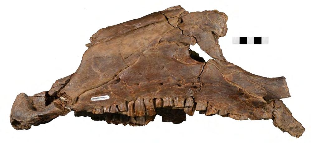
82 A Reviewing Manuscript post vent B ant vent C ant vent
83 5 Close ups of the anterior portion of the skull of Dakotadon lakotaensis (SDSM 8656). (A) photograph in left lateral view of the antorbital fenestra and surrounding bones; (B) photograph in ventral view of the premaxillae and anterior portions of the maxillae. In (A), the white dashed line follows the contact between the lacrimal and prefrontal and the black dashed lines outline the anterior process of the jugal along its contacts with the lacrimal (dorsal) and maxilla (ventral). Abbreviations: af, antorbital fossa; ant, anterior; dam, damaged area; eaof, external antorbital fenestra; ju, jugal; la, lacrimal; lat, lateral; mx, maxilla; na, nasal; pavf, premaxillary anteroventral foramen; pd, premaxillary denticle; pf, prefrontal; plp, posterolateral process of premaxilla; pm, premaxilla; pmf, posterior maxillary foramen; post, posterior; ppvf, premaxillary posteroventral foramen; vent, ventral; vo, vomer. Scale bars equal 4.0 cm.

84 Reviewing Manuscript
85 Figure 6(on next page) Left side of the skull of Dakotadon lakotaensis (SDSM 8656) in dorsal and slightly medial view. Abbreviations: ant, anterior; dam, damaged area; eaof, external antorbital foramen; ep, ectopterygoid; ju, jugal; la, lacrimal; mx, maxilla; na, nasal; pal, palatine; pf, prefrontal; pmf, posterior maxillary foramen; ppf, postpalatine foramen; vent, ventral; vo, vomer. Scale bars equal 5.0 cm.

86 ant vent pf na pmf la eaof ju dam ep ep pal mx vo ppf mx
87 Figure 7(on next page) Anterior portion of the braincase and skull roof of Dakotadon lakotaensis (SDSM 8656). (A) Photograph in dorsal view showing preserved portions of the frontals, posterior portion of the left prefrontal, left palpebral, left postorbital, anterior portion of left squamosal, and anterior portion of parietals; (B) photograph in ventral view showing preserved portions of the frontals, posterior portion of the left prefrontal, left palpebral, left postorbital, anterior portion of left squamosal, anterior portion of parietals, anterodorsal portions of the right and left laterosphenoids, and dorsal portion of the orbitosphenoid; (C) photograph in left lateral view showing preserved portions of the frontals, posterior portion of the left prefrontal, left palpebral, part of the left postorbital (posterior-most portion not shown), anterodorsal portions of the right and left laterosphenoids, and dorsal portion of the orbitosphenoid. Abbreviations: fr, frontal; lat, lateral; ls, laterosphenoid; os, orbitosphenoid; ot, optic tract; par, parietal; pf, prefrontal; po, postorbital; post, posterior; sa, supraorbital articulation surfaces; sor, supraorbital; vent, ventral. Scale bars equal 5.0 cm.


88 A B Reviewing Manuscript fr par fr ot os sa pf par ls pf sor sor post sq po post lat po lat C vent post pf fr sor po ls
89 8 Braincase of Dakotadon lakotaensis (SDSM 8656). (A) photograph of skull roof and braincase in left lateral view taken at an angle looking slightly posteriorly; (B) photograph of the braincase in posterior view; (C) photograph of the posterior portion of the braincase in left lateral view. Abbreviations: bo, basioccipital; bs, basisphenoid; bt, basipterygoid process; ci, crista interfenestralis; CN, cranial nerve; cpr, crista prootica; ct, crista tuberalis; dor, dorsal; fo, fenestra ovalis; fm, foramen metoticum; fr, frontal; gcn V 1 & vcms, groove for the ramus ophthalamicus and the vena cerebralis media secunda; gcn VIIp, groove for the palatine ramus of CN VII; lat, lateral; ls, laterosphenoid; op, fused opisthotic/exoccipital complex; os, orbitosphenoid; par, parietal; pf, prefrontal; po, postorbital; pp, preotic pendant; post, posterior; pro, prootic; pt, pterygoid; ptf, post-temporal foramen; so, supraoccipital; sor, supraorbital; sq, squamosal; vc, Vidian canal; vcm, foramen for the vena cerebralis media; vent, ventral; vp, vena parietalis. Scale bars equal 5.0 cm.

90 Reviewing Manuscript
91 9 Preserved portions of the lower jaw and close up of maxillary dentition of Dakotadon lakotaensis (SDSM 8656). (A) photograph of anterior portion of right dentary and the predentary in right lateral view; (B) photograph of same in medial view; (C) photograph of same in dorsal view; (D) photograph of middle portion of left dentary in left lateral view; (E) photograph of same in medial view; (F) photograph of same in dorsal view; (G) photograph of predentary in ventral view; (H) photograph of predentary in dorsal view; (I) photograph of posterodorsal portion of left dentary, anterior portion of surangular, and the posterior portion of the coronoid in left lateral view; (J) ) photograph of posterodorsal portion of left dentary, anterior portion of surangular, and the posterior portion of the coronoid in medial view; (K) close up view of maxillary teeth from middle portion of left maxilla. Abbreviations: adf, anterior dentary fenestra; ant, anterior; asf, accessory surangular foramen; dor, dorsal; lat, lateral; med, medial; post, posterior; vent, ventral. Abbreviations: ant, anterior; ar, accessory ridge; co, coronoid; de, dentary; dor, dorsal; lat, lateral; mc, Meckelian canal; med, medial; mx, maxilla; pd, predentary; post, posterior; pr, primary ridge; rdf, rostral dentary foramen; su, surangular; tp, tooth position; vent, ventral; vlp, ventrolateral process of predentary. Scale bars in (G) and (H) equal 3 cm, all others equal 5 cm.

92 Reviewing Manuscript
93 Figure 10(on next page) Systematic relationships of Dakotadon lakotaensis. (A) strict consensus tree calculated from eight most parsimonious trees obtained from parsimony-based analysis of relationships; (B) clade credibility tree resulting from the posterior probability-based analysis of relationships. In (A), jackknife values over 50% are listed below their respective nodes. In (B), clade support values over 60% are listed below their respective nodes. Taxa with grey branches in (B) are recovered in different positions than in (A).
94 A Lesothosaurus Reviewing Manuscript Hypsilophodon Muttaburrasaurus Rhabdodon Zalmoxes shqiperorum Zalmoxes robustus Tenontosaurus tilletti Tenontosaurus dossi Dryosaurus Valdosaurus Dysalotosaurus Camptosaurus Cumnoria Uteodon Dakotadon Lanzhousaurus Bolong Barilium Hippodraco Theiophytalia Osmakasaurus Iguanacolossus Proa Koshisaurus Iguanodon Mantellisaurus Altirhinus Equijubus Hypselospinus Xuwulong Gongpoquansaurus Probactrosaurus Eolambia Jeyawati Protohadros Nanyangosaurus Tethyshadros Levnesovia Gilmoreosaurus Bactrosaurus Shuangmiaosaurus Telmatosaurus Corythosaurus Edmontosaurus 100 HADROSAURIDAE HADROSAUROMORPHA HADROSAUROIDEA HADROSAURIFORMES STYRACOSTERNA ANKYLOPOLLEXIA IGUANODONTIA B
95 11 Caudal vertebrae of Dakotadon lakotaensis (SDSM 8656). (A) photograph of caudal 1 in lateral view; (B) photograph of same in opposite lateral view; (C) photograph of caudal 1 in ventral view; (D) photograph of caudal 2 in lateral view; (E) photograph of same in opposite lateral view; (F) photograph of caudal 2 in ventral view. Abbreviations: lr, lateral ridge; vr, ventral ridge. Scale bars equal 5.0 cm.

96 Reviewing Manuscript
SUPPLEMENTARY ONLINE MATERIAL FOR. Nirina O. Ratsimbaholison, Ryan N. Felice, and Patrick M. O connor
 http://app.pan.pl/som/app61-ratsimbaholison_etal_som.pdf SUPPLEMENTARY ONLINE MATERIAL FOR Nirina O. Ratsimbaholison, Ryan N. Felice, and Patrick M. O connor Ontogenetic changes in the craniomandibular
2. Skull, total length versus length of the presacral vertebral column: (0); extremely elongated neck (e.g. Tanystropheus longobardicus).
; extremely elongated neck (e.g. Tanystropheus longobardicus).") Character list of the taxon-character data set 1. Skull and lower jaws, interdental plates: absent (0); present, but restricted to the anterior end of the dentary (1); present along the entire alveolar
Character list of the taxon-character data set 1. Skull and lower jaws, interdental plates: absent (0); present, but restricted to the anterior end of the dentary (1); present along the entire alveolar
ONLINE APPENDIX 1. Morphological phylogenetic characters scored in this paper. See Poe (2004) for
 for") ONLINE APPENDIX Morphological phylogenetic characters scored in this paper. See Poe () for detailed character descriptions, citations, and justifications for states. Note that codes are changed from a
ONLINE APPENDIX Morphological phylogenetic characters scored in this paper. See Poe () for detailed character descriptions, citations, and justifications for states. Note that codes are changed from a
A new species of Hsisosuchus (Mesoeucrocodylia) from Dashanpu, Zigong Municipality, Sichuan Province
 from Dashanpu, Zigong Municipality, Sichuan Province") A new species of Hsisosuchus (Mesoeucrocodylia) from Dashanpu, Zigong Municipality, Sichuan Province Yuhui Gao (Zigong Dinosaur Museum) Vertebrata PalAsiatica Volume 39, No. 3 July, 2001 pp. 177-184 Translated
A new species of Hsisosuchus (Mesoeucrocodylia) from Dashanpu, Zigong Municipality, Sichuan Province Yuhui Gao (Zigong Dinosaur Museum) Vertebrata PalAsiatica Volume 39, No. 3 July, 2001 pp. 177-184 Translated
Exceptional fossil preservation demonstrates a new mode of axial skeleton elongation in early ray-finned fishes
 Supplementary Information Exceptional fossil preservation demonstrates a new mode of axial skeleton elongation in early ray-finned fishes Erin E. Maxwell, Heinz Furrer, Marcelo R. Sánchez-Villagra Supplementary
Bulletin of Big Bend Paleo-Geo An Open Access Publication from Mosasaur Ranch Museum, Terlingua and Lajitas, Texas All rights reserved
 Bulletin of Big Bend Paleo-Geo An Open Access Publication from Mosasaur Ranch Museum, Terlingua and Lajitas, Texas All rights reserved This was a private report in 2003 on my thoughts on Platecarpus planifrons.
AMERICAN MUSEUM NOVITATES Published by
 AMERICAN MUSEUM NOVITATES Published by Number 782 THE AmzRICAN MUSEUM OF NATURAL HISTORY Feb. 20, 1935 New York City 56.81, 7 G (68) A NOTE ON THE CYNODONT, GLOCHINODONTOIDES GRACILIS HAUGHTON BY LIEUWE
SUPPLEMENTARY INFORMATION
 Character 155, interdental ridges. Absence of interdental ridge (0) shown in Parasaniwa wyomingensis (Platynota). Interdental ridges (1) shown in Coniophis precedens. WWW.NATURE.COM/NATURE 1 Character
Supporting Online Material for
 www.sciencemag.org/cgi/content/full/329/5998/1481/dc1 Supporting Online Material for Tyrannosaur Paleobiology: New Research on Ancient Exemplar Organisms Stephen L. Brusatte,* Mark A. Norell, Thomas D.
SOME LITTLE-KNOWN FOSSIL LIZARDS FROM THE
 PROCEEDINGS OF THE UNITED STATES NATIONAL MUSEUM issued SWsK \ {^^m ^V ^^ SMITHSONIAN INSTITUTION U. S. NATIONAL MUSEUM Vol. 91 Washington : 1941 No. 3124 SOME LITTLE-KNOWN FOSSIL LIZARDS FROM THE OLIGOCENE
New Carnivorous Dinosaurs from the Upper Cretaceous of Mongolia
 1955 Doklady, Academy of Sciences USSR 104 (5):779-783 New Carnivorous Dinosaurs from the Upper Cretaceous of Mongolia E. A. Maleev (translated by F. J. Alcock) The present article is a summary containing
4. Premaxilla: Foramen on the lateral surface of the premaxillary body (Yates 2007 ch. 4) 0 absent 1 present
 0 absent 1 present") The character matrix used as a basis for this study is that of Yates et al (2010) which is modified from the earlier matrix used by Yates (2007). This matrix includes characters acquired and/or modified
The character matrix used as a basis for this study is that of Yates et al (2010) which is modified from the earlier matrix used by Yates (2007). This matrix includes characters acquired and/or modified
Fig. 5. (A) Scaling of brain vault size (width measured at the level of anterior squamosal/parietal suture) relative to skull size (measured at the
 Scaling of brain vault size (width measured at the level of anterior squamosal/parietal suture) relative to skull size (measured at the") Fig. 5. (A) Scaling of brain vault size (width measured at the level of anterior squamosal/parietal suture) relative to skull size (measured at the distance between the left versus right temporomandibular
Fig. 5. (A) Scaling of brain vault size (width measured at the level of anterior squamosal/parietal suture) relative to skull size (measured at the distance between the left versus right temporomandibular
A new sauropod from Dashanpu, Zigong Co. Sichuan Province (Abrosaurus dongpoensis gen. et sp. nov.)
") A new sauropod from Dashanpu, Zigong Co. Sichuan Province (Abrosaurus dongpoensis gen. et sp. nov.) by Ouyang Hui Zigong Dinosaur Museum Newsletter Number 2 1989 pp. 10-14 Translated By Will Downs Bilby
A new sauropod from Dashanpu, Zigong Co. Sichuan Province (Abrosaurus dongpoensis gen. et sp. nov.) by Ouyang Hui Zigong Dinosaur Museum Newsletter Number 2 1989 pp. 10-14 Translated By Will Downs Bilby
290 SHUFELDT, Remains of Hesperornis.
 290 SHUFELDT, Remains of Hesperornis. [ Auk [July THE FOSSIL REMAINS OF A SPECIES OF HESPERORNIS FOUND IN MONTANA. BY R. W. SHUFELD% M.D. Plate XI7III. ExR,¾ in November, 1914, Mr. Charles W. Gihnore,
Postilla PEABODY MUSEUM OF NATURAL HISTORY YALE UNIVERSITY NEW HAVEN, CONNECTICUT, U.S.A.
 Postilla PEABODY MUSEUM OF NATURAL HISTORY YALE UNIVERSITY NEW HAVEN, CONNECTICUT, U.S.A. Number 117 18 March 1968 A 7DIAPSID (REPTILIA) PARIETAL FROM THE LOWER PERMIAN OF OKLAHOMA ROBERT L. CARROLL REDPATH
NEW INFORMATION ON THE CRANIUM OF BRACHYLOPHOSAURUS CANADENSIS (DINOSAURIA, HADROSAURIDAE), WITH A REVISION OF ITS PHYLOGENETIC POSITION
, WITH A REVISION OF ITS PHYLOGENETIC POSITION") Journal of Vertebrate Paleontology 25(1):144 156, March 2005 2005 by the Society of Vertebrate Paleontology NEW INFORMATION ON THE CRANIUM OF BRACHYLOPHOSAURUS CANADENSIS (DINOSAURIA, HADROSAURIDAE), WITH
Journal of Vertebrate Paleontology 25(1):144 156, March 2005 2005 by the Society of Vertebrate Paleontology NEW INFORMATION ON THE CRANIUM OF BRACHYLOPHOSAURUS CANADENSIS (DINOSAURIA, HADROSAURIDAE), WITH
HONR219D Due 3/29/16 Homework VI
 Part 1: Yet More Vertebrate Anatomy!!! HONR219D Due 3/29/16 Homework VI Part 1 builds on homework V by examining the skull in even greater detail. We start with the some of the important bones (thankfully
YANGCHUANOSAURUS HEPINGENSIS - A NEW SPECIES OF CARNOSAUR FROM ZIGONG, SICHUAN
 Vol. 30, No. 4 VERTEBRATA PALASIATICA pp. 313-324 October 1992 [SICHUAN ZIGONG ROUSHILONG YI XIN ZHONG] figs. 1-5, pl. I-III YANGCHUANOSAURUS HEPINGENSIS - A NEW SPECIES OF CARNOSAUR FROM ZIGONG, SICHUAN
The cranial osteology of Belebey vegrandis (Parareptilia: Bolosauridae), from the Middle Permian of Russia, and its bearing on reptilian evolution
, from the Middle Permian of Russia, and its bearing on reptilian evolution") Blackwell Publishing LtdOxford, UKZOJZoological Journal of the Linnean Society0024-4082 2007 The Linnean Society of London? 2007 1511 191214 Original Articles RUSSIAN BOLOSAURID REPTILER. R. REISZ ET AL.
Blackwell Publishing LtdOxford, UKZOJZoological Journal of the Linnean Society0024-4082 2007 The Linnean Society of London? 2007 1511 191214 Original Articles RUSSIAN BOLOSAURID REPTILER. R. REISZ ET AL.
A New Ceratopsian Dinosaur from the Upper
 SMITHSONIAN MISCELLANEOUS COLLECTIONS VOLUME 63. NUMBER 3 A New Ceratopsian Dinosaur from the Upper Cretaceous of Montana, with Note on Hypacrosaurus (With Two Plates) CHARLES W. GILMORE Assistant Curator
Anatomy. Name Section. The Vertebrate Skeleton
 Name Section Anatomy The Vertebrate Skeleton Vertebrate paleontologists get most of their knowledge about past organisms from skeletal remains. Skeletons are useful for gleaning information about an organism
Cranial osteology of the African gerrhosaurid Angolosaurus skoogi (Squamata; Gerrhosauridae) HOLLY A. NANCE
 HOLLY A. NANCE") African Journal of Herpetology, 2007 56(1): 39-75. Herpetological Association of Africa Original article Cranial osteology of the African gerrhosaurid Angolosaurus skoogi (Squamata; Gerrhosauridae) HOLLY
African Journal of Herpetology, 2007 56(1): 39-75. Herpetological Association of Africa Original article Cranial osteology of the African gerrhosaurid Angolosaurus skoogi (Squamata; Gerrhosauridae) HOLLY
List of characters used in the phylogenetic analysis. Capital letters T, R, and L, refer to
 1 Supplementary data CHARACTER LIST List of characters used in the phylogenetic analysis. Capital letters T, R, and L, refer to characters used by Tchernov et al. (2000), Rieppel, et al. (2002), and Lee
complex in cusp pattern. (3) The bones of the coyote skull are thinner, crests sharper and the
 The bones of the coyote skull are thinner, crests sharper and the") DISTINCTIONS BETWEEN THE SKULLS OF S AND DOGS Grover S. Krantz Archaeological sites in the United States frequently yield the bones of coyotes and domestic dogs. These two canines are very similar both
DISTINCTIONS BETWEEN THE SKULLS OF S AND DOGS Grover S. Krantz Archaeological sites in the United States frequently yield the bones of coyotes and domestic dogs. These two canines are very similar both
 The following text is generated from uncorrected OCR. [Begin Page: Page 1] A NEW CERATOPSIAN DINOSAUR FROM THE UPPER CRETACEOUS OF MONTANA, WITH NOTE ON HYPACROSAURUS ' By CHARLES W. GILMORE assistant
A New Pterosaur from the Middle Jurassic of Dashanpu, Zigong, Sichuan
 A New Pterosaur from the Middle Jurassic of Dashanpu, Zigong, Sichuan by Xinlu He (Chengdu College of Geology) Daihuan Yang (Chungking Natural History Museum, Sichuan Province) Chunkang Su (Zigong Historical
CRANIAL ANATOMY OF ENNATOSAURUS TECTON (SYNAPSIDA: CASEIDAE) FROM THE MIDDLE PERMIAN OF RUSSIA AND THE EVOLUTIONARY RELATIONSHIPS OF CASEIDAE
 FROM THE MIDDLE PERMIAN OF RUSSIA AND THE EVOLUTIONARY RELATIONSHIPS OF CASEIDAE") Journal of Vertebrate Paleontology 28(1):160 180, March 2008 2008 by the Society of Vertebrate Paleontology ARTICLE CRANIAL ANATOMY OF ENNATOSAURUS TECTON (SYNAPSIDA: CASEIDAE) FROM THE MIDDLE PERMIAN
Journal of Vertebrate Paleontology 28(1):160 180, March 2008 2008 by the Society of Vertebrate Paleontology ARTICLE CRANIAL ANATOMY OF ENNATOSAURUS TECTON (SYNAPSIDA: CASEIDAE) FROM THE MIDDLE PERMIAN
A NEW GENUS AND SPECIES OF AMERICAN THEROMORPHA
 A NEW GENUS AND SPECIES OF AMERICAN THEROMORPHA MYCTEROSAURUS LONGICEPS S. W. WILLISTON University of Chicago The past summer, Mr. Herman Douthitt, of the University of Chicago paleontological expedition,
Williston, and as there are many fairly good specimens in the American
 56.81.7D :14.71.5 Article VII.- SOME POINTS IN THE STRUCTURE OF THE DIADECTID SKULL. BY R. BROOM. The skull of Diadectes has been described by Cope, Case, v. Huene, and Williston, and as there are many
FIELDIANA GEOLOGY NEW SALAMANDERS OF THE FAMILY SIRENIDAE FROM THE CRETACEOUS OF NORTH AMERICA
 FIELDIANA GEOLOGY Published by CHICAGO NATURAL HISTORY MUSEUM Volume 10 Sbftember 22, 1968 No. 88 NEW SALAMANDERS OF THE FAMILY SIRENIDAE FROM THE CRETACEOUS OF NORTH AMERICA Coleman J. Coin AND Walter
Big Bend Paleo-Geo Journal
 Big Bend Paleo-Geo Journal An Open Access Informal Publication from Mosasaur Ranch, Terlingua, Texas All rights reserved Copyright; Kenneth R. Barnes, 2014 New info and corrections in red 2 / 3 / 2015
LOWER CRETACEOUS OF SOUTH DAKOTA.
 A NEW DINOSAUR, STP^GOSAURUS MARSHl, FROM THE LOWER CRETACEOUS OF SOUTH DAKOTA. By Frederic A. Lucas, Curator, Divisioii of Coiiipnrative Anatomy, in charge, of Section of Vertebrate Fossils. The name
Mammalogy Lab 1: Skull, Teeth, and Terms
 Mammalogy Lab 1: Skull, Teeth, and Terms Be able to: Goals of today s lab Locate all structures listed on handout Define all terms on handout what they are or what they look like Give examples of mammals
Cranial anatomy of the iguanodontoid ornithopod Jinzhousaurus yangi from the Lower Cretaceous Yixian Formation of China
 Cranial anatomy of the iguanodontoid ornithopod Jinzhousaurus yangi from the Lower Cretaceous Yixian Formation of China PAUL M. BARRETT, RICHARD J. BUTLER, WANG XIAO LIN, and XU XING Barrett, P.M. Butler,
A M E G H I N I A N A. Revista de la Asociación Paleontológia Argentina. Volume XV September-December 1978 Nos. 3-4
 A M E G H I N I A N A Revista de la Asociación Paleontológia Argentina Volume XV September-December 1978 Nos. 3-4 COLORADIA BREVIS N. G. ET N. SP. (SAURISCHIA, PROSAUROPODA), A PLATEOSAURID DINOSAUR FROM
Description of Cranial Elements and Ontogenetic Change within Tropidolaemus wagleri (Serpentes: Crotalinae).
.") East Tennessee State University Digital Commons @ East Tennessee State University Electronic Theses and Dissertations 5-2016 Description of Cranial Elements and Ontogenetic Change within Tropidolaemus
East Tennessee State University Digital Commons @ East Tennessee State University Electronic Theses and Dissertations 5-2016 Description of Cranial Elements and Ontogenetic Change within Tropidolaemus
A Fossil Snake (Elaphe vulpina) From A Pliocene Ash Bed In Nebraska
 From A Pliocene Ash Bed In Nebraska") University of Nebraska - Lincoln DigitalCommons@University of Nebraska - Lincoln Transactions of the Nebraska Academy of Sciences and Affiliated Societies Nebraska Academy of Sciences 198 A Fossil Snake
University of Nebraska - Lincoln DigitalCommons@University of Nebraska - Lincoln Transactions of the Nebraska Academy of Sciences and Affiliated Societies Nebraska Academy of Sciences 198 A Fossil Snake
Cranial osteology and phylogenetic relationships of Hamadasuchus rebouli (Crocodyliformes: Mesoeucrocodylia) from the Cretaceous of Morocco
 from the Cretaceous of Morocco") Blackwell Publishing LtdOxford, UKZOJZoological Journal of the Linnean Society0024-4082 2007 The Linnean Society of London? 2007 1494 533567 Original Articles HAMADASUCHUS REBOULIH. C. E. LARSSON and H.-D.
Blackwell Publishing LtdOxford, UKZOJZoological Journal of the Linnean Society0024-4082 2007 The Linnean Society of London? 2007 1494 533567 Original Articles HAMADASUCHUS REBOULIH. C. E. LARSSON and H.-D.
Notes on Ceratopsians and Ankylosaurs at the Royal Ontario Museum
 Notes on Ceratopsians and Ankylosaurs at the Royal Ontario Museum Andrew A. Farke, Ph.D. Raymond M. Alf Museum of Paleontology 1175 West Baseline Road Claremont, CA 91711 email: afarke@webb.org Introduction
v:ii-ixi, 'i':;iisimvi'\>!i-:: "^ A%'''''-'^-''S.''v.--..V^'E^'-'-^"-t''gi L I E) R.ARY OF THE VERSITY U N I or ILLINOIS REMO
 R.ARY OF THE VERSITY U N I or ILLINOIS REMO") "^ A%'''''-'^-''S.''v.--..V^'E^'-'-^"-t''gi v:ii-ixi, 'i':;iisimvi'\>!i-:: L I E) R.ARY OF THE U N I VERSITY or ILLINOIS REMO Natural History Survey Librarv GEOLOGICAL SERIES OF FIELD MUSEUM OF NATURAL
"^ A%'''''-'^-''S.''v.--..V^'E^'-'-^"-t''gi v:ii-ixi, 'i':;iisimvi'\>!i-:: L I E) R.ARY OF THE U N I VERSITY or ILLINOIS REMO Natural History Survey Librarv GEOLOGICAL SERIES OF FIELD MUSEUM OF NATURAL
FHSU Scholars Repository. Fort Hays State University. Joshua J. Fry Fort Hays State University, Summer 2015
 Fort Hays State University FHSU Scholars Repository Master's Theses Graduate School Summer 2015 Redescription Of A Specimen Of Pentaceratops (Ornithischia: Ceratopsidae) And Phylogenetic Evaluation Of
.56 m. (22 in.). COMPSOGNATHOID DINOSAUR FROM THE. Medicine Bow, Wyoming, by the American Museum Expedition
. COMPSOGNATHOID DINOSAUR FROM THE. Medicine Bow, Wyoming, by the American Museum Expedition") Article XII.-ORNITHOLESTES HERMANNI, A NEW COMPSOGNATHOID DINOSAUR FROM THE UPPER JURASSIC. By HENRY FAIRFIELD OSBORN. The type skeleton (Amer. Mus. Coll. No. 6I9) of this remarkable animal was discovered
Article XII.-ORNITHOLESTES HERMANNI, A NEW COMPSOGNATHOID DINOSAUR FROM THE UPPER JURASSIC. By HENRY FAIRFIELD OSBORN. The type skeleton (Amer. Mus. Coll. No. 6I9) of this remarkable animal was discovered
Vol. XIV, No. 1, March, The Larva and Pupa of Brontispa namorikia Maulik (Coleoptera: Chrysomelidae: Hispinae) By S.
 By S.") Vol. XIV, No. 1, March, 1950 167 The Larva and Pupa of Brontispa namorikia Maulik (Coleoptera: Chrysomelidae: Hispinae) By S. MAULIK BRITISH MUSEUM (NATURAL HISTORY) (Presented by Mr. Van Zwaluwenburg
Vol. XIV, No. 1, March, 1950 167 The Larva and Pupa of Brontispa namorikia Maulik (Coleoptera: Chrysomelidae: Hispinae) By S. MAULIK BRITISH MUSEUM (NATURAL HISTORY) (Presented by Mr. Van Zwaluwenburg
A NEW SPECIES OF TROODONT DINOSAUR FROM THE
 A NEW SPECIES OF TROODONT DINOSAUR FROM THE LANCE FORMATION OF WYOMING By Charles W. Gilmore Curator of Vertebrate Paleontology, United States National Museum INTRODUCTION The intensive search to which
A Short Report on the Occurrence of Dilophosaurus from Jinning County, Yunnan Province
 A Short Report on the Occurrence of Dilophosaurus from Jinning County, Yunnan Province by Hu Shaojin (Kunming Cultural Administrative Committee, Yunnan Province) Vertebrata PalAsiatica Vol. XXXI, No. 1
Anatomy and Osteohistology of the basal hadrosaurid dinosaur Eotrachodon from the uppermost Santonian (Cretaceous) of southern appalachia
 of southern appalachia") Anatomy and Osteohistology of the basal hadrosaurid dinosaur Eotrachodon from the uppermost Santonian (Cretaceous) of southern appalachia Albert Prieto-Márquez 1, Gregory M. Erickson 2 and Jun A. Ebersole
Anatomy and Osteohistology of the basal hadrosaurid dinosaur Eotrachodon from the uppermost Santonian (Cretaceous) of southern appalachia Albert Prieto-Márquez 1, Gregory M. Erickson 2 and Jun A. Ebersole
The skull of Sphenacodon ferocior, and comparisons with other sphenacodontines (Reptilia: Pelycosauria)
") Circular 190 New Mexico Bureau of Mines & Mineral Resources A DIVISION OF NEW MEXICO INSTITUTE OF MINING & TECHNOLOGY The skull of Sphenacodon ferocior, and comparisons with other sphenacodontines (Reptilia:
Circular 190 New Mexico Bureau of Mines & Mineral Resources A DIVISION OF NEW MEXICO INSTITUTE OF MINING & TECHNOLOGY The skull of Sphenacodon ferocior, and comparisons with other sphenacodontines (Reptilia:
Mammalogy Laboratory 1 - Mammalian Anatomy
 Mammalogy Laboratory 1 - Mammalian Anatomy I. The Goal. The goal of the lab is to teach you skeletal anatomy of mammals. We will emphasize the skull because many of the taxonomically important characters
PRELIMINARY REPORT ON A CLUTCH OF SIX DINOSAURIAN EGGS FROM THE UPPER TRIASSIC ELLIO T FORMATION, NORTHERN ORANGE FREE STATE. J. W.
 41 Pa/aeont. afr., 22, 41-45 (1979) PRELIMINARY REPORT ON A CLUTCH OF SIX DINOSAURIAN EGGS FROM THE UPPER TRIASSIC ELLIO T FORMATION, NORTHERN ORANGE FREE STATE b y J. W. Kitching ABSTRACT A clutch of
A new carnosaur from Yongchuan County, Sichuan Province
 A new carnosaur from Yongchuan County, Sichuan Province by Dong Zhiming Institute of Vertebrate Palaeontology and Palaeoanthropology, Academia Sinica Zhang Yihong, Li Xuanmin, and Zhou Shiwu Chongqing
Redescription of the Mongolian Sauropod NEMEGTOSAURUS MONGOLIENSIS Nowinski (Dinosauria:
 Journal of Systematic Palaeontology 3 (3): 283 318 Issued 24 August 2005 doi:10.1017/s1477201905001628 Printed in the United Kingdom C The Natural History Museum Redescription of the Mongolian Sauropod
A Complete Late Cretaceous Iguanian (Squamata, Reptilia) from the Gobi and Identification of a New Iguanian Clade
 from the Gobi and Identification of a New Iguanian Clade") PUBLISHED BY THE AMERICAN MUSEUM OF NATURAL HISTORY CENTRAL PARK WEST AT 79TH STREET, NEW YORK, NY 10024 Number 3584, 47 pp., 19 figures September 6, 2007 A Complete Late Cretaceous Iguanian (Squamata,
PUBLISHED BY THE AMERICAN MUSEUM OF NATURAL HISTORY CENTRAL PARK WEST AT 79TH STREET, NEW YORK, NY 10024 Number 3584, 47 pp., 19 figures September 6, 2007 A Complete Late Cretaceous Iguanian (Squamata,
THE OCCURRENCE OF CONTOGENYS-LIKE LIZARDS IN THE LATE CRETACEOUS AND EARLY TERTIARY OF THE WESTERN INTERIOR OF THE U.S.A.
 Journal of Vertebrate Paleontology 29(3):677 701, September 2009 # 2009 by the Society of Vertebrate Paleontology ARTICLE THE OCCURRENCE OF CONTOGENYS-LIKE LIZARDS IN THE LATE CRETACEOUS AND EARLY TERTIARY
A new species of sauropod, Mamenchisaurus anyuensis sp. nov.
 A new species of sauropod, Mamenchisaurus anyuensis sp. nov. by Xinlu He, Suihua Yang, Kaiji Cai, Kui Li, and Zongwen Liu Chengdu University of Technology Papers on Geosciences Contributed to the 30th
Major cranial changes during Triceratops ontogeny John R. Horner 1, * and Mark B. Goodwin 2
 273, 2757 2761 doi:10.1098/rspb.2006.3643 Published online 1 August 2006 Major cranial changes during Triceratops ontogeny John R. Horner 1, * and Mark B. Goodwin 2 1 Museum of the Rockies, Montana State
Yamaceratops dorngobiensis, a New Primitive Ceratopsian (Dinosauria: Ornithischia) from the Cretaceous of Mongolia
 from the Cretaceous of Mongolia") PUBLISHED BY THE AMERICAN MUSEUM OF NATURAL HISTORY CENTRAL PARK WEST AT 79TH STREET, NEW YORK, NY 10024 Number 3530, 42 pp., 20 figures September 08, 2006 Yamaceratops dorngobiensis, a New Primitive Ceratopsian
PUBLISHED BY THE AMERICAN MUSEUM OF NATURAL HISTORY CENTRAL PARK WEST AT 79TH STREET, NEW YORK, NY 10024 Number 3530, 42 pp., 20 figures September 08, 2006 Yamaceratops dorngobiensis, a New Primitive Ceratopsian
On the history, osteology, and systematic position of the Wealden (Hastings group) dinosaur Hypselospinus fittoni (Iguanodontia: Styracosterna)
 dinosaur Hypselospinus fittoni (Iguanodontia: Styracosterna)") bs_bs_banner Zoological Journal of the Linnean Society, 2015, 173, 92 189. With 52 figures On the history, osteology, and systematic position of the Wealden (Hastings group) dinosaur Hypselospinus fittoni
bs_bs_banner Zoological Journal of the Linnean Society, 2015, 173, 92 189. With 52 figures On the history, osteology, and systematic position of the Wealden (Hastings group) dinosaur Hypselospinus fittoni
First Ornithomimid (Theropoda, Ornithomimosauria) from the Upper Cretaceous Djadokhta Formation of Tögrögiin Shiree, Mongolia
 from the Upper Cretaceous Djadokhta Formation of Tögrögiin Shiree, Mongolia") First Ornithomimid (Theropoda, Ornithomimosauria) from the Upper Cretaceous Djadokhta Formation of Tögrögiin Shiree, Mongolia Tsogtbaatar Chinzorig¹, ³ *, Yoshitsugu Kobayashi², Khishigjav Tsogtbaatar³,
First Ornithomimid (Theropoda, Ornithomimosauria) from the Upper Cretaceous Djadokhta Formation of Tögrögiin Shiree, Mongolia Tsogtbaatar Chinzorig¹, ³ *, Yoshitsugu Kobayashi², Khishigjav Tsogtbaatar³,
The phylogeny and evolutionary history of tyrannosauroid dinosaurs
 Supplementary information for: The phylogeny and evolutionary history of tyrannosauroid dinosaurs Stephen L. Brusatte 1#* & Thomas D. Carr 2# 1 School of GeoSciences, University of Edinburgh, Grant Institute,
Marshall Digital Scholar. Marshall University. F. Robin O Keefe Marshall University,
 Marshall University Marshall Digital Scholar Biological Sciences Faculty Research Biological Sciences 2008 Cranial anatomy and taxonomy of Dolichorhynchops bonneri new combination, a polycotylid (Sauropterygia:
VERTEBRATA PALASIATICA
 41 2 2003 2 VERTEBRATA PALASIATICA pp. 147 156 figs. 1 5 1) ( 100044), ( Parakannemeyeria brevirostris),,, : ( Xiyukannemeyeria),,, Q915. 864 60 Turfania (,1973), Dicynodon (, 1973 ; Lucas, 1998), (Lystrosaurus)
Cranial morphology of Sinornithosaurus millenii Xu et al (Dinosauria: Theropoda: Dromaeosauridae) from the Yixian Formation of Liaoning, China
 from the Yixian Formation of Liaoning, China") 1739 Cranial morphology of Sinornithosaurus millenii Xu et al. 1999 (Dinosauria: Theropoda: Dromaeosauridae) from the Yixian Formation of Liaoning, China Xing Xu and Xiao-Chun Wu Abstract: The recent discovery
1739 Cranial morphology of Sinornithosaurus millenii Xu et al. 1999 (Dinosauria: Theropoda: Dromaeosauridae) from the Yixian Formation of Liaoning, China Xing Xu and Xiao-Chun Wu Abstract: The recent discovery
Cranial morphology and taxonomy of South African Tapinocephalidae (Therapsida: Dinocephalia): the case of Avenantia and Riebeeckosaurus
: the case of Avenantia and Riebeeckosaurus") Cranial morphology and taxonomy of South African Tapinocephalidae (Therapsida: Dinocephalia): the case of Avenantia and Riebeeckosaurus Saniye Güven*, Bruce S. Rubidge & Fernando Abdala Evolutionary Studies
Cranial morphology and taxonomy of South African Tapinocephalidae (Therapsida: Dinocephalia): the case of Avenantia and Riebeeckosaurus Saniye Güven*, Bruce S. Rubidge & Fernando Abdala Evolutionary Studies
Reprinted from: CRUSTACEANA, Vol. 32, Part 2, 1977 LEIDEN E. J. BRILL
 Reprinted from: CRUSTACEANA, Vol. 32, Part 2, 1977 LEIDEN E. J. BRILL NOTES AND NEWS 207 ALPHE0PS1S SHEARMII (ALCOCK & ANDERSON): A NEW COMBINATION WITH A REDESCRIPTION OF THE HOLOTYPE (DECAPODA, ALPHEIDAE)
SOME NEW AMERICAN PYCNODONT FISHES.
 SOME NEW AMERICAN PYCNODONT FISHES. By James Williams Gidley, Assistant Curator of Fossil Mammals, United States National Museum. In the United States National Museum are several specimens representing
Jurassic Ornithopod Agilisaurus louderbacki (Ornithopoda: Fabrosauridae) from Zigong, Sichuan, China
 from Zigong, Sichuan, China") Jurassic Ornithopod Agilisaurus louderbacki (Ornithopoda: Fabrosauridae) from Zigong, Sichuan, China Guangzhao Peng (Zigong Dinosaur Museum) Vertebrata PalAsiatica Volume 30, No. 1 January, 1992 pp. 39-51
Jurassic Ornithopod Agilisaurus louderbacki (Ornithopoda: Fabrosauridae) from Zigong, Sichuan, China Guangzhao Peng (Zigong Dinosaur Museum) Vertebrata PalAsiatica Volume 30, No. 1 January, 1992 pp. 39-51
School of Earth Sciences, University of Bristol, Queen s Road, Bristol BS8 1RJ, UK 2
 bs_bs_banner Zoological Journal of the Linnean Society, 2015, 173, 55 91. With 20 figures Osteology of Rauisuchus tiradentes from the Late Triassic (Carnian) Santa Maria Formation of Brazil, and its implications
A R T I C L E S STRATIGRAPHIC DISTRIBUTION OF VERTEBRATE FOSSIL FOOTPRINTS COMPARED WITH BODY FOSSILS
 A R T I C L E S STRATIGRAPHIC DISTRIBUTION OF VERTEBRATE FOSSIL FOOTPRINTS COMPARED WITH BODY FOSSILS Leonard Brand & James Florence Department of Biology Loma Linda University WHAT THIS ARTICLE IS ABOUT
New information on the palate and lower jaw of Massospondylus (Dinosauria: Sauropodomorpha)
") New information on the palate and lower jaw of Massospondylus (Dinosauria: Sauropodomorpha) Paul M. Barrett 1* & Adam M. Yates 2* 1 Department of Palaeontology, The Natural History Museum, Cromwell Road,
New information on the palate and lower jaw of Massospondylus (Dinosauria: Sauropodomorpha) Paul M. Barrett 1* & Adam M. Yates 2* 1 Department of Palaeontology, The Natural History Museum, Cromwell Road,
PALEONTOLOGY AND BIOSTRATIGRAPHY OF MONGOLIA
 PALEONTOLOGY AND BIOSTRATIGRAPHY OF MONGOLIA THE JOINT SOVIET-MONGOLIAN PALEONTOLOGICAL EXPEDITION (Transactions, vol. 3) EDITORIAL BOARD: N. N. Kramarenko (editor-in-chief) B. Luvsandansan, Yu. I. Voronin,
Mammalogy Lecture 8 - Evolution of Ear Ossicles
 Mammalogy Lecture 8 - Evolution of Ear Ossicles I. To begin, let s examine briefly the end point, that is, modern mammalian ears. Inner Ear The cochlea contains sensory cells for hearing and balance. -
University of Iowa Iowa Research Online
 University of Iowa Iowa Research Online Theses and Dissertations Spring 2016 A reassessment of the late Eocene - early Oligocene crocodylids Crocodylus megarhinus Andrews 1905 and Crocodylus articeps Andrews
PEABODY MUSEUM OF NATURAL HISTORY, YALE UNIVERSITY NEW HAVEN, CONNECTICUT, U.S.A. A NEW OREODONT FROM THE CABBAGE PATCH LOCAL FAUNA, WESTERN MONTANA
 Postilla PEABODY MUSEUM OF NATURAL HISTORY YALE UNIVERSITY NEW HAVEN, CONNECTICUT, U.S.A. Number 85 September 21, 1964 A NEW OREODONT FROM THE CABBAGE PATCH LOCAL FAUNA, WESTERN MONTANA STANLEY J. RIEL
SUPPLEMENTARY INFORMATION
 doi:10.1038/nature13086 Part I. Supplementary Notes A: Detailed Description of Cotylocara macei gen. et sp. nov. Part II. Table of Measurements for holotype of Cotylocara macei (CCNHM-101) Part III. Supplementary
On Asian ornithopods (Dinosauria: Ornithischia). 3. A new species of iguanodontid d inosaur
. 3. A new species of iguanodontid d inosaur")
Erycine Boids from the Early Oligocene of the South Dakota Badlands
 Georgia Journal of Science Volume 67 No. 2 Scholarly Contributions from the Membership and Others Article 6 2009 Erycine Boids from the Early Oligocene of the South Dakota Badlands Dennis Parmley J. Alan
The Lower Jaws of Baenid Turtles
 AMERICAN MUSEUM Novitates PUBLISHED BY THE AMERICAN MUSEUM OF NATURAL HISTORY CENTRAL PARK WEST AT 79TH STREET, NEW YORK, N.Y. 10024 Number 2749, pp. 1-10, figs. 1-4, table 1 September 27, 1982 The Lower
The Geological Society of America Special Paper
 GSA_SP427_15_Meredith.qxd 8/8/07 12:16 PM Page 209 The Geological Society of America Special Paper 427 2007 The largest mosasaur (Squamata: Mosasauridae) from the Missouri River area (Late Cretaceous;
OF THE TRIAS THE PHYTOSAURIA
 THE PHYTOSAURIA OF THE TRIAS MAURICE G. MEHL University of Wisconsin Some time ago the writer gave a brief notice of a new genus of phytosaurs of which Angistorhinus grandis Mehl was the type.' It is the
EARLY PALEOGENE CROCODYLIFORM EVOLUTION IN THE NEOTROPICS: EVIDENCE FROM NORTHEASTERN COLOMBIA
 EARLY PALEOGENE CROCODYLIFORM EVOLUTION IN THE NEOTROPICS: EVIDENCE FROM NORTHEASTERN COLOMBIA By ALEXANDER K. HASTINGS A DISSERTATION PRESENTED TO THE GRADUATE SCHOOL OF THE UNIVERSITY OF FLORIDA IN PARTIAL
CRANIAL ANATOMY AND PHYLOGENETIC AFFINITIES OF THE PERMIAN PARAREPTILE MACROLETER POEZICUS
 CRANIAL ANATOMY AND PHYLOGENETIC AFFINITIES OF THE PERMIAN PARAREPTILE MACROLETER POEZICUS Author(s): LINDA A. TSUJI Source: Journal of Vertebrate Paleontology, 26(4):849-865. 2006. Published By: The Society
TRACHEMYS SCULPTA. A nearly complete articulated carapace and plastron of an Emjdd A NEAKLY COMPLETE SHELL OF THE EXTINCT TURTLE,
 A NEAKLY COMPLETE SHELL OF THE EXTINCT TURTLE, TRACHEMYS SCULPTA By Charles W. Gilmore Curator of Vertebrate Paleontology, United States National Museum INTRODUCTION A nearly complete articulated carapace
Florida, Gainesville, Florida, 32611, U.S.A. b Smithsonian Tropical Research Institute, Ancon, Republic of Panama,
 This article was downloaded by: [78.22.97.164] On: 04 May 2013, At: 14:02 Publisher: Taylor & Francis Informa Ltd Registered in England and Wales Registered Number: 1072954 Registered office: Mortimer
NOTES ON THE FIRST SKULL AND JAWS OF RIOJASAURUS INCERTUS (DINOSAURIA, PROSAUROPODA, MELANOROSAURIDAE) OF THE LATE TRIASSIC OF LA RIOJA, ARGENTINA
 OF THE LATE TRIASSIC OF LA RIOJA, ARGENTINA") NOTES ON THE FIRST SKULL AND JAWS OF RIOJASAURUS INCERTUS (DINOSAURIA, PROSAUROPODA, MELANOROSAURIDAE) OF THE LATE TRIASSIC OF LA RIOJA, ARGENTINA José F. Bonaparte and José A. Pumares translated by Jeffrey
NOTES ON THE FIRST SKULL AND JAWS OF RIOJASAURUS INCERTUS (DINOSAURIA, PROSAUROPODA, MELANOROSAURIDAE) OF THE LATE TRIASSIC OF LA RIOJA, ARGENTINA José F. Bonaparte and José A. Pumares translated by Jeffrey
FURTHER STUDIES ON TWO SKELETONS OF THE BLACK RIGHT WHALE IN THE NORTH PACIFIC
 FURTHER STUDIES ON TWO SKELETONS OF THE BLACK RIGHT WHALE IN THE NORTH PACIFIC HIDEO OMURA, MASAHARU NISHIWAKI* AND TOSHIO KASUYA* ABSTRACT Two skeletons of the black right whale were studied, supplementing
ABSTRACT. we define the taxa Alligatoroidae and Alligatoridae to be the descent community and crown group,
 AMERICAN MUSEUM No vtates PUBLISHED BY THE AMERICAN MUSEUM OF NATURAL HISTORY CENTRAL PARK WEST AT 79TH STREET, NEW YORK, N.Y. 10024 Number 3116, 26 pp., 10 figures, 1 table December 28, 1994 The Late
THE GORGONOPSIAN GENUS, HIPPOSAURUS, AND THE FAMILY ICTIDORHINIDAE * Dr. L.D. Boonstra. Paleontologist, South African Museum, Cape Town
 THE GORGONOPSIAN GENUS, HIPPOSAURUS, AND THE FAMILY ICTIDORHINIDAE * by Dr. L.D. Boonstra Paleontologist, South African Museum, Cape Town In 1928 I dug up the complete skeleton of a smallish gorgonopsian
Published online: 17 Sep 2014.
 This article was downloaded by: [78.22.98.228] On: 22 September 2014, At: 04:53 Publisher: Taylor & Francis Informa Ltd Registered in England and Wales Registered Number: 1072954 Registered office: Mortimer
REVISION OF REDONDASUCHUS (ARCHOSAURIA: AETOSAURIA) FROM THE UPPER TRIASSIC REDONDA FORMATION, NEW MEXICO, WITH DESCRIPTION OF A NEW SPECIES
 FROM THE UPPER TRIASSIC REDONDA FORMATION, NEW MEXICO, WITH DESCRIPTION OF A NEW SPECIES") Harris et al., eds., 2006, The Triassic-Jurassic Terrestrial Transition. New Mexico Museum of Natural History and Science Bulletin 37. REVISION OF REDONDASUCHUS (ARCHOSAURIA: AETOSAURIA) FROM THE UPPER
Harris et al., eds., 2006, The Triassic-Jurassic Terrestrial Transition. New Mexico Museum of Natural History and Science Bulletin 37. REVISION OF REDONDASUCHUS (ARCHOSAURIA: AETOSAURIA) FROM THE UPPER
Lower Cretaceous Kwanmon Group, Northern Kyushu
 Bull. Kitakyushu Mus. Nat. Hist., 11: 87-90. March 30, 1992 A New Genus and Species of Carnivorous Dinosaur from the Lower Cretaceous Kwanmon Group, Northern Kyushu Yoshihiko Okazaki Kitakyushu Museum
A new specimen of Acrocanthosaurus atokensis
 A new specimen of Acrocanthosaurus atokensis (Theropoda, Dinosauria) from the Lower Cretaceous Antlers Formation (Lower Cretaceous, Aptian) of Oklahoma, USA Philip J. CURRIE Royal Tyrrell Museum of Palaeontology,
CENE RUMINANTS OF THE GENERA OVIBOS AND
 DESCRIPTIONS OF TWO NEW SPECIES OF PLEISTO- CENE RUMINANTS OF THE GENERA OVIBOS AND BOOTHERIUM, WITH NOTES ON THE LATTER GENUS. By James Williams Gidley, Of the United States National Museum. Two interesting
Archived version from NCDOCKS Institutional Repository
 Archived version from NCDOCKS Institutional Repository http://libres.uncg.edu/ir/asu/ Revision of the Archosauromorph Reptile Trilophosaurus, With a Description of the First Skull of Trilophosaurus Jacobsi,
35. DATA REPORT: CRETACEOUS OSTRACODES FROM HOLES 865A AND 866A (MID-PACIFIC MOUNTAINS) 1. Renée Damotte 2
 1. Renée Damotte 2") Winterer, E.L., Sager, W.W., Firth, J.V., and Sinton, J.M. (Eds.), 1995 Proceedings of the Ocean Drilling Program, Scientific Results, Vol. 143 35. DATA REPORT: CRETACEOUS OSTRACODES FROM HOLES 865A AND
Winterer, E.L., Sager, W.W., Firth, J.V., and Sinton, J.M. (Eds.), 1995 Proceedings of the Ocean Drilling Program, Scientific Results, Vol. 143 35. DATA REPORT: CRETACEOUS OSTRACODES FROM HOLES 865A AND
A New Dromaeosaurid Theropod from Ukhaa Tolgod (Ömnögov, Mongolia)
") PUBLISHED BY THE AMERICAN MUSEUM OF NATURAL HISTORY CENTRAL PARK WEST AT 79TH STREET, NEW YORK, NY 10024 Number 3545, 51 pp., 25 figures, 1 table December 7, 2006 A New Dromaeosaurid Theropod from Ukhaa
PUBLISHED BY THE AMERICAN MUSEUM OF NATURAL HISTORY CENTRAL PARK WEST AT 79TH STREET, NEW YORK, NY 10024 Number 3545, 51 pp., 25 figures, 1 table December 7, 2006 A New Dromaeosaurid Theropod from Ukhaa
A NEW CROCODYLOMORPH ARCHOSAUR FROM THE UPPER TRIASSIC OF NORTH CAROLINA
 Journal of Vertebrate Paleontology 23(2):329 343, June 2003 2003 by the Society of Vertebrate Paleontology A NEW CROCODYLOMORPH ARCHOSAUR FROM THE UPPER TRIASSIC OF NORTH CAROLINA HANS-DIETER SUES 1 *,
SAUROPOD DINOSAURS FROM THE EARLY CRETACEOUS OF MALAWI, AFRICA. Elizabeth M. Gomani
 Palaeontologia Electronica http://palaeo-electronica.org SAUROPOD DINOSAURS FROM THE EARLY CRETACEOUS OF MALAWI, AFRICA Elizabeth M. Gomani ABSTRACT At least two titanosaurian sauropod taxa have been discovered
A NEW AUSTROSQUILLA (STOMATOPODA) FROM THE
 FROM THE") A NEW AUSTROSQUILLA (STOMATOPODA) FROM THE MARQUESAS ISLANDS BY ALAIN MICHEL Centre O.R.S.T.O.M., Noumea, New Caledonia and RAYMOND B. MANNING Smithsonian Institution, Washington, U.S.A. The At s,tstrosqzlilla
A NEW AUSTROSQUILLA (STOMATOPODA) FROM THE MARQUESAS ISLANDS BY ALAIN MICHEL Centre O.R.S.T.O.M., Noumea, New Caledonia and RAYMOND B. MANNING Smithsonian Institution, Washington, U.S.A. The At s,tstrosqzlilla
The Discovery of a Tritylodont from the Xinjiang Autonomous Region
 The Discovery of a Tritylodont from the Xinjiang Autonomous Region Ailing Sun and Guihai Cui (Institute of Vertebrate Paleontology, Paleoanthropology, Academia Sinica) Vertebrata PalAsiatica Volume XXVII,