Histological Studies on the Alimentary Tract of the Colubrid Snake Coluber florulentus (Family Colubridae)
|
|
|
- Luke Bennett
- 5 years ago
- Views:
Transcription


1 J.K.A. U.: Sci., vol. 1, pp (1409 A.H./1989 A.D.) Histological Studies on the Alimentary Tract of the Colubrid Snake Coluber florulentus (Family Colubridae) G.Y~ DEHLAWIANDM.M. ZAHER Biology Dept., Faculty of Applied Science, Umm AI-Qura University, Makkah AI-Mukarramah, Saudi Ar(lbia; and Zoology Dept., Faculty of Science, Cairo University, Cairo, Egypt. ABSTRAcr. The histological structure of the alimentary tract of the snake Coluber florulentus was described and compared with that of other examined reptiles. The wall of the oesophagus, stomach, small intestine and large intestine is built up of the following layers from outside inwards; serosa, muscularis, submucosa and mucosa, while in the wall of the stomach there is a new layer, subserosa which follows serosa. The oesophageal mucosa consists of simple columnar ciliated epithelial and goblet cells. The oesophageal glands were found to be completely absent. The gastric mucosa consists of simple columnar cells. There are two types of gastric glands. These are the fundic and pyloric glands (granular and light-celled types). The mucosa of the small intestine consists of three types of cells; the absorptive, the goblet and the endocrine cells. The intestinal glands as well as crypts of Lieberkhun are absent. The caecal mucosa consists of simple columnar epithelial and goblet cells. The mucosal epithelium of the colon consists of three types of cells; simple columnar, goblet and endocrine cells. The rectal mucosa is only represented by simple columnar epithelial cells. Introduction A review of the histological studies on the alimentary tract of the members of the order Squamata revealed that most of these studies were dealt with the members of the suborder Lacertiliall-11]. On the other hand, a little attention has been paid to the members of the suborder Ophidia. Moreover, most of the available works on the latter suborder were dealt with certain parts of the tractl12-21]. However, the histological studies of the different regions of the Ophidian tract, in general, were carried out, only, by a few number of workersl22,23&9]. This indicated that the present subject did not receive the necessary attention. This stimulated the present authors to carry out some work in this field on some members of order Ophidia. 9~


2 96 G. Y.DehlawiandM.M. Zaher The present article is the first in the series. It deals with the general histology of the different regions of the alimentary tract of the colubridsnake Coluber florulentus~ Material and Methods The animal used in this work is thecolubrid snake C. florulentus (Family Colubridae). It was caught by hand from different localities in Arafat region, 20 kin from Makkah, Saudi Arabia. Animals were dissected and the different regions of the alimentary canal; oesophagus, stomach, small and large intestine were fixed in Bouin's fluid and were subsequently processed for sectioning. Section 8 JL-thick were stained with haematoxylin and eosin as well as with Mallory's triple stain. Oesophagus Observations Microscopic examination of the oesophagus showed that its wall consists of serosa, muscularis, submucosa and mucosa (Fig. 1). i FIG: 1. T.5. in the oesophagus of the snake C. florulentus. x 82.5


3 Histological Studies on the Alimentary Tract. 97 The serosa is represented by a thin layer of squamous epithelium which may be ruptured during the preparation of the sections. The muscularis has an outer layer of longitudinal smooth muscle fibres and an inner one of smooth circular muscle fibres. It is uniform in thickness through the total length of the oesophageal region. The submucosa is well developed throughout the whol~length of the oesophagus and extends into folds. The muscularis mucosa is weakly developed and is represented by a thin contmuous striated layer of an outer longitudinal and an mner circular muscle fibres. The oesophageal mucosa is thrown into many folds with variable lengths and wavy appearance. They surround a relatively wide lumen. The number and distribution of the folds is nearly constant through the total length of the oesophagus.. The oesophageal mucosa comprises the lamina propria of connective tissue, and the epithelium which lines the lumen. The former propria extends in the well developed oesophageal folds. The mucosal epithelium is simple and formed of two types of cells; the elongated thin ciliated epithelial cells and large goblet cells (Fi.g. 2). Th~goblet cells are mar- FIG, 2:' T.S. in the oesopqagus of the snake C. florulentus. x 330

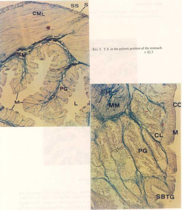
4 98 G.. Dehlawi and M.M. Zaher kedly numerous at the bottom of the oesophageal folds. They could not be considered as glands because they are identical to the surface epithelial cells. The mucous cells are large and club-shaped. Each consists of an inner small basal part and an outer large hyaline portion. The basal part contains a flat nucleus which is intensely stained and is embedded into the cytoplasm (Fig. 2). The outer large hyaline portion is stained faint blue with Mallory stain. This means that the mucin occupies most of the mucous cells. The openings of the mucous cells in the oesophageal lumen are wide and sometimes mucous plugs were observed outside the cells. In the present study, the ciliated epithelial cells of the oesophageal mucosa are found scattered between the numerous goblet cells (Fig. 2). They are relatively fewer in number as compared with those of the latter cells. Their cilia are found arranged along the outer border of the mucosal epithelium. Each columnar cell is provided with an oval nucleus rests near its base. It is stained dark red with the Mallory stain. The cytoplasm is provided with fine granules which are also stained dark red with the same stain. Microscopic examination of the oesophagus showed that its mucosa is totally devoid of glands.. Stomach The stomach of C. florulentus can be divided into an anterior fundic portion following the oesophagus and a posterior pyloric portion which ends with the pylorus and leads to the duodenum, (Fig. 3,4,5 & 6). The serosa of the stomach consists of a thin layer of spindle-shaped simple aquamous epithelial cells (Fig. 3). A layer of subserosa is present with scattered longitudinal muscle fibres. It has a uniform thickness through the whole length of the stomach (Fig. 5). The muscularies is composed essentially of smooth circular muscle fibres. It is more or less continuous, thick in the anterior region and increases in thickness towards the middle region and reaches its maximum thickness in the posterior region where it is highly developed (Fig. 5). The submucosa is well developed in both anterior and posterior regions of the stomach (fundic and pyloric portions), (Fig. 4 & 6). The muscularis mucosa is composed of two smooth continuous layers; an outer longitudinal and inner circular muscle layers. The latter is relatively thin while the longitudinal one is more developed and forms a thicker layer. Such a pattern is continuous in both pyloric and cardiac portions of the stomach (Fig. 4& 6). The gastric mucosa is thrown into longitudinal folds of varying height and is formed of mucosal epithelium, lamina propria and gastric glands. The gastric surface epithelium is of the simple columnar type. Its cells are cylindrical with ovoid and basal nuclei. Each nucleus contains one or two nucleoli. The cytoplasm of the luminal halves of such cells is filled with dark red stained granules. Their inner portions contain a clear non-granular cytoplasm (Fig. 4). The lamina propria is represented by the connective tissue found between the muscularis mucosa and the mucosal epithelium. It extends into the gastric folds. In the lamina propria,
.")

5 Histological Studies on the Alimentary Tract FIG. 3. T.S. in the fundic portion of the stomach. x 82.5 bodies of the gastric glands are embedded where they open into the lumen between the gastric folds through the gastric pits (Fig. 4). The gastric glands are numerous in the anterior region of the stomach (fundic region) and comparatively fewer in the posterior region (pyloric region), (Fig. 4 & 6). Microscopic examination of the stomach revealed the presence of two types of gastric glands; the granular type and the light-celled one. The fundic region contains both types of glands (Fig. 4), while the pyloric region contains only the light-celled one (Fig. 6). In the fundic region, the light-celled glands were found to be arranged along the periphery of the lamina propria, while those of the granular type are found scattered in the areolar connective tissue of the lamina propria.
.")

6 100 G. Y. DehlawiandM.M. Zaher Fro. 4:cl.S. in the fundic portion of the stomach x 330 The granular glands are mainly of simple tubular type. They open by their pits into the gastric lumen. Their bodies consist of dome-shaped granular cells. They are full of dark granules and haye central round nuclei. They stain violet with Mallory (Fig. 4). The light~celled glands, on the other hand, were found to be of the simple tubular or simple branched tubular type. Their bodies consist also of dome-shaped cells. The cytoplasm is pale and contains indistinct granules. The nucleus is round or flat and is scattered againstth~ base. The cytoplasm of these cells is stained faint blue with Mallory and red with eosin-haematoxylin. This means that they show positive reaction with mucous stain (Fig. 6).
7
. FIG. 7. T.S. in the ileum.")
, (Fig. 7).")
8 102 G. Y. Dehlawi and M.M. Zaher Small Intestine The lumen of the small intestine is more or less narrow due to the presence of extremely long and coiled villi (Fig. 7). FIG. 7. T.S. in the ileum. x 82.5 The serosa is composed of simple squamous epithelial cells. It represents the outer-most layer of the small intestine (the duodenum and ileum), (Fig. 7). The muscularis, which is formed of smooth muscles, is arranged into an outer longitudinal layer and an inner circular layer (Fig. 7). The submucosa is vascular and enters in the formation of the intestinal villi, whereas the muscularis mucosa is totally absent.
. The columnar cells have.large elongated nuclei situated at the base of the cells.")
.")
9 Histological Studies on the Alimentary Tract. 103 In the duodenum and ileum, the mucosal lining of the intestinal villi is unifonn throughout. It is composed of three types of cells; simple columnar epithelial cells, goblet cells and endocrine cells (Fig. 8). The columnar cells have.large elongated nuclei situated at the base of the cells. The coarsely granular cytoplasm is deeply stained with Mallory. The goblet cells are fewer in number as compared with the columnar ones. They are provided with a short basal part with a faint nucleus, and an upper flask shaped mucous part that has a wide opening into the lumen (Fig. 8). The endocrine cells are small in size, limited in number and have a clear cytoplasm in which spherical and central nuclei were located (Fig. 8). FIG. 8. T.S. in the ileum. x 330


10 104 G. Y. Dehlawi and M.M. Zaher The intestinal glands as well as the crypts are totally absent from the submucosa of the duodenum as well as of the ileum. Large Intestine The large intestine ofc. jlorulentus is in the form of a relatively short straight muscular tube extending from the end of the ileum till the cloaca. The large intestine is of three parts; the caecum, the colon and the rectum (Fig. 9 to 13). FIG. 9. T.S. in the caecum. x 330

11 Histological Studies on the Alimentary Tract. 105
. A thick well-developed submucosa follows muscularis and enter in the formation of the high folds of the mucosa (Fig. 9).")


12 It)6 G.Y. DehlawiandM.M. Zaher FIG. 12. T.S. in the rectum. x 82.5 FIG. 13. T.S. in the rectum. x 330 The smooth muscles of the muscularis are in the form of an outer longitudinal muscle layer and inner broader circular one (Fig. 12). A thick well-developed submucosa follows muscularis and enter in the formation of the high folds of the mucosa (Fig. 9). The caecal mucosa consists of simple columnar cells as well as the goblet cells. The latter cells are present in abundant. Their apical part forms most of the body of the cell. Their basal part is short and contains a faint basal nucleus with an oval outline. The columnar cells are thin and tall. They have spindle-shaped nuclei situated near the middle of the cells. Their cytoplasm is coarsely granular and deeply stained with Mallory stain (Fig. 9). The epithelium of the colon consists of three kinds of cells; simple columnar, goblet and endocrine cells. The columnar cells are present in a large number. They have a spherical and basal nuclei. Their cytoplasm is heavily granular and deeply stained. The goblet cells have a limited number and have the same shape and structure of those of the caecum (Fig. 11).
.")

![Such a structure is similar to the observations obtained in Mauremys caspica[24], Cnalcides levitoni[25] and Acanthodactylus boskianus[loi.](/docs-images/85/91395134/images/13-2.jpg "However, in Uromastyx aegyptia[3], Chameleon vulgaris'[4] and in Uromastyx philbyl126] only the anterior region of the oesophageal mucosa assumes identical to the present observation, while the")
13 Histologit;alStudies on the Alimenta~ Tract. 107 The rectal mucosa, which is provided with several depressions, consists only of simple columnar epithelial cells, i.e. the goblet cells are completely absent. Here, the columnar cells contain heavily granular and deeply stained cytoplasm. Their oval basal nuclei were difficult to identify due to the heavy granularity of the cytoplasm (Fig. 13). Discussion Microscopic examination of the 'oesophageal mucosa of several investigated species of reptiles showed a considerable variation in their histological structure. The present study revealed that the oesophageal mucosa of the colubrid snake C. florulentus is represented by simple columnar ciliated and goblet cells. Such a structure is similar to the observations obtained in Mauremys caspica[24], Cnalcides levitoni[25] and Acanthodactylus boskianus[loi. However, in Uromastyx aegyptia[3], Chameleon vulgaris'[4] and in Uromastyx philbyl126] only the anterior region of the oesophageal mucosa assumes identical to the present observation, while the posterior region was found to consist of a layer of ciliated columnar and goblet cells.. This latter layer is followed by two or three layers of replacing c~lls. In Lacerta agilis', the oesophageal mucosa is only represented by goblet cells[9]. Moreover, in the snake Natrix natrix, and Vipera berus[9], the ciliated cylindrical cells occurring in the anterior portion of the oesophagus are replaced posteriorly by more cubical cells devoid of cilia. In many snakes' species, the posterior portion of the oesophagus is devoid of ciliated cells[ ]. The presence of oesophageal glands in reptiles, in general, was a matter of great dispute between several authors. The present study revealed the absence of such glands as in many previous examined reptiles, e.g. in Alligators[32], Ablephorius pannonicus[ij, Scincus officinalis[2], Typhlops vermicularis[23], Agamastellio[6], the colubrid snake N. natrix and V. berus[9], C. levitoni[25], M. caspica[24], A. boskianus[lo] and Pristurus rupestris[ll]. The presence of oesophageal glands have been observed into 'Uromastyx acanthinurus[33], U. aegyptia[3], C. vulgaris[4], Chrysemus picta[34] and Diplometopon zarudnyz135]. A review to the published works on the alimentary tract of reptiles shows aconsiderable controversy on the nature of the granular cells forming the glandular bodies of the fundic glands of the reptilian stomach. Thus, many authors[36-38] reported that these cells playa role in the secretion of pepsin and hydrochloric acid and they termed them as oxyntico-peptic cells. However, the same cells were termed as oxyntic cells in the herbivorous U. aegyptia[3] and in the insectivorous C. vulgaris[4], whereas in Varanus griseus[39], which is purely carnivorous, and Mabuya quinquetaeniata[5], which is purely insectivorous, they were termed as peptic cells. In the present work, granular and light-celled glands were observed in the stomach. The granules of the first type were found to react in the same way as do oxyntic cells,,since they stain violet with Mallory. This proves that these glands are
.")
14 108 G.Y. DehlawiandM.M. Zaher oxyntic. On the other hand, the cells of the light-celled glands show a positive reaction with mucus stains, since they stain faint blue with Mallory (analineblue). On the other hand, the cells of the light-celled glands show a positive reaction with mucus stains, since they stain faint blue with Mallory (aniline blue). This means that these glands may be mucus secreting in function. Thus, it can be concluded that the gastric glands of the present snake are either oxyntic glands (granular type) or mucus secreting glands (light-celled type). This is identical to what was observed into the stomachs of the snakes Casarcadussumieri 20], Natrix naura, Typhlops braminus and Viper aspisl40]. In the present study, the mucosal epithelium of the small intestine contains endocrine cells. Such cells were observed into the snakes T. vermicularisl23], N. natrix, V. berusi9], U. phylbyi 26], A. boskianusllo] and P. rupestrislll]. However, the endocrine cells were not recorded into the small intestine of other investigated reptiles such as U. aegyptiai3), V. griseusi39],c. vulgarisl4] and M. quinquetaentiatai5]. The absence of intestinal glands or the crypts of Lieberkhun in the small intestine was noticed in the present work. Similar data has been observec;into M. quinquetaentiatai5], A. boskianusllo], V. aspisi22], N. natrix and V. berusi9]. It is to be mentioned that the Lieberkhun crypts was found to be present in some other reptilian species such as U. aegyptiai3], the amphisbaenian Diplometopon zarudnyi 35] and P. rupestrislll]. The present study revealed the absence of Paneth cells. This is confirmed by the provided observations recordedina. stellioi6], M. quinquetaentiatai5], P. rupestrislll], N. natrix. and V. berusi9]. Microscopic examination of the intestinal mucosa indicated the presence of extremely long and coiled villi to compensate for the shortness of the small intestine. Such histological feature may allow efficient absorption to take place. Examination of the mucosa of the large intestine proved that it is free from the glandular crypts, :while the rectal mucosa was found to be provided with several depressions. Such a feature may be an adaptation fot increasing the surface for water absorption. Finally, we have to refer here to a histological feature which is apparently characteristic to the gut of the colubride snake C. florulentus, represented by the predominance of mucus cells in the mucosal epithelium. Such foundation in the oesophagus may agree with its basic task which conveys food from the buccal cavity to the stomach, while the presence of a large number of goblet cells in the large intestine may be an adaptation for water absorption. Moreover, the presence of mucus in the colon is normally interpreted as a lubricant to facilitate the passage of faeces. In conclusion, it seems that the histology of the alimentary tract in C. florulentus shows certain specific features as a reflection of the mode of life of the animal. Further physiological as well as histochemical studies seem to reveal certain points of significance which are worthy of study.
![Histological Studies on the Alimentary TraJ;t. 109 Acknowledgement The authors would like to express their gratitude to Mr. O. Sarhan for his constant assistance. References [1] Greschik, E.](/docs-images/85/91395134/images/15-1.jpg ", Uber den DamjabaKVib Ablepharus pannonics Fritz, und Anguis fragilis, B., Anat. Anz.: 50-70 (1917). [2]EI- Toubi, M., Macroscopic andmicrqscopic anatomy of Scincus officina/is, M.Sc. Thesis, Fac.")
![Sci., Cairo Univ. (1936). [3] EI- Toubi, M. and BiShai, H.,The anatomy and histology of the alimentary tract of the lizard Uromastyx aegyptia Fo~kal, Bull. Fac. Sci. 34: 13-5 (1958). [4] BiShai, H.](/docs-images/85/91395134/images/15-2.jpg ", The anatomy and histology of the alimentary tract of Chameleon yu!garis, Fac. Sci., Cairo Univ. 15(29):44-61 (1960). [5] Amer, F. and Ismail, M.H.")
15 Histological Studies on the Alimentary TraJ;t. 109 Acknowledgement The authors would like to express their gratitude to Mr. O. Sarhan for his constant assistance. References [1] Greschik, E., Uber den DamjabaKVib Ablepharus pannonics Fritz, und Anguis fragilis, B., Anat. Anz.: (1917). [2]EI- Toubi, M., Macroscopic andmicrqscopic anatomy of Scincus officina/is, M.Sc. Thesis, Fac. Sci., Cairo Univ. (1936). [3] EI- Toubi, M. and BiShai, H.,The anatomy and histology of the alimentary tract of the lizard Uromastyx aegyptia Fo~kal, Bull. Fac. Sci. 34: 13-5 (1958). [4] BiShai, H., The anatomy and histology of the alimentary tract of Chameleon yu!garis, Fac. Sci., Cairo Univ. 15(29):44-61 (1960). [5] Amer, F. and Ismail, M.H., The micr~pic structure of the digestive tract of the lizard Mabuya quinquetileniata, Bull. Fac. Sci., AinShams Univ. 18{1975). [6], Histological studies on the aliffientary canal of the agamid liza,rd Agama stellio, Ann. 0. Zool. Vol. XII (12):12-26(1976). [7] Anwar, I.M. and Mahmoud, B.I., Histological and histochemical studies on the intestine of the Egyptian lizards Mabuya quinquetaeniata and Chalcides ocellatus, Bull. Fac. Sci., Assiut Univ.4: (1975). [8] Chou, L.M., Anatomy, histology and histochemistry of the alimentary canal of Gehyra mutilata (Reptilia, Lacertilia, Gekkonidae), J. Herpetol. 11(3): (1977). [9] Przystalski, A., The dimension of the mucosa and the structure of the alimentary canal in some reptiles, Acta Biological Cracoviensia, Series Zoology, Vol. XXIll, (1980). [10] Dehlawi, G. Y. and Zaher, M.M., Histological studies on the mucosal epithelium of the alimentary ~analof the lizard Acanthodactylus boskianus (Family Lacertidae). Proc. Zool. Soc., ARE. 9: 67.;90 (1985). [m, HistologIcal studies on the mucosal epithelium of the alimentary canal of the gecko Pristutus rupestris(family Geckonidae)) Proc. Zool. Soc., ARE 9: (1985). [12]. Gianelli, J. and Giacomini, E, Ricerche istologiche Sui tubo digerente dei ~ettili, Atti Accad. Fisiocr., Siena (1896). [13]. VialIi, M., Ricerche sullintestino dei Rettili. IV. L'epitelio intestinale., Archivo di Biologia 39: (1926). [14]. Reis,R. and Lyons, W., Histology of the small and large intestine of the common green snake ThammophiS sirtalis.i. Duodenum, Wassman CoiLS: (1943). [15]. Allen, R.F. and Lhotka J.F. Jr., Studies on Crotalus atrox. Histology of fundic glands of west em diamond back rattlesnake, Anat. Rec. 2: 202 (1961). [16]. Junqueira, I)..C.U., Malnic, G. and Monge, C., Reabsorptive function of the ophidian cloaca and large intestine, Physiol. Zool. 39: (1966). [17]. Gabe, M., Repartition des celluies histamin-ergiques dans la paroi gastrique Reptiles,C.R..hepd. Seanc. Acad. Sci., Paris, ser. D, 273: (1971), ' [18]., Donnees histologiques sur les celluies a gastrine des sauropsides, ArchS hepd. Anal. microsc. Morph. exp.61: (1972). [19]. Gabe, M. and Saint Girons, R., Contribution a la morphologie comparee du cloaque el des glandes epidermoidesdela region cloacale des lepido sauriens,mem. Mus. natn. HiSt. nat., Paris, Ser. A, 33: ) (1965). [20} Gabe, M. and Saint Girons, R., Contribution a I'histologie de I'estomac des Lepidosauriens (Reptiles), Zool. lahrb.89: \9(1972). [21] Ferri, S., Medeiros, L.O. and stipp, A.C.., Gastric wall histological analysis and cellular type distributionin Xenodon mertemii Wagler 1824 (Ophidia), Morphol. lb. 120: (1974).
![110 G. Y. Dehlawi and M.M. Zaher [22] Beguin, F., Contribution a l'etude histologique du tube digestif des reptiles, Revue suiss Zool. 10: 250-397 (1902). [23J Heyder, G.](/docs-images/85/91395134/images/16-0.jpg ", Das verdauungs-system yon Typhlops vermiuu cularis Marrem, 1920., Morph. lb. 120: 185-197(1974). [24] Taib, N. T. and Jarrar; B.")
16 110 G. Y. Dehlawi and M.M. Zaher [22] Beguin, F., Contribution a l'etude histologique du tube digestif des reptiles, Revue suiss Zool. 10: (1902). [23J Heyder, G., Das verdauungs-system yon Typhlops vermiuu cularis Marrem, 1920., Morph. lb. 120: (1974). [24] Taib, N. T. and Jarrar; B., Morphology and histology of the alimentary canal of Mauremys cas pica (Reptilia, Emydidae), Ind. l. Zool.ll: 1-12 (1983). [25] TBib, N,T., Jarrar, B. and EI-Ghandour, M.H., Morphology and histology of the alimentary tract of Chalcides levitoni (Reptilia,..Scincidae), Bangladesh l. Zool. 10: 1-14 (1982). [26] Farag, A.A., Histological studies on the mucosal epithelium of the alimentary tract. of the agamid lizard, Uromastyxphilbyi Parker. The Annals o/zoology. Published by the Academy of Zoology. XIX, (1):1-23(1982). {27] Dolmuchallliedov, M.E., K sravnitelnoj morfologii pieredniej kiski nekotorych repti1ij, Kazach Univ. 24: 1-31(1974). [28} Ferri, S. and Medeiros, L.O., Morphological study of the esophagus of Xenodon merremii Wagler 1824 (Ophidia), l. Herp. 9: (1975). [29] Ferri, So., Medeiros, L.O., Junquiera, L.C.U. and Medeiros, L.F., Gross lnicroscopic and ultrastructural study of the intestinal tube of Xenodon merremii Wagler, 1824 (Ophidia), l. Anai 121: (1976). [30] Zamith, A.P.L., Epitelio eosfagico de cascavel, Ciencia e Cultura 3: (1951). [31 J, Contribuicao para 0 conhecimento da estrutura da mucosa do esofagodos vertebrados, An. Esc. Sup. Agric. Luis de Queiroz 9: (1952). [32] Beguin, F., La muqueuse oesophagienne et ses glands chez les reptiles, Anz. 24: (1904). [33}, L'intestin pendant Ie jeune et l'intestin pendant la digestion, Etudes faites sur Ie crapaud de joucs et Ie leard des murailes, Arch. Anat. Micr. 6: (1904). [34] Thiruvaiukal, K. V. and Kuriakose, M. V., The histology of the digestive tract of the fresh water turtle Chrysemus picta, l. Anim. Morph. Physiol. 1.12(2): (1965). [35J AI-Nassar, N.A., Anatomical studies, osteolugy and gut histology of the amphibaenian Diplometopon zarudnyi inhabiting Kuwait, M.Sc. Thesis (1976). [36J Shoczylas, R., Salivary and gastric juice secretion in the grass snake Natnx natrix. Comp., Biochem. Physiol. 35: (1970). [37] Smith, H., Gastric secretion in the lower vertebrates al!d birds, in: Handbook of Physiology, Alimentary canal, Vol. 5, American Physiological Society, Washington, pp (1%2). [38] Gabe, M. and Saint Girons, H., Contribution Ii r Histologie de Sphenodonpunctatus, Centre National de la Recherche Scientifique, Paris (1964). [39] Bishai, H., The anatomy and histology of the alimentary tract of the lizard Varanus griseus, Daud" Bull. Fac. Sci., Cairo Univ. 35: (1959). [40J Gabe, M. and Saint Girons, H., Donneeshistologiques sur les glands salivaires des L'epidosauriens, Mem. natn. Hist. nat., Paris, Ser. A, 58: (1969). B. V. C. C.C. C.M.L. Cl.C.' D. D.C. END.C. F. F.G. G.C. G.P. = BloodVessel = Cilia = Columnar Cell = Circular Muscle Layer = Gear Cell = Depression = Dark Cell = Endocrine Cell = Fold = Fundic Gland = Goblet Cell = GastricPit List of Abbreviations
17 Histological Studies on the Alimentary Tract GR;G. L. L.C.G. L.M.L. LY. M. M.M. P.G. S. S.B.T.G. S.M. S.S. S.T.G. VI = Granular Gland = Lumen = Light-Celled Gland = Longitudinal Muscle Layer = Lymphocytes = Mucosa = Mascularis Mucosa = Pyloric Gland = Serosa = Simple Branched Tubular Gland = Submucosa = Sub-serosa = Simple Tubular Gland = Villus
18 112 G. Y. DehlawiandM.M. Zaher ('-7-'L:.;"11 ~J..JI ~L..::JI ~~) ~.J.Jj"1l ~~ ~I ol:.a.ll j&; ~)~ ;':"'~I.J~ ~Ij ~ J t.?p,) t.?jli-. ~;y-j\ ~.~\ ~\ ~ "'-"~\ ~ ~ \,S}'J\ ~i "u..~ ~ ~\ ~~\ 4JS ~..~\j\ ~ ~ ~.~~\ "u..~ ~ ~~\ 4JS ~..~\j\ ~J..),);j~I~,) "":'l:'~1!,":!j.s..:,~ ~I.l;.A.l) ~I '-:..f.;j1 ~I)~ ':'\.'.>o~1 it; 4,,:o:-).,;-.J.1,:..,l...i.;JJIJ~ J r!!l:i.:,- ~ ~ 6 i.;j.j1 oj..,. ~L;.;.:..;)..,; olj,) Ij..,. J ~.u,) Ij..,.j;..:,- ~,)JlII-A.:..\,)1...Al-.>.l ~1.l;.A.l) ~J.s.~~1 (~f) -.JJLJ ~I.l;.A.l) ~I.Ij":"'jJ 4..LJI 4,,:o:-).,;-.J.1,:..,~I ~I j.>ijji Jl "G]liJ..1.:,-':'A ~I.jkl.:ll J ~I.l:.l!1 J~.:,f.10>0)."":'l:'~1 ~ ~I..,...;L:o:- Jl.!l\~..4:l.L;.l\,, ~L;.ll ~, ~I, 4,.Lo4i1.:,-, ~ts" 4~.J ~ ~-1+- ~~l-'" 4~.:rA ~.JJj\j1 ij~.).4,.lo4i1 ~I J.",),..l.-ll J,:..,~,) ~I 4,.Lo4i1 -!os)1 ~~ ij.,.(:;, ~ ~~l-'" 4~.:rA ~~I ~UI ij.,.(:;.j~'ii J&o _!os)1.) ~Jj..l;:-y '1.J 4A..tJ.J1 -~\}I ~~ ij.,.(:;, ~~'-' ~~!;4IIl.a ~..1.:.J1.:rA ~y J& o~i!os.,:j..j.}>-ij.ji jl)'i1..:.j1~~..1.:.j1 4')1..;1..1, ~~'11 4~1 : 4')1..;1..1.:rA t!.,;f ~~.:ra ~~~')U.iJ.,.(:;. ~1~I.Jf~.ij..:.JI_l:>~~.J.Ia,...~t. ~ts:j'4~,-, ~~.:ra ij}.,aj1 ~~ ij.,.(:; ~.), ~ts" 4~.J ~ ~~l-'" 4~.:rA Jy\}1..}>-I~ jl)l..:.jl~ ~Jj. 4~, ~ts" 4~, ~ ~~l-'" 4~ : 4~1.:rA t!.,;f.j.,aj ~1 ~ u~ L~' :ra:y'-' ij~..~ r-.:-- _.,ll ti~. Lof
NIGERIAN VETERINARY JOURNAL
 NIGERIAN VETERINARY JOURNAL ISSN 0331-3026 Nig. Vet. J., December 2015 Vol. 36 (4): 1288-1298. ORIGINAL ARTICLE Anatomical Studies of the Gastrointestinal Tract of the Striped Sand Snake (Psammophis Sibilans)
Anatomical and Morphometrical Study of the Alimentary Canal of the Lizard Scincus scincus and the snake Natrix tessellata.
 Anatomical and Morphometrical Study of the Alimentary Canal of the Lizard Scincus scincus and the snake Natrix tessellata 1 Ahlam M. El- Bakry, 2 Ahmed M. Abdeen and 1 Rasha E. Abo- Eleneen 1 Department
HISTOPATHOLOGY. Introduction:
 Introduction: HISTOPATHOLOGY Goats and sheep are the major domestic animal species in India. Much of the economy of the country has been depend upon the domestication of these animals. Especially economy
Gross and histological studies of digestive tract of broilers during postnatal growth and development
 J. Bangladesh Agril. Univ. 10(1): 69 77, 2012 ISSN 1810-3030 Gross and histological studies of digestive tract of broilers during postnatal growth and development M. Nasrin, M. N. H. Siddiqi, M. A. Masum
Ahmed M. Abdeen 1, Nadia A. Mostafa 2, Rasha E. Abo-Eleneen 2 and DenaA.Elsadany 2.
 Anatomical Studies on the Alimentary Tract of the Egyptian Typhlopid SnakeRhamphotyphlops Braminus Ahmed M. Abdeen 1, Nadia A. Mostafa 2, Rasha E. Abo-Eleneen 2 and DenaA.Elsadany 2 1 Department of Zoology-
Anat. Labor. of Prof. H. SETO, Tohoku University, On the Sensory Terminations Formed along the Ductus
 Anat. Labor. of Prof. H. SETO, Tohoku University, Sendai. On the Sensory Terminations Formed along the Ductus Pancreaticus in Cat. The existence of PACINIan bodies in the pancreas of mammals, especially
A comparison of placental tissue in the skinks Eulamprus tympanum and E. quoyii. Yates, Lauren A.
 A comparison of placental tissue in the skinks Eulamprus tympanum and E. quoyii Yates, Lauren A. Abstract: The species Eulamprus tympanum and Eulamprus quoyii are viviparous skinks that are said to have
Morphological and Histological Study on Vermiform Appendix in Rabbit, Goat and Human Being.
 Original Article ISSN (O):2395-2822; ISSN (P):2395-2814 Morphological and Histological Study on Vermiform Appendix in Rabbit, Goat and Human Being. Jay Prakash Bharti 1, Saif Omar 2, Nawal Kishore Pandey
Title. CitationJapanese Journal of Veterinary Research, 24(1-2): 37. Issue Date DOI. Doc URL. Type. File Information
: 37. Issue Date DOI. Doc URL. Type. File Information") Title DISTRIBUTION OF LYMPHATIC TISSUES IN DUCK CAECA Author(s)KITAMURA, Hirokazu; SUGIMURA, Makoto; HASHIMOTO, Yos CitationJapanese Journal of Veterinary Research, 24(1-2): 37 Issue Date 1976-05 DOI 10.14943/jjvr.24.1-2.37
Title DISTRIBUTION OF LYMPHATIC TISSUES IN DUCK CAECA Author(s)KITAMURA, Hirokazu; SUGIMURA, Makoto; HASHIMOTO, Yos CitationJapanese Journal of Veterinary Research, 24(1-2): 37 Issue Date 1976-05 DOI 10.14943/jjvr.24.1-2.37
Digestive System Dissection
 Digestive System Dissection THE TERMS YOU NEED FOR THE PRACTICAL ARE IN THIS DISSECTION GUIDE. Instructions: Do one of the 2 respiratory dissections, and then the digestive dissection. Wordlist for cat
Development of the Intestinal Villi Associated
 Development of the Intestinal Villi Associated with the Increased Epithelial Cell Mitosis in Chickens Koh-en YAMAUCHI, Eiji NAKAMURA and Yutaka ISSHIKI Laboratory of Animal Science, Faculty of Agriculture,
Liver and Gallbladder Morphology of the juvenile Nile crocodile, Crocodylus niloticus (Laurenti, 1768)
") Liver and Gallbladder Morphology of the juvenile Nile crocodile, Crocodylus niloticus (Laurenti, 1768) by ERNA VAN WILPE Submitted in partial fulfilment of the requirements for the degree MSc DEPARTMENT
Liver and Gallbladder Morphology of the juvenile Nile crocodile, Crocodylus niloticus (Laurenti, 1768) by ERNA VAN WILPE Submitted in partial fulfilment of the requirements for the degree MSc DEPARTMENT
Alimentary System 解剖學科徐淑媛
 Alimentary System 解剖學科徐淑媛 本堂重點 1. Structures derived from primitive guts 2. Specific events Alimentary System endoderm of primordial gut epithelium & glands of digestive tract ectoderm of stomodeum epithelium
COMPARATIVE VERTEBRATE HISTOLOGY ZOO 4756c Syllabus for Fall 2018
 COMPARATIVE VERTEBRATE HISTOLOGY ZOO 4756c Syllabus for Fall 2018 Instructor: Frank T. Logiudice Office: Biology Building, Room 202c Office Phone Number: (407) - 823-2495 Email Address: Frank.Logiudice@ucf.edu
Master of Veterinary Science
 GROSS, BIOMETRICAL AND HISTOMORPHOLOGICAL STUDIES ON VARIOUS ORGANS OF THE DIGESTIVE SYSTEM OF FRIZZLED FEATHER AND NAKED NECK FOWLS (Gallus gallus domesticus) A THESIS SUBMITTED TO THE ANAND AGRICULTURAL
Histological and Histochemical Study of Large and Small Intestine of Hydrophis cyanocinctus in Minab Beaches
 Journal of the Persian Gulf (Marine Science)/Vol. 6/No. 22/December 2015/6/29-34 Histological Histochemical Study of Large Small Intestine of Hydrophis cyanocinctus in Minab Beaches Ghazizadeh, Mojtaba
Anatomical and Histochemical Studies of the Large Intestine of the African Giant rat (Cricetomysgambianus-Water house)- I
- I") Available online at www.aexpbio.com RESEARCH ARTICLE Annals of Experimental Biology 2015, 3 (4):27-35 ISSN : 2348-1935 Anatomical and Histochemical Studies of the Large Intestine of the African Giant rat
Available online at www.aexpbio.com RESEARCH ARTICLE Annals of Experimental Biology 2015, 3 (4):27-35 ISSN : 2348-1935 Anatomical and Histochemical Studies of the Large Intestine of the African Giant rat
The 1st studies on the blood of reptiles
 Zoological Studies 42(1): 173-178 (2003) Erythrocyte Size and Morphology of Some Tortoises and Turtles from Turkey. I smail HakkI Uǧurta *, Murat Sevinç and Hikmet Sami YIldIrImhan Science and Art Faculty,
SCANNING electron - microscopy has
 Characteristics of the Absorptive Surface of the Small Intestine of the Chicken from 1 Day to 14 Weeks of Age 1 R. C. BAYER, C. B. CHAWAN, F. H. BIRD AND S. D. MUSGRAVE Department of Animal and Veterinary
HISTOLOGICAL OBSERVATIONS ON THE REPRODUCTIVE TRACT OF THE EWE By B. J. RESTALL* [Manuscript received November 15, 1965] Summary
![HISTOLOGICAL OBSERVATIONS ON THE REPRODUCTIVE TRACT OF THE EWE By B. J. RESTALL* [Manuscript received November 15, 1965] Summary](/thumbs/85/92378061.jpg "HISTOLOGICAL OBSERVATIONS ON THE REPRODUCTIVE TRACT OF THE EWE By B. J. RESTALL* [Manuscript received November 15, 1965] Summary") HISTOLOGICAL OBSERVATIONS ON THE REPRODUCTIVE TRACT OF THE EWE By B. J. RESTALL* [Manuscript received November 15, 1965] Summary An histological examination of the female reproductive tract showed that
HISTOLOGICAL OBSERVATIONS ON THE REPRODUCTIVE TRACT OF THE EWE By B. J. RESTALL* [Manuscript received November 15, 1965] Summary An histological examination of the female reproductive tract showed that
WITH THE TABLE OF THE MORPHOLOGICAL FEATURES OF TAPEWORMS IN VAMPIROLEPIS. (Received: December 22nd, 1965)
") Japan. J. Med. Sci. Biol. 19, 51-57, 1966 *ON A NEW TAPEWORM, VAMPIROLEPIS ISENSIS, FOUND IN BATS WITH THE TABLE OF THE MORPHOLOGICAL FEATURES OF TAPEWORMS IN VAMPIROLEPIS ISAMU SAWADA Biological Laboratory,
Japan. J. Med. Sci. Biol. 19, 51-57, 1966 *ON A NEW TAPEWORM, VAMPIROLEPIS ISENSIS, FOUND IN BATS WITH THE TABLE OF THE MORPHOLOGICAL FEATURES OF TAPEWORMS IN VAMPIROLEPIS ISAMU SAWADA Biological Laboratory,
cyst&' appeared to be of two kinds-one smaller and Smnith "is inclined to regard these epithelial cell parasites as
 COCCIDIA IN SUBEPITHELIAL INFECTIONS OF THE INTESTINES OF BIRDS PHILIP B. HADLEY From the Agricultural Experiment Station of the Rhode Island State College' Received for publication, July 10, 1916 In an
INVESTIGATIONS ON THE SHAPE AND SIZE OF MOLAR AND ZYGOMATIC SALIVARY GLANDS IN SHORTHAIR DOMESTIC CATS
 Bulgarian Journal of Veterinary Medicine (2009), 12, No 4, 221 225 INVESTIGATIONS ON THE SHAPE AND SIZE OF MOLAR AND ZYGOMATIC SALIVARY GLANDS IN SHORTHAIR DOMESTIC CATS Summary A. A. MOHAMMADPOUR Department
Histological, Histomorphometrical and Histochemical Studies on the Large Intestine of Uttara Fowl
 International Journal of Current Microbiology and Applied Sciences ISSN: 2319-7706 Volume 7 Number 03 (2018) Journal homepage: http://www.ijcmas.com Original Research Article https://doi.org/10.20546/ijcmas.2018.703.176
Frog Dissection Information Manuel
 Frog Dissection Information Manuel Anatomical Terms: Used to explain directions and orientation of a organism Directions or Positions: Anterior (cranial)- toward the head Posterior (caudal)- towards the
International Journal of Science, Environment and Technology, Vol. 6, No 3, 2017,
 International Journal of Science, Environment and Technology, Vol. 6, No 3, 2017, 2124 2131 ISSN 2278-3687 (O) 2277-663X (P) HISTOMORPHOLOGICAL STUDIES ON INTESTINAL LYMPHOID TISSUES IN KADAKNATH BREED
FROG DISSECTION. a. Why is there a difference in size proportion between the hind and fore limbs?
 FROG DISSECTION External Anatomy 1. The division of a frog s body includes the head, trunk and limbs. Examine the front and hind limbs of the frog. The hind limbs are the long, more muscular limbs of the
Mystery of Life Travelling Exhibition Vertebrate Kingdom
 Mystery of Life Travelling Exhibition Vertebrate Kingdom When science meets art, what will happen? Vertebrate exhibition, it s a perfect convergence of the technique and art, where you can learn not only
Light, Scanning and Transmission Electron Microscopical Study on the Oviduct of the Ostrich (Struthio
 Light, Scanning and Transmission Electron Microscopical Study on the Oviduct of the Ostrich (Struthio camelus) A.S.Saber*, S.A.M.Emara*, O.M.M.AboSaeda** * Faculty of Veterinary Medicine, Sadat City Branch,
Intestinal linear foreign body
 Vet Times The website for the veterinary profession https://www.vettimes.co.uk Intestinal linear foreign body Author : Sally Birch Categories : Companion animal, Vets Date : February 6, 2017 Your first
An Amphoteric Character in Mast Cell Granules of Amphisbaenia fuliginosa, a Snakelike Lizard
 Arch. histol. jap., Vol. 35, No. 3 (1973) p. 249-254 Department of Morphology, School of Dentistry of Bauru, Bauru, S.P. and Department of Histology, Institute of Biomedicine, University of Sao Paulo,
HISTOPHYSIOLOGICAL STUDIES ON THE HYPOPHYSIO- MAMMARY AXIS IN SHEEP (Ovis aries) - MAMMOTROPHS
 - MAMMOTROPHS") International Journal of Science, Environment and Technology, Vol. 5, No 3, 2016, 912 917 ISSN 2278-3687 (O) 2277-663X (P) HISTOPHYSIOLOGICAL STUDIES ON THE HYPOPHYSIO- MAMMARY AXIS IN SHEEP (Ovis aries)
International Journal of Science, Environment and Technology, Vol. 5, No 3, 2016, 912 917 ISSN 2278-3687 (O) 2277-663X (P) HISTOPHYSIOLOGICAL STUDIES ON THE HYPOPHYSIO- MAMMARY AXIS IN SHEEP (Ovis aries)
Technique for microdissection and measurement in biopsies of human small intestine
 Journal of Clinical Pathology, 1977, 30, 1068-1073 Technique for microdissection and measurement in biopsies of human small intestine ANNE FERGUSON, A. SUTHERLAND, T. T. MAcDONALD, AND FRANCES ALLAN From
A NEW TYPE OF BRYOZOAN GIZZARD, WITH REMARKS ON THE GENUS BUSKIA.
 A NEW TYPE OF BRYOZOAN GIZZARD, WITH REMARKS ON THE GENUS BUSKIA. RAYMOND C. OSBURN AND RUTH M. VETH Department of Zoology and Entomology, Ohio State University A certain few of the Ctenostome Bryozoa
Ascarids, Pinworms, and Trichocephalids
 LABORATORY Laboratory 3 Pg. 1 3 Introduction: Ascarids, Pinworms, and Trichocephalids The ascarids are large parasitic nematodes that usually live in the lumen of the small intestine of their host. All
Recommended Resources: The following resources may be useful in teaching this
 Unit B: Anatomy and Physiology of Poultry Lesson1: Internal Anatomy of Poultry Student Learning Objectives: Instruction in this lesson should result in students achieving the following objectives: 1. Identify
Eyhab RM Al-Shamary, Ahmed Sami Jarad, Ihab Abbas Taher, FJ Al- Saffar and Wafa Abdulmutalib Naji
 2017; 5(6): 923-928 E-ISSN: 2320-7078 P-ISSN: 2349-6800 JEZS 2017; 5(6): 923-928 2017 JEZS Received: 04-09-2017 Accepted: 05-10-2017 Eyhab RM Al-Shamary Department of Anatomy, Histology and Embryology,
The family Gnaphosidae is a large family
 Pakistan J. Zool., vol. 36(4), pp. 307-312, 2004. New Species of Zelotus Spider (Araneae: Gnaphosidae) from Pakistan ABIDA BUTT AND M.A. BEG Department of Zoology, University of Agriculture, Faisalabad,
Foregut anatomy of the Cochlespirinae (Gastropoda, Conoidea, Turridae)
") Foregut anatomy of the Cochlespirinae (Gastropoda, Conoidea, Turridae) Alexandra I. MEDINSKAYA A. N. Severtzov Institute of Problems of Evolution, Leninsky Prospect 33, Moscow 117071 (Russia) Medinskaya
Foregut anatomy of the Cochlespirinae (Gastropoda, Conoidea, Turridae) Alexandra I. MEDINSKAYA A. N. Severtzov Institute of Problems of Evolution, Leninsky Prospect 33, Moscow 117071 (Russia) Medinskaya
Histology of the gastrointestinal tract from Bothrops jararaca and Crotalus durissus
 253 Histology of the gastrointestinal tract from Bothrops jararaca and Crotalus durissus Aspectos histológicos do trato gastrointestinal de Bothrops jararaca e Crotalus durissus Izabel Carolina Vargas
30-3 Amphibians Slide 1 of 47
 1 of 47 What Is an Amphibian? What Is an Amphibian? An amphibian is a vertebrate that, with some exceptions: lives in water as a larva and on land as an adult breathes with lungs as an adult has moist
The Equine Stomach. by: Multiple Authors March , Article # 5068
 The Equine Stomach by: Multiple Authors March 01 2004, Article # 5068 The Milne Lecture, named for AAEP past president and distinguished life member Frank J. Milne, each year honors a researcher for his
Age induced changes in the microscopic anatomy of the digestive system of Japanese quails (Coutrnix japonica)
") Research rticle ioscience Research, 13(1): 26-31, 2016 vailable online at www.isisn.org Innovative Scientific Information & Services Network Print ISSN: 1811-9506 Online ISSN: 2218-3973 ge induced changes
Research rticle ioscience Research, 13(1): 26-31, 2016 vailable online at www.isisn.org Innovative Scientific Information & Services Network Print ISSN: 1811-9506 Online ISSN: 2218-3973 ge induced changes
Ascarids, Oxyuris, Trichocephalids
 LABORATORY Laboratory 4 Pg. 1 4 Introduction: Ascarids, Oxyuris, Trichocephalids The ascarids are large parasitic nematodes that usually live in the small intestine of their host. All ascarids have 3 lips
Histomorphometric evaluation of small intestinal mucosa of red jungle fowl and commercial broiler from one day to four months of age
 African Journal of Biotechnology Vol. 11(7), pp. 186-1811, 24 January, 212 Available online at http://www.academicjournals.org/ajb DOI: 1.5897/AJB11.664 ISSN 1684 5315 212 Academic Journals Full Length
COMPARATIVE HISTOLOGY SLIDE SETS
 COMPARATIVE HISTOLOGY SLIDE SETS Cat #: CH-COMP1 - COMPARATIVE EPITHELIUM & CONNECTIVE TISSUE SLIDE SET - 28 slides 1 - Surface of Simple squamous epithelium (silver staining) 2 - Simple squamous epithelium
A Lymphosarcoma in an Atlantic Salmon (Salmo salar)
") A Lymphosarcoma in an Atlantic Salmon (Salmo salar) Authors: Paul R. Bowser, Marilyn J. Wolfe, and Timothy Wallbridge Source: Journal of Wildlife Diseases, 23(4) : 698-701 Published By: Wildlife Disease
A Lymphosarcoma in an Atlantic Salmon (Salmo salar) Authors: Paul R. Bowser, Marilyn J. Wolfe, and Timothy Wallbridge Source: Journal of Wildlife Diseases, 23(4) : 698-701 Published By: Wildlife Disease
THE EGGS AND EARLY DEVELOPMENTS OF TWO EELS FROM yizhinjam. Vizhinjam Research Centre of Central Marine Fisheries Research Institute
 THE EGGS AND EARLY DEVELOPMENTS OF TWO EELS FROM yizhinjam. RANI MARY GEORGE Vizhinjam Research Centre of Central Marine Fisheries Research Institute The eggs and early developments of an Ophichthyid and
ADDITIONAL NOTES ON ARGULUS TRILINEATUS (WILSON)
") ADDITIONAL NOTES ON ARGULUS TRILINEATUS (WILSON) O. LLOYD MEEHEAN, Junior Aquatic Biologist, U. S. Bureau of Fisheries The female of this species was described by Wilson (1904) from specimens collected
ADDITIONAL NOTES ON ARGULUS TRILINEATUS (WILSON) O. LLOYD MEEHEAN, Junior Aquatic Biologist, U. S. Bureau of Fisheries The female of this species was described by Wilson (1904) from specimens collected
HELMINTHES OF ANIMALS IMPORTED IN JAPAN I Tanqua ophidis Johnston and Mawson, 1948 of Water Snakes from Samarinda, Indonesia
 Japan. J. Trop. Med. Hyg., Vol. 5, No. 2, 1977, pp. 155-159 155 HELMINTHES OF ANIMALS IMPORTED IN JAPAN I Tanqua ophidis Johnston and Mawson, 1948 of Water Snakes from Samarinda, Indonesia NOBORU KAGEI1
The study of nasal gland secretions in the lizard Uromastix loricatus (Agamidae: Reptilia) in Iran
 in Iran") AENSI Journals Journal of Applied Science and Agriculture Journal home page: www.aensiweb.com/jasa/index.html The study of nasal gland secretions in the lizard Uromastix loricatus (Agamidae: Reptilia)
AENSI Journals Journal of Applied Science and Agriculture Journal home page: www.aensiweb.com/jasa/index.html The study of nasal gland secretions in the lizard Uromastix loricatus (Agamidae: Reptilia)
Importance of Electron Microscopy to reveal species-specific characteristics of gland secretion
 mportance of Electron Microscopy to reveal species-specific characteristics of gland secretion Gabriella Chieffi Baccari 1, Alessandra Santillo 1, and Sergio Minucci 2 1 Department of Life Sciences, Second
Title Archipelago, Washington State, USA.
 Title On Three Monostiliferous Hoplonemer Archipelago, Washington State, USA Author(s) IWATA, Fumio Citation Publications of the Seto Marine Bio 40(5-6): 9-45 Issue Date 2008-04-30 URL http://hdl.handle.net/2433/72819
Oviducal Anatomy and Sperm Storage Structures in Lizards
 Oviducal Anatomy and Sperm Storage Structures in Lizards ORLANDO CUELLAR Texas Technological College, Lubbock, Texas ABSTRACT Gross and histological examination of lizard oviducts was made in 11 species
Revajová, Viera, Loószová, Adrian. The Journal of Protozoology Resea Citation RightsNational Research Center for Prot
 ' ' Morphological study of partridge Title development in the foreign host - (Gallus gallus) Revajová, Viera, Loószová, Adrian Author(s) Maria, Zibrín, Martin, Herich, Ro Mikulas The Journal of Protozoology
Oviductal Structure and Ultrastructure of the Oviparous Gecko, Hemidactylus Mabouia (Moreau De Jonnès, 1818)
") THE ANATOMICAL RECORD 294:883 892 (2011) Oviductal Structure and Ultrastructure of the Oviparous Gecko, Hemidactylus Mabouia (Moreau De Jonnès, 1818) KATIANE DE OLIVEIRA PINTO COELHO NOGUEIRA, 1,2 * SIRLENE
THE ANATOMICAL RECORD 294:883 892 (2011) Oviductal Structure and Ultrastructure of the Oviparous Gecko, Hemidactylus Mabouia (Moreau De Jonnès, 1818) KATIANE DE OLIVEIRA PINTO COELHO NOGUEIRA, 1,2 * SIRLENE
Characteristics of a Reptile. Vertebrate animals Lungs Scaly skin Amniotic egg
 Reptiles Characteristics of a Reptile Vertebrate animals Lungs Scaly skin Amniotic egg Characteristics of Reptiles Adaptations to life on land More efficient lungs and a better circulator system were develope
HISTOLOGY OF MAMMARY GLAND DURING LACTATING AND NON-LACTATING PHASES OF MADRAS RED SHEEP WITH SPECIAL REFERENCE TO INVOLUTION
 International Journal of Science, Environment and Technology, Vol. 5, No 3, 2016, 991 996 ISSN 2278-3687 (O) 2277-663X (P) HISTOLOGY OF MAMMARY GLAND DURING LACTATING AND NON-LACTATING PHASES OF MADRAS
Microscopical features of the digestive tract in the rhea (Rhea americana americana, Linaeus, 1758)
") Current Microscopy Contributions to Advances in Science and Technology (A. Méndez-Vilas, Ed.) Microscopical features of the digestive tract in the rhea (Rhea americana americana, Linaeus, 1758) M. N. Rodrigues
Current Microscopy Contributions to Advances in Science and Technology (A. Méndez-Vilas, Ed.) Microscopical features of the digestive tract in the rhea (Rhea americana americana, Linaeus, 1758) M. N. Rodrigues
Lesson 6. References: Chapter 6: Reading for Next Lesson: Chapter 6:
 Lesson 6 Lesson Outline: General Features of the Integument Embryonic Origins of the Epidermis Specializations of the Epidermis o Glands o Keratin and Stratum Corneum Objectives: At the end of this lesson
MORPHOLOGICAL STRUCTURE OF THE SYRINX IN THE BURSA ROLLER PIGEON (COLUMBA LIVIA)
") Bull Vet Inst Pulawy 49, 323-327, 2005 MORPHOLOGICAL STRUCTURE OF THE SYRINX IN THE BURSA ROLLER PIGEON (COLUMBA LIVIA) HÜSEYIN YILDIZ 1, BESTAMI YILMAZ 2 AND İLKER ARICAN 1 1 Department of Anatomy, Faculty
Bull Vet Inst Pulawy 49, 323-327, 2005 MORPHOLOGICAL STRUCTURE OF THE SYRINX IN THE BURSA ROLLER PIGEON (COLUMBA LIVIA) HÜSEYIN YILDIZ 1, BESTAMI YILMAZ 2 AND İLKER ARICAN 1 1 Department of Anatomy, Faculty
The structure of the nasal chemosensory system in squamate reptiles. 2. Lubricatory capacity of the vomeronasal organ
 The structure of the nasal chemosensory system in squamate reptiles. 2. Lubricatory capacity of the vomeronasal organ SUSAN J REHOREK,, BRUCE T FIRTH and MARK N HUTCHINSON* Department of Anatomical Sciences,
Formoguanamine-induced blindness and photoperiodic responses in the Japanese quail, Coturnix coturnix japonica
 J. Biosci., Vol. 19, Number 4, October 1994, pp 479-484. Printed in India. Formoguanamine-induced blindness and photoperiodic responses in the Japanese quail, Coturnix coturnix japonica 1. Introduction
,,, THE MORPHOLOGY AND MORPHOMETRY OF THE PECTEN OCULI IN DIURNAL AND NOCTURNAL BIRDS: A
 ,,, THE MORPHOLOGY AND MORPHOMETRY OF THE PECTEN OCULI IN DIURNAL AND NOCTURNAL BIRDS: A COMPARATIVE STUDY" BY llijama, S.G., B. V. M. (NBI), Department of Veteri nary Anatomy, University of I\Jairobi.
Rec. zool. Surv. India, 85(4); , 1989
; , 1989") Rec. zool. Surv. India, 85(4); 583-588, 1989 CSTODS OF DOMSTIC FOWL AT VISAKHAPATNAM WITH DSCRIPTION OF A NW SPCIS OF RAILLITINA (RAILLITINA) By SR RAMULU KOLLURI AND C. VIJAYA LAKSHMI Department of Zoology,
Rec. zool. Surv. India, 85(4); 583-588, 1989 CSTODS OF DOMSTIC FOWL AT VISAKHAPATNAM WITH DSCRIPTION OF A NW SPCIS OF RAILLITINA (RAILLITINA) By SR RAMULU KOLLURI AND C. VIJAYA LAKSHMI Department of Zoology,
Vertebrate and Invertebrate Animals
 Vertebrate and Invertebrate Animals Compare the characteristic structures of invertebrate animals (including sponges, segmented worms, echinoderms, mollusks, and arthropods) and vertebrate animals (fish,
Histomorphology and scanning electron microscopy of the pharyngeal tonsil in goats
 Indian J. Anim. Res., 51 (3) 2017 : 464-468 Print ISSN:0367-6722 / Online ISSN:0976-0555 AGRICULTURAL RESEARCH COMMUNICATION CENTRE www.arccjournals.com/www.ijaronline.in Histomorphology and scanning electron
وحدة ضمان الجودة جامعة القاهرة. Curriculum Vitae. Mohamed Shehata El-Belely Professor. Egyptian. personal Information
 personal Information Name Title Date of birth Place of birth Curriculum Vitae Mohamed Shehata El-Belely Professor Citizenship Egyptian Contact Information Home phone Work phone Mobile phone E-mail (s)
 Title EUDISTOMA LAYSANI (SLUITER) THAILAND FROM TH Author(s) Senawong, Chokechai Citation PUBLICATIONS OF THE SETO MARINE BIO LABORATORY (1972), 19(6): 427-430 Issue Date 1972-03-31 URL http://hdl.handle.net/2433/175735
COMPARATIVE STUDY OF GLUCOSE TRANSPORTERS GLUT-2 AND GLUT-5 IN OSTRICHES GASTROINTESTINAL TRACT. Faculty of Medicine, University of Tartu, Estonia 2
 Macedonian Veterinary Review Mac Vet Rev 2016; 39 (2): 225-231 Available online at www.macvetrev.mk Original Scientific Article COMPARATIVE STUDY OF GLUCOSE TRANSPORTERS GLUT-2 AND GLUT-5 IN OSTRICHES
Distribution of lymphoid tissue in the caecal mucosa of chickens
 J. Anat. (1998) 192, pp. 293 298, with 5 figures Printed in the United Kingdom 293 Short Report Distribution of lymphoid tissue in the caecal mucosa of chickens HIROSHI KITAGAWA, YOSHITOSHI HIRATSUKA,
Lower Cretaceous Kwanmon Group, Northern Kyushu
 Bull. Kitakyushu Mus. Nat. Hist., 11: 87-90. March 30, 1992 A New Genus and Species of Carnivorous Dinosaur from the Lower Cretaceous Kwanmon Group, Northern Kyushu Yoshihiko Okazaki Kitakyushu Museum
Course Specifications (First year) Histology (A)
 Histology (A)") Course Specifications (First year) Histology (A) Benha University Faculty of Veterinary Medicine Program on which the course is given: Bachelor of Veterinary Medical Sciences Department offering the course:
Course Specifications (First year) Histology (A) Benha University Faculty of Veterinary Medicine Program on which the course is given: Bachelor of Veterinary Medical Sciences Department offering the course:
Exotic Hematology Lab Leigh-Ann Horne, LVT, CWR Wildlife Center of Virginia
 Exotic Hematology Lab Leigh-Ann Horne, LVT, CWR Wildlife Center of Virginia lhorne@wildlifecenter.org Anne Lynch, LVT Cedarcrest Animal Clinic amllvt9@gmail.com Introduction While the general set-up for
Equine Gastric Ulcer Syndrome
 Equine Gastric Ulcer Syndrome Dr. Kaitlin McDonald, DVM Swiftsure Equine Veterinary Services presented in part with: Island Equine Veterinary Services & Eden Equine Veterinary Services The Plan Anatomy
Gross and Microscopic Features of the Interdigital Sinus in the Barbados Black Belly Sheep in Trinidad
 Original Research Article International Journal of Current Research in Medical Sciences ISSN: 2454-5716 www.ijcrims.com Volume 2, Issue 7-2016 SOI: http://s-o-i.org/1.15/ijcrms-2016-2-7-4 Gross and Microscopic
Reprinted from: CRUSTACEANA, Vol. 32, Part 2, 1977 LEIDEN E. J. BRILL
 Reprinted from: CRUSTACEANA, Vol. 32, Part 2, 1977 LEIDEN E. J. BRILL NOTES AND NEWS 207 ALPHE0PS1S SHEARMII (ALCOCK & ANDERSON): A NEW COMBINATION WITH A REDESCRIPTION OF THE HOLOTYPE (DECAPODA, ALPHEIDAE)
PSEUDANDRYA MKUZll sp. nov, ( CESTODA: HYMENOLEPIDl DAE) FROM /CHNEUMIA ALBICAUDA
 FROM /CHNEUMIA ALBICAUDA") Onderstepoort J. Vet. Res. (1963), 30 (2), 127-132 Printed by the Government Printer, Pretoria PSEUDANDRYA MKUZll sp. nov, ( CESTODA: HYMENOLEPIDl DAE) FROM /CHNEUMIA ALBICAUDA R. J. ORTLEPP, Veterinary
Onderstepoort J. Vet. Res. (1963), 30 (2), 127-132 Printed by the Government Printer, Pretoria PSEUDANDRYA MKUZll sp. nov, ( CESTODA: HYMENOLEPIDl DAE) FROM /CHNEUMIA ALBICAUDA R. J. ORTLEPP, Veterinary
ACID-BASE STATUS OF BLOOD OF V ARANUS GRISEUS AND UROMASTYX AEGYPTIUS
 Qatar Univ. Sci. Bull. (1984) 4: 159-170 ACID-BASE STATUS OF BLOOD OF V ARANUS GRISEUS AND UROMASTYX AEGYPTIUS By SAID M. EISSA* and WAFAA S. HASHEESH Department of Zoology, Faculty of Science, Cairo University,
VIRIDOR WASTE MANAGEMENT LIMITED. Parkwood Springs Landfill, Sheffield. Reptile Survey Report
 VIRIDOR WASTE MANAGEMENT LIMITED Parkwood Springs Landfill, Sheffield July 2014 Viridor Waste Management Ltd July 2014 CONTENTS 1 INTRODUCTION... 1 2 METHODOLOGY... 3 3 RESULTS... 6 4 RECOMMENDATIONS
Proceeding of the SEVC Southern European Veterinary Conference
 www.ivis.org Proceeding of the SEVC Southern European Veterinary Conference Oct. 17-19, 2008 Barcelona, Spain http://www.sevc.info Reprinted in the IVIS website with the permission of the SEVC www.ivis.org
SILICIFIED TURBELLARIA FROM CALICO MOUNTAINS NODULES
 ^os BULLETIN, SO. CALIF. ACADEMY OF SCIENCES Vol. 59, Part 3, 1960 SILICIFIED TURBELLARIA FROM CALICO MOUNTAINS NODULES W. DWIGHT jplerce Drawings by the author. The following is the fifth report of the
Shannon Martinson, BSc, DVM, MVSc, DACVP Department of Pathology and Microbiology Atlantic Veterinary College, University of Prince Edward Island
 Shannon Martinson, BSc, DVM, MVSc, DACVP Department of Pathology and Microbiology Atlantic Veterinary College, University of Prince Edward Island Reptile pathology: Performing a necropsy Do a careful external
This is the smallest tapeworm that can affect human being but it s not really proper human tapeworm (the human is not the primary host).
.") Echinococcus Granulosus Small Tapeworm (1 cm), Cestode. This is the smallest tapeworm that can affect human being but it s not really proper human tapeworm (the human is not the primary host). The primary
Echinococcus Granulosus Small Tapeworm (1 cm), Cestode. This is the smallest tapeworm that can affect human being but it s not really proper human tapeworm (the human is not the primary host). The primary
BREATHING WHICH IS NOT RESPIRATION
 BREATHING WHICH IS NOT RESPIRATION Breathing vs. Respiration All animals respire. A lot of people think respiration means breathing- this is not true! Breathing is the physical process of inhaling oxygen
KINGDOM ANIMALIA Phylum Chordata Subphylum Vertebrata Class Reptilia
 KINGDOM ANIMALIA Phylum Chordata Subphylum Vertebrata Class Reptilia Vertebrate Classes Reptiles are the evolutionary base for the rest of the tetrapods. Early divergence of mammals from reptilian ancestor.
Protozoan Parasites: Lecture 17 - Trichomonas & Histomonas Pages 10-18
 Protozoan Parasites: Lecture 17 - Trichomonas & Histomonas Pages 10-18 Spencer Greenwood BSc, MSc, PhD, DVM Dept. of Biomedical Sciences Office: 2332N AVC-North Annex Phone: 566-6002 Home: 892-4686 E-mail:
Vertebrates. Vertebrate Characteristics. 444 Chapter 14
 4 Vertebrates Key Concept All vertebrates have a backbone, which supports other specialized body structures and functions. What You Will Learn Vertebrates have an endoskeleton that provides support and
Oribatid Mites of the Family Otocepheidae from Tian-mu Mountain in China (Acari: Oribatida)1'
1'") Acta arachnol,, 42 (1): 1-6, August 30, 1993 Oribatid Mites of the Family Otocepheidae from Tian-mu Mountain in China (Acari: Oribatida)1' Jun-ichi AoKI2' and Sheng-hao Hu3' Abstract Dolicheremaeus wangi
Acta arachnol,, 42 (1): 1-6, August 30, 1993 Oribatid Mites of the Family Otocepheidae from Tian-mu Mountain in China (Acari: Oribatida)1' Jun-ichi AoKI2' and Sheng-hao Hu3' Abstract Dolicheremaeus wangi
Macrometric study of the digestive system of the African giant rat (Cricetomys gambianus, Waterhouse 1840)
") ORIGINAL ARTICLE Eur J Anat, 16 (2): 113-118 (2012) Macrometric study of the digestive system of the African giant rat (Cricetomys gambianus, Waterhouse 1840) J.O. Nzalak, B.I. Onyeanusi, S.O. Salami Department
ORIGINAL ARTICLE Eur J Anat, 16 (2): 113-118 (2012) Macrometric study of the digestive system of the African giant rat (Cricetomys gambianus, Waterhouse 1840) J.O. Nzalak, B.I. Onyeanusi, S.O. Salami Department
Phylum Echinodermata. Biology 11
 Phylum Echinodermata Biology 11 General characteristics Spiny Radial symmetry Water vascular system Endoskeleton Endoskeleton Hard, spiny, or bumpy endoskeleton covered with a thin epidermis. Endoskeleton
A NEW AUSTROSQUILLA (STOMATOPODA) FROM THE
 FROM THE") A NEW AUSTROSQUILLA (STOMATOPODA) FROM THE MARQUESAS ISLANDS BY ALAIN MICHEL Centre O.R.S.T.O.M., Noumea, New Caledonia and RAYMOND B. MANNING Smithsonian Institution, Washington, U.S.A. The At s,tstrosqzlilla
A NEW AUSTROSQUILLA (STOMATOPODA) FROM THE MARQUESAS ISLANDS BY ALAIN MICHEL Centre O.R.S.T.O.M., Noumea, New Caledonia and RAYMOND B. MANNING Smithsonian Institution, Washington, U.S.A. The At s,tstrosqzlilla
THE EFFECT OF MUTILATION ON THE TAPEWORM TAENIA TAENIAEFORMIS
 THE EFFECT OF MUTILATION ON THE TAPEWORM TAENIA TAENIAEFORMIS JOE N. MILLER AND WM. P. BUNNER The reader is undoubtedly aware of work which has been done by Child (1910) and others in mutilating certain
Reptilian Physiology
 Reptilian Physiology Physiology, part deux The study of chemical and physical processes in the organism Aspects of the physiology can be informative for understanding organisms in their environment Thermoregulation
PERFUSION OF ISOLATED DOG SKIN*
 PERFUSION OF ISOLATED DOG SKIN* AAGE RITS KJAERSGAARD, M.D. Internal organs lend themselves easily to perfusion experiments. Important knowledge about intermediary metabolism of kidneys, liver, thyroid,
Reptile Identification Guide
 Care & preservation of Surrey s native amphibians and reptiles Reptile Identification Guide This identification guide is intended to act as an aid for SARG surveyors. Adder, Vipera berus A short, stocky
HYDATID CYST DISEASE
 HYDATID CYST DISEASE Hydatid disease, also called hydatidosis or echinococcosis, is a cystforming disease resulting from an infection with the metacestode, or larval form, of parasitic dog tapeworms from
The Comparative Study of the Blood Cellular Composition in Muscovy Ducks in Nigeria
 International Journal of Poultry Science 9 (9): 86-841, 2010 ISSN 1682-856 Asian Network for Scientific Information, 2010 The Comparative Study of the Blood Cellular Composition in Muscovy Ducks in Nigeria
Fischthal and Kuntz (1964) reported the
 reported the") Zoological Studies 41(3): 283-287 (2002) Meristocotyle provitellaria sp. nov. (Digenea: Meristocotylidae) from Varanus salvator in China Wei Liu 1, Qing-Kui Li 2, Hsiu-Hui Shih 3 and Zhao-Zhi Qiu 1, *
Zoological Studies 41(3): 283-287 (2002) Meristocotyle provitellaria sp. nov. (Digenea: Meristocotylidae) from Varanus salvator in China Wei Liu 1, Qing-Kui Li 2, Hsiu-Hui Shih 3 and Zhao-Zhi Qiu 1, *
Cloacal Prolapse in Reptilian Patients CVMA Lectures September 2017
 Cloacal Prolapse in Reptilian Patients CVMA Lectures September 2017 Krista A Keller, DVM, Dipl ACZM Assistant Professor of Zoological Medicine University of Illinois College of Veterinary Medicine Goals