Morphology and Ultrastructure of Possible Integumentary Sense Organs in the Estuarine Crocodile (Crocodylus porosus)
|
|
|
- Todd Lawson
- 6 years ago
- Views:
Transcription

1 JOURNAL OF MORPHOLOGY 229: (1996) Morphology and Ultrastructure of Possible Integumentary Sense Organs in the Estuarine Crocodile (Crocodylus porosus) KATE JACKSON, DAVID G. BUTLER, AND JOHN H. YOUSON Department of Zoology, University of Toronto, Toronto, Ontario, Canada, M5S 1Al ABSTRACT The skins of crocodylids and gavialids can be distinguished from those of alligatorids by the presence of darkly pigmented pits, known as integumentary sense organs (ISOs), on the postcranial scales. The structure of ISOs, in Crocodylus porosus, was studied using light microscopy and scanning and transmission electron microscopy. The stratum corneum of the epidermis in the area of the IS0 is thinner, while the stratum germinativum is thicker, relative to other regions of the integument. Beneath the epidermal layer the IS0 region has a paucity of collagen fibers relative to the rest of the dermis. Widely dispersed fibrocytes, nerve terminals, and chromatophores occur throughout the IS0 region of the dermis, but these elements are concentrated in the area immediately beneath the stratum germinativum in the IS0 region. The morphology of the ISOs suggests that they are sensory organs. It has traditionally been assumed that sensory organs on the amniote integument have a mechanosensory function. However, alternate functional interpretations of this structure are possible, and a resolution awaits further work. o 1996 Wiley-Liss, Inc. Integumentary pits are present, one per scale, on the postcranial scales of some crocodilians, and these have been used extensively as taxonomic characters in the identification of crocodilian skins (King and Brazaitis, 71; Wermuth and Fuchs, 78; Brazaitis, 87). The pits are present on the postcranial scales of crocodylids (Fig. la) (including Tomistoma) and gavialids, but are absent from the postcranial scales of alligatorids (Fig. lb). The presence or absence of pits can therefore be used to distinguish alligatorids from the other two crocodilian families. The pits have been referred to in the taxonomic literature as follicle pores (King and Brazaitis, 71), Poren (Wermuth and Fuchs, 781, follicle glands and follicle pits (Brazaitis, 87), and integumentary sense organs or ISOs (Brazaitis, 87). In this study, the term IS0 will be used throughout, for reasons to be explained below. Although the ISOs have been well studied as taxonomic characters, their structure and function are not known. Brazaitis ( 87) says that they are thought to be mechanosensory, while Grigg and Gans ( 93) speculate that they may be either sensory structures or secretory pores. A search of the literature reveals no detailed study of their structure or function. In contrast, detailed morphological and ultrastructural studies have been performed of mechanoreceptors or touch papillae on the cranial scales of the alligatorid, Caiman crocodilus (von During, 73, 74; von During and Miller, 79). These touch papillae are confined to the cranial scales and are found in all crocodilians (Fig. 2). The touch papillae on the cranial scales of Caiman crocodilus are not the postcranial ISOs referred to in the taxonomic literature, as alligatorids lack ISOs on the postcranial scales. However, von During s work may nonetheless be the source of the idea that the ISOs have a mechanosensory function. Guibe ( 70) reported that the abundance of ISOs decreases as the animal ages. The source of this information, however, is a study which compared juvenile Crocodylus with adult Alligator (Hulanicka, 13), and it seems likely that this difference may have more to do with phylogeny than with ontogeny. The objective of the present study was to investigate the general morphology and the ultrastructure of the ISOs of Address reprint requests to Kate Jackson at her current address: Museum of Comparative Zoology, Harvard University, Cambridge, MA o 1996 WILEY-LISS, INC.

2 Fig. 1. A: Ventral scales of a crocodylid, Crocodylus porosus, showing the ISOs (arrows). B: Ventral scales of an alligatorid, Caiman crocodilus, which lacks ISOs. Fig. 2. Touch papillae (sensu von During, '73) from the cranial scales of Crocodylusporosus. Head of a crocodile with touch papillae indicated (arrow).
 crocodiles (Crocodylusporosus) were obtained from the Long Kuan Hung Crocodile Farm in Singapore, where they had been housed in fresh")
3 INTEGUMENTARY SENSE ORGANS OF CROCODILES 317 Crocodylus porosus as one of the means we will use to determine their function. MATERIALS AND METHODS Animals Captive-bred juvenile ( g) crocodiles (Crocodylusporosus) were obtained from the Long Kuan Hung Crocodile Farm in Singapore, where they had been housed in fresh water in an outdoor enclosure. A total of five specimens was obtained, of which four were frozen specimens used for gross morphological studies. The fifth, a live specimen, was euthanized by cervical dislocation in order to fix tissues for electron microscopic examination. Scanning electron microscopy (SEM) Integument was dissected from the ventral surface of the freshly killed crocodile and transferred immediately to Bouin s solution. Following 24 h fixation, tissue was transferred to 70% ethanol. Cubes of tissue (1 mm3), each with an IS0 at its center, were cut out of the ventral integument. Specimens were dehydrated in a graded series of ethanol, dried in a Sorvall critical point drying system, mounted on metal stubs, sputter-coated with gold, and observed in a Hitachi H-2500 scanning electron microscope at an acceleration voltage of 15 kv. Light microscopy and transmission electron microscopy (TEM) Integument was dissected from the ventral surface of the freshly killed crocodile and transferred immediately to a solution of icecold 2.5% glutaraldehyde in 0.1 M Millonig s phosphate buffer at ph 7.3. The tissue was cut into 1 mm cubes, each with an IS0 at its center, and fixed in the above fixative for 3 h. The tissue cubes were then rinsed in the buffer, stored in the buffer for 3 days, and postfixed for 2 h in 1% OsO4 in the same buffer. Tissues were dehydrated in ethanol and propylene oxide and embedded in Spurr s resin. Tissue blocks were sectioned with glass knives, using a Sorvall MT2 ultramicrotome. Semithin (0.5 pm thickness) sections were placed on glass slides and stained with 1% toluidine blue in saturated sodium tetraborate. Thin (silver) sections were cut using a diamond knife and mounted on copper grids. In some cases formvar-coated single-slot grids were used. The specimens were stained with saturated uranyl acetate and lead citrate and examined using a Hitachi H-7000 transmission electron microscope. RESULTS Gross morphology ISOs are present on almost all the postcranial scales. The pits are darkly pigmented and are therefore most clearly visible on the large and relatively unpigmented ventral scales (Fig. 1A). However, they are also present on the darkly pigmented dorsal scales and on the very small scales surrounding the proximal ends of the limbs. There is usually one IS0 on each scale, although the number occasionally varies from zero to three. The IS0 is usually centered (sagittally) in the caudal third of the scale. When more than one IS0 is present on a single scale, the two (or three) ISOs are positioned in line along the same transverse plane. Scanning electron microscopy In SEM, the outer surface of the IS0 is revealed as a roughly circular opening, approximately 300 pm in diameter, in the stiff outer layer of the stratum corneum which forms a protective coating over the scales. The slightly convex surface of the epidermis of the IS0 is revealed through the opening (Fig. 3A,B). At the edges of the circular opening, the outer layer of keratin flakes off in stiff sheets. When the convex surface of the IS0 is viewed at high magnification (Fig. 3C), the margins between adjacent epidermal cells can be seen. The surfaces of these cells are pitted and possibly porous (Fig. 3D). Light microscopy and transmission electron microscopy In cross-section and at low magnification (Fig. 4A,B) the IS0 is revealed as a diffuse, lightly stained pocket in the darkly stained, collagen-rich surrounding dermal tissue. The IS0 occupies an ellipsoidal space in the dermis immediately underlying the circular opening in the stratum corneum proper of the epidermis. Cells in the dermal portion of the IS0 are widely separated, and there are very few collagen fibers in comparison with the surrounding dermis. Those cells that are present are usually densely concentrated at the apex of the dermal sphere, in the area immediately underlying the stratum germinativum. The stratum corneum appears to have two distinct layers: an outer layer through which the underlying second layer protrudes through a circular opening. During sectioning, the outer layer tended to separate from the underlying layer and to break off the tissue block.

4 318 K. JACKSON ET AL Fig. 3. SEMs of the ISOs from the ventral scales of Crocodylus porosus. A Ventro-lateral view. B-D: Ventral view. The cells of the IS0 beneath the epidermal layer are widely dispersed among a few collagen fibers and extensive ground substance of extracellular matrix. Three cell types were observed in the dermal region of the ISO, and all of these are most numerous at the apex of the dermal region of the ISO, in the area immehately underlying the stratum germinativum of the epidermis (Figs. 4A, 5). Fibroblasts (Fig. 6) are the most abundant cell type, but melanocytes are also common. All cells are often found close to nerve terminals (Fig. 7), but structural support for the nerve terminals may be provided by the attenuated processes of fibroblasts (Fig. 7). There are two types of chromatophores: iridocytes (Fig. 8) and melanocytes. The latter contain many melanosomes, and the cytoplasm of the former has many iridophores or guanine crystals. This is in contrast to the findings of Spearman and Riley ('69), who report that iridocytes are absent in Crocodylus niloticus. The epidermis in the IS0 region (Fig. 9A) differs from the epidermis of non-is0 regions (Fig. 9B). Although the total thickness of the epidermis is equal in both regions, the IS0 region has a thinner stratum corneum than the non-is0 region. Hence, the layer composed of the stratum germinativum and the suprabasal cells is more prominent in the IS0 region, while the stratum corneum is reduced. Another difference between the IS0 and non-is0 regions of the epidermis is that the stratum germinativum cells of the IS0 region are more columnar in shape than those in other areas of the epidermis. Columnar germinal cells in reptilian sense organs have been noted in other studies (e.g., Maclean, '80). DISCUSSION The general morphology of the ISOs in Crocodylus porosus suggests that they are sensory structures of some form. The IS0 is an opening in the stiff outer layer of the
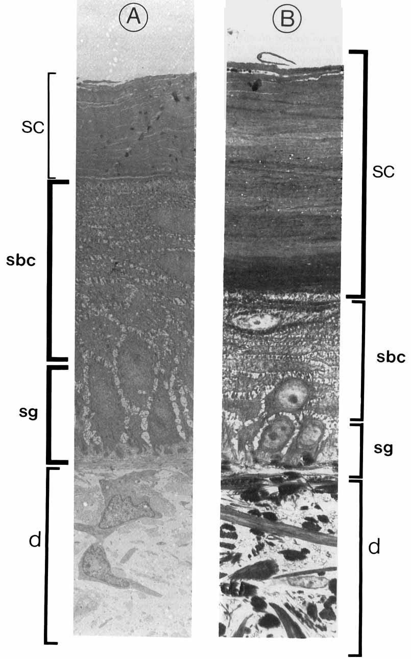
 at x 100 magnification, showing the diffuse IS0 region of the dermis (pr) and collagen- rich non-is0 region (nr), and (B) at ~470 magnification, showing")
5 INTEGUMENTARY SENSE ORGANS OF CROCODILES 319 Fig. 4. Crocodylus porosus. Light micrographs of cross-sections an IS0 (A) at x 100 magnification, showing the diffuse IS0 region of the dermis (pr) and collagen- rich non-is0 region (nr), and (B) at ~470 magnification, showing stratum germinativum (sg), suprabasal cells (sbc), and stratum corneum (sc) layers of the epidermis (ep). stratum corneum through which a thinner, underlying layer of the epidermis is exposed. Immediately beneath this exposed region of the epidermis is a pocket in the dermis, perhaps fluid-filled. Nerve terminals are found in this pocket, immediately beneath the epidermis. The presence of nerve terminals is consistent with the hypothesis that the pits are sensory organs, and we therefore favor the term integumentary sense organs (ISO) over other terms which have been used in the taxonomic literature (e.g., follicle glands, follicle pores, etc.). The ISOs of Crocodylusporosus differ from known cranial touch papillae of Caiman crocodihs (von During, '73, '74) in several

6 320 K. JACKSON ET AL. Fig. 5. Crocodylus porosus. TEM of the apex of IS0 region of the dermis, showing high concentration of cells. bl, basal lamina of the stratum germinativum of the epidermis; f, fibroblast; ir, iridocyte. x 1,800. Fig. 6. Crocodylus porosus. TEM of fibroblasts from the IS0 region of the dermis. c, collagen fibers; f, fibroblast. ~5,600. Fig. 7. Crocodylus porosus. TEM of nerve terminals (nt) supported by a fibroblast cell (f). ~6,800. Fig. 8. Crocodylus porosus. TEM of an iridocyte from the IS0 region of the dermis. x 10,300.
 The ISOs are present on only the postcranial scales of crocodylids and gavialids, while the touch papillae are present on the cranial but not the postcranial scales of all crocodilians.")
7 INTEGUMENTARY SENSE ORGANS OF CROCODILES 321 ways. 1) The ISOs are present on only the postcranial scales of crocodylids and gavialids, while the touch papillae are present on the cranial but not the postcranial scales of all crocodilians. 2) There is only one IS0 on each postcranial scale (with occasional exceptions), while the number of touch papillae on the cranial scales, by contrast, is much more variable (4-16). 3) The ISOs are not concentrated in any one particular region of the skin, while touch papillae are most numerous on the scales surrounding the nares and the mouth and least numerous on the scales between the eyes. 4) Whereas each IS0 is centered in the caudal third of the scale, the touch papillae are randomly distributed. 5) The ISOs are larger than the touch papillae (300 pm diameter vs. 200 pm diameter). 6) Although the outer surface of the ISOs is slightly convex, it is not raised to the degree described by von During ('73, '74) for cranial touch papillae, and the fluid in the diffuse pocket in the dermis of the IS0 does not appear to be maintained under pressure as it is in touch papillae (von During, '74). 7) Although we identified nerve terminals in the ISOs, we did not observe all the receptor types described by von During in the touch papillae (intraepidermal nerve endings, Merkel cell neurite complex, lamellated receptors). Von During's touch papillae are unusual among reptilian sensory organs in having these structures which resemble mammalian Pacinian corpuscles. Although this apparent difference may reflect the fact that we used semithin sections and light microscopy for serial reconstruction and TEM only for fine detail, whereas von During ('73) concentrated on nerve terminals rather than on the entire structure and used TEM of thin sections for the entire serial reconstruction, it is more likely that this represents another difference between touch papillae and ISOs. In spite of these differences, the overall morphology of the IS0 is similar to that of the touch papilla. Both consist of nerve terminals contained in a diffuse, fluid-filled pocket in the outer surface of the dermis. This structural similarity may indicate functional similarity. However, it is also possible that the structural similarity has arisen as a result of homology and that they may have totally different functions. Baden and Maderson ('70) have determined by x-ray diffraction that the stratum corneum of lepidosaur reptiles has an inner layer of alpha-keratin and an outer layer of beta-keratin, in contrast to that of Alligator, which has a single layer of beta-keratin, with alpha-keratin only at the hinge regions of the scales. We observed, using SEM and TEM, what appeared to be two distinct layers in the stratum corneum of Crocodylus: a stiff outer layer which peeled off in flakes and a pitted and apparently more pliable inner layer. The outer layer tended to separate from the inner layer during sectioning, probably indicating noncontinuous beta-keratinization of unspecialized epidermis, as described by Menon et al. ('96). The touch papillae have been described as mechanosensory on the basis of their structure (von During, '73, '74) and have been described as elevated relative to the surrounding integument as though under pressure from fluid inside (von During, '73). Figure 10 summarizes our observations of the morphology and ultrastructure of the ISO. The ISOs are not elevated relative to the surrounding integument. However, it is possible that the ISOs may also function as mechanoreceptors. We observed that the dermal region of the IS0 has a high component of ground substance and little collagen and few cells. It is possible that these cells and fibers of the extracellular matrix (ECM) have been dispersed by a unique gel or fluid-like component of the ECM. This ground substance could be interpreted as an important element of the mechanoreception system which, when stimulated by external pressure, stimulates the nerve terminals near the epidermaldermal junction. However, there is no direct physiological evidence for mechanosensory function in ISOs, touch papillae, or any other sensory organ in the reptilian integument. To date only one study of integumentary mechanoreception in reptiles has been undertaken in which nerve action potentials were recorded as the integument was mechanically stimulated (Necker, '74). Because of the technical difficulty of recording electrical activity in the small efferent nerve fibers of individual mechanoreceptors, recordings are made from larger nerve fibers far downstream, so that it is impossible to attribute the observed response to the stimulation of any particular proposed sensory structure. Although the ISOs may be mechanoreceptors, other functional interpretations are also possible. One such possibility is that the IS0 is a chemosensory organ. The stratum corneum of the epidermis of the IS0 region is thinner than that of the rest of the integument, with a surface that is pitted and possi-

8 Figure 9

9 INTEGUMENTARY SENSE ORGANS OF CROCODILES 323 Fig. 10. Crocodylus porosus. Summary illustration of the integumentary sense organ. bly porous rather than flaking off in flat sheets like the surface surrounding it. If the epidermis of the IS0 region is indeed porous and allows the passage of fluid from the outside environment, then the diffuse, possibly fluid-filled area of the dermis in the IS0 region could be interpreted as a sampling cell in which the nerve terminals of the IS0 region are bathed in fluid from outside and are stimulated by the chemical characteristics of this fluid. It has been shown that crocodylids and alligatorids differ in their capacity for salinity discrimination (Jackson et al., in press), and one possible hypothesis concerning function is that the ISOs are involved in discrimination between hyper- and hypoosmotic salinities. Morphological study of the ISOs reveals a structure which is potentially consistent with either a mechanosensory or a chemosensory function. If the function of the ISOs is to be determined, physiological study will be required. Such studies have the potential to Fig. 9. Crocodylus porosus. TEM of epidermis from (A) the IS0 region and (B) from another area of the same scale. d, dermis; sbc, suprabasal cells; SC, stratum corneum; sg, stratum germinatiwm. ~2,600. reveal whether the sensory function of the ISOs is mechanoreception or whether morphological differences between the ISOs and the cranial touch papillae reflect a functional difference between these organs. ACKNOWLEDGMENTS We thank T.J. Lam (National University of Singapore), Lee Bak Kuan, and Lee Pei Lin (Long Kuan Hung Crocodile Farm) in Singapore for generous scientific and logistical assistance. E. Campolin, T. Giri, H. Hong, E. Lin, L. Meszoly, J. Perez, R. Villadiego, and G. Weiblen provided essential technical advice and assistance. We thank P. Maderson and T. Parsons for helpful advice in the early stages of this project and the anonymous reviewers for comments on the manuscript. This study was funded by NSERC grant A-2359 to D.G. Butler. LITERATURE CITED Baden, H.P., and P.F.A. Maderson (1970) Morphological and biophysical identification of fibrous proteins in the amniote epidermis. J. Exp. Zool. 174r Brazaitis, P. (1987) Identification of crocodilian skins and products. In G.J. Webb, S.C. Manolis, and P.J. Whitehead (eds): Wildlife Management: Crocodiles and Alligators. Chipping Norton, NSW, Australia: Surrey Beatty & Sons, pp
 La peau et les productions cutankes. In P. Grasse (ed.")
10 324 K. JACKSON ET AL Grigg, G.C., and C. Gans (1993) Morphology and physiology of the Crocodilia. In C.J. Glasby, G.J.B. Ross, and P.L. Beesley (eds): Fauna of Australia, Vol. 2A: Amphibia and Reptilia. Canberra: Australian Government Publishing Service, pp Guibe, J. (1970) La peau et les productions cutankes. In P. Grasse (ed.): Traite de Zoologie: Anatomie, Systematique, Biologie. XIV Reptiles: Caracthres Genereaux et Anatomie. Paris: Masson et C", pp Hulanicka, R. (1913) Recherches sur les terminaisons nerveuses dans la langue, le palais, et la peau du crocodile. Arch. de Zool. Exp. et Gen Jackson, K., D.G. Butler, and D.R. Brooks (in press) Habitat and phylogeny influence salinity discrimination in crocodilians: Implications for osmoregulatory physiology and historical biogeography. Biol. J. Linn. SOC. King, F.W., and P. Brazaitis (1971) Species identification of commercial crocodilian skins. New York Zoological Society: Zoologica. MacLean, S. (1980) Ultrastructure of epidermal sensory receptors in Amphiholurus harhatus (Lacertilia: Agamidae). Cell Tissue Res. 210t Menon, G.K., P.F.A. Maderson, R.C. Drewes, L.F. Baptista, L.F. Price, and P.M. Elias (1996) Ultrastructural organization of avian stratum corneum lipids as the basis for facultative cutaneous waterproofing. J. Morphol. 227:l-13. Necker, R. (1974) Dependence of mechanoreceptor activity on skin temperature in sauropsids. I. Caiman. J. Comp. Physiol. 92t Spearmen, R.I.C., and P.A. Riley (1969) A comparison of the epidermis and pigment cells of the crocodile with those in two lizard species. Zool. J. Linn. Soc Von During, M. (1973) The ultrastructure of lamellated mechanoreceptors in the skin of reptiles. Z. Anat. Entwick1.-Gesch. 143t Von During, M. (1974) The ultrastructure of the cutaneous receptors in the skin of Ca. crocodilus. Abhandlungen Rhein.-Westfal. Akad. 53t Von During, M., and M.R. Miller (1979) Sensory nerve endings of the skin and deeper structures. In C. Gans, R.G. Northcutt, and P. Ulinski (eds): Biology of the Reptilia, 9. Neurology A. New York: Academic Press, pp Wermuth, H., and K. Fuchs (1978) Bestimmen von Krokodilen und ihrer Haute: Eine Anleitung zum Identifizieren der Art- und Rassen-Zugehorigkeit der Krokodile. Stuttgart: Gustav Fischer Verlag.
BEAK AND FEATHER DYSTROPHY IN WILD SULPHUR-CRESTED COCKATOOS (CACATUA GALERITA)
") BEAK AND FEATHER DYSTROPHY IN WILD SULPHUR-CRESTED COCKATOOS (CACATUA GALERITA) Author(s): Steven McOrist, Douglas G. Black, David A. Pass, Peter C. Scott, and John Marshall Source: Journal of Wildlife
BEAK AND FEATHER DYSTROPHY IN WILD SULPHUR-CRESTED COCKATOOS (CACATUA GALERITA) Author(s): Steven McOrist, Douglas G. Black, David A. Pass, Peter C. Scott, and John Marshall Source: Journal of Wildlife
Morphology of Shells From Viable and Nonviable Eggs of the Chinese Alligator (Alligator sinensis)
") ~ JOURNAL OF MORPHOLOGY 222:103-110 (1994) Morphology of Shells From Viable and Nonviable Eggs of the Chinese Alligator (Alligator sinensis) CAROLE S. WINK AND RUTH M. ELSEY Department of Anatomy, Louisiana
~ JOURNAL OF MORPHOLOGY 222:103-110 (1994) Morphology of Shells From Viable and Nonviable Eggs of the Chinese Alligator (Alligator sinensis) CAROLE S. WINK AND RUTH M. ELSEY Department of Anatomy, Louisiana
A Scanning Electron Microscopic Study of Eggshell Surface Topography of Leidynema portentosae and L. appendiculatum (Nematoda: Oxyuroidea)
") The Ohio State University Knowledge Bank kb.osu.edu Ohio Journal of Science (Ohio Academy of Science) Ohio Journal of Science: Volume 88, Issue 5 (December, 1988) 1988-12 A Scanning Electron Microscopic
The Ohio State University Knowledge Bank kb.osu.edu Ohio Journal of Science (Ohio Academy of Science) Ohio Journal of Science: Volume 88, Issue 5 (December, 1988) 1988-12 A Scanning Electron Microscopic
Title. CitationJapanese Journal of Veterinary Research, 24(1-2): 37. Issue Date DOI. Doc URL. Type. File Information
: 37. Issue Date DOI. Doc URL. Type. File Information") Title DISTRIBUTION OF LYMPHATIC TISSUES IN DUCK CAECA Author(s)KITAMURA, Hirokazu; SUGIMURA, Makoto; HASHIMOTO, Yos CitationJapanese Journal of Veterinary Research, 24(1-2): 37 Issue Date 1976-05 DOI 10.14943/jjvr.24.1-2.37
Title DISTRIBUTION OF LYMPHATIC TISSUES IN DUCK CAECA Author(s)KITAMURA, Hirokazu; SUGIMURA, Makoto; HASHIMOTO, Yos CitationJapanese Journal of Veterinary Research, 24(1-2): 37 Issue Date 1976-05 DOI 10.14943/jjvr.24.1-2.37
Seasonal Variations of yeso sika Deer Skin and its Vegetable Tanned Leather
 Seasonal Variations of yeso sika Deer Skin and its Vegetable Tanned Leather Shigeharu Fukunaga, Akihiko Yoshie, Ikuo Yamakawa, Fumio Nakamura Laboratory of Animal By-product Science, Graduate School of
General morphology of the oral cavity of the Nile crocodile, Crocodylus niloticus (Laurenti, 1768). I. Palate and gingivae
. I. Palate and gingivae") Onderstepoort Journal of Veterinary Research, 70:281 297 (2003) General morphology of the oral cavity of the Nile crocodile, Crocodylus niloticus (Laurenti, 1768). I. Palate and gingivae J.F. PUTTERILL
Onderstepoort Journal of Veterinary Research, 70:281 297 (2003) General morphology of the oral cavity of the Nile crocodile, Crocodylus niloticus (Laurenti, 1768). I. Palate and gingivae J.F. PUTTERILL
Lesson 7. References: Chapter 6: Chapter 12: Reading for Next Lesson: Chapter 6:
 Lesson 7 Lesson Outline: Embryonic Origins of the Dermis Specializations of the Dermis o Scales in Fish o Dermal Armour in Tetrapods Epidermal/Dermal Interactions o Feathers o Hair o Teeth Objectives:
SCANNING electron - microscopy has
 Characteristics of the Absorptive Surface of the Small Intestine of the Chicken from 1 Day to 14 Weeks of Age 1 R. C. BAYER, C. B. CHAWAN, F. H. BIRD AND S. D. MUSGRAVE Department of Animal and Veterinary
A comparison of placental tissue in the skinks Eulamprus tympanum and E. quoyii. Yates, Lauren A.
 A comparison of placental tissue in the skinks Eulamprus tympanum and E. quoyii Yates, Lauren A. Abstract: The species Eulamprus tympanum and Eulamprus quoyii are viviparous skinks that are said to have
SUPPLEMENTARY INFORMATION
 doi:10.1038/nature11046 Supplementary Figure 1: Images of PB-positive cells in the subepidermal region (a-i) Representative images of PB positive cells in the subepidermis of the upper beak of the pigeon.
Liver and Gallbladder Morphology of the juvenile Nile crocodile, Crocodylus niloticus (Laurenti, 1768)
") Liver and Gallbladder Morphology of the juvenile Nile crocodile, Crocodylus niloticus (Laurenti, 1768) by ERNA VAN WILPE Submitted in partial fulfilment of the requirements for the degree MSc DEPARTMENT
Liver and Gallbladder Morphology of the juvenile Nile crocodile, Crocodylus niloticus (Laurenti, 1768) by ERNA VAN WILPE Submitted in partial fulfilment of the requirements for the degree MSc DEPARTMENT
Development, comparative morphology and cornification of reptilian claws in relation to claws evolution in tetrapods
 Contributions to Zoology, 78 (1) 25-42 (2009) Development, comparative morphology and cornification of reptilian claws in relation to claws evolution in tetrapods Lorenzo Alibardi 1, 2 1 Dipartimento di
DISTRIBUTION, ABUNDANCE AND HABITAT CONSERVATION OF CROCODYLUS POROSUS IN REMBAU-LINGGI ESTUARY, PENINSULAR MALAYSIA
 3 DISTRIBUTION, ABUNDANCE AND HABITAT CONSERVATION OF CROCODYLUS POROSUS IN REMBAU-LINGGI ESTUARY, PENINSULAR MALAYSIA Mohd Fazlin Nazli*, Nor Rasidah Hashim and Mohamed Zakaria M.Sc (GS265) 3 rd Semester
RESEARCH ARTICLE Structure, innervation and response properties of integumentary sensory organs in crocodilians
 4217 The Journal of Experimental Biology 215, 4217-4230 2012. Published by The Company of Biologists Ltd doi:10.1242/jeb.076836 RESEARCH ARTICLE Structure, innervation and response properties of integumentary
Lingual Salt Glands in Crocodylus acutus and C. johnstoni and their absence from Alligator mississipiensis and Caiman crocodilus
 Lingual Salt Glands in Crocodylus acutus and C. johnstoni and their absence from Alligator mississipiensis and Caiman crocodilus Laurence E. Taplin 1, Gordon C. Grigg 1, Peter Harlow 1, Tamir M. Ellis
Mesosomes are a definite event in antibiotic-treated Staphylococcus aureus ATCC 25923
 Tropical Biomedicine 24(1): 105 109 (2007) Mesosomes are a definite event in antibiotic-treated Staphylococcus aureus ATCC 25923 Santhana Raj, L. 1*, Hing, H.L. 2, Baharudin Omar 2, Teh Hamidah, Z. 1,
Histo-Morphological Study on the Footpad of Ostrich (Struthio camelus) In Relation to Locomotion
 In Relation to Locomotion") Histo-Morphological Study on the Footpad of Ostrich (Struthio camelus) In Relation to Locomotion S.A.A. El-Gendy a, Amira Derbalah b, M.E.R. Abu El-Magd c a Department of Anatomy and Embryology, b Histology,
Histo-Morphological Study on the Footpad of Ostrich (Struthio camelus) In Relation to Locomotion S.A.A. El-Gendy a, Amira Derbalah b, M.E.R. Abu El-Magd c a Department of Anatomy and Embryology, b Histology,
,,, THE MORPHOLOGY AND MORPHOMETRY OF THE PECTEN OCULI IN DIURNAL AND NOCTURNAL BIRDS: A
 ,,, THE MORPHOLOGY AND MORPHOMETRY OF THE PECTEN OCULI IN DIURNAL AND NOCTURNAL BIRDS: A COMPARATIVE STUDY" BY llijama, S.G., B. V. M. (NBI), Department of Veteri nary Anatomy, University of I\Jairobi.
HISTOPATHOLOGY. Introduction:
 Introduction: HISTOPATHOLOGY Goats and sheep are the major domestic animal species in India. Much of the economy of the country has been depend upon the domestication of these animals. Especially economy
Sensory Setae of the First Tarsi and Palps of the Mite Macrocheles muscaedomesticae1.2
 $ Sensory Setae of the First Tarsi and Palps of the Mite Macrocheles muscaedomesticae1.2 ~, 'r L. B. COONS ANDR. C. AXTELL Department of Entomology, North Carolina State University, Raleigh 27607 By scanning
Anat. Labor. of Prof. H. SETO, Tohoku University, On the Sensory Terminations Formed along the Ductus
 Anat. Labor. of Prof. H. SETO, Tohoku University, Sendai. On the Sensory Terminations Formed along the Ductus Pancreaticus in Cat. The existence of PACINIan bodies in the pancreas of mammals, especially
DLS Sample Preparation Guide
 DLS Sample Preparation Guide The Leica TCS SP8 DLS is an innovative concept to integrate the Light Sheet Microscopy technology into the confocal microscope. Due to its unique optical architecture samples
Fischthal and Kuntz (1964) reported the
 reported the") Zoological Studies 41(3): 283-287 (2002) Meristocotyle provitellaria sp. nov. (Digenea: Meristocotylidae) from Varanus salvator in China Wei Liu 1, Qing-Kui Li 2, Hsiu-Hui Shih 3 and Zhao-Zhi Qiu 1, *
Zoological Studies 41(3): 283-287 (2002) Meristocotyle provitellaria sp. nov. (Digenea: Meristocotylidae) from Varanus salvator in China Wei Liu 1, Qing-Kui Li 2, Hsiu-Hui Shih 3 and Zhao-Zhi Qiu 1, *
HOW XTC IMPROVED MINOXIDIL PENETRATION - 5 WAYS!
 HOW XTC IMPROVED MINOXIDIL PENETRATION - 5 WAYS! What Hinders Minoxidil from Working Well 1. Sebum from sebaceous gland blocks the hair follicle. 2. Minoxidil therefore, cannot penetrate through the sebum
A Lymphosarcoma in an Atlantic Salmon (Salmo salar)
") A Lymphosarcoma in an Atlantic Salmon (Salmo salar) Authors: Paul R. Bowser, Marilyn J. Wolfe, and Timothy Wallbridge Source: Journal of Wildlife Diseases, 23(4) : 698-701 Published By: Wildlife Disease
A Lymphosarcoma in an Atlantic Salmon (Salmo salar) Authors: Paul R. Bowser, Marilyn J. Wolfe, and Timothy Wallbridge Source: Journal of Wildlife Diseases, 23(4) : 698-701 Published By: Wildlife Disease
Class Reptilia Testudines Squamata Crocodilia Sphenodontia
 Class Reptilia Testudines (around 300 species Tortoises and Turtles) Squamata (around 7,900 species Snakes, Lizards and amphisbaenids) Crocodilia (around 23 species Alligators, Crocodiles, Caimans and
Summary. Introduction
 Grigg GC, LE Taplin, P Harlow and J Wright 1980 Survival and growth of hatchling Crocodylus porosus in salt water without access to fresh drinking water. Oecologia 47:264-6. Survival and Growth of Hatchling
Lesson 6. References: Chapter 6: Reading for Next Lesson: Chapter 6:
 Lesson 6 Lesson Outline: General Features of the Integument Embryonic Origins of the Epidermis Specializations of the Epidermis o Glands o Keratin and Stratum Corneum Objectives: At the end of this lesson
Australian Freshwater Crocodile
 Australian Freshwater Crocodile Crocodylus johnstoni Grahame J.W. Webb and S. Charlie Manolis Wildlife Management International Pty. Limited, PO Box 530, Sanderson, NT 0812, Australia (gwebb@wmi.com.au,
The epidermis of reptiles and birds (sauropsids)
") ORIGINAL PAPER Soft epidermis of a scaleless snake lacks beta-keratin M. Toni, L. Alibardi Department of Biology, University of Bologna, Bologna, Italy 07 European Journal of Histochemistry Beta-keratins
ORIGINAL PAPER Soft epidermis of a scaleless snake lacks beta-keratin M. Toni, L. Alibardi Department of Biology, University of Bologna, Bologna, Italy 07 European Journal of Histochemistry Beta-keratins
A NEW AUSTROSQUILLA (STOMATOPODA) FROM THE
 FROM THE") A NEW AUSTROSQUILLA (STOMATOPODA) FROM THE MARQUESAS ISLANDS BY ALAIN MICHEL Centre O.R.S.T.O.M., Noumea, New Caledonia and RAYMOND B. MANNING Smithsonian Institution, Washington, U.S.A. The At s,tstrosqzlilla
A NEW AUSTROSQUILLA (STOMATOPODA) FROM THE MARQUESAS ISLANDS BY ALAIN MICHEL Centre O.R.S.T.O.M., Noumea, New Caledonia and RAYMOND B. MANNING Smithsonian Institution, Washington, U.S.A. The At s,tstrosqzlilla
Harold W. Manter Laboratory, University of Nebraska State Museum, Lincoln, Nebraska 68588
 Proc. Helminthol. Soc. Wash. 48(2), 1981, pp. 130-136 Observations of the Head and Tail Regions of Male Physaloptera praeputialis von Linstow, 1889, and Physaloptera rara Hall and Wigdor, 1918, Using Scanning
Frog Dissection Information Manuel
 Frog Dissection Information Manuel Anatomical Terms: Used to explain directions and orientation of a organism Directions or Positions: Anterior (cranial)- toward the head Posterior (caudal)- towards the
ABSTRACT. aspect is very sparse and in view of its importance. MATERIALS AND METHODS
 MICROMETRICAL STUDIES ON THE SKIN OF MADRAS RED SHEEP (OVIS ARIES) IN DIFEERENT AGE GROUPS Mir Shabir Ahmad 1, O.R. Sathyamoorthy 2, Geetha Ramesh 3 and C. Balachandran 4 Department of Veterinary Anatomy
SCANNING ELECTRON MICROSCOPY OF THE EGGSHELL OF LIZARD, CALOTES VERSICOLOR. Vilas Deshmukh Yeshwant Mahavidyalaya, Wardha
 INTERNATIONAL JOURNAL OF RESEARCHES IN BIOSCIENCES, AGRICULTURE AND TECHNOLOGY VISHWASHANTI MULTIPURPOSE SOCIETY (Global Peace Multipurpose Society) R. No. MH-659/13(N) www.vmsindia.org SCANNING ELECTRON
Criconemoides similis 1 G. W. BIRD ~
 Somatic Musculature of Trichodorus porosus and Criconemoides similis 1 G. W. BIRD ~ Abstract: The somatic musculature of Trichodorus porosus is transversely striated, and that of Criconemoides similis
A NEW SPECIES OF A USTROLIBINIA FROM THE SOUTH CHINA SEA AND INDONESIA (CRUSTACEA: BRACHYURA: MAJIDAE)
") 69 C O a g r ^ j^a RAFFLES BULLETIN OF ZOOLOGY 1992 40(1): 69-73 A NEW SPECIES OF A USTROLIBINIA FROM THE SOUTH CHINA SEA AND INDONESIA (CRUSTACEA: BRACHYURA: MAJIDAE) H P Waener SMITHSONIAN INSTITUTE
69 C O a g r ^ j^a RAFFLES BULLETIN OF ZOOLOGY 1992 40(1): 69-73 A NEW SPECIES OF A USTROLIBINIA FROM THE SOUTH CHINA SEA AND INDONESIA (CRUSTACEA: BRACHYURA: MAJIDAE) H P Waener SMITHSONIAN INSTITUTE
VARIATION IN MONIEZIA EXPANSA RUDOLPHI
 VARIATION IN MONIEZIA EXPANSA RUDOLPHI STEPHEN R. WILLIAMS, Miami University, Oxford, Ohio In making a number of preparations of proglottids for class study at the stage when sex organs are mature and
HIGLEY UNIFIED SCHOOL DISTRICT INSTRUCTIONAL ALIGNMENT. Zoology Quarter 3. Animal Behavior (Duration 2 Weeks)
") HIGLEY UNIFIED SCHOOL DISTRICT INSTRUCTIONAL ALIGNMENT Zoology Quarter 3 Animal Behavior (Duration 2 Weeks) Big Idea: Essential Questions: 1. Compare and contrast innate and learned behavior 2. Compare
HIGLEY UNIFIED SCHOOL DISTRICT INSTRUCTIONAL ALIGNMENT Zoology Quarter 3 Animal Behavior (Duration 2 Weeks) Big Idea: Essential Questions: 1. Compare and contrast innate and learned behavior 2. Compare
Animal Diversity III: Mollusca and Deuterostomes
 Animal Diversity III: Mollusca and Deuterostomes Objectives: Be able to identify specimens from the main groups of Mollusca and Echinodermata. Be able to distinguish between the bilateral symmetry on a
TWO NEW SPECIES OF WATER MITES FROM OHIO 1-2
 TWO NEW SPECIES OF WATER MITES FROM OHIO 1-2 DAVID R. COOK Wayne State University, Detroit, Michigan ABSTRACT Two new species of Hydracarina, Tiphys weaveri (Acarina: Pionidae) and Axonopsis ohioensis
Reintroduction of the Mugger Crocodile, Crocodylus palustris, in India
 Reintroduction of the Mugger Crocodile, Crocodylus palustris, in India Introduction Christina Jacobson Endangered species management has become an important issue for many countries as animals and their
OBSERVATIONS ON THE QUALITATIVE AND QUANTITATIVE STRUCTURAL CHARACTERISTICS OF THE REPTILIAN KIDNEYS.
 OBSERVATIONS ON THE QUALITATIVE AND QUANTITATIVE STRUCTURAL CHARACTERISTICS OF THE REPTILIAN KIDNEYS. ~B~SI"Y OF Nmlll,.tpj,Tb 1.11.,,)' A Thesis submitted to the university of Nairobi in partial fulfillment
At the Edge: Neuroethological Approaches to Reptilian Mechanoreception
 At the Edge: Neuroethological Approaches to Reptilian Mechanoreception Duncan B. Leitch The neural circuitry directing behavior is one of the fundamental questions of neurobiology. Historically, studies
Gross and Microscopic Features of the Interdigital Sinus in the Barbados Black Belly Sheep in Trinidad
 Original Research Article International Journal of Current Research in Medical Sciences ISSN: 2454-5716 www.ijcrims.com Volume 2, Issue 7-2016 SOI: http://s-o-i.org/1.15/ijcrms-2016-2-7-4 Gross and Microscopic
Cornification in developing claws of the common Australian skink (Lampropholis guichenoti) (Squamata, Lacertidae)
 (Squamata, Lacertidae)") Italian Journal of Zoology ISSN: 1125-0003 (Print) 1748-5851 (Online) Journal homepage: http://www.tandfonline.com/loi/tizo20 Cornification in developing claws of the common Australian skink (Lampropholis
Italian Journal of Zoology ISSN: 1125-0003 (Print) 1748-5851 (Online) Journal homepage: http://www.tandfonline.com/loi/tizo20 Cornification in developing claws of the common Australian skink (Lampropholis
When? Why? and How?: Some Speculations on the Evolution of the Vertebrate Integument
 AM. ZOOLOCIST, 12:159-171 (1972). When? Why? and How?: Some Speculations on the Evolution of the Vertebrate Integument PAUL F. A. MADERSON Biology Department, Brooklyn College, Brooklyn, New York 11210
REPTILES. Scientific Classification of Reptiles To creep. Kingdom: Animalia Phylum: Chordata Subphylum: Vertebrata Class: Reptilia
 Scientific Classification of Reptiles To creep Kingdom: Animalia Phylum: Chordata Subphylum: Vertebrata Class: Reptilia REPTILES tetrapods - 4 legs adapted for land, hip/girdle Amniotes - animals whose
Stress in farmed saltwater crocodiles (Crocodylus porosus): no difference between individually- and communally-housed animals
: no difference between individually- and communally-housed animals") Isberg and Shilton SpringerPlus 2013, 2:381 a SpringerOpen Journal RESEARCH Open Access Stress in farmed saltwater crocodiles (Crocodylus porosus): no difference between individually- and communally-housed
Isberg and Shilton SpringerPlus 2013, 2:381 a SpringerOpen Journal RESEARCH Open Access Stress in farmed saltwater crocodiles (Crocodylus porosus): no difference between individually- and communally-housed
NATIONAL BIORESOURCE DEVELOPMENT BOARD Dept. of Biotechnology Government of India, New Delhi
 NATIONAL BIORESOURCE DEVELOPMENT BOARD Dept. of Biotechnology Government of India, New Delhi MARINE BIORESOURCES FORMS DATA ENTRY: Form- 1(general ) (please answer only relevant fields;add additional fields
Morphology of the femoral glands of the lizard Iguana iguana (linnaeus, 1758) (reptilia, iguanidae)
 (reptilia, iguanidae)") A. Ferreira Femoral glands of lizard 97 ARTIGO ARTICLE Morphology of the femoral glands of the lizard Iguana iguana (linnaeus, 1758) (reptilia, iguanidae) Morfologia das glândulas femorais do lagarto Iguana
A. Ferreira Femoral glands of lizard 97 ARTIGO ARTICLE Morphology of the femoral glands of the lizard Iguana iguana (linnaeus, 1758) (reptilia, iguanidae) Morfologia das glândulas femorais do lagarto Iguana
KINGDOM ANIMALIA Phylum Chordata Subphylum Vertebrata Class Reptilia
 KINGDOM ANIMALIA Phylum Chordata Subphylum Vertebrata Class Reptilia Vertebrate Classes Reptiles are the evolutionary base for the rest of the tetrapods. Early divergence of mammals from reptilian ancestor.
Animal Coverings Facilitated
 Animal Coverings Facilitated Students will explore various animal coverings with their senses, with help from a high-powered microscope. Description: Explore fur, feathers and scales like never seen before
Formoguanamine-induced blindness and photoperiodic responses in the Japanese quail, Coturnix coturnix japonica
 J. Biosci., Vol. 19, Number 4, October 1994, pp 479-484. Printed in India. Formoguanamine-induced blindness and photoperiodic responses in the Japanese quail, Coturnix coturnix japonica 1. Introduction
ABSTRACT. are both widely scattered at low densities and aggregated. to background color matching and camouflage. The
 AMERICAN MUSEUM Novitates PUBLISHED BY THE AMERICAN MUSEUM OF NATURAL HISTORY CENTRAL PARK WEST AT 79TH STREET, NEW YORK, N.Y. 10024 Number 2943, 14 pp., 6 figs. June 27, 1989 Integumental Chromatophores
The 1st studies on the blood of reptiles
 Zoological Studies 42(1): 173-178 (2003) Erythrocyte Size and Morphology of Some Tortoises and Turtles from Turkey. I smail HakkI Uǧurta *, Murat Sevinç and Hikmet Sami YIldIrImhan Science and Art Faculty,
EFFECTS OF TEMPERATURE ON GROWTH IN THE REGENERATING TAIL OF THE SCINCID LIZARD, MABUYA STRIATA. Accepted: June 1977
 EFFECTS OF TEMPERATURE ON GROWTH IN THE REGENERATING TAIL OF THE SCINCID LIZARD, MABUYA STRIATA D K MAGON Department of Zoology. Kenyatta University College. Box 43844. Nairobi. Kenya Accepted: June 1977
Diapsida. BIO2135 Animal Form and Function. Page 1. Diapsida (Reptilia, Sauropsida) Amniote eggs. Amniote egg. Temporal fenestra.
 Amniote eggs. Amniote egg. Temporal fenestra.") Diapsida (Reptilia, Sauropsida) Vertebrate phylogeny Mixini Chondrichthyes Sarcopterygii Mammalia Pteromyzontida Actinopterygii Amphibia Reptilia! 1! Amniota (autapomorphies) Costal ventilation Amniote
Diapsida (Reptilia, Sauropsida) Vertebrate phylogeny Mixini Chondrichthyes Sarcopterygii Mammalia Pteromyzontida Actinopterygii Amphibia Reptilia! 1! Amniota (autapomorphies) Costal ventilation Amniote
Postilla PEABODY MUSEUM OF NATURAL HISTORY YALE UNIVERSITY NEW HAVEN, CONNECTICUT, U.S.A.
 Postilla PEABODY MUSEUM OF NATURAL HISTORY YALE UNIVERSITY NEW HAVEN, CONNECTICUT, U.S.A. Number 117 18 March 1968 A 7DIAPSID (REPTILIA) PARIETAL FROM THE LOWER PERMIAN OF OKLAHOMA ROBERT L. CARROLL REDPATH
Vol. XIV, No. 1, March, The Larva and Pupa of Brontispa namorikia Maulik (Coleoptera: Chrysomelidae: Hispinae) By S.
 By S.") Vol. XIV, No. 1, March, 1950 167 The Larva and Pupa of Brontispa namorikia Maulik (Coleoptera: Chrysomelidae: Hispinae) By S. MAULIK BRITISH MUSEUM (NATURAL HISTORY) (Presented by Mr. Van Zwaluwenburg
Vol. XIV, No. 1, March, 1950 167 The Larva and Pupa of Brontispa namorikia Maulik (Coleoptera: Chrysomelidae: Hispinae) By S. MAULIK BRITISH MUSEUM (NATURAL HISTORY) (Presented by Mr. Van Zwaluwenburg
Diapsida. BIO2135 Animal Form and Function. Page 1. Diapsida (Reptilia, Sauropsida) Amniote egg. Membranes. Vertebrate phylogeny
 Amniote egg. Membranes. Vertebrate phylogeny") Diapsida (Reptilia, Sauropsida) 1 Vertebrate phylogeny Mixini Chondrichthyes Sarcopterygii Mammalia Pteromyzontida Actinopterygii Amphibia Reptilia!! Amniota (autapomorphies) Costal ventilation Amniote
Diapsida (Reptilia, Sauropsida) 1 Vertebrate phylogeny Mixini Chondrichthyes Sarcopterygii Mammalia Pteromyzontida Actinopterygii Amphibia Reptilia!! Amniota (autapomorphies) Costal ventilation Amniote
Total Distribution of Taste Buds on the Tongue of the Pup
 The Ohio State University Knowledge Bank kb.osu.edu Ohio Journal of Science (Ohio Academy of Science) Ohio Journal of Science: Volume 4, Issue 6 (November, 194) 194-11 Total Distribution of Taste Buds
(Accepted ) ABSTRACT
 ABSTRACT") HRPTOLOG!CAL JOURNAL, Vol. I, pp. 458-462 (199) MTHODS FOR TH DTRMINATION OF TH PHYSICAL CHARACTRISTICS OF GGS OF ALLIGA TOR MISSISSIPPINSIS: A COMPARISON WITH OTHR CROCODILIAN AND AVIAN GGS D. C. DMING
HRPTOLOG!CAL JOURNAL, Vol. I, pp. 458-462 (199) MTHODS FOR TH DTRMINATION OF TH PHYSICAL CHARACTRISTICS OF GGS OF ALLIGA TOR MISSISSIPPINSIS: A COMPARISON WITH OTHR CROCODILIAN AND AVIAN GGS D. C. DMING
EFFECT OF FEEDING DIFFERENT GRADED DIETARY PROTEIN LEVELS ON GROWTH RATE OF NILE CROCODILE (CROCODYLUS NILOTICUS) HATCHLINGS
 HATCHLINGS") EFFECT OF FEEDING DIFFERENT GRADED DIETARY PROTEIN LEVELS ON GROWTH RATE OF NILE CROCODILE (CROCODYLUS NILOTICUS) HATCHLINGS Masamha Blessing 1, Nyamugure Tendayi 2, Wilson Mhlanga 3, Marisa Lesley 4,
EFFECT OF FEEDING DIFFERENT GRADED DIETARY PROTEIN LEVELS ON GROWTH RATE OF NILE CROCODILE (CROCODYLUS NILOTICUS) HATCHLINGS Masamha Blessing 1, Nyamugure Tendayi 2, Wilson Mhlanga 3, Marisa Lesley 4,
New Species of Black Coral (Cnidaria: Antipatharia) from the Northern Gulf of Mexico
 from the Northern Gulf of Mexico") Northeast Gulf Science Volume 12 Number 2 Number 2 Article 2 10-1992 New Species of Black Coral (Cnidaria: Antipatharia) from the Northern Gulf of Mexico Dennis M. Opresko Oak Ridge National Laboratory
Northeast Gulf Science Volume 12 Number 2 Number 2 Article 2 10-1992 New Species of Black Coral (Cnidaria: Antipatharia) from the Northern Gulf of Mexico Dennis M. Opresko Oak Ridge National Laboratory
Animal Diversity wrap-up Lecture 9 Winter 2014
 Animal Diversity wrap-up Lecture 9 Winter 2014 1 Animal phylogeny based on morphology & development Fig. 32.10 2 Animal phylogeny based on molecular data Fig. 32.11 New Clades 3 Lophotrochozoa Lophophore:
F.L. Andr6s. Rua Tristao Vaz No Esq., 1400 Lisboa, Portugal
 Supranumerary Barrels Develop in the Somatosensory Cortex of Mice, After the Implantation of the Vibrissal Follicle Parts Containing Large Numbers of Receptors F.L. Andr6s Rua Tristao Vaz No. 37 1 Esq.,
Testing Phylogenetic Hypotheses with Molecular Data 1
 Testing Phylogenetic Hypotheses with Molecular Data 1 How does an evolutionary biologist quantify the timing and pathways for diversification (speciation)? If we observe diversification today, the processes
Biology Slide 1 of 50
 Biology 1 of 50 2 of 50 What Is a Reptile? What are the characteristics of reptiles? 3 of 50 What Is a Reptile? What Is a Reptile? A reptile is a vertebrate that has dry, scaly skin, lungs, and terrestrial
Phylum Platyhelminthes Flatworms
 Phylum Platyhelminthes Flatworms The Acoelomates The acoelomates are animals that lack a coelom. Acoelomates lack a body cavity, and instead the space between the body wall and the digestive tract is filled
Title of Project: Distribution of the Collared Lizard, Crotophytus collaris, in the Arkansas River Valley and Ouachita Mountains
 Title of Project: Distribution of the Collared Lizard, Crotophytus collaris, in the Arkansas River Valley and Ouachita Mountains Project Summary: This project will seek to monitor the status of Collared
Lacerta viridis. Functional anatomy of the lungs of the green lizard, (Accepted 18 February 1977)
") J. Anat. (1978), 125, 2, pp. 421-431 421 With 9 figures Printed in Great Britain Functional anatomy of the lungs of the green lizard, Lacerta viridis C. MEBAN Department of Anatomy, The Queen's University
J. Anat. (1978), 125, 2, pp. 421-431 421 With 9 figures Printed in Great Britain Functional anatomy of the lungs of the green lizard, Lacerta viridis C. MEBAN Department of Anatomy, The Queen's University
08 alberts part2 7/23/03 9:10 AM Page 95 PART TWO. Behavior and Ecology
 08 alberts part2 7/23/03 9:10 AM Page 95 PART TWO Behavior and Ecology 08 alberts part2 7/23/03 9:10 AM Page 96 08 alberts part2 7/23/03 9:10 AM Page 97 Introduction Emília P. Martins Iguanas have long
Supplementary Figure 1 Cartilaginous stages in non-avian amniotes. (a) Drawing of early ankle development of Alligator mississippiensis, as reported
 Drawing of early ankle development of Alligator mississippiensis, as reported") Supplementary Figure 1 Cartilaginous stages in non-avian amniotes. (a) Drawing of early ankle development of Alligator mississippiensis, as reported by a previous study 1. The intermedium is formed at
Supplementary Figure 1 Cartilaginous stages in non-avian amniotes. (a) Drawing of early ankle development of Alligator mississippiensis, as reported by a previous study 1. The intermedium is formed at
A quantitative study of hair growth using mouse and rat vibrissal follicles
 /. Embryol. exp. Morph. Vol. 72, pp. 209-224, 1982 209 Printed in Great Britain Company of Biologists Limited 1982 A quantitative study of hair growth using mouse and rat vibrissal follicles I. Dermal
Reprinted from: CRUSTACEANA, Vol. 32, Part 2, 1977 LEIDEN E. J. BRILL
 Reprinted from: CRUSTACEANA, Vol. 32, Part 2, 1977 LEIDEN E. J. BRILL NOTES AND NEWS 207 ALPHE0PS1S SHEARMII (ALCOCK & ANDERSON): A NEW COMBINATION WITH A REDESCRIPTION OF THE HOLOTYPE (DECAPODA, ALPHEIDAE)
THE EFFECT OF MUTILATION ON THE TAPEWORM TAENIA TAENIAEFORMIS
 THE EFFECT OF MUTILATION ON THE TAPEWORM TAENIA TAENIAEFORMIS JOE N. MILLER AND WM. P. BUNNER The reader is undoubtedly aware of work which has been done by Child (1910) and others in mutilating certain
WITH THE TABLE OF THE MORPHOLOGICAL FEATURES OF TAPEWORMS IN VAMPIROLEPIS. (Received: December 22nd, 1965)
") Japan. J. Med. Sci. Biol. 19, 51-57, 1966 *ON A NEW TAPEWORM, VAMPIROLEPIS ISENSIS, FOUND IN BATS WITH THE TABLE OF THE MORPHOLOGICAL FEATURES OF TAPEWORMS IN VAMPIROLEPIS ISAMU SAWADA Biological Laboratory,
Japan. J. Med. Sci. Biol. 19, 51-57, 1966 *ON A NEW TAPEWORM, VAMPIROLEPIS ISENSIS, FOUND IN BATS WITH THE TABLE OF THE MORPHOLOGICAL FEATURES OF TAPEWORMS IN VAMPIROLEPIS ISAMU SAWADA Biological Laboratory,
On the nature of the horny scales of the pangolin
 J. Linn. Soc. (Zool.), 46, 310, p. 267 With 1 figure Printed in Great Britain Spril, 1967 On the nature of the horny scales of the pangolin BY R. I. C. SPEARMAN, PH.D., F.L.S. Department of Dermatology,
Taxonomy. Chapter 20. Evolutionary Development Diagram. I. Evolution 2/24/11. Kingdom - Animalia Phylum - Chordata Class Reptilia.
 Taxonomy Chapter 20 Reptiles Kingdom - Animalia Phylum - Chordata Class Reptilia Order Testudines - turtles Order Crocodylia - crocodiles, alligators Order Sphenodontida - tuataras Order Squamata - snakes
Hexamermis glossinae spnov. (Nematoda: Mermithidae), a parasite of tse-tse flies in West Africa
, a parasite of tse-tse flies in West Africa") I. ' NOTES Hexamermis glossinae spnov. (Nematoda: Mermithidae), a parasite of tse-tse flies in West Africa GEORGE O. POINAR, JR. Division of Entomology and Parasitology, University of California, Berkeley,
I. ' NOTES Hexamermis glossinae spnov. (Nematoda: Mermithidae), a parasite of tse-tse flies in West Africa GEORGE O. POINAR, JR. Division of Entomology and Parasitology, University of California, Berkeley,
Rec. zool. Surv. India, 85(4); , 1989
; , 1989") Rec. zool. Surv. India, 85(4); 583-588, 1989 CSTODS OF DOMSTIC FOWL AT VISAKHAPATNAM WITH DSCRIPTION OF A NW SPCIS OF RAILLITINA (RAILLITINA) By SR RAMULU KOLLURI AND C. VIJAYA LAKSHMI Department of Zoology,
Rec. zool. Surv. India, 85(4); 583-588, 1989 CSTODS OF DOMSTIC FOWL AT VISAKHAPATNAM WITH DSCRIPTION OF A NW SPCIS OF RAILLITINA (RAILLITINA) By SR RAMULU KOLLURI AND C. VIJAYA LAKSHMI Department of Zoology,
SEMESTER ONE 2007 INFECTION and IMMUNITY GRADUATE ENTRY PROGRAMME PARASITOLOGY PRACTICAL 9 Dr TW Jones NEMATODES
 SEMESTER ONE 2007 INFECTION and IMMUNITY GRADUATE ENTRY PROGRAMME PARASITOLOGY PRACTICAL 9 Dr TW Jones NEMATODES Objectives After this class I expect you to be able to: 1. Describe and recognise the range
Fine autoradiographical study on scale morphogenesis in the regenerating tail of lizards
 Histol Histopath (1 994) 9: 1 19-1 34 Histology and Histopathology Fine autoradiographical study on scale morphogenesis in the regenerating tail of lizards L. Alibardi Depariment of Histology and Embryology,
Animal Form and Function. Amphibians. United by several distinguishing apomorphies within the Vertebrata
 Animal Form and Function Kight Amphibians Class Amphibia (amphibia = living a double life) United by several distinguishing apomorphies within the Vertebrata 1. Skin Thought Question: For whom are integumentary
What are taxonomy, classification, and systematics?
 Topic 2: Comparative Method o Taxonomy, classification, systematics o Importance of phylogenies o A closer look at systematics o Some key concepts o Parts of a cladogram o Groups and characters o Homology
SCANNING ELECTRON MICROSCOPY OF THE MUCOSAL SURFACE OF THE FORESTOMACHS AND ABOMASA OF GREY, WHITE AND BLACK KARAKUL LAMBS
 OnderstepoortJ. Vet. Res., 59, 167-174 (1992) SCANNING ELECTRON MICROSCOPY OF THE MUCOSAL SURFACE OF THE FORESTOMACHS AND ABOMASA OF GREY, WHITE AND BLACK KARAKUL LAMBS H. B. GROENEWALD, Department of
REPORT FROM A BOU-FUNDED PROJECT
 Pneumatisation and internal architecture of the Southern Cassowary Casuarius casuarius casque: a microct study CHARLOTTE A. BRASSEY 1*, THOMAS O MAHONEY 2 1 School of Science and the Environment, Manchester
DEVELOPMENT OF THE HEAD AND NECK PLACODES
 DEVELOPMENT OF THE HEAD AND NECK Placodes and the development of organs of special sense L. Moss-Salentijn PLACODES Localized thickened areas of specialized ectoderm, lateral to the neural crest, at the
8/19/2013. Topic 5: The Origin of Amniotes. What are some stem Amniotes? What are some stem Amniotes? The Amniotic Egg. What is an Amniote?
 Topic 5: The Origin of Amniotes Where do amniotes fall out on the vertebrate phylogeny? What are some stem Amniotes? What is an Amniote? What changes were involved with the transition to dry habitats?
Beef Cattle Mobility: Scoring Methodology, Data Collection, and Other Considerations
 Beef Cattle Mobility: Scoring Methodology, Data Collection, and Other Considerations BRYAN BERNHARD, PH.D., TEXAS TECH UNIVERSITY Outline How did we get here? What is beef cattle mobility? How do you measure
revealed a population of particles apparently of considerable homogeneity with
 MORPHOLOGY OF THE VIRUS OF AVIAN ERYTHRO- MYELOBLASTIC LEUCOSIS AND A COMPARISON WITH THE AGENT OF NEWCASTLE DISEASE' D. G. SHARP, EDWARD A. ECKERT,2 DOROTHY BEARD, AND J. W. BEARD Department of Surgery,
In vitro permeation of progesterone from a gel through the shed skin of three different snake species
 International Journal of Pharmaceutics 170 (1998) 151 156 In vitro permeation of progesterone from a gel through the shed skin of three different snake species J.M. Haigh a, *, E. Beyssac b, L. Chanet
INVESTIGATIONS ON THE SHAPE AND SIZE OF MOLAR AND ZYGOMATIC SALIVARY GLANDS IN SHORTHAIR DOMESTIC CATS
 Bulgarian Journal of Veterinary Medicine (2009), 12, No 4, 221 225 INVESTIGATIONS ON THE SHAPE AND SIZE OF MOLAR AND ZYGOMATIC SALIVARY GLANDS IN SHORTHAIR DOMESTIC CATS Summary A. A. MOHAMMADPOUR Department
Page # Diversity of Arthropoda Crustacea Morphology. Diversity of Arthropoda. Diversity of Arthropoda. Diversity of Arthropoda. Arthropods, from last
 Arthropods, from last time Crustacea are the dominant marine arthropods Crustacea are the dominant marine arthropods any terrestrial crustaceans? Should we call them shellfish? sowbugs 2 3 Crustacea Morphology
(From the Division of Laboratories of Montefiore Hospital, New York.)
") CALCIFICATION OF THE SUPRARENAL GLANDS OF CATS. BY DAVID MARINE, M.D. (From the Division of Laboratories of Montefiore Hospital, New York.) PLATE 11. (Received for publication, January 18, 1925.) It is
CALCIFICATION OF THE SUPRARENAL GLANDS OF CATS. BY DAVID MARINE, M.D. (From the Division of Laboratories of Montefiore Hospital, New York.) PLATE 11. (Received for publication, January 18, 1925.) It is
The functional morphology of the intermandibulocervical envelope of the American alligator (Alligator mississippiensis)
") Louisiana State University LSU Digital Commons LSU Doctoral Dissertations Graduate School 2012 The functional morphology of the intermandibulocervical envelope of the American alligator (Alligator mississippiensis)
Louisiana State University LSU Digital Commons LSU Doctoral Dissertations Graduate School 2012 The functional morphology of the intermandibulocervical envelope of the American alligator (Alligator mississippiensis)
The family Gnaphosidae is a large family
 Pakistan J. Zool., vol. 36(4), pp. 307-312, 2004. New Species of Zelotus Spider (Araneae: Gnaphosidae) from Pakistan ABIDA BUTT AND M.A. BEG Department of Zoology, University of Agriculture, Faisalabad,
The external morphology of Oestridae parasites
 Article available at http://www.parasite-journal.org or http://dx.doi.org/10.1051/parasite/1997043277 MORPHOLOGICAL COMPARISON OF SECOND STAGE LARVAE OF OESTRUS OVIS (LINNAEUS, 1758), CEPHALOPINA TITILLATOR
Transformed centrioles In adult and aged cat pinealocytes
 Transformed centrioles In adult and aged cat pinealocytes J. L. Calvo. J. Boya*. J. E. Garcia-Mauriño and D. Rancaño Department of Histology. Faculty of Medicine. University Complutense, 28040 Madrid.