Svetlana A. Maslakova a * and Jon L. Norenburg b
|
|
|
- Colin Reeves
- 5 years ago
- Views:
Transcription
 from Florida and Belize Svetlana A. Maslakova a * and Jon L.")
1 Journal of Natural History Vol. 42, Nos , May 2008, Revision of the smiling worms, genus Prosorhochmus Keferstein, 1862, and description of a new species, Prosorhochmus belizeanus sp. nov. (Prosorhochmidae, Hoplonemertea, Nemertea) from Florida and Belize Svetlana A. Maslakova a * and Jon L. Norenburg b a Oregon Institute of Marine Biology, University of Oregon, Charleston, Oregon, USA; b National Museum of Natural History, Smithsonian Institution, Washington D.C., USA (Received 14 October 2007; final version received 18 February 2008) A new species of Prosorhochmus is described from Belize and Florida based on morphological, reproductive and sequence data. Similar to Prosorhochmus nelsoni (Sanchez, 1973), Prosorhochmus belizeanus sp. nov. is gonochoric and oviparous; all other species of Prosorhochmus are viviparous hermaphrodites. Prosorhochmus belizeanus sp. nov. differs from P. nelsoni by having significantly larger stylets and different arrangement of acidophilic cephalic glands. Sequence divergence between the two is 7.4% (16S) and 9.1% (COI), comparable to divergence from the viviparous hermaphroditic species. Prosorhochmus Keferstein, 1862 is revised based on re-evaluation of the type and voucher material as well as fresh specimens collected by us. We conclude that Prosorhochmus adriaticus Senz, 1993 is insufficiently described and cannot be distinguished from Prosorhochmus claparedii Keferstein, We re-establish Prosorhochmus korotneffi Bürger, 1895 from its previous synonymization with P. claparedii and designate it as type species of Arhochmus gen. nov. Keywords: Nemertea; Prosorhochmus; Arhochmus; smiling worms; Cytochrome Oxidase I; 16S rdna Introduction The name smiling worms used by us for the hoplonemertean family Prosorhochmidae refers to the heart-warming crescent-like fold on the head of these worms the prosorhochmid smile (see Figures 1B, 1H, 2A). The function of the smile is unknown, but its presence is correlated with the well-developed frontal organ and, associated with it, mucus cephalic glands. This, in turn, might have something to do with the unusual habitat of some of the prosorhochmids. Most nemerteans (phylum Nemertea) are marine oviparous worms with separate sexes. The family Prosorhochmidae continues to spark the interest of nemertinologists because many of its members are terrestrial or semi-terrestrial. Even more intriguing, many of the species (from marine, semi-terrestrial or terrestrial habitats) acquired hermaphroditism in combination with viviparity. This compact family includes 18 species in four genera: Prosorhochmus Keferstein, 1862, Prosadenoporus Bürger, 1890, Pantinonemertes Moore and Gibson, 1981 and Geonemertes Semper, The first three genera are in need of revision. Maslakova in her Ph.D. thesis (2005), revised and redefined the family and its genera but disclaimed all the new taxon names and nomenclatural changes *Corresponding author. svetlana@uoregon.edu ISSN print/issn online # 2008 Taylor & Francis DOI: /
2 1220 S.A. Maslakova and J.L. Norenburg making them unavailable for nomenclatural purposes (ICZN Art. 8.3). Therefore, we make those changes available here for the first time. Much has changed since the last revision of Prosorhochmus by Gibson and Moore (1985) that resulted in synonymization or invalidation of all previously described species but the type species Prosorhochmus claparedii Keferstein, Three new species have been described: Prosorhochmus americanus Gibson et al., 1986, the first New World representative, Prosorhochmus adriaticus Senz, 1993 from the Adriatic coast of Italy and Prosorhochmus chafarinensis Frutos et al., 1998 from the Chafarinas Islands (off the coast of Morocco, Western Mediterranean). One species, the Chilean Amphiporus nelsoni Sanchez, 1973, has been transferred to Prosorhochmus following its redescription by Maslakova et al. (2005). Prosorhochmus nelsoni (Sanchez, 1973) is the first reported species of Prosorhochmus with separate sexes and oviparity, all others being viviparous hermaphrodites. Here we describe another oviparous Prosorhochmus with separate sexes from Belize and Florida Prosorhochmus belizeanus sp. nov. To provide a proper comparison of the new species to other Prosorhochmus we took it upon ourselves to obtain and reinvestigate all the available type and voucher material from museums around the world and to attempt to collect fresh specimens for histology and sequencing as close as possible to the type localities of the previously described species. Material and methods Specimen preparation Characters of external appearance were documented in living specimens, colour illustrations prepared by hand and photographs taken with a Nikon Coolpix 4500 digital camera mounted on the stereo- and compound microscopes of various brands. Stylets were viewed and photographed through the wall of dissected proboscis slightly compressed under the cover slip and the drawings subsequently made in Adobe Photoshop as tracings. Measurements such as body length and width were taken on live specimens after relaxing them by gradually adding a 1:1 mixture of 7.5% MgCl 2 and local seawater to a container with animals in seawater, ultimately substituting it with 7.5% MgCl 2. For histology, specimens were relaxed in MgCl 2 as described above, fixed for 24 h in 4% formaldehyde made in local seawater, briefly rinsed in tap water and post-fixed in Hollande s cupri-picri-formal-acetic fluid for h. After post-fixation specimens were transferred to 70% ethanol for longterm storage. Specimens were dehydrated in a standard alcohol series, embedded in Tissueprep Paraffin compound (56uC melting point), serially sectioned at 8 mm and stained using Crandall s polychrome protocol a combined variant of the Mallory, Gomori, Koneff and Gurr-McConail techniques (Frank Crandall, National Museum of Natural History, Washington, D.C., pers. comm.). DNA extraction, amplification and sequencing Tissue for molecular work was obtained for Prosorhochmus claparedii, Prosorhochmus americanus, Prosorhochmus cf. chafarinensis, Prosorhochmus nelsoni, Arhochmus korotneffi comb. nov. (former Prosorhochmus korotneffi) and Prosorhochmus belizeanus sp. nov. (Table 1). Specimens were preserved in 95%
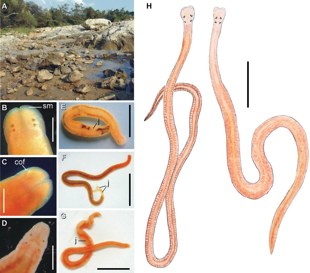
3 Journal of Natural History 1221 Figure 1. External appearance and habitat of Prosorhochmus species. (A) Typical intertidal habitat of Prosorhochmus spp. wet coarse sand just above the low tide mark with partially embedded rocks. The pictured location is about halfway between Croatian towns Savudrija and Zambratija on the Adriatic coast; (B C) Prosorhochmus chafarinensis, anterior end (B, dorsal view; C, ventral view); (D) Arhochmus korotneffi comb. nov., anterior end, dorsal view; (E) Prosorhochmus claparedii, external appearance of a viviparous hermaphroditic adult with several juveniles showing through the body wall between the gut diverticula; (F) P. chafarinensis, external appearance of viviparous hermaphroditic adult with two newly born juveniles; (G) A. korotneffi, external appearance of a viviparous hermaphroditic adult with several juveniles showing through the body wall; (H) Prosorhochmus belizeanus sp. nov. external appearance of sexually mature male (left) and female (right). Testes show through the body wall as whitish sacks regularly interspersed with gut diverticula on either side. Pinkish oocytes in ovaries are visible through the body wall of ripe females. Scales. (B D): 0.5 mm; (E): 3 mm; (F, G): 6 mm; (H): 3.5 mm. Abbreviations: cof, cerebral organ furrow; j, juvenile; sm, prosorhochmid smile. ethanol and stored at room temperature or 220uC short-term and at 280uC longterm. We used Qiagen DNAeasy miniprep kit (Qiagen Inc.) for DNA extractions. Partial sequences of Cytochrome Oxidase subunit I (COI), 658 bp long, and mitochondrial large subunit rdna (16S), bp long, were amplified using universal primers: 16sar-L [cgcctgtttatcaaaaacat] and 16sbr-H [ccggtctgaactcagatcacgt] from Palumbi et al. (1991) for 16S, and LCO1490 [ggtcaacaaatcataaagatattgg]
4
5 1222 S.A. Maslakova and J.L. Norenburg Table 1. Accession numbers and specimen data for molecular analysis. Species Accession numbers Collecting information and specimen numbers Prosorhochmus americanus Prosorhochmus belizeanus sp. nov. Prosorhochmus cf. chafarinensis Prosorhochmus cf. claparedii EF157588, EF EF157591, EF EF157587, EF EF157589, EF157590, EF Coll. J.M. Turbeville, Pawley s Island, SC, USA (#PISC ) Coll. JLN, SAM, , reef-crest berm of Carrie Bow Cay, Belize (# ,4) Coll. SAM, , Savudrija and Zambratija, Croatia, Adriatic Sea (# ,3,4.) Coll. SAM, , Armintza, Bizkaia, Spain (# ,2). Coll. SAM, , under rocks in front of the marine station, Roscoff, France (# ) Coll. SAM, , Savudrija and Zambratija, Croatia (# ). Coll. M. Thiel, , Coquimbo, Chile Arhochmus (Prosorhochmus) korotneffi EF157592, EF Prosorhochmus nelsoni EF157586, EF Tetrastemma albidum EF Coll. JLN, SAM, , La Jolla, CA, USA (sp. 644) and HCO2198 [taaacttcagggtgaccaaaaaatca] from Folmer et al. (1994) for COI. In addition, we designed a pair of internal primers for each gene to amplify fragments where DNA degradation appeared to be a problem: P2619-Nem16S-intF [acaagaagacccttttgagct] and P2620-Nem16S-intR [taaagctcaaaagggtcttctt] for 16S, and P2638-COIF3-nemert [gtctagraatrttgctcatgctg] and P2683-COIR-nemert [ctyccagcatgwgcaayatt] for COI. Each PCR reaction was performed with l ml of 1X and 0.1X dilution of unquantified template (genomic DNA) in a 25 ml volume, using pure TaqTMReady-To-GoTMPCR Beads (Amersham Biosciences) and 0.8 mm of each primer. Thermo-cycling was performed using an initial 2 min denaturation at 94uC, followed by five cycles of 30 s at 94uC, 15 s at 58uC and 45 s at 72uC, then an additional 30 cycles of 30 s at 94uC, 15 s at 55uC and 45 s at 72uC. The cycling ended with a 2 min sequence extension at 72uC. The PCR product was purified with QIAquick PCR Purification Kit (Qiagen). Where amplification produced multiple bands, they were separated by gel elecrophoresis, excised and purified with QIAquick Gel Extraction kit (Qiagen Inc.). The DNA concentration was evaluated on the gel using a standard Low Mass ladder (Invitrogen) and the PCR products were used in cycle sequencing with dye-terminators using BigDye (BD3) chemistry (Perkin-Elmer) at a concentration of ng/ml per 100bp of sequence, PCR primers at 0.16 mm and cycles of 30 s at 94C, 15 sec at 55C, and 4 min at 60C. The products were sequenced on a 3100 ABI Capillary DNA Sequencer; both strands were sequenced at least once and proofread using Sequencher (Gene Codes Corporation). Sequences are deposited with GenBank (see Table 1 for accession numbers).
6 Journal of Natural History 1223 Measurements of stylet apparatus To determine whether interspecific differences in the stylet and basis length were statistically significant, we performed a one-way ANOVA correcting for multiple comparisons with Tukey-Kramer HSD method using statistical software package JMP version (SAS Institute Inc.). Stylet measurements used in the analysis are presented in Table 2. Cases where individual measurements are not reported (marked with an asterisk in the Table 2) could not be used in the analysis. However, these measurements were used to adjust the range of variation in species diagnoses. Museum abbreviations USNM, US National Museum of Natural History, Smithsonian Institution, Washington, DC, USA; VMNH, Natural History Museum, Vienna, Austria; MNHM, Museo Nacional de Ciencias Naturales, Madrid, Spain; CMZ, University Museum of Zoology, Cambridge, England. Results Class HOPLONEMERTEA sensu Thollesson and Norenburg 2003 (5Enopla Schultze 1851) Order MONOSTILIFERA sensu Thollesson and Norenburg 2003 (5MONOSTILIFERA Brinkman 1917+BDELLONEMERTEA) Family PROSORHOCHMIDAE Bürger 1895 Diagnosis Monostiliferous, marine, brackish-water, semi-terrestrial or terrestrial hoplonemerteans with rhynchocoel extending most or all the body length, body wall containing distinct outer circular and inner longitudinal muscle layers and a delicate layer of diagonal muscles between; longitudinal musculature may appear to be anteriorly divided; four simple eyes; proboscis may be small or large, and sometimes used in locomotion; stylet basis in most species characteristically truncated; head with characteristic antero-dorsal horizontal epithelial fold (the prosorhochmid smile); cerebral sensory organs anterior or anterolateral to brain, with unbranched canals opening ventro-laterally via reduced cerebral organ furrows; cephalic glands well-developed, with distinct granular proteinaceous components (orange-g and acidophilic gland cells) in addition to basophilic mucus lobules; frontal organ represented by an exceptionally well-developed tubular canal, in most species with laterally specialized epithelium; middorsal blood vessel with a single vascular plug; typically gonochoric and oviparous, occasionally hermaphroditic and viviparous. Composition The family currently contains four genera: Prosorhochmus Keferstein, 1862 (type genus), Prosadenoporus Bürger, 1890, Pantinonemertes Moore and Gibson, 1981 and Geonemertes Semper, 1863.
7 1224 S.A. Maslakova and J.L. Norenburg Table 2. Measurements of the stylet apparatus in Prosorhochmus species. Species Specimen Stylet (S, mm) Basis (B, mm) S/B Source P. cf. chafarinensis Coll. SAM, Croatia P. cf. chafarinensis Coll. SAM, Croatia P. cf. chafarinensis Coll. SAM, Croatia P. cf. chafarinensis Coll. SAM, Croatia P. cf. chafarinensis Coll. SAM, Croatia P. cf. chafarinensis Coll. SAM, Croatia P. chafarinensis Frutos et al P. chafarinensis Frutos et al P. chafarinensis Frutos et al P. chafarinensis Frutos et al P. chafarinensis Frutos et al P. nelsoni Maslakova et al P. nelsoni Maslakova et al P. nelsoni Maslakova et al P. nelsoni Maslakova et al P. nelsoni Maslakova et al P. nelsoni Maslakova et al P. nelsoni Maslakova et al P. nelsoni Maslakova et al P. nelsoni Maslakova et al P. nelsoni Maslakova et al P. nelsoni Maslakova et al P. nelsoni Maslakova et al P. nelsoni Maslakova et al P. claparedii* NA :3 1:4 Gibson and Moore 1985 P. claparedii Coll. SAM, Roscoff, France P. claparedii Coll. SAM, Roscoff, France P. claparedii Coll. SAM, Roscoff, France P. americanus* NA :2 1:2.2 Gibson et al P. belizeanus sp. nov Coll. SAM and JLN, Belize P. belizeanus sp. nov Coll. SAM and JLN, Belize P. belizeanus sp. nov Coll. SAM and JLN, Belize P. belizeanus sp. nov Coll. SAM and JLN, Belize P. belizeanus sp. nov Coll. SAM and JLN, Belize P. belizeanus sp. nov Coll. SAM and JLN, Belize Note: Cases where the individual measurements are not reported marked with an asterisk. Geographic distribution Atlantic coast of the British Isles, France, Spain, USA (FL and SC) and Bermuda; Caribbean (Belize); Adriatic Sea (coast of Italy and Croatia); Mediterranean Sea (coast of Italy, France, Chafarinas Islands); Black Sea (Russian coast); Pacific coast of USA (Puget Sound, WA to CA, Hawaii) and Chile; Hong Kong, China (Fujian Province), northeastern coast of Australia (Queensland), Indopacific Islands (Noordwachter Is. off Sulawesi, Palau Bidan off Malay Peninsula, Papua New
8 Journal of Natural History 1225 Guinea, Japan, Seyshelles, Sri Lanka, Sulawesi, Pelew Is., Caroline Is., Samoan Is., Kei Is., Mauritius, Samarai, the Philippines), the West Indies (Dominica, Jamaica). Prosorhochmus Keferstein, 1862 Type species Prosorhochmus claparedii Keferstein, 1862, by monotypy. Etymology The name Prosorhochmus is a Greek compound formed from pros (pros)5in front, anterior+connective o +rvxmos (rhochmos)5cleft, runnel or gutter, which refers to the presence of the prosorhochmid smile in all species of this genus. The Greek has been Latinized by changing the terminal os to us. The name is masculine in gender. Diagnosis Modified from Gibson and Moore (1985): monostiliferous marine intertidal hoplonemerteans with dorsal crescent-shaped horizontal cephalic epithelial fold (the prosorhochmid smile) (see Figures 1B, 1H, 2A). Cerebral organ furrows reduced to shallow ventro-lateral crescents (see Figures 1C, 2B). Four simple eyes, anterior pair may be slightly larger than posterior (see Figures 1B, 1H, 2A). Cephalic lobe spatulate in shape, equal or wider than adjacent body region in actively moving worms and with distinctly bifid anterior margin (see Figures 1E, 1F, 1H, 2). Rhynchocoel extends full body length, with wall composed of distinct outer circular and inner longitudinal muscle layers (see Figure 3A). Proboscis small with 9 13 proboscis nerves (see Figures 3B C), a single pair of accessory stylet pouches, and characteristically truncated basis of central stylet. Body-wall musculature well-developed with a delicate layer of diagonal muscles between outer circular and inner longitudinal muscle layers (see Figure 4B). Body-wall longitudinal muscle layer is not anteriorly divided (see Figure 4C). Frontal organ well-developed and represented by a well-defined tubular canal with laterally differentiated epithelium opening into the prosorhochmid smile (Figures 4C D, 5A C, 5G H). Cephalic glands extensive and contain at least three components: vacuolated basophilic mucus glands, finely granular acidophilic proteinaceous glands staining pink to dark red and coarsely granular proteinaceous glands staining yellow, golden brown or deep orange with Mallory trichrome or its modifications (orange-g glands) (see Figures 3K L, 4C D, 4G H, 4K, 5B C, 5E, 5H, 6A D). Isolated gland cells occur as far back as the end of the rhynchocoel. Cerebral sensory organs small with unforked canal, anterior or antero-lateral to brain, opening ventro-laterally into reduced cerebral organ furrows (see Figures 1C, 2B, 5D E, 6A B). Neurochord cells and neurochords absent. Lateral nerve cords without accessory nerve. Oesophagus lacking acidophilic glands (Figures 4H I). Caecum long, may be anteriorly bifid, with several lateral diverticula on each side (see Figures 4D, 4F, 6E, 6F). Anterior caecal diverticula reach posterior portion of brain. Blood system with three main longitudinal vessels, without transverse connectives. Mid-dorsal blood vessel with single vascular plug (Figure 3H); cephalic blood loop planar (not recurved). Extracellular matrix of the so-called parenchyma scarce. Excretory system restricted to foregut region, with mononucleate terminal nephridial cells without distinct support
9 1226 S.A. Maslakova and J.L. Norenburg bars, with thick-walled canals and a single pair of nephridiopores in pyloric region (Figures 3K L, 6E). Dioecious oviparous species or viviparous hermaphrodites (Figures 1E H, 4E, 5F, 6F). Composition The genus contains six species: Prosorhochmus claparedii Keferstein, 1862, Prosorhochmus americanus Gibson et al., 1986, Prosorhochmus adriaticus Senz, 1993 (insufficiently described), Prosorhochmus chafarinensis Frutos et al., 1998, Prosorhochmus nelsoni (Sanchez, 1973) and Prosorhochmus belizeanus sp. nov. Geographic distribution Atlantic coast of the British Isles, France, Spain, USA (FL and SC); Caribbean (Belize); Adriatic Sea (coast of Italy and Croatia); Mediterranean Sea (coast of Italy, France, Chafarinas Islands); Black Sea (Russian coast); Pacific coast of Chile. Prosorhochmus adriaticus Senz, 1993 Prosorhochmus adriatica (Senz 1993; Gibson 1995; Frutos et al. 1998) Prosorhochmus adriaticus (Senz 1999; Maslakova et al. 2005) Etymology Species is named after its place of discovery Adriatic Sea. Type material Prosorhochmus adriaticus Senz, Holotype VMNH Coll. Wolfgang Senz, Venice, Italy. Material examined Prosorhochmus adriaticus Senz, Holotype VMNH 3254 plus two additional specimens VMNH 4292 and Coll. Wolfgang Senz, Venice, Italy. Diagnosis Prosorhochmus adriaticus does not have any unique characters. It differs from Prosorhochmus nelsoni and Prosorhochmus belizeanus sp. nov. by being viviparous and hermaphroditic. It differs from Prosorhochmus americanus by having but a single juvenile per ovary. Prosorhochmus adriaticus differs from Prosorhochmus chafarinensis by a smaller S/B ratio (0.25 compared to ), however, the data at hand are insufficient to make a statistical comparison. The length of central stylet and basis is unknown. Prosorhochmus adriaticus appears to be morphologically indistinguishable from P. claparedii. Habitat and distribution The habitat of this species is not recorded. The only location from which it is reported is the Adriatic Sea (the coast of Venice, Italy) (Senz 1993).
10 Journal of Natural History 1227 Remarks The original description of Prosorhochmus adriaticus Senz, 1993, a species from Italy, lacks information about external appearance or stylet apparatus, asserting only that the species most resembles Prosorhochmus claparedii and S/B ratio is about Senz (1993) emphasizes that P. adriaticus is different from P. claparedii and all other described nemerteans in its unique mode of embryonic nourishment via a direct connection between the ovary and specialized gut diverticula cinched off from the intestine, surrounded by a layer of extracellular matrix and filled with nutritional granules. We carefully studied the series of histological sections of the holotype and the other two specimens made available by the Natural History Museum in Vienna in attempt to confirm this particularly odd observation. However, we did not find anything unusual about the ovaries or the gut diverticula in these specimens and, certainly, there was no direct connection between the ovaries and the gut diverticula. We conclude that Senz (1993) misinterpreted the anatomy of this species. Moreover, in the follow up paper on the development of P. adriaticus, reporting their observations on sections of another specimen, Senz and Tröstl (1999) mention that the specialization of the gut diverticula is not nearly as prominent as in the holotype and that there is no direct connection between the lumen of gut diverticula and the gonads. We could not determine with confidence whether Prosorhochmus adriaticus has purple cephalic glands or not, because of monochromatic and faded staining of the sections of the holotype and voucher specimens. In the following paragraphs we debunk the other characters used to distinguish Prosorhochmus adriaticus from other Prosorhochmus. Take, for example, the presence of ciliation in the posterior oesophagus of P. adriaticus (Senz 1993). Although presence vs. absence of ciliation in oesophagus has been used to differentiate between the species of Prosorhochmus (Frutos et al. 1998), our observations on numerous nemertean species show that absence of ciliation can easily be an artifact of fixation, and sparse ciliation may be difficult or impossible to detect with standard light microscopy. We believe that this character cannot be used for diagnostic or identification purposes. Without further explanation, Senz (1993) mentions that the frontal organ of Prosorhochmus adriaticus is different from the frontal organ of Prosorhochmus claparedii. We can only assume that he refers to the reported presence of the 90u bend in the frontal organ of P. claparedii (Gibson and Moore 1985, p.149, plate I, fig. e) and lack of it in P. adriaticus. After investigating serial sections of all relevant specimens we conclude that the 90u bend is a misinterpretation. All Prosorhochmus species have a frontal organ of a similar structure and complexity (see Maslakova et al for discussion). Other reported differences between Prosorhochmus adriaticus and Prosorhochmus claparedii (Senz 1993; Frutos et al. 1998) are the lack of dorsoventral muscles in the foregut region of P. claparedii, presence of the neural supply in the posterior chamber of the proboscis of P. claparedii, and the supposed lack of basophilic mucus glands in P. adriaticus (Frutos et al. 1998, p. 297, table 2). Our re-investigation of all the available type and voucher material showed that both species have welldeveloped dorso-ventral muscles in the foregut region, there is no unique neural supply in the posterior chamber of the proboscis of P. claparedii (or any other species in the genus) and basophilic mucus glands are present and well developed in both
11 1228 S.A. Maslakova and J.L. Norenburg species. The reported 0.25 S/B ratio in P. adriaticus is within the range of intraspecific variation of P. claparedii (Table 3). In summary, Prosorhochmus adriaticus appears to be morphologically indistinguishable from Prosorhochmus claparedii. Material for molecular analysis of P. adriaticus is not available. We have not been able to get a hold of the species author Wolfgang Senz for help with obtaining fresh material. Attempts by SAM to recollect P. adriaticus from the type locality (broadly defined as coast of Venice, Italy) or adjacent coastal areas of Italy (near Trieste) and Croatia (near Savudrija and Zambratija) in August 2002 failed, despite apparently finding the suitable prosorhochmid habitat and collecting several specimens of Prosorhochmus cf. chafarinensis. Prosorhochmus americanus Gibson et al., 1986 (Figures 7G, 8C F, 9A; Tables 1 4) Prosorhochmus americanus (Gibson et al. 1986; Senz 1993; Frutos et al. 1998; Maslakova et al. 2005). Etymology At the time of discovery this species was the only known New World representative of the genus and was accordingly named after the place of discovery America. Type material Holotype USNM 98550, paratypes USNM , coll. J.M. Turbeville, Winyah Bay, Georgetown, South Carolina, USA. Material examined Prosorhochmus americanus Gibson et al, Holotype USNM 98550, paratypes USNM Additional material: USNM , coll. JLN, Sebastian Inlet, Florida, USA; USNM , coll. JLN, Virginia Key, Florida, USA. Diagnosis Prosorhochmus americanus differs from all other species of the genus in having welldeveloped purple cephalic glands (Figure 9A, Table 3). Additionally, it differs from Prosorhochmus nelsoni and Prosorhochmus belizeanus sp. nov. in being viviparous and hermaphroditic and from P. belizeanus sp. nov. in having acidophilic and purple cephalic glands intermixed with the basophilic mucus cephalic glands in the precerebral and cerebral region (compare Figures 6B C and 9A). It differs from P. claparedii, P. adriaticus and P. chafarinensis in having up to three juveniles per ovary (compared to one). Central stylet (S) 90 mm long, basis (B) truncated, 200 mm long; S/ B ratio (Gibson et al. 1986); data at hand are insufficient to make statistical comparisons with other species. Habitat and distribution Intertidal, under valves of the oyster Crassostrea virginica attached to the large irregular granite blocks of the North Jetty at Winyah Bay entrance near Georgetown (33u N, 79u W), North Jetty at Murrell s Inlet near Garden City
12 Journal of Natural History 1229 Table 3. Morphological comparison between the Prosorhochmus species. Characters P. claparedii P. adriaticus P. americanus P. chafarinensis P. nelsoni P. belizeanus sp. nov. Stylet length (S, mm) Basis length (B, mm) Stylet to basis length (S/B) ratio Purple cephalic glands Acidophilic cephalic glands Sex/life history Juveniles per ovary N/A (109.6) (84) (209.1) N/A (208) (108) (295) (0.52) (0.79) (0.71) Absent or poorly developed Intermix with basophilic glands Viviparous hermaphrodite? Well-developed Absent or poorly developed Intermix with basophilic glands Viviparous hermaphrodite Intermix with basophilic glands Viviparous hermaphrodite Intermix with basophilic glands Viviparous hermaphrodite Several embryos may start cleaving, but only one per ovary develops into a juvenile Absent or poorly developed Intermix with basophilic glands Oviparous, gonochoric Absent or poorly developed Form a dense cluster Oviparous, gonochoric N/A N/A Note: Where available, the average for stylet length, basis length and S/B ratio is provided in parenthesis.
13 1230 S.A. Maslakova and J.L. Norenburg (33u N, 79u W), North Jetty at the Charleston Harbor entrance near Charleston (33u N, 79u W) and on isolated boulders embedded in a sandy oceanic beach at Pawley s Island (33u N, 79u W) in South Carolina, USA. Gregarious with several (10 or more) individuals occasionally found below a single oyster valve. The heteronemertean Lineus socialis and the hoplonemertean Nemertopsis bivittata occur on the same granite blocks just below the Crassostrea Prosorhochmus zone. Additional specimens were collected by JLN in 1983 in Florida underneath and among coral rubble from mid- to high-tide region, on tidal flat in back of north-side breakwater at Sebastian Inlet; among littoral rubble on flats in back of the breakwater and from north-side jetty near Fort Pierce Inlet; and from freestanding concrete piling along the Intercoastal Waterway (Indian River Lagoon) near Lake Worth Inlet, Florida. In the latter locality, Prosorhochmus americanus co-occurs with Nemertopsis bivittata and vermetid gastropods. In 2006 JLN collected three more specimens from the littoral portion of fouling communities on cement pilings of the boat pier at Rosenstiel School of Marine and Atmospheric Science, Virginia Key, Florida. Remarks Surprisingly, sequences of both 16S and COI from Prosorhochmus americanus (specimens collected by J. Clint Turbeville from Pawley s Island, South Carolina, USA) turned out to be identical to sequences of Prosorhochmus claparedii collected by SAM in vicinity of Station Biologique de Roscoff in Roscoff, France. DNA extraction, amplification reactions and sequencing were repeated three times on each of these samples to exclude the possibility of cross-contamination. If these are the same species it means that there is intra-specific variation in the number of juveniles per ovary and presence and degree of development of purple cephalic glands. Alternatively, this might reflect hybridization accompanied by introgression, in which case the mitochondrial genome of one species could have taken over. The latter hypothesis is supported by comparing partial sequences of the nuclear gene 28S rdna, which display a small difference (Maslakova, unpublished). This, combined with their subtle morphological difference provides a justification for keeping them as separate species. Table 4. Sequence divergence between species of Prosorhochmus (%). P. nelsoni P. belizeanus sp. nov. P. claparedii P. americanus P. cf. chafarinensis (from Adriatic Sea) P. nelsoni P. belizeanus sp. nov. 7.4/9.1 P. claparedii 7.6/ /10.2 P. americanus 7.6/ / P. cf. chafarinensis 7.4/ / / /0.6 (from Adriatic Sea) Arhochmus korotneffi 15.6/ / / / /13.8 Note: The first number corresponds to 16S, the second to COI sequences.
14 Prosorhochmus belizeanus, sp. nov. (Figures 1H, 2 6, 7I; Tables 1 4) Journal of Natural History 1231 Etymology The species is named after the country of its type locality Belize. Type material Serial histological sections of the holotype (mature female, USNM ) and six paratypes (USNM , , ) are deposited in the collection of US National Museum of Natural History, Smithsonian Institution. Specimens USNM were collected by JLN and SAM from the type locality at Carrie Bow Cay, Belize in February Specimen USNM was collected by JLN from Lake Worth Inlet near West Palm Beach, FL, USA in February Material examined Prosorhochmus belizeanus sp. nov. USNM Additional material: Prosorhochmus sp. 137 USNM (coll. JLN, Peanut Island, near West Palm Beach, FL, USA). Figure 2. Prosorhochmus belizeanus sp. nov., diagram of anterior end. (A) Dorsal view; (B) ventral view. Abbreviations: cof, cerebral organ furrow; pf, posterior cephalic furrow; rhst, rhynchostome; sm, prosorhochmid smile.
15 1232 S.A. Maslakova and J.L. Norenburg Diagnosis Prosorhochmus belizeanus sp. nov. possesses a unique apomorphy of acidophilic cephalic glands forming a compact cluster in the precerebral region (Figures 4C D, G H, K). Additionally, it differs from P. claparedii, P. adriaticus, P. americanus and P. chafarinensis in being gonochoric and oviparous (Figures 1H, 4E, 5F, 6F) and from P. americanus in lacking well-developed purple cephalic glands (compare Figures 4G and 9A). Central stylet (S) mm long, average mm, significantly different (longer) from that of Prosorhochmus chafarinensis, Prosorhochmus nelsoni and Prosorhochmus claparedii (p50.05); basis (B) truncated, mm long, average 295 mm, significantly different from that of P. chafarinensis, P. nelsoni and P. claparedii ( p50.05); central stylet to basis (S/B) ratio is , average 0.72, significantly different from that of P. chafarinensis, but not from that of P. claparedii or P. nelsoni ( p50.05) (see Table 2). Habitat, type locality and distribution The type locality is Carrie Bow Cay, site of the Smithsonian Institution s Caribbean Coral Reef Ecosystems Station, located 18 km offshore on the barrier reef in Belize (16u489 N, 88u059 W). The specimens were obtained by breaking coral rubble exposed during low tide on the reef flat on the SE side of the island. A single specimen was collected from a bivalve vermetid community encrusting a concrete piling at Lake Worth Inlet near West Palm Beach, Florida. Description External appearance. Prosorhochmus belizeanus sp. nov. is relatively small, with maximum recorded length of reproductive specimens 35 mm and width 1.0 mm. The colour in life is yellowish-rosy, orange-yellow or salmon, head and ventral side slightly paler than the rest of the body. In some specimens narrow bands or specks of dark brown pigment extend posteriorly along the edges of lighter rhynchocoel, fading out toward the anterior end (Figure 1H). The body is slender and compact, dorso-ventrally flattened, wider at the anterior end, gradually tapering toward the posterior to end in a bluntly rounded tip. The head is spatulate in shape and wider than the adjacent body region, with a characteristic vertical anterior notch giving it a distinct bifid appearance. A dorsal horizontal epidermal fold anterior to the eyes separates two ventral apical lobes of the head from a median dorsal lobe, creating the appearance of a smile (Figures 1H, 2A B), characteristic of the genus. The four reddish-brown eyes are situated in front of the brain; the anterior pair is slightly larger than the posterior. The distance between the eyes of the anterior pair and the posterior pair is larger than between the two pairs. The rudimentary cerebral organ furrows, also referred to as the anterior cephalic grooves, appear as a pair of inconspicuous latero-ventral, whitish, semi-circular grooves approximately at the level of the anterior pair of eyes, reaching slightly over onto the dorsal side (Figures 2A B). The shallow posterior cephalic furrow is indistinct and forms a dorsal, posteriorly directed V immediately behind the brain and a ventral, incomplete anteriorly directed V immediately anterior to the brain (Figures 2A B). The rhynchopore is subterminal.
16 Journal of Natural History 1233 Body wall, musculature and parenchyma. Epidermis is of typical hoplonemertean structure (Figure 4A). Dermis is represented by a thin layer of extracellular matrix. Body-wall musculature consists of an outer circular layer and an inner longitudinal layer. Diagonal (oblique) muscle fibres situated between the circular and longitudinal musculature of the body wall form a thin but distinct layer. This layer is best visualized in longitudinal sections (Figure 4B). The precerebral septum is of split (Kirsteuer 1974) or mixed type (Chernyshev 2002). It is formed by individual muscle fibres emerging from the body-wall longitudinal musculature at several levels. Behind the brain, separate bundles of oblique fibres diverge from the inner margins of the longitudinal muscle layer and lead forward toward the proboscis insertion. Here the oblique fibres are joined by additional (radial) fibres, which turn inward from the main layer (Figure 4C, D). A few individual fibres from the inner portion of the longitudinal musculature continue into the head as cephalic retractors. Dorsoventral muscles are strongly developed between the gonads and intestinal diverticula (Figure 4E). Anteriorly, thick dorso-ventral muscles are found between the lateral pouches of the caecum, lobes of the mucus cephalic glands and the lateral nerve cords (Figures 4F, 6D, E). Muscle fibres oriented dorso-ventrally, obliquely and horizontally are abundant in the precerebral region (Figure 4G). The musculature associated with the foregut, in the literature often referred to as splanchnic musculature, is very well developed and is continuous with the fibres surrounding the rhynchodeum. A longitudinal muscle layer surrounds the oesophagus from the point of its separation from the rhynchodeum to the brain region (Figure 4H). At this point, oesophageal muscles become surrounded by additional longitudinal fibres originating at the proboscis insertion (Figure 4I). These muscles continue as a thin layer surrounding the stomach and are particularly apparent between its folds (Figure 4J). The amorphous extracellular matrix, of the so-called parenchyma, is scarce and otherwise unremarkable. Proboscis apparatus. The proboscis pore opens terminally. It leads into a short, thinwalled rhynchodeum. Rhynchodeal epithelium comprises squamous cells with small elongated nuclei (Figure 4K). Just anterior to the proboscis insertion, it is comprised of cells with acidophilic cytoplasm and large nuclei (Figure 4L). It was not possible to determine with light microscopy whether rhynchodeal epithelial cells bear cilia or not. The rhynchodeal musculature is rather well developed and comprises both longitudinal and circular muscle fibres. There is no localized concentration of circular muscle fibres representing a distinct rhynchodeal sphincter. The rhynchocoel reaches almost to the posterior end of the body. Its wall is of typical distromatonemertean (Thollesson and Norenburg 2003) structure i.e. contains separate outer circular and inner longitudinal muscle layers (Figure 3A). The thickness of the layers changes dramatically with the state of contraction of the animal. The proboscis is thin, longer than the body, somewhat translucent and whitish to dull cream. Immediately after proboscis insertion its wall consists of a thin non-glandular epithelium, a thin layer of extracellular matrix, an outer circular muscle layer, a longitudinal muscle layer divided into two concentric layers by the neural sheath with distinct proboscis nerves, a delicate layer of inner circular muscles, and a thin endothelium (Figure 3B). Further posterior, the proboscis wall comprises all the same layers except for the proboscideal epithelium, which is thick, glandular and arranged into conical papillae (Figures 3C, D). The neural sheath
 Transverse section through the rhynchocoel wall; (B) transverse section through the anterior-most portion of the anterior proboscis.")
17 1234 S.A. Maslakova and J.L. Norenburg Figure 3. Prosorhochmus belizeanus sp. nov. (A) Transverse section through the rhynchocoel wall; (B) transverse section through the anterior-most portion of the anterior proboscis. Note the non-glandular epithelium and proboscis nerves (indicated by the arrowheads). (C) Transverse section through the middle region of anterior proboscis; proboscis nerves (arrowheads); (D) longitudinal section through the wall of anterior proboscis showing the delicate layer or inner circular muscles overlaying longitudinal muscles; (E) transverse section through the posterior chamber of the proboscis; (F, G) transverse sections through the cephalic blood vessels showing pouches and valves (arrowheads); (H) transverse section through the vascular plug; lumen of the mid-dorsal blood vessel marked by an asterisk; (I) transverse section through the lateral nerve cord showing the upper nerve (asterisk) and nerve cord muscles (arrowhead); (J) longitudinal section through the ocelli; anterior to the right; (K) transverse section through the foregut region showing nephridial tubules in cross-section (arrowheads); (L) transverse section at the level of nephridioduct (arrowheads). Scales. (A, B, D, E and J L): 50 mm; (C): 100 mm; (F I): 25 mm. Abbreviations: bgl, basophilic cephalic glands; dvm, dorso-ventral muscles; icm, inner circular muscles; lm, longitudinal muscles; lnc, lateral nerve cord; ogl, orange-g cephalic glands; pb, proboscis; pc, pigment cup of ocellus; pep, proboscis epithelium; plg, vascular plug; rhc, rhynchocoel.
 Transverse section through the body wall; (B) longitudinal section through the body wall showing diagonal muscles; (C, D) longitudinal sagittal sections through the precerebral and cerebral")
18 Journal of Natural History 1235 Figure 4. Prosorhochmus belizeanus sp. nov. (A) Transverse section through the body wall; (B) longitudinal section through the body wall showing diagonal muscles; (C, D) longitudinal sagittal sections through the precerebral and cerebral region showing frontal organ (arrowhead), precerebral septum and acidophilic cephalic glands; anterior is to the right; (E) transverse section showing well-developed dorso-ventral muscles in the midgut region; (F) transverse sections through the stomach showing the ventral stomach pouch and welldeveloped dorso-ventral muscles; (G) transverse section through precerebral region showing numerous muscle fibres oriented in all possible directions, rhynchodeum (asterisk) and an ocellus (arrowhead); (H) transverse section through the anterior oesophageal region showing foregut or splanchnic muscles; (I) transverse section through the sparsely ciliated posterior oesophagus; (J) transverse section through the deeply folded stomach showing splanchnic muscles (arrows); (K) transverse section through the anterior rhynchodeum; (L) transverse section through the rhynchodeum just in front of the proboscis insertion. Scales. (A, B, E, F, H L): 50 mm; (C, D, G): 100 mm. Abbreviations: agl, acidophilic cephalic glands; bgl, basophilic cephalic glands; cae, caecum; cg, cerebral ganglia; cm, circular muscles; dvm, dorsoventral muscles; ep, epidermis; es, oesophagus; int, intestine; lm, longitudinal muscles; ogl, orange-g cephalic glands; pb, proboscis; ps, precerebral septum; rhd, rhynchodeum; spm, foregut or splanchnic muscles; stm, stomach; stp, stomach pouch; ts, testis.
19 1236 S.A. Maslakova and J.L. Norenburg bears proboscis nerves (Figure 3C). The proboscis armature consists of a central stylet, mounted on a characteristically truncated basis (Figure 7I) and two pouches each containing 1 3 accessory stylets. The length of the central stylet (S) ranges from 185 to 250 mm (average mm), basis length (B) ranges from 225 to 375 mm, (average 295 mm) and S/B ratio ranges from 0.60 to 0.83 (0.71 on average), see Tables 2 3. The wall of the posterior chamber of the proboscis consists of glandular epithelium, outer longitudinal muscle layer, inner circular muscle layer and a delicate endothelium (Figure 3E). We did not observe distinct nerve supply in the longitudinal muscle layer of the posterior proboscis of P. belizeanus sp. nov. Alimentary canal. The oesophagus opens into the rhynchodeum in front of the precerebral septum. It is enclosed by longitudinal muscle fibres (Figures 4H I), which are confluent with the rhynchodeal musculature and continue posteriorly as the musculature of the stomach. The posterior part of the oesophagus is ciliated and lacks acidophilic or basophilic glands (Figure 4I). The stomach is of typical hoplonemertean structure with densely ciliated, deeply folded epithelium, containing numerous basophilic and acidophilic glands (Figure 4J). Specimens from Belize lack a ventral posterior stomach pouch, while the only specimen from Florida (USNM ) possesses a single pouch about 80 mm long (Figure 4F). We do not attribute any taxonomic significance to the presence or absence of such pouches, as they are likely a result of folding of the voluminous stomach. The intestinal caecum is well developed and may be anteriorly bifid: divided portion up to 100 mm long, reaching the posterior portion of the dorsal cerebral ganglia. The caecum bears numerous lateral diverticula throughout its length. These and intestinal diverticula are lobed. Blood system. The blood system is of usual hoplonemertean type. A cephalic suprarhynchodeal loop crosses just behind the posterior chamber of the frontal organ and continues posteriorly as the body s paired lateral vessels. The middorsal blood vessel originates near the ventral cerebral commissure and immediately penetrates the rhynchocoel floor to form a single vascular plug. The wall of the vascular plug consists of thickened endothelium of the blood vessel, a thin layer of extracellular matrix and a modified rhynchocoel endothelium (Figure 3H). We did not observe any transverse connectives linking mid-dorsal and lateral blood vessels in the intestinal region. The blood vessels are thin-walled with a well-defined lumen and irregular thickenings of the wall. Apparently, during contraction of the blood vessels the latter may appear as pouches or valves (Figures 3F G). Nervous system. As is typical for nemerteans, the brain consists of two ventral and two dorsal ganglia, joined by ventral (subrhynchocoelic) and dorsal (suprarhynchocoelic) commissures, respectively. The smaller dorsal ganglia are more widely separated than the ventral. A thin, but distinct outer neurilemma encloses the brain as a whole, but there is no inner neurilemma dividing the fibrous and ganglionic tissues. There are no neurochord cells in the brain ganglia and no neurochords in the lateral nerve cords. The lateral nerve cords contain a single fibrous core throughout their length. The so-called upper nerve is present a
20 Journal of Natural History 1237 bundle of nerve fibres in the dorsal part of the fibrous core of the lateral nerve cord, distinguished by their lighter colour, which we observed in all other species of Prosorhochmus (Figures 3I, K). The difference between this upper nerve and a real accessory nerve is that the upper nerve is never separated from the main fibrous core by cell bodies, and it is derived from the ventral cerebral ganglion, as opposed to the dorsal cerebral ganglion. As observed in most monostiliferans studied in the last three decades, each lateral nerve cord contains a single delicate muscle bundle (several fibres thick) running within or adjacent to the fibrous core (Figure 3I). In addition, there are several less conspicuous muscle fibres running along the inner lateral side of the fibrous core. Muscle fibres associated with the lateral nerve cords can usually be traced to their extracerebral origin near the proboscis insertion. Cephalic nerves lead anteriorly from the brain ganglia to supply various structures of the head. Two stout nerves originating from the ventral ganglia innervate paired cerebral sensory organs. Eyes. The four eyes are well-developed pigment cups. The eyes of the anterior pair are slightly larger than the posterior. The pigment cups of the anterior pair are facing antero-laterally, while those of the posterior pair are directed posterolaterally (Figure 3J). Frontal organ. The frontal organ consists of a ciliated canal mm long, lined by a regionally differentiated epithelium (Figures 4C D, 5A C, G, H). The anterior portion of the canal is often triangular in cross-section, becoming rounded or oval toward the posterior end. Anteriorly, the ventral wall of the frontal organ comprises strongly acidophilic epithelium, clad in densely-arranged short cilia, which soon divides to run on lateral borders of the canal. The portions of the canal, through which the basophilic mucus cephalic glands discharge, have vacuolated appearance and bear much longer, sparsely distributed cilia. There do not appear to be subepidermal acidophilic glands associated with the acidophilic epidermis of the frontal organ. It seems that the acidophilic appearance comes from the densely arranged elongated nuclei of the ciliated cells. Cephalic glands. Cephalic glands are extremely well developed. As in other members of the genus, they include three distinct types: strongly vacuolated basophilic lobules (mucus glands), coarsely granular proteinaceous gland cells staining golden-yellow to brown with Mallory trichrome or orange with Crandall s method (orange-g glands), and finely granular proteinaceous acidophilic cells, staining pink to red with Mallory or Crandall s technique (acidophilic or red glands). Basophilic (mucus) glands are well developed and open through the dorsal, ventral and posterior epithelium of the frontal organ (Figures 4C D, 5B C). Dorsal lobes reach their maximum development in the cerebral region and reach as far back as the anterior pyloric region, while the two ventro-lateral lobes running parallel to the oesophagus reach the anterior end of stomach. Red acidophilic glands are well developed but restricted to the precerebral region. At the level of the frontal organ they are most abundant dorsally, in some individuals forming almost a continuous layer between the dorsal basophilic lobules and the longitudinal musculature of the body wall.
 A series of transverse sections through the frontal organ from anterior to posterior; lateral acidophilic regions marked by arrowheads.")
21 1238 S.A. Maslakova and J.L. Norenburg Figure 5. Prosorhochmus belizeanus sp. nov. (A C) A series of transverse sections through the frontal organ from anterior to posterior; lateral acidophilic regions marked by arrowheads. (D) Transverse section through the cerebral organ furrow and posterior ocellus. (E) Transverse section through the cerebral organ (outlined). (F) Transverse section through the intestinal region of a mature male. (G, H) Slightly oblique transverse sections through the tip of the head: prosorhochmid smile (G) and frontal organ (H). Scales on (A E) - 50 mm; on (G, H) mm; on (F) mm. Abbreviations: agl - acidophilic cephalic glands, bgl - basophilic cephalic glands, co - cerebral organ, cof - cerebral organ furrow, fo - frontal organ, int - intestine, pb - proboscis, pc - pigment cup of ocellus, rhd - rhynchodeum, ts - testis. Numerous red gland cells are also scattered ventrally and laterally on both sides of the rhynchodeum. Necks of the red glands reach to the epidermis and open via numerous pores dorsally, ventrally and laterally. Further back, at the level of the cerebral organs, dorsal red glands decrease in numbers, while the ventral red glands become much more abundant, and form a dense cluster below the oesophagus, reaching their maximum abundance in front of the precerebral septum (Figures 4C D, G, H, K, 6A B). A few isolated red gland cells persist after the septum, mostly in the dorsal region, interspersed with the orange-g and mucus glands. Orange-G glands are very strongly developed, particularly in the cerebral and foregut regions (Figures 3K L, 4G, 6A D). They open via improvised ducts in
22 Journal of Natural History 1239 the dorsal epithelium and can be found as far back as the end of the rhynchocoel. On series of transverse sections, orange-g gland cell bodies first appear near the anterior pair of eyes, on both sides of the rhynchodeum, although their glandular necks can be traced all the way into the anterior-most tip of the head, where they lie interspersed with the cell bodies of the red glands and open via numerous pores just above the frontal organ. Orange-G glands gradually increase in number further back and reach their maximum abundance immediately behind the brain, where they form dense dorso-lateral clusters on each side of the rhynchocoel adjacent to the nerve cords and nephridial tubules (Figure 3K). Near the end of the pylorus, orange-g glands become restricted to the two narrow dorso-lateral strips one on each side of the rhynchocoel between the diverticula of the gut and the longitudinal body wall musculature (Figure 6F). Cerebral organs. Small paired cerebral organs are situated almost entirely in front of the brain between the anterior and posterior pairs of eyes. The posterior portion of the cerebral organs slightly overlaps with the anterior portion of the brain. Each organ opens at the level of the anterior pair of eyes into a reduced ventro-lateral cerebral organ furrow, which is nothing more than a shallow ventro-lateral crescentshaped groove (Figures 1C, 2B) lined with a strongly acidophilic epithelium (Figures 5D E). The cerebral organ canals are not branched. The posterior portion of the cerebral organ is a glandular lobe with finely granular acidophilic secretion (Figure 5E). Excretory system. The protonephridial system extends from the dorsal brain ganglia to the anterior pyloric region, most of it immediately dorsal to the lateral nerve cords and the lateral blood vessels. Ciliated nephridial tubules are thick-walled and not regionally specialized. Paired nephridia open dorso-laterally immediately posterior to the brain via two nephridiopores - one on each side (Figures 3L, 6E). Small and hardly noticeable mononucleate flame cells are found embedded in the extracellular matrix in the vicinity of the lateral blood vessels. Reproductive system and life history: Prosorhochmus belizeanus sp. nov. is gonochoric. Reproductive males and females were observed in February in Belize and Florida. As in most other nemerteans, gonads alternate with the lobes of intestinal diverticula (Figures 1H, 4E, 5F, 6F). Similar to Prosorhochmus nelsoni, up to mature oocytes can be observed within the same ovary (Figure 6F), indicative of oviparity. Pinkish oocytes can be readily observed through the body wall of mature females (Figure 1H). Remarks Characters, such as bifid anterior cephalic margin with the prosorhochmid smile, truncated stylet basis, well-developed frontal organ with laterally differentiated epithelium, well-developed cephalic glands, combining mucus and at least two kinds of proteinaceous components (staining pinkish-red and golden-orange with Mallory trichrome) and protonephridial system with
 Section through one of the cerebral organ furrows and the anterior ocellus on the opposite side. (B) Posterior ocelli, precerebral septum and cerebral organs (outlined).")
23 1240 S.A. Maslakova and J.L. Norenburg Figure 6. A series of slightly oblique transverse sections of Prosorhochmus belizeanus sp. nov. from precerebral to pyloric region (proboscis is missing). (A) Section through one of the cerebral organ furrows and the anterior ocellus on the opposite side. (B) Posterior ocelli, precerebral septum and cerebral organs (outlined). (C) Cerebral ganglia and brain commissures. (D) Posterior portion of brain. (E) Section through the stomach and nephridia. (F) Pyloric region and ovaries. Scales 100 mm. Abbreviations: agl - acidophilic cephalic glands, bgl - basophilic cephalic glands, cae - caecum, cg - cerebral ganglia, co - cerebral organ, cof - cerebral organ furrow, dc - dorsal brain commissure, dvm - dorso-ventral muscles, es - oesophagus, lnc - lateral nerve cord, npd - nephridioduct, nph - nephridial tubule, npp - nephridiopore, oct - oocyte, ogl - orange-g cephalic glands, pb - proboscis, pc - pigment cup of ocellus, ps - precerebral septum, pyl - pylorus, rhc - rhynchocoel, rhd - rhynchodeum, vc - ventral brain commissure.
24 Journal of Natural History 1241 Figure 7. Central stylets. The 100 mm scale bar applicable to all, except (I). (A, B) Prosorhochmus chafarinensis (specimens from Croatia); (C, D) Arhochmus korotneffi comb. nov. (specimens from Croatia); (E) Prosorhochmus claparedii (specimen from Roscoff, France); (F) P. chafarinensis (after Frutos et al., p. 294, fig. 1b); (G) Prosorhochmus americanus (after Gibson et al. 1986, p. 330, plate I, fig. e); (H) Prosorhochmus nelsoni (specimen from Coquimbo, Chile); (I) Prosorhochmus belizeanus sp. nov (200 mm scale bar). mononucleate flame cells without distinct support bars, thick-walled excretory tubules and a single pair of nephridiopores place this species in the genus Prosorhochmus. Sequence divergence between Prosorhochmus belizeanus sp. nov. and its closest congener Prosorhochmus nelsoni is 7.7% for 16S rdna and 9.1%
25 1242 S.A. Maslakova and J.L. Norenburg for COI, which is comparable to the sequence divergence from the European (viviparous and hermaphroditic) species of Prosorhochmus (Table IV). One additional Prosorhochmus specimen, a female with numerous mature oocytes in each ovary, histologically indistinguishable from the P. belizeanus sp. nov., but much larger than all other known specimens of this species (50 mm long and mm wide), was collected by JLN in March, 1983 from a split Coquina rock at the low tide near Peanut Island, Fort Worth Inlet, Florida (Prosorhochmus sp. 137). Unfortunately, the specimen was collected without its proboscis. Thus, stylet characteristics could not be evaluated. Material for molecular analysis is not available from this specimen to determine with certainty whether it belongs to P. belizeanus sp. nov. Serial histological sections of this specimen are stored at the Smithsonian Institution s National Museum of Natural History in Washington D.C. (USNM ). Repeated efforts to recollect similar specimens from Florida in subsequent years were unsuccessful. Prosorhochmus chafarinensis Frutos et al., 1998 (Figures 1B C, 1F, 7A B, 7F, 9C F; Tables 1 4) Prosorhochmus chafarinensis (Frutos et al. 1998; Maslakova et al. 2005) Etymology The species is named after the place of discovery, the Spanish Chafarinas Islands off the coast of Morocco (western Mediterranean). Type material Holotype and two paratypes MNHM 5.01/1. Isabel II Island, Chafarinas Islands, Spain. Material examined Prosorhochmus chafarinensis Frutos, Holotype and two paratypes MNHM 5.01/1. Additional speciemens colleced by SAM in Savudrija and Zambratija, Croatia, Adriatic Sea and identified as P. cf. chafarinensis: USNM a series of transverse sections of anterior end, a series of longitudinal frontal sections of midbody and a series of longitudinal saggital sections of the posterior; USNM and two series of longitudinal frontal sections of anterior and posterior; USNM a series of longitudinal saggital sections of anterior and posterior; USNM and two complete series of transverse sections. These and several unsectioned specimens coll. by SAM from Croatia deposited at the Smithsonian Institution s National Museum of Natural History in Washington D.C., USA. Diagnosis Prosorhochmus chafarinensis has no known morphological apomorphies. It differs from Prosorhochmus nelsoni and Prosorhochmus belizeanus sp. nov. in being viviparous and hermaphroditic (Figure 1F) and from P. belizeanus sp. nov. in
 longitudinal sagittal section through frontal organ of A.")
26 Journal of Natural History 1243 Figure 8. (A) Slightly oblique transverse section through frontal organ of Arhochmus korotneffi comb. nov.; (B) longitudinal sagittal section through frontal organ of A. korotneffi; anterior to the left; (C) longitudinal sagittal section through frontal organ of Prosorhochmus americanus; anterior to the left; (D F) a series of transverse sections through the frontal organ of P. americanus from anterior to posterior showing lateral acidophilic regions of frontal organ (arrowheads); (G I) a series of transverse sections through the frontal organ of Prosorhochmus claparedii showing lateral acidophilic regions of frontal organ (arrowheads); (J) longitudinal sagittal section through frontal organ of P. claparedii; anterior to the left; (K L) transverse sections through stomach (K) and pylorus (L) of P. claparedii showing welldeveloped dorso-ventral muscles (arrowheads). Scales (A F, J L): 50 mm; (G I): 25 mm. Abbreviations: bgl, basophilic cephalic glands; cae, caecum; cdiv, caecal diverticulum; fo, frontal organ; lnc, lateral nerve cord; pb, proboscis; pyl, pylorus; rhst, rhynchostome; stm, stomach. having acidophilic cephalic glands intermixed with the basophilic mucus cephalic glands in the pre-cerebral region (compare Figures 9C and 6A). It differs from Prosorhochmus americanus in lacking well-developed purple cephalic glands (compare Figures 9C and 9A) and in having but a single juvenile per ovary. It
 Transverse sections through the precerebral region at the level of cerebral organs to show acidophilic, purple and basophilic cephalic glands: Prosorhochmus americanus (A), Prosorhochmus")
27 1244 S.A. Maslakova and J.L. Norenburg Figure 9. (A C) Transverse sections through the precerebral region at the level of cerebral organs to show acidophilic, purple and basophilic cephalic glands: Prosorhochmus americanus (A), Prosorhochmus claparedii (B) and Prosorhochmus chafarinensis (C); (D E) two slightly oblique transverse sections through the frontal organ of P. chafarinensis; (D) anterior opening into prosorhochmid smile; (E) middle portion of the canal; lateral acidophilic regions marked with arrowheads; (F) longitudinal sagittal section through frontal organ of P. chafarinensis. Scales: 50 mm. Abbreviations: agl, acidophilic cephalic glands; bgl, basophilic cephalic glands; es, oesophagus; fo, frontal organ; pb, proboscis; pgl, purple cephalic glands; rhd, rhynchodeum. differs from P. adriaticus in having a larger S/B ratio (0.52 on average compared to 0.25), but the data at hand are not sufficient to determine statistical significance. Central stylet (S) mm long (109.6 mm average), significantly different from that of P. belizeanus sp. nov. but not P. nelsoni or Prosorhochmus claparedii (p50.05), basis (B) truncated, mm long (208 mm average), significantly different from that of P. nelsoni and P. belizeanus but not P. claparedii; S/B ratio 0.38 to 0.65 (0.52 average), significantly different from that of P. nelsoni and P. belizeanus but not P. claparedii (Tables 2, 3). Prosorhochmus chafarinensis most closely resembles P. claparedii but differs from it by longer stylet and basis (see Tables 2, 3), although the data at hand are not sufficient to demonstrate statistical significance. Habitat and distribution On the encrusting alga Lithothamnion lichenoides on Isabel II Island (Chafarinas Island, 35u119 N, 2u259 W) (Frutos et al. 1998). Additional specimens collected by SAM from the coast of Adriatic Sea near Croatian coastal towns Savudrija and Zambratija in the northwestern part of the Istrian Peninsula. In this location several individuals were found together on the moist sand under stones or on the lower surface of stones just above the low water mark at low tide (Figure 1A). The worms seemed to prefer medium size stones resting on the fairly coarse, somewhat muddy sand. Specimens in Croatia were associated with another viviparous hoplonemertean Arhochmus korotneffi (Bürger, 1985) comb. nov.
The smile of Amphiporus nelsoni Sanchez, 1973 (Nemertea: Hoplonemertea: Monostilifera: Amphiporidae) leads to a redescription and a change in family
 leads to a redescription and a change in family") PROCEEDINGS OF THE BIOLOGICAL SOCIETY OF WASHINGTON 118(3):483 498. 2005. The smile of Amphiporus nelsoni Sanchez, 1973 (Nemertea: Hoplonemertea: Monostilifera: Amphiporidae) leads to a redescription and
PROCEEDINGS OF THE BIOLOGICAL SOCIETY OF WASHINGTON 118(3):483 498. 2005. The smile of Amphiporus nelsoni Sanchez, 1973 (Nemertea: Hoplonemertea: Monostilifera: Amphiporidae) leads to a redescription and
Title Archipelago, Washington State, USA.
 Title On Three Monostiliferous Hoplonemer Archipelago, Washington State, USA Author(s) IWATA, Fumio Citation Publications of the Seto Marine Bio 40(5-6): 9-45 Issue Date 2008-04-30 URL http://hdl.handle.net/2433/72819
SARSIA THE ANATOMY AND TAXONOMY OF AMORPHONEMERTES KUBERGENSIS GEN. ET SP. NOV. (NEMERTINI) FROM THE TROMSØ AREA (NORWAY) CARL-ERIK CANTELL
 FROM THE TROMSØ AREA (NORWAY) CARL-ERIK CANTELL") THE ANATOMY AND TAXONOMY OF AMORPHONEMERTES KUBERGENSIS GEN. ET SP. NOV. (NEMERTINI) FROM THE TROMSØ AREA (NORWAY) CARL-ERIK CANTELL SARSIA CANTELL, CARL-ERIK 1998 10 09. The anatomy and taxonomy of Amorphonemertes
THE ANATOMY AND TAXONOMY OF AMORPHONEMERTES KUBERGENSIS GEN. ET SP. NOV. (NEMERTINI) FROM THE TROMSØ AREA (NORWAY) CARL-ERIK CANTELL SARSIA CANTELL, CARL-ERIK 1998 10 09. The anatomy and taxonomy of Amorphonemertes
Tubulanus riceae new species (Nemertea: Anopla: Palaeonemertea: Tubulanidae), from South Florida, Belize and Panama
, from South Florida, Belize and Panama") Journal of Natural History, 2006; 40(15 16): 931 942 Tubulanus riceae new species (Nemertea: Anopla: Palaeonemertea: Tubulanidae), from South Florida, Belize and Panama REBECCA KIRK RITGER 1 & JON L. NORENBURG
Journal of Natural History, 2006; 40(15 16): 931 942 Tubulanus riceae new species (Nemertea: Anopla: Palaeonemertea: Tubulanidae), from South Florida, Belize and Panama REBECCA KIRK RITGER 1 & JON L. NORENBURG
NOTE XVII. Dr. A.A.W. Hubrecht. which should he in accordance with. of my predecessors. alive or in excellent. further
 further either EUROPEAN NEMERTEANS. 93 NOTE XVII. New Species of European Nemerteans. First Appendix to Note XLIV, Vol. I BY Dr. A.A.W. Hubrecht In the above-mentioned note, published six months ago, several
Some remarks on North Atlantic Non-Pelagic Polystylifera. Dr. Gerarda Stiasny-Wijnhoff.
 Some remarks on North Atlantic Non-Pelagic Polystylifera. By Dr. Gerarda Stiasny-Wijnhoff. Leiden. W i t h Plates 1 1-1 3. C O N T E N T S. p A G E 1. I N T R O D U C T I O N......... 167 2. P U N N E
Vol. XIV, No. 1, March, The Larva and Pupa of Brontispa namorikia Maulik (Coleoptera: Chrysomelidae: Hispinae) By S.
 By S.") Vol. XIV, No. 1, March, 1950 167 The Larva and Pupa of Brontispa namorikia Maulik (Coleoptera: Chrysomelidae: Hispinae) By S. MAULIK BRITISH MUSEUM (NATURAL HISTORY) (Presented by Mr. Van Zwaluwenburg
Vol. XIV, No. 1, March, 1950 167 The Larva and Pupa of Brontispa namorikia Maulik (Coleoptera: Chrysomelidae: Hispinae) By S. MAULIK BRITISH MUSEUM (NATURAL HISTORY) (Presented by Mr. Van Zwaluwenburg
Phylum Platyhelminthes Flatworms
 Phylum Platyhelminthes Flatworms The Acoelomates The acoelomates are animals that lack a coelom. Acoelomates lack a body cavity, and instead the space between the body wall and the digestive tract is filled
VARIATION IN MONIEZIA EXPANSA RUDOLPHI
 VARIATION IN MONIEZIA EXPANSA RUDOLPHI STEPHEN R. WILLIAMS, Miami University, Oxford, Ohio In making a number of preparations of proglottids for class study at the stage when sex organs are mature and
Flatworms Flatworms Platyhelminthes dorsoventrally free-living planarian parasitic fluke tapeworm label three body layers ectoderm mesoderm
 Flatworms Flatworms are in the phylum Platyhelminthes. Flatworms are flattened dorsoventrally (top to bottom). The group includes the freshwater, free-living planarian and the parasitic fluke and tapeworm.
 Title EUDISTOMA LAYSANI (SLUITER) THAILAND FROM TH Author(s) Senawong, Chokechai Citation PUBLICATIONS OF THE SETO MARINE BIO LABORATORY (1972), 19(6): 427-430 Issue Date 1972-03-31 URL http://hdl.handle.net/2433/175735
Fischthal and Kuntz (1964) reported the
 reported the") Zoological Studies 41(3): 283-287 (2002) Meristocotyle provitellaria sp. nov. (Digenea: Meristocotylidae) from Varanus salvator in China Wei Liu 1, Qing-Kui Li 2, Hsiu-Hui Shih 3 and Zhao-Zhi Qiu 1, *
Zoological Studies 41(3): 283-287 (2002) Meristocotyle provitellaria sp. nov. (Digenea: Meristocotylidae) from Varanus salvator in China Wei Liu 1, Qing-Kui Li 2, Hsiu-Hui Shih 3 and Zhao-Zhi Qiu 1, *
Phylum Mollusca (mollis, soft)
") Phylum Mollusca Phylum Mollusca (mollis, soft) Body usually an anterior head, ventral foot and a dorsal visceral mass. Covered by a fleshy outgrowth of the body wall called a mantle. Shell if present is
Phylum Mollusca Phylum Mollusca (mollis, soft) Body usually an anterior head, ventral foot and a dorsal visceral mass. Covered by a fleshy outgrowth of the body wall called a mantle. Shell if present is
SUBFAMILY THYMOPINAE Holthuis, 1974
 click for previous page 29 Remarks : The taxonomy of the species is not clear. It is possible that 2 forms may have to be distinguished: A. sublevis Wood-Mason, 1891 (with a synonym A. opipara Burukovsky
A NEW AUSTROSQUILLA (STOMATOPODA) FROM THE
 FROM THE") A NEW AUSTROSQUILLA (STOMATOPODA) FROM THE MARQUESAS ISLANDS BY ALAIN MICHEL Centre O.R.S.T.O.M., Noumea, New Caledonia and RAYMOND B. MANNING Smithsonian Institution, Washington, U.S.A. The At s,tstrosqzlilla
A NEW AUSTROSQUILLA (STOMATOPODA) FROM THE MARQUESAS ISLANDS BY ALAIN MICHEL Centre O.R.S.T.O.M., Noumea, New Caledonia and RAYMOND B. MANNING Smithsonian Institution, Washington, U.S.A. The At s,tstrosqzlilla
Phylum Echinodermata. Biology 11
 Phylum Echinodermata Biology 11 General characteristics Spiny Radial symmetry Water vascular system Endoskeleton Endoskeleton Hard, spiny, or bumpy endoskeleton covered with a thin epidermis. Endoskeleton
A NEW SPECIES OF A USTROLIBINIA FROM THE SOUTH CHINA SEA AND INDONESIA (CRUSTACEA: BRACHYURA: MAJIDAE)
") 69 C O a g r ^ j^a RAFFLES BULLETIN OF ZOOLOGY 1992 40(1): 69-73 A NEW SPECIES OF A USTROLIBINIA FROM THE SOUTH CHINA SEA AND INDONESIA (CRUSTACEA: BRACHYURA: MAJIDAE) H P Waener SMITHSONIAN INSTITUTE
69 C O a g r ^ j^a RAFFLES BULLETIN OF ZOOLOGY 1992 40(1): 69-73 A NEW SPECIES OF A USTROLIBINIA FROM THE SOUTH CHINA SEA AND INDONESIA (CRUSTACEA: BRACHYURA: MAJIDAE) H P Waener SMITHSONIAN INSTITUTE
Title. Author(s)Takahashi, Ryoichi. CitationInsecta matsumurana, 14(1): 1-5. Issue Date Doc URL. Type. File Information
Takahashi, Ryoichi. CitationInsecta matsumurana, 14(1): 1-5. Issue Date Doc URL. Type. File Information") Title Some Aleyrodidae from Mauritius (Homoptera) Author(s)Takahashi, Ryoichi CitationInsecta matsumurana, 14(1): 1-5 Issue Date 1939-12 Doc URL http://hdl.handle.net/2115/9426 Type bulletin File Information
Title Some Aleyrodidae from Mauritius (Homoptera) Author(s)Takahashi, Ryoichi CitationInsecta matsumurana, 14(1): 1-5 Issue Date 1939-12 Doc URL http://hdl.handle.net/2115/9426 Type bulletin File Information
NAUSHONIA PAN AMEN SIS, NEW SPECIES (DECAPODA: THALASSINIDEA: LAOMEDIIDAE) FROM THE PACIFIC COAST OF PANAMA, WITH NOTES ON THE GENUS
 FROM THE PACIFIC COAST OF PANAMA, WITH NOTES ON THE GENUS") 5 October 1982 PROC. BIOL. SOC. WASH. 95(3), 1982, pp. 478-483 NAUSHONIA PAN AMEN SIS, NEW SPECIES (DECAPODA: THALASSINIDEA: LAOMEDIIDAE) FROM THE PACIFIC COAST OF PANAMA, WITH NOTES ON THE GENUS Joel
5 October 1982 PROC. BIOL. SOC. WASH. 95(3), 1982, pp. 478-483 NAUSHONIA PAN AMEN SIS, NEW SPECIES (DECAPODA: THALASSINIDEA: LAOMEDIIDAE) FROM THE PACIFIC COAST OF PANAMA, WITH NOTES ON THE GENUS Joel
A NEW SALTICID SPIDER FROM VICTORIA By R. A. Dunn
 Dunn, R. A. 1947. A new salticid spider from Victoria. Memoirs of the National Museum of Victoria 15: 82 85. All text not included in the original document is highlighted in red. Mem. Nat. Mus. Vict.,
The family Gnaphosidae is a large family
 Pakistan J. Zool., vol. 36(4), pp. 307-312, 2004. New Species of Zelotus Spider (Araneae: Gnaphosidae) from Pakistan ABIDA BUTT AND M.A. BEG Department of Zoology, University of Agriculture, Faisalabad,
NOTE XXXVIII. Three new species of the genus Helota DESCRIBED BY. C. Ritsema+Cz. is very. friend René Oberthür who received. Biet.
 Subshining; HELOTA MARIAE. 249 NOTE XXXVIII. Three new species of the genus Helota DESCRIBED BY C. Ritsema+Cz. The first of these species is very interesting as it belongs to the same section as the recently
HISTOPATHOLOGY. Introduction:
 Introduction: HISTOPATHOLOGY Goats and sheep are the major domestic animal species in India. Much of the economy of the country has been depend upon the domestication of these animals. Especially economy
A new species of Antinia PASCOE from Burma (Coleoptera: Curculionidae: Entiminae)
") Genus Vol. 14 (3): 413-418 Wroc³aw, 15 X 2003 A new species of Antinia PASCOE from Burma (Coleoptera: Curculionidae: Entiminae) JAROS AW KANIA Zoological Institute, University of Wroc³aw, Sienkiewicza
Genus Vol. 14 (3): 413-418 Wroc³aw, 15 X 2003 A new species of Antinia PASCOE from Burma (Coleoptera: Curculionidae: Entiminae) JAROS AW KANIA Zoological Institute, University of Wroc³aw, Sienkiewicza

Beaufortia. (Rathke) ZOOLOGICAL MUSEUM - AMSTERDAM. July. Three new commensal Ostracods from Limnoria lignorum
 ZOOLOGICAL MUSEUM - AMSTERDAM. July. Three new commensal Ostracods from Limnoria lignorum") Beaufortia SERIES OF MISCELLANEOUS PUBLICATIONS ZOOLOGICAL MUSEUM - AMSTERDAM No. 34 Volume 4 July 30, 1953 Three new commensal Ostracods from Limnoria lignorum (Rathke) by A.P.C. de Vos (Zoological Museum,
Beaufortia SERIES OF MISCELLANEOUS PUBLICATIONS ZOOLOGICAL MUSEUM - AMSTERDAM No. 34 Volume 4 July 30, 1953 Three new commensal Ostracods from Limnoria lignorum (Rathke) by A.P.C. de Vos (Zoological Museum,
Exceptional fossil preservation demonstrates a new mode of axial skeleton elongation in early ray-finned fishes
 Supplementary Information Exceptional fossil preservation demonstrates a new mode of axial skeleton elongation in early ray-finned fishes Erin E. Maxwell, Heinz Furrer, Marcelo R. Sánchez-Villagra Supplementary
UPOGEBIA LINCOLNI SP. NOV. (DECAPODA, THALASSINIDEA, UPOGEBIIDAE) FROM JAVA, INDONESIA
 FROM JAVA, INDONESIA") NOTES AND NEWS UPOGEBIA LINCOLNI SP. NOV. (DECAPODA, THALASSINIDEA, UPOGEBIIDAE) FROM JAVA, INDONESIA BY NGUYEN NGOC-HO i) Faculty of Science, University of Saigon, Vietnam Among material recently collected
NOTES AND NEWS UPOGEBIA LINCOLNI SP. NOV. (DECAPODA, THALASSINIDEA, UPOGEBIIDAE) FROM JAVA, INDONESIA BY NGUYEN NGOC-HO i) Faculty of Science, University of Saigon, Vietnam Among material recently collected
HISTOPHYSIOLOGICAL STUDIES ON THE HYPOPHYSIO- MAMMARY AXIS IN SHEEP (Ovis aries) - MAMMOTROPHS
 - MAMMOTROPHS") International Journal of Science, Environment and Technology, Vol. 5, No 3, 2016, 912 917 ISSN 2278-3687 (O) 2277-663X (P) HISTOPHYSIOLOGICAL STUDIES ON THE HYPOPHYSIO- MAMMARY AXIS IN SHEEP (Ovis aries)
International Journal of Science, Environment and Technology, Vol. 5, No 3, 2016, 912 917 ISSN 2278-3687 (O) 2277-663X (P) HISTOPHYSIOLOGICAL STUDIES ON THE HYPOPHYSIO- MAMMARY AXIS IN SHEEP (Ovis aries)
Reprinted from: CRUSTACEANA, Vol. 32, Part 2, 1977 LEIDEN E. J. BRILL
 Reprinted from: CRUSTACEANA, Vol. 32, Part 2, 1977 LEIDEN E. J. BRILL NOTES AND NEWS 207 ALPHE0PS1S SHEARMII (ALCOCK & ANDERSON): A NEW COMBINATION WITH A REDESCRIPTION OF THE HOLOTYPE (DECAPODA, ALPHEIDAE)
Morphologic study of dog flea species by scanning electron microscopy
 Scientia Parasitologica, 2006, 3-4, 77-81 Morphologic study of dog flea species by scanning electron microscopy NAGY Ágnes 1, L. BARBU TUDORAN 2, V. COZMA 1 1 University of Agricultural Sciences and Veterinary
Diurus, Pascoe. sp. 1). declivity of the elytra, but distinguished. Length (the rostrum and tails 26 included) mm. Deep. exception
. declivity of the elytra, but distinguished. Length (the rostrum and tails 26 included) mm. Deep. exception") 210 DIURUS ERYTIIROPUS. NOTE XXVI. Three new species of the Brenthid genus Diurus, Pascoe DESCRIBED BY C. Ritsema+Cz. 1. Diurus erythropus, n. sp. 1). Allied to D. furcillatus Gylh. ²) by the short head,
210 DIURUS ERYTIIROPUS. NOTE XXVI. Three new species of the Brenthid genus Diurus, Pascoe DESCRIBED BY C. Ritsema+Cz. 1. Diurus erythropus, n. sp. 1). Allied to D. furcillatus Gylh. ²) by the short head,
DISCOVERY OF GENUS PLATOLENES (COLEOP TERA : TENEBRIONIDAE) FROM INDIA WITH DESCRIPTION OF TWO NEW SPECIES G. N. SABA
 FROM INDIA WITH DESCRIPTION OF TWO NEW SPECIES G. N. SABA") Rec. zool. Surv. India, 85(3) : 433-437,1988 DISCOVERY OF GENUS PLATOLENES (COLEOP TERA : TENEBRIONIDAE) FROM INDIA WITH DESCRIPTION OF TWO NEW SPECIES By G. N. SABA Zoological Survey of India M-Block,
Rec. zool. Surv. India, 85(3) : 433-437,1988 DISCOVERY OF GENUS PLATOLENES (COLEOP TERA : TENEBRIONIDAE) FROM INDIA WITH DESCRIPTION OF TWO NEW SPECIES By G. N. SABA Zoological Survey of India M-Block,
PSEUDANDRYA MKUZll sp. nov, ( CESTODA: HYMENOLEPIDl DAE) FROM /CHNEUMIA ALBICAUDA
 FROM /CHNEUMIA ALBICAUDA") Onderstepoort J. Vet. Res. (1963), 30 (2), 127-132 Printed by the Government Printer, Pretoria PSEUDANDRYA MKUZll sp. nov, ( CESTODA: HYMENOLEPIDl DAE) FROM /CHNEUMIA ALBICAUDA R. J. ORTLEPP, Veterinary
Onderstepoort J. Vet. Res. (1963), 30 (2), 127-132 Printed by the Government Printer, Pretoria PSEUDANDRYA MKUZll sp. nov, ( CESTODA: HYMENOLEPIDl DAE) FROM /CHNEUMIA ALBICAUDA R. J. ORTLEPP, Veterinary
DESCRIPTIONS OF THREE NEW SPECIES OF PETALOCEPHALA STÅL, 1853 FROM CHINA (HEMIPTERA: CICADELLIDAE: LEDRINAE) Yu-Jian Li* and Zi-Zhong Li**
 Yu-Jian Li* and Zi-Zhong Li**") 499 DESCRIPTIONS OF THREE NEW SPECIES OF PETALOCEPHALA STÅL, 1853 FROM CHINA (HEMIPTERA: CICADELLIDAE: LEDRINAE) Yu-Jian Li* and Zi-Zhong Li** * Institute of Entomology, Guizhou University, Guiyang, Guizhou
499 DESCRIPTIONS OF THREE NEW SPECIES OF PETALOCEPHALA STÅL, 1853 FROM CHINA (HEMIPTERA: CICADELLIDAE: LEDRINAE) Yu-Jian Li* and Zi-Zhong Li** * Institute of Entomology, Guizhou University, Guiyang, Guizhou
posterior part of the second segment may show a few white hairs
 April, 1911.] New Species of Diptera of the Genus Erax. 307 NEW SPECIES OF DIPTERA OF THE GENUS ERAX. JAMES S. HINE. The various species of Asilinae known by the generic name Erax have been considered
TWO NEW SPECIES OF ACUTIGEBIA (CRUSTACEA: DECAPODA: GEBIIDEA: UPOGEBIIDAE) FROM THE SOUTH CHINA SEA
 FROM THE SOUTH CHINA SEA") THE RAFFLES BULLETIN OF ZOOLOGY 2013 61(2): 571 577 Date of Publication: 30 Aug.2013 National University of Singapore TWO NEW SPECIES OF ACUTIGEBIA (CRUSTACEA: DECAPODA: GEBIIDEA: UPOGEBIIDAE) FROM THE
THE RAFFLES BULLETIN OF ZOOLOGY 2013 61(2): 571 577 Date of Publication: 30 Aug.2013 National University of Singapore TWO NEW SPECIES OF ACUTIGEBIA (CRUSTACEA: DECAPODA: GEBIIDEA: UPOGEBIIDAE) FROM THE
Sepia prabahari sp. nov. (Mollusca/Cephalopoda), a new species of Acanthosepion species complex from Tuticorin bay, southeast coast of India
, a new species of Acanthosepion species complex from Tuticorin bay, southeast coast of India") Indian Journal of Marine Sciences Vol. 31(1), March 2002, pp. 45-51 Sepia prabahari sp. nov. (Mollusca/Cephalopoda), a new species of Acanthosepion species complex from Tuticorin bay, southeast coast of
Indian Journal of Marine Sciences Vol. 31(1), March 2002, pp. 45-51 Sepia prabahari sp. nov. (Mollusca/Cephalopoda), a new species of Acanthosepion species complex from Tuticorin bay, southeast coast of
Frog Dissection Information Manuel
 Frog Dissection Information Manuel Anatomical Terms: Used to explain directions and orientation of a organism Directions or Positions: Anterior (cranial)- toward the head Posterior (caudal)- towards the
PARTIAL REPORT. Juvenile hybrid turtles along the Brazilian coast RIO GRANDE FEDERAL UNIVERSITY
 RIO GRANDE FEDERAL UNIVERSITY OCEANOGRAPHY INSTITUTE MARINE MOLECULAR ECOLOGY LABORATORY PARTIAL REPORT Juvenile hybrid turtles along the Brazilian coast PROJECT LEADER: MAIRA PROIETTI PROFESSOR, OCEANOGRAPHY
TWO NEW SPECIES OF WATER MITES FROM OHIO 1-2
 TWO NEW SPECIES OF WATER MITES FROM OHIO 1-2 DAVID R. COOK Wayne State University, Detroit, Michigan ABSTRACT Two new species of Hydracarina, Tiphys weaveri (Acarina: Pionidae) and Axonopsis ohioensis
Plestiodon (=Eumeces) fasciatus Family Scincidae
 fasciatus Family Scincidae") Plestiodon (=Eumeces) fasciatus Family Scincidae Living specimens: - Five distinct longitudinal light lines on dorsum - Juveniles have bright blue tail - Head of male reddish during breeding season - Old
Plestiodon (=Eumeces) fasciatus Family Scincidae Living specimens: - Five distinct longitudinal light lines on dorsum - Juveniles have bright blue tail - Head of male reddish during breeding season - Old
A Lymphosarcoma in an Atlantic Salmon (Salmo salar)
") A Lymphosarcoma in an Atlantic Salmon (Salmo salar) Authors: Paul R. Bowser, Marilyn J. Wolfe, and Timothy Wallbridge Source: Journal of Wildlife Diseases, 23(4) : 698-701 Published By: Wildlife Disease
A Lymphosarcoma in an Atlantic Salmon (Salmo salar) Authors: Paul R. Bowser, Marilyn J. Wolfe, and Timothy Wallbridge Source: Journal of Wildlife Diseases, 23(4) : 698-701 Published By: Wildlife Disease
SERIES OF MISCELLANEOUS PUBLICATIONS. Limnoria. be borne in mind, members of two monospecific
 Beaufortia SERIES OF MISCELLANEOUS PUBLICATIONS ZOOLOGICAL MUSEUM - AMSTERDAM No. 55 Volume 5 November 3, 1956 On commensal Ostracoda from the wood-infesting isopod Limnoria by A.P.C. de Vos and J.H. Stock
ZOOLOGISCHE MEDEDELINGEN
 ZOOLOGISCHE MEDEDELINGEN UITGEGEVEN DOOR HET RIJKSMUSEUM VAN NATUURLIJKE HISTORIE TE LEIDEN (MINISTERIE VAN CULTUUR, RECREATIE EN MAATSCHAPPELIJK WERK) Deel 43 no. 24 25 augustus 1969 A NEW SPECIES OF
Sergio, A NEW GENUS OF GHOST SHRIMP FROM THE AMERICAS (CRUSTACEA: DECAPODA: CALLIANASSIDAE)
") NAUPLIUS, Rio Grande, 1: 39-43, 1991!* ^ Sergio, A NEW GENUS OF GHOST SHRIMP FROM THE AMERICAS (CRUSTACEA: DECAPODA: CALLIANASSIDAE) R. B. MANNING & R. LEMAITRE Department of Invertebrate Zoology National
NAUPLIUS, Rio Grande, 1: 39-43, 1991!* ^ Sergio, A NEW GENUS OF GHOST SHRIMP FROM THE AMERICAS (CRUSTACEA: DECAPODA: CALLIANASSIDAE) R. B. MANNING & R. LEMAITRE Department of Invertebrate Zoology National
DEVELOPMENT OF THE HEAD AND NECK PLACODES
 DEVELOPMENT OF THE HEAD AND NECK Placodes and the development of organs of special sense L. Moss-Salentijn PLACODES Localized thickened areas of specialized ectoderm, lateral to the neural crest, at the
PSYCHE A NEW GENUS AND SPECIES OF SALDIDAE FROM SOUTH AMERICA (HEMIPTERA) BY CARL J. DRAKE AND LUDVIK HOBERLANDT. Iowa State College, Ames
 BY CARL J. DRAKE AND LUDVIK HOBERLANDT. Iowa State College, Ames") PSYCHE Vol. 59 September, 1952 No. 3 A NEW GENUS AND SPECIES OF SALDIDAE FROM SOUTH AMERICA (HEMIPTERA) BY CARL J. DRAKE AND LUDVIK HOBERLANDT Iowa State College, Ames Through the kindness of Dr. P. J.
PSYCHE Vol. 59 September, 1952 No. 3 A NEW GENUS AND SPECIES OF SALDIDAE FROM SOUTH AMERICA (HEMIPTERA) BY CARL J. DRAKE AND LUDVIK HOBERLANDT Iowa State College, Ames Through the kindness of Dr. P. J.
LUTEOCARCINUS SORDIDUS, NEW GENUS AND SPECIES, FROM MANGROVE SWAMPS IN PENINSULAR MALAYSIA (CRUSTACEA: DECAPODA: BRACHYURA: PILUMNIDAE: RHIZOPINAE)
") Los Ar-:::- :, Ciluornia AUG 0 3 1990 )3007 PROC. BIOL. SOC. WASH. 103(1), 1990, pp. 95-99 LUTEOCARCINUS SORDIDUS, NEW GENUS AND SPECIES, FROM MANGROVE SWAMPS IN PENINSULAR MALAYSIA (CRUSTACEA: DECAPODA:
Los Ar-:::- :, Ciluornia AUG 0 3 1990 )3007 PROC. BIOL. SOC. WASH. 103(1), 1990, pp. 95-99 LUTEOCARCINUS SORDIDUS, NEW GENUS AND SPECIES, FROM MANGROVE SWAMPS IN PENINSULAR MALAYSIA (CRUSTACEA: DECAPODA:
Ochthebius hajeki sp. nov. from Socotra Island (Coleoptera: Hydraenidae)
") ACTA ENTOMOLOGICA MUSEI NATIONALIS PRAGAE Published 30.xii.2014 Volume 54 (supplementum), pp. 115 119 ISSN 0374-1036 http://zoobank.org/urn:lsid:zoobank.org:pub:6a72b4b9-fb47-4165-86d8-3654293f09d3 Ochthebius
ACTA ENTOMOLOGICA MUSEI NATIONALIS PRAGAE Published 30.xii.2014 Volume 54 (supplementum), pp. 115 119 ISSN 0374-1036 http://zoobank.org/urn:lsid:zoobank.org:pub:6a72b4b9-fb47-4165-86d8-3654293f09d3 Ochthebius
AUSTRALIAN MUSEUM SCIENTIFIC PUBLICATIONS
 AUSTRALIAN MUSEUM SCIENTIFIC PUBLICATIONS Riek, E. F., 1964. Merostomoidea (Arthropoda, Trilobitomorpha) from the Australian Middle Triassic. Records of the Australian Museum 26(13): 327 332, plate 35.
A new species of Tomoderinae (Coleoptera: Anthicidae) from the Baltic amber
 from the Baltic amber") 130 A new species of Tomoderinae (Coleoptera: Anthicidae) from the Baltic amber Dmitry Telnov Stopiņu novads, Dārza iela 10, LV-2130, Dzidriņas, Latvia; e-mail: anthicus@gmail.com Telnov D. 2013. A new
130 A new species of Tomoderinae (Coleoptera: Anthicidae) from the Baltic amber Dmitry Telnov Stopiņu novads, Dārza iela 10, LV-2130, Dzidriņas, Latvia; e-mail: anthicus@gmail.com Telnov D. 2013. A new
NOTES A NEW ACHNIAN PARATEAUA KERALENSIS GEN. ET SP. NOV. FROM THE SOUTHWEST COAST OF INDIA ABSTRACT
 NOTES A NEW ACHNIAN PARATEAUA KERALENSIS GEN. ET SP. NOV. FROM THE SOUTHWEST COAST OF INDIA ABSTRACT In the benthos samples of' R.V. Conch' from the Kerala Coast at a depth of 150 m, occurred specimens
OCCASIONAL PAPERS BERNICE P. BISHOP MUSEUM HONOLULU, HAWAII. By WESLEY R COE
 WORMS 3 OCCASIONAL PAPERS OF BERNICE P. BISHOP MUSEUM HONOLULU, HAWAII Volume XIX.Number 3 Nemerteans of the Hawaiian -- and Marshall Islands By WESLEY R COE OSBORN ZOOLOGICAL LABORATORY, YALE UNIVERSIT~,
1. On Spiders of the Family Attidae found in Jamaica.
 Peckham, G. W. and E. G. Peckham. 1901. On spiders of the family Attidae found in Jamaica. Proceedings of the Zoological Society of London for 1901 (2): 6-16, plates II-IV. This digital version was prepared
A New Species of the Genus Asemonea (Araneae: Salticidae) from Japan
 from Japan") Acta arachnol., 45 (2): 113-117, December 30, 1996 A New Species of the Genus Asemonea (Araneae: Salticidae) from Japan Hiroyoshi IKEDA1 Abstract A new salticid spider species, Asemonea tanikawai sp. nov.
Acta arachnol., 45 (2): 113-117, December 30, 1996 A New Species of the Genus Asemonea (Araneae: Salticidae) from Japan Hiroyoshi IKEDA1 Abstract A new salticid spider species, Asemonea tanikawai sp. nov.
SILICIFIED TURBELLARIA FROM CALICO MOUNTAINS NODULES
 ^os BULLETIN, SO. CALIF. ACADEMY OF SCIENCES Vol. 59, Part 3, 1960 SILICIFIED TURBELLARIA FROM CALICO MOUNTAINS NODULES W. DWIGHT jplerce Drawings by the author. The following is the fifth report of the
HELMINTHES OF ANIMALS IMPORTED IN JAPAN I Tanqua ophidis Johnston and Mawson, 1948 of Water Snakes from Samarinda, Indonesia
 Japan. J. Trop. Med. Hyg., Vol. 5, No. 2, 1977, pp. 155-159 155 HELMINTHES OF ANIMALS IMPORTED IN JAPAN I Tanqua ophidis Johnston and Mawson, 1948 of Water Snakes from Samarinda, Indonesia NOBORU KAGEI1
MUNIDOPSIS ALBATROSSAB, A NEW SPECIES OF DEEP-SEA GALATHEIDAE (DECAPODA, ANOMURA) FROM THE EASTERN PACIFIC OCEAN
 FROM THE EASTERN PACIFIC OCEAN") MUNIDOPSIS ALBATROSSAB, A NEW SPECIES OF DEEP-SEA GALATHEIDAE (DECAPODA, ANOMURA) FROM THE EASTERN PACIFIC OCEAN BY WILLIS E. PEQUEGNAT and LINDA H. PEQUEGNAT Department of Oceanography, Texas A & M University,
MUNIDOPSIS ALBATROSSAB, A NEW SPECIES OF DEEP-SEA GALATHEIDAE (DECAPODA, ANOMURA) FROM THE EASTERN PACIFIC OCEAN BY WILLIS E. PEQUEGNAT and LINDA H. PEQUEGNAT Department of Oceanography, Texas A & M University,
Appendix 1. Taxonomy
 Appendix 1. Taxonomy Of the 49 species collected, 31 were confidently identified to species level using the resources available (Chapter 3, Section 3.2). Where taxonomic keys were not available, or where
New Species of Black Coral (Cnidaria: Antipatharia) from the Northern Gulf of Mexico
 from the Northern Gulf of Mexico") Northeast Gulf Science Volume 12 Number 2 Number 2 Article 2 10-1992 New Species of Black Coral (Cnidaria: Antipatharia) from the Northern Gulf of Mexico Dennis M. Opresko Oak Ridge National Laboratory
Northeast Gulf Science Volume 12 Number 2 Number 2 Article 2 10-1992 New Species of Black Coral (Cnidaria: Antipatharia) from the Northern Gulf of Mexico Dennis M. Opresko Oak Ridge National Laboratory
A new species of torrent toad (Genus Silent Valley, S. India
 Proc. Indian Acad. Sci. (Anirn. ScL), Vol. 90, Number 2, March 1981, pp. 203-208. Printed in India. A new species of torrent toad (Genus Silent Valley, S. India Allsollia) from R S PILLAI and R PATTABIRAMAN
FABIA TELLINAE, A NEW SPECIES OF COMMENSAL CRAB (DECAPODA, PINNOTHERIDAE) FROM THE NORTHEASTERN GULF OF MEXICO
 FROM THE NORTHEASTERN GULF OF MEXICO") Zobk s. / CRUSTACKANA, Vol. 25, l':irt i, 1073 FABIA TELLINAE, A NEW SPECIES OF COMMENSAL CRAB (DECAPODA, PINNOTHERIDAE) FROM THE NORTHEASTERN GULF OF MEXICO BY STEPHEN P. COBB Marine Research Laboratory,
Zobk s. / CRUSTACKANA, Vol. 25, l':irt i, 1073 FABIA TELLINAE, A NEW SPECIES OF COMMENSAL CRAB (DECAPODA, PINNOTHERIDAE) FROM THE NORTHEASTERN GULF OF MEXICO BY STEPHEN P. COBB Marine Research Laboratory,
Phylum Mollusca Protostomes Lophotrochozoan group Eucoelomates (coelomates)
") Phylum Mollusca Phylum Mollusca Protostomes Lophotrochozoan group Eucoelomates (coelomates) Tridacna gigas Tridacna squamosa Phylum Mollusca Soft Bodied Shelled Some without Cephalization Sensory organs
Phylum Mollusca Phylum Mollusca Protostomes Lophotrochozoan group Eucoelomates (coelomates) Tridacna gigas Tridacna squamosa Phylum Mollusca Soft Bodied Shelled Some without Cephalization Sensory organs
By H. G. JOHNSTON, Ames, Iowa.
 Dec., 19930 Bulletin of the Brooklyn Entomological Society 295 FOUR NEW SPECIES OF MIRIDAE FROM TEXAS (HEMIPTERA).* By H. G. JOHNSTON, Ames, Iowa. Phytocoris conspicuus n. sp. This species is readily distinguished
RECORDS. of the INDIAN MUSEUM. Vol. XLV, Part IV, pp Preliminary Descriptions of Two New Species of Palaemon from Bengal
 WJWn 's co^ii. Autbcr'a Cop/ RECORDS of the INDIAN MUSEUM Vol. XLV, Part IV, pp. 329-331 Preliminary Descriptions of Two New Species of Palaemon from Bengal By Krishna Kant Tiwari CALCUTTA: DECEMBER, 1947
TWO NEW PINE-FEEDING SPECIES OF COLEOTECHNITES ( GELECHIIDAE )
") Journal of the Lepidopterists' Society 32(2), 1978, 118-122 TWO NEW PINE-FEEDING SPECIES OF COLEOTECHNITES ( GELECHIIDAE ) RONALD W. HODGES l AND ROBERT E. STEVENS2 ABSTRACT. Two new species of moths,
Journal of the Lepidopterists' Society 32(2), 1978, 118-122 TWO NEW PINE-FEEDING SPECIES OF COLEOTECHNITES ( GELECHIIDAE ) RONALD W. HODGES l AND ROBERT E. STEVENS2 ABSTRACT. Two new species of moths,
A new interstitial flatworm (Turbellaria: Promesostomidae) from the Indian Ocean
 from the Indian Ocean") A new interstitial flatworm (Turbellaria: Promesostomidae) from the Indian Ocean G.G. De Clerck De Clerck, G.G. A new interstitial flatworm (Turbellaria: Promesostomidae) from the Indian Ocean. In: J.
A new interstitial flatworm (Turbellaria: Promesostomidae) from the Indian Ocean G.G. De Clerck De Clerck, G.G. A new interstitial flatworm (Turbellaria: Promesostomidae) from the Indian Ocean. In: J.
Alimentary System 解剖學科徐淑媛
 Alimentary System 解剖學科徐淑媛 本堂重點 1. Structures derived from primitive guts 2. Specific events Alimentary System endoderm of primordial gut epithelium & glands of digestive tract ectoderm of stomodeum epithelium
Aedes Wtegomyial eretinus Edwards 1921
 Mosquito Systematics Vol. 14(Z) 1982 81 Aedes Wtegomyial eretinus Edwards 1921 (Diptera: Culicidae) John Lane Department of Entomology London School of Hygiene and Tropical Medicine Keppel Street, London
A NEW TYPE OF BRYOZOAN GIZZARD, WITH REMARKS ON THE GENUS BUSKIA.
 A NEW TYPE OF BRYOZOAN GIZZARD, WITH REMARKS ON THE GENUS BUSKIA. RAYMOND C. OSBURN AND RUTH M. VETH Department of Zoology and Entomology, Ohio State University A certain few of the Ctenostome Bryozoa
NEW SCENOPINIDAE (Diptera) FROM THE PACIFIC AREA 1
 FROM THE PACIFIC AREA 1") Pacific Insects 12 (1) : 39-48 20 May 1970 NEW SCENOPINIDAE (Diptera) FROM THE PACIFIC AREA 1 By Lewis P. Kelsey 2 I was privileged to examine material, housed in the collection of the Bishop Museum 3,
Pacific Insects 12 (1) : 39-48 20 May 1970 NEW SCENOPINIDAE (Diptera) FROM THE PACIFIC AREA 1 By Lewis P. Kelsey 2 I was privileged to examine material, housed in the collection of the Bishop Museum 3,
Title. Author(s)YAMASHITA, Jiro; OHBAYASHI, Masashi; KONNO, Seiji. CitationJapanese Journal of Veterinary Research, 4(3): Issue Date
YAMASHITA, Jiro; OHBAYASHI, Masashi; KONNO, Seiji. CitationJapanese Journal of Veterinary Research, 4(3): Issue Date") Title STUDIES ON ECHINOCOCCOSIS : III. ON EXPERIMENTAL INF DEVELOPMENT OF ECHINOCOCCUS GRANULOSUS (BATSCH, 1786 Author(s)YAMASHITA, Jiro; OHBAYASHI, Masashi; KONNO, Seiji CitationJapanese Journal of Veterinary
Title STUDIES ON ECHINOCOCCOSIS : III. ON EXPERIMENTAL INF DEVELOPMENT OF ECHINOCOCCUS GRANULOSUS (BATSCH, 1786 Author(s)YAMASHITA, Jiro; OHBAYASHI, Masashi; KONNO, Seiji CitationJapanese Journal of Veterinary
Chapter 11: Echinoderms. Spiny-skinned Invertebrates
 Chapter 11: Echinoderms Spiny-skinned Invertebrates Echinoderms Kingdom: Animalia Phylum: Echinodermata ( spiny skin ) Invertebrates Radial symmetry No body segmentation Includes: sea stars, sea urchin,
Two new Phradonoma species (Coleoptera: Dermestidae) from Iran
 from Iran") Journal of Entomological Society of Iran 2008, 28(1), 87-91 87 Two new Phradonoma species (Coleoptera: Dermestidae) from Iran A. Herrmann 1&* and J. Háva 2 1. Bremervörder Strasse 123, D - 21682 Stade,
Journal of Entomological Society of Iran 2008, 28(1), 87-91 87 Two new Phradonoma species (Coleoptera: Dermestidae) from Iran A. Herrmann 1&* and J. Háva 2 1. Bremervörder Strasse 123, D - 21682 Stade,
Anat. Labor. of Prof. H. SETO, Tohoku University, On the Sensory Terminations Formed along the Ductus
 Anat. Labor. of Prof. H. SETO, Tohoku University, Sendai. On the Sensory Terminations Formed along the Ductus Pancreaticus in Cat. The existence of PACINIan bodies in the pancreas of mammals, especially
A Scanning Electron Microscopic Study of Eggshell Surface Topography of Leidynema portentosae and L. appendiculatum (Nematoda: Oxyuroidea)
") The Ohio State University Knowledge Bank kb.osu.edu Ohio Journal of Science (Ohio Academy of Science) Ohio Journal of Science: Volume 88, Issue 5 (December, 1988) 1988-12 A Scanning Electron Microscopic
The Ohio State University Knowledge Bank kb.osu.edu Ohio Journal of Science (Ohio Academy of Science) Ohio Journal of Science: Volume 88, Issue 5 (December, 1988) 1988-12 A Scanning Electron Microscopic
PLASMODIUM MODULE 39.1 INTRODUCTION OBJECTIVES 39.2 MALARIAL PARASITE. Notes
 Plasmodium MODULE 39 PLASMODIUM 39.1 INTRODUCTION Malaria is characterized by intermittent fever associated with chills and rigors in the patient. There may be enlargement of the liver and spleen in the
Nat. Hist. Bull Siam. Soc. 26: NOTES
 Nat. Hist. Bull Siam. Soc. 26: 339-344. 1977 NOTES l. The Sea Snake Hydrophis spiralis (Shaw); A New Species of the Fauna of Thailand. During the course of a survey of the snakes of Phuket Island and the
,,, THE MORPHOLOGY AND MORPHOMETRY OF THE PECTEN OCULI IN DIURNAL AND NOCTURNAL BIRDS: A
 ,,, THE MORPHOLOGY AND MORPHOMETRY OF THE PECTEN OCULI IN DIURNAL AND NOCTURNAL BIRDS: A COMPARATIVE STUDY" BY llijama, S.G., B. V. M. (NBI), Department of Veteri nary Anatomy, University of I\Jairobi.
TRACHEMYS SCULPTA. A nearly complete articulated carapace and plastron of an Emjdd A NEAKLY COMPLETE SHELL OF THE EXTINCT TURTLE,
 A NEAKLY COMPLETE SHELL OF THE EXTINCT TURTLE, TRACHEMYS SCULPTA By Charles W. Gilmore Curator of Vertebrate Paleontology, United States National Museum INTRODUCTION A nearly complete articulated carapace
Chapter 7. Marine Animals Without a Backbone
 Chapter 7 Marine Animals Without a Backbone Echinoderms Characteristics of Phylum: Name means "Spiny Skin" Endoskeleton Skeleton on inside of body Covered by tissue All 7000 species exclusively marine
Phylum Echinodermata -sea stars, sand dollars, sea
 Echinoderms Phylum Echinodermata -sea stars, sand dollars, sea urchins & sea cucumber -marine -deuterostomes -more closely related to chordates, than to other invertebrates -no head or any other sign of
NEW SPECIES OF BEAR-ANIMALCULE FROM THE
 NEW SPECIES OF BEAR-ANIMALCULE FROM THE COAST OF NORTH CAROLINA. By W. P. Hay, Of the Washington (Diftfr-ict of Columbia) High Schools. For many years the presence of marine species of tardigrades, or
Three new species of Microctenochira SPAETH from Brazil and Panama (Coleoptera: Chrysomelidae: Cassidinae)
") Genus Vol. 10 (1): 109-116 Wroc³aw, 31 III 1999 Three new species of Microctenochira SPAETH from Brazil and Panama (Coleoptera: Chrysomelidae: Cassidinae) JOLANTA ŒWIÊTOJAÑSKA and LECH BOROWIEC Zoological
Genus Vol. 10 (1): 109-116 Wroc³aw, 31 III 1999 Three new species of Microctenochira SPAETH from Brazil and Panama (Coleoptera: Chrysomelidae: Cassidinae) JOLANTA ŒWIÊTOJAÑSKA and LECH BOROWIEC Zoological
IDENTIFICATION / GENERAL CHARACTERISTICS OF TICK GENERA (HARD AND SOFT TICKS)
") Ticks Tick identification Authors: Prof Maxime Madder, Prof Ivan Horak, Dr Hein Stoltsz Licensed under a Creative Commons Attribution license. IDENTIFICATION / GENERAL CHARACTERISTICS OF TICK GENERA (HARD
Ticks Tick identification Authors: Prof Maxime Madder, Prof Ivan Horak, Dr Hein Stoltsz Licensed under a Creative Commons Attribution license. IDENTIFICATION / GENERAL CHARACTERISTICS OF TICK GENERA (HARD
IDENTIFICATION OF THE SHORE BARNACLES OF THE MALTESE ISLANDS
 University of Malta Department of Biology BIO3060 - Field Biology IDENTIFICATION OF THE SHORE BARNACLES OF THE MALTESE ISLANDS 1a. Shell flattened. The joint between the terga and the scuta forms an angle
SOME ERYTHRONEURA OF THE COMES GROUP (HOMOPTERA: CICADELLIDAE)
") SOME ERYTHRONEURA OF THE COMES GROUP (HOMOPTERA: CICADELLIDAE) DOROTHY M. JOHNSON During a study of the Erythroneura of the Comes Group, chiefly from Ohio, several undescribed species and varieties were
SOME ERYTHRONEURA OF THE COMES GROUP (HOMOPTERA: CICADELLIDAE) DOROTHY M. JOHNSON During a study of the Erythroneura of the Comes Group, chiefly from Ohio, several undescribed species and varieties were
Vertebrates. Vertebrate Characteristics. 444 Chapter 14
 4 Vertebrates Key Concept All vertebrates have a backbone, which supports other specialized body structures and functions. What You Will Learn Vertebrates have an endoskeleton that provides support and
COMPARATIVE VERTEBRATE HISTOLOGY ZOO 4756c Syllabus for Fall 2018
 COMPARATIVE VERTEBRATE HISTOLOGY ZOO 4756c Syllabus for Fall 2018 Instructor: Frank T. Logiudice Office: Biology Building, Room 202c Office Phone Number: (407) - 823-2495 Email Address: Frank.Logiudice@ucf.edu
Comparative Anatomy Lab 1: Cnidarians
 Comparative Anatomy Lab 1: Cnidarians The Cnidarians are an ancient assemblage of organisms whose ancestry can be traced back more than 700 million years. This marks them as one of the earliest stock of
click for previous page SEA TURTLES
 click for previous page SEA TURTLES FAO Sheets Fishing Area 51 TECHNICAL TERMS AND PRINCIPAL MEASUREMENTS USED head width (Straight-line distances) head prefrontal precentral carapace central (or neural)
Two new species longicorn beetles (Coleoptera: Cerambycidae) from western Palaerctic region
 from western Palaerctic region") Studies and reports of District Museum Prague-East Taxonomical Series 1 (1-2): 103-107, 2005 Two new species longicorn beetles (Coleoptera: Cerambycidae) from western Palaerctic region Stanislav KADLEC
Studies and reports of District Museum Prague-East Taxonomical Series 1 (1-2): 103-107, 2005 Two new species longicorn beetles (Coleoptera: Cerambycidae) from western Palaerctic region Stanislav KADLEC
Pairing Behavior in Thick-Clawed Porcelain Crabs
 Pairing Behavior in Thick-Clawed Porcelain Crabs Ben Perry Oregon Institute of Marine Biology, Charleston, Oregon 97420-0605, USA. ~ntroduction The Thick-Clawed Porcelain Crab, Pachycheles rudis, is an
ON A NEW SPECIES OF SCYPHOMEDUSA, ATOLLA VANHOFFENI N.SP.
 J. mar. biol. Ass. U.K. (1957) 36, 275-279 Printed in Great Britain 275 ON A NEW SPECIES OF SCYPHOMEDUSA, ATOLLA VANHOFFENI N.SP. By F. S. RUSSELL,F.R.S. The Plymouth Laboratory (Plate I and Text-fig.
FOUR NEW SPECIES AND A NEW RECORD OF CHIMARRA STEPHENS (TRICHOPTERA: PHILOPOTAMIDAE) FROM BOUGAINVILLE ISLAND, PAPUA NEW GUINEA
 FROM BOUGAINVILLE ISLAND, PAPUA NEW GUINEA") Memoirs of Museum Victoria 58(2): 223 230 (2001) FOUR NEW SPECIES AND A NEW RECORD OF CHIMARRA STEPHENS (TRICHOPTERA: PHILOPOTAMIDAE) FROM BOUGAINVILLE ISLAND, PAPUA NEW GUINEA DAVID I. CARTWRIGHT 13 Brolga
Memoirs of Museum Victoria 58(2): 223 230 (2001) FOUR NEW SPECIES AND A NEW RECORD OF CHIMARRA STEPHENS (TRICHOPTERA: PHILOPOTAMIDAE) FROM BOUGAINVILLE ISLAND, PAPUA NEW GUINEA DAVID I. CARTWRIGHT 13 Brolga
ZOOLOGISCHE MEDEDELINGEN
 ZOOLOGISCHE MEDEDELINGEN UITGEGEVEN DOOR HET RIJKSMUSEUM VAN NATUURLIJKE HISTORIE TE LEIDEN (MINISTERIE VAN WELZIJN, VOLKSGEZONDHEID EN CULTUUR) Deel 58 no. 19 16 november 1984 ISSN 0024-0672 CANTHARELLUS
Abundance and Diversity of Sea cucumbers in Point Pedro Coastal Waters in Jaffna Peninsula of Sri Lanka
 Tropical Agricultural Research Vol. 27 (2): 182 189 (2015) Short Communication Abundance and Diversity of Sea cucumbers in Point Pedro Coastal Waters in Jaffna Peninsula of Sri Lanka K. Veronika *, U.
A DUMP Guide to Dung beetles - Key to the species Aphodius
 A DUMP Guide to Dung beetles - Key to the species Aphodius Dung beetle UK Mapping Project @Team_DUMP This key is based on Jessop (1986) with added images, corrections and updates in nomenclature and taxonomy.
FROG DISSECTION. a. Why is there a difference in size proportion between the hind and fore limbs?
 FROG DISSECTION External Anatomy 1. The division of a frog s body includes the head, trunk and limbs. Examine the front and hind limbs of the frog. The hind limbs are the long, more muscular limbs of the