JUN. Return this book on or before the Latest Date stamped below. ? t 9J5. University of Illinois Library L161 O-1096
|
|
|
- Stella Cobb
- 5 years ago
- Views:
Transcription
1
2 5*5

3 Return this book on or before the Latest Date stamped below. Theft, mutilation, and underlining of books are reasons for disciplinary action and may result in dismissal from the University. University of Illinois Library JUN? t 9J5 L161 O-1096
4


5 Field Columbian Museum. Publication 73. Geological Series. Vol. II, No. 1 NORTH AMERICAN PLESIOSAURS. PART I. BY Samuel W. Williston, M.D., Ph.D., Associate Curator, Division of Palaeontology; Professor of Palaeontology, University of Chicago. Oliver C. Farrington, Ph.D., Curator of Geology. Chicago, U. S. A. April, 1903.
6


7 Field Columbian Museum. Publication 75. ( ii:< >logical Series Vol. II, No. 1 NORTH AMERICAN PLESIOSAURS. PART I. \\X Samuel \Y. Williston, M.D., Ph.D., Associate Curator, Division of Palaeontology ; Professor of Palaeontology, University of Chicago. Oliver C. Farrington, Ph. D., Curator of Geology. Chicago, U. S. A. April, 1903.
8
escrii)tion of skeleton, with restor ation,.")

9 . NORTH AMERICAN PLESIOSAURS. PART I. BY SAMUEL W. WILLISTON. CONTENTS. Introduction,... Bibliography and catalogue of North American Plesiosaurs, Dolichorhynchops osborni WilHston. I)escrii)tion of skeleton, with restor ation,... Ciraoliasaurus snowii Williston, Destription of skull and neck, Brachauchenius lucasii Williston. Description of skull, vertebrae and ribs Polycotylus latipinnis Cope. Description of paddle, l'olycotylus ischiadicus Williston. Description of pelvis, Plesiosaurus gouldii Williston. Description of dorsal vertebra, Propodial hones of young A peculiar food-habit of the Plesiosaurs,... Plates 1 to XX IX Plesiosaurs from the Cretaceous of Kans; Pagb T
10


11 INTRODUCTION There arc few orders of reptiles, so long and so widely known as arc the plesiosaurs, of which our knowledge is more unsatisfactory. It has been within the past decade only that a tolerably complete knowledge of any form has been obtained, thanks largely to the researches of Seeley, Dames and Andrews. Especially is our ignorance of the American forms yet great. Very few figures or adequate descriptions have been published of our numerous and diverse types. Not only are the specific characters of the descriptions almost wholly undecipherable, but the generic characters even can be satisfactorily made out in but few. Thirty-two species and fifteen genera have be< d described from the United States, and in not a single one of them has there been even a considerable part of the skeleton made known. The skull is known in but three species, and in only one has there been any description of With it. the exception of a sketch of the incomplete girdles of Elasmosaurus plaiyurus and of a few limb t bones by Leidy, with an outline figure of a Megalneusaurus paddle by Knight, nothing of the extremities has been published. And yet, specimens of plesiosaurs are not at all rare in American deposits and collections. Although most of the genera and species of the United States have been founded on such scant material, and even more scanty descriptions, that their identification is almost impossible, except by actual comparison of the type specimens, it is not at all improbable that nearly all the names which have been proposed will eventually be found valid. The group has a wide geological range, from the Jurassic to the uppermost Cretaceous, nearly every epoch being represented by one or more species. The writer has for some time given such attention as his duties permitted to the study of the American plesiosaurs, in the hopes eventually of clearing up much of the confusion now existing concerning these animals, and the present paper was intended to be published as a portion of this monographic study. As, however, the publication of so extensive a paper must be deferred for some time, he has thought best to publish that portion now prepared in advance 3


12 4 Field Columbian Museum Geology, Vol. II. of a more final review of the subject. The present paper contains detailed descriptions of Dolichorhynchops osborni Williston and Brackauchenius lucasi Williston; a revised description of Cimoliasaurus snowii Williston, together with certain descriptions of and remarks upon such other forms from the Kansas Cretaceous as bear more or less directly upon the principal species here discussed. As will be seen from the list given below, no less than nine distinct species of plesiosaurs have been described from the Kansas Cretaceous, all of which, except one or two, are autoptically more or less known to the writer, together with nearly as many more hitherto undescribed. The true generic determination of the most of these species is impossible at present. So little is known of the real generic characters, not only of the American but also of the. European plesiosaurs, that, unless specimens are very complete, it is impossible to correctly assign them. Furthermore, there is in many respects such wide diversity between the different forms now known that almost every species seems rightfully to belong in a different genus. On the other hand, in our present ignorance of their value, generic differences can rarely be recognized unless one has a considerable portion of the skeleton. Generic determination is, therefore, for the most part, at the present time simply guess-work. In the present paper I have, for convenience sake, given names to some of these new forms, but the generic names are always provisional, and the specific names also in some cases. Cimoliasaurus snowii I do not believe is congeneric with the type" species of the genus; it belongs as well in several other genera proposed by American writers. I do not see, however, much use in giving new generic names to every form until some raise// cfi/rr can be discovered for them. I have departed from this conviction in proposing two new generic names for species herewith described, largely because the specimens upon which the names are based are more than usually complete, and because there seems to be positive characters to sustain the names. The full description and illustration of Dolichorhynchops osborni will, I trust, aid in the solution of many of these generic problems; they will at least furnish a means of comparison for other forms known already or to be discovered in the future. A second part of this work is to follow soon, I trust. It will contain the descriptions and illustrations of two or three other skulls, different in structure from those herewith described and from each other, together with other important material. I am glad to express my thanks to Prof. Dr. E. Fraas for kind
 and")

13 Ark North American Plesiosaurs Williston. 5 suggestions, and for the communication of photographs; II. F. Osborn for kind favors. and to Prof. Seeley* has proposed to divide the plesiosaurs into two chief groups, the Dicranopleura, including those forms with double-headed ribs in the cervical region, both long-necked (Dolichodeira) and shortnecked (Brachydeira): and of which singularly no certain representatives have been discovered in America: and the Cercidopleura, those with single-headed ribs, also including both long-necked and shortnecked types. Cope in ikkyt proposed the two sub-families: Polycotylina. for those with broad epipodial bones; and the Plesiosaurina for those with elongated epipodial bones, of which there are no certain representatives in America. But objections may be urged against both of these classifications. Certain forms very closely allied to P/iosai/n/s, a dicranopleuran, have single-headed ribs throughout. Polycotylus is a short-necked type, with single-headed cervical ribs, and it seems almost certain that certain long-necked forms that should be widely separated have also broad epipodial bones. Nevertheless, I feel pretty confident that the final classification of the Plesiosauria will include three or four distinct families and twenty or thirty well-defined genera. There is scarcely a group of extinct reptiles, unless it be the Dinosauria, which offers more divergent characters than do the plesiosaurs. The skull may be long and slender or short and broad; the teeth, irregular in size and large, or small and nearly uniform: the prefrontals and postorbitals separated or suturally united: the parietals with a high thin crest, 6r without such a crest; the palatines widely separated or broadly contiguous; the supraoccipitals paired orsingle(p). The neck may include as few as thirteen vertebrae or as many as seventy-two, the vertebrae all very short or the posterior ones elongated; the ribs single or doubleheaded; the arches anchylosed to the centra or suturally free throughout life: The dorsal vertebrae may be no longer than the anterior cervicals or much elongated; all the vertebrae may have conspicuous vascular foramina below or be without them; the diapophyses may be much elongated and situated low down, or shorter and situated high, up; the vertebral spines elongated or short. In the pectoral girdle there may be a long epicoracoidal process; the clavicles and episternum either present or absent. The epipodial bones are two Proc. Royal Soc. Lond. 1S t American Naturalist. 1SS


14 6 Field Columbian Museum Geology, Vol. II. in number and elongated, or three or four and broad. The ilium may differ in its mode of attachment and the form of both pubis and ischium differ much. may Certainly among all these characters, and probably not a few others, there will be no dearth of material for classification. Unfortunately there are yet many forms in which we do not know what relations these different characters bear to each other, and until we do, any classification must be provisional. I believe that most herpetological taxonomists will agree with me that the differences between Dolichorhynchops and Brachauchenius are more than generic in value, and I doubt not that differences of equal value will be discovered in yet other species when we shall know more about them than we do at present. The origin of the Plesiosauria I will discuss in a later paper. For the present, I may say that I believe that their nearest affinities among all reptiles, recent or extinct, are with the Dicynodonts.
 Cope, Ext. Batrach. 1S69, 43. ff. 13-15. gulo Cope, Proc. Acad. Nat. Sci. Phil. 1872, 228; Cret. Vert 1875, 256. Fort Pierre Cretaceous, Kansas {t'rrorc, Niobrara).")

15 CATALOGUE AND BIBLIOGRAPHY OF THE NORTH AMERICAN PLESIOSAURIA. PLESIOSAURUS. Conybeare, Trans. Geol. Soc. Lond. v, 560, BREVI FEMUR Cope, Cret. Yertebr. 256, Greensand No. 5, New Jersey. Cimoliasaurus magnus (Leidy) Cope, Ext. Batrach. 1S69, 43. ff gulo Cope, Proc. Acad. Nat. Sci. Phil. 1872, 228; Cret. Vert 1875, 256. Fort Pierre Cretaceous, Kansas {t'rrorc, Niobrara). *MUDGE1 Cragin, Fifth Pub. Colorado Col. Sci. Soc. 69, ff Comanche Cretaceous, Kansas. *GOULDil Williston, Kansas Univ. Quart, vi, 57, Comanche Cretaceous, Kansas. shirlkvknsis Knight, Amer. Journ. Sci. 1900, p Jurassic, Wyoming. All of the foregoing species were based upon fragmentary mate* rial, and it is improbable that any belong in the genus Plesiosaurus. CIMOLIASAURUS. Leidy, Proc. Acad. Nat.. Sci. Phil. 1851, 325 (1852). MAGNUS Leidy, I.e.; ibid. 1854, 72, pi. ii, ff. 4-6: Cretac. Rept. 1865, 25, pi. v, ff , pi. vi; Cope, Ext. Batrachia, etc. 1869, 42; Lydekker, Cat. Fos. Rept. Brit. Mus. 11, 211. Cretaceous No. 5, New Jersey. ii, amor Leidy, Proc. Acad. Nat. Sci. Phil. 1870, 22. Cretaceous, New Jersey (see also Discostiurus'). *SNOWll Williston, Science, xvi, 262; Trans. Kansas Acad. Sci. xii, 174, 1890; Cope, Proc. Amer. Phil. Soc. xxxiii, 109. Niobrara Cretaceous, Kansas. laramiensis Knight, Amer. Jour. Sci. x, 117, 119. Jurassic, Wyoming. 7


16 8 Field Columbian Museum Geology, Vol. II. This genus was based s upon vertebral centra alone, from the cervical, dorsal and caudal regions: the author, however, referred them all erroneously to the dorsal and lumbar regions. The type is well figured in Leidy's work on Cretaceous Reptiles, plates v and vi. The vertebrae have infracentral vascular foramina. The ribs are single-headed. The largest centrum measures no millimeters in the greatest diameter. This genus has served as a sort of waste basket for the reception of fragments and poorly known forms. C, snotvii is known from a skull and long neck. It can scarcely belong i n Cimoliasaitrus. DISCOSAURUS. Leidy, Proc. Acad. Nat. Sci. Phil , 326 (1852). i'lanior Leidy, Proc. Acad. Nat. Sci. Phil. 1870, 20, 22; Cope, Cretac. Vert. 1875, 255 (Cimoliasaitrus). Cretaceous, Mississippi. Discosai/rus vetustus Leidy, Cretac. Reptilia, 23, pi. 5, ff vetustus Leidy, Cretac. Reptilia, 22, pi. iv, ff , pi. v, ff. 1-9; Proc. Acad. Nat. Sci. Phil. 1851, 326; Cope, Ext. Batrachia etc. 256; Amer. Journ. Sci. 1870, 141; Cretac. Vert. 1875, 255 (Cimoliasaurus). Cretaceous, Alabama. This genus was based upon the mutilated bodies of two caudal vertebrae from the Cretaceous of Alabama. Leidy associated with these other mutilated vertebrae from the Cretaceous of Mississippi, New Jersey and Alabama. Cope suppressed the name, as of a genus insufficiently differentiated from Cimoliasaiirits. This is quite true, as it is also true of several of Cope's own genera of the plesiosaurs. It is not at all improbable, however, that there are different species, and perhaps different genera represented by the specimens Leidy described and figured. BRIMOSAURUS. Leidy, Proc. Acad. Nat. Sci. Phil. 1854, 73. <;randls Leidy, Proc. Acad. Nat. Sci. Phil. 1854, 73, pi. i, ff. 1-3; ibid. 1870, 10; ibid. 1871, 22 (Discosauri/s); Cope, Ext. Batrachia, etc. 1869, \7,,^(Cii/io/iasauri/s); Proc. Bost. Soc. Nat. Hist. 1869, 266 (id.)', Amer. Journ. Sci. 1870, 269 (id.), Rep, Geol. Surv. Terr. 1871, 400 Cretac. Vert. (id.)', 1875, 2 55 ( l 'd.) Cretaceous, Arkansas. Tins genus and species were founded upon more or less imperfect


17 Apr North American Plesiosaurs YVilliston. 9 dorsal vertebrae from the Cretaceous, probably Benton, of Clark County, Arkansas. Cope suppressed the generic name as of a genus not sufficiently differentiated from Cimo/iasaurus. I believe, however, that both genus and species are valid; and that the former will include some of the species from Kansas. Lambe has identified the species from the Belly River Cretaceous of Canada, but it seems to me that the identity must be more or less problematical. ELASMOSAURUS. Cope, Proc. Acad. Nat. Sci. Phil. 1868, 68. platyurus Cope, 1. c; Notes on Geology, Leconte, 1868, 68; Proc. Bost. Soc. Nat. Hist. 1869, 266; Amer. Nat. iii, 87; Ext. Batrachia, etc. 1869, 47, ff. 7-12, pi. ii, ff. 1-9, pi. iii; Amer. Jour. Sci. 1870, 140, 268; Amer. Nat. v, 47; Rep. U. S. Geol. Surv. Terr. 1871, 393, 1872, 320, 336; Cretac. Vert. 1875, 44, 79, 256; Bull. U. S. Geol. Surv. Terr, iii, 1877, 578; Amer. Nat. xxii, 724; Leidy, Amer. Jour. Sci. xlix, 1870, 392; Proc. Acad. Nat. Sci. Phil , 18; Lydekker, Cat. Foss. Rept. Brit. Museum, ii, 181 (Cimo/iasaurus). Fort Pierre Cretaceous, Kansas. intermediis Cope, Proc. Amer. Phil. Soc. 1894, 112. Fort Pierre Cretaceous, South Dakota. 0RIENTALI3 Cope, Proc. Acad. Nat. Sci. Phil. 1868, 313; Proc. Bost. Soc. Nat. Hist 1869, 266; Geological Surv. New Jersey, Cook, Append. (1868), 1869, 733; Amer. Nat. 1869, 87; Ext. Batrachia, etc. 1869, 44, 55, pi. ii, f. 10; Amer. Jour. Sci. 1870, 368; Cretac. Vert. 1875, 255; Bull. U. S. Geol. Surv. Terr, iii, 1877, 567, 578; Am. Nat. xi, 1877, 311; Leidy, Proc. Acad. Nat. Sci. Phil. 1870, 22 {Discosaurus). Greensand No. 4, New Jersey. skri'iniinis Cope, Bull. U. S. Geol. Surv. Terr, iii, 578, 1877; Amer. Nat. xi. 1S77, 311. Niobrara Cretaceous, Nebraska. The genus Eiasmosaurus was founded upon a nearly complete series of vertebra? obtained near the vicinity of Fort Wallace, Kansas, wrongly ascribed to the Niobrara epoch. The neck was very long. The incomplete girdles are also known. No additional material has been ascribed to the type species since the original description by Cope.


18 io Field Columbian Museum Geology, Vol. II. POLYCOTYLUS. Cope, Proc. Amer. Phil. Soc. xi, 117, *latii'innis Cope, 1. c; Ext. Batrachia, etc. 36, pi. 1, ff. 1-12; An. Rep. U. S. Geol. Surv. 1871, 388; ibid. 1872,320,335; Bull. U. S. Geol. Surv. Terr. 27, 1874; Cretac. Vertebrata, 45, 72, 255, 1, vii, ff. 7, 7a; Leidy, Ext. Vert. Fauna Niobrara Cretaceous of Kansas. *ischiadicus Williston, postea, Niobrara Cretaceous, Kansas. This genus was based upon a portion of a propodial bone and imperfect cervical and dorsal vertebrae. The ribs of the neck are single-headed. PIRATOSAURUS. Leidy, Cretaceous Rept. N. Amer. p. 29, 1865 PL1CATUS Leidy, 1. c. pi. xix, fig. 8. Cretaceous, Minnesota. Based upon a single tooth. Believed by the author to be Crocodilian. The horizon is probably Niobrara, judging from the accompanying fossils. If so, it would seem very probable that the tooth belongs to a plesiosaur, though rather sharply conical in shape. NOTHOSAUROPS. Leidy, Proc. Acad. Nat. Sci. Phil. 1870, 74. occiduus Leidy, 1. c. ; Rep. Geol. Surv. Terr. 1873, i, 345, pi. xv, ff ; Cope, Bull. U.S. Geol. Surv. Terr, i, 28, i8j^.(p/cs/osaitrus); Cretac.Vert. 1875, 256 {id.). Laramie [?] Cretaceous, Dakota. TAPHROSAURUS. Cope, Proc. Amer. Phil. Soc. xi, 274, lockwoobi Cope, Ext. Batrachia, etc. 1869, 40 {P/i-siosa/tri/s); Proc. Amer. Phil. Soc. xi, 274. Cretaceous No. 1, New Jersey. OLIGOSIMUS. Leidy, Proc. Acad. Nat. Sci. Phil. 1872, 39 (1873). GRANDjEVUS Leidy, 1. c. 40; Extinct Vert. Fauna, 286, 345, pi. xvi. ff. 18, 19. This genus and species were proposed for a detached caudal vertebra of small size, without definite horizon, from Green River,
 Cretaceous, Montana. species Cope, Amer. Nat. 1887, 566. Fox Hills Cretaceous, New Mexico.")

19 Apr North American Plesiosaurs WilliSTON. 11 Wyoming. The processes are attached. The description will apply to caudal vertebrae of various genera. URONAUTES. Cope, Proc. Acad. Nat. Sci. Phil. 1876, 345. cetiformis Cope, 1. c Fort Pierre (?) Cretaceous, Montana. species Cope, Amer. Nat. 1887, 566. Fox Hills Cretaceous, New Mexico. This genus was based upon cervical, dorsal and caudal vertebrae. The cervicals are short, with the processes partly attached, and the ribs single-headed. Professor Cope referred the type species to the Fox Hills Cretaceous with doubt. I suspect, rather, that the horizon is Fort Pierre. OROPHOSAURUS. Cope, Amer. Naturalist, 1887, 564. PAUCIPORUS Cope, 1. c. Fox Hills Cretaceous of New Mexico. Based upon parts of three cervical vertebrae. The neural arches art coossified, the ribs free. Centra short; ribs single-headed. P1PTOMERUS. Cope, Amer. Nat. 1867, 564. UEGALOPORUS Cope, c. Fox Hills Cretaceous, N» \v Mexico. MICROPORUS Cope, 1. c. Fox Hills Cretaceous, New Mexico. HEXAGONUS Cope. 1. c. Fox Hills Cretaceous, New Mexico. This genus and species are based upon cervical and dorsal vertebra only. The cervicals are short, the processes free and the ribs single-headed. TR1NACROMERUM. < Cragin, Amer. Geologist, Dec i'.kn iomantm Cragin, 1. c. ; ibid, 1891, 171. Penton Cretaceous, Kansas. A large part of the skeleton was known to the describer, including the skull, vertebra-, part of the girdles and limbs.
; ibid, i, 406, 1895, ff. Baptanodon Beds, Wyoming. Based upon a posterior cervical centrum. \"Vertebrae strongly grqoved.")
. MEGALNEUSAURUS.")
20 12 Field Columbian Museum Geology, Vol. II. PANTOSAURUS. Marsh, Report Geological Congress, 1891, 159; Amer. Journ. Sci. xli, 1895, 406; Parasaurus Marsh, Amer. Journ. Sci. xliii, 338, 1891 (preoccupied). STRIATUS Marsh, Amer. Journ. Sci. xliii, 338, 1891 (Parasaurus); ibid, i, 406, 1895, ff. Baptanodon Beds, Wyoming. Based upon a posterior cervical centrum. "Vertebrae strongly grqoved. Neck long and slender, the vertebrae preserved resemble most in form and size those of Plesiosaurus plicatus Phillips." EMBAPH1AS. Cope, Proc. Amer. Phil. Soc. 1894, 111. CIRCULOSUS Cope, 1. c. Pierre Cretaceous, South Dakota. This genus and species were founded on three vertebrae, cervical and dorsal. The cervicals are short, with persistent sutures. Ribs double-headed(p). MEGALNEUSAURUS. Knight, Amer. Journ. Sci. v, 1898, 375. rex Knight, Science, 1895, 449 {Cimoliasaurus)\ Amer. Journ. Sci. v, I 8g8, 379, ff Jurassic, Wyoming. A large portion of the skeleton of the type species is known; the parts so far described are the vertebrae and limbs. DOLICHORHYNCHOPS. YYilliston, Kansas Univ. Sci. Bulletin, No. 9, p. 141, Sept *osborni Williston, 1. c. Niobrara Cretaceous, Kansas. *lucasi BRACHAUCHENIUS. Williston, postea. Williston, postea. Benton Cretaceous, Kansas.


21 Apr North American Plesiosaurs Williston. 13 DOLICHORHYNCHOPS OSBORNI. The specimen of DolichorJiynchops osborni herewith described and illustrated was discovered by Mr. George Sternberg in the chalk of Logan County, Kansas, in the summer of 1900, and skilfully collected by his father, Mr. Chas. H. Sternberg, the veteran collector of fossil vertebrates. The specimen was purchased of Mr. Sternberg in the following spring for the University of Kansas, where it has been mounted and where it now is. When received at the museum the skeleton was almost wholly contained in a large slab of soft yellow chalk, with all its bones disassociated and more or less entangled together. The left ischium, lying by the side of the maxilla, was protruding from the surface, and a part of it was lost. The bones of the tail and some of the smaller podial bones were removed a little distance from the rest of the skeleton, and were collected separately by Mr. Sternberg. The head was lying partly upon its left side and some of the bones of the right side had been macerated away; the maxilla indeed had disappeared. The task of removing and mounting the bones has required the labor of Mr. H. T. Martin the larger part of a year, and is, as finally mounted, an example of great labor and skill on his part. For the position of the bones in the recreated skeleton and their general arrangement I am of course responsible. There is some little doubt as to the exact position of the pectoral girdle, as respects the ribs and vertebrae. The position as shown in the restoration is that which med, upon the whole, most nearly the truth, judging from the figured skeletons of Plcsiosaurus. There is also some doubt about the proper length of the tail. The relations of the preserved centra seemed to indicate a loss of a few vertebrae in this region, and for that reason four plaster models have been intercalated. There are nineteen vertebrae preserved in the neck; there may have been one more, or possibly two, but for reasons discussed further on this is doubtful. In the dorsal region their are thirty vertebrae, three of which maybe called pectoral. Twenty-five are preserved in the tail. The skull, after its complete removal from the matrix, was found to be so very fragile that it was not thought expedient to mount it. It was also somewhat distorted, as will be seen from the illustrations. A model, therefore, was made under my careful supervision, and mounted in its stead. The skeleton as mounted is just ten feet in


22 14 Field Columbian Mtseum Geology, Vol. II. length. The neck in life must have been thick and heavy at the base, tapering rapidly from the trunk to the head. The'trunk was broad, as is evident from the position of the ribs, with the under side not flat, as might be supposed, but strongly convex from side to side. The abdominal region proper, between the girdles, must have been short, and could not have been very distensible. The short tail was thick at its base, as is conclusively shown by the attachment of the ilia and the elongated ischia. Furthermore, the fore legs, at least, must have been enclosed for a considerable distance at their attachment by the skin and muscles of the pectoral region; they could not have been pedunculated to the extent that they are usually represented to be in the restorations. The species was named in honor of Prof. H. F. Osborn of Columbia University. The distinguishing characters, both family and generic, may be summed up as follows: Dolichorhynchops. Head elongate, the facial'region much attenuated; teeth nearly uniform in size, small; prefrontal and postfrontal bones not joined; parietals extending into a high crest; sup/ aoeeipilal bones separated ; internal nares small, included between the vomer and palatine only ; palatines broadly separated throughout a ; large vacuity between the pterygoids anteriorly; quadrate proeess of pterygoids short. Neek but little longer than the head, eon/posed of nineteen or twenty vertebra'; all presacral vertebra' of nearly equal length, moderately eoneave, and with vascular foramina below; spines short, uniform in length; diapophyses of the dorsal vertebra situated high up. Coraeoids with long epicoracoid process; clavicles and seapuhe free; episternum with an emargi nation in front and behind, the latter forming part of a large interclavicular foramen. Three cpipodial bones, all broader than long. Ischium elongated, length ten feet. Skull. The skull of Dolichorhynchops osborni is of a remarkably elongate and slender form, attenuated in front of the orbits, and with a thin, high, parietal crest. The region between the eyes is very narrow, the superior temporal vacuities large, and the teeth numerous and slender. The head is more nearly of the typical aquatic fish-eating type than is perhaps known in any other plesiosaur, and the neck is as short as or shorter than in any other plesiosaur hitherto described. The skull, as received, was lying partly upon its left side, with a part of the right side separated and injured, some of the bones having been macerated away. The specimen was completely removed from the matrix, including even that which was between the bones, and the elements of the brain case were separated out. In conse-


23 Ahr North American Plesiosaurs Williston. 15 quence, the fragility of the skull was such that it was not deemed prudent to mount it with the remainder of the skeleton. A model of it was therefore made, based upon my drawings and studies, and which, I think, represents the skull very nearly as it must have been during life. Its width in all parts may not have been accurately determined, but the discrepancies from the reality can not be great. The premaxillaries are separated from each other distinctly by suture, the long facial processes apparently lying in contact with each other without close union. The suture separating them from the maxilla begins just back of the sixth tooth it curves ; upward and backward for a short distance, and then runs parallel with the upper border as far back as the narial opening, whence the margin runs more obliquely to the tip of the processes above the middle of the orbit. Each premaxilla bears six teeth, which are among the largest of the jaws, and are all of nearly uniform size, the first one curved forward. The facial process is slender, flattened on its opposing, sutural surface, and with its external, convex surface distinctly striated longitudinally. The dentigerous portion is convex, pitted toward the anterior part, and about twenty- five millimeters in height, opposite the last tooth. The relations of the bone on the palatal surface can not be determined. The maxilla are long and narrow on the facial surface, and very narrow on the palatal surface, at least posteriorly. They bear twenty teeth on each side, the first ten or eleven of which are of nearly equal size, and scarcely smaller than those of the premaxilla?. The posterior ten teeth are crowded, occupying a space less than one-half that of the preceding ten, and they are smaller. The greatest width of the maxilla on the facial surface about twenty-five millimeters is at about seventy millimeters in front of the orbit, whence the bone narrows to a width of ten millimeters below the anterior border of the xorbit. Below the orbit, the bone extends as a narrow bar, becoming slightly narrower posteriorly, before the beginning of the jugal suture. Beyond this, it flattens posteriorly to near its extremity, which is about midway of the temporal bar, and one hundred millimeters beyond the last tooth. There are twenty-five or twenty-six teeth in each jaw. They are inserted by a long fang, the pulp cavity of which occupies more than one-third of the diameter, extending a short distance into the crown. In the largest teeth, the crown is about twenty millimeters in length, with a diameter at the base of six millimeters. The crown is relatively slender, strongly convex anteriorly, sharply conical, and with slender, delicate, longitudinal stiia 1, except on the outer, anterior


24 16 Field Columbian Museum Geology, Vol. II. part, where the surface is nearly smooth. The posterior teeth are much smaller, as already stated, and are much more closely placed, their length varying from six to twelve millimeters. The united parietals form a high, thin, vertical plate of bone, convex in outline, about fifty millimeters in height in the middle, and only three or four in thickness at the margin, and extending nearly as far forward as the pineal foramen. Posteriorly, the sides extend downward and outward into a broad flattened process for union with the upper ramus of the squamosal. The suture, which is clearly apparent, runs downward and outward to the free margin of the parietal on each side, beginning in front of the posterior thickened bar of the squamosal. Anteriorly this free margin of the parietal is continued outward, like the eaves of a roof, to the posterior part of the orbit, where it is somewhat roughened ; it turns upward here rather abruptly. About twenty millimeters above- this angle, separated by a concave space, is the massive projection for the epipterygoid. This bone has been broken away from its attachment on each side, and separated for a short distance, leaving a jagged fracture, without indications of suture. The upper margin of this thickened epipterygoid protuberance is continued by sutural union with the postfrontal. A little in front of the parietal foramen, the bone narrows to a width of four or five millimeters, blended with and continued into the frontal, which continues forward to the premaxillary, under which it disappears. The sutural union for the postfrontal is well marked on the right side, beginning a little back of the pineal foramen and running downward, outward and backward to the upper margin of the epipterygoid protuberance. Internally the parietals form a broad roof, to which is attached, rather far forward, by distinct, oval, obliquely placed, V-shaped articular surfaces, the paired supraoccipitals, which do not reach quite to the lower free margin of the parietals on each side. Anteriorly,, as already stated, the frontal (?) continues, without the slightest indication of a suture with the parietals, forward for forty millimeters or so more, as a narrow, flattened surface above, distinctly divided by a median suture, to the upper end of the facial processes of the premaxillae, which articulate on the outer side of the slender projection, overlapping the upper surface. How much further the bones continue I can not say, but evidently as far forward as the anterior end of the orbits. On the right side, the " postprefrontonasal" has been macerated away, so that its relations are clearly marked. Below these bones are broader, continuous on each side with the free margin of the roof, as already described. The rostrum


25 Apr North American Plesiosalrs Willis ion. 17 formed by the " frontals " is stout and rounded, and is continued at least as far forward as the anterior end of the orbit, clearly separated above and below by the median suture. The anterior ends are lost in front in the broken fragments of bone, between and beyond the anterior end of the orbits. Lying between the orbits, and separated from each other by a narrow interval, are the narrow bones which may represent the conjoined postfrontals and prefrontals and nasals. On the right side, as stated above, the bone had been macerated away, and while some of its processes had been broken off and lost, the sutures for union with the parietal, frontal and prefrontal are beautifully preserved, showing the relation to these bones in a way that precludes doubt. The bone shows no trace of division whatever into its supposed elements. It articulates with the "frontal," parietal, epipterygoid, " postorbital," ' supraorbital," 'premaxilla and maxilla. Posteriorly the bone extends downward, outward and backward to the upper margin of the epipterygoid protuberance; externally and posteriorly it sends off a projection for union with the post-orbital ; anteriorly the bone fits into a groove on the outer side of the facial processes of the premaxillaries for a distance of thirty or forty millimeters, and has a stout process on the outer side for union with the supraorbital, or whatever the element may be here. On the under side there is a broad, flattened, vertical plate, continuous from the posterior, inferior angle, and widened in the middle so as to reach the greater part of the way to the upper surface of the palatal bone, forming the inner wall of the orbit in large part. The plate given off for union with the "supraorbital" is separated by a sharp, deep notch from a similar process for union with the " postorbital. " The ' ' supraorbital" bone has been crushed back over this process, so that the distinguishing suture can be perceived in one place only, anteriorly. In front of the orbit, the bone sends out a thin, triangular plate, which curves downward to meet the maxillae, separated from its mate by the premaxillae. Doubtless this part represents the nasal, and perhaps also the lachrymal, but there are no indications of distinguishing sutures, and I do not believe that the nasal exists as a separate element in the adult plesiosaur I can not find that it has ever been described in any plesiosaur. It joins the maxillae broadly and the "supraorbital" behind; in the angle between the three bones is located the small external nares. Below the supraciliar plate, near the anterior part of the orbit, on the side of the prefrontal, there is a well-defined fossa, leading forward into the ethmoidal region, into which opens a small foramen from the upper surface between the prefrontal and supraorbital.


26 i8 Field Columbian Museum Geology, Vol. II. The supraorbital forms, as already stated, a horizontal plate extending out over the orbit in front. Its union with the prefrontal posteriorly is obscured by fracture, but indications of a suture are seen anteriorly. Between this bone and the postorbital there is a deep notch, angulated externally. The suture between the prefrontal and supraorbital is clearly seen anteriorly, running from the small foramen already mentioned forward and outward to terminate near the maxilla, at the posterior end of the nares. The connection of the bone with the maxilla can not be made out, as there has been an infolding here ; its connecting suture with the ascending process of the maxilla is, however, well defined, running obliquely forward. The descending plate of the supraorbital has, in its orbital margin near the upper part, a small foramen, piercing the bone obliquely. The horizontal portion terminates anteriorly by sinking to the surface of the descending portion. The whole bone reminds one of the prefrontal of Clidastes, The postorbital bone is a narrow, elongate and thin bone, united above with the postfrontal, and to a slight extent with the parietal, near the top of the epipterygoid ; below to the jugal. On the right side, this bone, like the postprefrontal and jugal, has been macerated away, and, although somewhat distorted, presents no evidence of being composed of more than one element. In the above description of these frontal elements, I have followed the usual determinations, but I am not satisfied with them. The "supraorbital," though occupying the position usual for this bone above the orbit, has relations anteriorly that are altogether unusual ; the nasal and the lachrymal do not appear to exist as independent elements. It would seem more likely that this supposed "supraorbital" is really the lachrymal, if the postfrontal and prefrontal are fused into one element. Again, such a combination of the postfrontal and prefrontal and their peculiar articulations is remarkable. The very narrow frontal, while showing a distinct suture in the middle, presents no evidence of any connection with the parietal it seems more to be a very narrow rostrum projecting in front of the parietal and separating the bones, which otherwise would answer very well for frontals. In this latter case, the so-called "supraorbital" would really be the prefrontal, and the postorbital the postfrontal or postfronto-orbital. This may seem a violent supposition, but it does not seem at all improbable to me. Nor is the union of the parietal with the premaxilla any more extraordinary than is the union of the supraoccipital with the frontal in many Cetacea. Sclerotic plates are present in the left orbit of this specimen in a


27 Apr North American Plesiosal rs-» Willis ion. 19 nearly undisturbed condition. There are fourteen in the ring with beveled and imbricated contiguous margins, in texture, size and position very much like the corresponding bones of the mosasaurs. The pupillary opening measures about thirty millimeters in diameter, and the entire diameter of the ring is about seventy millimeters. The occurrence of sclerotic plates in the plesiosaurs has long been known. 1 described them in Cimoliasaurus snowii in 1890, and Owen many years earlier (Fossil Reptilia of the Liassic Formation, p. 10) said: " In both orbits some of the thin sclerotic plates of the eyeball are preserved ; this is the first specimen in which I have had evidence of their structure." The jugal is a small element intercalated between maxilla, postorbital and squamoso-prosquamosal. The suture separating it from the maxilla runs nearly parallel with the lower border of the bone. In its posterior third this suture is very distinct ; it seems to be continued forward to attain the margin of the orbit at its lower ppsterior part. Above, the bone is distinguished from the postorbital by a nearly parallel suture behind by a nearly transverse suture from the : squamosal. On the right side, the jugal had been separated from the other bones by maceration ; its relations, therefore, are positively indicated. The bone terminates about twenty millimeters before the posterior end of the maxilla. On the inner side, just back of the rounded orbital margin, the bone articulates by a flattened surface, about the size of one's finger-nail, with the ectopterygoid. The bone is pierced on its outer surface by three or four small zygomatic foramina. The broad, triradiate element, variously considered as being composed of, or the homologue of, the squamosal and mastoid by Owen*, the squamosal and supratemporal by Andrewst, the squamosal and prosquamosal by Owen and Baur, the supratemporal and supramastoid by CopeJ, the squamosal, supratemporal and quadratojugal by Woodward^, differs materially in its structure from that described or figured in other plesiosaurian skulls, in that the element, or elements, whatever they are, articulate proximally with the maxilla, as well as :he postfrontal and jugal. Trans. Geol. Soc. Load. (2). v. pt, iii. pi. xlv t Quart. Journ. Geol. Soc. Lond. lvii, Posteriorly, the suture separating the.bone % Proc. Amer. Phil. Soc xxxiii, no, Cope, in his es-;i\ on the posterior cranial arches in he Reptilia (Trans. Amer. Phil. Soc. 1892), reaches the conclusion that the lower temporal har of he Crocodilia, Sphcnodoiiy etc.. corresponds with the zygomatic arch of the mammalia, and there*.-< suppresses the term ' squamosal." The squamosal so-called in the Reptilia he calls the upramastoid. absent in the lacertilia and other forms. S Vert. Paleont., f. 116 A, 1898.


28 20 Field Columbian Museum Geology, Vol. II. from the quadrate is situated as in Cinwliasaunts snowii, at the external angle of the quadrate, which it borders to its upper extremity.. At the lower extremity there is a very distinct squamate suture, running upward and forward and becoming lost about twenty millimeters from its origin. This suture is clearly apparent on the two sides, and is also seen in the skull of Cifnoliasaurus snowii, as it was figured by myself (1. c.) and Cope*. Just what the course of the suture is anteriorly I cannot say, but I believe that it is indicated by a line passing forward to the maxilla, and excluding that bone from union with the squamosal. Whatever be its relations anteriorly, I doubt not that the quadratojugal exists as a distinct ossification in the plesiosaurs. In a separated quadrate of another species of plesiosaur T. ( aiwnymum Will.), from the Benton of Kansas, the sutural surfaces for union with the quadratojugal and squamosal are clearly distinguished. The quadratojugal does not enter into the formation of the condylar surface of the quadrate, as has been suspected, and as it does in Sphenodon; this is certain. On the outer side of this quadrate, just above the articular surface, there are two sutural surfaces one on the posterior and outer border, for the attachment of the squamosal, the other on the anterior border for the attachment of the quadratojugal, which, in this case, as also in Dolichorhynthops osbomi, must have been overlapped in part by the squamosal. In Cimoliasaurus snowiii the suture between the squamosal and the quadratojugal is very clearly indicated from the exterior, the squamosal not descending as low as in the other species. The suture shown as separating the quadratojugal from the squamosal anteriorly is conjectural, but I believe, as already stated, that it will be found to extend as far forward as the maxilla. The suture separating the squamosal from the postorbital is short and vertical, joining the border near the anterior extremity of the bone, as seen from the outer side. The suture joining the jugal is a squamous one, extending on the inner side nearly to -the margin of the orbit, but leaving a small space for the union of the ectopterygoid with the jugal. The suture with the maxilla is long and oblique, concealed. in about half its extent by the jugal. I believe, however, that the squamosal is really separated from the maxilla by the intervention of the quadratojugal, as already described, and for which there seems to be some evidence in the specimen. On the right side the maxilla had been removed by maceration, leaving the sutural surface for the temporal element very clear in its whole extent. Posteriorly, the sutural line of the squamosal passes downward * Froc. Amer. Phil.Soc


29 Apr North American Plesiosaurs Willis ton. 21 bv a somewhat zigzag line to reach the inner border of the quadrate a little above the border of the pterygoid process. On the inner side, the sutural line passes nearly directly across, and then upward to the inner border. The connection with the parietal is definite. The suture indicated by Copt- in his figure of Cimoliasanriis snowii c. (1. ) does not exist in the specimen figured, nor is there any such in the skull of Dolichorhynchops osborni here described. In order to definitely deter- to be the mine this fact I removed the portion supposed by Cope supramastoid from the skull of the Cimoliasanriis specimen and carefully cleaned it. thereby proving beyond peradventure that the supposed suture is in reality a fracture. The squamosal, or as it should be called, the squamoso-prosquamosal, in that form, as will be described hereinafter, reaches to the top of the skull, notwithstanding Baur's opinion to the contrary.. The two squamosals touch each other, or nearly do so, as in the skull of Cryptoclidus described by Andrews. The temporal bar in the plesiosaurs, it is thus seen, is composed of the jugal, qnadratojugal, squamosal and prosquamosal (supratemporal). This last element is not distinct in either of the skulls here described, nor is it usually apparent in the adult skull, but Owen* d< scribes and figures it as distinct; Andrews also saysf that " In several Plesiosaurian skulls in the British Museum the suture between these elements is distinct." The quadrate is a short and broad bone, united by a pit-like sutural surface on the inner side with the posterior prolongation of the pterygoid, on the outer side with the squamosal and qnadratojugal, as already described. Posteriorly the sutural surface for the squamosal begins a little above the pterygoid articulation, runs downward and outward for a short distance, then upward and outward to another point, whence it goes downward to appear on the outer surface a little below the angle of the bone, which it follows nearly to the lower articulation. The articulation for the paroccipital is immediately above and before the pit for the articulation of the pterygoid. A separated quadrate of another species (7\ anonym 11m), already described in part, with its sutures distinct and the bone undistorted, shows an elongated articular surface, broadest upon the inner end, narrowed and turned upward at the outer extremity nearly to the lower end of the squamosal articulation. A non-articular groove on the inner side of the middle behind divides the articular surface; it does not appear to be present in either of the other species. The Trans. Geol. Soc. 121, v. Fl. xiv (1840). fquart. Journ. Geol, Soc. Hi, 250, 1S96.


30 22 Fikld Columbian Museum Geology, Vol. II. pterygoid articular surface reaches to within about twenty-five millimeters of the articular extremity. The inner border of the pit is produced forward for articulation, apparently, with the paroccipital. The two narrow, concave, articular surfaces for the squamosal and quadratojugal are separated by a narrow, non-articular ridge. They both extend very nearly to the cotylar surface of the bone. The pterygoids articulate posteriorly by a deep, pit-like suture with the inner side of the distal extremity of the quadrate; the latter does not send out a process to meet the bone. The bar connecting the quadrate with the body of the bone is oval in cross-section, with a rounded inferior border. It is about thirty millimeters in length and is placed obliquely; it does not extend much posteriorly to the coronal plane of the occipital condyle. In front of this quadrate process there is an elongate, flattened or concave plate, with nearly parallel sides, separated from the parasphenoid by a slender, elongated vacuity. * At the posterior extremity of this plate there is a narrow bridge connection with the basisphenoid. The connecting suture is not determinable, so that one cannot say whether the two pterygoids meet here in the middle, as in Pcloneustcs and P/iosa/irits, or are separated, as in Plcsiosaurus. In front of the interpterygoid vacuity the pterygoids unite with the parasphenoid broadly; here also the connecting suture cannot be determined. Opposite this connection exteriorly, the bone sends out a stout process for union with the ectopterygoid or transverse bone. Back of both of these, and on the inner side, near the margin of the vacuities above, there is the attachment of a stout epipterygoid pillar, passing upward, and apparently a little inward to unite with the lower anterior part of the parietals, as already described: both extremities are tumid, and the connecting sutures cannot be determined. The rod is broken on both sides in the specimen near the parietal end, and, as preserved, is curved forward. It is oval in cross-section, with the greater diameter of about ten millimeters; the entire length is thirty millimeters. Anteriorly, the pterygoid sends a flattened process to meet the posterior extremity of the vomers; it is flattened and pointed. This process is gently expanded at each extremity, especially the proximal; it has a smooth, thin edge on each side, except at the distal end, where it meets its mate, suturally, in the middle. Between the two processes there is an elongate, oval vacuity, which is not filled by the ossified para- * Andrews calls this opening the posterior palatine vacuity or foramen; hut this term is more properly restricted to the opening between the palatine, pterygoids and maxilla;, corresponding to the posterior palatine foramina of mammals, and is thus used in the Chelonia the sub- or infraorbital vacuity of Andrews and other authors.


31 Apr North American 1'i.esiosaurs Williston. 23 sphenoid in this specimen. There is, however, a slight projection in the middle of the opening behind, which may represent a more extensive ossification, but it seems very probable that there was a real vacuity here, unlike the condition in Pc/onct/stcs and Plesiasaurus. The union with the vomer is oblique, from without inward and forward. The pcuatine and ectopterygoid on one side, though retaining Fig. 1. Fig. 2. Palate of PeUneustes. Palate of PUsiosavrus. /'.v.. premaxilla; w.v., maxilla; v., vomer; m., internal nares; /<7., palatine; />/., pterygoid; /i'., posterior palatine vacuity; cp., ectopterygoid; f>s.. basisphenoid; fio., basioccipital; (/..quadrate;.c</.. squamosal. After Andrews. their original positions approximately, were free in the specimen ; they are complete and show their sutural relations very well. On the Other side, they are both in position. The palatine is a long, narrow, thin bone, concave from side to side above, and correspondingly convex below. The inner, slightly sinuous margin is thin, and overlaps the outer margin of the palatine process of the pterygoid. Near its anterior extremity there is a small emargination for the nares*, where 'See description of the palate in Brachaucheniut beyond.


32 24 Field Columbian Museum Geology, Vol. II. tin' bone comes in contact with the proximal end of the vomer ; for a little distance in front of this emargination, and distad to the pointed extremity of the bone, the border is slightly thickened for union with the vomer. Posteriorly, the rounded extremity of the bone is slightly thickened, and with sutural roughening for union with the ectopterygoid process of the pterygoid. The outer border is slightly concave throughout nearly its whole extent it is also thin for ; nearly its whole extent. Anterior to the small narial emargination, the bone forms a long, slender point. On the proximal end, the thin border is underlapped by the thin anterior prolongation of the ectopterygoid for a distance of about fifty millimeters ; the remainder of the extent comes in contact with the maxilla, but presents no distinct sutural surface, unless it be near the anterior extremity. There is no posterior palatine foramen. The ectopterygoid or transverse bone is of a slender, triangular shape. Its slender anterior end extends forward on the outer margin of the palatine. *The posterior inner angle has a well marked sutural surface underlapping the pterygoid process. The outer extremity is thickened, curving somewhat downward to unite with the jugal, and, by a thin border, with the maxilla. The vomer is a very long, narrow bone, uniting with the palatine process of the pterygoids posteriorly by a squamous suture, and, for a short distance on the outer side posteriorly, with the slender pointed extremity of the palatine, the small narial opening intervening. They lie closely side by side, apparently without sutural union. They are concave above, and convex below from side to side, and are rather stout. The anterior ends are so concealed that they can not be described or figured. Brain-o'ase. The lateral walls of the brain-case in the reptilian skull are composed of six distinct elements, according to the views of some comparative anatomists. be fused with con- Two of these may tiguous elements in the adult skull, or one or more of them may be entirely absent. Those elements supposed to contain the otic capsule were called by Huxley, in his lectures on the structure of the vertebrate skull (Elements of Comparative Anatomy, 1864), the epiotic, prootic and opisthotic. The other three are the supraoccipital, exoccipital and alisphenoid. The epiotic, Huxley homologized with the so-called epiotic of fishes and batrachians, and, although indistinguishably united with the supraoccipital in all adult reptilian skulls*, he believed to be a distinct ossificatory element. This has l^7v l>. 4 S- Parker describes the epiotic as a distinct element in T>vf>i<ioitoti<s natrix. Phil. Trans..


33 published Apr North American Plesiosaurs YVilliston. 25 been denied by Baur*. No indications of such an ossification have bet D found in adult reptiles, living or extinct, even in those in which the opisthotic remains as a permanently free ossification. The opisthotic was previously called paroccipital by Owen in 1838, and the name must take precedence. Copet, however, suspected that the opisthotic or paroccipital is really composed of two elements, the outer of which is the true paroccipital, while the inner, that entering into the formation of the otic canals, may be properly called the opisthotic. Baur denies this, insisting that there is but a single element, persistent in the Testudinata, Ichthyopterygia, the young of Sphenodon, and other Rhynchocephalia, as well as in some of the Cotylosauria ; firmly and indistinguishably fused with the exoccipital in all other reptiles, so far as is known : free, according to Cope, also, as the so-called squamosal of Baur, the paroccipital of Cope, the supratemporal of Woodward*, in the lacertilia. If there be but' one element here, and, so far, the evidence is inconclusive that there are two, then it must be called the paroccipital, a name first given to it by Owen. Andrews describes the element as distinct in the young of Cryptoclidns^i but there are no indications of it in the present specimen. The prostic of Huxley, the alisphenoid of Owen (Comparative *Zool. Anzeiger, No, 298, 1889; Journ. Morphology, 1S89. p t " The opisthotic in reptiles is general! 3 early fused with the exocciphal, but in the Ichthyopter\yia ami Testudinata it is distinct, and takes the place of the petrosal as a support for the quadrate in conjunction with the exoccipital. In the Pythonomorpha a hone which occupies the position of the terminal part of the opisthotic (or paroccipital. which is the older namei issues from between the exoccipital and petrosal, and sifpports the quadrate. Whether this is homologous with part or all of the paroccipital is an open question. For the present 1 call it the paroccipital and it is probably a distinct element from the opisthotic." Cope. Syllabus, 2d ed., A fuller description of the relations of this hone the reader may find in my paper on the Mosasaurs il'niv. Kansas Geo), Surv.. vol. iv. p After much reflection I believe that Cope is right in rejecting the term squamosa! for this element, whatever it is. Parker describes and figures the opisthotic as a larire element in the snake (1. C), Occupying its usuai and norma! position. At the same time it is exceed ingly difficult to heiieve that the remarkable relations of the hone in the mosasaurs can he those of tmosal, occupying almost the normal position of tile real opisthotic. That the bone called squamosal in the lizards is not the squamosal would also seem probable, though not 1 impossible. prefer to call the elements, until it he proven that there are two opisthotics in the li/ard, the paroccipital and prosquamosal with Cope. It is of interest to note, however, that Cope, in hi- last edition of the 1 Syllabus posthumously), retains the name of squamosal for the element he previously called the 1 supratemporal /'. r.. the prosquamosali. Further on he defines the plesiosaurs as follow.-: " No supramastoid; paroccipital not distinct; a quadralo jttgal arch: scapula triradiate; no clavicle: ribs one-headed." Cope's supramastoid is the hone he thought erroneous! 3 to exist in the skull ol Cimoliasaurus snowii, that is the real squamosal if present, and Andrews assures us that it is sometimes present in the young animal. 1 do not understand what is meant " by no clavicle." unless it he that he accepted Hulke's determination of these elements as the omosternum, a subject which will he discussed further on. He forgets also that some plesio saurs do have rudimentary double-headed ribs in the cervical region, Notwithstanding all that has been written, the homologies of the temporal bars in the reptilia are set uncertain, more so than any other parts of the reptilian skull. % Vertebrate Paleontology g Geological Magazine, 189;. p. 242.


34 26 Fikld Columbian Museum Geology, Vol. II. Anatomy), is the petrosal of earlier authors, about which there is now no discussion. It always articulates behind with the exoccipital and paroccipital, above with the supraoccipital, below with the basisphenoid, and to a greater or less extent with the parietal (in certain lizards, etc.), the alisphenoids, when present, and epipterygoids. The epipterygoid, the columella of earlier authors, unites the pterygoids with the parietals or frontals. It has been supposed to be identical with the alisphenoids by Baur and others, but Baur* later retracted this opinion, with reason, as may be seen by an inspection of the cranial walls of Sphenodon. The alisphenoids (orbitosphenoids of Owen) articulate with the basisphenoid below, when present, and with the petrosals behind. In the crocodilia and Sphenodon they also articulate with the epipterygoids. They seem to be absent in the plesiosaurs. The bones of the brain capsule in our specimen of DolicJiorhynchops had been separated by maceration before fossilization, and were more or less displaced and entangled with one another. Moreover,, in each temporal vacuity there had lodged deeply a thoracic vertebra, wedged in and causing more or less distortion of the temporal arches. The atlas and axis, also, were crowded into the occipital region. The vertebrae had to be sacrificed in order not to endanger the other bones. Mr. Martin, with great care and patience, removed the disassociated bones of the capsule in more or less completeness. They were all soft and mealy, almost of the consistency of brown sugar when wetted, but by carefully infiltrating them with a solution of gum arabic, the bones were hardened bit by bit and then removed from the matrix. This exposed the surface of the basioccipital and basisphenoid in their entirety, in an undisturbed and uninjured condition. A gelatine mould of this surface was then made, from which a plaster cast was taken, showing the sutural surfaces for the exoccipitals and petrosals. While none of the bones were obtained quite complete, yet the mates, for the most part, mutually indicate the complete characters of each, thus enabling a nearly complete restoration of the capsule to be made. And the results have been well worth all the trouble, as the bones present certain features of much interest. The brain cavity is broadly open in front, as in the lizardsf and Sphenodon, with a broad base on the basisphenoid, a deep depression *Zool. Anzeiger, No. 298, tin the Pythonomorpha I have recently discovered that the brain-case is hounded in front in part hy a well developed orhitosphenoid, uniting the frontal with the basisphenoid, The same bone is present in the lizards and snakes. See Bulletin Kans. Univ., 1, No. 9, p. 14.


35 Apr North American Plesiosairs Wii.liston. 27 for the pituitary, a narrow roof under the parietal, an open vacuity posteriorly in the supraoccipitals, and with a relatively large otic capsule. The exoccipitah unite obliquely with the basioccipital, taking no part in the formation of the condyle. The paroccipital processes are small and slender, and there is no indication of a distinct ossification. They are dilated slightly at the extremity, where they abut against the upper part of the quadrate. They are directed downward and outward, the distal extremity reaching a level below the top of the occipital condyle. The occipital foramen is transversely Oval, if the upper end is assumed to be near the top of the exoccipitals, which.show a slight angularity at the place of their union with the separated supraoccipitals. At tin upper posterior extremity of each exoccipital there is a small, deeply excavated, angular cavity, excavated almost wholly from the exoccipital, its upper border only touching the posterior angle of the supraoccipital. Its excavated surface is smooth and sharply angular, looking upward and inward. This surface probably corresponds to the smooth tendinous surface seen on the outer angle of the supraoccipital, extending slightly on the corresponding angle of the exoccipital, in the crocodile. At the posterior part, the exoccipitals Dolichorhynchops osborni. Occipital view of skull, x l /}. l\i.. parietal;.»</.. squamosal; 1/.. quadrate: so., supraoccipital; co.. exoccipital; //.. pterygoid; occipital. <><'.. occipital condyle; /v.. basl approach each other rather closely. leaving about four millimeters of basioccipital space in the circumference of the foramen magnum. Anteriorly, however, the two bones diverge rather widely, terminating a little posterior to the suture separating the basioccipital from the basisphenoid. On the inner side, back of the middle of the bone, and a third of the distance above the sutural margin, is the large oval foramen for the vagus, opening exteriorly below the middle of the moderately expanded paroccipital process. The- smaller foramen for the hypoglossal is situated midway between this and the posterior margin, and nearer to the sutural surface ; it opens near the vagal orifice. The sutural surface for the supraoccipital is flat and broadly triangular, pierced near its middle by a small foramen leading into' the posterior semicircular canal in the supraoccipital, the floor of which is seen on this surface, leading


36 28 Field Columbian Museum Geology, Vol, II. v as a narrow groove to the inner posterior margin of the surface. Posteriorly the slit for the eighth nerve seems to be a little above and back of the vagal opening, in the interstice between the exoccipital and petrosal. The large cavity of this bone looks backward to communicate broadly with a similar cavity in the petrosal on the inner side. On the outer side there is a small foramen, nearly or quite separated from the inner opening, also communicating with a small foramen in the opposed sutural surface of the petrosal. Externally the exoparoccipital shows a narrow fossa below the process, into which open the vagal and hypoglossal foramina. Above, the gently convex surface continues into the similar surface on the sides of the snpraoccipitals. The posterior borders of the exoccipital and supraoccipital meet in an obtuse angle, which is excavated, as already described for ligamentous attachment. The snpraoccipitals are not only parial, but they are widely separated from each other, approaching each other only at the upper extremity posteriorly. They enclose between their smooth, narrow edges posteriorly a large vacuity, continuing the foramen magnum quite to the parietal roof. This relation of these bones I can not find paralleled in any reptiles. Though paired in the Stegocephalia, as also in Pariotic/u/s, they meet in a median suture. Whether this peculiar structure obtains in all other plesiosaurs I can not say, inasmuch as the only references to the supraoccipitals which I find in the literature is a brief one by Andrews* concerning the bone in the young of Cryptoclidus, in which nothing is said of a similar structure, and a notice by Owent, who describes the supraoccipital in Plcsiosaiirus dolicliodcints as a single, arched bone. The inferior articular surface for union with the exoccipital is flat and triangular in shape, looking downward or slightly backward. It is pierced near its middle by the foramen for the superior semicircular canal. The sutural surface for union w T ith the petrosal meets the exoccipital at an angle of about one hundred degrees, and is flattened or gently concave, and shorter than the other sutural surface. The external surface is moderately convex, and a little roughened. The posterior border is thin and smooth, deeply concave and sinuous, the upper extremity curved inward. The inner surface is quite smooth, gently convex from before backward, nearly straight to its upper third, where it bends strongly inward. The posterior border is short, thick, convex from side to side, and concave on its upper part before joining the sutural surface. The sutural surface above, for union with *Geol. Mag p t Fossil Rept. Liassic Formation, p. 8.


37 Apr North America** Pl esiosai rs WillIston. 29 the parietal, is elongate oval in shape, slightly convex in both directions, and turned obliquely inward posteriorly, so that the two bones when in place form a V. The surface looks forward and upward, and joins a projecting sutural surface of like shape on the parietal bone. Mandible. From the exterior of the mandible four elements are visible, arranged much as in the crocodile or Sphenodon. The denthi \ extends far back along the upper border, quite to the top of the coronary eminence. Thence its suture runs obliquely to a little beyond the posterior end of the symphysis on the lower border. The element back of this on the upper border is doubtless the surangular. separated from the angular below by a suture placed very much as is in the crocodile, beginning at the extreme posterior end of the mandible. The bone extends anteriorly as an elongated point between the dentary above and the angular below. The suture separating the element from the articular cannot be made out. The two, united, agree quite with the element described by Guenther in Sphenodon, and as seen in a specimen fifty-eight millimeters in length before me. 1 distinguish in this mandible, as did Guenther in his, only four elements the dentary, which reaches far back; the coronoid, a flat triangular bone occupying its usual place; the articular, inclusive of the surangular; and the angular. Baur* describes five elements in a Sphenodon skull fifty-six millimeters in length. The articular he restricts to a small nodule or disk oi bone, similar to that of the turtles, forming the articular surface; the surangular, the bone before the cotylus, which he indicates as separated by a suture; the angular he considers to be the inner prolongation of the bone which reaches to the coronoid. The slender bone usually called the angular he believes to be the splenial; while the bone usually called the splenial (presplenial, Baur) in the crocodile and lizard he believes to be wanting in the Sphenodon, as it usually is in the turtles. The small ossification which he finds in the cotylus of the young Sphenodon, similar to the element in a like place in the Tesfudinata, he assumes to be present in all reptilian mandibles, but is obliterated in the adult skull by the anchylosis of the suture. I certainly do not find such a bone in the Sphenodon mandible before me, nor could Guenther distinguish such an element. He believes then, that the element usually considered the articular, is in reality composed of two bones a chondrogenous articular part and a dermogenous anterior prolongation. This is probably true, but I do not see the necessity of changing the names of the other anterior elements and of calling this it * American Naturalist, 1891.
, or opercular it (a")

38 30 Field Columbian Museum Geology, Vol. II. anterior prolongation the angular, as does Baur. From the fact that the bone on the inner side of the dentary, covering Meckel's groove, is the only one which can with propriety be called " "splenial" (a bandage " or "patch "), or opercular it (a cover), will be better to retain the former name for the element, as usually applied, and to give a new name to the part separated from the articular, wherever it exists as an independent bone; it may be called the prearticnlar. AAi JAAAJlAJJM Fig. 4. Riylit mandible of Clidastes tor/or Cope. D.. dentary; s/>., splenial,- pra.. prearticnlar; <mg. x angular: air., coronary; art., articular; sin., surangular. I assume that the element containing the cotylus must be the articular, and that the one in front of it, back of the coronoid and dentary, must be the surangular, though, as already stated, I can find no positive evidence of a separating suture in the present specimen, as is also the case in the adult Sphenodon mandible. Doubtless in some more fortunately preserved specimen, or in one of a younger animal, the separating suture will be traced. I will add that the suture indicated by Cope* in his figure of the skull of Cimoliasaurus snowii, as separating the articular from the surangular, does not exist in the specimen; the place is indicated by a mere groove only. The angular is very long, and is extensively visible from both within and without. On the outer side it is seen reaching to a little beyond the proximal end of the symphysis, where the pointed extremity is visibly intercalated between the dentary and a small portion of the splenial. On the inner side, the suture follows inward below the cotylus to the anterior inner angle of the articulation, near which it passes upward to meet the prearticnlar. The bone passes beneath this latter.t'ltment but its connection with the surangular cannot be made out. Along the inferior border of Meckel's groove and the splenial, the suture goes forward to near the proximal end of the symphysis. On the inner side of the mandible, there is an extraordinary arrangement of the bones. After much deliberation, I interpret them *Proc. Ani.-r. Phil. Soc


39 1 mrus Apr North American Plesiosavrs Williston. 3i as the splenial (presplenial of Baur), the prearticular (angular of Baur), and the coronoid. The identity of the splenial is assured. It has been dislodged upward slightly, disclosing the narrow Meckelian groove, which terminates in an orifice at the proximal end of the splenial. The bone ends posteriorly below the coronoid eminence. Anteriorly it broadens so as to cover all but the upper inner part of this surface, uniting with its mate to form the symphysis. From AA&ALA-JLJU^h Fig. 5. Left mandible to symphysis of Doliehorhynehops osbomi. ar/., articular; /m., prearticular; <!ir.. surangular; cor., coronary; sf., splenial. Compare also Fl. II. below, the thickened bone forms the inner part of the symphysis for a short distance forward, at least. How far it extends can not be determined, as it gradually becomes thinner and disappears from view. PI. II.) The bone which I determine as the coronoid.is most peculiar, remarkably unlike that in any other animal which I know. It is a long, slender, flattened, trihedral bone, extending far forward, and like the splenial, meeting its mate in the median symphysis. It extends as far back as the end of the dentary, along its inner side, to tin- most elevated part of the surangular, where it is thin and spatulate. It follows the inner margin of the dental border of the dentary, apparently at least as far as the middle of the symphysis. At the beginning of the symphysis with its mate, the bone is somewhat triangular in cross-section, with its thin margin below touching the splenial; the mesial surface is in contact with that of its mate, while the upper surface is narrow. On one side the bone, while still retaining its proper relation with that of the opposite side, has been partially dislocated from the mandible, so that there can be no question of its morphological relations to the contiguous elements.* Between the splenial and the coronoid, on the inner side, is seen a narrow, thin bone, corresponding epiite to the dermogenous portion of the articular in the turtles and Rhynchocephalians, that is, This peculiar relation' of the coronoid is well illustrated in fie. 13, P-476. vol. xxxvii, of the Quarterly Journal of the Geological Society, in Prof. Sollas' article on "A New Species it from the Lower Lias of Charmoiitli."


40 32 Field Columbian Museum Geology, Vol. II. the angular of Baur. It lies above the splenial, disappearing beneath the' coronoid anteriorly. Posteriorly it is joined by a suture with the articular, approaching but not quite entering into the cotylar surface, or if so, only to a slight extent. This end has been dislodged slightly from its normal position and is slightly twisted upward. It is scarcely possible that this is due to fracture, since the surface has all the indications of a suture, and a fracture could hardly have occurred here without injury to the bone underneath. The end is slightly 'thickened and fits into a pit on the anterior upper part of the articular rim; just below the suture, separating it from the articular, there is a longitudinal ridge-like roughening, and a narrow, deep pit. This element I call the prearticular. For the sake of comparison, I have figured in PI. V the mandible oisphcnodon, Crocodiltts, Clwlyd/a, Vara/uts, with the interpretation of the elements as here accepted. The bones of the skull, as of the entire skeleton, seem to have had a sort of postmortem plasticity. Apparently during life the sutures everywhere were free, and the parts all readily separable, and wherever the bones have been disturbed or distorted the sutures have pulled apart and widened. Where there has not been such disturbance, however, the sutures are often obliterated, the elements fusing together. This would seem to indicate youth, but plasticity in the Cretaceous skeletons was largely due to the composition of the bones, which may have been more or less persistent throughout life. Those in which the inorganic proportions were large have suffered less from postmortem disturbances than those in which the organic material was considerable. Bird bones were never plastic, and very rarely are the bones crushed, the cavities being filled with crystalline material often. Of the pterodactyls, however, the bones are invariably found crushed, though presenting little evidence of plasticity. Among the mosasaurs, the mare firmly ossified bones of Clidastes are less often changed in shape, while the Tylosaurs, on the other hand, were more or less subjected to a plastic distortion. The structure of the plesiosaur bones in all that I have seen is unusually soft. Vertebr/e. Atlas and axis. (PI. XXII.) The atlas has the usual number of elements, the intercentrum and the two side pieces, or neurapophyses. It will be convenient, however, to describe in this connection the parts of the whole axial and atlantal complex, that is, in addition to the odontoid, the axial intercentrum and the axial centrum and arch. The arrangement of all these parts is very like that in the lizards, crocodiles and various other reptiles, save that the


41 Apr North American Plesiosaurs Willistom. 33 structure is somewhat more primitive- or generalized. The atlantal intercentrum is the largest element of the complex, save the axial centrum. It has five articular surfaces for union with as mmy bones ; four of these surfaces are sutural, and, doubtless in old animals or in other species, the sutures maybe obliterated. The inferior or ventral surface has an obtuse ridge along the middle, on either side of which the surface is flattened cr a trifle convex. This surface is free, and its anterior and posterior margins are parallel. The anterior or cephalic surface is concave for articulation with the hemispherical occipital condyle, its rim forming more than one-third of the entire circumference of the cup. The posterior surface is flat, elongated triangular in shape, with a V-shaped emargination, for articulation with the axial intercentrum. Dorsally the bone articulates by a broad sutural surface with the odontoid, except on the cephalic part of each lateral margin, which unites by a small, semi-oval surface with the neurapophysis. The axial intercentrum is not unlike the atlantal in shape, when seen from the ventral side, though smaller. Its ventral surface continues the obtuse ridge of that intercentrum, but it is here quite prominent, the nearly square free surface on either side being distinctly concave. The posterior surface for sutural union with the body of the axis is flat or gently concave its free ; margin is broadly V-shaped, with the inferior angle rounded : the dorsal margin is gently concave in the middle to the truncated, very broad arms of the V. The cephalic sutural surface is flat, for union with the atlantal intercentrum, and like that of this bone, its surface is broadly triangular in shape. On either side the bone articulates, through the greater part of its extent, by an oblique, concave surface with the axial rib, forming part of the pit for the reception of that bone. Its upper lateral part unites by a small surface with the odontoid, forming with it and with the axis the complete margin of the rib-pit. Dorsally the bone articulates on its caudal half with the axis : on its cephalic half with the odontoid. The odontoid, or atlantal centrum, unites posteriorly by a broad, flattened, sutural surface with the body of the axis. On the cephalic side there is a concave surface in the middle, occupying about onehalf of the diameter for articulation with the condyle, the deep cup being completed ventrally by the atlantal intercentrum and on the sides by the neurapophyses. Dorsally the neurapophyses leave a small notch of the rim incomplete, which is partly filled out by the odontoid, making the diameter of the cupped surface of this bone greater dorso-ventrally than from side to side. The sides of the bone


42 34 Field Columbian Museum Geology, Vol. II. have a free, irregularly trapezoidal surface, the longer, cephalic border articulating with the atlantal neurapophysis, the caudal, oblique border with the axial intercentrum, the ventrocaudal angle with the axial intercentrum, and the ventral part of the caudal border with the axial rib, its dorsal angle touching the axial neurapophysis. The ventral side of the body unites suturally with the intercentra. On the dorsal surface there is a narrow, free space, which helps form the floor of the neural canal. The lateral pieces or neurapophyses are composed of a large body for sutural union with the other elements, and a small, laminar, dorsal projection, which is free. The body is irregularly five-sided, the external free surface convex dorso-ventrally. The cephalic surface is smooth and concave, forming the lateral rim of the condylar cavity. Ventrally a small articular surface is for union with the condylar rim of the atlantal intercentrum; the border nearly parallel with the upper. Caudad, there is a small articular surface for union with the arch of the axis; the long border between these two joins the lateral surface of the odontoid; the dorsal surface sends a flat projection backward to touch, or nearly touch the small, vestigial prezygapophysis of the axis; this lamina is continued into a small, flat process, the vestige of the neural lamina. These neurapophyses are peculiar in articulating at their base for a short distance with the neurapophyses of the axis, doubtlessly corresponding to the articular surfaces between the body of the axis and the lateral masses of the atlas in the mammalian vertebrae. It is very plainly evident, therefore, that the name zygapophysis, when applied to this articulation in the mammalian axis and atlas is incorrect the real zygapophyses are completely lost. The atlas, as a whole, is of a primitive and generalized character, in that the neurapophyses are, for the most part, borne by their own centrum, and the atlantal arch only in small part by the atlantal intercentrum. The axis has its anterior surface flattened for sutural union with the odontoid. On the cephalic ventral part is received the axial intercentrum, the lines of the union reaching midway of the body on the ventral side. The pit for the reception of the rib is very large and deep, and is formed in part by the odontoid and intercentrum. The pedicles of the arch are stouter and broader than in the following vertebrae, articulating in front with the lateral pieces of the atlas, and, to a slight extent, with the odontoid. The prezygapophyses are represented by a small tubercle, approximated to the flattened posterior process of the lateral atlantal pieces. Back of this there is


43 Apr North American Pi.esiosaurs Willistok. 35 a thickened, rugose, horizontal ridge, reaching to the intervertebral notch. The posterior zygapophyses are situated rather high, and do not differ materially from the same processes in the following vertebrae. The spine is incomplete posteriorly, short, stout and much inclined. but seems to have been The structure of the plesiosaurian atlas and axis has be( n described by Owen,* Huxleyt and Barratt.^ In the specimen of a Plesiosaurus described by Barratt the different elements were separati d and were for the most part complete. The neurapophyses difiet markedly in their expansion inward to form a roof for the neural canal, though they do not touch each other. Theatlantal intercentrum also differs in its posterior projection into "two long processes," which are, however, broken away, leaving only their bases. The axial rib seems to articulate with the axis and axial intercentrum only. In Plesiosaurus etkeridgii, the atlas and axis of which are described by Huxley, the bases of the atlantal neurapophyses are much larger and meet above the odontoid. In Plesiosauruspachyomus, as described by Owen, " the anchylosed bases of the neurapophyses form the upper border of the cup," and the atlantal intercentrum "develops a thick but short rough tuberosity from its under part," and the rib projects from the centrum of the axis only. The processes were all broken away. It is seen that the structure in Dolichorhynchops is more specialized than in these species of Plesiosaurus. The atlantal and axial intercentra, variously considered by different authors as " subvertebral wedge-bones," hypapophyses or hypocentra, are correctly homologized by Baur, Albrecht and Osborn.^[ It is a little interesting to note, however, that Owen (1. c.) long ago gave a correct hint of their homology: "According to the latter view, what has usually been regarded as the centrum or body of the atlas in Saurians, Chelonians and the higher Vertebrate, would be the haemapophyses of that vertebra; and the odontoid process the true centrum. " He concludes, however, that these elements are "detached cortical parts of the real centrum;" though later he correctly compares them with the hypocentra of the labyrinthodonts. Perhaps the most primitive and unchanged condition of these ele- * Ann. Man. Nat. Hist. xx. 217, t Geological Journal, rf. }.\nn. Man. Nat. Hist. Nov. is;s. 5 American Naturalist Sept., p. 830, ii Bull. Mus. Roy. d'hist. Nat. de Belg. ii Mem. Amer. Mas. Nat. Hist. i. p. 1S7.


44 36 Field Columrian Museum Geology, Vol. II. ments in the reptilia is found in Ichthyosaurus, in which, as shown in the figures given of the axis and atlas by Owen, the intercentra of the first three vertebrae occupy nearly their normal and unaltered relations to the vertebrae. It is difficult to understand, however, how such a primitive condition of these parts could have been inherited from a terrestrial ancestor. In the plesiosaurs the specialization has been carried further, still the structure is yet more primitive than is known in any modern reptiles and in most of the extinct. We cerx tainly can look for ancestral forms of these vertebrae among the Stegocephalia or Anomodonts only; in all other reptilia they have acquired too great specialization to easily revert to the generalized structure. In the modern crocodiles the specialization has been carried so far that the axial rib has become supported by the atlantal centrum only, while the atlantal rib has been pushed forward on the atlantal intercentrum. It is of interest to observe that in Shastosaun/s of the earlier Ichthyopterygia, according to Merriam, there were probably five intercentra present in the anterior cervical region, while in Ichthyosaurus there are but three, and in the more specialized forms, Baptanodon* they have entirely disappeared. Beyond the axis there are seventeen distinctly cervical 7'crtchnc preserved, together with one or two transitional ones, which must be classed with the dorsals, however. I believe that these were all that the animal possessed, though it is possible there may have been one or two more. The arches and ribs of all, save of the third, were detached and scattered about among the other bones, so that much difficulty was encountered in properly associating the parts. Because of a gradual increase in size of the ribs and arches, as well as the centra, it would seem that the final collocation made is correct, and because all these processes agree in number it would seem still more probable that no vertebrae are missing. (See Pis. VI-IX.) The centra increase in height and width gradually throughout the series. The third has a transverse diameter of thirty-five millimeters, a height of thirty and length of twenty-eight. The sixteenth centrum has its corresponding diameters as follows: forty-five, thirty-eight and twenty-six. The three following are somewhat distorted, but seem likewise to increase slightly in height and width. It will be observed that the length is nearly or quite the same in all, the differences being exhibited in the width and height only. The articular surfaces of the centra are gently and evenly concave, with a slight rounded eminence *W. C. Knight, in Hit.


45 Apr North American Plesiosaurs Williston. 37 in the middle of the concavity, sometimes apparently obsolete. The margins are rounded, the cartilaginous borders limited exteriorly by a slender, smooth line. On the ventral surface, near the middle antero-posteriorly, there are two vascular foramina, at the bottom of a slight depression, and separated from each other by a rounded ridge. Posteriorly the distance between these foramina becomes greater. The pit for the articulation of the cervical rib occupies nearly the whole length of the centrum, between the cartilaginous margins, and reaches also nearly to the plane of the neural surface. The ribs increase in length and width from the axis to the beginning of the thorax. They are single headed, flattened, with the free extremity moderately dilated and thinned, except those of the axis, which are more styliform, and are directed more obliquely backward. On the following vertebrae they are directed outward and backward, with the distal extremity rounded, except in those of the anterior vertebras, where the anterior part distally is slightly angulated, as though suggesting a rudimentary anterior projection. The spines slope gently backward. They increase but little in length, that of the third vertebra measuring forty-eight millimeters in length while that of the last is but fifty-five above the zygapophyses. They increase in stoutness, however, much more than in length, the anterior ones being slender, the posterior broad and thick with a somewhat expanded cartilaginous extremity. The zygapophyses are broadly separated throughout, with an obliquity of about thirty degrees from the dorsoventral plane. They are broadly oval in outline, of large size and nearly flat. They project strongly from the body of the vertebra, leaving a space of about ten millimeters in extent between the centra, when in close articulation, for the intervening cartilage. The diameter of the spinal canal throughout the series is about fifteen millimeters. Upon the floor of the canal thereare two venous foramina, near the middle. Thirty dorsal vertebrae were found in the matrix. The arches invariably separated from the centra, and, because of the general resemblance of the latter it is impossible to say whether all have been correctly associated. The centra were in large part crushed or compressed, and in some cases were so soft that it was found impossible to remove them entire. Two of the arches were wedged into the temporal fossae of the skull in such a way that it was found necessary to largely destroy them in cleaning the skull. In general, the spines of the dorsal region were so soft and frail, intermingled as they were, that they could not be removed. A large part of them, hence, have been modeled in the restored skeleton. Because, however, of not a


46 38 Field Columbian Museum Geology, Vol. II. few arches that were recovered complete, or nearly so, from different parts of the series, the restoration of the intervening parts of the series has presented few difficulties. The spines decrease very slightly in height, in breadth and in thickness. Anteriorly they are directed somewhat backward, but soon become quite vertical in position. In the posterior cervical and anterior dorsal region the spines present a much larger, oval and truncate extremity for ligament or cartilage; this surface measures from twelve to fifteen millimeters in width in some of the posterior cervicals. In the posterior dorsals the extremity is narrow three or four millimeters in width. The first diapophysis* is small, for the attachment of a small, short rib, and is situated low down on the pedicle, t close to the sutural surface. The next is much stouter and longer, for the attachment of a long and strong rib. The next two or three ascend progressively on the pedicle, until in the fifth the upper margin of the root is above the plane of the anterior zygapophyses. They retain this position throughout the series, the last two or three, only, descending toward the centrum. The diapophyses have acquired their greatest length by the sixth or seventh, and are directed upward and outward. In the articulated skeleton there is a deep costo-spinal groove on either side. The zygapophyses are largest and stoutest in the posterior cervical region, decreasing gradually in size, and becoming obsolete at the base of the tail. Throughout the dorsal region they are but slightly cupped, and are directed dorsad and ventrad, at only a slight angle from the median plane. There are no indications whatever in any region of a zygosphene. The centra throughout this region were more or less crushed in the specimen, but seem to be very uniform in character. Their size is but very little different from that of the posterior cervicals, their length no greater; the venous foramina on the ventral side become gradually more remote from each other. Some of the dorsal ribs were so badly decomposed that they could not be recovered from the matrix; others were so intermingled with other bones that they could not be removed entire. The larger part, however, have been recovered and restored nearly to their living condition. The anterior long ribs are flattened, with an expanded head, only moderately curved and with the distal extremity only a little attenuated, the cartilaginous continuation evidently of some *Baur (Anatom. Anzeiger ix. No. 4, 1893, p. 120) would restrict this term to the process bearing the head of the rib in the Stapedifera. pectorals. tseeley has proposed to call these vertebra; in which the rib is ascending from the centrum,

 About fifteen ventral ribs were preserved; others have undoubtedly been lost.")
47 Apr North American Plesiosaurs Williston. 39 length. The posterior ribs are short, less flattened, with a less expanded head and with a pointed distal extremity. From the position of the diapophyses anteriorly, it is quite evident that the ribs were directed much more outwardly than downwardly. In the restored skeleton, the transition from the long to the short ribs has been made gradual, and two or three on each side have been modeled here. (See PI. XI.) About fifteen ventral ribs were preserved; others have undoubtedly been lost. Only one is symmetrical; it is about thirty centimeters in length and is thickest near its middle, tapering to a point on each side, and has a moderate curvature. The others preserved measure from twelve to sixteen centimeters, and have a thickness near their middle of about ten millimeters. They are gently curved, irregularly prismoidal in section, and taper to a point at each extremity. Because of their number and the scattered positions in which they were found, it has not been possible to arrange them in the mounted skeleton with any degree of precision. In the known European specimens they form a double series of three, with a larger symmetrical median one. Abdominal ribs are known only in th.e crocodilia, rhyncocephalia pterosaurs, ichthyosaurs, dinosaurs and plesiosaurs. The caudal vertebrae were found at some little distance from the remainder of the skeleton, and for the most part had been collected separately by Mr. Sternberg. The processes, as elsewhere in the column, had been separated, and were, many of them, found variously intermixed with the other bones. Both the centra and the proo however, were in excellent condition, as were those of the neck, in marked contrast to the dorsal vertebras. The last twelve taper so uniformly that it is quite certain they all belonged together, and that no intervening ones were missing. This series has a diameter of forty millimeters at the beginning and only twelve at the extremity, with lengths respectively of twenty-five and twelve millimeters. The greatest decrease in size occurs in the last five or six of the series. These distal five or six have the shortening much more pronounced on the dorsal than on the ventral side, indicating a well-marked upward curvature of the extremity of the tail. Because of the rapid change in the characters of these vertebrae there has been no difficulty of associating the processes with their respective vertebrae. The spines in the early ones of the series slope at a moderate angle backward, the obliquity being greatest in the twelfth from the end. In the last six, the spines are much shorter and stand nearly vertically, or even with an anterior slope. The diameter of the last centrum preserved indicates the presence of two, or perhaps three smaller
 Several other vertebrae are assigned to the caudal series, chiefly on the evidence of the venous foramina on the ventral side of the centra, though they have no facets for the chevrons.")

48 40 Field Columbian Museum Geology, Vol. II. ones forming the extreme tip of the tail; these must have been mere nodules of bone, without processes. (See PI. X.) Several other vertebrae are assigned to the caudal series, chiefly on the evidence of the venous foramina on the ventral side of the centra, though they have no facets for the chevrons. Between these and the continuous series, four vertebras have been intercalated; the number may be too great; possibly not enough. That some were missing is quite certain, since the change in the direction of the spine is too abrupt in the ones preserved. Because the tail as restored seems to be of about the length of some other known forms, am I inclined to believe that the number of the caudal vertebrae, all told, did not exceed twenty-five. All those preserved, except the anterior ones, show a nearly circular and somewhat cupped surface on each side for the attachment of the haemapophyses. The separate branches are directed downward, outward and backward. They are somewhat flattened and expanded distally, except the distal ones, which are more rod-like. The diapophyses or ribs of the connected series spring from near the middle of the centra dorso-ventrally, and are directed outward horizontally, the most posterior ones also slightly forward. They have a somewhat expanded extremity, with a cartilaginous margin. At the beginning of the series they arise near the middle of the centra antero-posteriorly, but gradually approach the anterior margin. They terminate as free ribs on the seventh before the end, that is on the ninth or tenth before the extreme tip of the the tail. The sixth has a small exogenous tubercle to represent the process. None of the caudal vertebrae, save at the immediate base, have functional zygapophyses, and the tail was evidently capable of considerable lateral and vertical movements. There are no indications whatever of a terminal fin, unless the upward curvature of the tip of the tail suggests such an appendage. Its use, however, could not have been great, since the evident shortness of the tail, and its interference with the hind limbs would have deprived it of much service as a propelling organ. Pectoral Girdle. The scapula is of the usual triradiate shape. The coracoid or glenoid ramus is short and rather stout, somewhat expanded at the extremity, with the two articular facets meeting in an obtuse angle, the larger, oval one for the glenoid articulation ; the smaller, triangular one for union with the coracoid. The 'dorsal ramus is narrowest, is rather stout, thickened on the posterior and thinned on the anterior border ; it terminates in a flattened surface for attachment of a suprascapu-
 views.")

49 Apf North American Plesiosairs Willis ion. 4i lar cartilage. The ventral or clavicular (proscapular) extremity is broad and thin and deeply concave on its visceral surface. Its inner border is rounded for the most part and thin, except Fig. 6. Fart of right scapula of Plesiosaurus wudgei Cragin, external (.left and internal (right) views. on the anterior part, where it is moderately thickened, as though for cartilage. The posterior and inner border is quite thin throughout and evenly concave, forming the outer anterior border of the scapulocoracoidal foramen. The anterior border is sinuous and thinned, having a knife-like edge throughout its whole extent. The exterior border, between the glenoid and suprascapular extremities, is deeply concave, with the margin thickened and convex. The ventral surface of the bones tonus two nearly flat planes, meeting in a straight. prominent ridge, which extends from the ventral side of the anterior border of the glenoid surface to the anterior angle of the ventral ramus. The two surfaces meet.,.,',',. Ventral extremity ol scapula of PUstosaurus gulo Cope (type in a very obtuse specimen), one-half natural size. angle. The visceral surface is, for the most part, concave. On the ventral ramus this concavity is marked, receiving the clavicle on its anterior part. There is a sutural roughening continued along the anterior border


50 42 Field Columbian Museum Geology, Vol. II. to the base of the dorsal ramus, throughout the whole extent of the concave portion of the border. The bone differs from the scapula of Muranosaurus* in the less expanded ventral ramus, which is separated from its mate by the clavicle and episternum. From the Cryptoclidus\ scapula it differs in having a more slender and elongate dorsal ramus, and in the non-expansion of the ventral ramus. From a scapula of PL mudgei Crag, from the Lower Cretaceous in the University of Kansas Museum, it differs in its much less slender coracoidal and dorsal rami. The bone is also much more slender than in Peloneustet. (See PI. XII.) The coracoid is a very large and broad plate, with a slender, blade-like epicoracoid projection. Its glenoid portion is massive, with a smaller humeral articular surface, and a larger scapular facet, meeting each other in a very obtuse angle. The external border is thickened and concave, produced into a considerable projection at the posterior angle. Just before its termination there are several tooth-like projections of small size, evidently for muscular attachment. The posterior margin is thin, with rounded angles and a slight concavity between them. Opposite the glenoid articulation the bone is massive, meeting its fellow in a thickened bar with an oblique sutural surface. Immediately posterior to this interglenoid thickening, the bone on the mesial side is very thin, and in the anterior part of this thinned portion, near the middle line, there are two large, well-defined, round foramina that have never before been described in the coracoid, though perhaps indicated in the description of Trinacrotnerum. The clavicular or epicoracoid process in front is long and thin, with nearly parallel sides, the anterior end slightly expanded and its margin with a cartilaginous border. The sutural surface for the clavicle extends back on the upper surface to nearly midway of the process. The coraco-scapular foramen is elongate in shape and is bounded entirely by the two bones, save for a short distance at the front inner part where the clavicle completes the margin. The coracoid has the posterior outer angle more produced than in- either Crxptoclidus, Peloneustes or Muranosaurus, and the clavicular process is longer and better developed than has been described in any form, except perhaps in Trinacromerum. The clavicle is a thin, concave, irregularly triangular or triradiate bone, and is well developed. It lies upon the ventral plate of the scapula and the epicoracoid process of the coracoid, and above the squamous margin of the episternum, a remarkable position for a Andrews, Ann. M;ii;. Nat. His. xv, 43c. tandrews, Ann. Mag. Nat. Hist, xv, 35$.

.. coracoid.")
51 Apr North American Plesiosaurs Williston. 43 membrane bone to attain on the visceral side of all three cartilage hones. The anterior border is gently thickened, somewhat sinuous in outline, and is free for a part of its extent. The other borders are Fig. 8. Diagram <>( pectoral girdle of Dolichorkynchops osborni^ from above; the outlines of concealed parts are indicated by dotted lines. lc, episternum (interclavicle); cl., clavicle; sc. % scap' ula; <().. coracoid. thin, for the most part squamous, and nowhere free, save for a short distance at the anterior end of the coraco-scapular foramen, and the posterior part of the interclavicular foramen. The two bones meet

 The clavicle seems to be the most variable bone in the skeleton ; I know of no form in which it is better developed.")
52 44 Field Columbian Museum Geology, Vol. II. in a median symphysis for a short distance back of this interclavicular foramen. The under or convex surface appears only for a small space between the ends of the scapulae, epicoracoid processes and the episternum. Its outer extremity extends into a slender process, broken away in the specimen figured, which reaches along the front margin of the ventral ramus of the scapula to the base of the dorsal ramus. (See PI. XIII.) The clavicle seems to be the most variable bone in the skeleton ; I know of no form in which it is better developed. In Cryftoclidus it is a small, triangular bone, meeting its mate in the middle line, according to Andrews, the episternum being absent. In Plesiosaurus mudgei Cragin (see PI. XXVII), the clavicle appears to have been smaller and more triangular than in the present species. It is generally assumed that this bone is the real clavicle by Seeley, Andrews, Fiirbringer and others, and one may, with Andrews, explain its position in relation to the ventral ramus of the scapula by the peculiar method of ossification of that bone. But, can its visceral relations to the Fig. 9. Episternum of Trinacromerum ainmymum Will. one-half natural size. coracoid and episternum be explained as easily? It is true that many authors call the median, unpaired piece the interclavicle, a membrane bone, and, if this origin is accepted, its position in relation to the clavicle is not remarkable. But I am inclined, with Gegenbaur and Furbringer, to believe that this central piece is really the episternum. a cartilage bone. Andrews described the clavicle in some* forms as showing a sutural tendency with the scapula, and in some forms, as Elasi)i o$a urns, it seems to become entirely fused with that bone, or if not, has disappeared entirely. The episternum (interclavicle) is a small, symmetrical bone deeply emargi- on each nate in front and behind. The thin squamous margin side underlaps the clavicle. These squamous borders seem to have

, in its less broad and deep emarginations. An episternum which I provisionally refer to the species Tr.")
53 Apr North America** Plrsiosaurs Williston. 45 been covered with cartilage below, leaving a free, convex, ventral surface, elongate oval in shape, including the emarginations, limited \ > n by a distinct ridge or angle. The striation of this thinned margin points to a covering of cartilage. The anterior emargination of the bone is narrow and deep, while the posterior one is broader and «deeper. It differs markedly from the corresponding bone of PL mudgei (see PI. XXVII), in its less broad and deep emarginations. An episternum which I provisionally refer to the species Tr. anonymini, herewith figured, resembles that of D. osborni more closely. In all three, perhaps belonging to distinct genera, it will be seen that Fig , Episternum and clavicles of Muratnotatana ; the same of PUsiosaurus, from above and below. After Seelev.


54 4 6 Field Columbian Museum Geology, Vol. II. the emargination behind represents a distinct foramen in the completed girdle the interclavicular foramen, of which the only mention hitherto that I can find is tjy Seeley (Proc. Roy. Soc. Lond. li, p. 140), whose figures I reproduce here. In PI. XV is shown the articulated pectoral girdle from below. and in PI. XIV, the same is shown in front view. As a whole, the girdle forms a rather deep trough, with the dorsal rami of the scapula directed upward and somewhat outward. Its exact position in relation to the ribs is difficult to state, but I do not think that the position given in the restoration can be far wrong. The slight Fig. 11. Scapuloclavicular girdle of PUsiosaurtts nuidgci from above; the missing parts restored in dotted lines. Ic, episternum; <l.. clavicle; fc., scapula, - co., epicoracoid process. degree of curvature of the ribs in the pectoral region must have left a rather angular margin to the thorax on either side, filled in, in part at least, by the cartilaginous continuation of the thoracic ribs, which the preserved ends plainly show were present. The upper surface of the girdle was not hollowed much, if any, longitudinally, so that the base of the neck must have been in life quite deep. An examination of the under surface of the girdle, as shown in PI. -XV, furnishes, I think, convincing proof that the space between the scapulae, epicoracoid process and the elliptical lines on the ventral surface of the episternum was filled in in life by cartilage, joining the coracoid and scapula and covering the squamous portion of the episternum, but leaving the interclavicular foramen free. Ossifi-


55 Apr North American Plesiosaurs Willis ion. 47 cation of this cartilage evidently occurs in some forms, uniting the whole into a single ventral plate, which is said to have a sutural connection with the epicoracoid processes, forming what Seeley calls the Elasmosaurian type of girdle. Seeley suggests that this cartilage may represent the precoracoid: "In all Plesiosaurs, on the other hand, the precoracoid, if developed, remains cartilaginous ; but I infer that a cartilage always extended from the anterior margin of coracoid to the anterior extremity of the scapula, and, by ossification of such cartilage, the Plesiosaurian shoulder girdle would become Elasmosaurian."* This precoracoid nature of the cartilage is contested by Koken : "The view that the bone considered to be the scapula in the Plesiosaurs also includes the precoracoid is supported neither by comparison nor observation. In no reptile has there been shown to be a union of the scapula with the precoracoid more intimate than its union with the coracoid, and its fusion with the scapula without union with the coracoid would be remarkable. I agree quite with Haur, who considers the forked bone of the turtles to be the scapula only, its two branches being homologous with those of the plesiosaurian scapula. Seeley believes that the precoracoid was cartilaginous, connecting the front end of the coracoid with the anterior end of the scapula, and that this became ossified in the Elasmosaurians in such a way that it was separated from the coracoid by suture, but was fused with the scapula. That is, it is the precoracoids and not the scapulae which meet in the middle line. It seems to me that this would be the method of extension and ossification of the scapulae. Why, then, should we call in the aid of a cartilaginous precoracoid as an unknown quantity, which later becomes indistinguishably fused with the scapula?"f The precoracoid arises from a distinct ossificatory center, when ossified. Is it reasonable to suppose, then, the if this is a distinct element, that any such mode of extension of the scapula as Andrews has shown to be the case in Cryptoclidus oxoniensis\ would occur if there was really a union of scapula and precoracoid? He shows clearly that the scapula increases in length peripherally, and not by the addition of an ossified cartilage. If it arose from a distinct center, as it must, one would certainly expect to find it in a separated condition in the young animal, but this was not the case in the ones that Andrews examined. Furthermore, in what possible way could a carti- Proc. Roy. Soc. Lond. li, p. 138, tkokcn. Zetttchr. Dentscben Geol. GeteJIacb. 1S93. xlv, 346. JAnn. Man. Nat. Hbt


56 48 Field Columman Museum Geology, Vol. II. lage bone like the precoracoid get entirely on the outside and seal in a membrane or dermal bone? I would sooner believe that the so-called clavicles are really precoracoids, and for this belief their position on the visceral side of all three cartilage bones, the scapula, episternum and coracoid, would lend some support. In no case, however, can I believe it probable that this cartilage represents the precoracoid. The same argument will apply in its entirety to the assumption that the ventral ramus of the scapula is in reality the precoracoid. In no specimen has it ever been found as a distinct ossification. It grows peripherally, enclosing the clavicles on their outer side. The elongated clavicles unite by suture with the scapula in the Nothosauria without the intervention of any precoracoid process. By the reduction of the clavicles and the extension of the acromial process in these animals we would get the Plesiosaurian girdle. Is it necessary to insert a distinct ossificatory element in this development? It may be added that Koken believes the precoracoid to be fused with the coracoid. I cannot, therefore, believe that the precoracoid is represented by any ossification in the plesiosaurian clavicular girdle. Nor do I believe there is any genetic relationship between the ventral ramus of the Plesiosaurs and that of the Chelonian scapula. If there is, is it not strange that in the one case the branch should lie ventrad to the clavicle and in the other dorsad? I am well aware that in thus concurring in the views held by Seeley, Andrews, Koken, Baur and others, there are pertinent arguments on the other side given by Hulke, and especially Fiirbringer. * Pelvic Girdle. The pubis varies but little from the usual form. It is a broad, flattened plate of bone, thinned throughout, except at the symphysial and acetabular articulations. It is, in general, quadrilateral in shape, with the anterior inner angle broadly rounded, and the acetabular angle truncated. The posterior and outer borders are " both markedly concave, and of about equal length. The anterior border is more nearly straight and irregular. The inner border is thickened on the posterior third or half, gradually becoming thinner anteriorly. The obliquely truncated sutural surface is much roughened. The two bones, when united, must have made an angle with each other of about one hundred and twenty-five degrees. I do not think that there was much cartilage between the two, or that it extended back to the ischial symphysis, though it may. Anteriorly Jena. Zeitschr p. 332.


57 Apr North American Plesiosaurs WfLusTON. 49 the cartilage continued to the rounded outer anterior angle, where the bone is a little more thickened. The acetabular surface is much the larger of the articulating facets. It meets the smaller and roughened facet for the ischium in a very obtuse angle. The bone is more thickened on the outer and posterior parts, that is, near the noncartilaginous border, than elsewhere. (See PI. XVI.) Fig. 12. Diagram of pelvic girdle of Delichorhynckops estemi, visceral surface. I'n.. pubis; ist, ischium; ;/.. ilium. < The ischium is more elongate and proportionally larger than is usual among plesiosaurs, though of the usual hatchet shape. It has three hon-sutural articular surfaces the larger middle one, looking directly outward in the articulated ischium; a smaller posterior one,


58 50 Field Columbian Museum Geology, Vol. II. looking upward; and a small one looking forward and a little downward for the pubis. The anterior border is concave, and the portion adjacent to this border is the thickest of the bone. The symphysial border is thickened on the anterior third, gradually thinning posteriorly. This border is obliquely truncated, as in the pubis, anteriorly; posteriorly the cartilaginous border, moderately thin, is continued around.the inner angle to the outer one, where it meets the free outer border abruptly. The outer border is gently concave in its whole extent, and only a little thicker than the inner portion of the bone. It ends in a right angle. (PI. XVII.) The ilium is a small, rod-like bone, moderately expanded at its ischial extremity. Of the two articulating extremities, that for the ischium is much larger than the one for the acetabulum. In the middle of the bone, a cross-section is roundly oval. The upper extremity has a small, flattened, parallelogrammatic surface, which, in the articulated pelvis, looks inward, slightly backward, and perhaps a little downward. The bone when articulated was directed backward at an angle of about thirty degrees, and inward perhaps a little more. There is very little evidence of articulation with the sacral ribs. The union must have been weak and slight. The pelvis, as a whole, was troughlike, as was the pectoral girdle. The two bones meet at a considerable angle, and it is also certain that there was a large angle between the ischium and pubis, so that, with the ischial symphysis nearly horizontal, the pubes were directed at a considerable angle downward. This position, indeed, is necessary, since otherwise there would have been a strongly upward turn of the abdominal contour immediately behind the coracoids, and the ischia and pubes will articulate in this position only in this specimen. Front Limbs. The paddle-bones of the specimen were all completely intermingled and displaced, so that none could be referred to its proper limb from the position they were in. Apparently nearly all were preserved, though some of them were distorted and crushed. The labor of assorting and correctly locating these parts was very great, especially the phalanges; indeed, of the latter there is little assurance that the final collocation in many cases is correct. Aside from the femora and humeri, the only distinction that could be made between the bones of the front and hind limbs was in the size, always slightly smaller in the hind than in the fore limb. By thus assorting into pairs and assigning the smaller pair to the hind limb it was certain that the bones of the epipodial and mesooodial regions were correctly placed. The labor was much lightened


59 Apr North American Plesiosaurs WiLLlSTON. 51 by the aid of a nearly complete paddle of another species, described further on. This paddle was less compressed and distorted, and I have therefore reserved it for a more full description of the parts and discussion of the functions. The humerus of Dolichorhynchops has the head only moderately convex, with its margins rather sharply limited, its greater convexity, as usual, on the radial side; its general surface looks mesad and ventrad at an angle of about forty-five degrees. Its cartilaginous surface is continuous with that of the tuberosity, which has a large, flat surface, directed dorsad and mesad at an angle of about forty-five degrees. The grooves separating the surface from the head are broad and shallow, that of the ulnar side the broader. The anterior border of the bone is concave on the proximal part, gently convex at the middle and shallowly concave on the distal part. The posterior border is nearly straight on the shaft, deeply concave distally. The distal anterior angle is rounded in both specimens. On the distal border two facets are apparent, for the radius and ulna; the rest of this border presents no clear indications of articulations. The pectoral rugosity is strongly roughened, and produced into a broad, low tubercle; the roughening, moreover, is continued obliquely nearly to the other rugosities on the sides. It is situated nearer the head than in Polycotylus. The ulnar rugosity forms a deep pit above the middle of tlie bone, while the radial rugosity is opposite it and much nearer the head of the bone than in Polycotylus. Three bones were certainly located in the fore-arm, and a fourth one seems to be represented by a pair of small nodules. The free, emarginate border between the radius and ulna is less apparent than it is in Polycotylus. The relations of all the smaller bones seem to be quite as they are in Polvcotvlus. and the reader is referred to the figures of the two paddles for comparison, in connection with the description of that of» Polycotylus. Hind Limbs. The femur shows the usual plesiosaurian differences from the humerus, in its greater slenderness, slightly greater length, tnore slender shaft and less dilated distal extremity. The anterior border, as seen in the figure, is nearly uniformly concave, terminating in a more pronounced angle than in the humerus. The posterior border is, also, concave throughout to the greatest expansion, which occurs more proximally than in the humerus, and in a rather better marked angle. The rugosities of the under side and margins art as in the humerus, though scarcely as well marked. Whether the bones of the leg and ankle have been correctly located, rather than in the


60 52 Field Columbian Museum Geology, Vol. II. fore limb, is impossible to say the only differences that could be detected are the rather lighter weight or greater slenderness of those located here. Their relations to each other are precisely those of the fore limb. It seems probable that the fourth bone on the posterior side of the first row, articulating with the femur, was wanting in life, or at least was. very small none were preserved. The fore and hind limbs, as restored, are of the same length. All four paddles were restored as completely as those shown in the figures. CIMOL1ASAURUS SNOW1I. Skull. The skull of this species was briefly described by me in the Transactions of the Kansas Academy of Science for 1890, to which some additional observations were made by Cope in the Proceedings of the. American Philosophical Society for 1894, p I give here a more complete comparative description, in the light of the information furnished by other known forms, especially DolicJiorJiynchops-. Fig.13. Skull of Cimoiiasaiirns siwivii Will. Pfn. premaxilla; en., external naris; mix., maxilla; ff. t prefrontal; pof., postfronto orbital; J., jugal; sq.. squamosal; pa., parietal; so., supraoccipital; co., exoccipital; q.. quadrate; qj. quadratojugal; d, dentary; sur., surangular; ang., angular. The parietals form a steep, roof-like covering, ascending into a thin, sharp, sagittal crest, extending through nearly their whole length, from the attachment of the squamosals posteriorly as far forward as the posterior part of the orbit. This crest, throughout most of its extent, forms a thin, vertical plate, with nearly parallel sides


61 Apr North American Plesiosaurs WlLLiSTON. 53 for about two inches, its thickness on the margin varying from two to four millimeters. Posteriorly they expand into a thickened, triangular process on each side, projected downward and backward ovei the supraoccipitals for union with the squamosals. Extending the whole length of the crest is a well-marked suture. Near the anterior extremity of the crest is the parietal foramen, which has been closed by compression. The postorbital appears as a narrow bar directed outward and downward. Its union with the supraorbital above and the squamosal below is shown in the figure. The bone is triangular in shape, as seen externally, the suture for the jugal extending back horizontally, and is then turned upward at right angles to meet the free margin of the arch. The suture separating the jugal from the squamosal begins at the angle of the postorbital suture and runs obliquely downward and backward to meet the lower border of the arch about fifty millimeters back of the teeth. The suture is a jagged one, but its existence as described and figured is beyond doubt. Professor Cope figured it much further back on the arch, but his supposed suture is very plainly a fracture. The bone unites below with the maxilla by a long, nearly horizontal suture, that begins in an angle a little back of the middle of the orbit and joins the alveolar margin near the last tooth. The anterior part of the suture is nearly at right angles to the remainder, joining the orbital margin back of the middle of the orbit. The squamosal, or squamoso-prosquamosal, is a large, triradiatt bone joining the jugal and the postorbital anteriorly, the parietal above, the quadrate and quadratojugal below. Cope has figured the upper branch as a distinct bone under the name of supramastoid. This was an error, as has already been stated. At the anterior inferior part of the quadrate there is a distinct suture, as has been figured, corresponding to the suture described as existing here in the specimen of Dolichorhynchops. In the figures given of C. snovtii, it was supposed that the quadratojugal was a small element. It now seems probable that its relations to the squamosal are like those of Dolichorhynchops; at least there is an indication that the real suture is continued upward and forward for some distance. The union of the squamosal and quadrate is very much as it is in Dolichorhynchops. The quadrate does not seem to differ from that described in Dolt' cftorhynchops. The premaxillary is very large and massive. Its dentigerous portion is broad and thick, with numerous pit-like depressions. It contains six very large and powerful teeth on each side, the maxillary


62 54 Field Columbian Museum Geology, Vol. II. suture beginning just back of the sixth tooth. The distinguishing suture turns upward and backward to meet the anterior end of the external nares in front of the orbit. Above this, for a short distance, the bone is so crushed that the suture is not determinable with certainty, but the upper end is evident, nearly above the middle' of the orbit. The suture separating the two premaxilla? is distinct throughout. The maxilla has on each side sockets for twelve or thirteen teeth, all. except a few of the anterior ones, much smaller than those of the premaxilla, and smaller than those of the mandible in this region. From the nares, which are chiefly excavated from this bone, the suture turns upward and backward for about twelve millimeters, separating the nasals ; it then turns downward and backward to join the anterior orbital margin, uniting with the prefrontal. The lower anterior" margin of the orbit is thus formed by the maxillary plate, which shows a shallow groove. The maxillary plate on the left is smooth and undistorted, the nasal and prefrontal having been separated at their sutures. There is no indication whatever of a separation into two elements, nor is there any free lachrymal. Posteriorly the maxilla forms a rather broad plate below the orbits, having a convex thinned margin continuous with that of the jugal. Back of the middle on this margin, the suture for the jugal turns directly downward for about twelve millimeters, and then backward in a straight line to terminate just beyond the last tooth. That element which, in Dolichorhynchops is described as the, supraorbital, is a much more massive bone in Cimoliasaiirus. It unites with the postorbital by a strong suture, behind the middle of the orbit above, and is not separated by a deep notch. as in that specie's. The bone arches forward and downward to beyond the middle of the anterior part of the orbit, as in DolicJiorhynchops, standing out prominently over the orbit and terminates in a strong suture by which it is united to the ascending plate of the maxilla, as has been described. Its union with the prefrontal or frontal cannot be determined in the crushed state of the specimen. Such relations of a supraorbital bone with the postorbital, maxilla, nasal, etc., are almost inconceivable, but are altogether right for a prefrontal. If this be a prefrontal, then the same element in Dolichorhynchops must also be the prefrontal, and the so-called postprefrontonasal is in reality the frontal, while the anterior prolongation from the parietal is in reality a process from that bone, separating the frontals, a most remarkable arrangement for an)- vertebrate skull. am I forced to believe, however, that such is really the explanation of these bones.


63 1 than Apr North American Plesiosaurs Williston. 55 The supraoccipitah are visible from the sick behind and are not covered over by the wing-like expansions of the parietals, as in Dolichorhynchops. They are paired and separated throughout, as in that genus. Below they unite with the exoccipital, and include a part of the semicircular canals. Above and in front they join the parietals at their posterior margin and not under the roof. The exoccipitals are also visible. The paroccipital process is slender, its posterior margin thin, joining the supraoccipital in an angle without the small excavation which is seen in Dolichorhynchops at this place. Lying within the orbit are thirteen thin, bony sclerotic plates, the largest about twenty millimeters in 'diameter, with somewhat crenulated margins. The larger number are lying in position, imbricated with each other. There are sockets in the mandibles for nineteen or twenty teeth on each side. Those in the upper jaws seem to be the same in number, though the small posterior ones are so covered by the inferior teeth that the number cannot be positively determined. The largest teeth implanted in the upper jaw are those of the premaxilla; back of these there is but a single large tooth, situated just in front of the orbit. The largest teeth of the mandible are the ones corresponding to those of the premaxilla ; the posterior ones, however, are much the corresponding ones of the upper jaw. The anterior teeth, especially, are elongate, conical and lightly recurved. All are sharply pointed, with the crown; within a half or three-fourths of an inch of the socket, finely striated. The largest is that of the premaxilla just in front of the maxillary suture, which measures fiftythree' millimeters in length by thirteen in width at the base of the crown. The first tooth in the mandible is fully as long, though a little more slender. The pterygoids are so crowded in between the mandibles and maxilla; that only a portion of them is visible. The posterior part joins the quadrate by a stout plate, as in Dolichorhynchops. By the sides of the sphenoids the plates are broad and massive, with a thinned outer crenulated margin. The ectopterygoid unites with the jugal and maxilla. Evidently there are elongated interpterygoid vacuities, as in Dolichorhynchops. The mandible, from the tip of the symphysis to the hind extremity, measures '480 millimeters, of which the teeth occupy 320. Its least width, near the middle, is 40 millimeters: its greatest width, just back of the teeth at the coronary eminence, is 75 millimeters. The length of the symphysis is 65 millimeters. The two sides are firmly coossified, traces of the suture being visible in the posterior part only. The


64 56 Field Columbian Museum Geoloqy, Vol. II. sutures separating the angular, surangular and dentary are shown in the figure. Cervical Vertebra. There were twenty-eight cervical vertebrae in a continuous series preserved, the last five or six, owing to exposure, in a less perfect condition: the others complete or nearly so. Traces of the sutures uniting the neural arches with the centra can be observed in the third and fourth vertebrae only. The atlas and axis are so wedged in the compressed occipital region that the former bone can not be distinguished, and the latter is visible in part only. The third vertebrae, herewith figured, differs from the following ones in the greater obliquity of the spine, in the more oblique anterior face of the centrum, in the presence of a conspicuous carina below in front, and in the simple, pointed shape of the single-headed rib. The fourth vertebrae has the neural process less oblique and broader, the carina in the middle of the concavity of the under surface not visible from the side; the rib is broad and of nearly equal width throughout. In the sixth vertebrae and beyond the neural process is broader and nearly vertical; the ribs are broad, with more marked anterior and posterior prolongations distally. The spines increase in width throughout the series, but are only a little longer posteriorly. The posterior centrum is more than three times the length of the anterior ones and the diameter posteriorly is more than twice that anteriorly. The ribs of the posterior vertebrae are but little longer, though much wider, than those of the anterior vertebrae. The following measurements will exhibit these differences in size more exactly: Number of Vertebra.


65 Apr North American Plesiosaurs Williston. 57 BRACHAUCHENIUS LUCASI, GEN. ET SP. NOV. The specimen upon which this genus and species are based is one of much interest, not only because of its excellent preservation and preparation, but also because of the new features which it presents. It consists of the skull, neck, and larger part of the dorsal column, all lying together in their natural relationships, and all with the ventral surface exposed, in the prepared specimen. The specimen was collected some years ago near Delphos, in Ottawa County, Kansas. Its horizon is the Benton Cretaceous. It is now preserved in a wall-case in the National Museum. I desire to express my hearty thanks to the authorities of the National Museum for permission to study and describe the specimen, and especially to Mr. F. A. Lucas for much kind assistance. It gives me great pleasure to honor, in the specific name, one who has done much valuable work in American paleontology. (Pis. XXIV, XXV.) Skull. The palatal surface of the skull lies exposed, with the mandibles in position. There has been but little distortion or displacement, the mandibles being slightly depressed and pushed to the left. The limestone matrix 'has been carefully removed from most parts, leaving the bones entirely clean. The sutures are entirely clear, and there are but few adventitious fractures to obscure the relations of the parts. It is certain that additional excavation will reveal further characters of importance, but not many, unless the specimen be entirely removed from the matrix. The specimen as now mounted in the wall slab makes a very interesting and instructive display, but I believe that some it day will be advisable to remove it entirely from its limestone bed and mount it after the manner of a recent skeleton. It was fortunate, however, that this was not done before a careful examination had been made of the natural relations of the bones, as it would have been difficult to believe that the neck comprised but thirteen vertebrae, had not the matrix conclusively demonstrated the fact. It is the shortest-necked plesiosaur known, differing in this respect not greatly from the short-necked aquatic saurians of other orders. The pterygoids extend far back to unite with the lower end <>t the quadrates, apparently quite as usual in the plesiosaurs, though tinprecise place of junction is obscured by the mandibles ; the process


66 58 Field Columbian Museum Geolooy, Vol. II. is slender. The two bones meet in a median suture on the under side of the sphenoid, their posterior border forming a wide and deep concavity. The outer margin of the bones converge from the quadrates as far forward as the posterior end of the interpterygoid vacuities, where they curve outward into the posterior border of the ectopterygoid processes. The under surface is shallowly concave on either side back of the vacuities, but in the middle there is a rather strong, obtuse ridge. A little back of the vacuities, on either side, with their origins separated, a narrow and strong ridge arises to curve outward and become lost on the under border of the somewhat descending ectopterygoid process. In front of the vacuities the two bones again meet in a long median suture. The palatal surface here occupies a somewhat higher plane than that of the posterior part, and is flat throughout. The bones of the two sides gradually narrow in width to terminate by an obtuse extremity near the middle of the palate. Between the ectopterygoid processes and between the curved ridges already described there is a narrow, deep vacuity, with two oval, elongated, well-defined foramina or vacuities at the bottom, the " palatonares" of Owen, the "posterior palatine vacuities" of Andrews, the "interpterygoid vacuities" of authors. A discussion of their character will find a place further on. The lateral walls of this fossa posteriorly are nearly vertical, but the anterior end of the fossa is but little or not at all excavated above the plane of the palate here. From the anterior end of the median posterior interpterygoid suture two sutures diverge, leaving a large angular sloping surface exposed which forms the posterior roof of the fossa the bone ; exposed between the V-shaped sutures is the basisphenoid, and has attached to it by suture the so-called parasphenoid bone in front. Just how the diverging sutures terminate I cannot definitely say. They seem to follow the lower angle of the lateral wall of the fossa as far forward as the middle of the vacuities and thence return to join the parasphenoid suture at the posterior part of the vacuities. If this determination is correct, these projections would correspond to the basipterygoid processes of the lizards. The ectopterygoid processes of the pterygoids, the continuation of the curved ridges, have a rounded, prominent under border, with a terminal, large, vertical or oblique, abutting, mandibular surface. This ridge and its abutting surface are very much as they are in Pliosaurus, and the whole structure here also reminds one of the crocodiles. The transverse, transpalatine or ectopterygoid bone is a rather small, flattened polygonal bone, whose under surface is continuous with that of the palatine and pterygoid in front. It joins the ptery-

 cannot be determined.")
67 Apr North American Plesiosaurs Willistok. 59. goid on the inner side and behind, the posterior suture descending on the side of the projecting process of the pterygoid. To what extent the bone enters into the abutting surface for the mandible I could not determine. Anteriorly the bone joins the palatine by a clearly marked suture. Its outer border posteriorly is rounded and seems to be free, forming a part of the border of the posterior palatine vacuity, but this can not be determined without further excavation of the matrix. The outer attachment of the bone (doubtless to the jugal and maxilla) cannot be determined. The palatines are long flat bones which meet for a considerable distance in a median suture in front of the pterygoids, a feature hitherto unknown among the plesiosaurs. In front this median suture divaricates to admit the pointed extremities of the vomers. A little distance from the interpalatine suture, near its middle, there are, on either side, two or three small, round foramina. The outer border of the palatines continues the border of the ectopterygoids throughout and is continuous, apparently with the lateral border of the vomers. How far this apparent border in the specimen represents the real border of the bone cannot be determined without additional excavation of the matrix, here filling in a narrow space between the apparent margin and the mandible. It is possible that there may be no free border, though I think it probable that there is a smaller or larger posterior palatine vacuity on each side posteriorly. It is probable that the sides of the bones turn upwards to meet the maxillae in the way they are figured by Sollas in Plesiosaurus.* The posterior pointed extremities of the vomits are seen in the middle in front, enclosed between the V-shaped suture of the palatines. The suture seems to be visible to the border of the palatine and includes no part of a narial opening. The nares hence must be situated far forward between the vomers and the maxilla 1. Unfortunately this part of the skull has been injured somewhat before removal, and the complete structure here cannot be determined. The so-called " parasphetwid" is different from that element in Other plesiosaurs. It is a single bone separating the interpterygoid vacuities. As seen from below, it is spindle-shaped, narrow in the middle, moderately expanded at either extremity. it Posteriorly is joined by a clearly marked suture with the basisphenoid. Anteriorly it joins the two pterygoids in the entrant angles, but does not extend much, if any, beyond the angle. The vacuities separated by this bone are long and oval, situated at the bottom of the fossa already described, between the pterygoids and in front of the basi- Quart. Journ. Geo!. Soc p. 475, ' 12-


68 60 Field Columbian Museum Geology, vol. II. sphenoid. It would seem that there could be no further question as to the character of these vacuities simply fortuitous openings separated by a remarkably persistent, well-ossified parasphenoid, invariably found in all plesiosaurs, but never in their nearest allies, the turtles and nothosaurs. In most of the plesiosaurs in which the palate is known, the openings are situated more nearly upon the plane of the palate, and, though somewhat variable in shape, are always of moderate or considerable size. In this species, however, they are at the bottom of a well-marked, rather deep fossa, with well defined lateral walls and a sloping posterior roof. These foramina in the plesiosaur skull were first thought to be the nares by Owen, who, however, confounded the anterior part of the pterygoids with the palatines. Huxley, who was also not clear about the palatines, suggested that the real internal nares were smaller openings situated more anteriorly. Sollas (1. c.) discussed the matter more at length and reached the conclusion that the real internal nares were situated far in front, between the premaxilla and vomer, with the palatines probably entering into the posterior border. Andrews has accepted this location of the nares, and has figured and described the openings in three genera of the plesiosaurs, always bounded by vomer, palatine and maxilla. In DolichorJiyncJiops osborni, however, as seen from the figure and descriptions, these openings can only be situated between the vomers and palatines and at some distance from the maxillae. In my description I accepted these orifices as the true nares, though greatly astonished at their minute size, and although they correspond with similar foramina between the vomers, in the mosasaurs. If we are to seek for the nares in their present specimen elsewhere than in the I openings have described it must be far forward, perhaps in precisely the position assigned to them by Sollas. In all the known forms, with this interpretation, they would be in front of the external nostrils, in the present species far in front. What possible combination of circumstances would have caused the recession of the external nostrils to a place so close to the orbits without affecting the position of the internal openings? I certainly suspect that in this species at least the openings between the pterygoids are the rt-al internal nares. As to the character of the separating element I am forced to the conclusion that Andrews has reached, that it is the parasphenoid. If the element in front called the vomers are rc-ally those bones, and there could seem to be no doubt that they are, then- is nothing else left, save possibly the turbinated bones. The slender bone in precisely similar position in the ichthyosaurs


69 Apr North American Plesiosaurs Wilms ion. 61 is usually called the presphenoid. Such perfect and persistent ossification of the parasphenoid, scarcely found elsewhere among the reptilia, unless it be the snakes, is in any case remarkable. Why should it be persistent in separating such persistent foramina, unless the openings were of functional importance? One would expect that the pterygoids would have united along the whole median line, as in the Nothosauria, or that they should have closed up in front of the basisphenoid, as in the Chelonia. Sollas objects to this posterior position of the nostrils, because there is no ossified canal for the air passages, as in the crocodiles; but his specimen, as ours, would call for a canal running in the opposite direction, from the posteriorly situated external nares anteriorly to the internal openings, and, in the present species, this canal would have been eight or more inches in length. And why may not the canal have been cartilaginous in either case? Mandible. The mandibles are nearly in place on the under side of the skull. They are a little compressed from above downward. The symphysis is short, the two jaws meeting in a considerable angle. The portion in front of the posterior end of the symphysis has been somewhat injured in the specimen, so that the precise shape and length can not be ascertained. The angular extends posteriorly into a relatively short process; the expansion below the cotylar cavity is rounded. In front of the cavity, the angular extends forward to within six inches of the symphysial angle, terminating in a slender, sharp end. From a little in front of the middle of the ramus it is excluded from the inner surface. In front of the cotylar cavity the greater width of the outer surface is composed of the surangular. Between these two bones, the dentary sends a long, slender process backward to within six inches of the cotylar cavity. Length of skull to end of mandible Length of skull to condyle...9m. Width of skull between outer margins of.. quadrate Vertebr/e. The atlanto-axial complex is thoroughly united, without indications of sutural division. The axial rib appears to be united with the axis only, though it may come in contact with the axial intercentrum. The atlas is convex from side to side, without indication of a median carina. The other cervical centra, as Seen from below, are nearly flat, with a slight convexity in the middle, and a slight concavity on either side before the sutural surface for the rib. This flatness is a natural character and not due to compression. The median convexity increases gradually throughout the cervical series.


70 62 Field Columbian Museum Geology. Vol. II. Tin- increase in length of the cervical vertebrae is slight, stlii from the table of measurements. The articular margin as will be of the vertebrae, throughout both the cervical and dorsal series, is sharp, not rounded for a continuation of the cartilaginous surface, as is so often the case among plesiosaurs. The ends, as seen in the third vertebra, are distinctly though not deeply concave. The ventral vascular foramina, so characteristic of plesiosaur vertebrae, appear to be wanting throughout the whole series, nor is there the slightest indication of a median ridge and lateral grooves. The lower margin of the rib articulation approaches the ventral plane in the early cervicals, but ascends somewhat on the sides of the vertebra in the last cervical. Twenty-two dorsal vertebrae are preserved in natural sequence. Because of the decrease in the length of the transverse process in the late dorsals, it does not seem probable that more than eight or ten presacral vertebrae could be missing, making altogether about thirty, the usual number of dorsal vertebrae. The length of the centra increases more rapidly, though gradually, in the early dorsals, and then remains constant throughout the remainder of the series. The centra are deeply concave below and on the sides, expanding outwardly on the sides above to meet the sutural surface for the arch. The surface of the centra is nearly smooth, with but slight indications of crenulations before the articular margins. Ribs. The ribs throughout are single-headed, and of the usual plesiosaurian type, though proportionally short in the thoracic region. The axial rib is flattened spatulate at the distal extremity; massive at the proximal. The lower surface is nearly flat, the upper surface deeply concave, the anterior border gently, the posterior border deeply concave. The rib of the third vertebra is more expanded distally, with the distal posterior margin somewhat more produced, and the distal border thinned. The fourth rib is more expanded distally, with a more pronounced distal expansion, and the distal border seems to be thickened for cartilage. The next four or five ribs differ only slightly from the preceding ones. The rib of the tenth vertebrae is more elongated and slender. The attachment to the centrum is sessile or nearly so, the head inserted in a shallow pit. In the eleventh vertebra there is a distinct exogenous process, standing out twenty or twenty-five millimeters from the surface of the centrum to the end of which is attached the rib. This process is equally as prominent, or more prominent on the next two centra, the twelfth and thirteenth. The rib of the twelfth vertebra is intermediate in


71 Apr North American Pi.k.sios.urs Wiiusion. '63 length between that of the eleventh and of the thirteenth. On the last cervical centrum, the thirteenth, the twelfth rib is long, heavy and stout, more than half the length of the first dorsal. It is nearly as stout as any of the following, but tapers somewhat distally, though ending in a truncated, cartilaginous extremity. The distal end of the eleventh rib is lost. The first dorsal rib resembles the one preceding it, though longer. The second dorsal rib has acquired nearly the full Length of the thoracic series. In the ninth rib there is a beginning ot a diminution in size; the rib is less thick, a little shorter, and less expanded at its extremity. The twelfth rib is completely preserved; it is yet smaller and thinner than the eleventh, though still possessing a cartilaginous extremity. Of the following ribs, only the heads of some are preserved. Diapophyses. The diapophyses occupy an unusually low position on the arch of the whole dorsal series, as do also the cervical ribs. They have not been wholly freed from the matrix, and their relation to the articular process is determinable only in slight part in a few of the posterior vertebrae. The arch, like the cervical ribs, is united by a strong, persistent suture, evidently an adult character, since the sutures of the atlanto-axial complex have been entirely obliterated. The process of the first dorsal is short, compressed, and somewhat expanded at each extremity, it clearly springs from below the dorsal surface of the centrum. As already described, the last cervical rib, but little shorter than the first dorsal rib, is attached to a short process which arises, apparently wholly, from the centrum. The diapophysis of the first dorsal is less than twice the length of this and so much like it that its sutural connection with the process, centrum is the chief distinctive difference. The second dorsal diapophysis is a little stouter than, and about twice as long as the first. Its articular surface for the rib is larger, flattened, and looks downward and outward. The fourth and fifth processes have attained the maximum size of the series. They have a narrow, concave ventral border, more strongly concave posterior border, a flattened, expanded proximal end for union with the centrum and an expanded distal extremity with its flattened oblique costal surface. Thu height of the process distally, and its expansion beyond the plane of the articular zygapophyses, can not be determined. It is very evident, however, that the diapophyses lie below the plane of the zygapophyses. in which they differ markedly from the diapophyses of Dolichorhynckops, where, throughout most of the dorsal vertebrae, tin van placed wholly above the plane of the zygapophyses. The succeeding dia-


72 6 4 Field Columbia^ Museum Geology, Vol. II. pophyses, to the sixteenth dorsal, scarcety differ in their shape and relations to the centrum. These vertebrae may, therefore, be properly called thoracic. From the seventeenth vertebra to the last one preserved, the twenty-second post-cervical, the diapophyses decrease rapidly in size, the last being scarcely more than half the length of the fourth or fifth. In these posterior processes the proximal articulation of the arch is as broad as in any of the others, but the distal end of the process is more compressed, with only a small surface for the small presacral ribs. Over the twentieth vertebra the matrix has been cleared away sufficiently to disclose a posterior zygapophysis. The free diapophysis in this vertebra is about fifty millimeters in length, thirty in height, and about twenty in width. The posterior zygapophysis arches upward and backward from the base of the free process. MEASUREMENTS. Length of Centrum.


73 A ik North American Plesiosaurs WlLLlSTON. 65 Among the characters which have been given in the foregoing descriptions there are some of more than usual importance, of more than generic value. It is quite evident that the form can not be placed in the same family with Dolichorhynchops or Cimeliasaurks snowii. Just what family this may be I can not say at present. It is very evident that we have to do in the plesiosaurs with several distinct families, but the material is hardly sufficient yet to clearly define them. Several names have already been proposed, based upon partial characters, but there is no unanimity in their acceptance, nor can there be until much more is known about these animals than is the case at the present time. The essential characters of the present genus, so far as known, may be summarized as follows: Head large and broad; palatine bones broadly contiguous; a strong pterygoid ridge on either side; a deep interpterygoid fossa; neck very short; cervical ribs singleheaded; cervical ribs and vertebral arches united by persistent suture; no infracentral vascular foramina. Many of these characters, possibly the union of the palatines in the median line, are those of Plwsaurus; but Pliosaurus has the anterior cervical ribs double-headed, a character supposed to be of at least family, possibly subordinal value. Of this, however, am I very skeptical, and it is possible that a final classification may locate this genus with the Pliosauridae. It is very evident that the elongation of the neck is a specialized character in the plesiosaurs, since we can not conceive of any animal with so many vertebrae in the cervical region from which these animals could be derived. Considering this character alone, Elasmosaurus would be the most specialized of all the plesiosaurs, and Braeha 11- chetiius the most generalized. It is a question, however, whether such forms as the present have preserved this primitive character from their terrestrial ancestors, with only a slight increase in the number of the cervical Vertebrae, or whether there has been a secondary reduction in the number from some long-necked ancestor. That the longnecked plesiosaurs are not all specialized throughout, is very evident. In the species of PUsiosaurus, a genus of long-necked forms, the epipodial bones are far more generalized in character than are these bones in the short-necked Polycotylus, where the epipodials have become not only broader than long, but have actually increased in number to four. That an increase of the number in the cervical vertebrae is a specialized character has already been affirmed by Baur, Dollo and Fiirbringer in the Dolichosaurs. It seems also evident that monocranial ribs are a specialization.


74 66 Field Columbian Museum Geology, Vol. II. not only in these, but in other aquatic air-breathing vertebrates, such as the cetacea, some ichthyosaurs and the mosasaurs, due to environmental causes. It is true that all the Squamata show the same singleheadedness of the ribs, brought about by similar conditions the lack of the necessity of support of the abdominal organs by the ribs in animals resting prone upon the ground, or in a medium of nearly the same specific gravity as the creatures themselves. It is a singular fact that, in many plesiosaurs, vestiges of dicranial ribs have been retained in the neck, though such have disappeared elsewhere in the vertebral column; and this character has been retained in both the long-necked and the short-necked types, such as Plesiosaurus and P/iosaurus, though utterly wanting in others, such as Elasmosaurus with seventy-two cervicals and the present with only thirteen. Did the long-necked forms become differentiated before the dicranial character was lost, and have they continued as a distinct phyllum until the character was wholly lost? If so, the short-necked Pliosaurs must represent a distinct branch of the order which has also undergone the same change. The Cretaceous plesiosaurs of America, so far as known, are all cercidopleural, while many of the European Jurassic forms are dicranopleural. This is the fourth species of plesiosaur that I know from the Fort Benton deposits of Kansas; there are none certainly referred to this epoch from other regions, though Brimosaitrus grandis Leidy is probably of this horizon. The only one of these hitherto described is Trinacromcrum bcntonianum Cragin, a long-headed form with long mandibular symphysis and short neck, a form indeed approaching, possibly identical with DolicJiorhynchops. Another form known of which a considerable part of the vertebral column is preserved at the museum of the University of Kansas, is of great size, the dorsal centra measuring five inches or more in diameter, with a very long neck and small anterior cervicals. The specimen is from near Beloit. It represents a distinct species that may provisionally be referred to Cimo/iasirurus or Brimosaurus. A third form is much smaller, about the size of DolicJioriiyncJiops osborni, with short neck. The episternum is shown in Fig. 9 and the cervical vertebrae and humerus in PI. XXVIII. I suspect that it belongs in Trinacromcrum, though smaller than the type species. I have called it provisionally Trinacromcrum anonxmum n. sp. From all these forms the one described may be at once satisfactorily distinguished by the entire absence of infracentral vascular foramina.


75 Apr North American Plesiosaurs Williston. 67 That the species described in the foregoing pages belongs in some genus named but not recognizably described from a later epoch is not probable, though possible. I have therefore given the genus the name Brachauchenius. POLYCOTYLUS LAT1P1NN1S. Paddle. Some years ago an excellent specimen of a paddle of plesiosaur belonging in all probability to Polycotylus latipinnis Cope, was collected by Mr. George R. Allman of Wallace, Kansas, from the upper Niobrara chalk of the Smoky Hill river, east of Fort Wallace. The bones of the paddle were, for the most part, found in their natural relations, but were separated in the collection of them. The radius and ulna of a second paddle, together with some of the smaller bones showed weathering, and doubtless had been picked up from the surface. It has required but little trouble to fit into their natural relations all the bones except most of the phalanges, which, presenting no lateral surfaces for articulation, could only be located from their other characters. A careful study of these, however, makes it probable that the positions assigned to them in the photograph are for the most part correct. Because of the considerable expansion distally of the long bone, the paddle is supposed to be the front one, but it is quite possible it may be a hind one. PI. XXI. The head of the humans is large and broad, of a flattened ellipsoidal form, with the surface nearly evenly convex ; it is slightly crushed dorso-ventrally. The tuberosity, placed at a slight distance beyond the plane of the proximal extremity, is massive. It has two, large, flattened, narrowly separated surfaces for muscular attachment, placed nearly at right angle to the longitudinal plane of the bone and separated from the head by a slight groove on either sick-, that of the ulnar side being the more pronounced. On the ulnar side of the tuberosity there is a slight rugosity, as though for muscular attachment. The shaft is narrowest near the upper third of the bone, where a cross-section would be nearly circular, or slightly greater in its dorso-ventral diameter. The anterior or radial border is gently convex on its upper two-thirds, gently concave below to the rectangular angle. The posterior border is more deeply concave throughout. The distal border has a deep, cupped, thickened facet at right a


76 68 Field Columbian Museum Geology, Vol. II. angles to the dorso-ventral plane, for the radius a ; second shorter and less thick one for the ulna joins this at a small angle ; beyond this, the thinned ulnar expansion is lost in the specimen, but doubtless had two facets for the third and fourth bones of the epipodial row. On the ventral surface of the bone above, proximad to the middle, there is a large, stout rugosity for muscular attachment. The very strong muscle attached to it doubtless arose from the ventral surface of the coracoid. On the ulnar border of the bone, at its middle, there is a more pronounced, though smaller rugosity, for muscular attachment. The peculiar tooth-like projections on the outer posterior angle of the coracoid described in Dolichorhynchops osborni probably indicates the origin of the muscle inserted into this* rugosity. It may be called the ulnar rugosity. On the radial border, a little beyond its middle, there is a smaller and less strong rugosity Avhich may be called the radial. The origin of the muscle inserted here probably was on the lower part of the scapula. The dorsal surface of the shaft is smooth, without muscular roughening beyond the tuberosity. The distal portion of the bone is much expanded, thickest toward the radial side, and moderately thinned at the distal outer margin. Of the four muscular rugosities, which doubtless furnished attachment for nearly all of the muscles controlling the arm, those of the tuberosity are of course the largest, though the large roughening on the ventral side may have been for the insertion of more powerful muscles, which were of course much more effective from the greater mechanical advantage under which they acted. The movement here, though strong, could not have been through a great range. The from the fulcrum is smallest of all, and placed much further away the radial. The great convexity of the head indicates considerable freedom of rotation. The glenoid surface in the articulated skeleton of Dolichorhynchops looks nearly directly outward. The obliquity at which the head of the humerus is placed as regards the horizontal plane, indicates that the natural resting position of the flipper was at about forty-five degrees downward, but I doubt whether the extremities of the paddles could have been raised much, if any, above a horizontal position. It is further certain that the flippers could not have been brought back against the side of the body. The posterior angle of the coracoid, projecting as it does beyond the plane of the glenoid surface, certainly prevented any great backward movement of the humerus. It seems also evident from this position of the coracoid, that the paddle was not so pedunculated as restorations usually have them, but that the humerus was largely or entirely hidden in


77 Apr North American Plrsiosaurs Williston. 69 folds of the skin, as was, evidently also the case, not only in the ichthyosaurs, but also in the mosasaurs. The largest bone beyond the humerus is the radius. It is irregularly four-sided in shape; the longest and convex proximal surface fits into the radial facet of the humerus; the shortest and nonarticular border, that adjoining the ulna, is emargiuated like its opposing border to form with it a small foramen. Doubtless these two emarginations represent the last vestige of the terrestrial type of the t pipodial bones. The outer border of the radius, the second in length, is non-articular and thinned, and has an acute angle proximally. The outer distal margin is thick for articulation with the radiale of the carpus. The inner distal border, next to the shortest of the four, is for articulation with the mediale. The ulna is next in size to the radius, and is irregularly six-sided. The largest, proximal border, is convex, like that of the radius, and joins the smaller facet of the humerus. The distal border has three facets of nearly equal length, and joining each other in nearly equal angles. These facets are for the mediale externally, the ulnare in the middle, and for the ulnar supernumerary, internally. On the inner side there is a longer, but thinner border for articulation with the first epipodial supernumerary, while the outer border is emarginated like the opposing border of the radius. The next bone of this row, articulating with the humerus, and of considerable size, is one of doubtful homology. I will call it the first epipodial supernumerary. If one follows Marsh in his views of these corresponding bones in Ba/>ta>iodo>i, then this bone is the ulna, and the one here called the ulna is the medial carpal. But this interpretation is very doubtful. Zittel thinks this bone in the ichthyosaurs is the pisiform, while Woodward calls it the sesamoid; but I doubt these interpretations as well. Among the cetacea, the mosasaurs, ichthyosaurs and plesiosaurs there is a reduplication of bones, which have been variously explained as a splitting of the phalanges, either directly or through the intervention of the epiphyses. The latter view is hardly possible, since there are no epiphysial ossifications in most if not all these reptiles, and besides, there would not be enough epiphyses on the normal digit to furnish the great number of phalanges seen in some of the forms. The extra digits in some ichthyosaurs are also explained by the longitudinal division of the normal phalanges; but I cannot believe that this is the correct explanation. 1 believe that the supernumerary digits, phalanges, epipodial or mesopodial as well as in most other American bones found in such species as this, plesiosaurs, represent entirely new ossifications in cartilaginous


78 jo Field Columbian Museum Geology, Vol. II. masses, which originally formed the borders or terminations of the adjacent bones. These, in the case of the phalanges, have become separated to increase the mobility of the fingers, have increased in size and ossified, and have in turn given rise to new cartilaginous extremities, that have separated and become ossified; that is, the growth of the additional phalanges and carpals, as well as the two additional bones of the fore arm, was peripheral. The additional fingers of the ichthyosaur may be also explained in a similar way. The margin of the flipper has become hardened by fibro-cartilage, which by the movement of the fingers was broken into segments, each of which finally took on ossification. A division of the phalanges by segmentation would certainly have to take place immediately in order to preserve the integrity of the paddle as an organ of propulsion. If this explanation be correct, then these additional ossifications in the fore arm in the present form, as well as in the carpus, are not displaced elements, but new ones, without homologies. To return to the description from this digression, the first epipodial supernumerary is irregularly five-sided in shape, for articulation with the carpal, ulna, humerus and second epipodial supernumerary, the longest, inner border, being thinned and non-articular. The second epipodial supernumerary is smaller than the first, and is somewhat elongate, articulating distally with the first supernumerary, proximally with the humerus, and with its longest, somewhat convex and thinned internal border non-articulate. There are four bones in the first row of the carpus, and the same number in the second, the inner one of which partakes so strongly of the nature of a phalange that it doubtfully can be called a carpal. The first, or radiate, is a thick massive bone, nearly regularly parallelogrammatic in shape, and articulates with four bones. The broad proximal end articulates with the radius; the distal surface with the first distal carpal; nearly the whole length of the inner side joins the medial carpal, leaving a small space at the distal angle for union with the second distal carpal; the outer border is rounded and nonarticulate. The second distal carpal is wanting in this specimen, and has been restored. It must, however, have articulated with the radiale and mediale proximally, the first distal carpal externally, the third distal carpal internally, and the second and third metacarpals. A good figure of this bone of the plesiosaur paddle will be found in Leidy, Cretaceous Reptiles, plate iv, ff. 13 and 14. The third distal carpal begins to assume something of the nature of a metacarpal, though a true carpal; it articulates proximally by two unequal facets with the ulnare and medial carpals; externally with the second distal


79 Apr North American Plbsjosaurs Willistok. 71 carpal: distally with the fourth metacarpal, and internally with the fourth distal carpal by two facets, leaving an emargination between tlum, which, with the opposing surface, forms a foramen. The fourth hone of this row is, I believe, in reality the fifth metacarpal, which has receded proximally to articulate with the hist row: with this interpretation there may have been some displacement of the carpals proximally. The bone is elongate, phalange-shaped, articulating proximally with the ulnare, externally by two facets, having an emargination between them, with the third distal carpal. Distally it articulates with a phalange, and on the outer angle with the fourth metacarpal. The metacarpals are shorter than the first phalanges of their respective fingers. The first is nearly square, articulating with the first distal carpal, the second metacarpal and its phalange; the outer border is similar to that of the first distal carpal. The second nn tacarpal is a peculiar bone, easily recognizable in isolated examples: it joins the first metacarpal externally, the phalange distally, the third metacarpal internally by articular surfaces, emar^inated between them, and proximally by two unequal facets, the first and second distal carpals. The third metacarpal is also closely wedged in between the second and third metacarpals, the phalange and second distal carpal. The fourth metacarpal is scarcely distinguishable from a phalange; while the fifth of the row seems to be a phalange. Of the phalanges only those of the first and fifth fingers are certainly placed in the : figured paddle the others must have been, in the living animal, very nearly as they are here figured, but the absence of distinct lateral articular facets renders the assumed locations uncertain. The proximal ends of the outer four of the first row arc in nearly the same straight line, but those of the inner fingers become successively less and less elongate, giving an increased are more or less 'obliquity of the articulations, and more and more definite interlocking of the bones. Those of the first and fifth digits thinned on tin free borders, while those of the intervening fingers have tin sides more or less flattened, with a greater thickness dorso- \ eiitrally than from side to side. The distal bones, however, become more and more flattened from above. They are all gently hour-glass shaped. There are at least ten phalanges in the first finger, fifteen in the second, and perhaps as many as twenty in some of the others.


80 72 Field Columbian Museum Geology, Vol. II. POLYCOTYLUS ISCHIAD1CUS, N. SP. I give this name provisionally to a species from the Niobrara Cretaceous of western Kansas, represented by a number of bones in the University of Kansas Museum, the most characteristic of which are shown in Pis. X and XXVI. There is no assurance whatever, however, that they belong in this genus on the other hand they ; may belong with the type species, P. latipinnis, or they may belong to some unrecognized genus. The ischia, it is seen, differ very markedly from the corresponding bones of Dolicharhynchops osbornixn the shorter length of the symphysial portion, in the greater breadth of the neck, and in the smaller extent of the cartilaginous rim posteriorly. The bones are also more massive and the face for the ilium is larger. The ilia also are materially different, in the greater expansion proximally, and in the absence of the lateral angular face distally. They have a somewhat curved neck, with a rounded head showing a cartilaginous surface. The transverse processes of the sacral vertebrae are more massive than in that species, with a considerable expansion proximally, a cylindrical shaft and a terminal, somewhat oblique face for articulation with the ilium. The somewhat compressed sacral vertebra is shown from its ventral surface. A specimen from the Niobrara in the Kansas Museum, comprising a number of caudal vertebrae and a portion of the pelvis, I refer with much more assurance to P. latipinriis Cope. The vertebrae differ very materially from the present, and the probability is, therefore, that P. ischiadicus is not a synonym of P. latipinnis. PLESIOSAURUS GOULD1I. Plesiosaurus ^w/v/vwilliston, Kansas Univ. Quart, vi, p. 57, Jan Among the material collected in the Lower Cretaceous shales of Clark county, Kansas, by Prof. C. N. Gould, and now preserved in the museum of the University of Kansas, are the remains of at least three different forms of Plesiosaurs, all, however, represented by rather incomplete material. Portions of one of these forms {Pltsiosaurus mudgei (/) Cr.) are figured elsewhere in this paper; another

 The specimen shows little compression, and its form is doubtless normal. The anterior face is rather deeply concave, cordatt in outline, with a small neural depression above.")
81 Apr North American Plesiosaurs Williston. 73 was briefly described by me several years ago under the nam. <>t Plesiosaurus gouldii. The species is referred to PUsiosaurus because no better place is known for it ; in all probability it really belongs to some other genus. The species was based upon several dorsal vertebrae in fairly good preservation, one of which, the best, is herewith figured. (PI. XXVII.) The specimen shows little compression, and its form is doubtless normal. The anterior face is rather deeply concave, cordatt in outline, with a small neural depression above. The anterior zygapophyses are spout-like, the notch between them not extending further than the middle of the articular surfaces. The spine is rather short and small. The transverse processes are compressed, springing in part below the neural canal from the base of the arch. The body is compressed in the middle, forming an obtusely rounded surface below. About midway on the sides, below the lower root of the transverse processes, the side is pinched in, with a small vascular foramen at the bottom of the depression. The vertebra described probably belongs near the sacrum, its measurements are as follows: Width of anterior end of centrum Vertical diameter, same end Length of centrum Height of vertebra Expanse <>f transverse processes Width of neural canal Expanse of anterior zygapophyses no mm ' PROPODIAL BONES OF YOUNG PLESIOSAURS. It is an interesting fact that isolated propodial bones of young plesiosaurs are not at all rare in the Kansas chalk ; no more so indeed than are bones of the adult animals. I have seen more than a score of such, and four or five are now preserved in the museum of the University of Kansas there are ; many others in the Yale museum. Four of these bones are shown in PI. XXIII; a fifth one, more immature than any of those, is figured in PI. XXII, Figs All such bones are composed of more dense tissue than is observed in adult bones. Especially is the structure dense in the youngest specimi n here figured. In this specimen the head of the bone is not at all dif-
.")

82 74 Field Columbian Museum Geology, Vol. II. ferentiated, nor are there any muscular markings. On the posterior border, near where the ulnar rugosity should be, there are the openings of two large canals, one on either side of the margin and c of (/' the figures). On what seems to be the ventral surface, near the front border of the bone and nearly opposite the other openings, there is a large one with its mouth, as in the others, directed distally. On the dorsal surface, and near the middle transversely, there is a fourth opening, much smaller than the others. All of these canals unite near the center of the bone in a rounded cavity or ampulla, as shown in Figs. 3 and 4. In PI. XXIII, Fig. 1, a large opening is seen on the posterior border below a rugose space, and another opening is seen towards the front leading into a groove. This was apparently a canal leading into a groove on the opposite side of the bone in the uninjured specimen, with another opening corresponding to the foramen which lead into a groove shown in the photograph on the exposed surface of the bone. The bone was, however, injured before I studied.it, as is indicated by the restored border in the figure. The posterior opening probably corresponds to the united b and c of the other figures. In Fig. 2 of this plate is also seen a foramen on the posterior margin of the bone (the left one of the figure). All these three bones are probably humeri. Figs. 4 and 5 of the plate do not show these canals; from their shape they are probably femora. In Fig. 1, PI. XXII, there is seen at the lower margin a groove (e), partly converted into a canal, which probably corresponds to one or the other of the grooves of Fig. 1, PI. XXIII. I believe this groove corresponds to the ectepicondylar groove or foramen of the chelonia, lacertilia, Belodon, Champsosaurus, and some of the nothosaurs and ichthyosaurs. I may add that a similar groove is sometimes indicated in the humerus of the mosasaurs, as shown in PL XLIV, Vol. IV of the University Geological Survey of Kansas. Why these canals and grooves should disappear in the adult humerus is not : apparent they are doubtless for the passage of vessels. The ampulla at the junction of the four canals in the embryonic bone is between the apices of two conical "epiphyses," the larger epiphysis occupying the chief part of the distal portion of the bone, the smaller one the proximal. The fractured specimen discloses these epiphyses with a smooth rounded surface, as shown in PI. XXII, Fig. 3, the outer part peeling away as does the bark from a tree. This ephiphysial-like method of ossification has been compared with a somewhat similar structure in the Ghelonians as an evidence of relationship between the two orders of reptiles.
, the following: \"In the Plesiosaurs we found another interesting feature showing an aid to digestion, similar to that of many and some birds.")

83 Apr North American Plesiosaurs Williston. 75 A PECULIAR FOOD HABIT OF. THE PLESIOSAURS. More than twenty years ago, Professor B. F. Mudge published, in the First Biennial Report of the Kansas State Board of Agriculture (p. 62), the following: "In the Plesiosaurs we found another interesting feature showing an aid to digestion, similar to that of many and some birds. This consisted of well-worn siliceous living reptiles pebbles, from one-fourth to one-half an inch in diameter. They were the more curious, as we never found such pebbles in the chalk or shales of the Niobrara." The specimens which led to this conclusion were collected while I was a member of Professor Mudge's party, and are now preserved in the Yale collection. Nearly ten years ago some bones collected near Ellsworth, Kansas, from the Benton plesiosaur limestone, were sent to the Kansas University museum, together with a lot of siliceous stones, with a request for information concerning both. At the first opportunity 1 visited the locality whence they had been discovered and collected what had been left of the specimen. The bones were in a poor state of preservation, due to the effects of frost, but by carefully digging over the shale in which they occurred we obtained about one hundred and twenty-five of the pebbles, together with several dorsal vertebra.' and ribs. Some of the pebbles were still attached to the ribs by the original matrix, it making certain that their deposition was contemporaneous with that of the skeleton. The plesiosaur is one of the largest of the order, the dorsal centra measuring five or more inches in diameter. In all probability the species is identical with that mentioned in the preceding pages as coming from the vicinity of Beloit. It is impossible to determine the genus, and the species is yet undescribed. The pebbles vary in weight from less than one gramme to more than one hundred and seventy grammes. The smaller ones were worn into more or less perfect ellipsoids, but the larger ones are more irregular in shape, having suffered less abrasion. It seems probable that the most of the pebbles had been obtained by the animal from the sea beaches bordering the Black Hills, but not a Jew of them, consisting of red quartzite, are quite identical with the quartzite boulders so often found in the drift of eastern Kansas, which have come from the vicinity of Sioux City, Iowa. The specimens show conclusively that the pyloric orifice of the plesiosaurs must have been well provided with a sphincter, and that no solid substances passed into the intestinal canal. One need


84 76 Field Columbian Museum Geology, Vol. II. never expect to find plesiosaur coprolites containing undigested remains of bones or other solid material. The nearest place where the animal could have found such pebbles on the sea beaches must have been several hundred miles away from Ellsworth, where the animal finally perished. We may conclude, hence, that the plesiosaurs were roving animals. Since the discovery of this specimen two others with siliceous pebbles have been received at the Kansas University museum, one from the Niobrara of Kansas and the other from the Comanche Cretaceous of Clark County, Kansas (PI. XXIX.) In neither of these cases were the pebbles worn into such regular figures as in the Benton specimen, and all the pebbles were dark in color, none of them quartzite. What the use of these pebbles was I will not venture to say. They may have.served as a sort of weight to regulate the specific gravity of the animals, or they may have been swallowed accidentally. If, as I believe probable, the plesiosaurs were in the habit of feeding upon invertebrate animals, seeking such in the shallow muddy bottoms, the pebbles may have been taken with their food unintentionally. doubt this, however. I may add that all specimens do I not reveal similar pebbles. In the specimen of Boliekorhynchops osborni, described in the preceding pages, where one would certainly expect to find them, there were none. Possibly it was only the broad-headed and more omnivorous kinds that resorted to this peculiar diet, the long-snouted types being more exclusively fish-eating in habit. Crocodiles and seals are said to have similar habits, but I have not learned the reason therefor. Many years ago, a similar habit was recorded of the teleosaurs by Geoffroy St. Hilaire, in the Memoires de l'acad. des Sci., xxii, p. 48, Of the plesiosaurs, the only recorded notice, other than by myself, that I can find of such habits is the following by Seeley (Quart. Jour. Geol. Soc, xxxiii, 1877, p. 546): "In the lower dorsal region of the animal {Muraenosaurus gardneirt) a peck of ovate and rounded pebbles occurred, varying in about size from a diameter of a quarter of an inch to a length of nearly two inches. They are chiefly of opaque milky quartz, several are of black, metamorphosed slate, and.a few of altered, fine-grained sandstone and ironstone, some of the pebbles showing a veined character, such as might be derived from the neighboring Paleozoic rocks of the north of France. Pebbles being of such rare occurrence in thcgault, it would seem natural to account for these associated ones by the

85 Apr North American Plesiosaurs Williston. 77 hypothesis that they were swallowed by the animal with food, as is the case with several living reptiles and birds. If this view should be Inlcl admissible, it would suggest that, as the teeth were too small for anything but prehension, a structure analogous to a gizzard or the stomach of an edentate, may have used these pebbles to assist in breaking up or crushing the food on which the saurian lived."'
86
87
88 Plate I. Skeleton of Dolichorhynchops osborni, as mounted; 1/16 natural size.
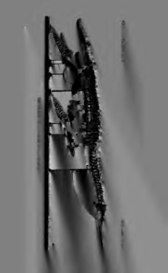
89
90

91 », 'Wf4KMMk_ WtiA'd
92 Plate II. X7/24. Skull of type specimen of Dolichorhynchops osborni, from the left (reversed).

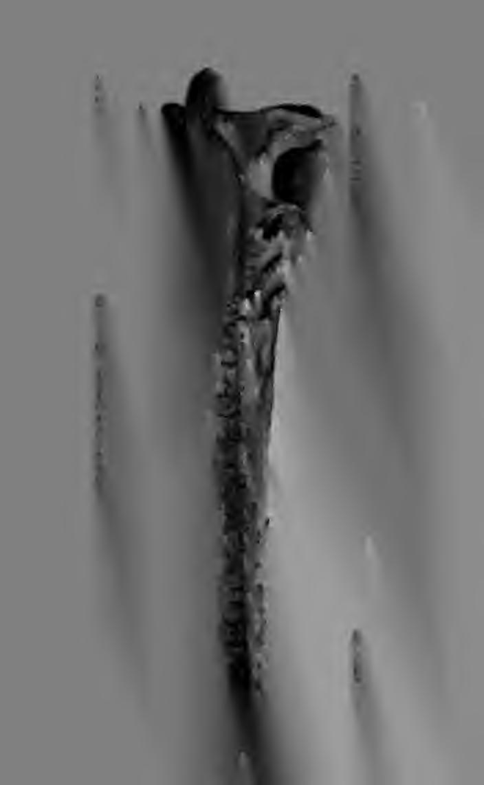
93

94
95 '
96 Plate III. Skull of Dolichorhynchops osborni. Fig. i, type specimen, from the right. Fig. 2, restoration, from the left. X7/24. ang, angular; d, dentary; eo, exoccipital; ep, epipterygoid;/, frontal ;/, jugal; m, maxilla;», nasal; pa, parietal; pet, petrosal; pt, pterygoid; pi, palatine; pm, premaxilla; pf, prefrontal;^?/, postorbital (postfronto-orbital?); q, quadrate; qj, quadratojugal; sc, sclerotic plates; so, supraoccipital; sq, squamosal; sur, surangular; v, vomer.
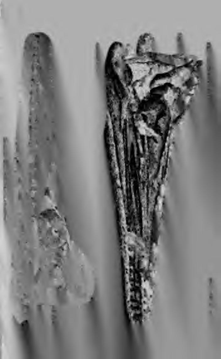
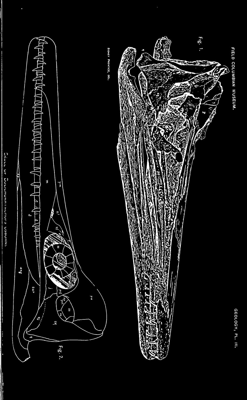
97
98
99
100 Plate IV. Skull of Dolichorhynchops osborni. Fig. i, from above. Fig. 2, from below. X i/3«bo, occipital condyle; en, external nares; ep, ectopterygoid; /, frontal; in, internal nares (the letters are on the anterior end of the palatine); /, jugal, mx, maxilla; pa, parietal; pf, prefrontal; pm, premaxilla; pt, pterygoid; pof, postorbital; q, quadrate; qj, quadratojugal; sq, squamosal


101 FIELD COLUMBIAN MUSEUM. GEOLOGY, PL. IV. Skull of Dolichorhynchops osborni.
102
103
104 Plate V. Inner side of the left mandible of various reptiles. Fig. i, Sphenodon. Fig. 2, Chelydra. Fig. 3, Alligator. Fig. 4, Varanus. Fig. 5, Cimoliasaurus snowii. art, articular; sur, surangular; ang, angular; c, coronoid; Pa, prearticular; sp, splenial; d, dentary; mg, Meckel's groove.


105 FIELD COLUMBIAN MUSEUM. GEOLOGY, PL. V. Inner Side of the Left Mandible of Various Reptiles.
106
107
108 Plate VI. below. Fourth to eighteenth cervical vertebrae of DolichorhynchoPs osborni, from X4/15.


109 FIELD COLUMBIAN MUSEUM. GEOLOGY, PL. VI. Cervical Vertebra of Dolichorhynchops osborni.
110
111
112 Plate VII. Seventh to fourteenth cervical vertebrae of Dolichorhynchops osborni, from the side. X4/15.


113 FIELD COLUMBIAN MUSEUM. GEOLOGY, PL. VII.
114
115
116 Plate VIM. Median cervical vertebrae of Dolichorhynchops osborni, from the front. X4/15.


117 fleld COLUMBIAN MUSEUM. GEOLOGY, PL. VIII. Cervical Vertebr/e of Dolichorhynchops osborni.
118
119 I
120 Plate IX. Posterior cervical vertebrae of DolichorhynchoPs osborni, from behind. 4/15.


121 FIELD COLUMBIAN MUSEUM. GEOLOGY, PL. IX. Cervical Vertebra of Dolichorhynchops osborni
122

123 I
124 Plate X. Left series, distal caudal vertebrae of Dolichorhynchops osborni, from above. XI 1/24. Right series, cervical vertebrae of Polycotylus ischiadicus, from below. X 1 1/24.


125 FIELD COLUMBIAN MUSEUM. GEOLOGY, PL. X. Left Series, Caudal Vertebrae of Dolichorhynchops osborni Right Series, Cervical Vertebra of Polycotylus ischiadicus.
126
127
128 Plate XI. Thoracic and abdominal ribs of Dolichorhynchops osborni. X 1/3.


129 FIELD COLUMBIAN MUSEUM. GEOLOGY, PL. XI. Ribs of Dolichorhynchops osborni.
130
131
132 Plate XII. Bones of pectoral girdle of Dolichorhynchops osborni. Upper left figure, right scapula, from without. Upper right figure, left scapula, from within. Lower left figure, left clavicle, from above. Lower right figure, episternum, from below. X7/13.


133 FIELD COLUMBIAN MUSEUM. GEOLOGY, PL. XII. Bones of Pectoral Girdle of Dolichorhynchops osborni.
134
135
136 Plate XIII. Bones of pectoral girdle of DolichorhynchoPs osborni, from above. Upper left figure, left clavicle. Upper middle figure, episternum. Upper right figure, scapula. Lower left figure, right coracoid. Lower right figure, humerus. X3/14.


137 FIELD COLUMBIAN MUSEUM. GEOLOGY, PL. XIII.
138
139
140 Plate XIV. Pectoral girdle of Dolichorhynchops osborni. Upper figure, left clavicle, scapula and upper extremity of coracoid, articulated, from above. X 1/3. Lower figures, scapuloclavicular girdle of same, from in front. X 1/4.


141 FIELD COLUMBIAN MUSEUM. GEOLOGY, PL. XIV. Pectoral Girdle of Dolichorhynchops osborni.
142

143
144 Plate XV. Complete pectoral girdle of Dolichorhynchops osborni, from below, articulated. X 1/4.


145 FIELD COLUMBIAN MUSEUM. GEOLOGY, PL. XV. Pectoral Girdle of Dolichorhynchops osbobni.
146
147
148 Plate XVI. Pubes of DolichorhynchoPs osborni, from above. X3/8.
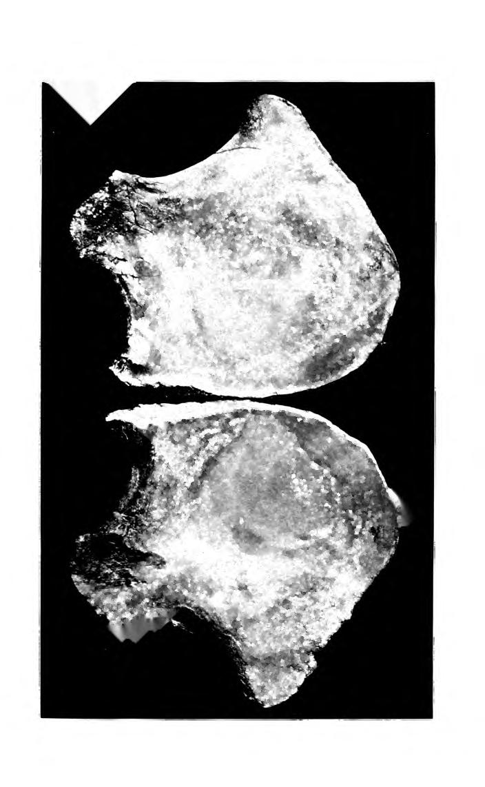
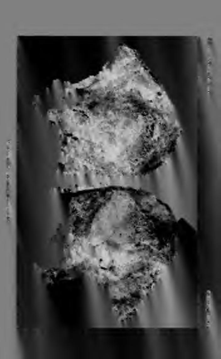
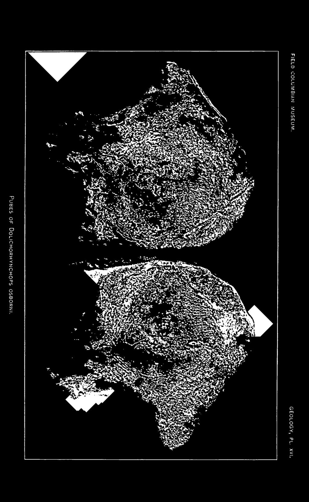
149
150
151
152 Plate XVII. Right ischium (left figure) and ilium of Dolichorhynchops osborni, visceral surface. X3/8.


153 FIELD COLUMBIAN MUSEUM. GEOLOGY, PL. XVII. is*
154
155
156 Plate XVIII. Outlines of pectoral and pelvic girdles of different plesiosaurs. Fig. i, pectoral girdle of Cryf>toclidus oxoniensis, after Andrews. Fig. 2, pelvic girdle of same, after Andrews. Fig. 3, pelvic girdle of Cimoliasaurus sp, from a photograph furnished by Prof. Fraas. Fig. 4, pelvic girdle of Cimoliasaurus trocha?iterius, after Lydekker. Fig. 5, pectoral girdle of Plesiosaurus s/>, from photograph from Prof. Fraas, Fig. 6, pectoral girdle of Murcenosaurus, after Andrews. Fig. 7, pelvic girdle of same, after Andrews. Fig. 8, pelvic girdle of same, from the side, after Andrews. ic, epistemum; cl, clavicle; sc, scapula; co, coracoid;/«, pubis; is, ischium.


157 FIELD COLUMBIAN MUSEUM GEOLOGY, PL. XVIII. 3. Pectoral and Pelvic Girdles of Plesiosaurs.
158

159 muni.,(» ;ekfi
160 Plate XIX. Pectoral and pelvic girdles of plesiosaurs. Fig. I, pectoral girdle of Plesiosaurus, after Owen. Fig. 2, pectoral girdle of Plesiosaurus, after Woodward. Fig. 3, pectoral girdle of Plesiosaurus laticeps, after Owen. Fig. 4, pectoral girdle of Elasmosaurus platyurus, after Cope. Fig. 5, pelvic girdle of same, after Cope. ic, episternum; cl, clavicle; so, scapula; co, coracoid ; /», pubis; is, ischium.


161 FIELD COLUMBIAN MUSEUM. GEOLOGY, PL. XIX. Pectoral and Pelvic Girdles of Plesiosaurs.
162
163
164 Plate XX. Limbs, as mounted, of Dolichorhynchops osborni, dorsal surface. X1/4. Left figure, right pectoral paddle. Right figure, right pelvic paddle.


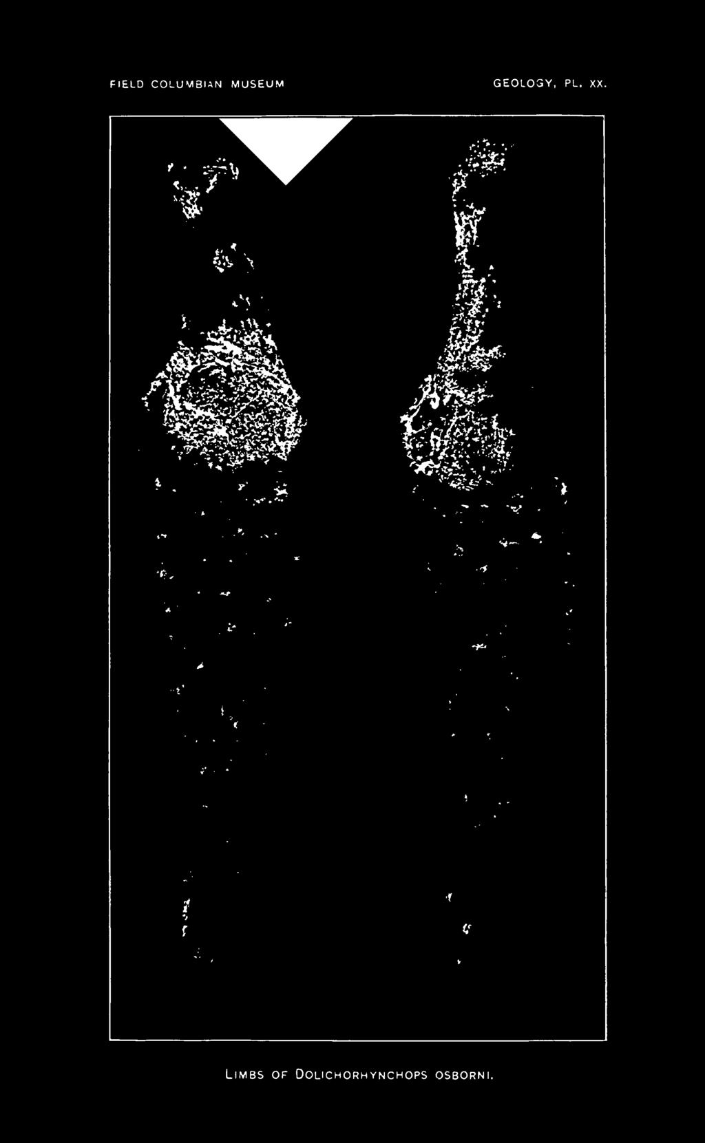
165 FIELD COLUMBIAN MUSEUM GEOLOGY, PL. XX. Limbs of Dolichorhynchops osborni.
166
167 .
168 Plate XXI. Right pectoral paddle of Polycotylus latipinnis Cope, palmar surface. Xl/6.


169 FIELD COLUMBIAN MUSEUM. GEOLOGY, PL. XXI. Paddle of Polycotylus latipinnis.
170
171 iiiuj-ijn-o: I
172 Plate XXII. Figs. 1-4, propodial bone of young plesiosaur, from Niobrara Cretaceous. X3/4- Fig. 5, atlanto-axis of Dolichorhynchops osborni, from the side. X3/4. ati, atlantal intercentrum; axi, axial intercentrum; n, atlantal neurapophysis; o, odontoid; ;-, pit for reception of axial rib; na, axial neurapophysis; 2, zygapophysis.


173 FIELD COLUMBIAN MUSEUM GEOLOGY, PL. XXII. Propodial Bone of Young Pleisosaur. Atlanto-axis of Dolichorhynchops osborni.
174
175
176 Plate XXIII. Propodial bones of young plesiosaurs, from the Niobrara Cretaceous of Kansas. X 1/2.
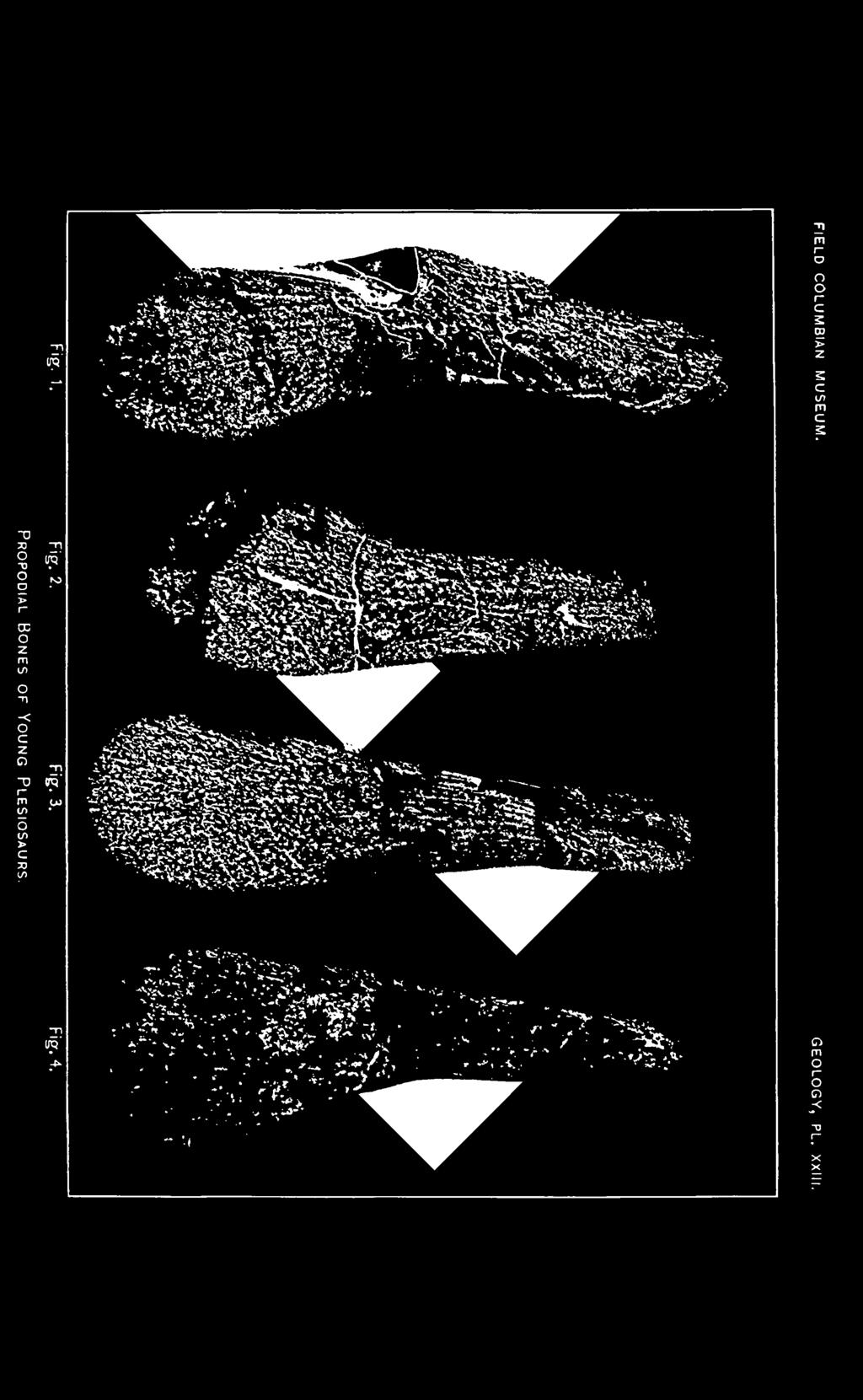
177
178
179
. X3/25.")
180 Plate'XXIV. Palatal view of type specimen of Brachauchenius lucasi (redrawn from photograph and sketch). X3/25. pt, pterygoid; e.p, ectopterygoid; pa, palatine; ps, parasphenoid; v, vomer; bs, basisphenoid.


181 FIELD COLUMBIAN MUSEUM. GEOLOGY, PL. XXIV. Palate of Brachauchenius lucasi.
182
183 ! im.nl
184 Plate XXV. Posterior cervical and dorsal vertebrae, and ribs of Brachauchenius lucasi, from below. Xi/io.


185 FIELD COLUMBIAN MUSEUM. GEOLOGY, PL. XXV. Vertebr/e and Ribs of Brachauchenius lucasi.
186
187
188 Plate XXVI. Pelvic bones of Polycotylus ischiadicus Willist., visceral surface. is, ischium; /'/, ilium; sp, sacral rib; sv, sacral vertebra. X1/3.
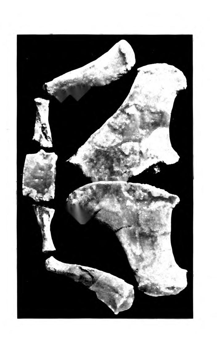
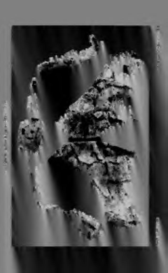
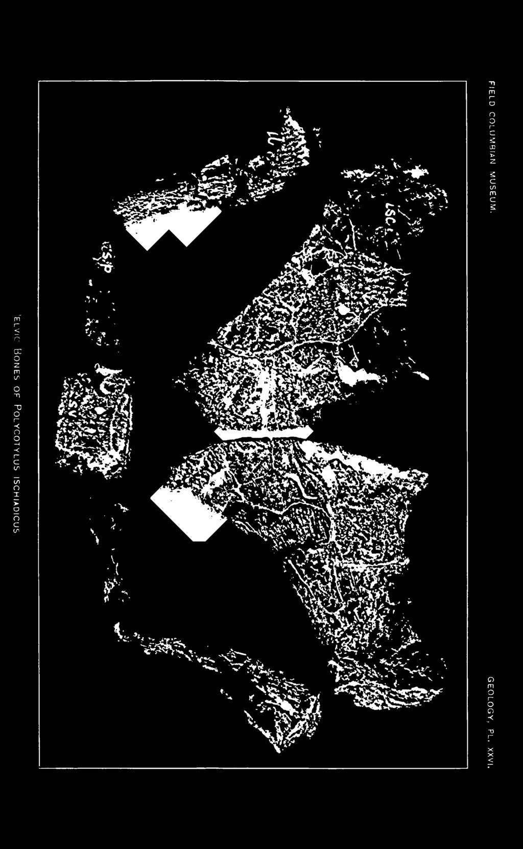
189
190
191 bfi
192 Plate XXVII. Fig. i, episternum and portion of clavicles of Plesiosaurus mudgei (?) Cragin, from below. X 1/2. Fig. 2, dorsal vertebra of Plesiosaurus gouldii (type specimen), from the front. X1/2. Fig. 3, the same, from the side. X 1/2. Drawn by Sidney Prentice.


193 FIELD COLUMBIAN MUSEUM. GEOLOGY, PL. XXVII. Fig. 1. Episternum and Portion of Clavicles of Plesiosaurus mudgei. Fig.?. Fig. 3. Dorsal Vertebra of Plesiosaurus gouldii.
194
195
196 Plate XXVIII. Trinacromerum anonymum Willist. X3/11. Upper figures, parts of ilium and coracoid. Lower figures, humerus, as restored, and cervical vertebrae.


197 HELD COLUMBIAN MUSEUM. GEOLOGY, PL. XXVIII. TRINACROMERUM ANONYMUM.
198
199
200 Plate XXIX. " Stomach pebbles," vertebrae and femur of Plesiosaurus mudgei Cragin, reduced.


201 FIELD COLUMBIAN MUSEUM. GEOLOGY, PL. XXIX.
202
203
204
205
206
Williston, and as there are many fairly good specimens in the American
 56.81.7D :14.71.5 Article VII.- SOME POINTS IN THE STRUCTURE OF THE DIADECTID SKULL. BY R. BROOM. The skull of Diadectes has been described by Cope, Case, v. Huene, and Williston, and as there are many
AMERICAN MUSEUM NOVITATES Published by
 AMERICAN MUSEUM NOVITATES Published by Number 782 THE AmzRICAN MUSEUM OF NATURAL HISTORY Feb. 20, 1935 New York City 56.81, 7 G (68) A NOTE ON THE CYNODONT, GLOCHINODONTOIDES GRACILIS HAUGHTON BY LIEUWE
A NEW GENUS AND SPECIES OF AMERICAN THEROMORPHA
 A NEW GENUS AND SPECIES OF AMERICAN THEROMORPHA MYCTEROSAURUS LONGICEPS S. W. WILLISTON University of Chicago The past summer, Mr. Herman Douthitt, of the University of Chicago paleontological expedition,
THE SKULLS OF ARAEOSCELIS AND CASEA, PERMIAN REPTILES
 THE SKULLS OF REOSCELIS ND CSE, PERMIN REPTILES University of Chicago There are few Permian reptiles of greater interest at the present time than the peculiar one I briefly described in this journal' three
SOME LITTLE-KNOWN FOSSIL LIZARDS FROM THE
 PROCEEDINGS OF THE UNITED STATES NATIONAL MUSEUM issued SWsK \ {^^m ^V ^^ SMITHSONIAN INSTITUTION U. S. NATIONAL MUSEUM Vol. 91 Washington : 1941 No. 3124 SOME LITTLE-KNOWN FOSSIL LIZARDS FROM THE OLIGOCENE
NORTH AMERICAN PLESIOSAURS
 NORTH AMERICAN PLESIOSAURS TRINACROMERUM S. W. WILLISTON The University of Chicago In previous papers' I have discussed the known characters of Elasmosaurus, Cimoliasaurus, Brachauchenius and Polycotylus,
v:ii-ixi, 'i':;iisimvi'\>!i-:: "^ A%'''''-'^-''S.''v.--..V^'E^'-'-^"-t''gi L I E) R.ARY OF THE VERSITY U N I or ILLINOIS REMO
 R.ARY OF THE VERSITY U N I or ILLINOIS REMO") "^ A%'''''-'^-''S.''v.--..V^'E^'-'-^"-t''gi v:ii-ixi, 'i':;iisimvi'\>!i-:: L I E) R.ARY OF THE U N I VERSITY or ILLINOIS REMO Natural History Survey Librarv GEOLOGICAL SERIES OF FIELD MUSEUM OF NATURAL
"^ A%'''''-'^-''S.''v.--..V^'E^'-'-^"-t''gi v:ii-ixi, 'i':;iisimvi'\>!i-:: L I E) R.ARY OF THE U N I VERSITY or ILLINOIS REMO Natural History Survey Librarv GEOLOGICAL SERIES OF FIELD MUSEUM OF NATURAL
.56 m. (22 in.). COMPSOGNATHOID DINOSAUR FROM THE. Medicine Bow, Wyoming, by the American Museum Expedition
. COMPSOGNATHOID DINOSAUR FROM THE. Medicine Bow, Wyoming, by the American Museum Expedition") Article XII.-ORNITHOLESTES HERMANNI, A NEW COMPSOGNATHOID DINOSAUR FROM THE UPPER JURASSIC. By HENRY FAIRFIELD OSBORN. The type skeleton (Amer. Mus. Coll. No. 6I9) of this remarkable animal was discovered
Article XII.-ORNITHOLESTES HERMANNI, A NEW COMPSOGNATHOID DINOSAUR FROM THE UPPER JURASSIC. By HENRY FAIRFIELD OSBORN. The type skeleton (Amer. Mus. Coll. No. 6I9) of this remarkable animal was discovered
New Carnivorous Dinosaurs from the Upper Cretaceous of Mongolia
 1955 Doklady, Academy of Sciences USSR 104 (5):779-783 New Carnivorous Dinosaurs from the Upper Cretaceous of Mongolia E. A. Maleev (translated by F. J. Alcock) The present article is a summary containing
SOME NEW AMERICAN PYCNODONT FISHES.
 SOME NEW AMERICAN PYCNODONT FISHES. By James Williams Gidley, Assistant Curator of Fossil Mammals, United States National Museum. In the United States National Museum are several specimens representing
A skull without mandihle, from the Hunterian Collection (no.
 4 MR. G. A. BOULENGER ON CHELONIAN REMAINS. [Jan. 6, 2. On some Chelonian Remains preserved in the Museum of the Eojal College of Surgeons. By G. A. Boulenger. [Eeceived December 8, 1890.] In the course
A New Ceratopsian Dinosaur from the Upper
 SMITHSONIAN MISCELLANEOUS COLLECTIONS VOLUME 63. NUMBER 3 A New Ceratopsian Dinosaur from the Upper Cretaceous of Montana, with Note on Hypacrosaurus (With Two Plates) CHARLES W. GILMORE Assistant Curator
Recently Mr. Lawrence M. Lambe has described and figured in the
 56.81,9C(117:71.2) Article XXXV.-CORYTHOSAURUS CASUARIUS, A NEW CRESTED DINOSAUR FROM THE BELLY RIVER CRETA- CEOUS, WITH PROVISIONAL CLASSIFICATION OF THE FAMILY TRACHODONTIDA1X BY BARNUM BROWN. PLATE
FURTHER STUDIES ON TWO SKELETONS OF THE BLACK RIGHT WHALE IN THE NORTH PACIFIC
 FURTHER STUDIES ON TWO SKELETONS OF THE BLACK RIGHT WHALE IN THE NORTH PACIFIC HIDEO OMURA, MASAHARU NISHIWAKI* AND TOSHIO KASUYA* ABSTRACT Two skeletons of the black right whale were studied, supplementing
A NEW SPECIES OF TROODONT DINOSAUR FROM THE
 A NEW SPECIES OF TROODONT DINOSAUR FROM THE LANCE FORMATION OF WYOMING By Charles W. Gilmore Curator of Vertebrate Paleontology, United States National Museum INTRODUCTION The intensive search to which
 The following text is generated from uncorrected OCR. [Begin Page: Page 1] A NEW CERATOPSIAN DINOSAUR FROM THE UPPER CRETACEOUS OF MONTANA, WITH NOTE ON HYPACROSAURUS ' By CHARLES W. GILMORE assistant
Postilla PEABODY MUSEUM OF NATURAL HISTORY YALE UNIVERSITY NEW HAVEN, CONNECTICUT, U.S.A.
 Postilla PEABODY MUSEUM OF NATURAL HISTORY YALE UNIVERSITY NEW HAVEN, CONNECTICUT, U.S.A. Number 117 18 March 1968 A 7DIAPSID (REPTILIA) PARIETAL FROM THE LOWER PERMIAN OF OKLAHOMA ROBERT L. CARROLL REDPATH
TRACHEMYS SCULPTA. A nearly complete articulated carapace and plastron of an Emjdd A NEAKLY COMPLETE SHELL OF THE EXTINCT TURTLE,
 A NEAKLY COMPLETE SHELL OF THE EXTINCT TURTLE, TRACHEMYS SCULPTA By Charles W. Gilmore Curator of Vertebrate Paleontology, United States National Museum INTRODUCTION A nearly complete articulated carapace
Exceptional fossil preservation demonstrates a new mode of axial skeleton elongation in early ray-finned fishes
 Supplementary Information Exceptional fossil preservation demonstrates a new mode of axial skeleton elongation in early ray-finned fishes Erin E. Maxwell, Heinz Furrer, Marcelo R. Sánchez-Villagra Supplementary
ONLINE APPENDIX 1. Morphological phylogenetic characters scored in this paper. See Poe (2004) for
 for") ONLINE APPENDIX Morphological phylogenetic characters scored in this paper. See Poe () for detailed character descriptions, citations, and justifications for states. Note that codes are changed from a
ONLINE APPENDIX Morphological phylogenetic characters scored in this paper. See Poe () for detailed character descriptions, citations, and justifications for states. Note that codes are changed from a
OF THE TRIAS THE PHYTOSAURIA
 THE PHYTOSAURIA OF THE TRIAS MAURICE G. MEHL University of Wisconsin Some time ago the writer gave a brief notice of a new genus of phytosaurs of which Angistorhinus grandis Mehl was the type.' It is the
YANGCHUANOSAURUS HEPINGENSIS - A NEW SPECIES OF CARNOSAUR FROM ZIGONG, SICHUAN
 Vol. 30, No. 4 VERTEBRATA PALASIATICA pp. 313-324 October 1992 [SICHUAN ZIGONG ROUSHILONG YI XIN ZHONG] figs. 1-5, pl. I-III YANGCHUANOSAURUS HEPINGENSIS - A NEW SPECIES OF CARNOSAUR FROM ZIGONG, SICHUAN
Anatomy. Name Section. The Vertebrate Skeleton
 Name Section Anatomy The Vertebrate Skeleton Vertebrate paleontologists get most of their knowledge about past organisms from skeletal remains. Skeletons are useful for gleaning information about an organism
A new species of Hsisosuchus (Mesoeucrocodylia) from Dashanpu, Zigong Municipality, Sichuan Province
 from Dashanpu, Zigong Municipality, Sichuan Province") A new species of Hsisosuchus (Mesoeucrocodylia) from Dashanpu, Zigong Municipality, Sichuan Province Yuhui Gao (Zigong Dinosaur Museum) Vertebrata PalAsiatica Volume 39, No. 3 July, 2001 pp. 177-184 Translated
A new species of Hsisosuchus (Mesoeucrocodylia) from Dashanpu, Zigong Municipality, Sichuan Province Yuhui Gao (Zigong Dinosaur Museum) Vertebrata PalAsiatica Volume 39, No. 3 July, 2001 pp. 177-184 Translated
Mammalogy Laboratory 1 - Mammalian Anatomy
 Mammalogy Laboratory 1 - Mammalian Anatomy I. The Goal. The goal of the lab is to teach you skeletal anatomy of mammals. We will emphasize the skull because many of the taxonomically important characters
LOWER CRETACEOUS OF SOUTH DAKOTA.
 A NEW DINOSAUR, STP^GOSAURUS MARSHl, FROM THE LOWER CRETACEOUS OF SOUTH DAKOTA. By Frederic A. Lucas, Curator, Divisioii of Coiiipnrative Anatomy, in charge, of Section of Vertebrate Fossils. The name
2. Skull, total length versus length of the presacral vertebral column: (0); extremely elongated neck (e.g. Tanystropheus longobardicus).
; extremely elongated neck (e.g. Tanystropheus longobardicus).") Character list of the taxon-character data set 1. Skull and lower jaws, interdental plates: absent (0); present, but restricted to the anterior end of the dentary (1); present along the entire alveolar
Character list of the taxon-character data set 1. Skull and lower jaws, interdental plates: absent (0); present, but restricted to the anterior end of the dentary (1); present along the entire alveolar
290 SHUFELDT, Remains of Hesperornis.
 290 SHUFELDT, Remains of Hesperornis. [ Auk [July THE FOSSIL REMAINS OF A SPECIES OF HESPERORNIS FOUND IN MONTANA. BY R. W. SHUFELD% M.D. Plate XI7III. ExR,¾ in November, 1914, Mr. Charles W. Gihnore,
List of characters used in the phylogenetic analysis. Capital letters T, R, and L, refer to
 1 Supplementary data CHARACTER LIST List of characters used in the phylogenetic analysis. Capital letters T, R, and L, refer to characters used by Tchernov et al. (2000), Rieppel, et al. (2002), and Lee
Bulletin of Big Bend Paleo-Geo An Open Access Publication from Mosasaur Ranch Museum, Terlingua and Lajitas, Texas All rights reserved
 Bulletin of Big Bend Paleo-Geo An Open Access Publication from Mosasaur Ranch Museum, Terlingua and Lajitas, Texas All rights reserved This was a private report in 2003 on my thoughts on Platecarpus planifrons.
complex in cusp pattern. (3) The bones of the coyote skull are thinner, crests sharper and the
 The bones of the coyote skull are thinner, crests sharper and the") DISTINCTIONS BETWEEN THE SKULLS OF S AND DOGS Grover S. Krantz Archaeological sites in the United States frequently yield the bones of coyotes and domestic dogs. These two canines are very similar both
DISTINCTIONS BETWEEN THE SKULLS OF S AND DOGS Grover S. Krantz Archaeological sites in the United States frequently yield the bones of coyotes and domestic dogs. These two canines are very similar both
CENE RUMINANTS OF THE GENERA OVIBOS AND
 DESCRIPTIONS OF TWO NEW SPECIES OF PLEISTO- CENE RUMINANTS OF THE GENERA OVIBOS AND BOOTHERIUM, WITH NOTES ON THE LATTER GENUS. By James Williams Gidley, Of the United States National Museum. Two interesting
A NEW SPECIES OF EXTINCT TURTLE FROM THE UPPER PLIOCENE OF IDAHO
 A NEW SPECIES OF EXTINCT TURTLE FROM THE UPPER PLIOCENE OF IDAHO By Charles W. Gilmore Curator, Division of Vertebrate Paleontology United States National Museum Among the fossils obtained bj^ the Smithsonian
Vol. XIV, No. 1, March, The Larva and Pupa of Brontispa namorikia Maulik (Coleoptera: Chrysomelidae: Hispinae) By S.
 By S.") Vol. XIV, No. 1, March, 1950 167 The Larva and Pupa of Brontispa namorikia Maulik (Coleoptera: Chrysomelidae: Hispinae) By S. MAULIK BRITISH MUSEUM (NATURAL HISTORY) (Presented by Mr. Van Zwaluwenburg
Vol. XIV, No. 1, March, 1950 167 The Larva and Pupa of Brontispa namorikia Maulik (Coleoptera: Chrysomelidae: Hispinae) By S. MAULIK BRITISH MUSEUM (NATURAL HISTORY) (Presented by Mr. Van Zwaluwenburg
HONR219D Due 3/29/16 Homework VI
 Part 1: Yet More Vertebrate Anatomy!!! HONR219D Due 3/29/16 Homework VI Part 1 builds on homework V by examining the skull in even greater detail. We start with the some of the important bones (thankfully
Biology 3315 Comparative Vertebrate Morphology Skulls and Visceral Skeletons
 Biology 3315 Comparative Vertebrate Morphology Skulls and Visceral Skeletons 1. Head skeleton of lamprey Cyclostomes are highly specialized in both the construction of the chondrocranium and visceral skeleton.
LEIDY, SHOWING THE BONES OF THE FEET 'AND LIMBS
 CQNTEUBUTIONS FBOM THE MUSEUM OF PALEONTOLOGY (Confindion of Con&&&m froin UB Muaercm of Gcologg) UNIVERSITY OF ' MICHIGAN VOL V, No. 6, pp. 6W3 (e ph.) DEAXMBER 31,1036 A SPECIMEN OF STYLEMYS NEBRASCENSIS
(Proc. Acad. Nat. Sci. Phila., p. I62) for the reception of his earlier. Chisternon. Article JX.-ON TWO INTERESTING GENERA OF EOCENE
 for the reception of his earlier. Chisternon. Article JX.-ON TWO INTERESTING GENERA OF EOCENE") 56.81,3(ii81 :78.7) Article JX.-ON TWO INTERESTING GENERA OF EOCENE TURTLES, CHISTERNON LEIDY AND ANOSTEIRA LEIDY. By OLIVER P. HAY. The genus Chisternon was proposed in I872 by Dr. Joseph Leidy (Proc.
56.81,3(ii81 :78.7) Article JX.-ON TWO INTERESTING GENERA OF EOCENE TURTLES, CHISTERNON LEIDY AND ANOSTEIRA LEIDY. By OLIVER P. HAY. The genus Chisternon was proposed in I872 by Dr. Joseph Leidy (Proc.
THE GORGONOPSIAN GENUS, HIPPOSAURUS, AND THE FAMILY ICTIDORHINIDAE * Dr. L.D. Boonstra. Paleontologist, South African Museum, Cape Town
 THE GORGONOPSIAN GENUS, HIPPOSAURUS, AND THE FAMILY ICTIDORHINIDAE * by Dr. L.D. Boonstra Paleontologist, South African Museum, Cape Town In 1928 I dug up the complete skeleton of a smallish gorgonopsian
Marshall Digital Scholar. Marshall University. F. Robin O Keefe Marshall University,
 Marshall University Marshall Digital Scholar Biological Sciences Faculty Research Biological Sciences 2008 Cranial anatomy and taxonomy of Dolichorhynchops bonneri new combination, a polycotylid (Sauropterygia:
AMERICAN NATURALIST. Vol. IX. -DECEMBER, No. 12. OR BIRDS WITH TEETH.1 OI)ONTORNITHES,
ONTORNITHES,") AMERICAN NATURALIST. Vol. IX. -DECEMBER, 1875.-No. 12. OI)ONTORNITHES, OR BIRDS WITH TEETH.1 BY PROFESSOR 0. C. MARSH. REMAINS of birds are amono the rarest of fossils, and few have been discovered except
AMERICAN NATURALIST. Vol. IX. -DECEMBER, 1875.-No. 12. OI)ONTORNITHES, OR BIRDS WITH TEETH.1 BY PROFESSOR 0. C. MARSH. REMAINS of birds are amono the rarest of fossils, and few have been discovered except
SUPPLEMENTARY INFORMATION
 Character 155, interdental ridges. Absence of interdental ridge (0) shown in Parasaniwa wyomingensis (Platynota). Interdental ridges (1) shown in Coniophis precedens. WWW.NATURE.COM/NATURE 1 Character
UNIVERSITY OF MICHIGAN
 CONTRIBUTIONS FROM THE MUSEUM OF GEOLOGY UNIVERSITY OF MICHIGAN THE VERTEBRAL COLUMN OF COELOPHYSIS COPE BY E. C. CASE UNIVERSITY OF MICHIGAN ANN ARBOR 4 Pi Spectra ABCDEFGHIJKLM~~OPORSTUWXYZ~~~~~~~~~~~~~~OP~~~~~~Y~
On the cranial anatomy of the polycotylid plesiosaurs, including new material of Polycotylus latipinnis, Cope, from Alabama
 Marshall University Marshall Digital Scholar Biological Sciences Faculty Research Biological Sciences 2004 On the cranial anatomy of the polycotylid plesiosaurs, including new material of Polycotylus latipinnis,
A new species of sauropod, Mamenchisaurus anyuensis sp. nov.
 A new species of sauropod, Mamenchisaurus anyuensis sp. nov. by Xinlu He, Suihua Yang, Kaiji Cai, Kui Li, and Zongwen Liu Chengdu University of Technology Papers on Geosciences Contributed to the 30th
A NEW ANKYLOSAUR FROM THE UPPER CRETACEOUS OF MONGOLIA E.A. Maleev Doklady Akademii Nauk, SSSR 87:
 translated by Dr. Tamara and F. Jeletzky, 1956 A NEW ANKYLOSAUR FROM THE UPPER CRETACEOUS OF MONGOLIA E.A. Maleev 1952. Doklady Akademii Nauk, SSSR 87:273-276 Armored dinosaurs make a considerable part
A Fossil Snake (Elaphe vulpina) From A Pliocene Ash Bed In Nebraska
 From A Pliocene Ash Bed In Nebraska") University of Nebraska - Lincoln DigitalCommons@University of Nebraska - Lincoln Transactions of the Nebraska Academy of Sciences and Affiliated Societies Nebraska Academy of Sciences 198 A Fossil Snake
University of Nebraska - Lincoln DigitalCommons@University of Nebraska - Lincoln Transactions of the Nebraska Academy of Sciences and Affiliated Societies Nebraska Academy of Sciences 198 A Fossil Snake
FIELDIANA GEOLOGY NEW SALAMANDERS OF THE FAMILY SIRENIDAE FROM THE CRETACEOUS OF NORTH AMERICA
 FIELDIANA GEOLOGY Published by CHICAGO NATURAL HISTORY MUSEUM Volume 10 Sbftember 22, 1968 No. 88 NEW SALAMANDERS OF THE FAMILY SIRENIDAE FROM THE CRETACEOUS OF NORTH AMERICA Coleman J. Coin AND Walter
Reprinted from: CRUSTACEANA, Vol. 32, Part 2, 1977 LEIDEN E. J. BRILL
 Reprinted from: CRUSTACEANA, Vol. 32, Part 2, 1977 LEIDEN E. J. BRILL NOTES AND NEWS 207 ALPHE0PS1S SHEARMII (ALCOCK & ANDERSON): A NEW COMBINATION WITH A REDESCRIPTION OF THE HOLOTYPE (DECAPODA, ALPHEIDAE)
A new sauropod from Dashanpu, Zigong Co. Sichuan Province (Abrosaurus dongpoensis gen. et sp. nov.)
") A new sauropod from Dashanpu, Zigong Co. Sichuan Province (Abrosaurus dongpoensis gen. et sp. nov.) by Ouyang Hui Zigong Dinosaur Museum Newsletter Number 2 1989 pp. 10-14 Translated By Will Downs Bilby
A new sauropod from Dashanpu, Zigong Co. Sichuan Province (Abrosaurus dongpoensis gen. et sp. nov.) by Ouyang Hui Zigong Dinosaur Museum Newsletter Number 2 1989 pp. 10-14 Translated By Will Downs Bilby
The occurrence of Elasmosaurus and Polycotylus in Russian deposits. *
 Yezregodneek Geologhee i Meeneraloghee Rossiee [Annuaire Geologique et Minéralogique de la Russie] Edited under the direction of N. Krishtofovich Vol. 14, Moscow, 1912 The occurrence of Elasmosaurus and
APPENDIX. 344 Mni-s/i Restorations of Claosaurus and Geratosaurus.
 344 Mni-s/i Restorations of Claosaurus and Geratosaurus. Claosaurics, Marsh, 1890.* The most important feature in the restoration of Claosaurus annectens given on Plate VI is the skull, which will be fully
UPOGEBIA LINCOLNI SP. NOV. (DECAPODA, THALASSINIDEA, UPOGEBIIDAE) FROM JAVA, INDONESIA
 FROM JAVA, INDONESIA") NOTES AND NEWS UPOGEBIA LINCOLNI SP. NOV. (DECAPODA, THALASSINIDEA, UPOGEBIIDAE) FROM JAVA, INDONESIA BY NGUYEN NGOC-HO i) Faculty of Science, University of Saigon, Vietnam Among material recently collected
NOTES AND NEWS UPOGEBIA LINCOLNI SP. NOV. (DECAPODA, THALASSINIDEA, UPOGEBIIDAE) FROM JAVA, INDONESIA BY NGUYEN NGOC-HO i) Faculty of Science, University of Saigon, Vietnam Among material recently collected
APPENDIX. 160 Miscellaneous Intelligence.
 160 Miscellaneous Intelligence. OBITUARY. GENERAL ANDREW A. HUMPHREYS. Brigadier-General Andrew Atkinson Humphreys died in Washington, on the 28th of November last, in the seventy-fourth year of his age.
A Short Report on the Occurrence of Dilophosaurus from Jinning County, Yunnan Province
 A Short Report on the Occurrence of Dilophosaurus from Jinning County, Yunnan Province by Hu Shaojin (Kunming Cultural Administrative Committee, Yunnan Province) Vertebrata PalAsiatica Vol. XXXI, No. 1
( M amenchisaurus youngi Pi, Ouyang et Ye, 1996)
") 39 4 2001 10 V ERTEBRATA PALASIATICA pp. 266 271 fig. 1,pl. I ( 643013), ( M amenchisaurus hochuanensis),,, Q915. 864 1995 12 31 (ZDM0126) ( M amenchisau rus hochuanensis Young et Chao, 1972),,, ZDM0126
39 4 2001 10 V ERTEBRATA PALASIATICA pp. 266 271 fig. 1,pl. I ( 643013), ( M amenchisaurus hochuanensis),,, Q915. 864 1995 12 31 (ZDM0126) ( M amenchisau rus hochuanensis Young et Chao, 1972),,, ZDM0126
posterior part of the second segment may show a few white hairs
 April, 1911.] New Species of Diptera of the Genus Erax. 307 NEW SPECIES OF DIPTERA OF THE GENUS ERAX. JAMES S. HINE. The various species of Asilinae known by the generic name Erax have been considered
SUPPLEMENTARY OBSERVATIONS ON THE SKULL OF
 SUPPLEMENTARY OBSERVATIONS ON THE SKULL OF THE FOSSIL PORPOISE ZARHACHIS FLAGELLATOR COPE By Remington Kellogg Of the Bureau of Biological Survey, United States Department of Agriculture During the past
CRANIAL ANATOMY OF ENNATOSAURUS TECTON (SYNAPSIDA: CASEIDAE) FROM THE MIDDLE PERMIAN OF RUSSIA AND THE EVOLUTIONARY RELATIONSHIPS OF CASEIDAE
 FROM THE MIDDLE PERMIAN OF RUSSIA AND THE EVOLUTIONARY RELATIONSHIPS OF CASEIDAE") Journal of Vertebrate Paleontology 28(1):160 180, March 2008 2008 by the Society of Vertebrate Paleontology ARTICLE CRANIAL ANATOMY OF ENNATOSAURUS TECTON (SYNAPSIDA: CASEIDAE) FROM THE MIDDLE PERMIAN
Journal of Vertebrate Paleontology 28(1):160 180, March 2008 2008 by the Society of Vertebrate Paleontology ARTICLE CRANIAL ANATOMY OF ENNATOSAURUS TECTON (SYNAPSIDA: CASEIDAE) FROM THE MIDDLE PERMIAN
Mammalogy Lab 1: Skull, Teeth, and Terms
 Mammalogy Lab 1: Skull, Teeth, and Terms Be able to: Goals of today s lab Locate all structures listed on handout Define all terms on handout what they are or what they look like Give examples of mammals
NORTH AMERICA. ON A NEW GENUS AND SPECIES OF COLUBRINE SNAKES FROM. The necessity of recognizing tlie two species treated of in this paper
 ON A NEW GENUS AND SPECIES OF COLUBRINE SNAKES FROM NORTH AMERICA. BY Leonhard Stejneger, and Batrachians. Curator of the Department of Reptiles The necessity of recognizing tlie two species treated of
AUSTRALIAN MUSEUM SCIENTIFIC PUBLICATIONS
 AUSTRALIAN MUSEUM SCIENTIFIC PUBLICATIONS McCulloch, Allan R., 1908. A new genus and species of turtle, from North Australia. Records of the Australian Museum 7(2): 126 128, plates xxvi xxvii. [11 September
Diurus, Pascoe. sp. 1). declivity of the elytra, but distinguished. Length (the rostrum and tails 26 included) mm. Deep. exception
. declivity of the elytra, but distinguished. Length (the rostrum and tails 26 included) mm. Deep. exception") 210 DIURUS ERYTIIROPUS. NOTE XXVI. Three new species of the Brenthid genus Diurus, Pascoe DESCRIBED BY C. Ritsema+Cz. 1. Diurus erythropus, n. sp. 1). Allied to D. furcillatus Gylh. ²) by the short head,
210 DIURUS ERYTIIROPUS. NOTE XXVI. Three new species of the Brenthid genus Diurus, Pascoe DESCRIBED BY C. Ritsema+Cz. 1. Diurus erythropus, n. sp. 1). Allied to D. furcillatus Gylh. ²) by the short head,
CRANIAL OSTEOLOGY OF SCHIZOTHORAICHTHYS NIGER (MECKEL) MISRA (CYPRINIDAE: SCHIZOTHORACINAE). L NEUROCRANIUM
 MISRA (CYPRINIDAE: SCHIZOTHORACINAE). L NEUROCRANIUM") CRANIAL OSTEOLOGY OF SCHIZOTHORAICHTHYS NIGER (MECKEL) MISRA (CYPRINIDAE: SCHIZOTHORACINAE). L NEUROCRANIUM A. R. YousuF, A. K. PANDIT AND A. R. KHAN Postgraduate Department of Zoology, University of Kashmir,
CRANIAL OSTEOLOGY OF SCHIZOTHORAICHTHYS NIGER (MECKEL) MISRA (CYPRINIDAE: SCHIZOTHORACINAE). L NEUROCRANIUM A. R. YousuF, A. K. PANDIT AND A. R. KHAN Postgraduate Department of Zoology, University of Kashmir,
SUPPLEMENTARY ONLINE MATERIAL FOR. Nirina O. Ratsimbaholison, Ryan N. Felice, and Patrick M. O connor
 http://app.pan.pl/som/app61-ratsimbaholison_etal_som.pdf SUPPLEMENTARY ONLINE MATERIAL FOR Nirina O. Ratsimbaholison, Ryan N. Felice, and Patrick M. O connor Ontogenetic changes in the craniomandibular
OSTEOLOGICAL NOTE OF AN ANTARCTIC SEI WHALE
 OSTEOLOGICAL NOTE OF AN ANTARCTIC SEI WHALE MASAHARU NISHIWAKI* AND TOSHIO KASUYA* ABSTRACT This is a report of measurements on the skeleton of a male se1 whale caught in the Antarctic. The skeleton of
VERTEBRATA PALASIATICA
 VERTEBRATA PALASIATICA ONLINE SUPPLEMENTARY MATERIAL Panxianichthys imparilis gen. et sp. nov., a new ionoscopiform (Halecomorphi) from the Middle Triassic of Guizhou Province, China XU Guang-Hui 1,2 SHEN
PRELIMINARY REPORT ON A CLUTCH OF SIX DINOSAURIAN EGGS FROM THE UPPER TRIASSIC ELLIO T FORMATION, NORTHERN ORANGE FREE STATE. J. W.
 41 Pa/aeont. afr., 22, 41-45 (1979) PRELIMINARY REPORT ON A CLUTCH OF SIX DINOSAURIAN EGGS FROM THE UPPER TRIASSIC ELLIO T FORMATION, NORTHERN ORANGE FREE STATE b y J. W. Kitching ABSTRACT A clutch of
VERTEBRATA PALASIATICA
 41 2 2003 2 VERTEBRATA PALASIATICA pp. 147 156 figs. 1 5 1) ( 100044), ( Parakannemeyeria brevirostris),,, : ( Xiyukannemeyeria),,, Q915. 864 60 Turfania (,1973), Dicynodon (, 1973 ; Lucas, 1998), (Lystrosaurus)
Chapter 6 - Systematic palaeontology
 - Sea-saurians have had a sorry experience in the treatment they have received from nomenclators Samuel Wendell Williston, 1914 6.1 Rhomaleosauridae - generic and species-level systematics As defined in
NIVOROUS DINOSAUR. (SECOND COMMUNICATION.) By HENRY FAIRFIELD OSBORN. PLATE XXXIX. This great carnivorous Dinosaur of the Laramie was contemporary
 By HENRY FAIRFIELD OSBORN. PLATE XXXIX. This great carnivorous Dinosaur of the Laramie was contemporary") 56, 8i, 9 T (I 7: 786) Article VI.-TYRANNOSAURUS, UPPER CRETACEOUS CAR- NIVOROUS DINOSAUR. (SECOND COMMUNICATION.) By HENRY FAIRFIELD OSBORN. PLATE I. This great carnivorous Dinosaur of the Laramie was
56, 8i, 9 T (I 7: 786) Article VI.-TYRANNOSAURUS, UPPER CRETACEOUS CAR- NIVOROUS DINOSAUR. (SECOND COMMUNICATION.) By HENRY FAIRFIELD OSBORN. PLATE I. This great carnivorous Dinosaur of the Laramie was
FCI-Standard N 251 / / GB. POLISH LOWLAND SHEEPDOG (Polski Owczarek Nizinny)
") FCI-Standard N 251 / 07. 08. 1998 / GB POLISH LOWLAND SHEEPDOG (Polski Owczarek Nizinny) TRANSLATION : Mrs. Peggy Davis. ORIGIN : Poland. 2 DATE OF PUBLICATION OF THE ORIGINAL VALID STANDARD : 07.08.1998.
FCI-Standard N 251 / 07. 08. 1998 / GB POLISH LOWLAND SHEEPDOG (Polski Owczarek Nizinny) TRANSLATION : Mrs. Peggy Davis. ORIGIN : Poland. 2 DATE OF PUBLICATION OF THE ORIGINAL VALID STANDARD : 07.08.1998.
PALEONTOLOGICAL CONTRIBUTIONS
 THE UNIVERSITY OF KANSAS PALEONTOLOGICAL CONTRIBUTIONS August, 1965 Paper 2 A NEW WYOMING PHYTOSAUR By THEODORE H. EATON, JR. [Museum of Natural History, University of Kansas I ABSTRACT The skull of a
DESCRIPTIONS OF NEW AND LITTLE-KNOWN FOSSIL LIZARDS FROM NORTH AMERICA
 PROCEEDINGS OF THE UNITED STATES NATIONAL MUSEUM SMITHSONIAN INSTITUTION U. S. NATIONAL MUSEUM Vol.86 Washington: 1938 No. 3042 DESCRIPTIONS OF NEW AND LITTLE-KNOWN FOSSIL LIZARDS FROM NORTH AMERICA By
ON TWO NEW SPECIMENS OF LYSTROSAURUS-ZONE CYNODONTS
 ON TWO NEW SPECMENS OF LYSTROSAURUS-ZONE CYNODONTS By A. S. Brink ABSTRACT n this paper the skulls of two new specimens of Lystrosaurus-zone cynodonts are described. One is a skull of Notictosaurus luckh1fi
ON SOME REPTILIAN REMAINS FROM THE DINOSAUR BEDS OF NYASALAND. By S. H. HAUGHTON, D.Sc., F.G.S.
 ( 67 ) ON SOME REPTILIAN REMAINS FROM THE DINOSAUR BEDS OF NYASALAND. By S. H. HAUGHTON, D.Sc., F.G.S. (Published by permission of the Hon. the Minister for Mines and Industries.) (With Plates II-V and
AUSTRALIAN MUSEUM SCIENTIFIC PUBLICATIONS
 AUSTRALIAN MUSEUM SCIENTIFIC PUBLICATIONS Riek, E. F., 1964. Merostomoidea (Arthropoda, Trilobitomorpha) from the Australian Middle Triassic. Records of the Australian Museum 26(13): 327 332, plate 35.
PEABODY MUSEUM OF NATURAL HISTORY, YALE UNIVERSITY NEW HAVEN, CONNECTICUT, U.S.A. A NEW OREODONT FROM THE CABBAGE PATCH LOCAL FAUNA, WESTERN MONTANA
 Postilla PEABODY MUSEUM OF NATURAL HISTORY YALE UNIVERSITY NEW HAVEN, CONNECTICUT, U.S.A. Number 85 September 21, 1964 A NEW OREODONT FROM THE CABBAGE PATCH LOCAL FAUNA, WESTERN MONTANA STANLEY J. RIEL
ON THE SCALOPOSAURID SKULL OF OLIVIERIA PARRINGTONI, BRINK WITH A NOTE ON THE ORIGIN OF HAIR
 ON THE SCALOPOSAURID SKULL OF OLIVIERIA PARRINGTONI, BRINK WITH A NOTE ON THE ORIGIN OF HAIR By G. H. Findlay, D.Sc., M.D. (Professor of Dermatology, University of Pretoria; Director, C.S.I.R. Photobiology
Lower Cretaceous Kwanmon Group, Northern Kyushu
 Bull. Kitakyushu Mus. Nat. Hist., 11: 87-90. March 30, 1992 A New Genus and Species of Carnivorous Dinosaur from the Lower Cretaceous Kwanmon Group, Northern Kyushu Yoshihiko Okazaki Kitakyushu Museum
The cranial osteology of Belebey vegrandis (Parareptilia: Bolosauridae), from the Middle Permian of Russia, and its bearing on reptilian evolution
, from the Middle Permian of Russia, and its bearing on reptilian evolution") Blackwell Publishing LtdOxford, UKZOJZoological Journal of the Linnean Society0024-4082 2007 The Linnean Society of London? 2007 1511 191214 Original Articles RUSSIAN BOLOSAURID REPTILER. R. REISZ ET AL.
Blackwell Publishing LtdOxford, UKZOJZoological Journal of the Linnean Society0024-4082 2007 The Linnean Society of London? 2007 1511 191214 Original Articles RUSSIAN BOLOSAURID REPTILER. R. REISZ ET AL.
A NEW SALTICID SPIDER FROM VICTORIA By R. A. Dunn
 Dunn, R. A. 1947. A new salticid spider from Victoria. Memoirs of the National Museum of Victoria 15: 82 85. All text not included in the original document is highlighted in red. Mem. Nat. Mus. Vict.,
By HENRY FAIRFIELD OSBORN.
 Article XI.-FORE AND HINI) LIMBS OF CARNIVOR- OUS AND HERBIVOROUS DINOSAURS FROM THE JURASSIC OF WYOMING. DINOSAUR CONTRIBU- TIONS, NO. 3. By HENRY FAIRFIELD OSBORN. In the Bone Cabin Quarry, opened by
Cranial osteology of the African gerrhosaurid Angolosaurus skoogi (Squamata; Gerrhosauridae) HOLLY A. NANCE
 HOLLY A. NANCE") African Journal of Herpetology, 2007 56(1): 39-75. Herpetological Association of Africa Original article Cranial osteology of the African gerrhosaurid Angolosaurus skoogi (Squamata; Gerrhosauridae) HOLLY
African Journal of Herpetology, 2007 56(1): 39-75. Herpetological Association of Africa Original article Cranial osteology of the African gerrhosaurid Angolosaurus skoogi (Squamata; Gerrhosauridae) HOLLY
NOTICE OF INTERESTING NEW FORMS OF CARBONIFEROUS FISH REMAINS.
 NOTICE OF INTERESTING NEW FORMS OF CARBONIFEROUS FISH REMAINS. C. R. EASTMAN. THROUGH the kindness of Professor G. Hambach, of Washington University, St. Louis, a number of highly instructive Carboniferous
OF THE TESTUDINATA. ON THE CLASSIFICATION
 530 The American Naturalist. [June, ON THE CLASSIFICATION OF THE TESTUDINATA. BY GEORGE BAUR. BOULENGER, Doderlein, Zittel, and Lydekker have lately published more or less extensive works on the Testudinata.
A new carnosaur from Yongchuan County, Sichuan Province
 A new carnosaur from Yongchuan County, Sichuan Province by Dong Zhiming Institute of Vertebrate Palaeontology and Palaeoanthropology, Academia Sinica Zhang Yihong, Li Xuanmin, and Zhou Shiwu Chongqing
NEGLECTUS. NOTE V. Synonymical Remarks. about Palaemon neglectus nov. nom. and. Palaemon reunionnensis Hoffm. Dr. J.G. de Man. Plate
 PALAEMON NEGLECTUS. 201 NOTE V. Synonymical Remarks about Palaemon neglectus nov. nom. and Palaemon reunionnensis Hoffm. BY Dr. J.G. de Man Plate 15. Palaemon (Eupalaemon) neglectus, nov. nom. (Plate 15,
THE GENUS FITCHIELLA (HOMOPTERA, FULGORIDAE).
.") Reprinted from BULLETIN OF THE BROOKLYN ENTO:>COLOGICAL SOCIETY, Vol. XXVIII, No. 5, pp. 194-198. December, 1933 THE GENUS FITCHIELLA (HOMOPTERA, FULGORIDAE). PAUL B. LAWSON, LaV
Reprinted from BULLETIN OF THE BROOKLYN ENTO:>COLOGICAL SOCIETY, Vol. XXVIII, No. 5, pp. 194-198. December, 1933 THE GENUS FITCHIELLA (HOMOPTERA, FULGORIDAE). PAUL B. LAWSON, LaV
The family Gnaphosidae is a large family
 Pakistan J. Zool., vol. 36(4), pp. 307-312, 2004. New Species of Zelotus Spider (Araneae: Gnaphosidae) from Pakistan ABIDA BUTT AND M.A. BEG Department of Zoology, University of Agriculture, Faisalabad,
A M E G H I N I A N A. Revista de la Asociación Paleontológia Argentina. Volume XV September-December 1978 Nos. 3-4
 A M E G H I N I A N A Revista de la Asociación Paleontológia Argentina Volume XV September-December 1978 Nos. 3-4 COLORADIA BREVIS N. G. ET N. SP. (SAURISCHIA, PROSAUROPODA), A PLATEOSAURID DINOSAUR FROM
SAINT GERMAIN POINTER (Braque Saint-Germain)
") FEDERATION CYNOLOGIQUE INTERNATIONALE (AISBL) SECRETARIAT GENERAL: 13, Place Albert 1 er B 6530 Thuin (Belgique) 05.05.2003/EN FCI-Standard N 115 SAINT GERMAIN POINTER (Braque Saint-Germain) 2 TRANSLATION
FEDERATION CYNOLOGIQUE INTERNATIONALE (AISBL) SECRETARIAT GENERAL: 13, Place Albert 1 er B 6530 Thuin (Belgique) 05.05.2003/EN FCI-Standard N 115 SAINT GERMAIN POINTER (Braque Saint-Germain) 2 TRANSLATION
A NEW AUSTROSQUILLA (STOMATOPODA) FROM THE
 FROM THE") A NEW AUSTROSQUILLA (STOMATOPODA) FROM THE MARQUESAS ISLANDS BY ALAIN MICHEL Centre O.R.S.T.O.M., Noumea, New Caledonia and RAYMOND B. MANNING Smithsonian Institution, Washington, U.S.A. The At s,tstrosqzlilla
A NEW AUSTROSQUILLA (STOMATOPODA) FROM THE MARQUESAS ISLANDS BY ALAIN MICHEL Centre O.R.S.T.O.M., Noumea, New Caledonia and RAYMOND B. MANNING Smithsonian Institution, Washington, U.S.A. The At s,tstrosqzlilla
Notes on Ceratopsians and Ankylosaurs at the Royal Ontario Museum
 Notes on Ceratopsians and Ankylosaurs at the Royal Ontario Museum Andrew A. Farke, Ph.D. Raymond M. Alf Museum of Paleontology 1175 West Baseline Road Claremont, CA 91711 email: afarke@webb.org Introduction
Comments on the Beauceron Standard By M. Maurice Hermel (Translated by C. Batson)
") Comments on the Beauceron Standard By M. Maurice Hermel (Translated by C. Batson) The following are comments written by M. Hermel for the FCI Standard #44 published on 10/25/06. They were approved by the
Comments on the Beauceron Standard By M. Maurice Hermel (Translated by C. Batson) The following are comments written by M. Hermel for the FCI Standard #44 published on 10/25/06. They were approved by the
SUOMENLAPINKOIRA. FEDERATION CYNOLOGIQUE INTERNATIONALE (AISBL) SECRETARIAT GENERAL: 13, Place Albert 1 er B 6530 Thuin (Belgique)
 SECRETARIAT GENERAL: 13, Place Albert 1 er B 6530 Thuin (Belgique)") 12.10.2016 / EN FEDERATION CYNOLOGIQUE INTERNATIONALE (AISBL) SECRETARIAT GENERAL: 13, Place Albert 1 er B 6530 Thuin (Belgique) FCI-Standard N 189 SUOMENLAPINKOIRA (Finnish Lapponian Dog) 2 ORIGIN: Finland.
12.10.2016 / EN FEDERATION CYNOLOGIQUE INTERNATIONALE (AISBL) SECRETARIAT GENERAL: 13, Place Albert 1 er B 6530 Thuin (Belgique) FCI-Standard N 189 SUOMENLAPINKOIRA (Finnish Lapponian Dog) 2 ORIGIN: Finland.
ON THE FPERYLOSIS OF THE BLACK-THROATED DIVER.
 ON THE FPERYLOSIS OF THE BLACK-THROATED DIVER. BY W. P. PYCRAFT. IT is surely a matter for regret that so little interest has been taken in that side of ornithology which concerns structural characters,
A redescription of Plesiosaurus propinquus Tate & Blake, 1876 (Reptilia, Plesiosauria), from the Lower Jurassic (Toarcian) of Yorkshire, England
, from the Lower Jurassic (Toarcian) of Yorkshire, England") PROCEEDINGS OF THE YORKSHIRE GEOLOGICAL SOCIETY, VOL. 57, PARTS 3 4, PP. 133 142, 2009 A redescription of Plesiosaurus propinquus Tate & Blake, 1876 (Reptilia, Plesiosauria), from the Lower Jurassic (Toarcian)
PROCEEDINGS OF THE YORKSHIRE GEOLOGICAL SOCIETY, VOL. 57, PARTS 3 4, PP. 133 142, 2009 A redescription of Plesiosaurus propinquus Tate & Blake, 1876 (Reptilia, Plesiosauria), from the Lower Jurassic (Toarcian)
APPENDIX. 328 Scientific Intelligence.
 328 Scientific Intelligence. selves in the first spiral coil of 0. tenuissima are what constitute the essential difference between the spire of Cornuspira and that of Spirolocidina; marking an imperfect
ON A NEW SPECIES OF APOVOSTOX HEBARD (DERMAPTERA : SPONGIPHORIDAE) FROM INDIA
 FROM INDIA") Rec. zoot. Surv. India, 97 (Part-2) : 39-43, 1999 ON A NEW SPECIES OF APOVOSTOX HEBARD (DERMAPTERA : SPONGIPHORIDAE) FROM INDIA G. K. SRIVASTAVA* Zoological Survey of India, Eastern RegionaL Station, Shillong
Rec. zoot. Surv. India, 97 (Part-2) : 39-43, 1999 ON A NEW SPECIES OF APOVOSTOX HEBARD (DERMAPTERA : SPONGIPHORIDAE) FROM INDIA G. K. SRIVASTAVA* Zoological Survey of India, Eastern RegionaL Station, Shillong