PRACTICAL APPROACHES TO ON-FARM BOVINE SURGERY
|
|
|
- Reynold Francis
- 5 years ago
- Views:
Transcription
1 PRACTICAL APPROACHES TO ON-FARM BOVINE SURGERY Dr. Gordon Atkins, University of Calgary Veterinary Medicine Although the role of the large animal veterinarian has expanded to a more herd focused production medicine approach, the need for competent on-farm surgical skills still remains as a primary responsibility of the herd veterinarian. In fact many of the modern dairy and cow-calf operations have greatly improved facilities that allow for the delivery of more sophisticated on-farm surgical procedures. In addition, bovine veterinary practice has not been influenced by the same development of large referral practices as is characteristic of the small animal and equine disciplines. As a result, there is a continual need for bovine veterinarians to expand their on-farm specialty surgical expertise to meet the ever increasing demands of especially the purebred livestock industry. It is particularly important for new graduates to develop competence in the delivery of on-farm surgical procedures in order to gain the confidence and respect of the managers of large well managed livestock operations so those new graduates have the opportunity to expand their role and deliver advanced production medicine services to these clients. This presentation is a compilation of topics and practice experience involving all aspects of the delivery of on-farm bovine surgery. It includes a discussion of the challenges of on-farm sedation and general anesthesia, the various approaches for performing local anesthesia of the paralumbar fossa, the lower limb, and the udder, as well as the associated milk and meat withdrawal times for the pharmaceuticals available for these procedures. In addition, the presentation provides a review of the often confusing bovine gastrointestinal anatomy and the various approaches and procedures for surgical correction of the many gastrointestinal problems we are presented with especially in lactating dairy cattle. Finally, the presentation will also address the surgical management of umbilical masses in calves, the surgical management of infections and other lesions of the distal limb, the surgical options available to deal with teat obstructions, and a few tips in the handling of the umbilicus in calves delivered by C-Section. Animal welfare and specifically pain management, as well as the prudent use of drugs are both topics that currently receive priority public attention. Therefore any veterinary surgical discussion must focus on these issues as well as the technical surgical procedure that is required. We are fortunate that the bovine patient will tolerate standing surgery for so many procedures making on-farm surgery much more feasible than is possible in other species. It is my hope that this presentation will stimulate an interactive discussion that will allow for the sharing of numerous practice tips among veterinarians and inspire new graduates to expand their comfort zone with regard to on-farm bovine surgery. Pre-anesthetic Considerations for the Bovine The major anatomic and physiologic features that increase the general anesthetic risk in cattle include: A tendency to regurgitate and therefore potentially aspirate if the airway is not protected, The potential to develop ruminal distention (bloat), The potential to develop neuropathies and myopathies with recumbency, The potential development of hypoxemia on room air with recumbency and/or general anesthesia. These same risks may become evident with recumbency on its own in cattle. Calves under 2 months of age can be considered monogastric animals because they lack a fully functional rumen. The younger calves may demonstrate immature organ function with a prolonged duration of action of injectable agents and inhalational anesthesia is recommended for the true neonatal animal. As
2 the calf ages rumination develops and the risk of regurgitation and aspiration increases despite the positive development of mature organ function. Ruminants are prone to hypoxemia and the development of hypoxemia has been linked to xylazine administration in several studies. Body position is also linked to the development of hypoxemia and dorsal recumbency should be avoided whenever possible. Ruminal tympany will result in thoracic compression and ventilation/perfusion mismatch. Nasal administration of oxygen at 5-10 L/min through a nasotracheal tube will offset hypoxemia in animals anesthetized with injectable products. In general, pre-anesthetic preparation in cattle includes a physical examination, fasting, estimation of body weight, and appropriate laboratory evaluation if indicated. Pre-anesthetic fasting recommendations typically reported are that 2-4 month old calves be deprived of food and water for 2-8 hours, and that 4-6 month old calves be fasted for hours and deprived of water for 8-12 hours. Adult cattle should be fasted hours and deprived of water for hours. Fasting true neonates is not recommended and should not be withheld for more than 2 hours because of the development of hypoglycemia as glycogen storage in the liver is minimal. With any anesthetic, the physical state of the animal, particularly its hydration state, is an important consideration. Calves presented for abdominal surgery are often dehydrated and require aggressive fluid therapy before they are induced. Tissue oxygenation should also be assessed. Alpha-2 agonist drugs can produce hypoxemia in healthy ruminants and oxygen carrying capacity should also be considered. When possible, a gauge IV catheter should be placed prior to induction. Considerations for Sedation and Induction of General Anesthesia There are very few anesthetic drugs labeled for use in ruminants and therefore most of the recommended protocols are extra label. Xylazine is labeled for use in cattle and it is a useful sedative and analgesic in ruminants and camelids. The major side effects are ruminal tympany and hypoxemia. Fasting will decrease the incidence of ruminal tympany and hypoxemia can be offset with supplemental oxygen delivered by nasotracheal tube. In adult cattle it may be necessary to use a respirator to achieve adequate oxygenation especially if deep anesthesia is required. The effects of xylazine can be reversed with Tolazoline in cattle. Tolazoline is an alphaadrenergic blocking agent that functions as an alpha-one and alpha-two antagonist competitively inhibiting the alpha-adrenergic receptors. The label dosage is 4mg/kg IV however a number of trials have shown that a dosage of 2mg/kg given IM will reverse the effects of xylazine nearly as fast with less impact on the circulatory system. The usual concentration of this product is 100 mg/ml so the dosage used most commonly in the field is about 1 ml/ 100 lbs. For a 1000 lb animal the cost of a 10 ml dose is roughly $10. Until recently with the new availability of Tolazoline, the most available and most commonly used reversal agent for xylazine has been Atipamezole (Antisedan Zoetis ). This drug is an alpha-two antagonist that competitively inhibits alpha-two adrenergic receptors. The biggest issue with this drug is its expense as it may cost over $100 to reverse xylazine in a mature bovine. The dosage used most commonly in the field is.05 mg/kg given IM. Therefore the dosage for a 500 kg animal would be about 25 mg or 5ml of the 5mg/ml solution. The dose ratio between the mg of xylazine given at 0.2 mg/kg rate for the same 500 kg animal and the mg of atipamezole would be about 4:1. In the event of an overdose, it may be important to go to the higher dose of Tolazoline (4mg/kg) or a higher dose of Atipamezole 0.1mg/kg given half IV and half IM.
3 One potential drawback of xylazine is that it will produce recumbency in the bovine at higher doses. This can be undesirable in cattle, if standing sedation is required. Detomidine can also be used and usually will produce a more reliable standing sedation in cattle (at a dose of 0.01 mg/kg). Some authors advocate the use of detomidine in the last third of gestation, as there may be less risk of inducing abortion, compared to xylazine. Xylazine has also been used as a component in epidural anesthesia in cattle. It produces both sedation and analgesia and can be used alone to supplement gas anesthesia, or it may be combined with local anesthetic to produce sedation and prolong the effects of local anesthesia. Diazepam is also a very useful sedative in calves, small ruminants and camelids. Intravenous diazepam induces reliable sedation with minimal adverse cardiopulmonary or respiratory effects and as a result is probably more suitable than xylazine in young animals or in animals with cardiovascular compromise. Butorphanol is a widely used sedative and analgesic used in cattle and it is frequently combined with diazepam to produce neuroleptanalgesia. This may be suitable for sedation in young calves, goats and sheep, or as a preanesthetic combination. The combination of butorphanol and diazepam will reduce gas anesthetic requirements, resulting in less cardiopulmonary depression during anesthesia. Animals sedated with diazepam-butorphanol may be mask induced with gas anesthesia. Butorphanol ( mg/kg) is also commonly combined with xylazine ( mg/kg), detomidine (.01 mg/kg), or acepromazine ( mg/kg) to produce varying degrees of sedation. Intravenous Ketamine is often used as a reliable induction agent for general anesthesia and is the induction agent of choice for animals in shock. Ketamine will decrease gas anesthetic requirements for animals with shock but it must be given in conjunction with a muscle relaxant such as xylazine, diazepam, or guaifenasin. Guaifenesin (GG) is a centrally acting skeletal muscle relaxant that can be used to induce recumbency in cattle, or given with ketamine or thiopental for induction and intubation. GG is an adjunct to anesthesia only and not a general anesthetic. Only 5% solution should be used in cattle as hemolysis occurs at higher concentrations and the product is very irritating when given perivascularly. Typical doses include (2ml/kg of 5% solution). Ruminants are prone to regurgitation, and must be intubated when anesthetized with Guaifenesin or the volatile anesthetics. Oro-tracheal intubation can be performed in both calves and cows. The preferred technique is to visualize the glottis with a laryngoscope but blind intubation is the technique that is usually used in the field. It can be performed successfully and is accomplished with the head and neck extended and the tube passed through the oropharynx. The esophagus compressed and the larynx is manipulated while the tube is advanced on inspiration. It is critical that a speculum be used to prevent the tube from being chewed until a surgical plane of anesthesia is reached. Intubation should be performed with the animal in sternal recumbency if at all possible as they often regurgitate when they are positioned in lateral or dorsal recumbency. The final sedative that is still used quite commonly in cattle is Acepromazine. It is often used in combination with butorphanol and this mixture is more reliable for keeping the animal standing while in the sedated state than is the combination of butorphanol and xylazine. Acepromazxine does cause peripheral vasodilation and is not recommended for geriatric or debilitated animals. While it is useful in the bull to relax the retractor penis muscle and facilitate examination of the penis, it must be used with caution in both bulls and stallions to avoid the risk of paraphimosis.
4 It is extremely important to provide appropriate padding for the anesthetized bovine patient. This is particularly important to prevent radial or facial paralysis with larger animals as they can be prone to compartmental crush syndromes. A small inflated inner tube works well to prevent both compression of the radial nerve over the lateral aspect of the shoulder as well as pressure necrosis over the greater trochanter. Resting the head on an inflated pad will also prevent facial paralysis. Field Anesthesia Protocols for Calves Anesthesia of calves can be performed with general anesthesia or a combination of deep sedation and local anesthesia. Restraint is particularly important and the construction of a wooden or metal V trough facilitates good surgical positioning and allows for lighter sedation or anesthesia. Young calves (< 3 months old) have an immature cardiovascular system, and as such, they are very dependent on heart rate to maintain cardiac output. It is best to avoid xylazine in very young calves as it will lower heart rate and decrease cardiac output. If xylazine is used, it should be used at a low dose, or should be administered epidurally. Young calves are prone to hypoglycemia and hypothermia. Blood glucose and body temperature should be closely monitored. Diazepam-butorphanol is a suitable sedative or preanesthetic mixture. Neonatal calves can be mask induced. Older calves can be induced with IV ketamine. Calves over 3 months old may be more difficult to physically restrain. Xylazine can be used at a dose of mg/kg IM or 0.1 mg/kg IV to facilitate restraint. This can be followed by diazepam (0.2 mg/kg) and ketamine (2-4 mg/kg) for induction. Maintenance of anesthesia can be performed with an IV drip solution or gas anesthesia using isoflurane or halothane. A unique modification to a field anesthesia protocol for calves is the use of a large volume epidural which results in anesthesia up to about the level of the umbilicus and reduces the need for heavy sedation or anesthesia since the hind limbs are completely immobilized. It should be noted that this protocol should be restricted to calves under 300 lbs since there is a risk of gastrocnemius rupture during recovery for larger animals. A common field anesthesia protocol used for hour umbilical surgery in lb calves at the University of Montreal in Saint Hyacinthe is as follows: Tranquilization 0.1 mg/kg Diazepam IV (5mg/ml) Large Volume Epidural mg/kg Xylazine (20 mg/ml) plus 0.15ml/kg 2% Lidocaine Anesthesia Drip - 2ml/kg 5% Guaifenasin plus 2mg/kg Ketamine (100 mg/ml) Example: 100 kg calf - 15 ml 2% Lidocaine, 5 mg 20mg/ml Xylazine, 200 ml 5% Guaifenasin, & 200 mg (2 ml of 100 mg/ml) Ketamine This solution is easy to monitor since each ml of 5% Guaifenasin contains 1 mg of Ketamine Field Anesthesia Protocols for Mature Cattle Short term anesthesia can be produced in adult cattle with xylazine as a preanesthetic, ketamine for induction, and maintenance with a mixture of guaifenasin and ketamine. The first step is to induce recumbency with 0.2 mg/kg of xylazine IV. This can be administered into the tail vein. Once the cow is recumbent 2 mg/kg of ketamine is administered IV. Ketamine can be repeated at a dose of 1 mg/kg as needed (generally every minutes). If complete anesthesia is required the cow is induced as above and maintained with a mixture of 5% guaifenasin and ketamine. The cow must be intubated if this mixture is used. Supplemental oxygen can be administered at a flow of 8-10 litres/minute via the endotracheal tube, or at a flow of litres/minute via a nasal catheter.
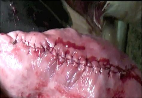
5 Standing Ketamine Stun Xylazine 0.02mg/kg and Butorphanol mg/kg given IV as a combination Ketamine 0.05 mg/kg given 10 min after the above combination Recumbent Ketamine Stun Xylazine 0.05mg/kg and Butorphanol mg/kg given IV as a combination Ketamine mg/kg given 10 min after the above combination Ruminant Triple Drip Add 1 mg/ml and 0.1 mg/ml to 5% Guaifenesin (2 ml/kg for induction) Ruminant Double Drip Add 1 mg/ml to 5% Guaifenesin (2 ml/kg for induction) (2.6 ml/kg/hr for maintaining anesthesia) Ruminant Withdrawal Times Xylazine Milk = 48 hrs Meat = 3 days Ketamine Milk = 48 hrs Meat = 3 days Guaifenesin Milk = 48 hrs Meat = 3 days Diazepam - Milk = NO Meat = 30 days Abdominal Surgery Introduction Bovine abdominal surgery is one of the fundamental procedures that all large animal veterinarians are called upon to perform. It will be used most commonly to resolve obstetrical problems or problems involving the gastro intestinal tract in the bovine. Right or left approaches to the abdomen will be used at the surgeon s discretion. It is important for the veterinarian to develop skill and confidence doing this procedure so the fear of performing surgery does not delay the timeliness of the surgical intervention. It is imperative for veterinarians to take pride in the quality of the bovine abdominal surgery they perform. Special attention must be paid to proper surgical preparation, sterile technique, atraumatic handling of tissues and secure, cosmetic closures. Many of these procedures will be done on farm and it may be less stressful on an animal to avoid a long truck ride to a hospital facility, particularly in adverse weather conditions. In addition, many livestock producers now have facilities that permit safe, clean on farm abdominal surgery.
.")

6 Proximal Paravertebral Area Blocked Dorsal and ventral branches of T13, L1 and L2 spinal nerves are desensitized as they emerge from the intervertebral foramina and course along the cranial border of L1, L2 and L3 vertebrae respectively (Figure 1). Figure 1 Spinal Nerves T13, L1 and L2 Technique Hair is clipped to include the local anesthetic block sites and surgical site; cranial to caudal margins are from the dorsal margin of the 11th rib to just past the tip of the tuber coxae, and doroventral margins are from dorsal midline to approximately 3/4 of the way down the abdomen. Skin aseptically prepped for the block from the dorsal midline past the lateral edge of the transverse processes. Figure 2 Surgical prep for paravertebral anesthesia Count the transverse processes from caudal to cranial to identify the transverse process of L3. The site for administration is the cranial aspect of the transverse process of L3, L2, and L1. Walk the needle off of cranial border of the transverse process to block the dorsal and ventral branch of nerves T13, L1, and L2. The cranial border of transverse process L1 can best be located by extrapolating the distance between the cranial border of L2 and L3 transverse process. Withdraw the needle to a location just on top of the transverse process L3 vertebrae. An additional 10 ml of anesthetic is injected to desensitize the dorsal branch of L2 nerve.
7 Distal Paravertebral (Paralumbar) This block is particularly useful in lean dairy cows where the transverse processes can be easily identified, more routinely sized needles can be used (18 g, 1.5 ), and less scoliosis and ataxia are created. This is the lateral approach to the dorsal and ventral branches of T13, L1 and L2 spinal nerves as seen in Figure 1 Area Blocked Same as for Proximal Paravertebral. Technique Hair is clipped and skin aseptically prepared similarly to above. Locate the lateral tips of the transverse processes of L1, L2 and L4. Insert an 18g, 1.5 needle through the skin at the lateral tip of L4 (later L2 and L1), then direct the needle just ventral to the bone, towards midline, all the way up to the hub of the needle. 25 ml of 2% Lidocaine is injected in a fan shaped infiltration pattern ventral to the transverse process. 10 ml of 2% Lidocaine is deposited in a fan shaped infiltration pattern dorsal to the transverse process. Repeat so that all three transverse processes (L1, L2, L4) and nerves (both dorsal and ventral branches) are addressed. Line Block This procedure involves the infiltration of anesthesia into the surgical field rather than targeting the specific spinal nerves that supply innervation to that area. It may be used to extend the area of anesthesia beyond the borders of the paravertebral anesthesia. Infiltration directly at the surgical margins is less than desirable for healing, but is a technique often used to extend the anesthetized field if incomplete regional anesthesia has been accomplished previously. Area Blocked A line directly along the intended incision site where the anesthetic solution is injected. Technique Multiple subcutaneous injections of 2% lidocaine or carbocaine (without epinephrine) are made along the entire length of the proposed incision using a 7.5 cm 18 ga needle. This is followed by deeper injections into the muscle and parietal peritoneum. A flank incision large enough to perform a C Section will usually require 100 ml of anesthetic to perform an adequate line block. Effectiveness of a line block is difficult to assess prior to surgery, especially for the deeper muscles and peritoneum. Because line blocks use large volumes of anesthetic solution, and the delivery of the carbocaine is directly into an incision site, it often results in edema and hematoma formation, making surgery more difficult and possibly delaying healing.

8 Inverted L Blocked This is a type of regional anesthesia that produces desensitization of an area by depositing anesthetic away from the surgery site to minimize edema and hematoma formation. Area Blocked With a typical inverted L block in the paralumbar fossa, tissues caudal to the last rib and ventral to the lumbar transverse processes are injected. Areas caudal and ventral to the site of injection are desensitized and complete desensitization is achieved in minutes. Technique Multiple subcutaneous injections of 2% lidocaine or carbocaine (without epinephrine) are made 1 2 cm apart. This is followed by deeper needle insertion through the desensitized skin and infiltrating the muscle layers and peritoneum. The lidocaine or carbocaine is injected in an inverted L pattern similar to the illustration in Figure 3. Figure 3 Inverted L regional anesthesia of the left paralumbar fossa Background Information on Surgical Site Preparation Applying basic surgical principles is essential to avoid surgical site complications such as wound abscess, dehiscence, or herniation. Economics and surgical environment are both common challenges in food animal practice; identifying critical control points for surgical asepsis within the confines of a given case is important. Preoperative site preparation is important to reduce the incidence of surgical wound infection. An ideal preoperative scrub solution should decrease the number of microorganisms immediately after the scrub at the surgical site and maintain a residual effect. The population of microorganisms can be divided into two distinctive categories: resident and transient. Transient microflora are eliminated efficiently by preliminary cleaning with soap and water. The resident microflora are normal inhabitants of the skin and the ultimate goal of skin disinfectants is to decrease the resident microflora just before the skin incision is made. Many of the recommended preoperative protocols in the bovine have been extrapolated from other species but we must be aware that the environment, the skin microflora, and hair coat are different. However we can conclude that whatever the species, proper preparation of the surgical site requires hair removal, site cleaning, and sterile preparation. Clipping or shaving the surgical site increases the
9 effectiveness of the disinfectant to decrease the population of resident microorganisms by improving the contact of the skin with the disinfectant. The instrument used for clipping the hair should be sharp and lubricated to avoid any skin trauma. A # 40 clipper blade is used most commonly however, with a thick winter hair coat, a larger clipper with # 10 clipper blade may have to be used first. Many different disinfectants have been used for surgical site preparation. The most popular disinfectants are the povidone iodine (0.75% 1% free iodine) products and chlorhexadine gluconate (4%). Povidone iodine is popular since it has a broad spectrum of action against bacteria, viruses, fungi, and some spores. It is inexpensive and allows a desirable slow release of iodine but its slow onset of action and decreased efficiency in the presence of organic material are both significant disadvantages. Chlorhexadine gluconate, on the other hand, has both a rapid onset of action and a desirable residual effect. However, it is more expensive. Although bacterial counts were lower in dogs and cattle when chlorhexadine was used, there was no difference in the post surgical infection rates between the two disinfectants. The scrubbing technique used in the preparation of a bovine paralumbar fossa surgical site is also important since it achieves a mechanical removal of skin debris as well as a reduction of microorganisms. A desirable scrubbing technique could be viewed as having three components: 1. The Cleaning Scrub (AKA Rough Scrub ) uses a disinfectant with a detergent and is applied with a soft bristle brush the foaming action keeps debris in suspension uses alternating scrubbing and rinsing with clean water the scrubbing should last at least 3 minutes with at least 3 scrubs and rinses 2. The Sterile Scrub uses a disinfectant with a detergent and is applied with a sterile soft bristle brush, and sterile gloves are used by the scrubber. scrubbing should start in the centre and move to the periphery of the surgical site this scrubbing process should last 2 3 minutes after scrubbing, the surgery site receives a final rinse and is dried with sterile gauze sponges working from the centre to the periphery (the gauze should be free of any dirt or debris) 3. The Final Preparation Uses alcohol and/or an iodine disinfectant without a detergent Final preparation solutions should not inactive the disinfectant scrub used prior. Alcohol has been reported to inactivate chlorohexidine. The surgery site is prepped with several passes of sterile alcohol soaked gauze sponges again working from the centre to the periphery or with an alcohol spray. If a long surgery is anticipated, the site may also be prepped with a non detergent iodine disinfectant


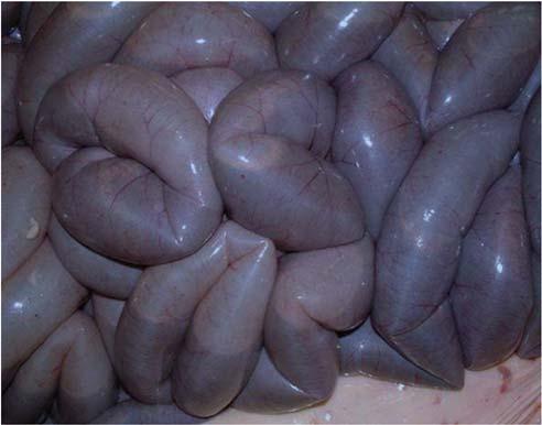
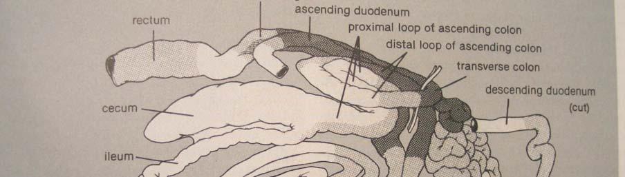
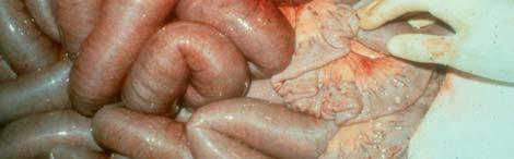
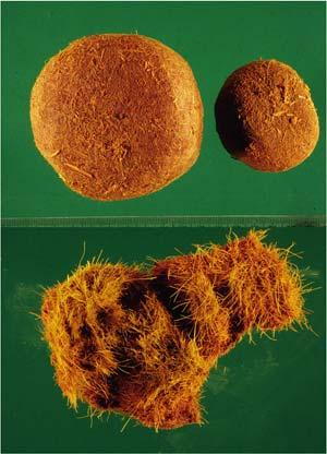
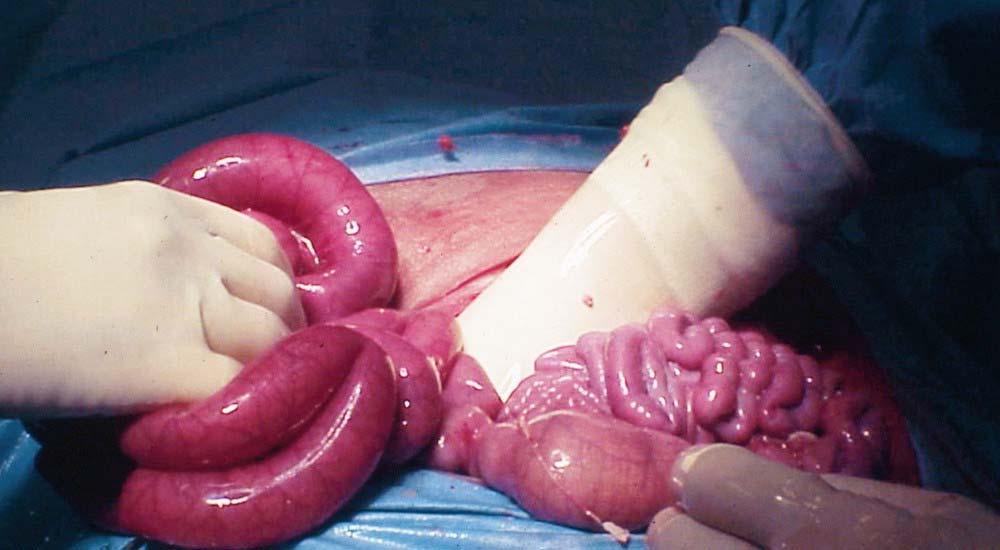
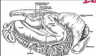
10 ANATOMY REVIEW FROM THE RIGHT SIDE Gastro-intestinal Structures: The intestinal tract is situated within the right half of the abdominal cavity on the right side of the rumen and is caudal to the liver, omasum and abomasum. Most of the intestinal tract lies within the supraomental recess, however, part of the jejunum, ileum and cecum extend caudal to the free border of the greater omentum. Initially (in second year clinical skills) it was advantageous to use the fresh viscera of calves to establish an understanding of the anatomical relationships simply because of the smaller size and lesser amount of mesenteric and omental fat. This is illustrated in Figure 4 and Figure 5 below. We will attempt to utilize the knowledge gained in that exercise to successfully complete bovine abdominal surgery in mature animals through the right paralumbar fossa. Figure 4: Anatomy of the superficial layer of the intestinal tract of a newborn calf as viewed from the right side. In the adult the umbilical vein is less prominent and the omasum is larger and not obscured by the liver. Figure 5: Anatomy of the second layer of the intestinal tract of a newborn calf. The abomasum, liver, and the cranial and descending parts of the duodenum have been removed.
11 Pylorus It lies at the level of the costrochondral junction of ribs 9 11 in the nonpregnant cow. Cranial Duodenum It extends craniodorsad from the pylorus with the proximal 8 10 cm being quite mobile and distal surface being tightly adhered to the visceral surface of the liver. The cranial duodenum then forms an S shaped curve near the openings of the common bile duct and the pancreatic ducts. Descending Duodenum This part of the duodenum is suspended dorsally by the mesoduodenum and it extends caudally along the right side of the dorsal abdomen. The superficial and deep walls of the greater omentum attach to the ventral surface of the descending duodenum. At the level of L5 L6 the descending duodenum forms the caudal flexure and turns cranial to form the ascending duodenum. Ascending Duodenum This part of the duodenum has a prominent attachment to the descending colon by way of the duodenocolic ligament. It passes cranially on the left side of the mesenteric root and finally turns to the right passing anterior to the mesenteric root and continues as the jejunum. Jejunum The jejunum is meters long in the adult cow and is suspended by the mesentery and arranged in a pattern of tight coils. The mesentery supporting the proximal and middle portions of the jejunum is short, however, the mesentery supplying the distal part of the jejunum and the proximal part of the ileum is longer and forms a narrow flange. This portion often protrudes beyond the caudal portion of the greater omentum and it is often referred to as the distal mesenteric flange or greater flange. Ileum The ileum is the terminal portion of the small intestine and it consists of a proximal convoluted portion that is continuous with the jejunum and a distal straight segment that attaches to the ventral surface of the cecum by the ileocecal fold and enters the cecocolic junction obliquely on its ventral surface. The division between the jejunum and the ileum is the part adjacent to the longest stretch of the mesenteric flange and this is also the site of termination of the cranial mesenteric artery. Cecum The cecum is a large mobile sac with its apex directed caudally (a significant difference to the horse). Cranial to the point of entry of the ileum, the cecum forms the cecocolic junction and continues cranially as the ascending colon. The cecum may lie within the supraomental recess but it often extends caudal to it or even may lie between the greater omentum and the right surface of the abdominal wall.

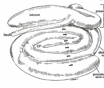
12 Ascending Colon The ascending colon has three parts: the proximal loop, the spiral loop, and the distal loop. a. Proximal Loop The proximal loop is continuous with the cecum and extends cranially to the level of L1 where it forms a loop turning first caudally and then again cranially but on the left side of the mesentery. The proximal loop then turns ventrally and continues as the spiral loop. b. Spiral Loop The spiral loop usually has 2 centripetal coils progressing inward and 2 centrifugal coils progressing outward. The central flexure is the midpoint between the inward (centripetal) and outward (centrifugal) coils and is usually directed cranially. The outside (centrifugal) portion of the spiral loop proceeds counter clockwise on the left side of the mesentery and transitions into the distal loop as it passes on the left side of the anterior mesenteric artery as shown in Figure 6 below. You will be able to identify the spiral loop of the ascending colon in mature cows but you will not be able to follow the centripetal and centrifugal coils as you did in newborn calves in second year clinical skills. c. Distal Loop The distal loop is continuous with the spiral loop and lies in close proximity to the proximal loop as it makes a tight turn passing first caudally and then cranially on the left side of the omentum. As the distal loop passes from the left side to the right side of the mesentery, it passes around the anterior mesenteric artery and becomes the transverse colon. This structure is very difficult to palpate. Figure 6: Anatomy of the deep layer of the intestinal tract of a newborn calf, as viewed from the right side. The central portion of the mesentery has been removed to expose the spiral loop of the ascending colon which has been laid out flat.
13 Transverse Colon The transverse colon is the short section of colon that passes from right to left around the cranial surface of the anterior mesenteric artery. This structure is also very difficult to palpate. Descending Colon The descending colon continues caudally along the dorsal surface of the abdomen where it is attached dorsally by the mesocolon. At the level of the duodenocolic ligament, the mesocolon is elongated thus providing that segment with considerable mobility facilitating such procedures as rectal palpation. Other Structures Palpable From the Right Side: Reproductive Tract, Bladder and Ureters The non pregnant uterus and ovaries can be palpated in the pelvic region caudal and dorsal to the dorsal sac of the rumen and the body of the uterus and part of the cervix can be evaluated. Ventral to the uterus, the bladder can be identified and the ureters can be traced from the bladder to the kidneys with the left ureter being much easier to trace. Liver and Gall Bladder Located cranially, up against the diaphragm Lightly palpate the edges; healthy cattle have relatively sharp liver edges, and if visible from the incision the liver is red In cases of fatty liver, the margins will be rounded, and appear yellow if visible The gall bladder is palpable as a flaccid fluid filled sack cranioventral to the incision, and just caudal to the liver. Anatomy Review from the Left Side A thorough systematic examination of the left side of the bovine abdomen should be performed before any specific manipulation is performed such as rumenotomy or abomasopexy. Unless a left displacement of the abomasum is present, the rumen will be visible following completion of the left flank laparotomy incision and the color of the serosa should be noted. If there is any evidence of peritonitis, the surgeon must be careful to explore the most contaminated area last, due to risk of spreading a localized peritonitis and causing an acute diffuse peritonitis. A basic approach to examine the left side of the abdominal cavity is identified below: Rumen The rumen is palpated to determine the nature of its contents, its state of fill, and any evidence of adhesions. Left Kidney The left kidney is pendulous and can be palpated straight in from the incision if the rumen is empty but with a full rumen the left kidney is located by passing a hand caudal to the dorsal sac of the rumen.
14 Spleen The spleen can be palpated in close proximity to the left, anterior aspect of the rumen and it can be evaluated for size or abscessation. Reticulum The reticulum is typically palpated last, particularly if hardware disease is suspected By passing a hand forward on the left side of the rumen, the lateral, ventral, and cranial aspects of the reticulum can be palpated including its proximity to the diaphragm which can be explored for adhesions or abscesses resulting from traumatic reticuloperitonitis. Reproductive Tract, Bladder and Ureters The non pregnant uterus and ovaries can be palpated in the pelvic region caudal and dorsal to the dorsal sac of the rumen and the body of the uterus and part of the cervix can be evaluated. Ventral to the uterus, the bladder can be identified and the ureters can be traced from the bladder to the kidneys with the left ureter being much easier to trace. Intestinal Tract Moving behind the rumen and over to the right side, the viscera can be palpated within the supraomental recess but cannot be exteriorized for specific examination or enterotomy. Surgical Exposure of the Intestinal Tract from the Right Side The extent to which the intestinal tract can be exteriorized through a right paralumbar fossa incision is limited primarily by the mesenteric and omental attachments of the intestine. Figure 7 below shows which portions of the bowel can be exteriorized and which can neither be exteriorized nor palpated. The pylorus can easily be identified by palpation as a firm structure deep to the costochondral junction of ribs 9 and 10. The pylorus with the distal 6 8 cm of abomasum and the proximal 6 8 cm of cranial duodenum can be exteriorized. The remainder of the cranial duodenum can be palpated in the region of the gall bladder, liver and pancreas but cannot be exteriorized. The descending duodenum is readily identified upon opening the abdominal cavity and it can be exteriorized. Except for the proximal 3 4 meters, the jejunoileum can be exteriorized. Exteriorization of more than a small portion (<1 ft) at any given time is not recommended, as the animal is likely to go down. The cecum and part of the proximal loop of the ascending colon can also be exteriorized. The ioeocecocolic junction and the cranial part of the proximal loop of the ascending colon are difficult to exteriorize. The spiral loop of the ascending colon lies on the left side of the mesentery and can be exposed by rotating the cecum and ileum craniad. Most of the distal loop of the ascending colon is inaccessible. The transverse colon and proximal portion of the descending colon lie next to the ascending duodenum and are inaccessible.

15 A portion of the descending colon just distal to the duodenocolic ligament has a relatively long mesocolon and can sometime be pulled to the incision site. The remainder of the descending colon and the intraperitoneal portion of the rectum can be easily palpated, but cannot be exteriorized. Figure 7: This diagram shows portions of the bowel can be exteriorized through a right paralumbar standing abdominal incision (white) those portions that can be palpated (grey) and those portions that can neither be exteriorized nor palpated (black). Technique for Surgical Exploration There is no single correct method for exploring the abdominal cavity and intestinal tract of the bovine, however, it is important for the surgeon to develop a consistent, systematic method of palpating and exteriorizing viscera. Without a systematic method, there will be a risk of failing to detect lesions and/or a protracted surgery time. It should be noted that abdominal exploration in the bovine is generally easier with the cow in a standing position. Suggested technique for abdominal exploration and palpation through the right paralumbar fossa: 1. Identify obvious abnormalities such as positive pressure in the abdominal cavity, abnormal peritoneal fluid, grossly distended viscera, or the presence of portions of bowel between the greater omentum and the right surface of the abdominal wall. 2. Palpate the viscera in the cranial abdomen including the omasum (should be firm but not hard like a basketball), abomasum (digesta is fluid and pylorus can be exteriorized), liver (identify enlargement, rounded edges or abscesses), gallbladder (often enlarged in anorectic cows), and finally the cranial and descending duodenum.
16 3. Palpate the kidneys and ureters. The right kidney is identified dorsal to the cranial portion of the descending duodenum and the left kidney is further caudal and identified by moving a hand around the caudal fold of the greater omentum and palpating medial to the greater omentum adjacent to the middle portion of the descending duodenum. The ureters extend caudad from the kidneys to the bladder. 4. Palpate the cecum and ascending colon. With a hand within the supraomental recess, identify and exteriorize the cecum and first portion of the proximal loop of the ascending colon. The free border of the greater omentum is reflected forward to expose the ileocecocolic junction. The cecum is then rotated craniad to expose the spiral loop of the ascending colon and allow palpation of part of the distal loop of the ascending colon. 5. Palpate the small intestine. Using the cecum as a landmark, the ileum can be palpated proximal to the cecum and to the distal flange of the jejunum which can be exteriorized and examined. After the distal flange has been replaced, then a hand can be positioned on the left side of the mesentery to palpate the small intestine to the duodenojejunal junction. This section of the small intestine need not be exteriorized unless obvious lesions are identified. 6. Palpate other structures in the dorsal and caudal parts of the abdomen. These structures include the descending colon and rectum, the uterus and ovaries, the urinary bladder, and the inguinal canals particularly in bulls. 7. Palpate the viscera in the left side of the abdomen. The surgeon should pass a hand behind the free border of the greater omenum, over the caudal portion of the rumen, and down the left side of the rumen to ensure there is not a left displacement of the abomasum. 8. Move the hand craniad and dorsad to palpate the spleen. 9. Move the hand back towards the incision, then reach cranioventrally along the right body wall, then slightly left cranial to palpate the reticulum last (identify adhesions, presence of a magnet or foreign bodies). Suggested technique for abdominal exploration and palpation through the left paralumbar fossa: 1. Identify obvious abnormalities such as positive pressure in the abdominal cavity, abnormal peritoneal fluid, adhesions between the rumen and/or reticulum and the abdominal wall, gas or fluid distension of the rumen or the presence of a left displaced abomasum. 2. Palpate the dorsal sac of the rumen, the spleen and the diaphragm. 3. Palpate caudally over the dorsal sac of the rumen and then dorsal to identify the left kidney as it hangs in a rather pendulous manner from dorsal aspect of the abdominal cavity. 4. Palpate caudal to the dorsal sac of the rumen, ventral to the left kidney and descending colon and medial to rumen to enter the supraomental recess and identify the jejunum, ileum and cecum. 5. Palpate further forward on the right side, behind the free border of the greater omentum, to identify the caudal lobe of the liver and the gall bladder. 6. Palpate the other structures in the caudal portion of the abdomen including the uterus and ovaries, the urinary bladder, and the internal inguinal rings. 7. Reach cranially along the left body wall to palpate the reticulum last (identify adhesions, presence of a magnet or foreign bodies).
 is the most common abnormality, and although right displacement of the abomasum (RDA) occurs, it is often just an abomasal dilatation.")
17 Abomasal Displacement and Volvulus Abomasal displacement and volvulus are predominantly conditions of the dairy cow although these conditions have been infrequently observed in bulls and beef cattle. Left displacement of the abomasum (LDA) is the most common abnormality, and although right displacement of the abomasum (RDA) occurs, it is often just an abomasal dilatation. The RDA may progress to a volvulus of the abomasum and this condition is an urgent life threatening condition. There is a general agreement that higher producing cows are at greater risk and there are further suggestions of a heritable component. The typical abomasal displacement occurs within six weeks of calving and the highest incidence appears to occur in the fall or winter when new forages are introduced and cows are more confined. Although the exact cause of displacement of the abomasum is not known, some predisposing factors that have been suggested include: 1. Excessive production of volatile fatty acids caused by modern diets consisting of high acid producing feed materials such as corn silage, haylage, and fermentable grains like barley and corn. 2. Gastrointestinal stasis caused by metabolic or infectious diseases such as hypocalcaemia, ketosis, retained placenta, metritis, mastitis, and indigestion. 3. The deeper body cavity that has been selected in the dairy cow may allow more room in the abdomen for movement of the abomasum. It is well known that excessive amounts of volatile fatty acids suppress abomasal wall tone and motility leaving the abomasum susceptible to dilatation. With parturition, there is an instantaneous loss of abdominal fill both with the empty uterus and the reduced dry matter intake at parturition. The metabolic and infectious diseases exacerbate the abomasal atony making it possible for the dilation prone abomasum to distend with gas and readily displace in the temporarily spacious abdomen. 1. Left Displacement of the Abomasum This condition is defined as a distension of the abomasum with gas or fluid so that the greater curvature passes under the rumen and is impounded between the rumen and the left abdominal wall as illustrated in Figure 8 below: Figure 8: Left displacement of the abomasum as viewed from the left side. In the pathogenesis of the left displacement of the abomasum (LDA), the accumulation of gas in the abomasum is one of the most important entities. The underlying hypothetical cause of this accumulation is a combination of the three pathways listed above. The gas that accumulates in the abomasum consists mainly of methane (70%), and carbon dioxide. In a normal functioning abomasum the gas production is equal to the clearance but when motility of the abomasum is inadequate, an accumulation of gas may
18 occur. The vagus nerve also plays a significant role in abomasal motility. Besides the effect of the vagal nerve, large amounts of volatile fatty acids (VFA) in the rumen and abomasum, endotoxins, metabolic alkalosis, and low blood calcium levels are mentioned as plausible causes for a decreased motility. The accumulation of gas in the abomasum results in a buoyancy of the abomasum and this buoyancy in combination with a characteristic reduction in the rumen size increases the likelihood of the abomasum slipping over to the left side. The major risk period is during the first month after calving with an increasing risk associated with increasing age. Cows that develop a LDA usually show a depressed feed intake prior to calving and often also have elevated NEFA levels. Feed form also appears to play a significant role in the pathogenesis with the quantity and quality of the roughage fed having a definite impact on the risk of an LDA developing. LDA is considered to be a metabolic disease and the three metabolic factors associated are: hypocalcaemia, metabolic alkalosis, and negative energy balance. The outflow of digesta is characteristically impeded and as a result cows fall into a severe negative energy balance and develop moderate to severe ketosis. In addition, the pyloric obstruction results in sequestering of chloride ions in the abomasum and a resulting hypochloremia and a metabolic alkalosis. The typical clinical signs include: A dramatic reduction in milk production (30 50%) and a decreased feed intake with a preference for forage rather than concentrates. The temperature, pulse, and respiratory rate are usually normal. The cow has a dull, depressed demeanor. There is usually mild dehydration with cows having a characteristic sunken eye. Rumen contractions are usually present and moderate in strength, however, with LDA the rumen becomes pushed medially and the stethoscope must be thrust deep into the paralumbar fossa to be able to hear the rumen contractions. Simultaneous auscultation and percussion will reveal an area of high-pitched tympanic resonance ( ping ) under the ribcage of the left side and may extend anywhere from the lower third of the abdomen in the 8 th intercostal space to the paralumbar fossa. The examiner should percuss along a line from the tuber coxae to the elbow and recognize that there can be much variation in both the location and the intensity of the ping. The gas distended abomasum can often be visualized and palpated behind the last rib in the left paralumbar fossa and it is not uncommon for the abomasum to extend into the paralumbar fossa and be visible as a half-moon distension posterior to the 13 th rib. On rare occasions, the abomasum can be palpated rectally but only if it is a relatively small cow and the abomasum is severely distended. The heart should always be auscultated since the electrolyte imbalance may precipitate an atrial fibrillation which will usually correct itself when the displacement is resolved. The typical clinical pathology seen with LDA include: Cows with LDA usually develop severe ketosis and the level of ketones in the blood, urine, and milk increase rapidly resulting in the reduced appetite and depression. In addition, LDA cows have a hypochloremic, hypokalemic metabolic alkalosis.
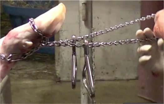
19 In a normal bovine the secretion of HCL into the abomasum is accompanied by return of HCO3 into the circulation. In the patent GI tract, the HCL is buffered and resorbed farther along the tract, thereby restoring the electrolyte and acid-base balance. With a DA and the associated obstructed outflow from the abomasum, there is an increase in plasma HCO3 and a net loss of Cl. The hypochloremia results from the abomasal sequestration of hydrochloric acid caused by the reduced emptying of the abomasum, and the subsequent reduction of chloride available to be absorbed by the small intestine. The metabolic alkalosis results from the increasing plasma HCO3 levels produced from the return of HCO3 into the circulation in response to the HCL secretion into the abomasum and the resulting Cl loss due to sequestration in the abomasum. Hypokalemia is caused by a number of factors and the most important may be the lack of ingestion of potassium rich feeds because of the anorexia. However, in a metabolic alkalosis state, there is also a net intracellular movement of K + in exchange for H + which moves extracellular to correct the alkalosis and low concentration of extracellular H +. The hypokalemia and dehydration result in the renal response of sodium retention but potassium depletion may have been so extensive that hydrogen ions are excreted in the urine instead of potassium, leading to the so-called paradoxic aciduria. Treatment for LDA involves returning the abomasum to its anatomic location and treating the coincident electrolyte, acid-base, dehydration, and energy deficit abnormalities. The treatment options for LDA are extensive and the common choices include: (a) Rolling Rolling the cow is one of the simplest methods to resolve the displacement, however, due to the very high recurrence rates, this procedure alone is used very infrequently and most techniques employ methodologies intended to secure the abomasum in its normal position. The procedure involves lying the cow down on the right side, rolling her up into dorsal recumbency for a short period of time (2-5 minutes), and then rolling her down on her left side before allowing her to stand. (b) Toggle Suture The toggle suture is a modification of the rolling procedure and it has the advantage of securing the abomasum in its near normal position. The cow is positioned in dorsal recumbency using the same technique as for the rolling procedure. The operational site is identified about 4-7 inches (10-15cm) behind the end of the xiphoid and prepared according to the preference of the surgeon. The abomasum may be ausculted in this area with a stethoscope and the cow should be positioned with the loudest 'ping' in the center of the operational site previously described. The abomasum will assume its normal anatomical position under most circumstances, except when concurrent adhesions or peritonitis might prevent normal movement to its correct anatomical location. It is useful to have an assistant place the pressure of a knee, hand, or foot, on the lower left abdominal quadrant ahead of the udder, to ensure that the abomasum is in proper position and as close to the ventral abdominal wall possible. The trocarcannula, with the push rod inside, is placed 4-7 inches (10-15cm) behind the end of the xiphoid and 2-3 inches (5-7cm) to the right of the midline. The abdominal wall and the abomasum are perforated with a
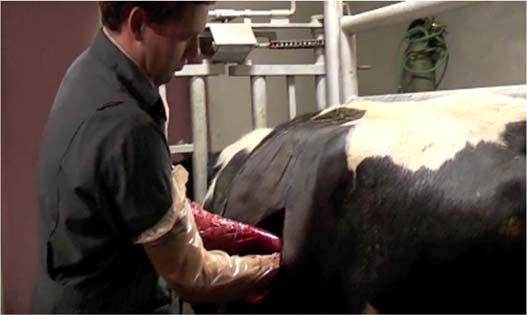
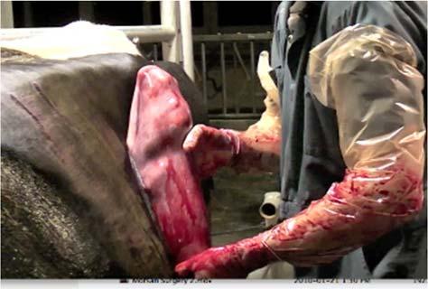
20 swift downward push of the trocar being careful to avoid large milk veins in the area. The distinct odor of abomasal gas may be identified at this time, and if desired, a small amount of fluid can be extracted via small diameter plastic tubing and checked for low ph (2-4). After removing the plastic handle and push rod from the trocar needle, place the first toggle suture into the open lumen of the needle. Use the push rod to move the toggle completely through the length of the needle so that it will turn perpendicular to the long axis of the needle once in the abomasal lumen. Remove the cannula and pull snugly on the suture, so that it lies firmly against the abdominal wall and secure with a hemostat. Place the second toggle 2-3 inches (4-7cm) anterior to the first toggle suture, forward toward the xiphoid in exactly the same manner described for the first suture. Before removing the trocar needle from the second suture site, place pressure on the external abdominal wall to force as much free gas from the abomasum as possible. The two toggle suture ends are then tied together, using a number of square knots to suit the surgeon's preference. It is suggested that a vertical distance of 2-3 inches be left between the abdominal wall and the knots. It is desirable to place a gauze pad or stint between the suture and the body wall to prevent it from becoming embedded in the abdominal wall. The cow is then rolled in a clockwise direction on to her left side and allowed to stand. Two major advantages of the procedure are its minimal invasiveness and its low cost. (c) Right-Flank Omentopexy The right-flank omentopexy is one of the most popular surgical repairs for LDA, however, it should be pointed out that there are also three other surgical LDA repair techniques that are used with relative frequency. The main advantage of the right-flank omentopexy is that it is performed in a chute with the animal in a standing position and it is often done by the veterinarian alone without any assistance for either restraint or surgical procedure. After local regional anesthesia and a surgical prep, a inch incision is made in the center of the right paralumbar fossa and a sterile gowned and gloved hand is introduced into the abdominal cavity. The hand is passed behind the free border of the greater omentum, caudal-dorsal to the entrance to the supraomental recess, ventral to the left kidney, and dorsal to the caudal dorsal sac of the rumen and then forward to palpate the abomasum which can be found trapped between the rumen medially and the left body wall laterally. It is desirable to deflate the abomasum with a vacuum apparatus and sterile vacuum line and needle. The contents of an abomasum that is displaced to the left is usually gas with only minimal fluid so it is possible to achieve near complete deflation. If the surgeon has long arms, a hand can be placed over the abomasum and the abomasum can be swept under the rumen and back to its normal position. If the cow is extremely large or the veterinarian has short arms, the deflated abomasum can be replaced to its normal position by gentle traction on the greater omentum. The serosal surface of the pyloric portion of the abomasum must be identified at the point where the greater and lesser omentum become continuous (this area is often referred to as the shark s nose). Identification at this point confirms the correct location to pexy the greater omentum to the body wall. Most surgical procedures include the greater omentum with the closure of the tranversus abdominus and peritoneum but there is much variation in the way the stay sutures are placed in the greater omentum prior to the closure. There is general consensus that the sutures should be placed 1-1 ½ inches from where the greater omentum reflects onto the serosa of the abomasum, but the type stay suture used by various surgeons may differ. Some surgeons prefer to place the stay sutures through both the cranial and caudal edges of the incision while others suture only through the cranial edge. In addition some surgeons place the stay sutures through only the transversus abdominus while others prefer to include the internal and external oblique as well. If surgery is performed early in the progression of the condition, excellent results can be expected. It is important to recognize that with the true omentopexy the sutures enter only the greater omentum and not the abomasum. Some surgeons have modified this technique to include the serosa of the abomasum rather than the greater omentum and if this procedure is done it would be referred to as a right-flank abomasopexy.
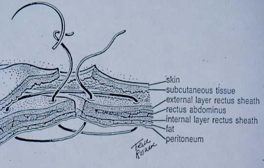
21 (d) Left-Flank Abomasopexy The left-flank abomasopexy is another procedure commonly used to repair LDA. The surgical preparation and entry of the abdominal cavity is the same as above except that the incision is made in the left paralumbar fossa. The gas filled greater curvature of the abomasum can easily be identified cranial-ventral to the incision. With the abomasum still distended with gas, several seromuscular stitches of non-absorbable suture material are placed along about a 5 cm distance in the greater curvature using a simple continuous suture pattern and having both ends of the suture material kept long. A 3 5 inch straight or serpentine needle is then attached to each end of the suture, the gas is removed from the abomasum with a needle and vacuum tube, and the needles and suture material are driven through the body wall at a point just to the right of the midline in a location similar to the needle placement for the toggle suture technique. An assistant grasps the needles and places traction on each suture while the surgeon pushes the abomasum ventrally to rest in its normal position in contact with the ventral body wall. Care must be taken not to twist the sutures so that the greater curvature of the abomasum lies smoothly along the ventral body wall to the right of the midline. The assistant then ties the sutures over a plastic or gauze stint leaving a similar amount of slack to that used with the toggle suture technique. The abdominal wall is then closed using the standard closure. (e) Paramedian Abomasopexy Another common surgical technique used in the surgical repair of LDA is the paramedian abomasopexy. This was one of the first described LDA surgical repair techniques and it is still the procedure of choice for many veterinarians. The cow is first positioned in dorsal recumbency and surgery is performed either under local or general anesthesia. The surgical sire is identified as being to the right of the midline, caudal to the xyphoid process, cranial to the umbilicus, and medial to the right cranial superficial epigastric vein. This is the same location as used for suture placement for the toggle technique. A 6-8 inch incision is made through the abdominal wall parallel to the midline and the greater curvature of the abomasum is usually visible within the surgery site if the cow is positioned correctly. Once the pyloris and fundi part of the abomasum have been identified, the location for the pexy is isolated mid-way along the greater curvature between the attachment of the greater and lesser omentum and the serosa is gently grasped between the thumb and forefinger to create a separation between serosa and mucosa. This allows the abomasum to be fixed to the abdominal wall with a nonperforating seromuscular suture. Most surgeons prefer to use a non-absorbable suture material to create a mild inflammatory reaction to facilitate a substantial adhesion. The first suture line (usually #1 nonabsorbable) is placed in either a simple continuous or simple interrupted pattern and passes through the peritoneum, internal sheath of the rectus abdominus, usually through some of the rectus abdominus muscle, and the seromuscular portion of the abomasum. It should be noted that it does not include the external sheath of the rectus abdominus as this is the main strength holding layer and is sutured separately using absorbable suture material such as #2 PDS in a simple continuous or simple interrupted pattern. The subcutaneous layer is usually also closed with absorbable suture material in a simple continuous pattern to eliminate as much dead space as possible. Finally the skin is closed with #3 nonabsorbable suture material in whatever pattern the surgeon prefers (f) Laparoscopic Abomasopexy The laparoscopic technique is the most recent procedure described for LDA repair and it has been described as both a one-step and a two-step procedure. The two-step procedure involves laparoscopic manipulations both with the cow standing and with her in dorsal recumbency and the complexity of the procedure has limited its use in practice. The one-step procedure is performed with the cow in dorsal recumbency using three separate laparoscopic sites. Although a significant investment in equipment is a factor limiting the use of the procedure, it is most likely that this procedure or a similar modification will be the most common laparoscopic procedure used in practice.

22 2. Right Displacement of the Abomasum This condition is defined as a distension of the abomasum with gas and/or fluid so that the greater curvature is displaced dorsally with no complete obstruction of the cranial duodenum. An RDA is similar to an LDA except that the distended abomasum rises against the right surface of the abdominal wall, rather than the left surface. Unlike the abomasal volvulus, there is not complete obstruction to passage of abomasal contents distally into the duodenum however an RDA can progress to an abomasal volvulus without apparent warning. Similar to an LDA, an RDA results from increased gas accumulation in the abomasum and the same resulting increased buoyancy. With the increased buoyancy, there are three separate possible rotations that the abomasum can make on the right side: 1. Gas accumulates in the fundus causing the cranial part of the abomasum to move dorsally adjacent to the right abdominal wall (counter-clockwise rotation as viewed from the right side). 2. Gas accumulates first in the pyloric part of the abomasum and causes a clockwise rotation of the abomasum as viewed from the right side. 3. Gas accumulates first in the body of the abomasum and causes dorsal migration of the greater curvature along the right surface of the abdominal wall. This results in a counter-clockwise rotation as viewed from behind the cow. This is the most common RDA rotation and is shown in Figure 9 below: Figure 9: Right displacement of the abomasum as viewed from the right side. All of the predisposing factors, clinical signs, and clinical pathology of an RDA are similar to that already described for an LDA. The major difference is that the gas filled abomasum creates a distension on the right side and this may also be visualized behind the 13 th rib in the right paralumbar fossa. The tympanic resonance or ping described for the LDA can be identified in the same location between the 8 th and 13 th ribs but on the right side. There is also a major difference in urgency with an RDA as compared to an LDA because it can easily progress to an abomasal volvulus which is a serious, life threatening condition. Treatment for RDA involves returning the abomasum to its anatomic location and treating the coincident electrolyte, acid-base, dehydration, and energy deficit abnormalities. The treatment options for RDA are more limited than for LDA because of the need to visualize the abomasum.

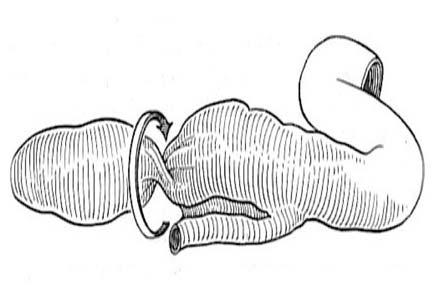
23 Rolling the cow is not an option because of the high risk of creating an abomasal volvulus. Therefore the correction of RDA must be done using a right-flank omentopexy, a ventral paramedian abomasopexy, or a ventral laparoscopic abomasopexy. All three of these procedures facilitate visualization of the abomasum and ensure that the displacement is corrected and not allowed to progress to an abomasal volvulus. The prognosis for full recovery from RDA should be as good as LDA if it is identified early enough. 3. Abomasal Volvulus This condition is characterized by its serious nature and its sudden onset. A right and dorsally displaced abomasum easily performs an additional rotation around a vertical axis. In almost all cases this rotation is performed to the left (counter-clockwise, seen from rear) and the greater curvature of the abomasum usually faces dorsally and caudally. The proximal part of the duodenum extends ventrally from the pylorus, around the neck of the omasum, and then medially in a cleft formed between the rumen and the omasum. As long as the abomasal twist does not exceed 180 degrees, functional disturbances are caused mainly by a reduced abomasal outflow, resulting in dehydration and in disturbances of blood acid-basebalance (hypochloremic metabolic alkalosis). These cases have a very good prognosis when surgery is performed immediately. When the twist of the abomasum exceeds 180 degrees (abomasal volvulus), the twine gets more contracted, strangulating the abomasal nerves and blood vessels. The distended omasum is often thrust into the lesser omentum and commonly tears the lesser omentum. It should be understood that an abomasal volvulus can result from any one of three rotation processes: 1. The counter-clockwise rotation of the fundic part of the abomasum as viewed from the right side continues to a 180 to 270 rotation. 2. After the first 180 clockwise rotation of the pyloric part of the abomasum as viewed from the right side: The abomasum, omasum, and proximal part of the duodenum then rotate 360 in a counterclockwise direction as viewed from behind the cow. 3. After the first counter-clockwise rotation as viewed from behind the cow (the most common RDA) the abomasum and duodenum then rotate in a counter-clockwise direction as viewed from the right side. This is the most common rotation sequence resulting in an abomasal volvulus and the progression is demonstrated in the four diagrams in Figure 10 below:

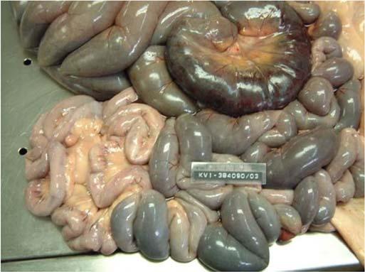
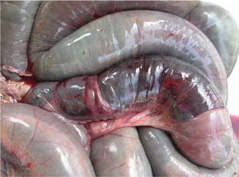
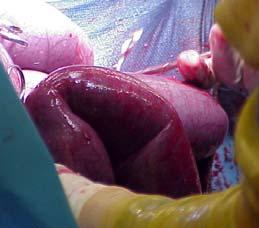
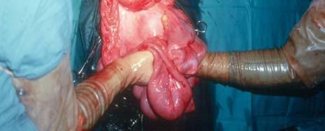
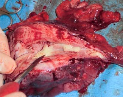
24 Figure 10: Development of an abomasal volvulus as viewed from the right side. Cows with abomasal volvulus (AV) will not survive without surgery. It is a serious, life-threatening condition in dairy cattle and is characterized by severe dehydration, hypochloremic, hypokalemic alkalosis, and complete mechanical obstruction of the abomasal outflow. The typical clinical signs include: Abdominal distension is observed bilaterally but it is the right side that has the marked distension and the distended abomasum often extends caudally behind the last rib forming a half moon distension that is visible and palpable in the right paralumbar fossa. Simultaneous auscultation and percussion reveals a large area of high-pitched tympanic resonance ( ping ) under the right rib cage from the 8 th to the 13 th rib. AV differs from a simple DA in that the distension is usually caused by more fluid that gas and as a result, combined succussion and auscultation will confirm a large fluid accumulation within the AV on the right side. The systemic effects of the gastrointestinal obstruction from an AV progress much more rapidly and to a much more serious degree than with either LDA or RDA. Much more severe depression, dehydration, & anxious demeanor are observed in cows with an AV than with a simple DA. The heart rate is often over 100, the extremities feel cold, and the rumen contractions are reduced or absent. The clinical pathology that characterizes abomasal volvulus is similar but of much greater severity than has been identified with LDA or RDA. The dehydration is much more severe and the syndrome begins with the same hypochloremic, hypokalemic metabolic alkalosis. However, because of the complete obstruction, the HCl that normally leaves the abomasum and is sequestered in the abomasum becomes regurgitated into the omasum and rumen resulting in a large increase in rumen chlorides. As the condition progresses, dehydration becomes more marked and metabolic acidosis may eventually supercede the alkalosis terminally. The most desirable surgical approach is the right-flank omentopexy. When the abdominal cavity is opened, one usually encounters the greater curvature of the abomasum facing dorsally and caudally.


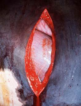
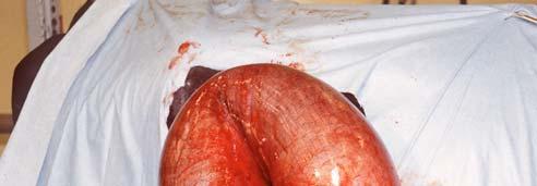
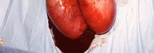
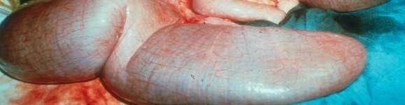
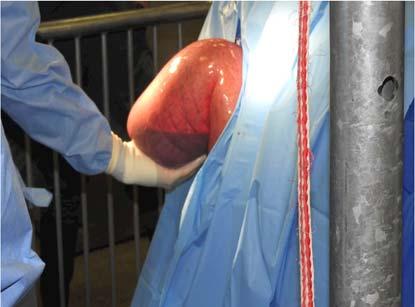
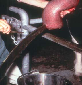
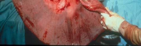
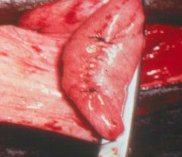
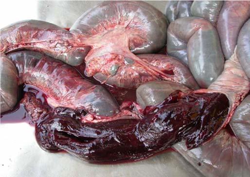
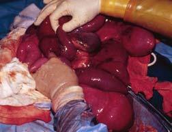
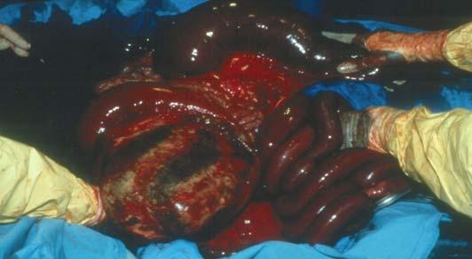
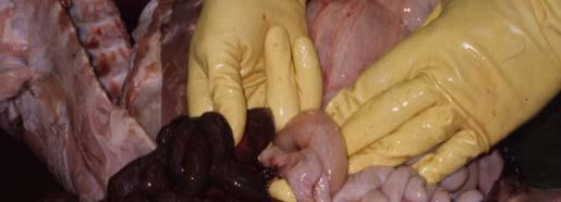
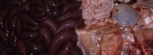
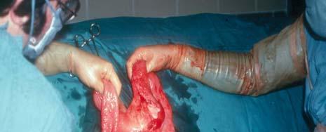
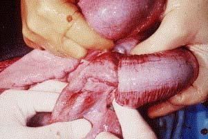
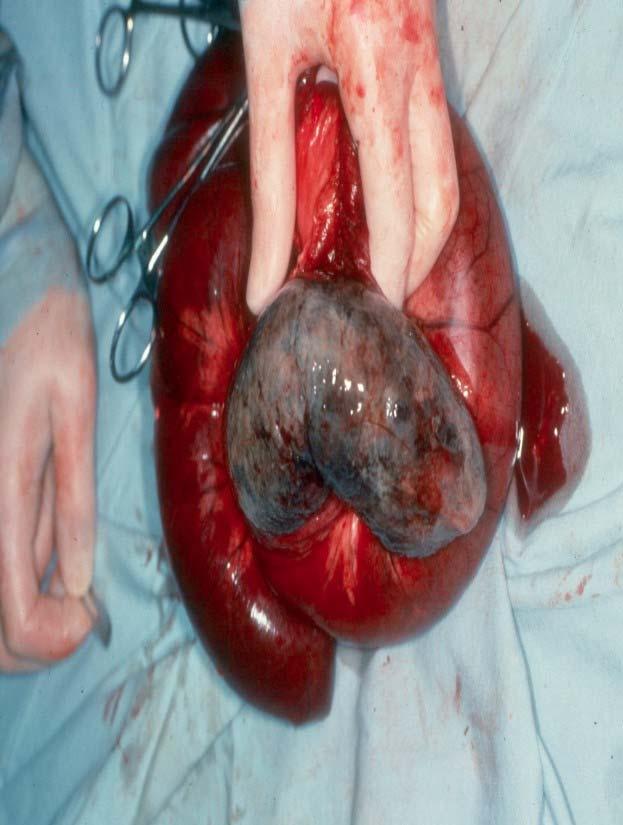
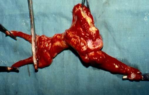
25 Severe distension of the abomasum occurs and the contents is usually primarily fluid although a significant component of gas may also be present. This is different from an LDA or an RDA where the distension is almost totally gas. Decompression of both gas and fluid is usually necessary before an abomasal volvulus can be corrected. Maintaining sterility and removing the fluid from a severely compromised abomasum is one of the major surgical challenges. Following deflation, the abomasum is pushed craniolaterally and cranioventrally to release the duodenum from the cleft between the rumen and the omasum. Often the omasum may have to be lifted to free the trapped duodenum. To ensure complete correction, the pylorus is held stationary with one hand while the other hand is used to palpate ventrally along the greater curvature of the abomasum until the reticulum is felt. The omasum is then palpated to ensure it is positioned correctly against the right abdominal surface, dorsolateral to the body of the abomasum. In an abomasal volvulus, the omasum is often thrust into the lesser curvature of the abomasum and often tears the fragile lesser omentum. While the object of the correction is returning the abomasum to its anatomic location and treating the coincident electrolyte, acid-base, dehydration, and energy deficit abnormalities, those cows affected with AV for longer than 24 hours have a poor chance of survival. If the greater curvature of the abomasum is dark purple or even black, it is an indication that there has been severe circulatory compromise and it may be impossible for it to return to normal function as seen in Figure 11 below: Figure 11: Severely Distended Abomasum - Abomasal Volvulus The abomasum may be distended with as much as 10 gallons of fluid and correction of the torsion without at least partially emptying the abomasum is impossible. As a result the use of a vacuum tube and purse-string suture is useful to drain the abomasum as seen in Figure 12 below: Figure 12: Fluid Drainage - Abomasal Volvulus

26 4. Differential Diagnosis Cecum Ascending Colon Descending Duodenum Abomasum Outlines of the approximate areas of ping for right side GI disorders

27 5. Cecal Dilatation/Torsion


28 6. Cranial Duodenal Sigmoid Flexure Volvulus 7. Hemorrhagic Bowel Syndrome
29
30 8. Intestinal Volvulus
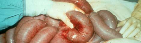
31 9. Intestinal Intussusception 10. Intraluminal Obstructions


32 11. Abomasal Ulcers in Calves 12. Abomasal Torsion in Calves 13. Mesenteric Torsion in Calves 14. Atresia Coli in Calves


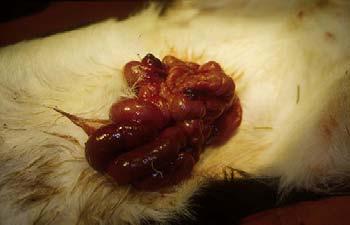
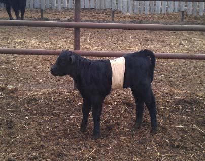
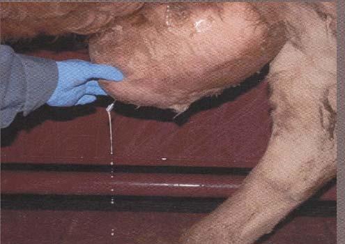
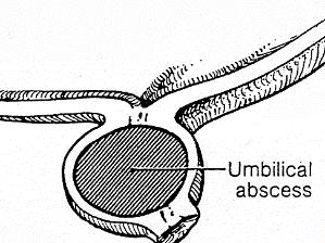
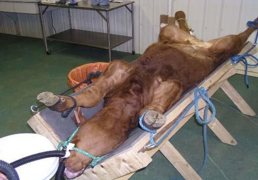
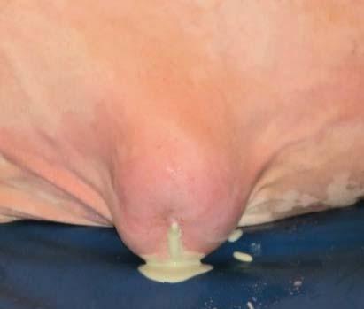
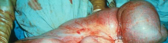
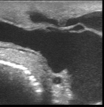
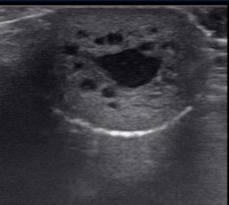
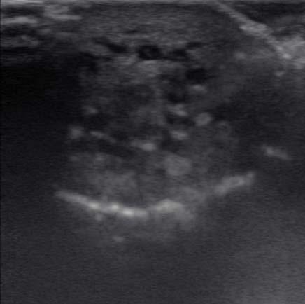
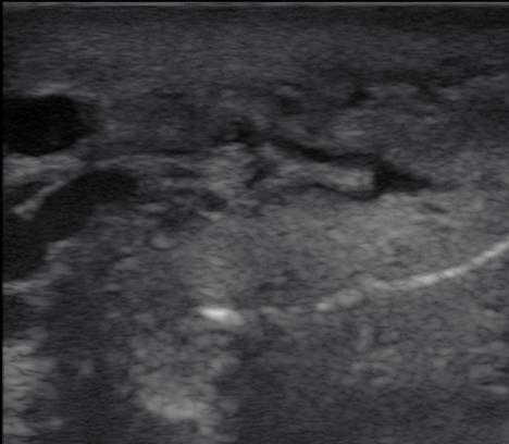
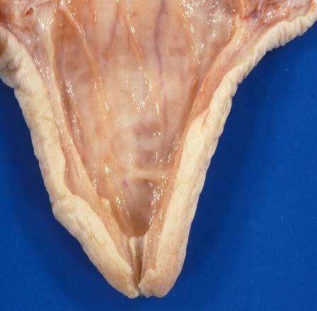
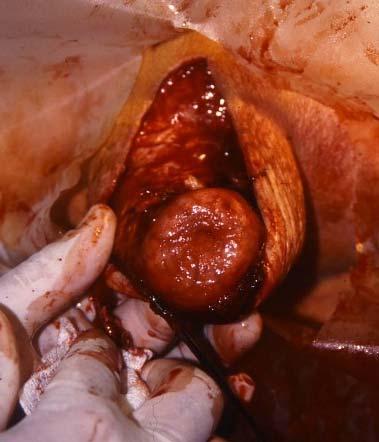
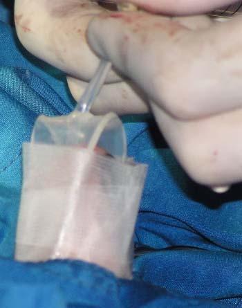
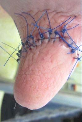
33 15. Umbilical Masses in Calves


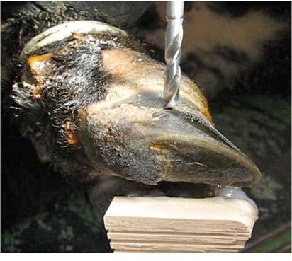
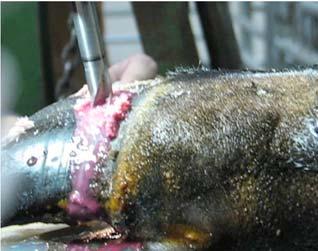
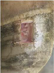
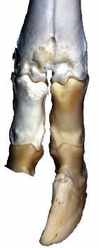
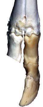
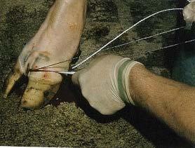
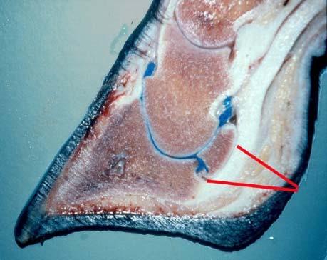
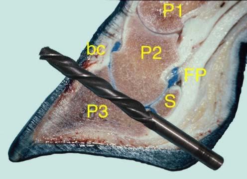
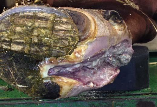
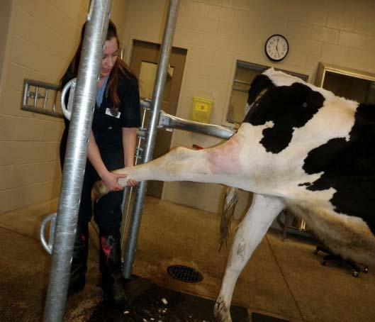
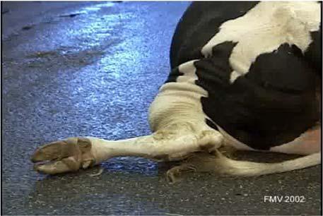
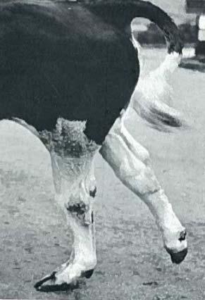
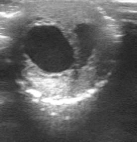
34 16. Septic Arthritis of the Distal Interphalangeal Joint 17. Upper Leg Injuries

35 18. Teat Surgery

36 19. C-Section Tips and Discussion
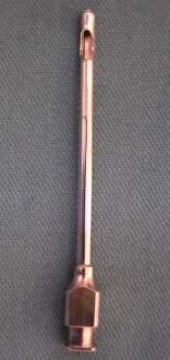
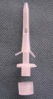
Title of Procedure: Rumen Cannulation (Sheep, Goats & Cattle) (L12)
 (L12)") THE UNIVERSITY OF NEW ENGLAND ANIMAL ETHICS COMMITTEE (AEC) STANDARD OPERATING PROCEDURES FORM (For Domestic Fowl, Native Fauna/Wildlife, Domestic Livestock & Laboratory Animals) Title of Procedure: Rumen
THE UNIVERSITY OF NEW ENGLAND ANIMAL ETHICS COMMITTEE (AEC) STANDARD OPERATING PROCEDURES FORM (For Domestic Fowl, Native Fauna/Wildlife, Domestic Livestock & Laboratory Animals) Title of Procedure: Rumen
The UCD community has made this article openly available. Please share how this access benefits you. Your story matters!
 Provided by the author(s) and University College Dublin Library in accordance with publisher policies., Please cite the published version when available. Title The use of epidurals in cattle Authors(s)
Gastric Dilatation-Volvulus
 Gastric Dilatation-Volvulus The term "ACVS Diplomate" refers to a veterinarian who has been board certified in veterinary surgery. Only veterinarians who have successfully completed the certification requirements
Perioperative Care of Swine
 Swine are widely used in protocols that involve anesthesia and invasive surgical procedures. In order to ensure proper recovery of animals, preoperative, intraoperative and postoperative techniques specific
METABOLIC DISEASES OF DAIRY CATTLE
 METABOLIC DISEASES OF DAIRY CATTLE J. K. Shearer DVM, MS. Professor and Dairy Extension Veterinarian Department of Large Animal Clinical Sciences College of Veterinary Medicine University of Florida Gainesville,
Digestive System Dissection
 Digestive System Dissection THE TERMS YOU NEED FOR THE PRACTICAL ARE IN THIS DISSECTION GUIDE. Instructions: Do one of the 2 respiratory dissections, and then the digestive dissection. Wordlist for cat
UNDERSTANDING COLIC: DON T GET IT TWISTED
 UNDERSTANDING COLIC: DON T GET IT TWISTED Today s Topics: What is colic? Anatomy review How to identify colic What to do when you suspect colic What to expect during a colic visit from your veterinarian
Some important information about the fetus and the newborn puppy
 Some important information about the fetus and the newborn puppy Dr. Harmon Rogers Veterinary Teaching Hospital Washington State University Here are a few interesting medical details about fetuses and
The Royal College of Veterinary Surgeons DIPLOMA IN EQUINE SOFT TISSUE SURGERY PAPER I. (Basic Sciences) Tuesday 2 May 1995
 Tuesday 2 May 1995") The Royal College of Veterinary Surgeons PAPER I (Basic Sciences) Tuesday 2 May 1995 10.00 a.m. to 1.00 p.m. (3 hours) SECTION A Two long answer questions of which a candidate must choose ONE question
The Royal College of Veterinary Surgeons PAPER I (Basic Sciences) Tuesday 2 May 1995 10.00 a.m. to 1.00 p.m. (3 hours) SECTION A Two long answer questions of which a candidate must choose ONE question
Animal Studies Committee Policy Rodent Survival Surgery
 Animal Studies Committee Policy Rodent Survival Surgery ASC Policy: To optimize animal health and well-being, survival surgery in rodents must be performed using sterile instruments, surgical gloves, masks
Frog Dissection Information Manuel
 Frog Dissection Information Manuel Anatomical Terms: Used to explain directions and orientation of a organism Directions or Positions: Anterior (cranial)- toward the head Posterior (caudal)- towards the
Sites of IM injections : 1. Ventrogluteal site: site is in the gluteus medius muscle, which lies over the gluteus minimus. 2. Vastus lateralis site:
 Sites of IM injections : 1. Ventrogluteal site: site is in the gluteus medius muscle, which lies over the gluteus minimus. 2. Vastus lateralis site: is the thick and well developed in both adults and children.
Dexmedetomidine and its Injectable Anesthetic-Pain Management Combinations
 Back to Anesthesia/Pain Management Back to Table of Contents Front Page : Library : ACVC 2009 : Anesthesia/Pain Management : Dexmedetomidine Dexmedetomidine and its Injectable Anesthetic-Pain Management
Alimentary System 解剖學科徐淑媛
 Alimentary System 解剖學科徐淑媛 本堂重點 1. Structures derived from primitive guts 2. Specific events Alimentary System endoderm of primordial gut epithelium & glands of digestive tract ectoderm of stomodeum epithelium
RESEARCH AND TEACHING SURGERY GUIDELINES FOR MSU-OWNED ANIMALS
 RESEARCH AND TEACHING SURGERY GUIDELINES FOR MSU-OWNED ANIMALS I. Purpose/Scope These guidelines apply to all surgical procedures performed on animals at Mississippi State University in which the animals
This SOP presents commonly used anesthetic regimes in rabbits.
 Comparative Medicine SOP #: 103. 01 Page: 1 of 7 Rabbit Anaesthesia The intent of this Standard Operating Procedure (SOP) is to describe commonly used methods to anesthetize rabbits at Comparative Medicine
DISSOCIATIVE ANESTHESIA
 DISSOCIATIVE ANESTHESIA Adarsh Kumar Dissociative anesthesia implies dissociation from the surrounding with only superficial sleep mediated by interruption of neuronal transmission from unconscious to
SUMMARY OF PRODUCT CHARACTERISTICS
 SUMMARY OF PRODUCT CHARACTERISTICS 1. NAME OF THE VETERINARY MEDICINAL PRODUCT Xylacare 2% w/v Solution for Injection 2. QUALITATIVE AND QUANTITATIVE COMPOSITION Active substances Qualitative composition
DREXEL UNIVERSITY COLLEGE OF MEDICINE ANIMAL CARE AND USE COMMITTEE POLICY FOR PREOPERATIVE AND POSTOPERATIVE CARE FOR NON-RODENT MAMMALS
 DREXEL UNIVERSITY COLLEGE OF MEDICINE ANIMAL CARE AND USE COMMITTEE POLICY FOR PREOPERATIVE AND POSTOPERATIVE CARE FOR NON-RODENT MAMMALS OBJECTIVE: This policy is to ensure that appropriate provisions
Pain Management. Anesthesia Asepsis Analgesia Euthanasia
 Pain Management Anesthesia Asepsis Analgesia Euthanasia What is Pain? Normal Behavior Pain Analgesics Altered Behavior Do Animals Feel Pain? Behavioral responses to stimuli Prey species Photoperiod Behavioral
Australian and New Zealand College of Veterinary Scientists. Membership Examination. Small Animal Surgery Paper 1
 Australian and New Zealand College of Veterinary Scientists Membership Examination June 2018 Small Animal Surgery Paper 1 Perusal time: Fifteen (15) minutes Time allowed: Two (2) hours after perusal Answer
GUIDELINES FOR ANESTHESIA AND FORMULARIES
 GUIDELINES FOR ANESTHESIA AND FORMULARIES Anesthesia is the act of rendering the animal senseless to pain or discomfort and is required for surgical and other procedures. Criteria for choosing an anesthetic
Equine Emergencies. Identification and What to do Until the Vet Arrives Kathryn Krista, DVM, MS
 Equine Emergencies Identification and What to do Until the Vet Arrives Kathryn Krista, DVM, MS Common Equine Emergencies Cellulitis/lymphangitis Choke (esophageal obstruction) Colic Eye abnormalities Fever
Biology of the Pig. Swine. Farm or miniature? Breeds Body Weights
 Swine Pigs like humans? Obvious similarities untrue: Biology of the Pig Breeds Body Weights Yucatan mini Yucatan micro Hanford Göttingen Minisib Standard Housing Groups, visual and snout contact. Hierarchy
Department of Laboratory Animal Resources. Veterinary Recommendations for Anesthesia and Analgesia
 Department of Laboratory Animal Resources Guideline Veterinary Recommendations for Anesthesia and Analgesia A. PRINCIPLES OF ANESTHESIA AND ANALGESIA 1. The proper anesthetic and analgesic agents must
POLICY ON ASEPTIC RECOVERY SURGERY ON USDA REGULATED NONRODENT SPECIES Adopted by the University Committee on Animal Resources October 15, 2014
 POLICY ON ASEPTIC RECOVERY SURGERY ON USDA REGULATED NONRODENT SPECIES Adopted by the University Committee on Animal Resources October 15, 2014 The U.S.D.A Animal Welfare Act (9 CFR) requires use of aseptic
Bovine Fracture Repair Sabrina Brounts, DVM, MS, DACVS, DECVS, DACVSMR University of Wisconsin Madison, WI
 Bovine Fracture Repair Ruminants are large animals with similar bone structure to equine species. Long bone fractures are common in these species and primary cause of fractures in these species is thought
Recommended Resources: The following resources may be useful in teaching this
 Unit B: Anatomy and Physiology of Poultry Lesson1: Internal Anatomy of Poultry Student Learning Objectives: Instruction in this lesson should result in students achieving the following objectives: 1. Identify
Day 90 Labelling, PL LABELLING AND PACKAGE LEAFLET
 LABELLING AND PACKAGE LEAFLET A. LABELLING PARTICULARS TO APPEAR ON THE OUTER PACKAGE : Carton 1. NAME OF THE VETERINARY MEDICINAL PRODUCT Alvegesic vet. 10 mg/ml Solution for injection for Horses, Dogs
Mouse Formulary. The maximum recommended volume of a drug given depends on the route of administration (Formulary for Laboratory Animals, 3 rd ed.
 Mouse Formulary The maximum recommended volume of a drug given depends on the route of administration (Formulary for Laboratory Animals, 3 rd ed.): Intraperitoneal (IP) doses should not exceed 80 ml/kg
Finding and treating sick animals early is the key to maintaining a safe, nutritious food supply. On dairies, this begins with a basic physical exam
 Finding and treating sick animals early is the key to maintaining a safe, nutritious food supply. On dairies, this begins with a basic physical exam of the cow. 1 Frequently a staff member, trained by
SURGICAL (SURVIVAL) OOCYTE COLLECTION FROM XENOUS LAEVIS
 OOCYTE COLLECTION FROM XENOUS LAEVIS") UBC Animal Care Guidelines SOP: ACC 2013 01 Surgical Oocyte Collection from Xenopus Laevis Submitted by: Shelly McErlane Last Date Revised: Date Approved: January 28, 2013 SURGICAL (SURVIVAL) OOCYTE COLLECTION
UBC Animal Care Guidelines SOP: ACC 2013 01 Surgical Oocyte Collection from Xenopus Laevis Submitted by: Shelly McErlane Last Date Revised: Date Approved: January 28, 2013 SURGICAL (SURVIVAL) OOCYTE COLLECTION
MEET THE DOCTORS Meet the Doctors - co-inventors of the Grymer / Sterner Toggles Suture Method of LDA Repair Jorgen Grymer, DVM,PhD Galten, Denmark Dr
 HISTORY History of The Grymer / Sterner Toggles Suture The Grymer/Sterner toggle suture LDA repair method was introduced in 1982 to bridge the gap between the blind stitch method (early '70's) and the
UNIVERSITY OF PITTSBURGH Institutional Animal Care and Use Committee
 UNIVERSITY OF PITTSBURGH Institutional Animal Care and Use Committee Policy: Surgical Guidelines EFFECTIVE ISSUE DATE: 2/21/2005 REVISION DATE(s): 2/14/15; 3/19/2018 SCOPE To describe guidelines and considerations
SUMMARY OF PRODUCT CHARACTERISTICS. 1. NAME OF THE VETERINARY MEDICINAL PRODUCT Emdocam 20 mg/ml solution for injection for cattle, pigs and horses
 SUMMARY OF PRODUCT CHARACTERISTICS 1. NAME OF THE VETERINARY MEDICINAL PRODUCT Emdocam 20 mg/ml solution for injection for cattle, pigs and horses 2. QUALITATIVE AND QUANTITATIVE COMPOSITION One ml contains:
Anesthesia Check-off Form
 Anesthesia Check-off Form 5231 SW 91st Drive Gainesville, FL 32608 (352) 377-6003 The doctors and staff at Haile Plantation Animal Clinic would like to offer the most advanced medical care and services
August 16, Implementing High Quality, High Volume Spay/Neuter: Challenges & Solutions
 August 16, 2014 Implementing High Quality, High Volume Spay/Neuter: Challenges & Solutions Carolyn Brown, DVM Spay/Neuter Operations carolyn.brown@aspca.org Kathleen Makolinski, DVM Shelter Medicine Service
STANDARD OPERATING PROCEDURE #110 MOUSE ANESTHESIA
 STANDARD OPERATING PROCEDURE #110 MOUSE ANESTHESIA 1. PURPOSE This Standard Operating Procedure (SOP) describes methods for anesthetizing mice. 2. RESPONSIBILITY Principal Investigators (PIs) and their
EC-AH-011v1 January 2018 Page 1 of 5. Standard Operating Procedure Equine Center Clemson University
 EC-AH-011v1 January 2018 Page 1 of 5 Standard Operating Procedure Equine Center Clemson University SOP ID: EC-AH-011v1 January 2018 Title: Injection Techniques Author(s): Julia Tagher, CU Equine Center
FROG DISSECTION. a. Why is there a difference in size proportion between the hind and fore limbs?
 FROG DISSECTION External Anatomy 1. The division of a frog s body includes the head, trunk and limbs. Examine the front and hind limbs of the frog. The hind limbs are the long, more muscular limbs of the
Identification and Management of At- Risk Pre-fresh Cows
 Identification and Management of At- Risk Pre-fresh Cows IDENTIFYING RISK FACTORS OBSERVE THE GROUP Overcrowding Stall comfort Bunk space Floor surface Water availability Group/Pen moves OBSERVE THE COW
STANDARD OPERATING PROCEDURE #111 RAT ANESTHESIA
 STANDARD OPERATING PROCEDURE #111 RAT ANESTHESIA 1. PURPOSE This Standard Operating Procedure (SOP) describes methods for anesthetizing rats. 2. RESPONSIBILITY Principal Investigators (PIs) and their research
CAT AND DOG ANESTHESIA
 Document: ACUP104.03 Issue Date: 21 SEP 17; Effective Date: 21 SEP 17 Authorization: Dr. N. Place, IACUC Chair Author: E. Silvela (Revision) CAT AND DOG ANESTHESIA 1. PURPOSE 1.1. The purpose of this Animal
Summary of Product Characteristics
 Summary of Product Characteristics 1 NAME OF THE VETERINARY MEDICINAL PRODUCT Melosolute 20 mg/ml solution for injection for cattle, pigs and horses. 2 QUALITATIVE AND QUANTITATIVE COMPOSITION One ml contains:
Dairy Cattle Assessment protocol
 Dairy Cattle Assessment protocol Guidance on sampling: Individual measures 1a. Mobility individual scoring 2. Body condition 3. Cleanliness 4. Hair loss, Lesions 5. Swellings Assessed on 20 cows from the
SOS EMERGENCY ANIMALS Please note that the following scenario(s) are generalized
 are generalized") Suggested Tasks for Veterinary Students Volunteering at the VSPCA By Bosmat Gal, DVM Assistant to the President of the Animal Rescue League of Boston for International Programs Member of the VSPCA Advisory
Suggested Tasks for Veterinary Students Volunteering at the VSPCA By Bosmat Gal, DVM Assistant to the President of the Animal Rescue League of Boston for International Programs Member of the VSPCA Advisory
SUMMARY OF PRODUCT CHARACTERISTICS
 SUMMARY OF PRODUCT CHARACTERISTICS 1. NAME OF THE VETERINARY MEDICINAL PRODUCT NOSEDORM 5 mg/ml Solution for injection for dogs and cats [DE, ES, FR, PT] 2. QUALITATIVE AND QUANTITATIVE COMPOSITION Each
Procedure # IBT IACUC Approval: December 11, 2017
 IACUC Procedure: Anesthetics and Analgesics Procedure # IBT-222.04 IACUC Approval: December 11, 2017 Purpose: The purpose is to define the anesthetics and analgesics that may be used in mice and rats.
BOX 1. NAME OF THE VETERINARY MEDICINAL PRODUCT. Hymatil 300 mg/ml solution for injection for cattle and sheep Tilmicosin
 BOX 1. NAME OF THE VETERINARY MEDICINAL PRODUCT Hymatil 300 mg/ml solution for injection for cattle and sheep Tilmicosin 2. STATEMENT OF ACTIVE AND OTHER SUBSTANCES Each ml contains: Tilmicosin 300 mg;
2011 ASPCA. All Rights Reserved.
 1 Community Cats at the Spay/Neuter Clinic Carolyn R. Brown, DVM Director of Surgery ASPCA Spay/Neuter Operations 2 Defining Community Cats Any cat presenting for spay/neuter surgery in a humane trap with
T u l a n e U n i v e r s i t y I A C U C Guidelines for Rodent & Rabbit Anesthesia, Analgesia and Tranquilization & Euthanasia Methods
 T u l a n e U n i v e r s i t y I A C U C Guidelines for Rodent & Rabbit Anesthesia, Analgesia and Tranquilization & Euthanasia Methods Abbreviations: General Considerations IV = intravenous SC = subcutaneous
VT-220 LAT I // Bos taurus and relatives Name:
 VT-220 LAT I // Bos taurus and relatives Name: 1) A group of cattle is known as a: a) herd b) pride c) gaggle d) flock 2) The "double-muscled" trait seen in Belgian Blue cattle: (Choose any that apply)
IN THE DAILY LIFE of a veterinarian or
 Administering Medication and Care IN THE DAILY LIFE of a veterinarian or veterinary technician, the majority of animal care involves administering medication to sick animals, giving vaccines for viruses,
Section A Definitions
 Guidelines for Surgical Procedures in Non-Rodent Mammals The University of Texas at Austin Institutional Animal Care and Use Committee These guidelines have been written to assist faculty, staff, and students
PROTOCOL FOR ANIMAL USE AND CARE
 PROTOCOL FOR ANIMAL USE AND CARE Score 1: Score 2: Total: 1. Contacts Primary Investigator Alternate contact Name Sandra Weisker Name Email sweisker@ucdavis.edu Email Dept Animal Science Dept Telephone
NUMBER: R&C-ARF-10.0
 1. PURPOSE PAGE 1 OF 6 This policy describes the procedures for keeping and maintaining animal medical records. This procedure is approved by the Creighton University Institutional Animal Care and Use
Animal, Plant & Soil Science
 Animal, Plant & Soil Science Lesson C5-9 Veterinary Terminology Interest Approach Gather some common veterinary tools (e.g., scissors, forceps, and scalpels). Ask the students what each item is and for
DEPARTMENT OF CLINICAL STUDIES POLICY ON FREQUENCY OF USE OF TEACHING AND DONATED ANIMALS
 DEPARTMENT OF CLINICAL STUDIES POLICY ON FREQUENCY OF USE OF TEACHING AND DONATED ANIMALS Revised: February 20, 2006 Preamble: The OVC and OAC through the Department of Clinical Studies (DCS) and the Veterinary
SPCA CERTIFIED. Table 1. Animal Health Response Plan. Calf mortality pre-weaning exceeds 5 % per calving season
 SPCA CERTIFIED Herd Health Planning for Beef Cattle The following Tables 1 & 2 are provided as examples of minimum response and plans and are not exhaustive. Consider additional information, conditions
Treatment of septic peritonitis
 Vet Times The website for the veterinary profession https://www.vettimes.co.uk Treatment of septic peritonitis Author : Andrew Linklater Categories : Companion animal, Vets Date : November 2, 2016 Septic
Candidate Name: PRACTICAL Exercise Medications & Injections
 PRACTICAL Exercise Medications & Injections VERY IMPORTANT Method: In groups - staggered - PLEASE WAIT YOUR TURN / STAND BACK IF ASKED Do bookwork - work out dosages - 1a / 2a / 3a Got to Medications Table
Pain Management in Racing Greyhounds
 Pain Management in Racing Greyhounds Pain Pain is a syndrome consisting of multiple organ system responses, and if left untreated will contribute to patient morbidity and mortality. Greyhounds incur a
NUMBER: /2005
 Purpose PAGE 1 OF 7 The purpose of this policy is to describe the procedures for keeping and maintaining animal medical records. This procedure is approved by the Creighton University Institutional Animal
Information document accompanying the EFSA Questionnaire on the main welfare problems for sheep for wool, meat and milk production
 EFSA Mandate for a Scientific Opinion on the main welfare risks related to the farming of sheep for wool, meat and milk production (M-2013-0197; EFSA-Q-2013-00580) Information document accompanying the
American Association of Feline Practitioners American Animal Hospital Association
 American Association of Feline Practitioners American Animal Hospital Association Basic Guidelines of Judicious Therapeutic Use of Antimicrobials August 1, 2006 Introduction The Basic Guidelines to Judicious
Diseases Affecting 4H Sheep and Goats
 Diseases Affecting 4H Sheep and Goats Dr. Chad Frank DVM, MS, DACVP CSU Veterinary Diagnostic Laboratory 1 Clostridial Diseases C. perfringens type D (Enterotoxemia) Usually sudden changes in diet Sheep-
Lactation. Macroscopic Anatomy of the Mammary Gland. Anatomy AS 1124
 Lactation AS 1124 Macroscopic Anatomy of the Mammary Gland Species differences in numbers and locations of glands inguinal - caudal to the abdomen, between the hind legs (cow, mare, ewe) abdominal - along
Summary of Product Characteristics
 Summary of Product Characteristics 1 NAME OF THE VETERINARY MEDICINAL PRODUCT Narketan-10 100 mg/ml Solution for Injection. 2 QUALITATIVE AND QUANTITATIVE COMPOSITION Each ml contains: Active substance
Field Anesthesia Techniques in Camelids
 Handling, Restraint and Field Anesthesia of Camelids David E Anderson, DVM, MS, DACVS Professor and Head Large Animal Clinical Sciences University of Tennessee While camelids are much easier to work with
Clinical Examination: Review
 Clinical Examination: Review Melissa R. Finley, DVM, PhD, DACVIM Sandia National Laboratories And George McCommon, DVM Georgia Animal Development Team October 2011 Sandia National Laboratories is a multi-program
DIY POST MORTEM TECHNIQUE FOR CATTLEMEN
 DIY POST MORTEM TECHNIQUE FOR CATTLEMEN A photographic guide for cattle post mortems Prepared by Dr. Ann Britton, Animal Health Centre, BCMA, Abbotsford, BC DIY Post Mortem for Cattlemen Post mortem evaluation
- Federal (USA) law restricts this drug to use by or on the order of a licensed veterinarian.
 law restricts this drug to use by or on the order of a licensed veterinarian.") MERIAL LTD. USA Product Label http://www.vetdepot.com 3239 SATELLITE BLVD., DULUTH, GA, 30096 Telephone: 888-637-4251 Website: www.merial.com GASTROGARD Merial (omeprazole) Oral Paste for Equine Ulcers
MERIAL LTD. USA Product Label http://www.vetdepot.com 3239 SATELLITE BLVD., DULUTH, GA, 30096 Telephone: 888-637-4251 Website: www.merial.com GASTROGARD Merial (omeprazole) Oral Paste for Equine Ulcers
Anesthetic regimens for mice, rats and guinea pigs
 Comparative Medicine SOP #: 101. 01 Page: 1 of 10 Anesthetic regimens for mice, rats and guinea pigs The intent of the Standard Operating Procedure (SOP) is to describe commonly used methods to anaesthetize
WEEKLY Ag Update By Nathan Anderson 1/22/2019. First Calf Heifer Nutrition
 WEEKLY Ag Update By Nathan Anderson 1/22/2019 First Calf Heifer Nutrition A lot of the time, we treat our first calf heifers (or first calf cow) the same as the rest of the cowherd, sometimes even with
Cydectin. Fort Dodge PRODUCT DESCRIPTION
 Cydectin Fort Dodge moxidectin Injectable Solution for Beef and Nonlactating Dairy Cattle Antiparasitic Contains 10 mg moxidectin/ml Not for use in female dairy cattle of breeding age, veal calves, and
Managing pre-calving dairy cows: nutrition, housing and parasites
 Vet Times The website for the veterinary profession https://www.vettimes.co.uk Managing pre-calving dairy cows: nutrition, housing and parasites Author : Lee-Anne Oliver Categories : Farm animal, Vets
6/10/2015. Multi Purpose Canine (MPC) Restraint and Physical Examination PFN: Terminal Learning Objective. Hours: Instructor:
 Restraint and Physical Examination PFN: Terminal Learning Objective. Hours: Instructor:") Multi Purpose Canine (MPC) Restraint and Physical Examination PFN: Hours: Instructor: Slide 1 Slide 2 Terminal Learning Objective Action: Communicate knowledge of Multi Purpose Canine (MPC) restraint and
Multi Purpose Canine (MPC) Restraint and Physical Examination PFN: Hours: Instructor: Slide 1 Slide 2 Terminal Learning Objective Action: Communicate knowledge of Multi Purpose Canine (MPC) restraint and
Herd Health Plan. Contact Information. Date Created: Date(s) Reviewed/Updated: Initials: Date: Initials: Date: Farm Manager: Veterinarian of Record:
 Reviewed/Updated: Initials: Date: Initials: Date: Farm Manager: Veterinarian of Record:") Contact Information Farm Name: Veterinarian of Record: Farm Owner: Farm Manager: Date Created: Date(s) Reviewed/Updated: Farm Owner: Date: Initials: Date: Initials: Date: Farm Manager: Date: Initials:
Contact Information Farm Name: Veterinarian of Record: Farm Owner: Farm Manager: Date Created: Date(s) Reviewed/Updated: Farm Owner: Date: Initials: Date: Initials: Date: Farm Manager: Date: Initials:
Summary of Product Characteristics
 Summary of Product Characteristics 1 NAME OF THE VETERINARY MEDICINAL PRODUCT Domitor 1 solution for injection 2 QUALITATIVE AND QUANTITATIVE COMPOSITION Active substance: Medetomidine hydrochloride (equivalent
Repair of a Teat Fistula in bovine with Emphasis on. Anesthesia: a Case Report.
 Repair of a Teat Fistula in bovine with Emphasis on Anesthesia: a Case Report. Author: Dr. Lilyan Wanjiku Mathai, BVM (UON) J56/64630/2010 Supervising surgeon: Dr. Eddy Mosoti Mogoa 1 ABSTRACT A dairy
On necropsy: petechial hemorrhages throughout small intestines 4+ Clostridium perfringes cultured from manure
 History: >2 lactation Jersey cow, 3 days fresh Late evening: blood in manure, slight diarrhea, normal TPR Next morning: found dead On necropsy: petechial hemorrhages throughout small intestines 4+ Clostridium
Cepesedan 10 mg/ml, Solution for Injection for Horses and Cattle
 1. NAME OF THE VETERINARY MEDICINAL PRODUCT Cepesedan 10 mg/ml, Solution for Injection for Horses and Cattle DE: Cepesedan RP 10 mg/ml, Solution for Injection for Horses and Cattle AT / BE / ES / FR /
Anesthesia & analgesia in birds
 Anesthesia and analgesia in birds Yvonne R.A. van Zeeland, DVM, PhD, MVR, Dip. ECZM (avian) Division of Zoological Medicine, Utrecht University Anesthesia & analgesia in birds Yvonne van Zeeland DVM, MVR,
A New Advancement in Anesthesia. Your clear choice for induction.
 A New Advancement in Anesthesia Your clear choice for induction. By Kirby Pasloske When using Alfaxan, patients should be continuously monitored, and facilities for maintenance of a patent airway, artificial
SUMMARY OF PRODUCT CHARACTERISTICS
 SUMMARY OF PRODUCT CHARACTERISTICS 1. NAME OF THE VETERINARY MEDICINAL PRODUCT Dormilan solution for injection for dogs and cats [FR] Dormilan 1 mg/ml solution for injection for dogs and cats [DE, ES,
Antimicrobial Selection and Therapy for Equine Musculoskeletal Trauma
 Antimicrobial Selection and Therapy for Equine Musculoskeletal Trauma Lucio Petrizzi DVM DECVS Università degli Studi di Teramo Surgical site infections (SSI) Microbial contamination unavoidable Infection
SUMMARY OF PRODUCT CHARACTERISTICS
 SUMMARY OF PRODUCT CHARACTERISTICS 1. NAME OF THE VETERINARY MEDICINAL PRODUCT Anaestamine 100 mg/ml solution for injection Aniketam, 100 mg/ml solution for injection (EE/LT/LV) Aniketam vet., 100 mg/ml
Intestinal linear foreign body
 Vet Times The website for the veterinary profession https://www.vettimes.co.uk Intestinal linear foreign body Author : Sally Birch Categories : Companion animal, Vets Date : February 6, 2017 Your first
Jeff Baier MS DVM Birds of Prey Foundation Broomfield, CO
 Jeff Baier MS DVM Birds of Prey Foundation Broomfield, CO drjeffbaier@gmail.com Squamates Chelonians Snakes Lizards Varanids Monitor Lizards Crocodilians Reptilian adaptations Anaerobic glycolysis Low
Regional and Local Anesthesia of the Wrist and Hand Aided by a Forearm Sterile Elastic Exsanguination Tourniquet - A Review
 H E M A C L E A R P R E S S A u g u s t 2 0 1 2 P a g e 1 Regional and Local Anesthesia of the Wrist and Hand Aided by a Forearm Sterile Elastic Exsanguination Tourniquet - A Review Noam Gavriely, MD,
1. NAME AND ADDRESS OF THE MARKETING AUTHORISATION HOLDER AND OF THE MANUFACTURING AUTHORISATION HOLDER RESPONSIBLE FOR BATCH RELEASE, IF DIFFERENT
 PACKAGE LEAFLET FOR: Dormilan solution for injection for dogs and cats [FR] Dormilan 1 mg/ml solution for injection for dogs and cats [DE, PT, UK] Reanest 1 mg/ml solution for injection for dogs and cats
1/30/2017. Introduction Sedation/anesthesia considerations & concerns Bovine, Small ruminants, swine south camelids?
 Introduction Sedation/anesthesia considerations & concerns Bovine, Small ruminants, swine south camelids? Sedative and anesthetic drugs Sedation/Anesthetic protocols Dr. P. Queiroz-Williams Veterinary
AC Horses have an enlarged that allows for extensive microbial fermentation of a roughage diet. a. stomach b. small intestine c. rumen d.
 AC002 1. Horses have an enlarged that allows for extensive microbial fermentation of a roughage diet. a. stomach b. small intestine c. rumen d. cecum AC003 2. The length of time the fetus is in the womb
TOC INDEX. Clostridial Diseases. Lyle Petrie. Take Home Message. Introduction
 TOC INDEX Clostridial Diseases Lyle Petrie Take Home Message The diseases caused by the clostridial group of organisms, such as blackleg caused by Clostridium chauvoei in young, well-fed beef cattle, are
IACUC POLICY Rodent Survival Surgery
 BACKGROUND The University of Rhode Island s Institutional Animal Care and Use Committee (IACUC) is charged with ensuring that all surgical facilities and procedures meet the criteria set by the federal
EUTHANASIA OF DOGS (Photos courtesy of KwaZulu-Natal Rabies Project and World Animal Protection)
") EUTHANASIA OF DOGS (Photos courtesy of KwaZulu-Natal Rabies Project and World Animal Protection) Euthanasia of dogs is a component of rabies control that may be necessary if suspected rabid or untreatable
EUTHANASIA OF DOGS (Photos courtesy of KwaZulu-Natal Rabies Project and World Animal Protection) Euthanasia of dogs is a component of rabies control that may be necessary if suspected rabid or untreatable
4.11 Major diseases in sheep
 49 4.11 Major diseases in sheep There are many types of pneumonia in sheep such as parasitic, aspiration, viral and bacterial. Sheep of all ages are affected. Stress factors influence the severity of pneumonia.
STANDARD OPERATING PROCEDURE RODENT SURVIVAL SURGERY
 ANA- 05 STANDARD OPERATING PROCEDURE RODENT SURVIVAL SURGERY Version #1 Author: Kerri Nielsen Effective Date: 27 Jan 2016 1. PURPOSE To ensure that rodent survival surgeries are completed using the basic
Medical terminology tests. Dr masoud sirati nir
 Circle the term that best completes the meaning of the Following sentences : 1. A physician who specializes in administering anesthetic agents before and during surgery a) anesthetist b) psychologist c)
Assuring Quality: A guide for youth livestock producers Activity for 2008
 Assuring Quality: A guide for youth livestock producers Activity for 2008 Daily Care and Management---Dairy Cow Activity 1: Proper Milking Procedures Resources Needed: Mud Bucket for water (ice cream pails