ΠΑΝΕΠΙΣΤΗΜΙΟ ΘΕΣΣΑΛΙΑΣ
|
|
|
- Ami Randall
- 6 years ago
- Views:
Transcription
1 ΠΑΝΕΠΙΣΤΗΜΙΟ ΘΕΣΣΑΛΙΑΣ ΣΧΟΛΗ ΕΠΙΣΤΗΜΩΝ ΥΓΕΙΑΣ ΤΜΗΜΑ ΚΤΗΝΙΑΤΡΙΚΗΣ ΠΕΙΡΑΜΑΤΙΚΗ ΜΕΛΕΤΗ ΤΗΣ ΤΟΞΙΝΑΙΜΙΑΣ ΤΗΣ ΕΓΚΥΜΟΣΥΝΗΣ ΣΕ ΠΡΟΒΑΤΙΝΕΣ ΚΑΙ ΤΗΣ ΣΥΣΧΕΤΙΣΗΣ ΑΥΤΗΣ ΜΕ ΜΑΣΤΙΤΙΔΑ ΚΑΤΑ ΤΗΝ ΕΠΙΛΟΧΕΙΑ ΠΕΡΙΟΔΟ ΜΑΡΙΑΝΑ Σ. ΜΠΑΡΜΠΑΓΙΑΝΝΗ Κτηνίατρος ΔΙΔΑΚΤΟΡΙΚΗ ΔΙΑΤΡΙΒΗ που εκπονήθηκε στην Κλινική Μαιευτικής και Αναπαραγωγής του Τμήματος Κτηνιατρικής του Πανεπιστημίου Θεσσαλίας Καρδίτσα
2 ΠΕΡΙΛΗΨΗ Η παρούσα διατριβή εστιάζεται: (α) στην ανάπτυξη ενός έγκυρου προτύπου για τη μελέτη της τοξιναιμίας της εγκυμοσύνης, με ταυτόχρονη διατήρηση των απαιτήσεων κορεσμού των ζώων, (β) στην καταγραφή, με υπερηχογραφική εξέταση, των μεταβολών στο έμβρυο και στο μαστό των προβατίνων με τοξιναιμία της εγκυμοσύνης, (γ) στην αξιολόγηση των συνεπειών της τοξιναιμίας της εγκυμοσύνης στις προσβεβλημένες προβατίνες κατά την περιτοκετιαία περίοδο και (δ) στην αξιολόγηση του ενδεχόμενου ρόλου της τοξιναιμίας της εγκυμοσύνης σε προδιάθεση των ζώων σε μαστίτιδα κατά την αμέσως μετά τον τοκετό περίοδο. Η διατριβή χωρίζεται σε δύο κεφάλαια και ακολουθεί η Γενική Συζήτηση. Στο Κεφάλαιο I ανασκοπείται η βιβλιογραφία σχετικά με την τοξιναιμία της εγκυμοσύνης. Στο Κεφάλαιο II παρουσιάζεται η πραγματοποιηθείσα ερευνητική μελέτη. Το κεφάλαιο υποδιαρείται σε δύο τμήματα. Το τμήμα Α του κεφαλαίου περιγράφει την ερευνητική μελέτη και τα σχετικά αποτελέσματα μέχρι τον ενοφθαλμισμό του μαστού των ζώων, ο οποίος πραγματοποιήθηκε την 5η ημέρα μετά τον τοκετό. Στη μελέτη περιλήφθησαν 28 προβατίνες. Αρχικά, στα ζώα πραγματοποιήθηκε ανθελμινθική αγωγή και, στη συνέχεια, αυτά έλαβαν ένα μίγμα από μολύνουσες προνύμφες τριχοστρογγυλοειδών ελμίνθων. Μέχρι την 60ή ημέρα της εγκυμοσύνης, σε κάθε προβατίνα χορηγείτο καθημερινά 1,30 kg έτοιμης συμπυκνωμένης ζωοτροφής (περιεκτικότητα σε ενέργεια: 0,844 FUL) και 2,50 kg σανού τριφυλλιού. Από την 60ή μέχρι την 100ή ημέρα της εγκυμοσύνης, κάθε προβατίνα ελάμβανε καθημερινά 0,60 kg συμπυκνωμένης ζωοτροφής και 2,00 σανού τριφυλλιού. Μετά την 100ή ημέρα της εγκυμοσύνης, κάθε προβατίνα που κυοφορούσε ένα έμβρυο ελάμβανε καθημερινά 0,50 kg έτοιμης συμπυκνωμένης ζωοτροφής με μειωμένη περιεκτικότητα σε ενέργεια (0,748 FUL) και 0,50 kg σανού τριφυλλιού. Οι αντίστοιχες ποσότητες για προβατίνες που κυοφορούσαν δύο έμβρυα ήταν 0,60 kg και 0,50 kg, αντίστοιχα, και για προβατίνες που κυοφορούσαν τρία έμβρυα ήταν 0,80 kg και 0,50 kg, αντίστοιχα. Στις προβατίνες πραγματοποιούνταν οι παρακάτω εξετάσεις από την 120ή ημέρα της εγκυμοσύνης μέχρι τον τοκετό: παρασιτολογική εξέταση κοπράνων, βιοχημική εξέταση αίματος για μέτρηση της συγκέντρωσης β-υδροξυβουτυρικού οξέος και γλυκόζης, υπερηχογραφική εξέταση B-mode και Doppler εμβρύου/ων και μαστού. Από τον τοκετό μέχρι την 5η ημέρα μετά από αυτόν, πραγματοποιούνταν οι παρακάτω εξετάσεις: ζύγισμα των νεογέννητων αρνιών, εξέταση του γεννητικού συστήματος και μέτρηση της παραγόμενης ποσότητας γάλακτος. Σε 16 προβατίνες παρατηρήθηκαν αυξημένες συγκεντρώσεις β-υδροξυβουτυρικού οξέος στο αίμα ( 1,2 mmol L -1 σε 2
3 τουλάχιστον δύο μετρήσεις) και αυτά τα ζώα κατανεμήθηκαν στην ομάδα Α, ενώ στις υπόλοιπες 12 προβατίνες δεν παρατηρήθηκαν αυξημένες συγκεντρώσεις β-υδροξυβουτυρικού οξέος στο αίμα και κατανεμήθηκαν στην ομάδα Β. Οι προβατίνες στην ομάδα Α είχαν αυξημένο αριθμό αυγών παρασίτων (epg) στα κόπρανα και μικρότερες συγκεντρώσεις γλυκόζης στο αίμα από αυτές στην ομάδα Β (P<0,025, P=0,033, αντίστοιχα). Δεν υπήρχαν διαφορές μεταξύ των δύο ομάδων (A, B) στα ευρήματα της υπερηχογραφικής εξέτασης των εμβρύων, εκτός από τον όγκο αίματος στην ομφαλική αρτηρία, παράμετρο για την οποία οι τιμές στα ζώα της ομάδας Β ήταν σημαντικά μεγαλύτερες από τις αντίστοιχες τιμές στα ζώα της ομάδας Α. Μολαταύτα, όταν πραγματοποιήθηκε ανακατανομή των ζώων σε προβατίνες οι οποίες εκδήλωσαν δυστοκία και σε προβατίνες οι οποίες δεν εκδήλωσαν δυστοκία, παρατηρήθηκαν σημαντικές διαφορές (P<0,047) μεταξύ των δύο κατηγοριών σε όλες τις αιμοδυναμικές παραμέτρους στην ομφαλική αρτηρία την 140ή και την 145η ημέρα της εγκυμοσύνης. Οι τιμές της έντασης των τόνων φωτεινότητας του γκρι χρώματος στο μαστικό παρέγχυμα ήταν μεγαλύτερη στα ζώα της ομάδας Α από τα ζώα της ομάδας Β (P=0,007). Ο όγκος αίματος στον μαστικό αδένα ήταν μικρότερος στα ζώα της ομάδας Α απ ό,τι στα ζώα της ομάδας Β (P<0,05) και επιπλέον παρατηρήθηκαν διαφορές μεταξύ των δύο ομάδων στο δείκτη παλμικότητας και στη μέση ταχύτητα του αίματος (P=0,007, P=0,036, αντίστοιχα) κατά τη διάρκεια των δύο τελευταίων εβδομάδων της εγκυμοσύνης. Η μέση διάρκεια της εγκυμοσύνης ήταν σημαντικά μικρότερης διάρκειας στις προβατίνες της ομάδας Α (145,75 ημέρες) απ ό,τι στις προβατίνες της ομάδας Β (148,42 ημέρες) (P<0,001). Παρατηρήθηκε επίσης μεγαλύτερο ποσοστό προσβολής από περιτοκετιαία προβλήματα στα ζώα της ομάδας Α. Ειδικότερα, το ποσοστό προσβολής από δυστοκία, περιγεννητικό θάνατο των εμβρύων/νεογέννητων ή επιλόχειες παθολογικές καταστάσεις του γεννητικού συστήματος ήταν 0,500, 0,375 ή 0,250, αντίστοιχα, στα ζώα της ομάδας Α, ενώ στα ζώα της ομάδας Β ήταν 0,083, 0,083 ή 0,000, αντίστοιχα (P=0,01, P=0,039 ή P=0,031, αντίστοιχα). Το μέσο σωματικό βάρος των νεογέννητων αρνιών από προβατίνες στην ομάδα Α ήταν 3,5 kg και αυτό των αρνιών από προβατίνες στην ομάδα Β ήταν 4,0 kg (P=0,016). Βρέθηκε ότι υπήρχε σημαντική αρνητική συσχέτιση μεταξύ των συγκεντρώσεων του β-υδροξυβουτυρικού οξέος στο αίμα των έγκυων προβατίνων και του σωματικού βάρους των νεογέννητων αρνιών, καθώς και μεταξύ του παρασιτικού φορτίου των έγκυων προβατίνων και του σωματικού βάρους των νεογέννητων αρνιών (P=0,016 και P=0,03, αντίστοιχα). Το τμήμα Β του κεφαλαίου περιγράφει την ερευνητική μελέτη και τα σχετικά αποτελέσματα μετά τον ενοφθαλμισμό του μαστού των ζώων, ο οποίος πραγματοποιήθηκε την 5η ημέρα μετά τον τοκετό. Οι προβατίνες στην ομάδα Α χωρίστηκαν στις υποομάδες Α1 (n=8) και Α2 (n=8) και αυτές στην ομάδα Β στις υποομάδες Β1 (n=8) και Β2 (n=4). Τα ζώα στις υποομάδες Α1 και Β1 ενοφθαλμίστηκαν την 5η ημέρα μετά τον τοκετό με εναπόθεση Mannheimia haemolytica στο 3
4 θηλαίο πόρο, ενώ αυτά στις υποομάδες Α2 και Β2 ήταν μάρτυρες. Πραγματοποιήθηκαν κλινική και υπερηχογραφική εξέταση B-mode και Doppler του μαστού και βακτηριολογική και κυτταρολογική εξέταση του μαστικού εκκρίματος, καθώς και ιστολογική εξέταση της ενοφθαλμισμένης θηλής (μετά από θηλεκτομή) και του παρεγχύματος των δύο μαστικών αδένων (μετά από λήψη ιστοτεμαχίου με βιοψία). Μαστίτιδα εκδηλώθηκε σε όλες (8/8) τις προβατίνες της υποομάδας Α1, σε 1/8 προβατίνα της υποομάδας Α2, σε 4/8 προβατίνες της υποομάδας Β1 και σε 0/4 προβατίνες της υποομάδας Β2. Η συχνότητα απομόνωσης M. haemolytica από ζώα των υποομάδων Α1 ή Α2 ήταν μεγαλύτερη από την αντίστοιχη από ζώα των υποομάδων Β1 ή Β2 (A1 έναντι B1: P<0,08, A2 έναντι Β2: P>0,3). Επίσης, M. haemolytica απομονώθηκε συχνότερα από δείγματα ιστών ζώων των υποομάδων Α1 (Α1 έναντι Β1: P=0,008) και Α2 (Α2 έναντι Β2: P=0,058). Μετά τον ενοφθαλμισμό, ο θηλαίος κόλπος και ο γαλακτοφόρος κόλπος απεικονίζονταν ως ανηχογενείς κοιλότητες, με παρουσία υπερηχογενών σωματιδίων εντός αυτών. Η υπερηχογραφική εικόνα εντός του παρεγχύματος προοδευτικά παρουσίαζε ανομοιογένεια και αδρούς σχηματισμούς. Η διάμετρος της μαστικής αρτηρίας αυξήθηκε σημαντικά ήδη έξι ώρες μετά τον ενοφθαλμισμό, οπότε παρατηρήθηκε επίσης απότομη και σημαντική αύξηση στον όγκο αίματος στη μαστική αρτηρία στην ενοφθαλμισμένη πλευρά του μαστού (A1 έναντι B1: P<0,06), καθώς και στη μέση ταχύτητα του αίματος στο αγγείο. Επίσης, παρατηρήθηκε αύξηση στον όγκο αίματος στις ενοφθαλμισμένες θηλές. Οι χαρακτηριστικοί λεμφοειδείς σχηματισμοί στο όριο μεταξύ θηλαίου πόρου και θηλαίου κόλπου παρατηρήθηκαν σε 3/8 προβατίνες στην υποομάδα Α1 και σε 7/8 στην υποομάδα Β1 (P=0,019). Στην υποομάδα Α1, οι σωρευτικές τιμές για τα μακροσκοπικά και ιστολογικά ευρήματα στη θηλή ήταν 18 και 23, αντίστοιχα και η σωρευτική τιμή για τα ιστολογικά ευρήματα στο μαστικό παρέγχυμα της ενοφθαλμισμένης πλευράς ήταν 24. Οι αντίστοιχες τιμές για την υποομάδα Α2 ήταν 2, 9 και 5, για την υποομάδα B1 ήταν 5, 31 και 16 (P 0.05 έναντι της υποομάδας Α1) και για την υποομάδα Β2 όλες οι τιμές ήταν 0 (P>0.05 έναντι της υποομάδας Α2). Τα συμπεράσματα που προκύπτουν από τα ευρήματα αυτής της διατριβής είναι τα παρακάτω. (α) Αναπτύχθηκε πρότυπο για πρόκληση τοξιναιμίας της εγκυμοσύνης σε προβατίνες για εφαρμογή σε πειραματικές μελέτες. Με το εν λόγω πρότυπο, καλύπτονταν οι απαιτήσεις κορεσμού των ζώων. Ο παρασιτισμός των ζώων ενδεχομένως συνέβαλε στην αποτελεσματική ανάπτυξη του προτύπου. (β) Η υπερηχογραφική εξέταση των εμβρύων των προβατίνων με τοξιναιμία της εγκυμοσύνης έδειξε μειωμένο όγκο αίματος στην ομφαλική αρτηρία. Παρατηρήθηκαν σημαντικές μεταβολές στις αιμοδυναμικές ιδιότητες, ειδικά δε σε προβατίνες στις οποίες εκδηλώθηκε δυστοκία. 4
5 (γ) Περιγράφηκε η υπερηχογραφική εικόνα των μαστικών αδένων των προβατίνων κατά τη διάρκεια της γαλακτογένεσης. Τα ευρήματα της υπερηχογραφικής εξέτασης B-mode έδειξαν διαφορές μεταξύ υγιών προβατίνων και προβατίνων με τοξιναιμία της εγκυμοσύνης στην ανάπτυξη του μαστικού παρεγχύματος. Επιπλέον, παρατηρήθηκε μικρότερος όγκος αίματος στο μαστό προβατίνων με τοξιναιμία της εγκυμοσύνης. (δ) Παρατηρήθηκε αυξημένο ποσοστό προσβολής από περιτοκετιαία προβλήματα σε προβατίνες με τοξιναιμία της εγκυμοσύνης, καθώς και αυξημένη συχνότητα περιστατικών περιγεννητικής θνησιμότητας στα έμβρυα/νεογνά αυτών των προβατίνων. (ε) Η τοξιναιμία της εγκυμοσύνης αποτελεί παράγοντα προδιάθεσης για μαστίτιδα στην επιλόχεια περίοδο. Πιθανόν, η μειωμένη λειτουργικότητα των λεμφοθυλακίων στο όριο θηλαίου πόρουθηλαίου κόλπου να οδηγεί σε μειωμένη προστασία του μαστικού αδένα. Με βάση την κείμενη νομοθεσία και μετά από σχετική απόφαση στη με αριθμό 27/ συνεδρίαση της Γενικής Συνέλευσης Ειδικής Σύνθεσης του Τμήματος Κτηνιατρικής του Πανεπιστημίου Θεσσαλίας, η συγγραφή της διατριβής έγινε στην αγγλική γλώσσα. Δημοσιεύσεις σχετιζόμενες με την παρούσα διατριβή Στις παρακάτω επιστημονικές δημοσιεύσεις παρουσιάζονται τμήματα της παρούσας διατριβής: I. M.S. Barbagianni, E. Giannenas, E. Papadopoulos, I.G. Petridis, S.A. Spanos, P.G. Gouletsou, I. Valasi, G.C. Fthenakis (2015) "Pregnancy toxaemia in ewes: development of an experimental model and potential interactions with gastrointestinal nematode infections" Small Ruminant Research, 133: II. M.S. Barbagianni, P.G. Gouletsou, I. Valasi, I.G. Petridis, I. Giannenas, G.C. Fthenakis (2015). "Ultrasonographic findings in the ovine udder during lactogenesis in healthy ewes or ewes with pregnancy toxaemia" Journal of Dairy Research, 82: III. M.S. Barbagianni, V.S. Mavrogianni, A.I. Katsafadou, S.A. Spanos, V. Tsioli, A.D. Galatos, M. Nakou, I. Valasi, P.G. Gouletsou, G.C. Fthenakis (2015) "Pregnancy toxaemia as risk factor for development of mastitis in sheep during the immediately post-partum period" Small Ruminant Research, 130:
6 IV. M.S. Barbagianni, S.A. Spanos, K.S. Ioannidi, N.G.C. Vasileiou, A.I. Katsafadou, I. Valasi, P.G. Gouletsou, G.C. Fthenakis (2015) "Increased incidence of peri-parturient problems in ewes with pregnancy toxaemia" Small Ruminant Research, 132:
7 ΤΡΙΜΕΛΗΣ ΣΥΜΒΟΥΛΕΥΤΙΚΗ ΕΠΙΤΡΟΠΗ Γ.Χ. Φθενάκης, Καθηγητής Επιβλέπων Κλινική Μαιευτικής και Αναπαραγωγής, Τμήμα Κτηνιατρικής, Πανεπιστήμιο Θεσσαλίας Ε.Γ. Βαλάση, Επίκουρη καθηγήτρια Μέλος Συμβουλευτικής Επιτροπής Εργαστήριο Φυσιολογίας, Τμήμα Κτηνιατρικής, Πανεπιστήμιο Θεσσαλίας Π.Γ. Γκουλέτσου, Επίκουρη καθηγήτρια Μέλος Συμβουλευτικής Επιτροπής Κλινική Μαιευτικής και Αναπαραγωγής, Τμήμα Κτηνιατρικής, Πανεπιστήμιο Θεσσαλίας ΕΠΤΑΜΕΛΗΣ ΕΞΕΤΑΣΤΙΚΗ ΕΠΙΤΡΟΠΗ Γ.Σ. Αμοιρίδης, Καθηγητής Γ.Χ. Φθενάκης, Καθηγητής Τμήμα Κτηνιατρικής, Πανεπιστήμιο Θεσσαλίας Τμήμα Κτηνιατρικής, Πανεπιστήμιο Θεσσαλίας M. Caroprese, Αναπλ. καθηγήτρια Dipartimento di Scienze Agrarie, degli Alimenti e dell'ambiente, Università degli Studi di Foggia Ε. Βαλάση, Επίκουρη καθηγήτρια Τμήμα Κτηνιατρικής, Πανεπιστήμιο Θεσσαλίας Π.Γ. Γκουλέτσου, Επίκουρη καθηγήτρια Τμήμα Κτηνιατρικής, Πανεπιστήμιο Θεσσαλίας Β.Σ. Μαυρογιάννη, Επίκουρη καθηγήτριατμήμα Κτηνιατρικής, Πανεπιστήμιο Θεσσαλίας Η. Γιάννενας, Λέκτορας Τμήμα Κτηνιατρικής, Αριστοτέλειο Πανεπιστήμιο Θεσσαλονίκης 7
8 UNIVERSITY OF THESSALY SCHOOL OF HEALTH SCIENCES FACULTY OF VETERINARY MEDICINE EXPERIMENTAL STUDY OF PREGNANCY TOXAEMIA IN EWES AND ITS ASSOCIATION WITH MASTITIS IN THE POST-PARTUM PERIOD MARIANA S. BARBAGIANNI DVM (Thessaly) A THESIS SUBMITTED FOR THE DEGREE OF DOCTOR OF PHILOSOPHY Work carried out at the Department of Obstetrics and Reproduction of the Faculty of Veterinary Medicine of the University of Thessaly Karditsa, Greece
9 ABSTRACT Specific objectives of the present thesis were as follows: (i) the development of a valid model for studying pregnancy toxaemia, whilst maintaining appropriate satiation requirements in the experimental animals, (ii) the recording, by means of ultrasonographic examination, of changes occurring in the foetus and in the udder of ewes with pregnancy toxaemia, (iii) the evaluation of consequences of pregnancy toxaemia in affected ewes around the lambing period and (iv) the evaluation of the potential predisposing role of pregnancy toxaemia in development of mastitis in the immediately post-partum period. The thesis is divided into two chapters followed by the General Discussion. In Chapter Ι, the literature regarding pregnancy toxaemia is reviewed. In the Chapter II, the research work performed is described in detail. The chapter is subdivided into two sections. Section A of the chapter describes work performed and relevant results until challenge of the mammary gland of the experimental animals, which took place on the 5th day after lambing. In total, 28 ewes were included into the study. These initially had received effective anthelmintic treatment and, then, received a mixture of trichostrongylid infective larvae. Until the 60th day of pregnancy, per ewe daily ration was 1.30 kg of a concentrate feed (net energy: FU L) plus 2.50 kg of alfalfa hay. From the 60th to 100th day of pregnancy, per ewe daily ration was 0.60 kg of the same concentrate plus 2.00 kg of hay. From the 100th day of pregnancy, per ewe daily ration was 0.50 kg of a reduced energy concentrate feed (net energy: FU L) plus 0.50 kg of alfalfa hay for ewes with one foetus; during that period, respective figures for ewes with two foetuses were 0.60 kg and 0.50 kg and for ewes with three foetuses were 0.80 kg and 0.50 kg. In the experimental ewes, examinations performed, starting on the 120th day of pregnancy and until parturition, were: parasitological examinations of faecal samples, biochemical examination of blood samples for measurement of β-hydroxybutyrate and glucose concentrations, B-mode and Doppler ultrasonographic examinations of the foetus(es) and of the udder; examinations performed after lambing and until the 5th day after that were: weighing of lambs, examination of the genital tract of ewes and milk yield measurements of the ewes. In 16 ewes, increased β- hydroxybutyrate blood concentrations ( 1.2 mmol L -1 in at least two samplings) were detected and these animals were allocated into group A; the 12 ewes that did not, were allocated into group B. Ewes into group A had greater faecal epg counts and smaller blood concentrations of glucose 9
10 than ewes into group B (P<0.025, P=0.033, respectively). There were no differences between ewes of the two groups (A, B) in findings of ultrasonographic examination of foetus(es), except for the blood input into the umbilical artery; in that parametre, values in ewes of group B were significantly greater than values in group A animals. However, there was clear evidence that when ewes were re-allocated into ones which developed and ones which did not develop dystocia, there were significant differences (P<0.047) between them in all haemodynamic parametres in the umbilical artery on the 140th and the 145th day of pregnancy. Grey-scale intensity values of mammary parenchyma of ewes in group A were significantly greater than of those of ewes in group B (P=0.007); blood input into the mammary gland was significantly greater in ewes of group B than in ewes of group A (P<0.05) and differences between the two groups were also identified in pulsatility index and in mean blood velocity (P=0.007, P=0.036, respectively) during the last fortnight of pregnancy. Mean duration of pregnancy was significantly shorter in group A ewes ( days) than in group B ones ( days) (P<0.001). There was also a significantly higher incidence risk of peri-parturient problems in group A ewes; incidence risks of dystocia, of perinatal mortality of offspring and of post-partum genital disorders were 0.500, and in group A and 0.083, and in group B ewes, respectively (P=0.01, P=0.039 and P=0.031, respectively). Median bodyweight of lambs of ewes allocated into group A was 3.5 kg and that of ewes into group B 4.0 kg (P=0.016); there was a significant reverse correlation between blood β- hydroxybutyrate concentrations and lamb birth bodyweight and between nematode epg faecal counts and lamb birth bodyweight (P=0.016 and P=0.03, respectively). Section B of the chapter describes work performed and relevant results after challenge of the mammary gland of the experimental animals. Group A ewes were allocated into subgroups A1 (n=8) or A2 (n=8) and group B ewes into B1 (n=8) or B2 (n=4). Ewes in A1 or B1 were challenged, on the 5th day after lambing, by deposition of Mannheimia haemolytica into one teat duct, whilst ewes in A2 and B2 were controls. Clinical and ultrasonographic examination of the udder and bacteriological and cytological examinations of the mammary secretion were performed, as well as pathological examination of the inoculated teat (after mammilectomy) and of both mammary parenchymas (after biopsy). Mastitis developed in 8/8 ewes of subgroup A1, in 1/8 ewe of subgroup A2, in 4/8 ewes in subgroup B1 and in 0/4 ewes in subgroup B2. Comparisons between subgroups revealed that isolations from A1 or A2 were greater than respective isolations from B1 or B2 (for A1 versus B1, P<0.08; for A2 versus B2, P>0.3). Also, bacteria were recovered more frequently from tissue samples from A1 than from B1 (P=0.008) and from A2 than from B2 (P=0.058). After challenge, the duct cistern and the gland cistern were observed as anechoic cavities, with presence of hyperechoic particles therein; progressively also, the ultrasonographic 10
11 pattern observed in the parenchyma became markedly heterogeneous, with presence of characteristically coarse formations within. After challenge, there was clear evidence for an increase in the diametre of the external pudendal artery already 6 h after challenge. There was also an abrupt and excessive increase in blood input into the mammary parenchyma of the challenged side (for differences between A1 and B1, P<0.06); mean velocity of blood also increased immediately after challenge; finally, blood input into the teat increased immediately after challenge. The characteristic lymphoid follicles at the border between teat duct and teat cistern were observed in 3/8 ewes in A1 and in 7/8 ewes in B1 (P=0.019). In A1, cumulative score for macroscopic and histological findings in the teat was 18 and 23, respectively; cumulative score for histological findings in the mammary parenchyma ipsilateral to the inoculated teat was 24, whilst scores for A2 were 2, 9 and 5, for B1 were 5, 31 and 16 (P 0.05 compared to results in A1) and for B2 were always 0 (P>0.05, compared to results in A2). The conclusions from the results of the present thesis are summarised herebelow. (a) A model has been developed to induce pregnancy toxaemia in ewes for use in experimental studies, at the same time covering satiation requirements of the animals. Parasitism might have further contributed to improving efficacy of the model. (b) Ultrasonographic examination of foetuses of ewes with pregnancy toxaemia revealed reduced blood input in the umbilical artery. There was a particularly intense modification of haemodynamic properties in ewes which later developed dystocia. (c) Ultrasonographic appearance of the mammary gland of ewes during lactogenesis has been described. Results of B-mode ultrasonographic examination indicated differences between healthy ewes and ewes with pregnancy toxaemia in the development of mammary parenchyma. Smaller blood input into the udder of ewes with pregnancy toxaemia was evident. (d) Increased incidence of peri-parturient problems was recorded in ewes with pregnancy toxaemia; there was also increased perinatal mortality in their offspring. (e) Pregnancy toxaemia can act as a potential predisposing factor for mastitis in the immediately post-partum period. Possibly, impairment of the lymphoid follicular structures present at the border between teat duct teat cistern could have been the cause of reduced protection of the mammary gland. 11
12 Publications associated with the present thesis The following scientific papers presenting facets of the present thesis, are available: I. M.S. Barbagianni, E. Giannenas, E. Papadopoulos, I.G. Petridis, S.A. Spanos, P.G. Gouletsou, I. Valasi, G.C. Fthenakis (2015) "Pregnancy toxaemia in ewes: development of an experimental model and potential interactions with gastrointestinal nematode infections" Small Ruminant Research, 133: II. M.S. Barbagianni, P.G. Gouletsou, I. Valasi, I.G. Petridis, I. Giannenas, G.C. Fthenakis (2015). "Ultrasonographic findings in the ovine udder during lactogenesis in healthy ewes or ewes with pregnancy toxaemia" Journal of Dairy Research, 82: III. M.S. Barbagianni, V.S. Mavrogianni, A.I. Katsafadou, S.A. Spanos, V. Tsioli, A.D. Galatos, M. Nakou, I. Valasi, P.G. Gouletsou, G.C. Fthenakis (2015) "Pregnancy toxaemia as risk factor for development of mastitis in sheep during the immediately post-partum period" Small Ruminant Research, 130: IV. M.S. Barbagianni, S.A. Spanos, K.S. Ioannidi, N.G.C. Vasileiou, A.I. Katsafadou, I. Valasi, P.G. Gouletsou, G.C. Fthenakis (2016) "Increased incidence of peri-parturient problems in ewes with pregnancy toxaemia" Small Ruminant Research, 132:
13 ADVISORY COMMITTEE Professor G.C. Fthenakis Supervisor Department of Obstetrics and Reproduction, Faculty of Veterinary Medicine, University of Thessaly Assistant professor P.G. Gouletsou Member of the advisory committee Department of Obstetrics and Reproduction, Faculty of Veterinary Medicine, University of Thessaly Assistant professor I.G. Valasi Member of the advisory committee Department of Physiology, Faculty of Veterinary Medicine, University of Thessalyi EXAMINATION BOARD Professor G.S. Amiridis Professor G.C. Fthenakis Associate professor M. Caroprese Asssistant professor P.G. Gouletsou Asssistant professor V.S. Mavrogianni Asssistant professor I. Valasi Lecturer I. Giannenas Faculty of Veterinary Medicine, University of Thessaly Faculty of Veterinary Medicine, University of Thessaly Dipartimento di Scienze Agrarie, degli Alimenti e dell'ambiente, Università degli Studi di Foggia Faculty of Veterinary Medicine, University of Thessaly Faculty of Veterinary Medicine, University of Thessaly Faculty of Veterinary Medicine, University of Thessaly School of Veterinary Medicine, Aristotle University of Thessaloniki 13
14 TABLE OF CONTENTS ΠΕΡΙΛΗΨΗ 2 Page ΣΥΜΒΟΥΛΕΥΤΙΚΗ ΕΠΙΤΡΟΠΗ - ΕΞΕΤΑΣΤΙΚΗ ΕΠΙΤΡΟΠΗ 7 ABSTRACT 9 ADVISORY COMMITTEE - EXAMINATION BOARD 13 TABLE OF CONTENTS 14 GENERAL INTRODUCTION 16 Preface - Objectives of the thesis 17 Acknowledgments - Ευχαριστίες 18 CHAPTER I 21 REVIEW OF THE LITERATURE PREGNANCY TOXAEMIA 22 Introduction 22 Aetiology and predisposing factors 22 Pathogenetic pathways 23 Effects of the disease in immunity 27 Manifestations 27 Diagnosis 30 Control 30 14
15 CHAPTER ΙΙ 34 RESEARCH WORK Α. BEFORE CHALLENGE OF THE MAMMARY GLAND OF THE EXPERIMENTAL EWES 35 Materials and methods 35 Results 47 B. AFTER CHALLENGE OF THE MAMMARY GLAND OF THE EXPERIMENTAL EWES 74 Materials and methods 74 Results 81 GENERAL DISCUSSION 107 Introduction 108 Definitions regarding pregnancy toxaemia 109 The experimental model used for induction of pregnancy toxaemia 112 Doppler ultrasonographic examination of the foetus 113 Ultrasonographic examination of the mammary glands during lactogenesis 116 Peri-parturient problems in ewes with pregnancy toxaemia 119 Association of pregnancy toxaemia with mastitis during the subsequent lactation period 121 Epilogue 125 REFERENCES
16 GENERAL INTRODUCTION 16
17 Preface - Objectives of the thesis The puerperium starts with completion of parturition. After lambing, the reproductive organs continue their increased role, although significance of the genital system obviously decreases and that of the mammary glands increases. Mastitis is the most significant disease of the udder, causing extensive financial losses in dairy sheep flocks. Moreover, mastitis has been recently described as the most significant welfare problem in sheep, independently of production type and management system (European Food Safety Authority 2014). Although aetiology of mastitis has been clarified, there are still questionmarks regarding risk factors for the disease. Various factors (e.g., husbandry, genetic, environmental, diseases) have been described as predisposing sheep to mastitis. Inappropriate feeding practices may contribute to mastitis. Increased incidence risk of clinical and subclinical mastitis in ewes with vitamin A deficiency has been reported, possibly as the result of reduced integrity and functionality of the epithelial defences of the mammary gland in affected animals (Koutsoumpas et al. 2013). Similarly, selenium deficiency (Giadinis et al. 2011) or increased consumption of cottoncake meal (containing increased gossypol concentration) (Fthenakis et al. 2004) have been reported to contribute to development of mastitis in ewes, possibly as the result of impeded cellular defences of the affected ewes. Pregnancy toxaemia is the most important metabolic disease of pregnant ewes. It occurs as the consequence of inappropriate management during pregnancy, more often during the last month of gestation. Clinical disease requires emergency intervention and increased medical attention, as it may lead to death of the affected animals (Brozos et al. 2011). The extent of the condition, in which ewes have increased β-hydroxybutyrate blood concentrations but do nor develop clinical signs relevant to pregnancy toxaemia (potentially termed subclinical pregnancy toxaemia ) is not known. Increased β-hydroxybutyrate blood concentrations in pregnant ewes are recognised as a risk factor for development of clinical signs relevant to pregnancy toxaemia and are used diagnostically for monitoring the situation within a flock (Brozos et al. 2011). Further, potential adverse effects of the problem have not been described in detail. The present thesis focuses in experimentally induced pregnancy toxaemia in sheep and its potential effects in the mammary gland, with the general objective to increase relevant available knowledge. Specific objectives of the thesis are as follows. The development of a valid model for studying pregnancy toxaemia, whilst maintaining appropriate satiation requirements in the experimental animals. 17
18 The recording, by means of ultrasonographic examination, of changes occurring in the foetus and in the udder of ewes with pregnancy toxaemia. The evaluation of consequences of pregnancy toxaemia in affected ewes around the lambing period. The evaluation of the potential predisposing role of pregnancy toxaemia in development of mastitis in the immediately post-partum period. The present thesis has been carried out at the Department of Obstetrics and Reproduction of the Veterinary Faculty of the University of Thessaly. Research work started in 2012 and was carried out until the end of 2014; it was followed by analysis of results and writing up of the thesis. The thesis was financially supported by departmental funds. A part of the work described in the thesis was carried out in collaboration with the Department of Surgery and the Department of Animal Husbandry and Nutrition of the Faculty; this latter collaboration, at subsequent stages of the work, continued with the Department of Nutrition of the Faculty of Veterinary Medicine of the Aristotle University of Thessaloniki. Parts of the work were also carried out at the private Medical Pathology Laboratory in Karditsa. Acknowledgments - Ευχαριστίες Κατ αρχήν, ευχαριστώ από καρδιάς τον επιβλέποντά μου, καθηγητή του Τμήματος Κτηνιατρικής του Πανεπιστημίου Θεσσαλίας κ. Γεώργιο Φθενάκη, ο οποίος μού έδωσε την δυνατότητα να εκπονήσω αυτή τη διατριβή και ήταν αρωγός, συνοδοιπόρος και υποστηρικτής σε κάθε φάση της διαδρομής μου. Η συμβολή του ήταν σημαντική στην επιλογή του θέματος, στην ανεύρεση εξοπλισμού και κατάλληλου ανθρώπινου δυναμικού για την εκπαίδευσή μου, καθώς επίσης και στη συγγραφή της διατριβής, όπου με τις συνεχείς εύστοχες και ουσιαστικές παρατηρήσεις του μού έδινε πάντα το έναυσμα για πιο διεισδυτική διερεύνηση του θέματος. Επίσης, τον ευχαριστώ διότι μού δίδαξε τι θα πει υπομονή, επιμονή και αγάπη για την έρευνα, ενώ ταυτόχρονα φρόντιζε να με στηρίζει βρίσκοντας πάντα λύση στα φαινομενικά άλυτα προβλήματα, καθώς και εξασφαλίζοντάς συνεχώς την απαραίτητη χρηματοδότηση για την πραγματοποίηση της έρευνας. Η αφοσίωση και η εμπιστοσύνη του, μού χάρισαν την απαραίτητη αυτοπεποίθηση για την επιτυχή διεξαγωγή της μελέτης. Ευχαριστώ την επίκουρη καθηγήτρια του Τμήματος Κτηνιατρικής του Πανεπιστημίου Θεσσαλίας κ. Ε. Βαλάση για την εκπαίδευσή μου στην υπερηχογραφία, κυρίως στην τεχνική της 18
19 διάγνωσης της εγκυμοσύνης, για την προθυμία της να βρίσκεται πάντα δίπλα μου και να ενισχύει με τις γνώσεις της την ερευνητική προσπάθειά μου και κυρίως για τις καθημερινές πολύτιμες για εμένα συμβουλές και συζητήσεις μας. Επίσης, ευχαριστώ την επίκουρη καθηγήτρια του Τμήματος Κτηνιατρικής του Πανεπιστημίου Θεσσαλίας κ. Π. Γκουλέτσου για την εκπαίδευσή μου στην υπερηχογραφία και κυρίως στην τεχνική εξέτασης των μαστών και θηλών, για την παρουσία της και τις χρήσιμες συμβουλές της κατά τη διάρκεια της πειραματικής μελέτης και για την αμέριστη ηθική συμπαράστασή της. Ευχαριστίες επίσης απευθύνω: Στην επίκουρη καθηγήτρια του Τμήματος Κτηνιατρικής του Πανεπιστημίου Θεσσαλίας κ. Β. Τσιώλη, για τη διεξαγωγή όλων των χειρουργικών επεμβάσεων, την υπομονή της και τις ατέλειωτες ώρες που αφιέρωσε για την επιτυχή διεξαγωγή της πειραματικής μελέτης. Στον καθηγητή του Τμήματος Κτηνιατρικής του Πανεπιστημίου Θεσσαλίας κ. Γ.Σ. Αμοιρίδη, για τις πολύτιμες συμβουλές του, τη συμπαράστασή του σε όλη τη διαδικασία του πειραματικού μέρους και τη βοήθειά του σε θέματα υπερηχογραφικής εξέτασης. Στην επίκουρη καθηγήτρια του Τμήματος Κτηνιατρικής του Πανεπιστημίου Θεσσαλίας κ. Β. Μαυρογιάννη, για την καθοδήγησή της στις μεθόδους κυτταρολογικής και ιστολογικής εξέτασης και για τη σημαντική συμβολή της στην αξιολόγηση των σχετικών ευρημάτων. Στον διδάκτορα του Τμήματος Κτηνιατρικής του Πανεπιστημίου Θεσσαλίας κ. I. Πετρίδη, για τη βοήθειά του σε τεχνικά θέματα χειρισμού της συσκευής υπερηχογραφίας και για την εκπαίδευσή μου στην υπερηχογραφία Doppler. Στον λέκτορα του Τμήματος Κτηνιατρικής του Αριστοτελείου Πανεπιστημίου Θεσσαλονίκης κ. Η. Γιάννενα, για την επιμέλεια της διατροφικής διαχείρισης των πειραματοζώων και για τον σχεδιασμό του κατάλληλου σιτηρεσίου για πρόκληση τοξιναιμίας εγκυμοσύνης με ταυτόχρονη διατήρηση της ευζωίας των ζώων. Στον αναπληρωτή καθηγητή του Τμήματος Κτηνιατρικής του Αριστοτελείου Πανεπιστημίου Θεσσαλονίκης κ. Η. Παπαδόπουλο, για την επιμέλεια των παρασιτολογικών θεμάτων που αναφέρονται στη διατριβή. Στον υπεύθυνο τεχνικό της εταιρείας Esaote κ. Ε. Σαμαρά, για την προθυμία του να αντιμετωπίζει όλα τα τεχνικά προβλήματα του υπερηχογράφου και να υποδεικνύει τρόπους, ώστε, μέσω της συσκευής, να εφαρμόζονταν οι θεωρητικές προσεγγίσεις. Στον αναπληρωτή καθηγητή του Τμήματος Κτηνιατρικής του Πανεπιστημίου Θεσσαλίας κ. Α. Γαλάτο, για τον σχεδιασμό κατάλληλου αναισθητικού πρωτοκόλλου για τα πειραματόζωα, για την αναισθησιολογική επίβλεψή τους και για τη συνεχή εμψύχωσή του. 19
20 Στην επίκουρη καθηγήτρια του Τμήματος Κτηνιατρικής του Πανεπιστημίου Θεσσαλίας κ. Κ. Θεοδοσιάδου, για την παραχώριση χρήσης εξοπλισμού και για τη στήριξη και τη συνεχή ενθάρρυνση. Στον επίκουρο καθηγητή του Τμήματος Ζωικής Παραγωγής του ΤΕΙ Θεσσαλίας κ. Σ. Παπαδόπουλο, για την παραχώρηση χρήσης εξοπλισμού. Στον αναπληρωτή καθηγητή της Κτηνιατρικής Σχολής του Πανεπιστημίου του Liverpool κ. P. Cripps, για τις πολύτιμες ιδέες του στη διαχείριση και σωστή ανάλυση των αποτελεσμάτων. Στον επίκουρο καθηγητή του Τμήματος Κτηνιατρικής του Πανεπιστημίου Θεσσαλίας κ. Ν. Σολωμάκο, για τη συμβολή του στη συλλογή μαστών από τα σφαγεία που αποτέλεσαν σημείο αναφοράς στην επιλογή κατάλληλης τεχνικής για τη θηλεκτομή. Στη διδάκτορα του Τμήματος Ιατρικής του Πανεπιστημίου Θεσσαλίας κ. Μ. Νάκου, για την ετοιμασία των ιστοπαθολογικών παρασκευασμάτων. Στον καθηγητή του Τμήματος Κτηνιατρικής του Πανεπιστημίου Θεσσαλίας κ. Χ. Μπιλλίνη, για την παραχώρηση χρήσης εξοπλισμού. Στη συνάδελφο κ. Α. Κατσαφάδου, για την ουσιαστική βοήθειά της στις δειγματοληψίες και τη συμπαράστασή της στη διάρκεια της πειραματικής μελέτης. Στους συναδέλφους Σ. Σπανό, Ν.Γ. Βασιλείου και Κ. Ιωαννίδη, για τη σημαντική βοήθειά τους στη φροντίδα και εξέταση των ζώων, όποτε αυτή χρειαζόταν. Στην παρασκευάστρια του Τμήματος Ζωικής Παραγωγής του Τεχνολογικού Εκπαιδευτικού Ιδρύματος Ηπείρου κ. Κ. Φώτου, για την παρασκευή μικροβιολογικών υποστρωμάτων. Στον κ. Α. Τρούκη και την κ. Φ. Ριζάβα, για τη φροντίδα των πειραματοζώων και τη βοήθειά τους στις δειγματοληψίες. Τέλος, ευχαριστώ ιδιαίτερα τον σύντροφό μου για την ψυχολογική και οικονομική στήριξή του, την υπομονή του και ανοχή του να διασχίσει μαζί μου αυτήν την επίπονη διαδικασία. Ένα μεγάλο ευχαριστώ επίσης απευθύνω στους γονείς μου που με παρακίνησαν να ξεκινήσω αυτήν τη διατριβή, μοιράστηκαν μαζί μου όλες τις ανησυχίες και τους προβληματισμούς μου και με στήριξαν με κάθε δυνατό τρόπο, καθώς χωρίς αυτούς δεν θα είχα καταφέρει να ολοκληρώσω την όλη μελέτη. 20
21 CHAPTER Ι REVIEW OF THE LITERATURE 21
22 PREGNANCY TOXAEMIA Introduction Pregnancy toxaemia is the most common metabolic disorder in sheep, which develops during late gestation. It was first described by McClymont and Setchell in Pregnancy toxaemia develops as a hypoglycaemic encephalopathy and hyperketonaemia and is a significant cause of deaths in peri-parturient ewes (Mavrogianni and Brozos 2008). In affected ewes, of major welfare concern are the reduced feed intake occurring during the disease and the hunger associated with it. A further, less obvious, welfare effect is the depletion of body fat reserves during late gestation (Robinson 1986). Welfare concerns can also be raised in relation to the newborn lambs, which are born with suboptimal bodyweight (Lynch et al. 1990, Mukasa-Mugerwa et al. 1994), receive reduced quantity of colostrum of inferior quality (Banchero et al. 2002, 2004) and are subjected to inappropriate mothering behaviours by their dams (Putu et al. 1988, Dwyer et al. 2003, Everett-Hincks et al. 2005). Aetiology and predisposing factors Pregnancy toxaemia develops as the result of decreased energy intake by animals, in relation to their particularly increased requirements at the end of gestation. The disease is caused by abnormal metabolism of carbohydrates and fats during pregnancy (Mavrogianni and Brozos 2008, Brozos et al. 2011). It is exacerbated by the reduction of the space available in the rumen, as a consequence of the progressively increasing size of the gravid uterus (Guard 1995, Panousis at al. 2001). Pregnancy toxemia occurs mainly in older, multiparous animals (Bickhardt at al. 1998), with most cases recorded during the last 14 days before lambing (Henze at al. 1998). Ewes bearing two or three foetuses are more susceptible to the disease (Pethick and Lindsay 1982, Everts and Kuiper 1983), as in these animals energy deficit is more common due to their increased energy requirements. Some sheep breeds are more susceptible to the disease, possibly associated with increased lipolysis and blood concentrations of non-esterified fatty acids genetically occurring in these breeds (Duehlmeier at al. 2011, 2013). 22
23 High-producing ewes are more susceptible to the disease (Rook 2000, Brozos et al. 2011), as they are at greater risk for negative energy balance. Moreover, bodyweight loss can also predispose ewes to the disease (Fthenakis et al. 2012); homoeostatic mechanisms respond to changes in energy balance during pregnancy and, depending on the duration and severity of the energy deficit period, may lead to reductions in muscle and fat tissue (Chilliard et al. 2000, Faulconnier et al. 2001), which are responsible for bodyweight loss by animals. Adverse weather conditions (e.g., low temperatures, windy periods) also predispose to the disease, as they increase energy requirements of animals. Hence, the incidence risk of the disease is increased during the winter months (Panousis et al. 2001). Wasting diseases (e.g., parasitic infections, paratuberculosis) also predispose ewes to pregnancy toxaemia by increasing energy depletion. Further, diseases that affect feed intake (e.g., dental problems, lameness) can also act as risk factors for development of pregnancy toxaemia (Panousis et al. 2001). Finally, factors not directly associated with the animals (e.g., financial situation of the farmer) can also predispose to development of the disease within a flock, as they may affect management practices necessary for prevention of the disease. Pathogenetic pathways Approximately 60% of foetal growth takes place during the last stage of gestation (Twardock et al. 1973), during which period up to 40% of the available glucose in the pregnant female is directed to the foetoplacental unit, in order to satisfy energy demands (Hay et al. 1983, Rook 2000). During that stage, the female animal s metabolism, in order to meet growing energy demands of the developing foetus(es) increases hepatic gluconeogenesis, reduces glucose uptake by maternal peripheral tissues and enhances placental glucose transport capacity (Bell and Bauman 1997). Foetuses require glucose and amino acids, for energy and for protein deposition. If energy requirements are not met, it would lead to increased rate of lipolysis and an increase in rate of ketone body production. Further, glucose demands are increased if multiple foetuses are borne (Sargison et al. 1994), in which case increased hepatic entry rates of β-hydroxybutyrate (Pethick and Lindsay 1982), decreased glucose and insulin blood concentrations (Marteniuk and Herdt 1988, Sigurdsson 1988) and increased ketone body blood concentrations (Varnam et al. 1978) occur. 23
24 In sheep, glucose is the most important energy source, with insulin being the major regulator of energy partitioning (Hart 1983); therefore, glucose deficiency develops in case of significant malfunction of sheep metabolism. Bergman (1973) has reported that increased glucose synthesis was usually associated with decreased ketone body formation. Conversely, in sheep with reduced feed intake, which causes significantly reduced glucose turnover, ketonogenesis increases (Kronfeld and Simesen 1961). In sheep, glucose blood concentrations are physiologically smaller during pregnancy than during the lactation period (Bickhardt and Konig 1985, Henze et al. 1994), which explains the greater risk for development of the disease in pregnancy than in lactation. This may be the consequence of increased glucose utilisation by the growing foetus(es) (Rook 2000) or of impaired hepatic gluconeogenesis in pregnant animals (Schlumbohm and Harmeyer 2004). Further, hypocalcaemia, which may also be the result of imbalanced nutritional regime, depresses endogenous glucose production (Schlumbohm and Harmeyer 1990, 1999, 2003) and that way contributes to development of pregnancy toxaemia (Simensen 1971, Bath 1983), with which it often coexists. The positive correlation between blood concentrations of glucose and insulin indicates that variations in insulin blood concentrations between reproductive stages can be the effect of variations in glucose blood concentrations, i.e., in cases of increased blood glucose values, there is increased glucose-stimulated pancreatic insulin secretion (Duehlmeier at al. 2011). The decreased blood concentrations of insulin in late pregnancy may also be the result of increasing lipolysis (Regnault et al. 2004); the observation of Boden and Shulman (2002) that long-term increased blood concentrations of non-esterified fatty acids inhibit secretion of insulin because of a lipotoxicity of fatty acids in pancreatic-cells, lends support to the hypothesis. It thus becomes evident that negative energy balance results in glucose deficiency, which is the initial factor leading in pregnancy toxaemia (Rook 2000, Van Saun 2000, Kulcsar et al. 2006). During the last stage of pregnancy, ewes may develop a decreased insulin sensitivity, which reduces glucose use by peripheral tissues and increases dependence on lipid and ketone metabolism; this can be exaggerated by undernutrition (Petterson et al. 1993). Glucose for foetuses is transferred in a different way than in maternal tissues: the placental glucose transport takes place via the insulin independent glucose transporter 1 (GLUT1), while glucose uptake in the maternal skeletal muscles and adipose tissue is mediated by the insulin dependent glucose transporter 4 (Anderson et al. 2001). Additionally, during negative energy balance, various endocrinological parametres are modified. Schwartz et al. (2000) and Woods (2005) have found that various factors (e.g., insulin, 24
25 leptin, insulin like growth factor-1, ghrelin) involved in regulation of feed intake and energy usage are affected by the energy imbalance. Under-nourished pregnant ewes have been found with smaller concentrations of leptin (Thomas et al. 2001, Bispham et al. 2003) and insulin (Wallace et al. 1999, Luther et al. 2007) compared to ewes receiving balanced rations; their deficiency may further affect feed intake and nutrient mobilisation (Schwartz et al. 2000, Woods and Seeley 2000). Further, intracerebroventricular infusion of sheep with leptin has been shown to decrease their feed intake (Henry et al. 2001, Morrison et al. 2001); similar findings have been reported after intracerebroventricular infusion of insulin (Foster et al. 1991). Long-standing undernutrition has been found to lead to suppression of the hypothalamicpituitary-adrenal axis and reduced cortisol blood concentrations in pregnant ewes (Jaquiery et al. 2006, Verbeek et al. 2012). Potentially, cortisol could play an indirect role, through its influence on leptin, as leptin and cortisol mutually regulate each other and affect energy balance (Leal-Cerro et al. 2001). Possibly, insulin like growth factor-1 plays an important role in carbohydrate and protein metabolism, as well as in cell replication and differentiation (Gluckman et al. 1987, Jones and Clemmons 1995); furthermore, it is essential in regulation of growth of the placenta and foetus (Wathes et al. 1998, Fowden 2003) and maternal nutrition may influence foetal development through this (Bassett et al. 1990, Brameld et al. 2000), directly or through partitioning of nutrients between tissues (Fowden 2003). In order to cover energy demands, pregnant ewes mobilise fatty acids from peripheral tissues for transport to the liver and extra-hepatic tissues. Uptake of fatty acids by the liver is proportional to their concentration in the blood. In the liver, fatty acids are stored as triglycerides and may be transformed to ketone bodies, be oxidised or be incorporated into lipoproteins in the Golgi device for release into the general circulation (Lean 2002). The mobilisation of long chain fatty acids from adipose tissue leads to incease of blood concentrations of non-esterified fatty acids and of ketone bodies. Therefore, blood concentrations of non-esterified fatty acids reflect fat mobilisation and those of β-hydroxybutyrate reflect fat oxidation in the liver (LeBlanc 2010); that way also, measurement of glucose and β-hydroxybutyrate blood concentrations can also be used to assess the nutritional status of pregnant ewes in late gestation (Russel 1984). As ketones are metabolic acids, acidosis may also develop in cases of pregnancy toxaemia (Lean 2002). The ketone bodies are oxidised, that way providing fuel for energy production. Many peripheral tissues (e.g., heart, skeletal muscles, kidneys, developing mammary glands) gain energy from the oxidation of ketone bodies (Harmeyer and Schlumbohm 2006). At the end, incorporation of acetyl coenzyme A into the Krebs cycle is inhibited and leads in accumulation of triglycerids into the hepatic cells; this lead to transformation of the acetyl coenzyme A to acetate, 25
26 β-hydroxubutyrate and acetone. Lack of oxaloacetate leads to incomplete break-down of nonesterified fatty acids, leading to enhanced ketonogenesis and consequently increased β- hydroxybutyrate blood concentrations, which inhibit gluconeogenesis and contribute to further hypoglycaemia of the pregnant animal; in turn, this enhances the energy deficit and continues the production of ketone bodies in a vicious circle, concluding in pregnancy toxaemia (Schlumbohm and Harmeyer 2004). Possibly, additional endocrinological factors may be involved in development of the disease (Schlumbohm and Harmeyer 2004). Cortisol blood concentrations can have an effect in lipid mobilisation and ketonaemia during late gestation (Reid 1960), whilst increased blood concentrations of progesterone and oestradiol have been recorded in ewes that received reduced energy after the 100th day of gestation (Lemley et al. 2014). Further, the insufficient oxidation of fatty acids leads in accumulation of triglycerides in hepatic cells (Lean 2002), which, given that mechanisms of their metabolism in or export from the liver are impaired, results in hepatic lipidosis. This can be a further factor contributing to pregnancy toxaemia. In the absence of propionate influx from the rumen, there is a reliance on amino acids and lactate to provide carbon chains for gluconeogenesis. Sheep more susceptible to pregnancy toxaemia have been found with hepatocytes producing smaller amounts of glucose from glucogenic precursors, including propionate, lactate and alanine. The impaired liver function observed before development of pregnancy toxaemia has been attributed to a diversion of the metabolism of polyunsaturated fatty acids from phospholipids to triacylglycerols; this could explain, at least partly, the degenerative changes in membrane and subcellular organelle structure within the organ (Henderson at al. 1982). Recently, Chen at al. (2012) have reported that hepatic expression of angiopoietin-like protein 3 gene (which encodes a member of a family of proteins expressed predominantly in the liver) was significantly down-regulated in animals with pregnancy toxaemia (Oike et al. 2005). Angiopoietin-like protein 3 is a secretory glycoprotein expressed only in the liver (Conklin et al. 1999), which can mediate in lipolysis of adipocytes (Shimamura et al. 2003) and contribute in carbohydrate metabolism and lipid metabolism (Robciuc et al. 2010). The foetus has also a strong demand for amino acids, which are primarily used for provision of energy (rather than coverage of structural needs). This demand is met from dietary sources and mobilisation of protein reserves. Sheep having received, in early gestation, increased energy before subsequent exposure to an energy deficit were found to be able to maintain optimum gluconeogenesis than animals which had received an energy deficient nutritional regime at the start of gestation. Anyway, after protein reserves would be depleted, there is little possibility 26
27 for pregnant animals to produce glucose if the diet failed to provide sufficient protein or propionate (Lean 2002). Hence, the reduced ability of ewes in late gestation to utilise ketone bodies is another key factor in the pathogenesis of pregnancy toxaemia. Hyperketonaemia is a characteristic feature of pregnancy toxaemia and explains, at least in part, why the disease occurs much more often in the last stage of pregnancy rather than in the early lactation period when energy output would be greater. Effects of the disease in immunity The increased blood concentrations of glucocorticoids and ketone bodies present in pregnancy toxaemia have been considered to contributing to the suppression of cell-mediated immunity in affected animals. Sartorelli et al. (1999, 2000) have reported that ovine neutrophils after incubation with β-hydroxybutyrate showed impairment of particle uptake, chemotaxis and bactericidal activity. Lacetera et al. (2001) have commented that pregnancy toxaemia could alter cell-mediated immunity of ewes, as the reduced concentration of glucose coupled with the potentially toxic effects of β-hydroxybutyrate and non-esterified fatty acids had negative effects in the proliferation of mononuclear cells of sheep; they have also indicated that antigen-specific IgG production in ewes with pregnancy toxaemia was also negatively affected. Manifestations Clinical features Initial clinical features of pregnancy toxaemia may not be noticed. An observation leading to suspicion of pregnancy toxaemia is that pregnant ewes would be standing away from the flock, rejoining it if prompted to, or staying in a corner of the animal house, often leaning on a wall, especially if enclosed into a pen. Later, mild neurological signs develop, including depression or suppression; affected animals have only instinctive responses to stimuli applied to the ocular, auditory and/or olfactory exteroceptors. However, often these initial symptoms may not be observed, with animals appearing to be clinically normal at that time, with more pronounced 27
28 symptoms developing later. The hyperketonaemia, which develops progressively, exerts a depressive action on appetite of affected animals, which becomes selective; animals would gradually stop eating concentrate feed, hay and, finally, straw (Scharrer 1999, Rossi et al. 2000, Panousis et al. 2001, Sargison 2007, 2008). At a later stage of the disease, sheep would show marked depression and little or no reaction to approach by people. Ocular and/or auditory reflexes could be depressed or absent at this stage and the sheep would often walk into other animals or objects. With further depression of consciousness, the affected animals would remain standing, often with the head against a wall, for many hours at a time with no attempt to move. They would show no reaction to humans or other situations that usually are stressful for a flock. Neuromuscular disturbances may also be observed, varying in intensity and degree of spread; these include myoclonic twitching of the ears, periorbital muscles and muzzle and clonic intermittent contractions of muscle groups of the head, back and limbs, lasting for seconds to few minutes, at a rate of one to three per second. These usually develop suddenly, with rapid spread over the muscle groups. Respiration rate and rectal temperature are not modified (Panousis et al. 2001, Sargison 2007, 2008). Eventually, affected ewes would go in sternal or lateral recumbency and usually make no attempt to rise on stimulation, having complete lack of reaction to the environment, except for lower reflexes. Some sheep may pass through short periods during which they might be hypersensitive to stimuli, standing quivering and reacting with a violent start to any sudden stimulus, although changes in consciousness are observed. Paddling movements of the limbs are often recorded in ewes in lateral recumbency. Chewing movements, manifested by teeth grinding and vigorous licking movements, can be observed (Panousis et al. 2001, Sargison 2007, 2008). Animals show loss of eye reservation reflex (response by blinking and/or movement of the head to a hand moved suddenly towards the eye), of pupillary light reflex (response of pupil, by dilation, to closing of the lid and shading of the eye, then, by constriction, to sudden exposure to bright light) and frequently of auditory reflex (response by movement of the head or ears to sudden shouting near the ears). If food is put in the mouth of sheep, it may be chewed and swallowed (Panousis et al. 2001, Sargison 2007, 2008). It is noteworthy that animals can show a marked clinical improvement, if foetuses die in utero. However, this would be often followed by deterioration of the clinical condition after one to two days. Death is the most common outcome, if treatment would not be instigated at an early stage of the disease. In any case, all sheep do not necessarily show all the symptoms or pass uniformly through all stages of depression of consciousness. 28
29 Paraclinical findings The salient paraclinical finding is the increased blood concentration of β-hydroxybutyrate, which characterises the disease. In animals with clinical signs of the disease, values of the parametre often are above 2.5 to 3.0 mmol L -1 (Sargison et al. 1994). Acetate blood concentrations are also above 0.2 mmol L -1. In such cases, there is also marked hyperketonuria that becomes evident even by using semi-quantitative measuring methods (Hindson and Winter 1990, Panousis et al. 2001, Sargison 2007, 2008). Hypoglycaemia is another salient feature of the disease, with values of glucose blood concentrations being <50 mg dl -1 or even <35 mg dl -1 in severe cases of the disease. It is however noteworthy that, if foetuses die in utero, glucose values may be within the reference range (Panousis et al. 2001). Hypocalcaemia can be a feature of the disease, as decreased feed intake reduces intestinal calcium absorption, lowering blood concentration of calcium (Harmeyer and Schlumbohm 2006). Increased aspartate aminotransferase activity and decreased albumin blood concentration may be the result of impaired liver function, due to fat infiltration of that organ (Radostits et al. 2007, Smith and Sherman 2009). As the disease can lead to impairment of renal function with glomerular lesions (Ferris et al. 1969), renal dysfunction and decreased creatinine clearance may occur. These can be manifested with increased blood concentrations of creatinine and blood urea nitrogen in affected ewes (McCausland et al. 1974). Haematological findings can include increased (due to dehydration) (Smith and Sherman 2009) or decreased (due to rupture of erythrocytes) (Tharwat and Al-Sobayil 2014) haematocrit and haemoglobin concentration values. The rupture of erythrocytes would be the result of endotoxaemia (Smith and Sherman 2009). Anaemia in pregnant ewes would lead to increased pulmonary oxygen ulitisation, hence decreased partial oxygen pressure in the blood vessels, including the umbilical artery. Increased blood concentration of cardian troponin I has also been indicated to occur in cases of the disease (Tharwat at al. 2012). Changes in cerebrospinal fluid include reduced glucose concentrations (Scott et al. 1995), but no alterations in other parametres (Scott 1992). 29
30 Post-mortem findings Carcasses are often in poor condition up to emaciated, as well as dehydrated. Liver lipidosis may be evident; the liver is usually swollen, pale and friable. Histologically, there may be distension of the parenchymatous cells with presence of fatty globules. Fat content in the liver can be increased up to 30% w/w, indicating triglyceride saturation (Sargison 2007). The adrenal glands can be hypertrophied and pale or hyperaemic. Lesions in the lungs are usually associated with the prolonged recumbency. Dead foetuses, often decomposed, found in the uterus are a principal feature of the disease. Histological lesions in the brain include nuclear swelling, hypertrophy and proliferation and cerebrocortical necrosis (Panousis et al. 2001, Sargison 2007, 2008). Diagnosis The clinical diagnosis takes into account the gestational stage of the animal and the combination of the relevant clinical findings. The post-mortem examination provides little diagnostic support as findings are non-specific. Confirmation of clinical diagnosis of pregnancy toxaemia is achieved by measurement of β-hydroxybutyrate blood concentration and identification of the increased values of the parametre; availability of rapid measurement equipment makes estimation of the parametre possible even in field conditions (Panousis et al. 2012). Measurement of glucose blood concentration is also of help, whilst measurement of calcium blood concentration does not provide significant diagnostic support, but should be performed, as hypocalcaemia is often a simultaneous problem (Panousis et al. 2001, Sargison 2007). Control Prognosis In cases of pregnancy toxaemia, prognosis should always be cautious. The efficacy of the treatment depends upon timely diagnosis of the problem and early start of the treatment regime. Treatment of the disorder should be based on two general principles: (i) administration of energy 30
31 sources and (ii) removal of factors increasing energy requirements of affected animals, which may not be always applied successfully (Brozos et al. 2011). However, treatment may still fail, even after a timely start. In animals with signs of the terminal stage of the disease (neurological signs, blindness, recumbency), it often leads to transient improvement of the general condition of the animal, which could subsequently deteriorate; finally, the animal would die. Sargison (1995) has reported that, despite a full course of treatment of toxaemic ewes, only one third of affected ewes would likely survive. Evaluation of glucose blood concentration in animals with pregnancy toxaemia may be used as an indicator of viability of foetuses (Lima et al. 2012). In such cases, for welfare reasons, euthanasia of affected animals should be considered before instigation of treatment. Sub-standard welfare of sick animals adds to the financial constraints of treatment, which can be expensive, but often fruitless. Further, even if treated intensively, ewes may die after parturition (Henze et al. 1998). Hence, possibly euthanasia might be the best approach for the welfare of affected animals and depending on the circumstances (Caroprese et al. 2016). Treatment of affected ewes and their newborn lambs Treatment of cases of pregnancy toxaemia has been recently reviewed in detail by Brozos et al. (2011). In general, it is based in the administration of energy sources (e.g., glucose, propylene glycol, dextrose), for which various schemes and routes of administration have been established (Buswell et al. 1986, Andrews 1997, Rook 2000, Sargison 2007, Brozos et al. 2011). Administration of various other agents (e.g., recombinant bovine somatotropin, insulin, flunixin meglumine) as part of the therapeutic regime has also been reported (Andrews 1998, Henze et al. 1998, Zamir et al. 2009), but these have not been widely employed in clinical conditions. Removal of foetuses by induction of lambing or caesarean section is also part of the entire therapeutic scheme, as it leads to decrease of energy requirements of the pregnant animal (Sargison 2007, 2008, Brozos et al. 2011). Usually, after removal of foetuses, the general condition of the animal is improved. Often however, it may deteriorate again, especially if the foetus(es) had died in utero. Newborn lambs from ewes with pregnancy toxaemia require increased care. These usually have a suboptimal birth weight, are stressed and may be premature. If their cardiac or respiratory function is weak, their body should be massaged to induce respiration. Doxapram hydrochloride (5-10 mg per lamb) should be administered by intravenous or subcutaneous injection or by 31
32 sublingual dropping (Monin 1994). If the dam cannot produce adequate amount of colostrum, then the newborns should be given colostrum from another ewe in the flock or from the colostrum bank of the farm at a dose of 50 ml kg -1 bw, 4 times in the first 24 hours of life. Subsequently, it should be evaluated whether the ewe/doe would be able to produce enough milk for feeding the newborns; if this is not considered to be possible, artificial feeding should be undertaken (Brozos et al. 2011). Care for other ewes in a flock with affected animals Pregnancy toxaemia should be considered as a flock problem. If clinical cases of the disease are diagnosed in a flock, then clinically health animals in that flock are potentially at risk to also develop the disease (Brozos et al. 2011). Therefore, appropriate measures must be taken for these animals. Risk factors, at individual and flock level, should be evaluated at the time of attending any ill animals for initiating remedial actions. A broad-spectrum anthelminthic treatment course should be administered in all animals if a diagnosis of gastrointestinal parasitism can be supported. Then, pregnant animals could be grouped according to body condition score and to stage of pregnancy, in order to improve feeding appropriately and according to the needs of each group, as well as to avoid wasteful feeding to animals. If grouping of animals is not possible, high-energy supplementary feed (e.g., vegetable fat, molasses) should be provided to all pregnant animals in the flock. Administration of a propionic salt (sodium or calcium) is also beneficial (Brozos et al. 2011). It is noteworthy that as clinical cases had already been diagnosed in the flock, the flock had already been characterised as an at risk flock, so individual animals need to be identified in order to prevent development of the disease, as well as for early instigation of treatment if clinical signs develop. For this, measurement of β-hydroxybutyrate blood concentration in ewes at the last month of gestation can be performed. If the number of foetuses borne by the ewe has not been identified, the value of 0.8 mmol L -1 should be considered to indicate that the animal is at increased risk of developing clinical disease; if the number of foetuses had been determined, then the measurement should be applied only in animals carrying multiple foetuses and a threshold of 1.2 mmol L -1 should be considered (Sargison 2007, Braun et al. 2010, Fthenakis et al. 2012). Animals found to have β-hydroxybutyrate blood concentration above those thresholds should be separated from other animals and monitored closely for development of early signs of the disease, in which case treatment should be instigated immediately. If the financial or labour constraints preclude the above approach, then examination of 10 to 15% of animals in the flock is of value 32
33 only in order to assess progreesive changes in the β-hydroxybutyrate blood concentration on population basis. Recent reports have indicated that the administration of butaphosphan and cyanocobalamin in animals of flocks with clinical cases of the disease may be of value to prevent further cases (Pereira et al. 2013, Temizel et al. 2015). Butaphosphan is an organic source of phosphorus that acts in gluconeogenesis in hepatic carbohydrate metabolism and plays a signifficant role in ATP synthesis (Rollin et al. 2010) and cyanocobalamin is the synthetic analogue of vitamin B 12, which can be used as gluconeogenetic substrate (Pereira et al. 2013). Reports have indicated the efficacy of the combination in the prevention of ketosis in cows (Furll et al. 2010, Rollin et al. 2010), but its clinical application in sheep has been thusfar limited. 33
34 CHAPTER II RESEARCH WORK 34
35 A. BEFORE CHALLENGE OF THE MAMMARY GLAND OF THE EXPERIMENTAL EWES Materials and methods Experimental overview In total, to 5-year old Chios-cross ewes were included into the study. Conditions prescribed by legislation of the European Union in relation to animal experimentation procedures (Council Directive 86/809/EEC) were met during this work. Throughout the study, ewes were housed. Animals were included into the study in the summer, when they were at the end of their lactation period. Ewes with no history of mastitis were considered. Moreover, a general clinical examination, with special reference to their udder, was initially performed. Milk samples were collected for bacteriological and cytological examinations. All animals into the study were initially drenched with a broad-spectrum anthelmintic, specifically netobimin (HAPADEX or. dr., Merck Animal Health, Summit, USA; dose rate: 20 mg kg -1 bodyweight) 30 days before mating. Then, 21 days later, the ewes received orally 2 ml of phosphate-buffer-saline containing 5,000 third-stage larvae of a trichostrongylid helminth mixture of local strains of Teladorsagia spp., Trichostrongylus spp., Cooperia spp., Haemonchus spp. and Oesophagostomum spp., which had been kindly prepared by Dr E. Papadopoulos. Four rams of known fertility were penned in a box adjoining to the one with the ewes for one month before their introduction into the females. They were introduced into ewes in early August, at which time sheep were in the reproductive season and the experimental ewes were into the dry-period. Animals were observed daily and matings were recorded; all ewes were mated within 20 days after ram introduction and no repeat matings were observed. For subsequent management of the experimental animals, first day of pregnancy (P0) was considered to be the day of their mating. Ewes were subsequently monitored throughout their pregnancy. Repeated ultrasonographic examinations were carried out to confirm pregnancy and number of foetuses borne. Further, ultrasonographic examination of the mammary parenchyma was performed. 35
36 Blood samples were regularly collected from all ewes at regular time-points subsequently to the 100th day of pregnancy (P100), for measurement of β-hydroxybutyrate and glucose concentrations. Faecal samples were also collected for parasitological examinations. Ewes with clinical signs relevant to pregnancy toxaemia were recognised for treatment. All ewes lambed; their parturition (L0) was monitored. Possible obstetrical problems were recorded; details of the situation and outcome were noted. Further, possible post-partum genital disorders of the ewes were monitored. Clinical examination of the udder of the experimental ewes Sample collection Before admission into the study, all ewes were clinically examined, with special attention paid to their mammary glands and teats (Fthenakis 1994, Saratsis et al. 1998, Mavrogianni et al. 2005). In all cases, samples were collected from both teats and both mammary glands of each ewe. A thorough disinfection was carried out by using povidone iodine scrub solution on the teat apex and the lower (1 cm) part of the teat skin. A fine (20 G), plastic, sterile catheter (Abbocath ; Abbott, Abbott Park, IL, USA) was used for sampling the teat duct and collecting teat duct material. The stylet was taken out and the catheter was cut with a sterile blade to a length of 2 mm. In order to ensure accurate and consistent cutting of the catheter at the desired length, a sterilised ruler was always placed beside the catheter. The whole procedure was carried out under aseptic conditions. The catheter was held from the cannula hub and was inserted into the teat, rolled around the internal teat wall, in order to sample the mucosa, and then withdrawn. Description and validation details of the method have been presented previously (Mavrogianni et al. 2006a). Milk samples were then obtained. The first two squirts of secretion were drawn onto the palm of the gloved hand of the investigator and examined for the presence of abnormal signs; then, 10 to 15 ml of secretion were carefully collected into a sterile container. Subsequently, clinical examinations of the udder were performed on each occasion when ultrasonographic examination of the mammary parenchyma was carried out during the last stage of pregnancy. 36
37 Bacteriological and cytological examinations of samples from the experimental ewes Samples of material collected on the tip of the catheter ( teat duct material ) and milk samples were plated onto Columbia 5% sheep blood agar; the media were incubated aerobically at 37 o C for up to 72 h. Throughout this study, all bacteria isolated were identified by using conventional techniques (Barrow and Feltham 1993, Euzeby 1997). The California Mastitis Test (CMT) was carried out in milk samples, as described by Fthenakis (1995) for ewes milk, by using a reagent (Jorgen Kruuse A/S, Marslev, Denmark). Five degrees of reaction scores ( negative, trace, 1, 2, 3 ), were recognised, according to the standards of Schalm et al. (1971) and Fthenakis (1995) for ewes milk. Finally, leucocyte subpopulations were identified by direct microscopy after Giemsa stain of milk films; in each case, 100 cells were observed and counted. Feeding of the experimental ewes Starting 40 days before ram introduction and until the 60th day of pregnancy (P60), the ration provided per ewe was 0.65 kg of a commercial concentrate feed in mash form plus 1.25 kg of alfalfa hay, offered twice daily. Barley straw was provided into the pens thrice daily, whilst water was available ad libitum. The concentrate feed was based on cereal grains, cereal bran and oil byproducts; details are in Table II.i. From P60 to P100, the ration provided per ewe was 0.30 kg of the same concentrate plus 1.00 kg of alfalfa hay, offered twice daily. Barley straw and water were also provided. On P100, ewes were allocated into separate pens according to the number of foetuses borne (one, two or three), as indicated in the ultrasonographic examination. In ewes with one foetus, the ration provided per ewe was 0.25 kg of a specially formulated and prepared concentrate feed in mash form plus 0.25 kg of alfalfa hay, offered twice daily. Respective figures for ewes with two foetuses were 0.30 kg and 0.25 kg and for ewes with three foetuses were 0.40 kg and 0.25 kg. Barley straw was provided into the pens thrice daily, whilst water was available ad libitum. The concentrate feed was based on cereal grains, cereal bran and oil by-products; details are in Table II.i. 37
38 Table II.i. Details of the concentrate feed (in mash form) provided to ewes during the study. Feed composition (g kg -1 ) Concentrate feed provided until 100th day of pregnancy Concentrate feed provided after 100th day of pregnancy Maize grains Barley grains Wheat grains Wheat bran Cottoncake meal Sunflower meal Lucerne meal Soybean meal Soy oil Limestone Monocalcium phosphate Magnesium oxide Vitamin premix Trace mineral premix Salt Total 1000 g 1000 g Feed proximate analysis 3 (g kg -1 ) Dry matter Crude protein (N 6.25) Crude fat (ether extract) Crude fibre Ash Starch Feed calculated analysis Calcium (g kg -1 ) Phosporus (total) (g kg -1 ) Sodium (g kg -1 ) Chloride (g kg -1 ) Lysine (g kg -1 ) Methionine+cystine (g kg -1 ) Net energy (FUL) Supplied per kg of concentrate feed: retinol 10,000 IU, cholecalciferol IU, tocopherol 30 IU. 2 Supplied per kg of concentrate feed: zinc 100 mg, manganese 100 mg, iron 80 mg, cobalt 0.2 mg, iodine 2 mg, selenium 0.2 mg. 3 According to Association of Official Analytical Chemists (1995). 38
39 Feed was provided in troughs allowing a space of 40 cm per ewe. Transition from the standard concentrate feed to the specially formulated feed was carried out as follows: for 3 days a mixture of 3:1 standard feed:special feed was given to ewes, for another 3 days a mixture of 1:1 standard feed:special feed was given and, finally, for another 3 days a mixture of 1:3 standard feed:special feed was given. Parasitological examinations of faecal samples from the experimental ewes Faecal samples were initially collected on the day of challenge with the trichostrongylid helminth larvae mixture, 21 days later (9 days before ram introduction into the flock of ewes) and 28 days after ram introduction into the ewes. Then, samples were again collected on P100, P120, P140. Faecal samples were collected directly from the rectum of each animal, placed into an isothermic box and transferred to the laboratory for epg counting. Each sample was divided in three lots, as follows. One lot was processed for trichostrongylid epg counting according to the modified McMaster technique with saturated NaCl solution; the second lot was processed for Dicrocoelium dendriticum epg counting according to the modified McMaster technique with ZnSO 4 (sp.g. 1.40); finally, the third lot was processed for Fasciola spp. and Paramphistomum cervi epg counting by using the Telemann sedimentation technique (acid - ether) (Ministry of Agriculture, Fisheries and Food 1986, Rehbein et al. 1999, Otranto and Traversa 2002, Taylor 2010). Biochemical examinations of blood samples from the experimental ewes Starting on P100 and every 5 days thereafter, a blood sample was collected from each ewe into the study, for measurement of β-hydroxybutyrate and glucose concentrations. On each occasion, samples were collected 4 to 5 hours after the morning feeding of the animals. A drop of blood was placed on an appropriate strip, which was subsequently inserted into an automated reader (Precision Xceed Meter ; Abbott Laboratories, Abbott Park, IL, USA), validated for measurement of β-hydroxybutyrate or glucose concentration in sheep blood (Panousis et al. 2012, Pichler et al. 2014). Different strips were used for measurements of β-hydroxybutyrate and glucose concentrations. 39
40 Ultrasonographic examinations of the foetus(es) of the experimental ewes Starting on P40, in the morning of each test day, a standard ultrasonographic examination of the uterus of each ewe was performed in order to confirm pregnancy, as well as to measure number of foetuses. The examination was repeated on P70, P100 and P120 to confirm continuation of pregnancy. After P120, animals were examined every five days until lambing (P125, P130, P135, P140, P145). On each of these days (P120-P145), the diametre of the abdomen of the foetus and a Doppler ultrasonographic examination of the umbilical artery were also performed. The examination was performed with the animal on the standing position and restrained inside a crate, using the support of an assistant. For examination, hair on the abdomen and the udder had been fully clipped. The standard procedure of ultrasonographic examination of ewes for diagnosis of pregnancy was performed with an ultrasound scanner (MyLab 30; ESAOTE SpA, Genova, Italy) fitted with a convex transducer; 7.5 MHz imaging frequency and 120 mm scanning depth were used for this procedure. Coupling gel was applied. The transducer was placed on the skin of the abdomen, at a location depending on the day of gestation (more caudally at early gestation, moving cranially to the udder as gestation advanced). The diametre of the abdomen of the foetus was measured by addressing the ultrasound beam at 90 o angle to the abdominal wall and parallel to the longitudinal axis of the foetus in the region of the umbilical cord (Ali and Hayder 2007). Doppler measurements were taken at the umblilical artery; 2.5 MHz imaging frequency and 120 mm scanning depth were used for this procedure. A skilled assistant was helping when measuring the blood flow velocity, in order to make adjustments necessary for optimal quality of colour-flow images. Animals were stressed as little as possible during the examination, with the objective to remaining still for the period of time necessary to apply correctly the sample gate and to take the spectral display after localising the vessel to be studied. Each examination did not last over 30 to 40 s, after which the procedure was repeated only after 50 to 60 s. Each foetus was imaged in a cross section of its abdomen, at the point where the entrance of the umbilical artery therein, was seen. The umbilical artery was detected and visualised. At the start, a cross-section of the vessel, at the point of its entry into the foetus, was taken by using colour Doppler, with the objective to measure its diametre. Then, flow waveforms were obtained from the vessel at the midcord part of the free-floating umbilical cord; pulse wave Doppler was applied; the sample gate was positioned inside the vessel and at its centre, whilst taking care not to include the vessel wall 40
41 and to avoid artifacts. Adjustments necessary for optimal colour-flow image quality were made. A Doppler angle [i.e., the angle at which the ultrasound beams intersect the path of flowing blood, also termed angle of insonation (Ginther 2007, Petridis et al. 2014)] of 0 to 10 was used in the examination. No data were collected during foetal movements or transient cardiac arrhythmias. Each examination session in an animal was completed within 10 min. In all cases, images (B-mode or Doppler) were frozen and saved on the equipment hard-disk for performing subsequently appropriate measurements and data analysis. Ultrasonographic examinations of the udder of the experimental ewes Starting on the 120th day after the recorded mating (P120), in the morning of each test day, an ultrasonographic examination of the udder was performed. The examination was repeated every five days until lambing (P125, P130, P135, P140, P145). The examination was performed with the animal on the standing position and restrained inside a crate, using the support of an assistant. For examination, hair on the abdomen and the udder had been fully clipped. Coupling gel was applied. Initially, the left side of the udder was imaged and subsequently, the whole procedure was repeated for the right side of the udder. B-mode ultrasonographic examination of the mammary parenchyma was performed with an ultrasound scanner (MyLab 30; ESAOTE SpA, Genova, Italy) fitted with a linear transducer. The transducer was placed on the caudal surface of the udder and moved around it; 10.0 MHz imaging frequency and 60 mm scanning depth were used for this procedure. The transducer was placed in a position perpendicular to the long axis and dorsal B-mode sections of the mammary parenchyma were taken, starting from the upper part downwards. In each mammary gland, three images were saved for further processing; first image was taken before the branching of the external pudental artery (arteria pudenda externa), second when distance between branches of the external pudental artery was ~1 cm and third image was taken immediately before the gland cistern (sinus lactiferous) became visible. Doppler measurements were taken at the external pudental artery; 6.6 MHz imaging frequency and 60 mm scanning depth were used for this procedure. A skilled assistant was helping when measuring the blood flow velocity, in order to make adjustments necessary for optimal quality of colour-flow images. Animals were stressed as little as possible during the examination, with the objective to remaining still for the period of time necessary to apply correctly the sample gate and to take the spectral display after localising the vessel to be studied. The 41
42 external pudendal artery was detected and visualised. At the start, a cross-section of the vessel was taken before its branching, with the objective to measure its diametre. Then, flow waveforms were obtained from the vessel; pulse wave Doppler was applied; the sample gate was positioned inside the vessel and at its centre, whilst taking care not to include the vessel wall and to avoid artifacts. Adjustments necessary for optimal colour-flow image quality were made. A Doppler angle of 50 to 60 was used in the examination. Each examination session in an animal was completed within 20 min. In all cases, images (B-mode or Doppler) were frozen and saved on the equipment hard-disk for performing subsequently appropriate measurements and data analysis. Weighing of lambs Lambs of the experimental ewes were weighed on the day of their birth by using automated scales (Jorgen Kruuse A/S, Marslev, Denmark). Examination of the genital tract of ewes post-partum Immediately after lambing (day L0), the genital tract of each ewe was examined clinically with the animal in the standing position. The external genitalia were observed. Presence of vaginal discharge was evaluated. Two gloved fingers of the investigator were inserted into the vagina, in order to evaluate possible intra-vaginal presence of foetal membranes. The examination was performed daily for at least five days after parturition (animals with no problems) or until any clinical signs had subsided. Milk yield measurements of the experimental ewes Milk yield of the ewes was estimated by measurements performed on the 3rd (L3) and the 5th (L5) days after lambing. Ewes were injected intramuscularly with 10 i.u. oxytocin and milked out. Lambs were taken away from the ewes for four hours, when ewes were injected again with 10 i.u. oxytocin and once more milked out. The milk was collected into a plastic container. To avoid misreading by foam formation and for accuracy of results, the milk was slowly poured into a volumetric glass container graduated to 2 ml and allowed still for five minutes before reading (Fthenakis and Jones 1990b, Fthenakis et al. 2005). 42
43 Data management and analysis Start of pregnancy of the experimental ewes Start of pregnancy for the experimental ewes was considered to be the day of their mating. Retrospective grouping of experimental ewes depending on the number of foetuses borne Retrospectively and based on (i) results of ultrasonographic examination detecting the number of foetuses and (ii) number of lambs born, animals were grouped according to number of foetuses borne (one, two or three). Retrospective allocation of experimental ewes into groups depending on the blood concentration of β-hydroxybutyrate concentration Retrospectively and based on results of blood β-hydroxybutyrate concentrations, animals were allocated into one of two groups: group A included ewes with β-hydroxybutyrate concentrations >1.2 mmol L -1 in two samples collected after the 129th day of pregnancy, whilst group B included all other ewes into the study. Retrospective grouping of experimental ewes depending on the development of dystocia Specifically for analysis of the results of the Doppler ultrasonographic examination of the umbilical artery, animals were grouped according to development of dystocia, i.e., independently of number of foetuses borne and blood β-hydroxybutyrate concentrations. Results taken into account during the ultrasonographic examination of the umbilical artery During the ultrasonographic examination of the foetus, four sets of images were recorded: (i) foetuses, (ii) images of cross-section of the foetus (iii) images of cross-sections of umbilical artery and (iv) spectral waveforms of umbilical artery. Data management was performed on images that had been saved at the time of examination. Presence of foetuses and numbers borne by the ewe under examination were recorded. In stored images of cross-section of foetuses. The diametre of the foetus was calculated directly on the image as the distance from entrance of the umbilical cord into the foetus to the antipodal point of the circular section image. Stored images of cross-sections of umbilical artery were processed by means of MyLab software (ESAOTE SpA, Genova, Italy), which, after pointing out the internal boundaries of the vessel, calculated the internal diametre of the vessel. Results were expressed as cm. 43
44 Spectral waveforms of the umbilical artery were processed by means of MyLab software (ESAOTE SpA, Genova, Italy). On each occasion, waveforms from three consecutive cardiac cycles of the animal under examination were considered for calculations. The software, based on the outline of the waveform, calculated directly the below haemodynamic parametres in that vessel (Maulik 2005, Ginther 2007, Wood et al. 2010). Resistance index: [(PSV-EDV)/PSV] (PSV: peak systolic velocity, EDV: end diastolic velocity) indicating the effect of the vessel under examination resisting blood flow. Pulsatility index: [(PSV-EDV)/TAMV] (TAMV: time-averaged maximum velocity) measuring the systolic-diastolic differential of the velocity pulse in the vessel under examination. Systolic:diastolic velocity ratio: [ASF/ADF] (ASF: average diastolic flow, ADF: average systolic flow) delineating systolic and diastolic phases of a blood flow waveform in the vessel under examination. General pressure: [P syst P diast] ([P syst: systolic pressure, P diast: diastolic pressure) measuring the change in pressure from the diastolic level to the systolic level in the vessel under examination (mm Hg). Mean pressure: [⅓ P syst+⅔ P diast] measuring the average blood pressure over time by proprietary pulse dynamics pattern-recognition algorithms in the vessel under examination (mm Hg). Mean velocity: indicating blood speed across the lumen of the vessel under examination at a given instance (m s -1 ). Systolic acceleration: indicating blood acceleration across the lumen of the vessel under examination (m s -2 ). Blood input: indicating the volume of blood entering into the foetus per unit of time (ml min -1 ). Results taken into account during the ultrasonographic examination of the udder of the experimental ewes During the ultrasonographic examination of the udder, three sets of images were recorded for each side of the udder: (i) images of mammary parenchyma, (ii) images of cross-sections of external pudendal artery and (iii) spectral waveforms of external pudendal artery. All images were initially evaluated visually for presence of abnormal structures therein. Then, data management was performed on images that had been saved at the time of examination. Data for left and right mammary gland of the same animal were initially considered separately. 44
45 Stored images of mammary parenchyma were processed by means of ImageJ software (National Institutes of Health, Rockville Pike, MD, USA), which can edit, process and analyse greyscale images, by calculating area and pixel value statistics to produce intensity values (National Institutes of Health 2013). In an image processing context, grey-scale analysis refers to the image s overall pixel grey intensity values (Ojala et al. 2002). For analysis of grey-scale, intensity values of each of the three images stored from each mammary gland on each occasion were considered together. Areas with vessels or ductal formations were not taken into account for the grey-scale analysis. Results were expressed on a 0 (black) to 255 (white) scale. Stored images of cross-sections of the external pudendal artery were processed by means of MyLab software (ESAOTE SpA, Genova, Italy), which, after pointing out the internal boundaries of the vessel, calculated the internal diametre of the vessel. Results were expressed as cm. Spectral waveforms of the external pudendal artery were processed by means of MyLab software (ESAOTE SpA, Genova, Italy). On each occasion, waveforms from three consecutive cardiac cycles of the animal under examination were considered for calculations. The software, based on the outline of the waveform, calculated directly the following haemodynamic parametres in that vessel (Maulik 2005, Ginther 2007, Wood et al. 2010): resistance index, pulsatility index, systolic:diastolic velocity ratio, general pressure, mean pressure, mean velocity and systolic acceleration. Further, the below haemodynamic parametre was also calculated. Blood input: indicating the volume of blood entering into the mammary gland per unit of time (ml min -1 ). Duration of pregnancy The duration of pregnancy was calculated as the length of time from the day of start of pregnancy (P0) to the day of lambing (L0) and was expressed in days. Statistical computations All data were entered into Excel spreadsheets (Microsoft Corporation, Redmond, WA, USA). Initially, appropriate descriptive statistics for all parametres were performed. In parametres, in which sequential measurements were taken, repeated measures mixed effect linear regression models were used to determine whether outcomes changed over the course of the study period. Models were adjusted for repeated measures within animals. Independent variables (fixed effects) included (i) number of foetuses borne (one, two or three) or inclusion into group A or B (based on results of blood β-hydroxybutyrate concentrations), (ii) day of pregnancy and (iii) a day of pregnancy by number of foetuses borne or group A/B interaction. 45
46 For ultrasonographic measurements in the foetus, repeated measures mixed effect linear regression models were used to determine whether outcomes in the two groups of the study changed over the course of the study period. Effect of experimental subjects (animals) was included as random effect in the model. Models were adjusted for repeated measures within animals. Independent variables (fixed effects) included experimental group (A or B), time of the study (i.e., P120, P125, P130, etc.) and a time of the study by experimental group interaction. In total two separate analyses were performed. Analysis of data obtained during the last stage of pregnancy (6 time-points: P120, P125, P130, P135, P140, P145) was initially performed, followed by analysis of data obtained during the last week of pregnancy (2 time-points: P140, P145). For ultrasonographic measurements in the mammary parenchyma, at first, repeated measures mixed effect linear regression models were used to determine whether outcomes in the two mammary glands of each animal changed over the course of the study period. Models were adjusted for repeated measures within animals. Variables (fixed effects) included mammary gland (i.e., left or right) and time-point of the study (i.e., P120, P125, P130, etc.). In all cases, no significant differences were evident between the left and the right glands of the experimental animals within each group (for all parametres, P>0.63). In view of that, the results of all measurements for left and right glands obtained from each animal at each time-point were averaged for between group comparisons. Then, repeated measures mixed effect linear regression models were used to determine whether outcomes in the two groups of the study changed over the course of the study period. Effect of experimental subjects (animals) was included as random effect in the model. Models were adjusted for repeated measures within animals. Independent variables (fixed effects) included experimental group (A or B), time of the study (i.e., P120, P125, P130, etc.) and a time of the study by experimental group interaction. In total four separate analyses were performed. Analysis of data obtained during the last stage of pregnancy (6 time-points: P120, P125, P130, P135, P140, P145) was initially performed, followed by analysis of data obtained during the last fortnight of pregnancy (3 time-points: P135, P140, P145) and analysis of data obtained during the first week of lactation (2 time-points: L3, L5). Finally, an analysis that took into account all data (8 time-points) was carried out. Subsequently, a different set of analyses was performed, where, for each parementre, results obtained on three time-points (P120, P140, L5) were compared between them. The same model as for ultrasonographic measurements in the foetus was employed for evaluation of differences in blood β-hydroxybutyrate or glucose concentrations during pregnancy (P120-P145). For these two parametres, only analysis of all data obtained during the last stage of pregnancy (6 time-points: P120, P125, P130, P135, P140, P145) was performed. 46
47 The same model as for ultrasonographic measurements in the foetus was employed for parasitological findings during pregnancy (P100-P140). However, for this parametre only analysis of all data obtained during pregnancy (3 time-points: P100, P120, P140) was performed. Durations of pregnancy, birth bodyweights of lambs born from ewes into the study and measurements of milk collected on each sampling occasion (L3, L5) were compared between groups A and B by using Student s t-test. Analysis of correlation between blood β-hydroxybutyrate concentrations and faecal epg counts / blood glucose concentrations / lamb birth bodyweight and between faecal epg counts and lamb birth bodyweight was performed. For evaluation of potential correlations between milk quantities obtained from ewes and grey-scale intensity / blood input values, results of milk quantities collected on each of L3 or L5 were correlated with the grey-scale / blood input values obtained from each ewe on the respective occasion, i.e., 32 pairs of values for group A results and 24 pairs of values for group B results. Then, averaged results of milk quantities obtained on L3 and L5 were correlated with the greyscale / blood input values obtained from each ewe (mammary parenchyma) on each of P130 to P145, i.e., 28 pairs in total tested on four separate occasions. Incidence of animals with dystocia or with post-partum genital disorders among ewes in group A and B was calculated. Moreover, incidence of animals in which perinatal mortality of offspring (i.e., foetus(es) dead at lambing or newborn lamb dead by 2nd day of life) was noted, among ewes in group A and B was calculated. In all cases, respective confidence intervals were also computed. Initially, Fisher-exact tests were performed for comparisons of proportions between the two groups; then, z ratio for the significance of difference of two proportions was determined. An electronic data management tool was employed (Lowry 2012, 2015). Significance level was set at P Results Findings before enrolling the experimental ewes into the study All ewes that were introduced into the study had no history of mastitis. No mammary abnormalities were clinically evident in any of the experimental ewes during clinical udder examination before enrolment into the study. Moreover, no bacteria were isolated from any teat 47
48 duct material or milk samples collected before start of the study. California Mastitis Test scores in milk samples were negative (scores negative or trace ) and observation of Giemsa-stained milk films revealed only scarce presence of macrophages therein (on average, one cell per 10 fields with the 10 objective lens). Findings of pregnancy diagnosis in the experimental ewes All ewes into the study conceived. During the ultrasonographic examination, of them, 7 were found to bear one foetus, 17 were found to bear two foetuses and 4 were found to bear three foetuses. These findings were confirmed at parturition when ewes lambed the number of offspring imaged during ultrasonographic examination for poregnancy diagnosis. Blood concentrations of β-hydroxybutyrate in the experimental ewes Ewes with three foetuses had greater median blood β-hydroxybutyrate concentrations than ewes with two or one foetuses. In ewes with one foetus, there was no evidence that changes in blood β-hydroxybutyrate concentrations during last stage of pregnancy were significant (P=0.52); however, in ewes with two or three foetuses, progressive increase in those concentrations was significant (P<0.005). In total, 16 ewes were allocated into group A, i.e. 57% of the ewes that received reduced energy feed. These included 2 ewes with one foetus (28% of ewes bearing singles), 10 ewes with two foetuses (59% of ewes bearing twins) and 4 ewes with three foetuses (100% of ewes bearing triplets). Of these 16 ewes, 4 (25%; i.e., 14% of all ewes into the study) showed clinical signs relevant to pregnancy toxaemia. Group B included 12 ewes (5 with one foetus, i.e., 72% of ewes bearing singles, and 7 with two foetuses, i.e. 41% of ewes bearing twins). Ewes into group A had greater blood β-hydroxybutyrate concentrations than ewes into group B (P<0.001). Progressive increase of blood β-hydroxybutyrate concentrations in ewes of group A was significant (P<0.001), whilst there was no evidence of significant changes in blood β- hydroxybutyrate concentrations in ewes of group B (P=0.855). Finally, there was a significant interaction of day of pregnancy and group (P<0.001). Detailed results of blood β-hydroxybutyrate concentrations are in Table II.ii. 48
49 Table II.ii. Median (min.-max.) blood β-hydroxybutyrate concentrations (mmol L -1 ) in ewes, which had been receiving reduced feed energy during the final stage of pregnancy. Day of pregnancy 100th 105th 110th 115th 120th 125th 130th 135th 140th 145th Ewes with 1 foetus ( ) ( ) ( ) ( ) ( ) ( ) ( ) ( ) ( ) ( ) Ewes with 2 foetuses ( ) ( ) ( ) ( ) ( ) ( ) ( ) ( ) ( ) ( ) Ewes with 3 foetuses ( ) ( ) ( ) ( ) ( ) ( ) ( ) ( ) ( ) ( ) Ewes into group A a 1.30 a 1.10 a 1.25 a 1.90 a 2.00 a 2.40 a 2.80 a 3.70 a ( ) ( ) ( ) ( ) ( ) ( ) ( ) ( ) ( ) ( ) Ewes into group B a 0.65 a 0.65 a 0.60 a 0.70 a 0.60 a 0.70 a 0.60 a 0.80 a ( ) ( ) ( ) ( ) ( ) ( ) ( ) ( ) ( ) ( ) Group A: ewes with β-hydroxybutyrate concentrations >1.2 mmol L -1 in two samples collected after the 129th day of pregnancy - Group B: all other ewes into the study. a within the same column: P<
50 Parasitological findings in the experimental ewes Before start of the mating period, faecal counts for nematodes and trematodes in all animals were 0 epg. Ewes allocated into group A had more increased nematode epg counts towards the end of pregnancy than ewes allocated into group B (P<0.025). Progressive increase of nematode epg counts in faecal samples throughout pregnancy was significant (P<0.001), whilst there was a significant interaction of day of pregnancy and group (P<0.001). Detailed results of nematode epg counts in faecal samples are in Table II.iii. In contrast, faecal counts for trematodes in all animals were consistently 0 epg throughout the study. There was a significant correlation between blood β-hydroxybutyrate concentrations and faecal epg counts (r=0.556, P<0.001) during the last stage of pregnancy. Table ΙI.iii. Median (min.-max.) nematode epg counts in faecal samples from ewes, which had been receiving reduced feed energy during the final stage of pregnancy. Ewes with 1 foetus Ewes with 2 foetuses Ewes with 3 foetuses Ewes into group A Ewes into group B on day of challenge 0 (0-0) 0 (0-0) 0 (0-0) 0 (0-0) 0 (0-0) 21 days after challenge 0 (0-50) 0 (0-50) 0 (0-50) 0 (0-50) 0 (0-50) 28 days after ram introduction 250 ( ) 250 ( ) 250 ( ) 250 ( ) 250 ( ) Day of pregnancy 100th 120th 140th 350 ( ) 35 ( ) 375 ( ) 350 ( ) 350 ( ) 550 ( ) 600 ( ) 700 ( ) 650 ( ) 550 ( ) 650 ( ) 800 ( ) 975 ( ) 850 a ( ) 675 a ( ) Group A: ewes with β-hydroxybutyrate concentrations >1.2 mmol L -1 in two samples collected after the 129th day of pregnancy - Group B: all other ewes into the study. a within the same column: P<0.05. Findings of ultrasonographic examination of the diametre of the abdomen of the foetus and findings of Doppler ultrasonographic examination of the umbilical artery There was clear evidence that, throughout the last stage of pregnancy, the progressive increase in the diametre of the foetus and in the blood input in the umbilical artery was significant (P<0.005 and P<0.001, respectively) in both groups, A or B (Fig. II.1.). Progressive significant changes were evident during the last 10 days of the pregnancy in three haemodynamic 50
51 parametres (resistance index, pulsatility index, systolic:diastolic velocity ratio; P=0.002, P=0.019, P<0.001, respectively) only in group A ewes, whilst in group B no significant modifications were recorded (P>0.25) (Fig. II.2.). In umbilical artery diametre, progressive significant increase during the last 10 days of the gestation was evident in both groups, A or B (P=0.025, P<0.001, respectively) (Figs II.2. and II.3.). No significant changes were seen in the other haemodynamic parametres throughout the study in either group (P<0.07). In general, there were no differences between ultrasonographic findings in ewes of the two groups (A, B) when comparisons took place on a day by day basis, except for the blood input into the umbilical artery (Fig. II.1.); in that parametre, values in ewes of group B were significantly greater than values in group A animals from 120th day of gestation. However, there was clear evidence that when ewes were allocated into ones which subsequently developed and ones that did not develop dystocia, there were significant differences (P<0.047) between them in all haemodynamic parametres in the last two measurements during pregnancy (P140, P145). Further, when data obtained during the last two measurements (140th and 145th day of pregnancy) were considered together and compared (i.e., 32 observations for group A ewes and 24 observations for group B ewes), a difference between group A and B became clear in the results of umbilical artery diametre, resistance index, pulsatility index and systolic:diastolic velocity ratio, all being greater in ewes into group B than in ewes into group A (P<0.001, P=0.040, P=0.029, P=0.007, respectively). For ewes distinguished into ones which subsequently developed and ones that did not develop dystocia, again differences in all haemodynamic parametres were significant (P<0.02). Detailed results of ultrasonographic examinations are in Tables II.iv and II.v. 51
52 Figure II.1. Time-series (last stage of pregnancy) graphs of (a) the diametre of the abdomen of the foetus (cm) and (b) the blood input into the umbilical artery (ml min -1 ) in ewes with (group A, red line) or without (group B, blue line) increased β-hydroxybuturate blood concentration during the last stage of pregnancy. (a) 52
53 Figure II.1. (continued). (b) 53
54 Figure II.2. Time-series (last stage of pregnancy) graphs of (a) the diametre (cm) of the umbilical artery and (b) the systolic:diastolic velocity ratio of the umbilical artery in ewes with (group A, red line) or without (group B, blue line) increased β-hydroxybuturate blood concentration during the last stage of pregnancy. (a) 54
 of the umbilical artery (at the point of entrance of the artery into the abdomen of the foetus) of ewes, with (group A, left picture) or without (group B,")
55 (b) Figure II.3. Spectral waveforms (140th day of pregnancy) of the umbilical artery (at the point of entrance of the artery into the abdomen of the foetus) of ewes, with (group A, left picture) or without (group B, right picture) increased β-hydroxybuturate blood concentration during the last stage of pregnancy. Images taken and processed on a MyLab 30 ultrasonography system with convex transducer, imaging frequency: 2.5 MHz - scanning depth: 120 mm. 55
56 Table II.iv. Quantitative results (mean±standard error of the mean) of ultrasonographic examination of the foetus in ewes, which had been receiving reduced feed energy during the final stage of pregnancy. Day of pregnancy 120th 125th 130th 135th 140th 145th Group A Diametre of foetus (cm) 6.77± ± ± ± ± ±0.21 Umbilical artery diametre (cm) 0.63± ± ± ± ±0.02** 0.71±0.01* Resistance index 0.71± ± ± ± ± ±0.05 Pulsatility index 1.24± ± ± ± ±0.05* 0.75±0.08 Systolic:diastolic velocity ratio 3.06± ± ± ± ± ±0.31 General pressure (mm Hg) 1.16± ±0.11** 1.06±0.11** 1.18± ± ±0.23 Mean pressure (mm Hg) 0.53± ± ± ± ± ±0.05 Mean velocity (m s -1 ) 0.32± ±0.02** 0.31±0.01** 0.33± ± ±0.00 Systolic acceleration (m s -2 ) 4.21± ± ± ± ± ±0.36 Blood input (ml min -1 ) 534.8±34.1** 639.7± ±30.2* 679.8±44.5* 658.5±40.9** 861.0±41.2* Group B Diametre of foetus (cm) 6.67± ± ± ± ± ±0.33 Umbilical artery diametre (cm) 0.66± ± ± ± ±0.02** 0.80±0.03* Resistance index 0.68± ± ± ± ± ±0.02 Pulsatility index 1.15± ± ± ± ±0.05* 0.99±0.07 Systolic:diastolic velocity ratio 3.28± ± ± ± ± ±0.22 General pressure (mm Hg) 1.15± ±0.08** 1.60±0.13** 2.15± ± ±0.73 Mean pressure (mm Hg) 0.51± ± ± ± ± ±0.33 Mean velocity (m s -1 ) 0.31± ±0.01** 0.36±0.02** 0.41± ± ±0.06 Systolic acceleration (m s -2 ) 3.89± ± ± ± ± ±0.61 Blood input (ml min -1 ) 726.3±53.1** 755.4± ±89.9* 859.7±60.4* ±133.6** ±136.0* 56
57 Table II.iv. (continued). Day of pregnancy 120th 125th 130th 135th 140th 145th Ewes which subsequently developed dystocia Diametre of foetus (cm) 6.87± ± ± ± ± ±0.19 Umbilical artery diametre (cm) 0.65± ± ± ± ±0.02* 0.70±0.02* Resistance index 0.70± ± ± ± ±0.01** 0.30±0.03** Pulsatility index 1.22± ± ± ± ±0.01** 0.48±0.01** Systolic:diastolic velocity ratio 3.36± ± ± ± ±0.12* 1.56±0.01** General pressure (mm Hg) 1.27± ± ± ± ±0.12** 0.53±0.01** Mean pressure (mm Hg) 0.52± ± ± ± ±0.05* 0.27±0.02* Mean velocity (m s -1 ) 0.32± ± ± ± ±0.01* 0.28±0.01* Systolic acceleration (m s -2 ) 4.25± ± ± ± ±0.18** 1.27±0.03** Blood input (ml min -1 ) 540.7± ± ± ± ±70.5* 822.3±55.6* Ewes which subsequently did not develop dystocia Diametre of foetus (cm) 6.66± ± ± ± ± ±0.26 Umbilical artery diametre (cm) 0.63± ± ± ± ±0.02* 0.77±0.02* Resistance index 0.70± ± ± ± ±0.02** 0.62±0.02** Pulsatility index 1.19± ± ± ± ±0.05** 1.00±0.05** Systolic:diastolic velocity ratio 3.55± ± ± ± ±0.16* 2.75±0.15** General pressure (mm Hg) 1.21± ± ± ± ±0.37** 2.17±0.50** Mean pressure (mm Hg) 0.52± ± ± ± ±0.17* 1.03±0.23* Mean velocity (m s -1 ) 0.32± ± ± ± ±0.04* 0.45±0.05* Systolic acceleration (m s -2 ) 3.98± ± ± ± ±0.26** 3.89±0.43** Blood input (ml min -1 ) 655.0± ± ± ± ±106.3* ±94.4* 57
58 Table II.iv. (continued). Group A: ewes with β-hydroxybutyrate concentrations >1.2 mmol L -1 in two samples collected after the 129th day of pregnancy - Group B: all other ewes into the study. * P<0.05, ** P<0.01 between the two groups at respective day for the respective parametre. 58
59 Table ΙI.v. Quantitative results (mean±standard error of the mean) of Doppler ultrasonographic examination of the umbilical artery in ewes, which had been receiving reduced feed energy during the final stage of pregnancy; combined results of 140th and 145th day of pregnancy. Haemodynamic parametre Group A Group B Umbilical artery diametre (cm) 0.68±0.01* 0.79±0.02* Resistance index 0.56±0.03* 0.66±0.02* Pulsatility index 0.87±0.05* 1.10±0.05* Systolic:diastolic velocity ratio 2.00±0.22* 3.08±0.15* General pressure (mm Hg) 1.74±0.29 2,30±0.36 Mean pressure (mm Hg) 0,78± ±0.17 Mean velocity (m s -1 ) 0.39± ±0.04 Systolic acceleration (m s -2 ) 3.25± ±0.31 Blood input (ml min -1 ) 755.3±33.7* ±92.5* Haemodynamic parametre Ewes which subsequently developed dystocia Ewes which subsequently did not develop dystocia Umbilical artery diametre (cm) 0.66±0.03* 0.76±0.01* Resistance index 0.45±0.07* 0.66±0.01* Pulsatility index 0.68±0.10* 1.08±0.04* Systolic:diastolic velocity ratio 2.13±0.26* 3.01±0.11* General pressure (mm Hg) 0.87±0.18* 2.38±0.29* Mean pressure (mm Hg) 0.39±0.07* 1.05±0.13* Mean velocity (m s -1 ) 0.30±0.02* 0.46±0.03* Systolic acceleration (m s -2 ) 2.28±0.45* 4.23±0.24* Blood input (ml min -1 ) 735.7±88.8* ±69.8* Group A: ewes with β-hydroxybutyrate concentrations >1.2 mmol L -1 in two samples collected after the 129th day of pregnancy - Group B: all other ewes into the study. * between results in the same row: P<0.05. Blood concentrations of glucose in the experimental ewes Ewes with three foetuses had smaller median blood concentrations of glucose than ewes with two or one foetuses (P=0.048 for comparisons between ewes with three foetuses and ewes with one or two foetuses). After P130, of the 16 ewes in group A, 10 had blood glucose concentration <55 mg dl -1 and 5 ewes had blood glucose concentration <35 mg dl -1 in at least one measurement. During the same period, all group B ewes had blood glucose concentration >55 mg dl -1 in at least one measurement. After P130, there was evidence that ewes into group A had smaller blood glucose concentrations than ewes into group B (P=0.033). Detailed results of blood glucose concentrations are in Table II.vi. Correlation coefficients (r) between blood concentrations of β-hydroxybutyrate and glucose in ewes with one, two or three foetuses were , and , respectively (P<0.003). 59
60 Correlation coefficients (r) between blood concentrations of β-hydroxybutyrate and glucose in ewes in group A or B were (P<0.001) and (P=0.772), respectively. 60
61 Table II.vi. Median (min.-max.) blood glucose concentrations (mmol L -1 ) in ewes, which had been receiving reduced feed energy during the final stage of pregnancy. Day of pregnancy 100th 105th 110th 115th 120th 125th 130th 135th 140th 145th Ewes with 1 foetus (57-60) (56-61) (57-65) (44-66) (40-71) (44-71) (34-65) (27-65) (30-88) (30-93) Ewes with 2 foetuses (64-65) (49-75) (44-93) (40-88) (45-77) (44-73) (42-78) (32-92) (36-86) (25-106) Ewes with 3 foetuses (55-65) (53-60) (62-67) (45-63) (37-52) (33-50) (27-57) (20-54) (28-60) (28-58) Ewes into group A a 52.5 a 52 a 53.5 a (55-65) (49-75) (60-67) (43-81) (37-68) (33-68) (27-74) (20-85) (28-86) (25-80) Ewes into group B a 65 a 63 a 71 a (57-65) (51-69) (44-93) (40-88) (40-77) (44-73) (46-78) (41-92) (43-88) (57-106) Group A: ewes with β-hydroxybutyrate concentrations >1.2 mmol L -1 in two samples collected after the 129th day of pregnancy - Group B: all other ewes into the study. a within the same column: P<
62 Findings of clinical examination of the udder of the experimental ewes No mammary abnormalities were clinically evident in any of the experimental ewes at any of the periodic clinical examination during the last stage of pregnancy. Findings of ultrasonographic examination of the udder of the experimental ewes Anatomic structures within the mammary parenchyma had a reduced to medium echogenicity. Blood vessels and lactiferous ducts could be readily imaged in all the animals during the study. No abnormal structures were evident. As pregnancy advanced (nearing parturition), the mammary parenchyma was seen more heterogeneous, with simultaneous appearance of a lobular image (consisting of the alveolar areas with reduced echogenicity) and the ductal part of the gland and the blood vessels therein (imaged as anechoic antra). Throughout the last stage of pregnancy, mean (±standard error of the mean) grey-scale intensity values in group A were 69.5±4.5 on P120, 81.9±6.4 on P130, 95.3±3.3 on P145; respective values in group B were 64.5±4.9, 71.3±4.1 and 79.3±2.0 (for differences between groups, P=0.007). There was also clear evidence that, throughout the last stage of pregnancy, the progressive increase in grey-scale intensity values was significant (P<0.001) in both groups, A or B. Then, after lambing, on L3, grey-scale intensity values decreased sharply compared to those on P145: by 20% or 15% for group A or B, respectively, which was a significant decrease (P=0.003 for group A, P=0.005 for group B). During the first week of lactation, changes over time were not significant (P>0.6), although differences between the two groups remained significant (P=0.046) (Figs II.4. and II.5.). 62
 or without (group B, right picture in each pair of images) increased")
63 Figure II.4. Serial B-mode ultrasonographic appearance of mammary parenchyma of ewes with (group A, left picture in each pair of images) or without (group B, right picture in each pair of images) increased β-hydroxybuturate blood concentration during the last stage of pregnancy: (a) on the 120th/125th day of pregnancy (P120/P125), (b) on the 145th day of pregnancy (P145), (c) on the 5th day of the subsequent lactation period (L5). Images taken at a level after the branching of the external pudendal artery (when distance between the two branches was ~0.5 cm) on a MyLab 30 ultrasonography system with linear transducer, imaging frequency: 10.0 MHz - scanning depth: 60 mm. (a) (b) 63
64 Figure II.4. (continued). (c) Figure II.5. Time-series (last stage of pregnancy and initial stage of lactation period) graphs of the grey-scale intensity values (0-255) of the mammary gland of ewes with (group A, red line) or without (group B, blue line) increased β-hydroxybuturate blood concentration during the last stage of pregnancy. 64
65 Throughout the last stage of pregnancy and during the first week of lactation, the progressive increase of the diametre of the external pudendal artery was significant in both groups, A or B (P<0.001). However, the diametre of the vessel did not differ significantly between the two groups (P=0.374 in pregnancy and P=0.591 in lactation) (Fig. II.6.). Figure II.6. Time-series (last stage of pregnancy and initial stage of lactation period) graphs of the diametre (cm) of the external pudendal artery of ewes with (group A, red line) or without (group B, blue line) increased β-hydroxybuturate blood concentration during the last stage of pregnancy. The spectral display in Doppler mode was observed as a broad band structure (Fig. II.7.). Throughout the last stage of pregnancy, the progressive changes in the haemodynamic parametres studied were significant (P<0.02) in both groups, A or B, bar changes in resistance index or in blood systolic acceleration (P>0.4). During the first week of lactation, changes in all the haemodynamic parametres studied were not significant in both groups (P>0.12), bar changes in blood input in group B ewes, which decreased significantly from L3 to L5 (P=0.009). 65
 of the external pudendal artery of ewes with (group A, left picture) or without (group B, right picture) increased β-hydroxybuturate blood concentration")
66 Figure II.7. Spectral waveforms (145th day of pregnancy) of the external pudendal artery of ewes with (group A, left picture) or without (group B, right picture) increased β-hydroxybuturate blood concentration during the last stage of pregnancy. Images taken and processed on a MyLab 30 ultrasonography system with linear transducer, imaging frequency: 6.6 MHz - scanning depth: 60 mm. Blood input was significantly greater in ewes of group B (169 ml min -1 on P130, 397 ml min -1 on P145, 649 ml min -1 on L3) than in ewes of group A (119 ml min -1, 262 ml min -1, 413 ml min -1 on respective days) during the last stage of pregnancy and the first week of lactation (P<0.05) (Fig. II.8.). Differences between group A and group B in all other haemodynamic parametres studied were not significant throughout the last stage of pregnancy (P>0.25). However, when data obtained only during the last fortnight of pregnancy or during the first week of lactation were considered, differences in pulsatility index (Fig. II.8.) and in mean blood velocity showed evidence of significance between the two groups (P=0.007 and P=0.036, respectively). Differences in results for these two parametres were also significant during the first week of lactation (P<0.04), whilst for other parametres, they were not (P>0.06). Detailed results of ultrasonographic examinations are in Table II.vii. 66
67 Figure II.8. Time-series (last stage of pregnancy and initial stage of lactation period) graphs of the Doppler ultrasonography results of the external pudendal artery of ewes with (group A, red line) or without (group B, blue line) increased β-hydroxybuturate blood concentration during the last stage of pregnancy and initial stage of lactation (a) blood input (ml min -1 ), (b) pulsatility index. (a) 67
68 Figure II.8. (continued). (b) 68
69 Table II.vii. Quantitative results (mean±standard error of the mean) of ultrasonographic (B-mode or Doppler) examination of the udder in ewes, which had been receiving reduced feed energy during the final stage of pregnancy; results of both mammary glands of ewes are considered together, as no statistical difference (P>0.63) was evident between left and right mammary glands. Day of pregnancy Day of lactation 120th 125th 130th 135th 140th 145th 3rd 5th Group A Mammary parenchyma grey-scale 69.5± ±1.9 * 81.9±6.4 * 82.1±5.5 * 89.9±4.7 *a 95.3±3.3 ** 76.7±4.5 * 75.5±5.0 *a External pudendal artery diametre (cm) 0.35±0.02 ab 0.38± ± ± ±0.02 ac 0.46± ± ±0.04 bc Resistance index 0.62±0.02 ab 0.60± ± ± ±0.02 a 0.58± ± ±0.01 b Pulsatility index 1.31±0.04 ab 1.03± ± ± ±0.04 **ac 0.95±0.04 * 0.99±0.03 * 0.98±0.04 **bc Systolic:diastolic velocity ratio 2.98±0.13 ab 2.74± ±0.13± 2.68± ±0.14 a 2.86± ± ±0.07 b General pressure (mm Hg) 0.94±0.06 ab 1.17± ± ± ±0.21 ac 1.55± ± ±0.37 bc Mean pressure (mm Hg) 0.32±0.02 ab 0.43± ± ± ±0.10 ac 0.61± ± ±0.16 bc Mean velocity (m s -1 ) 0.24±0.01 ab 0.35± ± ± ±0.02* ac 0.48±0.02 * 0.62±0.04 * 0.62±0.03 *bc Systolic acceleration (m s -2 ) 5.17±0.28 a 5.31± ± ± ±0.40 b 6.10± ± ±0.83 ab Blood input (ml min -1 ) 83.0±10.9 *ab 109.2±10.6 * 119.5±19.3 * 161.4±23.7 * 233.2±35.4 *ac 262.3±29.0 ** 413.6±44.0 ** 378.8±39.0 **bc 69
70 Table II.vii. (continued). Group B Day of pregnancy Day of lactation 120th 125th 130th 135th 140th 145th 3rd 5th Mammary parenchyma grey-scale 64.5±4.9 ab 66.9±4.3 * 71.3±4.1 * 70.6±2.6 * 74.1±2.0 *ac 79.3±2.0 ** 67.6±3.2 * 66.8±2.7 *bc External pudendal artery diametre (cm) 0.35±0.01 ab 0.38± ± ± ±0.01 ac 0.46± ± ±0.01 bc Resistance index 0.66± ± ± ± ± ± ± ±0.02 Pulsatility index 1.24± ± ± ± ±0.05 **a 1.05±0.02 * 1.1±0.03 * 1.20±0.04 **a Systolic:diastolic velocity ratio 2.96±0.17 ab 2.35± ± ± ±0.13 a 2.46± ± ±0.17 b General pressure (mm Hg) 1.03±0.11 ab 1.33± ± ± ±0.13 ac 2.31± ± ±0.48 bc Mean pressure (mm Hg) 0.34±0.05 ab 0.65± ± ± ±0.05 ac 0.84± ± ±0.24 bc Mean velocity (m s -1 ) 0.26±0.02 ab 0.38± ± ± ±0.03 *ac 0.44±0.03 * 0.52±0.05 * 0.51±0.05 *bc Systolic acceleration (m s -2 ) 6.00±0.57 a 4.94± ± ± ±0.42 b 5.71± ± ±0.46 ab Blood input (ml min -1 ) 114.6±11.2 *ab 191.4±34.3 * 169.4±22.5 * 241.8±34.9 * 282.9±30.2 *ac 397.0±17.7 ** 649.0±50.2 ** 482.0±42.0 **bc Group A: ewes with β-hydroxybutyrate concentrations >1.2 mmol L -1 in two samples collected after the 129th day of pregnancy - Group B: all other ewes into the study. * P<0.05, ** P<0.01 between the two groups at respective day for the respective parametre. a, b, c for numbers marked with same superscript within a parametre (same row): P<
71 Development of clinical signs relevant to pregnancy toxaemia in the experimental ewes In four group A ewes (1 with two foetuses and 3 with three foetuses) (14% of ewes into the study, 25% of ewes into group A), clinical signs relevant to pregnancy toxaemia were recorded after the 140th day of pregnancy. These included depression (n=4), selective or reduced appetite (n=4) and recumbency (n=2). Treatment was instigated immediately after detection of clinical signs. Treatment included administration of propylene glycol per os (30 g, b.i.d. 3 days) and, in ewes with three foetuses, glucose intravenously (2 g, q.i.d. 2 days), as well as twice daily 10-min long walks of the affected animals. In all affected animals, clinical signs subsided two to four days after instigation of treatment. Obstetrical problems in the experimental ewes In group A ewes, dystocia was diagnosed in eight ewes [0.500, 95% confidence intervals (CI): ]; in all cases, veterinary intervention was necessary to treat that. These eight ewes included the four animals with clinical signs relevant to pregnancy toxaemia. In two ewes, postural abnormalities of the foetus were evident (1 case: shoulder flexion, 1 case: lateral head deviation). In the other six ewes, expulsive deficiency was considered as the likely diagnosis. In group B ewes, dystocia was diagnosed in one ewe only (0.083, 95% CI: ); veterinary intervention was necessary. Postural abnormality of the foetus (shoulder flexion) was evident. Duration of pregnancy in the experimental ewes Duration of pregnancy was significantly shorter in ewes with increased β-hydroxybuturate blood concentration during the last stage of pregnancy (P<0.001). Mean (±standard error of the mean) duration of pregnancy of ewes into group A was ±0.35 days. Mean duration of pregnancy of ewes into group B was ±0.38 days. 71
72 Birth bodyweight of lambs of the experimental ewes Median birth bodyweight of lambs of ewes with one foetus was 5.00 kg ( kg), of those ewes with two foetuses was 3.90 kg ( kg) and of those with three foetuses was 2.95 kg ( kg) (P<0.001). Median bodyweight of lambs of ewes allocated into group A was 3.5 kg ( kg) and that of ewes in group B 4.0 kg ( kg) (P=0.016). There was a significant reverse correlation between nematode epg faecal counts and lamb birth bodyweight (r=-0.361, P=0.03). There was also a significant reverse correlation between blood β-hydroxybutyrate concentrations and lamb birth bodyweight (r=-0.405, P=0.016). Perinatal mortality of lambs of the experimental ewes In five animals into group A, a dead foetus was found at lambing. Moreover, lambs born from two ewes died by the 2nd day of life (in one of these, a dead foetus had been found at lambing). Thus, perinatal mortality in offspring of group A ewes occurred in totally six animals (0.375, 95% CI: ). In group B, in the animal with obstetrical problems, a dead foetus was found (0.083 of group B ewes, 95% CI: ). Post-partum disorders of the genital tract in the experimental ewes In group A ewes, post-partum genital disorders were evident in four ewes (all had dystocia) (0.250, 95% CI: ); of these, three animals developed mild metritis and one animal showed retention of foetal membranes. Immediately after diagnosis of the disorder, each ewe received an intramuscular injection of oxytocin (5 i.u.). The disorder resolved by the 3rd day after lambing (L3) with no further complications. In group B ewes, no post-partum genital disorders were recorded (0.000, 95% CI: ). A summary of the obstetrical and post-partum problems of the experimental ewes is in Table II.viii. 72
73 Table ΙI.viii. Incidence risk (95% confidence intervals) of peri-parturient problems diagnosed in ewes with (group A) or without (group B) increased β-hydroxybuturate blood concentration during the last stage of pregnancy. Peri-parturient problem Group A Group B Dystocia a ( ) a ( ) Perinatal mortality of offspring a ( ) a ( ) Post-partum disorders a ( ) a ( ) a : between results in the same row P<0.05. Milk yield of the experimental ewes Mean collected quantity of milk from group A ewes on L3 was 78.1±4.2 ml and on L5 was 86.4±3.8 ml; respective quantities for group B ewes were 89.5±3.1 ml and 117.6±4.8 ml (P=0.019 and P<0.001, respectively). There was a significant reverse correlation between grey-scale intensity values in L3/L5 and milk quantities collected from these animals on the same days (for group A: r=-0.409, P=0.010; for group B: r= , P=0.016); finally, there was also a significant reverse correlation between grey-scale intensity values on P145 and milk quantities collected from animals after lambing (r=-0.443, P=0.015). There was also a significant correlation between blood input values in L3/L5 and milk quantities collected from these animals on the same days (for group A: r=0.2709, P=0.067; for group B: r=0.4083, P=0.024); further, there was a significant correlation between blood input values on P130/P135/P140/P145 and milk quantities collected from these animals after lambing (r>0.41, P<0.035). 73
74 B. AFTER CHALLENGE OF THE MAMMARY GLAND OF THE EXPERIMENTAL EWES Materials and methods Experimental overview As described previously, of the 28 ewes, in 16 increased β-hydroxybutyrate (>1.2 mmol L -1 ) concentrations were recorded in blood samples during the last stage of pregnancy; these animals had been allocated into group A. In the other 12 ewes, β-hydroxybutyrate concentrations in blood samples during the last stage of pregnancy remained consistently <1.0 mmol L -1 ; these animals had been allocated into group B. Conditions prescribed by legislation of the European Union in relation to animal experimentation procedures (Council Directive 86/809/EEC) were met during this work; the experiment was carried out under a licence for experimental procedures obtained from the Greek Ministry of Agriculture. After lambing, animals within each group (A or B) were further allocated at random (at the toss of a coin) in subgroups (A1 or A2, B1 or B2). On the 3rd day post-partum, measurement of β- hydroxybutyrate blood concentrations was performed in all samples as described previously; it was confirmed that all ewes into subgroups A1 or A2 still had concentrations >1.2 mmol L -1 and all those in B1 or B2 still had concentrations <1.0 mmol L -1. Then, on the 5th day post-partum, ewes in subgroups A1 or B1 were challenged by deposition into the teat duct of Mannheimia haemolytica, strain VSM08L (Mavrogianni et al. 2005, 2006b), which has been isolated from the teat duct of a ewe and is of known pathogenicity for the mammary gland of this species. Feeding of the experimental ewes After lambing, in ewes suckling one lamb, the ration provided per ewe was 0.30 kg of the specially formulated and prepared concentrate feed in mash form (Table II.i) plus 0.35 kg of alfalfa hay, offered twice daily. Respective figures for ewes suckling two lambs were 0.35 kg and 0.35 kg and for ewes suckling three lambs were 0.50 kg and 0.35 kg. Then, on the 5th day of the lactation period, quantities of concentrate feed to ewes were as follows: 0.35, 0.40 and 0.55 kg for ewes 74
75 suckling one, two or three lambs, respectively, offered twice daily. Barley straw was provided into the pens thrice daily, whilst water was available ad libitum. Preparation of inoculum and inoculation procedure of the experimental ewes The M. haemolytica strain was grown on Columbia blood agar and checked for purity; then it was inoculated into Soy-broth (BioMerieux S.A., Marcy-l'-Etoile, Γαλλία) and incubated aerobically at 37 o C for 5 h. Serial dilutions of the broth culture into sterile phosphate-buffer-saline ph 7.3 (PBS Dulbecco-Biochrom AG, Berlin, Germany) were carried out; finally, 0.2 ml of the desired dilution was withdrawn with a syringe. The inoculum contained 1200 to 1250 c.f.u., as estimated by the method of Miles and Misra (1938). To ensure sterile conditions, on the day before inoculation, the hairs of the teats of the experimental ewes were clipped by using fine scissors and the skin of the udder was scrubbed by using chlorhexidine. Ewes were challenged as follows; initially, the teat was disinfected by using iodine povidone solution; then, a sterile plastic fine catheter (Abbocath ) 20 G, 2 mm-long was inserted into the teat; the syringe was attached to the catheter and the bacterial suspension was deposited inside the teat. Of the ewes in subgroup A1 or B1, half were challenged into the left teat and half into the right teat (animals were allocated for challenge into the left or right teat at random). The same technique was used to inject 0.2 ml of PBS into the contralateral teat of each ewe. PBS was also injected into the respective site of both teats (i.e., 2 mm deep) of ewes in subgroup A2 or B2. After challenge, lambs were kept away from their dams for 2 hours. Details are in Table II.ix. Table ΙI.ix. Summary of the experimental design. β-hydroxybutyrate blood concentrations at late gestation M. haemolytica deposition into the teat duct Days after challenge, when tissue samples were collected and ewes were removed of the study Subgroup n A1 8 >1.2 mmol L -1 Yes D1 (n=2), D2 (n=2), D3 (n=2), D4 (n=2) A2 8 >1.2 mmol L -1 No D1 (n=2), D2 (n=2), D3 (n=2), D4 (n=2) B1 8 <1.0 mmol L -1 Yes D1 (n=2), D2 (n=2), D3 (n=2), D4 (n=2) B2 4 <1.0 mmol L -1 No D1 (n=1), D2 (n=1), D3 (n=1), D4 (n=1) D0: day of challenge 75
76 Pre- and post-challenge procedures in the experimental ewes Clinical examination of the udder - Sample collection A general clinical examination of the experimental ewes, with special reference to their udder, was performed before challenge, on the 3rd and the 5th days after lambing (D-2: 2 days before challenge, D0: day of challenge, respectively). Samples of teat duct material and milk samples were then collected on those occasions. After challenge, detailed examinations of the animals (including their udder) and collection of samples were carried out 12 h after challenge (D0+12 h) and thereafter on D1 (1 day after challenge), D2, D3 and D4 (unless of course, a ewe had been removed from the study before that). Ultrasonographic examination of the udder Starting on the 3rd day of the lactation period, i.e. two days before challenge (D-2), in the morning of each test day, an ultrasonographic examination of the udder was performed; the examination was repeated on D0, before challenge. After challenge, the examination was performed 3 h after challenge (D0 + 3 h; only subgroups A1 and B1 - only Doppler examination), 6 h after challenge (D0 + 6 h; only subgroups A1 and B1 - only Doppler examination), 12 h after challenge (D0 +12 h) and thereafter on D1, D2, D3 and D4 (unless of course, a ewe had been removed from the study before that). Before each examination, lamb(s) of the respective ewe had been removed from the dam for one hour. The examination was performed with the animal on the standing position and restrained inside a crate, using the support of an assistant. For examination, hair on the abdomen and the udder had been fully clipped. Coupling gel was applied. Initially, the left side of the udder was imaged and subsequently, the whole procedure was repeated for the right side of the udder. B-mode and Doppler examination of the mammary parenchyma was performed by the same equipment (MyLab 30; ESAOTE SpA, Genova, Italy) and by using the same techniques as described in detail already. In B-mode examination, 10.0 MHz imaging frequency and 60 mm scanning depth were used for this procedure. In Doppler examination, 5.0 to 6.6 MHz imaging frequency and 60 mm scanning depth were used for this procedure. Subsequently, ultrasonographic evaluation was performed in the teat of the ewes. Ample quantity of coupling gel was applied. B-mode ultrasonographic examination was initially performed. The transducer was placed longitudinally to the teat; 12.0 MHz imaging frequency and 20 to 30 mm scanning depth were used for this procedure. All the teat was imaged in one section. Then, 76
77 colour Doppler evaluation was performed in the subcutaneous vessels of the teat. Adjustments necessary for optimal colour image quality were made, with the pulse repetition frequency maintained constant, in order to ensure comparability of images taken; 6.6 MHz imaging frequency and 20 to 30 mm scanning depth were used for this procedure In all cases, images (B-mode or Doppler) were frozen and saved on the equipment harddisk for performing subsequently appropriate measurements and data analysis. Bacteriological and cytological examinations of udder samples Conventional bacteriological examination of samples was carried out (Barrow and Feltham 1993, Euzeby 1997). The California Mastitis Test (CMT) was then carried out and leucocyte subpopulations were identified in milk samples. Mammilectomy and mammary parenchyma biopsy Two of the ewes in each of subgroups A1, A2 and B1 were subjected to mammilectomy of the inoculated teat and mammary parenchyma biopsy of both sides of the udder on each of D1, D2, D3 and D4. One ewe in subgroup B2 was subjected to the same procedures on each of the above days. The operation was carried out under sedation and cranial epidural block; opoioids were also administered for analgesia (Galatos 2011). All surgical procedures were performed under strict aseptic conditions. Initially, the teat was ligated at its base and excised. At the same time, a biopsy of the parenchyma of both mammary glands was also performed (Fthenakis and Jones 1990b); two cubes of tissue, approximately cm, were removed from the ventral part of each gland. After obtaining the tissue samples and repairing the wounds, ewes were removed from the experiment. Procedures in udder tissue samples The skin of each excised teat and the subcutaneous tissues were incised with a sterile blade; initially, the mucosa of the teat cistern (sinus papillaris) was exposed and subsequently the teat duct (ductus papillaris) was incised and its mucosa was exposed. A new blade was used for scraping the mucosa of the teat cistern, whilst another one was used to scrape the mucosa of the teat duct. An electronic cutimetre was used to measure 2 mm from the teat orifice, in order to determine the precise site of the teat, where the inoculum had been deposited (Mavrogianni et al. 2005). The scrapings from the inside of the teat and one of the cubes of tissue from each 77
78 mammary gland were used for bacteriological examination, by applying conventional techniques (Barrow and Feltham 1993, Euzeby 1997). Longitudinal sections, involving all the structures of the teat, were carried out for histopathological examination. The second tissue sample from each mammary parenchyma was also processed. Tissue samples were fixed in 10% neutral-buffered formalin and embedded in paraffin wax. Haematoxylin and eosin (HE) standard staining procedures were performed for histopathological study of tissue samples. Data management and analysis Definitions Clinical mastitis was defined as the presence of clinically evident abnormalities in the mammary gland or the mammary secretion. Subclinical mastitis was defined as the simultaneous isolation of bacteria from milk samples and the detection of positive CMT scores, with no clinically evident abnormalities. Cytological examination of milk samples In the California Mastitis Test, reactions scored 1 were considered to be indicative of increased cellular content in milk (Fthenakis 1995) Results taken into account during the ultrasonographic examination of the udder of the experimental ewes During the ultrasonographic examination of the udder, three sets of images were recorded for each side of the udder: (i) images of mammary parenchyma, (ii) images of cross-sections of external pudendal artery and (iii) spectral waveforms of external pudendal artery. All images were initially evaluated visually for presence of abnormal structures therein. Then, data management was performed on images that had been saved at the time of examination. Data for left and right mammary gland of the same animal were considered separately. Stored images of mammary parenchyma were processed to produce intensity values for analysis of grey-scale, with results were expressed on a 0 (black) to 255 (white) scale. Stored images of cross-sections of the external pudendal artery were also processed for calculation of the internal diametre of the vessel and of the same haemodynamic parametres of the external pudendal artery, i.e., resistance index, pulsatility index, systolic:diastolic velocity ratio, general pressure, mean pressure, mean velocity, systolic acceleration and blood input. 78
79 Blood input in teat vessels was presented by scoring the amount of colour on each image in a 1 to 4 scale, compared to 0, which was assigned on each teat before challenge; hence, scores 1 or 2 were given based on relative increase of diametre ( 50% increase for score 1 and >50% for score 2) of already imaged vessels; scores 3 or 4 were given only if new (i.e., not visible in previous images) vessels ( 2 vessels for score 3 and >2 vessels for score 4) were imaged. Finally, results from a ewe in subgroup A2, which developed spontaneous subclinical mastitis during the study were not taken into account in the analysis. Scoring of pathological findings in tissue samples A scoring system previously devised (Fragkou et al. 2007) was used and numerical values were assigned for the macroscopic or histological pathological findings in the teats or in the mammary parenchyma of the experimental animals. The system is detailed in Table II.x. Table ΙI.x. Description of scores given for pathological findings in the teats or the mammary parenchyma of ewes inoculated with M. haemolytica (Fragkou 2007). Score Description Teat Macroscopic pathological findings 0 Normal 1 Presence of folds on the mucosa of the teat 2 Hyperaemia of the mucosa of the teat Thickening of the mucosa of the teat, with increased number of folds and presence of 3 petechiae Extreme thickening of the mucosa of the teat, with loss of the separation of the compartments 4 of the teat Teat Histopathological findings 0 Normal 1 Presence of a few, scattered leucocytes 2 Presence of increased numbers of leucocytes clustered under the epithelium of the teat 3 Presence of high numbers of leucocytes evenly distributed under the epithelium of the teat Presence of high numbers of leucocytes, plus hyperplasia of lymphoid tissue at the teat duct - 4 teat cistern border Parenchyma Histopathological findings 0 Normal 1 Presence of a few, scattered leucocytes 2 Presence of increased numbers of leucocytes clustered in the intra- and inter-alveolar area 3 Diffuse presence of leucocytes, extravasation and destruction of epithelial cells 4 Haemorrhages, destruction of alveoli and loss of the internal architecture of the parenchyma 79
80 Statistical computations All data were entered into Excel spreadsheets (Microsoft Corporation, Redmond, WA, USA). Initially, descriptive statistics for all parametres were performed. The proportions of ewes with clinical or subclinical mastitis, of bacterial isolations from teat duct material or mammary secretion and of CMT positive results, as well as the proportion of bacterial isolations from tissue samples were calculated. Proportions were compared between groups (A versus B) and between inoculated or non-inoculated animals (A1 plus B1 versus A2 plus B2), as well as between corresponding subgroups (A1 versus B1, A2 versus B2). Separate analyses were carried out for the inoculated and the contralateral side. Finally, comparisons were made between the above proportions in samples from inoculated or contralateral sides within the same subgroup. Chi-square tests were performed for comparisons of proportions between respective groups / subgroups. Wilcoxon Signed Rank tests were performed to evaluate differences in pathology score medians between groups (A versus B) and between inoculated or non-inoculated animals (A1 and B1 versus A2 and B2), as well as between corresponding subgroups (A1 versus B1, A2 versus B2). They were also used to test differences between pathology scores in the mammary parenchyma ipsilaterally or contralaterally to the inoculated teat within the same subgroup. For the study of the results of the ultrasonographic examinations, repeated measures mixed effect linear regression models were used to determine whether outcomes in the four subgroups of the study changed over the course of the study period. Effect of experimental subjects (animals) was included as random effect in the model. Models were adjusted for repeated measures within animals. Independent variables (fixed effects) included mammary gland, experimental subgroup (A1, A2, B1, B2), time of the study (i.e., D-2, D0, D0+3 h, D0+6 h, D0+12 h, D1, D2, D3, D4) and a time of the study by mammary gland or a time of the study by experimental subgroup interaction. For each parametre, an analysis that took into account all data (9 time-points) was carried out. Subsequently, a more simplified analysis was performed, which accounted data in the three time-periods of the study (i.e., Before challenge, Day of challenge, After day of challenge ). An electronic data management tool was employed (Lowry 2012, 2015). Significance level was set at P
81 Results Findings in examinations before challenge of the experimental ewes The mammary glands and the teats of all ewes were clinically healthy in both examinations performed before challenge. The teats were soft with no external abnormalities. Moreover, no bacteria were isolated from any teat duct material or milk samples obtained. California Mastitis Test scores in milk samples were negative (scores negative or trace ) and observation of Giemsa-stained milk films revealed only scarce presence of macrophages therein (on average, one cell per 10 fields with the 10 objective lens). No abnormal findings were evident during the observation of ultrasonographic images of the mammary parenchyma and the teat. The mammary parenchyma appeared mildly heterogeneous. Presence of milk was evident in images of both the parenchyma and the teat. Clinical, bacteriological and cytological findings after challenge of the experimental ewes Inoculated (or corresponding) side of the udder In subgroup A1, three ewes developed (D1 to D3) signs of clinical mastitis (changes in the mammary secretion, which became serous or sero-haemorrhagic and contained flakes, and presence of increased size and temperature of the teat and the mammary parenchyma). The other five animals in the subgroup developed subclinical mastitis, i.e., in total, 8/8 animals developed mastitis. M. haemolytica was isolated in pure culture from teat duct material and milk samples of all ewes (8/8) (in total, 28/28 and 21/28 isolations, respectively), starting 12 h post-inoculation and until D4. The CMT increased (> 1 ) in all ewes (27/28 positive samples). Leucocytes were seen in Giemsa-stained secretion films; their great majority ( 95%) consisted of neutrophils, with some macrophages and lymphocytes also present. In subgroup A2, one ewe developed subclinical mastitis on D2; M. haemolytica was isolated on two occasions (D2, D3) from the teat duct material and the milk sample of that ewe (in total, 2/28 and 2/28 isolations, respectively). The CMT was increased in milk samples (D2, D3) from that animal (2/28 positive samples in total). Neutrophils were mainly seen in the secretion films from that animal, whilst scarce macrophages were observed in the samples from the other ewes. 81
82 In subgroup B1, four ewes developed subclinical mastitis (P=0.021 when compared to animals that developed mastitis in subgroup A1). M. haemolytica was isolated in pure culture from teat duct material samples of all ewes (8/8), as well as from milk samples of four ewes, starting 12 h post-challenge and until D4 (in total, 25/28 and 11/28 isolations, respectively). The CMT increased (> 1 ) in all ewes (27/28 positive samples). Leucocytes were seen in Giemsa-stained secretion films; their great majority ( 80%) consisted of neutrophils, with macrophages and lymphocytes also evident. In subgroup B2, no abnormal findings were recorded (P=0.460 when compared to animals that developed mastitis in subgroup A2). No bacteria were isolated from teat duct material or milk samples from these ewes (in total, 0/14 and 0/14 isolations) and the CMT remained negative (0/14 positive samples). Macrophages were seen scarcely in the milk samples. In total, there was a tendency for more bacterial isolations from the inoculated side of ewes with increased β-hydroxybuturate blood concentration during the last stage of the preceding pregnancy (group A) than from that of ewes with no increased β-hydroxybuturate blood concentration during the last stage of the preceding pregnancy (group B) (53/112 versus 36/84, respectively), which, however, was not statistically corroborated (P=0.535). Ewes that were inoculated (A1 plus B1), yielded more bacterial isolations than uninoculated controls (A2 plus B2) (85/112 versus 4/84, respectively; P<0.001). Comparisons between subgroups revealed that isolations from inoculated (or corresponding) side of A1 or A2 were greater than isolations from respective side of B1 or B2 (for isolations from teat duct material or mammary secretion samples from subgroup A1 versus B1, P=0.075 and P=0.007, respectively; for isolations from subgroup A2 versus B2, P=0.305). When comparing CMT scores, significantly more positive CMT results were evident in samples from inoculated side of ewes than from uninoculated controls (P<0.001), whilst all other comparisons did not reveal significant differences (P>0.2). Detailed results are in Table II.xi. 82
83 Table II.xi. Clinical findings, sequential isolation of M. haemolytica and results of California Mastitis Test in the udder of ewes with one teat inoculated or not with the organism. 12 h post-inoculation D1 D2 D3 D4 Α1 Α2 Β1 Β2 Α1 Α2 Β1 Β2 Α1 Α2 Β1 Β2 Α1 Α2 Β1 Β2 Α1 Α2 Β1 Β2 Clinical signs Presence 0/8 0/8 0/8 0/4 3/8 0/8 0/8 0/4 3/6 0/6 0/6 0/3 2/4 0/4 0/4 0/2 0/2 0/2 0/2 0/1 M. haemolytica isolations Teat duct material 8/8 0/8 8/8 0/4 8/8 0/8 8/8 0/4 6/6 1/6 5/6 0/3 4/4 1/4 2/4 0/2 2/2 0/2 2/2 0/1 Secretion 1/8 0/8 2/8 0/4 8/8 0/8 2/8 0/4 6/6 1/6 4/6 0/3 4/4 1/4 2/4 0/2 2/2 0/2 1/2 0/1 California Mastitis Test results Positive 7/8 0/8 8/8 0/4 8/8 0/8 8/8 0/4 6/6 1/6 6/6 0/3 4/4 1/4 4/4 0/2 2/2 0/2 1/2 0/1 D1, D2, D3, D4 = days after inoculation A1, A2, B1, B2 = subgroups (A*: ewes with increased β-hydroxybuturate blood concentration during the last stage of the preceding pregnancy, B*: ewes with no increased β-hydroxybuturate blood concentration during the last stage of the preceding pregnancy; *1: ewes challenged with deposition of M. haemolytica at the teat duct on the 5 th day post-partum, *2: uninfected control ewes). n/m=positive results out of total animals sampled 83
84 Non-inoculated side of the udder In subgroup A1, M. haemolytica was isolated on two occasions (12 h post-inoculation and D1) from milk samples, but not from teat duct material samples, of the contralateral side of one ewe (in total, 0/28 and 2/28 isolations from teat duct material and milk samples, respectively). Increased CMT scores were also recorded in milk samples from that animal. In all other subgroups, no bacteria were isolated from the contralateral side of the udder (teat duct material and milk samples) of the experimental ewes. Comparisons performed did not reveal any significant difference (P>0.2). When proportions of bacterial isolations or positive CMT scores in samples from inoculated or contralateral side were compared, significance was recorded in subgroups A1 or B1 (P<0.001), but not in subgroups A2 or B2 (P>0.14). Findings of ultrasonographic examination of the udder of the experimental ewes After D1, the duct cistern (sinus papillaris) and the gland cistern (sinus lactiferous) were observed as anechoic cavities, with presence of hyperechoic particles therein, initially in the duct cistern (Fig. II.9.) and later in the gland cistern. Progressively also (after D2), the ultrasonographic pattern observed in the parenchyma became markedly heterogeneous, with presence of characteristically coarse formations within (Figs II.10. and II.11.). 84
 as an anechoic cavity (left picture) and 2")
 (right picture).")

 and 4 days after challenge (D4) (right picture).")
85 Figure II.9. B-mode ultrasonographic appearance of the teat of a ewe (subgroup A1), before challenge with M. haemolytica into the teat duct showing the teat cistern (sinus papillaris) as an anechoic cavity (left picture) and 2 days after challenge (D2) with presence of hyperechoic particles therein, obvious in a snow-like appearance at the right part of the cavity (i.e., at the lower part of the cistern) (right picture). Images taken on a MyLab 30 ultrasonography system with linear transducer, imaging frequency: 12.0 MHz - scanning depth: 20 to 30 mm. Figure II.10. B-mode ultrasonographic apprearance of the parenchyma of the inoculated side of the udder of a ewe (subgroup A1), before challenge with M. haemolytica into the teat duct (left picture) and 4 days after challenge (D4) (right picture). Images taken immediately before the gland cistern on a MyLab 30 ultrasonography system with linear transducer, imaging frequency: 10.0 MHz - scanning depth: 60 mm. 85
86 Figure II.11. B-mode ultrasonographic apprearance of the parenchyma of the inoculated side of the udder of a ewe (subgroup A1) 4 days after challenge with M. haemolytica into the teat duct (D4) (left picture) and of the parenchyma of the contralateral side of the udder (right picture). Images taken immediately before the gland cistern on a MyLab 30 ultrasonography system with linear transducer, imaging frequency: 10.0 MHz - scanning depth: 60 mm. After challenge, median grey-scale intensity values in the inoculated side of subgroup A1 and B1 temporarily decreased, but no evidence of significant differences was produced. Recorded values were 66.9 Before challenge to 64.8 on Day of challenge to 79.4 After day of challenge for subgroup A1 and 73.7 Before challenge to 69.1 to Day of challenge to 71.2 After day of challenge for subgroup B1 (P<0.19 for subgroup A1 and P<0.055 for subgroup B1) (for differences between A1 and B1, P<0.17). A similar pattern was evident in the contralateral side of subgroup B1 ewes only (P=0.002 on Day of challenge compared to Before challenge ), but the opposite trend was noticed in the respective side of subgroup A1 ewes (P=0.022 on Day of challenge compared to Before challenge ) (Fig. II.12.). 86
87 Figure II.12. Time-series (before and after challenge with M. haemolytica into the teat duct) graphs of the grey-scale intensity values (0-255) of the mammary gland of ewes in subgroup A1 (red lines) or B1 (blue lines), which had one teat inoculated (solid lines) or not (dotted lines). After challenge (subgroups A1 and B1), there was clear evidence for an increase in the diametre of the external pudendal artery already 6 h after challenge (P<0.02), which later (D1) decreased to pre-challenge figures (P>0.12). Some increase was also observed in the external pudendal artery of the contralateral side of the udder in both subgroups, although evidence indicated a significant difference between the two sides only in subgroup B1 (P=0.28 for subgroup A1, P=0.025 for subgroup B1) (Fig. II.13.). Similar changes were not observed in subgroups A2 and B2 (P>0.11). Figure II.13. Time-series (before and after challenge with M. haemolytica into the teat duct) graphs of the diametre (cm) of the external pudendal artery of ewes in subgroup A1 (red lines) or B1 (blue lines), which had one teat inoculated (solid lines) or not (dotted lines). The spectral display in Doppler mode was observed as a broad band structure. After challenge, there was an abrupt and excessive increase in blood input into the mammary parenchyma of the challenged side in ewes in subgroups A1 and B1, as follows; median input in 87
88 subgroup A1 ewes was 400 ml min -1 Before challenge to 513 ml min -1 on Day of challenge to 318 ml min -1 after that (P<0.001) and respective values in subgroup B1 were 527 ml min -1, 709 ml min -1 and 418 ml min -1 (P<0.003) (for differences between A1 and B1, P=0.006 on Day of challenge and P=0.055 After day of challenge ). There was some, but not significant, increase of blood input into the contralateral gland: 400 ml min -1, 452 ml min -1 and 332 for subgroup A1 and 523 ml min -1, 538 ml min -1 and 321 ml min -1 for subgroup B1, respectively (P<0.065 for subgroup A1, P<0.42 for subgroup B1) and evidence showed that blood input into the parenchyma after challenge was greater in the inoculated than the contralateral side (P<0.03) (Figs II.14. and II.15.). No such changes were evident in the uinifected animals. Mean velocity of blood increased immediately after challenge; in ewes of subgroup A1, it returned to previous values later than in ewes of subgroup B1 (comparisons between values obtained Before challenge to values obtained on Day of challenge : P=0.001 for subgroup A1, P<0.001 for subgroup B1; comparisons between values obtained Before challenge to values obtained After day of challenge : P=0.004 for subgroup A1, P=0.242 for subgroup B1; comparisons between values obtained on Day of challenge to values obtained After day of challenge : P=0.38 for subgroup A1, P<0.001 for subgroup B1). A similar pattern, but of smaller magnitude, was recorded in the contralateral mammary glands of these ewes (P values varying from <0.001 to 0.124) (Fig. II.16.). No such changes were evident in the uninfected animals (P values varying from <0.135 to 0.5). Figure II.14. Time-series (before and after challenge with M. haemolytica into the teat duct) graphs of the Doppler ultrasonography results of the external pudendal artery of ewes in subgroup A1 (red lines) or B1 (blue lines), which had one teat inoculated (solid lines) or not (dotted lines): (a) blood input (ml min -1 ), (b) mean velocity (m s -1 ). (a) 88
89 Figure II.14. (continued). (b) 89
90 Figure II.15. Serial (before and after challenge with M. haemolytica into the teat duct; from left to right: picture on D-2, D0+12 h, D2) spectral waveforms of the external pudendal artery of (a) a ewe with increased β-hydroxybuturate blood concentration during the last stage of the preceding pregnancy, which was subsequently inoculated into the ipsilateral teat (on D0) (subgroup A1), (b) a ewe with no increased β- hydroxybuturate blood concentration during the last stage of the preceding pregnancy, which was subsequently inoculated into the ipsilateral teat (on D0) (subgroup B1) or (c) a ewe with no increased β-hydroxybuturate blood concentration during the last stage of the preceding pregnancy, which was subsequently inoculated into the contralateral teat (on D0) (subgroup B1). Images taken and processed on a MyLab 30 ultrasonography system with linear transducer, imaging frequency: 5.0 to 6.6 MHz - scanning depth 60 mm. (a) 90

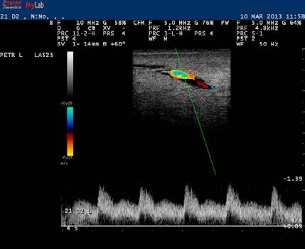
91 Figure II.15. (continued). (b) (c) 91
92 In both subgroups A1 and B1, blood input into the teat increased immediately after challenge and remained increased during the study (P<0.001 for all comparisons) in the inoculated teat. A similar increase was evident in the contralateral teat, but of smaller magnitude (P values varying from to 0.49), with a significant difference between the two sides of the same animal (P<0.002 for all comparisons) (Fig. II.16.). Detailed results are in Tables II.xii. and II.xiii. 92
 colour Doppler images of the subcutaneous vessels of the teat of (a) a ewe")
 (subgroup A1), (b) a ewe with no increased β- hydroxybuturate blood concentration during the last stage of the preceding pregnancy, which was")
 (subgroup B1).")
93 Figure II.16. Serial (before and after challenge with M. haemolytica into the teat duct; from left to right: picture on D-2, D0+12 h, D2) colour Doppler images of the subcutaneous vessels of the teat of (a) a ewe with increased β-hydroxybuturate blood concentration during the last stage of the preceding pregnancy, which was subsequently inoculated into that teat (on D0) (subgroup A1), (b) a ewe with no increased β- hydroxybuturate blood concentration during the last stage of the preceding pregnancy, which was subsequently inoculated into that teat (on D0) (subgroup B1) or (c) a ewe with no increased β-hydroxybuturate blood concentration during the last stage of the preceding pregnancy, which was subsequently inoculated into the contralateral teat (on D0) (subgroup B1). Images taken and processed on a MyLab 30 ultrasonography system with linear transducer; imaging frequency: 6.6 MHz - scanning depth: 20 to 30 mm. (a) 93

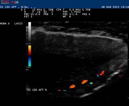
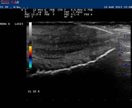
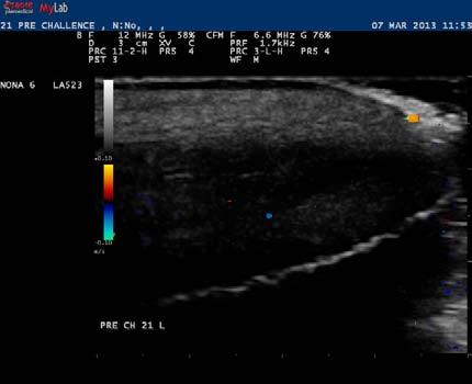
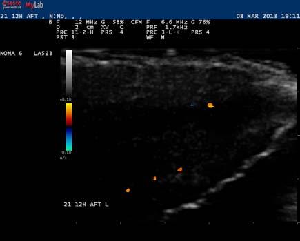
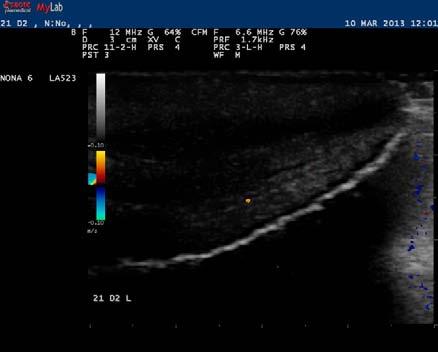
94 Figure II.16. (continued). (b) (c) 94
ΠΑΝΕΠΙΣΤΗΜΙΟ ΘΕΣΣΑΛΙΑΣ
 ΠΑΝΕΠΙΣΤΗΜΙΟ ΘΕΣΣΑΛΙΑΣ ΣΧΟΛΗ ΕΠΙΣΤΗΜΩΝ ΥΓΕΙΑΣ ΤΜΗΜΑ ΚΤΗΝΙΑΤΡΙΚΗΣ ΕΠΙΔΡΑΣΗ ΤΗΣ ΔΙΑΔΙΚΑΣΙΑΣ ΞΗΡΑΝΣΗΣ ΣΤΟ ΤΕΛΟΣ ΤΗΣ ΓΑΛΑΚΤΙΚΗΣ ΠΕΡΙΟΔΟΥ, ΣΤΗΝ ΥΓΕΙΑ ΤΟΥ ΜΑΣΤΟΥ ΤΩΝ ΠΡΟΒΑΤΙΝΩΝ ΙΩΑΝΝΗΣ Γ. ΠΕΤΡΙΔΗΣ Κτηνίατρος
Economic Review of Transition Cow Management
 Economic Review of Transition Cow Management John Fetrow VMD, MBA, DSc (hon) Emeritus Professor of Dairy Production Medicine College of Veterinary Medicine University of Minnesota This presentation is
Ceftaroline fosamil is a novel cephalosporin active against methicillin-resistant Staphylococcus aureus
 O r i g i n a l a r t i c l e In vitro activity of ceftaroline against methicillin-resistant Staphylococcus aureus isolates from blood and complicated skin and soft tissue infections and update on the
Lactation. Macroscopic Anatomy of the Mammary Gland. Anatomy AS 1124
 Lactation AS 1124 Macroscopic Anatomy of the Mammary Gland Species differences in numbers and locations of glands inguinal - caudal to the abdomen, between the hind legs (cow, mare, ewe) abdominal - along
Journal of the Hellenic Veterinary Medical Society
 Journal of the Hellenic Veterinary Medical Society Vol. 69, 2018 Reference intervals for canine hematologic analytes using Siemens Advia 120 OIKONOMIDIS I. TSOULOUFI T. PAPOUTSI A. KRITSEPI- KONSTANTINOU
AN EPIDEMIOLOGIC APPRAISAL OF MILK TESTING FOR DIAGNOSIS AND CONTROL OF PARATUBERCULOSIS IN GREEK DAIRY SHEEP AND GOATS
 UNIVERSITY OF THESSALY, SCHOOL OF HEALTH SCIENCES FACULTY OF VETERINARY MEDICINE LABORATORY OF EPIDEMIOLOGY, BIOSTATISTICS & ANIMAL HEALTH ECONOMICS AN EPIDEMIOLOGIC APPRAISAL OF MILK TESTING FOR DIAGNOSIS
FEEDING EWES BETTER FOR INCREASED PRODUCTION AND PROFIT. Dr. Dan Morrical Department of Animal Science Iowa State University, Ames, Iowa
 FEEDING EWES BETTER FOR INCREASED PRODUCTION AND PROFIT Dr. Dan Morrical Department of Animal Science Iowa State University, Ames, Iowa Introduction Sheep nutrition and feeding is extremely critical to
4.11 Major diseases in sheep
 49 4.11 Major diseases in sheep There are many types of pneumonia in sheep such as parasitic, aspiration, viral and bacterial. Sheep of all ages are affected. Stress factors influence the severity of pneumonia.
Late pregnancy nutrition the key to flock profitability
 Late pregnancy nutrition the key to flock profitability Dr. Tim Keady Animal and Grassland Research and Innovation Centre, Teagasc, Athenry, Co Galway. Introduction The plane of nutrition during late pregnancy
Pre-lambing preparation in sheep: feeding, metabolic profiles and minerals
 Vet Times The website for the veterinary profession https://www.vettimes.co.uk Pre-lambing preparation in sheep: feeding, metabolic profiles and minerals Author : Sara Pedersen Categories : Farm animal,
Research article Ερευνητικό άρθρο
 : 445-454 : 445-454 Research article Ερευνητικό άρθρο Axial pattern skin flaps in cats: 8 cases (2000-2015) Gavriilidou O. 1, Papazoglou LG. 2, Kouki M. 2, Strantzia E. 1, Giannouli M. 3, Mentzikof L.
Managing pre-calving dairy cows: nutrition, housing and parasites
 Vet Times The website for the veterinary profession https://www.vettimes.co.uk Managing pre-calving dairy cows: nutrition, housing and parasites Author : Lee-Anne Oliver Categories : Farm animal, Vets
The importance of nutrition during gestation for lamb vigour and survival. John Rooke, Gareth Arnott, Cathy Dwyer and Kenny Rutherford
 The importance of nutrition during gestation for lamb vigour and survival John Rooke, Gareth Arnott, Cathy Dwyer and Kenny Rutherford The importance of nutrition during gestation for lamb vigour and survival
Journal of the Hellenic Veterinary Medical Society
 Journal of the Hellenic Veterinary Medical Society Vol. 61, 2010 Farm conditions and production methods in Chios sheep flocks GELASAKIS (A. ΓΕΛΑΣΑΚΗΣ) A. Department of Animal Production, Faculty of Veterìnary
Mastitis and the link to infertility
 CONTINUING EDUCATION I LARGE ANIMAL Mastitis and the link to infertility Mastitis and infertility are the two most common disease complexes in dairy cattle worldwide. Both are major reasons for culling
Feeding Ewes Better for Increased Production and Profit
 Animal Science White Papers Animal Science 7-1-2003 Feeding Ewes Better for Increased Production and Profit Daniel G. Morrical Iowa State University, morrical@iastate.edu Follow this and additional works
Mastitis in ewes: towards development of a prevention and treatment plan
 SCHOOL OF LIFE SCIENCES, UNIVERSITY OF WARWICK Mastitis in ewes: towards development of a prevention and treatment plan Final Report Selene Huntley and Laura Green 1 Background to Project Mastitis is inflammation
Case Study: Pregnancy Toxemia in a Sheep Flock
 Case Study: Pregnancy Toxemia in a Sheep Flock Robert J. Van Saun, DVM, MS, PhD, Diplomate ACT and ACVN Department of Veterinary and Biomedical Sciences, Pennsylvania State University, University Park,
TRADITION AND MODERNITY IN VETERINARY MEDICINE
 SUBLINIAL AND LINIAL KETOSIS IN SHEEP RELATIONSHIPS BETWEEN BODY ONDITION SORES AND BLOOD β-hydroxybutyrate AND NON-ESTERIFIED FATTY AIDS ONENTRATIONS Vania Marutsova*, Plamen Marutsov Faculty of Veterinary
The tabanid fauna (Diptera: Tabanidae) of the Chirpan Eminences (Bulgaria)
 of the Chirpan Eminences (Bulgaria)") ENTOMOLOGIA HELLENICA 21 (2012): 45-53 The tabanid fauna (Diptera: Tabanidae) of the Chirpan Eminences (Bulgaria) DIANA GANEVA* AND MILENA KALMUSHKA Trakia University, Faculty of Agriculture, Department
ENTOMOLOGIA HELLENICA 21 (2012): 45-53 The tabanid fauna (Diptera: Tabanidae) of the Chirpan Eminences (Bulgaria) DIANA GANEVA* AND MILENA KALMUSHKA Trakia University, Faculty of Agriculture, Department
Do Greek doctors comply with guidelines for community-acquired pneumonia?
 Original Article Do Greek doctors comply with guidelines for community-acquired pneumonia? Serafeim Fountas, Katerina Manika, Martha Lada, Aliki-Lida Tsagkridi, Maria Kipourou, Ioannis Kioumis Respiratory
Improving sheep welfare for increased production
 Improving sheep welfare for increased production Emma Winslow 3 April 2017 SARDI - Struan sheep Research Livestock innovation and welfare group: Sheep welfare and wellbeing Production and management Genetic
Planning Spring/Summer 2018
 Planning Spring/Summer 2018 Poppy Frater Sheep Specialist SAC Consulting is a division of Scotland s Rural College Leading the way in Agriculture and Rural Research, Education and Consulting Outline 1.
Extra. Feed planning for ewes in late pregnancy and early lactation, during the housed period. Take a stepped approach to feed planning.
 Bulletin Autumn 2013 Extra Feed planning for ewes in late pregnancy and early lactation, during the housed period Compiled by Kate Philips, ADAS Providing ewes with adequate energy and protein in the last
Livestock and Poultry Environmental Learning Center Webcast Series March 28, 2008
 Antibiotic and Hormone Use in Livestock Production Paul Ebner Assistant Professor Department of Animal Sciences Purdue University Presentation Outline Antibiotics and Hormones a. How they are used b. Quantities
IRENE VALASI Assistant Professor Animal Physiology
 IRENE VALASI Assistant Professor Animal Physiology Surname Name Father s name Date of birth Place of birth Nationality Work address Tel. no. E-mail. VALASI Irene Dimitrios 24 December 1974 Larissa, Greece
Factors Affecting Breast Meat Yield in Turkeys
 Management Article The premier supplier of turkey breeding stock worldwide CP01 Version 2 Factors Affecting Breast Meat Yield in Turkeys Aviagen Turkeys Ltd Introduction Breast meat, in the majority of
Age of Weaning Lambs
 A Greener World Technical Advice Fact Sheet No. 17 Age of Weaning Lambs Certified Animal Welfare Approved by A Greener World (AGW) has the most rigorous standards for farm animal welfare currently in use
Case Study: Dairy farm reaps benefits from milk analysis technology
 Case Study: Dairy farm reaps benefits from milk analysis technology MARCH PETER AND SHELIA COX became the first dairy farmers in the UK to install a new advanced milk analysis tool. Since installing Herd
DAIRY HERD HEALTH IN PRACTICE
 Vet Times The website for the veterinary profession https://www.vettimes.co.uk DAIRY HERD HEALTH IN PRACTICE Author : James Breen, Peter Down, Chris Hudson, Jon Huxley, Oli Maxwell, John Remnant Categories
The strategic use of injectable trace minerals to improve fertility and health
 The strategic use of injectable trace minerals to improve fertility and health Improving productivity What should we aim for? Tightening the lambing pattern Lambs born earlier in the season have a longer
Lower body weight Lower fertility Lower fleece weight (superfine) (fine)
 (fine)") Generally, finer wool merino sheep are best suited to cooler areas Major Sheep Breeds In Australia Merino (75%) Border Leicester Merino x Border Leicester (12%) Suffolk Cheviot Poll Dorset Romney Merino
Generally, finer wool merino sheep are best suited to cooler areas Major Sheep Breeds In Australia Merino (75%) Border Leicester Merino x Border Leicester (12%) Suffolk Cheviot Poll Dorset Romney Merino
Pain Management Future pain relief options. Ian Colditz CSIRO Animal, Food and Health Sciences Armidale NSW 2350
 Pain Management Future pain relief options Ian Colditz CSIRO Animal, Food and Health Sciences Armidale NSW 2350 A national flystrike R&D technical update 1 st August 2012 Animal Experimentation The Principles
VETERINARY MEDICINE-VM (VM)
") Veterinary Medicine-VM (VM) 1 VETERINARY MEDICINE-VM (VM) Courses VM 603 Veterinary Science: Research and Methods Credit: 1 (1-0-0) Course Description: Conduct of responsible research, contributions of
Veterinary Medicine-VM (VM) 1 VETERINARY MEDICINE-VM (VM) Courses VM 603 Veterinary Science: Research and Methods Credit: 1 (1-0-0) Course Description: Conduct of responsible research, contributions of
METABOLIC DISEASES OF DAIRY CATTLE
 METABOLIC DISEASES OF DAIRY CATTLE J. K. Shearer DVM, MS. Professor and Dairy Extension Veterinarian Department of Large Animal Clinical Sciences College of Veterinary Medicine University of Florida Gainesville,
Feeding and Managing the Ewe Flock
 January, 2004 Feeding and Managing the Ewe Flock Brian Tarr Ruminant Nutritionist 1 FEEDING AND MANAGING THE EWE FLOCK Brian Tarr, Ruminant Nutritionist Shur-Gain, Member of Maple Leaf Foods Inc. Introduction
Farm Newsletter - February 2017
 Farm Newsletter - February 2017 Lung Worm in Cattle The disease is caused by the worm Dictyocaulus viviparus. Adult worms live in the animal s lungs where they produce eggs which hatch quickly. The first
Assessment Schedule 2017 Subject: Agricultural and Horticultural Science: Demonstrate knowledge of livestock management practices (90921)
") NCEA Level 1 Agricultural and Horticultural Science (90921) 2017 page 1 of 6 Assessment Schedule 2017 Subject: Agricultural and Horticultural Science: Demonstrate knowledge of livestock management practices
NCEA Level 1 Agricultural and Horticultural Science (90921) 2017 page 1 of 6 Assessment Schedule 2017 Subject: Agricultural and Horticultural Science: Demonstrate knowledge of livestock management practices
Mastitis: Background, Management and Control
 New York State Cattle Health Assurance Program Mastitis Module Mastitis: Background, Management and Control Introduction Mastitis remains one of the most costly diseases of dairy cattle in the US despite
Spot the Difference: Using the domestic cat as a model for the nutritional management of captive cheetahs. Katherine M. Bell
 Spot the Difference: Using the domestic cat as a model for the nutritional management of captive cheetahs Katherine M. Bell Edited by Lucy A. Tucker and David G. Thomas Illustrated by Justine Woosnam and
Herd health challenges in high yielding dairy cow systems
 Herd health challenges in high yielding dairy cow systems Robert Smith robsmith@liv.ac.uk The big three diseases Fertility Lameness Mastitis Energy balance and body condition Ruminal acidosis and abomasal
MANAGING NUTRITION AND ACTIVITY IN NEUTERED COMPANION ANIMALS
 Vet Times The website for the veterinary profession https://www.vettimes.co.uk MANAGING NUTRITION AND ACTIVITY IN NEUTERED COMPANION ANIMALS Author : TIM WATSON Categories : Vets Date : August 19, 2013
Trevor DeVries Dr. Trevor DeVries is an Associate Professor in the Department of Animal and Poultry Science at the University of Guelph.
 Trevor DeVries Dr. Trevor DeVries is an Associate Professor in the Department of Animal and Poultry Science at the University of Guelph. Trevor received his B.Sc. in Agriculture from The University of
ΓΡΑΦΕΙΟ ΟΙΚΟΝΟΜΙΚΩΝ ΚΑΙ ΕΜΠΟΡΙΚΩΝ ΥΠΟΘΕΣΕΩΝ P.O. BOX 5108, Jeddah Tel:
 ΓΕΝΙΚΟ ΠΡΟΞΕΝΕΙΟ ΤΗΣ ΕΛΛΑΔΟΣ ΣΤΗΝ ΤΖΕΝΤΑ ΓΡΑΦΕΙΟ ΟΙΚΟΝΟΜΙΚΩΝ ΚΑΙ ΕΜΠΟΡΙΚΩΝ ΥΠΟΘΕΣΕΩΝ P.O. BO 5108, Jeddah 21 422 Tel: 00966 12 66 90 824 E-Mail: ecocom-jeddah@mfa.gr ΑΔΙΑΒΑΘΜΗΤΟ ΕΠΕΙΓΟΝ Τζέντα, 25 Φεβρουαρίου
THE WELFARE OF ANIMALS IN PRODUCTION SYSTEMS
 THE WELFARE OF ANIMALS IN PRODUCTION SYSTEMS General Principles and Underlying Research David Fraser Animal Welfare Program University of British Columbia General principles for the welfare of animals
Subclinical mastitis in small ruminants: prevalence, comparative aspects and prevention
 Subclinical mastitis in small ruminants: prevalence, comparative aspects and prevention Dr. Gabriel Leitner, National Mastitis Reference Center, Kimron Veterinary Institute, Israel Dr. Nissim Silanikove
SHEEP. nd if appropriate/applicable)
") SHEEP GENERAL: UREA WARNING (only where an nd if appropriate/applicable) Vinegar is an effective remedy against NPN poisoning. Mix with an equal amount of water. Dose half a bottle per calf or large sheep
SHEEP GENERAL: UREA WARNING (only where an nd if appropriate/applicable) Vinegar is an effective remedy against NPN poisoning. Mix with an equal amount of water. Dose half a bottle per calf or large sheep
Key Information. Mountain Hill Vs Lowland Production. Breeding Strategy
 Key Information Short day breeder (come into heat in autumn as the day length decreases) Length of oestrus = 17 day cycle Duration of oestrus = 36 hours Length of gestation = 147 days or 5 months Can birth
Pre-fresh Heifers. A Might not Equal B. Pre-fresh Heifers Common A = B allegories. Udder edema = dietary salt. Transition (pre-fresh) = 21 d
 = 21 d") Pre-fresh Heifers A Might not Equal B Pre-fresh Heifers Common A = B allegories Udder edema = dietary salt Transition (pre-fresh) = 21 d Over-conditioned pre-fresh heifers = excess corn silage Early calving
Pre-fresh Heifers A Might not Equal B Pre-fresh Heifers Common A = B allegories Udder edema = dietary salt Transition (pre-fresh) = 21 d Over-conditioned pre-fresh heifers = excess corn silage Early calving
Influence of Experimentally- induced clinical mastitis on Reproductive Performance of Dairy Cattle
 Influence of Experimentally- induced clinical mastitis on Reproductive Performance of Dairy Cattle Dr. Mitch Hockett Department of Animal Science North Carolina State University Characteristics of Mastitis
Check that milk is suitable to go in the vat
 TECHNOTE 3 Check that milk is suitable to go in the vat CALVING For milk quality reasons, all cows must have their colostrum milk withheld from the vat for at least eight milkings after calving. Colostrum
LOCOMOTION SCORING OF DAIRY CATTLE DC - 300
 LOCOMOTION SCORING OF DAIRY CATTLE 2001 DC - 300 LOCOMOTION SCORING CONDITIONS Location: Researchers: Animals: Duration: Commercial Dairy in Michigan D.J. Sprecher, DVM, MS, DACT D.E. Hostetler, DVM, MS
SUCCESS IS IN THE BAG
 SUCCESS IS IN THE BAG NEW EPOL & MOLATEK PROUDLY TOGETHER LAYER FEEDING RATION BROCHURE EPOL = THE PERFECT BALANCE Epol has specialised ranges of feed for the different needs of layer farmers. The requirements
MASTITIS CASE MANAGEMENT
 MASTITIS CASE MANAGEMENT The 2nd University of Minnesota China Dairy Conference Hohhot Sarne De Vliegher Head of M-team UGent & Mastitis and Milk Quality Research Unit @ UGent OVERVIEW Mastitis case management
Internal Assessment Resource NCEA Level 1 Science AS KEEP CALM AND COUNT SHEEP. A unit of learning to be assessed for
 Internal Assessment Resource NCEA Level 1 Science AS 90949 KEEP CALM AND COUNT SHEEP A unit of learning to be assessed for KEEP NCEA CALM using AND Science COUNT 1.10 (AS90949) SHEEP 1 Contents.. Overview.................
Sustainable Integrated Parasite Management (sipm)
") Sustainable Integrated Parasite Management (sipm) The goal of a parasite control program is to control the parasites on a farm to a level which has minimal effect on animal health and productivity without
Sustainable Integrated Parasite Management (sipm) The goal of a parasite control program is to control the parasites on a farm to a level which has minimal effect on animal health and productivity without
Economic Analysis Papers
 Economic Analysis Papers Forecasting Cyprus GDP and its demand components: Single equation models and forecast combinations Christos Papamichael Economics Research Centre Nicoletta Pashourtidou Economics
WHY DO DAIRY COWS HAVE REPRODUCTIVE PROBLEMS? HOW CAN WE SOLVE THOSE REPRODUCTIVE PROBLEMS? Jenks S. Britt, DVM 1. Why Manage Reproduction?
 WHY DO DAIRY COWS HAVE REPRODUCTIVE PROBLEMS? HOW CAN WE SOLVE THOSE REPRODUCTIVE PROBLEMS? Jenks S. Britt, DVM 1 Why Manage Reproduction? The following table gives reproductive information from the DHIA
Catherine Gulliver. Collaborators: Dr Edward Clayton, Dr Michael Friend, Dr John Wilkins, Dr Belinda King, Dr Susan Robertson
 Catherine Gulliver Collaborators: Dr Edward Clayton, Dr Michael Friend, Dr John Wilkins, Dr Belinda King, Dr Susan Robertson Outline Background Why alter sex ratio? Factors affecting sex ratio? Maternal
Silage Analysis and Ration Planning: Benefits of knowing what you re feeding your stock. Mary McDowell Trainee Livestock Nutritionist
 Silage Analysis and Ration Planning: Benefits of knowing what you re feeding your stock Mary McDowell Trainee Livestock Nutritionist Issues during winter feeding Forage quality variation - How much do
PREDICTION OF LAMBING DATE BASED ON CLINICAL EXAMINATION PRIOR TO PARTURITION IN EWES
 PREDICTION OF LAMBING DATE BASED ON CLINICAL EXAMINATION PRIOR TO PARTURITION IN EWES J.V. Viljoen Grootfontein Agricultural Development Institute, Private Bag X529, Middelburg (EC), 5900 Email: HoggieV@daff.gov.za
Information document accompanying the EFSA Questionnaire on the main welfare problems for sheep for wool, meat and milk production
 EFSA Mandate for a Scientific Opinion on the main welfare risks related to the farming of sheep for wool, meat and milk production (M-2013-0197; EFSA-Q-2013-00580) Information document accompanying the
WHEN you first look at a dairy cow, what do
 External and Internal Parts of Dairy Cattle WHEN you first look at a dairy cow, what do you see? Most likely you see an animal weighing more than 1,000 pounds, with a head, tail, and four legs. But if
Mastitis and colostrum management update. Dr Kiro Petrovski University of Adelaide, Roseworthy
 Mastitis and colostrum management update Dr Kiro Petrovski University of Adelaide, Roseworthy Biography Started working with dairy cows at age of 11 First independent calving First cow surgery at 16 Work
How Does Photostimulation Age Alter the Interaction Between Body Size and a Bonus Feeding Program During Sexual Maturation?
 16 How Does Photostimulation Age Alter the Interaction Between Body Size and a Bonus Feeding Program During Sexual Maturation? R A Renema*, F E Robinson*, and J A Proudman** *Alberta Poultry Research Centre,
7. IMPROVING LAMB SURVIVAL
 7. IMPROVING LAMB SURVIVAL Introduction It is widely accepted that there is a large amount of lamb wastage in Merino flocks. Fertility rates, as measured by the number of lambs present at scanning are
Phase B 5 Questions Correct answers are worth 10 points each.
 2004 Junior Dairy Quiz Bowl Questions Round 05 Phase B 5 Questions Correct answers are worth 10 points each. Only the team being asked the questions is to be in the room. Each team will be asked these
In This Issue. News. Workman, Jeffrey D. News ecalving app for touchscreen mobile devices
 Workman, Jeffrey D. Subject: Ohio Veterinary Newsletter Vol 41 No 19 OHIO STATE UNIVERSITY EXTENSION OHIO VETERINARY NEWSLETTER November 6, 2015 Veterinary Extension Vol 41, No 19 In This Issue News ecalving
The comparison of some biochemical parameters in hyperketonemic and normal ewes
 Available online at www.pelagiaresearchlibrary.com European Journal of Experimental Biology, 2014, 4(3):83-87 ISSN: 2248 9215 CODEN (USA): EJEBAU The comparison of some biochemical parameters in hyperketonemic
Advanced Interherd Course
 Advanced Interherd Course Advanced Interherd Training Course... 2 Mastitis... 2 Seasonal trends in clinical mastitis... 2... 3 Examining clinical mastitis origins... 3... 4 Examining dry period performance
SUMMARY OF PRODUCT CHARACTERISTICS
 SUMMARY OF PRODUCT CHARACTERISTICS 1. Name of Veterinary Medicinal Product Endofluke 100 mg/ml Oral Suspension 2. Qualitative and Quantitative Composition Active Substance per ml Triclabendazole 100mg
PROJECT SUMMARY. Optimising genetics, reproduction and nutrition of dairy sheep and goats
 PROJECT SUMMARY Optimising genetics, reproduction and nutrition of dairy sheep and goats Introduction The Australian dairy sheep industry currently has six well established businesses, all of which are
Thomson, 1938, 1939] it was shown that ketosis can be produced in. toxemia", and "twin lamb disease". The two most characteristic
![Thomson, 1938, 1939] it was shown that ketosis can be produced in. toxemia, and twin lamb disease. The two most characteristic](/thumbs/87/95244598.jpg "Thomson, 1938, 1939] it was shown that ketosis can be produced in. toxemia, and twin lamb disease. The two most characteristic") 238 J. Physiol. (I939) 97, 238-249 6I2.397.2I:6I2.63 FATTY INFILTRATION OF THE LIVER IN PREGNANT EWES By L. C. SNOOK From the Rowett Research Institute, Aberdeen (Received 14 August 1939) THROUGHOUT the
238 J. Physiol. (I939) 97, 238-249 6I2.397.2I:6I2.63 FATTY INFILTRATION OF THE LIVER IN PREGNANT EWES By L. C. SNOOK From the Rowett Research Institute, Aberdeen (Received 14 August 1939) THROUGHOUT the
Transition cows have decreased immune function. The transition period. Inflammation, Immune Function, and the Transition Cow.
 Overview Inflammation, Immune Function, and the Transition Cow Barry Bradford Kansas State University Herd Health & Nutrition Conferences April 2016 Immunity and inflammation in the transition cow Long
General Practice Service Willows Information Sheets. Looking after your pregnant cat
 General Practice Service Willows Information Sheets Looking after your pregnant cat Looking after your pregnant cat The following information is aimed at ordinary cat owners rather than cat breeders. It
Table1. Target lamb pre-weaning daily live weight gain from grazed pasture
 Grassland Management for High Lamb Performance Tim Keady and Noel McNamara Animal & Grassland Research & Innovation Centre, Teagasc, Mellows Campus, Athenry, Co. Galway. To improve the financial margin
Hudson, a 10-year-old MC Cocker spaniel, was referred for evaluation of severe polyuria and polydipsia (PU/PD) of 3 months in duration...
 of 3 months in duration...") VCAWLAspecialty.com David Bruyette, DVM, DACVIM Hudson, a 10-year-old MC Cocker spaniel, was referred for evaluation of severe polyuria and polydipsia (PU/PD) of 3 months in duration... 1. Physical Examination
VCAWLAspecialty.com David Bruyette, DVM, DACVIM Hudson, a 10-year-old MC Cocker spaniel, was referred for evaluation of severe polyuria and polydipsia (PU/PD) of 3 months in duration... 1. Physical Examination
Understanding your pet s LIVER CONDITION
 Understanding your pet s LIVER CONDITION Why is the liver so important? What causes liver disease in dogs and cats? The liver is one of the largest organs in your pet s body, and it s vital for their good
BEEF SUCKLER HERD FERTILITY. Dr Arwyn Evans B.V.Sc., D.B.R., M.R.C.V.S. Milfeddygon Deufor
 BEEF SUCKLER HERD FERTILITY Dr Arwyn Evans B.V.Sc., D.B.R., M.R.C.V.S. Milfeddygon Deufor Why is fertility important? Aim of efficient suckled calf production can be defined as achieving the maximum output
Feeding strategy of Lacaune dairy sheep: Ewes fed in group according to milk yield
 Feeding strategy of Lacaune dairy sheep: Ewes fed in group according to milk yield Hassoun P. 1, Hardy A. 2, Tesnière A. 1, Legarto J. 3, De Boissieu C. 3 1 INRA UMR Selmet Montpellier; 2 Lycée Agricole
Animal Sciences (
 Animal Sciences 1 Animal Sciences The department offers four curriculum options. The Pre-Vet/Pre-Professional option (ANPV) provides students with a foundation in the biological and physical sciences for
Transition Period 1/25/2016. Energy Demand Measured glucose supply vs. estimated demands 1
 To Ensure a More Successful Lactation, The Vital 90 TM Days Make a Difference Andy Holloway, DVM Dairy Technical Consultant Elanco Animal Health Has been defined as the period of 3 weeks prepartum to 3
Level 1 Agricultural and Horticultural Science, 2017
 90921 909210 1SUPERVISOR S Level 1 Agricultural and Horticultural Science, 2017 90921 Demonstrate knowledge of livestock management practices 9.30 a.m. Wednesday 22 November 2017 Credits: Five Achievement
A METABOLOMICS APPROACH FOR THE STUDY OF LONG-TERM PROGESTERONE IN DOMESTIC SHEEP AND PHYSIOLOGICAL PROCESSES IN DOMESTIC AND BIGHORN SHEEP
 A METABOLOMICS APPROACH FOR THE STUDY OF LONG-TERM PROGESTERONE IN DOMESTIC SHEEP AND PHYSIOLOGICAL PROCESSES IN DOMESTIC AND BIGHORN SHEEP by Melissa Rashelle Herrygers A thesis submitted in partial fulfillment
My cat has kidney problems and food hypersensitivity what do I do now?
 TROVET Renal (Venison), complete, easily digestible, hypoallergenic dietary food for adult cats with an impaired kidney function My cat has kidney problems and food hypersensitivity what do I do now? reliable
Appendix I Average Analyses of B.C. Feeds
 Appendix I Average Analyses of B.C. Feeds The values given in the following table are not intended to substitute for the analysis of individual feeds. Looking at the crude protein (CP) values for forages
Flocks and Foliage Can Tree planning improve productivity, profit, health and welfare on livestock farms? Lovatt and Gascoigne 2016
 Flocks and Foliage Can Tree planning improve productivity, profit, health and welfare on livestock farms? Lovatt and Gascoigne 2016 Emily Gascoigne MA VetMB MRCVS Fiona Lovatt BVSc PhD DSHP DipECSRHM MRCVS
FRESH COW MANAGEMENT
 FRESH COW MANAGEMENT 2 FOREWORD Fresh Cow Management is the eleventh in a series of management manuals published by Veepro Holland. These Veepro Holland manuals are intended to provide a useful management
Parasites in Sheep Flocks
 Parasites in Sheep Flocks 1 WHAT IS NEW IN PARASITE CONTROL FOR SHEEP FLOCKS? Drew E. Hunnisett, DVM Honeywood and Warder Veterinary Services 132 Commerce Park Drive, Unit N Barrie, Ontario L4N 8W8 705
Hettinger Research Extension Center, North Dakota State University, Hettinger, ND
 Effects of maternal metabolizable protein supplementation during the last 50 days of gestation on ewe and offspring performance and carcass characteristics 1 M.L. Van Emon *, S.E. Eckerman *, L.A. Lekatz
Phase B 5 Questions Correct answers are worth 10 points each.
 2006 Junior Dairy Quiz Bowl Questions Round 07 Phase B 5 Questions Correct answers are worth 10 points each. Only the team being asked the questions is to be in the room. Each team will be asked these
Evaluation of Reproduction and Blood Metabolites in Beef Heifers Fed Dried Distillers Grains Plus Solubles and Soybean Hulls During Late Gestation 1
 Evaluation of Reproduction and Blood Metabolites in Beef Heifers Fed Dried Distillers Grains Plus Solubles and Soybean Hulls During Late Gestation 1 Chanda L. Engel 2, H. H. Trey Patterson 3, Ron Haigh
Proper Dry-Off Procedures to Prevent New Infections and Cure Existing Cases of Mastitis. Stephen C. Nickerson University of Georgia
 Proper Dry-Off Procedures to Prevent New Infections and Cure Existing Cases of Mastitis Stephen C. Nickerson University of Georgia scn@uga.edu Michelle Arnold, DVM DABVP (Food Animal) Ruminant Extension
FACULTY OF VETERINARY MEDICINE
 FACULTY OF VETERINARY MEDICINE DEPARTMENT OF VETERINARY PARASITOLOGY AND ENTOMOLOGY M.Sc. AND Ph.D. DEGREE PROGRAMMES The postgraduate programmes of the Department of Veterinary Parasitology and Entomology
Fact Sheet: Veterinary Natural Health Products and CQM
 January 2008 Fact Sheet: Veterinary Natural Health Products and CQM Introduction: The demand for organic production is increasing across Canada and the number of organic dairy farms is increasing to meet
FLOCK CALENDAR OUTLINE. a. Be sure they are vigorous, healthy and in good breeding condition.
 FLOK ALENDAR OUTLINE The following guidelines are neither inclusive nor intended to fit every sheep operation. Each operation is different, therefore, each Acalendar of events@ should be tailored to each
METABOLIC DISEASES IN CATTLE
 Vet Times The website for the veterinary profession https://www.vettimes.co.uk METABOLIC DISEASES IN CATTLE Author : PADY GORDON Categories : Vets Date : September 8, 2008 PADY GORDON describes the monitoring
Mastitis in non-bovine dairy species, companion animals and breastfeeding mothers. Chris Knight
 Mastitis in non-bovine dairy species, companion animals and breastfeeding mothers Chris Knight Objectives To stimulate thought/discussion regarding the relevance and importance of mastitis and mastitis
Herd Health Plan. Contact Information. Date Created: Date(s) Reviewed/Updated: Initials: Date: Initials: Date: Farm Manager: Veterinarian of Record:
 Reviewed/Updated: Initials: Date: Initials: Date: Farm Manager: Veterinarian of Record:") Contact Information Farm Name: Veterinarian of Record: Farm Owner: Farm Manager: Date Created: Date(s) Reviewed/Updated: Farm Owner: Date: Initials: Date: Initials: Date: Farm Manager: Date: Initials:
Contact Information Farm Name: Veterinarian of Record: Farm Owner: Farm Manager: Date Created: Date(s) Reviewed/Updated: Farm Owner: Date: Initials: Date: Initials: Date: Farm Manager: Date: Initials:
George C. Fthenakis GEORGE C. FTHENAKIS
 George C. Fthenakis Professor and Dean, Veterinary Faculty, University of Thessaly Former President, European College of Small Ruminant Health Management President, Division of Farm Animal Veterinarians,