Sylvia Debast Clostridium difficile Infection:
|
|
|
- Ronald Richardson
- 6 years ago
- Views:
Transcription


1 Sylvia Debast Clostridium difficile Infection: The role of antibiotics in outbreak control, epidemiology and treatment


2
3 Sylvia Debast Clostridium difficile Infection: The role of antibiotics in outbreak control, epidemiology and treatment
4 Colofon ISBN/EAN: The copyright of the published articles has been transferred to the respective journals or publishers. 2014, S.B. Debast, Amersfoort, the Netherlands. All rights reserved. No part of this publication may be reproduced, stored in a retrieval system, or transmitted in any form or by any means without prior permission of the author.
5 Clostridium difficile Infection: The role of antibiotics in outbreak control, epidemiology and treatment Proefschrift Ter verkrijging van de graad van Doctor aan de Universiteit Leiden op gezag van Rector Magnificus prof. mr. C.J.J.M. Stolker, volgens besluit van het College voor Promoties te verdedigen op donderdag 13 februari 2014 klokke uur door: Sylvia Brigitte Debast geboren 30 juli 1966 te Xanten (Duitsland)
6 Promotiecommissie Promotor: Prof. dr. E.J. Kuijper Overige leden: Prof. dr. J.T. van Dissel Prof. dr. A. Voss, Radboud Universiteit Nijmegen Prof. dr. F. van Knapen, Universiteit Utrecht
7 Vita bevis, ars vero Ionga, occasio autem praeceps, experimentum periculosum, iudicium difficile. Nec solum se ipsum praestare oportet oportuna facientem, sed et aegrum et assidentes et exteriora. Hippocrates Vertaald en besproken in: Medisch Contact 1967: 35;
8
9 i Table of Contents Introduction Chapter 1. General Introduction 3 Introduction 4 Clinical disease in humans 5 Pathogenesis 8 Epidemiology 12 Laboratory diagnosis 14 Antibiotics and CDI 19 Therapeutic options 19 Infection Control 21 Economics and CDI 23 Outline of this thesis 24 Aim of the studies 27 References 28 Outbreak control Chapter 2. Successful combat of an outbreak due to Clostridium difficile PCR-ribotype 027 and recognition of specific risk factors 43 Chapter 3. Effect on diagnostic yield of repeated stool testing during outbreaks of Clostridium difficile-associated disease 61 Chapter 4. PCR-ribotype-specific risk factors and outcome in an outbreak with 2 different Clostridium difficile Types simultaneously in one hospital 69 Epidemiology Chapter 5. Human Clostridium difficile-associated disease PCR ribotype 078 toxinotype V identified in Dutch food producing swine 91
10 ii
11 iii Treatment Chapter 6. Antimicrobial Activity of LFF571 and three treatment agents against Clostridium difficile isolates collected at a pan-european survey in Clinical and Therapeutic Implications 109 Chapter 7. European Society of Clinical Microbiology and Infectious Diseases: update of the treatment guidance document for Clostridium difficile infection (CDI) 127 Acta est fabula Chapter 8. General Discussion 193 Introduction 194 Outbreak control 196 Epidemiology 204 Treatment 211 Updated treatment guidelines for CDI 213 References 217 Chapter 9. Future Perspectives and Recommendations 225 Samenvatting [Summary in Dutch] 229 Maatschappelijke Relevantie [Social Relevance] 239 List of Abbreviations 241 Bibliography 243 Curriculum Vitae 247 Dankwoord [Acknowledgements] 249
12 iv
13 Introduction
14
15 1 Chapter 1 General Introduction
16 4 Chapter 1 Introduction In 1978, Clostridium difficile has been recognized as the agent responsible for most cases of antibiotic-associated pseudomembranous colitis (PMC) [1]. Until then PMC has been regarded as a bothersome, but inevitable and untreatable side effect of prolonged hospitalization and use of antibiotics. The detection of an identifiable pathogen for PMC marked a turning point in providing the rationale for research on and developments in laboratory diagnosis, therapeutic options and preventive measures for C. difficile infection (CDI). Over the last decade, CDI has progressively increased in incidence and severity of disease. To date, CDI is considered the leading cause of nosocomial diarrhoea, associated with an increased duration of hospitalization, healthcare expenses, morbidity and mortality among patients, especially among the elderly [2-4]. Clinical manifestations of CDI range from asymptomatic carriage to severe diarrhoea and pseudomembranous colitis with toxic megacolon [5]. Since 2003 a significant increase in rates of CDI-associated complications including deaths has been reported in the United States, Canada and Europe. This recent change in epidemiology is at least partly due to the epidemic spread of a novel more virulent ribotype, such as PCR-ribotypes 027 and also due to the emergence of ribotypes 001 and 078 [5-9]. In addition, expansion of CDI is observed in the community and in patients previously considered at low risk [10,11]. The occurrence of CDI is also increasingly recognized in veterinary medicine [12,13].
17 General introduction and outline of the thesis 5 Clinical disease in humans C. difficile can be found in the intestinal tract of 1-3% of all healthy adults and in 15-25% of individuals with recent healthcare exposure [14]. Loo and colleagues concluded that more than 50% of hospital patients infected with C. difficile might be symptomless carriers. Patients with symptomatic CDI were more likely to be infected with a highly pathogenic strain than were patients with C. difficile colonization [15]. Colonization with C. difficile and high levels of serum antibody against C. difficile toxin A and/or toxin B may provide protection against the development of CDI [15-18] Asymptomatic colonization may occur in 20% or more of patients in acute care hospitals. Increasing length of stay correlates with a greater likelihood of acquisition. From 4% to 20% of long-term care residents may carry the organism [19-21]. Once colonization with C. difficile is established several factors favour development of symptomatic CDI. Disruption of bacteria that normally reside in the bowel is the most common, and longer courses and use of multiple antibiotics increase the risk for disease [12,14,15,22]. C. difficile has been established as the most common cause of antibiotic-associated diarrhoea, accounting for 15% to 25% of cases [5,12]. Antibiotics that are most frequently related to CDI are: clindamycin, cephalosporins and penicillins, but to date several other antibiotics have been associated with CDI as well e.g. fluoroquinolones [22-27]. Fluoroquinolone exposure may be an important risk factor for the development of CDI due to highly fluoroquinolone-resistant PCR-ribotype 027 strains. Aldape et al. showed that ciprofloxacin up-regulates toxin gene expression and protein production in BI/NAP1/027 strains [28]. Clinical symptoms of CDI usually appear a few days after beginning anti biotic treatment and may appear up to three months after discontinuation [27]. In a majority of cases, patients with C. difficile associated diarrhoea, received antibiotics within 14 days preceding the infection, but in some patients symptoms can occur several months after discontinuation of antibiotic therapy. Olson et al. [29] found all patients with antibiotic-associated symptomatic CDI had received an antimicrobial within the previous three months. In a study of cancer patients who were being treated as outpatients, the median interval from hospital discharge to CDI was 20.3 days [30]. The principal risk-factors for the development of (severe) CDI include: antibiotic use [12,27,31], recent hospitalization [27,32], prolonged hospitalization (>3 days) [32-34],
18 6 Chapter 1 nursing home care [21,32], advanced age [8,15,31,35,36], chronic underlying disease [36,37], impaired host immune response against infections [38,39], gastrointestinal manipulation e.g. abdominal surgery, tube-feeding [40], enemas, and use of proton-pump inhibitors [15,35,41]. Colonization pressure quantifies the exposure of a person to a pathogen in terms of the number of infectious contacts and the duration of exposure. C. difficile colonization pressure has been shown to be an important exogenous risk factor for CDI at high levels of exposure in an ICU setting [34]. Although these risk factors are also associated with community associated CDI, potential risk factors for community-associated CDI may differ from those associated with nosocomial CDI. Patients with community acquired CDI are on average younger, more likely to be female and less likely to have underlying diseases than patients with healthcare associated CDI [11,32,42]. Importantly, almost half of community acquired CDI cases have not used antibiotics in the month before CDI, two-third of patients has not been hospitalized in the preceding 6 months before infection, and approximately one-third of the patients neither has exposure antibiotics nor recent hospitalization [43-45]. A frequently used case definition for CDI is: diarrhoea (defined as >3 unformed stools in less than 24 hours) and a stool test positive for toxigenic C. difficile or its toxins/toxin genes, or colonoscopic/histopathologic findings demonstrating pseudomembranous colitis [46,47]. Physical findings in CDI are variable, depending on the length and severity of disease [5,33]. Signs of dehydration may be present. The abdomen may be tender, and in severe cases, peritoneal signs may be present. Ileus or toxic megacolon may result in abdominal distension [48]. Toxic megacolon is the most serious clinical disease entity of CDI [5], defined as an acute dilatation of the colon (>6 cm) associated with severe colitis and systemic toxicity [49]. The clinical spectrum of symptomatic CDI may be classified by the severity of disease [46,47,50,51]. This classification enables the clinician to make therapeutic decisions and reach prognostic conclusions regarding the care of the patient, although it should be noted that currently there are no prospectively validated severity scores for CDI. Severity of CDI associated colitis can be divided into: mild moderate or early colitis, severe colitis, and fulminant colitis [52]. Approximately, 4 10% of patients with CDI develop fulminant colitis [53]. Fulminant colitis is a distinct clinical entity in which the release of bacterial toxins results in a systemic inflammatory response and multiorgan dysfunction. Fulminant colitis is characterized by hypotension, rising lactic acid levels, shock and complete ileus or toxic megacolon. Fatality rates
19 General introduction and outline of the thesis 7 in fulminant colitis may be as high as 33-50%, and it is therefore important to determine a precise time moment for surgical intervention [49,54]. Several clinical risk factors have been identified that are associated with increased postoperative mortality: age of 80 years or older, preoperative shock, preoperative dialysis dependence, chronic obstructive pulmonary disease, wound class III, thrombocytopenia, coagulopathy and renal insufficiency [54-57]. Fulminant colitis is not exclusive for CDI and is encountered in other diseases such as ulcerative colitis as well. C. difficile genotype has been shown to predict mortality [58], although this finding has been disputed by other investigators who did not detect a ribotype association with CDI case severity [59]. A correlation between excess mortality and genotype-specific changes in biomarkers (neutrophil/white cell counts, C-reactive protein, eosinophil counts and serum albumin) emphasizes the importance of inflammatory pathways as a major influence on the outcome of CDI [58]. Clinical Features and Complications of CDI are summarized in Table 1. Table 1. Clinical Features and complications of C. difficile infection (adapted from Refs. 5, 46, 47, 52 and 60). Spectrum of Disease Diarrhoea Other symptoms Physical examination Laboratory findings Endoscopy* & Imaging Asymptomatic carrier None None Normal Normal Unknown Mild-tomoderate Profuse Nausea, anorexia Low-grade fever, with or without mild abdominal tenderness Usually normal Nonspecific patchy erythema (endoscopy)* Severe CDI Profuse Nausea, malaise, abdominal discomfort Fever (sometimes high), abdominal tenderness and distension Leucocytosis (WBC >15 x 10 9 cells/l, with left shift). Serum albumin <3 g/l Pseudomembranous colitis: pseudomembranes (endoscopy)* Severe and complicated colitis Usually profuse and severe, but may be absent in ileus or toxic megacolon Nausea, malaise, abdominal discomfort and/or pain Fever (often high), rigors, abdominal tenderness and significant distention, signs of peritonitis, signs of ileus, hemodynamic instability, endorgan failure, admission to Intensive Care Unit for CDI Leucocytosis (WBC >35 x 10 9 cells/l, with left shift or <2 x 10 9 cells/l), rise in serum creatinine, elevated serum lactate (>5 mmol/l) Distension of large intestine, colonic wall thickening, pericolonic fat stranding, perforation, ascites (imaging) Nb. Endoscopy is contraindicated in severely ill patients * There is insufficient knowledge concerning the correlation of endoscopic findings compatible with CDI, such as oedema, erythema, friability and ulceration, and the severity of disease [46].
20 8 Chapter 1 One of the characteristics and main problems of CDI, is the high recurrence rate up to 20-30% after initial successful treatment of CDI with either metronidazole or vancomycin [61-63]. Recurrent CDI is defined as an episode of CDI occurring within eight weeks following an initial response. In daily practice it is impossible to distinguish recurrence due to relapse from recurrence due to reinfection [63]. The risk for recurrence increases with each episode, and may greater than 60% in patients with more than two episodes [63]. Since 2003 the epidemiology and clinical presentation of CDI has changed with the appearance of a new hypervirulent strain: PCR-ribotype 027 [2,65,66]. Large outbreaks of severe CDI in hospitals in Canada, America as well as in Europe were reported, presenting with pseudomembranous colitis and fulminant colitis, and with a higher mortality (3-30% case fatality associated with CDI depending on the methods and definitions used) [4,31,66-68] and recurrence rate (up to 25% within one to three months after treatment is completed) [68-71]. Additionally, the incidence of (severe) CDI in the community and in patients with no known risk factors, such as pregnant women and children has increased significantly as well [12]. Pathogenesis C. difficile is an anaerobic spore forming bacterium. Spores are shed significantly by infected patients with diarrhoea, which can survive for months in the environment. C. difficile can be transmitted via a faecal-oral route from the environment or from the hands of healthcare workers to patients. Ingested spores may transform into the vegetative form, which can then multiply and colonize the bowel [12]. Human colonic microbiota offer protection against bowel infection. However, the mechanisms by which colonic microbiota may mediate colonization resistance against C. difficile, how antibiotic disruption of the microbiota can alter this colonization resistance and how antibiotics induce C. difficile spore germination and subsequent toxin production, are not yet fully understood [12,72,73]. In recent studies specific anaerobic bacteria (e.g. Ruminococcaceae, Lachnospiraceae and butyrogenic bacteria) are identified as significantly depleted in C. difficile infection and nosocomial diarrhoea [74]. Disruption of this colonic barrier function, e.g. by the use of anti-microbial or chemotherapeutic agents, may lead to multiplication and colonization of the bowel with C. difficile. However, not all patients colonized with C. difficile develop symptomatic disease. Host
21 General introduction and outline of the thesis 9 and pathogen factors play an important role in the pathogenesis of disease. Only toxin-producing C. difficile strains cause disease; toxin-negative strains are considered non-pathogenic. Toxins cause diarrhoea and inflammation of the bowel. Therefore, host production of antitoxin antibody may be protective against development of disease as well as protective against relapsing CDI [16,39,75]. The incubation period from exposure to spores to onset of disease is not yet clear, but is thought to be a median of two days [76]. The main virulence factors of enteropathogenic C. difficile strains are two clostridial exotoxins, namely toxin A and toxin B [77]. The toxins are encoded by their genes tcda and tcdb, which are located, along with surrounding regulatory genes, on a 19.6-kilobase section of chromosomal DNA known as the pathogenicity locus (Paloc) [78,79]. In addition to the major toxin genes, the PaLoc region encodes three accessory genes tcdr and tcdc, which encode proteins involved in regulating the expression of TcdA and TcdB, and tcde. A schematic overview of the pathogenicity locus is shown in Figure 1. Secretory diarrhoea and inflammation of the colonic mucosa can largely be explained by the effect of these toxins. Both toxins are cytotoxic, causing disruption of the actin cytoskeleton and tight junctions, and resulting in decreased transepithelial resistance, fluid accumulation, inflammatory response and degradation of the intestinal epithelium. Toxigenic C. difficile strains can produce both toxins, or only one of them. Toxin A was thought to be the major virulence factor for many years; however, it has become increasingly evident that toxin B plays a much more important role than anticipated [81,82]. TcdA negative, tcdb positive strains can indeed cause clinical disease in humans [77,83]. The incidence of A- negative B-positive C. difficile strains appeared to be increasing worldwide. Patients infected with toxin A-negative, toxin B-positive strains exhibit the full spectrum of symptoms associated with CDI. Some studies also suggest that these isolates are even associated with more severe disease [84]. These strain types now represent a substantial number of C. difficile isolates [84,85]. Animal model studies resulted in conflicting results on the importance of toxin A and toxin B [79]. Purified toxin B was shown to be a more potent enterotoxin than toxin A, causing severe damage to the intestinal epithelium and leading to an acute inflammatory response in a mice model [86]. To assess the individual contribution of toxin A and B, recently, research was performed using multiple genetically constructed C. difficile tcda and tcdb toxin mutants [82]. Using these toxin mutants in a hamster model, toxin B was shown to be the major virulence

22 10 Chapter 1 factor instead of toxin A. In addition, toxin B did not require the presence of toxin A to cause disease. However in a second study using equivalent toxin mutants, contradictory data were reported [77]. In this second study both a toxin A and toxin B mutant caused disease, and the authors concluded that both toxins are important in CDI. From an analysis of both studies [81] it was concluded that it is evident that toxin A is not the major virulence factor, but further experiments are required to accurately determine the relative roles of each toxin in CDI, especially in strains that produce higher levels of toxin, such as PCR-ribotypes 027 or 078. TcdR RNA polymerase 19.6 kb tcdr tcdb tcde tcda tcdc Up-regulates transcription of toxins A and B TcdE Toxin B Toxin A Lysis of cytoplasmic membrane Inhibits toxin transcription Toxin release Figure 1. The pathogenicity locus of C. difficile: 19.6-kb pathogenicity locus encodes toxin A (tcda), toxin B (tcdb), a positive regulator of toxin transcription (tcdr), and a putative negative regulator of transcription (tcdc). The function of the tcde gene product is uncertain but may include the facilitation of toxin release by bacterial membrane lysis. (Adapted from Ref. 80). C. difficile toxins A and B have no export signature and their secretion is not explainable by cell lysis. Recently, TcdE was found to act as a holinlike protein to facilitate the release of C. difficile toxins to the extracellular environment, but unlike the phage holins, does not cause the non-specific release of cytosolic contents. TcdE appears to be the first example of a bacterial protein that releases toxins into the environment by a phage-like system [87]. Some strains produce a third toxin known as CDT or binary toxin [88]. CDT has been suggested to increase the pathogenicity of C. difficile strains. CDT ADP-ribosylate actin and inhibits actin polymerization. Indeed a more severe form of disease and a higher case-fatality rate in CDI due to strains
23 General introduction and outline of the thesis 11 with binary toxin as compared to infections without binary toxins have been described. Binary toxin either is a marker for more virulent C. difficile strains or contributes directly to strain virulence [89]. Despite these findings the clinical relevance of binary toxin is not yet well understood. CDT has been shown not only to depolymerize the actin cytoskeleton but to induce the formation of a novel ribotype of microtubule structures, consisting of long microtubule-based protrusions on the surface of epithelial cells as well, thereby leading to increased adherence of C. difficile. Eventually this causes death of target cells [90]. The membrane receptor for CDT uptake by target cells was recently identified by Papatheodorou et al. [91]. It has been shown that a related binary toxin (C. perfringens iota toxin) enters target cells via this lipolysis-stimulated lipoprotein receptor. The presence of a naturally occurring mutation in the tcdc has also been associated with the ability of toxigenic strains to become more virulent [79]. The tcdc gene has been reported to down-regulate the expression of tcda and tcdb. A mutation in the tcdc gene, may therefore lead to increased production of toxin A and toxin B. However, in vitro results on the role of tcdc as a major regulator of toxin expression are controversial, and whether increased toxin production also occurs in vivo remains unclear [92,93]. An additional regulatory gene on the pathogenicity locus is the tcdr gene. TcdR appears to be a positive regulator for the expression of tcda an tcdb [94]. C. difficile forms spores that are highly resistant to desiccation, chemicals and extreme temperatures. Spores frequently contaminate the environment around patients with CDI, potentially persisting for months and even years. Before the C. difficile toxins can exert their effects, ingestion and germination of spores in the intestinal tract is required [12]. Therefore, it has been postulated that increased sporulation may be associated with hypervirulence and C. difficile epidemic strains have been associated with a greater sporulation capacity in vitro than non-outbreak strains [95]. In recent years other potential virulence or toxin regulating factors have been identified: e.g. hybrid toxins [79], sigma factors TxeR [94] and control proteins CodY and CcPA [96]. Toxin expression may be influenced by specific environmental signals such as the nutritional status of the bacteria. A rapidly metabolizable carbon source such as glucose, inhibits toxin
24 12 Chapter 1 expression [94]. In addition, general regulatory molecules such as CodY and CcpA are known to influence toxin synthesis [96,97]. CodY was found to repress toxin gene expression in C. difficile. CcpA is involved in the glucosedependent repression of C. difficile toxin production. This repression is because of a direct binding of CcpA to the regulatory region of the tcda and tcdb genes. Unfortunately investigations on the role of environmental factors, toxins and other potential virulence factors are mainly performed in in vitro studies or animal models, thereby limiting the clinical implications of these results in human CDI. In vitro experiments may not reflect in vivo behaviour, and translating in vitro or animal derived data into C. difficile behaviour in humans is not straightforward. In general it seems likely that multiple factors determine whether a strain is more or less virulent and/ or epidemic. Risk factors for acquisition of C. difficile colitis include factors leading to disruption of colonic bacterial flora, such as receipt of antimicrobial agents or chemotherapeutic agents; undergoing solid-organ or bone marrow transplant; inflammatory bowel disease (IBD); factors leading to increased colonization by C. difficile spores, such as hospitalization and duration of hospitalization; and factors impacting the host immune system, such as advanced age and immunosuppression [98,99]. Epidemiology C. difficile is recognized as the primary infectious cause of pseudomembranous colitis and the principal cause of infectious diarrhoea in hospitalized patients [65]. In recent years incidence, severity, and recurrence rates of CDI have increased dramatically. There has also been a significant increase in severe cases causing admission to a healthcare facility and/ or intensive care unit for treatment, in colectomies, and death-related to CDI [2,7,12,65-67]. Additionally, CDI is also emerging in the community and in food-producing animals [6,10,12, 13,100]. Although elderly hospitalized patients receiving antibiotics is still the main group at risk of infection, an increase in CDI in younger populations with no previous contact either with the hospital environment or with antibiotics is noticed [11,31,101,102]
25 General introduction and outline of the thesis 13 Increases in incidence of CDI have been largely attributed to the emergence of a previously rare and more virulent strain, C. difficile BI/NAP1/027 [7]. Increased toxin production and high-level resistance to newer generation of fluoroquinolones made this strain a very successful pathogen in healthcare settings and populations previously thought to be at low risk. However, the underlying reasons for its rapid emergence and the subsequent patterns of global spread remains unknown. To gain more insight into key genetic changes leading to the emergence of this highly pathogenic strain and the subsequent patterns of global spread, whole-genome sequencing and phylogenetic analysis was performed on a global collection of C. difficile 027/ BI/NAP1 isolated primarily from hospital patients between 1985 and 2010 [103]. It was shown that two, and not one as previously thought, distinct epidemic lineages, FQR1 and FQR2 emerged in North America within a relatively short period after acquiring an identical fluoroquinolone resistance conferring mutation and a highly related conjugative transposon. The two epidemic lineages showed distinct patterns of global spread, and the FQR2 lineage spread more widely, leading to healthcare-associated outbreaks in the UK, continental Europe and Australia. The data suggested that the acquisition of resistance to commonly used antibiotics to be a major feature of the continued evolution and persistence of C. difficile 027/ BI/NAP1 in healthcare settings. Furthermore, the ease and rapidity with which the bacterium was transmitted internationally highlighted the interconnectedness of the global healthcare system, which is facilitated by rapid human travel. Human travel was indeed included in a risk assessment framework, which was developed by Clements et al. to assess risks of further worldwide spread of this pathogen [104]. The framework the authors present requires identification of potential vehicles of introduction, including international transfers of hospital patients, international tourism and migration, and trade in livestock, associated commodities, and foodstuffs. Besides C. difficile PCR-ribotype 027, several other strains have been associated with outbreaks and severe CDI as well [105,106]. In 2009, Bauer and colleagues [31] performed a hospital-based survey supported by the European Centre for Infectious Disease Prevention and Control, to obtain an overview of CDI in Europe. An incidence of 4.1 per 10,000 patient-days was found. The incidence of CDI and the distribution of causative PCR- ribotypes differed greatly between the European hospitals included in this study. The three most frequently found PCR-ribotypes of toxigenic C. difficile strains were 014/020 (16%), 001 (10%) and 078 (8%).


![osis has not been clearly established [60]. Diagn](/docs-images/71/66196777/images/26-2.jpg "ostic tests for CDI include: (1) Detection of C. difficile products: e.g.")
26 14 Chapter 1 Laboratory diagnosis CDI is a primarily a clinical diagnosis supported by laboratory or endoscopic evidence. Clinical presentation has been shown to be important when interpreting C. difficile diagnostic assays. Specificity of any given C. difficile assay for the diagnosis of CDI is increased when clinical symptoms of the patient are included in the reference standard [107]. For this reason only stools from patients with diarrhoea should be tested for C. difficile. There are many different approaches that can be used in the laboratory diagnosis of CDI. However, the best standard laboratory test for diagnosis has not been clearly established [60]. Diagnostic tests for CDI include: (1) Detection of C. difficile products: e.g. toxins A and B by cell culture cytotoxicity assay (CCA) or EIA, and glutamate dehydrogenase (GDH) by EIA, (2) Culture and detection of toxins produced by the isolate: toxigenic culture of Clostridium difficile, and (3) Molecular diagnostics of C. difficile specific targets: 16S RNA, toxin genes, GDH genes. A general overview of diagnostic methods is shown in Figure 2. Toxin detection Presence of C. difficile Presence of toxigenic C. difficile EIA (TcdA/TcdB) Membrane assay (TcdA/TcdB) Cytotoxicity assay EIA (GDH) Culture Toxigenic culture PCR to tcdb LAMP to tcda Figure 2. Methods to diagnose CDI can be divided into: the determination of C. difficile toxins A and/or B (blue), the presence of C. difficile (green) and the presence of toxigenic C. difficile (orange).
27 General introduction and outline of the thesis 15 The detection of neutralizable cell cytotoxicity in stools from patients with antibiotic-associated colitis has led to the discovery that C. difficile is the causative agent of this infection [108]. Since then, cell cytotoxicity assay (CCA) has been regarded as the gold standard for the detection of C. difficile toxins [47]. CCA is a tissue-culture assay based on the detection of the cytopathic effect of the C. difficile toxins present in stool. Using a combination of clinical and laboratory criteria to establish the diagnosis of CDI, the sensitivity of the cytotoxin detection as a single test for the laboratory diagnosis is reported to range from 67% to 100% [109,110]. However, the test is difficult to standardize leading to large variations when performed by different laboratories. Additionally, cell lines may also differ in susceptibility to C. difficile toxins, as has recently been detected at the Leiden University Medical Center for Vero cells ATCC CCL-81 and its clone E6, which differ a factor 8 for susceptibility to TcdA (pers. comm. Ing. I.M.J.G. Sanders). In many laboratories enzyme immunoassays (EIAs) for the detection GDH, Toxin A and/or Toxin B are used, as they are rapid and easy-to-perform assays. Rapid diagnosis of CDI is essential both for improving outcomes of patients with CDI and for reducing horizontal transmission in healthcare facilities. In a systematic review by Crobach et al. [111] the diagnostic accuracy of various EIAs (GDH and Toxins A and/or B) and a real-time PCR for C. difficile toxin B gene for the diagnosis of CDI, were evaluated and compared with CCA and toxigenic stool culture. EIAs were found to be quite specific, but less sensitive in detecting CDI. Only when these tests are performed in an epidemic situation with a CDI prevalence of 50%, positive predictive values are assumed to be acceptable due to their high specificity. However, in an endemic situation, the prevalence of CDI is expected to range between 5% and 10%. Therefore, it was concluded that EIAs are not suitable as stand-alone tests to diagnose CDI in endemic populations. Because of the lower sensitivity to detect the presence of toxigenic C. difficile in stool versus other methods, the Society for Healthcare Epidemiology of America and Infectious Diseases Society of America CDI guidelines state that toxin enzyme immunoassays (EIAs) are a suboptimal approach for the diagnosis of CDI [47]. GDH is an enzyme produced by C. difficile in relatively large amounts compared with toxins A and B [112]. Although GDH is sensitive, it is not as specific for CDI, because both toxigenic and non-toxigenic organisms produce this enzyme. The sensitivity of GDH antigen detection (ranging from 75% to >90%) has led to its use as a screening test as part of CDI testing algorithms,
28 16 Chapter 1 although it should be noted that as many as 10% of patients with toxigenic organisms can be missed by this method [60,111, ]. Currently, many laboratories use a combination of a sensitive, but not necessarily highly specific, screening test such as the GDH assay, followed by a more specific test on specimens that test positive to confirm the presence of toxin (e.g. an EIA for toxin A and/or B, PRC or toxigenic culture). Using toxigenic stool culture, C. difficile strains are isolated, followed by in toxin detection of the isolate using CCA, EIA or molecular tests to detect TcdA and/or TcdB. Because of the long turnaround time of this method, toxigenic culture is mainly used as a confirmatory test and/or for epidemiological purposes. Disadvantages of CCA and toxigenic culture are that they are expensive, time-consuming and laborious and that interpretation is subjective. Toxigenic stool culture for the detection of C. difficile and CCA have been considered the main reference assays for the diagnosis of CDI [110]. More recently, rapid molecular assays such as the real-time polymerase chain reaction (PCR) and technically simpler loop-mediated isothermal amplification (LAMP) have become available for the diagnosis of CDI [60, ]. These assays detect conserved regions of toxin A or toxin B genes on the PaLoc of C. difficile. Compared to other non-culture-based methods, molecular assays are considered the most sensitive methods available. Evidence suggests that PCR s for toxigenic C. difficile may be good standalone tests for toxigenic C. difficile. However, clinicians and microbiologists have some concerns regarding their clinical use, because the gene for toxin and not the toxin itself is detected [116]. PCR for the detection of toxigenic C. difficile has been shown to have a high sensitivity and excellent negative predictive value. A positive test however cannot differentiate infection from asymptomatic carriage and a second toxin detection test is therefore recommended (see Figure 3). Currently available Nucleic Acid Amplification Tests (NAAT s), including PCR assays and isothermal amplification tests, which are approved by the Federal Drug Administration (FDA) are: LAMP (Illumigene) [121,122], Xpert C. difficile PCR assay (Cepheid, Sunnyvale) [ ], ProGastro Cd (PG PCR) assay (Prodesse, Waukesha) [ ], BD GeneOhm (BD PCR) assay (Becton Dickinson, San Diego) [ ], Simplexa-C. difficile Universal Direct Test (Quest Diagnostics, Madison), and ribonuclease-mediated isothermal amplification and chip-based detection method test (Great Basin Corp., Salt Lake City) [135].
29 General introduction and outline of the thesis 17 The diagnostic accuracy of real-time polymerase chain reaction in detection of C. difficile in the stool samples of patients with suspected CDI was evaluated in a meta-analysis performed by Deshpande et al. [119]. The analysis included 19 diagnostic accuracy studies comparing PCR with cell culture cytotoxicity neutralization assay or toxigenic culture of C. difficile [119]. Three commercial PCR assays were investigated: GeneOhm Cdiff Assay (BD Diagnostics GeneOhm, San Diego); Xpert C. difficile Test (Cepheid); and ProGastro Cd Assay (Gen-Probe, San Diego). The investigators concluded that real-time PCR has a high sensitivity (90%) and specificity (93%) to confirm CDI. More importantly however, test accuracy depended on the prevalence of C. difficile and not on the reference test used: with a low C. difficile prevalence of 10%, the positive predictive value was only 71%, and with a high prevalence of >20% it was 93%. Real-time PCR may therefore be an adequate diagnostic assay in epidemic conditions with higher C. difficile prevalence but might not be the best diagnostic test in endemic situations with low C. difficile prevalence. In endemic situations PCR may serve as a screening test with emphasis on a negative test result. Peterson et al. recently evaluated ten diagnostic tests (including one commercial PCR: BD Diagnostics Cdiff PCR test (Becton Dickinson) for the detection of toxigenic C. difficile compared with toxigenic culture. The authors concluded PCR for toxigenic C. difficile and GDH testing to be the most sensitive assays for detection of C. difficile in stool specimens. GDH and PCR were statistically more sensitive than various toxin A and B EIAs and cell-cytotoxicity assay [133].
30 18 Chapter 1 Toxin detection or bacterial detection EIA to detect TcdA and TcdB EIA to detect GDH, or real-time PCR to TcdB + + EIA to detect GDH, or real-time PCR to TcdB, or cytotoxicity assay No CDI High clinical suspicion: toxigenic culture* EIA to detect TcdA and TcdB, or cytotoxicity assay + + CDI is diagnosed No CDI C. difficile toxins are not detectable in faeces but C. difficile is present; CDI can not be excluded CDI is diagnosed * A positive toxigenic culture always indicates the presence of toxin-producing C. di cile and makes further testing unnecessary Figure 3. A two-step algorithm to diagnose CDI [111]. De Boer et al. developed two real-time PCR assays for the detection of C. difficile, and subsequent identification of a tcdc mutation at nucleotide 117 directly in stool specimens [136]. The authors concluded that this assay was a rapid method to identify all toxigenic strains and stool samples containing the epidemic 027/NAP1 strain. The mutation has also been applied as a rapid identification method for PCR-ribotype 027 in the GeneOhm Cdiff Assay (BD Diagnostics GeneOhm) and Xpert C. difficile Test (Cepheid, Sunnyvale). However, the mutation is not specific for PCR-ribotype 027, as the same single-base-pair tcdc nucleotide 117 deletion was also demonstrated in other PCR-ribotypes such as PCR-ribotype 076 [137]. Limitations in sensitivity and specificity of common rapid diagnostic tests, have led to the development of several diagnostic algorithms that combine two and sometimes three tests to improve diagnostic accuracy e.g. screening with the GDH antigen test and confirmatory testing with toxigenic culture and/or PCR [60,111,113,115,117,118,138]. A two-step approach, with a second test or a reference method in case of a first positive test to diagnose CDI is proposed by Crobach et al. [111] (Figure 3).
31 General introduction and outline of the thesis 19 Antibiotics and CDI Antimicrobial therapy, often given for treatment of other infectious diseases, can render the patient susceptible to CDI if the patient is exposed to a toxigenic strain of the organism. When CDI was first reported, prior use of clindamycin was established as a significant risk factor [14,49,139,140]. However, in the years thereafter several other antibiotics were found to be associated with a risk of CDI [15,22,27,31,32,36,141]. Cephalosporins and fluoroquinolones replaced clindamycin as the major risk factor, but almost all antibiotics carry some risk. Fluoroquinolones have been linked to CDI and to severe epidemics, particularly those caused by PCR-ribotype 027 [61, ]. C. difficile strains that are resistant to multiple antimicrobial agents may thrive in an environment where other commensal flora are suppressed in the presence of these antibiotics [22, ]. In addition reduced susceptibility or resistance to common treatment agent e.g. metronidazole and vancomycin, may have clinical implications [ ]. Tenover et al. [146] investigated the prevalence of antimicrobial resistant strains in 316 toxigenic clinical isolates of C. difficile from seven hospitals in the United States and Canada (Quebec) during Multidrug resistance (i.e. resistance to clindamycin, moxifloxacin and rifampicin) was present in 22 of 80 (27.5%) C. difficile PCR-ribotype 027 isolates from the United States and Canada but was unusual among other ribotypes. In several studies high rates of clindamycin resistance have been demon strated in a variety of ribotypes, including ribotype PCR-ribotypes 001, 014, 017, and 027 worldwide [ ]. Resistance to the antimicrobial agents most commonly used to treat CDIs, i.e. metronidazole is reported rarely in the literature and the clinical impact has not yet been assessed [153,154]. Vancomycin resistance has not yet been documented. Therapeutic options Currently three guidance documents are available for the treatment of CDI: a guideline supported by the European Society of Clinical Microbiology and Infection (ESCMID) [46], a second guideline including recommendations of the Australian Society for Infectious Diseases (ASID) [115] and Clinical Practice Guidelines for CDI in adults published by the Society for Health care Epide-
32 20 Chapter 1 miology of America (SHEA) and the Infectious Diseases Society of America (IDSA) [47]. A Cochrane systematic review has also been published recently [155]. One of the main problems in the treatment of CDI is the occurrence of (sometimes multiple) relapse rates in patients after successful initial therapy is completed, ranging up to 25% and thereby increasing the infectious burden in patients significantly [69-71]. Recommendation of medical treatment options for CDI, are often subdivided in: the first episode of CDI, severe/complicated infection, recurrent infection and prevention of (recurrent) disease. An overview of treatment options is given in Table 2. Data in this table have been collected from the current ESCMID guideline for the treatment of CDI [46]. Table 2. Overview of therapeutic options for C. difficile infection (CDI) and recommendations by the ESCMID as of 2009 (marked in green) [46]. Therapeutic options Treatment Treatment guideline ESCMID 2009* Recommendation Indication Antibiotic Oral antibiotic Metronidazole Vancomycin Non-severe CDI: Initial infection First recurrence Severe CDI Recurrent CDI (>1) Parenteral antibiotic Metronidazole iv Metronidazole iv + vancomycin intracolonic Non-severe CDI Severe CDI Non-antibiotic (in combination with antibiotics) Probiotics Not recommended - Toxin binding resins and polymers Not recommended - Immunotherapy Not recommended - Faecal transplant Not recommended - Surgery Colectomy Complicated disease: perforation of the colon deteriorating clinical condition despite antibiotic therapy The first step in CDI treatment is the discontinuation of the antimicrobial therapy if possible. The rate of spontaneous resolution of CDI is unknown. In one study a spontaneous recovery rate in hospitalized patients with
33 General introduction and outline of the thesis 21 diarrhoea and a positive toxin assay who did not undergo endoscopy or had no pseudomembranous colitis on colonoscopy of 33% was found [1]. Except for very mild CDI, which is clearly induced by antibiotic usage, antibiotic treatment is advised. The (initial) antibiotics used for the treatment of CDI in various European countries, generally include oral vancomycin and metronidazole [46]. However, in severe CDI and recurrent infection antibiotic treatment may fail [63,156]. The last five years several new antibiotic agents (e.g. fidaxomicin and rifaximin) for CDI have been developed and limitations of the currently recommended treatment options of CDI are at discussion [69,70,157]. In addition new treatment modalities other than antibiotics have become available, such as donor faeces installation and use of monoclonal antibodies against toxins A and B [ ]. Recently, the first randomized controlled trial comparing a standard of vancomycin versus duodenal infusion of donor faeces has been published. Infusion of donor faeces was significantly more effective for the treatment of recurrent CDI than the use of vancomycin [158]. Infection Control Various infection control measures, including barrier precautions (contact isolation), hand hygiene, environmental cleaning, use of single-use rectal thermometers, endoscope disinfection, and limited use of select antibiotics, have been described in CDI guidelines [47,115,162]. Environmental cleaning with sodium hypochlorite (bleach) solutions (concentration of at least 1000 ppm available chlorine) decreases C. difficile surface contamination and has been associated with a significant reduction in the transmission risk of CDI [95, 162,163]. However, cleaning is required prior to disinfection with chlorine-based solutions, as they have poor activity in dirty conditions [164]. Alcohol-based hand sanitizers are thought to be ineffective in controlling CDI transmission, because they have poor activity against CD spores. Therefore hand-washing with water and soap is advised [162, 165]. As some hypervirulent strains (e.g. PCR-ribotype 027) are resistant to fluoroquinolones, increased use of these antimicrobial agents is proposed to contribute to the emergence of epidemics. For this implementation of an antimicrobial management program including a reduction in the use of antibacterials may be essential in outbreak control [106,162,166].
34 22 Chapter 1 Increased toxin production and hypersporulation are suggested to facilitate environmental contamination and contribute to outbreaks of infection as well. Since 2000, outbreak investigation has guided the sequential introduction of control measures and the development of a comprehensive CDI control bundle approach in which several outbreak measures are taken simultaneously [47,106,162, ]. An overview of recommended measures for the prevention and control of CDI recommended by the ESCMID is given in Table 3. Table 3. Overview of recommended measures for the prevention and control of C. difficile infection (CDI): bundle approach [162]. Interventions for the prevention and control of C. difficile infection Diagnosis Early, rapid and reliable diagnostics Awareness Education and communication Surveillance Monitor: incidence of CDI, distribution of PCR-ribotypes, clinical outcome Hand hygiene Hygiene Protective clothing Medical equipment: single use, disinfection, disposables Environmental cleaning and disinfection Barrier precautions Contact isolation in a single room Cohort isolation (outbreaks) Antibiotics Stop antibiotics in case of CDI Good antibiotic stewardship Monitoring the epidemiology of CDI (prevalence and incidence) is important for assessing risk factors and outcome of disease for planning prevention programs and focusing antibiotic stewardship efforts [170]. Access to C. difficile ribotyping in national surveillance programs to measure the distribution of PCR-ribotypes was associated with significant control of epidemic strains, especially of PCR-ribotype 027 [171,172]. Changes in prevalence of epidemic strains coincided with markedly reduced CDI incidence and related mortality [172].
35 General introduction and outline of the thesis 23 Economics and CDI The economic burden associated with healthcare associated CDI is high for primary and recurrent infection [ ]. Healthcare-associated cases of CDI are associated with significantly higher mean cost and longer length of hospital stay [3,176,177]. Recently, Wiegand et al. reviewed all studies published in the English language between 2000 and 2010 to determine the clinical and economic burden associated with CDI acquired and treated in European healthcare facilities [4]. CDI mortality at 30 days ranged from 2% (France) up to 42% (UK) and median length of hospital stay due to CDI ranged from eight days (Belgium) to 27 days (UK). The incremental cost of a CDI case was estimated 4,577 in Ireland and 8,843 in Germany. The high economical burden of CDI was also confirmed by a recent study by McGlone et al. [178], in which a computer simulation model was developed to determine the costs attributable to healthcare acquired CDI. In 2009, a range of estimates for the annual direct hospital cost of treating healthcare-associated infections in the United States was reported by the CDC using results from the published medical and economic literature [179]. The number of C. difficile cases in this analysis was derived from a study by McDonald et al. [180] and the estimated cost of hospital-associated CDI from a study by Dubberke et al. [175]. The estimated number of healthcare-associated CDI was 178,000 annually. The estimated average attributable per patient costs of healthcare-associated CDI ranges from $ 6,408 to $ 9,124. The estimated total annual costs associated with healthcare-associated CDI in U.S. hospitals ranges from $1.01 to 1.62 billion per annum. Further research is required to establish the costs and effectiveness of possible infection control interventions for CDI in order to estimate the benefits of them on medical cost savings. However, considering the estimated economical burden of CDI the benefits (or savings) of prevention and surveillance programs on direct medical cost of preventable healthcare associated with CDI are considered to be significant [3,172,173,176,179,181].
36 24 Chapter 1 Outline of this thesis An important question at the start of this research was if PCR-ribotype specific risk factors for the development of CDI could be recognized, and subsequently if specific measures could be identified and applied to control hospital outbreaks. Fluoroquinolones appeared to play a part in the global emergence of the PCR-ribotype 027 strains. In contrast to other PCRribotypes, the 027 strain was found to be resistant to the newer generation of fluoroquinolones and an increase in the incidence of CDI due to this ribotype was assumed to be associated with (an increased) exposure to this antibiotic in healthcare facilities [25,141,142, 182]. There was a need to further elucidate the role of antibiotic stewardship as part of outbreak control protocols for CDI. This thesis contains the first report on a hospital outbreak of severe CDI with PCR-ribotype 027 in the Netherlands. Rapid laboratory diagnostics used in this hospital outbreak, specific risk factors associated with C. difficile PCR-ribotype 027 and applied measures for outbreak control were analysed. During a second outbreak in the same hospital with two PCR-ribotypes (027 and 017) occurring simultaneously, PCR-ribotype-specific risk factors as well as outcome parameters were investigated. Though infections with C. difficile PCR-ribotype 027 only occur in hospitals, other PCR-ribotypes reveal a different behaviour. With an increase in the incidence of CDI, early recognition of CDI patients has become of prime importance to prevent spread of the bacterium, especially in the context of outbreak control. Because standard reference tests (cell culture cytotoxic assay and toxigenic culture) are slow and labour-intensive, and require specialised facilities and expertise, novel rapid diagnostic methods were developed. A major advance in the diagnosis of CDI has been the development of rapid enzyme immunoassays (EIA) for detection of GDH and/or toxins A and B in stool samples. In recent years EIAs for the detection of Toxins A an B have become a widely used diagnostic method for CDI because of their rapid turnaround time, low cost, and simplicity to perform. However, EIAs for toxins A and B are known to have low sensitivity (60% 80%) compared with toxigenic stool culture [183,184]. One of the questions in this thesis was if testing sequential stool samples could enhance the diagnostic yield of EIA for toxins A and B in an epidemic situation.
37 General introduction and outline of the thesis 25 Besides hospital acquired CDI, studies in the United States [182] and Europe [7] suggested that the incidence in community-associated CDI is also increasing [11,102]. This increase in community-associated CDI has led to the investi gation of other potential vehicles for the transmission of CDI. Several studies suggested the role of animals in human CDI [12,100]. To investigate the relatedness of C. difficile strains found in humans and livestock, there was a need for further pheno- and genotypically characterization and comparison of strains. Soon after the decrease of PCR-ribotype 027, a new ribotype (078) emerged which was also found in patients with community-acquired CDI and in animals. We studied the significance of PCR-ribotype 078 in animals and established the molecular relatedness of isolates obtained from animals and humans with CDI. Antibiotics used to treat CDI are usually vancomycin or metronidazole. Metronidazole has been the drug of first choice for mild infections, whereas vancomycin is recommended for the treatment of severe infections [46]. With a change in PCR-ribotype distribution, there has been increasing concern about changes in the antibiotic susceptibility of endemic and epidemic C. difficile strains for metronidazole, vancomycin, and novel agents such as fidaxomicin. Given the potential implications of antibiotic resistance for CDI therapy, there was a need for surveillance of antibiotic susceptibility of C. difficile isolates, in order to develop up-to-date guidelines for the treatment of CDI. In this thesis we analysed the antimicrobial susceptibility of C. difficile in Europe to the most frequently used agents and also tested two new agents (LFF-571 and fidaxomicin). Finally, we updated the CDI European treatment guideline from 2009 supported by the European Society of Clinical Microbiology and Infection (ESCMID) and an international team of experts from 11 European countries. The studies described in this thesis were organised in the following way:
38 26 Chapter 1 Outbreak control Chapter 2 describes the first hospital outbreak of CDI due to the hypervirulent PCR-ribotype 027 in the Netherlands. Risk factors, clinical outcome and outbreak control measures were investigated. Chapter 3 describes the laboratory diagnosis during hospital outbreaks of C. difficile PCR-ribotypes 027 and 017. In this study, the value of sequential analyses of stools on the diagnostic yield was investigated using a rapid membrane immunoassay for the detection of C. difficile toxins A and B in faeces followed by classic selective culturing. Chapter 4 describes an outbreak with two virulent strains of C. difficile (PCR ribotypes 027 and 017) that simultaneously occurred in one hospital in the Netherlands. Ribotype-specific risk factors for clinical disease and clinical outcome were studied. Epidemiology Chapter 5 describes the emergence of C. difficile PCR-ribotype 078 as a patho gen in human and animal disease. To gain epidemiological insight in the possible transmission from symptomless or diseased animals to humans through direct contact, food or through the environment, as a zoonotic disease, C. difficile isolates from Dutch food-producing pigs were characterized and compared to human strains. Using multiple-locus variable-number tandem-repeat analysis (MLVA) a genetically relationship between porcine and human isolated C. difficile PCR-ribotype 078 strains was studied. Treatment Chapter 6 describes ribotype-specific susceptibility patterns of C. difficile to therapeutic agents. C. difficile isolates obtained from a European hospitalbased survey were investigated to compare antimicrobial susceptibility patterns of common PCR-ribotypes across Europe. Chapter 7 describes the therapeutic options for CDI. In this study the currently available evidence concerning treatment of CDI is evaluated and recommendations for treatment are formulated. Aim was to develop an up-to-
39 General introduction and outline of the thesis 27 date / state-of-the-art European treatment guidance document supported by the European Society of Clinical Microbiology and Infectious Diseases. Aim of the studies This thesis focuses on antibiotics in the outbreak control, epidemiology and treatment of infections with toxigenic C. difficile. The objectives were to i) investigate the importance of antibiotic stewardship as part of the infection control measures in hospital outbreaks with CDI, and ii) discover risk factors for the development of an infection with specific PCR-ribotypes. This was done with the purpose to gain more insight into ribotype-specific antibiotic risk factors, so that preventive and outbreak control measures can be improved further. The reservoir for pathogenic C. difficile is largely unknown. A potential source and thus risk for CDI may be in the environment of humans. It is known for longer time that animals can suffer from CDI. Another aim of this thesis was therefore to investigate whether this animal-borne CDI could clarify for a part the emergence of specific PCR-ribotypes in animals and humans. Studies were also intended to inspect the antibiotic susceptibility of C. difficile within Europe, with the purpose to up-date and optimize European guidelines for the antibiotic treatment of CDI.
40 28 Chapter 1 References 1. Bartlett JG. Treatment of antibioticassociated pseudomembranous colitis. Clin. Infect. Dis. 1984; 6:S235 S Kuijper EJ, Coignard BB, Tüll PP. Emergence of Clostridium difficileassociated disease in North America and Europe. Clin. Microbiol. Infect. 2006; 12 Suppl 6: Ghantoji SS, Sail K, Lairson DR, DuPont HL, Garey KW. Economic healthcare costs of Clostridium difficile infection: a systematic review. J. Hosp. Infect. 2010; 74: Wiegand PN, Nathwani D, Wilcox MH, Stephens J, Shelbaya A, Haider S. Clinical and economic burden of Clostridium difficile infection in Europe: a systematic review of healthcare-facility-acquired infection. J. Hosp. Inf. 2012; 81: Barbut F, Gariazzo B, Bonne L, et al. Clinical features of Clostridium difficileassociated infections and molecular characterization of strains: results of a retrospective study, Infect Control Hosp. Epidemiol. 2007; 28: Goorhuis A, Bakker D, Corver J, et al. Emergence of Clostridium difficile infection due to a new hypervirulent strain, polymerase chain reaction ribotype 078. Clin. Infect. Dis. 2008; 47: Kuijper EJ, Barbut F, Brazier JS, et al. Update of Clostridium difficile infection due to PCR ribotype 027 in Europe, Euro Surveill. 2008; 13: Miller M, Gravel D, Mulvey M, et al. Health care-associated Clostridium difficile infection in Canada: patient age and infecting strain type are highly predictive of severe outcome and mortality. Clin. Infect. Dis. 2010; 50: Kuijper EJ, van den Berg RJ, Debast S, et al. Clostridium difficile ribotype 027, toxinotype III, the Netherlands. Emerging Infect. Dis. 2006; 12: Hensgens MPMM, Keessen ECE, Squire MMM, et al. Clostridium difficile infection in the community: a zoonotic disease? Clin. Microbiol. Infect. 2012; 18: Khanna S, Pardi DS, Aronson SL, et al. The epidemiology of communityacquired Clostridium difficile infection: a population-based study. Am. J. Gastroenterol. 2012; 107: Rupnik M, Wilcox MH, Gerding DN. Clostridium difficile infection: new developments in epidemiology and pathogenesis. Nat. Rev. Microbiol. 2009; 7: Keessen EC, Gaastra W, Lipman LJA. Clostridium difficile infection in humans and animals, differences and similarities. Vet. Microbiol. 2011; 153: Gerding DN, Johnson S, Peterson LR, Mulligan ME, Silva J. Clostridium difficileassociated diarrhea and colitis. Infect. Control Hosp. Epidemiol. 1995; 16: Loo VG, Bourgault A-M, Poirier L, et al. Host and pathogen factors for Clostridium difficile infection and colonization. N. Engl. J. Med. 2011; 365: Kyne L, Warny M, Qamar A, Kelly CP. Asymptomatic carriage of Clostridium difficile and serum levels of IgG antibody against toxin A. N. Engl. J. Med. 2000; 342: Shim JK, Johnson S, Samore MH, Bliss DZ, Gerding DN. Primary symptomless colonisation by Clostridium difficile and decreased risk of subsequent diarrhoea. Lancet 1998; 351:
41 General introduction and outline of the thesis Johnson S, Clabots CR, Linn FV, Olson MM, Peterson LR, Gerding DN. Nosocomial Clostridium difficile colonisation and disease. Lancet 1990; 336: Simor AE, Bradley SF, Strausbaugh LJ, Crossley K, Nicolle LE, SHEA Long-Term-Care Committee. Clostridium difficile in long-term-care facilities for the elderly. Infect. Control Hosp. Epidemiol. 2002; 23: Fulton JDJ, Fallon RJR. Is Clostridium difficile endemic in chronic-care facilities? Lancet 1987; 2: Makris AT, Gelone S. Clostridium difficile in the long-term care setting. J. Am. Med. Dir. Assoc. 2007; 8: Owens RC, Donskey CJ, Gaynes RP, Loo VG, Muto CA. Antimicrobialassociated risk factors for Clostridium difficile infection. Clin. Infect. Dis. 2008; 46 Suppl 1:S19 S Wiström J, Norrby SR, Myhre EB, et al. Frequency of antibiotic-associated diarrhoea in 2462 antibiotic-treated hospitalized patients: a prospective study. J. Antimicrob. Chemother. 2001; 47: Dubberke ER, Reske KA, Yan Y, Olsen MA, McDonald LC, Fraser VJ. Clostridium difficile-associated disease in a setting of endemicity: identification of novel risk factors. Clin. Infect. Dis. 2007; 45: Yip CC, Loeb MM, Salama SS, Moss LL, Olde JJ. Quinolone use as a risk factor for nosocomial Clostridium difficileassociated diarrhea. Infect. Control Hosp. Epidemiol. 2001; 22: Bartlett JG. Historical perspectives on studies of Clostridium difficile and C. difficile infection. Clin. Infect. Dis. 2008; 46 Suppl 1:S4 S Hensgens MPM, Goorhuis A, Dekkers OM, Kuijper EJ. Time interval of increased risk for Clostridium difficile infection after exposure to antibiotics. J. Antimicrob. Chemother. 2012; 67: Aldape MJ, Packham AE, Nute DW, Bryant AE, Stevens DL. Effects of ciprofloxacin on the expression and production of exotoxins by Clostridium difficile. J. Med. Microbiol. 2013; 62: Olson MM, Shanholtzer CJ, Lee JT, Gerding DN. Ten years of prospective Clostridium difficile-associated disease surveillance and treatment at the Minneapolis VA Medical Center, Infect. Control Hosp. Epidemiol. 1994; 15: McFee RB, Abdelsayed GG. Clostridium difficile. Dis. Mon. 2009; 55: Bauer MP, Notermans DW, Van Benthem BHB, et al. Clostridium difficile infection in Europe: a hospitalbased survey. Lancet 2011; 377: Vesteinsdottir I, Gudlaugsdottir S, Einarsdottir R, Kalaitzakis E, Sigurdardottir O, Bjornsson ES. Risk factors for Clostridium difficile toxin-positive diarrhea: a populationbased prospective case-control study. Eur. J. Clin. Microbiol. Infect. Dis. 2012; 31: Bartlett JG, Gerding DN. Clinical recognition and diagnosis of Clostridium difficile infection. Clin. Infect. Dis. 2008; 46 Suppl 1:S12 S Lawrence SJ, Puzniak LA, Shadel BN, Gillespie KN, Kollef MH, Mundy LM. Clostridium difficile in the intensive care unit: epidemiology, costs, and colonization pressure. Infect. Control Hosp. Epidemiol. 2007; 28:
42 30 Chapter Garey KW, Sethi S, Yadav Y, DuPont HL. Meta-analysis to assess risk factors for recurrent Clostridium difficile infection. J. Hosp. Infect. 2008; 70: McFarland LV, Surawicz CM, Stamm WE. Risk factors for Clostridium difficile carriage and C. difficile-associated diarrhea in a cohort of hospitalized patients. J. Infect. Dis. 1990; 162: Kyne L, Sougioultzis S, McFarland LV, Kelly CP. Underlying disease severity as a major risk factor for nosocomial Clostridium difficile diarrhea. Infect. Control Hosp. Epidemiol. 2002; 23: Collini PJ, M, Kuijper E, Dockrell DH. Clostridium difficile infection in HIV-seropositive individuals and transplant recipients. J. Infect. 2012; 64: Kelly CP, Kyne L. The host immune response to Clostridium difficile. J. Med. Microbiol. 2011; 60: Bliss DZ, Johnson S, Savik K, Clabots CR, Willard K, Gerding DN. Acquisition of Clostridium difficile and Clostridium difficile-associated diarrhea in hospitalized patients receiving tube feeding. Ann. Intern. Med. 1998; 129: Janarthanan S, Ditah I, Adler DG, Ehrinpreis MN. Clostridium difficileassociated diarrhea and proton pump inhibitor therapy: a meta-analysis. Am. J. Gastroenterol. 2012; 107: Viseur N, Lambert M, Delmée M, Van Broeck J, Catry B. Nosocomial and non-nosocomial Clostridium difficile infections in hospitalised patients in Belgium: compulsory surveillance data from 2008 to Euro Surveill. 2011; 16(43):pii= Wilcox MH, Mooney L, Bendall R, Settle CD, Fawley WN. A case-control study of community-associated Clostridium difficile infection. J. Antimicrob. Chemother. 2008; 62: Bauer MP, Veenendaal D, Verhoef L, Bloembergen P, van Dissel JT, Kuijper EJ. Clinical and microbiological characteristics of community-onset Clostridium difficile infection in The Netherlands. Clin. Microbiol. Infect. 2009; 15: Kuntz JL, Chrischilles EA, Pendergast JF, Herwaldt LA, Polgreen PM. Incidence of and risk factors for community-associated Clostridium difficile infection: a nested case-control study. BMC Infect. Dis. 2011; 11: Bauer MP, Kuijper EJ, Van Dissel JT. European Society of Clinical Microbiology and Infectious Diseases (ESCMID): treatment guidance document for Clostridium difficile infection (CDI). Clin. Microbiol. Infect. 2009; 15: Cohen SH, Gerding DN, Johnson S, et al. Clinical practice guidelines for Clostridium difficile infection in adults: 2010 update by the society for healthcare epidemiology of America (SHEA) and the infectious diseases society of America (IDSA). Infect. Control Hosp. Epidemiol. 2010; 31: Girotra M, Kumar V, Khan JM, Damisse P, Abraham RR, Aggarwal V, Dutta SK. Clinical Predictors of Fulminant Colitis in Patients with Clostridium difficile Infection. Saudi J. Gastroenterol. 2012; 18: Cone JB, Wetzel W. Toxic megacolon secondary to pseudomembranous colitis. Dis. Colon Rectum 1982; 25: Lungulescu OA, Cao W, Gatskevich E, Tlhabano L, Stratidis JG. CSI: a severity index for Clostridium difficile infection at the time of admission. J. Hosp. Infect. 2011; 79:
43 General introduction and outline of the thesis Henrich TJ, Krakower D, Bitton A, Yokoe DS. Clinical risk factors for severe Clostridium difficile-associated disease. Emerg. Infect. Dis. 2009; 15: Hessen MT. In the clinic. Clostridium difficile Infection. Ann. Intern. Med. 2010; 153:ITC4-1 - ITC Greenstein AJ, Byrn JC, Zhang LP, Swedish KA, Jahn AE, Divino CM. Risk factors for the development of fulminant Clostridium difficile colitis. Surgery 2008; 143: Lee DY, Chung EL, Guend H, Whelan RL, Wedderburn RV, Rose KM. Predictors of Mortality After Emergency Colectomy for Clostridium Difficile Colitis: An Analysis of ACS-NSQIP. Ann. Surg. 2013; 259: Bhangu A, Nepogodiev D, Gupta A, Torrance A, Singh P, West Midlands Research Collaborative. Systematic review and meta-analysis of outcomes following emergency surgery for Clostridium difficile colitis. Br. J. Surg. 2012; 99: Koss K, Clark MA, Sanders DSA, Morton D, Keighley MRB, Goh J. The outcome of surgery in fulminant Clostridium difficile colitis. Colorectal Dis. 2006; 8: Chan S, Kelly M, Helme S, Gossage J, Modarai B, Forshaw M. Outcomes following colectomy for Clostridium difficile colitis. Int. J. Surg. 2009; 7: Walker AS, Eyre DW, Wyllie DH, et al. Relationship Between Bacterial Strain Type, Host Biomarkers and Mortality in Clostridium difficile Infection. Clin. Infect. Dis. 2013; 56: Walk ST, Micic D, Jain R, Lo ES, Trivedi I, Liu EW, et al. Clostridium difficile ribotype does not predict severe infection. Clin. Infect. Dis. 2012; 55: Surawicz CM, Brandt LJ, Binion DG, et al. Guidelines for Diagnosis, Treatment, and Prevention of Clostridium difficile Infections. Am. J. Gastroenterol. 2013; 108: Pépin J, Alary ME, Valiquette L, et al. Increasing risk of relapse after treatment of Clostridium difficile colitis in Quebec, Canada. Clin. Infect. Dis. 2005; 40: Teasley DG, Gerding DN, Olson MM, et al. Prospective randomised trial of metronidazole versus vancomycin for Clostridium-difficile-associated diarrhoea and colitis. Lancet 2003; 2: McFarland LV, Elmer GW, Surawicz CM. Breaking the cycle: treatment strategies for 163 cases of recurrent Clostridium difficile disease. Am. J. Gastroenterol. 2002; 97: Figueroa I, Johnson S, Sambol SP, Goldstein EJC, Citron DM, Gerding DN. Relapse versus reinfection: recurrent Clostridium difficile infection following treatment with fidaxomicin or vancomycin. Clin. Infect. Dis. 2012; 55 Suppl. 2:S104 S Lessa FC, Gould CV, McDonald LC. Current status of Clostridium difficile infection epidemiology. Clin. Infect. Dis. 2012; 55 Suppl. 2:S65 S Pepin J, Valiquette L, Alary M-E, et al. Clostridium difficile-associated diarrhea in a region of Quebec from 1991 to 2003: a changing pattern of disease severity. Can. Med. Ass. J. 2004; 171: Huttunen R, Vuento R, Syrjänen J, Tissari P, Aittoniemi J. Case fatality associated with a hypervirulent strain in patients with culture-positive Clostridium difficile infection: a retrospective population-based study. Int. J. Infect. Dis. 2012; 16:e532 e535.
44 32 Chapter Wenisch JM, Schmid D, Kuo HW, et al. Hospital-acquired Clostridium difficile infection: determinants for severe disease. Eur. J. Clin. Microbiol. Infect. Dis. 2012; 31: Louie TJ, Miller MA, Mullane KM, et al. Fidaxomicin versus vancomycin for Clostridium difficile infection. N. Engl. J. Med. 2011; 364: Cornely OA, Crook DW, Esposito R, et al. Fidaxomicin versus vancomycin for infection with Clostridium difficile in Europe, Canada, and the USA: a double-blind, non-inferiority, randomised controlled trial. Lancet Infect. Dis. 2012; 12: Vardakas KZ, Polyzos KA, Patouni K, Rafailidis PI, Samonis G, Falagas ME. Treatment failure and recurrence of Clostridium difficile infection following treatment with vancomycin or metronidazole: a systematic review of the evidence. Int. J. Antimicrob. Agents 2012; 40: Britton RA, Young VB. Interaction between the intestinal microbiota and host in Clostridium difficile colonization resistance. Trends Microbiol. 2012; 20: Saxton K, Baines SD, Freeman J, O Connor R, Wilcox MH. Effects of exposure of Clostridium difficile PCR ribotypes 027 and 001 to fluoroquinolones in a human gut model. Antimicrob. Agents Chemother. 2009; 53: Antharam VC, Li E, Ishmael A, Sharma A, Mai V, Rand KH, et al. Intestinal dysbiosis and depletion of butyrogenic bacteria in Clostridium difficile infection and nosocomial diarrhea. J. Clin. Microbiol. 2013; 51: McFarland LV, Mulligan ME, Kwok RY, Stamm WE. Nosocomial acquisition of Clostridium difficile infection. N. Engl. J. Med. 1989; 320: Kuehne SA, Cartman ST, Minton NP. Both, toxin A and toxin B, are important in Clostridium difficile infection. Gut Microbes 2011; 2: Dingle KE, Griffiths D, Didelot X, et al. Clinical Clostridium difficile: Clonality and Pathogenicity Locus Diversity. PLoS ONE 2011; 6:e Voth DE, Ballard JD. Clostridium difficile toxins: mechanism of action and role in disease. Clin. Microbiol. Rev. 2005; 18: Martinez FJ, Leffler DA, Kelly CP. Clostridium difficile outbreaks: prevention and treatment strategies. Risk Manag. Healthc. Policy 2012; 5: Carter GP, Rood JI, Lyras D. The role of toxin A and toxin B in the virulence of Clostridium difficile. Trends Microbiol. 2012; 20: Lyras D, O Connor JR, Howarth PM, et al. Toxin B is essential for virulence of Clostridium difficile. Nature 2009; 458: Van den Berg RJ, Claas ECJ, Oyib DH, et al. Characterization of toxin A-negative, toxin B-positive Clostridium difficile isolates from outbreaks in different countries by amplified fragment length polymorphism and PCR ribotyping. J. Clin. Microbiol. 2004; 42: Drudy D, Fanning S, Kyne L. Toxin A-negative, toxin B-positive Clostridium difficile. Int. J. Infect. Dis. 2007; 11: Kyne L, Warny M, Qamar A, Kelly CP. Association between antibody response to toxin A and protection against recurrent Clostridium difficile diarrhoea. Lancet 2001; 357:
45 General introduction and outline of the thesis Goorhuis A, Legaria MC, van den Berg RJ, et al. Application of multiple-locus variable-number tandem-repeat analysis to determine clonal spread of toxin A-negative Clostridium difficile in a general hospital in Buenos Aires, Argentina. Clin. Microbiol. Infect. 2009; 15: Savidge TC, Pan W-H, Newman P, O Brien M, Anton PM, Pothoulakis C. Clostridium difficile toxin B is an inflammatory enterotoxin in human intestine. Gastroenterol. 2003; 125: Govind R, Dupuy B. Secretion of Clostridium difficile toxins A and B requires the holin-like protein TcdE. PLoS Pathog. 2012; 8:e Stewart DB, Berg A, Hegarty J. Predicting Recurrence of C. difficile Colitis Using Bacterial Virulence Factors: Binary Toxin Is the Key. J. Gastrointest. Surg. 2013; 17: Bacci S, Mølbak K, Kjeldsen MK, Olsen KE. Binary toxin and death after Clostridium difficile infection. Emerg. Infect. Dis. 2011; 17: Schwan C, Stecher B, Tzivelekidis T, et al. Clostridium difficile Toxin CDT Induces Formation of Microtubule-Based Protrusions and Increases Adherence of Bacteria. PLoS Pathog 2009; 5:e Papatheodorou P, Carette JE, Bell GW, et al. Lipolysis-stimulated lipoprotein receptor (LSR) is the host receptor for the binary toxin Clostridium difficile transferase (CDT). Proc. Natl. Acad. Sci. U.S.A. 2011; 108: Bakker D, Smits WK, Kuijper EJ, Corver J. TcdC does not significantly repress toxin expression in Clostridium difficile 630ΔErm. PLoS ONE 2012; 7:e van Leeuwen HC, Bakker D, Steindel P, Kuijper EJ, Corver J. Clostridium difficile TcdC protein binds four-stranded G-quadruplex structures. Nucleic Acids Res. 2013; 41: Mani N, Dupuy B. Regulation of toxin synthesis in Clostridium difficile by an alternative RNA polymerase sigma factor. Proc. Natl. Acad. Sci. U.S.A. 2001; 98: Fawley WN, Underwood S, Freeman J, et al. Efficacy of hospital cleaning agents and germicides against epidemic Clostridium difficile strains. Infect. Control Hosp. Epidemiol. 2007; 28: Antunes A, Camiade E, Monot M, et al. Global transcriptional control by glucose and carbon regulator CcpA in Clostridium difficile. Nucleic Acids Res. 2012; 40: Dineen SS, Villapakkam AC, Nordman JT, Sonenshein AL. Repression of Clostridium difficile toxin gene expression by CodY. Mol. Microbiol. 2007; 66: Moudgal V, Sobel J. Clostridium difficile Colitis: A Review. Hosp. Pract. 2012; 40: Kyne L, Kyne L, Warny M, Warny M, Qamar A, Qamar A, et al. Asymptomatic carriage of Clostridium difficile and serum levels of IgG antibody against toxin A. N. Engl. J. Med. 2000; 342: Songer JG, Trinh HT, Killgore GE, Thompson AD, McDonald LC, Limbago BM. Clostridium difficile in retail meat products, USA, Emerg. Infect. Dis. 2009;15: Jen M-H, Saxena S, Bottle A, Pollok R, Holmes A, Aylin P. Assessment of administrative data for evaluating the shifting acquisition of Clostridium difficile infection in England. J. Hosp. Infect. 2012; 80:
46 34 Chapter Murphy CR, Avery TR, Dubberke ER, Huang SS. Frequent hospital readmissions for Clostridium difficile infection and the impact on estimates of hospital-associated C. difficile burden. Infect. Control Hosp. Epidemiol. 2012; 33: He M, Miyajima F, Roberts P, et al. Emergence and global spread of epidemic healthcare-associated Clostridium difficile. Nat. Genet. 2013; 45: Clements ACA, Magalhães RJS, Tatem AJ, Paterson DL, Riley TV. Clostridium difficile PCR ribotype 027: assessing the risks of further worldwide spread. Lancet Infect. Dis. 2010; 10: Arvand M, Hauri AM, Zaiss NH, Witte W, Bettge-Weller G. Clostridium difficile ribotypes 001, 017, and 027 are associated with lethal C. difficile infection in Hesse, Germany. Euro Surveill. 2009; Ratnayake L, McEwen J, Henderson N, et al. Control of an outbreak of diarrhoea in a vascular surgery unit caused by a high-level clindamycin-resistant Clostridium difficile PCR ribotype 106. J. Hosp. Infect. 2011; 79: Dubberke ER, Han Z, Bobo L, et al. Impact of Clinical Symptoms on Interpretation of Diagnostic Assays for Clostridium difficile Infections. J. Clin. Microbiol. 2011; 49: Larson HE, Parry JV, Price AB, Davies DR, Dolby J, Tyrrell DA. Undescribed toxin in pseudomembranous colitis. Br. Med. J. 1977; 1: Gerding DN, Brazier JS. Optimal methods for identifying Clostridium difficile infections. 1993: S439 S Planche T, Wilcox M. Reference assays for Clostridium difficile infection: one or two gold standards? J. Clin. Pathol. 2011; 64: Crobach MJT, Dekkers OM, Wilcox MH, Kuijper EJ. European Society of Clinical Microbiology and Infectious Diseases (ESCMID): data review and recommendations for diagnosing Clostridium difficile-infection (CDI). Clin. Microbiol. Infect. 2009; 15: Lyerly DM, Barroso LA, Wilkins TD. Identification of the latex test-reactive protein of Clostridium difficile as glutamate dehydrogenase. J. Clin. Microbiol. 1991; 29: Walkty A, Lagace-Wiens PRS, Manickam K, et al. Laboratory Diagnosis of Clostridium difficile Infection - Evaluation of an Algorithmic Approach in Comparison with the Illumigene(R) Assay. J. Clin. Microbiol. 2013; DOI: / JCM ; Published ahead-of-print Wilcox MH, Planche T, Fang FC. What Is the Current Role of Algorithmic Approaches for Diagnosis of Clostridium difficile Infection? J. Clin. Microbiol. 2010; 48: Cheng AC, Ferguson JK, Richards MJ, et al. Australasian Society for Infectious Diseases guidelines for the diagnosis and treatment of Clostridium difficile infection. Med. J. Aust. 2011; 194: de Jong E, de Jong AS, Bartels CJM, van der Rijt-van den Biggelaar C, Melchers WJG, Sturm PDJ. Clinical and laboratory evaluation of a real-time PCR for Clostridium difficile toxin A and B genes. Eur. J. Clin. Microbiol. Infect. Dis. 2012; 31: Bruins MJ, Verbeek E, Wallinga JA, Bruijnesteijn van Coppenraet LES, Kuijper EJ, Bloembergen P. Evaluation of three enzyme immunoassays and a loop-mediated isothermal amplification test for the laboratory diagnosis of Clostridium difficile infection. Eur. J. Clin. Microbiol. Infect. Dis. 2012; 31:
47 General introduction and outline of the thesis Russello G, Russo A, Sisto F, Scaltrito MM, Farina C. Laboratory diagnosis of Clostridium difficile associated diarrhoea and molecular characterization of clinical isolates. New Microbiol. 2012; 35: Deshpande A, Pasupuleti V, Rolston DDK, et al. Diagnostic accuracy of real-time polymerase chain reaction in detection of Clostridium difficile in the stool samples of patients with suspected Clostridium difficile Infection: a meta-analysis. Clin. Infect. Dis. 2011; 53:e81 e Viala C, Le Monnier A, Maataoui N, Rousseau C, Collignon A, Poilane I. Comparison of commercial molecular assays for toxigenic Clostridium difficile detection in stools: BD GeneOhm Cdiff, XPert C. difficile and illumigene C. difficile. J. Microbiol. Meth. 2012; 90: Noren T, Alriksson I, Andersson J, Akerlund T, Unemo M. Rapid and sensitive loop-mediated isothermal amplification test for Clostridium difficile detection challenges cytotoxin B cell test and culture as gold standard. J. Clin. Microbiol. 2011; 49: Lalande VV, Barrault LL, Wadel SS, Eckert CC, Petit J-CJ, Barbut FF. Evaluation of a loop-mediated isothermal amplification assay for diagnosis of Clostridium difficile infections. J. Clin. Microbiol. 2011; 49: Sharp SES, Ruden LOL, Pohl JCJ, Hatcher PAP, Jayne LML, Ivie WMW. Evaluation of the C.Diff Quik Chek Complete Assay, a new glutamate dehydrogenase and A/B toxin combination lateral flow assay for use in rapid, simple diagnosis of Clostridium difficile disease. J. Clin. Microbiol. 2010; 48: Novak-Weekley SM, Marlowe EM, Miller JM, et al. Clostridium difficile testing in the clinical laboratory by use of multiple testing algorithms. J. Clin. Microbiol. 2010; 48: Goldenberg SD, Dieringer T, French GL. Detection of toxigenic Clostridium difficile in diarrheal stools by rapid real-time polymerase chain reaction. Diagn. Microbiol. Infect. Dis. 2010; 67: Huang HH, Weintraub AA, Fang HH, Nord CEC. Comparison of a commercial multiplex real-time PCR to the cell cytotoxicity neutralization assay for diagnosis of Clostridium difficile infections. J. Clin. Microbiol. 2009; 47: Doing KM, Hintz MS, Keefe C, Horne S, LeVasseur S, Kulikowski ML. Reevaluation of the Premier Clostridium difficile toxin A and B immunoassay with comparison to glutamate dehydrogenase common antigen testing evaluating Bartels cytotoxin and Prodesse ProGastro Cd polymerase chain reaction as confirmatory procedures. Diagn. Microbiol. Infect. Dis. 2010; 66: Stamper PD, Babiker W, Alcabasa R, et al. Evaluation of a new commercial TaqMan PCR assay for direct detection of the Clostridium difficile toxin B gene in clinical stool specimens. J. Clin. Microbiol. 2009; 47: Selvaraju SB, Gripka M, Estes K, Nguyen A, Jackson MA, Selvarangan R. Detection of toxigenic Clostridium difficile in pediatric stool samples: an evaluation of Quik Check Complete Antigen assay, BD GeneOhm Cdiff PCR, and ProGastro Cd PCR assays. Diagn. Microbiol. Infect. Dis. 2011; 71: Terhes GG, Urbán EE, Sóki JJ, Nacsa EE, Nagy EE. Comparison of a rapid molecular method, the BD GeneOhm Cdiff assay, to the most frequently used laboratory tests for detection of toxin-producing Clostridium difficile in diarrheal feces. J. Clin. Microbiol. 2009; 47:
48 36 Chapter Eastwood K, Else P, Charlett A, Wilcox M. Comparison of nine commercially available Clostridium difficile toxin detection assays, a real-time PCR assay for C. difficile tcdb, and a glutamate dehydrogenase detection assay to cytotoxin testing and cytotoxigenic culture methods. J. Clin. Microbiol. 2009; 47: Barbut F, Braun M, Burghoffer B, Lalande V, Eckert C. Rapid detection of toxigenic strains of Clostridium difficile in diarrheal stools by real-time PCR. J. Clin. Microbiol. 2009; 47: Peterson LR, Mehta MS, Patel PA, et al. Laboratory testing for Clostridium difficile infection: light at the end of the tunnel. Am. J. Clin. Pathol. 2011; 136: Kvach EJ, Ferguson D, Riska PF, Landry ML. Comparison of BD GeneOhm Cdiff real-time PCR assay with a two-step algorithm and a toxin A/B enzyme-linked immunosorbent assay for diagnosis of toxigenic Clostridium difficile infection. J. Clin. Microbiol. 2010; 48: Hicke BB, Pasko CC, Groves BB, et al. Automated Detection of Toxigenic Clostridium difficile in Clinical Samples: Isothermal tcdb Amplification Coupled to Array-Based Detection. J. Clin. Microbiol. 2012; 50: de Boer RF, Wijma JJ, Schuurman T, Moedt J, Dijk-Alberts BG, Ott A, et al. Evaluation of a rapid molecular screening approach for the detection of toxigenic Clostridium difficile in general and subsequent identification of the tcdc Δ117 mutation in human stools. J. Microbiol. Meth. 2010; 83: Nyč O, Pituch H, Matějková J, Obuch-Woszczatynski P, Kuijper EJ. Clostridium difficile PCR ribotype 176 in the Czech Republic and Poland. Lancet 2011; 377: Cohen SH, MD, Gerding DN, MD, Johnson S, MD, et al. Clinical Practice Guidelines for Clostridium difficile Infection in Adults: 2010 Update by the Society for Healthcare Epidemiology of America (SHEA) and the Infectious Diseases Society of America (IDSA). Infect. Control Hosp. Epidemiol. 2010; 31: Pear SM, Williamson TH, Bettin KM, Gerding DN, Galgiani JN. Decrease in nosocomial Clostridium difficile-associated diarrhea by restricting clindamycin use. Ann. Intern. Med. 1994; 120: Tedesco FJ, Barton RW, Alpers DH. Clindamycin-associated colitis. A prospective study. Ann. Intern. Med. 1974; 81: Muto CA, Pokrywka M, Shutt K, et al. A large outbreak of Clostridium difficile-associated disease with an unexpected proportion of deaths and colectomies at a teaching hospital following increased fluoroquinolone use. Infect. Control Hosp. Epidemiol. 2005; 26: Gaynes R, Rimland D, Killum E, et al. Outbreak of Clostridium difficile infection in a long-term care facility: association with gatifloxacin use. Clin. Infect. Dis. 2004; 38: Loo VG, Poirier L, Miller MA, et al. A predominantly clonal multi-institutional outbreak of Clostridium difficile-associated diarrhea with high morbidity and mortality. N. Engl. J. Med. 2005; 353: Zaiß NH, Witte W, Nübel U. Fluoroquinolone Resistance and Clostridium difficile, Germany. Emerg. Infect. Dis. 2010; 16:
49 General introduction and outline of the thesis Curry SR, Marsh JW, Shutt KA, et al. High Frequency of Rifampin Resistance Identified in an Epidemic Clostridium difficile Clone from a Large Teaching Hospital. Clin. Infect. Dis. 2009; 48: Tenover FC, Tickler IA, Persing DH. Antimicrobial-Resistant Strains of Clostridium difficile from North America. Antimicrob. Agents Chemother. 2012; 56: Spigaglia P, Barbanti F, Mastrantonio P, et al. Multidrug resistance in European Clostridium difficile clinical isolates. J. Antimicrob. Chemother. 2011; 66: Barbut F, Decré D, Burghoffer B, et al. Antimicrobial susceptibilities and serogroups of clinical strains of Clostridium difficile isolated in France in 1991 and Antimicrob. Agents Chemother. 1999; 43: Drummond LJ. Changes in sensitivity patterns to selected antibiotics in Clostridium difficile in geriatric in-patients over an 18-month period. J. Med. Microbiol. 2003; 52: Brazier JS, Fawley W, Freeman J, Wilcox MH. Reduced susceptibility of Clostridium difficile to metronidazole. J. Antimicrob. Chemother. 2001; 48: Baines SD, O Connor R, Freeman J, et al. Emergence of reduced susceptibility to metronidazole in Clostridium difficile. J. Antimicrob. Chemother. 2008; 62: Freeman J. Surveillance for resistance to metronidazole and vancomycin in genotypically distinct and UK epidemic Clostridium difficile isolates in a large teaching hospital. J. Antimicrob. Chemother. 2005; 56: Pelaez T, Alcala L, Alonso R, Rodriguez- Creixems M, Garcia-Lechuz JM, Bouza E. Reassessment of Clostridium difficile Susceptibility to Metronidazole and Vancomycin. Antimicrob. Agents Chemother. 2002; 46: Moura I, Spigaglia P, Barbanti F, Mastrantonio P. Analysis of metronidazole susceptibility in different Clostridium difficile PCR ribotypes. J. Antimicrob. Chemother. 2013; 68: Nelson RL, Kelsey P, Leeman H, Meardon N, Patel H, Paul K, Rees R, Taylor B, Wood E, Malakun R. Antibiotic treatment for Clostridium difficile-associated diarrhea in adults. Cochrane Database Syst. Rev. 2011; 9:CD DOI: / CD pub Zar FA, Bakkanagari SR, Moorthi KMLST, Davis MB. A Comparison of Vancomycin and Metronidazole for the Treatment of Clostridium difficile-associated Diarrhea, Stratified by Disease Severity. Clin. Infect. Dis. 2007; 45: Rubin DT, Sohi S, Glathar M, Thomas T, Yadron N, Surma BL. Rifaximin Is Effective for the Treatment of Clostridium difficile-associated Diarrhea: Results of an Open-Label Pilot Study. Gastroenterol. Res. Pract. 2011; 2011: DOI: /2011/ van Nood E, Vrieze A, Nieuwdorp M, et al. Duodenal Infusion of Donor Feces for Recurrent Clostridium difficile. N. Engl. J. Med. 2013; 368: Lowy I, Molrine DC, Leav BA, et al. Treatment with monoclonal antibodies against Clostridium difficile toxins. N. Engl. J. Med. 2010; 362: Guo B, Harstall C, Louie T, Veldhuyzen van Zanten S, Dieleman LA. Systematic review: faecal transplantation for the treatment of Clostridium difficile-associated disease. Aliment. Pharmacol. Ther. 2012; 35:
50 38 Chapter Joseph J, Singhal S, Patel GM, Anand S. Clostridium difficile Colitis: Review of the Therapeutic Approach. Am. J. Ther. 2013; DOI: /MJT.0b013e d; Published ahead-of-print Vonberg R-P, Kuijper EJ, Wilcox MH, et al. Infection control measures to limit the spread of Clostridium difficile. Clin. Microbiol. Infect. 2008; 14 Suppl. 5: Mayfield JL, Leet T, Miller J, Mundy LM. Environmental control to reduce transmission of Clostridium difficile. Clin. Infect. Dis. 2000; 31: Fraise A. Currently available sporicides for use in healthcare, and their limitations. J. Hosp. Infect. 2011; 77: Jabbar U, BA, Leischner J, MD, Kasper D, MD, et al. Effectiveness of Alcohol Based Hand Rubs for Removal of Clostridium difficile Spores from Hands. Infect. Control Hosp. Epidemiol. 2010; 31: Aldeyab MA, Kearney MP, Scott MG, et al. An evaluation of the impact of antibiotic stewardship on reducing the use of high-risk antibiotics and its effect on the incidence of Clostridium difficile infection in hospital settings. J. Antimicrob. Chemother. 2012; 67: Muto CA, Blank MK, Marsh JW, et al. Control of an Outbreak of Infection with the Hypervirulent Clostridium difficile BI Strain in a University Hospital Using a Comprehensive Bundle Approach. Clin. Infect. Dis. 2007; 45: McDonald LC. Confronting Clostridium difficile in Inpatient Health Care Facilities. Clin. Infect. Dis. 2007; 45: Gerding DN, Muto CA, Owens RC. Measures to control and prevent Clostridium difficile infection. Clin. Infect. Dis. 2008; 46 Suppl. 1:S43 S Lavan AH, McCartan DP, Downes MM, Hill ADK, Fitzpatrick F. Monitoring Clostridium difficile infection in an acute hospital: prevalence or incidence studies? Ir. J. Med. Sci. 2012; 181: Hensgens MP, Goorhuis A, Notermans DW, van Benthem BH, Kuijper EJ. Decrease of hypervirulent Clostridium difficile PCR ribotype 027 in the Netherlands. Euro Surveill. 2009; Wilcox MH, Shetty N, Fawley WN, et al. Changing epidemiology of Clostridium difficile infection following the introduction of a national ribotyping-based surveillance scheme in England. Clin. Infect. Dis. 2012; 55: Kyne L, Hamel MB, Polavaram R, Kelly CP. Health care costs and mortality associated with nosocomial diarrhea due to Clostridium difficile. Clin. Infect. Dis. 2002; 34: O Brien JA, Lahue BJ, Caro JJ, Davidson DM. The emerging infectious challenge of Clostridium difficile-associated disease in Massachusetts hospitals: clinical and economic consequences. Infect. Control Hosp. Epidemiol. 2007; 28: Dubberke ER, Reske KA, Olsen MA, McDonald LC, Fraser VJ. Short- and long-term attributable costs of Clostridium difficile-associated disease in nonsurgical inpatients. Clin. Infect. Dis. 2008; 46: Dubberke ER, Wertheimer AI. Review of current literature on the economic burden of Clostridium difficile infection. Infect. Control Hosp. Epidemiol. 2009; 30: Forster AJ, Taljaard M, Oake N, Wilson K, Roth V, van Walraven C. The effect of hospital-acquired infection with Clostridium difficile on length of stay in hospital. Can. Med. Ass. J. 2012; 184:37 42.
51 General introduction and outline of the thesis McGlone SMS, Bailey RRR, Zimmer SMS, et al. The economic burden of Clostridium difficile. Clin. Microbiol. Infect. 2012; 18: Scott RD. The direct medical costs of healthcare-associated infections in US hospitals and the benefits of prevention. National Center for Prepardness, Detection, and Control of Infectious Diseases, Division of Healthcare Quality Promotion; 2009 March. Report No. CS A McDonald LC, Owings M, Jernigan DB. Clostridium difficile infection in patients discharged from US short-stay hospitals, Emerg. Infect. Dis. 2006; 12: Vancouver Island Health Authority. Annual Infection Prevention and Control Report : McDonald LC, Killgore GE, Thompson A, Owens RC, Kazakova SV, Sambol SP, et al. An epidemic, toxin gene-variant strain of Clostridium difficile. N. Engl. J. Med. 2005; 353: Goldenberg SD, French GL. Diagnostic testing for Clostridium difficile: a comprehensive survey of laboratories in England. J. Hosp. Infect. 2011; 79: Alcala L, Martín A, Marín M, Sánchez- Somolinos M, Catalán P, Pelaez T, et al. The undiagnosed cases of Clostridium difficile infection in a whole nation: where is the problem? Clin. Microbiol. Infect. 2012; 18:E204 E213.
52
53 Outbreak control
54
55 2 Chapter 2 Successful combat of an outbreak due to Clostridium difficile PCRribotype 027 and recognition of specific risk factors Debast SB, Vaessen N, Choudry A, Wiegers-Ligtvoet EA, van den Berg RJ, Kuijper EJ Clinical Microbiology and Infection 2009; 15(5):
56 44 Chapter 2 Abstract In the period April September 2005, an outbreak of Clostridium difficile infection (CDI) due to PCR-ribotype 027 occurred among 50 patients in a 341- bed community hospital in Harderwijk, The Netherlands. A retrospective case control study was performed to identify risk factors specific for CDI, using a group of patients with CDI (n = 45), a group of randomly selected control patients without diarrhoea (n = 90), and a group of patients with non infectious diarrhoea (n = 109). Risk factors for CDI and for non-cdi diarrhoea were identified using multiple logistic regression analysis. Independent risk factors for CDI were: age above 65 years (OR 2.6; 95% CI ), duration of hospitalization (OR 1.04 per additional day; 95% CI ), and antibiotic use (OR 12.5; 95% CI ). Of the antibiotics used, cephalosporins and fluoroquinolones were identified as the major risk factors for development of CDI. The risk of developing CDI was particularly high in people receiving a combination of a cephalosporin and a fluoroquinolone (OR 57.5; 95% CI ). The main factors affecting the risk of non-cdi diarrhoea were proton-pump inhibitors, immunosuppressive drugs, underlying digestive system disease, previous surgery, and gastric tube feeding. The outbreak ended only after implementation of restricted use of cephalosporins and a complete ban on fluoroquinolones, in addition to general hygienic measures, cohorting of patients in a separate ward, education of staff, and intensified environmental cleaning. The results of this study support the importance of appropriate antimicrobial stewardship in the control of hospital outbreaks with C. difficile PCR-ribotype 027.
57 Successful combat of an outbreak due to Clostridium difficile PCR-ribotype Introduction Clostridium difficile infection (CDI) is one of the most common hospitalacquired infections, and is a frequent cause of morbidity and mortality among elderly hospitalized patients [1]. Recent reports indicate an increasing occurrence and severity of CDI [2-5]. This change in epidemiology and clinical presentation can, to a certain extent, be explained by the spread of a new, potentially more virulent isolate, referred to as PCR-ribotype 027/toxinotype lll/pulsed-field gel electrophoresis type NAPI/REA group Bl (027/lll/NAPI/BI), which has caused outbreaks in North America and Europe [6-12]. The most important risk factor for CDI is prior antibiotic use. Other risk factors are: increasing age, severe underlying disease, prolonged duration of hospitalization, CDI pressure (defined as the sum of a patient s daily exposure to patients with CDI who share the same unit or ward divided by the length of stay of the patient at risk [13,14] ), gastrointestinal surgery, and enteral tube feeding [15-19]. During the recent outbreaks caused by C difficile PCR-ribotype 027, several new putative risk factors have been reported, e.g. the use of proton-pump inhibitors [20-22], of non-steroidal anti-inflammatory drugs [22], and of fluoroquinolones [23-25]. Given the high a priori chance of non-infectious diarrhoea developing in hospitalized patients, it is often difficult to distinguish between risk factors specific for CDI and risk factors for diarrhoea due to other causes in the setting of an epidemic of CDI. To unravel the risk factors specific for CDI, we performed a case-control study using a group of patients with CDI and a group of patients with non-infectious diarrhoea, both diagnosed during an outbreak of C difficile PCR-ribotype 027 in a community hospital. Materials and Methods Study population and definition of CDI cases This study was conducted during an epidemic of CDI caused by C. difficile PCR-ribotype 027 in St Jansdal Hospital, a 341-bed community hospital in
58 46 Chapter 2 Harderwijk, The Netherlands. CDI was defined by the presence of diarrhoea (two or more loose bowel movements per day) and a positive C. difficile toxin assay result from a stool sample. All faecal samples were tested within 1-18 h after arrival at the laboratory, using a rapid enzyme immunoassay (ImmunoCard Toxin A and B (ICTAB); Meridian, Boxtel, The Netherlands). In patients with diarrhoea and a negative rapid immunoassay result, a second faecal sample was tested after h. When two tests gave negative results, CDI was considered to be unlikely. Characterization of C. difficile isolates Toxin-positive faecal samples were cultured for the presence of C difficile, using non-selective and selective agar supplemented with cefoxitin, amphotericin B, and cycloserin (CLO-medium; Biomérieux), with and without ethanol shock pre-treatment. After incubation in an anaerobic environment at 37 C for 48 h, colonies of Gram-positive rods with sub-terminal spores were tested for the production of L-proline aminopeptidase and for the hydrolysis of esculine. All culture-positive strains isolated from faecal samples were identified as C. difficile using a PCR for the presence of the glud gene encoding the glutamate dehydrogenase specific for C. difficile [9]. C. difficile isolates were tested for the presence of the tcda and tcdb binary toxin genes and deletions in tcdc, as described previously [9]. PCR ribotyping and toxinotyping were performed as described previously [26,27]. For all isolates, Etest (AB Biodisk, Solna, Sweden) was used to determine susceptibility to erythromycin, ciprofloxacin, clindamycin and metronidazole. Case-control study To identify risk factors specific for CDI, patients were assigned to three different study groups during the peak of the outbreak in St Jansdal Hospital (Table I). Study group I consisted of 45 patients diagnosed with CDI as described above. Study group II consisted of 109 patients diagnosed with non-cdi diarrhoea, i.e. patients with diarrhoea who tested negative in the C. difficile toxin assay of two faecal samples collected at least 24 h apart. Study group III consisted of 90 randomly selected control patients without diarrhoea. Patients with non-cdi diarrhoea (study group II) and control patients (study group III) were randomly selected from among all patients residing at the same time and in the same ward as the patient newly diagnosed with CDI.
59 Successful combat of an outbreak due to Clostridium difficile PCR-ribotype A standardized questionnaire was used to collect clinical and demographic data from hospital records. Data were collected concerning each participant s age and gender, time of onset and duration of diarrhoea, duration of hospital stay, previous hospitalization, co-morbidity, and level of care prior to the development of diarrhoea. Comorbidity was defined according to the International Classification of Disease, version 10 (ICD-10). For study groups I and II, the duration of hospital stay was defined as the number of days from admission to the development of diarrhoea; for study group III, it was defined as the number of days from admission to discharge. Information on the use of antibiotics or other medication within the preceding 3 months was extracted from an electronic pharmacy database. This database contained information on all medications prescribed both within and outside the hospital for every participant in this study. All medications used were categorized according to the latest international ATC code [28]. The defined daily dose of antibiotics was established according to the WHO Collaborating Centre for Drug Statistics Methodology guidelines for ATC classification and defined daily dose assignment [28]. For each patient diagnosed with CDI, additional information was collected concerning severity of disease, treatment regimen, disease recurrence, and 30-day mortality. Recurrent disease was defined as a second episode of diarrhoea within 30 days of diagnosis of CDI following initial clinical improvement, combined with a positive C. difficile toxin assay result from a stool sample. Statistical analysis The distributions of risk factors in study group I and study group II were compared to the distribution in the control group (study group III). Continuous data were compared among groups using analyses of variance. A Yates-corrected chi-square test was used for the analysis of proportions. If a cell value was less than five in the two-by-two table, Fisher s exact test was used. A multiple logistic regression model was used to study the association of putative risk factors with CDI and non-cdi diarrhoea. Relative risks were estimated as ORs and presented with a 95% CI. Both crude ORs and ORs after adjustment for the possible confounder s age, duration of hospital stay, comorbidity (ICD-10 category), level of care and co-medication are presented in Table 2. All p-values were two-sided. Finally, for both cephalosporin therapy and fluoroquinolone therapy, the
60 48 Chapter 2 population-attributable risk percentage (PAR%) was calculated as previously described [29]. All analyses were performed using SPSS for Windows, version Results Description of the outbreak The background incidence of CDI in St Jansdal Hospital was 3.8 patients per 10,000 admissions in In 2005, a more than ten-fold increase in the incidence of CDI was observed (Fig. I). In this study, we included the first 45 patients diagnosed with CDI in In total, 50 patients with CDI were diagnosed during the outbreak. Faeces were cultured, and C. difficile isolates were identified as toxinotype III and PCR-ribotype 027. In addition, the strain had the binary toxin genes and contained an 18-bp deletion in the toxin regulator gene tcdc. The isolates were resistant to erythromycin (MIC >256 mg/l) and ciprofloxacin (MIC >32 mg/l), and susceptible to clindamycin (MIC 2 ml/l) and metronidazole (MIC 0.19 mg/l). A multidisciplinary hospital outbreak management team (OMT) was formed to coordinate measures to control the epidemic. Special folders informed medical personnel in the hospital. In addition, all clinicians were informed personally. The medical microbiologist and infection control practitioner organized special meetings on the involved wards with the nursing staff. The cleaning team received special instructions for intensified cleaning procedures from the infection control practitioner. All measures were described in a CDI hospital guideline by the OMT. Measures taken by the OMT to control the epidemic (from I May 2005 onwards) included isolation of all patients with diarrhoea (until two tests, 24 h apart, gave negative results for C. difficile toxin), hand washing with water and soap, use of chlorine-containing disinfectant (0.1% sodium hypochlorite), and cohorting of all C. difficile-infected patients on a separate ward. In addition, from 7 July 2005 until 14 September 2005, a complete ban on all fluoroquinolones was established, and the use of cephalosporins and clindamycin was limited.
61 Successful combat of an outbreak due to Clostridium difficile PCR-ribotype Start infection control measures Reintroduction of fluoroquinolones DDD/100 bed-days per month Ban on all 2nd ban on fluoroquinolones fluoroquinolones Jan Feb March April May June July Aug Sept Oct Nov Dec Jan* Feb 2005 Months 2006 Cefuroxim IV Ciprofloxacin PO + IV Incidence CDI CDI incidence per admissions per month Figure 1. Course of the epidemic and dynamics of antibiotic use in St Jansdal Hospital. DDD, defined daily dose; PO, oral administration; IV, intravenous administration. The course of the epidemic, including the time-scheme of all infection control measures taken and the use of antibiotics in the hospital, are depicted in Figure 1. The outbreak came to an end in September After the re-introduction of fluoroquinolones, however, a temporary increase in CDI was noticed. Description of C. difficile-associated disease cases From April 2005 until the end of August 2005, a total of 45 patients met the case definition of CDI. Clinical characteristics of the CDI cases are given in Table 1. Thirty-five patients developed diarrhoea during their stay in the hospital (mean duration of hospital stay prior to development of symptoms was 13 days). Of the ten patients admitted with diarrhoea, nine patients had healthcare-associated CDI, as they had been hospitalized in the same
62 50 Chapter 2 hospital within the preceding 3 months. The only patient who had not been hospitalized before was suffering from ulcerative colitis and was known to have frequent periods of diarrhoea. The symptoms and signs most frequently observed within the first 2 weeks following onset of diarrhoea were fever (53.3%), abdominal pain (20%), high white blood cell count in the first 2 weeks after onset of diarrhoea (mean 1.6 X cells/ml; >2.0 X cells/ml in 23.7% of cases), high erythrocyte sedimentation rate (mean, 48.2 mm/h), high serum creatinine level (mean, mmol/l; >0.200 mmol/l in 17.5% of cases), and low serum albumin level (mean, 28.6 g/l). Bloody stools were noticed in only three patients (6.7%). All but two patients were treated with vancomycin or metronidazole or a combination of both. Recurrence of diarrhoea following initial improvement was observed in ten patients (22%). In nine of these patients, a positive C. difficile toxin assay result was obtained from a stool sample. Recurrence of CDI was more often seen in patients with a peak white blood cell count >2.0 X cells/ml (p 0.002; OR 16, and 95% CI ) or a peak serum creatinine level >0.200 mmol/l (p 0.03; OR 7.1, and 95% CI ). Nine patients (20%) with CDI died within 30 days after diagnosis, three (7%) as a direct result of CDI. A peak white blood cell count >2.0 X cells/ml within the first 2 weeks following onset of diarrhoea was a strong predictor of mortality (p 0.01; OR 7.8, and 95% CI ). Case-control study Table 1 presents the characteristics of the participants in the case-control study. Table 2 summarises the risk of CDI and non-cdi diarrhoea. Both crude ORs (univariate analysis) and adjusted ORs (multivariate analysis) are given (only characteristics that were significantly different among study groups in the univariate analysis are shown). After adjustment for differences in comorbidity, level of care, and co-medication, the independent risk factors for CDI were age above 65 years (OR 2.6), duration of hospitalization (OR 1.04 per additional day), and antibiotic use (OR 12.5). Independent risk factors for non-cdi diarrhoea were underlying digestive system disease (OR 3.1) and previous surgery (OR 2.1). Although immunosuppressive agents and proton-pump inhibitors were not associated with CDI, patients with non- CDI diarrhoea were less often treated with these. Finally, nasogastric tube feeding appeared to be a general risk factor for diarrhoea, being associated both with CDI (OR 3.6) and with non-cdi diarrhoea (OR 4.8).
63 Successful combat of an outbreak due to Clostridium difficile PCR-ribotype Antibiotic use was exclusively associated with CDI. Of all antibiotics, cephalosporins, macrolides and fluoroquinolones were associated with CDI in the univariate analysis (Table 2). After correction for differences in comorbidity, level of care, co-medication, and the use of multiple antibiotics, the association of CDI with macrolides was no longer significant. Even with the small numbers in our study, we could demonstrate a statistically significant interaction between cephalosporin and fluoroquinolone use in the multivariate analysis (OR for the interaction factor, 13.6; p 0.006). To study this interaction in more detail, we analysed the risk of CDI for different treatment schemes (Fig. 2). In this analysis, cephalosporin monotherapy (OR 7.8, 95% CI ) and fluoroquinolone monotherapy (OR 28.8, 95% CI ) were shown to be independent risk factors for CDI. Patients who used a combination of both antibiotics in the preceding 3 months had the highest risk of developing CDI (OR 57.5, 95% CI ). The PAR%, i.e. the proportion of CDI cases in the study population that was attributable to the use of cephalosporin or fluoroquinolone therapy, was calculated as 56% and 33%, respectively.
64 52 Chapter 2 Table 1. Baseline characteristics of participants in the case-control study. Characteristic CDI Non-CDI Controls n Gender Male 19 (42.2) 36 (33.0) 42 (46.7) Female 26 (57.8) 73 (67.0) 48 (53.3) Age, years (17.8) 41 (37.7) 30 (33.3) (82.2)* 68 (62.3) 60 (66.7) Main comorbidity (ICD-10 classification) Neoplasm 12 (26.7) 28 (25.7) 23 (25.6) Endocrine disease 16 (35.6) 30 (27.5) 18 (20.0) Cardiovascular disease 28 (62.2)** 52 (47.7) 34 (37.8) Respiratory system disease 16 (35.6)* 14 (12.8) 17 (18.9) Digestive system disease 11 (24.4) 32 (29.4)* 14 (15.6) Musculoskeletal disease 6 (13.3) 14 (12.8) 10 (11.1) Genitourinary disease 13 (28.9) 22 (20.2) 20 (22.2) Duration of stay in hospital 7 (0)77)* 4 (0)97) 4 (0)63) (prior to diarrhoea), in days: median (range) Level of care Intensive-care unit stay 9 (20.0) 19 (17.4) 8 (8.9) Surgery 7 (15.6) 42 (38.5)** 20 (22.2) Endoscopy prior to CDI 6 (13.3) 9 (8.3) 11 (12.2) Nasogastric tube 10 (23.3)** 24 (22.9)** 7 (7.8) Antibiotics received in the preceding 3 months Any antibiotic 42 (93.3)*** 53 (50.5) 42 (46.7) Penicillins 10 (22.2) 22 (20.2) 21 (23.3) Cephalosporins 33 (73.3)*** 18 (16.5)* 25 (27.8) Tetracycline 3 (6.7) 0 0 Aminoglycosides 2 (4.4) 4 (3.7) 2 (2.2) Macrolides 16 (35.6)*** 4 (3.7) 9 (10.0) Clindamycin 1 (2.2) 5 (4.6) 5 (5.6) Quinolones 13 (28.9)*** 7 (6.4) 3 (3.3) Other 12 (26.7) 13 (11.9) 14 (15.6) Other drugs received in the preceding 3 months Proton-pump inhibitors 21 (46.7) 27 (24.8) 31 (34.4) H2 blockers 2 (4.4) 0 2 (2.2) Drugs used in diabetes 7 (15.6) 10 (9.2) 11 (12.2) Antithrombotic agents 30 (66.7)** 55 (50.5) 40 (44.4) Cardiovascular system, all agents Digoxin 31 (68.9)* 37 (33.9)* 43 (47.8) Diuretics 11 (24.4)*** 2 (1.8) 5 (5.6) b-blocking agents 18 (40.0) 21 (19.3) 26 (28.9) Calcium channel 8 (17.8) 17 (15.6) 15 (16.7) Blockers 8 (17.8) 9 (8.3) 10 (11.1) Renin angiotensin 17 (37.8)** 17 (15.6) 17 (18.9) Modifying agents 7 (15.6) 8 (7.3) 10 (11.1) Lipid-modifying agents 28 (62.2)** 17 (15.6)** 30 (33.3) Respiratory medication 17 (37.8) 7 (6.4)*** 22 (24.4) Immunosuppressive agents, NSAIDs 24 (53.3) 48 (44.0) 43 (47.8) Data are no. (%) of patients, unless otherwise indicated. n, number of patients; CDI, Clostridium difficile infection; non-cdi, diarrhoea due to another cause; NSAIDs, non-steroidal anti-inflammatory drugs. Significantly different from control group (*p <0.05, **p <0.01, ***p <0.001).
65 Successful combat of an outbreak due to Clostridium difficile PCR-ribotype Table 2. Crude and adjusted ORs for development of diarrhoea, according to demographic, clinical and pharmaceutical characteristics CDI Non-CDI Crude OR (95% CI) Adjusted OR Crude OR (95% CI) a (95% CI) Adjusted OR (95% CI) a Age, years (reference) ( )* 2.6 ( )* 0.8 ( ) 0.8 ( ) Duration of stay in hospital 1.03 ( )* 1.04 ( )* 1.0 ( ) 1.0 ( ) Cardiovascular disease 2.7 ( )** 1.9 ( ) 1.5 ( ) 1.9 ( ) Respiratory system disease 2.3 ( )* 2.7 ( ) 0.6 ( ) 1.3 ( ) Digestive system disease 1.7 ( ) 2.7 ( ) 2.2 ( )* 3.1 ( )* Surgery 0.6 ( ) 0.7 ( ) 2.2 ( )* 2.1 ( )* Nasogastric tube 3.4 ( )* 3.6 ( )* 3.3 ( )** 4.8 ( )** Antibiotics Any antibiotic 15.3 ( )*** 12.5 ( )*** 1.1 ( ) 1.8 ( ) Cephalosporins 7.0 ( )*** 5.7 ( ) b, *** 0.5 ( ) 0.9 ( ) Macrolides 4.9 ( )*** 2.4 ( ) c 0.3 ( ) 0.3 ( ) Quinolones 11.6 ( )*** 15.3 ( ) d, ** 2.0 ( ) 2.2 ( ) Other drugs Proton-pump inhibitors 1.6 ( ) 1.1 ( ) 0.6 ( )* 0.9 ( ) Antithrombotic agents 2.5 ( )* 1.2 ( ) 1.1 ( ) 1.7 ( ) Cardiovascular agents, all Digoxin 2.4 ( )* 1.3 ( ) 0.6 ( )* 0.7 ( ) Renin angiotensin 5.4 ( )** 2.3 ( ) 0.3 ( ) 0.5 ( ) Modifying agents 2.6 ( )* 2.2 ( ) 0.8 ( ) 0.9 ( ) Respiratory medication 3.2 ( )** 1.1 ( ) 0.4 ( )** 0.6 ( ) Immunosuppressive agents 1.8 ( ) 0.9 ( ) 0.2 ( )** 0.4 ( )* NSAIDs 1.2 ( ) 1.0 ( ) 0.8 ( ) 1.0 ( ) CDI, Clostridium difficile infection; non-cdi, diarrhoea due to another cause. a Adjusted for differences in age, duration of hospital stay, comorbidity (ICD-10), level of care, and comedication. b Additional adjustment for concomitant use of macrolides and quinolones. c Additional adjustment for concomitant use of cephalosporins and quinolones. d Additional adjustment for concomitant use of cephalosporins and macrolides. *p <0.05; **p <0.01; ***p <0.001.
66 54 Chapter 2 Risk for developing CDI (OR) AB group: 1 = no CE or FQ use 2 = CE therapy only 3 = FQ therapy only 4 = combination therapy Figure 2. Risk for development of Clostridium difficile infection (CDI), stratified by cephalosporin (CE) and fluoroquinolone (FQ) therapy within the preceding 3 months. Discussion In 2005, the first outbreak of CDI due to C. difficile 027/111/ NAPI/BI occurred in a medium-size hospital in The Netherlands. As was also observed during the recent epidemics in Europe and North America, the outbreak was very difficult to control, and came to an end only after implementation of measures in addition to general measures of hygiene, i.e. cohorting of all C. difficile infected patients on a separate ward, education of staff, intensified cleaning of the environment, and strong limitations on antibiotic use. These measures have also been described as an effective comprehensive bundle approach to combat CDI outbreaks in the USA [30,31]. The use of cephalosporins is a well-documented risk factor for the development of CDI [15-17]. In this study, fluoroquinolone therapy, especially in combination with cephalosporin therapy, was identified as another major risk factor for the development of CDI. Ciprofloxacin is still the main fluoroquinolone used in The Netherlands. In our study population, 22 patients used ciprofloxacin and only one patient used moxifloxacin. Although fluor-oquinolones account only for a small proportion of all antibiotics used in St Jansdal Hospital (9.5% of all antibiotics prescribed, in contrast to 31% for cephalosporins), the proportion of CDI cases in the study population that was attributable to the use of fluoroquinolones was as high as 33%. This finding is in line with results reported by Pepin et al. [23], who calculated a PAR.% of 35.9% for fluoroquinolones during a large outbreak of nosocomial CDI in Canada.
67 Successful combat of an outbreak due to Clostridium difficile PCR-ribotype Associations between CDI and fluoroquinolones, including ciprofloxacin, have been described previously [23-25,32-36]. A recent study, which included CDI pressure as a risk factor for the development of CDI, found ciprofloxacin to be an independent factor [14]. In The Netherlands, ciprofloxacin has been recognized as a risk factor for acquisition of CDI, particularly infection due to PCR-ribotype 027 [12]. However, the exact role of fluoroquinolones in the aetiology of CDI is still unclear. An important factor might be the increasing fluoroquinolone resistance of C. difficile, which has been observed worldwide [37,38], coupled with an increasing use of fluoroquinolones, leading to more efficient proliferation of resistant clones following disruption of colonic flora. Until 2000, no relationship between CDI and the use of ciprofloxacin and ofloxacin had been reported. Interestingly, two historical isolates of C. difficile from 1987, which were also typed as 027/lll/NAPI/ BI, were susceptible to fluoroquinolones [6]. Therefore, we consider it very likely that the acquisition of fluoroquinolone resistance contributed to the increased spread of this hypervirulent strain. Recently, several authors have underlined the importance of the improved anti-anaerobe spectrum of the newer fluoroquinolones in the aetiology of CDI [25,37]. However, this does not apply to ciprofloxacin, which possesses poor in vitro activity against anaerobic bacteria. As correctly stated by Wilcox et al. [1], the duration of treatment and antibiotic polypharmacy affect the incidence of CDI, and may confound risk analyses for antimicrobial agents. Pepin et al. [23] suggested that long duration of fluoroquinolone therapy, in particular, enhances the risk of CDI. Unfortunately, we did not have sufficient data to assess the possible effect of duration of treatment on the risk of CDI in our study. With respect to polypharmacy, it must be noted that, in The Netherlands, fluoroquinolones are often administered together with cephalosporins, e.g. in empirical therapy of severe community-acquired pneumonia. In a separate analysis, after correcting for differences in co-medication and the use of multiple antibiotics, we could demonstrate that patients who had received fluoroquinolone monotherapy within the preceding 3 months were also at very high risk of developing CDI. This clearly demonstrates that fluoroquinolones represent an independent risk factor for CDI in our population. Surprisingly, the risk of developing CDI was extremely high in people receiving a combination of cephalosporins and fluoroquinolones. The fact that the OR in these subjects was much higher (57.5) than could be explained by simply summing the ORs for the separate antibiotics (7.8 and
68 56 Chapter , respectively) could suggest a synergistic effect of cephalosporins and fluoroquinolones in the aetiology of CDI. In addition to antibiotic use, several other risk factors have been associated with the development of CDI [15-25]. Analysing three different study populations, we were able to demonstrate that underlying digestive system disease, previous surgery and gastric tube feeding are not specifically associated with CDI, but are general risk factors for (non-infectious) diarrhoea. In addition, we demonstrated that although proton-pump inhibitors and immunosuppressive medication were not associated with CDI, subjects with non-infectious diarrhoea less frequently used these drugs. This observation indicates that differences in selection of control subjects may underlie the inconsistency among studies regarding the role of proton-pump inhibitors and immunosuppressive medication in the aetiology of CDI. Unfortunately, we were unable to determine the role of CDI pressure as a risk factor [13,14]. Most experts emphasize that antimicrobial intervention alone is unlikely to result in successful control of all CDI outbreaks. Issues related to the environment, education and infection control should also be addressed [30]. A recently published ECDC-supported guideline emphasizes the importance of antimicrobial stewardship in conjunction with proper environmental disinfection, hand hygiene compliance, protective clothing, education of staff, and single-room isolation or cohorting of CDI patients [39]. The outbreak described here ended only after the formation of a multidisciplinary hospital OMT to coordinate measures to control the epidemic, the enhancement of case-finding and compliance by continuous education, isolation of all patients with diarrhoea until CDI was excluded, increasing the rapidity of microbiological diagnosis by using repeated stool ICTAB testing, the implementation of specific hygiene measures (including hand washing with water and soap and intensified environmental cleaning procedures), the cohorting of all CDI patients on a separate ward, and the implementation of an antimicrobial stewardship programme. The value of implementation of a CDI control bundle, including early identification, coupled with appropriate control measures, in reducing the rate of CDI and the frequency of adverse events in a university hospital was shown recently by Muto et al. [31]. The importance of appropriate antimicrobial stewardship has recently been illustrated by a report from Canada. Valiquette et al. [40] reported that no change in CDI incidence was noted after strengthening of infection control procedures, but that implementation of the antimicrobial stewardship
69 Successful combat of an outbreak due to Clostridium difficile PCR-ribotype programme was followed by a marked reduction in incidence. These observations are very similar to those made in this study, as an effective outbreak control was only obtained after strong restrictions on the use of cephalosporins and a complete ban on the use of ciprofloxacin. The decline in CDI cases following restriction of cephalosporin use and a complete ban on the use of fluoroquinolones in our hospital, followed by an increase in CDI cases following the reintroduction of fluoroquinolones, underline the importance of these antibiotics in the development of CDI. In conclusion, cephalosporin therapy and fluoroquinolone therapy were identified as important risk factors for the development of CDI during an outbreak of C. difficile PCR-ribotype 027 in The Netherlands. The risk of developing CDI was particularly high in people receiving a combination of cephalosporins and fluoroquinolones. Our data indicate the importance of good antimicrobial stewardship, in relation with other measures, to control outbreaks of C. difficile PCR-ribotype 027. Transparency Declaration This study was not sponsored commercially. St. Jansdal Hospital provided a grant for a 6-month fellowship to A. Choudry. The authors did not have any dual or conflicting interests.
70 58 Chapter 2 References 1. Wilcox MH, Freeman J. Epidemic Clostridium difficile. N. Engl. J. Med. 2006; 354: Kuijper EJ, Dissel JT, Wilcox MH. Clostridium difficile: changing epidemiology and new treatment options. Curr. Opin. infect. Dis. 2007; 20: Pepin J, Alary ME, Valiquette L et al. Increasing risk of relapse after treatment of Clostridium difficile colitis in Quebec, Canada. Clin. Infect. Dis. 2005; 40: Pepin J, Valiquette L, Alary ME et al. Clostridium difficile-associated diarrhea in a region of Quebec from 1991 to 2003: a changing pattern of disease severity. Can. Med. Ass. J. 2004; 171: Archibald LK, Banerjee SN, Jarvis WR. Secular trends in hospitalacquired Clostridium difficile disease in the United States, J. Infect. Dis. 2004; 189: McDonald LC, Killgore GE, Thompson A et al. An epidemic, toxin gene-variant strain of Clostridium difficile. N. Engl. J. Med. 2005; 353: Warny M, Pepin J, Fang A et al. Toxin production by an emerging strain of Clostridium difficile associated with outbreaks of severe disease in North America and Europe. Lancet 2005; 366: Loo VG, Poirier L, Miller MA et al. A predominantly clonal multiinstitutional outbreak of Clostridium difficile-associated diarrhea with high morbidity and mortality. N. Engl. J. Med. 2005; 353: Kuijper EJ, van den Berg RJ, Debast S et al. Clostridium difficile ribotype 027, toxinotype III, the Netherlands. Emerg. Infect. Dis. 2006; 12: Kuijper EJ, Debast SB, Van Kregten E, Vaessen N, Notermans DW, van den Broek PJ. Clostridium difficile ribotype 027, toxinotype III in the Netherlands. Ned. Tijdschr. Geneeskd. 2005; 149: Paltansing S, van den Berg RJ, Guseinova RA Visser CE, van der Vorm ER, Kuijper EJ. Characteristics and incidence of Clostridium difficile-associated disease in the Netherlands. Clin. Microbiol. Infect. 2007; 13: Goorhuis A, Van der Kooi T, Vaessen N et al. Spread and epidemiology of Clostridium difficile polymerase chain reaction ribotype 027/ toxinotype III in the Netherlands. Clin. Infect. Dis. 2007; 45: Dubberke ER, Reske KA, McDonald LC, Fraser VJ. Evaluation of CDAD pressure as a risk factor for Clostridium difficile-associated disease. Arch. Intern. Med. 2007; 167: Dubberke ER, Reske KA, Yan Y, Olsen MA, McDonald LC, Fraser VJ. Clostridium difficile-associated disease in a setting of endemicity: identification of novel risk factors. Clin. Infect. Dis. 2007; 15: Bignardi GE. Risk factors for Clostridium difficile infection. J. Hosp. Infect. 1998; 40: Samore MH. Epidemiology of nosocomial Clostridium difficile diarrhoea. J. Hosp. Infect. 1999; 43 (Suppl.): SI83-SI Palmore TN, Sohn S, Malak SF, Eagan J, Sepkowitz KA. Risk factors for acquisition of Clostridium difficile-associated diarrhea among outpatients at a cancer hospital. Infect. Control Hosp. Epidemiol. 2005; 26:
71 Successful combat of an outbreak due to Clostridium difficile PCR-ribotype Bliss DZ, Johnson S, Savik K et al. Acquisition of Clostridium difficile and Clostridium difficile-associated diarrhea in hospitalized patients receiving tube feedings. Ann. Intern. Med. 1998; 129: Kyne L, Sougioultzis S, McFarland LV, Kelly CP. Underlying disease severity as a major risk factor for nosocomial Clostridium difficile diarrhea. Infect. Control Hosp. Epidemiol. 2002; 23: Cunningham R, Dale B, Undy B, Gaunt N. Proton pump inhibitors as a risk factor for Clostridium difficile diarrhoea. J. Hosp. Infect. 2003; 54: Dial S, Alrasadi K, Manoukian C, Huang A. Risk of Clostridium difficile diarrhea among hospital inpatients prescribed proton pump inhibitors: cohort and case-control studies. Can. Med. Ass. J. 2004; 171: Dial S, Delaney JA Barkun AN, Suissa S. Use of gastric acid-suppressive agents and the risk of community-acquired Clostridium difficile-associated disease. J. Am. Med. Ass. 2005; 294: Pepin J, Saheb N, Coulombe MA et al. Emergence of fluoroquinolones as the predominant risk factor for Clostridium difficile-associated diarrhea: a cohort study during an epidemic in Quebec. Clin. Infect. Dis. 2005; 41: Muto CA, Pokrywka M, Shutt K et al. A large outbreak of Clostridium difficile-associated disease with an unexpected proportion of deaths and colectomies at a teaching hospital following increased fluoroquinolone use. Infect. Control Hosp. Epidemiol. 2005; 26: Gaynes R, Rimland D, Killum E et al. Outbreak of Clostridium difficile infection in a long-term care facility: association with gatifloxacin use. Clin. Infect. Dis. 2004; 38: Rupnik M, Avesani V, Jane M, von Eichel-Streiber C, Delmee M. A novel toxinotyping scheme and correlation of toxinotypes with sero-groups of Clostridium difficile isolates. J. Clin. Microbiol. 1998; 36: Stubbs SL, Brazier J, O Neill GL, Duerden Bl. PCR targeted to the I6S-23S rrna gene intergenic spacer region of Clostridium difficile and construction of a library consisting of I 16 different PCR ribo-types. J. Clin. Microbiol. 1999; 37: WHO Collaborating Centre for Drug Statistics Methodology. Guidelines for ATC classification and DDD assignment, 5th edn., Oslo: WHO Collaborating Centre for Drug Statistics Methodology, Hennekens CH. Measures of disease frequency and association. In: Hennekens CH, Buring JE, Mayrent SL, eds. Epidemiology in medicine. Boston: Little, Brown and Company, 1987; Owens RC, Donskey CJ, Gaynes RP, Loo VG, Muto CA. Antimicrobialassociated risk factors for Clostridium difficile infection. Clin. Infect. Dis. 2008; 46: SI9-S3I. 31. Muto CA, Blank MK, Marsh JW et al. Control of an outbreak of infection with the hypervirulent Clostridium difficile Bl strain in a university hospital using a comprehensive bundle approach. Clin. Infect. Dis. 2007; 45:
72 60 Chapter Yip C, Loeb M, Salama S, Moss L, Olde J. Quinolone use as a risk factor for nosocomial Clostridium difficileassociated diarrhea. Infect. Control Hosp. Epidemiol. 2001; 22: Bates CJ, Wilcox MH, Spencer RC, Harris DM. Ciprofloxacin and Clostridium difficile infection. Lancet 1990; 336: I Cain DB, O Connor ME. Pseudomembranous colitis associated with ciprofloxacin. Lancet 1990; 336: McFarland LV, Bauwens JE, Melcher SA, Surawicz CM, Greenberg RN, Elmer GW. Ciprofloxacin-associated Clostridium difficile disease. Lancet 1995; 346: Ozawa TT, Valadez T. Clostridium difficile infection associated with levofloxacin treatment, J. Tenn. Med. Ass. 2002; 95: I Stein GE, Goldstein EJ. Fluoroquinolones and anaerobes. Clin. Infect. Dis. 2006; 42: Drudy D, Kyne L, O Mahony R, Fanning S. GyrA mutations in fluoroquinoloneresistant Clostridium difficile PCR-027. Emerg. Infect. Dis. 2007; 13: Vonberg RP, Kuijper EJ, Wilcox MH et al. Infection control measures to limit the spread of Clostridium difficile. Clin. Microbiol. Infect. Dis. 2008; 14 (Suppl. 5): Valiquette L, Cossette B, Garant MP, Diab H, Pepin J. Impact of a reduction in the use of high-risk antibiotics on the course of an epidemic of Clostridium difficile-associated disease caused by the hypervirulent NAP 1/027 strain. Clin. Infect. Dis. 2007; 45:SI I2-SI2I.
73 3 Chapter 3 Effect on diagnostic yield of repeated stool testing during outbreaks of Clostridium difficile-associated disease Debast S, Van Kregten E, Oskam K, Van den Berg T, Van den Berg R and Kuijper E Clinical Microbiology and Infection 2008; 14:
74 62 Chapter 3 Abstract The effect on diagnostic yield of testing sequential stools was assessed during two hospital epidemics of Clostridium difficile. Using a rapid immunoassay, C. difficile-associated disease was diagnosed in 237 diarrhoeal patients, of whom 204 (86%) were diagnosed from the first faeces sample and 12 (5%) were diagnosed from follow-up samples obtained within 1 week. The remaining 21 (9%) patients yielded a positive test from stools obtained >1 week after the initial negative sample. It was concluded that repeated testing of stools for C. difficile toxin is of value in controlling outbreaks of C. difficile infection.
75 Diagnostic yield of repeated stool testing during outbreaks 63 Research note Clostridium difficile-associated disease (CDAD) is one of the most common hospital-acquired infections [1]. Early recognition of CDAD patients is of prime importance to prevent spread and to enable rapid implementation of adequate isolation and hygiene procedures and the initiation of CDADspecific therapy. For rapid diagnosis, a fast, one-step immunoassay (ICTAB; Meridian Bioscience Europe, Boxtel, The Netherlands) is available for the detection of C. difficile toxins A and B in faeces samples. Using the cell cytotoxicity test as a reference standard, the relative sensitivity and specificity, and positive and negative predictive values of the ICTAB assay were 91%, 97%, 70% and 99%, respectively [2] ; similarly, Diederen et al. [3] reported a relative sensitivity of 88.6% compared with the cytotoxicity test. Current guidelines for the diagnosis of CDAD recommend analysis of additional samples for C. difficile toxin when the first sample is negative and clinical suspicion is high [4,5]. This recommendation has been disputed in two published studies [6,7] ; however, both of these studies were performed in an endemic situation. The purpose of the present study was to assess the effect of sequential analysis of stools on diagnostic yield when using the ICTAB immunoassay as an alternative to the cytotoxicity test in CDAD outbreaks caused by C. difficile strains belonging to PCR-ribotypes 027 and 017. A CDAD epidemic caused by C. difficile PCR-ribotype 027/toxinotype III occurred in hospital A between April and September 2005, with the incidence of CDAD increasing rapidly from 3.8 to 58.4/10,000 admissions. At a distance of 35 km, a second epidemic occurred in hospital B between May 2005 and October 2006, caused by C. difficile PCR-ribotype 027/toxinotype III and PCR-ribotype 017/toxinotype VIII. Physicians were instructed to collect stools from all diarrhoeal patients who were hospitalized for >3 days and/ or who were clinically suspected of CDAD. Samples were tested within 24 h of arrival at the laboratory because of possible toxin degradation. The ICTAB immunoassay was performed at least twice daily for as long as the epidemics continued. Following a negative result, the responsible clinicians were requested to resample diarrhoeal patients, preferably within 48 h. When both tests were negative, CDAD was considered unlikely, and a new test was requested and the corresponding sample was cultured only
76 64 Chapter 3 if clinical suspicion remained. Toxin-positive faeces were cultured for the presence of C. difficile and isolates were identified as described previously [8]. PCR-ribotyping was also performed as described previously [9]. During the epidemic in hospital A, 50 patients eventually yielded an ICTAB-positive sample, with 43 (86%) patients being ICTAB-positive on initial testing (Table 1). Within 7 days, a second sample was collected from 131 patients who were initially ICTAB-negative, of whom three (2%) were positive with the second sample; thus, 46 (92%) patients were diagnosed correctly with CDAD following two sequential samples. One additional patient was ICTAB-positive with a third sample, also obtained within 7 days, and three (2%) patients were positive with samples taken within, on average, 24 days of the first sample. Considering the interval between samples, this suggested a new infection. The final four samples mentioned above were confirmed by specific culture of C. difficile. Of the ICTAB-positive samples, 37 were available for culture, with 33 (90%) yielding C. difficile. Twenty-five (76%) isolates were identified as C. difficile PCR-ribotype 027. The remaining eight isolates belonged to various other PCR-ribotypes. A comparison of patients with CDAD caused by PCR-ribotype 027 and other PCR-ribotypes revealed no differences in the test results. In hospital B, 187 patients were diagnosed with CDAD, of whom 161 (86%) were found to be ICTAB-positive on initial testing (Table 1). Following a negative first test, 15 patients were resampled within 1 week, of whom eight were positive. Thus, CDAD was diagnosed in <1 week in 169 (90%) of 187 patients. In addition, two patients were found to be ICTAB-positive with a second sample obtained 10 days after the first negative sample. The remaining 16 patients were diagnosed as positive with samples taken >14 days after the initial sample. Of the total of 187 ICTAB-positive samples, 165 were cultured for the presence of C. difficile, with 149 being culture-positive. Isolates from 147 samples were available for further typing (Table 1). The epidemic strains isolated from patients in hospital B were identified as PCR-ribotypes 017 (toxinotype VIII; n = 47) and 027 (toxinotype III; n = 40). The remaining 60 isolates belonged to a range of PCR-ribotypes. Thus, overall, 12 (5%) of 237 diarrhoeal patients from hospitals A and B were diagnosed following the analysis of one or more additional samples within a week of the initial negative result. An additional 21 (9%) samples became positive within, on average, 45 days of the initial sample, which
77 Diagnostic yield of repeated stool testing during outbreaks 65 probably reflects the development of CDAD in diarrhoeal patients after the observation period of 1 week. Of 202 positive samples from hospitals A and B, 20 (10%) were negative by culture for C. difficile. Importantly, in both hospitals, all retested and subsequently cultured (n = 9) ICTAB-positive samples that were taken within 1 week of the first negative sample yielded a positive culture for C. difficile. In conclusion, testing of multiple stool samples, collected at an interval of a few days, for C. difficile toxin appears to be of value for combating outbreaks of toxin-producing C. difficile. In particular, when highly epidemic strains are involved, the additional costs of repeated testing may be rapidly offset by the benefits associated with prevention of spread of the disease, including preventing closure of wards and expensive treatment of patients.
78 66 Chapter 3 Table 1. Value of repeated testing with Immunocard toxins A and B (ICTAB) for patients with Clostridium difficile-associated disease. associated disease Hospital A Hospital B Subdivided by PCR-ribotype b Patient characteristics All ribotypes a All ribotypes Other ribotypes Number of CDAD patients ICTAB-positive with first sample (% of all positive patients) 43 (86%) 161 (86%) 36 (90%) 40 (85%) 51 (85%) ICTAB-positive with repeated sample 1 week 4 (94%) 8 (90%) 1 (93%) 3 (91%) 1 (87%) (cumulative % of all positive patients) ICTAB-positive with repeated sample >1 week 3 (100%) 18 (100%) 3 (100%) 4 (100%) 8 (100%) (cumulative % of all positive patients) a PCR-ribotyping was only performed for ribotype 027 in hospital A (25 isolates, 76%). b Not all isolates were available for typing.
79 Diagnostic yield of repeated stool testing during outbreaks 67 References 1. Kuijper EJ, Coignard B, Tull P. Emergence of Clostridium difficile-associated disease in North America and Europe. Clin. Microbiol. Infect. 2006; 12 (Suppl. 6): Van den Berg RJ, Bruijnesteijn van Coppenraet LS, Gerritsen H-J et al. Prospective multicenter evaluation of a new immunoassay and real-time PCR for rapid diagnosis of Clostridium difficile-associated diarrhea in hospitalized patients. J. Clin. Microbiol. 2005; 43: Diederen BMW, Verbakel H, Bergmans A, Peeters MF. Evaluation of two immunochromatographic tests (ImmunoCard Toxins A&B, Xpect C. difficile Toxin A&B) and PCR for the detection of Clostridium difficile toxins in faecal samples. J. Infect. 2007; 54: e251-e Gerding DN, Johnson S, Peterson LR, Mulligan ME, Silva J. Society for Healthcare Epidemiology of America position paper on Clostridium difficileassociated diarrhea and colitis. Infect. Control Hosp. Epidemiol. 1995; 16: Fekety R. Guidelines for the diagnosis and management of Clostridium difficile-associated diarrhea and colitis. Am. J. Gastroenterol. 1997; 92: Borek AP, Aird DZ, Carroll KC. Frequency of sample submission for optimal utilization of the cell culture cytotoxicity assay for detection of Clostridium difficile toxin. J. Clin. Microbiol. 2005; 43: Mohan SS, McDermott BP, Parchuri S, Cunha BA. Lack of value of repeat stool testing for Clostridium difficile toxin. Am. J. Med. 2006; 119:356.e7-356.e8. 8. Paltansing S, van den Berg RJ, Guseinova RA, Visser CE, van der Vorm ER, Kuijper EJ. Characteristics and incidence of Clostridium difficile-associated disease in The Netherlands, Clin. Microbiol. Infect. 2007; 13: Bidet P, Lalande V, Salauze B et al. Comparison of PCR-ribotyping, arbitrarily primed PCR, and pulsed-field gel electrophoresis for typing Clostridium difficile. J. Clin. Microbiol. 2000; 38:
80 68 Chapter 3
81 4 Chapter 4 PCR-ribotype-specific risk factors and outcome in an outbreak with 2 different Clostridium difficile Types simultaneously in one hospital Goorhuis A, Debast SB, Dutilh JC, Van Kinschot CM, Harmanus C, Cannegieter SC, Hagen EC and Kuijper EJ Clinical Infectious Diseases 2011; 53:
82 70 Chapter 4 Abstract Background. Clostridium difficile infection (CDI) due to polymerase chain reaction (PCR) ribotype 027 has been described worldwide. In some countries, an increase was reported of toxin A-negative PCR-ribotype 017. We encountered an outbreak due to these 2 types occurring simultaneously in a 980-bed teaching hospital in the Netherlands. Methods. In a case-control study from May 2005 through January 2007, we investigated general and PCR-ribotype-specific risk factors as well as outcome parameters for CDI due to ribotype 027 or 017. Clonal dissemination was investigated by multilocus variable number of tandem repeat analysis (MLVA). Results. We identified 168 CDI patients: 57 (34%) with ribotype 017, 46 (27%) with ribotype 027, and 65 (39%) with 1 of 36 different other types. As controls, we included 77 non-cdi diarrheal patients and 162 patients without diarrhoea. Risk factors for CDI were nasogastric intubation, recent hospitalization, and use of cephalosporins and clindamycin. PCR-ribotype-specific risk factors were older age for both types 017 and 027, use of clindamycin and immunosuppressive agents for ribotype 017, and use of fluoroquinolones for ribotype 027. At day 30 of follow-up, the overall mortality among patients with types 017, 027, other types, non-cdi diarrheal patients, and nondiarrheal patients was 23%, 26%, 3%, 2%, and 6%, respectively. MLVA showed persistent clonal dissemination of types 017 and 027, despite appropriate infection control measures. Conclusions. Patients with CDI have PCR-ribotype-specific risk factors and mortality rates, with prolonged clonal spread of ribotype 027 or 017.
83 PCR-ribotype-specific risk factors and outcome in an outbreak 71 Introduction Clostridium difficile infection (CDI) due to polymerase chain reaction (PCR) ribotype 027 has been described worldwide [1-4]. This strain harbours the toxin genes tcda and tcdb as well as binary toxin genes, and has a deletion at position 117 in the toxin regulatory gene tcdc, which is associated with increased virulence [5]. C. difficile strains lacking toxin A (A-/B+) are also increasingly found to cause outbreaks, especially in some Eastern European countries, South America, and Asia [6-12]. The most commonly found A-/B+ strain belongs to PCR-ribotype 017 [7]. Although several outbreaks of CDI due to ribotype 017 have been reported, it is unclear whether the clinical characteristics, spread, response to therapy, and outcome differ from outbreaks due to other C. difficile types [6, 11]. We encountered a unique CDI outbreak due to ribotype 027 and ribotype 017 occurring simultaneously in a 980-bed teaching hospital in the Netherlands. In response, we performed a case-control study to investigate PCR-ribotype-specific risk factors and outcome of CDI patients, compared with control patients without diarrhoea. Risk factors for diarrhoea in general were also analysed by inclusion of a control group of diarrheal patients without CDI. Finally, we studied clonal dissemination using multilocus variable number tandem repeat analysis (MLVA). Methods Study Design The medical ethics committee and the institutional board of the hospital approved the study. We included all consecutively diagnosed CDI patients with a positive faeces toxin test and culture of C. difficile from May 2005 through January For every CDI patient, we randomly selected a control patient without diarrhoea, matched for ward, age, sex, admission period, and duration of hospitalization. We also included a group of control patients with non-cdi diarrhoea, as determined by a negative C difficile toxin assay. We matched these patients for ward and date of toxin testing, but because of an insufficient number of available controls, not for age, sex, or duration of hospitalization.
84 72 Chapter 4 Microbiological Analysis We tested diarrheal faecal samples from hospitalized patients with a rapid enzyme immunoassay (ImmunoCard Toxin A and B [ICTAB]; Meridian). This test was selected because of its easy use and good performance in comparison with cell cytotoxicity and real-time PCR [14]. All toxin test-positive stool samples were cultured for the presence of C. difficile using previously described methods, and isolates were further investigated by PCR ribotyping [15, 16]. A randomly selected number of isolates were tested for antimicrobial susceptibility to ciprofloxacin, moxifloxacin, erythromycin and clindamycin, using E tests. We defined resistance to all 4 antibiotics at > 4 mg/1 [17]. Molecular genotyping was performed by multilocus variable-number tandem-repeat analysis (MLVA) and minimum spanning tree (MST) analysis was used to determine the genetic distance between isolates [18]. We used the number of differing loci and the summed tandem-repeat difference (STRD) between MLVA types as coefficients for the genetic distance, using the BioNu-merics software program (version 4.6, Applied Maths). Genetically related complexes were defined by a STRD < 10 and clonal complexes by a STRD < 2 [8, 19]. Clinical Analysis CDI was defined as diarrhoea in combination with a positive laboratory assay for C. difficile toxin A or B in stools. Diarrhoea was considered as severe when it occurred in combination with 1 or more of the following: bloody stools, hypovolemia, hypo-albuminemia (< 20 g/l), fever (T > 38.0 C), leukocytosis (white blood cell count > 12 X 10 9 cells/l), and pseudomembranous colitis. For each death, two physicians (A. G. and J. C. D.) reached consensus about whether CDI was the direct cause of death (attributable mortality), contributed (contributable mortality) to the death, or was not related to death. We collected patient information on age, sex, ward of acquisition, disease severity, mortality and Charlson comorbidity index on admission [13]. Data were collected on procedures (endoscopy, abdominal surgery), previous admissions, and use of antibiotics and medications during the 3 months prior to the first CDI episode. This period was determined by calculating backward from a reference date. For CDI and non-cdi diarrheal patients, this reference date was defined as the day on which the diarrhoea started. For
85 PCR-ribotype-specific risk factors and outcome in an outbreak 73 non-diarrheal control patients, we determined the reference date by adding the hospitalized period of the matched CDI patient (time between admission and start of diarrhoea) to the admission date of the control patient. We assessed comorbidity using the International Classification of Diseases 10 (ICD-10) classification. For each prescribed antibiotic, defined daily dose (DDD) was calculated according to the World Health Organization (WHO) recommendation ( /study_ groups/ esgap/abc_calc/). Low exposure to antibiotics was defined as the use of < 3 DDDs of a certain antibiotic, and high exposure to antibiotics as the use of > 3 DDDs. Statistical Analysis To compare risk factors for CDI with risk factors for diarrhoea in general, we compared the distribution of risk factors among patients with CDI diarrhoea and with non-cdi diarrhoea with the distribution among non-diarrheal control patients. Relative risk was expressed as an odds ratio (OR) with a 95% confidence interval (95% CI). Because non-diarrheal control patients were matched to case patients for potential risk factors, a conditional logistic regression analysis was performed that took this matching fully into account. The comparisons between non-cdi diarrheal patients and non-diarrheal control patients, as well as comparisons of CDI caused by different PCR-ribotypes were analysed by unconditional logistic regression analysis. When a patient died who had lived within the community boundaries, the hospital received notification from the community council. For this subgroup of patients, the vital status was certain at the end of follow-up. To determine the overall 30-day mortality, we therefore only included this subgroup of patients. In the multivariable model, we always adjusted for age, sex, and ward, except in the matched analysis between CDI patients and non-diarrheal control patients where these factors were taken into account by the matching. For the analysis of the effect of antibiotics and other medications, we addition-
86 74 Chapter 4 ally adjusted for comorbidity and use of co-medication. All analyses were performed using the Statistical Package for the Social Sciences (SPSS) for Windows software, version Results In July 2005, CDI occurred among 20 patients, and a CDI outbreak was recognized, with an increased incidence of 101 cases per 10,000 admissions. Predominantly affected were the departments of haematology, nephrology, and general surgery. Although implementation of infection control measures (disinfection, isolation, cohort nursing, antibiotic stewardship) resulted in a decrease in incidence, several new peaks were noticed following the release of these measures, forcing their reimplementation. After introducing a restriction on the use of fluoroquinolones and cephalosporins in June 2006, the incidence finally decreased to around 30 cases per 10,000 admissions in early Microbiology Isolates from 168 of 223 patients with CDI (75%) were available for PCR ribotyping. Of these, 57 patients (34%) had ribotype 017, 46 (27%) had ribotype 027, and 65 (39%) had a ribotype other than 017 or 027. Within this last group, the following types were found: 014 (14 patients), 001 (6), 078 (5), 015 (3), 070 (3), 002 (2), 045 (2), 122 (2), 016 (1), 029 (1), 056 (1), 064 (1), 077 (1), 081 (1), 117(1), 126(1), 135(1), 164(1), and unknown types (18). We performed susceptibility testing on a random selection of 19 ribotype 027 isolates and 19 ribotype 017 isolates. All ribotype 017 and ribotype 027 isolates were resistant to ciprofloxacin (minimum inhibitory concentration [MIC] > 32 mg/l). All ribotype 017 isolates and 18 ribotype 027 isolates (94.7%) were resistant to erythromycin (MIC > 256 mg/l). Resistance to moxifloxacin was found among 17 (89.4%) of both the ribotype 017 and ribotype 027 isolates. Resistance to clindamycin was found among 18 (94.7%) ribotype 017 isolates (MIC > 256 mg/l), whereas all ribotype 027 isolates had MICs <4 mg/l for clindamycin.
87 PCR-ribotype-specific risk factors and outcome in an outbreak 75 In total, 108 isolates of 168 CDI patients (64%) were available for investigation by MLVA: 33 ribotype 027 isolates, 42 ribotype 017 isolates, and 33 isolates that belonged to other types. MST analysis of the ribotype 017 and ribotype 027 isolates is depicted in Figure 1. Of the 33 ribotype 027 isolates, 32 (97%) were genetically related (STRD < 10), and among these isolates, 3 clonal complexes (STRD < 2) were found (boxed clusters CC-A through CC-C). In total, 25 (76%) of the ribotype 027 isolates belonged to a clonal complex. Similarly, 41 of the 42 ribotype 017 isolates (98%) were genetically related and 4 clonal complexes (CC-D through CC-G) were found, comprising 37 of the ribotype 017 isolates (88%). In contrast, no clonal complexes were found among 10 ribotype 014 isolates and only 3 (30%) were genetically related (not shown in Figure 1). Among 23 isolates that belonged to types other than 014, 017, or 027, 1 clonal complex was found, comprising 2 isolates belonging to ribotype 070 (not shown in Figure 1). Clonal spread of types 027 and 017 was predominant on the wards of geriatrics (CC-B), internal medicine, and Surgery (CC-A, CC-D, CC-E, CC-F). Clonal complexes persisted on these wards for a maximum of 12 months (CC-D) and persisted throughout the hospital for a maximum of 18 months (CC-A).
: 33 ribotype 027 isolates (in blue circles) and 42 ribotype 017")
88 76 Chapter 4 Figure 1. Minimum spanning tree analysis of Clostridium difficile isolates typed by multilocus variable-number tandem-repeat analysis (MLVA): 33 ribotype 027 isolates (in blue circles) and 42 ribotype 017 isolates (in green circles). Each circle represents either a unique isolate or more isolates that are 100% homologous. The numbers between the circles represent the summed tandem-repeat difference (STRD) between MLVA types. Within the spanning tree, genetically related complexes (STRD < 10) are marked in grey. Clonal complexes (CC-A to CC-G) with a STRD < 2 are marked in yellow. Isolates are marked according to the ward where CDI was diagnosed and date of diagnosis (mm-yy). Abbreviations: IM1, internal medicine, ward 1 (department of haematology); IM2, internal medicine, ward 2 (department of nephrology); IM3, internal medicine, ward 3; IM4, internal medicine, ward 4; S1-4, surgery, wards 1-4; Ca, cardiology; IC, intensive care; G, geriatrics; U, urology; P, pulmonology. Example: IM1 6-5 stands for internal medicine, ward 1, June 2005.
89 PCR-ribotype-specific risk factors and outcome in an outbreak 77 Clinical Analysis Risk Factors General risk factors are shown in Table 1, whereas Table 2 depicts odds ratios (ORs) for specific antibiotics. Patients With CDI Versus Non-diarrheal Control Patients Significant crude risk factors were discharge from the hospital in the month before the current admission, colonic diseases, abdominal surgery, complications of surgical care, nasogastric intubation, and any use of antibiotics. In the adjusted model, the association with recent discharge became weaker, whereas the association with use of antibiotics became stronger (increase in OR from 8.33 to 12.6). Specific antibiotics exposures associated with an increased risk were low and high exposure to secondgeneration cephalosporins and high exposure to clindamycin. In the adjusted model, the association with penicillins disappeared, whereas the association with high exposure to second-generation cephalosporins and clindamycin remained statistically significant (the OR for clindamycin increased from 5.17 to 8.79). Finally, control patients had significantly higher Charlson comorbidity indices than those of CDI patients (matching effect; Charlson scores not shown in Table 1). Patients With Non-CDI Diarrhoea Versus Non-diarrheal Control Patients Significant risk factors, in both crude and adjusted models, were inflammatory bowel disease and abdominal surgery. Use of any antibiotics was also a significant risk factor in the adjusted model (increase in OR from 1.86 to 2.56). Specifically, low exposure to first-generation cephalosporins was associated with and increased risk, both in the crude and in the adjusted model. Patients with non-cdi diarrhoea were significantly younger (19% vs 36% older than 80 years) and had a lower Charlson comorbidity index.
90 78 Chapter 4 Patients With PCR-ribotype-027 CDI Versus Patients With CDI Due to Other Types (Non-027/Non-017) Significant risk factors, in both crude and adjusted models, were older age and haematological malignancy (Table 3). High exposure to ciprofloxacin was specifically associated with ribotype 027 CDI (Table 4). Patients With PCR-ribotype-017 CDI Versus Patients With CDI Due to Other Types (Non-027/Non-017) Significant crude risk factors were male sex, higher Charlson comorbidity score, and use of immunosuppressive agents. All these factors, except male sex, remained statistically significant in the adjusted model. Haematological malignancy (increase in OR from 3.83 to 4.78) was also a significant risk factor in the adjusted model. Regarding antibiotic exposure, high exposure to clindamycin was specifically associated with ribotype 017 CDI, which remained statistically significant in the adjusted model, with an increased OR from 2.24 to Clinical Course and Outcome Compared with non-cdi diarrheal patients, patients with CDI more often had severe diarrhoea: 47.8% versus 25.7% (adjusted OR 3.06; 95% CI ). As shown in Table 5, patients with CDI had a higher 30-day mortality than both non-cdi diarrheal patients and non-diarrheal control patients. As shown in table 6, the attributable in-hospital mortality rates among patients with CDI types 027 and 017 were higher than the rate among patients with other types (not significant). Patients with ribotype 027 or 017 had similar overall mortality rates after 30 days, which were significantly higher than the mortality rate observed among patients with CDI due to other types.
91 PCR-ribotype-specific risk factors and outcome in an outbreak 79 Table 1. Risk factors among Clostridium difficile infection cases and non-clostridium difficile infection diarrheal patients compared with non-diarrheal control patients. Patient groups Case vs control Non-CDI vs control Case a (n 5 168) Non-CDI b (n 5 77) Control c (n 5 162) OR (95% CI) OR (95% CI) n (%) n (%) n (%) Crude Adjusted Crude Adjusted Age, years (23.2) 22 (28.6) 38 (23.6) matched matched 1 (ref. group) 1 (ref. group) (41.1) 40 (51.9) 65 (40.4) matched matched 1.06 ( ) 1.09 ( ) (35.7) 15 (19.5) 58 (36.0) matched matched.45 (.21.97)*.43 (.20.95)* Recent discharge 76 (45.8) 16 (22.9) 46 (30.1) 1.83 ( )* 1.56 ( ).69 ( ).72 ( ),1 week before current admission 24 (14.5) 4 (5.7) 8 (5.2) 4.06 ( )* 2.34 ( ).99 ( ).98 ( ),1 month before current admission 33 (19.9) 8 (11.4) 18 (11.8) 2.10 ( )* 1.60 ( ).88 ( ).89 ( ) Hematological malignancy 22 (13.3) 8 (11.3) 11 (6.8) 2.50 ( ) 3.20 ( )* 1.73 ( ) 2.17 ( ) Diseases of the digestive system 58 (34.9) 27 (38.0) 43 (26.9) 1.49 ( ) 1.74 ( )* 1.67 ( ) 1.59 ( ) Inflammatory bowel disease 6 (3.6) 7 (9.9) 1 (0.6) 6.00 ( ) 7.07 ( ) 17.4 ( )* 22.9 ( )* Other colonic diseases 23 (13.9) 8 (11.3) 12 (7.5) 2.32 ( )* 2.74 ( )* 1.58 ( ) 1.07 ( ) Abdominal surgery 34 (20.4) 17 (22.1) 19 (11.7) 2.62 ( )* 2.67 ( )* 2.13 ( )* 2.19 ( ) Complications of surgical care 19 (11.7) 4 (5.7) 7 (4.3) 3.75 ( )* 3.99 ( )* 1.33 ( ) 1.67 ( ) Nasogastric intubation 40 (26.5) 7 (9.9) 20 (12.6) 2.73 ( )* 2.73 ( )*.76 ( ).56 ( ) Any antibiotic 155 (92.8) 60 (77.9) 106 (65.4) 8.33 ( )* 12.6 ( )* 1.86 ( ) 2.56 ( )* Immunosuppressive agents 53 (31.7) 17 (22.1) 48 (29.6) 1.05 ( ).65 ( ).67 ( ).73 ( ) Abbreviations: CDI, Clostridium difficile infection; CI, confidence interval; OR, odds ratio; ref., reference. An asterisk indicates statistical significance. The number of patients of whom information was available varied between 151 and 168. b Between 70 and 77 patients. Between 153 and 162 patients. a c
92 80 Chapter 4 Table 2. Antibiotic use among cases and non-clostridium difficile infection diarrheal patients compared with non-diarrheal controls. Patient groups Case vs control Non-CDI vs control Case a (n 5 168) Non-CDI b (n 5 77) Control c (n 5 162) OR (95% CI) OR (95% CI) Antibiotics n (%) n (%) n (%) Crude Adjusted Crude Adjusted Penicillins DDD, 3 9 (5.7) 3 (4.2) 6 (4.0) 1.74 ( ).63 ( ) 1.21 ( ).70 ( ) DDD $ 3 82 (52.2) 30 (42.3) 53 (35.1) 2.32 ( )* 1.36 ( ) 1.37 ( ) 1.06 ( ) Cephalosporins DDD, 3 13 (8.6) 11 (15.5) 9 (5.8) 2.74 ( )* 3.53 ( ) * 3.40 ( )* 4.66 ( )* DDD $ 3 81 (53.6) 23 (32.4) 44 (28.2) 5.44 ( )* 4.15 ( ) * 1.46 ( ) 1.33 ( ) 1st generation DDD, 3 18 (11.1) 13 (17.8) 13 (8.1) 1.70 ( ).77 ( ) 2.57 ( )* 3.30 ( )* DDD $ 3 3 (1.9) 4 (5.5) 4 (2.5).81 ( ).45 ( ) 2.57 ( ) 3.22 ( ) 2nd generation DDD, 3 6 (3.8) 1 (1.3) 3 (1.9) 4.91 ( )* 2.88 ( ).71 ( ).70 ( ) DDD $ 3 65 (41.1) 16 (21.3) 33 (20.8) 4.26 ( )* 3.19 ( ) * 1.03 ( ).86 ( ) 3rd generation DDD, 3 5 (3.1) 1 (1.3) 1 (0.6) 6.12 ( ) 11.3 ( ) 2.19 ( ) 3.12 ( ) DDD $ 3 20 (12.4) 6 (8.0) 10 (6.3) 2.59 ( ) 1.48 ( ) 1.31 ( ) 1.21 ( ) Clindamycin DDD, 3 3 (1.9) 0 (0.0) 1 (0.6) 3.00 ( ) 3.38 ( ) No OR No OR DDD $ 3 32 (19.9) 3 (3.9) 6 (3.8) 5.17 ( )* 8.79 ( ) * 1.05 ( ) 1.58 ( ) Ciprofloxacin DDD, 3 6 (3.7) 1 (1.4) 2 (1.3) 3.22 ( ).95 ( ) 1.08 ( ) 1.01 ( ) DDD $ 3 34 (21.0) 11 (14.9) 22 (13.9) 1.67 ( ).44 ( ) 1.08 ( ).88 ( ) Abbreviations: CDI, Clostridium difficile infection; CI, confidence interval; OR, odds ratio; DDD, daily designated dose. An asterisk indicates statistical significance. Per risk factor, the number of patients of whom information was available varied between 151 and 168. b Between 71 and 76 patients. Between 151 and 162 patients. a c
93 PCR-ribotype-specific risk factors and outcome in an outbreak 81 Table 3. Risk factors among patients with ribotype 017, ribotype 027 and other (non 017/non-027) ribotypes. CDI caused by different PCR ribotypes Type 027 vs other types (non-027/non-017) Type 017 vs other types (non-027/non-017) Type 027 a (n 5 46) Type 017 b (n 5 57) Other type c (n 5 65) OR (95% CI) OR (95% CI) n (%) n (%) n (%) Crude Adjusted Crude Adjusted Male sex 20 (43.5) 23 (57.1) 22 (34.3) 1.47 ( ) 1.89 ( ) 2.54 ( )* 2.08 ( ) Age, years (13.0) 14 (24.6) 19 (29.2) 1 (ref. group) 1 (ref. group) 1 (ref. group) 1 (ref. group) (50.0) 26 (45.6) 20 (30.8) 3.64 ( )* 4.56 ( )* 1.76 ( ) 1.95 ( ) (37.0) 17 (29.8) 26 (40.0) 2.07 ( ) 2.07 ( ).89 ( ) 1.11 ( ) Hematological malignancy 10 (21.7) 9 (16.1) 3 (4.8) 5.56 ( )* 8.68 ( )* 3.83 ( ) 4.78 ( )* Diseases of the digestive system 20 (43.5) 18 (31.6) 20 (31.7) 1.65 ( ) 1.90 ( ).99 ( ) 1.36 ( ) Immunosuppressive agents 12 (26.1) 30 (52.6) 11 (17.2) 1.73 ( ) 1.87 ( ) 5.35 ( )* 5.00 ( )* Abbreviations: CDI, Clostridium difficile infection; PCR, polymerase chain reaction; CI, confidence interval; OR, odds ratio; ref., reference. An asterisk indicates statistical significance. The number of patients of whom information was available varied between 43 and 46. b Between 55 and 57 patients. Between 60 and 64 patients. a c
94 82 Chapter 4 Table 4. Antibiotic use among patients with PCR-ribotype 027 or PCR-ribotype 017 versus patients with other (non-27/non-017) Clostridium difficile infection ribotypes. Antibiotics Type 027 a (n 5 46) CDI caused by different PCR ribotypes Type 017 b (n 5 57) Type 027 vs other types (non-027/non-017) Type 017 vs other types (non-027/non-017) Other type c (n 5 65) OR (95% CI) OR (95% CI) n (%) n (%) n (%) Crude Adjusted Crude Adjusted Clindamycin DDD,3 1 (2.2) 0 (0.0) 2 (3.3).61 ( ).62 ( ) No OR No OR DDD $3 4 (8.7) 20 (36.4) 8 (13.3).61 ( ).48 ( ) 2.24 ( )* 3.56 ( )* Ciprofloxacin DDD,3 0 (0.0) 2 (3.6) 4 (6.5) No OR No OR.64 ( ).62 ( ) DDD $3 14 (31.1) 13 (23.6) 7 (11.3) 3.29 ( )* 3.47 ( )* 2.37 ( ) 1.50 ( ) Antibiotic use is divided in low exposure:,3 DDDs and high exposure use: $3 DDDs. Abbreviations: CDI, Clostridium difficile infection; CI, confidence interval; OR, odds ratio; DDD, daily designated dose. An asterisk indicates statistical significance. a Per risk factor, the number of case patients of whom information was available varied between 43 and 46. b Between 55 and 57 patients. c Between 60 and 64 patients. Table 5. Clinical outcome among cases (all types combined), non-clostridium difficile infection diarrheal patients, and non-diarrheal controls. Outcome Patient groups Case vs non-cdi Case vs control Case (n 5 168) Non-CDI (n 5 77) Control (n 5 162) n (%) n (%) n (%) OR (95% CI) OR (95% CI) Overall 30-day mortality a 16/93 (17.2) 1/51 (2.0) 6/95 (6.3) 10.4 ( )* 3.08 ( )* Overall in-hospital mortality 29 (17.3) 5 (6.5) 14 (8.6) 3.13 ( )* 2.30 ( )* Attributable mortality 8 (4.8) Contributable mortality 14 (8.4) Abbreviations: CDI, Clostridium difficile infection; CI, confidence interval; OR, odds ratio. An asterisk indicates statistical significance. a Data only from patients who had lived within the community boundaries (of these patients, the hospital received a notification of death from the community council, so the vital status was certain at the end of follow-up). The two numbers that are shown in the n columns reflect the number of patients that died on the total number of patients that lived within the community boundaries (numerator/denominator). Table 6. Clinical outcome among patients with C. difficile infection due to different PCR-ribotypes. Type 027 (n 5 46) CDI caused by different PCR ribotypes Type 017 (n 5 57) Other Type (n 5 65) Type 027 vs other types (non-027/non-017) Type 017 vs other types (non-027/non-017) Outcome n (%) n (%) n (%) OR (95% CI) OR (95% CI) Overall 30-day mortality a 7/27 (25.9) 8/35 (22.9) 1/31 (3.2) 10.5 ( )* 8.89 ( )* Overall in-hospital mortality 9 (19.6) 10 (17.5) 11 (16.9) 1.19 ( ) 1.04 ( ) Attributable mortality 3 (6.5) 4 (7.0) 1 (1.6) 4.40 ( ) 4.75 ( ) Contributable mortality 6 (13.0) 5 (8.8) 3 (4.7) 3.05 ( ) 1.96 ( ) Abbreviations: CDI, Clostridium difficile infection; CI, confidence interval; OR, odds ratio. An asterisk indicates statistical significance. a Data only from patients who had lived within the community boundaries (of these patients, the hospital received a notification of death from the community council, so the vital status was certain at the end of follow-up). The two numbers that are shown in the n columns reflect the number of patients that died on the total number of patients that lived within the community boundaries (numerator/denominator).
95 PCR-ribotype-specific risk factors and outcome in an outbreak 83 Discussion We experienced an outbreak with 2 different C. difficile PCR-ribotypes (types 017 and 027), which occurred simultaneously at the departments of internal medicine, geriatrics, and general surgery in 1 hospital in the Netherlands. Using MLVA, we discerned a pattern of clonal dissemination of types 027 and 017. Transmission occurred despite appropriate infection control measures, with prolonged presence of clones on wards and throughout the hospital for >1 year. Several factors may have contributed to failure to control the outbreak. Per ward, only a limited number of single rooms were available. Until patients with CDI were sequestered on a separate C. difficile ward, CDI patients who sojourned in 2- or 4-patient rooms were placed in contact isolation in single rooms, often on another ward. This exchange of CDI patients may explain the observation of repeated small outbreaks on several wards. Second, use of clindamycin was not restricted, which may have affected the incidence of C. difficile ribotype 017. During the study period, we observed no significant increase in the overall incidence of hospital-acquired infections due to vancomycin-resistant Enterococcus (only 3-9 clinical isolates per year), norovirus, or extended-spectrum β-lactamase-producing Gram-negative bacteria. In the first months of 2005, an outbreak due to methicillin-resistant Staphylococcus aureus (MRSA) occurred in the geriatric department, which was related to an outbreak in an adjacent nursing home. The outbreak was controlled soon after implementation of intensive MRSA outbreak control measures. The setting of this outbreak provided a unique opportunity to study PCRribotype-specific risk factors, clinical presentation, and outcome of CDI. The inclusion of a non-cdi diarrheal control group enabled us to discriminate between risk factors for CDI and for diarrhoea in general, which may be of importance in outbreaks (when specific infection control measures are considered) and in epidemiological studies. Risk factors for CDI included increased comorbidity, haematological malignancy, nasogastric intubation and use of antibiotics, especially high exposure to cephalosporins and clindamycin. These factors have previously been recognized, although studies lacked appropriate control groups of non-cdi diarrheal patients [20-23]. Risk factors for diarrhoea were prior abdominal surgery, coexisting diseases of the digestive system, and low exposure to first-generation cephalosporins (generally prescribed as perioperative prophylaxis).
96 84 Chapter 4 In this study, risk factors for ribotype 027 CDI differed from risk factors for ribotype 017 CDI, although patients were nursed on similar departments and did not differ in age or comorbidity. Interestingly, high exposure to fluoroquinolones was a specific risk factor for ribotype 027, whereas high exposure to clindamycin was a specific risk factor for ribotype 017. In contrast to 027 isolates, 95% of the 017 isolates were resistant to clindamycin, supporting the hypothesis that differential susceptibility correlates with exposure rates. This is not applicable for the association of ribotype 027 with fluoroquinolones, because strains of both ribotype 027 and ribotype 017 were resistant. Possibly, to a lesser extent, fluoroquinolones also increase the risk to develop CDI due to ribotype 017 (adjusted OR was 1.5), but our study was too small to reach statistical significance. Other possible explanations are that fluoroquinolones influence the host defence against ribotype 027 by specific changes of the microbiota or increase spread of ribotype 027 by enhanced sporulation. An interesting finding in this study was the high 30-day mortality rate among patients with CDI due to types 017 and 027 (23% and 26%, respectively); the rate for CDI caused by other types was only 3% and comparable to the 30-day mortality for non-cdi diarrheal patients and non-diarrheal control patients. The first explanation for this large difference is that infections with PCR-ribotypes 027 and 017 are markers of underlying disease severity. Although Charlson comorbidity indices at baseline did not differ, we were not informed about the severity of underlying disease during and after admission, which is not taken into account by this score. A second possible explanation is that mortality depends on the involved PCR-ribotype, as was also recently described by Miller et al., who found that among patients aged years, those with ribotype 027 CDI were twice as likely to die as those with non-ribotype 027 CDI [24]. By contrast, in 2 studies, ribotype 027 was not associated with adverse outcome; however, one described an endemic setting with ribotype 027 CDI, and the second study did not compare CDIs caused by different types [25, 26]. In another recent study, the independent impact of hospital acquired CDI on in-hospital mortality was investigated, after adjusting for the time-varying nature of CDI and baseline mortality risk at hospital [27]. On average, patients with CDI had a 3-fold increased risk of death. In this study, the strainribotype that caused CDI was not taken into account. However the results
97 PCR-ribotype-specific risk factors and outcome in an outbreak 85 of this study match those that we found for the outbreak strains, types 017 and 027, which suggests that our findings are probably not unique for this hospital. In this outbreak setting, ribotype 017 was associated with similar clinical presentation and outcomes as ribotype 027. This is surprising, because ribotype 017 lacks toxin A gene and contains none of the proposed virulence markers typical of ribotype 027. We hypothesize that yet unknown virulence markers might be involved, such as variants of TcdB, or non-toxin-related virulence factors. In a very recent study applying comparative genome analysis of 14 sequences strains, we found SNPs that were found in 2 candidate genes with yet-unknown functions were associated with severe CDI [28]. Interestingly, these SNPs were found to be present among ribotype 027 strains, but also among ribotype 017 strains that lacked toxin A. Limitations of our study are firstly that we had to perform a matched analysis in the comparison between patients with CDI and non-diarrheal control patients to take possible confounding into account introduced by the matching [29]. The resulting loss of power may have obscured other significant associations. Second, non-cdi diarrheal patients could have falsely tested negative for CDI, due to lack of sensitivity of the applied diagnostic test. However, almost all these patients were repeatedly tested negative and none developed CDI at a later stage. Finally, we could assess mortality only in a limited number of patients and although the observed differences were statistically significant, the confidence intervals were wide. The high 30-day mortality among CDI patients therefore warrants more detailed investigation, specifically aimed at attributable and contributable mortality at longer-term follow-up.
98 86 Chapter 4 Notes Acknowledgments We thank all hospital personnel that participated in the data collection. Financial support This work was supported financially by the Netherlands Organization for Scientific Research ( ) and the Meander Medical Center Amersfoort. Potential conflicts of interest All authors: No reported conflicts. All authors have submitted the ICMJE Form for Disclosure of Potential Conflicts of Interest. Conflicts that the editors consider relevant to the content of the manuscript have been disclosed.
99 PCR-ribotype-specific risk factors and outcome in an outbreak 87 References 1. Goorhuis A, van der Kooi T, Vaessen N, et al. Spread and epidemiology of Clostridium difficile polymerase chain reaction ribotype 027/ toxinotype III in the Netherlands. Clin. Infect. Dis. 2007; 45: Loo VG, Poirier L, Miller MA, et al. A predominantly clonal multiinstitutional outbreak of Clostridium difficile-associated diarrhea with high morbidity and mortality. N. Engl. J. Med. 2005; 353: McDonald LC, Killgore GE, Thompson A, et al. An epidemic, toxin gene-variant strain of Clostridium difficile. N. Engl. J. Med. 2005; 353: Pepin J, Valiquette L, Cossette B. Mortality attributable to nosocomial Clostridium difficile-associated disease during an epidemic caused by a hypervirulent strain in Quebec. Can. Med. Ass. J. 2005; 173: Warny M, Pepin J, Fang A, et al. Toxin production by an emerging strain oi Clostridium difficile associated with outbreaks of severe disease in North America and Europe. Lancet 2005; 366: Alfa MJ, Kabani A, Lyerly D, et al. Characterization of a toxin A-negative, toxin B-positive strain of Clostridium difficile responsible for a nosocomial outbreak of Clostridium difficile-associated diarrhea. J. Clin. Microbiol. 2000; 38: Drudy D, Fanning S, Kyne L. Toxin A-negative, toxin B-positive Clostridium difficile. Int. J. Infect. Dis. 2007; 11: Goorhuis A, Legaria MC, Van den Berg RJ, et al. Application of multiple-locus variable-number tandem-repeat analysis to determine clonal spread of toxin A-negative Clostridium difficilem a general hospital in Buenos Aires, Argentina. Clin. Microbiol. Infect. 2009; 15: Kikkawa H, Hitomi S, Watanabe M. Prevalence of toxin A-nonproducing/ toxin B-producing Clostridium difficile in the Tsukuba-Tsuchiura district, Japan. J. Infect. Chemother. 2007; 13: Kim H, Riley TV, Kim M, et al. Increasing prevalence of toxin A-negative, toxin B-positive isolates of Clostridium difficile in Korea: Impact on laboratory diagnosis. J. Clin. Microbiol. 2008; 46: Kuijper EJ, de Weerdt J, Kato H, et al. Nosocomial outbreak of Clostridium difficile-associated diarrhoea due to a clindamycin-resistant enterotoxin A-negative strain. Eur. J. Clin Microbiol. Infect. Dis. 2001; 20: Pituch H, van den Braak N, Van Leeuwen W, et al. Clonal dissemination of a toxin- A-negative/toxin-B-positive Clostridium difficile strain from patients with antibiotic-associated diarrhea in Poland. Clin. Microbiol. Infect. 2001; 7: Charlson ME, Pompei P, Ales KL, MacKenzie CR. A new method of classifying prognostic comorbidity in longitudinal studies: Development and validation. J. Chronic. Dis. 1987; 40:
100 88 Chapter Van den Berg RJ, Bruijnesteijn van Coppenraet LS, Gerritsen HJ, et al. Prospective multicenter evaluation of a new immunoassay and real-time PCR for rapid diagnosis of Clostridium difficile-associated diarrhea in hospitalized patients. J. Clin. Microbiol. 2005; 43: Paltansing S, van den Berg RJ, Guseinova RA, Visser CE, van der Vorm ER, Kuijper EJ. Characteristics and incidence of Clostridium difficile-associated disease in the Netherlands, Clin. Microbiol. Infect. 2007; 13: Bidet P, Lalande V, Salauze B, et al. Comparison of PCR-ribotyping, arbitrarily primed PCR, and pulsed-field gel electrophoresis for typing Clostridium difficile. J. Clin. Microbiol. 2000; 38: Barbut F, Mastrantonio P, Delmee M, Brazier J, Kuijper E, Poxton I. European Study Group on Clostridium difficile (ESGCD). Prospective study of Clostridium diffcile infections in Europe with phenotypic and genotypic characterisation of the isolates. Clin. Microbiol. Infect. 2007; 13: Goorhuis A, Bakker D, Corver J, et al. Emergence ofclostridium diffcile infection due to a new hypervirulent strain, polymerase chain reaction ribotype 078. Clin. Infect. Dis. 2008; 47: Marsh JW, O Leary MM, Shutt KA, et al. Multilocus variable-number tandem-repeat analysis for investigation of Clostridium diffcile transmission in hospitals. J. Clin. Microbiol. 2006; 44: Safdar N, Maki DG. The commonality of risk factors for nosocomial colonization and infection with antimicrobialresistant Staphylococcus aureus, enterococcus, gram-negative bacilli, Clostridium diffcile, and Candida. Ann. Intern. Med. 2002; 136: Poutanen SM, Simor AE. Clostridium difficile-associated diarrhea in adults. Can. Med. Ass. J. 2004; 171: Bartlett JG. Narrative review: The new epidemic of Clostridium difficileassociated enteric disease. Ann. Intern. Med. 2006; 145: McFee RB, Abdelsayed GG. Clostridium difficile. Dis. Mon. 2009; 55: Miller M, Gravel D, Mulvey M, et al. Health care-associated Clostridium difficile infection in Canada: Patient age and infecting strain type are highly predictive of severe outcome and mortality. Clin. Infect. Dis. 2010; 50: Cloud J, Noddin L, Pressman A, Hu M, Kelly C. Clostridium difficile strain NAP-1 is not associated with severe disease in a nonepidemic setting. Clin. Gastroenterol. Hepatol. 2009; 7: MacCannell DR, Louie TJ, Gregson DB, et al. Molecular analysis of Clostridium difficile PCR ribotype 027 isolates from Eastern and Western Canada. J. Clin. Microbiol. 2006; 44: Oake N, Taljaard M, van Walraven C, Wilson K, Roth V, Forster AJ. The effect of hospital-acquired Clostridium difficile infection on in-hospital mortality. Arch. Intern. Med. 2010; 170: Forgetta V, Oughton MT, Marquis P, et al. A fourteen-genome comparison identifies DNA markers for severe diseaseassociated strains of Clostridium difficile. J. Clin. Microbiol. 2011; 49: Rothman KJ, Greenland S, Lash TL. Modern epidemiology, 3rd ed. Philadelphia, Pennsylvania: Lippincott, Williams, & Wilkins, 2008.
101 Epidemiology
102
103 5 Chapter 5 Human Clostridium difficileassociated disease PCR ribotype 078 toxinotype V identified in Dutch food producing swine Debast SB, Van Leengoed LAMG, Kuijper EJ, Bergwerff AA Environmental Microbiology 2009; 11:
104 92 Chapter 5 Abstract In diseased piglets from two Dutch pig-breeding farms with neonatal diarrhoea for more than a year, culture and PCR analyses identified the involved microorganism as C. difficile PCR-ribotype 078 harbouring toxin A (tcda) and B (tcdb), and binary toxin genes. Isolated strains showed a 39 bp deletion in the tcdc gene and they were ermb gene-negative. A number of 11 porcine and 21 human isolated C. difficile PCR-ribotype 078 toxinotype V strains were found genetically related by multiple-locus variable-number tandem-repeat analysis (MLVA). Moreover, a clonal complex was identified, containing both porcine and human isolates. The porcine isolates showed an antimicrobial susceptibility profile overlapping that of isolates from Dutch human patients. On the basis of these pheno- and genotypical analyses results, it was concluded that the strains from affected piglets were indistinguishable from increasingly encountered C. difficile PCR-ribotype 078 strains of human C. difficile infections in the Dutch population and that a common origin of animal and humans strains should be considered.
105 Comparison of PCR-ribotype 078 in humans and swine 93 Introduction Recent reports suggest an increase in occurrence and severity of human Clostridium difficile infections [1]. These changes in epidemiological and clinical presentation can to a certain extent be explained by the emergence of epidemic hypervirulent C. difficile strains capable to produce increased amounts of entero-toxins (encoded by the genes tcda and tcdb) due to a defect in a toxin-regulating gene, and the presence of a so-called binary toxin [1]. The resulting protein toxins A and B are especially associated with C. difficile s pathogenicity [1]. The detection of these toxins is used in screening assays for early diagnosis of C. difficile infections (CDI) by virulent C. difficile in humans. The relationship of CDI in humans and animals has been subject of ongoing discussions [2-4]. The disease and the microorganism have been reported in pigs, calves, dogs, horses, ostriches and elephants [5-11]. Early typing comparisons did not identify animals as an important source for human infection. Recent reports, however, showed overlap between C. difficile isolates from animals and humans. For example, the increasing proportion of binary toxin-positive strains in the human population may have an animal origin, as such strains have a relative high prevalence among animals, such as horses, piglets and cattle [4]. The role of animals in human CDI was further suggested by the isolation of highly virulent C. difficile ribotype 027 from a dog following a hospital visit [2]. In calves, two predominant human outbreak types, 017 and 027, were found [10]. Recently, C. difficile ribotype 078 was isolated from pigs and calves in the USA as the most prominent ribotype [3]. The discovery of C. difficile in retail meat samples hints a possible [7,12] transmission route from food to humans. So far, CDI has been diagnosed microbiologically only occasionally in animal populations [3]. The major objective of this study was to characterize C. difficile-suspected pig isolates pheno- and genotypically to investigate their relatedness with human isolates. Results and discussion Escherichia coli and Clostridium perfringens are the most common causes of neonatal diarrhoea in piglets [13,14]. Through vaccination of sows by using commercial vaccines that contain pilus antigens F4, F5, F6 and F41, E. coli-
106 94 Chapter 5 induced neonatal diarrhoea can be effectively prevented. The Dutch available vaccines against C. perfringens type C, the most common cause of clostridial diarrhoea in piglets, only contain the toxoid of β1 toxin. As these vaccines are not effective against prevailing β2 toxin-containing strains, clostridial diarrhoea may occur in neonates despite vaccination [14]. This explains why preventive treatment of newborn piglets with amoxicillin is practiced. Diarrhoeal piglets from two herds with a long history of neonatal diarrhoea, despite preventive use of amoxicillin, were examined pathomorphologically. The piglets showed exudative fibrino-haemorrhagic colitis, but no necrotic lesions in the mucosa of the small intestine characteristic for C. perfringens type C [13] were present. All affected piglets excreted the same characteristic yellow to orange and pasty to watery diarrhoea reported for porcine neonatal CDI [13,15]. As no pathogenic C. perfringens was isolated from the autopsied piglets, cases were suggestive for neonatal CDI. The detection of C. difficile-specific toxins A and B is usually the primary CDI diagnostic test. Detection of these toxins through their cytotoxicity for Chinese hamster ovary (CHO) cells is the reference method. Recently, the applicability of two alternative methods for detection of porcine CDI was investigated [16]. These enzyme immunoassays (EIA) were developed originally for the diagnosis of human CDI and were tested with 115 samples from neonatal pigs with the CHO cell cytotoxicity test as the gold standard. A sensitivity of 91% and 39%, and a specificity of 86% and 100% were obtained for these Tox A/B (Techlab) and Gastro-tect C. difficile Toxin A + B (Medical Chemical Corporation) assays respectively [16]. Here, a commercially available one-strip test [Immuno-Card toxin A and B (ICTAB)] developed for the screening of human patients was used as an alternative diagnostic assay for porcine samples. In human diagnosis and using the cell-cytotoxicity test as the standard, this assay scored a sensitivity, specificity, positive predictive value, and negative predictive value of 91%, 97%, 70% and 99%, respectively [17]. Pooled piglet faecal samples were ICTAB tested. When at least one sample was positive, the corresponding litter was considered positive. In this way, one out of six litters (Farm 1) and three out of six litters (Farm 2) were found C. difficile toxin A- and/or B-positive. In addition, all faecal samples were cultured specifically. Colonies that were considered characteristic for
107 Comparison of PCR-ribotype 078 in humans and swine 95 C. difficile were picked for PCR analysis, which confirmed all ICTAB-positive samples as tcda- and tcdb-positive C. difficile. Two ICTAB-negative litter samples from Farm 2, however, were culture-positive and were confirmed as tcda- and tcdb-positive C. difficile strains. Following a time interval of 9 months, Farms 1 and 2 showed continued problems with diarrhoea, although the animal population had changed considerably, as usual on a pig farm, and antibiotic consumption was more restricted to therapeutic instead of routinely preventive use. Toxin screening (ICTAB) and culturing of 31 faecal samples from 31 affected piglets revealed the chronic character of CDI in pigs and the persistence of the pathogenic microorganism on these farms. PCR analysis confirmed the identity of C. difficile ribotype 078. Using this limited number of 31 samples, relative sensitivity, specificity and accuracy for the ICTAB compared with specific culture were estimated as 83%, 68% and 74%, respectively. Accordingly, in a human epidemic CDI situation, not all sampled patients showed a positive ICTAB test in a first sample as well [18]. Here, the farm can be considered as a single entity. Farms harbour relatively homogenetic animal populations. The contact structure between environment and other animals is relatively simple and all pigs receive identical care, including veterinary drug treatment. So, despite the relatively low accuracy, rapid toxin testing of multiple samples from a swine population with diarrhoea during an epidemic situation in a single farm can predict an involvement of C. difficile as the causing agent. The spore-forming bacterium was determined by culture only in a part of the sampled litters, whereas all sampled piglets presented identical symptoms. It is therefore challenging to accept that no C. difficile was involved in the negative-cultured diarrhoeal piglets, as other causative agents were excluded carefully by specific examinations (results not shown). The outcome of apparently negative litters following specific culturing may be explained by, e.g. the overgrowing of C. difficile in the culture. Some studies included a time-consuming bacterial enrichment step before culturing of C. difficile [3,10]. Despite this enrichment, C. difficile-positive samples were missed in these studies as well [10]. To assess the occurrence of this pathogenic microorganism in non-diseased animals, 272 healthy weaned piglets were sampled on seven farms with
108 96 Chapter 5 no recent history of gastrointestinal diseases. These seven farms were considered to be a reflection of the approximately 800 large pig-breeding facilities in the Netherlands. Faecal samples were screened for the presence of toxins A and B using the ICTAB assay and cultured specifically for C. difficile. Suspected isolates were analysed finally by PCR. Despite that none of the samples was found suspected by the ICTAB assay, 12 out of 68 pooled samples indicated growth of Clostridia. None of the picked colonies, however, could be recognized as C. difficile by PCR analysis. It is of importance for the understanding of, for example, the possibility of pigborne CDI in humans that although symptomless C. difficile toxin-positive piglets have been reported [15], no C. difficile carriers could be identified here among healthy piglets on farms without diarrhoeal problems. Intriguingly, CDI is exclusively reported in neonatal piglets [15]. Also in this study, none of the mother sows (n = 20) at either of the two affected farms showed diarrhoea. Sampled sows were all ICTAB- and/or culture-negative. Whether this finding reflects symptomless carriership of undetected low levels of (concealed) bacteria among adult pigs or rejection of the pathogen by the animals is hitherto not clear. This phenomenon seems to be in accordance with results of neonatal C. difficile carriers and absence of maternal carriers in the human population [19]. It must be noted that this study did not include small farms. In general, management structures, hygiene standards, protocols and measures are different on these farms and may give more or better possibilities for the bacterium to colonize and/or survive in these animal housings. PCR analysis of the piglet-derived suspected isolates revealed the occurrence of C. difficile ribotype 078 on both sampled problem farms. PCR-ribotype 078 was also described in calves in Canada with a 39 bp deletion in the tcdc gene [10] and in calves in the USA [3] accounting for 23% and 94%, respectively, of all typed C. difficile isolates. This ribotype has also been detected in 83% of the swine isolates in the USA [3]. Recently, the first finding of a C. difficile ribotype 078 toxinotype V in pigs on the European continent in Slovenia has been presented [20]. Our isolates were also typed as toxinotype V. Further inspection showed a 39 bp deletion in the tcdc gene, which has not been reported earlier in swine, in all isolates. In addition, the cells harboured toxin A and B, and were binary toxin gene-positive but ermb gene-negative. It must be noted that these apparently identical strains were
109 Comparison of PCR-ribotype 078 in humans and swine 97 isolated from distant and independent farms. Of high and particular interest for the outcome of this study is the isolation of C. difficile PCR-ribotype 078 toxinotype V from Dutch hospitalized patients showing identical characteristics with our animal isolates [21]. The human isolates also contained tcda, tcdb and binary toxin-positive genes and a 39 bp deletion in the tcdc gene. To substantiate this match of the porcine with human ribotype 078 isolates, colonies of 11 isolates were analysed by multiple-locus variable-number tandem-repeat analysis (MLVA) and results were compared with 21 Dutch patient isolates. Figure 1 shows the minimum spanning tree (MST) analysis of these 32 isolates. All were genetically related [summed tandem repeat difference (STRD) < 10] and four clonal complexes (CC) with an STRD < 2 were recognized (boxed CC-A to CC-D). Of these, CC-A contained both human (n = 4) and porcine (n = 4) isolates. Two porcine isolates in CC-A were 100% homologous to one and two human isolates respectively. Table 1. Antimicrobial drug resistance (MIC in mg/ml) of C. difficile isolated from diseased piglets determined by E-test on Mueller-Hinton agars after 48 h incubation a. C. difficile infected litter (No. of isolates tested) Ciprofloxacin Clindamycin Erythromycin Metronidazol Moxifloxacin Penicillin Vancomycin Farm 1 (1) > > Farm 2 (4) > > a. Breakpoints were as described in Methods for Antimicrobial Susceptibility Testing of Anaerobic Bacteria (Clinical and Laboratory Standards Institute, 2007). The antimicrobial susceptibility of the isolated porcine strains (Table 1) towards metronidazole and vancomycin are consistent with the results in human strains. Here, in accordance with human strains, the isolated porcine C. difficile PCR-ribotype 078 strains were resistant towards ciprofloxacin. The susceptibility patterns to erythromycin, clindamycin and moxifloxacin were comparable between human and animal strains. A C. difficile strain identical to that isolated here from Dutch pigs has attributed to the death of a patient in the Netherlands [22]. Such findings raise the question whether C. difficile strains are exchanged between species, including humans.
110 98 Chapter 5 Figure 1. Minimum spanning tree (MST) analysis of 32 Clostridium difficile PCRribotype 078 strains: 11 porcine and 21 human isolates. Porcine isolates are printed bold with the first letter P. Each circle represents either a unique isolate or more isolates that are 100% homologous. The numbers between the circles represent the summed tandem repeat difference (STRD) between MLVA types. Thick lines represent single-locus variants, and thin lines represent double-locus variants. Within the spanning tree, four boxed clonal complexes (CC-A to CC-D) with an STRD < 2 are depicted.
111 Comparison of PCR-ribotype 078 in humans and swine 99 Knowledge is greatly lacking worldwide with respect to the reservoir of C. difficile. The contribution of swine-born C. difficile PCR-ribotype 078 to community-acquired C. difficile diseases is not clear. At the C. difficileinfected pig farms in this study, none of the farm-workers and none of the family members of the farm-owner replied to have suffered from any recent gastrointestinal tract problem. It is difficult to assess the meaning of this observation with respect to the potentiality of C. difficile as a newly recognized zoonotic agent. Infections with C. difficile only affect certain patients at risk, such as elderly patients with an underlying disease who recently used antibiotics. The interviewed persons have no underlying disease and may have acquired an immune status protecting them from C. difficile-associated disease. Furthermore, hygiene standards at a modern pig farm are relatively high, i.e. environmental exchange of organisms is limited and the farmer is obliged to shower and clean clothes when entering and leaving the animal housing. This might have prevented transmission among animal caretakers and their family. Conclusion Clostridium difficile PCR-ribotype 078 is increasingly found as a human pathogen in nosocomial and, in particular, community-associated disease [23,24]. In fact, PCR-ribotype 078 was the third common isolated type in 2005 in the Netherlands [22]. Here, C. difficile strains have been isolated from diseased Dutch food-producing pigs, which were indistinguishable to those isolated from Dutch patients in terms of genetic identity, toxin production and antimicrobial susceptibility. The Netherlands is relatively highly populated with humans and animals. It is therefore of eminent public health importance to gain epidemiological insight in the onset and possible transmission patterns of hypervirulent CDI from symptomless or diseased animals in large as well as in small herds to humans through direct contact, food or through the environment, as a zoonotic disease. On the other hand, we are not informed on the occurrence of ribotype 078 in other animals, in the environment and in the (animal) food chain. Our data could merely indicate that the pig strains and human strains may have derived from a common source. The transmission between humans and animals (anthropozoonosis) or from humans to animals only (reversed zoonosis), however, is also an intriguing possibility to investigate.
112 100 Chapter 5 Experimental procedures Farms and animals Neonatal piglets (n = 48), 1-4 days of age, suffering from diarrhoea were sampled in two herds of 240 (Farm 1) and 520 (Farm 2) sows respectively. The yellowish to orange diarrhoea varied from pasty, slimy to watery. The live births ratio was 12.2 piglets per sow at Farm 1, which delivered 10.7 weaned piglets per sow. At Farm 2, these numbers were 13.0 and 11.5 respectively. The incidence of diarrhoea was high among litters (50-90%) and within litters (> 90%). The piglets were stained yellow to orange by their diarrhoea, whereas the mother sows showed no diarrhoeal problems. The sow herds had a long history of neonatal diarrhoea that was caused by C. perfringens type A (α and β2), whereas C. perfringens type C (α and β), E. coli, Isospora suis, rotavirus were excluded as causal organisms. Commercial vaccines, used in sows to prevent C. perfringens type C diarrhoea in their offspring, proved to be ineffective in preventing neonatal diarrhoea. Periparturient medication of sows with trimethoprim-sulfadiazine (600 mg of trimethoprim and 3 g of sodium sulfadiazine for 8 days) and consecutive preventive treatment of all neonatal piglets with amoxicillin (50 mg of amoxicillin trihydrate) at the first day of life did not prevent the neonatal diarrhoea problem and the veterinary faculty was consulted. The diarrhoeal disease was characterized by a high morbidity (up to 80% of all born piglets) but low overall mortality (approximately 12%, which is within the range of non-problem herds). Growth rates of the piglets during suckling period were severely affected by the neonatal diarrhoea. Dissection of acute diseased piglets (n = 6) from both herds revealed enteritis of the large intestine (colitis), but no enteritis of the jejunum or ileum. All piglets had mesocolonic oedema and an exudative colitis with fibrino-haemorrhagic exudate. The colon content varied from pasty orange stained faeces to watery stool. Examination of the native content, revealed polymorphonuclear cells and high numbers of Gram-positive spore-forming rods. In addition, 272 piglets from seven large Dutch pig farms were sampled as controls. These seven farms, which are members of the European Pig Producers Association, are spread over the provinces Flevoland, Noord- Brabant and Gelderland (central-east and south-east regions of the
113 Comparison of PCR-ribotype 078 in humans and swine 101 Netherlands). The selected farms reflected together over 10,000 sows and produced together approximately 300,000 piglets annually. At these farms, the averaged live births ratio was 12.4 piglets and 10.9 weaned piglets per sow respectively. They represent the approximately 800 relative large pig farms in the Netherlands. These farms have implemented formal working methods in addition to IKB (Integrated Chain Control) quality assurance system, including standard operating procedures for drug treatment of pigs. Sampling Diarrhoeal animals. At the problem farms, faecal samples were taken from 1- to 4-day-old piglets with diarrhoea. All piglets in a litter with diarrhoea were sampled as well as the sow. Faecal samples were taken using cotton swabs from the rectum of the piglets or by gently pressing their abdomen. Faeces of two piglets were combined to a single sample, and in the case that an insufficient amount of faeces was harvested, samples of up to four piglets were pooled. Six litters were sampled at each farm and from each litter at least two pooled samples were analysed. Rectal samples were also taken from the sow of each sampled litter. The farms were re-visited 9 months later, and 31 affected piglets were sampled from which a satisfactory amount of faeces could be collected. These samples were not pooled but processed individually. Healthy animals. From seven farms, 68 pooled faecal samples representing 272 piglets were collected from apparently healthy 4- to 5-week-old weaned piglets. Faecal samples were collected in vials following the stimulation of defecation by massaging the inside of the rectum. Gloves were changed to avoid cross-contamination of collected samples between pens. Four pigs of each pen with pigs were sampled and pooled. Samples were stored between 2 C and 8 C and processed for ImmunoCard toxins A and B analysis and culturing the next day. Detection of C. difficile toxins A and B in faecal samples ICTAB (Meridian, Boxtel, the Netherlands) was used for the detection of C. difficile toxins in porcine faecal samples. The ICTAB is an immunoaffinity assay based on a so-called one-strip test or lateral flow device for the
114 102 Chapter 5 detection of C. difficile toxins A and B. Hitherto it was applied exclusively for early diagnosis of human CDI. The test was performed according to the instructions of the manufacturer using pig faeces. Culturing and identification of C. difficile Collected samples were all cultured for presence of C. difficile using selective agar supplemented with cefoxitine, amphotericine B and cycloserine (CLOmedium, Biomérieux), with and without ethanol shock pre-treatment as described [25,26]. After incubation in an anaerobic environment at 37 C for 48 h, colonies of Gram-positive rods with sub-terminal spores were tested for the production of L-proline-aminopeptidase and for the hydrolysis of esculine [26]. PCR analyses Colonies were picked and examined genetically using an in-house PCR method to determine the presence of the glud gene encoding glutamate dehydrogenase specific for C. difficile [22,27]. The PCR-confirmed C. difficile clones were then PCR-ribotyped [28] and toxinotyped [29]. The presence of ermb, tcda, tcdb, and binary toxin genes was investigated according to standardized techniques [30-33]. Deletions in tcdc were determined by PCR using in-house designed primers [22,25]. Multiple-locus variable-number tandem-repeat analysis (MLVA) Molecular genotyping by MLVA was performed on a random selection of ribotype 078 strains as described previously [34], with a minor modification: a new reverse primer was used for marker CdG8 reflecting 5 -ACCAAAAATTTCTAACCCAAC-3. Minimum spanning tree analysis of MLVA types was performed to determine the genetic distance between isolates, using the number of differing loci and the STRD as coefficients for the genetic distance in the BioNumerics software program (version 4.6, Applied Maths, Belgium) [34-36]. Isolates with an STRD < 10 were defined as genetically related. Clonal complexes were defined by an STRD < 2 [35,36]. Antimicrobial susceptibility testing Minimal inhibitory concentrations (MIC) for ciprofloxacin, clindamycin, erythromycin, metronidazole, moxifloxacin, penicillin and vancomycin
115 Comparison of PCR-ribotype 078 in humans and swine 103 were determined by using the E-test method (AB Biodisk, Solna, Sweden). A suspension of C. difficile colonies was placed on Mueller-Hinton blood agar plates for an 48 h incubation in an anaerobic environment at 37 C as described [37]. Acknowledgements The authors acknowledge the assistance of Mrs drs. Eveline Willems, Mrs drs. Amber Klooster and Mrs drs. Famke van Reeuwijk as part of their internships.
116 104 Chapter 5 References 1. Kuijper EJ, Coignard B, Tull P. Emergence of Clostridium difficile-associated disease in North-America and Europe. Clin. Microbiol. Infect (Suppl. 6): Lefebvre SL, Arroyo LG, Weese JS. Epidemic Clostridium difficile strain in hospital visitation dog. Emerg. Infect. Dis. 2006; 12: Keel K, Brazier JS, Post KW, Weese S, Songer JG. Prevalence of PCR ribotypes among Clostridium difficile isolates from pigs, calves, and other species. J. Clin. Microbiol. 2007; 45: Rupnik M. Is Clostridium difficileassociated infection a potentially zoonotic and foodborne disease? Clin. Microbiol. Infect. 2007;13: Frazier KS, Herron AJ, Hines ME, Gaskin JM, Altman NH. Diagnosis of enteritis and enterotox-aemia due to Clostridium difficile in captive ostriches [Struthio camelus). J. Vet. Diag. Invests. 1993; Songer JG, Post KW, Larson DJ, Jost BH, Glock RD. Infection of neonatal swine with Clostridium difficile. Swine Health Prod. 2000; 8: Songer JG. The Emergence of Clostridium difficileas a pathogen of food animals. Anim. Health Res. Rev. 2004; 5: Weese JS, Stämpfli HR, Prescott JF, Kruth SA, Greenwood SJ, Weese HE. The role of Clostridium difficile and enterotoxigenic Clostridium perfringens in diarrhea in dogs. J. Vet. Intern. Med. 2001; 15: Bojesen AM, Olsen KE, Bertelsen MF. Fatal enterocolitis in Asian elephants (Elephas maximus) caused by Clostridium difficile. Vet. Microbiol. 2006;116: Rodriguez-Palacios A, Stämpfli HR, Duffield T, Peregrine AS, Trotz-Williams LA, Arroyo, LG, et al. Clostridium difficile PCR ribotypes in calves, Canada. Emerg. Infect. Dis. 2006; 12: Taha S, Johansson O, Rivera Jonsson S, Heimer D, Krovacek K. Toxoduction by and adhesive properties of Clostridium difficile isolated from humans and horses with antibiotic-associated diarrhea. Comp. Immunol. Microbiol. Infect. Dis. 2007; 30: Rodriguez-Palacios A, Stämpfli HR, Duffield T, Weese JS. Clostridium difficile in retail ground meat, Canada. Emerg. Infect. Dis : Songer JG, Uzal FA. Clostridial enteric infections in pigs. J. Vet. Diagn. Invest. M. 2005; Hendriksen SW, Van Leengoed LAMG, Roest HI, Van Nes A. Neonatal diarrhoea in pigs: alpha- and beta2-toxin produced by Clostridiumperfringens [in Dutch]. Tijdschr. Diergeneeskd. 2006; 131: Songer JG, Anderson MA. Clostridium difficile: an important pathogen of food animals. Anaerobe 2006; 12: Anderson MA, Songer, JG. Evaluation of two enzyme immunoassays for detection of Clostridium difficile toxins A and B in swine. Vet. Micobiol. 2008;128: Van den Berg RJ, Bruijnesteijn van Coppenraet LS, Gerritsen HJ, Endtz HP, Van der Vorm ER, Kuijper EJ. Prospective multicenter evaluation of a new immunoassay and real-time PCR for rapid diagnosis of Clostridium difficile-associated diarrhea in hospitalized patients. J. Clin. Microbiol. 2005; 43:
117 Comparison of PCR-ribotype 078 in humans and swine Debast S, Kregten E, Oskan K, Van den Berg T, Van den Berg R, Kuijper E. Effect on diagnostic yield of repeated stool testing during outbreaks of Clostridium difficile-associated disease. Clin. Microbiol. Infect. 2008;14: Al-Jumaili A, Shibley M, Lishman AH, Record CO. Incidence and origin of Clostridium difficile in neonate. J. Clin. Microbiol : Pirs T, Ocepek M, Rupnik M. Isolation of Clostridium difficile from food animals in Slovenia. J. Med. Microbiol. 2008: 57: Goorhuis A, Debast SB, Van Leengoed LAMG, Harmanus C, Notermans DW, Bergwerff AA, Kuijper EJ. Clostridium difficile PCR ribotype 078: an emerging strain in humans and in pigs? J. Clin. Microbiol. 2008; 46: Paltansing S, Van den Berg R, Guiseinova R, Visser CE, Van der Vorm ER, Kuijper EJ. The incidence of Clostridium difficile-associated disease (CDAD) in The Netherlands and recognition of an outbreak due to the new emerging PCR ribotype 027 strain. Clin. Microbiol. Infect. 2007; 13: Limbago BM, Long CM, Thompson AD, Killgore GE, Hannett G, Havill N, et al. Isolation and characterization of Clostridium difficile for communityassociated disease. Second international Clostridium difficile symposium, Maribor, Slovenia, 6-9 June 2007, p Goorhuis A, Bakker D, Corver J, Debast SB, Harmanus C, Notermans DW, et al. Emergence of Clostridium difficile infection due to a new hyper-virulent strain, polymerase chain reaction ribotype 078. Clin. Infect. Dis. 2008; Clin. Infect. Dis. 45: Kuijper EJ, Van den Berg RJ, Debast S, Visser CE, Veenendaal D, Troelstra A, et al. Clostridium difficile PCR ribotype 027, toxinotype III in The Netherlands. Emerg. Infect. Dis. 2006;12: Van den Berg RJ, Vaessen N, Endtz HP, Schulin T, Van der Vorm ER, Kuijper EJ. Evaluation of real-time PCR and conventional diagnostic methods for the detection of Clostridium difficile-associated diarrhoea in a prospective multicentre study. J. Med. Microbiol. 2007; 56: Lyerly DM, Barosso LA, Wilkins TD. Identification of the latex test-reactive protein of C. difficile as glutamate dehydrogenase. J. Clin. Microbiol. 1991; 29: Bidet P, Lalande V, Salauze B, Burghoffer B, Avesani V, Delmee M et al. Comparison of PCR-ribotyping, arbitrarily primed PCR, and pulsed-field gel electrophoresis for typing Clostridium difficile. J. Clin. Microbiol. 2000; 38: Rupnik M, Avesani V, Jane M, Von Eichel-Streiber C, Delmee M. A novel toxinotyping scheme and correlation of toxinotypes with serogroups of Clostridium difficile isolates. J. Clin. Microbiol. 1998; 36: Sutcliffe J, Grebe T, Tait-Kamradt A, Wondrack L. Detection of erythromycin-resistant determinants by PCR. Antimicrob. Agents Chemother. 1996; 40: Kato H, Kato N, Watanabe K, Iwai N, Nakamura H, Yamamoto T, et al. Identification of toxin A-negative, toxin B-positive Clostridium difficile by PCR. J. Clin. Microbiol. 1998; 36:
118 Kato H, Kato N, Katow S, Maegawa T, Nakamura S, Lyerly DM. Deletions in the repeating sequences of the toxin A gene of toxin A-negative, toxin B-positive Clostridium difficile strains. FEMS Microbiol. Lett. 1999; 175: Goncalves C, Deere D, Barbut F, Burghoffer B, Petit JC. Prevalence and characterization of a binary toxin (actin-specific ADP-ribosyltransferase) from Clostridium difficile. J. Clin. Microbiol. 2004; 42: Van den Berg RJ, Schaap I, Templeton KE, Klaassen CH, Kuijper EJ. Typing and subtyping of Clostridium difficile isolates by using multiple-locus variable-number tandem-repeat analysis. J. Clin. Microbiol. 2007; 45: Marsh JW, O leary MM, Shutt KA, Pasculle AW, Johnson S, Gerding DN, et al. Multilocus variablenumber tandem-repeat analysis for investigation of Clostridium difficile transmission in hospitals. J. Clin. Microbiol. 2006; 44: Goorhuis A, Legaria MC, Van den Berg RJ, Harmanus C, Klaassen CH, Brazier JS, Lumelsky G, Kuijper EJ. Application of multiple-locus variable-number tandem-repeat analysis to determine clonal spread of toxin A-negative Clostridium difficile in a general hospital in Buenos Aires, Argentina. Clin. Microbiol. Infect. 2009; 15: Clinical and Laboratory Standards Institute Methods for Antimicrobial Susceptibility Testing of Anaerobic Bacteria - Approved Standard - 7th edn, Vol. 27, 2007; Wayne, PA, USA: Clinical and Laboratory Standards Institute.
119 Treatment
120
121 6 Chapter 6 Antimicrobial Activity of LFF571 and three treatment agents against Clostridium difficile isolates collected at a pan- European survey in Clinical and Therapeutic Implications Debast SB, Bauer MP, Sanders IMJG, Wilcox MH, Kuijper EJ, for the ECDIS Study Group Journal of Antimicrobial Chemotherapy 2013; 68:
122 110 Chapter 6 Abstract Objectives. In November 2008, a study was performed with support from the European Centre for Disease Prevention and Control (ECDC) to obtain an overview of CDI in European hospitals. A collection of 398 C. difficile isolates obtained from this hospital-based survey was utilized to identify anti microbial susceptibility patterns of common C. difficile PCR-ribotypes across Europe. Methods. The MICs of three approved therapeutic agents (vancomycin, metronidazole and fidaxomicin) and LFF571 (a novel semi-synthetic thiopeptide antibiotic) were determined by the agar dilution method. Results. MICs of fidaxomicin and LFF571 were in general 2 4-fold lower than those of vancomycin and metronidazole. Isolates belonging to clade 2, including the hypervirulent ribotype 027, had one-dilution higher MIC50 and MIC90 values for fidaxomicin and metronidazole, whereas similar MIC values were observed for vancomycin and LFF571. Isolates belonging to C. difficile PCR-ribotype 001 were more susceptible to fidaxomicin than other frequently found PCR-ribotypes 014/020 and 078. Six isolates from three different countries had a metronidazole MIC of 2 mg/l. Four of the six isolates were characterized as PCR-ribotype 001. Conclusions. There was no evidence of in vitro resistance of C. difficile to any of the four agents tested. However, the results suggest type-specific differences in susceptibility for the treatment agents we investigated. Continuous surveillance of C. difficile isolates in Europe is needed to determine the possible clinical implications of ribotype-specific changes in susceptibility to therapeutic agents.
123 Antimicrobial Activity of LFF571 and three treatment agents 111 Introduction Clostridium difficile infection (CDI) is the primary cause of antibioticassociated diarrhoea and a prevalent disease in healthcare facilities in many European countries. In recent years an increase in CDI has been reported, partly due to the spread of one specific ribotype: PCR-ribotype 027 [1-4]. Another emerging strain of C. difficile in Europe and the USA is PCR-ribotype 078, which has been associated with both food animals and humans [5-9]. Clinical manifestations of CDI range from asymptomatic carriage to severe diarrhoea and pseudomembranous colitis with toxic megacolon. The anti biotics used to treat CDI are usually vancomycin or metronidazole. Metronidazole is currently the drug of first choice for mild infections, whereas vancomycin is preferred for the treatment of severe infections [10-16]. Alternative antibiotic agents have been introduced in the USA and Europe for the treatment of CDI [17-19]. Recently fidaxomicin, a new macrocyclic antibiotic, was approved in Europe for the treatment of adults with CDI. Fidaxomicin was shown to have similar efficacy in the initial cure of CDI compared with oral vancomycin [19-21]. However, recurrence of CDI, due to strains other than PCR-ribotype 027, was significantly less frequent in fidaxomicin-treated patients. Data on the use of fidaxomicin compared with guideline-recommended therapies for mild to moderate and life-threatening CDI are not yet available. Although changes in antibiotic resistance and ribotype prevalence have been reported, in vitro studies indicate that MICs of metronidazole and vancomycin for endemic C. difficile have remained relatively low over the years [22-26]. There have only been occasional reports of resistance to metronidazole [27,28]. Brazier et al. [23] concluded that the MICs of metronidazole and vancomycin were not indicative of clinical resistance, but MICs for epidemic ribotypes (027, 106 and 001) were several dilutions higher. Almost a quarter of recent as opposed to historical C. difficile ribotype 001 isolates causing CDI were found to have reduced susceptibility to the metronidazole MIC in one UK centre [29]. While decreased clinical effectiveness of metronidazole treatment for specific ribotypes causing CDI has been described [15,30], there are no published reports in which treatment failure has been linked to antimicrobial metronidazole resistance in C. difficile [31]. In November 2008 a pan-european period prevalence surveillance study of CDI was performed with support from the European Centre for Disease Prevention and Control (ECDC) [7]. A unique network of 106 laboratories in
124 112 Chapter 6 34 European countries was established. Given the potential implications of antibiotic resistance for CDI therapy, we have examined the susceptibility of C. difficile isolates from this study. Three antibiotic agents used for the treatment of CDI and a novel investigational agent (LFF571, Novartis) were tested against 398 clinical C. difficile isolates and appropriate control strains [32]. LFF571 is novel semisynthetic thiopeptide antibiotic, which has been shown to possess potent in vitro and in vivo activity against C. difficile [33,34]. Though LFF571 has no human clinical history, other thiopeptides have been shown to induce single-site mutations of the ribosomal 23S rrna binding site region, directly affecting thiopeptide affinity with reduced susceptibility [35]. In addition, clinical outcomes of therapy were evaluated in cases from whom isolates were recovered with higher vancomycin and metronidazole MICs. All isolates were further characterized by multilocus sequence typing (MLST), PCR ribotyping and the presence of genes encoding toxin A, toxin B and binary toxin [36-40]. The antibiotic susceptibility profiles were analysed according to ribotype, MLST clade and country of origin. Materials and methods C. difficile isolates and characterization of ribotypes and sequence types In the European Clostridium difficile infection study (ECDIS), isolates were collected from 73 hospitals in 26 countries during November 2008 [7]. Of the 404 isolates collected, 398 were available for characterization and antibiotic susceptibility testing in this study. Identification of C. difficile was confirmed by an in-house PCR test for the glutamate dehydrogenase gene specific to C. difficile [38]. Isolates were further characterized by PCR ribotyping [36]. The presence of toxin A, toxin B and binary toxin genes was investigated by PCR as described elsewhere [36-39]. In addition, C. difficile strains were characterized by MLST. Clades were established by MLST using seven housekeeping genes [40,41]. Clade 2 encompasses C. difficile PCR-ribotype 027 and closely related PCR-ribotypes, including 016, 036 and 176, all of them belonging to sequence type (ST) 1. Clade 5 contains C. difficile PCR-ribotypes 078 and closely related types, such as 033, 045, 066, 126 and 193, all belonging to ST11.
125 Antimicrobial Activity of LFF571 and three treatment agents 113 Antibiotics and MIC determinations Stock solutions of 12.8 mg/ml were prepared for fidaxomicin (Novartis, Switzerland), LFF571 (Novartis, Switzerland), vancomycin (AppliChem, Germany) and metronidazole (Sigma-Aldrich, Germany). Antibiotics were dissolved in DMSO and stored at -20 C. All antibiotic stock solutions were sterilized by filtration through 0.22 μm filters. For preparation of agar plates, the stock solution was diluted in distilled water (fidaxomicin, vancomycin and metronidazole) or in 0.01 M phosphate-buffered saline (PBS), ph 7.5 (LFF571). MICs were determined using the agar dilution method according to CLSI guidelines [42]. Doubling dilutions of antibiotics ( mg/l) were made in Brucella Blood Agar (Becton and Dickinson, France) supplemented with haemin and vitamin K1. Bacterial isolates were cultured on sheep blood agar plates and after 24 h suspended to a concentration equivalent to that of a 0.5 McFarland standard in PBS. The strains were inoculated onto solid medium using multipoint inoculators to a final concentration of 104 cfu per spot. Bacteroides fragilis ATCC 25285, C. difficile ATCC and Clostridium glycolicum were used as quality controls. Plates were incubated in an anaerobic cabinet (Don Whitley, UK) and after 48 h plates were read. The MIC endpoints were taken as the concentrations at which marked reductions in growth occurred on the test compared with control plates after 48 h. The MIC50 and MIC90 were defined as the antibiotic concentrations at which 50% and 90%, respectively, of the tested strains were susceptible. The MIC50 and MIC90 from the most frequently found PCR-ribotypes and clades in Europe were compared with other ribotypes and clades. Additionally, geographical differences in MIC50 and MIC90 values between European countries were investigated. Results In total, 398 C. difficile clinical isolates were investigated in the study. Of the original 404 isolates, five were contaminated with other bacteria and were excluded from the study. The MIC results are summarized in Figure 1 and Table 1.
126 114 Chapter 6 All 398 strains included in this study were previously characterized by PCR ribotyping. The three most frequently found PCR-ribotypes were /020 and 078 [7]. The MIC50s and MIC90s for ribotypes 001, 014/020 and 078 are shown in Table 2 in comparison with those for ribotype 027 and the remaining ribotypes. Out of 398 isolates, six from three different countries (Germany, Greece and UK) had a metronidazole MIC of 2 mg/l. Four of the six isolates were characterized as PCR-ribotype 001 and the remaining isolates as ribotypes 002 and 078. Table 1. MIC 50 s, MIC 90 s and MIC ranges of the four antibiotics tested against 398 C. difficile isolates. C. difficile infected litter (No. of isolates tested) Ciprofloxacin Clindamycin Erythromycin Metronidazol Moxifloxacin Penicillin Vancomycin Farm 1 (1) > > Farm 2 (4) > > a. Breakpoints were as described in Methods for Antimicrobial Susceptibility Testing of Anaerobic Bacteria (Clinical and Laboratory Standards Institute, 2007). Number of isolates Fidaxomicin LFF571 Metronidazole Vancomycin 50 0 < MIC mg/l Figure 1. Overall distribution of the MICs (mg/l) of four antibiotics tested against 398 C. difficile isolates.
127 Antimicrobial Activity of LFF571 and three treatment agents 115 Out of 398 isolates, six from three different countries (Germany, Greece and UK) had a metronidazole MIC of 2 mg/l. Four of the six isolates were characterized as PCR-ribotype 001 and the remaining isolates as ribotypes 002 and 078. Strains were characterized by MLST: six different clades were identified. The most frequently isolated clades in Europe include 1 (62.3% of isolates), 2 (6.5% of isolates) and 5 (12.3% of isolates). Fifty-seven percent of the isolates in clade 2 were characterized as ribotype 027. Other clades included clades 3, 4 and 6 (6.3%). Fifty of 398 (12.6%) strains were not typeable. In Table 3 the MIC50s and MIC90s according to clades are shown. Table 2. MIC 50 s, MIC 90 s and MIC ranges for the three most frequently found C. difficile ribotypes in comparison with those of ribotype 027 and the remaining ribotypes ( other ). PCR ribotype MIC 50 (mg/l) MIC 90 (mg/l) MIC range (mg/l) Number of isolates 014/020 metronidazole vancomycin fidaxomicin LFF metronidazole vancomycin fidaxomicin LFF metronidazole vancomycin fidaxomicin LFF metronidazole vancomycin fidaxomicin LFF Other metronidazole vancomycin fidaxomicin LFF
128 116 Chapter 6 When using CLSI breakpoints, no resistance to metronidazole was detected. The clinical outcome of all six patients with a metronidazole MIC of 2 mg/l was evaluated. All six CDIs were healthcare associated. Five patients were treated with oral metronidazole and in one patient the treatment was unknown. CDI complications were defined as CDI that contributed to or caused intensive care unit (ICU) admission or death, or led to colectomy. CDI complications were not reported in any of these six patients. One of the patients (PCR-ribotype 078) had recurrent CDI after initial treatment with metronidazole. The distribution of MIC90s in European countries with >10 evaluable isolates is shown in Figure 2. Table 3. MIC 50 s, MIC 90 s and MIC ranges according to C. difficile clades Clade MIC 50 (mg/l) MIC 90 (mg/l) MIC range (mg/l) Number of isolates metronidazole vancomycin fidaxomicin LFF metronidazole vancomycin fidaxomicin LFF metronidazole vancomycin fidaxomicin LFF Other metronidazole vancomycin fidaxomicin LFF Indeterminate metronidazole vancomycin fidaxomicin LFF a Presence of various C. difficile PCR ribotypes in the different clades. Clade 2: 016, 019, 027, 075 and 208. Clade 5: 033, 045, 078 and 126. Clade 1: 001, 002, 003, 005, 009, 010, 011, 012, 014, 015, 018, 025, 026, 029, 031, 037, 050, 051, 053, 056, 057, 064, 070, 081, 084, 087, 106 and 118. Other (clades 3, 4 and 6): 017, 023 and 131. Indeterminate: 013, 024, 039, 046, 063, 090, 093, 097, 101, 107, 110, 137, 139, 150, 154, 159, 161, 176, 202, 205, 207, 228, 229, 230, 231, 232 and 234.
129 Antimicrobial Activity of LFF571 and three treatment agents 117 MIC 90 (mg/l) AT (21) BE (12) CH (12) DE (22) NL (20) UK (42) DK (16) ES (27) FI (19) FR (26) GR (18) HU (25) IE (18) Country (number of isolates) IT (29) NO (14) PL (13) PT (11) SE (29) Total Vancomycin Metronidazole LFF571 Fidaxomicin Figure 2. MICs (mg/l) of four antibiotics for C. difficile isolates from European countries with >10 isolates. AT, Austria; BE, Belgium; CH, Switzerland; DE, Germany; NL, Netherlands; UK, United Kingdom; DK, Denmark; ES, Spain; FI, Finland; FR, France; GR, Greece; HU, Hungary; IE, Ireland; IT, Italy; NO, Norway; PL, Poland; PT, Portugal; SE, Sweden. Discussion In accordance with previous studies, all 398 clinical C. difficile isolates collected in 2008 from 28 different European countries showed no in vitro resistance to metronidazole according to CLSI breakpoints [43]. Fidaxomicin and LFF571 (MIC range < mg/l) were in general 2-4-fold more potent than vancomycin and metronidazole [19,21,33,44]. All isolates were highly susceptible to fidaxomicin and LFF571, including the six isolates with a metronidazole MIC of 2 mg/l. Of 398 isolates 130 (32.7%) had a fidaxomicin MIC <0.06 mg/l, whereas 49 (12.3%) had a LFF571 MIC <0.06 mg/l. However, the MIC50 (0.125 mg/l) and MIC90 (0.25 mg/l) for these newer agents were identical. Vancomycin and metronidazole MICs ranged from <0.06 to 2 mg/l, although the modal MICs were 0.5 and 0.25 mg/l, respectively. Notably, however, six isolates from three different countries had a metronidazole MIC of 2 mg/l. Four of these six isolates were characterized as PCR-ribotype 001 and were obtained from three different hospitals (and regions) in Germany. In general, PCR-ribotype 001 predominated in Germany (10/22 isolates) in this study. The number of isolates is too small to draw conclusions, although higher
130 118 Chapter 6 MICs of metronidazole for PCR-ribotype 001 and other common ribotypes have indeed been described before [23,29]. One of the factors that may play a role in the development of antimicrobial resistance to metronidazole is prolonged antibiotic exposure of the most common C. difficile clones followed by selection in healthcare facilities. In a study by Zaiß et al. [45] in Germany in 2008, PCR-ribotype 001 was the most prevalent and widespread ribotype in German hospitals, but they found no significant differences in the mean MICs of metronidazole for the common ribotypes 001, 078 and 027 compared with other ribotypes. However, metronidazole MICs were determined by E-test, which may be a less reliable method for the detection of reduced susceptibility to metronidazole [46]. In the six patients with a metronidazole MIC of 2 mg/l we evaluated, there was no correlation between the elevated MIC and clinical outcome. Five patients were treated with metronidazole and none of them developed CDI complications. One of these patients developed recurrent CDI within 3 months after the primary infection. In theory, given the gut pharmacokinetic profile of metronidazole in humans, a higher MIC of metronidazole could have implications in clinical cure or recurrences of CDI due to the poor penetration of metronidazole into the colon [15]. Mean antibiotic concentrations reported in faeces of patients receiving oral metronidazole range from <0.25 to 9.5 mg/l, and drug concentrations decrease as diarrhoea resolves [47-49]. In an in vitro gut model that simulates CDI, metronidazole was instilled into the system at a dosage that was calculated to achieve concentrations equivalent to the published faecal concentrations. Interestingly, the metronidazole concentrations measured by bioassay were markedly lower than expected, which may be due to inhibition or inactivation, e.g. by enterococci in the gut [50-52]. Thus, the modest penetration of metronidazole into the lower gastrointestinal tract may be further compromised by drug inactivation, which increases the chance that CDIs due to strains displaying increased MICs of metronidazole will not be effectively treated using this antibiotic. There are, however, no published reports in which CDI treatment failure has been linked to metronidazole resistance in C. difficile. In a retrospective study, clinical outcome data were compared for 19 CDI cases due to C. difficile ribotype 001 strains having reduced susceptibility to metronidazole (MICs >4 mg/l) and 19 control CDI cases (metronidazole MICs <0.5-2mg/L for ribotype 001 strains), of whom 14 and 13, respectively, were treated with metronidazole (median ages 81 and 80, respectively) [53]. Notably, patients
131 Antimicrobial Activity of LFF571 and three treatment agents 119 were typically frail and elderly with very poor outcome (21% mortality rate by day 30). Response to metronidazole was generally slow and in all patients it was prone to recurrence (16% of cases and 26% of controls). However, using the endpoints for failure to resolve (need for vancomycin therapy), such as the number of days to resolution of diarrhoea, death by day 30 and recurrence, no difference was seen between the two groups of CDI cases. Much larger study groups would be needed, ideally with less frail patients, to determine the true clinical significance of C. difficile strains with reduced susceptibility to metronidazole. There are clear logistical and ethical issues in carrying out such a study prospectively, including the lack of real-time availability of metronidazole susceptibility results and whether it would be acceptable to randomize individuals to metronidazole treatment if isolates are susceptible in vitro but have elevated MICs. Furthermore, it should be emphasized that metronidazole should only be used in mild to moderate CDI, and differences in outcome in such cases might be difficult to elucidate. As shown in Table 3, C. difficile isolates from clade 2 had 2-4-fold higher metronidazole and fidaxomicin MIC90s in comparison with the other clades, whereas there were no clade-to-clade variations in MICs of either vancomycin or LFF571. In the present study, 57% of the isolates in clade 2 were characterized as ribo-type 027. Similar fidaxomicin and metronidazole MICs were indeed observed for ribotype 027 compared with other ribotypes. C. difficile ribotype 001 isolates had a 2-fold lower fidaxomicin MIC90 compared with the other frequently found ribotypes 014/020 and 078, and a 4-fold lower fidaxomicin MIC90 compared with ribotype 027. Thirty-five out of 40 (87.5%) ribotype 001 isolates had a fidaxomicin MIC of <0.06 mg/l. This was a statistically significantly higher proportion than for other ribotypes (Student s t-test, P<0.05). Ribotype 001 isolates (n = 40/398) were obtained from 13 different European countries. These results suggest type-specific differences in susceptibility for the treatment agents we investigated. Notably, clonal spread of C. difficile strains displaying reduced susceptibility to metronidazole or vancomycin has been observed [29,54]. Two- to four-fold higher metronidazole MIC90s were found in isolates originating in Germany, UK, Finland, Greece and Ireland compared with 13 other countries. Although the number of isolates is very small, it should be noted that a metronidazole MIC90 >1 mg/l was recorded in only one of the 18 countries with >10 isolates; C. difficile ribotype 001 predominated in Germany. Although we emphasize that the geographical distribution
132 120 Chapter 6 of ribotypes and MIC50/MIC90 values for C. difficile isolates in this study does not represent the national epidemiology of C. difficile in Europe due to the small number of participating laboratories per country, the ribotype distribution might be suggestive of regional spread. The epidemic and highly pathogenic ribotype 027 was found in only 4.5% of the isolates and in 6/28 countries. However, 11/18 (61%) of these isolates originated from the UK, accounting for 26.1% of all UK isolates. In addition, 12 (28.6%) isolates from the UK were characterized as PCR-ribotype 106. PCR-ribotype 027 as well as ribotype 106 have 2-4-fold higher metronidazole MIC90s compared with other ribotypes [23]. We conclude that there was no evidence of in vitro resistance of C. difficile to any of the four agents tested in 398 European clinical isolates in this study. Vancomycin and metronidazole MICs for the C. difficile strains we investigated were generally low. However, metronidazole MICs were 2-fold higher for clade 2 isolates, which include PCR-ribotype 027, compared with other clades and ribotypes, suggesting ribotype-specific differences in antibiotic susceptibility. All strains were highly susceptible to fidaxomicin and LFF571. Continuous surveillance of C. difficile isolates in Europe is needed to determine the possible clinical implications of ribotype-specific changes in susceptibility to therapeutic agents. Acknowledgements ECDIS Study Group, national coordinators (local coordinators not displayed) Austria: F. Allerberger [AGES (Austrian Agency for Health and Food Safety), Vienna]. Belgium: M. Delmee (Université Catholique de Louvain). Bulgaria: K. Ivanovo (National Center of Infectious and Parasitic Diseases, Sofia). Croatia: B. Matica (Institute of Public Health Andija Stampar, Zagreb). Cyprus: P. Maikanti-Charalampous (Nicosia General Hospital, Nicosia). Czech Republic: O. Nyč (Hospital FN Motol Prague, Prague). Denmark: K. E. P. Olsen (Statens Serum Institut, Copenhagen). Estonia: M. Jyma- Ellam (North-Estonian Regional Hospital and North Estonia Medical Centre, Tallinn). Finland: A. Virolainen-Julkunen (THL, National Institute for Health and Welfare, former KTL, Helsinki). Former Yugoslav Republic of Macedonia: G. Boshevska (Institute of Microbiology and Parasitology, Skopje). France: F. Barbut (Hôpital Saint-Antoine, Paris). C. Eckert (Hôpital Saint-Antoine, Paris).
133 Antimicrobial Activity of LFF571 and three treatment agents 121 B. Coignard (Institut de veille Sanitaire, France). Germany: T. Eckmanns (Robert Koch-Institut, Berlin). Greece: E. Malamou-Lada (General Hospital of Athens). Hungary: E. Nagy (Faculty of Medicine, University of Szeged, Szeged). Iceland: H. Hardarson (Landspitali University Hospital, Reykjavik). Ireland: F. Fitzpatrick (Health Protection Surveillance Centre, Dublin). Italy: P. Mastrantonio [Istituto Superiore di Sanità (National Institute of Health, Rome)]. Latvia: A. O. Balode (Central laboratory, Paul Stradins Clinical University Hospital, Riga). Liechtenstein: M. Ritzier (Labormed. Zentrum Dr Risch, Schaan). Lithuania: J. Miciuleviciene (National Public Health Surveillance Laboratory, Lithuania) and R. Valentilliene (Institute of Hygiene, Vilnius, Lithuania). Luxembourg: M. Perrin (Laboratoire National de Santé, Luxembourg). The Netherlands: E. J. Kuijper (Leiden University Medical Centre, Leiden). Norway: A. Ingebretsen (Rikshospitalet University Hospital, Oslo). Poland: H. Pituch (Medical University of Warsaw, Warsaw). Portugal: J. Machado (Instituto Nacional de Saude Dr Ricardo Jorge, Lisboa). Romania: D. Lemeni (Cantacuzino Institute, Bucarest). Slovakia: E. Novakova (Martinská univerzitná nemocnica Jesseniova lekárska fakulta UK, Martin). Slovenia: M. Rupnik (Institute of Public Health, Maribor). Spain: E. Bouza (Hospital General Universitario Gregorio Maranon, Madrid). Sweden: T. Åkerlund (Swedish Institute of Infectious Disease Control, Solna). Switzerland: A. F. Widmer (University Hospital, Basel). Turkey: B. Levent (Refik Saydam National Hygiene Center, Ankara). UK: M. Wilcox (Leeds General Infirmary, Leeds), B. Patel (Health Protection Agency, London; Northwick Park Hospital, Harrow; Central Middlesex Hospital, London), C. Wiuff (Health Protection Scotland, Glasgow) and V. Hall (Public Health Wales Anaerobe Reference Unit, Cardiff). Funding This work was supported by an unrestricted grant from Novartis. Transparency declarations None to declare.
134 122 Chapter 6 References 1. Wamy M, Pepin J, Fang A et al. Toxin production by an emerging strain of Clostridium difficile associated with outbreaks of severe disease in North America and Europe. Lancet 2005; 366: Kuijper EJ, Coignard B, Tull P. Emergence of Clostridium difficile-associated disease in North America and Europe. Clin. Microbiol. Infect. 2006; 12: Goorhuis A, Van der Kooi T, Dekker FW et al. Spread and epidemiology of Clostridium difficile polymerase chain reaction ribotype 027/ toxinotype III in The Netherlands. Clin. Infect. Dis. 2007; 45: Kuijper EJ, Barbut F, Brazier JS et al. Update of Clostridium difficile infection due to PCR ribotype 027 in Europe. Euro Surveill. 2008; 13: pii= Goorhuis A, Bakker D, Corver et al. Emergence of Clostridium difficile infection due to a new hypervirulent strain, polymerase chain reaction ribotype 078. Clin. Infect. Dis. 2008; 47: Hensgens MPM, Keessen EC, Squire MM et al. Clostridium difficile infection in the community: a zoonotic disease? Clin. Microbiol. Infect. 2012; 18: Bauer MP, Notermans DW, van Benthem BHB et al. Clostridium difficile infection in Europe: a hospital-based survey. Lancet 2011; 377: Songer JG, Trinh HT, Killgore GE et al. Clostridium difficile in retail meat products, USA Emerg. Infect. Dis. 2009; 15: Barbut F, Jones G, Eckert C. Epidemiology and control of Clostridium difficile infections in healthcare settings: an update. Curr. Opin. Infect. Dis. 2011; 24: Bauer MP, Kuijper EJ, van Dissel JT et al. European Society of Clinical Microbiology and Infectious Diseases (ESCMID). Treatment guidance document for Clostridium difficile infection (CDI). Clin. Microbiol. Infect. 2009; 15: Cohen SH, Gerding DN, Johnson S et al. Clinical practice guidelines for Clostridium difficile infection in adults: 2010 update by the Society for Healthcare Epidemiology of America (SHEA) and the Infectious Diseases Society of America (IDSA). Infect. Control Hosp. Epidemiol. 2010; 31: Le F, Arora V, Sha DN et al. A real-world evaluation of oral vancomycin for severe Clostridium difficile infection: implications for antibiotic stewardship programs. Pharmacother. 2012; 32: Pepin J. Vancomycin for the treatment of Clostridium difficile Infection: for whom is this expensive bullet really magic? Clin. Infect. Dis. 2008; 46: Al Nassir WN, Sethi AK, Nerandzic et al. Comparison of clinical and microbiological response to treatment of Clostridium difficile-associated disease with metronidazole and vancomycin. Clin. Infect. Dis. 2008; 47: Kuijper EJ, Wilcox MH. Decreased effectiveness of metronidazole for the treatment of Clostridium difficile infection? Clin. Infect. Dis. 2008; 47:63-65.
135 Antimicrobial Activity of LFF571 and three treatment agents Zar FA, Bakkanagari SR, Moorthi KM et al. A comparison of vancomycin and metronidazole for the treatment of Clostridium difficile-associated diarrhea, stratified by disease severity. Clin. Infect. Dis. 2007; 45: Herpers BL, Vlaminckx B, Burkhardt O et al. Tigecycline for severe refractory Clostridium difficile infection. Clin. Infect. Dis. 2009; 48: Rubin DT, Sohi S, Glathar M et al. Rifaximin is effective for the treatment of Clostridium difficile-associated diarrhea: results of an open-label pilot study. Gastroenterol. Res. Pract. 2011; doi: /2011/ Comely OA, Crook DW, Esposito R et al. Fidaxomicin versus vancomycin for infection with Clostridium difficile in Europe, Canada, and the USA: a double-blind, non-inferiority, randomised controlled trial. Lancet Infect. Dis. 2012; 12: Whitman CB, Czosnowski QA. Fidaxomicin for the treatment of Clostridium difficile infections. Ann. Pharmacother. 2012; 46: Louie TJ, Miller MA, Mullane KM et al. Fidaxomicin versus vancomycin for Clostridium difficile infection. N. Engl. J. Med. 2011; 364: Tenover FC, Tickler IA, Persing DH. Antimicrobial-resistant strains of Clostridium difficile from North America. Antimicrob. Agents Chemother. 2012; 56: Brazier JS, Raybould R, Patel B et al. Distribution and antimicrobial susceptibility patterns of Clostridium difficile PCR-ribotypes in English hospitals, Euro Surveill. 2008; 13:pii= Noren T, Alrikkson I, Akerlund T et al. In vitro susceptibility to 17 antimicrobials of clinical Clostridium difficile isolates collected in in Sweden. Clin. Microbiol. Infect. 2010; 16: Freeman J, Stott J, Baines SD et al. Surveillance for resistance to metronidazole and vancomycin in genotypically distinct and UK epidemic Clostridium difficile isolates in a large teaching hospital. J. Antimicrob. Chemother. 2005; 56: Barbut F, Deere D, Burghoffer B et al. Antimicrobial susceptibilities and serogroups of clinical strains of Clostridium difficile isolated in France in 1991 and Antimicrob. Agents Chemother. 1999; 43: Brazier JS, Fawley W, Freeman J et al. Reduced susceptibility of Clostridium difficile to metronidazole. J. Antimicrob. Chemother. 2001; 48: Pelaez T, Alcala L, Alonso R et al. Reassessment of Clostridium difficile susceptibility to metronidazole and vancomycin. Antimicrob. Agents Chemother. 2002; 46: Baines SD, O Connor R, Freeman J et al. Emergence of reduced susceptibility to metronidazole in Clostridium difficile. J. Antimicrob. Chemother. 2008; 62: Musher DM, Aslam S, Logan N et al. Relatively poor outcome after treatment of Clostridium difficile colitis with metronidazole. Clin. Infect. Dis. 2005; 40: Johnson S, Sanchez JL, Gerding D. Metronidazole resistance in Clostridium difficile. Clin. Infect. Dis. 2000; 31:
136 124 Chapter LaMarche MJ, Leeds JA, Amaral A et al. Discovery of LFF571: an investigational agent for Clostridium difficile infection. J. Med. Chem. 2012; 55: Leeds JA, Sachdeva M, Mullin S et al. Mechanism of action of and mechanism of reduced susceptibility to the novel anti-clostridium difficile compound LFF571. Antimicrob. Agents Chemother. 2012; 56: Trzasko A, Leeds JA, Praestgaard J et al. The efficacy of LFF571 in the hamster model of Clostridium difficile infection. Antimicrob. Agents Chemother. 2012; 56: Baumann S, Schoof S, Bolten M et al. Molecular determinants of microbial resistance to thiopeptide antibiotics. J. Am. Chem. Soc. 2010; 132: Bidet P, Lalande V, Salauze B et al. Comparison of PCR-ribotyping, arbitrarily primed PCR, and pulsed-field gel electrophoresis for typing Clostridium difficile. J. Clin. Microbiol. 2000; 8: Kato H, Kato N, Watanabe K et al. Identification of toxin A-negative, toxin B-positive Clostridium difficile by PCR. J. Clin. Microbiol. 1998; 36: Paltansing S, van den Berg RJ, Guseinova RA et al. Characteristics and incidence of Clostridium difficile-associated disease in The Netherlands, Clin. Microbiol. Infect. 2007; 13: Stubbs S, Rupnik M, Gibert M et al. Production of actin-specific ADP-ribosyltransferase (binary toxin) by strains of Clostridium difficile. FEMS Microbiol. Lett. 2000; 186: Griffiths D, Fawley W, Kachrimanidou M et al. Multilocus sequence typing of Clostridium difficile. J. Clin. Microbiol. 2010; 48: Knetsch W, Terveer EM, Lauber C et al. Comparative analysis of an expanded C. difficile reference strain collection reveals genetic diversity and evolution through six lineages. Infect. Genet. Evol. 2012; 12: Clinical and Laboratory Standards Institute. Methods for Antimicrobial Susceptibility Testing of Anaerobic Bacteria Seventh Edition: Approved Standard M11-A7. CLSI, Wayne, PA, USA, Barbut F, Mastrantonio P, Delmee M et al. Prospective study of Clostridium difficile infections in Europe with phenotypic and genotypic characterization of the isolates. Clin. Microbiol. Infect. 2007; 13: Hecht DW, Galang MA, Sambol SP et al. In vitro activities of 15 antimicrobial agents against 110 toxigenic Clostridium difficile clinical isolates collected from 1983 to Antimicrob. Agents Chemother. 2007; 51: Zaiß NH, Witte W, Nubel U. Fluoroquinolone resistance and Clostridium difficile, Germany. Emerg. Infect. Dis. 2010; 16: Poilane I, Cruaud P, Torlotin JC et al. Comparison of the E test to the reference agar dilution method for antibiotic susceptibility testing of Clostridium difficile. Clin. Microbiol. Infect. 2001; 6: Krook A, Jarnerot G, Danielsson D. The effect of metronidazole and sulfasalazine on Crohn s disease in relation to changes in the fecal flora. Scand. J. Gastroenterol. 1981; 16: Bolton RP, Culshaw MA. Faecal metronidazole concentrations during oral and intravenous therapy for antibiotic associated colitis due to Clostridium difficile. Gut 1986; 27:
137 Antimicrobial Activity of LFF571 and three treatment agents Arabi Y, Dimock F, Burdon DW et al. Influence of neomycin and metronidazole on colonic microflora of volunteers. J. Antimicrob. Chemother. 1979; 5: Freeman J, Baines SD, Saxton K, Wilcox MH. Effect of metronidazole on growth and toxin production by epidemic Clostridium difficile PCR ribotypes 001 and 027 in a human gut model. J Antimicrob Chemother 2007; 60: Nagy E, Foldes J. Inactivation of metronidazole by Enterococcus faecalis. J. Antimicrob. Chemother. 1991; 27: Rafii F, Wynne R, Heinze TM et al. Mechanism of metronidazole-resistance by isolates of Enterococcus gallinarum and Enterococcus casseliflavus from the human intestinal tract. FEMS Microbiol. Lett. 2003; 225: Purdell J, Fawley W, Freeman J et al. Investigation of outcome in cases of Clostridium difficile infection due to isolates with reduced susceptibility to metronidazole. In: Abstracts of the Twenty-first European Congress of Clinical Microbiology and Infectious Diseases, Milan, Italy, Abstract European Society of Clinical Microbiology and Infectious Diseases, Basel, Switzerland. 54. Freeman J, Fawley WN, Best E et al. Clustering of Clostridium difficile isolates with reduced susceptibility to metronidazole or vancomycin. In: Abstracts of the Fifty-first Interscience Conference on Antimicrobial Agents and Chemotherapy, Chicago, 11, USA, Abstract C American Society for Microbiology, Washington, DC, USA.
138
139 7 Chapter 7 European Society of Clinical Microbiology and Infectious Diseases: update of the treatment guidance document for Clostridium difficile infection (CDI) Debast SB, Bauer MP, Kuijper EJ, on behalf of the Committee Clinical Microbiology and Infection Accepted for publication.
140 128 Chapter 7 Abstract In 2009 the first European Society of Clinical Microbiology and Infection (ESCMID) treatment guidance document for Clostridium difficile infection (CDI) was published. The guideline has been applied widely in clinical practice. In this document an up-date and review on the comparative effectiveness of the currently available treatment modalities of CDI is given, thereby providing evidence-based recommendations on this issue. A computerized literature search was carried out to investigate randomized and non-randomized trials investigating the effect of an intervention on the clinical outcome of CDI. The Grades of Recommendation Assessment, Development and Evaluation (GRADE) system was used to grade the strength of our recommendations and the quality of the evidence. The ESCMID and an international team of experts from eleven European countries supported the process. To improve clinical guidance in the treatment of CDI, recommendations are specified for various patient groups, e.g. initial non-severe disease, severe CDI, first recurrence or risk for recurrent disease, multiple recurrences, and treatment of CDI when oral administration is not possible. Results from individual studies, reviews and meta-analyses on prognostic markers for CDI are evaluated in this document to select prognostic markers that may be useful in clinical practice to distinguish patients with increased risk for severe or recurrent CDI. Treatment options that are considered in this guidance document include: oral and non-oral antibiotics, toxin-binding resins and polymers, immunotherapy, probiotics, faecal or bacterial intestinal transplantation. The choice of antibiotics depends mainly on the stage and severity of disease. Except for very mild CDI, that is clearly induced by antibiotic usage, antibiotic treatment is advised. The main antibiotic treatment agents that are recommended in this guideline are: metronidazole, vancomycin and fidaxomicin. A non-antibiotic treatment modality strongly recommended for multiple recurrent CDI is faecal transplantation. In case of perforation of the colon and/or systemic inflammation and deteriorating clinical condition despite antibiotic therapy, total abdominal colectomy or diverting loop ileostomy combined with colonic lavage is recommended.
141 ESCMID: update of the treatment guidance document 129 Introduction The previous ESCMID guidance document, which has been applied widely in clinical practice, dates from 2009 [1]. Meanwhile, new treatments for CDI have been developed and limitations of the currently recommended treatment options of CDI are considered. As the current ESCMID treatment guidance document is already implemented in clinical practice, an update of this widely applied guidance document is essential to further improve uniformity of national hospital infection treatment policies for CDI in Europe. In particular, after the recent development of new alternative drugs for the treatment of CDI (e.g. fidaxomicin) in US and Europe, there has been an increasing need for an update on the comparative effectiveness of the currently available antibiotic agents in the treatment of CDI, thereby providing evidence-based recommendations on this issue. Therefore the objectives of this document are to: 1) Provide an overview of currently available CDI treatment options 2) Develop an evidence-based update of treatment recommendations Update methodology Studies on CDI treatment were found with a computerized literature search of PUBMED and Google Scholar using the terms Clostridium difficile AND (treatment OR trial). All randomized and non-randomized trials investigating the effect of an intervention on the clinical outcome (resolution or recurrence of diarrhoea; incidence of complications) of CDI published in any language were included. Studies investigating carriage or other purely microbiological parameters were not considered sufficient evidence for treatment strategies. The resulting literature from 1978 was reviewed and analysed. Furthermore, systematic reviews from the most recent Cochrane analysis [2] and the up-dated guidelines of the Infectious Diseases Society of America (IDSA), the Australasian Society for Infectious Diseases, the American College of Gastroenterology, and the HPA/Public Health England guidance document ( were evaluated [3-5]. Recommendations were based on a systematic assessment of the quality of evidence. The GRADE system was used to grade the strength of our recommen dations and the quality of the evidence [6,7].
142 130 Chapter 7 Table 1. Definition of the Strength of Recommendation Grade (SoR) ESCMID. Strength A B C D Definition Strongly supports a recommendation for use. Moderately supports a recommendation for use. Marginally supports a recommendation for use. Supports recommendation AGAINST use.* * Recommendations against use are marked in grey in the Tables Draft versions of the guideline were written by the executive committee (consisting of: S. Debast, M. Bauer and E. Kuijper) and criticized by the Executive Committee, advisors and a patient representative. After this, consensus was reached, resulting in the final version. The methods to evaluate the quality of evidence and to reach group consensus recommendations were based on the method described by Ullmann et al. [8]. Definition of the strength of recommendation is given in Table 1. The quality of the published evidence is defined in Table 2a. Grouping quality of evidence into three levels only may lead to diverse types of published evidence being assigned specifically a level II. To increase transparency in the evaluation of the evidence an index (Table 2b) to the level II recommendations was added where appropriate. The guideline followed the Appraisal of Guidelines Research and Evaluation Collaboration (AGREE) self-assessment tool [9]. Table 2a. Definition of the Quality of Evidence (QoE) Level ESCMID. Quality of Evidence Level I II III Definition Evidence from at least 1 properly designed randomized, controlled trial. Evidence from at least 1 well-designed clinical trial, without randomization; from cohort or case-controlled analytic studies (preferably from >1centre); from multiple time series; or from dramatic results of uncontrolled experiments. Evidence from opinions of respected authorities, based on clinical experience, descriptive case studies, or reports of expert committees.
143 ESCMID: update of the treatment guidance document 131 Table 2b. Definition of the Quality of Evidence (QoE) Index ESCMID. Adapted from Ref [8]. Quality of Evidence Index r Definition Meta-analysis or systematic review of randomized controlled trials. t Transferred evidence i.e. results from different patients cohorts, or similar immune-status situation. h u a Comparator group is a historical control. Uncontrolled trial. Abstract or poster of a study published at an international meeting. Definitions Diagnosis The diagnosis of CDI is based on (1) a combination of signs and symptoms, confirmed by microbiological evidence of C. difficile toxin and toxin-producing C. difficile in stools, in the absence of another cause, or (2) colonoscopic or histopathologic findings demonstrating pseudomembranous colitis [1,3,10,11,12]. There are many different approaches that can be used in the laboratory diagnosis of CDI, however the best standard laboratory test for diagnosis has not been established yet. Diagnostic tests for CDI include: (1) detection of C. difficile products: cell culture cytotoxicity assay (CCA), glutamate dehydrogenase (GDH) and Toxins A and/or B, (2) toxigenic culture of C. difficile, and (3) nucleic acid amplification tests (NAAT): 16S RNA, toxin genes, GDH genes. Preferably a two- or three-stage algorithm is performed to diagnose CDI, in which a positive first test is confirmed with one or two confirmatory tests or a reference method [3,4,12,13]. Faeces samples could be investigated with an enzyme immunoassay (EIA) detecting GDH, an EIA detecting toxins A and B, or NAAT detecting Toxin B (TcdB). Samples with a negative test result can be reported as negative. Faeces samples with a positive first test result should be re-tested with a method to detect free faeces toxins, or with a method to detect GDH or toxin genes, dependent on the assay applied as first screening test. If free faeces toxins are absent
144 132 Chapter 7 but C. difficle TcdB or GDH is present; CDI cannot be differentiated from asymptomatic colonization. Recently a large study was presented in which several diagnostic algorithms were evaluated to optimise the laboratory diagnosis of CDI [14]. The investigators concluded that two-stage algorithms improve diagnosis of CDI. Two commonly recommended methods in the laboratory diagnosis of CDI are the use of GDH detection in stools as a means of screening for CDI, confirmed by NAAT such as PCR to detect toxigenic strains of C. difficile [4]. Furthermore, patients with a positive stool toxin had C. difficile disease with an increased risk of mortality as compared to patients with only a positive toxigenic culture, thereby implicating stool toxin testing to be included in a testing algorithm to optimize C. difficile diagnostic testing [15]. Diarrhoea is defined as loose stools, i.e. taking the shape of the receptacle or corresponding to Bristol stool chart types 5-7, plus a stool frequency of three stools in 24 or fewer consecutive hours or more frequently than is normal for the individual (definition World Health Organization, topics/diarrhoea) [1, 3, 16-18]. Clinical pictures compatible with CDI are summarized in Table 3. Table 3. Clinical pictures compatible with CDI [1,3,11,19,20]. Sign/symptom Diarrhoea Ileus Toxic megacolon Definition Loose stools, i.e. taking the shape of the receptacle or corresponding to Bristol stool chart types 5-7, plus a stool frequency of three stools in 24 or fewer consecutive hours or more frequently than is normal for the individual. Signs of severely disturbed bowel function such as vomiting and absence of stool with radiological signs of bowel distension. Radiological signs of distension of the colon (>6 cm in transverse width of colon) and signs of a severe systemic inflammatory response.
145 ESCMID: update of the treatment guidance document 133 Definition of CDI An episode of CDI is defined as: A clinical picture compatible with CDI and microbiological evidence of free toxins and the presence of C. difficile in stool without reasonable evidence of another cause of diarrhoea. or pseudomembranous colitis (PMC) as diagnosed during endoscopy, after colectomy or on autopsy [3, 11, 19]. Treatment response Definition of treatment response Treatment response is present when either stool frequency decreases or stool consistency improves and parameters of disease severity (clinical, laboratory, radiological) improve and no new signs of severe disease develop. In all other cases, treatment is considered a failure. Treatment response should be daily observed and evaluated after at least three days, assuming that the patient is not worsening on treatment. Treatment with metronidazole, in particular, may result in a clinical response only after three to five days [21-23]. After clinical response, it may take weeks for stool consistency and frequency to become entirely normal [24]. Recurrences Definition of recurrent CDI Recurrence is present when, CDI re-occurs within eight weeks after the onset of a previous episode, provided the symptoms from the previous episode resolved after completion of initial treatment [4,11]. It is not feasible to distinguish recurrence due to relapse (renewed symptoms from already present CDI) from recurrence due to reinfection in daily practice [20,25-28].
146 134 Chapter 7 Table 4. Patient characteristics that could reasonably be assumed to correlate positively with severity of colitis in the absence of another explanation for these findings. Category Physical examination Signs/symptoms - Fever (core body temperature > 38.5ºC). - Rigours (uncontrollable shaking and a feeling of cold followed by a rise in body temperature). - Haemodynamic instability including signs of distributive shock. - Respiratory failure requiring mechanical ventilation. - Signs and symptoms of peritonitis. - Signs and symptoms of colonic ileus. Admixture of blood with stools is rare in CDI and the correlation with severity of disease is uncertain. Laboratory investigations Colonoscopy or sigmoidoscopy Imaging - Marked leucocytosis (leukocyte count >15 109/l). - Marked left shift (band neutrophils >20% of leukocytes). - Rise in serum creatinine (>50% above the baseline). - Elevated serum lactate ( 5 mmol/l). - Markedly reduced serum albumin (<30 g/l). - Pseudomembranous colitis. There is insufficient knowledge on the correlation of endoscopic findings compatible with CDI, such as oedema, erythema, friability and ulceration, and the severity of disease. - Distension of large intestine (> 6 cm in transverse width of colon). - Colonic wall thickening including low-attenuation mural thickening. - Pericolonic fat stranding. - Ascites not explained by other causes. The correlation of haustral or mucosal thickening, including thumbprinting, pseudopolyps and plaques, with severity of disease is unclear. Severity of disease Definition of severe CDI Severe CDI is defined as an episode of CDI with (one or more specific signs and symptoms of) severe colitis or a complicated course of disease, with significant systemic toxin effects and shock, resulting in need for ICU admission, colectomy or death [1,4,29]. CDI without signs of severe colitis in patients with high age ( 65), serious comorbidity, Intensive Care Unit (ICU) admission, or immunodeficiency may also be considered at increased risk of severe CDI [30, 31].
147 ESCMID: update of the treatment guidance document 135 An overview of characteristics in patients with CDI that are assumed to correlate with the severity of colitis is given in Table 4 [32-39]. We must stress that the prognostic value of these markers is uncertain. Clinical prediction markers Evidence Clinical studies indicate superiority of specific treatment strategies depending on the severity of disease. In addition, alternative treatment options have been developed, which may be more effective in preventing recurrences of disease. Unfortunately some of the novel treatment strategies can be very expensive, and may only be cost-effective for a certain group of patients depending on the stage and severity of disease. This emphasizes the importance for better identification of clinical markers, preferably early in the course of disease, which might predict the benefit from specific treatment regimens to decrease CDI related complications, mortality or recurrences. Surprisingly little prospective and validated research has been done on clinical predictors of outcome [40]. Furthermore, for some complications of CDI, such as ICU admission or death, it is difficult to determine to what extent the complication can be attributed to CDI as opposed to the presenting acute illness(es) or comorbidities. A wide variety of risk factors for severe or recurrent CDI have been suggested in literature, which makes it difficult to set a rigid clinical prediction rule [1, 25, 41-46]. Recently, a systematic review was performed to derive and validate clinical rules to predict recurrences, complications and mortality [46]. A majority of studies was found to have a high risk of bias due to small sample sizes and much heterogeneity in the variables used, except for: leukocytosis, serum albumin and age [46]. Bauer et al. used a database of 2 randomized controlled trials, which contained information for a large patient group (1105 patients) with CDI, to investigate the prognostic value of 3 markers for severe CDI. They found both leukocytosis and renal failure are useful predictors of a complicated course of CDI, if measured on the day of diagnosis [45]. A recent meta-analysis of two pivotal randomized controlled trials comparing fidaxomicin and vancomycin revealed: previous vancomycin or metronidazole treatment in the 24 hours before randomization, low eosinophil count (< /L), and low albumin level to be independent pre-
148 136 Chapter 7 dictors of persistent diarrhoea or death in the first 12 days [40]. Recently Miller et al. [36] analysed the same two clinical therapeutic trials in order to derive and validate a categorization system to discriminate among CDI patients and correlate the grouping with treatment response. They concluded a combination of five clinical and laboratory variables measured at the time of CDI diagnosis, combined into a scoring system (ATLAS), were able to accurately predict treatment response to CDI therapy with fidaxomicin and vancomycin. These variables include: age, treatment with systemic antibiotics, leukocyte count, albumin and serum creatinine as a measure of renal function. Strain type has been suggested as an additional cause of excess morbidity, disease severity and higher recurrence rates of CDI. In a Canadian study [47], PCR-ribotype 027 was correlated with more-severe disease and fatal outcome among patients at almost all ages. Some studies on the other hand suggested that PCR-ribotype 027 strains might only be associated with worse outcome in settings where 027 strains are epidemic, and not in an endemic situation [38, 48]. However, these findings are questioned by others [49]. Recently, a large study by Walker and colleagues clearly showed that strain types varied in the overall impact on mortality and biomarkers (predominantly those associated with inflammatory pathways) [50]. Besides C. difficile PCR-ribotype 027, other strains are associated with outbreaks and severe C. difficile infection as well, e.g. PCR-ribotype 078 [51]. Despite increased virulence of specific strain types, the value of the PCR-ribotype as a prediction marker for disease severity may be limited, as the ribotype involved in an infection is commonly not known upon diagnosis. However, in an epidemic situation the PCR-ribotype may be taken into account in deciding on the choice of empiric treatment regimens [21,39]. The level of host immune response to C. difficile exposure has been shown to be an important determinant of the severity and duration of clinical manifestations [52-57]. Anti-toxin antibody levels have been demonstrated to be higher in healthy adult controls compared with healthy children, and levels were noticed to fall with increasing age. In addition, antitoxin antibodies increased after resolution of diarrhoea, which coincided with decreased incidence of CDI recurrence [57]. Inability to mount an adequate humoral immune response (e.g. during use of rituximab) may therefore be an important additional prediction marker for severe and/ or recurrent CDI [25, 57-62]. Unfortunately in most cases this information is
149 ESCMID: update of the treatment guidance document 137 not available at presentation/diagnosis; also, as the strength of evidence for immunodeficiency as an independent predictor for severe and/or recurrent CDI is still limited, we did not include this risk factor as a separate prediction marker. Table 5. Prognostic markers that can be used to determine (increased risk of developing) severe CDI. Characteristics SoR* QoE Ref(s) not exhaustive Comment(s) Age ( 65 years) A IIr [32, 41, 46] Large cohort study on CDI mortality at 30 d, and review of studies of factors associated with CDI outcome [41]. Systematic review of studies describing the derivation or validation of Clinical Prediction Rules for unfavourable outcomes of CDI [46]: in general methodological biases and weak validities. Marked leukocytosis (leukocyte count > /l) A IIrht [32, 37, 39 45, 46, 63, 64] Systematic review [46]: in general methodological biases and weak validities. Cohort study: severity score on malignancy, white blood cell count, blood albumin, and creatinine [37]. Retrospective cohort study on risk factors for severe CDI: death < 30 d, ICU, colectomy or intestinal perforation [32]. Decreased blood albumin (< 30 g/l) Rise in serum creatinine level ( 133 µmol/l or 1.5 times the premorbid level) Comorbidity (severe underlying disease and/or immunodeficiency) A IIr [32, 37, 40, 46, 65] A IIht [32, 37, 41, 45] B IIht [37, 41, 63, 66] Systematic review [46]: in general methodological biases and weak validities. Depending on the timing of measurement around CDI diagnosis [45]. Comorbidity: wide variety of risk factors described/investigated, including cancer, cognitive impairment, cardiovascular, respiratory and kidney disease [41]. Chronic pulmonary disease, chronic renal disease and diabetes mellitus [66]. History of malignancy [37]. Prior operative therapy, inflammatory disease and intravenous immunoglobulin treatment [63]. * SoR: degree of recommendation to use a (clinical) characteristic as a prognostic marker. The results from individual studies, reviews and meta-analyses on prognostic markers for CDI were evaluated to reach a group consensus on a selection of markers that may be useful in clinical practice to distinguish patients with increased risk for severe or life-threatening CDI and recurrences. For detailed recommendations refer to Tables 5 and 6. Recommendations CDI is judged as severe when one or more of the clinical markers of severe colitis listed in Table 4 is present, and/or when one or more unfavourable prognostic factors (Table 5) is present:
150 138 Chapter 7»» Marked leucocytosis (leukocyte count > /L)»» Decreased blood albumin (<30 g/l)»» Rise in serum creatinine level ( 133 μmol/l or 1.5 times the premorbid level) CDI without signs of severe colitis in patients with high age ( 65), serious comorbidity, Intensive Care Unit (ICU) admission, or immunodeficiency may also be regarded as increased risk of developing severe CDI. Table 6. Prognostic markers that can be used to determine (increased risk of) recurrent CDI. Characteristics SoR* QoE Ref (s) not exhaustive Comment(s) Age (> 65 years) A IIrh [42, 43, 46, 67] Meta-analysis: [43]. Systematic review: [46]. Prospective validation study of risk factor: [42]. Continued use of (non-cdi) antibiotics after diagnosis of CDI and/or after CDI treatment A IIrh [42, 43] Meta-analysis: [43]. Prospective validation study of risk factor: [42]. Comorbidity (severe underlying disease) and/or renal failure A history of previous CDI (> 1 recurrences) Concomitant use of antacid medications (PPI) A IIh [42, 45, 68] Prospective validation study of risk factor: comorbidity conditions rated by Horn s index (scoring system for underlying disease severity) [42]. A IIt [26, 40, 69-71] Data from randomized controlled trials: [26, 70]. Meta-analysis of pivotal randomized controlled trials [40]. B IIrh [43, 72] Meta-analysis on recurrent CDI: [43]. Meta-analysis on CDI:[72]. Initial disease severity B IIth [42, 67] Prospective validation study of risk factor [42]. Long-term population based cohort study [67]. * SoR: degree of recommendation to use a (clinical) characteristic as a prognostic marker.
151 ESCMID: update of the treatment guidance document 139 Treatment of CDI Once CDI is diagnosed in a patient, immediate implementation of appropriate infection control measures is mandatory in order to prevent further spread within the hospital. These include early diagnosis of CDI, surveillance, education of staff, appropriate use of isolation precautions, hand hygiene, protective clothing, environmental cleaning and cleaning of medical equipment, good antibiotic stewardship, and specific measures during outbreaks. Measures for the prevention and control of CDI ( bundle approach ) have been described in a ESCMID guideline by Vonberg et al. [73]. Additional treatment measures include [1,3,4,72,74] :»» discontinuation of unnecessary antimicrobial therapy»» adequate replacement of fluid and electrolytes»» avoidance of anti-motility medications»» reviewing proton pump inhibitor use In general it is difficult to compare studies on the treatment of CDI because of the use of variable diagnostic criteria, patient selection and subgroup definitions, stringency of searches for potential enteropathogens, severity of CDI, co-morbidities, exposures to causative and/or concomitant antibiotics, and follow-up. Moreover, studies have employed different definitions of clinical and/or microbiological cure and recurrence [2, 75]. The variability in definitions and criteria of randomized controlled trials of antibiotic therapy for CDI is illustrated in Table 7. In 13/17 randomized controlled trials of antibiotic treatment of initial CDI, recurrences and duration of follow-up were defined. Follow-up varied from three to six weeks after treatment for CDI. In 6/17 randomized controlled trials definitions for severity of disease were given. In most of the studies very severe and/or life-threatening CDI was excluded.
152 140 Chapter 7 Table 7. Randomized controlled trials of antibiotic treatment of initial CDI: definitions and criteria of recurrences, follow-up and severity of infection. d = days; wk = weeks; m = months; WBC = white blood cell count; Alb = serum albumin. Trial Recurrences prior to study Relapse/recurrences and follow-up Severity of CDI Severe CDI excluded/ included [76] Previous PMC excluded Recurrences not defined and follow-up not specified Not defined Not specified [77] Not described Reappearance of diarrhoea <21 d [78] Not described Reappearance of diarrhoea <5 wk [79] Not described Reappearance of diarrhoea after therapy Follow-up: length not clear [80] Not described Recurrence of disease : not further specified Follow-up not defined [81] Not described Not described No follow-up period [82] Not described Reappearance of diarrhoea and other symptoms 1 m Follow-up not further specified Not defined Not defined Not defined No definition but judged by physician Not defined Not defined Not specified Not specified Not specified Severe/moderate CDI included, mild CDI excluded Not specified Not specified [83] Treatment for CDI <6 wk excluded Cure followed by return of inclusion criteria CDI <4 wk Not defined Not specified [84] Not described Reappearance of diarrhoea and other symptoms <25-30 d [85] CDI 6 m excluded Reappearance diarrhoea during d [86] Not specified Excluded oral vanco/ metro treatment <7 d prior to study ( 2 doses included) Reappearance of symptoms < 31 d after start of treatment and after at least 1 negative CD toxin test before retreatment Severity estimated by: number/shape stool, CRP, WBC, ESR Not defined Not defined Severe and mild CDI included. Results for PMCspecified Not specified. Severe medical conditions excluded Toxic megacolon excluded
153 ESCMID: update of the treatment guidance document 141 Table 7. [continued] Trial Recurrences prior to study Relapse/recurrences and follow-up Severity of CDI Severe CDI excluded/ included [87] Previous CDI excluded Recurrence of diarrhoea during 30 d Not defined Not specified. Ileus and toxic megacolon excluded [88] Prior failure of treatment for CDI with study-drugs excluded Recurrence of CD toxin positive diarrhoea within 21 d Severe CDI defined as severity assessment score 2 (points). Based on: age (1), temperature (1), Alb (1), WBC (1), endoscopic PMC (2), ICU (2) Severe and mild CDI included: results specified Life-threatening abdominal complications excluded [89] >1 recurrence or relapse within 3 m prior to study excluded Recurrence of CD toxin positive diarrhoea <6 wk Severity CDI based on: stools/ day, vomiting, ileus, severe abdominal tenderness, WBC, toxic megacolon, life-threatening CDI Mild to moderately severe CDI included: results not specified Very severe CDI excluded [90] >1 recurrence <3 m prior to study excluded Results specified for CDI <90 d before study. [70] >1 CDI <3 m prior to study excluded. Results specified for patients with/ without CDI <3 m before study. Return of symptoms (toxin positive diarrhoea) <31 d after onset of treatment, or clinical response after empiric re-treatment Reappearance of CD toxin positive diarrhoea <4 wk and need for retreatment for CDI Severe CDI defined as severity assessment score 2 (points). Based on: age (1), stools/day (1), temperature (1), Alb (1), WBC (1) Mild, moderate and severe CDI: based on bowel movements/day, WBC Severe and mild CDI included: results specified Unstable vital signs or ICU excluded. Mild, moderate and severe disease included: results specified. Lifethreatening or fulminant CDI and toxic megacolon excluded [91] >1 CDI <3 m prior to study excluded Results specified for patients with CDI <3 m before study. Return of CD toxin positive diarrhoea <30 d and need for retreatment for CDI Severe and not-severe CDI based on ESCMID criteria [1]: WBC, creatinine, temperature Severe and notsevere disease included: results specified for severity. Life-threatening or fulminant CDI and toxic megacolon excluded
154 142 Chapter 7 A Cochrane analysis published in 2011 reviewed 15 studies on the antibiotic treatment for CDI in adults [2]. The risk of bias was rated as high in 12 of the 15 included studies. The authors concluded that a specific recommendation for the antibiotic treatment of CDI could not be made. Nevertheless, and in spite of the observed limitations, it is apparent that a clear and up-to-date guideline on the treatment of CDI is urgently needed for clinical practice. For this purpose the strength of a recommendation and the quality of evidence are assigned in two separate evaluations in this guideline, thus allowing an assessment of the strength of a recommendation independent of the level of supportive evidence (Tables 1 and 2). To improve clinical guidance in the treatment of CDI, treatment recommendations are specified for various patient groups: A Initial CDI: non-severe disease B. Severe CDI C. First recurrence or risk of recurrent disease D. Multiple recurrent CDI E. Treatment of CDI when oral administration is not possible The following treatment options are considered: 1. Oral and non-oral antibiotics 2. Toxin-binding resins and polymers 3. Immunotherapy 4. Probiotics 5. Faecal or bacterial intestinal transplantation
155 ESCMID: update of the treatment guidance document 143 A. Initial CDI Oral antibiotic therapy for non-severe disease Evidence The antibiotics commonly used to treat CDI are oral metronidazole or oral vancomycin. Oral metronidazole has been shown to be effective in inducing a clinical response and has the advantage of low cost and is assumed to be associated with reduced vancomycin resistant enterococci (VRE) selection risk. In a pooled intention to treat analysis (treating exclusions, deaths and relapses as treatment failures) of three randomized controlled trials comparing symptomatic cure between metronidazole and vancomycin [77, 84, 88] : no statistically significant differences were found [2, 75]. However, a recently presented pooled analysis of a study on the use of tolevamer showed that overall metronidazole was inferior to vancomycin [92]. In addition the response rate to metronidazole may be slower than with vancomycin [23]. Oral metronidazole is usually recom mended for treatment of non-severe disease, whereas oral vancomycin is generally preferred for treatment of severe infections [1, 3-5]. Decreased clinical effectiveness of metronidazole treatment for specific ribotypes causing CDI, e.g. PCR-ribotype 027 has been described [93]. Although changes in antibiotic resistance and ribotype prevalence have been reported, in vitro studies indicate that MICs of metronidazole and vancomycin for endemic C. difficile have remained relatively low over the years. Brazier et al. concluded that the MICs of metronidazole and vancomycin were not indicative of clinical failure, but MICs for epidemic ribotypes (027, 106 and 001) were several dilutions higher [94]. Indeed there is increasing evidence of the emergence of reduced susceptibility to metronidazole in some C. difficile strains, with evidence for clonal spread [95]. Notably, MIC methodology is crucial to the detection of reduced susceptibility to metronidazole; E-tests in particular under-estimate the MIC [95, 96]. There is also evidence of inferior microbiological efficacy of metronidazole in comparison with vancomycin [21, 22]. Although poor gut concentrations of metronidazole alongside reduced susceptibility to metronidazole could explain reduced treatment efficacy, treatment failures have not been associated with decreased susceptibility [95, 97, 98]. A case-control study found no significant differences in clinical outcome for CDI cases from which strains with reduced susceptibility to metronidazole were recovered versus matched (metronidazole susceptible) controls. Response to metronidazole
156 144 Chapter 7 was generally poor (slow and prone to recurrence) and the frail elderly patients had a 21% 30-day-mortality. However, much larger study groups are needed to determine the clinical significance of CD isolates with reduced susceptibility to metronidazole [99]. Orally administered vancomycin is poorly absorbed from the gastrointestinal tract, and therefore luminal drug levels are very high and orders of magnitude greater than the susceptibility breakpoint concentration for all strains of C. difficile tested so far, thereby resulting in a more rapid suppression of C. difficile to undetectable levels during therapy and faster resolution of diarrhoea [22, 23]. Metronidazole, on the other hand, is well absorbed from the gastrointestinal tract. Mean antibiotic concentrations reported in faeces of patients receiving oral metronidazole range from < mg/l, and drug concentrations in faeces decrease to undetectable levels as mucosal inflammation improves and diarrhoea resolves [100]. Increased MIC for metronidazole could therefore have implications on clinical cure or recurrences in CDI. Although there are no published reports in which treatment failure has been linked to antimicrobial metronidazole resistance in C. difficile, the pharmacokinetic properties of vancomycin are considered superior to metronidazole in severe C. difficile disease [88]. There is concern that use of vancomycin may be more likely to promote colonization and transmission of VRE by selection pressure. However, both oral metronidazole and oral vancomycin have been associated with the promotion of persistent overgrowth of VRE in stool samples obtained from colonized patients during CDI treatment, thereby increasing the risk of transmission [101]. In a small study of VRE colonized patients with CDI, who experienced frequent faecal incontinence, skin and environmental VRE contamination was common during and after resolution of diarrhoea. It was concluded that the frequency of VRE contamination of skin or the environment was similar between patients treated with metronidazole (n = 17) and those given vancomycin (n = 17), although the study clearly had only limited power to examine this issue [102]. In a large retrospective analysis, increased vancomycin use during an outbreak of CDI was not associated with an increase in VRE colonization during a follow-up period of two years after the outbreak period. The authors concluded that restriction of vancomycin use during CDI outbreaks because of the fear of increasing VRE colonization might not be warranted. However, the interpretation of
157 ESCMID: update of the treatment guidance document 145 the data was complicated by an outbreak of VRE (VanA) cases that was observed after approximately 20 months of increasing preferential use of vancomycin. As the rate of VanA cases subsequently decreased very quickly, the investigators concluded that this temporary increase reflected a localized clonal outbreak unrelated to the CDI therapy at that time [103]. Although vancomycin and metronidazole are effective in the treatment of CDI, they are both broader spectrum agents that cause significant disruption of the commensal colonic microbiota. A disruption in the commensal microbiota may predispose to recurrent CDI and intestinal colonization by healthcare-associated pathogens such as VRE and Candida species. Fidaxomicin appears to cause less disruption of the anaerobic colonization microbiota, and has activity against many VRE strains [104]. Therefore it is suggested that the risk of colonization with and transmission of VRE associated with fidaxomicin treatment may be lower as compared with vancomycin therapy. A recent study concluded that fidaxomicin was indeed less likely than vancomycin to promote acquisition of VRE and Candida species during CDI treatment. However, selection of pre-existing subpopulations of VRE with elevated fidaxomicin MICs was more common during fidaxomicin therapy [105]. Similar cure rates have been demonstrated for oral vancomycin and oral teicoplanin [82, 84]. For bacteriologic cure oral teicoplanin may even be more effective than vancomycin [2, 82]. Both glycopeptides are very active in vitro against C. difficile isolates [106]. Since 2013 Teicoplanin does have a licensed indication for CDI and is available for oral administration. Teicoplanin is not available in the USA. For the purpose of this treatment guideline only oral vancomycin is included in the treatment recommendations. Tables 8 and 9 report the evidence for oral treatment of initial CDI from randomized trials and observational studies with comments on methodology. Evidence not included in the previous ESCMID guideline for the treatment of CDI [1], is highlighted in green. Although oral metronidazole absorption is very high and potentially can lead to more systemic side effects, adverse effects of oral metronidazole are commonly mild to moderate in severity. The most common adverse reactions reported involve the gastrointestinal tract [107]. Rarely, particularly in
158 146 Chapter 7 association with long duration therapy, metronidazole has been linked to more severe safety issues, e.g. peripheral and optic neuropathy [108] and interactions with warfarines [109]. Oral vancomycin has been shown to be poorly absorbed in most patients, usually producing minimal or sub-therapeutic serum concentrations. However, bowel inflammation may enhance absorption of oral vancomycin, particularly in those with renal failure, thereby increasing the risk for systemic side effects [110]. A recently performed safety analysis of fidaxomicin in comparison with oral vancomycin revealed no differences in serious adverse events between these agents [111]. Note: Fidaxomicin is minimally absorbed. While no specific concerns related to hypersensitivity reactions were identified during the drug development, hypersensitivity reactions associated with fidaxomicin use have been reported to the FDA in the post-marketing phase. The fidaxomicin labeling was revised to include information about the possibility of hypersensitivity reactions. Ref: Iarikov DE, Alexander J, Nambiar S. Hypersensitivity reactions associated with fidaxomicin use. Clin Infect Dis 2013, doi: /cid/cit719. To evaluate the clinical outcomes of the main antimicrobial agents used in the treatment of CDI, we compared dosages, cure rate, recurrence rate, stated time to response and adverse events of treatment with vancomycin, metronidazole and fidaxomicin. Only randomized controlled trials of antibiotic treatment of initial CDI were included. Results are summarized in Table 10. Table 8. Randomized controlled trials of oral antibiotic treatment of initial CDI. Initial cure rate, and sustained response rates as a percentage of all patients and relapse rate as a percentage of initially cured patients. Trial Treatment Number of patients Cure [%] Recurrence [%] Sustained response [%] [76] Vancomycin, 125 mg qid, 5 days Placebo No clear case definition. No description of allocation of treatment. Only data of patients with toxin-positive stool shown. Unclear length of follow-up and incidence or relapse in placebo group. p < 0.02 for comparison of cure rates. [77] Vancomycin, 500 mg qid, 10 days Metronidazole 250 mg qid, 10 days Only data of patients with toxin-positive stools or pseudomembranous colitis shown. Per-protocol analysis. Follow-up 21 days. Differences not statistically significant.
159 ESCMID: update of the treatment guidance document 147 Table 8. (Continued) Trial Treatment Number of patients Cure [%] Recurrence [%] Sustained response [%] [78] Vancomycin, 125 mg qid, 7 days Bacitracin, U qid, 7 days Double-blind. 25% drop-out during follow-up of bacitracin group. Follow-up 5 weeks. Differences not statistically significant. [79] Vancomycin, 500 mg qid, 10 days Bacitracin, U qid, 10 days Double-blind. Patients had leukocytosis, fever or abdominal pain. 29% drop-out in vancomycin group, 12% in bacitracin group. Per-protocol analysis. Unclear definition of failure ( worsening during treatment ). Failing patients crossed over to alternate drug. Interruption of study drug in vancomycin group for a mean of 2.8 days and in bacitracin group for a mean of 1.8 days. Unclear length of follow-up. Differences not statistically significant. [80] Vancomycin, 125 mg qid, mean 10.6 days Vancomycin, 500 mg qid, mean 10.1 days Variable duration of therapy. 18% dropout rate. Per-protocol analysis. Unclear length of follow-up. Differences not statistically significant. [81] Vancomycin, 500 mg bid, 10 days Rifaximin, 200 mg tid, 10 days Article in Italian. Patients had diarrhoea, abdominal pain and fever. No description of allocation of treatment. Unclear definition of cure. Differences not statistically significant. [82] Vancomycin, 500 mg qid, 10 days Teicoplanin, 100 mg bid, 10 days No description of allocation of treatment. Per-protocol analysis. Unclear length of follow-up ( at least 1 month ). Differences not statistically significant. [83] Teicoplanin, 100 mg qid, 3 days, followed by 100 mg bid, 4 days Teicoplanin, 100 mg bid, 7 days Double-blind. Outcome of improvement, but not cure (2 loose stools per day or 1 loose stool per day with fever or cramps) was counted as failure. 3 patients with improvement in bid group; 1 in qid group. Follow-up 5 weeks. p = 0.08 for comparison of cure rates. [84] Vancomycin, 500 mg tid, 10 days Metronidazole, 500 mg tid, 10 days Teicoplanin, 400 mg bid, 10 days Fusidic acid, 500 mg tid, 10 days Follow-up 30 days. Only statistically significant difference was relapse rate of fusidic acid versus teicoplanin (p = 0.042). [85] Metronidazole, 400 mg tid, 7 days Fusidic acid, 250 mg tid, 7 days Double-blind. 13% drop-out during treatment; 15% further drop-out during follow-up. Per-protocol analysis. Follow-up 35 days. Differences not statistically significant.
160 148 Chapter 7 Table 8. (Continued) Trial Treatment Number of patients Cure [%] Recurrence [%] Sustained response [%] [86] Metronidazole, 250 mg qid, 10 days Nitazoxanide, 500 mg bid, 7 days Nitazoxanide, 500 mg bid, 10 days No definition of relapse. Double-blind. 23% drop-out during treatment. Per-protocol analysis. Follow-up 31 days. Differences not statistically significant. [87] Metronidazole, 500 mg tid, 10 days Metronidazole, 500 mg tid + rifampicin 300 mg bid, 10 days Intention-to-treat analysis. Follow-up 40 days. Differences not statistically significant. [88] Vancomycin, 125 mg qid, 10 days Metronidazole, 250 mg qid, 10 days Double-blind. 13% drop-out during treatment. Per-protocol analysis. Follow-up 21 days. p = for comparison of cure rates. p = 0.27 for comparison of relapse rates. The original protocol was stratified in a group with mild and a group with severe disease (based on age, fever, albumin level and leukocyte count), which resulted in a larger difference between cure rates in the group with severe disease and a statistically non-significant difference between cure rates in the group with mild disease. Intention-to-treat analysis with dropouts regarded as failures resulted in a statistically significant difference between overall cure rates (initial cure minus relapse; 57 out of 90 versus 64 out of 82; risk ratio 0.91). Other comparisons were not significant anymore in the intention-to-treat analysis. [89] Fidaxomicin, 50 mg bid, 10 days Fidaxomicin, 100 mg bid, 10 days Fidaxomicin, 200 mg bid, 10 days Open-label. Patients with signs of highly severe CDI (>12 bowel movements per day, vomiting, severe abdominal tenderness, ileus, WBC >30, toxic megacolon) were excluded. Cure = complete resolution of diarrhoea. Follow-up 6 weeks after end of treatment. [90] Vancomycin, 125 mg qid, 10 days Nitazoxanide, 500 mg bid, 10 days CDI = stool EIA for toxin A or B positive AND (temperature >38.3ºC OR abdominal pain OR leukocytosis). Patients with >1 episode in preceding 6 months were excluded. 12% dropout rate during treatment. Double-blind, placebo-controlled. Modified intention-to-treat analysis. Industry-sponsored. Cure = complete resolution of symptoms during 3 days after completion of therapy. Per-protocol analysis: 87 versus 94% cure. Follow-up 31 days after start of treatment. No differences in severity subgroups. Differences not statistically significant. [70] Vancomycin, 125 mg qid, 10 days Fidaxomicin, 200 mg bid, 10 days Placebo-controlled. Industry-sponsored. Very severe CDI and more than one previous episode excluded. Designed as non-inferiority trial. 4 weeks follow-up for recurrences after completion of study drug. Cure = <4 times daily passage of unformed stools AND no necessity for additional treatment. Fidaxomicin was not associated with fewer recurrences in CDI due to PCR ribotype 027 as opposed to non-027. Modified intention-to-treat (patients who received at least one dose of the study drug) and per-protocol analyses were similar. [91] Vancomycin, 125 mg qid, 10 days Fidaxomicin, 200 mg bid, 10 days Methods identical to the trial by Louie [70]. Contrary to that trial, this trial did show fewer recurrences in both PCR ribotype 027 and non-027 patients, although the difference was not significant for the former subgroup.
161 ESCMID: update of the treatment guidance document 149 Table 9. Observational studies of oral antibiotic treatment of initial CDI. Initial cure rate and sustained response as a percentage of all patients and relapse rate as a percentage of initially cured patients. Trial Treatment: Number of patients Cure [%] Recurrence [%] Sustained response [%] Antibiotics: [112] Vancomycin [113] Vancomycin [114] Metronidazole [115] Vancomycin [106] Vancomycin 500 mg qid, 10 days Teicoplanin 200 mg bid, 10 days [116] Metronidazole Vancomycin [57] Metronidazole 44? 50 - [117] Metronidazole 99 62? - [118] Metronidazole [68] Metronidazole Vancomycin 112? 28 - [119] Difimicin varying dose [120] Nitazoxanide 500 mg bid, 10 days Patients first failed metronidazole. [101] Metronidazole 34 >90 12 >79 Ten patients switched to vancomycin Vancomcyin 18 >90 11 >80 [121] Tigecycline varying duration Severe CDI. Follow-up at least 3 months. [122] Rifaximin 400 mg tid weeks follow-up.
162 150 Chapter 7 Table 10. Results of randomized controlled trials of oral antibiotic treatment of initial CDI with vancomycin/teicoplanin, metronidazole and fidaxomicin: comparison of dosages, cure rate, recurrence rate, stated time to response or adverse effects due to treatment. Trial Number of patients Dosages and duration of therapy Time to initial response (mean) Cure rate [%] Recurrence rate [%] and definition Adverse events [%] [76] mg qid 5 days Recurrence not defined, follow-up period not specified - [77] mg qid 10 days 3.2 days Reappearance of diarrhoea <21 d after therapy 3 Drug intolerance [78] mg qid 7 days Reappearance of diarrhoea <5 wk after therapy - [79] mg qid 10 days Reappearance of diarrhoea after therapy Follow-up: length not clear - [80] mg qid mean 11 days 4 days mg qid mean 10 days 4 days Recurrence of disease not further specified Follow-up not defined 0 [81] mg bid 10 days 3.8 days 100 Not described No follow-up period 0 [82] mg qid 10 days 3.6 days Reappearance of diarrhoea and other symptoms 1 m after therapy. Follow-up not further specified 0 [84] mg tid 10 days 3.1 days Reappearance of diarrhoea and other symptoms <25-30 d after therapy 0 [88] mg qid 10 days Recurrence of CD toxin positive diarrhoea within 21 d after start of therapy 1 (nausea) [90] qid 10 days Median: 96 hrs 74 7 Return of symptoms (toxin positive diarrhoea) < 31 d after onset of treatment, or clinical response after empiric re-treatment for CDI 0 [70] mg qid 10 days Median: 78 hrs in the MITT Reappearance of CD toxin positive diarrhoea < 4 wk after treatment and need for retreatment for CDI Possibly or definitely related: 9 Serious events related to laboratory test results: 1.2 [91] mg qid 10 days Median: 58 hrs in the MITT Return of CD toxin positive diarrhoea <30 d after treatment and need for retreatment for CDI Any treatment-emergent adverse event related to study drug: 13.8 Vancomycin d = days; wk = weeks; m = months.
163 ESCMID: update of the treatment guidance document 151 Table 10. (Continued) Trial Number of patients Dosages and duration of therapy Time to initial response (mean) Cure rate [%] Recurrence rate [%] and definition Adverse events [%] [82] mg bid 10 days 3.4 days 96 2 Reappearance of diarrhoea and other symptoms 1 m after therapy. Follow-up not further specified 0 [84] mg bid 10 days 2.8 days 96 7 Reappearance of diarrhoea and other symptoms < d after therapy 0 [83] mg qid, 3 days, followed by 100 mg bid, 4 days mg bid 7 days % vomiting, nausea, exanthema, arthralgia, pruritus, hallucinations. No abnormal laboratory results [77] mg qid 10 days 3.1 days 97 6 Reappearance of diarrhoea <21 d after therapy 3 [84] mg tid 10 days 3.2 days Reappearance of diarrhoea and other symptoms <25-30 d after therapy 10 GI discomfort [85] mg tid 7 days Within 5 days Reappearance diarrhoea during d after treatment 14.5 GI, exanthema, taste [86] mg bid 10 days Median: 3 days Reappearance of symptoms <31 days after start of treatment and after at least 1 negative CD toxin test before retreatment related to study drug: 0 serious adverse events not related to study drug: 18.2 intolerance or allergy: 0 [87] mg tid 10 days 6.6 days Recurrence of diarrhoea <30 d after treatment 40 (not specified if related to study drug: rash, nausea vomiting) [88] mg qid 10 days Recurrence of CD toxin positive diarrhoea <21 d after start of therapy 1.3 (nausea) [89] mg bid 10 days Median 6.3 days but not related to study drug mg bid 10 days Median 4.8 days mg bid 10 days Median 3.6 days 94 6 Recurrence of CD toxin positive diarrhoea <6 wk after treatment [70] mg bid 10 days Median 58 hours in the MITT Reappearance of CD toxin positive diarrhoea <4 wk and need for retreatment for CDI Possibly or definitely related: 9.7 Serious events related to laboratory test results: 4.7 [91] mg bid 10 days Median 56 hours Return of CD toxin positive diarrhoea <30 d and need for retreatment for CDI Any treatment-emergent adverse event related to study drug: 11.7 Fidaxomicin Metronidazole Teicoplanin
164 152 Chapter 7 Recommendations In case of non-severe CDI (no signs of severe colitis) in non-epidemic situations and with CDI clearly induced by the use of antibiotics, it may be acceptable to stop the inducing antibiotic and observe the clinical response for 48 hours, but patients must be followed very closely for any signs of clinical deterioration and placed on therapy immediately if this occurs. Metronidazole is recommended as oral antibiotic treatment of initial CDI in mild/moderate disease. For detailed recommendations on oral antibiotic treatment of initial non-severe CDI refer to Table 11. Table 11. Recommendations on oral antibiotic treatment of initial CDI: non-severe disease Treatment SoR QoE Ref(s) Comment(s) Metronidazole, 500 mg tid, 10 days A I [77, 84-88] No statistically significant difference in cure rate between metronidazole and vancomycin or teicoplanin. Vancomycin, 125 mg qid, 10 days B I [70, 76, 78, 80, 82, 84, 88, 90, 91] Statistically significant difference in sustained clinical cure between metronidazole and vancomycin in favour of vancomycin in one study [2, 88] (and pooled results of two randomized controlled trials published only in abstract form [92, 122, 123]). Cochrane analysis: teicoplanin significantly better than vancomycin for bacteriologic cure and borderline superior in terms of symptomatic cure [2]. Fidaxomicin, 200 mg bid, 10 days B I [70, 89, 91] Evidence limited to two Phase III studies. Fewer recurrences as compared to vancomycin, except for C. difficile PCRribotype 027 [91]. Vancomycin, 500 mg qid, 10 days C I [77, 79-82, 84] Vancomycin: Equal cure rate 500 mg qid po compared to 125 qid po [80]. Stop inducing antibiotic(s) and observe the clinical response for 48 hours C II [115, 116] Rate of spontaneous resolution unknown in mild CDI. Studies performed before increased incidence of hypervirulent strains.
165 ESCMID: update of the treatment guidance document 153 A. Initial CDI Alternative treatment regimens for non-severe disease Evidence Tables 12 and 13 report the evidence from randomized trials and observational studies on the non-antibiotic treatment of initial CDI, with comments on methodology. The majority of these alternative treatment strategies are combined with antibiotic treatment. Evidence not included in the previous ESCMID guideline [1], is highlighted in green. Currently there are no randomized controlled trials on the use of human intravenous gamma-globulins (IVIG). Passive immunizations with IVIG have been reported to be successful in small case series, but the grade of evidence and strength of recommendation of IVIG are too weak to allow recommendations on the use of IVIG in CDI [4,129]. Hypogammaglobulinemia, e.g. following solid organ transplants, may predispose to CDI. For this subgroup of patients, IVIG may be beneficial, but more studies are needed before this can be recommended definitively [4]. A recent systematic review on the use of probiotics suggests that probiotics are associated with a reduction in antibiotic associated diarrhoea (AAD) [130]. A recent meta-analysis on probiotic prophylaxis for CDI, concluded moderate-quality evidence suggests a beneficial effect of probiotic prophylaxis in CDI without an increase in clinically important adverse events [131]. However, a Cochrane analysis concluded that there was insufficient evidence to recommend probiotics, in general, as an adjunct to antibiotics in the treatment of C. difficile diarrhoea [132]. Although no cases of translocation of microorganisms have been reported in clinical trials with probiotics for AAD or CDI, probiotics should be used with caution. Several studies of invasive disease have been reported, resulting from the use of probiotics such as Saccharomyces boulardii in debilitated or immune-compromised patients [133,134]. Moreover, probiotics were associated with increased mortality, partly due to non-occlusive mesenterial ischemia, in a randomized controlled trial in acute pancreatitis [135].
166 154 Chapter 7 Table 12. Randomized controlled trials of alternative treatment regimens for initial CDI. Initial cure rate and sustained response as percentage of all patients and relapse rate as percentage of initially cured patients. Trial Treatment Number of patients Cure [%] Recurrence [%] Sustained response [%] Probiotics: [125] Vancomycin or metronidazole + Saccharomyces boulardii CFU/day, 4 weeks Vancomycin or metronidazole + placebo Double-blind. No control for type, duration or dose of antibiotic. Unclear definition of relapse. Follow-up 8 weeks after start of treatment. p = 0.86 for comparison of relapse rates. Toxin-binding resins and polymers: [24] Tolevamer 1 g tid, 14 days + placebo Tolevamer 2 g tid, 14 days + placebo Vancomycin 125 mg qid, 10 days + placebo Non-inferiority trial. Patients with stool frequency >12 daily or abdominal pain were excluded. Tolevamer could be prolonged when inciting antibiotic could not be stopped. Double-blind. 23% drop-out. Per-protocol analysis. Cure rate of tolevamer 2 g non-inferior in comparison with vancomycin (Chow-test p = 0.03). Non-inferiority of tolevamer 1 g compared with vancomycin could not be demonstrated. p = 0.05 for comparison of relapse rates of tolevamer 2 g with vancomycin. Relapse rates of tolevamer 1 g and vancomycin not statistically different. Follow-up 6 8 weeks. [123] * Tolevamer, 3g tid, 14 days Vancomycin, 125 mg qid, 10 days Metronidazole, 375 mg qid, 10 days [124] * Tolevamer, 3g tid, 14 days Immunotherapy: Vancomycin, 125 mg qid, 10 days Metronidazole, 375 mg qid, 10 days [71] Single dose of 10 mg/kg CDA1 and CDB1 (iv. administered human monoclonal antibodies against TcdA and TcdB) with standard antimicrobial therapy Placebo with standard antimicrobial therapy Industry-sponsored and -analysed. Patients must have diarrhoea and receive vancomycin or metronidazole at time of enrolment. Diarrhoea = >2 unformed stools on 2 consecutive days or >6 unformed stools on 1 day. Recurrence = new episode of diarrhoea with new positive stool toxin test after resolution of initial diarrhoea. Analysis for recurrence only performed in those who were cured, received >7 days of antimicrobial therapy and did not receive IVIG (93 versus 82). Dropout rate 9 versus 13%, mainly due to deaths not related to CDI. Vancomycin: 30 versus 22%. Follow-up 12 weeks. p < for comparison of relapse rates. Intention-to-treat analysis. Primary endpoint was changed during the study before unblinding. Original endpoint: resolution of illness. Subgroup analysis: similar results, although difference much smaller in inpatients than outpatients. Length of hospitalisation did not differ. * poster presentation
167 ESCMID: update of the treatment guidance document 155 Table 13. Observational studies of alternative treatment regimens for initial CDI. Initial cure rate as a percentage of all patients and relapse rate as a percentage of initially cured patients. Trial Treatment Number of patients Cure [%] Recurrence [%] Toxin-binding resins and polymers: [126] Colestipol 10 g qid, 5 days Originally set up as a randomized placebo-controlled trial. Placebo group was merged with historical control, however. Only 6 patients had toxin-positive stool. Passive immunotherapy with immune whey: [127] Metronidazole or vancomycin followed by immune whey protein concentrate, 14 days % of patients had recurrent CDI; mean follow-up 333 days. [128] Metronidazole or vancomycin followed by immune whey protein concentrate, 14 days episodes; 101 patients; 40% of patients had recurrent CDI. Recommendations There is insufficient evidence to support administration of probiotics, toxin binding resins and polymers, or monoclonal antibodies. For detailed recommendations refer to Table 14. Table 14. Recommendations on alternative treatment regimens for initial CDI. Type of intervention Treatment SoR QoE Ref(s) Comment(s) Immunotherapy Probiotics Human monoclonal antibodies against TcdA and TcdB with standard oral antimicrobial therapy (metronidazole and vancomycin) Passive immunotherapy with immune whey after standard oral antimicrobial therapy Oral vancomycin or oral metronidazole + Saccharomyces boulardii C I [71] Evidence limited to Phase II randomized controlled trial. Primary endpoint changed during study. Reduced recurrence of CDI: analysis for recurrence only performed in those who were cured, received >7 day of antimicrobial therapy and did not receive IVIG. C II [128] Observational study: 101 CDI patients (40% recurrent CDI). Results suggest reduction in recurrence rate. D I [125, 136] Comparison of relapse rates: in subgroup analysis efficacy in recurrent CDI, but not in initial CDI. Evidence based review: [136]. Toxin binding resins and polymers Tolevamer, 3 g tid D I [24] Evidence limited to Phase II randomized controlled trial. Non-inferiority study: tolevamer versus vancomycin.
168 156 Chapter 7 B. Severe CDI Oral antibiotic therapy Evidence In 6/17 randomized controlled trials severity of disease was defined. Definitions varied among the studies. Only in 4/6 of these trials treatment results were specified for severity of disease (Table 15). Table 15. Randomized controlled trials of oral antibiotic treatment of initial CDI in which severity of disease is defined and outcome of treatment is specified for severity of disease. Study Treatment CDI severity: Moderate/Mild (M), Severe (S) Nr of patients (%) Initial cure Nr of patients (%) Relapse Nr of patients (% of patients with initial cure) Sustained response rate* Nr of patients (% of all patients) [88] Vancomycin, 125 mg qid, 10 days M 40/71 (56) S 31/71 (44) 39/40 (98) 30/31 (97) 2/39 (5) 3/30 (10) 37/40 (93) 27/31 (87) Metronidazole, 250 mg qid, 10 days M 41/79 (52) S 38/79 (48) 37/41 (90) 29/38 (76) 3/37 (8) 6/29 (21) 34/41 (83) 23/38 (61) Intention to treat analysis: Vancomycin, 125 mg qid, 10 days M 44/82 (49) S 38/82 (46) 39/44 (89) 30/38 (79) 2/39 (5) 3/30 (10) 37/44 (84) 27/38 (71) Metronidazole, 250 mg qid, 10 days M 46/90 (51) S 44/90 (49) 37/46 (80) 29/44 (66) 3/37 (8) 6/29 (21) 34/46 (74) 23/44 (52) [90] Vancomycin, 125 mg qid, 10 days M 17/27 (63) S 10/27 (37) 13/17 (76) 7/10 (70) 1/13 (8) 1/7 (14) 12/17 (71) 6/10 (60) Nitazoxanide, 500 mg bid, 10 days M 12/22 (55) S 10/22 (45) 9/12 (75) 8/10 (80) 0/9 (0) 1/8 (13) 9/12 (75) 7/10 (70) [70] Vancomycin, 125 mg qid, 10 days M 186/309 (60) S 123/309 (40) 156/186 (85) 109/123 (89) 38/156 (24) 29/109 (27) 118/186 (63) 80/123 (65) Fidaxomicin, 200 mg bid, 10 days M 175/287 (61) S 112/287 (39) 161/175 (92) 92/112 (82) 27/161 (17) 12/92 (13) 134/175 (77) 80/112 (71) [91] Vancomycin, 125 mg qid, 10 days M 196/257 (76) S 61/257 (24) 180/196 (92) 43/61 (71) 46/180 (26) 14/43 (33) 134/196 (68) 29/61 (48) Fidaxomicin, 200 mg bid, 10 days M 189/252 (75) S 63/252 (25) 173/189 (92) 48/63 (76) 24/173 (14) 4/48 (8) 149/189 (79) 44/63 (70) *Sustained response rate: clinical cure and no recurrences during follow-up
169 ESCMID: update of the treatment guidance document 157 Recommendations Based on its pharmacokinetic properties vancomycin is considered superior to metronidazole in severe C. difficile disease [22, 88]. The use of high doses of vancomycin (500 mg orally qid) was included in the IDSA / SHEA treatment guidelines [3] for management of severe complicated CDI as defined by the treating physician. However, there is insufficient evidence to support the use of doses >125 mg four times daily in the absence of ileus [80]. Fidaxomicin was not inferior to vancomycin for initial cure of CDI, but there are no data available on the efficacy of this drug in severe life-threatening disease [70,91]. For detailed recommendations on oral antibiotic treatment of severe CDI refer to Table 16. Table 16. Recommendations on oral antibiotic treatment of initial CDI: severe disease. Treatment SoR QoE Ref(s) Comment(s) Metronidazole, 500 mg tid 10 days D I [88] * Cure rate lower as compared with vancomycin in severe CDI [88]. Intention to treat analysis not reported. Extremely severe CDI excluded. Vancomycin, 125 mg qid 10 days A I [70, 88, 90, 91] Differences in symptomatic cure of metronidazole versus vancomycin not statistically significant in a pooled analysis [2] ICU admission and hypoalbuminemia (= disease severity) predictors of metronidazole failure [118]. * Cure rate higher as compared with metronidazole in severe CDI [88] See also above Vancomycin, 500 mg qid 10 days B III (I*) [80] * Randomized controlled trial on dose effectiveness: no significant differences in measurable responses of highdose compared to low-dose regimens. However: results not stratified for severity of illness [80]. Fidaxomicin, 200 mg bid 10 days B I [70, 89, 91] Evidence limited to two Phase III studies. Fewer recurrences as compared to vancomycin 125 mg qid in severe disease (except for PCR ribotype 027). No data on the efficacy in severe life-threatening disease and/or toxic megacolon: excluded from both studies. * Two studies reported in abstract form confirm the superiority of vancomycin over [92, 123, 124]. metronidazole for treatment of (severe) CDI
170 158 Chapter 7 Surgery for complicated CDI Evidence Patients with fulminant CDI who fail to respond and progress on to systemic toxicity, peritonitis, or toxic colonic dilatation and bowel perforation require surgical intervention [4]. Mortality rates of emergency surgery in complicated CDI remain high, ranging from 19% to 71% depending on the clinical condition of the patient at the time of surgery [137]. However, recently a systemic review of the existing literature was performed to assess the effect on mortality by colectomy for the treatment of fulminant CDI. The authors concluded that colectomy is associated with a lower mortality than continued medical treatment when this is no longer improving the patient [138]. Several studies suggest that earlier colectomy (time from presentation to surgery) is associated with improved survival [139]. Independent risk factors for mortality in patients who underwent colectomy that have been found among multiple studies include: the development of shock (need for vasopressors), increased serum lactate ( 5 mmol/l), mental status changes, end organ failure, renal failure, and the need for preoperative intubation and ventilation [29, 35, 137, 140, 141]. The more negative prognostic signs a patient has, the earlier surgical consultation and operative management should be considered. The established operative management of severe, complicated CDI has been subtotal colectomy with end-ileostomy [139]. However, recently an alternative surgical treatment with creation of a diverting loop ileostomy, followed by colonic lavage, has been shown to reduce morbidity and mortality, while preserving the colon. The surgical approach involves the laparoscopic creation of a diverting loop ileostomy. The colon is then lavaged in an ante-grade fashion through the ileostomy with a high volume of polyethylene glycol 3350 or balanced electrolyte solution and the effluent is collected via a rectal drainage tube. A catheter is placed in the efferent limb of the ileostomy to deliver vancomycin flushes in an ante-grade fashion in the postoperative period. In addition patients receive intravenous metronidazole for 10 days [142]. A multicentre randomized controlled trial is currently being conducted to provide level I evidence for possible implementation of this new treatment into standard practice [ show/ NCT ].
171 ESCMID: update of the treatment guidance document 159 Recommendations Total abdominal colectomy should be performed to treat CDI in case of:»» Perforation of the colon»» Systemic inflammation and deteriorating clinical condition despite maximal antibiotic therapy; this includes the clinical diagnoses of toxic megacolon, acute abdomen, and severe ileus. Colectomy should preferably be performed before colitis is very severe. Serum lactate may, inter alia, serve as a marker for severity (operate before lactate exceeds 5.0 mmol/l). A future alternative to colectomy may be diverting loop ileostomy and colonic lavage, combined with antibiotic treatment (intracolonic ante-grade vancomycin and intravenous metronidazole).
172 160 Chapter 7 C. First recurrence or (risk of) recurrent CDI Oral antibiotic therapy Evidence In 3/17 randomized controlled trials of antibiotic treatment of initial CDI, results were specified for CDI prior to the study (Table 17). Table 17. Randomized controlled trials of antibiotic treatment of initial CDI in which relapses are defined, and outcome of treatment is specified for CDI prior to study. Study Treatment CDI prior to study Nr of patients (%) Initial cure Nr of patients (%) Relapse Nr of patients (% with initial cure) Sustained response rate* Nr of patients (%) [90] Vancomycin, 125 mg qid, 10 days Nitazoxanide, 500 mg bid, 10 days [70] Vancomycin, 125 mg qid, 10 days Fidaxomicin, 200 mg bid, 10 days [91] Vancomycin, 125 mg qid, 10 days Fidaxomicin, 200 mg bid, 10 days analysed in: [142] 5/27 (19) 4/5 (80) 1/4 (25) 3/5 (60) 2/22 (9) 2/2 (100) 1/2 (50) 1/2 (50) 54/309 (17) 48/54 (89) 15/48 (31) 33/54 (61) 48/287 (17) 42/48 (88) 9/42 (21) 33/42 (78) 36/257 (14) 32/36 (89) 11/32 (34) 21/36 (58) 40/252 (16) 37/40 (93) 7/37 (19) 30/40 (75) * Sustained response rate: clinical cure and no recurrences during follow up. Recommendations The incidence of a second recurrence after treatment of a first recurrence with oral metronidazole or vancomycin is similar. Fewer secondary recurrences with oral fidaxomicin as compared to vancomycin after treatment of a first recurrence are reported [70, 91, 143]. However, the evidence on fidaxomicin for this specific subgroup of CDI patients is limited to two phase III studies and based on a retrospective subset analysis of data and a limited number of patients (number of patients in the modified intention to treat analysis: fidaxomicin n = 79 and vancomycin n = 80) [143]. There are no prospective randomized controlled trials performed with metronidazole, vancomycin
173 ESCMID: update of the treatment guidance document 161 or fidaxomicin in this specific patient group. In addition, fidaxomicin was not associated with fewer recurrences in CDI due to PCR-ribotype 027 as opposed to non-027 in one of the randomized controlled trials [70]. Therefore, based on the evidence currently available, the SoR for treating a first recurrence of CDI with oral vancomycin or oral fidaxomicin is considered equal (B-I), unless disease has progressed from non-severe to severe. For detailed recommendations on oral antibiotic treatment of mild/moderate initial CDI with risk for recurrent CDI or a first recurrence refer to Table 18. Table 18. Recommendations on oral antibiotic treatment of mild/moderate initial CDI with risk for recurrent CDI or first recurrence. Treatment SoR QoE Ref(s) Comment(s) Vancomycin, 125 mg qid 10 days B I [70, 82, 90, 91] No statistically significant difference in recurrence rate between vancomycin and teicoplanin [1, 2, 82, 84]. Fidaxomicin, 200 mg bid 10 days Metronidazole, 500 mg tid 10 days Vancomycin, 500 mg qid 10 days B I [70, 89, 91] Evidence limited to two Phase III studies. Retrospective subset analysis: fewer secondary recurrences with fidaxomicin (n= 16/79 patients) as compared to vancomycin (n = 26/80 patients) after treatment of a first recurrence [143]. Fidaxomicin was not associated with fewer recurrences in CDI due to PCR ribotype 027 as opposed to non-027 [70]. C I [27, 88] Recurrence rate: metronidazole not inferior to vancomycin for treatment of mild primary CDI [2, 82, 88] or after a first recurrence [27]. Vancomycin significantly more effective in bacteriological cure than metronidazole in recurrent CDI [69]. C III [80, 84] One randomized controlled trial on dose effectiveness in primary CDI: no significant differences in responses of high-dose compared to low-dose regimens vancomycin. However results not stratified for recurrent CDI [80].
174 162 Chapter 7 D. Multiple recurrent CDI Antibiotic and non-antibiotic treatment strategies Evidence Tables 19 and 20 report the evidence from randomized trials and observational studies with comments on methodology. Evidence not included in the previous ESCMID guideline [1], is highlighted in green.
175 ESCMID: update of the treatment guidance document 163 Table 19. Randomized controlled studies of treatment of recurrent CDI. Trial Treatment Nr. of patients Failure* [%] Faecal or bacterial instillation [144] Vancomycin 500 mg qid, 14 days Vancomycin 500 mg qid, 14 days + bowel lavage Vancomycin 500 mg qid, 4 days + bowel lavage + nasoduodenal infusion donor faeces /16 patients with failure after first donor faeces infusion received second infusion from a different donor: 2/3 resolved. Treatment with donor faeces was superior to either of the vancomycin regimens (both P<0.001). Open label. No definition of diarrhoea. Study terminated by use of Haybittle-Peto rule at unplanned interim analysis. Fecotherapy group was older, had more co-morbidities, higher creatinine, and more infections with PCR ribotype 027. Other characteristics were comparable. Probiotics [125] Vancomycin or metronidazole + Saccharomyces boulardii CFU/day, 4 weeks Vancomycin or metronidazole + placebo Double-blind. No control for type, duration or dose of antibiotic. Unclear definition of relapse. Follow-up 8 weeks after start of treatment. p = 0.04 for comparison of failure rates. [145] Vancomycin 500 mg qid, 10 days, followed by Saccharomyces boulardii CFU/d, 4 wks Vancomycin 500 mg qid, 10 days, followed by placebo Vancomycin 125 mg qid, 10 days, followed by Saccharomyces boulardii CFU/d, 4 wks Vancomycin 125 mg qid, 10 days, followed by placebo metronidazole 1 g/d, 10 days, followed by Saccharomyces boulardii CFU/d, 4 wks Metronidazole 1 g/ day, 10 days, followed by placebo Follow-up 5 months after completion of study. p = 0.05 for the comparison of failure rates in patients who received 500 mg vancomycin qid. 22% drop-out in this group. No further statistically significant differences. [146] Metronidazole 400 mg tid, 10 days + Lactobacillus plantarum 299v CFU/d, 38 days Metronidazole 400 mg tid, 10 days + placebo 9 67 Double-blind. 28% drop-out. Follow-up 70 days. Difference not statistically significant.. [147] Vancomycin or metronidazole followed by Lactobacillus GG CFU/d, 21 days 8 38 Vancomycin or metronidazole followed by placebo 7 14 Patients blinded. No control for type, duration or dose of antibiotic. Follow-up 60 days after completion of antibiotic. Difference not statistically significant. Passive immunotherapy with immune whey: [148] Colostral immune whey 200 ml tid + placebo, 14 days Metronidazole 400 mg tid + placebo, 14 days Double-blind. Multi-centre trial. Follow-up 70 days. Difference not statistically significant. * Non-response or relapse
176 164 Chapter 7 Table 20. Observational studies for treatment of recurrent CDI. Trial Treatment Nr. of patients Antibiotics: Failure* [%] Mean follow-up [149] Vancomycin taper, 21 days, followed by vancomycin pulse, 21 days [150] vancomycin 125 mg qid + rifampicin 600 mg bid, 7 days m m [69] Vancomycin 1 2 g/day d Vancomycin <1 g/day d Vancomycin 2 g/day d Vancomycin taper d Vancomycin pulse d Metronidazole <1 g/day d Metronidazole 1.5 g/day d Metronidazole 2 g/day d [151] Vancomycin, 14 days, followed by rifaximin varying dose, 14 days [152] Rifaximin 400 mg tid, 14 days, followed by rifaximin 200 mg tid, 14 days d d Rifaximin 400 mg tid, 36 days [153] Rifaximin 400 mg tid, 14 days d Probiotics: Severe CDI excluded. Patients unresponsive to metronidazole 500 mg tid, 5 days. Cure = negative stool PCR for TcdB. All patients had resolution of diarrhoea, but no definition or description of how this was measured is given. [154] Metronidazole or bacitracin, 10 days, followed by Lactobacillus GG CFU/d, 7 10 days [155] Lactobacillus GG CFU/day, 14 days m Faecal or bacterial instillation [156] Faecal enema faecal enema n = 15, enteric tube n = 1 [157] Faecal or bacterial enema 2 faecal and 4 bacterial mixture (5d-3y) m [158] Rectal tube y [159] Faecal instillation through colonoscope or gastrostoma [160] Lower gastrointestinal tract 6 0 (9-50 m) [161] Nasogastric tube, median 3 courses 2 patients died: not CDI related, 15/16 cure after first FT, 1 relapse d [162] Faecal enema [163] * Rectal catheter 45 4 ( 1 y)
177 ESCMID: update of the treatment guidance document 165 Trial Treatment Nr. of patients Failure* [%] Mean follow-up [164] Colonoscopy, enema Complete resolution of symptoms in 8/16 and marked reduction in 7/ wk [165] Vancomycin 500 mg qid, followed by faecal instillation by nasoduodenal tube or colonoscopy after repeated infusion 150 d [166] Nasogastric tube d [163] # Faecal enema CDI in refractory IBD wk [167] Nasogastric tube median 4 m [168] Colonoscopy m [169] Colonoscopy 1/19 non-responders after 1st FT; all cured after 2nd FT m [170] Enema m [171] Colonoscopy m [172] Colonoscopy 12 0 (3 wk-8 yr) [173] Gastroscopy or colonoscopy d [174] Colonoscopy m [175] Colonoscopy 7/77 treatment failures within 90 days after treatment (early recurrence). 8/77 recurrence > 90 days after treatment (late recurrence) m [176] Faecal enema d [177] 5/27 patients had two FT: 2/5 failures Faecal instillation through coloscope Patients with (14) and without (29) IBD. 6/43 patients had two FT: 2/6 failures [178] Colonoscopy Initial failures were all PCR-ribotype m y Immunotherapy: [179] Iv gammaglobulin 400 mg/kg every 3 weeks, 4 6 months m [180] Iv gammaglobulin 400 mg/kg day 1 and m Iv gammaglobulin, varying dose m [56] Iv gammaglobulin 300 to 500 mg/kg, 1 to 6 doses d [181] Iv gammaglobulin 150 to 400 mg/kg once m [182] Iv gammaglobulin 200 to 300 mg/kg once (died or colectomy) - [183] Iv gammaglobulin 75 to 400 mg/kg, 1 to 5 days (died) - Non-response or relapse; d = days; m = months; wk = weeks; yr, years Reviewed by Refs. [163, ]; * Louie (2008) abstract only derived from Ref. [163]; # Borody (2008) abstract only derived from Ref. [163].
178 166 Chapter 7 Recommendations In non-severe second (or later) recurrences of CDI oral vancomycin or fidaxomicin is recommended. Vancomycin and fidaxomicin are equally effective in resolving CDI symptoms, but fidaxomicin has been shown to be associated with a lower likelihood of CDI recurrence after a first recurrence [104, 143]. However, there are no prospective randomized controlled trials investigating the efficacy of fidaxomicin in patients with multiple recurrences of CDI. Vancomycin is preferably administered using tapered and/or pulsed regimen. Recently the first randomized controlled trial on faecal enteric instillation has been published: faecal transplantation following antibiotic treatment with an oral glycopeptide is reported to be highly effective in treating multiple recurrent CDI [144]. For detailed recommendations on treatment regimens of multiple recurrent CDI refer to Tables 21 and 22.
179 ESCMID: update of the treatment guidance document 167 Table 21. Recommendations on oral antibiotic treatment of multiple recurrent CDI (> 1 relapse). Treatment SoR QoE Ref(s) Comment(s) Vancomycin, 125 mg four times daily for 10 days, followed by pulse regimen (e.g mg/day every 2 3 days) for at least 3 weeks). Vancomycin, 125 mg four times daily for 10 days, followed by taper regimen (e.g. gradually (weekly) decreasing the daily dose by 125 mg per day) Fidaxomicin, 200 mg bid for 10 days B IIt [69, 149] Retrospective case cohort of two placebo/antibiotic trials: [125, 145]. Expert opinion [3]. B IIt [69, 149] Retrospective case cohort of two placebo/antibiotic trials: [69, 145]. Expert opinion [3]. B IIrt [75, 143] Evidence limited to two Phase III studies. [70,91] Retrospective subset analysis: fewer recurrences as compared to vancomycin treatment after first recurrence. [143]. Systematic review: [75]. Efficacy after multiple recurrences was not investigated [143]. Vancomycin, 500 mg qid days C IIrt [69, 75] Retrospective case cohort of two placebo/antibiotic trials: [125, 145]. Trend for lower recurrence frequency for high-dose vancomycin [69]. Systematic review: [75]. Metronidazole, 500 mg tid 10 days D IIrt [69, 75] Retrospective case cohort of two placebo/antibiotic trials: [125, 145]. Trend for lower recurrence frequency for high-dose vancomycin and low-dose metronidazole [69]. Systematic review: [75].
180 168 Chapter 7 Table 22. Recommendations on non-antibiotic treatment (in combination with antibiotic treatment) of recurrent CDI (> 1 relapse). Type of intervention Treatment SoR QoE Ref(s) Comment(s) Faecal or bacterial instillation Probiotics Passive immunotherapy with immune whey Vancomycin, 500 mg qid, 4 days + bowel lavage + nasoduodenal infusion donor faeces vancomycin or metronidazole + Saccharomyces boulardii Vancomycin or metronidazole + Lactobacillus spp. Colostral immune whey A I [144] Also many observational studies and metaanalyses. [163,185, ]. D I [125] Comparison of relapse rates: in subgroup analysis efficacy in recurrent CDI, but not in initial CDI. Evidence based review: [136]. D I [146, 147] Evidence based review: [136]. D I [148] Study interrupted early.
181 ESCMID: update of the treatment guidance document 169 E. Treatment of CDI when oral administration is not possible Evidence Metronidazole remains the only parental antibiotic therapy supported by case series [191]. Intravenous metronidazole (500 mg IV tid) may be added to oral vancomycin, if the patient has ileus or significant abdominal distension [4, 44]. However, there are no randomized controlled trials available to guide this recommendation. It is still unknown how to best treat patients with ileus due to CDI. There are some anecdotal reports on delivery of vancomycin to the gut by other means than orally, mainly through intracolonic delivery. Questions regarding the efficacy, optimal dosing and duration of treatment with intracolonic vancomycin remain unanswered [192, 193]. Prospective clinical trials with other antibiotics, like tigecycline, have not yet been performed to support general use [121, 194]. Recommendations When oral treatment is not possible, parenteral metronidazole is recom mended, preferably combined with intracolonic or nasogastric administration of vancomycin. Parenteral tigecycline as salvage therapy is only recommended with marginal strength. For detailed recommendations refer to Table 23. Table 23. Recommendations on non-oral antibiotic treatment of initial CDI: mild and severe disease. Patient subgroup Treatment SoR QoE Ref(s) Comment(s) Non-severe disease Metronidazole iv 500 mg tid iv for 10 days A IIu [191] Retrospective uncontrolled study [191]. Severe disease complicated or refractory CDI Metronidazole 500 mg tid iv for 10 days + vancomycin retention enema 500 mg in 100 ml normal saline qid intracolonic A B IIru III [ ] Retrospective uncontrolled study [191]. Systematic review [192, 193]. Expert opinion [3]. Metronidazole 500 mg tid iv for 10 days + vancomycin 500 mg qid by oral/ nasogastric tube for 10 days A B IIru III [ ] Retrospective uncontrolled study [191]. Systematic review [192, 193]. Expert opinion [3]. Tigecycline iv 50 mg bid for 14 days C III [121] Observational study/case report [121].
182 170 Chapter 7 Summary of definitions Episode of Clostridium difficile infection (CDI) A clinical picture compatible with CDI and microbiological evidence of free toxins and the presence of C. difficile in stool, without reasonable evidence of another cause of diarrhoea or pseudomembranous colitis (PMC) diagnosed during endoscopy, after colectomy or on autopsy. Clinical pictures compatible with CDI Diarrhoea: loose stools, i.e. taking the shape of the receptacle or corresponding to Bristol stool chart types 5-7, plus a stool frequency of three stools in 24 or fewer consecutive hours, or more frequently than is normal for the individual. Ileus: signs of severely disturbed bowel function such as vomiting and absence of stool with radiological signs of bowel distension. Toxic megacolon: radiological signs of distension of the colon (>6 cm in transversal width of colon) and signs of a severe systemic inflammatory response. Severe CDI Severe or life-threatening CDI is defined as an episode of CDI with (one or more specific signs and symptoms of) severe colitis or a complicated course of disease, with significant systemic toxin effects and shock, resulting in need for ICU admission, colectomy or death. One or more of the following unfavourable prognostic factors can be present without evidence of another cause:»» Marked leucocytosis (leukocyte count > /L)»» Decreased blood albumin (<30 g/l)»» Rise in serum creatinine level ( 133 μmol/l or 1.5 times the premorbid level)
183 ESCMID: update of the treatment guidance document 171 Recurrent CDI Recurrence is present when CDI re-occurs <8 weeks after the onset of a previous episode, provided the symptoms from the previous episode resolved after completion of initial treatment. Treatment response Treatment response is present when after therapy either stool frequency decreases or stool consistency improves and parameters of disease severity (clinical, laboratory, radiological) improve and no new signs of severe disease develop. Treatment response should be daily observed and evaluated after at least 3 days, assuming that the patient is not worsening on treatment. Treatment with metronidazole, in particular, may result in a clinical response only after 3 5 days. After clinical response, it may take weeks for stool consistency and frequency to become entirely normal. Summary of treatment recommendations Strength of Evidence (SoE: I to III) and Strength of Recommendation (SoR: A to D) are shown between brackets. For grading definitions we refer to Tables 1 and 2. Asses severity and identify recurrent disease (or risk of recurrent disease) before initiation of treatment. A. Initial CDI: non-severe disease Non-antibiotic treatment In non-epidemic situations and with (non-severe) CDI clearly induced by the use of antibiotics, it may be acceptable to stop the inducing antibiotic and observe the clinical response for 48 hours, but patients must be followed very closely for any signs of clinical deterioration and placed on therapy immediately if this occurs (C-II). Oral antibiotic treatment Metronidazole po 500 mg tid for 10 days (A-I) Vancomycin po 125 mg qid for 10 days (B-I) Fidaxomicin po 200 mg bid for 10 days (B-I)
184 172 Chapter 7 B. Severe CDI Oral antibiotic treatment Vancomycin po 125 mg qid for 10 days (A-I) Fidaxomicin po 200 mg bid for 10 days (B-I) Notes:»» It can be considered to increase the vancomycin dosage to 500 mg qid for 10 days (B-III)»» There is no evidence that supports the use of fidaxomicin in life-threatening CDI (D-III) The use of oral metronidazole in severe CDI or life-threatening disease is strongly discouraged (D-I). Surgical treatment Total abdominal colectomy with ileostomy should be performed in case of:»» Perforation of the colon»» Systemic inflammation and deteriorating clinical condition not responding to antibiotic therapy; including toxic megacolon, an acute abdomen and severe ileus. Surgical treatment should preferably be performed before colitis is very severe. Serum lactate may, inter alia, serve as a marker for severity (operate before lactate exceeds 5.0 mmol/l). A future alternative to colectomy may be diverting loop ileostomy and colonic lavage, combined with antibiotic treatment (intracolonic ante-grade vancomycin and intravenous metronidazole).
185 ESCMID: update of the treatment guidance document 173 C. First recurrence or risk of recurrent disease Oral antibiotic treatment Fidaxomicin po 200 mg bid for 10 days (B-I) Vancomycin po 125 mg qid for 10 days (B-I) Metronidazole po 500 mg tid for 10 days (C-I) Note: Fidaxomicin was not associated with fewer recurrences in CDI due to PCR-ribotype 027 as opposed to non-027 ribotypes. D. Multiple recurrent CDI Oral antibiotic treatment Fidaxomicin po 200 mg bid for 10 days (B-II) Vancomycin po 125 mg qid for 10 days followed by pulse strategy (B-II) or Vancomycin po 125 mg qid for 10 days followed by taper strategy (B-II) Non-antibiotic treatment in combination with oral antibiotic treatment For multiple recurrent CDI unresponsive to repeated antibiotic treatment, faecal transplantation in combination with oral antibiotic treatment is strongly recommended (A-I). E. Treatment of CDI when oral administration is not possible Antibiotic treatment Non-severe CDI: metronidazole iv 500 mg tid for 10 days (A-II) Severe CDI: metronidazole iv 500 mg tid for 10 days (A-II) + vancomycin retention enema 500 mg in 100 ml normal saline qid intracolonic or vancomycin 500 mg qid by oral/ nasogastric tube for 10 days (B-III) A schematic overview of currently available therapeutic regimens for CDI, including the quality of evidence (QoE: I to III) and strength of recommendations (SoR: A to D) are shown in Figure 1.
186 174 Chapter 7 Non-severe CDI Oral antibiotic treatment Non-antibiotic treatment regimens Metronidazol 500 mg tid 10 days A-I Vancomycin 125 mg qid 10 days B-I Fidaxomicin 200 mg bid 10 days B-I Stop inducing antibiotic(s) + 48hrs clinical observation C-II Immunotherapy with human monoclonal antibodies C-I or immune whey C-II Probiotics D-I Toxin binding D-I (Risk of) first recurrence Oral antibiotic treatment Vancomycin 125 mg qid 10 days B-I Fidaxomicin 200 mg bid 10 days B-I Metronidazole 500 mg tid 10 days C-I CDI Multiple recurrences Oral antibiotic treatment Non-antibiotic treatment regimens Pulse/taper therapy Vancomycin B-II Fidaxomicin 200 mg bid 10 days B-II Vancomycin 500 mg qid 10 days C-II Metronidazole 500 mg tid 10 days D-II Faecal transplant (combined with oral antibiotic treatment) A-I Probiotics D-I Passive immunotherapy with immune whey D-I disease or complicated course 1 Oral antibiotic treatment Vancomycin 125 mg qid 10 days A-I 2 Fidaxomicin 200 mg bid 10 days B-I 3 Metronidazole 500 mg tid 10 days D-I Oral treatment not possible Non-severe CDI: Metronidazole 500 mg tid iv 10 days A-II Severe CDI: Metronidazole 500 mg tid iv 10 days A-II + Vancomycin 500 mg qid enteral 10 days B-III Tigecycline 50 mg bid iv 14 days C-III Figure 1. Schematic overview of therapeutic regimens for CDI. 1 Severe CDI or complicated course: surgical therapy not included in this overview; 2 It can be considered to increase the oral dosage of vancomycin to 500 mg qid 10 days (B-III); 3 There is no evidence that supports the use of fidaxomicin in life threatening CDI (D-III); SoR A=green (Strongly supports a recommendation for use); SoR B=blue (Moderately supports a recommendation for use); SoR C=grey (Marginally supports a recommendation for use); SoR D=red (Recommendation against use).
187 ESCMID: update of the treatment guidance document 175 On behalf of the Committee Expert panel composition:»» F. Allerberger, Austrian Agency for Health and Food Safety (AGES), Vienna, Austria.»» E. Bouza, Department of Infectious Diseases, Madrid, Spain.»» J. E. Coia, Department of Clinical Microbiology, Glasgow Royal Infirmary, Glasgow, UK.»» O. A. Cornely, Department of Internal Medicine, Clinical Trials Centre Cologne, ZKS Köln, BMBF 01KN1106, Cologne Excellence Cluster on Cellular Stress Responses in Aging-Associated Diseases (CECAD), University Hospital of Cologne, German Centre for Infection Research (DZIF), partner site Bonn-Cologne, Germany.»» F. Fitzpatrick, Beaumont Hospital and Health Protection Surveillance Centre, Dublin, Ireland.»» B. Guery, Department of Infectious Diseases, Lille, France.»» M. Wilcox, Department of Microbiology, Old Medical, School Leeds General Infirmary, Leeds Teaching Hospitals & University of Leeds, Leeds, UK.»» D. Nathwani, Department of Infectious Diseases Ninewells Hospital & Medical School, Dundee, UK.»» T. Norén, Department of Infectious Diseases, Örebro University Hospital, SE Örebro, Sweden.»» B. Olesen, Department of Microbiology, Herlev Hospital, Herlev, Denmark.»» E. Rakoczi, Department of Clinical Pharmacology, Infectious Diseases and Allergology, Kenezy County Hospital, Debrecen, Hungary.»» T. Welte, Department of Infectious Diseases, Hannover Medical School, Hannover, Germany.»» A. F. Widmer, Department of Infectious Diseases, Universitätsspital, Basel, Switzerland.
188 176 Chapter 7 Authorship Four draft versions of this guideline document were written by three authors (MB, EK, SD) and critiqued by the Committee and Advisors. A consensus was reached, resulting in the final version. Transparency Declaration Authors The authors declare that they have no conflicts of interest.
189 ESCMID: update of the treatment guidance document 177 References 1. Bauer MP, Kuijper EJ, Van Dissel JT. European Society of Clinical Microbiology and Infectious Diseases (ESCMID): treatment guidance document for Clostridium difficile infection (CDI). Clin. Microbiol. Infect. 2009; 15: Nelson RL. Antibiotic treatment for Clostridium difficile-associated diarrhea in adults. Cochrane Database Syst. Rev. 2011; CD DOI: / CD pub4. 3. Cohen SH, Gerding DN, Johnson S, Kelly CP, Loo VG, McDonald LC, et al. Clinical practice guidelines for Clostridium difficile infection in adults: 2010 update by the Society for Healthcare Epidemiology of America (SHEA) and the infectious diseases society of America (IDSA). Infect. Control. Hosp. Epidemiol. 2010; 31: Surawicz CM, Brandt LJ, Binion DG, Ananthakrishnan AN, Curry SR, Gilligan PH, et al. Guidelines for diagnosis, treatment, and prevention of Clostridium difficile infections. Am. J. Gastroenterol. 2013; 108: Cheng AC, Ferguson JK, Richards MJ, Robson JM, Gilbert GL, McGregor A, et al. Australasian Society for Infectious Diseases guidelines for the diagnosis and treatment of Clostridium difficile infection. Med. J. Aust. 2011; 194: Guyatt GH, Oxman AD, Vist GE, Kunz R, Falck-Ytter Y, Alonso-Coello P, et al. GRADE: an emerging consensus on rating quality of evidence and strength of recommendations. BMJ 2008;26: Hsu J, Brożek JL, Terracciano L, Kreis J, Compalati E, Stein AT, et al. Application of GRADE: Making evidence-based recommendations about diagnostic tests in clinical practice guidelines. Implement. Sci. 2011;6: Ullmann AJ, Cornely OA, Donnelly JP, Akova M, Arendrup MC, Arikan- Akdagli S, et al. ESCMID guideline for the diagnosis and management of Candida diseases 2012: developing European guidelines in clinical microbiology and infectious diseases. Clin. Microbiol. Inf. 2012;18: Brouwers MC, Kho ME, Browman GP, Burgers JS, Cluzeau F, Feder G, et al. AGREE II: advancing guideline development, reporting, and evaluation in health care. Prev. Med. 2010; 51: Bartlett JG, Gerding DN. Clinical recognition and diagnosis of Clostridium difficile infection. Clin. Infect. Dis. 2008; 46 (Suppl. 1): S Kuijper EJ, Coignard BB, Tüll PP. Emergence of Clostridium difficile-associated disease in North America and Europe. Clin. Microbiol. Infect. 2006;12 (Suppl. 6): Crobach MJT, Goorhuis A, Kelly CP, Dekkers OM, Bakker D, Kyne L, et al. European Society of Clinical Microbiology and Infectious Diseases (ESCMID): data review and recommendations for diagnosing Clostridium difficile-infection (CDI). Clin. Microbiol. Infect. 2009; 15: Wilcox MH, Planche T, Fang FC. What Is the current role of algorithmic approaches for diagnosis of Clostridium difficile infection? J. Clin. Microbiol. 2010;48: Davies KA, Planche TD, Coen P, Crook D, Shetty N, Wren M, et al. The largest ever study to define a testing algorithm to optimize the laboratory diagnosis of C. difficile infection. In: 22nd European Congress of Clinical Microbiology and Infectious Diseases (ECCMID); 2012 in London, UK. Abstract LB2817.
190 178 Chapter Planche TD, Davies KA, Coen P, Crook D, Shetty N, Wren M, et al. Clinical validation of Clostridium difficile infection (CDI) diagnostics: importance of toxin detection. In: 52nd Interscience Conference of Antimicrobial Agents and Chemotherapy (ICAAC); 2012 in San Francisco, CA, USA. Abstract D O Donnell LJ, Virjee J, Heaton KW. Detection of pseudodiarrhoea by simple clinical assessment of intestinal transit rate. BMJ 1990; 300: McDonald LC, Coignard B, Dubberke E, Song X, Horan T, Kutty PK. Recommendations for surveillance of Clostridium difficile-associated disease. Infect. Control Hosp. Epidemiol. 2007;28: Lewis SJ, Heaton KW. Stool form scale as a useful guide to intestinal transit time. Scand. J. Gastroenterol. 1997;32: Knoop FC, Owens M, Crocker IC. Clostridium difficile: clinical disease and diagnosis. Clin. Microbiol. Rev. Am. Soc. Microbiol. 1993;6: Moudgal V, Sobel J. Clostridium difficile colitis: A review. Hosp. Pract. 2012;40: Kuijper EJ, Wilcox MH. Editorial commentary: Decreased effectiveness of metronidazole for the treatment of Clostridium difficile infection? Clin. Infect. Dis. 2008;47 : Nassir Al WN, Sethi AK, Nerandzic MM, Bobulsky GS, Jump RLP, Donskey CJ. Comparison of clinical and microbiological response to treatment of Clostridium difficile-associated disease with metronidazole and vancomycin. Clin. Infect. Dis. 2008;47: Wilcox MH, Howe R. Diarrhoea caused by Clostridium difficile: response time for treatment with metronidazole and vancomycin. J. Antimicrob. Chemother. 1995;36: Louie TJ, Peppe J, Watt CK, Johnson D, Mohammed R, Dow G, et al. Tolevamer, a novel nonantibiotic polymer, compared with vancomycin in the treatment of mild to moderately severe Clostridium difficile-associated diarrhea. Clin. Infect. Dis. 2006;43: Kelly CP. Can we identify patients at high risk of recurrent Clostridium difficile infection? Clin. Microbiol. Infect. 2012;18 (Suppl. 6): Fekety R, McFarland LV, Surawicz CM, Greenberg RN, Elmer GW, Mulligan ME. Recurrent Clostridium difficile diarrhea: characteristics of and risk factors for patients enrolled in a prospective, randomized, double-blinded trial. Clin. Infect. Dis. 1997;24: Pépin J, Routhier S, Gagnon S, Brazeau I. Management and outcomes of a first recurrence of Clostridium difficile-associated disease in Quebec, Canada. Clin. Infect. Dis 2006;42: Figueroa I, Johnson S, Sambol SP, Goldstein EJC, Citron DM, Gerding DN. Relapse versus reinfection: recurrent Clostridium difficile infection following treatment with fidaxomicin or vancomycin. Clin. Infect. Dis. 2012;55 (Suppl. 2): S104-S Sailhamer EA, Carson K, Chang Y, Zacharias N, Spaniolas K, Tabbara M, et al. Fulminant Clostridium difficile colitis: patterns of care and predictors of mortality. Arch. Surg. 2009;144: Hall JF, Berger D. Outcome of colectomy for Clostridium difficile colitis: a plea for early surgical management. Am. J. Surg. 2008;196: Dallal RM, Harbrecht BG, Boujoukas AJ, Sirio CA, Farkas LM, Lee KK, et al. Fulminant Clostridium difficile: an underappreciated and increasing cause of death and complications. Ann. Surg. 2002;235:
191 ESCMID: update of the treatment guidance document Henrich TJ, Krakower D, Bitton A, Yokoe DS. Clinical risk factors for severe Clostridium difficile-associated disease. Emerg. Infect. Dis. 2009;15: Kelly MCP, LaMont MJT. Clostridium difficile infection. Ann. Rev. Med. 1998;49: Rubin MS, Bodenstein LE, Kent KC. Severe Clostridium difficile colitis. Dis. Colon Rectum 1995;38: Longo WE, Mazuski JE, Virgo KS, Lee P, Bahadursingh AN, Johnson FE. Outcome after colectomy for Clostridium difficile colitis. Dis. Colon Rectum 2004;47: Miller MA, Louie T, Mullane K, Weiss K, Lentnek A, Golan Y, et al. Derivation and validation of a simple clinical bedside score (ATLAS) for Clostridium difficile infection which predicts response to therapy. BMC Infect. Dis. 2013;13: Lungulescu OA, Cao W, Gatskevich E, Tlhabano L, Stratidis JG. CSI: a severity index for Clostridium difficile infection at the time of admission. J. Hosp. Infect. 2011; 79: Morgan OW, Rodrigues B, Elston T, Verlander NQ, Brown DFJ, Brazier J, et al. Clinical severity of Clostridium difficile PCR ribotype 027: a case-case study. PLoS ONE 2008;3: e Huttunen R, Vuento R, Syrjänen J, Tissari P, Aittoniemi J. Case fatality associated with a hypervirulent strain in patients with culture-positive Clostridium difficile infection: a retrospective population-based study. Int. J. Infect. Dis. 2012;16: e Crook DW, Walker AS, Kean Y, Weiss K, Cornely OA, Miller MA, et al. Fidaxomicin versus vancomycin for Clostridium difficile infection: meta-analysis of pivotal randomized controlled trials. Clin. Infect. Dis. 2012;55 (Suppl. 2): S93 S Welfare MR, Welfare MR, Lalayiannis LC, Lalayiannis LC, Martin KE, Martin KE, et al. Co-morbidities as predictors of mortality in Clostridium difficile infection and derivation of the ARC predictive score. J. Hosp. Infect. 2011;79: Hu MY, Katchar K, Kyne L, Maroo S, Tummala S, Dreisbach V, et al. Prospective derivation and validation of a clinical prediction rule for recurrent Clostridium difficile infection. Gastroenterol. 2009;136: Garey KW, Sethi S, Yadav Y, DuPont HL. Meta-analysis to assess risk factors for recurrent Clostridium difficile infection. J. Hosp. Infect. 2008;70: Voelker R. Increased Clostridium difficile virulence demands new treatment approach. J. Am. Med. Ass. 2010; 26: Bauer MP, Hensgens MPM, Miller MA, Gerding DN, Wilcox MH, Dale AP, et al. Renal failure and leukocytosis are predictors of a complicated course of Clostridium difficile infection if measured on day of diagnosis. Clin. Infect. Dis. 2012;55 (Suppl. 2): S149-S Abou Chakra CN, Pepin J, Valiquette L. Prediction tools for unfavourable outcomes in Clostridium difficileinfection: A systematic review. PLoS ONE 2012;7: e Miller M, Gravel D, Mulvey M, Taylor G, Boyd D, Simor A, et al. Health care-associated Clostridium difficile infection in Canada: Patient age and infecting strain type are highly predictive of severe outcome and mortality. Clin. Infect. Dis. 2010;50: Walk ST, Micic D, Jain R, Lo ES, Trivedi I, Liu EW, et al. Clostridium difficile ribotype does not predict severe infection. Clin. Infect. Dis. 2012; 55:
192 180 Chapter Walker AS, Eyre DW, Crook DW, Peto TE, Wilcox MH. Reply to Walker et al. Clostridium difficile ribotype does not predict severe infection. Clin. Infect. Dis. 2012; 56: Walker AS, Eyre DW, Wyllie DH, Dingle KE, Griffiths D, Shine B, et al. Relationship between bacterial strain type, host biomarkers and mortality in Clostridium difficile infection. Clin. Infect. Dis. 2013; 56: Goorhuis A, Bakker D, Corver J, Debast SB, Harmanus C, Notermans DW, et al. Emergence of Clostridium difficile infection due to a new hypervirulent strain, polymerase chain reaction ribotype 078. Clin. Infect. Dis. 2008;47: Kelly CP, Kyne L. The host immune response to Clostridium difficile. J. Med. Microbiol. 2011;60: Sun X, Wang H, Zhang Y, Chen K, Davis B, Feng H. Mouse relapse model of Clostridium difficile infection. Infect. Immun. 2011;79: Wullt M, Norén T, Ljungh Å, Åkerlund T. IgG antibody response to toxins A and B in patients with Clostridium difficile infection. Clin. Vaccine Immunol. 2012;19: Wilcox M, Minton J. Role of antibody response in outcome of antibiotic-associated diarrhoea. Lancet 2001;357: Wilcox MH. Descriptive study of intravenous immunoglobulin for the treatment of recurrent Clostridium difficile diarrhoea. J. Antimicrob. Chemother. 2004;53: Kyne L, Warny M, Qamar A, Kelly CP. Association between antibody response to toxin A and protection against recurrent Clostridium difficile diarrhoea. Lancet 2001;357: Kyne L, Warny M, Qamar A, Kelly CP. Asymptomatic carriage of Clostridium difficile and serum levels of IgG antibody against toxin A. N. Engl. J. Med. 2000; 342: Warny M, Vaerman JP, Avesani V, Delmée M. Human antibody response to Clostridium difficile toxin A in relation to clinical course of infection. Infect. Immun. 1994; 62: Aronsson B, Granstrom M, Mollby R, Nord CE. Serum antibody response to Clostridium difficile toxins in patients with Clostridium difficile diarrhoea. Infection 1985;13: Leav BA, Blair B, Leney M, Knauber M, Reilly C, Lowy I, et al. Serum anti-toxin B antibody correlates with protection from recurrent Clostridium difficile infection (CDI). Vaccine 2010; 28: Mulligan ME, Miller SD, McFarland LV, Fung HC, Kwok RY. Elevated levels of serum immunoglobulins in asymptomatic carriers of Clostridium difficile. Clin. Infect. Dis. 1993; 16 (Suppl. 4): S239 S Greenstein AJ, Byrn JC, Zhang LP, Swedish KA, Jahn AE, Divino CM. Risk factors for the development of fulminant Clostridium difficile colitis. Surgery 2008; 143: Wanahita A, Goldsmith EA, Musher DM. Conditions associated with leukocytosis in a tertiary care hospital, with particular attention to the role of infection caused by Clostridium difficile. Clin. Infect. Dis. 2002; 34: Ramaswamy R, Grover H, Corpuz M, Daniels P, Pitchumoni CS. Prognostic criteria in Clostridium difficile colitis. Am. J. Gastroenterol. 1996; 91:
193 ESCMID: update of the treatment guidance document Wenisch JM, Schmid D, Kuo HW, Simons E, Allerberger F, Michl V, et al. Hospital-acquired Clostridium difficile infection: determinants for severe disease. Eur. J. Clin. Microbiol. Infect. Dis. 2012; 31: Eyre DW, Walker AS, Wyllie D, Dingle KE, Griffiths D, Finney J, et al. Predictors of first recurrence of Clostridium difficile infection: Implications for initial management. Clin. Infect. Dis. 2012; 55 (Suppl 2): S77 S Pépin J, Alary ME, Valiquette L, Raiche E, Ruel J, Fulop K, et al. Increasing risk of relapse after treatment of Clostridium difficile colitis in Quebec, Canada. Clin. Infect. Dis. 2005; 40: McFarland LV, Elmer GW, Surawicz CM. Breaking the cycle: Treatment strategies for 163 cases of recurrent Clostridium difficile disease. Am. J. Gastroenterol. 2002; 25: Louie TJ, Miller MA, Mullane KM, Weiss K, Lentnek A, Golan Y, et al. Fidaxomicin versus vancomycin for Clostridium difficile infection. N. Engl. J. Med. 2011; 364: Lowy I, Molrine DC, Leav BA, Blair BM, Baxter R, Gerding DN, et al. Treatment with monoclonal antibodies against Clostridium difficile toxins. N. Engl. J. Med. 2010; 362: Janarthanan S, Ditah I, Adler DG, Ehrinpreis MN. Clostridium difficile-associated diarrhea and proton pump inhibitor therapy: a meta-analysis. Am. J. Gastroenterol. 2012; 107: Vonberg RP, Kuijper EJ, Wilcox MH, Barbut F, Tüll P, Gastmeier P, et al. Infection control measures to limit the spread of Clostridium difficile. Clin. Microbiol. Infect. 2008; 14 (Suppl. 5): Martinez FJ, Leffler DA, Kelly CP. Clostridium difficile outbreaks: prevention and treatment strategies. Risk Manag. Healthc. Policy 2012; 5: Drekonja DM, Butler M, MacDonald R, Bliss D, Filice GA, Rector TS, et al. Comparative effectiveness of Clostridium difficile treatments: A systematic review. Ann. Intern. Med. 2011; 155: Keighley MR, Burdon DW, Arabi Y, Williams JA, Thompson H, Youngs D, et al. Randomised controlled trial of vancomycin for pseudomembranous colitis and postoperative diarrhoea. Br. Med. J. 1978; 2: Teasley DG, Gerding DN, Olson MM, Peterson LR, Gebhard RL, Schwartz MJ, et al. Prospective randomised trial of metronidazole versus vancomycin for Clostridium-difficile-associated diarrhoea and colitis. Lancet 1983; 2: Young GP, Ward PB, Bayley N, Gordon D, Higgins G, Trapani JA, et al. Antibioticassociated colitis due to Clostridium difficile: double-blind comparison of vancomycin with bacitracin. Gastroenterol. 1985; 89: Dudley MN, McLaughlin JC, Carrington G, Frick J, Nightingale CH, Quintiliani R. Oral bacitracin vs vancomycin therapy for Clostridium difficile-induced diarrhea. A randomized double-blind trial. Arch. Intern. Med. 1986; 146: Fekety R, Silva J, Kauffman C, Buggy B, Deery HG. Treatment of antibiotic-associated Clostridium difficile colitis with oral vancomycin: comparison of two dosage regimens. Am. J. Med. 1989; 86:
194 182 Chapter Boero M, Berti E, Morgando A, Verme G. Terapia della colite da Clostridium difficile: Risultati di uno studio randomizzato aperto rifaximina vs. vancomicina. [Treatment for colitis caused by Clostridium difficile: results of a randomized open study of rifaximine vs. vancomycin] Microbiol. Med. 1990; 5: De Lalla F, Nicolin R, Rinaldi E, Scarpellini P, Rigoli R, Manfrin V, et al. Prospective study of oral teicoplanin versus oral vancomycin for therapy of pseudomembranous colitis and Clostridium difficile-associated diarrhea. Antimicrob. Agents Chemother. 1992; 36: The Swedish CDAD Study Group. Treatment of Clostridium difficile associated diarrhea and colitis with an oral preparation of teicoplanin; a dose finding study. Scand. J. Infect. Dis. 1994; 26: Wenisch C, Parschalk B, Hasenhündl M, Hirschl AM, Graninger W. Comparison of vancomycin, teicoplanin, metronidazole, and fusidic acid for the treatment of Clostridium difficile-associated diarrhea. Clin. Infect. Dis. 1996; 22: Wullt M, Odenholt I. A double-blind randomized controlled trial of fusidic acid and metronidazole for treatment of an initial episode of Clostridium difficile-associated diarrhoea. J. Antimicrob. Chemother. 2004; 54: Musher DM, Musher DM, Logan N, Logan N, Hamill RJ, Hamill RJ, et al. Nitazoxanide for the treatment of Clostridium difficile colitis. Clin. Infect. Dis. 2006; 43 : Lagrotteria D, Holmes S, Smieja M, Smaill F, Lee C. Prospective, randomized inpatient study of oral metronidazole versus oral metronidazole and rifampin for treatment of primary episode of Clostridium difficile-associated diarrhea. Clin. Infect. Dis. 2007; 43: Zar FA, Bakkanagari SR, Moorthi KMLST, Davis MB. A Comparison of vancomycin and metronidazole for the treatment of Clostridium difficile-associated diarrhea, stratified by disease severity. Clin. Infect. Dis. 2007; 45: Louie T, Miller M, Donskey C, Mullane K, Goldstein EJ. Clinical Outcomes, Safety, and Pharmacokinetics of OPT-80 in a phase 2 trial with patients with Clostridium difficile infection. Antimicrob. Agents Chemother. 2008; 53: Musher DM, Logan N, Bressler AM, Johnson DP, Rossignol JF. Nitazoxanide versus vancomycin in Clostridium difficile infection: A randomized, double blind study. Clin. Infect. Dis. 2009; 48: e41 e Cornely OA, Crook DW, Esposito R, Poirier A, Somero MS, Weiss K, et al. Fidaxomicin versus vancomycin for infection with Clostridium difficile in Europe, Canada, and the USA: a double-blind, non-inferiority, randomised controlled trial. Lancet Inf. Dis. 2012; 12: Johnson S, Gerding D, Davidson D, Louie TJ, Cornely OA, Fitts D, et al. Efficacy and safety of oral vancomycin versus oral metronidazole for treatment of Clostridium difficileassociated diarrhea (CDAD): pooled results from two randomized clinical trials. Posterpresentation IDSA webprogram/paper html 93. Freeman J, Baines SD, Saxton K, Wilcox MH. Effect of metronidazole on growth and toxin production by epidemic Clostridium difficile PCR ribotypes 001 and 027 in a human gut model. J. Antimicrob. Chemother. 2007; 60: Brazier JS, Fawley W, Freeman J, Wilcox MH. Reduced susceptibility of Clostridium difficile to metronidazole. J. Antimicrob. Chemother. 2001; 48:
195 ESCMID: update of the treatment guidance document Baines SD, O Connor R, Freeman J, Fawley WN, Harmanus C, Mastrantonio P, et al. Emergence of reduced susceptibility to metronidazole in Clostridium difficile. J. Antimicrob. Chemother. 2008; 62: Moura I, Spigaglia P, Barbanti F, Mastrantonio P. Analysis of metronidazole susceptibility in different Clostridium difficile PCR ribotypes. J. Antimicrob. Chemother. 2013; 68: Johnson S, Sanchez JL, Gerding DN. Metronidazole resistance in Clostridium difficile. Clin. Infect. Dis. 2000; 31: Pelaez T, Alcala L, Alonso R, Rodriguez- Creixems M, Garcia-Lechuz JM, Bouza E. Reassessment of Clostridium difficile susceptibility to metronidazole and vancomycin. Antimicrob. Agents Chemother. 2002; 46: Purdell J. Investigation of outcome in cases of Clostridium difficile infection due to isolates with reduced susceptibility to metronidazole. In: 21st European Congress of Clinical Microbiology and Infectious Diseases (ECCMID); 2011 in Milan, Italy. Abstract: O Bolton RPR, Culshaw MAM. Faecal metronidazole concentrations during oral and intravenous therapy for antibiotic associated colitis due to Clostridium difficile. Gut 1986; 27: Al-Nassir WN, Sethi AK, Li Y, Pultz MJ, Riggs MM, Donskey CJ. Both Oral metronidazole and oral vancomycin promote persistent overgrowth of vancomycin-resistant enterococci during treatment of Clostridium difficile-associated disease. Antimicrob. Agents Chemother : Sethi AK, Nassir Al WN, Nerandzic MM, Donskey CJ. Skin and environmental contamination with vancomycinresistant enterococci in patients receiving oral metronidazole or oral vancomycin treatment for Clostridium difficile associated disease. Infect. Control. Hosp. Epidemiol. 2009; 30: Miller M, Bernard L, Thompson M, Grima D, Pepin J, et al. Lack of increased colonization with vancomycin resistant enterococci during preferential use of vancomycin for treatment during an outbreak of healthcare associated Clostridium difficile infection. Infect. Control Hosp. Epidemiol. 2010; 31: Louie TJ, Cannon K, Byrne B, Emery J, Ward L, Eyben M, et al. Fidaxomicin preserves the intestinal microbiome during and after treatment of Clostridium difficile infection (CDI) and reduces both toxin reexpression and recurrence of CDI. Clin. Infect. Dis. 2012; 55 (Suppl. 2): S132 S Nerandzic MM, Mullane K, Miller MA, Babakhani F, Donskey CJ. Reduced acquisition and overgrowth of vancomycin-resistant enterococci and Candida species in patients treated with fidaxomicin versus vancomycin for Clostridium difficile infection. Clin. Infect. Dis. 2012; 55 (Suppl. 2): S121 S De Lalla F, Privitera G, Rinaldi E, Ortisi G, Santoro D, Rizzardini G. Treatment of Clostridium difficile-associated disease with teicoplanin. Antimicrob. Agents Chemother. 1989; 33: Löfmark S, Edlund C, Nord CE. Metronidazole is still the drug of choice for treatment of anaerobic infections. Clin. Infect. Dis. 2010; 50 (Suppl. 1): S16 S McGrath NM, Kent-Smith B, Sharp DM. Reversible optic neuropathy due to metronidazole. Clin. Experiment. Ophthalmol. 2007; 35:
196 184 Chapter Howard-Thompson A, Hurdle AC, Arnold LB, Finch CK, Sands C, Self TH. Intracerebral hemorrhage secondary to a warfarin-metronidazole interaction. Am. J. Geriatr. Pharmacother. 2008; 6: Aradhyula S, Manian FA, Hafidh SAS, Bhutto SS, Alpert MA. Significant absorption of oral vancomycin in a patient with Clostridium difficile colitis and normal renal function. South. Med. J. 2006; 99: Weiss K, Allgren RL, Sellers S. Safety analysis of fidaxomicin in comparison with oral vancomycin for Clostridium difficile infections. Clin. Infect. Dis. 2012; 55 (Suppl. 2): S110 S Bartlett JGJ, Tedesco FJF, Shull SS, Lowe BB, Chang TT. Symptomatic relapse after oral vancomycin therapy of antibiotic-associated pseudomembranous colitis. Gastroenterol. 1980; 78: Silva J, Batts DH, Fekety R, Plouffe JF, Rifkin GD, Baird I. Treatment of Clostridium difficile colitis and diarrhea with vancomycin. Am. J. Med. 1981; 71: Cherry RDR, Portnoy DD, Jabbari MM, Daly DSD, Kinnear DGD, Goresky CAC. Metronidazole: an alternate therapy for antibiotic-associated colitis. Gastroenterol. 1982; 82: Bartlett JG. Treatment of antibiotic-associated pseudomembranous colitis. Clin. Infect. Dis. 1984; 6 (Suppl. 1): S235 S Olson MM, Shanholtzer CJ, Lee JT, Gerding DN. Ten years of prospective Clostridium difficile-associated disease surveillance and treatment at the Minneapolis VA Medical Center, Infect. Control Hosp. Epidemiol. 1994; 15: Musher DM, Aslam S, Logan N, Nallacheru S, Bhaila I, Borchert F, et al. Relatively poor outcome after treatment of Clostridium difficile colitis with metronidazole. Clin. Infect. Dis. 2005; 40: Louie TJ. Treating Clostridium difficile in the future: what s coming? 45th Interscience Conference on Antimicrobial Agents and Chemotherapy; December 16-19, 2005; Washington, DC. Abstract Musher DM, Logan N, Mehendiratta V, Melgarejo NA, Garud S, Hamill RJ. Clostridium difficile colitis that fails conventional metronidazole therapy: response to nitazoxanide. J. Antimicrob. Chemother. 2007; 59: Herpers BL, Vlaminckx B, Burkhardt O, Blom H, Biemond-Moeniralam HS, Hornef M, et al. Intravenous tigecycline as adjunctive or alternative therapy for severe refractory Clostridium difficile infection. Clin. Infect. Dis. 2009; 48: Rubin DT, Sohi S, Glathar M, Thomas T, Yadron N, Surma BL. Rifaximin is effective for the treatment of Clostridium difficile associated diarrhea: Results of an open-label pilot study. Gastroenterol. Res. Pract. 2011; doi: /2011/ Louie TJ, Gerson M, Grimard D, Johnson S, Poirier A, Weiss K, et al. Results of a phase III trial comparing tolevamer, vancomycin and metronidazole in patients with Clostridium difficile-associated diarrhea (CDAD). Program and abstracts of the 47th Interscience Conference on Antimicrobial Agents and Chemotherapy, September 17-20, 2007; Chicago, USA. Abstract K-425a Fernandez A, Anand G, Friedenberg F. Factors associated with failure of metronidazole in Clostridium difficile-associated disease. J. Clin. Gastroenterol. 2004; 38: 414.
197 ESCMID: update of the treatment guidance document Bouza E, Dryden M, Mohammed R, Peppe J, Chasan-Taber S, Donovan J, et al. Results of a phase III trial comparing tolevamer, vancomycin and metronidazole in patients with Clostridium difficile-associated diarrhoea. Program and abstracts of the 18th European Congress of Clinical Microbiology and Infectious Diseases April 19-22, 2008; Barcelona, Spain. Abstract O McFarland LV, Surawicz CM, Greenberg RN, Fekety R, Elmer GW, Moyer KA, et al. A randomized placebo-controlled trial of Saccharomyces boulardii in combination with standard antibiotics for Clostridium difficile disease. J. Am. Med. Ass. 1994; 271: Mogg GA, George RH, Youngs D, Johnson M, Thompson H, Burdon DW, et al. Randomized controlled trial of colestipol in antibiotic-associated colitis. Br. J. Surg. 1982; 69: Van Dissel JT. Bovine antibody-enriched whey to aid in the prevention of a relapse of Clostridium difficile-associated diarrhoea: preclinical and preliminary clinical data. J. Med. Microbiol. 2005; 54: Numan SC, Veldkamp P, Kuijper EJ, Van den Berg RJ, Van Dissel JT. Clostridium difficile-associated diarrhoea: bovine anti-clostridium difficile whey protein to help aid the prevention of relapses. Gut 2007; 56: Abougergi MS, Kwon JH. Intravenous immunoglobulin for the treatment of Clostridium difficile infection: a review. Dig. Dis. Sci. 2011; 56: Hempel S, Newberry SJ, Maher AR, Wang Z, Miles JNV, Shanman R, et al. Probiotics for the prevention and treatment of antibiotic-associated diarrhea: A systematic review and meta-analysis. J. Am. Med. Ass. 2012; 307: Johnston BC, Ma SSY, Goldenberg JZ, Thorlund K, Vandvik PO, Loeb M, Guyatt GH. Probiotics for the prevention of Clostridium difficile associated diarrhea. Ann. Intern. Med. 2012; 157: Pillai A, Nelson R. Probiotics for treatment of Clostridium difficile-associated colitis in adults. Cochrane Database Syst. Rev. 2008; DOI: / CD pub Enache-Angoulvant A, Hennequin C. Invasive Saccharomyces infection: a comprehensive review. Clin. Infect. Dis. 2005; 41: Muñoz P, Bouza E, Cuenca-Estrella M, Eiros JM, Pérez MJ, Sánchez- Somolinos M, et al. Saccharomyces cerevisiae fungemia: an emerging infectious disease. Clin. Infect. Dis. 2005; 40: Besselink MGH, van Santvoort HC, Buskens E, Boermeester MA, van Goor H, Timmerman HM, et al. Probiotic prophylaxis in predicted severe acute pancreatitis: a randomised, double-blind, placebo-controlled trial. Lancet 2008; 371: McFarland LV. Evidence-based review of probiotics for antibiotic-associated diarrhea and Clostridium difficile infections. Anaerobe 2009; 15: Bhangu A, Nepogodiev D, Gupta A, Torrance A, Singh P, West Midlands Research Collaborative. Systematic review and meta-analysis of outcomes following emergency surgery for Clostridium difficile colitis. Br. J. Surg. 2012; 99: Stewart DB, Hollenbeak CS, Wilson MZ. Is colectomy for fulminant C. difficile colitis life saving? A systematic review. Colorectal Dis. 2013; doi: / codi.12134
198 186 Chapter Koss K, Clark MA, Sanders DSA, Morton D, Keighley MRB, Goh J. The outcome of surgery in fulminant Clostridium difficile colitis. Colorectal Dis. 2006; 8: Chan S, Kelly M, Helme S, Gossage J, Modarai B, Forshaw M. Outcomes following colectomy for Clostridium difficile colitis. Int. J. Surg. 2009; 7: Lee DY, Chung EL, Guend H, Whelan RL, Wedderburn RV, Rose KM. Predictors of mortality after emergency colectomy for Clostridium difficile colitis: An analysis of ACS-NSQIP. Ann. Surg. 2013; DOI: /SLA.0b013e31828a8eba Neal MD, Alverdy JC, Hall DE, Simmons RL, Zuckerbraun BS. Diverting loop ileostomy and colonic lavage: an alternative to total abdominal colectomy for the treatment of severe, complicated Clostridium difficile associated disease. Ann. Surg. 2011; 254: Cornely OA, Miller MA, Louie TJ, Crook DW, Gorbach SL. Treatment of first recurrence of Clostridium difficile infection: Fidaxomicin versus vancomycin. Clin. Infect. Dis. 2012; 55 (Suppl. 2): S154 S van Nood E, Vrieze A, Nieuwdorp M, Fuentes S, Zoetendal EG, de Vos WM, et al. Duodenal infusion of donor feces for recurrent Clostridium difficile. N. Engl. J. Med. 2013; 368: Surawicz CM, Surawicz CM, McFarland LV, McFarland LV, Greenberg RN, Greenberg RN, et al. The search for a better treatment for recurrent Clostridium difficile disease: use of high-dose vancomycin combined with Saccharomyces boulardii. Clin. Infect. Dis. 2000; 31: Wullt M, Hagslätt M-LJ, Odenholt I. Lactobacillus plantarum 299v for the treatment of recurrent Clostridium difficile-associated diarrhoea: a doubleblind, placebo-controlled trial. Scand. J. Infect. Dis. 2003; 35: Lawrence SJ, Korzenik JR, Mundy LM. Probiotics for recurrent Clostridium difficile disease. J. Med. Microbiol. 2005; 54: Mattila E, Anttila V-J, Broas M, Marttila H, Poukka P, Kuusisto K, et al. A randomized, double-blind study comparing Clostridium difficile immune whey and metronidazole for recurrent Clostridium difficile-associated diarrhoea: Efficacy and safety data of a prematurely interrupted trial. Scand. J. Infect. Dis. 2008; 40: Tedesco FJF, Gordon DD, Fortson WCW. Approach to patients with multiple relapses of antibiotic-associated pseudomembranous colitis. Am. J. Gastroenterol. 1985; 80: Buggy BP, Fekety R, Silva J Jr. Therapy of relapsing Clostridium difficile-associated diarrhea and colitis with the combination of vancomycin and rifampin. J. Clin. Gastroenterol. 1987; 9: Johnson S, Schriever C, Galang M, Kelly CP, Gerding DN. Interruption of Recurrent Clostridium difficile-associated diarrhea episodes by serial therapy with vancomycin and rifaximin. Clin. Infect. Dis. 2007; 44: Garey KW, Jiang Z-D, Bellard A, DuPont HL. Rifaximin in treatment of recurrent Clostridium difficile-associated diarrhea: an uncontrolled pilot study. J. Clin. Gastroenterol. 2009; 43: Basu PP, Dinani A, Rayapudi K, Pacana T, Shah NJ, Hampole H, et al. Rifaximin therapy for metronidazole-unresponsive Clostridium difficile infection: a prospective pilot trial. Therap. Advances Gastroenterol. 2010; 3: Gorbach SL, Chang TW, Goldin B. Successful treatment of relapsing Clostridium difficile colitis with Lactobacillus GG. Lancet 1987; 2: 1519.
199 ESCMID: update of the treatment guidance document Biller JA, Katz AJ, Flores AF, Buie TM, Gorbach SL. Treatment of recurrent Clostridium difficile colitis with Lactobacillus GG. J. Pediatr. Gastroenterol. Nutr. 1995; 21: Bowden TA, Mansberger AR, Lykins LE. Pseudomembraneous enterocolitis: mechanism for restoring floral homeostasis. Am. Surg. 1981; 47: Tvede M, Rask-Madsen J. Bacteriotherapy for chronic relapsing Clostridium difficile diarrhoea in six patients. Lancet 1989; 1: Paterson DLD, Iredell JJ, Whitby MM. Putting back the bugs: bacterial treatment relieves chronic diarrhoea. Med. J. Aust. 1994; 160: Lund-Tønnesen S, Berstad A, Schreiner A, Midtvedt T. [Clostridium difficile-associated diarrhea treated with homologous feces]. Tidsskr. Nor. Laegeforen 1998; 118: Faust G, Langelier D, Haddad H, Menard DB. Treatment of recurrent pseudomembranous colitis (RPMC) with stool transplantation (ST): report of six (6) cases. 41st annual meeting of the Canadian Association of Gastroenterology in conjunction with the Canadian Association for the Study of the Liver, 2002; Montreal, Quebec, Canada: Abstract Aas J, Gessert CE, Bakken JS. Recurrent Clostridium difficile colitis: case series involving 18 patients treated with donor stool administered via a nasogastric tube. Clin. Infect. Dis. 2003; 36: Jorup-Rönström C, Håkanson A, Persson AK, Midtvedt T, Norin E. [Feces culture successful therapy in Clostridium difficile diarrhea]. Lakartidningen 2006; 103: Brandt LJ, Reddy SS. Fecal microbiota transplantation for recurrent Clostridium difficile infection. J. Clin. Gastroenterol. 2011; 45 (Suppl.): S159 S Wettstein A, Borody TJ, Leis S. Fecal bacteriotherapy: an effective treatment for relapsing symptomatic Clostridium difficile infection. 15th United European Gastroenterology Week; 2007, October 27 31, Paris, France: Abstract G-67] Nieuwdorp M, Van Nood E, Speelman P, van Heukelem HA, Jansen JM, Visser CE, et al. [Behandeling van recidiverende Clostridium difficile-geassocieerde diarree met een suspensie van donorfeces.] Ned. Tijdschr. Geneeskd. 2008; 152: Rubin TA, Gessert CE, Aas J. Stool transplantation for older patients with Clostridium difficile infection. J. Am. Geriatr. Soc. 2009; 57: MacConnachie AA, Fox R, Kennedy DR, Seaton RA. Faecal transplant for recurrent Clostridium difficile-associated diarrhoea: a UK case series. QJM 2009; 102: Arkkila PE, Uusitalo-Seppälä R, Lehtola L, Moilanen V, Ristikankare M, Mattila EJ. Fecal bacteriotherapy for recurrent Clostridium difficile infection. Gastroenterol. 2010; 138: S Rohlke F, Surawicz CM, Stollman N. Fecal flora reconstitution for recurrent Clostridium difficile infection: results and methodology. J. Clin. Gastroenterol. 2010; 44: Silverman MS, Davis I, Pillai DR. Success of self-administered home fecal transplantation for chronic Clostridium difficile infection. Clin. Gastroenterol. Hepatol. 2010; 8: Mellow MHM, Kanatzar AA. Colonoscopic fecal bacteriotherapy in the treatment of recurrent Clostridium difficile infection--results and follow-up. J. Okla. State Med. Assoc. 2011; 104:
200 188 Chapter Yoon SS, Brandt LJ. Treatment of refractory/recurrent C. difficile-associated disease by donated stool transplanted via colonoscopy: a case series of 12 patients. J. Clin. Gastroenterol. 2010; 44: Garborg K, Waagsbo B, Stallemo A, Matre J, Sundy A. Results of faecal donor instillation therapy for recurrent Clostridium difficile-associated diarrhoea. Scand. J. Infect. Dis. 2010; 42: Kelly CR, de Leon L, Jasutkar N. Fecal microbiota transplantation for relapsing Clostridium difficile infection in 26 patients: methodology and results. J. Clin. Gastroenterol. 2012; 46: Brandt LJ. Fecal transplantation for the treatment of Clostridium difficile infection. Gastroenterol. Hepatol. 2012; 8: Kassam Z, Hundal R, Marshall JK, Lee CH. Fecal transplant via retention enema for refractory or recurrent Clostridium difficile infection. Arch. Intern. Med. 2012; 172: Hamilton MJ, Olson MM, Weingarden AR, Shanholtzer CJ, Sadowsky MJ, Lee JT, et al. Standardized frozen preparation for transplantation of fecal microbiota for recurrent Clostridium difficile infection. Am. J. Gastroenterol. 2012; 107: Mattila E, Seppälä RU, Wuorela M, Lehtola L, Nurmi H, Ristikankare M, et al. Fecal transplantation, through colonoscopy, is effective therapy for recurrent Clostridium difficile infection. Gastroenterol. 2012; 142: Leung DY, Kelly CP, Boguniewicz M, Pothoulakis C, LaMont JT, Flores A. Treatment with intravenously administered gamma globulin of chronic relapsing colitis induced by Clostridium difficile toxin. J. Pediatr. 1991; 118: McPherson S, Rees CJ, Ellis R, Soo S, Panter SJ. Intravenous immunoglobulin for the treatment of severe, refractory, and recurrent Clostridium difficile diarrhea. Dis. Colon Rectum 2006; 49: Juang P, Skledar SJ, Zgheib NK, Paterson DL, Vergis EN, Shannon WD, et al. Clinical outcomes of intravenous immune globulin in severe Clostridium difficile-associated diarrhea. Am. J. Infect. Control 2007; 35: Abougergi MS, Broor A, Cui W, Jaar BG. Intravenous immunoglobulin for the treatment of severe Clostridium difficile colitis: An observational study and review of the literature. J. Hosp. Med. 2010; 5: E1 E Bakken JS. Fecal bacteriotherapy for recurrent Clostridium difficile infection. Anaerobe 2009; 15: Landy J, Al-Hassi HO, McLaughlin SD, Walker AW, Ciclitira PJ, Nicholls RJ, et al. Review article: faecal transplantation therapy for gastrointestinal disease. Aliment. Pharmacol. Ther. 2011; 34: Kassam Z, Lee CH, Yuan Y, Hunt RH. Fecal microbiota transplantation for Clostridium difficile infection: Systematic review and meta-analysis. Am. J. Gastroenterol. 2013; 108: Rohlke F, Stollman N. Fecal microbiota transplantation in relapsing Clostridium difficile infection. Ther. Adv. Gastroenterol. 2012; 5: Guo BB, Harstall CC, Louie TT, Van Zanten SSV, Dieleman LAL. Systematic review: faecal transplantation for the treatment of Clostridium difficile-associated disease. Aliment. Pharmacol. Ther. 2012; 35: Beales ILP. Intravenous immunoglobulin for recurrent Clostridium difficile diarrhoea. Gut 2002; 51: 456.
201 ESCMID: update of the treatment guidance document Gough E, Shaikh H, Manges AR. Systematic review of intestinal microbiota transplantation (fecal bacteriotherapy) for recurrent Clostridium difficile infection. Clin. Infect. Dis. 2011; 53: Van Nood E, Speelman P, Kuijper EJ, Keller JJ. Struggling with recurrent Clostridium difficile infections: is donor faeces the solution? Euro Surveill. 2009; 14: pii: Friedenberg F, Fernandez A, Kaul V, Niami P, Levine GM. Intravenous metronidazole for the treatment of Clostridium difficile colitis. Dis. Colon Rectum 2001; 44: McFarland LV. Alternative treatments for Clostridium difficile disease: what really works? J. Med. Microbiol. 2005; 54: Musgrave CR, Bookstaver PB, Sutton SS, Miller AD. Use of alternative or adjuvant pharmacologic treatment strategies in the prevention and treatment of Clostridium difficile infection. Int. J. Infect. Dis. 2011; 15: e438 e Larson KC, Belliveau PP, Spooner LM. Tigecycline for the treatment of severe Clostridium difficile infection. Ann. Pharmacother. 2011; 45:
202 190 Chapter 7
203 Acta est fabula
204
205 8 Chapter 8 General Discussion
206 194 Chapter 8 Introduction In the last decade, CDI has become evidently the leading cause of healthcare-associated diarrhoea in Europe [1-3]. Compared to other important healthcare-associated infections (HCAI), CDI is even so underestimated given the rates of morbidity and mortality (15-25 % within 30 days of infection in outbreaks [4] and up to 10% in endemic situations [2,5] ). The seriousness of CDI as an HCAI was illustrated for example in a study, which included 28 community hospitals in southern United States [6]. This study revealed that C. difficile has replaced methicillin-resistant Staphylococcus aureus (MRSA) as the most common cause of HCAI [6]. In German hospitals, nosocomial CDI incidence was twice as high as that of nosocomial MRSA [7]. Obviously, much can still be improved in infection prevention control measures, as this study also showed that nosocomial MRSA and CDI were associated statistically significant [7]. The first PCR-ribotype 027 C. difficile outbreaks signalled the beginning of a continuous rise of the incidence of CDI worldwide [1,3]. In the US, rates of hospital discharges with CDI as any of the listed diagnoses rose from an averaged 3.82 per 1,000 discharges in 2000 to an average of 8.75 per 1,000 discharges in 2008 (Figure 1). In particular in elderly patients (Figure 1), a strong association between a hypervirulent PCR-ribotype 027 infection, severe CDI and mortality attributable to CDI was demonstrated [8]. Figure 1. Age-categorized discharge rates for CDI from US short-stay hospitals [9].
207 General Discussion 195 Outbreaks with this hypervirulent C difficile strain were just reported in Canada [10], United States [11] and United Kingdom [12], when in 2005 the first Dutch hospital outbreak of severe CDI with PCR-ribotype 027 in a hospital in Harderwijk was discovered (Chapter 2). It was the onset for the investigations presented in this thesis. Shortly after this first acknowledged outbreak, a second epidemic was revealed in a hospital distanced 35 km from the Harderwijk hospital (Chapter 2). The reporting of these cases provoked three other Dutch hospitals to report their observed increased incidences of severe CDI, which could also be ascribed to PCR-ribotype 027 C. difficile. In fact, retrospectively, CDI in two of these hospitals had increased evidently in 2002 and 2004 (results not shown). Subsequently, an increase in CDI incidence was noticed in two new hospitals when they sent their samples to the Dutch C. difficile reference laboratory for PCR-ribotyping [13]. Analysis of these samples also disclosed the involvement of the hypervirulent ribotype 027. These events emphasized the need for a better awareness of CDI, continuous surveillance and PCR-ribotyping in the early detection of CDI outbreaks in healthcare facilities, at least in the Netherlands. As soon as CDI is detected, control measures have to be enforced to prevent and/or limit further spread of the infection [13]. This thesis describes and substantiates in Chapter 2 the importance of swiftly launched adequate and dedicated control measures in terms of hygiene and antibiotic stewardship, known as the bundle approach, as soon as the cause of the HCAI by C. difficile and, preferably, the identity of the ribotype, is known.
 as a proportion of provincial total), in Canada in 2005 by province [8].")
208 196 Chapter 8 Figure 2. C. difficile-associated disease (CDAD) incidence, CDAD-attributable mortality and fraction of C. difficile PCR-ribotype 027 isolates (%027 (NAP1) as a proportion of provincial total), in Canada in 2005 by province [8]. Outbreak control Antibiotic stewardship Despite intensive hygiene measures, cohorting patients in a separate ward, education and instruction of staff and intensified environmental cleaning, the CDI outbreak in the Harderwijk hospital continued (Chapter 2). Only after very restricted use of cephalosporins and a complete ban on the application of fluoroquinolones, in which all involved physicians completely cooperated, the outbreak could be halted and the causative microorganism eradicated. The decay of CDI incidence after restrictive use of specific antibiotics and its increase again upon the reintroduction of fluoroquinolones, revealed an important role of these pharmaceuticals in the spread of PCRribotype O27-related CDI (Chapter 2). The importance of antibiotic stewardship, and of a very restricted use of fluoroquinolones in particular in CDI outbreak control, was confirmed by Kallen et al. [14]. In compliance with our results, these authors demonstrated also a significant decline in CDI after a restriction, but not complete ban, on the use of fluoroquinolones (Figure 3). With our current knowledge of the
209 General Discussion 197 pathology and epidemiology of the infection, the relative slow decay of incidence (Figure 3) could have been much steeper if the fluoroquinolones were banned instead of restricted in their use. In the case of a CDI outbreak on a surgical ward, a non-o27 C. difficile PCR-ribotype (ribotype 106) highly resistant to clindamycin was involved [15]. The outbreak was most likely associated with the administration of clindamycin and ciprofloxacin, as the outbreak was ended by complete removal of these two antibiotics from the involved unit and use within the surgical directorate was restricted. Figure 3. Rate of hospital-onset CDI (dashed line), this rate when predicted from an interrupted time-series model (solid line) and percentage of epidemic C. difficile strain isolates (asterisks) [14]. FQ, fluoroquinolone. Labbé et al. found evidence that continued selective antibiotic pressure is associated with the development of antibiotic-resistant C. difficile clones and of CDI caused by, in particular, fluoroquinolone-resistant ribotype 027 and clindamycin-resistant ribotype 001 [4]. Although its mechanism is not clear, the application of fluoroquinolones may contribute to the spread and severity of CDI by inducing spore and cytotoxin production [16,17]. Saxton et al. demonstrated that ciprofloxacin, moxifloxacin and levofloxacin stimulate germination and cytotoxin production by C. difficile PCR-ribotypes 027 and 001, despite differences in their extent of inhibiting gut flora [17]. In their study, early toxin production was observed only for the PCR-ribotype 027 variants suggesting strain-specific responses towards fluoroquinolone exposure.
210 198 Chapter 8 Recently, the effects of sub-inhibitory concentrations of ciprofloxacin on Toxin A and B gene expressions and protein production in two PCR-ribotype 027 clinical isolates were investigated [18]. One strain had a high and the other strain had a low-level ciprofloxacin resistance. In vitro, the strains exhibited distinct differences in exotoxin production following ciprofloxacin exposure. These results demonstrate that identical C. difficile PCR-ribotypes can respond differently towards antibiotic pressure with increased toxin production being highest in highly resistant strains [18]. With this outcome it can be anticipated that fluoroquinolones will increase the incidence and severity of CDI when patients are colonized with highly fluoroquinolone-resistant C. difficile PCR-ribotype 027. The conclusion underpins the importance of determining the antimicrobial susceptibility of clinical isolates. C. difficile PCR-ribotype 027 isolates have shown widespread resistance to ciprofloxacin, but also to newer fluoroquinolones such as moxifloxacin and levofloxacin [19]. Acquisition of fluoroquinolone resistance in PCRribotype 027 C. difficile isolates has been associated with a single transition mutation in DNA gyrase A (GyrA) [17,20-23]. The mutation (C to T) results in the substitution of Thr-82 by the amino acid Ile (Thr-82-Ile) in the active site of GyrA [17,21,23]. Spigaglia et al. described this mutation as the most common cause of resistance in 73 multidrug-resistant isolates affiliated with 10 ribotypes collected in 14 countries in Europe [21]. The antimicrobial resistance-driven selection of specific PCR-ribotypes is considered an important factor that has led to an increase in the incidence of hypervirulent C. difficile strains and the global change in the epidemiology of CDI [24,25]. The apparent spreading of epidemic C. difficile strains may be the result of selective pressure by widespread fluoroquinolone use and thus development of resistance in C. difficile. Mena et al. showed that application of levofloxacin could select Thr-82-Ile GyrA mutants in vivo, conferring resistance also to newer fluoroquinolones [20]. Because identical mutations, like the Thr-82-Ile GyrA, are found in epidemic (e.g. PCR-ribotypes 027 and 001) as well as non-epidemic strains (e.g. PCR-ribotype 014 and 046), it is suggested that fluoroquinolone resistance alone cannot explain the sudden increase in prevalence of PCR-ribotype 027 [19]. The work presented in Chapters 2 and 4 confirmed that fluoroquinolones represent a critical and independent risk factor for CDI. The risk of developing CDI was extremely high in patients receiving a combination
211 General Discussion 199 of cephalosporins and fluoroquinolones (Chapter 2). This was a surprising finding crucial for deducing effective control measures (Chapter 2). The fact that the OR in these patients was much higher than simply summing the ORs for the separate antibiotics, suggested a synergistic effect of cephalosporins and fluoroquinolones in the aetiology of CDI. We were not able to elucidate this synergy, and more studies are needed to unravel the biological effects of (combinations of) antibiotics and/or other agents on the growth, spore- forming capability and toxin-production of the pathogenic bacterium. Several studies confirmed that stringent antibiotic stewardship measures combined with aggressive infection control are required to combat outbreaks of C. difficile infections. This so-called bundle approach in outbreak control of CDI is described in this thesis in Chapters 2 and 4 and has been recommended by the ESCMID [26]. The bundle approach includes i) early diagnosis of CDI, ii) surveillance of CDI cases, iii) education of staff, iv) appropriate use of isolation precautions, v) hand hygiene, vi) protective clothing, vii) environmental cleaning and cleaning of medical equipment, viii) good antibiotic stewardship and ix) other very specific measures during outbreaks. The general outbreak measures are [26] : 1. Infection control staff should always be informed when there is an increased number or augmented severity of CDI cases. 2. All hygiene rules should be enforced in case of a CDI outbreak. 3. Review the standard of environmental cleaning to ensure high-quality and high frequency of decontamination. If possible, implement a designated, well-trained and well-instructed cleaning team especially for the rooms where CDI patients reside. 4. Perform good antibiotic stewardship. Antimicrobial prescribing (frequency, duration and types of agents) should be reviewed as soon as possible, with emphasis on avoiding the use of high-risk agents (i.e. cephalosporins, fluoroquinolones and clindamycin) in at-risk patients. Use these agents only when medically needed and alternatives are exhausted.
212 200 Chapter 8 5. Faecal samples from all CDI cases should be collected and stored for the purpose of culturing and typing, retrospectively if needed. 6. In order to elucidate the epidemiology of C. difficile, isolates from infected patients should ideally be compared using molecular methods. 7. Implement interim policies for patient admissions, placement and staffing as needed to prevent C. difficile transmission. 8. Implement isolation procedures and dedicate nursing staff. 9. When transmission continues despite the assignment of dedicated staff, close the unit or facility for new admissions. 10. When transmission continues despite all of the above measures, vacate the unit for intensive environmental cleaning to eliminate all potential environmental reservoirs of C. difficile. Existing local protocols and practices for the control of C. difficile should be carefully reviewed and modified according to these advised measures. Chapter 4 shows that risk factors for the development of CDI may depend on the PCR-ribotype involved. General and ribotype-specific risk factors as well as outcome parameters for CDI due to ribotype 027 or 017 were investigated during a hospital outbreak in which both PCR-ribotypes occurred simultaneously. We found that nasogastric intubation, recent hospitalization and use of cephalosporins and clindamycin were general risk factors for the development of CDI. A ribotype-specific risk factor is older age for 017 and 027 in comparison with other PCR-ribotypes. The use of clindamycin and immunosuppressive agents were specific risk factors for PCR-ribotype 017, and the use of fluoroquinolones for ribotype 027. Resistance to clindamycin (MIC >256 mg/l) was found in nearly all ribotype 017 isolates (95%), whereas all ribotype 027 isolates were susceptible for clindamycin showing MICs 4 mg/l for this drug (Chapter 4). However, although both ribotypes 027 and 017 were resistant to ciprofloxacin, high exposure to fluoroquinolones was a specific risk factor exclusively for PCRribotype 027. This is an intriguing finding. To explain this difference it was suggested that fluoroquinolones affect the host defence against ribotype
213 General Discussion by specific changes of the microbiota [17]. Alternatively, fluoroquinolones may increase the spread of ribotype 027 by stimulated sporulation and toxin production of this germ specifically [17]. This implies that besides general outbreak control measures, ribotype-specific measures may have to be taken to prevent and/or combat outbreaks. Good antibiotic stewardship in CDI outbreaks is thus not only steered by the information available on its antimicrobial susceptibility, but also on the involved PCR-ribotype of the outbreak strain. The results from continuous surveillance of the incidence and of antimicrobial susceptibility of circulating PCR-ribotypes within healthcare facilities will assist the choice of specific measures. Such choices are for example a restriction in or ban on the use of fluoroquinolones in the case of an outbreak with ribotype 027 versus restriction of clindamycin in the case of an outbreak with PCR-ribotype 017. In addition, risk factors for endemic CDI may differ from epidemic CDI [27,28]. In a study by Hensgens et al., risk factors that have been ascribed to epidemic CDI, such as use of fluoroquinolones and proton pump inhibitors, did not influence the risk of endemic CDI [27]. Independent risk factors for endemic CDI were the use of second-generation cephalosporins, previous hospital admission and previous stay at the intensive care unit (ICU). The use of third-generation cephalosporins was a risk factor for diarrhoea in general. To enable targeted preventive and/or infection control measures in endemic or epidemic CDI, it is clear that much more research on the role of ribotypeand/or strain-specific risk factors is needed. The specific research questions are formulated in the Future Perspectives and Recommendations at the end of this Chapter. Laboratory diagnosis An important observation in the study presented in Chapter 3 is that repeated testing of stools for C. difficile toxin is of value in controlling outbreaks of C. difficile infection. CDI was diagnosed in 5% from follow-up samples obtained within one week after a first negative test. The significance of this finding is that the availability of a highly sensitive and specific screening test in order to identify CDI patients as quickly as possible in the course of an outbreak is essential.
214 202 Chapter 8 Guidelines for the diagnosis of CDI recommended the analysis of sequential stool samples for C. difficile toxins when the first laboratory sample was negative but clinical suspicion of CDI persisted [29]. Our increased diagnostic yield by repeated testing with EIA was reproduced by others [30,31]. This relatively small sensitivity improvement by repeated testing in case of a first negative result was, however, disputed [30,32,33]. On the other hand, repeated testing has been shown to increase the diagnostic yield of the toxin-eia even more than 5-10% in specific patients groups, such as in irritable bowel diseases (IBD) patients. Approximately one in five IBD patients with CDI required repeated testing to yield a toxin-positive result [34]. It should be noted here, however, that repeated toxin-testing of stool samples had only been evaluated in non-outbreak situations and/or by evaluating data from the laboratory without any correlation to patients symptoms [30-34]. In an epidemic setting with high prevalence of CDI, the negative predictive value of the toxin-assay will be lower. We concluded that in such setting, repeated testing of stools will be of value to detect additional cases. Besides sensitivity, Litvin et al. demonstrated the importance of the specificity of a diagnostic test in repeated testing of CDI [35]. The authors showed that repeated testing entails a greater chance of a false positive test, which might lead to the call for false CDI outbreaks. False outbreaks result in unnecessary CDI prevention measures, which increase healthcare costs, and which may have various adverse effects on patients. In general, the toxin-detecting EIA has been shown to be less accurate than cell cytotoxicity assays and toxigenic culture, and its use as a stand-alone test results in missed CDI cases (false negatives) and cases being incorrectly assigned to CDI (false positives) [36-39]. Choice of laboratory tests Two tests are currently of interest for medical microbiological laboratories to implement in routine diagnostics of CDI; the glutamate dehydrogenase (GDH) test and PCR. The enzyme GDH, is produced in large amounts by all strains of C. difficile and can be exploited as marker for the presence of C. difficile [40]. Assays detecting this enzyme have been introduced as an alternative for the detection of C. difficile in stool samples [36,40-44]. In a meta-analysis on its usability and fitness to confirm the presence of C. difficile in faeces, it was concluded that GDH detection has a high relative diagnostic accuracy,
215 General Discussion 203 sensitivity (>90%) and specificity (>90%), when compared to selective culture as reference method. However, GDH is expressed by toxigenic as well as non-toxigenic strains of C. difficile. The GDH test is therefore only a powerful tool for the identification of pathogenic C. difficile in a two-step testing algorithm, in which a positive GDH result is followed by a second confirmatory test detecting toxins and/or toxin genes. Most of the diagnostic PCRs to diagnose CDI are directed to TcdA and/or TcdB. Because a positive PCR-analysis alone cannot differentiate infection from asymptomatic carriage, a two-step testing algorithm that includes a toxin test, is recommended by the ESCMID as well [35]. A combination of realtime PCR assays and GDH detection is considered to be superior to Toxin A and B-detecting EIA s as a standard rapid diagnostic test in epidemic situations [45]. In general, rapid and accurate diagnosis of CDI is essential for patient management, implementation of infection control measures and thus intervention of the spreading of the infection. Recently, Barbut et al. compared the impact of three different diagnostic strategies on patient care: i) stool cytotoxicity assay/toxigenic culture, ii) PCR, and iii) a two-step algorithm based on GDH detection followed by PCR. When applying a PCR test (ii) or a two-step algorithm (iii), the time-to-result is significantly shorter compared to a culture (i), so that CDI patients were treated earlier and empirical therapy of patients without CDI decreased [46]. Despite the recommendation of a two-step testing algorithm by the ESCMID, the EIA for toxin detection is still often used as a stand-alone test in Europe [47], which may have hindered adequate intervention in past CDI cases. There is thus a clear need for a consensus on optimal and conscientious application of agreed testing protocols for C. difficile infections in order to further optimize diagnostics and improve CDI surveillance in Europe.
216 204 Chapter 8 Epidemiology Surveillance Continuous surveillance is an important and useful tool to assess the epidemiology of CDI. Surveillance results are also used to assist and evaluate prevention and control measures. After the first outbreaks of C. difficile PCR-ribotype 027 in the Netherlands in 2005, a national CDI surveillance was started by the Leiden University Medical Centre (LUMC) and the Centre for Infectious Disease Control (CIb) of the National Institute for Public Health and the Environment (RIVM). This surveillance transformed into a continuous Sentinel surveillance in 20 hospitals to evaluate the changes in epidemiology and distribution of circulating C. difficile PCR-ribotypes nationwide [27]. Since 2005, national guidelines have been developed to rapidly recognize ribotype 027 infections and prevent further spreading. By 2009, a significant decrease in PCR-ribotype 027-associated CDI in the Netherlands was reported [53]. This decrease was only possible and is the result of the highdegree of participation of healthcare facilities, in particular of hospitals, and the consciousness, understanding and good collaboration of all stakeholders, including laboratories, practitioners of multiple disciplines, infection control practitioners and many other experts. The vast stream of information developed valuable awareness for CDI not only of medical personnel, but also of the patients themselves. The doctors in charge supported the measures for the prevention of infections and ordered a higher level of hygiene in the facilities (personal professional experience). The distribution of the five most common PCR-ribotypes in the Netherlands between April 2005 and June 2009 is depicted in Figure 4 [53]. A decrease was seen in the number and incidence of ribotype 027 after the second half of In the first half of 2009, the percentage of ribotype 027 isolates among all CDI cases decreased to 3.0%, whereas ribotype 001 increased to 27.5%. PCR-ribotype 014 was present in 9.3% of the isolates and C. difficile ribotype 078 slightly increased to 9.1%.
217 General Discussion Other types (n=1,236) Type 002 (n=125) Type 014 (n=358) Type 001 (n=351) Type 078 (n=301) Type 027 (n=417) nd 3rd 4th 1st 2nd 3rd 4th 1st 2nd 3rd 4th 1st 2nd 3rd 4th 1st 2nd Number of isolates Quarter of year Figure 4. Prevalence of C. difficile PCR-ribotypes in the Netherlands (April 2005 to June 2009) [53]. Recently the seventh annual report of the sentinel surveillance described that despite the decrease by 2009, PCR-ribotype 027 was, unexpectedly, found more frequently (20%) between May 2012 and May 2013, compared to the year (15%) [48]. The re-emergence of PCR-ribotype 027 appeared to be attributable to a large outbreak in one hospital and its surrounding nursing homes. The unexpected rise of ribotype 027 and its explanation shows the importance of continuous surveillance of CDI cases in all types of healthcare facilities. C. difficile has namely been identified as the most common cause of nonepidemic acute diarrheal illness in nursing homes [49-51]. It should be noted that microbiological diagnostics of diarrhoea is not routinely performed in nursing homes [52], so that the real incidence of CDI may be more problematic than at first glance. The prevalence of C. difficile colonization in nursing home residents in the absence of a recognized outbreak, ranges from 4% to 20% [49,52]. It is still subject of research to value the contribution of CDIassociated disease in nursing and elderly home residents in the total CDI load. Undoubtedly, nursing home residents, who are transferred for medical care to a hospital, can be a source for (propagation of) CDI infections in
218 206 Chapter 8 the clinic. This is not to exclude the vice versa route; discharged patients returning to their elderly or nursing home may be a potential source for CDI in these homes through carriership or infection. Clostridium difficile ribotyping networks (CDRN) were established in The Netherlands in 2004 and in the UK in Both networks became part of enhanced CDI surveillance to facilitate the recognition and control of epidemic strains. Wilcox et al. reported changes in CDI epidemiology during the first three years [54]. By providing timely data on ribotypes to infection prevention teams across England, the CDRN enabled interventions in high-incidence CDI settings and particularly those with a high prevalence of ribotype 027. In a similar way as in the Dutch situation (see last few paragraphs above), the proportion of CDIs caused by ribotype 027 declined markedly as a result of the timely interventions facilitated by the network. On the other hand, the English CDRN also reported a significant increase in prevalence of other C. difficile ribotypes, such as 014/020, 015, 002, 005, 023, 016 and 078, [54]. By 2011, 15 European countries followed this success and had installed a national or regional network for CDI surveillance. As a consequence of intensified surveillance, which included PCR-ribotyping of CDI isolates, we became more familiar with the distribution and incidence of toxigenic C. difficile. The increase of ribotype 078 that was noticed in the Netherlands since 2006 [55] is a direct outcome of this well-organised surveillance on a national level. In the first trimester of 2008, 19% of all samples collected from 14 Dutch hospitals were ribotype 078-positive [27]. By 2009 ribotype 078 had become the third most common C. difficile strain in the Netherlands. In several other European countries, the emergence of ribotype 078 was observed as well [2]. It is obvious that the current (professional) interactions, patient-patient, nursing personnel-patient and environmental contacts in healthcare and nursing facilities are very complex. It warns us that a dense and highquality level of surveillance should include all relevant (health)care facilities at risk of CDI. Standards for analysis of samples must comprise ribotyping to identify the spreading of C. difficile strains. Acquired and interpreted information should then be disseminated correctly and without delay to notify and update all actors in CDI prevention. In this way, prevention of CDI is secured to the best of our know-how and in an advanced fashion.
219 General Discussion 207 C. difficile in animals and humans The national surveillance data and several reports on veterinary cases of CDI in animals, prompted us to inspect outbreaks of disease involving watery and pasty diarrhoea in piglets more closely. It was already shown that C. difficile PCR-ribotype 078 is a predominant strain in several farm animals, in particular, in pigs and dairy calves [56,57]. Worrisome was the finding of this ribotype in retail meat products [58]. In addition, this was the ribotype of which its presence is increasing not only in the Netherlands [55], but in several other countries as well [2]. This thesis showed that strains isolated from CDI-affected piglets displaying comparable clinical signs as humans suffering from CDI, were pheno- and genotypically indistinguishable from C. difficile PCR-ribotype 078 strains extracted from human CDI patients (Chapter 5). This was repeated in another study focussing on isolates obtained from piglets shortly after their birth [59]. Piglets up to seven days old can be affected and present diarrhoea varying from yellow to orange and from pasty, slimy to watery [60]. Some piglets with CDI are non-diarrheic, but may be constipated or obstipated, although colitis was seen at necropsy of such animals [61]. Although mortality attributed to CDI in piglets is usually very low [60], morbidity of these animals in a farrowing facility may be as high as % [61]. In piglets, PCR-ribotype 078, besides some 045 carriage [62], is the prevalently identified ribotype that causes disease in the animal. Possibly, other ribotype strains do not have all necessary biochemical tools to survive and cause disease in pigs. Of concern was that C. difficile may shed easily over sows, other piglets and the environment [59]. Because the emergence of C. difficile ribotype 078 in humans is linked epidemiologically to its presence in piglets, calves, and their environment, zoonotic transmission is suggested [60,63]. A high C. difficile carriage rate of 21% (15/70) was found recently among persons with daily to weekly contact with pigs and concerned all ribotype 078 except on one farm it was ribotype 045 [62]. This rate is higher than the carriership rate of less than 5% in non-hospitalized adults [64]. An important finding in the study described in Chapter 5 was that the antimicrobial susceptibility of the strains isolated from pigs is consistent with that of strains isolated from humans. In accordance with human-
220 208 Chapter 8 derived strains, the porcine C. difficile PCR-ribotype 078 strains were resistant for ciprofloxacin (MIC >256 mg/l), but sensitive for the last generation quinolones. Keessen et al. who also compared the antimicrobial profiles of humanand piglet-derived C. difficile strains reproduced this result [65]. Human and porcine isolates were susceptible to clindamycin (96%) and resistant to ciprofloxacin (96%). Moxifloxacin resistance was found in 16% of the human and of the porcine isolates. This was in fact a surprising result, while the susceptibility patterns for the fluoroquinolones tested in human and porcine ribotype 078 isolates are similar, the antimicrobial pressure in humans and pigs is not comparable at all [65]. Here, it must be noted that the piglets studied in Chapter 5 were not treated with fluoroquinolones, which are generally hardly used in pigs [66]. Fluoroquinolones are not frequently used in the Dutch animal production chain [67] being about three metric tonnes in 2012 (1.3% of the total antibiotics sales in metric tonnes; 0.41% being the newer fluoroquinolones [66] ). This was also reflected by the antibacterial sensitivity of porcine-isolated indicator E. coli. In the reporting group of member states, the resistance levels for tetracyclines, streptomycin, sulphonamides and ampicillin were 48%, 44%, 37% and 21%, respectively, whereas 1.1% of the E. coli isolates from Dutch pigs showed reduced susceptibility for ciprofloxacin [66]. The level of resistance to both ciprofloxacin and nalidixic acid was only 2%, whereas cefotaxime resistance was 1% (varying between 0% and 5%). Compared to other EU member states, the Dutch data on swine isolates showed moderate to high resistance [67]. Arruda et al. concluded that the administration of antibiotics was no major risk factor for CDI in piglets [68]. This appears to be in contrast with the observations by Belloc et al., who found that quinolone treatment in pigs caused a strong selective pressure in the E. coli population of treated sows and their piglets [69]. This was in accordance with the study of Taylor et al., who reported that the use of fluoroquinolones was the most important factor associated with finding resistant E. coli and/or Campylobacter strains [70]. In addition, quinolone-resistant bacteria may spread between pig farms [70]. Recently, the role of pigs as a potential source for epidemic multidrug resistant C. difficile strains in Spain was suggested [71]. So far, there is no strong scientific evidence for shared or overlapping routes of
221 General Discussion 209 infection of animals and humans. Although pigs can have infected humans (the zoonotic route), humans can have infected pigs vice versa (the reversed zoonotic route). In addition, a common infection source for both animals and humans strains is possible as well. One of the possible explanations for similar infections in humans and pigs, despite the very different antibiotic exposures and limited use of (fluoro) quinolones in pigs, is the protective role of gut flora. The disruption of microbiota due to antibiotic administration is one of the main risk factors for the development of CDI [72]. Britton et al reviewed potential mechanisms for the mediation of C. difficile colonization by the normal microbiota [72]. These mechanisms include: (1) modulation of the intestinal bile composition, which may impact the antimicrobial properties of bile, (2) exclusion of toxigenic C. difficile by colonization with nontoxigenic C. difficile, (3) direct antagonism by the intestinal bacteriocins produced by specific microbiota. Specific organisms of the gut microbiota have been shown to inhibit C. difficile in vitro. Skraban described changes in faecal microbiota associated with C. difficile colonization in poultry [73]. Microbes associated with C. difficile colonisation in poultry were different than those reported for humans and included bacteria (e.g. Acidaminococcus intestine) as well as fungi. Interestingly, another recent study by Skraban in humans indicates that not only the presence of a single species/group (Bifidobacterium longum) of microbiotia is important in preventing colonization with C. difficile, but that certain combinations of gut microbes are associated with C. difficile carriage and that some ribotypes (e.g. PCR-ribotype 027) might be associated with more disturbed microbiota than other ribotypes [74]. This implies that specific antibiotic regimens that spare organisms important for colonization resistance could be preferentially used in humans and animals to decrease the risk of C. difficile colonization. A recent study by Harlow et al. [75] clearly illustrates that disruption of specific gastrointestinal microbiota (e.g. cellulolytic bacteria) in horses can lead to high level colonisation by enteric pathogens such as C. difficile or Salmonella. Within 24 hr after administration of trimethoprim-sulfadiazine a group of healthy horses became highly colonized with C. difficile, without showing signs of disease. The bacterium remained detectable at least one week after withdrawal of the antibiotic. The sows described in Chapter 5 were also treated peripartum for 1 week with trimethoprim-sulfadiazine.
222 210 Chapter 8 Unfortunately, there are no comparable studies investigating the role of trimethoprim-sulfadiazine in the disruption of microbiota and subsequent colonization with C. difficile in pigs. However, it can be reasoned that the antibiotic, may increase the risk of symptomless-colonization of pigs with C. difficile, thereby increasing the risk of transmission. These observations in combination with the results shown in this thesis on the role of cephalosporins, fluoroquinolones and clindamycin in the aetiology of CDI, the medical community, veterinarians and physicians, have to deliberate about appropriate use of antibiotics. Despite the very limited application of quinolones in pig production, unrestrained, unaccounted and irresponsible use of antibiotics is not acceptable. Antibiotics may not only stimulate the emergence of antibiotic-resistant bacteria, but may give advantage to the spreading of toxigenic C. difficile strains in particular too. A factor in the spreading of toxigenic C. difficile is the role of the environment. The consumption of quinolones is increasing in the community (through primary health care and nursing homes), which lead to increasing resistance rates [67]. For comparison, high-level resistance to ciprofloxacin in broiler chickens was 4.5% of the E. coli isolates in 2012 [67], whereas quinolone consumption accounted for 22% of the total antibiotic use on broiler farms [66]. It is shown that the degree of quinolone resistance is correlated to the extent of toxin expression [18]. In other words, the resistant variants of the circulating C. difficile strains may have a selective advantage over non-resistant variants and are possibly able to manifest themselves in more virulent fashion. It should be noted that antibiotics are not the only environmental risk for the augmented spreading of toxigenic strains. For example, certain disinfectants intensify sporulation and are associated positively with the spreading of C. difficile as well [76]. In the Netherlands, PCR-ribotype 027 CDI restricts to the healthcare facilities and has not been found in animals or in the community. In contrast, PCRribotype 078 is more frequently associated with community-acquired CDI and to a lesser extent bound by spatial barriers. Goorhuis et al. also showed that, compared to patients with ribotype 027-associated CDI, patients with CDI due to PCR-ribotype 078 were generally younger [77]. So it seems that the interaction of the microorganism with its environment and host determine
223 General Discussion 211 i) the risk of CDI, and ii) the PCR-ribotype, which is most likely to infect human or animal, and by that the severeness of the disease. The reason for the emergence of ribotype 078 in humans is still under discussion. One explanation could be the increased use of fluoroquinolones in patients with ribotype 078-associated CDI. However, it was shown that the majority of patients with CDI due to ribotype 078 were not treated with fluoroquinolones [77]. Therefore, increased fluoroquinolone use alone cannot explain the recent emergence of this ribotype in humans. An additional selection mechanism that may favour of this hypervirulent genotype has not yet been uncovered. One other possible explanation is that ribotype 078 emerged from animals close to humans, including cattle, pigs, dogs, elephants, horses and ostriches [57,62,78-84], but to our opinion a common source is also possible. The fact that CDI due to ribotype 078 is predominantly a community-associated disease is also in line with a role for animals and/or common source for humans and animals in the environment. It is obvious that veterinary and human medical scientists have to put the relationship between specific (drug-resistant) C. difficile strain carriership and development of CDI in animals and in humans on their mutual agenda. The One Health concept that is currently attracting increasingly attention [85], is a very suited platform for this. It stimulates and invites interdisciplinary collaboration and communication, which is apparent and highly needed in this matter. Involved researchers will have an important and essential task to unravel the epidemiology and infection control of quinolone-resistant C. difficile in humans and livestock. Treatment Antimicrobial susceptibility The antibiotics used to treat human CDI are usually vancomycin or metronidazole. Metronidazole is currently the drug of first choice for mild infections, whereas vancomycin is preferred for the treatment of severe infections [41,86]. Because the emergence of vancomycin and/or metronidazole resistance may have very serious consequences for the treatment of CDI, it is important to monitor the antimicrobial susceptibility of C. difficile [87]. Chapter 6 of this thesis describes the resistance profiles of
224 212 Chapter 8 nearly 400 clinical C. difficile isolates obtained from 26 European countries. This investigation showed no evidence of in vitro resistance of C. difficile to any of the four (potential) treatment agents tested, including vancomycin and metronidazole. However, the results suggested ribotype-specific differences in MICs for the investigated agents. In several studies the MICs of metronidazole and vancomycin, especially for epidemic ribotypes (027, 106 and 001), were several dilutions higher [88-90]. Therefore, the options for optimal antibiotic treatment of CDI may depend on the PCR-ribotype involved. In this context it must be noted that sub-optimal efficacy of metronidazole treatment has been associated with C. difficile PCR-ribotype 027 outbreaks [10,91]. Hitherto, metronidazole resistance is not linked to CDI treatment failure. Metronidazole resistance of other anaerobic microorganisms, such as Bacteroides spp., has been shown to be associated with the presence of specific nitroimidazole (nim) resistance genes [92]. However, Pelaez et al. were not able to demonstrate this mechanism through these genes in metronidazole resistant C. difficile isolates which were collected in Spain [93]. They investigated whether metronidazole resistance in C. difficile may manifest through hetero-resistance, which is selected via in vitro and, possibly, in vivo exposure to the drug. However, this ribotype of resistance to metronidazole in C. difficile appeared to be very unstable, and the measured MIC depended on the method used to determine antimicrobial susceptibility (E-test versus agar-dilution and disk-diffusion methods). An important conclusion for common laboratory practice was that metronidazole hetero-resistance of C. difficile isolates may go undetected if metronidazole MICs are determined by the CLSI standard agar dilution method after the isolates are thawed. In Chapter 6 we applied an agar dilution method, which may have hampered the detection of metronidazole hetero-resistant strains. Recently, Lynch et al. characterized the first stable metronidazole resistance in a C. difficile PCR-ribotype 027 isolate found in Canada [94]. Following the isolation of the strain from the stool sample, the MIC value was 256 μg/ml by agar diffusion and 32 μg/ml by E-test. The metronidazoleresistant strain followed an aberrant growth in broth and showed elongated cell morphology relative to a metronidazole-susceptible wild ribotype strain. Additionally, comparative genomic analysis revealed single nucleotide polymorphism (SNP) level variation within genes affecting core metabolic
225 General Discussion 213 pathways such as electron transport, iron utilization and energy production. It is clear that more research is needed to elucidate completely the exact mechanisms of resistance in and related fitness of the germ. Resistance to the glycopeptide vancomycin was first described in enterococci, and has spread to other Gram-positive bacteria. Therefore, there is much concern about the potential risk of the development of vancomycin resistance in C. difficile. Vancomycin resistance in enterococci is generally due to VanA and VanB determinants [95]. The VanG-type determinant in enterococci is characterized by a low-level resistance to vancomycin (MIC 16 μg/ml) and by susceptibility to teicoplanin [96]. In 2006, the complete genome sequence of C. difficile revealed the presence of a VanG cluster designated VanG-like [97]. The VanG-like C. difficile cluster displayed a high degree of identity with VanG in E. faecalis [98]. Amman et al. found a high prevalence of the VanG-like cluster among clinical isolates of C. difficile [99]. Fortunately, despite the presence of these genes homologous to the VanG operon, C. difficile continues to be susceptible to vancomycin [98]. The results and conclusions from the monitoring in Chapter 6 underline the necessity to establish the monitoring of C. difficile susceptibility to critical drugs in clinical isolates on regular basis. However, research is needed to optimize the methods to detect and monitor susceptibility of the critical therapeutic drugs in clinical practice, as illustrated by the metronidazole resistance detection case here above. Additionally, research is also needed to elucidate the mechanisms of metronidazole and vancomycin resistance in C. difficile. It is namely expected that (ribotype-specific) development of reduced susceptibility and/or antimicrobial resistance to vancomycin and metronidazole will become a more important element in future therapeutic guidance for CDI. Updated treatment guidelines for CDI In 2009, the first ESCMID treatment guidance document for CDI was published [100]. The guideline has been applied widely in clinical practice. Since then, new treatments for CDI were developed, and the limitations of the recommended treatment of CDI have surfaced. In Chapter 7 an updated comparative effectiveness of currently available antibiotics in modern treatment of CDI is outlined. The comparison provides an evidence-based
226 214 Chapter 8 recommendation on CDI treatment. The main antibiotic treatment agents that are recommended in the new ESCMID guideline are fidaxomicin, metronidazole and vancomycin. The choice for one of these antibiotics depends mainly on the stage and severity of disease, which is explained in detail in Chapter 7. It must be noted that C. difficile resistance against these three antibiotics, including the commonly used therapeutics metronidazole and vancomycin, has not (yet) been shown in Europe (Chapter 6) and they should continue to be effective in the proposed treatments of CDI in the elaborated guideline. Metronidazole, however, is less successful in the treatment of epidemic PCR-ribotype 027-associated CDI [101]. As data on in vivo efficacy of this therapeutic in specific PCR-ribotypes is largely missing, the guideline does not advise ribotype-specific administration of certain antibiotics. It is expected that this will change as soon as we have learned more on the effectiveness of the drugs on each ribotype strain causing CDI in vivo. When severe ribotype 027-associated CDI manifests, however, the antibacterial of choice is vancomycin or fidaxomicin. The study in Chapter 7 demonstrated the need for identification of improved clinical markers early in the course of the disease. These markers can predict the merits from specific treatment regimens to decrease CDIrelated complications, mortality or recurrences. Unfortunately, almost no prospective and validated research has been carried out on the clinical predictors of CDI treatment outcomes. The new ESCMID guideline is to a large extent in line with recently published other guidance documents [41,102,103]. In contrast to these guidelines, we included an evidence-based recommendation on the use of fidaxomicin in addition to metronidazole and vancomycin. Fidaxomicin is one of the latest developed alternative drugs for the treatment of CDI. It is a macrocyclic antibiotic with activity against Gram-positive aerobes and anaerobes, including C. difficile [104]. The pharmaceutical lacks activity against Gram-negative microorganisms and will consequently preserve normal gastrointestinal flora [105]. This is of importance, as preservation of intact gastrointestinal flora is associated with a reduced risk of recurrence of CDI. In addition, fidaxomicin achieves very high faecal concentrations with minimal systemic absorption [104,106].
227 General Discussion 215 When the data from two large phase-iii studies with fidaxomicin were assessed retrospectively [107,108], reduction of persistent diarrhoea, recurrence and death by 40% compared with vancomycin through day 40 was revealed [109]. A reduction in recurrences is considered to be one of the most important advantages of fidaxomicin administration [110]. In a subgroup analysis, however, the significantly fewer recurrences in CDI due to PCR-ribotype 027 as opposed to non-027 PCR-ribotypes by fidaxomicin treatment could not be repeated [108,109]. In addition, patients with multiple CDI recurrences were not included in prospective, multicentre, double-blind and randomized trails. The cited studies had also not included patients with fulminant CDI. Therefore, as stated in Chapter 7, the role of this antibiotic in multiple recurrent CDI, in fulminant CDI and in PCR-ribotype O27-affected patients remains unclear and needs more studies, preferable independent from the pharmaceutical industry. A limitation in the treatment of CDI with fidaxomicin is that the antibiotic is quite expensive and treatment with this drug may therefore be more costly than using its alternatives. Indeed, Stranges et al. state that the treatment with fidaxomicin may only be cost-effective for a certain group of patients [111]. National cost analyses have compared fidaxomicin and vancomycin [ ,111]. The analyses were based on the clinical data from the two pivotal phase-iii studies. In the cost analysis performed by the Scottish Medicines Consortium [112] two subgroups of patients were evaluated. The cost effectiveness of fidaxomicin was demonstrated in patients with a first CDI recurrence, but not for the population of patients with severe CDI. The Irish National Centre performed another pharmaco-economical examination and concluded that fidaxomicin was dominant (less costly and more effective) for patients with non-severe and severe CDI, and patients with a first recurrence [113]. The All Wales Therapeutic and Toxicology Centre published an advice on the use of fidaxomicin, in which several limitations in the cost analysis provided by the manufacturer are discussed [114]. In conclusion, the current cost analyses are based on a limited number of patients of two trial populations with severe CDI and recurrences. The favourable outcome for fidaxomicin as compared to vancomycin depends mainly on the assumed reduction in the re-infection rate in specific patient groups. The hypothetical cost-effectiveness of fidaxomicin remains therefore uncertain and is likely to vary across countries and settings with different local and specific cost structures.
228 216 Chapter 8 To give a substantiated cost-effectiveness analysis of any appropriate antibiotic therapy, more prospective randomized trials comparing specific patient subgroup populations (e.g. multiple recurrent CDI and severe CDI) are necessary. In addition, further prospective randomized trials are needed to investigate and compare the (long-term) effectiveness of a treatment with respect to the specific PCR-ribotype involved in the infection. Successful treatment of multiple recurrent CDI is achieved with antibiotics in combination with a therapy not based on the use of antibiotics, such as inoculation of patients with a faecal preparation from healthy donors (Chapter 7). In fact, faecal transplant is one of the main advances in combined non-antibiotic and antibiotic therapies. It is included as an important treatment instrument in the guideline presented in Chapter 7. Based on the recently published first prospective randomized controlled trial, faecal transplant was strongly recommended. However, the practical implementation and implications of faecal transplantation in a hospital setting have to be further elaborated. Moreover, consensus has to be reached on the screening of faecal donors, i.e. whether provided faecal flora contains key microorganisms, but also whether harmful (nonbacterial) micro-organisms, residues of pharmaceuticals, allergens and other potentially health-threatening substances are present.
229 General Discussion 217 References 1. Kuijper EJ, Barbut F, Brazier JS, Kleinkauf N, Eckmanns T, Lambert ML, et al. Update of Clostridium difficile infection due to PCR-ribotype 027 in Europe, Euro Surveill. 2008; 13: Bauer MP, Notermans DW, Van Benthem BH, Brazier JS, et al. Clostridium difficile infection in Europe: a hospital-based survey. Lancet. 2011; 377: Rupnik M, Wilcox MH, Gerding DN. Clostridium difficile infection: new developments in epidemiology and pathogenesis. Nat. Rev. Microbiol. 2009; 7: Labbé A-C, Poirier L, MacCannell D, Louie T, Savoie M, Béliveau C, et al. Clostridium difficile infections in a Canadian tertiary care hospital before and during a regional epidemic associated with the BI/NAP1/027 strain. Antimicrob. Agents Chemother. 2008; 52: Hensgens MPM, Goorhuis A, Dekkers OM, van Benthem BHB, Kuijper EJ. All-Cause and Disease-Specific Mortality in Hospitalized Patients With Clostridium difficile Infection: A Multicenter Cohort Study. Clin Infect Dis 2013; 45: Miller BA, Chen LF, Sexton DJ, Anderson DJ. Comparison of the burdens of hospital-onset, healthcare facility-associated Clostridium difficile Infection and of healthcare-associated infection due to methicillin-resistant Staphylococcus aureus in community hospitals. Infect. Control Hosp. Epidemiol. 2011; 32: Meyer E, Gastmeier P, Weizel-Kage D, Schwab F. Associations between nosocomial meticillin-resistant Staphylococcus aureus and nosocomial Clostridium difficile-associated diarrhoea in 89 German hospitals. J. Hosp. Infect. 2012; 82: Miller M, Gravel D, Mulvey M, Taylor G, Boyd D, Simor A, et al. Health care-associated Clostridium difficile infection in Canada: patient age and infecting strain type are highly predictive of severe outcome and mortality. Clin. Infect. Dis. 2010; 50: Lessa FC, Gould CV, McDonald LC. Current status of Clostridium difficile infection epidemiology. Clin. Infect. Dis. 2012; 55 Suppl. 2:S65 S Pepin J, Pepin J, Valiquette L, Valiquette L, Alary M-E, Alary M-E, et al. Clostridium difficile-associated diarrhea in a region of Quebec from 1991 to 2003: a changing pattern of disease severity. Can. Med. Ass. J. 2004; 171: McDonald LC, Killgore GE, Thompson A, Owens RC, Kazakova SV, Sambol SP, et al. An epidemic, toxin gene-variant strain of Clostridium difficile. N. Engl. J. Med. 2005; 353: Smith AA. Outbreak of Clostridium difficile infection in an English hospital linked to hypertoxin-producing strains in Canada and the US. Euro Surveill. 2004; 10:E E Kuijper EJ, Van den Berg RJ, Debast S, Visser CE, Veenendaal D, Troelstra A, et al. Clostridium difficile ribotype 027, toxinotype III, the Netherlands. Emerg. Infect. Dis. 2006; 12: Kallen AJ, Thompson A, Ristaino P, et al. Complete restriction of fluoroquinolone use to control an outbreak of Clostridium difficile infection at a community hospital. Infect. Control Hosp. Epidemiol. 2009; 30:
230 218 Chapter Ratnayake L, McEwen J, Henderson N, Nathwani D, Phillips G, Brown D, et al. Control of an outbreak of diarrhoea in a vascular surgery unit caused by a high-level clindamycin-resistant Clostridium difficile PCR ribotype 106. J. Hosp. Infect. 2011; 79: Adams DA, Riggs MM, Donskey CJ. Effect of fluoroquinolone treatment on growth of and toxin production by epidemic and nonepidemic Clostridium difficile strains in the cecal contents of mice. Antimicrob. Agents Chemother. 2007; 51: Saxton K, Baines SD, Freeman J, O Connor R, Wilcox MH. Effects of exposure of Clostridium difficile PCR ribotypes 027 and 001 to fluoroquinolones in a human gut model. Antimicrob. Agents Chemother. 2009; 53: Aldape MJ, Packham AE, Nute DW, Bryant AE, Stevens DL. Effects of ciprofloxacin on the expression and production of exotoxins by Clostridium difficile. J. Med. Microbiol. 2013; 62: Zaiß NH, Witte W, Nübel U. Fluoroquinolone Resistance and Clostridium difficile, Germany. Emerg. Infect. Dis. 2010; 16: Mena A, Riera E, Lopez-Causape C, Weber I, et al. In vivo Selection of Moxifloxacin- Resistant Clostridium difficile. Antimicrob. Agents Chemother. 2012; 56: Spigaglia P, Spigaglia P, Barbanti F, Barbanti F, Mastrantonio P, Mastrantonio P, et al. Multidrug resistance in European Clostridium difficile clinical isolates. J. Antimicrob. Chemother. 2011; 66: Ackermann G, Tang YJ, Kueper R, Heisig P, Rodloff AC, Silva J, et al. Resistance to moxifloxacin in toxigenic Clostridium difficile isolates is associated with mutations in gyra. Antimicrob. Agents Chemother ; 45: Drudy D, Kyne L, O Mahony R, Fanning S. gyra mutations in fluoroquinolone-resistant Clostridium difficile PCR-027. Emerg. Infect. Dis. 2007; 13: Coia JE. What is the role of antimicrobial resistance in the new epidemic of Clostridium difficile? Int. J. Antimicrob. Agents 2009; 33:S Owens RC, Donskey CJ, Gaynes RP, Loo VG, Muto CA. Antimicrobialassociated risk factors for Clostridium difficile infection. Clin. Infect. Dis. 2008; 46 Suppl. 1:S19 S Vonberg R-P, Kuijper EJ, Wilcox MH, et al. Infection control measures to limit the spread of Clostridium difficile. Clin. Microbiol. Infect. 2008;14 Suppl. 5: Hensgens MPM, Goorhuis A, Van Kinschot CMJ, Crobach MJT, Harmanus C, Kuijper EJ. Clostridium difficile infection in an endemic setting in the Netherlands. Eur. J. Clin. Microbiol. Infect. Dis. 2011; 30: Kuntz JL, Chrischilles EA, Pendergast JF, Herwaldt LA, Polgreen PM. Incidence of and risk factors for community-associated Clostridium difficile infection: a nested case-control study. BMC Infect. Dis. 2011; 11: Gerding DN, Eyre DW, Johnson S, Walker AS, Peterson LR, Wyllie D, et al. Clostridium difficile-associated diarrhea and colitis. Infect. Control Hosp. Epidemiol. 1995; 16: Nemat H, Khan R, Ashraf MS, Matta M, Ahmed S, Edwards BT, et al. Diagnostic value of repeated enzyme immunoassays in Clostridium difficile infection. Am. J. Gastroenterol. 2009; 104: Cardona DM, Rand KH. Evaluation of repeat Clostridium difficile enzyme immunoassay testing. J. Clin. Microbiol. 2008; 46:
231 General Discussion Deshpande A, Pasupuleti V, Patel P, Ajani G, Hall G, Hu B, et al. Repeat stool testing to diagnose Clostridium difficile infection using enzyme immunoassay does not increase diagnostic yield. Clin. Gastroenterol. Hepatol. 2011; 9: Drees M, Snydman DR, O Sullivan CE. Repeated enzyme immunoassays have limited utility in diagnosing Clostridium difficile. Eur. J. Clin. Microbiol. Infect. Dis. 2008; 27: Deshpande A, Pasupuleti V, Patel P, Pant C, Pagadala M, Hall G, et al. Repeat stool testing for Clostridium difficile using enzyme immunoassay in patients with inflammatory bowel disease increases diagnostic yield. Curr. Med. Res. Opin. 2012; 28: Litvin M, Reske KA, Mayfield J, McMullen KM, Georgantopoulos P, Copper S, et al. Identification of a pseudo-outbreak of Clostridium difficile infection (CDI) and the effect of repeated testing, sensitivity, and specificity on perceived prevalence of CDI. Infect. Control Hosp. Epidemiol. 2009; 30: Crobach MJT, Goorhuis A, Kelly CP, Dekkers OM, Bakker D, Kyne L, et al. European Society of Clinical Microbiology and Infectious Diseases (ESCMID): data review and recommendations for diagnosing Clostridium difficile-infection (CDI). Clin. Microbiol. Infect. 2009;15: Longtin Y, Trottier S, Brochu G, Paquet- Bolduc B, Garenc C, Loungnarath V, et al. Impact of the type of diagnostic assay on Clostridium difficile infection and complication rates in a mandatory reporting program. Clin. Infect. Dis. 2013; 56: Alcala L, Martín A, Marín M, Sánchez- Somolinos M, Catalán P, Pelaez T, et al. The undiagnosed cases of Clostridium difficile infection in a whole nation: where is the problem? Clin. Microbiol. Infect. 2012; 18:E204 E Goldenberg SD, French GL. Diagnostic testing for Clostridium difficile: a comprehensive survey of laboratories in England. J. Hosp. Infect. 2011; 79: Wren M, Sivapalan M, Kinson R, Shetty NP. Laboratory diagnosis of Clostridium difficile infection. An evaluation of tests for faecal toxin, glutamate dehydrogenase, lactoferrin and toxigenic culture in the diagnostic laboratory. Br. J. Biomed. Sci. 2009; 66: Surawicz CM, Brandt LJ, Binion DG, Ananthakrishnan AN, Curry SR, Gilligan PH, et al. Guidelines for Diagnosis, Treatment, and Prevention of Clostridium difficile Infections. Am. J. Gastroenterol. 2013; 108: Walkty A, Lagace-Wiens PRS, Manickam K, Adam H, Pieroni P, Hoban D, et al. Laboratory Diagnosis of Clostridium difficile Infection - Evaluation of an Algorithmic Approach in Comparison with the Illumigene Assay. J. Clin. Microbiol. 2013; DOI: / JCM ; Published ahead-of-print. 43. Davies KA, Planche TD, Coen P, Crook D, Shetty N, Wren M, et al. The largest ever study to define a testing algorithm to optimize the laboratory diagnosis of C. difficile infection. In: 22nd European Congress of Clinical Microbiology and Infectious Diseases (ECCMID); 2012, London, UK. Abstract LB Shetty N, Wren MWD, Coen PG. The role of glutamate dehydrogenase for the detection of Clostridium difficile in faecal samples: a meta-analysis. J. Hosp. Infect. 2011; 77: Deshpande A, Pasupuleti V, Rolston DDK, Jain A, Deshpande N, Pant C, et al. Diagnostic accuracy of real-time polymerase chain reaction in detection of Clostridium difficile in the stool samples of patients with suspected Clostridium difficile Infection: a meta-analysis. Clin. Infect. Dis. 2011; 53:e81 e90.
232 220 Chapter Barbut F, Surgers L, Eckert C, Visseaux B, Cuingnet M, Mesquita C, et al. Does a rapid diagnosis of Clostridium difficile infection impact on quality of patient management? Clin. Microbiol. Infect. 2013; DOI: / ; Published ahead-of-print. 47. Notermans DW, Virolainen A, Nagy E, Mastantonio P, et al. In: 22nd European Congress of Clinical Microbiology and Infectious Diseases (ECCMID); 2012, London, UK. Abstract P Van Dorp SM, Hensgens MPM, Harmanus C, Sanders I, Corver J, Kuijper EJ. Seventh Annual Report of the National Reference Laboratory for Clostridium difficile and results of the national surveillance May 2012 to May Joint annual report of LUMC and CIb, Published July 1st, 2013, Bilthoven, The Netherlands. 49. Simor AE, Yake SL, Tsimidis K. Infection due to Clostridium difficile among elderly residents of a long-term-care facility. Clin Infect Dis 1993; 17: Makris AT, Gelone S. Clostridium difficile in the Long-Term Care Setting J. Am. Med. Dir. Ass. 2007; 8: Gaynes R, Rimland D, Killum E, Lowery K, Johnson TM, Killgore G, et al. Outbreak of Clostridium difficile Infection in a Long-Term Care Facility: Association with Gatifloxacin Use. Clin. Infect. Dis. 2004; 38: Archbald-Pannone L, Sevilleja JE, Guerrant R. Diarrhea, Clostridium difficile, and Intestinal Inflammation in Residents of a Long-Term Care Facility. J. Am. Med. Dir. Ass. 2010; 11: Hensgens MP, Viala C, Nerandzic MM, Goorhuis A, Le Monnier A, Mullane K, et al. Decrease of hypervirulent Clostridium difficile PCR ribotype 027 in the Netherlands. Euro Surveill. 2009; 14(45): pii: Wilcox MH, Shetty N, Fawley WN, Shemko M, Coen P, Birtles A, et al. Changing epidemiology of Clostridium difficile infection following the introduction of a national ribotyping-based surveillance scheme in England. Clin. Infect. Dis. 2012; 55: Goorhuis A, Debast SB, van Leengoed LAMG, Harmanus C, Notermans DW, Bergwerff AA, et al. Clostridium difficile PCR ribotype 078: an emerging strain in humans and in pigs? J. Clin. Microbiol. 2008; 46: Keel K, Brazier JS, Post KW, Weese S, Songer JG. Prevalence of PCR ribotypes among Clostridium difficile isolates from pigs, calves, and other species. J. Clin. Microbiol. 2007; 45: Rodriguez-Palacios A, Stämpfli HR, Duffield T, Peregrine AS, Trotz-Williams LA, Arroyo LG, et al. Clostridium difficile PCR ribotypes in calves, Canada. Emerging Infect. Dis. 2006; 12: Songer JG, Trinh HT, Killgore GE, Thompson AD, McDonald LC, Limbago BM. Clostridium difficile in retail meat products, USA, Emerg. Infect. Dis. 2009;15: Hopman NEM, Keessen EC, Harmanus C, Sanders IMJG, Van Leengoed LAMG, Kuijper EJ, et al. Acquisition of Clostridium difficile by piglets. Vet. Microbiol. 2011; 149: Keessen EC, Gaastra W, Lipman LJA. Clostridium difficile infection in humans and animals, differences and similarities. Vet. Microbiol. 2011; 153: Anderson MA, Songer JG. Evaluation of two enzyme immunoassays for detection of Clostridium difficile toxins A and B in swine. Vet. Microbiol. 2008; 128:
233 General Discussion Keessen EC, Harmanus C, Dohmen W. Kuijper EJ, Lipman LJA. Clostridium difficile Infection Associated with Pig Farms. Emerg. Infect. Dis. 2013; 19: Hensgens MPMM, Keessen ECE, Squire MMM, et al. Clostridium difficile infection in the community: a zoonotic disease? Clin. Microbiol. Infect. 2012; 18: Loo VG, Bourgault A-M, Poirier L, Lamothe F, Michaud S, Turgeon N, et al. Host and pathogen factors for Clostridium difficile infection and colonization. N. Engl. J. Med. 2011; 365: Keessen EC, Hensgens M, Spigaglia P, Barbanti F, et al. Antimicrobial susceptibility profiles of human and piglet Clostridium difficile PCR-ribotype 078. Antimicrob. Resist. Inf. Control 2013; 2: Trends in veterinary antibiotic use in the Netherlands , Report of LEI Wageningen UR, 2012, Wageningen, The Netherlands. 67. NethMap, Consumption of antimicrobial agents and antimicrobial resistance among medically important bacteria in the Netherlands, Annual Report of the Dutch Foundation of the Working Party on Antibiotic Policy (SWAB), 2013, Nijmegen, The Netherlands. 68. Arruda PH, Madson DM, Ramirez A, Rowe E, Lizer JT, Songer JG. Effect of age, dose and antibiotic therapy on the development of Clostridium difficile infection in neonatal piglets. Anaerobe Publication ahead-of-print. DOI: /j.anaerobe Belloc C, Lam DN, Pellerin J-L, Beaudeau F, Laval A. Effect of quinolone treatment on selection and persistence of quinolone-resistant Escherichia coli in swine faecal flora. J. Appl. Microbiol. 2005; 99: Taylor NM, Clifton- Hadley FA, Wales AD, Ridley A, Davies RH. Farm-level risk factors for fluoroquinolone resistance in E. coli and thermophilic Campylobacter spp. on finisher pig farms. Epidemiol. Infect. 2009; 137: Peláez T, Alcalá L, Blanco JL, Álvarez- Pérez S, Marín M, Martín-López A, et al. Characterization of swine isolates of Clostridium difficile in Spain: A potential source of epidemic multidrug resistant strains? Anaerobe 2013;. DOI: /j.anaerobe , Publication ahead-of-print. 72. Britton RA, Young VB. Interaction between the intestinal microbiota and host in Clostridium difficile colonization resistance. Trends Microbiol. 2012; 20: Skraban J, Dzeroski S, Zenko B, Tusar L, Rupnik M. Changes of poultry faecal microbiota associated with Clostridium difficile colonization. Vet Microbiol 2013; 165: Skraban J, Dzeroski S, Zenko B, Mongus D, Gangl S, Rupnik M. Gut Microbiota Patterns Associated with Colonization of Different Clostridium difficile Ribotypes. PLoS ONE 2013; 8:. doi: /journal.pone Harlow BE, Lawrence LM, Flythe MD. Diarrhea-associated pathogens, lactobacilli and cellulolytic bacteria in equine feces: Responses to antibiotic challenge. Vet. Microbiol. 2013; published ahead-ofprint. DOI: /j.vetmic Wilcox MH, Fawley WN. Hospital disinfectants and spore formation by Clostridium difficile. Lancet 2000; 356:1324.
234 222 Chapter Goorhuis A, Bakker D, Corver J, Debast SB, Harmanus C, Notermans DW, et al. Emergence of Clostridium difficile infection due to a new hypervirulent strain, polymerase chain reaction ribotype 078. Clin. Infect. Dis. 2008; 47: Debast SB, Van Leengoed LAMG, Kuijper EJ, Bergwerff AA. Human Clostridium difficile-associated disease PCR ribotype 078 toxinotype V identified in Dutch food-producing swine. Environmental Microbiology 2009; 11: Frazier KS, Herron AJ, Hines ME, Gaskin JM, Altman NH. Diagnosis of enteritis and enterotox-aemia due to Clostridium difficile in captive ostriches [Struthio camelus). J. Vet. Diag. Invests. 1993; Songer JG, Post KW, Larson DJ, Jost BH, Glock RD. Infection of neonatal swine with Clostridium difficile. Swine Health Prod. 2000; 8: Songer JG. The Emergence of Clostridium difficileas a pathogen of food animals. Anim. Health Res. Rev. 2004; 5: Weese JS, Stämpfli HR, Prescott JF, Kruth SA, Greenwood SJ, Weese HE. The role of Clostridium difficile and enterotoxigenic Clostridium perfringens in diarrhea in dogs. J. Vet. Intern. Med. 2001; 15: Bojesen AM, Olsen KE, Bertelsen MF. Fatal enterocolitis in Asian elephants (Elephas maximus) caused by Clostridium difficile. Vet. Microbiol. 2006;116: Taha S, Johansson O, Rivera Jonsson S, Heimer D, Krovacek K. Toxoduction by and adhesive properties of Clostridium difficile isolated from humans and horses with antibiotic-associated diarrhea. Comp. Immunol. Microbiol. Infect. Dis. 2007; 30: King LJ, Anderson LR, Blackmore CG, Blackwell MJ, Lautner EA, Marcus LC, et al. Executive summary of the AVMA One Health Initiative Task Force report. J. Am. Vet. Med. Ass. 2008; 233: Bauer MP, McFee RB, Kuijper EJ, Abdelsayed GG, Van Dissel JT. European Society of Clinical Microbiology and Infectious Diseases (ESCMID): treatment guidance document for Clostridium difficile infection (CDI). Clin. Microbiol. Infect. 2009; 15: Pelaez T, Alcala L, Alonso R, Rodriguez- Creixems M, Garcia-Lechuz JM, Bouza E. Reassessment of Clostridium difficile susceptibility to metronidazole and vancomycin. Antimicrob. Agents Chemother. 2002; 46: Brazier JS, Fawley W, Freeman J, Wilcox MH. Reduced susceptibility of Clostridium difficile to metronidazole. J. Antimicrob. Chemother. 2001; 48: Baines SD, O Connor R, Freeman J, Fawley WN, Harmanus C, Mastrantonio P, et al. Emergence of reduced susceptibility to metronidazole in Clostridium difficile. J. Antimicrobial. Chemother. 2008; 62: Moura I, Spigaglia P, Barbanti F, Mastrantonio P. Analysis of metronidazole susceptibility in different Clostridium difficile PCR ribotypes. J. Antimicrob. Chemother. 2013; 68: Musher DM, Aslam S, Logan N, Nallacheru S, Bhaila I, Borchert F, et al. Relatively poor outcome after treatment of Clostridium difficile colitis with metronidazole. Clin. Infect. Dis. 2005; 40: Gal MM, Brazier JSJ. Metronidazole resistance in Bacteroides spp. carrying nim genes and the selection of slow-growing metronidazole-resistant mutants. J. Antimicrob. Chemother. 2004; 54:
235 General Discussion Pelaez T, Cercenado E, Alcala L, Marín M, Martin-Lopez A, Martinez-Alarcon J, et al. Metronidazole resistance in Clostridium difficile is heterogeneous. J. Clin. Microbiol. 2008; 46: Lynch T, Chong P, Zhang J, Hizon R, Du T, Graham MR, et al. Characterization of a Stable, Metronidazole-Resistant Clostridium difficile Clinical Isolate. PLoS ONE 2013; 8:e Arthur MM, Courvalin PP. Genetics and mechanisms of glycopeptide resistance in enterococci. Antimicrob. Agents Chemother. 1993; 37: Depardieu F, Bonora MG, Reynolds PE, Courvalin P. The vang glycopeptide resistance operon from Enterococcus faecalis revisited. Mol. Microbiol. 2003; 50: Sebaihia MM, Wren BWB, Mullany PP, Fairweather NFN, Minton NN, Stabler RR, et al. The multidrug-resistant human pathogen Clostridium difficile has a highly mobile, mosaic genome. Nat Genet. 2006; 38: Ammam F, Meziane-Cherif D, Mengin- Lecreulx D, Blanot D, Patin D, Boneca IG, et al. The functional vangcd cluster of Clostridium difficile does not confer vancomycin resistance. Mol. Microbiol. 2013; 89: Ammam F, Marvaud J-C, Lambert T. Distribution of the vang-like gene cluster in Clostridium difficile clinical isolates. Can. J. Microbiol. 2012; 58: Bauer MP, McFee RB, Kuijper EJ, Abdelsayed GG, Van Dissel JT. European Society of Clinical Microbiology and Infectious Diseases (ESCMID): treatment guidance document for Clostridium difficile infection (CDI). Clin. Microbiol. Infect. 2009; 15: Pépin J, Valiquette,L,Alary M-E, Villemure P, Forget K, Pépin K, Chouinard D. Clostridium difficile-associated diarrhea in a region of Quebec from 1991 to 2003: a changing pattern of disease severity. CMAJ 2004; 171: Cohen SH, Gerding DN, Johnson S, Kelly CP, Loo VG, McDonald LC, et al. Clinical practice guidelines for Clostridium difficile infection in adults: 2010 update by the society for healthcare epidemiology of America (SHEA) and the infectious diseases society of America (IDSA). Infect. Control Hosp. Epidemiol. 2010; 31: Cheng AC, Ferguson JK, Richards MJ, Robson JM, Gilbert GL, McGregor A, et al. Australasian Society for Infectious Diseases guidelines for the diagnosis and treatment of Clostridium difficile infection. Med. J. Aust. 2011; 194: Sullivan KM, Spooner LM. Fidaxomicin: a macrocyclic antibiotic for the management of Clostridium difficile infection. Ann. Pharmacother. 2010; 44: Louie TJ, Cannon K, Byrne B, Emery J, Ward L, Eyben M, et al. Fidaxomicin Preserves the Intestinal Microbiome During and After Treatment of Clostridium difficile Infection (CDI) and Reduces Both Toxin Reexpression and Recurrence of CDI. Clin. Infect. Dis. 2012; 55 Suppl. 2:S132 S Louie T, Miller M, Donskey C, et al. Clinical Outcomes, Safety, and Pharmacokinetics of OPT-80 in a Phase 2 Trial with Patients with Clostridium difficile Infection. Antimicrob. Agents Chemother. 2008; 53: Cornely OA, Crook DW, Esposito R, et al. Fidaxomicin versus vancomycin for infection with Clostridium difficile in Europe, Canada, and the USA: a double-blind, non-inferiority, randomised controlled trial. Lancet Infect. Dis. 2012; 12:
236 224 Chapter Louie TJ, Louie TJ, Miller MA, Miller MA, Mullane KM, Mullane KM, et al. Fidaxomicin versus vancomycin for Clostridium difficile infection. N. Engl. J. Med. 2011; 364: Crook DW, Walker AS, Kean Y, Weiss K, Cornely OA, Miller MA, et al. Fidaxomicin versus vancomycin for Clostridium difficile infection: meta-analysis of pivotal randomized controlled trials. Clin. Infect. Dis. 2012; 55 Suppl. 2:S93 S Cornely OA, Miller MA, Louie TJ, Crook DW, Gorbach SL. Treatment of First Recurrence of Clostridium difficile Infection: Fidaxomicin Versus Vancomycin. Clin. Infect. Dis. 2012; 55 Suppl. 2:S154 S Stranges PM, Hutton DW, Collins CD. Cost-Effectiveness Analysis Evaluating Fidaxomicin versus Oral Vancomycin for the Treatment of Clostridium difficile Infection in the United States. Value Health 2013; 16: Scottish Medicines Consortium, files/ advice/fidaxomicin _Dificlir_FINAL_ June_2012_for_website_new.pdf, website last visited July 12th, Cost-effectiveness of Fidaxomicin (Dificlir ) tablets for the treatment of adults with Clostridium difficileassociated diarrhoea. February 2013, Report of the National Centre for Pharmacoeconomics (NCPE), St. James Hospital, Dublin, Ireland Fidaxomicin (Dificlir ) 200 mg film-coated tablets. November 2012, Report of the All Wales Therapeutics and Toxicology Centre, All Wales Medicines Strategy Group (AWMSG) Secretariat Assessment Report, Advice No. 3712, Penarth, Wales, United Kingdom.
237 9 Chapter 9 Future Perspectives and Recommendations
238 226 Chapter 9 Future Perspectives and Recommendations This thesis summarizes our findings with outbreak control, diagnosis of C. difficile, identification of PCR-ribotype-specific risk factors and treatment of CDI after the discovery of the emergence of C. difficile PCR-ribotype 027 in the Netherlands. The studies illustrate the role of antibiotics in relation to persistence, severeness and spreading of CDI. Antibiotics are shown to be a primary risk factor for the development of (ribotype-specific) CDI and an essential part of the outbreak control measures, namely antibiotic stewardship. The use of antibacterials is a risk for selection of novel endemic C. difficile strains in e.g. animals, which introduce an increasing risk of alternative zoonotic transmission routes. Forthcoming research should give more insight into the mechanisms of induction, selection and virulence of specific C. difficile strains by antibiotics or by combinations of drugs. It is important to realize that the intestinal microbiota probably determines whether C. difficile can colonize and/or produce toxins with subsequent development of disease. Future research should be directed toward the precise role of the microbiota in de defence against CDI, enabling us to develop new interventions. Regarding the general increase of antibiotic-resistant bacteria causing nosocomial infections, and the consequent limitations in antibiotic treatment over the past years, more knowledge on PCR-ribotype specific antibiotic stewardship will be needed to prevent and control outbreaks with CDI. In addition a local, national and European network for the surveillance of antibiotic susceptibility in C. difficile strains is essential for up-to-date treatment recommendations. More research is necessary to elucidate the (ribotype-specific) mechanisms by which colonic microbiota may mediate colonization resistance against C. difficile in vivo. It may explain the success of faecal transplantation. More knowledge on this mechanism will be input for the development of novel treatment procedures for CDI and development of strategies preventing infections. Looking to the future, many scientific questions remain to be answered. For example, how can we further optimize, facilitate, and more importantly, standardize CDI diagnosis and subsequent ribotyping of C. difficile strains? What is the value of PCR-based rapid diagnostics in outbreak control? Should we screen hospitalized patients and/or nursing home residents for C. difficile carriership? How can we improve the recognition of persons at
239 Future Perspectives and Recommendations 227 risk for developing (severe) CDI? Should pre-emptive barrier precautions and antibiotic treatment of carriers of known epidemic and pathogenic strains be part of preventive and/or outbreak-control measures? How may specific antibiotics disrupt the microbiota and alter the colonization resistance? Can we identify specific bacteria in the gut microbiota that interfere with C. difficile? What is the role of the host immune system in mediating colonization resistance against C. difficile? How can specific antibiotics induce C. difficile spore germination and subsequent toxin production? In particular, high-quality knowledge on C. difficile spore germination and toxin production is of paramount importance for the development of sophisticated strategies for the prevention of CDI. If, unfortunately, CDI occurs, such understanding of the responses of the bacterium will contribute to the availability of novel effective treatments. The cost effectiveness of infection interventions and novel treatment options for CDI need to be investigated in more detail and with more underpinning data in order to estimate the benefits on clinical outcome and medical cost savings. Given the increasing elderly population, we expect that unless we are able to increase the awareness of patients, healthcare workers and of policy makers, the economic burden associated with nosocomial and community-acquired CDI will increase for primary and recurrent infection. Such an increase of medical costs should be avoided, not only from an economical point of view, but the expectation of high costs will discourage decision-makers to make right and firm decisions as fast as possible after the discovery of a commencing outbreak. CDI is too contagious and too serious in many of its aspects to delay effective intervention.
240 228
241 Samenvatting [Summary in Dutch] 229 Samenvatting [Summary in Dutch] Achtergrond In 1977 werd voor het eerst ontdekt dat de anaerobe bacterie, Clostridium difficile (C. difficile), antibiotica geassocieerde diarree veroorzaakt. Tot die tijd werd diarree tijdens een (langdurig) verblijf in het ziekenhuis of na gebruik van antibiotica, beschouwd als een vervelende doch onvermijdbare complicatie en bijwerking van een opname of behandeling. De ontdekking van C. difficile als een belangrijke verwekker van nosocomiale (= in het ziekenhuis ontstane) diarree markeert dan ook de start van veel wetenschappelijk onderzoek naar de diagnostiek, behandeling en preventie van deze infectie. C. difficile geassocieerde infectie (CDI) heeft zich inmiddels ontwikkeld tot de meest voorkomende ziekenhuis-gerelateerde diarree. Een van de bijzondere eigenschappen van C. difficile is de vorming van sporen. Eenmaal uitgescheiden in de omgeving kan de bacterie in de vorm van sporen zeer lang (jaren) overleven. Sporen zijn zeer resistent tegen hitte, uitdroging, lucht, reinigingsmiddelen en veel gebruikte desinfectantia zoals alcohol. De vorming van sporen is dan ook een belangrijke reden waarom de bacterie zich snel kan verspreiden in een ziekenhuisomgeving. Infectie ontstaat door inademing of inname via de mond van sporen, die vervolgens kunnen ontkiemen in de darm. Niet alle patiënten met C. difficile worden ziek (dragerschap). Het ziekmakend vermogen van C. difficile wordt door bacterie en gastheer factoren bepaalt. Alleen C. difficile stammen die toxinen kunnen produceren veroorzaken ziekte: uiteenlopend van milde diarree tot ernstige en/of gecompliceerde darmontsteking en dood. Het aantonen van C. difficile toxine is dan ook belangrijk voor het stellen van de diagnose (Hoofdstuk 3). Een van de belangrijkste gastheerfactoren voor het ontwikkelen van ziekte is de verstoring van de normale darmflora (microbiota) door het gebruik van antibiotica. Het laatste decennium is CDI, mede door uitbraken met snel verspreidende en ernstige infecties veroorzakende C. difficile stammen, wereldwijd sterk toegenomen. In 2003 werden een aantal grote uitbraken van CDI beschreven in ziekenhuizen in Canada, gevolgd door vergelijkbare uitbraken in de VS en VK. Deze uitbraken kenmerkten zich niet alleen door een significante
242 230 Samenvatting [Summary in Dutch] toename in het aantal ziektegevallen veroorzaakt door C. difficile, maar met name door een ongekend ernstig beloop, een hoge mortaliteit en meer complicaties. Al gauw bleek de toename in de ernst van de ziekte toe te schrijven te zijn aan de opkomst van een hoog virulent (= zeer ziekmakend) ribotype van deze bacterie: later geïdentificeerd als het PCR-ribotype 027. Het onderzoek beschreven in dit proefschrift begint in die periode, met de ontdekking van de eerste uitbraak met dit bijzonder virulente C. difficile ribotype in een Nederlands ziekenhuis in Op dat moment was er nog weinig bekend over aspecten zoals: epidemiologie, pathogenese, risicofactoren, behandelstrategieën, diagnostiek en preventieve en uitbraak beheersmaatregelen. Omdat antibioticagebruik een bekende risicofactor voor het ontstaan van CDI was en ribotype 027 in vitro ongevoelig bleek te zijn voor verschillende antibiotica die frequent gebruikt worden voor de behandeling van diverse infecties (zoals fluorochinolonen), was er grote behoefte aan meer onderzoek naar de rol van antibiotica in de epidemiologie, uitbraakbeheersing en behandeling van CDI. Sinds de uitbraken met het PCR-ribotype 027, werden er in toenemende mate CDI uitbraken beschreven met andere (virulente) PCR-ribotypen (zoals PCR-ribotype 001 en 017). Naast het identificeren van CDI specifieke risicofactoren, was het dus belangrijk te onderzoeken of er ook PCR-ribotype specifieke risicofactoren zijn. Meer kennis van (eventuele PCR-ribotypespecifieke) risicofactoren van patiënt, omgeving en bacterie voor het ontwikkelen van een (ernstige) CDI, is belangrijk voor de bestrijding van uitbraken (Hoofdstukken 2 en 4). Continue surveillance toonde de laatste jaren een stijging in incidentie in community acquired (= niet gerelateerd aan een gezondheidsinstelling) CDI, waarbij ook andere opkomende PCR-ribotypen gesignaleerd worden, zoals het PCR-ribotype 078. Met de toename van CDI buiten het ziekenhuis, rijst de vraag of en welke specifieke risicofactoren zoals antibioticagebruik hiermee geassocieerd zijn en of en met welke potentiele bronnen er buiten het ziekenhuis rekening gehouden moet worden. Omdat CDI ook beschreven wordt in dieren, en het antibioticumgebruik in de veehouderij en humane geneeskunde de laatste jaren een punt van zorg en aandacht zijn, is het verkrijgen van meer inzicht in de een mogelijke epidemiologische relatie tussen CDI in mens en dier belangrijk (Hoofdstuk 5)
243 Samenvatting [Summary in Dutch] 231 In de meeste gevallen wordt CDI antibiotisch behandeld. De belangrijkste antibiotica voor de behandeling van CDI zijn metronidazol en vancomycine. Gezien de zorgwekkende algemene toename in resistentieontwikkeling van micro-organismen wereldwijd, is het van belang dat ook eventuele resistentieontwikkeling van C. difficile onderzocht en gevolgd wordt (Hoofdstuk 6). Met de komst van nieuwe antibiotica en niet-antibiotische behandelstrategieën, is het belangrijk dat behandelrichtlijnen voor CDI regelmatig herzien worden (Hoofdstuk 7). In dit proefschrift worden diverse aspecten van CDI onderzocht en beschreven: het onderzoek richt zich daarbij met name op de rol van antibiotica. Het proefschrift bestaat uit drie secties: Sectie I behandelt de beheersing van uitbraken in het ziekenhuis (Hoofdstukken 2, 3 en 4). Sectie II richt zich op de epidemiologie van nieuw opkomende PCR-ribotypen bij mens en dier (Hoofdstuk 5). Sectie III focust op de (antibiotische en niet-antibiotische) behandeling van C. difficile (Hoofdstukken 6 en 7). De studies die beschreven worden in secties I en II van dit proefschrift spitsen zich toe op drie PCR-ribotypen van C. difficile: 027, 017 en 078. De belangrijkste onderzoeksdoelstellingen van dit proefschrift zijn: i) Het vaststellen van het belang van antibiotic stewardship als een van de CDI uitbraak beheersmaatregelen in een ziekenhuis. ii) Het identificeren van (PCR-ribotype specifieke) risico factoren voor de ontwikkeling van CDI, zodat preventieve en uitbraak beheersmaatregelen verder verbeterd kunnen worden. iii) Het onderzoeken of CDI bij dieren een potentiële bron zou kunnen zijn voor de opkomst van specifieke PCR-ribotypen bij dier en mens. iv) Het bestuderen van de antimicrobiële gevoeligheid van C. difficile in Europa, om mede daarmee de Europese richtlijnen voor de behandeling van CDI te herzien, te optimaliseren en te moderniseren.
244 232 Samenvatting [Summary in Dutch] Onderzoeken in dit proefschrift Sectie I: Uitbraakbeheersing In Hoofdstuk 2 wordt de eerste uitbraak met de zeer virulente C. difficile PCR-ribotype 027 in een Nederlands ziekenhuis beschreven: specifieke risicofactoren voor CDI en uitbraak beheersmaatregelen werden onderzocht. Doelstelling en opzet In een retrospectieve case-control studie werden 3 groepen patiënten vergeleken voor het identificeren van CDI specifieke risicofactoren: (1) patiënten met CDI, (2) patiënten zonder diarree en (3) patiënten met non-infectieuze diarree. Daarnaast werden uitbraak beheersmaatregelen geëvalueerd; met name de rol van antibiotic stewardship. Resultaten Onafhankelijke risicofactoren voor CDI met PCR-ribotype 027 waren: leeftijd ouder dan 65 jaar), opnameduur in het ziekenhuis, en antibioticagebruik. Met name cefalosporinen en fluorochinolonen werden geïdentificeerd als belangrijkste riscofactoren voor het ontwikkelen van CDI. Dit risico was significant groter in geval van gebruik van een combinatie van een cefalosporine met een fluorochinolon. De uitbraakmaatregelen die genomen werden bestonden uit: intensivering van hygiëne (w.o. handen wassen met water en zeep, en intensieve reiniging en desinfectie van gebruikte patiëntmaterialen en van omgeving), cohorteren van patiënten op een speciaal voor CDI-patiënten bestemde afdeling, de invoering van versnelde diagnostiek naar CDI en educatie van de ziekenhuispersoneel. Desondanks zette de uitbraak zich voort; pas na de invoering van een zeer stringent antibioticumbeleid naast alle overige maatregelen ( bundle approach ), waarin het gebruik van cefalosporinen werd verminderd en het gebruik van fluorochinolonen volledig stop gezet werd, kon de uitbraak beëindigd worden. Conclusie Antibiotica (cefalosporinen en fluorochinolonen) zijn een belangrijke risicofactor voor het ontstaan van CDI door PCR-ribotype 027. Antibiotic stewardship (in dit geval: restrictie van het gebruik van cefalosporines en het stoppen van fluorochinolonen) is een van de essentiële uitbraak beheersmaatregelen bij CDI met ribotype 027 in een ziekenhuis.
245 Samenvatting [Summary in Dutch] 233 Hoofdstuk 4 beschrijft een uitbraak met simultaan twee verschillende PCR-ribotypen (027 en 017) in een enkel ziekenhuis. Doelstelling en opzet In een retrospectieve case-control studie werden karakteristieken van vijf patiëntengroepen vergeleken om ribotype specifieke riscifactoren te onderzoeken: 1) patiënten met PCR-ribotype 027, 2) patiënten met PCRribotype 017, 3) patiënten met overige PCR-ribotypen, 4) patiënten met niet-infectieuze diarree, en 5) patiënten zonder diarree. Daarnaast werd de klonale verspreiding van de verschillende PCR-ribotypen onderzocht met behulp van multilocus variable number tandem repeat analysis (MLVA). Resultaten PCR-ribotype specifieke risicofactoren waren: leeftijd en (recente) ziekenhuisopname (PCR-ribotypen 027 en 017 in vergelijking met overige endemisch voorkomende ribotypen), gebruik van clindamycine en immunosuppressiva (PCR-ribotype 017), gebruik van fluorochinolonen (PCR-ribotype 027). De MLVA analyse liet een verspreiding van beide klonale, persistente ribotypen zien. Conclusie Patiënten met CDI hebben ribotype-specifieke risicofactoren. De studie onder schrijft het belang van continue surveillance (=bewakingsonderzoek) inclusief PCR-ribotypering in ziekenhuizen voor de preventie en beheersing van uitbraken. Hoofdstukken 2 en 4 laten zien dat antibiotica belangrijke risicofactoren voor CDI zijn. Daarnaast tonen beide studies aan dat de ribotypen verschillend reageren op gebruikte antibiotica. Het gevolg hiervan is dat het instellen van ribotype-specifieke restricties in het antibioticagebruik een belangrijke uitbraak beheersmaatregel is. Het wil ook zeggen dat de druk van een specifiek antibioticum van invloed is op de selectie van specifieke PCR-ribotypen in een ziekenhuis. In Hoofdstuk 3 wordt een uitstapje gemaakt naar de rol van snelle diagnostiek als onderdeel van uitbraakbeheersing. Gezien de snelle verspreiding van CDI wordt het snel en adequaat stellen van een diagnose essentieel geacht.
246 234 Samenvatting [Summary in Dutch] Doelstelling en opzet De meerwaarde van het effect van het sequentieel testen van feces gedurende een ziekenhuisuitbraak met CDI PCR-ribotype 027 werd onderzocht gebruikmakend van sneldiagnostiek met een immunochemische test (immunoassay) voor de detectie van C. difficile toxinen A en B in feces. Resultaten In het merendeel van de CDI patiënten werden toxinen in een eerste feces monster aangetoond (86%). Additioneel werd bij 5% van de patiënten de diagnose gesteld in een vervolgmonster die binnen één week na het eerste feces monster werd afgenomen. De overige 9% van de patiënten hadden alsnog een positieve test na meer dan een week na de eerste negatieve test. De diagnose werd in alle herhaalde monsters bevestigd door het kweken van toxinogene C. difficile. Conclusie Uit de resultaten van deze studie werd geconcludeerd dat het herhaald testen van feces tijdens een uitbraak een toegevoegde waarde heeft voor het snel identificeren van patiënten met CDI. Omdat de in Hoofdstuk 3 gebruikte test een lage gevoeligheid heeft en daarnaast nieuwe snelle gevoeligere technieken ontwikkeld zijn, wordt de laatste jaren de voorkeur gegeven aan een twee- of drie-stappen procedure om de microbiologische diagnose CDI te stellen. Hierbij worden feces monsters met een snelle gevoelige methode getest op de aanwezigheid van C. difficile (bv. door middel van het aantonen van glutamaat dehydrogenase (GDH) met behulp van een enzym immunoassay (EIA) en/of het aantonen van bacteriële genen die coderen voor toxinen A en/of B). Indien deze eerste test positief is bevonden, wordt met behulp van een tweede techniek de aanwezigheid van toxine producerende C. difficile bevestigd. Sectie II: Epidemiologie In Hoofdstuk 5 wordt de relatie tussen CDI in mens en CDI in dier onderzocht. Doelstelling en opzet In een prospectieve studie werden fecale monsters van CDI-verdachte biggen bacteriologisch onderzocht op feno- en genotype van de veroorzaker
247 Samenvatting [Summary in Dutch] 235 van de infectie. Daarnaast werd de genotypische relatie tussen porciene en humane C. difficile stammen bestudeerd. Voor dit onderzoek werd de feces van biggen van twee Nederlandse varkenshouderijen met verschijnselen van diarree onderzocht op de aanwezigheid van toxine producerende C. difficile. Resultaten Kweek en PCR analyse toonden aan dat toxinogene C. difficile PCR-ribotype 078 de verwekker van de diarree in de onderzochte biggen was. Een MLVA analyse liet zien dat de bij de varkens geïsoleerde stammen genetisch nauw verwant zijn aan humane PCR-ribotype 078 stammen. Aanvullend werd een klonaal complex geïdentificeerd dat zowel varkens als humane isolaten omvatte. De antimicrobiële gevoeligheid van de porciene stammen was gelijk aan die van de Nederlandse humane stammen. Conclusie Op basis van de feno- en genotypische analyses, kan geconcludeerd worden dat de C. difficile stammen van varkens met CDI niet verschillen van de in toenemende mate voorkomende C. difficile PCR-ribotype 078 stammen in humane infecties in de Nederlandse populatie. Sectie III: Behandeling In Hoofdstuk 6 wordt het voorkomen van antimicrobiële resistentie van C. difficile tegen antibiotica die gebruikt (zouden kunnen) worden voor de behandeling van CDI, onder de loep genomen. Doelstelling en opzet De antimicrobiële gevoeligheidspatronen van de in Europese ziekenhuizen voorkomende C. difficile PCR-ribotypen werd onderzocht. Van 398 C. difficile stammen afkomstig van 73 ziekenhuizen in 26 Europese landen werd met behulp van de agar verdunningsmethode de MIC bepaald. De volgende middelen werden getest: vancomycine, metronidazol, fidaxomicine en LFF571 (een nieuw experimenteel middel). Resultaten De MICs van fidaxomicine en LFF571 waren lager dan die van vancomycine en metronidazol. Isolaten behorende clade 2 (een groep van genetisch verwante PCR-ribotypen), waaronder PCR-ribotype 027, vertoonden een- tot
248 236 Samenvatting [Summary in Dutch] twee verdunningen hogere MIC50 en MIC90 waarden voor fidaxomicine en metronidazol. C. difficile PCR-ribotype 001 was in vitro gevoeliger voor fidaxomicine in vergelijking tot andere vaak voorkomende PCR-ribotypen 014/020 en 078. Zes isolaten afkomstig uit drie verschillende landen hadden een verhoogde metronidazol MIC van 2 mg/l. Vier van deze isolaten betroffen PCR-ribotype 001. Conclusie Alle C. difficile stammen waren in vitro gevoelig voor de vier geteste middelen. Er werden wel ribotype specifieke verschillen in MICs aangetoond. Om de klinisch implicaties van ribotype specifieke MIC veranderingen voor de behandeling van CDI te bepalen, is een continue surveillance van de gevoeligheid van C. difficile isolaten in Europa nodig. In Hoofdstuk 7 wordt een overzicht gegeven van en aanbeveling gegeven voor de huidige behandelstrategieën van CDI. Doelstelling en opzet In dit onderzoek wordt een overzicht gegeven van gerandomiseerde en niet-gerandomiseerde studies die vorsen naar het klinisch effect van een behandeling van CDI, en die tussen 1978 tot 2013 gepubliceerd zijn. Op basis van de studies werd een evidence-based, herziene richtlijn ontwikkeld voor de behandeling van CDI. Resultaten De behandelopties die in dit onderzoek werden bestudeerd zijn: orale en niet-orale antibiotica, toxine-bindende middelen, immunotherapie, probiotica, intestinale feces/bacterie transplantatie. De aanbevolen behandelstrategieën werden onderverdeeld naar ernst van de ziekte, het voorkomen van recidieven en/of niet-orale antibiotische behandeling. Conclusie Behalve voor CDI met milde diarree, die duidelijk gerelateerd is aan antibiotica gebruik, wordt een behandeling met antibiotica aanbevolen. De keuze van de gebruikte antibiotica hangt met name af van de ernst van de ziekte. De belangrijkste antibiotica die in de richtlijn aanbevolen worden, zijn: metronidazol, vancomycine en fidaxomicine. Een behandelstrategie die sterk geadviseerd wordt in geval van multipele recidieven van CDI, is antibiotische behandeling gevolgd door feces transplantatie. In geval van
249 Samenvatting [Summary in Dutch] 237 perforatie van het colon en/of systemische ontstekingen (inflammatie) met ernstige verslechtering van de klinische toestand ondanks antibiotische behandeling, is chirurgisch ingrijpen geïndiceerd. In dat geval wordt totale abdominale colectomie, of zogenaamde diverting loop ileostomie, gecombineerd met een colon-lavage geadviseerd.
250 238
251 Maatschappelijke Relevantie [Social Relevance] 239 Maatschappelijke Relevantie [Social Relevance] De bescherming van onzichtbare micro-organismen tegen ziekten enerzijds en de bedreiging erdoor van onze gezondheid anderzijds, is een delicate balans. De eerste uitbraak met een zich snel verspreidende zeer ziekmakende en voorheen onbekende variant van de bacterie Clostridium difficile in een Nederlands ziekenhuis in 2005 bewees dit weer eens. Onderzoek naar en ervaringen met deze variant waren nog beperkt en gebaande paden voor uitbraakcontrole bleken er voor deze sporenvormer niet te zijn. Gesteund door ziekenhuisbestuur, medische staf, mijn promotor en vele collega s, was dit voor mij de kans om bij te dragen aan het vinden van oplossingen voor een aandoening die inmiddels een grote impact op de gezondheidszorg heeft. De natuurlijke darmflora beschermt ieder mens tegen invloeden van buiten. Antibioticagebruik is geassocieerd met een verstoring van deze natuurlijke weerstand, waardoor Clostridium difficile de kans krijgt zich in de darm te nestelen. Behalve dit, stimuleren bepaalde antibiotica Clostridium difficile ook om zijn gifstoffen af te geven, waaraan deze bacterie zijn ziekmakend vermogen ontleent. Het vroegtijdig identificeren van hoog-risico patiënten leidt tot een effectievere preventie en behandeling van deze infectie. In de media wordt een ziekenhuisbacterie nog vaak geassocieerd met MRSA, maar schattingen tonen dat Clostridium difficile infecties (CDI) significanter kunnen zijn dan iedere andere in een zorg instelling ontstane infectie. Een uitbraak heeft directe gevolgen voor welzijn, genezing, opnameduur en overlevingskansen van de patiënt, en de kosten van het bestrijden ervan zijn hoog. Het beïnvloedt de bedrijfsvoering van een zorginstelling op dramatische wijze en ook de naasten van een patiënt ondervinden gevolgen. De oplossing voor uitbraakcontrole vraagt multi disciplinair management, strikte opvolging van hygiëne maat regelen en het beperken van het gebruik van bepaalde antibiotica. Het is evident dat de maatschappelijke relevantie van dit CDI onderzoek groot is.
252 240
253 List of Abbreviations 241 List of Abbreviations AAD Bp CIb CC CDAD CDI CLSI CodY CcPA EIA ESCMID ermb GDH glud GyrA HCAI ICTAB ICU Ile LAMP MIC MLVA MST Nim OR PCR RIVM SNP STRD tcda tcdb tcdc tcdr Thr VanA VanB VanG WBC Antibiotic-associated diarrhoea Base pair Centrum Infectieziektebestrijding Clonal complexes Clostridium difficile associated disease Clostridium difficile infection Clinical and Laboratory Standards Institute Transcriptional repressor protein Carbon catabolite control protein Enzyme immunoassay European Society of Clinical Microbiology and Infection Erythromycin ribosomal methylase B Glutamase dehydrogenase Glutamate dehydrogenase gene DNA gyrase A gene Healthcare-associated Infections ImmunoCard Toxins A and B Intensive care unit Isoleucine loop-mediated isothermal amplification Minimum inhibitory concentration Multiple-Locus Variable number tandem repeat Analysis Minimum spanning tree Nitroimidazole Odds ratio Polymerase chain reaction Rijksinstituut voor Volksgezondheid en Milieu Single nucleotide polymorphism Summed tandem repeat difference Toxin A encoding gene Toxin B encoding gene Gene encoding TcdC: anti-sigma factor; (negative) regulator toxin genes Gene encoding TcdR: alternate sigma factor; (positive) regulator toxin transcription Threonine Vancomycin-resistance gene A Vancomycin-resistance gene B Vancomycin-resistance gene G White blood cell count
254 242
255 Bibliography 243 Bibliography Helders PJ, Cats BP, Van der Net J, Debast SB. The effects of a tactile stimulation/range finding programme on the development of very low birth weight infants during initial hospitalization. Child: care, health and development 1988; 14: Helders PJ, Cats BP, Debast SB. Effects on tactile stimulation/range-finding programme on the development of VLBW-neonates during the first year of life. Child: care, health and development 1989; 15: Debast SB, Koot R, Meis JFGM. Infections caused by Gemella morbillorum. Lancet 1993; 342: 560. Debast SB, Van Rijswijk E, Jira PE, Meis JFGM. Fatal Clostridium perfringens meningitis associated with the insertion of a ventriculo-peritoneal shunt. European Journal of Clinical Microbiology and Infectious Diseases 1993; 12: Debast SB, Melchers WJG, Voss A, Hoogkamp-Korstanje JAA, Meis JFGM. Epidemiological survey of an outbreak of multiresistant Serratia marcescens by PCR-fingerprinting. Infection 1995; 23: Debast SB, Van Heyst FJ, Bergman KA, Galama JMD. A case of severe adenovirus pneumonia in a neonate. Clinical Microbiology and Infection 1996; 1: Debast SB, Meis JFGM. Orale schimmelinfecties bij patiënten met of zonder afweerstoornissen. Tijdschrift voor Huisarts Geneeskunde 1996; 13: Debast SB, Meis JFGM, Melchers WJG, Hoogkamp-Korstanje JAA, Voss A. Use of interrepeat PCR fingerprinting to investigate an Acinetobacter baumannii outbreak in an intensive care unit. Scandinavian Journal of Infectious Diseases 1997; 28:
256 244 Bibliography Brinkman K, Debast SB, Sauerwein R, Ooyman F, Hiel J, Raemaekers J. Toxoplasmic retinitis/encephalitis nine months after allogenic bone marrow transplantation. Bone Marrow Tranplant 1998; 21: Debast SB, Verweij PE, Yntema JL. Two children with discitis [Twee kinderen met discitis]. Nederlands Tijdschrift voor Geneeskunde 1993; 137: Kuijper EJ, Debast SB, Van Kregten E, Vaessen N, Notermans DW, Van den Broek PJ. Clostridium difficile ribotype 027, toxinotype III in Nederland. Nederlands Tijdschrift voor Geneeskunde 2005; 149: Van Steenbergen J, Debast S, Van Kregten E, Van den Berg R, Notermans D, Kuijper E. Isolation of Clostridium difficile ribotype O27, toxinotype III in the Netherlands after increase in C. difficile associated diarrhoea. Eurosurveillance Report 2005; 10: 7. Kuijper EJ, Van den Berg R, Debast S, Van Kooi T, Visser CE, Veenendaal D, Van den Hof S, Notermans DW. Clostridium difficile PCR-ribotype O27, toxinotype III in the Netherlands. Emerging Infectious Diseases 2006; 12: Debast S, Van Kregten E, Oskam K, Van den Berg T, Van den Berg R, Kuijper E. Effect on diagnostic yield of repeated stool testing during outbreaks of Clostridium difficile-associated disease. Clinical Microbiology and Infection 2008; 14: Bauer MP, Goorhuis A, Koster T, Numan-Ruberg SC, Hagen C, Debast S Kuijper EJ, Van Dissel JT. Community-onset Clostridium difficile-associated diarrhoea not associated with antibiotic usage. Two case reports with review of the changing epidemiology of Clostridium difficile-associated diarrhoea, Netherlands Journal of Medicine 2008; 66: Vonberg R-P, Kuijper EJ, Wilcox MH, Barbut F, Tüll P, Gastmeier P, On behalf of the European C. difficile-infection Control Group and the European Centre for Disease Prevention and Control (ECDC), Van den Broek P-HJ, Colville A, Coignard B, Daha TJ, Debast S, Duerden B, Van der Kooi T, Maarleveld TT, McDonald C, Nagy E, Notermans DW, Ó Driscoll J, Patel B, Stone S, Wiuff C.
257 Bibliography 245 Infection Control Measures to limit the Spread of Clostridium difficile Conclusions from the Literature. Clinical Microbiology and Infection 2008; 14: Notermans DW, Van der Kooi TII, Goorhuis A, Debast SB, Van Benthem BHB, Kuijper EJ. De epidemiologie van Clostridium difficile PCR-ribotype 027 in Nederland sinds 2005 en de opkomst van andere typen. Nederlands Tijdschrift voor Geneeskunde 2008; 152: Goorhuis A, Debast S, Van Leengoed LAMG, Harmanus C, Notermans DW, Bergwerff AA, Kuijper EJ,. Clostridium difficile PCR Ribotype 078: a new emerging hypervirulent strain in humans and in pigs? Journal of Clinical Microbiology 2008; 46: Goorhuis A, Bakker D, Corver J, Debast SB, Harmanus C, Notermans DW, Bergwerff AA, Dekker FW, Kuijper EJ. Emergence of Clostridium difficile infection due to a new hypervirulent strain, polymerase chain reaction ribotype 078. Clinical Infectious Diseases 2008; 47: Debast SB, Van Leengoed LAMG, Kuijper EJ, Bergwerff AA. Human Clostridium difficile-associated disease PCR ribotype 078 toxinotype V identified in Dutch food-producing swine. Environmental Microbiology 2009; 11: Debast SB, Vaessen N, Choudry A, Wiegers-Ligtvoet EA, van den Berg RJ, Kuijper EJ. Successful combat of an outbreak due to Clostridium difficile PCR ribotype 027 and recognition of specific risk factors. Clinical Microbiology and Infection 2009; 15(5): Bauer MP, Notermans DW, Van Benthem BHB, Brazier JS, Wilcox MH, Rupnik M, Monnet DL, Van Dissel JT, Kuijper EJ for the ECDIS Study Group (including Debast SB). Clostridium difficile-infection in Europe: hospital-based survey. The Lancet 2011; 377: Goorhuis A, Debast SB, Dutilh JC, Van Kinschot CM, Harmanus C, Cannegieter SC, Hagen EC, Kuijper EJ. Outbreak with 2 different Clostridium difficile types simultaneously in 1 hospital. Clinical Infectious Diseases 2011; 53:
258 246 Bibliography Debast SB, Bauer MP, Sanders IMJG, Wilcox MH, Kuijper EJ, for the ECDIS Study Group. Antimicrobial Activity of LFF571 and three treatment agents against Clostridium difficile isolates collected at a pan-european survey in Clinical and Therapeutic Implications. Journal of Antimicrobial Chemotherapy 2013; 68: Debast SB, Bauer MP, Kuijper EJ. European Society of Clinical Micro biology and Infectious Diseases: update of the treatment guidance document for Clostridium difficile infection (CDI) Clinical Microbiology and Infection, Accepted for publication.
259 Curriculum Vitae 247 Curriculum Vitae Sylvia Debast kwam op 30 juli 1966 te Xanten (Duitsland) ter wereld. Na het Cals College (Nieuwegein), startte zij aanvankelijk met de studie Scheikunde aan de Universiteit Utrecht (UU), maar na haar propedeuse begon zij in 1985 Geneeskunde aan dezelfde Universiteit te studeren. Het keuze-co-assistentschap Medische Microbiologie werd aangevuld met een 9-maands onderzoeksstage bij de afdeling Medische Microbiologie van het Universitair Medisch Centrum (UMC) Utrecht (prof dr. J. Verhoef) en het bedrijf U-Gene Research (dr. R. Torensma en dr. A.D.C. Fluit). Na het artsexamen in 1992, volgde haar specialisatie medische microbiologie aan het Radboud UMC (opleider: prof. dr. J.A.A. Hoogkamp-Kostanje). In het jaar dat ze er als arts-microbioloog in hetzelfde ziekenhuis werkte, verdiepte ze zich in de serologie en verrichtte in de afdeling Virologie (prof. dr. J.M.D. Galama) en het Laboratorium Kindergeneeskunde & Neurologie (Dr. K.J.B. Lamers) onderzoek naar de serologische diagnostiek van infecties van het centraal zenuwstelsel. In 1998 trad Sylvia toe tot de maatschap Medische Microbiologie Amersfoort- Harderwijk. Tot 2006 werkte zij als all-round arts-microbioloog en staflid van het Ziekenhuis St Jansdal te Harderwijk en het Meander Medisch Centrum (MMC) te Amersfoort. In 1999 werd zij hoofd van het laboratorium Medische Microbiologie in het Ziekenhuis St Jansdal. Daarnaast participeerde zij in de Ghana-commissie van dit ziekenhuis om samen met het St Mary s Hospital in Drobo humanitaire projecten te realiseren. Na het beëindigen van haar werkzaamheden in het Ziekenhuis St Jansdal in 2006, zette zij haar werk voort in het MMC, waar zij eenheidsmanager werd van de afdeling Medische Microbiologie-Medische Immunologie. In 2005 werd zij geconfronteerd met de eerste Nederlandse ziekenhuis uitbraak van een zeer ziekmakende variant van Clostridium difficile. Gesteund door de raad van bestuur van het Ziekenhuis St Jansdal (dr. C.M.M.L. Bontemps- Hommen) en prof. dr. E.J. Kuijper (Leids Universitair Medisch Centrum en Nederlands Referentielaboratorium C. difficile) werd in 2006 gestart met een eerste studie. De uitbraak markeerde het startpunt van het onderzoek dat tot het onderhavige proefschrift leidde.
260 248 Curriculum Vitae In 2009 verrichtte zij samen met dierenarts dr. L.A.M.G. van Leengoed en prof. dr. A.A. Bergwerff een pilotstudie naar het voorkomen van C. difficile in varkens. Deze studie was aanleiding voor een onderzoeksproject van de faculteit Diergeneeskunde van de UU (prof. dr. A.A. Bergwerff, prof. dr. F. van Knapen en dr. L.J.A. Lipman) en het laboratorium Medische Microbiologie van het LUMC (prof. dr. E.J. Kuijper). Naast haar werk als arts-microbioloog is Sylvia sinds 2010 bestuurslid van de Werkgroep Openbare Gezondheidszorg van de Nederlandse Vereniging van Medische Microbiologie. In 2012 beëindigde zij haar werkzaamheden in het MMC te Amersfoort en is thans werkzaam in het Radboud UMC. Sylvia is gehuwd met Aldert Bergwerff en zij vormen samen met hun vier kinderen en twee honden een meesterlijk team.
261 Dankwoord [Acknowledgements] 249 Dankwoord [Acknowledgements] Ik wil alle mensen danken die aan de totstandkoming van dit proefschrift hebben bijgedragen. Daar het risico om namen te vergeten groot is, zal ik met een kleine uitzondering, niet specifiek op namen ingaan. De weg naar dit proefschrift is niet altijd eenvoudig geweest. Daarom ben ik een ieder die mij ondanks de hindernissen geholpen en gesteund heeft bijzonder dankbaar. Dankzij velen heb ik dit onderzoek kunnen afronden om uiteindelijk deze mijlpaal in mijn carrière te bereiken. Uiteraard wil ik mijn promotor Ed Kuijper bedanken. Door een onverwachte ziekenhuis uitbraak met een voorheen onbekende variant van Clostridium difficile en de confrontatie met de ernstig ziekmakende werking op patiënten, werd mijn interesse in en de wens onderzoek te doen naar ziekteverwekker en infectie gewekt. Ed, het was het moment dat ik jou, deskundig op dit gebied, om advies vroeg en van waaruit een langdurige samenwerking en vriendschap is ontstaan. Dit onderzoek heeft uiteindelijk geleid tot dit proefschrift. Dank voor je vertrouwen in mij. Ik hoop dat wij onze samenwerking ook in de toekomst voort zullen zetten. Aldert, we zijn in zoveel opzichten een waar team. Je hebt mij gesteund in mijn besluit een nieuwe weg in mijn carrière in te slaan om mij tijdelijk op de afronding van mijn promotie te kunnen richten. Daar ben ik je ongelooflijk dankbaar voor. Persoonlijk, maar ook op wetenschappelijk gebied ben je een grote steun en stimulans voor mij. Wij hebben elkaar leren kennen tijdens de scheikunde studie. Daarna hebben wij ieder onze eigen wetenschappelijke weg gekozen. Overtuigd dat het over de grenzen van onze vakgebieden heen kijken de belangrijkste voorwaarde is om nieuwe dingen te ontdekken, hebben onze gezamenlijke discussies aan de keukentafel er uiteindelijk toe geleid dat door jouw inzet een onderzoeksproject samen met de faculteit diergeneeskunde opgezet kon worden en wij onze eerste gezamenlijke publicaties hebben geschreven. Ursula, Anna-Marij, Thijmen en Fabian, eindelijk is het proefschrift klaar! Zonder jullie geduld, liefde en gezelligheid was dit werk niet tot stand gekomen. Ik ben ongelooflijk trots dat ik dit moment samen met jullie mag beleven.
262 250 Dankwoord [Acknowledgements] Lieverds, dat we nog lang samen nieuwe dingen mogen ontdekken. Amor omnia vincit. Tot slot wil ik mijn familie bedanken. In het bijzonder mijn ouders en zus Stephanie; zonder wie ik niet zou staan waar ik nu sta. Jullie hebben mij een solide, liefdevolle basis gegeven en geleerd nooit op te geven. Lieve papa, wat had ik dit graag met je gedeeld.
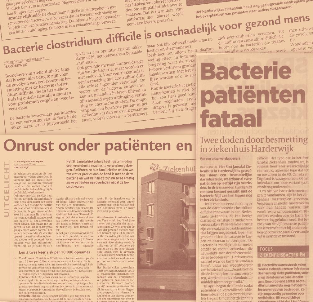

263
Clostridium difficile Colitis
 Update on Clostridium difficile Colitis Fredrick M. Abrahamian, D.O., FACEP Associate Professor of Medicine UCLA School of Medicine Director of Education Department of Emergency Medicine Olive View-UCLA
Overview of C. difficile infections. Kurt B. Stevenson, MD MPH Professor Division of Infectious Diseases
 Overview of C. difficile infections Kurt B. Stevenson, MD MPH Professor Division of Infectious Diseases Conflicts of Interest I have no financial conflicts of interest related to this topic and presentation.
11/2/2015. Update on the Treatment of Clostridium difficile Infections. Disclosure. Objectives
 Update on the Treatment of Clostridium difficile Infections Spencer H. Durham, Pharm.D.,BCPS (AQ-ID) Assistant Clinical Professor of Pharmacy Practice Auburn University Harrison School of Pharmacy Kurt
Running head: CLOSTRIDIUM DIFFICILE 1
 Running head: CLOSTRIDIUM DIFFICILE 1 Clostridium difficile Infection Christy Lee Fenton Mountainland Applied Technology College CLOSTRIDIUM DIFFICILE 2 Clostridium difficile Infection Approximately 200,000
Healthcare-associated Infections Annual Report December 2018
 December 2018 Healthcare-associated Infections Annual Report 2011-2017 TABLE OF CONTENTS INTRODUCTION... 1 METHICILLIN-RESISTANT STAPHYLOCOCCUS AUREUS INFECTIONS... 2 MRSA SURVEILLANCE... 3 CLOSTRIDIUM
Community-Associated C. difficile Infection: Think Outside the Hospital. Maria Bye, MPH Epidemiologist May 1, 2018
 Community-Associated C. difficile Infection: Think Outside the Hospital Maria Bye, MPH Epidemiologist Maria.Bye@state.mn.us 651-201-4085 May 1, 2018 Clostridium difficile Clostridium difficile Clostridium
Inappropriate Use of Antibiotics and Clostridium difficile Infection. Jocelyn Srigley, MD, FRCPC November 1, 2012
 Inappropriate Use of Antibiotics and Clostridium difficile Infection Jocelyn Srigley, MD, FRCPC November 1, 2012 Financial Disclosures } No conflicts of interest } The study was supported by a Hamilton
Infection control in Indonesian hospitals
 Infection control in Indonesian hospitals PROEFSCHRIFT ter verkrijging van de graad van Doctor aan de Universiteit Leiden, op gezag van Rector Magnificus prof.mr. P.F. van der Heijden, volgens besluit
Policy for the Management of Clostridium Difficile
 Policy for the Management of Clostridium Difficile Printed copies must not be considered the definitive version DOCUMENT CONTROL POLICY NO. Policy Group Infection Control Committee Author Dr Linsey Batchelor
The Epidemiology Of Clostridium Difficile Infections Among Oncology Patients
 Yale University EliScholar A Digital Platform for Scholarly Publishing at Yale Public Health Theses School of Public Health January 2015 The Epidemiology Of Clostridium Difficile Infections Among Oncology
Clostridium Difficile Infection (CDI) Alistair McGregor Hobart Pathology Royal Hobart Hospital TIPCU
 Alistair McGregor Hobart Pathology Royal Hobart Hospital TIPCU") Clostridium Difficile Infection (CDI) Alistair McGregor Hobart Pathology Royal Hobart Hospital TIPCU Disclosures I am not Tom Riley The Fidaxomicin guys brought me dinner once Outline ASID/AICA position
Clostridium Difficile Infection (CDI) Alistair McGregor Hobart Pathology Royal Hobart Hospital TIPCU Disclosures I am not Tom Riley The Fidaxomicin guys brought me dinner once Outline ASID/AICA position
Preventing Clostridium difficile Infection (CDI)
") 1 Preventing Clostridium difficile Infection (CDI) All Hands on Deck to Reduce CDI Skill Nursing Facility Conference July 28, 2017 Idamae Kennedy, MPH,BSN,RN,CIC Liaison Infection Preventionist Healthcare
1 Preventing Clostridium difficile Infection (CDI) All Hands on Deck to Reduce CDI Skill Nursing Facility Conference July 28, 2017 Idamae Kennedy, MPH,BSN,RN,CIC Liaison Infection Preventionist Healthcare
Clostridium Difficile Primer: Disease, Risk, & Mitigation
 Clostridium Difficile Primer: Disease, Risk, & Mitigation KALVIN YU, M.D. CHIEF INTEGRATION OFFICER, SCPMG/SCAL KAISER PERMANENTE ASSOCIATE PROFESSOR INFECTIOUS DISEASE, COLLEGE OF GLOBAL PUBLIC HEALTH,
Clostridium difficile infection: The Present and the Future
 Clostridium difficile infection: The Present and the Future Carlos E. Figueroa Castro, MD Assistant Professor, Division of Infectious Diseases Medical College of Wisconsin November 2014 I have made this
Safe Patient Care Keeping our Residents Safe Use Standard Precautions for ALL Residents at ALL times
 Safe Patient Care Keeping our Residents Safe 2016 Use Standard Precautions for ALL Residents at ALL times #safepatientcare Do bugs need drugs? Dr Deirdre O Brien Consultant Microbiologist Mercy University
Should we test Clostridium difficile for antimicrobial resistance? by author
 Should we test Clostridium difficile for antimicrobial resistance? Paola Mastrantonio Department of Infectious Diseases Istituto Superiore di Sanità, Rome,Italy Clostridium difficile infection (CDI) (first
Clostridium difficile
 Clostridium difficile A Challenge in Long-Term Care Andrew E. Simor, MD, FRCPC Sunnybrook Health Sciences Centre University of Toronto Hosted by Paul Webber paul@webbertraining.com Objectives to understand
Clostridium difficile Infection: An Update on the Current State of Prevention
 Intermountain APIC and Qualis Health present I-APIC HAI Prevention Learning Network Webinar Series Clostridium difficile Infection An Update on the April 11, 2012 Ruth CarricoPhD RN FSHEA CIC Clostridium
Preventing Multi-Drug Resistant Organism (MDRO) Infections. For National Patient Safety Goal
 Infections. For National Patient Safety Goal") Preventing Multi-Drug Resistant Organism (MDRO) Infections For National Patient Safety Goal 07.03.01 2009 Methicillin Resistant Staphlococcus aureus (MRSA) About 3-8% of the population at large is a carrier
Preventing Multi-Drug Resistant Organism (MDRO) Infections For National Patient Safety Goal 07.03.01 2009 Methicillin Resistant Staphlococcus aureus (MRSA) About 3-8% of the population at large is a carrier
8/17/2016 ABOUT US REDUCTION OF CLOSTRIDIUM DIFFICILE THROUGH THE USE OF AN ANTIMICROBIAL STEWARDSHIP PROGRAM
 Mary Moore, MS CIC MT (ASCP) Infection Prevention Coordinator Great River Medical Center, West Burlington REDUCTION OF CLOSTRIDIUM DIFFICILE THROUGH THE USE OF AN ANTIMICROBIAL STEWARDSHIP PROGRAM ABOUT
Consequences of Antimicrobial Resistant Bacteria. Antimicrobial Resistance. Molecular Genetics of Antimicrobial Resistance. Topics to be Covered
 Antimicrobial Resistance Consequences of Antimicrobial Resistant Bacteria Change in the approach to the administration of empiric antimicrobial therapy Increased number of hospitalizations Increased length
MID 23. Antimicrobial Resistance. Consequences of Antimicrobial Resistant Bacteria. Molecular Genetics of Antimicrobial Resistance
 Antimicrobial Resistance Molecular Genetics of Antimicrobial Resistance Micro evolutionary change - point mutations Beta-lactamase mutation extends spectrum of the enzyme rpob gene (RNA polymerase) mutation
Antimicrobial Resistance
 Antimicrobial Resistance Consequences of Antimicrobial Resistant Bacteria Change in the approach to the administration of empiric antimicrobial therapy Increased number of hospitalizations Increased length
Antimicrobial Resistance Acquisition of Foreign DNA
 Antimicrobial Resistance Acquisition of Foreign DNA Levy, Scientific American Horizontal gene transfer is common, even between Gram positive and negative bacteria Plasmid - transfer of single or multiple
Clinical Spectrum of Disease. Clinical Features. Risk Factors. Risk of CDAD According to Antibiotic Class. Fluoroquinolones as Risk Factor for CDAD
 Clinical Features Range from mild diarrhea to severe colitis and death Common clinical symptoms include Watery diarrhea Fever Loss of appetite Nausea Abdominal pain/tenderness Less common ileus CDC Fact
Infection Comments First Line Agents Penicillin Allergy History of multiresistant. line treatment: persist for >7 days they may be
 Gastrointestinal Infections Infection Comments First Line Agents Penicillin Allergy History of multiresistant Campylobacter Antibiotics not recommended. Erythromycin 250mg PO 6 Alternative to first N/A
Alfonso Torress-Cook, Dr.P.H. Director of Epidemiology/Patient Safety Pacific Hospital of Long Beach
 Alfonso Torress-Cook, Dr.P.H. Director of Epidemiology/Patient Safety Pacific Hospital of Long Beach Historical overview The myriad causes of hospital acquired diarrhea Microbiology and ecology of Clostridium
Antimicrobial Resistance
 Antimicrobial Resistance Consequences of Antimicrobial Resistant Bacteria Change in the approach to the administration of Change in the approach to the administration of empiric antimicrobial therapy Increased
Clostridium difficile Infection Prevention. Basics of Infection Prevention 2-Day Mini-Course 2012
 Clostridium difficile Infection Prevention Basics of Infection Prevention 2-Day Mini-Course 2012 2 Objectives Describe the etiology and epidemiology of C. difficile infection (CDI) Review evidence-based
Treatment of septic peritonitis
 Vet Times The website for the veterinary profession https://www.vettimes.co.uk Treatment of septic peritonitis Author : Andrew Linklater Categories : Companion animal, Vets Date : November 2, 2016 Septic
Healthcare-associated Infections Annual Report
 September 2014 Healthcare-associated Infections Annual Report 2009-2013 Summary Provincial Infection Control Newfoundland Labrador (PIC-NL) has collected data on inpatients and outpatients with healthcare-associated
Antimicrobial Prescribing Advice for patients with Clostridium difficile Associated Diarrhoea
 For use in: By: For: Antimicrobial Prescribing Advice for patients with Clostridium difficile Division responsible for document: Key words: Names of document authors: Job titles of document authors: Name
Clostridium difficile: Review of Treatment & Prevention through Antimicrobial Stewardship
 Clostridium difficile: Review of Treatment & Prevention through Antimicrobial Stewardship Kim Van Wyk, Pharm.D., BCPS Mountain-Pacific Quality Health Objectives Review epidemiology of Clostridium diffilcile
GUIDELINES FOR THE MANAGEMENT OF COMMUNITY-ACQUIRED PNEUMONIA IN ADULTS
 Version 3.1 GUIDELINES FOR THE MANAGEMENT OF COMMUNITY-ACQUIRED PNEUMONIA IN ADULTS Date ratified June 2008 Updated March 2009 Review date June 2010 Ratified by Authors Consultation Evidence base Changes
Healthcare-associated Infections Annual Report March 2015
 March 2015 Healthcare-associated Infections Annual Report 2009-2014 TABLE OF CONTENTS SUMMARY... 1 MRSA SURVEILLANCE RESULTS... 1 CDI SURVEILLANCE RESULTS... 1 INTRODUCTION... 2 METHICILLIN-RESISTANT
مادة االدوية المرحلة الثالثة م. غدير حاتم محمد
 م. مادة االدوية المرحلة الثالثة م. غدير حاتم محمد 2017-2016 ANTIMICROBIAL DRUGS Antimicrobial drugs Lecture 1 Antimicrobial Drugs Chemotherapy: The use of drugs to treat a disease. Antimicrobial drugs:
Preventing Clostridium difficile. July 13,
 Preventing Clostridium difficile Infection (CDI) July 13, 2011 1 Learning Objectives: Identify recent changes in the epidemiology of CDI, including transmission i and risk ikpopulations. Review recent
Cover Page. The handle holds various files of this Leiden University dissertation
 Cover Page The handle http://hdl.handle.net/1887/22747 holds various files of this Leiden University dissertation Author: Yirga Abay, Gidey Title: Ecology and conservation of spotted hyena (Crocuta crocuta
Acute Hemorrhagic Diarrhea Syndrome (AHDS) A Cause of Bloody Feces in Dogs
 A Cause of Bloody Feces in Dogs") Acute Hemorrhagic Diarrhea Syndrome (AHDS) A Cause of Bloody Feces in Dogs No dog parent wants to clean up diarrhea. Cleaning up bloody diarrhea is even more unpleasant. Unfortunately, the development
Acute Hemorrhagic Diarrhea Syndrome (AHDS) A Cause of Bloody Feces in Dogs No dog parent wants to clean up diarrhea. Cleaning up bloody diarrhea is even more unpleasant. Unfortunately, the development
Cover Page. The handle holds various files of this Leiden University dissertation.
 Cover Page The handle http://hdl.handle.net/1887/31633 holds various files of this Leiden University dissertation. Author: Kant, Anne Marie van der Title: Neural correlates of vocal learning in songbirds
Dr Eleri Davies. Consultant Microbiologist and Infection Control Doctor, Public Health Wales NHS Trust
 Dr Eleri Davies Consultant Microbiologist and Infection Control Doctor, Public Health Wales NHS Trust Antimicrobial stewardship What is it? Why is it important? Treatment and management of catheter-associated
Methicillin-Resistant Staphylococcus aureus
 Methicillin-Resistant Staphylococcus aureus By Karla Givens Means of Transmission and Usual Reservoirs Staphylococcus aureus is part of normal flora and can be found on the skin and in the noses of one
Cover Page. The handle holds various files of this Leiden University dissertation.
 Cover Page The handle http://hdl.handle.net/1887/20908 holds various files of this Leiden University dissertation. Author: Kok, Philippe Jacques Robert Title: Islands in the sky : species diversity, evolutionary
Learning Objectives 6/1/18
 Gulf Coast Multidisciplinary Pharmacotherapy Conference Kelly R. Reveles, PharmD, PhD, BCPS College of Pharmacy, The University of Texas at Austin School of Medicine, UT Health San Antonio Email: kdaniels46@utexas.edu
Development and improvement of diagnostics to improve use of antibiotics and alternatives to antibiotics
 Priority Topic B Diagnostics Development and improvement of diagnostics to improve use of antibiotics and alternatives to antibiotics The overarching goal of this priority topic is to stimulate the design,
FM - Male, 38YO. MRSA nasal swab (+) Due to positive MRSA nasal swab test, patient will be continued on Vancomycin 1500mg IV q12 for MRSA treatment...
 Due to positive MRSA nasal swab test, patient will be continued on Vancomycin 1500mg IV q12 for MRSA treatment...") Jillian O Keefe Doctor of Pharmacy Candidate 2016 September 15, 2015 FM - Male, 38YO HPI: Previously healthy male presents to ED febrile (102F) and in moderate distress ~2 weeks after getting a tattoo
Jillian O Keefe Doctor of Pharmacy Candidate 2016 September 15, 2015 FM - Male, 38YO HPI: Previously healthy male presents to ED febrile (102F) and in moderate distress ~2 weeks after getting a tattoo
Incidence of hospital-acquired Clostridium difficile infection in patients at risk
 Baptist Health South Florida Scholarly Commons @ Baptist Health South Florida All Publications 5-20-2016 Incidence of hospital-acquired Clostridium difficile infection in patients at risk Christine Ibarra
11-ID-10. Committee: Infectious Disease. Title: Creation of a National Campylobacteriosis Case Definition
 11-ID-10 Committee: Infectious Disease Title: Creation of a National Campylobacteriosis Case Definition I. Statement of the Problem Although campylobacteriosis is not nationally-notifiable, it is a disease
Glycopeptide Resistant Enterococci (GRE) Policy IC/292/10
 Policy IC/292/10") BASINGSTOKE AND NORTH HAMPSHIRE NHS FOUNDATION TRUST Glycopeptide Resistant Enterococci (GRE) Policy IC/292/10 Supersedes: IC/292/07 Owner Name Dr Nicki Hutchinson Job Title Consultant Microbiologist,
BASINGSTOKE AND NORTH HAMPSHIRE NHS FOUNDATION TRUST Glycopeptide Resistant Enterococci (GRE) Policy IC/292/10 Supersedes: IC/292/07 Owner Name Dr Nicki Hutchinson Job Title Consultant Microbiologist,
Georgia State University. Georgia State University. Zirka Thompson. Spring
 Georgia State University ScholarWorks @ Georgia State University Public Health Theses School of Public Health Spring 5-11-2012 Comparison of Risk Factors for Clostridium Difficile Infection Among Community
Newsflash: Hospital Medicine JOHN C. CHRISTENSEN, MD FACP AMERICAN COLLEGE OF PHYSICIANS, UTAH CHAPTER SCIENTIFIC MEETING FEBRUARY 10, 2017
 Newsflash: Hospital Medicine JOHN C. CHRISTENSEN, MD FACP AMERICAN COLLEGE OF PHYSICIANS, UTAH CHAPTER SCIENTIFIC MEETING FEBRUARY 10, 2017 Newsflash: Fluoroquinolones Newsflash: Fluoroquinolones Don t
Educational Module for Nursing Assistants in Long-term Care Facilities: Preventing and Managing Clostridium difficile Infections
 Educational Module for Nursing Assistants in Long-term Care Facilities: Preventing and Managing Clostridium difficile Infections Minnesota Department of Health Infectious Disease Epidemiology, Prevention,
Carbapenemase-Producing Enterobacteriaceae (CPE)
") Carbapenemase-Producing Enterobacteriaceae (CPE) September 21, 2017 Maryam Khan Peel Public Health Madeleine Ashcroft Public Health Ontario Objectives Differentiate the acronyms related to CPE (CPE,CPO,CRE,CRO)
Carbapenemase-Producing Enterobacteriaceae (CPE) September 21, 2017 Maryam Khan Peel Public Health Madeleine Ashcroft Public Health Ontario Objectives Differentiate the acronyms related to CPE (CPE,CPO,CRE,CRO)
Multi-Drug Resistant Gram Negative Organisms POLICY REVIEW DATE EXTENDED Printed copies must not be considered the definitive version
 Multi-Drug Resistant Gram Negative Organisms POLICY REVIEW DATE EXTENDED 2018 Printed copies must not be considered the definitive version DOCUMENT CONTROL POLICY NO. IC-122 Policy Group Infection Control
General Approach to Infectious Diseases
 General Approach to Infectious Diseases 2 The pharmacotherapy of infectious diseases is unique. To treat most diseases with drugs, we give drugs that have some desired pharmacologic action at some receptor
Dr. P. P. Doke. M.D., D.N.B., Ph.D., FIPHA. Professor, Department of Community Medicine, Bharati Vidyapeeth Medical College, Pune
 Dr. P. P. Doke M.D., D.N.B., Ph.D., FIPHA Professor, Department of Community Medicine, Bharati Vidyapeeth Medical College, Pune 1 Anti microbial resistance is now a global geometrically increasing threat
Please distribute a copy of this information to each provider in your organization.
 HEALTH ADVISORY TO: Physicians and other Healthcare Providers Please distribute a copy of this information to each provider in your organization. Questions regarding this information may be directed to
Introduction to Chemotherapeutic Agents. Munir Gharaibeh MD, PhD, MHPE School of Medicine, The university of Jordan November 2018
 Introduction to Chemotherapeutic Agents Munir Gharaibeh MD, PhD, MHPE School of Medicine, The university of Jordan November 2018 Antimicrobial Agents Substances that kill bacteria without harming the host.
Clostridium difficile Colitis
 1 Clostridium difficile Colitis William R. Sonnenberg, MD 2 Disclosure Dr. Sonnenberg has no conflict of interest, financial agreement, or working affiliation with any group or organization. 3 Learning
Multi-Drug Resistant Organisms (MDRO)
") Multi-Drug Resistant Organisms (MDRO) 2016 What are MDROs? Multi-drug resistant organisms, or MDROs, are bacteria resistant to current antibiotic therapy and therefore difficult to treat. MDROs can cause
Multi-Drug Resistant Organisms (MDRO) 2016 What are MDROs? Multi-drug resistant organisms, or MDROs, are bacteria resistant to current antibiotic therapy and therefore difficult to treat. MDROs can cause
LA-MRSA in the Netherlands: the past, presence and future.
 LA-MRSA in the Netherlands: the past, presence and future. Prof. Jaap Wagenaar DVM, PhD With input from Prof. Jan Kluytmans MD, PhD Department of Infectious Diseases and Immunology, Faculty of Veterinary
Source: Portland State University Population Research Center (
 Methicillin Resistant Staphylococcus aureus (MRSA) Surveillance Report 2010 Oregon Active Bacterial Core Surveillance (ABCs) Office of Disease Prevention & Epidemiology Oregon Health Authority Updated:
Section 10: Antimicrobial Stewardship and Clostridium difficile Infection: A Primer for the Infection Preventionist
 Section 10: Antimicrobial Stewardship and Clostridium difficile Infection: A Primer for the Infection Preventionist Antimicrobial stewardship may be a relatively new addition to the job responsibilities
Approved by the Food Safety Commission on September 30, 2004
 Approved by the Food Safety Commission on September 30, 2004 Assessment guideline for the Effect of Food on Human Health Regarding Antimicrobial- Resistant Bacteria Selected by Antimicrobial Use in Food
Guideline Updates Change is Inevitable Especially in Infectious Diseases!
 Guideline Updates Change is Inevitable Especially in Infectious Diseases! Vicky Shah, PharmD, BCPS Assistant Professor of Pharmacy Practice Wilkes University Nesbitt School of Pharmacy 1 Vicky Shah has
Healthcare Facilities and Healthcare Professionals. Public
 Document Title: DOH Guidelines for Antimicrobial Stewardship Programs Document Ref. Number: DOH/ASP/GL/1.0 Version: 1.0 Approval Date: 13/12/2017 Effective Date: 14/12/2017 Document Owner: Applies to:
Antimicrobial Stewardship
 Antimicrobial Stewardship Report: 11 th August 2016 Issue: As part of ensuring compliance with the National Safety and Quality Health Service Standards (NSQHS), Yea & District Memorial Hospital is required
Burton's Microbiology for the Health Sciences. Chapter 9. Controlling Microbial Growth in Vivo Using Antimicrobial Agents
 Burton's Microbiology for the Health Sciences Chapter 9. Controlling Microbial Growth in Vivo Using Antimicrobial Agents Chapter 9 Outline Introduction Characteristics of an Ideal Antimicrobial Agent How
Antimicrobial use in poultry: Emerging public health problem
 Antimicrobial use in poultry: Emerging public health problem Eric S. Mitema, BVM, MS, PhD CPD- Diagnosis and Treatment of Poultry Diseases FVM, CAVS, 6 th. August, 2014 AMR cont Antibiotics - Natural or
The Use of Procalcitonin to Improve Antibiotic Stewardship
 The Use of Procalcitonin to Improve Antibiotic Stewardship Disclosures I have no actual or potential conflict of interest in relation to this presentation. Patrick A. Laird, DNP, RN, ACNP-BC Objectives
Ca-MRSA Update- Hand Infections. Washington Hand Society September 19, 2007
 Ca-MRSA Update- Hand Infections Washington Hand Society September 19, 2007 Resistant Staph. Aureus Late 1940 s -50% S.Aureus resistant to PCN 1957-80/81 strain- of S.A. highly virulent and easily transmissible
TREAT Steward. Antimicrobial Stewardship software with personalized decision support
 TREAT Steward TM Antimicrobial Stewardship software with personalized decision support ANTIMICROBIAL STEWARDSHIP - Interdisciplinary actions to improve patient care Quality Assurance The aim of antimicrobial
biomérieux, Inc. 100 Rodolphe Street Durham, NC U.S.A. Tel. : (800) Fax : (800)
 Fax : (800)") 2014 BIOMÉRIEUX, INC. BIOMÉRIEUX, THE BLUE LOGO, EMPOWERING CLINICAL DECISIONS, API, CHROMID, DIVERSILAB, ETEST, VIDAS, VIGIGUARD, VITEK ARE USED PENDING AND/OR REGISTERED TRADEMARKS BELONGING TO BIOMÉRIEUX
2014 BIOMÉRIEUX, INC. BIOMÉRIEUX, THE BLUE LOGO, EMPOWERING CLINICAL DECISIONS, API, CHROMID, DIVERSILAB, ETEST, VIDAS, VIGIGUARD, VITEK ARE USED PENDING AND/OR REGISTERED TRADEMARKS BELONGING TO BIOMÉRIEUX
Global Status of Antimicrobial Resistance with a Focus on Nepal
 Global Status of Antimicrobial Resistance with a Focus on Nepal John Ferguson, John Hunter Hospital, University of Newcastle, NSW, Australia Infectious Diseases Physician and Medical Microbiologist SIMON
Duke University Hospital Guideline for Empiric Inpatient Treatment of Cancer- Related Neutropenic Fever in Adult Patients
 Duke University Hospital Guideline for Empiric Inpatient Treatment of Cancer- Related Neutropenic Fever in Adult Patients PURPOSE Fever among neutropenic patients is common and a significant cause of morbidity
Antibiotic stewardship in long term care
 Antibiotic stewardship in long term care Shira Doron, MD Associate Professor of Medicine Division of Geographic Medicine and Infectious Diseases Tufts Medical Center Boston, MA Consultant to Massachusetts
APPROVED PACKAGE INSERT. Each capsule contains clindamycin hydrochloride equivalent to 150 mg clindamycin base.
 APPROVED PACKAGE INSERT SCHEDULING STATUS: S4 PROPRIETARY NAMEAND DOSAGE FORM: DALACIN C TM 150 mg (Capsules) COMPOSITION: Each capsule contains clindamycin hydrochloride equivalent to 150 mg clindamycin
03/09/2014. Infection Prevention and Control A Foundation Course. Talk outline
 Infection Prevention and Control A Foundation Course 2014 What is healthcare-associated infection (HCAI), antimicrobial resistance (AMR) and multi-drug resistant organisms (MDROs)? Why we should be worried?
C.difficile Re-emergence of an Old Pathogen
 C.difficile Re-emergence of an Old Pathogen Gonzalo Bearman MD, MPH Assistant Professor of Medicine, Epidemiology and Community Health Associate Hospital Epidemiologist Virginia Commonwealth University
Ed J. Kuijper National Reference Laboratory for Clostridium difficile Leiden University Medical Center
 Clinical spectrum of Clostridium difficile Infection (CDI) and the emergence of hypervirulent strains Ed J. Kuijper National Reference Laboratory for Clostridium difficile Leiden University Medical Center
Other Enterobacteriaceae
 GUIDE TO INFECTION CONTROL IN THE HOSPITAL CHAPTER NUMBER 50: Other Enterobacteriaceae Author Kalisvar Marimuthu, MD Chapter Editor Michelle Doll, MD, MPH Topic Outline Topic outline - Key Issues Known
Principles of Antimicrobial therapy
 Principles of Antimicrobial therapy Laith Mohammed Abbas Al-Huseini M.B.Ch.B., M.Sc, M.Res, Ph.D Department of Pharmacology and Therapeutics Antimicrobial agents are chemical substances that can kill or
SOFT Movement Survey of FMT Programs
 Appendix 1 (as supplied by the authors): Survey SOFT Movement Survey of FMT Programs Part 1: General Information about your Fecal Microbiota Transplant (FMT) Program 1) Please fill out the information
CDI Management in Post-Acute Care: Part 1
 CDI Management in Post-Acute Care: Part 1 Robin Jump, MD, PhD VISN10 Geriatric Research Education and Clinical Center (GRECC) Louis Stokes Cleveland VA Medical Center Case Western Reserve University Robin.Jump@va.gov
ANTHRAX. INHALATION, INTESTINAL and CUTANEOUS ANTHRAX
 INHALATION, INTESTINAL and CUTANEOUS ANTHRAX CPMP/4048/01, rev. 3 1/7 General points on treatment Anthrax is an acute infectious disease caused by Bacillus anthracis, that may be infecting man via cutaneous
Necrotizing Soft Tissue Infections: Emerging Bacterial Resistance
 Necrotizing Soft Tissue Infections: Emerging Bacterial Resistance Eileen M. Bulger, MD Professor of Surgery Harborview Medical Center University of Washington Objectives Review definition & diagnostic
Why Don t These Drugs Work Anymore? Biosciences in the 21 st Century Dr. Amber Rice October 28, 2013
 Why Don t These Drugs Work Anymore? Biosciences in the 21 st Century Dr. Amber Rice October 28, 2013 Outline Drug resistance: a case study Evolution: the basics How does resistance evolve? Examples of
28/08/2017. Infection Prevention and Control. Safe Patient Care Bugs and Drugs The ongoing challenge of MDROs and AMR
 Safe Patient Care Bugs and Drugs The ongoing challenge of MDROs and AMR 2017 Safe Patient Care 2017: The Ongoing Challenge of MDROs and AMR Management of the Patient Environment in relation to Multidrug
Infection Control & Prevention
 Infection Control & Prevention Objectives: Define the term multi-drug resistant organism (MDRO). Recognize risk factors for developing MDROs. Describe the clinical manifestations and medical treatment
International Food Safety Authorities Network (INFOSAN) Antimicrobial Resistance from Food Animals
 Antimicrobial Resistance from Food Animals") International Food Safety Authorities Network (INFOSAN) 7 March 2008 INFOSAN Information Note No. 2/2008 - Antimicrobial Resistance Antimicrobial Resistance from Food Animals SUMMARY NOTES Antimicrobial
International Food Safety Authorities Network (INFOSAN) 7 March 2008 INFOSAN Information Note No. 2/2008 - Antimicrobial Resistance Antimicrobial Resistance from Food Animals SUMMARY NOTES Antimicrobial
GASTRO-INTESTINAL TRACT INFECTIONS - ANTIMICROBIAL MANAGEMENT
 GASTRO-INTESTINAL TRACT INFECTIONS - ANTIMICROBIAL MANAGEMENT DRAFT AS CURRENTLY OUT FOR CONSULTATION BUT CAN BE UTILISED IN PRESENT FORMAT Name & Title Of Author: Date Revised: Approved by Committee/Group:
Inhibiting Microbial Growth in vivo. CLS 212: Medical Microbiology Zeina Alkudmani
 Inhibiting Microbial Growth in vivo CLS 212: Medical Microbiology Zeina Alkudmani Chemotherapy Definitions The use of any chemical (drug) to treat any disease or condition. Chemotherapeutic Agent Any drug
Hand washing, Asepsis, Precautions and Infection Control
 Hand washing, Asepsis, Precautions and Infection Control FN Ch 12, NICS Ch4 Week 2 Lesa McArdle, MSN, RN Objectives Hand washing, Asepsis, Precautions & Infection Control Explain the chain of infection
Antibiotic therapy of acute gastroenteritis
 Antibiotic therapy of acute gastroenteritis Potential goals Clinical improvement (vs control) Fecal eradication of the pathogen and decrease infectivity Prevent complications Acute gastroenteritis viruses
Antibiotic Updates: Part II
 Antibiotic Updates: Part II Fredrick M. Abrahamian, DO, FACEP, FIDSA Health Sciences Clinical Professor of Emergency Medicine David Geffen School of Medicine at UCLA Los Angeles, California Financial Disclosures
Antimicrobial Stewardship in the Hospital Setting
 GUIDE TO INFECTION CONTROL IN THE HOSPITAL CHAPTER 12 Antimicrobial Stewardship in the Hospital Setting Authors Dan Markley, DO, MPH, Amy L. Pakyz, PharmD, PhD, Michael Stevens, MD, MPH Chapter Editor
ESBL Producers An Increasing Problem: An Overview Of An Underrated Threat
 ESBL Producers An Increasing Problem: An Overview Of An Underrated Threat Hicham Ezzat Professor of Microbiology and Immunology Cairo University Introduction 1 Since the 1980s there have been dramatic
ANTIMICROBIAL STEWARDSHIP: THE ROLE OF THE CLINICIAN SAM GUREVITZ PHARM D, CGP BUTLER UNIVERSITY COLLEGE OF PHARMACY AND HEALTH SCIENCES
 ANTIMICROBIAL STEWARDSHIP: THE ROLE OF THE CLINICIAN SAM GUREVITZ PHARM D, CGP BUTLER UNIVERSITY COLLEGE OF PHARMACY AND HEALTH SCIENCES 1 Crisis: Antibiotic Resistance Success Strategy 2 OBJECTIVES Discuss
Antibacterial Agents & Conditions. Stijn van der Veen
 Antibacterial Agents & Conditions Stijn van der Veen Antibacterial agents & conditions Antibacterial agents Disinfectants: Non-selective antimicrobial substances that kill a wide range of bacteria. Only
The Infected Implant in Orthopaedic Reconstruction: An Update on the Clinical and Molecular Approaches to Prevention and Diagnosis
 The Infected Implant in Orthopaedic Reconstruction: An Update on the Clinical and Molecular Approaches to Prevention and Diagnosis (Organized by the Musculoskeletal Tumor Society (MSTS) and ORS) Organizers: