A New Lower Permian Trematopid (Temnospondyli: Dissorophoidea) from Richards Spur, Oklahoma
|
|
|
- Poppy Patrick
- 5 years ago
- Views:
Transcription
1 A New Lower Permian Trematopid (Temnospondyli: Dissorophoidea) from Richards Spur, Oklahoma by Brendan Polley A thesis submitted in conformity with the requirements for the degree of Master of Science Department of Ecology and Evolutionary Biology University of Toronto Copyright by Brendan Polley 2009
2 A new Lower Permian trematopid (Temnospondyli: Dissorophoidea) from Richards Spur, Oklahoma Abstract Brendan Polley Master of Science Department of Ecology and Evolutionary Biology University of Toronto 2009 A new trematopid amphibian, Acheloma dunni, is reported based on excellently preserved cranial and postcranial elements recovered from the Lower Permian fissure fill deposits of the Dolese Brothers Co. limestone quarry near Richards Spur, Oklahoma. The new taxon is characterized by distinct lateral exposures of the palatine (l.e.p.) and ectopterygoid (l.e.e.) completely enclosed within the suborbital elements. This large, terrestrial adapted carnivore may represent the top predator of the Richards Spur assemblage. A phylogenetic analysis including 11 ingroup taxa and 54 cranial characters yielded a single most parsimonious tree, placing A. dunni within the monophyletic Trematopidae as the sister taxon to Acheloma cumminsi. Furthermore, the analysis supports including the enigmatic Ecolsonia and Actiobates within Trematopidae, forming a clade with the Upper Pennsylvanian Anconastes and the Lower Permian Tambachia. The study is the first to comprehensively analyze all valid and aberrant forms of Trematopidae within the context of broader dissorophoid phylogeny. ii
3 Acknowledgments I am indebted to the many individuals whose support and guidance truly enriched this challenging and rewarding year. Thank you to my supervisor, R. Reisz for being an unwavering source of encouragement and direction. As you did for me, I will be sure to have faith in my own students, treat them with respect, and push them to realize their potential. I am grateful to D. Scott for her invaluable technical training, for photographing specimens, and for, of course, her friendship. Thank you to my friends in the Reisz lab, N. Campione, J. Fröbisch, N. Fröbisch, J. Hawthorn, D. Mazierski, and N. Wong Ken for all their advice and support. Also, many thanks to my advisors D. Evans and D. Walsh for your guidance and constructive feedback. Special thanks to B. Dunn and M. Feese for their continuing efforts to collect fossils from Richards Spur. This project, among countless others, would not have been made possible without their generous donations to the Sam Noble Oklahoma Museum of Natural History. I am grateful to D. Berman of the Carnegie Museum of Natural History, Pittsburgh, PA; R. Ethington of the University of Missouri, Columbia, MO; M. Norell of the American Museum of Natural History, New York, NY; and M. Ryan of the Cleveland Museum of Natural History, Cleveland, OH for allowing me access to various trematopid specimens for study. Finally, thank you to my family, friends, and especially J. Cawaling for their unconditional love and support throughout the course of this project and beyond. iii
4 Table of Contents Acknowledgments... iii Table of Contents... iv List of Figures... vi List of Appendices... viii 1 Introduction Materials Abbreviations Institutional Abbreviations Anatomical Abbreviations Systematic Paleontology Type Specimen Referred Specimens Diagnosis Occurrence Etymology Description General Skull Roof Palate Braincase Lower Jaw Axial Skeleton Appendicular Skeleton Discussion iv
5 6.1 Phylogenetic Analysis Habits and Lifestyle References Figures and Captions Appendix Appendix v
6 List of Figures Figure 1. Reconstruction of skull of Acheloma dunni in A, dorsal view; B, ventral view; C, right lateral view. Scale = 50 mm Figure 2. Skull of Acheloma dunni, holotype (OMNH 73281) in dorsal view. Scale = 50 mm Figure 3. Skull of Acheloma dunni, holotype (OMNH 73281) in right lateral view. Scale = 50 mm Figure 4. Acheloma dunni, referred specimen (BMRP ) in left lateral view. Scale = 10 mm Figure 5. Acheloma dunni, referred specimen (BMRP ) in right lateral view. Scale = 10 mm Figure 6. Acheloma dunni, referred specimen (OMNH 52365). Partial right upper jaw articulation in dorsal view. Scale = 10 mm Figure 7. Skull of Acheloma dunni, holotype (OMNH 73281) in ventral view. Scale = 50 mm Figure 8. Skull of Acheloma dunni, holotype (OMNH 73281) in occipital view. Scale = 50 mm Figure 9. Partial left lower jaw of Acheloma dunni, holotype (OMNH 73281) in A, lateral view; B, medial view; C, dorsal view; D, ventral view. Scale = 10 mm Figure 10. Partial right lower jaw of Acheloma dunni, holotype (OMNH 73281) in A, lateral view; B, medial view; C, dorsal view; D, ventral view. Scale = 10 mm Figure 11. Acheloma dunni, referred specimen (OMNH 52365). Partial right lower jaw articulation in A, lateral view; B, posterior view. Scale = 10 mm Figure 12. Atlas-axis complex and associated cervical vertebrae of Acheloma dunni, holotype (OMNH 73281) in A, right lateral view; B, left lateral view; C, anterior view. Scale = 10 mm vi
7 Figure 13. Acheloma dunni, referred specimen (OMNH 52545). Right humerus in A, posterior view; B, extensor view; C, anterior view; D, flexor view; E, proximal view; F, distal view. Scale = 50 mm Figure 14. Acheloma dunni, referred specimen (OMNH 73514). Partial pelvic girdle and hindlimb. Scale = 5 mm. Illustration by Heidi Richter. Figure 15. Single most parsimonious tree of dissorophoid relationships derived from cladistic analysis using PAUP 4.0b10 with A, relevant synapomorphies mapped on (* indicates a synapomorphy that supports the clade, but appears elsewhere in the tree); B, bootstrap (bold) and Bremer decay (italics) values calculated for the analysis; TL, tree length; CI, consistency index; RC, rescaled consistency index vii
8 List of Appendices Appendix 1. Data matrix used for phylogenetic analysis including 12 taxa and 54 characters. a represents polymorophic characters (0&1) Appendix 2. Descriptions of characters used in phylogenetic analysis. Characters 1-24 are informative cranial characters based on Schoch and Rubridge (2005). Characters are taken from Ruta & Bolt (2006). Characters are from literature as cited. viii
9 1 1 Introduction The Dolese limestone quarry, situated near Richards Spur, Oklahoma is home to the most diverse known assemblage of Paleozoic terrestrial tetrapods. Regular excavation of Ordovician Arbuckle limestone has continually yielded fossil-rich clays and conglomerates (Olson, 1991). Although often disarticulated, fossil material is abundant and well preserved, boasting representatives of numerous different taxa (Reisz, 2007; Frobisch & Reisz, 2008). A significantly large proportion of this fossil material (Daly, 1973; Olson, 1991) has been assigned to the small terrestrial eureptile, Captorhinus aguti. Olson (1991) interpreted the relative abundance of C. aguti material as indicative of an Early Permian age for the faunal assemblage. More specifically, he inferred that the fauna of Richards Spur was contemporaneous with the Leonardian Arroyo Formation of the Clear Fork Group of Texas (Olson, 1991). This assignment has been further supported by biostratigraphic evidence based on the fauna recovered from the South Grandfield locality of southern Oklahoma (Daly, 1973; Sullivan et al., 2000; Maddin et. al., 2006). The Richards Spur assemblage represents a dramatic departure from contemporary Early Permian faunas. Whereas the majority of North American localities typically yield faunal samples consistent with aquatic, semi-aquatic, and terrestrial forms of lowland, deltaic environments (Olson, 1952), the Richards Spur assemblage is comprised exclusively of small and medium-sized terrestrial forms. This unique faunal composition has led to the suggestion that the locality has preserved a predominantly arid, upland assemblage (Sullivan et al., 2000; Anderson & Reisz, 2003; Schoch, 2009). Although rare, skeletal elements of larger taxa have been reported from Richards Spur, corroborating the hypothesis that the distinct composition of the fossil assemblage may have been the product of paleoecological factors, rather than taphonomic biases (Sullivan et al., 2000). These large remains include isolated elements assignable to a varanopid (Maddin et al., 2006), the sphenacodontid Thrausmosaurus (Evans et. al., 2009), the dissorophid Cacops (Sullivan et al., 2000; Reisz et al., 2009), a large trematopid suggested to be Acheloma (Bolt, 1974a; Shultze & Chorn, 1983; Sullivan et al., 2000), Seymouria (Sullivan & Reisz, 1999), and an unidentified eryopid, possibly Eryops (Olson, 1991). Similar to the typical smaller taxa, the large forms recovered from Richards Spur appear to represent taxa that were primarily terrestrial. As early amniotes, there is little doubt that the varanopid and sphenacodontid were well suited for life on land. Dissorophids and trematopids,
10 2 collectively forming the clade Olsoniformes, are considered the most terrestrial representatives of late Paleozoic temnospondyl amphibians (Bolt, 1969; Berman et al., 1987; Sumida et al., 1998; Dilkes & Brown, 2007; Markey & Marshall, 2007; Schoch, 2009). Likewise, authors agree Seymouria was principally terrestrial, dependant on an aquatic habitat only during reproduction and very early growth stages (Sullivan & Reisz, 1999; Berman et al., 2000; Klembara et al., 2007; Schoch, 2009). While it may be argued that eryopids may have been aquatic or semiaquatic, the identification of the skull fragment as Eryops is doubtfull, and the fragment is not diagnostic. In fact, we propose that it is more appropriate to designate the skull fragment as Temnospondyli incertae sedis. Described here is a new large trematopid skull (OMNH 73281) collected from Richards Spur in While trematopids were previously known from Richards Spur on the basis of isolated, fragmentary elements, the newly recovered skull is nearly complete, showing little distortion. This material represents the largest recorded taxon of the Richards Spur assemblage, rivaling the varanopid described by Maddin et al. (2006) in size. Trematopids form a monophyletic clade of poorly understood temnospondyl amphibians known from the Late Pennsylvanian and Early Permian of North America and central Germany (Berman et al., 1987; Dilkes, 1990; Sumida et al., 1998). The taxonomic history of Trematopidae is long and convoluted and while the monophyly of the clade has remained largely unquestioned, the group as a whole has yet to be subject to rigorous phylogenetic analysis. Trematopidae, as erected by Williston (1910) was originally a modest group, including only two taxa known from Lower Permian type specimens: Acheloma cumminsi (Cope, 1882) and Trematops milleri (Williston, 1909). In 1926, Mehl described an additional species of Trematops, T. thomasi; however, the family remained largely unexamined until Olson s (1941) comprehensive study. In his review, he erected several new species of Acheloma and Trematops: A.pricei, A.whitei, and T.willistoni. He later expanded the family by describing Trematopsis seltini (Olson, 1956) and Trematops stonei (Olson, 1970). The type specimen of Trematopsis was later found to be a junior synonym of Cacops (Milner, 1985b). Moreover, Olson was first to recognize the close affinities between trematopids and the diverse group of terrestrially adapted amphibians of the family Dissorophidae. Dissorophids span a greater temporal and geographic range than trematopids and are distinguished from trematopids by the possession of armored
11 3 dermal plates associated with the neural spines and the absence of an elongated external naris (Olson, 1941; Carroll, 1964a; DeMar, 1966b; DeMar, 1968). Subsequent authors also recognized the close relationship of trematopids and dissorophids (Vaughn, 1969; Berman et al. 1985; 1987; Dilkes, 1990; Daly, 1994; Sumida et al., 1998; Anderson et al., 2008). However, because ingroup relationships of both families remained largely unresolved, several taxonomic problems arose when forms seemingly possessing a combination of trematopid and dissorophid characters were discovered and described. Mordex calliprepes and Parioxys ferricolus were both originally described as trematopids (Romer, 1947). Mordex was later synonymized with Amphibamus (Carroll, 1964a) only to be resurrected as a dissorophid with a trematopid-like elongated external naris (Milner, 1986). Presently, Mordex is in the process of once again being reclassified as a basal trematopid (Milner, 2007). Parioxys is still considered closely related to dissorophids, but has been removed from Trematopidae and placed within its own family, Parioxyidae (Mustafa, 1955; Carroll, 1964b). Longiscitula houghae (DeMar, 1966a) was originally described as possessing both a trematopidlike external naris and the distinctive dermal armor of dissorophids. Reexamination of the type specimen revealed the apparent elongated external naris was an artifact of preservation (Bolt, 1974c). Milner (2003) eventually synonymized L. houghae with Dissorophus multicintus. The enigmatic Ecolsonia cutlerensis was described as sharing affinities with dissorophids but was tentatively assigned to Trematopidae by Vaughn (1969) based on its elongated external naris and lack of exostoses and armor. Berman et al. (1985) argued the skull proportions and structure of the otic notch of Ecolsonia were in fact indicative of dissorophid affinities, and that its elongated external naris evolved convergently. Daly (1994) incorporated Ecolsonia into her study of dissorophoid relationships but was unable to resolve its assignment (Sumida et al., 1998). Eaton (1973) described the Late Pennsylvanian Actiobates peabodyi as a dissorophid; however, he stipulated that the distinction between trematopids and dissorophids was nonexistent and all taxa assigned to both families should fall under Dissorophidae. Later authors that recognized both families agree Actiobates is a trematopid possibly exhibiting an early stage of development (Berman et al., 1985; Milner, 1985; Daly, 1994). Partly for these reasons, both Ecolsonia and Actiobates have been excluded from most cladistic analyses of trematopid relationships (Berman et al. 1987; Dilkes, 1990; Sumida et al. 1998).
12 4 Currently, Trematopidae consists of five known genera. Actiobates peabodyi is at least provisionally still a valid trematopid (Berman et al., 1987; Milner, 1985; Dilkes, 1990; Daly, 1994; Sumida et al., 1998). Anconastes vesperus (Berman et al., 1987) represents a Late Pennsylvanian trematopid from Texas. Dilkes and Reisz (1987) found Trematops milleri to be a junior synonym of Acheloma cumminsi, and retained the name Acheloma for the genus. Dilkes (1990) synonymized the small Early Permian trematopid taxa erected by Olson (1941) into a new genus, Phonerpeton pricei. Finally, the Early Permian Tambachia trogallas (Sumida et al., 1998) recovered from central Germany represents the most recent addition to the family. Here we describe a new species of trematopid and comprehensively revaluate the interrelationships of trematopids using phylogenetic analysis. Furthermore, this study is the first to include the aberrant Ecolsonia and Actiobates in an analysis of trematopid ingroup relationships in an attempt to resolve their taxonomic positions and define Trematopidae. 2 Materials The holotype, OMNH 73281, consists of an exceptionally preserved skull measuring 164 mm along its midline. The skull has experienced no crushing but has been slightly sheared to the right. All other damage appears to have occurred as a result of the blasting and transport operations of the quarry from which it was recovered. The antorbital region of the left side of the skull is substantially damaged and remains unprepared. The rest of the skull has been prepared and shows little sign of damage with the following exceptions: the posterior portion of the quadratojugals and tabulars are broken, while the quadrates are missing entirely. Also, a break is present along the roof of the skull between the frontal and nasal, extending onto a portion of the prefrontal. The palate has been extensively prepared; leaving only a small area along the medial edges of the right premaxilla and maxilla obscured by matrix and a tightly associated captorhinid lower jaw. Only the most anterior portions of the lower jaws are present but remain disarticulated. The atlas-axis complex is present along with disarticulated elements comprising the third and fourth cervical vertebrae. The referred specimens consist of well-preserved large postcranial elements, jaw joint, and two small partial skulls. The pelvic girdle OMNH is nearly complete and allied with a femur, tibia, fibula, and disarticulated elements of the pes. OMNH consists of partial elements
13 5 comprising the jaw articulation. OMNH is a complete right humerus. BMRP is a small partial skull measuring 56 mm along its dorsal midline. It appears to have been extensively acid prepared, leaving the dermal sculpturing severely eroded but all sutures clearly visible. The anterior most half of the skull is absent along with the braincase, palate and most of the lower jaw. BMRP is a partial skull consisting only of the antorbital bar and its associated cheek region. The holotype of Acheloma cumminsi (AMNH 4205) was examined for anatomical comparison. The material consists of a nearly complete but substantially crushed skull, a string of 22 articulated presacral vertebrae, both scapulocoracoids, and both humeri. Also examined, was the type specimen of Acheloma stonei (CMNH 10969), represented by a distorted partial skull. The specimen was observed for comparative purposes but left out of analysis due to its lack of informative characters. A cast of the only specimen of Acheloma thomasi (MU 501) was also made available for study. Specimens of almost all other trematopid taxa were also available for study, including the holotype (CM 41711) and paratype (CM 28590) of Anconastes vesperus; the holotype (UCLA VP 1734) and paratypes (CM 38017; CM 41703) of Ecolsonia cutlerensis; and the holotype (MNG 7722) of Tambachia trogallas. Observations of Phonerpeton pricei were based on a complete skull with partial postcranial skeleton (AMNH 7150) and published reconstructions (Dilkes, 1990). Specimens of Actiobates peabodyi were not examined. Comparisons and character coding was accomplished using published illustrations and descriptions (Eaton, 1973; Milner, 1985a).
14 6 3 Abbreviations 3.1 Institutional Abbreviations AMNH, American Museum of Natural History, New York, NY; BMRP, Burpee Museum of Natural History, Rockford, IL; CM, Carnegie Museum of Natural History, Pittsburgh, PA; CMNH, Cleveland Museum of Natural History, Cleveland, OH; MCZ, Museum of Comparative Zoology, Harvard University, Cambridge, MA; MNG, Museum der Natur, Gotha, Germany; MU, University of Missouri, Columbia, MO; OMNH, Sam Noble Oklahoma Museum of Natural History, University of Oklahoma, Norman, OK; UCLA VP, University of California, Los Angelas, CA. 3.2 Anatomical Abbreviations a, angular; art, articular; at, atlas; axn, axial neural spine; axi, axial intercentrum; bo, basioccipital; c, centrale; c3, third cervical vertebra; c4, fourth cervical vertebra; cr, cervical rib; ct, cultriform process; d, dentary; ect, ectopterygoid; eo, exoccipital; f, frontal; fe, femur; fi, fibula; i, intermedium; il, ilium; in.f, internarial fenestra; is, ischium; j, jugal; l, lacrimal; l.e.e., lateral exposure of the ectopterygoid; l.e.p., lateral exposure of the palatine; m, maxilla; n, nasal; obt, obturator foramen; op, opisthotic; p, parietal; paf, para-articular foramen; pal, palatine; pc, pleurocentrum; pco, precoronoid; pf, postfrontal; pm, premaxilla; po, postorbital; pp, postparietal; pra, prearticular; prf, prefrontal; ps, parasphenoid; pt, pterygoid; ptf, post temporal fenestra; pu, pubis; q, quadrate; qj, quadratojugal; s, stapes; sa, surangular; se, sphenethmoid; sm, septomaxilla; so, supraocciptal; sp, splenial; sq, squamosal; st, supratemporal; t, tabular; ti, tibia; tib, tibiale; v, vomer
15 7 4 Systematic Palaeontology Temnospondyli Zittel, 1888 Euskelia Yates and Warren, 2000 Dissorophoidea Bolt, 1969 Olsoniformes Anderson et al., Revised Diagnosis Trematopidae Williston, 1910 The clade consisting of Acheloma cumminsi and all other taxa that share a more recent common ancestor with A. cumminsi than with Dissorophus. Acheloma Cope, Revised Generic Diagnosis Large trematopid temnospondyl characterized by the following autapomorphies: toothed, raised crest running anteroposteriorly along the vomer, mesial to the choana; otic notch with a nearly horizontal ventral margin. Acheloma dunni, sp. nov. (Fig. 1-14) 4.3 Type Specimen OMNH 73281, nearly complete skull with associated atlas-axis complex and partial lower jaw.
16 8 4.4 Referred Specimens BMRP , small trematopid skull; BMRP , trematopid snout; OMNH 52365, jaw articulation; OMNH 73514, pelvic girdle; OMNH 52545, right humerus 4.5 Diagnosis Large trematopid temnospondyl amphibian distinguished from all other trematopids by sculptured lateral exposures on skull roof of both palatine and ectopterygoid that are excluded from the ventral margin orbit by the lacrimal and jugal. Differs from Acheloma cumminsi in having a relatively narrower and less robust snout, and a proportionally smaller internarial fenestra. 4.6 Occurrence Dolese Brothers Co. limestone quarry, near Richards Spur, Comanche County, Oklahoma; fissure fill deposits in Ordovician Arbuckle limestone probably equivalent to Leonardian Arroyo Formation of Clear Fork Group, Lower Permian. 4.7 Etymology The specific name honors Brent Dunn, who has graciously collected and donated several specimens from the Dolese Brothers Co. limestone quarry for study. 5 Description 5.1 General As observed in all other trematopids (Berman et al., 1987; Dilkes, 1990; Sumida et al., 1998), the skull of Acheloma dunni is tall, box-like, and roughly triangular in shape (Fig.1 & 2). The surface of the skull roof is covered in deep, rounded dermal pitting, about uniform in shape. Overall, the skull most closely resembles Acheloma cumminsi in its large size, the presence of an enlarged, elongate key-hole shaped, external narial opening, and slitlike otic notch with broad, overhanging shelf (Dilkes & Reisz, 1987). However, while the skull roof of OMNH is slightly larger in both overall length and width than the holotype of A. cumminsi (AMNH 4205),
17 9 the snout of A. dunni is narrower and considerably less robust. Descriptions are based primarily on OMNH and others specimens as cited. 5.2 Skull Roof The premaxilla of A. dunni has a well-developed posterodorsal alary process, lapping onto the anterolateral area of the nasal. An internarial fenestra measuring 3 mm in diameter is situated along the midline suture between the premaxillae. The same structure is present in the type specimen of A. cumminsi but is proportionately larger, measuring 7 mm in diameter. A proportionately large internarial fenestra is also present in Phonerpeton (Dilkes, 1990), while the structure appears to be completely absent in all other trematopids (Berman et al., 1985; Berman et al., 1987; Dilkes, 1990; Sumida et al., 1998). The presence of an internarial fenestra is not exclusive to trematopids as it is found in a variety of other taxa, including the dissorophid Cacops (Bolt, 1977a, Reisz et al Mehl (1926) described the partial snout of the Lower Permian trematopid Acheloma thomasi (as Trematops thomasi) from Snyder, Oklahoma that possessed a reduced or perhaps absent internarial fenestra. Authors have generally accepted Olson s (1941) suggestion that A. thomasi is a junior synonym of A. cumminsi (as T. milleri), although the assertion was made only under the condition that the internarial fenestra was in fact present. Mehl did not list a specimen number in his description and it appears the only record of A. thomasi material is of a cast of the type specimen (MU 501) in the University of Missouri at Columbia collections (Katz, 2008). The cast is in poor condition; however, it is informative enough to indicate the internarial fenestra is absent. Seven teeth are preserved on each premaxilla, along with another six empty alveoli. Initially, there is a steady increase in size of teeth posteriorly, with the largest being in the ninth or tenth tooth position. The last three teeth in the premaxillary series decrease in size posteriorly. The preserved teeth on the maxilla exhibit a similar pattern, at first increasing in size posteriorly and reaching their maximum size at about the seventh or eighth tooth position, beneath the posterior margin of the external naris. The remaining teeth gradually decrease in size posteriorly. There are 18 preserved teeth on the right maxilla with spaces for at least ten more. The maxilla is a long slender element, stretching posteriorly past the level of the anterior border of the otic notch. The maxilla has the greatest dorsal expansion at the level of the largest teeth, beneath the enlarged external narial opening. Its dorsal edge makes up the majority of the ventral border of the
18 10 external naris where the maxilla also has a well-developed medial shelf that forms the floor of the external naris. The conspicuous elongated external narial opening, often cited as the defining character of trematopids (Olson, 1941; Vaughn, 1969; Olson, 1970; Eaton, 1973; Daly, 1994), is bordered by the premaxilla, maxilla, nasal, prefrontal, and lacrimal. The increase in the height of the maxilla in conjunction with a lateral expansion of the nasal along the margin of the external naris, partitions the external naris into distinct anterior and posterior sections. This gives the narial opening an overall keyhole shape (Fig. 3). The dorsal expansion of the maxilla occurs just posterior to the area where the septomaxilla is located in the opening in all other trematopids (Sumida et. al., 1998), including Ecolsonia, delineating the posterior extent of the functional external naris (Bolt, 1974c; Berman et. al., 1987; Dilkes, 1993). Although the septomaxilla is not preserved in A. dunni, it probably was in a similar position. A laterally concave, smooth lamina known as the narial flange (Dilkes, 1990; Dilkes, 1993) descends from the ventral surface of the skull roof and lies in close association with the external naris. Comprised of contributions of the nasal, prefrontal, and lacrimal, the narial flange makes broad contact with the antorbital bar. This condition is common among other trematopid taxa (Eaton, 1973; Berman et. al., 1987; Dilkes & Reisz, 1987; Dilkes, 1990; Sumida et al., 1998). A narial flange has been found in other dissorophoids including Doleserpeton (Bolt, 1974c; Carroll, 1964a; Reisz et. al., 2008); however, the structure does not appear to contact the antorbital bar in any of these taxa. A narial flange in this case is visible in the holotype of Ecolsonia in cross section; however, the configuration of the narial flange cannot be confirmed because the area surrounding the antorbital bar remains absent or unprepared in all specimens. The external naris of A. dunni is floored by the vomer and palatine. The choana lies well anterior to the posterior margin of the external naris as in A. cumminsi (Dilkes & Reisz, 1987). The posterior border of the choana is in line with the posterior margin of the external naris in all other trematopids (Eaton, 1973; Berman et. al., 1985; Berman et. al., 1987; Dilkes, 1993; Sumida et. al., 1998). A partially prepared sheet of bone is exposed medial to the narial flange. It appears to be the dorsally directed laminar process running along the midline of the rostrum, referred to as the median vomerine septum by Dilkes (1990). This structure is present in A. cumminsi, Phonerpeton (Dilkes, 1990), and was also revealed in Anconastes and Tambachia (Sumida et. al. 1998). Examination of the holotype of Ecolsonia (UCLA VP 1734) indicates a median
19 11 vomerine septum is present. Contact between the skull roof and the median vomerine septum can be confirmed in only A. cumminsi (Dilkes & Riesz, 1987); however, the same structure approaches the skull roof but does not contact it in Phonerpeton (Dilkes, 1990), and in Ecolsonia. The dorsal extent of the median vomerine septum is unknown in either specimen of Anconastes (CM 41711; CM 28590) or Tambachia (MNG 7722). If the median vomerine septum made even cartilaginous contact with the skull roof, the strut-like structure may have supported compressive and shearing forces acting on the rostrum and vomers respectively during feeding. Anteriorly, the vomerine walls bifurcate, contacting the ventrolateral surfaces of the premaxillae. The lacrimal makes up the ventral half of the antorbital bar, contributing to both the posteroventral margin of the external narial opening and the anteroventral margin of the orbit. Anteriorly, the bone has a relatively short subnarial process, suturing bluntly with the dorsally expanded portion of the maxilla (Fig. 3). A short subnarial process is also present in Tambachia and Actiobates (Sumida et. al., 1998). In other trematopids, the lacrimal extends anteriorly to a point about level with the subdivision of the external narial opening. The prefrontal is a triangular shaped bone that makes up the dorsal half of the antorbital bar. Anteriorly, it contributes to the posterodorsal margin of the external narial opening. The posterior process of the prefrontal forms the anterodorsal margin of the orbit and makes contact with the frontal. The prefrontal does not contact the postfrontal; instead, the frontal separates the two elements and contributes to the dorsal border of the orbit. The jugal is tall and broad, spanning most of the length of the cheek region. Dorsally, it contributes to the circumorbital series by forming the ventral margin of the orbit. It extends ventrally to meet the maxilla near the posterior limit of the latter. The posterior-most portions of the jugal form a tall slightly curved wedge, contacting the squamosal dorsally, and the quadratojugal posteroventrally. The most intriguing feature of A. dunni is the presence of several distinct exposures of sculptured bone visible on the lateral surface of the cheek region. The roughly oval shaped elements lie adjacent to one another, bordered ventrally by the maxilla and dorsally by the lacrimal and jugal. Three separate elements can be seen in OMNH and BMRP (Fig. 4). The most anterior element appears to be a lateral exposure of the palatine (l.e.p.). It is comprised of a sculptured lateral expansion of a portion of the palatine lying just posterior to the palatal fang
20 12 and replacement pit. Posterior to the l.e.p. is a small lateral exposure of the ectopterygoid (l.e.e.), consisting of only the most anterior portion of the ectopterygoid. The third and largest exposure is formed by a secondary lateral expansion of the ectopterygoid associated with the lateral margin of the socket housing the ectopterygoid fang and replacement pit. A similar pattern is observed in BMRP although it would appear that the lateral exposures of the ectopterygoid are fused (Fig. 5). Occurrences of an l.e.p. and l.e.e. have been reported in only dissorophoids and the trimerorhachoids Isodectes and Saurerpeton (Godfrey et al., 1987). An l.e.p. and l.e.e. have been recognized in the trematopid Phonerpeton, although the relationship between the two exposures vary between specimens (Dilkes, 1990). Neither an l.e.p. nor l.e.e. are present in Actiobates, Anconastes, Ecolsonia or Tambachia (Eaton, 1973; Berman et al., 1985; Berman et al., 1987; Sumida et al., 1998). Although all four taxa possess relatively large orbits and narrow suborbital bars, it is the maxilla and not the palatine or ectopterygoid that contributes to the ventral margin of the orbit. Examination of the holotype of A. cumminsi confirms previous accounts that neither an l.e.p. nor l.e.e. are present (Bolt, 1974b; Dilkes & Reisz, 1987). Posteriorly, the orbit is bordered by the postfrontal and postorbital (Fig. 3). Both bones are roughly triangular in shape. The postfrontal contacts both the frontal and parietal medially, and the supratemporal posteriorly. The postorbital is a narrow element forming a broad, interdigitated suture with the squamosal. Only the dorsal and ventral-most points of the postorbital make constricted contact with the supratemporal and jugal respectively. Together, the postfrontal, postorbital and squamosal form a narrow area of bone separating the orbit and the otic notch. The squamosal is a large element, forming a substantial portion of the otic notch. The sculptured lateral margin of the embayment of the squamosal appears horizontal in outline in its most anterior portion. Medially, the squamosal has an internally directed flange that makes up the ventral surface of the otic notch. The flange is deflected ventrally at a gradual angle, and contacts the quadratojugal posteriorly. Together, the squamosal, supratemporal, and tabular contribute to the dorsal margin of the otic notch. The bones form a deep posterolaterally directed supratympanic shelf overhanging a well-defined, unsculptured supratympanic flange. Presence of a supratympanic shelf has been confirmed in A. cumminsi and Phonerpeton; however, the supratympanic shelf appears to be replaced by sculpturing covering the lateral area above the otic
21 13 notch in both Ecolsonia and Tambachia. The area above the otic notch is unpreserved and undescribed in Anconastes and Actiobates respectively. Dorsally, the squamosal slopes ventrally to form the majority of the anteroposteriorly directed supratympanic flange. The slope of the squamosal constricts the otic notch dorsoventrally; however the bone lacks the distinct semilunar curvature observed in other dissorophoids. Among trematopids, a semilunar curvature of the squamosal has been recorded in Ecolsonia, Phonerpeton, and Tambachia (Berman et. al., 1985; Dilkes, 1990; Sumida et. al., 1998). A semilunar curvature of the squamosal is absent in A. cumminsi, while the area is unpreserved in Anconastes (Berman et. al., 1987). Posteriorly, the squamosal contacts the tabular, excluding the semilunar flange of the supratemporal (Bolt, 1974a) from the ventral margin of the supratympanic flange. The posterior extent of the tabulars is unknown in all specimens of A. dunni. OMNH preserves the posterior most portion of the otic notch (Fig. 6). The posteroventral margin of the squamosal slopes ventrally lapping onto the quadratojugal. The quadratojugal forms the posterior end of the otic notch before curving sharply dorsally and contributing to the lateral surface of a robust, rounded jaw articulation. A medially projecting process of the quadratojugal contacts the quadrate. The quadrate is a narrow, unsculptured bone forming the posterolateral corner of the skull. The dorsal surface of OMNH 52365is slightly damaged and what would appear to be the dorsal process of the quadrate has broken off. 5.3 Palate The vomers are densely covered with small, recurved teeth. Anteriorly, the vomers are smooth, deflected laterally and contact the premaxillae. The dorsal extent of the bones contacts the nasals, forming a deep internarial pit (Fig. 7). The ventral area of the internarial fenestra is visible in this vicinity and appears laterally expanded with respect to its dorsal compliment. A prominent shelf housing a large fang with a replacement pit on each vomer overhangs the posterior margin of the internarial pit and is level with the anterior margin of the choana. An additional set of accessory fang pairs are situated directly posterior to the larger fangs. A distinct toothed, raised crest runs anteroposteriorly along the medial border of the choana. The same crest has been observed in some primitive temnospondyls (Ruta & Bolt, 2006). The crest continues onto a posterolateral process of the vomer that flanks the medial edge of the palatine and contacts the pterygoid.
22 14 The ventral surface of the palatine is mainly occupied by a massive fang and a large replacement pit. As mentioned above, the palatine forms the posterior border of the choana. Medially, the palatine is excluded from the interpterygoid vacuity by contact between the vomer and the palatal ramus of the pterygoid. In most dissorophoids, contact between the vomer and pterygoid is lost and the palatine and/or ectopterygoid contribute to the lateral margins of an expanded interpterygoid vacuity. Retention of the vomer-pterygoid contact and a rather narrow interpterygoid vacuity is common among trematopids but considered primitive (Berman et. al., 1985; Berman et. al., 1987; Dilkes & Reisz, 1987; Dilkes, 1990; Sumida et. al., 1998). In most dissorophoids, contact between the vomer and pterygoid is lost and the palatine and/or ectopterygoid contribute to the lateral margins of an expanded interpterygoid vacuity. Posteromedially, the palatine has a small, toothed, ridge-like swelling that contacts the pterygoid. The ectopterygoid is a smaller palatal element than either the palatine or the pterygoid, but still houses a large fang and replacement pit. The overall size of the ectopterygoid teeth does not exceed that of the marginal teeth in A. cumminsi (Dilkes & Reisz, 1987). Although the tips of the fangs are broken in OMNH 73281, the teeth of A. dunni are clearly larger than any of the preserved marginal teeth. Posteriorly, the ectopterygoid forms the rounded anterior border of the adductor fossa. Similar to the palatine, a raised toothed ridge runs posterolaterally along the ectopterygoid onto the pterygoid. The right pterygoid of OMNH is nearly complete, missing only the posteriormost portion of the quadrate ramus. The entire palatal ramus and basipterygoid region of the pterygoid are covered in a dense shagreen of small teeth, whereas the quadrate ramus appears smooth. Although slightly damaged, a curved transverse flange runs along the posterior margin of the palatal ramus into the adductor fossa. The dorsomedially directed internal process of the basipterygoid region makes broad contact with the basipterygoid process of the parasphenoid. The basicranial joint is firmly sutured and immobile. Examination of AMNH 4205 reveals that the basicranial joint of A. cumminsi is also sutured, and not indistinguishably fused as described by Dilkes & Reisz (1987). The basicranial joint is mobile in Anconastes and Tambachia (Sumida et. al., 1998)
23 Braincase The parasphenoid is complete and visible in ventral view of the skull (Fig. 8). The long, narrow cultriform arches along the midline of the skull, reaching its maximum height at point about level with the center of the orbit. While the tapered anterior end of the cultriform process undoubtedly reached the vomers, the area remains damaged and details of attachment between the elements are unknown. Dorsally, the cultriform process articulates with an ossified sphenethmoid. The sphenethmoid contacts tightly the ventral surface of the frontal and postfrontal. The body of the parasphenoid is rectangular, extending thin laterally expanding wings posteriorly. A deep anteroposterior depression runs medially along the ventral surface. A distinct triangular patch of tiny teeth sits on a transverse, tall ridge medial to the basipterygoid processes. The denticles extend only onto the posterior tip of the cultriform process. The absence of parasphenoidal dentition was used to unite Acheloma and Phonerpeton (Dilkes, 1990); however, reexamination of AMNH 4205 indicates that small teeth are in fact present on the parasphenoid of A. cumminsi but only the most proximal borders of the dentition have been preserved. Among dissorophoids in which the area is known, only Broiliellus and Dissorophus lack parasphenoidal dentition (Sumida et. al., 1998). The basioccipital is tightly associated with the parasphenoid and only enough of the bone has been prepared to confirm its presence. The basioccipital joins the exoccipitals to form the occipital condyle; however, the suture between the two elements is not visible. The exoccipitals are slightly narrow ventrally and form the lateral borders of the foramen magnum. Each exoccipital makes contact with the posterolateral edge of the occipital flange of the postparietals. Interestingly, dorsal to this contact, the exoccipitals appear robust, and medially expanded, forming the majority of the dorsal margin of the foramen magnum. In the only specimen of Acheloma stonei (CMNH 10969) and Phonerpeton, this area consists of a gap, partially separating the exoccipitals and postparietals. The same gap is present in the aberrant temnospondyl Platyhystrix and is interpreted as evidence of a cartilaginous supraoccipital (Berman, 2000). It is possible that the robust dorsal expansion of the exoccipitals of A. dunni represents an ossified suproccipital fused to the exoccipitals. Within temnospondyls, similar ossifications thought to be homologous to the suprocciptial have been recorded in Eryops and Edops (Berman,
24 ). Orginally, Carroll s (1964) description of Dissorophus angustus included an account of a supraoccipital similar to that of A. dunni; however, according to the character coding of Laurin & Reisz (1997), the supraoccipital is absent. The only other recorded instance of an ossified supraoccipital within Temnospondyli is in Schoch s (1999) description of the braincase of Kamacops acervalis. However, the structure of the supraoccipital of Kamacops differs greatly from that of A. dunni and other temnospondyls. Whereas, the suppraoccipital appears fused to the exoccipitals in A. dunni, in Kamacops the bone fuses with the opisthotics, separating the exoccipitals from the postparietals in a manner similar to its homologue in microsaurs, lysorophids, amniotes, and diadectomorphs (Schoch, 1999; Berman, 2000 Both stapes are complete and preserved in place (Fig.1 & 8). The thin anteroposteriorly compressed shaft is curved with its distal end fitting into an open posteroventral notch along the supratympanic flange. A distinct stapedial foramen is visible on the posterior surface of the footplate (Fig.7 & 8). The foramen is considered primitive in temnospondyls (Daly, 1994), but is retained in all trematopids. The footplate is roughly tetrahedral in shape and sutures to the basal plate of the parasphenoid ventrally. The rest of the footplate contacts the fenestra ovalis. This configuration is common within Temnospondyli and may have been mobile in a pump-handle fashion (Bolt & Lombard, 1984). 5.5 Lower Jaw The lower jaw is represented by fragmentary material (OMNH 73281) consisting of the anterior most portions of the dentary, precoronoid, and splenial (Fig. 9 & 10). The dorsal surface of the dentary is smooth, housing a large fang and a replacement pit medially. The posterior extent of the bone is missing, making an exact count of dentary teeth impossible. The dentary sutures with the precoronoid along a distinct dorsal depression, where a large foramen is present posterior to the dentary fang. A dense patch of small teeth occupies a narrow band along the dorsal surface of the precoronoid. Ventromedially, the precoronoid contacts the splenial along a deep anteroposteriorly directed groove. The groove continues anteriorly, deflecting dorsally and creating a medially situated gap in the sutural surface of the symphysis. The symphysis consists mainly of the dentary with smaller posterior contributions from the splenial and precoronoid. OMNH preserves the posterior corner of the lower jaw (Fig. 11). The surangular and articular are robust, forming the articulating surface of the lower jaw. Medially, a greatly
25 17 interdigitated suture separates the surangular from prearticular and angular. The para-articular foramen is visible on the posteromedial surface of the prearticular. 5.6 Axial Skeleton Slightly disarticulated anterior vertebral elements were recovered in close association with OMNH (Fig. 12), including the atlas-axis complex and portions of the third and fourth vertebrae. Several fragments of tall, wing-like cervical ribs flank the vertebral column on either side. Although each structure appears to be in its correct relational position, each vertebral element has been rotated counterclockwise. The altas-axis complex is a massive structure exhibiting an interesting configuration. The atlantal neural arches are present but only the right half appears to be complete. Both elements are oriented dorsally and are greatly separated from their counterpart medially by a single robust anterior expansion of the axial neural spine. Williston (1909) described a similar orientation of the neural arches in A. cumminsi (as Trematops milleri). The prezygapophyses are oriented almost vertically, probably articulating with paired proatlantal elements. The postzygapophyses are comparably more prominent and horizontally directed. The atlantal neural arches are fused to the centrum with no distinguishable suture, as observed in Doleserpeton (Bolt, 1969; Bolt, 1977b) and Amphibamus (Carroll, 1964; Daly, 1994). The body of the centrum is unipartite, consisting of one or more fused elements. A distinct laterally constricted body and ventral keel are typical features of rhachitimous intercentra and suggest that the atlantal intercentrum is at least one of the contributing elements to the centrum. The centrum appears subrectangular in lateral view and dorsolaterally expanded beneath the neural arches. This expanded area may consist of the intercentrum and neural arches fusing to the atlantal pleurocentra that are otherwise absent. Anteriorly, the bicondylar articulating surface of the centrum aligns almost perfectly with the occipital condyle of the skull. The tight fit between the atlas and the occipital would have likely restricted the range of movement about this joint, if not preventing it altogether. The axis is a tall, broad multipartite element. Stout axial neural arches contact broad posterolaterally protruding transverse processes but remain unfused to the centrum. The prezygapophyses are directed anterolaterally to articulate with the atlantal neural arches. The postzygapophyses are notably elongate in comparison to the prezygapophyses and directed posterolaterally. The axial neural arches fuse dorsally, forming the broad, rugous axial neural
26 18 spine. The lateral surfaces of the neural spine are concave, narrowing anteriorly to wedge between the atlantal neural arches. Dorsally, the neural spine thickens greatly before tapering to a pyramidal point. The wedge-like axial intercentrum is present but displaced posteriorly. As in A. cumminsi the axial intercentrum is considerably smaller than those of the following cervical vertebrae (Williston, 1909). No pleurocentra are visible. The third cervical vertebra appears mostly complete but disarticulated. The slightly bulbous dorsal tip of the neural spine is damaged. A dorsoventrally directed groove runs along the anterior face of the spine. Ventrally, the groove bifurcates to meet the strongly anteromedially directed prezygapohyses. The postzygapophyses remain only partially prepared, with articulating surfaces oriented posterolaterally. A single ventrolaterally directed transverse process is visible. Posteriorly, the distal surface of the transverse process is broad and flat, and would have articulated with the wide cervical ribs. The intercentrum is disarticulated but lies in close association with the rest of the third cervical vertebra. It is roughly crescentric and wedge-shaped in lateral view. Another intercentrum, presumably belonging to the fourth cervical vertebra is pressed against the posterior surface of the third cervical intercentrum. Only a single unpaired and disarticulated pleurocentrum is visible. It is unclear what particular vertebra this element contributed to. 5.7 Appendicular Skeleton OMNH represents a complete right humerus (Fig. 13). The humerus is a massive element with flared proximal and distal ends oriented at approximately right angles to each other. The shaft is laterally constricted and would appear somewhat subrectangular in cross section. Distally, a well-developed ectepicodyle and entepicondyle dominate the extensor surface. A convex radial condyle is positioned on the anteroventral surface of the flexor side. Proximal to this area, a prominent supinator process comes to a blunt point. A supinator process has been observed in other trematopids where the area is preserved (Williston, 1909; Olson, 1941; Dilkes & Reisz, 1987; Dilkes, 1990) but is also present in Eryops, seymouramorphs, and diadectimorphs (Pawley & Warren, 2006). Moreover, the development of a supinator process has been correlated with the degree of ossification of the humerus and is at least suggestive of a terrestrial lifestyle (Pawley & Warren, 2006). A posteriorly directed anterior humeral keel joins the supinator
27 19 process to a roughtly oval attachment area for the pectoralis muscle alongside a distinct deltoid crest. Proximally, the humeral articulating surfaces are well developed and slightly convex. OMNH represents the pelvic girdle and extremity of A. dunni (Fig. 14). Both sides of the pelvic girdle are partly exposed in lateral view with no discernable sutures separating the ilium, ischium, and pubis. Most of the iliac blade is missing on the left pelvis, although the rest of the element appears intact. The right pelvis is missing portions of the ichium and pubis and has a large crack running along its surface. Any preserved structures dorsal to the ventral margin of the acetabulum remain occluded by matrix housing the hindlimb. Both pelves are broad, thin bones. The acetabulum is triangular in outline, bordered dorsally by a strongly developed transverse pelvic ridge and posterior supra-acetabular notch. A large laterally directed obturator foramen is present. The right femur is complete and is in articulation with the right pelvis, and resembles the described femur of A. cumminsi (Williston, 1909; Olson, 1941). The proximal head and distal heads are expanded, while the shaft is gently concave. The long fourth and internal trochanters of the adductor blade are robust, with a thin intertrochanteric ridge bordering a deep oval concavity. A deep adductor crest runs along the entire flexor surface of the femur, terminating near the fibular condyle. The distal head is all that remains of the left femur, which sits in close association with an articulated tibia, fibula, and elements of the pes. The tibia (Fig.14) has a greatly expanded femoral head with a well-developed cnemial crest and trough. The slender shaft appears circular in cross section, broadening distally to meet the convex articular facet. The fibula is almost as long as the tibia. Its proximal and distal ends are about equal in width. Both ends are slightly directed toward the tibia by a medially concave curvature of the shaft. Following the description of the pes of A. cumminsi (Williston, 1909; Schaeffer, 1941), the tibia and fibula articulate with a proximodistally elongate intermedium (Fig.14) along the medial surfaces of their distal ends. Distal to the intermedium is the wide, concave fourth centrale. The wedge-shaped tibiale flanks the tibia, rotated slightly out of position. A small bone possibly representing a centrale or tarsal sits out of place, displaced by the distal end of the right femur. Next to this bone are two broken unidentified phalangeal elements. A single element of the pes located next to the right pelvis has suffered pyrite damage and may represent the first centrale.
28 20 6 Discussion 6.1 Phylogenetic Analysis A review of previous studies involving trematopid relationships reveals that the family has typically been analyzed in two ways. While each approach affirms both the monophyly of Trematopidae and its position as the sister group of Dissorophidae, subtle differences in methodology have resulted in varying perceptions of trematopid interrelationships. Firstly, cladistic analysis of trematopid ingroup relationships have been assessed using a single taxon as the outgroup while also excluding aberrant forms from the analysis altogether. Dilkes (1990) was first to show that Anconastes, Phonerpeton, and Acheloma formed a monophyletic sister-group to Amphibamus (Amphibamus(Anconastes(Phonerpeton, Acheloma))). Ecolsonia and Actiobates were not included in that study at the time because they were generally considered as a dissorophid or juvenile, respectively. Sumida et al. (1998) performed a similar cladistic analysis when describing Tambachia, heavily basing the choice of character states and outgroups on the work of Dilkes (1990) and Daly (1994). They concluded that the monophyletic Trematopidae consisted of two sister groups comprised of Tambachia and Anconastes in one clade, and Acheloma and Phonerpeton in the other (Amphibamus(Tambachia, Anconastes)(Acheloma, Phonerpeton)). As in earlier works, Sumida et al. (1998) used only Amphibamus for outgroup comparisons and left Ecolsonia and Actiobates out of that analysis, despite growing evidence that neither form could be considered a dissorophid (Daly, 1994). Secondly, trematopids have been included in larger scale phylogenetic studies of dissorophoid and temnospondyl relationships. Although, Ecolsonia and Actiobates have also been included in most of these cases, character coding for trematopids has often been based primarily on only the best known forms, excluding more than half of the potential members of the family from analysis. Daly (1994) included Trematopidae in her analysis of amphibamid relationships along with Ecolsonia. The analysis placed Ecolsonia outside Trematopidae; however, while several different trematopid taxa were discussed, character coding for the group as a whole was based largely on Acheloma. Subsequent authors followed this trend, coding characters for trematopids as using only Acheloma and Phonerpeton (Ruta et al., 2003; Schoch & Rubridge, 2005; Anderson et al., 2008; Frobisch & Reisz, 2008). The purposes of these studies were not to examine trematopid ingroup relationships, still the approach maintained monophyly for
29 21 trematopids, allowing the clade to be effectively utilized for outgroup comparison. However, resolution within the group was lost through this method and the diversity existing within Trematopidae as highlighted by Sumida et al. (1998) remained unaccounted for. As a result, forms such as Ecolsonia and Actiobates failed to cluster with derived trematopid taxa like Acheloma, and fell outside the clade. Ruta et al. (2007) included all valid trematopid taxa, as well as Ecolsonia and Actiobates into a supertree of temnospondyl relationships. The results showed that Actiobates grouped with the trematopids Anconastes and Tambachia, while Ecolsonia fell within Dissorophidae. Conversely, the analysis of dissorophoid relationships conducted by Huttenlocker et al. (2007) indicated that Ecolsonia shared a more recent common ancestor with trematopids than dissorophids. Although the study did not resolve trematopid ingroup relationships, data from Acheloma, Phonerpeton, Tambachia, and Actiobates were considered when coding characters for Trematopidae. The present study undertakes the first comprehensive phylogenetic analysis of Trematopidae examining all valid and aberrant forms of the group along with the new taxon within the broader context of dissorophoid relationships. The data matrix used by Schoch & Rubridge (2005) provided the framework for our phylogenetic analysis. Twenty-four informative cranial characters were utilized from this matrix and another thirty were added from the literature (Appendix 2). Sclerocephalus haeuseri is a well described lower tetrapod known from abundant material (Schoch, 2003; Schoch & Rubridge, 2005) and was selected as the outgroup for this analysis. Micromelerpeton credneri was included as a representative of basal dissorophoids. Eoscopus lockardi (Daly, 1994) and Micropholis stowi were used to represent the two sister clades comprising Amphibamidae (Schoch, 2005). Exquisite material of the recently described Cacops morrisi (Reisz et al., 2009) was readily available and used to represent Dissorophidae. The trematopids Acheloma cumminsi, Anconastes vespersus, Phonerpeton pricei, Tambachia trogallas, Actiobates peabodyi, and Ecolsonia cutlerensis were added, essentially forming an ingroup of 11 taxa. A total of 54 cranial characters were used in the analysis. Character coding for Ecolsonia, all trematopid taxa except Actiobates, and Cacops was based on first hand observations (Appendix 1). Coding for all other taxa was based primarily on the data matrices of Schoch & Rubridge (2005), Ruta & Bolt (2006), and published descriptions as cited. A parsimony analysis was performed using PAUP 4.0b10 (Swofford, 2003) and MacClade 4.08 (Maddison & Maddison, 1992). All analyses were performed using a branch-and-bound search
30 22 with all characters equally weighted and unordered. Bremer support and bootstrap values were calculated to determine the robustness of nodes. The analysis resulted in one single mostparsimonious tree with a tree length of 101 steps, consistency index of 0.69, and rescaled consistency index of 0.45 (Fig. 15). The Olsoniformes (Dissorophidae, Trematopidae) forms a clade supported by three unambiguous synapomorphies (16, 19, 20). Two additional steps are required to collapse the node. It is supported by a bootstrap value of 65%. The monophyly of Trematopidae is supported by five unambiguous synapomorphies (39, 41, 42, 44, 50) and includes all previously valid trematopids along with Actiobates and Ecolsonia. The clade is strongly supported by a Bremer value of 5 and bootstrap value of 90%. Members of Trematopidae are defined by the presence of an elongated external naris (39) with the prefrontal contributing to its margin (50); caniniform teeth (41); an inflection of the prearticular along the medial rim of the adductor fossa (42); and a median vomerine septum (44). Within Trematopidae, Actiobates, Ecolsonia, Anconastes, and Tambachia form a clade based primarily on the contribution of the maxilla to the ventral margin of the orbit in the absence of an l.e.p. and l.e.e. The node collapses after an additional two steps and it has a bootstrap value of 77%. Anconastes and Tambachia are strongly united by a Bremer value of 6 and bootstrap value of 77%. Interestingly, both Ecolsonia and Actiobates share a more recent ancestor with Anconastes and Tambachia (the two trematopids often left out of analyses) than either does with Acheloma or Phonerpeton. This may account for instances in which Ecolsonia fell outside Trematopidae when trematopid character coding was representative of only Acheloma or Phonerpeton. Furthermore, replacement of the supratympanic shelf by dermal sculpturing along the dorsal rim of the otic notch has been cited as a character uniting Dissorophidae (Daly, 1994), and was used to argue the assignment of Ecolsonia as a dissorophid (Berman, et. al., 1985). However, this character cannot be valid since the recently described dissorophid, Cacops morrisi (Reisz et. al., 2009), retains a supratympanic shelf along the dorsal border of its otic notch. The new trematopid recovered from Richards Spur clusters with Acheloma and indicates that we can place it with that genus, but as a new species. The node uniting Acheloma dunni and Acheloma cumminsi is supported by two unambiguous synapomorphies: vomer with raised crest lying mesial to choana (33-0); and otic notch with nearly horizontal ventral margin (43). The
31 23 node has a Bremer support value of 8 and bootstrap value of 98%. A. dunni is distinguished from all other dissorophoids based on a single autapomorphy: the presence of sculptured lateral exposure of palatal elements that are excluded from the ventral margin of the orbit. Phonerpeton does not form a clade with Acheloma as previous studies have suggested (Dilkes, 1990; Sumida et al., 1998; Ruta et al., 2007). This contradiction has resulted from changes in the character coding with respect to the presence of parasphenoid dentition in Acheloma. Originally, Acheloma and Phonerpeton were united based on the presence of an internarial fenestra and a lack of teeth on the parasphenoid. The internarial fenestra appears in an array of other dissorophoids. Examination of the holotype of A. cumminsi along with the fact that dentition is present on the parasphenoid of A. dunni resulted in the collapse of the node uniting Phonerpeton and Acheloma. 6.2 Speculations on Habits and Lifestyle Acheloma dunni appears to represent a large, terrestrial predator within the Richards Spur assemblage. As an olsoniform, Acheloma dunni falls within the group of Paleozoic amphibians considered to exhibit morphological specializations for life on land (Bolt, 1969; Berman et al., 1987; Sumida et al., 1998; Dilkes & Brown, 2007; Markey & Marshall, 2007; Schoch, 2009). Its tall, box-like skull with laterally positioned orbits and lack of lateral line canals is comparable to that of Early Permian terrestrial amniotes and other olsoniforms (Dilkes & Brown, 2007; Reisz et al., 2009). Furthermore, well-ossified limb bones including a humerus with well-developed condyles and prominent supinator process suggest A. dunni possessed large muscles capable of holding itself up on land (Pawley & Warren, 2006; Dilkes & Brown, 2007). What is known of the axial skeleton bears similarities to that of other olsoniforms characterized by a well-ossified atlas-axis complex and presacral vertebrae contributing to a relatively short trunk (Williston, 1909; 1910). Although many medium to large temnospondyls similar in size to the olsoniforms were aquatic (Schoch, 2009), their relatively short axial length and small tail would suggest olsoniforms were more suited for an amphibious to terrestrial lifestyle (Laurin et al., 2004). Furthermore, the atlas-axis complex observed in A. dunni, is robust like that of Diadectes (Sumida & Lombard, 1991). The atlas would have attached firmly to the occipital condyle, and likely would have provided support for the weight of the massive skull in terrestrial environments.
32 24 The function of the posterior expansion of the trematopid external narial opening has been the focus of limited inquiry (Olson, 1941; Bolt, 1974c; Dilkes, 1993). Bolt (1974c) proposed the posterior portion of the external naris developed in response to the lateral expansion of a gland. Furthermore, as the external naris expanded to accommodate the gland, the narial flange may have developed its unique morphology to support stressors concentrated on the narrow antorbital bar during feeding and avert the potential loss of structural integrity in the rostrum. He cited the lateral expansion of the glandula nasalis externa (salt gland) found in living reptiles as the most likely candidate, suggesting it may have helped the amphibians cope with the demands of living in terrestrial environments. However, within extant taxa, salt glands are found primarily in birds and reptiles that are either marine or feed on vegetation with high potassium content; functioning primarily in ionic regulation rather than osmoregulation (Peaker & Linzell, 1975). Neither condition would appear to be met by the carnivorous, terrestrial trematopids. But regardless of whether or not the posterior expansion of the external naris was caused by the enlargement of a salt gland, Bolt (1974c) concluded the typical trematopid elongated external naris and narial flange could not be used as diagnostic characters for the family. He reasoned that in any labyrinthodont, the expansion of any structure lateral to the nasal cavity could result in an elongated external narial opening along with the subsequent development of a narial flange. The argument was later used to support removing Ecolsonia from Trematopidae (Berman et. al., 1985). Conversely, Dilkes (1993) study of the cranial ontogeny of Phonerpton suggests that the narial flange developed early in the postmetamorphic ontogeny of trematopids. In turn, the posterior expansion of the external naris occurred successively, correlating with the growth of the premaxillary and maxillary caniniform teeth and probable changes in the areas of stress concentration in the skull. He argued the narial flange evolved as a terrestrial adaptation in dissorophoids, enhancing water conservation and olfactory sensitivity by increasing the surface area of the nasal capsule. Additionally, the flange may have been modified in trematopids to also reinforce the skull. Furthermore, Dilkes (1993) stated the presence of a salt gland is equivocal and as seen in living reptiles, an expansion of such gland would not necessarily result in the posterior expansion of the external naris. Although, the true physiological function of the elongated external naris and narial flange is unknown, both Bolt (1974c) and Dilkes (1993) agree changes in forces acting on the skull during feeding played a key role in their development. The
33 25 advent of the use of finite-element analysis in assessing sutural stress responses to feeding forces (Rayfield, 2005) may provide promising insight into the purpose of these unique features. Modifications in sutural morphology in response to changes in forces associated with feeding are also illustrated in the unique configuration of the l.e.p. and l.e.e. in A. dunni. Bolt (1974b) proposed the l.e.p. (and likely the l.e.e.) evolved in early dissorophoids typically characterized by small skulls with large orbits. A thickening of the palatine and its subsequent migration into the circumorbital elements of a widening orbit would have provided mechanical support for compressive forces acting on the narrow suborbital bar during feeding. However, unlike other dissorophoids, A. dunni has small orbits and a tall suborbital bar comparable to that of Acheloma cumminsi. A tall cheek region likely provided greater mechanical support than the typically thin suborbital bars of most dissorophoids, so the development of an l.e.p. and l.e.e. in A. dunni is surprising. In A. dunni, the ectopterygoid fang is relatively larger than that found in any other trematopid, including A. cumminsi in which the ectopterygoid fangs are about the same size as the largest marginal tooth (Dilkes & Reisz, 1987). Moreover, the development of large ectopterygoid fangs in A. dunni would have undoubtedly resulted in different forces acting on the palate and suborbital bar during feeding in comparison to A. cumminsi. If Bolt (1974b) is correct in asserting that the evolution of an l.e.p. provided support for stressors associated with feeding then the evolution of laterally exposed palatal elements in A. dunni may relate to forces ensued on the skull by the enlarged ectopterygoid fangs. Trematopids have generally been considered as terrestrial predators (Olson, 1941; Dilkes, 1990; Markey & Marshall, 2007). Recent fossil evidence indicates large olsoniforms like A. dunni at least scavenged on top predators and played a significant role in their ecosystems (Reisz & Tsuji, 2006; Reisz et al., 2009). A. dunni is the largest described taxon of the Richards Spur assemblage (Maddin et al., 2006) and displays a host of complex adaptations associated with specialized feeding habits. As such, A. dunni may represent the top predator of the Richards Spur ecosystem.
34 26 References Anderson, J. S. and R. R. Reisz A new microsaur (Tetrapoda: Lepospondyli) from the Lower Permian of Richards Spur (Fort Sill), Oklahoma. Canadian Journal of Earth Sciences 40: Anderson, J. S., A. Henrici, S. S. Sumida, T. Martens, and D. S. Berman Georgenthalia clavinasica, a new genus and species of dissorophoid temnospondyl from the early Permian of Germany, and the relationships of the family Amphibamidae. Journal of Vertebrate Paleontology 28: Berman, D. S., R. R. Reisz, and D. A. Eberth Ecolsonia cutlerensis, an Early Permian Dissorophid amphibian from the Cutler Formation of North-Central New Mexico. New Mexico Bureau of Mines and Mineral Resources 191:5-31. Berman, D. S., R. R. Reisz, and D. A. Eberth A new genus and species of trematopid amphibian from the late Pennsylvanian of North-Central New Mexico. Journal of Vertebrate Paleontology 7: Berman, D. S Origin and early evolution of the amniote occiput. Journal of Paleontology 74: Bolt, J. R Lissamphibian origins: possible protolissamphibian from the Lower Permian of Oklahoma. Science 166: Bolt, J. R. 1974a. A trematopid skull from the Lower Permian, and analysis of some characters of the dissorophoid (Amphibia: Labyrinthodontia) otic notch. Fieldiana, Geology 30: Bolt, J. R. 1974b. Evolution and functional interpretation of some suture patterns in Paleozoic labyrinthodont amphibians and other lower tetrapods. Journal of Paleontology 48:
35 27 Bolt. J. R. 1974c. Osteology, function, and evolution of the trematopsid (Amphibia: Labyrinthodontia) nasal region. Fieldiana, Geology 33: Bolt, J. R. 1977a. Cacops (Amphibia: Labyrinthodontia) from the Fort Sill locality, Lower Permian of Oklahoma. Fieldiana, Geology 37: Bolt, J. R. 1977b. Dissorophoid relationships and ontogeny, and the origin of the Lissamphibia. Journal of Paleontology 51: Carroll, R. L. 1964a. Early evolution of the dissorophid amphibians. Bulletin of the Museum of Comparative Zoology 131: Carroll, R. L. 1964b. The relationships of the rhachitomous amphibian Parioxys. American Museum Novitates 2167:1-11. Cope, E. D Third contribution to the history of the Vertebrata of the Permian Formation of Texas. Proceedings of the American Philosophical Society 20: Daly, E A Lower Permian vertebrate fauna from Southern Oklahoma. Journal of Paleontology 47: Daly, E The Amphibamidae (Amphibia: Temnospondyli), with a description of a new genus from the Upper Pennsylvanian of Kansas. Miscellaneous Publications of the University of Kansas Museum of Natural History 85:1-59. DeMar, R. E. 1966a. Longiscitula houghae, a new genus of dissorophid amphibian from the Permian of Texas. Fieldiana: Geology 16: DeMar, R. E. 1966b. The phylogenetic and function implications of the armor of the Dissorophidae. Fieldiana: Geology 16:55-88.
36 28 DeMar, R. E The Permian labyrinthodont amphibian Dissorophus multicinctus, and adaptations and phylogeny of the family Dissorophidae. Journal of Paleontology 42: Dilkes, D. W., and R. R. Reisz Trematops milleri Williston, 1909 identified as a junior synonym of Acheloma cumminsi Cope, 1882, with a revision of the genus. American Museum Novitates 2902:1-12. Dilkes, D. W A New Trematopsid Amphibian (Temnospondyli: Dissorophoidea) from the Lower Permian of Texas. Journal of Vertebrate Paleontology 10: Dilkes, D. W Reinterpretation of a larval dissorophoid amphibian from the Lower Permian of Texas. Canadian Journal of Earth Sciences 28: Dilkes, D. W Biology and evolution of the nasal region in trematopid amphibians. Paleontology 36: Dilkes, D. W. and L. E. Brown Biomechanics of the vertebrae and associated osteoderms of the Early Permian amphibian Cacops aspidephorus. Journal of Zoology 271: Duellman, W. E. and L. Trueb Biology of amphibians. McGraw-Hill, New York. Eaton, T. H A Pennsylvanian dissorophid amphibian from Kansas. Occasional Papers of the Museum of Natural History, University of Kansas 14:1-8. Evans, D. C., H. C. Maddin and R. R. Reisz A re-evaluation of sphenacodontid synapsid material from the Lower Permian fissure fills near Richards Spur, Oklahoma. Palaeontology 52:
37 29 Frobisch, N. B. and R. R. Reisz A new Lower Permian amphibamid (Dissorophoidea, Temnospondyli) from the fissure fill deposits near Richards Spur, Oklahoma. Journal of Vertebrate Paleontology 28: Godfrey, S. T., A. R. Fiorillo, and R. L. Carroll A newly discovered skull of the temnospondyl amphibian Dendrerpeton acadianum Owen. Canadian Journal of Earth Sciences 24: Huttenlocker, A. K., J. D. Pardo, and B. J. Small Plemmyradytes shintoni, gen. et sp. nov., an Early Permian amphibamid (Temnospondyli: Dissorophoidea) from the Eskridge Formation, Nebraska. Journal of Vertebrate Paleontology 27: Katz, S. G Restoration of the vertebrate fossil collection of the University of Missouri at Columbia. Journal of Paleontology 50: Klembara, J., D. S. Berman, A. C. Henrici, A. Čerňanský, R. Werneburg, and T. Martens First description of skull of Lower Permian Seymouria sanjuanensis (Seymouriamorpha: Seymouriidae) at an early juvenile growth stage. Annals of Carnegie Museum 76: Laurin, M. and R. R. Reisz A new perspective on tetrapod phylogeny; pp in Sumida, S. S. and Martin, L. M. (eds.), Amniote Origins. Academic Press, San Diego. Laurin, M., M. Girondot, and M. Loth The evolution of long bone microstructure and lifestyle in lissamphibians. Paleobiology 30: Maddin, H. C., D. C. Evans, and R. R. Reisz An Early Permian Varanodontine Varanopid (Synapsida: Eupelycosauria) from the Richards Spur Locality, Oklahoma. Journal of Vertebrate Paleontology 26: Maddison, W. P. and D. R. Maddison MacClade: Analysis of Phylogeny and Character Evolution. Sinauer Associates, Sunderland, Massachusetts.
38 30 Markey, M. J. and C. R. Marshall Terrestrial-style feeding in a very early aquatic tetrapod is supported by evidence from experimental analysis of suture morphology. Proceedings of the National Academy of Sciences 17: Mehl, M. G Trematops thomasi, a new amphibian from the Permian of Oklahoma. Journal of Geology 34: Milner, A. R. 1985a. On the identigy of the amphibian Hesperoherpeton garnettense from the Upper Pennsylvanian of Kansas. Paleontology 28: Milner, A. R. 1985b. On the identity of Trematopsis seltini (Amphibia: Temnospondyli) from the Lower Permian of Texas. Nues Jahrbuch fur Geologie und Palaontologie, Monatshefte 1985(6): Milner, A. R Dissorophoid amphibians from the Upper Carboniferous of Nyranny; pp in Z. Rocek (ed.), Studies in Herpetology: Proceedings of the Herpetological Meeting (3 rd Ordinary General Meeting of the Societas Europea Herpetologica). Chicago University Press, Prague. Milner, A. R Longiscitula houghae DeMar, 1966 (Amphibia: Temnospondyli), a junior synonym of Dissorophus multicinctus Cope, Journal of Vertebrate Paleontology 23: Milner, A. R Mordex laticeps and the base of the Trematopidae. Journal of Vertebrate Paleonotology 27:118A. Mustafa, Y. S The affinities of Parioxys ferricolus and the phylogeny of the eryopsoid amphibians. Bulletin of the Institute of Egypt 36: Olson, E. C The Family Trematopsidae. Journal of Geology 49:
39 31 Olson, E. C A new trematopsid amphibian from the Vale Formation. Fieldiana: Geology 10: Olson, E. C Trematops stonei sp. nov. (Temnospondyli: Amphibia) from Washington Formation, Dunkard Group, Ohio. Kirtlandia 8:1-12. Olson, E. C An Eryopid (Amphibia: Labyrinthodontia) from the Fort Sill fissures, Lower Permian, Oklahoma. Journal of Vertebrate Paleontology 11: Pawley, K. and Warren, A The appendicular skeleton of Eryops megacephalus Cope, 1877 (Temnospondyli: Eryopoidea) from the Lower Permian of North America. Journal of Paleontology 80: Peaker, M. and J. L. Linzell Salt glands in birds and reptiles. Cambridge University Press, New York. Rayfield, E. J Using finite-element analysis to investigate suture morphology: A case study using large carnivorous dinosaurs. The Anatomical Record Part A: Discoveries in Molecular, Cellular, and Evolutionary Biology 283A: Reisz, R. R. and L. A. Tsuji An articulated skeleton of Varanops with bite marks: the oldest known evidence of scavenging among terrestrial vertebrates. Journal of Vertebrate Paleontology 26: Resiz, R. R Terrestrial vertebrate fauna of the Lower Permian cave deposits near Richards Spur Oklahoma with emphasis on dissorophoids. Journal of Vertebrate Paleontology 27:A113 Reisz, R. R., R. R. Schoch, and J. S. Anderson The armoured dissorophid Cacops from the Early Permian of Oklahoma and the exploitation of the terrestrial realm by amphibians. Naturwissenschaften 96:
40 32 Ruta, M., J. E. Jeffery, and M. I. Coates A supertree of early tetrapots. Proceedings of the Royal Society of London: Biological Sciences 270: Ruta, M and J. R. Bolt A reassessment of the temnospondyl amphibian Perryella olsoni from the Lower Permian of Oklahoma. Transactions of the Royal Society of Edinburgh: Earth Science 97: Ruta, M., D. Pisani, G. T. Lloyd, and M. J. Benton A supertree of Temnospondyli: cladogenetic patterns in the most species-rich group of early tetrapods. Proceedings of the Royal Society of Edinburgh: Biological Sciences 274: Schaeffer, B The morphological and functional evolution of the tarsus in amphibians and reptiles. Bulletin of the American Museum of Natural History 78: Schoch, R. R Studies on braincases of early tetrapods: Structure, homology, and phylogeny 2. Kamacops acervalis and other advanced temnospondyls. Neues Jahrbuch für Geologie und Paläontologie Abhandlungen, 213: Schoch, R. R Early larval ontogeny of the Permo-Carboniferous temnospondyl Sclerocephalus. Paleontology 46: Schoch, R. R. and B. S. Rubidge The amphibamid Micropholis from the Lystrosaurus assemblabge zone of South Africa. Journal of Vertebrate Paleontology 25: Schoch, R. R Evolution of life cycles in early amphibians. Annual Review of Earth and Planetary Sciences 37: Schultze, H. P. and J. Chorn A Labyrinthodont Palatine form the Permian of Fort Sill, Oklahoma, Reinterpreted as a Vomer. Journal of Paleontology 57:
41 33 Sullivan, C., and R. R. Reisz First record of Seymouria (Vertebrata: Seymouriamorpha) from Early Permian fissure fills at Richards Spur, Oklahoma. Canadian Journal of Earth Sciences 36: Sullivan, C., R. R. Reisz, and W. J. May Large Dissorophoid Skeletal Elements from the Lower Permian Richards Spur Fissures, Oklahoma, and their Paleoecological Implications. Journal of Vertebrate Paleontology 20: Sumida, S. S. and R. E. Lombard The atlas-axis complex in the Late Paleozoic genus Diadectes and the characteristics of the atlas-axis complex across the amphibian to amniote transition. Journal of Paleontology 65: Sumida, S. S., D. Berman, and T. Martens A New Trematopid Amphibian from the Lower Permian of Central Germany. Paleontology 41: Swofford, D PAUP*: Phylogenetic Analysis Using Parsimony (*and Other Methods), Version 4 0b10. Sinauer Associates, Sunderland, Massachusetts. Vaughn, P. P Further evidence of close relationship of the trematopsid and dissorophid labyrinthodont amphibians with a description of a new genus and species. Bulletin of the Southern California Academy of Sciences 68(3): Williston, S. W New or little-known Permian vertebrates: Trematops, new genus. Journal of Geology 17: Williston, S. W Cacops, Desmospondylus: new genera of Permian vertebrates. Bulletin of the Geological Society of America 21: Yates, A. M. and A. A. Warren The phylogeny of the higher temnospondyls (Vertebrata: Choanata) and its implications for the monophyly and origins of the Stereospondyli. Zoological Journal of the Linnean Society 128:
42 34 Zittel, K. A. R., von Handbuch der Paläontologie. 1. Abteilung: Paläozoologie, Volume 3 Vertebrata (Pisces, Amphibia, Reptilia, Aves). Oldenbourg, Munich, 900 pp.
43 35 Figures and Captions Figure 1. Reconstruction of skull of Acheloma dunni in A, dorsal view; B, ventral view; C, right lateral view. Scale = 50 mm
44 36
45 Figure 2. Skull of Acheloma dunni, holotype (OMNH 73281) in dorsal view. Scale = 50 mm 37
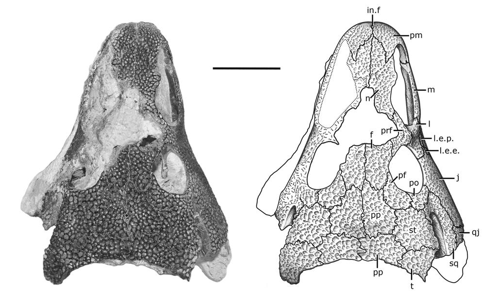
46 38
47 39 Figure 3. Skull of Acheloma dunni, holotype (OMNH 73281) in right lateral view. Scale = 50 mm
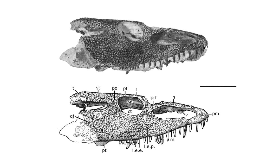
48 40
49 41 Figure 4. Acheloma dunni, referred specimen (BMRP ) in left lateral view. Scale = 10 mm
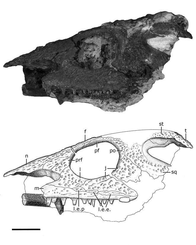
50 42
51 43 Figure 5. Acheloma dunni, referred specimen (BMRP ) in right lateral view. Scale = 10 mm
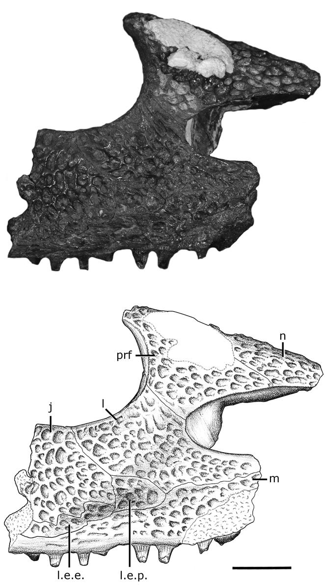
52 44
53 45 Figure 6. Acheloma dunni, referred specimen (OMNH 52365). Partial right upper jaw articulation in dorsal view. Scale = 10 mm
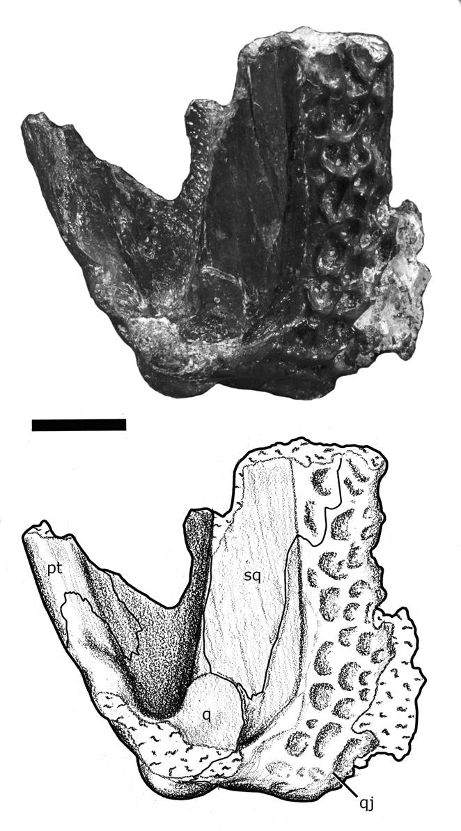
54 46
55 Figure 7. Skull of Acheloma dunni, holotype (OMNH 73281) in ventral view. Scale = 50 mm 47
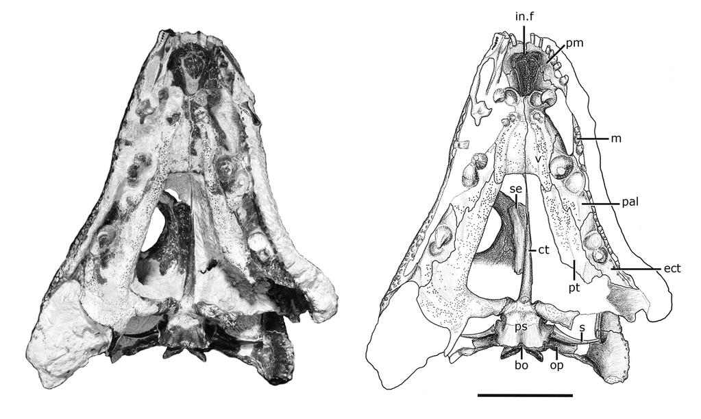
56 48
57 Figure 8. Skull of Acheloma dunni, holotype (OMNH 73281) in occipital view. Scale = 50 mm 49
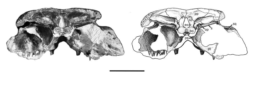
58 50
59 51 Figure 9. Partial left lower jaw of Acheloma dunni, holotype (OMNH 73281) in A, lateral view; B, medial view; C, dorsal view; D, ventral view. Scale = 10 mm
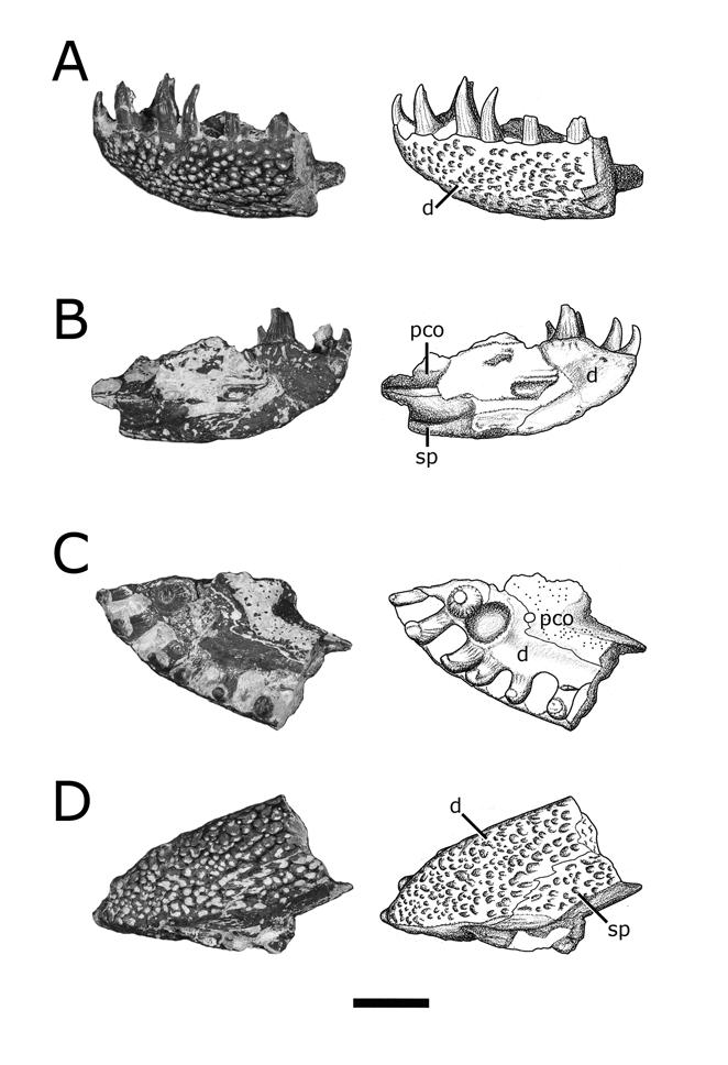
60 52
61 53 Figure 10. Partial right lower jaw of Acheloma dunni, holotype (OMNH 73281) in A, lateral view; B, medial view; C, dorsal view; D, ventral view. Scale = 10 mm
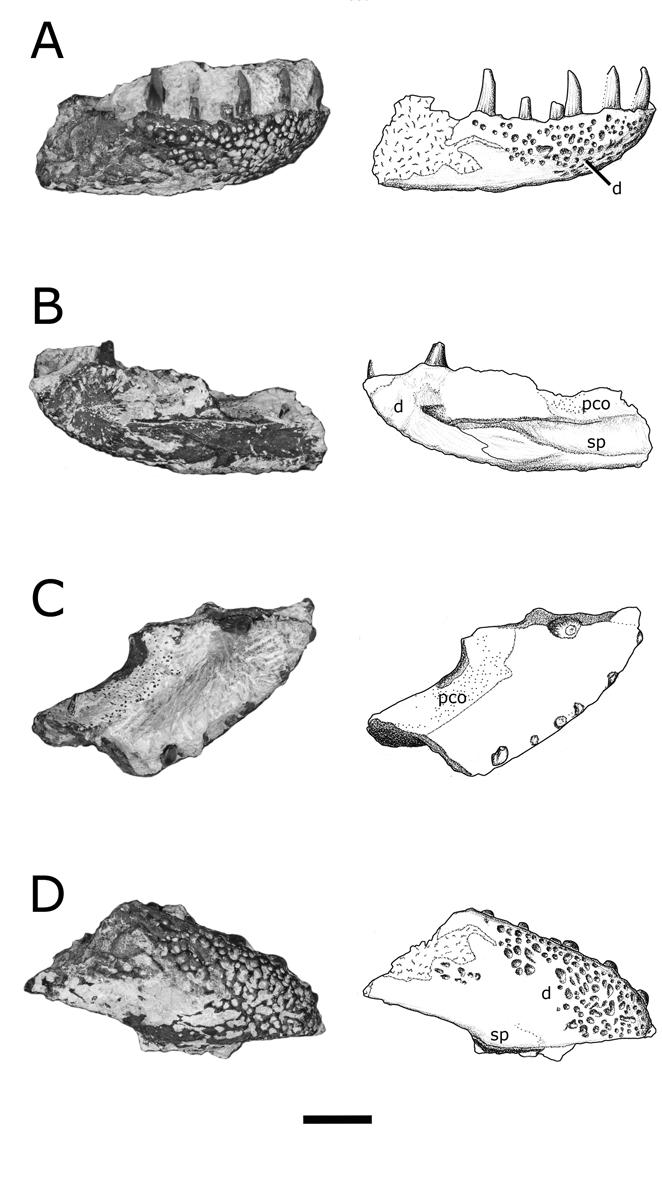
62 54
63 55 Figure 11. Acheloma dunni, referred specimen (OMNH 52365). Partial right lower jaw articulation in A, lateral view; B, posterior view. Scale = 10 mm
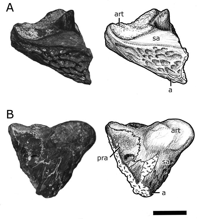
64 56
65 57 Figure 12. Atlas-axis complex and associated cervical vertebrae of Acheloma dunni, holotype (OMNH 73281) in A, right lateral view; B, left lateral view; C, anterior view. Scale = 10 mm
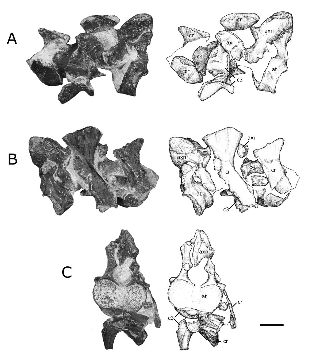
66 58
67 59 Figure 13. Acheloma dunni, referred specimen (OMNH 52545). Right humerus in A, posterior view; B, extensor view; C, anterior view; D, flexor view; E, proximal view; F, distal view. Scale = 50 mm
68 60
69 61 Figure 14. Acheloma dunni, referred specimen (OMNH 73514). Partial pelvic girdle and hindlimb. Scale = 5 mm. Illustration by Heidi Richter.
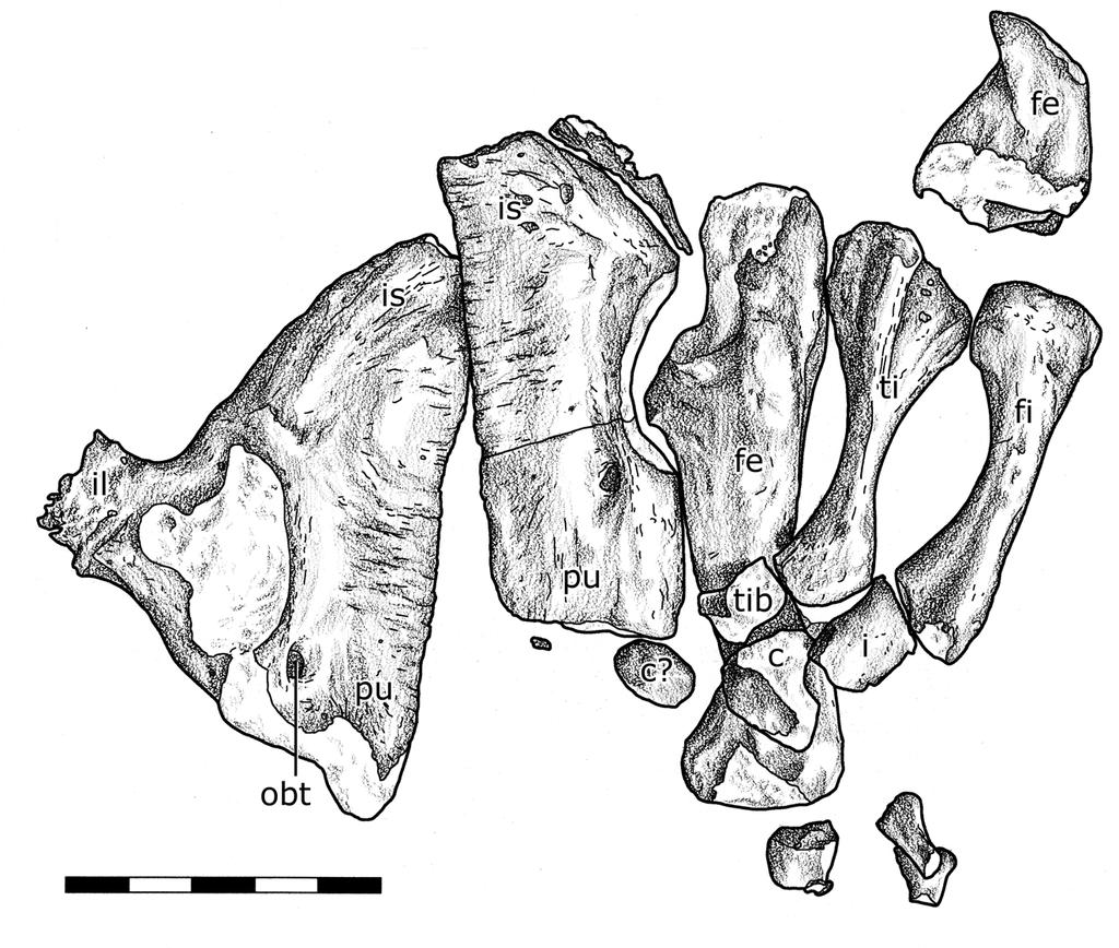
70 62
Postilla PEABODY MUSEUM OF NATURAL HISTORY YALE UNIVERSITY NEW HAVEN, CONNECTICUT, U.S.A.
 Postilla PEABODY MUSEUM OF NATURAL HISTORY YALE UNIVERSITY NEW HAVEN, CONNECTICUT, U.S.A. Number 117 18 March 1968 A 7DIAPSID (REPTILIA) PARIETAL FROM THE LOWER PERMIAN OF OKLAHOMA ROBERT L. CARROLL REDPATH
2. Skull, total length versus length of the presacral vertebral column: (0); extremely elongated neck (e.g. Tanystropheus longobardicus).
; extremely elongated neck (e.g. Tanystropheus longobardicus).") Character list of the taxon-character data set 1. Skull and lower jaws, interdental plates: absent (0); present, but restricted to the anterior end of the dentary (1); present along the entire alveolar
Character list of the taxon-character data set 1. Skull and lower jaws, interdental plates: absent (0); present, but restricted to the anterior end of the dentary (1); present along the entire alveolar
Exceptional fossil preservation demonstrates a new mode of axial skeleton elongation in early ray-finned fishes
 Supplementary Information Exceptional fossil preservation demonstrates a new mode of axial skeleton elongation in early ray-finned fishes Erin E. Maxwell, Heinz Furrer, Marcelo R. Sánchez-Villagra Supplementary
CRANIAL ANATOMY OF ENNATOSAURUS TECTON (SYNAPSIDA: CASEIDAE) FROM THE MIDDLE PERMIAN OF RUSSIA AND THE EVOLUTIONARY RELATIONSHIPS OF CASEIDAE
 FROM THE MIDDLE PERMIAN OF RUSSIA AND THE EVOLUTIONARY RELATIONSHIPS OF CASEIDAE") Journal of Vertebrate Paleontology 28(1):160 180, March 2008 2008 by the Society of Vertebrate Paleontology ARTICLE CRANIAL ANATOMY OF ENNATOSAURUS TECTON (SYNAPSIDA: CASEIDAE) FROM THE MIDDLE PERMIAN
Journal of Vertebrate Paleontology 28(1):160 180, March 2008 2008 by the Society of Vertebrate Paleontology ARTICLE CRANIAL ANATOMY OF ENNATOSAURUS TECTON (SYNAPSIDA: CASEIDAE) FROM THE MIDDLE PERMIAN
A new species of Hsisosuchus (Mesoeucrocodylia) from Dashanpu, Zigong Municipality, Sichuan Province
 from Dashanpu, Zigong Municipality, Sichuan Province") A new species of Hsisosuchus (Mesoeucrocodylia) from Dashanpu, Zigong Municipality, Sichuan Province Yuhui Gao (Zigong Dinosaur Museum) Vertebrata PalAsiatica Volume 39, No. 3 July, 2001 pp. 177-184 Translated
A new species of Hsisosuchus (Mesoeucrocodylia) from Dashanpu, Zigong Municipality, Sichuan Province Yuhui Gao (Zigong Dinosaur Museum) Vertebrata PalAsiatica Volume 39, No. 3 July, 2001 pp. 177-184 Translated
The cranial osteology of Belebey vegrandis (Parareptilia: Bolosauridae), from the Middle Permian of Russia, and its bearing on reptilian evolution
, from the Middle Permian of Russia, and its bearing on reptilian evolution") Blackwell Publishing LtdOxford, UKZOJZoological Journal of the Linnean Society0024-4082 2007 The Linnean Society of London? 2007 1511 191214 Original Articles RUSSIAN BOLOSAURID REPTILER. R. REISZ ET AL.
Blackwell Publishing LtdOxford, UKZOJZoological Journal of the Linnean Society0024-4082 2007 The Linnean Society of London? 2007 1511 191214 Original Articles RUSSIAN BOLOSAURID REPTILER. R. REISZ ET AL.
SUPPLEMENTARY ONLINE MATERIAL FOR. Nirina O. Ratsimbaholison, Ryan N. Felice, and Patrick M. O connor
 http://app.pan.pl/som/app61-ratsimbaholison_etal_som.pdf SUPPLEMENTARY ONLINE MATERIAL FOR Nirina O. Ratsimbaholison, Ryan N. Felice, and Patrick M. O connor Ontogenetic changes in the craniomandibular
A NEW GENUS AND SPECIES OF AMERICAN THEROMORPHA
 A NEW GENUS AND SPECIES OF AMERICAN THEROMORPHA MYCTEROSAURUS LONGICEPS S. W. WILLISTON University of Chicago The past summer, Mr. Herman Douthitt, of the University of Chicago paleontological expedition,
ONLINE APPENDIX 1. Morphological phylogenetic characters scored in this paper. See Poe (2004) for
 for") ONLINE APPENDIX Morphological phylogenetic characters scored in this paper. See Poe () for detailed character descriptions, citations, and justifications for states. Note that codes are changed from a
ONLINE APPENDIX Morphological phylogenetic characters scored in this paper. See Poe () for detailed character descriptions, citations, and justifications for states. Note that codes are changed from a
AMERICAN MUSEUM NOVITATES Published by
 AMERICAN MUSEUM NOVITATES Published by Number 782 THE AmzRICAN MUSEUM OF NATURAL HISTORY Feb. 20, 1935 New York City 56.81, 7 G (68) A NOTE ON THE CYNODONT, GLOCHINODONTOIDES GRACILIS HAUGHTON BY LIEUWE
New Carnivorous Dinosaurs from the Upper Cretaceous of Mongolia
 1955 Doklady, Academy of Sciences USSR 104 (5):779-783 New Carnivorous Dinosaurs from the Upper Cretaceous of Mongolia E. A. Maleev (translated by F. J. Alcock) The present article is a summary containing
Anatomy. Name Section. The Vertebrate Skeleton
 Name Section Anatomy The Vertebrate Skeleton Vertebrate paleontologists get most of their knowledge about past organisms from skeletal remains. Skeletons are useful for gleaning information about an organism
YANGCHUANOSAURUS HEPINGENSIS - A NEW SPECIES OF CARNOSAUR FROM ZIGONG, SICHUAN
 Vol. 30, No. 4 VERTEBRATA PALASIATICA pp. 313-324 October 1992 [SICHUAN ZIGONG ROUSHILONG YI XIN ZHONG] figs. 1-5, pl. I-III YANGCHUANOSAURUS HEPINGENSIS - A NEW SPECIES OF CARNOSAUR FROM ZIGONG, SICHUAN
Williston, and as there are many fairly good specimens in the American
 56.81.7D :14.71.5 Article VII.- SOME POINTS IN THE STRUCTURE OF THE DIADECTID SKULL. BY R. BROOM. The skull of Diadectes has been described by Cope, Case, v. Huene, and Williston, and as there are many
REVISION OF THE AQUATIC ERYOPID TEMNOSPONDYL GLAUKERPETON AVINOFFI ROMER, 1952, FROM THE UPPER PENNSYLVANIAN OF NORTH AMERICA
 ANNALS OF CARNEGIE MUSEUM vol. 81, number 1, PP. 33 60 31 DecembeR 2012 REVISION OF THE AQUATIC ERYOPID TEMNOSPONDYL GLAUKERPETON AVINOFFI ROMER, 1952, FROM THE UPPER PENNSYLVANIAN OF NORTH AMERICA Ralf
List of characters used in the phylogenetic analysis. Capital letters T, R, and L, refer to
 1 Supplementary data CHARACTER LIST List of characters used in the phylogenetic analysis. Capital letters T, R, and L, refer to characters used by Tchernov et al. (2000), Rieppel, et al. (2002), and Lee
HONR219D Due 3/29/16 Homework VI
 Part 1: Yet More Vertebrate Anatomy!!! HONR219D Due 3/29/16 Homework VI Part 1 builds on homework V by examining the skull in even greater detail. We start with the some of the important bones (thankfully
.56 m. (22 in.). COMPSOGNATHOID DINOSAUR FROM THE. Medicine Bow, Wyoming, by the American Museum Expedition
. COMPSOGNATHOID DINOSAUR FROM THE. Medicine Bow, Wyoming, by the American Museum Expedition") Article XII.-ORNITHOLESTES HERMANNI, A NEW COMPSOGNATHOID DINOSAUR FROM THE UPPER JURASSIC. By HENRY FAIRFIELD OSBORN. The type skeleton (Amer. Mus. Coll. No. 6I9) of this remarkable animal was discovered
Article XII.-ORNITHOLESTES HERMANNI, A NEW COMPSOGNATHOID DINOSAUR FROM THE UPPER JURASSIC. By HENRY FAIRFIELD OSBORN. The type skeleton (Amer. Mus. Coll. No. 6I9) of this remarkable animal was discovered
Mammalogy Laboratory 1 - Mammalian Anatomy
 Mammalogy Laboratory 1 - Mammalian Anatomy I. The Goal. The goal of the lab is to teach you skeletal anatomy of mammals. We will emphasize the skull because many of the taxonomically important characters
SOME LITTLE-KNOWN FOSSIL LIZARDS FROM THE
 PROCEEDINGS OF THE UNITED STATES NATIONAL MUSEUM issued SWsK \ {^^m ^V ^^ SMITHSONIAN INSTITUTION U. S. NATIONAL MUSEUM Vol. 91 Washington : 1941 No. 3124 SOME LITTLE-KNOWN FOSSIL LIZARDS FROM THE OLIGOCENE
v:ii-ixi, 'i':;iisimvi'\>!i-:: "^ A%'''''-'^-''S.''v.--..V^'E^'-'-^"-t''gi L I E) R.ARY OF THE VERSITY U N I or ILLINOIS REMO
 R.ARY OF THE VERSITY U N I or ILLINOIS REMO") "^ A%'''''-'^-''S.''v.--..V^'E^'-'-^"-t''gi v:ii-ixi, 'i':;iisimvi'\>!i-:: L I E) R.ARY OF THE U N I VERSITY or ILLINOIS REMO Natural History Survey Librarv GEOLOGICAL SERIES OF FIELD MUSEUM OF NATURAL
"^ A%'''''-'^-''S.''v.--..V^'E^'-'-^"-t''gi v:ii-ixi, 'i':;iisimvi'\>!i-:: L I E) R.ARY OF THE U N I VERSITY or ILLINOIS REMO Natural History Survey Librarv GEOLOGICAL SERIES OF FIELD MUSEUM OF NATURAL
THE SKULLS OF ARAEOSCELIS AND CASEA, PERMIAN REPTILES
 THE SKULLS OF REOSCELIS ND CSE, PERMIN REPTILES University of Chicago There are few Permian reptiles of greater interest at the present time than the peculiar one I briefly described in this journal' three
AMERICAN MUSEUM. Novitates PUBLISHED BY THE AMERICAN MUSEUM OF NATURAL HISTORY CENTRAL PARK WEST AT 79TH STREET
 AMERICAN MUSEUM Novitates PUBLISHED BY THE AMERICAN MUSEUM OF NATURAL HISTORY CENTRAL PARK WEST AT 79TH STREET NEW YORK, N.Y. 10024 U.S.A. NUMBER 2662 NOVEMBER 21, 1978 RONN W. COLDIRON Acroplous vorax
SUPPLEMENTARY INFORMATION
 Character 155, interdental ridges. Absence of interdental ridge (0) shown in Parasaniwa wyomingensis (Platynota). Interdental ridges (1) shown in Coniophis precedens. WWW.NATURE.COM/NATURE 1 Character
A new sauropod from Dashanpu, Zigong Co. Sichuan Province (Abrosaurus dongpoensis gen. et sp. nov.)
") A new sauropod from Dashanpu, Zigong Co. Sichuan Province (Abrosaurus dongpoensis gen. et sp. nov.) by Ouyang Hui Zigong Dinosaur Museum Newsletter Number 2 1989 pp. 10-14 Translated By Will Downs Bilby
A new sauropod from Dashanpu, Zigong Co. Sichuan Province (Abrosaurus dongpoensis gen. et sp. nov.) by Ouyang Hui Zigong Dinosaur Museum Newsletter Number 2 1989 pp. 10-14 Translated By Will Downs Bilby
CRANIAL ANATOMY AND PHYLOGENETIC AFFINITIES OF THE PERMIAN PARAREPTILE MACROLETER POEZICUS
 CRANIAL ANATOMY AND PHYLOGENETIC AFFINITIES OF THE PERMIAN PARAREPTILE MACROLETER POEZICUS Author(s): LINDA A. TSUJI Source: Journal of Vertebrate Paleontology, 26(4):849-865. 2006. Published By: The Society
The skull of Sphenacodon ferocior, and comparisons with other sphenacodontines (Reptilia: Pelycosauria)
") Circular 190 New Mexico Bureau of Mines & Mineral Resources A DIVISION OF NEW MEXICO INSTITUTE OF MINING & TECHNOLOGY The skull of Sphenacodon ferocior, and comparisons with other sphenacodontines (Reptilia:
Circular 190 New Mexico Bureau of Mines & Mineral Resources A DIVISION OF NEW MEXICO INSTITUTE OF MINING & TECHNOLOGY The skull of Sphenacodon ferocior, and comparisons with other sphenacodontines (Reptilia:
Supporting Online Material for
 www.sciencemag.org/cgi/content/full/329/5998/1481/dc1 Supporting Online Material for Tyrannosaur Paleobiology: New Research on Ancient Exemplar Organisms Stephen L. Brusatte,* Mark A. Norell, Thomas D.
The earliest reptiles
 J. Linn. SOC. (Zool), 45, no. 304, p. 61 With 14 tezt-figures Printed in Great Britain The earliest reptiles BY ROBERT L. CARROLL (Accepted for publication December 1963) Communicated by Errol I. White,
New specimen of Cacops woehri indicates differences in the ontogenetic trajectories among cacopine dissorophids
 doi:10.5194/fr-18-73-2015 Author(s) 2015. CC Attribution 3.0 License. New specimen of Cacops woehri indicates differences in the ontogenetic trajectories among cacopine dissorophids N. B. Fröbisch 1, A.
A new species of sauropod, Mamenchisaurus anyuensis sp. nov.
 A new species of sauropod, Mamenchisaurus anyuensis sp. nov. by Xinlu He, Suihua Yang, Kaiji Cai, Kui Li, and Zongwen Liu Chengdu University of Technology Papers on Geosciences Contributed to the 30th
A Fossil Snake (Elaphe vulpina) From A Pliocene Ash Bed In Nebraska
 From A Pliocene Ash Bed In Nebraska") University of Nebraska - Lincoln DigitalCommons@University of Nebraska - Lincoln Transactions of the Nebraska Academy of Sciences and Affiliated Societies Nebraska Academy of Sciences 198 A Fossil Snake
University of Nebraska - Lincoln DigitalCommons@University of Nebraska - Lincoln Transactions of the Nebraska Academy of Sciences and Affiliated Societies Nebraska Academy of Sciences 198 A Fossil Snake
NOTES ON THE FIRST SKULL AND JAWS OF RIOJASAURUS INCERTUS (DINOSAURIA, PROSAUROPODA, MELANOROSAURIDAE) OF THE LATE TRIASSIC OF LA RIOJA, ARGENTINA
 OF THE LATE TRIASSIC OF LA RIOJA, ARGENTINA") NOTES ON THE FIRST SKULL AND JAWS OF RIOJASAURUS INCERTUS (DINOSAURIA, PROSAUROPODA, MELANOROSAURIDAE) OF THE LATE TRIASSIC OF LA RIOJA, ARGENTINA José F. Bonaparte and José A. Pumares translated by Jeffrey
NOTES ON THE FIRST SKULL AND JAWS OF RIOJASAURUS INCERTUS (DINOSAURIA, PROSAUROPODA, MELANOROSAURIDAE) OF THE LATE TRIASSIC OF LA RIOJA, ARGENTINA José F. Bonaparte and José A. Pumares translated by Jeffrey
SUPPLEMENTARY INFORMATION
 doi:10.1038/nature22966 TABLE OF CONTENTS PART A. MATRIX CONSTRUCTION AND CODING CHANGES PART B. PHYLOGENETIC CHARACTER LIST PART C. NEXUS SCRIPTS PART D. REFERENCES CITED IN PART A. MATRIX CONSTRUCTION
The cranial skeleton of the Early Permian aquatic reptile Mesosaurus tenuidens: implications for relationships and palaeobiology
 Blackwell Publishing LtdOxford, UKZOJZoological Journal of the Linnean Society0024-4082The Linnean Society of London, 2006? 2006 146? 345368 Original Article THE CRANIAL SKELETON OF MESOSAURUS TENUIDENSS.
A new basal sauropodiform dinosaur from the Lower Jurassic of Yunnan Province, China
 SUPPLEMENTARY INFORMATION A new basal sauropodiform dinosaur from the Lower Jurassic of Yunnan Province, China Ya-Ming Wang 1, Hai-Lu You 2,3 *, Tao Wang 4 1 School of Earth Sciences and Resources, China
A Short Report on the Occurrence of Dilophosaurus from Jinning County, Yunnan Province
 A Short Report on the Occurrence of Dilophosaurus from Jinning County, Yunnan Province by Hu Shaojin (Kunming Cultural Administrative Committee, Yunnan Province) Vertebrata PalAsiatica Vol. XXXI, No. 1
Description of Cranial Elements and Ontogenetic Change within Tropidolaemus wagleri (Serpentes: Crotalinae).
.") East Tennessee State University Digital Commons @ East Tennessee State University Electronic Theses and Dissertations 5-2016 Description of Cranial Elements and Ontogenetic Change within Tropidolaemus
East Tennessee State University Digital Commons @ East Tennessee State University Electronic Theses and Dissertations 5-2016 Description of Cranial Elements and Ontogenetic Change within Tropidolaemus
A M E G H I N I A N A. Revista de la Asociación Paleontológia Argentina. Volume XV September-December 1978 Nos. 3-4
 A M E G H I N I A N A Revista de la Asociación Paleontológia Argentina Volume XV September-December 1978 Nos. 3-4 COLORADIA BREVIS N. G. ET N. SP. (SAURISCHIA, PROSAUROPODA), A PLATEOSAURID DINOSAUR FROM
Follow this and additional works at: Part of the Paleontology Commons
 California State University, San Bernardino CSUSB ScholarWorks Theses Digitization Project John M. Pfau Library 2003 A reinterpretation of the small Captorhinid Reptile Captorhinikos Parvus Olson as a
Supplementary Figure 1 Additional diagnostic information on the dvinosaur temnospondyl Timonya anneae gen. et sp. nov. from the lower Permian of
 Supplementary Figure 1 Additional diagnostic information on the dvinosaur temnospondyl Timonya anneae gen. et sp. nov. from the lower Permian of northeastern Brazil. (a) Occipital view of Timonya anneae,
A NEW SPECIES OF EXTINCT TURTLE FROM THE UPPER PLIOCENE OF IDAHO
 A NEW SPECIES OF EXTINCT TURTLE FROM THE UPPER PLIOCENE OF IDAHO By Charles W. Gilmore Curator, Division of Vertebrate Paleontology United States National Museum Among the fossils obtained bj^ the Smithsonian
Bulletin of Big Bend Paleo-Geo An Open Access Publication from Mosasaur Ranch Museum, Terlingua and Lajitas, Texas All rights reserved
 Bulletin of Big Bend Paleo-Geo An Open Access Publication from Mosasaur Ranch Museum, Terlingua and Lajitas, Texas All rights reserved This was a private report in 2003 on my thoughts on Platecarpus planifrons.
A NEW ANKYLOSAUR FROM THE UPPER CRETACEOUS OF MONGOLIA E.A. Maleev Doklady Akademii Nauk, SSSR 87:
 translated by Dr. Tamara and F. Jeletzky, 1956 A NEW ANKYLOSAUR FROM THE UPPER CRETACEOUS OF MONGOLIA E.A. Maleev 1952. Doklady Akademii Nauk, SSSR 87:273-276 Armored dinosaurs make a considerable part
FURTHER STUDIES ON TWO SKELETONS OF THE BLACK RIGHT WHALE IN THE NORTH PACIFIC
 FURTHER STUDIES ON TWO SKELETONS OF THE BLACK RIGHT WHALE IN THE NORTH PACIFIC HIDEO OMURA, MASAHARU NISHIWAKI* AND TOSHIO KASUYA* ABSTRACT Two skeletons of the black right whale were studied, supplementing
Fig. 5. (A) Scaling of brain vault size (width measured at the level of anterior squamosal/parietal suture) relative to skull size (measured at the
 Scaling of brain vault size (width measured at the level of anterior squamosal/parietal suture) relative to skull size (measured at the") Fig. 5. (A) Scaling of brain vault size (width measured at the level of anterior squamosal/parietal suture) relative to skull size (measured at the distance between the left versus right temporomandibular
Fig. 5. (A) Scaling of brain vault size (width measured at the level of anterior squamosal/parietal suture) relative to skull size (measured at the distance between the left versus right temporomandibular
Cranial osteology of the African gerrhosaurid Angolosaurus skoogi (Squamata; Gerrhosauridae) HOLLY A. NANCE
 HOLLY A. NANCE") African Journal of Herpetology, 2007 56(1): 39-75. Herpetological Association of Africa Original article Cranial osteology of the African gerrhosaurid Angolosaurus skoogi (Squamata; Gerrhosauridae) HOLLY
African Journal of Herpetology, 2007 56(1): 39-75. Herpetological Association of Africa Original article Cranial osteology of the African gerrhosaurid Angolosaurus skoogi (Squamata; Gerrhosauridae) HOLLY
A Complete Late Cretaceous Iguanian (Squamata, Reptilia) from the Gobi and Identification of a New Iguanian Clade
 from the Gobi and Identification of a New Iguanian Clade") PUBLISHED BY THE AMERICAN MUSEUM OF NATURAL HISTORY CENTRAL PARK WEST AT 79TH STREET, NEW YORK, NY 10024 Number 3584, 47 pp., 19 figures September 6, 2007 A Complete Late Cretaceous Iguanian (Squamata,
PUBLISHED BY THE AMERICAN MUSEUM OF NATURAL HISTORY CENTRAL PARK WEST AT 79TH STREET, NEW YORK, NY 10024 Number 3584, 47 pp., 19 figures September 6, 2007 A Complete Late Cretaceous Iguanian (Squamata,
OF THE TRIAS THE PHYTOSAURIA
 THE PHYTOSAURIA OF THE TRIAS MAURICE G. MEHL University of Wisconsin Some time ago the writer gave a brief notice of a new genus of phytosaurs of which Angistorhinus grandis Mehl was the type.' It is the
4. Premaxilla: Foramen on the lateral surface of the premaxillary body (Yates 2007 ch. 4) 0 absent 1 present
 0 absent 1 present") The character matrix used as a basis for this study is that of Yates et al (2010) which is modified from the earlier matrix used by Yates (2007). This matrix includes characters acquired and/or modified
The character matrix used as a basis for this study is that of Yates et al (2010) which is modified from the earlier matrix used by Yates (2007). This matrix includes characters acquired and/or modified
Abstract. M. Jimena Trotteyn 1,2 *, Martín D. Ezcurra 3 RESEARCH ARTICLE
 RESEARCH ARTICLE Osteology of Pseudochampsa ischigualastensis gen. et comb. nov. (Archosauriformes: Proterochampsidae) from the Early Late Triassic Ischigualasto Formation of Northwestern Argentina M.
Cranial morphology and taxonomy of South African Tapinocephalidae (Therapsida: Dinocephalia): the case of Avenantia and Riebeeckosaurus
: the case of Avenantia and Riebeeckosaurus") Cranial morphology and taxonomy of South African Tapinocephalidae (Therapsida: Dinocephalia): the case of Avenantia and Riebeeckosaurus Saniye Güven*, Bruce S. Rubidge & Fernando Abdala Evolutionary Studies
Cranial morphology and taxonomy of South African Tapinocephalidae (Therapsida: Dinocephalia): the case of Avenantia and Riebeeckosaurus Saniye Güven*, Bruce S. Rubidge & Fernando Abdala Evolutionary Studies
Marshall Digital Scholar. Marshall University. F. Robin O Keefe Marshall University,
 Marshall University Marshall Digital Scholar Biological Sciences Faculty Research Biological Sciences 2008 Cranial anatomy and taxonomy of Dolichorhynchops bonneri new combination, a polycotylid (Sauropterygia:
Cranial osteology and phylogenetic relationships of Hamadasuchus rebouli (Crocodyliformes: Mesoeucrocodylia) from the Cretaceous of Morocco
 from the Cretaceous of Morocco") Blackwell Publishing LtdOxford, UKZOJZoological Journal of the Linnean Society0024-4082 2007 The Linnean Society of London? 2007 1494 533567 Original Articles HAMADASUCHUS REBOULIH. C. E. LARSSON and H.-D.
Blackwell Publishing LtdOxford, UKZOJZoological Journal of the Linnean Society0024-4082 2007 The Linnean Society of London? 2007 1494 533567 Original Articles HAMADASUCHUS REBOULIH. C. E. LARSSON and H.-D.
NEW INFORMATION ON THE CRANIUM OF BRACHYLOPHOSAURUS CANADENSIS (DINOSAURIA, HADROSAURIDAE), WITH A REVISION OF ITS PHYLOGENETIC POSITION
, WITH A REVISION OF ITS PHYLOGENETIC POSITION") Journal of Vertebrate Paleontology 25(1):144 156, March 2005 2005 by the Society of Vertebrate Paleontology NEW INFORMATION ON THE CRANIUM OF BRACHYLOPHOSAURUS CANADENSIS (DINOSAURIA, HADROSAURIDAE), WITH
Journal of Vertebrate Paleontology 25(1):144 156, March 2005 2005 by the Society of Vertebrate Paleontology NEW INFORMATION ON THE CRANIUM OF BRACHYLOPHOSAURUS CANADENSIS (DINOSAURIA, HADROSAURIDAE), WITH
AEROSAURUS WELLESI, NEW SPECIES, A VARANOPSEID MAMMAL-LIKE
 Journal of Vertebrate Paleontology 1(1):73-96. 15 June 1981 1 AEROSAURUS WELLESI, NEW SPECIES, A VARANOPSEID MAMMAL-LIKE REPTILE (SYNAPSIDA: PELYCOSAURIA) FROM THE LOWER PERMIAN OF NEW MEXICO WANN LANGSTON
VERTEBRATA PALASIATICA
 41 2 2003 2 VERTEBRATA PALASIATICA pp. 147 156 figs. 1 5 1) ( 100044), ( Parakannemeyeria brevirostris),,, : ( Xiyukannemeyeria),,, Q915. 864 60 Turfania (,1973), Dicynodon (, 1973 ; Lucas, 1998), (Lystrosaurus)
First Ornithomimid (Theropoda, Ornithomimosauria) from the Upper Cretaceous Djadokhta Formation of Tögrögiin Shiree, Mongolia
 from the Upper Cretaceous Djadokhta Formation of Tögrögiin Shiree, Mongolia") First Ornithomimid (Theropoda, Ornithomimosauria) from the Upper Cretaceous Djadokhta Formation of Tögrögiin Shiree, Mongolia Tsogtbaatar Chinzorig¹, ³ *, Yoshitsugu Kobayashi², Khishigjav Tsogtbaatar³,
First Ornithomimid (Theropoda, Ornithomimosauria) from the Upper Cretaceous Djadokhta Formation of Tögrögiin Shiree, Mongolia Tsogtbaatar Chinzorig¹, ³ *, Yoshitsugu Kobayashi², Khishigjav Tsogtbaatar³,
Revision of the Gymnarthridae American Permian Microsaurs
 PEABODY MUSEUM OF NATURAL HISTORY, YALE UNIVERSITY BULLETIN 10 Revision of the Gymnarthridae American Permian Microsaurs BY JOSEPH T. GREGORY, FRANK E. PEABODY, and LLEWELLYN L PRICE NEW HAVEN, CONNECTICUT
Title: Phylogenetic Methods and Vertebrate Phylogeny
 Title: Phylogenetic Methods and Vertebrate Phylogeny Central Question: How can evolutionary relationships be determined objectively? Sub-questions: 1. What affect does the selection of the outgroup have
Carpus and tarsus of Temnospondyli
 Vertebrate Anatomy Morphology Palaeontology 1(1):51-87 ISSN 2292-1389 Carpus and tarsus of Temnospondyli 51 David Dilkes Department of Biology & Microbiology, University of Wisconsin Oshkosh, 800 Algoma
UN? RSITYOF. ILLIiwiS LIBRARY AT URBANA-CHAMPAIGN NATURAL HIST. SURVEY
 UN? RSITYOF ILLIiwiS LIBRARY AT URBANA-CHAMPAIGN NATURAL HIST. SURVEY FIELDIANA GEOLOGY Published by CHICAGO NATURAL HISTORY MUSEUM Volume 10 July 29, 1954 No. 17 FAUNA OF THE VALE AND CHOZA: 7 PELYCOSAURIA:
Chapter 6 - Systematic palaeontology
 - Sea-saurians have had a sorry experience in the treatment they have received from nomenclators Samuel Wendell Williston, 1914 6.1 Rhomaleosauridae - generic and species-level systematics As defined in
On the cranial anatomy of the polycotylid plesiosaurs, including new material of Polycotylus latipinnis, Cope, from Alabama
 Marshall University Marshall Digital Scholar Biological Sciences Faculty Research Biological Sciences 2004 On the cranial anatomy of the polycotylid plesiosaurs, including new material of Polycotylus latipinnis,
CAMBRIDGE, MASS. 4 MAY 2011 NUMBER 523
 US ISSN 0006-9698 CAMBRIDGE, MASS. 4 MAY 2011 NUMBER 523 THE SMALLER EMBOLOMEROUS AMPHIBIANS (ANTHRACOSAURIA) FROM THE MIDDLE PENNSYLVANIAN (DESMOINESIAN) LOCALITIES AT LINTON AND FIVE POINTS COAL MINES,
Department of Biology, Faculty of Science, Razi University, Kermanshah, Iran 2
 Iranian Journal of Animal Biosystematics (IJAB) Vol.13, No.2, 247-262, 2017 ISSN: 1735-434X (print); 2423-4222 (online) DOI: 10.22067/ijab.v13i2.64614 A comparative study of the skull between Trachylepis
A new parareptile with temporal fenestration from the Middle Permian of South Africa
 A new parareptile with temporal fenestration from the Middle Permian of South Africa 9 Sean P. Modesto, Diane M. Scott, and Robert R. Reisz Abstract: The partial skeleton of a small reptile, from the Middle
Muséum national d Histoire naturelle, F-75005, Paris, France c Karoo Palaeontology, Iziko South African Museum, PO Box 61, Cape Town, 8000, South
 This article was downloaded by: [76.187.62.88] On: 16 May 2014, At: 23:11 Publisher: Taylor & Francis Informa Ltd Registered in England and Wales Registered Number: 1072954 Registered office: Mortimer
CHARACTER LIST: Nesbitt et al., 2011
 CHARACTER LIST: Nesbitt et al., 2011 1. Vaned feathers on forelimb symmetric (0) or asymmetric (1). The barbs on opposite sides of the rachis differ in length; in extant birds, the barbs on the leading
PALEONTOLOGICAL CONTRIBUTIONS
 THE UNIVERSITY OF KANSAS PALEONTOLOGICAL CONTRIBUTIONS August, 1965 Paper 2 A NEW WYOMING PHYTOSAUR By THEODORE H. EATON, JR. [Museum of Natural History, University of Kansas I ABSTRACT The skull of a
On the Discovery of the earliest fossil bird in China (Sinosauropteryx gen. nov.) and the origin of birds
 and the origin of birds") On the Discovery of the earliest fossil bird in China (Sinosauropteryx gen. nov.) and the origin of birds by Qiang Ji and Shu an Ji Chinese Geological Museum, Beijing Chinese Geology Volume 233 1996 pp.
On the Discovery of the earliest fossil bird in China (Sinosauropteryx gen. nov.) and the origin of birds by Qiang Ji and Shu an Ji Chinese Geological Museum, Beijing Chinese Geology Volume 233 1996 pp.
Redescription of the Mongolian Sauropod NEMEGTOSAURUS MONGOLIENSIS Nowinski (Dinosauria:
 Journal of Systematic Palaeontology 3 (3): 283 318 Issued 24 August 2005 doi:10.1017/s1477201905001628 Printed in the United Kingdom C The Natural History Museum Redescription of the Mongolian Sauropod
Biology 340 Comparative Embryology Lecture 12 Dr. Stuart Sumida. Evo-Devo Revisited. Development of the Tetrapod Limb
 Biology 340 Comparative Embryology Lecture 12 Dr. Stuart Sumida Evo-Devo Revisited Development of the Tetrapod Limb Limbs whether fins or arms/legs for only in particular regions or LIMB FIELDS. Primitively
complex in cusp pattern. (3) The bones of the coyote skull are thinner, crests sharper and the
 The bones of the coyote skull are thinner, crests sharper and the") DISTINCTIONS BETWEEN THE SKULLS OF S AND DOGS Grover S. Krantz Archaeological sites in the United States frequently yield the bones of coyotes and domestic dogs. These two canines are very similar both
DISTINCTIONS BETWEEN THE SKULLS OF S AND DOGS Grover S. Krantz Archaeological sites in the United States frequently yield the bones of coyotes and domestic dogs. These two canines are very similar both
A skull without mandihle, from the Hunterian Collection (no.
 4 MR. G. A. BOULENGER ON CHELONIAN REMAINS. [Jan. 6, 2. On some Chelonian Remains preserved in the Museum of the Eojal College of Surgeons. By G. A. Boulenger. [Eeceived December 8, 1890.] In the course
Supplementary information to A new troodontid dinosaur from China with avian-like sleeping-posture. Xing Xu 1 and Mark Norell 2
 Supplementary information to A new troodontid dinosaur from China with avian-like sleeping-posture Xing Xu 1 and Mark Norell 2 1 Institute of Vertebrate Paleontology & Paleoanthropology, Chinese Academy
FIELDIANA GEOLOGY NEW SALAMANDERS OF THE FAMILY SIRENIDAE FROM THE CRETACEOUS OF NORTH AMERICA
 FIELDIANA GEOLOGY Published by CHICAGO NATURAL HISTORY MUSEUM Volume 10 Sbftember 22, 1968 No. 88 NEW SALAMANDERS OF THE FAMILY SIRENIDAE FROM THE CRETACEOUS OF NORTH AMERICA Coleman J. Coin AND Walter
THE GORGONOPSIAN GENUS, HIPPOSAURUS, AND THE FAMILY ICTIDORHINIDAE * Dr. L.D. Boonstra. Paleontologist, South African Museum, Cape Town
 THE GORGONOPSIAN GENUS, HIPPOSAURUS, AND THE FAMILY ICTIDORHINIDAE * by Dr. L.D. Boonstra Paleontologist, South African Museum, Cape Town In 1928 I dug up the complete skeleton of a smallish gorgonopsian
Jurassic Ornithopod Agilisaurus louderbacki (Ornithopoda: Fabrosauridae) from Zigong, Sichuan, China
 from Zigong, Sichuan, China") Jurassic Ornithopod Agilisaurus louderbacki (Ornithopoda: Fabrosauridae) from Zigong, Sichuan, China Guangzhao Peng (Zigong Dinosaur Museum) Vertebrata PalAsiatica Volume 30, No. 1 January, 1992 pp. 39-51
Jurassic Ornithopod Agilisaurus louderbacki (Ornithopoda: Fabrosauridae) from Zigong, Sichuan, China Guangzhao Peng (Zigong Dinosaur Museum) Vertebrata PalAsiatica Volume 30, No. 1 January, 1992 pp. 39-51
A Late Jurassic Protosuchian Sichuanosuchus huidongensis from Zigong, Sichuan Province. Guangzhao Peng. Zigong Dinosaur Museum, Zigong, Sichuan
 A Late Jurassic Protosuchian Sichuanosuchus huidongensis from Zigong, Sichuan Province Guangzhao Peng Zigong Dinosaur Museum, Zigong, Sichuan 643013 Vertebrata PalAsiatica Volume 34, Number 4 October,
Notes on Ceratopsians and Ankylosaurs at the Royal Ontario Museum
 Notes on Ceratopsians and Ankylosaurs at the Royal Ontario Museum Andrew A. Farke, Ph.D. Raymond M. Alf Museum of Paleontology 1175 West Baseline Road Claremont, CA 91711 email: afarke@webb.org Introduction
A NEW CROCODYLOMORPH ARCHOSAUR FROM THE UPPER TRIASSIC OF NORTH CAROLINA
 Journal of Vertebrate Paleontology 23(2):329 343, June 2003 2003 by the Society of Vertebrate Paleontology A NEW CROCODYLOMORPH ARCHOSAUR FROM THE UPPER TRIASSIC OF NORTH CAROLINA HANS-DIETER SUES 1 *,
Florida, Gainesville, Florida, 32611, U.S.A. b Smithsonian Tropical Research Institute, Ancon, Republic of Panama,
 This article was downloaded by: [78.22.97.164] On: 04 May 2013, At: 14:02 Publisher: Taylor & Francis Informa Ltd Registered in England and Wales Registered Number: 1072954 Registered office: Mortimer
SUPPLEMENTARY INFORMATION
 1. Differential diagnosis for Asilisaurus kongwe Asilisaurus differs from Eucoelophysis in having a Meckelian groove in the dorsoventral middle of the dentary, teeth that have no expansion above the root,
A New Dromaeosaurid Theropod from Ukhaa Tolgod (Ömnögov, Mongolia)
") PUBLISHED BY THE AMERICAN MUSEUM OF NATURAL HISTORY CENTRAL PARK WEST AT 79TH STREET, NEW YORK, NY 10024 Number 3545, 51 pp., 25 figures, 1 table December 7, 2006 A New Dromaeosaurid Theropod from Ukhaa
PUBLISHED BY THE AMERICAN MUSEUM OF NATURAL HISTORY CENTRAL PARK WEST AT 79TH STREET, NEW YORK, NY 10024 Number 3545, 51 pp., 25 figures, 1 table December 7, 2006 A New Dromaeosaurid Theropod from Ukhaa
TRACHEMYS SCULPTA. A nearly complete articulated carapace and plastron of an Emjdd A NEAKLY COMPLETE SHELL OF THE EXTINCT TURTLE,
 A NEAKLY COMPLETE SHELL OF THE EXTINCT TURTLE, TRACHEMYS SCULPTA By Charles W. Gilmore Curator of Vertebrate Paleontology, United States National Museum INTRODUCTION A nearly complete articulated carapace
Correspondence to: F. Spindler D. Scott and R. R. Reisz
 doi:10.5194/fr-18-17-2015 Author(s) 2015. CC Attribution 3.0 License. New information on the cranial and postcranial anatomy of the early synapsid Ianthodon schultzei (Sphenacomorpha: Sphenacodontia),
Biology 3315 Comparative Vertebrate Morphology Skulls and Visceral Skeletons
 Biology 3315 Comparative Vertebrate Morphology Skulls and Visceral Skeletons 1. Head skeleton of lamprey Cyclostomes are highly specialized in both the construction of the chondrocranium and visceral skeleton.
A NEW GENUS OF PLIOSAUR (REPTILIA: SAUROPTERYGIA) FROM THE LOWER JURASSIC OF HOLZMADEN, GERMANY
 FROM THE LOWER JURASSIC OF HOLZMADEN, GERMANY") [Palaeontology, Vol. 53, Part 5, 2010, pp. 1049 1063] A NEW GENUS OF PLIOSAUR (REPTILIA: SAUROPTERYGIA) FROM THE LOWER JURASSIC OF HOLZMADEN, GERMANY by ADAM. S. SMITH* and PEGGY VINCENT *Natural History
[Palaeontology, Vol. 53, Part 5, 2010, pp. 1049 1063] A NEW GENUS OF PLIOSAUR (REPTILIA: SAUROPTERYGIA) FROM THE LOWER JURASSIC OF HOLZMADEN, GERMANY by ADAM. S. SMITH* and PEGGY VINCENT *Natural History
Supporting Online Material for
 www.sciencemag.org/cgi/content/full/317/5843/1378/dc1 Supporting Online Material for A Basal Dromaeosaurid and Size Evolution Preceding Avian Flight Alan H. Turner,* Diego Pol, Julia A. Clarke, Gregory
Recently Mr. Lawrence M. Lambe has described and figured in the
 56.81,9C(117:71.2) Article XXXV.-CORYTHOSAURUS CASUARIUS, A NEW CRESTED DINOSAUR FROM THE BELLY RIVER CRETA- CEOUS, WITH PROVISIONAL CLASSIFICATION OF THE FAMILY TRACHODONTIDA1X BY BARNUM BROWN. PLATE
Oedaleops campi (Reptilia: Pelycosauria) New Genus and Species from the Lower Permian of New Mexico, and the Family Eothyrididae
 New Genus and Species from the Lower Permian of New Mexico, and the Family Eothyrididae") BULLETIN OF THE TEXAS MEMORIAL MUSEUM NUMBER 9 JANUARY 15, 1965 Oedaleops campi (Reptilia: Pelycosauria) New Genus and Species from the Lower Permian of New Mexico, and the Family Eothyrididae by WANN
BULLETIN OF THE TEXAS MEMORIAL MUSEUM NUMBER 9 JANUARY 15, 1965 Oedaleops campi (Reptilia: Pelycosauria) New Genus and Species from the Lower Permian of New Mexico, and the Family Eothyrididae by WANN
Evidence of a new carcharodontosaurid from the Upper Cretaceous of Morocco
 http://app.pan.pl/som/app57-cau_etal_som.pdf SUPPLEMENTARY ONLINE MATERIAL FOR Evidence of a new carcharodontosaurid from the Upper Cretaceous of Morocco Andrea Cau, Fabio Marco Dalla Vecchia, and Matteo
SOME NEW AMERICAN PYCNODONT FISHES.
 SOME NEW AMERICAN PYCNODONT FISHES. By James Williams Gidley, Assistant Curator of Fossil Mammals, United States National Museum. In the United States National Museum are several specimens representing
School of Earth Sciences, University of Bristol, Queen s Road, Bristol BS8 1RJ, UK 2
 bs_bs_banner Zoological Journal of the Linnean Society, 2015, 173, 55 91. With 20 figures Osteology of Rauisuchus tiradentes from the Late Triassic (Carnian) Santa Maria Formation of Brazil, and its implications
LEIDY, SHOWING THE BONES OF THE FEET 'AND LIMBS
 CQNTEUBUTIONS FBOM THE MUSEUM OF PALEONTOLOGY (Confindion of Con&&&m froin UB Muaercm of Gcologg) UNIVERSITY OF ' MICHIGAN VOL V, No. 6, pp. 6W3 (e ph.) DEAXMBER 31,1036 A SPECIMEN OF STYLEMYS NEBRASCENSIS
ABSTRACT. we define the taxa Alligatoroidae and Alligatoridae to be the descent community and crown group,
 AMERICAN MUSEUM No vtates PUBLISHED BY THE AMERICAN MUSEUM OF NATURAL HISTORY CENTRAL PARK WEST AT 79TH STREET, NEW YORK, N.Y. 10024 Number 3116, 26 pp., 10 figures, 1 table December 28, 1994 The Late
A new carnosaur from Yongchuan County, Sichuan Province
 A new carnosaur from Yongchuan County, Sichuan Province by Dong Zhiming Institute of Vertebrate Palaeontology and Palaeoanthropology, Academia Sinica Zhang Yihong, Li Xuanmin, and Zhou Shiwu Chongqing
On the morphoplogy and taxonomic status of Xinpusaurus kohi JIANG et al., 2004 (Diapsida: Thalattosauria) from the Upper Triassic of China
 from the Upper Triassic of China") Palaeodiversity 7: 47 59; Stuttgart 30 December 2014. 47 On the morphoplogy and taxonomic status of Xinpusaurus kohi JIANG et al., 2004 (Diapsida: Thalattosauria) from the Upper Triassic of China MICHAEL
Palaeodiversity 7: 47 59; Stuttgart 30 December 2014. 47 On the morphoplogy and taxonomic status of Xinpusaurus kohi JIANG et al., 2004 (Diapsida: Thalattosauria) from the Upper Triassic of China MICHAEL
A NEW SPECIES OF TROODONT DINOSAUR FROM THE
 A NEW SPECIES OF TROODONT DINOSAUR FROM THE LANCE FORMATION OF WYOMING By Charles W. Gilmore Curator of Vertebrate Paleontology, United States National Museum INTRODUCTION The intensive search to which