Geology E PUBLISHED BY FIELD MUSEUM OF NATURAL HISTORY. The Dermal Armor of the Cyamodontoid. Morphology and Systematic Value
|
|
|
- Collin Hunter
- 5 years ago
- Views:
Transcription


1 550 5 fi n.s. no. 46 GEOLOGY LJBRARY h ieldiana Geology NEW SERIES, NO. 46 The Dermal Armor of the Cyamodontoid Placodonts (Reptilia, Sauropterygia): Morphology and Systematic Value Olivier Rieppel February 28, 2002 CSi Publication 1517 E PUBLISHED BY FIELD MUSEUM OF NATURAL HISTORY CD


2 WU AT llrbana-chamrvftljjjrmation for Contributors to Fieldiana GEOLOGY General: /;i'/hfc//7j\v^.primarily a journal for Field Museum stafi members and research associates, although manuscrita^mijjutnattiliated authors may be considered as space permits. TheTOUrnal carries a page charge of $65.00 per printed page or fraction thereof. Payment of at least 50% of page charges qualifies a paper for expedited processing, which reduces the publication time. Contributions from staff. research associates, and invited authors will be considered for publication regardless of ability to pay page charges, however, the full charge is mandatory for nonaffiliated authors of unsolicited manuscripts. Three complete copies of the text (including title page and abstract) and of the illustrations should be submitted (one original copy plus two review copies which may be machine copies). No manuscripts will be considered for publication or submitted to reviewers before all materials are complete and in the hands of the Scientific Editor. Manuscripts should be submitted to Scientific Editor, Fieldiana, Field Museum of Natural History, Chicago, Illinois , U.S.A. Text: Manuscripts must be typewritten double-spaced on standard-weight, 8V2- by 11 -inch paper with wide margins on all four sides. If typed on an IBM-compatible computer using MS-DOS, also submit text on 5V4-inch diskette (WordPerfect 4.1, 4.2, or 5.0, MultiMate, Displaywrite 2, 3 & 4, Wang PC, Samna, Microsoft Word, Volkswriter, or WordStar programs or ASCII). For papers over 100 manuscript pages, authors are requested to submit a "Table of Contents," a "List of Illustrations," and a "List of Tables" immediately following title page. In most cases, the text should be preceded by an "Abstract" and should conclude with "Acknowledgments" (if any) and "Literature Cited." All measurements should be in the metric system (periods are not used after abbreviated measurements). The format and style of headings should follow that of recent issues of Fieldiana. For more detailed style information, see The Chicago Manual of Style (13th ed.), published by The University of Chicago Press, and also recent issues of Fieldiana. References: In "Literature Cited," book and journal titles should be given in full. Where abbreviations are desirable (e.g., in citation of synonymies), authors consistently should follow Botanico-Periodicum-Huntianum and TL-2 Taxonomic Literature by F. A. Stafleu & R. S. Cowan (1976 et seq.) (botanical papers) or Serial Sources for the Biosis Data Base (1983) published by the BioSciences Information Service. Names of botanical authors should follow the "Draft Index of Author Abbreviations, Royal Botanic Gardens, Kew," 1984 edition, or TL-2. References should be typed in the following form: Croat, T B Flora of Barro Colorado Island. Stanford University Press, Stanford, Calif., 943 pp. Grubb, P. J., J. R. Lloyd, and T. D. Pennington A comparison of montane and lowland rain forest in Ecuador. I. The forest structure, physiognomy, and floristics. Journal of Ecology, 51: Langdon, E. J. M Yage among the Siona: Cultural patterns in visions, pp In Browman, D. L., and R. A. Schwarz, eds., Spirits, Shamans, and Stars. Mouton Publishers, The Hague, Netherlands. Murra, J The historic tribes of Ecuador, pp In Steward, J. H., ed., Handbook of South American Indians. Vol. 2, The Andean Civilizations. Bulletin 143, Bureau of American Ethnology, Smithsonian Institution, Washington, D.C. Stolzi-, R. G Ferns and fern allies of Guatemala. Part II. Polypodiaceae. Fieldiana: Botany, n.s., 6: I 522. Illustrations: Illustrations are referred to as "figures" in the text (not as "plates"). Figures must be accompanied by some indication of scale, normally a reference bar. Statements in figure captions alone, such as "X0.8," are not acceptable. Captions should be typed double-spaced and consecutively. See recent issues of Fieldiana for details of style. All illustrations should be marked on the reverse with authors name, figure number(s), and "top." Figures as submitted should, whenever practicable, be 8V2 by inches 1 1 (22 X 28 cm) and may not exceed 1 \Vi by I6V2 inches (30 X 42 cm). Illustrations should be mounted on boards in the arrangement to be obtained in the printed work. This original set should be suitable for transmission to the printer as follows: Pen and ink drawings may be originals (preferred) or photostats; shaded drawings must be originals, but within the size limitation; and photostats must be high-quality, glossy, black and white prints. Original illustrations will be returned to the corresponding author upon publication unless otherwise specified. Authors who wish to publish figures that require costly special paper or color reproduction must make prior arrangements with the Scientific Editor. Page Proofs: Fieldiana employs a two-step correction system. The corresponding author will normally receive a copy of the edited manuscript on which deletions, additions, and changes can be made and queries answered. Only one set of page proofs will be sent. All desired corrections of type must be made on the single set of page proofs. Changes in page proofs (as opposed to corrections) are very expensive. Author-generated changes in page proofs can only be made if the author agrees in advance to pay for them. This paper meets the requirements of ANSI/NISO Z (Permanence of Paper).


3 FIELDIANA Geology NEW SERIES, NO. 46 The Dermal Armor of the Cyamodontoid Placodonts (Reptilia, Sauropterygia): Morphology and Systematic Value Olivier Rieppel Department of Geology Field Museum of Natural History 1400 South Lake Shore Drive Chicago, Illinois U.S.A. Accepted November 7, 2001 Published February 28, 2002 Publication 1517 PUBLISHED BY FIELD MUSEUM OF NATURAL HISTORY


4 2002 Field Museum of Natural History ISSN PRINTED IN THE UNITED STATES OF AMERICA


5 KOUKfUBmY Table of Contents Abstract 1 Introduction 1 Material 2 The General Structure of the Cyamodontoid Dermal Armor 2 Ontogeny and Phylogeny of the Cyamodontoid Dermal Armor 3 Systematic Paleontology 6 Genus Cyamodus Meyer, Genus Henodus Huene, Genus Placochelys Jaekel, Genus Psephoderma Meyer, 1858a,b 14 Genus Psephosaurus Fraas, Genus Psephosauriscus n. gen 21 A Comparison of the Dermal Armor in Cyamodontoid Placodonts and Turtles 32 Functional Anatomy of the Dermal Armor in Placodonts 36 Discussion and Conclusions 38 Acknowledgments 39 Literature Cited 39 List of Illustrations 1. Life reconstruction of a hypothetical cyamodontoid 4 2. Carapace fragment and isolated osteoderm of cf. Psephoderma 5 3. Plastron fragment of Psephosauriscus sinaiticus Haas 6 4. Plastron fragment of Psephosauriscus sinaiticus Haas 7 5. Detail of carapace of Cyamodus hildegardis Peyer 7 6. A juvenile specimen of Psephoderma alpinum H.v. Meyer 8 7. Carapace fragment referred to Cyamodus kuhnschnyderi Nosotti and Pinna Carapace fragments referred to Cyamodus kuhnschnyderi Nosotti and Pinna Carapace fragment referred to Cyamodus kuhnschnyderi Nosotti and Pinna Dermal armor of Cyamodus hildegardis Peyer Marginal osteoderms of the carapace of Cyamodus hildegardis Peyer Carapace fragment of Placochelys placodonta Jaekel Gastral rib and plastron fragments of Placochelys placodonta Jaekel Left lateral margin of the carapace of Psephoderma alpinum H.v. Meyer Dermal armor of Psephoderma alpinum H.v. Meyer Osteoderm shape and structure in the carapace of Psephoderma alpinum H.v. Meyer Osteoderm shape and structure in the carapace of Psephoderma alpinum H.v. Meyer Carapace of Psephoderma sculptata n. sp Isolated osteoderms from the carapace of Psephoderma sculptata n. sp Carapace fragment of Psephosaurus suevicus Fraas Carapace fragments and isolated osteoderms of Psephosaurus suevicus Fraas Carapace fragments of Psephosauriscus mosis Haas Osteoderm shape and structure in the dermal armor of Psephosauriscus mosis Haas Plastron fragment referred to Psephosauriscus mosis Haas Carapace of Psephosauriscus ramonensis n. sp Osteoderm shape and structure in the dermal armor of Psephosauriscus ramonensis n. sp Plastron of Psephosauriscus ramonensis n. sp Dermal armor of Psephosauriscus sinaiticus Haas Carapace fragments referred to Psephosauriscus sinaiticus Haas Carapace fragment referred to cf. Psephosauriscus rhomhifer Haas Pectoral girdle of Henodus chelyops 33 List of Charts 1. Morphological differences in the dermal armor of Testudines and Cyamodontoidea 35
6
: Morphology and Systematic Value Olivier Rieppel Abstract The dermal armor of cyamodontoid placodonts is described and discussed in detail.")


7 The Dermal Armor of the Cyamodontoid Placodonts i kept ilia. Sauropterygia): Morphology and Systematic Value Olivier Rieppel Abstract The dermal armor of cyamodontoid placodonts is described and discussed in detail. A review of all available data on the ontogeny and phylogeny of the cyamodontoid dermal armor precedes the discussion of its value in placodont systematics. The cyamodontoid dermal armor is known from Middle to Upper Triassic fossil remains found in the Germanic and Alpine Triassic and at various circum-mediterranean localities, most notably Makhtesh Ramon in the Negev, as well as in southern China. This paper recognizes five, possibly six species of cyamodontoids from the Middle Triassic of Makhtesh Ramon, two of them new. The morphology of the cyamodontoid dermal armor is compared in detail to the morphology of the turtle shell. The similarity is shown to be superficial only. The study concludes with comments on the functional anatomy of the cyamodontoid dermal armor. Introduction The history of the investigation of sauropterygians from the Germanic Triassic goes back to the beginning of the 19th century. Large, black, shiny tooth plates have been reported from the upper Muschelkalk of Bayreuth (Bavaria, southern Germany) since 1809 (Weiss, 1983; 1806 following Miiller, 1979), the year when Count Georg von Munster ( ) started the systematic collection of vertebrate fossils from these deposits. The first skull was collected in 1824, described by Munster in 1 830, and named by Agassiz ( ) as a new genus of pyenodont fish, Placodus. It was left to Owen (1858) to discover the reptilian affinities of that genus. In 1863, H.v. Meyer reviewed the then available knowledge on placodonts and recognized two basic groups, which he named "Macrocephali" and "Platycephali," respectively (see also Braun. 1862). Meyer (1863) thereby captured a number of essential characteristics separating two clades of placodonts that today are known as the Placodontoidea (Nopcsa, 1923) and Cyamodontoidea (Peyer & Kuhn-Schnyder, 1955), respectively (Rieppel & Zanon, 1997). The skull of cyamodontoids (Rieppel. 2001) is rather low and broad, characterized by flaring upper temporal arches and squamosals that project far back beyond the dorsal head of the quadrate. The premaxillary rostrum may be short and rounded and carrying premaxillary teeth, or it is slender, elongate, and edentulous, with margins that may have been covered by a horny sheath. The maxillary and palatine dentition is reduced to a variable degree, and only the posterior palatine teeth are expanded into distinctly enlarged tooth plates. In his review, Meyer (1863) considered placodont skulls only, since at that time another important distinction between the two clades had not been recognized. Whereas osteoderms are (Placodus: Drevermann, 1933) or are not (Paraplacodus: Rieppel, 2(KX)a) present in the Placodontoidea. osteoderms always develop and combine to form a turtle-like body armor in the Cyamodontoidea. Throughout their history, placodonts remained restricted to the Middle and Upper Triassic of the Tethyan faunal province. Their remains are found in deposits of the shallow epicontinental sea of the Germanic basin and of intraplatform basins FIELDIANA: GEOLOGY, N.S., NO. 46, FEBRUARY , PP. 1 41


.")
8 that developed on the extended carbonate platforms lining the northern and southern shores of the developing southern branch of the Neotethys. Among the placodonts, the Cyamodontoidea are a far more diverse and more widespread group than the Placodontoidea. However, a better understanding of the phylogeny and historical biogeography of the Cyamodontoidea remains hampered by an incomplete understanding of their taxonomic diversity and phylogenetic interrelationships. The reasons for this are partly to be sought in the nature of the material. Articulated cyamodontoid specimens that preserve the skull in association with dermal armor are rare. As a result, some cyamodontoid genera and species are based on skull material (e.g., Cyamodus rostratus, Cyamodus kuhnschnyderi), while others are based on fragments of the dorsal armor only (e.g., Psephosaurus suevicus). The dermal armor of cyamodontoids has received little attention so far, the only recent and comprehensive studies being those of Westphal (1975, 1976). Very little is known about the variability of the dermal armor both within and between species, and nothing is known about the use of the dermal armor for taxonomic purposes, other than that its usefulness has been disputed (Westphal, 1976). The purpose of this paper is to review the anatomy of the cyamodontoid dermal armor and its potential use for taxonomic purposes. Material Following is a list of material included in the present study. Institutional abbreviations are: bsp, Bayerische Staatssammlung fiir Palaontologie und historische Geologie, Munich; fafi, Magayar Allami Foldtani Intezet (Geological Institute of Hungary, Budapest); fmnh, Field Museum of Natural History; HUJ-Pal., Paleontological Collections, Department of Evolution, Systematics and Ecology, The Hebrew University, Jerusalem; msnb, Museo Civico di Scienze Naturali "E. Caffi," Bergamo; msnm, Museo Civico di Storia Naturale di Milano; pimuz, Palaontologisches Institut und Museum der Universitat Zurich; smns, Staatliches Museum fiir Naturkunde, Stuttgart. Caretta caretta (L.), fmnh 51676, Cuba. Cyamodus hildegardis Peyer, 1931: msnm V458 (complete specimen, ventral view); pimuz T4763 (holotype); pimuz T58. Cyamodus cf. C. kuhnschnyderi Nosotti and Pinna, 1993: smns (carapace fragment); smns (carapace fragment). Placochelys placodonta Jaekel, 1902: fafi Ob/ 2323/Vt.3. (holotype). Psephoderma alpinum Meyer, 1858: bsp As I 8 (holotype); msnb 4614 (carapace fragment); msnb 4884a and b (juvenile specimen); msnb 8358 (complete carapace); msnb 8359 (pelvic region and tail); msnm V527 (complete specimen, ventral view). Psephoderma sculptata n. sp.: HUJ-Pal. TR. 198 (holotype, partial carapace); HUJ-Pal. T.R.207 (isolated osteoderms), HUJ-Pal. T.R.929 (small carapace fragment, original of Haas, 1975, PI. I, Fig. 8). cf. Psephoderma sp.: HUJ-Pal. TR.3189 (almost complete carapace, original of Haas, 1969); T.R.I 044 (carapace fragment and isolated osteoderm). Psephosauriscus mosis (Brotzen, 1957): HUJ- Pal. C.F.247 (holotype, fragments of carapace and plastron); HUJ-Pal. uncatalogued, fragments from Makhtesh Ramon. Psephosauriscus ramonensis n. sp.: HUJ-Pal. T.R.2751 (holotype, carapace fragment); HUJ-Pal. uncatalogued, fragments from Makhtesh Ramon. cf. Psephosauriscus rhombifer: HUJ-Pal. TR.3676, TR Psephosauriscus sinaiticus (Haas, 1959): HUJ- Pal. TR.3421 (holotype, carapace fragment, specimen A of Haas, 1959); HUJ-Pal. T.R.966, TR.1097, TR.3061, TR.3422, TR.3636, TR.3673 (carapace fragments from Makhtesh Ramon). Psephosaurus suevicus Fraas, 1896: smns 6693 (holotype); smns 7180 (isolated osteoderm); smns (isolated osteoderm, original of Fraas, 1896, Fig. 7d); smns (isolated osteoderm, original of Huene, 1936, Fig. 29c). The General Structure of the Cyamodontoid Dermal Armor If completely developed, the dermal armor of cyamodontoids comprises a dorsal shield, the carapace, and a ventral shield, the plastron (the terms carapace and plastron as used in this paper do not imply homology with the convergent dermal armor of turtles; see the discussion below). Whereas all cyamodontoids develop a carapace (but see comments on Cyamodus below), the plastron is variably developed, or may be absent. Where present, it is linked to the carapace by a "lateral FIELDIANA: GEOLOGY
 or as a dual structure, with a large dorsal shield and a smaller posterior shield")
 with a variable thickness that may, or may not, exceed its diameter (Westphal, 1975, 1976).")

9 wall" (Haas, 1959, 1969) that covers the flanks of the body between the anterior and posterior limbs (Fig. 1). The carapace has rounded contours except for an anterior (nuchal) excavation (concavity). It may be developed as a single shield covering the dorsal side of the trunk of the animal (Henodus, Placochelys) or as a dual structure, with a large dorsal shield and a smaller posterior shield covering the posterior pelvic and proximal caudal region {Cyamodus hildegardis, Psephoderma). The basic morphogenetic unit of the carapace is a hexagonal osteoderm (Fig. 2) with a variable thickness that may, or may not, exceed its diameter (Westphal, 1975, 1976). These osteoderms meet in smooth or interdigitating sutures and may display a variety of surface ornamentations. In the case of interdigitating interfaces, interdigitation may be less expressed superficially than at the base of the osteoderms, such that osteoderm contours are more regularly delineated on the superficial (dorsal) surface of the carapace than on its internal (ventral) surface. Osteoderm size, shape, and ornamentation may vary between species and also in different parts of the carapace of a single species. Prominent is the development of longitudinal ridges by the alignment of crested osteoderms (Psephoderma), or the strengthening of the lateral margins of the carapace by enlarged, tubercular osteoderms. A regular geometrical relationship of carapacial osteoderms and underlying endoskeletal elements (vertebrae and ribs) could so far be established for Henodus only (Westphal, 1975, 1976). Haas (1959) described mineralized fibrous connective tissue underlying carapacial osteoderms (see also Westphal, 1975). Well-preserved specimens show superficial impressions of epidermal scute margins on the carapace and plas- not coincide in their out- tron, which may or may lines with the circumference of underlying osteoderms. A plastron is absent in Cyamodus (based on Cyamodus hildegardis: Pinna, 1992; see further comments below) and Psephoderma (Pinna & Nosotti, 1989). A plastron is present in Henodus, yet its detailed structure remains poorly known (Huene, 1936; Westphal, 1975), and very little is known about the plastron of Placochelys (Jaekel, 1907; Westphal, 1975; see further comments below). The plastron is best known in specimens from the Middle Triassic of Araif en Naqua, Sinai Peninsula (Haas, 1959), and from Makhtesh Ramon, Israel (Haas, 1969, 1975), where it is composed of osteoderms of distinctly different (superficial) shape than those of the carapace. The plastral osteoderms tend to be thinner than their carapacial counterparts, and on the inner (dorsal) surface of the plastron (i.e., at their base) appear as rhomboidal or trapezoidal elements that meet each other in interdigitating sutures. In contrast to 1975, Fig. 6). The superficial appearance of carapacial osteoderms (of the same specimens), the plastral osteoderms are aligned in regular, obliquely trending transverse rows. In one wellpreserved specimen (Figs. 3, 4), the osteoderms are aligned with gastral ribs (Haas, 1959; Westphal, the plastral osteoderms (i.e., their ventral surface or "crown") assumes a more or less regular cycloid shape congruent with the overlying epidermal scute area. The apex of these scutes points anteriorly, their convex base posteriorly. Where a plastron in it present, is linked to the carapace by a lateral wall that extends between the anterior and posterior limb. Osteoderm structure in the lateral wall may resemble plastral or carapacial osteoderms respectively in different species. The transition of the lateral wall into the carapace and plastron respectively may be strengthened by the development of enlarged and keeled osteoderms that form dorsolateral and ventrolateral body ridges. If a lateral wall is present, the distal tips of the dorsal ribs, or of the transverse processes of the dorsal vertebrae (if fused with the dorsal ribs), abut the medial surface of its osteoderms (Cyamodontoidea indet., HUJ-Pal. TR.3673). Ontogeny and Phylogeny of the Cyamodontoid Dermal Armor Osteoderms are absent in the placodontoid genus Paraplacodus broilii (Rieppel, 2000a). They are present in the enigmatic genus Saurosphargis volzi from the lower Muschelkalk of Upper Silesia (Huene, 1936; the holotype and only known specimen is now lost), which shows overlapping uncinate processes on the dorsal ribs, as does Paraplacodus. The presence of osteoderms in Saurosphargis may cast doubt on its previously proposed synonymy with Paraplacodus (Rieppel, 1995), or it may indicate that the two specimens represent different species within the genus Paraplacodus. In Placodus, a single row of osteoderms is aligned along the midline of the body on top of the neural spines (Drevermann, 1933). It is conceivable that the dermal armor of cy- RIEPPEL: THE DERMAL ARMOR OF CYAMODONTOID PLACODONTS
10 FIELDIANA: GEOLOGY

.")
, 150 mm. Approximately 90 mm of the again indicating an anteroposterior gradient in the development of the dermal armor.")
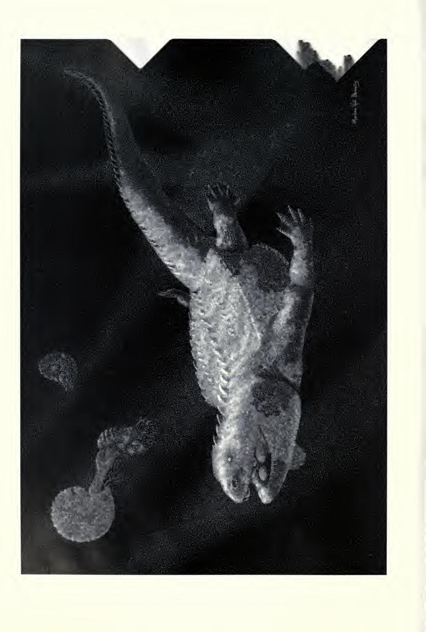
11 amodontoids originated by coalescence of originally separate osteoderms covering the body surface. A juvenile specimen of Cyamodus hildegardis was described as showing an immature stage of development of the carapace (Fig. 5), with incomplete coalescence of irregularly shaped osteoderms (msnm V458: Westphal, 1975; Pinna, 1992; and personal observation). The development and coalescence of osteoderms appears to proceed in an anteroposterior gradient, as osteoderms are more densely packed in the anterior trunk region of msnm V458 (Pinna, 1992, Fig. 2). Similar observations of an ontogenetic consolidation of the carapace have been reported for Psephoderma alpinum, where the ossification of the caudal shield lags somewhat behind the ossification of the carapace (Pinna & Nosotti, 1989), 150 mm. Approximately 90 mm of the again indicating an anteroposterior gradient in the development of the dermal armor. One remarkable juvenile specimen (msnb 4884a and b) from the Norian of northern Italy shows a skull length of only 28 mm and a total body length of approximately vertebral column is preserved, including the very long and slender tail, and weak ossifications of the four limbs can be identified, but there is no trace of a carapace or of separate osteoderms (Fig. 6). It seems to represent an ontogenetic stage at which dermal ossification has not yet started. This conclusion contrasts with a small cyamodontoid from the Ladinian Muschelkalk of Mont-ral-Alcover in northeastern Spain with a total length (from the tip of the snout to the tip of the tail as preserved) of 120 mm and a skull length of 24.5 mm. Due to taphonomic peculiarities at this locality (Hemleben & Freels, 1977), bone substance is not preserved, but the body contours are distinct and indicate the presence of a bipartite dorsal armor comprising a carapace and a tail shield (Rieppel & Hagdorn, 1997, Fig. 2). The carapace does not show the three longitudinal ridges otherwise typical of Psephoderma, which renders the generic identification of the specimen impossible. But with a well-developed dorsal armor at this small size, the specimen either represents a separate miniature cyamodontoid species, or raises questions as to the proper identification of supposedly juvenile Cyamodus and Psephoderma specimens Fig. 2. Carapace fragment and isolated osteoderm of cf. Psephoderma (HUJ-Pal. T.R.I 044) from the Middle Triassic of Makhtesh Ramon, Negev. with what appears to be an absent or incompletely ossified dermal armor. Indeed, knowledge of the ontogeny of the dermal armor of cyamodontoid placodonts must remain incomplete until the discovery of new material. Aside from documenting the presence of dissociated osteoderms which indicate ontogenetic fusion in the formation of the carapace, specimen msnm V458 of Cyamodus hildegardis poses some special problems. The osteoderms come in all sizes and shapes in that specimen, without regularity to their appearance. This irregularity of appearance, together with the fact that the osteoderms "thin out" toward their margins, may indicate their as yet incomplete ossification in a juvenile animal. But, as already noted by Pinna (1980), some osteoderms lie outside the dorsal rib cage, as they should and as is particularly clear on the right side of the body. Other osteoderms, however, lie inside the body cavity, overlapping the flat ventral surface of the broad transverse processes of the dorsal vertebrae. And whereas most osteoderms lie in between the transverse processes of the dorsal vertebrae and in between the gastral ribs, not infrequently osteoderms overlap the ventral surface of gastral ribs. Pinna (1980, PI. 4, Fig. 4) even postulated an occasional fusion of osteoderms with the ventral surface of gastral ribs, Fig. 1. Life reconstruction of a hypothetical cyamodontoid placodont showing the characteristics of the dermal armor (artwork by Marlene Donnelly, the Field Museum). RIEPPEL: THE DERMAL ARMOR OF CYAMODONTOID PLACODONTS
 the (right) transverse process of the 6th preserved dorsal vertebra.")
 noticed isoorientation and some degree of disarticulation in vertebrates (mostly fishes) from the Formazione di Besano (equivalent to the Grenzbitumen horizon), which he")

12 been some dislocation of skeletal elements in the decaying animal. For example, a gastral rib lies on top of (i.e., morphologically ventral to) the (right) transverse process of the 7th preserved dorsal vertebra. But as the intact yet dislocated gastral rib extends anteriorly, it passes below (i.e., morphologically dorsal to) the (right) transverse process of the 6th preserved dorsal vertebra. Deposited in a supposedly anoxic intraplatform basin, fossilization of vertebrates was generally assumed to be undisturbed. However, Tintori (1992) noticed isoorientation and some degree of disarticulation in vertebrates (mostly fishes) from the Formazione di Besano (equivalent to the Grenzbitumen horizon), which he attributed to light bioturbation (in a disaerobic environment) and currents at the bottom of the basin. The same factors, together with pressure originating from the compaction of sediment, apparently did have an impact on the carapace of Cyamodus and may be responsible for some of the disarticulation and dislocation of the osteoderms. Systematic Paleontology Sauropterygia Owen, 1860 Placodontia Zittel, Cyamodontoidea Peyer and Kuhn-Schnyder, 1955 Genus Cyamodus Meyer, 1863 Fig. 3. Plastron fragment of Psephosauriscus sinaiticus Haas (HUJ-Pal. uncatalogued, part of the now broken specimen D of Haas, 1959, PI. V) from the Middle Triassic of Makhtesh Ramon, Negev. A, ventral view; B, dorsal view. which might indicate the presence of an incomplete plastron. By comparison, the specimen pimuz T58 of Cyamodus hildegardis clearly displays, on its right between osteo- side, the hexagonal suture pattern derms exposed in ventral view. In other parts of the body, ill-defined dermal bone appears to have been squeezed in between the transverse processes of the dorsal vertebrae, and bone may even have been squeezed across the ventral surface of the transverse processes. There must also have Type Species Cyamodus rostratus (Munster, 1839). Diagnosis See Rieppel (2000b, 2001) for a diagnosis and discussion of the genus. Distribution Middle Triassic (Anisian, Ladinian); Germanic basin and southern Alps. Description The genus was erected by Meyer (1863) for Cyamodus rostratus (Munster, 1839) from the upper Muschelkalk (upper Anisian) of southeastern Germany (Bayreuth). Other species from the same age and locality are Cyamodus muensteri (Agassiz, ) and Cyamodus laticeps (Owen, 1858), the latter most probably a junior synonym of Cyamodus muensteri (Rieppel, 2000b, 2001). Cyamodus kuhnschnyderi Nosotti and Pinna, 1993, is from the upper Muschelkalk of southwestern Germany (Crailsheim; lower Ladinian); and Cyamodus hildegardis Peyer, 1931, is from the Grenzbitumen horizon (Anisian- FIELDIANA: GEOLOGY
 of Monte San Giorgio (southern Alps).")


13 Fig. 4. Plastron fragment of Psephosauriscus sinaiticus Haas (HUJ-Pal. uncatalogued, part of the now broken specimen D of Haas, 1959, PI. V) from the Middle Triassic of Makhtesh Ramon. Negev. A, ventral view; B, dorsal view. Ladinian boundary) of Monte San Giorgio (southern Alps). The holotype and only known specimen of Cyamodus tarnowitzensis Giirich, 1884, from the Karchowice Beds (lower Muschelkalk, lower Anisian) of Tarnowiskie, Poland, was lost during World War II. Among this material, only Cyamodus hildegardis is known from articulated specimens. Cyamodontoids from the Germanic Triassic are known from skulls collected at three different localities (Upper Silesia, Bayreuth, and Crailsheim), all of which have yielded abundant additional sauropterygian material, but virtually no osteoderms. and exceedingly rare coherent dermal armor frag- Fig. 5. Detad of carapace of Cyamodus hildegardis Peyer (msnm V458), with incomplete coalescence of irregularly shaped osteoderms. ments. This is in stark contrast to other localities (such as Makhtesh Ramon in the Negev: Haas, 1969, 1975; see below), where cyamodontoid osteoderms and armor fragments, if present, are the most frequently found fossil remains. Only three isolated armor fragments are known from the Germanic Muschelkalk (upper Muschelkalk [mo2] of Crailsheim, lower Ladinian), and for stratigraphic reasons they have been referred to Cyamodus kuhnschnyderi (Nosotti & Pinna, 1996, Fig. 14); no armor fragments have been reported from the Muschelkalk of Bayreuth (upper Muschelkalk. mol, upper Anisian) and of Upper Silesia (lower Muschelkalk, lower Anisian). All Cyamodus skulls from the Germanic Triassic show tubercular osteoderms secondarily fused to the temporal region of the skull, demonstrating the developmental capacity to grow osteoderms. which raises the question of why dermal armor remains are so rare in the Germanic Muschelkalk. The most complete dermal armor fragment from the upper Muschelkalk of Crailsheim (smns 81600; Nosotti & Pinna, 1996, Fig. 14C) is 213 mm long. It represents part of a rectangular dorsolateral ridge with the lateral wall still attached to it (Figs. 7, 8). The dorsolateral ridge is formed by enlarged and distinctly keeled osteoderms that slightly interlock with each other in a peg-andsocket fashion. Their circumference is irregularly octagonal, with a length that varies from 37 mm RIEPPEL: THE DERMAL ARMOR OF CYAMODONTOID PLACODONTS
 showing the absence of dermal")

.")
14 Fig. 6. A juvenile specimen of Psephoderma alpinum H. v. Meyer (msnb 4884a) showing the absence of dermal armor. Fig. 7. Carapace fragment from the upper Muschelkalk (lower Ladinian) of Crailsheim (Germany) referred to Cyamodus kuhnschnyderi Nosotti and Pinna (smns 81600). A, dorsal view; B, lateral view. FIELDIANA: GEOLOGY
 in lateral view; C, specimen smns 15891c in lateral view. to 39 mm and a width that ranges from 43 mm to 45 mm.")

 bridge the gaps between adjacent dorsolateral ridge osteoderms.")
15 A.1 Fig. 8. Carapace fragments from the upper Muschelkalk (lower Ladinian) of Crailsheim (Germany) referred to Cyamodus kuhnschnyderi Nosotti and Pinna. A, specimen smns in dorsal view: B, specimen smns 816(H) in lateral view; C, specimen smns 15891c in lateral view. to 39 mm and a width that ranges from 43 mm to 45 mm. The medial ridge is raised into a low, blunt apex at the anterior margin of the osteoderm. It divides the osteoderm into a medial and a lateral part, which together define an angle of 90. Medial to the dorsolateral ridge a row of distinctly smaller, triangular osteoderms separates the latter from what appears to be another row of large, irregularly octagonal and keeled osteo- of the verti- derms. The lateral wall is composed cally descending lateral part ridge osteoderms, flanked ventral ly of the dorsolateral by a row of distinctly pentagonal osteoderms. The latter vary in size. Larger osteoderms (width: 24 mm to 26 mm; height: 22 mm to 25 mm) bridge the gaps between adjacent dorsolateral ridge osteoderms. Between these larger elements, at the middle of the dorsolateral ridge osteoderms, are located smaller yet again pentagonal osteoderms (width: 17 mm to 20 mm; height: 17 mm to 18 mm). RIEPPEL: THE DERMAL ARMOR OF CYAMODONTOID PLACODONTS

 of 150 mm total length comprises four very prominent tubercular osteoderms, of which the two middle ones are complete and have a width of 44 mm and 46 mm")

16 Fig. 9. Carapace fragment from the upper Muschelkalk (lower Ladinian) of Crailsheim (Germany) referred to Cyamodus kuhnschnyderi Nosotti and Pinna (smns 15891c) in lateral view. Collectively, these polygonal osteoderms define a straight ventral margin of the lateral wall, which shows a limited depth and does not appear to have been connected to a plastron. The latter may have been absent. A second dermal armor fragment (smns 15891) of 150 mm total length comprises four very prominent tubercular osteoderms, of which the two middle ones are complete and have a width of 44 mm and 46 mm respectively (Figs. 8B, 9). These are of an irregular pyramidal shape with a blunt apex. On one side of the specimen, much smaller, triangular osteoderms bridge the gaps between the the osteoderms are larger elements. Collectively, very reminiscent of the anterolateral peripherals of Proganochelys (Gaffney, 1990), but whereas the latter are solid (E. S. Gaffney, pers. comm.), the cyamodontoid osteoderms are deeply hollow and rather thin-walled, and may have formed a lateral ridge along the margin of the carapace. As such, specimen smns more closely resembles the enlarged tubercular osteoderms lining the lateral margin of the carapace in Cyamodus hildegardis than specimen smns 81600, which preserves a vertical lateral wall that is absent in Cyamodus hildegardis. The carapace of Cyamodus hildegardis (see also comments above) is composed of a dorsal shield and a separate tail shield (Fig. 10). A lateral wall, as well as a plastron, is absent. Distinctly enlarged, tubercular or pyramidal osteoderms line the circumference (nuchal region not known) of both the dorsal and the tail shield (pimuz T4763; Fig. 11); similarly enlarged marginal osteoderms are absent in a juvenile specimen (msnm V458). The surface of the osteoderms is pitted, but the carapace does not form longitudinal ridges as are known in Psephoderma. Three rows of osteoderms cover the dorsal surface of the tail behind the tail shield, of which the two lateral ones are again enlarged and of tubercular shape (pimuz T4763: Pinna, 1992, Fig. 3; T58: Pinna, 1992, Fig. 1; and personal observation). Genus Henodus Huene, 1936 Type Species Henodus chelyops Huene, Diagnosis See Rieppel (2000b, 2001) for a diagnosis and discussion of the genus. Distribution Upper Gipskeuper (lower Carnian, Upper Triassic); southern Germany. Description The dermal armor of Henodus has been studied in detail and illustrated by Huene (1935, 1958), Reiff (1942), and Westphal (1975, 1976). It may represent the most highly derived dermal armor among cyamodontoid placodonts, as carapacial osteoderms are arranged in a complex yet highly constrained geometrical pattern that relates in a well-defined geometry to the underlying endoskeleton. A dorsomedial row of hexagonal osteoderms is associated with the underlying neural arches of the dorsal vertebrae, whereas a marginal row of smaller hexagonal osteoderms is closely associated with the underlying ribs. It should be noted, however, that due to the poor preservation of Henodus, some controversy surrounds the nature of the articulation of the ribs with the transverse as are known from processes of the dorsal vertebrae. The discussion as to whether the dorsal vertebrae of Henodus carry elongate transverse processes other cyamodontoids (Huene, 1936) or only very short ones (Reiff, 1942) has been resolved by the removal of part of the carapace in specimen VIII 10 FIELDIANA: GEOLOGY

.")

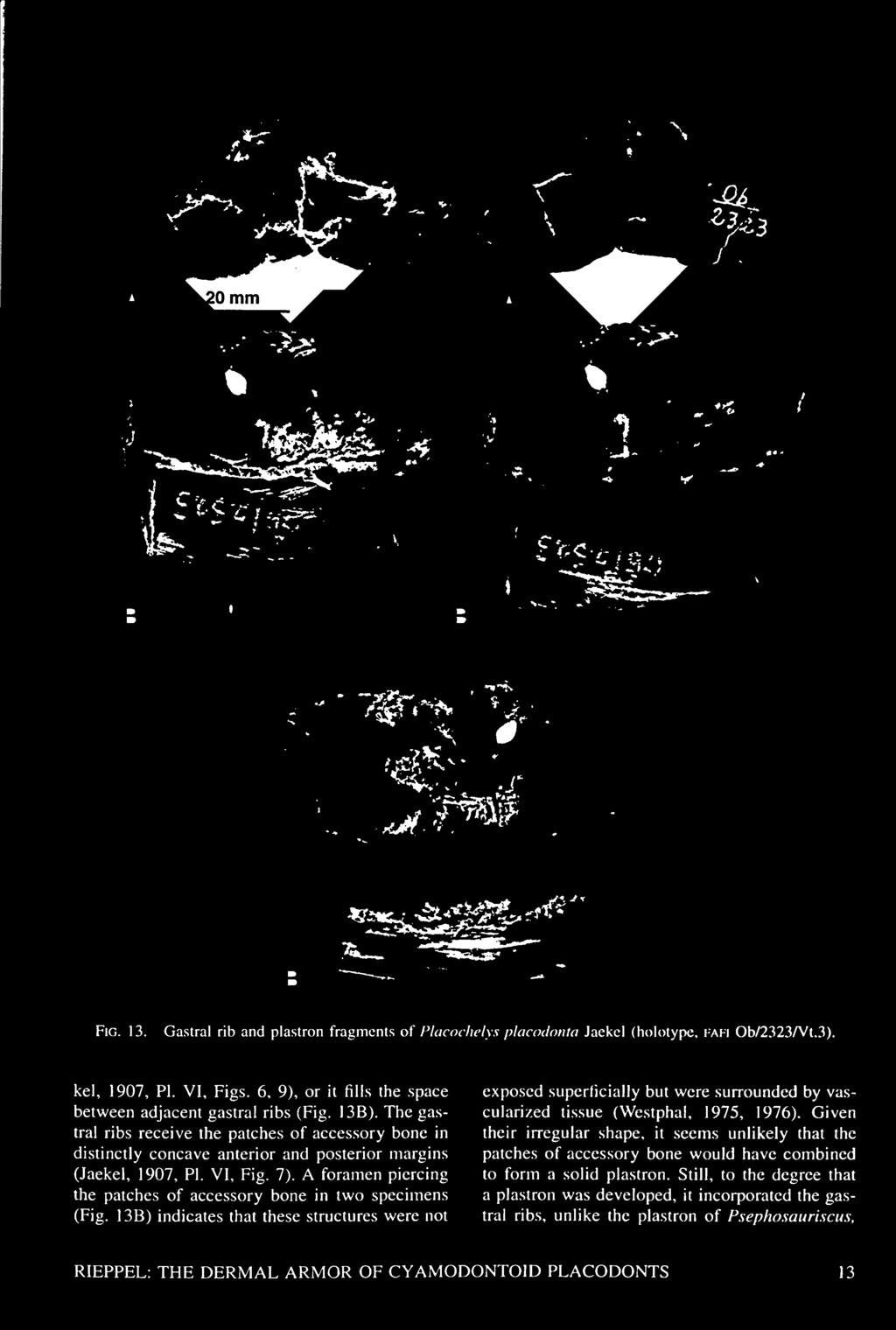
17 Fig. 10. The dermal armor of Cyamodus hildegardis Peyer (holotype, pimuz T4763. dorsal view). (Huene, 1958). This exposed the characteristically elongated transverse processes, which articulate with free ribs. The association of the marginal plates with the endoskeleton is located at the level of a distal expansion of the ribs, i.e., lateral to the transverse processes. The contours of the epidermal scutes are not congruent with the contours of the underlying osteoderms but are indicated by distinct grooves on the surface of the carapace (Reiff, 1942). The carapace of Henodus is shorter than wide. distinctly excavated both anteriorly and posteriorly, and linked by a lateral wall to the plastron. Because of the poor preservation, the structure of the plastron remains incompletely known, but it appears to have been composed of a row of very broad yet short bony lamellae underlying the gastral ribs (Westphal, 1975). Among all cyamodontoids, Henodus certainly has the most turtle-like dermal armor (Reiff, 1942), which was identified by Gregory (1946) as a case of striking convergence. Genus Placochelys Jaekel, 1902 Type Species Placochelys placodonta Jaekel, Diagnosis See Rieppel (2000b. 2001) for a diagnosis and discussion of the genus. Distribution Upper Triassic; central Europe. Description The dermal armor of Placochelys placodonta Jaekel is much less well known than is suggested by Jaekel's (1907) monograph (Westphal, 1975). Some of the original material described by Jaekel (1907) was lost during Fig Enlarged marginal osteoderms of the carapace of Cyamodus hildegardis Peyer (holotype. PIMUZ T4763, dorsal view). World War II. most notably limb bones and parts of the dorsal armor. The carapace of Placochelys is composed of osteoderms of variable size, with a hexagonal base meeting its neighbors in an interdigitating interface and a distinctly ridged, tubercular or py- RIEPPEL: THE DERMAL ARMOR OF CYAMODONTOID PLACODONTS 11

.")
 as dorsal ribs; unquestionable parts of slender gastral ribs (Jaekel, 1907, PI. VI, Fig. 2 [lateral element]; Fig.")
18 Fig. 12. Carapace fragment of Placochelys placodonta Jaekel (holotype, fafi Ob/2323/Vt.3) in dorsolateral view. ramidal "crown" (Fig. 12). Enlarged tubercles were aligned along the lateral margins of the carapace and apparently irregularly interspersed among smaller osteoderms throughout the dorsal shield. The lateral wall is composed of osteoderms with a cycloid superficial appearance, their blunt apex pointing dorsally, the convex base pointing ventrally (Jaekel, 1907, PI. IX, Fig. 3). The large tubercular or pyramidal osteoderms are solid, but isolated specimens show a ventrally concave base (Jaekel, 1907, PI. IX, Fig. 5b). Nothing is known about the presence of a separate Among tail shield, and very little is known of the plastron. the material still available of Placochelys are fragments of bone, identified by Jaekel (1907, PI. VI, Fig. 1) as dorsal ribs; unquestionable parts of slender gastral ribs (Jaekel, 1907, PI. VI, Fig. 2 [lateral element]; Fig. 3 [medioventral element]); and very peculiar elements that look like rib or gastral rib fragments fully or partially fused with irregular patches of accessory bone. These latter elements were identified as ventral rib fragments fused with gastral ribs by Jaekel (1907), or as broadened gastral ribs fused with osteoderms by Westphal (1975). The dorsal vertebrae of cyamodontoids are characterized by very broad transverse processes that articulate with rather short dorsal ribs located in the body wall (Cyamodus hildegardis: Pinna, 1992) or are completely fused with the dorsal ribs (Psephoderma alpinum: Pinna & Nosotti, 1989). Only one, incompletely preserved dorsal vertebra is known for Placochelys, with indications of a broad transverse process (Jaekel, 1 907, PI. VII, Fig. 10). It is possible that the fragments identified as dorsal ribs by Jaekel (1907) represent broken parts of dorsal ribs that may or may not have been fused to the transverse processes of dorsal vertebrae. The ventral rib fragments of Jaekel (1907) show a distinct surface ornamentation of ridges and grooves, which suggests a dermal rather than endochondral origin (Fig. 13). Some fragments are elongate and slightly curved (Jaekel, 1907, PI. VI, Figs. 6, 7) and seem to represent segments of distinctly broadened gastral ribs, comparable to the broadened lateral gastral elements seen in Henodus (Huene, 1958). One specimen is distinctly broadened and angulated (Jaekel, 1907, PI. VI, Fig. 9) and might represent a fragment of a broadened medioventral gastral rib with accessory bone wrapped around it (Fig. 13 A). Huene (1958) interpreted this fragment as a ventrolateral part of a dorsal rib carrying an uncinate process, which would form a ventrolateral body ridge comparable to Henodus. Westphal (1975) showed, however, that the broadening of the distal part of the dorsal ribs underlies the dorsolateral carapacial ridge in Henodus, which in Placochelys carries large, pyramidal osteoderms. Henodus thus appears to be a poor model for the reconstruction of the plastron in Placochelys. The accessory bone has a smooth surface and may wrap around the thickened gastral ribs (Jae- 12 FIELDIANA: GEOLOGY

19 _~1
. Genus Psephoderma Meyer, 1858a,b Type Species Psephoderma alpinum Meyer, 1858a,b.")

20 Fig. 14. The left lateral margin of the carapace of Psephoderma alpinum H. v. Meyer (holotype, bsp As I 8) in dorsal view. which is composed of discrete osteoderms lying ventral to the gastral ribs (for a description, see below). Genus Psephoderma Meyer, 1858a,b Type Species Psephoderma alpinum Meyer, 1858a,b. Diagnosis See Rieppel (2000b, 2001) for a diagnosis and discussion of the genus based on skull structure. The genus is further diagnosed by a carapace carrying three longitudinal ridges, a dorsomedial one and two dorsolateral ones, which are composed of enlarged and distinctly keeled or tuberculiform osteoderms. Distribution Upper Triassic; northern and southern Alps, and northern Gondwanan shelf (Middle East). Comments The holotype of Psephoderma alpinum is represented by a carapace fragment from the Rhaetian Koessen-Formation of the Bavarian Alps (Winkelmoos Alpe), which was first described by Meyer (1858a,b). Meyer (1858a) did not provide a formal diagnosis of the taxon, but salient characteristics of the new genus and species are easily gleaned from his description of the specimen: the carapace is of rounded contours, its width slightly exceeding its length; the rather flat dorsal shield of the carapace meets the lateral wall at an angle of 90 ; in addition to the marginal keels, three longitudinal keels are distinct on the dorsal surface of the carapace, the medial one marking the dorsal midline; the osteoderms forming the dorsal keels are larger than the intervening elements, and all osteoderms are of a fairly regular hexagonal shape. New and more completely preserved specimens from the southern Alpine Triassic (Pinna, 1978; Pinna & Nosotti, 1989; Renesto & Tintori, 1995) added greatly to our understanding of the genus, and hence to the precision of its diagnosis (Pinna, 1999), which can now be based on autapomorphic characters of skull structure (Rieppel, 2000b, 2001). The second species in the genus, Psephoderma anglicum Meyer, 1864, remains very incompletely known. C. J. Duffin considers the latter species a subjective junior synonym of Psephoderma alpinum (quoted in Rieppel, 2000b: 38), while Storrs (1994) treated the isolated osteoderms referred to Psephoderma anglicum as not diagnostic. Description The holotype of Psephoderma alpinum Meyer, 1858a,b, is represented by a carapace fragment with a total length of 375 mm and a total width of 425 mm. Its circumference is almost circular; the nuchal concavity is distinct, the posterior margin is incomplete. The carapace is composed of regularly shaped hexagonal osteoderms that meet in slightly interdigitating sutures (Fig. 14). The surface of the osteoderms is flat or elevated into a weakly expressed, blunt apex, surrounded by a circular zone of slight depression. The osteoderms are pitted, the pits radiating from the centrally located center of ossification toward 14 FIELDIANA: GEOLOGY
.")


21 Fig. 15. The dermal armor of Psephoderma alpinum H. v. Meyer (msnb 8358) in dorsal view. the margins. The average osteoderm is 34 mm to 36 mm long and 30 mm to 34 mm broad. Fractures in the middle of the carapace indicate a thickness of the osteoderms that does not exceed 5 mm (contra Westphal, 1975). The epidermal scute areas are indicated by shallow grooves that coincide with the circumference of the osteoderms. Diagnostic for the genus are three longitudinal ridges on the carapace, a dorsomedial one and two dorsolateral ones. These are composed of slightly enlarged and distinctly keeled osteoderms with a length of 36 mm to 4 1 mm and a width of 45 mm to 48 mm. The keel is elevated into a low, blunt apex at the center of the osteoderm. The dorsomedial keel is separated on either side from the dorsolateral keels by two rows of intermediate, regular osteoderms, with the rare intercalation of a distinctly smaller third element. The dorsolateral ridge is separated from the lateral margin of the carapace by three rows of regular osteoderms. The lateral margin of the carapace itself is again formed by enlarged osteoderms of 40 mm to 43 mm length, which are of hexagonal circumference and distinctly keeled and which define a sharp and rectangular angle between the dorsal surface and the lateral wall of the carapace. These marginal osteoderms meet each other but do not interlock in a peg-and-socket fashion. The lateral wall of the carapace is of limited depth and consists of a single row of regularly shaped pentagonal elements. The apex of each of these elements interlocks with the lateral ridge osteoderms, while the broad base contributes to the formation of a smooth ventral edge. There is no indication that the lateral wall would have connected to a plastron, which in fact seems to have been absent (Westphal, 1975). New and beautifully preserved material has come from the upper Norian (Calcare di Zorzino) of the southern Alps (Pinna & Nosotti, 1989: specimens msnm V527, msnb 4614, 8358; Renesto & Tintori, 1995: specimen ST82003). Collective- this material documents that the dorsal armor ly, of Psephoderma is bipartite, including a carapace and a tail shield (Fig. 15); a plastron is again absent in these specimens (Pinna & Nosotti, 1989, Pis. 25, 29). The carapace and the tail shield are composed of very regularly shaped hexagonal osteoderms that meet each other in slightly interdigitating (Figs. 16A, 17A) or noninterdigitating (msnb 4614) sutures (Figs. 16B, 17B). As in the holotype, three longitudinal rows of enlarged and keeled osteoderms form a dorsomedial keel and RIEPPEL: THE DERMAL ARMOR OF CYAMODONTOID PLACODONTS 15

 of Endenna near Bergamo (Fig. 15).")
 by its width yields a quotient of 0.88 for the holotype and 0.83 for the specimen msnb 8358.")
22 Fig. 16. Osteoderm shape and structure in the carapace of Psephoderma alpinum H. v. Meyer (dorsal view). A, specimen msnb 8358; B, specimen msnb two dorsolateral keels on the carapace; the dorsolateral keels, but not the dorsomedial keel, continue on to the tail shield. As in "Cyamodus" hildegardis, loose osteoderm covering continues on the dorsal surface of the tail behind the tail shield (Renesto & Tintori, 1995). The same material also documents some variability in the dermal armor in Psephosaurus. msnb 8358 is a complete specimen from the Norian (Calcare di Zorzino) of Endenna near Bergamo (Fig. 15). It is somewhat smaller than the holotype, yet shows a fully developed dorsal armor. The carapace is 210 mm long and 253 cm wide; the tail shield is 49 mm long and mm wide. Dividing the length of the carapace (dorsal shield) by its width yields a quotient of 0.88 for the holotype and 0.83 for the specimen msnb The osteoderms meet each other in distinctly interdigitating sutures (Figs. 16 A, 17 A). The osteoderm surface is elevated into a low apex surrounded by a circular zone of slight depression and is ornamented by a pattern of radiating grooves and ridges, rather than pits as in the holotype. The osteoderms are of a regular hexagonal (occasionally pentagonal) outline with an average diameter of 18 mm to 22 mm. The nuchal area is distinctly concave and has a width of six to seven osteoderms. Along the lateral margins of the dorsal shield, there are 12 somewhat enlarged osteoderms (of a length of 23 mm to 25 mm) that form a distinct lateral ridge; their number compares closely with the holotype. The dorsomedial and the two dorsolateral ridges are again composed of enlarged (length: 20 mm; width: 27 mm to 27 mm) and distinctly keeled osteoderms, nine to ten in each row (ten in the right dorsolateral ridge of the holotype). The dorsomedial and dorsolateral ridges are separated from one another by three rows of intervening osteoderms anteriorly but by only two rows posteriorly. The tail shield is three rows of osteoderms long and eight rows of osteoderms broad. The specimen of Psephoderma published by Renesto & Tintori (1995, specimen ST82003) is larger than any other specimen of its genus found so far. Although its morphology remains to be described in detail, it can be seen from Figure 2 in Renesto and Tintori (1995) that the carapace is again somewhat wider than it is long, as is also the case for the holotype of Psephoderma alpinum and for specimen msnb This contrasts with specimen msnm V527, from the upper Norian of Endenna (Pinna & Nosotti, 1989), which is prepared in ventral view but which still shows the contours of the carapace. With a length of 275 mm and a width of 240 mm, the dorsal shield is longer than broad, which contrasts with the other specimens of Psephoderma alpinum, including the holotype. At this time it remains unknown whether this difference represents a taphonomic artifact resulting from the strong dorsoventral of the fossils compression or whether some taxonomic variation is implied (Nosotti & Rieppel, work in progress). The Rhaetian (Calcare di Zu) of Monte Rena near Bergamo has yielded carapace fragments that compare to the holotype of Psephoderma in osteoderm size, msnb 4614 (Pinna, 1978, PI. 74) is a specimen that shows the osteoderms to meet superficially in a smooth rather than interdigitating contact (Figs. 16B, 17B). And whereas the osteoderms of Psephoderma show a weakly expressed 16 FIELDIANA: GEOLOGY


 referred it to Psephoderma alpinum.")
23 Fig. 17. Osteoderm shape and structure in the carapace of Psephoderma alpinum H. v. Meyer (dorsal view). A.l, specimen msnb 8358, left posterolateral margin; A.2, specimen msnb 8358, right anterolateral margin; B, specimen msnb 4614, left dorsolateral ridge. apex, those of msnb 4614 show a weakly expressed keel, and no circular zone of slight depression. The surface of the osteoderms is ornamented with a pattern of vermiculate radiating ridges and grooves, similar to those seen in other Psephoderma from the Alpine Triassic. The fragment msnb 4614 comprises parts of at least two rows of enlarged and distinctly keeled osteoderms that appear to converge on each other. Originally identified (as indicated by the museum label) as Placochelyanus stoppanii (new combination: Pinna, 1976), a species described by Osswald (1930; who referred it to the genus Placochelys), Pinna (1978) referred it to Psephoderma alpinum. The specimen does indicate some variation in details of carapace structure, but it is too incomplete to allow the identification of taxic diversity within the genus Psephoderma (for comments on carapace proportions, see above). The dermal armor appears to be completely absent in a juvenile specimen of Psephoderma (msnb 4884a and b) of approximately 145 mm total length (Fig. 6). B 8359 is an incomplete specimen from the Norian (Calcare di Zorzino) of Endenna near Bergamo comprising the pelvic region and tail (Pinna & Nosotti, 1989, PI. 31). The tail, which comprises 44 to 45 vertebrae (exposed behind the tail shield), measures 268 mm in length and shows no osteoderms associated with it, although the tail shield is ossified and partially RIEPPEL: THE DERMAL ARMOR OF CYAMODONTOID PLACODONTS 17
 shows a tail length of 432 mm, and osteoderms associated with it up to the 9th caudal vertebra.")

.")
24 BJ mm %> 1 Fig. 18. Carapace of Psephoderma sculptata n. sp. (holotype, T.R.929, original of Haas, 1975, PI. I, Fig. 8). A, overview; B, close-up view of enlarged dorsomedial ridge osteoderms. covers the pelvic region. A larger specimen from the same locality (msnm V527) shows a tail length of 432 mm, and osteoderms associated with it up to the 9th caudal vertebra. The specimen described by Renesto and Tintori (1995, Fig. 2) shows rows of associated osteoderms up to at least the 12th caudal vertebra. Collectively, these specimens indicate variability in the ossification of the dermal armor along the tail. Possible taxonomic implications of these observations must await a comprehensive revision of the genus Psephoderma. Psephoderma sculptata n. sp. Holotype HUJ-Pal. T.R.I 98, carapace fragment (original of Haas, 1975, Fig. 14). Stratum and Locus Typicus Lower Member of the Saharonim Formation of late Anisian (middle and late Illyrian) or early Ladinian (Fassanian) age, Middle Triassic, Makhtesh Ramon, Negev, Israel. Referred Material HUJ-Pal. T.R.207, isolated osteoderms; T.R.929, small carapace fragment (original of Haas, 1975, PI. I, Fig. 8). Diagnosis A cyamodontoid placodont with a triple-keeled dorsal shield (carapace); keels composed of much enlarged, hexagonal and tuberculiform osteoderms with a posteriorly inclined apex; dorsomedial keel separated from dorsolateral keels by a single row of distinctly smaller, hexagonal or polygonal Comments osteoderms. The carapace from the Middle Triassic of Makhtesh Ramon, which is here referred to a new species of Psephoderma, is again rather incompletely preserved (Fig. 18). It remains unknown whether this carapace was linked to a lateral wall, or whether this taxon differentiated a plastron, as it is known to occur in other cyamodontoids from Makhtesh Ramon (see below), but which is absent in Psephoderma (Pinna & Nosotti, 1989). However, as in Psephoderma alpinum, the carapace of the Makhtesh Ramon cyamodontoid is composed of hexagonal osteoderms, and it shows clear indications of three longitudinal keels on its dorsal surface formed by enlarged osteoderms (Haas, 1975). Among the Cyamodontoidea, FIELDIANA: GEOLOGY

 comprises the middle section of the dorsal shield. As preserved, the fragment is 262 mm wide and 332 mm long.")

25 20 mm Fig. 19. Isolated osteoderms from the carapace of Psephodertnu sculptata n. sp. (referred specimens, HUJ-Pal. T.R.207). this character is so far known only for Psephoderma, which is the reason why the new taxon from Makhtesh Ramon is referred to that genus. Distribution Middle Triassic (Anisian, lower Ladinian), Middle East (Makhtesh Ramon, Negev, Israel). Description The carapace fragment of Psephoderma sculptata n. sp. (original of Haas, 1975, PI. 2, Fig. 14) comprises the middle section of the dorsal shield. As preserved, the fragment is 262 mm wide and 332 mm long. Neither the marginal zones nor any part of the lateral walls (if present) are preserved. The sculpturing of the carapace surface is more distinctly expressed than is the case in Psephoderma alpinum, which is a function of relative osteoderm size. Osteoderm structure is again basically hexagonal in specimen HUJ-pal. T.R.I 98 (holotype of Psephoderma sculptata, Fig. 18), but the osteoderms forming the dorsomedial and dorsolateral keels are dramatically increased by comparison to the intervening osteoderms. A typical osteoderm of the dorsomedial keel is between 59 mm and 67 mm wide and between 59 mm and 62 mm long. As a function of their dimensions, these osteoderms may assume an almost circular circumference (Fig. 19). The osteoderms again meet in slightly interdigitating sutures. The dorsomedial keel is again somewhat less prominent than the dorsolateral keels, but the sculpturing of the carapace is generally much more prominently developed than in Psephoderma alpinum. The individual osteoderms contributing to the formation of the dorsal keels are of a tuberculiform shape, the keel developing a tall apex (abraded in the holotype, but well-preserved in HUJ-Pal. T.R.207, Fig. 19) with distinctly ridged flanks. The apex is slightly asymmetrical, as it is positioned somewhat more closely to the posterior margin of the osteoderm. In Psephoderma sculptata, the dorsomedial keel is separated from the dorsolateral keels by a single row of much smaller osteoderms of hexagonal circumference. One well-preserved and well-delineated intervening osteoderm has a width of 27 mm and a length of 28 mm. Its surface is ornamented by a pattern of radiating ridges. However, the intervening osteoderms vary quite substantially in size and shape in order to fit into the space between the much enlarged osteoderms of the dorsomedial and dorsolateral keels. With its prominently sculptured dorsal surface and the large size discrepancy between the osteoderms that form the three longitudinal dorsal ridges and the intervening osteoderms, specimen huj- Pal. T.R.198 is sufficiently different from any specimen of Psephoderma alpinum known from the Alpine Triassic, or from any other cyamodontoid, in order to warrant the description, and diagnosis, of a separate species. RIEPPEL: THE DERMAL ARMOR OF CYAMODONTOID PLACODONTS 19
 from the upper Ceratites beds (layer D2 of Brotzen, 1957) of Makhtesh Ramon (see also Haas, 1975, PI. 2, Fig. 11, HUJ-Pal. T.R.I 843).")


26 cf. Psephoderma sp. Haas (1969) published a comparatively wellpreserved cyamodontoid carapace (HUJ-Pal. T.R.3189) from the upper Ceratites beds (layer D2 of Brotzen, 1957) of Makhtesh Ramon (see also Haas, 1975, PI. 2, Fig. 11, HUJ-Pal. T.R.I 843). The osteoderm structure seen in this carapace matches that of many fragmentary pieces or isolated osteoderms from the same locality (Fig. 2). These osteoderms differ from those of Psephosauriscus by their columnar proportions, their height exceeding their diameter. Midcarapacial osteoderms have a fairly regular hexagonal structure. Their diameter averages approximately 15 mm, their thickness 20 to 25 mm. The surface of the osteoderms shows a distinct central depression. There are no distinct impressions of overlying epidermal scales. The osteoderms meet in smooth or faintly interdigitating sutures, which is the reason for their easy dissociation from one another during fossilization. It is not uncommon to find small fragments composed of only a few osteoderms, or single elements, in the Muschelkalk of Makhtesh Ramon. The cohesion of the osteoderms in the carapace of the live animal was mediated by calcified bundles of connective tissue ("calcified decussating connective tissue bundles" of Haas, 1969, PI. 1, Fig. c; "mineralized connective fibers" of Westphal, 1976, Fig. 3A; see also Westphal, 1975, Figs. 3c-e). Laterally, the rather flat carapace merges into a ventrally descending lateral wall. The transition from the dorsal surface of the carapace to the lateral wall describes a gentle curve (Haas, 1969, PI. 1, Fig. B) which is capped by somewhat enlarged osteoderms that retain a smooth surface, however. A distinct dorsolateral ridge is not differentiated. There is also no differentiation of distinct longitudinal ridges on the dorsal surface of the carapace in a manner comparable to Psephoderma. However, the central part of the carapacial surface is slightly depressed (concave), and so is the marginal zone of the carapace. This results in the formation of two very shallow and smooth, dorsolateral and slightly curved ridges (Westphal, 1975, Fig. 2), vaguely reminiscent of the much more strongly differentiated dorsal ridges seen on the carapace of Psephoderma. The carapace HUJ-Pal. T.R.3189 was found in association with its steinkern filling. No remains of the postcranial skeleton or any part of a plastron were recovered. It remains unknown whether a plastron was present (Haas, 1959), although the tapering ventral edge of the lateral wall (Haas, 1969, PI. lb) suggests its absence. A plastron is present in Psephosauriscus but absent in Psephoderma. Given the characteristics of this carapace, there is no doubt that it represents a separate cyamodontoid taxon from Makhtesh Ramon. Since it remains unknown whether a plastron was present or no basis for the inclusion of this absent, there is taxon in the genus Psephosauriscus. The osteoderm structure of this unidentified cyamodontoid from Makhtesh Ramon is also strikingly different from the osteoderms known from Placochelys or Psephosaurus (see above). The faint differentiation of longitudinal ridges on the carapace, as well as the tapering ventral edge of the lateral wall, might be taken as an indication for some affinity of this taxon with the genus Psephoderma. But whatever its ultimate generic affinities might turn out to be should more completely preserved material become available, this taxon adds to the species diversity of cyamodontoids known from Makhtesh Ramon. Genus Psephosaurus Fraas, 1896 Type Species Psephosaurus suevicus Fraas, Diagnosis See Rieppel (2000b, 2001) for a diagnosis and discussion of the genus. Distribution Upper Ladinian (Middle Triassic); southern Germany. Description Psephosaurus suevicus Fraas, 1 896, was based on an incomplete carapace from the Lower Keuper (upper Ladinian) of southwestern Germany (Hoheneck near Stuttgart; Fraas, 1896). Also available are isolated osteoderms. Today the carapace is represented by six fragments (Figs. 20, 21) that can no longer be fitted together to reconstruct the carapace outline. Preservation is rather poor, and the delineation of individual osteoderms is difficult. As noted by Fraas (1896), two different types of osteoderms can be distinguished. Large osteoderms of irregular polygonal or even rounded contours typically have a diameter of 25 mm to 28 mm. The center of these osteoderms is elevated into an apex, which, owing to compression, is weakly expressed. The surface of the osteoderms is ornamented by a pattern of very delicate grooves and ridges that radiate from the center toward the margins. The enlarged osteoderms are 20 FIELDIANA: GEOLOGY

 the sutures between osteoderms run in shallow grooves, which indicate the congruent circumference of the overlying epidermal scutes.")

27 separated from one another by smaller, mostly pentagonal or hexagonal but occasionally irregular polygonal osteoderms with a diameter that from 15 mm to 25 mm. The surface of may vary these osteoderms is fiat or slightly depressed and again ornamented with a pattern of delicate yet numerous radiating grooves and ridges. It seems that the epidermal scute area coincides with the osteoderm outline. In one fragment (smns 7113; Fig. 21A.1) the sutures between osteoderms run in shallow grooves, which indicate the congruent circumference of the overlying epidermal scutes. In superficial view, the sutures between osteoderms are slightly interdigitating; in internal (ventral view), suture lines may even appear to be smooth (Fig. 21 A. 2). The osteoderms did not dissociate during fossilization, however. Incomplete preservation renders it difficult to establish regularity of the arrangement of the larger osteoderms within the smaller ones. On the largest of all the carapace fragments, the enlarged osteoderms can be seen to be aligned in a row, separated from one another by intervening smaller osteoderms. Other enlarged osteoderms appear to be randomly distributed among the smaller elements. Some isolated osteoderms, corresponding to the enlarged elements interspersed between smaller ones, better preserve their three-dimensional shape. All have a concave base and a crown protruding into an apex (Figs. 21B-D). This apex may be distinctly keeled (smns 17790; diameter: 27 mm), elongated (smns 7180; diameter: 28.5 mm), or symmetrical (smns 54710; diameter: 27.5 mm). Genus Psephosauriscus n. gen. Type Species Psephosauriscus mosis (Brotzen, 1957). Diagnosis Dermal armor comprising a solid carapace and plastron; carapace composed of hexagonal osteoderms with smooth or interdigitating interfaces; osteoderm thickness does not exceed their diameter; carapace linked to plastron by a vertically oriented or curved lateral wall; dorsolateral ridge may (with vertically oriented lateral wall) or may not (with curved lateral wall) be differentiated; ventrolateral ridge always present; plastron composed of osteoderms with trapezoidal to rhomboidal base and cycloid crown. Plastral osteoderms arranged in regular transverse rows, aligned along and partially fused with gastral ribs. Distribution Lower Anisian to lower Ladinian. Middle East. Comments The Middle Triassic Muschelkalk of Makhtesh Ramon, Negev (Brotzen, 1957, Haas, 1969, 1975), and of Araif en Naqua, Sinai Peninsula (Haas, 1959), has yielded the remains of a variety of cyamodontoid placodonts, all of which have provisionally been referred to the genus Psephosaurus. Unfortunately, the (prepared) material currently comprises the remains of two very incomplete skulls only, and fragments of a lower jaw (Brotzen, 1957; Haas, 1975; Rieppel et al., 1999) from the basal Beneckeia beds (lower Anisian) and younger Ceratites beds (upper Anisian, lower Ladinian) of these Muschelkalk deposits. None of that skull material is diagnostic, and, as noted by Brotzen (1957), there is no character that precludes the best-preserved skull fragment from being referred to Cyamodus (no skull is known for Psephosaurus). In contrast to the Germanic Muschelkalk, dermal armor fragments abound in the Middle Eastern deposits and indicate a significant taxonomic variety of cyamodontoids at least at the species level within one genus (or perhaps several genera) distinct from Psephosaurus. The diversity of dermal armor remains in the Middle East has led to considerable taxonomic confusion at the level of species names. In his original description of cyamodontoids from Makhtesh Ramon, Brotzen (1957) recognized two distinct species, viz. "Psephosaurus" mosis from the Beneckeia beds and "P." picardi from the Ceratites beds. In his study of the cyamodontoids from the Sinai Peninsula, Haas (1959) noted that the presumed carapace of the holotype of "Psephosaurus" mosis (HUJ-Pal. C.F.247) really consists of two carapacial fragments and a fragmentary plastron (confirmed by personal observation). A full description of "Psephosaurus" picardi, promised by Brotzen (1957), was never published; the original material is represented by a natural mold of the internal side of a carapace (not diagnostic). The taxon is here treated as a nomen duhium for reasons discussed below. In his description of the material from the Sinai Peninsula, Haas (1959) described two additional species, "Psephosaurus" sinaiticus (holotype HUJ-Pal. 3421) and "Psephosaurus" rhomhifer (the holotype cannot be located at present). Comparing the diagnoses and illustrations provided by Haas (1959), the validity of "P." rhomhifer as a separate taxon is difficult to evaluate (see discussion of cf. Psephosauriscus rhomhifer below). If RIEPPEL: THE DERMAL ARMOR OF CYAMODONTOID PLACODONTS 21
. Such a conclusion would conflict with that of Haas (1975), who considered \"P.\" sinaiticus a possible junior synonym of \"P.")
 with no description and no illustration (nomen nudum), but apparently with reference to a taxon from Makhtesh Ramon which is close to,")
28 Fig. 20. Carapace fragment of Psephosaurus suevicus Fraas (holotype, smns 6693, dorsal view). P. rhombifer is not, indeed, a separate taxon, it would have to be a subjective junior synonym of "P." sinaiticus (by page priority of the latter; see further discussion below). Such a conclusion would conflict with that of Haas (1975), who considered "P." sinaiticus a possible junior synonym of "P." mosis, and "P." picardi a separate species. The species name ramonensis was introduced by Haas (1975: 455) with no description and no illustration (nomen nudum), but apparently with reference to a taxon from Makhtesh Ramon which is close to, or even synonymous with, " Psephoderma" mosis. In the Paleontological Collections of the Hebrew University, specimen HUJ-Pal. T.R.2751 was found to be labeled "Psephoderma" ramonensis without indication of an author. The name was never formally published, but the specimen was figured by Westphal (1975, Fig. 5, top). A review of the material presently available in the Paleontological Collections of the Hebrew University, Jerusalem, allows the diagnosis of at least four species within this genus. The phylogenetic position of the genus Psephosauriscus among the Cyamodontoidea remains currently unresolved. The reason for this is that Psephosauriscus is known exclusively from dermal armor fragments, whereas phylogenetic reconstruction of the Cyamodontoidea is based primarily on skull structure (Rieppel, 2000b, 2001). Psephosauriscus mosis (Brotzen, 1957) 1957 Psephosaurus mosis Brotzen, p Psephosaurusl mosis Haas, p Psephosaurus mosis Haas, p Holotype HUJ-Pal. C.F.247, two fragments of a carapace, plus one fragment of a plastron. Stratum and Locus Typicus Beneckeia beds (lower Anisian), Makhtesh Ramon, Negev, Diagnosis Israel. Dorsal surface of carapace ornamented by scale impressions of highly irregular size and shape, imprinted on hexagonal osteoderms; ventral surface of plastron covered by transverse rows of relatively large cycloid osteoderms, but osteoderm shape becoming irregular toward the margins of plastron; dorsolateral ridge fortified by enlarged, keeled osteoderms separated from one another by a pair of intervening smaller osteoderms; lateral wall vertically placed, composed of hexagonal osteoderms; ventrolateral ridge fortified by enlarged, keeled osteoderms that are in noninterlocking contact with one another. Distribution Middle Triassic (Anisian, lower Ladinian), Middle East (Negev, Israel). Description The preserved carapace of Psephosauriscus mosis comprises two adjacent fragments, the anterior one of which preserves part of the nuchal emargination (Fig. 22). The superficial appearance of the carapace shows a complex pattern of grooves delineating areas of epidermal scutes of highly irregular shape and size. This pattern of epidermal scute areas is imprinted on osteoderms of more or less regular hexagonal outlines, although the sutures between the osteoderms are identifiable in very localized areas only. The thickness of the carapacial osteoderms does not exceed their diameter of around 1 5 to 20 mm. 22 FIELDIANA: GEOLOGY
 in ventral view; B, isolated osteoderm with longitudinal ridge (smns 17790); C, isolated osteoderm (smns 54701); D, isolated osteoderm with abraded apex (smns 7180);")
, characterized by distinctly enlarged (maximal length of 40 mm, maximal width of 36 mm) osteoderms of an irregular octagonal shape with a projecting posterior tip.")

29 A.1 _20 mm A.2 Fig. 21. Carapace fragments and isolated osteoderms of Psephosaurus suevicus Fraas. A.1, carapace fragment (smns 7113) in dorsal view, showing grooves delineating epidermal scutes; A.2, carapace fragment (smns 7113) in ventral view; B, isolated osteoderm with longitudinal ridge (smns 17790); C, isolated osteoderm (smns 54701); D, isolated osteoderm with abraded apex (smns 7180); E, carapace fragment from holotype (smns 6693) in dorsal view. Regularity of osteoderm pattern is established along the dorsolateral ridge of the carapace (Fig. 23A.2), characterized by distinctly enlarged (maximal length of 40 mm, maximal width of 36 mm) osteoderms of an irregular octagonal shape with a projecting posterior tip. These enlarged osteoderms carry a longitudinal keel, raised into a low apex at the center of the osteoderm. These enlarged osteoderms are regularly separated from one another by an intermediate pair of smaller osteoderms. The lateral wall of the dorsal dermal armor of Psephosauriscus mosis is poorly preserved but shows hexagonal or rhomboidal osteoderms at its anterolateral corner. The plastron of Psephosauriscus mosis (Figs. 23A.3, 23B, 24) is composed of transverse rows of regularly arranged cycloid scales, matching the contours of the crown of the underlying osteoderms with a transverse diameter of 35 mm and a length of 22 mm. The apex of these scales points anteriorly, their convex base faces posteriorly. Along the ventrolateral ridge of the plastron (very incompletely preserved), the osteoderms are enlarged and of rounded contours, with a diameter of 42 mm. These ventrolateral ridge scales are in noninterlocking contact with one another and carry a longitudinal keel that is raised into a low apex toward the center of the scale (Fig. 23A.1). In summary, Psephosauriscus mosis differs from Psephosauriscus sinaiticus by a more pro- RIEPPEL: THE DERMAL ARMOR OF CYAMODONTOID PLACODONTS 23


 described a second species from the Muschelkalk (Ceratites beds) of Makhtesh Ramon, \"Psephosaurus\" picardi.")
30 Fig. 22. Carapace fragments of Psephosauriscus mosis Haas (holotype, HUJ-Pal. C.E247) in dorsal view. A, part 3 of Brotzen, 1957; B, part 2 of Brotzen, nounced development of both the dorsolateral and ventrolateral ridges at the transition of the lateral wall to the carapace and plastron, respectively, by larger plastral osteoderms, and by a highly irregular epidermal scute pattern distinctly imprinted on the underlying osteoderms. Psephosauriscus mosis differs from Psephosauriscus ramonensis n. sp. by the development of a pronounced dorsolateral ridge on the dorsal armor and by highly irregular epidermal scute areas distinctly imprinted on the underlying Comments osteoderms. Dermal armor fragments with imprints of similarly irregular epidermal scutes on hexagonal osteoderms are also found in the Ceratites layers of Makhtesh Ramon. Remarks Along with "Psephosaurus" mosis, Brotzen (1957) described a second species from the Muschelkalk (Ceratites beds) of Makhtesh Ramon, "Psephosaurus" picardi. This latter species is based on a natural cast of the inside of a carapace and on dermal armor fragments from different layers (Brotzen, 1957: 215). Brotzen (1957) designated the cast of the carapace as holotype and indicated that he had deposited the Paleozoological Department of the Natural History Museum in Stockholm. However, this specimen can no longer be located. Given the taxonomic diversity of cyamodontoids from Makh- it in tesh Ramon, as evidenced by carapace fragments, and the poor documentation of the natural cast of the inside of a carapace (Brotzen, 1957, PI. 7), I concur with Haas (1959: 14) in treating "Psephosaurus" picardi as a nomen dubium. Psephosauriscus ramonensis n. sp. Holotype HUJ-Pal. 2751, fragment of carapace and plastron (Figs. 25A, 26). 24 FIELDIANA: GEOLOGY


31 g 20 mm Fig. 23. Osteoderm shape and structure in the dermal armor of Psephosauriscus mosis Haas in superficial view. A.l, osteoderms from the ventrolateral ridge (holotype, Ht'J-Pal. C.F247): A.2, osteoderms from the dorsolateral ridge (holotype, HUJ-Pal. C.F.247); A.3, plastron: B, plastron of referred specimen HUJ-Pal RIEPPEL: THE DERMAL ARMOR OF CYAMODONTOID PLACODONTS 25


, which also assume a more regular and constant")
32 slightly interdigitating, which is why these osteoderms tend to easily dissociate during fossilization in isolated carapace fragments. As a consequence, the same circumferential outline of the osteoderms is preserved throughout their height. The superficial surface of the individual osteoderms is either flat or shows a slight central elevation surrounded by a zone of slight depression. A dorsolateral ridge is absent in Psephosauriscus ramonensis n. sp. As a consequence, the lateral margins of the dorsal shield gently turn downward to meet the plastron in a well-defined ventrolateral ridge. The only structural difference observable in this curved lateral wall is a distinct increase in size of the osteoderms (maximal diameter of 21 mm), which also assume a more regular and constant hexagonal circumference and Fig. 24. Plastron fragment (HUJ-Pal. 948) referred to Psephosauriscus mosis Haas. Stratum and Locus Typicus Ceratites beds (upper Anisian-lower Ladinian), Makhtesh Ramon, Negev, Israel. Diagnosis Carapace composed of relatively small hexagonal osteoderms with smooth or only slightly interdigitating interfaces; osteoderm thickness does not exceed their diameter; osteoderms flat, or with very weakly expressed central elevation; osteoderm surface smooth, or with very weakly expressed pattern of irregular radiating ridges; dorsolateral ridge absent; lateral wall curved, composed of relatively large osteoderms; ventrolateral ridge distinct, formed by enlarged and interlocking osteoderms; plastron composed of enlarged, trapezoidal or cycloid osteoderms with a smooth surface and interdigitating interfaces. Distribution Middle Triassic (upper Anisian, lower Ladinian), Middle East (Negev, Israel). Description The holotype (Figs. 25A, 26) comprises articulated parts of a carapace and plastron, representing the middle part of the left side of the carapace. As preserved, the specimen is 250 mm long and 150 mm wide. The carapace is composed of relatively small (12 mm to 17 mm diameter) hexagonal osteoderms (Figs. 25B, 26A.1) whose thickness does not exceed their diameter. The basic hexagonal design of the osteoderms shows a marked variability, individual osteoderms assuming a pentagonal or even an irregular rounded circumference. The sutural interface between osteoderms is smooth or only very meet in distinctly interdigitating sutures (Figs. 25C, 26A.2). Interdigitation between these lateral wall osteoderms is more distinctly expressed in transversely oriented sutures than in longitudinally oriented sutures. The ventrolateral ridge (Figs. 26A.3, 27) is composed of distinctly enlarged irregularly octagonal osteoderms with a diameter of up to 43 mm. These osteoderms carry a longitudinal keel that is elevated into a low apex in the center of the element, and a somewhat thickened and projecting posterior tip that is directly interlocking with a concavity on the anterior margin of the succeeding osteoderm. The plastron (Figs. 26A.3, 27) is composed of relatively large osteoderms with a trapezoidal or cycloid superficial appearance and interdigitating interfaces. The largest ventral osteoderms are 36.5 mm broad and 25 mm long; smaller, more marginally located elements are 24 mm broad and 18 mm long. The trapezoidal rather than cycloid appearance of the larger, more medially located elements may be due to some superficial abrasion, because grooves delineating epidermal scute areas are not distinct. These grooves become distinct in more marginal areas; filled with sediment, they account for the appearance of what Westphal (1975) described as a tendency of the plastron to disintegrate. In summary, Psephosauriscus ramonensis n. sp. differs from the other two species in its genus by the absence of a vertically oriented lateral wall and of a dorsolateral ridge, as well as by the smooth or only slightly interdigitating interface between relatively small dorsal carapacial osteoderms. It differs from Psephosauriscus mosis by the trapezoidal shape of the medial plastral osteo- 26 FIELDIANA: GEOLOGY

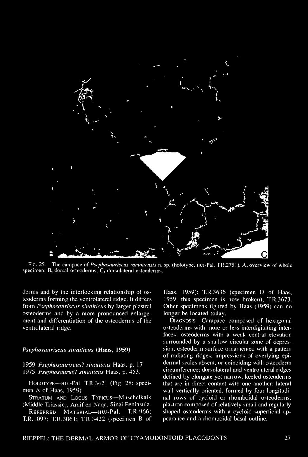
33 4H

. A.")

34 B 20 mm Fig. 26. Osteoderm shape and structure in the dermal armor of Psephosauriscus ramonensis n. sp. (A: holotype, HUJ-Pal. T.R.2751; B: referred specimen). A.l, dorsal osteoderms; A.2, dorsolateral osteoderms; A.3, left ventrolateral margin of plastron. B, referred carapace fragment (HUJ-Pal., uncatalogued). 28 FIELDIANA: GEOLOGY
, Middle East (Araif en Naqa, Sinai Peninsula, and Makhtesh Ramon, Negev, Israel).")
.")
35 Fig. 27. The plastron of Psephosauriscus ramonensis n. sp. (holotype, HUJ-Pal. T.R.2751). Distribution Middle Triassic (Anisian, lower Ladinian), Middle East (Araif en Naqa, Sinai Peninsula, and Makhtesh Ramon, Negev, Israel). Description The taxon is represented by several fragments of dermal armor, the most complete of which is specimen A of Haas (1959; huj- Pal. 3421). It is a piece of the carapace and plastron joined by the lateral wall (Fig. 28). The carapace is composed of relatively small, more or less regularly shaped osteoderms of hexagonal circumference that meet each other in an interdigitating suture. The diameter of the osteoderms is around 20 mm. Their thickness does not exceed their diameter. The superficial surface of the osteoderms shows a slight central elevation surrounded by a slightly depressed zone. Although the interdigitation between the osteoderms may in some specimens be weakly expressed on the surface of the carapace, becomes more it distinctly expressed, and the outlines of the osteoderms more irregularly shaped, toward deeper layers and on the inner (ventral) surface of the carapace. Strongly eroded pieces of dermal armor, showing rather deep interdigitation between adjoining osteoderms, may consequently appear rather different from less eroded pieces. Impressions of overlying epidermal scales are absent, or they may coincide with the circumference of the osteoderms. Haas (1959) noted one area in the carapace of the holotype where impressions of epidermal scales deviated from the circumference of the underlying osteoderms. Renewed preparation using acid did not confirm this observation. Instead, impressions of overlying epidermal scales are absent in Psephoderma sinaiticus, unless they coincide with the circumference of the osteoderms. Especially in the holotype. there is some depression of the suture between adjacent osteoderms, but this depression may also be the result of partial separation of the osteoderms. The lateral wall is vertically oriented. Carapace and lateral wall therefore meet each other at a we 11 -ex pressed angle defining the dorsolateral body ridge. Narrow yet somewhat elongated and keeled osteoderms are aligned along the dorsolateral ridge, joining the lateral wall to the carapace. The ventrolateral ridge is a mirror image of its dorsolateral counterpart, with elongated yet narrow and keeled osteoderms joining the lateral wall to the plastron in a well-defined angle. Better preserved than their dorsolateral counterparts, the ventrolateral ridge osteoderms show a maximal length of 32 mm and a maximal width of 13 mm. The lateral wall shows a pattern of cycloid scales. The plastron again shows cycloid scales of 28 mm width and 22.5 mm length. More marginal elements are smaller, with a width of 20 mm and a length of 16 mm. The epidermal scute areas match the superficial crown of the osteoderms. The apex of the cycloid scales points forward. RIEPPEL: THE DERMAL ARMOR OF CYAMODONTOID PLACODONTS 29


36 Fig. 28. The dermal armor of Psephosauriscus sinaiticus Haas (holotype. HUJ-Pal. T.R.3421). A, dorsal view; B, ventral view. their convex base faces backward. The osteoderms are aligned in regular transverse rows. The internal (dorsal) surface of the plastron displays the rhomboidal outline of the base of the osteoderms, which meet at interdigitating interfaces. Specimen HUJ-Pal. T.R.3673 preserves part of the plastron and associated gastrals and part of the lateral wall and associated dorsal ribs or transverse processes, but only fragments of the carapace. Unfortunately, the specimen does not allow determining whether the dorsal transverse processes extend laterally to the lateral wall, or whether these elements are the dorsal ribs, which would have articulated with the transverse processes at a more proximal level that is not preserved. The ribs, or distal parts of the transverse processes, are hollow and squarely abut the medial surface of lateral wall osteoderms with their slightly expanded distal ends. The gastral ribs are composed of five elements, a medioventral element and two collateral elements on either side, which show broad overlap with each other in specimen D of Haas (1959, specimen huj- Pal. T.R.3636; see also Westphal, 1975, Fig. 6, and specimen HUJ-Pal. T.R.3673). The gastral ribs are partially fused to the osteoderms and aligned such as to underlie the transverse suture line separating the transverse rows of osteoderms from one another. Their arrangement is thus strikingly different from that observed in Placochelys, where plastral ossifications are located between, rather than ventral to, the gastral ribs. In summary, Psephosauriscus sinaiticus differs from Psephosauriscus mosis by the absence of distinct impressions of irregular epidermal scute areas on the carapace, by the shape and arrangement of osteoderms along the dorsolateral and ventrolateral ridges, and by smaller ventral cycloid scutes in the plastron. Psephosauriscus sinaiticus differs from Psephosauriscus ramonensis 30 FIELDIANA: GEOLOGY

![Comments Dermal armor fragments referable to Psephosauriscus sinaiticus are also known from the Ceratites layers of Makhtesh Ramon, Israel (HUJ-Pal. T.R.966 [Fig. 29A], T.R.1097 [Fig. 29B], and T.R.3061 [Fig.](/docs-images/93/114504615/images/37-1.jpg "29C]). cf. Psephosauriscus rhombifer The holotype of \"Psephosaurus\" (Psephosauriscus) rhombifer (Haas, 1959, specimen G) can no longer be located today. Comparing Haas's (1959, PI. 8, Figs.")
 was that putative impressions from overlying epidermal scales could be identified on the single specimen of P. rhombifer that was available.")
37 Fig. 29. Carapace fragments from the Middle Triassic of Makhtesh Ramon, Negev, referred to Psephosawiscus sinaiticus Haas. A, HUJ-Pal. T.R.966; B, HUj-Pal. T.R.1097; C, HUJ-Pal. T.R.3061 (abraded specimen). by the presence of a vertical lateral wall and of a dorsolateral ridge, by distinct interdigitation at the sutural interface between the osteoderms, and by smaller cycloid scutes on the plastron. Comments Dermal armor fragments referable to Psephosauriscus sinaiticus are also known from the Ceratites layers of Makhtesh Ramon, Israel (HUJ-Pal. T.R.966 [Fig. 29A], T.R.1097 [Fig. 29B], and T.R.3061 [Fig. 29C]). cf. Psephosauriscus rhombifer The holotype of "Psephosaurus" (Psephosauriscus) rhombifer (Haas, 1959, specimen G) can no longer be located today. Comparing Haas's (1959, PI. 8, Figs. 30, 31) illustrations of P. rhombifer with those of P. sinaiticus (Haas. 1959, PI. 6, Fig. 23; PI. 7, Fig. 25) shows a closely similar pattern of osteoderm structure and arrangement. The only difference noted by Haas (1959) was that putative impressions from overlying epidermal scales could be identified on the single specimen of P. rhombifer that was available. These scales appear to have been of a strictly rhomboidal shape, with their corners located on the centers of four adjacent osteoderms. As can be seen from the illustration of Haas (1959, PI. 8, Fig. 31), these impressions of epidermal scutes are rather vague, however, unlike those in other cyamodontoids RIEPPEL: THE DERMAL ARMOR OF CYAMODONTOID 'LACODONTS 31

 comprises three carapace fragments, one of which was illustrated by Haas (1959, PI. 4, Fig. 16; PI. 8, Fig.")

38 Fig. 30. Carapace fragment from the Middle Triassic of Makhtesh Ramon, Negev, referred to cf. Psephosauriscus rhombifer Haas (HUJ-Pal. T.R.3676). {Psephosauriscus mosis). However, Haas (1959, PI. 4, Fig. 16; PI. 8, Fig. 29; HUJ-Pal. T.R.3676) figured a carapace fragment from another cyamodontoid from Makhtesh Ramon, which deserves a more detailed discussion, as it does represent a separate taxon of a cyamodontoid from Makhtesh Ramon that might be comparable to Psephosauriscus rhombifer. Specimen HUJ-Pal. T.R.3676 (Fig. 30) comprises three carapace fragments, one of which was illustrated by Haas (1959, PI. 4, Fig. 16; PI. 8, Fig. 29). Their morphology recalls that described for Psephosauriscus sinaiticus. The osteoderm surface of the latter taxon was characterized above by the presence of a weak central elevation surrounded by a shallow circular zone of depression. The osteoderms are of a more or less regularly hexagonal shape, they meet in interdigitating sutures, and their surface is ornamented by a pattern of radiating ridges. By contrast, specimen HUJ-Pal. T.R.3676 shows osteoderms of more irregular contours, meeting in more deeply interdigitating sutures, and with a distinctly more sculptured surface. Their diameter ranges from 1 5 to 20 mm. Their height does not exceed their diameter. Unlike Psephosauriscus sinaiticus, the surface of the carapace is very uneven, as the central part of the individual osteoderm is more or less deeply excavated. A central elevation may rise from the bottom of the excavated area. In contrast to Psephosauriscus sinaiticus, the impressions of overlying epidermal scales are distinct in specimens HUJ-Pal. TR.2492 as well as T.R.3676, and they are not congruent with the hexagonal contours of the osteoderms. This adds significantly to the unevenness of the surface of the carapace. Although a one-to-one relationship is preserved between the number of osteoderms and the number of epidermal scales, the margins of the latter are shifted obliquely out of phase relative to the sutures between the osteoderms by about one-fifth of the diameter of the osteoderms. The epidermal scales still approach a hexagonal shape, which more frequently is modified to a rhomboidal shape, however, or their shape even approaches the cycloid fish-scale pattern otherwise characteristic of the crown of plastral osteoderms. As the epidermal scute margins are obliquely shifted out of phase relative to the contours of the osteoderms by about one-fifth of the osteoderm diameter, the corners of the epidermal scales do not come to lie on the centers of four adjacent osteoderms as was described by Haas (1959) for "Psephosaurus" rhombifer. Allowing for the indistinctiveness of the epidermal scale margins in the holotype of "Psephosaurus" rhombifer (Haas, 1959, PI. 8, Figs. 30, 31), and allowing for some variation (regional variation?) in the pattern of overlap between epidermal scales and underlying osteoderms, it might very well be that specimens HUJ-Pal. T.R.3676 and 2492 might be referable to "P." rhombifer. In the absence of the holotype for the latter species, and in the absence of more and better preserved material, it seems prudent at this time to register specimens HUJ-Pal. T.R.3676 and TR.2492 as different cyamodontoids from Makhtesh Ramon, without formalizing taxonomic conclusions by the erection of a neotype description of a separate species. or the A Comparison of the Dermal Armor in Cyamodontoid Placodonts and Turtles Among reptiles, cyamodontoid placodonts and turtles are the only two groups that develop a complete dermal armor covering the dorsal and ventral surface of the body as well as the flanks. In both groups, the pectoral girdle (scapula) shifts 32 FIELDIANA: GEOLOGY
 to the ribs as a consequence of the development of a carapace (the position of the scapula relative to the ribs is known only in Henodus among the")
 believed the carapace of turtles to have evolved by fusion of originally separate osteoderms overlying broadened ribs (of pareiasaurs), and that the pectoral girdle assumed its")
 scenario of \"correlated progression\" does not account for the morphological complexity of the turtle body plan (Rieppel & Reisz, 1999), but instead almost perfectly captures the")
39 Fig. 31. The pectoral girdle of Henodus chelyops (partially reconstructed on the basis of II specimen of Huene. 1936). to a morphological position deep (ventral) to the ribs as a consequence of the development of a carapace (the position of the scapula relative to the ribs is known only in Henodus among the cyamodontoids). After a detailed comparison of the dermal armor in turtles and cyamodontoids, Gregory (1946) concluded that these structures evolved convergently in the two clades. Lee (1995, 1996, 1997) believed the carapace of turtles to have evolved by fusion of originally separate osteoderms overlying broadened ribs (of pareiasaurs), and that the pectoral girdle assumed its peculiar position inside the rib cage by a backward shift from its ancestral location relative to the axial skeleton. Lee's (1996) scenario of "correlated progression" does not account for the morphological complexity of the turtle body plan (Rieppel & Reisz, 1999), but instead almost perfectly captures the evolution of the carapace in cyamodontoids, as is suggested by its ontogeny and by outgroup comparison. Closer anatomical scrutiny reveals that the development of dermal armor is convergent in the two groups, the carapace and plastron of turtles being autapomorphic for that clade. The turtle carapace is referred to as a composite, or "duplex," structure, as it involves deeper thecal and superficial epithecal ossifications (Hay, 1898; Kaelin, 1945; Volker, 1913; Zangerl, 1939). The thecal ossifications are arranged in a very regular, strictly defined geometry that is closely comparable throughout turtles. A central longitudinal row of neural plates overlies and co-ossifies with the neural arches of the dorsal vertebrae; a lateral row of costal plates is closely associated with the dorsal ribs; a marginal row of marginal plates, an anterior nuchal plate, and one or two posterior pygal plates complete the carapace. In addition, some fossil turtles, such as Proganochelys (Gaffney, 1990), show supramarginal scutes. Epithecal ossifications are osteoderms that develop superficial to the thecal ossifications. Their appearance and arrangement vary among turtles. some fossil marine turtles they are superimposed on the neural shields; in dermochelyids, they are interdigitating osteoderms of polygonal (hexagonal) shape superimposed on the reduced theca; and in trionychids, they occur in a mosaic resembling thecal ossifications (Zangerl, ). Earlier authors (Hay, 1898; Oguschi, ) had claimed that epithecal ossifications are primitive for turtles and covered the body of the ancestral turtle prior to the development of a deeper theca. Volker (1913) drew attention to the large tubercular osteoderms that form longitudinal keels in Dermochelys, and homologi/.ed the marginal keels of Dermochelys with the marginal plates of other turtles, while Versluys (1914) homologi/.ed the lateral keels of Dermochelys with the marginal and supramarginal (submarginal, in his terminology) plates of Progcmochelys (see also Vallen. 1942, for similar arguments). The discussion surrounding the osteoderms and their arrangement in Dermochelys recalls the longitudinal rows of enlarged osteoderms in the carapace of some cyamodontoids, which may be particularly pro- In RIEPPEL: THE DERMAL ARMOR OF CYAMODONTOID PLACODONTS 33


, but it could just as well account, in morphological terms, for the evolution of a carapace in cyamodontoid placodonts.")
40 nounced along the dorsolateral and ventrolateral margins of the dermal armor. A striking similarity also exists between the dermal armor fragment of smns referred to Cyamodus and the anterolateral peripherals of Proganochelys, as discussed above. Earlier authors visualized the "ancestral" turtle to be covered with longitudinal rows of epithecal ossifications (osteoderms). Below these, the neural and costal plates would subsequently develop by broadening of the neural arches and ribs. This scenario for the evolution of the turtle shell almost perfectly fits Lee's (1996) hypothesis of "correlated progression" (Versluys, 1914, Fig. 10), but it could just as well account, in morphological terms, for the evolution of a carapace in cyamodontoid placodonts. However, given our modern understanding of turtle interrelationships, epithecal ossifications cannot be claimed to be primitive for turtles. Epithecal elements ossify later than thecal compo- of ex- nents of the carapace during the ontogeny tant turtles, and mapping the occurrence of epithecal ossifications on a cladogram of Testudines unequivocally indicates their derived nature (Zangerl, 1939, 1969; Kaelin, 1945). By contrast, the earliest known turtle, Proganochelys (Gaffney, 1990), shows a full complement of thecal ossifications, including neural and costal plates associated with the underlying endoskeleton (vertebrae and ribs). And whereas the genuinely dermal nature of the marginal, supramarginal, nuchal, and pygal plates is generally accepted (Vallen, 1942; Kaelin, 1945), the nature of the neural and costal plates is still in dispute. These are at the same time the components of the turtle carapace that cannot be compared to any elements in the dorsal dermal armor of cyamodontoids. The theca of turtles develops within a thickened dermal-epidermal carapacial disc (Burke, 1989; Gilbert et al., 2001). Early during development, growth of the ribs is "deflected" to a dorsal position (Ruckes, 1929) under the inductive influence of the carapacial ridge, which redirects the migration of those somitic cells that will eventually form the ribs (Burke, 1989; Gilbert et al., 2001). That way, the ribs chondrify dorsal to the scapula, but within the dermal carapacial disc. Perichondral ossification of the ribs is initiated in their proximal part that is, at the point of their entry into the dermis and from there progresses distally. Completion of the perichondral ossification of the ribs shows that these are not expanded in turtles at the cartilaginous stage (e.g., Goette, 1899, Pis. 27, 28; Kaelin, 1945, Figs. 4-11). Once the rib is fully encased in bone, ossification of the costal plates proceeds by the formation of trabecular bone in continuity with the periost and spreading anteriorly and posteriorly from the rib (Goette, 1899; Vallen, 1942; Kaelin, 1945; Gilbert et al., 2001). The cartilage of the rib itself degenerates without undergoing concomitant endochondral ossification; its space is eventually filled by trabecular bone that develops from haematopoietic elements invading the lumen left by the degenerated cartilage (Suzuki, 1963). The fact that the ribs as well as the tips of the neural arches pierce the dermal carapacial disc renders the identification of neurals and costals as endoskeletal versus exoskeletal elements difficult. On the one hand, the neurals and costals ossify from the periosteum of the underlying endoskeletal elements, which would make them part of the endoskeleton, but this whole process occurs within the dermis, which would make them dermal ossifications (Gilbert et al., 2001, distinguish between "primary" and "secondary" dermal bone). Both Patterson (1977) and Starck (1979) stressed that the endo- and exoskeleton cannot be defined on the basis of histogenesis but must be defined with reference to a phylogenetic framework. Exoskeletal are those elements that are homologous with structures which in the ancestral condition combine bone, dentine, and enamel, i.e., develop at the ectoderm-mesoderm interface. Endoskeletal are those elements that in the ancestral condition are preformed in cartilage, while the cartilaginous stage may be deleted in the descendant (membrane bone). The extension of the ribs into the dermis at a level lateral (dorsal) to the scapula is unique for turtles, yet the ribs as well as the neural arches are endoskeletal components of the carapace (Goette, 1 899). The neural and costal plates ossify from, and in continuity with, the periosteum of their endoskeletal component, and osteogenetic activity is initially most intensive at the deepest layer of the dermis, i.e., at the level of entry of the rib into the dermis. Neurals and costals, therefore, are, in part, composed of bone that matches the definition of Zuwachsknochen as given by Starck (1979: 13), or bone growing from perichondrally ossified elements of the endoskeleton without itself having been preformed in cartilage. As such, neurals and costals are endoskeletal components of the turtle carapace and cannot be compared to osteoderms fused into a carapace in cyamodontoid placodonts. The latter are exoskeletal elements, as are epithecal ossifications of tur- 34 FIELDIANA: GEOLOGY

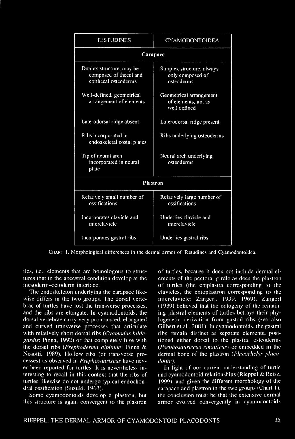
41 TESTUDINES


 than it is in most other terrestrial reptiles, which suggests less")
42 and turtles. This also implies, however, that both groups had to solve similar functional problems associated with the development of such extensive dermal armor. Functional Anatomy of the Dermal Armor in Placodonts Little work has been done on the functional anatomy of armored placodonts. Previous studies mostly addressed the function of the dermal armor in terms of protection and hydrodynamics (Westphal, 1975, 1976; Pinna & Nosotti, 1989; Renesto & Tintori, 1995). It is obvious that the development of a carapace will cause a shift from axial to paraxial locomotion, and those taxa in which a lateral wall links the carapace to a plastron may experience some restriction of the excursion range of the humerus and femur, as do turtles (Walker, 1971, 1973; Zug, 1971). And, as in turtles, the asymmetry of the metatarsus is less pronounced in cyamodontoids (Psephoderma: Renesto & Tintori, 1995) than it is in most other terrestrial reptiles, which suggests less agility in terrestrial locomotion (Walker, 1971, 1973; Zug, 1971). More important, however, turtles have lost the capability of using the ribs in support of locomotion and respiration by fusion of the ribs into the carapace, and cyamodontoids seem to have faced a similar problem. In a generalized tetrapod reptile, body weight is transferred from the limb to the axial skeleton via the muscular suspension of the pectoral girdle (scapula) from the vertebral column. Medially directed force components generated by a sprawling gait are absorbed by the clavicular-interclavicular complex. In turtles (terrestrial or aquatic bottom walkers), body weight is transferred from the limbs via the long and rod-shaped scapula to the carapace; medially directed force components are transmitted to the entoplastron via the acromial process of the scapula (Rieppel & Reisz, 1999). Little is known about the exact configuration of the pectoral girdle with respect to the dermal armor in cyamodontoids. The morphology of the relative to scapula of Cyamodus is plesiomorphic other sauropteryians (specimen msnm V458; see also Pinna, 1992). The scapula shows an elongate, broad, almost rectangular dorsal blade that shows no indication of any well-defined contact to the internal side of the carapace. The ventral glenoid portion is distinctly expanded and shows a convex ventral margin. A deep concavity along the posterior margin of the scapula marks the transition from the ventral glenoid portion to the dorsal blade. The coracoid is of almost circular outline, and the coracoid foramen appears as an open notch at its margin. Evidently, the scapula and coracoid were in cartilaginous contact with each other, and both elements were embedded in soft tissue below the dermal armor. The only departure from the plesiomorphic (sauropterygian) condition in the pectoral girdle of Cyamodus is seen in the dermal components. The interclavicle is a rather slender, boomerang-shaped element without any indication of a posterior stem. It contacts the clavicles on either side in an interdigitating suture. The clavicles are again slender, gently curved elements without any indication of expanded anterolateral corners. Together, the elements of the dermal pectoral girdle must have formed an upright U-shaped structure at the anterior end of the carapace. The dorsal tips of the clavicles may have contacted the inside of the carapace, as is known from Henodus. The pectoral girdle of Psephoderma (specimen msnm V527; see also Pinna & Nosotti, 1989) approaches that of Henodus more closely in its morphology than that of Cyamodus. The dermal pectoral girdle is less well exposed in Psephoderma, as it is obscured by the 5th cervical. However, as far as can be determined, it again forms a slender, upright U-shaped structure at the anterior end of the carapace. The interclavicle again lacks a posterior stem. The scapula differs from that of Cyamodus yet resembles that of Henodus in that it is a tall and slender structure. The dorsal blade is a simple dorsal process, while the ventral portion is only moderately expanded. The reconstruction of the scapula given by Pinna and Nosotti (1989, Fig. 9) is a good rendition of the structure. The clavicles are applied against the medial surface of the scapula. As the interclavicular-clavicular complex is positioned at the anterior end of the carapace, the scapula comes to lie at a morphological level deep to and below the transverse processes of the dorsal ribs. Among cyamodontoids, it is Henodus (Huene, 1936) that most closely resembles turtles in the structure of its pectoral girdle (Fig. 31; see also Huene, 1936, Fig. 15a, and PI. 84, Fig. 2). The clavicles are massive and sturdy L-shaped structures that meet each other in an interdigitating ventromedial suture in front of the interclavicle. Each clavicle has a prominent dorsal process that abuts the ventral surface of the anterior margin of 36 FIELDIANA: GEOLOGY

, a remarkable case of convergence (Rieppel & Reisz, 1999).")

43 the carapace. The columnar scapula is sutured to the posteromedial surface of the dorsal process of the clavicle. The coracoid remains unknown for Henodus. In functional terms, Henodus retains a sturdy clavicular-interclavicular complex for the absorption of medially directed force components generated by limb movements when walking on the bottom of a water body or on land, while vertically directed force components appear to have been transmitted to the carapace via the long and columnar clavicle and scapula. In this respect, the pectoral girdle of Henodus superficially looks almost identical to that of Proganochelys (Gaffney, 1990), a remarkable case of convergence (Rieppel & Reisz, 1999). The scapular and clavicular complex of Henodus further provides a firm anchoring of the pectoral girdle inside the dermal armor, which results in excellent support for the front limbs during swimming action. In a generalized tetrapod reptile, aspiration of air is effected by an expansion of the body cavity through muscular action exerted on the ribs. Exhalation is effected either by passive recoil of the body walls or by compression of the lungs as a result of active compression of the rib cage. Respiration in turtles depends on volume changes in the thoracicoperitoneal cavity inside the rigid dermal armor, which are achieved by altering the position of the limb flanks through the activity of anterior and posterior muscles (Gans & Hughes, 1997). The only author who addressed respiration in armored placodonts was Westphal (1975, 1976), who suggested mobility of the plastron (where present) as a mechanism that would allow volume changes of the thoracicoperitoneal cavity inside the rigid dermal armor. However, carapacial osteoderms do not fuse with the underlying endoskeleton in cyamodontoids (Westphal, 1975). If free dorsal ribs are retained in articulation with the (elongated) transverse processes of the dorsal vertebrae, these could therefore have moved independently from the overlying carapace. In Cyamodus hildegardis, the relatively short dorsal ribs are located in the flanks of the body (Pinna, 1992), and their movement may have laterally and ventrally expanded the volume of the thoracicoperitoneal cavity below the lateral margin of the carapace (a plastron is absent in Cyamodus hildegardis). A similar mechanism cannot be proposed for taxa in which a lateral wall links a carapace to a well-developed plastron (Psephosauriscus) or for taxa in which the dorsal ribs are fused to the transverse processes (Psephoderma). Like aquatic turtles (Gaunt & Gans, 1969), cyamodontoids may have used gravity (in support of inhalation) and hydrostatic pressure (in support of exhalation) for respiration. Expansion and contraction of the thoracicoperitoneal cavity through gravity and hydrostatic pressure would be enhanced by the absence of a plastron (as in Psephoderma, with fused dorsal ribs), or by mobility within the plastron if present. Westphal ( ) noted, as did Haas (1959), that the (superficially) cycloid plastral osteoderms are arranged in regular oblique rows in cyamodontoids from Makhtesh Ramon, which suggests the potential for some flexion between these rows. As described above, plastral osteoderms have a cycloid crown, but the thin base (exposed on the internal surface of the plastron) is rhomboidal and meets its neighbors in interdigitating sutures. The gastral ribs are aligned along the transverse sutures. Given a syndesmotic contact between osteoderms, some flexibility might very well have been possible between the transverse rows of osteoderms, given their regular arrangement, while the gastral ribs provided coherence between the rows. Flexibility in the plastron of Henodus may be indicated by its delicate structure and by the arrangement of much broadened osteoderms in an anteroposterior series mimicking transverse rows (Huene, 1936; Reiff, 1942; Westphal. 1975, 1976). Flexibility in the plastron of Placochelys is indicated by the nature of the junction of dermal bone to gastral ribs. As described above, dermal bone develops between gastral ribs, and if it does not wrap around the latter, it is received in distinctly concave facets on the anterior and posterior aspects of the gastral ribs (Fig. 13). This certainly suggests some mobility between the elements, as does the fact that dermal bone and gastral ribs easily dissociate in the fossilized stage. Whereas the dermal armor certainly provided protection from it is potential predators, worth mentioning, as Renesto & Tintori ( 1995) did. that large predators may be rare or absent in sediments yielding cyamodontoids. Conversely, cyamodontoid placodonts represent a clade that survived significantly longer than any other sauropterygian lineage of the Triassic. Triassic stem-group Sauropterygia (excluding the plesio-. plio-. and elasmosaurs of the Jurassic and Cretaceous) had all disappeared by the upper Carnian (Bardet. 1995; Rieppel & Dalla Vecchia, 2001), while cyamodontoid placodonts extend through the Norian and into the Rhaetian (Pinna. 1990; Pinna & Mazin. 1993). This may well reflect a greater tolerance RIEPPEL: THE DERMAL ARMOR OF CYAMODONTOID PLACODONTS 37
 situated above the last occurrence of Nothosaurus in the Germanic Triassic (Rieppel & Wild, 1994).")
.")

44 of armored placodonts for salinity changes caused by the transgression-regression cycles characteristic of the Middle and Upper Triassic. Henodus, for example, comes from layers of the upper Gipskeuper (Carnian) situated above the last occurrence of Nothosaurus in the Germanic Triassic (Rieppel & Wild, 1994). The sediments from which eight skeletons of Henodus have been collected have yielded a single Nothosaurus tooth only, and have been deposited in a lagoonalbrackish lake environment subject to cycles of marginal desiccation and rain flooding (Reiff, 1937; Fischer, 1959). Aigner (quoted in Reif & Stein, 1999) characterized the sedimentary facies of the upper Gipskeuper as a playa covered with brackish to hypersaline ponds that dried up seasonally. This is the most severe environment ever successfully invaded by sauropterygians, and it may testify to the role of the dermal armor as an osmotic barrier. Like the turtle shell, the dermal armor of cyamodontoids was covered by epidermal scutes, which, in combination with a wellossified carapace and plastron, provide a very efficient osmotic barrier in modern turtles. Experimental studies have shown a significantly smaller rate of gain of water (in fresh water) or loss of water (in sea water) in a slider turtle {Pseudemys scripta) with a well-ossified carapace and plastron than in a soft-shelled turtle (Apalone spiniferus) or a caiman {Caiman crocodilus) (Bentley, 1976). The function of the dermal armor as an osmotic barrier may also explain the stratigraphic distribution of Sauropterygia in the Muschelkalk of Makhtesh Ramon, Negev, Israel. This deposit has yielded a diverse sauropterygian fauna, including cyamodontoid placodonts, from two separate horizons, the Middle Member of the Gevanim Formation (lower Anisian: Druckman, 1974) and the Lower Member of the Saharonim Formation (straddling the Anisian-Ladinian boundary: Druckman, 1974). Both of these horizons were deposited under normal shallow marine conditions, favorable for the occurrence of sauropterygians. The two horizons are separated by the Upper Member of the Gevanim Formation, which was deposited in a marginal tidal flat, and cyamodontoid placodonts were the only sauropterygians that continued to inhabit this environment under those conditions (Druckman, 1974). Discussion and Conclusions to its A review of the dermal armor of cyamodontoid placodonts identifies a number of areas of incomplete knowledge, in particular with respect ontogenetic development. On the other hand, the structure of the dermal armor allows the identification of a number of characters of potential use in phylogenetic analysis, such as the presence or absence of a caudal shield separate from the dorsal shield of the carapace; the presence or absence of a plastron; the presence and nature of carapacial ornamentation and/or of the dorsolateral and ventrolateral body ridges; size, shape and ornamentation of the carapacial and plastral osteoderms; and the nature of their interfaces. An area that requires further investigation concerns the relation of epidermal scutes to the underlying osteoderms. Equally important is the recognition that characters of the dermal armor can be used for taxonomic purposes at the species level. This is documented by the description above of the abundant dermal armor fragments from the Muschelkalk (Middle Triassic) of the Middle East, which recognizes a new genus with three species. In total, two genera and five, potentially even six, species of cyamodontoids can be recognized from the Middle Triassic of Makhtesh Ramon. This is a taxic diversity of cyamodontoids that so far is not known to be paralleled in other Triassic deposits. At the same time, this high degree of taxic diversification indicates a sophisticated degree of habitat partitioning among those coexisting cyamodontoids. The only area that might hold the potential for a similar taxic diversity of cyamodontoids is the Triassic of southeastern China (Guizhou Province). Other areas with some taxic diversity of cyamodontoids include the southern Alpine Triassic, which yielded Cyamodus, Psephoderma, and Protenodontosaurus. However, unlike at Makhtesh Ramon, these genera (and species) have been reported from different deposits of different stratigraphic position within the Triassic of the southern Alps. The same is true for the different species of Cyamodus reported from the German Muschelkalk, with the exception of C. rostratus and C. muensteri, which coexisted in the lower upper Muschelkalk (mol) of Bayreuth. The skull structure of cyamodontoid placodonts in general shows a remarkable diversification in terms of trophic specialization as expressed in rostrum shape and function and in different patterns of dentition (Rieppel, 2001). Unfortunately, the cranial remains of cyamodontoid placodonts recovered from the Triassic of Makhtesh Ramon are too rare, too incompletely preserved, or too conservative in structure to be used for taxonomic 38 FIELDIANA: GEOLOGY
, is not diagnostic even at the genus level.")

45 purposes at the species level or to reflect any diversification of trophic specialization (Rieppel et al., 1999). The one relatively complete skull fragment from Makhtesh Ramon, the holotype of Psephosauriscus mosis (Brotzen, 1957), is not diagnostic even at the genus level. Given its high potential for fossilization, an improved knowledge of the structure of the dermal armor of cyamodontoid placodonts, and of the nature of its variability, is essential for a better understanding of the taxic diversity of these exotic animals in the intraplatform basin habitats surrounding the developing southern branch of the Neotethys. Knowledge of the functional anatomy of cyamodontoid placodonts remains incomplete, in particular with respect to the impact of the development of a carapace and plastron on locomotion and respiration. A comparison with turtles might shed further light on these issues, yet a detailed anatomical comparison reveals that the dermal armor in placodonts and turtles developed convergently in the two groups. Acknowledgments I thank Eitan Tchernov, Department of Ecology and Evolution, The Hebrew University, Jerusalem, for unlimited access to the collections in his care, and for allowing me to obtain cyamodontoid material on loan for further preparation. This study was made possible by financial support from the U.S. National Science Foundation (grants DEB and DEB ). Literature Cited Agassiz, L Recherches sur les Poissons Fossiles, Vol. II. Imprimerie de Petitpierre, Neuchatel. Bardet, N Evolution et extinction des reptiles marins au cours du Mesozoique. Palaeovertebrata, 24: Bentley, P. J Osmoregulation. In Gans, C, and W. R. Dawson, eds., Biology of the Reptilia, Vol. 5, pp Academic Press, London. Braun, C. W. F Uber Placodus gigas Agassiz, und Placodus Andriani Miinster. Programm zum Jahresbericht der konigl. Kreis-Landwirtschafts- und Gewerbeschule zu Bayreuth fur das Schuljahr 1861/62, pp Theodor Burger, Bayreuth. Brotzen, F Stratigraphical studies on the Triassic vertebrate fossils from Wadi Ramon, Israel. Arkiv for Mineralogi och Geologi, 2: Burke, A. C Development of the turtle carapace: Implications for the evolution of a novel bauplan. Journal of Morphology, 199: Drevkrmann, Fr Die Placodontier. 3. Das Skelctt von Placodus gigas Agassiz im Senckcnberg-Museum. Abhandlungen der senckenbergischen naturforschenden Gesellschaft, 38: Druckman, Y The stratigraphy of the Triassic Sequence in southern Israel. Geological Survey of Israel, Bulletin, 64: Fischer, W Neue Funde von Henodus chelyops v. Huene im Tubinger Gipskeuper. Neues Jahrbuch fiir Geologie und Palaontologie, Monatshefte, 1959: Fraas, E Die Schwabischen Trias-Saurier. E. Schweizerbart, Stuttgart. Gaffney, E. S The comparative osteology of the Triassic turtle Proganochelys. Bulletin of the American Museum of Natural History, 194: Gans, C, and G. M. Hughes The mechanism of lung ventilation in the tortoise Testudo graeca Linne. Journal of Experimental Biology, 47: Gaunt, A. S., and C. Gans Mechanics of respiration in the snapping turtle, Chelydra serpentina (Linne). Journal of Morphology, 128: Gilbert, S. E, G. A. Loredo, A. Brukman, and A. C. Burke Morphogenesis of the turtle shell: The development of a novel structure in tetrapod evolution. Evolution and Development, 3: Goette, A Uber die Entwicklung des knochernen Riickenschildes (Carapax) der Schildkroten. Zeitschrift fiir wissenschaftliche Zoologie, 66: Gregory, W. K Pareiasaurs versus placodonts as near ancestors to turtles. Bulletin of the American Museum of Natural History, 86: Gurich, G. J. E Uber einige Saurier des oberschlesischen Muschelkalkes. Zeitschrift der Deutschen Geologischen Gesellschaft, 36: Haas, G On some fragments of the dermal skeleton of Placodontia from the Trias of Aari en Naga, Sinai Peninsula. Kunglia Svenska vetenskapsakadcmiensis Handlingar, (4) 7: The armor of placodonts from the Muschelkalk of Wadi Ramon (Israel). Israel Journal of Zoology, 18: On the placodonts of the Wadi Ramon area Muschelkalk. Colloque international, Centre National de la Recherche Scientiliquc, 218: Hay, O. P On Protostega, the systematic position of Dermochelys, and the morphogeny of the chelonian carapace and plastron. American Naturalist, 32: Hemleben, Ch., and D. Freels Fossilfuhrcnde dolomitisierte Plattenkalke aus dcm "Muschelkalk superior" bei Montral (Prov. Tarragona, Spanien). Neues Jahrbuch fiir Geologie und Palaontologie, Abhandlungen, 154: Huene, E v Henodus chelyops, ein neuer Placodontier. Palaeontographica, A. 84: Nachtrage zur Kenntnis von Henodus chelyops aus dem Tubinger Gipskeuper. Palaeontographica, 110: Jaekel, O Ueber Placochelys n. g. und ihre Be- RIEPPEL: THE DERMAL ARMOR OF CYAMODONTOID PLACODONTS 39


46 . 1858b. deutung fur die Stammesgeschichte der Schildkroten. Neues Jahrbuch fiir Mineralogie, Geologie und Palaontologie, Abhandlungen, 1: Placochelys placodonta aus der Obertrias des Bakony. In Resultate der wissenschaftlichen Erforschung des Balatonsees, 1. Band. 1. Teil. Palaeontologischer Anhang. Victor Homyanszky, K. und K. Hofbuchdruckerei, Budapest. Kaelin, J Zur Morphogenese des Panzers bei den Schildkroten. Acta Anatomica, 1: Lee, M. S. Y Historical burden in systematics and the interrelationships of "parareptiles." Biological Reviews, 70: Correlated progression and the origin of turtles. Nature, 379: Pareiasaur phylogeny and the origin of turtles. Zoological Journal of the Linnean Society, 120: Meyer, H. v. 1858a. Psephoderma Alpinum aus dem Dachsteinkalke der Alpen. Neues Jahrbuch fiir Mineralogie, Geognosie, Geologie und Petrefaktenkunde, 1858: Psephoderma alpinum aus dem Dachsteinkalke der Alpen. Palaeontographica, 6: Die Placodonten, eine Familie von Sauriern der Trias. Palaeontographica, 11: Mittheilung an Professor H.B. Geinitz. Neues Jahrbuch fiir Mineralogie, Geognosie, Geologie und Petrefaktenkunde, 1864: Muller, H Bayreuth und die Palaontologie. Aufschluss, 30: Munster, G Uber einige ausgezeichnete fossile Fischzahne aus dem Muschelkalk bei Bayreuth. F. C. Birner, Bayreuth Beitriige zur Petrefaktenkunde, mit XVIII nach der Natur gezeichneten Tafeln der Herren Hermann v. Meyer und Professor Rudolph Wagner. Buchner'sche Buchhandlung, Bayreuth. Nopcsa, F Die Familien der Reptilien. Fortschritte der Geologie und Palaeontologie, 2: Nosotti, S., and G. Pinna Cyamodus kuhnschnyderi n. sp., nouvelle espece de Cyamodontidae (Reptilia, Placodontia) du Muschelkalk superieur allemand. Comptes Rendues a l'academie des Sciences, Paris, 317: Osteology of the skull of Cyamodus kuhnschnyderi Nosotti and Pinna 1993 (Reptilia, Placodontia). Paleontologia Lombarda, n.s., 6: Oguschi, K Anatomische Studien an der japanischen dreikralligen Lippenschildkrote (Trionyx japanicus). Gegenbaurs Morphologisches Jahrbuch, 43: Osswald, K Uber einige Ratfossilien aus dem Risserkogelgebiet (siidlich Tegernsee). Jahrbuch der Preussischen Geologischen Landesanstalt zu Berlin, 50: Owen, R Description of the skull and teeth of the Placodus laticeps OWEN, with indications of other new species of Placodus, and evidence of the saurian nature of that genus. Philosophical Transactions of the Royal Society of London, 148: Palaeontology. Adam and Charles Black, Edinburgh. Patterson, C Cartilage bones, dermal bones and membrane bones, or the exoskeleton versus the endoskeleton. In Andrews, S. M., R. S. Miles, and A. D. Walker, eds., Problems in Vertebrate Evolution, pp Academic Press, London. Peyer, B Die Triasfauna der Tessiner Kalkalpen. III. Placodontia. Abhandlungen der Schweizerischen Palaontologischen Gesellschaft, 51: Peyer, B., and E. Kuhn-Schnyder Placodontia. In Piveteau, J., ed., Traite de Paleontologie, Vol. 5, pp Masson, Paris. Pinna, G Placochelys zitteli, Placochelys stoppanii, Placochelyanus malanchinii: un caso di sinonimia fra I rettili placodonti retici della famiglia Placochelyidae. Bolletino dell Societa Paleontologica Italians*, 15: Descrizione di un nuovo esemplare di Placochelyidae del Retico Lombardo (Psephoderma alpinum Meyer, 1858) e discussione sulla sinonimia P sephoderma-placochelyanus. Atti della Societa Italiana di Scienze Naturali e del Museo Civico di Storia Naturale di Milano, 119: Lo scheletro postcraniale di Cyamodus hildegardis Peyer, 1931, descrito su un esemplare del Triassico Medio Lombardo. Atti della Societa Italiana di Scienze Naturali e del Museo Civico di Storia Naturale di Milano, 121: Notes on stratigraphy and geological distribution of placodonts. Atti della Societa Italiana di Scienze Naturali e del Museo Civico di Storia Naturale di Milano, 131: Cyamodus hildegardis Peyer, 1931 (Reptilia, Placodontia). Memorie della Societa Italiana di Scienze Naturali e del Museo Civico di Storia Naturale di Milano, 26: Placodontia (Reptilia Triadica). In Westphal, F, ed., Fossilium Catalogus Animalia, Pars 136. Backhuys Publ., Leiden. Pinna, G., and J.-M. Mazin Stratigraphy and paleobiogeography of the Placodontia. Paleontologia Lombarda, n.s., 2: Pinna, G., and S. Nosotti Anatomie, morfologia funzionale e paleoecologia del rettile placodonte Psephoderma alpinum Meyer, Memorie della Societa Italiana di Scienze Naturali e del Museo Civico di Storia Naturale di Milano, 25: Reif, W.-E., and F Stein Morphology and function of the dentition of Henodus chelyops Huene, 1936 (Placodontia, Triassic). Neues Jahrbuch fiir Geologie und Palaontologie, Monatshefte. 1999: Reiff, W Ergebnisse der Grabung des Geologisch-Palaontologischen Universitats-Instituts bei Tiibingen-Lustnau im Oberen Gipskeuper. Neues Jahrbuch fiir Geologie, Mineralogie und Palaontologie, Monatshefte, Abteilung B, 1937: Ergiinzungen zum Panzerbau von Henodus chelyops v. Huene. Palaeontographica, 94: Renesto, S., and A. Tintori Functional morphology and mode of life of the Late Triassic placodont Psephoderma alpinum Meyer from the Calcare 40 FIELDIANA: GEOLOGY
. Zoological Journal of the Linnean Society, 130: 635-659. 2000b. Sauropterygia: I.")
. Fieldiana (Geology), n.s., 45: 1-104. Rieppel, O., and F. M. Dalla Vecchia. 2001.")
. Historical Biology. 13: 77-97. Rieppel, O., J.-M. Mazin, and E. Tchernov. 1999.")
47 di Zorzino (Lombardy, N Italy). Rivista Italiana di Paleontologia e Stratigrafia, 101: Rieppel, O Fragmenta Sauropterygiana. Neues Jahrbuch fur Geologie und Paliiontologie. Abhandlungen, 197: a. Paraplacodus and the phylogeny of the Placodontia (Reptilia: Sauropterygia). Zoological Journal of the Linnean Society, 130: b. Sauropterygia: I. Placodontia, Pachypleurosauria, Nothosauroidea, Pistosauroidea. In Wellnhofer. P., ed.. Encyclopedia of Paleoherpetology, 12A. Pfeil, Munich The cranial anatomy of Placochelys placodonta Jaekel, 1902, and a review of the Cyamodontoidea (Reptilia, Placodonta). Fieldiana (Geology), n.s., 45: Rieppel, O., and F. M. Dalla Vecchia Marine reptiles from the Triassic of Tre Venezie, northeastern Italy. Fieldiana (Geology), n.s., 44: Rieppel, O., and H. Hagdorn Fossil reptiles from the Spanish Muschelkalk (Mont-ral-Alcover, Province Tarragona). Historical Biology. 13: Rieppel, O., J.-M. Mazin, and E. Tchernov Sauropterygia from the Middle Triassic of Makhtesh Ramon, Negev, Israel. Fieldiana (Geology), n.s., 40: Rieppel, O., and R. R. Reisz The origin and early evolution of turtles. Annual Review of Ecology and Systematics, 30: Rieppel, O., and R. Wild Nothosaurus edingerae Schultze, 1970: Diagnosis of the species and comments on its stratigraphical occurrence. Stuttgarter Beitrage zur Naturkunde, B, 204: Rieppel, O., and R. T. Zanon The interrelationships of Placodontia. Historical Biology, 12: Ruckes, H Studies in chelonian osteology. Part II. The morphological relationships between girdles, ribs and carapace. Annals of the New York Academy of Sciences, 31: Starck, D Vergleichende Anatomie der Wirbeltiere auf evolutionsbiologischer Grundlage, Vol 2. Springer Verlag, Berlin. Storrs, G. W Fossil vertebrate faunas from the British Rhaetian (latest Triassic). Zoological Journal of the Linnean Society, 112: Si /i Ki. H. K Studies on the osseous system on the slider turtle. Annals of the New York Academy of Sciences. 109: Tintori, A Fish taphonomy and Triassic anoxic basins from the Alps: A case history. Rivista Italiana di Paleontologia i Stratigraphia, 97: Vallen, E Beitrage zur Kenntnis der Ontogenie und der vergleichenden Anatomie des Schildkrotenpanzers. Acta Zoologica. Stockholm. 23: Versluys, J Uber die Phylogenie des Panzers der Schildkroten und Liber die Verwandtschaft der Lederschildkrote (Dermochelys coriacea). Paliiontologische Zeitschrift, 1: ' Volker, H Uber das Stamm-. Gliedmassen-, und Hautskelett von Dermochelys coriacea L. Zoologische Jahrbucher, Abteilung fiir Anatomie und Ontogenie dertiere, 33: Walker, W F A structural and functional analysis of walking in the turtle, Chrysemis picta marginata. Journal of Morphology, 134: The locomotor apparatus in turtles. In Gans, C., and T S. Parsons, eds.. Biology of the Reptilia, Vol. 4, pp Academic Press, London. Weiss, G. (Ed.) Bayreuth als Statte alter erdgeschichtlicher Entdeckungen. Druckerei Ellwanger, Bayreuth. Westphal, F Bauprinzipien im Panzer der Placodonten (Reptilia triadica). Paliiontologische Zeitschrift, 49: The dermal armor of some Triassic placodont reptiles. In Bellairs, A. d'a., and C. B. Cox, eds.. Morphology and Biology of Reptiles, pp Academic Press, London. Zangerl, R The homology of the shell elements in turtles. Journal of Morphology. 65: The turtle shell. In Gans. C.. A. d'a. Bellairs, and T. S. Parsons, eds.. Biology of the Reptilia, Vol. 1, pp Academic Press. London. Zittel, K. A. v Handbuch der Palaeontologie. 1. III. Abtheilung. Palaeozoologie, Band. Vertebrata (Pisces, Amphibia, Reptilia, Aves). R. Oldenbourg. Munchen und Leipzig. Zug, G. R Buoyancy, locomotion, morphology of the pelvic girdle and hindlimb. and systematics of cryptodiran turtles. Miscellaneous Publications. Museum of Zoology, University of Michigan. 142: RIEPPEL: THE DERMAL ARMOR OF CYAMODONTOID PLACODONTS 41

48

49

50
 from the Germanic Triassic, with Comments on the Status of Conchiosaurus clavatus.")


51 A Selected Listing of Other Fieldiana: Geology Titles Available Osteology of Simosaurus gaillardoti and the Relationships of Stem-Group Sauropterygia. Bj Olivier RieppeL Fieldiana: Geology, n.s.. no. 28, pages. 71 illus. Publication 1462, $18.00 A Revision of the Genus Nothosaurus (Reptilia: Sauropterygia) from the Germanic Triassic, with Comments on the Status of Conchiosaurus clavatus. By Olivier Rieppel and Rupert Wild. Fieldiana: Geology, n.s.. no pages, 66 illus. Publication 1479, $17.00 The Status of the Sauropterygian Genera Ceresiosaums, Lariosaurus, and Silvestrosaurus from the Middle Triassic of Europe. By Olivier Rieppel. Fieldiana: Geology, n.s.. no. 38, pages. 21 illus. Publication 1490, $15.00 Sauropterygia from the Middle Triassic of Makhtesh Ramon. Negev. Israel. By Olivier Rieppel, Jean- Michel Ma/in. and Eitan Tchernov. Fieldiana: Geology, n.s., no. 40., pages, 58 illus. Publication 1499, $25.00 Hie Intramandibular Joint in Squamates, and the Phylogenetie Relationships of the Fossil Snake Fachyrhachis problematicus Haas. By Olivier Rieppel and Hassam Zahar. Fieldiana: Geology, n.s pages. 17 illus. no. Publication 1507, $23.00 Marine Reptiles from the Triassic of the Tre Venezie Area, Northeastern Italy. By Olivier Rieppel and Fabio Marco Dalla Vecchia. Fieldiana: Geology, n.s, no. 44, pages. 29 illus. Publication 1511, $10.00 The Cranial Anatomy of Plaeoehelys placodonta Jaekel and a Review of the ( yamodontoidea (Reptilia, Placodonta). By Olivier Rieppel. Fieldiana: Geology, n.s.. no pages. 39 illus. Publication 1514, $30.00 To order Fieldiana, please address correspondence to: Peter Fortsas Fortsas Books, Ltd No. Lovejoy Chicago, IL fieldiana^ aol.com Prices and availability may change.


52 UNIVERSITY OF ILLINOIS-URBANA Field Museum of Natural History 1400 South Lake Shore Drive Chicago, Illinois Telephone: (312)
Exceptional fossil preservation demonstrates a new mode of axial skeleton elongation in early ray-finned fishes
 Supplementary Information Exceptional fossil preservation demonstrates a new mode of axial skeleton elongation in early ray-finned fishes Erin E. Maxwell, Heinz Furrer, Marcelo R. Sánchez-Villagra Supplementary
TRACHEMYS SCULPTA. A nearly complete articulated carapace and plastron of an Emjdd A NEAKLY COMPLETE SHELL OF THE EXTINCT TURTLE,
 A NEAKLY COMPLETE SHELL OF THE EXTINCT TURTLE, TRACHEMYS SCULPTA By Charles W. Gilmore Curator of Vertebrate Paleontology, United States National Museum INTRODUCTION A nearly complete articulated carapace
SUPPLEMENTARY INFORMATION
 In comparison to Proganochelys (Gaffney, 1990), Odontochelys semitestacea is a small turtle. The adult status of the specimen is documented not only by the generally well-ossified appendicular skeleton
FIRST RECORD OF PLACODONTOIDEA (REPTILIA, SAUROPTERYGIA, PLACODONTIA) FROM THE EASTERN TETHYS
 FROM THE EASTERN TETHYS") Journal of Vertebrate Paleontology 28(3):904 908, September 2008 2008 by the Society of Vertebrate Paleontology SHORT COMMUNICATION FIRST RECORD OF PLACODONTOIDEA (REPTILIA, SAUROPTERYGIA, PLACODONTIA)
Journal of Vertebrate Paleontology 28(3):904 908, September 2008 2008 by the Society of Vertebrate Paleontology SHORT COMMUNICATION FIRST RECORD OF PLACODONTOIDEA (REPTILIA, SAUROPTERYGIA, PLACODONTIA)
AUSTRALIAN MUSEUM SCIENTIFIC PUBLICATIONS
 AUSTRALIAN MUSEUM SCIENTIFIC PUBLICATIONS Riek, E. F., 1964. Merostomoidea (Arthropoda, Trilobitomorpha) from the Australian Middle Triassic. Records of the Australian Museum 26(13): 327 332, plate 35.
w^osm The person charging this material is responsible for L161 O-I096 UNIVERSITY OF ILLINOIS LIBRARY AT URBANA-CHAMPAIGN
 ; ;,!! i i IT:'':'':! NOTICE: Return or renew all Library Materialsl The Minimum Fee for each Lost Book is $50.00. The person charging this material is responsible for its return to the library from which
Williston, and as there are many fairly good specimens in the American
 56.81.7D :14.71.5 Article VII.- SOME POINTS IN THE STRUCTURE OF THE DIADECTID SKULL. BY R. BROOM. The skull of Diadectes has been described by Cope, Case, v. Huene, and Williston, and as there are many
FOSSIL TURTLE RESEARCH
 FOSSIL TURTLE RESEARCH VOLUME 1 Proceedings of the Symposium on Turtle Origins, Evolution and Systematics August 18 20, 2003, St. Petersburg, Russia Edited by Igor G. Danilov and James F. Parham St. Petersburg,
290 SHUFELDT, Remains of Hesperornis.
 290 SHUFELDT, Remains of Hesperornis. [ Auk [July THE FOSSIL REMAINS OF A SPECIES OF HESPERORNIS FOUND IN MONTANA. BY R. W. SHUFELD% M.D. Plate XI7III. ExR,¾ in November, 1914, Mr. Charles W. Gihnore,
REVISION OF REDONDASUCHUS (ARCHOSAURIA: AETOSAURIA) FROM THE UPPER TRIASSIC REDONDA FORMATION, NEW MEXICO, WITH DESCRIPTION OF A NEW SPECIES
 FROM THE UPPER TRIASSIC REDONDA FORMATION, NEW MEXICO, WITH DESCRIPTION OF A NEW SPECIES") Harris et al., eds., 2006, The Triassic-Jurassic Terrestrial Transition. New Mexico Museum of Natural History and Science Bulletin 37. REVISION OF REDONDASUCHUS (ARCHOSAURIA: AETOSAURIA) FROM THE UPPER
Harris et al., eds., 2006, The Triassic-Jurassic Terrestrial Transition. New Mexico Museum of Natural History and Science Bulletin 37. REVISION OF REDONDASUCHUS (ARCHOSAURIA: AETOSAURIA) FROM THE UPPER
A new species of sauropod, Mamenchisaurus anyuensis sp. nov.
 A new species of sauropod, Mamenchisaurus anyuensis sp. nov. by Xinlu He, Suihua Yang, Kaiji Cai, Kui Li, and Zongwen Liu Chengdu University of Technology Papers on Geosciences Contributed to the 30th
A new placodont sauropterygian from the Middle Triassic of the Netherlands
 A new placodont sauropterygian from the Middle Triassic of the Netherlands NICOLE KLEIN and TORSTEN M. SCHEYER Klein, N. and Scheyer, T.M. 2014. A new placodont sauropterygian from the Middle Triassic
Reprinted from: CRUSTACEANA, Vol. 32, Part 2, 1977 LEIDEN E. J. BRILL
 Reprinted from: CRUSTACEANA, Vol. 32, Part 2, 1977 LEIDEN E. J. BRILL NOTES AND NEWS 207 ALPHE0PS1S SHEARMII (ALCOCK & ANDERSON): A NEW COMBINATION WITH A REDESCRIPTION OF THE HOLOTYPE (DECAPODA, ALPHEIDAE)
FURTHER STUDIES ON TWO SKELETONS OF THE BLACK RIGHT WHALE IN THE NORTH PACIFIC
 FURTHER STUDIES ON TWO SKELETONS OF THE BLACK RIGHT WHALE IN THE NORTH PACIFIC HIDEO OMURA, MASAHARU NISHIWAKI* AND TOSHIO KASUYA* ABSTRACT Two skeletons of the black right whale were studied, supplementing
New Carnivorous Dinosaurs from the Upper Cretaceous of Mongolia
 1955 Doklady, Academy of Sciences USSR 104 (5):779-783 New Carnivorous Dinosaurs from the Upper Cretaceous of Mongolia E. A. Maleev (translated by F. J. Alcock) The present article is a summary containing
complex in cusp pattern. (3) The bones of the coyote skull are thinner, crests sharper and the
 The bones of the coyote skull are thinner, crests sharper and the") DISTINCTIONS BETWEEN THE SKULLS OF S AND DOGS Grover S. Krantz Archaeological sites in the United States frequently yield the bones of coyotes and domestic dogs. These two canines are very similar both
DISTINCTIONS BETWEEN THE SKULLS OF S AND DOGS Grover S. Krantz Archaeological sites in the United States frequently yield the bones of coyotes and domestic dogs. These two canines are very similar both
A NEW AUSTROSQUILLA (STOMATOPODA) FROM THE
 FROM THE") A NEW AUSTROSQUILLA (STOMATOPODA) FROM THE MARQUESAS ISLANDS BY ALAIN MICHEL Centre O.R.S.T.O.M., Noumea, New Caledonia and RAYMOND B. MANNING Smithsonian Institution, Washington, U.S.A. The At s,tstrosqzlilla
A NEW AUSTROSQUILLA (STOMATOPODA) FROM THE MARQUESAS ISLANDS BY ALAIN MICHEL Centre O.R.S.T.O.M., Noumea, New Caledonia and RAYMOND B. MANNING Smithsonian Institution, Washington, U.S.A. The At s,tstrosqzlilla
.56 m. (22 in.). COMPSOGNATHOID DINOSAUR FROM THE. Medicine Bow, Wyoming, by the American Museum Expedition
. COMPSOGNATHOID DINOSAUR FROM THE. Medicine Bow, Wyoming, by the American Museum Expedition") Article XII.-ORNITHOLESTES HERMANNI, A NEW COMPSOGNATHOID DINOSAUR FROM THE UPPER JURASSIC. By HENRY FAIRFIELD OSBORN. The type skeleton (Amer. Mus. Coll. No. 6I9) of this remarkable animal was discovered
Article XII.-ORNITHOLESTES HERMANNI, A NEW COMPSOGNATHOID DINOSAUR FROM THE UPPER JURASSIC. By HENRY FAIRFIELD OSBORN. The type skeleton (Amer. Mus. Coll. No. 6I9) of this remarkable animal was discovered
BREVIORA LEUCOLEPIDOPA SUNDA GEN. NOV., SP. NOV. (DECAPODA: ALBUNEIDAE), A NEW INDO-PACIFIC SAND CRAB. Ian E. Efford 1
, A NEW INDO-PACIFIC SAND CRAB. Ian E. Efford 1") ac lc BREVIORA CAMBRIDGE, MASS. 30 APRIL, 1969 NUMBER 318 LEUCOLEPIDOPA SUNDA GEN. NOV., SP. NOV. (DECAPODA: ALBUNEIDAE), A NEW INDO-PACIFIC SAND CRAB Ian E. Efford 1 ABSTRACT. Leucolepidopa gen. nov.
ac lc BREVIORA CAMBRIDGE, MASS. 30 APRIL, 1969 NUMBER 318 LEUCOLEPIDOPA SUNDA GEN. NOV., SP. NOV. (DECAPODA: ALBUNEIDAE), A NEW INDO-PACIFIC SAND CRAB Ian E. Efford 1 ABSTRACT. Leucolepidopa gen. nov.
FIELDIANA GEOLOGY NEW SALAMANDERS OF THE FAMILY SIRENIDAE FROM THE CRETACEOUS OF NORTH AMERICA
 FIELDIANA GEOLOGY Published by CHICAGO NATURAL HISTORY MUSEUM Volume 10 Sbftember 22, 1968 No. 88 NEW SALAMANDERS OF THE FAMILY SIRENIDAE FROM THE CRETACEOUS OF NORTH AMERICA Coleman J. Coin AND Walter
AUSTRALIAN MUSEUM SCIENTIFIC PUBLICATIONS
 AUSTRALIAN MUSEUM SCIENTIFIC PUBLICATIONS McCulloch, Allan R., 1908. A new genus and species of turtle, from North Australia. Records of the Australian Museum 7(2): 126 128, plates xxvi xxvii. [11 September
Postilla PEABODY MUSEUM OF NATURAL HISTORY YALE UNIVERSITY NEW HAVEN, CONNECTICUT, U.S.A.
 Postilla PEABODY MUSEUM OF NATURAL HISTORY YALE UNIVERSITY NEW HAVEN, CONNECTICUT, U.S.A. Number 117 18 March 1968 A 7DIAPSID (REPTILIA) PARIETAL FROM THE LOWER PERMIAN OF OKLAHOMA ROBERT L. CARROLL REDPATH
SUPPLEMENTARY ONLINE MATERIAL FOR. Nirina O. Ratsimbaholison, Ryan N. Felice, and Patrick M. O connor
 http://app.pan.pl/som/app61-ratsimbaholison_etal_som.pdf SUPPLEMENTARY ONLINE MATERIAL FOR Nirina O. Ratsimbaholison, Ryan N. Felice, and Patrick M. O connor Ontogenetic changes in the craniomandibular
ONLINE APPENDIX 1. Morphological phylogenetic characters scored in this paper. See Poe (2004) for
 for") ONLINE APPENDIX Morphological phylogenetic characters scored in this paper. See Poe () for detailed character descriptions, citations, and justifications for states. Note that codes are changed from a
ONLINE APPENDIX Morphological phylogenetic characters scored in this paper. See Poe () for detailed character descriptions, citations, and justifications for states. Note that codes are changed from a
SOME NEW AMERICAN PYCNODONT FISHES.
 SOME NEW AMERICAN PYCNODONT FISHES. By James Williams Gidley, Assistant Curator of Fossil Mammals, United States National Museum. In the United States National Museum are several specimens representing
Anatomy. Name Section. The Vertebrate Skeleton
 Name Section Anatomy The Vertebrate Skeleton Vertebrate paleontologists get most of their knowledge about past organisms from skeletal remains. Skeletons are useful for gleaning information about an organism
Lower Cretaceous Kwanmon Group, Northern Kyushu
 Bull. Kitakyushu Mus. Nat. Hist., 11: 87-90. March 30, 1992 A New Genus and Species of Carnivorous Dinosaur from the Lower Cretaceous Kwanmon Group, Northern Kyushu Yoshihiko Okazaki Kitakyushu Museum
A NEW SPECIES OF A USTROLIBINIA FROM THE SOUTH CHINA SEA AND INDONESIA (CRUSTACEA: BRACHYURA: MAJIDAE)
") 69 C O a g r ^ j^a RAFFLES BULLETIN OF ZOOLOGY 1992 40(1): 69-73 A NEW SPECIES OF A USTROLIBINIA FROM THE SOUTH CHINA SEA AND INDONESIA (CRUSTACEA: BRACHYURA: MAJIDAE) H P Waener SMITHSONIAN INSTITUTE
69 C O a g r ^ j^a RAFFLES BULLETIN OF ZOOLOGY 1992 40(1): 69-73 A NEW SPECIES OF A USTROLIBINIA FROM THE SOUTH CHINA SEA AND INDONESIA (CRUSTACEA: BRACHYURA: MAJIDAE) H P Waener SMITHSONIAN INSTITUTE
These small issues are easily addressed by small changes in wording, and should in no way delay publication of this first- rate paper.
 Reviewers' comments: Reviewer #1 (Remarks to the Author): This paper reports on a highly significant discovery and associated analysis that are likely to be of broad interest to the scientific community.
LOWER CRETACEOUS OF SOUTH DAKOTA.
 A NEW DINOSAUR, STP^GOSAURUS MARSHl, FROM THE LOWER CRETACEOUS OF SOUTH DAKOTA. By Frederic A. Lucas, Curator, Divisioii of Coiiipnrative Anatomy, in charge, of Section of Vertebrate Fossils. The name
v:ii-ixi, 'i':;iisimvi'\>!i-:: "^ A%'''''-'^-''S.''v.--..V^'E^'-'-^"-t''gi L I E) R.ARY OF THE VERSITY U N I or ILLINOIS REMO
 R.ARY OF THE VERSITY U N I or ILLINOIS REMO") "^ A%'''''-'^-''S.''v.--..V^'E^'-'-^"-t''gi v:ii-ixi, 'i':;iisimvi'\>!i-:: L I E) R.ARY OF THE U N I VERSITY or ILLINOIS REMO Natural History Survey Librarv GEOLOGICAL SERIES OF FIELD MUSEUM OF NATURAL
"^ A%'''''-'^-''S.''v.--..V^'E^'-'-^"-t''gi v:ii-ixi, 'i':;iisimvi'\>!i-:: L I E) R.ARY OF THE U N I VERSITY or ILLINOIS REMO Natural History Survey Librarv GEOLOGICAL SERIES OF FIELD MUSEUM OF NATURAL
Diurus, Pascoe. sp. 1). declivity of the elytra, but distinguished. Length (the rostrum and tails 26 included) mm. Deep. exception
. declivity of the elytra, but distinguished. Length (the rostrum and tails 26 included) mm. Deep. exception") 210 DIURUS ERYTIIROPUS. NOTE XXVI. Three new species of the Brenthid genus Diurus, Pascoe DESCRIBED BY C. Ritsema+Cz. 1. Diurus erythropus, n. sp. 1). Allied to D. furcillatus Gylh. ²) by the short head,
210 DIURUS ERYTIIROPUS. NOTE XXVI. Three new species of the Brenthid genus Diurus, Pascoe DESCRIBED BY C. Ritsema+Cz. 1. Diurus erythropus, n. sp. 1). Allied to D. furcillatus Gylh. ²) by the short head,
Title: Phylogenetic Methods and Vertebrate Phylogeny
 Title: Phylogenetic Methods and Vertebrate Phylogeny Central Question: How can evolutionary relationships be determined objectively? Sub-questions: 1. What affect does the selection of the outgroup have
TOPOTYPES OF TYPOTHORAX COCCINARUM, A LATE TRIASSIC AETOSAUR FROM THE AMERICAN SOUTHWEST
 Lucas, S.G. and Spielmann, J.A., eds., 2007, The Global Triassic. New Mexico Museum of Natural History and Science Bulletin 41. TOPOTYPES OF TYPOTHORAX COCCINARUM, A LATE TRIASSIC AETOSAUR FROM THE AMERICAN
A NEW SALTICID SPIDER FROM VICTORIA By R. A. Dunn
 Dunn, R. A. 1947. A new salticid spider from Victoria. Memoirs of the National Museum of Victoria 15: 82 85. All text not included in the original document is highlighted in red. Mem. Nat. Mus. Vict.,
A NEW SPECIES OF EXTINCT TURTLE FROM THE UPPER PLIOCENE OF IDAHO
 A NEW SPECIES OF EXTINCT TURTLE FROM THE UPPER PLIOCENE OF IDAHO By Charles W. Gilmore Curator, Division of Vertebrate Paleontology United States National Museum Among the fossils obtained bj^ the Smithsonian
Three new species of Microctenochira SPAETH from Brazil and Panama (Coleoptera: Chrysomelidae: Cassidinae)
") Genus Vol. 10 (1): 109-116 Wroc³aw, 31 III 1999 Three new species of Microctenochira SPAETH from Brazil and Panama (Coleoptera: Chrysomelidae: Cassidinae) JOLANTA ŒWIÊTOJAÑSKA and LECH BOROWIEC Zoological
Genus Vol. 10 (1): 109-116 Wroc³aw, 31 III 1999 Three new species of Microctenochira SPAETH from Brazil and Panama (Coleoptera: Chrysomelidae: Cassidinae) JOLANTA ŒWIÊTOJAÑSKA and LECH BOROWIEC Zoological
Sauropterygia. Lepidosauromorpha
 Sauropterygia Lepidosauromorpha ***cladogram of lepids*** Pachypleurosauridae Nothosauria Pliosauroidea Plesiosauroidea Mosasauridae Thalattosauriformes? Placodontia Pistosauridae Plesiosauria Sauropterygia
(Proc. Acad. Nat. Sci. Phila., p. I62) for the reception of his earlier. Chisternon. Article JX.-ON TWO INTERESTING GENERA OF EOCENE
 for the reception of his earlier. Chisternon. Article JX.-ON TWO INTERESTING GENERA OF EOCENE") 56.81,3(ii81 :78.7) Article JX.-ON TWO INTERESTING GENERA OF EOCENE TURTLES, CHISTERNON LEIDY AND ANOSTEIRA LEIDY. By OLIVER P. HAY. The genus Chisternon was proposed in I872 by Dr. Joseph Leidy (Proc.
56.81,3(ii81 :78.7) Article JX.-ON TWO INTERESTING GENERA OF EOCENE TURTLES, CHISTERNON LEIDY AND ANOSTEIRA LEIDY. By OLIVER P. HAY. The genus Chisternon was proposed in I872 by Dr. Joseph Leidy (Proc.
A Fossil Snake (Elaphe vulpina) From A Pliocene Ash Bed In Nebraska
 From A Pliocene Ash Bed In Nebraska") University of Nebraska - Lincoln DigitalCommons@University of Nebraska - Lincoln Transactions of the Nebraska Academy of Sciences and Affiliated Societies Nebraska Academy of Sciences 198 A Fossil Snake
University of Nebraska - Lincoln DigitalCommons@University of Nebraska - Lincoln Transactions of the Nebraska Academy of Sciences and Affiliated Societies Nebraska Academy of Sciences 198 A Fossil Snake
A DESCRIPTION OF CALLIANASSA MARTENSI MIERS, 1884 (DECAPODA, THALASSINIDEA) AND ITS OCCURRENCE IN THE NORTHERN ARABIAN SEA
 AND ITS OCCURRENCE IN THE NORTHERN ARABIAN SEA") Crustaceana 26 (3), 1974- E. J. BiiU, Leide A DESCRIPTION OF CALLIANASSA MARTENSI MIERS, 1884 (DECAPODA, THALASSINIDEA) AND ITS OCCURRENCE IN THE NORTHERN ARABIAN SEA BY NASIMA M. TIRMIZI Invertebrate
Crustaceana 26 (3), 1974- E. J. BiiU, Leide A DESCRIPTION OF CALLIANASSA MARTENSI MIERS, 1884 (DECAPODA, THALASSINIDEA) AND ITS OCCURRENCE IN THE NORTHERN ARABIAN SEA BY NASIMA M. TIRMIZI Invertebrate
A new species of Hsisosuchus (Mesoeucrocodylia) from Dashanpu, Zigong Municipality, Sichuan Province
 from Dashanpu, Zigong Municipality, Sichuan Province") A new species of Hsisosuchus (Mesoeucrocodylia) from Dashanpu, Zigong Municipality, Sichuan Province Yuhui Gao (Zigong Dinosaur Museum) Vertebrata PalAsiatica Volume 39, No. 3 July, 2001 pp. 177-184 Translated
A new species of Hsisosuchus (Mesoeucrocodylia) from Dashanpu, Zigong Municipality, Sichuan Province Yuhui Gao (Zigong Dinosaur Museum) Vertebrata PalAsiatica Volume 39, No. 3 July, 2001 pp. 177-184 Translated
PEABODY MUSEUM OF NATURAL HISTORY, YALE UNIVERSITY NEW HAVEN, CONNECTICUT, U.S.A. A NEW OREODONT FROM THE CABBAGE PATCH LOCAL FAUNA, WESTERN MONTANA
 Postilla PEABODY MUSEUM OF NATURAL HISTORY YALE UNIVERSITY NEW HAVEN, CONNECTICUT, U.S.A. Number 85 September 21, 1964 A NEW OREODONT FROM THE CABBAGE PATCH LOCAL FAUNA, WESTERN MONTANA STANLEY J. RIEL
NAUSHONIA PAN AMEN SIS, NEW SPECIES (DECAPODA: THALASSINIDEA: LAOMEDIIDAE) FROM THE PACIFIC COAST OF PANAMA, WITH NOTES ON THE GENUS
 FROM THE PACIFIC COAST OF PANAMA, WITH NOTES ON THE GENUS") 5 October 1982 PROC. BIOL. SOC. WASH. 95(3), 1982, pp. 478-483 NAUSHONIA PAN AMEN SIS, NEW SPECIES (DECAPODA: THALASSINIDEA: LAOMEDIIDAE) FROM THE PACIFIC COAST OF PANAMA, WITH NOTES ON THE GENUS Joel
5 October 1982 PROC. BIOL. SOC. WASH. 95(3), 1982, pp. 478-483 NAUSHONIA PAN AMEN SIS, NEW SPECIES (DECAPODA: THALASSINIDEA: LAOMEDIIDAE) FROM THE PACIFIC COAST OF PANAMA, WITH NOTES ON THE GENUS Joel
TWO NEW SPECIES OF WATER MITES FROM OHIO 1-2
 TWO NEW SPECIES OF WATER MITES FROM OHIO 1-2 DAVID R. COOK Wayne State University, Detroit, Michigan ABSTRACT Two new species of Hydracarina, Tiphys weaveri (Acarina: Pionidae) and Axonopsis ohioensis
LEIDY, SHOWING THE BONES OF THE FEET 'AND LIMBS
 CQNTEUBUTIONS FBOM THE MUSEUM OF PALEONTOLOGY (Confindion of Con&&&m froin UB Muaercm of Gcologg) UNIVERSITY OF ' MICHIGAN VOL V, No. 6, pp. 6W3 (e ph.) DEAXMBER 31,1036 A SPECIMEN OF STYLEMYS NEBRASCENSIS
THE GORGONOPSIAN GENUS, HIPPOSAURUS, AND THE FAMILY ICTIDORHINIDAE * Dr. L.D. Boonstra. Paleontologist, South African Museum, Cape Town
 THE GORGONOPSIAN GENUS, HIPPOSAURUS, AND THE FAMILY ICTIDORHINIDAE * by Dr. L.D. Boonstra Paleontologist, South African Museum, Cape Town In 1928 I dug up the complete skeleton of a smallish gorgonopsian
AMERICAN MUSEUM NOVITATES Published by
 AMERICAN MUSEUM NOVITATES Published by Number 782 THE AmzRICAN MUSEUM OF NATURAL HISTORY Feb. 20, 1935 New York City 56.81, 7 G (68) A NOTE ON THE CYNODONT, GLOCHINODONTOIDES GRACILIS HAUGHTON BY LIEUWE
SOME LITTLE-KNOWN FOSSIL LIZARDS FROM THE
 PROCEEDINGS OF THE UNITED STATES NATIONAL MUSEUM issued SWsK \ {^^m ^V ^^ SMITHSONIAN INSTITUTION U. S. NATIONAL MUSEUM Vol. 91 Washington : 1941 No. 3124 SOME LITTLE-KNOWN FOSSIL LIZARDS FROM THE OLIGOCENE
NOTE XXXVIII. Three new species of the genus Helota DESCRIBED BY. C. Ritsema+Cz. is very. friend René Oberthür who received. Biet.
 Subshining; HELOTA MARIAE. 249 NOTE XXXVIII. Three new species of the genus Helota DESCRIBED BY C. Ritsema+Cz. The first of these species is very interesting as it belongs to the same section as the recently
DESCRIPTIONS OF THREE NEW SPECIES OF PETALOCEPHALA STÅL, 1853 FROM CHINA (HEMIPTERA: CICADELLIDAE: LEDRINAE) Yu-Jian Li* and Zi-Zhong Li**
 Yu-Jian Li* and Zi-Zhong Li**") 499 DESCRIPTIONS OF THREE NEW SPECIES OF PETALOCEPHALA STÅL, 1853 FROM CHINA (HEMIPTERA: CICADELLIDAE: LEDRINAE) Yu-Jian Li* and Zi-Zhong Li** * Institute of Entomology, Guizhou University, Guiyang, Guizhou
499 DESCRIPTIONS OF THREE NEW SPECIES OF PETALOCEPHALA STÅL, 1853 FROM CHINA (HEMIPTERA: CICADELLIDAE: LEDRINAE) Yu-Jian Li* and Zi-Zhong Li** * Institute of Entomology, Guizhou University, Guiyang, Guizhou
DISCOVERY OF GENUS PLATOLENES (COLEOP TERA : TENEBRIONIDAE) FROM INDIA WITH DESCRIPTION OF TWO NEW SPECIES G. N. SABA
 FROM INDIA WITH DESCRIPTION OF TWO NEW SPECIES G. N. SABA") Rec. zool. Surv. India, 85(3) : 433-437,1988 DISCOVERY OF GENUS PLATOLENES (COLEOP TERA : TENEBRIONIDAE) FROM INDIA WITH DESCRIPTION OF TWO NEW SPECIES By G. N. SABA Zoological Survey of India M-Block,
Rec. zool. Surv. India, 85(3) : 433-437,1988 DISCOVERY OF GENUS PLATOLENES (COLEOP TERA : TENEBRIONIDAE) FROM INDIA WITH DESCRIPTION OF TWO NEW SPECIES By G. N. SABA Zoological Survey of India M-Block,
The family Gnaphosidae is a large family
 Pakistan J. Zool., vol. 36(4), pp. 307-312, 2004. New Species of Zelotus Spider (Araneae: Gnaphosidae) from Pakistan ABIDA BUTT AND M.A. BEG Department of Zoology, University of Agriculture, Faisalabad,
INSTITUTE FOR STRATEGIC BIOSPHERIC STUDIES CONFERENCE CENTER HUNTSVILLE, TEXAS
 INSTITUTE FOR STRATEGIC BIOSPHERIC STUDIES CONFERENCE CENTER HUNTSVILLE, TEXAS Mantis/Arboreal Ant Species September 2 nd 2017 TABLE OF CONTENTS 1.0 INTRODUCTION... 3 2.0 COLLECTING... 4 3.0 MANTIS AND
Erycine Boids from the Early Oligocene of the South Dakota Badlands
 Georgia Journal of Science Volume 67 No. 2 Scholarly Contributions from the Membership and Others Article 6 2009 Erycine Boids from the Early Oligocene of the South Dakota Badlands Dennis Parmley J. Alan
UPOGEBIA LINCOLNI SP. NOV. (DECAPODA, THALASSINIDEA, UPOGEBIIDAE) FROM JAVA, INDONESIA
 FROM JAVA, INDONESIA") NOTES AND NEWS UPOGEBIA LINCOLNI SP. NOV. (DECAPODA, THALASSINIDEA, UPOGEBIIDAE) FROM JAVA, INDONESIA BY NGUYEN NGOC-HO i) Faculty of Science, University of Saigon, Vietnam Among material recently collected
NOTES AND NEWS UPOGEBIA LINCOLNI SP. NOV. (DECAPODA, THALASSINIDEA, UPOGEBIIDAE) FROM JAVA, INDONESIA BY NGUYEN NGOC-HO i) Faculty of Science, University of Saigon, Vietnam Among material recently collected
click for previous page SEA TURTLES
 click for previous page SEA TURTLES FAO Sheets Fishing Area 51 TECHNICAL TERMS AND PRINCIPAL MEASUREMENTS USED head width (Straight-line distances) head prefrontal precentral carapace central (or neural)
A R T I C L E S STRATIGRAPHIC DISTRIBUTION OF VERTEBRATE FOSSIL FOOTPRINTS COMPARED WITH BODY FOSSILS
 A R T I C L E S STRATIGRAPHIC DISTRIBUTION OF VERTEBRATE FOSSIL FOOTPRINTS COMPARED WITH BODY FOSSILS Leonard Brand & James Florence Department of Biology Loma Linda University WHAT THIS ARTICLE IS ABOUT
THE SKULLS OF ARAEOSCELIS AND CASEA, PERMIAN REPTILES
 THE SKULLS OF REOSCELIS ND CSE, PERMIN REPTILES University of Chicago There are few Permian reptiles of greater interest at the present time than the peculiar one I briefly described in this journal' three
A new carnosaur from Yongchuan County, Sichuan Province
 A new carnosaur from Yongchuan County, Sichuan Province by Dong Zhiming Institute of Vertebrate Palaeontology and Palaeoanthropology, Academia Sinica Zhang Yihong, Li Xuanmin, and Zhou Shiwu Chongqing
TWO NEW SPECIES OF ACUTIGEBIA (CRUSTACEA: DECAPODA: GEBIIDEA: UPOGEBIIDAE) FROM THE SOUTH CHINA SEA
 FROM THE SOUTH CHINA SEA") THE RAFFLES BULLETIN OF ZOOLOGY 2013 61(2): 571 577 Date of Publication: 30 Aug.2013 National University of Singapore TWO NEW SPECIES OF ACUTIGEBIA (CRUSTACEA: DECAPODA: GEBIIDEA: UPOGEBIIDAE) FROM THE
THE RAFFLES BULLETIN OF ZOOLOGY 2013 61(2): 571 577 Date of Publication: 30 Aug.2013 National University of Singapore TWO NEW SPECIES OF ACUTIGEBIA (CRUSTACEA: DECAPODA: GEBIIDEA: UPOGEBIIDAE) FROM THE
THE LATE TRIASSIC AETOSAUR PARATYPOTHORAX
 Harris et al., eds., 2006, The Triassic-Jurassic Terrestrial Transition. New Mexico Museum of Natural History and Science Bulletin 37. THE LATE TRIASSIC AETOSAUR PARATYPOTHORAX 575 SPENCER G. LUCAS 1,
Biology 340 Comparative Embryology Lecture 12 Dr. Stuart Sumida. Evo-Devo Revisited. Development of the Tetrapod Limb
 Biology 340 Comparative Embryology Lecture 12 Dr. Stuart Sumida Evo-Devo Revisited Development of the Tetrapod Limb Limbs whether fins or arms/legs for only in particular regions or LIMB FIELDS. Primitively
Vol. XIV, No. 1, March, The Larva and Pupa of Brontispa namorikia Maulik (Coleoptera: Chrysomelidae: Hispinae) By S.
 By S.") Vol. XIV, No. 1, March, 1950 167 The Larva and Pupa of Brontispa namorikia Maulik (Coleoptera: Chrysomelidae: Hispinae) By S. MAULIK BRITISH MUSEUM (NATURAL HISTORY) (Presented by Mr. Van Zwaluwenburg
Vol. XIV, No. 1, March, 1950 167 The Larva and Pupa of Brontispa namorikia Maulik (Coleoptera: Chrysomelidae: Hispinae) By S. MAULIK BRITISH MUSEUM (NATURAL HISTORY) (Presented by Mr. Van Zwaluwenburg
HONR219D Due 3/29/16 Homework VI
 Part 1: Yet More Vertebrate Anatomy!!! HONR219D Due 3/29/16 Homework VI Part 1 builds on homework V by examining the skull in even greater detail. We start with the some of the important bones (thankfully
Comments on the Beauceron Standard By M. Maurice Hermel (Translated by C. Batson)
") Comments on the Beauceron Standard By M. Maurice Hermel (Translated by C. Batson) The following are comments written by M. Hermel for the FCI Standard #44 published on 10/25/06. They were approved by the
Comments on the Beauceron Standard By M. Maurice Hermel (Translated by C. Batson) The following are comments written by M. Hermel for the FCI Standard #44 published on 10/25/06. They were approved by the
Origin and Evolution of Birds. Read: Chapters 1-3 in Gill but limited review of systematics
 Origin and Evolution of Birds Read: Chapters 1-3 in Gill but limited review of systematics Review of Taxonomy Kingdom: Animalia Phylum: Chordata Subphylum: Vertebrata Class: Aves Characteristics: wings,
ZOOLOGISCHE MEDEDELINGEN
 "f ~- >D noitnwz, tito ZOOLOGISCHE MEDEDELINGEN UITGEGEVEN DOOR HET RIJKSMUSEUM VAN NATUURLIJKE HISTORIE TE LEIDEN (MINISTERIE VAN CULTUUR, RECREATIE EN MAATSCHAPPELIJK WERK) Deel 48 no. 25 25 maart 1975
New Species of Black Coral (Cnidaria: Antipatharia) from the Northern Gulf of Mexico
 from the Northern Gulf of Mexico") Northeast Gulf Science Volume 12 Number 2 Number 2 Article 2 10-1992 New Species of Black Coral (Cnidaria: Antipatharia) from the Northern Gulf of Mexico Dennis M. Opresko Oak Ridge National Laboratory
Northeast Gulf Science Volume 12 Number 2 Number 2 Article 2 10-1992 New Species of Black Coral (Cnidaria: Antipatharia) from the Northern Gulf of Mexico Dennis M. Opresko Oak Ridge National Laboratory
First Ornithomimid (Theropoda, Ornithomimosauria) from the Upper Cretaceous Djadokhta Formation of Tögrögiin Shiree, Mongolia
 from the Upper Cretaceous Djadokhta Formation of Tögrögiin Shiree, Mongolia") First Ornithomimid (Theropoda, Ornithomimosauria) from the Upper Cretaceous Djadokhta Formation of Tögrögiin Shiree, Mongolia Tsogtbaatar Chinzorig¹, ³ *, Yoshitsugu Kobayashi², Khishigjav Tsogtbaatar³,
First Ornithomimid (Theropoda, Ornithomimosauria) from the Upper Cretaceous Djadokhta Formation of Tögrögiin Shiree, Mongolia Tsogtbaatar Chinzorig¹, ³ *, Yoshitsugu Kobayashi², Khishigjav Tsogtbaatar³,
A REDESCRIPTION OF THE HOLOTYPE OF CALLIANASSA MUCRONATA STRAHL, 1861 (DECAPODA, THALASSINIDEA)
") Crustaceana 52 (1) 1977, E. J. Brill, Leiden A REDESCRIPTION OF THE HOLOTYPE OF CALLIANASSA MUCRONATA STRAHL, 1861 (DECAPODA, THALASSINIDEA) BY NASIMA M. TIRMIZI Department of Zoology, University of Karachi,
Crustaceana 52 (1) 1977, E. J. Brill, Leiden A REDESCRIPTION OF THE HOLOTYPE OF CALLIANASSA MUCRONATA STRAHL, 1861 (DECAPODA, THALASSINIDEA) BY NASIMA M. TIRMIZI Department of Zoology, University of Karachi,
PSYCHE A NEW GENUS AND SPECIES OF SALDIDAE FROM SOUTH AMERICA (HEMIPTERA) BY CARL J. DRAKE AND LUDVIK HOBERLANDT. Iowa State College, Ames
 BY CARL J. DRAKE AND LUDVIK HOBERLANDT. Iowa State College, Ames") PSYCHE Vol. 59 September, 1952 No. 3 A NEW GENUS AND SPECIES OF SALDIDAE FROM SOUTH AMERICA (HEMIPTERA) BY CARL J. DRAKE AND LUDVIK HOBERLANDT Iowa State College, Ames Through the kindness of Dr. P. J.
PSYCHE Vol. 59 September, 1952 No. 3 A NEW GENUS AND SPECIES OF SALDIDAE FROM SOUTH AMERICA (HEMIPTERA) BY CARL J. DRAKE AND LUDVIK HOBERLANDT Iowa State College, Ames Through the kindness of Dr. P. J.
A NEW SPECIES OF TROODONT DINOSAUR FROM THE
 A NEW SPECIES OF TROODONT DINOSAUR FROM THE LANCE FORMATION OF WYOMING By Charles W. Gilmore Curator of Vertebrate Paleontology, United States National Museum INTRODUCTION The intensive search to which
Juehuaornis gen. nov.
 34 1 2015 3 GLOBAL GEOLOGY Vol. 34 No. 1 Mar. 2015 1004 5589 2015 01 0007 05 Juehuaornis gen. nov. 1 1 1 2 1. 110034 2. 110034 70% Juehuaornis zhangi gen. et sp. nov Q915. 4 A doi 10. 3969 /j. issn. 1004-5589.
A new basal sauropodiform dinosaur from the Lower Jurassic of Yunnan Province, China
 SUPPLEMENTARY INFORMATION A new basal sauropodiform dinosaur from the Lower Jurassic of Yunnan Province, China Ya-Ming Wang 1, Hai-Lu You 2,3 *, Tao Wang 4 1 School of Earth Sciences and Resources, China
Supplementary Figure 1 Cartilaginous stages in non-avian amniotes. (a) Drawing of early ankle development of Alligator mississippiensis, as reported
 Drawing of early ankle development of Alligator mississippiensis, as reported") Supplementary Figure 1 Cartilaginous stages in non-avian amniotes. (a) Drawing of early ankle development of Alligator mississippiensis, as reported by a previous study 1. The intermedium is formed at
Supplementary Figure 1 Cartilaginous stages in non-avian amniotes. (a) Drawing of early ankle development of Alligator mississippiensis, as reported by a previous study 1. The intermedium is formed at
A NEW GENUS AND SPECIES OF AMERICAN THEROMORPHA
 A NEW GENUS AND SPECIES OF AMERICAN THEROMORPHA MYCTEROSAURUS LONGICEPS S. W. WILLISTON University of Chicago The past summer, Mr. Herman Douthitt, of the University of Chicago paleontological expedition,
TitleA NEW PORCELLANID CRAB FROM.
 TitleA NEW PORCELLANID CRAB FROM MIDDLE Author(s) Miyake, Sadayoshi Citation PUBLICATIONS OF THE SETO MARINE BIO LABORATORY (1957), 6(1): 75-78 Issue Date 1957-06-30 URL http://hdl.handle.net/2433/174572
( M amenchisaurus youngi Pi, Ouyang et Ye, 1996)
") 39 4 2001 10 V ERTEBRATA PALASIATICA pp. 266 271 fig. 1,pl. I ( 643013), ( M amenchisaurus hochuanensis),,, Q915. 864 1995 12 31 (ZDM0126) ( M amenchisau rus hochuanensis Young et Chao, 1972),,, ZDM0126
39 4 2001 10 V ERTEBRATA PALASIATICA pp. 266 271 fig. 1,pl. I ( 643013), ( M amenchisaurus hochuanensis),,, Q915. 864 1995 12 31 (ZDM0126) ( M amenchisau rus hochuanensis Young et Chao, 1972),,, ZDM0126
ZOOLOGISCHE MEDEDELINGEN
 ZOOLOGISCHE MEDEDELINGEN UITGEGEVEN DOOR HET RIJKSMUSEUM VAN NATUURLIJKE HISTORIE TE LEIDEN (MINISTERIE VAN WELZIJN, VOLKSGEZONDHEID EN CULTUUR) Deel 58 no. 19 16 november 1984 ISSN 0024-0672 CANTHARELLUS
POSTILLA PEABODY MUSEUM YALE UNIVERSITY NUMBER OCTOBER 1976 SPECIALIZED SCALES IN THE CLOACAL REGION OF TWO PALEOZOIC FISHES (CROSSOPTERYGII)
") POSTILLA PEABODY MUSEUM YALE UNIVERSITY NUMBER 170 21 OCTOBER 1976 SPECIALIZED SCALES IN THE CLOACAL REGION OF TWO PALEOZOIC FISHES (CROSSOPTERYGII) KEITH S. THOMSON JEROME S. RACKOFF JOAN S. DARLING SPECIALIZED
POSTILLA PEABODY MUSEUM YALE UNIVERSITY NUMBER 170 21 OCTOBER 1976 SPECIALIZED SCALES IN THE CLOACAL REGION OF TWO PALEOZOIC FISHES (CROSSOPTERYGII) KEITH S. THOMSON JEROME S. RACKOFF JOAN S. DARLING SPECIALIZED
Today there are approximately 250 species of turtles and tortoises.
 I WHAT IS A TURTLE OR TORTOISE? Over 200 million years ago chelonians with fully formed shells appeared in the fossil record. Unlike modern species, they had teeth and could not withdraw into their shells.
On the Discovery of the earliest fossil bird in China (Sinosauropteryx gen. nov.) and the origin of birds
 and the origin of birds") On the Discovery of the earliest fossil bird in China (Sinosauropteryx gen. nov.) and the origin of birds by Qiang Ji and Shu an Ji Chinese Geological Museum, Beijing Chinese Geology Volume 233 1996 pp.
On the Discovery of the earliest fossil bird in China (Sinosauropteryx gen. nov.) and the origin of birds by Qiang Ji and Shu an Ji Chinese Geological Museum, Beijing Chinese Geology Volume 233 1996 pp.
SUBFAMILY THYMOPINAE Holthuis, 1974
 click for previous page 29 Remarks : The taxonomy of the species is not clear. It is possible that 2 forms may have to be distinguished: A. sublevis Wood-Mason, 1891 (with a synonym A. opipara Burukovsky
A new species of Antinia PASCOE from Burma (Coleoptera: Curculionidae: Entiminae)
") Genus Vol. 14 (3): 413-418 Wroc³aw, 15 X 2003 A new species of Antinia PASCOE from Burma (Coleoptera: Curculionidae: Entiminae) JAROS AW KANIA Zoological Institute, University of Wroc³aw, Sienkiewicza
Genus Vol. 14 (3): 413-418 Wroc³aw, 15 X 2003 A new species of Antinia PASCOE from Burma (Coleoptera: Curculionidae: Entiminae) JAROS AW KANIA Zoological Institute, University of Wroc³aw, Sienkiewicza
A skull without mandihle, from the Hunterian Collection (no.
 4 MR. G. A. BOULENGER ON CHELONIAN REMAINS. [Jan. 6, 2. On some Chelonian Remains preserved in the Museum of the Eojal College of Surgeons. By G. A. Boulenger. [Eeceived December 8, 1890.] In the course
35. DATA REPORT: CRETACEOUS OSTRACODES FROM HOLES 865A AND 866A (MID-PACIFIC MOUNTAINS) 1. Renée Damotte 2
 1. Renée Damotte 2") Winterer, E.L., Sager, W.W., Firth, J.V., and Sinton, J.M. (Eds.), 1995 Proceedings of the Ocean Drilling Program, Scientific Results, Vol. 143 35. DATA REPORT: CRETACEOUS OSTRACODES FROM HOLES 865A AND
Winterer, E.L., Sager, W.W., Firth, J.V., and Sinton, J.M. (Eds.), 1995 Proceedings of the Ocean Drilling Program, Scientific Results, Vol. 143 35. DATA REPORT: CRETACEOUS OSTRACODES FROM HOLES 865A AND
A New Pterosaur from the Middle Jurassic of Dashanpu, Zigong, Sichuan
 A New Pterosaur from the Middle Jurassic of Dashanpu, Zigong, Sichuan by Xinlu He (Chengdu College of Geology) Daihuan Yang (Chungking Natural History Museum, Sichuan Province) Chunkang Su (Zigong Historical
UNIVERSITY OF MICHIGAN MONTANA
 CONTRIBUTIONS FROM THE MUSEUM OF PALEONTOLOGY UNIVERSITY OF MICHIGAN VOL. VIII, No. 4, pp. 43-58 (1 PI., 4 figs.) M~Y 31, 1950 A NEW TESTUDO FROM MADISON COUNTY, MONTANA BY THOMAS M. OELRICH UNIVERSITY
VARIATION IN MONIEZIA EXPANSA RUDOLPHI
 VARIATION IN MONIEZIA EXPANSA RUDOLPHI STEPHEN R. WILLIAMS, Miami University, Oxford, Ohio In making a number of preparations of proglottids for class study at the stage when sex organs are mature and
A NEW PLIOCENE FOSSIL CRAB OF THE GENUS (Trichopeltarion) FROM NEW ZEALAND
 FROM NEW ZEALAND") De/i & I f f n 8 t 0 * of Orustac^ A NEW PLIOCENE FOSSIL CRAB OF THE GENUS (Trichopeltarion) FROM NEW ZEALAND by R. K. DELL Dominion Museum, Wellington, New Zealand ABSTRACT A new Pliocene species of Trichopeltarion
De/i & I f f n 8 t 0 * of Orustac^ A NEW PLIOCENE FOSSIL CRAB OF THE GENUS (Trichopeltarion) FROM NEW ZEALAND by R. K. DELL Dominion Museum, Wellington, New Zealand ABSTRACT A new Pliocene species of Trichopeltarion
AMERICAN NATURALIST. Vol. IX. -DECEMBER, No. 12. OR BIRDS WITH TEETH.1 OI)ONTORNITHES,
ONTORNITHES,") AMERICAN NATURALIST. Vol. IX. -DECEMBER, 1875.-No. 12. OI)ONTORNITHES, OR BIRDS WITH TEETH.1 BY PROFESSOR 0. C. MARSH. REMAINS of birds are amono the rarest of fossils, and few have been discovered except
AMERICAN NATURALIST. Vol. IX. -DECEMBER, 1875.-No. 12. OI)ONTORNITHES, OR BIRDS WITH TEETH.1 BY PROFESSOR 0. C. MARSH. REMAINS of birds are amono the rarest of fossils, and few have been discovered except
ON A NEW SPECIES OF APOVOSTOX HEBARD (DERMAPTERA : SPONGIPHORIDAE) FROM INDIA
 FROM INDIA") Rec. zoot. Surv. India, 97 (Part-2) : 39-43, 1999 ON A NEW SPECIES OF APOVOSTOX HEBARD (DERMAPTERA : SPONGIPHORIDAE) FROM INDIA G. K. SRIVASTAVA* Zoological Survey of India, Eastern RegionaL Station, Shillong
Rec. zoot. Surv. India, 97 (Part-2) : 39-43, 1999 ON A NEW SPECIES OF APOVOSTOX HEBARD (DERMAPTERA : SPONGIPHORIDAE) FROM INDIA G. K. SRIVASTAVA* Zoological Survey of India, Eastern RegionaL Station, Shillong
Pseudamophilus davidi sp. n. from Thailand. (Coleoptera: Elmidae)
") Linzer biol. Beitr. 24/1 359-365 17.7.1992 Pseudamophilus davidi sp. n. from Thailand (Coleoptera: Elmidae) J. KODADA Abstract: Pseudamophilus davidi sp. n. from Thailand is described. Line drawings of
Linzer biol. Beitr. 24/1 359-365 17.7.1992 Pseudamophilus davidi sp. n. from Thailand (Coleoptera: Elmidae) J. KODADA Abstract: Pseudamophilus davidi sp. n. from Thailand is described. Line drawings of
A new species of torrent toad (Genus Silent Valley, S. India
 Proc. Indian Acad. Sci. (Anirn. ScL), Vol. 90, Number 2, March 1981, pp. 203-208. Printed in India. A new species of torrent toad (Genus Silent Valley, S. India Allsollia) from R S PILLAI and R PATTABIRAMAN
A NEW ANKYLOSAUR FROM THE UPPER CRETACEOUS OF MONGOLIA E.A. Maleev Doklady Akademii Nauk, SSSR 87:
 translated by Dr. Tamara and F. Jeletzky, 1956 A NEW ANKYLOSAUR FROM THE UPPER CRETACEOUS OF MONGOLIA E.A. Maleev 1952. Doklady Akademii Nauk, SSSR 87:273-276 Armored dinosaurs make a considerable part
RECORDS. of the INDIAN MUSEUM. Vol. XLV, Part IV, pp Preliminary Descriptions of Two New Species of Palaemon from Bengal
 WJWn 's co^ii. Autbcr'a Cop/ RECORDS of the INDIAN MUSEUM Vol. XLV, Part IV, pp. 329-331 Preliminary Descriptions of Two New Species of Palaemon from Bengal By Krishna Kant Tiwari CALCUTTA: DECEMBER, 1947
Plating the PANAMAs of the Fourth Panama Carmine Narrow-Bar Stamps of the C.Z. Third Series
 Plating the PANAMAs of the Fourth Panama Carmine Narrow-Bar Stamps of the C.Z. Third Series by Geoffrey Brewster The purpose of this work is to facilitate the plating of CZSG Nos. 12.Aa, 12.Ab, 13.A, 14.Aa,
CENE RUMINANTS OF THE GENERA OVIBOS AND
 DESCRIPTIONS OF TWO NEW SPECIES OF PLEISTO- CENE RUMINANTS OF THE GENERA OVIBOS AND BOOTHERIUM, WITH NOTES ON THE LATTER GENUS. By James Williams Gidley, Of the United States National Museum. Two interesting
2. Skull, total length versus length of the presacral vertebral column: (0); extremely elongated neck (e.g. Tanystropheus longobardicus).
; extremely elongated neck (e.g. Tanystropheus longobardicus).") Character list of the taxon-character data set 1. Skull and lower jaws, interdental plates: absent (0); present, but restricted to the anterior end of the dentary (1); present along the entire alveolar
Character list of the taxon-character data set 1. Skull and lower jaws, interdental plates: absent (0); present, but restricted to the anterior end of the dentary (1); present along the entire alveolar