IMMUNODIAGNOSIS OF HUMAN AND CANINE ECHINOCOCCOSIS AND COMMUNITY STUDIES IN NORTHWESTERN CHINA. Xiaohui FENG
|
|
|
- Rosamund Hall
- 5 years ago
- Views:
Transcription
1 IMMUNODIAGNOSIS OF HUMAN AND CANINE ECHINOCOCCOSIS AND COMMUNITY STUDIES IN NORTHWESTERN CHINA Xiaohui FENG School of Environment & Life Sciences University of Salford, Salford, UK Submitted in Partial Fulfillment of the Requirement of the Degree of Doctor of Philosophy, July 2012
2 Abstract i Echinococcosis is highly endemic in northwestern China. In order to improve sero-testing in support of community screening and for hospital use, a dot immunogold filtration assay (DIGFA) for rapid serodiagnosis of human CE and AE was developed. DIGFA incorporated four antigen preparations: crude E. granulosus cyst fluid, crude extract of E. granulosus protoscoleces, E.granulosus native antigen B, and a metacestode extract (Em2) from E. multilocularis. The overall sensitivity of DIGFA in a hospital diagnostic setting using archived sera was 80.7% for human CE (n=857 samples) and 92.9% for human AE (n=42 samples). In endemic communities (Qinghe, Hobukersaier, Wenquan, Xinyuan County and Bayanbulak Pasture in Xinjiang; Xiji County in Ningxia; Ganzi County in Sichuan; Dangxiong and Dingqing County in Tibet AR) in northwest China screened for echinococcosis, the sensitivity of DIGFA ranged from 71.8% to 90.7% in comparison to abdominal ultrasound as the gold standard; specificity for CE using AgB was 94.6% and for AE using Em2 was 97.1%. This simple eye-read rapid test was judged useful for both clinical diagnostic supports, as well as in conjunction with ultrasound for mass screening in endemic CE and AE areas. An immunochromatographic assay (ICA) test for rapid E.granulosus antigen detection showed AgB detection in human cyst fluid biopsy samples had a sensitivity of 93.6%. Application of ICA for rapid coproantigen detection in dog faeces, indicated a test sensitivity and specificity generally lower (66.7%) than for coproelisa (72.2%) after 20 days post infection (dpi). A faecal sample time-course from experimental E.granulosus in dogs (n=9) indicated ICA coproantigen detection by 16 dpi and coprodna detection by 20dpi. Epidemiological results also showed the overall ultrasound prevalence of human echinococcosis was 3.28% (615/18766), with cystic echinococcosis (CE) 2.73% (513/18766) and alveolar echinococcosis (AE) 0.54% (102/18766) respectively. Meanwhile the DIGFA serological positive rate was 22.4% (2388/10684), females had a relative higher seropositive rate (p<0.05). Relative risk factors for human CE were dog and livestock ownership, occupation as herdsman, ethnic groups as Mongolian and Kazakh. Another risk factor for seropositive might be involved with the gender as a female. This research has implications for further development of rapid tests in support of human and canine echinococcosis diagnosis and for surveillance of transmission in China and elsewhere.
3 TABLE OF CONTENTS ii Abstract i Acknowledgements ix Abbreviations xi Chapter 1. Introduction General background of Echinococcus spp. and Echinococcosis Genus and species of Echinococcus The lifecycle of Echinococcus spp Echinococcus granulosus Echinococcus multilocularis Global distribution of Echinococcus species and echinococcosis Echinococcus granulosus Echinococcus multilocularis Echinococcus and echinococcosis in the People s Republic of China Echinococcus granulosus and cystic echinococcosis (CE) Echinococcus multilocularis and Alveolar echinococcosis 23 (AE) Risk factors for human CE and AE in China Diagnosis of echinococcosis Human echinococcosis Clinical symptoms Imaging diagnostic techniques Laboratory diagnosis for echinococcosis Antigens in immunodiagnosis of human echinococcosis Native crude E. granulosus Cyst fluid antigen (EgCF): E. granulosus cyst fluid antigen 5 (Ag5) E. granulosus cyst fluid antigen B (AgB) E. granulosus protoscolex extract (EgP) E. granulosus adult worm extract (EgW) E. multilocularis protoscolex antigen (EmP) E. multilocularis metacestode antigen (Em2) E. multilocularis protoscolex antigen (Em18) Definitive host diagnosis: 40
4 Parasitological diagnosis Serological diagnosis Coproantigen detection Copro PCR Adult Echinococcus spp antigens Developments in immunodiagnostic assays New tools for rapid diagnosis of hydatidosis / echinococcosis Colloidal gold preparation Colloidal gold based immunodiagnostic assays Aims and Objectives. 50 Chapter 2. Materials and methods Study sites 52 iii 2.2 Materials and methods for developing a rapid DIGFA test for hydatid 52 disease Human serum samples Antigens for human Echinococcus antibodies detection Development of multiple-antigen DIGFA for immunodiagnosis of 53 human echinococcosis Comparison between DIGFA and ELISA Evaluation of diagnostic accuracy of DIGFA Materials and methods for Rapid immunochromatographic assay 53 (ICA) test for direct detection of human E. granulosus cyst fluid antigen B (EgB) 2.4 Dog faeces sampling and preparation for coproantigen test Study and sampling sites for canine echinococcosis Matierials and methods for canine echinococcosis Community studies on echinococcosis in northwest China Study locations and communities Human echinococcosis screening Canine echinococcosis surveys Data analysis 56 Chapter 3. Development and application of a rapid dot immunogold 57 filtration assay (DIGFA) antibody detection kit for human CE and AE 3.1 Introduction Methods and Approaches 59
5 3.2.1 Serum samples and echinococcosis patients Hospitalized hydatid patients Samples collection from community screening Preparation of diagnostic antigens Development of a rapid DIGFA system for human echinococcosis Diagnostic Antigens selection and preparation Preparation of colloidal gold and conjugate Building a Rapid dot immunogold infiltration assay (DIGFA) Composition of a DIGFA test Plate: Test buffers: buffer A, B and C Stability of DIGFA Test procedure Optimization of DIGFA ELISA tests for detection of human serum antibodies Assessment of DIGFA in diagnosis of human CE and AE Results 74 iv Development and Initial validation of multiple Echinococcus antigens 74 (EgCF, EgP, AgB and Em2) in DIGFA for human echinococcosis Diagnostic evaluation of the rapid DIGFA in a hospital setting Comparison of DIGFA with different sources of serum samples from 85 China, UK and France Diagnostic evaluation of the DIGFA for endemic community hydatid 86 mass screening in northwest China False positives and negatives Discussion Summary 99 Chapter 4. Development and application of a rapid antigen detection 100 method in cyst fluid for human CE 4.1 Introduction Methods and approaches Preparation of antigen, rabbit anti sera and conjugate Optimizing capture, conjugate, blocking reagents, sample buffer and 103 washing buffer Detection methods Results 104
6 4.3.1 AgB detection trial in cyst fluid with indirect DIGFA ICA for human cyst fluid samples Discussion Summary 110 v Chapter 5. Development and application of a rapid sandwich ICA 111 (Immuno Chromatographic Assay) coproantigen detection for canine echinococcosis 5.1 Introduction Methods and approaches Preparation of adult worm antigen (EgWWE) Preparation and purification of rabbit anti E. granulosus (EgWWE) 114 antibodies Preparation of horseradish peroxidase (HRP) conjugates Preparation and purification of colloidal gold conjugate Colloidal gold: Optimized antibody concentration for gold conjugate Procedure for Anti-EgW Gold Conjugate: Conjugate purification: Development of Immunochromatographic Assay (ICA) Copro PCR for experimental infected dogs Faecal DNA extraction Preparation Procedures: Copro PCR procedure (Abbasi, 2003) Results Sandwich ELISA test for canine coproantigen Test dog faecal samples with sandwich ELISA Coproantigen ELISA tests for screening dog-faecal samples 123 from community survey in western China Diagnostic evaluation of immunogold chromatographyic assay 124 (IGCA) for experimental dogs Copro PCR results for experimental infected dogs Discussion Summary 127
7 vi Chapter 6. Epidemiological studies and risk factor analysis for 128 echinococcosis in northwestern China Introduction General methods for community studies Study locations and communities Human echinococcosis screening Canine echinococcosis surveys Data analysis Community study in Wenquan County, Boertala Mongol Autonomous 136 Prefecture, Xinjiang Introduction to study site Results Mass screening Ultrasound and Serological prevalences of human 138 echinococcosis Analysis of risk factors for human CE in Wenquan County Discussion (Wenquan County, Xinjing) Community study in Bayinbuluke Town, Hejing County, Bayinguoleng 144 Mongol Autonomous Prefecture, Xinjiang Introduction to study site Results Prevalence of CE and AE by ultrasound in human Serological prevalence by DIGFA Risk factors for human CE Discussion (Bayinbuluke, Hejing County, Xinjiang) Community study in Xinyuan County, Yili Kazakh Autonomous 157 Prefecture, Xinjiang Introduction to study site Results Ultrasound prevalence of human CE and seropositives in 158 Xinyuan Risk factors for human CE in Xinyuan County Discussion (Xinyuan, Xinjiang) Community study in Hoboksar Mongol Autonomous County, Tacheng 169
8 Prefecture, Xinjiang Introduction to Study Site Results Ultrasound Prevalence Serological prevalence by DIGFA Risk factors for human CE Discussion (Hoboksar, Tacheng Prefecture, Xinjiang) Discussion ---Community studies on human echinococcosis in XUAR, 178 China 6.5 Community study in Xiji County, Guyuan Prefecture, Ningxia Hui 186 Autonomous Region (2002) Introduction to study site Results Ultrasound Prevalence Serological prevalence by DIGFA Discussion (Xiji County, Ningxia) Community study in Ganzi County, Ganzi Tibetan Autonomous 192 Prefecture, Sichuan Introduction of Study Site Results Discussion (Ganzi County, Ganzi Tibetan Autonomous Prefecture, 199 Sichuan) 6.7 Community study on echinococcosis in Dangxiong County, Lhasa 202 Prefecture, Tibet Autonomous Region, P.R.China Introduction Results Prevalence of of human CE in Dangxiong County, Lhasa 205 Prefecture, Tibet AR Serological prevalence by DIGFA Risk factors for human CE in Dangxiong County, Lhasa 210 Prefecture, Tibet AR Discussion (Dangxiong County, Lhasa Prefecture, Tibet AR) Community screening in Dingqing County, Chamdo Prefecture, 215 Tibetan Autonomous Region Introduction 215 vii
9 6.8.2 Materials and Methods Results Ultrasound prevalence of human CE and AE Serological (DIGFA) test results Risk factors for CE or AE Discussion (Dingqing County, Chamdo Prefecture, TAR) 226 viii 6.9 Discussion --- Community studies on human echinococcosis in Tibet 229 Autonomous Region, P.R.China Chapter 7. General discussion 236 References 245 Appendix 275 I. Preparation of antigens for human Echinococcus antibodies 275 detection II. Main buffers used for human serodiagnostical ELISA 279 III. Main buffers used for human serodiagnostical DIGFA 281 IV. Main buffers used for rapid ICA test for coproantigen in dogs 283 V. QIAamp DNA Stool Handbook 284 VI. Questionnaire for human screening on hydatid disease in Xinjiang 287 Uygur Autonomous Region, P.R,China VII. Questionnaire for dog owners on hydatid disease in Xinjiang 289 Uygur Autonomous Region, P.R,China VIII. Publications 291
10 Acknowledgements As a split-site PhD student, this PhD thesis took me over five years time to write and finish it. I feel deeply grateful to all the people who gave me any kinds of help wherever in Salford or in China. Here their names are. Prof. Craig PS from the University of Salford and Prof. Wen Hao from the First Affiliated Hospital of Xinjiang Medical University and Xinjiang Hydatid Clinical Research Institute. They gave me this chance to do this PhD research under their supervision. They never gave me up through this overlong studying and writing time even I almost lost my courage sometimes. Their support was the most important motivation for me with their abundant and scientific knowledge, research ideas, statistical and writing skills, and positive enthusiasm Prof. Vuitton Dominique from WHO Collaborating Centre for Prevention and Treatment of Human Echinococcosis, University of Franche-Comté and University Hospital, BESANCON cedex, FRANCE. She gave me so many suggestions about writing and research. Her kind help let me regain my confidence to come on. And her cooperation with me in field work was applied in my research work. Prof. Giraudous Patrick from University of Franche-Comté, BESANCON, FRANCE. His ecological and statistics knowledge was helpful for my study. The community studies in Xinjiang and Ningxia were carried out by our cooperation. Prof. Zhang Zhaoxia. She is the Director of Clinical Laboratory and firstly showed me how immunological test knowledge and experimental operations. Prof. Rogan Machael from the University of Salford. His immunological experience help me improve our rapid DIGFA test and kindly joining my PhD committee. Mrs Broadshaw Helen from the University of Salford. Her laboratory technique and kindly help in routine life help me spend my time in Salford. And also thanks for providing valuable control sera and also theoretical and practical guidance in the lab in Salford. Ms. Boufana Belgees from the University of Salford. Her Copro-PCR technique and kindly help in my lab life gave me great confidence in Salford. Mrs. Zhang Jingping, Mr. Qi Xinwei, Mrs. Gong Yuehong and Mrs Fu yan from the First Affiliated Hospital of Xinjiang Medical University. Their kind help in my Xinjiang laboratory was very important part in my PhD study. Dr. Chen Xinhua from the First Affiliated Hospital of Zhejiang University. Her hard ix
11 work and practical writing skills in Echinococcosis was helpful in DIGFA trial and application in lab or field. Prof. Wang Yunhai from the First Affiliated Hospital of Xinjiang Medical University who always had a good advice, especially in epidemiology experience. And his kind help in Salford let me familiar with life in UK quickly and never felt along over there. Mr. Ma Xudong previous from Xinjiang Hydatid Clinical Research Institute and Xinjiang Bestmind Bio-tech Development Limited Company who always arranged every field work and coordinated with local government and medical units, and also supple the workshop to DIGFA kit manufacture. Mrs. Wang Guizhi previous from Xinjiang Medical University who gave me help in statistics analysis. Mr. Zhang Zhuangzhi from in Xinjiang Veterinary Research Institute, who supply E. granulosus adult worms and faecal samples from experimental infected dogs for my research work. All the Xinjiang Key Lab of Hydatid Fundamental Medicine Research members created a pleasant working atmosphere. And all the other numerous members of the Xinjiang Hydatid Clinical Research Institute, the First Affiliated Hospital of Xinjiang Medical University, and Xinjiang CDC. Their friendship and support was very important for me to develop my laboratory and field work. Thank them for all the good suggestions, discussions, the tips and tricks and all your sympathy! My study was supported by a grant from the National High Technology Research and Development Program of China (863 Program) (No. 2007AA02Z411), the National Nature Science Fund of China (No and ), China Soong Ching Ling Foundation, and the NSF/NIH Ecology of Infectious Diseases project (TWO-1565). We are very grateful for the administrative support from Mr. Zhang Qingli (Former Leader of Tibet AR Government), Director Xirao Ruodeng (Tibet AR CDC) and Director Luosang Qiongzhen of Institute of Endemic Disease of Tibet AR CDC. Thanks also for surgical treatment cooperation from Lhasa City Hospital, Ganzi County Hospital, Ili Prefecture Friendship Hospital, Hoboksar County Hospital and the First Affiliated Hospital of Xinjiang Medical University. My family and friends always stand by me and give me any kinds of support as their best. x
12 xi Abbreviations ABZ AgB or EgB Arc 5 albendazole Antigen B antigen-antibody precipitation line detected by double diffusion (DD)or IEP AE AR AP alveolar echinococcosis Autonomous Region in China (eg. Xinjiang, Tibet and Ningxia) Autonomous Prefecture in some provinces and ARs (i.e. Yili Kazakh AP in Xinjiang, Aba Tibetan AP in Sichuan) CDC CE CT DIGFA DNA dpi Centers for Disease Control cystic echinococcosis Computed (computer assisted) tomography Dot immuno gold filtration assay Deoxyribonucelic acid Days post infection E. granulosus Echinococcus granulosus, or E. granulosus E. multilocularis Echinococcus multilocularis, or E. multilocularis EgCF EgP EgWWE EITB ELISA IB ICA/IGCA IEP IHA kda LAT MBZ crude antigen of E. granulosus cyst fluid crude extract of E. granulosus protoscoleces E. granulosus whole worm extract antigen Enzyme-linked immunoelectro transfer blot Enzyme-linked immunosorbent assay Immunoblot Immuno chromatographic assay Immunoelctrophoresis Indirect haemagglutination assay Kilodalton Latex agglutination test Mebendazole
13 xii MRI N or n NC membrane OD OIE Magnetic resonance imaging Number Nitrocellulose membrane Optical density Office International des Epizooties (World Organisation for Animal Health) PAIR Puncture, aspiration, injection, reaspiration PCR sp., spp. US WHO X-ray XUAR Polymerase chain reaction Species (singular and plural) Ultrasonography World Health Organization Radiography Xinjiang Uygur Autonomous Region
14 Chapter 1. Introduction 1.1 General background of Echinococcus spp. and Echinococcosis Echinococcosis, which also called hydatidosis, or hydatid disease, is an ancient chronic zoonosis, with a worldwide distribution caused by adult or larval (metacestode) stages of tapeworms (cestodes) belonging to the genus Echinococcus Rudolphi, 1801, family Taeniidae, order Cyclophyllidea, subclass Eucestoda, Class Cestoda, Phylum Platyhelminthes, and Kingdom Animalia. (Ding et al, 2000, WHO/OIE, 2001; Oxford Medical Dictionary, 2007) The Scientific classification: Kingdom: Animalia Phylum: Platyhelminthes Class: Cestoda Order: Cyclophyllidea Family: Taeniidae Genus: Echinococcus The classic 4 species (WHO/OIE, 2001) of Echinococcus are recognized as Echinococcus granulosus (Batsch, 1786), E. multilocularis (Leuckart, 1863; Rausch, 1995; 1997), E. oligarthrus (Diesing, 1863), and E. vogeli (Rausch and Bernstein, 1972). Other species however have recently been described or proposed (see below 1.2). The parasites have life-cycles which utilize carnivores as definitive hosts, harbouring the adult egg-producing stage in the intestine; and ungulates, rodents or other small mammals as intermediate hosts, developing the metacestode stage (also called larval stage) in inner organs (mostly liver and lung) after egg infection. The two major species of medical and pubic health importance in northwestern China are Echinococcus granulosus and Echinococcus multilocularis, which cause cystic echinococcosis (CE) and alveolar echinococcosis (AE) respectively (Craig et al., 1995, 2000, 2003, 2006, 2007; Wen et al., 1997, 2000; Zhou et al., 2000, Ito, 2003). Both are serious and severe life-threatening diseases, the latter especially with high fatality rates and poor prognosis if careful clinical treatment is not available in early stages. Human CE 1
15 often occurs as a fluid-filled cyst (bladder-like, single or multiple), with or without daughter cysts. It was occurred in most internal organs of humans but especially the liver (around 70%), lung (around 20%), peritoneal cavity, spleen, kidney, brain, bone, pelvic, heart, and also muscle or subcutis (Ding & Wen, 2000, WHO/OIE, 2001). Meanwhile human AE mainly occurs as a tumor-like lesion mostly in the liver (>99%) (Ammann et al, 1996, Sato, et al 1993, WHO/OIE, 2001, Craig 2000, 2001, 2003; McManus 2003), with possible lung and/or brain secondary lesions in late stages. Mixed human CE and AE cases are rare but have been described (Wen et al.1992, Yang, et al 2006). Mortality for human CE varied between 0.5% and 4.5%, and for human AE between 10-15% (WHO Guidelines, 1996; Ito, 2003; Vuitton, 2003; McManus, 2003). Early diagnosis becomes difficult because human CE or AE cases usually have no signs or symptoms during the first few years. The clinical diagnosis of CE or AE mainly relies on imaging techniques such as ultrasound (US), X-ray, computerized tomography (CT) or magnetic resonance imaging (MRI). Surgery is currently the main initial choice for the treatment of most CE and AE cases. Medical treatment using Mebendazole and albendazole chemotherapy may not always kill the cyst/lesion but can control the growth of the parasites, and could be applied preor post surgery or alone if the patient was not operable or refused surgical treatment (Vuitton, 2001, Kern, 2006). Different formulations of albendazole (ABZ) such as tablet (cheapest), liposomal-abz or emulsion-abz have now been widely used in severe endemic area of China (Wen et al., 1994; Chai et al., 2004, Li et al, 2006). Serological tests can give useful confirmative information to support a clinical diagnosis, and may also indicate exposure at community levels (Rogan and Craig, 1997, 2002). Prevention and control of echinococcosis is quite difficult in many endemic areas due to complex factors including ethnic belief, religion, education level, sanitary habits, husbandries, transmission ecology etc. However, control programmes against CE in 5 island-based countries/areas (Iceland, New Zealand, Tasmania, Falkland Islands and Cyprus) have been successful in the eventual elimination of CE as a public health problem, and in some cases even to elimination of transmission of the parasite in dogs and sheep (Craig and Larrieu, 2006). 2
16 1.2 Genus and species of Echinococcus Members of the genus Echinococcus are small intestinal tapeworms with an adult length of mm, and a maximum of 7 segments (proglottides). The metacestode of Echinococcus spp. develops and settles in the internal organs (mostly the liver and lungs) of a wide range of mammalian intermediate hosts and is a fluid-filled cystic or vesicular structure composed of two main parasite layers with an outer host layer of fibrous capsule. The outer layer of the parasite is the laminated membrane, a carbohydrate-rich, acellular structure that is unique to the genus Echinococcus. It supports and also encloses the germinal membrane, which also produces protoscoleces asexually and these are the infective stage for the carnivore definitive host. The asexual production of protoscoleces by Echinococcus spp. is the reason for high adult worm burdens in carnivore definitive hosts. The genus Echinococcus includes up to 8 species of tapeworms in the family Taeniidae. Infection with Echinococcus results in hydatid disease, also known as hydatidosis and echinococcosis. Recommended Species (2008): Echinococcus granulosus Echinococcus multilocularis Echinococcus oligarthrus Echinococcus vogeli Echinococcus shiquicus Echinococcus ortleppi Echinococcus equinus Echinococcus canadensis Currently of these, the first five recognized species of cestode are undisputed within the genus Echinococcus, which includes E. Shiquicus described in China in 2006 (Xiao, et al., 2005; Xiao et al., 2006). Some authorities however, consider that E. granulosus is not a single species but rather comprises at least 4 species i.e. E. granulosus sensu strictu, E. ortleppi, E. equinus and E. canadensis (McManus, 2002; Nakao et al., 2006). In addition, a ninth species E. felidis has very recently been proposed for the parasite that uses lions as a definitive host in sub-saharan Africa (Huttner et al, 2008) 3
17 The species E. granulosus has, until recently, been divided into 10 genotypes (G1-G10). The G1 sheep strain of E. granulosus is the most widespread and important zoonotic genotype, although cattle, cervid, pig and camel genotypes also show zoonotic potential. The G4 horse strain is recommended to become E. equinus, the G5 E. ortleppi and G6 G10 (E. Canadensis) (Nakao et al, 2006). Human infection with the metacestode (hydatid cyst) of E. granulosus is geographically widely distributed, from the sub-arctic to the tropics, with an estimated 2 million cases mostly associated with regions of sheep herding (Craig, Rogan & Allan, 1996). The other three major Echinococcus species (E. multilocularis, E. vogeli and E. oligarthrus) are also potential zoonoses. E. multilocularis is a species distributed only in the Nearctic and Palearctic regions, but also cause more human infections (probably > cases) than either E. vogeli (approximately 120 cases described) or E. oligarthrus (<5 cases described). The latter two species are limited to neotropical forest and wet savannah due to their forest transmission cycles, and only a few epidemiological studies has been reported. Within the species E. multilocularis, intraspecific variation appears low in comparison to E. granulosus, and based on current assessments nucleic acid analysis can only broadly differentiate E. multilocularis regional isolates from Alaska, Eurasia, Japan and US/Canada (Rinder et al. 1997; Nakao et al. 2006). 4
18 Table 1.1: Genotypic variation in Echinococcus Species strain / isolate (genotype) Known intermediate hosts Infective to humans? Echinococcus granulossus Sheep strain (G1)Sheep, cattle, pigs, camels, goats, macropods Known definitive hosts Yes Dog, fox, dingo, jackal and hyena Probable geographical distribution Proposed taxonomix designation Australian mainland, Europe, USA, New Zealand, Africa, China, Middle East, South America and Russian E. granulosus Tasmanian sheep strain (G2)Sheep, cattle? Yes Dog, fox Tasmania, Argentina E. granulosus Buffalo strain (G3)Buffalo, cattle?? Dog, fox? Asia E. granulosus Horse strain (G4)Horses and other equines No Dog Europe, Middle East, South Africa, Echinococcus Cattle strain (G5)Cattle Yes Dog Europe, South Africa, India, Nepal, Russian, South America? equinus Echinoccus ortleppi Camel strain (G6)Camel, goats, cattle? Yes Dog Middle East, Africa, China, Argentina E. granulosus (E. canadensis) Pig strain (G7)Pigs Yes Dog Europe, Russian, South America E. intermidius (E. canadensis) Cervid strain (G8)Moose, caribou, reindeer Yes Wolf, coyote, dog North America, Eurasia E. granulosus (E. canadensis) Fennoscandinavian cervid strain (G Reindeer, moose?? Wolf, dog Eurasia E.granulosus? 10) (E. canadensis) Lion strainzebra, wildebeest warthog,? Lion Africa E. felidis bushpig buffalo various antelope, giraffe? Hippopotamus? Echinococcus multilocularis European isolaterodents, domestic and wild pig, dog, monkey Yes Fox, dog, cat, raccoon-dog Europe, China? E. multilocularis Alaskan isolaterodents Yes Fox, dog, cat Alaska E. multilocularis North American isolaterodents Yes Fox, dog, cat, coyote North America E. multilocularis Hokkaido isolaterodents, pig, mondey, horse Yes Fox, dog, cat Japan E. multilocularis raccoon-dog Echinoccus vogeli (No variants reported)rodents Yes Bush dog Central and South America E. vogeli Echinococcus oligarthrus (No variants reported)rodents Yes Wild felids Central and south America E. oligarthrus Echinococcus shiquicus (No variants reported)lagomorphs? Tibetan fox Tibetan Plateau (China) E. shiquicus (From Schantz, 2006, modified from Thompson and McManus 2003, Nakao et al. 2006) 5
19 1.3 The lifecycle of Echinococcus spp Echinococcus granulosus E. granulosus is a small tapeworm (approximately 2 to 7 mm in length) with typically three segments and other morphological characteristics (e.g. length of hooks or strobila, position of genital pore, testes number, form of uterus, onset of egg production, etc.) which allow a species diagnosis (WHO/OIE, 2001; Ding and Wen 2000, Shan 2001) (Fig. 1.3). Eggs of E. granulosus are difficult to differentiate through morphologic descriptions from those of other tapeworms in the genus Taenia. Egg hatches in the stomach after ingested by their intermediate host and release oncospheres in the small intestine. Oncospheres are activated and penetrate the mucosa of the small intestine and enter into the circulatory system and reach its final location and develops into the metacestode stage. A unilocular hydatid cyst develops and several thousands protoscoleces (called hydatid sand ) may be produced by asexually budding from inner germinal membrane, within a single cyst or within daughter cysts. The protoscoleces evaginates in the upper duodenum after being ingested by a suitable definitive host and develops into the sexually mature adult tapeworm in approximately 45 days (Ding and Wen, 2000; WHO/OIE, 2001). In the natural cycle, dogs and other canids are typical definitive hosts and domestic ungulates (sheep, goats, pigs, horses, etc.) intermediate hosts (Fig. 1.1). The cycle mainly occurs as a dog-sheep, so called domestic cycle. A sylvatic cycle also occurs with wolves as definitive host and cervids as intermediate host. In Australia wild dogs (dingo) are also a good definitive host and a sylvatic cycle occurs by predation of dingoes on macropod species (kangaroos and wallabies) (Jenkins 1995; Craig 2000; Torgerson 2003). Both domestic and sylvatic cycles could overlap as has been shown in Australia (Jenkins et al., 2006, WHO/OIE, 2001). The metacestode stage develops in the intermediate host and can be develop in a broad range of mammals, including ungulates, marsupials, lagomorphs, rodents, non-human primates, and humans. These and other hosts may play a role in the transmission cycle (intermediate hosts) or are dead ends of the development (aberrant hosts). Hydatid cysts of E. granulosus occur in internal organs (mainly in liver and lung) of humans and other intermediate hosts (Fig. 1.2). The disease is called cystic echinococcosis (CE). 6
 The adult Echinococcus granulosus (3 to 6 mm long) resides in the small bowel of the definitive hosts, dogs or other canids.")
20 Fig. 1.1: Life cycle of E. granulosus (common sheep strain). (Image adapted from original available at the United States Centres for Disease Control Parasitology Identification Laboratory). ( The adult Echinococcus granulosus (3 to 6 mm long) resides in the small bowel of the definitive hosts, dogs or other canids. Gravid proglottids release eggs that are passed in the feces. After ingestion by a suitable intermediate host (under natural conditions: sheep, goat, swine, cattle, horses, camel), the egg hatches in the small bowel and releases an oncosphere that penetrates the intestinal wall and migrates through the circulatory system into various organs, especially the liver and lungs. In these organs, the oncosphere develops into a cyst that enlarges gradually, producing protoscolices and daughter cysts that fill the cyst interior. The definitive host becomes infected by ingesting the cyst-containing organs of the infected intermediate host. After ingestion, the protoscolices evaginate, attach to the intestinal mucosa, and develop into adult stages in 32 to 80 days. Humans become infected by ingesting eggs, with resulting release of oncospheres in the intestine and the development of cysts,,,,, in various organs. 7


21 Fig. 1.2: CE cysts and E. Granulosus protoscoleces (Above left, CE cysts in a sheep s liver, Bayinbuluk, Xinjiang, China, 2004; above right, E. granulosus protoscoleces from protoscoleces culture in XMUH, made by Dr. Chunfang Zhao; bottom, CE cysts taken during a human operation, XMUH, April 28th, 2001). 8
. 1.3.2 Echinococcus multilocularis The adult stage of E.")
. The metacestode stage of E.")
22 Fig. 1.3: Echinococcus granulosus adult worms (The left diagram is from a book named Human Parasitology, Chinese, Shan XM Eds, 2001, and the right one was a photography of E. granulosus infected intestine of a dog, Xinjiang, China, Ding and Wen 2000) Echinococcus multilocularis The adult stage of E. multilocularis is characterized by its small size (length of up to 4.5 mm), a mean number of five segments, a sack-like uterus, and other morphological features, allowing its differentiation from E. granulosus and other Echinococcus species (WHO/OIE, 2001; Ding and Wen, 2000). The metacestode stage of E. multilocularis develops in the intermediate host (99% in liver) by forming aggregates of grape-like lesions composed of many smaller cysts or vehicles (alveolar hydatid) with a jelly-like matrix that enlarges by external budding off of germinal cells or microvesicles. Alveolar hydatid cysts have no host fibrous outer capsule, and they invade and may eventually destroy normal tissue in a host organ. Its development resembles a slower style of malignant tumor, so called colloid carcinoma, tumor-like disease or parasite tumor and alveolar liver in some endemic areas of China (Ding and Wen, 2000; Wen et al. 2001). 9
23 Transmission of E. multilocularis occurs in a sylvatic cycle, which is sometimes linked via infected small mammals to domestic dogs (and possibly cats) (Fig. 1.4). In the typical sylvatic cycle, foxes (mainly the Arctic fox [Alopex lagopus], and the red fox [Vulpes vulpes]) play a key role as definitive hosts and small mammals, mainly microtine rodents, act as intermediate hosts. In some areas, other wild canids, such as coyotes (Canis latrans), Tibetan fox (V. ferrilata), raccoon dogs (Nyctereutes procyonoides), and wolves (Canis lupus) can also serve as definitive hosts (WHO/OIE, 2001; Ding and Wen, 2000). Among potential intermediate hosts species of small mammals (more than 40) that are susceptible to E. multilocularis under natural conditions, members of the family Arvicolidae (voles and lemmings) and Cricetidae (hamsters, gerbils, and related rodents) are most important as intermediate hosts. Aberrant host animals (including domestic dogs, domestic and wild pigs, horses, monkeys, and large rodents (e.g. Myocastor coypus) (Eckert, 1996; Ohbayashi, 1996; Losson and Coignoul, 1997; Deplazes, 2001) and humans can also become infected with the metacestode stage, which has the potential to cause alveolar echinococcosis (AE), one of the most lethal helminthic infection in humans (Fig. 1.5). Although some variation between E. multilocularis isolates from North America and Eurasia has been described, there is little evidence so far major for sub-specific genetic differences (Haag et al., 1997, Eckert, 2004, Nakao et al., 2006). This is in accordance with the fact that E. multilocularis in various regions, including large areas of the northern hemisphere (Asia, Europe, and North America), appears to be equally infective to humans (Eckert, 1999, 2004; Vuitton 2003; Kimura, 1999; Schantz, 1995, 1996). 10
. (1) E. multilocularis is a small tapeworm (1, 2-4, 5 mm in length) that parasites carnivores (fox, dog and wolf) which as definitive hosts.")
 The embryonated eggs, the infectious stage, are long-lived and highly resistant to high and low temperature (more than 50ºC and down to -40ºC).")
 When the intermediate hosts (predominantly rodents or other small mammals, or, accidentally, humans) ingest eggs, the onchosphere hatches from the egg in the duodenum.")

24 metacestode Fox, wolf, dog, etc.) Small mammals, rodents, etc 7 Alveolar lesion Fig. 1.4: Life cycle of E. multilocularis. Image modified from /dpdx /HTML /ImageLibrary /Echinococcosis_il.htm). (1) E. multilocularis is a small tapeworm (1, 2-4, 5 mm in length) that parasites carnivores (fox, dog and wolf) which as definitive hosts. (2) The adult tapeworm, consisting of 2 to 6 proglottids, lives attached to the luminal surface of the small intestine in the definitive hosts. The terminal proglottid contains mature eggs. (3) The embryonated eggs, the infectious stage, are long-lived and highly resistant to high and low temperature (more than 50ºC and down to -40ºC). The mature eggs are shed with faeces and are spread in the environment. It is assumed that the intermediate host acquires the infections through the ingestion of contaminated environment. (4) When the intermediate hosts (predominantly rodents or other small mammals, or, accidentally, humans) ingest eggs, the onchosphere hatches from the egg in the duodenum. (5) The activated oncosphere penetrates the small intestine, enters blood vessels and reaches primarily the liver via the portal vein. In the liver the oncosphere proliferates into the metacestode surrounded by an inner germinal membrane and an outer laminated layer. (6) The lifecycle is completed when an intermediate host, carrier of viable protoscoleces within the cysts, is devoured by a definite host. (7) Humans become infected by ingesting eggs, with resulting release of oncospheres in the intestine and the development of lesions in mainly liver (99%), lung and brain. 11
; right, an AE lesion from human liver (above) after surgery in the XMUH. 1.4. Global distribution of Echinococcus species and echinococcosis 1.4.1 Echinococcus granulosus E.")
, Mediterranean countries, North and East Africa, Australia, and")
25 Fig. 1.5: Alveolar echinococcosis (AE) in intermediate host (Left, experimentally infected gerbil with E. multilocularis from the Animal Center of XMUH); right, an AE lesion from human liver (above) after surgery in the XMUH Global distribution of Echinococcus species and echinococcosis Echinococcus granulosus E. granulosus is geographically by distributed worldwide and involves almost all continents. Highest prevalence occurs in parts of Eurasia (especially in China, Kazakhstan, Russian Federation and adjacent Independent States), Mediterranean countries, North and East Africa, Australia, and South America (WHO/OIE, 2001). In the UK, the parasite has a restricted distribution, being found mainly in mid and southern Wales (Williams, 1976; Staullbaumer et al., 1986; Jones and Walters, 1992; Richards et al., 1995). In Europe, zoonotic strains of E. granulosus are present in every country with the possible exceptions of Ireland, Iceland and Denmark. It is most intensely endemic in the Mediterranean areas and parts of Eastern Europe such as Bulgaria and Romania (WHO/OIE, 2001). In Asia the parasite is highly endemic in large parts of north and west China and is an important re-emerging zoonosis in the former Soviet Republics in Central Asia (Wang 2000; WHO/OIE, 2001; Craig 2003, Torgerson, 2003). The parasite is also found throughout the Indian Subcontinent and the Middle East. In Africa, E. granulosus is widespread and is a particular problem in northern African countries such as Tunisia, Morocco, Libya and Algeria. South of the Sahara the parasite is of specific concern in certain locations such as Turkana District in Kenya. In North America E. granulosus is found in Canada and Alaska, but seems to assume mainly a sylvatic cycle involving wolves and cervids. In the continental USA, the parasite is endemic in a few foci such as traditional pastoral Native-American 12
26 communities in Utah and California. In South America the parasite is widely distributed, particularly in Argentina, Chile, Uruguay, southern Brazil and the Peruvian Andes (Schantz, 2006). In Australia the parasite is common in domestic sheep-dog and sylvatic dingo-macropod cycles, the latter with over 25% of dingoes and up to 65% of macropod marsupials infected (Jenkins and Morris, 1995; Jenkins, 2002). In some more developed countries, due to the application of successful control programmes, CE has become less prevalent. In Iceland, New Zealand, Tasmania and southern Cyprus the parasite has been effectively eradicated (Economides and Christofi, 2002; Craig and Larrieu, 2006). In many poorer parts of world, however particularly where sheep husbandry is an important agricultural industry, CE remains widespread (Craig et al, 2007) Echinococcus multilocularis Echinococcus multilocularis, commonly known as the fox tapeworm, can be found in areas of central and northern Europe, northern Asia, and parts of North America. It has also been proposed that E. multilocularis may occur in parts of northern Africa, but currently there is not enough information to substantiate this claim (Schantz et al., 1995). The life cycle of E. multilocularis is primarily sylvatic. The red fox (Vulpes vulpes) is the most well known host but the arctic fox (Alopex lagopus), the coyote (Canis latrans), the wolf (Canis lupus), the raccoon-dog (Nyctereutes procyanoides), the sand fox (Vulpes corsac), and the Tibetan fox (Vulpes ferrilata) are all known definitive hosts, depending on geographic location. Other canids (including domestic dogs), and occasionally felids, can also be definitive hosts if they be come infected through the ingestion of an intermediate host harboring an infective metacestode. The principal intermediate hosts include rodents of the family Arvicolidae, with a number of reports of infection in the Sciuridae, Cricetidae, Dipodidae and Muridae; some of which maybe important locally. Lagomorphs of the family Ochotonidae are frequently infected in parts of China. There have been occasional reports of infections in insectivores such as the Soricidae and Talpidae situation in China (Torgerson and Budke, 2003) Echinococcus and echinococcosis in the People s Republic of China Echinococcus granulosus and cystic echinococcosis (CE) Echinococcus granulosus is endemic in northwest of the People s Republic of 13
 have been recorded in 21 of People Republic of China s 31 provinces, municipalities and autonomous regions (covered approximately 87% of Chinese territories)")
27 China. Patients with cystic echinococcosis (CE) have been recorded in 21 of People Republic of China s 31 provinces, municipalities and autonomous regions (covered approximately 87% of Chinese territories) and it is a major public health problem in several north-western provinces and autonomous regions (Fig. 1.6 to Fig. 1.8). The prevalence of Echinococcus granulosus was showed decrease from west to east and Echinococcus multilocularis appeared overlap in some area (Wen 1997, WHO/OIE 2001) (Fig. 1.6, Fig. 1.9). Fig. 1.6: Initial national survey of human echinococcosis cases (10,790) in P. R. China from (Ministry of Health, China and 2009) Human echinococcosis cases were indicated in each province/autonomous region involved in this study, and the proportion (%) of all these HD cases (10790) were shown in high endemic areas as well. 14
28 RMB Average indirect economic burden for 2018 cases CE patients from 5 official appoined hospitals in Xinjiang, P.R.China GBP total yrs Fig. 1.7: Average indirect economic burden for 2018 cases CE in-hospital patients from 5 officials appointed hospitals in Xinjiang, P. R. China (Wang et al., 2010) (10000RMB would be approximately 800 to 1000 sterling pounds) DALY Average DALY for 2018 CE patients of 5 official appointed hospitals in Xinjiang, P.R.China TOTAL Fig. 1.8: Average DALY for 2018 cases CE in-hospital patients from 5 officials appointed hospitals in Xinjiang, P. R. China (Wang et al., 2010) 15
 and alveolar echinococcosis (sporadic dots º ) in China (Ding and Wen, 2000) Main lifecycle of Echinococcus granulosus in China is")
29 Fig. 1.9: Endemic areas of human cystic echinococcosis (marked with ) and alveolar echinococcosis (sporadic dots º ) in China (Ding and Wen, 2000) Main lifecycle of Echinococcus granulosus in China is mainly domestic dog and herbivores (i.e. Sheep, goat, cattle, yak, horse, camel, pig and farmed red deer (Cervus elaphus)) (Schantz et al., 1995; WHO/OIE 2001). Main strain of Echinococcus granulosus is the common sheep strain (G1) and reports showed the camel strain (G6) found in Xinjiang (Zhang 1998). Human cystic echinococcosis (CE) was firstly reported in China in 1905 and since then at least 26,065 cases have been reported in China during four decades between (Yu et al., 1994; Chai, 1995), with the greatest surgical incidence recorded in Xinjiang, followed by Ningxia, Qinghai, Sichuan, Gansu, Tibet, Inner Mongolia, Yunnan and other provinces. Prevalence baseline investigation organized by Chinese Ministry of Public Health showed 1.08% human CE diagnosed by abdominal ultrasound in 12 provinces or autonomous regions, mean financial burden 2700 RMB (approximately GBP 270) per case and estimated 380,000 human CE cases totally in
30 Xinjiang Uygur Autonomous Region (XUAR). XUAR is in the northwest China with 16 million populations. More than half (58.4%) of hospital CE cases in China were recorded in XUAR (Jiang 1991). Human CE cases were recorded all over the region, which consisted of all of 12 prefectures including 5 minority ethnic autonomous prefectures. Main CE endemic area is in north Xinjiang where from Tianshan Mountain to Altai Mountain including Bayinguoleng Mongolian Autonomous Prefecture, Yili Kazak Autonomous Prefecture, Tacheng Prefecture, Altai Prefecture, Changji Prefecture and Hami Prefecture (Fig. 1.10, Fig. 1.11). Hospital records indicated 16,663 cases of CE were surgical treated in Xinjiang during 1951 to 1991 (National Hydatid Disease Center of China, 1993) (Fig. 1.10). Human CE were more endemic in Tianshan Mountain (2.23% 47/2103) and Altai Mountain (2.28% 41/1420) than in Kunlun Mountain (0.6% 6/1000) from a study in 4 counties community screening (Wei 1994) CE cases were recorded in Xinjiang Medical University Hospital from 1957 to 1997 and annual cases curve showed to be increasing during last decade (Qiu et al., 1999) hospital CE cases were registered in north 4 counties in Tacheng Prefecture (Qi et al., 1995) hospital CE cases were reported in Yili Valley which consists of 8 counties and 1 city from 1993 to 2003 (Gao et al., 2005). Communities screening data showed 2.22% (45/2044) CE prevalence in Wulasitai Commune of Nileke County, Yili Valley (Dingmulati et al., 2005); 4.5% (34/755) and 1.91% (17/889) in Habahe County (Song et al., 1999; Zhao et al., 2003), 5.78% (31/536) in Qinghe County (Zhao et al., 2003), Altai Prefecture; 2.4% (49/1844) in Hobuksar County, Tacheng Prefecture (Wang et al., 2001) (Table 1.2). 17
 Tianshan 1994 2.23%, 47/2103; Wei, 1994 Mountain Altai Mountain Kunlun Mountain 2.28%, 41/1420 0.")
31 Table 1.2: Human cystic echinococcosis (CE) cased reviewed in Xinjiang AR Locations Duration Hospital Community Refs. records Prevalence Xinjiang NHDC a of China, (1993) Tianshan %, 47/2103; Wei, 1994 Mountain Altai Mountain Kunlun Mountain 2.28%, 41/ %, 6/1000 XMUH b Qiu et al., 1999 Yili Valley Gao et al., 2005 Yili /Nileke %, 45/2044 Dingmulati, 2005 Altai / Habahe 1998/ %, 34/ %, 17/889 Song et al., 1999; Zhao et al., 2003 Altai / Qinghe %, 31/536 Zhao et al., 2003 Tacheng / %, 49/1844 Wang et al., 2001 Hobuksar a NHFC: National Hydatid Disease Center b XMUH: Xinjiang Medical University Hospital Fig. 1.10: Echinococcus granulosus is high endemic in Xinjiang Uygur Autonomous Region. CE cases could be found in all 12 prefectures with higher prevalence in north than in south of Xinjiang. Average annual incidence in 1990 was 8.7 cases per 100,000 populations in Xinjiang, which the lowest was 0.07 in Hotan prefecture and highest 28.4 in Tacheng prefecture (data from Menghebate et al., 1993; WHO/OIE, 2001). 18

 Ningxia Hui Autonomous Region.")
, and Zhongwei with higher endemic and overlap with human AE in three counties (Xiji, Guyuan, Haiyuan) of Guyuan Prefecture")
32 Surgical CE and AE human serological record (ID, IHA, ELISA) Echinococcus granulosus in dogs Echinococcus multilocularis in foxes Echinococcus multilocularis in wolves Fig. 1.11: Human or animal infections with Echinococcus granulosus and Echinococcus multilocularis recorded in Xinjiang in 1980s (Ding, ) Ningxia Hui Autonomous Region human CE cases were recorded in all of Ningxia s five prefecture-level cities: Guyuan, Shizuishan, Wuzhong, Yinchuan (capital city), and Zhongwei with higher endemic and overlap with human AE in three counties (Xiji, Guyuan, Haiyuan) of Guyuan Prefecture (Yang et al., 2005, 2006). A community study in Xiji, Guyuan and Longde in Guyuan Prefecture showed 0-7.4% (mean 1.6%) human CE prevalence and 2% in Xiji County (Yang et al.,2006) (Fig. 1.12). 19
 from hospital records (1985 2001) in Ningxia")
33 Fig. 1.12: Geographic distribution of confirmed AE and CE cases (numbers in parenthesis) from hospital records ( ) in Ningxia Hui Autonomous Region, China. The shaded areas show the locations of hospitals involved in the survey (Yang et al., 2006). 20
, Huan County (Qingyang Prefecture), Gulang County and Tianzhu Tibetan Autonomous County (Wuwei Prefecture), Jiuquan City (Jiuquan")
34 Gansu Province human hospital CE cases were recorded in 13 prefectures and 52 counties (60%) exclude Jiayuguan City in Gansu Province from 1951 to 1990, and 1160 CE cases from 1991 to Main endemic areas included Jingyuan County (Baiyin prefecture), Huan County (Qingyang Prefecture), Gulang County and Tianzhu Tibetan Autonomous County (Wuwei Prefecture), Jiuquan City (Jiuquan Prefecture), Zhang County (Dingxi Prefecture) and Yongdeng (Lanzhou Prefecture) (Wang et al., 1995; Wang and Zhang, 2000) (Fig. 1.13). Fig. 1.13: Higher endemic prefectures (yellow areas) for Echinococcus granulosus in Gansu Province (data from Wang et al., 1995; Wang and Zhang, 2000). Qinghai Province. Qinghai is administratively divided into one prefecture-level city Xining, one prefecture Haidong, and six autonomous prefectures: Golog, Haibei, Hainan, Huangnan, Yushu Tibetan Autonomous Prefecture and Haixi Mongolian and Tibetan Autonomous Prefecture. The higher endemic areas for CE in Qinghai were Gangcha County (4.0%) in Haibei AP, Gonghe County (1.8%) in Haihan AP (Wu et al., 2006), Tongren County (1.6%) and Zeku County (7.5%) in Huangnan AR (Liu et al., 2006; Han et al., 2006), Gande County (5.77%), Jiuzhi 21
. Fig. 1.14: Human CE endemic areas according community screening data (prevalence) in Qinghai Province (data from Wang et al., 2006, Ma et al., 2006; Wu et al.,2006) Sichuan Province.")
35 County (5.4%) and Banma County (6.1%) in Guoluo AP (Han et al., 2006; Wu et al., 2006), Chengduo County (5% to 9.7%) in Yushu AP (He et al., 2006; Han et al., 2006) (Fig. 1.14). Fig. 1.14: Human CE endemic areas according community screening data (prevalence) in Qinghai Province (data from Wang et al., 2006, Ma et al., 2006; Wu et al.,2006) Sichuan Province. Ganzi Tibetan Autonomous Prefecture and Aba Tibetan and Qiang Autonomous Prefecture in west Sichuan are the main endemic area for both CE and AE. Higher endemic area were showed 4.8% (60/1291 in 1998) to 6.8% (216/3199 in 2005) in Shiqu County, 2.04% in Baiyu and Seda County, 0.91% (25/2748 in 1998) in Ganzi County, all in Ganzi Tibetan Autonomous Prefecture (Qiu et al., 2000; Yu et al., 2005; Li et al., 2005). Hospital diagnosed 610 cases of human echinococcosis were recorded in a hospital in Aba Prefecture from 1992 to 2000, which among them 562 cases got surgical treatment including AE 92, CE 347, abscess 93 and calcificated lesion 30 cases (Renzhen et al., 2006). Another report said 35 CE in one year (Oct 2004 Aug 2005) in Aba Prefecture Hospital (Liu 2006). 22
 in Chamdo Prefecture in 1993 (Luo and Zhao, 1993).")
 prevalence of human CE (Zhou and Xi, 2004).")
. Fig. 1.15: Tibetan Autonomous Region in People s Republic of China. Inner Mongolia Autonomous Region. 675 CE cases were recorded in main hospitals of Huhehaote (capital) from 1986-1996 (Li et al.")
36 Tibet Autonomous Region. Human CE cases were recorded in all of its 7 prefectures and hospital cases showed much higher in Naqu, Lhasa, Shannan and Chamdo (Gong et al., 2001) (Fig. 1.15). An early hospital data showed 94 echinococcosis (not sure CE or AE) in Chamdo Prefecture in 1993 (Luo and Zhao, 1993). And 80 cases of human echinococcosis were reported in a hospital in Shannan ((Zhao et al., 2002). A community study in Naidong County of Shannan Prefecture showed 2.4% (81/3379) prevalence of human CE (Zhou and Xi, 2004). A study using Casoni test as first trial confirmed 48 CE cases by ultrasound in total populations in Mozugongka and Dangxiong County in Lhasa Prefecture (Hu et al., 1999). Fig. 1.15: Tibetan Autonomous Region in People s Republic of China. Inner Mongolia Autonomous Region. 675 CE cases were recorded in main hospitals of Huhehaote (capital) from (Li et al., 1996). Bayinxile Qi in Xilingele Alliance was regarded with higher prevalence (1.08%) (Zhang et al., 1996) Echinococcus multilocularis and Alveolar echinococcosis (AE) The first five alveolar echinococcosis (AE) cases in China were reported in Xinjiang in 1965 (Yao, 1965, Wen 1997). Following reports showed human AE cases in Qinghai, Ningxia Hui Autonomous Region, Gansu, Aba and Ganzi Tibetan Autonomous Prefecture of Sichuan, and a few cases reported in Inner Mongolian Autonomous Region, Tibet Autonomous Region and Heilongjiang Province (Table 1.3) (Craig, 1992, 2004; Schantz et al., 1995; WHO/OIE, 2001; Vuitton et al., 2003; Ito et al., 2003). Echinococcus multilocularis infections were found in fox, Tibetan fox, wolf and even domestic dogs. Microtus ilaeus and 23
37 Arvicola terrestris Linnaeus in Xinjiang, pika (Tibetan plateau) were involved as an intermediate host role in Echinococcus multilocularis lifecycle. Xinjiang Uygur Autonomous Region. Higher prevalence (3.9/100,000) were observed in Altai Mountain (Zhou et al., 2000) and also 84 hospital AE cases were recorded between in Ili valley in Tianshan Mountain of Xinjiang Uygur Autonomous Region (Gao et al., 2005) % (13/2044) human AE were observed in a community survey in Nileke County of Ili valley (Dingmulati et al., 2005). 79 cases of human AE in 4486 hydatid disease records during 1957 and 1997 were treated in Xinjiang Medical University Hospital (Qiu et al., 1999). Ningxia Hui Autonomous Region. Human AE cases were found in three counties (60 in Xiji, 3 in Haiyuan and 19 in Guyuan) in Guyuan Prefecture through Liupan Mountain area from 1985 to 2001 (Lin and Hong 1991;, Wang et al. 1991, Yang et al., 2006). A community study in 1980s showed 5.9% (141/2389) AE prevalence in Xiji County (Wang et al., 1991). Recent screening in Ningxia in found 2.4% (88/3629) AE prevalence in Xiji, 0.5% (5/983) in Guyuan and no cases in Longde, all 3 counties are in Guyuan Prefecture (Yang et al., 2006). Qinghai Province. 90 human AE cases from hospital records were reported from (Xin, 1994) (another speaking are 111 from by Wang et al., 2006; 143 from , Vuitton 2003). Several community studies had been reported Echinococcus multilocularis was endemic mainly in southeast Qinghai which showed 0.8% (10/1253) in Chengduo County, 2% (8/394) in Yushu County of Yushu Tibetan Autonomous Prefecture; 2.52% (39/1549) in Jiuzhi County, 0.71%(10/1403) in Gande County, 6.11% (65/1277) in Banma County in Guoluo Tibetan Autonomous Prefecture, 0.29% (3/1046) in Zeku County of Huangnan Tibetan Autonomous Prefectures (Wang et al., 2006). Gansu Province. Human AE endemic area in south Gansu were focus on Zhang and Min County in Dingxi Prefecture from 1980s (Jiang 1981; Craig 1992, 2000; Vuitton 2003). Community study in showed 3% (84/2482) incidence and overall prevalence of 4.1% (135/3331) in Zhang and Min County. Village (n=31) human AE prevalence rates varied from % which the latter was the highest report in an individual village (Craig et al., 2000). Sichuan Province. Human cases of AE were found mainly in Shiqu, Ganzi, Seda, Baiyu Kangking County in Ganzi Tibetan Autonomous Prefecture and Nuoergai 24
38 County in Aba Tibetan and Qiang Autonomous Prefecture (Lin and Hong 1991; Qiu et al., 1999; Yu et al., 2005; Li et al., 2005). Community study showed highest prevalence of human AE was 2.96% (37/1291 in 1998) and 6.2% (198/3199 in 2005) in Shiqu County, followed by 1.79% in Baiyu and Seda County, 1.42% (39/2748) in Ganzi County (Qiu et al., 1999; Yu et al., 2005; Li et al., 2005). Tibet AR. First two human AE were reported from Chamdo (Peng, 1988). 12 hospital AE samples were reported in 1993 in main hospitals in Lhasa city of Tibet Autonomous Region (Luo et al., 1993). Cases from Naqu, Lhasa and Chamdo and no more details were published. Two brain AE cases (one was secondary AE) were recorded respectively (Pu 1999; Yixijiacuo et al., 2001). Three human AE cases were reported in Linzhi Prefecture (Duan et al., 2006). So totally 19 human AE cases were reported in Tibet AR. Recently 11 human AE cases were found in a pilot study (n=242) in Dengqing County of Chamdo Prefecture. Inner Mongolian AR and Heilongjiang. Echinococcus multilocularis infection in rodents has been reported in Inner Mongolian Autonomous Region (Tang et al.,2001, 2002, 2006) but just 1 hospital AE case were reported (Li, 1996) without details. 4 human AE cases in Heilongjiang Province were recorded in 1980s and no new report after that (Yu et al.,1994). Mixed CE and AE individuals were reported in Xinjiang, Ningxia and Sichuan (Wen and Yang1997; Yang et al., 2005; Yu et al., 2005). 25
39 Table 1.3: Human alveolar echinococcosis (AE) cased reviewed in Western China Locations Duration Hospital records Community Prevalence Refs. Xinjiang / Altai /100,00 Zhou et al., 2000 / Yili Gao et al., 2005 / Yili/Nileke %, 13/2044 Dingmulati, 2005 / XMUH Qiu et al., 1999 Ningxia / Xiji / Haiyuan / Guyuan Lin and Hong, 1991, Wang et al., 1991, Yang et al., 2006 / Xiji 1980s 5.9%, 141/2389 Wang et al., 1991 / Xiji %, 88/3629 Yang et al., 2006 / Guyuan %, 5/983 Yang et al., 2006 Qinghai Xin, Wang et al., Vuitton, 2003 Yushu/Chengduo %, 10/1253 Wang et al., 2006 / Yushu %, 8/394 Wang et al., 2006 Guoluo/ Jiuzhi %, 39/1549 Wang et al., 2006 / Gande %, 10/1403 Wang et al., 2006 / Banma %, 65/1277 Wang et al., 2006 Huangman/Zeku %, 3/1046 Wang et al., 2006 Gansu Zhang & Min %, 135/331 Craig et al., 2000 Sichuan Shiqu %, 37/1291 Qiu et al., %, 198/3199 Yu et al., 2005; Li et al., 2005 Baiyu & Seda % Yu et al., 2005; Li et al., 2005 Ganzi , 39/2748 Li et al., 2005 Tibet Not sure, Lhasa, Naqu, Shannan and Changdu, Linzhi and Sichuan Origin Inner Mongolia Peng, 1988; Luo et al., 1993; Pu 1999; Yixijiacuo et al., 2001; Duan et al., Li, 1996 Heilongjiang 1980s 4 Yu et al.,1994 XMUH: Xinjiang Medical University Hospital 26
40 Risk factors for human CE and AE in China XUAR. Hospital cases and community studies indicated that the risk factors for human CE might be age over 20s, occupation as farmers and herdsmen, Ethnity as Mongolian, Xibo, Kazak and Han Chinese (National Hydatid Disease Center of China, 1993; Qiu et al., 1999; Gao et al., 2005; Dingmulati et al., 2005; Song et al., 1999; Zhao et al., 2003; Wang et al., 2001). Meanwhile, female, countryside residents, dog ownership, poor disease knowledge, home-slaughter of livestocks, were also involved with higher CE prevalence. As for serological positives, female, different areas (ie, Tacheng Prefecture, Tianshan Mountain), occupation as herdsmen, ethnity as Han, Mongolian were regarded as main risk factors in above studies (National Hydatid Disease Center of China, 1993; Wei 1994; Qi et al., 1995). Human AE was relatively common occurred in the certain areas around Altai, western Junggar, and Tianshan mountain ranges, semi-nomadic groups, Kazakh of Mongol origin, and also to be correlated with aspects of the local climate (Zhou et al., 2000). Ningxia Hui AR. Hospital based study showed sheep farming, home-slaughter of livestock and lack of piped water were higher risk factors for human CE (Yang et al., 2006). Ages older than 30 yrs, farmers and dog owners were related to higher human CE and AE prevalence, however, Hui ethnic group, female were mainly risk factors for human AE (Yang et al., 2005) Gansu. Dog and livestock ownership, drinking water contaminatiion were main risk for human CE in Gansu (Wang and Zhang, 2000). Female, age group years, a long period dog ownership and close contact were the main risk factors for human AE, meanwhile the ecological features were involved for the risk, such as village surround (\50% ratio scrub:grassland total area, might due to a process of deforestation), density indices of voles, semi-domestic (or synanthropic) cycle of E. multilocularis (Craig et al., 2000). Qinghai. Main risk factors for human CE in Qinghai Plateau from epidemiological studies were analyzed. With the occupation of livestock husbandry, traditional normad lifestyle, lower education level, dogs ownership, increased stray dogs, drinking surface water, unwashed hand and gender as female, were involved with both CE and AE. (Wang, 2004). Sichuan. In Tibetan areas of western Sichuan, ages below 19 years old, nomadic lifestyle, playing with dogs, hygienic behariors, and yaks or sheep ownership, 27
41 were thought to increase the risk of suffering from CE (Wang et al., 2001). But human AE cases showed higher prevalence in ages group over 19 yrs old. Study in Shiqu County showed female, pastoral herdsmen, increased ages, the number of owned dogs, frequency of dog contaction and source of drinking water were risk factors for both CE and AE (Li et al., 2005). Tibet AR. Just a paper mentioned that the poor hygienic habits were involved with hydatidosis in Tibet AR, such as unwashed hands, eating uncooked meat, drinking surface water, close contact with dogs and dog ownership (Shen et al., 2004). Inner Mongolia AR. The community studies showed the lower education group, herdsmen, housewife, Mongolian had a relative higher risk for hydatidosis (serology and ultrasound) in Inner Mongolian AR (Zhang et al., 2007). Dog ownership, home slaughter for livestock, suspicious livestock organ to dogs became the main reason for human hydatidosis in Xilinhaote area (Liu et al., 2009) Diagnosis of echinococcosis Human echinococcosis Clinical symptoms Human CE often occurs as a fluid-filled cyst, single or multiple, with or without daughter cysts in most internal organs of human but especially the liver (around 70%), lung (around 20%), peritoneal cavity, spleen, kidney, brain, bone, and also muscle or subcutis (Ding & Wen, 2000, WHO/OIE, 2001). The initial phase of primary infection is always asymptomatic, and small (<5cm) well-encapsulated cysts located in organ sites, where they do not induce major pathological problem, may remain asymptomatic for many years or even permanently (Ammann & Eckert, 1996; Pawlowski, 1997; WHO/OIE 2001). Disease symptoms arise as the cysts grow bigger and start eroding and/or putting pressure on blood vessels and organs. Hepatomegaly, pain, or with cholestasis and jaundice often occur in human liver CE, symptoms similar to secondary biliary cirrhosis, liver abscess, calcified lesion, portal hypertension, Budd-chiari syndrome, cyst rupture, biliary fistula, etc may occur. Chest pain and chronic cough may happen in lung CE, also with pneumothorax, pleuritis, lung abscess, etc. Pains, tumor-like growth and other symptoms may anaphylactic vary in CE cases within different organ 28
42 locations. Large cysts can also cause shock if they happen to rupture (WHO/OIE, 2001). Cases of human AE are characterized by an initial asymptomatic incubation period of 5 to 15 years duration and a subsequent chronic course. AE mainly occurs as tumor-like lesions mostly in the liver (>99%), with possible lung and/or brain secondary multiple organ involvement in late stages (Ammann et al, 1996, Sato, et al 1993, WHO/OIE, 2001, McManus 2003). AE lesions are typically tumor-like multivesicular, infiltrating structures consisting of numerous small vesicles embedded in stroma of connective tissue; the larval mass usually contains a semisolid matrix rather than clear fluid. The fatality rate of untreated or inadequately treated AE is high, 94% died within 10 years after diagnosis in a patient series described in Germany (Ammann and Eckert, 1995, 1996; WHO/OIE, 2001). Symptoms of AE are primarily cholestatic jaundice (about a third of the cases) and/or epigastric pain (about a third of the cases). In the remaining third of patients, AE may be detected incidentally during medical examination for symptoms such as fatigue, weight loss, hepatomegaly, or abnormal routine laboratory findings (Ammann and Eckert, 1996; Vuitton, 1996; WHO/OLE, 2001) Imaging diagnostic techniques Individual CE and AE cases are best clinically diagnosed using various imaging techniques such as ultrasonography (US), standard radiology (X-ray), computerized tomography (CT) and/or magnetic resonance imaging (MRI). Aetiological confirmation or support may derive from specific serum antibody detection. Generally, portable US and serological testing has been applied in the diagnosis for human echinococcosis in epidemiological studies because other imaging procedures are often not readily available in resource-poor isolated communities (Bartholomat et al., 2000; Craig et al., 2001). A recent criteria for ultrasound classification of CE has been published by the WHO Informal Working Group on Echinococcosis (WHO, 2003), which has been suggested for use in both field epidemiological studies as well as for clinical investigators. This classification intends to follow the presumed natural history of CE and starts with undifferentiated simple cysts (CL), as presumably hydatid cysts evolve from these structures. These simple cysts, however, may be due to a number of different aetiologies (parasitic lesions, congenital disorders, biliary cysts or neoplasms) and, therefore, require further diagnostic tests to reveal their 29
43 identity. As their origin is uncertain they are not given the designation of a CE type lesion, and, in the proposed classification, should be recorded as cystic lesions (CL). The first clinical group starts with cyst types CE 1 and 2 and such cysts are considered active and usually fertile cysts containing viable protoscoleces and CE2 with daughter cysts. CE Type 3 is a cyst entering a transitional stage where the integrity of the cyst has been compromised either by the host or by chemotherapy and this transitional stage is assigned to the second clinical group. The third clinical group comprises CE Types 4 and 5 which are considered inactive cysts which they have normally lost their fertility and are degenerative. There is a uniform approach and principles of treatment currently recommended for each CE cyst type (Gharbi et al., 1981, WHO/OIE, 2001; Wang et al., 2003). Other classifications have also been considered for example, it was suggested that type-size-number (TSN) of CE types be described according to clinical, epidemiology and follow-up studies (Wang et al, 2003). US classification could also be considered to represent a natural history of hydatid cyst development, and provide the dynamic transmission information in community screening (Table 1.4) (Rogan et al., 2006). Table 1.4: Comparison of Gharbi, WHO and 'TSN' ultrasound classifications for human cystic echinococcosis cases from community and clinical surveys (Wang, et al, 2003) Gharbi a WHO a TSN c Description Type I Type CL T0 Univesicular without pathognomonic signs Type I Type CE1 T1 Univesicular with pathognomonic signs Type II Type CE3 T2 Sagging or floating laminated membrane Type III Type CE2 T3 Cysts containing daughter cysts Type IV Type CE4 T4 Solid mass or mixed cysts Type V Type CE5 T5 Partial or full calcifications a Gharbi et al. (1981). b WHO/OIE (2001). c TSN, type, size and number (see Results). In AE patients, the liver is usually enlarged through many years of lesion growth and development. In US and CT imaging, lesions are characterized by heterogenous hypodense masses, often associated with a central necrotic cavity. The lesion contours are irregular and there is no obvious cyst wall to adjoin normal liver tissue. Calcifications are often found inside and/or around the lesion and 30
44 exhibit a typical pattern in regard to shape and distribution: clusters of microcalcifications or irregular plaque-like calcified foci are located in the central or peripheral parts of the lesions (Liu 1999; WHO/OIE, 2001; Kern et al 2006). A classification (PNM) for human AE based a parasite location (P), neighbor involvement organ (N) and occurrence of metastases (M) has been recommended (Table 1.5 and 1.6) (Eur Echino Reg, 1998, WHO/OIE, 2001, Kern, 2003, 2006) Table 1.5: PNM system for classification of human alveolar echinococcosis Classification of findings P: Hepatic localisation of the Parasite P X: Primary tumor cannot be assessed P 0: No detectable tumor in the liver P 1: Peripheral lesions without proximal vascular and/or biliar involvement P 2: Central lesions with proximal vascular and/or biliar involvement of one lobe a P 3: Central lesions with hilar vascular or biliar involvement of both lobes and/ or with involvement of two hepatic veins P 4: Any liver lesion with extension along the vessels b and the biliary tree N: Extra hepatic involvement of neighbouring organs Diaphragm, lung, pleura, pericardium, heart, gastric and duodenal wall, adrenal glands, peritoneum, retroperitoneum, parietal wall(muscles, skin, bone), pancreas, regional lymph nodes, liver ligaments, kidney N X: Not evaluable N 0: No regional involvement N 1: Regional involvement of contiguous organs or tissues M : The absence or presence of distant Metastasis Lung, distant lymph nodes, spleen, CNS, orbital, bone, skin, muscle, kidney, distant peritoneum and retroperitoneum M X: Not completely evaluated M 0: No metastasis c M 1; Metastasis a For classification, the plane projecting between the bed of the gall bladder and the inferior vena cava divides the liver in two lobes. b Vessels mean inferior vena cava, portal vein and arteries. c Chest X-ray and cerebral CT negative. (Eur Echino Reg, 1998, WHO/OIE, 2001, Kern, 2003, 2006) 31
45 Table 1.6: PNM stage grouping of alveolar echinococcosis Staging of AE PNM classification Stage I P1 N0 M0 Stage II P2 N0 M0 Stage IIIa P3 N0 M0 Stage IIIb P1 3 N1 M0 P4 N0 M0 Stage IV P4 N1 M0 Any P Any N and/or M1 (Eur Echino Reg, 1998, WHO/OLE, 2001, Kern, 2003, 2006) Laboratory diagnosis for echinococcosis As a rule, routine laboratory haematology tests show non-specific results. Marked eosinophilia may however occur in cases of cyst rupture. Immunodiagnostic procedures for serum antibody detection are generally used for the aetiological confirmation of imaging structures suggestive for CE or AE for diagnosis or differential diagnosis in cases of uncharacteristic imaging findings. In clinical practice tests for detecting specific serum antibodies are of particular importance in the diagnosis of CE, and the detection of circulating antigens is less relevant (Siles-Lucas and Gottstein, 2001; Craig et al., 2003). Almost all immunodiagnostic methods have been used for human cystic or alveolar echinococcosis over the past 30 years. An intradermal test (Casoni s test) was the first one used by Casoni from Casoni s test, indirect haemaglutination assay (IHA), and immunoelectophoresis (IEP) had been used in Xinjiang (China) from 1960s. The high false positive rate (between 12-67%) of Casoni s test and also risk of allergic hypersensitivity was problematic. In the last 10 years several new techniques have been applied for immunodiagnosis of CE/AE, such as lymphocyte proliferation responses, cytokine analyses, could apparently give more information about post-treatment follow-up studies. The detection of echinococcosis specific antibodies is also important and widely used in clinical and epidemiologic studies. However the sensitivity and specificity of tests are variable, due to the application of different antigens and test methods. 32
46 Currently, the ELISA and Western blot (immunoblot) have been the main assays for human CE using cyst fluid antigen and/or antigen B (Craig, 2003). For human AE, Em2 (or Em2 plus ) and Em18 are considered currently best antigens for immunodiagnosis (Gosstein 1993, 1996; Ito 1999). The benefits of serology for human CE/AE has been reviewed by several authors and including: confirmation of imaging/clinical evidence, identification of asymptomatic, infected individuals with no obvious cystic image, provision of long term epidemiological information, and provision of information on the state of the infection and the immune response against the parasites (Rogan and Craig, 2002). Even if the highly specific antigens and detection methods (IgG-ELISA or Western Blot) are used, antibodies may not be detectable in a certain proportion of patients with echinococcosis. For example, CE cyst in brain or eye, calcificated cysts or lesions, cyst with a thick cyst wall, single small cyst, paediatric CE, etc. as they may induce low or no antibody titres. Specific antibody detection is most valuable for human echinococcosis diagnosis and follow-up study compared to circulating antigen detection which is difficult to apply due to its lower sensitivity than other tools (Siles-Lucas and Gottstein, 2001; Craig et al., 2003). However, direct antigen detection in hydatid cyst fluid has been used to confirm the presence of a hydatid cyst at surgery or by fine-needle puncture inspiration (Craig et al., 1986; Wang et al., 2002). Sensitivity and specificity of serologic tests can be quite different in different labs or different areas even when using the same antigen and the same detection method. The main factors affecting a test will be the antigen quality, preparation and whether the detection method is adequately standardized and repeatable Antigens in immunodiagnosis of human echinococcosis For the application of immunodiagnostic in human echinococcosis, diagnostic antigens should be easy to obtain and have relatively stable and higher sensitivity and specificity. Native antigens used for human CE serodiagnosis have been derived from E. granulosus hydatid cyst fluid, extracts of protoscoleces (excretory-secretory (ES) or somatic), and E. granulosus adult tapeworm, or even oncosphere stages (Carmena et al, 2006). Hydatid cyst fluid antigen of E. granulosus from livestock hosts has been the main antigen resource for human CE immunodiagnosis. Crude cyst fluid antigen, and the cyst fluid antigens, antigen B and antigen 5, have been used both in clinical diagnosis or surveys in endemic 33
47 areas (Rogan et al., 1991; Shepher et al., 1991; Lightowlers and Gottstein, 1995; Ortona et al., 2000; WHO/OIE 2001; Craig et al., 2003). Crude cyst fluid antigen (EgCF) has been widely used in all immunodiagnostic tools with high sensitivity, and also antigen B (EgB) with its higher specificity (Rogan et al., 1991, 1993, 1997; Liu et al., 1993; Craig et al., 2003; Zhang et al., 2003; Carmena et al., 2006). Antigen 5, which detected by immunoelectrophoresis, is less useful than antigen B for diagnostic purpose due to its lower sensitivity 44%-89% and cross reaction with other cestode, trematode and nematode infections (Yarzabal et al., 1977; Di Felice et al., 1986; Carmena et al, 2006). Crude antigen somatic extracts from protoscoleces (EgP) or adult worms (EgW) have been used for immunodetection in dogs and other carnivores, and also for antibody detection in human sera with 82-90% sensitivity and 48-65% specificity respectively (Allan et al., 1992, Craig et al., 1995; Allan and Craig, 2006; Carmena et al, 2006). Excretory-secretory (ES) antigens from protoscoleces or adult stages also have been used for definitive host coproantigen ELISA tests and for human sera specific antibody detection (Allan et al, 1992; Benito et al., 2005; Carmena et al, 2005, 2006). Antigens used for human AE serodiagnosis are Em2, Em2 plus, EmP and Em18 which have usually been derived from AE cyst metacestode or protoscoleces taken from experimentally infected rodents. Em2 plus is a mixture of native Em2 and a recombined antigen (Em II/3-10) which has been used commercially with sensitivity 85% and specificity 95% (Gosstein 1993; Rogan and Craig, 2002). Immunoblot tests for identification of Em18 antigen are reported with a sensitivity range of 50-90% and specificities >95% (Ito et al., 1999). Em2 as a native carbohydrate rich laminated layer antigen is relatively easy to obtain, low cost and could be applied in several immunodiagnostic tests (Gosstein, 1993). The antigens EgCF, Ag5, EgP, EgB, Em2 and Em18 which have frequently been used in immunodiagnosis of human echinococcosis are reviewed here Native crude E. granulosus Cyst fluid antigen (EgCF): Crude E. granulosus cyst fluid antigen (EgCF) from livestock hydatid cysts has been used for over 50 years and even now remains most widely used antigen preparation for almost all the specific antibody detection methods. Chordi and Kagan were the first to analyze serum antibody responses in human hydatid infection by gel immunodiffusion with EgCF of sheep origin. Hydatid cyst fluid from human CE cases was not suitable for diagnosis since there are human origin 34
48 proteins in cyst fluid such as human IgG. Hydatid cyst fluid from sheep is also the main source for antigen preparation (EgCF, Ag5, AgB) for ELISA and immunoblot for detection of total IgG / IgG subclasses or other Ig isotypes (IgM, IgE, IgA). Crude hydatid cyst fluid, EgCF, is a complex mixture of glycoprotein, lipoproteins, carbohydrates and salts, and contains metabolic products from both the metacestode and the host (mainly albumin and immunoglobulin) (Baveja et al., 1997; Zhang et al., 2003). EgCF has a high sensitivity around 72%-96% for human CE in ELISA (Zhang, et al., 2003), but its specificity is variable with cross-reactions reported against other cestode (89%), nematode (39%) or trematode (30%) species (Eckert and Deplazes, 2004). Due to high sensitivity, EgCF antigen has been recommended by WHO for application of mass screening in endemic areas, especially when used together with ultrasound (WHO/OIE, 2001; Carmena et al., 2006). Partially purified E. granulosus cyst fluid antigens were obtained through precipitation at low ionic strength (0.005M acetate buffer, ph 5, Oriol et al. 1971) or eluted after affinity column chromatography (against normal human sera coupled CNBr-activated Sepharose4B) (Zhang, 1999). E. granulosus cyst fluid antigens are also often used for the source of antigenic materials for animal intermediate hosts (ungulates, such as sheep and cattle) immunodiagnosis, but cross-reactions were also observed (Lightowlers and Gottstein, 1995; Zhang 2003) E. granulosus cyst fluid antigen 5 (Ag5) Ag5 (Capron et al., 1967) is a lipoprotein complex composed of 57- and 67-kDa components (Di Felice et al., 1986). Under reducing conditions 38 and kda subunits were further found (Lightowlers et al., 1989). Ag5 is thermolabile with high immunogenicity and forms a precipitation line in agar diffusion and immunoelectrophoresis assays known as Arc 5. Ag5 has been used widely in immunodiagnosis of human CE mainly with immunoelectrophoresis (Arc 5) and ELISA. Cross-reaction with other cestode, trematode and nematode infections were however observed using native Ag5 for serodiagnosis of human CE (Yarzabal et al., 1977; Di Felice et al., 1986; Carmena et al., 2006). The sensitivity and specificity of Ag5 ranged between 50-54% and 89-92% respectively (Barbieri et al., 1998; Gonzalez et al., 2000, Carmena et al., 2006). Ag5 have been considered less useful than antigen B for diagnosis of human cystic echinococcosis (Carmena et al., 2006) 35
49 Recombinant Ag5 (rag5) and recombinant Ag5 38 kda subunit (rag5-38s) have been evaluated for CE serological diagnosis, but showed less diagnostic value than native preparations (Lorenzo et al., 2005; Camena et al., 2006). Synthetic peptide showed variable sensitivity with different panels of patient sera (range 16-85%) with main cross-reaction against sera from alveolar echinococcosis patients (Chamekh et al., 1992; Barbieri et al., 1998, Gonzalez et al., 2000, Camena et al., 2006) E. granulosus cyst fluid antigen B (AgB) AgB is a major heat stable lipoprotein, and a component from hydatid cyst fluid (Oriol et al, 1971). It is a strongly immunogenic polymeric lipoprotein with a molecular weight of kda that dissociates under reducing conditions into 8/12, 16, and 20/24 kda subunits, suggesting that it consists of polymers of 8 kda subunits (Lightowlers et al., 1989). The smallest subunit has proved the most useful target in diagnostic studies. A possible new antigen B (AgB) subunit (AgB4) was recently identified (Arend et al., 2004) and shows that AgB is encoded by a multigene family (Haag et al., 2006). Furthermore, AgB has some homology with molecules in E. multilocularis, and shares apparent structural similarities with, helix-rich hydrophobic ligand binding proteins (HLBPs) from other cestodes, together with fatty acid binding properties (Chemale et al., 2005). AgB can be detected in CE patient serum as circulating antigen, and used as an immunodiagnostic marker to identify cyst fluid removed from suspected human cases of suspected cystic echinococcosis (Craig et al., 1986; Wang et al., 2002). Antigen B appears to play an important role in the immuno biology of the parasite and probably parasite-host relationship (Shepherd et al., 1991; Rigano et al., 2001, 2002; Zhang, 2003; Carmena et al., 2006). AgB has been regarded as the most specific antigen for human CE (>90%) currently available for immunodiagnosis of human CE. The only important cross reaction that may occur is with other cestode species primarily E. multilocularis and to a less extent with Taenium Solium (Maddison et al., 1989; Ito et al, 1999). Immunoblot detection of smallest subunit (8kDa) of AgB has proved the most overall useful subunit in diagnostic studies (Ortona et al., 2000). The sensitivity of native AgB for human CE varied between 63-92%. Maddison et al (1989) reported 18% of human CE sera were specific antibodies negative against AgB, and 39% of human AE cases showed cross-reactivity with AgB. 36
50 Five major gene clusters named EgAgB1, EgAgB2, EgAgB3, EgAgB4 and EgAgB5 have now been identified in Antigen B (Shepherd et al., 1991; Fernandez et al., 1996; Chemale et al., 2001; Arend et al., 2004; Haag et al., 2004; Carmina et al., 2006). Recombinant AgB proteins, EgAgB1 and EgAgB2 have been cloned, expressed and assessed for serodiagnosis of human CE to give high specificity but lower sensitivity. Of them, EgAgB2 was shown to have best features in terms of diagnostic efficiency, and significantly higher than native AgB (Virginio et al., 2003), however another evaluation showed different results which had lower sensitivity and specificity (Lorenzo et al.2005). This might due to the different preparation of recombined antigen and the antigen stability might have some influence. Recent research showed antigen B is also expressed in E. multilocularis, where 5 cdnas encoding 8kDa subunit monomers (EmAgB) named as EmAgB/1 to EmAgB/5 (Mamuti et al 2004). The diagnostic value of native and recombinant AgB preparations and synthetic peptides of AgB subunit derived from this molecule in the immunodiagnosis of human CE were compared by Carmena et al (2006) (Table 1.7). Another interesting finding in human CE is a clear predominance of IgG4 antibody response for both native and recombinant AgB (Wen and Craig, 1994; Mcvie et al, 1997; Daeki et al, 2000). Human IgG4 antibody production appears to be associated with hydatid cyst development, growth and disease progression or active disease (Wen et al., 1994; Roger and Craig, 2002). IgG4 detection with AgB has also been used for follow-up studies of treated CE cases (surgery or chemotherapy) (Rigano et al., 1995). This finding has important implications on the sensitivity of the immunodiagnostic test used in CE patients, the serological profile for IgG and IgG subclass antibodies may vary according to disease stage (e.g., Type 1-5), site and progression (Daeki et al. 2000). 37
51 Table 1.7: Main characteristics of antigens used for serodiagnosis of human cystic echinococcosis based on native, recombinant, or synthetic peptides of antigen B (AgB) Antigen No. of subjects tested Test Sensitivity (%) Specificity Cross reaction Reference CE cases Other disease Healthy subjects (%) Native IgG AE, Cys. McVie et al. AgB ELISA Schis. (1997) Native IgG AE Barbieri et al. AgB ELISA (1998) Native AgB IgG ELISA AE Gonz alez-sapien za et al. (2000) Native IgG None Ortona et al. AgB ELISA (2000) Native AgB IgG ELISA AE, Schis. Tox Rott et al. (2000) Native IgG Cys, Tox Virginio et al. AgB ELISA (2003) Native IgG AE, Cys Lorenzo et al. AgB ELISA (2005a) Native AgB IgG IB AE Ito et al. (1999) Native IgG IB AE, Cys Poretti et al. AgB (1999) Native IgG IB None Ortona et al. AgB (2000) ragb8/ IgG AE McVie et al. ELISA (1997) ragb8/ IgG ELISA AE, Schis. Tox Rott et al. (2000) ragb8/ IgG Cys Virginio et al. ELISA (2003) ragb8/ IgG AE, Cys Lorenzo et al. ELISA (2005a) ragb8/ IgG IB None Ortona et al. (2000) ragb8/ IgG ELISA Schis, Tox Rott et al. (2000) ragb8/ IgG Cys, Tox Virginio et al. ELISA (2003) ragb8/ IgG AE, Cys Lorenzo et al. ELISA (2005a) p IgG AE, Onch, Barbieri et al. ELISA Schis, Tox (1998) p IgG ELISA AE, Schis, Tox Gonz alez-sapien za et al. (2000) P IgG ELISA AE, Schis, Tox Gonz alez-sapien za et al. (2000) P IgG ELISA AE, Schis, Tox Gonz alez-sapien za et al. (2000) P IgG AE, Cys Lorenzo et al. ELISA (2005a) P IgG ELISA AE, Schis, Tox Gonz alez-sapien za et al. (2000) pgu IgG AE Barbieri et al. ELISA (1998) pgu IgG ELISA AE, Schis, Tox Gonz alez-sapien za et al. (2000) r=recombinant; p=peptide; IB=immunoblot; AE=alveolar echinococcosis, Cys=cysticercosis; Schis=schistosomiasis, Tox=toxoplasmosis; Onch=Onchocerciasis) (Carmena et al, 2006) E. granulosus protoscolex extract (EgP) Antigenic differences have been found in a somatic extract of protoscoleces (EgP) from different species of intermediate hosts (Rafiei and Craig, 2002). Some research showed EgP sensitivity is similar to hydatid cyst fluid antigen (EgCF) and 38
52 little specificity (29.6%) in a dot-elisa immunodiagnostic test of human CE (Qiao et al, 1999). Native EgP generally showed poorer specificity for human CE, some recombinant protoscoleces proteins exhibited higher levels of sensitivity (90-92%) and specificity (95-96%), particularly repc1 (Li et al, 2003) and regcmdh (Virginio et al., 2003). The real potential of EgP antigen for human immunodiagnosis still remains to be determined E. granulosus adult worm extract (EgW) E. granulosus adult worm extract (EgW) has also been assessed for serodiagnosis of human CE (Ersfeld et al, 1997). Sensitivity of 82% and specificity 65% in ELISA were observed with EgW. A range of low-molecular-weight antigenic proteins (12-45 kda), being immunoprecipitated from in vitro-translated E. granulosus adult worm mrna, was recognized by human CE sera (Ersfeld et al., 1997) E. multilocularis protoscolex antigen (EmP) Crude extract from supernatant of in vitro E. multilocularis protoscoleces was first demonstrated in 1988 by Auer et al. And an E. multilocularis protoscoleces extract (EMP) had been used for community screening and IgG/IgG subclass analysis with better specificity than whole E. multilocularis metacestode extract (EmCH). (Craig et al., 1992, 2000; Wen and Craig, 1994; Wen et al., 1995). Main cross-reactivity of EmP was 36% with sera from CE patient and 50% with sera from T. solium cysticercosis (Wen et al., 1995). Two low- molecular-weight (16- and 18- kda) peptides from E. multilocularis protoscoleces were regarded as species-specific antigens later (Ito 1993, 1999) E. multilocularis metacestode antigen (Em2) Em2 is a carbohydrate rich, affinity purified, highly species-specific native antigen which is extracted from E. multilocularis metacestode derived from experimentally infected animals (Gottstein et al., 1983, 1992). Em2 has been used successfully over a long period for immunodiagnosis of human AE with a sensitivity of 77-92% in ELISA in different endemic areas (Gottstain et al., 1992; Eckert et al., 1992). It has been confirmed as a PAS (periodic acid shifts) staining positive component from the laminated layer of E. multilocularis. It appears that Em2 could not differentiate between antibodies in progressive or inactive AE lesions so it may not be suitable for follow-up studies, however it can identify aborted infections in human (Gottstein et al., 1983, 1987, 1993; Bartholomat et al., 2000). Em2 shows 39
53 high specificity (95%) but has cross-reaction with some CE cases and other cestode species (Wen et al., 1995). The Em2plus ELISA, that was a combination of native Em2 with a recombinant protein designated II/3-10 (also termed Em10), increased the sensitivity for human AE to 97% (Gottstein et al, 1993). The Em2plus assay exhibited cross-reaction in 25.8% of CE cases) which was higher than the individual Em2 (5.6%) or 11/3-10 (6.5%), but limited cross-reactivity with other diseases (Gottstein et al., 1992) E. multilocularis protoscolex antigen (Em18) An 18-KD antigen from E. multilocularis protoscoleces (Em18) was first reported by Ito et al. (1993), as a highly species-specific (96.8%) and sensitive (97%) antigen with potential not only for differentiation of AE from either CE or other helminthes infections, but also for differentiation of active from inactive AE (Ito et al., 1993, 1995, 1997, 1999). A small degree of cross-reaction has been described with CE patient sera (Nirmalan and Craig, 1997; Ito et al., 1999; Ito, 2002). Subsequently, Em18 was shown to be a fragment of the C-terminal of Em10 and recombinant protein (rem18) was recognized in ELISA and immunoblotting by 87.1% and 90.3% respectively of 31 serum samples from AE patients, respectively (Sako et al, 2002). Recombinant Em18 ELISA and Em18 immunoblot assays have proved very accurate for differentiating AE from CE infection, with Em18 ELISA also being useful for evaluation of the efficacy of treatment in AE patients (Ito et al, 2002). Epitope mapping has indicated that the part of the recombinant Em18 antigen sequence important for detection of AE antibodies occur in the N-terminal half to two thirds of the entire sequence (Jiang, et al., 2004). Em 18 was also shown to be present in E. granulosus and recognized by some CE cases (Wen et al., 1995; Nirmalan and Craig, 1997; Ito et al., 2003) Definitive host diagnosis: Parasitological diagnosis Canids play the most important role as the definitive host in the transmission and epidemiology of E. granulosus and E. multilocularis. Necropsy to identify the tapeworms in the small intestine is the best (gold-standard) and traditional way for confirmation of infection in definitive hosts, but is difficult, biohazardous and unethical to use routinely for either domestic dogs or sylvatic hosts (e.g. foxes) (Craig 2001; Zhang 2003, Allan and Craig 2006). Pre-mortem direct microscopic 40
54 detection of echinococcus (eggs or proglottides) in faeces may be carried out after purgation with arecoline salts. Purgation, should be 100% specific for Echinococcus Spp tapeworms due to direct morphological identification, however, it has several disadvantages: including importantly variable sensitivity (Wachira et al., 1990; Craig 1994), its complicated operation, time consuming, requirement for technical person, is bio-hazardous and may cause distress to some dogs and has a high failure rate (10-20%) (Craig, 1994; 2001; Lahmar et al., 2007). Eggs could be found in faecal samples using routine flotation in saturated saline or using clear adhesive tape to microscopic slide (Deplazes & Eckert 1988). Microscopically detection using canid faeces may also be difficult due to often absence of eggs from faeces, furthermore the morphologic structure under light microscopy of eggs is indistinguishable between Echinococcus and Taenia species (Euzeby, 1966; Craig et al., 1986; Aluja et al., 1987, Craig, 2001, WHO/OIE, 2001; Allan and Craig, 2006) Serological diagnosis Serological tests to detect of adult E. granulosus tapeworm antibodies using a protoscoleces antigen in definitive host are considered unreliable for due to the lack of specificity and sensitivity. EgP is a crude somatic extract from E. granulosus protoscoleces, which has been used for detection of antibodies in the definitive host but lacked sensitivity (Gasser et al., 1988 and Gasser 1994, Carmena et al., 2006). Serological test for dogs was not widely carried due to variable sensitivity (40-90%) (Jenkins et a., 1990; Gasser et al., 1994); lower levels of specific antibody in 25-60% sera from dog with E. granulosus and cross-reactivity with other parasite species (Casser et al., 1988). Serological diagnosis to detect adult E. granulosus tapeworm antibodies with a protoscoleces antigen was assessed using experimental canine echinococcosis in Australia and showed initially 91.8% specificity and 72.7% sensitivity (Gasser et al., 1988). Further studies however exhibited lower sensitivity (40%) as observed in naturally infected dogs in Kenya (Jenkins et al., 1990) and in Uruguay (Craig et al., 1995). The potential application of antibody tests in definitive hosts might supply more information on community studies rather than individual diagnostic value (WHO/OIE, 2001) Coproantigen detection Sandwich ELISA for detection of parasite specific antigen in faecal samples 41
55 (coproantigens) using polyclonal antibodies against somatic or excretory/secretory antigens of adult E. granulosus has been developed and used in several specialized laboratories for transmission and epidemiologic studies in endemic areas (Allan et al., 1992; Deplazes et al., 1992; Craig et al., 1995). Echinococcus coproantigen has been observed to be very stable during the sampling, storing (at room temperature over several days), and detection. Coproantigen ELISA has been showed to have reasonable sensitivity (70% to 95%) and high genus-specificity (>90%) against other parasites including Taenia spp in dogs, dingoes, and foxes, and coproantigen does not depend on the presence of eggs (Allan et al. 1992; Deplazes, 1992; Allan & Craig, 2006). E. granulosus coproantigen appeared to be associated with the parasite tegument or glycocalyx due to its components of large molecular weight (around 150 kda to 670 kda) with carbohydrate moieties (Casaravilla et al., 2003, 2005; Elayoubi et al., 2003, 2004; Allan and Craig, 2006). Above ELISA coproantigen tests were asked professional laborotary and technitians, which should be carried out in standard reference laborotary. Then a rapid and easy method for using in rural field was needed to apply in endemic area Copro PCR PCR amplification of DNA from faeses of foxes infected with E. multilocularis was developed by Bretagne et al (1993) and modified for epidemiological studies in Europe (Dinkel et al. 1998; Deplazes et al., 2003; Van der Giessen et al., 1999). Recently PCR (copro PCR) has also been developed and applied for specific detection of E. granulosus worm DNA from faeces of definitive hosts (Cabrera et al, 2002; Abbasi et al., 2003, Casulli et al., 2004, 2005; Stefanic et al., 2004, Varcasia et al., 2004; Reiterova et al., 2005). The PCR method should give more information and better chance of species-specific confirmation (Deplazes and Eckert, 2001; Abbasi et al, 2003) and even for environmental detection of Echinococcus eggs in soil samples (Shaikenov et al., 2004, Zhang and McManus 2006). Application of coproantigen ELISA and copro-pcr to tests definitive host faecal samples have provided excellent tools for transmission or epidemiology studies, and for monitoring the progress of hydatid control Program (Deplazes and Eckert, 2001; Craig 2003; Craig et al., 2007; Huang et al., 2007) Adult Echinococcus spp antigens. A crude somatic extract from E. granulosus adult worms (EgWWE) has been 42
56 assessed as the basis coprodiagnosis of echinococcosis. EgWWE was used to boost rabbits to get polyclonal anti-egwwe antibodies, and then were used for a double antibodies sandwich ELISA to detect coproantigens in definitive host faeces (Allan et al., 1992; Craig et al., 1995; Allan and Craig, 2006). This method considerable improved the sensitivity for detection of coproantigens of adult worm infection in dogs compared to antibody detection in serum (Craig et al., 1995). It has been widely applied in different geographical regions more than 10 years and confirmed its usefulness for epidemiological studies. The EgWWE preparation is a saline extract from adult tapeworm by removal of the non-gravid segments, homogenization and centrifugation. Rabbits were immunized with EgWWE and Freund s complete or incomplete adjuvant after first boost. IgG fraction were purified from rabbit sera using a Protein A sepharose CL 4B column and subsequent elution using a low ph glycine buffer for affinity chromatography. Half of IgG was dialyzed against PBS for following conjugation with horseradish peroxidase and the other half dialyzed against bicarbonate-carbonate buffer to act as capture antibody in a coproantigen sandwich ELISA (Allan et al., 1992). Secretory-excretory (ES) antigens from adult E. multilocularis or E. granulosus have been investigated for specifically primarily in relation to coproantigen test development (Deplazes, et al., 1992; Allan et al., 1992, Malgor et al., 1997; Casaravilla et al., 2005; Huang et al., 2007). And recently ES antigens from protoscoleces of E. granulosus have been demonstrated for coproantigen ELISA and more effective with modifications (Carmena et al., 2005; Benito and Carmena, 2005). 1.6 Developments in immunodiagnostic assays There is a long of history using immunodiagnosis tests, and almost all immunodiagnostic methods have been assessed or used at the same time for detection of human echinococcosis (Table 1.8). Various results were observed in both sensitivity and specificity. Non-specific tests, like the Casoni intradermal test, the complement fixation test, the indirect haemagglutination test, and the latex agglutination test, the indirect immunofluorescence antibody test, immunoelectrophoresis (IEP), have now been largely replaced by the ELISA, and immunoblotting (IB) in well equipped laboratories for routine application. The diagnostic sensitivity and specificity of ELISA and IB in comparison to IEP in 43
57 detecting IgG antibodies to native or recombinant antigen B and a crude hydatid cyst fluid antigen in CE patient sera were compared. Hydatid cyst fluid fraction-ib gave the highest sensitivity (80%) followed by ELISA (72%) and IEP (31%), the diagnostic sensitivity decreased significantly in relation to hydatid cyst pathology (i.e. active vs. inactive cysts) (Ortona et al., 2000; Zhang et al., 2003). Recombinant and native EgB immunoblots (IB) had similar sensitivity (74%) but gave 20% of CE cases were false negative (Ortona et al., 2000). It has been suggested that combination of several defined antigens (including native or synthetic peptides may obtain more information about serological responses to this infection (Zhang et al., 2003). Antigen detection in the suspected hydatid cyst fluid samples for confirmation of E granulosus infection with ELISA or dot-blots has been tried to assess diagnosis of suspected CE during the surgery or PAIR (Craig, 1986; Wang 1997). In definitive hosts, serological tests were generally of low sensitivity and sometimes difficult to apply. In contrast, the coproantigen sandwich ELISA has been used successfully for mass screening of dogs. Confirmation of coproantigen ELISA positives, copro PCR for species differentiation is the current recommended approach for epidemiological and surveillance studies in canids (WHO/OIE, 2001; Eckert, 2003; Craig et al., 2003; Allan and Craig, 2006). 44
58 Table 1.8: Immunodiagnostic Tests used for human cystic echinococcosis Detection methods Technique References Agglutination, Complement Fixation Test, CFT Bradstreet, 1969 lysis Latex Agglutination Test, LA Williams and Prezioso, 1970 Indirect Hemagglutination Test, Varela-Diaz et al, IHA 1975a Precipitation Immunoelectrophoresis, IEP Varela-Diaz et al, 1975b Double Diffusion, DD Coltori and Varela-Diaz, 1978 Counter Immunoelectrophoresis, Pinon et al, 1979 CIEP Diffusion In Gel-Enzyme-Linked Dematteis et al, 1989 Immunosorbent Assay, DIG-ELISA Indirect Radioimmunoassay, RIA Musiani et al.,1974 antibody labeled Indirect Fluorescent Antibody Test, IFA Matossian et al., 1972 Enzyme-linked Immunosorbent Craig, 1986 Assay, ELISA Dot-ELISA Rogan et al., 1991 Western Blot Maddison et al.,1989 Skin tests Lymphocyte stimulation,cyto kine assays Dot Immuno-Gold Filtration Assay, DIGFA Fu et al., 2001; Feng et al.,2002; Chen et al.,2005 Xue et al., 2005 Gold Immunochromatographic Assay, GICA Casoni intradermal (ID) test Yarzabal et al., 1975; Gonlugur et al., 2005 Delayed intradermal reaction Todorov et al., 1979; Gonlugur et al., 2005 Lymphocyte Transformation In Siracusano et al, 1988 Vitro ELISA Coproantigen ELISA Allan et al., 1992; Deplazes et al., 1992 DNA probe DNA hybridization and immunoelectrophoretic assay, Immunoblotting Rishi and McManus, New tools for rapid diagnosis of hydatidosis / echinococcosis ELISA and immunoblot have confirmed efficacy and use in immunodiagnostic studies. They are relatively easy to set up in any laboratory but have the limitation of long assay-time especially in mass-screening community studies. A rapid 45
59 immunological diagnostic method that can be used in both the field and for initial differentiation of CE and AE at clinical level would be very useful (Craig et al., 2000; Bartholomat et al., 2000; Wang et al., 2004). Rapid serological test formats, such as dot-elisa have been assessed for both human CE and AE, and although useful when associated with mass ultrasound screening, were temperamental and subjective in reading (Rogan et al., 1991). Dot immuno-gold filtration assay (DIGFA) is a rapid immunodiagnostic test that uses colloidal gold conjugated antibody or antigen instead of enzyme or fluorescence conjugates (Valkirs and Barton, 1985; Beesley 1989; Chun and Chu, 1989; Xiao et al., 1995; Reddy 2006). Antibody or antigen are attached on a nitrocellulose membrane, heparinized blood or serum applied, and colloidal gold conjugated anti-human antibodies give a color change to show positive or negative. The procedure is similar to ELISA, but uses an infiltration system with colloidal gold to give a rapid result Colloidal gold preparation Colloidal gold, also called "nanogold", is a suspension (colloid-like) of sub-micrometre-sized particles of gold in a fluid formation. The liquid shows usually an intense red colour for particles less than 100 nm (Wessling et al, 1996). The gold particles themselves can come in a variety of shapes, e.g. spheres, rods, cubes, and caps are some of those frequently observed. Known since the ancient Roman times, colloidal gold was originally used as a method of staining glass as an intense red which the process was refined by Andreus Cassius and Johann Kunchel in the 17th century. In 1842, John Hershel found a method that used colloidal gold to record images on paper, called chrysotype (from the Greek word for gold). The first pure sample of colloidal gold was prepared in 1857 by Michael Faraday who used phosphorus to reduce a solution of gold chloride, which he called 'activated gold'. Faraday was also the first to recognize that the colour was due to the size of the gold particles. Modern scientific evaluation of colloidal gold did not begin until Michael Faraday's work of the 1850s (Mulvaney, 2003, Reddy, 2006). Colloidal gold has been widely applied in a wide variety scientific and technological fields, including electronics, nanotechnology due to its unique optical, electronic, and molecular-recognition properties, etc. (Rao et al, 1999, Mulvaney, 2003, Reddy 2006). 46
60 Generally, colloidal gold is produced in a liquid formular ("liquid chemical methods") by reduction (usually sodium citrate or sodium borohydride) of hydrogen tetrachloroaurate (HAuCl 4 ). After dissolving HAuCl 4 in distilled water, the boiled solution is rapidly stirred while a reducing agent is added. This changes Au 3+ ions to reduce to un-ionized gold atoms. The solution becomes supersaturated when most gold atoms reduced, and residual gold gradually starts to precipitate as sub-nanometer particles while other rest of the gold atoms stick to the existing particles. The similar size of particles could be obtained if the solution is stirred vigorously enough. To prevent the particles from aggregating, some sort of stabilizing agent that sticks to the nanoparticle surface is usually added. They can be functionalized with various organic ligands to create organic-inorganic hybrids with advanced functionality (Beesley, 1989; Reddy 2006). Generally used sythenization methods for colloidal gold were sodium citrate reducing (Turkevich et al., 1951; Frens et al., 1970; Beesley 1989, Reddy 2006; Pong et al., 2007); organic reducing (like toluene) using tetraoctylammonium bromide (TOAB) (Brust et al ) and sonolysis (Zhang, et al., 2006) Colloidal gold based immunodiagnostic assays Colloidal gold conjugated antibody/antigen could be used in immunodiagnostic assays replacing enzyme or fluorescence dyes. Colloidal gold technique was first used in 1970s for immunological purpose in locating special antigen on the surface of cell through electric microscopy (Faulk and Taylor 1971; Horisberger et al., 1975). A dot immunogold infiltration assay (DIGFA) was used for detect HIV in 1989 (Beesley 1989; Chun and Chu, 1989). This technique is basically involves the antigen or antibody attached to a nitrocellulose membrane with a filter tissue underneath with the target antibody / antigen be combined on the membrane (Fig. 1.11). The procedure is similar to ELISA but using an infiltration system and colloidal gold provide a more rapid and reliable result. Another related technique is the immunogold chromatography assay (IGCA) which has been widely used for example in HCG hormone detection for early diagnosis of pregnancy (May 1991). Gold conjugate and sample are allowed to flow to the capture (antigen or antibody) on the nitrocellulose membrane through chromatography, reaction in precipitation as a line (Millipore corp 1996). Other colloidal dyes such as Palami Red (UK), Samaron Red (Hoechst F.R.G) etc. were 47
 was using optimum volume of 1mg/ml antibody for conjugation with colloidal gold; and blocking with 1-10% BSA or gelatin or PEG 20,000")
61 used as an alternative to colloidal gold particles for antigen detection immunoassay with dipsticks (Snowden and Hommel, 1991). Preparation of antibody-colloidal gold conjugate (Beesley, 1989) was using optimum volume of 1mg/ml antibody for conjugation with colloidal gold; and blocking with 1-10% BSA or gelatin or PEG 20,000 according different design; purification using centrifugation with different speed due to different size of colloidal gold; column chromatography to obtain the evenly size of gold particle. Dot Immuno-Gold Filtration Assay (DIGFA) C Top cap NC membrane Absorbent pad Bottom Fig. 1.16: The device for DIGFA. A: ready-to-use device; B: different components of this device, including a plastic bottom for backup, an absorbent pad for collecting all the liquid, a piece of NC membrane as a reaction barrier, and a top cap with a middle well. C: reactions occurred in DIGFA, with specific antigen binded to NC membrane (solid phase), specific antibody in patient s sample would combined with this antigen, and a colloidal gold conjugated anti-ig could bind to above combination and showed color to judge the reaction. DIGFA, using nitrocellulose (NC) membrane as a carrier which pre-coated antigen or antibody as a capture, is similar to immunoblot procedure excepted of a substrate step (Fig. 1.16). A sample (including antibody or antigen) dropped on the NC membrane followed by colloidal gold conjugate and washing buffer, antigen-antibody-conjugate would show red dot on the membrane and excessive reagents would filtrate through NC membrane to an absorbent tissue. Antigen or 48
, and washing step through filtration became more simple and rapid.")
62 antibody in sample / conjugate would combined to the antibody/antigen on NC membrane when the solution past through NC membrane and thus was more like a concentration with affinity chromatography. This made the procedure rapid (about 5 minutes), and washing step through filtration became more simple and rapid. DIGFA became one of the point of care test (POCT) due to rapid and simple (no special device needed). Fig. 1.17: Immunogold chromatography assay strip Immunogold chromatographic Assays, also called lateral flow assays or simply strip assays, have been developed for some time. This technique is based on an immunochromatographic procedure that utilizes antigen-antibody properties in a novel manner and provides rapid detection of analyte (Fig. 1.17) (Millipore corp 1996). They possess four benefits of user-friendly format, very short time to get test results, long-term stability over a wide range of climates and relatively inexpensive to make. These characteristics render it ideally suited for on site testing by untrained personnel, rapid point of care testing, and testing in the field for various environmental and agricultural analytes. In addition, they provide reliable testing that might not otherwise be available to third world countries. The principle behind the test is straightforward. Basically, any ligand that can be bound to a visually detectable solid support, such as dyed microspheres, can be tested for qualitatively, and in many cases even semi-quantitatively. Some of the more 49
63 common lateral flow tests currently on the market are tests for pregnancy (May 1991, Millipore corp 1996), strep throat, and chlamydia (Liu 2005; United States Patent ; European Patent EP ; Shim et al., 2006). These are examples of conditions for which a quantitative assay is not necessary. The main advantages of immunogold techniques are rapid and efficient. The procedure is simple, cheap, and easy to apply and does not require a reader device or machine and therefore cheap. Sensitivity could also be comparable to ELISA if good quality controls have been included. Such test have been used for diagnosis in some parasite diseases, such as antibody detection circulating antigen in toxoplasmasis (Wang et al., 2007; Li et al., 2004; Tang and Zhou, 2004), schistosomiasis (Zhu et al., 2002; Wu et al., 2005), cysticercosis (Liu et al., 2001, 2002), circulating antigen detection for Plasmodium falciparum (Moody, 2002; Li et al., 2004; Grobusch et al., 2004) and Plasmodium vivax, Wuchereria bancrofti and Leishmania spp (Yang, 2003, Zhu, 2002; Garcia et al., 2000;). Colloidal gold antibody conjugate based DIGFA or similar immunochromatographic assays have also become an acceptable rapid clinical bed-side detection method for drug screening and diagnosis of severe microbial and parasitic infections (Dar et al., 1994; Xiao et al., 1995; Dylan and Kevin, 1999; Feng et al., 2000; Garcia et al., 2000; Feng et al., 2002; Zhu et al., 2002; Sorell et al., 2002; Yang, 2003; Hujakka et al., 2003; Chen et al., 2005). Initial applications of DIGFA for human echinococcosis in China indicated good potential as a rapid test (Fu et al., 2000; Feng et al., 2002; Zhang et al., 2001). 1.8 Aims and Objectives. Echinococcosis is highly endemic in northwestern China. In order to improve immunodiagnostic tests in support of community screening and for hospital use, this study tried to develop and standardize a dot immunogold filtration assay (DIGFA) with novel antigens for rapid serodiagnosis of human CE and AE, then compare the performance of DIGFA with a standard ELISA, and the diagnostic value included sensitivity, specificity, accuracy, positive and negative predictive value of DIGFA would be assessed in hospital diagnostic setting and in endemic community screenings in northwest China. This study also tried to develop an immunochromatographic assay (ICA) test that would be initial developed for rapid E.granulosus antigen detection in cyst fluid samples to identify / confirm that cysts 50
64 are of hydatid origin. Another ICA strip was to be disigned for rapid coproantigen detection in dog faeces. A series of epidemiological surveys using portable ultrasound and DIGFA test during would be reported and risk factors for human echinococcosis would be analyzed for further understanding the epidemiological features, where in the counties of Habahe, Qinghe, Mulei, Hejing, Wenquan, Xinyuan, Nileke, Hoboksaier in Xinjiang Region; Xiji in Ningxia; Ganzi Tibetan Autonomous Prefecture in Sichuan Province; and Dangxiong and Dingqing in Tibet Autonomous Region (TAR) in northwestern China. This research has implications for further development of rapid tests in support of human and canine echinococcosis diagnosis and for surveillance of transmission in China and elsewhere. 51
65 Chapter 2. Materials and methods 2.1 Study sites Serological studies for human echinococcosis were mainly undertaken in Xinjiang Hydatid Clinical Research Institute, First Teaching Hospital of Xinjiang Medical University, Urumqi, P. R. China. Most rapid test development for Echinococcus coproantigen detections and for human hydatid cyst fluid antigen test was carried in Salford University. Community studies in several regions were undertaken in Xinjiang, Ningxia, Sichuan and Tibet. 2.2 Materials and methods for developing a rapid DIGFA test for hydatid disease Human serum samples Hospital human hydatid cases serum samples were collected from Xinjiang Medical University Hospital (XMUH) during ; Chongqing Medical University, P. R. China. Blind test sera from Xinjiang Medical University Hospital (XMUH) Salford University (UK), Besancon University Hospital (France). Non-endemic control sera were collected from Greater Manchester hospital, UK (non-endemic area). Serum samples for community study were collected from endemic communities in northwest China screened by ultrasound (US). The screened communities were Qinghe, Hobukersaier, Wenquan, Xinyuan County and Bayinbuluk Pasture in Xinjiang; Xiji County in Ningxia, Ganzi County in Sichuan, and Dangxiong, Dingqing County in Tibet AR Antigens for human Echinococcus antibodies detection Four native antigens were used for DIGFA test which included E. granulosus cyst fluid antigen (EgCF), crude somatic extract of E. granulosus protoscoleces (EgP), E. granulosus cyst fluid antigen B (EgB) and E. multilocularis metacestode antigen Em2. The first three antigens were made from hydatid cyst fluid and E. granulosus protoscoleces, which were obtained by aseptic aspiration from naturally infected sheep livers or lungs collected in abattoirs from different endemic areas of Xinjiang, P. R. China. Em2 was made from E. multilocularis larval metacestode masses which were obtained from experimentally infected gerbils (Meriones 52
66 unguiculatus) at XMU. The details for preparation of them were in Appendix I. The assay followed the standard indirect ELISA procedure for specific IgG detection with slight modification as used in the XMUH laboratory (Zhang et al., 2001). Buffers used are detailed in Appendix Development of multiple-antigen DIGFA for immunodiagnosis of human echinococcosis Colloidal gold and conjugate was made following laborotary protocols. Rapid dot immunogold infiltration assay (DIGFA) was then designed for rapd test which was including a test plate and three test buffers: buffer A, B and C. Stability of DIGFA were observed in different temperatures for over then one year. Test procedure was finally standarded Comparison between DIGFA and ELISA Serum samples from a hospital panel were tested by DIGFA and ELISA tests. The difference of sensitivity and specificity was calculated by SPSS 16.0 software (at p<0.05 level). The sensitivity and specificity of each antigen in DIGFA and ELISA were assessed Evaluation of diagnostic accuracy of DIGFA The sensitivity, specificity, accuracy, negative predictive value (NPV) and positive predictive value (PPV), Odds Ratio (OR), Youden index, Positive likelihood ratio (PLR) and Negative likelihood ratio (NLR) of DIGFA were assessed for evaluation of this diagnostic test in each serum panel (The TDR Diagnostic Evaluation Expert Panel, 2006). In addition the reproducibility of DIGFA was also assessed for measuring or was test the stability. True positives and negatives were previously determined by the gold-standard of clinical diagnosis with imaging (which was confirmed by surgery in hospital cases or ultrasound in the community studies). 2.3 Materials and methods for Rapid immunochromatographic assay (ICA) test for direct detection of human E. granulosus cyst fluid antigen B (EgB) Human hydatid cyst fluid samples were collected from the First Teaching Hospital of Xinjiang Medical University. E. granulosus antigen B (EgB, from sheep cyst fluid) was prepared as Appendix I. Hyperimmune rabbit serum anti EgB sera was then made and prepared for capture and conjugated antibodies. Half of above rabbit anti-egb IgG was 53
67 conjugated with colloidal gold. Direct and indirect DIGFA test for antigen detection in human hydatid cyst fluid were tried for the rapid test but did not perform very well. Immunochromatographic Assay (ICA) was then another better choice for antigen detection using a sandwich assay style approach. Therefore, an ICA test was designed to be composed of a sample pad, a conjugate pad, a NC membrane with test and control lines and an absorbent pad. Finally the ICA results were initially assessed. 2.4 Dog faeces sampling and preparation for coproantigen test Study and sampling sites for canine echinococcosis Most research on diagnosis of canine echinococcosiswas undertaken in Salford University with the main faecal samples came from China. E. granulosus adult worms used for developing a rapid immunochromatographic assay (ICA) were obtained from experimentally infected dogs in Xinjiang Veterinary Research Institute, China. A panel of normal dog faeces was gain from a UK non-endemic area and experimental normal control non-parasite infected dogs from XMUH, China. Screening of owned dog was carried at the same time as the human echinococcosis surveys in northwestern China. Dog sampling occurred mainly in Habahe, Qinghe, Xinyuan, Bayinbuluk, Wenquan and Hoboksar counties in Xinjiang; Yushu County in Qinghai; Ganzi County in Ganzi Tibetan AutonomousPrefecture in Sichuan, and Dangxiong and Dingqing counties in Tibet AR Matierials and methods for canine echinococcosis Matierials. E. granulosus adult worms used for developing a rapid immunochromatographic assay (ICA) were obtained from 9 experimentally infected dogs at Xinjiang Veterinary Research Institute, China. Required E. granulosus protoscoleces were collected freshly from hydatid cysts from sheep liver or lungs in a slaughter house in Xinjiang. The adult worms were collected and stored as described by Allan et al,1992 and Craig, Faecal samples were prepared essentially as described by Allan et al. (1992). E. granulosus whole worm extract antigen (EgWWE) was prepared essentially 54
68 according to Allan et al Preparation of hyperimmune rabbit antiserum was done according to the method described by Allan and Craig (1989) with a few small alterations. Sandwich ELISA test for canine coproantigen detection Preparation of capture and HRP conjugated rabbit anti-serum IgG was made according to the methods mentioned from Allan and Craig, 1989 and the conjugate was made via the method described by Wilson and Nakane (1978). Coproantigen ELISA procedure was used as described by Allan and Craig, Development of a rapid ICA coproantigen test A rapid sandwich ICA test were developed and optimized. Dog faecal samples were tested by ICA and ELISA for coproantigen detection. Results were compared and assessed with sensitivity and specificity with 95 confidence interval. Positive and negative predictive values were also determined. 2.5 Community studies on echinococcosis in northwest China Study locations and communities The study was part of mass screening survey for echinococcosis in highly endemic areas of northwest China during There included main study sites of Yili, Tacheng, Altai, Boertala and Bayinguoleng Prefectures in Xinjiang Uygur Autonomous Region; also collaborate work with -Guyuan Prefecture in Ningxia Hui Autonomous Region, Ganzi Tibetan Autonomous Prefecture in Sichuan, Lhasa and Chamdo Prefectures of Tibet Autonomous Region Human echinococcosis screening i. Questionnaire including general information and relative risk factors history (see appendix). ii. Blood samples were collected from volunteers after the questionnaire and serological tests were did with Rapid DIGFA kit iii. Abdominal ultrasound scanning were did for all the volunteers and Images with CE or AE characters or suspected cases were recorded. iv. Serology follow-up were did by double check by DIGFA and/or US, or a chest X-ray for suspects. v. All the CE or AE detected cases that needed treatment were initially given albendazole tablets or surgical treatment. 55
69 2.5.3 Canine echinococcosis surveys Dog surveys by different teams were performed at the same time as community screening for human disease. A questionnaire (see appendix III) for dog owners included their host general information and dog s details. Dog faecal samples were collected. 2.6 Data analysis Data analysis was used to evaluate the immunodiagnositic tools and determine risk factors in epidemiological studies on echinococcosis. Chi-square tests were used to compare differences between rapid DIGFA and portable US scanning for human CE/AE; between rapid DIGFA and ELISA for human echinococcosis diagnosis; contribution differences among four different antigens; between rapid ICA and ELISA for coproantigen detection in dogs; and distributions of CE or AE in different locations/ethnic groups/ages/occupations. Univariate odds ratios were used to determine independent associations of risk factors in different areas and different variables (including dog owner, livestock owner, home slaughter, occupation, education, hygienic habits, etc.) Multivariate logistic regression was used to find adjusted odds rations to assess the relationship of diseases with different risk factors in different ethnic groups, different areas and find the key control points. All analyses performed using SAS 9.0, SPSS 16.0 and Epi info version 6.0. Statistical significance was set at alpha=
70 Chapter 3. Development and application of a rapid dot immunogold filtration assay (DIGFA) antibody detection kit for human CE and AE 3.1 Introduction Echinococcosis is a worldwide zoonosis caused by the larval stages of tapeworms (cestodes) belonging to the genus Echinococcus (family Taeniidae). Echinococcus granulosus and Echinococcus multilocularis, which cause human cystic echinococcosis (CE) and alveolar echinococcosis (AE) respectively, are highly endemic in China (Wen and Yang, 1997; Craig, 2004). Both cause serious and potentially life-threatening diseases, the latter especially with high fatality rates and poor prognosis if not diagnosed and treated in the early stages (Zhou et al., 2000; WHO/OIE 2001; Craig et al., 2003). Mixed CE and AE cases are rare but have also been reported in China (Wen et al., 1992; Yang et al., 2006a; Yang et al., 2006b). Currently, mortality for human CE may vary between 0.2% and 4.5%, and for human AE 10-15% (Wen and Yang, 1997; WHO/OIE 2001; McManus et al., 2003; Zhang et al., 2003). Early diagnosis of human echinococcosis is difficult because CE and AE patients usually have no signs or symptoms during the first few years of infection. Human echinococcosis commonly comes to the attention of clinicians because of non-specific clinical signs (e.g. upper abdominal pain, jaundice, allergic reactions) or due to incidental image findings of echinococcal cysts or lesions, or after specific mass-screening surveys by ultrasound and/or serology (WHO 1996; WHO/OIE 2001; Zhang and McManus, 2006). The frequent difficulty in obtaining a definitive diagnosis is one reason why immunological methods have played an important role of diagnosis of human echinococcosis (Wen et al., 1995, Rogan and Craig 1997, 2002; WHO/OIE 2001). Almost all traditional immunodiagnostic methods (e.g. Casoni intradermal test, complement fixation test, indirect haemagglutination test, indirect immunofluorescence antibody test, immunoelectrophoresis (IEP), and latex agglutination test), have now been replaced by the enzyme-linked immunosorbent assay (ELISA) and/or immunoblotting (IB) which are commonly performed in routine laboratory diagnosis of human echinococcosis (Rogan and Craig, 2002; 57
71 Craig et al., 2003). Hydatid cyst fluid lipoprotein antigen B (AgB) from E. granulosus, and Em2/Em2plus, and/or Em18 antigens from E. multilocularis, are currently considered to be the most specific native or recombinant antigens for immunodiagnosis of human CE and AE respectively (Gottstein et al., 1987; Ito, 2002; Zhang et al., 2003; Carmena et al., 2005, 2006). Although ELISA and immunoblot are very useful laboratory tests for human echinococcosis, a rapid immunological method that can be used for initial diagnosis of clinically suspected CE or AE, and that could be applied in community screening, would be extremely convenient. Rapid serological test formats such as dot-elisa have been previously assessed for both human CE and AE, and although useful in conjunction with mass ultrasound screening, they were temperamental and difficult to use and interpret (Zheng et al., 1986; Rogan et al., 1991; Eliades et al., 1998; Qiao et al., 1999; Craig et al., 2000). Dot immuno-gold filtration assay (DIGFA) is a rapid immunodiagnostic test similar to a pregnancy test that uses colloidal gold conjugated antibody or antigen instead of enzyme or fluorescence conjugates (Faulk and Taylor 1971; Horisberger et al., 1975; May 1991; Chun and Chu 1989; Xiao et al., 1995). Antigens are attached on a nitrocellulose membrane, and serum or whole blood applied, followed by colloidal gold conjugated anti-human antibodies to give a desired color change to indicate a positive or negative reaction. In the current study a rapid DIGFA has been developed for human echinococcosis and assessed with four different native antigen-preparations including, E. granulosus crude hydatid cyst fluid antigen (EgCF), hydatid cyst fluid native antigen B (AgB), an E. granulosus protoscolex antigen extract (EgP), and an E. multilocularis metacestode laminated layer extract (Em2). The test was assessed in Xinjiang Medical University Hospital (Urumqi, northwestern China), which has treated over 6000 human echinococcosis cases in the last 40 years (Wen and Yang, 1997). The current study showed that the major advantages of DIGFA were rapidity, convenience, and ability to provide initial diagnosis and even differentiation of cystic and alveolar echinococcosis in approximately 80% of cases either in clinical or community screening settings. 58
72 3.2 Methods and Approaches Serum samples and echinococcosis patients Hospitalized hydatid patients Archived serum panels used in the initial laboratory development and standardization of the DIGFA, were available from 108 post-operative hepatic CE cases, 34 post-operative hepatic AE cases, and 101 healthy controls collected from Xinjiang Medical University Hospital (XMUH) during In addition 25 sera from cysticercosis (Taenia solium) patients were a gift from Prof. Y.H. Liu, Chongqing Medical University, P. R. China. (Table 3.1). A serum panel was also available to assess hospital-based diagnosis of DIGFA and compared with standard ELISA. It consisted of 857 CE sera including 717 hepatic CE cases: among them, 516 ultrasound and/or surgery confirmed patients with less than 2 years post-surgery, 64 lung CE cases (diagnosed by X-ray or computerized tomography (CT)), 11 abdominal CE (diagnosed by ultrasound or CT), 18 multi-organ CE (diagnosed by ultrasound and CT) and 47 non-liver/lung CE cases (diagnosed by ultrasound, CT or magnetic resonance imaging (MRI)) (Table 3.2 and Fig. 3.1). In addition, sera from 42 liver AE cases and 1 mixed AE/CE case were assessed. In total 702 serum samples from non-hydatid disease patients were used as negative controls: non-parasite simple cystic disease 153, carcinoma 85, tuberculosis 28, solid or complicated space-occupying lesions (non-echinococcosis by imaging) 266, cirrhosis 6, abscess 13, cysticercosis 3, cholecystitis/gallstones 12, other patients treated in internal medicine (for hypertension, diabetes, and other clinical conditions) 88 and healthy individuals 5 (Table 3.3). All samples were collected and tested in XMUH during the period For non-endemic controls, 35 sera from healthy people were collected from a hospital in Greater Manchester, UK, which is a non-endemic area. 59
73 Table 3.1: Hospital serum samples for developing and application of DIGFA serodiagnosis of human echinococcosis Duration Where CE AE Mixed CE/AE Cysticercosis (Taenia solium) Normal control Total XJMUH a 108 b 34 b Chongqing Medical University c XJMUH Total a : XJMUH means Xinjiang Medical University Hospital, Urumqi , P. R. China b : hepatic CE or AE cases c : sera from cysticercosis (Taenia solium) patients were a gift from Prof. Y.H. Liu in Chongqing Medical University, P. R. China. Table 3.2: Hospital cystic echinococcosis cases in different organs in XJMUH ( ) Organs Cases Percentage liver % lung % abdominal cavity % pelvic cavity % multi-organ % brain 5 0.6% spleen 6 0.7% kidney 5 0.6% bone 7 0.8% heart 3 0.4% thoracic wall 3 0.4% pancreas 1 0.1% Total % 60
74 7% 1%1%1%1%0%0% 1% 2%2% 84% liver lung abdominal cavity pelvic cavity multi-organ brain spleen kidney bone heart thoracic wall pancreas Fig. 3.1: Hospital cystic echinococcosis cases in different organs in XJMUH ( ) 61
75 Table 3.3: Hospital non CE/AE control cases in different disease in XJMUH ( ) Clinical diagnosis Organs N Percentage Non-parasite cystic disease (n=196) Carcinoma (n=85) Tuberculosis (n=28) Solid or complicated space-occupying lesions (non echinococcosis by imaging) Liver % Lung 4 0.6% Brain 5 0.7% Kidney 5 0.7% Spleen 5 0.7% Abdominal cavity 5 0.7% Pelvic 8 1.1% Multi-organ 5 0.7% Others 5 0.7% Benign % Malignant % Unknown 6 0.9% Thoracic cavity or lung % Spine 3 0.4% Abdominal cavity 3 0.4% Liver, lung and others % Cirrhosis 6 0.9% Abscess Lung, liver etc % Cysticercosis (T. solium) Brain 3 0.4% Cholecystitis /gallstones Gallbladder % Other internal medicine Diabetes, hypertension, etc % Normal Healthy individuals 5 0.7% Total % Blind test. Sera from 224 CE and 68 AE patients from Xinjiang Medical University Hospital (XMUH) confirmed by surgery and pathology were assessed. In addition, 112 serum samples, including 33 AE, 18 CE, 32 cysticercosis (T. solium), 10 schistosomiasis and 19 negative controls were available from Salford University 62
76 (UK), and 128 serum samples, comprising 11 CE, 72 AE and 45 negative controls were provided by Besancon University Hospital (France). All sera were labeled by number only and tested in XMUH and Salford University. Non-endemic control sera. 35 normal (non echinococcosis) controls from healthy people were collected from Greater Manchester hospital, UK (non-endemic area). Serum sample preparation from hospital human hydatid cases All blood samples were collected through vein injection (3-5mL per person), allowed to clot for 1 hour under the room temperature, or mixed well with procoagulant reagent (Clot activator for quick clot in 5 mins, such as increased silica act clot activator, silicone-coated interior, which could be applied in procoagulant tube products, Zhejiang Gong-Dong Medical Plastic Company), centrifuged at 2000 rpm for 10 mins and aliquots 0.5ml/tube, stored at 4 C for test within 2 days or -80 C for long-term storage Samples collection from community screening. Echinococcosis endemic communities in northwest China that were screened by ultrasound and serology were: Qinghe, Hobukersaier, Wenquan, Xinyuan County and Bayinbuluk Pasture in XUAR; Xiji County in Ningxia Hui Autonomous Region (AR); Ganzi County in Sichuan Province; and Dingqing County in Tibet AR (Feng et al., 2002, Chen et al., 2005, Wang et al., 2001, Yang et al., 2006a, 2006b). Based on ultrasound scan as the gold standard, serum samples from 160 CE and 108 AE cases, and 2923 normal persons came from endemic communities in northwest China screened by ultrasound (US). Ultrasound normal cases with a previous history of echinococcosis surgery were not included in the negative group. All persons included gave informed consent for their serum to be collected and assessed in the study. Ethical permission was granted by the Xinjiang Medical University Hospital Ethical Committee Preparation of diagnostic antigens Native extracts of E. granulosus and E. multilocularis were used because they can be prepared relatively easily by most laboratories. Sheep hydatid cyst fluid and protoscoleces from E. granulosus were collected in Xinjiang Uygur Autonomous 63
77 Region (XUAR), China. Crude cystic fluid (EgCF) was partially purified by affinity chromatography using a normal human serum coupled to CNBr Sepharose 4B to remove non-specific host reactive proteins from sheep hydatid cyst fluid (Rogan et al., 1991, Zhang et al., 2000). A crude somatic extract of E. granulosus protoscoleces (EgP) with >85% viability were harvested from fertile sheep hepatic hydatid cysts, prepared by ice cold homogenization and centrifugation (13,000 g for 30 min at 4 C) and partially purified by affinity chromatography as for EgCF (Zhang et al., 2000). E. granulosus cyst fluid antigen B (AgB) was purified from fresh sheep hydatid cyst fluid by precipitation, boiling, centrifugation and concentration by dialysis as previously described (Rogan et al., 1991, Rogan and Craig, 1997, Zhang et al., 2000 and Zhang et al., 2001). Experimental infections of gerbils (Meriones unguiculatus) after 3 months post-infection with E. multilocularis protoscoleces (in a metacestode homogenate suspension) were used to produce metacestode tissue for extraction of a laminated layer enriched antigen (Em2) by homogenization, centrifugation and affinity chromatography using rabbit anti-e. granulosus cyst fluid-igg coupled CNBr Sepharose 4B column (Gottstein et al., 1983 and Zhang et al., 2001). (Details in appendix I) Development of a rapid DIGFA system for human echinococcosis A rapid dot immuno-gold filtration assay (DIGFA) was chosen for development an immunodiagnostic test for detection antibody of human echinococcosis, which could be easily used to replace Casoni test for initial diagnosis combined with ultrasound in the rural area. Several antigens were selected and applied with a DIGFA test for evaluation their sensitivity and specificity for human echinococcosis. This developed DIGFA test for human echinococcosis was applied in the patients from Xinjiang Medical University Hospital and the results were compared with standard ELISA Diagnostic Antigens selection and preparation Several native antigens were generally used for antibody detection of human echinococcosis by ELISA or immunoblot, e.g., native Echinococcus antigens EgCF, EgP, Arc-5 and antigen B for CE, EmP, Em2 and Em18 for AE (see Chapter 1). Combined different antigens might supply more information for diagnosis of human echinococcosis and obtain better sensitivity and specificity. Antigens were selected by materials available, easy preparation, sensitive and 64
78 specific for both CE and AE. Preparation of Echinococcus granulosus cyst fluid antigen (EgCF) and E. granulosus protoscoleses abstract (EgP) were studied by ELISA and dot immunoblot assay (DIBA) to obtain better sensitivity of human CE (Zhang et al., 2000). Meanwhile, more specific antigen B for human CE and Em2 for AE were selected and tested by ELISA. Native extracts of E. granulosus and E. multilocularis were used because they can be prepared relatively easily by most laboratories. Sheep hydatid cyst fluid and protoscoleces from E. granulosus were collected in Xinjiang Uygur Autonomous Region (XUAR), China. Crude cystic fluid (EgCF) was partially purified by affinity chromatography using a normal human serum coupled to CNBr Sepharose 4B to remove non-specific host reactive proteins from sheep hydatid cyst fluid (Rogan et al., 1991, Zhang et al., 2000). A crude somatic extract of E. granulosus protoscoleces (EgP) with >85% viability were harvested from fertile sheep hepatic hydatid cysts, prepared by ice cold homogenization and centrifugation (13,000 g for 30 min at 4 C) and partially purified by affinity chromatography as for EgCF (Zhang et al., 2000). E. granulosus cyst fluid antigen B (AgB) was purified from fresh sheep hydatid cyst fluid by precipitation, boiling, centrifugation and concentration by dialysis as previously described (Rogan et al., 1991, Rogan and Craig, 1997, Zhang et al., 2000 and Zhang et al., 2001). Experimental infections of gerbils (Meriones unguiculatus) after 3 months post-infection with E. multilocularis protoscoleces (in a metacestode homogenate suspension) were used to produce metacestode tissue for extraction of a laminated layer enriched antigen (Em2) by homogenization, centrifugation and affinity chromatography using rabbit anti-e. granulosus cyst fluid-igg coupled CNBr Sepharose 4B column (Gottstein et al., 1983 and Zhang et al., 2001). (Details see appendix I) Preparation of colloidal gold and conjugate Preparation of colloidal gold Gold chloride (HAuCl 4, 10%) was prepared with H 2 O using HAuCl4 3H 2 O, Gold (III) Chloride trihydrate, ACS reagent (Sigma G4022). Pure water 100mL in a 500mL glass flask was boiled in a microwave oven (around 90 seconds in 750W). 300 µl of 10% HAuCl 4 was added until the solution was pale yellow and then boiled again and 7.5 ml 1% sodium citrate (fresh made) was added immediately. The flask was swirled quickly to mix the reagents well. The colour should change into wine red in 65
79 1-2 minutes. The colloidal gold was cooled at room temperature and then stored at 4 C with a foil cover until used for conjugate to antibody. Preparation of colloidal gold conjugate Goat anti-human IgG (Sigma I1886, USA; Sino-American Biotechnology Co, Luoyang, China) was diluted to 1mg/ml in 20mM Tris-HCl buffer, and 384µL of IgG solution added (determined by colloidal gold conjugate volume, see appendix 3) to 10 ml of colloidal gold with 1 ml of 10mM borate buffer. The conjugate was allowed to combine at 4 C for over 45 minutes. Blocking buffer including 100mM Tris-HCl, 25% sucrose, 5% Gelatin (Sigma-Aldrich, G7765, from cold water fish skin) and 0.03% polyethylene glycol 20,000 (PEG, BDH Chemicals Ltd. Poole, UK) was added 2.95mL and mixed well to block unbound colloidal gold. The conjugate was then filtered through a 0.2µm syringe filter (Millipore, UK) and stored at 4 C within dark bottles (stable for up to 1 year) Building a Rapid dot immunogold infiltration assay (DIGFA) Composition of a DIGFA test Plate: The test components were set up in advance: plastic plate base plus filter tissue plus Nitrocellulose (NC) membrane (0.45µm pore size, Sartorius, Germany) plus top cap (Fig 2-2). Normal human sera control for test function diluted with ph mM Tris-HCl buffer (1:1 ratio) was added as one drop (1µL) onto the middle of the NC membrane ensuring that the pipette tip did not contact with the NC membrane; Antigen solutions EgCF, EgP, EgB, Em2 (the concentration of each was determined by chess-board check with CE or AE patient sera) were coated as a small dot (0.5 µl per dot) onto the 4 corners of the NC membrane, and allowed to dry completely. The test kit with antigens was vacuum-packed in an aluminium foil bag and stored at 4 C until used (stable for up to 1 year) Test buffers: buffer A, B and C Buffer A was a sample dilution buffer and also blocking buffer and consisted of 20mM Tris-HCl, 1% Gelatin and 0.05% Tween 20. Washing buffer B was made up of 20mM Tris-HCl, 0.9% NaCl and 0.4% Tween 20. Buffer C was the colloidal gold 66
 for 2 months with no change to test efficacy.")
80 conjugated goat anti-human IgG (see above) Stability of DIGFA The test kit and buffers were stored at 37 C for 2 weeks and at room temperature (20-25 C) for 2 months with no change to test efficacy. The test result could be kept stable in 24 hours at 4 C and 20 minutes in room temperature Test procedure Serum (20µL) was diluted with 5-drops (about 200 µl) of Buffer A. Whole blood (heparinized blood) was diluted 40 µl with 5-drops Buffer A and centrifuged at 3000rpm for 3 min and the supernatant retained. Diluted sera or whole blood supernatant was added (100µL) to the well of NC membrane until absorbed. Then the NC membrane was added with 3 drops of washing buffer (Buffer B). Three drops colloidal gold conjugated goat anti-human IgG (Buffer C) was added until absorbed. Then the NC membrane was washed with 3 drops washing buffer (Buffer B) again. The membrane was read then (better in 5 minutes). Red or purple colour of an antigen dot means positive. Buffer A Central well Sera Mix Add on the plate Buffer B Buffer C Buffer B Positive Fig. 3.2: Left: the model of test kit; Right: procedure step by step, 3-5 minutes in total Optimization of DIGFA In general, DIGFA is using nitrocellulose (NC) membrane as a carrier which pre-coated antigen or antibody as a capture which is similar to immunoblot without 67
81 substrate step (see Chapter 1). An immunoblot nitrocellulose (NC) membrane (pore size 0.45µm, Sartorius, German) was used in this study. Pretreatment with 20% methanol of NC membrane made it operable to put into a frame of DIGFA kit since original NC membrane was crisp and easy to break. Additional treatment with 0.01% SDS and 0.01% Tween 20 were optional when the absorbance of antigens on NC membrane was not good enough due to storing for a longer time (eg. more than one year). Optimal concentrations of antigens were assessed by coated with different dilutions (1:2, 1:4, 1:8 and 1:16) and tested with serum samples of CE, AE cases and healthy individuals (Table 3.4). Finally EgCF 1:2 EgP 1:2 AgB 1:3 Em2 1:6 were determined. Optimal concentration of colloidal gold conjugate (see Chapter 2) was also determined by the same serum panel. The DIGFA kit was designed with a test working control (mixed sera of healthy individuals) in the central of a test well and four antigens at 4 corners. DIGFA procedure was carried out as described in Appendix. The DIGFA results were assessed usually as positive or negative spots (Fig. 3.3). 68
82 Table 3.4: Optimization of concentrations of antigens for DIGFA Antigens a DIGFA results b CE1 CE2 CE3 AE1 AE2 AE3 N1 N2 N3 EgCF original ± - - EgCF 1:2 2+ ± + 3+ ± EgCF 1:4 2+ ± ± EgCF 1: EgP original ± - - EgP 1:2 2+ ± EgP 1:4 ± ± ± EgP 1: AgB original ± AgB 1:2 2+ ± + 2+ ± AgB 1:4 2+ ± AgB 1: Em2 original ± ± - Em2 1:2 ± ± ± Em2 1:4 ± Em2 1: ± a The protein concentration of original antigens were ranged from 2-4mg/ml. b CE1,2 and 3 were serum samples of CE cases which showed strong, weak and middle positives by ELISA; AE1, 2 and 3 were serum samples of AE cases which showed strong, weak and middle positives by ELISA; + was positives and - was negatives. 69
 Diagram of antigen dots: Dot A is for EgCF, Dot B for EgP, Dot C for AgB and Dot D for Em2 antigens (ii) Actual DIGFA result after serum test (3 minutes) (a) Serum positive CE")
 were determined by check-board titration using EgCF, EgP, EgB or Em2 antigens.")
83 (i) (ii) a b c Fig. 3.3: DIGFA test kit (i) Diagram of antigen dots: Dot A is for EgCF, Dot B for EgP, Dot C for AgB and Dot D for Em2 antigens (ii) Actual DIGFA result after serum test (3 minutes) (a) Serum positive CE case, (b) serum positive AE case, (c) negative control ELISA tests for detection of human serum antibodies Optimal dilutions of antigen extracts (5-15µg/ml) were determined by check-board titration using EgCF, EgP, EgB or Em2 antigens. Antigens were coated 100µl/well on to the 96-well microtitre plates (12well *8 line and frame, from Shanghai Kehua Bio Tech Co., China) with carbonated coating buffer (0.01M NaHCO3, ph9.6) and left at 4 ºC overnight. The microtitre plate was washed 3 times with 0.15M PBS (ph7.2) including 0.1% Tween20 (PBSTW) and then blocked 37ºC for 2 hours with a blocking buffer consisted of 0.15M PBS, 1% BSA (bovine sera albumin), 0.3% Tween20 and 1.6% gelatin added to reduce non-specific binding. After washing the plate 3 times with PBSTW, sera was added 100µl/well at 1:100 dilution in a sample buffer (0.15M PBS with 0.3% Tween20 and 3% normal sheep sera for 30 minutes at 37 ºC. Plate then washed as above. Horseradish peroxidase (HRP) conjugated goat anti-human IgG (Sigma A6029) at 70
84 100µL/well (1:8000 diluted according to manufactories instruction by a conjugate buffer including 0.15 M PBS, 0.3% Tween20 and 3% normal sheep sera) was added for 20 minutes at 37ºC. Plate then washed as above. Substrate of tetramethyl benzidine (TMB) (buffer A and B 50 µl/well respectively, TMB powder from BBI, Canada, imported by Shanghai Sangon Co, buffer A and B made according appendix 2) was added and incubated for 15 minutes at 37ºC. After final substrate incubation, the reaction was stopped after minutes by adding 50 µl/well of stop buffer (0.5% sulfuric acid). Microtitre plate OD values for each well was read at 450nm using a microplate reader (Bio-Rad 550, USA). The positive-negative cut-off was the mean OD of control sera plus 3 SD, or 2.1 times the OD value of normal controls. The latter was used routinely in the XMU hospital ELISA microplate reader s software programme. Buffers details were shown in Appendix II Assessment of DIGFA in diagnosis of human CE and AE The sensitivity, specificity, accuracy, negative predictive value (NPV) and positive predictive value (PPV), Odds Ratio (OR), Youden index, Positive likelihood ratio (PLR) and Negative likelihood ratio (NLR) of DIGFA were assessed for evaluation of this diagnostic test in each serum panel (The TDR Diagnostic Evaluation Expert Panel, 2006). The reproducibility was tested in the same or different batch of DIGFA kit. The storing stability was determined by testing after storing at 37 C 2 for 2 weeks and at room temperature (20-25 C) for 2 months. Comparisons of DIGFA with ELISA were carried out in initial development panel and hospital test panel. The definitions and formulations for above assessment indexes were as follows. i) Gold standard or reference standard means the best available test that is used as comparison and provided by professional authorities. Here the confirmed clinical diagnosis by ultrasound, X-ray, computerized tomography (CT), magnetic resonance imaging (MRI and/or surgery was used as gold standard. ii) Sensitivity (Se) means that the probability of test positives from truly infected individuals (true positive rate). Sensitivity= no. of test positives in infected individuals /no. of infected individuals *100% 71
85 iii) Specificity (Sp) means that the probability of test negatives from truly uninfected individuals (true negative rate). Specificity = test negatives in uninfected individuals/ uninfected individuals *100% iv) Accuracy means that the percentage of correct test results compared with the gold standard. Accuracy = (true positives + true negatives)/(infected + uninfected)*100% (the number of correct results divided by the total number of results) v) Negative predictive value (NPV) means the probability of a negative result accurately which indicates the absence of infection. NPV=true negatives/(true negatives + false negatives) vi) Positive predictive value (PPV) means the probability of a positive result accurately which indicates the presence of infection. PPV= true positives / (true positives + false positives) p(1 p) p ± 1.96 vii) 95% confidence intervals (CI): n viii) The diagnostic odds ratio (DOR) is a measure of the effectiveness of a diagnostic test. It is defined as the ratio of the odds of the test being positive if the subject has a disease relative to the odds of the test being positive if the subject does not have the disease. A DOR of exactly one means that the test is equally likely to predict a positive outcome whatever the true condition. Higher DOR are indicative of better test performance. Sensitivity Specificity Diagnostic Odds Ratio = 1 - Sensitivity 1 - Specificity ix) Youden index is an index for accurate diagnosis, which shows the ability of the test could judge patient or non-patient. Youden index = Sensitivity + Specificity 1 x) Positive likelihood ratio (PLR) indicates the ratio between true positive rate and false positive rate, which explains the multiple between the probability of the test positives in patients and in non-patients. Higher PLR means better validity and valuable for clinical diagnosis. 72
86 PLR = True positive rate False positive rate = Sensitivity (1-Specificity) xi) Negative likelihood ratio (NLR) is the ratio between false negative rate and true negative rate, which indicates the multiple between the test negative probability in patients and in non-patients. Lower NLR means better validity. False negative rate (1-Sensitivity) NLR = True negative rate = Specificity For the ELISA procedure, positive-negative cutoff value was determined as the mean optical density (OD) value of a panel of negative controls (n=35) plus three standard deviations (Table 3.5). Sensitivity and specificity were calculated using 95% confidence intervals (CI) and significance values were also determined at the 95% probability level. Table 3.5: Comparison of DIGFA and ELISA with non-echinococcosis human sera from non-endemic area Antigens DIGFA (n=35) ELISA (n=35) Positive * Negative Specificity Positive Negative Specificity EgCF % % EgP % % AgB % % Em % % * These positives in DIGFA all showed just suspectable with very poor positive. ELISA positives cutoff was mean OD of all samples add 3 times standard deviation. Cutoff-1 for EgCF: 0.570, EgP: 0.327, AgB: 0.303, Em2:
87 3.3 Results All the human serum samples were tested by the DIGFA and/or ELISA formats prepared in the Xinjiang Hydatid Clinical Research Institute, Xinjiang Medical University Hospital Development and Initial validation of multiple Echinococcus antigens (EgCF, EgP, AgB and Em2) in DIGFA for human echinococcosis In preliminary assessment of archived serum samples the sensitivity of DIGFA for human CE (n=108) was 92.6%, 90.7% and 89.8% with EgCF, EgP and AgB native antigen preparations respectively. For human AE (n=34) the sensitivity of DIGFA was 91.1% for Em2 antigen. The specificity for CE with AgB antigen in DIGFA was 88.1% with 95% confidence intervals (CI) (82.0%--94.2%), and for AE with Em2 antigen was 93.6% with 95% CI (89.4%--97.8%). Cross-reactivity between human CE and AE for AgB antigen was 35.3% for AE, while cross-reactivity for Em2 with human CE was 7.4%. Cross-reaction in DIGFA with serum from cysticercosis patients was observed with all four antigens (60% and 56% with EgCF and EgP; 8%, 16% respectively for AgB and Em2). There was no statistical difference between results observed using DIGFA versus the standard ELISA (p>0.05). Of the four different native antigen preparations the highest sensitivity occurred with antigen EgCF (92.6% for CE cases and 88.2% for AE cases) (Table 3.6). The reproducibility, or called reliability was checked by the same or different batches of DIGFA kits and 95.5% of reproducibility was obtained as 95% (Table 3.7 and 3.8). The stability of DIGFA kits was confirmed by the same results obtained after 2 weeks at 37 C and 2 month at room temperature. 74
88 Table 3.6: DIGFA and ELISA results with multiple antigens (EgCF, EgP, AgB and Em2) for detection of confirmed human hepatic cystic (CE) or alveolar (AE) Echinococcosis. (n=268) Antigens CE cases a (n=108) AE cases a (n=34) Non-hydatid (n=101) Cysticercosis (n=25) EgCF EgP AgB Em2 DIGFA ELISA DIGFA ELISA DIGFA ELISA DIGFA ELISA Positive Sensitivity (%) Specificity (%) Cross-reaction (%) Positive Sensitivity (%) Specificity (%) Cross-reaction (%) Positive Sensitivity (%) Specificity (%) Cross-reaction (%) Positive Sensitivity (%) Specificity (%) Cross-reaction (%) Total a CE or AE cases were selected surgical confirmed samples post-surgery, in Xinjiang Medical University Hospital(XMUH) 75
89 Table 3.7: Reproducibility in the same batch of DIGFA kits Sera Test Test Reproducibility Antigens No. Group positive negative (%) Positives EgCF (CE or EgP AE) AgB Em Controls EgCF EgP AgB Em Sera Group Positives (CE or AE) Controls Table 3.8: Reproducibility in different batch of DIGFA kits Antigens No. Test positive Test negative Reproducibility (%) EgCF EgP AgB Em EgCF EgP AgB Em
90 3.3.2 Diagnostic evaluation of the rapid DIGFA in a hospital setting The DIGFA was applied for immunodiagnosis of clinical echinococcosis using a panel of sera (n=899, CE and AE) taken from patients that were treated in XMUH over the period September 1999 to April The DIGFA test had a lower sensitivity for CE (80.7%) compared with the initial laboratory based standardization study (p<0.01) for all CE patients combined including liver, lung, or other organs (Table 3.9 and 3.10). Specificity of DIGFA for CE was 93.4% (695/744 non-ce cases were negative) (Table 3.11). The sensitivity of DIGFA for human AE showed no statistical difference between the laboratories based study (91.1%) and the main hospital study (90.3%) (p>0.05) (Table 3.12). The sensitivity of DIGFA for CE in different organs was, 94.4% in multi-organ CE, 83.4% for hepatic CE, 80.7% for pulmonary CE, 80% for CE in the pelvic cavity, 70% for CE in the abdominal cavity, and 56.7% for CE in other organs (including heart, kidney, brain, spine, bone, subcutaneous). Significant statistical differences were observed between DIGFA and ELISA for serodiagnosis of CE. The ELISA exhibited lower sensitivity for hospitalized human CE cases (75.0%), but higher specificity (97.6%) compared to DIGFA (p <0.01). For human AE both ELISA and DIGFA had similar sensitivities (97.6% vs 92.9%) (p> 0.05) but different specificities (78.6.1% vs. 90.3%) (p<0.01) (Table 3.9, Table 3.12 and ). Overall a false positive rate of 10.4% occurred with the DIGFA for sera from persons without echinococcosis. 77
91 Table 3.9: Comparison of rapid DIGFA and standard ELISA applications for serodiagnosis of human echinococcosis in hospitalized CE (n=857) or AE cases (n=42) using four native antigen preparations: E. granulosus cyst fluid (EgCF, AgB), E. granulosus proscoleces (EgP) and E. multilocularis (Em2). Clinical DIGFA ELISA N Ag Diagnosis Pos Neg Pos Neg Chi 2 p b CE: 857 EgCF <0.01 EgP <0.01 AgB <0.01 Em <0.01 Any dot a <0.01 Sensitivity 80.7% 75.0% AE: 42 EgCF <0.01 EgP <0.02 AgB >0.05 Em >0.05 Controls (non CE/AE) Total Any dot* >0.05 Sensitivity 92.9% 97.6% 702 EgCF <0.01 EgP <0.05 AgB >0.05 Em <0.01 Any dot* >0.05 Specificity 89.6% 89.7% 1601 c Ag=Antigen, Pos=positive, Neg=negative a : means any antigen showed positive in positive column, and none antigen showed positive in negative column. b : means p value when compared DIGFA and ELISA, Chi-Square test and correction if one cell had expected count less than 5. (SPSS 16.0) c : Total tests included another mixed CE and AE case, which showed positive with four antigens in DIGFA and all positive exclude Em2 in ELISA. 78
92 Table 3.10: Comparison of rapid DIGFA and standard ELISA applications for serodiagnosis of human echinococcosis in hospitalized CE (n=857) or AE cases (n=42) (modified from Table 3-8). DIGFA Clinical Diagnosis N Positive b Negative CE AE Sensitivity (%) (95% CI c ) 80.7 ( ) 92.9 ( ) ELISA Sensitivity P Positive Negative (%) (95% CI) ( ) < ( ) >0.05 AE/CE Controls a d * > Total a. Controls were defined as those free from either CE or AE infection. b. Positive means any one of the four antigen dots changing color. p(1 p) c. 95% confidence interval, p ± 1.96, p means sensitivity or specificity, n n means number. d. False positive rate in control group (non CE/AE). The diagnostic assessments were then analyzed by accuracy, PPV, NPV, diagnostic odds ratio, Youden Index, PLR and NLR. The accuracies of DIGFA were 84.7% (( )/( ))for CE and 89.8% for AE ((39+629)/(42+702)), meanwhile the accuracies of ELISA were 81.7% (( )/( )) for CE and 90.2% ((41+630)/(42+702)) for AE. PPV of DIGFA and ELISA for CE (90.5% and 89.9) was better than for AE (34.8% and 36.3%), however, NPV of DIGFA and ELISA for CE (79.2% and 74.6%) was lower than for AE (99.5% and 99.8%). DOR of DIGFA and ELISA for CE (36 and 13) was lower than for AE (112.7 and 354). Youden index of DIGFA and ELISA for CE (0.7, 0.65) was also lower than for AE (0.8, 0.87). And also DIGFA and ELISA for AE showed a better probability for true positives and true negatives than for CE with higher PLR and lower NLR results (Table ). 79
93 Table : Assessments of rapid DIGFA and standard ELISA applications for serodiagnosis of human echinococcosis in hospitalized CE (n=857) or AE cases (n=42) Assessments: CE AE DIGFA ELISA DIGFA ELISA i. Se (%) (95%CI) 80.7 ( ) 75.0 ( ) 92.9 ( ) 97.6 ( ) ii. Sp (%) in Non CE or AE controls (95%CI) 89.6 ( ) 89.7 ( ) 89.6 ( ) 89.7 ( ) iii. Accuracy (%)(95%CI) 84.7 ( ) 81.7 ( ) 89.8 ( ) 90.2 ( ) iv. PPV (%) (95%CI) ( ) v. NPV (%)(95%CI) ( ) ( ) ( ) vi DOR vii. Youden Index viii. PLR ix. NLR ( ) The diagnostic value was also assessed for DIGFA and ELISA antibody test against a single antigen and parallel combination (Table and a for EgCF, Table and a for EgP, Table and a for AgB, Table 3.11 for parallel combination) for CE. The generally lower Se, Sp, Accuracy, PPV, NPV, DOR, Youden Index and PLR were observed in DIGFA antibody against single antigen (EgCF, EgP and EgB) for CE compared to that against parallel combination, except a higher specificity (93.4%), PPV (92.3%) and PLR (10.36) was occurred in single antigen AgB (Table 3.11). Table : Sensitivity and specificity of EgCF antigen in DIGFA and ELISA for hospitalized CE cases Clinical DIGFA ELISA N diagnosis Positive Negative Positive Negative P value CE <0.01 Non-CE >0.05 Total
94 Table a: Assessments of EgCF antigen in rapid DIGFA and standard ELISA applications for serodiagnosis of human echinococcosis in hospitalized CE (n=857) or non-ce cases (n=744) Assessments EgCF for CE DIGFA ELISA Se (%) (95%CI) 78.1 ( ) 62.3 ( ) Sp (%) in Non CE controls (95%CI) 86.3 ( ) 92.7 ( ) Accuracy (%) (95%CI) 82.8 ( ) 76.4 ( ) PPV (%) (95%CI) 87.0 ( ) 90.8 ( ) NPV (%) (95%CI) 78.7 ( ) 68.0 ( ) Odds ratio Youden Index PLR NLR Table : Sensitivity and specificity of EgP antigen in DIGFA and ELISA for hospitalized CE cases Clinical DIGFA ELISA N diagnosis Positive Negative Positive Negative P value CE <0.01 Non-CE >0.05 Total Table a: Assessments of EgP antigen in rapid DIGFA and standard ELISA applications for serodiagnosis of human echinococcosis in hospitalized CE (n=857) or non-ce cases (n=744) Assessments EgP for CE DIGFA ELISA Se (%) (95%CI) 73.6 ( ) 51.0 ( ) Sp (%) in Non CE controls (95%CI) 87.4 ( ) 91.5 ( ) Accuracy (%) (95%CI) 80.0 ( ) 69.8 ( ) PPV (%) (95%CI) 87.0 ( ) 69.8 ( ) NPV (%) (95%CI) 74.2 ( ) 87.4 ( ) Odds ratio Youden Index PLR NLR
95 Table : Sensitivity and specificity of AgB antigen in DIGFA and ELISA for hospitalized CE cases Clinical DIGFA ELISA N diagnosis Positive Negative Positive Negative P value CE <0.01 Non-CE >0.05 Total Table a: Assessments of AgB antigen in rapid DIGFA and standard ELISA applications for serodiagnosis of human echinococcosis in hospitalized CE (n=857) or non-ce cases (n=744) Assessments AgB for CE DIGFA ELISA i Se (%) (95%CI) 68.4 ( ) 57.4 ( )) ii Sp (%) in Non CE controls (95%CI) 93.4 ( ) 93.4 ( ) iii Accuracy (%) (95%CI) 80.0 ( ) 74.1 ( ) iv PPV (%) (95%CI) 92.3 ( ) 90.9 ( ) v NPV (%) (95%CI) 71.9 ( ) 65.6 ( ) vi Odds ratio vii Youden Index viii PLR ix NLR
96 Table 3.11: Assessments of multiple antigens and single antigen in rapid DIGFA applications for serodiagnosis of human echinococcosis in hospitalized CE (n=857) or non-ce cases (n=744) Assessments Multiple antigens EgB EgCF EgP Se (%) (95%CI) 80.7 ( ) 68.4 ( ) 78.1 ( ) 73.6 ( ) Sp (%) in Non CE controls (95%CI) ( ) ( ) ( ) ( ) Accuracy (%) (95%CI) ( ) ( ) ( ) PPV (%) (95%CI) ( ) 87.0 ( ) 87.0 ( ) NPV (%) (95%CI) 79.2 ( ) 71.9 ( ) 78.7 ( ) 74.2 ( ) Odds ratio Youden Index PLR NLR The diagnostic value was also assessed for DIGFA and ELISA antibody test against Em2 for human AE. Almost all the assessment parameters by single Em2 were lower than parallel combination in both DIGFA and ELISA, except a higher specificity (90.3%) and accuracy (90.1%) for single Em2 in DIGFA (Table 3.12 and 3.12a). 83
97 Table 3.12: Sensitivity and specificity of Em2 antigen in DIGFA and ELISA for hospitalized AE cases Clinical DIGFA ELISA N diagnosis Positive Negative Positive Negative P value AE patients >0.05 Non AE <0.01 Total Table 3.12a: Assessments of Em2 antigen in rapid DIGFA and standard ELISA applications for serodiagnosis of human echinococcosis in hospitalized AE (n=42) or non-ae cases (n=1559) Assessments Em2 for AE DIGFA ELISA Sensitivity (%) (95%CI) 83.3 ( ) 78.6 ( )) Specificity (%) in Non AE controls (95%CI) 90.3 ( ) 78.0% ( ) Accuracy (%) (95%CI) 90.1 ( ) 80.1 ( ) Positive predictive value (%) (95%CI) 18.8 ( ) 10.6 ( ). Negative predictive value (%) (95%CI) 99.5 ( ) 99.3 ( ) Odds ratio Youden Index Positive likelihood ratio Negative likelihood ratio
98 3.3.3 Comparison of DIGFA with different sources of serum samples from China, UK and France Double-blind test was carried out for DIGFA using serum stored in XMUH (Xinjiang, China), Salford University (UK) and Besanson University (France). A higher sensitivity and specificity was observed for both CE and AE in XMUH compared in Salford and Besancon (Table 3.13). There were cross reactions found in serum of cysticercosis and schistosomiasis (31.3% and 40%). Table 3.13: Blinding test for evaluation of DIGFA in the sera from China, Britain and France Countries Clinical DIGFA Sensitivity Specificity No. Diagnosis Positive Negative (%) (%) China CE AE Normal Britain CE AE Cysticercosis Cross-reaction 31.3% Schistosomiasis Cross-reaction 40% Normal France CE AE Normal Total
99 3.3.4 Diagnostic evaluation of the DIGFA for endemic community hydatid mass screening in northwest China When DIGFA was used in conjunction with ultrasound in community mass screening studies, it showed good sensitivity for human AE (90.7%) but lower sensitivity for CE (71.8%). Overall specificity for CE was 78.1% and for AE was 97.6% based on ultrasound abdominal screening as the gold standard. AgB antigen had the lowest sensitivity (51.3%) in DIGFA in comparison to ultrasound confirmed human CE (n=160), while Em2 antigen in DIGFA had a sensitivity of 77.8% for ultrasound confirmed AE cases (n=108) (Table 3.14 to 3.16). Specificity of AgB for community detected human CE was 94.6% and Em2 for AE was 97.1% in this study (Table 3.14 and 3.15). AgB antigen in DIGFA gave high cross reaction 68.6% (74/108) with community detected AE cases, while Em2 antigen cross reacted with 11.3% (18/160) of ultrasound confirmed CE cases. Table 3.14: Comparison of DIGFA test with abdominal ultrasound imaging in mass screening community studies in western China (data combined from Xinjiang, Ningxia, Sichuan and Tibet AR) Ultrasound No. EgCF Positive EgP Positive AgB Positive Em2 Positive All Negative CE AE Normal Sensitivity CE 70% 55% 51.3% 11.3% 71.8% a For each antigen AE 87.9% 76.9% 68.5% 77.8% 90.7% b Specificity 78.1% 84.2% 96.9% 97.6% 74.4% c a. 95% CI of general sensitivity for CE was 64.9%-78.8%. b. 95% CI of general sensitivity for AE was 85.2%-96.2%. c. 95% CI of general specificity for both CE and AE was 72.8%-76.0%. d. Accuracy of DIGFA for CE was 74.3% ((160-45) )/( )) and for AE was 75.0% ((108-10)+2175)/( ). e. NPV of DIGFA for CE was 98.0% (2175/( )) and for AE was 99.5% (2175/( )). f. PPV of DIGFA for CE was 13.3% ((160-45)/((160-45)+( )) and for AE was 11.6% ((108-10)/((108-10)+( )). Evaluations of DIGFA test with abdominal ultrasound in community studies showed a lower sensitivity, specificity, accuracy, PPV, DOR, Youden Index and 86
100 PLR compared to hospitalized study, especially PPV (11.9% to 90.5%) (Table and a). Table : Assessment cross tab of DIGFA test with abdominal ultrasound CE imaging in mass screening community studies in western China (data combined from Xinjiang, Ningxia, Sichuan and Tibet AR) DIGFA Clinical diagnosis N Positive Negative CE Non-CE / Non HD 3031 / / / 2175 Total 3191 / / / 2220 Table a: Evaluations of DIGFA test with abdominal ultrasound CE imaging in mass screening community studies in western China (data combined from Xinjiang, Ningxia, Sichuan and Tibet AR) Assessments Multi-DIGFA for CE Se (%) (95%CI) / ( / ) Sp (%) in Non CE/HD controls (95%CI) / ( / ) Accuracy (%) (95%CI) / ( / ) PPV (%) (95%CI) / ( / ) NPV (%) (95%CI) / ( / ) DOR /7.431 Youden Index /0.463 PLR /2.809 NLR /0.378 The diagnostic value in community study was also assessed for DIGFA antibody test against a single antigen and parallel combination (Table and a for EgCF, Table and a for EgP, Table and a for AgB, Table 3.15 for parallel combination) for CE. The lower sensitivities were observed in DIGFA antibody against single antigen (EgCF, EgP and EgB) for CE compared to against parallel combination (Table 3.15). 87
101 Table : Assessment cross tab of EgCF-DIGFA test with abdominal ultrasound imaging in mass screening community studies in western China (data combined from Xinjiang, Ningxia, Sichuan and Tibet AR) EgCF-DIGFA Ultrasound Total Positive Negative CE patients Non-CE / Non-HD control 735 / / / 2923 Total / / / 3083 Table a: Evaluations of EgCF-DIGFA test with abdominal ultrasound CE imaging in mass screening community studies in western China (data combined from Xinjiang, Ningxia, Sichuan and Tibet AR) Assessments EgCF-DIGFA for CE Se (%) (95%CI) 70 /70 (67.7 / / 72.3) Sp (%) in Non-CE/HD controls (95%CI) / (73.6 / / 80.1) Accuracy (%) (95%CI) / (73.3 / / 79.7) PPV (%) (95%CI) / (11.5 / / 16.6) NPV (%) (95%CI) / (97.3 / / 98.6) Odds ratio 7.29 / Youden Index / Positive likelihood ratio / Negative likelihood ratio /
102 Table : Assessment cross tab of EgP-DIGFA test with abdominal ultrasound CE imaging in mass screening community studies in western China (data combined from Xinjiang, Ningxia, Sichuan and Tibet AR) EgP-DIGFA Ultrasound Total Positive Negative CE patients Non-CE patients Non-HD control Total Total Table a: Evaluations of EgP-DIGFA test with abdominal ultrasound CE imaging in mass screening community studies in western China (data combined from Xinjiang, Ningxia, Sichuan and Tibet AR). Assessments EgP-DIGFA for CE Se (%) (95%CI) 55 / 55 ( / ) Sp (%) in Non CE/HD controls (95%CI) 82.0 / 84.2 ( / ) Accuracy (%) (95%CI) 80.7 / 82.7 ( / ) PPV (%) (95%CI) 13.9 / 16 ( / ) NPV (%) (95%CI) 97.2 / 97.2 ( / ) Odds ratio / Youden Index 0.37 / Positive likelihood ratio / Negative likelihood ratio /
103 Table : DIGFA test using AgB antigen for immunodiagnosis of CE in community mass screening studies (data combined from Xinjiang, Ningxia, Sichuan and Tibet AR) in comparison to abdominal ultrasound AgB-DIGFA Ultrasound Total Positive Negative CE patients Non-CE / Non-HD control 165 / / / 2923 Total 247 / / / 3083 Sensitivity 51.3% (82/160), specificity 94.6% (2866/3031); Accuracy of AgB-DIGFA for CE was 92.4% (( )/3191). NPV of AgB-DIGFA was 97.4% (2866/2944). AgB PPV was 33.2% (82/247) in DIGFA. Table a: Assessments of antigen B by DIGFA for serodiagnosis of human echinococcosis in community CE (n=160) or non-ce/hd cases (n=3031/2923) Assessments AgB-DIGFA for CE (95%CI) Se (%) (95%CI) 51.25/51.25 ( / ) Sp (%) in Non CE/HD controls (95%CI) / ( / ) Accuracy (%) (95%CI) / ( / ) PPV (%) (95%CI) / ( / ) NPV (%) (95%CI) / ( / ) Odds ratio 18.26/ Youden Index 0.458/0.481 PLR 9.414/ NLR 0.515/
104 Table 3.15: Assessments of multiple antigens and single antigen in rapid DIGFA applications for serodiagnosis of human echinococcosis in community CE (n=160) or non-ce/hd cases (n=3031/2923) Assessments Multiple antigens EgB EgCF EgP Se (%) / / /70 55 / 55 Sp (%) in Non CE/HD controls / / / / 84.2 Accuracy (%) / / / / 82.7 PPV (%) / / / / 16 NPV (%) / / / / 97.2 Odds ratio 6.61 / / / / 6.51 Youden Index 0.44 / / / / 0.39 PLR 2.58 / / / / 3.48 NLR 0.39 / / / / 0.53 The diagnostic value in community study was also assessed for DIGFA against Em2 for human AE. The DIGFA sensitivity by single Em2 (77.8%) were lower than parallel combination (90.7%) in community study (Table 3.14) and also lower than Em2 (83.3%) in hospitalized study. Meanwhile the higher specificity (97.1%), accuracy (96.5%) and PPV (48.8%) for single Em2 in DIGFA was found in community study (Table 3.16). Table 3.16: DIGFA test using Em2 antigen for diagnosis of AE in community mass screening studies (data combined from Xinjiang, Ningxia, Sichuan and Tibet AR) in comparison to abdominal ultrasound Em2-DIGFA Ultrasound Total Positive Negative AE patients Non AE Total Sensitivity 77.8% (84/108) (95% CI 70.0%-85.6%), specificity 97.1% (2995/3083) (95% CI 96.5%-97.6%) Accuracy of Em2-DIGFA for AE was 96.5% (( )/3191). NPV of Em2-DIGFA was 99.2% (2995/3019). Em2 PPV was 48.8% (84/172) in DIGFA. 91
105 3.3.5 False positives and negatives For clinically defined echinococcosis patient sera, there were in total 14.6% (131/899) false negatives in the DIGFA test and these were also negative in the standard ELISA (Table 3.9 and 3.10). Clinical features of false negative CE cases were collapsed, degenerated, necrotic (Type CE4), or consolidated cysts, or calcificated type cysts (CE5), or single, small univesicular cysts (Type CE 1), and also for small single cysts deep in organ locations (WHO/OIE 2001). A false positive rate of 10.4% (73/702) occurred in hospitalized persons in XMUH without echinococcosis, and these cases (including simple non-parasitic cysts, carcinoma or tuberculosis) showed no evidence of echinococcocal cysts or lesions by ultrasound or other imaging methods (X-ray or CT) (Table 3.17). Three T. solium cysticercosis cases treated in XMUH were all seronegative in DIGFA and ELISA. In addition, there were other 12 false negatives in the DIGFA test that were also negative in ELISA (Table 3.18). Clinical features of false negative CE cases indicated 12 patients had collapsed cyst, degenerated, necrotic, consolidated or calcified CE5 type cysts, or single, small size (Type CE 1) cysts, or cysts in a deep location in the organ or far away from blood vessels. Another 12 false positive cases from XJMUH appeared as patients with other diseases including 1 cysticercosis, 3 carcinoma, 2 tuberculosis, 5 simple cyst cases and 1 lipoma case (Table 3.19). 92
106 Table 3.17: False positives occurred by DIGFA in 702 non echinococcosis sera from XMUH ( ) Clinical diagnosis N DIGFA positives (false positive rate) Cystic disease (8.2%) Carcinoma (12.9%) Tuberculosis 28 8 (28.6%) Space-occupying lesions (non echinococcosis by imaging) (10.9%) Cirrhosis 6 1 (16.7%) Abscess 13 1 (7.7%) Cysticercosis (T. solium) 3 0 (0%) Cholecystitis /gallstones 12 1 (8.3%) Other medicinal disease 88 6 (6.8%) Normal 5 0 (0%) Total (10.4%) 93
107 Table 3.18: Analysis influent factors in 12 false negative CE patients No. Sex Age DIGFA Results of surgery and pathology Clinical Remarks 1 Male 50 (-) Single hydatid cyst in right lobe of liver 2 Male 25 (-) Single hydatid cyst in Ⅷ segment 3 Female 27 (-) Single hydatid cyst in the peak of liver 4 Male 69 (-) Single hydatid cyst in low lobe of right lung 5 Female 55 (-) Multiple hydatid cysts both in liver and lung 6 Female 15 (-) Hydatid cyst in abdomen, malignant tumor in ovary 7 Male 9 (-) Alveolar hydatid in left lobe of liver 8 Male 68 (-) Single hydatid cyst in left lobe of liver 9 Female 34 (-) hydatid cyst in thyroid gland 10 Female 42 (-) Single hydatid cyst in lung 11 Female 30 (-) Single hydatid cyst in right lobe of liver Deep in inner liver, 3 cm from surface of liver Full of yellow, sticky, necrotic fluid Adhere to phrenic muscle Inner cyst had infected, broken and necrosis Inner cyst had broken and degenerated Hydatid cyst had calcificated child Necrosis and consolidated Deep in thyroid gland Single Deep in liver, the wall is thick, protoscleces were found in puncture 12 Male 11 (-) hydatid cyst in brain child 94
108 Table 3.19: Analysis influent factors in 12 false positive cases No. Sex Age DIGFA Results of surgery Clinical Remarks and pathology 1 Male 41 (+) cysticercosis in brain Necrosed 2 Male 41 (+) malignant tumor in mediastinum 3 Female 63 (+) carcinoma of the lung 4 Male 23 (+) granuloma in socket of the left eye 5 Female 30 (+) peritonitis caused by tuberculosis 6 Female 60 (+) cholelithiasis, multiple cysts in left lobe of liver 7 Female 7 (+) cyst in mediastinum 8 Male 66 (+) multiple cysts in kidney and liver 9 Female 38 (+) carcinoma of the lung 10 Male 32 (+) lipoma in upper lobe of lung 11 Male 26 (+) cyst in mediastinum 12 Female 30 (+) pulmonary tuberculosis 95
109 3.4 Discussion Early diagnosis of human cystic echinococcosis (CE) and especially for alveolar echinococcosis (AE) could provide significant improvement in the quality of clinical management and treatment prognosis of both these zoonotic diseases (WHO/OIE, 2001; Craig et al 2003). Diagnostic laboratory methods that are cheap, rapid and easy to use would be very useful for basic clinics in endemic rural areas and in support of mass screening programmes since most persons in the early stages of CE or AE are asymptomatic (Rogan and Craig, 1997, 2002; Craig et al., 2000, 2003; WHO/OIE, 2001). Specific antibody detection appears most valuable for serodiagnosis of human CE or AE and has also shown promise in some post-treatment follow-up studies (Wen et al, 1995; Ito, 2002; Rogan and Craig, 2002). Gold standard laboratory tests for human echinococcosis are currently based on ELISA or immunoblots using E. granulosus hydatid cyst fluid antigen B for CE, and E. multilocularis metacestode antigen Em2 or antigen Em18 for AE (Gottstein et al, 1983, 1987; Zhang et al., 2000, 2001; Rogan and Craig, 2002; Ito, 2002; Craig et al., 2003; Carmena et al., 2006). Dot-ELISA rapid format has been applied in a few community based studies for human CE but has limitations since enzyme-conjugates are difficult to store and apply in field conditions (Zheng et al., 1986; Rogan et al., 1991; Qiao et al., 1999). Colloidal gold labeling techniques were first used in the 1970s to locate specific antigens on the cell surface using electron microscopy (Faulk and Taylor, 1971; Horisberger et al., 1975). One-step pregnancy test strip type tests are a good example of a colloidal gold based rapid diagnostic test (May, 1991; Millipore Corp, 1996). A dot-immunogold infiltration assay (DIGFA) was first developed for serodiagnosis of HIV in 1989 (Chun and Chu, 1989; Spielberg et al., 1989). The procedure is similar to dot-elisa but has the advantage of an infiltration system and use of colloidal gold conjugated IgG to give a more rapid, reliable and clear result. Colloidal gold antibody conjugate based DIGFA or similar immunochromatographic assays have also become an acceptable rapid clinical bed-side detection method for drug screening and diagnosis of several microbial and parasitic infections (Dar et al., 1994; Xiao et al., 1995; Dylan and Kevin, 1999; Feng et al., 2000; Garcia et al., 2000; Feng et al., 2002; Zhu et al., 2002; Sorell et al., 2002; Yang, 2003; Hujakka et al., 2003; Chen et al., 2005). Initial applications of DIGFA for human echinococcosis in China indicated good potential as a rapid 96
110 test (Fu et al., 2000; Feng et al., 2002; Zhang et al., 2001). The current study reports the most comprehensive assessment and application of a rapid DIGFA for quick serodiagnosis of human echinococcosis. We confirm that DIGFA exhibited the following features: (1) the test could give a reliable diagnostic result within 2--3 minutes using only 20μL of serum or 40 μl heparinized blood; (2) the test was able to detect human echinococcosis in approximately 80-93% of cases and differentiate human CE and AE in about 80% of confirmed cases; (3) the DIGFA procedure is simple and no special training was required and therefore it had practical value for support of both community mass-screening in conjunction with ultrasound, and for hospital based diagnostic confirmation of echinococcosis (Fu et al., 2000; Zhang et al., 2001; Feng et al., 2002; Chen et al., 2005). We developed this DIGFA test which acted as a qualitative immunological diagnostic method for detection antibody of human echinococcosis. Antigens used for DIGFA had been studied from 1980s and the preparation had been partially modified by our research group in Xinjiang Hydatid Clinical Research Institute from 1997 to Those antigens had been tested by ELISA and dog immunoblot assay (DIBA) and a sensitivity of 91% for EgCF in ELISA and DIBA, 93% in ELISA and 91% in DIBA for EgP had been reported (Zhang et al., 2000). I had tried to apply DIBA for the community study in 1998 but the storing and application of enzyme conjugate became the main problems since no fridge or freezer available. And also the DIBA procedure was similar with ELISA and asked for at least 1 hour test time at room temperature. But the stability of antigens on the NC membrane kept effective for longer time (at least 2 months without special package) indicated that we might find another stable conjugate to develop a test for using in the rural area. Colloidal gold conjugate could be stable at 4 C in working dilution and had been successfully applied in DIGFA and other tests for some infection disease (e.g. HAV IgM for hepatitis, Feng et al., 2000). The four antigens were combined together in one test for obtain better sensitivity and also partially specific for differentiation of CE and AE. Optimization the concentration of four antigens could supply stable and reproductive results and commercial DIGFA kit had been allowed to apply in many hospitals in hydatid endemic area of northwest China for clinical diagnosis and community study. DIGFA kit with 4 antigens was accepted since no special device needed, easy to perform and effective for both CE and AE. Other antigens had been tried to apply as well. 97
111 Native E. multilocularis crude abstract showed a higher false positive in normal controls in initial trial (not exactly calculated) and was given up. A recombined antigen B (r-agb) was also regarded as a potential candidate antigen but need more work for that since initial poor sensitivity was observed due to lower concentration and poor stability. Em18 or recombined Em18 might also be optional antigens for future research and application in DIGFA for human AE. Based on a panel of 1601 serum samples from advanced CE or AE patients confirmed by imaging, pathology and/or surgery in Xinjiang Medical University Hospital (XMUH, Urumqi, China), and control sera, overall DIGFA sensitivity was 80.7% (692/857) for human CE and 92.9% (39/42) for human AE. Specificity for echinococcosis (both CE and AE) was 89.6% (629/702); while E. granulosus antigen B specificity for CE was 93.4% (695/744), and E. multilocularis Em2 antigen specificity for AE was 90.3%(1408/1559). When applied to community mass screening studies in western China (i.e. sites in Xinjiang, Ningxia, Sichuan, Tibet, see also Chapter 6), the Echinococcus DIGFA showed slightly lower sensitivity (71.8% for CE and 90.7% for AE) and specificity (74.4% for echinococcosis in general, 94.6% with antigen B for CE, 97.1% with Em2 for AE) compared with the hospital based DIGFA assessment. The DIGFA test was nevertheless extremely useful in these resource-poor settings as a combined diagnostic tool with ultrasound. Sera could be tested within 1 hour of ultrasound scan and up to 200 sera tested in one day. Diagnosis of CE or AE was confirmed in more than 80% of community detected cases and therefore facilitated efficient clinical treatment and assisted follow-up recommendations. Reasons for lower sensitivity of DIGFA in community (vs hospital settings) may be due to exposure without a detectable abdominal cyst lesion, involvement of sites not ultrasound detectable, and / or presence of small cysts or lesions, or degenerate, calcified, or necrotic cysts/lesions. The false negative rate for hospitalized CE cases was 19.3% (165/857) compared to 7.1% (3/42) for AE, while false positives occurred in 6.6% of CE (49/744), and in 9.7% (151/1559) of AE cases in XMUH. The DIGFA test could reliably differentiate CE and AE cases from each other around 80% of the time and an Em2 positive reaction appeared in 17.1% (146/857) of CE case sera. The DIGFA results were comparable to those obtained with the standard ELISA (false negative for CE 25.0%, for AE 2.4%, false positive for both 10.3%), and in general the ELISA was less sensitive (p<0.01) but 98
112 exhibited comparable specificity with DIGFA for human CE. The AgB antigen preparation from E. granulosus and Em2 metacestode extract from E. multilocularis, showed reliable specificity (90.3% %) in DIGFA for CE or AE disease, and were comparable to other studies using traditional ELISA formats (Gottstein et al., 1983, 1987; Liu and Zhao, 1993; Poretti et al., 1999; Carmena et al., 2006). 3.5 Summary In conclusion, a rapid 3 minute eye-read dot immunogold filtration assay (DIGFA) for serodiagnosis of human cystic (CE) and alveolar (AE) echinococcosis was developed in which 4 crude or semi-purified native antigens from E. granulosus (EgCF, EgP, AgB) and E. multilocularis (Em2) were utilized simultaneously. The overall sensitivity of DIGFA in a hospital diagnostic setting was 80.7% for human cystic echinococcosis (CE) (n=857) and 92.9% for human alveolar echinococcosis (AE) (n=42). The E. granulosus protoscoleces (EgP) and crude cyst fluid (EgCF) extracts provided high sensitivity for the test; while E. granulosus partially purified antigen B (AgB) and E. multilocularis antigen (Em2) ensured specificity comparable to standard ELISA. Highest specificity was 93.4% with AgB extract for CE, and 90.3% with Em2 antigen for AE when CE vs AE cross-reactivity was excluded. Anti-AgB antibodies were present in 35.5% of AE cases and anti-em2 in 7.4% of CE cases. In endemic communities in northwest China screened for echinococcosis, the sensitivity of DIGFA ranged from 71.8% to 90.7% in comparison to abdominal ultrasound; specificity for CE using AgB was 94.6% and for AE using Em2 was 97.1%. The DIGFA format was used successfully in conjunction with ultrasound for mass screenings to identify or confirm asymptomatic CE and AE cases in co-endemic communities in western China. 99
113 Chapter 4. Development and application of a rapid antigen detection method in cyst fluid for human CE 4.1 Introduction Crude E.granulosus cyst fluid antigens (EgCF) and antigen B or are present in E.granulosus cyst fluid. Therefore, detection of antigen B or EgCF with immunological methods can be used to identify / confirm that cysts are of hydatid origin. Generally we did not get cyst fluid from confirmed CE patients before operation is not usually tested or available. Diagnosis of CE is usually already made for most cases using imaging techniques with or without serological confirmation. However testing cyst fluid could be helpful in some special situations. PAIR treatments for those CE cases who were not suitable for surgery (as elder, poor healthy status or unwilling for a surgical operation) and as a less medical alternative to surgery, was developed in Italy and China (Felice et al., 1990, 1997, 2000; Wang 1994; Wen 1997). A cyst fluid antigen test could confirm the diagnosis of CE and following PAIR or albendazole treatment could be useful to those cases. Another requirement can be for the differential diagnosis for non-specific fluid such as the lavage fluid under bronchoscopy, or other cyst fluid from cystic lesions in the liver, eyes or other organs. The use of ELISA and a rapid dot-elisa have been applied to test cyst fluid for hydatid origin (Craig, et al., 1986; Paul et al., 1997; Wang et al., 2002). The latter just needs 10 minutes and this was really useful for clinical practice. However, shelf-life of enzyme labeled antibody, could be limited as it was easily degraded. Rapid tests with colloidal gold techniques have been widely applied in clinical laboratory diagnosis for urine HCG, sera HBsAg, and some initial screening for drugs, etc. The advantages of colloidal gold tests is their speed and easy to operate. This kind of bedside detection test was designed for rapid identification of hydatid cyst fluid in the current study for potential use during surgery and/or PAIR for CE. 100
114 4.2 Methods and approaches Preparation of antigen, rabbit anti sera and conjugate i) Antigen preparation: E. granulosus antigen B (AgB, from sheep cyst fluid) was prepared (details shown in Appendix I). ii) Preparation of rabbit anti antigen B: Rabbit anti AgB IgG was prepared by boosting rabbit with AgB. AgB 0.5 ml and complete Freund s adjuvant (Sigma F5881) 0.5ml were mixed evenly to emulsify and kept overnight at 4 o C. A New Zealand rabbit, approximate 2kg, was injected subcutaneously in multiple sites on the back and intramascular injection in the thigh with this emulsion. After days, another emulsion of mixed EgB 0.5ml and incomplete Freund s Adjuvant 0.5ml (Sigma F5506) was injected into the rabbit as above, followed by once a week for 2-3 weeks until antibody titers were sufficiently raised. One week after the last boost, the rabbit was anesthetized and bled from the heart or cervical artery. The blood was stored at room temperature for 1 hour; the clot separated and then at 4 C overnight, centrifuged 2000 g for 10 minutes and the serum supernatant collected. Approximately ml of rabbit anti-sera could be collected finally, and stored at -80 C. An IgG fraction prepared from rabbit anti-sera by Protein A column affinity chromatography. A protein A Sepharose CL 4B column (Pharmacia) was prepared with PBS following the manufactures instructions. 1 ml serum was added to a 10 ml column and subsequent elution of IgG using a low ph glycine buffer (0.2M glycine/hcl + 0.5M NaCl, ph3.0). After dialysis against 500 vol. PBS, the IgG fraction was reconcentrated by an Amicon ultra filtration cell with YM10 membrane (Amicon Corp, MA, USA) to a final concentration of 8.6 mg/ml, which was determined by spectrophotometry at 280nm. Capture antibody: Half of above rabbit IgG anti-egb was diluted to 5µg/ml with BCB (ph9.6) for use in ELISA, and diluted to 2mg/ml with 20mM Tris-HCl (ph 8.5) for the colloidal gold rapid test as a capture antibody. Conjugate: Half of above rabbit anti-egb IgG was conjugated with colloidal gold. 101
115 iii) Conjugate Procedures: (1) Preparation of colloidal gold and conjugate. Preparation of colloidal gold was the same as the procedure for DIGFA for human serology test as described in section 3.2. Colloidal gold conjugated rabbit anti-egb IgG was prepared similar to section 3.2 but with 5% BSA blocking buffer and purification through the following procedures. The conjugate was centrifuged at 2000g for 30 minutes to remove some bigger particles. The conjugate solution was concentrated in a dialysing tube covered sucrose powder until one third of original volume. The concentrated conjugate was coupled to a Sephadex G200 column (1g for a volume 15-20ml) column, and eluted with 2% BSA 20mM Tris-HCl. Since uneven sizes of colloidal conjugate would have different pass speed, the middle part of the elute with a deep wine red colour was collected to get similar size (40nm) conjugate. The final volume of conjugate was around 4ml. The OD value was checked at a wavelength of 570nm. The conjugate was diluted with conjugate buffer to lower an OD value of 0.7. (2). Determination of the optimized labeling volume of IgG: The colloidal gold, IgG and 10%NaCl were mixed in order in small eppendor tubes; the optimal volume of IgG was selected according to the better color change point (Table 4.1). Table 4.1: The trial for optimizing the volume of IgG to conjugate with colloidal gold. Reagents Tube 1# Tube 2# Tube 3# Tube 4# Tube 5# Tube 6# Colloidal gold 100 μl 100 μl 100 μl 100 μl 100 μl 100 μl IgG 1mg/ml 0.4 μl 0.8 μl 1.6 μl 3.2 μl 6.4 μl 12.8 μl 10%NaCl 10 μl 10 μl 10 μl 10 μl 10 μl 10 μl Results Grey Grey Grey-red Purple red Red Red The optimized volume of IgG was 3.2 μl in 100 μl colloidal gold in this test. And practical optimized volume of IgG was 3.2*120%=3.84 μl for 100 μl colloidal gold. (3). Conjugation procedure was same with goat anti human IgG conjugate with colloidal gold which was described in Chapter 2. The conjugate used for DIGFA 102
116 was not needed to be purified again but should be done for ICA tests. The blocking buffer for conjugate used in ICA was 5% BSA blocking buffer. Then the conjugate was centrifuged under 2000g for 30 minutes, the suspension was concentrated to 1/3 of original volume by polyethylene glycol (PEG) 4000 and then purified by pass through Sephadex G 200 column. The combined collections were test OD value under 570nm wavelength and the results was Optimizing capture, conjugate, blocking reagents, sample buffer and washing buffer The optimal capture dilution for ICA was determined by continuous dilution method and final capture was 2mg/ml. The conjugate (non-dried) was 20μl per strip; the sample buffer was PBS with 0.3% Tween-20. Solution A and B for DIGFA were prepared as described in Appendix III Detection methods. (1). Direct dot immunogold filtration assay: Preparation of immunogold kit and reagents. The kit with NC membrane was the same as DIGFA used for human serology (in section 3. 2). A control dot was goat anti rabbit IgG (2mg/ml), and a test dot was rabbit anti sheep EgB IgG. Sample dilution buffer A and washing buffer B were prepared as the same as section 3.2. Add 8~10ul sample to the middle of the hole with NC membrane, then add Solution A 2 drops, Solution B 3 drops, Solution C 4 drops, Solution B 3 drops. The results can be read in 3 minutes. This assay was just used as an initial trial and not for formal test. (2.) Indirect DIGFA (sandwich method): Coated rabbit anti AgB IgG to NC membrane until dried completely. Add 2 drops Solution A, then 50~100ul sample in the hole, then Solution B 3 drops, Solution C 4 drops, Solution B 3 drops. The appearance of a red colour on the test dot indicated a positive reaction and presence of antigen B in the tested cyst fluid sample. (3). ICA (immuno chromatography assay) strip: Because the original hydatid cyst fluid might be mixed with bile or other host contaminants, the DIGFA results did not perform very well due to poor background. Furthermore the DIGFA set up was not good for a sandwich design test. Immunochromatographic Assay (ICA) became the better choice for antigen 103
117 detection using a sandwich assay style approach. Therefore, an ICA test was designed to be composed of a sample pad, a conjugate pad, a NC membrane with test and control lines and an absorbent pad. NC membrane with test and control line: The NC membrane used in the ICA test was different to that used in the DIGFA. General blotting NC membrane with pore size 0.45μm was tried but failed to function due to its smaller pore size. A commercial Whatman Immunopore FP was chosen finally due to a more acceptable flow speed. For a quality control reagent, goat anti-rabbit IgG (affinity purified, from Chemicon International Inc., UK), at a concentration of 2.18mg/ml, was diluted to 0.5-2mg/ml to act as a control line. Final optimal concentration was 2mg/ml. The test line was rabbit anti-egb IgG diluted to 0.5-4mg/ml with a final optimal concentration of 2mg/ml. Capture buffer for dilution of both control and test IgG captures was 10mM Tris-HCl, 3% ethanol, ph 8.2. Conjugate Pad. Colloidal gold conjugated rabbit anti-egb IgG were prepared as above and absorbed to a glass fiber or cellulose based membrane (Whatman rapid 27) (15-20μl per test depending on the width of pad). Left to dry at 37ºC for 2 hours or freezer-dried overnight. Conjugate dilution Buffer for adjusting conjugate to optimal ratio was 20mM Tris-HCl containing 2% BSA, 10% sucrose, 0.1% Tween 20. Sample pad. A Whatman 3MM sample pad was soaked in 0.1M PBS with 0.6% Tween 20 and 1% gelatin and left to dry at 37ºC for 2 hours or freezer-dried overnight. Finally, Rabbit anti AgB IgG was coated on the NC membrane (Millipore HF 180) as a capture (protein conc: 2mg/ml) and goat anti rabbit IgG (affinity purified, by Chemicon International, Inc) as a quality control (2mg/ml). The strip was made with the absorbent pad (Whatman 3MM) above, NC membrane in middle and sample pad at the bottom end. In the test tube, 20μl PBS with 0.3% Tween-20, 20μl cyst fluid, and 20μl colloidal gold labeled rabbit anti AgB IgG was added in order. Then the strips were plugged into the test tube, the liquid would go up until the absorbent pad in 5-10 minutes. 4.3 Results AgB detection trial in cyst fluid with indirect DIGFA In total of 26 confirmed CE cyst fluid samples from human and livestock or rodents 104
118 were tested, just 3.9% (1/26) was false negative, 1 could not be read due to poor situation of cyst fluid sample. The sensitivity was 92.3%. The specificity was not calculated since sample number was too fewer (Table 4.2). Table 4.2: AgB detection in different hydatid cyst fluid isolates with DIGFA. CF Origin No. of samples Positive Negative Human Sheep Horse 9 8 1* Buffalo Bovine Cotton rat Gerbil Negative(L.C.R) Total *: The sample looked yellow; it may be contaminated with bile and looked difficult to filtrate. Sensitivity=Positive number in positive samples/positive samples*100% =24/26 *100% =92.3% ICA for human cyst fluid samples Different concentration of Tween 20 could reduce the background of the ICA test. The following results showed 0.6% Tween 20 could be better for this method (Table 4.3 and Fig. 4.1). 105
119 Table 4.3: First trial of ICA strips with different conc. of Tween 20 for AgB in cyst fluid No Sample Results 0.3% Tw20 0.6% Tw20 B1-1 Liver CE CF (female, 12y, 8/00), T B1-2 Liver CE CF (male, 14y, 10/00, Qinghe), T1 + + B1-3 Liver CE CF (female, 17y, 10/00), T B1-4 Liver CE CF (male, 27y, 7/00,), T3, ++ + B1-5 CE CF (male, 34y, 4/99), T1 + + B1-6 Liver CE CF (male, 61y, 5/99, Huang + ++ Yuntang), T1 B1-7 Liver non-parasite CF (male, 56y, 25/7/00) Suspicious - B1-8 Liver non-parasite CF (male, 7/00) - - B1-9 CE CF (male, 10y, 7/00), T1 Poor + + B1-10 CE CF (male, 26y, 7/00), T1 + + B1-11 CE CF (female, 27y, 7/00), T B1-12 CE CF (female, 12y, 8/00), T B , fluid from residual cavity (7/00) Suspicious - B2-N PBS with 0.6% Tween Control line Test line Fig. 4.1: First trial of ICA strips with different conc. of Tween 20 for AgB in cyst fluid (left half with 0.3% Tween 20 and right half with 0.6% Tween 20 in the sample buffer). 106
120 A total of 23 cyst fluid samples from Xinjiang, China were used for initial trial of ICA strips. The result showed all 22 confirmed human CE cyst fluid samples were positive, 1 cyst fluid from residual cavity were suspicious (Table 4.4 and Fig. 4.2). Table 4.4: Trial of ICA strips for AgB in human cyst fluid No Sample Results B3-1 CE CF (female, 27y, 7/00), ++ B3-2 Human CE CF B3-3 Human CE CF B3-4 Human CE CF B3-5 HU CE CF, X264 + B3-6 CE CF (female, 27y, 7/00), T3, 2 ++ B3-7 CE CF (female, 27y, 7/00), T3, 4 ++ B , fluid from residual cavity (7/00) Suspicious B3-9 HU CE CF, X264 + B3-10 Human CE CF B3-11 Human CE CF B3-12 Liver CE CF (male, 61y, 5/99), T14 ++ B3-13 Liver CE CF (male, 61y, 5/99, Huang Yuantang), T12 ++ B3-14 Human CE CF B3-15 Liver CE CF (male, 61y, 5/99), T13 + B3-16 Liver CE CF (female, 12y, 8/00), T32 ++ B3-17 Liver CE CF (female, 12y, 8/00), T33 ++ B3-18 Liver CE CF (female, 12y, 8/00), T32 ++ B3-19 Chinese human cyst fluid, B3-20 Chinese human cyst fluid, B3-21 Human CF ++ B3-22 Human CF ++ B3-23 Human CF ++ N Sample buffer - 107
121 Control line Test line Fig. 4.2: Trial of ICA strips for AgB in human cyst fluid (samples details see Table 4.4.) A total of 109 human CE cyst fluid samples were collected in Xinjiang Medical University Hospital and 6 fluid samples from non-parasite cysts, were tested with the ICA strips. The results showed the sensitivity was 93.58% (95% CI %) and the specificity was 100% (except the samples with color or contamination) (Table 4.5 and 4.5a). Table 4.5: Sensitivity of ICA strips for AgB in human cyst fluid Samples No. of Sample ICA Positive (%) CE cyst fluid (93.58) Non-CE sample 6 0 Total
122 Table 4.5a: Assessments of ICA strips for AgB in human cyst fluid Assessments AgB-ICA for CE cyst fluid (95%CI) Sensitivity (%) ( ) Specificity (%) in Non CE controls 100 Accuracy (%) ( ) Positive predictive value (%) 100 Negative predictive value (%) ( ) Youden Index ( ) 4.4 Discussion Identification of cyst fluid as being of hydatid origin is of use for clinical requirements especially for unconfirmed cyst pathology. The use of immunological methods to detect hydatid antigen B since it could be more useful for cyst fluid identification than other antigens such as crude E. granulosus cyst fluid antigen. Antigen 5 has also been assessed by previous research (Paul et al., 1997). ELISA has been a traditional and practical tool for antigen detection, especially with double antibody sandwich methods. AgB ELISA and AgB Dot-ELISA have been confirmed useful and effective (Craig et al., 1986; Wang et al., 2002). Colloidal gold techniques in this study have been confirmed as a simple rapid and functional method for detecting hydatid antigens in cyst fluid, either was DIGFA or ICA approaches. Results were readable after only several minutes. This then could be useful for hydatid diagnosis during the surgical or percutaneous procedures. Generally the two methods had similar sensitivities but practical aspects were really different. ICA is easy to be made since the strips could be easily coated with an antibody line and cut into strips by a Bio-Dot machine. And DIGFA is more complicated since the NC membrane should be pre-set in the plate to fix its position to apply the dots. A disadvantage of DIGFA in our study was poor background because the cyst fluid might be turbid or contaminated by bile leakage. The initial trial with the ICA test showed satisfactory sensitivity (93.6%) and accuracy (93.9%) but specificity was not so accurate 100% (6/6) due to less non-parasite cyst fluid or some given up cyst fluid samples with contamination. The advantages of an ICA test for cyst fluid identification were rapid, simple and 109
123 effective. It could be used as an instant test during the surgical treatment or PAIR and enable differential diagnosis with other cyst pathologies. The ICA test however was still in an initial developing stage and not a well rounded final product. For developing this method in the future, some further assessment is required. The purification of rabbit anti AgB IgG could be improved to a better capture and conjugate. Using recombined AgB as a stimulant to boost rabbit, or recombined antibody for AgB due to phage antibody library technique. More samples testing and development of this method to test both cyst fluid and sera will be preceded in the future research. 4.5 Summary A rapid immuno chromatographic assay (ICA) for hydatid cyst fluid identification was initially developed with a rabbit anti AgB IgG as the capture and colloidal gold labeled rabbit anti AgB IgG as the conjugate. A satisfactory sensitivity (93.6%) and accuracy (93.9%) was observed. The ICA test however was still in an initial developing stage and need to be improved to a well rounded final product. 110
124 Chapter 5. Development and application of a rapid sandwich ICA (Immuno Chromatographic Assay) coproantigen detection for canine echinococcosis 5.1 Introduction Carnivores play the most important role as a definitive host in Echinococcus transmission. Therefore, diagnosis and treatment of definitive hosts become a major point for prevention and control for echinococcosis in endemic areas including China (Craig et al., 2000, 2006, 2007; Ito et al., 2003). Carnivores, especially dogs, are infected in a domestic lifecycle after eating livestock offal infected with Echinococcus granulosus cysts that contain protoscoleces. Protoscoleces evaginated and attach to the epithelium wall of dog small intestine with suckers and rostellar hooks (Ding and Wen 2000; WHO/OIE 2001). Protoscoleces develop to the adult tapeworm stage after evagination, elongation (day 1), formation of first proglottid (day 11-14), genital rudiment division and first segment fully formed (day 14-17), rudimentary testes appear and second proglottid begin to form (day 17-20), two-segmented worm (day 20-28), male and female genitalia fully mature (day 28-33), ovulation and fertilization in terminal proglottid, 3-4 segments (day 33-37); gravid with embryonated eggs, 3-5 segments (day 37-45) (WHO/OIE 2001). The gold standard for detection of adult Echinococcus worms in dogs is based on morphological check after necropsy of the small intestine and proglottid check under microscopy. But necropsy could not be accepted as a routine diagnostic approach for domestic dogs, and can only be used for selected sampling in research and possibly dog slaughter (for foods in some areas). Proglottid check could be accepted easily but the sensitivity is generally lower for light infection. Microscopy for eggs can not differentiate Echinococcus from other taeniid eggs. Arecoline purgation can be very useful for pre-mortene diagnosis of Echinococcus for in dogs, but its time consuming, biohazards and may lack sensitivities (Craig, 1997; WHO/OIE, 2001). Coproantigen ELISA and copro-pcr therefore became more important for general detection assays in epidemiological survey and control projects (Craig et al, 1995, 1996; Sakai et al., 1995; Eckert et al, 1997). The satified sensitivities (84%-95%) and very high specificities (>95%) of coproantigen 111
125 ELISA were reported (Allan et al., 1992, Deplazes et al, 1992, 1999; Sakai et al., 1995; Fraser et al, 2002). A disadvantage of these two assays was the professional technical aspects, cost and instruments. The aim of this research was to develop a rapid test for initial screening of dog fecal samples so that the first check could be carried on dogs in the field and rural endemic areas. 5.2 Methods and approaches Experimental infected dogs: Protoscoleces of E. granulosus were collected from freshly hydatid cysts from sheep liver or lungs in an official slaughter house in Xinjiang. Then the protoscoleces were packed within dog food (sheep intestine) and feed to 9 dogs which had been treated with praziquantel 1 week before infection. Each dog received above 80,000 protoscoleces by oral dosing. Dog faecal samples were collected every 2 day until necropsy. Infection periods were 20-, 34- and 53 days respectively. Three dogs were post-mortem at 20 days after infection and 2 of them were confirmed infected with E. granulosus worms in their intestine. Three dogs were post-mortem 34 days, and the remaining 3 were killed 53 days after infection; all were confirmed infected. Intestines of dogs were removed at necropsy as described by Allan et al (1992) and put into 37 saline. E. granulosus adult worms were washed out from intestinal wall using warm physiological saline, washed by decantation and collected into tubes with saline and frozen at -80 ºC immediately (Craig, 1997) for further developing the IGCA rapid test. Faecal Samples: A total of 163 faecal samples were collected during the infection from 0 days to 53 days from above E. granulosus experimental infected dogs in Xinjiang Veterinary Research Institute, China. And 158 faecal samples among them were extracted for DNA which would be used for Copro PCR test. The normal controls were from Britain non-endemic area and experimental controlled non-parasite dogs in XMUH (n=30). Faeces were stored at -80 ºC for a week and then transferred to -20 ºC. Faecal samples for coproantigen sandwich ELISA and rapid ICA test were prepared essentially as described by Allan et al. (1992). This method involved mixing faecal material (~2gm) with an equal volume of 0.15 M PBS containing 112
126 0.3% Tween 20 (Sigma). The samples were then vigorously shaken by hand or shaker to produce a homogeneous mixture. Faecal samples were then centrifuged at 3000rpm for 30 minutes at room temperature and supernatants were aliquoted for later testing. If sample were not tested in 2 days they were frozen at -20ºC/-80ºC. The application of rapid coproantigen tests were carried on feces samples from communities including Hoberkesaier, Bayanbulake County in Xinjiang Preparation of adult worm antigen (EgWWE) E. granulosus adult worms came from Xinjiang Medical University. They were obtained from experimentally infected dogs with E. granulosus protoscoleces from sheep hydatids and necropsied at 20-, 34- and 53- days after infection. E. granulosus whole worm extract antigen (EgWWE) was prepared essentially according to Allan et al. (1992) by selection of the non-gravid segments (could be done under a microscope), which were then washed three times in sterile 0.15 M phosphate-buffer saline (PBS) (ph 7.2) for at least 30 minutes. The worms in PBS were frozen and thawed at -80ºC / room temperature, then homogenised on ice using a hand held glass homogenizer. To the homogenate 5ml of PBS plus protease inhibitors 0.1M Phenylmethanesulfonyl fluoride (PMSF) was added, and then the preparation was ultrasonicated (60sec, 40W, 80%pulse) on ice. This extract was centrifuged at g for 30 mins at 4 ºC and the supernatant was removed and aliquot to use as an antigen (EgWWE), frozen at -20 C. Protein concentration of EgWWE was measured by Bradford Assay (Allan and Craig, 1992). Above EgWWE from worms with 20-, 34- and 53-day infection, were checked with a SDS-PAGE and western blot (Fig. 5.1). The gel and blotting showed EgWWE-34D and -53D owned similar protein bands with standard EgWWE control but EgWWE-20D had stronger reactions. Their protein concentrations were tested by ultraviolet spectrophotometry which checks OD value of 1:30 dilution antigen under 280nm wavelength. The result showed OD value was 0.038, and respectively and calculated protein concentration was 0.844mg/ml, 0.889mg/ml and 2.756mg/ml using the formulation OD 30/
127 Fig. 5.1: Western-Blot and SDS Page results for EgWWE antigens. 1. Protein marker; 2. EgWWE control; 3. Eg immature worm antigen (20 days after infection); 4. Eg mature worm antigen (34 days after infection); 5. Eg adult worm antigen (53 days after infection); 6. Eg mature worm antigen 1:62.5 (protein conc 0.1mg/ml); 7. Eg adult worm antigen 1:125 (protein conc 0.1mg/ml); 8. EgWWE H (homogenized); 9. EgWWE NH (none-homogenized); 10. EgWWE H;8-10. EgWWE from different sources. The antibody used for blot was HRP conjugated rabbit specific anti-egwwe IgG Preparation and purification of rabbit anti E. granulosus (EgWWE) antibodies The preparation of hyperimmune antiserum was produced according to the method described by Allan and Craig (1989) with a few small alterations. A hyperimmune rabbit antiserum was obtained by the following steps. First boost was using the injections consisted of intramuscular and subcutaneous of a rabbit with 0.5ml (250µg protein) EgWWE in 0.5ml Freund's complete adjuvant (Sigma). This was followed by a second boost days after first one, and then other 2-3 boosts at 7-10 days intervals, using 0.5ml EgWWE in 0.5ml Freund's incomplete adjuvant (Sigma) from second boost. Prior to each injection, approximately 0.5ml blood from an ear vein was separated and serum tested for antibody levels using a standard ELISA (EgWWE as a capture, goat anti-rabbit IgG-HRP as a conjugate). The rabbit was then killed and bled-out 7-10 days after the third or fourth boost when satisfactory antibody level (>1:30,000 titre) occurred. Blood was allowed to clot for 1 hour at room temperature and overnight at 4 C before being centrifuged at 3000rpm and serum removed. Serum was used within 2 days or stored at -80 C until used. 114
128 5.2.3 Preparation of horseradish peroxidase (HRP) conjugates An IgG fraction was then isolated from above hyperimmune rabbit for use in ELISA. This was achieved by passage of the sera through a protein A sepharose CL 4B column (Pharmacia, Sweden) and subsequent elution using a low ph glycine buffer (0.2M glycine/hcl + 0.5M NaCl, ph3.0). After dialysis against 500 vol. PBS, the IgG solution was re-concentrated by vacuum dialysis to a final concentration of 6-8 mg/ml, which was determined by spectrophotometry at 280nm (Allan and Craig, 1989). The hyperimmune serum IgG was divided into equal portions, one was dialysed with 0.15M PBS (ph7.2) for use as capture antibodies, and another part dialysed with 0.01M BCB buffer (ph9.6) for conjugation to horseradish peroxidase (HRP, type VI, Sigma P6782), via the method described by Wilson and Nakane (1978). Conjugation to HRP procedure: HRP (Sigma P6782) was dissolved in pure H 2 O with a ratio of 1:2 (w/w) with IgG (e.g. 1ml 8mg/ml IgG would using 4 mg HRP). Fresh prepared 0.1M sodium periodate (NaIO 4 ) was added to the HRP solution with a ratio 200μl per ml and mixed well to become a blue-green solution and covered with foil (to prevent light) for 20 minutes at room temperature. The above solution was dialyzed against 0.001M sodium acetate buffer ph4.4 overnight at 4 C, then the ph was raised to 9.5 by adding 20μl/ml freshly prepared 0.2M sodium carbonate/bicarbonate buffer (BCB) ph 9.6. Additional volume of 0.05M BCB was added to bring the volume of the IgG solution to the same as the HRP solution. Immediately, the HRP and IgG solutions were mixed together and gently stirred for 2 hours at room temperature in a foil covered tube. Finally, 0.1ml/ml fresh prepared sodium borohydride (NaBH4, 4mg/ml) was added to the conjugation solution and mixed well for 2 hours at 4 o C. Finally the conjugate solution was dialyzed against 0.15M PH7.4 PBS, overnight at 4 o C and aliquoted and store at -80 o C. Coproantigen ELISA The optimal dilutions for capture IgG antibody and HRP conjugated antibodies were obtained by checker-board titration. There ranged from 5-10µg/ml for capture antibody. 115
129 A 96-well Immulon 4 microtitre plate (Product code M129AIV-50, Dynex Technologies Ltd, UK) was coated with capture antibody -- rabbit anti-egwwe IgG (diluted with 0.05M NaHCO3 / Na 2 CO 3 buffer ph9.6) 100µL/well, 2 wells should be left as blank controls (not antibody, faeces or conjugate solution added) and covered with cling film and left at 4ºC overnight. Plates were washed with PBS (0.1% Tween 20), and blocked with 100 μl/well of PBS with 0.3% Tween 20 for 1 hour at room temperature. Supernatant from a faecal sample (see section 2.5.2) was diluted 1:1 in PBS 0.3% Tween 20 and 50µL/well added to 50µL/well of heat-inactivated foetal calf sera (FCS, to prevent non-specific reaction) already in each well and incubated at room temperature for 1 hour. Positive and negative control faecal samples were included in each plate and all the samples including controls were tested in duplicate. Faecal samples were discarded from the plate into 10% bleach. Plates were washed 3 times with PBS (0.1% Tween 20), and then 100 µl/well of conjugate (HRP-IgG anti-egwwe, diluted with PBS 0.3% Tween 20) added at optimal concentration, and incubated at room temperature for 1 hour. Plates were washed 3 times with PBS (0.1% Tween 20). Then a substrate of tetramethyl benzidine (TMB microwell peroxidase system, Catalog no Dynex Ltd. UK) was added at 100 µl/well including blank wells for 20 mins. OD values were determined initially at 630nm by microplate reader; after addition of stopping buffer (1M phosphoric acid at 50µL/well) was final ODs were read at 450nm if necessary Preparation and purification of colloidal gold conjugate Colloidal gold: 10% HAuCl 4, were prepared with H 2 O using HAuCl4 3H 2 O, Gold (III) Chloride trihydrate, ACS reagent, Sigma G ml pure H 2 O was brought to boil on a heat board or in a microwave. Then 10% HAuCl µl was added and back to boil again. Add 7 ml 1% Sodium citrate into above hot solution quickly. Keep boiling for more 10 minutes with stirring evenly on a heat board or just shake the 116
130 bottle quickly to mix the reagents well. The colour would be changed into wine red in 1-2 minutes. Cool in room temperature and store at 4 C with foil covered Optimized antibody concentration for gold conjugate 1 ml of Colloidal gold were first added into each tube, then followed by different volumes antibody (8 μl, 16μl, 32μl, 64μl, 128μl respectively) and 100μl 10% NaCl at last. Observe the colors of each tube after 1 hour. The 3 rd one with 32μl antibody showed red-purple which just changed to red from black or dark purple. A rabbit anti- EgWWE antigen was prepared as described in Section Sera was taken out and then purified through Protein A column to obtain IgG. A part of IgG was conjugated with HRP as described in general protocol using NaIO 4 which have details in 5.2.3, in order to use it as secondary antibody in the sandwich ELISA. Another part of the rabbit IgG was conjugated with colloidal gold. Fig Experiment for optimizing the antibody conc. to be labelled with colloidal gold. Antibody was added into 1ml colloidal gold for conjugate with different volumes (8 μl, 16μl, 32μl, 64μl, and 128μl) from left to right. The tube was selected when the color of liquid was just turned into purple-red from black or purple. Above result showed the third one with 32μl volume was that we needed. Then the optimal concentration of antibody for 1ml colloidal was expected as 32*120=38.4μl for the conjugation Procedure for Anti-EgW Gold Conjugate: Mixed 10 ml Colloidal Gold with 1 ml 0.2 M Sodium borate first and then added 0.384ml 1mg/ml rabbit anti-egw IgG prepared in 20 mm Tris-HCl. Kept above mixture at 4 C for 45 minutes or 1 hour. Add 440 µl 5% PEG 20,000 and 2.94ml 117
131 Blocking buffer (certain concentration of sucrose, Gelatin and BSA in 200mM Tris-HCl). The initial conjugate was then filtered with 0.2 µm pore size syringe filter and stored in dark at 4 C Conjugate purification: Above conjugate was centrifuged in 2000rpm for 30 minutes to get rid off some bigger particles. Then the supernatants were concentrated in a dialysing tube with covered PEG 4000 until final one third of original volume left. A Sephadex G200 column was prepared with 1g Sephadex G200 powder for a volume 15-20ml. Concentrated conjugate was coupled with the column, and eluted with 2% BSA 20mM Tris-HCl. The middle part of conjugate with deep wine red colour was collected due to different sizes of colloidal conjugate which would have different pass speed to get similar size conjugate. The final volume of conjugate was around 4ml. OD value was tested under the wavelength 570nm. The final conjugate was diluted to lower the OD value to the optimal 0.7 with a conjugate buffer Development of Immunochromatographic Assay (ICA) Preparation for ICA test A rabbit anti- EgWWE antigen was prepared as described (section 5.2.1). Sera was taken out and then purified through Protein A column to obtain IgG. A part of IgG was conjugated with HRP as described in general protocol using NaIO 4 in order to use it as secondary antibody in the sandwich ELISA. Another part of the rabbit IgG was conjugated with colloidal gold. Colloidal gold was prepared by the reduction of gold chloride with citric acid; protein could be combined were test with rabbit anti-egwwe IgG and 10% NaCl. Colloidal gold conjugate anti-egwwe IgG was prepared using 1mg/ml IgG in 20 mm Tris-HCl buffer, and combined 120% of optimal volume to the suspension of borate buffer balanced colloidal gold (ph>8.0), at 4ºC for 1 hour. Blocking buffer (5%BSA, 100mM Tris-HCl, sucrose, gelatin and PEG 20, 000) was added drop by drop, mixed and filtered through a 0.2μm membrane, then stored at 4ºC in the dark. The last concentrate could be changed by ultracentrifuge to individual design, the purification methods would be talked in following paragraph. 118
 (Fig. 5.3).")

132 ICA test kit designed for coproantigen detection ICA test kit was essentially a kind of dipstick or strip-test. With the NC membrane in the middle, a sample pad, conjugate pad and downstream an absorbent pad, were in proper order and pasted to a commercial plastic back (Whatman, China) (Fig. 5.3). Top Absorbent Pad Upstream Quality control line (sheep anti-rabbit IgG) Test line (capture IgG) NC membrane Conjugate Pad Sample Pad Bottom, immerse in faecal supernatant Fig. 5.3: ICA test strip An ICA test kit consists of the following downstream membrane or pads on a plastic backup: an absorbent pad, a strip of NC membrane with capture, a conjugate pad and a sample pad at the bottom (Fig. 5.4). The NC membrane used in ICA test was different with that used in the DIGFA approval. General blotting NC membrane with pore size 0.45µm was tried but failed to absorb reagent due to its smaller pore size. A commercial Millipore HiFlow Plus HF NC membrane was finally chosen due to better flow speed and reaction time. A quality control line of affinity purified goat anti-rabbit IgG (Chemicon International Inc., UK), at a concentration of 2.18mg/ml, was diluted to 0.5-2mg/ml to act as a control line. Final optimal concentration was 2mg/ml. 119
 was used as capture antibody diluted in 10mM Tris-HCl, 3% ethanol, ph 8.2.")
 and negative dog faecal samples.")
133 Rabbit anti-egwwe IgG at a concentration of 0.5-4mg/ml (optimal concentration was 2mg/ml) was used as capture antibody diluted in 10mM Tris-HCl, 3% ethanol, ph 8.2. Colloidal gold conjugated rabbit anti - EgWWE IgG was coated (approximately 32µg/ml, 2µl per test) on the conjugate pad (Millipore, based on glassfibre membrane). The dilution of colloidal gold conjugate was determined by titration with positive control (EgWWE antigen 1:100) and negative dog faecal samples. A Millipore sample pad made of cellulose was soaked in a Tris-HCl buffer including some surfactants such as Tween 20, PEG 20,000, and 2% BSA to decrease the nonspecific reaction and dried at 37 C for over 2 hours. A Whatman 3MM chromatographic membrane was used as absortent pad. The ICA procedure was simple to carry out. The sample pad of the strip was first immersed in the faecal supernatant in a tube. The absorbed faecal supernatant then met with the pre-coated conjugate and antigen-antibody combined together if coproantigen existed in the faecal sample. The mixture was then upstream again on the NC membrane and reacted with capture antibody (double antibodies sandwich). The red color on test line would appear if coproantigen existed. Excess conjugate should go up and combine with the third antibody to act as a quality control line. All the excess liquid would be absorbed into the top absorbent pad to leave the reaction area for clear reading. The procedure could be carried out in 5 to 20 minutes depending on the different type of NC membrane and utilizes. Absorbing Pad. NC membrane Control line Test line Conjugate Pad Sample Pad Fig. 5.4: ICA coproantigen strips were prepared to test 120
134 5.2.6 Copro PCR for experimental infected dogs Faecal DNA extraction Preparation Enough 2ml & 1.5ml tubes and collecting topless tubes (for DNA collection at end of procedure) were autoclaved before extraction.tips for loading gels only were autoclave as well. Samples were labeled properly. Plenty of PBS was prepared with 1 tablet PBS adding 200ml H2O. Bleach pot was prepared for used tips and samples. The Sonitech water bath was warm to 70 C--90 C for eggs lysis. PCR sample were stored in ethanol. Medical torgue depnessors were prepared for holding faecal samples Procedures: Bottles of 12*10ml were labeled and added about 1/3 volume PBS. Tubes (12*50ml) were labeled and added 2.5-3ml percoll per tube, then added PBS until 42.5 ml, put a deposable sieve on the top. 1-2g faeces were weighted and put into the above 10ml-bottle and mixed with PBS. Then the mixture was poured on the sieve on the above tubes. Then the tube top was stewed and the tube was centrifuged at rpm for 30 minutes. The sieve and faeces were thrown away and the supernatant was collected for next procedure. Operation for stool DNA was then carried out from Step 2 in manufacture s protocol (QIAamp DNA Stool Handbook) and the last DNA eluate stored at -20 C Copro PCR procedure (Abbasi, 2003; Boufana et al, 2008 ) The primers was as follows: Eg1121a 5 -GAATGCAAGCAGCAGATG-3 (upstream) and Eg1122a 5 -GAGATGAGTGAGAAGGAGTG-3 (downstream). First to calculate how much PCR solution would be needed which 49ul last solution + 1ul DNA for each sample. For 50 μl reaction solution, it was made by 21.25μl H2O, 25μl X2 buffer 9, 0.5μl 1mm dntp, 0.5μl 1μm Pra, 0.5μl 1μm Prb, 1μl 2% Formamide, 0.25μl 1.25μ Taq, and then 1μl DNA solution. Turn on the UV light before start to work and bring ice back. PCR tubes were put on the rack and then put on the icebox. All the solutions were added one by one to a 2ml tube. Add 1μl DNA to the bottom of PCR tube. Taq could be taken out only the last step. 121
135 Mixed them in a rotor and aliquoted to the PCR tubes, the mineral oil could be add on the top. Then all the tubes were capped, marked label on the top. Sample box was taken out from the UV box, and DNA sample did before. PCR machine which need to preheat before starting. The copropcr was runned with the profile of 5 minutes at 95 C for 1 cycle, 1 minute at 95 C+1minute at 55 C + 10 seconds at 72 C for 35 cycles, then 10 minutes at 72 C for 1 cycle. PCR Gel preparation: 0.5% TBE buffer 100ml was added agarose 1.5g, heated by a microwave oven for 3 minutes, was cooling down with water bath in 0.5 hours, poured into the gel cassette with 2 drops Ethidium Bromide (0.6mg/ml, 5μl per 100ml). 1 drop Blue one and 10μl DNA sample were mixed and then added to the well, at the same time 5μl DNA marker was added in another line. 5.3 Results Sandwich ELISA test for canine coproantigen Test dog faecal samples with sandwich ELISA A total of 163 fecal samples from E. granulosus experimental infected dogs were used for copro ELISA. The time-curve of coproantigen ELISA showed the positivity and sensitivity were higher following the infection time. The samples with the infection time fewer than 28 days presented the lower OD value than the cutoff (0.318), and after that time, almost all the samples showed positives. First copropositive occurred by 28dpi (Fig. 5.5). 122
136 Mean OD value of Coproantigen ELISA in E. granulosus experimental infected dogs OD value Mean OD Cutoff DPI Fig. 5.5: Mean OD value of Coproantigen sandwich ELISA in E. granulosus experimental infected dogs. (Cutoff=N+3*SD=0.318) Coproantigen ELISA tests for screening dog-faecal samples from community survey in western China. The results showed that 3 stronger positive in 21 samples (14.3%) from Habahe and 3 positive in 41 samples (7.3%) from Yushu (Table 5.1). The positive rate in Yushu was lower than we expected. I tested them again with other reagents, but got the similar results with before. I thought that the reason might be relevant with the condition of the faecal samples collection, dilution and storage. The samples were collected 2 years ago, the diluted ratio looked different in different samples, and they were stored at 4 C for 2 years. Table 5.1: Coproantigen ELISA tests for screening dog-faecal samples from endemic communities in Xinjiang (X) and Qinghai (Q) of China. Communities No. Positive (%) Negative Habahe(X) 21 3 (14.29) 18 Yushu(Q) 41 3 (7.32) 38 Hoboksar(X) 76 36(47.4) 40 Hejing(X) 40 9(22.5%) 31 Total (23.03)
137 5.3.2 Diagnostic evaluation of immunogold chromatographyic assay (IGCA) for experimental dogs 96 faecal samples from E. granulosus experimental infected dogs were also tested by rapid IGCA assay as for the ELISA test. The results showed the sensitivity was increased followed by infected days as well and had significant difference in four different infection period (P<0.01) (Table 5.2(a) and 5.2(b)). The initial IGCA result presented a little lower sensitivity compared with ELISA (P>0.05). The specificity was 100% (30/30 for non-infected control group) in ELISA and 93.33% (28/30) in IGCA. Table 5.2(a): Comparison of coproantigen ELISA and IGCA rapid test for E. granulosus experimental infected dogs Positives Negatives Sensitivity (%) Samples No. ELISA IGCA ELISA IGCA ELISA IGCA Non-infected days days days Total Table 5.2(b): Statistical result for comparison of coproantigen ELISA and IGCA rapid test ELISA & ICA Time courses with ELISA Time courses with ICA Chi-square P Copro PCR results for experimental infected dogs A total of 158 DNA samples were extracted from feces from above E. granulosus experimental infected dogs. The positive rate was not so good since the fecal sample had stored for near 8 yrs ago. But the interesting result was the clear positive zones occurred in sample No 93 and No. 155 which were fecal samples from two 20-day infected dogs (Fig. 5.6). 124
 Fig. 5.6: No. 93# and 155# showed all positives within double check with Copro PCR (Abbasi s methods) for experimental infected dogs.")
 had one positive lane in 133bp and No. 155# (20 days post infection) had another strong bands. (b) No. 93# had two obvious positive bands and NO.")
.")


138 Marker N control P control (a) Marker N control P control (b) Fig. 5.6: No. 93# and 155# showed all positives within double check with Copro PCR (Abbasi s methods) for experimental infected dogs. Lanes: fist and last lane were 100-bp molecular DNA ladder, second and third lanes from right were negative and positive control, the other lanes were different DNA samples from dog faeces. (a) No. 93# (20 days post infection) had one positive lane in 133bp and No. 155# (20 days post infection) had another strong bands. (b) No. 93# had two obvious positive bands and NO. 155# had a weak positive in its second band. 5.4 Discussion The dog is a key host in the life cycle of Echinococcus, not just for E. granulosus, but also for E. multilocularis since it has been confirmed to be infected with E. multilocularis (Craig et al., 1992, 2000, 2003, 2005; Shi, 1995; Vuitton et al., 2003). Serological tests for dog infection were effective but not so sensitive because serum antibodies may be present from recent infection even if there is no current infection (Craig, 1997). Eggs as the infective source for livestock and human, exist within segments of adult worms and in dog feces. Eggs are however difficult to be differentiated from other taeniid cestode eggs under the microscope. Arecoline purgation is useful to show presence of adult worms after purgation and has been applied in mass screening (Gemmell, 1973; Craig et al., 1995; Budke et al., 2005; Lahmar et al., 2007, 2009). However unsuccessful purgation might cause missed infections and light infections may not be detected (Craig et al., 1995; Schantz et al., 1995; Fraser et al., 2002; Budke et al. 2005). The autopsy of dogs is a gold standard for infection but can not usually be accepted or recommended (Craig et al., 1997; Eckert et al., 1997; WHO/OIE, 2001; Fraser et al, 2002). 125
139 Coproantigen sandwich ELISA has been widely accepted as a laboratory detection method for canine echinococcosis (Allan et al., 1992; Craig et al, 1995; Deplazes et al, 1992, 1997; WHO/OIE, 2001; Fraser et al, 2002). One of the most common used antigens for raising antibodies was whole worm extract of E. granulosus (EgWWE) and the sensitivity and specificity was generally over 85% (Allan et al, 1992; Deplazes et al., 1992, 1999). The extracts of E. granulosus include somatic protoscoleces, excretoary / secretory (E/S) extract of E. granulosus protoscoleces, and the FPLC fraction from EgWWE were all used for find a more specific antigen to be used as a boost for coproantigen ELISA (Allan et al., 1992; Craig et al., 1995, 1996; Deplazes 1992, 1999; Sakai et al., 1995; Frazer et al., 2002; Elayoubi et al., 2004). Monoclonal antibody for EgWWE was also another attempt to gain more specific result (Kohno et al., 1991, 1995; Sakai et al., 1995; Malgor et al., 1997; Zhang et al., 2003). Copro-PCR was developed for differential diagnosis of E. granulosus and E multilocularis by amphyzation of species specific DNA (Dinkel et al, 1998; Frazer et al., 2002; Abbasi, 2003; Boufana et al, 2008, 2012). This Chapter reports the first initial development and assessment of a rapid IGCA for coproantigen detection for effective screening of dog infections. The sensitivity of IGCA was not as good (94%) compared with coproantigen ELISA (100%) in 16 experimental dpi dogs. This might be due to several possibilities. Polyclonal antibody was used as the capture and detection antibody, but a good monoclonal antibody was difficult to obtain against a purified antigen. Then the detection system was very important, IGCA is a rapid test, based on eye-reading so its sensitivity and specificity were generally lower than for coproantigen ELISA. Thus it might be best used as an initial screening tool in the field. The E. granulosus experimental time-course infection of dogs was subjected to faecal screening. ELISA and IGCA showed that coproantigen level appeared to increase from day 16 after infection and were clearly detected after day 24 dpi by both ELISA and IGCA. This might be thought to correlate when the first segment of worm formed in that period (i.e. day 14-17) and two segmented worm (day 20-28) (WHO/OIE, 2001). And the DNA of E. granulosus was amplified from dog faeces from 20 days after infection. Copro DNA has been detected in experimentally infected dogs in other studies pre-patency (Lahmar et al., 2010). DNA might be associated with cell turnover from the worm surface and/or lost / 126
140 degraded worms or proglottids from dpi. The copro ELISA was also applied to screen faeces of dog sampled in endemic communities in Xinjiang and Qinghai. The initial trial had been used in some community studies and a mean positive rate of 23% (41/178) was obtained. The samples from Hobokersaier and Hejing showed relative higher positives (47.4% and 22.5%) compared to Habahe and Yushu Counties, and also we confirmed the higher human CE prevalence in epidemiological studies in these two counties in Xinjiang (see Chapter 6). These results suggested that copro tests (coproantigen ELISA, IGCA, copro-pcr) might be sensitive for canine echinococcosis from 20 days after infection. The use of monoclonal antibody for surface antigen of E. granulosus adult worm had been shown to have effective results in other research work (Zhang et al., 2003) and this could be helpful for both coproantigen ELISA and IGCA. And the IGCA might be potential. 5.5 Summary A rapid IGCA of coproantigen detection for dog infections was initially developed and assessed. The sensitivity of IGCA was 94% in 16 experimental dpi dogs. ELISA and IGCA showed that coproantigen level appeared to increase from day 16 after infection and were clearly detected after day 24 dpi by both ELISA and IGCA. The copro ELISA was also applied to screen faeces of dog sampled in endemic communities in Xinjiang and Qinghai. The initial mean positive rate of 23% (41/178) was obtained. The samples from Hobokersaier and Hejing showed relative higher positives (47.4% and 22.5%).The results suggested that copro tests (coproantigen ELISA, IGCA, copro-pcr) might be sensitive for canine echinococcosis from 20 days after infection. 127
141 Chapter 6. Epidemiological studies and risk factor analysis for echinococcosis in northwestern China Introduction The areas including Wenquan County, Bayinbuluke (Hejing County), Xinyuan County and Hoboksar Mongol Autonomous County in Xinjiang, Xiji County in Ningxia, Ganzi County in Sichuan, Dangxiong County and Dingqing County in Tibet (Fig ) were considered as suspected endemic areas for human echinococcosis (Menghebat et al., 1993; Guo et al., 2001; Bai et al., 2002; Feng et al., 2002; Huang et al., 2002; Bao et al., 2003; Tang et al, 2003; Budke et al., 2005; Li et al., 2005, 2010; Wang et al., 2005, 2006; Yang et al., 2006; Chu et al., 2010; Pang et al., 2010). But the limitations of above studies showed just Casoni test was a serological tool, or just ultrasound result without serology, the relative risk for local people not sure, and ever no general information (ie. Tibet). A comprehensive epidemiology model included ultrasound for abdominal echinococcosis with serology (DIGFA), and questionnaires for risk factor analysis, was nessessary for mass human screening of these endemic areas. Furthermore, the performance evaluation of DIGFA for community study could be assessed for further understanding the epidemiological features. 128

142 Wenquan Xinyuan Hoboksar Bayinbuluke Xiji Dangxiong Dingqing Ganzi Fig : Community studies for echinococcosis in 8 Counties of Northwest China 129
143 6.0.2 General methods for community studies Study locations and communities The study was part of mass screening survey for echinococcosis in highly endemic areas of northwest China during There included main study sites of Yili, Tacheng, Altai, Boertala and Bayinguoleng Prefectures in Xinjiang Uygur Autonomous Region; also collaborate work with -Guyuan Prefecture in Ningxia Hui Autonomous Region, Ganzi Tibetan Autonomous Prefecture in Sichuan, Lhasa and Chamdo Prefectures of Tibet Autonomous Region. Most populations in these area included Kazakh, Mongolian, Hui, Han and Tibetan ethnic groups, many of them were nomadic or semi-nomadic and a few farmers (Table and Table 6.0.2). During each community screening, local administrative support from local hospital, Center of Disease Control, Public Health Office and local government was obtained. All the procedures during screening were explained to individuals through local language translators according to ethical standards under the directions of Xinjiang Medical University Hospital Ethics Committee. 130
144 Table 6.0.1: General information of studied communities on echinococcosis in Northwestern China Counties Prefectures /Communities Populationa Ethnic b Longitude Latitude Altitude c Bayinguoleng Hejing /Bayinbulak Boertala Yili Wenquan /Angelige Nileke /Wulasitai Mongolian m Han, Mongolian, m Kazak 9098 Kazak m Nileke /Musi Kazak m Xinyuan /Nalati Kazak m Xinyuan Kazak m /Turgen Tacheng Hoboksaier Mongolian m Altai Qinghe Kazak, Mongolian m Habahe Kazak ~ ~ m Guyuan Xiji Hui m Ganzi Ganzi Tibetan m Lhasa Dangxiong Tibetan m Chamdo Dingqing Tibetan m Shannan Qusong Tibetan a. Population got from government statistics (2003 or 2005) and indicated communities or counties if not specialize commune. b. Main ethnic groups in these areas. c. Mean altitude or low-highest altitude above sea level. 131
145 Table 6.0.2: Community mass screening sites for human echinococcosis by XMUH in China during Province or Autonomous Region Prefecture County Commune Xinjiang Bayinguoleng Hejing 1 Bayanbulu Survey period No of screened , ke Boertala Wenquan 2 Angelige Oct Yili Xinyuan 3 Narati Oct Xinyuan Turgen May Nileke 4 Wulasitai Apr Nileke Musi May Changji Mulei Altai Habahe , Qinghe , Tacheng Hobuksaier , Ningxia Guyuan Xiji Sichuan Ganzi Ganzi Tibet Shannan Qusong Lhasa Dangxiong Chamdo Dingqing Ethnic groups were mainly 1. Mongolian; 2. Han, Kazak, Mongolian; 3. Kazak; 4. Kazak; 5. Kazak; 6. Kazak; 7. Mongolian and Kazak; 8. Mongolian; 9. Hui; 10. Tibetan; 11. Tibetan; 12. Tibetan; 13. Tibetan. All the community studies shown here were conducted from in Xinjiang (Fig ). Also other community studies including assessment at a rapid serology test were carried out in Xiji County, Ningxia Hui Autonomous Region in 2001 and 2002; in Ganzi County, Ganzi Tibetan Autonomous Prefecture of Sichuan Province in 2006, in Qusong (2003, just ultrasound and DIGFA results, no questionnaires), Dangxiong (2006) and Dingqing (2007) Counties, Tibet Autonomous Region. 132
 and was given a registration number.")
146 Habahe n=1648 (1998,2002) Hoboksar n=1325 Qinghe n=1134 (2001,2002) Wenquan n=1292 Nileke n=3895 (2004,2005) Mulei n=962 Xinyuan n=3656 (2004,2005) Bayanbulak n=1398 Fig : Community screening sites for echinococcosis in Xinjiang Uygur Autonomous Region, China Human echinococcosis screening i. Questionnaire Each person was asked by the registering person to answer a questionnaire (2 sides) and was given a registration number. Generally questionnaires were completed with the help of local translators. Questionnaire included the following information request: a. General information including gender, age, ethnic, occupation, income, address b. Risk factors history: dog owner, fox/wolf contact, livestock ownership, home slaughter, health education (see Appendix VI). ii. Blood samples and serology Most people voluntary donated 3-5ml of venous blood for immunological testing. The blood collection procedures were undertaken using strict standard operations by a registered nurse with a single-use disposable syringe or vacuum blood collection tube. The blood samples were centrifuged at 2000g for 10 minutes after 133
147 1 hour or 10 minutes if using procoagulant tube. Sera were collected into 1.5ml/0.5ml capped tubes. Rapid DIGFA was carried out in the field and the results provided to participant the same day. Sera were stored at -20 C for ELISA test later in the XMUH laboratory. Sera were random selected for clinically normal and DIGFA negatives, and all ultrasound positives and/or DIGFA positive cases. iii. Abdominal ultrasound scanning People were scanned by abdominal ultrasound (US) operated by an experienced registered sonographer (from XMUH) using a portable ultrasound scanner (Sonoline SX Siemens, Germany; or LOGIQ Book XP, USA). Images with CE or AE characters or suspected cases were recorded, and for CE cyst were graded Type I, II, III, IV, V according to WHO classification (WHO/OIE 2001). For AE liver lesions were classified using the PNM system (EurEchinoReg, 1998, WHO/OIE, 2001, Kern, 2003, 2006) iv. Serology follow-up Sero-diagnostic results were compared with the US result. Double checks for serology (DIGFA) were performed if one was positive but another showed negative. Sero-negative but US positives were regarded as true positive (US results as gold standard). Sero-positive but US negatives were double check by US also arranged to have a chest X-ray. Sero-positives without abdominal or chest cysts or lesions were recorded for follow-up management study. v. Treatment of CE or AE cases All the CE or AE detected cases that needed treatment were initially given albendazole tablets for 3-6 months. A few CE cases that were not suitable for surgery were given fine-needle puncture aspiration injection reaspiration (PAIR) treatment under US guidance in a local hospital (e.g. Habahe County in Xinjiang). Cases with cysts or lesions that could be surgically removed, received surgical treatment performed by the professional surgeon group from XMUH that follow-up the community screening. Both medical, PAIR or surgical treatment were free to patients and supported by Chinese governmental funds, cooperation funds (e.g. NIH) and/or the Chinese Song Qingling Charity Fund Committee. 134
 for dog owners included general information, the number of dogs owned and history of ownership, hunting practices, health behaviors (washing hands before eating,")
, and another tube with 95% or 100% ethanol (for future copro-pcr test), sealed and stored at room temperature during the field work and then -20ºC in the laboratory")
148 Canine echinococcosis surveys Dog surveys by different teams were performed at the same time as community screening for human disease. A questionnaire (see Appendix VII) for dog owners included general information, the number of dogs owned and history of ownership, hunting practices, health behaviors (washing hands before eating, etc.). Dog faecal samples were collected from the freshest faeces on the ground or taken with a plastic loop from the rectum (Fig ). Faecal sample was divided into one tube with PBS (0.3% Tween 20, 5% formal saline), and another tube with 95% or 100% ethanol (for future copro-pcr test), sealed and stored at room temperature during the field work and then -20ºC in the laboratory later. Fig : Left: rapid DIGFA test for human serum in the field; Right: dog faecal sampling by loops Data analysis Data analysis was used to evaluate the immunodiagnositic tools and determine risk factors in epidemiological studies on echinococcosis. Chi-square tests were used to determine the distributions of CE or AE in different locations/ethnic groups/ages/occupations. Univariate odds ratios were used to determine independent associations of risk factors in different areas and different variables (including dog owner, livestock owner, home slaughter, occupation, education, hygienic habits, etc.) Multivariate logistic regression was used to find adjusted odds rations to assess the relationship of diseases with different risk factors in different ethnic groups, different areas and find the key control points. All analyses performed using SAS 9.0, SPSS 16.0 and Epi info version 6.0. Statistical significance was set at alpha=
149 6.1 Community study in Wenquan County, Boertala Mongol Autonomous Prefecture, Xinjiang Introduction to study site Wenquan County is located within the west of Boertala Mongol Autonomous Prefecture, west of Xinjiang Uygur Autonomous Region, P.R.China, at longitude 81º02-81º08 E and latitude 44º51-44º57 N, elevation is metre above sea level (Fig 6.1.1). It contains an area of 5,900 km 2. According to the 2002 census, it has a population of 70,000. Wenquan County was reported as a high endemic area for CE which showed 185 CE cases (191.8 cases per 100,000 populations) during (National Hydatid Disease Center of China, Menghebat et al., 1993). Sheep infection with echinococcosis was 84.6% in Wenquan County (Li et al., 2005). Fig : Wenquan County in Boertala Mongol Autonomous Prefecture, Xinjiang Uygur Autonomous Region, P. R. China 136

150 A community study on echinococcosis was carried out in 2005 in Angelige Commune of Wenquan County, 16 Km from Wenquan County Center (Fig ). Angelige Commune is a semi- agriculture and semi-pastoral area, covered 617 Km 2 of which acres pasture and acres agriculture land. The population of consisted of 3204 households in 16 villages, comprising Mongolian, Han, Uygur, Kazakh, Hui and other 9 ethnic groups. The dominant ethnic group was Mongolian, Kazak and Han. Kazakhstan Wenquan Angelige Bole Huocheng Sailimu L k Fig : Community study site- Angelige Commune, for human echinococcosis in Wenquan County( N, E; km 2 ; 1:60,000) Results Mass screening In this study 1293 residents (12.2% of the commune population) were screened in four agriculture villages (Bureletunge, Erantuoergan, Tuoli and Angeligebuhu) and two livestock husbandry villages (Benbutu and Tuosihuertu). Each person was registered (plus questionnaire), screened by abdominal ultrasound and serological test with vein blood. The screened people included 604 males and 688 females, with ages from 12 to 81 years (average 36 years), and ethnic groups comprised by Mongolian 189 (14.7%), Kazakh 175, Hui 142, Han 734, Uygur 47 and other ethnics 5. Their occupations were farmers 697, herdsmen 170, and students 330, others 95 (cadres, businessmen, etc.). 137
151 Ultrasound and Serological prevalences of human echinococcosis Ultrasonographic scan showed 13 active cystic echinococcosis (CE) cases (1% prevalence in 1292) including 6 that had a previous surgical history. There were another 11 individuals with a previous surgical history of CE but with no recurrence sign on ultrasound scan. No human AE cases were found in this survey. Echinococcus antibodies serological test using DIGFA showed a 15.3% (197/1288) seropositive rate in total. All 24 (1.86%) CE cases were analyzed for distribution and risk factors. i) Prevalence by gender, ethnicity and occupations. (Table 6.1.1) More female CE cases (15/24, 2.18%) were found by ultrasound in this study, even no statistical difference in genders (p>0.05). But females showed higher seropositives (17.4%) than males (13%) (p<0.05).. The main ethnic groups had a similar ultrasound prevalence of CE from 1.1% to 3.4% (p>0.05). And Han and Mongolian ethnic groups had higher seropositives than other ethnic groups (p<0.05). Occupation as farmers or herdsmen, dog or livestock owners appeared as the main risk factors for human ultrasound CE in this study (p<0.05). There were also significant differences in seroprevalence between occupation groups (p<0.05). 138
152 Table 6.1.1: Human ultrasound CE prevalence and seropositivity by gender, ethnic group and occupation in Wenquan County (Xinjiang) Ultrasound No. CE CE% Chi- Square p Serology No. Positives P% Gender Male % Chi- Square Female Total % Ethnicity Han % Hui % Mongolian % Uygur % Kazak % Others Total % Occupation Farmer % Herdsman % Student % Others % Total % p ii) Prevalence by age and locations. Human ultrasound CE prevalence (2.03%-3.6%) in the years people were higher than other age groups (P<0.05). Seropositivities was observed increase in the over 20 year group (p<0.05) (Table 6.1.2). Agriculture village residents had a similar ultrasound CE prevalence compare to residents in livestock husbandry villages (p>0.05). Seroprevalences were different with ultrasound prevalence at village level and there were differences between all villages (p<0.05). (Tables 6.1.3). 139
153 Table 6.1.2: Human ultrasound CE prevalence and serological positives by DIGFA in different age groups in Wenquan County (Xinjiang) Age Ultrasound group No CE Prevalence Chi 2 p DIGFA serology N Positives P(%) Chi 2 p < % % % % % % % Total % Table 6.1.3: Human CE prevalence and seropositivity in different villages in Wenquan County (Xinjiang) Villages Ultrasound No. CE Prevalence Chi 2 P DIGFA serology No Positives P (%) Chi 2 P Bureletunge % Eranhaergan % Tuoli % Angeligebuhu % Benbutu % Tuosihuertu % Others % Town Total % Analysis of risk factors for human CE in Wenquan County From all the questionnaires, 69.4% (896/1292) of people raised livestock, and 57.8% (747/1292) owned dogs in which 32.8% (245/1292) owned dogs more than 5 yrs. Both females 368 (49.4%) and males 197 (26.4%), and both 181 (24.2%) 140
154 reported close contact with dogs. There were 597 (79.9%) people said they fed animal offal to dogs, and 135 (18.1%) people reported that they applied dog faeces to fertilize their gardens. A total of 135 (18.1%) people reported that they used dogs for livestock herds; 617 (47.8%) people raised dogs and livestock at the same time; and 816 (63.2%) people said they had the habit to slaughter livestock at home. Only 295 (22.8%) people thought they knew something about echinococcosis. People who owned dogs had a higher CE prevalence than those without dogs (p<0.05) and people who owned livestock showed the same difference (p<0.05) (Table 6.1.4) Table 6.1.4: Relationship between livestock and dog ownership with risk of human CE in Wenquan County Ownership CE Non CE Total Ultrasound Prevalence Chi p Dog Yes % No % Total % Livestock Yes % No % Total % Multivariate analysis of risk factors of human CE was carried out for three variables including ethnic, occupation and dog ownership (Table 6.1.5). 141
155 Table 6.1.5: Multivariate analysis of risk factors of human CE in Wenquan County (Analysis of Maximum Likelihood Estimates) Parameter Variable Standard Pr > Hazard DF Estimate Chi-Square Error ChiSq Ratio Ethnic Occupation Dog Analysis of Maximum Likelihood Estimates Standard Wald Parameter DF Estimate Error Chi-Square Pr > ChiSq Intercept <.0001 Dog owner Occupation ln(p/1-p)= x x2; OR=dog owner/non dog owner=exp (1.0605) =2.887, and OR for occupation (farmer & herdsman/others) was These 2 factors could be the main risk factor for local human echinococcosis Discussion (Wenquan County, Xinjiang) Wenquan County has been known an endemic area of CE from 1950s. Hospital records showed 185 CE cases (19.18 per 100,000 pop in 1990) in Wenquan County from (National Hydatid Disease Center of China, 1992), with had 67 cases of CE were reported during (Wu, 1994). An ultrasound survey of students in Wenquan County indicated a prevalence of 0.4% (22/5550) in 2003, which was similar to our study (0.3%, 1/330). Echinococcosis in livestock and dogs were very high in Wenquan County, i.e. 71.5% (1492/2083) in sheep and 54.8% (17/31) in dogs (Dang et al., 1997). Although the Xinjiang provincial authorities had applied a control programme for echinococcosis since late 1980s, human CE cases still occurred in western Xinjiang. The total prevalence of confirmed human CE (new and existing cases) in Angelige Commune of Wenquan County, in Boertala Mongolian Autonomous 142
156 Prefecture (western Xinjiang, China), was 1.86% (24/1292) in the current study. No alveolar echinococcosis (AE) cases were found during this study but a case report of AE existed (from Prefecture Hospital records). The youngest CE case in the current study was 16 yrs old and the average age of all 24 CE cases was 41.3 yrs. A total of 13 new cases were detected. New young case meant the transmission of echinococcus granulosus was still active in this area. Human CE prevalence usually resulting from the presence of an active dog-sheep cycle, close contact with dogs and livestock, and human behavior, which contribute to main risk factors for human CE infection (Craig et al., 1994, 2000, 2004, 2007, Eckert et al., 2000; WHO-OIE, 2001; McManus et al., 2003; Schantz et al., 2003; Budke, 2006, Yang et al., 2006). Occupations as farmers or herdsmen, and the dog or livestock ownership were the main risk factors for human CE in Wenquan County. Farmers had similar prevalence with herdsmen since they also raised livestock and dogs, as well as Herdsmen. The same reasoning applies for different ethnic groups because Han were engaged with livestock husbandry as well as Mongolian and Kazakh groups. Local people s lifestyle around dogs and livestock might be the main reason for active E. granulosus life cycle. Dogs were considered very important for most rural people in Xinjiang. Muslim people did not eat dog meat and Mongolians were forbidden to kill dogs under Buddhism beliefs. Eggs in dog faeces could cause contamination because of faeces in public areas or from faeces fertilization of gardens. Poor hygiences such as non-washed hands after touching dogs or poor contaminated surroundings possibly also contribute to infection. Home slaughter was very common in Wenquan with 79.9% of persons reported to give infected offal to their dogs. In addition, poor knowledge of CE (<25%) might have also contributed to the transmission likelihood over many years. From above observation, CE infection in humans and an active dog-sheep cycle appeared to be ongoing in Wenquan County. Progress in the National Echinococcosis Control Programme had been carried in northwest China since 2007, based on dog treatment (with praziquantel) and health education in selected endemic areas. Further follow-up after the control programme should be assessed through dog and human prevalence and disease burden. 143
157 6.2 Community study in Bayinbuluke Town, Hejing County, Bayinguoleng Mongol Autonomous Prefecture, Xinjiang Introduction to study site Bayinbuluke (means 'abundant spring' in Mongolian) Grassland, located at longitude E and latitude N , is a main pasture area (the second largest grassland in China) in Hejing County, Bayinguoleng Mongol Autonomous Prefecture (Fig ). The well-known Swan Lake is located in the prairie. In fact it has many small lakes and is the only nature reserve for swans in China. Bayinbuluke is in northwest of Hejing County and Yanqi Basin, southeastern Yili Valley, south side of Tianshan mountain, with mean elevation meters ( meters for mountains around), and covering 23,868 km2 area. Local populations was 14,310, including 9945 herdsmen and 95% of them were Mongolian. Main livestock were cattle, yak, horses, sheep and camels (760,000 totals). Bayinguoleng Mongol Autonomous Prefecture was recorded as a high endemic area for cystic echinococcosis which had 1412 hospital CE cases were reported during (National Hydatid Disease Center of China, by Menghebat et al., 1993). Hejing County was one of counties with high endemic for human CE in Bayinguoleng Mongol AP which showed 334 CE cases and cases per 100,000 populations during A serological and ultrasound survey in other communes/towns showed 1.18% (21/1785) human CE prevalence (Gu et al., 1991). A pilot study by ultrasound indicated 6.6% (20/300) human echinococcosis (unpublished data, personal communicate) in Bayinbuluke Town. And a case of human AE was reported in Hejing County (Zhang et al., 2000). 144

158 Fig : Hejing County in Bayinguoleng Mongol Autonomous Prefecture, Xinjiang Uygur Autonomous Region, P.R.China Narati Xinyuan Gongliu Bayinbulae Pasture Bayanguol Bayanwulu Shawan Manasi Hutubi Changji Urumqi Baicheng Luntai Hejing County Yanqi Heshu Fig : Community study sites in Bayinbuluke Area of Hejing County, Xinjiang, P. R. China (1:1, 550,000) 145
159 A community study for echinoccosis by ultrasound and serology was carried on in 2 communes and one pasture of Bayinbuluke Town in 2004 (Fig ). In total 1398 people were screened including 597 (42.7%) males and 801 (57.3%) females; age from 7 to 82 years old (average 29 years); ethnic groups included Mongolian 1377 (98.5%), Uygur 6 (0.43%) and Han 15 (1.07%); occupations included herdsmen 826 (59.1%), farmers 16 (1.14%), cadres 129 (9.2%), students 407 (29.1%) and others 20 (1.43%). And 30 dogs from Bayinbuluke were checked by autopsy Results Prevalence of CE and AE by ultrasound in human A total of 60 CE (including 1 case combined with a confirmed CE cyst and a query AE lesion), 3 AE cases were detected by ultrasound. There were 18 of 45 individuals with a previous history of surgery for CE that they showed normal images in this study. So the overall prevalence of human CE was 5.6% (78/1398), AE was 0.2% (3/1398) in Bayinbuluke in this survey. i) Ultrasound prevalence by gender and age. No significant difference was found between male and female for CE, AE or both (p>0.05). CE cases occurred in age range 7-68 yrs old with mean age 34.3 yrs and AE cases from yrs old with average 45.3 yrs. Differences among age classes showed > 10 yrs had higher prevalence for both CE and AE (p<0.05) (Table 6.2.1). 146
160 Table 6.2.1: Human echinococcosis prevalence by genders and ages in Bayinbuluke Town Ages Male Female Total (-ys) No CE (%) AE (%) No CE (%) AE (%) No CE (%) AE (%) < (0) (3.9) (1.9) 0 11~ (5. 6) (1.8) (3.5) (8.9) (5.5) 2(0.9) (6.8) 2(0.5%) (7.6) (3.1) (5.2) (4.8) (3.2) 1(1.1) 156 6(3.8) 1(0.6) (9.1) (17.6) (15.1) 0 > (18.2) (16) (16.7) 0 Total (6.9) (4.7) 3(0.4) (5.7) 3(0.2) For gender analysis of CE: Pearson Chi-Square Value2.893 Asymp. Sig. (2-sided) For gender analysis of AE, Pearson Chi-Square Value2.241 Asymp. Sig. (2-sided) Fisher's Exact Test Exact Sig. (1-sided) For gender analysis of both CE&AE, Pearson Chi-Square Value1.895 Asymp. Sig. (2-sided) For Ages analysis: Fisher's Exact Test, Table Probability (P) 1.112E-06, Pr <= P ii) Ultrasound prevalence by ethnicity. No significant differences were found between different ethnic groups (p>0.05) for both CE and AE in Bayinbuluke (Table 6.2.2). Table 6.2.2: Ultrasound CE and AE prevalence by ethnic group in Bayinbuluke Town Ethnic groups No. CE (%) AE (%) Mongolian (5.7) 3 (0.2%) Uygur 6 1(16.67) 0 Han 15 0(0) 0 Total (5.7) 3 (0.2%) Fisher's Exact Test: Table Probability Pr <= P iii) Ultrasound prevalence by occupation. Most people were involved with livestock husbandry and farming was very limited. Herdsmen had a higher prevalence than other occupations (p<0.01) (Table 6.2.3). 147
161 Table 6.2.3: Ultrasound CE and AE prevalence by occupation in Bayinbuluke Town Occupations No. CE (%) AE (%) Herdsman (7.6) 3(0.4) Farmer Cadre 129 1(0.8) 0 Student (3.4) 0 Others 20 1(5) 0 Total (5.6) 3 (0.2) Fisher's Exact Test for CE: Table Probability (P) For both CE and AETable Probability (P) 1.032E-06 Pr <= P 5.516E E-07, Pr <= P 3.945E Serological prevalence by DIGFA Serological assay with DIGFA showed 24.54% positive (293/1194) among people who had the vein blood test. Female showed the higher positive rate (29.2) than male (p<0.01) (Table and Fig ). Table 6.2.4: Serum screening by DIGFA in Bayinbuluke Male Female Total Ages No Positives (%) Screened Positives (%) No Positives (%) > Total Chi-Square Tests for gender Pearson Chi-Square Value Asymp. Sig. (2-sided) Chi-Square Tests for ages, Pearson Chi-Square Value Asymp. Sig. (2-sided)
162 positive rate (%) Seropositives in different ages in Bayanbulake >60 Age Male Female total Fig : Seropositives by gender and age groups in Bayinbuluke (n=1194) Risk factors for human CE People being a dog owner had a higher ultrasound CE prevalence (6.1%, 76/1245) than those who did not have dogs (2%, 3/153) (p<0.05) (Table 6.2.5). The same situation occurred in people who had livestock around. Persons with livestock ownership had a higher CE prevalence (5.7%, 78/1369) than those without livestock (3.5%, 1/29), but no statistical difference was found (p>0.05) (Table 6.2.5). Table 6.2.5: Dog/Livestock ownership and human hydatid disease (CE, AE). No. Dog ownership Livestock ownership Ultrasound (%) Yes No Chi p Yes No Chi p CE (5.7%) (6.1%) (2.0%) (5.7%) (3.5%) AE (0.2%) (0.2%) (0.6) (0.2%) Non HD (94.1%) (93.7%) (97.4%) Total For hydatid cased including CE and AE, Chi value was and P was
163 The varieties, which included gender, age, occupation, ethnic groups, dog owner, livestock owner, wildlife contact and home slaughter, were selected for risk factor analysis and valuated as Table Others in ethnic group included Uygur ethnic (n=6) and Han ethnic (n=15); in occupation group included farmers (n=16), cadres (n=129) and others such as businessmen, housewives (n=20). Table 6.2.6: Analysis variables and valuation for hydatid disease in Bayinbuluke Variables Valuation Gender Ethnic Occupation Dog ownership Wild life contact Livestock ownership Home slaughter 1=Male, 2=Female 1=Mongolian, 2=others 1=Herdsmen, 2=Students, 3=others 1=Yes, 2=No 1=Yes, 2=No 1=Yes, 2=No 1=Yes, 2=No The single factor logistic regression analysis showed that the dog ownership was probably the most import risk factor for human ultrasound CE (OR=2. 298) in Bayinbuluke in the current study (Table 6.2.7). The other relative risk factors might be age, wildlife contact and home slaughter. 150
164 Table 6.2.7: CE prevalence and univariate logistic regression analysis for Bayinbuluke Factor Prevalence (%) OR value (95%CI) Age 5.7(79/1389) 1.028( ) Gender Male 6.9(41/595) 0.679( ) Female 4.8(38/794) Ethnic Mongolian 5.7(78/1368) 0.827( ) Others 4.8(1/21) Occupation Herdsmen 7.5(63/839) 0.442( ) Dog ownership Wild Life contact Livestock ownership Home slaughter Student 3.5(14/404) 0.171( ) Others 1.4(2/146) Yes 6.0(75/1242) 2.298( ) No 2.7(4/147) Yes 6.3(45/711) 1.280( ) No 5.0(34/678) Yes 4.7(44/927) 0.608( ) No 7.6(35/462) Yes 6.1(44/720) 1.179( ) No 5.2(35/669) The single factor logistic regression analysis was also used for CE seropositive prevalence and suggested more risk factors involves included age, gender, occupation, dog owner, wildlife contact, livestock and home slaughter (Table 6.2.8). 151
165 Table 6.2.8: Univariate associated with CE seropositives for hydatid disease in Bayinbuluke Factor Prevalence (%) OR value (95%CI) Age 14.2(198/1392) 1.033( ) Gender Male 9.9(59/595) 1.919( ) Female 17.4(139/797) Ethnic Mongolian 14.2(195/1371) 1.005( ) Others 14.3(3/21) Occupation Herdsmen 19.0(160/842) 1.568( ) Student 4.7(19/404) 0.330( ) Others 13.0(19/146) Dog ownership Yes 14.6(182/1245) 1.402( ) No 10.9(16/147) Wildlife contact Yes 17.2(123/714) 1.673( ) No 11.1(75/678) Livestock ownership Yes 14.8(138/930) 1.167( ) No 13.0(60/462) Home slaughter Yes 16.5(119/722) 1.476( ) No 11.8(79/670) Multivariate logistic regression analysis of CE ultrasound prevalence showed students and wildlife contact were main risk factors in Bayinbuluke (Table 6.2.9). And multivariate logistic regression analysis of seropositives showed female and wildlife contact were risk factors (Table ). 152
166 Table 6.2.9: Multivariate logistic regression analysis for CE ultrasound prevalence in Bayinbuluke Variable B S.E. Wald value P value OR value (95%CI) Constant Age ( ) Occupation Herdsmen Students ( ) Others ( ) Livestock ownership ( ) Wildlife contact ( ) Table : Multivariate logistic regression analysis of possible risk factors for CE seropositive in Bayinbuluke Variable B S.E. Wald value P value OR value (95%CI) Constant Age ( ) Gender ( ) Occupation Herdsmen Students ( ) Others ( ) Livestock ownership ( ) Wildlife contact ( ) Discussion (Bayinbuluke, Hejing County, Xinjiang) The Mongolian population in Xinjiang mostly lives in two Mongolian Autonomous Prefectures (Bayinguoleng and Boertala) and one Mongolian Autonomous County (Hoboksaier). But Bayinbuluke in Bayinguoleng AP has the highest proportion of 95% population is Mongolian and this relatively rare since the ethnic group move 153
167 widely and commonly (Zhang, 1995). Mongolian lifestyle is more close to Tibetan with communities at higher altitude (2400 meters), Buddhism religion, mainly work as herdsmen. Both CE and AE were all endemic in Tibetan areas such as western Sichuan, north Qinghai and Tibet AR (Qiu et al, 1995, 2000; Wang et al., 2000; Budke et al, 2005; Li et al., 2006, 2010). Human CE prevalence was also higher in Inner Mongolian AR (Zhang et al., 1996; Tao et al, 2011). In Xinjiang, higher CE prevalence in Mongolian communities in Hoboksar has been reported (Wang et al., 2005, 2006; Chu et al., 2007, 2010). We now show that human CE was also very serious in Bayinbuluke. This could be an active area for transmission of E. granulosus since younger people (<10 years) were found to have a higher ultrasound prevalence (1.9%) and seropositive rate (15.2%). Poor health education and low socioeconomic level were a limitation for local people to go outside and get medical attention. The domestic dog as a definitive host of E. granulosus appears to be the main risk factor for local people for both CE and AE. We found most dogs were not tied during day and night, and the dogs ran around tents even the open area for cheese dried in the sun (Fig ). Dog faeces could be found anywhere even near the stream used for drinking and washing. Livestock slaughter in the street is common and small cysts in sheep liver are thrown to any nearby dogs (Fig and Fig ). Twenty dogs (66.7%) were found infected with E. granulosus after necropsy from 30 dogs in Bayinbuluke. The high prevalence in dogs suggested that the active transmission of E. granulosus in Bayinbuluke. People in this area, whatever gender, age, occupation, inevitably lived in an area of high endemically for E. granulosus. Unfortunately they did not understand the risk since health education was very limited. Human AE cases were reported rarely but existed in Hejing County (Hu 1993; Zhang et al., 2000). In the current study 3 AE cases and a suspicious CE and AE mixed infection were found by ultrasound in Bayinbuluke and also a dog was reported to have a mixed infection with E. granulosus and E. multilocularis (Zhang et al., 2006). This suggested that the transmission of E. multilocularis existed in Bayinbuluke and the domestic dog was involved in the life-cycle of E. multilocularis. The sylva lifecycle of E. multilocularis in Bayinbuluke is probably 154

168 similar to the Tianshan Mountains because human AE cases were previously reported in Ili Valley (Zhou et al., 2000; Gao et al, 2005; Dingmulati et al., 2005). The suggestion from this survey, health education around hydatid disease should be given to local people as soon as we can. Local dogs should be registered and treated with praziquentel twice a year. People should prohibit their dogs close to their water source and foods, and do not give a suspicious livestock organ to dogs. These suggestions had been reported to local government and they now have a policy to have parts of herdsmen moving to other area under the high land and training for other occupation such as agriculture, worker, etc.; so that to escape of overgraze, poor education and bad medical concern. Fig : A dog close to a sheet with sun-dried cheese (August, Bayinbuluke). 155

.")

169 Fig : Untied dogs in a yard, they came to scavenge after home slaughter (October, Bayinbuluke). Fig : A sheep slaughtered in the town center of Bayinbuluke, infected liver was thrown to dogs nearby. 156
170 6.3 Community study in Xinyuan County, Yili Kazakh Autonomous Prefecture, Xinjiang Introduction to study site Xinyuan County, also called Künes County ( N, E), is a county situated within Yili Kazakh Autonomous Prefecture (AP) and located in the east of Ili Valley in the north part of West Tianshan Mountains in Xinjiang (Fig ). It contains an area of 7,581 km 2. According to the 2002 census, it has a population of 290,000 Most Kazakh people in China live in Yili Kazakh AP. Narati Town located in the east end of Xinyuan County, 60 km and local population was 25,986. As one of the world's four largest grasslands, Narati is located in the hinterlands of the Tianshan Mountains, to the east of the Ili River Valley, covers a total area of 400 square kilometers, with an altitude of 1,800 meters. Turgen Commune is 42 km from Narati with 8,490 populations. Surveys for human echinococcosis were planned and carried out during 2003 to 2005 (Fig ). Fig : Xinyuan County (pink ) in Ili Kazakh AP of Xinjiang, China 157
171 Xinyuan County Turgen C Narati Town Fig : Narati Town and Turgen Commune in Xinyuan County (1:650000) Results A total of 3691 residents accepted the questionaire survey, a portable ultrasound check and sera test. This included 1838 persons in Narati Town and 1853 persons in Turgen Commune. The mean age was 33 yrs old with youngest 8 yrs and oldest 90 yrs old. The ratio of male to female was 0.93: 1. In total 7 ethnic groups were screened of which Kazak was 69.06%. The proportion of people whose occupation was herdsman was 39.1% and agricultural peasant was 33.89% Ultrasound prevalence of human echinococcosis and seropositives in Xinyuan 56 CE and 11 AE cases were diagnosed by abdominal ultrasound which gave 1.52% CE prevalence (56/3691) and 0.3% AE prevalence (11/3691). The youngest CE case was 11 yrs old and oldest was 72 yrs old. Kazakh CE cases were 85.71%, herdsmen and peasants were 85.71%. The CE prevalence by ultrasound was 1.74% in Narati Town and 1.29% in Turgen Commune, which no significant difference was found (p>0.05). The human AE prevalence was 0.32% and 0.27% in Narati and Turgen respectively. A total of 933 people (25.28%) were seropositives by DIGFA The youngest was 11 yrs old and oldest was 86 yr old; and Kazak proportion was 71.81%, herdsmen and peasants were 74.38%. Seropositive rate was 19.15% in Narati Town and 31.33% in Turgen Commune, the later was higher (p<0.05). 158
172 i.) Ultrasound and seropositive CE prevalence by genders and age No significant differences was found in different genders on ultrasound prevalence of CE (p>0.05) which the CE prevalence was 1.69% in male and 1.36% in female. The seropositive rate was 23.31% in males and 27.11% in females, which was significant higher in females (p<0.05) (Table 6.3.1). There was no difference of CE ultrasound prevalence in different age groups (P>0.05), the highest prevalence was 2.56% in yrs old group. No obvious difference was found in different age groups (p>0.05). Seropositive rate increased with age and the highest was 30.08% in over 60 yrs old group (Table 6.3.1, Fig , Fig 6.3.4). Table 6.3.1: Ultrasound and seropositive prevalence of human CE by genders and age in Xinyuan County US CE Serology 2 2 Distribution No. Positives χ P χ P Positives (%) (%) Gender Ages Male (1.69) 415 (23.31) Female (1.36) 518 (27.11) < (0.96) 227 (24.10) 20~ (2.30) 168 (24.14) 30~ (1.09) 221 (24.10) 40~ (2.56) (25.82) ~ (1.41) 105 (29.66) 60~ (0.85) 71 (30.08) Subtotal (1.52) (25.28) - - US: ultrasound 159
173 Sera Positive Rate (% )Male Female <20 20~ 30~ 40~ 50~ 60~ Ages (years) Fig : Comparison of seropositive rate in different ages and genders in Xinyuan County Sera positive rate(%) Sera Positive Rate (%) Prevalence (%) 0.00 <20 20~ 30~ 40~ 50~ 60~ Ages (yrs) Fig : Ultrasound and seropositive prevalence for human CE in different ages in Xinyuan County 160
174 ii.) Ultrasound CE and seropositive prevalence by ethnic group and occupations The prevalence of CE by ultrasound was 1.60% in Han, 1.72% in Mongolian, 1.57% in Kazak echnic group and which no difference was found (p>0.05). The seropositive rate was 19.65% in Han, 21.26% in Mongolian, 26.28% in Kazak. Significant difference was found (p<0.05) in seropositives among different ethnic groups (Table 6.3.2). The highest seropositive rate was 27.60% in farmers and herdsmen, then 25.03% in students, but no statistical difference (p>0.05). Cadres had the highest CE prevalence of 5.45%, and then was herdsmen 1.80%. Statistical difference was showed in this study. The highest CE prevalence by occupation was cadre, herdsmen, students, peasants and others (including worker, businessmen, soldier, housewife and slaughterers) (Table and Fig ). Table 6.3.2: Seropositives and prevalence (by US) of human CE in different ethnic groups/ occupations / Townships in Xinyuan Ultrasound Serology 2 2 Distribution No. χ P χ P CE (%) Positives (%) Ethnic groups Han Mongolian Kazakh Others Occupations Herdsmen Farmer Students Cadres Others Towns Narati Turgen Subtotal
175 Prevalences (%) Herdsmen Farmers Students Cadres Narati Turgen Occupations Fig : The comparison of human CE prevalence by occupations in Narati and Turgen of Xinyuan iii.) Dog and livestock ownership The proportion of residents who owned dogs and livestock was higher in Narati Town (76.85%, 1412/1838 for dog and 92.9%, 1707/1838 for livestock) than Turgen Commune (71.2%, 1320/1853 for dog and 67.1%, 1244/1853 for livestock) (p<0.01) (Table 6.3.3). The highest proportion of dog ownership was occurred in the Mongolian ethnicity which was 81.61% compared tp 75.56% in Kazakh (p<0.01) (Table 6.3.3). Herdsmen and students had higher seropositive prevalence (14.8% and 13.2%) compared to farmers, cadres and other occupations in Narati Town. Meanwhile the highest seropostive prevalence occurred in farmers (25%) and other occupations (businessmen, housewife, etc.) in Turgen Commune (Fig ). 162
176 Table 6.3.3: Dogs and livestock owning by different ethnic residents in Narati and Turgen in Xinyuan People with dogs/livestock ownership(%) Town No. Dogs ownership Livestock ownership Both owner Non-owner Narati (76.82) 1707(92.87) 1381(75.14) 135(7.34) Turgen (71.24) 1642(88.61) 1244(67.13) 100(5.40) Chi p Ethnic groups Han (66.93) 527(84.19) 390(62.30) 60(9.58) Mongolian (81.61) 159(91.38) 141(81.03) 11(6.32) Kazakh (75.56) 2346(92.04) 1885(73.95) 147(5.77) Others (71.64) 317(92.69) 242(70.76) 17(4.97) Chi p Total (74.02) 3349(90.73) 2625(71.12) 235(6.37) Seropositive rage (%) Herdsmen Farmers Students Cadres Others Occupations Narati 吐尔根乡 Turgen Fig : The comparison of seropositives for human CE by occupation in Narati and Turgen of Xinyuan 163
177 Risk factors for human CE in Xinyuan County i) Risk factors and their evaluation In this study, dependant variable for risk factors of human CE infection was designated by seropositives, and independant variable was risk factors. Positive and negative were assigned as 1 and 0 respectively. The assignment of risk factors is shown in Table 6.3.4, Univariate and multivariate logistic regressive analysis was calculated (Table 6.3.5). Table 6.3.4: Assignment of risk factors for human CE in this study Variable Assignments Towns Genders Ethnic groups Occupations Dogs owner Livestock owner Home slaughter 0=Narati Town,1= Turgen Town 0=Male,1=Female 0=Others,1=Han,2=Mongolian,3=Kazakh 0=Others,1=Herdsmen,2=Farmers,3=Students,4=Cadres 0=No,1=Yes 0=No,1=Yes 0=No,1=Yes Analysis of Univariable Logistic regression of risk factors related with human CE, which showed four risk factors had statistical differences (p<0.05) (Table 6.3.5). These risk factors were areas, genders, ages and ethnic groups (Regression coefficient>0, OR>1). 164
178 Table 6.3.5: Analysis of Univariate Logistic regression of risk factors related with human CE 95% Regression Standard 2 Variables χ P OR confidence coefficient Error interval of OR Township Genders Ages Ethnic groups Han Mongolian Kazakh Occupations Herdsmen Farmers Students Cadres Dog owners Livestock owners Home slaughers The logistic regression analysis of the multiple factors for risk factors related with human CE showed that five variable which came into the multiple factors regressive model. In this model, different areas, genders, ages, ethnic and occupation were risk factors (Table 6.3.6). 165
179 Table 6.3.6: The multivariate logistic regression analysis for risk factors related with human CE Regression Standard 95% CI a of 2 Variables χ P OR coefficient Error OR Township Genders Ages Ethnic groups Han Mongolian Kazakh Occupations Herdsmen Farmers Students Cadres Dog owners Livestock owners Home slaughers a. CI means confidence interval Discussion (Xinyuan, Xinjiang) Xinyuan County located between N to and E82 28 to 84 56, 196km from Yining City, 495km from Urumqi. It is geographically surrounded by mountains in east, north and south, with the west opened as Yili Valley crossing the border to Kazakstan. Its altitude above sea level is between 792 to 4261 meters where east part is higher and west part is lower. The climate in Xinyuan belongs to continental semi-drought area, so it is easy to be affected by humid airstream from west to east and become the character of relatively warm winter and cool summer in Xinyuan. Annual amount of precipitation is 270 to 800 mm, fewer flatlands and more mountain areas, and difference between east and west is higher than it between north and south. Annual sun period is 2400 to
180 hours, from a total of 8144 km 2 of Xinyuan, 470 km 2 is natural pasture and 535 km 2 agriculture land. Productive of grass had good quantities and the number of livestock was in the second position in county level in Xinjiang with average population 1 million annually ( The ultrasound and seropositives prevalence of human CE in Xinyuan County which was presented here, was close to Gongliu County, Yining County and Nileke County where also belongs to Yili Valley region. Those counties had similar geographical characters, which show highland pasture surrounded by forest; and with ethnic Kazak population, similar life styles and business structure. In addition, from hospital records we could find human CE cases had increased during last 50 years (Gao et al., 2005; Dingmulati et al., 2005). So that we could suggest that the transmission of E. granulosus is still in an active life cycle and was not well controlled. A total of 1.52% ultrasound CE prevalence (56/3691), 0.3% AE prevalence (11/3691) and 25.28% seropositives by DIGFA were reported in the current study % CE cases were Kazakh, herdsmen and peasants. Seropositive rate was 19.15% in Narati Town and 31.33% in Turgen Commune, the later was higher (p<0.05). No significant differences were found on ultrasound prevalence of CE between males and females. However the seropositive rate in males (23.31%) was significant higher than in females (27.11%) (p<0.05). There was no difference of ultrasound CE prevalence and seropositive rate in different age groups (p>0.05). Significant difference was found in seropositives among different ethnic groups with the highest 26.28% in Kazak (p<0.05). Herdsmen and students had higher seropositive prevalence (14.8% and 13.2%) compared to farmers, cadres and other occupations in Narati Town. Meanwhile the highest seropostive prevalence occurred in farmers (25%) and other occupations (businessmen, housewife, etc.) in Turgen Commune. The proportion of residents who owned dogs and livestock was higher in Narati Town than Turgen Commune (p<0.01). The highest proportion of dog ownership was occurred in the Mongolian ethnicity which was 81.61% compared tp 75.56% in Kazakh (p<0.01). Different areas, genders, ages, ethnic and occupation were risk factors for human CE in current study. This high CE endemic status might due to poor veferinary public health. The check and treatment for dogs seemed not to work and no dogs were registered 167
181 officially. As the most important definite host of E. granulosus, dogs undoubtedly became the main risk factor and other factors such as home slaughter, livestock and poor hygiene habits etc. were also related to dog infection and transmission. Traditional husbandry was practiced by most people and they relied on dogs for herding livestock and guarding their yards. Generally people did not have the dog tied all of the time. The local climate of wet, cold weather appears suitable for Echinococcus eggs surviving in pasture or around the village. Occupations which were more close to dogs had more chance to be infected, but farmers were herding livestock as well in many villages and generally they had same risk of CE as herdsmen. Even mostly Kazakh populations in Xinyuan, but other ethnic groups such as Han, Hui, Uygur, had a similar chance to be infected since they had same life-style and habits. Human AE was relatively sporadic in Xinjiang compared to CE. Higher prevalence (3.9/100,000) was observed in Altai Mountain (Zhou et al., 2000). 79 cases of human AE in 4486 hydatid disease records during 1957 and 1997 were treated in Xinjiang Medical University Hospital (Qiu et al., 1999). A total of 84 hospital AE cases were recorded between in Ili valley (Gao et al., 2005) and 0.636% (13/2044) human AE prevalence were observed in a community survey in Nileke County of Ili valley (Zhumabai et al., 2005). Human AE prevalence (0.3%) of Xinyuan County in this study showed no significant statistical difference (p>0.05) compared to 0.64% in the nearby Nileke County (Zhumabai et al., 2005). The transmission of E. multilocularis might be similar in these two counties because they all located in the Ili Valley of Tianshan Mountains. A sylvatic cycle is maintained in Xinjiang with red fox (Vulpes vulpes) as the main definitive host, microtines (voles) as main intermediate host (WHO/OIE, 2001; Ding and Wen, 2000). Domestic dogs were involved in E. multilocularis transmission in nearby Bayinbuluke pasture in Xinjiang. So the close contact with dog might be one of the main risk factors for human AE in this case. The knowledge of how to prevent and control of E. granulosus and E. multilocularis infection seems very poor for local people. So public health education through TV programme, broadcast, handbook etc, might be helpful to local people to understand the transmission and get to prevent by better hygiene, correct methods for livestock offal and dog feces, and they could accepted the test and prazquentel treatment for dogs. 168
. It has an area of 28,799 km² with a population of 50,000.")

182 6.4 Community study in Hoboksar Mongol Autonomous County, Tacheng Prefecture, Xinjiang Introduction to Study Site Hobukesaier (Hoboksar) Mongol Autonomous County is a county situated in the Xinjiang Uyghur Autonomous Region and is under the administrative jurisdiction of the Tacheng Prefecture (Fig ). It has an area of 28,799 km² with a population of 50,000. Hoboksar is located northwest board of Jungger Basin, 495 km from Urumqi, with low mountain ranges (altitude above 1000 metre) in the north, Gurbantunggut Desert in the south and piedmont alluvial plain in the middle. Its climate belongs to north temperate zone continental drought weather, with average annual temperature 3.0ºC and annual rainfall 142 mm. Fig : Hoboksar Mongol Autonomous County in Tacheng Prefecture, Xinjiang Uygur Autonomous Region, P.R.China 169
. Tiebukenwusan Chagankule Busitunge Bayinaowa Narenhebuk Fig. 6.4.2: Community study sites in Hoboksar Mongol Autonomous County, Tacheng Prefecture, Xinjiang Uygur AR (1:1,200,000) 6.4.2 Results A total of 1339 residents accepted questionnaire, abdominal ultrasound and serological test by DIGFA.")
183 A community study was carried out in five communes/pastures ie. Narenhebuk and Bustunge Pastures, Tiebukenwusan, Chagankuke and Bayinaowa Communes) in Hoboksar Mongol Autonomous County in 2007 (Fig ). Tiebukenwusan Chagankule Busitunge Bayinaowa Narenhebuk Fig : Community study sites in Hoboksar Mongol Autonomous County, Tacheng Prefecture, Xinjiang Uygur AR (1:1,200,000) Results A total of 1339 residents accepted questionnaire, abdominal ultrasound and serological test by DIGFA. Population sample (age ranged from 6 to 77 years, median 31years) comprised 42.57% (593)male and 53.55% (746) female, with ethnic groups as Han 121 (9%), Mongolian 574 (42.9%), Kazakh 620 (46.3%), and others 24 (1.8%). Their occupations included farmers 491(36.7%), herdsmen 260 (19.4%), students 477 (35.6%), Cadres 76 (5.7%), and others 35 (2.6%). Communes screened were Tiebukenwusan (n=460), Narenhebuke (n=248), Chagankuke (n=268), Bayinaowa (n=240), Busitunge (n=119) and others (n=4) Ultrasound Prevalence Of 1339 volunteers who were checked by abdominal ultrasound, 68 (5.1%) were confirmed to have CE, 2 (0.1%) to have AE, and 12 (0.9%) to have isolated calcification lesions. i) Prevalence by communes: There were significant differences of CE prevalence among different communes (p<0.01). Narenhebuke Pasture showed the highest CE prevalence of 9.3%, and 170
184 followed by Tiebukenwusan and Chagankuke (Table 6.4.1). Table Human CE or AE prevalence by abdominal ultrasound in Hoboksar Mongol Autonomous County, Tacheng Prefecture, Xinjiang Uygur Autonomous Region, P.R.China Communes Populations Screened CE(%) AE(%) Calcification(%) Normal Tiebukenwusan (5) 1(0.2) 7(1.5) 429 Narenhebuke (9.3) 0 5(2) 220 Chagankuke (4.9) Bayinaowa (1.7) Busitunge (1.7) 1(0.8) Others Total (5.1) 2(0.1) 12(0.9) 1257 For CE prevalence in 5 different communes: Chi-Square Value Prob ii) Prevalence by gender, ethnic and occupation Females showed similar CE prevalence to males. Just 2 AE cases were identified both females. Mongolian and Han ethnic people had higher CE prevalence compared to Kazakh (p<0.01). Farmers and herdsmen presented higher CE prevalence than other occupations (p<0.01) (Table 6.4.2). 171
185 Table 6.4.2: Human CE or AE prevalence in three communes in northwest Hoboksar Ultrasound Gender Ethnic groups Occupation N CE(%) AE(%) Calcification Male (4.9) 0 7(1.2) Female (5.2) 2(0.3) 5(0.7) Total (5.1) 2(0.1) 12(0.9) Han (9.0) 1(0.8) 1(0.8) Mongolian (5.9) 1(0.2) 7(1.2) Kazakh (3.2) 0 4(0.6) Others 24 3(12.5) 0 0 Total (5.1) 2(0.1) 12(0.9) Farmers (6.1) 1(0.2) 3(0.6) Herdsmen (8.5) 1(0.4) 7(2.7) Students 477 8(1.7) 0 0 Cadres 76 4(5.3) 0 2(2.6) Others 35 4(11.4) 0 0 Total (5.1) 2(0.1) 12(0.9) For genders, Pearson Chi-Square Value.078, Asymp. Sig. (2-sided) For ethnic, Pearson Chi-Square Value Asymp. Sig. (2-sided) For occupations, Pearson Chi-Square Value a Asymp. Sig. (2-sided) iii) Prevalence by age group Higher CE prevalence occurred in the > 20 age groups (p<0.01) (Table 6.4.3) Table 6.4.3: Human CE or AE prevalence in different age groups in community study in north Hoboksar Mongol Autonomous County Ages N CE (%) AE (%) Calcification (%) Normal < (1.99 ) 0 (0.00) 1 (0.20) (7.93 ) 1 (0.28) 6 (1.70) (6.68 ) 0 (0.00) 3 (0.80) (4.55 ) 1 (0.91) 2 (1.82) 102 Total (5.08) 2 (0.15) 12 (0.90) 1257 CE prevalence: Chi-Square Value was Prob was
186 Serological prevalence by DIGFA The rapid DIGFA test results showed females had a higher seropositive rate (22.56%) than males (17.41%, p<0.05). Han and Mongolian ethnic group had higher seropositives than Kazakh and other ethnic groups (p<0.01). Herdsmen and farmers showed higher seropositive rate than students, cadres and other occupations (p<0.01) (Table 6.4.4). Table 6.4.4: Serological prevalence by gender /ethnic group /occupation in Hoboksar. No. test Positive Positive % Negative Chi P Genders Male Female Ethnic groups Han Mongolian Kazakh Others < Occupation Farmers Herdsmen Students Cadres &others <.0001 Age (yrs) < Total
187 Table 6.4.5: Human ultrasound CE and AE prevalence and seropositive prevalence by education level in Hoboksar Ultrasound DIGFA Education N CE (%) AE (%) N Positives (%) Illiteracy Primary school Middle school High school College and over Total (Education level: χ b =1.692, P b >0. 05; χ 血 =13.462, P 血 =0.009) For CE prevalence Pearson Chi-Square Value7.453 Asymp. Sig. (2-sided).114 For seropositives Pearson Chi-Square Value Asymp. Sig. (2-sided) Risk factors for human CE Human CE related risk factors, including ethnicity, age, sex, occupation and personal status on hygiene were analyzed under multifactorial logistic regression. The main risk factors that were significantly associated with CE were ages (OR=7.6, 95%CI: ) and home slaughtering of livestock (OR=3.2, 95%CI: ). Herdsmen had the highest prevalence of CE in all of the occupations in this study (Table 6.4.6). 174
188 Table Univariable Logistic regression analysis for risk factors of human CE in Hoboksar Variables Prevalence (%) OR (95%CI) Gender Male 29/593 (4.9) 1.073( ) Female 39/746 (5.2) Ethnic Han 11/121 (9.0) Mongol 34/574 (5.9) 1.429( ) Kazakh 20/620 (3.2) 5.562( ) Others 3/24 (12.5) ( )* Occupation Herdsmen 22/260 (8.5) Farmer 30/491 (6.1) ( ) Students 8/477 (1.7) ( ) Cadres 4/76 (5.3) ( )* Others 4/35 (11.4) ( ) Age (years) ~20 10/502 (1.99 ) 20~ 28/353 (7.93 ) ( ) 40~ 25/374 (6.68 ) ( ) 60~ 5/110 (4.55 ) ( ) Dog owner Yes 51/942 (5.41) ( ) No 17/397 (4.28 ) Livestock owner Yes 63/1276 (4.94 ) ( ) No 5/63 (7.94 ) Home slaughter Yes 42/885 (4.75 ) ( ) No 26/454 (5.73 ) Stray dogs Yes 15/315 (4.76 ) ( ) No 53/1024 (5.18 ) Drinking water Tap water 34/533 (6.38 ) Deep well 1/96 (1.04 ) ( ) Shallow well 20/528 (3.79 ) ( ) River or stream 13/182 (7.14 ) ( ) Wash hand Yes 65/1298 (5.01 ) ( ) before eating No 3/41 (7.32 ) Drink unboiled Yes 35/ ( ) water No 33/ Eat uncooked ( Yes 21/ vegetables 3.344)* No 47/ Three variables were chosen since they seemed to have significant differences for CE. There were ethnicity, occupation and eating of uncooked vegetables, which were selected for multivariable logistic regression. The analyzed results showed that Han, Mongolian and Kazak groups had a lower CE prevalence risk than other ethnic group included Uygur, Hui and Xibo ethnic. Farmers and students had a lower risk than other occupations but herdsmen and cadre had a slighting higher 175
189 risk, eating uncooked vegetables did not show any significant differences (Table 6.4.7). Table 6.4.7: Multivariable Logistic regression analysis for risk factors of human CE in Hoboksar 95.0% C.I.for EXP(B) Risk Factor B S.E. Wald Sig. Exp(B) Lower Upper Step 1 a Ethnic ethnic(1) ethnic(2) ethnic(3) Occupation Occupation(1) Occupation(2) Occupation(3) occupation(4) uncookveg(1) Constant Discussion (Hoboksar, Tacheng Prefecture, Xinjiang) Geographical CE distribution of human CE in Hobokesar showed that three southwest communes had higher prevalence than others in the north. CE prevalence by ultrasound in Narenhebuke (23/248, 9.3%) was higher than a previous report in same area which was 49/1844, 2.7% (Wang et al., 2001). Similarly in the current study CE prevalence in Mongolians was 5.9% (34/574), which was higher than the 2.7% (34/1267) recorded by Wang et al (2001). This difference might be caused a smaller sample for both Narenhebuke and Mongolian populations in the current survey. In Hoboksar females had a significant higher seropositive rate than males (P<0.05). That might due to close contact with dogs since female had more responsibility to feed dogs and to clean the yards where dogs and livestock are 176
190 kept. Generally however females and males had no difference for CE prevalence in other areas of Xinjiang, and also in a previous study in Hobokesar (Wang, 2001). Persons younger than 20 years old had a significantlly lower CE prevalence and seropositives rate than other age groups and seropositives increased with ages. Students, which for most part were younger than 20 yrs old had the lowest CE ultrasound prevalence and sero prevalence compared to other occupation groups. Education was also analyzed in this study with higher CE seropositive rate in persons who just had a primary school level. This might indicate that lower education level had more risk of exposure, not definitely. This could be caused by different health education levels or different level of knowledge about echinococcosis and its transmission, or different (closer) behavior of young children with dogs. Mongolian and Han ethnic groups showed higher CE ultrasound and sero prevalence compared to Kazakhs in the current study. Wang et al. (2001) found no significant difference for CE among Mongolian and Kazak groups, but higher for Han. That might be because in this study we had more Kazakhs and fewer Mongolian registered. As for Han residents, they have a similar life-style with local Mongolians and Kazakhs and also kept close contact with dogs as well. Risk factors for human CE were not so obvious in this study in Hoboksar County. That may be due to local people having similar life-styles and living in similar circumstances with dogs and livestock. The overall CE ultrasound prevalence in Hoboksar was high (5.1%) for Xinjiang, and similar with that we obtained in Bayinbuluke (5.7%), but much higher than in for Wenquan (1.86%) and Xinyuan (1.57%) areas. The youngest CE patient in the Hoboksar study was a 6-year old child. This suggested that E. granulosus was still in higher transmission status in this area since new cases occurred in young children (age group <20). The main activity in Hoboksar County is livestock production and 93.5% families raised livestock. Furthermore, 75.4% of families slaughtered their livestock in their yards. Dogs as guards or for herding livestock are essential for the local people, and 63% of people owned dogs (average 2 dogs per family). Untied dogs could go anywhere. People have more chance to be contacted with the skin, fur and feces of dogs and livestock. This suggested that people might have specific sera antibodies of E. granulosus since they kept contact with tools or materials and 177
191 surrounding infected by E. granulosus eggs. The distribution of human CE in southwest Hoboksar was high, but two human AE cases were also found in this study. Transmission of E. multilocularis may occur between the red fox and small mammals in this area (Lin et al., 1993; Zhou et al., 2000). The high CE prevalence could affect local economic development due to livestock loses and human health loses. Prevention could be focus on dog management including routine testing and anthelmintic treatment, strengthen slaughter hygiene supervision, also to strengthen health education especially for children and students, and improve the general hygiene situation. At the same time, survey and treatment of cases and routine prevalence monitoring, which had been confirmed effective for prevention, are important for any effort to reduce human echinococcosis especially human CE Discussion ---Community studies on human echinococcosis in XUAR, China Echinococcosis is a zoonosis with a worldwide distribution caused by adult or larval stages of tapeworms (cestodes) belonging to the genus Echinococcus (family Taeniidae) (Zhou et al., 2000; WHO/OIE 2001; Craig et al., 2003). The two major species of medical and public health importance in northwestern China are Echinococcus granulosus (E.g) and Echinococcus multilocularis (E.m), which result in cystic echinococcosis (CE) and alveolar echinococcosis (AE) in humans (Wen and Yang, 1997; Craig 2004, Xu et al., 2007; Bud). Both can be considered as life-threatening diseases, the latter especially with high fatality rates and poor prognosis if careful clinical treatment is not available in early stages. Mortality for human CE varied between 0.5% and 4.5%, and for human AE between 10-15% (Wen and Yang, 1997; WHO/OIE 2001; McManus et al., 2003; Zhang et al., 2003). Xinjiang Uygur Autonomous Region (XUAR) is in the northwestern China with a population of 22 million people. A total of 21,560 hospitalized CE cases in China were recorded in Xinjiang from (Jiang, 1991), over 1965 CE cased from 1993 to 2004 (Chai et al. 2004; Dinmurati et al.2005; Gao et al Wang et al., 2008). Human CE cases have been recorded all over the region, which consisted of all of 12 prefectures including 5 minority ethnic autonomous prefectures. Main CE endemic area may focus on the North of Xinjiang from the 178
192 Tianshan Mountain to Altai Mountain including Bayinguoleng Mongolian Autonomous Prefecture, Yili Kazak Autonomous Prefecture, Tacheng Prefecture, Altai Prefecture, Changji Prefecture and Hami Prefecture. Hospital records indicated 16,663 cases of CE were treated surgically in Xinjiang from 1951 to 1991 (National Hydatid Disease Center of China, 1993). Wei et al reported that human CE was more endemic in Tianshan Mountain (2.23%, 47/2103) and Altai Mountain (2.28%, 41/1420) than in Kunlun Mountain (0.6%, 6/1000) based on a cross sectional survey including 4 community populations from 4 counties (Wei 1994). Community screening data showed 2.22% (45/2044) CE prevalence in Wulasitai Commune of Nileke County, Yili Valley (Zhumabai et al., 2005); 4.5% (34/755) and 1.91% (17/889) in Habahe County (Song et al., 1999; Zhao et al., 2003), 5.78% (31/536) in Qinghe County (Zhao et al., 2003), Altai Prefecture; 2.4% (49/1844) in Hobukesar County, Tacheng Prefecture (Wang et al., 2001). Seven thousand two hundred and fifty-five CE cases have been treated in Xinjiang Medical University Hospital since 1957 and the annual cases curve showed a remarkable increase during the last decade (hospital record) hospitalized CE cases were registered in north 4 counties in Tacheng Prefecture (Qi et al., 1995) CE cases were reported in Yili Valley which consists of 8 counties and 1 city from 1993 to 2003 (Gao et al., 2005). 179
193 To further understand the geographic and ethnic distribution of human echinococcus infection (CE and AE) in Xinjiang, our team conducted series fine designed epidemiological studies in 7 counties including 6 natural villages in 5 prefectures. A questionnaire including demographic and economic information and risk factors (dog owner, fox/wolf contact, homeslaughter, health education etc.) need to be completed at first, followed by ultrasound plus serologic tests with a rapid DIGFA and/or ELISA tests randomly selected subjects from each fields. Human hydatid survey and control model can be modified as an system which including basically population data, initial trial in a small group, formal survey (ultrasound plus serological tests), chemotherapy and/or surgical treatment, health education and continuous control programme. The overall results showed that CE were high endemic and AE were sporatic in all these counties of Xinjiang (Table and Fig ). Table : The ultrasound prevalence of human CE and AE in field screening studies in Xinjiang ( ) Counties No CE cases CE prevalence AE AE prevalence Wenquan % % Hejing % % Xinyuan % % Hoboksaier % % Nileke % % Habahe % % Qinghe % % Subtotal % % For CE, Pearson Chi-Square Value1.073E2 Asymp. Sig. (2-sided) 0.000; For AE, Fisher's Exact Test Value3.852 Sig , 99% Confidence Interval (0.250, 0.273) 180
194 7.00% 6.00% 5.00% 4.00% 3.00% CE prevalence AE prevalence 2.00% 1.00% 0.00% Wenquan Hejing Xinyuan Hoboksaier Nileke Habahe Qinghe Fig : The ultrasound prevalence of human CE and AE in field screening studies in Xinjiang ( ) Field works in above four counties in Section 6.1 to 6.4, which included Wenquan, Hejing, Xinyuan and Hoboksar, were put together to find the differences between them. The main findings appeared as follows. The ultrasound CE prevalences were different in different counties, which were higher in Hejing and Hoboksar counties (Table ). There was no gender difference in human CE prevalence in Xinjiang (Table ). In contrast females were 1-2 times higher CE than males on Tibet Plateau (Wang et al, 2000, Li et al., 2005, 2010). Mongolians were the main ethnic group with Echinococcus infection, with prevalence as high as 5.19% (Table ). Persons with occupation as herdsmen and farmers, aged years old should have greater risk than other occupation groups (Table ). Dog and livestock ownership had given more risk for human CE. A total of 23 human AE cases were reported in above four counties including 5 cases in three Mongolian dominated communities (Hejing, Wenquan and Hoboksar). There were no statistical differences for ultrasound AE between three Mongolian dominated counties (Table ). 181
195 Table : Echinococcosis prevalence by ultrasound in 3 Mongolian dominated communities of Xinjiang County/ Commune No. HD ( %) AE (%) CE(%) Hejing / Bayanbuluke (5.87) 3 (0.21) 79(5.6) Wenquan / Angelige (1.86) 0 24(1.86) Hoboksar (5.23) 2 (0.15) 68 (5.08) Total (4.27) 5 (0.12) 171(4.24) For HD, Pearson Chi-Square Value Asymp. Sig. (2-sided) For CE, Pearson Chi-Square Value Asymp. Sig. (2-sided) For AE, Fisher's Exact Test Exact Sig. (2-sided) Table : Ultrasound Echinococcosis prevalence of Mongolian in 3 Mongolian dominated communities in Xinjiang Communities No. HD(%) AE(%) CE(%) Hejing/Bayinbuluke (5.88%) 3 (0.21) 78(5.6) Wenquan/Angelige 189 5(2.65%) 0(0) 5(2.65) Hoboksar (5.92) 1 (0.17) 33(5.75) Total (5.61) 4(0.19) 116(5.42) For HD, Pearson Chi-Square Value3.437 Asymp. Sig. (2-sided) For CE, Pearson Chi-Square Value3.120 Asymp. Sig. (2-sided) For AE, Fisher's Exact Test Value0.242 Sig1.000 Table : CE prevalence comparision of different genders in 4 counties of Xinjiang Male Female Subtotal Counties N CE (%) N CE (%) N CE (%) Wenquan (1.49) (2.18) (1.86) Hejing (6.87) (4.74) (5.65) Xinyuan (1.69) (1.36) (1.52) Hoboksar (4.89) (5.23) (5.08) Subtotal (3.05) (2.85) (2.94) Male CE* counties Pearson Chi-Square 53.98, Asymp. Sig. (2-sided) Female CE * counties Pearson Chi-Square Value 42.09, Asymp. Sig. (2-sided) County * CE Pearson Chi-squarevalue90.11, Asymp. Sig. (2-sided) 2.08E-19 Gender * AE Pearson Chi-squarevalue0.21,Asymp. Sig. (2-sided)
196 Table : Ultrasound CE prevalence by ethnic group in four counties in Xinjiang Ethnic N CE (%) Han Kazak Mongolian Others Subtotal Pearson Chi-Square Value Asymp. Sig. (2-sided) Table : Ultrasound CE prevalence by age group in four counties in Xinjiang Age N CE CE% < Total Pearson Chi-Square Value Asymp. Sig. (2-sided) Table : Ultrasound CE prevalence by occupation in four counties in Xinjiang Occupation No. CE Prevalence Farmer Herdsman Student Others Total Pearson Chi-Square Value Asymp. Sig. (2-sided) The development of Dot Immuno-Gold Filtration Assay (DIGFA) for serological studies in hospitals and communities shared promise (Fu et al, 2000, Feng et al., 2002, Chen et al, 2005, Feng et al., 2010). Compared with standard ELISA test using the same antigens, the DIGFA test showed higher sensitivity especially for 183
197 detection of CE T1, T2, T3 types (Chapter 3). A network for detection, treatment and control of human echinococcosis has been established in Xinjiang in the period of , which consists of the first 3 sentry hospitals. A total of 29 hospitals were included in the network by the end of Training, related scientific research, regular surveillance data collection have been done through the network. Official Appointed Hospitals for Human Echinococcosis were defined as two parts: one as Surgical Instruction Hospitals including the 1st Affiliated Hospital of Xinjiang Medical University and the 1st Affiliated Hospital of Medical College in Shihezi University, the other as Official Appointed Surgery Treatment Hospitals in Xinjiang such as Yili Friendship Hospital, Changji Prefecture Hospital and Wusu City Hospital et al., (see details in map). Distant tele-medical consultation for hydatid patients by National surgical aid programme was located in the 1st Affiliated Hospital of Xinjiang Medical University in April 2008 and well done for hydatid patients all over Xinjiang. National Training Programme for hydatid has been activated for mainstay staff of those Official Appointed Hospitals of Xinjiang since 2008 and continued in 2009, which included in serological diagnosis and imaging, surgical treatment if needed. Training video of hydatid intervention financially supported on the surgical aid programme for hydatid control and free charged for education and training in the 7 endemic provinces and Autonomous Regions under the financial aids from Chinese Government. Consultations for the Official Appointed Hospitals in China were performed by MOH (Ministry of Health). In addition, Surgical Consultation Group for hydatid patients had been worked in Guyuan Hospital in Ningxia, Tianzhu Tibetan Autonomous County Hospital in Gansu, Ganzi Prefecture Hospital in Sichuan, Xilinguole Prefecture Hospital in Inner Mongolia, Xining CDC Centre in Qinghai, Wuzhong City Hospital in Ningxia and Shannan Prefecture Hospital in Tibet between 2008~2009 (see Fig ). 184

198 Fig : Map of the sentry county hospitals all over Xinjiang Uygur Autonomous Region, P.R.China 3 counties in 2005/ counties in counties in Official appointed hospitals in other endemic provinces or autonomous regions and 15 official appointed hospitals in Xinjiang. 185

 from Xian and 270 km from Lanzhou, and about 2200 km from")
199 6.5 Community study in Xiji County, Guyuan Prefecture, Ningxia Hui Autonomous Region (2002) Introduction to study site Xiji County in southern Ningxia Hui Autonomous Region (Fig ), is situated to the west Liupanshan Mountains, with longitude E , latitude N , elevation 1688~2633 metres, and part of the hinterland of Loess Plateau in the middle reaches of the Yellow River. Xiji County is located 63 kilometers from east of Guyuan City, 391 kilometers from the capital Yinchuan City in Ningxia, 459 kilometers (km) from Xian and 270 km from Lanzhou, and about 2200 km from Urumqi. The Region covers square kilometers. The climate is a typical continental climate being sub-humid and semi-arid. It has an annual sunshine time of 2322 hours, with an average annual temperature of 5.3 C and days frost-free period, an average annual rainfall around 400 mm. There are owns 3 towns, 16 communes, 306 villages, and the total population of 471,000, of which 255,000 Hui Moslem ethnic group are biggest munorities (54.3%) ( Fig Xiji County of Guyuan Prefecture, Ningxia Hui Autonomous Region, P.R.China 186
200 6.5.2 Results Ultrasound Prevalence Following an ultrasound based survey a total 36 (3.81%) CE cases and 57 (6.04%) AEcased were detected in addition another 14 (1.48%) with queried and 40 persons (4.23) showed isolated calcifications on portable ultrasound (Tabel 6.5.1). This region therefore had a high prevalence of human AE as well as CE. i) Prevalence by gender No difference between males and females was found in human CE prevalence by ultrasound in this study in Xiji (P>0.05). But as for AE prevalence in Xiji, the situation looked quite different since females had significantly higher prevalence (7.7% vs %, P<0.05) Table 6.5.1: Prevalence comparison of human CE and AE in different genders in Xiji County, Ningxia Gender N CE (%) AE ( %) Query (%) Calcification (%) Normal Male (3.88) 22 (4.49) 4 (0.82) 20 (4.08) 425 Female (3.74) 35 (7.70) 10 (2.20) 20 (4.39) 373 Subtotal (3.81) 57 (6.04) 14 (1.48) 40 (4.23) 798 For CE, Chi-Square Tests Pearson Chi-Square Value.013 Asymp. Sig. (2-sided) For AE, Chi-Square Tests Pearson Chi-Square Value4.269 a Asymp. Sig. (2-sided) ii) Prevalence by age There were significant differences for human CE and AE among different age groups (P<0.01).No CE or AE cases detected by ultrasound occurred in ages younger than 10 yrs old. The ultrasound prevalence of both CE and AE increased with age (Table 6.5.2). 187
201 Table 6.5.2: Ultrasound prevalence of human CE and AE by age in Xiji County Calcification Age N CE (%) AE (%) Query (%) Normal (%) (%) < (0) 0 (0) 0 (0) 1 (3.03) 32 (96.97) (0.27) 0 (0) 1 (0.27) 7 (1.88) 363 (97.58) (2.36) 1 (0.79) 0 (0) 9 (7.09) 114 (89.76) (4.03) 6 (4.03) 3 (2.01) 7 (4.70) 127 (85.23) (4.39) 20 (17.54) 3 (2.63) 8 (7.02) 78 (68.42) (12.63) 16 (16.84) 3 (3.16) 5 (5.26) 59 (62.11) (16.36) 14 (25.45) 4 (7.27) 3 (5.45) 25 (45.45) Total (3.81) 57 (6.03) 14 (1.48) 40 (4.23) 798 (84.44) Chi-Square Tests For CE Pearson Chi-Square Value Asymp. Sig. (2-sided) Chi-Square Tests for AE Pearson Chi-Square Value1.161E2 Asymp. Sig. (2-sided) iii) Prevalence by ethnic groups No statistics differences were found between Han and Hui ethnic groups for CE or AE ultrasound prevalence (p>0.05) (Table 6.5.3). Table 6.5.3: Ultrasound prevalence of human CE and AE by ethnicity in Xiji County Ethnicity N AE AE(%) Chi p CE CE(%) Chi p HAN % % HUI % % Total % % iv) Prevalence by occupation Agriculturalist showed higher prevalence of CE (5.59%) and AE (10.06%) compared to students (p<0.01). Other occupations which included cadres, businessmen and housewife, etc., showed high CE and AE prevalence (10% and 6% respectively) (Table 6.5.4). 188
202 Table 6.5.4: Ultrasound prevalence of human CE and AE by occupation in Xiji County, Ningxia Occupation N CE (%) AE (%) Query (%) Calcification (%) Normal (%) Peasant (5.59) 54 (10.06) 12 (2.23) 31 (5.77) 410 (76.35) Student (0.28) 0 (0.00) 1 (0.28) 6 (1.68) 350 (97.77) Others 50 5 (10.00) 3 (6.00) 1 (2.00) 3 (6.00) 38 (76.00) Subtotal (3.81) 57 (6.03) 14 (1.48) 40 (4.23) 798 (84.44) For CE Pearson Chi-Square Value Asymp. Sig. (2-sided) For AE Pearson Chi-Square Value Asymp. Sig. (2-sided) Serological prevalence by DIGFA Females showed higher seropositivity (21.32%, 97/455) than males (12.65%, 62/490) for DIGFA test (p<0.01). Seropositive rate increased with age (p<0.01) and was over 30% seropositives in people older than 50 yr old (Table 6.6.5). Table 6.5.5: Seropositive comparison for human echinococcosis antibodies in different genders/ages in Xiji County, Ningxia N Positives (%) Negatives (%) Chi p Gender Male Female Age < Subtotal There was no significant statistical difference between Han and Hui echnic group for human serology prevalence (p>0.05) (Table 6.5.5). Peasants had a higher sero prevalence (20.9%, 112/537) compared to students (10.3%, 37/358) (p<0.01) (Table 6.5.6), however the other occupation group included cadre, businessmen 189
203 and housewife had a higher seropositive prevalence of 20% (10/50). Table 6.5.6: Seropositive comparison of human echinococcosis antibodies in different ethnic groups in Xiji County, Ningxia N Positives (%) Negatives (%) Chi p Ethnic Han Hui Occupation Peasant Student Others Subtotal Discussion (Xiji County, Ningxia) Xiji County is located in the south mountainous area of Ningxia Hui AR and whose hospital records showed the hydatid patients incidence (combined cased of CE and AE) to be 4.62 per 100,000 from (Zhao et al., 2001; Yang et al., 2006). The CE prevalence in Xiji by mass screening using ultrasound was reported 1.61% (18/1078) (Li et al., 2005), 1.96% (71/3629) (Yang et al., 2006), and 3.81% (36/945) in the current study. The CE prevalence in females showed no difference with males in some studies (Yang, 2006, and this study), or more female CE cases in other reports (Yang et al., 2006; Zhao et al., 2003). Furthermore, a higher seropositive rate in females was found in the current study. This suggested that females had the same or higher risk than males for CE infection. Other reports showed that the Hui ethnic group had a higher hydatid prevalence than the Han group (Yang, et al., 2006; Zhao et al, 2003; Li et al., 2005), but no difference was found in our study. That might be due to local people living in the same endemic environment and having similar hygienic habits, and there were more people from Han- only areas in others reports. Peasants as agriculturalists had the highest CE prevalence of all occupations in all studies here. This suggested that peasants had more direct contact with the dog-livestock cycle of E. granulosus compared with other occupations. 190
204 Human AE prevalence has been known to be high in Xiji County since the late 1980s. Local red fox population peaks occurred in that time and might have increaded E. multilocularis transmission together with increased dog population in 1980s. Human AE prevalence was higher than CE in Xiji in this study and Yang s report (Yang, et al., 2006). All AE cases in the current study were over 20 year old age. Most of them were provided albendazole treatment since they were late stage cases and possibly too late to be treated by surgical resection. Suggestions for prevention and control of echinococcosis in Xiji County should be focus on improving public health education level and dog management. A new policy had been carried out in poor mountainous areas in Xiji which moved people to relative rich flatlands in recent two years (from 2011). It would be interesting to carry out assessment of Echinococcus transmission in those translocated communities. 191
.")
 involved with livestock husbandry. About 48.5% of the county is pasture area with agriculture in 31.")
205 6.6 Community study in Ganzi County, Ganzi Tibetan Autonomous Prefecture, Sichuan Introduction of Study Site Ganzi County (also called Garze), is one of 18 counties in Ganzi Tibetan Autonomous Prefecture (TAP), Sichuan Province, P.R.China (Fig ). Ganzi is located northwest of Ganzi TAP, upper reaches of Yalong River, with longitude E , latitude N , elevation metres, covers 7303 square kilometers. Ganzi County had a population of 57,557 in 2006 which are mostly Tibetans (54867, 95.3%) involved with livestock husbandry. About 48.5% of the county is pasture area with agriculture in 31.9% of valley area (mainly in Laima and Tuoba Communes), and 19.6% of area classed as semi-pasture and semi-agriculture areas. Ganzi County owns comprising 1 town and 21 communes (villages). The weather is dry and cold, with an average annual temperature of 5.6 C, ranging from -4.4 C in January and 14.4 C in July. The frost-free period is days, annual rainfall mm and annual sun time hours. An epidemiological survey in Ganzi County was carried out in May 2006 (Fig ). Fig : Ganzi County (red dot) in Ganzi Tibetan Autonomous Prefecture (yellow) in Sichuan Province, southwestern China. 192
 6.6.2 Results In total 1655 individuals were screened by portable ultrasound, which including 723 males and 942 females, ages ranged from 1 to 86 years old and average age was 38 yrs old.")
206 Datongma Shengkan Donggu Rongbach Tuoba Yalong Fig : Map of Ganzi County (1:350000) Results In total 1655 individuals were screened by portable ultrasound, which including 723 males and 942 females, ages ranged from 1 to 86 years old and average age was 38 yrs old. Among them, a total of 1578 individuals were also screened by serological DIGFA test (see Section 6.0). A total of 13 CE and 30 AE cases were found by ultrasound and 39 isolated calcification cases were detected as well. Among them, 10 CE and 26 AE cases came from 7 and 10 communes in Ganzi County and other 3 CE and 4 AE cases came from Dege and Shiqu Counties (Table 6.6.1). 193
207 Table 6.6.1: Human CE or AE cases were detected in the community study in Ganzi County, Ganzi Tibetan Autonomous Prefecture, Sichuan Province, P. R. China Area Communes Populations Screened CE AE Calcifications Normal Chengguan Ganzi (Yalong) Gala Shengkang Nanduo Shengkang Gonglong Zhake Rongbacha Laima Xise Kagong Renguo Tuoba Sexidi Tuoba Si e Tingka Xiaxiong Donggu Sitongda Duoduo Nike Datongma Chazha Dade Kalong Chalong Dege County Shiqu County Baiyu County Xinlong County Others a 1 6 Total a : query 194
208 i) Ultrasound and seropositive prevalence by region. There were no significant differences for human CE or AE ultrasound prevalence in different areas of Ganzi County (P>0.05) (Table 6.6.2). But lower sero prevalence was found in Tuoba Commune compared to other areas (P<0.01) (Table 6.6.3). Table 6.6.2: Human CE or AE prevalence by abdominal ultrasound in three different areas (excluding Datongma) of Ganzi County Areas CE AE Query Calcification Normal Total Chengguan 4(0.5%) 9(1.2%) 0(0) 9(1.2%) Rongbacha 3(0.5%) 5(0.9%) 0(0) 13(2.4%) Tuoba 1(0.3%) 4(1.4%) 0(0) 13((4.5%) Total 8(0.5%) 18 (1.1%) 0(0) 35 (2.2%) For CE Pearson Chi-Square Value0.171 Asymp. Sig. (2-sided) 0.918, Fisher's Exact Test Table Probability (P) , Pr <= P For AE Pearson Chi-Square Value Asymp. Sig. (2-sided) 0.802, Fisher's Exact TestTable Probability (P) Pr <= P Table 6.6.3: Human serological positives by DIGFA in three different areas (excluding Datongma) of Ganzi County. Areas Positives (%) Normal Total Chengguan 164 (23.10) Rongbacha 149 (28.22) Tuoba 105 (8.46) Others 23 (35.38) Total 441 (27.98) Pearson Chi-Square Value Asymp. Sig. (2-sided) ii) Relationship of ultrasound with serology. Using ultrasound as gold standard, the sensitivity of DIGFA in this study was 80% for CE and 92.8% for AE. Also 37.8% (14/37) of calcification lesions showed a seropositive reaction. The specificity of DIGFA for echinococcosis was 72% in this study (Table 6.6.4). 195
209 Table 6.6.4: Human serological positives by DIGFA in CE, AE, query, calcification and normal cases of Ganzi County, Ganzi Tibetan Autonomous Prefecture, Sichuan Province, P.R.China Positives Negative Total Ultrasound EgCF EgP EgB Em2 Any dot CE 8 (80%) AE 24 (85.7%) Query 1 (100%) Calcification 12 (32.4%) Normal 325 (21.7%) Total 370 (23.5%) 8 (80%) 24 (85.7%) 1 (100%) 14 (37.8%) 379 (25.3%) 426 (27.0%) 7 (70%) 25 (89.3%) 1 (100%) 5 (13.5%) 90 (6%) 128 (8.8%) 1 (10%) 25 (89.3%) 1 (100%) 4 (10.8%) 42 (2.8%) 73 (4.6%) 8 (80%) 26 (92.8%) 1 (100%) 14 (37.8%) 392 (26.1%) 441 (28.0%) 2 (20%) 2 (7.1%) (62.2%) 1108 (73.9%) 1135 (72.0%) iii) Prevalence and seropositives by gender Females had a higher CE ultrasound prevalence, 1.2% (11/942) than males (0.3%, 2/723, p<0.05) but there was no significant difference between females and males for AE prevalence (both 1.8%) (Table 6.6.5). Also females had a seroprevalence of 31.38% (279/889) which was significantly higher than males (23.58%, 162/687) (p<0.01) (Table 6.6.6). Table 6.6.5: Human CE or AE prevalence in different genders by abdominal ultrasound in community study in Ganzi County, Ganzi Tibetan Autonomous Prefecture, Sichuan Province, P.R.China Gender CE AE Query Calcification Total Male 2(0.3%) 13(1.8%) 1(0.1%) 15(2.1%) 723 Female 11(1.2%) 17(1.8%) 0 24(2.5%) 942 Total 13(0.8%) 30(1.8%) 1(0.1%) 39(2.3%) 1665 CE: Pearson Chi-Square Value4.193 Asymp. Sig. (2-sided) Ae&gendersPearson Chi-Square Value0.000 Asymp. Sig. (2-sided)
210 Table 6.6.6a: Serological positives in different genders by DIGFA test in community study in Ganzi County, Ganzi Tibetan Autonomous Prefecture, Sichuan Province, P. R. China Areas Seropositiveagainst antigen Negative EgCF EgP EgB Em2 Any dot Male 130(18.9%) 157(22.8%) 51(7.4%) 30(4.4%) 162(23.6%) 525(76.4%) 687 Female 240(27.0%) 269(30.2%) 77(8.7%) 43(4.8%) 279(31.4%) 610(68.6%) 889 Total Total 370(23.5%) 426(27.0%) 128(8.8%) 73(4.6%) 441(28.0%) 1135(72.0%) 1576 Table 6.6.6b: Serological positives in different genders by DIGFA test in community study in Ganzi County, Ganzi Tibetan Autonomous Prefecture, Sichuan Province, P.R.China DIGFA Male Female Total Positives 162 (23.58%) 279 (31.38%) 441 (27.98%) Negatives Total Pearson Chi-Square Value Asymp. Sig. (2-sided) iv) Ultrasound prevalence and seroprevalence by age. Human CE ultrasound cases were 0.78% (13/1665) in Ganzi County which occurred in >20 years old group in the current study. Human AE ultrasound cases were 1.8% (30/1665) and happened in all age groups. There were no significant difference by age groups for both CE and AE (p>0.05) (Table 6.6.7). Table 6.6.7: Human CE or AE prevalence in different age groups by abdominal ultrasound in community study in Ganzi County, Ganzi Tibetan Autonomous Prefecture, Sichuan Province, P.R.China Age CE(%) AE(%) Query Calcification Normal Total Total Fisher's Exact Test for CE prevalence, Monte Carlo Sig. (2-sided) Pearson Chi-Square for AE prevalence Value7.327 Asymp. Sig. (2-sided)
211 There was significant difference for seroprevalence between age groups (p<0.05). Age >40 years old groups had a higher seropositive rate (30.9%-31.8%) compared to other age groups (24.2%-26.8%) (Table 6.6.8). Table 6.6.8: Serological positives in different age groups by DIGFA test in community study in Ganzi County, Ganzi Tibetan Autonomous Prefecture, Sichuan Province, P. R. China Age Positives (%) Negative Total (26.77) (24.20) (31.84) (30.90) Total 441(28.0%) 1135(72.0%) 1576 Chi-Square Tests Pearson Chi-Square Value9.103 Asymp. Sig. (2-sided) v) Ultrasound prevalence by ethnic group All CE cases in the current study were Tibetan ethnic group. Meanwhile 27 AE cases were Tibetan (1.8%) and other 3 AE cases were Han ethnic (1.6%). There was no significant differences between ethnic groups for both CE and AE (p>0.05) (Table 6.6.9). Table 6.6.9: Human CE or AE prevalence in different ethnic groups by abdominal ultrasound in community study in Ganzi County, Ganzi Tibetan Autonomous Prefecture, Sichuan Province, P. R. China Ethnic Normal CE AE Query Calcification Total Tibetan (0.9%) 27(1.8%) 1(0.1%) 38(2.6%) 1472 Han (1.6%) 0 1(0.5%) 189 Hui Qiang Yi Total (0.8%) 30(1.8%) 1(0.1%) 39(2.3%) 1665 For CE, Chi-Square TestsContinuity Correction Value.767 Asymp. Si g. (2-sided) For AE, Chi-Square TestsContinuity Correction Value.000 Asymp. Sig. (2-sided) vi) Ultrasound prevalence by occupation Other occupations (businessmen, housewife, etc.) had the highest ultrasound CE prevalence (3.7%, 3/73) and followed by herdsmen (1.4%, 2/115), farmers (0.9%, 198
212 7/750) and cadres (0.3%, 1/300) (p<0.05). However, Herdsmen were found to be the highest ultrasound AE prevalence (6.4%, 9/115) (p<0.01) (Table ). Table : Human CE or AE prevalence in different occupations by abdominal ultrasound in community study in Ganzi County, Ganzi Tibetan Autonomous Prefecture, Sichuan Province, P.R.China Occupations Normal CE AE Query Calcification Total Farmers 750 7(0.9%) 11(1.4%) 0(0) 14(1.8%) 782 Herdsmen 115 2(1.4%) 9(6.4%) 0 14(10%) 140 Farmer & (1.3%) 0 3(3.8%) 79 Herdsmen Cadres 300 1(0.3%) 5(1.6%) 0 3(1.0%) 309 Students (0.4%) 0 3(1.1%) 273 Others 73 3(3.7%) 3(3.7%) 1(1.2%) 2(2.4%) 82 Total (0.8%) 30(1.8%) 1(0.1%) 39(2.3%) 1665 For CE, Fisher's Exact Test Value9.690 Monte Carlo Sig. (2-sided).041 For AE, Fisher's Exact Test Value Monte Carlo Sig. (2-sided) Discussion (Ganzi County, Ganzi Tibetan Autonomous Prefecture, Sichuan) Ganzi County in Tibetan Sichuan was known as a high endemic area for both human CE and AE (Qiu et al., 2000). The ultrasound prevalence of CE was reported as 0.91% (25/2748) and AE 1.42% (39/2748) (Qiu et al., 2000). More extensive survey in Ganzi and Aba TAP during showed a human ultrasound prevalence of 3.2% for CE, 3.1% for AE and 0.04% for dual infection (both CE and AE) (Li et al., 2010), By district that study obtained a prevalence of CE 0.91%(5/549), AE 3.83% (21/549), dual infection 0% in Kalong; CE 2.61% (16/614), AE 6.35%(39/614) ie, total prevalence 8.96% in Chalong; CE 2.59% (3/116), AE 2.59% (3/116) and total 5.17% in Chaza; CE 1.63% (2/123), AE 3.25% (4/123) and total 4.88% in Dade (Li et al., 2010). Dual infection (CE & AE) occurred in Shiqu and Seda Counties in that study (Li et al., 2010). The current study showed that the ultrasound prevalence in Ganzi County was 0.8% (13/1665) for CE and 1.8% (30/1665) for AE which is similar to the previous study of Qiu et al. (2000). The distribution of human CE indicated that females, persons older than 20 years and occupation as herdsmen were risk factors. This 199
213 was similar to previous studies (REF). The highest prevalence of AE occurred in the years age group in the current survey. Echinococcosis in the Tibetan population had been confirmed to have high endemicity in northeast Tibet AR, southeast Qinghai and western Sichuan. The reason could be variable since both CE and AE were all in very high prevalence in same area. E. granulosus which is transmitted through a dog-livestock cycle exists in most pastoral area in western China. The higher endemic areas for CE included Xinjiang, Inner Mongolian, Gansu, Ningxia, Qinghai, Tibet and west Sichuan. Furthermore, E. multilocularis which is transmitted primarily in wildlife cycles such as rodents - fox, also has high transmission in western Sichuan. A similar situation of co-endemic CE and AE areas was also in Ningxia but the later changes in dog ownership and expansion of agriculture has reduced numbers of new AE cases after that period (Pleydell et al., 2010). Such high AE prevalences might suggest the E. multilocularis transmission was very common. Dogs infected with E. multilocularis, which might occurr through dogs predating small mammals such as plateau pika, become the key role for human infection. Dog ownership is very common in Tibetan populations since household guard and livestock herding are needed for their pastoral lifestyle. Stray dogs were also very common in these areas because Tibetan monks and local people would like to feed them according their religious beliefs. Poor hygiene habit may also contribute to the high infection rate. Unwashed hands, eating with hands, living close to dogs, are all risk factors commonly in Tibetan areas. Using ultrasound as gold standard, the sensitivity of DIGFA in the curren study was 80% for CE and 92.8% for AE; meanwhile the specificity of DIGFA for echinococcosis was 72%. Also 37.8% (14/37) of calcification lesions showed a seropositive reaction. Lower sensitivity (70%-80%) and fewer false positives (6% for AgB and 2.8% for Em2) occurred when using single antigen for diagnosis in the current study. The paralleled combination of four antigens could supply more information for immunodiagnosis than single antigen test. Ultrasound and the DIGFA serological test were easy to carry out in rural areas of Ganzi County since the local hospital owned a portable ultrasound and had a clinical laboratory. But basic training should be a regular work for local hospital since their staff was fewer and always moved to other areas. Residents health education was also a big problem since not all parents sent their children to school. 200
214 Traditional behavior of local people might request in their asking help from the local temple but not the hospital. Higher prevalence in local residents might suggest that develop the health education programme in Tibetan teaching systems so that the students could tell people to get medical check and treatment in time. 201
, Tibet Autonomous Region (TAR), P.R. China.")
.")
. (http://www.lasa.gov.")
 Qinghai Province Naqu (Nagqu) Rikaze (Xigazê) Lhasa Shannan Changdu")
215 6.7 Community study on echinococcosis in Dangxiong County, Lhasa Prefecture, Tibet Autonomous Region, P.R.China Introduction Dangxiong County (also called Damxung) is one of seven counties of Lhasa Prefecture (Fig ), Tibet Autonomous Region (TAR), P.R. China. Dangxiong is located E and N, in the middle of Tibet AR and by the side of the Lhamo Namco Holy Lake, 170 kilometres north of Lhasa City, and borders with Naqu Prefecture (Fig ). The elevation of Dangxiong is over 4200m and the top peak of Nyainqentanglha Mountain is 7111m. The county covers an area of 10,036 square kilometres with a population of 41,918 (2003). ( Tibet Xinjiang Uygur Autonomous Ali (Ngari) Qinghai Province Naqu (Nagqu) Rikaze (Xigazê) Lhasa Shannan Changdu (Qamdo) Linzhi (Nyinchi) Sichuan Province Fig : Lhasa Prefecture in Tibet Autonomous Region, P.R. China 202
 owned livestock.")
216 Fig Damxung (Dangxiong) County located in north of Lhasa Prefecture, Tibet AR Dangxiong County has 98.8% Tibetan population in its 6 communes and 2 towns, including 7442 house-holds in total of which 6312 house-holds (37,616, 92.73% of total population) owned livestock. Damxung means "selected pasture" in the Tibetan language. The county depends on livestock breeding, and the many domestic animals include goats, sheep, yaks, cattle and horses. Average annual temperature is 1.3 C, the mean temperature in the coldest month (January) is C and the warmest in July l0.7 C; lowest temperature C (16th of January, 1981) and highest 26.5 C (8th of June, 1995). The annual amount of sun is around 2880 hours, and annual rainfall is 456.8mm. In the area, the Tibetan fox (Vulpes ferrilata), wild yak (Bos grunniens), wild ass (Equus), Blue Sheep (Pseudois nayaur), and small mammals such as plateau pikas (Ochotona curzoniae) and marmot (Marmota himalayanus) are common in Dangxiong County. Winter-worm or Chinese caterpillar fungus, Xuelian, Fritillaria, and other valuable traditional Chinese/Tibetan medicines are commercially important and part of local production. Tibet AR is a known endemic area for human CE (Gong, 2001; Jiang 2003; Moh, 2005). A skin test survey (Casoni s test) carried out in Dangxiong and Mozugongka (Maizhokunggar) Counties, identified 734/20160 (3.65%) Casoni positives of which 48 CE cases were confirmed by ultrasound; 33/48 CE cases were given surgical treatment (Hu et al., 1999). 203
 and one town (Yangbajin) in Dangxiong County in October 2006 (Fig. 6.7.3).")
217 In the current study, a community survey including questionnaire registration, ultrasound scanning and rapid serological test (DIGFA, see Chapter 3) were carried out in two communes (Wuma and Ningzhong) and one town (Yangbajin) in Dangxiong County in October 2006 (Fig ). Self-selected volunteers from the county (n=557) included 212 males and 345 females, 532 came from above areas, and 25 from other communes excluding Namucuo (Namco) Commune. In total 488 people agreed to provide 2-3ml venous blood per person for rapid antibody DIGFA test, and 165 among those were also tested by standard ELISA in Urumqi. Among the ultrasound confirmed CE cases, 11 cases were given surgical treatment and albendazole post-surgery in Lhasa City Hospital by a surgical group comprising the First Teaching Hospital of Xinjiang Medical University and Lhasa City Hospital. In total 39 cases with active CE cysts were give albendazole tablets for 6 month treatment courses. Surgical and chemotheraphy treatment were financially supported by China Soong Ching Ling Foundation (Beijing, China). Lhamo Nam Holy Lak Namucuo Gongtan Wuma Dangquka Longren Ningzhong Yangbajin Geda Fig : Main screened communes and town in Dangxiong County, Lhasa Prefecture, TAR 204
218 6.7.2 Results Prevalence of of human CE in Dangxiong County, Lhasa Prefecture, Tibet AR In total 55 (9.87%) human CE cases were detected in 557 people from Dangxiong County. More CE cases was identified in Wuma (10/89, 11.2%) and Ningzhong (29/295, 9.83%) communes than Yangbajin (8/148, 4.8%) (Table 6.7.1). Of the cases, 33 CE cases were first identified in a previous screening by another study in 2003 (unpublished) and those persons came over for further check and treatment (5 of them did not have an ultrasound CE image this time and they did not accept any treatment during , they were considered as non CE cases). Our study also showed 26 (5.6%) cases of human CE in 463 screened people, were not included all the follow-up of CE cases found in In total 13 CE cases had evidence of effective for surgical treatment (without active cystic lesion by ultrasound) among 27 CE cases that had a historic CE surgery in Lhasa or other hospitals (local county hospital could not do CE surgery). More CE cases occurred in females 11.6% (40/345) than in males 7.1% (15/212) (Table and Fig ). The youngest (7 yrs old) and oldest (85 yrs old) CE cases were females; the 7-yrs old had an operation during our survey, the 85-yrs old was too ill to be moved and presented with many large CE cysts (max cyst size 14.2 cm, full of her whole abdominal cavity and only part of right liver lobe could be seen under ultrasound). This latter case was given oral albendazole for a 6-month course. No human AE cases were identified during the current survey in Dangxiong County. 205
219 Table 6.7.1: Dangxiong population and ultrasound CE prevalence by the community/town. Commune/Town Population a No CE CE surgery Prevalence history (%) Dangquka Town Yangbajin Town Wuma Commune Gongtang Commune Ningzhong Commune Geda Commune ABZ - Longren Commune Namucuo Commune Total a Commune/town population data was based on local data in Table 6.7.1a: Ultrasound CE prevalence by communes/town Crosstabulation Communes/Town Ultrasound Total Wuma Ningzhong Yangbajin Others CE Non CE Total Pearson Chi-Square Value Asymp. Sig. (2-sided) Table 6.7.2: Ultrasound CE prevalence by age and gender Ages No Male Female (yrs) No CE Normal No CE Normal < (3.2%) (9.3%) (11.5%) (11.0% (5.9%) (11.9%) (2.8%) (15.2%) 39 Total (7.1%) (11.6%)
220 Table 6.7.2a: Ultrasound CE prevalence by gender Crosstabulation Gender Ultrasound Total Male Female CE Non CE Total Pearson Chi-Square Value3.013, Asymp. Sig. (2-sided) Continuity Correction 2.526, p Fisher's Exact Test Exact Sig. (2-sided) Table 6.7.2b: Ultrasound CE prevalence by age group crosstabulation Ages Ultrasound >=60 Total CE Non CE Total Chi-Square Tests Pearson Chi-Square Value Asymp. Sig. (2-sided) % 14.00% Age prevalence (%) 12.00% 10.00% 8.00% 6.00% 4.00% 2.00% 0.00% < Age groups Male Female Fig : Ultrasound CE prevalence by age- and gender in Dangxiong County, Lhasa Prefecture, Tibet AR. (n=55) 207
221 Serological prevalence by DIGFA In total 488 serum samples were tested by rapid DIGFA during the survey within one hour of ultrasound scanning. The results showed 17 (32.7%) sero-positives in 52 CE cases, including 12 positives in 23 active CE type 1 to type 3, 1 positive in 16 non active CE type 4-5, and 4 positives in 13 post operative cases (4 positives were provided surgical treatment in 2003 and 2005) (Table to 6.7.4). An ELISA test was carried out in Urumqi which had higher sensitivity than DIGFA (Chapter 3), and showed 43 (86%) seropositives of 50 CE cases (Table 6.7.5). However the specificity of ELISA 78.3% (90/115) was lower than DIGFA (96.8%, 422/436) (Table and Table 6.7.5). There was a single Em2 positive in DIGFA (0.2%, 1/488) but in ELISA12.1% (20/165). Em2 seroprevalence by DIGFA in Dangxiong (0.2%) was lower than in Dingqing County of Changdu Prefecture (11.2%, 22/195) in all screened people. However Em2 positive responses occurred in 4.2% (7/165) of ultrasound negative CE/AE persons in Dingqing County. Table 6.7.3: Rapid DIGFA serological test result in Dangxiong County, Lhasa Prefecture, Tibet AR Antigens DIGFA results CE (n=52) Non CE (n=436) Total EgCF Positive Negative EgP Positive Negative EgB Positive Negative Em2 Positive Negative Any antigen Positive Negative Total
222 Table 6.7.4: Serological positives in different CE types in Dangxiong County, Lhasa Prefecture, Tibet AR Serological test by DIGFA and positives CE No types a EgCF EgP EgB Em2 Anyone No Positive Positive Positive Positives positives T T T T T Others b Total a. CE ultrasound classification according WHO/OIE, 2001 b. ultrasound imagines including normal or residual cavity after human CE surgery, 4 cases showed positives in this group were all got surgery treatment in 2003 and Table 6.7.5: Serological test by ELISA in Dangxiong County, Lhasa Prefecture, Tibet AR Antigens ELISA results CE (n=50) Non CE (n=115) Total EgCF Positive Negative EgP Positive Negative EgB Positive Negative Em2 Positive Negative Any antigen Positive 43(86%) Negative 7 90(78.3%) 97 Total
223 Table 6.7.6: Serological positives by ELISA in different human CE types in Dangxiong County, Lhasa Prefecture, Tibet AR Serological test by ELISA and positives CE types No a EgCF EgP EgB Em2 Anyone Positive No Positive Positive Positive positives positives rate (%) T % T % T % T % T % Others b % Total % a. CE ultrasound classification according WHO/OIE, 2001 b. ultrasound imagines including normal or residual cavity after human CE surgery, 4 cases showed positives in this group were all got surgery treatment in 2003 and Risk factors for human CE in Dangxiong County, Lhasa Prefecture, Tibet AR Several potential risk factors were analysed for human CE including sex, age, occupation, dog and livestock ownership, home-slaughter, education and income levels, knowledge about CE, and hygiene habits (Table 6.7.2, Tables to ). For occupation, 49 CE cases (9.7%) occurred in herdsmen (504, 90.5% in total 557 surveyed people) and 6 cases (11.3%) in 53 other occupations but with no statistical differences between them (P>0.05) (Table 6.7.7). The results also showed no statistical difference between CE prevalence and age, income, education level, or water source (Table 6.7.2, Table to 6.7.9, Table ). However, dog ownership was a significant risk factor (p<0.01) (Table ). Individual hygiene habits reported was not show the true situation, such as just 110/557 person said they did not washing hand before eating; generally it was higher by observation. Drinking unboiled water was another risk factor, 47 (12.2%) CE cases found in 380 people who drunk unboilded water (P<0.01%) (Table ). Eating uncooked vegetables did not show a significant risk since most vegetables were in fact transported from other areas. 210
224 Table 6.7.7: Ultrasound CE prevalence by occupation in Dangxiong County Occupations CE Non CE Total Herdsmen Farmer Cadre Students Others Total Table 6.7.7a: Ultrasound CE prevalence by occupation Crosstabulation Occupation Ultrasound Herdsmen Others Total CE Non CE Total Chi-Square Tests Pearson Chi-Square Value Asymp. Sig. (2-sided) Continuity Correction Fisher's Exact Test Exact Sig. (2-sided) Table 6.7.8: Ultrasound CE prevalence by education level Education level CE Non CE Total Illiteracy 33(9.1%) (65.2%) Pre-school children 1 1 2(0.4%) Primary school 20(11.5%) (31.2%) Middle school and over 1(5.5%) 17 18(3.2) Total 55(9.7%) Chi-Square Tests Pearson Chi-Square Value Asymp. Sig. (2-sided)
225 Table 6.7.9: Ultrasound CE prevalence by income Income level CE Non CE Total < > Total 55(9.7%) Chi-Square Tests Pearson Chi-Square Value Asymp. Sig. (2-sided) Table : Ultrasound CE prevalence correlation with dog ownership Total CE Non CE Chi p Dog ownership Yes (12.4%) 370 No 135 3(2.2%) Livestock ownership Yes (10.4%) 476 No 26 0(0%) Home slaughter Yes (10.5%) 460 No 43 1(2.3%) Water source Tap Well River and stream * Total (9.7%) 502 Chi-Square Tests did by combined Tap and well water as cleaner water source and compared with river and stream. Table : Ultrasound CE prevalence by drinking unboiled water crosses tabulation Drink unboiled water US Total Yes No CE 47(12.2%) 8(4.6%) 55(9.7%) Non CE Total Pearson Chi-Square Value7.772 a Asymp. Sig. (2-sided)
226 6.7.3 Discussion (Dangxiong County, Lhasa Prefecture, Tibet AR) A human CE ultrasound prevalence of 5.6%-9.7% was obtained in Dangxiong County, which is quite high even for northwest China. CE is also endemic in other Tibetan areas around Tibet AR, for example in adjoining Qinghai, Gansu and Sichuan Provinces (Craig, 2003; Schantz et al., 2003; Wang et al., 2006; Moh, 2005; Li et al., 2005, 2010). A mass screening study in three Tibetan Autonomous Prefectures of Qinghai province showed 6.5% (243/3703) human CE prevalence (Schantz et al., 2003). Prevalence of CE from 1.8% to 4.0% was reported in three Tibetan prefectures around Qinghai Lake (Wang et al., 2006) and higher human CE prevalence (7.55%) occurred in the south high plateau of Qinghai Province (Wang et al., 2000). High CE endemic areas in Sichuan Province showed 4.8% (60/1291 in 1998) to 6.8% (216/3199 in 2005) in Shiqu County, 2.04% in Baiyu and Seda County, 0.91% (25/2748 in 1998) in Ganzi County, all in Ganzi Tibetan Autonomous Prefecture of Sichuan Province (Qiu et al., 2000; Yu et al., 2005; Li et al., 2005, 2010). Hospital cases in Aba Tibetan and Qiang Autonomous Prefecture in Sichuan showed human CE endemic in this eastern Tibet plateau area including counties of Aba, Rangtang, Hongyuan, Ruoergai and Xiaojin (Chen and Qiu, 1996; Renzhen et al., 2006; Liu 2006). Human CE was also shown endemic in all 7 prefectures in Tibet AR, especially higher in livestock husbandry areas, such as Naqu, Changdu, Lhasa, Shannan (recorded CE cases near or over 100). Naqu Prefecture is a high CE endemic prefecture in TAR with the most hospital CE cases reported (n=709) for the Tibet AR (Gong et al., 2001). Interestingly no human AE cases were confirmed in this community study in Dangxiong County. The one sero-positive for Em2 by DIGFA 0.2% (1/488) was confirmed ultrasound image with CE not AE. Furthermore, in 12.1% (20/165) Em2 positives by ELISA, 14 were CE cases, 1 had a CE surgery history and 5 were normal. Thus it would appear that human AE is rare or not present in Dangxiong County of TAR. Tibetan foxes (Vulpes ferrilata) and small mammals however appeared common around Dangxiong County, but despite this transmission of Echinococcus multilocularis was not found in the current study since no human AE cases and no hospital records proven. The Em2 sero positive rate was low (0.2%) in the DIGFA and is probably due to false positives and cross-reaction with CE cases. Hunting rodents or small mammals by dogs was not observed in this study but was quite common in known AE endemic areas of the eastern Tibetan plateau 213
227 eg. Qinghai and Sichuan (Qiu et al., 1995, 2000; Wang et al., Wang et al., 2000; Li et al., 2005, 2010). Possible risk factors for CE included gender (female), dog and livestock ownership, practise of home-slaughter, using stream/river water and drinking un-boiled water. Most Tibetan people were involved with livestock husbandry in Dangxiong since most areas were not suitable for agriculture at an elevation over 4200 metres. Dog-livestock cycle for transmission of E. granulosus exists and is probably maintained since an average 1-3 dogs are owned per household and 14.5 livestock owned per person (total of 232,996 yak, 302,505 sheep, 145,223 goats and 8,063 horses in Dangxiong in 2004). This study showed 422/557 (74.8%) people owned at least 1-3 dogs and some with a long history of dog owning (at least 371 people more than 10 years). Home slaughter was popular and feeding of offal to dogs was a normal practice for most people. Most owned dogs were not tied and stray dogs were common. Most people in rural areas of Dangxiong were still drinking stream or river water (515/527, 92.5%) rather than tap water or from wells. Drinking un-boiled water was common (68.9%), and actually boiling water is difficult due to the high elevation and lack of fuel. Dog ownership and drinking un-boiled water were main risk factors in this study in Dangxiong (p<0.01). More CE cases in females might be due to their being the main dog care person (414/421, 98.3%) in most families, and also their special role in routine life (general around the house). Male CE prevalence in the years old group was however similar with females, but females exhibited a higher prevalence in all age groups. 214
228 6.8 Community screening in Dingqing County, Chamdo Prefecture, Tibet Autonomous Region, P.R. China Introduction Dingqing, also called Dengqen, Temgchen, is one of 11 counties in Changdu (Chamdo, Qamdo) Prefecture (one of six prefectures) of Tibet Autonomous Region (TAR), People s Republic of China (Fig and 6.8.2). Its location is in west of Changdu Prefecture, northeast of Tibet AR, 800 kilometers from Lhasa, boundary with Leiwuqi (Riwoqe) County of Changdu Prefecture from east; with Baqing, Suo County of Naqu Prefecture from west; with Luolong (Lhorong), Bianba (Banbar) County of Changdu Prefecture from south; with Zaduo and Nangqian County in Qinghai Province from north. The average elevation of Dingqing is 4000 meters (lowest 3500m in southeast (Luohe Village in Nujiang River valley) and highest Bujagangri mountain top is 6328m) above sea level, with longitude , latitude , 11,365 km 2 including most grazing lands, just 110,000 acres agriculture land and 6.7% under forest covering. The annual amount of sun time is around 2450 hours, annual rainfall is 641mm, average temperature annually is 3.1 C; the highest recorded temperature in summer (July and August) is 27 C and the lowest is minus 25 C in winter (January and Febrary). ( 215

229 Fig. Fig : Changdu (Chamdo, Qamdo) Prefecture in Tibet Autonomou Region, P.R. China Qinghai Province Sichuan Province Fig : Dingqing (Dengqen, Temgchen) County in Changdu Prefecture, Tibetan AR, P.R.China 216
230 Population of Dingqing County was 60,000 in 2003 and in 2006, over 99% Tibetan. Most residents (62143) worked as herdsmen and farmers in 2 towns and 11 communes, 65 villages of Dingqing County. Main kinds of livestock in northwest Dingqing County are yak, cattle, yak-cattle cross breeds, sheep, goats, horses, donkeys, and mules. Main agriculture products are barley, wheat and peas from southeast of Dingqing. About 60% population had been involved with traditional herb winter worm - yartsa gunbu (Cordyceps sinensis; caterpillar fungus) collection in the last few years. Domestic dogs and stray dogs are very numerous around towns or villages (approximate 1-3 per household). Among the widespread small mammals are: plateau pikas (Ochotona curzoniae), Tibetan fox (Vulpes ferrilata), red fox (Vulpes vulpes), Himalayan marmots (Marmota himalayana), and Tibetan woolly hares (Lepus oiostolus). In total 94 human cystic echinococcosis (CE) cases have been recorded in Changdu Prefecture before 1994 (Luo and Zhao, 1994) and 709 cases all over Tibet AR including 623 from Naqu Prefecture from (Ze, 2001). 12 cases of alveolar echinococcosis (AE) were first reported in 1993 (Peng, 1988; Yixi, 1992; Luo, 1993; Pu, 1999) in patients involved Lhasa, Naqu and Changdu Prefectures. Community screening in Naqu Prefecture and Dangxiong County in Lhasa Prefecture however did not find evidence for new AE patients. But during a visit to Changdu Prefecture Hospital in 2006, CT scan images with typical AE lesions were found by a hydatid research team from Xinjiang Medical University Hospital. A pilot screening in a village of Jiangda County in Changdu Prefecture found CE cases but no evidence for AE Materials and Methods A pilot community study was performed in Dingqing County of Changdu Prefecture located between Naqu and Changdu (both AE case reported area) for estimate the prevalence of AE in humans in November 2007 by Xinjiang Hydatid Clinical Research Institute (XHCRI) and Tibet AR CDC. From five villages of Seza Commune, one village of Buta Commune and one village of Dingqing town (see Fig ), 232 volunteers came for screening by a questionnaire, serology and ultrasound scanning. Seza Commune, locates about 40 km from west of Dingqing Town, N, E. Its mean elevation over 3927m where has 969 household and population 7761 in 5 villages who work for agriculture, winter-worm collection and livestock production. 122 people were screened in all 217
 were screened. Dingqing Town is the center of Dingqing County, where has 8665 population in 7 villages and engaged in agriculture and livestock production.")
231 5 villages. Buta Commune, locates in northwest of Seza, 1704km 2, 1928 population in 2 villages called Ruta and Buta. 61 people (all herdsmen) from Ruta Village (20km from Seza, N ,E , mean elevation 4390m) were screened. Dingqing Town is the center of Dingqing County, where has 8665 population in 7 villages and engaged in agriculture and livestock production. 49 people (all herdsmen) from Butuo Village (20km from Dingqing Town, N ,E , mean elevation 4273m) were screened. Buta Sezha Dingqing Fig : Screened communes/town in Dingqing County, Chamdo Prefecture, Tibet AR Results Ultrasound prevalence of human CE and AE. Totally 232 people were registered by questionnaire in Dingqing County and screened by ultrasound and 195 persons accepted serology testing. In total, 11 CE (4.7%), 11 AE (4.7%), 1 mixed CE and AE (0.1%) and 1 suspected AE (0.1%) were diagnosed by ultrasound. In total of 67 (34.3%) were seropositive in 195 individuals. Both CE and AE prevalence in females (6.5% and 7.5%) were higher than in males (3.2% and 2.4%) however no statistical difference were showed between males and females for both CE and AE (p>0.05, Table 6.8.1). All CE and 218
232 most AE (10/11) cases were occurred in the age groups 20 to 60 years old though this was no statistical different (p>0.05, Table and Table 6.8.2a). Most cases of CE (10/11), all AE (11) cases, 1 mixed CE/AE, and the suspected AE occurred in farmers and herdsman. However there was no statistical differences between occupations (p>0.05) (Table 6.8.3). Table 6.8.1: Prevalence of CE or AE by sex by ultrasound diagnosis in Dingqing County, Chamdo Prefecture, Tibet AR Ultrasound diagnosis Total Gender Normal CE AE Mixed Query Male 116 4(3.2%) 3(2.4%) 1(0.8%) 1(0.8) 125 Female 92 7(6.5%) 8(7.5%) Total (4.7%) 11(4.7%) 1(0.4%) 1(0.4%) 232 Pearson Chi-Square for CE=0.759, p value=0.383; Pearson Chi-Square for AE= 2.150, p value= Table 6.8.2: Prevalence of CE or AE by sex and age by ultrasound diagnosis in Dingqing County, Chamdo Prefecture, Tibet AR Ages Male Female Total CE AE Others Normal Total CE AE Others Normal Total a Total a. including 1 mixed CE/AE,1 suspected AE 219
233 Table 6.8.2a: Age- CE Cross tabulation Age US result Total CE Non CE Total The SAS: 33% of the cells have expected counts less than 5. Chi-Square may not be a valid test. Fisher's Exact Test, Table Probability (P)=0.0389, Pr <= P Table 6.8.3: Prevalence of CE or AE by occupation with ultrasound diagnosis Mixed Occupation Normal CE AE Suspected Total CE/AE Farmer Herdsman Cadre Student Others Total For CE: The SAS System: 60% of the cells have expected counts less than 5. Chi-Square may not be a valid test. Fisher's Exact Test: Table Probability (P)=0.0204, Pr <= P For AE: The SAS System: 60% of the cells have expected counts less than 5. Chi-Square may not be a valid test. Fisher's Exact Test: Table Probability (P)= ; Pr <= P The CE cases mainly came from Seza and Buta communes and one case from Dingqing town. All the AE cases and the mixed CE/AE, came from Seza and Buta communes. (Table 6.8.4) No statistical differences were found for human CE prevalence between the three communes/town (p>0.05) but the ultrasound prevalence of human AE in Buta (7/61, 11.5%) was significantly higher than in Seza (4/122, 1.6%) and Dingqing (0%) (p<0.05). 220
234 Table 6.8.4: Prevalence of CE and AE in 3 communes/town of Dingqing County, Changdu Prefecture, Tibet AR Communes /Town Normal CE AE Mixed CE/AE Suspected AE Total Seza 110 6(4.9%) 4(1.6%) 1(0.8%) 1(0.8%) 122 Buta 50 4(6.6%) 7(11.5%) Dingqing 48 1(2.0%) Total (4.7%) 11(4.7%) 1(0.4%) 1(0.4%) 232 Chi-Square Tests for CE 2 cells (33.3%) have expected count less than 5. The minimum expected count is 2.53.Pearson Chi-Square Value1.298 Asymp. Sig. (2-sided) 0.523, Serological (DIGFA) test results DIGFA test gave 9/10 seropositives in CE cases of which 7/10 seropositive against antigen B. Seropositive AE cases were 10/11 versus antigen Em2. However in abdominal ultrasound negative individuals, 9/172 was seropositive against antigen B and 7/172 seropositive against antigen Em2 (Table 6.8.5). The serological positive prevalence was higher than the abdominal ultrasound CE or AE prevalence (p<0.01) (Table 6.8.5a and 6.8.5b). Table 6.8.5: Serological (rapid DIGFA) results in Dingqing County Antigens DIGFA CE AE Mixed CE / Query Normal Total results (n=10) (n=11) AE (n=1) (n=1) (n=172) EgCF P N EgP P N EgB P N Em2 P N Any one P N Total
235 Table 6.8.5a: Ultrasound (CE) and serology crosstabulation Serology (DIGFA) US positive negative Total CE Non CE Total Chi-Square Tests1 cells (25.0%) have expected count less than 5. The minimum expected count is Pearson Chi-Square Continuity Correction b Value Asymp. Sig. (2-sided) Fisher's Exact Test Exact Sig. (2-sided) Table 6.8.5b: Ultrasound (AE) prevalence and DIGFA crosstabulation DIGFA Ultrasound Total Positive Negative AE Non AE Total Chi-Square Tests. 1 cells (25.0%) have expected count less than 5. The minimum expected count is 4.25 Pearson Chi-Square Continuity Correction b Value Asymp. Sig. (2-sided).000 Fisher's Exact Test Exact Sig. (2-sided) Risk factors for CE or AE Occupation, education level, income level, dog and livestock ownership, and home slaughter were analyzed by independent test for risk factors vesus CE or AE. No statistical differences were found (p>0.05) by occupation, dog and livestock ownership, and home slaughter groups (Table 6.8.3, Table to Table ). Most CE (10/11) and AE (9/11), mixed CE/AE and the suspected AE cases occurred in only 189 dog owners and 194 livestock owners (Table 6.8.7, 6.8.8). Illiteracy and low income were the main risk factors especially for AE (p<0.05) (Table and 6.8.7). 222
236 Table 6.8.6: Ultrasound prevalence of human CE or AE by different education level in Dingqing County Ultrasound diagnosis Educations Children school under Normal CE AE Mixed CE/AE Suspected Total Illiteracy Primary school Middle school High school College and above Unknown Total Table 6.8.6a: Ultrasound CE/AE prevalence by education level Education Total CE AE HD Nomal Non educated 134 7(6.0%) 11(8.2%) Primary school and over 98 4(4.1%) 1(1.0%) 5 93 Chi p Total
237 Table 6.8.7: Ultrasound prevalence of human CE or AE by different income Ultrasound diagnosis Income Normal CE AE Mixed CE/AE Suspected Total <2000 RMB RMB >5000 RMB Total For CE, The SAS System: 33% of the cells have expected counts less than 5. Chi-Square may not be a valid test. Fisher's Exact Test, Table Probability (P) =0.1395, Pr <= P For AE, Fisher's Exact Test: Table Probability (P)=0.0160; Pr <= P Table 6.8.7a: Ultrasound hydatid disease (HD) by income (1) Income HD Non HD Total < > Total The SAS System Chi-Square = p=0.0820; Fisher's Exact Test Table Probability (P)=0.0109; Pr <= P Table 6.8.7b: Ultrasound hydatid disease (HD) by income (2) Income HD Non HD Total < > Total The SAS System Chi-Square = p =
238 Table 6.8.8: Independent test for dog /livestock ownership with ultrasound prevalence of human CE or AE Total Mixed Normal CE AE CE/AE Suspected Dog ownership Yes No Chi P Livestock ownership Yes No Chi P Total For HD, Fisher's Exact Test, Table Probability (P)=0.1897; Two-sided Pr <= P Table 6.8.9: Ultrasound CE /AE prevalence by home slaughter crosstabulation Home slaughter Ultrasound Total Chi p Yes No CE Non CE Subtotal AE Non AE Subtotal
239 Table : Ultrasound CE/AE prevalence by dog & livestock ownership Dog & livestock ownership US Total Chi p Yes No CE Non CE Subtotal AE non AE Subtotal Discussion (Dingqing County, Changdu Prefecture, TAR) Human CE is probably endemic throughout Tibet AR, but especially in Naqu, Lhasa, Shannan and Chamdo Prefectures (Guo et al., 1994; Luo and Zhao, 1994; Gong et al., 2001; Jiang, 2002; Zhou and Xi, 2004; Yin and Wang, 2007). In 1990s, 94 cases of human echinococcosis were reported in Chamdo hospital and 68 human CE cases were specifically recorded recently (Yin and Wang, 1997). In the present study, an initial investigation of clinical echinococcosis cases was carried out in the hospitals in Changdu, and in total 68 cases of hydatid disease and 4 AE was found originating from Changdu Prefecture (Fig ). Current pilot study confirmed the ultrasound prevalence was 4.7% (11/232) for CE and 4.7% (11/232) for AE in Dingqing County. Seza and Buta Communes are close to Yushu Prefecture of Qinghai Province where both human CE and AE were known to be endemic (Schantz et al., 2003). Our study showed most human CE and AE cases came from these two communes, except one CE case which originated from a village of Dingqing town. Most CE (7/11) or AE (8/11) cases were females with ages from 21 to 60 although there was no statistical difference probably because the screened population was limited. 226
240 Qinghai Province HD17, AE3 HD7 HD1 HD1 HD3 HD 5 Sichuan Province HD9, AE1 HD3 HD13 HD8 HD1 Fig : Human echinococcosis distribution in Changdu Prefecture (including Dingqing County), Tibet AR. HD meant hydatid cases (recorded not clear CE or AE); Four AE cases were diagnosed retrospectively from CT images by Xinjiang Clinical Research Institute. One case of mixed CE/AE was found in this study, who was male, 33 years old, and a herdsman, non-educated with an annual income less than 2000RMB (~ 200). Ultrasound images showed one 10.5cm x 10cm solid AE lesion, one 8.5cm x 8cm CE cyst and 2 smaller CE cysts in his liver. The rapid DIGFA test gave a strong positive against all the four antigens (EgCF, EgP, AgB and Em2). Possibel risk factors which might related to human CE in Dingqing County of Changdu Prefecture (TAR), included occupation, education level, income, dog or livestock ownership, and home slaughter, however no statistical differences were found (P>0.05). The main reasons might due to the following points: (1) screened population limitation; (2) transmission (dog-livestock) existed and the environment for human life might be contaminated. The main risk of CE for local people might due to several reasons. (1) Dog and livestock ownership were common and stray dogs everywhere, that life cycle for Echinococcus granulosus might take eggs all around those area. Feeding offals of livestock to dogs (68.5%, 159/232) and home 227
241 slaughter (77.6%, 180/232) were common in Dingqing County. (2) Their water source was mainly from streams (207/232, 89.2%) which might give more chance of egg dispersion. (3) Unhealthy hygienic habits, for example 81.9% (190/232) screened people said they did not have the habit of washing hands before eating. (4) Lower education level and income level: 57.8% (134/232) people never had any education, and 71.5% (166/232) had a lower annual income less than 2000 RMB (280 US$). General putative risk factors for human AE suggested that education and income level had some influence (p<0.05). The life-cycle of Echinococcus multilocularis could be maintained between thetibetan fox (Vulpes ferrilata) and plateau pikas (Ochotona curzoniae) as definitive host and intermediate host respectively in Dingqing County. The landscape is like dry steppe and treeless in Dingqing County and this was typical Tibetan fox habitat and dietary analysis showed 95% pika (O. curzoniae) and small rodents (Pitymus, Alticola, Cricetelus) in 113 fox scat samples from north west Tibet (Schaller 1998). The traditional winter-worm collection in Spring might be another chance to bring people to rural areas where foxes lived and contact with E. multilocularis eggs from fox faeces. Furthermore hunting foxes was restricted in recent years. Dogs as a potential definitive host of E. multilocularis might exist due to owned and stray dogs hunting small mammals but the questionnaire responses showed no evidence (copro PCR would performed in future to identify dog s infections). Same risk factors as water source, hygienic habits for CE might relate to AE infections as well. The curren study had confirmed both CE and AE were endemic in Dingqing County, Changdu Prefecture, Tibet AR. Prevention and control for CE and AE might be difficult due to lower education level and income and poor hygienic habits. Treatment of dogs is also difficult since lots of stray dogs existed. Possibly more health education about CE or AE given to school students, dog treatment with governmental support could be helpful for improving this situation. 228
242 6.9 Discussion --- Community studies on human echinococcosis in Tibet Autonomous Region, P.R.China The Tibet Autonomous Region (TAR) is situated in south-western area of the People's Republic of China, bordered to the north by Qinghai Province and Xinjiang Uygur Autonomous Region from northwest, Sichuan and Yunnan Province to the east, and neighbouring with Myanmar, India, Bhutan and Nepal to the south. Known as the "roof of the world'', the Tibet Autonomous Region forms the major part (47%) of the Qinghai-Tibetan Plateau (2.5 million square kilometres). Its varied topography is composed of three different natural areas: the northern Tibet Plateau (two-thirds of the autonomous region) is situated among the Kunlun, Tanggula, Kangdese and Nyainqentanglha mountains; the southern Tibet Valley is located between the Kangdese and Himalayan Mountains, and is drained by the Yarlung Zangbo River and its tributaries; the eastern Tibet Canyon is formed by mountains extending east-west before turning south-north that belongs to the Hengduan Mountain Range. TAR has an average elevation over 4000 metres above sea level, 1.22 million square kilometres and a population of 2.81 million (lowest population density, 2.3 inhabitants per square kilometre) (China Statistical Yearbook 2007). The regional capital is Lhasa. Tibet AR is the largest Tibetan region in China. Tibetans account for 96% of the region's population. Other groups are Han, Hui, Mongolian, Monba, Lhoba, and others. The total number of Tibetan in China (TAR, Qinghai, Sichuan Gansu and Yunnan) is approximate 6 million. Tibet Autonomous Region is divided into one prefecture-level city and six prefectures. The prefecture-level city is Lhasa, and the prefectures are Naqu (Nagqu), Ali (Ngari), Linzhi (Nyinchi), Changdu (Qamdo), Shannan and Rikaze (Xigazê). Human cystic echinococcosis (CE) has been known to be endemic in the TAR since 1987 (Hu et al., 1987), but has continuously been present among of the predominant nomadic pastoral population on the Tibetan plateau for hundred of years. Mass screening programme for echinococcosis, principally using portable ultrasound for abdominal CE or AE, have been undertaken in several Tibetan Autonomous Counties and Prefectures outside the TAR in Qinghai and Gansu Province and in western Sichuan Province (Schantz et al., 2003; Bai 2002; Li et al., 2005, 2010). These studies revealed a significant very high prevalence of both CE 229
243 and AE in Tibetan communities in Sichuan and Qinghai with mean prevalence of 4-6% and ranges 1-15% for CE or AE. In 2004, the Chinese Ministry of Health (MoH) carried out a nation-wide assessment of 8 parasitic diseases including echinococcosis (MoH, 2005). The overall ultrasound prevalence (2.4%) by ethnics was in Tibetan communities of TAR, northwest Sichuan and Qinghai province. Hospital records in TAR showed 709 cases of human CE from three hospitals (Naqu Prefecture Hospital, 1 st and 2 nd Peoples Hospital of TAR) from (Gong et al., 2001). In total 623 CE cases were originally from Naqu Prefecture and 76 from Lhasa (35), Shannan (22), Changdu (10), Linzhi (8), Rikaze (4), Ali (2) and other areas (5). 94 cases of echinococcosis (not differentiated as CE or AE) were reported in Changdu Prefecture (Luo and Zhao, 1994), and 116 ( ), 155 (to 2007) or 268 ( ) cases in Shannan (Liao et al., 2003; Peng et al., 2005, Cai et al., 2007). Several community studies have been carried out in different areas of TAR. Immunodiagnostic positive (Casoni) rate in Tibet AR was 34.9% in a survey in (Guo et al., 1994). One survey in Dangxiong and Mozugongka County of Lhasa Prefecture in 1999 showed 48 cases of cystic echinococcosis from 734 Casoni s test positive individuals in a test population of (Hu et al., 1999). In Naidong County of Shannan Prefecture 81 cases of CE from 3379 individuals were detected by ultrasound, IHA and clinical features (Zhou and Xi, 2005). It appears that a hot spot of human AE occurs in northwest Sichuan and southwest Qinghai which burden eastern TAR (Wang et al., 2005; Li et al., 2005) although there is very little information on human AE in TAR itself. Human AE cases in Tibet AR were first reported in 1987 (1 AE and 174 CE in in Tibet AR Peoples Hospital) (Hu et al., 1987). Then 2 AE cases (history identified in 1977) were found from Changdu Prefecture (Peng, 1988). Since then, 12 cases of human AE were reported in hospitals in Lhasa and originally from Lhasa, Naqu and Changdu. After that, 2 cranial AE were reported (Pu, 1999; Yixi Jiacuo, 2001) and 3 hepatic AE cases were appeared in a CT image study in Linzhi (Duan et al., 2006). The current studies of records in the People s Hospital of Changdu Prefecture (TAR), and a pilot community study in Dangxiong County of Lhasa Prefecture, and Dingqing County of Changdu Prefecture (eastern TAR) (2006, 2007), was undertaken by Xinjiang Hydatid Clinical Research Institute with cooperation with 230
244 the Centre of Disease Prevention and Control (CDC) of Tibet AR. Community surveys including questionnaire registration, ultrasound scanning and rapid serological test were performed in Dangxiong County in October 2006 and in Dingqing County in November Volunteered people from Dangxiong were 557 and form Dingqing County were 232, they came for screening by a questionnaire, serology and ultrasound scanning. In total 55 (9.87%) human CE cases were detected in 557 people, and no human AE cases were identified during the current survey in Dangxiong County. Meanwhile, 11 CE (4.7%), 11 AE (4.7%), 1 mixed CE and AE (0.1%) and 1 suspected AE (0.1%) were diagnosed by ultrasound from 232 people in Dingqing County. Human CE and AE cases were recorded in all of 7 prefectures in Tibet Autonomous Region (Table 1, 2). First reported hospital echinococcosis cases in the People s Hospital of Tibet AR (in Lhasa city) were 174 CE and 1 AE through but without original patients address (Hu et al., 1987). And serological survey (not mentioned, but main possibility was by Casoni at that period) in Tibet AR showed average 34.9% positive (Guo et al., 1994). In the past 20 years, human CE cases recorded in hospital were over 100 in Naqu (623), Shannan (258), Changdu ( =172) and not sure in Lhasa (because hospital cases in Lhasa might came from other prefectures due to Lhasa as capital in Tibet AR, which showed 174 CE cases from and 162 CE (35 from Lhasa) in 2 regional hospitals, 80 CE cases from an Armed Police Hospital) (Hu et al., 1987; Cirendawa and Wang, 2000; Gong et al., 2001; Zhou et al., 2002; Liao et al., 2003; Peng et al., 2005; Yu, 2006; Cai et al., 2007). Cases reported in Rikaze, Linzhi an Ali were very limited but might not really rare in those areas since 9 vagina CE cases (7 cases had hepatic CE and 2 had uterus CE) were reported in Rikaze, and 18 hepatic CE in a CT scan imagine analysis in Linzhi Prefecture Hospital (Hou et al., 2005; Duan et al., 2006). The human CE cases in above reports and papers in Tibet AR were reviewed in Table
245 Table 6.9.1: Human cystic echinococcosis recorded in Tibet Autonomous Region, P.R.China Hospital CE Community Prefecture Duration cases Refs County CE case Refs Naqu Gong et al., 2001 Lhasa Gong et al., Mozugongka 48/20160 Hu et al., 2001 /Dangxiong (0.2%) 1999 Dangxiong 55/ Changdu 94 Luo and Zhao, Lan et al., Gong et al., Yin et al., 2007 Shannan Gong et al., Zhou et al., Peng et al., Liao et al., Yu, Cai et al., 2007 Linzhi Gong et al., Duan et al., 2006 Rikaze Gong et al., Hou et al, 2005 Ali Gong et al., 2001 Not sure Hu et al, 1987 (9.9%) Dingqing 11/232 (4.7%) 2007 Naidong 81/3379(2.4%) Zhou et al., /1631(0.4%), Shen et al., 16 query 2004 Qusong 11/722(1.5%) 2003 Human AE prevalence was not as high as CE in Tibet AR. After first reported in 1987, other reports showed 2 cases from Changdo (Peng 1988), 12 cases (3 from Lhasa, others from Naqu and Changdu) (Yixijiacuo, 1992; Luo et al., 1993), 2 cases cranial AE (Pu 1999; Yixijiacuo et al., 2001), and 3 AE cases diagnosed by CT images in Linzhi (Duan et al., 2006). The human AE cases in above reports and papers in Tibet AR were reviewed in Table
246 Table 6.9.2: Human alveolar echinococcosis recorded in Tibet Autonomous Region, P.R.China Prefectures Hospital AE Community Duration cases References County AE case References Not sure 1 Hu, 1987 Lhasa, Shannan Changdu Naqu, and Luo et al.,1993 Not sure 1 Pu, 1999 Sichuan Origin 1 Yixijiacuo, 2001 Changdu Peng, 1988 Dingqing 11/ Hospital imagines Duan et al., Linzhi Tibet AR 2.76% MoH, 2005 Total CT Several community study using ultrasound and serology also found 48 CE cases in Lhasa, 81 echinococcosis cases in Naidong County and 11 CE cases in Qusong County, of Shannan (Table 6.9.1) (Hu et al., 1999; Zhou et al., 2005). No AE cases were found in community studies which have been published. Prevalence of human CE by ultrasound were 9/722 (1.2%), 55/575 (9.9%) and 11/232 (4.7%) in Qusong, Dangxiong and Dingqing and highest was in Dangxiong (p<0.01) (Table 6.9.3). Prevalence review of human CE and AE in Tibet AR showed higher CE prevalences occurred in Naqu, Shannan, Changdu and Lhasa; and AE cases were mainly from Changdu, Linzhi and Lhasa (Table 6.9.4). 233
247 Table 6.9.3: Community studies for human echinococcosis by ultrasound in Qusong, Dangxiong and Dingqing Counties ( ) Population No. of mixed Counties CE AE (2003) a screened CE/AE Query Serological positives Qusong 20, (1.2%) 0 0 2(0.3%) 55/722(7.6%) Dangxiong 41, (9.9%) /488(6.4%) Dingqing 60, (4.7%) 11(4.7) 1(0.4%) 1(0.4%) 69/195(35.4%) Total 121, (4.96%) /1405(11.0%) a. Prevalence of human CE in local population was 45/100,000in Qusong, 131.2/100,000 in Dangxiong, 18.3/100,000 in Dingqing and AE prevalence was 18.3/100,000 in Dingqing just from this study. Chi-Square Tests Pearson Chi-Square Value a Asymp. Sig. (2-sided) Table 6.9.4: Prevalence review of human CE and AE in Tibet AR CE AE Population Prefecture Confirmed Prevalence Confirmed Prevalence (2005) a cases b (1/100,000) cases (1/100,000) Lhasa 540, Naqu 401, ? Ali 86, Rikaze 662, Linzhi 158, Shannan 325, c 82.4? Changdu 606, Tibet AR 2,770, a. population from until the end of b. Hospital cases and ultrasound confirmed community cases excluding some cases surgical treated in the recorded hospital in the same period if community studies showed exactly. c. The actual human CE cases in Shannan might be more than 268 (at least other 20 were recorded in Lhasa s hospital), but the data from same hospital were overlapped in different papers, and community study s data did not published on formal magazines, so just choose the most records as a reference. Human CE cases were found in all seven prefectures of Tibet AR (Gong, et al., 2001, Table and 6.9.4) with most hospital cases in Naqu, Shannan, Changdu, Lhasa and Linzhi Prefectures. Community studies in Naqu, Lhasa, Changdu and Shannan Prefectures showed highest prevalences by ultrasound (Moh, 2005 and Table 6.9.1). Distribution of human CE cases was mainly located in southeast, east, north and northwest part of Tibet AR, where were located close to the areas 234
248 known as high endemic areas, such as southwest of Qinghai, northwest Sichuan Provinces. Previous reports showed that the highest ultrasound prevalence rate for human CE in China were occurred in Tibetan pastoral communities of eastern Tibetan plateau including southwest Qinghai and northwest Sichuan Provinces (Qiu et al., 1999; Schantz et al., 2003; Craig et al., 2004; Moh, 2005). Transmission of E. granulosus in Tibet AR might be due to the distribution of numbers of its major definitive host (dog) and intermediate hosts (domestic livestock mammals, such as sheep, yak, goats, cattle, etc.) (WHO/OIE, 2001, Craig, 2004). Numerous dogs were found in Tibet AR, especially in pastoral areas, which had many uses, such as guard, herd, farm dog, temple dog and also are a suitable companion dog. And adult worms of E. granulosus were found in the intestine of dogs in Naqu, Tibet AR (E. granulosus infections were reported common in yak, cattle, sheep and goat in Tibet AR (Liu et al., 1994; Zhang et al., 1994; Nimaciren, 1998, Lan et al., 1999). 235
249 Chapter 7. General discussion Echinococcosis is listed as one of the neglected diseases in the world (WHO, 2006). It is difficult to control in many countries due to poor efficacy and prolonged policies or lack of financial support. The vague definition which called hydatid disease for both CE and AE, was widely used in general discussion and scientific papers in western China for many years, although the first AE case was reported in China in In fact, the transmission lifecycle, clinical diagnosis and treatment are really different between CE and AE. Exact distribution of CE and AE are necessary for differentiating the epidemiological characters between these two kinds of disease and implement efficient prevention and control measures. A series of epidemiological surveys in this thesis showed there was different prevalence of human CE and AE at regional, prefectural and county level, even local town or commune level. The overall ultrasound prevalence of human echinococcosis in western China was 3.3% (619/18766), with CE 2.73% (513/18766) and AE 0.56% (106/18766) respectively (Table 7.1). Over 80% of human echinococcosis we detected was CE in this study, and CE cases were more than AE cases in most of our study sites. This means CE was endemic in all the study sites, and some counties had relatively higher CE prevalence over 5%. These high endemic areas were: Hejing and Hoboksar in Xinjiang, Xiji in Ningxia, Dangxiong and Dingqing in Tibet AR. The prevalence of human AE was however rather different. There was large variation in AE prevalence among different counties, for example sporadic AE cases in Xinjiang and high prevalence (>3%) in some counties in Ningxia, Sichuan and Tibet AR. The remarkable data was a relatively centralized focus of human AE cases reported in Shiqu County of Ganzi Tibetan Autonomous Prefecture in Sichuan in recent few years (Li et al., 2006, 2010). Dingqing County in Tibet AR is now reported from the current study as an AE endemic area for the first time. The reason might due to its close location and similar transmission ecological landscape with other AE endemic area such as Yushu Tibetan AP in Qinghai and Shiqu County in Sichuan. We also found the prevalence in one location or place might change from one 236
250 period to another time. The temporal change of echinococcosis transmission has been considered by ecologists and they found landscape changes might cause increase in small mammal populations and eventually outbreak of AE since the life-cycle of E. multilocularis will be favoured by suitable small mammal and fox habitats (Giraudaux, et al, 2006). Table 7.1: Echinococcosis prevalence by ultrasound in northwestern China (current study) Province/ AR Prefecture County Town/ Survey Number HD (%) AE (%) CE (%) Commune Year Xinjiang Altai Habahe Qinghe Changji Mulei Bayinguoleng Hejing Bayinbuluke Boertala Wenquan Angelige Yili Xinyuan Narati Turgen Nileke Wulasitai Musi Tacheng Hoboksar Ningxia Guyuan Xiji Sichuan Ganzi Ganzi Tibet Dangxiong Dingqing Total HD = hydatid diseases cases in total (CE and AE) For CE, human behavior was more focused on since the transmission of E. granulosus usually depends on dog-livestock cycle which is more affected by human themselves. CE was found to be endemic in husbandry or pastoral dominated areas, such as in western China. On the other hand, the environmental factors such as cold climate and grassland landscape might be suitable for Echinococcus egg survival (Ding et al., 2000; Wen et al., 2010; WHO/OIE, 2001). This natural dog-livestock ecology balance for E. granulosus also has human interference such as feeding dogs with the infected offal of livestock. Human 237
251 infections were determined by local parasite burden in dogs which could result in environmental contamination with eggs. As a consequence CE also occurred in persons who did not have close contact with dogs or livestock. People who owned dogs and livestock however might have more risk due to their actions and close proximities to the transmission system of CE. This occupation or gender risk might not come from herding itself. Some studies found human or dog infection risk were not really highest in pastoral areas versus settled communities (Wang et al, 2004; Wang et al, 2001, 2005). The limitation of livestock and dogs might give more chance to keep them close to each other and obtain high density parasite burden. Poor hygienic habits are another aspect of human infection with CE. Washing hands before eating was not common in children and also many adults since they did not feel the danger of unseen eggs existing in the surrounding areas. Uncooked or even unwashed vegetables are another potential egg source due to some people fertilizes their gardens with dog faeces (WHO/OIE, 2001). Another contamination came from drinking water. Streams became the main water source in the wild field for herds and this could be contaminated by dog faeces. Some general government policies have asked the herdsman to settle down rather than have a traditional nomadic life. The fencing for livestock might not decrease the incidence of CE since the close contact might cause higher parasite density in the limited space. But it could be resolved if routine dog administration could be carried out seriously, such as copro-detection and regular helminthicide treatment (Wang et al., 2004; Wang et al., 2001, 2005). Risk factors for human CE could be related with three directions including genetic susceptibility, environment and lifestyle-related. Genetic factors are difficult to show e.g. family cases, though some studies suggest familial clustering (Musui et al., 1989). So the other two aspects became very important. For example of environment, farm with livestock husbandry and pastures (where have dog-sheep cycle), home slaughter is popular, relative cold weather, open water sources, etc. Lifestyle-related risk factors for CE from the current study suggests occupation as a herdsman or farmer, owning dogs, poor hygiene habits, ethnic as nomadic people (i.e. Mongol, Kazakh, Tibetan, etc.) (Table 7.2) For human AE in Xinjiang, patients occasionally appeared in Tianshan and Altay Mountains areas. With red fox (Vulpes vulpes) as the main definitive host, 238
252 microtines (voles) as main intermediate host, a sylvatic cycle is maintained in Xinjiang (Ding et al., 2000; Wen et al., 2010; WHO/OIE 2001). Other endemic areas, the Tibetan fox (Vulpes ferrilata) and Plateau Pika (Ochotona curzoniae) might act as two important hotst in E. multilocularis transmission. Domestic dogs were involved in E. multilocularis transmission in many areas (Ganzi in Sichuan, Bayinbuluke in Xinjiang), which their roles might be a reason for relative centralized AE cases in those areas. So the close contact with dog might be one of the main risk factors for human AE in this case. Human behaviour could be also involved in the AE transmission such as fox hunting, fox skin handling, playing with dog and poor hygiene habits (WHO/OIE 2001). However there were very significant in current study. Rapid serological colloidal techniques have been widely applied in clinical practice. We developed a DIGFA colloidal gold test for hospital and epidemiological investigations on human echinococcosis. This real time bedside detection was easy to be operated by technicians. Comparable assessments for these developed potential commercial diagnostic tools have been done in many infectious diseases (Dar et al., 1994; Xiao et al., 1995; Dylan and Kevin, 1999; Feng et al., 2000; Garcia et al., 2000; Feng et al., 2002; Zhu et al., 2002; Sorell et al., 2002; Yang, 2003; Hujakka et al., 2003; Chen et al., 2005). Generally these rapid tests were more recommended for mass screening since they had the easy-to-use trait. The limitation of these tests was mainly for quality check rather than quantities measure. Two basic types of colloidal gold assays developed in the current study were DIGFA and ICA, which have been applied in commercial products for human echinococcosis in China. They have similar detection principle, which has antigen coated on the nitrocellulose membrane, then sera and colloidal gold labeled second antibody was reacted with antigen. They are similar to an indirect ELISA. The sensitivity and specificity was influenced by antigens, second antibody, and the size of colloidal gold and level of labeling. A purified E. granulosus cyst fluid antigen was used in ICA kit made in China and another recombined Em18 in another ICA product in Japan. We used a crude extract of E. granulosus cyst fluid, a crude antigen from E. granulosus protoscoleces, antigen B and a metacetode extract from E. multilocularis tissue and protoscoleces Em2 in our DIGFA kits. This kind of combination could give different reactive strengthen with parallel 239
253 multiple antigens and supply more information for same patient (Feng et al., 2010). The overall sensitivity of DIGFA was 80.7% for human CE (n=857) and 92.9% for human AE (n=42) in a hospital diagnostic setting, and ranged from 71.8% to 90.7% in comparison to abdominal ultrasound in the community screening. However the higher specificity occurred in both settings which were 93.4% and 94.6% with AgB for CE, and 90.3% and 97.1% with Em2 for AE (Feng et al., 2010). New techniques and detection tools were developed through many years hard work. Molecular and immunological diagnostic tools has been applied in research laboratory and partially supplied to hospitals and survey group for both human and dog (or wild canids). An initial rapid ICA for coproantigen detection had been carried out in the current study. The 94% sensitivity of ICA in 16 experimental dpi dogs was reported. ELISA and IGCA showed that coproantigen level appeared to increase from day 16 after infection and were clearly detected after day 24 dpi by both ELISA and IGCA. The current results suggested that copro tests (coproantigen ELISA, IGCA, copro-pcr) might be sensitive for canine echinococcosis from 20 days after infection. And that would be also helpful to evaluate the efficacy of control policy. The epidemiological studies showed E. granulosus with dog-sheep lifecycle was high endemic in seven northwest provinces/autonomous regions including Xinjiang, Gansu, Ningxia, Qinghai, Tibet and Inner Mongol where livestock husbandry were prevalent and dog owning were popular. The prevalence of CE was different at county-level and generally pastures areas higher than agriculture areas. The distribution of AE was really complicated since higher prevalence occurred in Zhang and Min Counties of Gansu, Xiji County of Ningxia, Guoluo and Yushu Tibetan Prefectures of southeastern Qinghai, and Ganzi Tibetan Prefecture of Sichuan. The transmission of E. multilocularis would be relative with populations of wild carnivores and density of rodents which could form the lifecycle. The landscapes of endemic areas should be suitable for those wild animals to survive. But the population of wild animals and land usage might be dynamic and change through different time period. So the distribution of human AE cases was usually as a sporadic status in other areas such as Tianshan Mountain and Altay Mountain in Xinjiang. 240
254 Table 7.2: Echinococcosis and risk factors for CE in northwestern China (current study) US prevalence Counties Year No. HD (%) AE (%) CE (%) Sero-DIGFA Positives (%) Major Risk factors Genders Distribution US DIGFA US Ethinic Distribution Occupation Distribution DIGFA US DIGFA Xinjiang Hejing Dog owner p>0.05 Female p>0.05 p>0.05 Herdsmen, Students Herdsmen, Students Xinjiang Wenquan Livestock, Dog owner, Occupation p>0.05 Female p>0.05 Han, Mongolian Farmer& Herdsmen Students, Cadres Xinjiang Xinyuan 2003/ Township, Occupation, Kazak p>0.05 Female p>0.05 Mongolian, Kazak Herdsmen, Cadre p>0.05 Xinjiang Hoboksar Occupation, Homeslaughter Ningxia Xiji Occupation Sichuan Ganzi Occupation, Female, Ages p>0.05 Female p>0.05 (CE) Female (AE) Female (CE), p>0.05 (AE) Female (CE & AE) Female (CE & AE) Mongolian, Han p>0.05 Han, Mongolian Farmer, Herdsmen, Cadre Farmer, Herdsmen, Cadre p>0.05 Farmer Farmers p> Herdsmen -- Tibet Dangxiong Dog owner, drinking water p> All Tibetans All Tibetans p> Tibet Dingqing None p> All Tibetans All Tibetans p> Total p>0.05 (CE), Female (AE) 241
255 The control programme for echinococcosis was first started in Xinjiang from end of 1980s. But non-continued financial support did not provide long term intervention. The next focus on echinococcosis was an initial prevention plan for echinococcosis since a serious endemic report of national parasite disease investigation was sent to the National Ministry of Health in Echinococcosis was not a neglected disease in China at that time. Hydatid disease was internalized into the range of national major infectious diseases with free treatment in highly endemic counties. Most endemic counties were internalized as prevention and control programme county. For example, 30 counties became prevention and control programme county and 24 hospitals became the appointed hospitals for surgical treatment of echinococcosis in Xinjiang by the end of Hydatid patients in those counties could obtain medicine treatment free of charge and RMB 8000 yuan ( 800) for their surgical treatment from Special Funds for Echinococcosis. And medical staff and CDC staff in those counties and hospitals could receive professional training and technique direction free of charge through this control plan. On the other hand, financial support was supplied to those counties including health education for residents, treatment plan for dogs and management for livestock slaughter. All those policies could be very helpful for practical prevention and control in high endemic areas even the complicated administration system existed. New techniques and detection tools were developed through many years hard work. Molecular and immunological diagnostic tools has been applied in research laboratory and partially supplied to hospitals and survey group for both human and dog (or fox and wolf). And that would be also helpful to evaluate the efficacy of control policy. Improvement of operation path has been successfully studied and carried out in clinical practice such as complete resection of hydatid cyst. New formulations of albendazole and other antihelminth drugs were developed and commercial available for assistant medicine treatment of patient. And the fundamental research on mechanism of transmission, infection model, immunological defense etc., had been evolved in research centers or institutes in Shanghai, Sichuan, Qinghai, Ningxia, Gansu and Xinjiang. The aetiology and infection route of human echinococcosis are clear but the transmission path or model needs to be established based on GIS technique or 242
256 mathematic model from dog infection. The diagnostic tool and vaccination for sheep are also available although the applications of them are still limited due to personal monetary outcome needed. In order to eliminate the preventable disease in northwest China, more effort is expected to be paid on developing comprehensive, effective, feasible policy, strategies and measures, as well as effective implementation and evaluation systems. Conclusions and recommendations Rapid diagnostic tests including DIGFA for human echinococcosis, ICA for cyst fluid identification and for coproantigen detection had been developed and evaluated through this study. DIGFA for human echinococcosis has been formally registered in National FDA of China and commercially used for hospitals and community studies from Results showed accepted sensitivity for above rapid test in hospital setting and also community studies. The rapid tests had been confirmed effective, and valuable for clinical diagnosis and community screening. But the rapid tests had the limitied diagnostic accuracy which the clinical diagnosis must combined imaging results. The false negative and false positive needed to be follow-up with same rapid test or other tests (ie. ELISA, western blot). Furthermore, new tools or techniques could be applied to improving the diagnostic accuracy and validity based on the above assays. A series of epidemiological surveys including the current study showed the overall ultrasound prevalence of human echinococcosis in western China was 3.3%, with CE 2.73% and AE 0.56% respectively. The transmission of E. granulosus and E. multilocularis might be involved with dog-livestock, fox or domestic dog and small mammal lifecycle in Xinjiang, Ningxia and Tibetan Plateau (Ganzi of Sichuan and Tibet AR). The risk factors for human CE could be relative with ethnic group, occupation, dog ownership and homeslaughter in the current study. However, we found that the control of echinococcosis was still difficult in above survey areas. In order to eliminate hydatid disease in Xinjiang, or extending to the whole country, more efforts can be expected for the payment of the development of comprehensive, effective, feasible policy, prevention and control strategies and evaluative system, as well as effective implementation. It would be very important to establish an administration system for control of human echinococcosis. This system should have several fields to be free from 243
257 existing risk factors. Dog control would be firstly responsible for registering all the information for local dogs including their birth/death, drug taken and faecal test and follow-up. Livestock control would be in charge of regular control of possible infected organs both in city slaughter and countryside. Thirdly, human control including hospital, CDC and public health education branch would be focused on prevention, diagnosis, treatment and follow-up observation for local population. Additionally, good hygiene habits and friendly home education formulation would be helpful to control of Echinococcus infections in humans and animals in Xinjiang, and gradually extended in northwestern China, and further more in all over the country. 244
258 References Abbasi I, Branzburg, A., Campos-Ponce, M., Abdel-Hefez, S.K., Raoul, F., Craig, P.S. and Hamburger, J. (2003). Coprodiagnosis of Echinococcus granulosus infection in dogs by amplification of a newly identified repeated DNA sequence. American Journal of Tropical Medicine and Hygiene 69: Ammann R, Eckert J, (1996) Cestodes: Echinococcus. Gastroenterology Clinics of North America ; 25: Allan JC, Craig PS, Garcia NJ, Mencos F, Liu D, Wang Y, Wen H, Zhou P, Stringer R, Rogan M, (1992), Coproantigen detection for immunodiagnosis of echinococcosis and taeniasis in dogs and humans. Parasitology 104: Allan JC, Mencos F, Garcia-Noval J, Sarti E, Flisser A, Wang Y, Liu D, Craig PS. (1993). Dipstick dot ELISA for the detection of Taenia coproantigens in humans. Parasitology 107: Allan JC, Wilkins PP, Tsang VCW, Craig PS, (2003). Immunodiagnositc tools for taeniasis. Acta Tropica 87: Allan JC., Craig PS, (2006) Coproantigens in taeniasis and echinococcosis, Parasitology International 55: S75-S80 Al-Qaoud KM., Craig PS. and Abdel-Hafez SK. (2003), Retrospective surgical incidence and case distribution of cystic echinococcosis in Jordan between 1994 and Acta Tropica, 87: Bai YN, Cheng N, Jiang CP, Wang Q, Cao DR. (2002). Survey on cystic echinococcosis in Tibetans, West China. Acta Tropica 82: Bao Wenhui, Yang Lijuan, Yang Wen Jun, Deqing Yangzong, (2003). A case of family hepatic echinococcosis in Lhasa Prefecture. Chinese Journal of Ultrasound in Medicine (Zhong Guo Chao Sheng Yi Xue Za Zhi), 19: 67 Barbieri M, Sterla S, Battistoni J, Nieto A, (1993). High performance latex reagent for hydatid serology using an Echinococcus granulosus lipoprotein antigen fraction purified from cyst fluid in one step. International Journal for Parasitology 23:
259 Barbieri M, Severi MA, Pirez MI, Battiston J, Nieto A, (1994). Use of specific antibody and circulating antigen serum levels in the hydatid immunodiagnosis of asymptomatic population. International Journal for Parasitology 24: Barbieri M, Fernandez V, Gonzalez G, Luaces VM & Nieto A. (1998) Diagnostic evaluation of a synthetic peptide derived from a novel antigen B subunit as related to other available peptides and native antigens used for serology of cystic hydatidosis. Parasite Immunologyogy: 20: Beesley J (1989) Colloidal Gold. A new perspective for cytochemical marking. Royal Microscopical Society Handbook No 17.Oxford Science Publications. Oxford University Press. Benito A, Carmena D, (2005). Double-antibody sandwich ELISA using biotinylated antibodies for the detection of Echinococcus granulosus coproantigens in dogs. Acta Tropica 95: 9-12 Bonifacino R., Carter SD., Craig PS., Almeida I. and Rosa DD, (2000), Assessment of the immunological surveillance value of humoral and lymphocyte assays in severe human cystic echinococcosis. Transactions of the Royal Society of Tropical Medicine and Hygiene, 94: Boufana BS, Campos-Ponce M, Naidich A, Buishi I, Lahmar S, Zeyhle E, Jenkins DJ, Combes B, Wen H, Xiao N, Nakao M, Ito A, Qiu J, Craig PS. (2008). Evaluation of three PCR assays for the identification of the sheep strain (genotype 1) of Echinococcus granulosus in canid feces and parasite tissues. The American Journal of Tropical Medicine and Hygiene, 78(5): Bradstreet CM. (1969). A study of two immunological tests in the diagnosis and prognosis of hydatid disease. Journal of Medical Microbiology; 2(4): Buishi I.E., Njoroge E.M., Bouamra O. and Craig PS (2005), Canine echinococcosis in northwest Libya: Assessment of coproantigen ELISA, and a survey of infection with analysis of risk-factors. Veterinary Parasitology, 130 : Buishi I, Walters T, Guildea Z, Craig P, Palmer S, (2005). Reemergence of canine Echinococcus granulosus infection, Wales. Emerging Infectious Diseases 11:
260 Budke CM, Campos-Ponce M, Wang Qian, Torgerson PR. (2005). A canine purgation study and risk factor analysis for echinococcosis in a high endemic region of the Tibetan plateau. Veterinary Parasitology 127: Budke CM, Deplazes P, Torgerson PR, (2006). Global socio-economic impact of cystic echinococcosis. Emerging Infectious Diseases 12: Cabrera M, Canova S, Rosenzvit M, Guarnera E, (2002). Identification of Echinococcus granulosus eggs, Diagnostic Microbiology and Infectious Disease, 44: Cai Zhizhong, Zhao Yufeng, Yan Mingyi, Xi Quanhong, Zhou Zhide, (2004). Analysis of surgical treatment in 112 case of hepatic echinococcosis. Clinical Journal of Medical Officer (Lin Chuang Jun Yi Za Zhi), 32: Cai Zhizhong, Peng Shunzhou, Shen Dingzhi, Yan Mingyi, Xi Quanhong, (2007). Experience of surgical treatment for 155 cases of hepatic echinococcosis. Journal of Military Surgeon in Southwest, 9(6): Carmena D, Benito A, Eraso E, (2006). Antigens for the immunodiagnosis of Echinococcus granulosus infection: An update. Acta Tropica 98:74-86 Casaravilla C, Malgor R, Rossi, Sakai H, Nonaka N, Kamiya M, (2005). Production and characterization of monoclonal antibodies against excretory/secretory products of adult Echinococcus granulosus, and their application to coproantigen detection. Parasitology International, 54: Casaravilla C, Malgor R, Carmona C. (2003). Characterization of carbohydrates of adult Echinococcus granulosus by lectin-binding analysis. The Journal of Parasitology: 89:57-61 Chai JJ, Yeerjiang, Wei MY, Chang Q, Zuo XP, Wang G, Jiang W, Fu Ch, Menghebate, Li X, Zhang WL, Yang X, Mao YD, Cao W, Shi JC, Ruziguli, (1989). An investigation on the epidemiologic baseline of hydatid disease in Xinjiang, China, I. A seroepidemiological survey of human hydatidosis. Endemic Diseases Bulletin (Di Fang Bing Tong Bao) (Chinese), 4(4): 1-8 Chai JJ, Menghebat, Jiao W, Sun DY, Liang B, Shi JC, Fu C,Li X, Mao YD, Wang XL, Dolikun, Guliber, Wang YC, Gao FH, Xiao SH, (2004). Observations on 247
261 clinical efficacy of albendazole emulsion in 264 cases of hepatic cystic echinococcosis. Parasitology International 53: 3 10 Chemale G, Haag KL, Ferreira HB, Zaha A, (2001). Echinococcus granulosus antigen B is encoded by a gene family. Molecular and Biochemical Parasitology 116: Chen XH, Wen H, Zhang ZX, Feng XH, Zhang JP, Zhang JH, Ma XD, Zheng SS. (2005). Field trial on rapid detection of echinococcosis by dot immunogold filtration assay (DIGFA) with whole blood sample. Chinese Journal of Parasitological Parasite Disease; 23: Chu Xiangdong, He Jinhua, (2007). Analysis of hydatid disease in students of Hoboksaier County, Xinjiang, Endemic Disease Bulletin (Di Fang Bing Tong Bao), 22(3): 61 CHU Xiang-dong, WANG Gui-zhi, FENG Xiao-hui, ER Xidin, HE Jin-hua, WEN Hao, (2010). Risk factors on human cystic echinococcosis in Hobukesar Mongolian Autonomous County in Xinjiang, Chinese Journal of Epidemiology,, 31(3): Chun PK, Chu AE. (1989) A simplified 5 minute staining procedure for HIV Western blots using Protein-A colloidal gold. International Conference of AIDS; 5: 307 Ciren Dawa, Wang Huaizhi, (2000). Report on surgical treatment of 80 cases of hepatic echinococcosis, Medical Journal of the Chinese People's Armed Police Forces (Wu Jing Yi Xue), 11: 548 Ciren Dawa, Hu Gende, Chen Hao, (2001). Special Procedure of Surgical Therapy for Hydatid Cyst in Lhasa District, Chinese Journal of Clinical Medicine (Zhong Guo Lin Chuang Yi Xue), 8: 432 Coltorti EA, Varela-Díaz VM. (1978). Detection of antibodies against Echinococcus granulosus arc 5 antigens by double diffusion test. Transactions of the Royal Society of Tropical Medicine and Hygiene; 72(3): Craig P.S., Bailey W. & Nelson G.S. (1986). A specific test for the identification of cyst fluid samples from suspected human hydatid infections. Transactions of the Royal Society of Tropical Medicine and Hygiene, 80,
262 Craig P. S., Macpherson C. N. L., Watson-Jones D. L. and Nelson G. S., (1988), Immunodetection of Echinococcus eggs from naturally infected dogs and from environmental contamination sites in settlements in Turkana, Kenya. Transactions of the Royal Society of Tropical Medicine and Hygiene, 82: Craig P. S., Liu D., Shi D., Macpherson C. N. L., Barnish G., Reynolds D., Gottstein B. and Wang Z. 1992, A large focus of alveolar echinococcosis in central China. The Lancet, 340: Craig P. S. 1994, Current research in echinococcosis. Parasitology Today, 10: Craig P.S., Gasser R.B, Parada L., et al. (1995). Diagnosis of canine echinococcosis: comparison of coproantigen and serum antibody tests with arecoline purgation in Uruguay, Veterinary Parasitology Craig P.S. (1997). Immunodiagnosis of Echinococcus granulosus and a comparison of techniques for diagnosis of canine echinococcosis. In Compendium on cystic echinococcosis in Africa and Middle Eastern Countries with special reference to Morocco (F.L. Andersen, H. Ouhelli & M. Kachani, eds). Brigham Young University, Print Services, Provo, Craig P.S., Rogan M.T. & Allan J.C. (1996). Detection, screening and community epidemiology of taeniid cestode zoonoses: cystic echinococcosis, alveolar echinococcosis and neurocysticercosis. Advances in Parasitology, 38, Craig P.S., Giraudoux P., Shi D., Bartholomot B., Barnish G., Delattre P., Quere J.P., Harraga S., Bao G., Wang Y., Lu F., Ito A., Vuitton D.A., An epidemiological and ecological study of human alveolar echinococcosis transmission in south Gansu, China. Acta Tropica. 77, Craig, P.S. Rogan M. T. and Campos-Ponce M., (2003). Echinococcosis: disease, detection and transmission, Parasitology, 127, S5 S20. Craig, P.S., (2004). Epidemiology of echinococcosis in China. The Southeast Asian Journal of Tropical Medicine and Public Health, 35 (Suppl. 1), Craig P.S. and Larrieu E., (2006). Control of Cystic Echinococcosis/Hydatidosis: , Advances in Parasitology, 61:
263 Craig PS. (2006). Epidemiology of human alveolar echinococcosis in China, Parasitology International, 55: S221-S225 Craig PS, McManus DP, Lightowlers MW, Chabalgoity JA, Garcia HH, Gavidia CM, Gilman RH, Gonzalez AE, Lorca M, Naquira C, Nieto A and Schantz PM, (2007). Prevention and control of cystic echinococcosis. The Lancet Infectious Diseases 7: Daeki A. O.; Craig P. S.; Shambesh M. K. (2000), IgG-subclass antibody responses and the natural history of hepatic cystic echinococcosis in asymptomatic patients. Annals of Tropical Medicine and Parasitology, Dai WJ, Hemphill A, Waldvogel A, Ingold K, Deplazes P, Mossmann H, Gottstein B, (2001). Major carbohydrate antigen of Echinococcus multilocularis induces an immunoglobulin G response independent of αβ+ CD4+ T cells. Infection and Immunity; 69: Danson F. Mark, Craig, Philip S. Man Wai, Shi Dazhong, and Giraudoux Patrick, (2004). Landscape Dynamics and Risk Modeling of Human Alveolar Echinococcosis. Photogrammetric Engineering & Remote Sensing, 70 (3) pp: Dar VS, Ghosh S, Broor S. (1994). Rapid detection of rotavirus by using colloidal gold particles labeled with monoclonal antibody. Journal of Virological Methods; 47: Dematteis S, Arburúas G, Marco M, Míguez M, Nieto A. (1989). Quantitative determination of anti-hydatid antibodies by ELISA without a colorimetric reading. International Journal for Parasitology; 19(2): Deplazes P, Gottstein B, Eckert J, Jenkins DJ, Ewald D, Jimeniz-Palacios S. (1992) Detection of Echinococcus coproantigens by enzyme-linked immunosorbent assay in dogs, dingoes and foxes. Parasitology Research 78: Deplazes P., Eckert J. (2001). Veterinary aspects of alveolar echinococcosis a zoonosis of public health significance, Veterinary Parasitology, 98:
264 Deplazes P, Cloor S, Stieger C, et al. (2002).Urban transmission of Echinococcus multilocularis, Cestode Zoonoses: Echinococcosis and Cysticercosis, P. Craig and Z. Paulowske (Eds.) IOS Press: Deplazes P, Dinkel A, Mathis A, (2003). Molecular tools for studies on the transmission biology of Echinococcus multilocularis. Parasitology 127 (Suppl): S53-S61 Dingmulati, Guo Yongzhong, Gao Yongsheng, Chu Yiming, Zhumabai, Xu Shubo, Wen Hao, Epidemiological survey for hydatid disease in Wulasitai Commune of Nileke County, Xinjiang, Chinese Journal of Epidemiology, 2005, 26(2):131 Duan Xuguang, Zhajie, Yongqing Bazhen, Shang Li, SCT Diagnosis of hepatic hydatid Disease. Journal of Practical Medical Techniques (Shi Yong Yi Ji Za Zhi), 13: Dylan R. Pillai and Kevin C. Kain, (1999). Immunochromatographic Strip-Based Detection of Entamoeba histolytica- E. dispar and Giardia lamblia Coproantigen, Journal of Clinical Microbiology, 37(9): Eckert J., (1996). Echinococcus multilocularis and alveolar echinococcosis in Europe (except parts of Eastern Europe). In: Uchino, J., Sato, N. (Eds.), Alveolar Echinococcosis. Strategy for Eradication of Alveolar Echinococcosis of the liver. Fuji Shoin, Sapporo, Japan, pp Eckert J., Conrathsb F.J., Tackmann K., (2000). Echinococcosis: an emerging or re-emerging zoonosis? International Journal for Parasitology Eckert J and Deplazes P, (2004), Biological, Epidemiological, and Clinical Aspects of Echinococcosis, a Zoonosis of Increasing Concern, Clinical Microbiology Review, 17(1): Elayoubi F. A., Fraser A., Jenkins D. J. and Craig P. S., (2003), Partial characterisation of carbohydrate-rich Echinococcus granulosus coproantigens. International Journal for Parasitology, 33: Elayoubi FA, Craig PS. (2004) Echinococcus granulosus coproantigens: chromatographic fractionation and characterization. Parasitology 128:
265 Eliades P., Karagouni E., Stergiatou I., Miras K. (1998). A simple method for the serodiagnosis of human hydatid disease based on a protein A / colloidal dye conjugate. Journal of Immunological Methods; 218: Ersfeld K, Gasser RB, Craig PS, The immunodiagnostic potential of Echinococcus granulosus adult-worm antigens in human cystic echinococcosis. Parasitology Research 83: Fang Yin, Pu Zhi, (1999). A case of cranial alveolar echinococcosis. Modern Medical Imagelogy (Xian Dai Yi Yong Ying Xiang Xue), 8: 273 Faraday M. (1857). The Bakerian lecture: experimental relations of gold (and other metals) to light. Philosophical Transactions of the Royal Society of London; 147: Faulk WP, Taylor GM. (1971). An immunocolloid method for the electron microscope. Immunochemistry; 8: Feng CH, Yu H, Xiao JY, Le YX, Yan HG. (2000). Dot immunogold filtration assay for rapid detection of anti-hav IgM in Chinese, World Journal of Gastroenterology; 6: Feng XH, Chen XH, Fu Y, Zhang JP, Ma XD, Wen H. (2002). The assessment and application of Dot Immunogold Filtration Assay using multiple antigens in Hydatid Survey, Journal of Xinjiang Medical University; 25: Filice, C., and E. Brunetti. (1997). Use of PAIR in human cystic echinococcosis. Acta Tropica. 64: Filice, C., E. Brunetti, R. Bruno, F. G. Crippa, and WHO Informal Working Group on Echinococcosis: PAIR Network. (2000). Percutaneous drainage of echinococcal cysts (PAIR-puncture, aspiration, injection, reaspiration): results of a worldwide survey for assessment of its safety and efficacy. Gut 47: Filice, C., F. Pirola, E. Brunetti, S. Dughetti, M. Strosselli, and C. S. Foglieni. (1990). A new therapeutic approach for hydatid liver cysts. Aspiration and alcohol injection under sonographic guidance. Gastroenterology 98: Fu Y, Feng XH, Wen H, Zhang ZX, Zhao JM, Chen XH, Luan MX. (2000). The initial observation of 8-test immunological for echinococcosis in clinical application. Journal of Xinjiang Medical University; 23:
266 GAO Yong-sheng,ZHU Ma-bai,GUO Yong-zhong,DIL Mura-ti,Ar-xen,WANG Yan,CHU YI-ming,WEN Hao,LIANG Dong,LI Shi-cai,LI Chang-yu. (2005), Clinical Analysis on Hepatic Hydatid Disease in Yili River Valley, Chinese Journal of Parasitology and Parasite Disease, 23(1): Garcia L S. and Shimizu R Y., (2000), Detection of Giardia lamblia and Cryptosporidium parvum Antigens in Human Fecal Specimens Using the ColorPAC Combination Rapid Solid-Phase Qualitative Immunochromatographic Assay, Journal Of Clinical Microbiology, p Gasser R.B., Lightowlers, M.W., Obendorf, D.L., Jenkins, D.J., Rickard, M.C., (1988). Evaluation of serological test system for the diagnosis of natural Echinococcus granulosus infection in dogs using E.granulosus protoscolex and oncosphere antigens. Australian Veterinary Journal, 65: Gasser R, Jenkins DJ, Heath DD, Lawrence SB, (1992). Use of Echinococcus granulosus worm antigens for immunodiagnosis of Echinococcus granulosus infection in dogs. Veterinary Parasitology, 45: Gasser R. B., D Jenkins. J., Paolillo E., Parada L., Cabrera P. and Craig P. S. (1993), Serum antibodies in canine echinococcosis. International Journal for Parasitology, 23: Gasser R.B., Parada L., Acuna A., Burges C., Laurenson M.K., Gulland F.M.D., Reichel M.P., Paolillo E., (1994). Immunological assessment of exposure to Echinococcus granulosus in a rural dog population in Uruguay. Acta Tropica. 58: Gavidia CM, Gonzalez AE, Zhang W, McManus DP, Lopera L, Ninaquispe B, Garcia HH, Rodríguez S, Verastegui M, Calderon C, Pan WK, Gilman RH Diagnosis of cystic echinococcosis, central Peruvian Highlands. Emerging Infectious Diseases 14, Gharbi H. A., Hassine B., Braunner M. W. & Dupuch K., (1981), Ultrasound examination of hydatid liver, Radiology, 139: Giraudoux P, Pleydell D, Raoul F, Quéré JP, Wang Q, Yang YR, Vuitton DA., Qiu JM, Yang W and Craig PS., (2006), Transmission ecology of Echinococcus 253
267 multilocularis: What are the ranges of parasite stability among various host communities in China? Parasitology International, 55: S237-S246 Gong Xuehong, Ciwang Renzhen, Ze Yongge, Lu Hongzhou, Pan Xiaozhang, Weng Xinhua, (2001). Clinical analysis for 709 cases of cystic echinococcosis in Tibet Autonomous Region. Chinese Journal of Parasitology Parasitidosis Disease (Zhong Guo Ji Sheng Chong Xue Yu Ji Sheng Chong Bing Za Zhi), 19:128 Gonlugur U, Ozcelik S, Gonlugur TE, Celiksoz A. (2005). The role of Casoni's skin test and indirect haemagglutination test in the diagnosis of hydatid disease. Parasitology Research. 97(5): González-Sapienza G, Lorenzo C, and Nieto A. (2000). Improved Immunodiagnosis of Cystic Hydatid Disease by Using a Synthetic Peptide with Higher Diagnostic Value Than That of Its Parent Protein, Echinococcus granulosus Antigen B.Journal of Clinical Microbiology 38 (11): Gottstein B, Eckert J, Fey H, Serological differentiation between Echinococcus granulosus and E. multilocularis infection in man. Zeitschrift für Parasitenkunde 69: Gottstein B, Lengeler C, Bachmann P, Hagemann P, Kocher P, Brossard M, Witassek F, Eckert J. (1987). Sero-epidemiological survey for alveolar echinococcosis (by EM2-ELISA) of blood donors in an endemic area of Switzerland. Transactions of the Royal Society of Tropical Medicine and Hygiene ; 81: Graham J. C., Gunn M., Hudson M., Orr K. E. and Craig P. S., (2002), A Mass in the Liver, Journal of Infection, 45: Guo Wenmin, Yu Dejiang, (1994). Investigation on the distribution of human parasites in Tibet Autonomous Region. Chinese Journal of Parasitic Disease Control (Zhong Guo Ji Sheng Chong Bing Fang Zhi Za Zhi), 7: Haag, K.L., A. Zaha, A.M. Araujo, and B. Gottstein. (1997). Reduced genetic variability within coding and non-coding regions of the Echinococcus multilocularis genome. Parasitology, 115: Han Xiumin, Wang Hu, Qiu Jiamin, Ma Xiao, Cai Huixia, Liu Peiyun, Ding Qijun, Dai Nan, Ito. A, Craig P.S. (2006), The endemic status analysis of alveolar and 254
268 cystic echinococcosis in Banma County of Qinghai Province. Chinese Journal of Zoonoses (Zhong Guo Ren Shou Gong Huan Bing Za Zhi), 22(2): He Duolong, Han Xiumin, Wu Xianhong, Ma Yingfu, Liu Haiqing, Zhong Tianding. (2006) Epidemiological and aetiological investigations of hydatid disease in Chenduo, Qinghai Province. Chinese Journal of Parasitology & Parasitic Diseases (Special Issue): Hira PR, Shweiki HM, Siboo R, Behbehani K. (1987). Counter immunoelectrophoresis using an arc 5 antigen for the rapid diagnosis of hydatidosis and comparison with the indirect hemagglutination test. The American Journal of Tropical Medicine and Hygiene; 36(3): Horisberger M, Jaqueline R, Bauer H. (1975). Colloidal gold granules as markers for cell surface receptors in the scanning electron microscope. Cellular and Molecular Life Sciences (CMLS); 31: Horton R. J., (1997). Albendazole in treatment of human cystic echinococcosis: 12 years of experience, Acta Tropica, 64: Hou Junji, Zhaxi Ciren, Yixi Quzhen, (2005). Hydatidosis in female reproductive organ (report of 9 cases). Journal of Chinese Modern Obstetrics and Gynecology, 2(3): 271 Hu Ruguo, Luobu Wangdui, Li Maoji, Chang Jingxiong, Chen Quanhong, Qiongda, Gelong, Siqu, Gong Que, Danzeng Ouzhu, Quzha, Song Weidong, Cangjue, Yang Zhen, Sangzhen, Qing Xiaolin, Laba, Zhou Min, Dai Xia, Selimi, Hao Jinwei, (1999). Sampling survey of echinococcosis infection and state of illness in pastoral area of Tibet. Tibetan Journal of Medicine (Xi Zang Yi Yao Za Zhi), 20: Hu Xianghao, Luozhu Quzha, Jiang Meijun, Hu Jiade, (1987). Clinical assessment of 175 case of echinococcosis in Tibet Autonomous Region. Chinese Journal of Zoonosis (Zhong Guo Ren Shou Gong Huan Bing Za Zhi), 3: Huang L, Li YB, Liu HH, et al. (2002). Serum examination analysis of hydatid disease/ alveolar hydatid disease in two villages of Xiji County, Ningxia Medical Journal,24(11) 255
269 Hujakka H, Koistinen V, Kuronen I, Eerika inen P, Parviainen M, Lundkvist A, Vaheri A, Vapalahti O, Na rva nen A, (2003) Diagnostic rapid tests for acute hantavirus infections: specific tests for Hantaan, Dobrava and Puumala viruses versus a hantavirus combination test, Journal of Virological Methods 108: Huo Yigang, Human alveolar echinococcosis in Tibet Autonomous Region-clinical reports of 13 cases. Tibetan Science & Technology (Xi Zang Ke Ji), 2: Ibrahem M. M., Craig P. S., McVIE A., et al. (1996). Echinococcus granulosus antigen B and seroreactivity in natural ovine hydatidosis, Research in Veterinary Science, 61, Ito A, Ma L, Schantz P.M., Gottstein B., Liu YH, Chai JJ., Abedel-Hafez S.K., Altintas N., Joshi D.D., Lightowlers M.W., Pawlowski Z.S., 1999, Differential serodiagnosis for cystic and alveolar echinococcosis using fractions of Echinococcus granulosus cyst fluid (antigen B) and E. mutilocularis protoscolex (Em18), The American Journal of Tropical Medicine and Hygiene, 60(2): Ito A, (2002). Serologic and molecular diagnosis of zoonotic larval cestode infections, Parasitology International Ito A, Urbani C, Qiu JM, Vuitton DA., Qiu DC, Heath DD., Craig PS., Feng Z and Schantz PM. (2003), Control of echinococcosis and cysticercosis: a public health challenge to international cooperation in China. Acta Tropica, 86: 3-17 Ito A, Craig P and Schantz P, (2006). Taeniasis/cysticercosis and echinococcosis with focus on Asia and the Pacific. Parasitology International, 55: S1 Jenkins DJ, Gasser RB, eyhle E, Romig T, Macpherson CNL, (1990). Assessment of a serological test for the detection of Echinococcus granulosus infection in dogs in Kenya. Acta Tropica 47: Jenkins, D.J., Morris, B., (1995). Unusually heavy infection of Echinococcus granulosus in wild dogs in south eastern Australia. Australian Veterinary Journal 66, Jenkins, D.J., Fraser, A., Bradshaw, H., Craig, P.S., (2000). Detection of Echinococcus granulosus coproantigens in Australian canids with natural or experimental infections. Journal of Parasitology 86,
270 Jiang, C. P. (1991). Continuous review of hydatidosis in China. Chinese Journal of Epidemiology, 12, Jiang CP, (2005). Present epidemic situation of liver alveolar echinococcosis in Gansu Province. Chinese Medical Journal. 118(4): Kaddah,.H; Maher K.M.; Hassanein, H. I.; et al (1992); Evaluation of different immunodiagnostic techniques for diagnosis of hydatidosis in Egypt. Journal of the Egyptian Society of Parasitology. 22(3): Lahmar S, Lahmar S, Boufana B, Bradshaw H and Craig PS. (2007). Screening for Echinococcus granulosus in dogs: Comparison between arecoline purgation, coproelisa and copropcr with necropsy in pre-patent infections. Veterinary Parasitology, 144: Lan Sixue, Gasong Dajie, Baima Langzhu, (1999). Investigation of echinococcosis in human and livestock in Changdu Prefecture, Tibet Autonomous Region. Chinese Journal of Veterinary Parasitology (Zhong Guo Shou Yi Ji Sheng Chong Bing), 7:23-24 Li J, Zhang WB, Wilson M, Ito A, McManus DP, (2003). A novel recombinant antigen for immunodiagnosis of human cystic echinococcosis. The Journal of Infectious Diseases 188: Li Li, Xia Qing, Fu Daren, Xiao Tong, Duan Minxian, Xie Fan, Chen Jie, (2005), The epidemiological survey on hydatid disease in countryside populations in Ningxia Hui Autonomous Region. Chinese Journal of Zoonoses, 21 (4): (Chinese) Li Tiaoying, Qiu Jiamin, Yang Wen, Philip S. Craig, Chen Xingwang, Xiao Ning, Akira Ito, Patrick Giraudoux, Mamuti Wulamu, Yu Wen, and Peter M. Schantz. (2005), Echinococcosis in Tibetan Populations, Western Sichuan Province, China. Emerging Infectious Diseases, 11( 12): Li Tiaoying, ChenXingwang, Zhen Ren, Qiu Jiamin, Qiua Dongchuan, Xiao Ning, Ito Akira, Wang Hu, Giraudoux Patrick, Sako Yasuhito, Nakao Minoru, Craig Philip S. (2010). Widespread co-endemicity of human cystic and alveolar echinococcosis on the eastern Tibetan Plateau, northwest Sichuan/southeast Qinghai, China. Acta Tropica 113:
271 Liao Lijuan, Peng Shunzhou, Wang Xiangui, Feng Guojun, Hu Zhongming, Zhou Zhaoxia, (2003). Investigation of 268 cases of hydatidosis in area of elevation 3700m, Journal of High Altitude Medicine (Gao Yuan Yi Xue Za Zhi), 4: Liao Lijuan, Peng Shunzhou, Feng guojun, Yan Ming, Xi Quanhong, Zong Ying, (2004). Psychological Analysis and nursing of 80 cases of hepatic echinococcosis. Nursing Journal of Chinese People s Liberation Army, 9: LIN YG & HONG LX, The biology and the geographical distribution of Echinococcus multilocularis infection in China. Endemic Disease Bulletin 6, (In Chinese.) Lin Yuguang, Hong Linxian, Yang Wenhuan, Peng Wenfeng, Qi Zhirong, Jiang Ertai, Tang Guohua, Yang Weijun, Ma Jun, Guo Shuifa, Ge Yousheng, Li Wenpin, Song Xiusheng, Shen Hui, Sun Zujun, Zhang Zhimei, Kamaerbieke, Tuoliubai, Wang Wei, Ma Chengshan, Mulati, Pu Xiongming, Yang Junjie, Huang Yunhan, Dong Kechang, (1993), Observations on The Natural Rodent Hosts of Alveolar Hydatid Cyst of Echinococcus. multilocularis in Tacheng Region, Xinjiang, (Chinese) Endemic Diseases Bulletin (Di Fang Bing Tong Bao), 8(2): Liu Barui, He Duolong, Zhao Yanmei, Liu Haiqing, Zhang Jingxiao, Liu Peiyun, Qiao Jiuxian, Wan Dejia, (2006). The investigation of human hydatid disease in Tongren County, Qinghai Province. Journal of Pathogen Biology (Zhong Guo Bing Yuan Sheng Wu Xue Za Zhi), 1(3): app1. Liu YH, Zhao WX. (1993); Clinical immunology of Parasitic Infections, the first edition, Chongqing Press: Lopera L, Moro P L., Chavez A, (2003) Field evaluation of a coproantigen enzyme-linked immunosorbent assay for diagnosis of canine echinococcosis in a rural Andean village in Peru, Veterinary Parasitology Lorenzo C, Ferreira HB, Monteiro KM, Rosenzvit M, Kamenetzky L, Garcia HH, Vasquez Y, Naquira C, Sanchez E, Lorca M, Contreras M, Last JA, Gonzalez-Sapienza GG, (2005). Comparative analysis of the diagnostic performance of six major Echinococcus granulosus antigens assessed in a double-blind randomized multicenter study. Journal of Clinical Microbiology 43:
272 Luo Yigang, Luozhu Quzha, Yixi Jiacuo, Li Weidong, Fu Yujiang, Peng Shuguo, Tang Zongde, (1993). Analysis of 12 cases of alveolar echinococcosis in Tibet. Journal of Pathogen Biology (Zhong Guo Bing Yuan Sheng Wu Xue Za Zhi), 1:44 Luo Yigang, Peng Shuguo, (1993). Analysis of 12 cases of alveolar echinococcosis in Tibet. Chinese Journal of Parasitic Disease Control (Zhong Guo Ji Sheng Chong Bing Fang Zhi Za Zhi), 6: Luo Silang, Zhao Yinsheng, (1994). Ultrasound diagnosis on 94 cases of liver echinococcosis in Changdu Prefecture. Tibetan Journal of Medicine (Xi Zang Yi Yao Za Zhi), 6: Maddison SE, Slemenda SB, Schantz PM, Fried JA, Wilson M, Tsang VC. (1989). A specific diagnostic antigen of Echinococcus granulosus with an apparent molecular weight of 8 kda. The American Journal of Tropical Medicine and Hygiene; 40(4): Makaryus AN., Hametz C, Mieres J, Kort S, Carneglia J and Mangion J, (2004), Diagnosis of suspected cardiac echinococcosis with negative serologies: role of transthoracic, transesophageal, and contrast echocardiography. European Journal of Echocardiography, 5: Mamuti W., Yamasake H., Sako Y., Nakao M., Xiao N, Nakaya K., Sato N., Vuitton D.A., Piarroux R., Lightowlers M.W. Craig P.S., Ito A., (2004). Molecular cloning, expression, and serological evaluation of an 8-kilodalton subunit of antigen B from Echinococcus multilocularis. Journal of Clinical Microbiology 42: Mamuti W., Sako, Y., Nakao K., Xiao N., Nakaya K., Ishikawa Y., Hiroshi Y., Lightowlers M.W., Ito A., (2006) Recent advances in characterization of Echinococcus antigen B. Parasitology International. 55: S57-S62 Ma Shumei, Wang Hu, Zhao Hailong, Cao Deping, Bai Haiyan, (2006). A Survey on Hydatidosis among females in southern Qinghai Plateau. Acta Parasitologica Et Medica Entomologica Sinica (Ji Sheng Chong Yu Yi Xue Kun Chong Xue Bao) (Chinese), 13(1):12-15 Matossian R. M., Kane G. J., Chantler S. M., Batty I., and Sarhadian H. (1972). The specific immunoglobulin in hydatid disease. Immunology; 22(3):
273 May K. (1991). Home tests to monitor fertility. American Journal of Obstetrics and Gynecology ; 165: McManus D P, Zhang W B, Li J, Bartley P B, (2003). Echinococcosis, The Lancet; 362: McVie A., Ersfeld K., Rogan M.T. and Craig P.S., (1997), Expression and immunological characterisation of Echinococcus granulosus recombinant antigen B for IgG4 subclass detection in human cystic echinococcosis. Acta Tropica, 67: Millipore Corp. (1996).A short guide: developing immunochromatographic test strips. Millipore Bedford, MA. Ministry of Health, P. R. China. (2005). Report on the national survey of current situation of major human parasitic diseases in China. National Institute of Parasitic Diseases, Shanghai Musiani P, Piantelli M, Arru E, Pozzuoli R. (1974). A solid phase radioimmunoassay for the diagnosis of human hydatidosis. The Journal of Immunology; 112 (5): Musio F, Linos D. (1989), Echinococcal diseases in an extended family and review of the literature. Archives of Surgery;124(6): Nakaya K, Mamuti W, Xiao N, Sato MO., Wandra T, Nakao M, Sako Y, Yamasaki H, Ishikawa Y, Craig PS., Schantz PM. and Ito A, (2006), Usefulness of severe combined immunodeficiency (scid) and inbred mice for studies of cysticercosis and echinococcosis. Parasitology International, 55 : S91-S97 National Hydatid Disease Center of China, (1993) A retrospective survey for surgical cases of cystic echinococcosis in the Xinjiang Uygur Autonomous Region, PRC ( ). In: Auderson FL edited, Compendium on cystic echinococcosis with special reference to the Xinjiang Uygur Autonomous Region, the People s Republic of China, Brigham Young University, Provo, UT 84602, USA Nima Ciren, (1998). Investigation of animal parasite disease in Ali Prefecture, Tibet. China Animal Husbandry Bulletin (Zhong Guo Mu Ye Tong Xun): 8: 19 Nirmalan N and Craig PS., (1997), Immunoblot evaluation of the species-specificity of Em18 and Em16 antigens for serodiagnosis of human 260
274 alveolar echinococcosis. Transactions of the Royal Society of Tropical Medicine and Hygiene, 91: Ohbayashi, M., (1996). Host animals of Echinococcus multilocularis in Hokkaido. In: Uchino, J., Sato, N. (Eds.), Alveolar Echinococcosis. Strategy for Eradication of Alveolar Echinococcosis of the Liver. Fuji Shoin, Sapporo, Japan, pp Oliver C. (1999) Preration of colloidal gold. Methods in Molecular Biology; 115: Oliver C. (1999). Conjugation of colloidal gold to proteins. Methods in Molecular Biology; 115: Oriol R, Williams JF, Pérez Esandi MV, Oriol C. (1971). Purification of lipoprotein antigens of Echinococcus granulosus from sheep hydatid fluid. The American Journal of Tropical Medicine and Hygiene; 20(4): Ortona E, Rigano R, Margutti P, Notargiacomo S, Ioppolo S, Vaccari S, Barca S, Buttari B, Profumo E, Teggi A, Siracusano A, (2000). Native and recombinant antigens in the immunodiagnosis of human cystic echinococcosis. Parasite Immunology 22: Paek SH, Lee SH, Cho JH, and Kim YS, (2000). Development of Rapid One-Step Immunochromatographic Assay, Methods, 22, Pang Dongming, Julaiti, Gui Xuemei, Bulingdai, Liu Shuguang, Epidemiological screening for hydatid disease in Angelige Commune, Wenquan County, Xinjiang, (2010), (Chinese) Endemic Diseases Bulletin (Di Fang Bing Tong Bao), 25(5): 28 Paul M. & Stefaniak J. (1997). Detection of specific Echinococcus granulosus antigen 5 in liver cyst bioptate from human patients. Acta tropica., 64, Peng Shuguo, (1988). Report of two alveolar echinococcosis cases from Changdu Prefecture, Tibet Autonomous Region. Chinese Journal of Parasitology and Parasite Disease (Zhong Guo Ji Sheng Chong Xue Yu Ji Sheng Chong Bing Za Zhi), 6: 288 Peng Shunzhou, Xi Quanhong, Zhou Zhide, Wang Yan, (2004). Vesiclectomy for patients of hepatic echinococcosis in Tibet: a report of 116 cases. Chines Journal of Practical Medical, 4(9): 261
275 Peng Shunzhou, Xi Quanhong, Zhou Zhide, (2004). Cyst punctures aspiration in 40 cases of abdominal echinococcosis. Journal of First Military Medical University (Di Yi Jun Yi Da Xue Xue Bao), 24: Pleydell D. R. J., Raoul,F. Tourneux F., Danson F. M., Graham A. J., Craig P. S. and Giraudoux P. (2004), Modelling the spatial distribution of Echinococcus multilocularis infection in foxes. Acta Tropica, 91: Pong BK, Elim HI, Chong JX, Ji W, Trout BL, Lee JY, (2007). New insights on the nanoparticle growth mechanism in the citrate reduction of gold (III) salt: formation of the Au nanowire intermediate and its nonlinear optical properties, The Journal of Physical Chemistry, 111: Poretti D, Felleisen E, Grimm F, Pfister M, Teuscher F, Zuercher C, Reichen J, Gottstein B. (1999). Differential immunodiagnosis between cystic hydatid disease and other cross-reactive pathologies. The American Journal of Tropical Medicine and Hygiene; 60: Pu Zhi, (1999). A case of brain alveolar echinococcosis. Chinese Journal of Neurosurgery (Zhong Hua Shen Jing Wai Ke Za Zhi), 6: 338 Qi ZR, Yang WJ, Song XS, Guo SF, Ge YS, Yang WC, Lin YG, Hong LX, Guan JZ, Peng WF, (1995). Studies on the epidemiology of hydatid disease in Tacheng District, Xinjiang, P.R.China. Endemic Disease Bulletin (Di Fang Bing Tong Bao) (Chinese), 10(2):50-54 Qiao JY, Huang DL, Qin HP, Wei SL, (1999), Comparison on specificity and sensitivity of dot-elisa with three kinds of Echinococcus granulosus antigen, Chinese Journal of Parasitic Disease Control, 12(1): Qiu J, Wang Y, (1999). Analysis on 4486 cases of hydatid disease in a hospital in past 41 years. Chinese Journal of Health Statistics (Chinese), 16(3): Qiu JM, Chen XW, Ren M, et al., (1995) Epidemiological Study on Alveolar hydatid disease in Qinghai-Xizang Plateau, Practical Parasitology Journal (Chinese), 3(3): QIU Jiamin, Liu Fengjie, Schantz Peter, Ito Akira, Carol Delker, He Jinge, Zhang Yiwei, Chen Xingwang, (2000), Epidemiological Study On Human Hydatidosis In Tibetan Region Of Western Sichuan, Chinese Journal of Zoonosis, 16 (2):
276 Qunjue, (2006). A case report of hepatic hydatidosis combined with tuberculosis in right lobe. Tibetan Journal of Medicine (Xi Zang Yi Yao Za Zhi), 27: 40 Rafiei A., Craig P.S., (2002). The immunodiagnostic potential of protoscolex antigens in human cystic echinococcosis and the possible influence of parasite strain. Annals of Tropical Medicine and Parasitology 96: Rao CNR, Kulkarni GU, Thomas PJ, Edwards PP, Metal nanoparticles and their assemblies, Chemical Society Reviews, 29: Reddy VR, (2006). Gold nanoparticles: synthesis and application, Synlett 11: Reichel M. P., Baber D. J., Craig P. S. and Gasser R. B., (1996), Cystic echinococcosis in the Falkland Islands. Preventive Veterinary Medicine, 27: Rigano R, Profumo E, Ioppolo S, Notargiacomo S, Ortona E, Teggi A, Siracusano A, (1995). Immunological markers indicating the effectiveness of pharmacological treatment in human hydatid disease. Clinical & Experimental Immunology 102: Rishi. A. K. and McManus D. P., (1987), Genomic cloning of human Echinococcus granulosus DNA: isolation of recombinant plasmids and their use as genetic markers in strain characterization. Parasitology, 94(02): Rogan M.T., Morris D.L., Pritichard E.I., Perkins A.C., (1990). Echinococcus granulosus: the potential use of specific radiolabelled antibodies in diagnosis by immunoscintigraphy. Clinical & Experimental Immunology, 80, Rogan MT, Craig PS, Zeyhle E, Romig T, Lubano GM, Liu DS, (1991). Evaluation of a rapid dot-elisa as a field test for the diagnosis of cystic hydatid disease. Transactions of the Royal Society of Tropical Medicine and Hygiene 85: Rogan M.T., Craig P.S., (1997). Immunology of Echinococcus granulosus infections, Acta Tropica 67:7-17 Rogan, M.T. (1997). Immunological analysis of parasite molecules. In: ROGAN, M. T. Analytical parasitology. Berlin: Springer-Verlag, Chap.10. p
277 Rogan M.T., Craig P.S., (2002), Immunological Approaches for Transmission and Epidemiological Studies in Cestode Zoonoses the Role of Serology in Human Infection, Cestode Zoonoses: Echinococcosis and Cysticercosis, P. Craig and Z. Pawlowski (Eds.), IOS Press: Rogan MT., Wang YH, Richardson R, Zeyhle E and Craig P S., (2006). Hydatid cysts: does every picture tell a story? Trends in Parasitology, 22: Rott MB, Fernández V, Farias S, Ceni J, Ferreira HB, Haag KL, Zaha A. (2000). Comparative analysis of two different subunits of antigen B from Echinococcus granulosus: gene sequences, expression in Escherichia coli and serological evaluation. Acta Tropica, 75 (3): Sato N, Akoi S, Matsushita M, Uchino J. (1993). Clinical features. In: Uchino J, Sato N, eds. Alveolar echinococcosis of the liver. Sapporo: Hokkaido University School of Medicane,: Sayek I, Tirnaksiz M. B, and Dogan R, (2004). Cystic Hydatid Disease: Current Trends in Diagnosis and Management, Surgery Today 34: Schantz, P.M., Chai, J., Craig, P.S., Eckert, J., Jenkins, D.J., Macpherson, C.N., Thakur, A., (1995). Epidemiology and control of hydatid disease. In: Thompson, R.C.A., Lymbery, A.J. (Eds.), Echinococcus and hydatid disease. CAB International, Wallingford, Oxon, pp Schantz PM, Wang H, Qiu J, Liu FJ, Saito E, Emshoff A, Ito A, Roberts JM, Delker C. (2003). Echinococcosis on the Tibetan Plateau: prevalence and risk factors for cystic and alveolar echinococcosis in Tibetan populations in Qinghai Province, China. Parasitology, 127: S109-S120 Shambesh M. A., Craig P. S., Gusbi A. M., Echtuish E. F. and Wen H., (1995), Immunoblot evaluation of the 100 and 130 kda antigens in camel hydatid cyst fluid for the serodiagnosis of human cystic echinococcosis in Libya. Transactions of the Royal Society of Tropical Medicine and Hygiene, 89: Shambesh M. A., Craig P. S., Wen H., Rogan M. T. and Paolillo E., (1997), IgG1 and IgG4 serum antibody responses in asymptomatic and clinically expressed cystic echinococcosis patients. Acta Tropica, 64:
278 She Yong-xin, Yang Xiao-mei, (2002), Parasites in Yaks in Linzhi Prefecture, Tibet AR, Animal Husbandry &Veterinary Medicine, 134 (17): Shen Dingzhi, Cai Zhizhong, Xi Quanhong, Zong Ying, (2004). Relationship between echinococcosis and living habit of herdsmen and farmers in Tibetan Area. Clinical Journal of Medical Officer, 33: Siracusano A, Teggi A, Quintieri F, Notargiacomo S, De Rosa F, Vicari G. (1988), Cellular immune responses of hydatid patients to Echinococcus granulosus antigens. Clinical & Experimental Immunology. 72:400 5 Snowden, K. and Hommel, M., (1991). Antigen detection immunoassay using dipsticks and colloidal dyes. Journal of Immunological Methods, 140, Song T, Wen H, Wang YH, (1999). The ultrasound survey for human echinococcosis in Tielieke Community of Habahe County. Endemic Disease Bulletin (Di Fang Bing Tong Bao) (Chinese), 14(3); Sorell L, Garrote J A, Acevedo B, Arranz E. (2002). One-step immunochromatographic assay for screening of coeliac disease, Lancet; 359: Spielberg F, Kabeya CM, Ryder RW, et al. (1989), Field testing and comparative evaluation of rapid, visually read screening assays for antibody to human immunodeficiency virus. Lancet, 1(8638): Staebler S, Grimm F, Glaus T, Kapel C M.O., Haller M, Hasler A, Hanosset R, Deplazes P. (2006), Serological diagnosis of canine alveolar echinococcosis, Veterinary Parasitology, 141(3-4): Stoscheck,CM. (1990) Quantitation of Protein.Methods in Enzymology 182: Tao Bo, Zhang Bin, Du Baobiao, (2011), Epidemiological survey and control for hydatid disease in Inner Mongol Autonomous Region. Chinese Journal of Zoonoses, 27 ( 7): Tang Xine, Yang song, Shi Jianyong, Chen Chunxia, (2003). The ultrasound survey for hydatid disease in students in Wenquan County, Xinjiang, (Chinese ) Endernic Diseases Bulletin (Di Fang Bing Tong Bao), 18(3):65 265
279 Todorov T, Jeleva R. (1979). Demonstration of precipitating antibodies in patients with echinococcosis using a counter immunoelectrophoresis (author's translation)]. Tropenmedizin und Parasitologie; 30(2): German. Torgerson P.R., Budke C.M., (2003). Echinococcosis an international public health challenge, Research in Veterinary Science 74: Valkirs GE, Barton R. (1985), Immunoconcentration - a new format for solid-phase immunoassays. Clinical Chemistry, 31: Varela-Díaz VM, López-Lemes MH, Prezioso U, Coltorti EA, Yárzabal LA. (1975a). Evaluation of four variants of the indirect hemagglutination test for human hydatidosis. The American Journal of Tropical Medicine and Hygiene; 24(2): Varela-Díaz VM, Guisantes JA, Ricardes MI, Yarzábal LA, Coltorti EA. (1975b). Evaluation of whole and purified hydatid fluid antigens in the diagnosis of human hydatidosis by the immunoelectrophoresis test. The American Journal of Tropical Medicine and Hygiene; 24(2): Varela-Díaz VM, Coltorti EA, D'Alessandro A. (1978). Immunoelectrophoresis tests showing Echinococcus granulosus arc 5 in human cases of Echinococcus vogeli and cysticercosis-multiple myeloma. The American Journal of Tropical Medicine and Hygiene; 27(3): Virginio V G, Hernández A, Rott M B, Monteiro K M, Zandonai A F, Nieto A, Zaha A, And Ferreira H B. (2003). A set of recombinant antigens from Echinococcus granulosus with potential for use in the immunodiagnosis of human cystic hydatid disease. Clinical & Experimental Immunology. 132(2): Wang H, Schantz P M, Liu FJ, et al. (2000). Infections of Larval and adult Echinococcus multilocularis in Human and Animals in Qinghai Province, Chinese Journal of Parasite Disease Control, 13(2): Wang Hu, Zhang Jingxiao, Schantz PM, Ito A., Craig PS, Wu Xianhong, Han Xiumin. (2006). Epidemiologic survey and analysis on echinococcosis in humans and animals from 1995 to 2005 in Qinghai Province. Chinese Journal of Zoonoses (Zhong Guo Ren Shou Gong Huan Bing Xue Bao), 22(12):
280 WANG, H. L., YI, Y. C., MA, Z., ZHANG, X. P., CHENG, R. P., JING, R. F. & LI, M. (1991). A report of a mass screening on alveolar and cystic echinococcosis in Xiji County, Ningxia, China. Chinese Chi Sheng Chung Hsueh Yu Chi Sheng Chung Ping Tsa Chih 9, (In Chinese.) Wang GZ, Feng XH, Chu XD, Erxiding, Amina, Zhou JX, Wang Q, He JH, Wen H. (2009). Epidemiologieal study on human echinococcosis in Hobukesar Mongolian Autonomous County of Xinjiang. Chinese Journal of Endemiology (Zhong Guo Di Fang Bing Xune Za Zhi) (Chinese), 28 (2): Wang Jianguo, Zhao Guangcai, Lu Heling, Niu Hong, Ren Deyi, Zhao Yufang, Li Suzhen, Niu Yanru, (1995) An investigation on the base line of human hydatid disease in Gansu Province. Endemic Diseases Bulletin (Di Fang Bing Tong Bao) (Chinese), 10(1): WANG Jian-guo, ZHANG Chou-ji. (2000). The Epidemiological Investigation on Hydatidosis in Gansu Province. Endemic Diseases Bulletin (Di Fang Bing Tong Bao) (Chinese), 15(1): Wang Le, Feng Xiao-hui, Duan Xin-yu,Wen Hao.(2010), Direct and indirect economic burden of 999 cystic echinococcosis patients in a tertiary hospital. Chinese Journal of Epidemiology, 31(7): Wang Q, Vuitton DA., Qiu JM, Giraudoux P, Xiao Y, Schantz PM., Raoul F, Li T, Yang W and Craig PS. (2004), Fenced pasture: a possible risk factor for human alveolar echinococcosis in Tibetan pastoralist communities of Sichuan, China. Acta Tropica, 90: Wang Y. H., Rogan M. T., Vuitton D. A., Wen H., Bartholomot B., Macpherson C. N. L., Zou P. F., Ding Z. X., Zhou H. X., Zhang X. F., Luo J., Xiong H. B., Fu Y., McVie A., Giraudoux P., Yang W. G. and Craig P. S., (2001), Cystic echinococcosis in semi-nomadic pastoral communities in north-west China. Transactions of the Royal Society of Tropical Medicine and Hygiene, 95: Wang Y., He T., Wen X., Li T., Waili T.T., Zhang W., Zhou H., Zheng H., Wen H., Davaadorj N., Gambolt L., Mukhar T., Rogan M.T. and Craig P.S. (2005), Human cystic echinococcosis in two Mongolian communities in Hobukesar (China) and 267
281 Bulgan (Mongolia). Transactions of the Royal Society of Tropical Medicine and Hygiene, 99: Wang Y., Zhang X., Bartholomot B., Liu B., Luo J., Li T., Wen X., Zheng H., Zhou H., Wen H., Davaadorj N., Gambolt L., Mukhar T., A1-Qaoud K., Abdel-Hafez S., Giraudoux P., Vuitton D. A., Fraser A., Rogan M. T. and Craig P. S., (2003). Classification, follow-up and recurrence of hepatic cystic echinococcosis using ultrasound images, Transactions of The Royal Society Of Tropical Medicine And Hygiene 97: Wang YH, He TH, Wen XN, Li T, ArbuduWaili, Zhang WB, Xu XC, Vuitton DA., Rogan MT, Wen H and Craig PS. (2006), Post-survey follow-up for human cystic echinococcosis in northwest China. Acta Tropica, 98: Weller MG. (2000) Immunochromatographic techniques a critical review. Fresenius Journal of Analytical Chemistry; 366: Wei MY, (1994). The status of hydatidosis epidemic in some parts of Tianshan, Altay and Kunlunshan Mountains in Xinjiang (Review). Endemic Disease Bulletin (Di Fang Bing Tong Bao) (Chinese), 9(2): Wen, H., Tian, W.L., Zou, P.F., Xiang, M.X., (1992). A rare case of mixed cystic and alveolar hydatidosis. Transactions of the Royal Society of Tropical Medicine and Hygiene 86, Wen H. & Craig P.S. (1994). Immunoglobulin G subclass responses in human cystic and alveolar echinococcosis. The American Journal of Tropical Medicine and Hygiene, 51, Wen H, Zou PF, Yang WG, Lu J, Wang YH, Zhang JH, Roger R. C. N and Craig PS., (1994), Albendazole chemotherapy for human cystic and alveolar echinococcosis in north-western China. Transactions of the Royal Society of Tropical Medicine and Hygiene, 88: Wen H., Bresson-Hadni S., Vuitton D. A., Lenys D., Yang B. M., Ding Z. X. and Craig P. S., (1995), Analysis of immunoglobulin G subclass in the serum antibody responses of alveolar echinococcosis patients after surgical treatment and chemotherapy as an aid to assessing the outcome. Transactions of the Royal Society of Tropical Medicine and Hygiene, 89:
282 Wen, H; Craig P. S; Ito, A; Vuitton D.A., Bresson Hadni S., Allan J.C., Rogan M.T., Paollilo E. & Shambesh M, (1995): Immunoblot evaluation of IgG and IgG-subclass antibody responses for immunodiagnosis of human alveolar echinococcosis. Annals of Tropical Medicine and Parasitology. 89(5): Wen, H., Yang, W.G., (1997). Public health importance of cystic echinococcosis in China. Acta Tropica. 67, WHO Informal Working Group on Echinococcosis. (1996). Guidelines for treatment of cystic and alveolar echinococcosis in humans. Bull World Health Organ; 74: WHO/OIE. (2001). In: WHO/OIE Manual on Echinococcosis in Humans and Animals: A Public Health Problem of Global Concern (J. Eckert, M.A.Gemmell, F.-X. Meslin and Z.S. Pawlowski, eds). Paris: World Health Organization for Animal Health. WHO Informal Working Group, (2003). International classification of ultrasound images in cystic echinococcosis for application in clinical and field epidemiological settings, Acta Tropica 85: Williams JF, Prezioso U. (1970). Latex agglutination test for hydatid disease using Boerner slides. The Journal of Parasitology; 56(6): Wu Xianhong, Wang Hu, Qiu Jiamin, Liu Barui, Ma Xiao, Liu Peiyun, Liu Haiqing, Zhang Jingxiao, Cai Huixia, Liu Yufang, Zhao Yanmei, Ma Junying, (2006). The epidemiological study for human echinococcosis in Qinghai Lake region. Chinese Journal of Pathogen Biology (Zhong Guo Bing Yuan Sheng Wu Xue Za Zhi), 1(4):app2-3 Xiao LY, Yan XJ, Chen YX, Li SQ, Guo YH, Su CZ, Hou Y, Liu J. (1995); Primary study of a dot immnunogold filtration assay for rapid detection of HAV, HBV and HCV IgM. Academic Journal of The Fourth Army Medical University (Disi Junyi Daxue Xuebao) (Chinese) 16: 176 Xiao N, Qiu JM, Nakao M, Li TY, Yang W, Chen XW, Schantz P M., Craig P S., Ito A. (2005). Echinococcus shiquicus. sp., a taeniid cestode from Tibetan fox and plateau pika in China. International Journal for Parasitology 35:
283 Xiao N, Qiu JM, Nakao M, Li TY, Yang W, Chen XW, Schantz P M., Craig P S. and Ito A, (2006), Echinococcus shiquicus, a new species from the Qinghai Tibet plateau region of China: Discovery and epidemiological implications, Parasitology International, 55, Supplement 1, S233-S236 Xiao N, Nakao M, Qiu JM, Budke CM., Giraudoux P, Craig PS., and Ito A. (2006), Short Report: Dual Infection Of Animal Hosts With Different Echinococcus Species In The Eastern Qinghai-Tibet Plateau Region of China. The American Journal of Tropical Medicine and Hygiene, 75(2), pp Xu Shuzhen, Jiang Shuigen, Layong, Chen Hongqiang, Daci, (2002). Ultrasound characters of hydatid cyst in Tibet Autonomous Region. Chinese Journal of Infectious Disease (Zhong Hua Chuan Ran Bing Za Zhi), 20: Xu X, Wu WP, (2007),The development of epidemiological factors on echinococcosis, International Medicine Journal on Parasite Disease (Chinese), 34(5): Xue HC, Fan PC, (2002). The Immunochromatographic assay for rapid diagnosis of hydatid disease. Endemic Diseases Bulletin (Di Fang Bing Tong Bao), 17(2): Yang Jie, Lu Zongren, Ma Guoliang, Zhu Liangjun, Fan Yuyu, (2007), Investigation analysis of 101 cases of hydatid disease in Xiji County. Ningxia Medical Journal, 29 (5): Yang XJ, (2003). Application of Immunochramatography in Parasitology Disease, Parasitology Branch of Abroad Medicine, 30(1):13-18 Yang YR; Sun T; Li ZZ; Zhang JZ; Teng J; Liu XZ; Liu RQ; Zhao R; Jones MK; Wang YH ; Wen H; Feng, XH; Zhao Q; Zhao YM; Shi DZ ; Bartholomot B; Vuitton, DA; Pleydell D; Giraudoux P; Ito A; Danson MF; Boufana B; Craig PS; Williams, GM; McManus DP, (2006). Community surveys and risk factor analysis of human alveolar and cystic echinococcosis in Ningxia Hui autonomous region, China. Bulletin of the World Health Organization 84: Yang Y.R., Vuitton D.A., Jones M.K., Craig P.S. and McManus D.P. (2005), Brain metastasis of alveolar echinococcosis in a hyperendemic focus of Echinococcus 270
284 multilocularis infection. Transactions of the Royal Society of Tropical Medicine and Hygiene, 99: Yang YR, Liu XZ, Vuitton D A., Bartholomot B, Wang YH, Ito A, Craig P S., Donald P. McManus; (2006). Simultaneous alveolar and cystic echinococcosis of the liver, Transactions of the Royal Society of Tropical Medicine and Hygiene, 100, Yang,YR, Cheng L, Yang SK, Pan X, Sun T, Li XP, Hu SP, Zhao R, Craig PS., Vuitton DA. and McManus DP, (2006). A hospital-based retrospective survey of human cystic and alveolar echinococcosis in Ningxia Hui Autonomous Region, PR China, Acta Tropica, 97: Yarzábal LA, Schantz PM, López-Lemes MH. (1975). Comparative sensitivity and specificity of the Casoni intradermal and the immunoelectrophoresis tests for the diagnosis of hydatid disease. The American Journal of Tropical Medicine and Hygiene. Sep;24(5): Yin Chengyu, Wang Libing, (2007). The diagnosis value of routine hydatid disease granulosus supplementary test, Tibetan Journal of Medicine (Xi Zang Yi Yao Za Zhi), 28: Yixi Jiacuo, (1992). Pathological analysis of 10 cases of alveolar echinococcosis in Tibet Autonomous Region. Tibetan Journal of Medicine (Xi Zang Yi Yao Za Zhi), 13: Yixi Jiacuo, (1992). Pathological analysis of 11 cases of alveolar echinococcosis in Tibet Autonomous Region. Tibet's Science & Technology (Xi Zang Ke Ji) 2: Yixi Jiacuo, Sui Guanjie, Quzhen, (2001). A case report of secondary alveolar echinococcosis in brain. Chinese Journal of Pathology (Zhong Hua Bing Li Xue Za Zhi), 30: 349 Youngster I., Hoid G., Craig P.S., Sneir R., El-On J., (2002) Prevalence of cystic echinococcosis among Muslim and Jewish populations in southern Israel, Acta Tropica Yu Chunmei, (2006). Ultrasound diagnosis analysis for 59 cases of hepatic echinococcosis. People s Military Surgeon (Ren Min Jun Yi), 49: Yu Dejiang, Guo Wenmin, Zeng Xianrong, Losang Zhaba, Liu Xiaotang, Ciren Deji, Wang Kai, Suolang Ouzhu, Langge Zhuoma, Xiao Yangjin, Bianba Zhuoma, Jin 271
285 Yunhua, Dan Zhen, Senqu Dadun, (1994). An overview of human parasite distribution in Xizang (Tibet AR). Chinese Journal of Parasitology and Parasite Disease (Zhong Guo Ji Sheng Chong Xue Yu Ji Sheng Chong Bing Za Zhi), 12: Zhang Bin, Guo Tianjin, Sharen, Huo Shouliang, Qiqige, Niu Lichun, Shan Ran, Liu Qinghuai, (1995),Epidemiological study on hydatid disease in Inner Mongol Autonomous Region, Chinese Journal of Parasitic disease Control (Zhong Guo Ji Sheng Chong Bing Fang Zhi Za Zhi), 9(4): Zhang JL, Du JM, Han BX, Liu ZM, Jiang T, Zhang ZF, (2006). Sonochemical formation of single-crystalline gold nanobelts, Angewandte Chemie International Edition, 45: Zhang Helin, Jin Qiang, Zhang Bo, Wang Qiang, (2000). A case of hepatic alveolar echinococcosis in Hejing County, Xinjiang. Endemic Disease Bulletin (Di Fang Bing Tong Bao), (4): 87 Zhang WB, Li J, McManus D P., (2003), Concepts in Immunology and Diagnosis of Hydatid Disease, Clinical Microbiology Reviews, 16(1): Zhang WB, McManus DP, (2006). Recent advances in the immunology and diagnosis of echinococcosis. FEMS Immunology & Medical Microbiology 47: Zhang Y, (1995), The characters and thought of Mongolian population development in Xinjiang. Xinjiang Social Economy, 6: Zhang YL, Bart JM, Giraudoux P, Craig PS, Vuitton D and Wen H, (2006). Morphological and molecular characteristics of Echinococcus multilocularis and Echinococcus granulosus mixed infection in a dog from Xinjiang, China, Veterinary Parasitology, 139: Zhang Yongqing, Yang Dequan, Chen Yuxiang, Liu Jianzhi, Dazha, Ciren Yuzhen, (1994). Report on parasite strains in domestic animals in Shenzha County, Tibet. Chinese Veterinary Science and Technology (Zhong Guo Shou Yi Ke Ji), 24: Zhang ZX, Wen H, Zhao JM, Feng XH, Wu N. (2000); A Method of Immunological Purification of Echinococcus granulosus Antigens and its Application in 272
286 Immunodiagnosis of Echinococcosis. Di Fang Bing Tong Bao (Endemic Disease Bulletin) 15: Zhang ZX, Wen H, Fu Y, Zhao JM. (2001), Rapid differential test with multiple antigens for human echinococcosis and cysticercosis. Di Fang Bing Tong Bao (Endemic Disease Bulletin): 16: Zhao Q, LV YQ, (2003). Investigation on Prevalence of Human Hepatic Hydatid Disease Diagnosed by Ultrasound in Two Counties of Xinjiang. Endemic Disease Bulletin (Di Fang Bing Tong Bao) (Chinese), 18(2): ZHAO Rui, YANG Yu-rong, ZHAO Jia-qing, LI Yan-bing, ZHANG Wei, CHANG Yue, DONG Jian-da, JIN Guo-hua, MA Xin-wei. (2003), Analysis On The Cases Of Hydatidosis In Southern Mountaineers Of Ningxia, China. Chinese Journal of Parasite Disease Control, 16 (2):84-86 Zheng GY, Zhao RL, Feng XH,. (1986), Dot-Immunobinding Assay in the serodiagnosis of human hydatid disease. The American Journal of Tropical Medicine and Hygiene ; 35: Zhou, H.X., Chai, S.X., Craig, P.S., Delattre, P., Quere, J.P., Raoul, F., Vuitton, D.A., Wen, H., Giraudoux, P., (2000). Epidemiology of alveolar echinococcosis in Xinjiang Uygur autonomous region, China: a preliminary analysis. Annals of Tropical Medicine and Parasitology, 94, Zhou Zhide, Li Dangsheng, Peng Shunzhou, Yan Chuncheng, Yan Mingyi, Yu Min, Chang Qingchun, (2002). Vesiclectomy for patients of hepatic echinococcosis in Tibet:a report of 80 cases. Chinese Journal of General Surgery (Zhong Hua Pu Tong Wai Ke Za Zhi): 17: Zhou Zhide, Peng Shunzhou, Wang Xiangui, Feng Guojun, Yan Mingyi, Zhao Yufeng, He Jinghua, (2003). Treatment of complex hepatic echinococcosis by surgical removal of the internal capsule plus external drainage: experience with 31 cases. Journal of First Military Medical University (Di Yi Jun Yi Da Xue Xue Bao), 23: Zhou Zhide, Xi Quanhong, Epidemiological study on echinococcosis in Naidong County, Tibet Autonomous Region. Chinese Practical Medicine Journal (Zhong Hua Shi Yong Yi Yao Za Zhi),internet journal 2(9) 273
287 Zhu YC, He W, Liang Ys, Xu M, Yu Cx, Hua WQ, Chao GQ (2002). Development of a rapid, simple dipstick dye immunoassay for schistosomiasis diagnosis, Journal of Immunological Methods
288 Appendix I. Preparation of antigens for human Echinococcus antibodies detection I.1 Native E. granulosus cyst fluid antigen (EgCF) Hydatid cyst fluid was obtained by aseptic aspiration from naturally infected sheep livers or lungs collected in abattoirs from different endemic areas of Xinjiang, P. R. China. The cyst fluid was centrifuged at 3000rpm for 30 minutes, and then supernatant was dialyzed against 0.15m PBS (ph 7.2) overnight at 4ºC. Dialyzed cyst fluid supernatant was then affinity purified against normal human IgG chromatographic column (DEAE, diethylaminoethyl) coupled cyanogens bromide activated Sepharose 4B (CNBr-4B, Sigma). The affinity absorbed cyst fluid supernatant was concentrated with an Amicon ultra filtration cell using YM10 membrane (Amicon Corp, MA, USA). The protein concentration of EgCF antigen was estimated by spectrophotometric absorption at 280nm and 260nm (2.493mg/ml, from a calculation for the concentration of protein (1.55*A *A260), Stoscheck et al., 1990)). Hydatid cyst fluid (EgCF) purification procedure with immunoabsorbent chromatography: Human immunoglobulin was prepared from normal human serum samples from XJMU (laboratory confirmed without any other infection, e.g. HIV, HAV, HBV, HCV, TB) by precipitating with 40% saturated ammonium sulfate in normal saline for 3 hours at 4ºC, centrifuged 3000rpm for 20 minutes; centrifuged pellets was re-dissolved in saline and the saturated ammonium sulfate precipitation repeated; last pellets were dissolved with original volume of 0.02M PBS and dialyzed against 0.02M PBS, with a change of dialyzing buffer at least 3 times and left overnight at 4ºC. Preparation of a DEAE column used the appropriate amount of DEAE cellulose and in a 200ml glass beaker; washed DEAE three to four times with large volume pure water, the DEAE was mixed with water by stirring and then put aside for 1 hour. Water were poured off and DEAE immersed in 0.5N NaOH (15ml per germ of DEAE), mixed well and equilibrated 30 minutes, filtered and washed with water until ph to about 7. DEAE immersed in 0.5N HCl and treated the same as above; repeated immersion in 0.5N NaOH again 275
289 and treated the same way. Finally DEAE was immersed in 0.1 M ph 7.4 PB, over night. A quarter volume of 0.1 M ph 7.4 PB added in a column tube, and DEAE was poured into until suitable volume; equilibrated with PB to get same ph 7.4. Dialyzed normal human Ig with 0.1M ph 7.4 PB, overnight at 4 ºC; added IgG on the DEAE column, eluted with 0.1M ph 7.4 PBS, collected the flow through to get ion-exchange purified human Ig. Dialyzed against with 0.1M ph 8.0 PBS at 4ºC overnight before the coupling. Coupling human Ig to CNBr-4B sepharose One gram of CNBr-4B was swollen in 10-5 HCl solution on a glass filter and washed for 15 minutes; approximately 200ml solution was added. Immediately after washing, a solution of normal human Ig which was adjusted ph to 9.0 was added to be coupled at room temperature for 2 hours. Blocked with 0.2M glycine for 2 hours. Washed with 5 volumes 0.2Mol/L ph 9.0 NaHCO 3 (0.1Mol/L NaCl). The column was ready for use. Crude centrifuged EgCF was passed through the Ig-CNBr sepharose column slowly (1ml/min flow speed), washed through with 0.01Mol/L ph7.4 PBS; and eluted with 0.1 M glycine-hcl buffer (ph 2.4). Partially purified EgCF was collected in tubes containing 1Mol/L NaHCO 3 for neutralizing the solution; dialyzing against 0.01M ph 7.4 PBS. This EgCF preparation absorbed against normal human serum was aliquoted (0.5ml/tube) and stored at -80 C until used. Anti-EgCF IgG preparation: - A rabbit was hyperimminised with native E. granulosus cyst fluid antigen (EgCF) as described by Gottstein Rabbit serum was taken when the specific antibody titre in rabbit was high enough (>10,000, tested with ELISA: capture was native antigen EgCF to the microtitre plate, conjugate was HRP conjugated goat anti rabbit IgG and TMB as substrate) - IgG from rabbit sera was purified by precipitation with saturated ammonium sulfate and DEAE ion-exchange chromatography column as described above. 276
290 I.2 Native crude somatic extract of E. granulosus protoscoleces (EgP) E. granulosus protoscoleces were harvested by aseptic aspiration from fertile E. granulosus hydatid cysts from naturally infected sheep in Xinjiang. Protoscoleces / brood capsule suspension was centrifuged and washed by ph7.2 PBS 3 times. A crude somatic extract was prepared by freeze / thaw twice at -70ºC and room temperature followed by ultrasonic treatment at 70w, 5ms (JY92-II sonicator, Ningbo Xinzhi Bio-Tech Co, China), allowed to stand for 1 hour at 4 C and then centrifuged at rpm for 30 min. The supernatant was partially purified by affinity immunosorbent chromatography - affinity purified with normal human Ig coupled CNBr-activated Sepharose4B (Sigma) column as described above. Supernatants were concentrated with an Amicon ultra filtration cell with YM10 membrane (Amicon Corp, MA, USA). The protein concentration of the EgP extract was estimated by spectropherometric absorbance OD value at 280nm/260nm wavelengths. I.3 Native E. granulosus cyst fluid antigen B (EgB) E. granulosus hydatid cyst fluid was obtained from naturally infected sheep in Xinjiang, P. R. China and EgB antigen prepared as described by Oriol et al. (1971) and Rogan et al (1989). Approximately 200mL of sheep hydatid cyst fluid was centrifuged at 3000 rpm for 10 min at 4 C, the supernatant was then dialyzed against H 2 O, followed by 0.005M, ph5.0 acetate buffer overnight at 4 C. The dialyzed cyst fluid was centrifuged at rpm at 4 0 C for 60 minutes and the supernatant discarded. The precipitate was re-suspended in 20mL 0.2M ph 8.0 PBS and 40% saturate ammonium sulfate added and left for 1 hour to cause precipitation. The precipitate was centrifuged and the supernatant boiled in a water bath for 15 min, cooled then centrifuged at rpm at 4 0 C for 60 minutes and the pellet discarded. This supernatant should be enriched for antigen B and was further concentrated with an Amicon ultra filtration cell with YM10 membrane (Amicon Corp, MA, USA) (Rogan et al., 1993, Zhang 1999). The protein concentration of antigen B was estimated by spectrophotometric absorbance at 280nm and 260 nm wavelengths. The final concentration of stock EgB was 0.522mg/ml. Aliquots of EgB were stored at -80 C until used. 277
291 I.4 Native E. multilocularis metacestode antigen Em2 E. multilocularis larval metacestode masses were obtained from experimentally infected gerbils (Meriones unguiculatus) at XMU. The larval mass was broken completely by homogenization (food processor) and mixed with 0.15M ph7.2 PBS. This crude homogenizate was freeze/thawed at -80ºC for 3 times, then centrifuged at 10,000 rpm for 30 minutes at 4 C and the supernatant dialyzed against 0.15M ph 7.2 PBS to get crude E. multilocularis (Em) antigen. Crude antigen was passed through a CNBr-Sepharose4B column containing rabbit anti-egcf IgG and eluted a buffer of 5M MgCl 2. The eluate was enriched with Em2 antigen from the laminated membrane. The concentration of Em2 extract was estimated by OD value at 280/260 nm wavelengths (Gottstein et al., 1983; Zhang et al., 2000). 278
292 II. Main buffers used for human serodiagnostical ELISA Coating buffer: ph 9.6 Na 2 CO g NaHCO g MgCl 2 6H 2 O 3.04 g ddh 2 O add to 1000 ml 20 PBS storing buffer: NaCl Na 2 HPO 4 12H 2 O 58 g KH 2 PO 4 KCl ddh 2 O add up to 1000 ml 160 g 4 g 4 g PBS buffer: 20 PBS ddh2o 50 ml 950 ml 20 PBST buffer 20 PBS Tween ml 3 ml Washing buffer 20 PBST buffer ddh 2 O 25 ml add up to 500 ml Blocking buffer 20 PBST BSA Gelatine ddh 2 O 20 ml 4 g 8 ml add up to 500 ml 279
293 Sample dilution buffer 20 PBST 20 ml Normal Sheep sera 15 ml Thiomersal 0.04 g (C 9 H 9 HgNaO 2 S) ddh 2 O add up to 400 ml Conjugate dilution buffer PBST Enzyme stable reagent Normal sheep sera Proclin ml 0.25 g (a production from a Chinese Company) 15 ml(3%) (from healthy sheep) 0.25 ml (a stable reagent for antigen/antibody) Substrate buffer A : Citric acid EDTA Mannitol ddh 2 O Wait for 1hr, then TMB/DMSO 7.3 g 0.15 g 30 g 475 ml 150 mg / 25 ml Substrate buffer B Na 2 HPO 4 12H 2 O g Urea peroxide (carbamide peroxide) 0.5 g ddh 2 O add up to 500 ml Stopping buffer Sulfuric acid (98%) 11.2mL ddh 2 O add up to 2000mL 280
294 III. Main buffers used for human serodiagnostical DIGFA Tris-HCl storing buffer: 200Mm PH ml Tris 9.7 g NaCl 35 g NaN g Concentrated HCl 2.5ml Buffer A: Sample buffer Tris-HCl storing buffer: 50 ml Tween µl Gelatin 5 ml (Sigma-Aldrich, G7765, from cold water fish skin) Pure H 2 O 445 ml Buffer B: Washing buffer Tris-HCl storing buffer: Tween-20 NaCl Pure H 2 O 50 ml 2.0 ml 4.5 g ml Buffer C: Colloidal conjugate Test for antibody volume to be conjugate: Antibody (goat anti-human IgG (γ-chain) diluted to 1mg/ml with 20 mm Tris-HCl (200 mm Tris-HCl storing buffer 5mL and pure H 2 O 45 ml). 6 of 1.5 ml tubes were added as follows: Tube No Colloidal gold 1mL 1mL 1mL 1mL 1mL 1mL 1mg/ml antibody 4µL 8µL 16µL 32µL 64µL 128µL 10% NaCl 100µL per tube The colour would be purple to wine red in order, choose the one just changed to wine red as the conjugate concentration and added 120% of selected conc. for fully conjugated (eg. 32 µl selected and 32*120%=38.4µL IgG for 1 ml colloidal gold) 281
295 Colloidal conjugate: Colloidal gold 100 ml 0.2 M Sodium borate 10 ml 1mg/ml goat anti-human IgG (γ-chain) 3.84ml 4 C for 45 minutes or 1 hour 5% PEG 20, µl Blocking buffer 29.4ml Blocking buffer 200mM Tris-HCl 20 ml Over saturated sucrose 20 ml (made from 350g sucrose in 150mL pure H 2 O) Gelatin 2 ml 5% PEG 20, ml) Filtered with 0.2 µm pore size syringe filter and stored in dark at 4 C. 282
296 IV. Main buffers used for rapid ICA test for coproantigen in dogs Conjugate: Colloidal Gold 10 ml 0.2 M Sodium borate 1 ml 1mg/ml rabbit anti-egb IgG 0.384ml 4 C for 45 minutes or 1 hour 5% PEG 20, µl Blocking buffer 2.94ml Blocking buffer 200mM Tris-HCl 5 ml Over saturated sucrose 5 ml (made from 350g sucrose in 150mL pure H 2 O) Gelatine 0.5 ml BSA 0.58 g ) Filtered with 0.2 µm pore size syringe filter and stored in dark at 4 C. Conjugate purification: Centrifuged conjugate in 2000rpm for 30 minutes to get rid off some bigger particles. Concentrated above conjugate in a dialysing tube with covered sucrose powder until one third of original volume. Preparing a Sephadex G200 column (1g for a volume 15-20ml). Concentrated conjugate coupled with the column, elution with 2% BSA 20mM Tris-HCl. Collected the middle part of deep wine red part of conjugate according that different sizes of colloidal conjugate would have different pass speed to get similar size conjugate. The final volume of conjugate was around 4ml. Test OD value under the wavelength 570nm. Diluted the conjugate to lower the OD value to 0.7 with conjugate buffer (the details would be talked in the following
297 V. QIAamp DNA Stool Handbook Add 1.6 ml Buffer ASL to each stool sample. Vortex continuously for 1 min or until the stool sample is thoroughly homogenized. Note: It is important to vortex the samples thoroughly. This helps ensure maximum DNA concentration in the final eluate. 3. Centrifuge sample at full speed for 1 min to pellet stool particles. 4. Pipet 1.4 ml of the supernatant into a new 2 ml microcentrifuge tube (not provided) and discard the pellet. Note: The 2 ml tubes used should be wide enough to accommodate an InhibitEX Tablet. Transferring small quantities of pelleted material will not affect the procedure. 5. Add 1 InhibitEX Tablet to each sample and vortex immediately and continuously for 1 min or until the tablet is completely suspended. Incubate suspension for 1 min at room temperature to allow inhibitors to adsorb to the InhibitEX matrix. 6. Centrifuge sample at full speed for 3 min to pellet stool particles and inhibitors bound to InhibitEX matrix. Note: For most samples, 3 min centrifugation is sufficient. With some samples, however, centrifugation for 3 min may result in a pellet that is not sufficiently compact. Therefore it may be difficult to remove enough supernatant to transfer 600 μl supernatant after the next centrifugation step (step 9). In these cases, we recommend to centrifuge for 6 min. Note: When processing more than 12 samples, for this step and step 7 we recommend processing batches of no more than 12 samples each. This is because the pellets formed after centrifugation will break up quickly if the supernatant is not removed immediately. 7. Immediately after the centrifuge stops, pipet all of the supernatant into a new 1.5 ml microcentrifuge tube (not provided) and discard the pellet. Centrifuge the sample at full speed for 3 min. Transferring small quantities of pelleted material from step 6 will not affect the procedure. 8. Pipet 25 μl proteinase K into a new 2 ml microcentrifuge tube (not 284
298 provided). 9. Pipet 600 μl supernatant from step 7 to the 2 ml microcentrifuge tube containing proteinase K. 10. Add 600 μl Buffer AL and vortex for 15 s. Note: Do not add proteinase K directly to Buffer AL. It is essential that the sample and Buffer AL are thoroughly mixed to form a homogeneous solution. 11. Incubate at 70 C for 10 min. Centrifuge briefly to remove drops from the inside of the tube lid (optional). 12. Add 600 μl of ethanol (96 100%) to the lysate, and mix by vortexing. Centrifuge briefly to remove drops from the inside of the tube lid (optional). 13. Label the lid of a new QIAamp spin column provided in a 2 ml collection tube. Carefully apply 600 μl lysate from step 12 to the QIAamp spin column without moistening the rim. Close the cap and centrifuge at full speed for 1 min. Place the QIAamp spin column in a new 2 ml collection tube, and discard the tube containing the filtrate. Close each spin column in order to avoid aerosol formation during centrifugation. If the lysate has not completely passed through the column after centrifugation, centrifuge again until the QIAamp spin column is empty. 14. Carefully open the QIAamp spin column, apply a second aliquot of 600 μl lysate and centrifuge at full speed for 1 min. Place the QIAamp spin column in a new 2 ml collection tube, and discard the tube containing the filtrate. Close each spin column in order to avoid aerosol formation during centrifugation. If the lysate has not completely passed through the column after centrifugation, centrifuge again until the QIAamp spin column is empty. 15. Repeat step 14 to load the third aliquot of the lysate onto the spin column. 16. Carefully open the QIAamp spin column and add 500 μl Buffer AW1. Close the cap and centrifuge at full speed for 1 min. Place the QIAamp spin column in a new 2 ml collection tube, and discard the collection tube containing the filtrate. 17. Carefully open the QIAamp spin column and add 500 μl Buffer AW2. Close the cap and centrifuge at full speed for 3 min. Discard the collection tube containing the filtrate. 285
299 Note: Residual Buffer AW2 in the eluate may cause problems in downstream applications. Some centrifuge rotors may vibrate upon deceleration, resulting in the flow-through, which contains Buffer AW2, contacting the QIAamp spin column. Removing the QIAamp spin column and collection tube from the rotor may also cause flow-through to come into contact with the QIAamp spin column. 18. Recommended: Place the QIAamp spin column in a new 2 ml collection tube (not provided) and discard the old collection tube with the filtrate. Centrifuge at full speed for 1 min. This step helps to eliminate the chance of possible Buffer AW2 carryover. 19. Transfer the QIAamp spin column into a new, labeled 1.5 ml microcentrifuge tube (not provided). Carefully open the QIAamp spin column and pipet 200 μl Buffer AE directly onto the QIAamp membrane. Close the cap and incubate for 1 min at room temperature, then centrifuge at full speed for 1 min to elute DNA. Note: When using eluates in PCR, for maximum PCR robustness we highly recommend adding BSA to a final concentration of 0.1 μg/μl to the PCR mixture. For maximum PCR specificity we recommend using QIAGEN HotStarTaq Plus DNA Polymerase (see ordering information on page 39). For best results in downstream PCR, use the minimum amount of eluate possible in PCR; the volume of eluate used as template should not exceed 10% of the final volume of the PCR mixture. Also, note that high amounts of template DNA may inhibit the PCR. DNA yield is typically μg but, depending on the individual stool sample and the way it was stored, may range from 5 to 100 μg. DNA concentration is typically ng/μl. For more information about elution and how to determine DNA yield, purity, and length, see the Appendix, page 36. For long-term storage, we recommend keeping the eluate at 20 C. 286
300 VI. Questionnaire for human screening on hydatid disease in Xinjiang Uygur Autonomous Region, P.R,China 中国新疆地区包虫病调查表 1. 登记号 : 姓名 : 性别 : 男 女 年龄 : Reg.no. Name Sex: Male Female Age 民族 : 汉 回 蒙 维 哈 其他 Ethnic: Han Hui Mongolian Uygur Kazak Others 职业 :(1) 农民 (2) 牧民 (3) 干部 (4) 工人 (5) 商人 (6) 学生 (7) 士兵 (8) 家庭主妇 (9) 其他 Occupation: Farmer Herdsman Cadre Worker Businessman Student Soldier Housewife Others 2. 住址 : 县 乡 村 队 Domicile: County Commune Village Group 曾居住地 : Lived before: 3. 上一年年收入 : (1)<2000 元 (2) 元 (3) >5000 元 Income of last year (RMB): 4. 听说过包虫病吗? 是 否 Have you heard of hydatid disease? Yes No 5. 过去得过包虫病吗? 是 否 ; 如果是, 囊型 泡型 ; 做过何种治疗 : 手术 药物 穿刺 Have you been a hydatid patient: Yes No; If yes, CE or AE; Treatment: Operation Drugs PAIR 6. 家里有人得过包虫病吗 : 是 否 ; 如果是, 是 : 父 母 爱人 兄弟 姐妹 孩子 其他 Has someone in your family been a hydatid patient? Yes No; if yes, he is: father mother partner brother sister children others 7. 家里养过狗吗? 是 否, 如果是, Have you owned any dogs? Yes, No; If yes, 养过几年狗 : 年 ; 养过几只狗 :(1)1 只,(2)2 只,(3)3 只,(4)>3 只 No. of years dogs owned: Yrs; No. of dogs owned: (1) 1,(2)2,(3)3,(4)>3 家庭中照顾狗的是 : 父 母 爱人 兄弟 姐妹 孩子 本人 其他 Who cared dogs in your family: father, mother, partner, brother sister, children, yourself, others 狗吃什么 :(1) 动物内脏 (2) 剩饭 (3) 野鼠 兔 Dog s food: Viscera House hold scraps catch wild rodents 狗接受过检查吗? 是 否 Have your dogs been tested/treated before: Yes No 狗粪作肥料浇地吗? 是 否 狗用于放牧家畜吗? 是 否 Have you used dog faeces as fertile in the farm? Yes, No; Did dog go with livestock together? Yes, No 8. 你家里有家畜吗? 是 否 ; 如果是, 绵羊 山羊 牛 马 骆驼 驴 骡 猪 Do you own livestock? Yes No; if yes, sheep, goat, cattle, horse, camel, donkey, mule, pig 9. 你在家屠宰牲畜吗? 是 否 Do you slaughter livestock at home? Yes No 10. 你见过狐狸 : 是 否 ; 狼 : 是 否 ; 野猫 : 是 否 ; Have you seen fox: yes no; wolf: yes no; felines: yes no 你打猎打过狐狸 : 是 否 ; 接触过狐狸皮 : 是 否 Have you hunted fox before? Yes no; have you touched fox skin? Yes no 11. 你见过的老鼠 : 家鼠 田鼠 旱獭 其他 Have you seen rodents: Rattus norvegicus, Microtus, Marmota 12. 饮用水 :(1) 自来水 (2) 深井水 (3) 浅井水 (4) 河塘水 (5) 溪水 ( 6 ) 其 他 Drinking water: (1)tap in the street (2) deep well (3) pump in the yard (4)from river (5)from scream (6)others 13. 饭前洗手 : 是 否 ; 饮用未经煮沸的生水 : 是 否 ; 食用生的蔬菜 : 是 否 Washing hands before eating: yes no; drinking un-boiled water: yes no; eating uncooked vegetables: yes no 287
301 14. 血样品采集 :(1) 滤纸 (2) 静脉 (3) 两者均有 Blood sample collection: filter-paper vein both of them 15. 腹部 B 超检查结果 :(1) 正常 (2)CE (3)AE (4) 可疑 (5) 单纯钙化灶 Abdomen ultrasound: normal CE AE query calcified lesion (6) 其他腹部疾病 : 胆结石 胆囊炎 脂肪肝 Other abdomen abnormal: gall-stone cholecystitis fatty liver 如果是 CE: If CE, T1= 单纯囊型 ( 直径 <4cm) Type 1 =univesicular (Φ<4cm) T2= 单纯囊型 ( 直径 >4cm) Type 2 =univesicular (Φ>4cm) T3= 多子囊型 Type 3 =daughter cysts T4= 内囊塌陷内囊破碎型 Type 4 =ruptured endocyst T5= 实变型 Type 5 =solid cyst T6= 钙化型 Type 6 =calcified cyst 大小 (mm): 数量 : Size (mm): Number: 位置 : 左肝 右肝 左右均有 脾 Location: left liver right liver, both spleen 肾 腹腔 肝及其他部位 kidney, abdominal cavity, liver and other organs 症状 : Symptom: 如果是 AE: If AE, P1= 病灶 2 个肝段, 无肝脏 胆道累及 Single lesion 2 segments, without intrahepatic vascular or biliary involvement P2= 病灶 2 个肝段, 有肝脏胆道累及 Single lesion 2 segments with the above involvement P3= 病灶占 3-5 个肝段, 有肝胆道累及 The lesion occupied 3-5 segments P4= 病灶占 6-8 个肝段, 有肝胆道累及 The lesion occupied 6-8 segments 其他型 Other types 大小 (mm): 数量 : Size (mm): Number: AE 病变有中央型坏死腔 : 是 否 Central necrosis in AE lesion: Yes No 位置 : 左肝 右肝 左右均有 Location: left liver right liver both; 症状 : Symptom 16. 其他部位包虫病检测结果 : Other tests for other organs hydatid disease: 部位 : Location: 检测结果 : Test results: 17. 血清学检测结果 : Serology test results: (1) 快速检测 : EgCF: /- EgP: /- Rapid DIGFA: EgB: /- Em2: /- (2) 酶联检测 : EgCF EgP ELISA: EgB Em2 288
302 VII. Questionnaire for dog owners on hydatid disease in Xinjiang Uygur Autonomous Region, P.R,China China: Surveillance Data for Echinococcus spp. in dogs 中国 : 关于有腔棘球绦虫的调查数据 Date 日期 Grid point 坐标点全球定位系统坐标 :GPS X GPS Y General Information 常规信息 1. Village name 村庄名称 2. Household name 户主名称 3. Are you nomadic? (please circle one) No 不是您是牧民吗?( 请选择右边的一项 ) Yes-herdsman 是 放牧 Yes-dig herbs 是 采药 Yes-hunter 是 狩猎 4. Number of years at current location 请填写您在这个地区居住的时间为 ( 年 ) 5. What is your occupation? 请填写您在这里所从事的职业 6. Do you or have you ever hunted fox (explain) 请叙述您以前猎捕狐狸的一些情况 Dog Information 关于狗的信息 7. Length of dog ownership(years) 请填写您养狗的时间 ( 年 ) 8. Number of dogs currently owned 请填写您家里目前所养狗的总数 9. Name of dog 这只狗的名字是 10. General description of dog 请您对这只狗进行概括的描述 11. Age of dog(years) 这只狗的年龄是 12. Sex of dog(please circle one) Male 公请选择这只狗的性别 Female 母 13. Does your dog eat raw meat?(please circle one)yes-frequently 是 经常请选择这只狗是否吃未经过烧煮的肉 Yes-occasionally 是 有时候 No 不 14. Has the dog been seen eating rodents? (please circle one) Yes-frequently 是 经常您的狗吃其他啮齿类的动物吗? Yes-occasionally 是 有时候 No 不 15. Is the dog tied? (please circle one) Never 从来没有您拴着您的狗吗? Yes-all of the time 是 总是 Yes-during the day only 是 只有白天 Yes-at night only 是 只有夜里 16. Who cares for the dog 请填写您家里经常照顾狗的人是 ( 男性或女性 )? 17. Do you use dog feces as fertilizer(please circle one) Yes 是的您用狗的粪便做肥料吗? No 不是 Don t have a garden 我家没有 ( 菜 花 ) 园 18. Are there stray dogs in the area(please circle one) Yes 有在附近有没有发现野狗 No 没有 19. Do you play with or pet your dog(please circle one) Yes 是的您和您的狗一起玩耍吗? No 不是 Livestock Information 关于牲畜的信息 20. Do you own yaks? (please circle one) Yes 有您有牦牛吗? No 没有 21. Do you own sheep or goats? (please circle one) Yes 有您有绵羊或者山羊吗? No 没有 289
303 Water Source 水源 22. What is your primary water source central supply 集中提供您家里所使用的水源是 tap in the house 家用自来水 (please circle one) tap in the street 自来水 ( 公共场所 ) ( 请选择 ) pump in the yard 院子里的水源 tank filled from river 供水车中的水箱 carried from river 河水 carried from stream 泉水 from a well 井水 Human Hydatid Disease 人体包虫病 23. Number of family members ultrasounded 请填写您家里有几位成员进行过超声波检查 24. Presence of positive cases(please circle one) Yes 有检查结果是否有人感染 No 没有 If yes, please fill out the following (noting how disease was confirmed and location of lesion): 如果有请指出是下列哪项 ( 说明 : 疾病是由什么方式检查出来以及病变所在的位置 ) Male AE confirmed 男 确诊 AE Female AE confirmed 女 确诊 AE Male CE confirmed 男 确诊 CE Female CE confirmed 女 确诊 CE Male hydatid disease suspected 男 怀疑感 Female hydatid disease suspected 女 怀疑感染包虫病染包虫病 How long ago was the most recent case diagnosed (in years) 请填写您上次检查的时间 ( 几年 以前 )? 25. Was surgery performed on this case? 请填写您以前是否针对这种疾病进行了外科手术 Knowledge of Hydatid Disease 对于包虫病的认识 27.Correct description of hydatid disease (please circle one) Yes 能 您是否能对包虫病进行正确的描述 No 不能 28.Correct transmission knowledge (please circle one) Yes 能 您是否能把包虫病的知识传达给其他人 No 不能 Samples and Findings 采样以及发现 Sample collected(please circle one) Feces (ground) 粪便 ( 地面 ) 所采集的样品 Feces (loop) 粪便 ( 采样仪器 ) Purged sample 使用泄药采集的样品 Necropsy 尸体检验时采集的样品 Echinococcus multilocularis found (number) 发现泡状棘球绦虫的数目 Echinococcus granulosus found (number) 发现细粒棘球绦虫的数目 ELISA(please circle one) Positive 阳性 Negative 阴性 PCR(please circle one) Positive 阳性 Em/Eg Negative 阴性 Other parasites found (please specify) 其它寄生虫病例 Where samples kept from this animal (please specify) 寄生虫感染的位置 290
304 VIII. Publications 1. Feng Xiao-hui, Wen Hao, Zhang Zhaoxia, Chen Xinhua, Ma Xudong, Zhang Jinping, Qi Xinwei, Bradshaw Helen, Vuitton Dominique and Craig Philip S., Dot immunogold filtration assay (DIGFA) with multiple native antigens for rapid serodiagnosis of human cystic and alveolar echinococcosis. Acta Tropica. 2010; 113(2): WANG Le, FENG Xiao-hui, DUAN Xin-yu,WEN Hao.Direct and indirect economic burden of 999 cystic echinococcosis patients in a tertiary hospital. Chinese Journal of Epidemiology, 2010, 31(7): CHU Xiang-dong, WANG Gui-zhi, FENG Xiao-hui (Correspondence author), ER Xi-ding, HE Jin-hua, WEN Hao, Risk factors on human cystic echinococcosis in Hobukesar Mongolian Autonomous County in Xinjiang, Chinese Journal of Epidemiology, 2010, 31(3): LIU Da-peng, FENG Xiao-hui, ZHANG Jing-ping, WEN Hao. Clinical apphcation of the 8-test for immunodiagnosis of human bone cystic echinococcosis, Chinese Journal of Orthopedic, 2010, 30(2): WANG Gui-zhi, FENG Xiao hui (Correspondence author), CHU Xiang-dong, ERXIDING, AMINA, ZHOU Ji-xia, WANG Qiao, HE Jin-hua, WEN Hao, Epidemiologieal study on human echinococcosis in Hobukesar Mongolian Autonomous County of Xinjiang. Chinese Journal of Endemiology, 2009, 28 (2): Books editor joined. 6. Atlas of Echinococcosis (English version), 2008, 7. Chapter 7. The Immunological Diagnosis, in Practical Echinococcosis Posters and abstracts 8. A set of Poster for echinococcosis (with Chinese, Uygur, Kazakh, Mongolian and Tibetan languages). 9. Abstract in XXIII WORLD CONGRESS OF HYDATIDOSIS (Colonia de Sacramento URUGUAY December 10 12, Feng Xiaohui, Wen Hao, Gongsangquzhen, Ma Xudong, Luosangqunzhen, Yang Lei, Qi Xinwei, Bartholomot Briggette, Vuitton Dominique, Craig Philip S., Human echinococcosis in Tibet Autonomous Region, P.R.China 10. Poster and abstract in XXIV WORLD CONGRESS OF HYDATIDOSIS (Urumqi China, September 14, 2011): FENG Xiaohui, WEN Hao, Duan Xinyu, Qi Xinwei, ZHANG Jingpin, Craig P.S.. Epidemiological analysis on Echinococcus infections in three Mongolian communities of Xinjiang,P. R. China. 291
 with multiple native antigens for rapid serodiagnosis of human cystic and alveolar echinococcosis Xiaohui Feng a,b,1, Hao Wen a,,")

305 Acta Tropica 113 (2010) Contents lists available at ScienceDirect Acta Tropica journal homepage: Dot immunogold filtration assay (DIGFA) with multiple native antigens for rapid serodiagnosis of human cystic and alveolar echinococcosis Xiaohui Feng a,b,1, Hao Wen a,, Zhaoxia Zhang a,1, Xinhua Chen a,1, Xudong Ma a,1, Jinping Zhang a,1, Xinwei Qi a,1, Helen Bradshaw b,2, Dominique Vuitton c,3, Philip S. Craig b,2 a Xinjiang Hydatid Clinical Research Institute, First Teaching Hospital of Xinjiang Medical University, No.1 Liyushan RD, Urumqi , Xinjiang, China b Cestode Zoonoses Research Group, School of Environment and Life Sciences, University of Salford, M5 4WT, UK c WHO Collaborating Centre for Prevention and Treatment of Human Echinococcosis, University of Franche-Comté and University Hospital, Besancon Cedex, France article info abstract Article history: Received 15 May 2008 Received in revised form 6 October 2009 Accepted 7 October 2009 Available online 24 October 2009 Keywords: Echinococcosis Immunodiagnosis Dot immunogold filtration assay (DIGFA) ELISA A new 3-min rapid dot immunogold filtration assay (DIGFA) for serodiagnosis of human cystic and alveolar echinococcosis was developed using four native antigen preparations: crude and partially purified hydatid cyst fluid extracts from Echinococcus granulosus (EgCF and AgB), E. granulosus protoscolex extract (EgP) and Echinococcus multilocularis metacestode antigen (Em2). The overall sensitivity of DIGFA in a hospital diagnostic setting was 80.7% for human cystic echinococcosis (CE) (n = 857) and 92.9% for human alveolar echinococcosis (AE) (n = 42). Highest specificity was 93.4% with AgB extract for CE, and 90.3% with Em2 antigen for AE when CE versus AE cross-reactivity was excluded. Anti-AgB antibodies were present in 35.5% of AE cases and anti-em2 in 7.4% of CE cases. In endemic communities in northwest China screened for echinococcosis, the sensitivity of DIGFA ranged from 71.8% to 90.7% in comparison to abdominal ultrasound; specificity for CE using AgB was 94.6% and for AE using Em2 was 97.1%. This simple eye-read rapid test can be used for both clinical diagnostic support, as well as in conjunction with ultrasound for mass screening in endemic CE and AE areas Published by Elsevier B.V. 1. Introduction Echinococcosis is a worldwide zoonosis caused by the larval stages of tapeworms (cestodes) belonging to the genus Echinococcus (family Taeniidae). Echinococcus granulosus and Echinococcus multilocularis, which cause human cystic echinococcosis (CE) and alveolar echinococcosis (AE), respectively, are highly endemic in China (Wen and Yang, 1997; Craig, 2004). Both cause serious and potentially life-threatening diseases, the latter especially with high fatality rates and poor prognosis if not diagnosed and treated in the early stages (Zhou et al., 2000; WHO/OIE, 2001; Craig et al., 2003). Mixed CE and AE cases are rare but have also been reported in China (Wen et al., 1992; Yang et al., 2006a,b). Currently, mortality for human CE may vary between 0.2% and 4.5%, and for human AE between 10% and 15% (Wen and Yang, 1997; WHO/OIE, 2001; McManus et al., 2003; Zhang et al., 2003). Early diagnosis of human echinococcosis is difficult because CE and AE cases usually Corresponding author. Tel.: ; fax: addresses: feng xh cn@yahoo.com.cn (X. Feng), dr.wenhao@163.com (H. Wen), h.bradshaw@salford.ac.uk (H. Bradshaw), dvuitton@univ-fcomte.fr, ccoms@chu-besancon.fr (D. Vuitton), p.s.craig@salford.ac.uk (P.S. Craig). 1 Tel.: ; fax: Tel.: ; fax: Tel.: have no signs or symptoms during the first few years of infection. Human echinococcosis commonly comes to the attention of clinicians because of non-specific clinical signs (e.g. upper abdominal pain, jaundice, allergic reactions), or due to incidental image findings of echinococcal cysts or lesions, or after specific mass screening surveys by ultrasound and/or serology (WHO, 1996; WHO/OIE, 2001; Zhang and McManus, 2006). The frequent difficulty in obtaining a definitive diagnosis is one reason why immunological methods have played an important role in diagnosis of human echinococcosis (Wen et al., 1995; Rogan and Craig, 1997, 2002; WHO/OIE, 2001). Almost all traditional immunodiagnostic methods (e.g. Casoni intradermal test, complement fixation test, indirect haemagglutination test, indirect immunofluorescence antibody test, immunoelectrophoresis, and latex agglutination test), have now been replaced by the enzymelinked immunosorbent assay (ELISA) and/or immunoblotting which are commonly performed in routine laboratory diagnosis of human echinococcosis (Rogan and Craig, 2002; Craig et al., 2003). Hydatid cyst fluid lipoprotein antigen B (AgB) from E. granulosus, and Em2/Em2plus, and/or Em18 antigens from E. multilocularis, are considered to be the most specific native or recombinant antigens for immunodiagnosis of human CE and AE, respectively (Gottstein et al., 1987; Ito, 2002; Zhang et al., 2003). Although ELISA and immunoblotting are very useful laboratory tests for human echinococcosis, a rapid and cheap immunological X/$ see front matter 2009 Published by Elsevier B.V. doi: /j.actatropica
306 X. Feng et al. / Acta Tropica 113 (2010) method that can be used for initial diagnosis of clinically suspected CE or AE, and that could also be applied in community screening, would be extremely convenient. Rapid serological test formats such as dot-elisa have been previously assessed for both human CE and AE, and although useful in conjunction with mass ultrasound screening, they were temperamental, and difficult to use and interpret (Zheng et al., 1986; Rogan et al., 1991; Eliades et al., 1998; Qiao et al., 1999; Craig et al., 2000). Dot immunogold filtration assay (DIGFA) is a rapid immunodiagnostic test similar to a pregnancy test that uses colloidal gold conjugated antibody or antigen instead of enzyme or fluorescence conjugates (Faulk and Taylor, 1971; Horisberger et al., 1975; May, 1991; Chun and Chu, 1989; Dar et al., 1994; Xiao et al., 1995). Antigens are attached on a nitrocellulose membrane, and serum or whole blood applied, followed by colloidal gold conjugated anti-human antibodies to give a desired color change to indicate a positive or negative reaction. In the current study a rapid DIGFA has been developed for human echinococcosis and assessed with four different native antigen preparations including E. granulosus crude hydatid cyst fluid antigen (EgCF), hydatid cyst fluid native antigen B (AgB), an E. granulosus protoscolex antigen extract (EgP), and an E. multilocularis metacestode laminated layer extract (Em2). The test was assessed in Xinjiang Medical University Hospital (Urumqi, northwestern China), which has treated over 6000 human echinococcosis cases in the last 40 years (Wen and Yang, 1997). The current study showed that the major advantages of DIGFA were rapidity, convenience, and ability to provide initial diagnosis and even differentiation of CE and AE in approximately 80% of cases either in clinical or community screening settings. 2. Materials and methods 2.1. Preparation of diagnostic antigens Native extracts of E. granulosus and E. multilocularis were used because they can be prepared relatively easily by most laboratories. Sheep hydatid cyst fluid and protoscoleces from E. granulosus were collected in Xinjiang Uygur Autonomous Region (XUAR), China. Crude cystic fluid (EgCF) was partially purified by affinity chromatography using a normal human serum coupled to CNBr Sepharose 4B to remove non-specific host reactive proteins from sheep hydatid cyst fluid (Rogan et al., 1991; Zhang et al., 2000). A crude somatic extract of E. granulosus protoscoleces (EgP) with >85% viability were harvested from fertile sheep hepatic hydatid cysts, prepared by ice cold homogenization and centrifugation (13,000 g for 30 min at 4 C) and partially purified by affinity chromatography as for EgCF (Zhang et al., 2000). E. granulosus cyst fluid antigen B (AgB) was purified from fresh sheep hydatid cyst fluid by precipitation, boiling, centrifugation and concentration by dialysis as previously described (Rogan et al., 1991; Rogan and Craig, 1997; Zhang et al., 2000, 2001). Experimental infections of gerbils (Meriones unguiculatus) after 3 months post-infection with E. multilocularis protoscoleces (in a metacestode homogenate suspension) were used to produce metacestode tissue for extraction of a laminated layer enriched antigen (Em2) by homogenization, centrifugation and affinity chromatography using rabbit anti-e. granulosus cyst fluid-igg coupled CNBr Sepharose 4B column (Gottstein et al., 1983; Zhang et al., 2001) Serum samples and echinococcosis patients Hospitalized hydatid patients Archived serum panels used in the initial laboratory development and standardization of the DIGFA, were available from 108 post-operative hepatic CE cases, 34 post-operative hepatic AE cases, and 101 healthy controls collected from Xinjiang Medical University Hospital (XMUH) during In addition 25 sera from cysticercosis (Taenia solium) patients were a gift from Prof. Y.H. Liu, Chongqing Medical University, PR China. A serum panel was also available to assess hospital-based diagnosis of DIGFA and compared with standard ELISA. It consisted of 857 CE sera including 711 hepatic CE cases: among them, 516 ultrasound and/or surgery confirmed patients with less than 2 years post-surgery, 64 lung CE cases (diagnosed by X-ray or computerized tomography (CT)), 11 abdominal CE (diagnosed by ultrasound or CT), 18 multi-organ CE (diagnosed by ultrasound and CT) and 47 non-liver/lung CE cases (diagnosed by ultrasound, CT or magnetic resonance imaging (MRI)). In addition, sera from 42 liver AE cases and 1 mixed AE/CE case were assessed. In total 702 serum samples from non-hydatid disease patients were used as negative controls: non-parasite simple cystic disease 153, carcinoma 85, tuberculosis 28, solid or complicated space-occupying lesions (non-echinococcosis by imaging) 266, cirrhosis 6, abscess 13, cysticercosis 3, cholecystitis/gallstones 12, other patients treated in internal medicine (for hypertension, diabetes, and other clinical conditions) 88 and healthy individuals 5. All samples were collected and tested in XMUH during the period For non-endemic controls, 35 sera from healthy people were collected from a hospital in Greater Manchester, UK, which is a non-endemic area Community screening Echinococcosis endemic communities in northwest China that were screened by ultrasound and serology were: Qinghe, Hobukersaier, Wenquan, Xinyuan County and Bayanbulak Pasture in XUAR; Xiji County in Ningxia Hui Autonomous Region (AR); Ganzi County in Sichuan Province; and Dingqing County in Tibet AR. Based on ultrasound scan as the gold standard, serum samples from 160 CE and 108 AE cases, and from 2923 ultrasound normal persons were processed from these endemic communities in northwest China (Feng et al., 2002; Chen et al., 2005; Wang et al., 2001; Yang et al., 2006a,b). Ultrasound normal cases with a previous history of echinococcosis surgery were not included in the negative group. All persons included gave informed consent for their serum to be collected and assessed in the study. Ethical permission was granted by the Xinjiang Medical University Hospital Ethical Committee DIGFA procedure The four native antigen preparations (mostly protein antigens EgCF, EgP and AgB, and mostly carbohydrate antigen Em2) were used at a protein concentration, measured by OD value under 280 nm wavelength and then diluted to 2.2, 2.2, 0.5 and 2.2 mg/ml, respectively before optimized. Together with a quality control (diluted normal human sera), they were coated 1 L/dot onto nitrocellulose (NCP) paper (pore size 0.45 m, Millipore Co., Bedford, USA) fixed in a special plastic frame (5 cm 3.5 cm) with a central well (diameter 0.7 cm) (XMUH Chinese patent: ZL ) (Fig. 1). Test serum was diluted with a 20 mm Tris HCl (ph 8.2) based sample buffer (20 L serum or 40 L heparinised blood in 5 drops (about L) buffer) onto the NCP in the well until completely infiltrated. This was followed by 3 drops (about L) of 20 mm Tris HCl (ph 8.2) washing buffer, 3 drops (about L) of colloidal gold conjugated antihuman IgG antibody solution and finally by Tris HCl washing buffer. Colloidal gold was made by sodium citrate reduction of hydrogen tetrachloroaurate (HAuCl 4 ), and conjugated with goat anti-human IgG (Sigma I1886, USA; Sino-American Biotechnology Co., Luoyang, China) (Beesley, 1989; Millipore Corp., 1996; Oliver, 1999a,b; Reddy, 2006). Each reagent was added after the previous was totally absorbed (generally 1 min per step). The result could be
 Actual DIGFA result after serum test (3 min) (a) serum positive CE case, (b) serum positive AE case, and (c) negative control.")
307 116 X. Feng et al. / Acta Tropica 113 (2010) Fig. 1. DIGFA test kit. (i) Diagram of antigen dots: Dot A is for EgCF, Dot B for EgP, Dot C for EgB and Dot D for Em2 antigens. (ii) Actual DIGFA result after serum test (3 min) (a) serum positive CE case, (b) serum positive AE case, and (c) negative control. observed and recorded immediately after the last washing buffer had filtrated, and the whole assay usually took 3 4 min (Fig. 1). DIGFA reagents were stable when stored at 4 C for 1 year. The intensity of the red color as a spot from the colloidal gold conjugate indicated the degree of immune combination. A control for correct working of the test was a diluted normal human serum pool (from normal human serum pool) placed in a central well, which should always become positive if all reagents were in good working condition. The crude EgCF and EgP extracts were used primarily for anti-echinococcus antibody sensitivity, while AgB was mainly specific for the confirmation of CE, and Em2 primarily for AE (Gottstein et al., 1987; Rogan and Craig, 1997; Craig et al., 2000; WHO/OIE, 2001; Zhang et al., 2003). If either one of the respective EgCF, EgP, AgB or Em2 spots appeared (a red spot), the sera were presumptively positive for echinococcosis. Color change in the AgB spot indicated high probability of CE antibodies, and the Em2 spot indicated high probability of AE antibodies. If both AgB and Em2 spots appeared, the serum was considered to be either CE or AE, respectively dependent on the strength of color change. When EgCF and/or EgP was/were positive, but both AgB and Em2 were negative, the serum was considered doubtful. If no antigen spots were reactive, the serum was considered negative. The degree of color in general reflected the antibody activity level in a serum sample. The degree of positive color change was subjective and judged between + to ++++, according to the color-darkness level (Fig. 1(ii)) ELISA procedure The panels of serum samples from hospital treated echinococcosis patients were tested by both DIGFA and ELISA. The ELISA was the routine test used in the hospital (XMUH). Microtitration plates were coated overnight at 4 C with the above four native antigen preparations at optimal concentrations (2 10 g/ml) in 0.1 M carbonate/bicarbonate buffer (ph 9.6). The four antigens were separately coated on ELISA plates. The plates were then washed with 0.1 M PBS 0.1% Tween 20, and blocking buffer added (PBS, ph 7.2, 0.3% Tween 20, 1% bovine serum albumen (BSA), 2% gelatin) for 2 h at 37 C; washed with 0.1 M PBS (0.1% Tween 20) and then freeze-dried (using Labconco Freeze Dry System and Stoppering Tray Dryer, Labconco Co., Kansas, USA) for storage at 4 C until used. Human serum samples were diluted 1:100 in PBS (ph 7.2, 0.1% Tween 20, 3.75% normal sheep serum) at 100 L/well, added in duplicate to the above four antigens coated microtitration plate and incubated for 30 min at 37 C. Plates were washed as above then incubated with 100 L/well of horseradish peroxidase conjugated anti-human IgG (Sigma A6029, Saint Louis, USA), diluted 1:8000 with 0.1 M PBS, 3% sheep serum and 0.05% enzyme stable reagent (Beier Co., Lianyungang, Jiangsu, China) for 20 min at 37 C. After the final wash a substrate of 3,3,5,5 -tetramethylbenzidine (TMB) was used and allowed to develop for 15 min, followed by a stop solution of 50 L 0.01 M sulfuric acid. Wells were read at 450 nm with a Bio-Rad 550 plate reader (Bio-Rad Laboratories, Inc., CA, US). Positive control sera from confirmed CE or AE patients, and negative control sera from healthy individuals, were used in each microtitration plate for quality control. Sera were tested in duplicate and the positive negative cut-off value was determined as the mean optical density of a panel of negative controls (n = 35) plus three standard deviations (OD cut-off for EgCF = 0.286; EgP = 0.609; AgB = 0.105; Em2 = 0.187) (Craig et al., 2000; Rogan and Craig, 1997, 2002). Sensitivity and specificity were calculated using 95% confidence intervals and significance values were also determined at the 95% probability level. 3. Results 3.1. Initial validation of multiple Echinococcus antigens (EgCF, EgP, AgB and Em2) in DIGFA In preliminary assessment of archived serum samples the sensitivity of DIGFA for human CE (n = 108) was 92.6%, 90.7% and 89.8%
308 X. Feng et al. / Acta Tropica 113 (2010) Table 1 Comparison of rapid DIGFA and standard ELISA applications for serodiagnosis of human echinococcosis in hospitalized CE (n = 857) or AE cases (n = 42). Clinical diagnosis N DIGFA ELISA p Positive a Negative Sensitivity (%) (95% CI b ) Positive Negative Sensitivity (%) (95% CI) CE ( ) ( ) <0.01 AE ( ) ( ) >0.05 AE/CE Controls c d 10.4 e d 10.3 e >0.05 Total a Positive means any one of the four antigen dots changing color in DIGFA, and any one of the four antigens OD value over cut-off in ELISA. b 95% confidence interval, p ± 1.96 p(1 p)/n, p means sensitivity or specificity, n means number. c Controls were defined as those free from either CE or AE infection. d Negative concordance was 83.8% (588/702) between DIGFA and ELISA in control group. e False positive rate in control group (non-ce/ae). Table 2 Sensitivity and specificity of AgB antigen in DIGFA and ELISA for hospitalized CE cases. Clinical diagnosis N DIGFA ELISA p value Positive Negative Positive Negative CE <0.01 AE >0.05 Non-CE Controls >0.05 Subtotal >0.05 Total Sensitivity was 68.4% (586/857) with 95% CI % in DIGFA and 57.4 (492/857) (95% CI %) in ELISA. Specificity was 93.4% (695/744) (95% CI %) in DIGFA and ELISA. Negative concordance between DIGFA and ELISA was 94.2% (661/702) in control group. with EgCF, EgP and AgB native antigen preparations, respectively. Of the four different native antigen preparations the highest sensitivity occurred with antigen EgCF (92.6% for CE cases and 88.2% for AE cases). For human AE (n = 34) sensitivity of DIGFA was 91.1% for Em2 antigen. The specificity for CE with AgB antigen in DIGFA was 88.1% with 95% confidence intervals (CI) ( %), and for AE with Em2 antigen was 93.6% with 95% CI ( %). Crossreactivity between human CE and AE for AgB antigen was 35.3% for AE, while cross-reactivity for Em2 with human CE was 7.4%. Cross-reaction in DIGFA with serum from cysticercosis patients was observed with all four antigens (60% and 56% with EgCF and EgP; 8% and 16%, respectively for AgB and Em2). There was no statistical difference between results observed using DIGFA versus the standard ELISA using above four native antigens (p > 0.05) Diagnostic evaluation of the rapid DIGFA in a hospital setting The DIGFA was applied for immunodiagnosis of clinical echinococcosis using a panel of sera (n = 899, CE = 857 and AE = 42) taken from patients that were treated in XMUH over the period Table 3 Sensitivity and specificity of Em2 antigen in DIGFA and ELISA for hospitalized AE cases. Clinical diagnosis N DIGFA ELISA p value Positive Negative Positive Negative AE patients >0.05 Non-AE CE <0.01 Controls <0.01 Subtotal <0.01 Total Sensitivity was 83.3% (35/42) with 95% CI % in DIGFA and 78.6% (33/42) (95% CI %) in ELISA. Specificity was 90.3% (1408/1559) (95%CI %) in DIGFA and 78.0% (1249/1559) (95% CI %) in ELISA. Negative concordance between DIGFA and ELISA was 95.7% (672/702) in control group. Table 4 Comparison of DIGFA test with abdominal ultrasound imaging in mass screening community studies in western China (data combined from Xinjiang, Ningxia, Sichuan and Tibet AR). US result N EgCF positive EgP positive AgB positive Em2 positive All negative CE AE Normal Sensitivity CE 70% 55% 51.3% 11.3% 71.8% a For each antigen AE 87.9% 76.9% 68.5% 77.8% 90.7% b Specificity 78.1% 84.2% 96.9% 97.6% 74.4% c a 95% CI of general sensitivity for CE was %. b 95% CI of general sensitivity for AE was %. c 95% CI of general specificity for both CE and AE was %.
309 118 X. Feng et al. / Acta Tropica 113 (2010) Table 5 DIGFA test using AgB antigen for immunodiagnosis of CE in community mass screening studies (data combined from Xinjiang, Ningxia, Sichuan and Tibet AR) in comparison to abdominal ultrasound. Ultrasound AgB-DIGFA Total Table 6 DIGFA test using Em2 antigen for diagnosis of AE in community mass screening studies (data combined from Xinjiang, Ningxia, Sichuan and Tibet AR) in comparison to abdominal ultrasound. Ultrasound Em2-DIGFA Total Positive Negative Positive Negative CE patients AE Non-CE patients Controls Subtotal Total Sensitivity 51.3% (82/160) and specificity 94.6% (2866/3031). September 1999 to April The DIGFA test had a lower sensitivity for CE (80.7%) compared with the initial laboratory based study (p < 0.01) for all CE patients combined including liver, lung, or other organs (Table 1). Overall specificity of DIGFA for CE was 93.4% (695/744 non-ce cases were negative); cross-reactions occurred with 28/42 AE cases (Table 2). The sensitivity of Em2-DIGFA for human AE showed no statistical difference between the laboratory based study (91.1%) and the main hospital study (90.3%) (p > 0.05) (Table 3). However Em2 antigen showed cross-reaction with 17% (146/857) of CE cases. The sensitivity of DIGFA for CE in different organs was: 94.4% in multi-organ CE, 83.4% for hepatic CE, 80.7% for pulmonary CE, 80% for CE in the pelvic cavity, 70% for CE in the abdominal cavity, and 56.7% for CE in other organs (including heart, kidney, brain, spine, bone, subcutaneous). Significant statistical differences were observed between DIGFA and ELISA for serodiagnosis of CE. The ELISA exhibited lower sensitivity for hospitalized human CE cases (75.0%), but higher specificity (97.6%) compared to DIGFA (p < 0.01). For human AE both ELISA and DIGFA had similar sensitivities (97.6% vs. 92.9%) (p > 0.05) but different specificities (80.1% vs. 90.3%) (p < 0.01) (Tables 1 and 3). Overall a false positive rate of 10.4% occurred with the DIGFA for sera from persons without echinococcosis. Negative concordance between DIGFA and ELISA in the control group was 83.8% (588/702) in total, i.e. 94.2% (661/702) for AgB and 95.7% (672/702) for Em Diagnostic evaluation of the DIGFA for endemic community mass screening in northwest China When DIGFA was used in conjunction with ultrasound in community mass screening studies, it showed good sensitivity for human AE (90.7%) but lower sensitivity for CE (71.8%). Overall specificity for CE was 78.1% and for AE was 97.6% based on ultrasound abdominal screening as the gold standard. AgB antigen had the lowest sensitivity (51.3%) in DIGFA in comparison to ultrasound confirmed asymptomatic human CE (n = 160), while Em2 antigen in DIGFA had a sensitivity of 77.8% for ultrasound confirmed asymptomatic AE cases (n = 108) (Table 4). Specificity of AgB for community detected human CE was 94.6% and Em2 for AE was 97.1% in this study (Tables 5 and 6). AgB antigen in DIGFA gave high cross-reaction 68.6% (74/108) with community ultrasound detected AE cases, while Em2 antigen cross-reacted with 11.3% (18/160) of ultrasound confirmed CE cases False positives and negatives There were in total 14.6% (131/899) false negative sera among clinically defined echinococcosis patients using the DIGFA test and these were also negative using the standard ELISA. Clinical features of false negative CE cases were collapsed, degenerated, necrotic (Type CE4), or consolidated cysts, or calcified type cysts (CE5), or single, small univesicular cysts (Type CE 1), and also small single cysts deep in organ locations (WHO/OIE, 2001). A false pos- AE patients CE Non-AE Controls Subtotal Total Sensitivity 77.8% (84/108) (95% CI %) and specificity 97.1% (2995/3083) (95% CI %). itive rate of 10.4% (73/702) occurred in hospitalized persons in XMUH without echinococcosis. These cases included simple nonparasitic cysts, carcinoma or tuberculosis and showed no evidence of echinococcocal cysts or lesions by ultrasound or other imaging methods (X-ray or CT). Three cysticercosis cases treated in XMUH were all seronegative in DIGFA and ELISA. 4. Discussion Gold standard laboratory tests for human echinococcosis serology are currently based on standard ELISA or immunoblot formats using E. granulosus hydatid cyst fluid antigen B for CE, and E. multilocularis metacestode antigen Em2 or antigen Em18 for AE (Gottstein et al., 1983, 1987; Zhang et al., 2000, 2001; Rogan and Craig, 2002; Ito, 2002; Craig et al., 2003; Carmena et al., 2006). Rapid diagnostic tests for human echinococcosis would provide several advantages, not least more practical application in resource-poor community settings, including them as a confirmatory tool during mass ultrasound screening surveys. Dot-ELISA rapid format has been applied in a few community based studies for human CE but has limitations since enzyme-conjugates are difficult to store and apply in field conditions (Zheng et al., 1986; Rogan et al., 1991; Qiao et al., 1999). The current study reports the most comprehensive assessment and application of a rapid immunodiagnostic format for human CE and AE. We show that a dot immunogold filtration assay (DIGFA) exhibited the following features: (1) the test could give a reliable diagnostic result within 2 3 min using only 20 L of serum or 40 L heparinized blood, with no significant differences observed between serum or heparinised blood (see also Chen et al., 2005); (2) the test though qualitative was able to detect human echinococcosis in approximately 80 93% of cases and differentiate human CE and AE in about 80% of confirmed cases; (3) the DIGFA procedure is simple and no special training was required and therefore it had practical value for support of both community mass screening in conjunction with ultrasound, and for hospital-based diagnostic confirmation of echinococcosis. Based on a panel of 1601 serum samples from advanced CE or AE patients confirmed by imaging, pathology and/or surgery in Xinjiang Medical University Hospital (XMUH), Urumqi, China, and control sera, overall DIGFA sensitivity was 80.7% (692/857) for human CE and 92.9% (39/42) for human AE. The lower sensitivity for CE in the clinical hospital setting compared with the preliminary laboratory based assessment, could be considered to be due to the inclusion of pre-operative CE cases in the hospital setting, versus mainly post-operative sera in the archived laboratory samples. Post-surgery CE sera may show higher overall antibody positivity when compared to pre-operative sera. Specificity of DIGFA for human echinococcosis overall (both CE and AE) in symptomatic hospital treated cases was 89.6% (629/702); with antigen B specificity for CE at 93.4%, and Em2 specificity for AE at 90.3%.
310 X. Feng et al. / Acta Tropica 113 (2010) When the rapid test was applied to community mass screening studies in western China (i.e. sites in Xinjiang, Ningxia, Sichuan, Tibet), the Echinococcus DIGFA showed slightly lower sensitivity (71.8% for CE and 90.7% for AE) and specificity (74.4% for echinococcosis in general, 94.6% with antigen B for CE, 97.1% with Em2 for AE) compared with the hospital-based DIGFA assessment. Recombinant antigens (e.g. rec AgB) might be used in the DIGFA test in further assessments since they are easier to standardize, but poor stability and reduced sensitivity was found to occur in initial studies (X. Feng, unpublished observations). The DIGFA test was nevertheless extremely useful in these resourcepoor settings as a combined diagnostic tool in conjunction with ultrasound. Sera could be tested within 1 h of ultrasound scan and up to 200 sera tested in 1 day. Diagnosis of CE or AE was able to be confirmed in more than 80% of community detected cases using the ultrasound/digfa serology approach and therefore facilitated efficient clinical treatment and/or follow-up recommendations. Reasons for lower sensitivity of DIGFA in community (vs. hospital settings) may be due to exposure without a detectable abdominal cyst lesion and especially involvement of sites not ultrasound detectable. However, false positive cases are actually more common in mass screenings performed in endemic areas than in non-endemic areas or in hospital settings; this has been attributed to the high proportion of asymptomatic subjects that may have spontaneously recovered after contact with the parasite (Craig et al., 2000; Yang et al., 2006a,b). Lower sensitivity may be related to false positive ultrasound images and thus misdiagnosed spaceoccupying lesions (e.g., neoplasia, abscesses, non-parasitic cysts the nature of which cannot be confirmed using CT scan or MRI) and/or to the presence of small cysts or lesions, or degenerate, calcified, or necrotic cysts/lesions, or more generally to undetectable levels of circulating antibodies depending on the number, size, location and condition of the cyst (Gavidia et al., 2008). The false negative rate of DIGFA for hospitalized CE cases was 19.3% (165/857) compared to 7.1% (3/42) for AE, while false positives occurred in 6.6% of CE (49/744), and in 9.7% (151/1559) of AE cases. The DIGFA test could reliably differentiate CE and AE cases from each other around 80% of the time and an Em2 positive reaction appeared in 17.1% (146/857) of CE case sera. The DIGFA results were comparable to those obtained with the standard ELISA using the above four antigens respectively (false negative for CE 25.0%, for AE 2.4%, false positive for both 10.3%). In general the standard ELISA was less sensitive (p < 0.01) but exhibited comparable specificity with DIGFA for human CE. The AgB preparation from E. granulosus hydatid cyst fluid and Em2 metacestode extract from E. multilocularis showed reasonable specificity ( %) in DIGFA for CE or AE, and were comparable to other studies using traditional ELISA formats. Determination of the carbohydrate concentration, rather than protein, may be beneficial in further optimization of Em2 use in the DIGFA because this antigen is essentially a carbohydrate (Gottstein et al., 1983, 1987; Liu and Zhao, 1993; Poretti et al., 1999; Carmena et al., 2006; Dai et al., 2001). In conclusion, a robust 3 min eye-read dot immunogold filtration assay (DIGFA) for the rapid serodiagnosis of human cystic (CE) and alveolar (AE) echinococcosis was developed in which 4 crude or semi-purified native antigens from E. granulosus (EgCF, EgP, AgB) and E. multilocularis (Em2) were utilized simultaneously. The E. granulosus protoscolex (EgP) and crude cyst fluid (EgCF) extracts, provided high sensitivity for the test; while E. granulosus partially purified antigen B (AgB) and E. multilocularis antigen (Em2) ensured specificity comparable to standard ELISA. The DIGFA format was used successfully in conjunction with ultrasound for mass screenings to identify or confirm asymptomatic CE and AE cases in co-endemic communities in western China. Acknowledgements This study was supported by: (1) A grant from the National Key Technologies R&D Program of China during the 9th Five-Year Plan Period (No ), (2) The British Council s Academic Links with China Scheme (ALCS, No. PEK\0992\307), (3) The NSF/NIH Ecology of Infectious Diseases project (TWO-1565), (4) A grant from the National High Technology Research and Development Program of China (863 Program) (No. 2007AA02Z411), and (5) Grants from the National Nature Science Fund of China (No and ). Thanks to Prof. Liu YH supply of cysticercosis serum samples for this study. The DIGFA test is produced by the First Teaching Hospital of Xinjiang Medical University ( and is available commercially from Xinjiang Bestmind Biotechnological Development Co., Ltd. References Beesley, J., Colloidal gold. A new perspective for cytochemical marking. In: Royal Microscopical Society Handbook No. 17. Oxford Science Publications. Oxford University Press. Carmena, D., Benito, A., Eraso, E., Antigens for the immunodiagnosis of Echinococcus granulosus infection: an update. Acta Trop. 98, Chen, X.H., Wen, H., Zhang, Z.X., Feng, X.H., Zhang, J.P., Zhang, J.H., Ma, X.D., Zheng, S.S., Field trial on rapid detection of echinococcosis by dot immunogold filtration assay (DIGFA) with whole blood sample. Chin. J. Parasitol. Parasit. Dis. 23, Chun, P.K., Chu, A.E., A simplified 5 minute staining procedure for HIV Western blots using Protein-A colloidal gold. Int. Conf. AIDS 5, 307. Craig, P.S., Epidemiology of echinococcosis in China. Southeast Asian J. Trop. Med. Public Health (Suppl. 1), Craig, P.S., Giraudoux, P., Shi, D., Bartholomot, B., Barnish, G., Delattre, P., Quere, J.P., Harraga, S., Bao, G., Wang, Y., Lu, F., Ito, A., Vuitton, D.A., An epidemiological and ecological study of human alveolar echinococcosis transmission in south Gansu, China. Acta Trop. 77, Craig, P.S., Rogan, M.T., Campos-Ponce, M., Echinococcosis: disease, detection and transmission. Parasitology 127, S5 S20. Dai, W.J., Hemphill, A., Waldvogel, A., Ingold, K., Deplazes, P., Mossmann, H., Gottstein, B., Major carbohydrate antigen of Echinococcus multilocularis induces an immunoglobulin G response independent of + CD4+ T cells. Infect. Immun. 69, Dar, V.S., Ghosh, S., Broor, S., Rapid detection of rotavirus by using colloidal gold particles labeled with monoclonal antibody. J. Virol. Methods 47, Eliades, P., Karagouni, E., Stergiatou, I., Miras, K., A simple method for the serodiagnosis of human hydatid disease based on a protein A/colloidal dye conjugate. J. Immunol. Methods 218, Faulk, W.P., Taylor, G.M., An immunocolloid method for the electron microscope. Immunochemistry 8, Feng, X.H., Chen, X.H., Fu, Y., Zhang, J.P., Ma, X.D., Wen, H., The assessment and application of dot immunogold filtration assay using multiple antigens in hydatid survey. J. Xinjiang Med. Univ. 25, Gavidia, C.M., Gonzalez, A.E., Zhang, W., McManus, D.P., Lopera, L., Ninaquispe, B., Garcia, H.H., Rodríguez, S., Verastegui, M., Calderon, C., Pan, W.K., Gilman, R.H., Diagnosis of cystic echinococcosis, central Peruvian Highlands. Emerg. Infect. Dis. 14, Gottstein, B., Eckert, J., Fey, H., Serological differentiation between Echinococcus granulosus and E. multilocularis infection in man. Z. Parasitenkd. 69, Gottstein, B., Lengeler, C., Bachmann, P., Hagemann, P., Kocher, P., Brossard, M., Witassek, F., Eckert, J., Sero-epidemiological survey for alveolar echinococcosis (by EM2-ELISA) of blood donors in an endemic area of Switzerland. Trans. R. Soc. Trop. Med. Hyg. 81, Horisberger, M., Jaqueline, R., Bauer, H., Colloidal gold granules as markers for cell surface receptors in the scanning electron microscope. Cell Mol. Life Sci. (CMLS) 31, Ito, A., Serologic and molecular diagnosis of zoonotic larval cestode infections. Parasitol. Int. 51, Liu, Y.H., Zhao, W.X., Clinical Immunology of Parasitic Infections, first edition. Chongqing Press, pp May, K., Home tests to monitor fertility. Am. J. Obstet. Gynecol. 165, McManus, D.P., Zhang, W.B., Li, J., Bartley, P.B., Echinococcosis. Lancet 362, Millipore Corp., A Short Guide: Developing Immunochromatographic Test Strips. Millipore Bedford, MA. Oliver, C., 1999a. Preparation of colloidal gold. Methods Mol. Biol. 115, Oliver, C., 1999b. Conjugation of colloidal gold to proteins. Methods Mol. Biol. 115, Poretti, D., Felleisen, E., Grimm, F., Pfister, M., Teuscher, F., Zuercher, C., Reichen, J., Gottstein, B., Differential immunodiagnosis between cystic hydatid disease and other cross-reactive pathologies. Am. J. Trop. Med. Hyg. 60,
311 120 X. Feng et al. / Acta Tropica 113 (2010) Qiao, J.Y., Huang, D.L., Qin, H.P., Wei, S.L., Comparison on specificity and sensitivity of dot-elisa with three kinds of Echinococcus granulosus antigen. Zhongguo Ji Sheng Chong Bing Fang Zhi Za Zhi (Chin. J. Parasitic Dis. Control) 12, Reddy, V.R., Gold nanoparticles: synthesis and application. Synlett 11, Rogan, M.T., Craig, P.S., Immunology of Echinococcus granulosus infections. Acta Trop. 67, Rogan, M.T., Craig, P.S., Immunological approaches for transmission and epidemiological studies in Cestode Zoonoses the role of serology in human infection. In: Craig, P., Pawlowski, Z. (Eds.), Cestode Zoonoses: Echinococcosis and Cysticercosis. IOS Press, Amsterdam, pp Rogan, M.T., Craig, P.S., Zeyhle, E., Romig, T., Lubano, G.M., Liu, D.S., Evaluation of a rapid dot-elisa as a field test for the diagnosis of cystic hydatid disease. Trans. R. Soc. Trop. Med. Hyg. 85, Wang, Y.H., Rogan, M.T., Vuitton, D.A., Wen, H., Bartholomot, B., Macpherson, C.N.L., Zou, P.F., Ding, Z.X., Zhou, H.X., Zhang, X.F., Luo, J., Xiong, H.B., Fu, Y., McVie, A., Giraudoux, P., Yang, W.G., Craig, P.S., Cystic echinococcosis in seminomadic pastoral communities in north-west China. Trans. R. Soc. Trop. Med. Hyg. 95, Wen, H., Craig, P.S., Ito, A., Vuitton, D.A., Bresson-Hadni, S., Allan, J.C., Rogan, M.T., Paollilo, E., Shambesh, M., Immunoblot evaluation of IgG and IgG-subclass antibody responses for immunodiagnosis of human alveolar echinococcosis. Ann. Trop. Med. Parasitol. 89, Wen, H., Tian, W.L., Zou, P.F., Xiang, M.X., A rare case of mixed cystic and alveolar hydatidosis. Trans. R. Soc. Trop. Med. Hyg. 86, Wen, H., Yang, W.G., Public health importance of cystic echinococcosis in China. Acta Trop. 67, WHO Informal Working Group on Echinococcosis, Guidelines for treatment of cystic and alveolar echinococcosis in humans. Bull. World Health Org. 74, WHO/OIE, In: Eckert, J., Gemmell, M.A., Meslin, F.-X., Pawlowski, Z.S. (Eds.), WHO/OIE Manual on Echinococcosis in Humans and Animals: A Public Health Problem of Global Concern. World Health Organization for Animal Health, Paris. Xiao, L.Y., Yan, X.J., Chen, Y.X., Li, S.Q., Guo, Y.H., Su, C.Z., Hou, Y., Liu, J., Primary study of a dot immnunogold filtration assay for rapid detection of HAV, HBV and HCV IgM. Disi Junyi Daxue Xuebao (J. Fourth Mil. Med. Univ.) 16, 176. Yang, Y.R., Liu, X.Z., Vuitton, D.A., Bartholomot, B., Wang, Y.H., Ito, A., Craig, P.S., McManus, D.P., 2006a. Simultaneous alveolar and cystic echinococcosis of the liver. Trans. R. Soc. Trop. Med. Hyg. 100, Yang, Y.R., Sun, T., Li, Z.Z., Zhang, J.Z., Teng, J., Liu, X.Z., Liu, R.Q., Zhao, R., Jones, M.K., Wang, Y.H., Wen, H., Feng, X.H., Zhao, Q., Zhao, Y.M., Shi, D.Z., Bartholomot, B., Vuitton, D.A., Pleydell, D., Giraudoux, P., Ito, A., Danson, M.F., Boufana, B., Craig, P.S., Williams, G.M., McManus, D.P., 2006b. Community surveys and risk factor analysis of human alveolar and cystic echinococcosis in Ningxia Hui autonomous region, China. Bull. World Health Org. 84, Zhang, W.B., Li, J., McManus, D.P., Concepts in immunology and diagnosis of hydatid disease. Clin. Microbiol. Rev. 16, Zhang, W., McManus, D.P., Recent advances in the immunology and diagnosis of echinococcosis. FEMS Immunol. Med. Microbiol. 47 (1), Zhang, Z.X., Wen, H., Zhao, J.M., Feng, X.H., Wu, N., A method of immunological purification of Echinococcus granulosus antigens and its application in immunodiagnosis of echinococcosis. Di Fang Bing Tong Bao (Endem. Dis. Bull.) 15, Zhang, Z.X., Wen, H., Fu, Y., Zhao, J.M., Rapid differential test with multiple antigens for human echinococcosis and cysticercosis. Di Fang Bing Tong Bao (End. Dis. Bull.) 16, 1 3. Zheng, G.Y., Zhao, R.L., Feng, X.H., Dot-immunobinding assay in the serodiagnosis of human hydatid disease. Am. J. Trop. Med. Hyg. 35, Zhou, H.X., Chai, S.X., Craig, P.S., Delattre, P., Quere, J.P., Raoul, F., Vuitton, D.A., Wen, H., Giraudoux, P., Epidemiology of alveolar echinococcosis in Xinjiang Uygur Autonomous Region, China: a preliminary analysis. Ann. Trop. Med. Parasitol. 94,
312
313
314
315
316
317
318
319
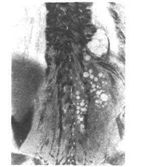
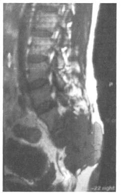
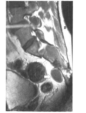
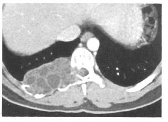
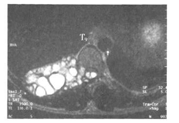
320
321
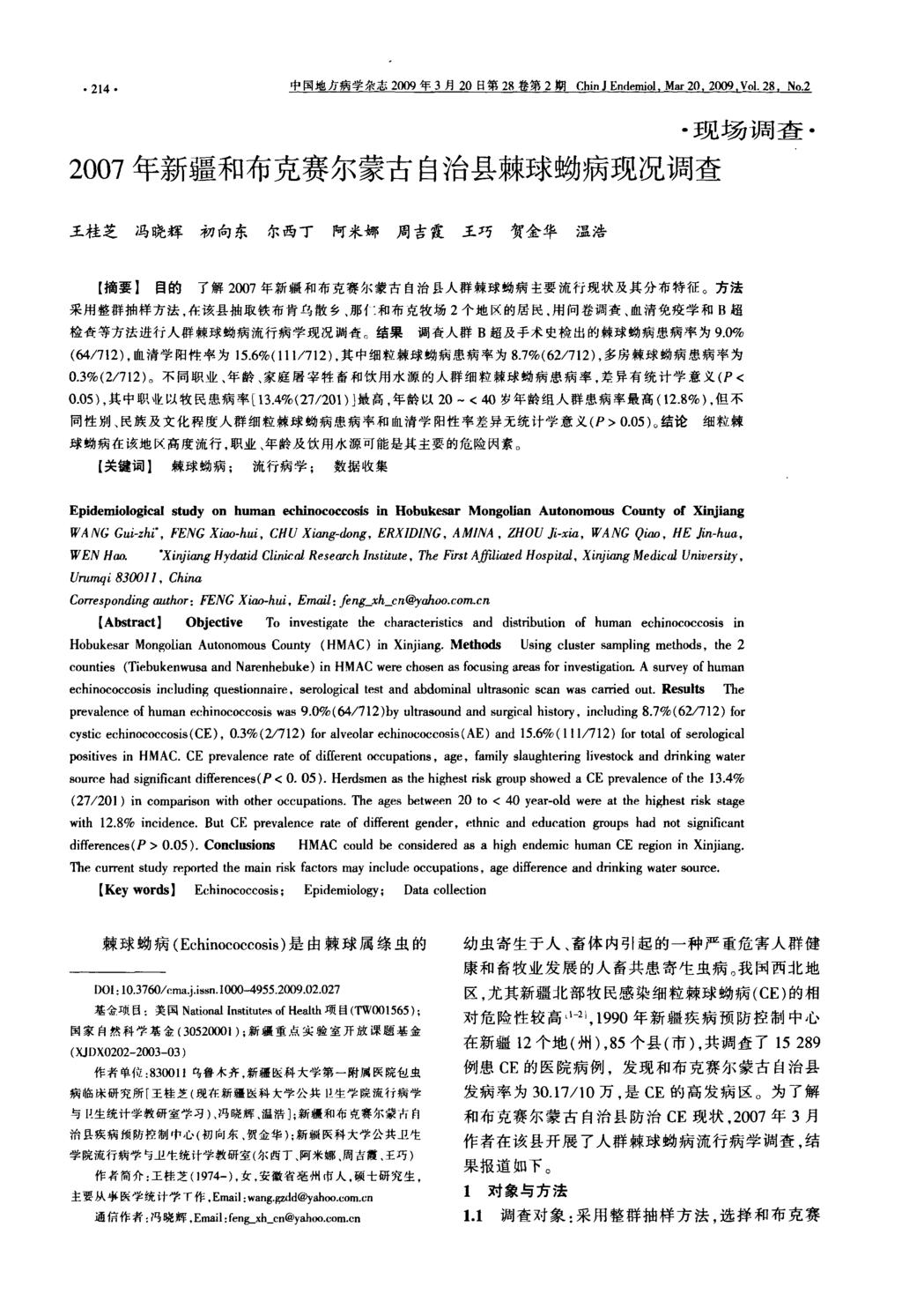
322
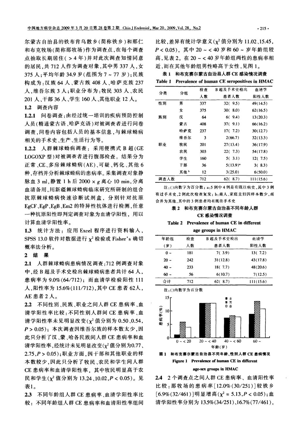
323
324
325
Hydatid Disease. Overview
 Hydatid Disease Overview Hydatid disease in man is caused principally by infection with the larval stage of the dog tapeworm Echinococcus granulosus. It is an important pathogenic zoonotic parasitic infection
ECHINOCOCCOSIS. By Dr. Ameer kadhim Hussein. M.B.Ch.B. FICMS (Community Medicine).
.") ECHINOCOCCOSIS By Dr. Ameer kadhim Hussein. M.B.Ch.B. FICMS (Community Medicine). INTRODUCTION Species under genus Echinococcus are small tapeworms of carnivores with larval stages known as hydatids proliferating
ECHINOCOCCOSIS By Dr. Ameer kadhim Hussein. M.B.Ch.B. FICMS (Community Medicine). INTRODUCTION Species under genus Echinococcus are small tapeworms of carnivores with larval stages known as hydatids proliferating
1.0 INTRODUCTION. Echinococcosis, a cyclozoonotic helminthosis caused by the dwarf dog
 INTRODUCTION 1.0 INTRODUCTION Echinococcosis, a cyclozoonotic helminthosis caused by the dwarf dog tapeworm Echinococcus granulosus is highly endemic and is considered to be one of the most important parasitic
Scientific background concerning Echinococcus multilocularis. Muza Kirjušina, Daugavpils University, Latvia
 Scientific background concerning Echinococcus multilocularis Muza Kirjušina, Daugavpils University, Latvia Echinococcus multilocularis Infection with the larval form causes alveolar echinococcosis (AE).
Hydatid Cyst Dr. Nora L. El-Tantawy
 Hydatid Cyst Dr. Nora L. El-Tantawy Ass. Prof. of Parasitology Faculty of Medicine, Mansoura university, Egypt Echinococcus granulosus Geographical Distribution: cosmopolitan especially in sheep raising
Cystic echinococcosis in a domestic cat: an Italian case report
 13th NRL Workshop, Rome, 24-25 May, 2018 Cystic echinococcosis in a domestic cat: an Italian case report Istituto Zooprofilattico Sperimentale (IZS) of Sardinia National Reference Laboratory for Cistic
National Research Center
 National Research Center Update of immunodiagnosis of cystic echinococcosis cysts Global distribution of zoonotic strains of Echinococcus granulosus (Adapted from Eckert and Deplazes, 2004) Echinococcus
Global diversity of cystic echinococcosis. Thomas Romig Universität Hohenheim Stuttgart, Germany
 Global diversity of cystic echinococcosis Thomas Romig Universität Hohenheim Stuttgart, Germany Echinococcus: generalized lifecycle Cystic echinococcosis: geographical spread Acephalocystis cystifera
ECHINOCOCCUS GRANULOSUS
 48 ECHINOCOCCUS GRANULOSUS 48.1 INTRODUCTION E granulosus are small tape worms that parasitize the intestines of carnivores like dogs. About one million people are infected with this tape worm worldwide.
This is the smallest tapeworm that can affect human being but it s not really proper human tapeworm (the human is not the primary host).
.") Echinococcus Granulosus Small Tapeworm (1 cm), Cestode. This is the smallest tapeworm that can affect human being but it s not really proper human tapeworm (the human is not the primary host). The primary
Echinococcus Granulosus Small Tapeworm (1 cm), Cestode. This is the smallest tapeworm that can affect human being but it s not really proper human tapeworm (the human is not the primary host). The primary
Echinococcosis on the Tibetan Plateau
 Echinococcosis on the Tibetan Plateau Inauguraldissertation zur Erlangung der Würde einer Doktorin der Philosophie vorgelegt der Philosophisch-Naturwissenschaftlichen Fakultät der Universität Basel von
Contains most of the medically important tapeworms Scolex has 4 suckers and compact vitelline gland are characteristic Range from mm to >10m
 Cyclophyllidae Contains most of the medically important tapeworms Scolex has 4 suckers and compact vitelline gland are characteristic Range from mm to >10m Family Taeniidae Taenia saginata: beef tapeworm
HYDATID CYST DISEASE
 HYDATID CYST DISEASE Hydatid disease, also called hydatidosis or echinococcosis, is a cystforming disease resulting from an infection with the metacestode, or larval form, of parasitic dog tapeworms from
Echinococcosis in Tibetan Populations, Western Sichuan Province, China
 RESEARCH Echinococcosis in Tibetan Populations, Western Sichuan Province, China Li Tiaoying,* Qiu Jiamin,* Yang Wen,* Philip S. Craig, Chen Xingwang,* Xiao Ning,* Akira Ito, Patrick Giraudoux, Mamuti Wulamu,
Echinococcus multilocularis Diagnosis. Peter Deplazes. Medical Faculty. Swiss TPH Winter Symposium 2017
 Medical Faculty Swiss TPH Winter Symposium 2017 Helminth Infection from Transmission to Control Echinococcus multilocularis Diagnosis Peter Deplazes Global distribution of E. multilocularis Deplazes et
Presentation of Quiz #85
 Presentation of Quiz #85 ***Reminder: Slides are copyrighted and cannot be copied for publication. A 36 year old male from Columbia was admitted to the hospital with seizures. This patient had previously
5.0 DISCUSSION. Echinococcosis is a cosmopolitan parasitic zoonosis caused by the
 DISCUSSION 5.0 DISCUSSION Echinococcosis is a cosmopolitan parasitic zoonosis caused by the dwarf dog tapeworm Echinococcus granulosus. The domestic life cycle is maintained through dogs and ungulates,
Schistosoma mansoni, S. japonicum, S. haematobium
 Schistosoma mansoni, S. japonicum, S. haematobium The Organisms More than 200 million people are infected worldwide with Schistosoma species. The adult worms are long and slender (males are 6 12 mm in
Parasitology. Echinococcus Spp.
 Parasitology المحاضرة الثالثت ا. صباح النجار Echinococcus Spp. Genus Echinococcus include three different species in which man acts as intermediate host and infecting by the larval stage of these species.
Understanding the Lifecycle of the Hydatid Tapeworm
 Hydatid Tapeworm The Hydatid Tapeworm (scientific name Echinococcus granulosis) is one of a number of tapeworms that infect dogs. The reason this tapeworm is considered the most significant is that, unlike
Hydatid disease (Echinococcus granulosus) in Australian Wildlife FACT SHEET
 in Australian Wildlife FACT SHEET") Hydatid disease (Echinococcus granulosus) in Australian Wildlife FACT SHEET Introductory Statement Echinococcus granulosus is widespread in Australian wildlife where its reproductive potential may be greater
Hydatid disease (Echinococcus granulosus) in Australian Wildlife FACT SHEET Introductory Statement Echinococcus granulosus is widespread in Australian wildlife where its reproductive potential may be greater
Antihelminthic Trematodes (flukes): Cestodes (tapeworms): Nematodes (roundworms, pinworm, whipworms and hookworms):
: Cestodes (tapeworms): Nematodes (roundworms, pinworm, whipworms and hookworms):") Antihelminthic Drugs used to treat parasitic worm infections: helminthic infections Unlike protozoa, helminthes are large and have complex cellular structures It is very important to identify the causative
Antihelminthic Drugs used to treat parasitic worm infections: helminthic infections Unlike protozoa, helminthes are large and have complex cellular structures It is very important to identify the causative
Coproantigen prevalence of Echinococcus spp. in rural dogs from Northwestern Romania
 Coproantigen prevalence of Echinococcus spp. in rural dogs from Northwestern Romania Ştefania Seres 1, Eugeniu Avram 1, Vasile Cozma 2 1 Parasitology Department of Sanitary Veterinary and Food Safety Direction,
FAO-APHCA/OIE/USDA Regional Workshop on Prevention and Control of Neglected Zoonoses in Asia July, 2015, Obihiro, Japan.
 FAO-APHCA/OIE/USDA Regional Workshop on Prevention and Control of Neglected Zoonoses in Asia 15-17 July, 2015, Obihiro, Japan Dr Gillian Mylrea 1 Overview What is a Neglected Zoonotic Disease? The important
ECHINOCOCCOSIS AND CYSTICERCOSIS IN ASIA: EVALUATION OF THE MODERN TECHNOLOGY FOR EPIDEMIOLOGICAL STUDY
 ECHINOCOCCOSIS AND CYSTICERCOSIS IN ASIA: EVALUATION OF THE MODERN TECHNOLOGY FOR EPIDEMIOLOGICAL STUDY Akira Ito 1, Hiroshi Yamasaki 1, Minoru Nakao 1, Yasuhito Sako 1, Kazuhiro Nakaya 2, Wulamu Mamuti
Curriculum Vitae. Education: DVM University of Shiraz, School of veterinary medicine
 Curriculum Vitae Name :Mohammad Reza Siavashi Address: Pasteur Institute of Iran,No: 69, Pasteur Ave., Tehran, Iran 1316943551 Tel: +98 21 66968855 Fax: +98 21 66968855 E mail: m_siavashi@hotmail.com Nationality:
Report on the third NRL Proficiency Test to detect adult worms of Echinococcus sp. in the intestinal mucosa of the definitive host.
 Report on the third NRL Proficiency Test to detect adult worms of Echinococcus sp. in the intestinal mucosa of the definitive host March-April, 2011 page 1 of 11 Table of contents 1 Introduction 3 2 Scope
MORPHOLOGICAL CHARACTERIZATION OF ADULT ECHINOCOCCUS GRANULOSUS AS A MEANS OF DETERMINING TRANSMISSION PATTERNS
 J. Parasitol., 79(1), 1993, p. 57-61? American Society of Parasitologists 1993 MORPHOLOGICAL CHARACTERIZATION OF ADULT ECHINOCOCCUS GRANULOSUS AS A MEANS OF DETERMINING TRANSMISSION PATTERNS Clare C. Constantine,
THE STRUCTURE OF ECHINOCOCCAL CYSTS AND HISTOPATHOLOGICAL CHANGES IN LIVER
 THE STRUCTURE OF ECHINOCOCCAL CYSTS AND HISTOPATHOLOGICAL CHANGES IN LIVER Michal Juszynski Helena Palenga, Danuta Cielecka PhD Department of General Biology and Parasitology Medical University of Warsaw
We are IntechOpen, the world s leading publisher of Open Access books Built by scientists, for scientists. International authors and editors
 We are IntechOpen, the world s leading publisher of Open Access books Built by scientists, for scientists 3,900 116,000 120M Open access books available International authors and editors Downloads Our
On the Occurrence and Significance of Hydatid Cysts in the Ceylon Sambhur Rusa unicolor unicolor.*
 CEYLON J. MBD. SCI. (D) Vol. XI, Pt. 1 (May 1962) On the Occurrence and Significance of Hydatid Cysts in the Ceylon Sambhur Rusa unicolor unicolor.* by A. S. DISSANAIKE AND D. C. PARAMANANTHAN** Department
A Pilot Study for Control of Hyperendemic Cystic Hydatid Disease in China
 A Pilot Study for Control of Hyperendemic Cystic Hydatid Disease in China Author Zhang, Wenbao, Zhang, Zhuangzhi, Yimit, Turhong, Shi, Baoxin, Aili, Hasyeti, Tulson, Gulnor, You, Hong, Li, Jun, Gray, Darren,
Prevalence of Cystic Echinococcosis in Slaughtered Sheep as an Indicator to Assess Control Progress in Emin County, Xinjiang, China
 ISSN (Print) 0023-4001 ISSN (Online) 1738-0006 BRIEF COMMUNICATION Korean J Parasitol Vol. 53, No. 3: 355-359, June 2015 http://dx.doi.org/10.3347/kjp.2015.53.3.355 Prevalence of Cystic Echinococcosis
Vertebrates and Parasites
 Vertebrates and Parasites Parasites indicators of biodiversity o Lots of parasites with complex life histories = area of high biodiversity with a good ecosystem o Provide deep phylogenetic and ecological
Title. Author(s)YAMASHITA, Jiro; OHBAYASHI, Masashi; KONNO, Seiji. CitationJapanese Journal of Veterinary Research, 4(3): Issue Date
YAMASHITA, Jiro; OHBAYASHI, Masashi; KONNO, Seiji. CitationJapanese Journal of Veterinary Research, 4(3): Issue Date") Title STUDIES ON ECHINOCOCCOSIS : III. ON EXPERIMENTAL INF DEVELOPMENT OF ECHINOCOCCUS GRANULOSUS (BATSCH, 1786 Author(s)YAMASHITA, Jiro; OHBAYASHI, Masashi; KONNO, Seiji CitationJapanese Journal of Veterinary
Title STUDIES ON ECHINOCOCCOSIS : III. ON EXPERIMENTAL INF DEVELOPMENT OF ECHINOCOCCUS GRANULOSUS (BATSCH, 1786 Author(s)YAMASHITA, Jiro; OHBAYASHI, Masashi; KONNO, Seiji CitationJapanese Journal of Veterinary
EXPERIMENTAL HYDATIDOSIS IN THE SUDAN: TRANSMISSION AND NATURAL INFECTION
 EXPERIMENTAL HYDATIDOSIS IN THE SUDAN: TRANSMISSION AND NATURAL INFECTION By Nadia Ahmed Ali Mohamed B.Sc. (Assuit University -Egypt) M.Sc. (Parasitology) University of Khartoum Supervisor: Prof. Mohamed
Breast Mass on Mammography
 Breast Mass on Mammography Bobbi Pritt, MD 2014 MFMER slide-1 Clinical Presentation 68 year old woman was noted to have 2 adjacent masses in the right breast on screening mammography Well-circumscribed,
PARASITOLOGICAL EXAMINATIONS CATALOGUE OF SERVICES AND PRICE LIST
 INSTITUTE OF PARASITOLOGY Biomedical Research Center Seltersberg Justus Liebig University Giessen Schubertstrasse 81 35392 Giessen Germany Office: +49 (0) 641 99 38461 Fax: +49 (0) 641 99 38469 Coprological
Title. Author(s)YAMASHITA, Jiro; OHBAYASHI, Masashi; KITAMURA, Yukit. CitationJapanese Journal of Veterinary Research, 6(2): 89-92
YAMASHITA, Jiro; OHBAYASHI, Masashi; KITAMURA, Yukit. CitationJapanese Journal of Veterinary Research, 6(2): 89-92") Title STUDIES ON ECHINOCOCCOSIS VII. : ON THE DEVELOPMENT IN THE TAPEWORM STAGE Author(s)YAMASHITA, Jiro; OHBAYASHI, Masashi; KITAMURA, Yukit CitationJapanese Journal of Veterinary Research, 6(2): 89-92
Title STUDIES ON ECHINOCOCCOSIS VII. : ON THE DEVELOPMENT IN THE TAPEWORM STAGE Author(s)YAMASHITA, Jiro; OHBAYASHI, Masashi; KITAMURA, Yukit CitationJapanese Journal of Veterinary Research, 6(2): 89-92
Surveillance of animal brucellosis
 Surveillance of animal brucellosis Assoc.Prof.Dr. Theera Rukkwamsuk Department of large Animal and Wildlife Clinical Science Faculty of Veterinary Medicine Kasetsart University Review of the epidemiology
by author Cystic Echinococcosis Rogelio López-Vélez MD, DTM&H, PhD National Referral Unit for Tropical Diseases Infectious Diseases Department
 Cystic Echinococcosis Rogelio López-Vélez MD, DTM&H, PhD National Referral Unit for Tropical Diseases Infectious Diseases Department Ramón y Cajal University Hospital. Madrid. Spain National Referral Unit
Title. CitationJapanese Journal of Veterinary Research, 52(2): 101- Issue Date Doc URL. Type. File Information
: 101- Issue Date Doc URL. Type. File Information") Title INFORMATION: Thesis for the Doctor of Veterinary Med CitationJapanese Journal of Veterinary Research, 52(2): 101- Issue Date 2004-08 Doc URL http://hdl.handle.net/2115/10515 Type bulletin File Information
Title INFORMATION: Thesis for the Doctor of Veterinary Med CitationJapanese Journal of Veterinary Research, 52(2): 101- Issue Date 2004-08 Doc URL http://hdl.handle.net/2115/10515 Type bulletin File Information
Management of Echinococcus Multilocularis Infections in Animals Guideline, 2018
 Ministry of Health and Long-Term Care Management of Echinococcus Multilocularis Infections in Animals Guideline, 2018 Population and Public Health Division, Ministry of Health and Long-Term Care Effective:
The EU thanks the OIE TAHSC, the APSFWW and the ad hoc group for their work.
 1 Annex 34 Original: English October 2010 REPORT OF THE MEETING OF THE OIE AD HOC GROUP ON ZOONOTIC PARASITES Paris (France), 57 October 2010 s The EU thanks the OIE TAHSC, the APSFWW and the ad hoc group
Hazem.K.Al-Khafaji FICMS College of medicine- Al-Qadissyia university
 Hazem.K.Al-Khafaji FICMS College of medicine- Al-Qadissyia university Cestodes(Tapeworms) - Morphology - Tapeworm parts: Flat, segmented body with various length (several mm,25mm as hymenolepis nana ~
COMMISSION DELEGATED REGULATION (EU)
") L 296/6 Official Journal of the European Union 15.11.2011 COMMISSION DELEGATED REGULATION (EU) No 1152/2011 of 14 July 2011 supplementing Regulation (EC) No 998/2003 of the European Parliament and of the
L 296/6 Official Journal of the European Union 15.11.2011 COMMISSION DELEGATED REGULATION (EU) No 1152/2011 of 14 July 2011 supplementing Regulation (EC) No 998/2003 of the European Parliament and of the
Emergence/re-emergence of Echinococcus spp. a global update
 International Journal for Parasitology 35 (2005) 1205 1219 www.elsevier.com/locate/ijpara Invited review Emergence/re-emergence of Echinococcus spp. a global update D.J. Jenkins a,b, *, T. Romig b, R.C.A.
Liver cysts caused by E. granulosus in Xinjiang, Peoples Republic of China
 Liver cysts caused by E. granulosus in Xinjiang, Peoples Republic of China Eleonor Sjöstrand and Monica Olsson Dept. of Infectious Diseases Sahlgrenska Academy University of Gothenburg Sweden Supervisors:
Old Disease New Location Surgeons Be Alerted
 Old Disease New Location Surgeons Be Alerted K. B. Ashok Vol. 3 No. 4 (April 2011) International Journal of Collaborative Research on Internal Medicine & Public Health (IJCRIMPH) ISSN 1840-4529 Journal
Sera from 2,500 animals from three different groups were analysed:
 FIELD TRIAL OF A BRUCELLOSIS COMPETITIVE ENZYME LINKED IMMUNOABSORBENT ASSAY (ELISA) L.E. SAMARTINO, R.J. GREGORET, G. SIGAL INTA-CICV Instituto Patobiología Area Bacteriología, Buenos Aires, Argentina
V. Subclass Eucestoida (Chapters 20 & 21, BLY 459, 2010)
") V. Subclass Eucestoida (Chapters 20 & 21, BLY 459, 2010) A. Characteristics (Ignore Cestodaria) 1. Differences from trematodes a. No digestive tract (1) No mouth, gut, nor anus (2) All nutrients absorbed
V. Subclass Eucestoida (Chapters 20 & 21, BLY 459, 2010) A. Characteristics (Ignore Cestodaria) 1. Differences from trematodes a. No digestive tract (1) No mouth, gut, nor anus (2) All nutrients absorbed
EVALUATION AND RECOMMENDATIONS FOR THE CONTROL OF CYSTIC ECHINOCOCCOSIS IN WALES. Mariel Stefhanie Aybar Espinoza
 EVALUATION AND RECOMMENDATIONS FOR THE CONTROL OF CYSTIC ECHICOCCOSIS IN WALES Mariel Stefhanie Aybar Espinoza Supervisor: Eileen Devaney A dissertation submitted to obtain the degree of Master of Veterinary
ORGANISATION FOR ANIMAL
 Report of the WHO Informal Working Group on cystic and alveolar echinococcosis surveillance, prevention and control, with the participation of the Food and Agriculture Organization of the United Nations
General introduction
 Spirometra mansoni General introduction Distributed worldwide, mainly in southeast Asia. Larval infection of S. mansoni may cause serious clinical disease ---Sparganosis Morphology Adult worm measures
Cestodes P & S, Joshua Stillman MD, MPH Department of Emergency Medicine Assistant Professor, Columbia University. Helminths
 Cestodes P & S, 2008 Joshua Stillman MD, MPH Department of Emergency Medicine Assistant Professor, Columbia University Helminths Phylum Nematoda (Roundworms) - Nematodes Pinworm, Whipworm, Ascaris + VLM,
SHORT RESEARCH NOTE. Anca Florea 1. , Liviu Vlad 2, Vasile Cozma 3, Zoe Coroiu 4. Introduction
 Serological diagnosis of cystic echinococcosis by the ELISA technique, in the cases hospitalized in the Surgical Clinic no. III and Internal Medicine no. III of Cluj-Napoca, during October 2006 December
The prevalence of anti-echinococcus antibodies in the North-Western part of Romania
 The prevalence of anti-echinococcus antibodies in the North-Western part of Romania Anca Florea 1, Zoe Coroiu 2, Rodica Radu 2 1 Prof. dr. Octavian Fodor Regional Institute of Gastroenterology and Hepatology,
New treatments for manges in dogs? Canine demodicosis. Canine demodicosis. Current approved drug in Canada:
 New treatments for manges in dogs? Andrew S. Peregrine, BVMS, PhD, DVM, DipEVPC, DipACVM E-mail: aperegri@ovc.uoguelph.ca; Tel: 519-824-4120 ext 54714 Canine demodicosis Most common = D. canis No difference
Prevalence of Taenia in selected Canids and felids living within wildlife sanctuaries in Kenya
 International Journal of Advanced Multidisciplinary Research ISSN: 2393-8870 www.ijarm.com DOI: 10.22192/ijamr Volume 4, Issue 9-2017 Research Article Prevalence of Taenia in selected Canids and felids
We are IntechOpen, the first native scientific publisher of Open Access books. International authors and editors. Our authors are among the TOP 1%
 We are IntechOpen, the first native scientific publisher of Open Access books 3,350 108,000 1.7 M Open access books available International authors and editors Downloads Our authors are among the 151 Countries
CYSTIC ECHINOCOCCOSIS IN AUSTRALIA: THE CURRENT SITUATION
 CYSTIC ECHINOCOCCOSIS IN AUSTRALIA: THE CURRENT SITUATION David J Jenkins Australian Hydatid Control and Epidemiology Program, Fyshwick; School of Botany and Zoology, Australian National University, Canberra,
Cestodes P & S, Joshua Stillman MD, MPH Department of Emergency Medicine Assistant Professor, Columbia University. Helminths
 Cestodes P & S, 2009 Joshua Stillman MD, MPH Department of Emergency Medicine Assistant Professor, Columbia University Helminths Phylum Nematoda (Roundworms) - Nematodes Pinworm, Whipworm, Ascaris + VLM,
The Taxonomic Value and Variability of Certain Structures in the Cestode Genus Echinococcus (Rudolphi, 1801) and a Review of Recognized Species
 and a Review of Recognized Species") University of Nebraska - Lincoln DigitalCommons@University of Nebraska - Lincoln Faculty Publications from the Harold W. Manter Laboratory of Parasitology Parasitology, Harold W. Manter Laboratory of 11-15-1953
University of Nebraska - Lincoln DigitalCommons@University of Nebraska - Lincoln Faculty Publications from the Harold W. Manter Laboratory of Parasitology Parasitology, Harold W. Manter Laboratory of 11-15-1953
Data were analysed by SPSS, version 10 and the chi-squared test was used to assess statistical differences. P < 0.05 was considered significant.
 Toxocara canis is one of the commonest nematodes of the dog and most often this nematode is the cause of toxocariasis (visceral larva migrans) [1]. People become infected by ingestion of eggs from soil,
Cestodes. Tapeworms from man and animals
 Cestodes Tapeworms from man and animals Taenia sp. The common (beef) tapeworm is several meters long. Courtesy Peters W. & Gilles H. Courtesy CDC Courtesy CDC Taenia sp. Unstained egg with four (visible)
Mexican Wolves and Infectious Diseases
 Mexican Wolves and Infectious Diseases Mexican wolves are susceptible to many of the same diseases that can affect domestic dogs, coyotes, foxes and other wildlife. In general, very little infectious disease
Introduction to Helminthology
 Introduction to Helminthology HELMINTHES (WORMS) - Characteristics Eukaryotic, multicellular animals that usually have digestive, circulatory, nervous, excretory, and reproductive systems. Worms with bilateral
Eukaryotic Parasites. An Illustrated Guide to Parsitic Life Cycles to Accompany Lecture. By Noel Ways
 Eukaryotic Parasites An Illustrated Guide to Parsitic Life Cycles to Accompany Lecture By Noel Ways Giardia lamblia Life Cycle Reservoir: Beavers strongly implicated. Also, many other wild animals as well
We are IntechOpen, the world s leading publisher of Open Access books Built by scientists, for scientists. International authors and editors
 We are IntechOpen, the world s leading publisher of Open Access books Built by scientists, for scientists 3,500 108,000 1.7 M Open access books available International authors and editors Downloads Our
INTRODUCTION MATERIALS AND METHODS
 Am. J. Trop. Med. Hyg., 88(4), 2013, pp. 795 802 doi:10.4269/ajtmh.12-0331 Copyright 2013 by The American Society of Tropical Medicine and Hygiene Development of Three PCR Assays for the Differentiation
Diagnosis, treatment, and management of echinococcosis
 Follow the link from the online version of this article to obtain certified continuing medical education credits Diagnosis,, and management of echinococcosis Donald P McManus, 1 Darren J Gray, 1 2 Wenbao
Iranian J Parasitol: Vol. 7, No.1, 2012, pp Iranian J Parasitol. Open access Journal at ijpa.tums.ac.ir
 Iranian J Parasitol: Vol. 7, No.1, 2012, pp.59-66 Tehran University of Medical Sciences Publication http:// tums.ac.ir Original Article Iranian J Parasitol Open access Journal at http:// ijpa.tums.ac.ir
Post treatment follow up study of abdominal cystic echinococcosis in. Tibetan communities of northwest Sichuan Province, China
 Post treatment follow up study of abdominal cystic echinococcosis in Tibetan communities of northwest Sichuan Province, China Li, T, Ito, A, Pengcuo, R, Sako, Y, Chen, X, Qiu, D, Xiao, N and Craig, PS
Outcome of the Conference Towards the elimination of rabies in Eurasia Joint OIE/WHO/EU Conference
 Outcome of the Conference Towards the elimination of rabies in Eurasia Joint OIE/WHO/EU Conference WHO (HQ-MZCP) / OIE Inter-country Workshop on Dog and Wildlife Rabies Control in the Middle East 23-25
31/05/2011. Epidemiology and Control Programs for Echinococcus multilocularis. - geography? - frequency? - risk factors? - geography? - frequency?
 Epidemiology and Control Programs for Echinococcus multilocularis - geography - frequency - risk factors Thomas Romig Universität Hohenheim Stuttgart, Germany - geography - frequency - risk factors Global
Dog vaccination with EgM proteins against Echinococcus granulosus
 Zhang et al. Infectious Diseases of Poverty (2018) 7:61 https://doi.org/10.1186/s40249-018-0425-4 SHORT REPORT Open Access Dog vaccination with EgM proteins against Echinococcus granulosus Zhuang-Zhi Zhang
Oxfendazole Treatment for Cystic Hydatid Disease in Naturally Infected Animals
 ANTIMICROBIAL AGENTS AND CHEMOTHERAPY, Mar. 1998, p. 601 605 Vol. 42, No. 3 0066-4804/98/$04.00 0 Copyright 1998, American Society for Microbiology Oxfendazole Treatment for Cystic Hydatid Disease in Naturally
"Serpent" Sign, "Double Arch" Sign and "Air-Bubble"Sign in a case of Ruptured Hydatid Cyst-A Case Report
 Article ID: WMC004602 ISSN 2046-1690 "Serpent" Sign, "Double Arch" Sign and "Air-Bubble"Sign in a case of Ruptured Hydatid Cyst-A Case Report Peer review status: No Corresponding Author: Dr. Dipti Gothi,
Scientific Opinion of the Scientific Panel on Animal Health and Welfare on a request from the Commission regarding the
 The EFSA Journal (2006) 441, 1-54, Assessment of the risk of echinococcosis introduction into the UK, Ireland, Sweden, Malta and Finland as a consequence of abandoning national rules Scientific Opinion
OIE international standards on Rabies:
 Regional cooperation towards eradicating the oldest known zoonotic disease in Europe Antalya, Turkey 4-5 December 2008 OIE international standards on Rabies: Dr. Lea Knopf Scientific and Technical Department
A LABORATORY NETWORK FOR DIAGNOSTIC OF CAMELIDS DISEASES
 A LABORATORY NETWORK FOR DIAGNOSTIC OF CAMELIDS DISEASES M. EL HARRAK Chair of OIE ad hoc Group on Camelids Diseases Biopharma Lab BP 4569 Rabat Morocco CAMELIDS FAMILY Dromadary Camel Bactrian Camel Lama
Still and Moving Image Evidences for Mating of Echinococcus granulosus Reared in Culture Media
 Iranian J Parasitol: Vol. 9, No. 1, Jan -Mar 2014, pp.129-133 Short Communication Still and Moving Image Evidences for Mating of Echinococcus granulosus Reared in Culture Media Tahereh MOHAMMADZADEH, *Seyed
Brucellosis situation in Mongolia and Result of Bovine Brucellosis Proficiency Test
 The 4 th FAO-APHCA/OIE/DLD Regional Workshop on Brucellosis Diagnosis and Control in Asia-Pacific Region - Proficiency Test and Ways Forward- Chiang Mai, Thailand, 18-21 March 2014 Brucellosis situation
UNDERSTANDING HEARTWORMS 4-Pets
 Oklahoma 4-H 4H SMAN 503 UNDERSTANDING HEARTWORMS 4-Pets WHAT IS A HEARTWORM Heartworms are internal parasites that can infect your dog or cat. Parasites live on the inside (internal) or on the outside
Department of Immunology, Microbiology and Parasitology, Faculty of Pharmacy, University of the Basque Country, Vitoria, Spain
 REVIEW 10.1111/j.1469-0691.2007.01665.x The immunodiagnosis of Echinococcus multilocularis infection D. Carmena, A. Benito and E. Eraso Department of Immunology, Microbiology and Parasitology, Faculty
Prevalence Survey on Hydatidosis and its Financial Loss in Small Ruminants Slaughtered at Addis Ababa Abattoirs Enterprise
 ISSN 079-018 IDOSI Publications, 015 DOI: 10.589/idosi.apg.015.6.3.950 Prevalence Survey on Hydatidosis and its Financial Loss in Small Ruminants Slaughtered at Addis Ababa Abattoirs Enterprise Simegnew
OIE Collaborating Centres Reports Activities
 OIE Collaborating Centres Reports Activities Activities in 2016 This report has been submitted : 2017-01-08 05:38:55 Title of collaborating centre: Food-Borne Parasites from the Asia-Pacific Region Address
ANIMAL RABIES IN NEPAL AND RACCOON RABIES IN ALBANY COUNTY, NEW YORK
 ANIMAL RABIES IN NEPAL AND RACCOON RABIES IN ALBANY COUNTY, NEW YORK SHANKAR YADAV MPH Report/Capstone Project Presentation 07/19/2012 CHAPTER 1: FIELD EXPERIENCE AT KANSAS STATE UNIVERSITY RABIES LABORATORY
CRANIAL HYDATID CYST
 Thi-Qar Medical Journal (TQMJ): Vol(6) No(1):2012(48-52) OBJECT: CRANIAL HYDATID CYST Dr. Haitham Handhal* HYDATID disease is caused by infestation by larvae of the tapeworm Echinococcus granulosus. The
COMMISSION DELEGATED REGULATION (EU) /... of XXX
 /... of XXX") Ref. Ares(2017)4396495-08/09/2017 EUROPEAN COMMISSION Brussels, XXX SANTE/7009/2016 CIS Rev. 1 (POOL/G2/2016/7009/7009R1-EN CIS.doc) [ ](2016) XXX draft COMMISSION DELEGATED REGULATION (EU) /... of XXX
Ref. Ares(2017)4396495-08/09/2017 EUROPEAN COMMISSION Brussels, XXX SANTE/7009/2016 CIS Rev. 1 (POOL/G2/2016/7009/7009R1-EN CIS.doc) [ ](2016) XXX draft COMMISSION DELEGATED REGULATION (EU) /... of XXX
EVALUATION OF THE USEFULNESS OF LAMINATED LAYER ANTIGENS IN THE SEROLOGICAL FOLLOW UP OF CYSTIC ECHINOCOCCOSIS IN HUMANS OGHENEKARO E.
 EVALUATION OF THE USEFULNESS OF LAMINATED LAYER ANTIGENS IN THE SEROLOGICAL FOLLOW UP OF CYSTIC ECHINOCOCCOSIS IN HUMANS OGHENEKARO E. OKITI SCHOOL OF ENVIRONMENT AND LIFE SCIENCES, UNIVERSITY OF SALFORD,
Cestodes (tapeworms) Pro. Dr. Mohammed Sabri
 Pro. Dr. Mohammed Sabri") Cestodes (tapeworms) Pro. Dr. Mohammed Sabri Characters (general):- Tape worms consist of a round head, called a scolex, and a flat body of multiple segments called proglottides. The scolex has specialized
Cestodes (tapeworms) Pro. Dr. Mohammed Sabri Characters (general):- Tape worms consist of a round head, called a scolex, and a flat body of multiple segments called proglottides. The scolex has specialized
Enzyme-Linked Immunosorbent Assay (Elisa) In The Serodiagnosis Of Hydatidosis In Camels (Camelus dromedarius) And Cattle In Sokoto, Northern Nigeria
 In The Serodiagnosis Of Hydatidosis In Camels (Camelus dromedarius) And Cattle In Sokoto, Northern Nigeria") ISPUB.COM The Internet Journal of Infectious Diseases Volume 13 Number 1 Enzyme-Linked Immunosorbent Assay (Elisa) In The Serodiagnosis Of Hydatidosis In Camels (Camelus B Okolugbo, S Luka, I Ndams Citation
ISPUB.COM The Internet Journal of Infectious Diseases Volume 13 Number 1 Enzyme-Linked Immunosorbent Assay (Elisa) In The Serodiagnosis Of Hydatidosis In Camels (Camelus B Okolugbo, S Luka, I Ndams Citation
Title. CitationJapanese Journal of Veterinary Research, 35(2): Issue Date DOI. Doc URL. Type. File Information /jjvr.35.2.
: Issue Date DOI. Doc URL. Type. File Information /jjvr.35.2.") Title ISOLATION OF ECHINOCOCCUS MULTILOCULARIS FROM THE LI Author(s)KAMIYA, Masao; OOI, Hong Kean; OKU, Yuzaburo; OKAMOT CitationJapanese Journal of Veterinary Research, 35(2): 99-1 Issue Date 1987-04-30
Title ISOLATION OF ECHINOCOCCUS MULTILOCULARIS FROM THE LI Author(s)KAMIYA, Masao; OOI, Hong Kean; OKU, Yuzaburo; OKAMOT CitationJapanese Journal of Veterinary Research, 35(2): 99-1 Issue Date 1987-04-30
Emergence of Echinococcus multilocularis in dogs in Ontario: implications for public and wildlife health?
 Emergence of Echinococcus multilocularis in dogs in Ontario: implications for public and wildlife health? Andrew S. Peregrine 1, Jonathon Kotwa 1, Claire Jardine 1, Benoît Cuq 1, Nicola Mercer 2, Bruno
Feline and Canine Internal Parasites
 Feline and Canine Internal Parasites Internal parasites are a very common problem among dogs. Almost all puppies are already infected with roundworm when still in the uterus, or get the infection immediately
General Secretary s Report
 General Secretary s Report require a constitutional change. Either way, the AMI consider the European consumer to be the important consideration, and we will continue to represent the UK for the foreseeable
CONTAGIOUS BOVINE PLEURO- PNEUMONIA steps towards control of the disease. Rose Matua -Department of Veterinary Services, Kenya
 CONTAGIOUS BOVINE PLEURO- PNEUMONIA steps towards control of the disease Rose Matua -Department of Veterinary Services, Kenya Introduction CBPP is a highly contagious acute, subacute or chronic disease
Wang et al. Parasites & Vectors (2018) 11:302 https://doi.org/ /s x
 11:302 https://doi.org/ /s x") Wang et al. Parasites & Vectors (2018) 11:302 https://doi.org/10.1186/s13071-018-2873-x RESEARCH Open Access Echinococcus multilocularis and Echinococcus shiquicus in a small mammal community on the eastern
Wang et al. Parasites & Vectors (2018) 11:302 https://doi.org/10.1186/s13071-018-2873-x RESEARCH Open Access Echinococcus multilocularis and Echinococcus shiquicus in a small mammal community on the eastern