Incidence of Bordetella bronchiseptica in swine and experimental production of rhinitis with the organism
|
|
|
- Aldous Edwards
- 5 years ago
- Views:
Transcription

1 Retrospective Theses and Dissertations Iowa State University Capstones, Theses and Dissertations 1965 Incidence of Bordetella bronchiseptica in swine and experimental production of rhinitis with the organism Richard Francis Ross Iowa State University Follow this and additional works at: Part of the Microbiology Commons, and the Veterinary Pathology and Pathobiology Commons Recommended Citation Ross, Richard Francis, "Incidence of Bordetella bronchiseptica in swine and experimental production of rhinitis with the organism" (1965). Retrospective Theses and Dissertations This Dissertation is brought to you for free and open access by the Iowa State University Capstones, Theses and Dissertations at Iowa State University Digital Repository. It has been accepted for inclusion in Retrospective Theses and Dissertations by an authorized administrator of Iowa State University Digital Repository. For more information, please contact
2 This dissertation has been microfilmed exactly as received ROSS, D.V.M., Richard Francis, 1935 INCIDENCE OF BORDETELLA BRONCHISEPTICA IN SWINE AND EXPERIMENTAL PRODUCTION OF RHINITIS WITH THE ORGANISM. Iowa State University of Science and Technology Ph.D., 1965 Bacteriology' University Microfilms, Inc., Ann Arbor, Michigan
3 INCIDENCE OF BORDETELLA BRONCHISEPTICA IN SWINE AND EXPERIMENTAL PRODUCTION OF RHINITIS WITH THE ORGANISM by Richard Francis Ross ' A Dissertation Submitted to the Graduate Faculty in Partial Fulfillment of The Requirements, for the Degree of DOCTOR OF PHILOSOPHY Major Subject: Veterinary Bacteriology Approved; Signature was redacted for privacy. Ih Charge of Major Work Signature was redacted for privacy. Head of Ma]or Department Signature was redacted for privacy. Iowa State University Of Science and Technology Ames, Iowa 1965
4 I ii TABLE OF CONTENTS Page INTRODUCTION 1 REVIEW OF LITERATURE. 4 Classification 4 Morphologic and Growth Characteristics 6 Biochemic and Metabolic Activities 1 Serology, Antigens and Endotoxins 12 Heat Labile Toxins 14 Hemagglutinins 15 Hemolysins 16 Bacteriophages 17 Sensitivity to Antibacterial Agents 17 Pathogenicity of Bordetella Bronchiseptica 2 Bordetella bronchiseptica in swine 2 Bordetella bronchiseptica in guinea pigs 23 Bordetella bronchiseptica in dogs 25 Bordetella bronchiseptica in rabbits 27 Bordetella bronchiseptica in rats 29 Bordetella bronchiseptica in humans 31 Bordetella bronchiseptica in other species of 33 animals Other Species of Bordetella 35 MATERIALS AND METHODS 38 Culture Media 38 Collection of Specimens for Culture 39 Isolation and Identification of Bordetella 43 Bronchiseptica, Pasteurella Multocida and~ Hemophilus Spp. Source of Bordetella Bronchiseptica Isolates 43
5 1X. Page Biochemic, Metabolic and Morphologic Comparisons 46 of Various Isolates of Bordetella Bronchiseptica Agglutination Tests 47 Source of Experimental Animals 49 Incidence Surveys 5 Survey I 5 Survey II 5 Survey III 51 Survey IV 51 Transmission Trials 51 Trial I 51 Trial II 53 Trial III 55 RESULTS 58 Development of a Selective Medium for Isolation 58 of Bordetella Bronchiseptica Incidence Surveys 59 Survey I 59 Survey II 59 Survey III 64 Survey IV 64 Transmission Trials 65 Trial I 65 Trial II 73 Trial III 88 Biochemic, Metabolic and Morphologic Comparisons 95 of Various Isolates of Bordetella Bronchiseptica DISCUSSION 18 Incidence Surveys 18 Transmission Trials 113 SUMMARY 122
6 iv Page BIBLIOGRAPHY 125 ACKNOWLEDGMENTS 138
7 INTRODUCTION Atrophie rhinitis is a chronic infectious disease of the upper respiratory tract in swine. The most characteristic lesion of this disease is atrophy of the nasal turbinates. It affects a significant proportion of the swine raised in most areas of the world. The disease does not cause a high death loss, but interferes with growth and acts as an enhancing factor in pneumonias of swine. The etiology of atrophic rhinitis has been uncertain for over a century. Nutritional deficiencies, genetic faults and infectious agents were incriminated by early workers. In the 194's it was established that the disease was transmissible. Microorganisms found with sorr.e frequency in the porcine nasal cavity included trichomonads, Pseudomonas aeruginosa, Corynebacterium pyogenes. Mycoplasma hyorhinis, Pasteurella multocida and Hemophilus suis. Two of these organisms, Pasteurella multocida and Hemophilus suis, were shown in the 195's to be capable of causing rhinitis and turbinate atrophy. A suspected viral rhinitis, reported in 1955, was characterized by the development of specific intranuclear inclusions and severe sneezing in young pigs (Done, 1955). In 1956, Switzer demonstrated the multiple etiology of atrophic rhinitis. He reported that Pasteurella multocida, Bordetella bronchiseptica and a large filter passing agent were capable of producing rhinitis and turbinate atrophy.
8 2 Since 1956, 3 research groups have implicated Bordetella bronchiseptica as a cause of atrophic rhinitis (Switzer, 1959; Cross and Claflin, 1962; Goodwin and Whittlestone, 1963). Acceptance of the concept that Bordetella bronchiseptica is a primary factor in atrophic rhinitis has been slow. There appear to be 2 reasons for this. First of all, Bordetella bronchiseptica, originally thought to cause canine distemper, has been relegated to the role of a minor pathogen since 1926 when it was conclusively demonstrated that the cause of that disease was a virus. Secondly, the etiologic factor of atrophic rhinitis was expected by some to be a new agent rather than agents which were common and easily detectible. All species of the genus Bordetella are well established as etiologic agents of respiratory disease in man and animals. Bordetella pertussis causes whooping cough and Bordetella parapertussis causes a milder pseudowhooping cough in man. Bordetella bronchiseptica is a frequent secondary invader in canine respiratory disease, causes pneumonia in pigs, laboratory rats and guinea pigs and is a cause of chronic snuffles in domestic rabbits. The similarity of clinical manifestations and histologic changes observed in diseases caused by members of the genus Bordetella indicate that their disease producing mechanisms are similar. Bordetella pertussis grows on the tracheal and bronchial mucosa in whooping cough. Virulence factors liberated by the bacterial cell are responsible for the development
9 3 of the local lesions and generalized symptoms seen in pertussis. Bordetella bronchiseptica grows on the tracheal and bronchial mucosa of infected animals. Virulence factors liberated by this organism are thought to be similar to those produced by Bordetella pertussis. The need for elucidation of the role of Bordetella bronchiseptica in atrophic rhinitis was quite apparent at the inception of this study. It was known that the organism was capable of causing rhinitis in pigs, but its significance in the overall atrophic rhinitis problem was not known. It had been observed that several surgically repopulated swine herds had become infected with Bordetella bronchiseptica. Pigs in these herds were found to have severe lesions of turbinate atrophy from which the organism could be recovered in heavy growth. The present study was undertaken to determine the incidence of Bordetella bronchiseptica in Iowa swine, to study the development of turbinate atrophy caused by this organism in respiratory disease-free pigs and to compare the relative pathogenicity for young pigs of several isolates of Bordetella bronchi septica.
10 4 REVIEW OF LITERATURE The review of literature is divided into various areas for conveniertce. These are entitled Classification; Morphologic and Growth Characteristics ; Biochemic and Metabolic Activities; Serology, Antigens and Endotoxins; Heat Labile Toxins; Hemagglutinins; Hemolysins; Bacteriophages; Sensitivity to Antibacterial Agents; Pathogenicity of Bordetella Bronchiseptica; and Other Species of Bordetella. Classification The organism now known as Bordetella bronchiseptica was first named Bacillus bronchica'nis by Ferry (1911). In 1912 he (Ferry, 1912a) changed the specific name to bronchisepticus since the organism had been found to infect several different species of animals. Ferry and Noble (1918) and Ferry and Klix (1918) demonstrated an antigenic relationship between Bordetella bronchiseptica and Bordetella pertussis. Evans (1918) showed that it was also antigenically related to Brucella abortus. Bordetella bronchiseptica was classified in the genus Alcaligenes in the first edition of Bergey's Manual of Determinative Bacteriology (Bergey, 1923) because of its morphologic, growth and biochemic similarities to members of that genus. The close relationship of Bordetella bronchiseptica to Alcaligenes fecalis has been a subject of controversy until recent years. These 2 species are
11 5 peritrichously flagellated. Gram negative and somewhat pleomorphic. In addition, they both alkalinize litmus milk and carbohydrates and utilize citrate as a source of carbon. Neither species ferments carbohydrates. Isolates of both species are variable in their ability to reduce nitrates and produce similar reactions on potato medium. Recent investigations by Szturm and Bourdon (1948), Ulrich and Needham (1953), Proom (1955) and Beer (196b) indicated that Bordetella bronchiseptica produced positive urease reactions in less than 24 hours, required niacin and would not grow on simple glucose-inorganic salts media. Alcaligenes fecalis did not produce urease or require niacin for growth. It grew on simple glucose-inorganic salts media. Beer (196b) differentiated the 2 species on the basis of dye tolerance. Alcaligenes fecalis grew on agar containing 1:1 thionin, 1:5, malachite green, 1:1 crystal violet and 1:3 methyl violet. Bordetella bronchiseptica was inhibited by these dyes at these levels. Both organisms grew in the presence of 1:3 fuchsin. Bordetella bronchiseptica was classified with the Brucellae in the fifth and sixth editions of Bergey's Manual of Determinative Bacteriology (Bergey et, 1939; Breed et al., 1948). This classification was based on the biochemic, cultural, morphologic and antigenic similarities to members of that genus. Wilson and Miles (1946, 1955) recognized the close
12 6 relationship of Bordetella pertussis, Bordetella parapertussis and Bordetella bronchiseptica and placed all 3 species in the genus Hemophilus. For a time the name Haemophilus bronchiseptica enjoyed widespread usage in Europe. Moreno-Lopez (1952) classified the 3 species in a new genus named Bordetella. Their antigenic, morphologic and biochemic similarities had been pointed out previously (Ferry and Klix, 1918; Eldering and Kendrick, 1938; Bruckner and Evans, 1939; Evans and Maitland, 1939; Eldering, 1941; Keogh et al., 1947). Evidence confirming the validity of this classification has been published in recent years (Andersen, 1953; Lacey, 1953a; Kendrick et ^., 1953; Proom, 1953; Rauch and Pickett, 1961; Sutherland, 1961). Widespread acceptance of this classification is evident from the fact that it was used in both the seventh edition of Bergey's Manual of Determinative Bacteriology (Breed et al., 1957) and the fifth edition of Topley and Wilson's Principles of Bacteriology and Immunity (Wilson and Miles, 1964). Morphologic and Growth Characteristics McGowan (1911) and Torrey and Rahe (1913) reported that Bordetella bronchiseptica was a slender pleomorphic rod with rounded ends. Coccoid forms were found to be more common in tissues and on glycerin agar while bacillary forms were more common in broth cultures (McGowan, 1911). In addition, organisms from densely crowded colonies on agar medium were more
13 7 coccoid than organisms from larger isolated colonies (McGowan, 1911). McGowan (1911), Torrey and Rahe (1913) and Lautrop and Lacey (196) found that coccoid forms of Bordetella bronchiseptica measured about.5 n by 1. p while bacillary forms measured about.5 y by 1.5 p in size. Torrey and Rahe (1913) reported that the 2 morphologic types of Bordetella bronchiseptica differed in their action on nitrates. The more coccoid type was found to reduce nitrates while the bacillary type did not. Bordetella bronchiseptica was found to occur more frequently in pairs and chains in fluid media cultures (Ferry, 1911; Torrey and Rahe, 1913) and infected tissue (McGowan, 1911) than in solid media cultures. Torrey and Rahe (1913) reported that involution forms are found in older broth cultures, but not in cultures grown on solid medium. Evans (1918) observed that immune serum or pieces of tissue in the medium sometimes caused development of threads and clumps of bacterial cells. McGowan (1911) and Torrey and Rahe (1913) found that Bordetella bronchiseptica appeared as minute circular dewdrops on solid medium after 24 hours of incubation at 37 C. They stated that during the next 24 hours the colonies grew quite rapidly and increased from an average size of less than a millimeter in diameter to several millimeters in diameter. These 48-hour colonies of Bordetella bronchiseptica were slightly convex, glistening, opalescent by reflected light
14 8 and translucent with a smoky tinge by transmitted light. The growth of Bordetella bronchiseptica is quite characteristic on potato. Torrey and Rahe (1913) had the following comments : "In twenty-four hours at 37 C. a marked yellowishbrown growth always appeared with often a coincident greenish or grayish darkening of the medium. In two or three days the growth became typically raised, moist and copper to Van Dyck brown and the potato much darker. No further change occurred. The bacillus which most nearly simulates this type of growth on potato medium is B. fecalis alkaligenes, but,, although the color is very similar, the growth is not raised and generally not so moist." Similar reactions on potato were produced by Malleomyces mallei (Smith, 1913) and Brucella abortus (Smith, 1913; Evans, 1918)' Smith (1913) found that Bordetella bronchiseptica grew fairly well on gelatin at room temperature. He stated that liquefaction did not occur and that the growth varied with the type of gelatin. Winsser (196) reported that foulsmelling, grayish colonies developed on chocolate agar. It has been reported that the organism grew well on MacConkey's agar (Goodwin and Whittlestone, 1962; Ross et al., 1963; Wilson" and Miles, 19 64), Endo agar (Winsser, 19 6), desoxycholate agar (Winsser, 196), and SS agar (Winsser, 196). Winsser (196) reported that it did not grow on bismuth sulfite agar. Ferry (1911), McGowan (1911), Torrey and Rahe (1913), Smith (1913), Evans (1918) and Winsser (196) described the
15 9 growth of Bordetella bronchiseptica in fluid medium. After 24 hours of incubation there was a cloudiness with a small amount of sediment. After longer incubation, tendrils from a thin pellicle were observed adhering to the wall of the tube. The sediment became ropy and the cloudiness remained unchanged. After several days a characteristic stale odor was noted. Ferry (1911) and McGowan (1911) reported that Bordetella bronchiseptica was Gram negative. It was found to be bipolar when stained with carbolthionin (McGowan, 1911), Loeffler's methylene blue (Ferry, 1911; Torrey and Rahe, 1913) or toluidiîi blue (Torrey and Rahe, 1913). Smith (1913) stated that staining with strong carbolfuchsin followed by treatment with.1 percent acetic acid gave the sharpest and most distinct outlines. Most workers reported that the organism did not have a capsule. However, Evans and Maitland (1939) reported that they had observed one. Repentigny and Frappier (1956) demonstrated a capsule in Bordetella bronchiseptica using flourescent antibody. Bordetella bronchiseptica is motile by means of peritrichous flagella. Some authors describe it as actively motile (Ferry, 1911; Smith, 1913) while others state that motility can be easily overlooked in adapted strains (Torrey and Rahe, 1913; Winsser, 196). Lacey (1953b) and Lautrop and Lacey (196) observed that some strains of Bordetella
16 1 bronchiseptica were nonmotile. Winsser (196) found that animal passage of sluggishly motile, adapted strains resulted in rejuvenation so that typical motility could be demonstrated. Leifson (196) reported that the flagella of 2 isolates of Bordetella bronchiseptica were uniform in their morphology. All isolates had peritrichous flagella and the mean wavelength of these flagella was 2.78 y. In electron micrographs, palladium shadowed flagella of this organism were observed to have an "external contour of a counterclockwise or left-handed triple helix" (Labaw and Mosl'ey, 1955). In addition, "the average periodicity along the length of the flagella was measured as 19 angstroms with the average diameter of thé flagella measuring 139 angstroms." These workers estimated that the average length of Bordetella bronchiseptica flagella was 17 p. Biochemic and Metabolic Activities McGowan (1911), Torrey and Rahe (1913), Smith (1913), Evans (1918) and Winsser (196) reported that Bordetella bronchiseptica did not ferment carbohydrates, but produced a characteristic alkalinization of carbohydrate and litmus milk media. Torrey and Rahe (1913) made the following observations on the activity of this organism in litmus milk: "The reaction in this medium is very similar to that of B. fecalis alkaligenes. There is a progressive"'change to intense alkalinity. After twenty-four hours at 37 C. there appears a ring of deeper blue extending about three-eighths of an inch from the surface, varying in its intensity
17 11 with different strains. After severity-two hours the upper ring becomes intensely blue and the lower part of the tube much darker than the control. In from five to ten days the whole medium has assumed a blue black color. After fifteen days the lower fourth is found to be bleached and after three weeks the bleached part occupies the lower third of the medium. No further change was noted in cultures kept several weeks." Bordetella bronchiseptica was found by Evans (1918) to decompose urea and asparagin with the production of ammonia. Other workers have emphasized the characteristic urease activity of this organism (Szturm and Bourdon, 1948; Ulrich and Needham, 1953; Lacey, 1953a; Beer, 196b). Szturm and Bourdon (1948), Ulrich and Needham (1953), Proom (1955), Joubert et a^. '195) and Farkas-Himsley (1963) found that it utilized citrate as a source of carbon. Bradford and Slavin (1937), Rowatt (1957) and Lautrop and Lacey (196) found that Bordetella bronchiseptica produced catalase. Lacey (1953a), Lautrop and Lacey (196) and Farkas-Himsley (19 63) reported that the organism produced cytochrome oxidase. Farkas-Himsley (196 3) reported that it was lysine decarboxylase positive. Smith (1913), Torrey and Rahe (1913), Ferry (1911), Evans (1918) and Joubert et a2. (196) stated that it did not produce indole or hydrogen sulfide. It was not inhibited by peroxide (Rowatt, 1957) or colloidal copper sulfide (Proom, 1955). Most workers reported that some isolates reduced nitrates to nitrites while other isolates did not (Torrey and Rahe, 1913; Evans, 1918; Ferry and Noble, 1918; Spooner, 1938; Lautrop and Lacey, 196). Ulrich and
18 12 Needhara (1953), Proom (1955) and Lautrop and Lacey (196) reported that Bordetella bronchiseptica required niacin for growth. Serology, Antigens and Endotoxins Dogs infected with Bordetella bronchiseptica were found to develop agglutinating antibodies by Ferry (191), McGowan (1911), and Torrey and Rahe (1913). Circulating antibody titers in infected animals reached 1:2 (Ferry, 1911), but titers of supposedly normal dogs ranged from 1:1 to 1:5 (Torrey and Rahe, 1913). In 1927 Bailey reported that serum from rabbits infected with Bordetella bronchiseptica had high titers of agglutinating but not complement fixing antibodies. Bull and McKee (1928) showed that Bordetella-free rabbits immunized with a bacterin and subsequently infected with Bordetella bronchiseptica remained positive for the organism even when the agglutinating antibody titer was quite high. In addition, they found that rabbits previously unexposed to the organism developed high titers after exposure. Ferry (1911) detected some variability in the antigenic composition of different strains of Bordetella bronchiseptica. However, McGowan (1911) and Torrey and Rahe (1913) felt that no significant variability occurred. Eldering (1941) observed that laboratory passage of Bordetella bronchiseptica frequently resulted in the development of rough colony forms that
19 13 were poorly antigenic, nonpathogenic and nonhemolytic. Similar observations were made by Flosdorf et (1941). Beer (196b) observed the development of smooth, intermediate, and rough colony forms. Lacey (1953a, 1953b) found that alteration of the ionic composition of the. growth medium or the incubation temperature resulted in reversible changes in the antigenic structure of Bordetella pertussis, Bordetella parapertussis and Bordetella bronchiseptica. In 1918 Bordetella bronchiseptica and Bordetella pertussis were shown to have common antigens by means of agglutination (Ferry and Noble, 1918) and complement fixation tests (Ferry and Klix, 1918). Leslie and Gardner (1931) confirmed this finding. The antigenic relationship of Bordetella pertussis, Bordetella bronchiseptica and Bordetella parapertussis has been studied by other workers (Eldering and Kendrick, 1938; Bruckner and Evans, 1939; Evans and Maitland, 1939; Flosdorf et al., 1941; Andersen, 1953; Eldering et al., 1957; Winsser, 196; Sutherland, 1961). All 3 species are now considered to have common species specific K or heat labile antigens and common or heat stable antigens (Andersen, 1953; Eldering et al., 1957; Lautrop and Lacey, 196). Bordetella bronchiseptica was also reported to have H or flagellar antigens (Lautrop and Lacey, 196). Eldering (1941, 1942) isolated lipopolysaccharides from strains of the 3 Bordetella species. The preparation obtained from Bordetella bronchiseptica was toxic for mice. Similar
20 14 preparations from all 3 species were found to be capable of immunizing mice against intraperitoneal challenge with Bordetella bronchiseptica. Maclennan (196) reported the use of a phenol extraction procedure to isolate specific lipopolysaccharides from each of the 3 species. These preparations' were found to contain an aldoheptose, a hexose and a hexosamine. Sutherland (1961) confirmed these findings. Maclennan (196) found that these lipopolysaccharides were active in serological tests and differed in their serological specificity. He reported that their chemical and biological properties were similar to those of lipopolysaccharides prepared from other Gram negative bacteria. He also found that lipopolysaccharides prepared from avirulent strains of Bordetella bronchiseptica and Bordetella pertussis differed from those prepared from virulent strains. Heat Labile Toxins Torrey and Rahe reported in 1913 that filtrates of broth cultures of Bordetella bronchiseptica administered intraperitoneally killed guinea pigs. They attempted to produce an antitoxin but were unsuccessful. Evans and Maitland (1939) found that extracts of Bordetella bronchiseptica were similar to those prepared from Bordetella pertussis in both lethal activity in guinea pigs and dermonecrotic activity in rabbits. Oddy and Evans (194) demonstrated that extracts of these 2 species were capable
21 15 of inducing hyperglycemia followed by hypoglycemia when injected intravenously in rabbits. The extracts prepared by Evans and Maitland (1939) decreased in potency when filtered through a Seitz E K filter and were less stable when stored at room temperature than at 5 C. They could be stored in a dried state at 5 C. for up to 4 months. These preparations were sensitive to formalin and were nonantigenic. The heat labile toxin of Bordetella pertussis is now thought to be a protein of cytoplasmic origin. Banerjea and Munoz (1962) and Billaudelle et (196) found that the dermonecrotic and lethal factors could be separated by electrophoresis and chromatography and noted that the dermonecrotic factor was more stable than the lethal factor. It has been speculated that this toxin may be responsible for the toxicity of fresh whole-cell pertussis vaccines (Munoz, 1963). Hemagglutinins Keogh, et a2. (1947) demonstrated that saline suspensions of Bordetella bronchiseptica, Bordetella pertussis and Bordetella parapertussis agglutinated human, mouse and fowl erythrocytes. Filtrates and supernatants of broth cultures of these organisms showed the same activity. Virulent strains of Bordetella bronchiseptica were rich in hemagglutinin while avirulent strains produced little hemagglutinin and were
22 16 nonhemolytic. The hemagglutinating activity could be neutralized with specific immune servim. There was a correlation between the protective antibody and the antihemagglutinin activity of this serum. However, the agglutinating and antitoxic antibody of immune serum were distinct from the antihemagglutinin. Masry (1952) found that the hemagglutinin of Bordetella pertussis could be extracted from young agar grown cells with solutions " sodium chloride or sodium acetate. He also observed that the hemagglutinin was most potent in the early phases of growth in fluid media. Joubert et (196) found that Bordetella bronchiseptica agglutinated sheep and human erythrocytes but not horse erythrocytes. Hemolysins McGowan (1911) and Winsser (196) observed that incorporation of blood in agar medium stimulated the growth of Bordetella bronchiseptica. It has been found to be beta hemolytic on sheep (Winsser, 196; Ross et al., 1963), rabbit (Torrey and Rahe, 1913; Winsser, 196; Ross et al., 1963), dog (Torrey and Rahe, 1913), guinea pig (Torrey and Rahe, (1913), horse (L'Ecuyer et al., 1951a; Ross et aa., 1963) and bovine (Ross al., 1963) blood agar. Eldering (1941), Winsser (196), and L'Ecuyer ^ (1961a) reported that freshly isolated strains were more hemolytic than laboratory adapted strains. L'Ecuyer et al. (1961a) found that passage
23 17 of the organism in the allantoic sac of embryonating hen's eggs resulted in increased hemolysis on 5 percent horse blood agar. Winsser (196) found that no hemolysis occurred when fresh, washed, rabbit, sheep or human erythrocytes were incorporated in broth cultures of Bordetella bronchiseptica. He pointed out that hemolysis on agar medium induced by Bordetella bronchiseptica.was a "hazy incomplete clearing" which was produced inconstantly. It was> most pronounced where many colonies were close together and was usually absent where the colonies were few in number. He stated that it showed up most clearly after "hot-cold" incubation. Bacteriophages Rauch and Pickett (1961) reported the isolation of 38 bacteriophages from 48 isolates of Bordetella bronchiseptica. All phages appeared to be spontaneously inducible. Each of 9 isolates of Bordetella parapertussis was sensitive to at least 2 of the Bordetella bronchiseptica phages. Fifty isolates of Bordetella pertussis examined were not sensitive to any of the phages. In addition, 7 isolates of Alcaligenes fecalis and several isolates of Brucella spp. were insensitive to these phages. Sensitivity to Antibacterial Agents The sensitivity of Bordetella bronchiseptica to certain physical and chemical agents was studied by Torrey and Rahe
24 18 (1913). They found that this organism was unusually sensitive to moist heat. Twenty-four hour broth cultures were killed in 5 to 1 minutes at 55 C. Dry heat killed the organism in less than 5 minutes at 1 C., however, it was extremely resistant to drying at room temperature. Cultures dried over calcium chloride lived up to 5 months at room temperature. Twenty-four hour broth cultures of Bordetella bronchiseptica were found to be fairly resistant to repeated freezing and thawing. The organism was more resistant to exposure to direct sunlight than Salmonella typhosa. Shigella dysenteria, Pseudomonas aeurginosa, Escherichia coli and Vibrio cholera. Switzer^ found that tryptose phosphate broth cultures of this organism were still viable after at least 9 years of incubation at 37 C. Torrey and Rahe (1913) also determined the sensitivity of the organism to several germicides. Mercuric chloride was found to be particularly effective. Repeated intranasal instillation of mercurochrome cleared Bordetella bronchiseptica from the nasal cavities of infected r^bits (Bull and Bailey, 1927). Bordetella bronchiseptica was sensitive in vitro to chlortetracycline (Winsser, 196; L'Ecuyer et al., 1961a; Switzer, 1963), oxytetracycline (L'Ecuyer et al., 1961a), ^Switzer, W. P., Veterinary Medical Research Institute, Iowa State University, Ames, Iowa. Studies on atrophic rhinitis. Private communication
25 19 tetracycline (Winsser, 196; L'Ecuyer et al., 1961a), chloramphenicol (Joubert et aj.., 196; L'Ecuyer et al., 1961a), sulfaethoxypyridazine (Switzer, 1963) and sulfamethazine (Switzer, 1963). Slavin and Maclay (1947) found that the organism was more sensitive to sulfapyridine, sulfathiazole, sulfadiazine or sulfamerazine than to sulfamethazine or sulfanilamide. Joubert et al. (196) found the organism highly sensitive in vitro to sulfamide and sulfamethiazole. It was slightly less sensitive to sulfathiazole, sulfadiazine and sulfamerazine. L'Ecuyer et a^. (1961a) found the organism resistant to furacin and furadantin, but Joubert et a2. (196) reported that it was highly sensitive to furazolidone. It was only slightly sensitive to streptomycin (Joubert et al., 196; L'Ecuyer et a2., 1961a), erythromycin (L'Ecuyer et aa., 1961a) and novobiocin (Winsser, 196) and was resistant to penicillin (Joubert et al., 196; L'Ecuyer et al., 1961a). Observations on pneumonia in rats due to Bordetella bronchiseptica revealed that streptomycin, chloramphenicol, chlortetracycline and oxytetracycline were effective medications (Rosen et al., 1954). Sulfamethazine and neoprontisil reduced the death rate, but were not as effective as the antibiotics. Penicillin was ineffective. Swine with pneumonia caused by this organism responded to treatment with chlortetracycline (L'Ecuyer et al., 1961a). However, use of this antibiotic in treatment of rhinitis caused by Bordetella bronchiseptica was not beneficial
26 2 (Switzer, 1963). Sulfamethazine (Switzer, 1963) and sulfathiazole have been found to be highly effective therapeutic agents in the treatment of swine with nasal Bordetella bronchiseptica infection. Pathogenicity of Bordetella Bronchiseptica Bordetella bronchiseptica in swine Bordetella bronchiseptica and organisms resembling it have been isolated from normal and pneumonic swine lungs since 1922 (Dorset et, 1922; Spray, 1922; Thorp and Tanner, 194; Phillips, 1943 and 1944; Genest, 1944; Morcas et al., 1947; Betts, 1952; Ryu, 1954; Beer, 196a; L'Ecuyer et al., 1961b; Goodwin and Whittlestone, 1964). The first recognition of Bordetella bronchiseptica as a significant primary cause of pneumonia in swine was published by Phillips (1943). He isolated the organism from the bronchial exudate of pigs from several farms in Ontario. The clinical disease was characterized by coughing and unthriftiness. Ray reported the same disease in the United States (195, 1959). He found that few pigs died in the acute stages, but that most infections became chronic. Several reports of outbreaks of Bordetella bronchiseptica pneumonia in young pigs have appeared in recent years (Joubert ^Switzer, W. P., Veterinary Medical Research Institute, Iowa State University, Ames, Iowa. Studies on atrophic rhinitis. Private communication
27 21 et al.; 196; L'Ecuyer et al., 1961a; Dunne et al., 1961; Goodwin and Whittlestone, 1962). Experimental production of the disease was reported by L'Ecuyer et a2. (1961a) and Goodwin and Whittlestone (1962). L'Ecuyer et al. (1961a) described the gross character of pneumonia in swine due to Bordetella bronchiseptica. The early lesions were reddish tan in color and quite firm. After about 2 weeks duration these lesions were grayish-yellow in color and very firm and fibrous on palpation. In addition there were small purple foci scattered throughout the involved tissue. Cross sections revealed numerous brownish necrotic foci. The histopathology of this pneumonia has been studied (L'Ecuyer et al., 1961a; Dunne et al., 1961; Roberts et al., 1962). Phillips (1944) and Ray (195, 1959) recommended the use of bacterins in the control of the disease in infected herds. The first report of Bordetella bronchiseptica in the porcine nasal cavity was that of Daugherty (1941). He stated that this organism had been isolated from the nasal cavities of pigs with rhinitis. This report predated the first report of atrophic rhinitis in the United States made by Doyle et al. (1944). Phillips (1946) listed Bordetella bronchiseptica as a secondary invader in atrophic rhinitis. In 1947 Moynihan isolated the organism from the nasal cavities of 2 pigs with atrophic rhinitis. He made the following statement: "Staphylococcus albus was isolated from the nasal passages of each affected pig and Pseudomonas aeruginosa, Corynebacterium pyogenes, Staphylococcus
28 22 aureus and Alcaligenes bronchisepticus were isolated from two of the pigs. All of these organisms at one time or another have been suspected of causing rhinitis." Moynihan transmitted broth cultures of Bordetella bronchiseptica, S baphyloccus albus and Staphylococcus aureus to a normal 3-month-old pig. After 39 days there was no evidence of rhinitis. Switzer (1956, 1959, 1964) proposed the multiple etiology concept of atrophic rhinitis. He presented evidence that filter-passing agents, Bordetella bronchiseptica and Pasteurella multocida, were capable of inducing rhinitis and turbinate atrophy in swine. Claflin (1958) reported that pure cultures of Alcaligenes fecalis caused turbinate atrophy. It is probable that his organism was Bordetella bronchiseptica. Dunne (1961) reported the isolation of Bordetella bronchiseptica from the nasal cavity of a pig with Bordetella bronchiseptica pneumonia. Goodwin and Whittlestone (19 62, 1963) reported producing rhinitis as well as pneumonia with broth cultures of the organism. Cross and Claflin (1962) produced rhinitis and turbinate atrophy of swine with broth cultures of Bordetella bronchiseptica. In addition, they isolated the organism from 9 of 1 pigs submitted from 1 separate field outbreaks of atrophic rhinitis. Part of the experimental work contained in this dissertation was published in 1963 (Ross et, 1963a). It was found that 1 of 15 pigs infected at 4 weeks of age with Bordetella
29 23 bronchiseptica and necropsied from 2 to 8 weeks postinoculation had mild to moderate turbinate atrophy. In addition, 15 of 16 pigs infected with the organism at 3 days of age and necropsied from 2 to 5 weeks postinoculation had mild to severe turbinate atrophy. Bordetella bronchiseptica was found to be extremely common in the nasal cavities of Iowa swine (Ross et al., 1963b; Ross, 1964). Bordetella bronchiseptica in guinea pigs Several reports in the early literature on guinea pig pneumonia contain reference to organisms which could have been Bordetella bronchiseptica (Tartakowsky, 1897; Martini, 19; Wherry, 192; Selter, 196). McGowan (1911) reported that he had isolated the organism from the tracheas of normal guinea pigs. Ferry (1912b, 1914) reported that Bordetella bronchiseptica was a cause of pneumonia in guinea pigs and a common cause of death losses in guinea pigs in shipments from animal suppliers. He isolated the organism from the tracheas, lungs, blood, internal organs and intestines of these guinea pigs. He suggested using a bacterin to control the disease. Theobald Smith (1913) reported that pneumonia had been encountered repeatedly in guinea pigs in his laboratory since Culture of the lung tissue from these animals revealed pure growth of Bordetella bronchiseptica in 9 percent of the cases. Cultures of the spleens, livers and kidneys were uniformly negative. Occasionally, the organisms were isolated
30 24 from the uterus. Dead embryos were usually present in such uteri. He found the organism in the bronchi of a few healthy guinea pigs, but felt that most normal guinea pigs were negative. He speculated that seemingly normal animals that were positive for the organism may have survived a pneumonia outbreak. Smith observed that gross lesions produced by Bordetella bronchiseptica consisted of varying degrees of hepatization in the anterior lobes of the lungs. The involved tissue varied from a uniform flesh red to a variegated grayish red. The gray lesions were always very firm. He observed the bacilli attached to the cilia of the bronchial mucosa in histologic sections. He found that administration of cultures of the organism per os, intranasally and intravaginally resulted in no lesions. This was in contrast to the report by Winsser (196) who found that intranasal inoculation of Bordetella bronchiseptica resulted in chronic nasal infection or death due to pneumonia. Bordetella bronchiseptica was isolated from the pneumonic lungs of guinea pigs and tracheas of healthy guinea pigs by Evans (1918). She also isolated the organism from the spleen, heart blood, kidneys and uteri of these animals. Keegan (192) isolated Bordetella bronchiseptica from the lungs and tracheas of guinea pigs with pneumonia. He found that in necropsies of animals from an infected colony over 5 percent had some lung lesions. Bordetella bronchiseptica was
31 25 frequently present in the tracheas of these animals. Griffin (1955), Beer (196a), Dunne et (1961), and Hagan and Bruner (1961) listed Bordetella bronchiseptica as an important cause of pneumonia in guinea pigs. Bordetella bronchiseptica in dogs Ferry (191) reported the isolation of an organism now known as Bordetella bronchiseptica from the lungs, bronchi, tracheas, heart blood and small intestines of dogs with canine distemper. In 1911 McGowan, working independently, reported the isolation of a similar organism from distemper dogs and other animals with respiratory diseases. Pure cultures of Bordetella bronchiseptica were reported by these workers to be capable of causing canine distemper in susceptible dogs. In addition, both workers believed they were able to prevent the disease by immunizing dogs with killed cultures of Bordetella bronchiseptica. Torrey and Rahe (1913) found the organism in pure culture in the trachea in 8 percent of 9 dogs with canine distemper. They also found it in the nasal exudate 5 percent of the time and in a few cases were able to isolate it from the liver, kidney, spleen and heart blood. In one case it was isolated from the conjunctiva. Torrey and Rahe (1913) were able to produce a serious respiratory disease in dogs with cultures of the organism. They were also able to protect dogs against the disease by
32 26 administration of a bacterin. They made several interesting observations on the effects of other bacteria on Bordetella bronchi s epti ca: "We come now to the discussion of a locality most favorable for B. bronchisepticus and that is the respiratory tract, especially the trachea. This bacillus is markedly aerobic and also resists weakly the antagonism of other bacteria." "Given a clear, well-aerated field, as in the lower respiratory tract, and it grows luxuriantly, but following the advent of secondary invading bacteria, this bacillus may be crowded out with greater or less rapidity, first from the nose and later from the entire respiratory tract." Rhea (1915) reported Bordetella bronchiseptica tended to localize between the cilia of the tracheal and bronchial epithelium of infected dogs. He also observed that Bordetella bronchiseptica produced lesions in dogs similar to those produced by Bordetella pertussis in man. Lockhart (1927) reported that Bordetella bronchiseptica, staphylococci and streptococci were commonly isolated from lesions in the respiratory tracts of dogs. Schlingman (1931) recovered Bordetella bronchiseptica from_-the respiratory tracts of 93 percent of 72 dogs with canine distemper. Bordetella bronchiseptica was isolated from 12 of 2 surgically removed canine tonsils (Smith, 194). In studies on kennel cough, Greig (1954) found that a canine mycoplasma and freshly isolated Bordetella bronchiseptica in combination or alone would not produce respiratory
33 27 disease in experimental dogs. Hosier (1955) stated that Bordetella bronchiseptica and Streptococcus canis were frequently isolated from the throats of dogs with kennel cough. However, he was unable to reproduce the disease with the organisms. Merchant and Packer (1961) and Hagan and Bruner (1961) list Bordetella bronchiseptica as a frequent secondary invader in canine distemper and other canine respiratory diseases. Bordetella bronchiseptica in rabbits Bordetella bronchiseptica was found in a high percentage of the tracheas and nasal cavities of 25 rabbits by McGowan (1911). Many of these rabbits had a clinical rhinitis. Ferry (1912b) reported an epizootic of respiratory disease caused by this organism. He emphasized that the organism was easily isolated from the trachea. In 1914 he (Ferry, 1914) suggested using a bacterin in the control of the disease. Ferry and Hoskins (192) reported that Bordetella bronchiseptica was a cause of snuffles in laboratory rabbits. They isolated the organism from the nasal cavities, sinuses and tracheas of a large number of rabbits. They suggested sanitation and the use of a bacterin to aid in control of the disease. Webster (1924a, 1924b) incriminated Pasteurella multocida as the causative agent of rabbit snuffles. He reported that this organism was present more consistently and in greater
34 28 numbers than other bacteria isolated from rabbits with snuffles. In addition, he showed that 3 to 4 percent of normal rabbits as well as diseased rabbits had Bordetella bronchiseptica in their nasal cavities. Pasteurella multocida was far less common in the nasal cavities of normal rabbits. Smith and Webster (1925) infected 16 Bordetella bronchisepticafree rabbits with the organism. At necropsy all 16 had become nasal carriers and 4 had the organism in the middle ear with some evidence of otitis media. Tanaka (1926) reported that Bordetella bronchiseptica could occasionally be isolated from cases of sinusitis and rhinitis in rabbits. He felt that Pasteurella multocida was more frequently associated with these lesions. In 1927 Bailey presented evidence that rabbits infected with Bordetella bronchiseptica frequently have high titers of circulating agglutinating antibody. In his experience, rabbits with the highest agglutinating antibody titers had some of the heaviest nasal infections. It was reported that repeated intranasal instillation of mercurochrome cleared Bordetella bronchiseptica from the nasal cavities of 17 of 19 carrier rabbits (Bull and Bailey, 1927). Bull and McKee (1928) found that pneumococci seeded on the nasal mucosa of nonimmune rabbits survived and multiplied for 4 to 5 days. Rabbits which had been previously exposed to pneumococci or immunized with a bacterin were able to shed the organism as early as 4 hours after infection. In contrast.
35 29 rabbits immunized with a Bordetella bronchiseptica culture were found to carry this organism indefinitely in their nasal cavities in spite of high titers of circulating antibody. Andersen (1953) stressed the importance of Bordetella bronchiseptica in rabbits used in pertussis studies. She stated that rabbits infected with this organism frequently had antibodies that reacted with Bordetella pertussis. In 1964 Hagen^ observed that about 2 percent of several hundred normal rabbits were carrying Bordetella bronchiseptica in their nasal cavities. Bordetella bronchiseptica has been isolated from the pneumonic lungs of laboratory rabbits (McGowan, 1911; Rosenow, 1931; Hagan, 1958; Smith and Webster, 1925). In 1945 Kyaw reported the isolation of a Bordetella bronchiseptica-like organism from the genital tracts of doe rabbits with infertility problems. Several other authors list Bordetella bronchiseptica as a common inhabitant and frequent pathogen of the rabbit respiratory tract (Griffin, 1955; Sanford et al., 1957; Ostler, 1961; Hagan and Bruner, 1961). Bordetella bronchiseptica in rats Borden and Kulp (19 39) isolated Bordetella bronchiseptica from the lungs and heart blood of rats with acute pneumonia. ^Hagan, K. W., U. S. Rabbit Experiment Station, Fontana, California. Observations on rabbit snuffles. Private communication
36 3 They reproduced the disease by intratracheal inoculation of the organism. Nelson (194) isolated the organism from the lungs of 1 rat and from the nasal cavity of a second rat. An epizootic of Bordetella bronchiseptica pneumonia killed 25 of 1 laboratory rats (Rosen et a2.^ 1954). The authors found that terramycin in the feed at a level of.1 percent effectively controlled the outbreak. In a second paper they reported the successful use of a bacterin as an immunizing agent in their colony (Wickert et, 1958). Sacquet (1959) reported that he had observed a disastrous outbreak of pneumonia in a laboratory colony of rats caused by Bordetella bronchiseptica. Winsser (196) stated that Bordetella bronchiseptica had been isolated from a wild rat. This isolation probably was made from the respiratory tract. He also reported a pneumonia outbreak in a colony of laboratory rats in which several died. The organism was isolated from the lungs and middle ear of an affected rat. Intranasal inoculation of a suspension of pneumonic lung from this rat into 4 young rats did not result in illness. At necropsy 2 months later, 2 rats had lesions of pneumonia and 3 had lesions of otitis media. Bordetella bronchiseptica was recovered from the lungs of all 4 rats. Similar results were obtained when a broth culture was given intranasally to 2 young, pathogen-free rats. He felt that his results were consistent with the concept that laboratory rats under good conditions are resistant to clinical
37 31 disease, but frequently develop a carrier state. Nelson (196), Beer (196a) and Hagan and Bruner (1961) listed Bordetella bronchiseptica as a cause of pneumonia in rats. Bordetella bronchiseptica in humans The first human infection with Bordetella bronchiseptica was reported by McGowan (1911). A laboratory worker who frequently handled rabbits and guinea pigs had suffered a chronic rhinitis with acute exacerbation s for about 18 months. A pure culture of the organism was isolated from pus present on the soft palate. Ferry (1913) also studied a human isolate of Bordetella bronchiseptica. Brown (1926) reported a case of whooping cough due to Bordetella bronchiseptica in a 5-year-old girl. The girl had received a pet rabbit for Easter. The rabbit developed symptoms of snuffles. Ten to 12 days after Easter the girl began to cough at night. This developed into the typical paroxysms of whooping cough. Bordetella bronchiseptica was isolated from her throat and from the nares of the rabbit. After about 1 month the coughing subsided. Evans and Maitland (1939) quote Scott as stating that he found Bordetella bronchiseptica not infrequently present in human infections including meningitis. Man (195) isolated Bordetella bronchiseptica from a 2-year-old child. The child had been immunized against
38 32 pertussis at 1 year of age and had intimate contact with dogs. The early symptoms of sneezing and rhinorrhea gradually progressed to typical paroxysmal coughing. One month after hospitalization the patient was asymptomatic, but a nasal pharyngeal culture was still positive. Mention was made in the 1951 report of the British Medical Research Council of a case of whooping cough due to Bordetella bronchiseptica in which the child coughed for 6 weeks. Kendrick et a]^. (1953) record a communication from Alexander in which she reported isolating Bordetella bronchiseptica from 2 children with whooping cough-like diseases. Lacey (Lautrop and Lacey, 196) estimated that.1 percent of the cases of whooping cough in London were due to Bordetella bronchiseptica. Winsser (196) isolated the organism from the throat of a caretaker who worked with animals which were thought to be negative for the'organism. The individual's household contacts and animals at home were also negative. This individual had suffered a bronchopneumonia 18 months previously and had recurrent colds and an occasional "croup-like", nonproductive cough, resembling mild whooping cough. An outbreak of Bordetella bronchiseptica infections in which both children and family pets were involved was described in 1962 (Veterinary Dispatch, 1962). The outbreak started as a respiratory disorder in the pet rabbits and cats. The 6 youngest of the 11 children subsequently developed a
39 33 respiratory disease. Two of these showed definite signs of whooping cough. Bordetella bronchiseptica was isolated from all 6 children, a surviving cat and a kitten. The older children, the parents, a dog and a puppy were negative. Bordetella bronchiseptica in other species of animals McGowan (1911) reported an endemic of Bordetella bronchiseptica infection in a colony of laboratory cats. Cats entering the colony were negative on nasal culture. After a few weeks most became nasal carriers and many were found to have tracheal infections. The organism was isolated from the pneumonic lungs of 2 of these cats and from the conjunctiva of 1 cat. Scott et al. (1957) mention isolating Bordetella bronchiseptica from the mouth, nasal and ocular discharges of unhealthy kittens. McGowan (1911) isolated Bordetella bronchiseptica from the respiratory tracts of 2 ferrets. Spooner (1938) observed distemper in ferrets in which canine distemper virus and Bordetella bronchiseptica were involved. Ferrets inoculated with the virus alone rarely developed pneumonia, but the virus and the bacterium in combination frequently resulted in pneumonia. Intranasal inoculation of the bacterium alone resulted in only a transient pyrexia. Winsser (196) reported that rhinitis and respiratory distress developed in 2 ferrets infected intranasally with Bordetella bronchiseptica. One animal died. Focal abscesses were found in the lung and the
40 34 organism was recovered from these lesions. Bordetella bronchiseptica was isolated from the respiratory tracts of monkeys by McGowan (1911) and Ferry (1912b). - Winsser (196) isolated the organism from the respiratory tracts of 11 monkeys. All of the animals had lesions of pneumonia. He experimentally infected a monkey by intranasal inoculation. One month later this monkey had no pneumonic lesions, but the organism was recovered from the trachea and bronchi. Keegan (192) reported an epidemic of pneumonia in mice and guinea pigs. Bordetella bronchiseptica was isolated from the lungs of 6 of 24 mice examined. Evans and Maitland (1939) stated that mice generally do not harbor Bordetella bronchiseptica. However, these workers were able to produce an experimental pneumonia in white mice by inoculation of this organism. The organism was isolated from pneumonic mouse lungs by Ratcliffe (1945). Griffin (1955) stated that the organism may spread from infected guinea pigs to mice housed in close proximity. Bacon et (1958) isolated Bordetella bronchiseptica from the liver and spleen of a vole. Edwards (1957) noted that Bordetella bronchiseptica is a common inhabitant of the respiratory tract of the European hedgehog. Under the stress of captivity the organism frequently causes pneumonia and rhinitis in this animal. Bordetella bronchiseptica has been isolated from foxes with canine distemper (Rosenow, 1931; Pinkerton, 194).
41 35 McGowan (1911) isolated it from the trachea and pneumonic lung of a goat. Winsser (196) found that the hamster is quite refractory to experimental infection with this organism. Other Species of Bordetella Excellent reviews of the literature on Bordetella pertussis have been published by Lautrop and Lacey (196), Billaudelle et a2. (196), Sutherland (1961) and Munoz (1963). Only the most pertinent factors regarding this organism will be discussed. Bordetella pertussis was first recovered from cases of whooping cough by Bordet and Gengou in 196. They were able to grow it on a potato glycerin agar containing 5 percent mammalian blood. Modifications of this medium are still employed in many laboratories for the isolation of Bordetella pertussis. The organism is more fastidious in its growth requirements than Bordetella parapertussis or Bordetella bronchiseptica. Bordetella pertussis is a pleomorphic Gram negative rod. Virulent strains are typically coccobacillary and uniform in size. It is nonflagellated and has a capsule. Bordetella pertussis has several antigenic substances which are characterized by certain biologic properties. The cell wall has been shown to contain a protective antigen and a histamine sensitizing factor (Munoz et, 1959; Billaudelle et al., 196). In addition, it was found to contain an agglutinogen
42 36 (Flosdorf and Kimball, 194; Ehrich et al., 1942) and a hemagglutinin (Keogh et aa., 1947; Masry, 1952). Thermostable (Maclennan, 196; Sutherland, 1961) and thermolabile (Billaudelle et al., 196; Banerjea and Munoz, 1962) toxins have also been described. Bordetella pertussis produces a disease in infants and children characterized by tracheobronchitis and toxemia. The condition is manifested clinically in 3 stages. The first stage is a catarrhal tracheitis which lasts about 2 weeks and is characterized by a mild cough. This progresses in severity to a paroxysmal stage characterized by rapid consecutive coughs and a deep inspiratory whoop. This stage lasts about 2 weeks. During convalescence the paroxysms gradually decrease in severity and frequency. Pneumonia and otitis media are frequent complications. The organism is present in large numbers in the respiratory tract during the catarrhal stage. In histological sections they are seen massed between the cilia of the tracheal and bronchial epithelial cells. The organism is seldom isolated after the fourth week of the disease or from normal individuals. In the latter part of the paroxysmal stage and during convalescence specific antibodies appear in the blood. The lethal toxin is thought to assist in producing the generalized symptoms observed in pertussis. Individuals immunized with a toxoid develop clinical localized pertussis when exposed, but do not develop the generalized symptoms.
43 37 It is speculated that the histamine sensitizing factor and the hemagglutinin may also play roles in the pathogenesis of the disease. Bordetella parapertussis was first isolated from a case of whooping cough by Bradford and Slavin in They reported that 8 of 16 isolates suspected of being Bordetella pertussis were Bordetella parapertussis. The disease produced by this organism is generally milder than that produced by Bordetella pertussis. Lacey (Lautrop and Lacey, 196) reported that Bordetella pertussis was responsible for 95 percent of the cases of whooping cough in London, England, while Bordetella parapertussis was responsible for 5 percent and Bordetella bronchiseptica was- responsible for.1 percent. Lautrop (Lautrop and Lacey, 196) stated that in Denmark 2 to 25 percent of the cases were due to Bordetella parapertussis. Bordetella pertussis and Bordetella parapertussis are not known to cause natural infections in any species other than man.
44 38 MATERIALS AND METHODS Culture Media Isolation of Bordetella bronchiseptica from porcine nasal exudate is frequently difficult due to the presence of faster growing organisms. During the initial stages of this work this organism was found to grow readily on MacConkey's agar^. Most bacteria other than Bordetella bronchiseptica found in porcine nasal exudate do not grow on this medium. It was also found that addition of 1 percent dextrose to MacConkey's agar increased its selectivity. This medium is designated modified MacConkey's agar in this thesis. Bordetella bronchiseptica, Pasteurella multocida and Hemophilus spp. were grown on 5 percent bovine or equine blood agar. The base used in making the bovine blood agar was composed of the following ingredients; Beef extract^ Proteose peptone #3^ Sodium chloride Tryptose^ Agar Distilled HgO 3. Grams 7.5 Grams 5. Grams 7.5 Grams 15. Grams 1. ml. The base used in the equine blood agar was tryptose blood agar base^. Growth of Bordetella bronchiseptica, Pasteurella multocida and Hemophilus spp. on these 2 media appeared to be equally satisfactory. ^Difco Laboratories, Inc., Detroit, Michigan
45 39 Broth media used for propagation of Bordetella bronchiseptica and Pasteurella multocida were tryptose phosphate broth^ and a similar broth composed of the following ingredients: Tryptose^ Proteose peptone #3^ Dextrose Sodium chloride Disodium phosphate Beef extract"*" Distilled H2O 3.5 Grams 1. Grains.2 Grams 5. Grams 2.5 Grams 5. Grams 1. ml. Growth of these organisms was good in both media. Other media employed in biochemical studies were reconstituted dehydrated commercial media^. Inocula for transmission and pathogenicity trials were prepared by inoculating.1 ml. amounts of 24-hour broth cultures of the organism into the yolk, sacs of 5- to 7-day-old embryonating hens' eggs. In Trial IV, reconstituted lyophilized second-passage cultures were inoculated into the embryos instead of the broth cultures. The embryos died after about 36 hours of incubation at 37 C. Yolk-sac fluids were harvested, checked for purity on blood agar and used as inocula. Collection of Specimens for Culture Nasal exudate was routinely collected from live pigs in the following manner. One external naris of a well-restrained ^Difco Laboratories, Inc., Detroit, Michigan.
46 4 pig was cleaned with cotton saturated with alcohol. A sterile cotton-tipped applicator was carefully inserted about onethird the length of the nasal cavity using a gentle twisting motion. Samples collected in this manner in Survey I were then suspended in 2 ml. of Dulbecco's phosphate broth (Dulbecco and Vogt, 1954) plus 1 percent calf serum within 2 hours after collection. In all other surveys and experimental work the nasal exudate samples were immediately streaked on media. Nasal exudate was also collected from pigs examined at necropsy. The head was split longitudinally, the median septum was carefully removed and a sterile cotton-tipped applicator was inserted between the dorsal and ventral turbinates at the center of the nasal cavity. Figure 1 demonstrates the collection of nasal exudate from a pig at necropsy. Tracheal exudate was aseptically collected with sterile cotton-tipped applicators from the lower trachea. Lung tissue was collected aseptically from the cardiac lobe. A piece approximately.5 cm.^ was minced with scissors, ground with sterile 9-mesh alundum and suspended in broth. In Trials I and II this lung tissue was incubated at 37 C. for 24 hours and subcultured on modified MacConkey's agar. In Trial III the lung tissue suspension was subcultured without preincubation.
47 Figure 1. Collection of nasal exudate at necropsy
48 42
49 43 Isolation and Identification of Bordetella Bronchiseptica, Pasteurella Multocida and Hemophilus Spp. Moderate-sized tan colonies that appeared on modified MacConkey's agar after 48 hours incubation were considered typical suspect colonies of Bordetella bronchiseptica. Positive isolates were identified as Gram negative rods producing urease in 2 to 12 hours, utilizing citrate in 24 to 48 hours, alkalinizing litmus milk and dextrose in 24 to 72 hours and not fermenting dextrose. Typical colonies of Pasteurella multocida were picked from blood agar after 24 hours of incubation. Positive isolates were identified as Gram negative rods producing acid in dextrose but not in lactose, producing indole but not hydrogen sulfide and producing no change in litmus milk. Hemophilus spp. were isolated on blood agar by streaking a staphylococcus culture, known to supply growth factors, once across the blood agar. Typical satelliting colonies were picked and restreaked on blood agar with a staphylococcus streak line to confirm the satelliting and morphologic characteristics. No attempt was made to differentiate Hemophilus suis and Hemophilus parasuis. Source of Bordetella Bronchiseptica Isolates Isolates of Bordetella bronchiseptica used were from the nasal and tracheal exudate of various species of animals. These were collected from animals and specimens submitted to the Iowa Veterinary Diagnostic Laboratory and the Iowa Veterinary Medical Research Institute.
50 44 All isolates of Bordetella bronchiseptica used in transmission trials have been assigned a number and an abbreviation to indicate the species of origin. Abbreviations are "S" for swine, "D" for dog, "Rb" for rabbit, "Rt" for rat and "Ct" for cat. Isolate S-1^ was originally isolated from the nasal exudate of a pig in a surgically derived herd of swine that was experiencing an outbreak of rhinitis. Isolate S-2 was a reisolate of S-1 from the nasal exudate of an experimentally infected 1-week-old pig. The pig had been infected intranasally with eighth-passage yolk-sac culture of Isolate S-1 on the fourth day of life. Isolate S-3 was recovered from nasal exudate collected from a 4-month-old pig with definite nasal distortion and clinical signs of rhinitis. This animal had been on a thera-, peutic regime of sulfamethazine followed by sulfathiazole and was the only pig in the group to remain positive after the treatments. Isolate S-4 was isolated from porcine nasal exudate submitted to the Iowa Veterinary Diagnostic Laboratory from a group of sneezing purebred pigs. Isolate S-5 was recovered from another young pig with rhinitis cind turbinate atrophy. It was submitted from a herd that had a clinical rhinitis problem. Isolate S-6 was ^Appreciation is expressed to Dr. W. P. Switzer who recovered this isolate.
51 45 isolated from a pig submitted with a similar history. Isolate D-1 was recovered from the nasal exudate of a young mongrel pup that had clinical signs of canine distemper. The organism was isolated from both the nasal and tracheal exudate of this dog. Histologic examination revealed inclusion bodies and other lesions of canine distemper. Isolate Rb-1 was recovered from nasal exudate collected from a laboratory rabbit that had a chronic respiratory infection. The animal had been used to produce hyperimmune Bordetella bronchiseptica serum. Subsequent to isolating the organism from the nasal exudate, the animal was necropsied and lesions of rhinitis with moderate to severe turbinate atrophy were observed. Isolate Rt-1^ was recovered from the tracheal exudate of a wild rat trapped at a small town dump. Samples collected from several other rats from this dump were positive for Bordetella bronchiseptica. Isolate Ct-1 was recovered from the tracheal exudate of a 2- to 3-month-old kitten. The kitten was thin, had encrusted exudate around the eyes and tracheal exudate. Other isolates studied were obtained from the 8- to 1- week-old pigs sampled in Survey I, from swine submitted to the Iowa Veterinary Diagnostic Laboratory and from a collection belonging to Dr. W. P. Switzer. Certain of these isolates Appreciation is expressed to Dr. W. P. Switzer who recovered this isolate.
52 46 were 8- to 1-years-old and had originally been identified as Alcaligenes fecalis-like organisms. All isolates were stored on tryptose agar slants at room temperature. Isolates S-2, S-3, S-4, S-5, D-1, Rb-1, Rt-1 and Ct-1 were selected on initial isolation from primary 48-hour colonies on modified MacConkey's agar. After 48 hours of incubation in broth, a few drops of sterile calf serum were added and the cultures were lyophilized. The lyophilized isolates were stored at 4 C. Biochemic, Metabolic and Morphologic Comparisons of Various Isolates of BordetelBa Bronchiseptica Comparison of the morphology of Bordetella bronchiseptica isolates was made using Gram's staining procedure and Leifson's flagellar stain (Leifson, 196). Motility was studied using a brightfield illuminated light microscope with the condenser stopped down to increase the contrast. Comparison of the biochemical activity of isolates of Bordetella bronchiseptica was made in Simmons citrate agar^, litmus milk^, urea agar^, phenol red broth base^ with 1 perc e n t d e x t r o s e a d d e d, n i t r a t e a g a r ^, g e l a t i n ^, a n d S I M medium^. All media were inoculated with 24-hour broth cultures of fourth- to sixth-passage isolates of Bordetella bronchiseptica. They were incubated aerobically at 37 C. and observed at various intervals. ^Difco Laboratories, Inc., Detroit, Michigan.
53 47 Indol formation was determined according to Kovac's method (Difco Laboratories, Inc., 1953). Determinations of the other activities were made according to the methods outlined in the Difco Manual (Difco Laboratories, Inc., 1953). Catalase production and cytochrome oxidase production were determined according to the procedures outlined by Wilson and Miles (1964) and Gaby and Free (1958) respectively. Hemolysis was determined after 24 and 48 hours of incubation at 37 C. on 5 percent bovine, equine, sheep or rabbit blood agar. Agglutination Tests Serum collected from experimental animals was stored at -2 C. Prior to running plate or tube agglutination tests, samples of these sera were heated at 56 C. for 3 minutes and 1:1, merthiolate added as a preservative. Samples of serum collected from blood submitted to the Iowa Veterinary Diagnostic Laboratory for routine leptospirosis plate tests were also inactivated and preserved. Antigen was prepared from 48-hour bovine blood agar cultures of the S-1 isolate of Bordetella bronchiseptica. In 1 instance a similar preparation was made of the S-4 isolate. The 48-hour-growth was scraped off and suspended in.85 percent sodium chloride solution. The concentration of organisms in plate antigens was adjusted to contain approximately 2 x 1^ organisms per ml.
54 48 by the Wright method (Carpenter, 1956). This antigen was diluted 1:1 for use in the tube agglutination test. It was found that live,.25 percent formaldehyde inactivated and.25 percent phenol inactivated preparations were comparable in their activity as plate antigens. Preparations boiled for 3 minutes or heated at 121 C. for 1 hour were considerably less active in the plate agglutination test. A plate agglutination procedure was found to be useful in studies on Bordetella bronchiseptica. Equal quantities of various dilutions of antiserum and the plate antigen were mixed on a glass plate and incubated at room temperature for 8 minutes. Agglutination titers were graded 4+, 3+, 2+, 1+, and negative. The highest dilution of antiserum with a 2+ reaction was considered the titer of a given sample. In the tube agglutination test,.5 ml. amounts of doubling dilutions of serum were made in.85 percent sodium chloride solution in 12 mm. x 1 mm. glass tubes. One-half ml. of the dilute antigen was then added to each tube. The serum dilution-antigen mixtures were incubated at room temperature for 1 hour on a platform shaker. The tubes were secured at approximately a 45 degree angle and the shaker was set at about 9 strokes per minute. This was designed to closely simulate thé procedure recommended for studies on pertussis by Lautrop and Lacey (196). Replicate, unagitated Eberbach and Son Co. Inc., Ann Arbor, Michigan.
55 49 tests incubated at room temperature for 4 hours were comparable in readability and reactivity. Agglutination reactions were graded as 4+, 3+, 2+, 1+, and negative. The highest dilutions of antiserum with a 2+ reaction were considered the titer of a given sample. Source of Experimental Animals Swine used in this work were procured from an isolated, respiratory disease-free herd maintained at the Veterinary Medical Research Institute. This herd was founded with surgically derived stock in 1951 and all subsequent entries have been surgically derived. Continuous clinical observation of the animals raised in this herd has revealed no signs of respiratory disease. In addition, approximately 5 percent of the pigs raised each year are examined at necropsy for the presence of respiratory disease. Repeated sampling of nasal secretions from pigs raised in this herd has revealed no evidence of infection with Bordetella bronchiseptica or other bacteria known to produce respiratory disease. Personnel caring for and managing this swine herd have no contact with other swine or other species of animals. Commonly recommended management, sanitation and breeding practices are strictly enforced. All rations and feeds are prepared on special order by local commercial sources. No antibiotics, arsenicals or other drug additives are used.
56 5 Incidence Surveys Survey ^ Purebred pigs from 87 of the more progressive swine herds in Iowa were sampled during September and October of Four of the best 8- to 1-week-old pigs were sampled in each herd. These samples were collected as described previously and suspended in broth. One drop of the suspended exudate was streaked on 5 percent horse blood agar and.2 to.3 ml. was streaked on modified MacConkey's agar. These cultures were examined for Bordetella bronchiseptica, Pasteurella multocida and Hemophilus spp. The results of this survey were published (Ross et a2.y 1963b). Survey II Samples of nasal exudate were collected in the Iowa Veterinary Diagnostic Laboratory from pigs submitted from another group of 87 swine herds. These pigs were submitted to the laboratory for a variety of reasons in groups of 1 to 3 animals. They varied from 1 week to several months in age. In addition, samples of pneumonic lung tissue were collected from pigs with pneumonia. These specimens were cultured for Bordetella bronchiseptica, Pasteurella multocida and Hemophilus spp. Observations were also made on the incidence of turbinate atrophy in these pigs.
57 51 Survey III In the third survey, samples of nasal exudate from rhinitis suspects were submitted to the Iowa Veterinary Diagnostic Laboratory by veterinary practitioners. The ages of the sampled animals varied considerably. The number of animals sampled.per herd varied from 3 to 5. Thirty-two herds were sampled. These specimens were cultured for Bordetella bronchiseptica, Pasteurella multocida and Hemophilus spp. Survey IV In this survey samples of nasal exudate were collected from pigs submitted to the Iowa Veterinary Diagnostic Laboratoiry as rhinitis suspects. The average age of these pigs was about 8 weeks. The sample size varied from 1 to 3 pigs. Twenty-eight herds were sampled. These specimens were cultured for Bordetella bronchiseptica, Pasteurella multocida and Hemophilus spp. The incidence of turbinate atrophy was determined. Transmission Trials Trial I Objectives The objectives of this trial were to determine the pathogenicity of an isolate of Bordetella bronchiseptica for 4-week-old pigs, to determine the prevalence of the organism in the respiratory tract and its
58 52 association with the lesions, and to determine the agglutinating antibody response of infected pigs to the organism. Animals Thirty pigs were weaned at 3 weeks of age, randomized and divided into groups of 12 and 18 pigs. When they were 4 weeks of age, preinoculation nasal secretion samples were collected. All samples were negative for Bordetella bronchiseptica, Pasteurella multocida and Hemophilus spp. Housing and feeding The 12 control pigs were housed in a central farrowing house. The 18 inoculated pigs were housed in groups of 9 in 2 isolation units. The temperature of the central building was maintained at 45 to 55 F. The temperature of the isolation units was maintained at this level for the first week. The windows were opened during the second week and the temperature frequently dropped below freezing. No bedding was used. A 16 percent grower ration was supplied in self-feeders. Water was supplied in troughs. Inoculation Inoculum consisted of 1 ml. of sixthpassage yolk-sac culture of Bordetella bronchiseptica Isolate S-1. It was administered into the right nasal cavity with a syringe and a short piece of 2 mm. plastic tubing. Necropsy and bactériologie sampling Randomly selected pigs were necropsied in groups of 3 infected pigs and 2 control pigs at each of 6 intervals (1, 2, 3, 4, 6, and 8 weeks postinoculation). Samples of nasal exudate, tracheal exudate and lung tissue were collected as described previously. The lung tissue suspension was incubated at 37 C. for 24 hours
59 53 and subcultured on modified MacConkey's agar. The nasal exudate and tracheal exudate were immediately cultured on modified MacConkey's agar. Each week after the necessary animals had been necropsied, samples of nasal exudate were collected from the remaining live pigs. These were cultured on modified MacConkey's agar. Trial ^ Objectives The objectives of Trial II were to determine the pathogenicity of an isolate of Bordetella bronchiseptica for 3-day-old pigs, to determine the prevalence of the organism in the respiratory tract and its association with lesions, and to determine the ability of this organism to infect adult swine. Animals Three pregnant third-litter Landrace sows mated to a Hampshire boar were procured from the source herd approximately 1 week prior to parturition. These sows were allowed to farrow in isolation units. Two of the 3 sows did not deliver enough pigs, so extra newborn pigs of the same age were procured from the source herd and placed with the deficient litters. Housing and feeding Each sow and litter was housed in an isolation unit which had approximately 5 square feet of floor space. Water and feed were supplied in troughs. Wood shavings were used for bedding.
60 54 Two ml. of injectible iron^ was administered intramuscularly to each pig at 7 days of age. A starter feed was supplied at 2 weeks of age and the pigs were weaned at 4 weeks of age. Appropriate rations were supplied in self-feeders at various stages of growth. Inoculation The sows were each given sixth-passage yolk-sac cultures of Bordetella bronchiseptica Isolate S-1 on 2 consecutive days prior to parturition. Two ml. of the inoculum was instilled in each nasal cavity with a syringe and a piece of 2 mm. plastic tubing. The pigs were given.5 ml. of the same inoculum at 3 days of age. They were inoculated by infusing the inoculum into the right nostril. The nostrils and mouth were then covered to induce inhalation. Necropsy and bacteriologic sampling Pigs were randomly selected from each of the 3 litters so that each litter contributed equally to each necropsy period. Three animals were necropsied at each of 3 intervals (7 days, 14 days, and 21 days postinoculation) and 1 were necropsied at 5 weeks postinoculation. The remaining 11 pigs were necropsied at 5 months postinoculation. Samples of nasal exudate, tracheal exudate and lung tissue were collected from all pigs at necropsy as previously described. The lung tissue suspensions were incubated at 37 C. for 24 hours, then subcultured on modified MacConkey's ^Armidexan, Armour Veterinary Laboratories, Kankakee, Illinois.
61 55 agar. Samples of liver, spleen, kidney and heart were similarly collected, ground and suspended in broth. These were immediately subcultured on the modified MacConkey's agar. Samples of nasal exudate were collected at weekly intervals during the first 3 weeks postinoculation. In addition, similar samples were collected from 11 pigs at 5 weeks, 12 weeks and 16 weeks postinoculation and at necropsy at 2 weeks postinoculation. Trial III Objective In Trial III the pathogenicity of 8 isolates of Bordetella bronchiseptica for baby pigs was compared under relatively standardized conditions. Animals Eight groups of 4 purebred Yorkshire pigs were procured from the source herd at 1 day of age. These pigs had all nursed their dams several times. It was impossible to randomize these because the dates of birth varied over a several month period and it was necessary that all of the pigs be inoculated at the same age. In most instances, the animals inoculated with a given isolate were littermates. A few pigs died because of diarrhea. These were replaced with 1-day-old pigs from other litters. Housing and feeding The procedure for box-rearing pigs outlined by Switzer et al. (1963) was used. Isolation units were set up with 4 cardboard egg cases in each unit. The temperature in each unit was set at 75 F. for the first
62 56 few days, then gradually dropped to about 5 F. Heat bulbs were suspended above the pigs to raise the temperature to a comfortable level. A commercial sow's milk replacer^ was fed 4 times a day. At about 1 days of age the pigs were removed from the boxes and placed on the unit floor. With some groups, wood shavings were kept in 1 corner of the unit, but in later groups the shavings were not used. A heat lamp was suspended in 1 corner to provide extra warmth. Water was supplied in automatic caterers and an 18 percent protein starter was supplied in self-feeders. At 3-1/2 weeks of age the pigs were weaned. Personnel caring for these animals wore rubber rain suits and showered each time when entering and when leaving a unit. Inoculation At^3^days of age each pig was given.25 ml, of third-passage yolk-sac culture of the appropriate Bordetella bronchiseptica isolate in each nostril. Necropsy and bacteriologic sampling The pigs were necropsied at 4 weeks postinoculation. Samples of nasal exudate, tracheal exudate and lung tissue were collected and cultured on modified MacConkey's agar. The nasal exudate was also streaked on blood agar. The lung tissue suspensions were subcultured immediately on modified MacConkey's agar. The liver, spleen, 1 kidney and the heart of each pig were seared ^S.P.P.-lac, Borden Company, 35 Madison Avenue, New York, New York.
63 57 with a hot spatula, incised and material collected on a 2 ram. loop was cultured on modified MacConkey's agar.
64 58 RESULTS Development of a Selective Medium For Isolation of Bordetella Bronchiseptica In the surveys and transmission trials reported in this work, MacConkey's agar modified by the addition of 1 percent dextrose was found to be a very useful selective medium for isolation of Bordetella bronchiseptica. This medium inhibited the growth of common organisms found in the porcine nasal cavity such as alpha hemolytic streptococci, Pasteurella multocida and Staphylococcus spp. However, Escherichia coli and certain other organisms when present in large numbers did grow on this medium and produced an acid medium which inhibited the growth of Bordetella bronchiseptica. Bordetella bronchiseptica colonies developing on modified MacConkey's agar appeared as opalescent pin-point colonies after 24 hours of incubation. These colonies enlarged rapidly during the next 24 hours of incubation. Isolated colonies averaged 2 to 3 mm. in diameter, while densely crowded colonies were considerably smaller after 48 hours of incubation. These colonies were translucent, but had a distinct tan periphery with a darker center. Forty-eight-hour colonies of Bordetella bronchiseptica on modified MacConkey's agar were circular in outline and low convex in elevation. The surface was usually smooth, although primary colonies in a few instances were quite rough with irregular contours. No attempt was made to identify intermediate forms.
65 59 Colonies developing on blood agar were more opaque, domeshaped and smaller in size than those developing on modified MacConkey's agar. These 48-hour colonies on blood agar resembled a bisected pearl. Figure 2 demonstrates the 48-hour growth of Bordetella bronchiseptica Isolate S-1 on modified MacConkey's agar. Figure 3 shows the same isolate on 5 percent bovine blood agar. Incidence Surveys Survey I Culture of samples of nasal exudate collected from four 8- to 1-week-old pigs from each of 87 Iowa swine herds revealed that the herd incidence of Bordetella bronchiseptica, Hemophilus spp. and Pasteurella multocida was 54 percent (47/87), 48 percent (42/87) and 6 percent (5/87) respectively. Twenty-eight percent (24/87) of these herds were negative for all 3 species. Additional information on the incidence of these 3 bacteria is presented in Table 1. Survey II Survey II consisted of cultural examination of samples of nasal exudate collected at necropsy from a second group of 87 herds (124 pigs) of Iowa swine. The herd incidence of Bordetella bronchiseptica/ Hemophilus spp. and Pasteurella multocida was 38 percent (33/87), 23 percent (2/87) and 9 percent (8/87) respectively. Forty-five percent (39/87) of
66 Figure 2. Forty-eight-hour growth of Bordetella bronchiseptica Isolate S-1 on modified MacConkey's agar Figure 3. Forty-eight-hour growth of Bordetella bronchiseptica Isolate S-1 on 5 percent bovine blood agar
67 61
68 62 Table 1. Incidence of infections of Bordetella bronchiseptica, Pasteurella multocida, and Hemophilus spp. in Surveys I and II Organism Survey I Survey II (87 herds) (87 herds) No. Percent No. Percent herds herds herds herds Bordetella bronchiseptica Bordetella bronchiseptica and Hemophilus spp. Bordetella bronchiseptica and Pasteurella multocida Bordetella bronchiseptica Pasteurella multocida and Hemophilus spp ', 3 Pasteurella multocida Pasteurella multocida and Hemophilus spp. 1 1 Hemophilus spp No organisms isolated the herds were negative for all 3 bacteria. More detailed information on the incidence of these 3 organisms in Survey II is presented in Table 1 along with comparable data from Survey I. Turbinate atrophy was observed in at least 1 pig from each of 24 of the 87 (28 percent) herds. On a pig basis, the incidence of turbinate atrophy was 26 percent (32/124). The
69 63 incidence of turbinate atrophy in relation to the bacteria isolated is presented in Table 2. Table 2. Correlation of turbinate atrophy and isolation of Bordetella bronchiseptica/ Pasteurella multocida and Hemophilus spp. in Survey II Bacteria isolated^ No. pigs positive No. pigs w/turbinate atrophy Percent pigs w/turbinate atrophy Bordetella bronchiseptica Hemophilus spp Pasteurella multocida Bordetella bronchiseptica and Pasteurella multocida Bordetella bronchiseptica and Hemophilus spp Combinations of the 3 bacteria other than those presented were not encountered in this survey on a pig basis. Forty-three percent (53/124) of the pigs necropsied in Survey II had lesions of pneumonia. On a herd basis the incidence of pneumonia was 44 percent (38/87). Thirty-two percent (17/53) of the pigs with pneumonia had lesions of turbinate atrophy. Table 3 contains information on the bacteria isolated from the nasal cavities and lungs of 35 of the pigs with pneumonia.
70 64 Table 3. Incidence of Bordetella bronchiseptica, Hemophilus spp. and Pasteurella multocida in thenasal cavities and lungs of 35 pigs with pneumonia in Survey II Bacteria isolated Bordetella bronchiseptica^ 1 2 Hemophilus spp. 3 2 Pasteurella multocida^ 5 1 ^Bordetella bronchiseptica was isolated from both the pneumonic lungs and nasal cavity of one pig. ^Pasteurella multocida was isolated from both the pneumonic lungs and nasal cavities of two pigs. Survey III Culture of samples of nasal exudate submitted by veterinary practitioners from pigs in 32 herds suspected of having rhinitis revealed thac 56 percent (18/32) had Bordetella bronchiseptica and 16 percent (5/32) had Hemophilus spp. Pasteurella multocida was not detected. Survey IV Culture of samples of nasal exudate collected from pigs from each of 28 Iowa swine herds suspected of having rhinitis revealed that the herd incidence of Bordetella bronchiseptica, Pasteurella multocida and Hemophilus spp. was 68 percent (19/28), 11 percent (3/28), and 11 percent (3/28) respectively. Seventy-one percent (2/28) of these herds had pigs with
71 65 turl>inate atrophy. In 68 percent (13/19) of the herds with Bordetella bronchiseptica, gross turbinate atrophy was observed. Transmission Trials Trial I Eighteen 4-week-old pigs inoculated with Bordetella bronchiseptica Isolate S-1 sneezed and coughed intermittently from the fifth day to the fourth week postinoculation. Sneezing and coughing were rarely observed from the fourth week to the termination of the trial. No evidence of respiratory disease was observed in the 12 control pigs at any time. Three pigs necropsied 1 week after inoculation had a slight catarrhal rhinitis. Ten of 15 pigs necropsied from the second week to the eighth week postinoculation had lesions of slight to moderate turbinate atrophy. The 12 control pigs had normal turbinates. Detailed information on the necropsy findings in Trial I is presented in Table 4. In comparison with the turbinates of control animals, the turbinates of inoculated pigs were ischemic, covered with catarrhal exudate, softened because of loss of bony core and atrophic. The ventral turbinates were invariably the most severely affected. The ventral scroll of the ventral turbinates was always more atrophic than the dorsal scroll. The ethmoid turbinates were frequently slightly atrophic while atrophy of the dorsal turbinates was infrequent. The degree
72 Table 4. Necropsy, bactériologie and serologic findings in 18 pigs inoculated intranasally at 4 weeks of age with Bordetella bronchiseptica Isolate S-1 1 interval Turbinate atrophy Recovery of Bordetella bronchiseptica Agglutination titer Nasal Cavity Trachea Lungs Plate Tube One week Pig one none Pig two none Pig three none Control pig Control pig none none Two weeks Pig one Pig two Pig three none slight bilateral none Control pig Control pig none none Three weeks Pig one Pig two Pig three slight unilateral + moderate bilateral + moderate unilateral Control pig Control pig none none
73 Table 4. (continued) Necropsy interval Turbinate atrophy Recovery of Bordetella bronchiseptica Agglutination titer Nasal Cavity Trachea Lungs Plate Tube Four weeks Pig one none + +a - Pig two moderate bilateral + +a - Pig three slight unilateral + +a - Control pig none - Control pig none p Six weeks Pig one none + +a Pig two moderate bilateral Pig three slight unilateral Control pig none Control pig none Eight weeks Pig one slight bilateral + _ 16 Pig two slight unilateral Pig three none + 16 Control pig none Control pig none b ^Colonies reduced in number. ^Titer as determined by highest serum dilution with a 2+ reaction or higher.
74 68 of turbinate atrophy was estimated by comparing the size and relative space occupied by the turbinates of each inoculated pig with those of comparable control pigs. Figures 4, 5 and 6 demonstrate lateral views of turbinates from pigs with slight atrophy, moderate atrophy and no atrophy respectively. Pneumonia was not observed in any of the 18 inoculated pigs. However, increased tracheal secretions were frequently observed in these pigs. The 12 control pigs had normal lungs and tracheas. Bordetella bronchiseptica was recovered in heavy growth from the nasal exudate of all but 1 of the inoculated pigs at necropsy. In addition, the organism was recovered in heavy growth from the tracheal exudate of all pigs necropsied from the first to the third week postinoculation. The organism disappeared from the trachea during the fourth to eighth week postinoculation. Bordetella bronchiseptica was recovered from the grossly normal lungs of 4 pigs. Samples of nasal secretions, tracheal secretions and lung tissue collected from the control pigs were negative for Bordetella bronchiseptica. The bacteriologic findings on these pigs are presented in Table 4. Plate and tube agglutination tests were conducted with serum from blood samples collected at necropsy from all inoculated and uninoculated pigs. Two pigs necropsied at 6 weeks postinoculation had titers of 1:2 and 1:4 and 3 pigs necropsied at 8 weeks postinoculation had titers of 1:64 in the tube agglutination test. The results of these tests are
75 Figure 4. Mild turbinate atrophy 2 weeks postinoculation in a 6-week-old pig inoculated intranasally with Bordetella bronchiseptica Isolate S-1 Figure 5. Moderate turbinate atrophy 3 weeks postinoculation in a 7-week-old pig inoculated intranasally with Bordetella bronchiseptica Isolate S-1
76 7
77 Figure 6. Normal turbinates of an uninoculated 8-week-old pig Observe the normal redness of the turbinate mucosa in comparison to the relatively ischemic turbinates in Figures 3 and 4.
78 72
79 73 also recorded in Table 4. Samples of nasal exudate collected from pigs remaining each week after the necessary animals had been necropsied were uniformly positive for Bordetella bronchiseptica from the second week postinoculation to the sixth week postinoculation. Similar samples collected from the control pigs were negative. Results of the bacteriologic examinations of these samples are presented in Table 5. Table 5. Number of live pigs positive on nasal exudate culture for Bordetella bronchiseptica at various intervals postinoculation Animals Weeks postinoculation No. of inoculated pigs positive No. of inoculated pigs sampled No. of control pigs positive No. of control pigs sampled Trial II Three pregnant sows inoculated on 2 consecutive days with Bordetella bronchiseptica Isolate S-1 showed no signs of respiratory disease. Samples of nasal exudate collected from these sows 24 hours, 2 weeks, and 5-1/2 weeks after the second inoculation were negative for Bordetella bronchiseptica.
80 74 Thirty pigs nursing these sows and inoculated with Bordetella bronchiseptica Isolate S-1 at 3 days of age developed limited sneezing 5 to 1 days after inoculation. The sneezing was never marked and it subsided about 3 weeks postinoculation. Mild to moderate coughing was observed in a few pigs. One pig necropsied 1 week after inoculation had slight turbinate atrophy and a catarrhal rhinitis. The other 2 inoculated pigs necropsied at this interval had a catarrhal rhinitis. Fifteen of 16 pigs necropsied from the second to the fifth week postinoculation had lesions varying from slight to severe turbinate atrophy. Eight control pigs necropsied at the various intervals had normal turbinates. Additional information on the necropsy findings in these pigs is presented in Table 6. The turbinate atrophy was more pronounced in Trial II than in Trial I. However, the gross lesions were very similar in most respects to those observed in the older pigs. Figures 7, 8, 9, 1, 11, 12 and 13 demonstrate turbinates with various degrees of atrophy. Figure 14 demonstrates normal turbinates in a 3-1/2-week-old pig. Small areas of pneumonia were found in the lungs of 1 pig necropsied at 3 weeks postinoculation and in 1 pig necropsied at 5 weeks postinoculation. Increased tracheal secretions were frequently observed in the inoculated pigs. Control pigs
81 Table 6. Necropsy, bactériologie and serologic findings in 3 pigs inoculated intranasally at 3 days of age with Bordetella bronchiseptica Isolate S-1 Necropsy interval Turbinate atrophy Recovery of Bordetella bronchiseptica Nasal cavity Agglutination titer Trachea Lung Plate Tube One week Pig one Pig two Pig three Control pig Control pig mild bilateral none none none none Two weeks Pig one Pig two Pig three Control pig Control pig moderate bilateral + moderate bilateral + moderate bilateral + none none Three weeks Pig one Pig two Pig three moderate bilateral + moderate bilateral + moderate bilateral Control pig Control pig none none Five weeks Pig one severe bilateral + Pig two mild bilateral + Pig three severe bilateral + Pig four severe bilateral + Pig five severe bilateral
82 Table 6. (continued) interval Turbinate atrophy Recovery of Agglutination Bordefcella bronchiseptica titer cavity Trachea Lung Plate Tube Five weeks (continued) Pig six Pig seven Pig eight Pig nine Pig ten Control pig Control pig severe bilateral + mild bilateral + severe bilateral + none + severe bilateral + none none Twenty weeks Pig one none Pig two none Pig three none Pig four none Pig five none Pig six none Pig seven none Pig eight none Pig nine none Pig ten none Pig eleven none Control pig none Control pig none (distorted)a (distorted)a (distorted)a (distorted)a (distorted)a (distorted)a (distorted)a ^Turbinates ischemic and distorted, but occupying approximately the normal dorso-ventral and medial-lateral area.
83 Figure 7. Catarrhal rhinitis 1 week postinoculation in a 1-day-old pig inoculated intranasally with Bordetella bronchiseptica Isolate S-1 Figure 8. Mild turbinate atrophy 2 weeks postinoculation in a 17-day-old pig inoculated intranasally with Bordetella bronchiseptica Isolate S-1
84 78
85 Figure 9. Moderate turbinate atrophy 3 weeks postinoculation in a 24-day-old pig inoculated intranasally with Bordetella bronchiseptica Isolate S-1 Figure 1. Moderate turbinate atrophy 3 weeks postinoculation in a 24-day-old pig inoculated intranasally with Bordetella bronchiseptica Isolate S-1
86
87 Figure 11. Severe turbinate atrophy 5 weeks postinoculation in a 38-day-old pig inoculated intranasally with Bordetella bronchiseptica Isolate S-1 Figure 12. Severe turbinate atrophy 5 weeks postinoculation in a 38-day-old pig inoculated intranasally with Bordetella bronchiseptica Isolate S-1
88 Figure 13. Severe turbinate atrophy 5 weeks postinoculation in a 38-day-old pig inoculated intranasally with Bordetella bronchiseptica Isolate S-1 Figure 14. Normal turbinates of an uninoculated 24-day-old pig Observe the normal redness of the turbinate mucosa in comparison to the relatively ischemic turbinates in Figures 7 through 13.
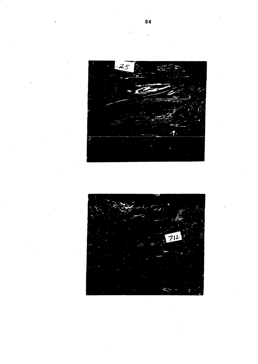
89 84
90 85 necropsied at the various intervals had normal lungs and tracheas. Examination of the turbinates of 11 pigs necropsied at 2 weeks postinoculation revealed that their ventral turbinates had normal dorso-ventral and medial-lateral dimensions and occupied the normal amount of space in the nasal cavities. However, some of these turbinates were ischemic in comparison with those from normal 2-week-old pigs and were distorted with longitudinal grooves and folds. Detailed information is presented in Table 6. Figures 15 and 16 demonstrate turbinates from a Bordetella bronchiseptica infected 2-week-old pig with distorted turbinates and from a normal 2-week-old pig respectively. Bordetella bronchiseptica was isolated in heavy growth from samples of nasal and tracheal exudate collected from all pigs necropsied at 1 through 5 weeks postinoculation. It was also isolated from the lungs of the 2 pigs with pneumonia. In addition, 1 pig at 1 week postinoculation, 1 pig at 2 weeks postinoculation and 1 pig at 3 weeks postinoculation had the organism in grossly normal lungs. Two of the 11 animals necropsied at 2 weeks postinoculation had small numbers of Bordetella bronchiseptica in their nasal cavities. None of these 11 pigs had the organism in their lungs or tracheas. The results of the bacteriologic examinations on all pigs in Trial II are recorded in Table 6. Plate and tube agglutination tests revealed that none of
91 Figure 15. Distorted ventral turbinate of a 2-week-old pig inoculated intranasally at 3 days of age with Bordetella bronchiseptica Isolate S-1 Figure 16. Normal turbinates of an uninoculated 2-week-old pig
92 87
93 88 the pigs necropsied from 1 through 5 weeks postinoculation developed agglutinating antibodies. Eight of 11 pigs necropsied at 2 weeks postinoculation had tube agglutination titers ranging from 1:2 to 1:8. Sera from the 2 control pigs necropsied at 2 weeks of age and 4 other uninoculated 2-weekold pigs were negative. These results are presented in Table 6.. Preinoculation sera collected from the 3 sows had tube agglutination titers of 1:8. Sera collected from these sows 4 weeks postinoculation also had titers of 1:8. Sera were collected from blood samples submitted from 15 different swine herds for routine leptospirosis plate agglutination tests. The animals ranged in age from 6 months to 4 years. Tube agglutination tests revealed that 24 of 35 animals from these herds had tube agglutination titers to Bordetella bronchiseptica Isolate S-1 ranging from 1:2 to 1:16. The results of bacteriologic examinations of samples of nasal exudate collected from the remaining live pigs after each necropsy interval are recorded in Table 7. Trial III Isolate S-2 (swine origin) Pigs inoculated with Bordetella bronchiseptica Isolate S-2 coughed for a limited period during the second week postinoculation. Sneezing was never prevalent. One pig died 2 weeks after inoculation. Necropsy of this pig revealed moderate turbinate atrophy and
94 89 Table 7. Number of live pigs positive on nasal exudate culture for Bordetella bronchiseptica at various intervals postinoculation Animals Weeks postinoculation ^ No. of inoculated pigs positive ^ 4^ 2^ No. of inoculated pigs sampled ^Samples collected at postmortem. ^Number of colonies markedly reduced. extensive pneumonia of the apical and cardiac lobes of the lungs. Postmortem examination of the other 3 pigs at 4 weeks postinoculation revealed slight to moderate bilateral turbinate atrophy and moderate involvement of the apical and cardiac lobes of the lungs with pneumonia. Culture of nasal exudate from all 4 pigs on modified MacConkey's agar and 5 percent equine blood agar revealed heavy growth of Bordetella bronchiseptica. No other significant organisms were isolated. Culture of tracheal exudate from these animals revealed heavy growth of the organism. It was also recovered from the pneumonic lungs of all 4 animals. Isolate S-3 (swine origin) Two pigs inoculated with Bordetella bronchiseptica Isolate S-3 started coughing about 1 days postinoculation and coughed with increasing frequency and severity until they were necropsied at 4 weeks
95 9 postinoculation. The other 2 pigs were not observed to cough and sneezing was never observed in any of the pigs. Necropsy revealed that 3 pigs had moderate bilateral turbinate atrophy and 1 pig had slight to moderate bilateral turbinate atrophy. Extensive pneumonia was observed in the apical, cardiac and diaphragmatic lobes of the lungs of 2 pigs. One pig had small areas of pneumonia in the apical and cardiac lobes while the fourth pig had normal lungs. Culture of nasal exudate from all 4 pigs on modified MacConkey's agar and 5 percent equine blood agar revealed heavy growth of Bordetella bronchiseptica. No other significant organisms were isolated. Bordetella bronchiseptica was isolated in heavy growth from the tracheal exudate of these pigs. It was also isolated from the pneumonic lungs of 3 pigs, but not from the normal lungs of the fourth pig. Isolate S-4 (swine origin) The pigs inoculated with Bordetella bronchiseptica Isolate S-4 coughed mildly about 1 days after inoculation. No evidence of respiratory disease was observed throughout the rest of this trial. Necropsy of these pigs revealed moderate to severe turbinate atrophy in 1 pig, slight to moderate bilateral turbinate atrophy in a second pig, slight to moderate unilateral turbinate atrophy in a third pig, and normal turbinates in the fourth pig. The lungs were normal in all 4 pigs. Culture of nasal exudate from these pigs on modified
96 91 MacConkey's agar and 5 percent equine blood agar revealed heavy growth of Bordetella bronchiseptica. No other significant organisms were isolated. Culture of tracheal exudate from these pigs revealed heavy growth of Bordetella bronchiseptica, Approximately 1 colonies of the organism grew from the normal lungs of 2 pigs. Culture of the lungs from the other 2 pigs did not yield the organism. Isolate S-5 (swine origin) The pigs inoculated with Bordetella bronchiseptica Isolate S-5 coughed mildly for 2 or 3 days about 1 days postinoculation. Necropsy revealed slight to moderate bilateral turbinate atrophy in all 4 pigs. One pig had extensive lesions of pneumonia. Two pigs had small lesions of pneumonia in the right apical lobe and the fourth pig had normal lungs. Bordetella bronchiseptica was isolated in heavy growth on modified MacConkey's agar and 5 percent equine blood agar from the nasal exudate of all 4 pigs. No other significant organisms were isolated. Culture of tracheal exudate from these pigs revealed heavy growth of Bordetella bronchiseptica. It was isolated from the pneumonic lungs of 3 pigs, but not from the normal lungs of the fourth pig. Isolate D-1 (canine origin) Isolate D-1 produced no coughing or sneezing. Necropsy revealed normal lungs and turbinates in all 4 pigs. Culture of nasal exudate from these animals on modified MacConkey's agar and 5 percent equine blood agar revealed
97 92 heavy growth of Bordetella bronchiseptica from only 1 pig. Culture of the nasal cavities of the other 3 pigs revealed relatively few colonies. One culture plate had 3 colonies, 1 had 1 colonies, while the third had 5 to 75 colonies. No other significant organisms were isolated. Bordetella bronchiseptica was isolated in limited numbers from 3 of the 4 tracheal exudate samples. The fourth sample was negative. The lungs of 3 of these pigs were negative for Bordetella bronchiseptica while the fourth lung was positive for the organism. Isolate Rb-1 (rabbit origin) Inoculation of Bordetella bronchiseptica Isolate Rb-1 produced a limited amount of sneezing and coughing about 7 to 1 days postinoculation. This lasted for about 1 week. Postmortem examination of these pigs at 4 weeks postinoculation revealed slight turbinate atrophy in 3 pigs and normal turbinates in 1 pig. Two pigs had moderate lesions of pneumonia in the dependent portion of the apical and cardiac lobes of their lungs. Culture of nasal exudate from these animals on modified MacConkey's agar and 5 percent equine blood agar revealed moderate numbers of colonies (5 to 1) of Bordetella bronchiseptica. No other significant organisms were isolated. Culture of tracheal exudate revealed heavy growth of the organism from all 4 animals. The lungs of 3 pigs were positive for Bordetella bronchiseptica and the lungs of the fourth pig were negative for the organism.
98 93 Isolate Ct-1 (cat origin) Pigs inoculated with Bordetella bronchiseptica Isolate Ct-1 started coughing 5 days after inoculation. This persisted for about 2 weeks. Sneezing was evident, but minimal. One pig died 12 days after inoculation. Necropsy of this pig revealed extensive lesions of pneumonia in the apical, cardiac and diaphragmatic lobes. Moderate turbinate atrophy was observed. Necropsy of the other 3 pigs at 4 weeks postinoculation revealed moderate turbinate atrophy in 1 pig and slight to moderate turbinate atrophy in the other 2 pigs. One pig had moderate lesions of pneumonia in the apical and cardiac lobes. A second pig had small areas of pneumonia in the cardiac lobes while the third pig had normal lungs. Culture of nasal exudate from all 4 pigs on modified MacConkey's agar and 5 percent bovine blood agar revealed heavy growth of Bordetella bronchiseptica. No other significant organisms were isolated. Culture of tracheal exudate from these pigs revealed heavy growth of Bordetella bronchiseptica. The pneumonic lungs of 3 pigs were positive for Bordetella bronchiseptica. The organism was isolated in heavy growth from the normal lungs of 1 pig. Isolate Rt-1 (rat origin) Inoculation of Bordetella bronchiseptica Isolate Rt-1 produced a limited amount of coughing about 7 days postinoculation. Sneezing was never observed and the coughing soon subsided. One pig died 2-1/2 weeks postinoculation. Necropsy revealed slight turbinate
99 94 atrophy and normal lungs. The pig was severely dehydrated as a result of a chronic catarrhal enteritis. Postmortem examination of the other 3 pigs at 4 weeks postinoculation revealed normal turbinates in all 3 animals. Two pigs had normal lungs and 1 pig had small areas of congestion in 1 apical lobe. Culture of nasal exudate from the pig that died revealed heavy growth of Bordetella bronchiseptica. Culture of nasal exudate from the other 3 pigs revealed moderate numbers (5 to 1) of colonies of the organism. Culture of these samples on 5 percent bovine blood agar revealed no other significant organisms. Bordetella bronchiseptica was isolated in heavy growth front the tracheas of 2 pigs. The trachea of the third pig was negative. Culture of the lungs from these three pigs revealed heavy growth of the organism from 1 pig and 1 colonies from 1 pig. The lungs of the third pig were negative. The gross lesions produced by the 8 isolates were quite similar. The main variation appeared to be in the severity of these lesions. The turbinates were ischemic, softened due to loss of bony core and atrophic. The ventral turbinates were most severely affected and atrophy of the ethmoid turbinates was observed in most pigs. The dorsal turbinates were also frequently mildly atrophic. The involvement was bilateral in almost all cases. Catarrhal exudate was observed infrequently on the nasal mucosa of these pigs. However, it was frequently observed in
100 95 tracheas. Pneumonia was quite extensive in some pigs. Lesions of subacute to acute pneumonia were reddish-brown while the chronic lesions were gray in color. These chronic lesions were quite firm and fibrous. Small areas of hemorrhage were observed in cross sections of these pneumonic areas. The results of this trial are summarized in Table 8. Turbinate atrophy produced by Isolates S-3 (swine origin) and S-4 (swine origin) are demonstrated in Figures 17 and 18 respectively. Turbinate atrophy produced by Isolate Ct-1 (cat origin) is demonstrated in Figures 19 and 2. Turbinate atrophy produced by Isolate Rb-1 (rabbit origin) is demonstrated in Figure 21. Normal turbinates from a pig inoculated with Isolate D-1 (canine origin) are demonstrated in Figure 22. The acute and chronic lesions of pneumonia produced by Bordetella bronchiseptica are demonstrated in Figures 23 and 24 respectively. Plate and tube agglutination tests were run on sera collected from all of the pigs in Trial III. In many instances these sera did not react or reacted very weakly with the S-1 antigen. The results of this work are recorded in Table 9. Biochemic, Metabolic and Morphologic Comparisons of Various Isolates of Bordetella Bronchiseptica The 8 isolates of Bordetella bronchiseptica used in Trial III were compared morphologically and by biochemic and metabolic tests. All 8 isolates (S-2, S-3, S-4, S-5, D-1,
101 Table 8. Incidence of turbinate atrophy, pneumonia and recovery of Bordetella bronchiseptica in pigs infected with eight different isolates of the organism Isolate S-2 S-3 S-4 S-5 D-1 Rb-1 Ct-1 Rt-1 Turbinate atrophy 4/4^ 4/4 3/4 4/4 /4 3/4 4/4 1/4 Pneumonia 4/4 3/4 /4 3/4 /4 2/4 3/4 2/4 Organism isolated-nasal exudate 4/4 4/4 4/4 4/4 4/4^ 4/4^ 4/4 4/4^ Organism isolated-tracheal exudate 4/4 4/4 4/4 4/4 3/4^ 4/4 4/4 3/4 Organism isolated-lungs 4/4 3/4 2/4 3/4 1/4 3/4 3/4 2/4 ^Numerator equals number of pigs positive. Denominator equals number of pigs in group. ^Colonies reduced in number.
102 Figure 17. Moderate turbinate atrophy 4 weeks postinoculation in a 31-day-old pig inoculated with Bordetella bronchiseptica Isolate S-3 (swine origin) Figure 18. Moderate turbinate atrophy 4 weeks postinoculation in a 31-day-old pig inoculated with Bordetella bronchiseptica Isolate S-4 (swine origin)
103 98
104 Figure 19. Moderate turbinate atrophy 12 days postinoculation in a 15-day-old pig inoculated with Bordetella bronchiseptica Isolate Ct-1 (cat origin) Figure 2. Moderate turbinate atrophy 4 weeks postinoculation in a 31-day-old pig inoculated with Bordetella bronchiseptica Isolate Ct-1 (cat origin)
105 1
106 Figure 21. Mild turbinate atrophy 4 weeks postinoculation in a 3i-day-old pig inoculated with Bordetella bronchiseptica Isolate Rb-1 (rabbit origin) Figure 22. Normal turbinates 4 weeks postinoculation in a 31-day-old pig inoculated with Bordetella bronchiseptica Isolate D-1 (canine origin)
107 12
108 Figure 23. Acute pneumonia 2 weeks postinoculation in a 17-day-old pig inoculated with Bordetella bronchiseptica Isolate S-2 Figure 24. Chronic pneumonia 4 weeks postinoculation in a 31-day-old pig inoculated with Bordetella bronchiseptica Isolate S-3
109 14
110 15 Table 9. Agglutination titers of sera collected from pigs inoculated with 8 different isolates of Bordetella bronchiseptica Isolate Pig No. Plate titer Tube titer S-2 (swine) S-3 (swine) S-4 (swine) S-5 (swine) D-1 (canine) Rb-1 (rabbit) Rt-1 (rat) Ct-1 (cat) 1 2 3
111 16 Rb-1, et-if and Rt-1) were small Gram-negative rods, sluggishly motile with peritrichous flagella and produced hemolysis on 5 percent bovine blood agar after incubation for 24 hours at 37 C. The zone of hemolysis increased in size after incubation at room temperature for an additional 24 hours. These isolates were citrate positive in 12 to 24 hours, urease positive in 2 to 12 hours and alkalinized litmus milk and dextrose in 24 to 48 hours. They did not produce indole or hydrogen sulfide. None of the isolates liquefied gelatin. They were all catalase and oxidase positive. Isolates Ct-1, Rt-1 and Rb-1 reduced nitrates to nitrites while the other 5 isolates did not. A second group of 55 isolates were compared. Twenty-two of these isolates were from the nasal cavities of the pigs sampled in Survey I. Fifteen isolates were from the nasal cavities of pigs submitted to the Iowa Veterinary Diagnostic Laboratory for various reasons. Eight isolates were from the nasal cavities of pigs from herds with known cases of Bordetella bronchiseptica rhinitis^. Another group of 8 isolates were isolated between 1954 and 1956 from the nasal cavities of pigs with rhinitis. These isolates had been initially identified as Alcaligenes fecalis-like organisms^. One isolate was recovered from a canine lung about Another isolate was recovered from a rabbit lung about ^Appreciation is expressed to Dr. W. P. Switzer who recovered these isolates.
112 17 These 55 isolates were all small Gram-negative rods. Fifty-four of the isolates were sluggishly motile. One was nonmotile. They were all citrate positive in 12 to 24 hours, urease positive in 2 to 12 hours and alkalinized litmus milk and dextrose in 24 to 48 hours. Fourteen of the 55 isolates reduced nitrates to nitrites. All of these isolates produced hemolysis on 5 percent horse, sheep and rabbit blood after 24 hours of incubation at 37 C. Incubation for an additional 24 hours at room temperature resulted in an increase in size of the zone of hemolysis on the horse blood agar. Eight of the 55 isolates produced hemolysis on 5 percent bovine blood agar. These 8 isolates were evenly distributed among the older and more recent isolates.
113 18 DISCUSSION Incidence Surveys Bordetella bronchiseptica, as indicated by the data obtained from the surveys of field swine, is widely distributed among Iowa swine herds. The herd incidence of Bordetella bronchiseptica, as well as that of Hemophilus spp. and Pasteurella multocida, in Surveys I, II, III and IV is summarized in Table 1. Table 1. Incidence of Bordetella bronchiseptica, Pasteurella multocida and Hemophilus spp. in the nasal cavities of Iowa swine Survey Bordetella Pasteurella Hemophilus ^ bronchiseptica multocida spp. Survey I (87 herds) No. herds positive Percent herds positive Survey II (87 herds) No. herds positive Percent herds positive Survey III (32 herds) No. herds positive Percent herds positive Survey IV (28 herds) No. herds positive Percent herds positive
114 19 Bordetelia bronchiseptica was found in 54 percent of 87 purebred swine herds sampled in Survey I. The animals sampled in this survey had been selected by their owners as being healthy, of good conformation and capable of better than average performance. Purebred pigs such as these which are sold as replacement breeding stock undoubtedly serve to disseminate Bordetella bronchiseptica among other purebred and commercial swine herds. Bordetella bronchiseptica was found in 38 percent of 87 herds sampled in Survey II. This lower incidence as compared to Survey I may be due to the fact that the average sample size in this survey was less than 2 pigs per herd. In addition, the pigs sampled in Survey II varied from 1 to 16 weeks of age while the pigs sampled in Survey I were 8 to 1 weeks of age. The incidence of turbinate atrophy in pigs necropsied in Survey II (26 percent of 124 pigs) was lower than that found _ in a similar survey conducted in the Iowa Veterinary Diagnostic Laboratory 13 years earlier. During a 6-week period Bennett (1952) found that 41.5 percent of 142 pigs submitted over 3 weeks of age had turbinate atrophy. The high incidence of Bordetella bronchiseptica in pigs with rhinitis is demonstrated by the results of Surveys III and IV. Most of the pigs sampled in both surveys were from herds with clinical signs of rhinitis. Additional evidence associating Bordetella bronchiseptica with turbinate atrophy
115 11 can be obtained by comparing the results of Surveys II and IV. The pigs sampled in Survey II were submitted to the Iowa Veterinary Diagnostic Laboratory for many reasons while the pigs sampled in Survey IV were submitted from herds specifically suspected of having rhinitis. In Survey II, 28 percent of the herds with Bordetella bronchiseptica had pigs with turbinate atrophy and in Survey IV, 68 percent of the herds with Bordetella bronchiseptica had pigs with turbinate atrophy. Reports from 2 other areas of the United States indicate that Bordetella bronchiseptica is frequently isolated from cases of atrophic rhinitis. In Indiana, Cross and Claflin (1962) isolated the organism from 9 of 1 pigs submitted from 1 different herds with atrophic rhinitis. A diagnostic laboratory in Georgia recently reported that the organism was frequently isolated from the nasal cavities of pigs with rhinitis (Bedell and Miller, 1964). The presence of Pasteurella multocida in approximately 5 percent of the herds sampled is in agreement with reports by several other workers. Radtke (1938) found this organism infrequently in the nasal cavities of German swine. Shuman et al. (1953) found Pasteurella multocida in the nasal cavities of only 6 of 55 pigs in a herd with rhinitis. Heddleston et al. (1954) isolated the organism from the nasal cavities of 8 percent of the pigs with rhinitis and 4.3 percent of the normal pigs in 2 herds with atrophic rhinitis. Olander (1961) isolated Pasteurella-like organisms from the
116 Ill nasal cavities of 5 of 69 slaughter-weight pigs in California. These findings are not in agreement with reports by Canadian workers that Pasteurella multocida was frequently isolated from the porcine nasal cavity. MaKay (cited by Schofield and Jones, 195) found Pasteurella multocida in the nasal cavities of 65 of 75 pigs with atrophic rhinitis. Jones (1952) observed that the organism was rarely absent from the early lesions of atrophic rhinitis and Gwatkin et al. (1953) reported that in 186 bacteriologic examinations this organism was present in the nasal cavities of 38 percent of the pigs with turbinate atrophy and 16 percent of the pigs with normal turbinates. The results of studies on Canadian swine and limited observations by the author indicate that a high percentage of the pigs in some herds have nasal infection with Pasteurella multocida. Such pigs have well defined clinical signs of rhinitis and may remain infected for extended periods of time. Strains of Pasteurella multocida involved in such cases appear to have an unusual affinity for the nasal cavity. Possibly most strains of the organism have a lower affinity for the nasal cavity and are recovered from that area because of continual reinfection from pneumonic lungs. Pneumonia was observed in 43 percent of the pigs in Survey II. Culture of the pneumonic lungs of 35 of these pigs revealed that 1 had Pasteurella multocida and 2 had Bordetella bronchiseptica. Culture of the nasal cavities of
117 112 the same pigs revealed that 1 had Bordetella bronchiseptica and 5 had Pasteurella multocida. These findings indicate that Bordetella bronchiseptica is more common in the swine nasal cavity and Pasteurella multocida is more common in the pneumonic swine lung. This is in agreement with L'Ecuyer et al, (1961) who found Pasteurella multocida and Bordetella bronchiseptica in 4 percent and 1 percent respectively of 86 pneumonic swine lungs. Similar findings on the incidence of these 2 organisms in pneumonic swine lungs were reported by Spray (1922), Thorp and Tanner (194), and Morcos et al. (1947). Investigations in this laboratory indicate that approximately 5 percent of Iowa swine have pneumonia when slaughtered. The high incidence of Pasteurella multocida in pneumonic swine lungs and the high incidence of pneumonia in swine indicate that this organism is extremely common on a herd basis in Iowa swine. The finding in Survey I that Hemophilus spp. were present in 48 percent of the herds is in reasonable agreement with Radtke (1938) who found that 6 to 7 percent of German swine harbored the organism in their nasal cavities. Braend and Flatla (1954) found these organisms in the nasal cavities of many Norwegian swine. Olander (1961) isolated Hemophilus spp. from the nasal cavities of only 6 of 69 market-age swine in California. Hemophilus spp. were isolated from 23 percent, 16 percent
118 113 and 11 percent of the herds in Surveys II, III and IV respectively. The lower incidence could be a reflection of variability in age of pigs sampled and small sample size in comparison to Survey I. <1 Transmission Trials The clinical appearance of sneezing correlated to a high degree with the presence of catarrhal exudate on the nasal mucosa of experimentally infected pigs. In general, sneezing and catarrhal exudate were observed from the fifth day to the second or third week postinoculation. Sneezing was rarely observed after 3 weeks postinoculation in any of the trials and the surface of the nasal mucosa appeared to have a normal coating of secretions in most pigs necropsied after this period. However, culture of these secretions routinely revealed abundant growth of Bordetella bronchiseptica until after the twelfth week postinoculation. In some cases of natural Bordetella bronchiseptica rhinitis, sneezing and catarrhal rhinitis were observed in pigs up to 6 or 8 weeks of age. Possibly these pigs were infected at a later age and were in the acute stages of infection at the time of necropsy. This is quite conceivable since weaning and pooling of larger groups of pigs commonly occurs at this age in field swine. In herds with only a few carrier sows, many pigs might remain uninfected until they came into contact with other infected pigs at weaning time.
119 114 Observations made in this laboratory indicate that this frequently occurs. A higher incidence of sneezing was observed in pigs inoculated at 4 weeks of age in Trial I than in pigs inoculated at 3 days of age in Trials II and III. These results and similar observations made in other trials in this laboratory indicate that the sensory apparatus of the nasal mucosa of the 4-week-old pig may be better developed than that of the 3-day-old pig. Additional support for this concept is gained from observations made by the author that sneezing associated with Bordetella bronchiseptica, Pasteurella multocida or Hemophilus spp. infection in 1- to 3-week-old field pigs was relatively mild as compared with that seen in older pigs from which these organisms were isolated. The increased incidence of sneezing in weanling pigs affected with atrophic rhinitis is generally attributed to increased severity of the lesions. However, it is possible that increased sensitivity to irritation also contributes to this increase in sneezing. In Trial I significant turbinate atrophy was not detected until 3 weeks postinoculation. In Trial II moderate turbinate atrophy was observed in all 3 pigs necropsied at 2 weeks postinoculation. Similar lesions were observed in 3 pigs that died about 2 weeks postinoculation in Trial III. In addition, in Trial II the lesions became progressively more severe from the second week to the fifth week postinoculation when 7 of 1 pigs necropsied had severe turbinate atrophy. On the other
120 115 hand, in Trial I the older pigs appeared to resist the effects of the infection si'nce neither the incidence nor the severity of the lesions increased from the third week to thé eighth week postinoculation. These findings are consistent with the concept that older swine are less susceptible to atrophic rhinitis than younger swine (Jones, 1947). Turbinate atrophy observed in pigs experimentally infected with Bordetella bronchiseptica was as severe as that observed in the majority of field pigs with atrophic rhinitis. In many cases of atrophic rhinitis in field pigs, Bordetella bronchiseptica was isolated in abundant growth with virtually no other bacteria detected on modified MacConkey's agar or 5 percent bovine blood agar. In all respects, the field disease closely simulated the experimental disease. The results of Trial III indicate that many isolates of Bordetella bronchiseptica are pathogenic for young pigs. The ability of isolates of the organism from a rabbit, a cat and a rat to cause rhinitis and pneumonia in pigs clearly shows that these animals may serve to infect swine herds with the organism. Rats, cats and dogs are frequently in close habitation with swine in confinement. Reports by several workers have implicated rats, cats, dogs and rabbits as carriers of atrophic rhinitis (Thunberg and Carlstrom, 1946; Jones, 1947; Schofield, 1955; Gwatkin and Dzenis, 1955; Andrews et al., 1957). Pasteurella multocida was present in the material transmitted from the various species to swine in some cases.
121 116 However, it is quite possible that Bordetella bronchiseptica was also present. Culture of the nasal exudate of pigs necropsied at various intervals up to 12" weeks postinoculation revealed large numbers of Bordetella bronchiseptica. From the twelfth to the sixteenth week postinoculation, the number of organisms recovered from each of 11 pigs in Trial II decreased. By the twentieth week postinoculation, only 2 of the 11 pigs harbored the organism in their nasal cavities. Switzer (1963) made similar observations on the elimination of the organism from the nasal cavities of swine as they matured. Samples of nasal exudate collected from live pigs at various intervals were usually positive for the organism. However, all samples collected from 15 pigs at 7 days postinoculation were negative in Trial I. Samples collected from 3 penmates at necropsy were positive. It is suspected that the organism was not disseminated throughout the nasal cavity and, as a result, the sampling technique was inadequate. Intranasal inoculation of 3 pregnant sows in Trial II did not result in infection. These sows were constantly exposed to infected pigs, but remained negative when samples of nasal exudate were cultured. These animals were not necropsied, so it is possible that more exhaustive sampling of the nasal secretions might have detected the organism. It has been found that some naturally infected sows harbor the organism in only 1 nasal passage (Switzer, 1963).
122 117 The elimination of the organism from the nasal cavity and the apparent regeneration which occurred in Trial II indicate that some change occurred in the host or the organism. This subsequently resulted in the elimination of the organism from a high percentage of animals which were previously quite heavily infected. The factor responsible for elimination of Bordetella bronchiseptica from the nasal cavity could be specific antibody, a nonspecific resistance factor such as an increase in lysozyme or some other inhibitory factor liberated in the nasal secretions, or it could be a mutation or phase change in the organism. It seems doubtful that circulating antibody would have any appreciable effect on nasal infection in swine with Bordetella bronchiseptica since high titers of circulating antibody in rabbits have been shown to be without effect on nasal infection with the organism (Bailey, 1927). In addition, the circulating antibody titers in infected pigs were quite low. It is more conceivable that a local antibody or resistance factor is produced by the mucosal cells of the nasal cavity. The possibility of a phase change in the organism should be considered since Lacey (1953) has shown that changes in growth temperature and growth medium composition can induce such changes in this organism. Organisms altered in their growth characteristics in this manner might find the environment in the maturing swine nasal cavity less desirable. In some instances, rough colony forms were
123 118 observed in primary cultures from infected pigs. The significance of these is not known. Bordetella bronchiseptica was isolated in heavy growth from the tracheal secretions of all pigs necropsied in Trial II from 1 through 5 weeks postinoculation. In Trial I the pigs began eliminating the organism from the trachea about 4 weeks postinoculation. Culture of tracheal exudate from 3 pigs necropsied 8 weeks postinoculation in this trial revealed no Bordetella bronchiseptica. In all 3 trials coughing was observed in the inoculated pigs. Lesions of pneumonia were observed in some of these pigs; however, necropsy of the pigs in Trial I as well as many pigs in Trials II and III which had been observed to cough revealed no pneumonia. It was observed that pigs with pneumonia had coughed more frequently and for a longer duration. Catarrhal exudate was observed in the tracheas of most pigs and the organism was consistently recovered from this exudate up to 4 to 5 weeks postinoculation. The presence of this organism in the tracheas and bronchi of infected pigs apparently caused irritation with resultant coughing. Since very little evidence of inflammation was observed in the tracheas of these pigs, it appears that the organism was liberating a toxic substance which then caused the coughing. It seems reasonable that the mechanism may be similar to that seen in whooping cough in man. Bordetella bronchiseptica can survive in apparently
124 119 normal swine lungs for a considerable time. Four pigs necropsied at 4 different intervals (1 week, 2 weeks, 3 weeks, and 6 weeks postinoculation) in Trial I and 3 pigs necropsied at 3 different intervals (1 week, 2 weeks, and 5 weeks postinoculation) in Trial II had the organism in apparently normal lung tissue. In Trial III, 2 pigs inoculated with Isolate S-4, 1 pig inoculated with Isolate Ct-1, and 1 pig inoculated with Isolate Rt-1 had the organism in normal lung tissue at 4 weeks postinoculation. The presence of this organism in these normal lungs is undoubtedly associated with heavy growth of the organism in the trachea and bronchi. Stress or exposure could probably precipitate a severe pneumonia in such animals. The work of Phillips (1943), Ray (195, 1959), L'Ecuyer (1961), Dunne et al. (1961), and Goodwin and Whittlestone (1964) indicate that this frequently occurs. These reports also indicate that Bordetella bronchiseptica has been widely distributed among swine for many years. The organism was isolated from the nasal cavities of pigs with a rhinitis in Illinois almost 25 years ago (Daugherty, 1941). The apparent failure-of many investigators to associate this organism with atrophic rhinitis may be due to several factors. The organism is slow to develop on culture media and is frequently overgrown by other organisms that are common in the porcine nasal cavity. The organism is frequently not detected unless cultures are incubated at least 48 hours.
125 12 The organism was confused with Alcaligenes fecalis in earlier work (Switzer, 1956; Claflin, 1958). A third possibility is that culture of nasal exudate was not made from pigs during the early stages of infection. In advanced cases of turbinate atrophy this organism is frequently crowded out by other bacteria. The presence of agglutinating antibodies in preinoculation serum samples from the 3 sows used in Trial II raised doubt about the specificity of the agglutination test employed. The herd from which these sows were obtained has been repeatedly negative for Bordetella bronchiseptica on culture of nasal secretion and the sows themselves were negative for the organism in all preinoculation and postinoculation nasal exudate cultures. The prevalence of low levels of agglutinating antibodies in sera collected from field swine of various ages indicated that these antibodies are quite common in adult swine. Possibly the antibody detected in the sera from these sows was present due to antigenic stimulation by some organism with antigens common to Bordetella bronchiseptica. Antibody titers of 1:64 that developed by the eighth week postinoculation in Trial I probably were specific since uninoculated controls did not develop these titers. Although further elucidation of the value of the tests employed in this work is needed, it appears that titers of 1:32 or higher may be significant while those below should be disregarded in view of
126 121 the possibility of nonspecificity. The delayed development of these antibodies is similar to the appearance of antibodies in pertussis in humans. La"trop and Lacey (196) state that in Bordetella pertussis infections in man, specific antibodies appear in the blood between the third and fourth week and reach a maximum about the seventh to eighth week of the disease.
127 122 SUMMARY The incidence of Bordetella bronchiseptica in Iowa swine was studied in 4 different surveys. The organism was isolated from the nasal cavities of pigs in 54 percent of 87 purebred swine herds in Survey I. The herd incidence of the organism was 38 percent in a second survey of 87 commercial and purebred herds. Bordetella bronchiseptica was found in a higher percentage of herds with clinical signs of rhinitis in Surveys III and IV. The organism was isolated from 56 percent (18/32) and 68 percent (19/28) of the herds in Surveys III and IV respectively. Hemophilus spp. and Pasteurelia multocida were found in 48 percent and 5 percent of 87 purebred herds respectively. Comparable findings on the incidence of these organisms were made in the other 3 surveys. Intranasal inoculation of Bordetella bronchiseptica in 4-week-old pigs resulted in turbinate atrophy in 1 of 15 pigs necropsied from 2 to 8 weeks postinoculation. Intranasal inoculation of the organism in 3-day-old suckling pigs resulted in turbinate atrophy in 15 of 16 necropsied from 2 to 5 weeks postinoculation. Randomly selected littermates necropsied at 2 weeks postinoculation had regenerated turbinates that occupied the normal amount of space in the nasal cavity. The organism was recovered in abundant growth from
128 I 123 the nasal exudate of all experimentally infected pigs up to 12 weeks postinoculation. It gradually disappeared from the nasal cavities of the older pigs so that only 2 of 11 pigs had the organism in their nasal cavities at 2 weeks of age. Four different isolates of Bordetella bronchiseptica recovered from nasal exudate of pigs from herds with atrophic rhinitis produced moderate turbinate atrophy in box-reared pigs inoculated at 3 days of age. Similar pigs inoculated with isolates of the organism recovered from tracheal exudate of a cat, tracheal exudate of a rat and nasal exudate of a rabbit also developed turbinate atrophy. One isolate of Bordetella bronchiseptica recovered from nasal exudate of a puppy with canine distemper produced no evidence of respiratory disease in these box-reared pigs. Bordetella bronchiseptica was isolated in heavy growth from the tracheal exudate of many of the experimentally infected pigs; Its presence in the trachea and bronchi frequently elicited coughing during the early stages of infection. Many of the box-reared pigs also developed pneumonia. The organism was isolated from the normal lung tissue in about 15 percent of the inoculated pigs. Survival in this tissue for extended periods of time is probably associated with growth of the organism on the tracheal and bronchial mucosa. There is undoubtedly a very close relationship between the presence of this organism in the trachea and bronchi and the severe pneumonia caused by the organism in experimentally and
129 124 naturally infected pigs. Sixty-three isolates of Bordetella bronchiseptica were found to be identical in their ability to produce urease, alkalinize litmus milk and dextrose and utilize citrate. Seventeen of the 63 isolates reduced nitrates to nitrites. The agglutinating antibody response of pigs infected with Bordetella bronchiseptica is relatively protracted and weak. Nonspecific agglutinating antibodies in low titer appear to be quite common in adult swine.
130 125 BIBLIOGRAPHY Andersen, E. K Serological studies on H. pertussis, H. parapertussis and H. bronchisepticus. Acta Path, et Microbiol. Scand. 33: Andrews, J. S., Spindler, L. A., Earl, F. L. and Diamond, L. S Atrophic rhinitis. VIII. The albino rat as an experimental carrier. Proc. U.S. Livestock San. Ass. 6: Bacon, M., Bacon, R. F. and Drake, C. H Bacterial infections in wild rodents of eastern and central Washington. J. Infect. Dis. 12: Bailey, G. H Respiratory immunity in rabbits. I. Systemic reactions in rabbits which are nasal carriers of Bacterium lepisepticum, Bacillus bronchisepticus, or Micrococcus catarrhalis. Amer. J. Hyg. 7: Banerjea, A. and Munoz, J Antigens of Bordetella pertussis. II. Purification of heat-labile toxin. J. Bact. 84: Bedell, D. M. and Miller, J. G Tifton diagnostic laboratory notes. Georgia Veterinarian 16, No. 1: Jan.-Feb. Beer, J. 196a. Uber das Vorkommen and uber die rolle von Bordetella (B) bronchiseptica bei lungenerkrankungen einiger Haus-und laboratoriumstiere. Zentralbl. Bakt. Para. Infek. Hyg. I. Orig. 177: Beer, J. 196b. Zur differenzierung von Bordetella (B.) bronchiseptica unter besonderer Berucksichtigung der Abgrenzung von Alcaligenes faecalis. Zentralbl. Bakt. Para. Infek. Hyg. I. Orig. 177: Bennett, P. C Some angles on atrophic rhinitis. Proc. U.S. Livestock San. Ass. 55: Bergey, D. H., ed Bergey's manual of determinative bacteriology. 1st ed. Baltimore, Maryland. Williams and Wilkins Company. Bergey, D. H., Breed, R. S., Murray, E. G. D. and Hitchens, A. P., eds Bergey's manual of determinative bacteriology. 5th ed. Baltimore, Maryland. Williams and Wilkins Company.
131 126 Betts, A. O Respiratory diseases of pigs. V. Some clinical and epidemiological aspects of virus pneumonia of pigs. Vet. Rec. 64: Billaudelle, H., Edebo, L., Hammarsten, E., Hedén, C. -G., Malmgren, B. and Palmstierna, H Studies on the chemical and immunological structure of Bordetella pertussis. Acta Path, et Microbiol. Scand. 5: Borden, D. and Kulp, W. L A study of rat pneumonia. J. Bact. 37: Bordet, J. and Gengou, Le microbe de la coqueluche. Ann. Inst. Pasteur 2: ' Bradford, W. L. and Slavin, B An organism resembling Hemophilus pertussis. Amer. J. Publ. Health 27: Braend, M. and Flatla, J. L Noen undersofkeler over Haemophilus suis. Nord. Vet. Med. 6: Breed, R. S., Murray, E. G. D. and Kitchens, A. P., eds Bergey's manual of determinative bacteriology. 6th ed. Baltimore, Maryland. Williams and Wilkins Company. Breed, R. S., Murray, E. G. D. and Smith, N. R., eds Bergey's manual of determinative bacteriology. 7th ed. Baltimore, Maryland. Williams and Wilkins Company. British Medical Research Council The prevention of whooping-cough by vaccination. Brit. Med. J. 1: Brown, J. H Bacillus bronchisepticus infection in a child with symptoms of pertussis. Bull. Johns Hopkins Hosp. 38: Bruckner, I. E. and Evans, D. G The toxin of B. parapertussis and the relationship of this organism to H. pertussis and Br. bronchiseptica. J. Path, and Bact. 45": Bull, C. G. and Bailey, G. H The effect of antiseptics on the nasal flora of rabbits. Amer. J. Hyg. 7: Bull, C. G. and McKee, C. M Respiratory immunity in rabbits. VI. The effects of immunity on the carrier state of the pneumococcus and Bacillus bronchisepticus. Amer. J. Hyg. 8:
132 127 Carpenter, P. L Immunology and serology. Philadelphia, Pennsylvania. W. B. Saunders Company. Claflin, R. M Studies on the etiology and pathology of atrophic rhinitis of swine. [Microfilm Copy , unpublished Ph.D. thesis, Lafayette, Indiana, Purdue University]. Ann Arbor, Michigan. University Microfilms. Cross, R. F. and Claflin, R. M Bordetella bronchiseptica-induced porcine atrophic rhinitis. J. Amer. Vet. Med. Ass. 141: Daugherty, W. D Respiratory diseases of swine with special reference to bull nose in pigs. North Amer. Veterinarian 22; Done, J. T An "inclusion body" rhinitis of pigs. Vet. Rec. 67: Dorset, M., McBryde, C. N. and Niles, W. B Remarks on "hog flu". J. Amer. Vet. Med. Ass. 62: Doyle, L. P., Donham, C. R. and Hutchings, L. M Report on a type of rhinitis in swine. J. Amer. Vet. Med. Ass. 15: Dulbecco, R. and Vogt, M Plaque formation and isolation of pure lines of poliomyelitis virus. J. Exptl. Med. 99: Dunne, H. W., Kradel, D. C., Doty, R. B Bordetella bronchiseptica (Brucella bronchiseptica) in pneumonia in young pigs. J. Amer. Vet. Med. Ass. 139: Edwards, J. T. G The European hedgehog. In Worden, A. N. and Lane-Petter, W., eds. U.F.A.W. handbook on the care and management of laboratory animals. 2nd ed. pp London, England. Universities Federation for Animal Welfare. Ehrich, W. E., Bondi, A., Jr., Mudd, S., and Flosdorf, E. W The tolerance of rabbits for the agglutinogen and the toxins of Hemophilus pertussis. Amer. J. Med. Sci. 24: Eldering, G A study of the antigenic properties of Hemophilus pertussis and related organisms. I. A factor obtained from Br. bronchiseptica. Amer. J. Hyg. 34: 1-7. Eldering, G A study of the antigenic properties of Hemophilus pertussis and related organisms. II. Protection tests in mice. Amer. J. Hyg. 36:
133 128 Eldering, G,, Hornbeck, C. and Baker, J Serological study of Bordetella pertussis and related species. J. Bact. 74: Eldering, G. and Kendrick, P Bacillus parapertussis. J. Bact. 35: Evans, A. C Further studies on Bacterium abortus and related bacteria. II. A comparison of Brucella abortus with Bacterium bronchisepticus and with the organism which causes Malta fever! J. Infect. Dis. 22: Evans, D. G The production of pertussis antitoxin in rabbits and the neutralisation of pertussis, parapertussis and bronchiseptica toxins. J. Path, and Bact. 51; Evans, D. G. and Maitland, H. B The toxin of Br. bronchiseptica and the relation of the organism to H. pertussis. J. Path, and Bact. 48: Farkas-Himsley, H Differentiation of Pasteurella pseudotuberculosis and Bordetella bronchiseptica by simple biochemical tests. Amer. J. Vet. Res. 24: Ferry, N. S A preliminary report of the bacterial findings in canine distemper. Amer. Vet. Rev. 37: Ferry, N. S Etiology of canine distemper. J. Infect. Dis. 8; Ferry, N. S. 1912a. Further studies on the Bacillus bronchicanis, the cause of canine distemper. Amer. Vet. Rev. 41: Ferry, N. S. 1912b. Bacillus bronchisepticus (bronchicanis). Brit. Vet. J. 68: Ferry, N. S "Bacillus bronchisepticus". Amer. Vet. Rev. 43: Ferry, N. S Bacteriology and control of acute infections in laboratory animals. J. Path, and Bact. 18: Ferry, N. S. and Hoskins, H. P Bacteriology and control of contagious nasal catarrh (snuffles) of rabbits. J. Lab. Clin. Med. 5: Ferry, N. S. and Klix, H. C Studies relative to the apparent close relationship between Bact. pertussis and B. bronchisepticus. II. Complement fixation tests. J. Bact. 3:
134 129 Ferry, N. S. and Noble, A Studies relative to the apparent close relationship between Bact. pertussis and B. bronchisepticus. I. Cultural agglutination and absorption reactions. J. Bact. 3; Flosdorf, E. W., Dozois, T. F. and Kimball, A. C Studies with H. pertussis. V. Agglutinogenic relationships of the phases. J. Bact. 41: Flosdorf, E. W. and Kimball, A. C Separation of the phase I agglutinogen of H. pertussis from toxic components. J. Immunol. 39: Gaby, W. L. and Free, E Differential diagnosis of pseudomonas-like microorganisms in the clinical laboratory. J. Bact. 76: Galli-Valerio, B Der mikroorganimus der hundestaupe. Zentralbl. Bakt. Para. Infek. Hyg. I. Orig. 19: 694. Genest, P Isolement, chez le porcelet, d'alcaligenes bronchisepticus. Canad. J. Comp. Med. and Vet. Sci. SI Goodwin, R. F. W. and Whittlestone, P A respiratory disease of pigs (Type XI) differing from enzootic pneumonia. [II.] The field disease. J. Comp. Path, and Therap. 72: Goodwin, R. F. W. and Whittlestone, P The differential diagnosis of respiratory diseases in the pig. Proc. 17th World Vet. Congress 2: Goodwin, R. F. W. and Whittlestone, P A respiratory disease of pigs (Type XI) differing from enzootic pneumonia. [I.] Natural transmission experiments. J. Comp. Path, and Therap. 74: Greig, A. S The significance of a pleuropneumonialike organism in kennel cough. Canad. J. Comp. Med. and Vet. Sci. 18: Griffin, C. A Bacterial diseases of common laboratory animals. Proc. Animal Care Panel 6: Gwatkin, R. and Dzenis, L Rhinitis of swine. VIII. Experiments with Pasteurella multocida. Canad. J. Comp. Med. and Vet. Sci. 17: c
135 13 Gwatkin, R. and Dzenis, L Rhinitis of swine. X. Further experiments with laboratory animals. Canad. J. Comp. Med. and Vet. Sci. 19: Gwatkin, R., Dzenis, L. and Byrne, J. L Rhinitis of swine. VII. Production of lesions in pigs and rabbits with a pure culture of Pasteurella multocida. Canad. J. Comp. Med. and Vet. Sci. 17: Hagen, K. W Enzootic Pasteurellosis in domestic rabbits. I. Pathology and bacteriology. J. Amer. Vet. Med. Ass. 133: Hagan, W. A. and Bruner, D. W The infectious diseases of domestic animals. Ithaca, New York. Cornstock Publishing Associates. Heddleston, K. L., Shuman, R. D. and Earl, F. L Atrophic rhinitis. IV. Nasal examination for Pasteurella multocida in two swine herds affected with atrophic rhinitis. J. Amer. Vet. Med. Ass. 12f,: Jones, T. L Rhinitis in swine. Agri. Inst, Rev. 2: Jones, T. L Streptomycin and other agents used in infectious atrophic rhinitis of swine. J. Amer. Vet. Med. Ass. 121: Joubert, L., Courtieu, A. -L. and Oudar, J Pneumonie enzootique du porc à Bordetella bronchiseptica. Bull. Soc. Sci. Vet. Lyon 62: Keegan, J. J The pathology of epidemic pneumonia in mice and guinea pigs. Arch. Int. Med. 26: Kendrick, P. L., Nadolski, E. B., Eldering, G. and Baker, J Antigenic relationships of Haemophilus pertussis, the parapertussis bacillus and Brucella bronchiseptica as shown by cross protection tests in mice. J. Bact. 66: Keogh, E. W., North, E. A. and Warburton, M. F Haemagglutinins of the Haemophilus group. Nature 16: 63. Kobayashi, S Studies on the hemolytic factor of Bordetella bronchiseptica. Jap. J. Bact. 16: Kyaw, M. H Note on a bacterial cause of sterility in the rabbit. Vet. Rec. 57:
136 131 Labaw, L. W. and Mosley, V. M Periodic structure in the flagella of Brucella bronchiseptica. Biochim. et Biophys. Acta 17: Lacey, B. W. 1953a. Three dimensional patterns of antigenic modulation of Haemophilus pertussis, H. parapertussis and H. bronchisepticus. Proc. Soc. Gen. Microbiol., 1952, in J. Gen. Microbiol. 8l Tii. Lacey, B. W. 1953b. The influence of growth conditions on the antigenic structure of Haemophilus pertussis, parapertussis and bronchisepticus. Int. Cong. Microbiol., 6th, Rome, 1953, Riassunti delle communicazioni 2: Lacey, B. W. 1953c. On the serology and taxonomy of Haemophilus pertussis, parapertus sis and bronchisepticus. Int. Cong. Microbiol., 6th Rome, 1953, Riassunti delle communicazioni 2: Lautrop, H. and Lacey, B. W Laboratory diagnosis of whooping-cough or Bordetella infections. Bull. World Health Org. 23: L'Ecuyer, C., Roberts, E. D. and Switzer, W. P. 1961a. An outbreak of Bordetella bronchiseptica pneumonia in swine. Vet. Med. 56: L'Ecuyer, C., Switzer, W. P. and Roberts, E. D. 1961b. Microbiologic survey of pneumonic and normal swine lungs. Amer. J. Vet. Res. 22: Leifson, E Atlas of bacterial flagellation. New York, N.Y. Academic Press, Inc. Leslie, P. H. and Gardner, A. D The phases of Haemophilus pertussis. J. Hyg. 31: Lockhart, A Some phases of canine distemper. J. Amer. Vet. Med. Ass. 7: Maclennan, A. P Specific lipopolysaccharides of Bordetella. Biochem. J. 74: Man, C. S Pertussis due to Brucella bronchiseptica. Pediatrics 6: Martini, E. 19. Ein gelegentlicher, durch inhalation ûbertragbarer erreger der lungenentzûndung bei meerschweinchen. Bacillus pulmonum glutinosus. Arch. Hyg. 38:
137 132 Masry, F. L. G Production, extraction and purification of the haemagglutinin of Haemophilus pertussis. J. Gen. Microbiol. 7: McGowan, J. P Some observations on a laboratory epidemic, principally among dogs and cats, in which the animals affected presented the symptoms of the disease called "distemper." J. Path, and Bact. 15: Merchant, I. A. and Packer, R. A Veterinary bacteriology and virology. 6th ed. Ames, Iowa. Iowa State University Press. Morcas, Z., Zaki,. A. and Zaki, R Bacteriological study of swine pneumonia in Egypt. Vet. Med. 42: Moreno-Lopez, M El Genero Bordetella. Microbiol. Espanola. 5: 1-5. Mosier, J. E A preliminary report on the use of nitrofurantoin in treatment of canine tracheobronchitis (Kennel cough). Vet. Med. 5: Moynihan, I. W Rhinitis of swine. II. An effort to transmit chronic atrophic rhinitis of swine. Canad. J. Comp. Med. and Vet. Sci. 11: Munoz, J., Ribi, E. and Larson, C. L Antigens of Bordetella pertussis. I. Activities of cell walls and protoplasm. J. Immunol. 83: Munoz, J. J Symposium on relationship of structure of microorganisms to their immunological properties. I. Immunological and other biological activities of Bordetella pertussis antigens. Bacteriological Rev. 27: Nelson, J. B Infectious catarrh of the albino rat. I. Experimental transmission in relation to the role of Actinobacillus muris. J. Exptl. Med. 72: Nelson, J. B The problems of disease and quality in laboratory animals. J. Med. Educ. 35: Oddy, J. G. and Evans, D. G The effects produced by toxic and nontoxic extracts of H. pertussis and bronchiseptica on the blood sugar of rabbits. J. Path, and Bact. 5:
138 133 Olander, H. J A septicemic disease of swine and its causative agent, Haemophilus parahaemolyticus. [Microfilm Copy C.U.D. 15, unpublished Ph.D. thesis, Davis, California, University of California]. University of California, Berkeley, California. Library Photographic Service. Ostler, D. C The diseases of broiler rabbits. Vet. Rec. 73: Phillips, C. E Alcali^enes (Brucella) bronchisepticus as a factor in porcine pneumonias. Canad. J. Comp. Med. and Vet. Sci. 7: Phillips, C. E Pathogenic organisms complicating haemorrhagic septicemia in swine. Canad. J. Comp. Med. and Vet. Sci. 8: Phillips, C. E Infectious rhinitis in swine (bull nose). Canad. J. Comp. Med. and Vet. Sci. 1: Pinkerton, H Immunological and histological studies on mink distemper. J. Amer. Vet. Med. Ass. 96: Proom, H The minimal nutritional requirements of the genus Bordetella Lopez. J. Gen. Microbiol. 12: Radtke, G Untersuchunger liber die Ursache und das Wesen der Schnuffelkrankheit der schweines. Arch. wiss. prakt. Tierheilk. 72: Ratcliffe, H. L Infectious diseases of laboratory animals. Ann. N.Y. Acad. Sci. 46: Rauch, H. C. and Pickett, M. J Bordetella bronchiseptica bacteriophage. Canad. J. Microbiol. 71 ÏY Ray, J. D Brucella bronchisepticus. J. Amer. Vet. Med. Ass. 116: 51. Ray, J. D Respiratory problems in swine. J. Amer. Vet. Med. Ass. 134: Repentigny, J. de, and Frappier, A Studies on H. pertussis liquid cultures. III. Localization of surface antigens by means of fluorescent antibody. Canad. J. Microbiol. 2: Rhea, L. J The comparative pathology of the tracheal and bronchial lesions produced in man by B. pertussis (whooping-cough) and those produced in dogs by B. bronchisepticus (canine distemper). J. Med. Res. 32:
139 Roberts, E. D., Switzer, W. P. and L'Ecuyer, C. 1962, Influence of Pasteurella multocida and Mycoplasma hyorhinis (PPLO) on the histopathology of field cases of swine pneumonia. Cornell Veterinarian 52: Rosen, S., Hunt, H. R., and Benarde, M. A Oxytetracycline (Terramycin) as a therapeutic agent for pneumonia caused by Brucella bronchiseptics in rats. J. Amer. Vet. Med. Ass. 124: 3. Rosenow, E. C The relation of streptococci to the filterable virus of encephalitis of the fox. J. Infect. Dis. 48: Ross, R. F Methods of identification of certain etiologic agents of atrophic rhinitis. Proc. U.S. Livestock San. Ass. 67: Ross, R. F., Duncan, J. R. and Switzer, W. P. 1963a. Turbinate atrophy in swine produced by pure cultures of Bordetella bronchiseptica. Vet. Med. 58: Ross, R. F., Switzer, W. P. and Mare, C. J. 1963b. Incidence of certain microorganisms in Iowa swine. Vet. Med. 58: Rowatt, E Some factors affecting the growth of Bordetella pertussis. J. Gen. Microbiol. 17: Ryu, E A bacteriological study on swine infectious pneumonia. National Taiwan University College of Agriculture Memoirs 3: 1-2. Sacguet, E Latent infections in laboratory animals. Collected Papers Med. Res. Council Lab. An. Centre 8: Sanford, J. C., Wilson, W. K. and Muir, R The rabbit. In Worden, A. N. and Lane-Petter, W., eds. U.F.A.W. handbook on the care and management of laboratory animals. 2nd ed. pp London, England. Universities Federation for Animal Welfare. Schlingman, A. S Canine distemper. Vet. Med. 26: Schofield, F. W The cat as a carrier of atrophic rhinitis infection of swine. Vet. Rec. 67: 74. Schofield, F. W. and Jones, T. L The pathology and bacteriology of infectious atrophic rhinitis in swine. J. Amer. Vet. Med. Ass. 116:
140 135 Schofield, F. W. and Robertson, A Further studies in the pathology and bacteriology of infectious atrophic rhinitis of swine. Proc. Amer. Vet. Med. Ass. 9: Scott, P. P., Silva, C. D. and Lloyd-Jacob, M. A The cat. In Worden, A. K. and Lane-Petter, W., eds. U.F.A.W. handbook on the care and management of laboratory animals. 2nd ed. pp London, England. Universities Federation for Animal Welfare. Selter, H Natiirliche pneumonkokkeninfektion bei versuchstieren und experimentelle untersuchungen iiber die entstehung der pneumonie. Zeit. Hyg. Infektkrh. 54: Shvunan, R. D., Earl, F. L., Shalkop, W. T. and Durbin, C. G Atrophic rhinitis. I. A herd survey. J. Amer. Vet. Med. Ass. 122: 1-4. Slavin, G. and Maclay, M. H Sulphonamide investigations. II. Treatment of experimental Pasteurella septica, Ery. rhusiopathiae and S. choleraesuis infections in mice with sulphathiazole, sulphadiazine, sulphamezathine and sulphapyridine. J. Comp. Path, and Therap. 57: Smith, D. T. and Webster, L. T Epidemiological studies on respiratory infections of the rabbit. VI. Etiology of otitis media. J. Exptl. Med. 41: Smith, H. C The relation of bacterial foci to canine pathology. J. Amer. Vet. Med. Ass. 97: Smith, T Some bacteriological and environmental factors in the pneumonias of lower animals with special reference to the guinea-pig. J. Med. Res. 29: Spooner, E. T. C A disease resembling distemper epidemic among ferrets. J. Hyg. 38: Spray, R. S The bacteria in normal and diseased lungs of swine. J. Infect. Dis. 31: Sutherland, I. W Studies on the genus Bordetella. Unpublished Ph.D. thesis. Edinburgh, Scotland. University of Edinburgh. Switzer, W. P Studies on infectious atrophic rhinitis. V. Concept that several agents may cause turbinate atrophy. Amer. J. Vet. Res. 17:
141 136 Switzer, W. P Infectious atrophic rhinitis. Vet. Med. 54: Switzer, W. P Elimination of Bordetella bronchiseptica from the nasal cavity of swine by sulfonamide therapy. Vet. Med. 58: Switzer, W. P Infectious atrophic rhinitis. In Dunne, H. W., ed. Diseases of swine. 2nd ed. pp Ames, Iowa. Iowa State University Press. Switzer, W. P., Preston, K. S. and Morrissey, T. J Substitutes needed for biological protection. Iowa State University Veterinarian 25: 5-1. Szturm, S. and Bourdon, D Bacterium alcaligenes fecalis et Hemophilus bronchisepticus. Ann. Inst. Pasteur 75: Tanaka, A Studies on snuffles in rabbits. J. Infect. Dis. 38: Tartakowsky, M. G Pneumonie contagieuse des cobayes. Arch. Sci. Biol. (St. Petersburg) 6: Thorp, F., Jr., Tanner, F. W A bacteriological study of the aerobic flora occurring in pneumonic lungs of swine. J. Amer. Vet. Med. Ass. 96: Thunberg, E. and Carlstrom, B The sneezing sickness in swine from an epizootic point of view. Canad. J. Comp. Med. and Vet. Sci. 1: Torrey, J. C. and Rahe, A. H Studies in canine distemper. J. Med. Res. 27: Ulrich, J. A. and Needham, G. M Differentiation of Alcaligenes faecalis from Brucella bronchisepticus by biochemical and nutritional methods. J. Bact. 65: Veterinary Dispatch Publishing Co., Inc Animal bacterium is found to cause human pertussoid. Veterinary Dispatch, November, 1962: [1]. Webster, L. T. 1924a. The epidemiology of a rabbit respira- ^ tory infection. II. Clinical, pathological and bacteriological study of snuffles. J. Exptl. Med. 39: Webster, L. T. 1924b. The epidemiology of a rabbit respiratory infection. III. Nasal flora of laboratory rabbits. J. Exptl. Med. 39:
142 137 Wherry, W. B Experiments on the permeability of the Berkefeld filter and the Pasteur Chamberland Bougie to bacteria of small size. J. Med. Res. 7: Wickert, W. A., Rosen, S., Dawson, H. A. and Hunt, H. R Brucella bronchiseptica vaccine for rats. J. Amer. Vet. Med. Ass. 133: Wilson, G. S. and Miles, A. A Topley and Wilson's principles of bacteriology and immunity. 3rd ed. Baltimore, Maryland. Williams and Wilkins Company. Wilson, G. S. and Miles, A. A Topley and Wilson's principles of bacteriology and immunity. 4th ed. Baltimore, Maryland. Williams and Wilkins Company. Wilson, G. S. and Miles, A. A Topley and Wilson's principles of bacteriology and immunity. 5th ed. Baltimore, Maryland. Williams and Wilkins Company. Winsser, J A study of Bordetella Bronchiseptica. Proc. Animal Care Panel 1:
143 138 ACKNOWLEDGMENTS The author wishes to express appreciation to Dr. W. P. Switzer for his stimulating guidance so graciously given throughout the course of this study and manuscript preparation. The suggestions of Drs. F. K. Ramsey and M. L. Kaeberle are also gratefully acknowledged. Appreciation is expressed to the author's wife, Karen, who forewent many family and social activities during the course of this work, to Dr. P. C. Bennett, Dr. V. A. Seaton and the staff of the Iowa Veterinary Diagnostic Laboratory for their cooperation, and to Mrs. Beverly Wandling for typing this manuscript. Transmission Trials I and II were conducted in cooperation with Dr. J. R. Duncan. Flourescent antibody studies and the histopathology of these 2 trials are reported in a thesis submitted by him entitled "Pathogenesis of Bordetella bronchiseptica rhinitis and pneumonia in swine". Appreciation is expressed to Dr. Duncan for his cooperation. The financial support for this study was obtained from funds of the Veterinary Medical Research Institute under the direction of Dr. M. S. Hofstad. Additional support was obtained from Public Health Service Grant No. Al BM.
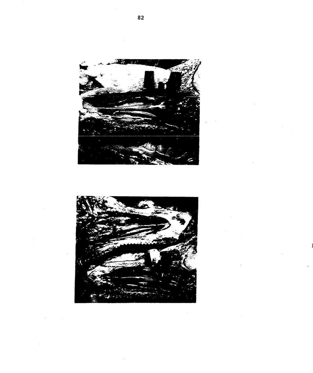
144 82
Micrococcus. May be normal present in upper respiratory tract. - Grow on ordinary media Nutrient agar - Blood agar and. M. luteus.
 Micrococcus Morphology: - Gram +ve cocci - Arrangement : Tetrades - Non motile, non capsulated, non sporulated Habitat: May be normal present in upper respiratory tract Species : 1- M.varians 2- M. luteus
Medical bacteriology Lecture 8. Streptococcal Diseases
 Medical bacteriology Lecture 8 Streptococcal Diseases Streptococcus agalactiae Beat haemolytic Lancifield group B Regularly resides in human vagina, pharynx and large inine Can be transferred to infant
Gram-positive cocci Staphylococci and Streptococcia
 Medical microbiology Laboratory Lab 8 Gram-positive cocci Staphylococci and Streptococcia Lecturer Maysam A Mezher Gram positive cocci 1-Staphylococcus. 2-Streptococcus. 3-Micrococcus The medically important
Overview. There are commonly found arrangements of bacteria based on their division. Spheres, Rods, Spirals
 Bacteria Overview Bacteria live almost everywhere. Most are microscopic ranging from 0.5 5 m in size, and unicellular. They have a variety of shapes when viewed under a microscope, most commonly: Spheres,
Cercetări bacteriologice, epidemiologice şi serologice în bruceloza ovină ABSTRACT
 ABSTRACT Thesis entitled BACTERIOLOGICAL, EPIDEMIOLOGICAL AND SEROLOGICAL RESEARCHES IN BRUCELLOSIS OVINE is scientific and practical reasons the following: - Infectious epididymitis in Romania, described
DO NOT WRITE ON or THROW AWAY THIS PAPER!
 What Kills Bacteria? Lab Procedure Go to the following link: http://www.glencoe.com/sites/common_assets/science/virtual_labs/ls08/ls08.html or DO NOT WRITE ON or THROW AWAY THIS PAPER! Visit my eboard
Bordetella bronchiseptica ASSOCIATED WITH PULMONARY DISEASE IN MOUNTAIN VOLES (Microtus montanus)
") Bordetella bronchiseptica ASSOCIATED WITH PULMONARY DISEASE IN MOUNTAIN VOLES (Microtus montanus) Authors: WAYNE I. JENSEN, and RUTH M. DUNCAN Source: Journal of Wildlife Diseases, 16(1) : 11-14 Published
Bordetella bronchiseptica ASSOCIATED WITH PULMONARY DISEASE IN MOUNTAIN VOLES (Microtus montanus) Authors: WAYNE I. JENSEN, and RUTH M. DUNCAN Source: Journal of Wildlife Diseases, 16(1) : 11-14 Published
USA Product Label LINCOCIN. brand of lincomycin hydrochloride tablets. brand of lincomycin hydrochloride injection, USP. For Use in Animals Only
 USA Product Label http://www.vetdepot.com PHARMACIA & UPJOHN COMPANY Division of Pfizer Inc. Distributed by PFIZER INC. 235 E. 42ND ST., NEW YORK, NY, 10017 Telephone: 269-833-4000 Fax: 616-833-4077 Customer
Biological Threat Fact Sheets
 Biological Threat Fact Sheets Anthrax Agent: Bacillus anthracis There are three clinical forms of B. anthracis which are determined by route of entry: Pulmonary or Inhalation BT implications Cutaneous
Medical Bacteriology- Lecture 14. Gram negative coccobacilli. Zoonosis. Brucella. Yersinia. Francesiella
 Medical Bacteriology- Lecture 14 Gram negative coccobacilli Zoonosis Brucella Yersinia Francesiella 1 Zoonosis: A disease, primarily of animals, which is transmitted to humans as a result of direct or
SUMMARY OF PRODUCT CHARACTERISTICS
 SUMMARY OF PRODUCT CHARACTERISTICS 1. NAME OF THE VETERINARY MEDICINAL PRODUCT Oxycare 20 %w/v LA Solution for Injection 2. QUALITATIVE AND QUANTITATIVE COMPOSITION Active Substance: Oxytetracycline (Equivalent
The Salmonella. Dr. Hala Al Daghisatni
 1 Dr. Hala Al Daghisatni The Salmonella Salmonellae are often pathogenic for humans or animals when acquired by the oral route. They are transmitted from animals and animal products to humans, where they
VPM 201-Lab 6 Bovine Mastitis, Bacillus & Mastitis (2012)
") Exercise 1. Bovine mastitic milk sample A. Note relevant images are on next page Sample A is Staphylococcus aureus Moderate size (1.0 mm), circular, convex, cream-to-light yellow, opaque Double-zone (target)
Exercise 1. Bovine mastitic milk sample A. Note relevant images are on next page Sample A is Staphylococcus aureus Moderate size (1.0 mm), circular, convex, cream-to-light yellow, opaque Double-zone (target)
تقارير الدروس العملية
 وزارة التعليم جامعة الباحة كلية العلوم الطبية التطبيقية قسم طب المختبرات تقارير الدروس العملية مقرر أحياء دقيقة إكلينيكية الدكتور : شائع بن صالح المالكي 5341 ه -5341 ه Routine of Laboratory Diagnosis of
Labquality External Quality Assesment Programmes General Bacteriology 1 3/2010
 Labquality External Quality Assesment Programmes General Bacteriology 1 3/2010 Photos and text: Markku Koskela, M.D., Ph.D. Clinical microbiology specialist Oulu, Finland Patient and sample 9/2010 Pus
Author - Dr. Josie Traub-Dargatz
 Author - Dr. Josie Traub-Dargatz Dr. Josie Traub-Dargatz is a professor of equine medicine at Colorado State University (CSU) College of Veterinary Medicine and Biomedical Sciences. She began her veterinary
ANTIBIOTICS USED FOR RESISTACE BACTERIA. 1. Vancomicin
 ANTIBIOTICS USED FOR RESISTACE BACTERIA 1. Vancomicin Vancomycin is used to treat infections caused by bacteria. It belongs to the family of medicines called antibiotics. Vancomycin works by killing bacteria
Interpretation of Bulk Tank Milk Results
 Interpretation of Bulk Tank Milk Results Introduction Culturing bulk tank milk (BTM) to monitor milk quality has limitations based on the amount and frequency of sampling and the amount and types of microorganisms
Salmonella. Serological. Salmonella. food poisoning. Use of pasteurized milk and milk products Improvement of hygiene. Proper storage of foods
 Character Habitat Serological Pathogenicity Salmonella food poisoning Salmonella Gram ve facultative anaerobes, non lactose fermenter bacilli Intestinal tracts of humans and animals Salmonella: >1000 species
Bacterial Pneumonia in Sheep, The Domestic Bighorn Sheep Interface, and Research at ADRU
 Bacterial Pneumonia in Sheep, The Domestic Bighorn Sheep Interface, and Research at ADRU USAHA Committee on Sheep and Goats Providence, RI October 27, 2015 PLC M. A. Highland, DVM, DACVP, PhD candidate
Aetiological Study on Pneumonia in Camel (Camelus dromedarius) and in vitro Antibacterial Sensitivity Pattern of the Isolates
 and in vitro Antibacterial Sensitivity Pattern of the Isolates") Pakistan Journal of Biological Sciences, 2 (4): 1102-1105, 1999 Research Article Aetiological Study on Pneumonia in Camel (Camelus dromedarius) and in vitro Antibacterial Sensitivity Pattern of the Isolates
Pakistan Journal of Biological Sciences, 2 (4): 1102-1105, 1999 Research Article Aetiological Study on Pneumonia in Camel (Camelus dromedarius) and in vitro Antibacterial Sensitivity Pattern of the Isolates
4.5. Special precautions for use Special precautions to be taken by person administering the veterinary medicinal product to animals
 1.B1. SUMMARY OF PRODUCT CHARACTERISTICS 1. NAME OF THE VETERINARY MEDICINAL PRODUCT AMOXYCOL Soluble Powder 2. QUALITATIVE AND QUANTITATIVE COMPOSITION Active substances: Amoxicillin trihydrate 640.0
Bovine Mastitis Products for Microbiological Analysis
 Bovine Mastitis Products for Microbiological Analysis 121917ss Hardy Diagnostics has everything for your laboratory! SAVE MONEY Now you have a choice for obtaining your supplies for mastitis testing. Hardy
n Am I B I A U n IVE RS ITV OF SCIEnCE AnD TECH n 0 LOGY
 n Am I B I A U n IVE RS ITV OF SCIEnCE AnD TECH n 0 LOGY FACULTY OF HEALTH AND APPLIED SCIENCES DEPARTMENT OF HEALTH SCIENCES QUALIFICATION: BACHELOR OF BIOMEDICAL SCIENCES QUALIFICATION CODE: SOBBMS LEVEL:
Microscopy Directions
 Name: Exercise 1 Microscopy Focus each slide of bacteria under the microscope using oil immersion. Draw the arrangement of the bacterial cells in the larger portion of the circle and draw the shape of
Canine and Feline Distemper. Description. The following chart indicates the animals which are susceptible to infection by canine and feline distemp
 Canine and Feline Distemper Description Canine and feline distemper are diseases affecting many wild and domestic carnivo The following chart indicates the animals which are susceptible to infection by
INFECTIOUS ABORTION INVESTIGATIONS
 INFECTIOUS ABORTION INVESTIGATIONS INFECTIOUS ABORTION INVESTIGATIONS.¹ H. F. LIENHARDT, C. H. KITSELMAN, AND C. E. SAWYER. FOREWORD. Infectious abortion of cattle has become a problem of world-wide
VOL. XXIII NO. II THE JOURNAL OF ANTIBIOTICS 559. ANTIBIOTIC 6640.* Ill
 VOL. XXIII NO. II THE JOURNAL OF ANTIBIOTICS 559 ANTIBIOTIC 6640.* Ill BIOLOGICAL STUDIES WITH ANTIBIOTIC 6640, A NEW BROAD-SPECTRUM AMINOGLYCOSIDE ANTIBIOTIC J. Allan Waitz, Eugene L. Moss, Jr., Edwin
COURSE SYLLABUS. (Clinical Bacteriology-1
 COURSE SYLLABUS (Clinical Bacteriology- MLAB-47) COURSE SYLLABUS Course title: Clinical Bacteriology- Code: MLAB-47 Credit hours: 4 (3 Theory+ Practical) Name of faculty member: Dr. Mohamudha Parveen Rahamathulla
What is a disease. Any condition that results in deviation from normal function
 What is a disease Any condition that results in deviation from normal function How do diseases occur? AGENT HOST ENVIRONMENT ETIOLOGY Infectious Agents Bacteria Viruses Parasites Fungi Non-infectious agents
Vaccines for Cats. 2. Feline viral rhinotracheitis, FVR caused by FVR virus, also known as herpes virus type 1, FHV-1
 Vaccines for Cats Recent advances in veterinary medical science have resulted in an increase in the number and type of vaccines that are available for use in cats, and improvements are continuously being
CONTAGIOUS BOVINE PLEURO- PNEUMONIA steps towards control of the disease. Rose Matua -Department of Veterinary Services, Kenya
 CONTAGIOUS BOVINE PLEURO- PNEUMONIA steps towards control of the disease Rose Matua -Department of Veterinary Services, Kenya Introduction CBPP is a highly contagious acute, subacute or chronic disease
Lab Exercise: Antibiotics- Evaluation using Kirby Bauer method.
 Lab Exercise: Antibiotics- Evaluation using Kirby Bauer method. OBJECTIVES 1. Compare the antimicrobial capabilities of different antibiotics. 2. Compare effectiveness of with different types of bacteria.
Enterobacter aerogenes
 Enterobacter aerogenes Enterobacter sp. Enterobacter sp. Species: Enterobacter aerogenes Enterobacter agglomerans Enterobacter cloacae causes UTI, enterotoxigenic Often found in the normal intestinal flora,
VETERINARY BACTERIOLOGY FROM THE DARK AGES TO THE PRESENT DAY
 VETERINARY BACTERIOLOGY FROM THE DARK AGES TO THE PRESENT DAY D.J.TAYLOR MA PhD VetMB DipECPHM DipECVPH MRCVS EMERITUS PROFESSOR OF VETERINARY BACTERIOLOGY AND PUBLIC HEALTH UNIVERSITY OF GLASGOW INTRODUCTION
Clinical Manifestations and Treatment of Plague Dr. Jacky Chan. Associate Consultant Infectious Disease Centre, PMH
 Clinical Manifestations and Treatment of Plague Dr. Jacky Chan Associate Consultant Infectious Disease Centre, PMH Update of plague outbreak situation in Madagascar A large outbreak since 1 Aug 2017 As
Ophthalmology Research: An International Journal 2(6): , 2014, Article no. OR SCIENCEDOMAIN international
: , 2014, Article no. OR SCIENCEDOMAIN international") Ophthalmology Research: An International Journal 2(6): 378-383, 2014, Article no. OR.2014.6.012 SCIENCEDOMAIN international www.sciencedomain.org The Etiology and Antibiogram of Bacterial Causes of Conjunctivitis
Ophthalmology Research: An International Journal 2(6): 378-383, 2014, Article no. OR.2014.6.012 SCIENCEDOMAIN international www.sciencedomain.org The Etiology and Antibiogram of Bacterial Causes of Conjunctivitis
USA Product Label CLINTABS TABLETS. Virbac. brand of clindamycin hydrochloride tablets. ANADA # , Approved by FDA DESCRIPTION
 VIRBAC CORPORATION USA Product Label http://www.vetdepot.com P.O. BOX 162059, FORT WORTH, TX, 76161 Telephone: 817-831-5030 Order Desk: 800-338-3659 Fax: 817-831-8327 Website: www.virbacvet.com CLINTABS
loopfull is removed from each dilution and transferred to capable of killing the test organism in 10 minutes but not GERMICIDAL SUBSTANCES
 A NEW METHOD FOR THE EVALUATION OF GERMICIDAL SUBSTANCES A. J. SALLE, W. A. McOMIE AND I. L. SHECHMEISTER Department of Bacteriology, University of California, Berkeley, California Received for publication
BOVINE RESPIRATORY DISEASE COMPLEX. Kristen Mierzwiak LCS 630
 BOVINE RESPIRATORY DISEASE COMPLEX Kristen Mierzwiak LCS 630 Ring... You are called out to the farm of one of your regular dairy clients because some of the replacement heifers they bought at a public
Domestic Bighorn Sheep Research American Sheep Industry/ National Lamb Feeders Association Annual Convention Charleston, SC January 22-25, 2014
 PLC Domestic Bighorn Sheep Research American Sheep Industry/ National Lamb Feeders Association Annual Convention Charleston, SC January 22-25, 2014 M. A. Highland, DVM, PhDc, Dipl. ACVP PhD Veterinary
ENTERIC BACTERIA. 1) salmonella. Continuation of the Enteric bacteria : A) We have mentioned the first group of salmonella (salmonella enterica ):
 salmonella. Continuation of the Enteric bacteria : A) We have mentioned the first group of salmonella (salmonella enterica ):") Continuation of the Enteric bacteria : ENTERIC BACTERIA 1) salmonella A) We have mentioned the first group of salmonella (salmonella enterica ): Salmonella is an obligate pathogen ; food poisoning due
Continuation of the Enteric bacteria : ENTERIC BACTERIA 1) salmonella A) We have mentioned the first group of salmonella (salmonella enterica ): Salmonella is an obligate pathogen ; food poisoning due
Q1. (a) Clostridium difficile is a bacterium that is present in the gut of up to 3% of healthy adults and 66% of healthy infants.
 Clostridium difficile is a bacterium that is present in the gut of up to 3% of healthy adults and 66% of healthy infants.") Q1. (a) Clostridium difficile is a bacterium that is present in the gut of up to 3% of healthy adults and 66% of healthy infants. C. difficile rarely causes problems, either in healthy adults or in infants.
Q1. (a) Clostridium difficile is a bacterium that is present in the gut of up to 3% of healthy adults and 66% of healthy infants. C. difficile rarely causes problems, either in healthy adults or in infants.
Aquatic Animal Bacterial Pathogen
 Aquatic Animal Bacterial Pathogen Veterinary Bacteriology and Mycology (3142304 ) Academic year 2012 Channarong Rodkhum D.V.M. (Hons), Ph.D. Department of Veterinary Microbiology Faculty of Veterinary
Burn Infection & Laboratory Diagnosis
 Burn Infection & Laboratory Diagnosis Introduction Burns are one the most common forms of trauma. 2 million fires each years 1.2 million people with burn injuries 100000 hospitalization 5000 patients die
How to stop the snotty noses: Preventing feline upper respiratory infections. Staci Cannon, DVM, MPH, DACVPM, DABVP (Shelter Medicine Practice)
") How to stop the snotty noses: Preventing feline upper respiratory infections Staci Cannon, DVM, MPH, DACVPM, DABVP (Shelter Medicine Practice) Why is URI so hard to control? Multiple pathogens Chronic
How to stop the snotty noses: Preventing feline upper respiratory infections Staci Cannon, DVM, MPH, DACVPM, DABVP (Shelter Medicine Practice) Why is URI so hard to control? Multiple pathogens Chronic
MILK COMPOSITIONAL CHANGES DURING MASTITIS
 MASTITIS PA R T 2 MILK COMPOSITIONAL CHANGES DURING MASTITIS Increased SCC Na Cl Whey protein (e.g. serum albumin, Ig, lactoferrin) Decreased Production α-lactalbumin & Lactose Casein K MILK LOSS LACTOFERRIN
CAUTION Federal (USA) law restricts this drug to use by or on the order of a licensed veterinarian.
 law restricts this drug to use by or on the order of a licensed veterinarian.") LINCOCIN- lincomycin hydrochloride injection, solution Pharmacia and Upjohn Company ---------- Lincocin lincomycin hydrochloride tablets and liquid lincomycin injection, USP For Use in Animals Only For
LINCOCIN- lincomycin hydrochloride injection, solution Pharmacia and Upjohn Company ---------- Lincocin lincomycin hydrochloride tablets and liquid lincomycin injection, USP For Use in Animals Only For
Quad Plate User s Manual
 A part of Eurofins DQCI SSGN - SSGNC Mastitis Culture Quad Plate User s Manual Eurofins Microbiology Laboratories / Eurofins DQCI Services 5205 Quincy Street, Mounds View, MN 55112 P: 763-785-0485 F: 763-785-0584
Evaluation of a computerized antimicrobial susceptibility system with bacteria isolated from animals
 J Vet Diagn Invest :164 168 (1998) Evaluation of a computerized antimicrobial susceptibility system with bacteria isolated from animals Susannah K. Hubert, Phouc Dinh Nguyen, Robert D. Walker Abstract.
Enteric Bacteria. Prof. Dr. Asem Shehabi Faculty of Medicine University of Jordan
 Enteric Bacteria Prof. Dr. Asem Shehabi Faculty of Medicine University of Jordan Enteric Bacteria General Characteristics: Gram-ve Bacilli, Facultative Anaerobes, Intestinal Normal Flora.. Humans, Animals,
Importance of Frequency Homeopathic application
 Homeopathic Antibiotic for Pets 5 Pages PRODUCT CODE AN070 * Stronger Antibiotic - see product AN071 Infection Fighter 50ml (herbal antibiotic) Last Updated: 11-07-18 All species and ages (and humans)
Treatment of Respiratory Tract Infections Prof. Mohammad Alhumayyd Dr. Aliah Alshanwani
 Treatment of Respiratory Tract Infections Prof. Mohammad Alhumayyd Dr. Aliah Alshanwani 30-1-2018 1 Objectives of the lecture At the end of lecture, the students should be able to understand the following:
3.0 Treatment of Infection
 3.0 Treatment of Infection Antibiotics and Medicine National Curriculum Link Key Stage 3 Sc1:1a - 1c. 2a 2p Sc2: 2n Unit of Study Unit 8: Microbes and Disease Unit 9B: Fit and Healthy Unit 20: 20 th Century
CHALLENGE SET EXERCISE FALL 2008
 CHALLENGE SET EXERCISE FALL 2008 Scenario 1 Fifteen year old female presents to clinic with cat bite to hand. Aerobic wound culture ordered No Gram Stain Organism 1 Characteristics Growth on Blood and
Pharm 262: Antibiotics. 1 Pharmaceutical Microbiology II DR. C. AGYARE
 Pharm 262: 1 Pharmaceutical Microbiology II Antibiotics DR. C. AGYARE Reference Books 2 HUGO, W.B., RUSSELL, A.D. Pharmaceutical Microbiology. 6 th Ed. Malden, MA: Blackwell Science, 1998. WALSH, G. Biopharmaceuticals:
Feline Vaccines: Benefits and Risks
 Feline Vaccines: Benefits and Risks Deciding which vaccines your cat should receive requires that you have a complete understanding of the benefits and risks of the procedure. For this reason, it is extremely
The surveillance and control programme
 Annual Reports 2010 Surveillance and control programmes for terrestrial and aquatic animals in Norway The surveillance and control programme for Brucella abortus in cattle in Norway Ståle Sviland Berit
MARBOCYL 10% SUMMARY OF PRODUCT CHARACTERISTICS
 MARBOCYL 10% SUMMARY OF PRODUCT CHARACTERISTICS 1. NAME OF THE VETERINARY MEDICINAL PRODUCT MARBOCYL 10%, solution for injection for cattle and swine 2. QUALITATIVE AND QUANTITATIVE COMPOSITION Marbofloxacin...100.0
Test Method Modified Association of Analytical Communities Test Method Modified Germicidal Spray Products as Disinfectants
 Study Title Antibacterial Activity and Efficacy of E-Mist Innovations' Electrostatic Sprayer Product with Multiple Disinfectants Method Modified Association of Analytical Communities Method 961.02 Modified
Course Curriculum for Master Degree in Poultry Diseases/Veterinary Medicine
 Course Curriculum for Master Degree in Poultry Diseases/Veterinary Medicine The Master Degree in Poultry Diseases /Veterinary Medicine, is awarded by the Faculty of Graduate Studies at Jordan University
Mastitis: Background, Management and Control
 New York State Cattle Health Assurance Program Mastitis Module Mastitis: Background, Management and Control Introduction Mastitis remains one of the most costly diseases of dairy cattle in the US despite
Dr. Michelle Arnold, DVM DABVP (Food Animal) Ruminant Extension Veterinarian University of Kentucky Veterinary Diagnostic Laboratory
 Ruminant Extension Veterinarian University of Kentucky Veterinary Diagnostic Laboratory") Dr. Michelle Arnold, DVM DABVP (Food Animal) Ruminant Extension Veterinarian University of Kentucky Veterinary Diagnostic Laboratory Mastitis-Treatment Options and Strategies Treatment Strategies 1 st
Dr. Michelle Arnold, DVM DABVP (Food Animal) Ruminant Extension Veterinarian University of Kentucky Veterinary Diagnostic Laboratory Mastitis-Treatment Options and Strategies Treatment Strategies 1 st
Introduction to Chemotherapeutic Agents. Munir Gharaibeh MD, PhD, MHPE School of Medicine, The university of Jordan November 2018
 Introduction to Chemotherapeutic Agents Munir Gharaibeh MD, PhD, MHPE School of Medicine, The university of Jordan November 2018 Antimicrobial Agents Substances that kill bacteria without harming the host.
Methicillin-Resistant Staphylococcus aureus
 Methicillin-Resistant Staphylococcus aureus By Karla Givens Means of Transmission and Usual Reservoirs Staphylococcus aureus is part of normal flora and can be found on the skin and in the noses of one
Introduction. Rabbit Respiratory Disease. Lecture Outline. Pre-consult. Initial presentation. History 26/01/2013
 Introduction Rabbit Respiratory Disease Joanna Hedley BVM&S DZooMed (Reptilian) MRCVS Rabbit, Exotic Animal and Wildlife Clinician at R(D)SVS Obligate nasal breathers Small thorax Tendency to mask symptoms
COMMON CLINICAL CONDITIONS IN RATS AND MICE
 COMMON CLINICAL CONDITIONS IN RATS AND MICE Megan H. Nowland, DVM, Diplomate ACLAM Assistant Professor Unit for Laboratory Animal Medicine University of Michigan NCRC Building 10, Suite G90 2800 Plymouth
averaged 10 ticks each. In addition, a considerable number of ticks
 THE MICROBIAL FLORA OF THE ROCKY MOUNTAIN WOOD TICK, DERMACENTOR ANDERSONI STILES EDWARD A. STEINHAUS U. S. Public Health Service, Rocky Mountain Laboratory, Hamilton, Montana Received for publication,
SUMMARY OF PRODUCT CHARACTERISTICS
 SUMMARY OF PRODUCT CHARACTERISTICS 1. NAME OF THE VETERINARY MEDICINAL PRODUCT Amfipen LA 100 mg/ml suspension for injection 2. QUALITATIVE AND QUANTITATIVE COMPOSITION Active substance: Each ml contains:
Antibiotic Resistance in Pseudomonas aeruginosa Strains Isolated from Various Clinical Specimens
 International Journal of Current Microbiology and Applied Sciences ISSN: 2319-7706 Volume 7 Number 03 (2018) Journal homepage: http://www.ijcmas.com Original Research Article https://doi.org/10.20546/ijcmas.2018.703.217
6.0 ANTIBACTERIAL ACTIVITY OF CAROTENOID FROM HALOMONAS SPECIES AGAINST CHOSEN HUMAN BACTERIAL PATHOGENS
 6.0 ANTIBACTERIAL ACTIVITY OF CAROTENOID FROM HALOMONAS SPECIES AGAINST CHOSEN HUMAN BACTERIAL PATHOGENS 6.1 INTRODUCTION Microorganisms that cause infectious disease are called pathogenic microbes. Although
Determination of antibiotic sensitivities by the
 Journal of Clinical Pathology, 1978, 31, 531-535 Determination of antibiotic sensitivities by the Sensititre system IAN PHILLIPS, CHRISTINE WARREN, AND PAMELA M. WATERWORTH From the Department of Microbiology,
Cleaning and Disinfection Protocol for Gram-Negative and Gram-Positive Bacteria, including Antibiotic Resistant Bacteria
 Cleaning and Disinfection Protocol for Gram-Negative and Gram-Positive Bacteria, including Antibiotic Resistant Bacteria This document has been developed in accordance with current applicable infection
gingivitis: periodontitis: dental caries: palatinitis: oral pharyngitis and tonsillitis: mouth abscess: glossitis: oro-sinus fistula: gingivitis:
 ABSTRACT Mouth is one of the anatomical segments of the digestive microbiota which is characterized by a marked diversity. Among the multitude of microorganisms that inhabit the oral mucosa at a time,
Walter M. Guterbock, DVM, MS Veterinary Medicine Teaching and Research Center University of California, Davis
 Walter M. Guterbock, DVM, MS Veterinary Medicine Teaching and Research Center University of California, Davis 1993 WESTERN LARGE HERD MANAGEMENT CONFERENCE V LAS VEGAS NEVADA 27 Alternatives To Antibiotic
Domestic Bighorn Sheep Interface Problem Overview and Research. American Sheep Industry Annual Convention Reno, NV January 27-31, 2015
 Domestic Bighorn Sheep Interface Problem Overview and Research American Sheep Industry Annual Convention Reno, NV January 27-31, 2015 Maggie Highland, DVM, PhDc, Dipl. ACVP PhD Veterinary Training Program
TOLYPOMYCIN, A NEW ANTIBIOTIC. V IN VITRO AND IN VIVO ANTIMICROBIAL ACTIVITY. Masahiro Kondo, Tokiko Oishi and Kanji Tsuchiya
 16 THE JOURNAL OF ANTIBIOTICS JAN. 1972 TOLYPOMYCIN, A NEW ANTIBIOTIC. V IN VITRO AND IN VIVO ANTIMICROBIAL ACTIVITY Masahiro Kondo, Tokiko Oishi and Kanji Tsuchiya Biological Research Laboratories, Research
Pathogens commonly isolated from selected diseases
 Pathogens commonly isolated from selected diseases Equine pneumonia/pleuropneumonia -hemolytic Strep. Clostridium Pasteurella E. coli Klebsiella pneumoniae Bacteroides Equine enteric pathogens Salmonella
(From the Division of Laboratories of Montefiore Hospital, New York.)
") CALCIFICATION OF THE SUPRARENAL GLANDS OF CATS. BY DAVID MARINE, M.D. (From the Division of Laboratories of Montefiore Hospital, New York.) PLATE 11. (Received for publication, January 18, 1925.) It is
CALCIFICATION OF THE SUPRARENAL GLANDS OF CATS. BY DAVID MARINE, M.D. (From the Division of Laboratories of Montefiore Hospital, New York.) PLATE 11. (Received for publication, January 18, 1925.) It is
Cleaning and Disinfection Protocol Vegetative Bacteria
 Cleaning and Disinfection Protocol Vegetative Bacteria This document has been developed in accordance with current applicable infection control and biosecurity guidelines. It is intended for use as a guideline
Index. Note: Page numbers of article titles are in boldface type.
 Index Note: Page numbers of article titles are in boldface type. A Abdominal viscera, examination of, in investigation of emerging infectious diseases of food animals, 6 American Veterinary Medical Association,
11/10/2016. Skin and Soft Tissue Infections. Disclosures. Educational Need/Practice Gap. Objectives. Case #1
 Disclosures Selecting Antimicrobials for Common Infections in Children FMR-Contemporary Pediatrics 11/2016 Sean McTigue, MD Assistant Professor of Pediatrics, Pediatric Infectious Diseases Medical Director
QUICK REFERENCE. Pseudomonas aeruginosa. (Pseudomonas sp. Xantomonas maltophilia, Acinetobacter sp. & Flavomonas sp.)
") Pseudomonas aeruginosa (Pseudomonas sp. Xantomonas maltophilia, Acinetobacter sp. & Flavomonas sp.) Description: Greenish gray colonies with some beta-hemolysis around each colony on blood agar (BAP),
Pseudomonas aeruginosa (Pseudomonas sp. Xantomonas maltophilia, Acinetobacter sp. & Flavomonas sp.) Description: Greenish gray colonies with some beta-hemolysis around each colony on blood agar (BAP),
number Done by Corrected by Doctor Dr.Malik
 number 27 Done by Fatimah Farhan Corrected by Basil Al-Bakri Doctor Dr.Malik Note: anything in red is just extra info and you will not be asked about it in the exam. In this sheet we will continue talking
COMMITTEE FOR VETERINARY MEDICINAL PRODUCTS
 The European Agency for the Evaluation of Medicinal Products Veterinary Medicines Evaluation Unit EMEA/MRL/389/98-FINAL July 1998 COMMITTEE FOR VETERINARY MEDICINAL PRODUCTS ENROFLOXACIN (extension to
CLPNA Pressure Ulcers ecourse: Module 5.6 Quiz II page 1
 CLPNA Pressure Ulcers ecourse: Module 5.6 Quiz II 1. What are the symptoms of an infected wound? a. Fever b. Edema c. Erythema d. Local pain and tenderness e. Induration of wound edge 2. A person with
Surveillance of animal brucellosis
 Surveillance of animal brucellosis Assoc.Prof.Dr. Theera Rukkwamsuk Department of large Animal and Wildlife Clinical Science Faculty of Veterinary Medicine Kasetsart University Review of the epidemiology
Role of Antibodies in Immunity to Bordetella Infections
 INFECTION AND IMMUNITY, Apr. 2003, p. 1719 1724 Vol. 71, No. 4 0019-9567/03/$08.00 0 DOI: 10.1128/IAI.71.4.1719 1724.2003 Copyright 2003, American Society for Microbiology. All Rights Reserved. Role of
SUMMARY OF PRODUCT CHARACTERISTICS. Cephacare flavour 50 mg tablets for cats and dogs. Excipients: For a full list of excipients, see section 6.1.
 SUMMARY OF PRODUCT CHARACTERISTICS 1. NAME OF THE VETERINARY MEDICINAL PRODUCT Cephacare flavour 50 mg tablets for cats and dogs 2. QUALITATIVE AND QUANTITATIVE COMPOSITION Each tablet contains: Active
مادة االدوية المرحلة الثالثة م. غدير حاتم محمد
 م. مادة االدوية المرحلة الثالثة م. غدير حاتم محمد 2017-2016 ANTIMICROBIAL DRUGS Antimicrobial drugs Lecture 1 Antimicrobial Drugs Chemotherapy: The use of drugs to treat a disease. Antimicrobial drugs:
Irish Medicines Board
 IRISH MEDICINES BOARD ACT 1995, as amended European Communities (Animal Remedies) (No. 2) Regulations 2007 VPA: 10999/033/001A Case No: 7006569 The in exercise of the powers conferred on it by Animal Remedies
Gye and Cramer (1919) found that the ionizable salts of calcium injected together with the washed spores of Cl. tetani or of certain
 found that the ionizable salts of calcium injected together with the washed spores of Cl. tetani or of certain") STUDIES ON TETANUS TOXOID III. ANTITOXIC RESPONSE IN GUINEA PIGS IMMUNIZED WITH TETANUS ALUM-PRECIPITATED TOXOID FOLLOWED BY TET- ANUS SPORES F. G. JONES AND W. A. JAMIESON Lilly Research Laboratories,
STUDIES ON TETANUS TOXOID III. ANTITOXIC RESPONSE IN GUINEA PIGS IMMUNIZED WITH TETANUS ALUM-PRECIPITATED TOXOID FOLLOWED BY TET- ANUS SPORES F. G. JONES AND W. A. JAMIESON Lilly Research Laboratories,
Microbiology : antimicrobial drugs. Sheet 11. Ali abualhija
 Microbiology : antimicrobial drugs Sheet 11 Ali abualhija return to our topic antimicrobial drugs, we have finished major group of antimicrobial drugs which associated with inhibition of protein synthesis
BACTERIOLOGY OF THE HEALTHY CONJUNCTIVA*
 Brit. J. Ophthal. (1954), 38, 719. BACTERIOLOGY OF THE HEALTHY CONJUNCTIVA* BY C. H. SMITH Department of Pathology, Institute of Ophthalmology, University of London THE normal bacterial flora of the mucous
Health Products Regulatory Authority
 1 NAME OF THE VETERINARY MEDICINAL PRODUCT Genta 50 mg/ml solution for injection 2 QUALITATIVE AND QUANTITATIVE COMPOSITION Each ml contains: Active Substances Gentamicin sulphate equivalent to Gentamicin
Burton's Microbiology for the Health Sciences. Chapter 9. Controlling Microbial Growth in Vivo Using Antimicrobial Agents
 Burton's Microbiology for the Health Sciences Chapter 9. Controlling Microbial Growth in Vivo Using Antimicrobial Agents Chapter 9 Outline Introduction Characteristics of an Ideal Antimicrobial Agent How
Tautopathic Treatment. Systemic infection or localized infections
 PRODUCT CODE AN071 Herbal Antibiotic for All Species 5 Pages Last Updated: 11-07-18 All species and ages (and humans) Tautopathic Treatment By taking Doxycycline 30C or 200C in a homeopathic form, this
TOC INDEX. Salmonellosis in Feedlot Cattle. Jane Pritchard. Take Home Message. Introduction
 TOC INDEX Salmonellosis in Feedlot Cattle Jane Pritchard Take Home Message Salmonellosis in feedlot cattle is an important but uncommon disease. The disease has been recognized only recently as a significant
Curriculum Vitae. : AlBaha University, faculty of Science.
 Curriculum Vitae Personal Data : Name : Layla Ismail Mohamed Nationality : Sudanese Present Position Held: Associate Professor Address Academic Qualification: : AlBaha University, faculty of Science. E-mail:
Approach to pediatric Antibiotics
 Approach to pediatric Antibiotics Gassem Gohal FAAP FRCPC Assistant professor of Pediatrics objectives To be familiar with common pediatric antibiotics o Classification o Action o Adverse effect To discus