Taxonomical and faunistic studies on the nematode parasites from Indonesian murines. (Rodentia; Muridae; Murinae) with special reference to
|
|
|
- Simon Eaton
- 5 years ago
- Views:
Transcription
1 Taxonomical and faunistic studies on the nematode parasites from Indonesian murines (Rodentia; Muridae; Murinae) with special reference to Syphacia spp. and their biogeography Graduate School of Veterinary Medicine, Rakuno Gakuen University DEWI, Kartika Zoology Division, Research Center for Biology-Indonesian Institute of Sciences JSPS Ronpaku Researcher ID No. LIPI Supervisor Prof. Mitsuhiko Asakawa Rakuno Gakuen University September, 2015 i
2 ABBREVIATIONS Cox1; cytochrome C oxidase subunit 1 gene DNA; deoxyribonucleic acid IUCN; International Union for Conservation of Nature LE; laterally-elongated ML; maximum likelihood MZB; Museum Zoologicum Bogoriense NJ; neighbor-joining R; round S; square WL; worm length i
3 Contents PREFACE CHAPTER 1 Faunal study on the nematodes parasitic in Indonesian murines Introduction... 4 Materials and methods Results Records between the 1930 s and early 2000 s Present author s records The new genus and species Musserakis sulawesiensis (Heterakidae) The superfamily Trichostrongyloidea and its murine hosts Zoonotic nematodes found from Indonesian murines An overview of nematode fauna from Indonesian murines Summary CHAPTER 2 Morphological taxonomy of the genus Syphacia from Indonesian murines Introduction Materials and methods Collection of materials Morphological observation Results and discussion Description Subgenus Syphacia (Rumbaisyphacia) Dewi, Hasegawa & Asakawa, Syphacia (R.) kumis Dewi, Hasegawa & Asakawa, Subgenus Syphacia (Segienamsyphacia) Dewi, Hasegawa & Asakawa, Syphacia (Se.) yuniae Dewi, Hasegawa and Asakawa, Subgenus Syphacia Seurat, Syphacia (Syphacia) rifaii Dewi & Hasegawa, Syphacia (Syphacia) taeromyos Dewi & Hasegawa, Syphacia (Syphacia) paruromyos Dewi & Hasegawa, Syphacia (Syphacia) semiadii Dewi, Asakawa & Fitriana, Syphacia (Syphacia) maxomyos from Maxomys spp An overview of the genus Syphacia and Indonesian species ii
4 Key to species of Syphacia in Sulawesi and the Australian bioregion Summary CHAPTER 3 Molecular biological analysis and phylogenetic c onsideration of the genus Syphacia from Indonesian murines Introduction Materials and methods Results and discussion Summary CHAPTER 4 Biogeographical discussion on host parasite r elationship between Indonesian murines and the genus Syphacia Introduction Geographical history of Indonesian Archipelago Host origin and phylogeny Host dispersal to Sunda and Sahul Related studies of Syphaciinae and Heligmonellidae in Indo-Australian Archipelago Morpho-phylogenetical relationship among the subgenus Syphacia Biogeography of host-parasite relationship between murines and subgenus Syphacia Conclusion; past, present and future of the host-parasit relationship between murines and their Syphacia Summary CONCLUSION ACKNOWLEDGMENTS REFERENCES APPENDIX Checklist of the nematode parasites of Indonesian murines iii
5 2. DNA sequences of Syphacia spp Geographical distribution of Syphaciinae Geographical distribution of Heligmonellidae ABSTRACT iv
6 PREFACE Rats and/or mice (Rodentia; Muridae) are of special interest due to their role as reservoirs of many important parasitic nematodes of humans and livestock (Kwo and Kwo, 1968; Bhaibulaya and Indrangarm, 1975; Baker, 1998). Human infections with migration of larvae of Angiostrongylus cantonensis (Chen, 1950), a nematode of rat, had been reported from North Sumatra and Java (Smit, 1962; Widagdo et al., 1977). Until recently, only A. cantonensis has been focused as zoonotic nematode of rat in Indonesia (Margono, 1970; Margono and Ilahude, 1974; Lim Boo Liat et al., 1978, 1986). However, murines have been known to harbor other zoonotic nematodes such as Calodium hepaticum (Bancroft, 1893) (syn. Capillaria hepatica), Trichinella spiralis (Owen, 1835) and Toxocara spp. (Beaver et al., 1984). Moreover, rodents participate in transmission of many nematodiases of cattle and pets (Hildebrand et al., 2009). Hence, it is critical to understand the nematode fauna of the murines, many of which are zoonotic organisms and may affect animal and human health (Pisanu et al., 2007). Fortunately, many rodents are easily collected and collection is not regulated by any policies by IUCN and so on. In short, they are ideal materials for the faunal study like the present one. At first, this thesis focuses on the general faunistic consideration based on the preceding papers and the author s fieldworks since 2007 on nematodes parasitic in murine 1
7 rodents in Indonesia. As a result, the author could demonstrate characteristic nematode fauna of murines, including many species of zoonotic importance and some new taxa including two new subgenera of Syphacia, one new genus of Heterakidae and a new molineid from the Wallacea. Special attention is paid for Syphacia species because they show diversification in Indonesian murines and rather strict host-parasite relationships. Because a unique distributional pattern has been observed in Wallacea according to the faunal studies on various free-living organisms, a biogeographical discussion on the host-parasite relationship between the murines and the genus Syphacia will be given (see Chapter 4). The biogeography is one field of the evolutionary biology, focusing on a historical process of an animal dispersal and evolution. Hasegawa and Asakawa (2003) made a biogeographical discussion on the host-parasite relationship of the Japanese endemic murines and some genera of the parasitic nematodes. However, there has been no such trial in other Asian countries including Indonesia. Based on the findings obtained in this study, a historical scenario was given, although several speculations, which should be tested in the future, are included. Even tentative, a hypothesis would provide a ground for further progress (Asakawa, 1991). Besides the traditional morphological measures, molecular analysis of DNA sequences is also employed partly to depict more robust phylogenetic relationship of Syphacia species in Indonesia and surrounding regions. 2
8 Nowadays, preventive (veterinary) medicine is needed for suppression of an outbreak of infectious diseases including zoonotic nematodiasis. Ultimately, the preventive measures related to an ecosystem should be connected with the context of the One Health concept (see Chapter 4): the measures are based on the natural history of parasites and hosts, viz., from where the nematode originated, when and how to invade the present locality. Therefore, the taxonomical and faunistic study on the nematode genus has medical importance also. Although Syphacia species show host specificity, they have been regarded to possess zoonotic potential (Yamaguti, 1961). Actually, some human cases infested with Syphacia have been reported (Riley, 1919; Mahmoud et al., 2009). The author hopes that the present results could provide a model not only for biogeography of nematode parasites, but also for the One Health approach in Indonesia. 3
9 CHAPTER 1 Faunal study on the nematodes parasitic in Indonesian murines Introduction Faunistic study on the Indonesian murines has already resulted in discovery of many new species and new genera since the early 20 th century (see Suyanto et al., 1998). However, the faunal study on the nematodes of the murines has just started since the 1970 s, and only limited murine species have been studied till now. It is therefore strongly expected that there are still numerous undescribed species that may provide key information on the zoonotic agents and/or evolution of nematodes in the endemic murines of Indonesia. Hence, the author performed a general survey on nematodes parasitic in murine rodents in Indonesia. Materials and methods This overviewing study was based on the published papers by the preceding researchers, and their papers were cited in the references list of this thesis. Besides this, the author had investigated the murine collections held by researchers in Museum Zoologicum 4
10 Bogoriense (MZB) for mammalogical projects. In total, over 10,000 murines from various areas of Indonesia had been deposited in MZB. Among them, approximatly 800 murine individuals were presented for this study. Additional nematode specimens of murines obtained in the medico-zoological survey conducted during the period from 1991 to 1994 (Miyagi, 1994) were also examined. Moreover, some specimens recovered from the carcass of murines preserved in the American Museum of Natural History, New York, USA (Musser, 1987) were examined (see Appendix 1). The bodies of murines have been kept mainly in 10% formalin solution, and partly in 70% ethanol solution. The alimentary canals and viscera were removed from carcass of the murine specimens and opened with scissors. Contents of each portion of the alimentary canal were rinsed separately and examined for nematodes under a stereomicroscope. Scrapings were also be taken from each portion of the alimentary canal and examined. The worms were fixed and stored in 70% ethanol. Later, the worm specimens were cleared in glycerol alcohol solution, and were examined using a compound Olympus BH series microscope (Olympus Co. Ltd., Tokyo, Japan). All worm specimens were deposited in MZB. 5
11 Results Records between the 1930 s and early 2000 s; Before the present study, a total of 51 taxa (38 species and 13 identified nematode up to generic level) belonging to 29 genera and 17 families of nematode parasites were obtained from 32 species of Indonesian murines (Dewi & Purwaningsih, 2013a). The nematodes that parasitize commensal rats are of special interest due to the role of rats as reservoirs for many important parasites of humans. Rats also harbor many zoonotic agents that affect captive animal and human health (Kwo and Kwo, 1968; Bhaibulaya and Indrangarm, 1975; Baker, 1998; Pisanu, 2007). Besides the discovery of new species, a series of research projects on nematode parasites of Indonesian murines have been conducted. Many nematode parasites of Indonesian murines have been collected, recorded and described. The oldest specimen deposited in MZB was Nippostrongylus sp., parasitizing Rattus sabanus collected from Jakarta in However, the study on helminth parasites of rats in Indonesia began much earlier with Vogel and Vogelsang in 1930 (Wiroreno, 1975), and the first publication on nematodes from Indonesian murines by an Indonesian scientist was made by Sri S. Margono, lecturer of the University of Indonesia. She obtained Angiostrongylus from laboratory-reared white rats which were given feeding with naturally infected snails in Jakarta. Her paper was 6
12 appeared in The Southeast Asian Journal of Tropical Medical and Public Health in 1970 (Margono,1970). In early reports of rat helminths in Indonesia, only medium to large sized worms were dealt, and minute nematodes were presumably overlooked due to insufficient research equipment (Wiroreno, 1975, 1978; Kadarsan et al., 1986; Purwaningsih and Saim, 1988; Saim and Purwaningsih, 1999). A review of the literature published during the period from the 1970 s to today including the present study suggests that most nematological studies of Indonesian rodents focused on biodiversity of the nematodes. Major works on nematode parasites of Indonesian murines as well as the description of many new species and genera were published by Hideo Hasegawa, Oita University, Japan, and his colleagues in Meanwhile, taxonomic study of nematodes in MZB was begun by Endang Purwaningsih in 1992, who has reported many species of nematode parasites of murines from various islands in Indonesia since then (see References section). Present author s records; Adding to the historical records mentioned above, a total of 20 species including one new genus and two new sub genera have been reported in the publications by the present author. In total, 61 taxa of nematodes (46 identified to species level and 15 to generic level) have been obtained from 35 species and three genera of 7
13 Indonesian murines (see Appendix 1). The newly found taxa by the present author were mostly collected from host materials from islands situated in Wallacea. Observation of the murines for nematodes from Sulawesi hitherto had been done only on so-called new endemic murines, whereas no nematodes data had been recorded for so-called old endemic ones (see Musser, 1987 and Chapter 4 about old/new endemic ). However, a prominent taxon, Musserakis sulawesiensis Hasegawa, Dewi & Asakawa, 2014 (Heterakidae), was described recently from an old endemic rat, Echiothrix centrosa. Molineidae gen. sp. collected from Paruromys dominator, a new endemic rat of Sulawesi, also showed peculiar morphology (Dewi et al., 2013). These two taxa will be dealt in the following paragraphs. Other newly found taxa also consisted of Aspiculuris sp., Cyclodontostomum purvisi, Gongylonema neoplasticum, Heterakis spumosa, Nippostrongylus brasiliensis, Pterygodermatities tani, P. whartoni, Subulura andersoni, Syphacia muris, Tikusnema javaense, Trichuris muris and seven species of the genus Syphacia. Among them, seven Syphacia species represented new taxa, and two of them belonged to new subgenera. Because each species has faunistic and/or biogeographical peculiarity, they will be dealt in the following chapters. Besides new host records, Sulawesi has been added as new locality for Tikusnema javaense from Bunomys prolatus and Rattus hoffmanni; Pterygodermatites whartoni from Rattus tanezumi, R. xanthurus and B. chrysocomus; Subulura andersoni from 8
14 B. chrysocomus, B. prolatus, Margaretamys elegans, Maxomys bartelsii, R. marmosurus, R. hoffmanni, R. xanturus and B. penitus; Cyclodontostomum purvisi from R. hoffmanni; Gongylonema neoplasticum from R. tanezumi and B. chrysocomus; Nippostrongylus brasiliensis from Taeromys sp.; Heterakis spumosa from B. andrewsi, B. chrysocomus, B. penitus, B. prolatus, Crunomys celebensis, Eropeplus canus, Margaretamys elegans, Paruromys dominator, Rattus hoffmanni, R. marmosurus, R. xanthurus, Tateomys macrocercus and T. rhinogradoides; Pterygodermatities tani from R. xanthurus; Masthoporus muris from B. chrysocomus, R. tiomanicus, R. tanezumi and R. xanthurus; and Trichuris muris from B. chrysocomus. The new genus and species Musserakis sulawesiensis (Heterakidae); This new genus is obtained from an old endemic rat, Echiothrix centrosa and readily distinguished from other heterakid genera by having non-recurrent and non-anastomosing cephalic cordons, by lacking papillae between papillae groups around the precloacal sucker and the cloacal aperture and by lacking teeth in the pharyngeal portion (Hasegawa, Dewi and Asakawa, 2014). The spicules are equal but with marked dimorphism among individuals. Other heterakids collected from other old endemic murines examined, i.e., Crunomys celebensis, Tateomys macrocerca and T. rhinogratoides and the new endemic rats of Sulawesi, were Heterakis spumosa, a 9
15 cosmopolitan nematode of various murines. It is suggested that M. sulawesiensis is specific to the shrew rats, Echiothrix, of which primary diet component is earthworm (Musser, 1987). The superfamily Trichostrongyloidea and its murine hosts; The family Heligmonellidae (Trichostrongyloidea) is the dominant nematode group in rodents of the world (see Durette- Desset, 1971 and Appendix 3). From Indonesian murines, 16 species in eight heligmonellid genera had been recorded before the present study. This family was obtained from endemic murines in Mallomys, Mastacomys, Melomys, Mesembriomys, Pseudomys, Uromys, Zyzomys, Paramelomys and Bunomys (Hasegawa et al., 1999; Smales, 2012). The present author collected many heligmonellid specimens during the present study and their data will be analyzed in the future. All of the species that belong to Heligmonellidae are endemic species except Nippostrongylus brasiliensis and Orientostrongylus tenorai. Sulawesi is the richest island for heligmonellids and a total of 14 species have been recorded. Most of these heligmonellids are parasitic in endemic murines in each area (Hasegawa and Syafrudin, 1994b; Hasegawa and Tarore, 1995; Hasegawa and Mangali, 1996; Hasegawa et al., 1999). However, helligmonellid genera in the intestine of murine rodents have wider distribution. For example, Maxomystrongylus was also proved in Maxomys of Kalimantan (Hasegawa and Syafruddin, 10
16 1997). Heligmonelloides, Heligmonoides and Macrostrongylus have been also known from both Sunda Shelf and New Guinea (Smales, 2012). Moreover, Orientostrongylus is also distributed in Sunda Shelf to Moluccas (Hasegawa and Syafruddin, 1995a). However, these genera have not been recorded from Australia. Meanwhile, Hasanuddinia is also shared by endemic rats of Sulawesi, Papua Indonesia/Papua New Guinea, and Odilia shows more wider distribution from Sulawesi to Australia, but they have not been demonstrated from Sunda Shelf (Smales, 2010, 2012). Only Nippostrongylus has been known from a wide distribution range through Sunda Shelf, Sulawesi, Mollucas, Papua and Australia (Durette-Desset, 1969, 1971, 1983). Beside the heligmonellids, a species belonging to Molineidae (Trichostrongyloidea) was found from the small intestine of only one individual of Paruromys dominator from Sulawesi (Fig. 1-1). It is unique from the morphological and taxonomical point of view (Dewi et al., 2013). Although its copulatory bursa is similar to the heligmonellids, this taxon has specific shape of anterior part and weakly-developed ridges or actually no ridges of synlophe like in the worms of family Molineidae. It is suggested that this was accidental parasitism, and its principal host is other mammalian host (probably the order Chiroptera). The resemblance in the copulatory bursa may be an example of convergence. 11
 anterior portion, lateral view; (b) bursa copulatrix, ventral view; (c) posterior portion of female, left lateral view;")
17 Fig Molineidae gen. sp. from Paruromys dominator on Sulawesi: (a) anterior portion, lateral view; (b) bursa copulatrix, ventral view; (c) posterior portion of female, left lateral view; (d) cross-section of male through midbody; (d) cross-section of female through midbody (Scale bars: 1: 50µm, 2; 3; 4; 5: 100µm). 12
18 Zoonotic nematodes found from Indonesian murines; Rodents could act as an intermediate, definitive or paratenic host for many helminth species (Hildebrand et al., 2009). Some nematodes parasitizing murines are regarded as zoonotic agents of nematodiasis of humans and/or captive animals (Kwo and Kwo, 1968; Bhaibulaya and Indrangarm, 1975; Baker, 1998). For example, Angiostrongylus cantonensis (Chen, 1935) (syn. Parastrongylus cantonensis), Calodium hepaticum (Bancroft, 1893) (syn. Capillaria hepatica), Trichinella spiralis (Owen, 1835) and Gnathostoma spp. are potential zoonotic pathogens that utilize rodents as final, reservoir or paratenic hosts. Cyclodontostomum purvisi, a nematode parasitic in the cecum of murines, has been recorded from human body (Bhaibulaya and Indrangarm, 1975). A case of human infection with Rictularia was found in New York (Kenney et al., 1975). The diagnosis was made based on worm sections appeared in the histopathological slides. However, it is impossible to distinguish Rictularia from Pterygodermatites by sectioned worms as the discrimination of these genera is based on oral structures (Quentin, 1969). Therefore, Pterygodermatites of rats could be also regarded as possible zoonotic agent. Presence of A. cantonensis infection in Indonesian murines has been reported since the 1960 s (Kwo and Kwo, 1968; Wiroreno, 1978; Cross, 1979). Moreover, human cases of angiostrongyloidiasis were reported from North Sumatra (Smit, 1962) and Java (Widagdo et al., 1977). Distribution of Trichinella has been recorded for Bali, and human infections with 13
19 T. spiralis were also reported from Bali and North Sumatra (Holz, 1962, 1966; Pozio, 2007). In addition, many nematodiasis in cattle and pets caused by the rodent transmission including suspected ones have been reported (Hildebrand et al., 2009), but there have been only limited reports about the direct detection of the nematode agents from the murines in Indonesia (Untung and Nalim, 1982; Dewi, 2011; Dewi and Purwaningsih, 2013a). Calodium hepaticum have been recorded from Indonesian murines (Brown et al., 1975). However, from the present survey, some potential agents were recorded and/or newly found, viz., Cyclodontostomum purvisi, Pterygodermatites sp. and Syphacia spp. The former two species are heteroxenous, requiring intermediate hosts and/or paratenic hosts for transmission, on the other hand, nematodes belonging to the genus Syphacia are monoxenous, infecting directly to their hosts (Anderson, 2000). Hence, it seems to be quite easy for humans to acquire infection with Syphacia by swallowing matured eggs. Actually, Syphacia obvelata was collected from a sample of stool of an American child in the Philippines (Riley, 1919; Ashford and Crewe, 2003). Although this record was very old, Syphacia nematodes should be regarded to have zoonotic potential. More recently, Mahmoud et al. (2009) reported 25 human cases of Syphacia infection in Egypt. They found Syphacia muris, Syphacia spp. and Enterobius vermicularis. Very curiously, only females were found among the worms 14
20 identified as Syphacia, whereas males and females were observed in E. vermicularis. Judging from the photomicrographs presented, the identification may need further confirmation. An overview of nematode fauna from Indonesian murines; The parasitic nematodes collected from Indonesian murines can be classified largely into two groups; viz., cosmopolitan and endemic nematodes. The cosmopolitan nematodes are Heterakis spumosa, A. cantonensis, M. muris, C. hepaticum, Nippostrongylus brasilieneis, Strongyloides ratti, Strongyloides venezuelensis and Syphacia muris. They are distributed worldwide, mostly having spread their geographical distribution range with the dispersal of commensal murine species such as Mus musculus, Rattus norvegicus, R. rattus, R. tanezumi, R. argentiventer, R. nitidus and R. exulans (for the commensal murine species see Fabre et al., 2013). Whereas, endemic nematodes are those of which geographical distribution is limited and usually found only in specific host murines. Musserakis sulawesiensis and the new species of Syphacia are examples of the endemic nematodes. The 61 nematode taxa (46 identified to species and 15 to generic level) hitherto recorded belong to 32 genera and 18 families (Appendix 1). They were obtained from 38 Indonesian murine species including three species identified onlyto generic level, of which 23 species are currently regarded as endemic species (see Appendix 1). 15
21 The most recent checklist of Indonesian murines recorded 167 species belonging to 47 genera (Suyanto et al., 1998), while catalogue of mammals of MZB in 2012 listed at least 173 species of Muridae. It means that 79% of the murines species remain without any nematode record. Many of the murine species are listed as endangered or extinct. The extinction of hosts means extinction of their parasitic nematodes, especially if they are host-specific or endemic. All of those data are important for documenting biodiversity of nematode parasites, especially from Indonesian murines. Furthermore, these data can be used as a baseline to guide future experimental and survey works. Most nematodes of Indonesian murines were recorded from western and central Indonesia. The low number of recorded nematodes from eastern Indonesia is due to few expeditions and studies on murine rodents in that region. It seems that large numbers of new species will be discovered with more intensive examination. Further observations of endemic host murines from Indonesia will reveal more new species and/or genera of nematode parasites and demonstrate characteristic distribution. The continuous studies may ultimately provide a model database to study the biodiversity of parasites and their coevolution with their hosts in a geographical area with high levels of endemism. 16
22 Summary This study performed a general survey on nematodes parasitic in murine rodents, especially of central islands of Indonesia. About 800 murines deposited in MZB were used. Adding to this, an overview based on the published papers by the preceding researchers was given for a baseline for the present study. In total, 61 species / taxa (46 species identified to species level and 15 taxa assigned up to generic level) including zoonotic agents were recorded from 35 species and three genera of Indonesian murines. A total of 20 nematode species including a new genus and two new subgenera were newly recorded. The new taxa consisted of Musserakis sulawesiensis from Echiothrix centrosa (Sulawesi), seven new species of the genus Syphacia including two new subgenera, Rumbaisyphacia and Segienamsyphacia. Molineidae gen. sp. from Paruromys dominator (Sulawesi) was surmised to represent a new genus. Zoonotic and possibly zoonotic nematodes found were A. cantonensis, A. malaysiensis, Cyclodontostomum purvisi and Pterygodermatities sp. Furthermore, it was discussed that Syphacia spp. could be zoonotic agents. As there are at least 173 murine species in Indonesia, over 130 murine species are waiting for future faunal studies of parasitic nematodes. Unfortunately, many of the murine species are listed as endangered or extinct. The extinction of hosts means extinction of their parasitic nematodes, 17
23 especially if they are host-specific or endemic. Hence, the study should be continued to provide a model database to understand the biodiversity of parasites and their coevolution with their hosts ultimately in a geographical area with high levels of endemism. 18
24 CHAPTER 2 Morphological taxonomy of the genus Syphacia from Indonesian murines Introduction Nematodes of the genus Syphacia Seurat, 1916 (family Oxyuridae) are pinworms parasitic in various murines all over the world (Hugot, 1988). Their life cycle is typical of the oxyurids in that it actually lacks a period to be exposed to the external environments. The simplicity of the life cycle is likely to provide less opportunity to acquire a new host than for other parasites that require a long period in external environment or in intermediate host to become infective. Therefore, Syphacia nematodes are considered to have generally coevolved with their hosts (Hugot, 1988, 1990). Indeed, the Syphacia spp. found in Indonesia seem to be specific to host species or genus (Hasegawa et al., 1992; Dewi et al., 2010, 2014a, b, 2015), and this means that the nematode could provide interesting biogeographical evidence (see Chapter 4). Moreover, the genus Syphacia could be zoonotic because a human case infested with Syphacia sp. was reported in the Philippines (Riley, 1919; see Chapter 1). Syphacia species are minute and resemble each other, strict taxonomical and morphological study on this nematode genus will become a baseline of an easy and quick diagnosis method. As 19
25 mentioned in Chapter 1, the present faunal survey demonstrated new taxa including two new subgenera in central Indonesia. Hence, descriptions of the Syphasia taxa are made herein, and a key of all species recorded from the areas Sunda to Sahul including the newly found taxa is proposed. Materials and methods Collection of materials: The present study was based largely on the material from various parts of Indonesia that were housed in MZB and some specimens collected in the medicozoological survey conducted during the period from 1991 to 1994 (Miyagi, 1994) (see Chapter 1). Detailed data (Coll. Date, Loc. and register MZB number etc.) of each host material was shown in a part of description of each nematode species. The viscera were removed from carcass of rodent and opened with scissors. Contents of each portion of the alimentary canal were rinsed separately and examined for nematodes under a stereomicroscope. Scrapings were also be taken from each portion of the alimentary canal and examined. The worms were fixed and stored in 70% ethanol. 20
26 Morphological observation: Later, the worms were examined using a compound Olympus BH series microscope (Olympus Co. Ltd., Tokyo, Japan) with a drawing tube or a Nikon microscope (Nikon Co., Tokyo, Japan) attached with a Canon PS s5is digital camera (Canon Inc., Tokyo, Japan), and a JEOL JSM5310LV scanning electron microscope (SEM) (JEOL Ltd., Tokyo, Japan). For light microscopy, the specimens were cleared in glycerol alcohol solution. For scanning electron microscope, the specimens were fixed in glutaraldehyde, dehydrated through an ethanol series, and freeze dried then coated with gold at 5 8 ma for 5 minutes using an ion coater Eiko IB-2 (Eiko Co., Tokyo, Japan). Characters recorded by light microscope measurements were made with an ocular micrometer. Measurements (range, followed by mean in parentheses) are given in micrometers unless otherwise stated. 21
27 Results and discussion Description Syphacia (Rumbaisyphacia) Dewi, Hasegawa and Asakawa, 2014 (new subgenus) Diagnosis Cephalic plate round. Cephalic papillae pedunculated. Amphidial pores with porous patches laterally. Cephalic vesicle present. Cervical alae absent. Lateral alae vesicular. Pharynx with setiferous apical margin. Male with three mamelons. Gubernaculum with nonornamented accessory piece. Parasites of murine rodents. Type and only species -Syphacia (R.) kumis Syphacia (R.) kumis Dewi, Hasegawa and Asakawa, 2014 (new species) (Figs. 2-1a, b) General Medium sized pinworm with subgeneric chatacteristics defined above. Cuticle with faint transverse striations. Oral aperture surrounded by three triangular lips, one dorsal and two subventral; anterior margin of pharynx setiferous; four large cephalic papillae pedunculated, situated squarely; amphids close to subventral cephalic papillae. Esophagus of typical oxyuroid form with valved bulb. Nerve ring anterior to midlevel of esophageal corpus. Cephalic vesicle extending to nerve ring level. Deirids not seen. Male (holotype and 10 paratypes) Total length (1.65) mm, maximum width (125). Posterior body bent ventrally. Cephalic papillae situated trapezoidally with wider 22
28 distance ventrally. Lateral alae large, vesicular. Total esophagus (353) long: pharynx (16) long and (22) wide, corpus (273) long and (37) wide, isthmus (21) wide at narrowest level, and bulb (67) long by (71) wide. Nerve ring (132), and excretory pore far posterior to esophago-intestinal junction, protruded, (617) from cephalic end. Three mamelons with prominent annulations developed at ventral posterior body; anterior mamelon (92) long, middle mamelon (97) long and posterior mamelon (80) long. Distance from cephalic end to anterior edges of anterior, middle and posterior mamelons (862), (1.07) mm and (1.29) mm, respectively. Spicule single, relatively short, thin, needle-shaped, (83) long, i.e (5.0) % of WL). Gubernaculum, (41) long with thin, unornamented accessory piece of (11) long. Caudal papillae present in 3 pairs, 2 pairs small, near cloaca and 1 pair, large, protruding posterolaterally. Tail (121) long [i.e (7.4)% of WL]. Female (allotype and 10 paratypes) Body slender, relatively stout; length (3.60) mm, width (212). Cephalic papillae situated quadrangularly. Distance between amphids (n=2). Lateral alae small, vesicular. Total esophagus (500) long: pharynx (19) long and (36) wide, corpus (384) long and (50) wide, isthmus (18) long, (34) wide at narrowest level, and bulb (98) long by (108) wide. Nerve ring (174), and excretory pore (795), from cephalic end. Vulva protruded, (1.08) mm from cephalic end; vagina and ovejector directed posteriorly. Distance between excretory pore and vulva (270) [i.e (7.6) % of WL]. Eggs ellipsoidal, asymmetrical with one side flattened, operculated in bubble side, shell surface pitted, embryonated in uteri, x Uterus extending 23
29 anteriorly from just posterior of esophageal bulb and ending posteriorly near anus. Tail relatively long, tapering to pointed end, (552) long [i.e (15.3) % of WL]. Type host Eropeplus canus Miller and Hollister, 1921 (Sulawesi soft-furred rat) (Rodentia: Muridae). Symbiotype The type host was deposited to the American Museum of Natural History with accession number M Site of infection Cecum. Type locality Lambanan, Sulawesi, Indonesia. Specimens deposited Holotype male and allotype female (MZBNa 624), 10 males and 10 females paratypes (MZBNa 625), Lambanan, Sulawesi, Indonesia, coll. H. Hasegawa, 31 July Etymology The subgeneric name was created by combining an Indonesian word rumbai, meaning fringe, and Syphacia, and the species epithet was derived from an Indonesian word kumis, which means moustach. Both words were adopted as the setiferous apical margin of pharynx reminds of fringed edge and moustach. 24
30 Remarks This is a typical member of the genus Syphacia Seurat, 1914 by having three mamelons in males (Petter and Quentin, 1976; Hugot, 1988). Three subgenera have been recognized: Syphacia Seurat, 1914, Cricetoxyuris Hugot, 1988 and Sueratoxyuris Hugot, 1988 (Hugot, 1988). By lacking cervical alae, developed deirids, and by having an unornamented accessory piece of gubernaculum, and vesicular lateral alae, it resembles subgenus Syphacia (Hugot, 1988). Pedunculated cephalic papillae arranged quadrangularly are also seen in Syphacia (Syphacia) muris (Yamaguti, 1935) (Quentin, 1971). However, setiferous apical margin of pharynx is a quite peculiar characteristic. A comparable structure has been known only in Oxyuris (Schrank, 1788) among the oxyuroids of vertebrates (Petter and Quentin, 1976; Gibbons, 2010). A new subgenus is hence proposed. 25
31 Figs. 2 1a. Syphacia (Rumbaisyphacia) kumis, new species from Eropeplus canus in south Sulawesi, Indonesia. (A) male, holotype, lateral view; (B) cephalic end of male, apical view; (C) midbody in cross section of male; (D) posterior end of male, ventral view; (E) spicule and gubernaculum, lateral view; (F) cephalic end of female, apical view; (G) anterior portion of female, lateral view; (H) Posterior portion of female; (I) midbody in cross section of female; (J) egg. 26
 cephalic end, apical view; (L) mouth opening showing setiferous apical margin of pharynx; (M) anterior end, lateral view; (N) egg; (O) posterior end of male; (P) mamelon, ventral view;")
32 Figs. 2 1b. Scanning electron microscopy of Syphacia (Rumbaisyphacia) kumis, new species from Eropeplus canus in south Sulawesi, Indonesia. (K) cephalic end, apical view; (L) mouth opening showing setiferous apical margin of pharynx; (M) anterior end, lateral view; (N) egg; (O) posterior end of male; (P) mamelon, ventral view; (Q) mamelon (lateral view); (R) protruded vulva. 27
33 Syphacia (Segienamsyphacia) Dewi, Hasegawa and Asakawa, 2014 (new subgenus) Diagnosis Cephalic plate round. Cephalic papillae and amphidial pores forming circle. Amphidial pores with porous patches laterally. Cephalic vesicle present. Oral aperture triradiate, surrounded by 3 lips in male, hexagonal in female. Cervical alae absent. Lateral alae vesicular in male. Male with three mamelons. Accessory piece of gubernaculum unornamented. Parasites of murine rodents. Type and only species -Syphacia (Se.) yuniae Syphacia (Se.) yuniae Dewi, Hasegawa and Asakawa, 2014 (new species) (Figs. 2-2a, b) General With subgeneric characteristics defined above. Small sized worms with cuticle striated transversely. Cephalic vesicle weakly developed. Amphidial pores slightly closer to subventral cephalic papillae than to subdorsal ones. Esophagus with a corpus, distinct isthmus and terminating in spherical bulb. Nerve ring at middle of esophageal corpus. Deirid not seen. Male (holotype and 10 paratypes) Posterior body bent ventrally; length (1.14) mm, maximum width (81). Mouth triradiate, surrounded by three lips; distance between amphidial pores (n=2); lateral alae large; total esophagus including pharynx, corpus and bulb (254) long: pharynx 9 11 (10) long and (11) wide, corpus (179) long and (26) wide, isthmus (13) long and (14) wide at narrowest level, bulb (52) long by (54) wide; nerve ring and excretory pore (109) and (404) from anterior end, respectively; three mamelons on ventral surface of body provided with many transverse bands, each with central rows of 28
34 spinules: anterior mamelon, middle mamelon and posterior mamelon (64) long, (46) long and (32) long; distance from cephalic end to anterior edges of anterior, middle and posterior (652), (758) and (867), respectively; spicule single, thin, unornamented needle-shaped, relatively long, (73) long [i.e (6)% of WL]; gubernaculum (25) long; accessory piece of gubernaculum protruded from body 9 12 (10); caudal papillae present in 3 pairs, 2 pairs small pre cloaca and 1pair large post cloacal papillae, protruding posterolaterally; tail (149) long including whip-like process [i.e (13) % of WL]. Female (allotype and 10 paratypes) Length (2.45) mm, width (181). Cephalic vesicle present, extending posteriorly to nerve ring. Oral aperture hexagonal. Distance between amphidial pores Lateral alae absent; total esophagus including pharynx, corpus and bulb (381) long: pharynx (21) long and (20) wide, corpus (266) long and (47) wide, isthmus (22) wide at narrowest level, bulb (81) long by (87) wide; nerve ring (141), excretory pore (501) from cephalic end; vulva not protruding, (811) from cephalic end; vagina and ovejector weakly developed, directed posteriorly; distance between excretory pore and vulva (269) [i.e (10.9) % of WL]. Eggs asymmetrical with one side flattened, having operculum on convex side, closer to equator of egg, shell surface densely pitted, containing embryo with visible esophagus in uterus, (71.5) x (25.7); uterus occupying in the middle of body, extending from level of excretory pore to near posterior end of middle 1/3 of body; tail long conical with pointed end, relatively long, (520) [i.e (21.1) % of WL]. 29
35 Type host Eropeplus canus Miller and Hollister, 1921 (Sulawesi soft-furred rat) (Rodentia: Muridae). Symbiotype The type host was deposited to the American Museum of Natural History with accession number M Site of infection Cecum. Type locality Lambanan, Sulawesi, Indonesia. Specimens deposited Holotype male and allotype female (MZBNa 624), 10 males and 10 females paratypes (MZBNa 625), Lambanan, Sulawesi, Indonesia, coll. H. Hasegawa, 31 July Etymology Subgeneric name was created by combining Indonesian word Segienam meaning hexagonal, symbolizing hexagonal oral shape in female, and Syphacia. Species epithet is dedicated to Ms. Yuni Apriyanti, to whom we are greatly indebted on preparation of specimen for SEM observation. Remarks This is also a typical member of the genus Syphacia Seurat, 1914 by having three mamelons in males (Petter and Quentin, 1976; Hugot, 1988). Among the three subgenera recognized, it is close to the subgenus Syphacia by lacking cervical alae and developed deirids, and by having an unornamented accessory piece of the gubernaculum and vesicular lateral alae (Hugot, 1988). However, the hexagonal oral shape in the female has not been 30
36 known for other members of the subgenus Syphacia species and other two subgenera. Hence new subgenus is proposed. Similar oral shape has been known in Oxyuris, Brasilnema Moravec et al., 1992, Paraustroxyuris Mawson, 1964, Petronema Hugot, 1983, Royandersonia Moravec and Van As, 2004 among the oxyuroids parasitic in vertebrates (Petter and Quentin, 1976; Gibbons, 2010). The egg operculum position is also characteristic because most congeners of Syphacia have an egg operculum closer to pole (Quentin, 1971; Petter and Quentin, 1976; Hugot, 1988). 31
37 Figs. 2-2a. Syphacia (Segienamsyphacia) yuniae, new species from Eropeplus canus in south Sulawesi, Indonesia. (A) male, holotype, lateral view; (B) cephalic end of male, apical view; (C) midbody in cross section of male; (D) midbody in cross section of male, higher magnification; (E) posterior portion of male, ventral view; (F) posterior portion of male; lateral view; (G) spicule and gubernaculum, lateral view; (H) female, lateral view; (I) cephalic end of female, apical view; (J) midbody in cross section of female; (K) excretory pore and vulva, showing poorly developed vagina; (L) egg. 32
 cephalic end of female, apical view; (N) cephalic end of female, apical view, higher magnification; (O) egg; (P) enlarged view of eggshell surface; (Q) caudal papilla of male,")
38 Figs. 2-2b. Scanning electron microscopy of Syphacia (Segienamsyphacia) yuniae, new species from Eropeplus canus in south Sulawesi, Indonesia. (M) cephalic end of female, apical view; (N) cephalic end of female, apical view, higher magnification; (O) egg; (P) enlarged view of eggshell surface; (Q) caudal papilla of male, lateral view; (R) mamelon, ventral view. 33
39 Syphacia (Syphacia) Seurat, 1916 Syphacia (Syphacia) rifaii Dewi and Hasegawa, 2010 (new species) (Figs. 2-3a, b) General Small worm. Cuticle with transverse striations. Cephalic vesicle. Esophagus of typical oxyuroid form. Cervical alae absent. Deirids not seen. Cephalic plate round; mouth surrounded by 3 weakly elevated lips, 1 dorsal and 2 subventral; 4 cephalic papillae large, arranged almost squarely; amphidial pores with porous patches laterally. Excretory pore posterior to esophago intestinal junction. Male (holotype and 12 paratypes) Length (0.67) mm, maximum width (89). Distance between amphidial pores 14. Lateral alae large, vesicular, extending from esophageal bulb level to posterior mamelon level. Total esophagus, including pharynx, corpus, and bulb, (178) long: pharynx 9 13 (11) long, corpus (134) long and (24) wide, bulb (41) long by (44) wide. Nerve ring (88), and excretory pore (301) from cephalic end, respectively. Mamelons at ventral posterior body, 3 well developed, anterior mamelon (46) long, middle mamelon (45) long, and posterior mamelon (35) long. Distance from cephalic end to anterior edges of anterior, middle, and posterior mamelons (386), (438), and (526), respectively. Spicule thin, needle shaped, (65) long, i.e., % (9.8%) of worm length (WL); gubernaculum stout, hook shaped, (25) long; accessory piece of gubernaculum relatively thin, unornamented. Caudal papillae in 3 pairs, 2 pairs near cloaca and 1 posterior pair protruding posterolaterally. Tail including short process (44) long, i.e % (6.6%) of WL. 34
40 Female (allotype and 13 paratypes) Length (1.82) mm, maximum width (182). Distance between amphidial pores 16. Lateral alae absent. Total esophagus, including pharynx, corpus, and bulb, (262) long: pharynx (15) long, corpus (189) long and (36) wide, bulb (59) long by (66) wide. Nerve ring (99), excretory pore (388) from cephalic end. Vulva not protruding, (503), i.e., 22 37% (28%) of WL from cephalic end; vagina and ovejector directed posteriorly. Distance between excretory pore and vulva (115), i.e., % (6.4%) of WL. Eggs oval, asymmetrical, operculated, concaved side with wrinkled shell, embryonated in uteri, (69) x (27). Uterus extending anteriorly to the esophageal bulb and ending posteriorly near anus. Tail conical, relatively short, (222), i.e., % (12.3%) of WL. Type host Bunomys chrysocomus (Hoffmann, 1887) (Yellow haired hill rat) (Rodentia: Muridae). Other host Bunomys prolatus Musser, 1991 (Long headed hill rat) (Rodentia: Muridae). Bunomys penitus (Miller & Hollister, 1921) (Inland hill rat) (Rodentia: Muridae). Site of infection Cecum. Type locality Donggala, Central Sulawesi, Indonesia. 35
41 Other locality Lore Lindu, Central Sulawesi, Indonesia. Indonesia Masembo Watershed, Mekongga Mountains, south east Sulawesi, Sulawesi, Date of collection 25 June 2008 (host: B. chrysocomus), 16 June 2001(host: B. prolatus). Specimens deposited Holotype male and allotype female (MZB Na418), 9 male and 9 female. paratypes (MZB Na 423), 2 male and 4 female, paratypes (NSMT-As 3607) (host: B. chrysocomus); 12 females (MZB Na 216) (host: B. prolatus). Etymology Species epithet is dedicated to Prof. Mien A. Rifai, Indonesian Academy of Sciences (AIPI). 36
42 Fig. 2-3a. Syphacia (Syphacia) rifaii collected from Bunomys chrysocomus in Central Sulawesi, Indonesia. (A) holotype male, left lateral view; (B) paratype male, ventral view; (C) cephalic end of male, apical view; (D) cross section of male midbody; (E) posterior end of paratype male, right lateral view; (F) posterior end of male, ventral view; (G) allotype female, right lateral view; (H) anterior portion of allotype female, ventral view; (I) anterior portion of paratype female, right lateral view; (J) cephalic end of paratype female, ventral view; (K) cephalic end of paratype female, right lateral view; (L) cephalic end of paratype female, apical view; (M) lateral field of female, mid-body in cross section; (N) egg. 37
 cephalic end of female, apical view; (P) enlarged view of cephalic apex of female, apical view; (Q) cephalic end of female,")
43 Fig. 2-3b. Scanning electron microscopy of Syphacia (Syphacia) rifaii collected from Bunomys chrysocomus in Central Sulawesi. (O) cephalic end of female, apical view; (P) enlarged view of cephalic apex of female, apical view; (Q) cephalic end of female, lateral view. 38
44 Syphacia (Syphacia) taeromyos Dewi and Hasegawa, 2014 (new species) (Figs. 2-4a, b, c) General Small nematodes with transverse cuticularstriations; cephalic vesicle developed, continuous to body cuticle posteriorly; cephalic plate round, only slightly extended laterally; mouth opening surrounded by three protruded lips, one dorsal and two subventral. Four cephalic papillae, large, arranged almost square; amphids opening sublaterally, with porous patches laterally; esophagus of typical oxyuroid form with valved bulb; cervical alae absent; deirids not seen. Male (holotype and 6 paratypes) Posterior body bent ventrally; length (2.09) mm, width (153); distance between amphids 23 (n = 1); cephalic vesicle (119) wide; lateral alae vesicular, moderately developed; total esophagus including pharynx, corpus and bulb (248) long: pharynx 9 13 (12) long and (13.5) wide, corpus (173) long and (36) wide, isthmus (19) wide at narrowest level, bulb (64) long by (66) wide; nerve ring at posterior part of oesophageal corpus, (152), and excretory pore far posterior to oesophago intestinal junction, (919) from cephalic end, respectively; three mamelons with prominent transverse furrows present at ventral surface of posterior body: anterior mamelon (127) long, middle mamelon (82) long and posterior mamelon (66) long; distance from cephalic end to anterior edges of anterior, middle and posterior mamelons (1.28), (1.52) and (1.77) mm, respectively; spicule single, thin, needle shaped, slightly constricted at middle, relatively short, (68) long (i.e (3.3) % of WL); gubernaculum rod like, (31) long; accessory piece of gubernaculum relatively thin, unornamented; caudal papillae present in three pairs, two pairs small, near cloaca and one 39
45 posterior pair large, protruding posterolaterally; tail including short terminal process (67) long (i.e (3.2)% of WL). Female (allotype and 4 paratypes) Body relatively stout; length (5.28) mm, width (419); distance between amphidial pores (n =2); small flat vesicular lateral alae present; total esophagus including pharynx, corpus and bulb (445) long: pharynx (19) long and wide (n= 2), corpus (334) long and (65) wide, isthmus (30.4) wide at narrowest level, bulb (108) long by (109) wide; nerve ring at middle of oesophageal corpus, (187), excretory pore (1.11) mm from cephalic end; vulva not protruding, (1.27)mm (i.e (23.8)% of WL) from cephalic end; vagina and ovejector directed posteriorly; distance between excretory pore and vulva (155) (i.e. 2 4 (2.9)% of WL); eggs long, elliptical, asymmetrical with one side flattened, operculated, shell surface uneven and densely pitted, containing morulastage embryo in uterus, (59) x (23); uterus winding, extending anteriorly to oesophageal bulb and ending posteriorly near anus; tail conical with pointed end, relatively long, (804) (i.e (14.7)% of WL). Type host Taeromys celebensis (Gray, 1867) (long tailed Taeromys) (Rodentia: Muridae). Site of infection Cecum. Type locality Masembo Watershed, Mekongga Mountains, south east Sulawesi, Sulawesi, Indonesia 40
46 Date of collection 4 July Specimens deposited Holotype male and allotype female (MZB Na 602), five male and three female paratypes (MZB Na 603 and NSMT As 3904). Etymology Species epithet is derived from the generic name of the host rodent, Taeromys. 41
 taeromyos from Taeromys celebensis in south-east Sulawesi, Indonesia: (A) holotype, lateral view; (B) cephalic end, apical view; (C) cephalic")
47 Fig. 2-4a. Male of Syphacia (Syphacia) taeromyos from Taeromys celebensis in south-east Sulawesi, Indonesia: (A) holotype, lateral view; (B) cephalic end, apical view; (C) cephalic end, ventral view; (D) midbody in cross-section; (E) posterior end, ventral view; (F) posterior end,lateral view; (G) spicule and gubernaculum, lateral view. 42
 taeromyos from Taeromys celebensis in south east Sulawesi, Indonesia: (H) anterior portion, lateral view; (I) posterior")
48 Fig. 2-4b. Female of Syphacia (Syphacia) taeromyos from Taeromys celebensis in south east Sulawesi, Indonesia: (H) anterior portion, lateral view; (I) posterior portion, lateral view; (J) cephalic end, apical view; (K) cephalic end, ventral view; (L) lateral field, midbody in cross section; (M) egg. 43
 taeromyos from Taeromys celebensis in south east")
49 Fig. 2-4c. Scanning electron microscopy of Syphacia (Syphacia) taeromyos from Taeromys celebensis in south east Sulawesi, Indonesia: (N) cephalic end, apical view; (O) cephalic end, lateral view; (P) eggs. 44
50 Syphacia (Syphacia) paruromyos Dewi and Hasegawa, 2014 (new species) (Figs. 2-5a, b, c) General Small nematodes, cuticle with faint striations; cephalic portion set off from body by constriction, especially clearly in female; cephalic plate round, without lateral extension; mouth with three developed lips, one dorsal and two subventral; four large papillae present; two at each lateral side; amphids with porous patches laterally; esophagus of typical oxyuroid form with valved bulb; cephalic vesicle present; cervical alae absent; deirids not seen. Male (holotype and 6 paratypes from P. dominator of Mangolo) Posterior body bent ventrally. Length (1.49) mm, maximum width (123); lateral alae large, vesicular; distance between amphids 18 (n =1); cephalic vesicle (78) wide; total esophagus (229) long: pharynx (14) long and (15) wide, corpus (164) long and (30) wide, isthmus (17) wide at narrowest level, and bulb (51) long by (53) wide; nerve ring slightly posterior to midlevel of oesophageal corpus, (109), and excretory pore far posterior to oesophago intestinal junction, (568) from cephalic end; three mamelons developed at ventral posterior body; anterior mamelon (72) long, middle mamelon (58) long and posterior mamelon (53) long; distance from cephalic end to anterior edges of anterior, middle and posterior mamelons (0.81) mm, (0.97) mm and (1.15) mm, respectively; spicule single, relatively short, thin, needleshaped, slightly constricted basal to middle, (56) long (i.e (3.8)% of WL); gubernaculum, (30) long with thin, unornamented accessory piece of 45
51 11 14 (13) long; caudal papillae present in three pairs, two pairs small, near cloaca and one pair, large, protruding posterolaterally; tail (124) long (i.e (8.4)% of WL), with whip like appendage of (82) long. Female (allotype and 9 paratypes from P. dominator of Mangolo) Body relatively stout; length (2.80) mm, width (223); distance between amphids 32 (n = 1); lateral alae absent;cephalic vesicle (119) wide; total esophagus (326) long: pharynx (16) long and (18) wide, corpus (237) long and (50) wide, isthmus (26) wide at narrowest level, and bulb (72) long by (83) wide; nerve ring at midlevel of oesophageal corpus, (130), excretory pore (491), from cephalic end; vulva not protruding, (650) from cephalic end (i.e (23.4) % of WL); vagina and ovejector directed posteriorly; distance between excretory pore and vulva (147) (i.e (5.3) % of WL); eggs elliptical, asymmetrical with one side flattened, operculated, shell surface pitted, embryonated in uteri, (68) x (26); uterus extending anteriorly to the oesophageal bulb and ending posteriorly near anus; tail relatively long, tapering to pointed end, (408) long (i.e (14.6)% of WL). Measurements of worms from P. dominator of Lambanan. Male (3 worms) Length (2.07) mm, maximum width (134); cephalic vesicle (112) wide; total esophagus (243) long: pharynx (15) long and (15) wide, corpus (170) long and (32) wide, isthmus (18) wide at narrowest level, and bulb (55) long by (54) wide; nerve ring
52 (138), and excretory pore (797) from cephalic end; anterior, middle and posterior mamelons (90), (68) and (58) long, respectively; distance from cephalic end to anterior, edges of anterior, middle and posterior mamelons (1.16), (1.39) and (1.67) mm, respectively; spicule (58) long (i.e (2.8)% of WL); gubernaculum (31) long; accessory piece of gubernaculum relatively thin, (11) long; tail (150) long (i.e (7.2)% of WL); whip like appendage (116) long. Female (5 worms) Length (3.58) mm, width (280); cephalic vesicle (141) wide; total esophagus (338) long: pharynx (20) long and (21) wide, corpus (236) long and (57) wide, isthmus (30) wide at narrowest level, bulb (83) long by (95) wide; nerve ring (122), and excretory pore (587), from cephalic end; vulva (783) from cephalic end (i.e (21.8)% of WL); distance between excretory pore and vulva (196) (i.e (5.4)% of WL); eggs (69) x (28); tail (557) (i.e (15.6)% of WL). Type host- Paruromys dominator (Thomas, 1921) (Rodentia: Muridae). Site of infection- Cecum. Type locality -MasemboWatershed, Mekongga Mountains, south east Sulawesi, Sulawesi, Indonesia. 47
53 Other locality -Indonesia: south Sulawesi, Lambanan. Date of collection -June 2011 (Mekongga); July 1992 (Lambanan). Specimens deposited -Holotype male and allotype female (MZB Na 604), six male and nine female paratypes (MZB Na 605); other specimens from Mekongga (MZB Na 612; NSMT As 3905); three males and five females from Lambanan (NSMT As 3906). Etymology -Species epithet is derived from the generic name of the host murine, Paruromys. 48
 paruromyos from Paruromys dominator in south-east Sulawesi, Indonesia: (A) holotype, lateral view; (B) cephalic end, apical")
54 Fig. 2-5a. Male of Syphacia (Syphacia) paruromyos from Paruromys dominator in south-east Sulawesi, Indonesia: (A) holotype, lateral view; (B) cephalic end, apical view; (C) cephalic end, sublateral view; (D) midbody in cross-section; (E) posterior end, lateral view; (F) spicule and gubernaculum, lateral view. 49
 paruromyos from Paruromys dominator in south east Sulawesi, Indonesia: (G) paratype, lateral view; (H) midbody")
55 Fig. 2-5b. Female of Syphacia (Syphacia) paruromyos from Paruromys dominator in south east Sulawesi, Indonesia: (G) paratype, lateral view; (H) midbody in cross section; (I) cephalic end, apical view; (J) cephalic end, ventral view; (K) egg; (L) posterior portion, lateral view. 50
 paruromyos from Paruromys dominator in south east Sulawesi, Indonesia: (M) cephalic end,")
56 Fig. 2-5c. Scanning electron microscopy of female Syphacia (Syphacia) paruromyos from Paruromys dominator in south east Sulawesi, Indonesia: (M) cephalic end, apical view; (N) cephalic end, lateral view; (O) egg, lateral view; (P) enlarged view of eggshell surface. Scale Bar: M, N, O = 20 µm; P = 25 µm. 51
57 Syphacia (Syphacia) semiadii Dewi, Asakawa and Fitriana, 2014 (new species) (Figs. 2-6a, b) General Small worms with transverse cuticular striations. Cuticle forming vesicular widening at head which extends to nerve ring. Mouth leading directly into small pharynx. Esophagus with pharynx, corpus and posterior bulb. Cervical and lateral alae absent in both sexes. Deirids not seen. Cephalic plate round; mouth surrounded by 3 lips with teeth like structure on apical margin, 1 dorsal and 2 subventral. Four large cephalic papillae; 2 placed at dorsal lip and 1 at each subventral lip, amphidial pores situated between cephalic papillae with porous patches laterally. Excretory pore posterior to oesophago intestinal junction. Male (holotype and 9 paratypes) Length (0.93) mm, maximum width (89). Total esophagus including pharynx, corpus and bulb (203) long: pharynx (14) long, corpus (144) long and (23) wide, bulb (49) long by (45) wide. Nerve ring (93), and excretory pore (385) from cephalic end, respectively. Three hemispherical mamelons with transverse striations at ventral posterior body, anterior mamelon (57) long, middle mamelon (58) long and posterior mamelon (53) long. Distance from anterior end to anterior, middle and posterior edges of mamelons (390), (511), and (643), respectively. Posterior extremity bent ventrally. Spicule thin, needle shaped, anterior proximal portion broad compared to posterior distal portion which is pointed, (70) long, i.e (6.1) % of worm length (WL); gubernaculum stout, hook shaped, (33) long; accessory piece of gubernaculum relatively thin, unornamented. Caudal papillae in 3 pairs, 2 pairs adanal close together and 1 posterior pair protruding posterolaterally. Tail whip like, (126) long, i.e (13.5) % of WL. 52
58 Female (10 paratypes) Length (2.22) mm, maximum width (208). Distance between amphidial pores 20. Lateral alae absent. Total esophagus including pharynx, corpus and bulb (280) long: pharynx (23) long, corpus (189) long and (35) wide, bulb (68) long by (77) wide. Nerve ring (137), excretory pore (452), from cephalic end. Vulva lip salient, (590), i.e (26.7) % of WL, from cephalic end; vagina and ovejector directed posteriorly. Cephalic vesicle (273) long. Distance between excretory pore and vulva (139), i.e (6.26) % of WL. Eggs numerous with a flattened side, operculated, embryonated in uteri, (69) x (27). Uterus extending anteriorly to the oesophageal bulb and ending posteriorly near anus. Tail long, tapering to a slender point, (480) long, i.e (21.7) % of WL. Type host Halmaheramys bokimekot Fabre et al., 2013 (Mammalia: Muridae) Site of infection Cecum. Type locality Boki Mekot, Central Halmahera, Indonesia (00 36'42.60"N, 128 2'49.00"E.) Type specimen was collected 15 km NW of Sagea village, (central 29 Halmahera, Halmahera Island, North Moluccas, Indonesia), at 723 m elevation. Date of collection-26 January Etymology The new species is named after Prof. G. Semiadi (MZB) for his kind help in providing the host specimens. 53
59 Collector G. Semiadi, Y. S. Fitriana and N. Supriatna (MZB). Specimens deposited MZB Na 483 (holotype); MZB Na 484 (paratypes). Symbiotypes MZB Mamm.33249, MZB Mamm.33251, MZB Mamm
60 Fig. 2-6a. Syphacia semiadii Dewi, Asakawa and Fitriana, 2014 collected from Halmaheramys bokimekot on Halmahera Island, Indonesia. (A) cephalic end of female (apical view); (B) cephalic end of female (right lateral view); (C) female (paratype) (left lateral view); (D) anterior portion of female (right lateral view); (E) midbody in cross section of female; (F) midbody in cross section of male; (G) egg; (H) male (holotype) (left lateral view); (I) posterior end of male (ventral view); (J) spicule and gubernaculum (right lateral view); (K) posterior end of male (right lateral view). Scale bars: A: 10µm; B, E, I, K: 50 µm; C, D: 200 µm; F, J: 25 µm, G: 20 µm; H: 100 µm. 55

61 Fig. 2-6b. Scanning electron microscopy of Syphacia semiadii Dewi, Asakawa and Fitriana, 2014 collected from Halmaheramys bokimekot on Halmahera Island, Indonesia. (L) cephalic end of female (apical view); (M) anterior portion of female (lateral view). Scale bars: L: 10µm; M: 20µm. 56
62 Syphacia (Syphacia) maxomyos from Maxomys spp. (in press) (Fig. 2-7a, b, c) General Small nematodes; cuticle with fine transversal striations; cephalic vesicle well developed; soft, making waved contour in apical view cephalic plate elongated laterally with dorsoventral constriction; mouth opening triangular, surrounded by 3 protruded lips, dorsal lip smaller than subventral ones. Two submedian papillae and one amphid, closely set, located at each lateral side of cephalic plate; amphids with porous patches laterally; cephalic vesicle present, soft, making waved contour in apical view; cervical alae absent; deirids not seen; excretory pore posterior to esophago-intestinal junction; esophagus club shaped with posterior bulb containing a valvular apparatus. Male (holotype and 10 paratypes from M. musschenbroekii) Posterior body bent ventrally. Length (1.63) mm, maximum width (122); distance between amphidial pores (n=2); lateral alae as slight cuticular theickenings with median furrow; total esophagus (209) long; pharynx (17) long; corpus (133) long, (33) wide; isthmus (20) wide; bulb (58) long by (58) wide; nerve ring (106), and excretory pore (454) from cephalic end: 3 mamelons developed at ventral posterior body; first mamelon (63) long, second mamelon (60) long and third mamelon (73) long; distance from cephalic end to anterior edges of first, second and third mamelons (559), (757) and (1041), respectively; testis recurrent at level of firts mamelon; spicule single, relatively short, thin, needle-shaped, slightly constricted at 1/3 length from proximal end, sharply pointed distally, (72) long [i.e (4.5) % of WL]; gubernaculum, (32) long with relatively large, unornamented accessory piece (23) long; caudal papillae 3 pairs: 2 57
63 pairs small, near cloaca and 1 pair large, postanal, protruding posterolaterally; tail tapered, forming whip like process, (245) long [i.e (15.1) % of WL]. Female (allotype and 10 paratypes from M. musschenbroekii) Body relatively stout; length (4.08) mm, width (212); distance between amphidial pores (n=2); lateral alae absent; total esophagus (302) long: pharynx (20), corpus (197) long and (47) wide, isthmus (29) wide at narrowest level, and bulb (90) long by (97) wide; nerve ring at midlevel of esophageal corpus and excretory pore (127) and (558) from cephalic end, respectively; vulva protruding and surrounded by smooth cuticle, (676) from cephalic end [i.e % of WL]; vagina and ovejector directed posteriorly; distance between excretory pore and vulva short, (118) [i.e (16.5) % of WL]; eggs elliptical, stumpy, asymmetrical with one side flattened, both poles rounded, operculum reaching polar end, surface not pitted, embryonated in uteri, [57.8 (54 59)] x [24.1 (23 26)] (n=20); tail long, tapering to pointed end, (777) long [i.e (19.0) % of WL]. Type host Maxomys musschenbroekii (Jentink, 1878) (Musschenbroek s spiny rat) (Rodentia: Muridae). Other host Maxomys whiteheadi (Thomas, 1894) (Whitehead s spiny rat) (Rodentia: Muridae). Site of infection Cecum. 58
64 Type locality Lambanan, West Sulawesi, Indonesia. Other locality Mambulillin, West Sulawesi; Bukit Batu, Bengkalis, Riau, Sumatra, Indonesia. Date of collection 1 August 1992 (M. musschenbroekii in Lambanan); 30 July 1992 (M. musschenbroekii in Mambulillin); 6 April 2011 (M. whiteheadi). Symbiotypes AMNH M ; MZB Mamm.34132, 34133, 34138, Specimens deposited Holotype male and allotype female (host: M. musschenbroekii) (MZB Na 675); 10 male and 10 females paratypes (host: M. musschenbroekii) (MZB Na ); 2 males and10 females paratypes (host: M. whiteheadi) (MZB Na ). Etymology The species epithet of this taxon is derived from the generic name of the host rodent, Maxomys. 59
65 Fig. 2-7a. Male of Syphacia maxomyos from Maxomys musschenbroekii in Sulawesi, Indonesia. (A) male, holotype, lateral view; (B-E) cephalic portion: (B) apical; (C) lateral; (D) dorsal; (E) ventral view; (F) cross section through midbody; (G-H) posterior extremity: (G) right lateral, (H) ventral view; (I) spicule and gubernaculum, lateral view. 60
 female, allotype, lateral view; (K-N) cephalic portion: (K) apical, (L) lateral, (M) dorsal, (N) ventral view; (O) cross")
66 Fig. 2-7b. Female of Syphacia maxomyos from Maxomys musschenbroekii in Sulawesi, Indonesia. (J) female, allotype, lateral view; (K-N) cephalic portion: (K) apical, (L) lateral, (M) dorsal, (N) ventral view; (O) cross section through midbody; (P) egg. 61
 cephalic end of female (apical view); (R) cephalic end of female (apical view); (S) anterior portion of female showing cephalic end and vulva (ventro lateral view); (T) egg.")
67 Fig. 2-7c. Scanning electron microscopy of Syphacia maxomyos collected from Maxomys musschenbroekii in Sulawesi, Indonesia. (Q) cephalic end of female (apical view); (R) cephalic end of female (apical view); (S) anterior portion of female showing cephalic end and vulva (ventro lateral view); (T) egg. An overview of the genus Syphacia and Indonesian species; Before the present study, only three subgenera have been recognized in the genus Syphacia, i.e. Syphacia Seurat, 1916, Cricetoxyuris Hugot, 1988 and Seuratoxyuris Hugot, 1988 (Hugot, 1988). This study added more two new subgenera, i.e. Rumbaisyphacia and Segienamsyphacia. Each genus has a new species namely S. (R.) kumis and S. (Se.) yuniae, respectively. Therefore, five subgenera have been hitherto recognized for this genus. 62
68 Before the present author s observation, 14 fully described species of the subgenus Syphacia had been recorded in Indonesia-Australia bioregion, viz., S. abertoni Weaver and Smales, 2006, S. australasiensis Smales, 2004, S. boodjamullaensis Weaver and Smales, 2010, S. brevicaudata Weaver and Smales, 2008, S. carnarvonensis Weaver and Smales, 2010, S. coccymyos Smales, 2011, S. darwini Hugot and Quentin, 1985, S. helidonensis Weaver and Smales, 2010, S. longaecauda Smales, 2001, S. lorentzimyos Smales, 2010, S. mamelontenuis Smales, 2010, S. muris (Yamaguti, 1935), S. pseudomyos Weaver and Smales, 2008, S. sulawesiensis Hasegawa and Tarore, 1996 from the area east of the Wallace s line (Hugot and Quentin, 1985; Hasegawa and Tarore, 1996; Smales, 2001, 2004, 2010, 2011; Weaver and Smales, 2006, 2008, 2010). Adding to them, the present author found five more species belonging to this subgenus and all species exhibited considerable diversity in morphology, so they were described as new species as mentioned above, viz., S. rifaii Dewi and Hasegawa, 2010, S. paruromyos Dewi and Hasegawa, 2014, S. taeromyos Dewi and Hasegawa, 2014, S. semiadii Dewi, Asakawa and Fitriana, 2014 and S. maxomyos from Maxomys spp. (in press). Except for the cosmopolitan species S. muris, they are endemic species parasitic in endemic murines, suggesting co-speciation with hosts (Hugot and Quentin, 1985; Hasegawa and Tarore, 1996; Smales, 2001, 2004, 2010, 2011; Weaver and Smales, 2006, 2008, 2010; Dewi and Hasegawa, 2010, 2014; Dewi et al., 2014a, b). 63
69 Restricting to Indonesia, 10 species of the genus Syphacia were hitherto recorded. Eight species belong to the subgenus Syphacia: one species from Papua: S. longaecauda in Melomys monktoni Thomas, 1904, four species from Sulawesi: S. paruromyos in Paruromys dominator (Thomas, 1921), S. rifaii in Bunomys spp., S. sulawesiensi in Rattus xanthurus (Gray, 1867), S. taeromyos in Taeromys celebensis (Gray, 1867) and one species from Halmahera Island: S. semiadii in Halmaheramys bokimekot Fabre et al., 2013 (Hasegawa and Tarore, 1996; Dewi and Hasegawa, 2010, 2014; Dewi et al., 2014a). Syphacia muris, the cosmopolitan pinworm of Rattus spp. and Niviventer spp. as mentioned above, has also been recorded in Sulawesi, Halmahera, Ambon, Bawean, Java, Kalimantan, Flores, Obi and Lampung (Hasegawa and Syafruddin 1995; Hasegawa and Tarore, 1996; Dewi and Purwaningsih, 2013; unpublished data); one species of the subgenus Rumbaisyphacia, S. (R.) kumis, and one species of the subgenus Segienamsyphacia, S. (Se.) yuniae were obtained from Sulawesi (Dewi et al., 2014b). From New Guinea Island, of which western region belongs to Indonesia as Papua (Irian Jaya), six species of Syphacia were described i.e. S. longaecauda in Melomys, Paramelomys and Uromys, S. australasiensis in Rattus leucopus, S. darwini in Melomys lutillus (Smales, 2001, 2009), S. lorentzimyos and S. mamelonitenuis both in Lorentzimys nouhuysi, S. coccymyos in Coccymys ruemmleri (Smales, 2001, 2009, 2010, 2011, 2012). 64
70 Lorentzicola wolleyae Smales, 2010 from Lorentzimys houhuysi and Pogonomicola rugala Smales, 2013 from Pogonomys loriae and P. sylvestri, very peculiar syphaciins, were also described from Papua New Guinea (Smales, 2010, 2013). Weaver and Smales (2010) published a key to 11 species of Syphacia in Indonesia to Australian bioregion. Subsequently, Dewi et al. (2014) revised the key by adding seven species and one Syphacia sp. of Weaver and Smales, Herein, an emended key to 22 species in three subgenera of Syphacia distributing in Sunda to Sahul is proposed. In this key, the three species of Syphacia sp. by Weaver and Smales, 2010 are not included because morphology of males remains unknown. 65
 One known species Syphacia (Segienamsyphacia) yuniae (Host: Eropeplus; Locality: Sulawesi) Female cephalic end of Syphasia yuniae (after Dewi, Hasegawa and Asakawa,")
71 Key to species of Syphacia in Indo-Australian bioregion (revised after Weaver and Smales, 2010; Dewi et al., 2014) A. Oral aperture hexagonal in female...syphacia (Segienamsyphacia) One known species Syphacia (Segienamsyphacia) yuniae (Host: Eropeplus; Locality: Sulawesi) Female cephalic end of Syphasia yuniae (after Dewi, Hasegawa and Asakawa, 2014) - Oral aperture not hexagonal in both sexes... B B. Anterior margin of pharynx setiferous...syphacia (Rumbaisyphacia) One known species Syphacia (Rumbaisyphacia) kumis (Host: Eropeplus; Locality: Sulawesi) Cephalic end of Syphacia (Rumbaisyphacia) kumis (after Dewi, Hasegawa and Asakawa, 2014) 66
72 - Anterior margin of pharynx without setae...c C. Cervical alae developed; deirids apparent; accessory piece of gubernaculum with ornamentations...syphacia (Seuratoxyuris) Only one species known from the bioregion... Syphacia (Seuratoxyuris) pahangi (Host: Chiropodomys; Locality: Malay peninsula and Thailand) Cervical alae absent or present; deirids not seen; accessory piece of gubernaculum without ornamentations...syphacia (Syphacia) Cephalic plate elongated laterally, often with dorsoventral constriction laterally 2 - Cephalic plate round, oval or square, without dorsoventral constriction laterally Alae (either lateral and cervical) absent Alae present Female tail length >600, male tail length > Syphacia longaecauda (Melomys; Australia and Papua New Guinea) Male tail of Syphacia longaecauda (after Smales, 2001) 67
73 Female tail of Syphacia longaecauda (after Smales, 2001) - Female tail length <500, male tail length < Male spicule length >75; female tail length >580; eggs large, >125 long... Syphacia boodjamullensis (Zyzomys; Australia) Male tail of Syphacia boodjamullensis (after Weaver and Smales, 2010) - Male spicule length <70; female tail length <580; eggs 125 long Male with two pairs of postanal papillae; female with excretory pore posterior to oesophageal bulb... Syphacia brevicaudata (Pseudomys; Australia) 68
74 Male tail of Syphacia brevicaudata (after Weaver and Smales, 2008) - Male with one pair of postanal papillae; female with excretory pore close-set to esophago-intestinal junction... Syphacia pseudomyos (Pseudomys; Australia) Male tail of Syphacia pseudomyos (after Weaver and Smales, 2008) 6. - Both cervical and lateral alae present... Syphacia coccymyos (Coccymys; Papua New Guinea) 69
75 Lateral and cervical alae of Syphacia coccymyos (after Smales, 2011) - Only lateral or cervical alae present Lateral alae present; cervical alae absent Cervical alae present; lateral alae absent Male with two mamelons... Syphacia darwini (Melomys; Australia) Male of Syphacia darwini (after Hugot and Quentin, 1985) - Male with three mamelons
76 9. - Lateral alae present in both sexes; male tail length <150; eggs >100 long...syphacia helidonensis (Pseudomys; Australia) Male tail of Syphacia helidonensis (after Weaver and Smales, 2010) - Lateral alae only in male as slight cuticular thickenings; male tail >200; eggs <60 long...syphacia maxomyos (Maxomys; Sumatra, Sulawesi) Male tail and lateral alae in male of Syphacia sp.(cross section) Cervical alae wide; male tail length >100; spicule length 60; egg length < Syphacia abertoni (Zyzomys; Australia) 71
77 Male tail of Syphacia abertoni (after Weaver and Smales, 2006) - Cervical alae narrow; male tail length <100; spicule length 60; egg length > Syphacia carnarvonensis (Pseudomys; Australia) Male tail of Syphacia carnarvonensis (after Weaver and Smales, 2010) Cephalic plate square 12 - Cephalic plate round
 Egg of Syphacia muris (after Hugot and Quentin,")
 Egg of Syphacia")
78 12. - Eggs without longitudinal ridge; spicule length <60... Syphacia muris (Rattus; cosmopolitan) Egg of Syphacia muris (after Hugot and Quentin, 1985) - Eggs with longitudinal ridge; spicule length >60...Syphacia australasiensis (Rattus; Papua New Guinea and Australia) Egg of Syphacia australasiensis (after Smales, 2004) Lateral alae present
 Small lateral alae of Syphacia lorentzymyos (after Smales, 2010) - Lateral alae large.")
79 - Lateral alae absent Lateral alae small...syphacia lorentzymyos (Lorentzymys; Papua New Guinea) Small lateral alae of Syphacia lorentzymyos (after Smales, 2010) - Lateral alae large...15 Large lateral alae (in Syphacia paruromyos) 15 - Lateral alae present in both sexes

 Lips not protruded prominently; eggs")
 Cephalic end and egg of Syphacia")
80 - Lateral alae present only in male Lips protruded prominently; eggs with uneven shell... Syphacia taeromyos (Taeromys; Sulawesi) Cephalic end and egg of Syphacia taeromyos (after Dewi and Hasegawa, 2014) Lips not protruded prominently; eggs with even shell...syphacia sulawesiensis (Rattus; Sulawesi) Cephalic end and egg of Syphacia sulawesiensis (after H\asegawa and Tarore, 1996) Male tail long with whip like appendages... Syphcia paruromyos (Paruromys; Sulawesi) 75
81 Posterior portion of Syphacia paruromyos (after Dewi and Hasegawa, 2014) - Male tail short without whip like appendages... Syphacia rifaii (Bunomys; Sulawesi) Posterior portion of Syphacia rifaii (after Dewi and Hasegawa, 2014) Male tail thin, >100 long;... Syphacia semiadii (Halmaheramys; Halmahera Island, the Moluccas, Indonesia) 76
82 Male tail of Syphacia semiadii (after Dewi, Asakawa and Fitriana, 2014) Male tail thick, <100 long,...syphacia mamelonitenuis (Lorentzymys: Papua New Guinea) Male tail of Syphacia mamelonitenuis (after Smales, 2010) 77
83 Summary The taxonomical and morphological study on the genus Syphacia will not only become a baseline for further faunistic and/or biogeographical study, but also provide useful diagnostic information. Descriptions about the taxa were made and a key to species recorded from Sunda to Sahul bioregion including the newly found taxa from Indonesia was proposed. Before the present study, only three subgenera have been recognized in the genus Syphacia, i.e. Syphacia, Cricetoxyuris and Seuratoxyuris. The author added two more new subgenera from Sulawesi, i.e. Rumbaisyphacia and Segienamsyphacia, each has a new species S. (R.) kumis and S. (Se.) yuniae, respectively. Furthermore, this study also could find five more new species belonging to the subgenus Syphacia, all exhibiting considerable diversity in morphology at both light and scanning electron microscopy studies, viz., S. paruromyos, S. rifaii, S. semiadii, S. taeromyos, and S. maxomyos. In total, 10 species of the genus Syphacia species are distributed in Indonesia with seven new species described by the present author. 78
84 Chapter 3 Molecular biological analysis and phylogenetic consideration of the genus Syphacia from Indonesian murines Introduction As mentioned in the Chapter 2, the species of the genus Syphacia are considered to have co-evolved generally with their rodent hosts. However, Okamoto et al. (2007) suggested that such a co-evolutionary relationship might not be so strict and host switching probably occurred during the course of evolution, at least in Japan. In this study, partial sequences of the mitochondrial Cox1 gene and 28Sr DNA from Syphacia species obtained not only from Indonesia, but also from Japan, were determined based on the method of Okamoto et al. (2007) and tried to discuss the relationships between pinworms and their hosts in Indonesia. 79
85 Materials and methods Several individuals from three species, viz., Syphacia rifaii from Bunomys spp. of Sulawesi, S. muris from Rattus tanezumi of Sumatra and Java (see Chapter 2), and Syphatineria sp. from a ground squirrel, Lariscus hosei (Sciuridae), of Kalimantan, and some species obtained from Japan and kept in Biological Laboratory, School of Medicine, Oita University, Japan, were used for the present analysis for nucleotides of the mitochondrial Cox1 gene and 28S r DNA. The DNA sequencing was attempted for pinworms fixed and preserved in 100% ethanol solution. The individual worm was rinsed in phosphate buffer (ph 6.5), and homogenized in a 1.5 ml Eppendorf tube containing 100 μl distilled water using a plastic pestle. Five μl of the homogenized solution was mixed with 50 μl liquid phase Dexpat (Takara Bio. Inc., Otsu, Shiga, Japan) in a 200 μl, heated at 196 C for 30 min, and then cooled on ice. Subsequently, 5 μl of the solution was added to the 50 μl PCR mixture, which contained 0.5 μl of KOD-Neo TM polymerase, 5 μl of 10x PCR buffer, 5 μl of 2mM dntp, 5 μl of 2 mm MgSO 4 (Toyobo Co., Tokyo, Japan) and 0.25μL each of forward and reverse primers. PCR was performed using a thermal cycler, PC-801 (ASTEC Co., Ltd., Fukuoka, Japan). The primer sets for amplification and sequencing of were those used 80
86 previously (Gouÿ de Bellocq, 2001; Hu et al., 2002; Okamoto et al., 2009; Hasegawa et al., 2010) or newly designed. For partial mitochondrial DNA cytochrome C oxidase subunit 1 (Cox1) gene: StrCoxAfrF 5 -GTGGTTTTGGTAATTGAATGGTT-3 (forward), HkCoxMidF 5 -ACTGTTTATCCACCTTTAAGTA-3 (forward), MH28R 5 -CTAACTACATAAT AAGTATCATG-3 JB3 5 -TTTTTTGGGCATCCTGAGGTTTAT-3 (forward) SyphCoxF1 5 -GGTCAGTTGTATAATGTTRT-3 (forward) SyphCoxF2 5 -TTGRACTTTATATCCTRCTTT-3 (forward) SyphCoxF3 5 -CWATTTTTAATTTRCGTTCT-3 (forward) SyphCoxF4 5 -TTTGATCGTAATTTTAATWSTT-3 (forward) SyphCoxF5 5 -TGAGGTTTATRTTYTDRTTTT-3 (forward) SyphCoxF6 5 -TAAGTACWCGTTTDTATTTTA-3 (forward) SyphCoxR1 5 -AAGATTATTTAAACGAGGAAA-3 (reverse) SyphCoxR2 5 -GCTACATGCAAACCAAAAATAA-3 (reverse) SyphCoxR3 5 -AAGACACCAACAATAAAAAAGAA-3 (reverse) SyphCoxR4 5 -ACACCTCCTTTTTACCAGTTAAA-3 (reverse) SyphCoxR5 5 -CAAAGTTAACAACCAACTAAAAA-3 (reverse) 81
87 JB TAAAGAAAGAACATAATGAAAATG-3 (reverse). For partial 28S rdna: C1 5 -ACCCGCTGAATTTAAGCAT -3 (forward) D2 5 -TCCGTGTTTCAAGACGG-3 (reverse) The PCR conditions were as follows: initial denaturation at 94 0 C for 2 min, followed by 30 cycles of 98 0 C for 10 sec, 50 0 C for 1 min, 68 0 C for 1 min, 30 cycles of 98 0 C for 10 sec, 55 0 C for 1 min, 68 0 C for 1 min, and a post-amplification extension at 68 0 C for 7 min for Cox1. PCR products were mixed with Ez-Vision TM Three DNA Dye (Amresco, Solon, Ohio, USA), electrophoresed in a 1.5% agarose gel plate and visualized using a UV illuminator. Promising bands, when observed, were dissected and processed using Nucleospin TM column (Machery-Nagel, Germany) according to the instruction provided by the manufacture to purify DNA, and then ethanol precipitated for further purification. Proper amount of the DNA was subjected to direct sequencing using the BigDye TM Terminator Cycle Sequencing Kit Version 3.1 (Applied Biosystems, Foster City, California, United States of America), and purified using CentriSep separation column according to the manifacture s instruction. Then sequencing was made in a genetic analyzer ABI-PRISM 3130 (Applied Biosystems). 82
88 The nucleotide sequences determined in this study were registered in the DNA Database (DDBJ, with accession numbers LC to LC Sequences were aligned using Clustal W, then phylogenetic analyses were made by neighbor-joining (NJ) and maximum likelihood (ML) methods using MEGA5 (v ) software (Saitou and Nei, 1987; Tamura et al., 2002). Both nucleotide and amino acid sequences translated using invertebrate mitochondrion code were analyzed for Cox1. In NJ analysis of nucleotide sequences, the evolutionary distances were computed using the Kimura s two-parameter method (Kimura, 1980). The bootstrap values were calculated by 1,000 replicates (Felsenstein, 1985). Aspiculuris tetraptera was used as an outgroup species to root tree of Cox1. Result and discussion Among the Indonesian Syphaciinae materials tested, partial Cox1 DNA amplification was successful for only three species, viz., S. rifaii from Bunomys penitus of Southeast Sulawesi, S. muris from R. tanezumi of Central Java and Lampung, Sumatra (see Appendix 2), and Syphatineria sp. from L. hosei of Kalimantan, whereas DNA could not be amplified for other seven Indonesian species, in spite of repeated trials. Using primers StrCoxAfrF, JB3, 83
89 MH28R and JB4.5, unambiguous sequence of 749bp of Cox1 was obtained for each one sample of S. rifaii [LC038087] and Syphatineria sp. [LC038092]. Only shorter sequence [LC038088] corresponding to 106 th to 748 th positions of LC03087 was obtained from another sample of S. rifaii. This shorter sequence had one synonymous substitution from C to T at 147 th position in Fig. A-1. Meanwhile, S. muris samples responded only to the primer set JB3-JB4.5, giving shorter sequences with 395bp. Because of these differences in length of the sequences obtained and limitation of the sequences in the DNA database, phylogenetic analyses were carried out separately on the two datasets covering 618bp and 249bp, respectively (Appendix Figs. A-1 to A-4). The shorter sequence obtained from S. rifaii was not used for phylogenetic analysis. The striking feature is the peculiarity of Cox1 of S. rifaii in nucleotide and amino acid sequences. By Clustal W alignment, it was found that S. rifaii had deletions of three consecutive nucleotides at two sites causing two amino acid deletions (Appendix Figs. A-1, A-2). The genetic distance from other congeners was large (Appendix Tables A-1, A-2), making extraordinarily long branch especially in the tree based on the longer sequences (Fig. 3-1). If outgroup setting was not done, S. rifaii diverged at the most basal node in the tree, putting Aspiculuris within Syphacia spp. When Aspiculuris was used as an outgroup, S. rifaii and Syphatineria sp. formed a clade clearly separated from other species of Syphacia. This 84
90 peculiarity became less prominent but persisted when the analysis was performed based on the short sequences (Fig. 3-2). Syphacia rifaii shared common ancestor with Syphatineria sp. In the tree based on the short sequences of Cox1, S. rifaii and S. muris were close each other, and they shared a common ancestor with Syphatineria sp. though the bootstrap value was not high. Phylogenetic reconstruction using ML method was attempted for amino acid sequences translated from the long nucleotide sequences of Cox1 (Fig. 3-3). Again, S. rifaii showed very curious position by having extraordinary long branch. 85
91 S montana AB S montana AB S montana AB S montana AB S montana AB S frederici AB S frederici AB S frederici AB S obvelata LC S obvelata AB S ohtaorum AB S emileromani AB S stroma LC S agraria AB Syphatineria sp. LC Aspiculuris tetraptera (outgroup) LC S rifaii LC Fig NJ reconstruction of phylogeny of Syphacia spp. based on long nucleotide sequences of mtdna Cox1. The optimal tree with the sum of branch length = is shown. The percentage of replicate trees in which the associated taxa clustered together in the bootstrap test (1000 replicates) are shown next to the branches. The evolutionary distances were computed using the Kimura 2-parameter method. The analysis involved 17 nucleotide sequences. All positions containing gaps and missing data were eliminated. There were a total of 618 positions in the final dataset. 86
92 83 S frederici AB S frederici AB S frederici AB S montana AB S montana AB S montana AB S montana AB S ohtaorum AB S emileromani AB S montana AB S obvelata AB S obvelata LC S stroma LC S agraria AB S muris HM S muris HM S muris HM S muris HM S muris HM S muris LC S muris LC Syphatineria sp. LC Aspiculuris tetraptera (outgroup) LC S rifaii LC Fig NJ reconstruction of phylogeny of Syphacia spp. based on short nucleotide sequences of mtdna Cox1. The optimal tree with the sum of branch length = is shown. The percentage of replicate trees in which the associated taxa clustered together in the bootstrap test (1000 replicates) are shown next to the branches. The evolutionary distances were computed using the Kimura 2-parameter method. The analysis involved 24 nucleotide sequences. All positions containing gaps and missing data were eliminated. There were a total of 249 positions in the final dataset. 87
93 S montana AB S montana AB S montana AB S montana AB S montana AB S obvelata AB S obvelata LC S frederici AB S frederici AB S frederici AB S ohtaorum AB S stroma LC S agraria AB S emileromani AB Syphatineria sp. LC Aspiculuris tetraptera (outgroup) LC S rifaii LC Fig ML reconstruction of phylogeny of Syphacia spp. based on amino acids translated from long nucleotide sequences of mtdna Cox1 using the General Reverse Transcriptional model. The tree with the highest log likelihood ( ) is shown. The percentage of trees in which the associated taxa clustered together in the bootstrap test (1000 replicates) is shown next to the branches. Initial trees for the heuristic search were obtained by applying the Neighbor-Joining method to a matrix of pairwise distances estimated using a JTT model. A discrete Gamma distribution was used to model evolutionary rate differences among sites (2 categories (+G, parameter = )). The tree is drawn to scale, with branch lengths measured in the number of substitutions per site. The analysis involved 17 amino acid sequences. All positions containing gaps and missing data were eliminated. There were a total of 206 positions in the final dataset. 88
94 Amplification of 28S rdna was also successful only for S. rifaii from B. penitus of Sulawesi, S. muris from R. tanezumi of Lampung, Sumatra and Central Java, and Syphatineria sp. from Lariscus hosei of Kalimantan (see Appendix 2). DNA could not be amplified for other seven Indonesia species of Syphacia. In the NJ tree based on 28S rdna, bootstrap values were generally high (Fig. 3-4). Even when outgroup setting was not made, Aspiculuris tetraptera was located most basal. According to this tree, Syphacia (Seuratoxyuris) petrusewiczi from Japanese Myodes (syn. Clethrionomys) diverged at the most basal node, and then Syphatineria sp. was separated. Among the species of the subgenus Syphacia, S. rifaii and S. muris formed a clade, sharing a long branch, diverging from the common ancestor to the other Syphacia species. Syphacia agraria, S. stroma and S. emileromani, all parasitic in Apodemus spp., are monophyletic. Syphacia vandenbrueli, S. frederici, S. obvelata and S. montana formed another monophyletic group. Syphacia muris of Rattus tanezumi of Sumatra and Java differed from that of Japan and USA, both were collected from laboratory rats, Rattus norvegicus. In ML tree based on 28S rdna (Fig. 3-5), topology was generally identical with that of NJ tree, but Syphatineria sp. diverged earlier than S. (Seu.) petrusewiczi. According to Hugot (1988), Syphaciinae arose with Glires as hosts in Paleocene, and evolved as Syphaciini in Muroidea in early Eocene. Subsequently, Syphaciins were divided into those in murids and sciurids during Eocene, and establishment 89
95 of the subgenera of Syphacia (i.e., Syphacia, Seuratoxyuris and Cricetoxyuris) and genera of Syphaciins (including Syphatineria and Syphabulea) of Sciurids occurred in Oligocene. However, the phylogenetic trees based on 28S rdna may suggest that diversification of murid- and scuirud-parasitic lineages of Syphaciini and diversification of subgenera Seuratoxyuris and Syphacia occurred in a relatively short geological period. 90
96 S montana AB S montana AB S montana AB S montana AB S obvelata AB S obvelata AB S frederici AB S frederici AB S frederici AB S frederici AB S vandenbrueli AB S ohtaorum AB S agraria AB S stroma LC S emileromani AB S emileromani AB S rifaii (Sulawesi) LC S rifaii (Sulawesi) LC S muris (JapanLab) AB S muris (USA Lab) EF S muris (Sumatra) LC S muris (Java) LC Syphatineria sp. (Kalimantan) LC S petrusewiczi AB Aspiculuris tetraptera AB Fig NJ reconstruction of phylogeny of Syphacia spp. based on sequences of partial 28S rdna. The optimal tree with the sum of branch length = is shown. The percentage of replicate trees in which the associated taxa clustered together in the bootstrap test (1000 replicates) are shown next to the branches. The evolutionary distances were computed using the Kimura 2-parameter method. The analysis involved 25 nucleotide sequences. All positions containing gaps and missing data were eliminated. There were a total of 689 positions in the final dataset. 91
97 S montana AB S montana AB S montana AB S montana AB S obvelata AB S obvelata AB S frederici AB S frederici AB S frederici AB S frederici AB S vandenbrueli AB S ohtaorum AB S agraria AB S stroma LC S emileromani AB S emileromani AB S petrusewiczi AB S muris (Java) LC S muris (Lampung) LC S muris (USA Lab) EF S muris (JapanLab) AB S rifaii (Sulawesi) LC S rifaii (Sulawesi) LC Syphatineria sp. (Kalimantan) LC Aspiculuris tetraptera AB Fig ML reconstruction of phylogeny of Syphacia spp. based on sequences of partial 28S rdna based on the Kimura 2-parameter model. The tree with the highest log likelihood ( ) is shown. The percentage of trees in which the associated taxa clustered together is shown next to the branches. Initial trees for the heuristic search were obtained by applying the Neighbor-Joining method to a matrix of pairwise distances estimated using the Maximum Composite Likelihood (MCL) approach. A discrete Gamma distribution was used to model evolutionary rate differences among sites (2 categories (+G, parameter = )). The tree is drawn to scale, with branch lengths measured in the number of substitutions per site. The analysis involved 25 nucleotide sequences. All positions containing gaps and missing data were eliminated. There were a total of 689 positions in the final dataset. 92
98 As shown above, S. rifaii and S. muris are located close together both in the phylogenetic trees on Cox1 and 28S rdna. This is not unexpected because ancestors of the host genera, Rattus and Bunomys, are considered to have diverged in early Pliocene while ancestors of Micromys, Mus, Apodemus and Microtines established much earlier, in Miocene (cf. Fabre et al., 2013). The numerous nucleotide and amino acid substitutions in Cox1 of S. rifaii are very curious. Although this pinworm is parasitic in plural Bunomys species in Sulawesi, only two worms from two hosts were subjected to analysis. DNA sequence analysis of worms from other Bunomys spp. is necessary to elucidate the extent of genomic diversification among S. rifaii populations. It is also noticeable that individuals of S. muris of Java and Sumatra were rather diverged from those in the laboratory rats both in Cox1 and 28S rdna. Ancestor of S. muris might be adapted to ancestral Rattus probably in Southeast Asia, and then made dispersal to the surrounding areas. Some Rattus, i.e. R. norvegicus and R. rattus, widened distribution over the world. The laboratory rat was domesticated from feral R. norvegicus in Europe or North America in middle of 19th century, and then distributed to various laboratories all over the world. Syphacia muris of them also has been maintained in the laboratory conditions. Thus, genetic divergence of S. muris in 28S rdna of U.S. and Japan materials was negligible. Also, Cox1 in rats in Chinese laboratories lacked variations (Fig. 3-2). However, it is 93
99 apparent that feral Rattus spp. harbored S. muris with genetic diversification as suggested by the present study. In order to elucidate comprehensive feature of evolution of Syphacia in Indonesia, it is essential to analyze more species. In the present study, only nine species were subjected, and DNA analysis was successful only in a few species. The causes of the difficulty might be insufficient fixation/preservation of the worms and unsuitable primers. For best results, worms should be fixed and preserved in pure ethanol. However, in rural field, it is difficult to obtain pure chemicals. Some ethanol sold at local areas had lower concentration and contains impurities, which might affect in DNA. Although various primers were employed to amplify Cox1, only a few of them gave positive results. This is apparently due to nucleotide variations as shown in Cox1 of S. rifaii. The primers JB3 and JB4.5 are usually effective for various nematodes, giving about 500bp band. However, in S. muris, they gave much shorter sequence. In future studies, it is essential to design more effective or specific primers, or utilize socalled next-generation sequencer. 94
100 Summary The species of the genus Syphacia are considered to have co-evolved generally with their rodent hosts, in Japan, though host switching is also possible. The present study determined partial sequences of the mitochondrial Cox1 gene and 28S rdna from Syphaciinae species, viz., S. rifaii from Bunomys spp., S. muris from Rattus tanezumi, and Syphatineria sp. from Lariscus hosei, obtained from Indonesia, and tried to examine the general aspect of the relationships. In the phylogenetic trees on Cox1, the peculiarity of S. rifaii was found. Its genetic distance from other congeners was so large, making an extraordinary long branch in the trees based on the long and short length sequences, respectively. Syphacia rifaii was positioned close to S. muris (bootstrap value 89 in NJ tree). In NJ and ML trees based on 28S rdna, bootstrap values were generally high. In NJ tree, S. (Seuratoxyuris) petrusewiczi from the Japanese Myodes (syn. Clethrionomys) diverged at the first node, leaving Syphatineria sp. within other Syphacia spp., whereas the latter species diverged first in ML tree. In both Cox1 and 28S rdna trees, S. rifaii and S. muris were positioned close together, possibly reflecting their host divergence history. The present samples of S. muris derived from Indonesian R. tanezumi were much diverged from those from laboratory rats both in Cox1 and 28S rdna. Probably, the ancestor of S. muris might 95
101 be adapted to ancestral Rattus in Asia, and then made dispersal to the surrounding areas with commensal rats. It is surmised that only limited strains of S. muris were chosen when Rattus norvegicus was domesticated for laboratory use, and has been distributed worldwide today. In order to elucidate comprehensive feature of evolution of Syphacia in Indonesia, it was essential to analyze more species in future studies. 96
102 CHAPTER 4 Biogeographical discussion on host parasite relationship between the Indonesian murines and the genus Syphacia Introduction Ten species of the genus Syphacia have been recorded from Indonesian murines. They are parasitizing eight genera of murine rodents (see Chapter 2), showing mosaic composition especially within the Wallacea. Some islands have a poor fauna, whereas others have rich fauna with high level of endemism etc. Therefore, the author tried to analyze the mosaic distribution and/or composition biogeographically. Geographical history of Indonesian archipelago The Indo-Pacific Archipelago consists of more than 20,000 islands. Within the area, the Indonesian archipelago includes 17,000 islands spread across the equator in Southeast Asia that includes many countries (Lohman et al., 2011; Fig. 4-1). 97
103 For understanding distributions of parasitic nematode faunas in Indonesia, geological history of the archipelagos provides the basis. The Indonesian archipelago was formed by addition of continental fragments, mainly rifted from Australia, to the margins of Sundaland as a result of the subduction event driven by the plate movement. Sundaland was an almost permanent landmass from the beginning of the Mesozoic (Whitmore, 1981). The addition of the continental fragments of Southwest Kalimantan and later East Java West Sulawesi formed a much larger emerged land by the Late Cretaceous that extended from Indochina to West Sulawesi. Australia began to collide with Southeast Asia about 25 million years ago (MA), effectively closing the deep ocean separating the two continents, and forming the region today known as Wallacea (Hall, 2009). From the Miocene (15 MA before) to the Pleistocene (about 20,000 years before), the islands westward of Borneo and Bali were connected to the Southeast Asian mainland as Sundaland while New Guinea and Australia were connected as Sahul. The remaining lands between these two regions were isolated (Groves et al., 2001; Stelbrink et al., 2012). Finally, the Thai-Malay Peninsula, Sumatra, the Sunda Shelf and Eastern Sundaland (west Borneo and parts of West Java) were joined (Hall, 2009; Stelbrink et al., 2012). The separation of islands due to continental drift or the rising sea levels had influenced on the dispersal of non-volant mammals (Stelbrink et al., 2012), especially the small bodied rodents (Fabre et al., 2013; Fig. 4-2). 98
104 Hence, the Indonesian archipelago has been regarded as a good model for the biogeographical field since 19 th century (Maryanto and Higashi, 2011; Fig. 4-1). The islands of Indonesia straddle two of the world s seven major biogeographic regions, the Oriental and Australasian, and include Wallacea mentioned above, a unique biotic and geographic area that lies in the broad interface between these two major regions (Stelbrink et al., 2012). These islands have fauna that are not particularly rich in species, but feature a very high level of endemism in many islands. For this, Indonesia is considered one of the most biologically diverse countries by having about 17% of all species and about 12% of mammal species hitherto discovered in the world (Wilson and Reeder, 1993). 99
105 Fig Map of Indonesian archipelago in the present time. 100
; : Hypothetical ancestor of the Murinae; dotted arrows: dispersal routes; MA: million years")
106 Fig History of the origin of the Murinae based on Stelbrink et al. (2012) and Fabre et al. (2013); : Hypothetical ancestor of the Murinae; dotted arrows: dispersal routes; MA: million years ago. 101
107 Host origin and phylogeny Ancestor of the subfamily Murinae is suggested to have originated in Asia, and its dispersal from Sundaland to Sulawesi mentioned below occurred between the late Miocene and the Plio-Pleistocene (Fabre et al., 2013; Fig. 4-2). The molecular phylogenetic tree for the genus Rattus and its allies in Indo-Pacific area was provided by Fabre et al. (2013) (Fig. 4-3). The rodent taxa from the Indonesian-Australian archipelago used in the present study belong to the tribes Rattini and Hydromyini defined by Fabre et al. (2013). The Rattini includes the genera Maxomys, Bunomys, Taeromys, Paruromys, Halmaheramys etc., and the Hydromyini includes Melomys, Lorentzimys, Pseudomys etc. The genus Eropeplus which is regarded as very important taxon in the present study because two new subgenera of Syphacia, viz., Segienamsyphacia and Rumbaisyphacia, were found (see Chapter 2), was not treated in Fabre et al. (2013). The two tribes mentioned above are divided by main geographical distribution. The Rattini occurs from Sundaland (Thailand, Malay Peninsula, Sumatra, Java etc) to Sulawesi/ Halmahera, and in Australia as well. However, the Hydromyini are distributed to Sahul and Walacea (Papua Indonesia, Papua New Guinea and Australia). The Hydromyini make only one clade altogether (Fig. 4-3). On the other hand, according to Fabre et al. (2013), the 102
108 Rattini contains four main monophyletic lineages: 1) the Southeast Asian Maxomys group, 2) the Melasmothrix lineage, 3) the Dacnomys division clade, and 4) the Rattus division clade. Furthermore, the Rattus clade was divided by several sub-clades. The genera including Bunomys, Taeromys, Paruromys and Halmaheramys formed the Sulawesian clade, or they are called as the new endemic as well (Musser, 1987). But the genera of the old endemic including Sulawesian genera Echiothrix, Melasmothrix and Taetomys may form one clade (see Esselstyn et al., 2012). However, Maxomys is nested in Crunomys (Achmadi, 2013; Fabre et al., 2013). Lorentzimys belongs to Hydromyini and Coccymys belongs to Anisomyini (see Esselstyn et al., 2012). 103

109 Fig Phylogenetic tree for the subfamily Murinae (modified after Fabre et al., 2013). *: Showing the genera Bunomys, Taeromys, Paruromys and Halmaheramys that were used in the present study. 104
110 Host dispersal to Sunda and Sahul Because murines migrated mostly through dispersal as mentioned above (Fig. 4-2), the sea level can be the major factor affecting their dispersal and species richness (Fabre et al., 2013). Even if the barrier distance is short, the migration of small mammals such as the murines would be difficult between the islands. In very rare occasions, some murines could manage to make dispersal by drifting across the deep sea surrounding the islands, which were isolated for quite long period (Maryanto and Higashi, 2011). Hence, the murines in Indonesia are divided into two groups, viz., the Sundaic group and the Sahulian group (Musser and Durden, 2002). Adding to two groups, murines of Wallacea (Sulawesi and Halmahera Islands) are located in transition area between the Sunda and Sahul. Sundaic group; There are so many genera (Maxomys, Niviventer, Sundamys, Leipoldamys, Rattus etc.) and species recognized in this group, and they have generally broad distributions in the Malay Peninsula, Sumatra, Borneo, Java, the Mentawai Islands, the Palawan group of islands in the Philippines, Thailand, Laos, Vietnam, and China (Achmadi et al., 2013; Fabre et al., 2013). However, ancestor of the subfamily Murinae was originated in Asia (Fabre et al., 2013; Fig. 4-2). 105
111 Sahulian group; Adding to the dispersal from the Sunda Shelf, Sulawesi also provided the migrating routes from the Sahul region (New Guinea and Australia; Fig. 4-2) with ancestors of the old/new endemic murines accompanied with their parasitic nematodes (Smales, 2012). Wallacean group; The Wallace s line is a boundary separating Oriental and Australian realms (Lomolino et al., 2010). The endemic non-volant eutherian mammals in the region east of this line are considered to have originated in the west region, especially, Sundaland, then dispersed across the line to the east, and diversified (Musser, 1987). The difference of mammalian fauna on each side of the Wallace s line is remarkable. Sulawesi is just east of the Wallace s line, constituting the biogeographical transition area between Oriental and Australian regions (Lomolino et al., 2010). This island has a unique mammalian fauna that is not only species richness, but also with features of very high level of endemism (Musser, 1987). The Sulawesian murines represent approximately 30% of the total mammalian species and approximately 52% of all the endemic species of Sulawesi (Musser and Durden, 2002). The aboriginal murines of Sulawesi have been assumed to have ancestors on the South East Asian mainland and on the Sunda Shelf islands (Musser, 1987; Musser and Durden, 2002; Fig. 4-2). They are composed of the new endemics including the genera Bunomys, Eropeplus, Paruromys, Rattus and Taeromys relatives of which are distributed on 106
112 the Sunda Shelf during Pliocene to Pleistocene, and old endemics including 5 genera (Crunomys, Echiotrix, Melasmothrix, Paucidentomys, Tateomys), of which ancestors were probably derived from the Sunda Shelf in Miocene. Because no oxyurids could be obtained from 19 invididuals belonging to five species of the old endemic murines examined (Hasegawa et al., 2014; Hasegawa, personal communication), the present discussion is focused on the new endemic murines and their pinworms. There have been arguments on the origin and dispersal of the new endemic murines of Sulawesi. Some researchers believe that the ancestors migrated via a land bridge connecting Sulawesi to the Sunda Shelf in or before the Pleistocene, though there has been no evidence of such a land connection during the Cenozoic era (see Hall, 1998). Meanwhile, mammalogists consider that dispersal by sea level fluctuations as a possible scenario (Musser, 1987; Fabre et al., 2013). In any cases, dispersal to Sulawesi might have exerted a bottleneck effect on the murines, driving them to experience adaptive radiation in the new environment to form many endemic species (Musser, 1987). Halmahera (North Moluccas) and other Moluccan islands were not connected by a land bridge to the surrounding landmasses during the Pleistocene (Voris, 2000). Hence, dispersal of non-volant mammals to Moluccan islands might have occurred only accidentally, probably by drifting. The endemic murines of the South Moluccas seemed to be allied with 107
113 those on New Guinea and small surrounding islands (Musser, 1987). Based on the phylogenetic studies, Halmaheramys ancestors probably colonized to Halmahera (North Moluccas) from the west (Sulawesi?) during the Pliocene (Fabre et al., 2013). Based on the molecular reconstruction including most murine genera of Indo-Pacific group within the Rattus Division, Fabre et al. (2013) assigned Halmaheramys to a new group, which includes Bullimus, Bunomys, Paruromys, Sundamys and Taeromys. Halmaheramys differs from other endemic murines of Halmahera Island, R. morotainensis and undescribed species of Melomys, which probably colonized the North Moluccas in the Pleistocene from the east (Sahul). Related studies of Syphaciinae and Heligmonellidae in Indo-Australian archipelago The faunistic studies on parasitic nematodes of the related murines from the Indo- Australian archipelago should be reviewed first, because such data will become base for discussion on the origins of the Wallacean nematode fauna. Some studies on the murine nematodes had been done in the subfamily Syphaciinae and the family Helilmonellidae (see Appendixes 3 and 4), and main reliable works of them, including those by the Indonesian researchers (see Chapter 1 and Appendix 1), focused on taxonomy with new locality/host records between the 1990 and 2010 s. 108
114 The first publication of parasitic nematodes from Malaysian murines was done by Adams (1933). He described Cyclodonstomum purvisi and reported Ancylostoma malayanum and Syphacia obvelata from rats (no scientific names, but Rattus spp.) collected from both Taiping and Pahang (see Ow-Yang, 1971). Later, Schacher and Chee-Hock (1960) examined 1,117 murine individuals, and found Nippostrongylus muris and Syphacia sp. from Rattus norvegicus, R. diardi and/or R. exulans. From Thailand, Ohbayashi and Kamiya (1980) described two new species of the genus Orientostrongylus, namely O. siamensis from Rattus surifer (now, Maxomys surifer) and O. ratti from R. rattus and R. norvegicus. However, Hasegawa et al. (1994) regarded O. ratti is a junior synonym of O. tenorai Durette-Desset, Yoshida et al. (1985) studied taxonomy of the genus Syphacia from 317 individuals belonging to six murine species in Thailand. They recorded Syphacia muris from Rattus losea, S. pahangi from Niviventer confucianus, Syphacia sp. 1 from Maxomys surifer and Syphacia sp. 2 from R. tanezumi. It is presumed that Syphacia sp. 1 of Yoshida et al. (1985) is the same species with S. maxmyos from Maxomys spp. in Sulawesi and Sumatra in the present study (see Chapter 2). On the other hand, Syphacia sp. 2 of Yoshida et al. (1985) might be S. muris. Recently, Chaisiri et al. (2012) examined 725 individuals belonging to 17 murines species in various habitats in Thailand, and found Syphacia muris in 8.6% of them, but without strict taxonomical consideration. Furthermore, the first survey in Lao PDR was made 109
115 by Pakdeenarong et al. (2013), who investigated a total of 404 murines belonging to 13 species, and recorded S. muris from Berylmys berdmorei, R. exulans, Maxomys surifer and Syphacia obvelata from Mus caroli and M. cookii. However, so-called S. muris by Pakdeenarong et al. (2013) might contain plural species because of the host specificity of Syphacia. Unfortunately, their report lacked taxonomical and morphological data. Besides this species, Trichostrongylidae sp. was listed in their study though heligmonellid nematodes were not reported. In Australia, there are so many Syphacia and heligmonellid species reported from murines (Gibbons & Spratt, 1995; Smales, 2008, 2011; Appendixes 3 and 4). Overall, the reliable faunistic studies on the origin of the Wallacean nematode fauna in the Indo-Australian archipelago are limited. Therefore, for the biogeography of the hostparasite relationships between the murines and their parasitic nematodes, only reliable records of the host specific nematodes should be selected. In general, such nematode groups as preferable candidates are the subfamily Syphaciinae and the families Heligmonellidae and Heligmosomidae in the murinae (Durette-Desset, 1971; Hugot, 1988; Hasegawa, 1999; Hasegawa and Asakawa, 2003; Asakawa, 1991, 1995). However, the species of the Heligmosomidae (see Durette-Desset, 1971; Asakawa, 1991, 1995), especially genus Heligmosomoides, were not recorded from the archipelago (see Appendix 1), so this group 110
116 was omitted. On the other hand, many species belonging to the family Heligmonellidae have been described and/or reported (see Appendix 4), but Wallacean taxa are now under study, so their analyses will be done in the future. Hence, the most adequate candidate at the present time is the genus Syphacia. Morpho-phylogenetic relationship in the subgenus Syphacia Phylogenetic analyses of Syphacia should be based on the taxonomical (morphological) and/or phylogenetical (molecular biological) data along with host biogeographical/evolutionary evidence (Hugot, 1988; Okamoto et al., 2007). The present morphological and molecular data (see Chapters 2 and 3) are expected to depict a convincing phylogenetical relationship among Syphacia spp. recorded not only from the archipelago including Indonesia (Fig. 4-4), but also with those in the other part of the world. There are distinct tree morphological types in the cephalic end of Syphacia spp., especially in the subgenus Syphacia shown in Fig. 4-5; namely, round, square and laterallyelongated head, respectively. These cephalic shape types and geographical distributions have relations with each other: square cephalic end is rare, being found in the cosmopolitan S. muris and S. australasiensis in the new endemic Rattus of Sahul; round type is found in most 111
117 of representatives of the Wallacean new endemic species and some of Sahul (Papuan) old endemic species; laterally-elongated type is shared by one species in Maxomys-parasitic species in Sunda and Wallacea and most species in the old endemic murines of Sahul (Fig. 4-6). In the evolutionary trend of the morphological characters in Syphacia or Oxyuridae, the rounded type has been believed as primitive because such cephalic end is commonly found in pinworms of sciurids and/or ground sciurids (Quentin, 1971; Hugot, 1988). However, this theory should be applicable only for early phase of evolution of Syphaciinae. Among the members of Syphacia, laterally-elongated head is predominant, being known in the species of subgenera Cricetoxyuris, Seuratoxyuris and Syphacia from the Indo-Australian archipelago, the Holarctic region, Africa and the New World (Quentin, 1971; Hugot and Quentin, 1985; Hugot, 1988). It is notable that the laterally-elongated head is found in the Syphacia (S.) spp. of the old endemics murines in Sahul (Fig. 4-6). Therefore, the laterallyelongated type of the head seems to be an ancestral form in the evolution of Syphacia, and the round and square heads are thus regarded as derived characteristics. The above idea of ancestry of cephalic shape is also supported by both of the present molecular relationships on Cox1 and 28S rdna (Fig. 4-7). The species with round and square heads were derived from the ancestor with laterally-elongated head species. Each 112
118 morpho-species group should be regarded as a phylogenetic lineage; namely, Round head lineage (abbreviated to R), Square head lineage (abbreviated to S) and Laterallyelongated head lineage (abbreviated to LE). When the murines have adapted to and speciated in their new insular environments of Wallacea, some of their host-specific nematodes might become extinct, whereas the others might co-evolve with their hosts or be shifted (switched) to a new hosts (Warner, 1998). In the latter two cases, for example the genus Syphacia, each nematode species underwent speciation in Wallacea, finally becoming morphologically distinct from the original species. This could be the beginning of a new evolutionary lineage. Probably, the two new subgenera, Rumbaisyphacia and Segienamsyphacia found in the present study (see Chapter 2) represent such case: they might have co-evolved with Eropeplus, a monotypic murine. Unfortunately, positive molecular data of the new subgenera taxa could not be obtained because of inadequate fixation of the worms in 1990 s. Moreover, the molecular phylogenetic relationship of Eropeplus with other murines remains unsolved (cf. Fabre et al., 2013) though close relationship with Lenothrix, a member of the earliest group derived from the core murine lineage in Sundaland, was suggested by morphological observation (Musser and Newcomb, 1983; Musser, 1987; Musser & Carleton, 2005). 113
119 Fig Distribution of Syphacia spp. in the areas from Malay Peninsula/ Thailand to Australia. Black marks showing locality of each Syphacia in Indonesia. All taxa new species including 2 new subgenera and species derived from long period-isolation and remarkable endemism occurred. : S. (Seu.) pahangi, : S.(S.) maxomyos, : S. (R.) kumis, : S. (S.) yuniae, : S. (S.) rifaii, : S. (S.) taeromyos, : S.(S.) paruromyos, : S.(S.) semiadii, : S.(S.) sulawesiensis, : S. (S.) lorentzimyos, : S. (S.) mamelonitenuis, Θ: S. (S.) coccymyos, $: S. (S.) S. (S.) brevicaudata, æ: S. (S.) pseudomyos, : S. (S.) helidonensis, : S. (S.) abertoni, «: S. (S.) carnavonensis, : S. (S.) longaecauda, : S. (S.) australasiensis, : S. (S.) darwini, : S. (S.) muris, 114
, Round head lineage (abbreviated to R) and Laterally-elongated head lineage (abbreviated to LE).")
120 Fig Cephalic shape of Syphacia spp. in Indo-Australian Archipelagos. Morphological characters of the cephalic end, namely Square head lineage (abbreviated to S), Round head lineage (abbreviated to R) and Laterally-elongated head lineage (abbreviated to LE). SEM Photos of upper site, left: S. muris from Niviventer cremoniventer (Dewi, unpublished); middle: S. rifaii from Bunomys penitus (see Chapter 2); right: Syphacia maxomyos from Maxomys whiteheadi (Dewi et al., in press). 115

121 Fig Cephalic shape (en-face view) of Syphacia (Syphacia) spp. distributed in Indo- Australian areas. Cephalic plate shape is divided into 3 types, square, round, and laterallyelongated. Thick lines surround each type of the cephalic shape and thin lines surrounds species in the geographical distributions, Sunda, Sulawesi and Sahul. 116
122 Fig Relationship between the cephalic shapes and molecular phylogenetic trees based on mt DNA Cox1 and 28S rdna of the subgenus Syphacia. R: Round head, S: Square head, LE: Laterally-elongated head. Phylogenetic trees were simplified based on Figs. 3-2, 3-4. Figures of cephalic ends of Japaneses taxa from Hasegawa et al. (1994). 117
123 Biogeography of host-parasite relationship between murines and subgenus Syphacia Overview of the world distribution; The host-parasite relationship between the murines and subgenus Syphacia seems to be as follows: at first, an ancestral Sypacia LE species parasitized the ancestral murine. The ancestral murine/syphacia LE dispersed from Sundaland to Wallacea, and to Sahul between the late Miocene and the Plio-Pleistocene and coevolved. This process made the Syphacia LE species present in the Indo-Australian archipelago with the Rattini and Hydromyini. Almost simultaneously, the species also invaded to north Eurasian Continent including North Africa and its islands with the ancestral murines of the genera Apodemus, Mus, Micromys etc. Furthermore, some of the Syphacia LE in the Eurasian murines shifted (switched) to the microtines (Microtidae), and the descendant species occur today not only in the Palearctic Subregion with the genera Myodes, Microtus, Eothenomys etc., but also in North America with the genera Myodes and Microtus. Syphacia obvelata belonging to the LE group became cosmopolitan with commensal Mus spp., and its habitat close to humans might cause zoonotic parasitism (see Chapter 1). Indo-Australian Archipelago; Before the eastward invasion events, the speciation of Syphacia species with round head shape (Syphacia R) might occur from Syphacia LE. The 118
124 ancestral Syphacia R was introduced with host murine dispersal to Walacea, especially Sulawesi, which is the island with extremely high diversity of fauna. Subsequently, the Syphacia R diversified with the evolution of their hosts. Exceptionally, only one species Syphacia LE occurs in Sulawesi (S. maxomyos from Maxomys; Dewi et al., in press). Because this species also occurs in Maxomys spp. of Sumatra and Asia continent (see Chapter 2), it could be regarded as an example of dispersal from Sunda to Wallacea with host murines. Wallacea; On the other hand, the LE, R and S lineages of Syphacia invaded Sahul, and now many endemic LE, two R and one S species are known in Australia and Papua (see Fig. 4-5). The Sahulian endemic R species, which occur on Papua have a remarkable characteristics of small or absent of lateral alae in both sexes (Smales, 2010). Meanwhile, the R species of Sulawesi have large lateral alae at least in one sex (see Chapter 2). The presence/absence of the alae is regarded as an important key character, and it could be sublineage criteria mentioned above. Hence, it is likely that Syphacia spp. on Sulawesi belong not only to new two subgenera, but also to plural lineages, namely the R with probably sublineages, the LE with one species during colonization of this island. However, on Halmahera Island an extraordinary event occurred. The R species, Syphacia semiadii parasitizes Halmaheramys (the new endemic mentioned above) and the 119
125 species lacks lateral alae in both sexes. Because the Moluccan islands including Halmahera Island were not connected by a land bridge to the surrounding landmasses as mentioned above, the murine dispersal into the islands is still in controversy (Fabre et al., 2013), but might be by drifting. At least, after Halmaheramys ancestors colonized from Sulawesi Island, presumably, ancestral S. semiadii invaded from Sahul into Halmahera Island with a Sahul origin murine, and thereafter the pinworm shifted to Halmaheramys or ancestral Halmaheramys and coevolved on the island, although the ancestral murines brought ancestral S. semiadii was unknown. It is expected that the allies of this Syphacia will be found from some endemic murines on Halmahera in future, unless the ancestral host became extinct already. Similar event of the host-shifting/ancestral host died-out is known in the host-parasite relationship between Apodemus and Heligmosomoides (Asakawa, 1991, 1995). Heligmosomoides kurilensis is widely distributed with A. speciosus in Japan, but the nematode species of the lineage are typical parasites of the lemmings (Microtinae; Muridae), for example Dicrostonyx, Lagurus, Lemmus etc. During the Glacial periods of the Pleistocene, they invaded Japan via land bridge(s), but just after the period, the lemmings were extinct there. Asakawa (1991, 1995) suggested that ancestral H. kurilensis shifted to the ancestral A. speciosus when both of the old/new hosts shared same habitat. 120
126 After the ancestral S diversified from the LE, one endemic species, S. australasiensis, in Sahul (Australia) and common species, S. muris, occurred there. Since end of the Plesitocene, human commensal rats have continued to invade everywhere with human activities, making S. muris as a cosmopolitan species. Overall, the dispersal to Sulawesi might have exerted a bottleneck effect not only on the new endemic murines, but also on their nematode parasites. Since arrival on Sulawesi, the Syphacia species in the new endemic murines of Sulawesi have experienced adaptive radiation and formed 6 endemic taxa including the new subgenera and species. 121
127 Fig Summarization of hypothetical dispersal and events of the subgenus Syphacia. Solid, broken and double lines indicating dispersal routes of LE, R and S cephalic types, respectively. : Hypothetical ancestor of the subgenus Syphacia, which might belong to LE lineage; Myodes etc.: Microtinae occurs in the Holoarctic region, including the genera Myodes, Microtus, Eothenomys etc. Apodemus etc.: Murinae occurs in the Palearctic subregion (East and mid-asia, Japan, Europe, and North Africa), including the genera Apodemus, Mus and Micromys; Rattini and Hydromyini: see the Fig. 4-3; O Shift to new end from Sahul: showing the event that ancestral Syphacia semiadii invaded into Halmahera Island with an extinct (?) rodent of the Hydromyini and the nematode parasitizes Halmaheramys. Dispersal of S. muris with commensal Rattus spp. Syphacia obvelata belonging to the LE is also cosmopolitan with Mus spp., but not included here. 122
128 Conclusion; past, present and future of the host-parasite relationship between murines and their Syphacia Ten species of the genus Syphacia have been recorded from Indonesian murines by the present study. Because the species composition shows specific features among the islands of the Wallacea, the Syphacia fauna was analyzed in the context of biogeography. Ancestors of the insular murines were suggested to have originated in Asia, and their dispersal from Sundaland to Sahul occurred during the period from the late Miocene to the Plio-Pleistocene. Because the murines migrated generally throughdispersal, the straits separating the lands and islands might be barriers for dispersal even though they became narrower and shallower at glacial age. This geological history has made the Indonesian murines divided into the Sundaic and the Sahulian groups, and Wallacean murines constitute transit but diversified fauna between the two groups. The Wallacean Syphacia should be analyzed based not only on their morpho/taxonomy and molecular data, but also on the geological background and host dispersal events. According to the morphological characteristics, the species of the subgenus Syphacia were divided into three groups; square (S), round (R) and laterally-elongated (LE) cephalic plate types. By considering the evolutionary trend of the characters, the LE type was regarded to be the primitive character. 123
129 A dispersal scenario of the host parasite-relationships between murines and the nematodes was presented. An original dispersal from Southeastern Asia was made with the LE type, spreading not only to Wallacea and Sahul, but also to Europe, North Africa, Eastmid Asia and Japan, finally to North America. Before the north-eastward movement, the speciation of the R type occurred, and the ancestral R and LE types invaded Wallacea and colonized. Simultaneously LE, R and S types also invaded Sahul, and many endemic LE, four R and one S species evolved in Australia and Papua. However, the origin of Syphacia semiadii, which parasitizes Halmaheramys on Halmahera Island seems to be exceptional: the host murine, Halmaheramys, is considered to have derived from Sulawesi, its Syphacia showed phylogenetic resemblance with Papuan representatives. It remains unknown whether the ancestral murines that introduced ancestral S. semiadii to Halmahera became extinct or its descendant is still extant after the pinworm shift to Halmaheramys. During such dispersal events, Syphacia muris evolved and became the common species of the S type. The One Health concept is founded on an awareness of the major opportunities that exist to protect public health through policies aimed at preventing and controlling pathogens at the level of animal populations, that is, at the interface between humans, animals and the environment. The Indo-Australia Archipelago is regarded as the biodiversity hotspots, but the archipelago is one of the most suffered areas by the recent climate change as well. The 124
130 climate change acts as a threat multiplier that interacts both directly and indirectly with variables such as disease, animal products and conservation of wildlife in the archipelago (Black and Butler, 2014; Romanelli et al., 2014). A better understanding of the links between biodiversity and diseases (or their responsible agents) shows understandable and excellent example of research models in the area. For example, there have been the viral and/or bacteria pathogens such as One Health approach models in South-east Asia and Australia including the archipelago (Pastoret et al., 2014; Godfroid et al., 2014; Peiris and Yen, 2014; de La Rocque and Formenty, 2014; Wang and Crameri, 2014; Michel, 2014). However, a parasitic nematode has not been identifies as a key One Health issue. So, the present study will become an ideal model for the approach, because the agents could infect not only wildlife, but also both human beings and captive animals (e.g., experimental and pet ones). 125
131 Summary The Syphacia fauna of Indo-Australian regions was analyzed in a context of the biogeography. Ancestors of the host murines were suggested to have originated in Asia continent, and made dispersal from Sundaland to Sahul during the period from the late Miocene and the Plio-Pleistocene by dispersal. By the dispersal and subsequent evolution, the present Indonesian murines are divided into the Sundaic and the Sahulian groups, and Wallacean murines constitute peculiar fauna. According to the morphological characteristics of the cephalic end, members of the subgenus Syphacia were divided into the three groups; square (S), round (R) and laterally-elongated (LE) types, and LE type seemed to be the primitive form. A dispersal scenario was presented to explain the host parasite-relationships between murines and their pinworms. The ancestor of Syphacia LE type, which parasitized the ancestor of murines, made dispersal from the original area in continental Asia southward not only to Wallacea and Papua/Australia, but also northward to spread over Northern Hemisphere. Probably before the northward movement, the R type evolved, and the ancestral R and LE types invaded Wallacea and colonized. Simultaneously, the other LE types, R and 126
132 S types invaded Sahul, resulting in the presence of many endemic LE, R and S types of Syphacia in both Papua and Australia. During the dispersal of the intimate host-parasite relationship, an extraordinary phenomenon was found in Syphacia from Halmahera Island. The ancestor Halmaheramys as a host of S. semiadii is suggested to be Sundaland origin, whereas S. semiadii has similar characters with Sahulian Syphacia. Probably, a host-switching was occurred in Halmahera. Furthermore, a cosmopolitan species of the S type, Syphacia muris, invade everywhere with human commensal rats. Finally, Syphacia could infect to human beings, and to experimental/pet rats, as well, and the issues should be regarded and presented as the One Health model. 127
133 Conclusion The biogeography is one of the evolutionary biology fields, focusing on a historical process of an animal dispersal. Nowadays, preventive (veterinary) medicine is needed for making preventive measures of an outbreak of infectious diseases including nematodiasis. The preventive (veterinary) medicine related to an ecosystem especially as the One Health context, the preventive measures are based on the past things, viz., from where the nematode came, when and how to invade into the place. Murine rodents (rats and mice) are the principal reservoirs of many important parasitic nematodes of humans beings and livestock because they are commonly live around human. For that reason, it is important to study the nematodes of murines and their biogeography. One of the interesting nematodes is the pinworms of the genus Syphacia because this genus has zoonotic importance in human. Furthermore, Syphacia has direct life cycle so it easier to acquired a new host. Therefore, the taxonomical and faunistic study on this genus should be regarded as medical parasitological importance. Based on this study, 20 species including a new heterakid genus/species, two new subgenera and seven new speciesof Syphacia could be newly recorded. Hence, descriptions about the taxa were done, and a key of all Syphacia species recorded in Indo-Australia region 128
134 including the newly found taxa was given. Adding to the taxonomical study, partial sequences of the mitochondrial Cox1 gene and 28Sr DNA from Syphaciinae species obtained not only from Indonesia, but also from Japan,were determined. The molecular biology from the Syphaciinae species from Indonesia was succeeded in the extraction of DNA from only three species i.e. Syphacia rifaii, S. muris and Syphatineria sp. Among the species of the subgenus Syphacia, S. rifaii and S. muris formed a clade, sharing a long branch, diverging from the common ancestor to the other Syphacia species in both Cox1 and 28S rdna trees. The tree lineages of the subgenes Syphacia were found based on the morphology of the cephalic end and molecural phylogeny. Finally, the author challenged to analyze the biogeography on the host-parasite relationship between murines and Syphacia. If the scenario presented here is correct, the Wallace s line could be applied for almost endemic Syphacia spp. as well, and the biogeographical approach could be regarded as one of the One Health model researches. Up to now, total 35 species and three genera of Indonesian murines have been investigated nematologically. However, there are at least 173 murine species in Indonesia, so over 130 murine species are waited for future faunal studies of parasitic nematodes. Unfortunately, many of the murine species are listed as endangered or extinct. The extinction of hosts means extinction of their parasitic nematodes, especially if they are host-specific or 129
135 endemic. Hence, the studies should be kept, and such continuous studies may ultimately provide an intrinsic information of biodiversity of parasites and their coevolution with their hosts in a geographical area that provides the setting for high levels of endemism. 130
136 ACKNOWLEDGMENTS First of all, I would like to express my sincere thanks to Prof. Mitsuhiko Asakawa, Graduate School of Veterinary Medicine, Rakuno Gakuen University, Japan as main supervisor and Prof. Hideo Hasegawa, School of Medicine, Oita University, Japan, as cosupervisor for their kind guidance and encouragement in the course of study also for providing an opportunity in studying of the oxyurid taxonomy and biology. I would also like to thank Prof. Hajime Nagahata and Prof. Yutaka Tamura, Graduate School of Veterinary Medicine, Rakuno Gakuen University, Japan, and Reference Laboratory of Japan of the World Organization for Animal Health (OIE), supervising this manuscript. My sincere thanks are extended to Dr. Hari Sutrisno as the head of Zoology Division, RC Biology- Indonesian Institute of Sciences, Dr. Sri Hartini as my home supervisor and Ir. Endang Purwaningsih as my senior laboratory mate who guidance me in the taxonomy nematode study. I am also grateful to Prof. Masanobu Hayashi, Graduate School of Veterinary Medicine, and the staffs of the Foreign Affairs Section, Rakuno Gakuen University, Japan, for helping and coordinating of official matters. When I have stayed both Oita and Rakuno Gakuen Universities (RGU), many official staffs and students helped me during my project, especially, the undergraduate and graduate students belong to the Wild Animal Medical 131
137 Center, which is a platform for the One Health research and educational activities in RGU supported me. Finally, my thank also owed to many people who give me kindly support and help to complete this thesis. This study was supported by the RONPAKU Project (ID No. LIPI , year 2013 to 2015), by the Strategic Research Foundation at Private Universities ( ) and by a grant-in-aid for Scientific Research (C , C ) from the Ministry of the Education, Science and Culture of Japan. 132
138 References Achmadi, A. S., Esselstyn, J. A., Rowe, K. C., Maryanto, I. and Abdullah, M. T Phylogeny, diversity, and biogeography of Southeast Asian spiny rats (Maxomys). Journal of Mammalogy 94: Adams, A. R. D Report on a collection of nematodes from the Federal Malay States. Annals of Tropical Medicine and Parasitology 27: Anderson, R. C Nematode Parasites of Vertebrates. Their Development and Transmission. 2 nd ed. CABI Publishing,Wllingford, Oxfordshire, U.K., 650pp. Asakawa, M Genus Heligmosomoides Hall, 1916 (Heligmosomidae: Nematoda) from Japanese islands. Helminthologia 28: Asakawa, M A biogeographical study of the parasitic nematodes of Japanese Microtinae and Muridae with systematic and phylogenetic studies of the genera Heligmosomoides and Heligmosomum (Nematoda: Heligmosomidae). Journal of Rakuno Gakuen Universty, Natural Science 19: (in Japanese with English summary) Asakawa, M. and Tenora, F A check list of epidemiology of nematode parasites of the genus Apodemus (Murinae: Rodentia) throughout the world excluding Japan. Journal of Rakuno Gakuen Universty, Natural Science 20:
139 Ashford, R. W. and Crewe, W The Parasites of Homo sapiens. 2 nd ed. Taylor Francis, London, UK., 142 pp. Baker, D. G Natural pathogens of laboratory mice, rats, and rabbits and their effects on research. Clinical Microbiology Reviews 11: Beaver, P. C., Jung, R. C. and Cupp, E. W Clinical Parasitology. 9 th ed. Lea & Febiger, Philadelphia, U.S.A., 825pp. Bhaibulaya, M. and Indrangarm, S Man: an accidental host of Cyclodontostomum purvisi (Adam, 1933) and the occurrence in rats in Thailand. Southeast Asian Journal of Tropical Medicine and Public Health 6: Black, P. F. and Butler, C. D One Health in a world with climate changes. Scientific and Technical Review of the World Organization for Animal Health (OIE) 33: Brown, R. J., Carney, W. P., Van Peenen, P. F. D., Cross, J. H. and Saroso, J. S Capillariasisi in wild rats of Indonesia. Southeast Asian Journal of Tropical Medicine and Public Health 6: Chabaud, A. G Host range and evolution of nematode parasites of vertebrates. Parasitology 82:
140 Chaisiri, K., Chaeychomsri, W., Siruntawineti, J., Ribas, A., Herbreteau, V. and Morand, S Diversity of gastrointesinal helminths in murid rodents from northeastern Thailand. Southeast Asian Journal of Tropical Medicine and Public Health 43: Cross, J. H. (editor) Studies on angiostrongyliasis in Eastern Asia and Australia. The Special Publication of the U. S. Naval Medical Research Unit No. 2, Taiwan, 164 pp. Dewi, K Subulura andersoni, a nematode parasite of rodents. Fauna Indonesia 8: (in Indonesian) Dewi, K The taxonomic status of Pterygodermatites spp. and the scanning electron microscopy study of Pterygodermatites whartoni (Tubangui, 1931) (Nematoda: Rictulariidae) from Indonesian murids. Zoo Indonesia 19: Dewi, K Nematode parasite from murid rodents in rural area in Pakuli, Sub district Gumbara, Regency Donggala, Central Sulawesi. Jurnal Ekologi Kesehatan 10: (in Indonesian) Dewi, K., Hasegawa, H. and Asakawa, M A preliminary report of trichostrongylid nematodes obtained from Paruromys dominator (Muridae; Rodentia) at Mt. Mekkonga, Southeast Sulawesi, Indonesia. Proceeding of the 6th Asian Meeting on Zoo and Wildlife Medicine/Conservation, in Singapore in 2013, Asian Society of Zoo and Willdlife Medicine:
141 Dewi, K., Asakawa, M. and Fitriana, Y.S. 2014a. Syphacia (Syphacia) semiadii n. sp (Nematoda: Oxyuridae) from Halmaheramys bokimekot Fabre et al., 2013 (Rodentia: Muridae) on Halmahera Island, Indonesia and a key to the species present in Sulawesi and the Australian bioregion. Transactions of Royal Society of South Australia 138: Dewi, K. and Hasegawa, H Syphacia (Syphacia) rifaii n. sp. (Nematoda: Oxyuridae) collected from Bunomys spp. (Rodentia: Muridae). Journal of Parasitology 96: Dewi, K. and Hasegawa, H Two new species of Syphacia (Nematoda:Oxyuridae) in endemic murid rodents from Sulawesi, Indonesia. Journal of Helminthology 88: Dewi, K., Hasegawa, H. and Asakawa, M. 2014b. Description of two new species of Syphacia (Nematoda: Oxyuridae) collected from Eropeplus canus (Rodentia: Muridae), an endemic rat of Sulawesi, Indonesia, with proposal of new subgenera. Raffles Bulletin of Zoology 62: Dewi, K., Hasegawa, H., Fitriana, Y. S. and Asakawa, M. in press. Syphacia (Syphacia) maxomyos sp. n. (Nematoda: Oxyuridae) from Maxomys spp. (Rodentia: Muridae) 136
142 from Sulawesi and Sumatra, Indonesia. Journal of Veterinary Medical Science. (in press) Dewi, K., Hasegawa, H., Sato, A. and Asakawa, M Biogeography of Syphacia spp. in Indonesia based on morphological and molecular characteristics. Abstracts of the 84th Annual Meeting of the Japanese Society of Parasitology, Kyorin University, Japan, Mar., 2015, 65. Dewi, K. and Purwaningsih, E. 2013a. A checklist of nematode parasites from Indonesian murids. Zootaxa 3608: Dewi, K. and Purwaningsih, E. 2013b. Helminth parasites of rats from rubber plantations in Bogorejo, Lampung. Zoo Indonesia 22: 1-7. (in Indonesian) Durette-Desset, M. C Les système d arêtes cuticulaires chez lesnématodes Héligmosomes parasites de Muridés australiens. Annales de Parasitologie Humaine et Comparée 44: Durette-Desset, M. C Essai de classification des Nématodes Héligmosomes. Corrélations avec la paléobiogeographie des hôtes. Mémories du Museum National d Hitoire Naturelle, Série A. Zoologie 49: Durette-Desset, M. C No. 10. Keys to genera of the superfamily Trichostrongyloidea. In R. C. Anderson and A. G. Chabaud eds. CIH Keys to the Nematode Parasites of 137
143 Vertebrates. Commonwealth Agricultural Bureaux, Farnham Royal, Buckinghamshire, U.K., 86pp. Esselstyn, J. A., Achmadi A. and Rowe K. C Evolutionary novelty in a rat with no molars. Biology Letters 8: Fabre, P H., Pagès, M., Musser, G.G., Fitriana, Y.S., Fjeldså, J., Jennings, A., Jønsson, K. A., Kennedy, J, Michaux, J., Semiadi, G., Supriatna, N. and Helgen, K. M A new genus of rodent from Wallacea (Rodentia: Muridae: Murinae: Rattini) and its implication for biogeography and Indo Pacific Rattini systematics. Biological Journal of the Linnean Society 169: Felsenstein, J Confidence limits on phylogenies: An approach using the bootstrap. Evolution 39: Gibbons, L. M Keys to the Nematode Parasites of Vertebrates. Supplementary volume. CABI publishing, Wallingford, Oxfordshire, U.K., 416pp. Gibbons, L. M., Crawshaw, M. T. and Rumpus, A. E Molinacuaria indonesiensis n. sp. (Nematoda: Acuarioidea) from Rattus argentiventer in Indonesia. Systematic Parasitology 23:
144 Gibbons, L. M. and Spratt, D. M Two new species of Odilia (Nematoda; Heligmonellidae) from Australian rodents with comments on O. bainae Beveridge & Durette-Desset, Systematic Parasitology 31: Gouÿ de Bellocq, J., Ferté, H., Depaquit, J., Justine, J. L., Tillier, A., and Durette-Desset, M. C Phylogeny of the Trichostrongylina (Nematoda) inferred from 28S rdna sequences. Molecular Phylogenetics and Evolution 19: Groves, C Mammals in Sulawesi: where did they come from and when, and what happened to them when they got there? In Metcalfe, I., Smith, J. M. B., Morwood, M. and Davidson, I. eds. Faunal and Floral Migrations and Evolution in SE Asia- Australasia. Swets Zeitlinger Publishers, Lisse, Netherland, pp Hadi, T. R., Nalim, S., Wasito, S. and Purnomo A survey on small mammals and their parasites in Batam Island, Riau, Indonesia. Bulletin Penelitian Kesehatan 10 (1): 1-6. (in Indonesian) Hall, R The plate tectonics of Cenozoic SE Asia and the distribution of land and sea. In Biogeography and Geological Evolution of SE Asia. Hall, R. and Holloway. J. D. eds. Backhuys Publishers, Leiden, Netherlands: Hall, R Southeast Asia s changing palaeogeography. Blumea 54:
145 Hasegawa, H Notes on the morphology of three nematode species of the subfamily Nippostrongylinae (Heligmosomoidea: Heligmonellidae) collected from an endemic rat of Halmahera island, Indonesia. Biological Magazine, Okinawa 34: Hasegawa, H Phylogeny, host parasite relationship and zoogeography. Korean Journal of Parasitology 37 (4): Hasegawa, H. and Asakawa, M Parasitic helminth fauna of terrestrial Vertebrates in Japan. In Progress of Medical Parasitology in Japan. vol. 7. Otsuru, M., Kamegai, S and Hayashi, S. eds. Meguro Parasitological Museum,Tokyo: Hasegawa, H., Asakawa, M., Yagi, K. and Takao, Y Descriptions of three species of the genus Syphacia (Nematoda: Oxyuridae) from the murids of Japan. Biological Magazine, Okinawa 32: Hasegawa, H., Dewi, K. and Asakawa, M Musserakis sulawesiensis gen. et sp. n. (Nematoda: Heterakidae) collected from Echiothrix centrosa (Rodentia: Muridae), an old endemic rat of Sulawesi, Indonesia. 2112: Zootaxa Hasegawa, H., Kobayashi, J. and Otsuru, M Helminth parasites collected from Rattus rattus on Lanyu, Taiwan. Journal of the Helminthological Society of Washington 61:
146 Hasegawa, H. and Mangali, A Two new nematode species of Bunomystrongylus n. gen. (Trichostrongylina: Heligmonellidae) collected from Bunomys spp. (Rodentia: Muridae) of Sulawesi, Indonesia. Journal of Parasitology 82: Hasegawa, H. and Tarore, D Syphacia (Syphacia) sulawesiensis n.sp. and S. (S.) muris (Yamaguti, 1933) (Nematoda: Oxyuridae) collected from Rattus xanthurus (Gray 1867) (Rodentia: Muridae) in North Sulawesi, Indonesia. Tropical Zoology 9: Hasegawa, H. and Tarore, D Nippostrongylus sembeli, new species (Nematoda: Heligmonellidae) collected from Rattus xanthurus of North Sulawesi, Indonesia. Raffles Bulletin of Zoology 43 (2): Hasegawa, H. and Tarore, D Maxomystrongylus yasumai gen. and sp. n. (Nematoda: Trichostrongylina: Heligmonellidae) collected from murid rodents in Kalimantan, Indonesia. Journal of Helminthological Society of Washington 64: Hasegawa, H. and Syafrudin. 1994a. Cyclodonstomum purvisi (Syn. Ancistronema coronatom) (Nematoda: Strongyloidea: Chabertiidae) from rats of Kalimantan and Sulawesi, Indonesia. Journal of Parasitology 80 (4):
147 Hasegawa, H. and Syafrudin. 1994b. Hasanuddinia maxomyos n. gen., n. sp. and Heligmonoides musseri n. sp. (Nematoda: Helligmonellidae) collected from endemic murines of Sulawesi, Indonesia. Journal of Parasitology 80: Hasegawa, H. and Syafrudin. 1994c. Odilia mallomyos (Nematoda: Helligmonellidae) from Malomys rothschildi weylandi (Rodentia: Muridae) of Irian Jaya, Indonesia. Journal of Helminthological Society of Washington 61: Hasegawa, H. and Syafrudin. 1995a. Nematode fauna of the two sympatric rats, Rattus rattus and R. exulans, in Kao district, Halmahera Island, Indonesia. Journal of Helminthological Society of Washington 62: Hasegawa, H. and Syafrudin. 1995b. Nippostrongylus marhaeniae sp. n. and other nematodes collected from Rattus cf. morotainensis in North Halmahera, Molucca Islands, Indonesia. Journal of Helminthological Society of Washington 62: Hasegawa, H., Miyata, A. and Syafrudin Six new nematodes of Heligmonellidae (Trichostrongylina) collected from endemic murines of Sulawesi, Indonesia. Journal of Parasitology 85 (3): Hasegawa, H., Shiraishi, S. and Rochman Tikusnema javaense n. gen., n. sp. (Nematoda: Acuarioidea) and other nematodes from Rattus argentiventer collected in West Java. Journal of Parasitology 78:
148 Hasegawa, H., Kobayashi, J. and Otsuru, M Helminth parasites collected from Rattus rattus on Lanyu, Taiwan. Journal of Helminthological Society of Washington 61: Hasegawa, H., Sato, H., Fujita, S., Nguema, P. P. M., Nobusue, K., Miyagi, K., Kooriyama, T., Takenoshita, Y., Noda, S., Sato, A., Morimoto, A., Ikeda, Y. and Nishida, T Molecular identification of the causative agent of human strongyloidiasis acquired in Tanzania: dispersal and diversity of Strongyloides spp. and their hosts. Parasitology International 59: Hildebrand, J., Zalesny, G., Okulewicz, A. and Bazkiewicz, K Preliminary studies on the zoonotic importance of rodents as a reservoir of toxocariasis from recreation ground in Wroclaw (Poland). Helminthologia 46 (2): Holz, J., Trichinellosis in the territory of Indonesia. Wiadomości parazytologiczne 8: (in German) Holz, J., Studies on trichinosis in Java and Sumatra. Wiadomości parazytologiczne 12: (in German) Hu, M., Chilton, N. B., Zhu, X. and Gasser, R. B Single-strand conformation polymorphism-based analysis of mitochondrial cytochrome c oxidase subunit 1 143
149 reveals significant substructuring in hookworm populations. Electrophoresis 23: Hugot, J. P Les nématodes Syphaciinae, parasites de rongeurs et de lagomorphes. Memoires du Museum National d Histoire Naturelle, série A. Zoologie 141: Hugot, J. P The Syphaciinae (Oxyuridae, Nematoda) parasitic in rodents and lagomorpha. Numerical taxonomy. Cladistic analysis of evolution. Annales de Parasitologie Humaine et Comparée 65 (Suppl. 1): Hugot, J. P. and Quentin, J. C., Étude morphologique de six espèces nouvelles ou peu connues appartenant au genre Syphacia (Oxyuridae, Nématoda), parasites de Rongeurs Cricétidés et Muridés. Bulletin de la Museum National d Histoire Naturelle, 4 e Série. 7 Section A. no. 2: Kadarsan, S., Purwaningsih, E., Hartini, S. Budiarti, I. and Saim, A The pattern of parasitic infection in rodent from Bogor Botanical Garden. Berita Biologi 3 (4): Kenney, M., Eveland, L. K., Yermakov, V. & Kassouny, D. Y A case of Rictularia infection of man in New York. American Journal of Tropical Medicine and Hygiene 24:
150 Kimura, M A simple method to estimating evolutionary rate of base substitutions through comparative studies of nucleotide sequences. Journal of Molecular Evolution 16: Kwo, E. H. and Kwo, I. H Occurance of Angiostrongylus cantonensis in rats in North Sumatra, Indonesia. Journal of Parasitology 54: Lim Boo Liat, Gandahusada, S., Kosin, E., Margono, S. S. Sustriayu, N. and Hadi, T. R Studies of Angiostrongylus cantonensis and A. malaysiensis of rodents hosts in two transmigration schemes in South Sumatera. Bulletin Penelitian Kesehatan 6: 9-14 Lim Boo Liat, Sigit, S. and Hadi, T. R., Status of commensal rodent-borne diseases research on Indonesia. Buletin Penelitian Kesehatan 14: Lohman, D. J., De Bruyn, M., Page, T., von Rintelen, K., Hall, R., Ng P. K. L., Shih H. T., Carvalho, G. R. and von Rintelen. T Beyond Wallace s Line: genetics and geology inform biogeographic insights in the Indo-Australian archipelago. Annual Review of Ecology, Evolution, and Systematics 42: Lomolino, M. V., Riddle, B. R., Whittaker, R. J. and Brown, J. H Biogeography 4th ed. Sinauer Assoc. Sunderland, MA: 560 pp. 145
151 Mahmoud, A. E., Attia, R. A. H., Eldeek, H. E. M., Abdel Baki, L. and Oshaish, H. A Oxyurid nematodes detected by colonscopy in patients with unexplained abdominal Parasitologists United Journal 2: Margono, S. S Angiostrongylus contonensis in Djakarta, Indonesia. Southeast Asian Journal of Tropical Medicine and Public Health 1: Margono, S. S. and Ilahude, H. D Angiostrongylus contonensis in rats and intermediate hosts in Jakarta and its vicinity, Indonesia. Southeast Asian Journal of Tropical Medicine and Public Health 5: Maryanto, I. and Higashi, S Comparison of zoogeography among rats, fruit bats and insectivorous bats on Indonesian Islands. Treubia 38: Michel, A. L Improving specific disease outcomes through a One Health approach- tuberculosis. Scientific and Technical Review of the World Organization for Animal Health (OIE) 33: Miyagi, I Medicozoological survey on biogeography and coevolution of vectors, parasites and hosts in Indonesia, Report of Monbusho International Scientific Research Program, 142 pp. Musser, G. G The mammals of Sulawesi. In Biogeographical Evolution of the Malay Archipelago, T. C. Whitmore. ed. Oxford University Press, Oxford, U.K.:
152 Musser, G. G. and Durden, L. A Sulawesi rodents: Descriptions of a new genus and species of Murinae (Muridae, Rodentia) and its parasitic new species of sucking louse (Insecta, Anoplura). American Museum Novitates 3368: Musser, G. G. and Newcomb, C Malaysian murids and the giant rat of Sumatra. Bulletin of the American Museum of Natural History 174: Ohbayashi, M. and Kamiya, M Studies on the parasite fauna of Thailand. II. Three nematode species of the genus Orientostrongylus Durette-Desset, Japanese Journal of Veterinary Research 28: Okamoto, M., Urushima, H., Iwasa, M. and Hasegawa, H Phylogenetic relationships of rodent pinworms (genus Syphacia) in Japan inferred from mitochondrial CO1 gene sequences. Journal of Veterinary Medical Science 69: Okamoto, M. and Hasegawa, H Phylogenetic relationships of rodent pinworms (genus Syphacia) in Japan inferred from 28S rdna sequences. Parasitology International 58: Ow-Yang, C. K Studies on the nematode parasites of Malaysian rodents. I. The Rhabdiasidea, Trichuridea and Oxyuridea. Journal of Helminthology 45: Pakdeenarong, N., Siribat, P., Chaisiri, K., Douangboupha, B., Ribas, A., Chaval, Y., Herbreteau, V. and Morand, S Helminth communities in murid rodents from 147
153 southern and northern localities in Lao PDR: the role of habitat and season. Journal of Helminthology. doi: /s x (on line version). Peiris, M. and Yen, H.-L Animal and human influenzas. Scientific and Technical Review of the World Organization for Animal Health (OIE) 33: Petter, A. J. and Quentin, J. C No. 4. Keys to genera of the Oxyuroidea. In R. C. Anderson, A. G. Chabaud and S. Willmott eds.. CIH Keys to the Nematode Parasites of Vertebrates. Commonwealth Agricultural Bureaux, Farnham Royal, Buckinghamshire, U.K., 30pp. Pisanu, B., Jerusalem, C., Huchery, C., Marmet, J. and Chapuis, J. L Helminth fauna of the Siberian chipmunk, Tamias sibiricus Laxmann (Rodentia, Sciuridae) introduced in suburban French forests. Parasitology Research 100: Pozio, E World distribution of Trichinella spp. infections in animals and humans. Veterinary Parasitology 149: Purnomo and Bangs, M. J Breilia tinjili sp. n. (Filarioidea: Onchocercidae), from the Malaysian field rat, Rattus tiomanicus, on Tinjili Island, West Java, Indonesia. Journal of Helminthological Society of Washington 63:
154 Purwaningsih, E Morphological variation and host range of Subulura andersoni Cobbold, 1887 from Indonesia and description of Subulura spiroki n.sp. Berita Biologi 6: (In Indonesian) Purwaningsih, E. and Saim, A Nematode parasitic in rodent in the islands nearby Sunda Strait. Berita Biologi 3: (In Indonesian) Purwaningsih, E. and Dewi, K Nematoda in Muridae and the pattern of infection in Lore Lindu National Park, Central Sulawesi. Berita Biologi 8: (In Indonesian) Purwaningsih, E. and Suwito, A A note on nematoda from bats and rats from Kayan Mentarang Nature Reserved, East Kalimantan. Majalah Parasitologi Indonesia 9: (In Indonesian) Purwaningsih, E., Hartini, S. and Saim, A Nematodes collection from Sulawesi. Berita Biologi 5: (In Indonesian) Quentin, J. C Essai de classification des Nématodes Rictulaires. Mémoires du Museum National d Histoire Naturelle, Série. A. 54: Quentin, J. C Morphologie comparée des structures céphaliques et génitales des Oxyures du genre Syphacia. Annlales de Parasitologie Humaine et Comparée 46:
155 Riley, W. A A mouse oxyurid, Syphacia obvelata, as a parasite of man. Jounal of Parasitology 6: Romanelli, C., Cooper, H. D. and de Souza Dias, B. F The integration of biodiversity into One Health. Scientific and Technical Review of the World Organization for Animal Health (OIE) 33: Saim, A. and Purwaningsih, E The pattern of helminth parasitic in the wild rodent from Siberut Island, West Sumatra. Majalah Parasitolologi Indonesia 12: Saitou, N. and Nei, M The neighbor-joining method: A new method for reconstructing phylogenetic trees. Molecular Biology and Evolution 4: Schacher, J. F. and Chee-Hock, C Nematode parasites of three common house rat species in Malaya, with notes on Rictularia tani Hoeppli, Malaya 29: Smit, A. M Eosinophilic meningitis at Kisaran (Indonesia) and the problem of its actiology. Bulletin de la Société de pathologie exotique 55: Smales, L. R Syphacia longaecauda n. sp. (Nematoda: Oxyuridae) Syphacinea from Melomys spp. (Muridae: Hydromyinae) from Papua New Guinea and Irian Jaya, Indonesia. Parasite 8:
156 Smales, L. R Syphacia australasiensis sp. nov. (Nematoda: Oxyuridae) from Rattus leucopus (Muridae) from Papua New Guinea and Australia. Transactions of the Royal Society of South Australia 128: Smales, L. R Three new species of Heligmonellidae (Nematoda: Trichostrongyloidea) from Melomys, Paramelomys, and Uromys (Rodentia: Muridae) from Papua, Indonesia and Papua New Guinea. Comparative Parasitology 75: Smales, L. R Helminths of Melomys rufescens and Melomys spp. (Muridae: Hydromyinae) from Papua New Guinea with the description of a new genus and five new species. Raffles Bulletin of Zoology 57: Smales, L. R The gastrointestinal helminthes of Lorentzimys nouhuysi (Rodentia: Muridae) with descriptions of two new genera and three new species (Nematoda) from Papua New Guinea. Journal of Parasitology 96: Smales, L. R Gastrointestinal nematodes of Coccymys ruemmleri (rodentia, Muridae) with the description Montistrongylus giluwensis sp. nov. (heligmonellidae) and Syphacia coccymyos sp. nov. (oxyuridae) from Papua new Guinea. Acta Parasitologia 56:
157 Smales, L. R Helminth parasites of hydromyine rodents from the island of New Guinea. In Rodents: Habitat, Pathology and Environmental Impact. Triunveri, A. and Scalise, D. eds. Nova Science Publishers, New York: pp. Smales, L. R Nematodes from the caecum and colon of Pogonomys (Muridae: Anisomyini) from Papua New Guinea with the descriptions of Oxyuridae (Nematoda: Oxyurida) and a new species of Trichuridae (Nematoda: Enoplida). Zootaxa 3599: Smit A. M Eosinophilic meningitis at Kisaran (Indonesia) and the problem of its aetiology. Bulletin de la Société de Pathologie Exotique 55: Stelbrink, B., Albrecht, C., Hall, R. and von Rintelen, T The biogeography of Sulawesi revisited: is there evidence for a vicariant origin of taxa on Wallace s anomalous island?. Evolution 66: Suyanto, A., Yoneda, M., Maryanto, I. Maharadatunkamsi, and Sugardjito, J Checklist of the Mammals of Indonesia. LIPI JICA Joint Project for Biodiversity Conservation in Indonesia. LIPI, Bogor, Indonesia, 34 pp. Tamura, K., Peterson, D., Peterson, N., Stecher, G., Nei, M. and Kumar, S MEGA5: Molecular evolutionary genetics analysis using maximum likelihood, evolutionary 152
158 distance, and maximum parsimony methods. Molecular Biology and Evolution 28: Untung, S. and Nalim, S [(translated title) Zoonotic aspect of nematode parasites from monkeys and rats in Bengkulu, Sumatra, Indonesia.] Bulletin Penelitian Kesehatan 10: 1-7. (in Indonesian) Voris, H. K Maps of Pleistocene sea levels in Southeast Asia: shorelines, river systems and time durations. Journal of Biogeography 27: Wang, L.-F. and Crameri, G Emerging zoonotic viral diseases. Scientific and Technical Review of the World Organization for Animal Health (OIE) 33: Warner, L. R Australian helminthes in Austarlian rodents: an issue of biodiversity. International Journal for Parasitology 28: Weaver, H. J. and Smales, L. R Syphacia (Syphacia) abertoni n. sp. (Nematoda: Oxyuridae) from Zyzomys argurus (Thomas) (Rodentia: Muridae) from northern Australia. Transactions of the Royal Society of South Australia 131: Weaver, H. J. and Smales, L. R.2008, New species of Syphacia (Syphacia) Seurat (Nematoda: Oxyuridae) from Pseudomys species (Rodentia: Muridae) from central Australia. Zootaxa 1775:
159 Weaver, H. J. and Smales, L. R Three new species of Syphacia (Syphacia) (Oxyurida: Oxyuridae) from Quennsland, Australia, and a key to the species present in the Australian bioregion. Comparative Parasitology 77: Whitmore, T. C. (eds.) Wallace s line and plate tectonics. Clarendon Press, Oxford, U.K., 90 pp. Wilson, D. E. and Reeder, D. M. (eds.) Mammal Species of the World. Institution Press, Washington, 1206 pp. Widagdo, Sunardi, Lokollo, D. M. and Margono, S. S Ocular angiostrongyliasis in Semarang, Central Java. American Journal of Tropical Medicine and Hygiene 26: Wiroreno, W Helminth parasites of Rattus rattus diardii in Bogor, West Java, Indonesia. Southeaset Asian Journal of Tropical Medicine and Public Health 6: Wiroreno, W Nematode parasites of rats in West Java, Indonesia. Southeast Asian Journal of Tropical Medicine and Public Health 9: Yamaguti, S Systema Helminthum. Vol. 3. The Nematodes of Vertebrates. Interscience Publishers, Inc., New York, 679 pp. 154
160 Yoshida, F., Asakawa, M. and Ohbayashi, M Taxonomical studies on the genus Syphacia (Nematoda: Oxyurioidea) from six species of Rattus in Thailand. Japanese Journal of Parasitology 35(6, Suppl. 2): 86. (In Japanese) 155
161 Appendix 1 Checklist of the nematode parasites of Indonesian murids This checklist was presented in a nematode list by host and also in host list by nematode. The classification mainly follows Anderson (2000). The nematode list by host, families and species are listed in alphabetical order. Data for each species are arranged as follows: Family of nematodes (in bold) Scientific name of nematode parasite (in bold faced italic) Synonyms occuring in the literature (if available and the literatures) (in italics) Site of infection of nematode in the host LOCATION OF HOST (in capital): Scientific name of host (in italic) (author and publication date, if available). [Catalogue number of MZB, if specimen is type]. Notes: the formal reported name if any taxonomic changes In the host list by nematode, data are presented as follows: Scientific name of host (in bold faced italic) Scientific name of nematode (in italic) *; zoonotic agent, or possible new recorded in Indonesia and/or host species by the present auhor ; data from MZB catalogue (not published yet) ȹ; data from the author s observation (not published yet) Nematode host List Acuariidae Railliet, Henry and Sisoff, javaense Hasegawa, Shiraishi and Rochman, 1992 Syn: Molinacuaria indonesiensis Gibbons, Crawshaw and Rumpus, 1992 (Gibbons et al., 1992; Smales, 1995) Site of infection: intestine, stomach LAMPUNG: Rattus argentiventer (Robinson and Kloss) (Purwaningsih, 2000) 156
162 WEST JAVA: Rattus argentiventer (Robinson and Kloss) (Hasegawa et al., 1992; Gibbons 1992; Purwaningsih 2000), Rattus tanezumi Temminck, Rattus tiomanicus Miller (Kadarsan et al., 1986) CENTRAL SULAWESI: Bunomys prolatus Musser (Purwaningsih and Dewi, 2007); Rattus hoffmanni (Matschie) (Purwaningsih and Dewi, 2007) Notes: Specimens of Kadarsan et al. (1986) were originally identified as Victorocara sp.; while Gibbons (1992), Purwaningsih (2000) and Purwaningsih and Dewi (2007) recorded it as Molinacuaria indonesiensis Angiostrongylidae Bȍhm and Gebauer, 1934 Angiostrongylus sp. Site of infection: lung NORTH SUMATRA: Rattus tanezumi Temminck JAKARTA: Rattus tanezumi Temminck *Angiostrongylus cantonensis (Chen, 1935) SOUTH SUMATRA: Rattus exulans (Peale), Rattus tiomanicus Miller WEST SUMATRA: Rattus tanezumi Temminck (Cross, 1979) NORTH SUMATRA: Rats (Kwo and Kwo, 1968); Rattus tanezumi Temminck (Cross 1979), Rattus tiomanicus Miller (Cross, 1979) LAMPUNG: Rattus argentiventer (Robinson and Kloss) (Lim Boo Liat, 1978; Cross, 1979); Rattus tanezumi Temminck (Cross, 1979); Rattus exulans (Peale) (Cross, 1979); Rattus tiomanicus Miller (Lim Boo Liat, 1978; Cross, 1979) JAKARTA: Bandicota indica (Bechstein) (Cross, 1979); Rattus argentiventer (Robinson and Kloss) (Margono and Ilahude, 1974); Rattus tanezumi Temminck (Margono and Ilahude, 1974) WEST JAVA: Rattus argentiventer (Robinson and Kloss) (Cross, 1979); Rattus exulans (Peale), Niviventer lepturus (Jentink); Rattus tanezumi Temminck; Rattus tiomanicus Miller; Bandicota indica (Bechstein), Maxomys bartelsii (Jentink) CENTRAL JAVA: Rattus exulans (Peale), Bandicota indica (Bechstein) Notes: Cross (1979) noted that the host of A. cantonensis from West Sumatra was R. diardii, but R. diardii is synonym of R. tanezumi (Anonim, 2012) *Angiostrongylus malaysiensis Bhaibulaya and Cross, 1971 Site of infection: lung SOUTH SUMATRA: Rattus sp. WEST SUMATRA: Rattus lugens (Miller) LAMPUNG: Rattus exulans (Peale); Rattus tiomanicus Miller 157
163 JAKARTA: Rattus tanezumi Temminck Capillariidae Moravec, 1982 Baruscapillaria traverae (Ash, 1962) Site of infection: intestine HALMAHERA: Rattus rattus (Linnaeus) (Hasegawa and Syafruddin, 1995) Capillaria sp. Site of infection: intestine WEST SUMATRA: Maxomys pagensis (Miller) (Saim and Purwaningsih, 1999) EAST KALIMANTAN: Maxomys pagensis (Miller) (Saim and Purwaningsih, 1999) CENTRAL SULAWESI: Maxomys pagensis (Miller) (Saim and Purwaningsih, 1999) Eucoleus bacillatus (Eberth, 1863) Site of infection: intestine WEST JAVA: Rattus argentiventer (Robinson and Kloss) (Hasegawa et al., 1992) *Calodium hepaticum (Bancroft, 1893) Syn. Capillaria hepatica Bancroft, 1893 Site of infection: liver WEST JAVA: Rattus spp. (Wiroreno, 1978) Chabertiidae (Popova, 1952) purvisi Adams, 1933 Syn. Ancistronema coronatum Smales, 1992 Site of infection: caecum WEST JAVA: Niviventer lepturus (Jentink) (Wiroreno, 1978); Rattus tiomanicus Miller; Maxomys whiteheadi (Thomas) (Wiroreno, 1978) EAST KALIMANTAN: Rattus tiomanicus Miller (Purwaningsih and Suwito, 1996); Rattus exulans (Peale) (Purwaningsih and Suwito, 1996); Maxomys whiteheadi (Thomas) (Hasegawa and Syafruddin, 1994a); Lepoldamys sabanus (Thomas) (Hasegawa and Syafruddin, 1994a); Niviventer cremoniventer (Miller) (Hasegawa and Syafruddin, 1994a) SOUTH SULAWESI: Eropeplus canus Miller and Hollister (Hasegawa and Syafruddin, 1994a); Paruromys dominator Thomas (Hasegawa and Syafruddin, 1994a); Rattus hoffmanni (Matschie) (Hasegawa and Syafruddin, 1994a) CENTRAL SULAWESI: Rattus hoffmanni (Matschie) (Purwaningsih and Dewi, 2007) Gongylonematidae (Hall, 1916) 158
164 Gongylonema sp. Site of infection: stomach ȹCENTRAL SULAWESI: Bunomys chrysocomus neoplasticum (Fibiger and Ditlevsen, 1914) Syn: Gongylonema orientale Yokogawa, 1924 (Yamaguti, 1961) Site of infection: stomach wall tanezumi Temminck (Dewi and Purwaningsih, 2013b); Niviventer lepturus (Jentink) ȹWEST JAVA: Rattus tanezumi Temminck CENTRAL SULAWESI: Rattus tanezumi Temminck (Dewi, 2011); Bunomys chrysocomus (Hoffmann) (Dewi, 2011) HALMAHERA: Rattus rattus (Linnaeus) (Hasegawa and Syafruddin, 1995) Heligmonellidae (Skrjabin and Schikhobalova, 1952) Bunomystrongylus abadii Hasegawa and Mangali, 1996 Site of infection: small intestine SOUTH SULAWESI: Bunomys penitus (Miller and Hollister) (Hasegawa and Mangali, 1996) [Holotype and allotype (MZB Na 282); paratype (MZB Na 283)] Bunomystrongylus miyagii Hasegawa and Mangali, 1996 Site of infection: small intestine SOUTH SULAWESI: Bunomys andrewsi (Allen) (Hasegawa and Mangali, 1996) [Holotype and allotype (MZB Na 282); paratype (MZB Na 283)] Hasanuddinia maxomyos Hasegawa and Syafruddin, 1994 Site of infection: small intestine SOUTH SULAWESI: Eropeplus canus Miller and Hollister (Hasegawa and Syafruddin 1994b); Maxomys musschenbroekii (Jentink) (Hasegawa and Syafruddin, 1994b) Heligmonoides musseri Hasegawa and Tarore, 1994 Site of infection: small intestine SOUTH SULAWESI: Maxomys musschenbroekii (Jentink) (Hasegawa and Syafruddin, 1994b). [Holotype and allotype (MZB Na 235), paratype (MZB Na 278)]; Margaretamys elegans Musser (Hasegawa and Syafruddin, 1994b); Eropeplus canus Miller and Hollister (Hasegawa and Syafruddin, 1994b) Maxomystrongylus yasumai Hasegawa and Syafruddin, 1997 Site of infection: small intestine EAST KALIMANTAN: Maxomys whiteheadi (Thomas) (Hasegawa and Syafruddin, 1997) [Holotype and allotype (MZB Na 286); paratype (MZB Na 287)]; Niviventer 159
165 cremoniventer (Miller) (Hasegawa and Syafruddin, 1997); Rattus tanezumi Temminck (Hasegawa and Syafruddin, brasiliensis (Travassos, 1914) Site of infection: small intestine Syn: Heligmosomum muris Yokogawa, 1920, Nippostrongylus muris (Yokogawa, 1920) (Yamaguti, 1961; Anderson, 2000) LAMPUNG: Rattus tanezumi Temminck (Dewi and Purwaningsih, 2013b); Rattus exulans (Peale) (Dewi and Purwaningsih, 2013b); Rattus tiomanicus Miller (Dewi and Purwaningsih, 2013b) JAKARTA: Rattus tanezumi Temminck WEST JAVA: Rattus argentiventer (Robinson and Kloss) (Hasegawa et al., 1992); Rattus sabanus (Thomas) CENTRAL JAVA: Rattus tanezumi Temminck CENTRAL SULAWESI: Taeromys sp. HALMAHERA: Rattus tanezumi Temminck (Hasegawa and Syafruddin, 1995a) Nippostrongylus marhaeniae Hasegawa and Syafruddin, 1995 Site of infection: small intestine HALMAHERA: Rattus cf. morotainensis Kellogg (Hasegawa and Syafruddin, 1995b) Nippostrongylus sembeli Hasegawa and Tarore, 1995 Site of infection: small intestine NORTH SULAWESI: Rattus xanthurus (Gray) (Hasegawa and Tarore, 1995) [Holotype and allotype MZB Na 261; paratype MZB Na 262] Odilia sp. Site of infection: small intestine HALMAHERA: Rattus cf. morotainensis Kellogg (Hasegawa and Syafruddin, 1995b) Odila moatensis Hasegawa, Miyata and Syafruddin, 1999 Site of infection: small intestine NORTH SULAWESI: Rattus xanthurus (Gray) (Hasegawa, Miyata and Syafruddin, 1999) [Holotype and allotype (MZB Na 259); paratype (MZB Na 260)] Odilia mallomyos Hasegawa and Syafruddin, 1994 Site of infection: small intestine PAPUA: Mallomys rothschildi Thomas (Hasegawa and Syafruddin 1994c) [Holotype and allotype (MZB Na 274), paratype (MZB Na 275)] Odilia mamasaensis, Hasegawa, Miyata and Syafruddin, 1999 Site of infection: small intestine 160
166 NORTH SULAWESI: Maxomys musschenbroekii (Jentink) (Hasegawa et al., 1999) [Holotype and allotype (MZB Na 355); paratype (MZB Na 356)] Odilia maxomyos Hasegawa, Miyata and Syafruddin, 1999 Site of infection: small intestine NORTH SULAWESI: Maxomys musschenbroekii (Jentink) (Hasegawa et al., 1999) [Holotype and allotype (MZB Na 357); paratype (MZB Na 358)] Odilia sulawesiensis Hasegawa, Miyata and Syafruddin, 1999 Site of infection: small intestine SOUTH SULAWESI: Rattus xanthurus (Gray) (Hasegawa et al., 1999) Orientostrongylus sp. Site of infection: small intestine NORTH SULAWESI: Rattus xanthurus (Gray) (Hasegawa et al., 1999) HALMAHERA: Rattus cf. morotaiensis Kellogg (Hasegawa and Syafruddin, 1995b) Orientostrongylus tenorai Durette Desset, 1970 Site of infection: small intestine HALMAHERA: Rattus rattus (Linnaeus) (Hasegawa and Syafruddin, 1995a) Paraheligmonelloides eropeplios Hasegawa, Miyata and Syafruddin, 1999 Site of infection: small intestine SOUTH SULAWESI: Eropeplus canus Miller and Hollister (Hasegawa et al., 1999) [Holotype and allotype (MZB Na 359), paratype (MZB Na 360)] Paraheligmonelloides paruromys Hasegawa, Miyata and Syafruddin, 1999 Site of infection: small intestine SOUTH SULAWESI: Paruromys dominator Thomas (Hasegawa et al., 1999) [Holotype and allotype (MZB Na 361), paratype (MZB Na 362)] Heterakidae Railliet and Henry, 1912 Heterakis sp. Site of infection: intestine, stomach WEST SUMATRA: Rattus lugens spumosa Scneider, 1866 Syn. Ganguleterakis gangula Lane, 1914; Heterakis dahomensis Gendre, 1911 (Yamaguti, 1961) Site of infection: intestine, stomach, caecum LAMPUNG: Rattus tanezumi Temminck (Dewi and Purwaningsih, 2013b); Rattus tiomanicus Miller (Dewi and Purwaningsih, 2013b) 161
167 CENTRAL SULAWESI: Bunomys andrewsi Allen (Hasegawa et al., 2014); Bunomys chrysocomus (Hoffmann) (Purwaningsih and Dewi, 2007; Hasegawa et al., 2014); Bunomys prolatus Musser (Purwaningsih and Dewi, 2007); Crunomys celebensis Musser (Hasegawa et al., 2014); Rattus hoffmanni (Matschie) (Purwaningsih and Dewi, 2007); Rattus marmosurus Thomas (Purwaningsih and Dewi, 2007); Rattus xanthurus (Gray) (Purwaningsih and Dewi, 2007); Tateomys macrocercus Musser (Hasegawa et al., 2014); Tateomys rhinogradoides Musser (Hasegawa et al., 2014) WEST SULAWESI: Bunomys penitus (Miller and Hollister) (Hasegawa et al., 2014); Eropeplus canus Miller and Hollister (Hasegawa et al., 2014); Margaretamys elegans Musser) (Hasegawa et al., 2014); Paruromys dominator Thomas (Hasegawa et al., 2014); ȹFLORES: Rattus hainaldi Kitchener, How and sulawesiensis Hasegawa, Dewi and Asakawa, 2014 Site of infection: caecum CENTRAL SULAWESI: Echiothrix centrosa Miller & Hollister (Hasegawa, Dewi and Asakawa, 2014) [Paratype (MZB Na 646)] Heteroxynematidae (Skrjabin and Schikhobalova, sp. Site of infection: caecum LAMPUNG: Rattus tanezumi Temminck (Dewi and Purwaningsih, 2013b; Maxomys surifer (Miller) (Dewi and Purwaningsih, 2013b) Molineidae Skrjabin and Schulz, 1937 Molineus sp. Site of infection: bile duct RAKATA ISLAND: Rattus tanezumi Temminck (Purwaningsih and Saim, 1988), Maxomys surifer (Miller) (Purwaningsih and Saim, 1988) Hepatojarakus sp. Site of infection: liver, ductus choledochus, lung NORTH SUMATRA: Lepoldamys sabanus (Thomas) WEST SUMATRA: Maxomys pagensis (Miller); Rattus lugens (Miller) LAMPUNG: Rattus exulans (Peale); Rattus tiomanicus Miller UJUNG KULON: Rattus tiomanicus Miller PEUCANG ISLAND: Rattus tanezumi Temminck WEST JAVA: Rattus sp. NORTH SULAWESI: Rattus tanezumi Temminck 162
168 SOUTH EAST SULAWESI: Rattus tanezumi Temminck CENTRAL SULAWESI: Lenothrix sp. Hepatojarakus malayae Yeh, 1955 Site of infection: liver, bile duct LAMPUNG: Rattus argentiventer (Robinson and Kloss); Rattus tanezumi Temminck; Rattus tiomanicus Miller PEUCANG ISLAND: Rattus tiomanicus Miller WEST JAVA: Rattus argentiventer (Robinson and Kloss); Rattus tiomanicus Miller (Purwaningsih and Saim, 1988) CENTRAL SULAWESI: Margaretamys elegans Musser (Purwaningsih and Dewi, 2007); Rattus hoffmanni (Matschie) (Purwaningsih and Dewi 2007); Rattus marmosurus Thomas (Purwaningsih and Dewi, 2007) Molineidae gen. sp. Site of infection: intestine SOUTH EAST SULAWESI: Paruromys dominator (Thomas, 1921) (Dewi et al., 2013) Onchocercidae Chabaud and Anderson, 1959 Onchocerca sp. Site of infection: intestine LAMPUNG: Rattus tiomanicus Miller Breinlia tinjili Purnomo and Bangs, 1996 Site of infection: intestine WEST JAVA: Rattus tiomanicus Miller (Rattus tiomanicus) (Purnomo and Bangs, 1996) Oxyuridae Cobbold, 1864 Syphacia longaecauda Smales, 2001 Site infection: caecum and colon PAPUA: Melomys monktoni Thomas, 1904 (Smales, maxomyos Dewi et al., xxxx Site infection: caecum WEST SULAWESI: M. musschenbroekii (Jentink, 1878) (Dewi et al., in press) RIAU: Maxomys whitheadi (Thomas, 1894) (Dewi et al., in muris Yamaguti, 1941 Site of infection: intestine, caecum LAMPUNG: Rattus tanezumi Temminck (Dewi and Purwaningsih, 2013b) WEST JAVA: Rattus argentiventer (Robinson and Kloss) (Hasegawa et al., 1992) 163
169 CENTRAL JAVA: Rattus tanezumi Temminck; Rattus tiomanicus Miller EAST KALIMANTAN: Rattus tanezumi Temminck NORTH SULAWESI: Rattus xanthurus (Gray) (Hasegawa and Tarore, 1996) HALMAHERA: Rattus exulans (Peale) (Hasegawa and Syafruddin, 1995); Rattus rattus (Linnaeus) (Hasegawa and Syafruddin, 1995) ȹFLORES: Rattus hainaldi Kitchener, How and Maharadatunkamsi ȹBAWEAN ISLAND: Rattus paruromyos Dewi and Hasegawa, 2014 SOUTH EAST SULAWESI: Paruromys dominator (Thomas, 1921) Dewi and Hasegawa, [Holotype and allotype (MZBNa 604), paratype (MZB Na rifaii Dewi and Hasegawa, 2010 Site of infection: intestine, caecum CENTRAL SULAWESI: Bunomys chrysocomus (Hoffmann) (Dewi and Hasegawa, 2010) [Holotype and allotype (MZB Na 418), paratypes (MZB Na 423)]; Bunomys prolatus Musser (Dewi and Hasegawa, 2010; Purwaningsih and Dewi, 2007) [Paratypes (MZB Na 216)] Notes: Specimens of Purwaningsih and Dewi, 2007 were oiginally identified as S. semiadii Dewi, Asakawa and Fitriana, 2014 Site of infection: intestine, caecum HALMAHERA: Halmaheramys bokimekot Fabre et al., 2013 (Dewi, Asakawa and Fitriana, 2014) [Holotype and allotype (MZB Na 483), paratypes (MZB Na 484)] Syphacia sulawesiensis Hasegawa and Tarore, 1996 Site of infection: intestine, caecum NORTH SULAWESI: Rattus xanthurus (Gray) (Hasegawa and Tarore, 1996) [Holotype and allotype (MZB Na 254), paratype (MZB Na taeromyos Dewi and Hasegawa, 2014 Site of infection: intestine, caecum SOUTH EAST SULAWESI: Taeromys celebensis (Gray, 1867) (Dewi and Hasegawa, 2014) [Holotype and allotype (MZB Na 602), paratype (MZB Na kumis Dewi, Hasegawa and Asakawa, 2014 Site of infection: intestine, caecum CENTRAL SULAWESI: Eroplepus canus Miller and Hollister, 1921 (Dewi et al., 2014) [Holotype and allotype (MZB Na 624), paratype (MZB Na yuniae Dewi, Hasegawa and Asakawa, 2014 Site of infection: intestine, caecum 164
170 CENTRAL SULAWESI: Eroplepus canus Miller and Hollister, 1921 (Dewi et al., 2014) [Holotype and allotype (MZB Na 624), paratype (MZB Na 625)] Physalopteridae (Railliet, 1893) Physaloptera sp. Site of infection: intestine, stomach WEST SUMATRA: Rattus lugens (Miller) LAMPUNG: Rattus tiomanicus Miller WEST JAVA: Rattus argentiventer (Robinson and Kloss) (Hasegawa et al., 1992); Maxomys bartelsii (Jentink); Niviventer fulvescens Gray EAST JAVA: Rattus tiomanicus Miller EAST KALIMANTAN: Maxomys whiteheadi (Thomas) WEST NUSA TENGGARA: Rattus tanezumi (Jentink) Rictulariidae (Hall, 1915) sp. Site of infection: intestine, stomach NORTH SUMATRA: Lepoldamys sabanus (Thomas) (Dewi, 2010); Rattus tanezumi (Jentink); Sundamys muelleri (Jentink) LAMPUNG: Rattus tanezumi Temminck (Dewi and Purwaningsih, 2013b) KRAKATAU ISLAND: Rattus tanezumi (Jentink) (Dewi, 2010) SOUTH SULAWESI: Rattus sp. (Dewi, 2010) FLORES: Rattus hainaldi Kitchener, How and Maharadatunkamsi (Dewi, tani (Hoeppli, 1929) Syn: Rictularia tani Hoeppli, 1929 (Hasegawa et al., 1993) Site of infection: intestine, stomach KRAKATAU ISLAND: Rattus tanezumi Temminck (Purwaningsih and Saim, 1988) WEST JAVA: Maxomys bartelsii (Jentink) (Wiroreno, 1978); Rattus tanezumi Temminck (Wiroreno, 1978) CENTRAL SULAWESI: Rattus xanthurus (Gray) (Purwaningsih and Dewi, 2007) Notes: Wiroreno (1978) noted the host of P. tani from West Java as R. diardii whereas R. diardii is synonym of R. tanezumi (Anonim, whartoni (Tubangui, 1931) Site of infection: intestine, stomach WEST JAVA: Rattus xanturus (Gray) 165
171 CENTRAL SULAWESI: Rattus tanezumi Temminck (Dewi 2010; Dewi, 2011); Rattus xanturus (Gray), Bunomys chrysocomus (Hoffmann) (Dewi, 2011) HALMAHERA: Rattus rattus (Linnaeus) (Hasegawa and Syafruddin, 1995); Rattus exulans (Gray) (Hasegawa and Syafruddin, 1995) Seuratidae (Hall, 1916) Seuratum sp. Site of infection: unspecified habitat EAST KALIMANTAN: Maxomys whiteheadi (Thomas) (Purwaningsih and Suwito, 1996) Spiruridae Oerley, muris (Gmelin, 1790) Syn: Protospirura muris Gmelin, 1790; Spiroptera obtusa Rud., 1809; Mastophorus echiurus Dies., 1853; Protospirura ascaroidea Hall, 1916; P. gracilis Cram. 1924; P. columbiana Cram, 1926; P. marsupialis Baylis, 1934; P. glareoli Soltys, 1949; P. bestiarum Kreis, 1953 (Yamaguti, 1961; Anderson, 2000) Site of infection: intestine, stomach LAMPUNG: Sundamys muelleri (Jentink) PANGGANG ISLAND: Sundamys muelleri (Jentink) WEST JAVA: Niviventer lepturus (Jentink); Sundamys muelleri (Jentink); Niviventer lepturus (Jentink) ȹCENTRAL JAVA: Rattus tiomanicus Miller CENTRAL SULAWESI: Bunomys chrysocomus Hoffmann (Purwaningsih and Dewi, 2007); Rattus xanthurus (Gray) (Purwaningsih and Dewi, 2007); R. tanezumi (Dewi, 2011) HALMAHERA: Rattus rattus (Linnaeus) (Hasegawa and Syafruddin, 1995) Physocephalus sexalatus (Molin, 1860) Syn: Spiroptera strongylina suis labiata Molin, 1860 Site of infection: intestine ȹCENTRAL JAVA: Leopoldamys sabanus (Thomas) Strongyloididae Chitwood and McIntosh, 1934 Strongyloides ratti Sandground, 1925 Site of infection: intestine WEST JAVA: Rattus argentiventer (Robinson and Kloss) (Hasegawa et al., 1992) 166
172 HALMAHERA: Rattus rattus (Linnaeus) (Hasegawa and Syafruddin, 1995) Strongyloides venezuelensis Brumpt, 1934 Site of infection: intestine HALMAHERA: Rattus exulans (Peale) (Hasegawa and Syafruddin, 1995); Rattus rattus (Linnaeus) (Hasegawa and Syafruddin, 1995) Subuluridae (Travassos, andersoni Cobbold, 1887 Syn. Latibuccana funambulensis Patwardhan, 1935; Subulura hindi Mirza, 1936 (Yamaguti 1961) Site of infection: intestine, caecum, stomach WEST SUMATRA: Rattus lugens (Miller) (Saim and Purwaningsih, 1999) KRAKATAU ISLAND: Rattus tanezumi Temminck (Purwaningsih and Saim, 1988; Purwaningsih, 2003) WEST JAVA: Maxomys bartelsii (Jentink); Rattus tiomanicus Miller NORTH SULAWESI: Bunomys chrysocomus (Hoffmann) (Purwaningsih et al., 2000); Rattus hoffmanni Thomas (Purwaningsih et al., 2000) CENTRAL SULAWESI: Bunomys chrysocomus (Hoffmann) (Purwaningsih and Dewi, 2007); Margaretamys elegans Musser (Purwaningsih and Dewi, 2007); Maxomys bartelsii (Jentink) (Purwaningsih and Dewi 2007); Rattus hoffmanni Thomas (Purwaningsih and Dewi, 2007); Rattus marmosurus Thomas (Purwaningsih and Dewi 2007); Bunomys prolatus Musser (Dewi, 2008); Rattus xanthurus (Gray ) (Purwaningsih and Dewi, 2007) SOUTH EAST SULAWESI: Bunomys penitus (Miller and Hollister) (Purwaningsih et al., 2000); Rattus sp. Notes: Specimens of Purwaningsih and Saim, 1988 were oiginally identified as Cruzia sp. Subulura spiroki Purwaningsih, 2003 Site of infection: intestine, caecum NORTH SUMATRA: Lepoldamys sabanus (Thomas) (Purwaningsih, 2003) [Holotype and allotype (MZB Na 306), paratype (MZB Na 312)] Trichuridae Ransom, muris Schrank, 1788 Syn: Trichocephalus nodosus Rud., 1809 (Yamaguti, 1961) Site of infection: intestine 167
173 CENTRAL SULAWESI: Bunomys chrysocomus (Hoffmann) (Purwaningsih and Dewi, 2007) Host nematode List Bandicota indica (Bechstein) Angiostrongylus cantonensis (Chen, 1935) Bunomys andrewsi (Allen) Bunomystrongylus miyagii Hasegawa and Mangali, 1996 Heterakis spumosa Scneider, 1866 Bunomys chrysocomus (Hoffmann) Gongylonema sp. Heterakis spumosa Scneider, 1866 Masthoporus muris (Gmelin, 1790) Pterygodermatities whartoni (Tubangui, 1931) Subulura andersoni Cobbold, 1887 Syphacia rifaii Dewi and Hasegawa, 2010 Trichuris muris Schrank, 1788 Bunomys penitus (Miller and Hollister) Bunomystrongylus abadii Hasegawa and Mangali, 1996 Heterakis spumosa Scneider, 1866 Subulura andersoni Cobbold, 1887 Bunomys prolatus Musser Heterakis spumosa Scneider, 1866 Subulura andersoni Cobbold, 1887 Syphacia rifaii Dewi and Hasegawa, 2010 Tikusnema javaense Hasegawa, Shiraishi and Rochman, 1992 Crunomys celebensis Musser Heterakis spumosa Scneider, 1866 Echiothrix centrosa Miller & Hollister Musserakis sulawesiensis Hasegawa, Dewi and Asakawa,
174 Eropeplus canus Miller and Hollister Cyclodontostomum purvisi Adams, 1933 Hasanuddinia maxomyos Hasegawa and Syafruddin, 1994 Heligmonoides musseri Hasegawa and Tarore, 1994 Heterakis spumosa Scneider, 1866 Paraheligmonelloides eropeplios Hasegawa, Miyata and Syafruddin, 1999 Lenothrix sp. Hepatojarakus sp. Lepoldamys sabanus (Thomas) Cyclodontostomum purvisi Adams, 1933 Hepatojarakus sp. Physocephalus sexalatus (Molin, 1860) Pterygodermatities sp. Subulura spiroki Purwaningsih, 2003 Mallomys rothschildi Thomas Odilia mallomyos Hasegawa and Syafruddin, 1994 Margaretamys elegans Musser Hepatojarakus malayae Yeh, 1955 Heterakis spumosa Scneider, 1866 Subulura andersoni Cobbold, 1887 Maxomys bartelsii (Jentink) Angiostrongylus cantonensis (Chen, 1935) Physaloptera sp. Pterygodermatities tani (Hoeppli, 1929) Subulura andersoni Cobbold, 1887 Maxomys pagensis (Miller) Capillaria sp. Hepatojarakus sp. 169
175 Maxomys musschenbroekii (Jentink) Hasanuddinia maxomyos Hasegawa and Syafruddin, 1994 Heligmonoides musseri Hasegawa and Tarore, 1994 Odilia mamasaensis, Hasegawa, Miyata and Syafruddin, 1999 Odilia maxomyos Hasegawa, Miyata and Syafruddin, 1999 Syphacia maxomyos Dewi et al., xxxx Maxomys surifer (Miller) Aspiculuris p. Molineus sp. Maxomys whiteheadi (Thomas) Cyclodontostomum purvisi Adams, 1933 Maxomystrongylus yasumai Hasegawa and Syafruddin, 1997 Physaloptera sp. Seuratum sp. Syphacia maxomyos Dewi et al., xxxx Niviventer cremoniventer (Miller) Cyclodontostomum purvisi Adams, 1933 Maxomystrongylus yasumai Hasegawa and Syafruddin, 1997 Niviventer fulvescens Gray Physaloptera sp. Niviventer lepturus (Jentink) Angiostrongylus cantonensis (Chen, 1935) Gongylonema neoplasticum (Fibiger and Ditlevsen, 1914) Masthoporus muris (Gmelin, 1790) Paruromys dominator Thomas Cyclodontostomum purvisi Adams, 1933 Heterakis spumosa Scneider, 1866 Paraheligmonelloides paruromys Hasegawa, Miyata and Syafruddin, 1999 Syphacia paruromyos 170
176 Rattus sp. Angiostrongylus malaysiensis Bhaibulaya and Cross, 1971 Hepatojarakus sp. Rattus argentiventer (Robinson and Kloss) Angiostrongylus cantonensis (Chen, 1935) Eucoleus bacillatus (Eberth, 1863) Hepatojarakus malayae Yeh, 1955 Nippostrongylus brasiliensis (Travassos,1914) Physaloptera sp. Strongyloides ratti Sandground, 1925 Syphacia muris Yamaguti, 1941 Tikusnema javaense Hasegawa, Shiraishi and Rochman, 1992 Rattus exulans (Peale) Angiostrongylus cantonensis (Chen, 1935) Angiostrongylus malaysiensis Bhaibulaya and Cross, 1971 Cyclodontostomum purvisi Adams, 1933 Hepatojarakus sp. Nippostrongylus brasiliensis (Travassos,1914) Strongyloides venezuelensis Brumpt, 1934 Syphacia muris Yamaguti, 1941 Rattus hainaldi Kitchener, How and Maharadatunkamsi Heterakis spumosa Scneider, 1866 Pterygodermatities sp. Subulura andersoni Cobbold, 1887 Syphacia muris Yamaguti, 1941 Rattus hoffmanni (Matschie) Cyclodontostomum purvisi Adams, 1933 Hepatojarakus malayae Yeh, 1955 Heterakis spumosa Scneider, 1866 Subulura andersoni Cobbold, 1887 Tikusnema javaense Hasegawa, Shiraishi and Rochman,
177 Rattus lugens (Miller) Angiostrongylus malaysiensis Bhaibulaya and Cross, 1971 Hepatojarakus sp. Heterakis sp. Physaloptera sp. Subulura andersoni Cobbold, 1887 Rattus marmosurus Thomas Hepatojarakus malayae Yeh, 1955 Heterakis spumosa Scneider, 1866 Subulura andersoni Cobbold, 1887 Rattus cf. morotainensis Kellogg Nippostrongylus marhaeniae Hasegawa and Syafruddin, 1995 Odilia sp. Orientostrongylus sp. Rattus xanthurus (Gray) Heterakis spumosa Scneider, 1866 Masthoporus muris (Gmelin, 1790) Nippostrongylus sembeli Hasegawa and Tarore, 1995 Odila moatensis Hasegawa, Miyata and Syafruddin, 1999 Odilia sulawesiensis Hasegawa, Miyata and Syafruddin, 1999 Orientostrongylus sp. Pterygodermatities tani (Hoeppli, 1929) Syphacia muris Yamaguti, 1941 Syphacia sulawesiensis Hasegawa and Tarore, 1996 Rattus rattus (Linnaeus) Capillaria traverae Ash, 1962 Gongylonema neoplasticum (Fibiger and Ditlevsen, 1914) Nippostrongylus brasiliensis (Travassos, 1914) Orientostrongylus tenorai Durette Desset, 1970 Pterygodermatities whartoni (Tubangui, 1931) Strongyloides ratti Sandground, 1925 Strongyloides venezuelensis Brumpt,
178 Syphacia muris Yamaguti, 1941 Rattus tanezumi Temminck Angiostrongylus sp. Angiostrongylus cantonensis (Chen, 1935) Angiostrongylus malaysiensis Bhaibulaya and Cross, 1971 Aspiculuris sp. Gongylonema neoplasticum (Fibiger and Ditlevsen, 1914) Hepatojarakus sp. Hepatojarakus malayae Yeh, 1955 Heterakis spumosa Scneider, 1866 Maxomystrongylus yasumai Hasegawa and Syafruddin, 1997 Masthoporus muris (Gmelin, 1790) Molineus sp. Nippostrongylus brasiliensis (Travassos, 1914) Physaloptera sp. Pterygodermatities sp. Pterygodermatities tani (Hoeppli, 1929) Pterygodermatities whartoni (Tubangui, 1931) Subulura andersoni Cobbold, 1887 Syphacia muris Yamaguti, 1941 Tikusnema javaense Hasegawa, Shiraishi and Rochman, 1992 Rattus tiomanicus Miller Angiostrongylus cantonensis (Chen, 1935) Angiostrongylus malaysiensis Bhaibulaya and Cross, 1971 Breinlia tinjili Purnomo and Bangs, 1996 Cyclodontostomum purvisi Adams, 1933 Hepatojarakus sp. Hepatojarakus malayae Yeh, 1955 Heterakis spumosa Scneider, 1866 Masthoporus muris (Gmelin, 1790) Nippostrongylus brasiliensis (Travassos,1914) Onchocerca sp. Physaloptera sp. Subulura andersoni Cobbold,
179 Syphacia muris Yamaguti, 1941 Tikusnema javaense Hasegawa, Shiraishi and Rochman, 1992 Sundamys muelleri (Jentink) Masthoporus muris (Gmelin, 1790) Pterygodermatities sp. Taeromys sp. Nippostrongylus brasiliensis (Travassos,1914) Taeromys celebensis (Gray, 1867) Heterakis spumosa Scneider, 1866 Syphacia taeromyos Dewi and Hasegawa, 2014 Tateomys macrocercus Musser Heterakis spumosa Scneider, 1866 Tateomys rhinogradoides Musser Heterakis spumosa Scneider,

180 Appendix 2. DNA Sequence of Syphacia spp. 175

181 176

182 177
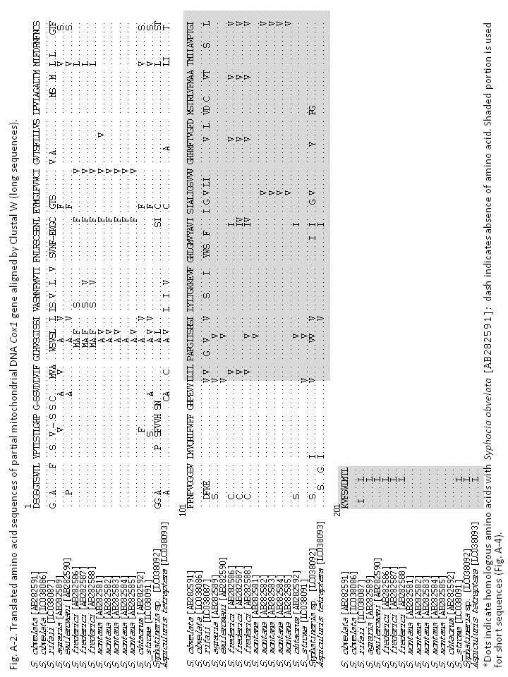
183 178
184 179
185 180
186 181
187 182
188 183
189 184
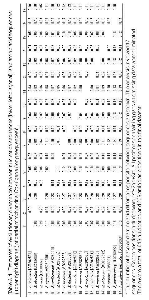
190 185
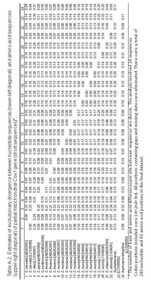
191 186
Article.
 Zootaxa 3881 (2): 155 164 www.mapress.com/zootaxa/ Copyright 2014 Magnolia Press Article http://dx.doi.org/10.11646/zootaxa.3881.2.4 http://zoobank.org/urn:lsid:zoobank.org:pub:679028cd-1d89-4488-bd90-fd78956d1caf
A TRICHOSTRONGYLOID NEMATODE, MACKERRASTRONGYLUS BIAKENSIS, NEW SPECIES, FROM ECHYMIPERA KALUBU (MARSUPIALIA: PERORYCTIDAE) OF IRIAN JAY A, INDONESIA
 OF IRIAN JAY A, INDONESIA") A TRICHOSTRONGYLOID NEMATODE, MACKERRASTRONGYLUS BIAKENSIS, NEW SPECIES, FROM ECHYMIPERA KALUBU (MARSUPIALIA: PERORYCTIDAE) OF IRIAN JAY A, INDONESIA ABSTRACT. Mackerrastrongylus biakensis, new species
A TRICHOSTRONGYLOID NEMATODE, MACKERRASTRONGYLUS BIAKENSIS, NEW SPECIES, FROM ECHYMIPERA KALUBU (MARSUPIALIA: PERORYCTIDAE) OF IRIAN JAY A, INDONESIA ABSTRACT. Mackerrastrongylus biakensis, new species
The Journal of Veterinary Medical Science
 Advance Publication The Journal of Veterinary Medical Science Accepted Date: 2 Aug 2018 J-STAGE Advance Published Date: 15 Aug 2018 1 2 WILDLIFE SCIENCE FULL PAPER 3 4 5 6 Redescription of Subulura (Murisubulura)
Ectoparasites Myobia musculi Radfordia affinis Radfordia ensifera
 Ectoparasites Fleas, ticks, and lice are uncommon in modern laboratory facilities, but may be seen on wild or feral rodents. Most ectoparasite infestations seen in rats and mice used for research are various
HELMINTHES OF ANIMALS IMPORTED IN JAPAN I Tanqua ophidis Johnston and Mawson, 1948 of Water Snakes from Samarinda, Indonesia
 Japan. J. Trop. Med. Hyg., Vol. 5, No. 2, 1977, pp. 155-159 155 HELMINTHES OF ANIMALS IMPORTED IN JAPAN I Tanqua ophidis Johnston and Mawson, 1948 of Water Snakes from Samarinda, Indonesia NOBORU KAGEI1
A COLLECTION OF TICKS (IXODIDAE) FROM SULAWESI UTARA, INDONESIA
 FROM SULAWESI UTARA, INDONESIA") BIOTROPIA (2) 1988/1989: 32-37 A COLLECTION OF TICKS (IXODIDAE) FROM SULAWESI UTARA, INDONESIA L.A. DURDEN Department of Entomology, NHB 165, Museum Support Center Smithsonian Institution, Washington D.C.
BIOTROPIA (2) 1988/1989: 32-37 A COLLECTION OF TICKS (IXODIDAE) FROM SULAWESI UTARA, INDONESIA L.A. DURDEN Department of Entomology, NHB 165, Museum Support Center Smithsonian Institution, Washington D.C.
SEMESTER ONE 2007 INFECTION and IMMUNITY GRADUATE ENTRY PROGRAMME PARASITOLOGY PRACTICAL 9 Dr TW Jones NEMATODES
 SEMESTER ONE 2007 INFECTION and IMMUNITY GRADUATE ENTRY PROGRAMME PARASITOLOGY PRACTICAL 9 Dr TW Jones NEMATODES Objectives After this class I expect you to be able to: 1. Describe and recognise the range
Helminth Parasites Collected from Rattus rattus on Lanyu, Taiwan
 J. Helminthol. Soc. Wash. 61(1), 1994, pp. 95-102 Helminth Parasites Collected from Rattus rattus on Lanyu, Taiwan HIDEO HASEGAWA, JUN KOBAYASHI, AND MASAMITSU OTSURU Department of Parasitology and Research
Title. Author(s)KAMIYA, Haruo; ISHIGAKI, Kenkichi; YAMASHITA, Jiro. CitationJapanese Journal of Veterinary Research, 22(4): 116- Issue Date
KAMIYA, Haruo; ISHIGAKI, Kenkichi; YAMASHITA, Jiro. CitationJapanese Journal of Veterinary Research, 22(4): 116- Issue Date") Title CITELLINA PETROVI SCHULZ, 1930 FROM THE JAPANESE FLY ORII KURODA Author(s)KAMIYA, Haruo; ISHIGAKI, Kenkichi; YAMASHITA, Jiro CitationJapanese Journal of Veterinary Research, 22(4): 116- Issue Date
Title CITELLINA PETROVI SCHULZ, 1930 FROM THE JAPANESE FLY ORII KURODA Author(s)KAMIYA, Haruo; ISHIGAKI, Kenkichi; YAMASHITA, Jiro CitationJapanese Journal of Veterinary Research, 22(4): 116- Issue Date
Nippostrongylus marhaeniae sp. n. and Other Nematodes Collected from Rattus cf. morotaiensis in North Halmahera, Molucca Islands, Indonesia
 J. Helminthol. Soc. Wash. 62(2), 1995, pp. 111-116 Nippostrongylus marhaeniae sp. n. and Other Nematodes Collected from Rattus cf. morotaiensis in North Halmahera, Molucca Islands, Indonesia HIDEO HASEGAWA1'3
Ascarids, Pinworms, and Trichocephalids
 LABORATORY Laboratory 3 Pg. 1 3 Introduction: Ascarids, Pinworms, and Trichocephalids The ascarids are large parasitic nematodes that usually live in the lumen of the small intestine of their host. All
A Scanning Electron Microscopic Study of Eggshell Surface Topography of Leidynema portentosae and L. appendiculatum (Nematoda: Oxyuroidea)
") The Ohio State University Knowledge Bank kb.osu.edu Ohio Journal of Science (Ohio Academy of Science) Ohio Journal of Science: Volume 88, Issue 5 (December, 1988) 1988-12 A Scanning Electron Microscopic
The Ohio State University Knowledge Bank kb.osu.edu Ohio Journal of Science (Ohio Academy of Science) Ohio Journal of Science: Volume 88, Issue 5 (December, 1988) 1988-12 A Scanning Electron Microscopic
Ascarids, Oxyuris, Trichocephalids
 LABORATORY Laboratory 4 Pg. 1 4 Introduction: Ascarids, Oxyuris, Trichocephalids The ascarids are large parasitic nematodes that usually live in the small intestine of their host. All ascarids have 3 lips
Harold W. Manter Laboratory, University of Nebraska State Museum, Lincoln, Nebraska 68588
 Proc. Helminthol. Soc. Wash. 48(2), 1981, pp. 130-136 Observations of the Head and Tail Regions of Male Physaloptera praeputialis von Linstow, 1889, and Physaloptera rara Hall and Wigdor, 1918, Using Scanning
Fischthal and Kuntz (1964) reported the
 reported the") Zoological Studies 41(3): 283-287 (2002) Meristocotyle provitellaria sp. nov. (Digenea: Meristocotylidae) from Varanus salvator in China Wei Liu 1, Qing-Kui Li 2, Hsiu-Hui Shih 3 and Zhao-Zhi Qiu 1, *
Zoological Studies 41(3): 283-287 (2002) Meristocotyle provitellaria sp. nov. (Digenea: Meristocotylidae) from Varanus salvator in China Wei Liu 1, Qing-Kui Li 2, Hsiu-Hui Shih 3 and Zhao-Zhi Qiu 1, *
Breinlia tinjili sp. n. (Filarioidea: Onchocercidae), from the Malaysian Field Rat, Rattus tiomanicus, on Tinjil Island, West Java, Indonesia
, from the Malaysian Field Rat, Rattus tiomanicus, on Tinjil Island, West Java, Indonesia") J. Helminthol. Soc. Wash. 3(1), 199, pp. 93-97 Breinlia tinjili sp. n. (Filarioidea: Onchocercidae), from the Malaysian Field Rat, Rattus tiomanicus, on Tinjil Island, West Java, Indonesia PURNOMO AND
J. Helminthol. Soc. Wash. 3(1), 199, pp. 93-97 Breinlia tinjili sp. n. (Filarioidea: Onchocercidae), from the Malaysian Field Rat, Rattus tiomanicus, on Tinjil Island, West Java, Indonesia PURNOMO AND
A New Species of the Genus Strongylus Muller, 1780 from the Black Rhinoceros, Diceros bicornis L. and a Note on the Other Species Occurring in Kenya
 Journal of H"minthology, Vol. XXXVI, Nos. 1/2, 1ge2, pp. 189-200. A New Species of the Genus Strongylus Muller, 1780 from the Black Rhinoceros, Diceros bicornis L. and a Note on the Other Species Occurring
Descriptions of two new species of the genus. Tachygonetria Wedl, 1862 (Nematoda, Pharyngodonidae) and redescriptions of five
 and redescriptions of five") Descriptions of two new species of the genus Tachygonetria Wedl, 1862 (Nematoda, Pharyngodonidae) and redescriptions of five species parasites of Palaearctic Testudinidae Salah BOUAMER Serge MORAND Centre
Descriptions of two new species of the genus Tachygonetria Wedl, 1862 (Nematoda, Pharyngodonidae) and redescriptions of five species parasites of Palaearctic Testudinidae Salah BOUAMER Serge MORAND Centre
Proc. Helminthol. Soc. Wash. 46(1), 1979, pp
, 1979, pp") Proc. Helminthol. Soc. Wash. 46(1), 1979, pp. 36-42 A Redescription of Dentosiomella translucida Schulz and Krepkorgorskaja, 1932 (Nematoda: Heteroxynematidae) Parasite of Domestic Mongolian Gerbils, Meriones
Proc. Helminthol. Soc. Wash. 46(1), 1979, pp. 36-42 A Redescription of Dentosiomella translucida Schulz and Krepkorgorskaja, 1932 (Nematoda: Heteroxynematidae) Parasite of Domestic Mongolian Gerbils, Meriones
Falcaustra belemensis n. sp. (Nematoda, Kathlaniinae) from the Lizard Neusticums bicarinatus L. (Teiidae) of Brazil
 from the Lizard Neusticums bicarinatus L. (Teiidae) of Brazil") Bull. Mus. natn. Hist, nat., Paris, 4 e ser., 3, 1981, section A, n 1 : 117-121. Falcaustra belemensis n. sp. (Nematoda, Kathlaniinae) from the Lizard Neusticums bicarinatus L. (Teiidae) of Brazil by Michael
Bull. Mus. natn. Hist, nat., Paris, 4 e ser., 3, 1981, section A, n 1 : 117-121. Falcaustra belemensis n. sp. (Nematoda, Kathlaniinae) from the Lizard Neusticums bicarinatus L. (Teiidae) of Brazil by Michael
A NEW SPECIES OF A USTROLIBINIA FROM THE SOUTH CHINA SEA AND INDONESIA (CRUSTACEA: BRACHYURA: MAJIDAE)
") 69 C O a g r ^ j^a RAFFLES BULLETIN OF ZOOLOGY 1992 40(1): 69-73 A NEW SPECIES OF A USTROLIBINIA FROM THE SOUTH CHINA SEA AND INDONESIA (CRUSTACEA: BRACHYURA: MAJIDAE) H P Waener SMITHSONIAN INSTITUTE
69 C O a g r ^ j^a RAFFLES BULLETIN OF ZOOLOGY 1992 40(1): 69-73 A NEW SPECIES OF A USTROLIBINIA FROM THE SOUTH CHINA SEA AND INDONESIA (CRUSTACEA: BRACHYURA: MAJIDAE) H P Waener SMITHSONIAN INSTITUTE
Introduction to Helminthology
 Introduction to Helminthology HELMINTHES (WORMS) - Characteristics Eukaryotic, multicellular animals that usually have digestive, circulatory, nervous, excretory, and reproductive systems. Worms with bilateral
Beaufortia. (Rathke) ZOOLOGICAL MUSEUM - AMSTERDAM. July. Three new commensal Ostracods from Limnoria lignorum
 ZOOLOGICAL MUSEUM - AMSTERDAM. July. Three new commensal Ostracods from Limnoria lignorum") Beaufortia SERIES OF MISCELLANEOUS PUBLICATIONS ZOOLOGICAL MUSEUM - AMSTERDAM No. 34 Volume 4 July 30, 1953 Three new commensal Ostracods from Limnoria lignorum (Rathke) by A.P.C. de Vos (Zoological Museum,
Beaufortia SERIES OF MISCELLANEOUS PUBLICATIONS ZOOLOGICAL MUSEUM - AMSTERDAM No. 34 Volume 4 July 30, 1953 Three new commensal Ostracods from Limnoria lignorum (Rathke) by A.P.C. de Vos (Zoological Museum,
Morphological characterization of Haemonchus contortus in goats (Capra hircus) and sheep (Ovis aries) in Penang, Malaysia
 and sheep (Ovis aries) in Penang, Malaysia") Tropical Biomedicine 24(1): 23 27 (2007) Morphological characterization of Haemonchus contortus in goats (Capra hircus) and sheep (Ovis aries) in Penang, Malaysia Wahab A. Rahman and Suhaila Abd. Hamid
Tropical Biomedicine 24(1): 23 27 (2007) Morphological characterization of Haemonchus contortus in goats (Capra hircus) and sheep (Ovis aries) in Penang, Malaysia Wahab A. Rahman and Suhaila Abd. Hamid
A new species of Antinia PASCOE from Burma (Coleoptera: Curculionidae: Entiminae)
") Genus Vol. 14 (3): 413-418 Wroc³aw, 15 X 2003 A new species of Antinia PASCOE from Burma (Coleoptera: Curculionidae: Entiminae) JAROS AW KANIA Zoological Institute, University of Wroc³aw, Sienkiewicza
Genus Vol. 14 (3): 413-418 Wroc³aw, 15 X 2003 A new species of Antinia PASCOE from Burma (Coleoptera: Curculionidae: Entiminae) JAROS AW KANIA Zoological Institute, University of Wroc³aw, Sienkiewicza
Morphologic study of dog flea species by scanning electron microscopy
 Scientia Parasitologica, 2006, 3-4, 77-81 Morphologic study of dog flea species by scanning electron microscopy NAGY Ágnes 1, L. BARBU TUDORAN 2, V. COZMA 1 1 University of Agricultural Sciences and Veterinary
Vol. XIV, No. 1, March, The Larva and Pupa of Brontispa namorikia Maulik (Coleoptera: Chrysomelidae: Hispinae) By S.
 By S.") Vol. XIV, No. 1, March, 1950 167 The Larva and Pupa of Brontispa namorikia Maulik (Coleoptera: Chrysomelidae: Hispinae) By S. MAULIK BRITISH MUSEUM (NATURAL HISTORY) (Presented by Mr. Van Zwaluwenburg
Vol. XIV, No. 1, March, 1950 167 The Larva and Pupa of Brontispa namorikia Maulik (Coleoptera: Chrysomelidae: Hispinae) By S. MAULIK BRITISH MUSEUM (NATURAL HISTORY) (Presented by Mr. Van Zwaluwenburg
JOURNAL OF INTERNATIONAL ACADEMIC RESEARCH FOR MULTIDISCIPLINARY Impact Factor 1.393, ISSN: , Volume 2, Issue 8, September 2014
 THE FIRST RECORD OF NEMATODA Chabaudinema americana ( Diaz- Ungria, 1968) FROM TURTLES AT BASRAH CITY/ IRAQ DR. FATIN A. A. MUSTAFA* *Asst. Professor, Dept. of Biology, College of Education of Pure Science,
DESCRIPTIONS OF THREE NEW SPECIES OF PETALOCEPHALA STÅL, 1853 FROM CHINA (HEMIPTERA: CICADELLIDAE: LEDRINAE) Yu-Jian Li* and Zi-Zhong Li**
 Yu-Jian Li* and Zi-Zhong Li**") 499 DESCRIPTIONS OF THREE NEW SPECIES OF PETALOCEPHALA STÅL, 1853 FROM CHINA (HEMIPTERA: CICADELLIDAE: LEDRINAE) Yu-Jian Li* and Zi-Zhong Li** * Institute of Entomology, Guizhou University, Guiyang, Guizhou
499 DESCRIPTIONS OF THREE NEW SPECIES OF PETALOCEPHALA STÅL, 1853 FROM CHINA (HEMIPTERA: CICADELLIDAE: LEDRINAE) Yu-Jian Li* and Zi-Zhong Li** * Institute of Entomology, Guizhou University, Guiyang, Guizhou
IDENTIFICATION / GENERAL CHARACTERISTICS OF TICK GENERA (HARD AND SOFT TICKS)
") Ticks Tick identification Authors: Prof Maxime Madder, Prof Ivan Horak, Dr Hein Stoltsz Licensed under a Creative Commons Attribution license. IDENTIFICATION / GENERAL CHARACTERISTICS OF TICK GENERA (HARD
Ticks Tick identification Authors: Prof Maxime Madder, Prof Ivan Horak, Dr Hein Stoltsz Licensed under a Creative Commons Attribution license. IDENTIFICATION / GENERAL CHARACTERISTICS OF TICK GENERA (HARD
TWO NEW SPECIES OF WATER MITES FROM OHIO 1-2
 TWO NEW SPECIES OF WATER MITES FROM OHIO 1-2 DAVID R. COOK Wayne State University, Detroit, Michigan ABSTRACT Two new species of Hydracarina, Tiphys weaveri (Acarina: Pionidae) and Axonopsis ohioensis
Skrjabinodon piankai sp. n. (Nematoda: Pharyngodonidae) and Other Helminths of Geckos (Sauria: Gekkonidae: Nephrurus spp.
 and Other Helminths of Geckos (Sauria: Gekkonidae: Nephrurus spp.") J. Helminthol. Soc. Wash. 66(2), 1999 pp. 175-179 Skrjabinodon piankai sp. n. (Nematoda: Pharyngodonidae) and Other Helminths of Geckos (Sauria: Gekkonidae: Nephrurus spp.) from Australia CHARLES R. BURSEYU
J. Helminthol. Soc. Wash. 66(2), 1999 pp. 175-179 Skrjabinodon piankai sp. n. (Nematoda: Pharyngodonidae) and Other Helminths of Geckos (Sauria: Gekkonidae: Nephrurus spp.) from Australia CHARLES R. BURSEYU
Oribatid Mites of the Family Otocepheidae from Tian-mu Mountain in China (Acari: Oribatida)1'
1'") Acta arachnol,, 42 (1): 1-6, August 30, 1993 Oribatid Mites of the Family Otocepheidae from Tian-mu Mountain in China (Acari: Oribatida)1' Jun-ichi AoKI2' and Sheng-hao Hu3' Abstract Dolicheremaeus wangi
Acta arachnol,, 42 (1): 1-6, August 30, 1993 Oribatid Mites of the Family Otocepheidae from Tian-mu Mountain in China (Acari: Oribatida)1' Jun-ichi AoKI2' and Sheng-hao Hu3' Abstract Dolicheremaeus wangi
BIO Parasitology Spring Trichostrongylines. Lecture 20. Hairworms in Horses. Stephen M. Shuster Northern Arizona University
 BIO 475 - Parasitology Spring 2009 Stephen M. Shuster Northern Arizona University http://www4.nau.edu/isopod Lecture 20 Trichostrongylines Hairworms in Horses Eggs hatch when eaten by the horse. Larvae
A New Species of the Genus Asemonea (Araneae: Salticidae) from Japan
 from Japan") Acta arachnol., 45 (2): 113-117, December 30, 1996 A New Species of the Genus Asemonea (Araneae: Salticidae) from Japan Hiroyoshi IKEDA1 Abstract A new salticid spider species, Asemonea tanikawai sp. nov.
Acta arachnol., 45 (2): 113-117, December 30, 1996 A New Species of the Genus Asemonea (Araneae: Salticidae) from Japan Hiroyoshi IKEDA1 Abstract A new salticid spider species, Asemonea tanikawai sp. nov.
A NEW SALTICID SPIDER FROM VICTORIA By R. A. Dunn
 Dunn, R. A. 1947. A new salticid spider from Victoria. Memoirs of the National Museum of Victoria 15: 82 85. All text not included in the original document is highlighted in red. Mem. Nat. Mus. Vict.,
The family Gnaphosidae is a large family
 Pakistan J. Zool., vol. 36(4), pp. 307-312, 2004. New Species of Zelotus Spider (Araneae: Gnaphosidae) from Pakistan ABIDA BUTT AND M.A. BEG Department of Zoology, University of Agriculture, Faisalabad,
Two new Phradonoma species (Coleoptera: Dermestidae) from Iran
 from Iran") Journal of Entomological Society of Iran 2008, 28(1), 87-91 87 Two new Phradonoma species (Coleoptera: Dermestidae) from Iran A. Herrmann 1&* and J. Háva 2 1. Bremervörder Strasse 123, D - 21682 Stade,
Journal of Entomological Society of Iran 2008, 28(1), 87-91 87 Two new Phradonoma species (Coleoptera: Dermestidae) from Iran A. Herrmann 1&* and J. Háva 2 1. Bremervörder Strasse 123, D - 21682 Stade,
The Rat Lungworm Lifecycle
 Hawaii Island Rat Lungworm Working Group Daniel K. Inouye College of Pharmacy University of Hawaii, Hilo The Rat Lungworm Lifecycle Rat Lungworm IPM RLWL-3 It is important to understand the lifecycle of
Transactions of the Royal Society of S. Aust. (20--), 000 (0): PELECITUS BARTNERI SP. NOV. (NEMATODA: FILARIOIDEA) FROM
, 000 (0): PELECITUS BARTNERI SP. NOV. (NEMATODA: FILARIOIDEA) FROM") 1 Transactions of the Royal Society of S. Aust. (20--), 000 (0): 00-00 PELECITUS BARTNERI SP. NOV. (NEMATODA: FILARIOIDEA) FROM THE SUBCUTANEOUS TISSUES OF THE LEG OF PSEPHOTUS CHRYSOPTERYGIUS GOULD, 1858
1 Transactions of the Royal Society of S. Aust. (20--), 000 (0): 00-00 PELECITUS BARTNERI SP. NOV. (NEMATODA: FILARIOIDEA) FROM THE SUBCUTANEOUS TISSUES OF THE LEG OF PSEPHOTUS CHRYSOPTERYGIUS GOULD, 1858
UPOGEBIA LINCOLNI SP. NOV. (DECAPODA, THALASSINIDEA, UPOGEBIIDAE) FROM JAVA, INDONESIA
 FROM JAVA, INDONESIA") NOTES AND NEWS UPOGEBIA LINCOLNI SP. NOV. (DECAPODA, THALASSINIDEA, UPOGEBIIDAE) FROM JAVA, INDONESIA BY NGUYEN NGOC-HO i) Faculty of Science, University of Saigon, Vietnam Among material recently collected
NOTES AND NEWS UPOGEBIA LINCOLNI SP. NOV. (DECAPODA, THALASSINIDEA, UPOGEBIIDAE) FROM JAVA, INDONESIA BY NGUYEN NGOC-HO i) Faculty of Science, University of Saigon, Vietnam Among material recently collected
BIO 221 Invertebrate Zoology I Spring Ancylostoma caninum. Ancylostoma caninum cuticular larval migrans. Lecture 23
 BIO 221 Invertebrate Zoology I Spring 2010 Stephen M. Shuster Northern Arizona University http://www4.nau.edu/isopod Lecture 23 Ancylostoma caninum Ancylostoma caninum cuticular larval migrans Order Ascarida
Exceptional fossil preservation demonstrates a new mode of axial skeleton elongation in early ray-finned fishes
 Supplementary Information Exceptional fossil preservation demonstrates a new mode of axial skeleton elongation in early ray-finned fishes Erin E. Maxwell, Heinz Furrer, Marcelo R. Sánchez-Villagra Supplementary
Title: Phylogenetic Methods and Vertebrate Phylogeny
 Title: Phylogenetic Methods and Vertebrate Phylogeny Central Question: How can evolutionary relationships be determined objectively? Sub-questions: 1. What affect does the selection of the outgroup have
Spauligodon timbavatiensis n. sp. (Nematoda: Pharyngodonidae) from Pachydactylus turneri (Sauria: Gekkonidae) in the Northern Province, South Africa
 from Pachydactylus turneri (Sauria: Gekkonidae) in the Northern Province, South Africa") Onderstepoort Journal of Veterinary Research, 65:153-158 (1998) Spauligodon timbavatiensis n. sp. (Nematoda: Pharyngodonidae) from Pachydactylus turneri (Sauria: Gekkonidae) in the Northern Province, South
Onderstepoort Journal of Veterinary Research, 65:153-158 (1998) Spauligodon timbavatiensis n. sp. (Nematoda: Pharyngodonidae) from Pachydactylus turneri (Sauria: Gekkonidae) in the Northern Province, South
WITH THE TABLE OF THE MORPHOLOGICAL FEATURES OF TAPEWORMS IN VAMPIROLEPIS. (Received: December 22nd, 1965)
") Japan. J. Med. Sci. Biol. 19, 51-57, 1966 *ON A NEW TAPEWORM, VAMPIROLEPIS ISENSIS, FOUND IN BATS WITH THE TABLE OF THE MORPHOLOGICAL FEATURES OF TAPEWORMS IN VAMPIROLEPIS ISAMU SAWADA Biological Laboratory,
Japan. J. Med. Sci. Biol. 19, 51-57, 1966 *ON A NEW TAPEWORM, VAMPIROLEPIS ISENSIS, FOUND IN BATS WITH THE TABLE OF THE MORPHOLOGICAL FEATURES OF TAPEWORMS IN VAMPIROLEPIS ISAMU SAWADA Biological Laboratory,
Title. Author(s)OHBAYASHI, Masashi. CitationJapanese Journal of Veterinary Research, 15(1): 1-3. Issue Date DOI. Doc URL.
OHBAYASHI, Masashi. CitationJapanese Journal of Veterinary Research, 15(1): 1-3. Issue Date DOI. Doc URL.") Title GRYPORHYNCHUS NYCTICORACIS YAMAGUTI, 1956 (DILEPIDID APHARYNGOSTRIGEA ARDEOLINA VIDYARTHI, 1937 (STRIGEID CINEREA JOUYI CLARK Author(s)OHBAYASHI, Masashi CitationJapanese Journal of Veterinary Research,
Title GRYPORHYNCHUS NYCTICORACIS YAMAGUTI, 1956 (DILEPIDID APHARYNGOSTRIGEA ARDEOLINA VIDYARTHI, 1937 (STRIGEID CINEREA JOUYI CLARK Author(s)OHBAYASHI, Masashi CitationJapanese Journal of Veterinary Research,
Hexamermis glossinae spnov. (Nematoda: Mermithidae), a parasite of tse-tse flies in West Africa
, a parasite of tse-tse flies in West Africa") I. ' NOTES Hexamermis glossinae spnov. (Nematoda: Mermithidae), a parasite of tse-tse flies in West Africa GEORGE O. POINAR, JR. Division of Entomology and Parasitology, University of California, Berkeley,
I. ' NOTES Hexamermis glossinae spnov. (Nematoda: Mermithidae), a parasite of tse-tse flies in West Africa GEORGE O. POINAR, JR. Division of Entomology and Parasitology, University of California, Berkeley,
Phylum Platyhelminthes Flatworms
 Phylum Platyhelminthes Flatworms The Acoelomates The acoelomates are animals that lack a coelom. Acoelomates lack a body cavity, and instead the space between the body wall and the digestive tract is filled
Title. Author(s)Takahashi, Ryoichi. CitationInsecta matsumurana, 14(1): 1-5. Issue Date Doc URL. Type. File Information
Takahashi, Ryoichi. CitationInsecta matsumurana, 14(1): 1-5. Issue Date Doc URL. Type. File Information") Title Some Aleyrodidae from Mauritius (Homoptera) Author(s)Takahashi, Ryoichi CitationInsecta matsumurana, 14(1): 1-5 Issue Date 1939-12 Doc URL http://hdl.handle.net/2115/9426 Type bulletin File Information
Title Some Aleyrodidae from Mauritius (Homoptera) Author(s)Takahashi, Ryoichi CitationInsecta matsumurana, 14(1): 1-5 Issue Date 1939-12 Doc URL http://hdl.handle.net/2115/9426 Type bulletin File Information
Two new species of Parapharyngodon (Oxyuroidea: Pharyngodonidae) from the enigmatic Bipes canaliculatus and Bipes tridactylus (Squamata: Bipedidae)
 from the enigmatic Bipes canaliculatus and Bipes tridactylus (Squamata: Bipedidae)") Revista Mexicana de Biodiversidad 79: 113S- 120S, 2008 Two new species of Parapharyngodon (Oxyuroidea: Pharyngodonidae) from the enigmatic Bipes canaliculatus and Bipes tridactylus (Squamata: Bipedidae)
Revista Mexicana de Biodiversidad 79: 113S- 120S, 2008 Two new species of Parapharyngodon (Oxyuroidea: Pharyngodonidae) from the enigmatic Bipes canaliculatus and Bipes tridactylus (Squamata: Bipedidae)
Order Strongylida. Superfamilies: Trichostrongyloidea Strongyloidea Metastrongyloidea Ancylostomatoidea (hookworms)
") Order Strongylida Superfamilies: Trichostrongyloidea Strongyloidea Metastrongyloidea Ancylostomatoidea (hookworms) ORDER STRONGYLIDA - Bursate worms Superfamily - Ancylostomatoidea HOOKWORMS *dorsally
Order Strongylida Superfamilies: Trichostrongyloidea Strongyloidea Metastrongyloidea Ancylostomatoidea (hookworms) ORDER STRONGYLIDA - Bursate worms Superfamily - Ancylostomatoidea HOOKWORMS *dorsally
BREVIORA LEUCOLEPIDOPA SUNDA GEN. NOV., SP. NOV. (DECAPODA: ALBUNEIDAE), A NEW INDO-PACIFIC SAND CRAB. Ian E. Efford 1
, A NEW INDO-PACIFIC SAND CRAB. Ian E. Efford 1") ac lc BREVIORA CAMBRIDGE, MASS. 30 APRIL, 1969 NUMBER 318 LEUCOLEPIDOPA SUNDA GEN. NOV., SP. NOV. (DECAPODA: ALBUNEIDAE), A NEW INDO-PACIFIC SAND CRAB Ian E. Efford 1 ABSTRACT. Leucolepidopa gen. nov.
ac lc BREVIORA CAMBRIDGE, MASS. 30 APRIL, 1969 NUMBER 318 LEUCOLEPIDOPA SUNDA GEN. NOV., SP. NOV. (DECAPODA: ALBUNEIDAE), A NEW INDO-PACIFIC SAND CRAB Ian E. Efford 1 ABSTRACT. Leucolepidopa gen. nov.
Three Nematode Species Recovered from Terrestrial Snakes in Republic of Korea
 ISSN (Print) 0023-4001 ISSN (Online) 1738-0006 BRIEF COMMUNICATION Korean J Parasitol Vol. 54, No. 2: 205-213, April 2016 http://dx.doi.org/10.3347/kjp.2016.54.2.205 Three Nematode Species Recovered from
Main Points. 2) The Great American Interchange -- dispersal versus vicariance -- example: recent range expansion of nine-banded armadillos
 The Great American Interchange -- dispersal versus vicariance -- example: recent range expansion of nine-banded armadillos") Main Points 1) Mammalian Characteristics: Diversity, Phylogeny, and Systematics: -- Infraclass Eutheria -- Orders Scandentia through Cetacea 2) The Great American Interchange -- dispersal versus vicariance
Main Points 1) Mammalian Characteristics: Diversity, Phylogeny, and Systematics: -- Infraclass Eutheria -- Orders Scandentia through Cetacea 2) The Great American Interchange -- dispersal versus vicariance
TWO NEW PINE-FEEDING SPECIES OF COLEOTECHNITES ( GELECHIIDAE )
") Journal of the Lepidopterists' Society 32(2), 1978, 118-122 TWO NEW PINE-FEEDING SPECIES OF COLEOTECHNITES ( GELECHIIDAE ) RONALD W. HODGES l AND ROBERT E. STEVENS2 ABSTRACT. Two new species of moths,
Journal of the Lepidopterists' Society 32(2), 1978, 118-122 TWO NEW PINE-FEEDING SPECIES OF COLEOTECHNITES ( GELECHIIDAE ) RONALD W. HODGES l AND ROBERT E. STEVENS2 ABSTRACT. Two new species of moths,
Morphological study on Dermacentor marginatus (Acari: Ixodidae) by environmental scanning electron microscopy (E.S.E.M.)
 by environmental scanning electron microscopy (E.S.E.M.)") Scientia Parasitologica, 2005, 1-2, 111-115 Morphological study on Dermacentor marginatus (Acari: Ixodidae) y environmental scanning electron microscopy Mariana IONIłĂ*, I.L. MITREA*, F. MINCULESCU** *
Scientia Parasitologica, 2005, 1-2, 111-115 Morphological study on Dermacentor marginatus (Acari: Ixodidae) y environmental scanning electron microscopy Mariana IONIłĂ*, I.L. MITREA*, F. MINCULESCU** *
Lecture 11 Wednesday, September 19, 2012
 Lecture 11 Wednesday, September 19, 2012 Phylogenetic tree (phylogeny) Darwin and classification: In the Origin, Darwin said that descent from a common ancestral species could explain why the Linnaean
ZOOSYSTEMA (1)
") Two Trichostrongylina (Nematoda) from Venezuela: a new species of Ornithostrongylus (Heligmosomoidea), parasitic in birds (Columbiformes) and a new species of Molineus (Molineoidea), parasitic in snakes
Two Trichostrongylina (Nematoda) from Venezuela: a new species of Ornithostrongylus (Heligmosomoidea), parasitic in birds (Columbiformes) and a new species of Molineus (Molineoidea), parasitic in snakes
JOURNAL OF. RONALD W. HODGES Systematic Entomology Laboratory, USDA, % U.S. National Museum of Natural History, MRC 168, Washington, D.C.
 JOURNAL OF THE LEPIDOPTERISTS' Volume 39 1985 SOCIETY Number 3 Journal of the Lepidopterists' Society 39(3), 1985, 151-155 A NEW SPECIES OF TlLDENIA FROM ILLINOIS (GELECHIIDAE) RONALD W. HODGES Systematic
Z. Kabata V9R 5K6. Canada, Abstract. Zusammenfassung. nov. (Copepoda: Hatschekiidae) werden beschrieben und. quently, I must. which I propose. nov.
 werden beschrieben und. quently, I must. which I propose. nov.") Beaufortia INSTITUTE OF TAXONOMIC ZOOLOGY (ZOOLOGICAL MUSEUM) UNIVERSITY OF AMSTERDAM Vol. 41, no. 19 October 22, 1990 Prohatschekia stocki sp. nov.(copepoda: Hatschekiidae) a parasite of an Australian
Beaufortia INSTITUTE OF TAXONOMIC ZOOLOGY (ZOOLOGICAL MUSEUM) UNIVERSITY OF AMSTERDAM Vol. 41, no. 19 October 22, 1990 Prohatschekia stocki sp. nov.(copepoda: Hatschekiidae) a parasite of an Australian
NOTES ON TWO ASTIGMATIC MITES (ACARI) LIVING IN BEEHIVES IN THAILAND
 LIVING IN BEEHIVES IN THAILAND") NOTES ON TWO ASTIGMATIC MITES (ACARI) LIVING IN BEEHIVES IN THAILAND BY A. FAIN* and V. GERSON ** APIS CERANA HIVES MITES APIS CERANA RUCHES ACARIENS SUMMARY: Two species of Astigmatic mites were found
NOTES ON TWO ASTIGMATIC MITES (ACARI) LIVING IN BEEHIVES IN THAILAND BY A. FAIN* and V. GERSON ** APIS CERANA HIVES MITES APIS CERANA RUCHES ACARIENS SUMMARY: Two species of Astigmatic mites were found
Scorpionyssus heterometrus gen. n., sp. n. (Acari, Laelapidae) parasitic on a scorpion from Sri Lanka
 parasitic on a scorpion from Sri Lanka") Entomol. Mitt. zool. Mus. Hamburg Bd. 9 (1988) Nr. 132 Scorpionyssus heterometrus gen. n., sp. n. (Acari, Laelapidae) parasitic on a scorpion from Sri Lanka Alex Fain and Gisela Rack (With 18 figures)
Entomol. Mitt. zool. Mus. Hamburg Bd. 9 (1988) Nr. 132 Scorpionyssus heterometrus gen. n., sp. n. (Acari, Laelapidae) parasitic on a scorpion from Sri Lanka Alex Fain and Gisela Rack (With 18 figures)
SILICIFIED TURBELLARIA FROM CALICO MOUNTAINS NODULES
 ^os BULLETIN, SO. CALIF. ACADEMY OF SCIENCES Vol. 59, Part 3, 1960 SILICIFIED TURBELLARIA FROM CALICO MOUNTAINS NODULES W. DWIGHT jplerce Drawings by the author. The following is the fifth report of the
Main Points. 2) The Great American Interchange -- dispersal versus vicariance -- example: recent range expansion of nine-banded armadillos
 The Great American Interchange -- dispersal versus vicariance -- example: recent range expansion of nine-banded armadillos") Main Points 1) Diversity, Phylogeny, and Systematics -- Infraclass Eutheria -- Orders Scandentia through Cetacea 2) The Great American Interchange -- dispersal versus vicariance -- example: recent range
Main Points 1) Diversity, Phylogeny, and Systematics -- Infraclass Eutheria -- Orders Scandentia through Cetacea 2) The Great American Interchange -- dispersal versus vicariance -- example: recent range
TWO NEW HETEROMORPHIe DEUTONYMPHS (HYPOPI) (ACARINA: HYPODERIDAE) FROM THE GREAT FRIGATEBIRD (FREGATA MINOR)1,2
 (ACARINA: HYPODERIDAE) FROM THE GREAT FRIGATEBIRD (FREGATA MINOR)1,2") J. Med. Ent. Vol. 5, DO. 3: 320-324 1 August 1968 TWO NEW HETEROMORPHIe DEUTONYMPHS (HYPOPI) (ACARINA: HYPODERIDAE) FROM THE GREAT FRIGATEBIRD (FREGATA MINOR)1,2 By Alex Fain 3 and A. Binion Arnerson,
J. Med. Ent. Vol. 5, DO. 3: 320-324 1 August 1968 TWO NEW HETEROMORPHIe DEUTONYMPHS (HYPOPI) (ACARINA: HYPODERIDAE) FROM THE GREAT FRIGATEBIRD (FREGATA MINOR)1,2 By Alex Fain 3 and A. Binion Arnerson,
Sepia prabahari sp. nov. (Mollusca/Cephalopoda), a new species of Acanthosepion species complex from Tuticorin bay, southeast coast of India
, a new species of Acanthosepion species complex from Tuticorin bay, southeast coast of India") Indian Journal of Marine Sciences Vol. 31(1), March 2002, pp. 45-51 Sepia prabahari sp. nov. (Mollusca/Cephalopoda), a new species of Acanthosepion species complex from Tuticorin bay, southeast coast of
Indian Journal of Marine Sciences Vol. 31(1), March 2002, pp. 45-51 Sepia prabahari sp. nov. (Mollusca/Cephalopoda), a new species of Acanthosepion species complex from Tuticorin bay, southeast coast of
TWO NEW SPECIES OF ACUTIGEBIA (CRUSTACEA: DECAPODA: GEBIIDEA: UPOGEBIIDAE) FROM THE SOUTH CHINA SEA
 FROM THE SOUTH CHINA SEA") THE RAFFLES BULLETIN OF ZOOLOGY 2013 61(2): 571 577 Date of Publication: 30 Aug.2013 National University of Singapore TWO NEW SPECIES OF ACUTIGEBIA (CRUSTACEA: DECAPODA: GEBIIDEA: UPOGEBIIDAE) FROM THE
THE RAFFLES BULLETIN OF ZOOLOGY 2013 61(2): 571 577 Date of Publication: 30 Aug.2013 National University of Singapore TWO NEW SPECIES OF ACUTIGEBIA (CRUSTACEA: DECAPODA: GEBIIDEA: UPOGEBIIDAE) FROM THE
TWO NEW SPECIES OF THE GENUS OSWALDOCRUZIA TRAVASSOS, 1917 (NEMATODA: TRICHOSTRONGYLINA: MOLINEOIDEA) PARASITIZING SPANISH AMPHIBIANS
 PARASITIZING SPANISH AMPHIBIANS") Research and Reviews ill Parasitology, 55 (4): 209 215 (1995) 1995 Asociaci6ndeParasit6logos Espaiioles (A.P.E.) PublishedbyA.P.E. Printed in Barcelona. Spain TWO NEW SPECIES OF THE GENUS OSWALDOCRUZIA
Research and Reviews ill Parasitology, 55 (4): 209 215 (1995) 1995 Asociaci6ndeParasit6logos Espaiioles (A.P.E.) PublishedbyA.P.E. Printed in Barcelona. Spain TWO NEW SPECIES OF THE GENUS OSWALDOCRUZIA
Diurus, Pascoe. sp. 1). declivity of the elytra, but distinguished. Length (the rostrum and tails 26 included) mm. Deep. exception
. declivity of the elytra, but distinguished. Length (the rostrum and tails 26 included) mm. Deep. exception") 210 DIURUS ERYTIIROPUS. NOTE XXVI. Three new species of the Brenthid genus Diurus, Pascoe DESCRIBED BY C. Ritsema+Cz. 1. Diurus erythropus, n. sp. 1). Allied to D. furcillatus Gylh. ²) by the short head,
210 DIURUS ERYTIIROPUS. NOTE XXVI. Three new species of the Brenthid genus Diurus, Pascoe DESCRIBED BY C. Ritsema+Cz. 1. Diurus erythropus, n. sp. 1). Allied to D. furcillatus Gylh. ²) by the short head,
Heterodera cynodontis n. sp. (Nematoda : Heteroderidae)
") Heterodera cynodontis n. sp. (Nematoda : Heteroderidae) from Cynodon dactylon (L.) in Pakistan Fayyaz SHAHINA and Mohammad A. MAQBOOL National Nematological Research Centre University of Karachi, Karachi-32,
Heterodera cynodontis n. sp. (Nematoda : Heteroderidae) from Cynodon dactylon (L.) in Pakistan Fayyaz SHAHINA and Mohammad A. MAQBOOL National Nematological Research Centre University of Karachi, Karachi-32,
DISCOVERY OF GENUS PLATOLENES (COLEOP TERA : TENEBRIONIDAE) FROM INDIA WITH DESCRIPTION OF TWO NEW SPECIES G. N. SABA
 FROM INDIA WITH DESCRIPTION OF TWO NEW SPECIES G. N. SABA") Rec. zool. Surv. India, 85(3) : 433-437,1988 DISCOVERY OF GENUS PLATOLENES (COLEOP TERA : TENEBRIONIDAE) FROM INDIA WITH DESCRIPTION OF TWO NEW SPECIES By G. N. SABA Zoological Survey of India M-Block,
Rec. zool. Surv. India, 85(3) : 433-437,1988 DISCOVERY OF GENUS PLATOLENES (COLEOP TERA : TENEBRIONIDAE) FROM INDIA WITH DESCRIPTION OF TWO NEW SPECIES By G. N. SABA Zoological Survey of India M-Block,
S.F.B.N HERING-HAGENBECK1, A.J. PETTER2 and J. BOOMKER3. Onderstepoort Journal of Veterinary Research, 69:7-29
 Onderstepoort Journal of Veterinary Research, 69:7-29 Redescription of some Spau/igodon spp. and Parapharyngodon spp., and of Skrjabinodon mabuyae (Sandground, 1936) Inglis, 1968 (Pharyngodonidae: Oxyuroidea)
A NEW AUSTROSQUILLA (STOMATOPODA) FROM THE
 FROM THE") A NEW AUSTROSQUILLA (STOMATOPODA) FROM THE MARQUESAS ISLANDS BY ALAIN MICHEL Centre O.R.S.T.O.M., Noumea, New Caledonia and RAYMOND B. MANNING Smithsonian Institution, Washington, U.S.A. The At s,tstrosqzlilla
A NEW AUSTROSQUILLA (STOMATOPODA) FROM THE MARQUESAS ISLANDS BY ALAIN MICHEL Centre O.R.S.T.O.M., Noumea, New Caledonia and RAYMOND B. MANNING Smithsonian Institution, Washington, U.S.A. The At s,tstrosqzlilla
Flatworms Flatworms Platyhelminthes dorsoventrally free-living planarian parasitic fluke tapeworm label three body layers ectoderm mesoderm
 Flatworms Flatworms are in the phylum Platyhelminthes. Flatworms are flattened dorsoventrally (top to bottom). The group includes the freshwater, free-living planarian and the parasitic fluke and tapeworm.
FOUR NEW SPECIES AND A NEW RECORD OF CHIMARRA STEPHENS (TRICHOPTERA: PHILOPOTAMIDAE) FROM BOUGAINVILLE ISLAND, PAPUA NEW GUINEA
 FROM BOUGAINVILLE ISLAND, PAPUA NEW GUINEA") Memoirs of Museum Victoria 58(2): 223 230 (2001) FOUR NEW SPECIES AND A NEW RECORD OF CHIMARRA STEPHENS (TRICHOPTERA: PHILOPOTAMIDAE) FROM BOUGAINVILLE ISLAND, PAPUA NEW GUINEA DAVID I. CARTWRIGHT 13 Brolga
Memoirs of Museum Victoria 58(2): 223 230 (2001) FOUR NEW SPECIES AND A NEW RECORD OF CHIMARRA STEPHENS (TRICHOPTERA: PHILOPOTAMIDAE) FROM BOUGAINVILLE ISLAND, PAPUA NEW GUINEA DAVID I. CARTWRIGHT 13 Brolga
Main Points. 2) The Great American Interchange -- dispersal versus vicariance -- example: recent range expansion of nine-banded armadillos
 The Great American Interchange -- dispersal versus vicariance -- example: recent range expansion of nine-banded armadillos") Main Points 1) Diversity, Phylogeny, and Systematics -- Infraclass Metatheria continued -- Orders Diprotodontia and Peramelina -- Infraclass Eutheria -- Orders Lagomorpha through Cetacea 2) The Great American
Main Points 1) Diversity, Phylogeny, and Systematics -- Infraclass Metatheria continued -- Orders Diprotodontia and Peramelina -- Infraclass Eutheria -- Orders Lagomorpha through Cetacea 2) The Great American
MORPHOTAXONOMICAL STUDY OF A NEW CESTODE GANGESIA (GANGESIA) CHOPARAI N.SP. FROM A FRESH WATER FISH, WALLAGO ATTU FROM JALAUN (U.P.
 CHOPARAI N.SP. FROM A FRESH WATER FISH, WALLAGO ATTU FROM JALAUN (U.P.") FLORA AND FAUNA 2016 Vol. 22 No. 1 PP 115-120 ISSN 0971-6920 MORPHOTAXONOMICAL STUDY OF A NEW CESTODE GANGESIA (GANGESIA) CHOPARAI N.SP. FROM A FRESH WATER FISH, WALLAGO ATTU FROM JALAUN (U.P.) INDIA ALOK
FLORA AND FAUNA 2016 Vol. 22 No. 1 PP 115-120 ISSN 0971-6920 MORPHOTAXONOMICAL STUDY OF A NEW CESTODE GANGESIA (GANGESIA) CHOPARAI N.SP. FROM A FRESH WATER FISH, WALLAGO ATTU FROM JALAUN (U.P.) INDIA ALOK
AUSTRALIAN MUSEUM SCIENTIFIC PUBLICATIONS
 AUSTRALIAN MUSEUM SCIENTIFIC PUBLICATIONS Riek, E. F., 1964. Merostomoidea (Arthropoda, Trilobitomorpha) from the Australian Middle Triassic. Records of the Australian Museum 26(13): 327 332, plate 35.
Attagivora, a new genus o f feather mite
 Entomol. Mitt. zool. Mus. Hamburg Bd. 10 (1992) Nr. 146 Attagivora, a new genus o f feather mite subfam ily Avenzoariinae (Analgoidea: Avenzoariidae) from seedsnipes o f the genus Attagis (Charadriiformes:
New York State Mammals. Order Lagomorpha Order Rodentia
 New York State Mammals Order Lagomorpha Order Rodentia FAMILY: LEPORIDAE Rabbits and hares Conspicuous tail Fenestra appears as bony latticework Some species molt seasonally Presence of a second incisor
Introduction. Syst Parasitol DOI /s
 DOI 10.1007/s10-017-9713-9 A new species of Neocosmocercella Baker & Vaucher, 1983 (Nematoda: Cosmocercidae), a parasite of Phyllomedusa vaillantii Boulenger (Anura: Phyllomedusidae) in the Caxiuanã National
CHERYL M. BARTLETT' AND ODILE BAIN2. Proc. Helminthol. Soc. Wash. 54(1), 1987, pp. 1-14
, 1987, pp. 1-14") Proc. Helminthol. Soc. Wash. 54(1), 1987, pp. 1-14 New Avian Filarioids (Nematoda: Splendidofilariinae): Dessetfilaria guianensis gen. n., sp. n., Andersonfilaria africanus gen. n., sp. n., and Splendidofilaria
Proc. Helminthol. Soc. Wash. 54(1), 1987, pp. 1-14 New Avian Filarioids (Nematoda: Splendidofilariinae): Dessetfilaria guianensis gen. n., sp. n., Andersonfilaria africanus gen. n., sp. n., and Splendidofilaria
Postilla PEABODY MUSEUM OF NATURAL HISTORY YALE UNIVERSITY NEW HAVEN, CONNECTICUT, U.S.A.
 Postilla PEABODY MUSEUM OF NATURAL HISTORY YALE UNIVERSITY NEW HAVEN, CONNECTICUT, U.S.A. Number 117 18 March 1968 A 7DIAPSID (REPTILIA) PARIETAL FROM THE LOWER PERMIAN OF OKLAHOMA ROBERT L. CARROLL REDPATH
Central Marine Fisheries Research Institute, Mandapam Camp
 w«r n Mar. biol. Ass. India, 1961, 3 (1 & 2): 92-95 ON A NEW GENUS OF PORCELLANIDAE (CRUSTACEA-ANOMURA) * By C. SANKARANKUTTY Central Marine Fisheries Research Institute, Mandapam Camp The specimen described
PARASITES OF PIGS By Dr. Fred O. Obonyo
 TRENDS IN VETERINARY PARASITOLOGY A TWO-DAYS COURSE DEPARTMENT OF VETERINARY PATHOLOGY, MICROBIOLOGY & PARASITOLOGY FACULTY OF VETERINARY MEDICINE UNIVERSITY OF NAIROBI 10 TH & 11 TH AUGUST 2011 PARASITES
Frog Dissection Information Manuel
 Frog Dissection Information Manuel Anatomical Terms: Used to explain directions and orientation of a organism Directions or Positions: Anterior (cranial)- toward the head Posterior (caudal)- towards the
A GLOBAL VETERINARY EDUCATION TO COPE WITH SOCIETAL NEEDS
 A GLOBAL VETERINARY EDUCATION TO COPE WITH SOCIETAL NEEDS Prof. Paul-Pierre PASTORET WORLD ORGANISATION FOR ANIMAL HEALTH (OIE) We have among the best students coming from secondary schools and entering
A New Crab-shaped Anomuran Living Commensally with a Gigantic Sea-anemone [Neopetrolisthes ohshimai gen. et sp. nov) Sadayoshi MiYAKE
 Sadayoshi MiYAKE") MA^^Ke, /^7i A New Crab-shaped Anomuran Living Commensally with a Gigantic Sea-anemone [Neopetrolisthes ohshimai gen. et sp. nov) Sadayoshi MiYAKE Zoological Laboratory, Kyushu Imperial University, Fukuoka
MA^^Ke, /^7i A New Crab-shaped Anomuran Living Commensally with a Gigantic Sea-anemone [Neopetrolisthes ohshimai gen. et sp. nov) Sadayoshi MiYAKE Zoological Laboratory, Kyushu Imperial University, Fukuoka
Three new hyporheic water mite species from Australia (Acari: Hydrachnidia)
") Subterranean Biology 10: 37-42, Three 2012 new (2013) hyporheic water mite species from Australia (Acari: Hydrachnidia) doi: 10.3897/subtbiol.10.2988 37 Three new hyporheic water mite species from Australia
Subterranean Biology 10: 37-42, Three 2012 new (2013) hyporheic water mite species from Australia (Acari: Hydrachnidia) doi: 10.3897/subtbiol.10.2988 37 Three new hyporheic water mite species from Australia
A new species of Tomoderinae (Coleoptera: Anthicidae) from the Baltic amber
 from the Baltic amber") 130 A new species of Tomoderinae (Coleoptera: Anthicidae) from the Baltic amber Dmitry Telnov Stopiņu novads, Dārza iela 10, LV-2130, Dzidriņas, Latvia; e-mail: anthicus@gmail.com Telnov D. 2013. A new
130 A new species of Tomoderinae (Coleoptera: Anthicidae) from the Baltic amber Dmitry Telnov Stopiņu novads, Dārza iela 10, LV-2130, Dzidriņas, Latvia; e-mail: anthicus@gmail.com Telnov D. 2013. A new
NOTE XXXVIII. Three new species of the genus Helota DESCRIBED BY. C. Ritsema+Cz. is very. friend René Oberthür who received. Biet.
 Subshining; HELOTA MARIAE. 249 NOTE XXXVIII. Three new species of the genus Helota DESCRIBED BY C. Ritsema+Cz. The first of these species is very interesting as it belongs to the same section as the recently
A new species of the genus Phytocoris (Heteroptera: Miridae) from the United Arab Emirates
 from the United Arab Emirates") ACTA ENTOMOLOGICA MUSEI NATIONALIS PRAGAE Published 6.xi.2006 Volume 46, pp. 15-19 ISSN 0374-1036 A new species of the genus Phytocoris (Heteroptera: Miridae) from the United Arab Emirates Rauno E. LINNAVUORI
ACTA ENTOMOLOGICA MUSEI NATIONALIS PRAGAE Published 6.xi.2006 Volume 46, pp. 15-19 ISSN 0374-1036 A new species of the genus Phytocoris (Heteroptera: Miridae) from the United Arab Emirates Rauno E. LINNAVUORI
CIRCUMOCULAR FILARIASIS *
 542 THE BRITISH JOURNAL OF OPHTHALMOLOGY the choroid, except the mere coincidence of their occurrence in the same patient. No relative of hers that she knows of has had any kind of growth, or has been
Biodiversity and Distributions. Lecture 2: Biodiversity. The process of natural selection
 Lecture 2: Biodiversity What is biological diversity? Natural selection Adaptive radiations and convergent evolution Biogeography Biodiversity and Distributions Types of biological diversity: Genetic diversity
MURRAY D. DAiLEY,1-4 STEPHEN R. GOLDBERG,2 AND CHARLES R. BuRSEY3
 J. Helminthol. Soc. Wash. 65(1), 1998 pp. 16-20 Allopharynx macallisteri sp. n. (Trematoda: Plagiorchiidae) from the Mourning Gecko, Lepidodactylus lugubris, from Guam, Mariana Islands, Micronesia, with
THREE NEW SPECIES OF SCHOENGASTIA (ACARI: TROMBICULIDAE) FROM PAPUA NEW GUINEA RODENTS WITH A KEY TO SCHOENGASTIA SPECIES REPORTED FROM NEW GUINEA 1
 FROM PAPUA NEW GUINEA RODENTS WITH A KEY TO SCHOENGASTIA SPECIES REPORTED FROM NEW GUINEA 1") Pacific Insects Vol. 21, no. 4: 321-327 21 March 1980 1979 by the Bishop Museum THREE NEW SPECIES OF SCHOENGASTIA (ACARI: TROMBICULIDAE) FROM PAPUA NEW GUINEA RODENTS WITH A KEY TO SCHOENGASTIA SPECIES
Pacific Insects Vol. 21, no. 4: 321-327 21 March 1980 1979 by the Bishop Museum THREE NEW SPECIES OF SCHOENGASTIA (ACARI: TROMBICULIDAE) FROM PAPUA NEW GUINEA RODENTS WITH A KEY TO SCHOENGASTIA SPECIES
Three new species of Microctenochira SPAETH from Brazil and Panama (Coleoptera: Chrysomelidae: Cassidinae)
") Genus Vol. 10 (1): 109-116 Wroc³aw, 31 III 1999 Three new species of Microctenochira SPAETH from Brazil and Panama (Coleoptera: Chrysomelidae: Cassidinae) JOLANTA ŒWIÊTOJAÑSKA and LECH BOROWIEC Zoological
Genus Vol. 10 (1): 109-116 Wroc³aw, 31 III 1999 Three new species of Microctenochira SPAETH from Brazil and Panama (Coleoptera: Chrysomelidae: Cassidinae) JOLANTA ŒWIÊTOJAÑSKA and LECH BOROWIEC Zoological
New Species of the Ptychobothridean Tapeworm Circumoncobohrium from Mastacembalus armatus
 New Species of the Ptychobothridean Tapeworm Circumoncobohrium from Mastacembalus armatus M B Sonune and C R Kasar 1 Department of Zoology, Shri. Shivaji Science, and Arts College, Chikhali, Dist- Buldhana,
NOTE XVII. Dr. A.A.W. Hubrecht. which should he in accordance with. of my predecessors. alive or in excellent. further
 further either EUROPEAN NEMERTEANS. 93 NOTE XVII. New Species of European Nemerteans. First Appendix to Note XLIV, Vol. I BY Dr. A.A.W. Hubrecht In the above-mentioned note, published six months ago, several