POSTILLA PEABODY MUSEUM YALE UNIVERSITY NUMBER MARCH 1973
|
|
|
- Justina Banks
- 6 years ago
- Views:
Transcription
1 POSTILLA PEABODY MUSEUM YALE UNIVERSITY NUMBER MARCH 1973 A REVISION OF LIZARDS OF THE GENUS PRIONODACTYLUS, WITH A NEW GENUS FOR P. LEUCOSTICTUS AND NOTES ON THE GENUS EUSPONDYLUS (SAURIA, TEN DAE) THOMAS UZZELL
2 POSTILLA Published by the Peabody Museum of Natural History, Yale University Postilla includes results of original research on systematic, evolutionary, morphological, and ecological biology, including paleontology. Syntheses and other theoretical papers based on research are also welcomed. Postilla is intended primarily for papers by the staff of the Peabody Museum or on research using material in this Museum. Editor: Zelda Edelson Postilla is published at frequent but irregular intervals. Manuscripts, orders for publications, and all correspondence concerning publications should be directed to: Publications Office Peabody Museum of Natural History New Haven, Conn., 06520, U.S.A. Lists of the publications of the Museum are available from the above office. These include Postilla, Bulletin, Discovery, and special publications. Postilla and the Bulletin are available in exchange for relevant publications of other scientific institutions anywhere in the world. Inquiries regarding back numbers of the discontinued journal, Bulletin of the Bingham Oceanographic Collection, should be directed to: Kraus Reprint Co. Route 100 Millwood, New York 10546
3 A REVISION OF LIZARDS OF THE GENUS PRIONODACTYLUS, WITH A NEW GENUS FOR P. LEUCOSTICTUS AND NOTES ON THE GENUS EUSPONDYLUS (SAURIA, TEIIDAE) THOMAS UZZELL Department of Biology and Peabody Museum of Natural History, Yale University, New Haven, Connecticut* (Received 2 May 1972) ABSTRACT Prionodactylus differs from Euspondylus in having a double row of widened gular scales and keeled hexagonal scales; it is therefore removed from synonymy with Euspondylus. Five species (P. vertebralis, P. dicrus new species, P. manicatus, P. argulus, and P. eigenmanni) are recognized. P. dicrus is related to P. vertebralis; in both, the loreal is separated from the supralabials, the subdigital lamellae are not tuberculate, and the frontonasal is not divided; P. dicrus differs in having two dorsolateral light stripes anteriorly that fuse to one posteriorly, rather than a single median light line throughout. P. vertebralis is known from Pacific Ecuador and Colombia, the Magdalena and Cauca valleys of Colombia, and from adjacent Panama and Venezuela; Euspondylus ampuedae appears to be the same species (new synonymy). P. dicrus occurs on the Amazonian slopes of central Ecuador. P. manicatus has two subspecies. P. m. manicatus, with single tubercles on the subdigital lamellae, no median collar scale, and a divided eyelid disc, occurs in Amazonian Ecuador and northern Peru. P. m. bolivianus, with nontuberculate subdigital lamellae, a median collar scale, and an undivided eyelid disc, occurs in Amazonian Bolivia and southern Peru. In both races, the loreal touches the supralabials and the frontonasal is not divided. P. argulus has a wide range in the Guianas and Amazonian South America, at relatively low elevations, from northern Brazil along the slopes of the Andes to Bolivia. It has a divided frontonasal, and the loreal touches the supralabials. P. eigenmanni occurs only in Amazonian Bolivia. It has a single frontonasal, the loreal touches the supralabials, and the subdigital lamellae may have both single and double tubercles. * Present address: Academy of Natural Sciences, Nineteenth and the Parkway, Philadelphia, Pennsylvania
4 Prionodactylus spinalis and P. rahmi are placed in the genus Euspondylus because they do not have a double widened row of gular scales. Prionodactylus ocellifer is identified as Aspidolaemus affinis (new synonymy). Riolama, new genus, is proposed for Prionodactylus leucostictus. Riolama differs from Prionodactylus in not having a double row of widened gular scales, in having a superficial tympanum rather than a recessed one, in having much depressed digits, except at the tips, and in having plicae rather than folds on the anterior tip of the tongue. The plicae of the tongue indicate affinities of Riolama with Ptychoglossus, Alopoglossus, and Ecpleopus. The former two have completely plicate tongues. Riolama differs from Ecpleopus by the much depressed toes, by the complete superciliary series, and by the longitudinal rather than diagonal rows of ventral scales. R. leucosticta is the only known species. Specimens of Euspondylus maculatus (the type species of the genus) that have been reported from Pacific coastal South America are apparently mislabelled. The species is reliably known from northern and central Amazonian Peru. Euspondylus stenolepis, on account of its completely plicate tongue and smooth scales on the forelimbs, is referred to the genus Ptychoglossus (new combination). Keys to Prionodactylus and Euspondylus based on these changes are included. The status of names proposed in or referred to Euspondylus and Prionodactylus is summarized. The genus Prionodactylus has as relatives Cercosaura, Pantodactylus, Aspidolaemus, and Pholidobolus. Cercosaura is distinct in scutellation. The other genera, particularly Aspidolaemus and Pantodactylus, may not be distinguishable from Prionodactylus. Hemipenial features of Pantodactylus and Cercosaura distinguish the individuals for which this organ was studied from the individuals of the other genera for which hemipenes were examined. POSTILLA 159: 67p. 19 MARCH 1973
5 CONTENTS ABSTRACT I Introduction 4 II A revision of the genus Prionodactylus 5 III Species removed from Prionodactylus 50 IV A new genus for Prionodactylus leucostictus 52 V Comments on the genus Euspondylus 56 VI A key to the species of Euspondylus 62 VII Status of names proposed in or referred to Prionodactylus and Euspondylus 63
6 4 POSTILLA 159 I. INTRODUCTION The genus Euspondylus, although one of the earliest named in Group II (Boulenger, 1885) of the family Teiidae, remains one of the most ill defined, largely because the type species of the genus, Euspondylus maculatus Tschudi (1845), is essentially devoid of unusual external morphological features on which a generic concept could be founded. As a result, Euspondylus became a wastebasket into which many other taxa could be put. As the generic concept gradually broadened, ever more distinctive taxa could be placed within it, so that by the time of Burt and Burt's (1931) study, one or more members of at least six quite distinct genera had become included. Several of these came into Euspondylus when the Burts merged Prionodactylus with Euspondylus. This merger seemed justified partly because of the diversity of taxa that had previously been included in Euspondylus and in Prionodactylus. Gradually, however, these diverse stocks have been removed from Euspondylus (Ruibal, 1952; Uzzell, 1959, 1961,1969a, b). The genus Prionodactylus can, I believe, be redefined to encompass five taxa, one of which I here describe for the first time. Definition of Prionodactylus requires considerable reshuffling of other taxa. I have considerable faith in some of the changes proposed here, but the collection of species that remains in Euspondylus still seems artificial to me. The easy work, however, appears to be done. AMNH BMNH CAS CM FMNH IRSN LACM MCZ MLS MNHN NRM UIMNH UKMNH UMMZ USNM ZMB ZSM ABBREVIATIONS American Museum of Natural History, New York British Museum (Natural History), London California Academy of Sciences, San Francisco Carnegie Museum, Pittsburgh Field Museum of Natural History, Chicago Institut Royal des Sciences Naturelles de Belgique, Brussels Los Angeles County Museum Museum of Comparative Zoology, Harvard University Museo de La Salle, Bogota Museum National d'histoire Naturelle, Paris Naturhistoriska Riksmuseet, Stockholm University of Illinois Museum of Natural History University of Kansas Museum of Natural History University of Michigan Museum of Zoology United States National Museum Zoologisches Museum, Berlin Zoologische Staatssammlung, Munich


7 PRIONODACTYLUS AND EUSPONDYLUS A RE ISION OF THE GENUS Prionodactylus Prionodactylus O'Shaughnessy Prionodactylus O'Shaughnessy, 1881, Proc. Zool. Soc. London 1881: 231, DEFINITION. Tongue with imbricate scalelike papillae. Snout moderate to long, blunt to pointed. Head scales without striations or rugosities; single or di ided frontonasal; single frontal and interparietal; paired prefrontals and parietals; a median and two paramedian occipitals. Nostril pierced in a single or di ided nasal; loreal and frenoocular present; first superciliary expanded onto dorsal surface of head or not; prefrontals in- contact with loreal. Supraoculars two to four; superciliary series complete. Eyelids deyeloped, lower with a translucent or lightly pigmented disc, di ided by Yertical groo es or not. Tympanum recessed, external ear opening small, Gular crease present, weak. Collar fold well deydoped. Gular scales flat, rectangular, the two mediae rows of scales, at least on the posterior part of the throat, forming a double longitudinal series of widened scales. Limbs pentadactyl; digits clawed. Forefoot with or without enlarged platelike scales along inner margin of palm between thumb and wrist; if enlarged, the medial edge projecting or not. Undersides of third and fourth toes usually with paired scales on proximal part, the inner scale usually producing a tubercle. Dorsal scales in diagonal and traesyerse rows, keeled, hexagonal. Lateral scales reduced in size, forming a wide baud between dorsal and ventral scales; upper rows keeled. Ventral scales in transverse and longitudinal rows, smooth, Femoral pores occasionally absent in females. Preaoal scales in two rows. REMARKS. The genus Prionodactylus was named for the tubercles under the toes and fingers of northern populations of P. manicatus, the type species of the genus. In these populations, the subdigital lamellae are angled on the midline of the digit, and bear a single tubercle on the angle (Fig. 1). In.«" W $* * * ZJ^.L FIG. 1. Undersurface of the left forefoot of Prionodactylus m. manicatus. (UKMNH 1098,19), showing the single medial tubercle of the basal lamellae of the third and fourth digits. X 8.
8 6 POSTILLA 159 southern populations of P. manicatus and in P. vertebralis and P. dicrus the subdigital lamellae are mostly single and are nontuberculate; in these, the subdigital lamellae at the base of the third and fourth toes are doubled, and the inner parts are swollen into tubercles. Prionodactylus eigenmanni has single and double tubercles, as well as simple subdigital lamellae. In P. argulus, most of the subdigital lamellae are paired, but not tuberculate. The relationships of Prionodactylus appear to be with Cercosaura, Pantodactylus, Aspidolaemus, and Pholidobolus, with all of which it shares keeled dorsal scales and a double longitudinal row of widened gular scales. Prionodactylus may be distinguished from Cercosaura because Cercosaura has large rectangular keeled dorsal scales in longitudinal rows. Pantodactylus, as noted by Ruibal (1952: 520) may in fact be indistinguishable from Prionodactylus. Aspidolaemus and Pholidobolus also may be indistinguishable from Prionodactylus, but at present I retain them as different genera pending studies based on more than scalation. The taxa that I recognize in the genus Prionodactylus may be distinguished by the following key. 1. Frontonasal divided P. argulus Frontonasal single 2 2. Loreal separated from supralabials by nasal and frenoocular 3 Loreal in contact with the supralabials 4 3. A narrow middorsal light stripe from rostral to tail P. vertebralis Two narrow dorsolateral light stripes on head fusing to one at midbody P. dicrus, new species scales around the midbody region; subdigital lamellae under fourth toe; some subdigital lamellae with one, others with two tubercles P. eigenmanni scales around the midbody region; subdigital lamellae under fourth toe; subdigital lamellae, if tuberculate, with a single tubercle per lamella or 3 posterior preanal scales; no median collar scale; translucent disc in lower eyelid divided into two segments P. m. manicatus 4 posterior preanal scales; a median collar scale; translucent disc in lower eyelid undivided P. m. bolivianus Although additional work may result in subdivision of some of the taxa that I recognize, these are the basic stocks in the genus as I understand it. Prionodactylus vertebralis (O'Shaughnessy) (Fig. 2) Cercosaura (Pantodactylus) vertebralis O'Shaughnessy, 1879, Ann. Mag. Nat. Hist. Ser. 5, 4: 298.

, left loreal is fused to the nasal on")
 collected by Buckley at Intac, Imbabura Province, Ecuador. DEFINITION. A single frontonasal.")


 have three supraoculars on each side.")
9 PRIONODACTYLUS AND EUSPONDYLUS 7 Prionodactylus palmeri Boulenger, 1908, Ann, Mag. Nat. Hist. Ser, 8, 2: 518. Prionodactylus marianus Rethven, 1921, Occ, Pap. Mus. Zool. Univ. Michigan 103: 1. (?) Euspondylus ampuedae Lancini, 1968, Publ. Ocas. Mus. Ciena Nat., Zool. 12:4, FIG. 2, Side iew of the head of Prionodactylus veriebralis (FMNH 43799), left loreal is fused to the nasal on this specimen, X 5, HOLOTYPE. A young female (BMNH 78.1,25.13, reregistered as ,31.35) collected by Buckley at Intac, Imbabura Province, Ecuador. DEFINITION. A single frontonasal. Loreal separated from supralabials by contact between nasal and frenoocular (98% ) 1. Two collar scales at midline, usually widened. Lamellae under iegers not forming tubercles. Translucent disc in lower eyelid divided into 3-5 parts 2, often lightly pigmented. No sexual dimorphism in femoral pore number. A conspicuous middorsal light stripe the length of the body and along the unregenerated part of the tail. 3 VARIATION. 1 ha e examined OYer 200 specimens of P. vertebralis. There is considerable variation, within as well as between populations (Table 1). Most of the specimens examined (88% ) have three supraoculars on each side. One individual (FMNH 43806) has 4-4. Counts of 2-2 supraoculars are relatively common (39% ) in the series from San Pedro? the type locality boreal absent in the single Peruvian specimen (LACM 55881). 'Undivided in LACM Absent in LACM 55881; possibly present only in male paratype of Euspondylus ampuedae,

10 8 POSTILLA 159 of P. marianus. Eight longitudinal rows of ventrals are also relatively common in the series from San Pedro (24/43); this count occurs sporadically elsewhere some 12 times. There is some indication of a latitudinal gradient in whether or not the prefrontals are in contact at midline. They more often are in contact in the north (San Pedro, 36/41; Sonson, 16/27, with three additional specimens having an azygous scale separating the prefrontals). Farther south, separated prefrontals are more common (Pasto, 4/4 separated; Pichincha and Cotopaxi, Ecuador, 5/9 separated). The series from "Mera" has 5/11 separated, and the series from "Zamora" has 4/6 separated. The two extreme southern specimens, AMNH from El Oro, Ecuador, and LACM from Piura, Peru, lack prefrontal scales entirely, and thus superficially resemble members of the genus Proctoporus. These two specimens appear to carry to an extreme the reduction in prefrontal size associated with separation of the prefrontals. As anomalies, occasional individuals have the median occipital missing, the loreal fused to the nasal, frontonasal asymmetrically divided, and various extra, minute head scales. These form no obvious geographic patterns. Samples of individuals from two areas show notable variation. The specimens from San Pedro, the type locality of P. marianus, fall into two groups. Eight of the 10 paratypes of P. marianus that I have examined (UMMZ 56031, 8 specimens; CAS ) have 8 rather than 6 longitudinal rows of ventral scales. Seven have 2-2 supraoculars, and one has 2-3. These specimens also have fewer femoral pores. Nine additional specimens (MCZ ; USNM 75967, ; BMNH ) were collected by Hno. Niceforo Maria at about the same time and place as UMMZ 56031; BMNH was obtained from the University of Michigan and has the same collection data as the paratypes; it probably is a paratype. These "topotypes" resemble the paratypes of P. marianus in having 8 longitudinal rows of ventral scales, 2-2 supraoculars in 6 individuals and a low number of femoral pores. In contrast, AMNH and MCZ and differ from the paratypes and similar specimens. Four of 23 have 2-2 supraoculars; 6 have 8 longitudinal rows of ventral scales; the femoral pore number is greater (Table 2). A canonical analysis (BMD07M; Dixon 1968) based on total femoral pores, longitudinal rows of ventral scales, transverse rows of ventral and of dorsal scales, scales around midbody region, and total number of supraocular scales, groups 18 of the 19 paratypes and "topotypes" together, and 18 of the 21 "non-types" together. The first two canonical axes account for 90 per cent of the dispersion. Fig. 3 shows that the non-paratype females are very variable; they include three of the misplaced individuals. I conclude that the samples labelled San Pedro came from at least two different localities in that area. Among the animals from near Popayan, two quite distinct forms occur. These can be separated by several scale counts and by total femoral pore number (Table 1). The specimens differ also in color, with the specimens
11 PRIONODACTYLUS AND EUSPONDYLUS r I Paratypes UJ _i GO < or $ _i < 3.6k Q Z o L±J 1 + w * 9? 9 * S» 9 «V <? f "Topotypes" 9 "Non-types" - + Mean o, 9 > R o o 1 L FIRST CANONICAL VARIABLE FIG. 3. Distributions and means for individuals of three groups of specimens from the type locality of Prionodactylus marianus (San Pedro, Colombia) along the first and second canonical axes. The characters on which the analysis was based include total femoral pores, longitudinal rows of ventral scales, transverse rows of ventral and of dorsal scales, scales around midbody region, and total number of supraoculars. The first two canonical axes account for 90 per cent of the dispersion. Paratypes include UMMZ (8 specimens) and CAS "Topotypes" include MCZ , USNM 75967, , BMNH ; the last is probably a paratype. "Non-types" include AMNH and MCZ and from Popayan (1700 m) being in general much lighter in coloration than the specimens from El Tambo (2000 m). Part of the difference may be due to preservation; the El Tambo specimens appear to be formalin blackened. In the darker specimens, from El Tambo, the dark borders to the light middorsal stripe are essentially straight and parallel on the head, and separated by the distance separating the supraocular series on the top of the head. In the lighter colored specimens, however, the dark borders for the median light line are much more irregular, and may run along the outer edges of the occipitals, the parietals, the supraoculars and along the canthus rostralis. Of the 10 light-colored specimens, 5 have the prefrontals separated; of the 12 darker-colored specimens, 2 have the prefrontals separated; the X 2 value observed (2.79) indicates that this difference has a probability between 0.1 and 0.05 of occurring by chance. 5.1
12 Panama 2 $ S Colombia San Pedro 19 $ S 24 5$ Sonson 14 $ $ Yarumal 1 $ 1 9 Medellin 1 S Rio Negrito 1 9 Las Palmas TABLE 1. Characteristics of specimens of Prionodactylus ver Figures represent ranges and, in parentheses, means. Total femoral pores (3.7) 0-9 (4.0) 0-6 (1.7) 0-6 (2.3) (6.5) Dorsal scale rows (32.2) (31.0) (31.3) (32.0) (33.0) Ventral scale rows (21.1) (21.3) (21.1) (22.0) (20.8) Scales around midbo region (36.1) (35.7) (38.4) (38.0) (38.5)
13 ^ S^- ON^ON^ fn. fnj. T H T-H T-H i-h i o i ON i r- i r- i i r* oont^rt r^ _^H r- i-h t^ vo 7-1 OOTHQQ I I ON O ^H-'-IOO ^ vn o CM CN CN T-H. ON ON I 00 ON ON h H Q O H 00 T-H ON T-H S~J2* I vi i TT ^" T-H ^ 1 1 ^2;q 1 en I en T H H ( S H vn T-H l ^ 2^ 1 en en T-H 2^ _ 1 VO ^1- en ^H vn T-H 1 ^ ^P^rJ Ho no irjthtj-^h ^12^ itt* 1 Tt mnrj-rt O^ooj ^J-^Tj-CN ^.ONop 1^. vo i r^ en > '^T *- en ^ ^en ^-^ T-H 7 O ^t in c^ cn<> 1 en en en en w en ^ 1 oo oo vo > en en w VO T-H -^O / ""* s en ^ - ^ ^ ^ en -,- ^.-,- en <*}. I I r- I r^ I wn i oo en ^en^"en! m en en enwenw enwenw I 00 I ON CN ^ o CM *Ss& o CN I ON JL o ON fm I O 00(S ON CS ^ fn ^ I T-H I *-. CN T-H I T-H CM ON r^ en ^ en H en. H I H I T^ I T^ (N *-1 en O en ON m CM en CM m m ^ en m t en H en > en H I I en I T-H I I CM I CM I'-'A^ i i CM en T-iOen O T-I en en H w O m en m ^ m m w en m w en w en s -^en >^' wn^vo^ T-I. r-*. I m I en T-i T-H en T-H T oo" oo oo VO I "st en od 00^ I vo en vo vo w I «00 2^6 OSOJ o 1 Of Of»o &H 00 Of Of CM N 8«>^CM a CO o CO rn O TH GO Z Of NO «o Of S T-i en 1 fa Of <CN 2^o IS m Of Of en H2 O g^o am Of Of
14 TABLE 1 (continued) Characteristics of specimens of Prionodactylu Figures represent ranges and, in parentheses, means. Total femoral pores Dorsal scale rows Ventral scale rows Scales around midbody region Rio Riposa 1 S Pueblorrico 1 S Pasto 3 S S 1 9 Ecuador Northwestern Localities 5 $ S El Chiral 1 9 Peru 1 $ (4.0) (10.2) 8-11 (9.7) (30.3) (31.0) (30.0) (20.3) (19.2) (19.0) (38.7) (36.8) (36.0) "Zamora" 4 $ $ "Eastern Eucador" 6 $ $ (11.8) 6-8 (7.0) 7-11 (8.8) 9-11 (9.8) 30 (30.0) (31.5) (30.0) (30.5) (18.3) (18.5) (18.8) (19.3) (36.3) (36.0) (36.3) (36.0)
15 PRIONODACTYLUS AND EUSPONDYLUS 13 These two series were also compared in a canonical analysis using the same characters used for the San Pedro specimens (Fig. 4). The first canonical axis accounts for 91 per cent of the dispersion and for the major differences between the samples. The second canonical axis includes largely sexual dimorphism, and accounts for an additional 6 per cent of the dispersion. In general, when other series are compared with those from near Popayan by canonical analysis, the dark specimens remain distinct. Their greatest similarity is with specimens from western Ecuador, especially in femoral pore number and number of transverse rows of ventral scales (Table 1). The specimen from Piura, Peru (LACM 55881) is remarkable in several ways. It lacks prefrontals and loreals. It has no middorsal light line. Instead, there are dorsolateral light lines with dark borders on the head and the anterior part of the body, the lines and their borders about one dorsal scale's width wide; these lines disappear near midbody. The number of scales around T =n 1 1 LU _l GO < DC 3.9 *..9 -J < o z o z g-0.1 Q 2 O O LU ~ co t + * f El Tambo O 9 Popayan + Mean J J - I 1 1 l I I I I i I FIRST CANONICAL VARIABLE FIG. 4. Distribution and means for individuals of two groups of Prionodactylus vertebralis from near Popayan, Colombia, along the first and second canonical axes. The characters on which the analysis was based include total femoral pores, longitudinal rows of ventral scales, transverse rows of ventral and dorsal scales, and total number of supraoculars. The second canonical axis reflects largely sexual dimorphism. These two axes account for 97 per cent of the dispersion. The Popayan sample includes FMNH ; the El Tambo sample includes FMNH
16 14 POSTILLA 159 the midbody is lower than in most Ecuadorian material, especially compared to AMNH 18312, from El Oro. The absence of the middorsal light line gives this animal a pattern quite unlike that of other specimens of P. vertebralis. Dorsolateral light lines are, however, faintly suggested in AMNH The undivided disc in the lower eyelid may reflect the common tendency for southern species and populations of species of Group II of the Teiidae to have an undivided eyelid disc. The absence of the loreal probably results from fusion with the nasal; the pattern of scales on the side of the head resembles that seen in P. vertebralis, except that the first superciliary scale, rather than the loreal, is separated from the supralabials by contact between the frenoocular and nasal. Eventually, this specimen may be referred to a distinct taxon. In the absence of additional material, and because of its similarities to AMNH 18312, which has faint dorsolateral light lines on the head and anterior part of body and which lacks prefrontals. I consider it a specimen, although highly unusual, of P. vertebralis. COLORATION. Most specimens of this species appear to be badly blackened. In life they appear to be considerably more attractive. James A. Peters kindly described the color and pattern of two specimens from Tandayapa, Pichincha, Ecuador, that I have not examined, as follows. Middorsal stripe bronze on snout, darker between eyes, lightening to light bronze or light tan, and TABLE 2. Characters of three groups of Prionodactylus vertebralis from San Pedro, Antioquia, Colombia. Paratypes 6 $ $ 4 9 $ "Topotypes" 5 $ $ 4 $ $ "Non-types" 8 $ $ 16 9 $ Total femoral pores 0-6 (2.7) 0-5 (1.7) 0-11 (2.2) 0-3 (0-7) 0-11 (5.7) 0-9 (5.3) Ventral scale rows (20.8) (20.5) (20.4) (21.5) (21.5) (21.5) Dorsal scale rows (31.8) 31 (31.0) (32.8) (32.0) (32.0) (32.9) Scales around midbody region (33.2) (34.2) (35.2) (32.7) (39.0) (37.0) Paratypes = UMMZ (8 specimens, CAS ); "topotypes" = MCZ , USNM 75967, , BMNH ; "non-types" = AMNH , MCZ 29680,

17 PRIONODACTYLUS AND EUSPONDYLUS 15 finally almost to white over sacrum. Three to four scales on either side of middorsal stripe jet black; rest of side olive brown. Ocelli along sides jet black, with bluish-white centers. A reddish area around first two ocelli on neck; another over sacrum and on lateral edge of tail. Ocelli all along side onto tail. Forelimb dorsally has black spots with bright yellow centers; remainder of limb olive brown. Hind limb olive brown with black mottling. Dorsum of head on either side of middorsal line black, shading to dark brown laterally. A bronze line over eye, another from nostral along labials, below tympanum to shoulder; behind the shoulder, much lighter, a dirty yellow, broken into spots each with an irregular black border. The chin and throat a soft chocolate tan; entire chest, belly, anal region, tail, and lower side of hindlimbs a vivid orangy-red, spotted and dotted with black. Ventral side of forelimbs olive-brownish. Iris a deep glowing red. The largest male examined is 63 mm snout to vent; the largest female, 68 mm. Twenty-five males with tails intact have tail over snout-vent length ratios of , mean 1.99; 20 females have a range of , mean, SEXUAL DIMORPHISM. This species shows relatively little sexual dimorphism, since the femoral pores number about the same in males and females. There is probably considerable sexual difference in color in living individuals, but most of the material examined is badly blackened after preservation. REMARKS. The holotype of P. vertebralis came from Intac, Imbabura, Ecuador. Although some of the locality data for specimens collected by Buckley appear to be incorrect (Peters 1955), that Intac is the correct locality is supported by recent collections of specimens from several localities close to Intac in western Ecuador (Fig. 5). There are records of P. vertebralis from eastern Ecuador. AMNH , reportedly from Mera, Tungarahua, at about 1000 m, are typical of the species; almost certainly they are incorrectly labelled. 4 Another record, BMNH from Zamora in Chinchipe-Zamora (Parker 1934) may be based on specimens from Loja rather than Zamora, since many of the specimens in that collection of Carrion's came from Loja. The altitudinal range of P. vertebralis in Ecuador ( m) suggests that it should be confined to one side of the Andes or the other. A closely related species, found on the eastern Andean slopes, is described below. Most of the recent records for P. vertebralis come from the western Andean slopes. I doubt records from the eastern slopes. The holotype of P. palmeri Boulenger (1908), BMNH (reregistered as ) is an adult female collected from San Antonio, Valle del Cauca, Colombia by M. G. Palmer. Boulenger's description and figure are excellent. The holotype falls within my concept of P. vertebralis. 4 James A. Peters (personal communication) has drawn a similar conclusion about AMNH , , specimens of Atelopus longirostris collected by C. Ollala, and supposedly from Mera.
18 16 POST ILL A 159 FIG. 5. Northwestern South America, showing ranges for Prionodactylus vertebralis and for P. dicrus, new species. Certain localities discussed in the text are also indicated. UMMZ 56037, the holotype of P. marianus Ruthven (1921) was collected by Hno. Niceforo Maria near San Pedro, Antioquia, Colombia. I have examined this specimen and it falls within my concept of P. vertebralis. There is some question about the number of paratypes of P. marianus. Ruthven gave no indication of the number of specimens that he examined. The paratypes recorded at the University of Michigan Museum of Zoology originally included 13 catalogued as Two of these were sent to the California Academy of Sciences (CAS ); one was sent to the

19 PRIONODACTYLUS AND EUSPONDYLUS 17 Field Museum of Natural History (FMNH ). There are still 8 specimens catalogued as at the University of Michigan. Two of the original paratype series are thus not accounted for; one is probably BMNH Although they are not included in Barbour and Loveridge's (1929) list of typical material in the Museum of Comparative Zoology, Barbour and Loveridge (1946) listed 10 specimens (MCZ ) as paratypes of P. marianus, and indicated that they were received as an exchange from the University of Michigan, These specimens apparently were examined by Ruthven before he described P. marianus (letter to T. Barbour, 10 June 1921). Ruthven clearly indicated, however, that many Colombian specimens, presumably including MCZ , were sent by Barbour to Ruthven for identification. Ruthven identified the MCZ Prionodactylus as conspecific with his new form. Ruthven stated that two of the paratypes have 3-3 supraoculars. I identify these as UMMZ 56031C and CAS Since MCZ also has 3-3 supraoculars, it seems likely that this individual, and thus the entire series, was not considered a paratype by Ruthven. If this is true, USNM , listed by Cochran (1961) as a paratype of P. marianus, likewise is not, since it was formerly MCZ I have been unable to examine any specimens of Euspondylus ampuedae Lancini (1968) and Lancini's description does not describe several features that I consider diagnostic of genera and species of teiid lizards of Group II. I believe, however, that Lancini has redescribed Prionodactylus vertebralis, and that E. ampuedae should be placed as a junior synonym of that species (new synonymy). My main reason for thinking that E. ampuedae is conspecific with P. vertebralis is the pattern of the light lip line, described by Lancini as beginning on the mental, crossing the middle of the second supralabial, the upper border of supralabials 3, 4, and 5, and continuing to below the ear opening and above the forelimb insertion. This pattern is duplicated in P. vertebralis (Fig. 2) and in P. dicrus, new species, but in no other Prionodactylus. Most of the other described features of E. ampuedae can be matched in the sample of P. vertebralis that I have examined. Few of these features, however, are diagnostic of any species. I interpret the two loreals to include a superior loreal separated from the supralabials by an inferior frenoocular. Two characteristics of E. ampuedae suggest that the present synonymy is not correct: the number of scales around the midbody region (27-29) reported by Lancini is less than I have observed in P. vertebralis (31-45 in Colombian specimens); no middorsal light line was described for the holotype and female paratypes of E. ampuedae, although such a line is almost always present in P. vertebralis. Both of these differences could easily be accounted for, however; the former by a different convention in counting the scales, especially the minute lower lateral scales; the latter by desquamation of the specimens, which makes the middorsal light stripe almost invisible while leaving the lip line conspicuous. The male paratype, however, does
20 18 POSTILLA 159 have a middorsal light stripe. Alternatively, E. ampuedae may be a distinct taxon. E. ampuedae was described from Villa Paez, Estado Tachira, Venezuela (Fig. 5). BIOLOGY. Virtually no biological data are available to me. One specimen from Pichincha was collected in the debris of a large log near a small, fastflowing stream. Many of the females contain two leathery-coated eggs, one in each oviduct. RANGE. Specimens of Prionodactylus vertebralis are known from the Pacific slopes of Ecuador and northern Peru at elevations of 700 to 1600 m above sea level, from the Cauca and Magdalena valleys of Colombia at elevations of 1500 to 2500 m, from far southern Panama, and possibly from Estado Tachira in Venezuela. Specimens Examined COLOMBIA: ANTIOQUIA, Jerico (1967 m) USNM 84967, 92496; Las Palmas, SE of Medellin: FMNH ; Medellin (1538 m) AMNH , , 32775; Rio Negrito; 15 km E of Sonson: FMNH 63823; Rio Negro (2120 m) AMNH ; San Pedro (2560 m) UMMZ (8 specimens), CAS , paratypes of Prionodactylus marianus; BMNH , MCZ , 29680, 46448, USNM 75967, , AMNH ; Santa Rosa de Osos (2640 m) AMNH ; Sonson (2545 m) AMNH , 35302, UMMZ (4), (10): Yarumal (2300 m) FMNH CALDAS, Pueblorrico (1560 m) BMNH CAUCA, Munchique, near El Tambo (2000 m) FMNH ; Popayan (1700 m) FMNH CUNDINAMARCA, Arracachal (200 m) UMMZ NARINO, Pasto (2594 m) AMNH TOLIMA, no other locality: UMMZ (4), (4); Quindio Mountains: MCZ , 15959, USNM VALLE DEL CAUCA, Rio Riposa Virology Field Station, USNM ; San Antonio ( m) BMNH , reregistered as , holotype of Prionodactylus palmeri. ECUADOR: COTOPAXI, below Sigchos in Toachi Valley (1000 m) USNM EL ORO, El Chiral (1350 m) AMNH IMBABURA, Intac (1000 m) BMNH , reregistered as , holotype of Cercosaura vertebralis. PASTAZA, Mera (1000 m) AMNH PICHINCHA, Mindo (1050 m) UMMZ ; 3 km E of Nanegal Chico (1600 m) USNM ; Pacto (1000 m) USNM ; below Pacto (900 m) USNM ; Palma Real (900 m) USNM ; Pandayacu USNM ; Rio Blanco near mouth of Rio Yambi (700 m) USNM


; BMNH 1912.11.1.34: Topo (1500 m); AMNH 24144, CAS^SU 8253: Pastaza,")
. x6.")
21 PRIONODACTYLUS AND EUSPONDYLUS ; Rio Caonl ( m) USNM Zamora^Chinchipe, Za^ mora (800 m) BMNH PANAMA. Dariem, Cerro Mall UKMNH PERU: Piera, 16 km E of Canchaque LACM Prionodactylus dicrus 9 new species (Fig. 6) Euspondylus festae, Burt and Myers, 1942:319. HOLOTYPE. FMNH 3 708? an adult female from Ecuador, Tungurahua? Mapoto; 1300 m; June 1938; William Clarke-Maclntyre. PARATYPES. FMNH 28043, , USNM : Ecuador, Tungurahua, Baios (1800 m); BMNH : Topo (1500 m); AMNH 24144, CAS^SU 8253: Pastaza, Abitagua (J m); USNM : head^ waters of Rio Arajuno (about 600 m); USNM : between Baios and Puyo (900 m). ;- rjrj^w'-. FIG, 6. Side view of the head of Prionodactylus dicrus, new species (USNM , paratype). x6. The loreal is separated from the supralabials by contact between the nasal and frenoocular. The light labial line Is less conspicuous than in P. vertebralis, but follows essentially the same course,


. DESCRIPTION OF HOLOTYPE.")

22 20 POSTILLA 159 DIAGNOSIS. A member of the genus Prionodactylus closely allied to P. vertebralis in having an undivided frontonasal and the loreal scale separated from the upper labial scales by contact between the nasal and frenoocular; these two features distinguish P. vertebralis and P. dicrus from all other species of the genus. P. dicrus differs from P. vertebralis in that it has two light lines on the snout, superciliary scales and anterior parts of the body that fuse posteriorly to form a median light line, rather than a median light line from the snout to the tail; and marked sexual dimorphism in femoral pore number (20-25 in males of P. dicrus, 7-11 females; 0-16 in males and females of P. vertebralis. Subdigital lamellae of forefoot without tubercles (Fig. 7). DESCRIPTION OF HOLOTYPE. Rostral broadly in contact with frontonasal, narrowly in contact with nasal and first supralabial. Frontonasal wider than long. Two prefrontals forming a short median suture. Frontal length about 1.4 times breadth, in contact with prefrontals, second supraoculars, and frontoparietals. Paired prefrontals, about twice as long as broad, in contact medially, Interparietal 1.4 times as long as broad. Parietals slightly longer FIG. 7, Underside of left forefoot of Prionodactylus dicrus, new species (USNM 4688, paratype) showing the nontuberculate subdigital lamellae. x7.

23 PRIONODACTYLUS AND EUSPONDYLUS 21 than wide, each in contact with interparietal, prefrontal, fourth supraocular, a small postocular, a lateral (narrowly) and medial temporal (broadly), and a paramedian occipital scale. A median occipital, a pair of paramedian occipitals, and a largish pair behind these that could be called postoccipitals. Four supraoculars on each side, the anteriormost small, second largest, fourth slightly larger than third. Superciliary series complete, 4 on right, 5 on left, the first not expanded onto dorsal surface of head. Nasal scale low, elongate, extending past middle of second supralabial, apparently divided on right, entire on left. A narrow, diagonally placed loreal separated from supralabials by contact between nasal and small, triangular frenoocular. Loreal bounded by nasal, prefrontal, first supraocular (narrowly), first superciliary, and frenoocular. Three (left) or 4(right) very narrow suboculars. A diagonally placed row of 3 somewhat larger postoculars. A pigmented disc in the lower eyelid, divided into four sections by vertical grooves. Seven supralabials on each side, the space between the sixth and seventh and the lateral temporal scale filled with about three longitudinal rows of elliptical scales. These and the temporal scales weakly marked with fine wrinkles. External ear opening round; tympanum deeply recessed, unpigmented, larger than external ear opening. Mental followed by 1 unpaired and 3 (left) or 4 (right) paired chin shields, all in contact with infralabials, the first (left) meeting the first and second (right) at midline, the posterior ones separated by pregulars. Pregulars (Ruibal, 1952) irregular, smooth, smallest on midline, in chevrons, not forming transverse rows. Gular crease distinct, marked by small scales. Gulars smooth, rounded posteriorly, larger in anteriormost row; a row of seven pairs of widened gular scales on midline. Collar distinct; scales smooth, rounded posteriorly, six in number including a large median pair. Median anterior dorsal scales almost square, wrinkled; remainder of dorsal body scales elongate, hexagonal, keeled, with minute ridges that converge toward posterior tips of keel (Fig. 8); in 33 rows between occipital and posterior margin of hind limbs. Lateral scales on neck, in axilla, and in groin small granules; a weak fold from tympanum to lateral edge of collar. Lateral body scales between limb insertions in two sizes; above, a zone about 6 scales high in which larger lateral rows are intercalated between ends of transverse rows of dorsals; below, a granular series, almost a groove, three to five scales high. Ventrals smooth, quadrangular, rounded at posterior corners, in 8 longitudinal and 20 tranverse rows between collar and anterior preanal scales. Two rows of smooth preanal scales, the posterior row with four elongate medial and two minute lateral scales, the 2 median widest; the anterior row with 2 relatively larger medial and 2 smaller lateral scales. Femoral pore series divided; a single pore near each knee, 2 (right) and 3 (left) minute pores near groin, the innermost virtually a preanal pore. Pores usually in center of scale. Scales on forelimb large, very weakly keeled, weakly wrinkled on anterior and dorsal surfaces, granular beneath. Subdigital lamellae undivided, slightly swollen, but not forming a series of projections (Fig. 7). Scales along margin
 showing keel and minute ridges converging posteriorly.")


24 22 P0SIILLA. 159 MrfA* - ***» &&V.*#,*;.*-^ -,fe- 4 i ****** SftM*fl*, " :*'- -'>-H^?i&?*>-*&<.-.. ^^#- FIG, 8, Dorsal scales of Prionodactylus dicrus, new species USNM , paratype) showing keel and minute ridges converging posteriorly. x20. between thumb and wrist conspicuously enlarged, with a free posterior edge. On palm scales granular, Dorsal thigh scales large, keeled and wrinkled; anterior and ventral scales smooth; posterior scales granular. Anterior shank scales large, keeled and wrinkled; Yentral scales ery large, smooth; posterior scales granular, A row of intercalated scales on posterior side under bases of digits one through four, the subdigital lamellae in these areas forming tubercles. Digits weakly compressed, claws relatively robust, curved. Caudal scales aboye, like dorsals, below, like ventrals, forming complete rings'around tail. Color and pattern generally obscure; ground color olive; a pair of thin light lines faintly discern able beginning on rostral, following canthus rostrails, superciliaries, and median temporals, becoming conspicuous on body as light bluish lines a single dorsal's width wide, gradually converging posteriorly, fusing at the nineteenth transverse row of dorsal scales, and continuing posteriorly to tip of unregenerated tail as a light line 2-3 scales wide.
25 PRIONODACTYLUS AND EUSPONDYLUS 23 Another, even thinner light line, beginning at edge of first supralabial, continuing along dorsal edge of third supralabial, across remaining supralabials to lower edge of tympanum, along the weak neck fold, along the body above the arm insertion and along upper edge of laterals almost to groin. Ventrally, a uniform lead gray. VARIATION. A median occipital is present in all individuals, the prefrontals are in contact in all, and the loreal is separated from the infralabials in all. Eight of the specimens examined have 4-4 supraoculars; USNM and FMNH have 3-3; on the left in the former the location of a fourth, posterior supraocular is indicated by grooves that do not meet in the center of the very large third. The first supraocular is very small in all, and occupies the space on the top of the head usually filled in Prionodactylus by a dorsal expansion of the first superciliary; as a consequence that scale ends at the canthus rostralis. There is a pair of large median collar scales in all specimens. The femoral pores in the females number far fewer than they do in males (Table 3). The series in females is divided in all cases, with at least one essentially preanal pore and at least one near the knee. There are enough scales between these sets so that if all had pores, there would be little sexual dimorphism in total number. One specimen (USNM ) has 6 rather than 8 longitudinal rows of ventral scales. The largest male examined is 40 mm snout to vent, the largest female, 53 (the holotype). Other variational data are included in Table 3. RELATIONSHIPS. The relationships of P. dicrus are with P. vertebralis. This is indicated not only by the features mentioned in the diagnosis, but also by numerous details of structure. A striking feature shared by these two taxa is the conspicuous wrinkling on many of the scales, especially the dorsal body scales (Fig. 8). Specimens of P. vertebralis have been reported from the Amazonian slopes of the Andes at Mera in Pastaza (AMNH ) and from Zamora in Chinchipe-Zamora (BMNH ; Parker 1934). While it is possible that P. vertebralis is sympatric with P. dicrus near Mera, I doubt it. I suspect that the American Museum specimens are mislabelled. The alti- TABLE 3. Characteristics of 10 specimens of Prionodactylus dicrus, new species. Figures represent ranges and, in parentheses, means. Scales Total Dorsal Ventral around Subdigital lamellae femoral scale scale midbody Fourth Fourth pores rows rows region finger toe 5 $ $ (22.2) (30.4) (20.8) (40.2) (15.8) (21.0) (8.4) (31.6) (20.8) (40.8) (16.0) (21.3)
26 24 POSTILLA 159 tudinal range of P. vertebralis in Ecuador (up to about 1600 m) suggests that it would be confined to one side or the other of the Andes. I believe it probable that the specimens reported by Parker from Zamora actually came from Loja in the province of Loja, some 50 kilometers to the west, whence much of the material collected by Carrion and reported in the same paper came; this would be more consistent with other distributional records. On the whole, I am more inclined to believe the Zamora record than the Mera record, since there are numerous collections from Mera, and no other individuals of P. vertebralis have turned up in that area. Additional collecting near Zamora, where the fauna is less well known, may yield new specimens of P. vertebralis. The derivation of P. dicrus and P. vertebralis from a common ancestor as a result of isolation of populations on each side of the Andes seems quite likely. The time or area of the transgression by some ancestral population are not known. The patterns of P. dicrus and P. vertebralis could easily be changed one into the other. Many specimens of P. vertebralis have already some lightening along the superciliary series. DERIVATION OF NAME. The name is from the Greek 8t K pos, forked. It is used as an adjective modifying Prionodactylus. RANGE. Specimens of Prionodactylus dicrus are known from elevations of perhaps 600 to perhaps 1800 m above sea level on the eastern Andean slopes of central Ecuador. Specimens Examined These include only the holotype and paratypes. Prionodactylus manicatus (O'Shaughnessy) DEFINITION. A single frontonasal. Loreal almost always in contact with frontonasal and supralabials. At least some indication of a light labial line beginning under eye and continuing to tympanum. Subdigital lamellae either smooth or with a single median tubercle. VARIATION. I have examined 48 specimens that I refer to this species. These come from localities in Amazonian Ecuador, Peru, and Bolivia, and from the Bolivian Chaco (Fig. 9). The sample shows considerable geographic variation (Table 4); I have grouped the populations, somewhat arbitrarily, into two subspecies. Geographical variation in several characters is discussed below. 1. Posterior preanal scales. Usually females of teiid lizards of Group II have more posterior preanal scales than males have. Although there is slight
27 PRIONODACTYLUS AND EUSPONDYLUS 25 sexual dimorphism in this character in P. manicatus, the dominant trend is a geographical one, independent of sex. Specimens from Ecuador usually have 2 large lateral and 1 narrow median posterior preanal scale. A single male (MCZ 45779) has only 2 posterior preanals. Two females have additional slivers lateral to the 3 major preanal scales. Three posterior preanals also occur in specimens from northern and central Peru. The female from Oxapampa and the two from Divisoria have the additional lateral slivers. All 11 specimens for which I have data from Bolivia and southern Peru have 4 posterior preanals; they are almost equally wide. 2. Enlarged collar scales. Specimens from Ecuador have 2 large collar scales that form almost the entire collar. These 2 large scales form a continuation of the pairs of large gular scales, although they are wider than the widened gulars. Two widened scales also occur in AMNH 90673, from Luisiana in central Peru. The Bolivian and the remainder of the Peruvian specimens have a median and 2 paramedian collar scales of about equal size, except for UMMZ and FMNH 40424, in which the median TABLE 4. Characters of 33 specimens of Prionodactylus manicatus. Figures are ranges i ind, in parentheses, means. Total femoral pores P. m. manicatus Divisoria 1 $ Oxapampa 1 9 Luisiana 1 9 P. m. bolivianus Southern Peru 4 $ $ Dorsal scale rows Ecuador and Northern Peru 7 $ $ (27.9) (26.6) Bolivia 1 $ (17.0) 5-9 (6.8) (40.3) (39.1) (36.3) (35.7) Ventral scale rows (19.3) (18.9) (18.7) (18.5) Scales around midbody region (46.6) (44.3) (38.3) (37.8) Italicized numbers indicate holotype of P. m. bolivianus. Subdigital lamellae Fourth Fourth finger toe (11.3) (11.4) (13.6) (14.2) (15.8) (15.7) (19.8) (20.6)
28 26 POSTILLA FIG. 9. Collection localities for Prionodactylus manicatus in Ecuador, Peru, and Bolivia. Localities mentioned in the text are also indicated. scale is fused with one of the lateral collar scales in an asymmetrical arrangement. 3. Disc is lower eyelid. The translucent disc in the lower eyelid is unpigmented in all specimens examined. In Ecuadorian and northern and central Peruvian specimens, the disc is divided into 2 or 3 segments by vertical grooves, except for FMNH The Bolivian and southern Peruvian material has the disc in the lower eyelid undivided except for FMNH and BMNH , one of the syntypes of Prionodactylus okendeni. This pattern of geographic Variation, divided eyelid discs in the north, undivided ones in the south, also occurs in other widespread teiids of Group II (Cercosaura ocellata and Neusticurus ecpleopus; Ruibal, 1952; Uzzell, 1964). 4. Longitudinal rows of ventral scales. The number recorded is the number of longitudinal rows at midbody not interrupted by smaller scales for at least

29 PRIONODACTYLUS AND EUSPONDYLUS 27 four transverse rows. Most of the specimens from Ecuador have 6 longitudinal rows if the criterion is strictly interpreted, but in two, the lateral rows would be counted as rows of ventrals by most observers, and a third specimen clearly has eight rows near midbody. The northern and central Peruvian specimens generally have 8 longitudinal rows, except for AMNH from Luisiana in central Peru, which has 6. Bolivian and southern Peruvian specimens have 8 longitudinal rows of ventrals except for BMNH Coloration. In Ecuador, at one extreme of development, there is a light line from the first supralabial along the lips, through the lower edge of the external ear opening, above the arm, along the side, finally disappearing near the hind limb insertion. This light line is bordered below by dark from the third infralabial posteriorly. More usually, this band begins on the first supralabial and terminates just posterior to the forelimb insertion; it is constricted or broken where it crosses the lateral extention of the collar. The northern and central Peruvian specimens also have this less well-developed light line, broken at the collar, and stopping just posterior to the arm. The specimens from Bolivia and southern Peru have a much reduced light lateral line, beginning under the middle or posterior part of the eye, having less regular margins, and generally broken somewhere anterior to the collar. There is usually an ill-defined line above the arm insertion. 6. Subdigital lamellae. The generic name Prionodactylus derives from the tubercles of the subdigital lamellae of northern population (Fig. 1). Such tubercles are also present in the central Peruvian populations, although they seem less well developed; this may be partly due to the condition of these older specimens. The specimens from Bolivia and southern Peru have little or no swelling of the subdigital lamellae. The toes of the central and southern Peruvian specimens and of the Bolivian specimens have many of the proximal subdigital lamellae doubled. When tubercles are present under the toes as in the central Peruvian specimens, it is the inner part that forms the tubercle. 7. Other characters. Table 4 lists ranges and means for several other characters. Total femoral pore number appears to decrease from north to south. Ecuadorian and northern Peruvian specimens show almost no sexual dimorphism in pore number; the dimorphism increases markedly to the south, where females have far fewer pores than males. The number of dorsal scales between occiput and posterior margin of hind limbs decreases from north to south, as does the number of scales around the midbody region. Subdigital lamellae, in contrast, increase in number from north to south. Because the samples of animals come from separated areas, it is impossible to be certain that they are connected by series of potentially interbreeding populations. The general pattern of variation from north to south, without reversals, and the fact that the central Peruvian populations resemble the southern populations in some characters (collar scales, longi-
 suggest that these populations are all conspecific.")


. x5.")
30 28 POST ILL A 159 tudinal rows of ventral scales), the northern populations in some others (preanal scales, disc in lower eyelid, coloration), and are intermediate in yet others (femoral pores, scales around midbody region, subdigital lamellae) suggest that these populations are all conspecific. Certainly, the populations here referred to F, manicatus are more closely related to each other than any of them is to any other species of Prionodaciylus, This is attested by the single frontonasal, the contact between the loreal and the supralabials, the presence of a light lip line, and the generally high number of dorsal scales and scale rows around the midbody region. Prionodaciylus manicatus manicatus (O'Shaughnessy) (Fig. 10) Cercosaura (Prionodactylus) manicata O'Shaughnessy, Proc, Zool. Soc, London 1881: 231. SYNTYPES. BMNH , reregistered as , an adult male from Ecuador, Chimborazo? Pallatanga, received from Buckley, and BMNH , reregistered as , , three adult females from Ecuador, Pastaza, Caeelos, also received from Buckley. DEFINITION. A subspecies of F. manicatus distinguished by having two or three, rather than four, large posterior preanal scales, two widened collar scales at midline, the transparent disc in the lower eyelid divided into two or three sections by vertical grooves, a light lip line beginning on the first flmk"" FIG. 10. Side view of the head of Prionodaciylus manicatus manicatus (UKMNH ). x5. The loreal is in contact both with the frontonasal and the supralabials. The light lip line is broader and straighter than in F. vertebralis and P, dicrus,
31 PRIONODACTYLUS AND EUSPONDYLUS 29 supralabial, and subdigital lamellae of toes and fingers forming a serrated series of tubercles (Fig. 1). SEXUAL DIMORPHISM. This subspecies of P. manicatus shows remarkably little sexual dimorphism, although females from central Peru appear to have fewer femoral pores than the single male examined. SIZE AND TAIL LENGTH. The largest male examined was 61 mm snout to vent; the largest female, 73 mm. Two males with tails intact have tail over snoutvent length ratios of 1.7 and 1.9, mean 1.84; four females with tails intact have tail over snout-vent length ratios of 1.4 to 1.7, means REMARKS. Although syntypes of P. manicatus manicatus, which I have examined, supposedly came from both the Pacific and the Amazonian slopes of Ecuador, the specimen from Pallatanga is undoubtedly mislabelled, as was much Buckley material reported from both Pallatanga and eastern Eucadorian localities (Peters 1955: Uzzell 1961). RANGE. Amazonian slopes of the Andes from central Peru north to Ecuador (Fig. 9), at elevations of 300 to perhaps 1600 m above sea level. Prionodactylus manicatus bolivianus Werner (Fig. 11) Prionodactylus bolivianus Werner, 1899, Zool. Anz. 22: 481. Prionodactylus Okendeni Boulenger, 1907, Ann. Mag. Nat. Hist. ser. 7, 19: 486. HOLOTYPE. MNHN 00.4, an adult female from the Chaco of Bolivia; the specimen was received from Werner (Guibe 1954). DEFINITION. A subspecies of P. manicatus distinguished by having 4 large posterior preanal scales, 3 subequal collar scales at midline, the transparent disc in the lower eyelid not divided into sections by vertical grooves, light lip line beginning under eye, and subdigital lamellae of toes and fingers except at bases of toes 3 and 4 not forming a serrate series of tubercles. SEXUAL DIMORPHISM. Females have far fewer femoral pores than males, but little other sexual dimorphism is evident. The males I have examined have 2 or 3 black ocelli with light centers, but these are ill defined. Boulenger (1907) reported ocelli in male syntypes of P. okendeni. SIZE AND TAIL LENGTH. The largest male examined was 56 mm snout to vent; the largest female, 58 mm. Two females with intact tails have tail over snoutvent length ratios of 1.7 to 2.1, mean REMARKS. I have examined the holotype of P. bolivianus. The description offered by Werner is adequate. I count 4 femoral pores on the right side,


 identiled by Werner as P.")

 from the Departamento de Cochabamba.")
32 30 POSTILLA 159 FIG. 11. Side iew of the head of Prionodactylus manicatus bouvianus (FMNH 40422). x6. The loreal is in contact with the supralabials, the small, o al disc in the lower eyelid is not divided, and the light lip line is absent beneath and in front of the eye. 3 on the left; these are weakly developed. There are 3 supraoculars on each side; the irst superciliary is expanded onto the dorsal side of the head. The loreal is in contact with both the frontonasal and the supralabials. The small oval translucent disc in the lower eyelid is undivided. There is an even number of collar scales at midline, and they are not particularly widened. There is a light middorsal area 2-3 scales wide bordered by dark stripes that end on the tail. Virtually all of these features can be found in the specimens referred to P. M, bouvianus. 1 have also examined a second specimen (ZSM 66/1920) identiled by Werner as P. bouvianus, It is an adult male, purchased from Werner in 1920, and collected by Schluter in Bolivia in This individual has 8 longitudinal rows of ventral scales, three large posterior preanals, 3-3 supraoculars, a small undivided oval disc in the lower eyelid, the first superciliary expanded onto the dorsal surface of the head, the loreal in contact with both the frontonasal and the supralabials, and 3 subequal collar scales at midline. The only definite statements about range in Bolivia are Werner's locality in Chaco, and a specimen (UMMZ 69547) from the Departamento de Cochabamba. 1 suspect that the Chaco is too dry for this species, but the fauna of Bolivia is too ill known for this to be more than conjecture, Many specimens of Opipeuter xestus in collections from Bolivia have been labelled Prionodactylus bouvianus; they differ from the holotype of P. bouvianus in being totally devoid of keeling on the scales, rather than
33 PRIONODACTYLUS AND EUSPONDYLUS 31 strongly keeled especially on the dorsal and lateral body and caudal scales. Werner did not mention keeling in his description of P. bolivianus but such keeling is implied by his generic placement of the taxon. The undivided disc in the lower eyelid of P. m. bolivianus is small, oval, and translucent, whereas in O. xestus it is relatively enormous, nearly circular, and transparent (Uzzelll969). The syntypes of Prionodactylus okendeni Boulenger (1907) are BMNH , reregistered as , from Puno, Oconeque, at about 1750 m above sea level, and , from Peru, Puno, Santo Domingo, about 1500 m above sea level. I have examined these specimens, but only data on are included in the discussion of variation and in Table 4. The description by Boulenger places the syntypes within the variation that I have observed in southern Peruvian specimens. I know of no characters that will distinguish the syntypes of Prionodactylus okendeni from the Bolivian material identified by Werner as P. bolivianus. I therefore consider P. okendeni a junior synonym of P. bolivianus Werner (cf. Peters and Donoso-Barros 1970). AMNH , recorded as from Lake Aracona, Juliaca, Peru, probably came from near Santo Domingo in Puno, Peru (cf. Dunn 1942, Uzzell 1970). RANGE. Specimens of P. m. bolivianus are known from the Department de Puno in southern Peru south to the Departamento de Cochabamba or possibly the Chaco of Bolivia, at elevations of about m above sea level (Fig. 9). Specimens Examined Prionodactylus m. manicatus ECUADOR: NAPO: Rio Licuna USNM ; Rio Pindo USNM ; Puerto Libre, Rio Aguarico (570 m) UKMNH ; Puerto Napo (463 m) UIMNH 55781; Santa Cecilia (340 m) UKMNH , ; Rio Viclano USNM PASTAZA, Rio Pastaza between Canelos and Rio Marafion MCZ MORONA-SANTIAGO, between Rio Pastaza and Rio Santiago MCZ PERU: AMAZONAS: Rio Cenipa Valley AMNH Cuzco, Rio Apurimac, Luisiana (500 m) AMNH HUANUCO, Divisoria ( m) FMNH PASCO, Oxapampa (1800 m) FMNH Prionodactylus m. bolivianus BOLIVIA: no other locality: ZSM 66/1920. CHACO: MNHN 00.4, holotype of Prionodactylus bolivianus; DEPARTAMENTO COCHABAMBA: UMMZ PERU, PUNO (Carabaya) Santo Domingo (1862 m) FMNH ;


34 32 POSTILLA 159 (Sandia) Oconeque (1956 m) BMNH , reregistered as ,37, syntype of Prionodactylus okendeni; Juliaca, Lake Aracona (=Lago de Aricoma) AMNH Prionodactylus eigenmanni Griffin Prionodactylus eigenmanni Griffin, 1917, Ann. Carnegie Museum 11: 316. HOLOTYPE. An adult female (CM 918) collected by Jose Steinbach in the Provincia de Sara, Beni, Bolivia, 400 m above sea level, DEFINITION. A single frontonasal, Loreal in contact with frontonasal and supralabials. Two large collar scales. Subdigital lamellae of three kinds (Fig. 12): simple, with a median tubercle, or with two lateroventral tubercles, \ 0 * m FIG, 12, Undersurface of the right forefoot of Prionodactylus eigenmanni (BMNH ,1.152), showing the single and double tubercles of the subdigital lamellae. X18.
35 PRIONODACTYLUS AND EUSPONDYLUS 33 Translucent disc in lower eyelid oval, not divided into segments by vertical grooves. Marked sexual dimorphism in femoral pore number. No conspicuous dorsal light lines; no continuous light line along supralabials. VARIATION. I have examined the holotype and six other individuals. There is very little variation (Table 5). All specimens have a median occipital, six transverse rows of ventral scales, 3-3 supraoculars, the first superciliary expanded onto the dorsal surface of the head, an undivided translucent dise in the lower eyelid, and two very wide collar shields at midline. In addition to the sexual dimorphism in femoral pore number, the single female examined has four rather than two posterior preanals. Griffin interchanged anterior and posterior preanals in his description. The largest male examined is 38 mm snout-to-vent; the largest female, 40. Three males with intact tails have tail over snout-vent length ratios of 1.2 to 1.4, mean REMARKS. Burt and Burt (1931) placed this species in the synonymy of P. bolivianus Werner. I have examined the holotype of P. bolivianus; P. eigenmanni is quite distinct. RANGE. Amazonian slopes of Bolivia between 200 and 400 m above sea level (Fig. 13). Specimens Examined BOLIVIA: BENL Yacuma, Rurrenabaque (227 m): AMNH ; Provincia de Sara ( m): CM 981, holotype. SANTA CRUZ, Ichilo, Buena Vista (400 m): MCZ 24887, BMNH Prionodactylus argulus (Peters) Cercosaura (Pantodactylus) argulus Peters, 1862, Abh. Akad. Wiss. Berlin 1862: 184. Prionodactylus oshaughnessyi Boulenger, 1885, Cat. Liz. British Mus. 2: 392. Prionodactylus holmgreni Andersson, 1914, Ark. for Zool. 9 (3): 9. Prionodactylus columbiensis Werner, 1916, Zool. Anz. 47: 306. TABLE 5. Characters of seven specimens of Prionodactylus eigenmanni. Figures represent ranges and, in parentheses, means. Total femoral pores Dorsal scale rows Ventral scale rows Scales around midbody region Subdigital Fourth finger lamellae Fourth toe 6$$ (13.0) (33.0) (17.8) (29.8) (10.0) (14.2)

36 34 POST ILL A 159 FIG. 13. Bolivia, showing localities for Prionodactylus eigenmanni. HOLOTYPE. ZMB 4555, an adult male from the mountainous regions around Santa Fe de Bogota, Cundinamarea, Colombia collected by Liedig. DEFINITION. Frontonasal divided. Loreal in contact with frontonasal and supralabials. Two or three large collar scales at midline. Subdigital lamellae mostly divided but not tuberculate except at bases of toes 1-4. Translucent disc in lower eyelid divided into two pieces by a vertical groove. Marked sexual dimorphism in femoral pore number. Usually a pair of dorsolateral light line; supralabials light, the light area continued posteriorly as a lateral light line through external ear opening and above forelimb. VARIATION. P. argulus is a wide-ranging species, but I have seen relatively few specimens from the northeastern and southwestern parts of its range (Fig. 14). For the characters that I have investigated, there seems to be little geographic variation (Table 6). Virtually all specimens have a median occipital, although it is irregular in several specimens. In UMMZ 68087, it is fused to one of the paramedian occipitals. The loreal is in contact with


37 PRIONODACTYLUS AND EUSPONDYLUS 35 FIG. 14. Northern South America showing savannah and grassland formations, and localities for Prionodactylus argulus. Open symbols are literature records. Certain localities discussed in the text are indicated. Abbreviations: DG, broadleaf deciduous trees and grass- Di, broadleaf deciduous trees, plants sufficiently far apart so that they frequently do not touch; DsG, broadleaf deciduous shrub forms, minimum height 3 feet, with grass; GBp, grass with broadleaf evergreen trees growing singly or in groups or patches; GSp, grass with semideciduous trees growing singly or in groups or patches. the supralabials in all specimens. The supraoculars mostly number 3-3; occasional specimens have 4 or 2 on one side, 3 on the other. The first superciliary is expanded onto the dorsal surface of the head in all specimens, but the area on the dorsal surface varies considerably. The translucent disc in the lower eyelid is divided into two parts by vertical grooves. Some 14 specimens from various localities lack a median collar scale and thus have an even rather than an odd number of collar scales. Most of the specimens examined have 6 longitudinal rows of ventral scales. In the small sample from Bolivia, including the holotype of P. holmgreni, all specimens have 8 longitudinal rows of ventral scales. These farsouthern specimens also possibly have a slightly larger number of dorsal and ventral scale rows, and a lower number of scales around the midbody region. The samples are, however, very small, and similar counts also occur in the far northeast. Because of the general absence of distinction in scale counts, I see no present merit in recognizing a southern race of P. argulus. The samples from Colombia seem distinctive in some counts, particularly in their lower numbers of dorsal scale rows. The interpretation of these data
38 36 POST ILL A 159 TABLE 6. Characters of 73 specimens of Prionodactylus argulus. Figures represent ranges and, in parentheses., means. Total femoral pores Dorsal scale rows Ventral scale rows Scales around midbody region Subdigita] I lamellae Fourth Fourth finger toe Brazil 1 $ 1 9 Guyana 1 S Colombia Meta 4 $ $ Putumayo 1 $ Miscellaneous 4 $ $ 1 2 Ecuador 1 14 $ $ (4.5) (15.5) 7-11 (9.0) (15.3) (18.8) 4-13 (10.1) Peru Loreto and San Martin 17 $ $ (17.0) (9.8) Huanuco 2 $ $ 1 9 Madre de Dios 12 1 $ Bolivia 2 $ $ (38.5) (33.5) (34.0) (32.7) (39.6) (41.1) (39.9) (41.2) (18-20 (19.0) (19.0) (18.3) (18.7) (18.6) (18.0) (19.9) (19.2) (32.5) (31.5) (31.0) (29.7) (34.3) (35.5) (33.6) (36.8) (15.7) (15.3) (15.2) (15.0) (15.5) (15.9) (15.3) (15.7) (20.0) (19.5) (20.7) (19.2) (19.4) (19.9) (19.9) (19.6) ibased on AMNH 32724, 60630, 89831; BMNH , , ; MLS 372 (Colombia) UIMNH , 04-15; UKMNH ; UMMZ 84745, (1), 90771, USNM Holotype of P. holmgreni italicized.
39 PRIONODACTYLUS AND EUSPONDYLUS 37 is confused by some doubts about the origin of some of the specimens. The specimens that supposedly came from west of the eastern range of the Andes seem no more distinct from other populations than those that certainly come from Amazonian Colombia. Thus, even though the dorsal counts given by Werner for his P. columbiensis are lower than those Werner gave for conspecific nominal taxa that have Amazonian ranges, the data do not permit recognition of a Magdalena Valley race, both because the western specimens are not distinct from the Amazonian specimens from Colombia, and because the total Colombian sample is only very weakly distinguished from the remainder of the populations. SEXUAL DIMORPHISM. The greatest sexual dimorphism is in color. Mature males have one to several light-centered, dark-bordered ocelli along the sides of the body beginning in the shoulder region. Such ocelli are absent or at most faint in the females and young. The number of femoral pores also shows sexual dimorphism; where appreciable samples have been examined, the highest total counts for the females overlap the lowest total counts for males (Table 6). REMARKS. The holotype of Cercosaura argulus (ZMB 4555) is an adult male. I have examined the specimen. Peters' (1862) description is very good. The holotype is unusual in having the prefrontals separated from each by an anterior extension of the frontal. I have not seen this condition elsewhere in the specimens I refer to this taxon, but the holotype is so similar to the material examined in so many other features that I consider this distinction an anomaly. Prionodactylus oshaughnessyi was distinguished from P. argulus by Boulenger (1885) primarily, I believe, because of the anomalous separation of the prefrontals in Peters' holotype. Boulenger also listed other differences in scale dimension and number. I have examined the syntypes of P. oshaughnessyi (BMNH from Pallatanga, and , recatalogued as ), a male and two females from Canelos. These specimens fall within my concept of P. argulus. All the syntypes were collected by Buckley; the specimen labelled Pallatanga, like much other Buckley material from Pallatanga, is undoubtedly mislabelled (Peters, 1955; Uzzell, 1961). Through the courtesy of Greta Vestergren, I have examined the holotype of Prionodactylus holmgreni, an adult male (NRM 3226) collected by N. Holmgren at San Fermin, in La Paz (Caupolican), about 850 m above sea level. I consider P. holmgreni SL junior synonym of P. argulus (cf. Peters and Donoso-Barros, 1970). Although this and the other Bolivian material examined show slight differences from the rest of the material referred to P. argulus, I do not presently believe that any purpose is served by recognition of a distinct southern subspecies of P. argulus, although the name holmgreni remains available should that course later prove useful.
40 38 PO STILL A 159 Burt and Burt (1931) considered P. holmgreni a subspecies of P. okendeni. P. okendeni, however, differs in having a single frontonasal, rather than a divided one, and belongs with P. manicatus. The specimens identified by the Burts as P. o. holmgreni (AMNH , ) are Opipeuter xestus. Prionodactylus columbiensis Werner (1916) was described from a specimen purportedly from the Canon de Tolima. I have been unable to locate the holotype. Werner described the specimen in enough detail so that it clearly may be placed with the rest of the material that I identify as Prionodactylus argulus. Especially, the divided frontonasal and the presence of a lateral light line are characteristic of P. argulus rather than any of the other species of Prionodactylus. The number of scales around the midbody region in P. columbiensis, according to Werner's key, varies from 25 to 29; the holotype had 25. I have not seen specimens with this few scales (Table 6). All of the other data given by Werner can, however, be matched within the specimens I refer to P. argulus. Werner gave no indication of the sex of the holotype. The presence of two large posterior preanal scales suggests that it was a male; most females have more than two posterior preanals. Most males, especially individuals as long as Werner's holotype (50 mm snout-to-vent) have at least one wellmarked ocellus on the side of the body, but Werner mentioned none. Prionodactylus argulus is basically an Amazonian drainage species (Fig. 14). In Colombia, most of the recent records are from the lower Amazonian slopes. I have examined 9 specimens from these areas. In addition, however, I have examined the following specimens from west of the easternmost Andes in Colombia: MLS 373 (Humbo near Muzo, Boyaca), MCZ and (Muzo, Boyaca), MCZ (Bogota, Cundinamarca), AMNH (Santa Rosa de Osos, Antioquia), and UMNZ (Fusagasuga, Cundinamarca). The holotype of P. argulus reportedly came from the mountainous surroundings of Bogota. The holotype of P. columbiensis reportedly came from the Canon de Tolima. Dunn (1944) was almost certainly correct in his surmise that P. argulus is not part of the herpetofauna of the Bogota region. AMNH had been identified (Burt and Burt, 1931) as one of a series of P. vertebralis from Santa Rosa de Osos, and almost surely some error in record keeping accounts for the locality data of this specimen. Since P. argulus is basically a lowland Amazonian form, I doubt that it crosses the easternmost Andes either near Andalucia or at the Paso de los Cruces, both of which are at about 1800 m. The records are, however, too numerous to reject out of hand, and I have indicated these western localities on the range map for P. argulus (Fig. 14). Verification that P. argulus does not occur in the Magdalena valley is needed. Regardless, there is nothing distinctive about the specimens from these western localities, which are listed in Table 6 as miscellaneous Colombian localities.
41 PRIONODACTYLUS AND EUSPONDYLUS 39 AMNH , 23328, and 32724, supposedly from Riomamba, Chimborazo Province, Ecuador, are probably mislabelled. RANGE. Specimens of Prionodactylus argulus are known from elevations from about 100 m above sea level to perhaps 1600 m above sea level at the periphery of the Amazon basin from Bolivia north and east to Amapa in Brazil. Specimens Examined BOLIVIA. BENI (Ichillo), Buena Vista (400 m) UMMZ COCHA- BAMBA: UMMZ (Cundinamarca), Monteredondo (1420 m) ZSM 63/1959 (2 specimens). LA PAZ (Caupolican), San Fermin ( m) NMNH 3226, holotype of Prionodactylus holmgreni. BRAZIL. AMAPA, upper Rio Lunier, Tumuc-Humac (Tumucumaque; m) MNHN PARA, Utinga, near Belem ( m) ZSM 203/1911. COLOMBIA. AMAZON AS, Puerto Narino (280 m) MLS 372. ANTIOQUIA, Santa Rosa de Osos: AMNH BOYACA, Garagoa (1634 m) MCZ 31860; Humbo, near Muzo: MLS 373; Muzo: MCZ 42190, ?CUN- DINAMARCA, Bogota: ZMB 4555, holotype of Cercosaura argulus, MCZ 22014; Fusagasuga (1800 m) UMMZ ; META, Acacias MLS 375; Carlo Guayapa, tributary of Rio Guejar (400 m) FMNH 81316, UMMZ, ; upper Rio Guejar near San Juan de Arama (400 m) UMMZ ; Villavicencio (450 m) MCZ , 31859; MLS 376. PUTU- MAYO, Puerto Asis MLS 374. VAUPES, upper Rio Apaporis, tributary of Rio Caqueta: UMMZ ECUADOR. No other locality: AMNH Eastern Ecuador: AMNH CHIMBORAZO, Pallatanga: BMNH , syntype of Prionodactylus oshaughnessyi; Riobamba (2250 m) AMNH , 23328, MORONA-SANTIAGO, between Rio Pastaza and Rio Santiago: MCZ NAPO, Cuyabeno (227 m): UIMNH 65707; Limon Cocha ( m) UIMNH , , , 65715, UKMNH , ; 2.5 km S of Ongota: UMMZ ; Puerto Libre, Rio Aguarico (570 m) UKMNH ; San Francisco (1200 m) UMMZ (4 specimens), (7), UIMNH 65714; Santa Cecilia (340 m) UKMNH , , , ; near Tena (500 m) UMMZ PASTAZA, Alpayacu UMMZ 90771; Anga Cocha, Rio Bobonaza AMNH 60630; 2.5 km downstream from Cabaceras, Rio Bobonaza (550 m) USNM ; Canelos (700 m) BMNH , reregistered as , syntype of Prionodactylus oshaughnessyi; 6 km W of Canelos (700 m) UKMNH ; Rio Conambo (300 m) USNM ;
 AMNH 21265; Marudi (250 m) AMNH 61386; Shudikar-wau (300 m) AMNH 61434. PERU. No other locality: AMNH 56293.")
42 40 POST ILL A 159 Mera (1140 m) UKMNH ; upper Rio Pastaza (800 m) USNM ; 2.5 km SE of Puyo (800 m) USNM ; Sarayacu (400 m) MCZ 37710; BMNH ; Veracruz, 10 km E of Puyo (900 m) USNM , UKMNH GUYANA. Essiquibo, Bartica District (10 m) AMNH 21265; Marudi (250 m) AMNH 61386; Shudikar-wau (300 m) AMNH PERU. No other locality: AMNH AMAZONAS, mouth of Rio Santiago ( m) AMNH 56285, 56291; HUANUCO, Divisoria ( m) FMNH ; Hacienda Pampayacu (750 m) MCZ LORETO, Cerros de Contaya, E of Contamana ( m) AMNH 56297; Cerro Azul ( m) FMNH 55982; Iquitos (100 m) AMNH 56299; Isla Cedro ( m) AMNH 56292; mouth of Rio Aquaytia ( m) AMNH 56286; Rio Bombo, upper Rio Tapiche Valley ( m) AMNH 56221; mouth of Rio Napo, Lago Mirano region ( m) AMNH ; mouth of Rio Tigre ( m) AMNH 56493; upper Rio Utoquinia ( m) AMNH 56280; Pucallpa ( m) MCZ 45878; San Regis ( m) AMNH 56294; Utoquinia-Tapiche region: AMNH ; Yarinacocha ( m) FMNH MADRE DE DIOS, Avispa (1000 m) FMNH SAN MARTIN, Juan del Monte, AMNH Hemipenes of Species of Prionodactylus I have examined hemipenes of all five species of Prionodactylus. Members of this genus, like most species of Boulenger's (1885) Group II of the family Teiidae, have calcareous spinules in the flounces of the hemipenis. I have developed a technique for examining these (Figs ). The hemipenis is dissected free from the tail; for consistency, I have used the left organ if possible. The organ is washed in several changes of distilled water, soaked briefly (several hours) in 0.5% KOH, stained in a solution of Alizarin Red S in 1% KOH for 12 to 24 hours, destained in several rinses of distilled water (by the time unbound dye is removed, excess KOH is also removed) and stored in 70% alcohol. Staining is more uniform and more rapid if the organ is slit before being stained. For uniformity, I have slit the organ along the length of the sulcus spermaticus from its attachment at the cloacal wall to the end of the undivided section of the hemipenis. Additional slitting beyond this point, both medially to separate the two lobes, and laterally, to open up each lobe, allows the internal pattern to be examined. The organ loses little of its integrity in the KOH solution, and may be manipulated with forceps so that the details, especially those hidden beneath the median welt, may be examined. The feature most objectively viewed is the pattern of the calcareous spinules. At the tip of the organ, the two lobes form a series of complex folds,

43 PRIONODACTYLUS AND EUSPONDYLUS 41 but these are difficult to examine, and easily destroyed or damaged while slitting the lobes. I have placed little emphasis on variation in this part of the organ. Because the illustrations are based on slit and spread preparations, it should be pointed out that in the intact organ, the spinous areas on either side of the median welt form two distinct pouches. Before dissection, the sulcus is in contact with the median welt, and in cross section, the undivided part of the organ has a C-shaped space, with the median welt inside the C. There are numerous slight differences in the hemipenes examined. Since I have examined only one from each species, it is not possible at this time to be certain that the differences do not reflect individual or seasonal differences rather than species-specific differences. The hemipenis of P. vertebralis (Fig. 15A) has a series of chevron-shaped flounces with calcareous spinules in both the lateral (left) and medial (right) pockets. The apices of the chevrons are basal and lie beneath the free edge of the median welt when the organ is slit along the sulcus spermaticus. There are 22 complete chevrons in the lateral pocket, plus two incomplete ones at the base of the organ; there are 21 complete and one incomplete chevron in the medial pocket. Teeth are present across the apices of the chevrons, but in the distal 4 or 5 chevrons, the apex is occupied by an enlarged tooth, usually with lateral spurs (Fig. 15B, C). These spurs have not been seen in other species of Group II. The spinules in the flounces are relatively uniform in size (Fig. 15D). The base of the median welt has a series of five tooth-bearing flounces that continue across it (Fig. 15E). The teeth in these vary considerably in size, being smallest in the basal rows, and at the lateral edges of each row; the teeth in the distalmost of these five rows are slightly smaller than the teeth in the row basal to it. The hemipenis of P. dicrus (Fig. 16A) is generally similar to that of P. vertebralis. There are 14 toothed flounces in the lateral pocket, and 15 in the medial pocket. The chevron-shaped flounces lack teeth at the apices (Fig. 16B); there are five short rows of teeth across the base of the median welt (Fig. 16C); the teeth are larger medially, smaller at the ends of the rows. The teeth of the third and fourth rows from the base are larger than those of the remaining rows. In the hemipenis of Prionodactylus m. manicatus (Fig. 17A), the toothed flounces form chevron-shaped structures; teeth are continuous across the apex. There is, however, no enlarged tooth at the apex of the distal chevrons. The chevrons number 23 in the lateral pocket, 24 in the medial. There are three complete and one incomplete rows of teeth across the base of the median welt (Fig. 17B). The teeth are larger medially, and larger in the distal rows. In the hemipenis of Prionodactylus argulus (Fig. 18A) the flounces do not form complete chevrons; this is conspicuously true of the basal flounces (Fig. 18B). These toothed flounces number 18 in the lateral pocket, 17 in the medial pocket. A single complete row of teeth crosses the base of the median

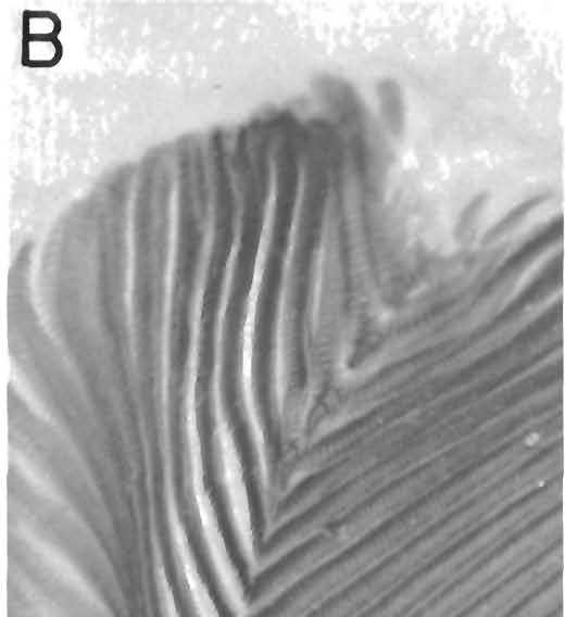
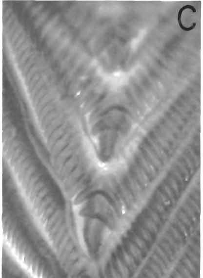
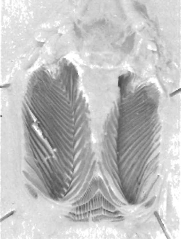
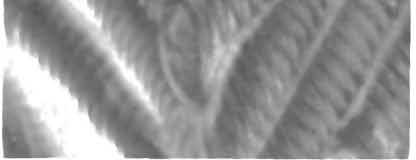
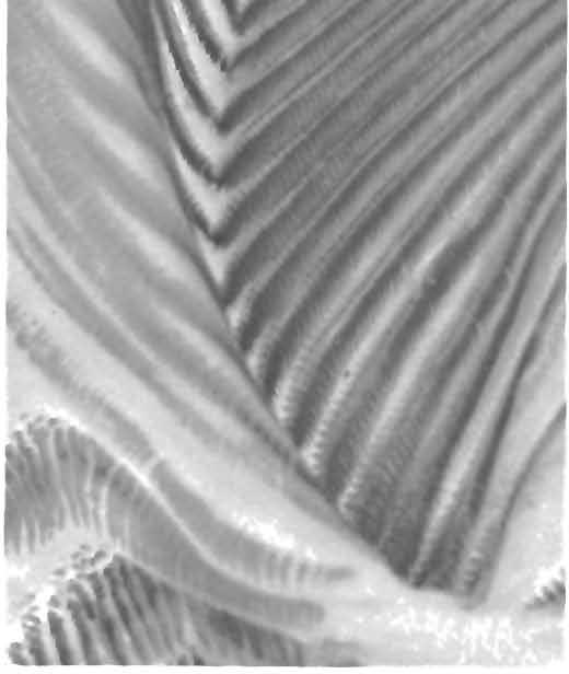
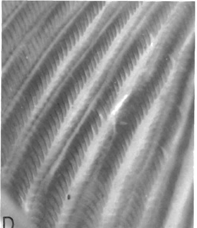
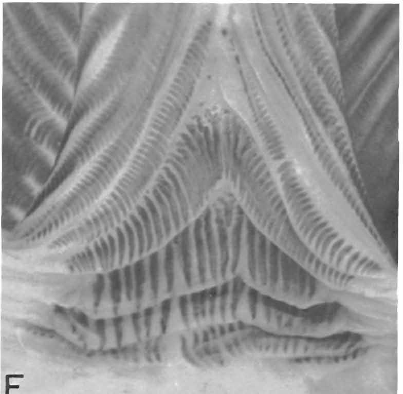
44 42 POSTILLA 159 B
.")
,")

 and medial")
 The median welt pulled back to reveal the")

 The short rows of teeth in flounces that cross")
, Structure of the left hemipenis")

 The median welt pulled back to re eal the")
 Details of the enlarged teeth at the apices of")
 Details of the teeth in the arms of the")
45 PRIONODACTYLUS AND EUSPONDYLUS 43 A ' B FIG, 16 (above). Structure of the left hemispeeis of Prionodactylus dicrus, new species (FMNH 28043), The in erted organ has been slit along the sulcus spermaticus. A) The entire organ showing general arrangement of the flounces in the lateral (left) and medial (right) pockets. X 10. B) The median welt pulled back to reveal the apices of the chevron-shaped flounces; the teeth are not continuous across the apex, and there are no enlarged teeth at the apex. X22. C) The short rows of teeth in flounces that cross the base of the median welt. X 33. FIG, 15 (left), Structure of the left hemipenis of Prionodactylus veriebralis (USNM ). The inverted organ has been slit along the sulcus spermaticus. A) The entire organ showing general arrangement of the flounces in the lateral (left) and medial (right) pockets. x7. B) The median welt pulled back to re eal the apices of the chevron-shaped flounces. X16. C) Details of the enlarged teeth at the apices of the distal che fons of the medial pocket. X43. D) Details of the teeth in the arms of the chevrons. x43, E) The short rows of teeth in flounces crossing the base of the median welt. X 16.


.")
46 44 POSTILLA 159 welt (Fig. 18C), but there is also a partial row, and there are one or two isolated teeth. In Prionodactylus eigenmanni, the hemipenis has 14 toothed flounces in the lateral pocket, 15 in the medial pocket. These flounces are chevron shaped, with teeth complete across the apices. There is no enlarged tooth at the apex of the chevron, There are 4 short rows across the base of the median welt, and part of a fifth. The teeth in these are largest medially, smallest at the ends of the rows. The teeth of the middle rows are larger. In general, the hemipenial structures in Prionodactylus thus seem to be rather similar. There are differences in the few organs examined, Some of these, such as number of rows of flounces, are associated with body size; there are more flounces with teeth in P. vertebralis and P. m. mamicatus, the two largest taxa. The enlarged teeth at the apices of the chevrons of P. vertebralis are the most distinctive feature observed in the series. R FIG. 17. Structure of the hemipenis of Prionodactylus manicatus manicatus (FMNH 45779). The inverted organ has been slit along the sulcus spermaticus. A) The entire organ showing general arrangement of the flounces in the lateral (left) and medial (right) pockets, x6. B) The short rows of teeth in flounces that cross the base of the mediae welt. X 17.


 and medial (right)")
 Detail of the flounces in")


47 PRIONODACTYLUS AND EUSPONDYLUS 45 A B FIG. 18. Structure of the left hemipenis of Prionodactylus argulus (FMNH 81316). The inverted organ has been slit along the sulcus spermaticus. A) The entire organ showing general arrangement of the flounces in the lateral (left) and medial (right) pockets. x6.5. B) Detail of the flounces in the basal part of the medial pocket, showing the absence of teeth at the apices of the chevrons. X34. C) The short row of teeth in the flounces crossing the base of the median welt. X34. C

48 46 POST ILL A 159 Figures show hemipenial structures of individuals of three related genera. The hemipenis of Pantodactylus schreibersii is very distinctive in comparison to the hemipenes of species of Prionodactylus. There are 15 tooth-bearing flounces in both the lateral and medial pockets (Fig. 20A). Teeth are absent across the apices of the chevrons, especially basally; in this, there is some similarity to the hemipenis of P. argulus (Fig. 18B). In marked distinction, however, the teeth of the basal five flounces of the left pocket are much larger than those of the distal flounces (Fig. 20B). The teeth in the short rows crossing the base of the medial welt also seem to be relatively much larger than the corresponding teeth in species of Prionodactylus (Fig.20C). The hemipenis of Cercosaura ocellata (Fig. 21) also shows some distinctive features. There are 12 chevron-shaped tooth bearing flounces in the lateral pocket, and 17 in the medial pocket. Three rows of teeth that cross the base of the median welt are continuous with 3 of the more basal chevrons in each pocket (Fig. 21B). This condition has not been seen in Prionodactylus or the other genera discussed here. The hemipenis of Aspidolaemus affinis (Fig. 22) seems rather like that of several forms of Prionodactylus, especially P. vertebralis and P. m. manicatus. There are 27 tooth-bearing flounces in the lateral and 28 in the medial pocket. Most of these form chevrons that are toothed across the apex; there is no enlarged tooth at the apex. There are 4 short rows of teeth across the base of the median welt. In addition, the ends of two of the incomplete basal chevrons are essentially joined on the median welt by an enlarged tooth (Fig. 22B). The general similarity of the hemipenis of P. vertebralis, P. m. manicatus, and Aspidolaemus affinis can be observed in comparing Figures 15, 17 and 22. Relationships within the Genus Prionodactylus Within Prionodactylus, the closest pair of species clearly seems to be P. vertebralis and P. dicrus. These share nontuberculate subdigital lamellae on the fingers, an undivided frontonasal, a loreal separated from the supralabials, minutely ridged dorsal scales, a distinctive light lip line, and parts of the dorsal pattern: the posterior middorsal light line of P. dicrus resembles that of P. vertebralis. The anterior pair of dorsolateral stripes on the head and anterior part of the body of P. dicrus is paralleled in some P. vertebralis by light lines on the superciliary scales. This pair seems clearly to represent two daughter species derived from a single ancestral species. P. manicatus seems in many ways related to P. vertebralis and P. dicrus. after formaldehyde preservation, some degree of minute ridging on the dorsal scales, the nontuberculate subdigital lamellae on the forelimbs of P. m. bolivianus, and the light lip line, especially of P. m. manicatus. P. manicatus has a much higher number of scales around the midbody region, and in P. manicatus, the loreal is in contact with the supralabials.

.")


 The median welt pulled back to show the teeth")


49 PRIONODACTYLUS AND EUSPONDYLUS 47 A % \ ' 'to #<~ FIG. 19. Structure of the hemipenis of Prionodactylus eigenmanni (BMNH ,1.152). The inverted organ has been slit along the sulcus spermaticus. A) The entire organ showing the general arrangement of the flounces ie the lateral (left) and medial (right) pockets. X 12. B) The median welt pulled back to show the teeth continuing across the apices of the flounces in the lateral pocket, and details of the spinules, x27. Prionodactylus argulus and P. eigenmanni seem rather alike, but this general impression results from their both being brownish rather than blackish when preserved; the venter in both is light. This perhaps reflects altitude, and possibly is related to insolation, rather than closeness of common ancestry. Indeed, P, manicatus in general occurs at altitudes ranging between those of P. argulus and P. eigenmanni on the one hand and those of P. vertebralis and P. dicrus on the other, and is considerably less uniformly black beneath than the two higher elevation forms, although blacker than the lower elevation forms, P. argulus seems to be closer to P. vertebralis, P. dicrus, and particularly

50 48 PO STILL A 159 P. manicatus in that it has a light lip; this forms a line only posteriorly where it is bordered beneath by a dark line. P. argulus has the loreal in contact with the supralabials, but is particularly distinctive in its longer snout and divided frontonasal. Prionodactylus eigenmanni also has the loreal in contact with the supralabials, but it has an undivided frontonasal. The single and double tubercles on the subdigital lamellae set this species apart from the others in the genus. The arrangement of the loreal and of the frontonasal both appear to be related to snout length. P. dicrus and P. vertebralis, in which the loreal is separated from the supralabials, have, relative to body size, the shortest snouts in the genus. The three species in which the loreal touches the supralabial series have relatively longer snouts. The longest snouted species, P. argulus, also has the frontonasal divided. Divided frontonasals are unusual in Group II of the Teiidae, and do not occur in all long-snouted forms, for example, Anadia and Placosoma. A divided frontonasal does occur, however, in the longer-snouted forms of Echinosaura. Relationships to Other Genera Ruibal (1952: 520) pointed out that Pantodactylus and Prionodactylus are closely related, in fact, perhaps not distinguishable. Among the species of Prionodactylus, P. argulus seems closest to Pantodactylus. They share, among other features, the general brownish color in preservation, a lined pattern, at least in some forms of Pantodactylus; and a light venter. More importantly, both Pantodactylus schreibersii and Prionodactylus argulus have all but the distal subdigital lamellae of the toes and fingers divided into two parts, although not tuberculate. There is possibly some confirmation of a relationship in the structure of the hemipenis, in the absence of teeth at the apices of the basal chevrons (Figs. 18B, 20A). The enlarged teeth in the basal chevrons of Pantodactylus is, however, quite distinctive. Cercosaura also seems to be closer to P. argulus or P. eigenmanni than to the other species of Prionodactylus, but is a very distinctive animal, and apparently not particularly close to any Prionodactylus. Aspidolaemus affinis appears in its general features to be closer to the high-elevation species of Prionodactylus, P. vertebralis and P. dicrus, than to other members of the genus. This similarity, however, in such features as the generally dark coloration, and slightly pigmented disc in the lower eyelid, may well reflect adaptations to high elevations. Some suggestion of affinity occurs in the hemipenial structure, in the numerous flounces with teeth in them and the larger number of short, tooth-bearing folds across the base of the median welt. There is some similarity to P. manicatus as well: the loreal in A. affinis touches the supralabials, and there are dorsolateral light lines, ill marked, in both. These notes are very tentative. A study of skeletal and muscular features would shed much light on the affinities of this complex group of lizards.


 The entire organ showing the general")
 and median (right) pockets.")


 The large teeth in the short rows in")
51 PRIONODACTYLUS AND EUSPONDYLUS 49 A FIG, 20. Structure of the left hemipenis of Fantodactylus schreibersii (AMNH ). The iirverted organ has been slit along the sulcus spermaticus. A) The entire organ showing the general arrangement of the flounces in the lateral (left) and median (right) pockets. The che ron-shaped Iounces, especially the basal ones, lack teeth at the apices. x9. B) Detail of the enlarged teeth on the lateral arms of the chevrons of the lateral pockets. x44. C) The large teeth in the short rows in the flounces that cross the base of the mediae welt. X 44.
52 50 POSTILLA 159 III. SPECIES REMOVED FROM THE GENUS Prionodactylus Four species described as Prionodactylus do not belong in the genus as I define it. Three of these can appropriately be transferred to other genera. Prionodactylus spinalis Boulenger Prionodactylus spinalis Boulenger, 1911, Ann. Mag. Nat. Hist. ser. 8, 7: 23. This species was described (Boulenger 1911) from BMNH , reregistered as The type locality, Huancabamba, above 750 m, is certainly the central Peruvian town of that name in Pasco rather than the northern town in Piura. Although Boulenger indicated that the specimens may have come from Oxapampa (Barbour and Noble, 1921), I see no reason to doubt the original information; at most it is a matter of a few kilometers distance and a few meters in elevation. I have examined the syntypes of P. spinalis. The absence of a double row of widened gular scales indicates that this species is not a Prionodactylus. P. spinalis shares most of the features by which I discriminate genera of teiid lizards in Group II with the type of the genus Euspondylus, E. maculatus. For this reason it seems appropriate to recognize this taxon as Euspondylus spinalis (cf. Peters and Donoso-Barros, 1970). Prionodactylus rahmi Grijs Prionodactylus rahmi Grijs, 1936, Zool. Anz. 116: 27. This species was described from two specimens, a male and a female, numbered 5221 in the museum in Hamburg. The two syntypes appear to have been lost during World War II. The locality given by Grijs is Cuzco, Peru, 3000 m. The quadrilateral rather than hexagonal dorsal scales and the absence of two median rows of conspicuously widened gular scales indicate that rahmi is not a member of the genus Prionodactylus as I define that genus. Presently, it seems best to recognize this taxon as Euspondylus rahmi (cf. Peters and Donoso-Barros, 1970). It is probably close to Euspondylus spinalis, as suggested by Grijs. Prionodactylus ocellifer Werner Ecpleopus (Aspidolaemus) affinis Peters 1862, Abh. Akad. Wiss. Berlin: 199. Prionodactylus ocellifer Werner, 1901, Verh. Zool.-Bot. Ges. Wien, 51: 596. Prionodactylus ocellifer was described (Werner, 1901) from a single specimen collected by Richard Haensch in Ecuador. Many of the labels for

53 PRIONODACTYLUS AND EUSPONDYLUS 51 Haensch's collection were illegible, however, and no specific locality is associated with the holotype. Other material in this collection came from both high and low elevations on both sides of the Andes, as well as from the inter-andean valleys. No other examples of this taxon have been reported. I have examined the holotype of P. ocellifer, ZMB It is an adult female in poor condition. Werner's description is adequate, but the following points are worthy of note. 1. The loreal is large, in contact with the frontonasal, prefrontal, first superciliary, frenoocular, nasal, supralabial two (broadly) and three (narrowly; Fig. 23A, B.). 2. The first superciliary is large and expanded onto the dorsal surface of the head; the superciliary series is complete. 3. The prefrontals are ever so slightly separated by contact between the frontonasal and frontal. 4. Contact between supraoculars one and three at the superciliary series excludes supraocular two from the superciliaries (Fig. 23C, D). 5. The disc in the lower eyelid is divided into three pieces by vertical grooves. 6. There is a light-colored ring around the external ear opening; two or three larger tubercles project into the opening from the anterodorsal margin of the external ear opening (Fig. 23C, D). The dorsal scales and the chin and throat scales are illustrated in Fig. 23E and 23F. Most of these features (and many others) can be matched in the taxon usually called Ecpleopus affinis ( Aspidolaemus affinis; Uzzell, 1969b). I have compared the holotype of P. ocellifer with some of these (MCZ ; FMNH ) and consider them to be conspecific, both on the basis of the characters described by Werner as well as those mentioned above; their general appearance also supports this conclusion, although the coloration is slightly different. 5 Much of the color difference may be due to the formalin-blackened condition of the specimens of A. affinis that I have examined, and to the poor condition and desquamation of the holotype of P. ocellifer. I have examined the holotype of Aspidolaemus affinis (ZSM 644/0). It was carefully figured by Peters (1862); the specimen was labelled as type. Despite Peters' statement to the contrary, where epidermis persists, the dorsal scales have a distinct but weak keel. There is no wrinkling on the dorsals, however. I thus know of no trenchant differences between the holotypes of P. ocellifer and A. affinis. Boulenger (1885) reported specimens of E. (A.) affinis in the British Museum from "western Ecuador" collected by Fraser, and from Intac (Imbabura Province), Ecuador, collected by Buckley. Others of Fraser's 5 Benjamin Shreve made this identification without benefit of seeing the holotype.
54 52 POST ILL A 159 specimens, for example, specimens of Neusticurus ecpleopus and Euspondylus maculatus, have erroneous locality data. Many specimens collected by Buckley reportedly both at Pallatanga and eastern Andean localities were apparently mislabelled (Peters 1955; Uzzell 1961). It is not unlikely that some other Buckley locality data are also incorrect. There is at present no reason to believe that Aspidolaemus affinis" occurs on the Pacific Andean slopes of Ecuador. In view of the similarities of Prionodactylus ocellifer and Aspidolaemus affinis, I consider P. ocellifer to be a junior synonym of A. affinis (new synonymy) 7. The name P. ocellifer is available, however, to designate a subpopulation within A. affinis should that prove useful. IV. A NEW GENUS FOR Prionodactylus leucostictus The only known specimen of Prionodactylus leucostictus Boulenger (1900) was collected by F. V. McConnell and J. J. Quelch on the summit of Mount Roraima (2150 m) in Guyana. I have examined the holotype (BMNH , reregistered as ). It is an adult female in relatively good condition. While it bears a superficial resemblance to other species of Prionodactylus because of its keeled, hexagonal dorsal scales, my observations convince me that it is unrelated to Prionodactylus, but instead belongs with Leposoma and its relatives {Ecpleopus, Alopoglossus, Ptychoglossus, Arthrosaura, and possibly Amapasaurus; Ruibal, 1952; Uzzell, 1969b; da Cunha, 1970). Because P. leucostictus differs from each of these genera in a variety of ways, I here erect a new genus for the taxon. The only known specimen shows some anomalies in scalation. I believe, however, that acquisition of additional material will reveal that most of the differences are real, and that leucostictus cannot readily be placed into any of the already established genera. Riolama, new genus TYPE SPECIES: Prionodactylus leucostictus Boulenger. DESCRIPTION. Tongue with chevron-shaped plicae on anterior and posterior thirds, with imbricate scalelike papillae on middle third. Snout moderately long, blunt. Head scales without striations or rugosities. Single frontonasal and frontal; a relatively broad interparietal extending posterior to parietals; no median occipital (Fig. 24A). Paired prefrontals, frontoparietals, and 6 A label with ZSM 644/0 indicates that it was collected in Pichincha by Wagler. The label is relatively recent, however, and Peters (1862) gave no such information. 7 Richard R. Montanucci, who is revising the systematics of Aspidolaemus and Pholidobolus, concurs in this identification.


55 PRIONODACTYLUS AND EUSPONDYLUS 53 parietals; a pair of occipitals and postoccipitals behind interparietal. An enlarged temporal scale lateral to parietal and occipital. Nostril pierced in middle of nasal; no evidence of division above or below nostril although there is an indistinct groove between the nostril and the first upper labial. Nasals not in contact; on right side, nasal extended posteriorly almost to anterior angle of eye; a loreal incompletely divided from the nasal by a groove beginning near the posterior angle of the nasal. On left side, a loreal is present, separated from labials. A large frenoocular and a series of moderate infraoculars. Superciliary series complete; irst superciliary short on right side; fused with Irst supraocular on left. Four supraoculars on each side, the first in contact with the prefrontals, separated from frontonasal. Eyelids well developed, the lower with a heavily pigmented central disc divided into three pieces by vertical grooves. Ear opening large; tympanum heavily pigmented, very shallowly recessed. A mental followed by one unpaired and four paired chin shields (Fig. 24B); the two anterior pairs form a mediae suture; the two posterior pairs are separated by small scales; the posteriormost pair is separated from the infralabials by small scales. Pregular (Ruibal, 1952) and gular scales are not clearly separated; collar fold very A FIG. 21. Structure of the left hemipenis of Cercosaura oceltata (BMNH ). The inverted organ has been slit along the sulcus spermatlcus. A) The entire organ showing the general arrangement of the flounces in the lateral (left) and medial (right) pockets, x 10. B) Detail showing the teeth in the arms of the chevrons that join across the base of the median welt. X30.

56 54 POST ILL A 159 well developed. Gulars flat, imbricate, irregular, large along midline, not forming pairs of large scales, rounded behind; collar scales flat, six in number including a large median scale, slightly rounded posteriorly. Limbs pentadactyl; digits much depressed except at tip; distalmost subdigital lamellae swollen (Fig. 25); digits clawed. Scales along inner margin of palm between thumb and wrist enlarged, with a free medial edge; 13 lamellae under fourth finger, the basal divided; 14 lamellae under fourth toe, the four basal ones divided, but not tuberculate. Dorsal scales keeled, bluntly hexagonal, in transverse rows. A zone of lateral scales four high, about half as large as dorsals, about % size of ventrals; one or two upper rows keeled. Ventral scales in transverse and longitudinal rows, smooth, rounded posteriorly. Anterior and posterior median preanal scales; each lateral preanal scale adjacent to both of these (Fig. 24C). Femoral pores well developed, 7 right, 6 left, none preanal. DIAGNOSIS. The widely separated nasal scales, each surrounding a nostril, and the pentadactyl limbs with all digits clawed place Riolama in Boulenger's (1885) Group II of the Teiidae. The combination of uniformly keeled, hexagonal dorsal scales, slightly recessed tympanum, oblique plicae rather than papillae on the anterior part of the tongue, and depressed digits with swollen tips distinguish Riolama from all other genera in Group II. RELATIONSHIPS. Several of the features of leucostictus distinguish it from the species I place in the genus Prionodactylus. In particular, the presence of oblique folds rather than papillae on the anterior part of the tongue, the large, nearly superficial heavily pigmented tympanum, and the markedly depressed toes are features not seen in Prionodactylus. The last pair of chin shields are widely separated from the infralabials, a condition that I have not noted in Prionodactylus, and the arrangement of the preanal scales with large anterior and posterior median scales, and large lateral scales that are neither anterior nor posterior adjacent to these also does not occur in Prionodactylus. Clearly, leuctostictus is not a Prionodactylus. The presence of plicae rather than papillae on the anterior as well as the posterior part of the tongue suggests that Riolama is close to Alopoglossus, Ptychoglossus, and Ecpleopus, other genera of Group II that have plicae rather than papillae on the anterior part of the tongue. In fact, the arrangement of the plicae in Ecpleopus (present on posterior part and usually on anterior part, but not the middle; Uzzell, 1969b) is an exact parallel for the condition seen in Riolama leucosticta. Other characters also support this arrangement, albeit weakly. The posterior pair of chin shields are separated from the labials in several forms of Leposoma, Alopoglossus, and Ptychoglossus (Uzzell, 1969b). This condition is unusual in other genera of Group II. The arrangement of the prefrontals with respect to the supraoculars is reminiscent of the condition in Ecpleopus (cf. Uzzell, 1969b, fig. 1; Boulenger, 1899, fig. 3a) except that in Riolama,


.")


, new")


57 PRIONODACTYLUS AND EUSPONDYLUS 55 the prefrontals are relatively much farther forward. The digits in Ecpleopus are also slightly depressed, but not markedly so as in Riolama. The arrangement of the preaeal scales in Riolama is reminiscent of that seen in some species of Arthrosaura (cf. da Cunha, 1987, fig. 1). The location of the tympanum close to the surface of the head is also reminiscent of the condition in Ecpleopus, although seen as _well in such diverse genera as Neusiicurus, Placosoma, Anadia, and EcMnosaura. None of these similarities except the condition of the tongue, however, is more than suggestiye. On the other hand, it is easy to distinguish Riolama from Leposoma and its relatives. i made a detailed comparison of those genera except for Amapasaurus (Uzzell 1969b, table 2). On the basis of data proyided in the generic diagnosis, I am not prepared to suggest that Riolama is closer to any one of those genera than to the others. The only known species of the genes is Riolama leucosiicta (Boulenger), new combination. DERIVATION OF NAME. Riolama was the name and home of Rima? who inhabited another isolated mountain of northern South America in W. H. Hudson's Green Mansions. It is dedicated to a friend akin to Rima in spirit. A \wf ; "^ ' "W / FIG,. 22, Structure of the hemipenis of Aspidolaemus affinis (USNM ). The inyerted organ has been slit along the sulcus spermaticus. A) The entire organ showing general arrangement of the flounces in the lateral (left) and medial (right) pockets. x6.5. B) Detail of teeth in the short rows in the flounces that cross the base of the median welt and of the large tooth where two basal chevrons of the two pockets meet at the middle of the median welt, X 18.

 with recent specimens of Aspidolaemus affinis")
 Side of the head of Aspidolaemus 'affinis (FMNH 36720).")
 Top of head of Aspidolaemus affinis (FMNH 36722). X5.")
 Dorsal scales of Prionodactylus ocellifer.")


58 56 POST ILL A 159.;-. *: '""m: ^'...,»'i, ; 'l-"*-- *"">, V* IK FIG. 23. Comparisons of the holotype of Prionodactylus ocellifer Werner (ZMB 16593) with recent specimens of Aspidolaemus affinis (FMNH and 36722) A»F ; x5. A) Side of the head of Aspidolaemus 'affinis (FMNH 36720). B) Side of the head of Prionodactylus ocellifer X5. C) Top of head of Aspidolaemus affinis (FMNH 36722). X5. D) Top of head of Prionodactylus ocellifer x5. E) Dorsal scales of Prionodactylus ocellifer. F) Chin and throat scales of Prionodactylus ocellifer, V. COMMENTS ON SPECIES OF THE GENUS Euspondylus My work on Prionodactylus necessitated consideration of the forms of Euspondylus with which Prionodactylus has been long united (Burt and Burt, 1931). I record here obser ations of forms described in or referred to Euspondylus other than those discussed as Prionodactylus. These comments,
 fraseri O'Shaughnessy, 1881, Ann. Mag. Nat. Hist. (5) 4: 296.")
59 PRIONODACTYLUS AND EUSPONDYLUS 57 together with those on Prionodactylus synopsis of the genus Euspondylus. rahmi and P. spinalis, provide a Euspondylus maculatus Tschudi Euspondylus maculatus Tschudi, 1845, Arch, fur Naturg. 11: 161. Ecpleopus (Proctoporus) fraseri O'Shaughnessy, 1881, Ann. Mag. Nat. Hist. (5) 4: 296. This species, the type of the genus Euspondylus, was described by Tschudi in 1845; additional information was provided in 1846; the type locality was specified as Moyobamba in the front range of the Andes. This locality is at about 750 m above sea level in the valley of the Rio Mayo, a tributary of the Rio Huallaga in San Martin, Peru. There are two syntypes in the Musee d'histoire Naturelle in Neuchatel, Switzerland. Peters (1862) examined these, described them carefully, and figured the male. I have examined them also. Peters' description is excellent; the second syntype is a female. The male has about 42 scales around the midbody region, the female, 44. The female appears to have a single femoral pore on each side. The disc in the lower eyelid is divided into 2-3 parts by vertical grooves in the male, into 3-4 in the female. There are 5 posterior preanal scales in the female. According to Peters, the dorsal scales were slightly keeled in the sacral region. I have seen other specimens that I believe are this species. They come from localities in southern and central Peru, on the Amazonian side of the Andes; elevations that are believable vary from about m above sea level. Ecpleopus (Proctoporus) fraseri, described by O'Shaughnessey on the basis of BMNH (= ), a female, appears to be the same taxon, but lacks prefrontals. This synonymy, suggested by Boulenger (1885), appears to be correct; the type locality for E. fraseri (Guayaquil, Ecuador) almost certainly is incorrect. An alternative possibility is Gualaquiza, Morona-Santiago, Ecuador (600 m), where Fraser spent considerable time in 1857 and early Most of Fraser's collections of birds from Ecuador in 1857 and 1858 were reported by Sclater (Chapman, 1926). The localities for 1857 are all in eastern Ecuador. Fraser collected birds in western Ecuador in late 1858 (August-December) and in The holotype of E. fraseri was accessioned 25 July 1858 (A. F. Stimson, personal communication). The large male in Brussels that Boulenger (1885) reported is MRHNB 951, I. G Its locality is Chile. I believe that this is incorrect. AMNH 1704, reportedly from Lake Aracona (= Lago de Aricoma), Juliaca, above 4000 m, certainly is incorrectly labelled. Dunn (1942) reported that Harvey Bassler suggested that American Museum material labelled Juliaca was sent there by a member of the Inca Mining Company
60 58 POSTILLA 159 (apparently H. H. Keays), but was probably collected near the mine at Santo Domingo, at about 1200 m above sea level in Puno (cf. Uzzell 1970: 26). Other specimens of Euspondylus maculatus that have satisfactory locality data include BMNH (Peru, Puno, Chaquimayo, m) and FMNH 40584, and AMNH (Peru, Junin, Chanchamayo, m). The valid locality records suggest an eastern Andean distribution at m throughout Peru and southern Ecuador, rather than the coastal areas of northern Peru and southern Ecuador suggested by Peters and Donoso-Barros (1970). Euspondylus simonsii Boulenger Euspondylus simonsii Boulenger, 1901, Ann. Mag. Nat. Hist. (1)1: 549. This species was described by Boulenger (1901) from a single female (BMNH ; reregistered as ) collected at Puntoyacu, Rio Perene, Peru, about 1250 m above sea level. No other specimens have been reported. Euspondylus simonsii can be distinguished from E. maculatus, which it resembles, by the undivided disc in the lower eyelid and by the light line between the angle of the mouth and the forelimb insertion. It may also have a reduced number of scales around the midbody region. The palm of E. simonsii has no enlarged scales between the thumb and wrist. Enlarged scales with a projecting medial edge are present in E. maculatus. Euspondylus guentheri Ecpleopus (Euspondylus) guentheri O'Shaughnessy, 1881, Proc. Zool. Soc. London 1881: 235. This very distinct species, based on BMNH , reregistered as , an adult female collected by Buckley near Sarayacu, Pastaza, Ecuador, has been reported from other localities in eastern Ecuador by Peters (1959). Euspondylus brevifrontalis Boulenger Euspondylus brevifrontalis Boulenger, 1903, Ann. Mag. Nat. Hist. (7) 12: 431. The syntypes of E. brevifrontalis Boulenger (1903) include BMNH , reregistered as , an adult female from Rio Albireggas, Venezuela (about 2900 m), and BMNH , reregistered as , two adult males from about 2500 m at Escorial, Venezuela,
61 PRIONODACTYLUS AND EUSPONDYLUS 59 FIG. 24. Scale features of the holotype of Riolama leucosticta (BMNH ) A) Top of head x3.5. B) Chin and throat scales. X3.5. C) Scales of cloacal region. x3. Based on Boulenger, collected by Briceno. As noted by Boulenger, these specimens are very similar to Anadia bitaeniata. Although this species perhaps should be transferred to the genus Anadia (Olav Oftedal and E. E. Williams, personal communication), minute differences between the taxa in arching of the distal phalanx of the toes, division of throat scales into two groups by a gular crease, and slightly smaller lateral scales may justify placing these two taxa on different sides of a generic boundary. An examination of features in addition to the scalation is needed to help place these two taxa correctly. At present I retain this form in Euspondylus but with no real conviction.
![60 POSTILLA 159 ] iew of the digits of the right forefoot of the hoiotype of J i (BMNH 1946,8,2.8), showing the general depressed shape of the digit and the swollen digit tip. x25.](/docs-images/73/68739504/images/62-1.jpg "Euspondylus acutirostris (Peters) Ecpleopus (Euspondylus) acutirostris Peters, 1862 Abh. Akad. Wiss. Berlin 1862: 209.")
 reported by Test et ah (1966) from Rancho Grande, Aragua? Venezuela.")


62 60 POSTILLA 159 ] iew of the digits of the right forefoot of the hoiotype of J i (BMNH 1946,8,2.8), showing the general depressed shape of the digit and the swollen digit tip. x25. Euspondylus acutirostris (Peters) Ecpleopus (Euspondylus) acutirostris Peters, 1862 Abh. Akad. Wiss. Berlin 1862: 209. The hoiotype of Ecpleopus (Euspondylus acutirostris) Peters (1862) is ZMB 4597, an adult male from Venezuela. I ha e examined both the hoiotype and the small female (UMMZ ) reported by Test et ah (1966) from Rancho Grande, Aragua? Venezuela. 1 have compared the specimens directly, and 1 see no difficulty in their being conspecific. The arrangement of the subocular scales appears to be the same in both specimens. The subdigitai lamellae of the basal phalanges of the fore- and hind feet of E. acutirostris are somewhat widened and flattened; the subdigitai lamellae of the distal phalanx on each toe are compressed. Although the distal phalanx is not arched, the arrangement is otherwise similar to that on the toes of arboreal species of Anadia, and supports the hypothesis that this may be an arboreal species (Test et al, 1966). Other specimens of this taxon have been reported by Lancini (1968) from the Cordillera de la Costa in Venezuela. Euspondylus phelpsi Lancini Euspondylus phelpsi Lancini, 1968, Pub, Ocas. Mus, Ciena Nat,, Zool. 12: 2. 1 have seen no material of this recently described taxon (Lancini, (1968). i am unable to determine whether it belongs in Euspondylus, Prionodaeiylus,
POSTILLA PEABODY MUSEUM YALE UNIVERSITY NUMBER FEB A NEW GENUS AND SPECIES OF TEND LIZARD FROM BOLIVIA THOMAS UZZELL
 POSTILLA PEABODY MUSEUM YALE UNIVERSITY NUMBER 129. 26 FEB. 1969 A NEW GENUS AND SPECIES OF TEND LIZARD FROM BOLIVIA THOMAS UZZELL POSTILLA Published by the Peabody Museum of Natural History, Yale University
OCCASIONAL PAPERS OF THE MUSEUM OF ZOOLOGY UNIVERSITY OF MICHIGAN
 OCCASIONAL PAPERS OF THE MUSEUM OF ZOOLOGY UNIVERSITY OF MICHIGAN A NEW SPECIES OF ELEUTHERODACTYLUS FROM THE CORDILLERA OCCIDENTAL OF COLOMBIA (AMPHIBIA : ANURA: LEPTODACTY LIDAE) Frogs of the fitzingeri
Two new skinks from Durango, Mexico
 Great Basin Naturalist Volume 18 Number 2 Article 5 11-15-1958 Two new skinks from Durango, Mexico Wilmer W. Tanner Brigham Young University Follow this and additional works at: https://scholarsarchive.byu.edu/gbn
POSTILLA PEABODY MUSEUM YALE UNIVERSITY NUMBER JAN TEND LIZARDS OF THE GENUS PROCTOPORUS FROM BOLIVIA AND PERU.
 POSTILLA PEABODY MUSEUM YALE UNIVERSITY NUMBER 142. 30 JAN. 1970 TEND LIZARDS OF THE GENUS PROCTOPORUS FROM BOLIVIA AND PERU. THOMAS UZZELL POSTILLA Published by the Peabody Museum of Natural History,
ON AN ALOPOGLOSSUS FROM SURINAM
 ON AN ALOPOGLOSSUS FROM SURINAM by Dr. L. D. BRONGERSMA Rijksmuseum van Natuurlijke Historie, Leiden With one textfigure A single Alopoglossus was taken by Dr. K. M. Hulk during the Corantine Expedition
A new species of torrent toad (Genus Silent Valley, S. India
 Proc. Indian Acad. Sci. (Anirn. ScL), Vol. 90, Number 2, March 1981, pp. 203-208. Printed in India. A new species of torrent toad (Genus Silent Valley, S. India Allsollia) from R S PILLAI and R PATTABIRAMAN
NORTH AMERICA. ON A NEW GENUS AND SPECIES OF COLUBRINE SNAKES FROM. The necessity of recognizing tlie two species treated of in this paper
 ON A NEW GENUS AND SPECIES OF COLUBRINE SNAKES FROM NORTH AMERICA. BY Leonhard Stejneger, and Batrachians. Curator of the Department of Reptiles The necessity of recognizing tlie two species treated of
OCCASIONAL PAPERS OF THE MUSEUM OF ZOOLOGY UNIVERSITY OF MICHIGAN
 OCCASIONAL PAPERS OF THE MUSEUM OF ZOOLOGY ~- UNIVERSITY OF MICHIGAN A NEW FROG FROM BRITISH GUIANA A collection received by the IIuseum of Zoology froin British Gniana some time ago includes a single
UNIVERSITY OF MICHIGAN PRESS
 OCCASIONAL PAPERS OF THE MUSEUM OF ZOOLOGY UNIVERSITY OF MICHIGAN ANN ARBOR, MICHIGAN UNIVERSITY OF MICHIGAN PRESS THE SUBSPECIES OF' CROTALUS LEPIDUS1 THE rattlesnake Crotalus lepidus is a small species
Postilla PEABODY MUSEUM OF NATURAL HISTORY YALE UNIVERSITY NEW HAVEN, CONNECTICUT, U.S.A.
 Postilla PEABODY MUSEUM OF NATURAL HISTORY YALE UNIVERSITY NEW HAVEN, CONNECTICUT, U.S.A. Number 117 18 March 1968 A 7DIAPSID (REPTILIA) PARIETAL FROM THE LOWER PERMIAN OF OKLAHOMA ROBERT L. CARROLL REDPATH
First Record of Lygosoma angeli (Smith, 1937) (Reptilia: Squamata: Scincidae) in Thailand with Notes on Other Specimens from Laos
 (Reptilia: Squamata: Scincidae) in Thailand with Notes on Other Specimens from Laos") The Thailand Natural History Museum Journal 5(2): 125-132, December 2011. 2011 by National Science Museum, Thailand First Record of Lygosoma angeli (Smith, 1937) (Reptilia: Squamata: Scincidae) in Thailand
The Thailand Natural History Museum Journal 5(2): 125-132, December 2011. 2011 by National Science Museum, Thailand First Record of Lygosoma angeli (Smith, 1937) (Reptilia: Squamata: Scincidae) in Thailand
ONLINE APPENDIX 1. Morphological phylogenetic characters scored in this paper. See Poe (2004) for
 for") ONLINE APPENDIX Morphological phylogenetic characters scored in this paper. See Poe () for detailed character descriptions, citations, and justifications for states. Note that codes are changed from a
ONLINE APPENDIX Morphological phylogenetic characters scored in this paper. See Poe () for detailed character descriptions, citations, and justifications for states. Note that codes are changed from a
Vol. XIV, No. 1, March, The Larva and Pupa of Brontispa namorikia Maulik (Coleoptera: Chrysomelidae: Hispinae) By S.
 By S.") Vol. XIV, No. 1, March, 1950 167 The Larva and Pupa of Brontispa namorikia Maulik (Coleoptera: Chrysomelidae: Hispinae) By S. MAULIK BRITISH MUSEUM (NATURAL HISTORY) (Presented by Mr. Van Zwaluwenburg
Vol. XIV, No. 1, March, 1950 167 The Larva and Pupa of Brontispa namorikia Maulik (Coleoptera: Chrysomelidae: Hispinae) By S. MAULIK BRITISH MUSEUM (NATURAL HISTORY) (Presented by Mr. Van Zwaluwenburg
Iovitate. daie'ican)jafseum. (Amphisbaenia, Reptilia). 8. and the Description of a New Species of. Amphisbaena from British Guiana
jafseum. (Amphisbaenia, Reptilia). 8. and the Description of a New Species of. Amphisbaena from British Guiana") daie'ican)jafseum Iovitate PUBLISHED BY THE AMERICAN MUSEUM OF NATURAL HISTORY CENTRAL PARK WEST AT 79TH STREET, NEW YORK 24, N.Y. NUMBER 2I28 APRIL 5, I963 Notes on Amphisbaenids (Amphisbaenia, Reptilia).
daie'ican)jafseum Iovitate PUBLISHED BY THE AMERICAN MUSEUM OF NATURAL HISTORY CENTRAL PARK WEST AT 79TH STREET, NEW YORK 24, N.Y. NUMBER 2I28 APRIL 5, I963 Notes on Amphisbaenids (Amphisbaenia, Reptilia).
POSTILLA PEABODY MUSEUM YALE UNIVERSITY NUMBER JULY THE STATUS OF THE GENERA ECPLEOPUS, ARTHROSEPS AND ASPIDOLAEMUS (SAURIA, TEI- IDAE)
") POSTILLA PEABODY MUSEUM YALE UNIVERSITY NUMBER 135. 11 JULY 1969. THE STATUS OF THE GENERA ECPLEOPUS, ARTHROSEPS AND ASPIDOLAEMUS (SAURIA, TEI- IDAE) THOMAS UZZELL POSTILLA Published by the Peabody Museum
POSTILLA PEABODY MUSEUM YALE UNIVERSITY NUMBER 135. 11 JULY 1969. THE STATUS OF THE GENERA ECPLEOPUS, ARTHROSEPS AND ASPIDOLAEMUS (SAURIA, TEI- IDAE) THOMAS UZZELL POSTILLA Published by the Peabody Museum
ZOOLOGISCHE MEDEDELINGEN UITGEGEVEN DOOR HET
 ZOOLOGISCHE MEDEDELINGEN UITGEGEVEN DOOR HET RIJKSMUSEUM V A N NATUURLIJKE HISTORIE T E LEIDEN (MINISTERIE VAN CULTUUR, RECREATIE EN MAATSCHAPPELIJK WERK) Deel 51 no. 2 15 februari 1977 A NEW SPECIES OF
OCCASIONAL PAPERS OF THE MUSEUM OF ZOOLOGY UNIVERSITY OF MICHIGAN PRESS
 OCCASIONAL PAPERS OF THE MUSEUM OF ZOOLOGY UNIVERSITY OF MICHIGAN ANN ARBOR, MICHIGAN UNIVERSITY OF MICHIGAN PRESS ATRACTUS SANCTAEMARTAE, A NEW SPECIES OF SNAKE FROM THE SIERRA NEVADA DE SANTA MARTA,
FLORIDA STATE MUSEUM BULLETIN OF THE UNIVERSITY OF FLORIDA BIOLOGICAL SCIENCES A REVIEW OF THE AMERICAN LIZARDS OF THE GENUS XENOSAURUS PETERS
 BULLETIN OF THE FLORIDA STATE MUSEUM BIOLOGICAL SCIENCES Volume 12 Number 2 A REVIEW OF THE AMERICAN LIZARDS OF THE GENUS XENOSAURUS PETERS Wayne King and Fred G. Thompson /853 UNIVERSITY OF FLORIDA Gainesville
TWO NEW SPECIES OF WATER MITES FROM OHIO 1-2
 TWO NEW SPECIES OF WATER MITES FROM OHIO 1-2 DAVID R. COOK Wayne State University, Detroit, Michigan ABSTRACT Two new species of Hydracarina, Tiphys weaveri (Acarina: Pionidae) and Axonopsis ohioensis
Monitore Zoologico Italiano
 Monitore Zoologico Italiano ITALIAN JOURNAL OF ZOOLOGY PUBBLICATO DALLA UNIVERSITA. DEGLI STUDI DI FIRENZE CON IL CONTRIBUTO DEL CONSIGLIO NAZIONALE DELLE RICERCHE N. S. SUPPLEMENTO VI 31. 12. 1975 NO.
OCCASIONAL PAPEKS OF THE MUSEUM OF ZOOLOGY UNIVERSITY OF MICHIGAN
 OCCASIONAL PAPEKS OF THE MUSEUM OF ZOOLOGY UNIVERSITY OF MICHIGAN Ann Arbor, Michigan University of Michigan Press A NEW SUBSI'ECIES OF THE IGUANID LIZARD SCELOPOK US SERRZFER FROM TAMAULIPAS, MEXICO*
Reprinted from: CRUSTACEANA, Vol. 32, Part 2, 1977 LEIDEN E. J. BRILL
 Reprinted from: CRUSTACEANA, Vol. 32, Part 2, 1977 LEIDEN E. J. BRILL NOTES AND NEWS 207 ALPHE0PS1S SHEARMII (ALCOCK & ANDERSON): A NEW COMBINATION WITH A REDESCRIPTION OF THE HOLOTYPE (DECAPODA, ALPHEIDAE)
Plestiodon (=Eumeces) fasciatus Family Scincidae
 fasciatus Family Scincidae") Plestiodon (=Eumeces) fasciatus Family Scincidae Living specimens: - Five distinct longitudinal light lines on dorsum - Juveniles have bright blue tail - Head of male reddish during breeding season - Old
Plestiodon (=Eumeces) fasciatus Family Scincidae Living specimens: - Five distinct longitudinal light lines on dorsum - Juveniles have bright blue tail - Head of male reddish during breeding season - Old
Rana catesbeiana [now Lithobates catesbeianus] Family Ranidae
![Rana catesbeiana [now Lithobates catesbeianus] Family Ranidae](/thumbs/78/76948879.jpg "Rana catesbeiana [now Lithobates catesbeianus] Family Ranidae") Rana catesbeiana [now Lithobates catesbeianus] Family Ranidae - Body large and heavy - Legs very stout - NO dorsolateral fold along sides of body - Distinct fold from eye curving downward along tympanum
Rana catesbeiana [now Lithobates catesbeianus] Family Ranidae - Body large and heavy - Legs very stout - NO dorsolateral fold along sides of body - Distinct fold from eye curving downward along tympanum
Mliiemtican%MlselIm. Lygophis bourszeri: Rhadinaea tristriata, Coronella whymperi, South American Snakes Related to. and Liophis atahuallpae
 Mliiemtican%MlselIm PUBLISHED BY THE AMERICAN MUSEUM OF NATURAL HISTORY CENTRAL PARK WEST AT 79TH STREET, NEW YORK, N. Y. I0024 NUMBER 2385 AUGUST I5, I969 South American Snakes Related to Lygophis bourszeri:
Common Tennessee Amphibians WFS 340
 Common Tennessee Amphibians WFS 340 Order Anura Frogs and Toads American toad Bufo americanus Medium to large toad (5.1-9.0 cm) Dorsum gray, brown, olive, or brick red in color Light middorsal stripe (not
The family Gnaphosidae is a large family
 Pakistan J. Zool., vol. 36(4), pp. 307-312, 2004. New Species of Zelotus Spider (Araneae: Gnaphosidae) from Pakistan ABIDA BUTT AND M.A. BEG Department of Zoology, University of Agriculture, Faisalabad,
A NEW SCINCID LIZARD OF THE GENUS TRIBOLONOTUS FROM MANUS ISLAND, NEW GUINEA
 A NEW SCINCID LIZARD OF THE GENUS TRIBOLONOTUS FROM MANUS ISLAND, NEW GUINEA by HAROLD G. COGGER The Australian Museum, Sydney With one text figure and one plate INTRODUCTION The scincid lizards of the
III. - NOTES ON THE SPECIES OF ARTHROSAURA BLGR. (TEIIDAE) L. D. BRONGERSMA, 'S RIJKS MUSEUM VAN NATUURLIJKE HISTORIE, LEIDEN. (WITH 12 FIGURES).
 L. D. BRONGERSMA, 'S RIJKS MUSEUM VAN NATUURLIJKE HISTORIE, LEIDEN. (WITH 12 FIGURES).") 76 ZOOLOGISCHE MEDEDEELINGEN DEEL XV. III. - NOTES ON THE SPECIES OF ARTHROSAURA BLGR. (TEIIDAE) BY L. D. BRONGERSMA, 'S RIJKS MUSEUM VAN NATUURLIJKE HISTORIE, LEIDEN. (WITH 12 FIGURES). In 1904 two Teiid
76 ZOOLOGISCHE MEDEDEELINGEN DEEL XV. III. - NOTES ON THE SPECIES OF ARTHROSAURA BLGR. (TEIIDAE) BY L. D. BRONGERSMA, 'S RIJKS MUSEUM VAN NATUURLIJKE HISTORIE, LEIDEN. (WITH 12 FIGURES). In 1904 two Teiid
PRELIMINARY DESCRIPTIONS OF NEW FORMS OF SOUTH AFRICAN REPTILIA AND AMPHIBIA, FROM THE VERNAY-LANG KALAHARI EXPEDITION, 1930.
 ANNAI,S OF THE TRANSVAAL MUSEUM 35 PRELIMINARY DESCRIPTIONS OF NEW FORMS OF SOUTH AFRICAN REPTILIA AND AMPHIBIA, FROM THE VERNAY-LANG KALAHARI EXPEDITION, 1930. By V. FITZSIMONS, M.Sc. Senior Assistant
A new species of coral snake (Serpentes, Elapidae) from the Sierra de Tamaulipas, Mexico
 from the Sierra de Tamaulipas, Mexico") Phyllomeduso 3(1 ):3-7,2004 @ 2004 Melopsittocus Publico~6es Cientificos ISSN 1519-1397 A new species of coral snake (Serpentes, Elapidae) from the Sierra de Tamaulipas, Mexico Pablo A. Lavin-Murciol and
Phyllomeduso 3(1 ):3-7,2004 @ 2004 Melopsittocus Publico~6es Cientificos ISSN 1519-1397 A new species of coral snake (Serpentes, Elapidae) from the Sierra de Tamaulipas, Mexico Pablo A. Lavin-Murciol and
NOVYITATES. AMEIRiICAN MUSEUM NOTES ON SOME INDO-AUSTRALIAN MONITORS (SAURIA, VARANI DAE) BY ROBERT MERTENS'
 BY ROBERT MERTENS'") AMEIRiICAN MUSEUM NOVYITATES PUBLISHED BY THE AMERICAN MUSEUM OF NATURAL HISTORY CITY OF NEW YORK MARCH 15, 1950 NUMBER 1456 NOTES ON SOME INDO-AUSTRALIAN MONITORS (SAURIA, VARANI DAE) BY ROBERT MERTENS'
AMEIRiICAN MUSEUM NOVYITATES PUBLISHED BY THE AMERICAN MUSEUM OF NATURAL HISTORY CITY OF NEW YORK MARCH 15, 1950 NUMBER 1456 NOTES ON SOME INDO-AUSTRALIAN MONITORS (SAURIA, VARANI DAE) BY ROBERT MERTENS'
AUSTRALIAN MUSEUM SCIENTIFIC PUBLICATIONS
 AUSTRALIAN MUSEUM SCIENTIFIC PUBLICATIONS McCulloch, Allan R., 1908. A new genus and species of turtle, from North Australia. Records of the Australian Museum 7(2): 126 128, plates xxvi xxvii. [11 September
SOME ERYTHRONEURA OF THE COMES GROUP (HOMOPTERA: CICADELLIDAE)
") SOME ERYTHRONEURA OF THE COMES GROUP (HOMOPTERA: CICADELLIDAE) DOROTHY M. JOHNSON During a study of the Erythroneura of the Comes Group, chiefly from Ohio, several undescribed species and varieties were
SOME ERYTHRONEURA OF THE COMES GROUP (HOMOPTERA: CICADELLIDAE) DOROTHY M. JOHNSON During a study of the Erythroneura of the Comes Group, chiefly from Ohio, several undescribed species and varieties were
Outline. Identifying Idaho Amphibians and Reptiles
 Identifying Idaho Amphibians and Reptiles Wildlife Ecology, University of Idaho Fall 2011 Charles R. Peterson Herpetology Laboratory Department of Biological Sciences, Idaho Museum of Natural History Idaho
posterior part of the second segment may show a few white hairs
 April, 1911.] New Species of Diptera of the Genus Erax. 307 NEW SPECIES OF DIPTERA OF THE GENUS ERAX. JAMES S. HINE. The various species of Asilinae known by the generic name Erax have been considered
Now the description of the morphology and ecology are recorded as follows: Megophrys glandulosa Fei, Ye et Huang, new species
 12 Description of two new species of the Genus Megophiys, Pelobatidae ( Amphibia: Anura ) from China Liang Fei, Chang-yiian Ye (Chengdu Institute of Biology, Academia Sinica 610015) Yong-zhao Huang (Chongqing
A new skink of the multivirgatus group from Chihuahua
 Great Basin Naturalist Volume 17 Number 3 Number 4 Article 5 12-31-1957 A new skink of the multivirgatus group from Chihuahua Wilmer W. Tanner Brigham Young University Follow this and additional works
NOTE XXXVIII. Three new species of the genus Helota DESCRIBED BY. C. Ritsema+Cz. is very. friend René Oberthür who received. Biet.
 Subshining; HELOTA MARIAE. 249 NOTE XXXVIII. Three new species of the genus Helota DESCRIBED BY C. Ritsema+Cz. The first of these species is very interesting as it belongs to the same section as the recently
UPOGEBIA LINCOLNI SP. NOV. (DECAPODA, THALASSINIDEA, UPOGEBIIDAE) FROM JAVA, INDONESIA
 FROM JAVA, INDONESIA") NOTES AND NEWS UPOGEBIA LINCOLNI SP. NOV. (DECAPODA, THALASSINIDEA, UPOGEBIIDAE) FROM JAVA, INDONESIA BY NGUYEN NGOC-HO i) Faculty of Science, University of Saigon, Vietnam Among material recently collected
NOTES AND NEWS UPOGEBIA LINCOLNI SP. NOV. (DECAPODA, THALASSINIDEA, UPOGEBIIDAE) FROM JAVA, INDONESIA BY NGUYEN NGOC-HO i) Faculty of Science, University of Saigon, Vietnam Among material recently collected
Beaufortia. (Rathke) ZOOLOGICAL MUSEUM - AMSTERDAM. July. Three new commensal Ostracods from Limnoria lignorum
 ZOOLOGICAL MUSEUM - AMSTERDAM. July. Three new commensal Ostracods from Limnoria lignorum") Beaufortia SERIES OF MISCELLANEOUS PUBLICATIONS ZOOLOGICAL MUSEUM - AMSTERDAM No. 34 Volume 4 July 30, 1953 Three new commensal Ostracods from Limnoria lignorum (Rathke) by A.P.C. de Vos (Zoological Museum,
Beaufortia SERIES OF MISCELLANEOUS PUBLICATIONS ZOOLOGICAL MUSEUM - AMSTERDAM No. 34 Volume 4 July 30, 1953 Three new commensal Ostracods from Limnoria lignorum (Rathke) by A.P.C. de Vos (Zoological Museum,
A NEW SALTICID SPIDER FROM VICTORIA By R. A. Dunn
 Dunn, R. A. 1947. A new salticid spider from Victoria. Memoirs of the National Museum of Victoria 15: 82 85. All text not included in the original document is highlighted in red. Mem. Nat. Mus. Vict.,
PEABODY MUSEUM OF NATURAL HISTORY, YALE UNIVERSITY NEW HAVEN, CONNECTICUT, U.S.A. A NEW OREODONT FROM THE CABBAGE PATCH LOCAL FAUNA, WESTERN MONTANA
 Postilla PEABODY MUSEUM OF NATURAL HISTORY YALE UNIVERSITY NEW HAVEN, CONNECTICUT, U.S.A. Number 85 September 21, 1964 A NEW OREODONT FROM THE CABBAGE PATCH LOCAL FAUNA, WESTERN MONTANA STANLEY J. RIEL
SOME LITTLE-KNOWN FOSSIL LIZARDS FROM THE
 PROCEEDINGS OF THE UNITED STATES NATIONAL MUSEUM issued SWsK \ {^^m ^V ^^ SMITHSONIAN INSTITUTION U. S. NATIONAL MUSEUM Vol. 91 Washington : 1941 No. 3124 SOME LITTLE-KNOWN FOSSIL LIZARDS FROM THE OLIGOCENE
Reptile Identification Guide
 Care & preservation of Surrey s native amphibians and reptiles Reptile Identification Guide This identification guide is intended to act as an aid for SARG surveyors. Adder, Vipera berus A short, stocky
SOME NEW AMERICAN PYCNODONT FISHES.
 SOME NEW AMERICAN PYCNODONT FISHES. By James Williams Gidley, Assistant Curator of Fossil Mammals, United States National Museum. In the United States National Museum are several specimens representing
Distribution and natural history notes on the Peruvian lizard Proctoporus laudahnae
 Distribution and natural history notes on the Peruvian lizard Proctoporus laudahnae (Squamata: Gymnophthalmidae) Germán Chávez and Juan C. Chávez-Arribasplata Phyllomedusa 15(2):147 154, 2016 2016 Universidade
Three new species of Microctenochira SPAETH from Brazil and Panama (Coleoptera: Chrysomelidae: Cassidinae)
") Genus Vol. 10 (1): 109-116 Wroc³aw, 31 III 1999 Three new species of Microctenochira SPAETH from Brazil and Panama (Coleoptera: Chrysomelidae: Cassidinae) JOLANTA ŒWIÊTOJAÑSKA and LECH BOROWIEC Zoological
Genus Vol. 10 (1): 109-116 Wroc³aw, 31 III 1999 Three new species of Microctenochira SPAETH from Brazil and Panama (Coleoptera: Chrysomelidae: Cassidinae) JOLANTA ŒWIÊTOJAÑSKA and LECH BOROWIEC Zoological
BULLETIN OF THE CHICAGO ACADEMY OF SCIENCES AMPHIBIANS AND REPTILES FROM THE CARMEN MOUNTAINS, COAHUILA. HOWARD K. GLOYD Chicago Academy of Sciences
 Vol. 6 No. 13 BULLETIN OF THE CHICAGO ACADEMY OF SCIENCES AMPHIBIANS AND REPTILES FROM THE CARMEN MOUNTAINS, COAHUILA BY HOWARD K. GLOYD Chicago Academy of Sciences AND HOBART M. SMITH University of Rochester
A REVIEW OF THE COLOMBIAN SNAKES OF THE FAMILIES TYPHLOPIDAE AND LEPTOTYPHLOPIDAE
 HERPETOLOGIA A REVIEW OF THE COLOMBIAN SNAKES OF THE FAMILIES TYPHLOPIDAE AND LEPTOTYPHLOPIDAE EMMETTREID DUNN This review was made at the suggestion of my old friend and correspondent Hermano Niceforo
YALE PEABODY MUSEUM OF NATURAL HISTORY NOTES ON AMPHISBAENIDS (AMPHISBAENIA; REPTILIA)
") YALE PEABODY MUSEUM OF NATURAL HISTORY Number 6 November 20, 161 New Haven, Conn. NOTES ON AMPHISBAENIDS (AMPHISBAENIA; REPTILIA) 2. Amphisbaena occidentalis Cope from the Coastal Plain of Northern Peru.
YALE PEABODY MUSEUM OF NATURAL HISTORY Number 6 November 20, 161 New Haven, Conn. NOTES ON AMPHISBAENIDS (AMPHISBAENIA; REPTILIA) 2. Amphisbaena occidentalis Cope from the Coastal Plain of Northern Peru.
TERRIER BRASILEIRO (Brazilian Terrier)
") 04.07.2018/ EN FEDERATION CYNOLOGIQUE INTERNATIONALE (AISBL) SECRETARIAT GENERAL: 13, Place Albert 1 er B 6530 Thuin (Belgique) FCI-Standard N 341 TERRIER BRASILEIRO (Brazilian Terrier) 2 TRANSLATION:
04.07.2018/ EN FEDERATION CYNOLOGIQUE INTERNATIONALE (AISBL) SECRETARIAT GENERAL: 13, Place Albert 1 er B 6530 Thuin (Belgique) FCI-Standard N 341 TERRIER BRASILEIRO (Brazilian Terrier) 2 TRANSLATION:
1. On Spiders of the Family Attidae found in Jamaica.
 Peckham, G. W. and E. G. Peckham. 1901. On spiders of the family Attidae found in Jamaica. Proceedings of the Zoological Society of London for 1901 (2): 6-16, plates II-IV. This digital version was prepared
ON THE NEW GUINEA TAIi'AN.
 Memoirs of the National Museum of Victoria https://doi.org/10.24199/j.mmv.1956.20.05 January 1956 ON THE NEW GUINEA TAIi'AN. By K. U. Slater, Port Moresby. 1 Pseudechis scutellatus was described by Peters'
11/4/13. Frogs and Toads. External Anatomy WFS 340. The following anatomy slides should help you w/ ID.
 Frogs and Toads WFS 340 The following slides do not include all 21 species covered during the TAMP workshop Graves modified an old slide presentation from a former course in an attempt to provide another
YALE PEABODY MUSEUM OF NATURAL HISTORY A NEW CAVERNICOLOUS PSEUDOSCORPION BELONGING TO THE GENUS MICROCREAGR1S WILLIAM B. MUCHMORE
 YALE PEABODY MUSEUM OF NATURAL HISTORY Number 70 November 5, 1962 New Haven, Conn. A NEW CAVERNICOLOUS PSEUDOSCORPION BELONGING TO THE GENUS MICROCREAGR1S WILLIAM B. MUCHMORE UNIVERSITY OF ROCHESTER, ROCHESTER,
USING MORPHOLOGICAL AND MOLECULAR EVIDENCE TO INFER SPECIES BOUNDARIES WITHIN PROCTOPORUS BOLIVIANUS WERNER (SQUAMATA: GYMNOPHTHALMIDAE)
") Herpetologica, 59(3), 2003, 432 449 Ó 2003 by The Herpetologists League, Inc. USING MORPHOLOGICAL AND MOLECULAR EVIDENCE TO INFER SPECIES BOUNDARIES WITHIN PROCTOPORUS BOLIVIANUS WERNER (SQUAMATA: GYMNOPHTHALMIDAE)
Herpetologica, 59(3), 2003, 432 449 Ó 2003 by The Herpetologists League, Inc. USING MORPHOLOGICAL AND MOLECULAR EVIDENCE TO INFER SPECIES BOUNDARIES WITHIN PROCTOPORUS BOLIVIANUS WERNER (SQUAMATA: GYMNOPHTHALMIDAE)
Williston, and as there are many fairly good specimens in the American
 56.81.7D :14.71.5 Article VII.- SOME POINTS IN THE STRUCTURE OF THE DIADECTID SKULL. BY R. BROOM. The skull of Diadectes has been described by Cope, Case, v. Huene, and Williston, and as there are many
Exceptional fossil preservation demonstrates a new mode of axial skeleton elongation in early ray-finned fishes
 Supplementary Information Exceptional fossil preservation demonstrates a new mode of axial skeleton elongation in early ray-finned fishes Erin E. Maxwell, Heinz Furrer, Marcelo R. Sánchez-Villagra Supplementary
MUNIDOPSIS ALBATROSSAB, A NEW SPECIES OF DEEP-SEA GALATHEIDAE (DECAPODA, ANOMURA) FROM THE EASTERN PACIFIC OCEAN
 FROM THE EASTERN PACIFIC OCEAN") MUNIDOPSIS ALBATROSSAB, A NEW SPECIES OF DEEP-SEA GALATHEIDAE (DECAPODA, ANOMURA) FROM THE EASTERN PACIFIC OCEAN BY WILLIS E. PEQUEGNAT and LINDA H. PEQUEGNAT Department of Oceanography, Texas A & M University,
MUNIDOPSIS ALBATROSSAB, A NEW SPECIES OF DEEP-SEA GALATHEIDAE (DECAPODA, ANOMURA) FROM THE EASTERN PACIFIC OCEAN BY WILLIS E. PEQUEGNAT and LINDA H. PEQUEGNAT Department of Oceanography, Texas A & M University,
ARIEGE POINTING DOG (Braque de l Ariège)
") FEDERATION CYNOLOGIQUE INTERNATIONALE (AISBL) SECRETARIAT GENERAL: 13, Place Albert 1 er B 6530 Thuin (Belgique) 07.08.1998/EN FCI-Standard N 177 ARIEGE POINTING DOG (Braque de l Ariège) 2 TRANSLATION
FEDERATION CYNOLOGIQUE INTERNATIONALE (AISBL) SECRETARIAT GENERAL: 13, Place Albert 1 er B 6530 Thuin (Belgique) 07.08.1998/EN FCI-Standard N 177 ARIEGE POINTING DOG (Braque de l Ariège) 2 TRANSLATION
tta tes Nov AMERICAN MUSEUM (Ranidae) from New Britain PUBLISHED BY NATURAL HISTORY THE AMERICAN MUSEUM
 from New Britain PUBLISHED BY NATURAL HISTORY THE AMERICAN MUSEUM") AMERICAN MUSEUM tta tes Nov PUBLISHED BY THE AMERICAN MUSEUM NATURAL HISTORY OF CENTRAL PARK WEST AT 79TH STREET NEW YORK, N.Y. 10024 U.S.A. NUMBER 2582 JUNE 24, 1975 RICHARD G. ZWEIFEL Two New Frogs of
AMERICAN MUSEUM tta tes Nov PUBLISHED BY THE AMERICAN MUSEUM NATURAL HISTORY OF CENTRAL PARK WEST AT 79TH STREET NEW YORK, N.Y. 10024 U.S.A. NUMBER 2582 JUNE 24, 1975 RICHARD G. ZWEIFEL Two New Frogs of
BREVIORA LEUCOLEPIDOPA SUNDA GEN. NOV., SP. NOV. (DECAPODA: ALBUNEIDAE), A NEW INDO-PACIFIC SAND CRAB. Ian E. Efford 1
, A NEW INDO-PACIFIC SAND CRAB. Ian E. Efford 1") ac lc BREVIORA CAMBRIDGE, MASS. 30 APRIL, 1969 NUMBER 318 LEUCOLEPIDOPA SUNDA GEN. NOV., SP. NOV. (DECAPODA: ALBUNEIDAE), A NEW INDO-PACIFIC SAND CRAB Ian E. Efford 1 ABSTRACT. Leucolepidopa gen. nov.
ac lc BREVIORA CAMBRIDGE, MASS. 30 APRIL, 1969 NUMBER 318 LEUCOLEPIDOPA SUNDA GEN. NOV., SP. NOV. (DECAPODA: ALBUNEIDAE), A NEW INDO-PACIFIC SAND CRAB Ian E. Efford 1 ABSTRACT. Leucolepidopa gen. nov.
 http://www.biodiversitylibrary.org The Annals and magazine of natural history; zoology, botany, and geology being a continuation of the Annals combined with Loudon and Charlesworth's Magazine of Natural
A MEXICAN SUBSPECIES OF GROTALUX MOLOXXUX BAIRD AND GIRARD1
 OCCASIONAL PAPERS OF THE MUSEUM OF ZOOLOGY UNIVERSITY OF MICHIGAN ANN ARBOR, MICIXIGAN UNIVERSITY OF MICHIGAN PRESS A MEXICAN SUBSPECIES OF GROTALUX MOLOXXUX BAIRD AND GIRARD1 BECAUSE of the limited number
A New Water Skink of the Genus Tropidophorus Scincidae) from Sulawesi, Indonesia
 from Sulawesi, Indonesia") A New Water Skink of the Genus Tropidophorus Scincidae) from Sulawesi, Indonesia (Lacertilia: TSUTOMU HIKIDA1*, AWAL RIYANTO2, AND HIDETOSHI OTA3 1Department of Zoology, Graduate School of Science, Kyoto
A New Water Skink of the Genus Tropidophorus Scincidae) from Sulawesi, Indonesia (Lacertilia: TSUTOMU HIKIDA1*, AWAL RIYANTO2, AND HIDETOSHI OTA3 1Department of Zoology, Graduate School of Science, Kyoto
Necturus maculosus Family Proteidae
 Necturus maculosus Family Proteidae - Robust body that is somewhat dorsoventrally compressed - Short tail with broad laterally compressed fin - Wide head with blunt/square snout - 3 pairs of bushy gills
A TAXONOMIC RE-EVALUATION OF Goniurosaurus hainanensis (SQUAMATA: EUBLEPHARIDAE) FROM HAINAN ISLAND, CHINA
 FROM HAINAN ISLAND, CHINA") Russian Journal of Herpetology Vol. 00, No.??, 20??, pp. 1 6 A TAXONOMIC RE-EVALUATION OF Goniurosaurus hainanensis (SQUAMATA: EUBLEPHARIDAE) FROM HAINAN ISLAND, CHINA Christopher Blair, 1,2 Nikolai L.
Russian Journal of Herpetology Vol. 00, No.??, 20??, pp. 1 6 A TAXONOMIC RE-EVALUATION OF Goniurosaurus hainanensis (SQUAMATA: EUBLEPHARIDAE) FROM HAINAN ISLAND, CHINA Christopher Blair, 1,2 Nikolai L.
ON COLOMBIAN REPTILES AND AMPHIBIANS COLLECTED BY DR. R. E. SCHULTES. By BENJAMIN SHREVE Museum of Comparative Zoology, cambridge, U. S. A.
 HERPETOLOGIA ON COLOMBIAN REPTILES AND AMPHIBIANS COLLECTED BY DR. R. E. SCHULTES By BENJAMIN SHREVE Museum of Comparative Zoology, cambridge, U. S. A. From Dr. Richard Evans Schultes, who has been engaged
PSYCHE A NEW GENUS AND SPECIES OF SALDIDAE FROM SOUTH AMERICA (HEMIPTERA) BY CARL J. DRAKE AND LUDVIK HOBERLANDT. Iowa State College, Ames
 BY CARL J. DRAKE AND LUDVIK HOBERLANDT. Iowa State College, Ames") PSYCHE Vol. 59 September, 1952 No. 3 A NEW GENUS AND SPECIES OF SALDIDAE FROM SOUTH AMERICA (HEMIPTERA) BY CARL J. DRAKE AND LUDVIK HOBERLANDT Iowa State College, Ames Through the kindness of Dr. P. J.
PSYCHE Vol. 59 September, 1952 No. 3 A NEW GENUS AND SPECIES OF SALDIDAE FROM SOUTH AMERICA (HEMIPTERA) BY CARL J. DRAKE AND LUDVIK HOBERLANDT Iowa State College, Ames Through the kindness of Dr. P. J.
AMERICAN MUSEUM NOVITATES Published by
 AMERICAN MUSEUM NOVITATES Published by Number 782 THE AmzRICAN MUSEUM OF NATURAL HISTORY Feb. 20, 1935 New York City 56.81, 7 G (68) A NOTE ON THE CYNODONT, GLOCHINODONTOIDES GRACILIS HAUGHTON BY LIEUWE
A NEW SPECIES OF A USTROLIBINIA FROM THE SOUTH CHINA SEA AND INDONESIA (CRUSTACEA: BRACHYURA: MAJIDAE)
") 69 C O a g r ^ j^a RAFFLES BULLETIN OF ZOOLOGY 1992 40(1): 69-73 A NEW SPECIES OF A USTROLIBINIA FROM THE SOUTH CHINA SEA AND INDONESIA (CRUSTACEA: BRACHYURA: MAJIDAE) H P Waener SMITHSONIAN INSTITUTE
69 C O a g r ^ j^a RAFFLES BULLETIN OF ZOOLOGY 1992 40(1): 69-73 A NEW SPECIES OF A USTROLIBINIA FROM THE SOUTH CHINA SEA AND INDONESIA (CRUSTACEA: BRACHYURA: MAJIDAE) H P Waener SMITHSONIAN INSTITUTE
JAMES AsHE. (Curator, Nairobi Snake Park)
") Page 53 A NEW BUSH VIPER By JAMES AsHE (Curator, Nairobi Snake Park) A new viper of the genus Atheris has recently been discovered near Mount Kenya. This form comes from East of the Rift Valley in Kenya
Page 53 A NEW BUSH VIPER By JAMES AsHE (Curator, Nairobi Snake Park) A new viper of the genus Atheris has recently been discovered near Mount Kenya. This form comes from East of the Rift Valley in Kenya
Salamanders of Tennessee
 Salamanders of Tennessee WFS 433/533 01/20/2015 Caudata Diverse amphibian order; nearly 675 species (9.1% of all amphibians) Ten extant families worldwide - Proteidae - Cryptobranchidae - Plethodontidae
TRACHEMYS SCULPTA. A nearly complete articulated carapace and plastron of an Emjdd A NEAKLY COMPLETE SHELL OF THE EXTINCT TURTLE,
 A NEAKLY COMPLETE SHELL OF THE EXTINCT TURTLE, TRACHEMYS SCULPTA By Charles W. Gilmore Curator of Vertebrate Paleontology, United States National Museum INTRODUCTION A nearly complete articulated carapace
ZOOLOGISCHE MEDEDELINGEN
 "f ~- >D noitnwz, tito ZOOLOGISCHE MEDEDELINGEN UITGEGEVEN DOOR HET RIJKSMUSEUM VAN NATUURLIJKE HISTORIE TE LEIDEN (MINISTERIE VAN CULTUUR, RECREATIE EN MAATSCHAPPELIJK WERK) Deel 48 no. 25 25 maart 1975
Frog Dissection Information Manuel
 Frog Dissection Information Manuel Anatomical Terms: Used to explain directions and orientation of a organism Directions or Positions: Anterior (cranial)- toward the head Posterior (caudal)- towards the
A TAXONOMIC RE-EVALUATION OF Goniurosaurus hainanensis (SQUAMATA: EUBLEPHARIDAE) FROM HAINAN ISLAND, CHINA
 FROM HAINAN ISLAND, CHINA") Russian Journal of Herpetology Vol. 16, No. 1, 2009, pp. 35 40 A TAXONOMIC RE-EVALUATION OF Goniurosaurus hainanensis (SQUAMATA: EUBLEPHARIDAE) FROM HAINAN ISLAND, CHINA Christopher Blair, 1,2 Nikolai
Russian Journal of Herpetology Vol. 16, No. 1, 2009, pp. 35 40 A TAXONOMIC RE-EVALUATION OF Goniurosaurus hainanensis (SQUAMATA: EUBLEPHARIDAE) FROM HAINAN ISLAND, CHINA Christopher Blair, 1,2 Nikolai
A new lizard from Iran, Eremias (Eremias) lalezharica sp. n.
 lalezharica sp. n.") Bonn. zool. Beitr. Bd. 45 H. 1 S. 61 66 Bonn, April 1994 A new lizard from Iran, Eremias (Eremias) lalezharica sp. n. (Reptilia: Lacertilia: Lacertidae) Jifi Moravec Abstract. A new lacertid species, Eremias
Bonn. zool. Beitr. Bd. 45 H. 1 S. 61 66 Bonn, April 1994 A new lizard from Iran, Eremias (Eremias) lalezharica sp. n. (Reptilia: Lacertilia: Lacertidae) Jifi Moravec Abstract. A new lacertid species, Eremias
XLVII, 1873, p. 97) has written: "Abaris picipes et striolatus
 has written: Abaris picipes et striolatus") 38 Psyche [March ON THE GENUS ABARIS DEJ. (COLEOPTERA CARABIDE) BY S. L. STRANE0 Parma, Italy I have been trying for many months to secure typical examples of all of the known species of the genus A ba..ris
38 Psyche [March ON THE GENUS ABARIS DEJ. (COLEOPTERA CARABIDE) BY S. L. STRANE0 Parma, Italy I have been trying for many months to secure typical examples of all of the known species of the genus A ba..ris
A new species of Antinia PASCOE from Burma (Coleoptera: Curculionidae: Entiminae)
") Genus Vol. 14 (3): 413-418 Wroc³aw, 15 X 2003 A new species of Antinia PASCOE from Burma (Coleoptera: Curculionidae: Entiminae) JAROS AW KANIA Zoological Institute, University of Wroc³aw, Sienkiewicza
Genus Vol. 14 (3): 413-418 Wroc³aw, 15 X 2003 A new species of Antinia PASCOE from Burma (Coleoptera: Curculionidae: Entiminae) JAROS AW KANIA Zoological Institute, University of Wroc³aw, Sienkiewicza
TWO NEW PINE-FEEDING SPECIES OF COLEOTECHNITES ( GELECHIIDAE )
") Journal of the Lepidopterists' Society 32(2), 1978, 118-122 TWO NEW PINE-FEEDING SPECIES OF COLEOTECHNITES ( GELECHIIDAE ) RONALD W. HODGES l AND ROBERT E. STEVENS2 ABSTRACT. Two new species of moths,
Journal of the Lepidopterists' Society 32(2), 1978, 118-122 TWO NEW PINE-FEEDING SPECIES OF COLEOTECHNITES ( GELECHIIDAE ) RONALD W. HODGES l AND ROBERT E. STEVENS2 ABSTRACT. Two new species of moths,
AUSTRALIAN MUSEUM SCIENTIFIC PUBLICATIONS
 AUSTRALIAN MUSEUM SCIENTIFIC PUBLICATIONS Riek, E. F., 1964. Merostomoidea (Arthropoda, Trilobitomorpha) from the Australian Middle Triassic. Records of the Australian Museum 26(13): 327 332, plate 35.
complex in cusp pattern. (3) The bones of the coyote skull are thinner, crests sharper and the
 The bones of the coyote skull are thinner, crests sharper and the") DISTINCTIONS BETWEEN THE SKULLS OF S AND DOGS Grover S. Krantz Archaeological sites in the United States frequently yield the bones of coyotes and domestic dogs. These two canines are very similar both
DISTINCTIONS BETWEEN THE SKULLS OF S AND DOGS Grover S. Krantz Archaeological sites in the United States frequently yield the bones of coyotes and domestic dogs. These two canines are very similar both
Central Marine Fisheries Research Institute, Mandapam Camp
 w«r n Mar. biol. Ass. India, 1961, 3 (1 & 2): 92-95 ON A NEW GENUS OF PORCELLANIDAE (CRUSTACEA-ANOMURA) * By C. SANKARANKUTTY Central Marine Fisheries Research Institute, Mandapam Camp The specimen described
Lytta costata Lec., 1854, monobasic.
 30 Psyche [March-June REVISION OF THE GENUS PLEUROPOMPHA LECONTE (COLEOP., MELOIDzE) BY F. G. WERNER Biological Laboratories, Harvard University Genus Pleuropompha LeConte LeConte, J. L., 1862, Smiths.
BRAZILIAN TERRIER (Terrier Brasileiro)
") FEDERATION CYNOLOGIQUE INTERNATIONALE (AISBL) SECRETARIAT GENERAL: 13, Place Albert 1er B 6530 Thuin (Belgique) 06.09.2013 / EN FCI-Standard N 341 BRAZILIAN TERRIER (Terrier Brasileiro) This illustration
FEDERATION CYNOLOGIQUE INTERNATIONALE (AISBL) SECRETARIAT GENERAL: 13, Place Albert 1er B 6530 Thuin (Belgique) 06.09.2013 / EN FCI-Standard N 341 BRAZILIAN TERRIER (Terrier Brasileiro) This illustration
This visual representation is by no means meant as an all inclusive document depicting the only physical attributes that make a Fila Brasileiro.
 CAFIB Visual Conformation Standard By Daniel Moore This visual representation is by no means meant as an all inclusive document depicting the only physical attributes that make a Fila Brasileiro. Variations
Oribatid Mites of the Family Otocepheidae from Tian-mu Mountain in China (Acari: Oribatida)1'
1'") Acta arachnol,, 42 (1): 1-6, August 30, 1993 Oribatid Mites of the Family Otocepheidae from Tian-mu Mountain in China (Acari: Oribatida)1' Jun-ichi AoKI2' and Sheng-hao Hu3' Abstract Dolicheremaeus wangi
Acta arachnol,, 42 (1): 1-6, August 30, 1993 Oribatid Mites of the Family Otocepheidae from Tian-mu Mountain in China (Acari: Oribatida)1' Jun-ichi AoKI2' and Sheng-hao Hu3' Abstract Dolicheremaeus wangi
Title. Author(s)Nishijima, Yutaka. CitationInsecta matsumurana, 20(1-2): Issue Date Doc URL. Type.
Nishijima, Yutaka. CitationInsecta matsumurana, 20(1-2): Issue Date Doc URL. Type.") Title On two new species of the genus Gampsocera Schiner f Author(s)Nishijima, Yutaka CitationInsecta matsumurana, 20(1-2): 50-53 Issue Date 1956-06 Doc URL http://hdl.handle.net/2115/9586 Type bulletin
Title On two new species of the genus Gampsocera Schiner f Author(s)Nishijima, Yutaka CitationInsecta matsumurana, 20(1-2): 50-53 Issue Date 1956-06 Doc URL http://hdl.handle.net/2115/9586 Type bulletin
Key to Adult Males and Females of the Genus Megasoma (Scarabaeidae: Dynastinae) (female of M. lecontei unknown) by Matthew Robert Moore 2007
 (female of M. lecontei unknown) by Matthew Robert Moore 2007") Key to Adult Males and Females of the Genus Megasoma (Scarabaeidae: Dynastinae) (female of M. lecontei unknown) by Matthew Robert Moore 2007 1. Posterior sternite emarginate at apex (males).. 2 1'.Posterior
Key to Adult Males and Females of the Genus Megasoma (Scarabaeidae: Dynastinae) (female of M. lecontei unknown) by Matthew Robert Moore 2007 1. Posterior sternite emarginate at apex (males).. 2 1'.Posterior
HUGH AVERY FREEMAN 1605 Lewis Drive. Garland. Texas 75041
 Journal of the Lepidopterists' Society 45(4). 1991.291-295 A NEW SPECIES OF AMBLYSCIRTES FROM MEXICO (HESPER lid AE) HUGH AVERY FREEMAN 1605 Lewis Drive. Garland. Texas 75041 ABSTRACT. Amblyscirtes brocki
SUBFAMILY THYMOPINAE Holthuis, 1974
 click for previous page 29 Remarks : The taxonomy of the species is not clear. It is possible that 2 forms may have to be distinguished: A. sublevis Wood-Mason, 1891 (with a synonym A. opipara Burukovsky
Title. Author(s)Takahashi, Ryoichi. CitationInsecta matsumurana, 14(1): 1-5. Issue Date Doc URL. Type. File Information
Takahashi, Ryoichi. CitationInsecta matsumurana, 14(1): 1-5. Issue Date Doc URL. Type. File Information") Title Some Aleyrodidae from Mauritius (Homoptera) Author(s)Takahashi, Ryoichi CitationInsecta matsumurana, 14(1): 1-5 Issue Date 1939-12 Doc URL http://hdl.handle.net/2115/9426 Type bulletin File Information
Title Some Aleyrodidae from Mauritius (Homoptera) Author(s)Takahashi, Ryoichi CitationInsecta matsumurana, 14(1): 1-5 Issue Date 1939-12 Doc URL http://hdl.handle.net/2115/9426 Type bulletin File Information
DISCOVERY OF GENUS PLATOLENES (COLEOP TERA : TENEBRIONIDAE) FROM INDIA WITH DESCRIPTION OF TWO NEW SPECIES G. N. SABA
 FROM INDIA WITH DESCRIPTION OF TWO NEW SPECIES G. N. SABA") Rec. zool. Surv. India, 85(3) : 433-437,1988 DISCOVERY OF GENUS PLATOLENES (COLEOP TERA : TENEBRIONIDAE) FROM INDIA WITH DESCRIPTION OF TWO NEW SPECIES By G. N. SABA Zoological Survey of India M-Block,
Rec. zool. Surv. India, 85(3) : 433-437,1988 DISCOVERY OF GENUS PLATOLENES (COLEOP TERA : TENEBRIONIDAE) FROM INDIA WITH DESCRIPTION OF TWO NEW SPECIES By G. N. SABA Zoological Survey of India M-Block,
NEW SCENOPINIDAE (Diptera) FROM THE PACIFIC AREA 1
 FROM THE PACIFIC AREA 1") Pacific Insects 12 (1) : 39-48 20 May 1970 NEW SCENOPINIDAE (Diptera) FROM THE PACIFIC AREA 1 By Lewis P. Kelsey 2 I was privileged to examine material, housed in the collection of the Bishop Museum 3,
Pacific Insects 12 (1) : 39-48 20 May 1970 NEW SCENOPINIDAE (Diptera) FROM THE PACIFIC AREA 1 By Lewis P. Kelsey 2 I was privileged to examine material, housed in the collection of the Bishop Museum 3,
A New Species of the Genus Asemonea (Araneae: Salticidae) from Japan
 from Japan") Acta arachnol., 45 (2): 113-117, December 30, 1996 A New Species of the Genus Asemonea (Araneae: Salticidae) from Japan Hiroyoshi IKEDA1 Abstract A new salticid spider species, Asemonea tanikawai sp. nov.
Acta arachnol., 45 (2): 113-117, December 30, 1996 A New Species of the Genus Asemonea (Araneae: Salticidae) from Japan Hiroyoshi IKEDA1 Abstract A new salticid spider species, Asemonea tanikawai sp. nov.
By H. G. JOHNSTON, Ames, Iowa.
 Dec., 19930 Bulletin of the Brooklyn Entomological Society 295 FOUR NEW SPECIES OF MIRIDAE FROM TEXAS (HEMIPTERA).* By H. G. JOHNSTON, Ames, Iowa. Phytocoris conspicuus n. sp. This species is readily distinguished
The Sonoran subspecies of the lizard Ctenosaura hemilopha
 Great Basin Naturalist Volume 32 Number 2 Article 6 6-30-1972 The Sonoran subspecies of the lizard Ctenosaura hemilopha Hobart M. Smith University of Colorado, Boulder Follow this and additional works
INSTITUTE FOR STRATEGIC BIOSPHERIC STUDIES CONFERENCE CENTER HUNTSVILLE, TEXAS
 INSTITUTE FOR STRATEGIC BIOSPHERIC STUDIES CONFERENCE CENTER HUNTSVILLE, TEXAS Mantis/Arboreal Ant Species September 2 nd 2017 TABLE OF CONTENTS 1.0 INTRODUCTION... 3 2.0 COLLECTING... 4 3.0 MANTIS AND