Postilla Number July 1983
|
|
|
- Arnold Gerald McDonald
- 6 years ago
- Views:
Transcription
1 Peabody Museum of Natural History Yale University New Haven, CT Postilla Number July 1983 Osteology and Functional Morphology of Dimorphodon macronyx (Buckland) (Pterosauria: Rhamphorhynchoidea) Based on in the Yale Peabody Museum Kevin Radian (Received 18 February 1982) Abstract Two incompfete skeletons and other isolated bones of Dimorphodon macronyx (Buckland), an early rhamphorhynchoid pterosaur from the Lower Lias (Hettangian) of England, have remained undescribed in the collections of the Peabody Museum of Natural History since their acquisition by 0. C. Marsh over a century ago. Some of this material comes from Aust Cliff near Bristol, and therefore constitutes the first record of Dimorphodon outside the Lyme Regis area of Dorset. The two individuals are smaller than those in the British Museum (Natural History) described by Owen, and juvenile proportions characterize both cranial and postcranial remains. Much of the material is three-dimensional and has been prepared from its matrix; it provides some of the fullest structural and functional information available for any pterosaur. A particularly well-preserved humerus gives insight into the articulations and folding of the wing, and two sets of distal tarsals demonstrate Copyright 1983 by the Peabody Museum of Natural History, Yale University. All rights reserved. No part of this publication, except brief quotations for scholarly purposes, may be reproduced without the written permission of the Director, Peabody Museum of Natural History. the mesotarsal flexion of the ankle. Comparison with more extensive but less fully prepared material in the British Museum (Natural History) allows some osteological identifications to be established or corrected; it also provides the basis for a new assessment of structure and function in pterosaurs. The forelimbs could not have moved parasagittal^ but were well suited for an active flight stroke. The hindlimbs were positioned and moved like those of bipedal dinosaurs and birds. The feet were digitigrade and were not adapted to hang from trees or cliffs. Comparative osteology indicates that these features and abilities conform very well to an "advanced archosaurian" Bauplan seen in dinosaurs and birds. Key Words Pterosauria, functional morphology, Dimorphodon, Archosauria. Abbreviations The following institutions are referred to in the text: BMNH British Museum of Natural History (London) YPM Peabody Museum of Natural History, Yale University

2 2 Dimorphodon macronyx (Buckland) Postilla189 Introduction 0. C. Marsh was the first American paleontologist to write extensively on pterosaurs. He did most of this work in the 1870s, concentrating on the giant Cretaceous pterodactyloids of the western United States. By 1872 he had already found enough material to name two new species, Pterodactylus ingensand Ft occidentalis. In 1876 he separated these and several other new large American forms into two new genera, Pteranodon and Nyctosaurus, and named a new suborder of the Pterosauria (Pteranodontia). These strange forms with their bizarre crests were larger than any European finds, and their discovery attracted worldwide attention. In his publications over the remainder of the decade, Marsh wrote on the general characters of Cretaceous pterosaurs, and also described the first record of a Jurassic pterosaur from North America, Pterodactylus montanus (1878), from the Morrison Formation; he changed its name to Dermodacty/us in In 1884 he began a description of the skull of Pteranodon, but this work was left unfinished. The study of American pterosaurs was later taken up by Williston, who began in the 1880s, and by Eaton (1910), who resumed work on the Yale Pteranodon material after Marsh's death in Marsh did not confine his research to American pterosaurs, although his efforts with European forms were less successful. In 1873, he made two purchases of European pterosaur material for the Yale College Museum. One was the fine specimen of Rhamphorhynchus phy/iurus from the Upper Jurassic of Bavaria, the first pterosaur to be discovered with impressions of the wings still intact. The circumstances of its acquisition have been described by Schuchert and LeVene (1940). The second European pterosaur was Dimorphodon macronyx, from the Lower Lias of Lyme Regis, Dorsetshire, which Marsh purchased from the fossil shop of Bryce M. Wright in London. Despite the value of this European material, however, Marsh postponed work on it. He did not publish his description of Rhamphorhynchus phyllurus until 1882, claiming "I'embarras de richesses nearer home," and when he finally did, his study was only cursory. The Dimorphodon material was never described. The aim of the present work is to describe the material, which has remained in the Yale collections for a hundred years. Dimorphodon has not been studied extensively since 1870, when Sir Richard Owen described the material in the collections of the British Museum (Natural History). Owen's evolutionary beliefs greatly colored his choice of anatomical comparisons and his conclusions about the functional morphology and physiology of Dimorphodon. His description brought strong objections in the form of a detailed reply from H. G. Seeley (1870), who challenged nearly every aspect of Owen's monograph (Padian 1980). The resulting confusion still needs clarification, and recent discoveries have provided much better evidence on which these questions can be assessed. Historical Background and Inventory of the Material The specimens in the Yale collections referred to Dimorphodon were acquired in three accession lots. All of them were bought from the shop of the fossil dealer Bryce M. Wright, 90 Great Russell Street, Bloomsbury, London, probably between 1873 and The accession numbers in the Yale catalogue are 456,462, and It is difficult to learn much about the histories of these specimens. The material was not collected systematically, and the locality datum "Lyme Regis" is probably general, like the usual designation of "Solnhofen" for much of the pterosaur material from the Upper Jurassic of Bavaria. It is unlikely that most of the Dimorphodon material was collected far from Lyme Regis itself, but the fossiliferous localities there extend for several miles, and are dozens of

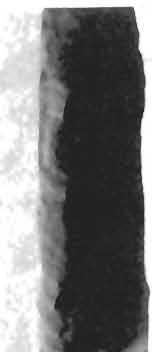
3 3 Dimorphodon macronyx (Buckland) Postilla189 meters thick in places. Beyond this, there is the problem of correlating Wright's own catalogue numbers with the numbers on his packing lists. In a letter to Marsh dated 21 June 1873, Wright expresses regret that Marsh has found some of the unsolicited material sent him unsatisfactory, and adds, "I will not forget to procure if possible the pterodactyle remains and other of your desiderata and will confine myself solely to those specimens you desire." But we do not know exactly what Marsh returned to Wright and how it may have affected inventory listings. It is unlikely that he sent back anything pterosaurian, because Wright refers to Marsh's requests for pterosaur material in at least twelve letters. He is always reminding Marsh of how scarce it is, but promising to send whatever he can. Thirty letters from Wright to Marsh, covering the years , are preserved in the Yale University Archives (Series I, Box 36, Folder 1556), but unfortunately we do not have Marsh's letters to Wright. Lot 456 is inventoried in a packing list from Bryce Wright dated 21 March It includes 87 separate items, for which Marsh paid a total of 91. The first item on the list is the pterosaur material, which consists of a slab and several smaller pieces. Wright listed it as follows: "No. 1. Humerus, ulna, and radius, etc., of pterodactyle in case and wing bones 1 rib and 1 other. Lias " Wright evidently meant the main slab when he said "in case," which leaves the "wing bones, 1 rib and 1 other." Four other small pieces of Lyme Regis matrix also have the accession number 456 and "No. 1" on their labels. Two are isolated wing bones, another is the rib, and the fourth, labeled simply "bone of Dimorphodon... "is the distal end of a right humerus. Two other pterosaur bones from Lyme Regis have the accession number 456. One is a shattered third wing-phalanx labeled "No. 26" on the back of the slab. This number does not correspond to Wright's listing in his letter of 21 March 1873 (No. 26 on his list is "Head and jaw of Belenostomus anningiae"), but it may have been a misprint of a number in Wright's catalogue. There is also a left wing-metacarpal, which has been prepared from its matrix. It is identified by a label that corresponds to Wright's No. 81 in the list for lot 456, which reads simply "bone of pterodactyle." Lot 462 is inventoried in a letter from Wright to Marsh dated 29 May This lot contains 37 items, including material of cave bear, plesiosaurs, turtles, mosasaurs, and many other vertebrates, for which Marsh paid One piece of bone is labeled "No. 27. Pterodactyle bone, Lyme Regis," and is evidently the distal end of another right humerus. The No. 27 on Wright's packing list does not correspond to pterosaurian material; however, his No. 26 does. The third accession lot (1503) containing Dimorphodon material is represented by a slab of black limestone (commonly called "Blue Lias") with fragmentary cranial and postcranial remains. The label is Wright's stationery, but the handwriting may be Marsh's; the locality given on the label is "Lyme Regis, Dorset, England." Accession number 1503 was received 16 September 1881 and contains European fossils donated by Marsh, probably received or bought by him during his European trip of that year (Marsh 1881b). The material is listed in only two parts in the accessions catalogue: "(a) casts of monkeys from Prof. A. Gaudry, Jardin-des-Plantes, Paris," and "(b) Tertiary fossils from Prof. A. Julien, Clermont- Ferrand, France." However, under entry 1503 in the original receipt book from which the listings in the accessions catalogue had been copied, there are not two items, but five. The first two correspond to (a) and (b); the last two include various fossils bought in Germany, but the third reads "(c) slab of Bone Bed rock from Aust Cliff, Gloucester, England, bought of B. M. Wright, London." How can the discrepancy in locality data be explained? All the known material of Dimorphodon comes from the cliffs of Lyme Regis, Dorset, on the southern shore
4 4 Dimorphodon macronyx (Buckland) Postilla 189 Fig. 1 Map of the southwest of England and Wales, with Lower Liassic horizons stippled. Based on 1958 Ordnance Survey Map of Great Britain.
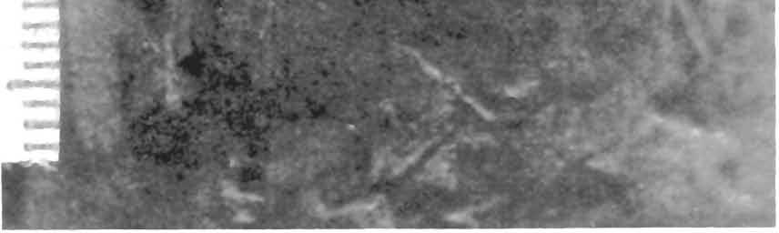
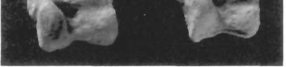
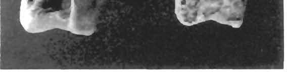
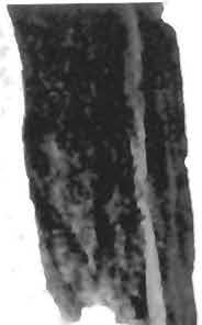
5 5 Dimorphodon macronyx (Buckland) Postilla189 of England; the first two specimens were collected by Mary Anning. Aust Cliff is not in Dorset, but in Gloucester, on the south bank of the River Severn (Fig. 1). However, a thin outcrop of Liassic deposits runs north from Dorset to the region of Aust, and the top of Aust Cliff consists of three feet of Lower Lias (Geological Survey of Great Britain, Map 250, Scale 1:63660,1958). Beneath this is 25 feet 6 inches of Rhaetic, 22 feet 9 inches of "Tea Green Marl" (Upper Keuper), and 97 feet of red marl. The black limestone slab is typical "Blue Lias," so it could indeed have been collected from the top of Aust Cliff although not from the "Bone Bed" of Aust Cliff listed in the YPM receipt book, because the latter is a light conglomerate of basal Rhaetic age, much lower in the section (Reynolds 1947). This specimen therefore marks the first occurrence of a Lower Liassic (Hettangian) pterosaur from a region in England other than Dorset. In Table 1 all YPM specimens referred to Dimorphodon have been tabulated and identified, and their principal measurements given. The only part of the Dimorphodon material to receive a Yale Peabody Museum catalogue number was the main slab of accession lot 456, which was catalogued as YPM 350 in This included a right humerus (YPM 350 F), a right lateral carpal (YPM 350 H), two metacarpals of the series l-lll (YPM 350 A and L), two phalanges from the manus (YPM 350 E and J), a pair of contiguous second and third wing-phalanges (YPM 350 D and 0, and what may be part of a fourth (YPM 350 K). Of the hindlimb there is preserved the complete right tibia-fibula (YPM 350 B), two distal tarsal elements (YPM 350 M and R), metatarsals ll-ivof the right pes (YPM 350 P), metatarsals III and IV of the left pes (YPM 3501), and two pedal phalanges (YPM 350 G and N). All of these have been removed from the slab (Fig. 2), and except for the fragile long bones (YPM 350 B, C, and D), are completely free of matrix. Fig. 2 Dimorphodon macronyx (Buckland), YPM 350, slab showing presumed original positions of the bones, which have been removed from the matrix. Restored from photographs; dimensions approximate. For identification and measurements of lettered elements, see Table 1. M K
6 6 Dimorphodon macronyx (Buckland) Postilla189 Table 1 Inventory and Measurements (in mm) of YPM Dimorphodon Material Item Description Length Width Prox. End Width Dist. End Median Width Accession Number 456: YPM 350 (Bryce M. Wright's "No. 1" of 21 March 1873) 350 A Metacarpal 350 B Right tibia-fibula 350 C Third wing-phalanx 350 D Second wing-phalanx 350 E Phalanx of right manus 350 F Right humerus 350 G Phalanx of pes 350 H Right lateral carpal 350 I Metatarsals III and IV of left pes 350 J Phalanx of right manus 350 K?partial fourth wing-phalanx or phalanx of fifth toe 350 L Metacarpal 350 M Left lateral distal tarsal 350 N Phalanx of pes 350 P Metatarsals ll-iv of right pes IV 350 R Left medial distal tarsal YPM 9175 Rib ("No. 1") YPM 9176 Second wing-phalanx ("No.1") YPM 9177?Ulna("No. 1") YPM 9178 Distal end of right humerus ("No. 1") YPM 9179 Third wing-phalanx ("No. 26"?) YPM 9180 Leftwing-metacarpal ("No. 81") Accession Number 462: YPM 9181 (Bryce Wright, 29 May 1873, No. 26 or 27 YPM 9181 Distal end of right humerus Accession Number 1503: YPM 9182 (Bryce Wright, 16 September 1881) 9182 A Left maxillary and jugal 9182 B Upper jaw fragment 9182 C Right humerus 9182 D Distal end of first wing-phalanx 9182 E Second wing-phalanx 9182 F Left femur, missing proximal end 9182 G Right femur 9182 H Right tibia-fibula 9182 I Fused right distal tarsals 9182 J Metatarsal 9182 K Unidentified shaft fragment nearb inc 7 (23.5) (78) 70 (31) (2.5) (1.8) (7) (50) (22) (1.0) (crushed)

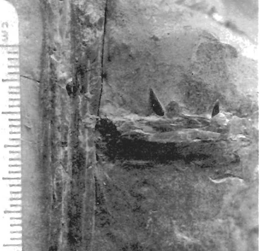
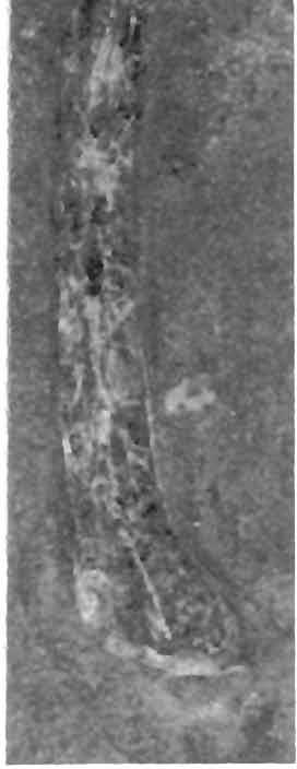
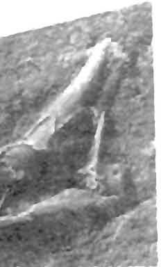
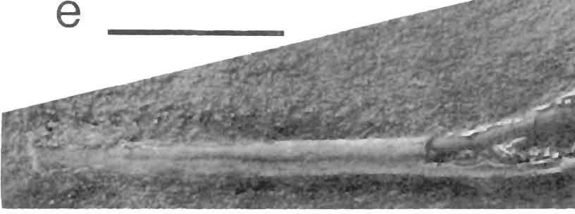
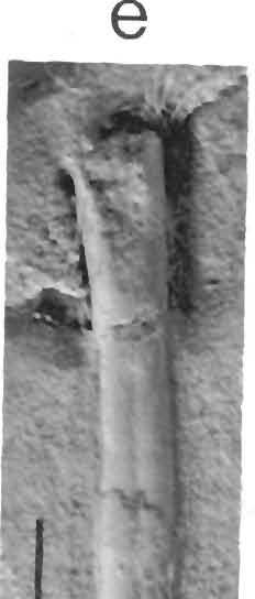
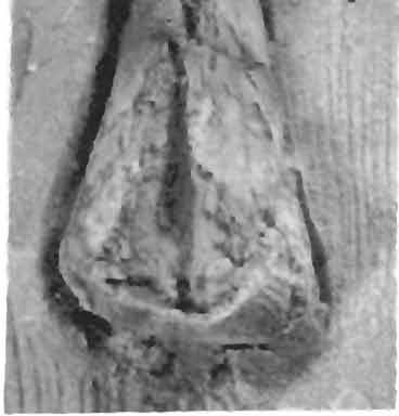
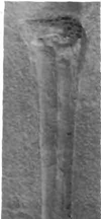
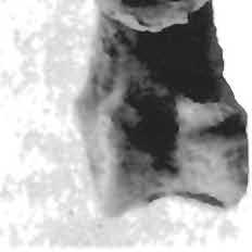
7 7 Dimorphodon macronyx (Buckland) Postilla189 The other material under lot 456 has been left uncatalogued until now. Of these specimens, the rib listed under "No. 1" has been designated YPM 9175; the nearly complete wing-phalanx is YPM 9176; the presumed ulna is YPM 9177; and the distal end of the right humerus is YPM All of this material and the main slab represent Wright's "No. 1" on his original list. "No. 27" on his list ("No. 26" on the specimen label) is the shattered third wing-phalanx, now numbered YPM 9179; the isolated left wing-metacarpal ("No. 81") is YPM The partial bone from lot 462, evidently a distal end of another right humerus, has been given YPM All the material on the slab from Aust Cliff (accession number 1503) is now YPM The slab of matrix is in the shape of a rough trapezoid measuring about 25 cm along the base, 8 cm along the top, 32 cm in height, and cm in thickness (Fig. 3). The Dimorphodon material includes two dentigerous parts of the upper jaws, the right humerus, the left femur (without the proximal portion: Fig. 4c), the complete right femur, the right tibia-fibula, the distal end of a first wing-phalanx, the entire second wing-phalanx adjacent to it, two contiguous distal tarsal elements, a single metatarsal, a possible fragment of the radius and ulna, and a small bone scrap, probably the hemicentrum of a fish. Of these bones, the humerus, right femur and tibia-fibula, first and second wingphalanges, and tarsals have been prepared out of the matrix. All bones are typically crushed flat and, except for the humerus, afford little three-dimensional relief. Description The osteology of pterosaurs has been well known for over 120 years, due largely to the many works of Hermann von Meyer, who concentrated his research on the Upper Jurassic forms of Bavaria. The discoveries and advances of the following century made necessary an extensive anatomical and systematic revision of the Pterosauria, which has been expertly carried out by Wellnhofer (1970,1974,1975,1978). The recent discoveries of pterosaurs in the Triassic of Italy (Zambelli 1973) and their detailed descriptions (Wild 1978) have also contributed greatly to an understanding of the anatomy and diversity of the earliest members of this group. The purpose of the present work, accordingly, is not to provide detailed analysis of pterosaurian osteology, but to point out those features in the Yale specimens that cast new light on an important early pterosaur and on the functional mechanics of pterosaurs in general. Skull Two fragments of the upper jaws are preserved in YPM The smaller (Fig. 4b) is from the right maxilla, seen in lateral view. It is 19 mm long and mm high, and bears three slightly recurved, sharply pointed teeth approximately 6 mm apart. The shape of this fragment and the size of its teeth appear to correspond to a part of the other fragment of the jaw preserved on the slab. Neither piece contains either the very enlarged laniaries found in the premaxilla and foremost part of the dentary, nor the minute "lancet-shaped" teeth found along most of the dentary, for which Dimorphodon was named. The second fragment (Fig. 4a) is larger than the first. It consists of most of the lower border of the left side of the skull, seen in lateral view, including nearly the entire maxillary bone and most of the jugal. In Dimorphodon the orbit sits higher in the skull than both the preorbital opening and the nares. The lower border of the orbit is formed by the upper edge of the jugal bone; the middle ascending process of the jugal separates the orbit from the preorbital opening. This process is incomplete in YPM 9182, but its configuration is clear. The entire lower border of the preorbital opening is outlined; it is approximately 33 mm at its widest point. A small thin flange of bone protrudes anteriorly at a low angle


8 8 Dimorphodon macronyx (Buckland) PostiIla189 ^%-±<$%y^ ri i r -'^ ^ * *?

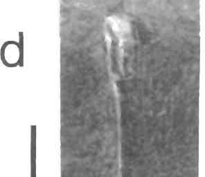
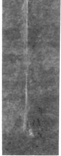
9 9 Dimorphodon macronyx (Buckland) Postilla189 from the lower border of the preorbital opening. This may be the thin lateral flange of the pterygoid, which articulates with the ectopterygoid in the region just medial to the jugal in Campylognathoidesand Rhamphorhynchus (Wellnhofer 1974; 1978, Abb. 3). The suture between the jugal and maxilla, like most skull connections in pterosaurs, is unclear. Owen (1870) did not even identify a jugal bone in his description. Von Meyer, the 19th-century German authority on pterosaurs, succinctly summarized the problem in Zur Fauna der Vorwelt (1859:15, translated by Seeley 1870). In Pterodactyles, as in birds, the bones of the skull blend together so imperceptibly that their sutures at best are only indistinctly seen, and are sometimes obliterated; while even in full-grown reptiles they are all to be made out with great distinctness. There is the more difficulty in ascertaining the structure of the Pterodactyle skull, since generally only the lateral aspect is exposed, and hence we get scarcely any information about its upper and under surfaces. Among the skulls which are exposed from the side, information is at times afforded by those in which the parts have suffered some displacement; but the separations so produced are to be accepted with great caution, for they do not always coincide with the real boundaries of the bones. Pterosaur bone is so delicate that it is easily checked and shattered. There are at least four different reconstructions of the < Fig. 3 Dimorphodon macronyx, YPM 9182, slab. Abbreviations: fv, hemicentrum of a fish, possibly Pholidophorus; If, left femur; / uj, left upper jaw; mt, metatarsal; fright femur; rhum, right humerus; rt, right distal tarsals; rt-f, right tibia-fibula;? r-u, possible portion of a radius and ulna; ruj, right upper jaw; wph 1, first wing-phalanx; wph 2, second wingphalanx. Scale in mm; measurements given in Table 1. skull of Dimorphodon, all drawn from the same specimen, and all with different interpretations of sutural connections (Fig. 5; see also Seeley 1901 and von Huene 1914). Figure 6 illustrates the portion of the skull of Dimorphodon represented by YPM 9182, based on the skulls in the British Museum (Natural History), described by Owen (1870). There is no clear connection between the maxilla and premaxilla, but bearing in mind von Meyer's cautionary remarks, the approximate extent of the nares may be inferred from the basal length of the preorbital opening. The fragment of bone preserved here ends at a point just short of the hypothetical anterior limit of the nares. This is the case for most rhamphorhynchoids, as Wellnhofer (1978, Abb. 2) shows. The usual reconstruction of this suture in Dimorphodon differs from the rhamphorhynchoid pattern (Fig. 5), but the evidence for this is not clear. It seems equally possible that the anterior end of the Yale specimen represents the natural break between maxilla and premaxilla. Seven maxillary teeth are preserved, and there are alveoli for one or two more; this corresponds to Owen's assignment of eight or nine teeth to the maxilla. The premaxilla, bearing the four large laniaries, would form the snout and the anterodorsal border of the nares, as in other rhamphorhynchoids (Wellnhofer 1978, Abb. 2). As no evidence of a large alveolus for the fourth laniary is preserved, no portion of the premaxilla appears to be represented in this specimen. The size and proportions of these cranial remains imply that the specimen is a juvenile; this assessment is further supported by the dimensions of the hindlimb, which will be discussed below. Dentition Three teeth have been preserved in the smaller jaw fragment of YPM 9182, and seven in the larger. Although none of the teeth in the larger fragment is complete, they are clearly the same size as the three teeth in the smaller fragment, as deter-

10 10 Dimorphodon macronyx (Buckland) Postiiia189 a 'fifr*.: *->' -... I -ft? V in VI * " :* - >J& '*' *_. /S.;<? f\ -..- '- -^^ ' ^^H^^-^^.S^s&t'*
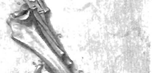
11 11 Dimorphodon macronyx (Buckland) Postilla 189 Fig. 4 Dimorphodon macronyx. a, YPM 9182, detail of slab, showing left maxilla and part of jugal. b, detail of the same slab, showing dentigerous part of right maxilla and possible radius-ulna fragment. Scale is in mm. c, detail of the same slab, showing isolated metatarsal, e, YPM 9175, rib. Scale bars = 1 cm. Fig. 5 T Four restorations of the skull of Dimorphodon macronyx. A, Owen 1870; B, Arthaber 1919; C Wellnhofer 1978; D. Wiman Parenthetical symbols in A represent Owen's terminology. Abbreviations: al, adlacrimal; - ang, angular; ar, articular; C/, dentary; f, frontal;/jugal; /, lacrimal; m, maxilla; /7?/,malar; ms, mastoid; a nasal; p, parietal; pm, premaxillary; pf, pof, postfrontal; po, postorbital; poc, pa rocci pita I; prf, prefrontal; q, quadrate; g/quadratojugal; sa,surangular; so, supraorbital; spl, splenial; so/, squamosal; tym, tympanic. Length of reconstructed skull about 20 cm. sq(ms) (sq) q(tym) ang ^Fig.6 Tentative restoration of the skull of the small and presumed juvenile Dimorphodon macronyx, based on skull fragment of YPM Abbreviations as in Figure 5. Length of reconstructed skull about 14 cm.

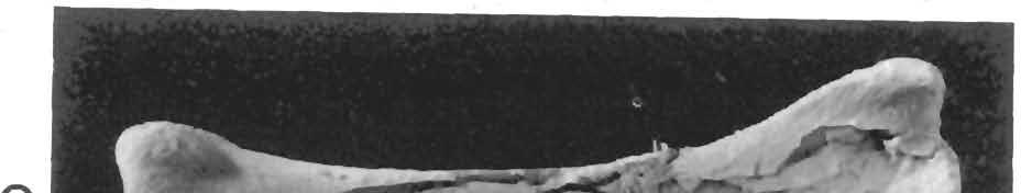
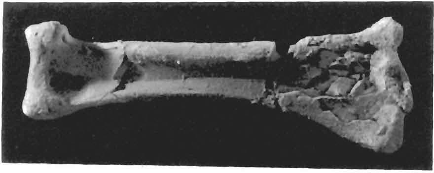
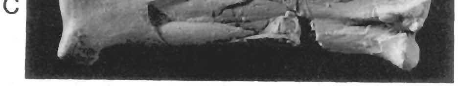
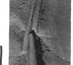
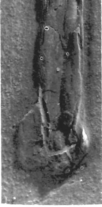
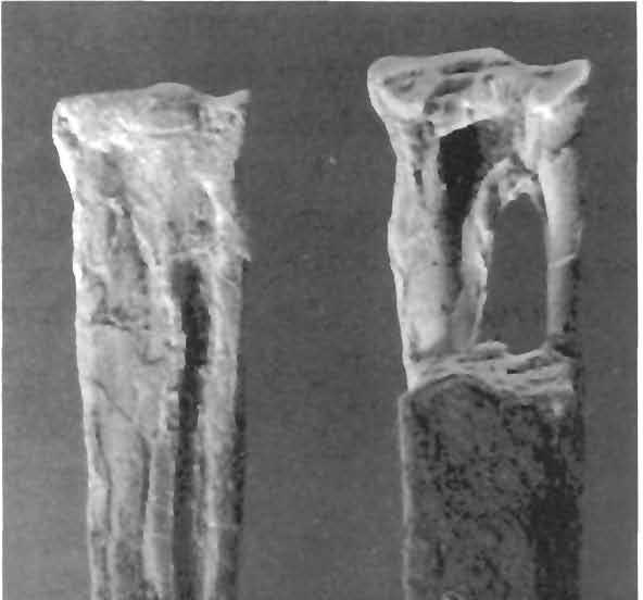
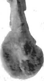
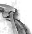
12 12 Dimorphodon macronyx (Buckland) Postilla 189 mined from the size of their roots. The placement of these teeth along the upper jaw permits an approximation of the dental configuration of the juvenile Dimorphodon. The adult has four large laniaries widely spaced at the front of the jaw, corresponding to the premaxilla. The maxilla bears seven or eight smaller teeth of this form, according to Owen (1870), although Arthaber (1922) figured nine in his reconstruction (Fig. 5B). The upper jaw fragment described here has fragments of seven teeth, of which the last two are very close together. There is a crushed alveolus midway between the third and fourth teeth preserved here, and also a large space (possibly an alveolus) just in front of the first tooth. This tooth is not quite parallel to the others, and it may have been dislodged; the space that separates it from the second tooth is smaller than in most of the series. This region of the maxilla is also badly preserved in the other Dimorphodon skulls. I regard the effective number of maxillary teeth at this growth stage to be eight, including the last two, which are smaller and more closely spaced. Additional teeth seem to have been formed at the back of the jaw, and were progressively smaller than those in front. The most complete teeth are in the smaller jaw fragment. The best preserved of these is partly disengaged from its natural position in the alveolus, and all but the tip of the root is visible. From comparison with the complete root portions of other teeth, the entire length of this tooth was approximately 7.0 mm. The form is identical to that of the larger laniaries, only proportionately smaller, as the isolated teeth figured by Owen (1870, plates XVII and XVIII) show. Owen described the large premaxillary laniaries as "subcompressed, subrecurved, and sharp-pointed" (p. 43), with the maxillary laniaries becoming gradually smaller and less curved towards the back of the maxilla. The size and shape of the three teeth in this smaller jaw fragment thus show that it belongs to the fore or middle part of the right maxilla. In the lower jaw of Dimorphodon, which is deeper than this fragment, there are three rather large laniaries in the front, followed posteriorly by only two of the size of the maxillary teeth, and finally by a long row of "small, lancetshaped, close-set teeth" (Owen 1870:42). It is therefore likely that this fragment belongs to the lower jaw, unless the relative size of some of the teeth changed during growth and replacement. Postcranial Material As frequently happens with disarticulated pterosaur skeletons, there are no remains of the pectoral or pelvic girdles or of any vertebrae among the Yale Dimorphodon specimens; almost all preserved material is of long bones. A single rib (YPM 9175; Fig. 4e) has been preserved and is tentatively referred to this taxon. It is double-headed, wide at the proximal end between the two heads, which are separated by 9 mm, and slender along its length (40 mm). A hollow channel in the proximal part of the shaft between the heads is revealed by crushing of the surface bone. There is a slight curvature at the proximal end, but the shaft is quite straight for 80% of its length. The distal end is slightly expanded and rounded. Two complete humeri are preserved. One right humerus (YPM 9182 C; Fig. 7c) measures 63 mm from the saddle-shaped articular surface of the head to the tip of the ulnar condyle. The proximal articular surface is well preserved and shows the characteristic lip that articulated with the glenoid fossa. The natural torsion of the shaft has been distorted by crushing, but the deltopectoral crest still preserves some of its natural curvature. The angle made by the head and crest of the humerus is slight when compared to less distorted specimens, in which the head and crest may curve anteroventrally to form a semicircular silhouette in proximal view (Lawson 1975, fig. 1). A bulblike terminal thickening of the expanded crest has been preserved; it shows, as Owen (1870:51) noted, that the deltopectoral crest was not simply a thin flat plate of bone, as speci-


.")


13 13 Dimorphodon macronyx (Buckland) Postillal83 mens often appear to indicate. The distal end of the humerus is well preserved, particularly the articular condyles for the radius and ulna, although this area is even better preserved in the following specimen. The other complete right humerus (YPM 350 F, Figs. 8 and 3) is exceptionally well preserved, and its fine condition allows new insight into the structural and functional details of the forelimb (see Discussion). The head and neck were separated from the shaft which itself had been broken in three places. These portions were all preserved in three dimensions, however, and were completely repaired. The deltopectoral Fig. 7 Dimorphodon macronyx, YPM 9182, material removed from slab, a, second wing-phalanx; b. distal end of first wing-phalanx; c right humerus; d. right tibia-fibula in posterior view; e, right femur. crest was also separated from the main shaft and slightly crushed in its proximal area; this portion has proven more difficult to restore to its original form, and the crest as repaired has less than the natural curvature. The head of the humerus is the typical saddle-shaped facet with a pronounced medial lip. A deep notch at the neck separates the lateral {= external or greater) tuberosity from the head of the humerus. The deltopectoral crest has a pronounced expansion at its distal extremity, as in the humerus described above and in BMNH The crest extends nearly 19 mm from the midline of the shaft, and is more similar to the narrow form of Rhamphorhynchus than to the platelike form of Eudimorphodon and Campylogna ihoides* The presence of a pneumatic foramen cannot be asc :ained because the central a b c d e


.")


.")


14 14 Dimorphodon macronyx (Buckland) PostilIa189 v\ i v Fig. 8 Dimorphodon macronyx, YPM 350 F, right humerus. Head of humerus at upper left, deltapectoral crest at upper right. Scale = 1 cm. Fig. 9 Dimorphodon macronyx, YPM 350 F, right humerus. Obverse view of Figure 8. Head of humerus at upper left, deltopectoral crest at upper right. Scale = 1 cm. proximal region of the humerus is fractured and crushed, but it does not appear to have been located in the same position as in birds, A deep channel near the head of the humerus runs for a short distance parallel to the axis of the shaft Whether this represents a pneumatic channel is conjectural. Most perforations taken for pneumatic foramina have been in uncrushed but worn and fragmented specimens from the Cambridge Greensand, described by Seeley and others (see Seeley 1901). Von Meyer, Owen, and Seeley found pneumatic foramina in many parts of the skeleton, but this work has not been extensively studied by later authors, and the whole problem needs further investigation. The curvature of the shaft of the humerus is sigmoid. Seen from the proximal end, the distal end of the shaft is twisted nearly 45 posterodorsally (see Fig. 10). Pronounced ridges run along the shaft from the greater tuberosity to the medial supracondyloid tubercle, and from the edge of the deltopectoral crest to the lateral supracondyloid process. The paths of these ridges further outline the torsion of the shaft and establish the lines indicative of muscular attachment. They are instrumental in understanding the movement of the humerus during the flight stroke (see Discussion). The distal end of the humerus is well preserved in YPM 9182 C and exceptionally so in YPM 350 F. The distal condyles and the adjacent ridges for muscular attachment are clearly delineated., and Figure 10 points out some of these comparable features in

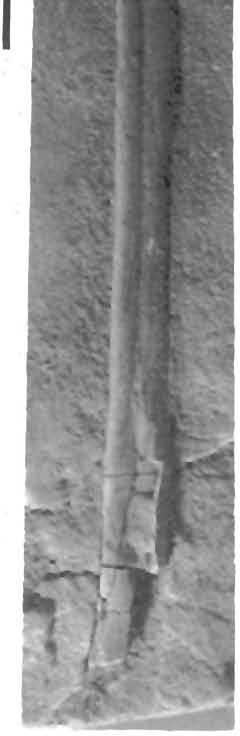
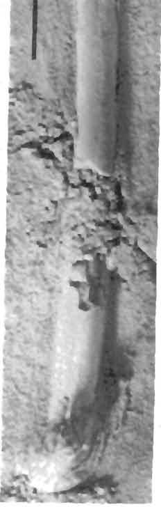
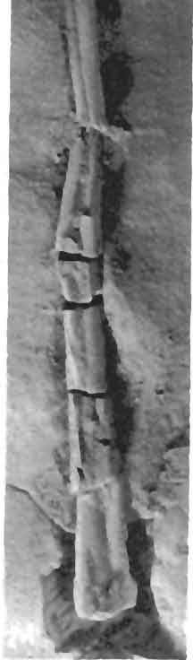
15 15 Dimorphodon macronyx (Buckland) Postilla 189 Dimorphodon and an eagle. In pterosaurs, as in birds, the radial condyle articulates with part of the ulna as well as with the radius, whereas the ulnar condyle articulates only with the ulna. In birds, the main extensor muscles of the distal segment of the wing originate from the lateral epicondyle of the humerus, while the principal flexors of the outer wing originate from the medial epicondyle (Hudson and Lanzillotti 1955). The comparable development of these sites of origin in pterosaurs argues for a similar functional pattern. Two other distal ends of right humeri are preserved in the YPM Dimorphodon material (YPM 9178, Fig. 11a; YPM 9181, Fig. 11d), but are more poorly preserved than the other humeri and yield no additional information. YPM 9177 (Fig. 11 e) is a slender, slightly bowed bone 70 mm long, and is tentatively identified as an ulna. It is preserved on a small slab of matrix of which the borders are encased in cement. A long crack in the slab, filled with glue, indicates that there were originally two pieces of matrix. Where the crack intersects with the bone, a section of the shaft is missing and has been filled in with cement. Unfortunately, the cement that encases the perimeter of the specimen also abuts against the articular ends of the bone, making further preparation very difficult. There is no other diagnostic material of radius or ulna among the Yale specimens, although an unidentified shaft fragment 45 mm long, preserved adjacent to the smaller jaw fragment on the slab of YPM 9182 (Fig. 4b), may pertain to one of these bones. YPM 350 H (Fig. 12a, 13a) is a right lateral carpal. This is a rather robust element with a concave surface that articulated with the distal carpal. A convex surface opposite to this with a round flattened area supported a small round sesamoid bone on which the medially directed pteroid bone rested (Wild 1978, Taf. 9f). Ironically, although this carpal is called "lateral," it is actually located on the medial (radial) side, and was held in front of the wing when the wing was outstretched. This medial carpal is marked by several tubercles and depressions to which attached the ligaments and tendons that helped to manipulate the propatagium. Its form and proportions correspond closely to those of Eudimorphodon, Dorygnathus, and Campylogna thoides. Two isolated metacarpal bones of the series l-lll have been preserved (YPM 350 A and L; Figs. 14c, d; 15c, d). They are extremely delicate and fragile. In comparison with the metatarsal elements discussed below, they are thinner and more rounded in cross-section than the metatarsals of the same length, and they are slightly bowed in the lateral direction. Their proximal ends are widened and flattened into shallowsshaped spatulate surfaces that articulated with the medial condyle of the proximal end of the large wing-metacarpal (=mc IV). Distally the metacarpals expand into round- Fig. 10 Comparisons of the right humeri of the eagle Aquila (above in each pair)) and Dimorphodon (below in each pair). A, proximal view, oriented with the distal ends parallel as in C. Note the differences in angle of orientation of the heads and deltopectoral crests. Drawn to the same size. B, complete humeri in palmar (left) and anconal (right) views. Scale = 1 cm. C distal ends in palmar view, drawn to the same size. /, Processus supracondyloideus lateralis; 2, Epicondylus lateralis; 3, Trochlea radialis; 4, Vallis intertrochlearis; 5. Trochlea ulnaris; 6, Epicondylus medialis; ZTuberculum supracondyloideum mediale; 8, Fovea supratrochlear ventralis. dp, deltopectoral crest; gt, greater tuberosity; h, head; a neck. Fig. 11 Dimorphodon macronyx. a, YPM 9178, distal end of right humerus; b, YPM 9180, left wingmetacarpal, lateral view; c, YPM 9180, medial view; d, YPM 9181, probably the distal end of a right humerus; e, YPM 9177,?ulna; f, YPM 9176, second wing-phalanx; g, YPM 9179, shattered third wing-phalanx. Scale bars = 1 cm.
16 16 Dimorphodon macronyx (Buckland) Postilla189 Fig. 10 bird pterosaur pterosaur

17 17 Dimorphodon macmnyx (Buckland) Postilla 183 Fig. 11 '** / 4. d \n "!ft ik *'** < 4 \ ''*">?&
.")


.")


18 18 Dimorphodon macronyx (Buckland) Postilla189 ed bulbs with ventral ligamentous grooves; this expansion was necessary for the reception of the much stouter proximal phalanges of the first three fingers. Two of these phalanges were preserved (YPM 350 E and J; Figs. 12c, g; 13c, g). They are more robust than the corresponding elements of the pes, and their flexor tubercles are more pronounced. Marked ginglymal grooves characterize their distal ends. Viewed from the proximal end, the flexor tubercles are subtriangular, not keeled, and distinctly set off to the medial Fig. 12 Small skeletal elements of Dimorphodon macronyx, YPM 350, prepared from matrix, a, 350 H. right lateral carpal; b. 350 G. pedal phalanx; a 350 J, phalanx of right manus; d. 350 R, left medial distal tarsal distal view; e, 350 M, left lateral distal tarsal, distal view; i 350 N, pedal phalanx; g, 350 E, phalanx of right manus; h, coalesced right distal tarsals of YPM 9182, distal view; /; YPM 350 K. possible fragment of fifth pedal phalanx {or fourth wingphalanx?)."scale = 1 cm. side. This corresponds to the condition seen in the first two digits of the dromaeosaur Deinonychus (Ostrom 1989:108). In both YPM 350 E and 350 J this tubercle is deflected to the left side, which indicates that both phalanges belong to the right manus. They are probably proximal phalanges, and if so, 350 J probably belongs to digit II, while 350 E may represent digit III, based on comparison with BMNH YPM 350 G (Figs. 12b, 13b) is more slender than these two, and has no tubercle. It more closely resembles the proximal phalanges of the pes in BMNH 41212, as does the long gracile element YPM 380 N (Figs. 12f, 13f), which is incomplete at the dorsal end of its proximal facet YPM 350 G and N are accordingly assigned to the hindfoot but cannot be further identified with certainty. A left wing-metacarpal (YPM 9180; Figs. 11 b, c) 38 mm long is badly crushed and retains little relief even at its articular ends. It is a typical broad, flat rhamphorhynchoid wing-metacarpal, with a well-developed, laterally placed bicondylar joint at the distal end, on which the enormous wing-finger b e d e f g


, and the complete length of the second phalanx (YPM 3182")


.")

19 19 Dimorphodon macronyx (Buckland) Postilla189 pivoted. This isolated metacarpal is 4 mm longer than the two medial ones catalogued as YPM 350 A and L White it is unlikely that it came from the same animal, the discrepancy in size is not great Gallon's (1381 b) recent description of a superb rhamphorhynchoid wing-metacarpal from the Morrison Formation of Wyoming obviates further discussion of YPM 3180 since, although the relative proportions of this bone in rhamphorhynchoids are variable, there are no morphological features diagnostic below the subordtnal level The distal end of the first phalanx of the wing-finger (YPM 3182 D), and the complete length of the second phalanx (YPM 3182 E; 74 mm), were preserved in contiguity (Pigs. 3,7a, 7b). The identification of these wing elements can be made on the basis of several features, In Dimorphodon the second wing-phalanx is shorter m Fig. 13 Obverse views of the same elements of Figure 12, minus /(350 K). Scale = 1 cm. h length and broader in cross-section than the third. The distal expansion of YPM 3182 E is too wide for the reception of the proximal end of the fourth phalanx, when compared to this element in other specimens of Dimorphodon and in other pterosaurs. The ratios of the various bones of the wing to each other (except the metacarpal and the terminal phalanx) are diagnostic, at least to the generic level (Padian and Wild, unpublished data). In the two British Museum specimens of Dimorphodon (BMNH and R 1035), the length ratios of the second wing-phalanx to the humerus are 1.38 and 1.23; in YPM 350 C and F this ratio is 1.20, These specimens have been ranked in decreasing order of size (the second phalanges are respectively 124,102, and 37 mm), and it can be seen that the ratio decreases with growth. In YPM 3182, the smallest of the group, the ratio of the presumed second phalanx to the humerus is only By contrast the ratios of the third phalanx to the humerus in the specimens mentioned above are 1.54,1.33+, and 1.30, respectively; these values are significantly higher, g.3ri; - - d

; they are")


 f a shattered third wing-phalanx 101 mm long.")

20 20 Dimorphodon macronyx (Buckland) Postilla 189 * Fig. 14 Dimorphodon macronyx, YPM 350, isolated metapodials. a, 350 P, metatarsals ll-iv of right pes, dorsal view; b. 350 I, metatarsals III and IV of left pes, dorsal view; c. 350 L, metacarpal, ventral view; d, 350 A, metacarpal ventral view. Scale is in mm. At least two segments of the wing-finger are represented on the main slab of accession number 456 (YPM 350); they are the complete second and contiguous third phalanges (YPM 350 D and C), and possibly part of a fourth (YPM 350 K). The second and third (Figs. 16b, c) are of typical form and proportion for Dimorphodon; they articulate snugly, but are unfortunately flattened and yield no new information. The same is true for YPM 9176 (Fig. 11 f), an isolated, mostly complete second wingphalanx with a preserved length of 76 mm; and YPM 9179 (Fig. 11 g) f a shattered third wing-phalanx 101 mm long. The articular ends of the wing-phalanges show the typical ovoid ball-and~cup arrangement: the proximal facet of each phalanx is concave and the distal end is convex. The shafts are quite straight and there seems to have been little movement possible between the phalanges, YPM 350 K (Fig. 12i) is an incomplete, flattened, slightly tapering fragment ovoid in cross-section. A faint impression on the slab corresponds roughly to a further extension of the wider (proximal) end, about 5 mm away from it and continuing for a


21 21 Dimorphodon macronyx (Buckland) Postilla189 Fig. 15 Dimorphodon macronyx, YPM 350, obverse views of isolated metapodials in Figure 14. length of 10 mm. This evidence may support the identification of the bone fragment as belonging to the fourth wing-phalanx, since almost no other element of the skeleton would be so long, straight and slender, There is no indication of an articular facet at either end, A shallow fracture runs along the exposed surface of the shaft as it does in the other two wing-phalanges. The width of the shaft supports the inference that it comes from the distal half of the fourth wing-phalanx, but it lacks the curvature that usually accompanies the tapering of this element. The preserved bone is also light and straight enough to belong to the first phalanx of the odd fifth toe, and this possibility must be considered, From the few elements that are preserved, the wingspan of these specimens can be roughly estimated, based on the proportions of other Dimorphodon material. The individual represented by YPM 350 had a wingspan of about 1200 mm, while the wingspan of YPM 9178 would have been about 980 mm. The largest nearly complete specimen of Dimorphodon (BMNH 41212) had a wingspan of almost 1450 mm. The right femur (YPM 9182 G; Fig, 7e) is bowed, but not sigmoid. Its length from the tip of the head to the lateral condyle is 59 mm. The head is set off at an angle of about


, was preserved with the anterior face up, unlike YPM 9182 H.")
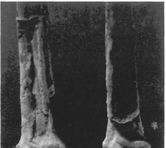
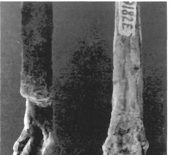
22 22 Dimorphodon macronyx (Buckland) PostlIia189 Fig, 16 Dimorphodon macronyx, YPM 350, long bones removed from matrix, a, 350 B, right tibia-fibula, anterior view; b 350 D, second wing-phalanx; c, 350 C, third wing-phalanx. 120 from the axis of the proximal region of the shaft. The articular surface is rounded and smooth; the neck is slightly constricted but rather robust, much like the configuration in bipedal dinosaurs such as Coelurus and Dilophosaurus. There is evidence of a greater trochanter, although most of this region is crushed. The distal end of the femur curves slightly laterally and the lateral condyle extends slightly farther distaliy than the medial, which is larger and more pronounced. The expansion of the distal condyles of the femur, in the two Dimorphodon specimens described here and in all known pterosaurs, is mainly subterminal. The articular surfaces of these condyles are oriented 90 ventral to the shaft axis. The right tibia-fibula (YPM 9182 H: Fig. 7d) has been preserved adjacent to the femur, and almost in natural position, except that the tibia-fibula has been rotated to lie with the anterior side down. Both bones have been removed from the slab, and their natural articulation can be determined (Fig. 17). It is evident that the medial condyle of the femur articulated with the tibia, whereas the lateral condyle articulated with the fibula, as Seeley (1901) stated, contrary to Owen (1870:52). The tibia and fibula of rhamphorhynchoids were fused at their proximal ends. The tibia is convex and the fibula concave at the surface where they meet. YPM 350 B, a complete right tibia-fibula (Fig, 16a), was preserved with the anterior face up, unlike YPM 9182 H. The entire proximal joint surface of YPM 350 B is well preserved, and slopes posteriorly at an angle nearly 50 to the long axis of the shaft. The femur, therefore, did not normally meet the tibia-fibula in columnar fashion. However, a range of movement of about 135 appears to have been possible at the knee, and the femur and tibia-fibula probably met at an angle of in normal stance, with the femur more or less horizontal and the tibia-fibula nearly vertical, as in birds (Fig. 17). There is no sign of a patella in any pterosaur. An incipient expansion similar to the cnemial crest of modern birds, but

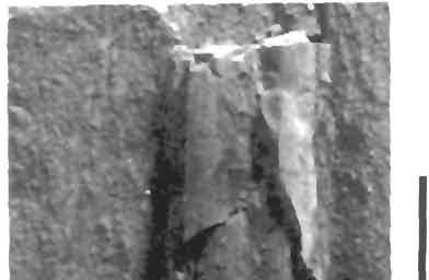
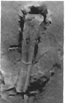
23 23 Dimorphodon macronyx (Buckland) Postilla 189 much less pronounced, has been noted in specimens from the Cambridge Greensand (Seeley 1901), and more recently by Galton (1981 a). This tuberosity is present in Dimorphodon as well, and, as Seeley noted, it does not appear to represent a separate center of ossification. The reduction of the fibula in rhamphorhynchoids (Fig. 18a, b) resembles in many respects the same pattern seen in birds. YPM 9182 H (Fig. 7d) is a right tibiafibula 85 mm long. 7 mm from the proximal end, the shafts of the tibia and fibula separate, and continue for a length of 20 mm spaced by a distance of approximately 1 mm before fusing again. The line of fusion is at least 12 mm long, and as the fibula gradually tapers along this length its distal extent becomes indistinguishable from the tibia, partly as a result of crushing. The total length of the fibula is thus at least 39 mm, or approximately 46% of the tibia, in this specimen. The other right tibia-fibula (YPM 350 B; Fig. 16a) in this collection is nearly 20 mm longer, but the proximal contact between tibia and fibula is only 5 mm, the interosseal space is at least 28 mm long, and the distal fusion of the two bones appears to extend for another 28 mm, although crushing is again a problem. Like pterosaurs, birds have an interosseal space between the tibia and the fibula; the latter bone is reduced to a splint and usually merges into the tibia at some distance before the distal end. The distal end of the right tibia-fibula (Fig. 18c, d, e) is exceptionally well preserved in both YPM 350 B and YPM 9182 H. This area, like the distal end of the humerus, shows many remarkable similarities to the homologous area of birds (Fig. 19). The distal ends are comparable in the extent of the anterior expansions of the bicondylar surface that forms the joint. This distal expansion in pterosaurs has been taken to represent the fusion of the proximal tarsal elements (astragalus and calcaneum) with the tibia, as in birds and small bipedal dinosaurs (Seeley 1901; Wellnhofer 1978). The two condyles are of comparable size, but bird pterosaur Fig. 17 Comparison of the knee joints of the Golden Eagle Aquila chrysaetos (above) and Dimorphodon macronyx (below). In both, the right femur and tibia-fibula are shown in lateral (left) and anterior (right) views. Not drawn to scale. the lateral condyle is larger and rounder in side view, while the medial condyle is somewhat broader in anterior view. This contrasts slightly with the situation in birds, where both condyles are of approximately equal transverse width, but the lateral (external) condyle may extend farther, and the groove between them is wider and shallower than in pterosaurs (Currieand Padian, in press). The ligamentous groove is deep in pterosaurs, although not as deep as in birds, and there is no bony supraligamentous bridge. The distal tarsal elements preserved in the two slabs (YPM 350 M and R and YPM 9182 I) are exceptional for the detail and


24 24 Dimorphodon macronyx (Buckland) Postilla189 \ i 9» a 1 cm N ' i u
25 25 Dimorphodon macronyx (Buckland) Postilla189 < Fig. 18 Dimorphodon macronyx. a, anterior views of proximal ends of right tibiae-fibulae of YPM 9182 E (left) and YPM 350 B (right); b, posterior views of the same; c, anterior views of distal ends of right tibiae-fibulae of YPM 350 B (left) and YPM 9182 E (right); d, posterior views of the same; e, distal ends of tibiae: YPM 350 B in lateral view (left), YPM 9182 E in anterior view (right). Fig. 19 V Anterior views of distal ends of right tibiaefibulae of Dimorphodon macronyx (left) and the Golden Eagle Aquila chrysaetos (right), drawn to the same size. In both, astragalus and calcaneum are indistinguishably fused to the bases of the long bones. Abbreviations: br, bony bridge of transverse ligament; condext, external or lateral condyle; cond int, internal or medial condyle; gr Edl, groove for M. Extensor digitorum longus; grpp, groove for M. Peroneus profundus; inc, incisura; L, lateral ligamentous prominence; L lo, lower attachment of Ligamentum obliquum; M, medial ligamentous prominence; U lo, upper attachment of Ligamentum obliquum. Not drawn to scale. the information they yield about the pterosaurian ankle. These elements must be considered the lateral and medial tarsals, since the astragalus and calcaneum are fused to the tibia-fibula. YPM 350 M (Figs. 12c, 13c) is the left lateral distal tarsal. YPM 350 R (Figs. 12d, 13d) is the left medial distal tarsal, and the two right tarsal elements are preserved in their natural articulation with each other in the smaller specimen YPM 9182 I (Figs. 12h, 13h). The correspondence of detail between these two specimens indicates that neither has suffered either wear or distortion, although there is a break in the flat, quadrangular medial distal tarsal of YPM 9182 I, which has been repaired. The lateral distal tarsal is a short stump nearly 6 mm in length and roughly 2 mm in cross-section, with a complex topography. The medial distal tarsal is a flat quadrangular plate 5 mm at the widest edge and 4 mm across, with two raised ridges on its distal face (Fig. 12d, h) that match the corresponding metatarsals, The surfaces of these tarsal elements are finely porous, which suggests a cartilaginous covering. The tarsal unit is roughly wedge-shaped. It thins anteriorly to a slight degree, but much more so medially, especially on the distal cond ext cond int bird

26 26 Dimorphodon macronyx (Buckland) Postilla189 surface. The distal view of these tarsals (Fig. 12d, e, h; Fig. 20) clearly shows the articular facets for the metatarsals. The large aberrant fifth metatarsal articulates laterally and slightly posteriorly with the lateral face of the lateral tarsal. The other four metatarsals are oriented normally. The articular surface for the fourth metatarsal appears to be shared by the lateral and medial tarsals, while the third and second are carried entirely by the medial tarsal. There is no space for the first metatarsal to articulate, which is consistent with observations on other pterosaurs (Wellnhofer 1978, Abb. 17). The lateral distal tarsal corresponds in both shape and topography to distal tarsal 4 of the lower Jurassic theropod dinosaur Syntarsus (Raath 1969:19, his fig. 6b): "Its proximal and medial surfaces are concave, and its distal and lateral surfaces are convex. The lateral surface also has a small notch to accommodate the proximal end of metatarsal V." In Syntarsus the lateral distal tarsal is fused to the metatarsals, but in theropods this element, when free, is usually identified as a fusion of distal tarsals 2 and 3 (Ostrom 1969) because it covers metatarsals II and III, as the corresponding element does in pterosaurs. The proximal surfaces of the distal tarsals (Figs. 13d, e, h; 20) show very clearly the rounded depressions which served as the articular facets for the tibia. The larger fossa is on the correspondingly wider medial tarsal, because the medial condyle of the tibia is the larger of the two. A tuberous posterior process of the lateral tarsal partly overlaps the posterior face of the medial tarsal, and may have been the site for tendinous attachments of muscles that extended the foot. The metatarsal elements preserved on this slab include metatarsals ll-iv of the right pes (YPM 350 P: Figs. 14a, 15a) and metatarsals III and IV of the left pes (YPM 350 I: Figs. 14b, 15b). The elements of both are preserved in a coalesced state. The proximal ends interlock and may have been fused; metatarsals II and IV meet at the dorsal surface near the proximal end of the metatarsus, displacing metatarsal III ventrally to a slight degree. The distal ends of the metatarsals are separated and splay slightly/recalling the situation in birds and some theropod dinosaurs (Osmolska 1981). The two metatarsals of YPM 350 I are not completely fused along their length, and lack 5 mm at their distal ends, compared to the complete members of YPM 350 P. The third metatarsal is slightly longer (about 1 mm) than the second and fourth; the first, when it is preserved, is about 1 mm shorter than these (Owen 1870). A slight lateral curvature at both ends, and a gradual swelling at the proximal end, identifies the fourth metatarsal. There are ginglymal grooves on the ventral and distal sides of the distal ends of the metatarsals, which are not as pronounced as in birds, but are comparable to those of theropod dinosaurs. The first four metatarsals seem to have functioned as a unit; the toes flexed in the same plane as the tarsus, and diverged distally to a slight degree, as in birds. Two phalanges (YPM 350 G and N) have been identified as pedal, and were discussed earlier. No remains of the odd fifth digit, or of any other phalanges or unguals, have been preserved among these specimens. An extremely delicate metapodial (Fig. 4d) approximately 22 mm long but only 0.8 mm in diameter is preserved in one corner of the slab of YPM From its uniform width and straightness it appears to be a metatarsal, but one articular end is unfortunately missing and there are no other metapodials preserved of this specimen. Fig. 20 Four views of the right distal tarsals of Dimorphodon macronyx, reconstructed from YPM 350 M and R and YPM The bottom picture shows the facets for medial (left) and lateral (right) condyles of the tibia, shown in Figure 26. Scale = 1 mm.
27 27 Dimorphodon macronyx (Buckland) Postilla189 ANT distal POST PROX posterior DIST DIST anterior PROX ANT proximal POST

28 28 Dimorphodon macronyx (Buckland) Postilla189 Referral of the Specimens to Dimorphodon macronyx (Buckland) Through the first half of the 19th century all pterosaurs were classified as Pterodactylus, following the example of Cuvier. In 1846 von Meyer recognized the genus Rhamphorhynchus, but it was not until the turn of the century that Plieninger (1901) established these two forms as representatives of separate suborders within the Pterosauria. When Buckland (1829, 1835) described the first pterosaur specimen from Lyme Regis, at the time the largest known representative of the Order, he accordingly identified the genus as Pterodactylus and added the specific epithet macronyx ("large claw"). When a skull was finally found in 1858, however, it proved to be so different from those of other pterosaurs that Owen immediately erected a separate genus for it. The name Dimorphodon referred to the two types of teeth in the lower jaw: the "long, slender, trenchant and sharppointed laniaries" seen in other pterosaurs, followed posteriorly by a unique "series of small, lancet-shaped, close-set teeth" (Owen 1870: 42). A mysterious, isolated dimorphodont jaw found some years earlier had been supposed by Owen to belong to a fish, contrary to Buckland's opinion that it actually pertained to the pterosaur of which Buckland had previously described postcranial remains. The presence of these two forms of teeth in the dentary, therefore, was the principal autapomorphic feature defining Dimorphodon as a distinct taxon. In the past decade, however, two new pterosaur taxa with dimorphodont dentition were discovered in the Norian horizons of northern Italy. The first is Eudimorphodon ranzii Zambelli 1973; the second is Peteinosaurus zambellii Wild Eudimorphodon, the larger of the two, had the basic plan of large anterior laniaries followed by smaller close-set teeth in the lower jaw, except that the latter were variably 1 -, 3-, or 5-cusped. These and other cranial and postcranial proportional differences* led Wellnhofer (1978) and Wild (1978) to place Eudimorphodon in a separate family, the Eudimorphodontidae, and to deny a strict phyletic connection to Dimorphodon on the basis of the many derived characters of the former. Wild (1978) suggested instead a closer relationship between Eudimorphodon and the later rhamphorhynchoid Campylognathoides, from the Upper Lias of Germany. He based this view on several skull characteristics, the quadrangular form of the deltopectoral crest of the humerus, the quadrangular sternal plate, ' and the number and topology of the carpal bones, which are variably ossified and preserved in rhamphorhynchoids. Peteinosaurus is smaller and known from less complete material than Eudimorphodon, but is clearly a distinct taxon. No cranial material is known but most of the lower jaw has been preserved, and its dimorphodont dentition is also unique. The anterior laniaries, of which there are an uncertain number, are known only from impressions on the counterslab of one of the two specimens. The posterior teeth, which appear unicuspid, are small and close-set, but they point backward sharply, in contrast to the simpler, smaller, peglike lancets of Dimorphodon. Wild (1978) suggested that Peteinosaurus was ancestral to Dimorphodon and placed it in the Dimorphodontidae Seeley 1870 on the basis of dental configuration and postcranial proportions. Assignment of the Yale specimens described here to any known pterosaur taxon is uncertain because of the absence of the lower jaw: no assessment of dimorphodonty can be made. However, the preserved skull fragments correspond to those of the two skulls of Dimorphodon in the British Museum (Natural History) (Owen 1870, plates XVII and XVIII). Furthermore, the dentitions of the midmaxillary portions of these skulls, while not recognized as diagnostic for the genus, are topographically identical and differ from those of Eudimorphodon

29 29 Dimorphodon macronyx (Buckland) Postilla189 and Peteinosaurus. Compared to pterosaurian genera of the Upper Lias in England and Germany, the maxillary portion is not so deep as in Dorygnathus, the antorbital fenestra is much shorter than in Parapsicephalus, and the base of the orbit is not rounded as in Campylognathoides. However, the Yale material matches Dimorphodon in all these respects. The preserved portion of the skull at hand is not preserved in Peteinosaurus, but the proportions of the postcranial material are more similar to Dimorphodon than to Peteinosaurus. These specimens also come from the same Hettangian horizons as the known specimens of Dimorphodon, the only pterosaur from that area or stratigraphic level. This is not a good criterion of taxonomic placement, but is strong circumstantial evidence. No pterosaur is reliably reported from more than one stratigraphic horizon, and geographically the genera are highly endemic. On these bases there seems to be adequate justification for assigning these specimens to Dimorphodon macronyx. Reconstruction of the Skull The sutural connections of pterosaur skulls are often obscured, and conflicting interpretations have resulted. Four reconstructions of the skull of Dimorphodon are reproduced in Figure 5. Of these, Arthaber's is perhaps most nearly correct in several important features, including the dentition, the jugal, and parts of the lower jaw. He shows that, while the posterior maxillary teeth do become progressively smaller, they do not become as fine or as numerous as the lancet-shaped teeth of the lower jaw. But the lower jaw is not as deep as Arthaber figured it: most of what he, and probably the other authors, perceived as the lower posterior part of the left ramus is actually both rami, compressed together and slightly displaced. Again, the configurations of the bones of the roof and back of the skull are hypothetical, since they are only partly preserved in known specimens. The reconstruction in Figure 6 attempts to incorporate the preserved YPM skull material into the general plan of the BMNH skulls. YPM 9182 is considerably smaller than YPM 350, and this is slightly smaller in turn than the three specimens in the British Museum, which are complete enough to give some indication of total size. Accordingly, the Yale specimens can be considered juvenile, and the maxillary portion of YPM 9182 gives some insight into juvenile characters of the skull that differ from those of the adult form. This skull fragment is not congruent with those portions of the two British Museum skulls. When scaled upward isometrically, either the upper jaw becomes too deep, or the width of the preorbital opening becomes too short. The juvenile form of the skull appears to be relatively shorter and higher than the adult form. As reconstructed, the ratio of the length of this skull to its maximum height is approximately 2.5. In the larger British Museum skulls (41212, R 1035), the length is at least three times the maximum height. The restoration of the skull of YPM 9182 suggests a length of approximately 142 mm, about 55% of the larger British Museum skulls, whereas the tibia (YPM 9182 H) is 65% of that of BMNH and the humerus (YPM 9182 C) is 70% of the same specimen. Thus, not only is the size difference considerable, but different regions of the skeleton grow at different rates. A similar tendency can be seen in juveniles of Pterodactylus (Wellnhofer 1970) and birds, which have relatively shorter, higher skulls than adult forms. This appears to be generally true among tetrapods. The skull of Dimorphodon has sometimes been compared to a bicycle in lightness, economy, and strength of construction in the strutlike facial bones. The cranial vault and snout are not nearly so high in later pterosaurs. The remarkable lightness and delicacy of construction in the pterosaur skull are evident even from

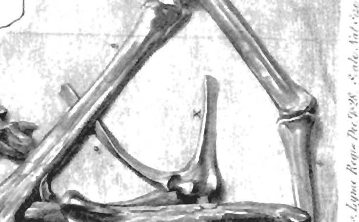
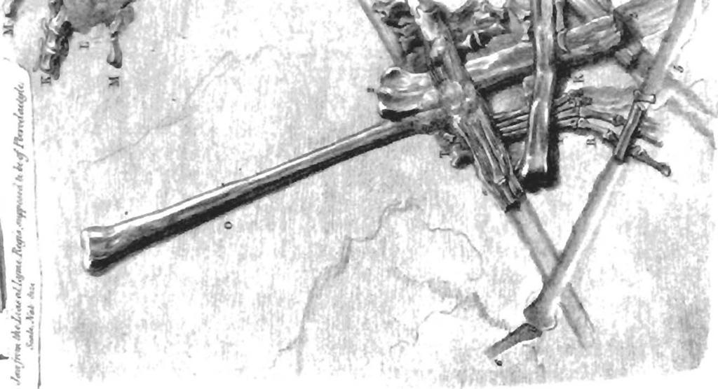
30 30 Dimorphodon macronyx (Buckland) Postilla189 the few fragments preserved in YPM The thickness of the maxilla cannot have exceeded a few millimeters, even in the larger specimens of Dimorphodon. Its structure consisted of two paper-thin laminar veneers separated by a spongiose layer of cancellous bone, the whole structure scarcely wider than the diameter of the largest teeth. When crushed, the laminar layers around the larger teeth have often been abraded away, and the alveoli destroyed, which results in separation of these teeth from the jaw. In life, however, these must have been firmly anchored, as the remaining teeth show (Fig. 4a). The construction of the pterosaur skeleton is testimony that the strength of a skeleton is not dependent solely on the thickness of bone, but rather on the interaction and arrangement of both hard and soft tissues in response to the forces of stress most frequently encountered by the animal. Pterosaurs show how far the limits of reduction of hard tissues can be taken without sacrifice of essential functions. This observation has been explored in detail with regard to the postcranial skeleton, particularly as it relates to flight (Hankin and Watson 1914; Bramwell and Whitfield 1974), but has never been considered with regard to the construction of the skull. Identification of Small Bones in the British Museum Dimorphodon Specimens The importance of the Peabody Museum specimens of Dimorphodon is that they are largely uncrushed. Some bones, like the humerus, are better preserved than in any other specimens of Dimorphodon; other bones, like the distal tarsals, can be studied for the first time. Reference to the specimens of Dimorphodon in the British Museum (Natural History) helps to identify previously unrecognized elements among the latter material, including carpal and tarsal bones. The only carpal element preserved in the Yale material of Dimorphodon is the right medial (usually called "lateral") carpal of YPM 350 H (Figs. 12a, 13a). However, it has not been generally appreciated that most of the elements of both wrists are preserved in the type specimen of Dimorphodon (BMNH R 1034). The left wrist is in place, but partly obscured by the overlying first wing-phalanx. This specimen was recently damaged when the collections were moved to the new wing of the British Museum (Natural History), and the metacarpal-phalangeal region shattered. The proximal end of the pteroid, identified by Owen (1870:44, as "styloid"), is now broken off. Of the right wrist, the proximal carpals are represented by the piece marked j in Buckland's (1835) plate (Fig. 21). This piece, along with bones marked k and /, Buckland included in the right carpus, k is the medial carpal, identical to YPM 350 H, but /is a lateral distal tarsal. A small sliver of bone in the same area of the slab, labeled by Buckland e, was identified as a rib, but it is in fact the right pteroid bone and its supporting sesamoid base. Owen did not identify this bone, but he claimed to find both pteroids in the jumble of wing bones that covers the back of the skull of BMNH R He did not, however, indicate themin his plate, and I have been unable to verify his identification. Neither pteroid is clearly visible in BMNH Wild (1978) described for the first time small sesamoid bones dorsal to the distal Fig. 21 The type specimen of Dimorphodon macronyx (BMNH R 1034), as illustrated by Buckland (1835, plate XXVII). Buckland did not realize that the features labeled 3"and /in his figure 1 belonged to the same bone (the wingmetacarpal), and therefore his reconstruction of the wing in figure 2 is missing a joint at the wrist. The jaw shown in figure 3 was not associated with other material; Buckland guessed correctly that it pertained to the pterosaur he had first described in 1829.


31 31 Dimorphodon macronyx (Buckland) Postilla189
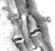
32 32 Dimorphodon macronyx (Buckland) Postilla189 ends of the penultimate phalanges of the hand of Eudimorphodon. It should be noted here, in light of this most interesting discovery, that these elements have also been preserved in Dimorphodon. In the type specimen (BMNH R 1034) there is one behind the third claw of the left hand, while in BMNH they are visible behind the claws of the first and second digits of the right hand (Fig. 22). Dr. Wild (personal communication) has since recorded antungual sesamoids in a Dorygnathus specimen in the collections of the Institut fur I Palaontologie und historische Geologie in Tubingen, a finding I can confirm from casts sent to me by Dr. Frank Westphal. I have been unable, however, to find evidence of them behind the claws of the foot in Dimorphodon (BMNH 41212) or any other pterosaur. Fig. 22 Detail of Dimorphodon macronyx, BMNH 41212, showing right manus and left pes, the latter in plantar view. Abbreviations: Idt, lateral distal tarsal; It left tibia; mc, metacarpal; mdt, medial distal tarsal; mt, metatarsal; rt, right tibia; ses, sesamoid; wph, wing-phalanx. Large Roman numberals designate phalanges, except for wing-phalanges. Scale = 1 cm.
33 33 Dimorphodon macronyx (Buckland) Postilla189 Articulations and Function of the Forelimb The fine preservation of many prominences and articular surfaces in the Yale material is crucial to the interpretation of functional morphology of the appendicular skeleton of Dimorphodon. From these remains, certain hypotheses about how pterosaurs walked and flew may be presented for the first time, subject to corroboration by comparative functional anatomy and aerodynamic requirements. YPM 350 F, the right humerus described earlier, is probably the best-preserved bone of this kind among pterosaurs. Several notable features have been clarified, especially the pronounced torsion of the shaft, the ridges showing attachments of muscles along the shaft, and the features of the distal end, particularly in palmar view. The movement of the humerus cannot be understood without reference to the pectoral girdle. Pectoral elements are absent from the Yale Dimorphodon material, and the sternum is not recorded in any specimen. The bones Buckland (1835) took for the sternal plate in BMNH R 1034 are cervical vertebrae. However, a wellpreserved platelike sternum is known in the earlier pterosaur Eudimorphodon, from the Norian of Italy (Zambelli 1973, Wild 1978), and in all later forms; so there is no reason to doubt its presence in Dimorphodon. The pectoral girdle in Dimorphodon (BMNH 41212) is of typical form and extremely birdlike (Fig. 23). The coracoid is elongated and stout, and is fused to the bladelike, attenuated scapula. It has a prominent process similar to the acrocoracoid of birds located anterior to the glenoid fossa, which is bounded anteriorly and posteriorly by raised bony knobs. These served as the site of origin for several forelimb muscles, and restricted the humerus from slipping out of its socket. The concave, saddle-shaped head of the humerus otherwise moved freely, and was capable of being retracted against the body to fold the wing as in birds. This action was further supplemented Fig. 23 Right pectoral girdles in lateral view. Above, Dimorphodon; below, Aquila chrysaetos. Abbreviations: a, acrocoracoid process; c. coracoid; furcula; s, scapula. Scale bar = 1 cm. by flexion of the elbow and metacarpalphalangeal joints. The primary action of the humerus was in the flight stroke. The mechanics of the flight stroke can be approached in three ways: (1) joint mobility and articular limitations; (2) comparative functional analysis with other flying vertebrates; and (3) aerodynamic requirements for flight, to which the flight apparatus must conform.
34 34 Dimorphodon macronyx (Buckland) Postilla189 Only the first approach will be considered here. It was stated above that the humerus could be fully retracted to close the wing against the body. It could be protracted approximately 90, or to the point where the axis of the shaft would be perpendicular to the plane of the glenoid fossa (Fig. 24). Further protraction was prevented by the bony knob anterior to the glenoid fossa. The humerus could also have been raised and lowered through an arc of approximately 90. Because radius and ulna are not suitably preserved in any rhamphorhynchoid pterosaur described thus far, the limitations of movement at the elbow can only be estimated. The elbow is a hinge joint that corresponds in mechanical detail quite closely to the elbow of birds. The similarities of the processes and areas of muscular attachment at the distal end of the humerus have already been noted (Fig. 10c). A mobility approximately equivalent to that of birds, i.e., somewhat less than 180, can be fairly assumed. The joint separating the fourth metacarpal and wing-finger is a hinge joint of great mobility, very similar to the outer joint of the bird wing (Bramwell and Whitfield 1974). The principal structural difference, of course, is that this joint is the carpometacarpal in birds, whereas in pterosaurs it is the metacarpophalangeal joint of the fourth (wing) finger (Fig. 25). This articulation is well preserved in several BMNH specimens, though not in the Yale material. In Dimorphodon there is some slight movement possible between the zeugopodials and the proximal carpal, and between the distal carpal and the metacarpals, but the proximal and distal carpals interlock snugly. It is difficult to assess the amount of movement between the phalanges of the wing-finger. These joints are flattened balland-cup articulations, never anchylosed but also without clearly developed collateral ligament fossae or any other evidence of restricted motion. The only indication that movement was restricted between these phalanges comes from relatively undis- Fig. 24 Right pectoral girdle and articulated humerus of Dimorphodon. Above, in retracted position; below, protracted as in flight. Note downand-forward rotation of humerus during flight stroke. Scale = 1 cm. turbed articulated specimens, in which the wing-finger always forms a taut, bowlike structure. Hence, the majority of movement in the wing occurred at three joints: the shoulder, the elbow, and the base of the fourth finger. The first had a wide range of movement in several planes, while the second and third were simple hinges with extensive mobility in only one plane. It is evident that the wings of birds and pterosaurs are divided into equivalent functional units, analogous in a mechanical sense but not homologous in structure. It is curious that in rhamphorhynchoid pterosaurs, as in theropods, the phalanges of the manus are generally more robust than those of the pes, and the claws are larger and more trenchant (Fig. 22). Larger theropods often tended to reduce the forelimbs, while pterosaurs enlarged them. It is possible that the first three digits of the
 and a bird (below); only the proximal portion of the pterosaur's first wing-phalanx is shown.")
35 35 Dimorphodon macronyx (Buckland) Postilla189 Fig. 25 Comparison of the right forelimb skeletons of a generalized rhamphorhynchoid pterosaur (above) and a bird (below); only the proximal portion of the pterosaur's first wing-phalanx is shown. Abbreviations: c, carpus; h, humerus; mc, metacarpus; pi pteroid; r, radius; u, ulna. I IV, digits. pterosaur hand were enlarged mainly as a developmental consequence of the hypertrophy of the fourth finger. Only the phalanges of the first three digits were movable, because the first three metacarpals were appressed and bound, probably ligamentously, to the fourth. But the welldeveloped flexor tubercles of the phalanges, especially the claws, seem to indicate considerable movement was possible. Ginglymal grooves are deep and allow a wide range of flexion and extension; one is reminded on examination of the unguals that Buckland did not idly name this species macronyx. The enormous flexor tubercles of the claws suggest strong powers of grasping, perhaps in climbing, but equally possible for manipulation or gouging, perhaps in predation. A common function of sesamoid bones in such situations is to sustain tensile forces around the extensor site of a joint where much flexion occurs (Hildebrand 1974), and this would be expected in cases where flexion is indicated by such well-developed ungual tubercles. If so, it can be suggested that some type of grasping function was highly probable. In the past, suggested uses of these digits have included grooming, feeding, hanging from cliffs, and moving between the branches of trees. Of these, none can be logically excluded, but there is no direct evidence for any. Pterosaurs were not necessarily arboreal, but they were predators. I would suggest that a function in predation is more likely than climbing or hanging. It should be remembered, though, that flight was the primary function of the forelimbs. No movement that contradicted the requirements of joint mobility and articulation for the flight stroke would have been possible: this seems to be the only caution. Articulations and Function of the Hindiimb The hindiimb of Dimorphodon is better preserved in the Yale specimens than in any other early pterosaur, and allows considerable insight into the stance and gait of the limb as well as particulars of its kinematics. No pelvic bones are preserved, however. These are known from the BMNH specimens and are of more or less standard pterosaurian type. The ilium is low and bladelike, with rodlike processes anteriorly and posteriorly. The acetabulum is imperforate, and ilium, ischium, and pubis seem to contribute to it. The ischium and pubis are fused in a continuation of the platelike form seen in the ilium. Most of this broad expansion is generally identified as the ischium, with the pubis consisting of a vertical, stalklike element incorporated into the pelvic plate. The two separate stalks of the pubis were joined medially by a paired element regarded as the prepubis. Its form is variable in pterosaurs: it is rodlike and divided distally in Rhamphorhynchus (Wellnhofer 1975), but in earlier forms it is flatter and roughly spatulate, with a diamond-shaped median blade in place of the prepubic "prongs" seen later in Rhamphorhynchus. The edges of these blades are irregular and rugose, and suggest extensive cartilaginous attachment as in the pelvis of crocodiles. Pterosaurs lacked a mammalian diaphragm, but it is
.")
36 36 Dimorphodon macronyx (Buckland) Postilla189 possible that the prepubis in pterosaurs served as the origin of a muscle similar to the M. diaphragmaticus of the crocodile, which via its insertion on the liver acts as a piston mechanism for inspiration by pulling air into the lungs (Gans and Clark 1976). In flight, respiration may have been accomplished in part by expansion and contraction of the chest along with the flight stroke. These suggestions, however, are offered only by analogy with birds and crocodiles. The puboischiadic plates were fused to some extent along their ventral borders. This statement is contrary to the traditional view (Wellnhofer 1970,1975,1978), but there are notable examples in which the median synthesis has been preserved. These include the Carnegie Museum specimen of Campylognathoides liasicus, the type specimen of C. zittelim the Staatliches Museum fur Naturkunde Stuttgart, and a slightly distorted but otherwise fully articulated and uncrushed pelvis oipteranodon in the Peabody Museum (Eaton 1910, plates X and XI). The consequence of this arrangement is that the acetabulum is made to face downward and outward, not upward and slightly backward as in Wellnhofer's reconstructions (Padian 1980). The rest of the pterosaur hindlimb is functionally analogous to bipedal dinosaurs and birds, not to bats or other arboreal mammals as traditional reconstructions imply. It will be recalled that the head of the femur is set off to the side of the main axis of the shaft, not central to the shaft as in bats. This limits movement of the femur to protraction and retraction in a nearly parasagittal plane. The orientation of the distal articular surfaces with respect to the axis of the femoral shaft indicates that the articulation of the femur with the tibia and fibula was normally much closer to a right angle than a straight line, when viewed from the side. The knee joint was, therefore, a hinge allowing no significant rotation during normal locomotion. Schaeffer (1941) observed that a well-offset femoral head corresponds to the role of the tibia as the main bearer of weight, and went on to suggest that the movement of the pterosaur hindlimb must have been largely restricted to the parasagittal plane, as in birds. This agrees with the idea that a large medial condyle is a primary indicator of parasagittal locomotion (D. Brinkman, personal communication.) The length of the fibula is variable in proportion to the tibia in both birds and pterosaurs, and this is to be expected of an element that is so reduced in size and function. The expected position of the tibia would be more or less vertical when the animal was at rest. In motion, the tibia would have swung through a wide parasagittal arc while the femur remained in a more horizontal position. The motion of the distal end of the femur would have been more up-and-down than backand-forth, like the knees of birds but unlike the knees of humans. The tarsal region of pterosaurs, detailed here for the first time, demonstrates the movements within the ankle region. The proximal tarsal bones (astragalus and calcaneum) can only be fused to the tibiafibula, as they are here and in birds and theropods, when there is no movement between these limb elements. The formation of a highly developed double condylar joint emphasizes the restriction of motion to a fore-and-aft plane, and the distal tarsals show clearly on their proximal faces the depressions that receive the medial and lateral condyles of the tibiotarsus. The distal end of the tibiotarsus has many topographic features that correspond to insertions and grooves for tendons and ligaments of the main flexor and extensor muscles of the avian foot (see Fig. 19). The distal end is expanded anteriorly in birds, bipedal dinosaurs, and pterosaurs, not distally as in sprawling forms. This shows that the axes of the tibia and the elongated metatarsals, as viewed from the lateral side, did not form a stright line but a sharp angle. Most flexion and extension of the distal tarsals and metatarsals against the tibiotarsus occurred within a range of about 90 to 150, approximating the range in birds.
.")
37 37 Dimorphodon macronyx (Buckland) Postilla189 The distal tarsals preserved in the Yale Dimorphodon material also provide greater insight than before possible into the articulations of the distal tarsal elements with the metatarsals (Fig. 26). On their distal faces a series of grooves mark where the distal tarsals receive metatarsals ll-iv, the fifth offset sharply in a distinct, diagonally placed channel of its own. As with the knee and tibiotarsal joints, there was little possibility of rotation here. The entire ankle assembly is less rigidly constructed than those of birds in the sense that the latter group has more fusion and definition of bony elements that form the articulations of the ankle. However, soft tissues such as cartilage, tendons, and ligaments still play an extensive part in the function of the avian foot, as Cracraft (1971) has shown. This situation in pterosaurs compares favorably with that of theropod dinosaurs, where, although some fusion of the metatarsals may occur, the distal tarsals are poorly defined when compared to the pterosaur's (see Ostrom 1969 for comparison with Deinonychus). Therefore, because rotation at the ankle is not assumed to have been a significant component of gait in theropods, it is unlikely to have been important in pterosaurs. The pterosaurian tarsal bones do not bind the joint rigidly, but suggest strongly from their features that the normal range and extent of motion was primarily parasagittal, like the movement of the knee. The pterosaurian ankle is properly regarded as mesotarsal, as in birds and dinosaurs, because the primary flexion is between proximal and distal tarsals and there is no movement between the proximal tarsals. The function of the fifth metatarsal and its long, aberrant toe remains a mystery, although it seems clear that digit V did not operate like the other digits. Wild (1978) has suggested the idea of stretching a web of skin between digits IV and V for paddling through the water, by analogy with a possible function in the foot of the Triassic marine reptile Tanystropheus (Wild 1973). This is certainly a possibility, although the variable form of this digit in rhamphorhynchoids and its eventual loss in pterodactyloids (Wellnhofer 1978, Abb. 17) should also be considered. The fifth metatarsal, which is not preserved in the Yale specimens but which is well shown in BMNH 41212, has a variety of prominences and tuberosities indicative of complex motion. An especially well-marked groove, for instance, is located on the plantar surface between two pronounced tuberosities. Tendons running along this groove evidently inserted on a rugose prominence at the distal end, and would have produced strong flexion of this digit (Fig. 26). The function of digit V in posture and locomotion, whether terrestrial or aquatic, is still not established, but the fifth metatarsal of Dimorphodon is the largest known among pterosaurs (in Campylognathoides it is also quite large; in Eudimorphodon it is unknown), and its robustness in these early forms may indicate a primitive function that was reduced or lost in later rhamphorhynchoids. The ginglymal grooves of the distal ends of metatarsals ll-iv describe an arc that begins approximately in line with the axis of the metatarsal shafts and continues between the ginglymal condyles to the shaft axis (Fig. 27). This is comparable to the condition in birds and dinosaurs and unlike the condition in crocodiles, lizards, and turtles. The latter reptiles are plantigrade and normally do not walk with a great deal of flexion between metatarsals and phalanges. In these groups the distal ends are normally not bicondylar and the joint facets are terminal, not subterminal. Nor in these groups do the metatarsals function as a coalesced unit, as they do in birds and dinosaurs, and pterosaurs. Instead, as Brinkman (1980) has shown for the caiman, each metatarsal lifts in sequence (I IV) and the main extension and flexion of the foot occurs at the ankle. In pterosaurs a great deal of flexion occurred at the phalangeal joints, as their ginglymal facets show, because the metatarsus was raised off the ground as it is in birds and theropods. The main axis of support of the hindlimb, then,
38 38 Dimorphodon macronyx (Buckland) Post ilia 189 Fig. 26 Right ankle assembly of Dimorphodon macronyx. The tibia and metatarsals are shown in anterior view. Spaces for the reception of metatarsals ll-iv are indicated. Scale = 1 mm.
39 39 Dimorphodon macronyx (Buckland) Postilla189 CD _^J \fj b Q3 z$ ff was through the two distal condyles of the tibiotarsus, through the two distal tarsals to the second, third, and fourth metatarsals (Fig. 28). These properties suggest quite strongly that pterosaurs were not plantigrade walkers, but digitigrade, contrary to Wellnhofer's (1978) conclusions. No features suggest a plantigrade mode of locomotion. Furthermore, all available evidence speaks for a completely upright, parasagittal stance and gait in Dimorphodon and all other pterosaurs. In all skeletal features that reflect the mechanics of limb movement, pterosaurs agree with birds and bipedal dinosaurs, not with crocodiles, thecodonts, or any sprawling reptiles. Fig. 27 A Distal ends of metatarsals in distal (left), lateral (center), and plantar (right) views, a, turtle; b, caiman; ctheropod dinosaur; d, Dimorphodon macronyx; e, tarsometatarsus of a bird. Arrows indicate the limits of the ginglymal grooves. Not drawn to scale. Fig. 28 T Right pelvis and hindlimb of Dimorphodon macronyx, restored in erect position, slightly extended, in lateral view. Scale 5 cm. Reconstruction of Dimorphodon The skeletal reconstruction of Dimorphodon given in Figure 29 reflects the conclusions of this work. When the actual articulations and possible actions of the bones comprising the appendicular skeleton are examined, the traditional picture of pterosaurs as clumsy quadrupeds does not make sense. Pterosaurs had well defined joints in the limbs, and their movements can be reconstructed with a high degree of confidence. The mechanics of these joints cannot be compared with those of crocodiles, thecodonts, or living reptiles, but in many cases are virtually indistinguishable from those of birds and bipedal dinosaurs, as the preceding analysis has shown. Some articular surfaces, like the head of the humerus, the area of the wrist and hand, and the distal tarsals, are not precisely like those of any known animals, although functionally analogous. Even so, these joints are all well defined enough to allow a confident approximation of the direction and range of their movement, and in every case are functionally analogous to the corresponding joints in birds or dinosaurs. Dimorphodon has been restored in Figure 30 as a rapidly moving terrestrial biped. The hindlimbs were apparently quite
40 40 Dimorphodon macronyx (Buckland) Postilla189 CD Q. X2 C * CM 2s +-> c: 13 o C) rphodon macr ssio kullab abo 20 cm o.fc= ^ E o o GO r CD CO O (> o o o 2 N O CD ri> co DC CD CO ~0 rial win rrest O II CD CD O in T -J c CD Q. CO O) j C!) r CD c


41 Fig. 30 Life-restoration of Dimorphodon macronyx, by J. Kevin Ramos. Wing configuration inferred from Rhamphorhynchus; "furry" covering from Sordes pilosus. The horny covering shown on the rostral area is based on an inference by Wellnhofer (1975) regarding Rhamphorh ynchus.
New Carnivorous Dinosaurs from the Upper Cretaceous of Mongolia
 1955 Doklady, Academy of Sciences USSR 104 (5):779-783 New Carnivorous Dinosaurs from the Upper Cretaceous of Mongolia E. A. Maleev (translated by F. J. Alcock) The present article is a summary containing
.56 m. (22 in.). COMPSOGNATHOID DINOSAUR FROM THE. Medicine Bow, Wyoming, by the American Museum Expedition
. COMPSOGNATHOID DINOSAUR FROM THE. Medicine Bow, Wyoming, by the American Museum Expedition") Article XII.-ORNITHOLESTES HERMANNI, A NEW COMPSOGNATHOID DINOSAUR FROM THE UPPER JURASSIC. By HENRY FAIRFIELD OSBORN. The type skeleton (Amer. Mus. Coll. No. 6I9) of this remarkable animal was discovered
Article XII.-ORNITHOLESTES HERMANNI, A NEW COMPSOGNATHOID DINOSAUR FROM THE UPPER JURASSIC. By HENRY FAIRFIELD OSBORN. The type skeleton (Amer. Mus. Coll. No. 6I9) of this remarkable animal was discovered
SOME LITTLE-KNOWN FOSSIL LIZARDS FROM THE
 PROCEEDINGS OF THE UNITED STATES NATIONAL MUSEUM issued SWsK \ {^^m ^V ^^ SMITHSONIAN INSTITUTION U. S. NATIONAL MUSEUM Vol. 91 Washington : 1941 No. 3124 SOME LITTLE-KNOWN FOSSIL LIZARDS FROM THE OLIGOCENE
Williston, and as there are many fairly good specimens in the American
 56.81.7D :14.71.5 Article VII.- SOME POINTS IN THE STRUCTURE OF THE DIADECTID SKULL. BY R. BROOM. The skull of Diadectes has been described by Cope, Case, v. Huene, and Williston, and as there are many
Mammalogy Laboratory 1 - Mammalian Anatomy
 Mammalogy Laboratory 1 - Mammalian Anatomy I. The Goal. The goal of the lab is to teach you skeletal anatomy of mammals. We will emphasize the skull because many of the taxonomically important characters
ONLINE APPENDIX 1. Morphological phylogenetic characters scored in this paper. See Poe (2004) for
 for") ONLINE APPENDIX Morphological phylogenetic characters scored in this paper. See Poe () for detailed character descriptions, citations, and justifications for states. Note that codes are changed from a
ONLINE APPENDIX Morphological phylogenetic characters scored in this paper. See Poe () for detailed character descriptions, citations, and justifications for states. Note that codes are changed from a
AMERICAN MUSEUM NOVITATES Published by
 AMERICAN MUSEUM NOVITATES Published by Number 782 THE AmzRICAN MUSEUM OF NATURAL HISTORY Feb. 20, 1935 New York City 56.81, 7 G (68) A NOTE ON THE CYNODONT, GLOCHINODONTOIDES GRACILIS HAUGHTON BY LIEUWE
Exceptional fossil preservation demonstrates a new mode of axial skeleton elongation in early ray-finned fishes
 Supplementary Information Exceptional fossil preservation demonstrates a new mode of axial skeleton elongation in early ray-finned fishes Erin E. Maxwell, Heinz Furrer, Marcelo R. Sánchez-Villagra Supplementary
Anatomy. Name Section. The Vertebrate Skeleton
 Name Section Anatomy The Vertebrate Skeleton Vertebrate paleontologists get most of their knowledge about past organisms from skeletal remains. Skeletons are useful for gleaning information about an organism
SUPPLEMENTARY ONLINE MATERIAL FOR. Nirina O. Ratsimbaholison, Ryan N. Felice, and Patrick M. O connor
 http://app.pan.pl/som/app61-ratsimbaholison_etal_som.pdf SUPPLEMENTARY ONLINE MATERIAL FOR Nirina O. Ratsimbaholison, Ryan N. Felice, and Patrick M. O connor Ontogenetic changes in the craniomandibular
Postilla PEABODY MUSEUM OF NATURAL HISTORY YALE UNIVERSITY NEW HAVEN, CONNECTICUT, U.S.A.
 Postilla PEABODY MUSEUM OF NATURAL HISTORY YALE UNIVERSITY NEW HAVEN, CONNECTICUT, U.S.A. Number 117 18 March 1968 A 7DIAPSID (REPTILIA) PARIETAL FROM THE LOWER PERMIAN OF OKLAHOMA ROBERT L. CARROLL REDPATH
AMERICAN NATURALIST. Vol. IX. -DECEMBER, No. 12. OR BIRDS WITH TEETH.1 OI)ONTORNITHES,
ONTORNITHES,") AMERICAN NATURALIST. Vol. IX. -DECEMBER, 1875.-No. 12. OI)ONTORNITHES, OR BIRDS WITH TEETH.1 BY PROFESSOR 0. C. MARSH. REMAINS of birds are amono the rarest of fossils, and few have been discovered except
AMERICAN NATURALIST. Vol. IX. -DECEMBER, 1875.-No. 12. OI)ONTORNITHES, OR BIRDS WITH TEETH.1 BY PROFESSOR 0. C. MARSH. REMAINS of birds are amono the rarest of fossils, and few have been discovered except
FURTHER STUDIES ON TWO SKELETONS OF THE BLACK RIGHT WHALE IN THE NORTH PACIFIC
 FURTHER STUDIES ON TWO SKELETONS OF THE BLACK RIGHT WHALE IN THE NORTH PACIFIC HIDEO OMURA, MASAHARU NISHIWAKI* AND TOSHIO KASUYA* ABSTRACT Two skeletons of the black right whale were studied, supplementing
First Ornithomimid (Theropoda, Ornithomimosauria) from the Upper Cretaceous Djadokhta Formation of Tögrögiin Shiree, Mongolia
 from the Upper Cretaceous Djadokhta Formation of Tögrögiin Shiree, Mongolia") First Ornithomimid (Theropoda, Ornithomimosauria) from the Upper Cretaceous Djadokhta Formation of Tögrögiin Shiree, Mongolia Tsogtbaatar Chinzorig¹, ³ *, Yoshitsugu Kobayashi², Khishigjav Tsogtbaatar³,
First Ornithomimid (Theropoda, Ornithomimosauria) from the Upper Cretaceous Djadokhta Formation of Tögrögiin Shiree, Mongolia Tsogtbaatar Chinzorig¹, ³ *, Yoshitsugu Kobayashi², Khishigjav Tsogtbaatar³,
A Fossil Snake (Elaphe vulpina) From A Pliocene Ash Bed In Nebraska
 From A Pliocene Ash Bed In Nebraska") University of Nebraska - Lincoln DigitalCommons@University of Nebraska - Lincoln Transactions of the Nebraska Academy of Sciences and Affiliated Societies Nebraska Academy of Sciences 198 A Fossil Snake
University of Nebraska - Lincoln DigitalCommons@University of Nebraska - Lincoln Transactions of the Nebraska Academy of Sciences and Affiliated Societies Nebraska Academy of Sciences 198 A Fossil Snake
2. Skull, total length versus length of the presacral vertebral column: (0); extremely elongated neck (e.g. Tanystropheus longobardicus).
; extremely elongated neck (e.g. Tanystropheus longobardicus).") Character list of the taxon-character data set 1. Skull and lower jaws, interdental plates: absent (0); present, but restricted to the anterior end of the dentary (1); present along the entire alveolar
Character list of the taxon-character data set 1. Skull and lower jaws, interdental plates: absent (0); present, but restricted to the anterior end of the dentary (1); present along the entire alveolar
A Pterodactylus with Remains of Flight Membrane. by F. Broili (with 3 plates). Read at the Conference on 7th February 1925.
. Read at the Conference on 7th February 1925.") Broili, F. (1925) Ein Pterodactylus mit Resten der Flughaut. Sitzungsberichte der Bayerischen Königlichen Akademie der Wissenschaften, Mathematischen-Physicalischen Classe, 1925, 23-32. A Pterodactylus
Broili, F. (1925) Ein Pterodactylus mit Resten der Flughaut. Sitzungsberichte der Bayerischen Königlichen Akademie der Wissenschaften, Mathematischen-Physicalischen Classe, 1925, 23-32. A Pterodactylus
A Short Report on the Occurrence of Dilophosaurus from Jinning County, Yunnan Province
 A Short Report on the Occurrence of Dilophosaurus from Jinning County, Yunnan Province by Hu Shaojin (Kunming Cultural Administrative Committee, Yunnan Province) Vertebrata PalAsiatica Vol. XXXI, No. 1
On the Discovery of the earliest fossil bird in China (Sinosauropteryx gen. nov.) and the origin of birds
 and the origin of birds") On the Discovery of the earliest fossil bird in China (Sinosauropteryx gen. nov.) and the origin of birds by Qiang Ji and Shu an Ji Chinese Geological Museum, Beijing Chinese Geology Volume 233 1996 pp.
On the Discovery of the earliest fossil bird in China (Sinosauropteryx gen. nov.) and the origin of birds by Qiang Ji and Shu an Ji Chinese Geological Museum, Beijing Chinese Geology Volume 233 1996 pp.
A NEW GENUS AND SPECIES OF AMERICAN THEROMORPHA
 A NEW GENUS AND SPECIES OF AMERICAN THEROMORPHA MYCTEROSAURUS LONGICEPS S. W. WILLISTON University of Chicago The past summer, Mr. Herman Douthitt, of the University of Chicago paleontological expedition,
complex in cusp pattern. (3) The bones of the coyote skull are thinner, crests sharper and the
 The bones of the coyote skull are thinner, crests sharper and the") DISTINCTIONS BETWEEN THE SKULLS OF S AND DOGS Grover S. Krantz Archaeological sites in the United States frequently yield the bones of coyotes and domestic dogs. These two canines are very similar both
DISTINCTIONS BETWEEN THE SKULLS OF S AND DOGS Grover S. Krantz Archaeological sites in the United States frequently yield the bones of coyotes and domestic dogs. These two canines are very similar both
A new species of sauropod, Mamenchisaurus anyuensis sp. nov.
 A new species of sauropod, Mamenchisaurus anyuensis sp. nov. by Xinlu He, Suihua Yang, Kaiji Cai, Kui Li, and Zongwen Liu Chengdu University of Technology Papers on Geosciences Contributed to the 30th
THE SKULLS OF ARAEOSCELIS AND CASEA, PERMIAN REPTILES
 THE SKULLS OF REOSCELIS ND CSE, PERMIN REPTILES University of Chicago There are few Permian reptiles of greater interest at the present time than the peculiar one I briefly described in this journal' three
SOME NEW AMERICAN PYCNODONT FISHES.
 SOME NEW AMERICAN PYCNODONT FISHES. By James Williams Gidley, Assistant Curator of Fossil Mammals, United States National Museum. In the United States National Museum are several specimens representing
A New Pterosaur from the Middle Jurassic of Dashanpu, Zigong, Sichuan
 A New Pterosaur from the Middle Jurassic of Dashanpu, Zigong, Sichuan by Xinlu He (Chengdu College of Geology) Daihuan Yang (Chungking Natural History Museum, Sichuan Province) Chunkang Su (Zigong Historical
Description of Cranial Elements and Ontogenetic Change within Tropidolaemus wagleri (Serpentes: Crotalinae).
.") East Tennessee State University Digital Commons @ East Tennessee State University Electronic Theses and Dissertations 5-2016 Description of Cranial Elements and Ontogenetic Change within Tropidolaemus
East Tennessee State University Digital Commons @ East Tennessee State University Electronic Theses and Dissertations 5-2016 Description of Cranial Elements and Ontogenetic Change within Tropidolaemus
The Origin of Birds. Technical name for birds is Aves, and avian means of or concerning birds.
 The Origin of Birds Technical name for birds is Aves, and avian means of or concerning birds. Birds have many unusual synapomorphies among modern animals: [ Synapomorphies (shared derived characters),
THE GORGONOPSIAN GENUS, HIPPOSAURUS, AND THE FAMILY ICTIDORHINIDAE * Dr. L.D. Boonstra. Paleontologist, South African Museum, Cape Town
 THE GORGONOPSIAN GENUS, HIPPOSAURUS, AND THE FAMILY ICTIDORHINIDAE * by Dr. L.D. Boonstra Paleontologist, South African Museum, Cape Town In 1928 I dug up the complete skeleton of a smallish gorgonopsian
Bulletin of Big Bend Paleo-Geo An Open Access Publication from Mosasaur Ranch Museum, Terlingua and Lajitas, Texas All rights reserved
 Bulletin of Big Bend Paleo-Geo An Open Access Publication from Mosasaur Ranch Museum, Terlingua and Lajitas, Texas All rights reserved This was a private report in 2003 on my thoughts on Platecarpus planifrons.
Origin and Evolution of Birds. Read: Chapters 1-3 in Gill but limited review of systematics
 Origin and Evolution of Birds Read: Chapters 1-3 in Gill but limited review of systematics Review of Taxonomy Kingdom: Animalia Phylum: Chordata Subphylum: Vertebrata Class: Aves Characteristics: wings,
By HENRY FAIRFIELD OSBORN.
 Article XI.-FORE AND HINI) LIMBS OF CARNIVOR- OUS AND HERBIVOROUS DINOSAURS FROM THE JURASSIC OF WYOMING. DINOSAUR CONTRIBU- TIONS, NO. 3. By HENRY FAIRFIELD OSBORN. In the Bone Cabin Quarry, opened by
v:ii-ixi, 'i':;iisimvi'\>!i-:: "^ A%'''''-'^-''S.''v.--..V^'E^'-'-^"-t''gi L I E) R.ARY OF THE VERSITY U N I or ILLINOIS REMO
 R.ARY OF THE VERSITY U N I or ILLINOIS REMO") "^ A%'''''-'^-''S.''v.--..V^'E^'-'-^"-t''gi v:ii-ixi, 'i':;iisimvi'\>!i-:: L I E) R.ARY OF THE U N I VERSITY or ILLINOIS REMO Natural History Survey Librarv GEOLOGICAL SERIES OF FIELD MUSEUM OF NATURAL
"^ A%'''''-'^-''S.''v.--..V^'E^'-'-^"-t''gi v:ii-ixi, 'i':;iisimvi'\>!i-:: L I E) R.ARY OF THE U N I VERSITY or ILLINOIS REMO Natural History Survey Librarv GEOLOGICAL SERIES OF FIELD MUSEUM OF NATURAL
1/9/2013. Divisions of the Skeleton: Topic 8: Appendicular Skeleton. Appendicular Components. Appendicular Components
 /9/203 Topic 8: Appendicular Skeleton Divisions of the Skeleton: Cranial Postcranial What makes up the appendicular skeleton? What is the pattern of serial homology of the limbs? Tetrapod front limb morphology
Fig. 5. (A) Scaling of brain vault size (width measured at the level of anterior squamosal/parietal suture) relative to skull size (measured at the
 Scaling of brain vault size (width measured at the level of anterior squamosal/parietal suture) relative to skull size (measured at the") Fig. 5. (A) Scaling of brain vault size (width measured at the level of anterior squamosal/parietal suture) relative to skull size (measured at the distance between the left versus right temporomandibular
Fig. 5. (A) Scaling of brain vault size (width measured at the level of anterior squamosal/parietal suture) relative to skull size (measured at the distance between the left versus right temporomandibular
First Flightless Pterosaur
 First Flightless Pterosaur David Peters no affiliation 9 Greenfield Court, Saint Charles, MO 63303 USA Pterosaur fossils have been discovered all over the world [1], but so far no flightless pterosaurs
A new basal sauropodiform dinosaur from the Lower Jurassic of Yunnan Province, China
 SUPPLEMENTARY INFORMATION A new basal sauropodiform dinosaur from the Lower Jurassic of Yunnan Province, China Ya-Ming Wang 1, Hai-Lu You 2,3 *, Tao Wang 4 1 School of Earth Sciences and Resources, China
290 SHUFELDT, Remains of Hesperornis.
 290 SHUFELDT, Remains of Hesperornis. [ Auk [July THE FOSSIL REMAINS OF A SPECIES OF HESPERORNIS FOUND IN MONTANA. BY R. W. SHUFELD% M.D. Plate XI7III. ExR,¾ in November, 1914, Mr. Charles W. Gihnore,
SUPPLEMENTARY INFORMATION
 Character 155, interdental ridges. Absence of interdental ridge (0) shown in Parasaniwa wyomingensis (Platynota). Interdental ridges (1) shown in Coniophis precedens. WWW.NATURE.COM/NATURE 1 Character
A New Ceratopsian Dinosaur from the Upper
 SMITHSONIAN MISCELLANEOUS COLLECTIONS VOLUME 63. NUMBER 3 A New Ceratopsian Dinosaur from the Upper Cretaceous of Montana, with Note on Hypacrosaurus (With Two Plates) CHARLES W. GILMORE Assistant Curator
Barney to Big Bird: The Origin of Birds. Caudipteryx. The fuzzy raptor. Solnhofen Limestone, cont d
 Barney to Big Bird: The Origin of Birds Caudipteryx The fuzzy raptor The discovery of feathered dinosaurs in Liaoning, China, has excited the many paleontologists who suspected a direct link between dinosaurs
A M E G H I N I A N A. Revista de la Asociación Paleontológia Argentina. Volume XV September-December 1978 Nos. 3-4
 A M E G H I N I A N A Revista de la Asociación Paleontológia Argentina Volume XV September-December 1978 Nos. 3-4 COLORADIA BREVIS N. G. ET N. SP. (SAURISCHIA, PROSAUROPODA), A PLATEOSAURID DINOSAUR FROM
Biology 340 Comparative Embryology Lecture 12 Dr. Stuart Sumida. Evo-Devo Revisited. Development of the Tetrapod Limb
 Biology 340 Comparative Embryology Lecture 12 Dr. Stuart Sumida Evo-Devo Revisited Development of the Tetrapod Limb Limbs whether fins or arms/legs for only in particular regions or LIMB FIELDS. Primitively
Cretaceous, toothed pterosaurs from Brazil. A reappraisal
 5. Preliminary description of a skull and wing of a Brazilian Cretaceous (Santana Formation; Aptian Albian) pterosaur (Pterodactyloidea) in the collection of the AMNH 34 5.1. Introduction The collection
A new carnosaur from Yongchuan County, Sichuan Province
 A new carnosaur from Yongchuan County, Sichuan Province by Dong Zhiming Institute of Vertebrate Palaeontology and Palaeoanthropology, Academia Sinica Zhang Yihong, Li Xuanmin, and Zhou Shiwu Chongqing
A NEW ANKYLOSAUR FROM THE UPPER CRETACEOUS OF MONGOLIA E.A. Maleev Doklady Akademii Nauk, SSSR 87:
 translated by Dr. Tamara and F. Jeletzky, 1956 A NEW ANKYLOSAUR FROM THE UPPER CRETACEOUS OF MONGOLIA E.A. Maleev 1952. Doklady Akademii Nauk, SSSR 87:273-276 Armored dinosaurs make a considerable part
 The following text is generated from uncorrected OCR. [Begin Page: Page 1] A NEW CERATOPSIAN DINOSAUR FROM THE UPPER CRETACEOUS OF MONTANA, WITH NOTE ON HYPACROSAURUS ' By CHARLES W. GILMORE assistant
LEIDY, SHOWING THE BONES OF THE FEET 'AND LIMBS
 CQNTEUBUTIONS FBOM THE MUSEUM OF PALEONTOLOGY (Confindion of Con&&&m froin UB Muaercm of Gcologg) UNIVERSITY OF ' MICHIGAN VOL V, No. 6, pp. 6W3 (e ph.) DEAXMBER 31,1036 A SPECIMEN OF STYLEMYS NEBRASCENSIS
Recently Mr. Lawrence M. Lambe has described and figured in the
 56.81,9C(117:71.2) Article XXXV.-CORYTHOSAURUS CASUARIUS, A NEW CRESTED DINOSAUR FROM THE BELLY RIVER CRETA- CEOUS, WITH PROVISIONAL CLASSIFICATION OF THE FAMILY TRACHODONTIDA1X BY BARNUM BROWN. PLATE
Jurassic Ornithopod Agilisaurus louderbacki (Ornithopoda: Fabrosauridae) from Zigong, Sichuan, China
 from Zigong, Sichuan, China") Jurassic Ornithopod Agilisaurus louderbacki (Ornithopoda: Fabrosauridae) from Zigong, Sichuan, China Guangzhao Peng (Zigong Dinosaur Museum) Vertebrata PalAsiatica Volume 30, No. 1 January, 1992 pp. 39-51
Jurassic Ornithopod Agilisaurus louderbacki (Ornithopoda: Fabrosauridae) from Zigong, Sichuan, China Guangzhao Peng (Zigong Dinosaur Museum) Vertebrata PalAsiatica Volume 30, No. 1 January, 1992 pp. 39-51
A skull without mandihle, from the Hunterian Collection (no.
 4 MR. G. A. BOULENGER ON CHELONIAN REMAINS. [Jan. 6, 2. On some Chelonian Remains preserved in the Museum of the Eojal College of Surgeons. By G. A. Boulenger. [Eeceived December 8, 1890.] In the course
Origin and Evolution of Birds. Read: Chapters 1-3 in Gill but limited review of systematics
 Origin and Evolution of Birds Read: Chapters 1-3 in Gill but limited review of systematics Review of Taxonomy Kingdom: Animalia Phylum: Chordata Subphylum: Vertebrata Class: Aves Characteristics: wings,
TRACHEMYS SCULPTA. A nearly complete articulated carapace and plastron of an Emjdd A NEAKLY COMPLETE SHELL OF THE EXTINCT TURTLE,
 A NEAKLY COMPLETE SHELL OF THE EXTINCT TURTLE, TRACHEMYS SCULPTA By Charles W. Gilmore Curator of Vertebrate Paleontology, United States National Museum INTRODUCTION A nearly complete articulated carapace
HONR219D Due 3/29/16 Homework VI
 Part 1: Yet More Vertebrate Anatomy!!! HONR219D Due 3/29/16 Homework VI Part 1 builds on homework V by examining the skull in even greater detail. We start with the some of the important bones (thankfully
What is evolution? Transitional fossils: evidence for evolution. In its broadest sense, evolution is simply the change in life through time.
 Transitional fossils: evidence for evolution http://domain- of- darwin.deviantart.com/art/no- Transitional- Fossils- 52231284 Western MA Atheists and Secular Humanists 28 May 2016 What is evolution? In
Reprinted from: CRUSTACEANA, Vol. 32, Part 2, 1977 LEIDEN E. J. BRILL
 Reprinted from: CRUSTACEANA, Vol. 32, Part 2, 1977 LEIDEN E. J. BRILL NOTES AND NEWS 207 ALPHE0PS1S SHEARMII (ALCOCK & ANDERSON): A NEW COMBINATION WITH A REDESCRIPTION OF THE HOLOTYPE (DECAPODA, ALPHEIDAE)
A NEW SPECIES OF EXTINCT TURTLE FROM THE UPPER PLIOCENE OF IDAHO
 A NEW SPECIES OF EXTINCT TURTLE FROM THE UPPER PLIOCENE OF IDAHO By Charles W. Gilmore Curator, Division of Vertebrate Paleontology United States National Museum Among the fossils obtained bj^ the Smithsonian
Vol. XIV, No. 1, March, The Larva and Pupa of Brontispa namorikia Maulik (Coleoptera: Chrysomelidae: Hispinae) By S.
 By S.") Vol. XIV, No. 1, March, 1950 167 The Larva and Pupa of Brontispa namorikia Maulik (Coleoptera: Chrysomelidae: Hispinae) By S. MAULIK BRITISH MUSEUM (NATURAL HISTORY) (Presented by Mr. Van Zwaluwenburg
Vol. XIV, No. 1, March, 1950 167 The Larva and Pupa of Brontispa namorikia Maulik (Coleoptera: Chrysomelidae: Hispinae) By S. MAULIK BRITISH MUSEUM (NATURAL HISTORY) (Presented by Mr. Van Zwaluwenburg
'Rain' of dead birds on central NJ lawns explained; Federal culling program killed up to 5,000 Associated Press, January 27, 2009
 'Rain' of dead birds on central NJ lawns explained; Federal culling program killed up to 5,000 Associated Press, January 27, 2009 Study May Give Hope That Ivory-billed Woodpeckers Still Around Science
AN ANNOTATED AND ILLUSTRATED CATALOGUE OF SOLNHOFEN (UPPER JURASSIC, GERMANY) PTEROSAUR SPECIMENS AT CARNEGIE MUSEUM OF NATURAL HISTORY
 PTEROSAUR SPECIMENS AT CARNEGIE MUSEUM OF NATURAL HISTORY") ANNALS OF CARNEGIE MUSEUM vol. 82, number 2, PP. 165 191 31 DEcEMBEr 2013 AN ANNOTATED AND ILLUSTRATED CATALOGUE OF SOLNHOFEN (UPPER JURASSIC, GERMANY) PTEROSAUR SPECIMENS AT CARNEGIE MUSEUM OF NATURAL
ANNALS OF CARNEGIE MUSEUM vol. 82, number 2, PP. 165 191 31 DEcEMBEr 2013 AN ANNOTATED AND ILLUSTRATED CATALOGUE OF SOLNHOFEN (UPPER JURASSIC, GERMANY) PTEROSAUR SPECIMENS AT CARNEGIE MUSEUM OF NATURAL
A NEW SPECIES OF TROODONT DINOSAUR FROM THE
 A NEW SPECIES OF TROODONT DINOSAUR FROM THE LANCE FORMATION OF WYOMING By Charles W. Gilmore Curator of Vertebrate Paleontology, United States National Museum INTRODUCTION The intensive search to which
Major cranial changes during Triceratops ontogeny John R. Horner 1, * and Mark B. Goodwin 2
 273, 2757 2761 doi:10.1098/rspb.2006.3643 Published online 1 August 2006 Major cranial changes during Triceratops ontogeny John R. Horner 1, * and Mark B. Goodwin 2 1 Museum of the Rockies, Montana State
A NEW PLIENSBACHIAN ICHTHYOSAUR FROM DORSET, ENGLAND
 A NEW PLIENSBACHIAN ICHTHYOSAUR FROM DORSET, ENGLAND by CHRISTOPHER MC GOWAN and ANGELA C. MILNER ABSTRACT. The first ichthyosaur to be recorded from the Pliensbachian Stage of the English Lower Liassic
Fossilized remains of cat-sized flying reptile found in British Columbia
 Fossilized remains of cat-sized flying reptile found in British Columbia By Washington Post, adapted by Newsela staff on 09.06.16 Word Count 768 An artist's impression of the small-bodied, Late Cretaceous
PEABODY MUSEUM OF NATURAL HISTORY, YALE UNIVERSITY NEW HAVEN, CONNECTICUT, U.S.A. A NEW OREODONT FROM THE CABBAGE PATCH LOCAL FAUNA, WESTERN MONTANA
 Postilla PEABODY MUSEUM OF NATURAL HISTORY YALE UNIVERSITY NEW HAVEN, CONNECTICUT, U.S.A. Number 85 September 21, 1964 A NEW OREODONT FROM THE CABBAGE PATCH LOCAL FAUNA, WESTERN MONTANA STANLEY J. RIEL
New Specimens of Microraptor zhaoianus (Theropoda: Dromaeosauridae) from Northeastern China
 from Northeastern China") PUBLISHED BY THE AMERICAN MUSEUM OF NATURAL HISTORY CENTRAL PARK WEST AT 79TH STREET, NEW YORK, NY 10024 Number 3381, 44 pp., 31 figures, 2 tables August 16, 2002 New Specimens of Microraptor zhaoianus
PUBLISHED BY THE AMERICAN MUSEUM OF NATURAL HISTORY CENTRAL PARK WEST AT 79TH STREET, NEW YORK, NY 10024 Number 3381, 44 pp., 31 figures, 2 tables August 16, 2002 New Specimens of Microraptor zhaoianus
Frog Dissection Information Manuel
 Frog Dissection Information Manuel Anatomical Terms: Used to explain directions and orientation of a organism Directions or Positions: Anterior (cranial)- toward the head Posterior (caudal)- towards the
( M amenchisaurus youngi Pi, Ouyang et Ye, 1996)
") 39 4 2001 10 V ERTEBRATA PALASIATICA pp. 266 271 fig. 1,pl. I ( 643013), ( M amenchisaurus hochuanensis),,, Q915. 864 1995 12 31 (ZDM0126) ( M amenchisau rus hochuanensis Young et Chao, 1972),,, ZDM0126
39 4 2001 10 V ERTEBRATA PALASIATICA pp. 266 271 fig. 1,pl. I ( 643013), ( M amenchisaurus hochuanensis),,, Q915. 864 1995 12 31 (ZDM0126) ( M amenchisau rus hochuanensis Young et Chao, 1972),,, ZDM0126
Title: Phylogenetic Methods and Vertebrate Phylogeny
 Title: Phylogenetic Methods and Vertebrate Phylogeny Central Question: How can evolutionary relationships be determined objectively? Sub-questions: 1. What affect does the selection of the outgroup have
NOTES ON THE FIRST SKULL AND JAWS OF RIOJASAURUS INCERTUS (DINOSAURIA, PROSAUROPODA, MELANOROSAURIDAE) OF THE LATE TRIASSIC OF LA RIOJA, ARGENTINA
 OF THE LATE TRIASSIC OF LA RIOJA, ARGENTINA") NOTES ON THE FIRST SKULL AND JAWS OF RIOJASAURUS INCERTUS (DINOSAURIA, PROSAUROPODA, MELANOROSAURIDAE) OF THE LATE TRIASSIC OF LA RIOJA, ARGENTINA José F. Bonaparte and José A. Pumares translated by Jeffrey
NOTES ON THE FIRST SKULL AND JAWS OF RIOJASAURUS INCERTUS (DINOSAURIA, PROSAUROPODA, MELANOROSAURIDAE) OF THE LATE TRIASSIC OF LA RIOJA, ARGENTINA José F. Bonaparte and José A. Pumares translated by Jeffrey
Supplementary Figure 1 Cartilaginous stages in non-avian amniotes. (a) Drawing of early ankle development of Alligator mississippiensis, as reported
 Drawing of early ankle development of Alligator mississippiensis, as reported") Supplementary Figure 1 Cartilaginous stages in non-avian amniotes. (a) Drawing of early ankle development of Alligator mississippiensis, as reported by a previous study 1. The intermedium is formed at
Supplementary Figure 1 Cartilaginous stages in non-avian amniotes. (a) Drawing of early ankle development of Alligator mississippiensis, as reported by a previous study 1. The intermedium is formed at
ZOOLOGISCHE MEDEDELINGEN
 MINISTERIE VAN ONDERWIJS, KUNSTEN EN WETENSCHAPPEN ZOOLOGISCHE MEDEDELINGEN UITGEGEVEN DOOR HET RIJKSMUSEUM VAN NATUURLIJKE HISTORIE TE LEIDEN DEEL XXXVII, No. 10 10 juli 1961 THE FOSSIL HIPPOPOTAMUS FROM
YANGCHUANOSAURUS HEPINGENSIS - A NEW SPECIES OF CARNOSAUR FROM ZIGONG, SICHUAN
 Vol. 30, No. 4 VERTEBRATA PALASIATICA pp. 313-324 October 1992 [SICHUAN ZIGONG ROUSHILONG YI XIN ZHONG] figs. 1-5, pl. I-III YANGCHUANOSAURUS HEPINGENSIS - A NEW SPECIES OF CARNOSAUR FROM ZIGONG, SICHUAN
A new Middle Jurassic sauropod subfamily (Klamelisaurinae subfam. nov.) from Xinjiang Autonomous Region, China
 from Xinjiang Autonomous Region, China") A new Middle Jurassic sauropod subfamily (Klamelisaurinae subfam. nov.) from Xinjiang Autonomous Region, China by Xijing Zhao Institute of Vertebrate Paleontology and Paleoanthropology, Academia Sinica
A new Middle Jurassic sauropod subfamily (Klamelisaurinae subfam. nov.) from Xinjiang Autonomous Region, China by Xijing Zhao Institute of Vertebrate Paleontology and Paleoanthropology, Academia Sinica
List of characters used in the phylogenetic analysis. Capital letters T, R, and L, refer to
 1 Supplementary data CHARACTER LIST List of characters used in the phylogenetic analysis. Capital letters T, R, and L, refer to characters used by Tchernov et al. (2000), Rieppel, et al. (2002), and Lee
OSTEOLOGICAL NOTE OF AN ANTARCTIC SEI WHALE
 OSTEOLOGICAL NOTE OF AN ANTARCTIC SEI WHALE MASAHARU NISHIWAKI* AND TOSHIO KASUYA* ABSTRACT This is a report of measurements on the skeleton of a male se1 whale caught in the Antarctic. The skeleton of
On the cervical vertebrae of the
 ~oological Journal of the Linnean Society (1986) 88: 307-328. With 12 figures On the cervical vertebrae of the Pterodactyloidea (Reptilia: Archosauria) S. C. B. HOWSE Department of Biology, Birkbeck College,
AMERICAN MUSEUM NOVITATES
 AMERICAN MUSEUM NOVITATES Published by Number 89 THE AmERcAN Mueum OF NATuRAL HIsTORY October 11, 1923 New York City 56.81,9. PRELIMINARY NOTICES OF SKELETONS AND SKULLS OF DEINODONTIDE FROM THE CRETACEOUS
LOWER CRETACEOUS OF SOUTH DAKOTA.
 A NEW DINOSAUR, STP^GOSAURUS MARSHl, FROM THE LOWER CRETACEOUS OF SOUTH DAKOTA. By Frederic A. Lucas, Curator, Divisioii of Coiiipnrative Anatomy, in charge, of Section of Vertebrate Fossils. The name
A new sauropod from Dashanpu, Zigong Co. Sichuan Province (Abrosaurus dongpoensis gen. et sp. nov.)
") A new sauropod from Dashanpu, Zigong Co. Sichuan Province (Abrosaurus dongpoensis gen. et sp. nov.) by Ouyang Hui Zigong Dinosaur Museum Newsletter Number 2 1989 pp. 10-14 Translated By Will Downs Bilby
A new sauropod from Dashanpu, Zigong Co. Sichuan Province (Abrosaurus dongpoensis gen. et sp. nov.) by Ouyang Hui Zigong Dinosaur Museum Newsletter Number 2 1989 pp. 10-14 Translated By Will Downs Bilby
Giant croc with T. rex teeth roamed Madagascar
 Giant croc with T. rex teeth roamed Madagascar www.scimex.org/newsfeed/giant-croc-with-t.-rex-teeth-used-to-roam-in-madagascar Embargoed until: Publicly released: PeerJ A fossil of the largest and oldest
UN? RSITYOF. ILLIiwiS LIBRARY AT URBANA-CHAMPAIGN NATURAL HIST. SURVEY
 UN? RSITYOF ILLIiwiS LIBRARY AT URBANA-CHAMPAIGN NATURAL HIST. SURVEY FIELDIANA GEOLOGY Published by CHICAGO NATURAL HISTORY MUSEUM Volume 10 July 29, 1954 No. 17 FAUNA OF THE VALE AND CHOZA: 7 PELYCOSAURIA:
A new species of Hsisosuchus (Mesoeucrocodylia) from Dashanpu, Zigong Municipality, Sichuan Province
 from Dashanpu, Zigong Municipality, Sichuan Province") A new species of Hsisosuchus (Mesoeucrocodylia) from Dashanpu, Zigong Municipality, Sichuan Province Yuhui Gao (Zigong Dinosaur Museum) Vertebrata PalAsiatica Volume 39, No. 3 July, 2001 pp. 177-184 Translated
A new species of Hsisosuchus (Mesoeucrocodylia) from Dashanpu, Zigong Municipality, Sichuan Province Yuhui Gao (Zigong Dinosaur Museum) Vertebrata PalAsiatica Volume 39, No. 3 July, 2001 pp. 177-184 Translated
POSTILLA PEABODY MUSEUM YALE UNIVERSITY NUMBER JUNE 1976 PROSAUROPOD DINOSAURS (REPTILIA: SAURISCHIA) OF NORTH AMERICA PETER MALCOLM GALTON
 OF NORTH AMERICA PETER MALCOLM GALTON") POSTILLA PEABODY MUSEUM YALE UNIVERSITY NUMBER 169 25 JUNE 1976 PROSAUROPOD DINOSAURS (REPTILIA: SAURISCHIA) OF NORTH AMERICA PETER MALCOLM GALTON CONTENTS Abstract 2 1. Introduction 3 2. Historical Survey
POSTILLA PEABODY MUSEUM YALE UNIVERSITY NUMBER 169 25 JUNE 1976 PROSAUROPOD DINOSAURS (REPTILIA: SAURISCHIA) OF NORTH AMERICA PETER MALCOLM GALTON CONTENTS Abstract 2 1. Introduction 3 2. Historical Survey
FIELDIANA GEOLOGY NEW SALAMANDERS OF THE FAMILY SIRENIDAE FROM THE CRETACEOUS OF NORTH AMERICA
 FIELDIANA GEOLOGY Published by CHICAGO NATURAL HISTORY MUSEUM Volume 10 Sbftember 22, 1968 No. 88 NEW SALAMANDERS OF THE FAMILY SIRENIDAE FROM THE CRETACEOUS OF NORTH AMERICA Coleman J. Coin AND Walter
APPENDIX. 344 Mni-s/i Restorations of Claosaurus and Geratosaurus.
 344 Mni-s/i Restorations of Claosaurus and Geratosaurus. Claosaurics, Marsh, 1890.* The most important feature in the restoration of Claosaurus annectens given on Plate VI is the skull, which will be fully
UPOGEBIA LINCOLNI SP. NOV. (DECAPODA, THALASSINIDEA, UPOGEBIIDAE) FROM JAVA, INDONESIA
 FROM JAVA, INDONESIA") NOTES AND NEWS UPOGEBIA LINCOLNI SP. NOV. (DECAPODA, THALASSINIDEA, UPOGEBIIDAE) FROM JAVA, INDONESIA BY NGUYEN NGOC-HO i) Faculty of Science, University of Saigon, Vietnam Among material recently collected
NOTES AND NEWS UPOGEBIA LINCOLNI SP. NOV. (DECAPODA, THALASSINIDEA, UPOGEBIIDAE) FROM JAVA, INDONESIA BY NGUYEN NGOC-HO i) Faculty of Science, University of Saigon, Vietnam Among material recently collected
Video Assignments. Microraptor PBS The Four-winged Dinosaur Mark Davis SUNY Cortland Library Online
 Video Assignments Microraptor PBS The Four-winged Dinosaur Mark Davis SUNY Cortland Library Online Radiolab Apocalyptical http://www.youtube.com/watch?v=k52vd4wbdlw&feature=youtu.be Minute 13 through minute
PARSON RUSSELL TERRIER
 17.10.2017/ EN FEDERATION CYNOLOGIQUE INTERNATIONALE (AISBL) SECRETARIAT GENERAL: 13, Place Albert 1 er B 6530 Thuin (Belgique) FCI-Standard N 339 PARSON RUSSELL TERRIER J.Campin, illustr. KC Picture Library
A NEW SALTICID SPIDER FROM VICTORIA By R. A. Dunn
 Dunn, R. A. 1947. A new salticid spider from Victoria. Memoirs of the National Museum of Victoria 15: 82 85. All text not included in the original document is highlighted in red. Mem. Nat. Mus. Vict.,
CRANIAL ANATOMY OF ENNATOSAURUS TECTON (SYNAPSIDA: CASEIDAE) FROM THE MIDDLE PERMIAN OF RUSSIA AND THE EVOLUTIONARY RELATIONSHIPS OF CASEIDAE
 FROM THE MIDDLE PERMIAN OF RUSSIA AND THE EVOLUTIONARY RELATIONSHIPS OF CASEIDAE") Journal of Vertebrate Paleontology 28(1):160 180, March 2008 2008 by the Society of Vertebrate Paleontology ARTICLE CRANIAL ANATOMY OF ENNATOSAURUS TECTON (SYNAPSIDA: CASEIDAE) FROM THE MIDDLE PERMIAN
Journal of Vertebrate Paleontology 28(1):160 180, March 2008 2008 by the Society of Vertebrate Paleontology ARTICLE CRANIAL ANATOMY OF ENNATOSAURUS TECTON (SYNAPSIDA: CASEIDAE) FROM THE MIDDLE PERMIAN
A Late Jurassic Protosuchian Sichuanosuchus huidongensis from Zigong, Sichuan Province. Guangzhao Peng. Zigong Dinosaur Museum, Zigong, Sichuan
 A Late Jurassic Protosuchian Sichuanosuchus huidongensis from Zigong, Sichuan Province Guangzhao Peng Zigong Dinosaur Museum, Zigong, Sichuan 643013 Vertebrata PalAsiatica Volume 34, Number 4 October,
Redpalh Museum, McGill University, Montreal, P.Q, Canada, HJA 2K6.
 143 Palaeont. afr., 21, 143-159 (1978) PERMO-TRIASSIC "LIZARDS" FROM THE KAROO SYSTEM PART II A GLIDING REPTILE FROM THE UPPER PERMIAN OF MADAGASCAR by Robert L. Carroll Redpalh Museum, McGill University,
OCCASIONAL PAPERS OF THE MUSEUM OF ZOOLOGY UNIVERSITY OF MICHIGAN
 OCCASIONAL PAPERS OF THE MUSEUM OF ZOOLOGY ~- UNIVERSITY OF MICHIGAN A NEW FROG FROM BRITISH GUIANA A collection received by the IIuseum of Zoology froin British Gniana some time ago includes a single
Your use of the JSTOR archive indicates your acceptance of the Terms & Conditions of Use, available at
 The Evolution of the Mammalian Jaw Author(s): A. W. Crompton Source: Evolution, Vol. 17, No. 4 (Dec., 1963), pp. 431-439 Published by: Society for the Study of Evolution Stable URL: http://www.jstor.org/stable/2407093
Geo 302D: Age of Dinosaurs. LAB 7: Dinosaur diversity- Saurischians
 Geo 302D: Age of Dinosaurs LAB 7: Dinosaur diversity- Saurischians Last lab you were presented with a review of major ornithischian clades. You also were presented with some of the kinds of plants that
BREVIORA LEUCOLEPIDOPA SUNDA GEN. NOV., SP. NOV. (DECAPODA: ALBUNEIDAE), A NEW INDO-PACIFIC SAND CRAB. Ian E. Efford 1
, A NEW INDO-PACIFIC SAND CRAB. Ian E. Efford 1") ac lc BREVIORA CAMBRIDGE, MASS. 30 APRIL, 1969 NUMBER 318 LEUCOLEPIDOPA SUNDA GEN. NOV., SP. NOV. (DECAPODA: ALBUNEIDAE), A NEW INDO-PACIFIC SAND CRAB Ian E. Efford 1 ABSTRACT. Leucolepidopa gen. nov.
ac lc BREVIORA CAMBRIDGE, MASS. 30 APRIL, 1969 NUMBER 318 LEUCOLEPIDOPA SUNDA GEN. NOV., SP. NOV. (DECAPODA: ALBUNEIDAE), A NEW INDO-PACIFIC SAND CRAB Ian E. Efford 1 ABSTRACT. Leucolepidopa gen. nov.
Breed Characteristics Overall Impression The German Long faced
 Text: N. Akkerman, The Netherlands Photos: Uwe Held, Germany In General The German Long Faced Tumbler is closely related to the English Magpie. Not because of recent crossings, no, they both spring from
Supporting Online Material for
 www.sciencemag.org/cgi/content/full/329/5998/1481/dc1 Supporting Online Material for Tyrannosaur Paleobiology: New Research on Ancient Exemplar Organisms Stephen L. Brusatte,* Mark A. Norell, Thomas D.
A new species of torrent toad (Genus Silent Valley, S. India
 Proc. Indian Acad. Sci. (Anirn. ScL), Vol. 90, Number 2, March 1981, pp. 203-208. Printed in India. A new species of torrent toad (Genus Silent Valley, S. India Allsollia) from R S PILLAI and R PATTABIRAMAN
Comments on the Beauceron Standard By M. Maurice Hermel (Translated by C. Batson)
") Comments on the Beauceron Standard By M. Maurice Hermel (Translated by C. Batson) The following are comments written by M. Hermel for the FCI Standard #44 published on 10/25/06. They were approved by the
Comments on the Beauceron Standard By M. Maurice Hermel (Translated by C. Batson) The following are comments written by M. Hermel for the FCI Standard #44 published on 10/25/06. They were approved by the