A revision of the genus Dinofelis (Mammalia, Felidae)
|
|
|
- Scott Gaines
- 5 years ago
- Views:
Transcription
 LARS WERDELIN FLS* Department of Palaeozoology, Swedish Museum of Natural History, Box 50007, 23-104 05 Stockholm, Sweden")
1 Zoological Journal of the Linnean Society (2001), 132: With 49 figures doi: /zjls.2OOO.0260, available online at http;l/ on 10 E c A revision of the genus Dinofelis (Mammalia, Felidae) LARS WERDELIN FLS* Department of Palaeozoology, Swedish Museum of Natural History, Box 50007, Stockholm, Sweden MqRGARET E. LEWIS FLS Bidogy Program, Faculty of Natural and Mathematical Sciences, The Richard Stockton College of New Jersey, PO Box 195, Pomona, NJ 08240, USA Received February 1999; revised and accepted for publication June 2000 This paper consists of a taxonomic and systematic revision of the extinct felid genus Dinofelis (Felidae, Machairodontinae) and an analysis of its ecomorphology and evolution. Dirwfelis has a broad distribution, with material from all northern continents and Africa, the latter of which was the apparent centre of evolution of the genus. We describe new material of Dinofelis from a number of sites in eastern Africa and reconsider all previously described material. We name two new species and identify several other distinct species-level taxa but refrain from naming these due to a paucity of well-preserved material. At the same time, we synonymize the two named Asian species, D. cristata and D. abeli, of which the former has priority. There are few characters useful in systematic analysis, but we can suggest at least one migration from eastern to southern Africa. Ecomorphological analysis of both craniodental and postcranial characters suggests that Dinofelis in many respects converged on modern pantherine cats in morphology and behaviour, a trend culminating in the South African D. barlowi and the Asian D. cristata, which are the most pantherine-like of all machairodont felids. This trend is reversed in the evolution of the youngest species, D. piveteaui, which is also the most machairodont in its ecomorphology. The timing of the extinction of Dinofelis is difficult to determine. Outside Africa material is scarce at all times, while in Africa the apparent extinction of Dinofelis at about 1.4 Mya coincides with the end of the good, semi-continuous fossil record present in eastern Africa from about 4Mya onwards. Dating of Kanam East (with D. piveteaui) to the Jaramillo Subchron (1.07O-O.990 Mya) suggests possible survival considerably later. Thus, the extinction datum for Dinofelis cannot at present be firmly established The Linnean Society of London ADDITIONAL KEYWORDS: Machairodontinae - pantherine - taxonomy - evolution - Pliocene - Pleistocene. INTRODUCTION The genus Dinofelis consists of a small number of species of large-bodied felids with somewhat compressed though not elongated upper canines lacking serrations but retaining anterior and posterior crests. The genus has been variously referred to the Machairodontinae (saber-toothed cats) (e.g. Beaumont, 1964, 1978) or the Felinae (conical-toothed cats) (e.g. Kretzoi, 1929; Hendey, 1974), or been placed as an intermediate between the two (e.g. Rveteau, 1961). * Corresponding author. werdelin@nrm.se /01/ $35.00/0 147 Recent consensus has placed the genus in the Machairodontinae and more particularly in the tribe Metailurini, along with the genera Metailurus, Pontosmilus, Stenailurus and Adelphailurus (Berta & Galiano, 1983). Beaumont (1978) has further suggested an ancestor-descendant relationship between Metailurus and Dinofelis. See also discussion in Averianov & Baryshnikov (1999). The genus Dinofelis was originally described by Zdansky (1924) on the basis of a cranium and mandible from the Pontian of China. The age of this specimen is often thought to be Miocene, but a consideration of the associated fauna (with canids but without primitive hyenas) indicates that the site, Lok. B, is actually The Linnean Society of London
 described the first European Dinofelis as Felis diastemata on the basis of a cranium and mandible from Perpignan, France (early")
2 148 L. WERDELIN and M. E. LEWIS Pliocene in age. Other specimens of Dinofelis from China have been recovered, but remain undescribed (cf. Turner & Anton, 1997, fig. 3.2). A few years after Zdansky s original publication, Astre (1929) described the first European Dinofelis as Felis diastemata on the basis of a cranium and mandible from Perpignan, France (early Pliocene). Soon afterwards, Schaub (1934) noted the machairodont rather than feline affinities of this form. The species was transferred to the genus Therailurus by Piveteau (1948), a genus that was later synonymized with Dinofelis by Hemmer (1965). According to Hemmer (1965), D. diastemata differs from D. abeli in having smaller incisors, a lower mandibular coronoid process and less reduced P4 protocone, as well as in some other minor dental features. All these features show D. diastemata to be more machairodont than D. abeli. The first Dinofelis from Africa was described by Broom (1937) as Megantereon barlowi, a species that was later (Ewer, 1955) transferred to Therailurus (= Dinofelis). The material described by Broom and Ewer was recovered from the Sterkfontein Qpe Site (Member 4). According to these authors, the species is distinguished from the Eurasian ones on the basis of the less compressed upper canine and other features of the dentition that indicate that D. barlowi is more similar to the pantherine Felinae than is any other Dinofelis. In the same paper, Ewer (1955) described a new species, Dinofelis piveteaui, which differs considerably from D. barlowi in a number of craniodental characters, showing far more machairodont adaptations in this species. The material assigned by Ewer to D. piueteaui is from Kromdraai A. In 1965, Hemmer reviewed the material known up to that point and synonymized Therailurus Piveteau with Dinofelis Zdansky. In this paper he suggested a lineage from D. diastemata to D. barlowi to D. piueteaui, with D. abeli being a derived Asian offshoot of D. diastemata. In a later paper, Hemmer (1973) added two additional species to Dinofelis, D. cristata from the Siwaliks of the Indian Subcontinent and D. palaeoonca from North America, the latter of which was also independently discussed by Kurt& (1973). Both of these species had been described earlier as members of the genus Panthera (Falconer & Cautley, 1836; Meade, 1945). Thus, Dinofelis is known from all northern hemisphere continents and Africa. Discoveries from the 1960s onward indicate that the genus was most common in Africa, where its evolution also appears to have been more complex than elsewhere, as will be made explicit in this paper. Most views of the morphological evolution of Dinofelis have been based on Hemmer s (1965, 1973) analysis of the genus. On the basis of the then available data, he found that there were some linear trends in the evolution of the genus. These include a gradual lengthening of the carnassials relative to the rest of the tooth row, suggesting an evolution from a more pantherine condition to a more machairodont one as a general feature of the genus. This viewpoint seems to have been strongly influenced by two considerations. First, some of the suggested dates that Hemmer (1973) based his sequence on, such as that of D. abeli, are now known to be incorrect. Second, although not explicitly stated, Hemmer appears to have been of the opinion that Dinofelis was a pantherine feline, rather than a machairodont, but a pantherine that during its evolution converged on the Machairodontinae in several features. Such a viewpoint would provide a predisposition towards regarding more pantherine forms as ancestral and more machairodont ones as descendant. Later researchers have recognized the similarity of Dinofelis to Panthera, but reaffirmed Dinofelis as a machairodont. Martin (1980) clearly regards Dinofelis as being a sabertooth that is more closely related to other scimitar-toothed cats (e.g. Homotherium) than to dirk-toothed cats (e.g. Megantereon and Smilodon) despite the lack of serrations on Dinofelis canines. The current perception of the evolution of the genus in Africa, almost exclusively based on the skull and dentition, has been strongly influenced by Hemmer s studies. The oldest described material is that from Langebaanweg (Hendey, 1974). This material was referred to D. aff. D. diastemata by Hendey and to D. barlowi by Turner (1990). Other material, whether from South Africa or eastern Africa, has generally been referred to either D. barlowi (material older than c. 1.6Mya) or D. piueteaui (material younger than c. 1.6 Mya) (e.g. Turner, 1990). Thus, the view has been that the more pantherine D. barlowi gave rise to the more machairodont D. piueteaui, a scheme conforming neatly to the larger one of Hemmer (1973). An exception to this view is provided by Petter (MS), who identified the Om0 Dinofelis material as a distinct species, but was unable to discern any distinct evolutionary trends within Dinofelis in Africa. As noted, previous work on Dinofelis has been based almost exclusively on craniodental remains. Although postcranial remains of Dinofelis have been described previously (e.g. Hendey, 1974; Collings et al., 1976; Howell & Petter, 1976; Turner, 1986, ; Barry, 1987), very few studies have analysed them in any detail (e.g. Hendey, 1974; Marean, 1989; Lewis, 1995, 1997). All of these studies involve postcrania of African species, as postcranial material of Dinofelis is either rare or remains unidentified outside of Africa. Analyses carried out as a part of a description of Dinofelis material from Langebaanweg (Hendey, 1974) indicate that the Langebaanweg Dinofelis is more equally proportioned in terms of relative limb lengths than the leopard or the cheetah. Although specimens

3 of Dinofelis are larger and more robust than leopards, Hendey noted the similarity in morphology between these two felids. Leopards and Dinofelis differ, however, in the relative proportions and robusticity of postcranial elements. Hendey noted, on the other hand, that Dinofelis had a shorter tail than do modern largebodied felids. Based on the heavy body, large paws, and relatively short tail, Hendey suggested that Dinofelis may have been plantigrade. Hendey s (1974) description reflected the prevailing viewpoint at that time that Dinofelis was a close relative of Panthera that had converged dentally on the machairodont condition. Hendey noted that this convergence was not limited to craniodental features of Dinofelis, but was also reflected in the morphology and proportions of the postcranial skeleton. As mentioned previously, not all researchers attribute the similarity between Dinofelis and Panthera to phylogeny. Martin (1980) refers to Dinofelis as a scimitar-tooth, yet notes the difference in body proportions from other scimitar-tooth cats such as Hornotherium. Martin characterized scimitar-toothed cats as being longer limbed, pursuit predators that were more likely to prefer open habitats. This is in contrast to dirk-toothed cats, such as Smilodon or Megantereon, which he characterized as being ambush predators. Martin does not comment on the significance of the departure of Dinofelis from his model of felid adaptations. This caution is likely due to the fact that postcranial material of Dinofelis is rare and has never been reported from North America, the primary focus of Martin s study. Postcranial proportions have been used to suggest that African Dinofelis preferred primarily mixed or closed habitats (Marean, 1989). Relatively short distal elements tend to be more characteristic of species that are not highly cursorial or highly specialized for open habitats. This research demonstrated that individuals of Dinofelis have even shorter distal elements than extant, large-bodied species of Panthera. A later study examined the postcranial functional anatomy of Dinofelis as part of a larger study on the ecomorphology of African carnivorans and the ecological implications of the interactions of African paleopredators and their prey (Lewis, 1995, 1997). This research demonstrated the similarity between Dinofelis and modern prey-grappling felids, (e.g. lions, tigers, and leopards) in terms of forelimb strength and rotatory ability. Overall, the forelimb apparatus of Dinofelis is more similar to the forelimb of these extant species than to that of the machairodont species Megantereon or Homotherium, which are also found in Africa. On the other hand, Dinofelis does resemble other machairodonts in having more robust forelimbs than hindlimbs. The hindlimb, however, cannot be characterized as weak, a common assertion about the DINOFELIS (MAMMALIA, FELIDAE) 149 relatively smaller hindlimb of machairodonts. On the contrary, it is the forelimb that has increased in robusticity relative to other portions of the body. This mixture of Panthera-like and machairodontine features, in combination with new taxonomic work (Berta & Galiano, 1983), led to the suggestion that Dinofelis, a machairodont, had converged on Panthera not only in craniodental features, but also to some extent in postcranial morphology. MATERIAL AND METHODS In this section we shall present all Dinofelis material known to us. The material will be presented in alphabetical order by continent, country and locality. Within localities, the order is stratigraphic. We use this scheme rather than beginning from the oldest and proceeding towards increasingly younger localities, since the dating of some localities is uncertain and the relative ages may change in the future. To facilitate discussion of the material, undescribed material will be subject to brief descriptions, while previously described material will be discussed in more general terms. The relevant sites and their geographic and stratigraphic context are shown in Figures 1 and 2. Complete lists of referred material with specimen numbers are provided under the relevant taxa in the Systematic Paleontology section below, as well as in the Appendix. Comparative material of extant species was examined at the American Museum of Natural History* (New York), the Field Museum of Natural History (Chicago), the Smithsonian National Museum of Natural History (Washington, D.C.), the Natural History Museum, (London), the Powell-Cotton Museum (Birchington), the Kenya National Museums* (Nairobi), the South African Museum (Cape Town), and the Transvaal Museum* (Pretoria). Specimens of Megantereon, Hornotheriurn, and Srnilodon were examined for comparison at the above museums, as indicated by an asterisk, and at the Texas Memorial Museum (Austin) and the Bernard Price Institute for Palaeontological Research (Johannesburg). Comparative metric data on dentitions was also obtained from the files of Bjorn KurtCn. All of the material discussed below has been examined by at least one of the authors unless otherwise noted. This material represents fossils attributed to Dinofelis in the literature or by the authors, as well as published and unpublished material noted to be within the size range (i.e. between leopards and lions) that might indicate that it is Dinofelis. Much of this work is based on comparative morphology. However, both the craniodental and postcranial specimens have been analysed statistically. Due to the nature of the material, these
; 4, Kanam East; 5, Sterkfontein Valley sites (Bolt's")
4 150 L. WERDELIN and M. E. LEWIS -.. Figure 1. Map showing the global geographic distribution of localities at which specimens here considered to belong to Dinofelis have been found. Legend 1, Hadar; 2, Middle Awash and Konso-Gardula; 3, Turkana Basin sites (Omo, Allia Bay, Kanapoi, Koobi Fora, Lothagam, Nakoret, West Turkana); 4, Kanam East; 5, Sterkfontein Valley sites (Bolt's Farm, Gladysvale, Kromdraai, Sterkfontein, Swartkrans); 6, Langebaanweg; 7, Makapansgat; 8, Laetoli and Olduvai Gorge; 9, Henan; 10, Siwalik Hills; 11, Balaruc I1 and Serrat-d'en-Vacquer; 12, Venta del Moro; 13, Melaani; 14, Etulia; 15, Blanco. statistics have been limited primarily to bivariate plots of log,, transformed data. Differences in morphology among taxa could therefore be compared and contrasted and allometric changes could be identified. Where useful, linear regressions were computed to determine the relationship between the two variables plotted. Craniodental measurements employed herein are standard for carnivores and include the following: Lc, Lp3, Lp4, Lml, LC, LP3, LP4: anteroposterior length of respective tooth; Lpp4: anteroposterior length of main cusp of p4; Ltrml: anteroposterior length of trigonid of ml; LpP4: length of parastyle of P4; W 4 : length of metastyle of P4; Wc, Wp3, Wp4, Wml, WC, WP3: transverse width of respective tooth WaP4: width across protocone of P4; WblP4: width of parastyle-metastyle blade of P4. More detailed definitions are given in Werdelin (1988). Postcranial measurements include total and functional (length from articular surface to articular surface) lengths and measurements of the articular surfaces of all elements mentioned. More detailed descriptions are given elsewhere (Lewis, 1995). Aside from abbreviations of measurements, given above, a great many abbreviations of museum collections are employed in this work. In the text, parentheses indicate where specimens are housed when the museum designation is not part of the specimen number. Abbreviations are as follows: KNM, Kenya National Museums, Nairobi; SAM, South African Museum, Cape Town; TMM, Texas Memorial Museum, Austin; BPI, Bernard Price Institute for Palaeontological Research, Johannesburg; TM, Transvaal Museum, Pretoria; UCMP, University of California Museum of Paleontology, Berkeley; BM, Natural History Museum, London; PMU, Palaeontological Museum, Uppsala University; TI'U, The Museum, Texas Tech University. Many specimens and species will be referred to below. In order to make the discussions a little easier to follow, we provide an Appendix listing the species and the specimens referred to each by skeletal element.
5 DINOFELIS (MAMMALIA, FELIDAE) 151 MYa I Hadar, Sidiha Koma Mb. Hadar, Denen Dora Mb. Hadar, Kada Hadar Mb. 1-3 Konso-Gardula Middle Awash, Matabaietu Fm. 3mo, Shungura, Mb. B 4llia Bay, Moiti Member Kanam East Kanapoi Koobi Fora, Tulu Bor Mb. Koobi Fora, Upper Burgi Mb. Koobi Fora, KBS Mb. Koobi Fora, Okote Mb. Lothagam Nakoret West Turkana, Lomekwi Member Laetoli, Upper Laetolil Beds Olduvai Gorge, Bed I Olduvai Gorge, Bed I1 Bolt's Farm Gladysvale Kromdraai A Kromdraai B E3 Langebaanweg Makapansgat Mb. 2 Makapansgat, Mb. 3 Makapansgat Mb. 4 Sterkfontein, Mb. 2 Sterkfontein, Mb. 4 Swartkrans, Mb. 1 Henan, Lok. B Zemo Melaani Siwaliks, Pinjor zone Balaruc I1 Serrat den Vacquer Novaya Etulia 2 Venta del Moro Blanco Figure 2. Stratigraphic position of localities at which specimens here considered to belong to Dinofelis have been found. Solid lines indicate well constrained dates, dashed lines indicate less well constrained ones. MATERIAL, OF DINOFELIS BY LOCALITY Africa, Ethiopia, Hadar - Sidiha Koma Member Postcranial material. Dinofelis is represented at Hadar by postcrania and only one dental specimen, AL A, which is from the Denen Dora Member. With one exception, the following postcranial material from the Sidiha Koma Member is within the size range expected for this dental specimen. A proximal ulna, AL, 262-9, has been found from 1 the Sidiha Koma Member (Fig. 3D, E). Although this cannot be the same individual as the humerus, AL , from the Denen Dora Member (Fig. 3A-C), the two articulate perfectly. The ulnar trochlea is deep, but not particularly large. A large ridge exists on the superomedial side of the anconeal process and is the anterior edge of a large groove running along the superomedial edge of the trochlear notch of the ulna. This groove is present in all machairodontines studied,

6 152 L. WERDELIN and M. E. LEWIS Figure 3. Postcranial material of Dinofelis from Hadar, Ethiopia. A-C, distal end of right humerus AL from the Denen Dora Member in A, anterior, B, posterior and C, distal view. D & E, proximal half of right ulna AL from the Sidiha Koma Member in D, lateral and E, medial view. F & G, distal end of left radius AL from the Kada Hadar Member in F, anterior and G, distal view. All to the same scale. except the most derived Homotherium, but never in extant, large-bodied felids. The anconeal process and superior portion of the trochlea are only slightly laterally offset from the shaft. The lateral coronoid process is small, but procumbent. The medial coronoid process is pointed and triangular in anterior view. This process projects as a shelf from the shaft and is not heavily buttressed. The radial notch is relatively flat. The olecranon process is relatively short and slopes only slightly posteriorly from the anconeal process. The medial and lateral processes forming the groove for the triceps brachii project quite a bit, forming a rounded, moderately deep groove. The medial edge of the olecranon process is large and projects superiorly. This edge also curves medially, as seen best in posterior view. In this view, the olecranon process exists as a

7 large expansion from the axis of the shaft that projects medially and superomedially. The relatively gracile shaft is preserved to just below the midshaft. The attachment for the abductor pollicis longus is enlarged and separated from the extensor pollicis longus by a deep groove. On the medial side, a large, indented, rugose area with a large medial lip occurs in the area of the brachialis attachment. Only a slight swelling along the posteromedial portion of the shaft indicates the origin of the flexor digitorum profundus. This specimen is similar in some features to specimen KNM-ER 722C from Koobi Fora (Fig. 18E-G). The Hadar specimen, however, has a slightly greater medial olecranon flange, greater posterior projection of the olecranon and is slightly more robust. This robusticity is in line with the morphology of KNM-ER 3880 (Fig. 15C), an individual from the Upper Burgi Member of Koobi Fora. The Hadar ulna is also much more robust than the fragmentary ulna from Kanapoi, KNM-KP (Fig. 7A-C). Africa, Ethiopia, Hadar - Denen Dora Member Craniodental material. Craniodental material referable to Dinofelis from Hadar is limited to a mandibular ramus, AL 366-2A, which is a left ramus fragment with p4 from the Denen Dora Member. This specimen has a pathological growth posterior to p4. The p4 is long and slender, with large and well set off anterior and posterior accessory cusps. The main cusp is triangular in lateral view. The posterior cingulum is small and there is a relatively broad and low posterior shelf. Postcranial material. Two postcranial specimens of Dinofelis come from the Denen Dora Member: a partial humerus, AL , and a right astragalus, AL These specimens come from individuals that are roughly equivalent in size to the individuals represented by the Sidiha Koma material. The distal humerus, AL (Fig. 3A-C), is larger and more robust than specimens of Megantereon from Koobi Fora, but is similar in size to specimens of Dinofelis from Bolt s Farm and Koobi Fora described below. Although similar in size to the smaller specimens of Homotherium from Hadar, this specimen has a much more narrow trochlea in the anteroposterior direction relative to mediolateral width than in any specimen of African or North American Homotherium examined. The medial and lateral epicondyles project further and the medial epicondyle is more compact than in Megantereon. The shape of the medial epicondyle is similar to specimen KNM-ER 6024A from the Okote Member of the Koobi Fora Formation. Superior to the capitulum, the anterior surface of the DINOFELIS (MAhlMALIA, FELIDAE) 153 shaft is excavated and there is a sharp distinction between the superior edge of the capitulum and the shaft. This area has the appearance of having been pinched thin as the lateral epicondyle was drawn laterally. This excavation occurs all the way to the medial border of the trochlea, but the shaft is thicker as one moves medially. There does not seem to be the deep, lipped excavation that is often seen in the anterior surface of specimens of Megantereon, such as KNM-ER 706. The olecranon of the ulna rotates at an oblique angle on the trochlea, such that the medial coronoid process is partially responsible for the anterior excavation. However, this excavation is not limited to the area of medial coronoid process contact, as in Megantereon, but extends across the area of radial contact. The broken entepicondylar foramen is oriented similarly to that in KNM-ER 706, but may have been slightly larger. In inferior view, the trochlea and capitulum of AL seem thick anteroposteriorly in comparison to their combined mediolateral width, although not to the degree seen in Homotherium. The capitulum is rounded and the surface of the trochlear lip also seems slightly rounded. The anterior appearance of the capitulum and trochlea is such that they appear to be set obliquely. In fact, the Hadar humerus is oriented in such a way that the proximal ends of the capitulum and trochlea are more lateral than the distal surfaces, as seen in other specimens of Dinofelis. In posterior view, the olecranon fossa is deep. There is no supratrochlear foramen, but the wall of the fossa is very thin. The trochlea is very thick superoinferiorly and is mediolaterally narrow. The medial margin is sharp and lips medially over the fossa. The lateral edge of the capitulum is rounded and indented. The fossa is tent-shaped, such that the superior portion is pointed and the sides are somewhat parallel and slope slightly towards one another. A groove occurs between the medial epicondyle and the sharp medial lip. In lateral view, the lateral epicondyle is angled anterolaterally. The posterior margin projects from the edge of the shaft to a greater degree and more superiorly than in the Sterkfontein Megantereon specimen (TM) SF The trochlear lip is then swept anteroinferiorly before curving superiorly. In medial view, the capitulum is curved anteriorly and is only slightly flattened on the distalmost surface. The sole Dinofelis specimen at Hadar from the pelvic limb is the right astragalus, AL , from the Denen Dora. The neck appears short in comparison to the Megantereon specimen, (TM) Kl3 5381, from Kromdraai, while the curvature of the head in medial view is similar. The trochlea in medial view is more tightly curved in the Kromdraai specimen than in the Hadar one. In other words, the Megantereon specimen has a smaller angle below the trochlea in medial view and

8 154 L. WERDELIN and M. E. LEWIS the trochlea in both medial and lateral views appears slightly more rounded. The astragalar head has a dorsal lip that projects proximodorsally, while the dorsomedial and ventrolateral edges of the head are eroded. When the medial lip of the trochlea is held vertical, the head appears to be at a slightly greater than 45" angle to the horizontal plane. The medial edge of the head projects past that of the medial edge of the trochlea. The lateral surface of the head is notched, while the ventromedial edge is rounded. The head appears to taper medially, but this could be due to erosion. The neck is short, particularly ventromedially where it is almost non-existent. The lateral edge is the longest. A pit occurs in the midline of the ventral surface of the neck. The medial and lateral edges of the trochlea are roughly parallel for most of their extent. At the posterior end of the trochlea, the lateral edge moves medially. Inferior to the posterior, squared-off end of the trochlea is a triangular region with the rounded tip skewed medially. This tip is the continuation of the medial lip and represents the junction of the medial and lateral lips. On the lateral surface of the trochlea, a depression occurs along the posterior end that is probably for the short portion of the lateral collateral ligament. The area over which the lateral malleolus of the fibula crosses is also excavated. The most anteroinferior portion of this lateral wall curves laterally. On the medial surface of the trochlea, the area of attachment for the medial collateral ligament can be distinguished as a relatively triangular surface. The apex of this triangle points towards the superior lip of the trochlea. At the apex of the triangle formed by the inferior surface of the astragalus there is a tubercle that projects medially. In superior view, the lateral lip of the trochlea projects almost to the level of the end of the head. The trochlea is deeply excavated between the anterior portion of the lateral and medial lips. In posterior view, the medial lip of the trochlea is short, rounded, and mediolaterally more narrow than the lateral lip. The lateral lip of the trochlea is flatter, but rounded towards the superior edge. The trochlea appears relatively deeply excavated, particularly in comparison to heavier felids, such as lions or Homotherium. The inferior surface of the trochlea is divided into two facets, as in all carnivores. The lateral facet is oriented obliquely, such that the lateral edge is superior to the medial edge, and is concave over the entire surface. The shape is roughly that of a rounded rectangle. The medial and lateral sides flare outwards slightly such that the anterior end is wider than the posterior end. The medial and lateral sides both flare medially so that the surface points slightly posteromedially. The medial facet is more irregularly shaped than the lateral. The anterior portion is almost as wide as in the lateral facet and is convex. The small posterior portion is concave. The anterior border, however, is eroded leading up to the inferior surface of the head. The middle portion of the facet is waisted. The medial edge then flares medially onto the inferior surface of the medial tubercle. The medial facet does not reach the posterior end of the trochlea as does the lateral. The groove between the two facets is deep and curves medially, posterior to the medial facet. A foramen occurs in the most deeply excavated portion of the groove. Africa, Ethiopia, Hadar - Kada Hadar Member Postcranial material. The only Dinofelis specimen from the Kada Hadar is a large distal radius, AL (Fig. 3F, G). This radius is much larger than the older specimens from Hadar and is similar in size and morphology to large specimens of the Dinofelis from Koobi Fora (e.g. KNM-ER 3747). The crest for the attachment of pronator quadratus is large and prominent, much more like that of modern Panthera or the Dinofelis skeleton from Kanam East than the much reduced crest of Homotherium. The styloid process is large and square and is angled more directly distally along the line of the shaft. The base of the styloid process is in line with the shaft, such that the tip is approximately lateral to a line drawn from the medial edge of the shaft, as in all Dinofelis. The grooves for the extensor digitorum communis, extensor carpi radialis, and abductor pollicis longus are deep and the bony projections between them large and robust. The posterior surface of the shaft is completely flat. The distal edge that intersects with the epiphysis is rugose and projecting, particularly on the lateral side. The articular surface is mediolaterally narrow relative to the size of the radius. The articular surface of the robust styloid process is flat and oriented obliquely, such that the anterior portion is more medial and the posterior portion more lateral. This surface slopes towards the rounded basin on the lateral side of the articulation. The overall shape is much more 'keyholeshaped' than in Homotherium. Afnca, Ethiopia, Konso-Gardula Craniodental material. Material of Dinofelis has been reported from two localities in the Konso-Gardula area, localities 4E and 10 (Asfaw et al., 1992). This material includes a well preserved cranium, illustrated on the cover of the same issue of Nature as the article. Since this specimen is said to have been found near a mandible of Homo erectus, which, in turn, was found at locality 10-1, presumably this cranium is from locality 10. The specimen is attributed to Dinofelis sp. aff. piveteaui by Asfaw et al. (1992), and, indeed, shows all signs of belonging to this species as far as this

9 can be determined from a single photograph taken in anterior view. Characteristic features of D. piveteaui that can be seen in this photograph and in which the species differs from other Dinofelis include the distinctly pointed rather than spatulate incisors and the noticeably compressed canines. Given the now confirmed presence of D. piveteaui at Koobi Fora and Kanam East in Kenya (see below) we have no hesitation in ascribing the Konso-Gardula specimen to this species. If the age of this specimen is the same as that of the Homo erectus mandible, which is sandwiched between tephras dated at 1.38 and 1.44 Ma (Asfaw et al., 1992, fig. l), it is equal in age to or younger than the youngest securely dated Dinofelis elsewhere. Africa, Ethiopia, Middle Awash - Matabaietu Formation Craniodental material. Kalb et al. (1982) report the presence of mandibular fragments referable to Dinofelis from the Matabaietu Formation, Middle Awash, Ethiopia. We have not seen this material and no measurements are available in the literature. No postcranial material has been reported. Africa, Ethiopia, Omo - Shungura Formation, Member A Postcranial material. Several postcranial specimens of Dinofelis from the Om0 have been reported (Howell & Coppens, 1974; Howell 8z Petter, 1976). Only one specimen, a proximal left femur (P 64-34) is from Member A. The head of this specimen is large and round and the neck is relatively short and thick. The head appears a little large in comparison to the mediolateral width of the entire proximal end. The posterolateral surface of the head blends into the neck and leads to a slight protuberance. In medial view, the neck appears to run only slightly anteriorly, while the head is offset anteriorly from the neck. The intertrochanteric crest is relatively vertical with the proximal end curving medially. There appears to be a groove crossing the crest that is bordered by the lesser trochanter. A thick but slight muscle scar runs from the lesser trochanter superomedially to the head. A ridge runs down the proximolateral edge of the shaft from the greater trochanter, while the shaft itself is relatively oval. Africa, Ethiopia, Omo - Shungura Formation, Member B Craniodental material. Howell & Petter (1976) mention the presence of a crushed cranium with posterior dentition, OM , and isolated teeth from a number of localities in the Usno and Shungura Formations. The following description of the cranium is based on brief study of the original specimen and DINOFELIS (MAMMALIA, FELIDAE) 155 more thorough examination of a cast (KNM-ZP 444) housed in the National Museums of Kenya, Nairobi. The cranium (Fig. 4) is dorsoventrally flattened, with the braincase pushed downwards and the sagittal crest broken. The nasals are likewise pushed downwards into the nasal cavity and the maxillae are folded at the root of the zygomatic arch. The posterior part of the left zygomatic arch is missing, as is most of the basicranium and part of the occipital region. The cranium is a little larger than that of a large male leopard. The face, if undistorted, would have been relatively vertical, The nasals are broad and there is a marked, though not very deep, naso-frontal depression. The zygomatic arches are quite slender and not very arched dorsoventrally. The postorbital processes are moderately expressed, while the postorbital constriction is wide. The palate is broad and short. The postglenoid processes are long. Of the dentition (Fig. 5), the right 11-C, M1, broken P3 and roots of P4 and left I2-C, broken P3 and P4- M1 are preserved. The I1 is small, with a large anterior main cusp showing horizontal wear and two distinct posterior cusps. The I2 is very similar to 11, but about 50% larger. I3 is somewhat caniniform, with a posteromedial cingulum and posterolateral crests. The canines are moderately flattened, with a distinct posterior and a less distinct anterior crest. Both crowns are broken, so the height of the canine cannot be determined. The P3 is long and slender. The posterior shelf is the broadest part of the tooth and has a small posterior accessory cusp. The upper carnassial has a large parastyle, a tall paracone with a distinct posterobuccal cingulum bulge, and a relatively short metastyle. The protocone is very reduced. The M1 is a narrow, transversely positioned blade with no distinct cusps. Africa, Ethiopia, Omo - Shungura Formation, Member C Postcranial material. Several specimens from Member C have been suggested to belong to Dinofelis including a small proximal femur (L 768-3) and a large lateral femoral condyle (L 768-5) (Howell & Petter, 1976). The femoral condyle, L 768-5, is similar to large specimens of Panthera from the Omo, but is slightly smaller. Not enough of this specimen is preserved to determine affinity with any assurance. However, it appears to be more similar in general to Panthera than to Dinofelis. The small proximal femur, L 768-3, is similar in size to the Lothagam femur, KNM-LT 25401, but differs in morphology from this and other specimens of Dinofelis. For example, the Om0 specimen has a much smaller head and the fovea capitis is quite large for the size of the head. This specimen also has a more distinct intertrochanteric crest than the other eastern African specimens. The crest moves inferiorly before curving

10 156 L. WEMIELIN and M. E. LEWIS Figure 4. Dinofelis cranium OM from Member B, Shungura Formation, Omo. Ethiopia in A, lateral and B, ventral view. back superomedially to the lesser trochanter. The intertrochanteric fossa is deep, but does not extend as far distally as in other specimens. There is also a large, sharp crest running along the posterolateral edge of the shaft from the greater trochanter. The lesser trochanter is extremely prominent and has a ridge along it. A tooth puncture mark is present just proximal to the lesser trochanter. In medial view, the neck runs in line with the midline of the shaft. In comparison to the specimen from Member A (P 64-34), the head is much smaller relative to the overall size of the specimen. The head is offset only slightly anteriorly from the neck and much less so than in P The portion of bone stretching from the neck to the greater trochanter is relatively longer than in the femur from Member A (P 64-34). It is also slightly excavated on its anterior surface giving a subtle ridgelike effect. There is also more posterolateral flaring inferior to the greater trochanter than in P Based on morphological differences, we do not believe that L is a member of the genus Dinofelis. Due to the differences not only in morphology, but in size from the older P 64-34, the affinities of this specimen will be addressed further below. Africa, Ethiopia, Omo - Shungura Formation, Member G Postcranial material. A partial hindlimb has been reported from Member G (Howell & Petter, 1976). This individual includes a distal femur, a nearly complete tibia, a patella, right and left calcanea, an astragalus, two tarsals, and a third metatarsal. Howell and Petter attributed this individual tentatively to Dinofelis based on comparison with specimens of D. barlowi from Bolt s Farm. We have not seen this material.
, may belong to Dinofelis.")
11 DINOFELIS (MAMMALIA, FELIDAFJ 157 Figure 5. Detail of palate and dentition of Dinofelis cranium OM from Member B, Shungura Formation, Omo, Ethiopia. An additional specimen, the distal third of a right fourth metacarpal (F ), may belong to Dinofelis. The specimen is robust in comparison to specimens of Panthera and is the right size and morphology for Dinofelis. The sagittal crest is robust and is more prominent on the distal end than in other specimens at Omo. The ventral surface is broken off. Africa, Ethiopia, Orno - Shungura Formation, Unspecified Mem ber Postcranial material. Howell & Petter (1976) mention two additional specimens: a proximal radius and a proximal right metacarpal. We have not seen this material. Africa, Kenya, Allia Bay - Koobi Fora Formation, Moiti Member Craniodental material. Material of Dinofelis from Allia Bay is limited to an edentulous left mandibular ramus with roots of c-ml, KNM-ER 30335, a p3, (KNM) AB 261, and a P4 with a broken protocone, (KNM) AB 311. The ramus is slender but relatively thick, similar to that from Kanapoi. There is a large mental foramen beneath the anterior root of p3 and a smaller one further anteriorly. The masseteric fossa reaches just anterior to the posterior end of ml. Judging by the roots, the teeth match those of the specimens from Kanapoi (KNhI-KF' 30397) and Hadar (AL 366-2A) in size and relative proportions. The isolated p3 is slender, with a small anterior accessory cusp and larger posterior one. The main cusp is triangular in lateral view and the tooth terminates with a low cingulum shelf that also forms the broadest part of the tooth. The P4 is robust, with a very reduced protocone. The parastyle is large, the paracone low and the metastyle relatively long. In its dimensions, this specimen matches the P4 from Laetoli. No postcranial material attributable to this Dinofelis has as yet been recovered from the Allia Bay area. Africa, Kenya, Kanam East Craniodental material. From Kanam East comes a partial skeleton and associated crushed cranium and
. Postcranial material. The undescribed partial skeleton includes much of the thoracic limb along with other elements.")
12 158 L. WERDELIN and M. E. LEWIS mandible with most of the teeth, KNM-KF3 21A-B (Leakey, 1965). The craniodental cbracteri&ics of this individual are very similar to D. piveteaui from &om-. draai A and it should be referred to that species (e.g. Cooke, 1991). Postcranial material. The undescribed partial skeleton includes much of the thoracic limb along with other elements. The Kanam postcranial material, in general, is slightly more gracile and smaller than the KNM- ER 722 partial skeleton, though the two are similar in many respects. Preliminary assessment is provided here while more detailed analyses will be published elsewhere. The humerus (KNM-ER 21N) is very similar in shape to humeri from Koobi Fora. However, the shaft appears to be shorter and the head is more pointed than in other eastern African specimens. The tmchlea and capitulum do not extend as far anteriorly in this specimen as in KNM-EX The trochlea is more narrow mediolaterally, but the p r o d and distal ends are not as small relative to the shaft as in KNM-ER Although the length of this specimen is similar to the shortened humeri from Olduvai Gorge, (KNM) OLD 74/01 and 78/54, it is much more gracile than the Olduvai specimens. The radius (0) is shorter than any Dinofelis radius from eastern Africa and is also shorter and more gracile than radii from Bolt s Farm. KNM-KE 210 seems most similar morphologically to KNM-ER 722, but is more gracile. In comparison to larger specimens such as KNM-ER 4419 or 3747, the shaft has greater anteroposterior bending. The shaft twists half way down and the head is turned obliquely. The small supinator crest curves posteriorly. The styloid process projeds further relative to the shaft length than does that of KNM- ER The unciform in this specimen is similar to the KNM- ER 722 unciforms. This specimen differs from KNM- ER 4419 in being shorter in the proximodistal direction. In addition, this unciform extends further ventrally than in KNM-ER Africa, Kenya, Kanapoi Craniodental material. The craniodental material from Kanapoi is limited to a P4 metastyle, KNM-KP 30429, and an almost complete right mandibular half, lacking only the incisors, KNM-KP (Fig. 6). The horizontal ramus is low but thick, a very marked thickening that is not present in either the Lothagam or the Langebaanweg forms. The depth is about the same throughout the anteroposterior length of the ramus. The symphysis is deep and short and almost vertically oriented, producing a small anteromedial chin. There are two mental foramina, both set low on the ramus. The anterior lies beneath the post-canhe diastema and the posterior beneath the anterior root of p3. The masseteric fossa is deep, while the coronoid process is relatively short anteroposteriorly. The mandibular condyle is thickest medially and tapers gradually in the lateral direction. The aqgular process is robust and angled ventrally relative to the horizontal ramus. The space for the incisors is very narrow, suggesting that they were either not set in a line, just as in the Lothagam form, or were very small. The lower canine is short and robust and is angled outward with respect both to the main axis of the ramus and to the sagittal plane. The diastema is long, although shorter than in the older forms. The p3 has a small antmior accessory cusp, a low, conical main cusp, and a posterior basin that forms the widest part of the tooth. The p3 has no posterior accessory cusp. The p4 is long and slender. The anterior accessory cusp is well developed and set far anteriorly, well away from the main cusp. The main cusp is triangular in lateral view, with straight anterior and posterior margins. The posterior accessory cusp is similar in size to the anterior, but set closer to the main cusp. There is a small posterior cingulum cusp and a low lingual cmgulum crest, making the posterior basin the widest part of the tooth. The lower carnassial is typically felid, with a broad paraconid and narrower and somewhat longer protoconid. There is a very small, posteriorly situated metaconid. The ml is set in a groove in the posterior end of the alveolar border. This groove is bounded laterally by the masseteric fossa and medially by the root of the coronoid process and is not contiguous with the ml alveolus. The dentition of the Kanapoi Dinofelis is very similar to that of the Langebaanweg form, though differing in some details. There are several differences in the mandible, however. These differences include the coronoid process, which is much lower and less vertical in the Kanapoi form, the ramus anterior to p3, which is considerably lower in the Kanapoi form and the aforementioned thickening of the ramus in the Kanapoi specimen. Postcranial material. One postcranial specimen of Dinofelis, a proximal ulna, KNM-KP (Fig. 7), is known from Kanapoi. The tip of the anconeal process is broken and the specimen is somewhat eroded. This ulna is similar in morphology to the much younger material from the Upper Burgi of Koobi Fora (e.g. Fig. 16). The Kanapoi ulna, however, is smaller and more gracile than most of the Upper Burgi material. In addition, it is even less robust than the gracile ulna from Hadar, AL (Fig. 3). Africa, Kenya, Koobi Fora Formation - nlu Bor Member Craniodental material. The Tulu Bor Member has yielded a partial cranium lacking the premaxillae,


13 DINOFELIS (MAMMALIA, FELIDAE) 159 Figure 6. Right mandibular ramus of KNM-ER from Kanapoi, Kenya in A, buccal and B, lingual view. Figure 7. Postcranial material of Dinofelis from Kanapoi, Kenya. Proximal part of left ulna KNM-KP in A, medial, B, lateral and C, anterior view. All to the same scale.

14 160 L. WERDELIN and M. E. LEWIS zygomatic arches, part of the basisphenoid and the posterior part of the basicranium and occiput, KNM- ER2612(Fig.8). Thecraniumissmall,wi~arelatively vertical snout. The nasals are broad and end just anterior to the caudalmost extent of the frontomaxillary suture, which is about at the middle of the orbit. The anterior end of the cranium is, as far as can be determined, relatively squared off. The infraorbital foramen was very large and nearly round. The frontals are convex, with a median valley and only a shallow anterior naso-frontal depression. The postorbital processes are short and wide and the postorbital constriction moderately narrow. Of the teeth, the left and right C roots, nearly complete left P3-P4 and partial right P3-P4 are preserved. The canine is moderately transversely flattened. The P3 is long and very slender, with a low anterior accessory cusp, a small main cusp and a long, wide posterior shelf. The carnassial has a large parastyle and very reduced protocone. The relative sizes of the paracone and metastyle cannot be determined because of damage to the tooth crown. Postcranial material. The atlas that was originally associated with the Tulu Bor cranium described above does not belong to a carnivoran. Only one other postcranial specimen, a complete radius (KNM-ER 3747; Fig. 16C) may have come from the Tulu Bor Member. This specimen, like the cranium, is from the I1 Naibar Lowlands, but from a different area (Area 116) than the cranium (Area 117). The age of the radius is unclear, but ranges from the Tulu Bor to the Upper Burgi. Due to the large size and robusticity of the radius and its similarity to material of the Dinofelis from the Upper Burgi, this specimen will be discussed with the Upper Burgi material. Africa, Kenya, Koobi Fom Formation - Upper Burgi Member Craniodental material. From the Upper Burgi Member there are several craniodental specimens. KNM-ER 1549 has partial right and left mandibular rami with the right il-p3 (with the canine erupting) and left ilml (with erupting canine) (Fig. 9). The ramus is slender but thick, with a flat anterior portion and very small chin. There is a single, large mental foramen beneath the anterior part of p3. The masseteric fossa reaches to the posterior end of ml. The il is strongly asymmetric, with a large medial cusp and smaller lateral one. There is no buccal cingulum. The i2 is similar to il, but the medial cusp is broader and the lateral one smaller. The i3 is slightly caninifom, but has a small lateral cusp that touches the erupting canine on its medial side. The lower canines appear to be moderately flattened. The postcanine diastema is greater than lcm in lengtb and would probably have increased further with age. The p3 is small, but two-rooted. There is a small anterior accessory cusp and slightly larger posterior one. The latter is bounded posteriorly by a cingulum. The p4 is long and slender, with large, almost cheetah-like anterior and posterior accessory cusps. The posterior shelf widens lingually. The ml is long and robust with a tall paraconid and longer and slightly lower protoconid. There is a minute talonid cusp appressed to the protoconid. KNM-ER 365 is a fragment of a left mandibular ramus with the posterior part of an erupting ml. This tooth matches the lower carnassial of KNM-ER 1549 in size, but otherwise the specimen shows no particular distinguishing characteristics. KNM-ER 3739 is a partial left mandibular ramus fragment that is very broken and distorted. It retains broken p4 and ml. The masseteric fossa ends just posterior to the ml. The p4 is long and slender, with prominent, nearly cheetah-like anterior and posterior accessory cusps. The ml is long and robust but otherwise too damaged to allow for any more complete description. KNM-ER 3880 includes partial left (R) and right (Q) mandibular rami (Fig. 10) and cranial fragments with the left c root and complete p3-ml and the right i2-i3, damaged c and p3-ml, the left C-P4 and right I3 and damaged P3-P4. The associated postcranial remains are described below. The ramus is slender but thickened, especially posteriorly. There is a single mental foramen set beneath the middle of p3. The masseteric fossa reaches the posterior end of ml. The lower incisors are small and have small lateral cusps. The canine is moderately flattened. The postcanine diastema is greater than l cm in length. The p3 is small and two-rooted. There is no anterior accessory cusp, but a small posterior one is present directly behind the triangular main cusp. The p4 is slender, with large, almost cheetah-like anterior and posterior accessory cusps. The ml is long and robust. The protoconid is broken on both sides. The I3 is somewhat caniniform, with a posteromedial crest. The upper canine is relatively flattened and has strong anterior and posterior crests. The postcanine diastema is short. There is no p2. The P3 is slender and short, with a very low anterior accessory cusp and larger posterior one. There is a small cingulum on P3 posterior to the posterior accessory cusp. The P4 is long and slender with a very reduced protocone. The parastyle is large, the paracone tall and the metastyle long and low. The specimen had a very large sagittal crest, similar to that in Dinofelis cristata from the Siwaliks (cf. Hemmer, 1973; Lydekker, 1884). KNM-ER 4419 includes some fragments of the dentition, including the crown of the right lower canine and the posterior parts of the left and right p4. The

15 DINOFELIS (MAMMALIA, FELIDAE) 161 Figure 8. Dinofelis cranium KNM-ER 2612 from the Tulu Bor Member, Koobi Fora Formation, Kenya in A, lateral. B, dorsal and C, ventral view.

16 162 L. WERDELIN and M. E. LEWIS Figure 9. Mandible KNM-ER 1549 from the Upper Burgi Member, Koobi Fora Formation, Kenya in A, left buccal and B, dorsal view. lower canine was robust and not very tall, but has a prominent posterior crest. The posterior part of p4 is very similar to that of the p4 of ER The associated postcrania of KNM-ER 4419 are described below. Postcranial material. Postcranial specimens of Dinofelis from the Upper Burgi Member can be placed into two size classes. The smaller size class includes a partial skeleton, KNM-ER 3880, and an astragalus, KNM-ER 3742, both from the Karari Ridge. The larger size class is comprised of a partial skeleton, KNM-ER 4419, and a proximal left femur, KNM-ER 893, that are both from the Bura Hasuma region. Three other specimens of unknown provenience at Koobi Fora are similar to the large partial skeleton, KNM-ER 4419, and will be discussed here. They include a second metacarpal, KNM-ER 6112, and a complete left femur, KNM-ER 987. The third specimen is KNM-ER 3747, a radius of uncertain age. This specimen was mentioned briefly in the previous section on specimens from the Tulu Bor Member. The thoracic limb of partial skeleton, KNM-ER (Figs 11-13). This partial skeleton includes elements of both limbs, vertebrae (including an axis), and ribs found in Area 123. This individual appears to have had some form of systemic infection, although not all skeletal elements are equally affected. Elements with the most severe signs of disease are noted, although almost all are affected to some degree. As such, the morphology of this specimen will be described, but may not always be representative of the species. The left humerus (KNM-ER 4419A, Fig. 11A-C) of this large specimen is complete, although the midshaft is crushed and much of the cortical bone is eroded in irregular patches. The head is rounded in all directions and the greater tuberosity projects far backwards, so that there is a very wide bicipital groove. When viewed posteriorly, the lesser tuberosity is seen to project further medially than the greater tuberosity does laterally. At the level of the midshaft, the shaft curves gently in the anteroposterior direction. The deltopectoral crest above the midshaft forms a rugose triangular tuberosity, as in most large felids. The shaft seems elongated distally for Dinofelis and has a large distal end with a mediolaterally wide trochlea. The anteroposterior width of the trochlea is also relatively widened, but not to the degree seen in Homotherium. In addition, the overall anteroposterior width of the distal end is not great. The trochlea and capitulum are relatively flat in comparison to those of other machairodonts. The superior end of the olecranon fossa is angled laterally. The lateral epicondyle is much smaller than the medial.

17 - DINOFELIS (MAMMALIA, FELIDAE) 163 Figure 10. Left mandibular ramus of KNM-ER 3880A from the Upper Burgi Member, Koobi Fora Formation, Kenya in A, buccal, B, lingual and C, dorsal view. This humerus is similar to that of KNM-KE 21 from Kanam East, but has a relatively wider trochlea. The trochlea and capitulum also extend more anteriorly that in the Kanam East specimen. The KNM-ER4419A humerus is quite different from that of KNM-ER 6024 from the Okote Member. The distal articular surfaces are slightly larger than in KNM-ER 6024, but KNM- ER 6024 has a slightly more flattened trochlea and capitulum. The distal end of KNM-ER 4419 also extends further anteriorly than KNM-ER 6024, while the shaft is more elongate distally and is more curved than in KNM-ER Although the specimen is more robust overall than KNM-KE 21 or KNM-ER 6024, it has a particularly robust proximal shaft. The head of KNM-ER 4419 appears slightly smaller and less rounded than that of KNM-ER 6024, despite the shorter shaft of that specimen. It differs from both Okote Member humeri, KNM-ER 1721 (Fig. 16A, B) and KNM-ER 6024, in having a flatter trochlea in posterior view, with less projecting edges. The ulna (B) of KNM-ER 4419 (Figs 11F, G, 12) is a large, robust bone with pitting that is indicative of severe systemic infection. The medial coronoid process is broken. There is a rugose attachment for the radioulnar ligament. The olecranon is tall and sloping in comparison to KNM-ER 722C (Fig. 18E-G) and is more square in lateral view. The trochlear notch is wide mediolaterally. The shaft is thick and is more robust due to its greater width behind the semilunar notch. The shaft curves more medially than in KNM-ER 722C. The shaft tapers a little more than in the Bolt s Farm material of D. barzowi. The olecranon process also slopes a little more posteriorly than in D. barlowi. The shaft of the radius (C; Fig. 11D, E) is similar in morphology to that of D. barlowi from Bolt s Farm, but has been affected by disease. The radial head is much larger in proportion to shaft size than in any other African specimen of Dinofelis. The absolute size of the head is equivalent to that of the radius from Lothagam, KNM-LT (Fig. 22D), but the Lothagam specimen has a more robust shaft and is equivalent in proportions to other specimens. In KNM-ER 4419C, the

18 164 L. WERDELIN and M. E. LEWIS Figure 11. Thoracic limb elements of KNM-ER 4419 from the Upper Burgi Member, Koobi Fora Formation, Kenya. A-C, proximal and distal parts of left humerus KNM-EW19A in A, medial, B, posterior and C, anterior view. D & E, complete left radius KNM-ERA419C in D, anterior and E, posterior view. F & G, proximal part of right ulna KNM- ER 4419B in F, anterior and G, lateral view. All to the same scale.

19 - Figure 12. Proximal part of right ulna KNM-ER 4419B showing extensive pathological pitting interpreted to be due to infection. long axis of the head is also in line with the mediolateral width of the shaft. The head is oval, culminating in a point on the medial side. The medial part of the head slopes down to the shaft. The shaft is longer and more robust, and deviates less mediolaterally to accommodate the ulna than does the radius of the Kanam East individual, KNM-KE 21. There is a deep excavation along the lateral surface of the distal shaft in KNM-ER 4419C. The medial flange supporting the tendon for abductor pollicis longus is broken. The distal half of the posterior surface of the radius is very flat. This condition is quite different from KNM-LT The head is not as rotated as in KNM-LT and the shaft is slightly straighter. The carpal articulation is also smaller in all dimensions relative to the overall size of the specimen. Both right and left (Fig. 13A, B) scapholunars (I, J) are known in this individual. These specimens are slightly larger and flatter than the KNM-ER 722H DZNOFELZS (MAMMALIA, FELIDAE) 165 scapholunar (Fig. 19J, K) from the Okote Formation. The superomedial projection is rather pointed and there is a notch between the superomedial and inferomedial projections. The lateral edge is flat, while it is more concave in KNM-ER 722H. The left (Fig. 131) and right (Fig. 135) magna (K, L) of this individual have been recovered. The distal articular surface flares dorsally. There is a slightly waisted region in the midline and only a slight ventral flare. On the ventral surface, the greatest ventral projection is along the lateral border. The proximal articulation is thin and curves from the superolateral edge to the inferomedial edge. The right unciform (M) shows some signs of disease (Fig. 13D-F), particularly on the dorsum and intercarpal articular surfaces. The distal articular surface is wide. The lateral side is notched to a greater degree than in the D. piveteuui partial skeleton, KNM-ER 7221 (Fig. 19L-N). The dorsal surface is extremely curved, with a softer-edged notch on the superomedial edge. The medial edge is flat dorsally, but angles laterally and much more flatly than in D. piueteuui. In medial view, the proximal and distal carpal facets are distinct, as illustrated for H. crenutidens (Ballesio, 1963), while in KNM-ER 7221 the two facets are joined. The proximal surface of the bone is smooth and the dorsal and ventral surfaces of the facet are not stepped. Several metacarpals from both the left and right manus have been recovered including left second (Figs 13C, 14A) and fourth (Fig. 14A) metacarpals (0, P) and a right third metacarpal (N; Figs 13G, H, 14). The second metacarpal is short and robust. The head is wide and projects ventrally to a great degree. The groove on the proximal dorsal surface for the tendon of extensor pollicis longus is wide and shallow. The shelf for the third metacarpal is squared off and thick. The carpal articulation is elongated ventrally, while the proximal ventral tip curves laterally. As in KNM- ER 722M, the robust third metacarpal of this species is much longer than the second. When the two are articulated, the head of the second ends at the beginning of the head of the third. The articulation with the fourth metacarpal does not extend as far laterally as does the surface on the second metacarpal for articulation with the third. The head is rounded in lateral view and the sagittal crest, although broken, is prominent. The shaft is flatter than in KNM-ER 722M or in the second metacarpals of either specimen. There is less tapered narrowing of metacarpals below the head in all KNM-ER 4419 metacarpals than in KNM-ER 722. The fourth metacarpal (P) is incomplete and most of the specimen is diseased. Little can be said of this specimen except that the proximal articular surface is very rounded. Several manual proximal and middle phalanges of this individual are known (Fig. 14A). Only a portion
 and B, carpal (distal) view. C, left MC I1 KNM-ER 44190 in dorsal view.")
20 166 L. WERDELIN and M. E. LEWIS Figure 13. Elements of the manus of KNM-ER 4419 from the Upper Bwgi Member, Koobi Fora Formation, Kenya. A & B, left scapholunar KNM-ER in A, radial (proximal) and B, carpal (distal) view. C, left MC I1 KNM-ER in dorsal view. D-F, right unciform KNM-ER 4419M in D, medial, E, distal and F, dorsal view. G, right manus digit I11 in maximal flexion. H, right manus digit I11 in approximately relaxed position. I, left magnum KNM-ER 4419K in proximal view. J, right magnum KNM-ER 4419L in distal view. All to the same scale.
 view. E, axis KNM-ER 4419AN in ventral view.")
21 - DINOFELIS (MAMMALIA, FELIDAE) 167 Figure 14. Thoracic. Pelvic and axial skeleton elements of KNM-ER 4419 from the Upper Burgi Member, Koobi Fora Formation, Kenya. A, right manus. B-D, left calcaneum KNM-ER 4419F in B, dorsal, C, medial and D, distal (carpal) view. E, axis KNM-ER 4419AN in ventral view. F, from left to right lumbar vertebrae L4 (KNM-ER 4419AR), L6 (KNM-ER 4419AS) and L7 (KNM-ER 4419AU) in ventral view. G, caudal vertebrae (left to right) KNM-ER 4419 no suffix, BI, BJ and BH. All to the same scale. of the proximal phalanx of the first digit is preserved (R). As in other machairodonts, the first digit is robust in comparison to other phalanges. Although this discrepancy in size is seen to some degree in all large felids, machairodonts have particularly enlarged first digits. Dinofelis, however, falls within the smaller pollical size range in machairodonts relative to body size. This specimen is also smaller, apparently, than the corresponding element of later material found in the Okote, as judged by the articular surface of the terminal phalanx, KNM-ER 722AE. Although KNM-ER 722 is a smaller individual in terms of limb robusticity and length, it did have a manus almost equal in size to KNM-ER 4419, but with a more disproportionately large first digit. The rest of the manual proximal phalanges (S, T, U,

22 168 L. WERDELJN and M. E. LEWIS V) are more similar to those seen in living felids. All of the proximal phalanges are robust with slightly bowed shafts. The shafts have prominent muscle markings for ligaments holding tbe flexor cligitorum superficialis tendon just distal to midshaft. The proximal phalanges are also more robwt for their length than those of KNM-ER 722. The ventral projections from the metapodial facet are more robust than in KNM- ER 722. These specimens, however, are only slightly more robust than those of the other partial skeleton, KNM-ER The proximal phalanges articulate with the middle phalanges Q, Y, Z, Al3) and belong to the right manus. The projection of the head and curvature of the shaft in the middle phalanges indicate that Dinofelk had retractile claws. Bryant et al. (1996) state that felid middle phalanges are longest and most robust in digit 111, followed by IV, 11, and V. This does not appear to be the case in this species of Dinofelis. Middle phalanx I11 is indeed the longest, but the second and fourth are almost equal in length, with the fourth beiug only slightly longer. Middle phalanx I1 is also wider than the other middle phalanges and more robust for its length. This is definitely the second middle phalanx, however, due to the extreme lateral displacement of the head and the orientation of the distal articular surface. Middle phalanx 11 has a head that is almost the same size in all dimensions as that of middle phalanx 111. The robust shaft is reflected in a proximal articulation that is nearly the size of that of middle phalanx I11 (KNM- ER 4419Y). This suggests that the terminal phalanx of digit two may have been slightly larger than in digit four, but not radically different in size from digit three. Middle phalanx V is not present in this specimen, but given the appearance of KNM-ER 72M, a fifth manual middle phalanx, there is no reason to suspect that it is anything other than the smallest of the middle phalanges. The proximal phalanges of KNM- ER 4419, however, do follow the pattern of 111, IV, 11, V in terms of decreasing length and robusticity. In Homotherium, the proximal phalanges follow the middle phalanx pattern of Dinofelis (111, I1 = IV, V) in length (Ballesio, 1963), although comparative lengths of middle phalanges are not published. Only one manual terminal phalanx (AA) has been recovered. As it articulates with the middle phalanx of digit three it is terminal phalanx 111. In comparison to the pollical terminal phalanx of KNM-ER 722, this specimen is extremely small. This, in conjunction with the size of the pollical proximal phalanx, (R), suggests that there is a great discrepancy between the size of the terminal phalanx of the pollex and those of other digits. The pelvic limb of partial skeleton, KNM-ER 4419 (F'ig. 14). Fragments of the right and left ischium of this skeleton have been recovered, The right specimen (D) includes the inferolateral portion of the acetabulum and most of the ischium, as does the left ischial fragment (E). The distal portion of the ischium projects slightly anteriorly although the ischial tuberosity is broken off. The preserved portion of the distal end is roughly triangular in cross-section, The inferolateral edge of the obturator foramen is preserved enough to suggest that the ischiatic table was not long. The extension of the acetabular fossa below the inferior projection of the lunate surface is extremely narrow, but deep. Two femoral fragments are a part of this skeleton, including a portion of a shaft from the bottom of the intertrochanteric fossa to approximately midshaft (G) and a femoral head fragment 0. The femoral head fragment does not include any edges and shows the pitting from infection found throughout the skeleton. It appears to be similar in size and shape to the other specimens from this time period, although its true size and shape cannot be assessed. The shaft seems slightly more round than in another Upper Burgi Member femur, KNM-ER 987 (Fig. 17A-C) and is certainly more robust. There is evidence of infection in this element, however. The lesser trochanter, although broken off, has a larger base than KNM-ER 987 and may have been more similar to KNM-ER 893, another femur from the Upper Burgi Member. Very little of the pes has been recovered. A calcaneum 0 is missing the lateral portion of the trochlea (Fig. 14B-D). The body is thicker than in KNM-ER 7226 (Fig. 20A, B). The calcanear tuberosity is missing, which exaggerates the truncated appearance of the bone. The roundness of the cuboidal articulation is interrupted along the medial edge as it veers inwards. The navicular facet is present and angled slightly more medially than the cuboidal facet. The facet for the astragalar head is immediately lateral to the navicular facet and connects by a ridge to the facet on the sustentaculum tali. This ridge is identical to that in KNM-ER The sustentacular facet is oriented in the same manner as that of KNM-ER 722Q. Both KNM-ER 7220 and this specimen have a lateral expansion from the manubrium just proximal to the cuboidal facet. This expansion is larger than in Punthera and slightly more proximally situated. The manubria of KNM-ER 4419F and 722Q seem relatively short in comparison to specimens of Panthera. The sole metatarsal recovered is from the fifth digit (Q) and has a very curved shaft. The shaft is oval in cross-section, with the widest portion being in the dorsoventral direction. The dorsal surface, in fact, forms a ridge all the way down the shaft, suggesting that this bone was closely approximated to the fourth metatarsal. The proximal articular end is relatively wide. The head is proximodistally elongated, with the

23 lateral expansion seen in all fifth metatarsals. As no other metatarsals are known for this individual, it is not possible to tell if the morphology is typical for the species or a result of the pathology. This bone shows much of the pathology present in the rest of the skeleton. The only pedal phalanx (W) is fragmentary and slightly smaller than the smallest manual phalanx (U). In comparison to the mediolateral width of the metatarsal articular surface, the dorsoventral width seems great. It is lew robust overall and appears more waisted, as in most felid pedal phalanges. The axial portion of partial skeleton, KNM-ER 4419 (Fig. 14). The axial skeleton of this individual is wellrepresented and even includes many ribs (BL-BP). Among the vertebrae, some of the distal epiphyses have not fully fused. However, fusion occurs late in large felids today, as well (pers. observ., MEL). All of the vertebrae display the evidence of disease seen in many of the other elements. Several cervical vertebrae have been recovered, including an axis (AN; Fig. 14E). This specimen is missing the dorsal portion including the pedicles, lamina, and spinous process. The greatest distinguishing feature of the axis is its extremely short body in combination with a relatively long dens. The dens originates from the dorsal keel running down the midline of the centrum. The straight keel continues as the most dorsal aspect of the dens. The facets for the atlas are large and extremely rounded, giving the top of the axis the shape of a rounded roof top. There is a slight ventral keel that splits towards the caudal end of the centrum. On either side of the midline are deep depressions giving a waisted look to the ventral surface of the vertebra. This specimen differs from the KNM- ER specimen (Fig. 15E) in its mediolateral breadth relative to overall size. Specimen KNM-ER 4419AN has enlarged facets for the atlas in both the dorsoventral(21.2 mm) and the mediolateral(23.2 mm) directions. The facets also appear to slope caudally much more quickly, despite their greater mediolateral length. However, this may not be a true difference, as the facets of KNM-ER are eroded around the edges. The other two cervical vertebrae (AO, AP) consist of centra only. Both specimens have two nutrient foramina on the dorsal surface of the centra on either side of the midline. AP is slightly better preserved and has a portion of the transverse processes. AP is probably one of the last, if not the last, cervical vertebra, as it is quite short. This specimen also has a slight ventral keel. A 0 is taller, less angled and may be C3 or C4. It articulates well with the axis. There is no ventral keel, although two ventral ridges triangulate outwards from the midline of the cranial end of the body. DINOFELIS (MAMMALIA, FELIDAE) 169 Much of the thoracic region of the axial skeleton is known. The first thoracic vertebra (AQ) is well preserved, missing only the end of the left transverse process. On the ventral surface of each transverse process just lateral to the costal facet is a deep, oval pit. The preserved right costal fovea of the transverse process is extremely large and faces ventrolaterally. The vertebral canal is quite wide. A possible second or third thoracic vertebra (AY) has a ventral keel and ridges along the centrum similar to those in the cervical vertebrae. The costal facets are large and located along the dorsal edge of the centrum. The six middle thoracic vertebrae (BA, BB, BC, BD, BE, BF), are overall smaller and more gracile than the lumbar vertebrae, as in all felids. The shape of the pedicle suggests a large vertebral canal that is slightly wider than it is high. Some centra also have a slight ventral keel, while in the others it is not preserved or there is only the suggestion of a keel. The height of the centra is very consistent among these vertebrae. Four lower thoracic vertebrae (BG, AZ, AW, AX) are represented only by their centra. The centra are large and become increasingly wide as one progresses down the vertebral column. They all have costal facets. The last vertebra, AX, has a slight ventral keel and appears to articulate with AR, the next more caudal vertebra without a costal facet. The two are also similar in size and shape, with AR being slightly larger. The centrum of AX has a dorsal indentation in both the cranial and caudal ends like the lumbar vertebrae, while the others do not. Five lumbar vertebrae (AR, AS, AT, AU, AV) have been found and include the first (AV) and last (AU) lumbar vertebrae (Fig. 14F). These vertebrae increase in width as one moves down the vertebral column. Although all of them have a ventral keel, that in AS is the most pronounced. AU is extremely flat and wide, with little keel. The extremely wide caudal surface of the centrum of AU suggests that this was in articulation with the sacrum. The lumbar vertebrae are robust overall, but not extremely so. There is only a moderate difference between them and the caudal thoracic vertebrae. There are no indications of postural adaptations in the lumbar region that differ significantly from extant felids. However, the vast majority of the articular facets and processes and extensions from the vertebrae are unknown. There are four caudal vertebrae (BH, BI, BJ, no suffix) present from the KNM-ER 4419 specimen (Fig. 14G). The largest currently has no letter suffix and is a middle vertebra. There are two visible hemal processes, but no arch. The mammillary processes are broken off, but their origins are visible. The right transverse process on the caudal end is preserved, although the left is missing. The head has a very

24 170 L. WERDELIN and M. E. LEWIS Figure 15. Postcranial material of KNM-ER 3880 from the Upper Burgi Member, Koobi Fora Formation, Kenya. A, proximal part of left humerus KNM-ER in proximal view. B, proximal part of right humerus KNM-ER in medial view. C, proximal part of right ulna KNM-ER 388OC in anterior view. D, distal part of right radius KNM- ER 3880D in anterior view. E, anterior part of axis KNM-ER in ventral view. F, cervical vertebra (C5) KNM- ER in cranial view. G, cervical vertebra (C3) KNM-ER 388OL in cranial view. All to the same scale. square shape. This vertebra is not unlike the fifth caudal vertebra pictured in Evans (1993). The next is only slightly smaller and may be the next caudal vertebra. The hemal processes are smaller and closer together. The right mammillary process is present, but the left is missing. There is only a slight ridge where the caudal articular process would be. The transverse processes are only slightly smaller than in the previous specimen. With the exception of the larger transverse processes and smaller caudal articular process, this specimen looks much like the sixth caudal vertebra in Evans (1993). Neither of the remaining vertebrae are immediately adjacent to BI. E&J and BH are similar to the eighth caudal vertebra depicted in Evans (1993). E&J is distinctly asymmetrical. A right mammillary process exists, but is not well defined. There is no indication that a left process ever existed. The caudal articular process, on the other hand, is skewed to the left, while a similar process exists on the ventral surface. The cranial transverse processes are large, but the caudal ones are broken off. BH is smaller than BJ. The cranial transverse processes are present, but by this vertebral level there are no caudal transverse processes. This specimen is also asymmetrical, with the rather indistinct mounds that are presumed to be much reduced mammillary processes skewed to the left. The tiny remains of the caudal articular process are skewed to the right. The partial skeleton, KNM-ER 3880 (Fig. 15). This partial skeleton includes elements of the thoracic limb and vertebrae, as well as the craniodental material described above. This specimen was found in Area 129 and is smaller and more gracile than KNM-ER 4419.

25 Only the proximal end and part of the shaft is known of the Fig. 15A, B). This element is similar to the humerus of the Bolt s Farm D. barlowi in terms of the shape of the greater tuberosity and bicipital groove. To a large degree, this element is just a smaller version of KNM-ER 4419A. The greater tuberosity is large and extends far anteriorly. The medial side of the shaft is extremely flat, much more so than in the Okote Member specimen, KNM-ER The greater tuberosity extends slightly more inferiorly than in KNM-ER Portions of the right (C) and left (G) ulnae are known (Fig. 15C). The right specimen includes the section from the middle of the semilunar notch to the proximal end, while the left is a fragment of the distal shaft. The height from the anconeal process to the edge of the olecranon groove for the triceps tendon is greater than in Okote specimens KNM-ER 722 (Fig. 18E-G) or KNM-ER 366 (Fig. l6d-f). The groove is wider and deeper and the processes are also more robust than in KNM-ER 366. The articular surface of the semilunar notch is similar in all three specimens, although the lateral edge may project slightly more proximally in KNM-ER The groove just proximal to the medial edge that is characteristic of most machairodonts is present. The medial edge of the olecranon curves medially and appears slightly more robust than in the Okote specimens. In posterior view, the olecranon is much more robust and the medial surface is much more swollen. Although this specimen is not much larger, the semilunar notch appears to have been so, given the curvature of the articular surface. Both the proximal (F) and distal portions of the radius (D) are known from this skeleton (Fig. 15D) and are similar to D. barlowi from Bolt s Farm. These specimens are also similar to KNM-ER 722, but the carpal surface seems relatively smaller than in any other Dinofelis. Neither specimen is as large as KNM- ER 722G (Fig. 18C, D). Although KNM-ER 3880D is the only complete distal radius of Dinofelis from Koobi Fora, it can be compared to the large radius of Dinofelis from Lothagam, KNM-LT (Fig. 22C). KNM-LT has a much larger, more robust carpal articulation with greater buttressing around the joint. In the KNM-ER 3880D specimen, the grooves for the extensors are deep and the ridges are huge. Those in KNM-LT are slightly wider, but erosion makes judging the true height of the ridges difficult. The medial flange supporting the abductor pollicis longus tendon is high on the shaft. This also appears to be the condition in KNM-LT 23111, KNM-ER 3747, and KNM-ER 4419C. One proximal phalanx (V) is known from KNM-ER This complete specimen is only slightly smaller and less robust than the KNM-ER 4419 specimens, despite coming from an individual with smaller limb DINOFELIS (MAMMALIA, FELIDAE) 171 elements. As the specimen is heavily encrusted, it is difficult to discern muscle markings, but they appear to be in the same position as in KNM-ER The head, as in KNM-ER 4419, is slightly flatter relative to overall size in comparison to KNM-ER 722. This specimen may be from the third or fourth digit based on the straight shaft and phalangeal articulation. The shaft is not as curved dorsoventrally as KNM-ER 4419 and resembles KNM-ER 722 more in this respect. Several cervical and thoracic vertebral specimens are known from this individual, including an axis (0; Fig. 15E). This specimen differs from the axis KNM- ER 4419AN (Fig. 14E) in its smaller size. There is also a greater dorsal flexion of the dens and the dens is more robust relative to overall axial size. Both specimens have a rugose dorsal keel. The presence of a ventral keel cannot be determined in KNM-ER All of the cervical vertebrae (I, J, L, M, N) are preserved in some form except for the atlas. In all of them, the laminae are broad mediolaterally. In the third cervical (L; Fig. EG), they are roughly square, with the cranial end and its facets being only slightly more narrow than the caudal end. The laminae are not preserved in the fourth (N), but by the fifth (I; Fig. 15F) and sixth (M) they are roughly rectangular. The transverse processes are broad craniocaudally in the third, although they have narrowed by the fifth. In the fifth, the transverse process does curve ventrally at the cranial end of the tip. This tip, however, is eroded. The inclination of the ventral surface of the vertebra becomes more sloping as one moves caudally along the vertebral column, as in all carnivorans. However, at no point is it flat as in thoracic vertebrae and the change along the vertebral column is not dramatic. Two thoracic vertebrae, including the first (H), are present, along with 22 vertebral fragments (P). The morphology is very similar to that of KNM-ER 4419, including the enlarged costal fovea and its orientation. The next thoracic vertebra (K) may be the second or third, as it preserves some similarities to cervical vertebrae, but does not articulate well with H. The costal fovea is still large, but not cup-like and is slightly more laterally directed. The centrum is rounder on its cranial surface, although the caudal surface is still oval. There is no ventral keel in either specimen. Other thoracic limb specimens. Several unassociated specimens of the thoracic limb may come from the Upper Burgi Member at Koobi Fora. These include the right radius, KNM-ER 3747, a specimen of unknown age mentioned briefly in the Tulu Bor Member section above, and a right second metacarpal, KNM-ER 6112, of unknown provenience. The complete radial specimen, KNM-ER 3747 (Fig. 16C), is similar in many respects to the smaller, more

26 172 L. WERDELIN and M. E. LEWIS Figure 16. Miscellaneous elements of the thoracic limb from the Koobi Fora Formation, Kenya. A & B, distal part of right humerus KNM-ER 1721 from the Okote Member in A, distal and B, anterior view. C, right radius KNM-ER 3747, unknown member, in anterior view. D-F, proximal part of left ulna KNM-ER 366 from the Okote Member in D, anterior, E, medial and F, lateral view. All to the same scale. gracile radius of KNM-ER 3880D, F (Fig. 15D). The head is twisted obliquely, as with all specimens of Dinofelis except KNM-ER 4419A (Fig. lla-c), and the shaft curves, but does not twist. The lip of the head does not project as far and the proximal articular surface is flatter than the Bolt s Farm Dinofelis. KNM- ER 3747 is longer than the radius of the KNM-ER 4419 skeleton and the shaft shows greater mediolateral curvature. The size and robusticity of the shaft is also similar to that of the Bolt s Farm D. barlowi. The mediolateral curvature of the shaft, however, is greater than in D. barlowi. Midway down the shaft on the lateral side is a pathological outgrowth, as if to reduce movement of the radius on the ulna. The flange sup-
.")
27 porting the abductor pollicis longus originates relatively high off of the medial side. The distal end is eroded, but appears deeper than KNM-ER The second metacarpal, KNM-ER 6112, is of unknown provenance. It is almost identical in morphology and size to KNM-ER (Fig. 13C). This morphological similarity to a specimen known to come from the Upper Burgi is the only available clue to the stratigraphic provenience of KNM-ER Other pelvic limb specimens. Several unassociated femoral fragments, a pubic fragment, and an astragalus have been found from the Upper Burgi of Koobi Fora. Some of this material is quite fragmentary. The left femur, KNM-ER 987 (Fig. 17A-C), is from Area 125. This specimen is similar in size to the femur of Dinofelis barlowi, but seems slightly more robust. The head is relatively rounded and large. The medial surface is only slightly flattened. The neck and head are oriented only slightly anteriorly from the shaft. The greater trochanter projects anteriorly, although the superior and lateral portions are broken off. A slight lateral ridge extends distally from the greater trochanter approximately one third of the way down the shaft. The lesser trochanter is large and relatively round. It projects posteromedially and slightly inferiorly. Only the distal portion of the intertrochanteric crest and fossa are preserved. The fossa appears to have been deep and does not extend to the lesser trochanter. The shaft is wider mediolaterally than anteroposteriorly and is thus oval in cross-section. The patellar groove appears relatively narrow for a large felid, but only the lateral half and a small portion of the medial half are preserved. Only a small portion of the medial condyle is preserved. The lateral condyle appears short in posterior view. The provenience of a proximal portion of a second femur, KNM-ER 893, is not known. The head of this specimen is rounded, with only the medial surface being slightly flat, as in KNM-ER 987. The fovea capitis is posterior to the midline of the head. The edges around the head are eroded. The preserved portion of the intertrochanteric fossa is deep and the crest is large. However, the crest fades before reaching the lesser trochanter. The lesser trochanter is large and very procumbent. The small portion of shaft present is roughly oval in shape. A left astragalus, KNM-ER 3742 (Fig. 17D-F), from Area 105 may be a little smaller than the Bolt s Farm material, but is very similar to it. This specimen has a slightly rounder head and trochlea, but has the same scooped portion of the neck at the anterior border. This groove on the neck is more excavated in this specimen than in the Bolt s Farm D. barlowi. The head in dorsal view is also much more rounded than in extant species of Panthera. The trochlea is narrower and deeper than in Homotherium and the neck is thinner and more DINOFELIS (MAMMALIA, FELIDAE) 173 distinct. There is less lateral projection for contact with the fibula in this specimen than in D. barlowi. On the ventral surface, the connection between the medial facet and the head is eroded, but appears to have existed, as in all Dinofelis. The corresponding facet on the calcaneum has already been noted. An unnumbered inferior pubic ramus is probably attributable to Dinofelis. From the pubic symphysis, the pubic ramus slopes inferolaterally and widens. The shape of the preserved portion of the obturator foramen suggests that it was very wide. In combination with the two ischial fragments from KNM-ER 4419 (D, E), this specimen suggests that this species had very robust 0s coxae. Africa, Kenya, Kmbi Fora Formation - KBS Member Postcmnial material. There are no craniodental specimens of Dinofelis known from the KBS Member of the Koobi Fora Formation, but two postcranial specimens are probably attributable to the genus. These specimens are a distal left tibia, KNM-ER 5475, from Area 103, and a left third metatarsal, KNM-ER 364, from an unknown site. The tibia1 specimen (Fig. 17H) is slightly smaller than the tibia of a lion, but much larger than that of a leopard, placing it within the expeded size range of Dinofelis. Although it is much larger than the Dinofelis from the Okote Member, it also appears to be larger than the Dinofelis from the Upper Burgi Member. In comparison to Homotherium, the distal tibia seems much more like Panthera or Dinofelis in both size and morphology. The medial malleolus projects further distally than in Homotherium, but only slightly more than in modern lions. The specimen is wide mediolaterally and quite narrow anteroposteriorly and does differ in some respects from Panthera as well. When examining the astragalar articulation, the medial facet is not as deep as that in a specimen of Panthera from Koobi Fora, KNM-ER In addition, the medial malleolus projects further medially than in the Panthera specimen. This projection would suggest that this specimen is associated with a flatter, wider trochlea, as one might expect from a heavy-bodied machairodont. In comparison to Dinofelis astragali from Koobi Fora, this distal tibia is much larger than the astragalus from the Okote Member, KNM-ER 3742 (Fig. 17D-F). Although the medial facet seems only slightly larger than the corresponding region in the astragalus, the lateral facet is much too wide and projects far beyond the astragalus. In all, the morphology of this specimen is most similar to Dinofelis in almost all respects. The third metatarsal, KNM-ER 364 (Fig. 17G), is similar in morphology to that of the partial skeleton from Kanam East. The proximal articulation is slightly smaller than in KNM-ER 722T (Fig. ZOC, D), but the

28 174 L. WERDELIN and M. E. LEWIS Figure 17. Miscellaneous elements of the pelvic limb from the Koobi Fora Formation, Kenya. A-C, Proximal part of left femur KNM-ER 987 from the Upper Burgi Member in A, posterior, B, anterior and C, medial view. D-F, left astragalus KNM-ER 3742 from the Upper Burgi Member in D, dorsal, E, medial and F, ventral view. G, right MT I11 KNM-ER 364 from the KBS Member in proximal view. H, distal part of left tibia KNM-ER 5475 from the KBS Member in anterior view. A-C to upper scale, D-H to lower scale. shaft is slightly more robust. The top of the 'T-shaped tarsal articulation appears to be thicker in the dorsoventral plane. However, erosion of the KNM-ER 722T specimen makes this difference difficult to assess. The constriction in the middle of the 'T-shaped tarsal' articulation is thicker in KNM-ER 364. The dorsal surface of the shaft slopes distally and the proximal portion of the ventral surface does not project as far ventrally. The shaft is very flat, particularly towards the head. The sagittal crest of the head is particularly large on the ventral surface, where it is thickened mediolaterally.

29 - Africa, Kenya, Koobi Fora Formation - Obte Member Craniodental material. The Okote Member has yielded one craniodental specimen, KNM-ER 666, which includes fragments of the anterior parts of the left and right mandibular rami with roots of right c, p3, the anterior root of p4, and the posterior part of ml and roots of the left c and p3. The rami are slender. The anterior symphyseal face is flat but there is no distinct ventral chin or flange. There are two mental foramina, the anterior of which is placed just anterior to p3 and the posterior posteroventral to the first, beneath the posterior end of p3. The postcanine diastema is less than 1 cm in length. The p3 is very small, though apparently still two-rooted. Not enough remains of the other teeth to characterize them properly. The only other craniodental material from the Okote Member are the isolated mandibular condyles associated with the partial skeleton KNM-ER 722. Postcranial material. The postcranial material from the Okote member of Koobi Fora includes a partial skeleton, KNM-ER 722, three partial humeri, KNM- ER 6024, 873 and 1721, a proximal ulna, KNM-ER 366, and a left second metacarpal, KNM-ER 710. The vast majority of this material is from Ileret, with the exception of KNM-ER 366, which is from the Karari Ridge. A distal right humerus, KNM-ER 1721, has been recovered from Koobi Fora Ridge. The thoracic limb of partial skeleton, KNM-ER 722 (Figs 18, 19). This partial skeleton includes portions of both limbs, as well as vertebral material. The glenoid region (A) of this skeleton is the only known scapular material from the Okote Member (Fig. HA). The surface of the glenoid fossa is relatively curved for a large machairodontine. The glenoid tubercle and superior projection of the glenoid is large and comprises more than a third of the articular surface. The medial edge of the glenoid is very rounded, while the lateral edge is notched inferior to the base of the superior projection. The inferolateral border is extremely thick. The spine arises just inferiorly and not far caudally from the notch in the lateral edge of the glenoid. Between the lateral edge and the origin of the spine there is a deep groove. The blade is very thick along the area of the spine. The supraspinous fossa has thinner walls than the infraspinous fossa. Only the lateral side of the distal portion of the humerus (B) is preserved (Fig. 18B). This specimen includes the capitulum and epicondyle. The capitulum is more rounded and smaller than that of KNM-ER The lateral epicondyle, however, projects further relative to the size of the specimen than in KNM-ER The capitulum does not seem to extend relatively as far lateral to the lateral lip of the olecranon fossa as it does in KNM-ER As in KNM-ER 6024, there is an excavation for the radial head just proximal DINOFELIS (MAMMALIA, FELIDAE) 175 to the anterolateral surface of the trochlea. This excavation spills over onto the lateral epicondyle. In posterior view, the lateral portion of the trochlea and the lateral edge is flatter than in KNM-ER 1721 or KNM-ER This is similar to the situation in KNM-ER However, preservation of this specimen makes the morphology difficult to assess. Three portions of ulnae (C, D, E) have been found, only one of which (C) is nearly complete (Fig. 18E-G). These specimens are similar to KNM-ER 366 (Fig. 16D-F), but not to the Bolt s Fann D. barlowi. They are a little less robust and the olecranon is not as wide anteroposteriorly as in D. barlowi. The shaft also tapers more distally. The edge of the medial coronoid process is chipped. Although the olecranon is eroded, this specimen bears a striking similarity to material from Hadar, AL (Fig. 3D, E). Both of these specimens, however, are more gracile than KNM-ER (Fig. 11A-C). The medial coronoid process of KNM-ER 722 is flat and relatively wide. The radial notch is also relatively flat and wide. The olecranon process is massive and the medial edge and most superior portion curve medially, although not as far medially as in the Hadar ulna. From the olecranon process down, the shaft is relatively gracile for a large felid. The groove superior to the semilunar notch on the medial side projects far laterally from the shaft. The proximal radius (F) and distal radius (G) are known from this specimen (Fig. 18C, D), even though the entire shaft has not been found. The head has the appearance of being more rounded than that of the Bolt s Farm D. barlowi, but the edges are eroded so this may be a false impression. The long axis of the head is rotated such that the medial portion points posterior to the long axis of the shaft. The point on the lateral side of the head is more rounded than in any other species of Dinofelis. The bicipital tuberosity is eroded, but appears to have a large base that is not inconsistent with the other African specimens of Dinofelis. The distal specimen (G) is similar to the Bolt s Farm D. barlowi. As this specimen is broken at midshaft, it is impossible to see if the shaft twists. There is no twisting or curvature in any dimension in the portion preserved. This portion constitutes approximately the distal third of the tibia. The posterior surface of the shaft is extremely flat, as in all Dinofelis. This specimen is slightly more robust than KNM-KE 21, but is more gracile than KNM-ER 4419C. The scapholunar (H; Fig. 19J, K) is approximately as wide dorsoventrally as the scapholunar KNM-ER (Fig. 13A, B), but is not as wide mediolaterally. This results in the lateral portion of the radial surface being shorter and more curved relative to the length of the bone. Both specimens are approximately the same depth proximodistally. The articulations are deep

30 176 L. WERDELIN and M. E. LEWIS Figure 18. Thoracic limb elements of KNM-ER 722 from the Okote Member, Koobi Fora Formation, Kenya. A, glenoid fossa of scapula KNM-ER 722A. B, lateral part of distal fragment of right humerus KNM-ER 722B in anterior view. C & D, proximal C, and distal D, parts of right radius KNM-ER 722G in posterior view. E-G, proximal part of left ulna KNM-ER 722C in E, medial, F, lateral and G, anterior view. All to the same scale. on the carpal side and are well-rounded on the metacarpal side. Overall, this bone seems more compact than KNM-ER A fragment of the left magnum (AF) consists primarily of the distal articular surface and part of the dorsal surface. The two unciform specimens (I, J; Fig. 19L-N) differ from the unciform of KNM-ER 4419 (M; Fig. 13D-F). The proximal articulation is oval with a ventral shelf. In distal view, the articular surface is relative6 oval, with the exception of a superolateral notch, and is not as wide as that of KNM-ER 4419M. The ventral tuberosity also extends further superolaterally in distal view. The dorsal surface is also slightly narrower than KNM-ER 4419M. In ventral view, KNM-ER 7221 is longer than KNM-ER 4419M, while in proximal view, the proximal articular surface is not dissimilar in shape, but does differ in the orientation of the facets. The ventral half of the facet is tilted such that the medial surface projects further proximally than the lateral surface. There is also a greater step between the dorsal and ventral facets. In lateral view, the ventral and dorsal portions are separated by a wide oval pit running the length of the bone. In KNM-ER 4419, they are separated by a groove, with a circular pit in the centre of the bone. Specimen KNM-ER 7221 is longer proximodistally than KNM-ER 4419M, despite coming from a smaller individual. The dorsum of the bone is concave and that of KNM-ER 4419M is convex. This convexity in KNM-ER 4419M appears to be due to the bone infection seen throughout the skeleton. Several metacarpals are known from this individual, including a fist metacarpal (K, Fig. 19B). In comparison to Homotherium crenatidem (Ballesio, 1963) and H. serum (TMM ), this specimen is ex-
31
 is approximately the same lem as KNM-ER 44190 (Fig. 13C), but is much more gracile and differs in morphology.")
32 178 L. WERDELIN and M. E. LEWIS tremely short, wide, and robust. The robust morphology of this specimen is similar to that of other specimens of Dinofelis. The second metacarpal (L; Fig. 19H) is approximately the same lem as KNM-ER (Fig. 13C), but is much more gracile and differs in morphology. The artidation for the first metacarpal does not extend as far distally on the shaft, nor does the groove found on the dorsum of the proximal end. The articulation does appear to project more medially than in KNM-ER due to the gracility of the shaft. The third metacarpal shelf projects less laterally, but is taller and even more rectangular. The carpal articulation does not extend as far ventrally and the ventral tip does not jut laterally as much. The surface is flatter, as the lateral portion does not extend proximally. A right and left third N; Fig. 19H, I) are much longer than the second metacarpal and extend much further than it when in articulation. The shafts are straight and gracile in comparison to the homologous elements in KNM-ER The sagittal crest on KNM-ER 722M is gracile, but projects relatively far from the head. The proximoventral end is continuous with the shaft, not distinct as in the second metacarpal of KNM-ER A fourth metacarpal (0) is fragmentary but in articulation appears slightly shorter or equal in length to the third metacarpal. It is probably shorter in overall length. The fifth metacarpal (P; Fig. 19A) is even shorter than the second metacarpal. Both the second and fifth metacarpals taper to a narrow point just proximal to the head. The proximal articulation is large, as in all felids. The lateral edge is flattened and projects proximally. The articulation with metacarpal IV is extremely flat, with minimal superomedial projection. The metacarpal heads of this individual are more flattened dorsoventrally, although similar in width to the larger-bodied KNM-ER The sagittal crest is more gracile. In KNM-ER 4419, the sagittal crest of the metacarpal head is mediolaterally thicker and far more ventrally projecting. In the KNM-ER 722 individual, the crest is broken ventrally so its extent is not known. Several manual proximal phalanges (W, X, Z, AD) have been identified based on the roundness of the head and metacarpal facet (Fig. 19E). The two complete specimens (W, X) are similar in length to the KNM- ER 4419 phalanges and only slightly more robust. This suggests that this later Dinofelis had a larger manus relative to forelimb size than did the species represented by KNM-ER In the largest specimen (X), the shaft is straight with only a slight curve to the dorsal surface. The distal articular surface is moderately concave with a large interfacet groove. Rugose markings are visible on the medial and lateral sides of the shaft just proximal to the head and distal to midshaft for the ligament holding the flexor digitorum superficialis tendon. These markings are slightly distal to their condition in KNM-ER A slight waisting of the shaft occurs just proximal to midshaft. The metacarpal facet is slightly wider than the head. One middle (AB; Fig. 19C) and one partial terminal (AE; Fig. 19F) phalanx of the manus have been recovered. The ventral projection on the proximal end of the middle phalanx arises from a flat shaft, the flatness of which is more characteristic of the phalanges of the fifth digit. The lateral side of the head projects more dorsally than the medial. There is a slight groove in the distal articular surface of the head to a greater degree than in extant felids. The terminal phalanx (AE) is a rather large specimen and probably belongs to the first manual digit. In this specimen, the interphalangeal facet is preserved, but the support for the sheath and claw are missing. The facet is large and oval with a deep, concave surface. This facet is larger than the distal articular surface of Dinofelis specimen KNM-LT 25403, a proximal phalanx of digit one. The proximal projection is robust. This specimen of Dinofelis, therefore, had a relatively large claw on digit one with respect to the other digits, as in other specimens of Dinofelis and many machairodonts. The discrepancy in size between the first and other digits in KNM-ER 722 is not as great as in earlier machairodonts. The pelvic limb of partial skeleton, KNM-ER 722 (Fg. 20). Although no long bones of the pelvic limb are known from KNM-ER 722, several tarsals, metatarsals, and phalanges are known. A left calcaneum (Q; Fig. 20A, B) is similar in size to that of KNM-ER 4419 (F; Fig. 14B, D), but is less robust. In overall morphology, it resembles specimens of Panthera more than other machairodonts such as Homotherium or Smilodon. The carpal facet appears to be wider mediolaterally than dorsoventrally, but the shape and orientation of the facet cannot be assessed due to its fragmentary nature. The character of the navicular facet also cannot be observed, as this region is missing. Articulation of the calcaneum, cuboid, and navicular, however, suggest that the navicular may not have touched the calcaneum, or touched it incidentally. This may be similar to the condition in KNM-ER 4419F, where the navicular facet is small and almost incidental, but still visible. The articulation between the navicular and cuboid in KNM-ER 722 confirms the lack of a navicular facet, as the navicular is too far distal to articulate with the calcaneum. When examining the region of astragalar articulation, the facet on the sustentaculum tali has a slight anterior orientation similar to Panthera, but much more horizontal. This is in contrast to the more
 and MT IV KNM- ER 722U in dorsal view. D, left MT I11 KNM-ER 722T in proximal view. E, proximal part of proximal phalanx KNM- ER 722Y in dorsal view.")
33 DINOFELIS (MAMMALIA, FELIDAE) 179 Figure 20. Elements of the pes of KNM-ER 722 from the Okote Member, Koobi Fora Formation, Kenya. A & B, left calcaneum KNM-ER 7226 in A, dorsal and B, medial view. C, left MT I11 KNh4-ER 722T (broken) and MT IV KNM- ER 722U in dorsal view. D, left MT I11 KNM-ER 722T in proximal view. E, proximal part of proximal phalanx KNM- ER 722Y in dorsal view. F, left entocuneiform. KNM-ER 722AL in proximal view, G, left navicular KNM-ER 722R in proximal view. H, left cuboid KNM-ER 7228 in ventral view. All to the same scale. vertical facet of Homotherium or the much more horizontal facet of Smilodon. The facet in Dinofelis is flatter than in Megantereon, which is even more tilted than in Panthera. Although the ventral surface of the sustentaculum tali is eroded, it appears to have less buttressing than Homotherium. In specimen KNM-ER 722Q there is a slight ridge joining the facet for the astragalar head and that on the sustentaculum tali. This is much less of a connection than in other species of sabertooths, but is not the total separation usually found in living felids. The calcaneal body is relatively short in comparison to the situation in Panthera. The body is tapered towards the tuber calcanei such that it has a waist, which is similar to Panthera, but not Homotherium. The groove for the tendon of triceps surae is deeper than in Homotherium and is more similar to that of Panthera. The navicular (R, Fig. 2OG) is not complete. The preserved portion suggests that is was relatively narrow mediolaterally. The astragalar surface is fairly curved, while the dorsal surface is rounded. On the distal articular surface, only the dorsalmost portion of the facet is preserved. The cuboid (S; Fig. 20H) is relatively short and square. The medial process is relatively small and located just distal to the middle of the bone. The groove for the fibularis longus is deep and completely contained within the lateral and ventral surfaces of the bone. The calcaneal facet is a rounded rectangle with a partially convex surface. An ovate portion of bone extends ventrally along the lateral margin. The
 is extremely narrow mediolaterally. The dorsal surface is wider mediolaterally than the ventral surface.")
34 180 L. WERDELIN and M. E. LEWIS distal facet is triangular with a rounded dorsal margin at the base and the tip located at the ventrum of the medial margin. The entocuneiform (AL; Fig. 20F) is extremely narrow mediolaterally. The dorsal surface is wider mediolaterally than the ventral surface. On the dorsum, the distal end is missing due to what looks like damage from a tooth puncture to the tarsus. The proximal half of the third metatarsal 0: Fig. 20C, D) and a complete fourtb. metatarsal (U; Fig. 20C) are known from this specimen. The shaft of the third metatarsal is similar in robusticity to the metacarpals of this individual. The proximal end is large relative to the gracility of the shaft. The lateral projection for articulation with the fourth metatarsal is large. On the ventral surface, a groove extends from the proximal end to the midshaft. The fourth metatarsal articulates with the third metatarsal and is similar in length and robusticity to the fourth metacarpal. On the ventral surface of the shaft, a wide groove extends from the proximal end to the midshaft. Although the head is broken, it appears to have been smaller relative to the shaft than those of the metacarpals. The third metatarsal is slightly more robust than the fourth, particularly in terms of mediolateral width. These two specimens, along with the metacarpals, suggest that the manus and pes are more equal in size than in extant large-bodied felids. Two partial proximal phalanges (Y, AA) are probably pedal phalanges based on size and morphology. Specimen Y (Fig. 20E) has an extremely gracile shaft relative to the proximal articular end. From the large proximal end the shaft narrows abruptly before flaring gently up to the head. The head of AA is flatter dorsoventrally than in the manual proximal phalanges in this individual. The axial portion of the partial skeleton, KNM-ER 722. Two vertebral centra, one from the lumbar (AJ) and one from the caudal (AI) portion of the skeleton, are known. The lumbar centrum has a deep dorsal indentation such as is found in lumbar vertebrae. This specimen is smaller than those of KNM-ER 4419, as would be expected. "he sides of the centrum are straight. The caudal vertebral centrum belonged to an extremely large vertebra and is probably the first caudal vertebra. The specimen is not well preserved and is very short anteroposteriorly. The left transverse process is partially preserved and appears to extend straight out from the centrum. This large specimen would suggest a relatively large, or at least a wide, tail. Other thoracic limb specimens. Several unassociated specimens of the thoracic limb are known from the Okote Formation, including a nearly complete left humerus, KNM-ER6024. This specimen is very similar to KNM-ER 1721, but smaller. Overall, this specimen is much more robust than KNM-ER 1538 and is larger than KNM-ER It is slightly smaller than KNM- ER 4419A. Reconstruction of the pieces of greater tuberosity suggest that it may not have extended as far anteriorly as in KNM-ER-4419A, resulting in a less robust proximal end. The lesser tuberosity is smaller and more compact, as well. The medial side of the shaft is not flat, as in KNM-ER 4419A, due to the medial expansion of the deltopectoral crest and tuberosity. The shaft is almost identical to KNM-ER 873. Although the distal end and shaft do not conjoin directly, their relative positions can be reconstructed. While the shaft in KNM-ER 4419A seems elongated distally, this specimen was slightly less elongate. In distal view, the mediolateral distance between the lateral and medial trochlear lips is a little less. The anteroposterior depth is slightly greater overall in comparison to that of KNM-ER 4419A. Overall, the greatest difference between this specimen and KNM- ER 4419A lie in the morphology of the articular surfaces. A humeral shaft, KNM-ER 873, attributed to Dinofezis is robust. This specimen is extremely similar in morphology to KNM-ER 6024, but is quite different from KNM-ER 1538 in both robusticity and the general shape of the shaft. It is much smaller and not as curved anteroposteriorly as KNM-ER 4419A. A distal humerus, KNM-ER 1721 (Fig. 16A, B), was originally placed in Homotherium. The size of the specimen, however, is within the range of the other Okote material of Dinofelis and is slightly small for Homotherium. In addition, the trochlea is narrow in the sagittal plane, as in Panthera or Dinofelis. This specimen is very similar in morphology to KNM-ER The major difference between this specimen and the Upper Burgi material is that the trochlea appears slightly narrower in the sagittal plane relative to the size of the specimen. In addition, in posterior view the superior portion of the medial edge of the trochlea is flatter. In KNM-ER 1721 it curves posteriorly, giving the olecranon process more stability in the posterior portion of the trochlea. In posterior view, the entepicondylar foramen of KNM-ER 1721 is only partially visible. It is covered by a thick bar of bone leading to a large projection from the medial epicondyle. In anterior view, the foramen is seen to be relatively wide, but still oval. A slight depression is present for the anconeal process as well for the radial head. The depression for the radial head is not nearly as deep as in KNM-ER 4419A, although this feature of KNM-ER 4419A may be pathological. The lateral epicondyle is small, as in KNM-ER Although the olecranon fossa still is to some degree obscured by matrix, the vaguely triangular shape of

35 DINOFELIS (MAMMALIA. FELIDAE) 181 the fossa is apparent, along with the slight skew towards the lateral side. This is another feature that distinguishes this specimen from Homotherium. In posterior view, the lateral portion of the trochlea and the lateral edge is quite curved, as in KNM-ER This differs from the situation in KNM-ER 722B and KNM-ER 4419A. The edge is also quite curved. This portion of the bone is missing in KNM-ER 722B, while KNM-ER 4419A is much flatter. A proximal left ulna, KNM-ER 366 (Fig. 16D-F), is similar to KNM-ER 722C both in morphology and robusticity. The olecranon process is not as posterior as that of the more robust specimen from Bolt s Farm, making KNM-ER 366 more like modern leopard. KNM- ER 366 is, however, similar to KNM-ER 722C. The anterior part of the trochlear notch is preserved. The olecranon process shows a little more sloping, especially as the mediolateral processes tilt slightly anteriorly. The top of the trochlear notch is less wide than in KNM-ER 722C, and the trochlear and radial notches are less robust overall than in that specimen. The medial coronoid process is smaller. The olecranon process is slightly more squared off than in KNM-ER 722C, while the medial projection of the olecranon process is slightly larger. The left second metacarpal, KNM-ER 710 (Fig. 19B), is from the same area as KNM-ER 722 and has the same color and preservation. These are the only two specimens of Dinofelis from Area 6. The morphology is virtually identical to KNM-ER 722L (Fig. 19H) and these two specimens may come from the same individual. Africa, Kenya, Koobi Fora Formation - Unknown Provenance Although several specimens of Dinofelis are of unknown provenance at Koobi Fora, only one cannot be linked morphologically to material of Dinofelis from a given member: the proximal right radius KNM-ER 359. This radius differs from other radii from Koobi Fora, such as KNM-ER 7226 from the Okote or KNM- ER 3880D from the Upper Burgi. The head is complete and differs from KNM-ER 3880D in being less pointed and more narrow in its anteroposterior dimension. The head also differs from KNM-ER 722G in being rotated relative to the position of the shaft to a slightly greater degree. In other words, the most medially projecting edge of the head is oriented much more posteriorly than in KNM-ER 722G. The shaft above the bicipital tuberosity is also straight, while in KNM-ER 722G it is angled medially. Only a slight angulation is seen in KNM-ER 3880D. This specimen is unlike Megantereon and outside of the size range of known species of Homotherium. The size of the specimen, however, is similar to the material of Dinofelis from the Okote. Two other specimens have been discussed with the Upper Burgi material due to their morphological similarity to that material. These are a right second metacarpal, KNM-ER 6112, and a left femur, KNM- ER 987 (Fig. 17A-C). Africa, Kenya, Lothagam - Nawata Formation, Upper and Lower Members Craniodental material (Fig. 21). From this site there are only a few fragmentary craniodental remains. The best preserved is KNM-LT (Fig. 21A, B) from the Upper Member. The ramus is slender and narrow, flaring anteriorly on both the lateral and medial sides to form a broad anterior chin. Most of the space in the anterior dental arcade is taken up by the canine, the alveolus for which is substantial though somewhat mediolaterally compressed. The relatively large canine means that there is not enough space for a regularly curved incisor arcade. Instead, i2 is set at a level posterior to il and i3, quite unlike the condition in Muchairodus spp. (Sotnikova, 1991). There is a long postcanine diastema that is bounded dorsally by a narrow crest that runs all the way to the alveolus for p3. There are two mental foramina, one below the middle of the diastema and one below the anterior root of p3. The two foramina lie at about the same distance from the ventral margin of the ramus. The ramus is absolutely straight in the part preserved in all specimens to the extent that when placed on a flat surface the entire ventral margin of the ramus is in contact with the surface. The ramus begins to broaden mediolaterally at the level of p3 and continues to broaden gradually in its entire preserved length. The symphysis is tall and very short, with deep relief indicating a strengthening against rotational forces in the sagittal plane. The third lower premolar was slender and two-rooted, as judged from its alveoli. The fourth lower premolar was considerably larger than the third. An upper canine, KNM-LT 23942, from the Lower Member (Fig. 21E, F) is broken and somewhat abraded and the enamel is flaked off some distance from the enamel-dentine juncture. Thus, neither the height of the crown nor its widtwength ratio can be accurately determined, though the tooth is clearly mediolaterally compressed and the ratio can be estimated as something less than The posterior cutting edge is distinctly developed and set directly at the posterior margin of the tooth, while the anterior edge is set slightly to the medial side. Neither edge shows any sign of serrations. Specimen KNM-LT from the Upper Member (Fig. 21C, D) represents the posterior two thirds of a p4. The tooth is relatively narrow in its preserved portion, with the widest part being level with the


36 182 L. WERDELIN and M. E. LEWIS B D 50 mm Figure 21. Craniodental material of Dinofelis from Lothagam, Kenya. A & B, anterior part of right mandibular ramus KNM-LT from the Upper Member of the Nawata Formation in A, dorsal and B, buccal view. C & D, posterior half of right p4 KNM-LT from the Upper Member of the Nawata Formation in C, buccal and D, dorsal view. E & F, partial left C KNM-LT from the Lower member of the Nawata Formation in E, medial and F, lateral view. All to the same scale. posterior accessory cusp. The main cusp is trenchant with a leaf-shaped lateral outline and sharp anterior and posterior cutting edges. The large posterior accessory cusp is likewise leaf-shaped and has a sharp cutting edge. The posterior shelf is formed into a sharp crest running on both buccal and lingual sides of a median cusp that is separated from the posterior accessory cusp by a narrow notch. Posterior to the crests there is a short posterior cingulum shelf. Postcranial material. There is a small number of unassociated postcranial specimens of Dinofelis from Lothagam representing elements of both the thoracic and pelvic limbs, but not the axial skeleton. These specimens are found in sediments dating from the Lower Member of the Nawata Formation to the Kaiyumung Member of the Nachukui Formation. Most of the material comes from the Upper Nawata Member. As only one postcranial specimen from the Lower

37 - DINOFELIS (MAMMALIA, FELIDAE) 183 Nawata has been tentatively identified as Dinofelis, this specimen will be discussed with the Upper Member material. Thoracic limb. Specimens of the thoracic limb found in the Upper Member include two proximal radii, KNM-LT and KNM-LT A third individual, KNM-LT 23934, includes a proximal radius associated with a partial ulna. A humeral shaft fragment, KNM-LT 24041B may also be attributable to Dinofelis and is from the Lower Nawata. The first radius, KNM-LT 30310, from the Upper Nawata, is a much smaller specimen than the younger KNM-LT from the Kaiyumung Member discussed below. This Nawata specimen includes only the proximal end of the radius. The enlarged bicipital tuberosities of all Lothagam radii, however, are similar in robusticity. Erosion of the bicipital tuberosity in specimens from other sites makes comparison of this feature difficult. The second radius, KNM-LT 25398, is similar in morphology to the associated radius and ulna, KNM- LT (Fig. 22A-C). The associated proximal radius and ulna are the smallest specimens known from Lothagam and are from the Upper Nawata. The distalmost portion of a right humeral shaft, KNM-LT 24041B, may be attributable to Dinofelis and is from the Lower Nawata. It is similar in size to specimens from Koobi Fora, and is more elongated than those from Olduvai Gorge (see below). One distinguishing feature of this shaft is that the bar of bone forming the entepicondylar foramen appears to sweep from the medial side of the shaft and does not begin as far anterolaterally as in other specimens of Dinofelis. This interpretation, however, could be influenced by the incomplete nature of the bone. This specimen is the oldest postcranial material from Africa that may be attributable to Dinofelis. Firm evidence that Dinofelis is present in the Lower Member of the Nawata Formation is provided by the partial upper canine discussed above. Pelvic limb. Several specimens from the pelvic limb are known from Lothagam, including a portion of a femur, KNM-LT 25401, two tibial specimens, KNM- LT and KNM-LT23933, and one calcaneum, KNM-LT These specimens resemble material from the Apak Member. A small but robust proximal end of a femur, KNM- LT 25401, is known from the Upper Nawata. This specimen is similar in size to a specimen from the Omo (OM ), but has a larger head and a more distal placement of the lesser trochanter. The intertrochanteric crest fades out before reaching the lesser trochanter. The lesser trochanter is prominent and projects posteromedially. The intertrochanteric fossa is oval, very deep, and quite long. One difference between the Lothagam femur and others is the presence of a slight fossa proximal to the lesser trochanter and moving onto the surface of the neck. This fossa is not present in any other eastern African specimen observed. Two tibial specimens are known from the Upper Nawata, including a weathered distal tibia, KNM-LT The weathered specimen is quite large, but is not within the size range of Homotherium, nor is it similar morphologically to that genus. This Lothagam specimen differs from a similarly sized specimen at Koobi Fora, KNM-ER 5475, in having a lateral astragalar facet that is wider in the anteroposterior plane. Although weathering makes true comparison difficult, the Lothagam specimen also appears to have a more shallow medial facet. The medial malleolus is broken. The other tibia from the Upper Nawata, KNM-LT (Fig. 22E), includes only the proximal end and is smaller than the weathered specimen. Although incomplete, this specimen is associated with some cranial fragments. The morphology of this specimen is similar to that of the tibia from the Apak Member, KNM-LT 25126, described below. This tibia is from an individual similar in size to the one that possessed the femur, KNM-LT A calcaneum, KNM-LT (Fig. 22H, I), is quite long in comparison to other specimens of Dinofelis. The primary contributor to this length is a longer lever arm for the triceps surae, although the manubrium is slightly longer as well. The small navicular facet present reflects onto the manubrium as the facet for the astragalar head. This facet for the head is connected to the sustentacular facet by a ridge, as in the Koobi Fora specimens. The entire region of articulation with the astragalus seems slightly deeper, however, than in the Koobi Fora specimens. The facet for the lateral articulation with the astragalus appears to extend further onto the triceps lever arm. The sustentaculum tali is oriented in much the same way as in the specimens from Koobi Fora. The lever arm tapers towards the tuber calcanei in a manner similar to that of KNM-ER 722, but much more gradually, reflecting the greater length of the Lothagam specimen. On the tuber calcanei, the medial projection is slightly shorter than in KNM-ER 722, but they are similar overall. This specimen is most similar to a specimen from the Apak Member, KNM-LT Africa, Kenya, Lothagam - Nachukui Formation, Apak Member The Apak member of the Nachukui Formation is less fossiliferous than either member of the Nawata Formation. Thus, the material of Dinofelis is more limited.


38 184 L. WERDELIN and M. E. LEWIS Figure 22. Postcranial material of Dinofelis from Lothagam, Kenya. A-C, proximal part of left ulna KNM-LT from the Upper Member of the Nawata Formation in A, medial, B, lateral and C, anterior view. D, complete left radius KNM-LT from the Kaiyumung Member of the Nachukui Formation in anterior view. E, proximal part of right tibia KNM-LT from the Upper Member of the Nawata Formation in anterior view. F, proximal part of left tibia KNM-LT from the Apak Member of the Nachukui Formation in posterior view. G, complete right calcaneum KNM-LT from the Apak Member of the Nachukui Formation in dorsal view. H & I, complete left calcaneum KNM-LT23932 from the Upper Member of the Nawata Formation in H, medial and I, distal (carpal) view. All to the same scale.

39 DINOFELIS (MAMMALIA. FELIDAE) 185 Craniodental material. Two partial mandibular rami, KNM-LT and KNM-LT 30305, are present from this level. Both are nearly identical to the more complete KNM-LT 00127, which is described above. Postcranial material. Only three postcranial specimens of Dinofelis are known from the Apak Member. These specimens are similar in size and morphology to the material from the Upper Nawata. Two of the specimens are from the pelvic limb. One of these is a proximal tibia, KNM-LT (Fig. 22F). This specimen is the size that one would expect to find with the femur KNM-LT and is very similar to the.upper Nawata tibia, KNM-LT The surface of the proximal end is tilted with respect to the long axis of the shaft such that the proximolateral edge is higher than the medial. The tibia1 tuberosity and crest are not particularly procumbent and arch only slightly laterally. The posterior edge of the lateral condylar surface is lipped and the lateral condyle is deeper than the relatively flat medial condyle. Although the lateral condyle projects further posteriorly from the shaft, neither condyle projects particularly far posteriorly. A calcaneum, KNM-LT (Fig. 22G), is similar in morphology to the Upper Nawata calcaneum, KNM- LT 23932, even though the cuboidal and navicular facets are broken off. There is the slightest indication of degenerative joint changes around the facets for the astragalus. The connection between the sustentacular facet and the facet for the astragalar head is more than a ridge; a narrow, curving facet unites the two. Age-related changes have also caused the near connection of the sustentacular facet and the lateral facet for the astragalus. The lateral projection of the manubrium is preserved in this specimen. Its lateralmost surface is flattened. Ventral to this lateral lip is a deep flexor sulcus running from the distalmost edge of the lateral side of the calcaneum to two thirds of the way along the specimen. This sulcus exists as only a slight impression in the other Lothagam specimen and is not present in the Koobi Fora specimens. Overall, the Lothagam calcaneal specimens could be Dinofelis, but have an extended lever arm for the triceps surae. They also seem quite large in comparison to the femur, KNM-LT A large complete proximal phalanx of the first manual digit, KNM-LT 25403, is known from the Apak Member and tentatively attributed to Dinofelis. This proximal phalanx is midway in size between modern leopards and lions, but within the smaller range of Dinofelis. As this specimen is similar in size and proportion to material of Megantereon, this material cannot be positively identified as Dinofelis. The size of this specimen, however, is consistent with the phalangeal size expected based on the other Apak and Nawata material of Dinofelis. It is much smaller than the homologous element of an overall somewhat larger machairodont from Lothagam that represents a new genus in the Homotherium lineage (Werdelin, in press). Africa, Kenya, Lothagam, Nachukui Formation - Kaiyumung Member Postcranial material. There is no craniodental material from this Member and only a single postcranial specimen. The complete radius, KNM-LT (Fig. 22D), represents the largest, complete radius of Dinofelis known from Africa. Although this radius is only slightly larger than the largest specimens from the Upper Burgi at Koobi Fora (KNM-ER 4419C and KNM-ER 3747), it is much more robust. The bicipital tuberosity is much enlarged, relative to these later specimens. The shaft is flat on the distal end of the posterior surface, as in all Dinofelis and in contrast to Homotherium. The head is offset from the shaft to a greater degree and the medial edge is rotated more posteriorly than in the large Upper Burgi specimens. In this feature, however, KNM-LT is not that different from KNM-ER 3880D (Fig. 15D), a smaller individual of the same species as KNM-ER 4419C (Fig. 11D-E) and KNM-ER 3747 (Fig. 16C). Much of the surface of KNM-LT is eroded below the level of the bicipital tuberosity. The grooves for the extensor tendons are eroded and the medial edge of the distal end is broken. The carpal articulation is very large relative to the size of the specimen in comparison to the Upper Burgi material from Koobi Fora. This enlargement suggests a relatively large manus. The medial edge of the large styloid process is in line with the shaft, as in all Dinofelis. The dimensions of this specimen are very similar to the distal radius from Hadar, AL, (Fig. 3F, G), and the two specimens are similar in age. Africa, Kenya, Nakoret Postcranial material. There is no craniodental material known from Nakoret. Two postcranial specimens of Dinofelis have, however, been found there: a proximal ulna, KNM-NO 28686, and a proximal femur, KNM- NO (Fig. 23). Both specimens are similar in robusticity to material from Hadar and the Upper Burgi of Koobi Fora. The first specimen is a proximal ulna, KNM-NO (Fig. 23C), that is slightly smaller than KNM- ER 4419B but larger than later specimens of Dinofelis, such as KNM-ER 722C. The olecranon is more squared off than in KNM-ER 722C and is similar to that of KNM-ER 4419B. One great difference between this specimen and KNM-ER 4419B is the height of the olecranon. The olecranon is much taller in this specimen than in any of the Koobi Fora or Olduvai Gorge specimens. The shaft is quite straight, as in all Koobi Fora specimens, and so differs from the Olduvai Gorge

40 186 L. WERDELIN and M. E. LEWIS Figure 23. Postcranial material of Dinofelis from Nakoret, Kenya. A & B, proximal part of right femur KNM-NO in A, anterior and B, posterior view. C, proximal part of right ulna --NO in anterior view. All to the same scale. ulna, where there is a slight anteroposterior arch to the shaft. Although this could be Panthera based on the height of the olecranon and its square shape, the deep groove found superior to the semilunar notch on the medial side suggests that it is machairodont. This groove is found in most machairodonts, with the exception of some species of Homotherium where its depth and intensity is variably expressed. It is not found in species of Panthera. The large size and robusticity of the Nakoret ulna make it unlikely to be Megantereon. In all, the protrusion of the groove above the semilunar notch makes this specimen resemble an ulna from Hadar, AL (Fig. 3D, E). This anterior protrusion gives the olecranon process a stepped look. In this specimen, however, there is only the slightest posterosuperior curve of the olecranon away from this step. The second specimen is a proximal femur, KNM-NO (Fig. 23A, B), that is extremely well preserved. Fragments of the shaft are present below the lesser trochanter. The exposed cortical bone seems slightly thicker than in the Koobi Fora Dinofelis femora. This specimen is almost inhistinguishable from the Koobi Fora specimens of Dinofelis. It is larger and more robust than KNM-ER 987 (Fig. 17A-C) and is most similar in size to KNM-ER 893. The Nakoret specimen, however, is the best preserved of the eastern African femora and shows only the slightest lateral flaring of the greater trochanter. The greater trochanter is not completely straight and in posterior view arches slightly superiorly and medially. The top of the greater trochanter extends to a level that is just slightly above the head. The region between the greater trochanter and head is relatively slightly narrower than in the Koobi Fora specimens. The head is round, but the inferior articular surface may not extend as far under the head as in the Koobi Fora specimens. As the Koobi Fora femora are eroded around the margins of the head, this extension is difficult to judge. The lesser

41 trochanter is less robust than in the Koobi Fora specimens, but has the same placement and orientation. The intertrochanteric crest faces out before reaching the lesser trochanter, while the intertrochanteric fossa is deep and oval. The tubercle posterolateral to the head is small but present, but differs in that the articular surface of the head extends to cover it. This extension does not seem to be present in the Koobi Fora specimens. Africa, Kenya, West nlrkana LO1 0 - Nachukui Formation - Upper Lomekwi Member Craniodental material. The craniodental material from West Turkana is limited to a partial upper canine, KNM-WT 16846, and a damaged, left lower jaw with damaged c and p4-ml and alveolus for p3, KNM-WT Except for the ramus being more slender, which may be due to the age of the individual, the specimen is very similar to that from Kanapoi in size and proportions and especially in the thickness of the ramus. It differs in having only one large mental foramen beneath the anterior root of p3. The upper canine is broken halfway up the crown and is severely abraded. It is relatively small, with a widtwength ratio that matches those of other Dinofelis. This material was discussed and referred to D. cf. D. barlowi by Harris, Brown & Leakey (1988). No postcranial material of Dinofelis is at present known from West Turkana sites other than Lothagam, Kanapoi, and Nakoret, as described above. Africa, South Africa, Bolt& Farm Craniodental material. The craniodental material of Dinofelis from Bolt s Farm consists of three nearly complete crania with mandibles, described by Cooke (1991) and referred by him to D. barlowi because of their general resemblance to the type material of the species from Sterkfontein. Postcranial material. Several partial skeletons of Dinofelis barlowi and a number of isolated postcranial elements are known from this site. The vast majority of material comes from Pit 23, as described by Cooke (1991). Although some specimens apparently rotted prior to fossilization, other specimens are extremely well preserved. A summary of the morphology will be presented here as the postcranial elements will be described and analyzed elsewhere (Lewis, in prep.). In general, the postcranial material is robust in comparison to most specimens of Dinofelis (e.g. KNM- ER 722 and KNM-KE 21). The general size of the material and the morphology of the articular surfaces suggest a less heavy-bodied individual than in KNM- ER The Bolt s Farm Dinofelis differs from Panthera in DINOFELIS (MAMMALIA. FELIDAE) 187 the pattern of robusticity of the joints. The distal articular surface of the humerus is relatively wider than in Panthera (Lewis, 1992a, b, 1993). The hindlimb is similar in some respects to that of leopards (Lewis, 1995); a similarity noted also in Dinofelis specimens at Makapansgat (Collings et al., 1976). The robusticity of the forelimb and particularly the humerus in the Bolt s Farm material, however, is greater than in the Makapansgat material. The Bolt s Farm material differs from not only other machairodonts, but also Panthera in having a relatively narrow patella in the mediolateral dimension. As the patella is not often preserved in the fossil record, it is not known whether this is a unique feature of the Bolt s Farm material. Africa, South Africa, Gladysvale Craniodental material. Berger (1993) mentions the presence of Dinofelis piveteaui and Dinofelis sp. at this site, while Cooke (MS) and Savage (1978) report on dental material referred to D. barlowi. The presence of two species of the genus at a single site would represent a unique situation and requires careful consideration. Unfortunately, we have at our disposal measurements of only a single specimen, which does not allow us to evaluate the statement of Berger (1993). No postcranial material of Dinofelis has been reported from Gladysvale. Africa, South Africa, Kmmdraai A Craniodental material. A good cranium and dentition from this site were described by Ewer (1955) as a new species, Therailurus (= Dinofelis) piveteaui. The form differs considerably in its dental characters from the older South African D. barlowi. Postcranial material. No postcranial material of Dinofelis has been reported in the literature from Kromdraai A. A distal right femur, (TM) KA 547 (Fig. 24), of a moderately large felid, however, is within the size range of Dinofelis. The morphology of this specimen is also consistent with that of other specimens of Dinofelis. Africa, South Africa, Kmmdraai B Craniodental material. Vrba (1981) andturner (1987a) describe as Megantereon two fragments of the lower jaw, a partial ml and a coronoid process, (TM) KB 5224 (see illustration in Turner, 1987: fig. 1.7). These remains were apparently found associated with the partial skeleton of a Megantereon numbered (TM) KB 5333 and a damaged upper canine (TM) KB Both of these are definitely Megantereon. However, the remains numbered (TM) KB 5224 cannot belong to this genus. The ml is much too large (far larger than
 and the coronoid process is considerably larger and differently shaped than in any Megantemn. In both of these features, however, (ThQ KB 5224 matches Dinofelis sp.")
 KB 3248, was suggested by Hendey (1973) to be a type of machairodont and possibly Dinofelis.")
42 188 L. WERDELIN and M. E. LEWIS Figure 24. Distal part of right femur KA 547 from Kromdraai A, South Africa in anterior view. any other Megantereon, cf. Turner 1987a, fig. 4) and the coronoid process is considerably larger and differently shaped than in any Megantemn. In both of these features, however, (ThQ KB 5224 matches Dinofelis sp. and we take this to indicate the presence of Dinofelis at Kromdraai B. Postcranial material. A distal end of a metapodial, (TM) KB 3248, was suggested by Hendey (1973) to be a type of machairodont and possibly Dinofelis. Both Brain (1981) and Turner (1986) supported this identification, while Vrba (1981) suggested that the specimen might be Megantemn. This specimen is the distal half of a second metacarpal and is nearly identical in size and proportion to material of Dinofelis from Makapansgat, Bolt s Farm, and Koobi Fora. This specimen is larger than and different in proportion from Megantereon and Panthera specimens known from Kromdraai B. Africa, South Africa, Langebaanweg Cmniodental materiul. The Dinofelis material from Langebaanweg was described by Hendey (1974). The craniodental material consists of a series of mandibles, together with some skull fragmenb and isolated teeth. Hendey (1974) discussed the similarities and differences between this material and other Eurasian and African material of Dinofelis and reached the conclusion that the Langebaanweg form showed greater affinities with the European early Pliocene form D. diastemata than with the later South African D. barlowi and D. piveteaui. Turner (1990), on the other hand, suggested synonymy between the Langebaanweg form and D. barlowi on the basis that the dif erences between them as identified by Hendey (1974) could be ascribed to sexual dimorphism and individual variation. The Langebaanweg Dinofelis differs from the Lothagam form in its shorter postcanine diastema. Characters that distinguish the Langebaanweg form from later African Dinofelis include the proportions of the teeth and the size and shape of the ascending ramus of the mandible. Postcranial material. In addition to craniodental material, Hendey (1974) described a partial skeleton, SAM-F Q-UO685, associated with craniodental material. Recovered elements include both clavicles and portions of the following: ribs, vertebrae, long bones, carpals, tarsals, metacarpals, metatarsals, and phalanges. Hendey reports that the long bones are fragmentary. Although we have not seen this material, the measurements of some of these elements are published and this partial skeleton is therefore included in the metric analyses. This material is within the smaller range of Dinofelis and is distinct in its proportions from later species, as will be discussed below. Africa, South Africa, Makapansgat - Member 2 Craniodental material. Collings et al. (1976) briefly describe some specimens from Makapansgat Members 2 and 3 attributed to Dinofelis. Craniodental material from Member 2 includes a crushed cranium and M , associated with metacarpals and other elements. This specimen is attributed by Collings et al. (1976) to D. barlowi. Unfortunately, the teeth are crushed and distorted and no measurements are available. It is thus impossible to confirm the attribution to species of this specimen. Postcranial material (Fig. 25). In addition to the material associated with the crushed skull mentioned above, postcranial material from Makapansgat Member 2 includes a complete ankle joint of D. barbwi, and a large selection of postcranial bones belonging to one individual referred to Dinofelis sp. by Collings
 M 16145 in cranial view. D, thoracic vertebra 16191 in cranial view. E, sacrum M 16169 in dorsal view.")
43 - DINOFELIS (MAMMALJA, FELIDAE) 189 Figure 25. Postcranial material of Dinofelis from Makapansgat Member 2, South Africa. A, proximal part of left humerus M in medial view. B, right femur M in anterior view. C, cervical vertebra (C6) M in cranial view. D, thoracic vertebra in cranial view. E, sacrum M in dorsal view. All to the same scale.
. The metacarpals and other elements associated with the crushed skull attributed to D.")
44 190 L. WERDELIN and M. E. LEWIS et az. (1976). This partial skeleton is said to have similar proportions to l? punlus and was not assigned to a species. The aukle was found near the crushed skull and its associated specimens (Collings et al., 1976). The metacarpals and other elements associated with the crushed skull attributed to D. burlowi are probably a group of specimens identified as (BPI) M and The ankle found nearby is probably (BPI) M All of these specimens are noted in the Makapansgat catalogue as being associated with the skull (BPI) M The partial skeleton referred to Dinofelis sp. by Collings et al. (1976) is probably a series of postcranial elements, (BPI) M to 16191, that were removed from the same block of breccia. This partial skeleton includes a scapular glenoid and other scapular fragments ((BPI) M 16146, 16150, 16157, 16158, 16175), a proximal right humerus, (BPI) M (Fig. 25A), a left femur, (BPI) M (Fig. %B), a proximal left tibia, (BPI) M 16188, fragments of the proximal shaft of a right tibia, (BPI) M 16187, and portions of the fibula ((BPI) M 16180, 16185, 16189). Fragments of the hyoid are M Various ribs from both sides of the body are present ((BPI) M 16144, , 16160, 16162, 16164, 16165, , , ), as well as most of the sternebrae ((BPI) M 16152, 16161, 16163, 16167, 16168, 16174). Several vertebrae are also included the sixth cervical vertebra, (BPI) M (Fig. 25C), seventh cervical vertebra, (BPI) M 16147, bt thoracic vertebra, (BPI) M 16148, a mid-thoracic vertebra, (BPI) M (Fig. 25D), thoracic vertebra fragments, (BPI) M and 16159, and a sacrum, (BPI) M (Fig. 25E). A proximal phalanx of the second digit, (BPI) M 16186, said to be associated with this material is probably that of a canid, while an atlas fragment, (BPI) M 16184, does not belong to a carnivoran. Much of this partial skeleton is fragmentary. Several unnumbered specimens may also be associated with this material and will be discussed below. Several unnumbered specimens from Member 2 are within the size range of Dinofelis including a right astragalus and distal right femur. Unprepared material includes more ribs and vertebrae. The astragalar specimen is the mirror image of left astragalus (BPI) M 16201, yet is noted to have come from the same breccia block as the (BPI) M specimens. This specimen may provide a link between the two sets of specimens indicating that they may be part of the same individual. No other overlap in elements occurs. These groups of specimens noted separately by Collings et al. (1976) will be presented separately, rather than as one individual, to err on the side of caution. The distal femur cannot be associated with any group of specimens. The thoracic limb ofpartial skeleton, (BPI) M 16201, and Although these specimens are assigned different numbers, they are likely to be the same individual. They are all associated with (BPI) M 8358; a skull attributed to Dinofelis that is most likely to be the crushed skull mentioned by Collings et al. (1976). The first set of material, (BPI) M 16202, is a partial right forelimb that includes the distal radius and ulna, scapholunar, unciform, magnum, trapezoid, pisiform, two other carpals, all five metacarpals, sesamoids, proximal and terminal phalanges of digit I, proximal and middle phalanges of digit 5. The second set, (BPI) M 16203, is a partial left forelimb that includes a left scapholunar, metacarpal 1, proximal and terminal phalanges of digit I, two carpals and several sesamoids. This material is probably that of a single individual. All of the M and material is Dinofelis with the exception of a large terminal phalanx I. The large terminal phalanx has a proximal phalangeal head still in articulation. As proximal phalanges are present for both right and left first digits of this Dimfelis skeleton and are too small for the giant phalanx, the terminal phalanx is likely to be that of Homotherium. Overall, the manus is very gracile in the individual. The metacarpals are long and gracile. Perhaps the most interesting feature of this individual is that the proximal end of the second metacarpal lacks the deep groove seen in other Dinofelis second metacarpals. This is the only machairodont that we have seen that lacks this groove. In all of the metacarpals, the proximal articulations are narrow. The two terminal phalanges of digit I in Dinofelis in this specimen are much smaller than in other Dinofelis specimens. The pelvic limb of partial skeleton (BPI) M 16201, and The ankle, (BPI) M 16201, includes an associated distal left tibia and fibula, patellar fragment, calcaneum, astragalus, cuboid, and navicular. This postcranial material is also within the lower size range of Dinofelis. The tibia is very robust and the articulation for the astragalus is large. The shaft is crushed. The fibula has a large lateral malleolus with a well preserved astragalar facet. The patella is of the proper size and robusticity of Dinofelis, but is missing the bottom portion. Part of the articular surface is missing. Most of the tarsals are well preserved. The calcaneum has a long lever arm for the triceps surae, while the tuber calcanei is well grooved for the triceps tendon. A navicular facet is present. The head of the astragalus is eroded superiorly, although the rest of the specimen is well preserved. "he medial lip of the trochlea is relatively low. The trochlea is not nearly as wide as that found in Homotherium and is slightly more narrow than other specimens of Dinofelis. The

45 neck is of a moderate length, but is partially eroded on the superior surface connecting to the head. There is a strong flange on the lateral side supporting the lateral malleolus and a much smaller flange on the medial side for the medial malleolus. The posteroinferior end of the trochlea comes to a relatively squared off point. The neck projects medially and slightly superiorly. The distal end of the navicular is sliced off, along with the astragalar head, suggesting that more of the skeleton may have been left in matrix upon removal from the quarry. What is left of the navicular is relatively gracile. Several of the tarsals are somewhat eroded. The ectocuneiform is not measurable. The cuboid is broken, but the groove is deep and tunnel-like. The thoracic limb ofpartial skeleton, (BPI) M The proximal half of a humerus, (BPI) M (Fig. 25A), is different from the Bolt s Farm material but similar, in general, to specimens of Dinofelis. The head is rounded and short in the anteroposterior direction. The lesser tuberosity is moderately large and projects somewhat medially from the shaft and only slightly superiorly above the head. The greater tuberosity is large, but does not rise far above the level of the head. The groove for the biceps brachii is large and steep sided. At the level of the deltoid tubercle, the shaft is flexed, as in all Dinofelis. There is a break in the region of curvature, but it does appear that this humeral shaft may be slightly more curved than in other Dinofelis. The pelvic limb of partial skeleton, (BPI) M The right femur, (BPI) M (Fig. 25B), is nearly complete and is missing only the greater trochanter. The head is quite small relative to the size of the shaft. The fovea capitis is on the posteromedial surface of the head. The shaft slopes such that the anterior edge of the head and neck is much posterior to the anterior line of the shaft. The neck is also short, giving a gracile appearance to the proximal end of the femur. The gracile nature of the proximal end, however, could also be a product of not having a greater trochanter present. The shaft is quite robust and long. It is almost circular in cross-section, coming to a point only in the posterolateral edge where there is a sharp ridge. As one moves up the shaft proximally, this ridge becomes even more pronounced until it extends laterally to form the greater trochanter. This ridge looks as if it extends the attachment area of the abductors on the posterior surface of the proximal shaft. A lighter ridge runs proximally from the posterior surface of the proximal third of the shaft superiorly towards the head. This lighter ridge crosses anterior to the lesser trochanter. The lesser trochanter is not large, but does project to a great degree. The two muscle markings converge at midshaft on the posterior aspect of the shaft. As one moves distally, however, DINOFELIS (MAMMALIA, FELIDAE) 191 the sharp lateral edge becomes more pronounced. The attachment of the lateral gastrocnemius is large and easily seen. Fabellar pits are present. The lateral lip of the patellar groove is broken, but the sides of the groove seem roughly parallel. The medial lip projects quite a distance from the shaft. In inferior view, the femoral condyles do not project far posteriorly. The lateral epicondyle is much larger than the medial. In posterior view, both condyles run superolaterally, with the lateral condyle becoming wedge shaped. The lateral condyle is only slightly larger than the medial. The intercondylar groove in posterior view has relatively parallel sides and runs superolaterally. The proximal left tibia, (BPI) M 16188, is robust. The tibia1 tuberosity is large. The condyles are oval and relatively flat, with only a slight dishing. They do not extend far posteriorly from the shaft. The unnumbered left astragalus associated with this material is the mirror image of (BPI) M Unlike (BPI) M 16201, the head is preserved fully in this specimen. Unfortunately, the specimen is rather eroded. As in the other astragalus from this member, the trochlea of this specimen is more compact mediolaterally than the Bolt s Farm material, but otherwise this is very similar to other specimens of Dinofelis. The axial portion of partial skeleton, (BPI) M Although originally classified as ribs, the hyoid fragments, (BPI) M 16143, are delicate and can only be hyoid bones. If these fragments belong with this skeleton, they are much more gracile than those found in Panthera. The sacrum ((BPI) M 16169; Fig. 25E) and other vertebral elements ((BPI) M (Fig. 25C), 16147, 16148, (Fig. 25D), 16149) are gracile in comparison to the other portions of the postcrania. The sacrum is broken on the caudal end and the last element is skewed to the left. As there are only three fused sacral vertebrae, it appears as if this was the last sacral vertebra and not a caudal vertebra. The vertebral bodies are gracile while the posterior surface of the specimen is flattened. The only dimension that is well preserved is the mediolateral width, which is relatively narrow. Other pelvic limb specimens. The unnumbered distal femur is only slightly larger than (BPI) M 16190, but in all other respects identical. As this specimen is missing the medial condyle, it cannot be considered in the metric analyses. Africa, South Africa, Makapansgat - Member 3 Craniodental material. Toerien (1955) described cranial and mandibular material from this site as belonging to a new species, Machaimdus darti. This material was subsequently transferred by Ewer (1956) to D. barlowi. This specimen is given by Toerien as
. These include a snout and lower jaw symphyseal region with canines and incisors, (BPI) M 259.")
46 192 L. WERDELIN and M. E. LEWIS Figure 26. Right mandibular ramus of (BPI) M 607 from Makapansgat, Member 3, South Africa in A, buccal, B, occlusal and C, lingual view. (BPI) M 607. Additional material attributed to D. barlowi from Member 3 was reported by Collings et al. (1976). These include a snout and lower jaw symphyseal region with canines and incisors, (BPI) M 259. A damaged mandibular ramus is attributed by them to Dinofelis sp. This is likely to be (BPI) M Of these specimens, only (BPI) M 607 includes measurable teeth. The specimen is well preserved, with trenchant, only slightly worn teeth and a distinctive posterad slant to the anterior rim of the coronoid process (Fig. 26). In some ways it resembles the material from the Upper Burgi Member at Koobi Fora, but is much less robust. It is, however, quite distinct from D. barlowi. (Fig. 27) as other distal femora from this member are distinct in morphology from Dinofelis. Unfortunately, this specimen is quite weathered. The edges of the patellar groove are eroded and broken. The condyles differ in morphology from that of (BPI) M and the intercondylar groove is relatively more narrow. Africa, South Africa, Makapamgat - Member 4 Craniodental material. A single specimen, a partial right mandibular ramus, (BPI) M 2136, is known from this member. This is better preserved than most Member 2 and 3 material and includes a measurable p4, which is included in the metric analysis. Postcranial material. Collings et al. (1976) also reported an isolated distal femur of D. sp. from Member 3. This distal femur is most likely to be (BPI) M 1983 Africa, South Africa, Sterkfontein - Member 2 Craniodental material. Turner (198%) describes some fragmentary craniodental remains from this member


47 Figure 27. A specimen of Dinofelis from Makapansgat, Member 3, South Africa. A, distal part of left femur M 1983 in distal view. as D. barlowi, and these are, where possible, included in the metric analyses below. Postcranial material. No craniodental material has been found from this member, but Turner (198%) has suggested that a slightly damaged astragalus, (TM) SF 5885, may be Dinofelis. This specimen is from the Silberberg Grotto and is therefore within Member 2. Although appropriate in size for Dinofelis, this specimen differs in some ways from other specimens of Dinofelis. For example, the lateral edge of the trochlea projects further anteriorly than in most specimens of Dinofelis. This specimen is figured by Turner (1987b; fig. 6.1). We agree that this specimen is likely to be Dinofel is. In addition to this specimen, Turner (1987b) discusses several felid specimens that are roughly within the size range of known Dinofelis or slightly larger or smaller. Two of these specimens are a proximal tibia, (TM) SF 5839, and a distal tibia, (TM) SF He describes these specimens in detail and rejects attribution to either species of African Panthera. Although he does not assign these specimens to a genus, he states that the distal tibia is similar to described material of Megantereon. The size of both specimens falls within the lower range of Dinofelis and they will be considered below. DINOFELIS (MAMMALIA, FELIDAE) 193 Africa, South Africa, Sterkfontein - Member 4 Cmniodental material. A damaged cranium from this locality was described by Broom (1937) as Meganthereon barlowi and subsequently transferred by Ewer (1955) to Themilurus (=Dinofelis as discussed in the introduction). In the same paper, Ewer also described additional material from the site, including a crushed cranium, a partial upper canine and anupper carnassial. Additional material, including a mandible, has recently been reported by Turner (1997). No postcranial material has as yet been identified as Dinofelis h m Sterkfontein, Member 4 (Lewis, 1995). Africa, South Africa, Swartkrans - Member 1 Craniodental material. Ewer (1955) reported as Themilurus sp. two specimens from Swartkrans, member 1, o SK 335 (Fig. 28) and o SK 372. There is some confusion over numbering here, as (TM) SK 335 is given in Turner (1993) as a maxillary fragment of Parahyaena brunnea. Nonetheless, the two specimens referred to by Ewer are certainly felid and as such are considered below. Postcranial material. Possible postcranial material exists from Swartkrans, Member 1, as Brain (1981) identified two metatarsals, (TM) SK 1848 and 1860, as Dinofelis. Later research (Turner, 1986; Lewis, 1995) suggested that these specimens were Panthera pardus. TM SK 1860 is slightly smaller than second metatarsals of Dinofelis and the proportions of the proximal end are most similar to those of? padus. The length of this specimen and all measurements of its proportions fall within the middle region of variation of extant specimens of I? pardus. The second specimen, (TM) SK 1848, is a fourth metacarpal missing the distal end. This specimen will be reconsidered in the light of the additional material of Dinofelis now known. Africa, South Africa, Swartkrans - Member 2 Postcranial material. No craniodental material of Dinofelis is known from this member. Turner (1986), however, has identified a fifth metacarpal, (TM) SK 6669, from Member 2 as possibly belonging to Dinofelis. Brain (1981) originally identified this specimen as possibly being that of a lion. This specimen is figured in Turner s paper, and is seen to be extremely gracile in comparison to both? leo and Homotherium. This specimen, if Dinofelis, would be the longest fifth metacarpal of African Dinofelis. In fact, the specimen overlaps extant lions in length, but as noted by Turner, is distinct in morphology from this species. The gracility of the shaft falls outside of extant species of Panthera. Although similar in gracility to cheetahs, this specimen is longer and has more robust distal ends than those of any extant cheetah examined. Based

48 194 L. WERDELIN and M. E. LEWIS Figure 28. Partial right mandibular ramus, SK 335, from Swartkrans, Member 1 in buccal view. on the proportions and morphology of the specimen, we do not believe that this specimen belongs within Dinofelis. Turner also suggested that this specimen could be Homotherium, an assignation that we believe is more likely. Africa, Tbnzania, Laetoli - Laetolil Beds Craniodental material. The Laetoli material includes one nearly complete tooth, a P4, (KNM) LAET 448 (Barry, 1987). This specimen is damaged and cracked, and lacks the tip of the paracone and the posterior part of the metastyle. The parastyle is substantial, as is the paracone, which is angled slightly towards the rear. The metastyle is long and the protocone very reduced. Barry (1987) also mentions LAET 5015, a fragment of a p4, which may belong to Dinofelis, although this cannot be positively demonstrated. Bany (1987) further describes as Megantereon sp. an incisor, LAET 868, that is the size of an i3 of a large leopard. As he notes, it is larger than any i3 of east African Megantereon and too small for an i3 of Homotherium. It is, however, just the right size and morphology for the i3 of the Dinofelis from Laetoli and Kanapoi and we here refer it to that taxon. Postcranial material. The two postcranial specimens of Dinofelis known from Laetoli have been described by Barry (1987). One specimen is from the Laetolil Beds, while the provenance of the other is unknown. These specimens have not previously been assigned to a species. One of the specimens is a portion of a distal humerus, (KNM) LAET 5045, that is slightly smaller than KNM-ER 722. The anteroposterior width seems more narrow than in other specimens of Dinofelis, suggesting a lighter individual. Barry (1987) describes it in great detail. The distal radius fragment, (KNM) LAET 4812, is also slightly smaller than KNM-ER 722, but not much else can be said about it as the posterior edge and side edges of the carpal articulation are missing. Africa, Tanzania, Olduvai Gorge - Bed I Cranwdental material. From Bed I of Olduvai Gorge comes a few scraps of dentition including left and right i3, (KNM) OLD 63/2642 and a P4 paracone and metastyle, (KNM) OLD 59,440. The morphology of these teeth is indistinguishable from that of Dinofelis from the Upper Burgi Member of the Koobi Fora Formation. In addition, an edentulous mandibular ramus, BM M 20323B, can be referred to Dinofelis on the basis of the relative proportions of the dental alveoli and the shape of the ramus. The ramus is small and slender and it is likely that this individual was not fully grown. Postcranial material (Figs 29, 30). The carnivorans from Olduvai Gorge have been described and analyzed in several papers (Ewer in Leakey, 1965; Hopwood, 1934; Leakey, 1965; Petter in Leakey, 1965; Petter, 1973; Pohle, 1928). Although these publications note the presence of machairodonts in both Bed I and 11, no specimens have been referred to Dinofelis or to a genus synonymized with it. The following section and

49 DINOFELIS (hmmmalia, FELIDAE) 195 Figure 29. Humeral specimens of Dinofelis from Olduvai Gorge Bed I, Tanzania. A & B, left humerus OLD 74/54 in A, posterior and B, anterior view. C & D, left humerus OLD 74/01 in C, posterior and B, anterior view. All to the same scale.

50 196 L. WERDELIN and M. E. LEWIS Figure 30. Specimens of the appendicular skeleton of Dinofelis from Olduvai Gorge, Tanzania. A-C, left ulna OLD 74/348 from Bed I in A, lateral, B, medial and C, anterior view. D, left humerus OLD from Bed I in proximal view. E & F, left astragalus OLD 068/6132 from Bed I1 in E, dorsal and F, medial view. A-D to 50 mm scale, E & F to 20 mm scale. the Bed I1 section represent a summary of the taxonomic identifications made for an ecomorphological analysis, the results of which have been presented elsewhere (Lewis, 1995, 1997). Specimens in London and Nairobi were studied, but material in Germany was not examined. Several forelimb specimens of Dinofelis are known from Bed I, including two complete humeri, (KNM)
 OLD 74/54 is larger.")
51 OLD 74/01 (Fig. 29C, D) and 74/54 (Fig. 28A, B, 29D), and a complete ulna, (KNM) OLD 74/348 (Fig. 30A-C) identified by Dr John Harris. All of these postcranial specimens are from the site of Long K. The two left humeri are extremely similar to one another, although (KNM) OLD 74/54 is larger. These specimens are similar in many respects to other species of Dinofelis, but differ in their proportions (Lewis, 1995). The proximal and distal ends are smaller in comparison to the size of the shaft. The distal portion of the humeral shaft is short in comparison to other species of Dinofelis. The ulna is extremely short and robust relative to the size of the humerus in comparison to other specimens of Dinofelis. The olecranon is short and directed posteriorly. In lateral view, the olecranon is more square than in KNM-ER 722 or earlier members of this genus. The semilunar notch is extremely large relative to the total length of the specimen. The robusticity of the ulna is similar to that of larger specimens of the Bolt s Farm Dinofelis barlowi, but with much more rugose muscle markings. A final specimen, (KNM) OLD F887, 1941, is in the right size range for Dinofelis. This distal left femur was collected from the surface. The patellar groove is large and the edges are roughly parallel. The epicondyles are not prominent. This specimen will be considered below. Africa, Tanzania, Olduvai Gorge - Bed 11 Craniodental material. A canine root fragment with a minute part of the crown preserved, (KNM) OLD 068/ 6158, referred by Petter (1973, P1. 9. fig. 2) to?machuimdus sp. may belong to Dinofelis, although it seems somewhat too compressed. It might prove to be within the range of variation of D. piueteaui, however. Postcranial material. Although no postcranial material from Bed I1 has been referred to Dinofelis in the literature, an unpublished astragalus, (KNM) OLD 068/6132 (Fig. 29E, F), is also within the size range of Dinofelis. This specimen is from the site of MCK. The medial edge of the astragalar trochlea is broken such that the size of the trochlea is difficult to assess. The neck is robust, although not to the degree seen in Homotherium. The anterior projection of the lateral lip of the trochlea and the proportions of the head and neck are similar to those of (TM) SF 5885 from Sterkfontein, Member 2, and KNM-ER 3742 from Upper Burgi Member of Koobi Fora. This specimen is unlike material from Bolt s Farm. Asia, China, Henan, Lok. B Craniodental material. The material from this site was described by Zdansky (1924) and this cranium and DINOFELIS (MAMMALIA, FELIDAE) 197 mandible (PMU M ) together make up the type specimen of D. abeli, the type species of Dinofelis. Unfortunately, the mandible is at present missing from the Uppsala collection, though the cranium is still available (Fig. 31). The measurements of the mandible used herein were taken by Bjorn Kurtkn in the 1950s and 196Os, the measurements of Zdansky (1924) generally not being reliable. Asia, Geolgia, Zemo Meluuni Cnzniodental material. Gabunia & Vekua (1998) describe a nearly complete skull from this site which they refer to Dinofelis cf. abeli. This very interesting specimen shows the typical characteristics of Dinofelis, moderately compressed upper canine, long carnassial with somewhat reduced protocone, and very strong sagittal crest. The measurements used herein were kindly provided by Dr. Bienvenido Martinez Navarro. The specimen is illustrated in Figure 32. Asia, India, Siwaliks - Upper Pinjor Zone Cranwdental material. The material from the Siwaliks consists of several crania and mandibular rami. The first Siwalik material of Dinofelis was described as Felis cristata by Falconer and Cautley as early as Unfortunately, the type specimen, housed in the Royal College of Surgeons, was destroyed during World War 11. The original illustration in Falconer & Cautley (1836) is here reproduced as Figure 33. The specimen is also illustrated in Lydekker (1884, P1. 41, 42). Through an unfortunate mixup, however, the specimen illustrated as Felis cristata by Falconer (1868) and commented on in Murchison (1868) in that volume (p.548) is the cranium of a fossil hyena. Because of the loss of the type specimen there has not been available any cranium preserving the dentition. The work of Hemmer (1973) was instead based on two posterior cranial fragments and a mandible fragment. However, we have had access to a recently prepared specimen, BM M (Fig. 34), a nearly complete cranium lacking the occiput and right zygomatic arch, but preserving the right I2-P4 and left I3-M1, of which the incisors, right C, left P3, and right P4 are damaged or heavily worn. This specimen is described briefly here. The cranium is large, about the size of that of a small tiger. Due to poor preservation, the only sutures that can be seen distinctly are those between the premaxillae and maxillae and between the maxillae and frontals. The nasals are short, leaving a large nasal opening. They are very wide, as is the entire snout. The nasals appear to intervene between the maxillae and frontals, as is normal in felids. There is a very large naso-frontal depression between the orbits. The maxillae are relatively short, with a prominent

52 198 L. WERDELIN and M. E. LEWIS Figure 31. Holotype cranium of Dinofelis abeli, the genoholotype species of Dinofelis, PIU M 3657 in A, right lateral, B, ventral view. lateral bulge on either side for the root of the upper canine. The infraorbital foramen is large and round. The orbits are relatively small. The hntals are broad and flat and the postorbital processes short. The zygomatic arch is deep and wide. The sagittal crest is broken. The lyriform crest originates on the posterior margin of the postorbital process and runs posteriorly in an arch for about 70mm before fusing to form what must have been a very large sagittal crest (a characteristic of the species). Ventrally, the palate is very broad, with its posterior width nearly equal to its length. The glenoid fossa is about equally long anteroposteriorly throughout its width. The incisors are too damaged to retain any distinctive morphological characteristics, although it can be noted that I3 is about three times the size of 12. The canines are not compressed relative to those of pantherine cats. Any anterior and posterior crests on the canines have been worn off, as there are anterior and posterior wear facets on both sides. The postcanine diastema is short, less than the length of P3. This tooth is relatively slender. It is separated from the P4 on both sides by a gap of about 2 mm. The P4 is long, with a very small preparastyle, robust parastyle, small protocone and large paracone and metastyle. The M1 is small and possesses a transverse wear facet along its entire anterior face, creating a short cutting blade on this tooth. No postcranial material of Dinofelis has as yet been reported from any site in the Siwaliks. Europe, France, Balaruc II Cranwdental material. The material from this site is limited to a single mandibular ramus, described and discussed by Beaumont (1983).
.")
53 DINOFELIS (MAMMALIA, FELIDAE) 199 Figure 32. Cranium of Dinofelis sp. from Zemo Melaani, Republic of Georgia, M 1 in A, right lateral and B, ventral view. Europe, France, Serrat-d en-vacquer Craniodental material. Material from this site has been described by several people (Astre, 1929; Piveteau, 1948, 1961; Schaub, 1934). The presently known material consists of a cranium with complete mandible and cranium and mandible fragments, as well as some postcranial elements. It is curious that the species D. diastemata is known only from this site, but this may most likely be due to the poor sampling of fossil mammals in the early Ruscinian of Europe. Postcranial material. Ballesio (1985) has referred some postcranial specimens from Serrat-den-Vacquer to Dinofelis. These specimens are some or all of those that Deperet ( , P1. 2, figs 5-9, P1. 18, fig. 31) referred to Machairodus cultridens ( = Homotherium). The material in question consists of a left humerus lacking the proximal portion (Pl. 2, fig. 5), a complete left radius (Pl. 2, fig. 6), a left ulna with broken olecranon (Pl. 2, fig. 7), a right MT I1 (Pl. 2, fig. 8), a proximal phalanx of the pes (Pl. 2, fig. 9) and a distal half of a right humerus (Pl. 18, fig. 31). Unfortunately, we have not had Ballesio s (1985) paper available to us, but judging by the illustrations provided by Deperet, the humeri, radius, and ulna appear ursid, while the metatarsal and phalanx are hardly diagnostic. We

 recently described a broken skull from the Moldovan locality Novaja Etulia 2 as Dinofelis sp.")
54 200 L. WERDELIN and M. E. LEWIS Figure 33. The ht illustration of a specimen of Dinofelis, the type specimen of D. cristatu (since destroyed). From Falconer & Cautley, mention this only to direct attention to this material, which if it truly belongs to Dinofelis is the only postcranial material of the genus known outside of Africa, apart from the single specimen from Venta del Moro (see below). Europe, Moldova, Novaja Etulia 2 Craniodental material. Averianov & Baryshnikov (1999) recently described a broken skull from the Moldovan locality Novaja Etulia 2 as Dinofelis sp. This specimen was found as early as 1964, but due to its preservation was not recognized as belonging to Dinofelis until the recent study. The specimen consists of several skull pads, including the premaxillae, partial maxillae and jugals, and parts of the braincase. Unfortunately, the only preserved tooth is P4, but the alveoli of the other teeth provide some information about their size and proportions. Averianov & Baryshnikov (1999) convincingly argue that the specimen belongs in Dinofelis rather than Panthera, and further suggest that it is more similar to D. barlowi (in particular) and D. piveteaui than the Eurasian D. diastemata and D. cristata. Europe, Spain, Venta del Mom Craniodental material. The material from this site consists of a distal canine fragment and an ml fragment (Morales & Aguirre, 1976). The canine shows machairodont affinities, while lacking anterior and posterior serrations. It is too large to belong to Metailurus and is assigned to Dinofelis more or less by default. The carnassial fragment by itself is indeterminate. The Venta del Moro site presents some interesting stratigraphic problems. It is generally placed in the uppermost Miocene (MN 13 in the mammalian biochronological scheme for Europe), and includes typical Miocene carnivores such as Ictitherium sp. However, it also includes some otherwise typical Pliocene elements, such as Dinofelis sp. and Felis christoli, which indicate a temporal relationship with French lower Ruscinian sites such as Serrat-d'en-Vacquer and Montpellier. This problem needs further study. Postcranial material. Only one postcranial specimen from Venta Del Mom has been referred to Dinofelis sp. This is a left calcaneum, NM-C-22 (Morales & Aguirre, 1976, fig. 5b), with a broken sustentaculum. The specimen seems generally similar to the calcanea of Dinofelis from Lothagam, but is about 10-15% smaller. On the basis of the present data this specimen might belong to Dinofelis, but might also be referred to a large species of Metailurus. North America, United States, Texas, Blanco Craniodental material. The first material of Dinofelis from this site complex was described by Meade (1945), but it was not until 1973 that Kurten and Hemmer (independently) determined the true generic affinities of the material. Subsequently, Dalquest (1975) added two specimens to the record from Blanco. Here, too, it is curious that no Dinofelis has been found elsewhere in North America, although this time period is poorly sampled for large cats. RESULTS METRIC ANALYSIS OF THE DENTITION The small sample sizes of the various craniodental records of Dinofelis precludes any proper statistical
55 'Pable 1. Craniodental measurement data for Dinofelis, together with some indices. Compiled from original data and published sources. For discussion, see text Number Locality LC WC LP3 WP3 LP4 LpP4 LmP4 WaP4 LM1 WMl Lc Wc Lp Wp3 Lp4 Wp4 Lml Wml WC/ Lp3/ Lp3+ Lp3/ Lp$/ LmPW LC LP4 WaP4 Lpq Lml Lml LP4 Lml AL 366-2A KNM-ZP 444 AB 216 AB 311 KNM-KP KNM-ER 2612 KNM-ER 1549 KNM-ER 3880 KNIv-ER 666 W-WT BF-I UCMP UCMP UCMP ucm KA 61 KA 62 KA 63 KB 5224 SAM-PQ L2674 SAM-PQ L20685 SAM-FQ L20284 SAM-FQ L20702 SAM-PQ L12237 M 607 STS 1542 STS 131 STS 1579 S94-56a s94-9 SF 5855 SK 335 LAET 448 PMU BM Balaruc I1 D. diastemata TMM TMM 'I7T.P 6280 M1 ZIN Hadar Omo Allia Bay Allia Bay Kanapoi Koobi Fora Koobi Fora Koobi Fora Koobi Fora West Turkana Bolt's Farm Bolt's Farm Bolt's Farm Bolt's Farm Gladysvale Kmrndraai A Kromdraai A Kromdraai A Kromdraai B Langebaanweg Langebaanweg Langebaanweg Langebaanweg Langebaanweg Makapansgat Sterkfontein Sterkfontein Sterkfontein Sterkfontein Sterkfontein Sterkfontein Swartkrans Mbl Laetoli Henan. Lok. B Siwaliks Balaruc I1 Serrat-d'en- Vacquer Blanco Blanco Blanco Melaani Etulia
.")
56 202 L. WERDELIN and M. E. LEWIS Figure 34. Cranium of D. cristutu from the Siwaliks of India, BM M in A, dorsal, B, ventral and C, left lateral view. Specimen artificially whitened for photography. analysis of the metric data Fable 1). However, trends and differences can be established through the use of bivariate plots and ratio diagrams and this topic will be pursued in the present section. Measurement data and some calculated ratios are given in Table 1. For comparisons we have included some small samples of the extant pantherine cats Puntheru leo (lion),? omu (jaguar), and I? tigris (tiger). These data are from the files of Bjorn Kurt6n and pertain to specimens from museums in Washington, D.C., London, Cambridge, and Stuttgart. The specimens used are purposely sampled from a wide geographic area, since the Dinofelis material discussed herein has a similarly wide geographic distribution. In addition, no attempt has
. Abbreviations used in the following figures are given in Table 2.")
, as would be expected given that Dinofelis is a machairodont and has been repeatedly stated to have somewhat compressed upper canines.")
57 DINOFELIS -IA, FELIDAE) 203 been made to distinguish between males and females in these samples, since this was not possible to do for Dinofelis, although we suspect that large specimens in each time interval are males and smaller ones females (see below). Abbreviations used in the following figures are given in Table 2. The widtwength proportions of the upper canine serve to distinguish nearly all Dinofelis from Panthera (Fig. 35A), as would be expected given that Dinofelis is a machairodont and has been repeatedly stated to have somewhat compressed upper canines. It should be noted, however, that the distinction does not apply to all specimens of Dinofelis, as the specimens of D. cristata and D. abeli have canines that are similar in width to the narrowest pantherine canines. It is worth considering that D. abeli is the type species of Dinofelis, but at the same time must be said to be highly atypical of the genus in its characters. The relative width of the upper canine is often used also to distinguish different species of Dinofelis, with D. barlowi seen as having a relatively oval upper canine and D. piveteaui a relatively compressed one. Figure 35A and Table 1 show that this view is not tenable. The range of upper canine proportions in D. barlowi is great, and even at their widest they are not very different from the upper canines of D. piveteaui in this respect. D. sp. from Langebaanweg shows a similarly great range in canine diameters. Figure 35A seems to show the material separated into two size categories. However, upon separating the specimens into geological and geographical age groups it is evident that what Figure 35A shows is really that at each time interval, there are large and small specimens of Dinofelis. The difference between these size groups is least in D. barlowi from Bolt s Farm (all these individuals are in the large group in Figure 34. For caption see facing page. Figure 35A) and greatest between the two pre-2.5 Mya individuals from Omo and West Turkana. These differences may likely be attributed to sexual dimorphism. Another ratio that has been used to distinguish Dinofelis from Panthera and different species of Dinofelis from each other is the relative lengths of P3 and P4. Data presented in Figure 35B and Table 1 indicates that Dinofelis is generally distinct in having a considerably shorter P3 relative to P4 than Panthera. However, there are again exceptions to this. In this case the exceptions are the specimens from Langebaanweg, Serrat-den-Vacquer, and Henan. The first mentioned can be seen in the context of intrageneric variation in Dinofelis, which Fig. 35B shows to be generally (except the Serrat-d en-vacquer and Henan specimens) indicative of an evolution from a relatively long P3 to a relatively short one, making it natural for the early Langebaanweg form to have a primitively long P3 relative to P4 length. This ratio usefully separates D. piveteaui (Kanam East, Kromdraai A) from other forms, though the variation within Bolt s Farm D. barlowi is great enough that no other morphotypes can be readily distinguished, though, as noted, we can see that early (approximately pre-2.5 Mya) forms all have a long P3. An additional character of the upper dentition that has been used to distinguish Dinofelis from Panthera (Dalquest, 1975) and species of Dinofelis from each other is the relative contribution of the metastyle to the total length of the upper carnassial. This is shown in Figure 35C and Table 1. In Figure 35C it can be seen that the two variables have a tight allometric relationship (r = 0.96), which is not unexpected, as one is a part of the other. However, the allometry of this

58 204 L. WERDELIN and M. E. LEWIS able 2. Abbreviations used in Figs AB Ba B1 Ch Etu G1 HdD HdK HdS KA KB KE KF KFK KFO KP KFT KFU Lae Lan LOA LOK LOU M& Mak3 Me1 Na OA oc OG1 OGls OG2 Omo Pp Si Skl St2 St4 Swl WT H M S A 0 0 v = Allia Bay = BalmcII = Blanco = China, Henan = Moldova, Novaja Etulia 2 = Gladysvale = Hadar - Denen Dora Member = Hadar - Kada Hadar Member = Hadar - Sidiha Koma Member = KromdraaiA = KromdraaiB = KanamEast = Koobi Fora - unknown member = Koobi Fora - KES Member = Koobi Fora - Okote Member = Kanapoi = Koobi Fora - Tulu Bor Member = Koobi Fora - Upper Burgi Member = Laetoli = Langebaanweg = Lothagam - Apak Member = Lothagam - Kaiyumung Member = Lothagam - Upper Nawata = Makapansgat - Member 2 = Makapansgat - Member 3 = Georgia, Zemo Melaani = Nakoret = Omo, Shungura Formation - Member A = Omo, Shungura Formation - Member C = Olduvai Gorge - Bed I = Olduvai Gorge - Bed I (surface) = Olduvai Gorge - Bed I1 = Omo, Shungura Formation = Perpignan, Serrat-Ben-Vacquer = Siwaliks = Swartkrans - Member 1 = Sterkfontein - Member 2 = Sterkfontein - Member 4 = Swartkrans - Member 1 = WestTurkana = Homotherium = Megantereon = Smilodon = Extant cheetah = Extant lion = Extant leopard = Extant tiger = Extantjaguar relationship is positive, meaning that as the P4 becomes larger, the metastyle makes up a proportionately greater part of the total length. This is also seen in Table 1. In addition, the early forms from Langebaanweg, Omo, and Allia Bay have shorter metastyles than D. barlowi, with Dinofelis from the Upper Burgi at Koobi Fora and, especially, D. piueteaui having still longer metastyles. The allometrc equations for Dinofelk and Panthem appear to be slightly different, with the metastyle of the P4 of Dinofelis increasing faster in length than that of Panthem, although this effect is caused mainly by the position of the Kromdraai A D. piveteuui type specimen. However, on the basis of the data shown here it can not be said that this is a character that is particularly useful in distinguishing Dinofelis from Panthem. The relationship between length and anterior width (at the protocone) of the upper carnassial is shown in Figure 35D. It is evident that for the most part the protocone of the upper carnassial of Dinofelis is not notably reduced relative to that of pantherine cats. There are two exceptions, however. The specimens from Kromdraai A (type specimen of D. piveteuui) and the Upper Burgi Member at Koobi Fora both have strongly reduced protocones. The 1enMwidth proportions of the lower canine are illustrated in Figure 36A. It is significant that the lower canine of Dinofelis does not differ from that of pantherines in its proportions. However, unlike the case with the upper canine (Fig. 36A) the lower canine of species of Dinofelis is small relative to those of the included pantherines. This is better illustrated in Figure 36B, which is a diagram showing the length relationship between the upper and lower canines. Although the data are fewer, it is evident from this plot that the lower canine of Dinofelis is relatively small, although, interestingly, one lion specimen has a lower canine that is nearly as small relative to the upper canine as in Dinofelis. Note also that the allometry is more strongly positive in Dinofelis than in Panthera, indicating that the size difference between upper and lower canines in the former taxon became less relative to the situation in Punthem with increasing specimen size. Widwength proportions of the lower cheek teeth are relatively uninformative as regards distinctions between species of Dinofelis. This is exemplified by Figure 36C and D. In Figure 36C, we can simply note that the p4 is narrower in the Langebaanweg and Balaruc I1 forms than in other specimens. This also applies to a lesser extent to the Siwalik and Henan specimens. Otherwise all Dinofelis are similar in p4 proportions, regardless of differences in size of the tooth. Note here also that the p4 from Hadar fits into the size range of other Dinofelis. Figure 36D shows that the Kromdraai A mandible had a wide ml compared to other Dinofelis. This specimen is so wide, in fact, that the width measurement for this tooth given by Ewer (1955) must be considered circumspectly. The ml of Dinofelis in general is narrower than in comparably sized Punthem. Aside from this, there are no obvious
59 DINOFELIS &WMMALIA. FELIDAE) A vv 5 c 0.0 %; v b 0 vv La" KFU KB B1 WT BF Omo " KA -, log LP log LP3 r 1' KFU 1.10) v vv 1.05 ' I 1 I I I log LP4 Figure 35. Bivariate diagrams illustrating relationships between measurements pertaining to the upper dentition. For legend, see Table 2. A, upper canine length and width. Dinofelis is characterized by relatively narrow upper canines relative to the condition in Panthera. This is borne out here. Exceptions to this are the two Asian specimens from China and the Siwaliks, which have a canine shape that is marginally if at all more transversely compressed than in Pantheru. This represents a derived condition within Dinofelis. B, length of P3 versus length of P4. The upper third premolar is characteristically reduced in Dinofelis relative to Panthera. However, this condition is variable, with some forms, such as that from Perpignan and the early form from Langebaanweg, having relatively unreduced P3s. Later forms, such as those from Bolt's Farm and Upper Burgi, as well as material attributed to D. piueteaui, have greatly reduced P3s. C, upper carnassial length versus metastyle length. The metastyle of some specimens of Dinofelis is relatively short as evidenced here in the specimens from Kanam East and the Siwaliks. D, length versus anterior width of upper carnassial. This diagram reflects protocone reduction. This is limited in most Dinofelis, but extreme in some individuals as seen here in specimens from the Upper Burgi and Kromdraai A. differences between different Dinofelis in the proportions of the ml, the variation within the Bolt's Farm sample being equal to the variation in all other Dinofelis. On the other hand, the relative lengths of the lower teeth are very informative as regards distinctions between species and also to some degree for the distinction between Dinofelis and Panthera. This is seen in Figure 37A-C and Table 1. Figure 37A and Table 1 show that the p3 is large relative to ml in early Dinofelis from Langebaanweg and Kanapoi, as well as in the late forms from Blanco and Balaruc 11, shorter in D. barlowi and D. abeli, and very short in Dinofelis sp. from the Upper Burgi Member, Koobi Fora Formation. Figure 37B and Table 1 show that the same general pattern applies to p4, although here D. barlowi is very similar to the earlier forms in its proportions, while the Upper Burgi form is quite distinct. Finally, the relationship between length of the lower canine and length of the lower carnassial is very useful for distinguishing Dinofelis from Panthera. As Figure 37C shows, the lower canine of Dinofelis is short relative to the carnassial compared to the condition in Panthera, a situation which applies to all Dinofelis for which this pair of measurements is available. However, as also indicated above, due to allometric effects the difference becomes less with increasing size, so that larger Dinofelis, such as D. abeli, differ less from comparably sized Panthera than do small Dinofelis, such as the Balaruc I1 specimen. Individual exceptions KA
60 A B * o :@:n ," *% r c% v*% Ch 0 BF s l.15 - v$tf : ' VV M B v a"=, BF V - v v BF V La4 KE VV Lan % st4 Mak Lan BaLa" Kp 0.95 I I I I I I 1.05 I I I I 1 I e log Lc C D KA ** r * 0.95 bv 0.90 p Ba I I I to this pattern are the Kanam East and (especially) the Swartkrans Member 1 specimens. METRIC ANALYSES BY POS~RANIAL ELEMENT The following represents the results of metric comparisons of postcranial elements, as well as observations on their morphology. Only elements for which more than one individual is represented will be considered. The data are presented in Table 3. Humerus Complete humeri are uncommon in the fossil record and only two are known from Africa, KNM-ER 4419A from the Upper Burgi of the Koobi Fora Formation (maximum length mm) and (KNM) OLD from Bed I of Olduvai Gorge (maximum length 256.2). Many specimens, however preserve the functional length, measured from proximal to distal articular surfaces. In this measurement, Dinofelis falls midway between leopards and lions. In distal view, the articular surface of the humerus of Dinofelis is relatively wider anteroposteriorly than in Punthera in comparison to the mediolateral width of the trochlea than inpuntheru (Figure 38). In general, Dinofelis has a slightly more compact, thicker humeral articular surface than does Punthem. In comparison to Homotherium, however, the relative anteroposterior width of Dinofelis is quite narrow for what are often

61 I DZNOFELZS (MAMMALIA. FELIDAE) similar mediolateral sizes. It should be noted, however, o* v v 1.25 I V log Lp3.' rl 1.40 E vv st4 log Lp4 KE BF Mak3 " vu BF Ch --7/ 0 %, o*o "a 1.25 I log Lc Figure 37. Bivariate diagrams illustrating length relationships between teeth from the lower dentition. For legend, see Table 2. A, length of p3 versus length of ml. The reduced p3 of later Dinofelis (Upper Burgi, D. piueteaui) is clearly seen. Early forms of Dinofelis are quite similar to tigers in the relative lengths of p3 and ml. B, length of p4 versus length of ml. The comments on the previous diagram also apply here. There is an indication of allometry, such that large Dinofelis have relatively long carnassials and smaller ones, such as that from Balaruc 11, relatively short ones. C, length of lower canine versus length of lower carnassial. Again, the relatively small lower canine of Dinofelis is clearly in evidence. that the overall lengths of the Homotherium specimens are much greater than those of the Dinofelis specimens. Homotherium has a relatively small distal articular surface relative to limb length in comparison to other machairodonts (Lewis, 1995). Similarly sized Homotherium and E! leo specimens differ greatly in anteroposterior articular surface width. While most specimens of Dinofelis still fall within the upper limit of Puntheru, the Olduvai Gorge material is unique. It is interesting to note that materials from Bolt's Farm and from the Okote and Upper Burgi Members of Koobi Fora are very similar in size and proportions, despite presumably being from three different species as judged by the craniodental material. The Kanam East individual, although not measured for this analysis, also appears to have a relatively narrower articular surface than the Koobi Fora material. As the Kanam East material is slightly smaller than the Okote material, the Kanam East individual may differ from the Koobi Fora-Bolt's Farm cluster in a manner similar to the Hadar material. Specimens of Dinofelis, with the exception of the Olduvai Gorge material and possibly some of the smaller material, can therefore be distinguished from Homotherium by the ratio of the anteroposterior and mediolateral widths of the distal articular surface. Although this material may overlap Punthera, a specimen falling in the lower range of Puntheru is not likely to be Dinofelis. In terms of non-quantified differences, Dinofelis also differs from Homotherium in the shape of the olecranon fossa. In Homotherium, the most proximal end of the fossa lies approximately along the median axis of the bone. In DinofeZis, the fossa is skewed such that the most proximal end is almost as far lateral as possible. Ulna Previous research (Lewis, 1995) has demonstrated that Dinofelis specimens from Bolt's Farm, Koobi Fora, and Olduvai Gorge have an enlarged semilunar notch relative to ulnar length in comparison to extant felids. This reflects the thicker articular surface of the humerus, as discussed above. When examining the shape of the semilunar notch (Fig. 39A), most species of Punthera are fairly conservative in morphology. Only jaguars (P onca) tend to vary, with slightly deeper notches than one might expect for theirbody size. These shape differences represent differences in loading due to locomotor differences (Lewis, 1995). When machairodonts are examined, Homotherium has a tall, relatively deep notch. The larger Dinofelis specimens (KNM-ER 366, KNM-ER 722, (KNM) OLD 74/348) also tend to have relatively deeper notches than species of Puntheru. The Olduvai Gorge ulna, however, has a much larger notch in comparison to its overall length
62 Species Specimen ## Max humeral Functional ML humeral AP humeral Distal humeral Distal hwneral AP length humeral length midshaft width midshaft width ML articular width articular width able 3. Postcranial measurement data for Dinofelis. For discussion, see text Dinofelis amnoki AL KNM-ER 3747 KNM-ER 6112 KNM-ER 987 KNM-ER 3742 KNM-ER 3880 KNM-ER 4419 KNM-ER 893 KNM-LT KNM-NO KNM-NO Dcnofel LS barlow i UCMP UCMP UCMP UCMP UCMP UCMP UCMP UCMP 88758j88773 UCMP UCMP UCMP 88770/88766 UCMP UCMP UCMP UCMP UCMP UCMP UCMP (TM) KB 32A8 (TM) SF 5839 (TM) SF 5840 (TM) SF 5885 (TM) SK 1848 Dinofelis petteri AL AL AL KNM-KP (KNM) LAET 5045 OM Dcnofelis paueteaui KNM-KE 21 KNM-ER 1721 KNM-ER 6024 KNM-ER 710 KNM-ER 722 I(NM-ER
63 continued KNM-ER 366 (TM) KA 547 Dinofelis sp. indet. A KNM-ER 359 KNM-ER 364 KNM-ER 5475 KNM-LT KNM-LT KNM-LT KNM-LT KNM-LT KNM-LT KNM-LT NM-LT KNM-LT Dinofelis darti Dinofelis sp. indet. D (BPI) M (BPI) M (BPI) M (BPI) M (BPI) M (BPI) M no # (BPI) M no # (BPI) M 1983 (KNM) OLD 74/ (KNM) OLD 74/348 (KNM) OLD 74/ (KNM) OLD F (KNM) OLD 068/6132
64 Species Specimen # Max radial Functional Max radial Minimum Biceps ML radial AP radial Distal radial length radial length head width radial head brachii lever midshaft, midshaft articular ML width arm width width width r m r 2 t; Sble 3-con.tinued DinofeEis aronoki AL KNM-ER KNM-ER 6112 KNM-ER 987 KNM-ER 3742 KNM-ER KNM-ER KNM-ER 893 KNM-LT KNM-NO KNM-NO Dinofelis barlowi UCMP UCMP UCMP UCMP UCMP UCMP UCMP UCMP 8875m8773 UCMP UCMP UCMP 88770/ UCMP UCMP UCMP UCMP UCMP UCMP UCMP (TM) KE 3248 (TM) SF 5839 (TM) SF 5840 (TM) SF 5885 (TM) SK 1848 Dinofelis petteri AL AL AL 262-9
65 continued KNM-KP (KNM) LAET 5045 OM Dinofelis piueteaui KNM-KE 21 KNM-ER 1721 KNM-ER 6024 KNM-ER 710 KNM-ER 722 KNM-ER 873 KNM-ER 366 VM) KA 547 Dinofelis sp. indet. A KNM-ER 359 KNM-ER 364 KNM-ER 5475 KNM-LT KNM-LT KNM-LT KNM-LT KNM-LT KNM-LT KNM-LT KNM-LT KNM-LT Dinofelis darti (BPI) M (BPI) M (BPI) M (BPI) M (BPI) M (BPI) M no # (BPI) M no # Dinofelis sp. indet. D (BPI) M 1983 (KNM) OLD 74/01 (KNM) OLD 74/348 (KNM) OLD 74/54 (KNM) OLD F (KNM) OLD 068/
66 Species Specimen # Distal radial Distal radial Ulnar length Ulnar Ulnar Ulnar radial Ulnar radial Triceps articular ML articular AP semilunar semilunar notch width notch depth brachii lever width width notch height notch depth arm able &continued Dinofelis aronoki AL KNM-ER 3747 KNM-ER 6112 KNM-ER 987 KNM-ER 3742 KNM-ER KNM-ER KNM-ER 893 KNM-LT KNM-NO KNM-NO Dinofelis barlowi UCMP UCMP UCMP UCMP UCMP UCMP UCMP UCMP 88758/88773 UCMP UCMP UCMP 88770/ UCMP UCMP UCMP UCMP UCMP UCMP UCMP (TM) KB 3248 ('I'M) SF 5839 (TM) SF 5840 (TM) SF 5885 (TM) SK 1848 Dinofelis petteri AL AL
67 continued p AL KNM-KP (KNM) LAET 5045 OM Dinofelis piveteaui KNM-KE 21 KNM-ER 1721 KNM-ER 6024 KNM-ER 710 KNM-ER 722 KNM-ER 873 KNM-ER 366 (TM) KA 547 Dinofelis sp. indet. A KNM-ER 359 KNM-ER 364 KNM-ER 5475 KNM-LT KNM-LT KNM-LT KNM-LT KNM-LT KNM-LT KNM-LT KNM-LT KNM-LT Dinofelis darti (BPI) M (BPI) M (BPI) M (BPI) M (BPI) M (BPI) M no # (BPI) M no # (BPI) M 1983 Dinofelis sp. indet. D (KNM) OLD 74/01 (KNM) OLD 74/348 (KNM) OLD 74/54 (KNM) OLD F (KNM) OLD 068/
68 Table?continued Species Specimen # MC I1 MC I1 ML hlc I11 MC 111 ML MC I11 ML MC IV MC IV ML MC V MC V ML length midshaft length midshaft max length midshaft length midshaft width width proximal width width articular width Dinofelis amnoki AL KNM-ER 3747 KNM-ER KNM-ER 987 KNM-ER 3742 KNM-ER 3880 KNM-ER KNM-ER 893 KNM-LT KNM-NO KNM-NO Dinofelis barlowi UCMP UCMP UCMP UCMP UCMP UCMP UCMP UCMP 88758/88773 UCMP UCMP JJCMP 88770/88766 UCMP UCMP UCMP UCMP UCMP UCMP UCMP (TMj KB (TM) SF 5839 (TM) SF 5840 (TM) SF 5885 (TMj SK
69 continued Dinofelis petteri Dinofelis piveteaui Dinofelis sp. indet. A Dinofelis darti Dinofelis sp. indet. D AL AL AL KNM-KP (KNM) LAET 5045 OM KNM-KE 21 KNM-ER 1721 KNM-ER 6024 KNM-ER 710 KNM-ER 722 KNM-ER 873 KNM-ER 366 (TM) KA 547 KNM-ER 359 KNM-ER 364 KNM-ER 5475 KNM-LT KNM-LT KNM-LT KNM-LT KNM-LT KNM-LT KNM-LT KNM-LT KNM-LT (BPI) M (BPI) M (BPI) M (BPI) M (BPI) M (BPI) M no # (BPI) M no # (BPI) M 1983 (KNM) OLD 74/01 (KNM) OLD 74/348 (KNM) OLD 74/54 (KNM) OLD F (KNM) OLD 068/
70 Species Specimen ## Femoral AP femoral ML femoral ML femoral AP femoral Femoral Femoral Proximal ML length head width head depth midshaft midshaft biepicon- distal AP tibia1 width width width dylar width width P, 5 a M e U 3 rn able Scontinued Dinofelis amnoki AL KNM-ER 3747 KNM-ER 6112 KNM-ER KNM-ER 3742 KNM-ER 3880 KNM-ER KNM-ER KNM-LT KNM-NO KNM-NO Dinofelis barlowi UCMP UCMP UCMP UCMP UCMP UCMP UCMP UCMP 88758/88773 UCMP UCMP UCMP 88770/88766 UCMP UCMP UCMF UCMP UCMP UCMP UCMP (TM) KB (TM) SF (TM) SF 5840 (TM) SF 5885 (TM) SK 1848
71 61.1 continued Dinofelis petteri AL AL AL KNM-KP (KNM) LAET 5045 OM Dinofelis pzveteaui KNM-KE 21 KNM-ER 1721 KNM-ER 6024 KNM-ER 710 KNM-ER 722 KNM-ER 873 KNM-ER 366 (TM) KA 547 Dinofelis sp. indet. A KNM-ER 359 KNM-ER 364 KNM-ER 5475 KNM-LT KNM-LT KNM-LT KNM-LT KNM-LT KNM-LT KNM-LT KNM-LT KNM-LT Dinofelis darti (BPI) M (BPI) M (BPI) M (BPI) M (BPI) M (BPI) M no # (BPI) M no # Dinofelis sp. indet. D (BPI) M 1983 (KNM) OLD 74/01 (KNM) OLD 74/348 (KNM) OLD 74/54 (KNM) OLD F (KNM) OLD 068/
72 Species Specimen # AP width ML tibial AP tibial Distal ML Distal AP Astragalar Astragalar Astragalar at tibial midshaft midshaft tibial width tibial width length trochlear neck length tuberosity width width width Table %-continued Dinofelis aronoki AL KNM-ER 3747 KNM-ER 6112 KNM-ER 987 KNM-ER KNM-ER 3880 KNM-ER 4419 KNM-ER 893 KNM-LT KNM-NO KNM-NO Dinofelis barlorui UCMP UCMP UCMP UCMP UCMP UCMP UCMP UCMF 88758/88773 UCMP UCMP UCMP 88770/88766 UCMP UCMP UCMP UCMP UCMP UCMP UCMP (TM) KB 3248 (TM) SF (TM) SF (TMI SF (TM) SK 1848
73 continued Dinofelis petteri AL AL AL KNM-KP (KNM) LAET 5045 OM Dinofelis piveteaui KNM-KE 21 KNM-ER 1721 KNM-ER 6024 KNM-ER 710 KNM-ER 722 KNM-ER 873 KNM-ER 366 (TM) KA 547 Dinofelis sp. indet. A KNM-ER 359 KNM-ER 364 KNM-ER 5475 KNM-LT KNM-LT KNM-LT KNM-LT KNM-LT KNM-LT KNM-LT KNM-LT KNM-LT Dinofelis darti (BPI) M (BPI) M (BPI) M (BPI) M (BPI) M (BPI) M no # (BPI) M no # (BPI) M 1983 Dinofelis sp. indet. D (KNM) OLD 74/01 (KNM) OLD 74/348 (KNM) OLD 74/54 (KNM) OLD F (KNM) OLD 068/
74 Species Specimen #I Astragalar Calcaneal MT I11 MTIIIML MTIV MTIVML MTV MT V ML neck width length length midshaft length midshaft length midshaft width width width Table kontinued Dinofelis aronoki AL KNM-ER 3747 KNM-ER 6112 KNM-ER 987 KNM-ER 3742 KNM-ER 3880 KNM-ER 4419 KNM-ER 893 KNM-LT KNM-NO KNM-NO Dinofelis barlowi UCMP UCMP UCMP UCMP UCMP UCMP UCMP UCMP 88758/88773 UCMP UCMP UCMP 88770/88766 UCMP UCMP UCMP UCMP UCMP UCMP UCMP (TM) KB 3248 (Thl) SF 5839 (TM) SF 5840 (TM) SF 5885 (TM) SK
75 Dinofelis petteri AL AL AL KNM-KP (KNM) LAET 5045 OM Dinofelis piveteaui KNM-KE 21 KNM-ER 1721 KNM-ER 6024 KNM-ER 710 KNM-ER 722 KNM-ER 873 KNM-ER 366 (TM) JCA 547 Dinofelis sp. indet. A KNM-ER 359 KNM-ER 364 KNM-ER 5475 KNM-LT KNM-LT KNM-LT KNM-LT KNM-LT KNM-LT KNM-LT KNM-LT KNM-LT Dinofelis darti (BPI) M (BPI) M (BPI) M (BPI) M (BPI) M (BPI) M no # (BPI) M no # Dinofelis sp. indet. D (BPI) M 1983 (KNM) OLD 74/01 (KNM) OLD 74/348 (KNM) OLD 74/54 (KNM) OLD F (KNM) OLD 068/

76 222 L. WERDELIN and M. E. LEWIS 1.6 I H H y = 0.74~ r2 = ' I I I I I I I log mediolateral width of the distal humeral articular surface Figure 38. Bivariate diagram showing shape characteristics of the distal humeral articular surface. For legend, see Table 2. The regression includes extant specimens only. Among extant felids, as mediolateral trochlear width increases, it becomes proportionately larger than the anteroposterior width. Dinofelis is relatively wider in anteroposterior dimensions than a similarly sized species of Panthera, but more narrow than Homotherium. Homotherium, however, has a much longer humerus relative to articular size than either Dinofelis or Panthera. Among the Dinofelis specimens, the Olduvai Gorge material is most similar to Homotherium. than does the Koobi Fora specimen. The geologically oldest eastern African individual, KNM-LT 23934, is not different from similarly-sized leopards in semilunar notch proportions. Examination of the articulation for the radius, however, indicates similarities between Dinofelis and Panthera (Fig. 39B). In this case, the Koobi Fora specimens from the Okote Member (KNM-ER 366 and KNM-ER 722), and the Olduvai Gorge Bed I specimen ((KNM) OLD 74/348) fall in the region between leopards, jaguars, tigers and lions when the shape of the radial notch is examined. The Hadar specimen, AL 262-9, has a much shallower radial notch, however, despite having a width similar to that of the Okote specimens. The Lothagam specimen, KNM-LT 23934, has a relatively shallow radial notch and is quite small in size, falling outside the range of most other specimens, living or extinct. Unfortunately, few complete ulnae are known of Dinofelis: one from the Upper Burgi Member of Koobi Fora (KNM-ER 4419B) and the Olduvai Gorge Bed I specimen ((KNM) OLD ). In these specimens, the ulnar proportions appear somewhat similar to Panthera (Fig. 39C). For example, the strength of the triceps brachii muscle can be seen to be at the upper limit of extant Panthera when the lever arm for the triceps brachii muscle (length from the proximal end to the center of the semilunar notch) is plotted against ulnar length. Note, however, that the specimens are H HH similar in length to large leopards, while the sizes of other appendicular elements are much greater than those of leopards. In general, a relatively longer lever arm indicates greater muscular strength. Therefore, this placement suggests that, the relatively shortened forearm of Dinofelis was stronger in forearm extension than that of modern leopards. A similar difference can be seen between modern lions (strong) and tigers (stronger). Cheetahs have sacrificed strength for speed in extension during locomotion (Lewis, 1995). These complete specimens of Dinofelis, however, represent only the larger, eastern African species of Dinofelis. Smaller specimens might differ more significantly from modern felids with respect to muscle leverages. Radius The length of the radius of Dinofelis falls roughly between leopards and lions (Fig. 40A). The proportions of Dinofelis, however, are quite different from those of extant felids. The maximum width of the radial head is much greater relative to radial length in all specimens of Dinofelis than in Panthera, Megantereon, or Homotherium. A regression of specimens of Panthera indicates the great degree of difference between Dinofelis and Panthera. Many of the specimens of Dinofelis fall outside the 95% prediction interval. The two specimens from the Upper Burgi of Koobi Fora indicate that Dinofelis, however, does overlap with what would be expected of a Panthera with that radial length. Dinofelis differs from Homotherium not only in the great difference in length, but also in the relative size of the head. Homotherium has a head that is relatively small for its length (Lewis, 1995, 1996). Among specimens of Dinofelis, the Lothagam specimen from the Kaiyumung Member, KNM-LT 23111, is similar in size and proportion to the Upper Burgi material from the Koobi Fora. These large specimens overlap Homotherium in the size of the head, but are much shorter in length. The smaller specimens from Kanam East and Bolt's Farm are similar in their proportions to the largest Dinofelis specimens. Despite having a relatively large radial distal end relative to radial length in comparison to other largebodied felids, Dinofelis preserves the general shape of the radial head seen in other large felids (Fig. 40B). Specimens of both Dinofelis and Megantereon fall along the regression of Panthera specimens. Based on Hendey's (1974) measurements of the Langebaanweg partial skeleton, SAM-F'Q L 20685, the radial head is within the smaller range of Dinofelis, along with the specimens from the Upper Nawata Member of Lothagam. These small specimens overlap with specimens of Megantereon from Kromdraai B and the Okote Member of Koobi Fora. Without the entire radius or associated material, the oldest species of Dinofelis may be difficult to distinguish from Megantereon.
77 DINOFELIS (MAMMALIA, FELIDAFJ c H H "H H f k 0.11 r2 = 0.82 I I I I 1.~ log ulnar semilunar notch height y = 1.08~ V 2 r = 0.88 d I I I log ulnar radial notch width ' *. y = 1.01~ r2 = 0.85 d 1 I I log ulnar length Figure 39. Bivariate diagrams showing relationships between measurements pertaining to the ulna. For legend, see Table 2. The regression includes only extant specimens of Pantheru. A, shape of the semilunar notch. Plotting machairodont ulnar shape on a regression of extant Punthem demonstrates that as semilunar notch height increases, the depth increases to a greater degree than in most extant felids. The tall, relatively deep semilunar notch of Homotherium and most Dinofelis species reflects their anteroposteriorly thick humeral trochlea. The oldest eastern African Dinofelis specimen (Lothagam), however, is not unlike the similarly-sized leopards. The primitive morphology is therefore unclear. B, shape of the radial notch. The radial notch shape of the larger Dinofelis specimens is not unusual for a felid of a size between lions and leopards. Once again, the Lothagam specimen represents a possible ancestral morphology. C, leverage of the triceps brachii muscle. Only two complete ulnae of Dinofelis are known. The length from the proximal end to the center of the semilunar notch represents the lever arm for the triceps brachii. Strength in extending the forearm is slightly greater in Dinofelis than in species of Panthera with ulnae of similar length. H 0 H
78 224 L. WERDELIN and M. E. LEWIS y=l.lox-l.13 r2 = I I I I 1.1 ~ log radial length 1.3 _... _ y = 1.01x _.. r2 = 0.85 in I I I I 1.v log maximum radial head width C _ I I I I I log functional radial length Figure 40. Bivariate diagrams showing relationships between measurements pertaining to the radius. For legend, see Table 2, The regression includes only extant specimens of Panthem. A, relative radial head size. Although the length of the radius of Dinofelis overlaps the upper range of leopards, the head is much wider. When compared to Megantereon, Dinofelis specimens of similar length have a wider head. Homotherium overlaps with Dinofelis in the size of the head, but has a much longer radius than Dinofelis. B, radial head shape, The shape of the radial head falls within the prediction intervals for a large-bodied felid that is between lions and leopards in body size. C, leverage of the biceps brachii muscle The length from the proximal end to the bicipital tuberosity in comparison to the functional length of the radius indicates the relative strength of the biceps brachii muscle. In general, this muscle is stronger in known specimens of Dinofelis than in Panthera species, while Acinonyx has sacrificed strength for speed.
.")
79 Dinofelis also differs from other species in the relative strength of the biceps brachii muscle (Fig. 40C). The smaller specimens from Bolt s Farm and Kanam East are within the range of Panthera pardus. However, as Dinofelis increases in size, the strength of the biceps brachii increases (i.e. increased lever arm for the muscle relative to functional radial length). This increase is strongly allometric and does not follow the typical Panthera pattern. The large Dinofelis specimens are also unlike Homotherium, which has a relatively weak flexor/supinator. As machairodonts are often expected to be extremely strong, the most interesting aspect of the changes in the radius is that the smaller Dinofelis specimens are the younger specimens. That is, the younger material is more like extant Panthera in the muscle leverage. The muscle leverage in the oldest specimens of Dinofelis, which are also small, is unknown. The radius of Dinofelis is characterized by a number of non-metric characters, including the possession of a large styloid process. The medial origin of this process extends far more medially in the facet for the scapholunar than in Homotherium, Megantereon, or Panthera. In fact, the medial origin is almost to the median axis of the specimen, resulting in an extremely chunky styloid process. Metacarpals In general terms, the relative length of the metacarpals falls between that of leopards and lions, although in some proportions Dinofelis overlaps not only leopards and lions, but also Homotherium. Although the general length of the second metacarpals of Dinofelis does not change through time, the robusticity of the specimens does. This can best be seen in comparing the Upper Burgi (KNM-ER 44190) and Okote Member (KNM- ER 710 and 722L) specimens (Fig. 41A). Although these individuals may simply represent the ranges of size in Dinofelis, they are extremely similar in length, but not in their midshaft width. The two Bolt s Farm second metacarpals (UCMP and 88792), which are the same age, also show a larger difference in midshaft width relative to their length. The smallest specimen, (BPI) M from Makapansgat Member 2, is quite different from the Kromdraai B Megantereon and more similar to Dinofelis. Vrba (1981) mentions that the Kromdraai B Megantereon specimen, (TM) KB 5333, has a relatively deep, steep-sided groove running from the most medioproximal corner of the dorsal surface in a laterodistal direction in the second metacarpal (Vrba, 1981: 37). She did not find this groove on any extant comparative material. In general, the second metacarpals of Dinofelis have this groove, as do the second metacarpals of other machairodonts. The presence of a deep DINOFELIS (MAMMALIA, FELIDAE) 225 groove is thus probably a machairodont feature related to robusticity. Extant felids may show the groove, although to a much lesser degree. The only machairodont that we have observed to lack this feature is the gracile Makapansgat specimen (BPI) M When the carpal articular surface size is compared (Fig. 41B), most of the specimens are relatively similar in width relative to the length of the specimen. The Koobi Fora specimens, which were so different in midshaft proportions, are relatively similar in articular widths. The Makapansgat Member 2 individual is relatively small in its articular proportions, but is similar to the other specimens in length. This specimen is by no means the smallest specimen of Dinofelis. The Langebaanweg metacarpals are different in proportion and shorter in length than the later species of Dinofelis. The first metacarpal is similar in length to leopards, but differs in articular proportions such that the size of the carpal articulation relative to the length falls between leopards and cheetahs. This specimen is additional evidence of the large change in size and robusticity in Dinofelis through time. The fourth metacarpal from Member 1 at Swartkrans, ( I M) SK 1848, is probably Dinofelis (Fig. 41C). This specimen is similar in size and robusticity to other specimens of Dinofelis and larger than any of the other fourth metacarpal specimens of Panthera pardus found at Swartkrans or those of extant leopards. Turner (1993) has demonstrated that some leopard dental specimens from Swartkrans fall within the upper regions of size in extant leopards. Known fourth metacarpals of leopards at Swartkrans, however, fall within the mid-range or smaller range of size and robusticity in extant leopards. Although it is possible that the size range of leopard fourth metacarpals at Swartkrans may include larger and more robust specimens than those currently known, there are several reasons to refer this specimen to Dinofelis. Most notably, the proportions of the articular surfaces of this specimen are most similar to those of Dinofelis. As both the size of the specimen and its proportions are similar to Dinofelis, this metacarpal is most likely to belong to that genus. The referral of this specimen to Dinofelis contradicts previous statements (Lewis, 1995) that there is no postcranial material of Dinofelis present at Swartkrans. This change is based on comparisons with an increased number of fossil specimens. Femur The femur of Dinofelis is, along with the rest of the hindlimb, relatively gracile in comparison to the forelimb. As a result, machairodonts are often more similar in hindlimb measurements to Panthera than they are in forelimb measurements. For example, the shape of the femoral head (Fig. 42A) changes very little with
80 226 L. WERDELIN and M. E. LEWIS %.c, 1.2 a.i B -a 1.1 a $ $ ; W Q E A $8 A A W - u1 0.8 A y = 0.96~ B 2 r = I I I I I I 1 I B log second metacarpal length A y = 1.04~ AA 2. r = ' I I I I I I I / M.A log third metacarpal length t 0 y = 1.02~ r 2 = 0.91 I I J I log fourth metacarpal proximal mediolateral articular width Figure 41. Bivariate diagrams showing relationships between measurements pertaining to the second, third and fourth metacarpals. For legend, see Table 2. A, second metacarpal robusticity. The robusticity of the metacarpal shaft varies within Dinofelis specimens of the same length. Although these specimens represent different species, variation among them is not dissimilar to interspecific variation in extant felids. B, third metacarpal relative proximal width. The width of the distal end of most third metacarpal specimens of Dinofelis relative to the overall length falls within the expected range for a large-bodied felid. The smallest specimen, which is from Langebaanweg, is more narrow than would be predicted based on the larger specimens and Punthem. C, fourth metacarpal proximal width relative to midshaft width. The identified Dinofelis specimens cluster in the range between leopards and larger felids in metacarpal shape. The Swartkrans specimen (SK 1848) falls with the Dinofelis specimens and most likely belongs to this genus. Homotherium has a relatively narrow proximal articulation for its shaft robusticity.

81 DINOFELIS (MAMMALIA. FELIDAE3 227 A IB I 3 OA H y = 1.01x r2 = I log mediolateral width of the femoral head Makl,& H H y = 1.07~ r2 = log femoral biepicondylar width Figure 42. Bivariate diagrams showing relationships between measurements pertaining to the femur. For legend, see Table 2. The regression includes only extant specimens of Puntheru. A, femoral head shape. The shape of the femoral head in felids changes little with increases in body size or locomotor pattern. Despite being similar in overall size to the Lothagam specimen (KNM-LT 25401), the Omo femur (OM0 L768-3) has a much smaller head and is probably not a specimen of Dinofelis. The larger southern African specimens are smaller overall than the eastern African material. B, distal femoral shape. The Makapansgat specimen is slightly wider in the anteroposterior dimension relative to mediolateral one. The other fossil specimens fall within the expected size range of Dinofelis and do not show any unique features. increased body size. Dinofelis clusters in the area between the smaller Panthera species and the larger ones. The smallest specimens referred to Dinofelis overlap to a great extent with Megantereon and leopards. With the exception of the Omo femora, the femora of Dinofelis are relatively similar in size and morphology. Femoral morphology in Dinofelis from different species and from different time periods is fairly conservative. The known range of diversity in terms of both shape and size is more similar to that seen in a single species rather than an entire genus. The small Om0 femur, L768-3, is similar in overall size to the Lothagam material with one important exception: it has a much smaller head. This smaller head falls somewhat within the range of Megantereon and? pardus (Fig. 42A). Although the proportions of the head are not unusual, the overall size of the head in comparison to the size of the specimen is unlike other specimens of Dinofelis. The small size of the head in combination with the distinct intertrochlear morphology suggests that this specimen is unlikely to be a specimen of Dinofelis. The larger Omo femur, P 64-34, is similar in overall size to the larger Bolt s Farm specimen, but differs in proportion. Although the anteroposterior dimension of the head in the larger Omo specimen is within the range of felid variation, in comparison to the entire mediolateral width of the specimen it is extremely large. The neck is also short and stout. The robusticity of this specimen is greater than that of other Dinofelis specimens. Although this specimen is more like Dinofelis than any other felid, it has a unique pattern of robusticity. The Lothagam specimen, KNM-LT 25401, also differs from the larger specimens of Dinofelis, but to a lesser degree than the small Omo specimen, L In this case, the femoral head differs primarily in proportions from other specimens. The head of the Lothagam specimen is similar in depth to the large Dinofelis specimens, but not in height. Other nonmorphometric features also distinguish this specimen from larger Dinofelis specimens. The southern African specimens are smaller than the Kenyan specimens from Koobi Fora and Nakoret. The smaller Bolt s Farm specimen, UCMP 69526, and the Makapansgat Member 2 specimen, (BPI) M 16190, are very similar in size and proportions to each other. The larger Bolt s Farm specimen, UCMP 88749, is similar in morphology to the smaller specimen from the same locality, but differs slightly in size. When the distal end is considered (Fig. 42B), the Makapansgat specimen, (BPI) M 16190, falls outside the range of Pantherapardus and with other specimens of Dinofelis. This specimen, therefore, has a relatively small head in proportion to its distal end. The only other complete femur of Dinofelis, UCMP 69526, does not show these proportions, as both the head and distal end are enlarged. The other distal femora are certainly in the range expected of Dinofelis. This includes (TM) KA 547 from Kromdraai A and (KNM) OLD F887, 1941, from Bed 1 of Olduvai Gorge. The distal femur from Member 3
.")
82 228 L. WERDELIN and M. E. LEWIS at Makapansgat, (BPI) M 1983, is likely to be Dinofelis, but was not complete enough to be included in these analyses. Tibia The morphology of the tibia of Dinofelis is similar in overall proportions to species of Panthera (Fig. 43). Known specimens of Dinofelis from Bolt s Farm fall between these extant species in their proximal proportions (Fig. 43A). The Makapansgat Member 2 specimen, (BPI) M 16188, is similar in size and proportions to the larger Bolt s Farm specimen, UCMF The Sterkfontein Member 2 specimen, SF 5839, is similar in size and proportion to the smaller Bolt s Farm specimen, UCMF As the range in size of Megantereon in Africa is unknown, one cannot rule out the possibility that this smaller specimen from Sterkfontein is Megantereon. Turner (198%) notes the similarity of this specimen to European material of Megantemn as discussed in Schaub (1925). The larger specimen from Makapansgat is part of a partial skeleton, much of which overlaps with known specimens of Dinofelis and some of which overlaps with Megantereon (e.g. the femoral head shape). There is a high likelihood that this material is Dinofelis. Note that all of these four specimens are similar to the small Sumatran tiger tibia and larger specimens of jaguars. No known extant African felids fall within this size range. As the material from Lothagam is relatively incomplete, another means of examining the proximal tibia is necessary (Fig. 43B). Unfortunately, few specimens, with the exception of the Lothagam material, include both the proximal end and the midshaft. The specimen of D. barlowi, UCMP 80285, almost overlaps larger Panthera despite its distance from Panthera in the previous graph. The much older Lothagam material, however, is much smaller and clusters with Pantherapadus. This Lothagam material is evenmore diminutive than the Megantereon, specimen, KB Craniodental material from Lothagam indicates that this species of Dinofelis was indeed a much smaller species than D. barlowi from Bolt s Farm. The morphology of these specimens, however, is similar to that in D. barlowi. Until more specimens of Megantereon are known, however, the Lothagam specimens can only be tentatively assigned to Dinofelis. The distal tibia, as with the rest of the tibia, is fairly conservative in morphology in these large felids (Fig. 43C). A known specimen of Dinofelis from Bolt s Farm, UCMP 88780, falls within the cluster of jaguar specimens. The possible Dinofelis specimen, KNM-ER 5475, lies near the Sumatran tiger. The Makapansgat tibia, however, is almost identical in distal proportions to ( I M) KB 5333, a specimen of Megantereon. The Sterkfontein Member 2 specimen, 0 SF 5840, is quite large (Fig. 43C), but not quite within the range of Homotherium. Calcaneum Ballesio (1963) observed a navicular facet in both H. cremtidens and Smilodon. Rawn-Schatzinger (1992) later noted this facet in H. serum and Smilodon, but not in Panthera. She inferred that this may be a machairodont character. In Dinofelis, however, the presence of this trait is equivocal. The small (KNM- ER 4419F) to absent (KNM-ER 722Q) state of the navicular facet in most specimens of Dinofelis may be due to the longer astragalar neck in Dinofelis than in Homotherium in comparison to the body size of the individual (see section on the astragalus). This longer neck in Dinofelis pushes the navicular distally and away from the medial edge of the calcaneum. The presence of a small navicular facet in KNM-ER 4419F cannot be considered to be the normal state, as this individual shows evidence of pathology throughout the skeleton. Dinofelis also has a narrow connection between the sustentacular facet and the facet for the head of the astragalus. In contrast, Homotherium has a much more substantial, wider connection. Sardella (1998) mentions a connection between the navicular facet and the facet on the sustentaculum tali of Megantereon cultridens, but he appears to be describing the connection between the facet on the sustentaculum tali and a facet for the head of the astragalus, not the navicular. Megantereon, therefore, shares this feature as well. No connection between the two facets exists in Panthera, although the break between the two may be very small. Astragalus The astragalus of Dinofelis is, in many ways, similar morphometrically to that of Panthera (Fig. 44A). As Dinofelis often is intermediate in size between small and large Panthera, the astragalar length is intermediate, as well. When astragalar length is regressed against trochlear width in Panthera, one can see that, in general, mediolateral trochlear width increases with an increase in total astragalar length, but that the two are not strongly correlated. Some species, such as jaguars, tend to fall above the regression, while others, such as leopards, tend to fall below. Jaguars are heavier-bodied and slightly shorter-limbed than leopards. Dinofelis also tends to fall on or above the line and overlaps in size and shape with jaguars. Neither has the extreme trochlear size seen in Homotherium, but the largest jaguar approaches the morphology seen in Homotherium. However, it must be remembered that Homotherium is a much larger, longer-limbed genus that has an extremely short astragalus for its body
83 DINOFELIS (MAMMALIA, FELIDAE) 229 b H y = 1.06~ * 2 r = I 1 I I I 1 I B 1.3 log proximal mediolateral tibial width 1.2 y = 0.85~ T T r2 = ' I 1 1 I log proximal anteroposterior tibial width at the level of the tibial tuberosity 1.6 ~ */ T A y = 1.06~ r2 = 0.96 L1 I 1 I I I L log distal mediolateral tibial width Figure 43. Bivariate diagrams showing relationships between measurements pertaining to the tibia. For legend, see Table 2. The regression includes only extant specimens of Panthem. A, proximal tibial shape. The shape of the proximal tibia is similar in Dinofelis and Panthera. More cursorial species, such as cheetahs and Homotherium, have tibias that are less wide mediolaterally relative to their anteroposterior dimensions. B, tibial shaft shape. The material from Lothagam is relatively incomplete but retains portions of the tibia not found in most other specimens. Measurements of these regions suggest that the Lothagam material differs from younger, larger specimens of Dinofelis primarily in size. C, distal tibial shape. "he distal tibia, like the rest of the tibia, is relatively conservative morphologically in felids. Specimens of Dinofelis and Megantereon are not distinguishable from modern felids in morphometric characters, but do fall in size between larger and smaller species of Panthera.
84 230 L. WERDELIN and M. E. LEWIS 1.55 I A y = 0.91~ r2 = 0.75 i ik I I I I I I I I.lO log astragalar length H HH y = 1.1~ r2 = nl I I I I I I I." log astragalar neck length Figure 44. Bivariate diagrams showing relationships between measurements pertaining to the astragalus. For legend, see Table 2. A, relative astragalar trochlear size. Although not highly correlated with one another, as astragalar length increases, width increases. The fossil specimens of both Dinofelis and Megantereon fall within the range of shortlimbed, robust jaguars and above the range of lions and leopards. Homotherium, however, has an even greater trochlear width relative to astragalar length. B, astragalar neck shape. Astragalar neck shape distinguishes between living felids and Homotherium, with Dinofelis falling closer to extant felids than Homotherium. Homotherium varies little in absolute neck width as length increases. Dinofelis also shows this pattern on a lesser scale. Panthera, however, does show a greater correlation between the two, as evidenced by the linear regression. The unnumbered Makapansgat Member 2 specimen does appear to be Dinofelis from its placement in the graph. size (Lewis, 1995). Within Dinofelis, it is the smaller individuals that fall closer to the regression line, while the larger individuals or species fall further away. The Sterkfontein Member 2 specimen, SF 5885, falls with Dinofelis, the attribution suggested by Turner (198%). Although the Makapansgat astragalus, (BPI) M 16201, was not complete enough to include, a similar specimen, an unnumbered specimen from Member 2, has an equivocal placement. Although the overall morphology most closely resembles Dinofelis, there is the possibility that this specimen might be a large Megantereon. It is unlikely to be a leopard, as it falls far above the range of modern leopards. The issue of the unnumbered Makapansgat specimen can be resolved when the dimensions of the astragalar neck are examined (Fig. 44B). Once again, among Panthera, a regression of neck length against width shows a moderate correlation, but suggests a general trend towards an increase in width with an increase in length. Dinofelis and Homotherium, however, are on a distinctly different trajectory. Although Homotherium is a much larger, longer-limbed species,

85 its neck lengths are the same as Dinofelis, while Dinofelis, which is larger-bodied than leopards, overlaps leopards in the range of neck length. For all neck lengths, Dinofelis has a much wider neck than does Panthera. Homotherium has even greater width than Dinofelis. Not enough specimens of Meguntereon are known to examine their morphological similarity to either Dinofelis or Puntheru, but the single specimen of Meguntereon in the figure lies closer to the regression line than any Dinofelis specimen, or than the unnumbered Makapansgat specimen. The unnumbered Makapansgat specimen falls squarely with the other specimens of Dinofelis. The Olduvai Gorge astragalus, (KNM) OLD 06w 6132, shares the robust neck of Dinofelis, as does the Sterkfontein Member 2 specimen, (TM) SF These two specimens cluster with the Upper Burgi Member specimen from Koobi Fora (KNM-ER 3742). All of these specimens are likely to be Dinofelis, yet they differ from the other cluster of Bolt s Farm specimens, the specimen from the Denen Dora Member of Hadar (AL ), and the unnumbered specimen from Member 2 of Makapansgat. A single specimen from Bolt s Farm falls between the two clusters. Whether these clusters represent the separate morphologies of two different adaptations among groups of species, the edges of variation within a single adaptation, or the extremes of sexual dimorphism, is unclear. Sexual dimorphism is the likely explanation for the variation within the Bolt s Farm material, but not within the genus as a whole. The Olduvai/Sterkfontein/Koobi Fora cluster has relatively short necks relative to width, while the other cluster has relatively longer necks for approximately the same neck width. More specimens are necessary to understand the significance of these differences within the genus. The astragalus of Dinofelis can be differentiated from other taxa on the basis of non-morphometric characters as well. Just as the position of the astragalar facets differ on the calcaneum, the corresponding position and morphology of the medial or sustentacular facet on the ventral surface of the astragalus differs among felids. The medial facet in Homotheriurn is for the most part found on the ventral surface of the head. In Dinofelis, the medial facet is separate from the head, but extends from its place anteriorly onto the head. This connection is wide enough to be considered part of the medial facet. Some species of Puntheru, such as lions, leopards, and jaguars, have a narrow connection. Others, such as tigers, usually do not. Interestingly, this connection in the astragalus is not reflected in the calcaneum of these species of Puntheru, where there is always at least a small distinction between the sustentacular facet and the facet for the astragalar head, as stated above. In Puntheru, there is also an expansion of the medial DINOFELIS (MAMMALIA, FELIDAE) 23 1 half of the trochlea onto the neck. In Dinofelis, there is only a slight expansion from the medial lip of the trochlea. Instead of an expansion of the trochlea onto the more lateral portions of the astragalar neck, as in Panthera, Dinofelis has a scooped out region on the dorsolateral region of the neck. Hornotherium lacks this as the neck is too short and indistinct. Vertebrae The vertebrae of Dinofelis are not as robust relative to the rest of the body as are the forelimbs. The sacrum was not only gracile, but mediolaterally narrow. Based on the size and length of the KNM-ER 4419 specimens, we believe that Dinofelis had a longer tail than the more often reconstructed Srnilodon. Although this tail was probably not the length seen in extant leopards, it was moderately long. RELATIVE LIMB PROFQFZIONS The association of complete long bones in the fossil record is a rare occurrence. Within the Dinofelis material of Africa, not enough associated complete femora and tibiae exist for comparison. A few sets of complete humeri and radii are known. Limb proportions have been used to infer primary habitat preferences for extinct species (e.g. Martin, 1980; Marean, 1989; Van Valkenburgh, 1985, 1987; Lewis, 1995,1997). A single species of large carnivoran, however, does not usually inhabit just one habitat. Lions, for example, are found in desert regions to more heavily forested regions. Leopards are found in mixed to closed habitats ranging from the lowlands to montane regions. In general, however, carnivorans that are adapted for more open environments tend to have elongated distal elements, while more mixed or closed habitat adapted carnivorans tend to have shortened distal elements. This difference exists mainly due to the modes of locomotion practiced by primarily open versus primarily closed habitat species. Species frequenting a wide variety of habitats, such as lions, are often in between the open and closed habitat species in terms of relative distal limb length. This morphology reflects the ability of these species to engage in a variety of locomotor styles, while not being adapted to the extreme in any particular activity. When comparing the brachial index (the ratio of radial to humeral length) of a variety of carnivorans (Fig. 45A), the extremely shortened distal elements of Dinofelis in comparison to extant carnivorans are apparent. The difference between the two species of Dinofelis for which there are complete specimens is almost negligible. Whether an increased sample size of both species would indicate more of a difference is unknown. Analysis of the components of the brachial index
86 232 L. WERDELIN and M. E. LEWIS s B 10150,,. y = 1.01x r 2 = 0.97 I I I I I I I I Figure 45. A, brachial index. Legend (0)- extant canids; (m) - extant hyaenids; (8)- extant felids; (El) - Machairodontinae. Species inhabiting open habitats tend to have a higher brachial index (radiawumeral length * 100) than those inhabiting more mixed or closed habitats. Open habitat species are more likely to be more cursorial. The high brachial index reflects the greater length of distal elements to increase angular excursion in cursorial species. Felid limb length and morphology is constrained to a great degree by the use of the forelimb in prey procurement. Dinofelis, however, has one of the lowest brachial indices for larger felids, including other machairodonts. B, functional humeral versus radial length. The regression includes extant specimens of Punthem only. Crosses are hyaenids and small dots are canids. Plotting the components of the brachial index separates felids using a variety of habitats (Pantheru) from more open habitat species (cheetahs, canids, hyenids). Raw values demonstrate absolute length differences between species. When a linear regression is performed on the species of Punthem, the mixed/closed habitat tigers are separated from the more opeqlmixed habitat lions. Cursorial canids, hyaenids, and cheetahs fall above the 95% prediction interval. Specimens of Smilodon, Megantereon, and Dinofelis overlap the lower 95% prediction interval. Although these machairodonts may have occasionally utilized open habitats, they were not as cursorial as modern lions and were probably better adapted for mixed or closed habitats in terms of locomotion.
 are seen to be quite distinct from the more open habitat species (cheetahs, canids, and hyenids).")
87 provides a greater understanding of the difference from modern taxa (Fig. 45B). Felids that use a variety of habitats (lions, tigers, jaguars and leopards) are seen to be quite distinct from the more open habitat species (cheetahs, canids, and hyenids). When a linear regression is performed on just the multiple-habitat felids, several features can be noted. First, the more open/ mixed habitat lions are separated from the more mixed/ closed habitat tigers, while jaguars and leopards overlap to a great degree. (Note that no lions from areas where they are known to inhabit closed habitats, such as in India, were used in this analysis). Second, the cursorial species fall outside the 95% prediction intervals. Third, and most important, the Bolt's Farm material falls just on the lower limit of the prediction intervals, while the Upper Burgi material is outside of them. Two other short-limbed machairodonts, Megantervon and Smilodon, fall nearby. Although these felids may have occasionally utilized open habitats, their relative limb lengths suggest locomotor capabilities more suited for closed or possibly mixed habitats. In felids, however, the forelimb is also used to contact prey during prey acquisition. Felid forelimb morphology reflects not only locomotion, but also prey capture technique (Gonyea, 1978; Lewis, 1992a, b, 1995). As no living analogues for the behaviour of machairodonts exist, the shortened forearms of Smilodon, Megantereon, and Dinofelis may represent an adaptation for a specialized means of acquiring prey with the enlarged canines. The reduction of the saber teeth in Dinofelis, however, argues against such an interpretation as the sole explanation of the foreshortened limbs. As judged by the dental apparatus, Dinofelis is the most likely of all machairodonts to have a prey acquisition behaviour similar to modern Panthera. Regardless of prey capture technique, the relatively short forearms of these three genera suggest that none were fast runners. SYNTHESIS OF RESULTS BY LOCALITY In this and the following section the order in which localities are listed is the same as in the materials section. Thus, the order is by continent, country and locality name. Africa, Ethiopia, Hadar - Sidiha Koma and Denen Dora Members The p4 in the pathological mandibular fragment from Hadar is similar to that in the Kanapoi mandible and also to p4 in the Upper Burgi material. However, the Kanapoi and Upper Burgi specimens have quite different lower dentition length proportions and it is reasonable to expect, given its stratigraphic age, that the Hadar specimen would follow the Kanapoi one in DINOFELIS (MAMMALIA, FELIDAE) 233 this respect, although this cannot be demonstrated directly. The postcrania of Dinofelis from the Sidiha Koma and Denen Dora Members falls within the size range expected for the dentition. In comparison to other specimens of Dinofelis, this material falls within the smaller size range of species such as D. barlowi and the new species from the Upper Burgi of Koobi Fora. The articular surfaces suggest greater load-bearing abilities than in Megantervon, as one would expect for Dinofelis. Although similar in morphology to D. piueteaui, some of this material is more robust. The size, morphology and age of the Denen Dora and Sidiha Koma material is consistent with the expected morphology of the species of Dinofelis from the Omo when viewed in the context of overall Dinofelis evolution. The dental material from this time period is consistent with the placement of the postcranial material in this species. Further material of this species is needed to assign the postcrania with certainty. Africa, Ethiopia, Hadar - Kadu Hadar Member The one Kada Hadar specimen, AL , is large, and similar in size and morphology to the Dinofelis from the Upper Burgi of Koobi Fora and the large radius from the Kaiyumung Member of Lothagam. The Kada Hadar specimen is the only postcranial specimen that is much larger in size than would be expected from the dentition of Dinofelis from Hadar. This radius is younger than the dentition and probably belongs to the new species from the Upper Burgi of Koobi Fora. Africa, Ethiopia, Orno, Shungura Formation - Member A The only postcranial material from this site is a proximal femur, P Although the size of the specimen is within the range of Dinofelis, the large head and short stout neck in combination with the robust shaft means that the attribution of this specimen to Dinofelis must remain tentative until more specimens of this age are found. Africa, Ethiopia, Orno, Shungura Formation - Member B In dental proportions the Omo cranium resembles the cranium from the Tulu Bor Member, Koobi Fora Formation (Fig. 35, Table 1). These two specimens are also closely similar in features of the cranium, especially as regards the shape of the braincase and snout. Africa, Ethiopia, Orno, Shungura Formation - Member C The two femoral specimens from the member are unlikely to be Dinofelis. The large lateral femoral condyle,

88 234 L. WERDELIN and M. E. LEWIS L 768-5, is most similar to Panthera. The small proximal femur, L 768-3, has a relatively small femoral head. The general morphology of this specimen is not consistent with that of Dinofelis. Africa, Ethiopia, Omo, Shungum Formation - Member G As we have not seen the partial hindlimb from this member, we cannot comment on the affinities of this specimen. An additional specimen, the distal third of a right fourth metacarpal (F ), is referred tentatively to Dinofelis. Africa, Kenya, Allia Bay The p3 from Allia Bay is closely similar to that from Kanapoi (Fig. 35; Table 1). The P4, while broken, can be seen to be similar in proportions to that from Omo, though substantially larger. Africa, Kenya, Kanam East Although this material is undescribed, the material seen by the authors indicates affinities with D. piueteaui. The KNM-ER 722 skeleton and the Kanam East skeleton are very similar, with the Kanam East specimen being slightly smaller. Africa, Kenya, Kanapoi The Kanapoi material is similar to that from Langebaanweg in size and proportions of the dentition (Figs 36, 37; Table 1). However, it is quite different in the characteristics of the mandible, which is dorsoventrally smaller but transversely wider, with a shorter diasterna and lower coronoid process. These are significant characters indicating a specific difference. The Kanapoi material can therefore not be placed in D. cf. D. diastemata. On the other hand, the Kanapoi material differs from D. barlowi, D. piueteaui and D. sp. nov. from the Upper Burgi Member, Koobi Fora Formation, in size and proportions and can likewise not be attributed to either of these taxa. The Kanapoi material does resemble the mandibular material from West Turkana and Hadar and, by size inference, upper dentition material from, e.g. Om0 and Allia Bay. The only postcranial specimen of Dinofelis from Kanapoi, an ulna KNM-KF' 30542, is very similar to the new species from the Upper Burgi of Koobi Fora, but smaller in size. The age and size of this specimen suggest that it might belong to the older species from the Omo. Africa, Kenya, Koobi Fom - lhlu Bor Member As noted above, KNM-ER 2612 is closely similar to the cranium from Om0 in size and proportions, as well as in morphology. The radius, KNM-ER 3747, that may come from this member is clearly aligned with the new species from the Upper Burgi Member based on both morphology and size. The difference in attribution between this specimen and the craniodental specimen suggests that this radius may be from another time period. Africa, Kenya, Kmbi Fora - Upper Burgi Member As can be seen from Figures 35, 36, 37 and Table 1, this material is quite different from the older material described above, as well as from D. barlowi. In particular, the p3 is small (Fig. 36B) relative to the large ml (Fig. 36D). This is best expressed in Figure 37A, which shows the relative lengths of p3 and ml. It is also evident from Table 1, where the pyml ratio can be seen to be quite different in the Upper Burgi form than in the earlier material and in D. barlowi. The same is true of the pqml ratio. In the upper dentition, the pattern as regards the PW4 ratio is similar to that of D. piueteaui, although both teeth are smaller than in the type specimen of that species (Fig. 35B, Table 1). Thus, we find that the Upper Burgi Dinofelis is more similar to D. piveteaui than it is to D. barlowi, with which it has previously been compared (Leakey, 1976; Turner, 1990). The two cannot be considered conspecific, however, as the Upper Burgi form differs from D. piveteaui in key features such as canine shape and the length of the postcanine diastema. The Upper Burgi postcranial specimens, in general, differ from known species of Dinofelis. There are two size classes of individuals of Dinofelis from the Upper Burgi. Given the two size classes present in the craniodental material, it is most parsimonious to suggest that these are males and females of the same species. The morphology differs slightly between the two classes, but this may be due to individual variation, allometry, anqor sex-specific differences. In addition, the most complete, large individual, KNM-ER 4419, clearly suffered from a systemic illness resulting in bone remodelling. Morphological differences between this individual and others are due in part to more than intra- or interspecific variation. Although the craniodental record does not support the recognition of two species at this time period, such a suggestion cannot be completely ruled out until more well-provenanced material, particularly of the larger size class, is recovered. The larger individuals, including KNM-ER 4419, are robust and show indications of being relatively heavy for their limb lengths in comparison to D. piueteaui. The limb elements do not show the distal shortening seen in later individuals, such as those from Olduvai Gorge. Some articular surfaces in KNM-ER 4419 show reduced rotatory ability relative to other Dinofelis species, but this may be due to illness-induced changes.

89 KNM-ER 4419 had a larger pollex relative to the size of the manus than in later species. However, the manus, overall, was not as large relative to the size of the limbs as in later species, such as D. piueteaui. The dens of the axis is extremely long in comparison to the short body. This morphology is not seen in any other known axis. The thoracic centra are relatively gracile in comparison to the large limbs. The caudal vertebrae are large and suggest that the tail was longer than in Smilodon, but shorter than in Panthera. The smaller individuals, including partial skeleton KNM-ER 3880, are more gracile than the larger individuals. The manus, however, is still quite large The leverage for the triceps brachii is greater than in D. piueteaui. The axis differs slightly in morphology from KNM-ER Whether these differences are due to sexual dimorphism, illness in KNM-ER 4119, or the presence of two species of Dinofelis, is difficult to assess. Africa, Kenya, Koobi Fora - KBS Member The material from the KBS Member in some respects almost seems transitional between the Upper Burgi and the Okote material. The two specimens attributable to Dinofelis from the KBS Member cannot be assigned with any certainty to a specific species. The distal tibia, KNM-ER 5475, clearly does not belong to Homotherium or Panthera. The trochlear morphology suggests a relatively heavy individual for its body size in comparison to extant felids. The third metatarsal, KNM-ER 364, is most similar in morphology to the undescribed Kanam East skeleton of Dinofelis, and is gracile. It is unclear whether or not these two specimens represent the range of sexual dimorphism in Dinofelis in this time period. Africa, Kenya, Koobi Fora - Okote Member The mandibular specimen from the Okote Member has characters, such as the small p3 (Fig. 36B) and short diastema, that are consistent with a continued evolution from early Dinofelis via the Upper Burgi form to D. piueteaui or something similar to it. The characters available are thus consistent with the referral of this specimen to D. piueteaui by Leakey (1976). The postcranial material from the Okote Member differs from that from the Upper Burgi. The partial skeleton, KNM-ER 722, and the remains of other individuals are, overall, more gracile than those from Upper Burgi or Bolt s Farm. However, the Okote material is still larger and more robust than extant leopards. The partial skeleton is only slightly more robust than the Kanam East skeleton. Overall, the articular surfaces of these specimens are more gracile than those of the Upper Burgi material. The entire skeleton appears more gracile than DZNOFELZS (MAMMALIA, FELIDAE) 235 D. barlowi from Bolt s Farm. The first metacarpal and manual terminal phalanx of KNM-ER 722 suggests that this individual may have had an enlarged terminal portion of the pollex, as is found in many machairodonts. The other metacarpals are similar in length to, but less robust than, those of the Upper Burgi material, which belongs to a larger species. The manual phalanges, however, are similar in length, but slightly more robust than, those from the Upper Burgi. The manus, or at least the distal portions of the manus, therefore, may have been larger relative to limb size than in older specimens. The hindlimb has some unusual features. For instance, the navicular and calcaneum may not have articulated, although this region of the calcaneum is not preserved. A navicular facet on the calcaneum has been suggested to be a shared feature of machairodonts (Ballesio, 1963; Ram-Schatzinger, 1992). Other specimens of Dinofelis also share this feature. This fact, and the state of the calcaneum, mean that this character cannot be assessed accurately in D. piveteaui until more calcanei are recovered. Africa, Kenya, Koobi Fora - unknown provenance Specimens of unknown provenance have been discussed with the material from Koobi Fora that they most resemble. Only one specimen, KNM-ER 359, is not attributable to a definite species although it is most similar to D. piueteaui in size and morphology. Africa, Kenya, Lothagam The Lothagam material, although not complete enough to be included in the metric analyses, clearly represents a taxon different from all the others discussed herein. The main characteristic distinguishing this taxon is the very long postcanine diastema of the mandible, in which feature the Lothagam Dinofelis is similar to the condition in most machairodont taxa. Since Dinofelis can be placed within the Metailurini on the basis of, at least, the presence of anterior and posterior crests on the upper canines, (see also Beaumont, 1978; Berta & Galiano, 1983), it can be postulated that such a long postcanine diastema represents the primitive state within Dinofelis and that the genus likely evolved from ancestors that were similar to, if not identical with, the Lothagam form. This precludes the origin of Dinofelis lying within Metailurus, as species of that genus are similar to later Dinofelis in having a shorter postcanine diastema (Andersson and Werdelin in prep.; Zdansky, 1924). The postcrania from Lothagam include the largest radius, KNM-LT 23111, of Dinofelis in Africa. This radius, from the Kaiyumung Member of the Nachukui Formation, is robust in comparison to earlier and later material, but is similar in size and morphology to a

90 236 L. WERDELIN and M. E. LEWIS radial specimen from Hadar, AL , and to material from the Upper Burgi of Koobi Fora. The carpal articulation suggests the large manus seen in all the Dinofelis species. This specimen differs from other specimens of Dinofelis found at Lothagam both in age and morphology and probably belongs to the new species from the Upper Burgi of Koobi Fora. Earlier material from both the Apak and Upper Nawata Members of Lothagam is less robust than the Kaiyumung radius. Possible humeral material suggests a longer arm and forearm than in later species, such as that from Olduvai Gorge. Hindlimb material from the Upper Nawata is similar in many morphological aspects to later specimens. Most Apak and Nawata specimens are smaller than later individuals of Dinofelis. Only one specimen, a weathered distal tibia, KNM-LT 25137, is relatively large and similar in size to large material from Koobi Fora. The calcanei are longer than in later specimens, reflecting a greater leverage in the triceps surae muscle in the Lothagam species. The material from the Lower Nawata assigned tentatively to Dinofelis is the earliest postcranial material of Dinofelis in Africa. Africa, Kenya, Nalzoret The postcranial specimens from Nakoret are most similar in morphology to the large size class of the new species from the Upper Burgi of Koobi Fora. Some features are unique, such as the length of the olecranon process that is greater than in any other specimen of Dinofelis. Although this is more typical of Panthera, the presence of the groove above the semilunar notch demonstrates that this is a machairodont. Africa, Kenya, West Tbkm.a The features that can be distinguished on the damaged mandibular ramus from West Turkana are consistent with those of the Kanapoi specimen. In addition, the isolated canine matches other early Dinofelis in size and proportions, although it is relatively small. Africa, South Africa, Boltk Farm The craniodental material from this site has been placed in D. barlowi by Cooke (1991) and matches the material from Sterkfontein well in all aspects (Figs 35, 36, 37 and Table 1). The Bolt s Farm postcranial material includes a large number of specimens. Although many are well preserved, some of these specimens either rotted prior to fossilization or belonged to sub-adult individuals. Despite these problems, the material represents a species, D. barlowi, that was more robust than D. piveteaui, yet not substantially larger. The thoracic limb is robust and had considerable strength, while the hindlimb is relatively gracile (Lewis 1992a, b, 1993, 1995). In all, the postcrania are slightly more Pantheralike than earlier species of Dinofelis. However, the limb proportions are still distinctly more similar to other machairodonts than to Panthera. Africa, South Africa, Gladysvale The single p4 from Gladysvale that we have been able to include in the analyses is somewhat smaller and narrower than those referred to D. barlowi. This might indicate that it belongs to D. piveteaui, but such a specific identification cannot be based on a single premolar. Afnca, South Africa, Kmmdraai A The type cranium, TM KA 61, of D. piveteaui comes from this site and the available material gives no indication that there is more than one taxon present at the site. A distal right femur, (TM) KA 547, is similar in both morphology and size to other specimens of Dinofelis and is probably a specimen of D. piveteaui. Africa, South Africa, Kmmdraai B Figure 36 shows the ml of the Kromdraai B mandibular specimen, TM KB ER 5224, to match other Dinofelis carnassials in size and proportions. This serves as additional confirmation that this specimen does not belong with the Megantervon skeleton from the site. The distal end of the second metacarpal, (TM) KB 3248, is a specimen of Dinofelis. As it is similar in size and proportion to material from both Bolt s Farm and Koobi Fora, it is difficult to assign this specimen to a species. Africa, South Africa, Langebaanweg Although we have not seen this material, the treatment by Hendey (1974) shows that this material, like that from Lothagam, has a long mandibular postcanine diastema, although the Lothagam material is more extreme in this respect. The viewpoint expressed by Turner (1990), that this material is conspecific with D. barlowi, can be discounted on the basis of the data displayed in Figures 35,36,37 and Table 1. Differences between the Langebaanweg material and D. barlowi are particularly clearly seen in Figure 37B, in which it is evident that the proportions between p4 and ml lengths are very different in the two forms. In addition, the Langebaanweg specimens are all much smaller than any material referred to D. barlowi, as can be seen from nearly all figures, as well as Table 1. This difference cannot be attributed to sexual dimorphism, as the sample of D. barlowi already appears dimorphic
 and no exact measurements are available.")
91 in its own right. On the other hand, Hendey s (1974) attribution of the Langebaanweg material to D. diastemata cannot be so easily dismissed. The type specimen of D. diastemata is not very well preserved (Astre, 1929; Hemmer, 1965; Piveteau, 1948) and no exact measurements are available. The two forms are similar in size and proportions and resemble each other more than either resembles any other species of Dinofelis. However, these resemblances are based on shared primitive characteristics of the dentition, cranium, and mandible, and are of limited use in estimating relationships. Thus, we must conclude that, although the Langebaanweg material may be conspecific with the European D. diastemata, this remains to be demonstrated one way or the other, and we shall here refer the Langebaanweg material to D. cf. D. diastemata. Much of the postcranial material from Langebaanweg falls with the smaller African individuals of Dinofelis, such as the Upper Nawata specimens from Lothagam, and also with the African specimens of Megantereon. Some specimens, such as the metacarpals, show unique proportions. As a result, the oldest postcranial material is difficult to distinguish morphometrically from specimens of Meguntervon. Comparisons with the morphology of the Lothagam specimens suggest that they are indeed Dinofelis. Africa, South Africa, Makapansgat - Member 2 The material associated with the cranium (BPI) M 8358, including the partial forelimbs, (BPI) M and 16203, and partial hindlimb, (BPI) M 16201, is within the smaller range of Dinofelis, but is not the smallest specimen. This material could easily represent a link between the small, gracile Langebaanweg material and the later, robust species of the Transvaal. The large selection of postcranial specimens ((BPI) M ) was assigned to Dinofelis sp. by Collings et az. (1976) based on its more Panthera-like proportions. Although this specimen is gracile, there is no evidence that it is more gracile or smaller than the other Member 2 specimens. In fact, an unnumbered right astragalus from the same breccia is nearly identical to the left astragalus (BPI) M An unnumbered distal femur is similar to (BPI) M There is a high probability that all of the Member 2 material belongs to the same species. Africa, South Africa, Makapansgat - Member 3 Figures 36, 37 and Table 1 show that the specimen ((BPI) M 607) from Makapansgat is generally similar to D. barlowi in most features, although it displays some interesting differences as well. These specifically have to do with the reduction in size of the p3 (Fig. 37A), in which feature this specimen is similar to D. piueteaui and to the Dinofelis from the Upper Burgi DINOFELIS (MAMMALIA, FELIDAE) 237 Member at Koobi Fora. The ensemble of features seen in this specimen indicates that it differs from other South African Dinofelis. Postcranial specimen (BPI) M 1983, is more similar in morphology to Dinofelis and is most likely to be the femur discussed by Collings et al. (1976). As this specimen is relatively eroded, the attribution of this specimen to the same species as the Member 2 material must remain tentative. Africa, South Africa, Sterkfontein - Member 2 The craniodental fragments from this Member referred by Turner (198713) to D. barlowi) show no features of note and match the published identification well. The astragalar specimen, (TM) SF 5885, is most likely a specimen of Dinofelis. The neck is robust and the proportions are similar to other specimens of Dinofelis. The size of the specimen is also larger than known leopards and smaller than known lions. Turner (198%) notes that (TM) SF 5839, the proximal tibia, and (TM) SF 5840, the distal tibia, may not belong to the same taxon. Whether these are simply large and small individuals of the same species or whether they are different species cannot be determined. We agree with Turner s assessment that neither of these specimens is Homotherium based on both size and morphology. Both specimens may be Dinofelis, although this must remain a tentative attribution. Africa, South Africa, Sterkfontein - Member 4 The material from Sterkfontein includes the type specimen of D. barlowi and Figures 35, 36, 37 and Table 1 give no reason to believe that more than one species of Dinofelis is represented at the site. Africa, South Africa, Swartkrans - Member 1 The metric analysis shows that (TM) SK 335, although very large, has dental proportions matching those of Dinofelis (Figs 35, 36, 37), although with this material alone it is not possible to determine which species may be involved. Of the two specimens previously identified as Dinofelis, we believe that only one truly belongs to that genus. This fourth metacarpal, (TM) SK 1848, is similar in size and robusticity to Dinofelis, but not to living or extinct specimens of Panthera. The morphology of the specimen is most similar D. barlowi. Africa, South Africa, Swartkrans - Member 2 Turner (1986) identified a fifth metacarpal, (TM) SK 6669, as possibly belonging to Dinofelis. We remove this material from the genus Dinofelis. We believe that this specimen is more likely to be Homotherium, an

92 238 L. WERDELIN and M. E. LEWIS alternative originally proposed by Turner. This reassignation means that no craniodental or postcranial material of Dinofelis is currently known from Member 2 at this site. Asia, China, Henan Lok. B The cranium, PMU M 3657, genoholotype of Dinofelis, differs from all Dinofelis except D. cristata in features of the upper canine and upper carnassial (Fig. 35). Africa, lhnzania, Laetoli The P4 from Laetoli is very similar in size and proportions to the P4 from Allia Bay and in proportions to that from Omo, though the latter is somewhat smaller (Fig. 35; Table 1). The postcranial material from Laetoli is similar in size to specimens from other localities of similar age, such as Kanapoi and earlier members at Hadar. The humerus, however, may belong to a lighter individual than these other specimens. Although this may be due to sexual dimorphism, it is impossible to be sure at present. Africa, lhnzania, Olduvai Golge - Bed I As noted above, although not measurable, the broken teeth from Olduvai Gorge are indistinguishable from the Upper Burgi specimens. The three forelimb specimens of Dinofelis from Bed I are unique among Dinofelis. In comparison to specimens from Koobi Fora, the Olduvai Gorge humeri and ulna are very robust and shortened distally. The Olduvai Gorge Dinofelis thus had relatively shortened limbs in comparison to the robusticity of the specimens. Although the radial length is unknown, the length of the ulna suggests that the Olduvai Gorge Dinofelis had an even shorter forearm than is found in other species. In addition, these specimens differ from other, larger species of Dinofelis in the extreme robusticity of the ulnar shaft and the enlarged ulnar articular surfaces relative to the short ulnar length. The robusticity of the species relative to its limb length surpassed that of other African machairodonts. Although these specimens may not necessarily belong to Dinofelis, they are more similar to specimens of Dinofelis than to any other felid currently known. The single hindlimb specimen, (KNM) OLD F887, 1941, falls within the size range of Dinofelis. The morphology is consistent with this genus. As most of the major evolutionary modifications in the postcranial anatomy of Dinofelis have occurred in the forelimb, this similarity to other specimens is not surprising. Africa, Tanzania, Olduvai Gorge - Bed 11 The unpublished astragalus, (KNM) OLD 068/6132, is probably a specimen of Dinofelis based on its similarity to material from Koobi Fora and Sterkfontein. This attribution contradicts previous assertions (Lewis, 1997) that Dinofelis is not found in Bed 11. Asia, Geogia, Zemo Melauni The Melaani skull was referred to D. 6. abeli (=D. cristata herein) by Gabunia & Vekua (1998). However, as noted by them and clearly seen in Figure 35A, the Melaani specimen has a more compressed upper canine that either the Chinese or the Siwalik specimen of D. cristata. Since this is a diagnostic characteristic of that species, we conclude that the Melaani skull belongs to a species distinct from D. cristata. It is also quite different in dental proportions from D. diastemata from Perpignan and cannot be referred to that species either. Overall, the greatest similarity is seen with D. barlowi from Bolt s Farm, although no special phylogenetic relationship is implied in this statement. Asia, India, Siwaliks This material differs in some respects from other Dinofelis (with the exception of D. abeli). This is true, for example, of the degree of transverse compression of the upper canine, which is less in the Siwalik material, and the length of the P4 metastyle, which is shorter in the Siwalik material. Europe, France, Balaruc 11 This is the smallest individual specimen of Dinofelis. It is clearly identified as belonging to this genus on the basis not only of characters discussed by Beaumont (1983), such as the small lower canine, but also through the narrow p4 (Fig. 36). Europe, France, Serrat-d en-vacquer This form has a less reduced P3 than other Dinofelis, but other than that there are not enough measurements available to characterize it. Europe, Moldova, Novaja Etulia 2 This specimen is included in Figure 35C and D based on the measurements provided by Averianov & Baryshnikov (1999). In the figures it can be seen that the specimen is rather small, more closely matching D. petteri in dimensions than either D. diastemata or D. cristata. Given the age of the specimen (Upper Villafranchian, i.e. latest Pliocene to earliest Pleistocene) we find it unlikely that it should be specifically related to the earlier Pliocene D. petteri. However, the specimen from Balaruc I1 in France (see above) is also similar in size (judging by the size of that mandibular

93 ramus relative to other Dinofelis) and in age, and the two may be related, although any definite assignment must await the discovery of additional material. North America, Texas, Blanco This is apparently the smallest Dinofelis, with the exception of the Balaruc I1 specimen. It also differs from other Dinofelis in the shape of P4, which has a long metastyle, but otherwise the metric data are inconclusive. GROUPINGS It is evident from the above that there are obvious and distinct groupings within the material that have a strong impact on how we should view the taxonomy and evolution of the genus. In this section we shall attempt to justify the grouping of material that we advocate. First, we can note that the material from the Nawata Formation and Apak Member of the Nachukui Formation at Lothagam material, though not included in any craniodental metric analyses, is clearly distinct from other putative taxa on the basis of the very long postcanine diastema (considerably longer than the diastemata of the Langebaanweg specimens, as well as D. diastemata, so named for its long postcanine diastema) and the smaller, more gracile postcrania. The Langebaanweg material also constitutes a cohesive group on the basis of long postcanine diastema, small size, and other characters that are discussed above. We suggest that these two forms probably represent distinct species of Dinofelis, but refrain from naming either. In the case of the Lothagam form this is because the material does not include sufficiently well preserved diagnostic elements. In the case of the Langebaanweg form it is because there remains a possibility that the material is conspecific with D. diastemata as originally suggested by Hendey (1974). We consider this possibility remote, but it cannot be discounted, as the most diagnostic specimen from Serrat-den-Vacquer, the cranium, is not duplicated in the Langebaanweg collection. It is evident that the Langebaanweg form is quite distinct from the large D. barlowi from younger South African deposits. The material from Sterkfontein and Bolt s Farm presents few locality-specific differences. Material from Makapansgat differs from material from the aforementioned sites in a way that currently precludes assigning it to either of these species. Although it bears some resemblance to the Dinofelis from the Upper Burgi Member at Koobi Fora, features such as the less reduced p4 and less robust overall appearance, as well some features of dental morphology, indicate that these are distinct taxa. This indicates the validity of the species Dinofelis darti as originally suggested DINOFELIS (MAMMALIA, FELIDAE) 239 by Toerien (1955). The Swartkrans specimens are large, but may conform to the morphology of D. barlowi, although the material is clearly not diagnostic. Material assigned to D. piueteaui from Kromdraai A, Koobi Fora, Okote Member, and Kanam East forms a distinct unit. Although only one specimen of each of the upper and lower dentitions is present in the dental metric analyses, the postcranial material supports this grouping as well. That this species is distinct from all other Dinofelis has not been disputed by any authority subsequent to its description by Ewer (1955). It can also be clearly seen that the craniodental specimens from Koobi Fora, Upper Burgi Member, are distinct from known species through their large size and reduced premolars, combined with a relatively long postcanine diastema and slender ml. This form shares characters both with earlier Dinofelis and with the younger D. piueteaui. The postcrania are robust and the forelimb had less supinatory ability than in D. piueteaui. We suggest that it should be referred to a new species, which we here name Dinofelis aronoki sp. nov. As the holotype of this new species we select the partial skeleton KNM-ER 3880 from the Upper Burgi member of the Koobi Fora Formation. The material from the Middle Awash (Kalb et al., 1982), which we have not seen, may belong here or with the following species and either way when described will provide additional information regarding the date of the transition between the two. Wo radii, one from the Kaiyumung Member of the Nachukui Formation at Lothagam, and the other from the Kada Hadar member at Hadar, share affinities with this new species, despite the greater age of these specimens compared to the type material. Although the dental material from Olduvai Gorge is similar to the new species from Koobi Fora, Upper Burgi Member, the postcranial material is quite distinct. Until more craniodental and postcranial material is recovered, the question of whether these forms are conspecific cannot be addressed. Although we believe that the Olduvai Gorge material is most likely to have affinities with the Upper Burgi species, the relationship between the two cannot currently be assessed with any assurance. Material from sites in East Africa dated between c. 4 Mya and 2.5 Mya (Kanapoi, Allia Bay, Hadar Sidiha Koma and Denen Dora Members, Laetoli, Om0 Shungura Member B, Koobi Fora Tulu Bor Member), form a fairly cohesive unit. Although most of this material is fragmentary, it can collectively be distinguished from earlier and later Dinofelis through the reduced postcanine diastema, more reduced premolars, differently shaped coronoid process, thick horizontal ramus and other characters. The few postcranial specimens are gracile relative to younger Dinofelis and suggest a more flexed limb posture than later species. We here assign this material to a new species, which

94 240 L. WERDELIN and M. E. LEWIS we name Dinofelis petteri sp. nov., for which we select the Tulu Bor Member, Koobi Fora Formation cranium, KNM-ER 2612, as the holotype. Asian material, both from China and the Indian Siwaliks, forms a highly cohesive group as far as the material allows us to say. These specimens differ from all other Dinofelis in a number of characters that have been discussed above. The most distinct of these is the reduced transverse flattening of the canines. They are also, at least craniodentally, the largest Dinofelis, with the exception of the occasional individual of D. barlowi. Since there is nothing in the available material that would allow us to distinguish D. cristata from D. abeli we suggest that these two species are synonymous. The valid name for the combined taxon is then D. cristata (Falconer & Cautley, 1836). As noted above in the discussion of the Langebaanweg material, the nature of the material referred to D. diastemata makes it difficult to exclude the possibility that these two are synonymous. However, there is no reason to believe that there are any other potential synonymies with regard to the Serrat-d'en- Vacquer material. In particular, this material is quite distinct from Asian specimens. The material from Blanco is very fragmentary, but most resembles the taxon we here name D. petteri. However, the North American form shows some distinctive features in the proportions of the carnassials and we see no reason to doubt that D. palaeoonca is a valid taxon. The material from Venta del Moro and Balaruc I1 is too incomplete or damaged to allow for definite referral to a species. The Venta del Moro material is very interesting from the point of view of migration history, being penecontemporaneous with material from Langebaanweg. However, none of the Venta del Moro material is diagnostic and it can only doubtfully be referred to Dinofelis. The mandible from Balaruc I1 is unlikely to represent D. diastematu, as the differences between the two are substantial both in morphology and metrics. However, the specimen is not by itself sufficient for the characterization of a new species. It may be conspecific with the craniodental material from Novaja Etulia 2, but this requires additional material and in-depth study to establish. The cranium from Zemo Melaani differs from known Eurasian material and must be left indeterminate at the present time, pending more Eurasian material of Dinofelis becoming available. SYSTEMATIC CONSIDERATIONS As is usual in the study of species-level systematics of carnivores, and especially felids, phylogenetically distinct and useful morphological characters are few. In the case of fossil taxa this is compounded by the commonly fragmentary nature of the material. In the case of Dinofelis, most of the more distinctive characters are autapomorphies of one or the other species. Examples of this type of character are the less compressed upper canines of D. cristutu and the thickened mandibular ramus of D. petteri. In addition, the limited number of individual specimens available of each species-level taxon makes distinguishing inter- from intraspecific variation difficult. This is particularly true for the postcranial material. As usually only one partial skeleton is known for each species, it is difficult to assess what differences may be due to individual variation. The observed size and morphological variation within species that have more than one partial skeleton known (e.g. D. amnoki, D. barlowi) suggest that there may be substantial sexual dimorphism, similar to that seen among extant lions. Based on the above reasoning, no useful quantitative cladistic analysis of the species of Dinofelis is feasible at the present time. We can only make an initial attempt to provide a basic framework around which the phylogeny of Dinofelis must be constructed. Firstly, the Lothagam Dinofelis sp. is clearly distinct from the remaining taxa in its small size and very Metailuruslike mandibular morphology. In many respects it is so primitive relative to the other material reviewed herein that additional material might lead to it being removed from the genus Dinofelis, yet remain as a close sister taxon to it. After establishing the basal status of the Lothagam DinQfelis in our scheme, there follows a considerable number of taxa, the interrelationships of which cannot be resolved with our present understanding of the morphology. Only the better known of these are presented in Figure 46. The problem is, of course compounded by the many poorly known taxa, some of which may be synonyms of each other or of previously named species of Dinofelis. The only sister-group relationship for which there is positive evidence is between D. amnoki and D. piueteaui, which share features of the dentition (greater distinctness of anterior and posterior crests on upper canines, stronger emphasis on carnassial within cheek tooth row) that can be considered derived in the context of Dinofelis evolution. This unsatisfactory result means that we are unable, for example, to arrive at a conclusion regarding the number of migrations in or out of Africa of species of Dinofelis. The options range from a single migration in or out of Africa, to several migrations in each direction. If our scheme is correct, however, it does require at least two dispersals (or range expansions) within Africa. These are the dispersal of Dinofelis from eastern to southern Africa towards the end of the Miocene and the dispersal of D. piveteaui from eastern Africa to southern Africa. This dispersal is indicated by the
95 DINOFELIS m I A, FELIDAE) 241 Figure 46. Phylogeny of Dinofelis compatible with our current understanding of the genus. See text for discussion. presence of a somewhat more craniodentally primitive taxon in eastern Africa that is not present in southern Africa, and by the approximately simultaneous appearance of D. piueteaui in both regions. ECOMORPHOLOGY The function of the canines in Dinofelis has been much debated. Although diagnosed as a machairodont based on several synapomorphies of the cranium and postcranial skeleton (reduced lower canines, elongated carnassials, groove above semilunar notch of ulna), Dinofelis has upper canines that differ only very slightly or not at all from the upper canines of species of Panthera, especially lions and tigers. Early Dinofelis such as those from Lothagam and Langebaanweg seem to have had a general appearance similar to that of large Metailurus, such as M. major (Andersson & Werdelin, in prep.; Zdansky, 1924). In these forms the canine is moderately transversely flattened, certainly more so than in Panthera, though less than in Machuimdus and related genera. The canines are short and are quite straight. The mode of use of the canines in such a form is hard to deduce, especially in the absence of postcranial material of typical species of Metailurus. However, in view of what was said above regarding machairodont features of the skeleton it seems likely that early Dinofelis evolved from ancestors that had a machairodont killing behaviour, with either a stawslash or shearing bite similar to but presumably less extreme than that described for Smilodon by various authors, e.g. Akersten (1985) and Kurten (1952). We regard any similarities in dental function to Panthera to be derived (i.e. convergent on Panthera). Similar considerations probably apply to somewhat later African Dinofelis, such as D. petteri and D. barlowi. Whether these retain the primitive machairodont canine function or have evolved in some other direction is difficult to say. It is evident, however, that other species, such as D. amnoki and D. piueteaui, are more typically machairodont in craniodental features. The morphology of the canines of these species indicates an increase in machairodont capabilities relative to those of earlier Dinofelis and also of Metailurus. Thus, these species evolved from a less to a more typical machairodont behaviour in terms of canine usage. The canines of D. cristata are evidence of an opposite trend. These are practically indistinguishable from the canines of Pantheru in all features except the absence of longitudinal grooves and the presence of anterior and posterior crests. As no postcranial material of this
, has only the right upper canine.")
96 242 L. WERDELIN and M. E. LEWIS species is available, no clues as to potential changes in canine usage and killing behaviour are available from that source. Instead, we can obtain clues from a more surprising source. The type specimen of D. ubeli, the cranium from Lok. B, Henan, China (Zdansky, 1924), has only the right upper canine. The left permanent canine is still lodged in the crypt and has never erupted. This fact, and the fact that the specimen is fully adult, with some wear on the cheek teeth, indicates that D. cristutu was able to kill its prey and feed itself with only one functional canine. The functional ramifications of this fact will be explored fully elsewhere, but it does not appear that this would be possible given the type of canine function posited for more derived machairodonts in the literature (Akersten, 1985; Kurten, 1952). The implication of this, together with the cross-sectional geometry, which as noted above is almost indistinguishable from that of Punthem, is thus that D. cristutu had a more Puntheralike canine usage and, by extension, killing behaviour. This species would, if this simplified analysis can be borne out, be the only example known of an evolutionary transition from the machairodont to the pantherine canine function. The different species of Dinofelis, therefore, are not equivalent to one another in their ecomorphology. This difference is reflected not only in craniodental morphology, as discussed above, but also in the postcrania. Before turning to the differences, however, it is important to summarize the morphological similarities within the postcrania of this genus. In general, the forelimb of the species of Dinofelis is robust and would have had great strength in grappling with prey. Although differences in relative limb proportions and robusticity occur, all species were probably ambush predators that may have favoured mixed and closed habitats. With a few exceptions, most species were probably less cursorial than modern lions. Unlike living felids, the long bones of the hindlimb of Dinofelis are usually less robust in comparison to the forelimb, as in other machairodonts. Even in species that begin to converge on Punthem, the hindlimb still remains relatively gracile. Although the hindlimb has been compared occasionally to that of leopards, this is most likely due to the gracility of the femur and tibia and not to the overall adaptation of the species (Lewis, 1995). The hindlimb cannot be considered 'weak', as is often said of machairodont hindlimbs. Rather, the hindlimb is what might be expected for a similarly-sized Puntheru, while the forelimb is disproportionately robust. Despite the relative sizes of the forelimb and hindlimb, the pes remains relatively large. Hendey (1974), in noting that the manus and pes are nearly equal in size, suggested that this reflected an ambulatory style of locomotion. The present study, however, does not bear this out, as the overall morphology of the foot itself is not significantly different from that of other felids, but only in its size relative to that of the manus and the enlarged articular surfaces. The enlarged surfaces of the tarsal elements demonstrate the different requirements in loading that machairodonts, such as Dinofelis, place on their hindlimbs. However, the metatarsals are relatively compressed in the manner of Puntheru and show no indication of ambulatory capabilities. This enlarged pes may have played a greater role in prey procurement than is seen in modern felids. The claws of Dinofelis were similar to modern felids in being retractile. The only major difference was the presence of a somewhat enlarged claw on a robust first digit. Although this condition is not unusual in a machairodont, it does indicate a difference from extant large-bodied felids in the use of the forepaw. The axial skeleton of Dinofelis is not comparable in morphology to any extant form of felid. From the robust limbs of Dinofelis barlowi, one would expect a robust vertebral column to work with the large musculature and heavy body. However, the vertebrae, and the thoracic vertebrae in particular, are relatively gracile in the Makapansgat material, in D. burlowi, and in D. umnoki from the Upper Burgi at Koobi Fora. This gracility is not limited to this younger material, as Hendey (1974) describes the thoracic vertebrae of the Langebaanweg material as being similar to extant cheetah vertebrae in size. The implication is that this gracility is an overall feature of the genus Dinofelis, especially as this is not the case in Meguntereon or Homotherium. Some caudal vertebrae are known of Dinofelis. In general, they suggest a tail that was shorter than in extant large-bodied felids, but not as short as that of Smilodon. Among the species of Dinofelis, several distinctions can be made. The smaller species, particularly those found in eastern Africa, are often more similar in postcranial morphology to Meguntereon or to other smaller machairodonts, than to later species of Dinofelis. At some sites, such as Hadar, Megantereon does not appear to coexist with Dinofelis, while in younger sites Megantereon appears alongside the larger, more Punthera-like Dinofelis species. At Koobi Fora, however, the robust D. umnoki appears to give way to the more gracile D. piueteaui. More specimens of both Megantereon and Dinofelis are needed to understand these trends and reversals. The smaller species were probably quite different in their ecological adaptations from the larger species. The articular surfaces in the smaller species indicate a more crouched forelimb posture than in the larger species of Dinofelis. Such a difference can be seen between the small D. piueteuui and the large D. umnoki. Although the joint surfaces in the small species are generally more robust than in African

97 - Megantereon, they are less robust than in the large species. More study is needed, however, to assess these differences more accurately particularly with reference to shape differences due to sexual dimorphism. The postcranial morphology of D. amnoki indicates a much more robust form than D. piveteaui and a larger form than D. barlowi. The larger individual, KNM-ER 4419 is almost Hornotheriurn-like in its relatively straight radial shaft. This individual, however, suffered from a systemic illness affecting bone morphology and cannot be considered representative of the species. The enigmatic specimens from Olduvai Gorge hint at adaptations unlike those seen in other species of Dinofelis. The Olduvai species had powerful, shortened limbs. The size of the articular surfaces suggest a robust body on relatively stocky legs. With the exception of the Olduvai material, the more robust species of Dinofelis were more similar to extant, large-bodied African felids in limb posture than the smaller forms. D. barlowi is the most similar in limb posture to Panthera, yet retains robust articular surfaces in the forelimb. When comparing Dinofelis and Panthera, however, it must be remembered that this comparison is made with reference to other machairodonts. D. barlowi was simply more Pantheralike than other machairodonts and should not be considered as equivalent ecologically to Panthera. EVOLUTION OF DINOFELIS An overview of the evolution of Dinofelis can conveniently be separated into three distinct parts: the evolution of the genus in Africa, its migration history from Africa, and its evolution outside Africa. Of these, the latter two are very poorly known, beyond the simple acknowledgement, made in this paper, of which taxa are present, where, and when, along with the ecomorphological comments on D. cristata made in the previous section. Thus, we will here concentrate on the first aspect. When the overall size and set of adaptations in the genus Dinofelis are examined through time, two distinct lineages can be seen within African Dinofelis. Based on our current state of knowledge, both of these lineages originate from an eastern African form identical with or at least morphologically similar to that from Lothagam (Nawata Formation). The separation into two lineages begins with the first migration of Dinofelis from eastern to southern Africa. That this is the earliest Dinofelis in the material available to us does not indicate that we can say that the origin of the genus lies in Africa, as Qui and Tedford (in prep.) have identified Dinofelis from Eurasian deposits in the same general time range as the Lothagam sequence (Qiu Zhanxiang, per. comm.). Where comparable, this DINOFELIS (MAMMALIA, FELIDAE) 243 Eurasian form is similar to the Lothagam form and further study of these materials should aid in the elucidation of the earliest history of the genus. The eastern African lineage shows a general increase in body size as different species appear. First there is a moderate increase in size from the Lothagam form to D. petteri, as judged by craniodental material. This change is accompanied by changes in the craniodental apparatus, such as a reduced postcanine diastema and a general increase in the robustness of the mandible. The mandibular rami of D. petteri seem particularly well buttressed against rotational forces around its anteroposterior axis. What this means in functional terms is a matter for future analysis. Size increases significantly with Dinofelis amnoki and becomes slightly smaller again with the appearance of D. piueteaui. These two species differ markedly from their forerunners in both craniodental and postcranial characters, with the craniodental characters in particular indicating an evolution towards a more machairodont mode of life. This trend reaches its culmination with D. piveteaui, which is the most machairodont species of Dinofelis. Some of these trends and transitions are visualized in Figures 47 and 48. Although the paucity of partial skeletons of the earliest species makes trends towards a more Megantereon or Smilodon-like postcranial morphology difficult to assess, some trends within the postcrania can be assessed. The earliest eastern African specimens are similar to other small machairodonts. As size increases in eastern Africa, there seems to be a slight trend away from this morphology with the appearance of D. amnoki. However, the postcrania of the younger D. piveteaui, as with its dentition, has more Megantereonlike features. As a result, this species had greater rotatory abilities in the forelimb than other species of Dinofelis and may have been more closed-habitat adapted. The steady trend towards increased machairodonty in the craniodental apparatus in combination with the fluctuations in postcranial adaptations is worth further study. In addition, the possible impact of sexual dimorphism on understanding shape differences between species can only be assessed with more specimens free from systemic disease. Within each eastern African species, there appear to be two size classes, suggesting an appreciable amount of sexual dimorphism. Some time shortly after the evolution of D. piveteaui there was a second dispersal event from eastern to southern Africa, resulting in the appearance of this taxon in South Africa. At some unknown time after this dispersal Dinofelis disappears in eastern Africa. While the eastern African lineage was evolving its larger body size and more machairodont dentition, the product of the original migration from southern Africa
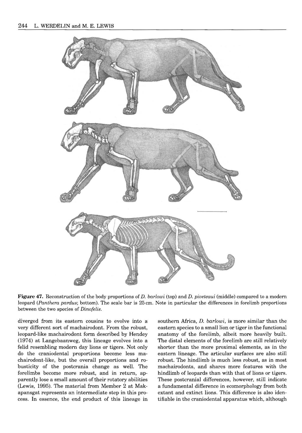
98


99 DINOFELIS (MAMMALIA, FELIDAE) 245 Figure 48. Reconstruction of the head of D. barlowi (top) and D. piueteaui (bottom). A, cut away to show skull. B, external features. The scale bar is 10 cm. Note the shorter skull of D. piueteaui with its short postcanine diastema and the differences between the taxa in the mastoid and occipital regions. Despite the differences in skull size, the body sizes of these two taxa were roughly similar (Fig. 47). more similar to Panthera in D. barlowi than in its forebears, still bears indications of its machairodont origin in a slightly compressed canine and long carnassial. D. barlowi is in such features less Pantheralike than D. cristata from Asia. Although in all species of Dinofelis the distal elements are relatively short in comparison to the proximal elements, this trend is accentuated with the appearance of Dinofelis at Olduvai Gorge. This species is unlike its congeners that are contemporaneous in eastern Africa (Lewis, 1995,1997). Unfortunately, only a humerus and ulna from Bed I and an astragalus from Bed I1 are known from this species. Although the morphology of the specimens is similar in some respects to that of D. amnoki, the forearm of the Olduvai Gorge species is extremely short and robust for its length. This species was very different from other species of Dinofelis found concurrently in eastern Africa. This difference was not like the difference between an extant leopard and lion, as this species differed radically in both proportion and robusticity from other species of Dinofelis. The limbs were short and stocky and must have been incredibly muscular, to judge from the muscle markings. When the material from around the world is considered, it is clear that Dinofelis is a machairodont that has converged on Panthera in craniodental and some postcranial features. This convergence probably occurred in more than one lineage of Dinofelis. At this time, Panthera was not common at fossil sites (Turner, 1990; Lewis, 1995, 1997). Although this may be a taphonomic artifact, the number of Panthera specimens does rise after the demise of Dinofelis and the other machairodonts. Panthera and Dinofelis are contemporaneous for some time before the disappearance of the machairodonts. Whether Panthera contributed to the demise of the machairodonts or was merely the beneficiary of their disappearance cannot be ascertained. Certainly, Dinofelis was not completely analogous to modern Panthera in behaviour or ecology, or they might have survived any environmental or ecological changes that benefited the modern species. When the final extinction of Dinofelis occurred can at present not be pinpointed precisely. The last well dated occurrences are in the range of 1.5 to 1.4 Mya in both Africa and Europe. In Europe this may represent something close to an extinction date in view of the many lower Pleistocene sites available from the continent. In Africa, however, the record of Dinofelis disappears at the precise time when the well dated, semicontinuous record in sediments such as those of the Omo Group disappear. The question of the extinction
100 246 L. WERDELIN and M. E. LEWIS MYa I I? Dinofelis sp. (Langebaanweg) Dinofelis diastemata Dinofelis cristata Dinofelis petteri, n. sp. Dinofelis barlowi Dinofelis darti Dinofelis aronoki, n. sp. Dinofelis palaeoonca Dinofelis piueteaui Dinofelis sp. indet. A Dinofelis sp. indet. B Dinofelis sp. indet. C Dinofelis sp. indet. D Dinofelis sp. indet. E Dinofelis sp. indet. F Figure 49. Stratigraphic ranges of the various taxa of Dinofelis discussed herein. Taxa which we definitely consider different from all others are shown above the horizontal dashed line. while indeterminate taxa that may turn out to be conspecific with others are shown below the horizontal dashed line. of Dinofelis in Africa has been thrown wide open by recent study of the Kanam East locality, which through a combination of biostratigraphy, stage of evolution (of the Dinofelis itself) and paleomagnetic calibration, has been suggested to have an age compatible with the Jaramillo Subchron ( Mya) (Ditchfield et al., 1999). Dinofelis may therefore have persisted in Africa well beyond the continuous sequence record. A summary of the taxa and their stratigraphic distributions is given in Figure 49 and below. SYSTEMATIC PALAEONTOLOGY ORDER CARNIVORA BOWDICH, 1821 FAMILY FELIDAE FISCHER, 1817 SUBFAMILY MACHAIRODONTINAE GILL, 1872 Remarks. The affinities of Dinofelis to other Felidae have been disputed. The best characters allying the t I - genus to the Machairodontinae are a combination of the relative proportions of the cheek teeth, with long carnassials and short anterior cheek teeth, the reduced lower canine, the more squared off shape of the anterior part of the lower jaw, and the presence of a deep groove superomedial to the trochlear notch of the ulna. These characters all appear derived with respect to the condition in the Felinae. GENUS DINOFELIS ZDANSKY, Therailurus Piveteau, p Type species. Dinofelis abeli Zdansky, 1924 Dinofelis cristata (Falconer & Cautley, 1836). Amended diagnosis. Machairodontinae of moderate to large size. Sexual dimorphism distinct but not great.

101 Cranium rounded, with large to very large sagittal crest. Lower jaw stout. Coronoid process large compared to other Machairodontinae. No anterior flange present but anterior margin of ramus strong and flat. Upper canine short and only very slightly mediolaterally compressed. No longitudinal grooves. Posterior crest well developed, but no serrations present. P2 lost, except in D. cristata. P3 not or slightly reduced and slender, but less so than in other Machairodontinae. P4 with somewhat reduced protocone. Lower canine reduced but not incisiform. p%p4 slender, ml elongated, but less so than in other Machairodontinae. Remarks. In addition, the evolutionary sequence postulated herein indicates that Dinofelis evolved from an ancestral stock very similar in its craniodental proportions to Metailurus spp. In particular, the lower ramus of the Lothagam Dinofelis (Fig. 21A, B) shows great similarities to the lower ramus of M. major (Andersson and Werdelin, in prep.; Zdansky, 1924, pl. 24, figs 3, 4), but has a longer postcanine diastema. DINOFELIS CRISTATA (FALCONER & CAUTLEY, 1836) 1836 Felis cristata Falconer & Cautley, p. 142, pl. 21 figs 1, Felis cristata Falconer & Cautley in Murchison, pp (non pl. 25 figs 14) Felis grandicristata Bose, pp Uncia cristata Cope, p Uncia grandicristata Cope, p Felis cristata Lydekker, p. 320, pl partim 1903 Machaimdus horribilis Schlosser, p. 37, P1. 1, figs 15, Dinofelis abeli Zdansky, p pl Felis cristata Matthew, p Tigris cristatus Kretzoi, p Leo cristatus Kretzoi, p Panthera cristata Pilgrim, p Panthera cristata Colbert, p Dinofelis abeli Hemmer, p. 79, fig Dinofelis cristata Hemmer, p. 158, fig. 1. Holotype. Royal College of Surgeons no. 358 (destroyed in 1941; cast BM M28913). Localities. Siwalik Hills, India; Lok. B (Zdansky, 1924), Mianzhi Xian, Yangshaozong, Henan, China. Age. All Siwalik specimens have been found in the Pinjor zone of the Upper Siwaliks. These sediments are generally late Pliocene to early Pleistocene in age (Barry, Behrensmeyer & Monaghan, 1980; Pilbeam et al., 1979). The age of Lok. B is probably lower Pliocene, based on the presence of Canidae indet. at the locality. DINOFELIS (MAMMALJA, FELIDAE) 247 If the first appearance datum of this family in the Yushe Basin (Flynn, Tedford & Qiu, 1991; Tedford et al., 1991) also applies here, the locality cannot be older than c. 4.5 Mya. Amended diagnosis. A Dinofelis of large size. Sagittal crest very large. Upper canine similar to Panthera in lengtwwidth relationship. P3 not reduced in length. P4 protocone not reduced. Lower canine moderately reduced. p4 relatively slender, ml robust. p3 but not p4 considerably reduced in length. ReferEd material. Henan: Lok. B: PMU , cranium and mandible (holotype of Dinofelis abeli, Zdansky, 1924, P1.31, fig. 14). Siwaliks: Pinior zone: BM M28913 (cast of holotype of Felis cristata, original in Royal College of Surgeons destroyed during World War 11; Falconer & Cautley, 1836, P1.21, figs 1, 2, Lydekker, 1884, P1.41,42); BM M49176, posterior part of cranium (Lydekker, 1884, P1.40, fig. 1; Hemmer, 1973, fig. 1); BM M49175, posterior part of cranium (holotype of Felis grandicristata, Bose, 1880, p. 128; Lydekker, 1884, P1.40, fig. 2); BM M32148, nearly complete cranium; BM M48437, horizontal ramus fragment (Matthew, 1929, p. 494; Hemmer, 1973, p. 163). Remarks. In terms of cranium and canine size, D. cristata is the largest of all Dinofelis. Ironically, given that it includes the type species, D. abeli, D. cristata is perhaps the most difficult of the species to properly distinguish from Panthera. The upper canine is not sufficiently compressed to make this a viable character. However, the lower canine and p3 are characteristically reduced in size, allying the species with other Dinofelis. The zygomatic arch is also characteristic of the genus (see Hemmer, 1973 for a discussion). DINOFELIS DIASTEMATA (ASTRE, 1929) 1929 Felis diastemata Astre, p. 203, figs Felis diastemata Schaub, p Therailurus diastemata Piveteau, pp. 103, 104, figs 1-8, 10-12, Therailurus diastemata Piveteau, 1961, p.796, figs. 181, Dinofelis diastemata Hemmer, p. 80, figs 36, 37. non 1974 Dinofelis diastemata Hendey, p Dinofelis diastemata Ballesio, p Dinofelis diastemata Ballesio, p Holotype. Partial right horizontal ramus (Astre, 1929, figs 1-3). Locality. Chefdebien brickworks and other sites at Serrat-den-Vacquer, near Perpignan, France.

102 248 L. WERDELIN and M. E. LEWIS Age. MN 14, Ruscinian, early Pliocene. Amended diagnosis. Moderately large species of Dinofelis. Postcanine diastema relatively long. P3 and P4 protocone not reduced. Lower canine considerably reduced. p4 not reduced. Referred material. Serrat-d'en-Vacquer, Perpignan: Partial right horizontal ramus (holotype, Felis diasternata, Astre, 1929, figs 1-3); Lyon PG 28, maxilla fragment (Schaub, 1934, p. 400, Piveteau, 1948, p. 104); Cranium and partial mandible (Piveteau, 1948, figs 1-14, P1.l, 2; Piveteau, 1961, figs 181, 182; Hemmer, 1965, figs 36, 37). Material of the postcranium has been referred to this species by Ballesio (1985), a publication we have not seen. Remarks. This species is hard to characterize due to the relatively poor condition of most of the available material. It is quite similar to D. cristata, but is considerably smaller. That this is not likely to be sexual dimorphism is attested to by the considerable range of variation in size of known specimens of D. cristata (see, e.g. Lydekker, 1884, P1.40), all of which are larger than the specimens referred to D. diasternata. The relatively unreduced P3 distinguishes D. diastemata from all other species of Dinofelis. DINOFELIS BARLOWZ (BROOM, 1937) 1937 Megunthereon barlowi Broom, p. 757, fig. 4B, C Meganthereon barlowi Broom, p Machaemdus transvaalensis Broom, p. 333, fig. lc, D Meganthereon barlowi Broom in Broom & Schepers, p. 78, fig. 7A Therailurus barlowi Ewer, p. 599, fig Therailurus sp. Ewer, p Dinofelis barlowi Hemmer, p. 81, fig non 1976 Dinofelis barlowi Leakey, p Megantereon cf. gracile Vrba, p. 33, figs 7B, C MegantereoncultridensTurner, p. 1259, fig Dinofelis barlowi Turner, p non 1988 Dinofelis cf. D. barlowi Harris et al., p Dinofelis barlowi Cooke, p. 11, figs Dinofelis barlowi Turner, p. 120, fig. 5. Holotype. (TM) STS 1541, cranium. Transvaal Museum, Pretoria, South Africa. Localities. Sterkfontein Member 2 and Member 4, Bolt's Farm, Swartkrans Member 1,?Kromdraai B, all in South Africa. Age. The South African karstic sites present notorious dating problems. According to the most recent estimates, the oldest of the aforementioned localities is Sterkfontein Member 2, with suggested dates of around Mya (Turner, 198%). The youngest are probably Kromdraai B and Bolt's Farm, provisionally dated around 2 Mya. This suggests an age range for D. barlowi of Mya, or middle to late Pliocene. Amended diagnosis. A Dinofelis of large size. Postcanine diastema moderately long. Anterior part of jaw relatively rounded. Upper canine somewhat transversely compressed, gently recurved. P3 reduced. P4 protocone not reduced. Lower canine moderately reduced. p3, p4 reduced, ml long. Referred material. Sterkfontein: Member 3, : SF 5855, left P4 (Turner, 198%, fig. 5.3); SF 5906, right P4 fragment (Turner, 1987b, p. 337); SF 5853, right mandibular ramus fragment with ml (Turner, 1987b, fig. 5.4). Membe r 4:STS 1541/1542, damaged cranium and left C (cranium holotype of MeguntheRon barlowi, Broom, 1937, p. 757, fig. 4B, C; Broom, 1939, p. 332; Broom & Schepers, 1946, fig. 7A); STS 131, broken cranium and mandible fragments (Ewer, 1955, fig. 7); STS 132, tip of crown of C (Ewer, 1955, p. 601); STS 1579, left P4 (Broom, 1939, figs lc, D; Ewer, 1955, p.601). Boltk Farm: Pit 23: BF-1, cranium and associated mandible (Cooke, 1991, figs 2A, B, 4); UCMP 69506, damaged cranium and mandible (Cooke, 1991, fig. 2C); UCMP 64443, damaged cranium and mandible; UCMF' 88799, left C; UCMP right and left C fragments; UCMP 88801, cranial fragment. Swartkrans, - M m : SK 335, partial right horizontal ramus with p4-ml (Ewer, 1955, p. 603); SK 372, damaged lower canine (Ewer, 1955, p. 604); SK 1848, MC IV. Tentatively referred material. Sterkfontein: Member 2: SF 5839, proximal tibia; SF 5840, distal tibia, SF 5885, astragalus (Turner, , fig. 6.1); : 4 - SF 433/ 444, P4 -er, 1987b, p. 331); Rmmdraai B: KB E3 5224, left horizontal ramus fragment (Vrba, 1981, Fig. 7B, C; Turner, 1987a, fig. 1.7); KB 3248 distal end of MC 11. Remarks. This species is distinguished from older forms by the shorter postcanine diastema and reduced premolars. The lower jaw is less like other Machairodontinae than it is in other species of Dinofelis. The upper canine of this species is often stated to be more pantherine-like than in D. piueteaui, but Fig. 32 shows that both follow the general allometric pattern of Dinofelis. The postcrania, however, do have more pantherine features yet can be distinguished from

103 Panthera by the robust morphology of the articular surfaces of the limbs. DINOFELIS PALAEOONCA (MEADE, 1945) 1945 Panthera palaeoonca Meade, p. 521, pl. 48 fig. 2, pl Dinofelis palaeoonca Kurten, p. 1, fig Dinofelis palaeoonca Hemmer, p Dinofelis palaeoonca Dalquest, p. 24, fig Dinofelis palaeoonca Kurtkn & Anderson, p Holotype. TMM , cranium and mandible. Texas Memorial Museum, University of Texas, Austin, USA. Localities. Three localities of the Blanco Local Fauna have yielded specimens of D. palaeoonca: Meade s Quarry sites 6 and 11, and Marmot Quarry. Age. The Blanco Local Fauna is dated to the uppermost Blancan Land Mammal Age (upper Pliocene). Amended diagnosis. A Dinofelis of small size. Upper canine transversely compressed, P3 moderately reduced. P4 protocone not reduced, metastyle long. Lower canine moderately reduced. ml long and slender. p3, p4 relatively unreduced. Referred material. Blanco: Meade s Quarrv. Site 11: TMM , cranium and mandible (holotype of Panthera palaeoonca, Meade, 1945, figs 1, 2, P1.49); Meade s Quarry. S ite 6 TMM , P4 fragment; TMM , right C (Kurten, 1973, fig. 1); Marmot Quarry: TTU-P6280, left P4 (Dalquest, 1975, fig. 5). Remarks. Distinguished from all other Dinofelis by the combination of long and slender ml, long P4 metastyle and unreduced premolars. DINOFELIS DARTI (TOERIEN, 1955) 1955 Machaemdus darti Toerien, p. 43, figs 11, Therailurus barlowi Ewer, p Dinofelis barlowi Collings et al., p Holotype. (BPI) M 607. Bernard Price Institute, University of the Witwatersrand, Johannesburg, South Africa. Locality. Makapansgat, South Africa. Age. The dating of Makapansgat presents great difficulties (e.g. Partridge, 1979; Delson, 1984; Vrba, 1995). However, Members 3 and 4 are generally believed to be in the range of Mya, while Member 2 is only slightly if at all older (Maguire, 1985). DINOFELIS (MAMMALIA, FELIDAE) 249 Referred material. Makapansgat, Member 2: M , crushed cranium and mandible; M 16201, partial left pes, tibia, fibula, associated with M ; M 16202, partial right forelimb, associated with M , 16203, partial left manus, associated with M ; M , , 16185, associated parts of partial skeleton; M no number, distal femur; M no number, astragalus, associated with partial skeleton. Makapansgat, Member: M 607, mandible (holotype of Machaemdus durti, Toerien, 1955); M 259, snout; M 2285, mandibular ramus. Makapansgat, Member 4: M 2136, partial right mandibular ramus. Tentatively referred material. Makapansgat, Member - 3: M 1983, distal femur. Remarks. This material has been studied in detail by us, and as noted in the text we find that Toerien s (1955) original assignment of this material to a taxon distinct from Broom s (1937) Meganthereon barlowi is correct. DINOFELIS PIVETEAUI (EWER, 1955) 1955 Therailuruspiueteaui Ewer, p. 588, figs 1-6, pl Dinofelis piveteaui Hemmer, p. 80, fig Dinofelis cf. piueteaui Leakey, p Dinofelis piveteaui Cooke (ex Leakey), p Dinofelis sp. aff. piveteaui Asfaw et al., p Dinofelis piueteaui Ditchfield et al., p Holotype. (TM) KA 61, cranium. Transvaal Museum, Pretoria, South Africa. Localities. Kromdraai A, South Africa; Koobi Fora Formation, Okote Member, Kanam East, Kenya; Konso-Gardula, locality 10, Ethiopia. Age. Kromdraai A is dated around 1.5 Mya, while the Okote Member lies between tuffs that are securely dated at 1.64 and 1.39Mya. Kanam East at present is of uncertain Plio-Pleistocene age, but may be as young as Mya (Jaramillo Subchron) (Ditchfield et al., 1999). Amended diagnosis. Cranium short and wide. Postcanine diastema very short. Upper canine nearly straight, moderately transversely compressed. P3 very reduced. P4 long, protocone almost entirely reduced. ml long and broad. Referred material. Kmmdraai A: KA 61, partial cranium (holotype of Therailurns piueteaui, Ewer, 1955, figs 1-4, P1. 1; Hemmer, 1965, fig. 38:2); KA 62, right horizontal ramus fragment (Ewer, 1955, fig. 5); KA 63,
 (Leakey, 1976, p.")
104 250 L. WERDELIN and M. E. LEWIS left horizontal ramus fragment (Ewer, 1955, fig. 6); KA 547, distal right femur. Koobi Fora (undescribed except as noted): Koob i Fora Formation. Okote Membe r: KNM-ER 666, anterior parts of left and right horizontal rami (Ileret) (Leakey, 1976, p.306); KNM-ER 722, partial skeleton (Ileret); KNM-ER 6024, partial left humerus (Ileret); KNM-ER 873, humerus shaft (Ileret); KNM-ER 1721, distal right humerus (Koobi Fora Ridge); KNM-ER 366, proximal ulna (Karari Ridge); KNM-ER 710, left MC I1 (Karari Ridge). Kanam East: KNM-KE 21, partial cranium and left and right horizontal rami and associated skeletal remains. Konso- Gardula: Localitv 10: Unknown catalogue number, complete cranium (Nature, 25 December 1992, cover illustration). Remarks. This species is readily distinguished from other Dinofelis by its more machairodont cheek dentition, with short postcanine diastema, reduced premolars and long carnassials. DINOFEMS PElTERI Sl? NOV Dinofelis sp. Howell & Petter, p Dinofelis sp. Howell & Petter, p Dinofelis sp. Petter & Howell, p Dinofelis sp. Barry, Dinofelis cf. D. barlowi Harris et al., p. 37. Holotype. KNM-ER 2612, partial cranium. Housed in the Department of Palaeontology, National Museums of Kenya, Nairobi (Fig 8). Localities. Kanapoi, Allia Bay, Koobi Fora Formation, Tulu Bor Member and West Turkana LO10, all Kenya; Laetoli, Tanzania; Omo and Hadar, Ethiopia. Age. The localities listed above are dated from c. 4.2 Mya (Kanapoi) to c Mya (upper limit of Tulu Bor Member). Derivation of name. After Dr Germaine Petter, leading scholar of African fossil carnivores. Diagnosis. A Dinofelis of moderate size. Postcanine diastema much shorter than in Langebaanweg form. Cranium low, rounded with moderately sized sagittal crest. Horizontal ramus of lower jaw straight and transversely thickened. P3 somewhat reduced, P4 protocone not reduced. Lower canine much reduced, premolars less so. Referred material. Kanapoi (previously undescribed): KNM-KP 30429, partial P4; KNM-KP 30397, complete right horizontal ramus; KNM-KP 30542, proximal ulna. Allia Bay (previously undescribed): F ormation. Mo i ti Memb e 1:: KNM-ER 30335, edentulous left horizontal ramus; AB 261, p3; AE3 311, P4. Koobi Fora (previously undescribed): r e er: KNM-ER 2612, partial cranium (I1 Naibar Lowlands). West Zbrkana: Na- 4): KNM- WT 16846, partial C (locality L010) (Harris et al., 1988, p.37); KNM-WT 16832, edentulous left horizontal ramus (locality L010) (Harris et al., 1988, p. 37). Laetoli: J,aetol il Beds: LAET 448, left P4 (Barry, 1987, p. 248); LAET 868, right i3 (Barry, 1987, p. 247). Omo: Shun= : - OM (cast KNM-ZP 444), damaged cranium (Howell & Petter, 1976, p- 323). Hadar (previously undescribed): Hadar Formation: Denen Dora Member: AL 366-2A, left horizontal ramus fragment. Tentatively referred material. Laetoli: Laetolil Beds: LAET 5045, distal humerus; LAET 4812, distal radius. Hadur Formation: Sidiha Koma Member: AL 262-9, proximal ulna. Hadar Formation: -Dora Mem- & AL , partial humerus; AL , right astragalus. Omo: : - F , right MC IV. Remarks. This species is distinctly more derived than the Langebaanweg and Lothagam forms in its shorter postcanine diastema and more reduced premolars. The latter are, however, not as reduced as in later species. In addition, this form is smaller than all later African Dinofelis. The postcrania is similar in many respects to Megantereon. DZNOFELZS ARONOKl SP. NOV Dinofelis barlowi Leakey, p Holotype. KNM-ER 3880, cranial and postcranial elements. Housed in the Department of Palaeontology, National Museums of Kenya, Nairobi (Figs 10, 15). Localities. Koobi Fora Formation, Upper Burgi Member, Nakoret, Lothagam, Nachukui Formation, Kaiyumung Member, all Kenya, Hadar, Ethiopia. Age. Between. 3.18Mya (lower limit of Kada Hadar Member) and c. 1.65Mya (based on an approximate correlation of Nakoret with the KBS Member of the Koobi Fora Formation). Derivation of name. From amno ki, meaning it was terrible in the language of the people of east Turkana. In reference to the genus name. Used in the sense of the terrible one. Diagnosis. A Dinofelis of moderate to large size. Postcanine diastema reduced but considerably longer than
: Koob i Fora Formation.")
105 in D. piueteaui. Upper canine flattened, P3 considerably reduced. P4 protocone greatly reduced. ml slender. p3 and p4 greatly reduced. Referred material. Koobi Fora (undescribed except as noted): Koob i Fora Formation. UpDer Burgi Member: KNM-ER 1549, partial left and right horizontal rami (Karari Ridge, area 130) (Leakey, 1976: 306); KNM- ER 365, fragment of left horizontal ramus (area not known); KNM-ER 3739, partial left horizontal ramus (Karari Ridge, area 130); KNM-ER 3880, partial left and right horizontal rami and cranial fragments and associated partial skeleton (Karari Ridge, area 129); KNM-ER 4419, partial skeleton and associated dental fragments (Bura Hasuma, area 123); KNM-ER 3742, left astragalus; KNM-ER 893, proximal left femur. Koobi Fora Formation. Unkn own Member: KNM-ER 6112, right MC 11; KNM-ER 987, left femur; KNM-ER 3747, right radius; KNM-ER no number, fragment of pubic ramus. Nakoret: KNM-NO 28686, proximal ulna; KNM-NO 29191, proximal femur. Lothagam: && yumuny Member: KNM-LT 23111, radius. Hadur, Hadar F ormation. Kada Hadar Member: AL , distal radius. Remarks. This species has several of the dental features of D. piueteaui, including the very straight upper canine and the reduced premolars. However, it has a primitively long postcanine diastema, thus lacking the distinctive short snout of that species. The postcrania are robust with slightly straighter radii than in other Dinofelis. DINOFELIS SP. Locality. Lothagam, Kenya. Age. Between c. 7.9 Mya to 4.5 Mya (the range of the Nawata and Apak Members at Lothagam, cf. Leakey et al., 1996). Referred material. Loth-: Nawata Formation, Lower Member: KNM-LT 23942, partial C; KNM-LT 25397, partial p4; Nawata Formation. Upper Member: KNM-LT 00127, anterior part of right horizontal ramus. Nachukui Formation. Apak Member: KNM-LT 28711, anterior part of right horizontal ramus; KNM- LT anterior part of left horizontal ramus; Tentatively referred material. Lothagam: Nawata Formation. Lower Member: KNM-LT 24041, distal right humeral shaft; Nawata Format ion. Upper Member: KNM-LT 25398, proximal right radius; KNM-LT 30310, proximal left radius; KNM-LT 23934, proximal left radius and ulna; KNM-LT 25401, proximal right femur; KNM-LT 25137, distal left tibia; KNM-LT DINOFELIS (MAMMALIA, FELIDAE) , proximal right tibia; KNM-LT 23932, left calcaneum; Nachukui Format ion. AD& M ember: KNM- LT 25126, proximal left tibia; KNM-LT 23696, right calcaneum; KNM-LT 25403, proximal left phalanx of manus, digit I. Remarks. This material almost certainly represents a new species, which we refrain from naming at this stage because of the fragmentary nature of the material. DINOFELIS SP. CF. D. DIASTEMATA 1974 Dinofelis diastemata Hendey, p. 169, figs Dinofelis barlowi Turner, p Locality. Langebaanweg, South Africa. Age. Approximately 54.5 Mya. Referred material. Langebaanweg: E Quarrv. Bed 2: SAM PQ-L20284, right C (Hendey, 1974, fig. 38), left and right horizontal rami (Hendey, 1974, fig. 36); SAM PQ-L20685, right and left C, right ml, left horizontal ramus and associated postcranial material; SAM PQ- L20702, left C; E Quarry. bed 3a : SAM PQ-L12237, left horizontal ramus fragment; Stratieraphic provenance unknown: SAM PQ-L2674, cranium fragments including maxilla (Hendey, 1974, fig. 37). Remarks. This material probably represents a new species, but cannot be adequately separated from D. diastemata because of the lack of overlap of the well preserved described material. DINOFELIS SP. INDET. A 1976 Dinofelis sp. Morales & Aguirre, p.54, fig. 5A, C, D, P1. 3, fig. 2A, B. Locality. Venta del Moro, Spain. Age. c.6-5mya. Referred material. Venta del Mom: NM-C-20, C fragment (Morales & Aguirre, 1976, fig. 5C, D, P1. 3, fig. 2A, B); NM-C-21, ml fragment (Morales & Aguirre, 1976, fig. 5A); NM-C-22, left calcaneum. Remarks. This material is not diagnostic at the species level. DINOFELIS SP. INDET. B 1963 Therailurus barlowi? Cooke, p Dinofelis barlowi Savage, p. 261.

106 252 L. WERDELIN and M. E. LEWIS 1993 Dinofelis piveteaui Berger, p Dinofelis sp. Berger, p. 53. Locality. Gladysvale, South Africa. Age. Perhaps Mya. Referred material. P4. Gladysvale: UCMP 88746, right Remarks. Almost certainly represents either D. barlowi or D. piveteaui but the available material is not sufficient to make the determination. DINOFELIS SP INDET. C 1983 Dinofelis sp. Beaumont, p Locality. Balaruc 11, France. Age. c Mya. Referred material. Balaruc Ik CF no number, left partial horizontal ramus (Beaumont 1983, P1. 1). Remarks. The material is too poorly preserved to assign with certainty to a specific species. DINOFELIS SP. INDET. D Locality. Olduvai Gorge, Tanzania. Age. c Mya. Referred material. Olduvai Gorge (previously undescribed): OLD 63/2642, left and right i3; OLD 59/140, partial P4; BM M 20323B, edentulous mandibular ramus; OLD 74/01, humerus; OLD 74j54, humerus; OLD 74j348, ulna. w: OLD 068/6132, astragalus. Tentatively referred material. Olduvai Golge (previously undescribed): Bed I: OLD F887, 1941, distal femur. Remarks, The material is not sufficient for definite referral to a species. The dental material is similar to that of D. amnoki but fragmentary. The postcranial material bears some resemblance to D. amnoki, but has very different proportions. It definitely does not represent D. piveteaui. DINOFELIS SP INDET. E 1998 Dinofelis cf. abeli Gabunia & Vekua, p Locality. Zemo Melaani, Republic of Georgia. Age. Akchagylian, c Mya. Referred material. National Museum of Georgia, Tbilisi, M 1 (Gabunia & Vekua, 1998, figs 1-3). Remarks. Differs from the Asian D. cristata in being less convergent dentally towards a pantherine morphology. DINOFELIS SP. INDET. F 1999 Dinofelis sp. Averianov & Baryshnikov, p Locality. Novaya Etulia 2, Moldova. Age. Upper Villafranchian (MN 17); c Mya. Referred material. Zoological Institute, Russian Academy of Sciences, St. Petersburg, ZIN (Averianov & Baryshnikov, 1999, figs 1, 2). ACKNOWLEDGEMENTS We would like to thank the CCRH of Ethiopia for permission to study the fossils from Hadar and the government of Kenya for permission to study material housed in Nairobi. We would also like to thank Drs Z. Alemseged, L. Berger, C. Gow, J. Hooker, F. C. Howell, M. Leakey, D. Lordkipanidze, E. Lundelius, B. Martinez Navarro, G. Petter, T. Plummer, Z. Qiu, K. Reed, F. Thackeray, M. Wolsan and Professors B. Rubidge and P. Tobias, as well as all their respective institutions for access to specimens in their care and for other assistance with Dinofelis. Thanks to an anonymous reviewer for comments that significantly improved the quality of the paper. Thanks also to Gabriel Ekalale and Meave hakey for help with the finer points of the Turkana language. Drawings of the Lothagam specimens were made by Bjorn Lindsten and the reconstructions of Dinofelis by Mauricio Ant6n. The photographs in Figures 31 and 34 were taken by Ki Andersson, those in Figure 32 by Bienvenido Martinez Navarro, the others by the authors. This research was supported by Swedish Natural Science Research Council grants to LW, a Distinguished Faculty Fellowship Award presented by The Richard Stockton College of New Jersey to MEL and a L. S. B. Leakey Foundation grant to MEL and Dr Kaye Reed, REFERENCES Akersten WA Canine function in Smilodon (Mammalia; Felidae; Machairodontinae). Contributions in Science, Natural History Museum of Los Angeles County 356: Asfaw B, Beyene J, Suwa G, Walter RC, White TD, WoldeGabriel G, Yemane T The earliest Acheulian from Konso-Gardula. Nature 360: Astre G Sur un felin a particularit& ursoi'des des limons Pliocenes du Roussillon. Bulletin de la Societ6 G6ologique de Frame 29:

107 Averianov AO, Baryshnikov GF Dinofelis sp. (Carnivora, Felidae) from the Late Pliocene locality Etulia in Moldavia. Neues Jahrbuch fur Geologie und Paliiontologie, Monatshefte 1999: Ballesio R Monographie dun machairodus du gisement Villafranchien de Seneze: Homotherium crenatidens Fabrini. Travaux du Laboratoire de Geologie de la Facult6 des Sciences de Lyon Ballesio R Sur l appartenance systkmatique des restes du gisement ruscinien du Serrat-den-Vacquer (bussillon) attribues par Charles Deperet a Machirodus cultridens (Cuvier). Congrks Hommage a Charles DeperW, 12. Ballesio R Sous-famille des Machairodontinae. In: Guerin C, Patou-Mathis M, eds. Les Gmnds Mammiferes Plio-Pleistocenes d Eumpe. Paris: Masson, Barry JC The large carnivores from the Laetoli region of Tanzania. In: Leakey MD, Harris JM, eds. Laetoli: a Pliocene site in northern Tanzania. Oxford: Clarendon Press, Barry JC, Behrensmeyer AK, Monaghan M A geologic and biostratigraphic framework for Miocene sediments near Khaur Village, northern Pakistan. Postilla Beaumont G de &marques sur la classification des Felidae. Eclogue Geologicae Helvetiae 57: Beaumont G de Notes complementaires sur quelques felides (Carnivores). Archives des Sciences, Genkve 31: Beaumont G de Une mandibule de Dinofelis (Mammifere, Carnivore, Felide) du Villafranchien inferieur de Balaruc 11, Herault, France. Archives des Sciences, Genkve 36: Berger L A preliminary estimate of the age of the Gladysvale australopithecine site. Palaeontologia Africana 30: Berta A, Galiano H Megantereon hesperus from the late Hemphillian of Florida with remarks on the phylogenetic relationships of machairodonts (Mammalia, Felidae, Machairodontinae). Journal of Paleontology 57: Bose PN Undescribed fossil Carnivora from the Sivalik Hills in the collection of the British Museum. Quarterly Journal of the Geological Society 36: Bowdich TE An analysis of the natural classifications of Mammalia for the use of students and travelers. Paris: J. Smith. Brain CK The Hunters or the Hunted? Chicago: The University of Chicago Press. Broom R On some new Pleistocene mammals from limestone caves of the Transvaal. South African Journal of Science 3375&768. Broom R A preliminary account of the Pleistocene carnivores of the Transvaal caves. Annals of the Transvaal Museum Broom R, Schepers GWH The South African fossil ape-men. The Australopithecinae. Transvaal Museum Memoirs 2: Bryant HN, Russell AP, Laroiya R, Powell GL DINOFELIS (MAMMALIA, FELIDAE) 253 Claw retraction and protraction in the Carnivora: skeletal microvariation in the phalanges of the Felidae. Journal of Morphology 229: Colbert EH Siwalik mammals in the American Museum of Natural History. Transactions of the American Philosophical Society 26: Collings GE, Cruickshank ARI, Maguire JM, Randall RM Recent faunal studies at Makapansgat Limeworks, Transvaal, South Africa. Annals of the South African Museum 71: Cooke HBS Pleistocene mammal faunas of Africa, with particular reference to southern Africa. In: Howell FC, Bourliere F, eds. African Ecology and Human Evolution. Chicago: Aldine Publishing Company, Cooke HBS Dinofelis barlowi (Mammalia, Carnivora, Felidae) cranial material from Bolt s Farm, collected by the University of California African expedition. Palaeontologia Africana 28: Cope ED On the extinct cats of America. American Naturalist Dalquest WW Vertebrate fossils from the Blanco local fauna of Texas. Occasional Papers of the Museum, Texas Tech University 30: Delson E Cercopithecid biochronology of the African Plio-Pleistocene: correlation among eastern and southern hominid-bearing localities. Courier Forschungsinstitut Senckenberg Defirkt C Les Animaux Pliocenes du bussillon. Memoires de la Societe Geologique de France, Paleontologie 3: Ditchfield P, Hicks J, Plummer T, Bishop LC, Potts R Current research on the Late Pliocene and Pleistocene deposits north of Homa Mountain, southwestern Kenya. Journal of Human Evolution 36: Evans HE ed Millerk Anatomy of the Dog. Third Edition. Philadelphia: W.B. Saunders Company. Ewer RF The fossil carnivores of the Transvaal caves: Machairodontinae. Proceedings of the Zoological Society of London 125: Ewer RF Some fossil carnivores from the Makapansgat valley. Palaeontologia Africana Falconer H, Cautley PT Note on the Felis cristata, a new fossil tiger from the Sivalik Hills. Asiatic Researches Fischer G Adversaria Zoologica, fasciculus primus. Memoires de la Societe des Naturalistes de Moscou Flynn LJ, Tedford RH, Qiu Zhanxiang Enrichment and stability in the Pliocene mammalian fauna of North China. Paleobiology 17: Gabunia L, Vekua A The find of Dinofelis in the Pliocene of Georgia. Bulletin of the Georgian Academy of Sciences 157-2: Gill T Arrangement of the families of mammals with analytical tables. Smithsonian Miscellaneous Collections 11: Gonyea WJ Functional implications of felid forelimb anatomy. Acta Anatomica
.")
108 254 L. WERDELIN and M. E. LEWIS Harris JM, Brown FH, Leakey MG Stratigraphy and paleontology of Pliocene and Pleistocene localities west of Lake Turkana, Kenya. Contributions in Science, Natural History Museum of LQS Angeles County 39fk Hemmer H Zur Nomenklatur und Verbreitung des Genus Dinofelis Zdansky, 1924 (Themilurus Piveteau, 1948). Palaeontologia Afncana 9: Hemmer H Neue Befunde zur Verbreitung und Evolution der pliozi4n-pleistoziinen Gattung Dinofelis (Mammalia, Carnivora, Felidae). Neues Jahrbuch fir Geologie und Paldontologie, Monatshefi Hendey QB Carnivore remains h m the Kromdraai australopithecine site (Mammalia: Carnivora). Annals of the Transvaal Museum 28: Hendey QFL The late Cenozoic Carnivora of the southwestern Cape Province. Annals of the South African Museum 83: Hopwood AT New fossil mammals from Olduvai, Tanganyika Territory. Annals and Magazine of Natural History 14: Howell FC, Coppens Y Les faunes de mammiferes des formations Plio-Pleistmenes de l'omo en Ethiopie (Tubulidentata, Hyracoidea, Lagomorpha, Rodentia, Chiroptera, Insectivora, Carnivora, primates). Comptes Rendus de I'Admie des Sciences, Paris Sene D!Z7& Howell FC, Petter G Carnivora from Om0 Group formations, southern Ethiopia. In: Coppens Y, Howell FC, Isaac GL, Leakey REF, eds. Earliest man and enuimnments in the Lake Rudolf Basin. Chicago: University of Chicago Press, Howell FC, Petter G Diversification et affinites des carnivores pliocenes du groupe de l'omo et de la formation d'hadar (Ethiopie). Bulletin de la Societe (Xobgique de France 7 (21): Kalb JE, Jolly CJ, Tebedge S, Menrate A, Smart C, Oswald EB, Whitehead PF, Wood CB, Adefris T, Rawn- Schatzinger V Vertebrate faunas from the Awash Group, Middle Awash Valley, Afar, Ethiopia. Journal of Vertebrate Paleontology Kretzoi M Materialien zur phylogenetischen Klassifikation der Aelurofdeen. X:e Corgr-2~ International de Zoologie Kretzoi M Feliden-Studien. A Magyar Kimlyi Foldtani Intezet Hazinyomdaja 24: Kurten B The Chinese Hipparion fauna. Commentationes Biobgicae, Societas Scientarum Fennica 13: Kurten B The genus Dinofelis (Carnivora, Mammalia) in the Blancan of North America. The Pea-Sellads Series 19: 1-7. Kurten B, Anderson E Pleistocene Mammals of North America. New York: Columbia University Press. LeakeyLSBed OlduvaiGolge Vo1umeI:A Preliminary Report on the Geology and Fauna. Cambridge: Cambridge University Press. Leakey MG Carnivora of the East Rudolf succession. In: Coppens Y, Howell FC, Isaac GL, Leakey REF, eds. Earliest Man and Environments in the Lake Rudolf Basin. Chicago: University of Chicago Press, Leakey MG, Feibel CS, Bernor RL, Harris JM, Cerling TE, Stewart KM, Storrs GL, Walker A, Werdelin L, Winkler A Lothagam: a record of faunal change in the late Miocene of East Africa. Journal of Vertebrate Palaeontology Lewis ME. les2a Forelimb functional anatomy of extinct, large-bodied carnivorans. Journal of Vertebrate Paleontology 12 (supplement): Lewis ME. 1992b. Use of forelimb characters in distinguishing locomotor behaviour and prey procurement type in large-bodied carnivorans. 72* Annual Meeting of the American Society of Mammalogists, abstract 165. Lewis ME Functional anatomy of the hindlimb of Dinofelis barlowi (Mammalia, Carnivora, Felidae) from Bolt's Farm, South Africa. Journal of Vertebrate Paleontology 13 (supplement): 46. Lewis ME Plio/Pleistocene carnivoran guilds: implications for hominid paleoecobgy. Unpublished Ph.D. thesis, State University of New York at Stony Brook. Lewis ME Ecomorphology of the African sabertooth felid Homotherium. Journal of Vertebrate Paleontology 16 (supplement): 48. Lewis ME Carnivoran paleoguilds of Africa: implications for hominid food procurement strategies. Journal of Human Evolution bdekker R Siwalik and Narbada Carnivora. Paleontologwa Indica, sex 10 2: Maguire JM Recent geological, stratigraphic and palaeontological studies at Makapansgat Limeworks. In: Tobias PV, ed. Hominid Euolution: past, present and future. New York Alan R. Liss, Inc., Marean CW Sabertooth cats and their relevance for early hominid diet and evolution. Journal of Human Evolution 18: Martin LD Functional morphology and the evolution of cats. Transactions of the Nebraska Academy of Science 8: Matthew WD Critical observations upon Siwalik mammals. Bulletin of the American Museum of Natural History 56: Meade GE The Blanco Fauna. University of Texas Publications 4401: Morales J, Aguirre E Carnivoros de Venta del Moro. Trabajos Sobre Neogeno-Cuaternario Murchison C ed Palaeontological Memoirs and Notes of the late Hugh Falconer, A.M., M.D. London: Robert Hardwicke. Partridge T Reappraisal of the lithostratigraphy of Makapansgat Limeworks hominid site. Nature Petter G Carnivores Pleistocenes du Ravin dolduvai. In: Leakey LSB, Savage RJG, Coryndon SC, eds. Fossil Vertebrates of Africa, Volume 3. London: Academic Press, Petter G, Howell FC Diversite des carnivores (Mammalia, Carnivora) dans les faunes du Pliocene moyen et

109 superieur d'afrique orientale. Indications paleoecologiques. In: L'Enuimnnement des Horninid& au Plio-Pleistocene. Paris: Fondation Singer-Polignac, Pilbeam DR, Behrensmeyer AK, Barry JC, Shah SMI Miocene sediments and faunas of Pakistan. Postilla Pilgrim GE The fossil Carnivora of India. Paleontologica Zndica, n.s Piveteau J Un felide du Pliocene du bussillon. Annales de PaleontologLe 34: Piveteau J Les Carnivores. In: Piveteau J, ed. Traite de Paleontologie, lbme W, Vol. 1. Paris: Masson et Cie, Pohle H Die Raubtiere von Oldoway. Wissenschaftliche Evebnisse der Oldoway-Expedition 1913 (N.F.) Ram-Schatzinger V The scimitar cat Homotherium serum Cope-osteology, functional morphology, and predatory behaviour. Illinois State Museum Reports of Znvestigations 47: Sardella R The Plio-Pleistocene Old World dirktoothed cat Megantereon ex gr. cultridens (Mammalia, Felidae, Machairodontinae), with comments on taxonomy, origin and evolution. Neues Jahrbuch fur Geologie und Palaontologie, Abhandlungen 207: Savage RJG Carnivora. In: Maglio VJ, Cooke HBS, eds. Evolution of African Mammals. Cambridge, Mass.: Harvard University Press, Schaub S Ueber die Osteologie von Machaemdus cultridens Cuvier. Eclogue Geologicae Helvetiae 19: Schaub S Observations critiques sur quelques machairodontides. Eclogue Geologicae Helvetiae 27: Schlosser M Die fossilen Saugetiere Chinas nebst einer Odontographie der rezenten Antilopen. Abhandlungen der Bayerischen Akademie der Wissenschaften 22: Sotnikova MV A new species of Machirodus from the late Miocene Kalmakpai locality in eastern Kazakhstan (USSR). Annales Zoologici Fennici 28: Tedford RH, Flynn JJ, Qiu Zhanxiang, Opdyke ND, Downs WR Yushe Basin, China: paleomagnetically calibrated mammalian biostratigraphic standard from the late Neogene of eastern Asia. Journal of Vertebrate Paleontology ll: Toerien MJ A sabre tooth cat from the Makapansgat valley. Palaeontologia Africana 3: lbrner A Miscellaneous carnivore remains from DINOFELIS (MAMMALIA, FELIDAE) 255 Plio-Pleistocene deposits in the Sterkfontein Valley (Mammalia: Carnivora). Annals of the Transvaal Museum 34: 'krner A. 1987a. Megantereon cultridens (Cuvier) (Mammalia, Felidae, Machairodontinae) from Plio-Pleistocene deposits in Africa and Eurasia, with comments on dispersal and the possibility of a New World origin. Journal of Pakontobgy 61: lbrner A New fossil carnivore remains from the Sterkfontein hominid site (Mammalia: Carnivora). Annals of the Transuaal Museum 'krner A The evolution of the guild of larger terrestrial carnivores during the Plio-Pleistocene in Africa, Geobios 23: 'hrner A New fossil carnivore remains from Swartkrans. In: Brain CK, ed. Swartkrans, a cavek chronicle of early man. Pretoria: Transvaal Museum Monographs 7: 'krner A Further remains of Carnivora (Mammalia) from the Sterkfontein hominid site. Palaeontologia Africana 34: 'krner A, Ant6n M The Big Cats and their Fossil Relatives. New York: Columbia University Press. Van Valkenburgh B Locomotor diversity within past and present guilds of large predatory mammals. Paleobiology ll: 40M28. Van Valkenburgh B Skeletal indicators of locomotor behaviour in living and extinct carnivores. Journal of Vertebrate Paleontology 7: Vrba ES The Kromdraai australopithecine site revisited in 1980 recent investigations and results. Annals of the Transvaal Museum Vrba ES The fossil record of African antelopes (Mmmalia, Bovidae) in relation to human evolution and paleoclimate. In: Vrba ES, Denton GH, Partridge TC, Burckle LH, eds. Paleoclimate and evolution, with emphasis on human origins. New Haven: Yale University Press, Werdelin L Studies of fossil hyaenids: the genera Thalassictis Gervais ex Nordmann, Palhyaena Gervais, Hyaenictitherium Kretzoi, Lycyaena Hensel and Palinhyaena Qiu, Huang & Guo. Zoological Journal of the Linnean Society 92: Werdelin L. in press. Mio-Pliocene Carnivora from Lothagam, Kenya. In: Leakey MG, Harris JM, eds. Lothagam: Dawn of Humanity in Eastern Africa. Columbia University Press. Zdansky Jungtertike Carnivoren Chinas. Paleontologia Sinica, ser C 2(1):
Felidae from Cooper s Cave, South Africa (Mammalia: Carnivora)
") Felidae from Cooper s Cave, South Africa (Mammalia: Carnivora) Hannah J. O REGAN Department of Archaeology, Humanities Building, University Park, University of Nottingham, Nottingham, NG7 2RD (United Kingdom)
Felidae from Cooper s Cave, South Africa (Mammalia: Carnivora) Hannah J. O REGAN Department of Archaeology, Humanities Building, University Park, University of Nottingham, Nottingham, NG7 2RD (United Kingdom)
New Carnivorous Dinosaurs from the Upper Cretaceous of Mongolia
 1955 Doklady, Academy of Sciences USSR 104 (5):779-783 New Carnivorous Dinosaurs from the Upper Cretaceous of Mongolia E. A. Maleev (translated by F. J. Alcock) The present article is a summary containing
AMERICAN MUSEUM NOVITATES Published by
 AMERICAN MUSEUM NOVITATES Published by Number 782 THE AmzRICAN MUSEUM OF NATURAL HISTORY Feb. 20, 1935 New York City 56.81, 7 G (68) A NOTE ON THE CYNODONT, GLOCHINODONTOIDES GRACILIS HAUGHTON BY LIEUWE
O'Regan HJ Defining cheetahs, a multivariante analysis of skull shape in big cats. Mammal Review 32(1):58-62.
:58-62.") O'Regan HJ. 2002. Defining cheetahs, a multivariante analysis of skull shape in big cats. Mammal Review 32(1):58-62. Keywords: Acinonyx jubatus/cheetah/evolution/felidae/morphology/morphometrics/multivariate
O'Regan HJ. 2002. Defining cheetahs, a multivariante analysis of skull shape in big cats. Mammal Review 32(1):58-62. Keywords: Acinonyx jubatus/cheetah/evolution/felidae/morphology/morphometrics/multivariate
SOME LITTLE-KNOWN FOSSIL LIZARDS FROM THE
 PROCEEDINGS OF THE UNITED STATES NATIONAL MUSEUM issued SWsK \ {^^m ^V ^^ SMITHSONIAN INSTITUTION U. S. NATIONAL MUSEUM Vol. 91 Washington : 1941 No. 3124 SOME LITTLE-KNOWN FOSSIL LIZARDS FROM THE OLIGOCENE
complex in cusp pattern. (3) The bones of the coyote skull are thinner, crests sharper and the
 The bones of the coyote skull are thinner, crests sharper and the") DISTINCTIONS BETWEEN THE SKULLS OF S AND DOGS Grover S. Krantz Archaeological sites in the United States frequently yield the bones of coyotes and domestic dogs. These two canines are very similar both
DISTINCTIONS BETWEEN THE SKULLS OF S AND DOGS Grover S. Krantz Archaeological sites in the United States frequently yield the bones of coyotes and domestic dogs. These two canines are very similar both
Exceptional fossil preservation demonstrates a new mode of axial skeleton elongation in early ray-finned fishes
 Supplementary Information Exceptional fossil preservation demonstrates a new mode of axial skeleton elongation in early ray-finned fishes Erin E. Maxwell, Heinz Furrer, Marcelo R. Sánchez-Villagra Supplementary
Mammalogy Laboratory 1 - Mammalian Anatomy
 Mammalogy Laboratory 1 - Mammalian Anatomy I. The Goal. The goal of the lab is to teach you skeletal anatomy of mammals. We will emphasize the skull because many of the taxonomically important characters
A new species of sauropod, Mamenchisaurus anyuensis sp. nov.
 A new species of sauropod, Mamenchisaurus anyuensis sp. nov. by Xinlu He, Suihua Yang, Kaiji Cai, Kui Li, and Zongwen Liu Chengdu University of Technology Papers on Geosciences Contributed to the 30th
PEABODY MUSEUM OF NATURAL HISTORY, YALE UNIVERSITY NEW HAVEN, CONNECTICUT, U.S.A. A NEW OREODONT FROM THE CABBAGE PATCH LOCAL FAUNA, WESTERN MONTANA
 Postilla PEABODY MUSEUM OF NATURAL HISTORY YALE UNIVERSITY NEW HAVEN, CONNECTICUT, U.S.A. Number 85 September 21, 1964 A NEW OREODONT FROM THE CABBAGE PATCH LOCAL FAUNA, WESTERN MONTANA STANLEY J. RIEL
THE GORGONOPSIAN GENUS, HIPPOSAURUS, AND THE FAMILY ICTIDORHINIDAE * Dr. L.D. Boonstra. Paleontologist, South African Museum, Cape Town
 THE GORGONOPSIAN GENUS, HIPPOSAURUS, AND THE FAMILY ICTIDORHINIDAE * by Dr. L.D. Boonstra Paleontologist, South African Museum, Cape Town In 1928 I dug up the complete skeleton of a smallish gorgonopsian
ZOOLOGISCHE MEDEDELINGEN
 MINISTERIE VAN ONDERWIJS, KUNSTEN EN WETENSCHAPPEN ZOOLOGISCHE MEDEDELINGEN UITGEGEVEN DOOR HET RIJKSMUSEUM VAN NATUURLIJKE HISTORIE TE LEIDEN DEEL XXXVII, No. 10 10 juli 1961 THE FOSSIL HIPPOPOTAMUS FROM
A Fossil Snake (Elaphe vulpina) From A Pliocene Ash Bed In Nebraska
 From A Pliocene Ash Bed In Nebraska") University of Nebraska - Lincoln DigitalCommons@University of Nebraska - Lincoln Transactions of the Nebraska Academy of Sciences and Affiliated Societies Nebraska Academy of Sciences 198 A Fossil Snake
University of Nebraska - Lincoln DigitalCommons@University of Nebraska - Lincoln Transactions of the Nebraska Academy of Sciences and Affiliated Societies Nebraska Academy of Sciences 198 A Fossil Snake
Fig. 5. (A) Scaling of brain vault size (width measured at the level of anterior squamosal/parietal suture) relative to skull size (measured at the
 Scaling of brain vault size (width measured at the level of anterior squamosal/parietal suture) relative to skull size (measured at the") Fig. 5. (A) Scaling of brain vault size (width measured at the level of anterior squamosal/parietal suture) relative to skull size (measured at the distance between the left versus right temporomandibular
Fig. 5. (A) Scaling of brain vault size (width measured at the level of anterior squamosal/parietal suture) relative to skull size (measured at the distance between the left versus right temporomandibular
lion (sp. BPl/c 183) and leopard (sp. BPl/c 258), the rat of skull height to width of palate was greater than that of the lion but less
 and leopard (sp. BPl/c 258), the rat of skull height to width of palate was greater than that of the lion but less") 81 lion (sp. BPl/c 183) and leopard (sp. BPl/c 258), the rat of skull height to width of palate was greater than that of the lion but less than that of the leopard. The jugal arch is distinctly thicker
81 lion (sp. BPl/c 183) and leopard (sp. BPl/c 258), the rat of skull height to width of palate was greater than that of the lion but less than that of the leopard. The jugal arch is distinctly thicker
CHAPTER 9 JACOVEC CAVERN CARNIVORES AND OTHER FAUNA. The Order Carnivora is represented by five families- Viverridae, Herpestidae,
 CHAPTER 9 JACOVEC CAVERN CARNIVORES AND OTHER FAUNA 9.a. Taxonomy Carnivores The Order Carnivora is represented by five families- Viverridae, Herpestidae, Canidae, Felidae, and Hyaenidae. The Viverridae
ONLINE APPENDIX 1. Morphological phylogenetic characters scored in this paper. See Poe (2004) for
 for") ONLINE APPENDIX Morphological phylogenetic characters scored in this paper. See Poe () for detailed character descriptions, citations, and justifications for states. Note that codes are changed from a
ONLINE APPENDIX Morphological phylogenetic characters scored in this paper. See Poe () for detailed character descriptions, citations, and justifications for states. Note that codes are changed from a
Anatomy. Name Section. The Vertebrate Skeleton
 Name Section Anatomy The Vertebrate Skeleton Vertebrate paleontologists get most of their knowledge about past organisms from skeletal remains. Skeletons are useful for gleaning information about an organism
Vol. XIV, No. 1, March, The Larva and Pupa of Brontispa namorikia Maulik (Coleoptera: Chrysomelidae: Hispinae) By S.
 By S.") Vol. XIV, No. 1, March, 1950 167 The Larva and Pupa of Brontispa namorikia Maulik (Coleoptera: Chrysomelidae: Hispinae) By S. MAULIK BRITISH MUSEUM (NATURAL HISTORY) (Presented by Mr. Van Zwaluwenburg
Vol. XIV, No. 1, March, 1950 167 The Larva and Pupa of Brontispa namorikia Maulik (Coleoptera: Chrysomelidae: Hispinae) By S. MAULIK BRITISH MUSEUM (NATURAL HISTORY) (Presented by Mr. Van Zwaluwenburg
SOME FOSSIL CARNIVORES FROM THE MAKAP ANSGAT VALLEY. By R. F. EWER. (Department of Zoology, Rhodes University, Grahamstown) ABsTRACT
 ABsTRACT") SOME FOSSL CARNVORES FROM THE MAKAP ANSGAT VALLEY By R. F. EWER (Department of Zoology, Rhodes University, Grahamstown) ABsTRACT All hitherto undescribed carnivore material from the Limeworks deposit at
SOME FOSSL CARNVORES FROM THE MAKAP ANSGAT VALLEY By R. F. EWER (Department of Zoology, Rhodes University, Grahamstown) ABsTRACT All hitherto undescribed carnivore material from the Limeworks deposit at
TRACHEMYS SCULPTA. A nearly complete articulated carapace and plastron of an Emjdd A NEAKLY COMPLETE SHELL OF THE EXTINCT TURTLE,
 A NEAKLY COMPLETE SHELL OF THE EXTINCT TURTLE, TRACHEMYS SCULPTA By Charles W. Gilmore Curator of Vertebrate Paleontology, United States National Museum INTRODUCTION A nearly complete articulated carapace
A new basal sauropodiform dinosaur from the Lower Jurassic of Yunnan Province, China
 SUPPLEMENTARY INFORMATION A new basal sauropodiform dinosaur from the Lower Jurassic of Yunnan Province, China Ya-Ming Wang 1, Hai-Lu You 2,3 *, Tao Wang 4 1 School of Earth Sciences and Resources, China
First Ornithomimid (Theropoda, Ornithomimosauria) from the Upper Cretaceous Djadokhta Formation of Tögrögiin Shiree, Mongolia
 from the Upper Cretaceous Djadokhta Formation of Tögrögiin Shiree, Mongolia") First Ornithomimid (Theropoda, Ornithomimosauria) from the Upper Cretaceous Djadokhta Formation of Tögrögiin Shiree, Mongolia Tsogtbaatar Chinzorig¹, ³ *, Yoshitsugu Kobayashi², Khishigjav Tsogtbaatar³,
First Ornithomimid (Theropoda, Ornithomimosauria) from the Upper Cretaceous Djadokhta Formation of Tögrögiin Shiree, Mongolia Tsogtbaatar Chinzorig¹, ³ *, Yoshitsugu Kobayashi², Khishigjav Tsogtbaatar³,
2. Skull, total length versus length of the presacral vertebral column: (0); extremely elongated neck (e.g. Tanystropheus longobardicus).
; extremely elongated neck (e.g. Tanystropheus longobardicus).") Character list of the taxon-character data set 1. Skull and lower jaws, interdental plates: absent (0); present, but restricted to the anterior end of the dentary (1); present along the entire alveolar
Character list of the taxon-character data set 1. Skull and lower jaws, interdental plates: absent (0); present, but restricted to the anterior end of the dentary (1); present along the entire alveolar
Mammalogy Lab 1: Skull, Teeth, and Terms
 Mammalogy Lab 1: Skull, Teeth, and Terms Be able to: Goals of today s lab Locate all structures listed on handout Define all terms on handout what they are or what they look like Give examples of mammals
Temporal lines. More forwardfacing. tubular orbits than in the African forms 3. Orbits larger relative to skull size than in the other genera 2.
 Asian lorises More forwardfacing and tubular orbits than in the African forms 3. Characterized by a marked extension of the ectotympanic into a tubular meatus and a more angular auditory bulla than in
A NEW SALTICID SPIDER FROM VICTORIA By R. A. Dunn
 Dunn, R. A. 1947. A new salticid spider from Victoria. Memoirs of the National Museum of Victoria 15: 82 85. All text not included in the original document is highlighted in red. Mem. Nat. Mus. Vict.,
A new species of Hsisosuchus (Mesoeucrocodylia) from Dashanpu, Zigong Municipality, Sichuan Province
 from Dashanpu, Zigong Municipality, Sichuan Province") A new species of Hsisosuchus (Mesoeucrocodylia) from Dashanpu, Zigong Municipality, Sichuan Province Yuhui Gao (Zigong Dinosaur Museum) Vertebrata PalAsiatica Volume 39, No. 3 July, 2001 pp. 177-184 Translated
A new species of Hsisosuchus (Mesoeucrocodylia) from Dashanpu, Zigong Municipality, Sichuan Province Yuhui Gao (Zigong Dinosaur Museum) Vertebrata PalAsiatica Volume 39, No. 3 July, 2001 pp. 177-184 Translated
Skulls & Evolution. 14,000 ya cro-magnon. 300,000 ya Homo sapiens. 2 Ma Homo habilis A. boisei A. robustus A. africanus
 Skulls & Evolution Purpose To illustrate trends in the evolution of humans. To demonstrate what you can learn from bones & fossils. To show the adaptations of various mammals to different habitats and
Title: Fossil Focus: Reimagining fossil cats IMPORTANT COPYRIGHT CITATION OF ARTICLE
 Title: Fossil Focus: Reimagining fossil cats Author(s): Andrew Cuff Volume: 8 Article: 4 Page(s): 1-10 Published Date: 01/04/2018 PermaLink: https://www.palaeontologyonline.com/articles/2018/patterns-palaeontology-earliestskeletons/
Human Evolution. Lab Exercise 17. Introduction. Contents. Objectives
 Lab Exercise Human Evolution Contents Objectives 1 Introduction 1 Activity.1 Data Collection 2 Activity.2 Phylogenetic Tree 3 Resutls Section 4 Introduction One of the methods of analysis biologists use
SUPPLEMENTARY INFORMATION
 Character 155, interdental ridges. Absence of interdental ridge (0) shown in Parasaniwa wyomingensis (Platynota). Interdental ridges (1) shown in Coniophis precedens. WWW.NATURE.COM/NATURE 1 Character
.56 m. (22 in.). COMPSOGNATHOID DINOSAUR FROM THE. Medicine Bow, Wyoming, by the American Museum Expedition
. COMPSOGNATHOID DINOSAUR FROM THE. Medicine Bow, Wyoming, by the American Museum Expedition") Article XII.-ORNITHOLESTES HERMANNI, A NEW COMPSOGNATHOID DINOSAUR FROM THE UPPER JURASSIC. By HENRY FAIRFIELD OSBORN. The type skeleton (Amer. Mus. Coll. No. 6I9) of this remarkable animal was discovered
Article XII.-ORNITHOLESTES HERMANNI, A NEW COMPSOGNATHOID DINOSAUR FROM THE UPPER JURASSIC. By HENRY FAIRFIELD OSBORN. The type skeleton (Amer. Mus. Coll. No. 6I9) of this remarkable animal was discovered
CENE RUMINANTS OF THE GENERA OVIBOS AND
 DESCRIPTIONS OF TWO NEW SPECIES OF PLEISTO- CENE RUMINANTS OF THE GENERA OVIBOS AND BOOTHERIUM, WITH NOTES ON THE LATTER GENUS. By James Williams Gidley, Of the United States National Museum. Two interesting
A skull without mandihle, from the Hunterian Collection (no.
 4 MR. G. A. BOULENGER ON CHELONIAN REMAINS. [Jan. 6, 2. On some Chelonian Remains preserved in the Museum of the Eojal College of Surgeons. By G. A. Boulenger. [Eeceived December 8, 1890.] In the course
BREVIORA LEUCOLEPIDOPA SUNDA GEN. NOV., SP. NOV. (DECAPODA: ALBUNEIDAE), A NEW INDO-PACIFIC SAND CRAB. Ian E. Efford 1
, A NEW INDO-PACIFIC SAND CRAB. Ian E. Efford 1") ac lc BREVIORA CAMBRIDGE, MASS. 30 APRIL, 1969 NUMBER 318 LEUCOLEPIDOPA SUNDA GEN. NOV., SP. NOV. (DECAPODA: ALBUNEIDAE), A NEW INDO-PACIFIC SAND CRAB Ian E. Efford 1 ABSTRACT. Leucolepidopa gen. nov.
ac lc BREVIORA CAMBRIDGE, MASS. 30 APRIL, 1969 NUMBER 318 LEUCOLEPIDOPA SUNDA GEN. NOV., SP. NOV. (DECAPODA: ALBUNEIDAE), A NEW INDO-PACIFIC SAND CRAB Ian E. Efford 1 ABSTRACT. Leucolepidopa gen. nov.
A Partial Skeleton of Pseudaelurus (Carnivora: Felidae) from the Nambé Member of the Tesuque Formation, Española Basin, New Mexico
 from the Nambé Member of the Tesuque Formation, Española Basin, New Mexico") PUBLISHED BY THE AMERICAN MUSEUM OF NATURAL HISTORY CENTRAL PARK WEST AT 79TH STREET, NEW YORK, NY 10024 Number 3342, 31 pp., 19 figures, 1 table June 22, 2001 A Partial Skeleton of Pseudaelurus (Carnivora:
PUBLISHED BY THE AMERICAN MUSEUM OF NATURAL HISTORY CENTRAL PARK WEST AT 79TH STREET, NEW YORK, NY 10024 Number 3342, 31 pp., 19 figures, 1 table June 22, 2001 A Partial Skeleton of Pseudaelurus (Carnivora:
FIELDIANA GEOLOGY NEW SALAMANDERS OF THE FAMILY SIRENIDAE FROM THE CRETACEOUS OF NORTH AMERICA
 FIELDIANA GEOLOGY Published by CHICAGO NATURAL HISTORY MUSEUM Volume 10 Sbftember 22, 1968 No. 88 NEW SALAMANDERS OF THE FAMILY SIRENIDAE FROM THE CRETACEOUS OF NORTH AMERICA Coleman J. Coin AND Walter
A NEW GENUS AND SPECIES OF AMERICAN THEROMORPHA
 A NEW GENUS AND SPECIES OF AMERICAN THEROMORPHA MYCTEROSAURUS LONGICEPS S. W. WILLISTON University of Chicago The past summer, Mr. Herman Douthitt, of the University of Chicago paleontological expedition,
Reprinted from: CRUSTACEANA, Vol. 32, Part 2, 1977 LEIDEN E. J. BRILL
 Reprinted from: CRUSTACEANA, Vol. 32, Part 2, 1977 LEIDEN E. J. BRILL NOTES AND NEWS 207 ALPHE0PS1S SHEARMII (ALCOCK & ANDERSON): A NEW COMBINATION WITH A REDESCRIPTION OF THE HOLOTYPE (DECAPODA, ALPHEIDAE)
Williston, and as there are many fairly good specimens in the American
 56.81.7D :14.71.5 Article VII.- SOME POINTS IN THE STRUCTURE OF THE DIADECTID SKULL. BY R. BROOM. The skull of Diadectes has been described by Cope, Case, v. Huene, and Williston, and as there are many
SOME NEW AMERICAN PYCNODONT FISHES.
 SOME NEW AMERICAN PYCNODONT FISHES. By James Williams Gidley, Assistant Curator of Fossil Mammals, United States National Museum. In the United States National Museum are several specimens representing
A new sauropod from Dashanpu, Zigong Co. Sichuan Province (Abrosaurus dongpoensis gen. et sp. nov.)
") A new sauropod from Dashanpu, Zigong Co. Sichuan Province (Abrosaurus dongpoensis gen. et sp. nov.) by Ouyang Hui Zigong Dinosaur Museum Newsletter Number 2 1989 pp. 10-14 Translated By Will Downs Bilby
A new sauropod from Dashanpu, Zigong Co. Sichuan Province (Abrosaurus dongpoensis gen. et sp. nov.) by Ouyang Hui Zigong Dinosaur Museum Newsletter Number 2 1989 pp. 10-14 Translated By Will Downs Bilby
Description of Cranial Elements and Ontogenetic Change within Tropidolaemus wagleri (Serpentes: Crotalinae).
.") East Tennessee State University Digital Commons @ East Tennessee State University Electronic Theses and Dissertations 5-2016 Description of Cranial Elements and Ontogenetic Change within Tropidolaemus
East Tennessee State University Digital Commons @ East Tennessee State University Electronic Theses and Dissertations 5-2016 Description of Cranial Elements and Ontogenetic Change within Tropidolaemus
ARIEGE POINTING DOG (Braque de l Ariège)
") FEDERATION CYNOLOGIQUE INTERNATIONALE (AISBL) SECRETARIAT GENERAL: 13, Place Albert 1 er B 6530 Thuin (Belgique) 07.08.1998/EN FCI-Standard N 177 ARIEGE POINTING DOG (Braque de l Ariège) 2 TRANSLATION
FEDERATION CYNOLOGIQUE INTERNATIONALE (AISBL) SECRETARIAT GENERAL: 13, Place Albert 1 er B 6530 Thuin (Belgique) 07.08.1998/EN FCI-Standard N 177 ARIEGE POINTING DOG (Braque de l Ariège) 2 TRANSLATION
The family Gnaphosidae is a large family
 Pakistan J. Zool., vol. 36(4), pp. 307-312, 2004. New Species of Zelotus Spider (Araneae: Gnaphosidae) from Pakistan ABIDA BUTT AND M.A. BEG Department of Zoology, University of Agriculture, Faisalabad,
Postilla PEABODY MUSEUM OF NATURAL HISTORY YALE UNIVERSITY NEW HAVEN, CONNECTICUT, U.S.A.
 Postilla PEABODY MUSEUM OF NATURAL HISTORY YALE UNIVERSITY NEW HAVEN, CONNECTICUT, U.S.A. Number 117 18 March 1968 A 7DIAPSID (REPTILIA) PARIETAL FROM THE LOWER PERMIAN OF OKLAHOMA ROBERT L. CARROLL REDPATH
TERRIER BRASILEIRO (Brazilian Terrier)
") 04.07.2018/ EN FEDERATION CYNOLOGIQUE INTERNATIONALE (AISBL) SECRETARIAT GENERAL: 13, Place Albert 1 er B 6530 Thuin (Belgique) FCI-Standard N 341 TERRIER BRASILEIRO (Brazilian Terrier) 2 TRANSLATION:
04.07.2018/ EN FEDERATION CYNOLOGIQUE INTERNATIONALE (AISBL) SECRETARIAT GENERAL: 13, Place Albert 1 er B 6530 Thuin (Belgique) FCI-Standard N 341 TERRIER BRASILEIRO (Brazilian Terrier) 2 TRANSLATION:
On the Discovery of the earliest fossil bird in China (Sinosauropteryx gen. nov.) and the origin of birds
 and the origin of birds") On the Discovery of the earliest fossil bird in China (Sinosauropteryx gen. nov.) and the origin of birds by Qiang Ji and Shu an Ji Chinese Geological Museum, Beijing Chinese Geology Volume 233 1996 pp.
On the Discovery of the earliest fossil bird in China (Sinosauropteryx gen. nov.) and the origin of birds by Qiang Ji and Shu an Ji Chinese Geological Museum, Beijing Chinese Geology Volume 233 1996 pp.
YANGCHUANOSAURUS HEPINGENSIS - A NEW SPECIES OF CARNOSAUR FROM ZIGONG, SICHUAN
 Vol. 30, No. 4 VERTEBRATA PALASIATICA pp. 313-324 October 1992 [SICHUAN ZIGONG ROUSHILONG YI XIN ZHONG] figs. 1-5, pl. I-III YANGCHUANOSAURUS HEPINGENSIS - A NEW SPECIES OF CARNOSAUR FROM ZIGONG, SICHUAN
The Discovery of a Tritylodont from the Xinjiang Autonomous Region
 The Discovery of a Tritylodont from the Xinjiang Autonomous Region Ailing Sun and Guihai Cui (Institute of Vertebrate Paleontology, Paleoanthropology, Academia Sinica) Vertebrata PalAsiatica Volume XXVII,
A NEW AUSTROSQUILLA (STOMATOPODA) FROM THE
 FROM THE") A NEW AUSTROSQUILLA (STOMATOPODA) FROM THE MARQUESAS ISLANDS BY ALAIN MICHEL Centre O.R.S.T.O.M., Noumea, New Caledonia and RAYMOND B. MANNING Smithsonian Institution, Washington, U.S.A. The At s,tstrosqzlilla
A NEW AUSTROSQUILLA (STOMATOPODA) FROM THE MARQUESAS ISLANDS BY ALAIN MICHEL Centre O.R.S.T.O.M., Noumea, New Caledonia and RAYMOND B. MANNING Smithsonian Institution, Washington, U.S.A. The At s,tstrosqzlilla
FINNISH SPITZ (Suomenpystykorva)
") 09.08.1999/EN FEDERATION CYNOLOGIQUE INTERNATIONALE (AISBL) SECRETARIAT GENERAL: 13, Place Albert 1 er B 6530 Thuin (Belgique) FCI-Standard N 49 FINNISH SPITZ (Suomenpystykorva) 2 TRANSLATION : Finnish
09.08.1999/EN FEDERATION CYNOLOGIQUE INTERNATIONALE (AISBL) SECRETARIAT GENERAL: 13, Place Albert 1 er B 6530 Thuin (Belgique) FCI-Standard N 49 FINNISH SPITZ (Suomenpystykorva) 2 TRANSLATION : Finnish
The cranial osteology of Belebey vegrandis (Parareptilia: Bolosauridae), from the Middle Permian of Russia, and its bearing on reptilian evolution
, from the Middle Permian of Russia, and its bearing on reptilian evolution") Blackwell Publishing LtdOxford, UKZOJZoological Journal of the Linnean Society0024-4082 2007 The Linnean Society of London? 2007 1511 191214 Original Articles RUSSIAN BOLOSAURID REPTILER. R. REISZ ET AL.
Blackwell Publishing LtdOxford, UKZOJZoological Journal of the Linnean Society0024-4082 2007 The Linnean Society of London? 2007 1511 191214 Original Articles RUSSIAN BOLOSAURID REPTILER. R. REISZ ET AL.
Introduction to Biological Anthropology: Notes 23 A world full of Plio-pleistocene hominins Copyright Bruce Owen 2011 Let s look at the next chunk of
 Introduction to Biological Anthropology: Notes 23 A world full of Plio-pleistocene hominins Copyright Bruce Owen 2011 Let s look at the next chunk of time: 3.0 1.0 mya often called the Plio-pleistocene
Biology 340 Comparative Embryology Lecture 12 Dr. Stuart Sumida. Evo-Devo Revisited. Development of the Tetrapod Limb
 Biology 340 Comparative Embryology Lecture 12 Dr. Stuart Sumida Evo-Devo Revisited Development of the Tetrapod Limb Limbs whether fins or arms/legs for only in particular regions or LIMB FIELDS. Primitively
v:ii-ixi, 'i':;iisimvi'\>!i-:: "^ A%'''''-'^-''S.''v.--..V^'E^'-'-^"-t''gi L I E) R.ARY OF THE VERSITY U N I or ILLINOIS REMO
 R.ARY OF THE VERSITY U N I or ILLINOIS REMO") "^ A%'''''-'^-''S.''v.--..V^'E^'-'-^"-t''gi v:ii-ixi, 'i':;iisimvi'\>!i-:: L I E) R.ARY OF THE U N I VERSITY or ILLINOIS REMO Natural History Survey Librarv GEOLOGICAL SERIES OF FIELD MUSEUM OF NATURAL
"^ A%'''''-'^-''S.''v.--..V^'E^'-'-^"-t''gi v:ii-ixi, 'i':;iisimvi'\>!i-:: L I E) R.ARY OF THE U N I VERSITY or ILLINOIS REMO Natural History Survey Librarv GEOLOGICAL SERIES OF FIELD MUSEUM OF NATURAL
AMERICAN NATURALIST. Vol. IX. -DECEMBER, No. 12. OR BIRDS WITH TEETH.1 OI)ONTORNITHES,
ONTORNITHES,") AMERICAN NATURALIST. Vol. IX. -DECEMBER, 1875.-No. 12. OI)ONTORNITHES, OR BIRDS WITH TEETH.1 BY PROFESSOR 0. C. MARSH. REMAINS of birds are amono the rarest of fossils, and few have been discovered except
AMERICAN NATURALIST. Vol. IX. -DECEMBER, 1875.-No. 12. OI)ONTORNITHES, OR BIRDS WITH TEETH.1 BY PROFESSOR 0. C. MARSH. REMAINS of birds are amono the rarest of fossils, and few have been discovered except
FURTHER STUDIES ON TWO SKELETONS OF THE BLACK RIGHT WHALE IN THE NORTH PACIFIC
 FURTHER STUDIES ON TWO SKELETONS OF THE BLACK RIGHT WHALE IN THE NORTH PACIFIC HIDEO OMURA, MASAHARU NISHIWAKI* AND TOSHIO KASUYA* ABSTRACT Two skeletons of the black right whale were studied, supplementing
ANTHR 1L Biological Anthropology Lab
 ANTHR 1L Biological Anthropology Lab Name: DEFINING THE ORDER PRIMATES Humans belong to the zoological Order Primates, which is one of the 18 Orders of the Class Mammalia. Today we will review some of
A NEW ANKYLOSAUR FROM THE UPPER CRETACEOUS OF MONGOLIA E.A. Maleev Doklady Akademii Nauk, SSSR 87:
 translated by Dr. Tamara and F. Jeletzky, 1956 A NEW ANKYLOSAUR FROM THE UPPER CRETACEOUS OF MONGOLIA E.A. Maleev 1952. Doklady Akademii Nauk, SSSR 87:273-276 Armored dinosaurs make a considerable part
A NEW SPECIES OF EXTINCT TURTLE FROM THE UPPER PLIOCENE OF IDAHO
 A NEW SPECIES OF EXTINCT TURTLE FROM THE UPPER PLIOCENE OF IDAHO By Charles W. Gilmore Curator, Division of Vertebrate Paleontology United States National Museum Among the fossils obtained bj^ the Smithsonian
NOTE XXXVIII. Three new species of the genus Helota DESCRIBED BY. C. Ritsema+Cz. is very. friend René Oberthür who received. Biet.
 Subshining; HELOTA MARIAE. 249 NOTE XXXVIII. Three new species of the genus Helota DESCRIBED BY C. Ritsema+Cz. The first of these species is very interesting as it belongs to the same section as the recently
SUPPLEMENTARY ONLINE MATERIAL FOR. Nirina O. Ratsimbaholison, Ryan N. Felice, and Patrick M. O connor
 http://app.pan.pl/som/app61-ratsimbaholison_etal_som.pdf SUPPLEMENTARY ONLINE MATERIAL FOR Nirina O. Ratsimbaholison, Ryan N. Felice, and Patrick M. O connor Ontogenetic changes in the craniomandibular
BRAZILIAN TERRIER (Terrier Brasileiro)
") FEDERATION CYNOLOGIQUE INTERNATIONALE (AISBL) SECRETARIAT GENERAL: 13, Place Albert 1er B 6530 Thuin (Belgique) 06.09.2013 / EN FCI-Standard N 341 BRAZILIAN TERRIER (Terrier Brasileiro) This illustration
FEDERATION CYNOLOGIQUE INTERNATIONALE (AISBL) SECRETARIAT GENERAL: 13, Place Albert 1er B 6530 Thuin (Belgique) 06.09.2013 / EN FCI-Standard N 341 BRAZILIAN TERRIER (Terrier Brasileiro) This illustration
Necturus maculosus Family Proteidae
 Necturus maculosus Family Proteidae - Robust body that is somewhat dorsoventrally compressed - Short tail with broad laterally compressed fin - Wide head with blunt/square snout - 3 pairs of bushy gills
Anatomía functional del esqueleto post-craneal de Styriofelis lorteti (Carnivora, Felidae, Felinae) del Mioceno Medio (MN 6) de Sansan (Gers, France)
 del Mioceno Medio (MN 6) de Sansan (Gers, France)") Estudios Geológicos, 67(2) julio-diciembre 2011, 223-243 ISSN: 0367-0449 doi:10.3989/egeol.40590.186 Functional anatomy of the postcranial skeleton of Styriofelis lorteti (Carnivora, Felidae, Felinae)
Estudios Geológicos, 67(2) julio-diciembre 2011, 223-243 ISSN: 0367-0449 doi:10.3989/egeol.40590.186 Functional anatomy of the postcranial skeleton of Styriofelis lorteti (Carnivora, Felidae, Felinae)
CRANIAL EVIDENCE FOR SEXUAL DIMORPHISM AND GROUP LIVING IN THE EXTINCT AMERICAN LION (PANTHERA LEO ATROX)
") CRANIAL EVIDENCE FOR SEXUAL DIMORPHISM AND GROUP LIVING IN THE EXTINCT AMERICAN LION (PANTHERA LEO ATROX) Gold, David Department of Ecological and Evolutionary Biology Professor Matthew McHenry This study
CRANIAL EVIDENCE FOR SEXUAL DIMORPHISM AND GROUP LIVING IN THE EXTINCT AMERICAN LION (PANTHERA LEO ATROX) Gold, David Department of Ecological and Evolutionary Biology Professor Matthew McHenry This study
Diurus, Pascoe. sp. 1). declivity of the elytra, but distinguished. Length (the rostrum and tails 26 included) mm. Deep. exception
. declivity of the elytra, but distinguished. Length (the rostrum and tails 26 included) mm. Deep. exception") 210 DIURUS ERYTIIROPUS. NOTE XXVI. Three new species of the Brenthid genus Diurus, Pascoe DESCRIBED BY C. Ritsema+Cz. 1. Diurus erythropus, n. sp. 1). Allied to D. furcillatus Gylh. ²) by the short head,
210 DIURUS ERYTIIROPUS. NOTE XXVI. Three new species of the Brenthid genus Diurus, Pascoe DESCRIBED BY C. Ritsema+Cz. 1. Diurus erythropus, n. sp. 1). Allied to D. furcillatus Gylh. ²) by the short head,
NAUSHONIA PAN AMEN SIS, NEW SPECIES (DECAPODA: THALASSINIDEA: LAOMEDIIDAE) FROM THE PACIFIC COAST OF PANAMA, WITH NOTES ON THE GENUS
 FROM THE PACIFIC COAST OF PANAMA, WITH NOTES ON THE GENUS") 5 October 1982 PROC. BIOL. SOC. WASH. 95(3), 1982, pp. 478-483 NAUSHONIA PAN AMEN SIS, NEW SPECIES (DECAPODA: THALASSINIDEA: LAOMEDIIDAE) FROM THE PACIFIC COAST OF PANAMA, WITH NOTES ON THE GENUS Joel
5 October 1982 PROC. BIOL. SOC. WASH. 95(3), 1982, pp. 478-483 NAUSHONIA PAN AMEN SIS, NEW SPECIES (DECAPODA: THALASSINIDEA: LAOMEDIIDAE) FROM THE PACIFIC COAST OF PANAMA, WITH NOTES ON THE GENUS Joel
A new species of Antinia PASCOE from Burma (Coleoptera: Curculionidae: Entiminae)
") Genus Vol. 14 (3): 413-418 Wroc³aw, 15 X 2003 A new species of Antinia PASCOE from Burma (Coleoptera: Curculionidae: Entiminae) JAROS AW KANIA Zoological Institute, University of Wroc³aw, Sienkiewicza
Genus Vol. 14 (3): 413-418 Wroc³aw, 15 X 2003 A new species of Antinia PASCOE from Burma (Coleoptera: Curculionidae: Entiminae) JAROS AW KANIA Zoological Institute, University of Wroc³aw, Sienkiewicza
FCI-Standard N 352 / / GB. RUSSIAN TOY (Russkiy Toy)
") FCI-Standard N 352 / 12.06.2006 / GB RUSSIAN TOY (Russkiy Toy) TRANSLATION: RKF, revised by R. Triquet and J. Mulholland. ORIGIN: Russia. DATE OF PUBLICATION OF THE ORIGINAL VALID STANDARD: 21.02.2006
FCI-Standard N 352 / 12.06.2006 / GB RUSSIAN TOY (Russkiy Toy) TRANSLATION: RKF, revised by R. Triquet and J. Mulholland. ORIGIN: Russia. DATE OF PUBLICATION OF THE ORIGINAL VALID STANDARD: 21.02.2006
Neapolitan Mastiff. EXPRESSION Wistful at rest, intimidating when alert. Penetrating stare.
 Neapolitan Mastiff GENERAL APPEARANCE He is characterized by loose skin, over his entire body, abundant, hanging wrinkles and folds on the head and a voluminous dewlap. The essence of the Neapolitan is
Bulletin of Big Bend Paleo-Geo An Open Access Publication from Mosasaur Ranch Museum, Terlingua and Lajitas, Texas All rights reserved
 Bulletin of Big Bend Paleo-Geo An Open Access Publication from Mosasaur Ranch Museum, Terlingua and Lajitas, Texas All rights reserved This was a private report in 2003 on my thoughts on Platecarpus planifrons.
1/9/2013. Divisions of the Skeleton: Topic 8: Appendicular Skeleton. Appendicular Components. Appendicular Components
 /9/203 Topic 8: Appendicular Skeleton Divisions of the Skeleton: Cranial Postcranial What makes up the appendicular skeleton? What is the pattern of serial homology of the limbs? Tetrapod front limb morphology
KATE E. ZEIGLER, ANDREW B. HECKERT and SPENCER G. LUCAS. New Mexico Museum of Natural History, 1801 Mountain Road NW, Albuquerque, NM
 Zeigler, K.E., Heckert, A.B., and Lucas, S.G., eds., 2003, Paleontology and Geology of the Snyder Quarry, New Mexico Museum of Natural History and Science Bulletin No. 24. AN ILLUSTRATED ATLAS OF THE PHYTOSAUR
The Portuguese Podengo Pequeno
 The Portuguese Podengo Pequeno Presented by the Portuguese Podengo Pequenos of America, Inc For more information go to www.pppamerica.org HISTORY A primitive type dog, its probable origin lies in the ancient
Lab 8 Order Carnivora: Families Canidae, Felidae, and Ursidae Need to know Terms: carnassials, digitigrade, reproductive suppression, Jacobson s organ
 Lab 8 Order Carnivora: Families Canidae, Felidae, and Ursidae Need to know Terms: carnassials, digitigrade, reproductive suppression, Jacobson s organ Family Canidae Canis latrans ID based on skull, photos,
A new carnosaur from Yongchuan County, Sichuan Province
 A new carnosaur from Yongchuan County, Sichuan Province by Dong Zhiming Institute of Vertebrate Palaeontology and Palaeoanthropology, Academia Sinica Zhang Yihong, Li Xuanmin, and Zhou Shiwu Chongqing
TWO NEW SPECIES OF WATER MITES FROM OHIO 1-2
 TWO NEW SPECIES OF WATER MITES FROM OHIO 1-2 DAVID R. COOK Wayne State University, Detroit, Michigan ABSTRACT Two new species of Hydracarina, Tiphys weaveri (Acarina: Pionidae) and Axonopsis ohioensis
A Short Report on the Occurrence of Dilophosaurus from Jinning County, Yunnan Province
 A Short Report on the Occurrence of Dilophosaurus from Jinning County, Yunnan Province by Hu Shaojin (Kunming Cultural Administrative Committee, Yunnan Province) Vertebrata PalAsiatica Vol. XXXI, No. 1
290 SHUFELDT, Remains of Hesperornis.
 290 SHUFELDT, Remains of Hesperornis. [ Auk [July THE FOSSIL REMAINS OF A SPECIES OF HESPERORNIS FOUND IN MONTANA. BY R. W. SHUFELD% M.D. Plate XI7III. ExR,¾ in November, 1914, Mr. Charles W. Gihnore,
Karelian bear dog. (FCI Show Judges Commission, Cartagena, February 2013)
") Karelian bear dog (FCI Show Judges Commission, Cartagena, February 2013) Karelian bear dog Karelian bear dog FCI Group 5 Breed number 48 Date of publication of the official valid standard 23/11/2013 The
Karelian bear dog (FCI Show Judges Commission, Cartagena, February 2013) Karelian bear dog Karelian bear dog FCI Group 5 Breed number 48 Date of publication of the official valid standard 23/11/2013 The
FRENCH POINTING DOG GASCOGNE TYPE (Braque français, type «Gascogne»)
") 07.08.1998/EN FEDERATION CYNOLOGIQUE INTERNATIONALE (AISBL) SECRETARIAT GENERAL: 13, Place Albert 1 er B 6530 Thuin (Belgique) FCI-Standard N 133 FRENCH POINTING DOG GASCOGNE TYPE (Braque français, type
07.08.1998/EN FEDERATION CYNOLOGIQUE INTERNATIONALE (AISBL) SECRETARIAT GENERAL: 13, Place Albert 1 er B 6530 Thuin (Belgique) FCI-Standard N 133 FRENCH POINTING DOG GASCOGNE TYPE (Braque français, type
In the following we are particularly elaborating the deviation of the position of lower canines
 IN THE BORZOI BREED PRELUDE At the revision and finalization of the RAS (Rasspecifika Avels Strategier -breed specific breeding strategies) at Borzoi-Ringen s annual meeting it was decided that this appendix
Description of Malacomys verschureni, a new Murid-species from Central Africa
 (Rev. ZooI. afr., 91, no 3) (A paru Ie 30 septembre 1977). Description of Malacomys verschureni, a new Murid-species from Central Africa (Mammalia - Muridae) By W.N. VERHEYEN ANDE. VAN DER STRAETEN * (Antwerpen)
Lagotto Romagnolo. Size The length of the head reaches 1/10 of the height at the withers. The dog is nearly as high a long.
 LISTED BREED-GROUP I SPORTING DOGS IL-3 Lagotto Romagnolo Origin & Purpose Ancient breed of water retrieving dogs in the lowlands of Comacchio and marshlands of Ravenna. During the centuries, the great
FCI-Standard N 251 / / GB. POLISH LOWLAND SHEEPDOG (Polski Owczarek Nizinny)
") FCI-Standard N 251 / 07. 08. 1998 / GB POLISH LOWLAND SHEEPDOG (Polski Owczarek Nizinny) TRANSLATION : Mrs. Peggy Davis. ORIGIN : Poland. 2 DATE OF PUBLICATION OF THE ORIGINAL VALID STANDARD : 07.08.1998.
FCI-Standard N 251 / 07. 08. 1998 / GB POLISH LOWLAND SHEEPDOG (Polski Owczarek Nizinny) TRANSLATION : Mrs. Peggy Davis. ORIGIN : Poland. 2 DATE OF PUBLICATION OF THE ORIGINAL VALID STANDARD : 07.08.1998.
CI-Standard N 343 / / GB. ITALIAN CORSO DOG (Cane Corso Italiano)
") CI-Standard N 343 / 06. 06. 2007/ GB ITALIAN CORSO DOG (Cane Corso Italiano) 2 TRANSLATION : Dr. Antonio Morsiani, Dr. J.-M. Paschoud and Prof. R. Triquet. ORIGIN : Italy. DATE OF PUBLICATION OF THE ORIGINAL
CI-Standard N 343 / 06. 06. 2007/ GB ITALIAN CORSO DOG (Cane Corso Italiano) 2 TRANSLATION : Dr. Antonio Morsiani, Dr. J.-M. Paschoud and Prof. R. Triquet. ORIGIN : Italy. DATE OF PUBLICATION OF THE ORIGINAL
REVISION OF REDONDASUCHUS (ARCHOSAURIA: AETOSAURIA) FROM THE UPPER TRIASSIC REDONDA FORMATION, NEW MEXICO, WITH DESCRIPTION OF A NEW SPECIES
 FROM THE UPPER TRIASSIC REDONDA FORMATION, NEW MEXICO, WITH DESCRIPTION OF A NEW SPECIES") Harris et al., eds., 2006, The Triassic-Jurassic Terrestrial Transition. New Mexico Museum of Natural History and Science Bulletin 37. REVISION OF REDONDASUCHUS (ARCHOSAURIA: AETOSAURIA) FROM THE UPPER
Harris et al., eds., 2006, The Triassic-Jurassic Terrestrial Transition. New Mexico Museum of Natural History and Science Bulletin 37. REVISION OF REDONDASUCHUS (ARCHOSAURIA: AETOSAURIA) FROM THE UPPER
PART FOUR: ANATOMY. Anatomy, Conformation and Movement of Dogs 41
 PART FOUR: ANATOMY Anatomy, Conformation and Movement of Dogs 41 ANATOMY The word anatomy is a scientific term that refers to the inner structure of the dog, comprising the muscles, skeleton and vital
Nyctereutes terblanchei: The raccoon dog that never was
 Page 1 of 10 : The raccoon dog that never was Author: Sally C. Reynolds 1 Affiliation: 1 Institute for Human Evolution, University of the Witwatersrand, Johannesburg, South Africa Correspondence to: Sally
YALE PEABODY MUSEUM JOSEPH T. GREGORY AND THEODORE DOWNS INTRODUCTION
 YALE PEABODY MUSEUM OF NATURAL HISTORY Number 8 May 10, 1951 New Haven, Conn. BASSARISCUS IN MIOCENE FAUNAS AND "POTAMOTHERIUM LYCOPOTAMICUM COPE" JOSEPH T. GREGORY AND THEODORE DOWNS INTRODUCTION Cope
RECORDS. of the INDIAN MUSEUM. Vol. XLV, Part IV, pp Preliminary Descriptions of Two New Species of Palaemon from Bengal
 WJWn 's co^ii. Autbcr'a Cop/ RECORDS of the INDIAN MUSEUM Vol. XLV, Part IV, pp. 329-331 Preliminary Descriptions of Two New Species of Palaemon from Bengal By Krishna Kant Tiwari CALCUTTA: DECEMBER, 1947
PICARDY SPANIEL (Epagneul picard)
") 25.09.1998/EN FEDERATION CYNOLOGIQUE INTERNATIONALE (AISBL) SECRETARIAT GENERAL: 13, Place Albert 1 er B 6530 Thuin (Belgique) FCI-Standard N 108 PICARDY SPANIEL (Epagneul picard) 2 TRANSLATION : Mrs Kincaid.
25.09.1998/EN FEDERATION CYNOLOGIQUE INTERNATIONALE (AISBL) SECRETARIAT GENERAL: 13, Place Albert 1 er B 6530 Thuin (Belgique) FCI-Standard N 108 PICARDY SPANIEL (Epagneul picard) 2 TRANSLATION : Mrs Kincaid.
Chapter 8. New Material of Osbornodon from the Early Hemingfordian of Nebraska and Florida
 Chapter 8 New Material of Osbornodon from the Early Hemingfordian of Nebraska and Florida XIAOMING WANG 1 ABSTRACT New cranial and dental material from the early Hemingfordian of Florida and Nebraska permits
DESCRIPTIONS OF THREE NEW SPECIES OF PETALOCEPHALA STÅL, 1853 FROM CHINA (HEMIPTERA: CICADELLIDAE: LEDRINAE) Yu-Jian Li* and Zi-Zhong Li**
 Yu-Jian Li* and Zi-Zhong Li**") 499 DESCRIPTIONS OF THREE NEW SPECIES OF PETALOCEPHALA STÅL, 1853 FROM CHINA (HEMIPTERA: CICADELLIDAE: LEDRINAE) Yu-Jian Li* and Zi-Zhong Li** * Institute of Entomology, Guizhou University, Guiyang, Guizhou
499 DESCRIPTIONS OF THREE NEW SPECIES OF PETALOCEPHALA STÅL, 1853 FROM CHINA (HEMIPTERA: CICADELLIDAE: LEDRINAE) Yu-Jian Li* and Zi-Zhong Li** * Institute of Entomology, Guizhou University, Guiyang, Guizhou
GREENLAND DOG (Grønlandshund)
") 05.05.2003/EN FEDERATION CYNOLOGIQUE INTERNATIONALE (AISBL) SECRETARIAT GENERAL: 13, Place Albert 1 er B 6530 Thuin (Belgique) FCI-Standard N 274 GREENLAND DOG (Grønlandshund) TR This illustration does
05.05.2003/EN FEDERATION CYNOLOGIQUE INTERNATIONALE (AISBL) SECRETARIAT GENERAL: 13, Place Albert 1 er B 6530 Thuin (Belgique) FCI-Standard N 274 GREENLAND DOG (Grønlandshund) TR This illustration does
BLUE GASCONY BASSET (Basset Bleu de Gascogne)
") 25.11.1996/EN FEDERATION CYNOLOGIQUE INTERNATIONALE (AISBL) SECRETARIAT GENERAL: 13, Place Albert 1 er B 6530 Thuin (Belgique) FCI-Standard N 35 BLUE GASCONY BASSET (Basset Bleu de Gascogne) 2 TRANSLATION
25.11.1996/EN FEDERATION CYNOLOGIQUE INTERNATIONALE (AISBL) SECRETARIAT GENERAL: 13, Place Albert 1 er B 6530 Thuin (Belgique) FCI-Standard N 35 BLUE GASCONY BASSET (Basset Bleu de Gascogne) 2 TRANSLATION
Introduction to phylogenetic trees and tree-thinking Copyright 2005, D. A. Baum (Free use for non-commercial educational pruposes)
") Introduction to phylogenetic trees and tree-thinking Copyright 2005, D. A. Baum (Free use for non-commercial educational pruposes) Phylogenetics is the study of the relationships of organisms to each other.
Introduction to phylogenetic trees and tree-thinking Copyright 2005, D. A. Baum (Free use for non-commercial educational pruposes) Phylogenetics is the study of the relationships of organisms to each other.